EP3914736B1 - Detecting cancer, cancer tissue of origin, and/or a cancer cell type - Google Patents
Detecting cancer, cancer tissue of origin, and/or a cancer cell type Download PDFInfo
- Publication number
- EP3914736B1 EP3914736B1 EP20710332.6A EP20710332A EP3914736B1 EP 3914736 B1 EP3914736 B1 EP 3914736B1 EP 20710332 A EP20710332 A EP 20710332A EP 3914736 B1 EP3914736 B1 EP 3914736B1
- Authority
- EP
- European Patent Office
- Prior art keywords
- cancer
- genomic regions
- scenarios
- cfdna
- fragments
- Prior art date
- Legal status (The legal status is an assumption and is not a legal conclusion. Google has not performed a legal analysis and makes no representation as to the accuracy of the status listed.)
- Active
Links
Images


Classifications
-
- C—CHEMISTRY; METALLURGY
- C12—BIOCHEMISTRY; BEER; SPIRITS; WINE; VINEGAR; MICROBIOLOGY; ENZYMOLOGY; MUTATION OR GENETIC ENGINEERING
- C12Q—MEASURING OR TESTING PROCESSES INVOLVING ENZYMES, NUCLEIC ACIDS OR MICROORGANISMS; COMPOSITIONS OR TEST PAPERS THEREFOR; PROCESSES OF PREPARING SUCH COMPOSITIONS; CONDITION-RESPONSIVE CONTROL IN MICROBIOLOGICAL OR ENZYMOLOGICAL PROCESSES
- C12Q1/00—Measuring or testing processes involving enzymes, nucleic acids or microorganisms; Compositions therefor; Processes of preparing such compositions
- C12Q1/68—Measuring or testing processes involving enzymes, nucleic acids or microorganisms; Compositions therefor; Processes of preparing such compositions involving nucleic acids
- C12Q1/6876—Nucleic acid products used in the analysis of nucleic acids, e.g. primers or probes
- C12Q1/6883—Nucleic acid products used in the analysis of nucleic acids, e.g. primers or probes for diseases caused by alterations of genetic material
- C12Q1/6886—Nucleic acid products used in the analysis of nucleic acids, e.g. primers or probes for diseases caused by alterations of genetic material for cancer
-
- A—HUMAN NECESSITIES
- A61—MEDICAL OR VETERINARY SCIENCE; HYGIENE
- A61K—PREPARATIONS FOR MEDICAL, DENTAL OR TOILETRY PURPOSES
- A61K45/00—Medicinal preparations containing active ingredients not provided for in groups A61K31/00 - A61K41/00
-
- A—HUMAN NECESSITIES
- A61—MEDICAL OR VETERINARY SCIENCE; HYGIENE
- A61P—SPECIFIC THERAPEUTIC ACTIVITY OF CHEMICAL COMPOUNDS OR MEDICINAL PREPARATIONS
- A61P35/00—Antineoplastic agents
-
- C—CHEMISTRY; METALLURGY
- C12—BIOCHEMISTRY; BEER; SPIRITS; WINE; VINEGAR; MICROBIOLOGY; ENZYMOLOGY; MUTATION OR GENETIC ENGINEERING
- C12Q—MEASURING OR TESTING PROCESSES INVOLVING ENZYMES, NUCLEIC ACIDS OR MICROORGANISMS; COMPOSITIONS OR TEST PAPERS THEREFOR; PROCESSES OF PREPARING SUCH COMPOSITIONS; CONDITION-RESPONSIVE CONTROL IN MICROBIOLOGICAL OR ENZYMOLOGICAL PROCESSES
- C12Q1/00—Measuring or testing processes involving enzymes, nucleic acids or microorganisms; Compositions therefor; Processes of preparing such compositions
- C12Q1/68—Measuring or testing processes involving enzymes, nucleic acids or microorganisms; Compositions therefor; Processes of preparing such compositions involving nucleic acids
- C12Q1/6806—Preparing nucleic acids for analysis, e.g. for polymerase chain reaction [PCR] assay
-
- C—CHEMISTRY; METALLURGY
- C12—BIOCHEMISTRY; BEER; SPIRITS; WINE; VINEGAR; MICROBIOLOGY; ENZYMOLOGY; MUTATION OR GENETIC ENGINEERING
- C12Q—MEASURING OR TESTING PROCESSES INVOLVING ENZYMES, NUCLEIC ACIDS OR MICROORGANISMS; COMPOSITIONS OR TEST PAPERS THEREFOR; PROCESSES OF PREPARING SUCH COMPOSITIONS; CONDITION-RESPONSIVE CONTROL IN MICROBIOLOGICAL OR ENZYMOLOGICAL PROCESSES
- C12Q1/00—Measuring or testing processes involving enzymes, nucleic acids or microorganisms; Compositions therefor; Processes of preparing such compositions
- C12Q1/68—Measuring or testing processes involving enzymes, nucleic acids or microorganisms; Compositions therefor; Processes of preparing such compositions involving nucleic acids
- C12Q1/6809—Methods for determination or identification of nucleic acids involving differential detection
-
- C—CHEMISTRY; METALLURGY
- C12—BIOCHEMISTRY; BEER; SPIRITS; WINE; VINEGAR; MICROBIOLOGY; ENZYMOLOGY; MUTATION OR GENETIC ENGINEERING
- C12Q—MEASURING OR TESTING PROCESSES INVOLVING ENZYMES, NUCLEIC ACIDS OR MICROORGANISMS; COMPOSITIONS OR TEST PAPERS THEREFOR; PROCESSES OF PREPARING SUCH COMPOSITIONS; CONDITION-RESPONSIVE CONTROL IN MICROBIOLOGICAL OR ENZYMOLOGICAL PROCESSES
- C12Q1/00—Measuring or testing processes involving enzymes, nucleic acids or microorganisms; Compositions therefor; Processes of preparing such compositions
- C12Q1/68—Measuring or testing processes involving enzymes, nucleic acids or microorganisms; Compositions therefor; Processes of preparing such compositions involving nucleic acids
- C12Q1/6813—Hybridisation assays
- C12Q1/6827—Hybridisation assays for detection of mutation or polymorphism
-
- C—CHEMISTRY; METALLURGY
- C12—BIOCHEMISTRY; BEER; SPIRITS; WINE; VINEGAR; MICROBIOLOGY; ENZYMOLOGY; MUTATION OR GENETIC ENGINEERING
- C12Q—MEASURING OR TESTING PROCESSES INVOLVING ENZYMES, NUCLEIC ACIDS OR MICROORGANISMS; COMPOSITIONS OR TEST PAPERS THEREFOR; PROCESSES OF PREPARING SUCH COMPOSITIONS; CONDITION-RESPONSIVE CONTROL IN MICROBIOLOGICAL OR ENZYMOLOGICAL PROCESSES
- C12Q1/00—Measuring or testing processes involving enzymes, nucleic acids or microorganisms; Compositions therefor; Processes of preparing such compositions
- C12Q1/68—Measuring or testing processes involving enzymes, nucleic acids or microorganisms; Compositions therefor; Processes of preparing such compositions involving nucleic acids
- C12Q1/6813—Hybridisation assays
- C12Q1/6832—Enhancement of hybridisation reaction
-
- C—CHEMISTRY; METALLURGY
- C12—BIOCHEMISTRY; BEER; SPIRITS; WINE; VINEGAR; MICROBIOLOGY; ENZYMOLOGY; MUTATION OR GENETIC ENGINEERING
- C12Q—MEASURING OR TESTING PROCESSES INVOLVING ENZYMES, NUCLEIC ACIDS OR MICROORGANISMS; COMPOSITIONS OR TEST PAPERS THEREFOR; PROCESSES OF PREPARING SUCH COMPOSITIONS; CONDITION-RESPONSIVE CONTROL IN MICROBIOLOGICAL OR ENZYMOLOGICAL PROCESSES
- C12Q1/00—Measuring or testing processes involving enzymes, nucleic acids or microorganisms; Compositions therefor; Processes of preparing such compositions
- C12Q1/68—Measuring or testing processes involving enzymes, nucleic acids or microorganisms; Compositions therefor; Processes of preparing such compositions involving nucleic acids
- C12Q1/6869—Methods for sequencing
- C12Q1/6874—Methods for sequencing involving nucleic acid arrays, e.g. sequencing by hybridisation
-
- G—PHYSICS
- G16—INFORMATION AND COMMUNICATION TECHNOLOGY [ICT] SPECIALLY ADAPTED FOR SPECIFIC APPLICATION FIELDS
- G16B—BIOINFORMATICS, i.e. INFORMATION AND COMMUNICATION TECHNOLOGY [ICT] SPECIALLY ADAPTED FOR GENETIC OR PROTEIN-RELATED DATA PROCESSING IN COMPUTATIONAL MOLECULAR BIOLOGY
- G16B20/00—ICT specially adapted for functional genomics or proteomics, e.g. genotype-phenotype associations
- G16B20/20—Allele or variant detection, e.g. single nucleotide polymorphism [SNP] detection
-
- G—PHYSICS
- G16—INFORMATION AND COMMUNICATION TECHNOLOGY [ICT] SPECIALLY ADAPTED FOR SPECIFIC APPLICATION FIELDS
- G16B—BIOINFORMATICS, i.e. INFORMATION AND COMMUNICATION TECHNOLOGY [ICT] SPECIALLY ADAPTED FOR GENETIC OR PROTEIN-RELATED DATA PROCESSING IN COMPUTATIONAL MOLECULAR BIOLOGY
- G16B40/00—ICT specially adapted for biostatistics; ICT specially adapted for bioinformatics-related machine learning or data mining, e.g. knowledge discovery or pattern finding
- G16B40/20—Supervised data analysis
-
- C—CHEMISTRY; METALLURGY
- C12—BIOCHEMISTRY; BEER; SPIRITS; WINE; VINEGAR; MICROBIOLOGY; ENZYMOLOGY; MUTATION OR GENETIC ENGINEERING
- C12Q—MEASURING OR TESTING PROCESSES INVOLVING ENZYMES, NUCLEIC ACIDS OR MICROORGANISMS; COMPOSITIONS OR TEST PAPERS THEREFOR; PROCESSES OF PREPARING SUCH COMPOSITIONS; CONDITION-RESPONSIVE CONTROL IN MICROBIOLOGICAL OR ENZYMOLOGICAL PROCESSES
- C12Q2521/00—Reaction characterised by the enzymatic activity
- C12Q2521/50—Other enzymatic activities
- C12Q2521/539—Deaminase
-
- C—CHEMISTRY; METALLURGY
- C12—BIOCHEMISTRY; BEER; SPIRITS; WINE; VINEGAR; MICROBIOLOGY; ENZYMOLOGY; MUTATION OR GENETIC ENGINEERING
- C12Q—MEASURING OR TESTING PROCESSES INVOLVING ENZYMES, NUCLEIC ACIDS OR MICROORGANISMS; COMPOSITIONS OR TEST PAPERS THEREFOR; PROCESSES OF PREPARING SUCH COMPOSITIONS; CONDITION-RESPONSIVE CONTROL IN MICROBIOLOGICAL OR ENZYMOLOGICAL PROCESSES
- C12Q2523/00—Reactions characterised by treatment of reaction samples
- C12Q2523/10—Characterised by chemical treatment
- C12Q2523/125—Bisulfite(s)
-
- C—CHEMISTRY; METALLURGY
- C12—BIOCHEMISTRY; BEER; SPIRITS; WINE; VINEGAR; MICROBIOLOGY; ENZYMOLOGY; MUTATION OR GENETIC ENGINEERING
- C12Q—MEASURING OR TESTING PROCESSES INVOLVING ENZYMES, NUCLEIC ACIDS OR MICROORGANISMS; COMPOSITIONS OR TEST PAPERS THEREFOR; PROCESSES OF PREPARING SUCH COMPOSITIONS; CONDITION-RESPONSIVE CONTROL IN MICROBIOLOGICAL OR ENZYMOLOGICAL PROCESSES
- C12Q2525/00—Reactions involving modified oligonucleotides, nucleic acids, or nucleotides
- C12Q2525/10—Modifications characterised by
- C12Q2525/204—Modifications characterised by specific length of the oligonucleotides
-
- C—CHEMISTRY; METALLURGY
- C12—BIOCHEMISTRY; BEER; SPIRITS; WINE; VINEGAR; MICROBIOLOGY; ENZYMOLOGY; MUTATION OR GENETIC ENGINEERING
- C12Q—MEASURING OR TESTING PROCESSES INVOLVING ENZYMES, NUCLEIC ACIDS OR MICROORGANISMS; COMPOSITIONS OR TEST PAPERS THEREFOR; PROCESSES OF PREPARING SUCH COMPOSITIONS; CONDITION-RESPONSIVE CONTROL IN MICROBIOLOGICAL OR ENZYMOLOGICAL PROCESSES
- C12Q2535/00—Reactions characterised by the assay type for determining the identity of a nucleotide base or a sequence of oligonucleotides
- C12Q2535/122—Massive parallel sequencing
-
- C—CHEMISTRY; METALLURGY
- C12—BIOCHEMISTRY; BEER; SPIRITS; WINE; VINEGAR; MICROBIOLOGY; ENZYMOLOGY; MUTATION OR GENETIC ENGINEERING
- C12Q—MEASURING OR TESTING PROCESSES INVOLVING ENZYMES, NUCLEIC ACIDS OR MICROORGANISMS; COMPOSITIONS OR TEST PAPERS THEREFOR; PROCESSES OF PREPARING SUCH COMPOSITIONS; CONDITION-RESPONSIVE CONTROL IN MICROBIOLOGICAL OR ENZYMOLOGICAL PROCESSES
- C12Q2537/00—Reactions characterised by the reaction format or use of a specific feature
- C12Q2537/10—Reactions characterised by the reaction format or use of a specific feature the purpose or use of
- C12Q2537/143—Multiplexing, i.e. use of multiple primers or probes in a single reaction, usually for simultaneously analyse of multiple analysis
-
- C—CHEMISTRY; METALLURGY
- C12—BIOCHEMISTRY; BEER; SPIRITS; WINE; VINEGAR; MICROBIOLOGY; ENZYMOLOGY; MUTATION OR GENETIC ENGINEERING
- C12Q—MEASURING OR TESTING PROCESSES INVOLVING ENZYMES, NUCLEIC ACIDS OR MICROORGANISMS; COMPOSITIONS OR TEST PAPERS THEREFOR; PROCESSES OF PREPARING SUCH COMPOSITIONS; CONDITION-RESPONSIVE CONTROL IN MICROBIOLOGICAL OR ENZYMOLOGICAL PROCESSES
- C12Q2537/00—Reactions characterised by the reaction format or use of a specific feature
- C12Q2537/10—Reactions characterised by the reaction format or use of a specific feature the purpose or use of
- C12Q2537/159—Reduction of complexity, e.g. amplification of subsets, removing duplicated genomic regions
-
- C—CHEMISTRY; METALLURGY
- C12—BIOCHEMISTRY; BEER; SPIRITS; WINE; VINEGAR; MICROBIOLOGY; ENZYMOLOGY; MUTATION OR GENETIC ENGINEERING
- C12Q—MEASURING OR TESTING PROCESSES INVOLVING ENZYMES, NUCLEIC ACIDS OR MICROORGANISMS; COMPOSITIONS OR TEST PAPERS THEREFOR; PROCESSES OF PREPARING SUCH COMPOSITIONS; CONDITION-RESPONSIVE CONTROL IN MICROBIOLOGICAL OR ENZYMOLOGICAL PROCESSES
- C12Q2537/00—Reactions characterised by the reaction format or use of a specific feature
- C12Q2537/10—Reactions characterised by the reaction format or use of a specific feature the purpose or use of
- C12Q2537/164—Methylation detection other then bisulfite or methylation sensitive restriction endonucleases
-
- C—CHEMISTRY; METALLURGY
- C12—BIOCHEMISTRY; BEER; SPIRITS; WINE; VINEGAR; MICROBIOLOGY; ENZYMOLOGY; MUTATION OR GENETIC ENGINEERING
- C12Q—MEASURING OR TESTING PROCESSES INVOLVING ENZYMES, NUCLEIC ACIDS OR MICROORGANISMS; COMPOSITIONS OR TEST PAPERS THEREFOR; PROCESSES OF PREPARING SUCH COMPOSITIONS; CONDITION-RESPONSIVE CONTROL IN MICROBIOLOGICAL OR ENZYMOLOGICAL PROCESSES
- C12Q2600/00—Oligonucleotides characterized by their use
- C12Q2600/112—Disease subtyping, staging or classification
-
- C—CHEMISTRY; METALLURGY
- C12—BIOCHEMISTRY; BEER; SPIRITS; WINE; VINEGAR; MICROBIOLOGY; ENZYMOLOGY; MUTATION OR GENETIC ENGINEERING
- C12Q—MEASURING OR TESTING PROCESSES INVOLVING ENZYMES, NUCLEIC ACIDS OR MICROORGANISMS; COMPOSITIONS OR TEST PAPERS THEREFOR; PROCESSES OF PREPARING SUCH COMPOSITIONS; CONDITION-RESPONSIVE CONTROL IN MICROBIOLOGICAL OR ENZYMOLOGICAL PROCESSES
- C12Q2600/00—Oligonucleotides characterized by their use
- C12Q2600/154—Methylation markers
Definitions
- DNA methylation plays an important role in regulating gene expression. Aberrant DNA methylation has been implicated in many disease processes, including cancer. DNA methylation profiling using methylation sequencing (e.g., whole genome bisulfite sequencing (WGBS)) is increasingly recognized as a valuable diagnostic tool for detection, diagnosis, and/or monitoring of cancer. For example, specific patterns of differentially methylated regions may be useful as molecular markers for various diseases.
- WGBS whole genome bisulfite sequencing
- WGBS is not ideally suitable for a product assay. The reason is that the vast majority of the genome is either not differentially methylated in cancer, or the local CpG density is too low to provide a robust signal. Only a few percent of the genome is likely to be useful in classification.
- determining differentially methylated regions in a disease group only holds weight in comparison with a group of control subjects, such that if the control group is small in number, the determination loses confidence with the small control group.
- methylation status can vary which can be difficult to account for when determining the regions are differentially methylated in a disease group.
- methylation of a cytosine at a CpG site is strongly correlated with methylation at a subsequent CpG site. To encapsulate this dependency is a challenge in itself.
- compositions comprising a plurality of different bait oligonucleotides, wherein the plurality of different bait oligonucleotides is configured to collectively hybridize to DNA molecules derived from at least 200 target genomic regions, wherein each genomic region of the at least 200 target genomic regions is differentially methylated in at least one cancer type relative to another cancer type or relative to a non-cancer type, and wherein the at least 200 target genomic regions comprise, for at least 80% of all possible pairs of cancer types selected from a set comprising at least 10 cancer types, at least one target genomic region that is differentially methylated between the pair of cancer types.
- the at least 10 cancer types comprise at least 2, 3, 4, 5, 10, 12, 14, 16, 18, or 20 cancer types.
- the cancer types are selected from uterine cancer, upper GI squamous cancer, all other upper GI cancers, thyroid cancer, sarcoma, urothelial renal cancer, all other renal cancers, prostate cancer, pancreatic cancer, ovarian cancer, neuroendocrine cancer, multiple myeloma, melanoma, lymphoma, small cell lung cancer, lung adenocarcinoma, all other lung cancers, leukemia, hepatobiliary carcinoma , hepatobiliary biliary, head and neck cancer, colorectal cancer, cervical cancer, breast cancer, bladder cancer, and anorectal cancer.
- the cancer types are selected from anal cancer, bladder cancer, colorectal cancer, esophageal cancer, head and neck cancer, liver/bile-duct cancer, lung cancer, lymphoma, ovarian cancer, pancreatic cancer, plasma cell neoplasm, and stomach cancer.
- the cancer types are selected from thyroid cancer, melanoma, sarcoma, myeloid neoplasm, renal cancer, prostate cancer, breast cancer, uterine cancer, ovarian cancer, bladder cancer, urothelial cancer, cervical cancer, anorectal cancer, head & neck cancer, colorectal cancer, liver cancer, bile duct cancer, pancreatic cancer, gallbladder cancer, upper GI cancer, multiple myeloma, lymphoid neoplasm, and lung cancer.
- the at least 200 target genomic regions are selected from any one of lists 1-16.
- the at least 200 target genomic regions comprise at least 20%, 30%, 40%, 50%, 60%, 70%, 80%, 90% or 95% of the target genomic regions in any one of lists 1-16. In some scenarios, the at least 200 target genomic regions comprise at least 500, 1,000, 5,000, 10,000, 15,000, 20,000, 30,000, 40,000, or 50,000 target genomic regions in any one of lists 1-16. In some scenarios, the at least 200 target genomic regions are selected from any one of lists 1-3. In some scenarios, the at least 200 target genomic regions comprise at least 20%, 30%, 40%, 50%, 60%, 70%, 80%, 90% or 95% of the target genomic regions in any one of lists 1-3.
- the at least 200 target genomic regions comprise at least 500, 1,000, 5,000, 10,000, 15,000, 20,000, 30,000, 40,000, or 50,000 target genomic regions in any one of lists 1-3. In some scenarios, the at least 200 target genomic regions are selected from any one of lists 13-16. In some scenarios, the at least 200 target genomic regions comprise at least 10%, 20%, 25%, 30%, 40%, 50%, 60%, 70%, 80%, 90% or 95% of the target genomic regions in any one of lists 13-16. In some scenarios, the at least 200 target genomic regions comprise at least 500, 1,000, 5,000, 10,000, 15,000, 20,000, 30,000, 40,000, or 50,000 target genomic regions in any one of lists 13-16. In some scenarios, the at least 200 target genomic regions are selected from list 12.
- the at least 200 target genomic regions comprise at least 10%, 20%, 25%, 30%, 40%, 50%, 60%, 70%, 80%, 90% or 95% of the target genomic regions in list 12. In some scenarios, the at least 200 target genomic regions comprise at least 500, 1,000, 5,000, 10,000, 15,000, 20,000, 30,000, 40,000, or 50,000 target genomic regions in list 12. In some scenarios, the at least 200 target genomic regions are selected from any one of lists 8-11. In some scenarios, the at least 200 target genomic regions comprise at least 20%, 30%, 40%, 50%, 60%, 70%, 80%, 90% or 95% of the target genomic regions in any one of lists 8-11.
- the at least 200 target genomic regions comprise at least 500, 1,000, 5,000, 10,000, 15,000, 20,000, 30,000, 40,000, or 50,000 target genomic regions in any one of lists 8-11. In some scenarios, the at least 200 target genomic regions comprise at least 40%, 50%, 60%, or 70% of the target genomic regions listed in List 4. In some scenarios, wherein the at least 200 target genomic regions comprise, for at least 90% or for 100% of all possible pairs of cancer types selected from a set comprising at least 10 cancer types, at least one target genomic region that is differentially methylated between the pair of cancer types.
- the plurality of bait oligonucleotides hybridize to at least 15 nucleotides or to at least 30 nucleotides of the DNA molecules derived from the at least 200 target genomic regions.
- the DNA molecules derived from the at least 200 target genomic regions are converted cfDNA fragments.
- the cfDNA fragments are converted by a process comprising treatment with bisulfite.
- the cfDNA fragments are converted by an enzymatic conversion reaction.
- the cfDNA fragments are converted by a cytosine deaminase.
- each bait oligonucleotide is conjugated to an affinity moiety.
- the affinity moiety is biotin.
- each bait oligonucleotide is between 50 and 300 bases in length, between 60 and 200 bases in length, between 100 and 150 bases in length, between 110 and 130 bases in length, and/or 120 bases in length.
- compositions comprising a plurality of different bait oligonucleotides configured to hybridize to DNA molecules derived from at least 100 target genomic regions selected from any one of Lists 1-16.
- the at least 100 target genomic regions comprises at least 200 target genomic regions. In some scenarios, the at least 100 target genomic regions are selected from any one of lists 1-16. In some scenarios, the at least 100 target genomic regions comprise at least 20%, 30%, 40%, 50%, 60%, 70%, 80%, 90% or 95% of the target genomic regions in any one of lists 1-16. In some scenarios, the at least 100 target genomic regions comprise at least 500, 1,000, 5,000, 10,000, 15,000, 20,000, 30,000, 40,000, or 50,000 target genomic regions in any one of lists 1-16. In some scenarios, the at least 100 target genomic regions are selected from any one of lists 1-3.
- the at least 100 target genomic regions comprise at least 20%, 30%, 40%, 50%, 60%, 70%, 80%, 90% or 95% of the target genomic regions in any one of lists 1-3. In some scenarios, the at least 100 target genomic regions comprise at least 500, 1,000, 5,000, 10,000, 15,000, 20,000, 30,000, 40,000, or 50,000 target genomic regions in any one of lists 1-3. In some scenarios, the at least 100 target genomic regions are selected from list 12. In some scenarios, the at least 100 target genomic regions comprise at least 10%, 20%, 25%, 30%, 40%, 50%, 60%, 70%, 80%, 90% or 95% of the target genomic regions in list 12.
- the at least 100 target genomic regions comprise at least 500, 1,000, 5,000, 10,000, 15,000, 20,000, 30,000, 40,000, or 50,000 target genomic regions in list 12. In some scenarios, the at least 100 target genomic regions are selected from list 8. In some scenarios, the at least 100 target genomic regions comprise at least 20%, 30%, 40%, 50%, 60%, 70%, 80%, 90% or 95% of the target genomic regions in list 8. In some scenarios, the at least 100 target genomic regions comprise at least 500, 1,000, 5,000, 10,000, 15,000, 20,000, 30,000, 40,000, or 50,000 target genomic regions in list 8. In some scenarios, the at least 100 target genomic regions comprise at least 40%, 50%, 60%, or 70% of the target genomic regions listed in List 4.
- the DNA molecules derived from the at least 100 target genomic regions are converted cfDNA fragments.
- the cfDNA fragments are converted by a process comprising treatment with bisulfite.
- the composition further comprises cfDNA fragments from a test subject.
- the cfDNA fragments from the test subject are converted cfDNA molecules.
- the cfDNA fragments from the test subject are converted by a process comprising treatment with bisulfite.
- each target genomic region comprises at least 5 CpG dinucleotides.
- each bait oligonucleotide is between 60 and 200 bases in length, between 100 and 150 bases in length, between 110 and 130 bases in length, and/or 120 bases in length.
- the different bait oligonucleotides comprise a plurality of sets of two or more bait oligonucleotides, wherein each bait oligonucleotide within a set of bait oligonucleotides is configured to bind to the converted DNA molecules from the same target genomic region.
- the ratio of bait oligonucleotides configured to hybridize to hypermethylated target regions to bait oligonucleotides configured to hybridize to hypomethylated target regions is between 0.5 and 1.0.
- each set of bait oligonucleotides comprises one or more pairs of a first bait oligonucleotide and a second bait oligonucleotide
- each bait oligonucleotide comprises a 5' end and a 3' end
- a sequence of at least X nucleotide bases at the 3' end of the first bait oligonucleotide is identical to a sequence of X nucleotide bases at the 5' end the second bait oligonucleotide
- X is at least 20, at least 25, or at least 30. In some scenarios, X is 30.
- Also provided herein are methods for obtaining sequence information informative of a presence or absence of cancer or a type of cancer, the method comprising sequencing enriched converted cfDNA prepared by a method comprising contacting a converted or unconverted cfDNA sample with a bait set described above; and enriching the sample for cfDNA corresponding to a first set of genomic regions by hybridization capture.
- the cfDNA sample is a converted cfDNA sample
- Also described herein are methods of determining a presence or absence of cancer in a subject, the method comprising capturing cfDNA fragments from the subject with a composition described above, sequencing the captured cfDNA fragments, and applying a trained classifier to the cfDNA sequences to determine the presence or absence of cancer.
- the likelihood of a false positive determination of a presence or absence of cancer is less than 1% and the likelihood of an accurate determination of a presence or absence of cancer is at least 40%.
- the cancer is a stage I cancer, the likelihood of a false positive determination of a presence or absence of cancer is less than 1%, and the likelihood of an accurate determination of a presence or absence of cancer is at least 10%.
- the cfDNA fragments are converted cfDNA fragments.
- Also provided herein are methods of detecting a cancer type comprising capturing cfDNA fragments from a subject with a composition comprising a plurality of different oligonucleotide baits, sequencing the captured cfDNA fragments, and applying a trained classifier to the cfDNA sequences to determine a cancer type; wherein the oligonucleotide baits are configured to hybridize to cfDNA fragments derived from a plurality of target genomic regions, wherein the plurality of target genomic regions is differentially methylated in one or more cancer types relative to a different cancer type or a non-cancer type, wherein the likelihood of a false-positive determination of cancer is less than 1%, and wherein the likelihood of an accurate assignment of a cancer type is at least 75%, at least 80%, at least 85% or at least 89%, or at least 90%.
- Some scenariosfurther comprise applying the trained classifier to the cfDNA sequences to determine a presence of cancer before determining the cancer
- the cancer type is a stage I cancer type, and the likelihood of an accurate assignment is at least 75%. In some scenarios, the cancer type is a stage II cancer type, and the likelihood of an accurate assignment is at least 85%. In some scenarios, the cancer type is prostate cancer and the likelihood of an accurate assignment of prostate cancer is at least 85% or at least 90%. the cancer type is breast cancer and the likelihood of an accurate assignment of breast cancer is at least 90% or at least 95%. In some scenarios, the cancer type is uterine cancer and the likelihood of an accurate assignment of uterine cancer is at least 90% or at least 95%. In some scenarios, the cancer type is ovarian cancer and the likelihood of an accurate assignment of ovarian cancer is at least 85% or at least 90%.
- the cancer type is bladder & urothelial cancer and the likelihood of an accurate assignment of bladder & urothelial is at least 90% or at least 95%.
- the cancer type is colorectal cancer and the likelihood of an accurate assignment of colorectal cancer is at least 65% or at least 70%.
- the cancer type is liver & bile duct cancer and the likelihood of an accurate assignment of liver & bile duct cancer is at least 90% or at least 95%.
- the cancer type is pancreas & gallbladder cancer and the likelihood of an accurate assignment of pancreas & gallbladder cancer is at least 85% or at least 90%.
- the cfDNA fragments are converted cfDNA fragments.
- the likelihood of detecting stage III or stage IV renal cancer is at least 50% or at least 70%. In some scenarios, the likelihood of detecting stage III or stage IV breast cancer is at least 70% or at least 85%. In some scenarios, the likelihood of detecting stage III or stage IV uterine cancer is at least 50%. In some scenarios, the likelihood of detecting ovarian cancer is at least 60% or at least 80%. In some scenarios, the likelihood of detecting bladder cancer is at least 35% or at least 40%. In some scenarios, the likelihood of detecting anorectal cancer is at least 60% or 70%. In some scenarios, the likelihood of detecting head and neck cancer is at least 75% or at least 80%. In some scenarios, the likelihood of detecting stage 1 head and neck cancer is at least 80%.
- the classifier is trained on converted DNA sequences derived from at least 1000, at least 2000, or at least 4000 target genomic regions selected from any one of Lists 1-16.
- the trained classifier determines the presence or absence of cancer or a cancer type by (a) generating a set of features for the sample, wherein each feature in the set of features comprises a numerical value; (b) inputting the set of features into the classifier, wherein the classifier comprises a multinomial classifier; (c) based on the set of features, determining, at the classifier, a set of probability scores, wherein the set of probability scores comprises one probability score per cancer type class and per non-cancer type class; and (d) thresholding the set of probability scores based on one or more values determined during training of the classifier to determine a final cancer classification of the sample.
- the classifier assigns a cancer label corresponding to the highest probability score determined by the classifier as the final cancer classification when it is determined that the top-two probability score differential exceeds the minimum value; and assigns an indeterminate cancer label as the final cancer classification when it is determined that the top-two probability score differential does not exceed the minimum value.
- the anti-cancer agent is a chemotherapeutic agent selected from the group consisting of alkylating agents, antimetabolites, anthracyclines, anti-tumor antibiotics, cytoskeletal disruptors (taxans), topoisomerase inhibitors, mitotic inhibitors, corticosteroids, kinase inhibitors, nucleotide analogs, and platinum-based agents.
- cancer assay panels comprising: at least 500 pairs of probes, wherein each pair of the at least 500 pairs comprise two probes configured to overlap each other by an overlapping sequence, wherein the overlapping sequence comprises a 30-nucleotide sequence, and wherein the 30-nucleotide sequence is configured to hybridize to a converted cfDNA molecule corresponding to, or derived from one or more of genomic regions, wherein each of the genomic regions comprises at least five methylation sites, and wherein the at least five methylation sites have an abnormal methylation pattern in cancerous samples.
- each of the at least 500 pairs of probes is conjugated to a non-nucleotide affinity moiety.
- the non-nucleotide affinity moiety is a biotin moiety.
- the cancerous samples are from subjects having cancer selected from the group consisting of breast cancer, uterine cancer, cervical cancer, ovarian cancer, bladder cancer, urothelial cancer of renal pelvis, renal cancer other than urothelial, prostate cancer, anorectal cancer, colorectal cancer, hepatobiliary cancer arising from hepatocytes, hepatobiliary cancer arising from cells other than hepatocytes, pancreatic cancer, squamous cell cancer of the upper gastrointestinal tract, upper gastrointestinal cancer other than squamous, head and neck cancer, lung adenocarcinoma, small cell lung cancer, squamous cell lung cancer and cancer other than adenocarcinoma or small cell lung cancer, neuroendocrine cancer, mela
- the abnormal methylation pattern has at least a threshold p-value rarity in the cancerous samples.
- each of the probes is designed to have less than 20 off-target genomic regions.
- the less than 20 off-target genomic regions are identified using a k-mer seeding strategy.
- the less than 20 off-target genomic regions are identified using k-mer seeding strategy combined to local alignment at seed locations.
- the cancer assay panel comprises at least 10,000, 50,000, 100,000, 200,000, 300,000, 400,000, 500,000, 600,000, 700,000 or 800,000 probes.
- the at least 500 pairs of probes together comprise at least 2 million, 3 million, 4 million, 5 million, 6 million, 8 million, 10 million, 12 million, 14 million, or 15 million nucleotides.
- each of the probes comprises at least 50, 75, 100, or 120 nucleotides.
- each of the probes comprises less than 300, 250, 200, or 150 nucleotides.
- each of the probes comprises 100-150 nucleotides.
- each of the probes comprises less than 20, 15, 10, 8, or 6 methylation sites.
- at least 80, 85, 90, 92, 95, or 98% of the at least five methylation sites are either methylated or unmethylated in the cancerous samples.
- each of the probes comprise multiple binding sites to the methylation sites of the converted cfDNA molecule, wherein at least 80, 85, 90, 92, 95, or 98% of the multiple binding sites comprise exclusively either CpG or CpA.
- each of the probes is configured to have less than 15, 10 or 8 off-target genomic regions.
- at least 30% of the genomic regions are in exons or introns.
- at least 15% of the genomic regions are in exons.
- at least 20% of the genomic regions are in exons.
- less than 10% of the genomic regions are in intergenic regions.
- the genomic regions are selected from any one of Lists 1-3 or Lists 4-16. In some scenarios, the genomic regions comprise at least 20%, 30%, 40%, 50%, 60%, 70%, 80%, 90% or 95% of the genomic regions in any one of Lists 1-3 or Lists 4-16. In some scenarios, the genomic regions comprise at least 500, 1,000, 5000, 10,000, or 15,000, 20,000, 30,000, 40,000, 50,000, 60,000, or 70,000 genomic regions in any one of Lists 1-3 or Lists 4-16.
- the plurality of probes together is configured to hybridize to a plurality of converted cfDNA molecules corresponding to at least 20%, 30%, 40%, 50%, 60%, 70%, 80%, or 90%, 95% or 100% of the genomic regions of any one of Lists 1-3 or Lists 4-16.
- TOO tissue of origin
- Some scenarios further comprise the step of: determining a health condition by evaluating the set of sequence reads, wherein the health condition is a presence or absence of cancer; a presence or absence of cancer of a tissue of origin (TOO); a presence or absence of a cancer cell type; or a presence or absence of at least 5, 10, 15, or 20 different types of cancer.
- the sample comprising a plurality of cfDNA molecules was obtained from a human subject.
- Also provided herein are methods for detecting a cancer, comprising the steps of: obtaining a set of sequence reads by sequencing a set of nucleic acid fragments from a subject, wherein the nucleic acid fragments are corresponding to, or derived from a plurality of genomic regions selected from any one of Lists 1-3 or Lists 4-16; for each of the nucleic acid fragments, determining methylation status at a plurality of CpG sites; and detecting a health condition of the subject by evaluating the methylation status for the sequence reads, wherein the health condition is (i) a presence or absence of cancer; (ii) a presence or absence of cancer of a tissue of origin (TOO); (iii) a presence or absence of a cancer cell type; or (iv) a presence or absence of at least 5, 10, 15, or 20 different types of cancer.
- the health condition is (i) a presence or absence of cancer; (ii) a presence or absence of cancer of a tissue of origin (TOO); (
- Also provided herein are methods of designing a cancer assay panel for diagnosing cancer of a tissue of origin (TOO) comprising the steps of: identifying a plurality of genomic regions, wherein each of the plurality of genomic regions (i) comprises at least 30 nucleotides, and (ii) comprises at least five methylation sites, selecting a subset of the genomic regions, wherein the selection is made when cfDNA molecules corresponding to, or derived from each of the genomic regions in cancerous samples have an abnormal methylation pattern, wherein the abnormal methylation pattern comprises at least five methylation sites either hypomethylated or hypermethylated, and designing a cancer assay panel comprising a plurality of probes, wherein each of the probes is configured to hybridize to a converted cfDNA molecule corresponding to or derived from one or more of the subset of the genomic regions.
- TOO tissue of origin
- the first cancer type and the second cancer type are selected from uterine cancer, upper GI squamous cancer, all other upper GI cancers, thyroid cancer, sarcoma, urothelial renal cancer, all other renal cancers, prostate cancer, pancreatic cancer, ovarian cancer, neuroendocrine cancer, multiple myeloma, melanoma, lymphoma, small cell lung cancer, lung adenocarcinoma, all other lung cancers, leukemia, hepatobiliary hepatocellular carcinoma, hepatobiliary biliary, head and neck cancer, colorectal cancer, cervical cancer, breast cancer, bladder cancer, and anorectal cancer.
- the bait set may comprise at least 500, 1,000, 2,000, 2,500, 5,000, 6,000, 7,500, 10,000, 15,000, 20,000, 25,000, 50,000, 100,000, 200,000, 300,000, 500,000, or 800,000 different oligonucleotide-containing probes.
- the sequence of at least 30 bases in length is complementary to either (1) a sequence within a genomic region selected from the genomic regions set forth in any one of Lists 1-16; or (2) a sequence that varies from the sequence of (1) only by one or more transitions, wherein each respective transition of the one or more transitions occurs at a cytosine in the genomic region.
- the sequence of at least 30 bases in length is complementary to either (1) a sequence within a genomic region selected from the genomic regions set forth in any one of Lists 13-16; or (2) a sequence that varies from the sequence of (1) only by one or more transitions, wherein each respective transition of the one or more transitions occurs at a cytosine in the genomic region.
- the sequence of at least 30 bases in length is complementary to either (1) a sequence within a genomic region selected from the genomic regions set forth in any one of Lists 4 or 6; or (2) a sequence that varies from the sequence of (1) only by one or more transitions, wherein each respective transition of the one or more transitions occurs at a cytosine in the genomic region.
- the sequence of at least 30 bases in length is complementary to either (1) a sequence within a genomic region selected from the genomic regions set forth in List 4; or (2) a sequence that varies from the sequence of (1) only by one or more transitions, wherein each respective transition of the one or more transitions occurs at a cytosine in the genomic region.
- the sequence of at least 30 bases in length is complementary to either (1) a sequence within a genomic region selected from the genomic regions set forth in List 8; or (2) a sequence that varies from the sequence of (1) only by one or more transitions, wherein each respective transition of the one or more transitions occurs at a cytosine in the genomic region.
- the sequence of at least 30 bases in length is complementary to either (1) a sequence within a genomic region selected from the genomic regions set forth in List 9; or (2) a sequence that varies from the sequence of (1) only by one or more transitions, wherein each respective transition of the one or more transitions occurs at a cytosine in the genomic region.
- the sequence of at least 30 bases in length is complementary to either (1) a sequence within a genomic region selected from the genomic regions set forth in List 10; or (2) a sequence that varies from the sequence of (1) only by one or more transitions, wherein each respective transition of the one or more transitions occurs at a cytosine in the genomic region.
- the sequence of at least 30 bases in length is complementary to either (1) a sequence within a genomic region selected from the genomic regions set forth in List 11; or (2) a sequence that varies from the sequence of (1) only by one or more transitions, wherein each respective transition of the one or more transitions occurs at a cytosine in the genomic region.
- the sequence of at least 30 bases in length is complementary to either (1) a sequence within a genomic region selected from the genomic regions set forth in List 12; or (2) a sequence that varies from the sequence of (1) only by one or more transitions, wherein each respective transition of the one or more transitions occurs at a cytosine in the genomic region.
- the plurality of different oligonucleotide-containing probes are each conjugated to an affinity moiety.
- the affinity moiety is biotin.
- at least 80%, 90%, or 95% of the oligonucleotide-containing probes in the bait set do not include an at least 30, at least 40, or at least 45 base sequence that has 20 or more off-target regions in the genome.
- the oligonucleotide-containing probes in the bait set do not include an at least 30, at least 40, or at least 45 base sequence that has 20 or more off-targets regions in the genome.
- the sequence of at least 30 bases of each of the probes is at least 40 bases, at least 45 bases, at least 50 bases, at least 60 bases, at least 75, or at least 100 bases in length.
- each of the oligonucleotide-containing probes has a nucleic acid sequence of at least 45, 40, 75, 100, or 120 bases in length.
- each of the oligonucleotide-containing probes have a nucleic acid sequence of no more than 300, 250, 200, or 150 bases in length.
- each of the plurality of different oligonucleotide-containing probes is between 60 and 200 bases in length, between 100 and 150 bases in length, between 110 and 130 bases in length, and/or 120 bases in length.
- the different oligonucleotide-containing probes comprise at least 500, at least 1000, at least 2,000, at least 2,500, at least 5,000, at least 6,000, at least 7,500, and least 10,000, at least 15,000, at least 20,000, or at least 25,000 different pairs of probes, wherein each pair of probes comprises a first probe and second probe, wherein the second probe differs from the first probe and overlaps with the first probe by an overlapping sequence that is at least 30, at least 40, at least 50, or at least 60 nucleotides in length.
- the bait set comprises oligonucleotide-containing probes that are configured to target at least 20%, at least 25%, at least 30%, at least 40%, at least 50%, at least 60%, at least 70%, at least 80%, at least 90%, at least 95%, or 100% of the genomic regions identified in any one of Lists 1-16.
- the bait set comprises oligonucleotide-containing probes that are configured to target at least 20%, at least 25%, at least 30%, at least 40%, at least 50%, at least 60%, at least 70%, at least 80%, at least 90%, at least 95%, or 100% of the genomic regions identified in any one of Lists 1-3.
- the bait set comprises oligonucleotide-containing probes that are configured to target at least 20%, at least 25%, at least 30%, at least 40%, at least 50%, at least 60%, at least 70%, at least 80%, at least 90%, at least 95%, or 100% of the genomic regions identified in any one of Lists 4-12.
- the bait set comprises oligonucleotide-containing probes that are configured to target at least 20%, at least 25%, at least 30%, at least 40%, at least 50%, at least 60%, at least 70%, at least 80%, at least 90%, at least 95%, or 100% of the genomic regions identified in any one of Lists 4, 6, or 8-12.
- the bait set comprises oligonucleotide-containing probes that are configured to target at least 20%, at least 25%, at least 30%, at least 40%, at least 50%, at least 60%, at least 70%, at least 80%, at least 90%, at least 95%, or 100% of the genomic regions identified in List 8.
- an entirety of oligonucleotide probes in the bait set are configured to hybridize to fragments obtained from cfDNA molecules corresponding to at least 30%, 40%, 50%, 60%, 70%, 80%, 90% or 95% of the genomic regions in a list selected from any one of Lists 1-16.
- the entirety of oligonucleotide probes in the bait set are configured to hybridize to fragments obtained from cfDNA molecules corresponding to at least 30%, 40%, 50%, 60%, 70%, 80%, 90% or 95% of the genomic regions in a list selected from any one of Lists 1-3. In some scenarios, the entirety of oligonucleotide probes in the bait set are configured to hybridize to fragments obtained from cfDNA molecules corresponding to at least 30%, 40%, 50%, 60%, 70%, 80%, 90% or 95% of the genomic regions in a list selected from any one of Lists 4-12.
- the entirety of oligonucleotide probes in the bait set are configured to hybridize to fragments obtained from cfDNA molecules corresponding to at least 30%, 40%, 50%, 60%, 70%, 80%, 90% or 95% of the genomic regions in a list selected from any one of Lists 4, 6, or 8-12. In some scenarios, the entirety of oligonucleotide probes in the bait set are configured to hybridize to fragments obtained from cfDNA molecules corresponding to at least 30%, 40%, 50%, 60%, 70%, 80%, 90% or 95% of the genomic regions in a list selected from List 8.
- mixtures comprising: converted cfDNA; and a bait set provided above.
- the converted cfDNA comprises bisulfite-converted cfDNA.
- Also provided herein are methods for enriching a converted cfDNA sample, a method comprising: contacting the converted cell-free DNA sample with a bait set provided above; and enriching the sample for a first set of genomic regions by hybridization capture.
- Also provided herein are methods for providing sequence information informative of a presence or absence of a cancer or a type of cancer, the method comprising: processing cfDNA from a biological sample with a deaminating agent to generate a cell-free DNA sample comprising deaminated nucleotides; enriching the cfDNA sample for informative cell-free DNA molecules; and sequencing the enriched cfDNA molecules, thereby obtaining a set of sequence reads informative of a presence or absence of a cancer or a type of cancer.
- the cfDNA sample comprises contacting the cell-free DNA with a plurality of probes configured to hybridize to converted fragments obtained from the cfDNA molecules corresponding to or derived from at least 30%, 40%, 50%, 60%, 70%, 80%, 90%, 95% of the genomic regions in any one of Lists 1-16.
- the genomic regions are selected from any one of Lists 1-3.
- the genomic regions are selected from any one of Lists 4-12.
- the genomic regions are selected from any one of Lists 4, 6, or 8-12.
- the genomic regions are selected from List 8.
- the cfDNA sample is enriched by a method provided above.
- the method further comprises determining a cancer classification by evaluating the set of sequence reads, wherein the cancer classification is a presence or absence of cancer; or a presence or absence of a type of cancer.
- the step of determining a cancer classification comprises: generating a test feature vector based on the set of sequence reads; and applying the test feature vector to a classifier.
- the classifier comprises a model that is trained by a training process with a first cancer set of fragments from one or more training subjects with a first cancer type and a second cancer set of fragments from one or more training subjects with a second cancer type, wherein both the first cancer set of fragments and the second cancer set of fragments comprise a plurality of training fragments.
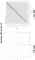
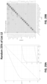
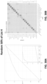
- the cancer classification is a presence or absence of cancer. In some scenarios, has an area under a receiver operating characteristic curve of at least 0.8. In some scenarios, the cancer classification is a type of cancer. In some scenarios, the type of cancer is selected from among at least 12, 14, 16, 18, or 20 cancer types.
- the cancer types are selected from anal cancer, bladder cancer, colorectal cancer, esophageal cancer, head and neck cancer, liver/bile-duct cancer, lung cancer, lymphoma, ovarian cancer, pancreatic cancer, plasma cell neoplasm, and stomach cancer.
- the sensitivity of the method for head and neck cancer is at least 79% or at least 84%; at 99% specificity the sensitivity of the method for liver cancer is at least 82% or at least 85%; at 99% specificity the sensitivity of the method for upper GI tract cancer is at least 62% or at least 68%; wherein at 99% specificity the sensitivity of the method for pancreatic or gallbladder cancer is at least 62% or at least 68%; at 99% specificity the sensitivity of the method for colorectal cancer is at least 60% or at least 65%; at 99% specificity the sensitivity of the method for ovarian cancer is at least 75% or at least 80%; at 99% specificity the sensitivity of the method for lung cancer is at least 60% or at least 65%; at 99% specificity the sensitivity of the method for multiple myeloma is at least 68% or at least 75%; at 99% specificity the sensitivity of the method for lymphoid neoplasm is at least 65% or at least
- the cancer classification is a presence or absence of a type of cancer.
- the step of determining a cancer classification comprises: generating a test feature vector based on the set of sequence reads; and applying the test feature vector to a classifier.
- the classifier comprises a model that is trained by a training process with a first cancer type set of converted DNA sequences from one or more training subjects with a first cancer type and a second cancer type set of converted DNA sequences from one or more training subjects with a second cancer type, wherein both the first cancer type set of converted DNA sequences and the second cancer type set of converted DNA sequences comprise a plurality of training converted DNA sequences.
- the type of cancer is an upper GI tract cancer, and the method, at 99.0% specificity, has a sensitivity of at least 62% or at least 68%. In some scenarios, the type of cancer is a pancreatic or gallbladder cancer, and the method, at 99.0% specificity, has a sensitivity of at least 62% or at least 68%. In some scenarios, the type of cancer is colorectal cancer, and the method, at 99.0% specificity, has a sensitivity of at least 60% or at least 65%. In some scenarios, the type of cancer is ovarian cancer, and the method, at 99.0% specificity, has a sensitivity of at least 75% or at least 80%.
- the type of cancer is lung cancer, and the method, at 99.0% specificity, has a sensitivity of at least60% or at least 65%. In some scenarios, the type of cancer is multiple myeloma, and the method, at 99.0% specificity, has a sensitivity of at least 68% or at least 75%. In some scenarios, the type of cancer is a lymphoid neoplasm, and the method, at 99.0% specificity, has a sensitivity of at least 65% or at least 70%. In some scenarios, the type of cancer is anorectal cancer, and the method, at 99.0% specificity, has a sensitivity of at least 60% or at least 65%.
- the type of cancer is bladder cancer
- the method at 99.0% specificity, has a sensitivity of at least 40% or at least 44%.
- the total size of the target genomic regions is less than 4 Mb, less than 2 Mb, less than 1 Mb, less than 0.7 Mb or less than 0.4 Mb.
- the training process comprises: obtaining sequence information of training fragments from a plurality of training subjects; for each training fragment, determining whether that training fragment is hypomethylated or hypermethylated, wherein each of the hypomethylated and hypermethylated training fragments comprises at least a threshold number of CpG sites with at least a threshold percentage of the CpG sites being unmethylated or methylated, respectively, for each training subject, generating a training feature vector based on the hypomethylated training fragments and hypermethylated training fragments, and training the model with the training feature vectors from the one or more training subjects without cancer and the training feature vectors from the one or more training subjects with cancer.
- the model comprises one of a kernel logistic regression classifier, a random forest classifier, a mixture model, a convolutional neural network, and an autoencoder model.
- the method further comprises the steps of: obtaining a cancer probability for the test sample based on the model; and comparing the cancer probability to a threshold probability to determine whether the test sample is from a subject with cancer or without cancer.
- the method further comprises the steps of: obtaining a cancer type probability for the test sample based on the model; and comparing the cancer type probability to a threshold probability to determine whether the test sample is from a subject with the cancer type or another cancer type or without cancer.
- the method further comprises administering an anti-cancer agent to the subject.
- the method comprising: administering an anti-cancer agent to a subject who has been identified as a cancer subject by a method provided above.
- the anti-cancer agent is a chemotherapeutic agent selected from the group consisting of alkylating agents, antimetabolites, anthracyclines, anti-tumor antibiotics, cytoskeletal disruptors (taxans), topoisomerase inhibitors, mitotic inhibitors, corticosteroids, kinase inhibitors, nucleotide analogs, and platinum-based agents.
- the method comprising administering an anti-cancer agent to a subject who has been identified as a cancer subject by a method provided herein.
- the anti-cancer agent is a chemotherapeutic agent selected from the group consisting of alkylating agents, antimetabolites, anthracyclines, anti-tumor antibiotics, cytoskeletal disruptors (taxans), topoisomerase inhibitors, mitotic inhibitors, corticosteroids, kinase inhibitors, nucleotide analogs, and platinum-based agents.
- Also provided herein are methods for assessing whether a subject has a cancer, the method comprising: obtaining cfDNA from the subject; isolating a portion of the cfDNA from the subject by hybridization capture; obtaining sequence reads derived from the captured cfDNA to determine methylation states cfDNA fragments; applying a classifier to the sequence reads; and determining whether the subject has cancer based on application of the classifier; wherein the classifier has an area under the receiver operator characteristic curve of at least 0.80.
- the method further comprises determining a cancer type, wherein the sensitivity of the method for head and neck cancer is at least 79% or at least 84%; wherein the sensitivity of the method for liver cancer is at least 82% or at least 85%; wherein the sensitivity of the method for upper GI tract cancer is at least 62% or at least 68%; wherein the sensitivity of the method for pancreatic or gallbladder cancer is at least 62% or at least 68%%; wherein the sensitivity of the method for colorectal cancer is at least 60% or at least 65%; wherein the sensitivity of the method for ovarian cancer is at least 75% or at least 80%; wherein the sensitivity of the method for lung cancer is at least 60% or at least 65%; wherein the sensitivity of the method for multiple myeloma is at least 68% or at least 75%; wherein the sensitivity of the method for lymphoid neoplasm is at least 65% or at least 70%; wherein the sensitivity of the method for anorectal cancer is
- the total size of the target genomic regions is less than 4 Mb, less than 2 Mb, less than 1 Mb, less than 0.7 Mb or less than 0.4 Mb.
- the method further comprises converting unmethylated cytosines in the cfDNA to uracil prior to isolating the portion of the cfDNA from the subject by hybridization capture.
- the method further comprises unmethylated cytosines in the cfDNA to uracil after isolating the portion of the cfDNA from the subject by hybridization capture.
- the classifier is a binary classifier. In some scenarios, the classifier is a mixture model classifier.
- isolating a portion of the cfDNA from the subject by hybridization capture comprises contacting the cell-free DNA with a bait set comprising a plurality of different oligonucleotide-containing probes.
- the bait set is a bait set provided herein.
- Also provided herein are methods comprising the steps of: obtaining a set of sequence reads of modified test fragments, wherein the modified test fragments are or have been obtained by processing a set of nucleic acid fragments from a test subject, wherein each of the nucleic acid fragments corresponds to or is derived from a plurality of genomic regions selected from any one of Lists 1-16; and applying the set of sequence reads or a test feature vector obtained based on the set of sequence reads to a model obtained by a training process with a first set of fragments from a plurality of training subjects with a first cancer type and a second set of fragments from a plurality of training subjects with a second cancer type, wherein both the first set of fragments and the second set of fragments comprise a plurality of training fragments.
- the model comprises one of a kernel logistic regression classifier, a random forest classifier, a mixture model, a convolutional neural network, and an autoencoder model.
- the set of sequence reads is obtained by using an assay panel provided above.
- any reference to "one scenario” or “a scenario” means that a particular element, feature, structure, or characteristic described in connection with the scenario is included in at least one scenario.
- the appearances of the phrase “in one scenario” in various places in the specification are not necessarily all referring to the same scenario, thereby providing a framework for various possibilities of described scenariosto function together.
- the terms “comprises,” “comprising,” “includes,” “including,” “has,” “having” or any other variation thereof, are intended to cover a non-exclusive inclusion.
- a process, method, article, or apparatus that comprises a list of elements is not necessarily limited to only those elements but may include other elements not expressly listed or inherent to such process, method, article, or apparatus.
- “or” refers to an inclusive or and not to an exclusive or. For example, a condition A or B is satisfied by any one of the following: A is true (or present) and B is false (or not present), A is false (or not present) and B is true (or present), and both A and B are true (or present).
- ranges and amounts can be expressed as “about” a particular value or range. About also includes the exact amount. Hence “about 5 ⁇ g” means “about 5 ⁇ g” and also “5 ⁇ g.” Generally, the term “about” includes an amount that would be expected to be within experimental error. In some embodiments, “about” refers to the number or value recited, “+” or “-” 20%, 10%, or 5% of the number or value. Additionally, ranges recited herein are understood to be shorthand for all of the values within the range, inclusive of the recited endpoints.
- a range of 1 to 50 is understood to include any number, combination of numbers, or sub-range from the group consisting of 1, 2, 3, 4, 5, 6, 7, 8, 9, 10, 11, 12, 13, 14, 15, 16, 17, 18, 19, 20, 21, 22, 23, 24, 25, 26, 27, 28, 29, 30, 31, 32, 33, 34, 35, 36, 37, 38, 39, 40, 41, 42, 43, 44, 45, 46, 47, 48, 49, and 50.
- methylation refers to a process by which a methyl group is added to a DNA molecule.
- a hydrogen atom on the pyrimidine ring of a cytosine base can be converted to a methyl group, forming 5-methylcytosine.
- the term also refers to a process by which a hydroxymethyl group is added to a DNA molecule, for example by oxidation of a methyl group on the pyrimidine ring of a cytosine base. Methylation and hydroxymethylation tend to occur at dinucleotides of cytosine and guanine referred to herein as "CpG sites.”
- methylation can also refer to the methylation status of a CpG site.
- a CpG site with a 5-methylcytosine moiety is methylated.
- a CpG site with a hydrogen atom on the pyrimidine ring of the cytosine base is unmethylated.
- methylation status at a site i.e., presence or absence of a methyl group.
- a methylated site i.e., a methylated site / absence of a methyl group is an unmethylated site or non-methylated site.
- wet laboratory assay used to detect methylation may vary from those described herein as is well known in the art.
- methylation site refers to a region of a DNA molecule where a methyl group can be added.
- CpG sites are the most common methylation site, but methylation sites are not limited to CpG sites.
- DNA methylation may occur in cytosines in CHG and CHH, where H is adenine, cytosine or thymine. Cytosine methylation in the form of 5-hydroxymethylcytosine may also assessed (see, e.g., WO 2010/037001 and WO 2011/127136 ), and features thereof, using the methods and procedures disclosed herein.
- CpG detection site refers to a region in a probe that is configured to hybridize to a CpG site of a target DNA molecule.
- the CpG site on the target DNA molecule can comprise cytosine and guanine separated by one phosphate group, where cytosine is methylated or unmethylated.
- the CpG site on the target DNA molecule can comprise uracil and guanine separated by one phosphate group, where the uracil is generated by the conversion of unmethylated cytosine.
- UpG is a shorthand for 5'-U-phosphate-G-3' that is uracil and guanine separated by only one phosphate group. UpG can be generated by a bisulfite treatment of a DNA that converts unmethylated cytosines to uracils. Cytosines can be converted to uracils by other methods known in the art, such as chemical modification, synthesis, or enzymatic conversion.
- hypomethylated refers to a methylation status of a DNA molecule containing multiple CpG sites (e.g., more than 3, 4, 5, 6, 7, 8, 9, 10, etc.) where a high percentage of the CpG sites (e.g., more than 80%, 85%, 90%, or 95%, or any other percentage within the range of 50%-100%) are unmethylated or methylated, respectively.
- methylation state vector or "methylation status vector” as used herein refers to a vector comprising multiple elements, where each element indicates the methylation status of a methylation site in a DNA molecule comprising multiple methylation sites, in the order they appear from 5' to 3' in the DNA molecule.
- ⁇ M x , M x+1 , M x+2 >, ⁇ M x , M x+1 , U x+2 >, . . ., ⁇ U x , U x+1 , U x+2 > can be methylation vectors for DNA molecules comprising three methylation sites, where M represents a methylated methylation site and U represents an unmethylated methylation site.
- abnormal methylation pattern or “anomalous methylation pattern” as used herein refers to the methylation pattern of a DNA molecule or a methylation state vector that is expected to be found in a sample less frequently than a threshold value in a non-cancer or healthy sample.
- the expectedness of finding a specific methylation state vector in a healthy control group comprising healthy individuals is represented by a p-value.
- a low p-value score generally corresponds to a methylation state vector which is relatively unexpected in comparison to other methylation state vectors within samples from healthy individuals.
- genomic sample refers to a sample comprising genomic DNAs from an individual diagnosed with cancer.
- the genomic DNAs can be, but are not limited to, cfDNA fragments or chromosomal DNAs from a subject with cancer.
- the genomic DNAs can be sequenced and their methylation status can be assessed by methods known in the art, for example, bisulfite sequencing.
- genomic sequences are obtained from public database (e.g., The Cancer Genome Atlas (TCGA)) or experimentally obtained by sequencing a genome of an individual diagnosed with cancer
- cancerous sample can refer to genomic DNAs or cfDNA fragments having the genomic sequences.
- the term "cancerous samples” as a plural refers to samples comprising genomic DNAs from multiple individuals, each individual diagnosed with cancer. In various scenarios, cancerous samples from more than 100, 300, 500, 1,000, 2,000, 5,000, 10,000, 20,000, 40,000, 50,000, or more individuals diagnosed with cancer are used.
- non-cancerous sample refers to a sample comprising genomic DNAs from an individual not diagnosed with cancer.
- the genomic DNAs can be, but are not limited to, cfDNA fragments or chromosomal DNAs from a subject without cancer.
- the genomic DNAs can be sequenced and their methylation status can be assessed by methods known in the art, for example, bisulfite sequencing.
- genomic sequences are obtained from public database (e.g., The Cancer Genome Atlas (TCGA)) or experimentally obtained by sequencing a genome of an individual without cancer
- non-cancerous sample can refer to genomic DNAs or cfDNA fragments having the genomic sequences.
- non-cancerous samples refers to samples comprising genomic DNAs from multiple individuals, each individual is without cancer.
- cancerous samples from more than 100, 300, 500, 1,000, 2,000, 5,000, 10,000, 20,000, 40,000, 50,000, or more individuals without cancer are used.
- training sample refers to a sample used to train a classifier described herein and/or to select one or more genomic regions for cancer detection or detecting a cancer tissue of origin or cancer cell type.
- the training samples can comprise genomic DNAs or a modification there of, from one or more healthy subjects and from one or more subjects having a disease condition (e.g., cancer, a specific type of cancer, a specific stage of cancer, etc.).
- the genomic DNAs can be, but are not limited to, cfDNA fragments or chromosomal DNAs.
- the genomic DNAs can be sequenced and their methylation status can be assessed by methods known in the art, for example, bisulfite sequencing.
- TCGA The Cancer Genome Atlas
- test sample refers to a sample from a subject, whose health condition was, has been or will be tested using a classifier and/or an assay panel described herein.
- the test sample can comprise genomic DNAs or a modification there of.
- the genomic DNAs can be, but are not limited to, cfDNA fragments or chromosomal DNAs.
- target genomic region refers to a region in a genome selected for analysis in test samples.
- An assay panel is generated with probes designed to hybridize to (and optionally pull down) nucleic acid fragments derived from the target genomic region or a fragment thereof.
- a nucleic acid fragment derived from the target genomic region refers to a nucleic acid fragment generated by degradation, cleavage, bisulfite conversion, or other processing of the DNA from the target genomic region.
- telomere sequence listing a target genomic region includes two DNA strands: one with the sequence provided in the listing and a second that is a reverse complement to the sequence in the listing.
- Probes can be designed to hybridize to one or both sequences.
- probes hybridize to converted sequences resulting from, for example, treatment with sodium bisulfite.
- off-target genomic region refers to a region in a genome which has not been selected for analysis in test samples but has sufficient homology to a target genomic region to potentially be bound and pulled down by a probe designed to target the target genomic region.
- an off-target genomic region is a genomic region that aligns to a probe along at least 45 bp with at least a 90% match rate.
- converted DNA molecules refer to DNA molecules obtained by processing DNA or cfDNA molecules in a sample for the purpose of differentiating a methylated nucleotide and an unmethylated nucleotide in the DNA or cfDNA molecules.
- the sample can be treated with bisulfite ion (e.g., using sodium bisulfite), as is well-known in the art, to convert unmethylated cytosines ("C") to uracils ("U").
- converted DNA molecules or cfDNA molecules include additional uracils which are not present in the original cfDNA sample.
- Replication by DNA polymerase of a DNA strand comprising a uracil results in addition of an adenine to the nascent complementary strand instead of the guanine normally added as the complement to a cytosine or methylcytosine.
- cell free nucleic acid refers to nucleic acid fragments that circulate in an individual's body (e.g., bloodstream) and originate from one or more healthy cells and/or from one or more cancerous cells. Additionally, cfDNA may come from other sources such as viruses, fetuses, etc.
- fragment can refer to a fragment of a nucleic acid molecule.
- a fragment can refer to a cfDNA molecule in a blood or plasma sample, or a cfDNA molecule that has been extracted from a blood or plasma sample.
- An amplification product of a cfDNA molecule may also be referred to as a "fragment.”
- fragment refers to a sequence read, or set of sequence reads, that have been processed for subsequent analysis (e.g., for in machine-learning based classification), as described herein.
- raw sequence reads can be aligned to a reference genome and matching paired end sequence reads assembled into a longer fragment for subsequent analysis.
- the term "individual” refers to a human individual.
- the term “healthy individual” refers to an individual presumed not to have a cancer or disease.
- subject refers to an individual whose DNA is being analyzed.
- a subject may be a test subject whose DNA is be evaluated using a targeted panel as described herein to evaluate whether the person has cancer or another disease.
- a subject may also be part of a control group known not to have cancer or another disease.
- a subject may also be part of a cancer or other disease group known to have cancer or another disease. Control and cancer/disease groups may be used to assist in designing or validating the targeted panel.
- sequence reads refers to nucleotide sequences reads from a sample. Sequence reads can be obtained through various methods provided herein or as known in the art.
- sequencing depth refers to the count of the number of times a given target nucleic acid within a sample has been sequenced (e.g., the count of sequence reads at a given target region). Increasing sequencing depth can reduce required amounts of nucleic acids required to assess a disease state (e.g., cancer or cancer tissue of origin).
- transition generally refers to changes in base composition from one purine to another purine, or from one pyrimidine to another pyrimidine. For instance, the following changes are transitions: C ⁇ U, U ⁇ C, G ⁇ A, A ⁇ G, C ⁇ T, and T ⁇ C.
- a panel or bait set generally refers to all of the probes delivered with a specified panel or bait set.
- a panel or bait set may include both (1) probes having features specified herein (e.g., probes for binding to cell-free DNA fragments corresponding to or derived from genomic regions set forth herein in one or more Lists) and (2) additional probes that do not contain such feature(s).
- the entirety of probes of a panel generally refers to all probes delivered with the panel or bait set, including such probes that do not contain the specified feature(s).
- the present description provides a cancer assay panel comprising a plurality of probes or a plurality of probe pairs.
- the assay panels described herein can alternatively be referred to as bait sets or as compositions comprising bait oligonucleotides.
- the probes can be polynucleotide-containing probes that are specifically designed to target one or more genomic regions differentially methylated between cancer and non-cancer samples, between different cancer tissue of origin (TOO) types, between different cancer cell types, between samples of different stages of cancer, as identified by methods provided herein.
- the target genomic regions are selected to maximize classification accuracy, subject to a size budget (which is determined by sequencing budget and desired depth of sequencing).
- the analytics system may then select target genomic regions based on methylation patterns of nucleic acid fragments.
- One approach considers pairwise distinguishability between pairs of outcomes for regions (or more specifically for CpG sites within regions).
- Another approach considers distinguishability for regions (or more specifically for CpG sites within regions) when considering each outcome against the remaining outcomes.
- the analytics system may design probes to target fragments from the selected genomic regions.
- the analytics system may generate variable sizes of the cancer assay panel, e.g., where a small sized cancer assay panel includes probes targeting the most informative genomic regions, a medium sized cancer assay panel includes probes from the small sized cancer assay panel and additional probes targeting a second tier of informative genomic regions, and a large sized cancer assay panel includes probes from the small-sized and the medium-sized cancer assay panels along with even more probes targeting a third tier of informative genomic regions.
- the analytics system may train classifiers with various classification techniques to predict a sample's likelihood of having a particular outcome or state, e.g., cancer, specific cancer type, other disorder, other disease, etc.
- the probes are configured to hybridize to a converted DNA or cfDNA molecule corresponding to, or derived from, one or more genomic regions, the probes can have a sequence different from the targeted genomic region.
- a DNA containing an unmethylated CpG site will be converted to include UpG instead of CpG because unmethylated cytosines are converted to uracils by a conversion reaction (e.g., bisulfite treatment).
- a probe is configured to hybridize to a sequence including UpG instead of a naturally-existing unmethylated CpG.
- a panel can include probes that can selectively bind to and enrich cfDNA fragments that are differentially methylated in cancerous samples. In this case, sequencing of the enriched fragments can provide information relevant to detection of cancer.
- the probes (or a portion thereof) are designed to target genomic regions that are determined to have an abnormal methylation pattern in cancer samples, or in samples from certain cancer types, tissue types or cell types.
- probes are designed to target genomic regions determined to be hypermethylated or hypomethylated in certain cancers or cancer types to provide additional selectivity and specificity of the detection.
- a panel comprises probes targeting hypomethylated fragments.
- a panel comprises probes targeting hypermethylated fragments.
- a panel comprises both a first set of probes targeting hypermethylated fragments and a second set of probes targeting hypomethylated fragments.
- a cancer assay panel includes not only probes that are designed to target a region that has a first methylation status (e.g., hypomethylation), but also includes probes that are designed to hybridize to the same target region with the opposite methylation status (e.g., hypermethylation).
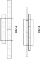
- the targeting of probes to both hypo- and hyper-methylated fragments from the same regions can be referred to as "binary" targeting (see information in the Sequence Listing) ( FIG. 1C ) .
- Each of the probes can target a genomic region comprising at least 30 bp, 35 bp, 40 bp, 45 bp, 50 bp, 60 bp, 70 bp, 80 bp, 90 bp, 100 bp or more.
- the genomic regions can be selected to have less than 30, 25, 20, 15, 12, 10, 8, or 6 methylation sites.
- the genomic regions can be selected when at least 80, 85, 90, 92, 95, or 98% of the at least five methylation (e.g., CpG) sites within the region are either methylated or unmethylated in non-cancerous or cancerous samples, or in cancer samples from a tissue of origin (TOO).
- at least 80, 85, 90, 92, 95, or 98% of the at least five methylation (e.g., CpG) sites within the region are either methylated or unmethylated in non-cancerous or cancerous samples, or in cancer samples from a tissue of origin (TOO).
- TEO tissue of origin
- a first count can be determined that is the number of cancer-containing samples (cancer_count) that include a fragment overlapping that CpG
- a second count is determined that is the number of total samples containing fragments overlapping that CpG site (total).
- Genomic regions can be selected based on criteria positively correlated to the number of cancer-containing samples (cancer_count) that include a fragment indicative of cancer overlapping that CpG site, and inversely correlated with the number of total samples containing fragments indicative of cancer overlapping that CpG site (total).
- the number of non-cancerous samples (nnon-cancer) and the number of cancerous samples (ncancer) having a fragment overlapping a CpG site are counted. Then the probability that a sample is cancer is estimated, for example as (ncancer + 1) / (ncancer + nnon-cancer + 2). This principle could be similarly applied to other outcomes.
- probes can be filtered to reduce non-specific binding (or off-target binding) to nucleic acids derived from non-targeted genomic regions. For example, probes can be filtered to select only those probes having less than a set threshold of off-target binding events.
- probes can be aligned to a reference genome (e.g., a human reference genome) to select probes that align to less than a set threshold of regions across the genome. For example, probes can be selected that align to less than 25, 24, 23, 22, 21, 20, 19, 18, 17, 16, 15, 14, 13, 12, 11, 10, 9 or 8 off-target regions across the reference genome.
- a panel includes at least 1,000, 2,000, 5,000, 10,000, 12,000, 15,000, 20,000, 30,000, 40,000, 50,000, 100,000, 200,000, 250,000, 300,000, 400,000, 500,000, 550,000, 600,000, 700,000, or 800,000 probes.
- the plurality of probes together can comprise at least 0.2 million, 0.4 million, 0.6 million, 0.8 million, 1 million, 2 million, 3 million, 4 million, 5 million, 6 million, 7 million, 8 million, 9 million, 10 million, 12 million, 14 million, 15 million, 20 million, or 25 million nucleotides.
- primers may be used to specifically amplify targets/biomarkers of interest (e.g., by PCR), thereby enriching the sample for desired targets/biomarkers (optionally without hybridization capture).
- forward and reverse primers can be prepared for each genomic region of interest and used to amplify fragments that correspond to or are derived from the desired genomic region.
- additional or alternative methods are used for enrichment (e.g., non-targeted enrichment) such as reduced representation bisulfite sequencing, methylation restriction enzyme sequencing, methylation DNA immunoprecipitation sequencing, methyl-CpG-binding domain protein sequencing, methyl DNA capture sequencing, or microdroplet PCR.
- the cancer assay panels can be a panel that includes a set of hybridization probes (also referred to herein as “probes”) designed to, during enrichment, target and pull down nucleic acid fragments of interest for the assay.
- the probes are designed to hybridize and enrich DNA or cfDNA molecules from cancerous samples that have been treated to convert unmethylated cytosines (C) to uracils (U).
- the probes are designed to hybridize and enrich DNA or cfDNA molecules from cancerous samples of a TOO (or a plurality of TOOs) that have been treated to convert unmethylated cytosines (C) to uracils (U).
- the probes can be designed to anneal (or hybridize) to a target (complementary) strand of DNA or RNA.
- the target strand can be the "positive" strand (e.g., the strand transcribed into mRNA, and subsequently translated into a protein) or the complementary "negative" strand.
- a cancer assay panel may include sets of two probes, one probe targeting the positive strand and the other probe targeting the negative strand of a target genomic region.
- each target genomic region at least four possible probe sequences can be designed.
- Each target region is double-stranded, and as such, a probe or probe set can target either the "positive" or forward strand or its reverse complement (the "negative” strand).
- the probes or probe sets are designed to enrich DNA molecules or fragments that have been treated to convert unmethylated cytosines (C) to uracils (U).
- the probes or probe sets are designed to enrich DNA molecules corresponding to, or derived from the targeted regions after conversion
- the probe's sequence can be designed to enrich DNA molecules of fragments where unmethylated C's have been converted to U's (by utilizing A's in place of G's at sites that are unmethylated cytosines in DNA molecules or fragments corresponding to, or derived from, the targeted region).
- probes are designed to bind to, or hybridize to, DNA molecules or fragments from genomic regions known to contain cancer-specific methylation patterns (e.g., hypermethylated or hypomethylated DNA molecules), thereby enriching for cancer-specific DNA molecules or fragments.
- Targeting genomic regions, or cancer-specific methylation patterns can be advantageous allowing one to specifically enrich for DNA molecules or fragments identified as informative for cancer or cancer TOO, and thus, lowering sequencing needs and sequencing costs.
- two probe sequences can be designed per a target genomic region (one for each DNA strand).
- probes are designed to enrich for all DNA molecules or fragments corresponding to, or derived from, a targeted region (i.e., regardless of strand or methylation status).
- the cancer methylation status is not highly methylated or unmethylated, or because the probes are designed to target small mutations or other variations rather than methylation changes, with these other variations similarly indicative of the presence or absence of a cancer or the presence or absence of a cancer of one or more TOOs. In that case, all four possible probe sequences can be included per a target genomic region.
- the probes can range in length from 10s, 100s, 200s, or 300s of base pairs.
- the probes can comprise at least 50, 75, 100, or 120 nucleotides.
- the probes can comprise less than 300, 250, 200, or 150 nucleotides.
- the probes comprise 100-150 nucleotides.
- the probes comprise 120 nucleotides.
- At least two probes are complementary to the same sequence within a target genomic region, and a nucleotide fragment corresponding to or derived from the target genomic region can be bound and pulled down by at least one of the probes.
- Other levels of tiling are possible, such as 3 ⁇ tiling, 4 ⁇ tiling, etc., wherein each nucleotide in a target region can bind to more than two probes.
- each base in a target genomic region is overlapped by exactly two probes, as illustrated in FIG. 1B .
- Probes that extend in both directions beyond a target genomic region are useful to pull down cfDNA fragments comprising a portion of the target genomic region and DNA sequences adjacent to the target genomic region.
- even relatively small target regions may be targeted with three probes (see FIG. 1A ).
- a probe set comprising three or more probes is optionally used to capture a larger genomic region (see FIG. 1B ).
- subsets of probes will collectively extend across an entire genomic region (e.g., may be complementary to non-converted or converted fragments from the genomic region).
- a tiled probe set optionally comprises probes that collectively include at least two probes that overlap every nucleotide in the genomic region. This is done to ensure that cfDNAs comprising a small portion of a target genomic region at one end will have a substantial overlap extending into the adjacent non-targeted genomic region with at least one probe, to provide for efficient capture.
- a 100 bp cfDNA fragment comprising a 30 nt target genomic region can be guaranteed to have at least 65 bp overlap with at least one of the overlapping probes.
- Other levels of tiling are possible.
- probes can be designed to expand a 30 bp target region by at least 70 bp, 65 bp, 60 bp, 55 bp, or 50 bp.
- the probes can be designed to extend past the ends of the target region on either side.
- the probes are designed to analyze methylation status of target genomic regions (e.g., of the human or another organism) that are suspected to correlate with the presence or absence of cancer generally, presence or absence of certain types of cancers, cancer stage, or presence or absence of other types of diseases.
- target genomic regions e.g., of the human or another organism
- the probes are designed to effectively bind and pull down cfDNA fragments containing a target genomic region.
- the probes are designed to cover overlapping portions of a target region, so that each probe is "tiled" in coverage such that each probe overlaps in coverage at least partially with another probe in the library.
- the panel contains multiple pairs of probes, where each pair comprises at least two probes overlapping each other by an overlapping sequence of at least 25, 30, 35, 40, 45, 50, 60, 70, 75 or 100 nucleotides.
- the overlapping sequence can be designed to be complementary to a target genomic region (or a converted version thereof), thus a nucleotide fragment derived from or containing the target genomic region can be bound and pulled down by at least one of the probes.
- probes can be designed to cover both strands of a double-stranded cfDNA sequence.
- the smallest target genomic region is 30 or 31 bp.
- the new target region of 30bp can be centered on a specific CpG site of interest. Then, it is checked whether each edge of this new target is close enough to other targets such that they can be merged. This is based on a "merge distance" parameter which can be 200bp by default but can be tuned. This allows close but distinct target regions to be enriched with overlapping probes.
- the new target can be merged with nothing (increasing the number of panel targets by one), merged with just one target either to the left or the right (not changing the number of panel targets), or merged with existing targets both to the left and right (reducing the number of panel targets by one).
- methods of selecting target genomic regions for detecting cancer and/or a TOO can be used to design and manufacture probes for a cancer assay panel. Methylation status of DNA or cfDNA molecules corresponding to, or derived from, the target genomic regions can be screened using the cancer assay panel.
- Alternative methods for example by WGBS or other methods known in the art, can be also implemented to detect methylation status of DNA molecules or fragments corresponding to, or derived from, the target genomic regions.
- a nucleic acid sample (DNA or RNA) is extracted from a subject.
- DNA and RNA may be used interchangeably unless otherwise indicated. That is, the scenarios described herein may be applicable to both DNA and RNA types of nucleic acid sequences. However, the examples described herein may focus on DNA for purposes of clarity and explanation.
- the sample may be any subset of the human genome, including the whole genome.
- the sample may include blood, plasma, serum, urine, fecal, saliva, other types of bodily fluids, or any combination thereof.
- methods for drawing a blood sample e.g., syringe or finger prick
- procedures for obtaining a tissue biopsy which may require surgery.
- the extracted sample may comprise cfDNA and/or ctDNA.
- the human body may naturally clear out cfDNA and other cellular debris. If a subject has a cancer or disease, cfDNA and/or ctDNA in an extracted sample may be present at a detectable level for detecting the cancer or disease.
- the cfDNA fragments are treated to convert unmethylated cytosines to uracils.
- the method uses a bisulfite treatment of the DNA which converts the unmethylated cytosines to uracils without converting the methylated cytosines.
- a commercial kit such as the EZ DNA Methylation TM - Gold, EZ DNA Methylation TM - Direct or an EZ DNA Methylation TM - Lightning kit (available from Zymo Research Corp (Irvine, CA)) is used for the bisulfite conversion.
- the conversion of unmethylated cytosines to uracils is accomplished using an enzymatic reaction.
- the conversion can use a commercially available kit for conversion of unmethylated cytosines to uracils, such as APOBEC-Seq (NEBiolabs, Ipswich, MA).
- a sequencing library is prepared.
- a ssDNA adapter is added to the 3'-OH end of a bisulfite-converted ssDNA molecule using a ssDNA ligation reaction.
- the ssDNA ligation reaction uses CircLigase II (Epicentre) to ligate the ssDNA adapter to the 3'-OH end of a bisulfite-converted ssDNA molecule, wherein the 5'-end of the adapter is phosphorylated and the bisulfite-converted ssDNA has been dephosphorylated (i.e., the 3' end has a hydroxyl group).
- the ssDNA ligation reaction uses Thermostable 5' AppDNA/RNA ligase (available from New England BioLabs (Ipswich, MA)) to ligate the ssDNA adapter to the 3'-OH end of a bisulfite-converted ssDNA molecule.
- the first UMI adapter is adenylated at the 5'-end and blocked at the 3'-end.
- the ssDNA ligation reaction uses a T4 RNA ligase (available from New England BioLabs) to ligate the ssDNA adapter to the 3'-OH end of a bisulfite-converted ssDNA molecule.
- a second strand DNA is synthesized in an extension reaction.
- an extension primer that hybridizes to a primer sequence included in the ssDNA adapter, is used in a primer extension reaction to form a double-stranded bisulfite-converted DNA molecule.
- the extension reaction uses an enzyme that is able to read through uracil residues in the bisulfite-converted template strand.
- a dsDNA adapter is added to the double-stranded bisulfite-converted DNA molecule.
- the double-stranded bisulfite-converted DNA is amplified to add sequencing adapters.
- targeted DNA sequences may be enriched from the library. This is used, for example, where a targeted panel assay is being performed on the samples.
- hybridization probes also referred to herein as "probes" are used to target, and pull down, nucleic acid fragments informative for the presence or absence of cancer (or disease), cancer status, or a cancer classification (e.g., cancer type or tissue of origin).
- the probes may be designed to anneal (or hybridize) to a target (complementary) strand of DNA or RNA.
- the target strand may be the "positive" strand (e.g., the strand transcribed into mRNA, and subsequently translated into a protein) or the complementary "negative” strand.
- the probes may range in length from 10s, 100s, or 1000s of base pairs. Moreover, the probes may cover overlapping portions of a target region.
- FIG. 3A is a flowchart describing a process 300 of generating a data structure for a healthy control group, according to a scenario.
- the analytics system obtains information related to methylation status of a plurality of CpG sites on sequence reads derived from a plurality of DNA molecules or fragments from a plurality of healthy subjects.
- the method provided herein for creating a healthy control group data structure can be performed similarly for subjects with cancer, subjects with cancer of a TOO, subjects with a known cancer type, or subjects with another known disease state.
- a methylation state vector is generated for each DNA molecule or fragment, for example via the process 100.
- the analytics system subdivides 310 the methylation state vector of each DNA fragment into strings of CpG sites.
- the analytics system subdivides 310 the methylation state vector such that the resulting strings are all less than a given length.
- a methylation state vector of length 11 may be subdivided into strings of length less than or equal to 3 would result in 9 strings of length 3, 10 strings of length 2, and 11 strings of length 1.
- a methylation state vector of length 7 being subdivided into strings of length less than or equal to 4 would result in 4 strings of length 4, 5 strings of length 3, 6 strings of length 2, and 7 strings of length 1.
- the methylation state vector resulting from a DNA fragment is shorter than or the same length as the specified string length, then the methylation state vector may be converted into a single string containing all CpG sites of the vector.
- the analytics system tallies 320 the strings by counting, for each possible CpG site and possibility of methylation states in the vector, the number of strings present in the control group having the specified CpG site as the first CpG site in the string and having that possibility of methylation states. For a string length of three at a given CpG site, there are 2 ⁇ 3 or 8 possible string configurations. For each CpG site, the analytics system tallies 320 how many occurrences of each possible methylation state vector appear in the control group.
- the analytics system creates 330 a data structure storing the tallied counts for each starting CpG site and string possibility at each starting CpG.
- a maximum string length of 4 means that there are at most 2 ⁇ 4 numbers to tally at every CpG. Increasing the maximum string length to 5 doubles the possible number of methylation states to tally. Reducing string size helps reduce the computational and data storage burden of the data structure. In some scenarios, the string size is 3. In some scenarios, the string size is 4. A second reason to limit the maximum string length is to avoid overfitting downstream models.
- Calculating probabilities based on long strings of CpG sites can be problematic if the long CpG strings do not have a strong biological effect on the outcome (e.g., predictions of anomalousness that predictive of the presence of cancer), as it requires a significant amount of data that may not be available, and would thus be too sparse for a model to perform appropriately. For example, calculating a probability of anomalousness/cancer conditioned on the prior 100 CpG sites would require counts of strings in the data structure of length 100, ideally some matching exactly the prior 100 methylation states. If only sparse counts of strings of length 100 are available, there will be insufficient data to determine whether a given string of length of 100 in a test sample is anomalous or not.
- the analytics system may seek to validate 340 the data structure and/or any downstream models making use of the data structure.
- This first type of validation ensures that potential cancerous samples are removed from the healthy control group so as to not affect the control group's purity.
- This type of validation checks consistency within the control group's data structure.
- the healthy control group may contain a sample from an individual with an undiagnosed cancer that contains a plurality of anomalously methylated fragments.
- the analytics system may perform various calculations to determine whether to exclude data from a subject with apparently undiagnosed cancer.
- a second type of validation checks the probabilistic model used to calculate p-values with the counts from the data structure itself (i.e., from the healthy control group).
- a process for p-value calculation is described below in conjunction with FIG. 5 .
- the analytics system Once the analytics system generates a p-value for the methylation state vectors in the validation group, the analytics system builds a cumulative density function (CDF) with the p-values. With the CDF, the analytics system may perform various calculations on the CDF to validate the control group's data structure.
- CDF cumulative density function
- FIG. 3B is a flowchart describing the additional step 340 of validating the data structure for the control group of FIG. 3A , according to a scenario.
- the analytics system performs the fourth type of validation test as described above which utilizes a validation group with a supposedly similar composition of subjects, samples, and/or fragments as the control group. For example, if the analytics system selected healthy subjects without cancer for the control group, then the analytics system also uses healthy subjects without cancer in the validation group.
- the analytics system takes the validation group and generates 100 a set of methylation state vectors as described in FIG. 3A .
- the analytics system performs a p-value calculation for each methylation state vector from the validation group.
- the p-value calculation process will be further described in conjunction with FIGS. 4-5 .
- the analytics system calculates a probability from the control group's data structure. Once the probabilities are calculated for the possibilities of methylation state vectors, the analytics system calculates 350 a p-value score for that methylation state vector based on the calculated probabilities.
- the p-value score represents an expectedness of finding that specific methylation state vector and other possible methylation state vectors having even lower probabilities in the control group.
- the analytics system enumerates 410 all possibilities of methylation state vectors having the same starting CpG site and same length (i.e., set of CpG sites) in the methylation state vector.
- each methylation state may be methylated or unmethylated there are only two possible states at each CpG site, and thus the count of distinct possibilities of methylation state vectors depends on a power of 2, such that a methylation state vector of length n would be associated with 2 n possibilities of methylation state vectors.
- the analytics system calculates 420 the probability of observing each possibility of methylation state vector for the identified starting CpG site / methylation state vector length by accessing the healthy control group data structure.
- calculating the probability of observing a given possibility uses a Markov chain probability to model the joint probability calculation which will be described in greater detail with respect to FIG. 5 below.
- calculation methods other than Markov chain probabilities are used to determine the probability of observing each possibility of methylation state vector.
- the analytics system calculates 430 a p-value score for the methylation state vector using the calculated probabilities for each possibility. In one scenario, this includes identifying the calculated probability corresponding to the possibility that matches the methylation state vector in question. Specifically, this is the possibility having the same set of CpG sites, or similarly the same starting CpG site and length as the methylation state vector. The analytics system sums the calculated probabilities of any possibilities having probabilities less than or equal to the identified probability to generate the p-value score.
- This p-value represents the probability of observing the methylation state vector of the fragment or other methylation state vectors even less probable in the healthy control group.
- a low p-value score thereby, generally corresponds to a methylation state vector which is rare in a healthy subject, and which causes the fragment to be labeled abnormally methylated, relative to the healthy control group.
- a high p-value score generally relates to a methylation state vector is expected to be present, in a relative sense, in a healthy subject. If the healthy control group is a non-cancerous group, for example, a low p-value indicates that the fragment is abnormally methylated relative to the non-cancer group, and therefore possibly indicative of the presence of cancer in the test subject.
- the analytics system calculates p-value scores for each of a plurality of methylation state vectors, each representing a cfDNA fragment in the test sample.
- the analytics system may filter 440 the set of methylation state vectors based on their p-value scores. In one scenario, filtering is performed by comparing the p-values scores against a threshold and keeping only those fragments below the threshold. This threshold p-value score could be on the order of 0.1, 0.01, 0.001, 0.0001, or similar.
- S 1 ⁇ P S 1 Markov chain model can be used to make the calculation of the conditional probabilities of each possibility more efficient.
- the analytics system selects a Markov chain order k which corresponds to how many prior CpG sites in the vector (or window) to consider in the conditional probability calculation, such that the conditional probability is modeled as P(S n
- the analytics system accesses the control group's data structure, specifically the counts of various strings of CpG sites and states.
- S n-k-2 , ..., S n-1 ) the analytics system takes a ratio of the stored count of the number of strings from the data structure matching ⁇ S n-k-2 , ..., S n-1 , M n > divided by the sum of the stored count of the number of strings from the data structure matching ⁇ S n-k-2 , ..., S n-1 , M n > and ⁇ S n-k-2 , ..., S n-1 , U n >.
- S n-k-2 , ..., S n-1 ) is calculated ratio having the form: # of ⁇ S n ⁇ k ⁇ 2 , ... , S n ⁇ 1 , M n > # of ⁇ S n ⁇ k ⁇ 2 , ... , S n ⁇ 1 , M n > + # of ⁇ S n ⁇ k ⁇ 2 , ... , S n ⁇ 1 , U n >
- the calculation may additionally implement a smoothing of the counts by applying a prior distribution.
- the prior distribution is a uniform prior as in Laplace smoothing.
- a constant is added to the numerator and another constant (e.g., twice the constant in the numerator) is added to the denominator of the above equation.
- an algorithmic technique such as Knesser-Ney smoothing is used.
- the above denoted formulas are applied to the test methylation state vector 505 covering sites 23 - 26.
- the analytics system calculates 430 a p-value score 525 that sums the probabilities that are less than or equal to the probability of possibility of methylation state vector matching the test methylation state vector 505.
- the computational burden of calculating probabilities and/or p-value scores may be further reduced by caching at least some calculations.
- the analytic system may cache in transitory or persistent memory calculations of probabilities for possibilities of methylation state vectors (or windows thereof). If other fragments have the same CpG sites, caching the possibility probabilities allows for efficient calculation of p-value scores without needing to re-calculate the underlying possibility probabilities.
- the analytics system may calculate p-value scores for each of the possibilities of methylation state vectors associated with a set of CpG sites from vector (or window thereof). The analytics system may cache the p-value scores for use in determining the p-value scores of other fragments including the same CpG sites.
- the p-value scores of possibilities of methylation state vectors having the same CpG sites may be used to determine the p-value score of a different one of the possibilities from the same set of CpG sites.
- the window In calculating p-values for a methylation state vector larger than the window, the window identifies the sequential set of CpG sites from the vector within the window starting from the first CpG site in the vector.
- the analytic system calculates a p-value score for the window including the first CpG site.
- the analytics system then "slides" the window to the second CpG site in the vector, and calculates another p-value score for the second window.
- each methylation state vector will generate m-l + 1 p-value scores.
- the analytics system aggregates the p-value scores for the methylation state vectors to generate an overall p-value score.
- Each of the 50 calculations enumerates 2 ⁇ 5 (32) possibilities of methylation state vectors, which total results in 50 ⁇ 2 ⁇ 5 (1.6 ⁇ 10 ⁇ 3) probability calculations. This results in a vast reduction of calculations to be performed, with no meaningful hit to the accurate identification of anomalous fragments. This additional step can also be applied when validating 340 the control group with the validation group's methylation state vectors.
- the analytics system identifies 450 DNA fragments indicative of cancer from the filtered set of anomalously methylated fragments.
- the analytics system may identify DNA fragments that are deemed hypomethylated or hypermethylated as fragments indicative of cancer from the filtered set of anomalously methylated fragments.
- Hypomethylated and hypermethylated fragments can be defined as fragments of a certain length of CpG sites (e.g., more than 3, 4, 5, 6, 7, 8, 9, 10, etc.) with a high percentage of methylated CpG sites (e.g., more than 80%, 85%, 90%, or 95%, or any other percentage within the range of 50%-100%) or a high percentage of unmethylated CpG sites (e.g., more than 80%, 85%, 90%, or 95%, or any other percentage within the range of 50%-100%).
- the analytics system identifies fragments indicative of cancer utilizing probabilistic models of methylation patterns fitted to each cancer type and non-cancer type.
- the analytics system calculates log-likelihood ratios for a sample using DNA fragments in the genomic regions considering the various cancer types with the fitted probabilistic models for each cancer type and non-cancer type.
- the analytics system may determine a DNA fragment to be indicative of cancer based on whether at least one of the log-likelihood ratios considered against the various cancer types is above a threshold value.
- the analytics system partitions the genome into regions by multiple stages.
- the analytics system separates the genome into blocks of CpG sites. Each block is defined when there is a separation between two adjacent CpG sites that exceeds some threshold, e.g., greater than 200 bp, 300 bp, 400 bp, 500 bp, 600 bp, 700 bp, 800 bp, 900 bp, or 1,000 bp.
- the analytics system subdivides at a second stage each block into regions of a certain length, e.g., 500 bp, 600 bp, 700 bp, 800 bp, 900 bp, 1,000 bp, 1,100 bp, 1,200 bp, 1,300 bp, 1,400 bp, or 1,500 bp.
- the analytics system may further overlap adjacent regions by a percentage of the length, e.g., 10%, 20%, 30%, 40%, 50%, or 60%.
- the analytics system analyzes sequence reads derived from DNA fragments for each region.
- the analytics system may process samples from tissue and/or high-signal cfDNA.
- High-signal cfDNA samples may be determined by a binary classification model, by cancer stage, or by another metric.
- Cancer of various types having different TOO can be selected from the group consisting of breast cancer, uterine cancer, cervical cancer, ovarian cancer, bladder cancer, urothelial cancer of renal pelvis, renal cancer other than urothelial, prostate cancer, anorectal cancer, anal cancer, colorectal cancer, hepatobiliary cancer arising from hepatocytes, hepatobiliary cancer arising from cells other than hepatocytes, liver/bile-duct cancer, esophageal cancer, pancreatic cancer, stomach cancer, squamous cell cancer of the upper gastrointestinal tract, upper gastrointestinal cancer other than squamous, head and neck cancer, lung cancer, lung adenocarcinoma, small cell lung cancer, squamous cell lung cancer and cancer other than adenocarcinoma or small cell lung cancer, neuroendocrine cancer, melanoma, thyroid cancer, sarcoma, plasma cell neoplasm, multiple myeloma, myeloid n
- cancer types can be classified and labeled using classification methods available in the art, such as the International Classification of Diseases for Oncology (ICD-O-3) (codes.iarc.fr) or the Surveillance, Epidemiology, and End Results Program (SEER) (seer.cancer.gov).
- ICD-O-3 International Classification of Diseases for Oncology
- SEER Surveillance, Epidemiology, and End Results Program
- SEER End Results Program
- cancer types are classified in three orthogonal codes, (i) topographical codes, (ii) morphological codes, or (iii) behavioral codes. Under behavioral codes, benign tumor is 0, uncertain behavior is 1, carcinoma in situ is 2, malignant, primary site is 3 and malignant, metastatic site is 6.
- the analytics system can further calculate log-likelihood ratios ("R") for a fragment indicating a likelihood of the fragment being indicative of cancer considering the various cancer types with the fitted probabilistic models for each cancer type and non-cancer type, or for a cancer TOO.
- the two probabilities may be taken from probabilistic models fitted for each of the cancer types and the non-cancer type, the probabilistic models defined to calculate a likelihood of observing a methylation pattern on a fragment given each of the cancer types and the non-cancer type.
- the probabilistic models may be defined fitted for each of the cancer types and the non-cancer type.
- different classifiers may be trained using sequence reads obtained from samples enriched for tumor cells from subject cohorts known to have particular cancer (e.g., breast, lung, prostrate, etc.).
- Each genomic region's ability to distinguish between cancer type and non-cancer type in the classification model is used to rank the genomic regions from most informative to least informative in classification performance.
- the analytics system may identify genomic regions from the ranking according to information gain in classification between non-cancer type and cancer type.
- the analytics system may train a classifier according to a process 600 illustrated in FIG. 6A , according to a scenario.
- the process 600 accesses two training groups of samples - a non-cancer group and a cancer group - and obtains 605 a non-cancer set of methylation state vectors and a cancer set of methylation state vectors comprising anomalously methylated fragments, e.g., via step 440 from the process 400.
- the process takes a ratio of (1) over (1) summed with (3).
- the hypermethylation score is calculated by taking a ratio of (2) over (2) and (4). Additionally, these ratios may be calculated with an additional smoothing technique as discussed above.
- the hypomethylation score and the hypermethylation score relate to an estimate of cancer probability given the presence of hypomethylation or hypermethylation of fragments from the cancer set.
- the analytics system trains 660 a binary classifier to distinguish feature vectors between the cancer and non-cancer training groups.
- a binary classifier to distinguish feature vectors between the cancer and non-cancer training groups.
- the classifier is a non-linear classifier.
- the classifier is a non-linear classifier utilizing a L2-regularized kernel logistic regression with a Gaussian radial basis function (RBF) kernel.
- RBF Gaussian radial basis function
- the analytics system calculates 695 an informative score for each region describing that region's ability to distinguish between each pair of cancer types.
- the analytics system may specify one type as a positive type and the other as a negative type.
- a region's ability to distinguish between the positive type and the negative type is based on mutual information, calculated using the estimated fraction of cfDNA samples of the positive type and of the negative type for which the feature would be expected to be non-zero in the final assay, i.e., at least one fragment of that tier that would be sequenced in a targeted methylation assay.
- Those fractions are estimated using the observed rates at which the feature occurs in healthy cfDNA, and in high-signal cfDNA and/or tumor samples of each cancer type. For example, if a feature occurs frequently in healthy cfDNA, then it will also be estimated to occur frequently in cfDNA of any cancer type and would likely result in a low informative score.
- the analytics system may choose a certain number of regions for each pair of cancer types from the ranking, e.g., 1024.
- the analytics system further identifies predominantly hypermethylated or hypomethylated regions from the ranking of regions.
- the analytics system may load the set of fragments in the positive type(s) for a region that was identified as informative.
- the analytics system evaluates whether the loaded fragments are predominantly hypermethylated or hypomethylated. If the loaded fragments are predominately hypermethylated or hypomethylated, the analytics system may select probes corresponding to the predominant methylation pattern. If the loaded fragments are not predominantly hypermethylated or hypomethylated, the analytics system may use a mixture of probes for targeting both hypermethylation and hypomethylation.
- the analytics system may further identify a minimal set of CpG sites that overlap more than some percentage of the fragments.
- the analytics system after ranking the regions based on informative scores, labels each region with the lowest informative ranking across all pairs of cancer types. For example, if a region was the 10th-most-informative region for distinguishing breast from lung, and the 5th-most-informative for distinguishing breast from colorectal, then it would be given an overall label of "5".
- the analytics system may design probes starting with the lowest-labeled regions while adding regions to the panel, e.g., until the panel's size budget has been exhausted.
- probes targeting selected genomic regions are further filtered 475 based on the number of their off-target regions. This is for screening probes that pull down too many cfDNA fragments corresponding to, or derived from, off-target genomic regions. Exclusion of probes having many off-target regions can be valuable by decreasing off-target rates and increasing target coverage for a given amount of sequencing.
- An off-target genomic region is a genomic region that has sufficient homology to a target genomic region, such that DNA molecules or fragments derived from off-target genomic regions are hybridized to and pulled down by a probe designed to hybridize to a target genomic region.
- An off-target genomic region can be a genomic region (or a converted sequence of that same region) that aligns to a probe along at least 35 bp, 40 bp, 45 bp, 50 bp, 60 bp, 70 bp, or 80 bp with at least an 80%, 85%, 90%, 95%, or 97% match rate.
- an off-target genomic region is a genomic region (or a converted sequence of that same region) that aligns to a probe along at least 45bp with at least a 90% match rate.
- Various methods known in the art can be adopted to screen off-target genomic regions.
- a k-mer seeding strategy (which can allow one or more mismatches) is combined to local alignment at the seed locations.
- exhaustive searching of good alignments can be guaranteed based on k-mer length, number of mismatches allowed, and number of k-mer seed hits at a particular location.
- This requires doing dynamic programing local alignment at a large number of locations, so this approach is highly optimized to use vector CPU instructions (e.g., AVX2, AVX512) and also can be parallelized across many cores within a machine and also across many machines connected by a network.
- vector CPU instructions e.g., AVX2, AVX512
- probes having sequence homology with off-target genomic regions, or DNA molecules corresponding to, or derived from off-target genomic regions comprising more than a threshold number are excluded (or filtered) from the panel.
- probes having sequence homology with off-target genomic regions, or DNA molecules corresponding to, or derived from off-target genomic regions from more than 30, more than 25, more than 20, more than 18, more than 15, more than 12, more than 10, or more than 5 off-target regions are excluded.
- methods of using a cancer assay panel are provided.
- the methods can comprise steps of treating DNA molecules or fragments to convert unmethylated cytosines to uracils (e.g., using bisulfite treatment), applying a cancer panel (as described herein) to the converted DNA molecules or fragments, enriching a subset of converted DNA molecules or fragments that bind to the probes in the panel, and sequencing the enriched cfDNA fragments.
- Alignment position information derived from the read pair R 1 and R 2 may include a beginning position in the reference genome that corresponds to an end of a first read (e.g., R 1 ) and an end position in the reference genome that corresponds to an end of a second read (e.g., R 2 ).
- the beginning position and end position in the reference genome represent the likely location within the reference genome to which the nucleic acid fragment corresponds.
- An output file having SAM (sequence alignment map) format or BAM (binary alignment map) format may be generated and output for further analysis.
- FIGS. 8A-8B show three graphs of data validating consistency of sequencing from a control group.
- the first graph 170 shows conversion accuracy of conversion of unmethylated cytosines to uracil (step 120) on cfDNA fragment obtained from a test sample across subjects in varying stages of cancer - stage 0, stage I, stage II, stage III, stage IV, and non-cancer. As shown, there was uniform consistency in converting unmethylated cytosines on cfDNA fragments into uracils. There was an overall conversion accuracy of 99.47% with a precision at ⁇ 0.024%.
- the second graph 180 compares coverage (depth of sequencing) over varying stages of cancer. Counting only sequence reads that were confidently mapped to a reference genome, the mean coverage over all groups was ⁇ 34.
- the third graph 190 shows the concentration of cfDNA per sample across varying stages of cancer.
- Sequence reads obtained by the methods provided herein are further processed by automated algorithms.
- the analytics system is used to receive sequencing data from a sequencer and perform various aspects of processing as described herein.
- the analytics system can be one of a personal computer (PC), a desktop computer, a laptop computer, a notebook, a tablet PC, a mobile device.
- a computing device can be communicatively coupled to the sequencer through a wireless, wired, or a combination of wireless and wired communication technologies.
- the computing device is configured with a processor and memory storing computer instructions that, when executed by the processor, cause the processor to perform steps as described in the remainder of this document.
- the amount of genetic data and data derived therefrom is sufficiently large, and the amount of computational power required so great, so as to be impossible to be performed on paper or by the human mind alone.
- the clinical interpretation of methylation status of targeted genomic regions is a process that includes classifying the clinical effect of each or a combination of the methylation status and reporting the results in ways that are meaningful to a medical professional.
- the clinical interpretation can be based on comparison of the sequence reads with database specific to cancer or non-cancer subjects, and/or based on numbers and types of the cfDNA fragments having cancer-specific methylation patterns identified from a sample.
- targeted genomic regions are ranked or classified based on their likeness to be differentially methylated in cancer samples, and the ranks or classifications are used in the interpretation process.
- the ranks and classifications can include (1) the type of clinical effect, (2) the strength of evidence of the effect, and (3) the size of the effect.
- the clinically meaning information can include the presence or absence of cancer generally, presence or absence of certain types of cancers, cancer stage, or presence or absence of other types of diseases.
- the information relates to a presence or absence of one or more cancer types, selected from the group consisting of breast cancer, endometrial cancer, cervical cancer, ovarian cancer, bladder cancer, urothelial cancer of renal pelvis, renal cell carcinoma, prostate cancer, anorectal cancer, anal cancer, colorectal cancer, hepatocellular cancer, liver/bile-duct cancer, cholangiocarcinoma and hepatobiliary cancer, pancreatic cancer, upper GI adenocarcinoma, esophageal squamous cell cancer, head and neck cancer, lung cancer, squamous cell lung cancer, lung adenocarcinoma, small cell lung cancer, neuroendocrine cancer, melanoma, thyroid cancer, sarcoma, plasma cell neoplasm, multiple myeloma, mye
- the assay panel described herein can be used with a cancer type classifier that predicts a disease state for a sample, such as a cancer or non-cancer prediction, a tissue of origin prediction, and/or an indeterminate prediction.
- the cancer type classifier can generate features based on sequence reads by taking into account methylated or unmethylated fragments of DNA at certain genomic areas of interest. For instance, if the cancer type classifier determines that a methylation pattern at a fragment resembles that of a certain cancer type, then the cancer type classifier can set a feature for that fragment as 1, and otherwise if no such fragment is present, then the feature can be set as 0.
- the cancer type classifier can produce a set of binary features (merely by way of example, 30,000 features) for each sample. Further, in some examples, all or a portion of the set of binary features for a sample can be input into the cancer type classifier to provide a set of probability scores, such as one probability score per cancer type class and for a non-cancer type class. Furthermore, in some examples, the cancer type classifier can incorporate or otherwise be used in conjunction with thresholding to determine whether a sample is to be called as cancer or non-cancer, and/or indeterminate thresholding to reflect confidence in a specific TOO call. Such methods are described further below.
- the analytics system can obtain a set of training samples.
- each training sample includes fragment file(s) (e.g., file containing sequence read data), a label corresponding to a type of cancer (TOO) or non-cancer status of the sample, and/or sex of the individual of the sample.
- the analytics system can utilize the training set to train the cancer type classifier to predict the disease state of the sample.
- the analytics system divides the genome (e.g., whole genome) or a subset of the genome (e.g., targeted methylation regions) into regions.
- portions of the genome can be separated into "blocks" of CpGs, whereby a new block begins whenever there is a separation between nearest-neighbor CpGs is at least a minimum separation distance (e.g., at least 500 bp).
- each block can be divided into 1000 bp regions and positioned such that neighboring regions have a certain amount (e.g., 50% or 500 bp) of overlap.
- the analytics system can split the training set into K subsets or folds to be used in a K-fold cross-validation.
- the folds can be balanced for cancer/non-cancer status, tissue of origin, cancer stage, age (e.g., grouped in 10yr buckets), and/or smoking status.
- the training set is split into 5 folds, whereby 5 separate classifiers are trained, in each case training on 4/5 of the training samples and using the remaining 1/5 for validation.
- the rate of methylation represents a fraction or percentage of base pairs that are methylated within a CpG site.
- the trained probabilistic model can be parameterized by products of the rates of methylation.
- any known probabilistic model for assigning probabilities to sequence reads from a sample can be used.
- the probabilistic model can be a binomial model, in which every site (e.g., CpG site) on a nucleic acid fragment is assigned a probability of methylation, or an independent sites model, in which each CpG's methylation is specified by a distinct methylation probability with methylation at one site assumed to be independent of methylation at one or more other sites on the nucleic acid fragment.
- the probabilistic model is a Markov model, in which the probability of methylation at each CpG site is dependent on the methylation state at some number of preceding CpG sites in the sequence read, or nucleic acid molecule from which the sequence read is derived. See, e.g., U.S. Pat. Appl. No. 16/352,602, entitled “Anomalous Fragment Detection and Classification,” and filed March 13, 2019 .
- the probabilistic model is a "mixture model" fitted using a mixture of components from underlying models.
- the mixture components can be determined using multiple independent sites models, where methylation (e.g., rates of methylation) at each CpG site is assumed to be independent of methylation at other CpG sites.
- methylation e.g., rates of methylation
- the probability assigned to a sequence read, or the nucleic acid molecule from which it derives is the product of the methylation probability at each CpG site where the sequence read is methylated and one minus the methylation probability at each CpG site where the sequence read is unmethylated.
- the analytics system determines rates of methylation of each of the mixture components.
- the analytics system fits the probabilistic model using maximum-likelihood estimation to identify a set of parameters ⁇ ⁇ ki , f k ⁇ that maximizes the log-likelihood of all fragments deriving from a disease state, subject to a regularization penalty applied to each methylation probability with regularization strength r.
- the maximized quantity for N total fragments can be represented as: ⁇ j N ln Pr fragment j
- the analytics system performs fits separately for each cancer type and for healthy cfDNA.
- other means can be used to fit the probabilistic models or to identify parameters that maximize the log-likelihood of all sequence reads derived from the reference samples.
- Bayesian fitting using e.g., Markov chain Monte Carlo
- each parameter is not assigned a single value but instead is associated to a distribution
- gradient-based optimization in which the gradient of the likelihood (or log-likelihood) with respect to the parameter values is used to step through parameter space towards an optimum, is used.
- the analytics system can generate features for each sample in the training set. For example, for each sample (regardless of label), in each region, for each cancer type, for each fragment, the analytics system can evaluate the log-likelihood ratio R with the fitted probabilistic models according to: R cancer type A fragment ⁇ ln Pr fragment
- the analytics system can select certain features for inclusion in a feature vector for each sample. For example, for each pair of distinct cancer types, the analytics system can specify one type as the "positive type” and the other as the "negative type” and rank the features by their ability to distinguish those types. In some cases, the ranking is based on mutual information calculated by the analytics system. For example, the mutual information can be calculated using the estimated fraction of samples of the positive type and negative type (e.g., cancer types A and B) for which the feature is expected to be nonzero in a resulting assay. For instance, if a feature occurs frequently in healthy cfDNA, the analytics system determines the feature is unlikely to occur frequently in cfDNA associated with various types of cancer.
- the mutual information can be calculated using the estimated fraction of samples of the positive type and negative type (e.g., cancer types A and B) for which the feature is expected to be nonzero in a resulting assay. For instance, if a feature occurs frequently in healthy cfDNA, the analytics system determines the feature is unlikely to
- variable X is a certain feature (e.g., binary) and variable Y represents a disease state, e.g., cancer type A or B:
- I X Y ⁇ y ⁇ Y ⁇ x ⁇ X p x y log log p x y p x p y I ⁇ 1 2 p 1
- A f A + f H ⁇ f H f A
- the joint probability mass function of X and Y is p(x, y) and the marginal probability mass functions are p(x) and p(y).
- the probability of observing (e.g., in cfDNA) a given binary feature of cancer type A is represented by p (1
- the analytics trains a two-stage classifier.
- the analytics system trains a binary cancer classifier to distinguish between the labels, cancer and non-cancer, based on the feature vectors of the training samples.
- the binary classifier outputs a prediction score indicating the likelihood of the presence or absence of cancer.
- the analytics system trains a multiclass cancer classifier to distinguish between many cancer types.
- the cancer classifier is trained to determine a cancer prediction that comprises a prediction value for each of the cancer types being classified for.
- the prediction values can correspond to a likelihood that a given sample has each of the cancer types.
- the cancer classifier returns a cancer prediction including a prediction value for breast cancer, lung cancer, and non-cancer.
- the cancer classifier may return a cancer prediction for a test sample including a prediction score for breast cancer, lung cancer, and/or no cancer.
- the analytics system can train the cancer classifier according to any one of a number of methods.
- the binary cancer classifier may be a L2-regularized logistic regression classifier that is trained using a log-loss function.
- the multi-cancer (TOO) classifier may be a multinomial logistic regression. In practice either type of cancer classifier may be trained using other techniques. These techniques are numerous including potential use of kernel methods, machine learning algorithms such as multilayer neural networks, etc. In particular, methods as described in PCT/US2019/022122 and U.S. Patent. App. No. 16/352,602 can be used for various scenarios.
- the sequencer 820 receives an enriched nucleic acid sample 810.
- the sequencer 820 can include a graphical user interface 825 that enables user interactions with particular tasks (e.g., initiate sequencing or terminate sequencing) as well as one more loading stations 830 for loading a sequencing cartridge including the enriched fragment samples and/or for loading necessary buffers for performing the sequencing assays. Therefore, once a user of the sequencer 820 has provided the necessary reagents and sequencing cartridge to the loading station 830 of the sequencer 820, the user can initiate sequencing by interacting with the graphical user interface 825 of the sequencer 820. Once initiated, the sequencer 820 performs the sequencing and outputs the sequence reads of the enriched fragments from the nucleic acid sample 810.
- the sequence reads may be aligned to a reference genome using known methods in the art to determine alignment position information.
- Alignment position may generally describe a beginning position and an end position of a region in the reference genome that corresponds to a beginning nucleotide based and an end nucleotide base of a given sequence read.
- the alignment position information may be generalized to indicate a first CpG site and a last CpG site included in the sequence read according to the alignment to the reference genome.
- the alignment position information may further indicate methylation statuses and locations of all CpG sites in a given sequence read.
- a region in the reference genome may be associated with a gene or a segment of a gene; as such, the analytics system 800 may label a sequence read with one or more genes that align to the sequence read.
- fragment length (or size) is determined from the beginning and end positions.
- the sequence processor 840 generates methylation state vectors for fragments from a sample. At each CpG site on a fragment, the sequence processor 840 generates a methylation state vector for each fragment specifying a location of the fragment in the reference genome, a number of CpG sites in the fragment, and the methylation state of each CpG site in the fragment whether methylated, unmethylated, or indeterminate via the process 300 of FIG. 3A .
- the sequence processor 840 may store methylation state vectors for fragments in the sequence database 845. Data in the sequence database 845 may be organized such that the methylation state vectors from a sample are associated to one another.
- both the first and the second time points are after a cancer treatment (e.g., before a resection surgery or a therapeutic intervention) and the method is used to monitor the effectiveness of the treatment or loss of effectiveness of the treatment.
- cfDNA samples may be obtained from a cancer patient at a first and second time point and analyzed. e.g., to monitor cancer progression, to determine if a cancer is in remission (e.g., after treatment), to monitor or detect residual disease or recurrence of disease, or to monitor treatment (e.g., therapeutic) efficacy.
- a classifier can be used to determine a likelihood or probability score that a sample feature vector is from a subject that has cancer.
- an appropriate treatment e.g., resection surgery or therapeutic
- the likelihood or probability exceeds a threshold. For example, in one scenario, if the likelihood or probability score is greater than or equal to 60, one or more appropriate treatments are prescribed. In another scenarios, if the likelihood or probability score is greater than or equal to 65, greater than or equal to 70, greater than or equal to 75, greater than or equal to 80, greater than or equal to 85, greater than or equal to 90, or greater than or equal to 95, one or more appropriate treatments are prescribed.
- a cancer log-odds ratio can indicate the effectiveness of a cancer treatment. For example, an increase in the cancer log-odds ratio over time (e.g., at a second, after treatment) can indicate that the treatment was not effective. Similarly, a decrease in the cancer log-odds ratio over time (e.g., at a second, after treatment) can indicate successful treatment. In another scenario, if the cancer log-odds ratio is greater than 1, greater than 1.5, greater than 2, greater than 2.5, greater than 3, greater than 3.5, or greater than 4, one or more appropriate treatments are prescribed.
- the treatment is one or more differentiating therapy agents including retinoids, such as tretinoin, alitretinoin and bexarotene.
- the treatment is one or more hormone therapy agents selected from the group consisting of anti-estrogens, aromatase inhibitors, progestins, estrogens, anti-androgens, and GnRH agonists or analogs.
- the treatment is one or more immunotherapy agents selected from the group comprising monoclonal antibody therapies such as rituximab (RITUXAN) and alemtuzumab (CAMPATH), non-specific immunotherapies and adjuvants, such as BCG, interleukin-2 (IL-2), and interferon-alfa, immunomodulating drugs, for instance, thalidomide and lenalidomide (REVLIMID).
- monoclonal antibody therapies such as rituximab (RITUXAN) and alemtuzumab (CAMPATH)
- non-specific immunotherapies and adjuvants such as BCG, interleukin-2 (IL-2), and interferon-alfa
- immunomodulating drugs for instance, thalidomide and lenalidomide (REVLIMID).
- REVLIMID thalidomide and lenalidomide
- each probe was scored based on the number of off-target regions. The majority of probes have a score of 1, meaning they match in only one place. Probes with scores between 2-19 were accepted but probes with scores of more than 20 were discarded. Other cutoff values can be used for specific samples. Probes targeting hypermethylated regions tend to have significantly less off-target regions than probes targeting other regions.
- Target genomic regions identified by the process outlined in FIG. 4 were analyzed to understand features of the target regions. Specifically, selected target genomic regions were aligned to a reference genome to determine alignment positions. The alignment position information was collected for each selected target genomic region, including the chromosome number, beginning nucleotide base, end nucleotide base, and the genomic annotations of the given genomic region. Target genomic regions were positioned in introns, exons, intergenic regions, 5'UTRs, 3'UTRs, or controlling regions such as promoters or enhancers. The number of target genomic regions that fall within each genomic annotation were counted and plotted in the graph provided in FIG. 11. FIG. 11 also compares numbers of the selected target genomic regions (black bars) or numbers of randomly selected genomic regions (gray bars) that fall within each genomic annotation.
- target genomic regions are not random in their genomic distributions and they had higher enrichment for regulatory and functional elements such as promoters and 5'UTRs and less representation of intergenic sequences in comparison with randomly selected targets of the same size.
- target genomic regions were found to position in promoters, 5'UTR, exons, intron/exon boundaries, introns, 3'UTRs or enhancers, rather than intergenic regions.
- DNA samples used for genomic region selection came from various sources.
- Circulating Cell-free Genome Atlas Study (“CCGA”; Clinical Trial.gov identifier NCT02889978) is a prospective, multi-center, case-control, observational study with longitudinal follow-up. De-identified biospecimens were collected from approximately 15,000 participants from 142 sites. Samples were selected to ensure a prespecified distribution of cancer types and non-cancers across sites in each cohort, and cancer and non-cancer samples were frequency age-matched by gender.
- the Cancer Genome Atlas (“TCGA”; Clinical Trial.gov identifier NCT02889978) is a public resource developed through a collaboration between the National Cancer Institute (NCI) and the National Human Genome Research Institute (NHGRI).
- DTC Dissociated tumor cells
- Non-cancer cells were provided by Yuval Dor and Ben Glaser (Hebrew University) and originated from human tissue obtained from standard clinical procedures. For example, breast luminal and basal epithelial cells were from breast reduction surgery; colon epithelial cells were from tissue near the site of re-implantation following segmental resection for localized colon pathology; bone marrow cells were from joint replacement surgery; vascular and arterial endothelial cells were from vascular surgery; and head and neck epithelium was from tonsillectomy.
- WGBS was performed on more than 1000 genomic DNA samples collected from healthy individuals and individuals diagnosed with cancers of various stages and tissues of origin.
- the samples included formaldehyde-fixed, paraffin-embedded (FFPE) tissue blocks, disseminated tumor cells (DTC) from cancers of different TOOs, bone marrow mononuclear cells (BMMC), white blood cells (WBC) and peripheral blood mononuclear cells (PBMC).
- FFPE formaldehyde-fixed, paraffin-embedded
- DTC disseminated tumor cells
- BMMC bone marrow mononuclear cells
- WBC white blood cells
- PBMC peripheral blood mononuclear cells
- the DTCs were subjected to negative selection to remove WBCs, fibroblasts, and endothelial cells using a negative selection kit prior to gDNA isolation. The negative selection yielded purified tumor cells that allowed differentially methylated regions to be more clearly identified.
- the TCGA data was collected by hybridization of bisulfite-converted DNA fragments from 8809 samples to methylation-sensitive oligonucleotide arrays. ⁇ -values from this study represent the relative abundance of methylation at 480,000 individual CpG sites. 75,000 of these CpG sites were analyzed after excluding CpGs from noisy genomic regions (360,000) and CpG sites with cross-hybridizing probes (45,000).
- the TCGA data was analyzed using different algorithms because it describes methylation of individual CpG sites, whereas WGBS data reveals the methylation pattern of strings of adjacent CpG sites on DNA fragments.
- Tissue of Origin classes Each sample was categorized into one of twenty-five (25) different Tissue of Origin (TOO) classes (i.e. Cancer Types): breast cancer, uterine cancer, cervical cancer, ovarian cancer, bladder cancer, urothelial cancer of renal pelvis, renal cancer other than urothelial, prostate cancer, anorectal cancer, colorectal cancer, hepatobiliary cancer arising from hepatocytes, hepatobiliary cancer arising from cells other than hepatocytes, pancreatic cancer, squamous cell cancer of the upper gastrointestinal tract, upper gastrointestinal cancer other than squamous, head and neck cancer, lung adenocarcinoma, small cell lung cancer, squamous cell lung cancer and cancer other than adenocarcinoma or small cell lung cancer, neuroendocrine cancer, melanoma, thyroid cancer, sarcoma, multiple myeloma, lymphoma, and leukemia.
- TOO Tissue of Origin
- TOO classes encompass 97% of the cancer incidence reported by the Surveillance, Epidemiology, and End Results program (SEER; seer.cancer.gov), after filtering out liquid, brain, small intestine, vagina & vulva and penis & testis. Rare incidence cancers like sarcoma, and neuroendocrine cancers were aggregated to guard against misclassification.
- International Classification of Diseases for Oncology (ICD-O-3) topographical, morphological, and behavioral codes and World Health Organization (WHO) topography designations were used to categorize individual samples into the TOO classes. For example, the 34 TCGA studies were mapped to 25 TOO classes as shown in TABLE 1 . The TOO classification was iteratively refined against observed classification performance.
- Re2ion selection For target selection, fragments having abnormal methylation patterns in cancer samples were selected using one or more methods as described herein. Use of these methods allowed the identification of low noise regions as putative targets. Among the low noise regions, fragments most informative in discriminating cancer types were ranked and selected.
- fragment sequences in the database were filtered based on p-value using a non-cancer distribution, and only fragments with p ⁇ 0.001 were retained, as described herein.
- the selected cfDNAs were further filtered to retain only those that were at least 90% methylated or 90% unmethylated.
- the numbers of cancer samples or non-cancer samples were counted that include fragments overlapping that CpG site. Specifically, P (cancer
- the selected fragments had very low noise (i.e., few non-cancer fragments overlapping).
- CpG sites were ranked based on their information gain, comparing (i) the numbers of samples of a specific TOO or other samples, including both non-cancer samples and samples of a different TOO, (ii) the numbers of samples of a specific TOO or non-cancer samples, and/or (iii) the numbers of samples of a specific TOO or a different TOO that include fragments overlapping that CpG site. The process was applied to each of the 25 TOOs and the comparison was done for all pairwise combinations for 25 TOOs.
- genomic regions selected by the pairwise comparisons included genomic regions differentially methylated to separate a target TOO and a contrast TOO.
- Additional target genomic regions were selected according to methods described in the section above titled “Computing pairwise information gain from fragments indicative of cancer identified from probabilistic models.”
- the numbers of genomic regions for differentiating each target TOO (x-axis) from a contrast TOO (y-axis) are provided in FIG. 13 .
- CpG beta values indicating intensity of methylation was used to identify target genomic regions. This is because array data are not at the CpG site levels, and thus they are prone to result in false positives. To avoid false positives, CpG sites were converted into 350 bp bins across the genome. Beta values of each bin were calculated as the mean of CpG beta values in that bin. Bins with less than 2 CpG's were excluded from the analysis.
- bins were selected with beta difference of > 0.95 between (i) samples of a specific TOO and other samples, including both non-cancer samples and samples of a different TOO, (ii) samples of a specific TOO and non-cancer samples, and/or (iii) samples of a specific TOO and a different TOO that include fragments overlapping that CpG site.
- TABLE 2 Various lists of target genomic regions selected as described in this section are identified in TABLE 2 . These lists have different but overlapping sets of target genomic regions. They differ in their total numbers of target genomic regions, the total of the lengths of their target genomic regions, and the chromosomal locations of their target genomic regions. Lists 1-3 are small, medium, and large panels. The target genomic regions of lists 4-16 have subsets of the CpG methylation sites found in the target genomic regions of List 3. Lists 4, 6, and 8-16 were filtered to exclude previously known target genomic regions. TABLE 2 - SEQ ID NOs corresponding to Lists 1-16.
- the table For each list, the table identifies the total number of target genomic regions in the list, a range of SEQ ID NOs corresponding to all target genomic regions in the list to be found in the sequence listing submitted with this application, and the total of the lengths of all target genomic regions in the list.
- the sequence listing identifies the chromosomal location of each target genomic region, whether cfDNA fragments to be enriched from the region are hypermethylated or hypomethylated, and the sequence of one DNA strand of the target genomic region.
- the chromosome numbers and the start and stop positions are provided relative to a known human reference genome, hg19.
- the sequence of the human reference genome, hg19 is available from Genome Reference Consortium with a reference number, GRCh37/hg19, and also available from Genome Browser provided by Santa Cruz Genomics Institute. List Target Genomic Regions SEQ ID NOs Panel Size (Mb) First Last 1 34844 1 34844 6.43 2 67431 34845 102275 12.14 3 94955 102276 197230 17.72 4 23941 197231 221171 4.63 5 56624 221172 277795 16.42 6 52850 277796 330645 10.45 7 14284 330646 344929 8.48 8 1370 344930 346299 0.39 9 2842 346300 349141 0.79 10 7483 349142 356624 1.94 11 12328 356625 368952 3.08 12 14725 368953 383677 3.65 13 3814 383678 387491 0.62 14 7730 387492 395221 1.26 15 19424 395222 414645 3.23 16 38061 414646 452706 6.58
- SEQ ID NOs 452,706 - 483,478 provide further information about certain hypermethylated or hypomethylated target genomic regions. These SEQ ID NO records identify target genomic regions that can be differentially methylated in samples from specified pairs of cancer types.
- the target genomic regions of SEQ ID NOs 452,706 - 483,478 are drawn from list 6. Many of the same target genomic regions are also found in lists 1-5 and 7-16.
- the entry for each SEQ ID indicates the chromosomal location of the target genomic region relative to hg19, whether cfDNA fragments to be enriched from the region are hypermethylated or hypomethylated, the sequence of one DNA strand of the target genomic region, and the pair or pairs of cancer types that are differentially methylated in that genomic region.
- each entry identifies a first cancer type as indicated in TABLE 3 and one or more second cancer types.
- TABLE 3 SEQ ID NOs identifying target genomic regions that are differentially methylated between pairs of cancer types
- First Cancer Type Target Genomic Regions SEQ ID NOs First Last Anorectal 1377 452707 454083 Bladder & Urothelial 1411 454084 455494 Breast 1748 455495 457242 Cervical 2011 457243 459253 Colorectal 1321 459254 460574 Head & Neck 1624 460575 462198 Liver & Bile duct 1810 462199 464008 Lung 1863 464009 465871 Lymphoid Neoplasm 2660 465872 468531 Melanoma 1378 468532 469909 Multiple Myeloma 986 469910 470895 Myeloid Neoplasm 1595 470896 472490 Ovary 1041 472491 473531 Pancreas
- FIG. 14 provides the verification results based on correctly-classified fractions. The results are from (1) verification done with cfDNA over genomic regions trained on cfDNA; (2) verification done with cfDNA over genomic regions trained on all different types of samples used herein; and (3) verification done with tissue and WBC gDNA sample over the selected genomic regions.
- the verification data is summarized in TABLE 4, additionally including data from verification done with all samples.
- the predictive cancer models described in this Example were trained using sequence data obtained from a plurality of samples from known cancer types and non-cancers from both CCGA sub-studies (CCGA1 and CCGA22), a plurality of tissue samples for known cancers obtained from CCGA1, and a plurality of non-cancer samples from the STRIVE study (See Clinical Trail.gov Identifier: NCT03085888 (//clinicaltrials.gov/ct2/show/NCT03085888)).
- the STRIVE study is a prospective, multi-center, observational cohort study to validate an assay for the early detection of breast cancer and other invasive cancers, from which additional non-cancer training samples were obtained to train the classifier described herein.
- a model can be a multi-cancer model (or a multi-cancer classifier) for detecting one or more, two or more, three or more, four or more, five or more, ten or more, or 20 or more different types of cancer.
- the classifier performance data shown below was reported out for a locked classifier trained on cancer and non-cancer samples obtained from CCGA2, a CCGA sub-study, and on non-cancer samples from STRIVE.
- the individuals in the CCGA2 sub-study were different from the individuals in the CCGA1 sub-study whose cfDNA was used to select target genomes.
- blood samples were collected from individuals diagnosed with untreated cancer (including 20 tumor types and all stages of cancer) and healthy individuals with no cancer diagnosis (controls).
- STRIVE blood samples were collected from women within 28 days of their screening mammogram.
- Cell-free DNA (cfDNA) was extracted from each sample and treated with bisulfite to convert unmethylated cytosines to uracils.
- the bisulfite treated cfDNA was enriched for informative cfDNA molecules using hybridization probes designed to enrich bisulfite-converted nucleic acids derived from each of a plurality of targeted genomic regions in an assay panel comprising all of the genomic regions of Lists 1-16.
- the enriched bisulfite-converted nucleic acid molecules were sequenced using paired-end sequencing on an Illumina platform (San Diego, CA) to obtain a set of sequence reads for each of the training samples, and the resulting read pairs were aligned to the reference genome, assembled into fragments, and methylated and unmethylated CpG sites identified.
- a probabilistic mixture model was trained and utilized to assign a probability to each fragment from each cancer and non-cancer sample based on how likely it was that the fragment would be observed in a given sample type.
- a probabilistic model was fit to the fragments derived from the training samples for each type of cancer and non-cancer.
- the probabilistic model trained for each sample type was a mixture model, where each of three mixture components was an independent-sites model in which methylation at each CpG is assumed to be independent of methylation at other CpGs.
- Fragments were excluded from the model if: they had a p-value (from a non-cancer Markov model) greater than 0.01; were marked as duplicate fragments; the fragments had a bag size of greater than 1 (for targeted methylation samples only); they did not cover at least one CpG site; or if the fragment was greater than 1000 bases in length. Retained training fragments were assigned to a region if they overlapped at least one CpG from that region. If a fragment overlapped CpGs in multiple regions, it was assigned to all of them.
- a set of probabilistic models were trained, one for each training label (i.e., one for each cancer type and one for non-cancer).
- Each model took the form of a Bernoulli mixture model with three components.
- n is the number of mixture components, set to 3;
- m i ⁇ ⁇ 0, 1 ⁇ is the fragment's observed methylation at position i ;
- ⁇ ki is the methylation fraction in component k at CpG i.
- the product over i included only those positions for which a methylation state could be identified from the sequencing.
- Maximum-likelihood values of the parameters ⁇ f k , ⁇ ki ⁇ of each model were estimated by using the rprop algorithm (e.g., the rprop algorithm as described in Riedmiller M, Braun H. RPROP - A Fast Adaptive Learning Algorithm. Proceedings of the International Symposium on Computer and Information Science VII, 1992 ) to maximize the total log-likelihood of the fragments of one training label, subject to a regularization penalty on ⁇ ki that took the form of a beta-distributed prior.
- the maximized quantity was ⁇ j ln Pr fragment j
- r is the regularization strength, which was set to 1.
- a set of numerical features was computed for each sample. Specifically, features were extracted for each fragment from each training sample, for each cancer type and non-cancer sample, in each region. The extracted features were the tallies of outlier fragments (i.e., anomalously methylated fragments), which were defined as those whose log-likelihood under a first cancer model exceeded the log-likelihood under a second cancer model or non-cancer model by at least a threshold tier value. Outlier fragments were tallied separately for each genomic region, sample model (i.e., cancer type), and tier (for tiers 1, 2, 3, 4, 5, 6, 7, 8, and 9), yielding 9 features per region for each sample type.
- outlier fragments i.e., anomalously methylated fragments
- each feature was defined by three properties: a genomic region; a "positive" cancer type label (excluding non-cancer); and the tier value selected from the set ⁇ 1, 2, 3, 4, 5, 6, 7, 8, 9 ⁇ .
- the numerical value of each feature was defined as the number of fragments in that region such that ln Pr fragment
- the features were ranked using mutual information based on their ability to distinguish the first cancer type (which defined the log-likelihood model from which the feature was derived) from the second cancer type or non-cancer.
- two ranked lists of features were compiled for each unique pair of class labels: one with the first label assigned as the "positive” and the second as the "negative", and the other with the positive/negative assignment swapped (with the exception of the "non-cancer" label, which was only permitted as the negative label).
- the fraction of training samples with non-zero feature value was calculated separately for the positive and negative labels.
- the top ranked 256 features from each pairwise comparison were identified and added to the final feature set for each cancer type and non-cancer. To avoid redundancy, if more than one feature was selected from the same positive type and genomic region (i.e., for multiple negative types), only the one assigned the lowest (most informative) rank for its cancer type pair was retained, breaking ties by choosing the higher tier value.
- the features in the final feature set for each sample were binarized (any feature value greater than 0 was set to 1, so that all features were either 0 or 1).
- the training samples were then divided into distinct 5-fold cross-validation training sets, and a two-stage classifier was trained for each fold, in each case training on 4/5 of the training samples and using the remaining 1/5 for validation.
- a binary (two-class) logistic regression model for detecting the presence of cancer was trained to discriminate the cancer samples (regardless of TOO) from non-cancer.
- a sample weight was assigned to the male non-cancer samples to counteract sex-imbalance in the training set. For each sample, the binary classifier outputs a prediction score indicating the likelihood of a presence or absence of cancer.
- a parallel multi-class logistic regression model for determining cancer tissue of origin was trained with TOO as the target label. Only the cancer samples that received a score above the 95th percentile of the non-cancer samples in the first stage classifier were included in the training of this multi-class classifier.
- the multi-class classifier outputs prediction values for the cancer types being classified, where each prediction value is a likelihood that the given sample has a certain cancer type.
- the cancer classifier can return a cancer prediction for a test sample including a prediction score for breast cancer, a prediction score for lung cancer, and/or a prediction score for no cancer.
- Scores assigned to the validation folds within the training set were retained for use in assigning cutoff values (thresholds) to target certain performance metrics.
- the probability scores assigned to the training set non-cancer samples were used to define thresholds corresponding to particular specificity levels. For example, for a desired specificity target of 99.4%, the threshold was set at the 99.4th percentile of the cross-validated cancer detection probability scores assigned to the non-cancer samples in the training set. Training samples with a probability score that exceeded a threshold were called as positive for cancer.
- a TOO or cancer type assessment was made from the multiclass classifier.
- the multi-class logistic regression classifier assigned a set of probability scores, one for each prospective cancer type, to each sample.
- the confidence of these scores was assessed as the difference between the highest and second-highest scores assigned by the multi-class classifier for each sample.
- the cross-validated training set scores were used to identify the lowest threshold value such that of the cancer samples in the training set with top-two score differential exceeding the threshold, 90% had been assigned the correct TOO label as their highest score. In this way, the scores assigned to the validation folds during training were further used to determine a second threshold for distinguishing between confident and indeterminate TOO calls.
- samples receiving a score from the binary (first-stage) classifier below the predefined specificity threshold were assigned a "non-cancer" label.
- samples receiving a score from the binary (first-stage) classifier below the predefined specificity threshold were assigned a "non-cancer" label.
- those whose top-two TOO-score differential from the second-stage classifier was below the second predefined threshold were assigned the "indeterminate cancer” label.
- the remaining samples were assigned the cancer label to which the TOO classifier assigned the highest score.
- the discriminatory value of the target genomic regions of Lists 4-16 was evaluated by testing the ability of a cancer classifier to detect cancer and any of 20 different cancer types according to the methylation status of these target genomic regions. Performance was evaluated over a set of 1,532 cancer samples and 1,521 non-cancer samples that were not used to train the classifier, as shown in TABLE 5. For each sample, differentially methylated cfDNA was enriched using a bait set comprising all of the target genomic regions of Lists 1-16. The classifier was then constrained to provide cancer determinations based only on the methylation status of the target genomic regions of the List being evaluated.
- FIGURES 15-27 Results from the classifier performance analysis for Lists 4-16 are presented in FIGURES 15-27 .
- part A is a receiver operator curve (ROC) showing true positive results and false positive results for a determination of cancer or no-cancer.
- ROC receiver operator curve
- the asymmetric shape of these ROC curves illustrates that the classifier was designed to minimize false positive results.
- the areas under the curve are tightly clustered between 0.78 and 0.83, as shown in TABLE 6.
- TABLE 6 Target regions AUC List 4 0.81 List 5 0.83 List 6 0.81 List 7 0.83 List 8 0.80 List 9 0.81 List 10 0.81 List 11 0.81 List 12 0.81 List 13 0.78 List 14 0.79 List 15 0.80 List 16 0.80
- a Cancer Type (i.e. TOO) determination was attempted for all samples with a determination of cancer.
- Panel B in FIGURES 15-30 shows the accuracy of these determinations.
- the value in the top right corner of FIGURE 15B indicates that 151 samples classified as lung cancer based on the methylation status of the target genomic regions of List 4 had been obtained from subjects known to have lung cancer.
- the "3" value 3 positions to the left in the same confusion matrix indicates that three samples predicted to have lung cancer were from subjects who actually had an Upper GI cancer.
- the vast majority of cancer type determinations made using the target genomic regions of any of Lists 4-16 fall on the diagonals of the confusion matrices, indicating that the classifier determined the correct cancer type. Similar results were obtained using randomly selected target genomic regions from Lists 4 and 12,
- the sensitivity for detecting 20 different cancer types using the target genomic regions of lists 4-16 or randomly selected portions from lists 4 and 12 is presented in TABLES 25-40 .
- Sensitivity results are presented for a specificity of 0.990 (a 1% false positive rate).
- Sensitivity is presented for all cancers of the specified cancer type and for cancers at stages I through IV. The sensitivity was generally higher for later stage cancers. For stage IV cancers, the sensitivity was greater than 60% for all cancers with more than one sample and was greater than 90% for breast cancer, ovarian cancer, bladder & urothecal cancer, head & neck cancer, colorectal cancer, liver cancer, pancreas & gallbladder cancer, upper GI cancer, lymphoid neoplasm, and lung cancer.
- sensitivity was best for head & neck cancer, liver cancer, pancreas & gallbladder cancer, upper GI cancer, lymphoid neoplasm, and lung cancer.
- List 8 the smallest group of target genomic regions, provide a sensitivity of at least 50% for these stage II cancers. TABLE 8. Classification accuracy using the genomic regions of List 4. Data for Cancer Presence and Cancer type at a specificity of 0.990 show percentage accuracy, a 95% confidence interval in brackets, and the number correctly assigned over the total in parentheses.
- Blood samples are collected from a group of individuals previously diagnosed with cancer of a TOO ("test group"), and other groups of individuals without cancer or diagnosed with a different type of cancer ("other group”).
- cfDNA fragments are extracted from the blood samples and treated with bisulfite to convert unmethylated cytosines to uracils.
- the cancer assay panel described herein was applied to the bisulfite treated samples. Unbound cfDNA fragments are washed and cfDNA fragments bound to the probes are collected.
- the collected cfDNA fragments are amplified and sequenced. The sequence reads confirm that the probes specifically enrich cfDNA fragments having methylation patterns indicative of cancer of a TOO and samples from the test group include significantly more of the differentially methylated cfDNA fragments compared to other groups.
Landscapes
- Chemical & Material Sciences (AREA)
- Life Sciences & Earth Sciences (AREA)
- Health & Medical Sciences (AREA)
- Organic Chemistry (AREA)
- Engineering & Computer Science (AREA)
- Proteomics, Peptides & Aminoacids (AREA)
- Zoology (AREA)
- Wood Science & Technology (AREA)
- Analytical Chemistry (AREA)
- Physics & Mathematics (AREA)
- Bioinformatics & Cheminformatics (AREA)
- General Health & Medical Sciences (AREA)
- Genetics & Genomics (AREA)
- Biophysics (AREA)
- Biotechnology (AREA)
- Immunology (AREA)
- Molecular Biology (AREA)
- General Engineering & Computer Science (AREA)
- Microbiology (AREA)
- Biochemistry (AREA)
- Pathology (AREA)
- Medical Informatics (AREA)
- Chemical Kinetics & Catalysis (AREA)
- Bioinformatics & Computational Biology (AREA)
- Data Mining & Analysis (AREA)
- Evolutionary Biology (AREA)
- Theoretical Computer Science (AREA)
- Spectroscopy & Molecular Physics (AREA)
- Public Health (AREA)
- Oncology (AREA)
- Hospice & Palliative Care (AREA)
- Epidemiology (AREA)
- Databases & Information Systems (AREA)
- Computer Vision & Pattern Recognition (AREA)
- Evolutionary Computation (AREA)
- Artificial Intelligence (AREA)
- Software Systems (AREA)
- Bioethics (AREA)
- Medicinal Chemistry (AREA)
- Animal Behavior & Ethology (AREA)
Priority Applications (3)
| Application Number | Priority Date | Filing Date | Title |
|---|---|---|---|
| SI202030392T SI3914736T1 (sl) | 2019-01-25 | 2020-01-24 | Odkrivanje raka, izvornega tkiva raka in/ali vrste rakavih celic |
| EP25187669.4A EP4640843A3 (en) | 2019-01-25 | 2020-01-24 | Detecting cancer, cancer tissue of origin, and/or a cancer cell type |
| EP23217634.7A EP4353834B1 (en) | 2019-01-25 | 2020-01-24 | Detecting nucleic acids from cells of a cancer type |
Applications Claiming Priority (4)
| Application Number | Priority Date | Filing Date | Title |
|---|---|---|---|
| US201962797176P | 2019-01-25 | 2019-01-25 | |
| US201962797170P | 2019-01-25 | 2019-01-25 | |
| US201962797174P | 2019-01-25 | 2019-01-25 | |
| PCT/US2020/015082 WO2020154682A2 (en) | 2019-01-25 | 2020-01-24 | Detecting cancer, cancer tissue of origin, and/or a cancer cell type |
Related Child Applications (2)
| Application Number | Title | Priority Date | Filing Date |
|---|---|---|---|
| EP25187669.4A Division EP4640843A3 (en) | 2019-01-25 | 2020-01-24 | Detecting cancer, cancer tissue of origin, and/or a cancer cell type |
| EP23217634.7A Division EP4353834B1 (en) | 2019-01-25 | 2020-01-24 | Detecting nucleic acids from cells of a cancer type |
Publications (2)
| Publication Number | Publication Date |
|---|---|
| EP3914736A2 EP3914736A2 (en) | 2021-12-01 |
| EP3914736B1 true EP3914736B1 (en) | 2023-12-20 |
Family
ID=69780286
Family Applications (3)
| Application Number | Title | Priority Date | Filing Date |
|---|---|---|---|
| EP20710332.6A Active EP3914736B1 (en) | 2019-01-25 | 2020-01-24 | Detecting cancer, cancer tissue of origin, and/or a cancer cell type |
| EP25187669.4A Pending EP4640843A3 (en) | 2019-01-25 | 2020-01-24 | Detecting cancer, cancer tissue of origin, and/or a cancer cell type |
| EP23217634.7A Active EP4353834B1 (en) | 2019-01-25 | 2020-01-24 | Detecting nucleic acids from cells of a cancer type |
Family Applications After (2)
| Application Number | Title | Priority Date | Filing Date |
|---|---|---|---|
| EP25187669.4A Pending EP4640843A3 (en) | 2019-01-25 | 2020-01-24 | Detecting cancer, cancer tissue of origin, and/or a cancer cell type |
| EP23217634.7A Active EP4353834B1 (en) | 2019-01-25 | 2020-01-24 | Detecting nucleic acids from cells of a cancer type |
Country Status (13)
| Country | Link |
|---|---|
| US (2) | US20220119890A1 (pl) |
| EP (3) | EP3914736B1 (pl) |
| CN (1) | CN113728115A (pl) |
| CA (1) | CA3127762A1 (pl) |
| DK (1) | DK3914736T3 (pl) |
| ES (1) | ES2974178T3 (pl) |
| FI (1) | FI3914736T3 (pl) |
| IL (2) | IL325180A (pl) |
| LT (1) | LT3914736T (pl) |
| PL (1) | PL3914736T3 (pl) |
| PT (1) | PT3914736T (pl) |
| SI (1) | SI3914736T1 (pl) |
| WO (1) | WO2020154682A2 (pl) |
Families Citing this family (29)
| Publication number | Priority date | Publication date | Assignee | Title |
|---|---|---|---|---|
| CN119753091A (zh) | 2018-04-02 | 2025-04-04 | 格里尔公司 | 用于扩增与癌症相关联的cfDNA分子的化验板 |
| CN118892493A (zh) | 2018-09-25 | 2024-11-05 | 爱默蕾大学 | 用于细胞识别和整合的核酸 |
| IL281741B2 (en) | 2018-09-27 | 2026-04-01 | Grail Inc | Methylation markers and a targeted methylation detection panel |
| EP3921445B1 (en) * | 2019-02-05 | 2024-09-04 | Grail, Inc. | Detecting cancer, cancer tissue of origin, and/or a cancer cell type |
| EP4425506A3 (en) | 2019-05-22 | 2024-12-04 | Grail, Inc. | Systems and methods for determining whether a subject has a cancer condition using transfer learning |
| KR102103885B1 (ko) * | 2019-10-08 | 2020-04-24 | 주식회사 레피다인 | 생물학적 시료의 간 조직 유래 여부를 판별하는 방법 |
| WO2021092476A1 (en) * | 2019-11-06 | 2021-05-14 | The Board Of Trustees Of The Leland Stanford Junior University | Methods and systems for analyzing nucleic acid molecules |
| AU2020408215A1 (en) | 2019-12-18 | 2022-06-09 | Grail, Inc. | Systems and methods for estimating cell source fractions using methylation information |
| AU2021227920A1 (en) | 2020-02-28 | 2022-09-08 | Grail, Llc | Systems and methods for calling variants using methylation sequencing data |
| AU2021228737A1 (en) | 2020-02-28 | 2022-09-22 | Grail, LLC. | Identifying methylation patterns that discriminate or indicate a cancer condition |
| EP4115427A1 (en) | 2020-03-04 | 2023-01-11 | Grail, LLC | Systems and methods for cancer condition determination using autoencoders |
| KR20230167070A (ko) * | 2021-04-06 | 2023-12-07 | 그레일, 엘엘씨 | 국재화 정확도를 위한 조건부 기원 조직 리턴 |
| CN117858954A (zh) * | 2021-04-21 | 2024-04-09 | 赫利奥健康公司 | 细胞异质性调整的克隆甲基化(chalm):甲基化定量方法 |
| US11783912B2 (en) | 2021-05-05 | 2023-10-10 | The Board Of Trustees Of The Leland Stanford Junior University | Methods and systems for analyzing nucleic acid molecules |
| US20220411878A1 (en) * | 2021-05-14 | 2022-12-29 | Universal Diagnostics, S.L. | Methods for disease detection |
| US20230057154A1 (en) | 2021-08-05 | 2023-02-23 | Grail, Llc | Somatic variant cooccurrence with abnormally methylated fragments |
| US20230279498A1 (en) * | 2021-11-24 | 2023-09-07 | Centre For Novostics Limited | Molecular analyses using long cell-free dna molecules for disease classification |
| AU2023231558A1 (en) * | 2022-03-08 | 2024-09-26 | Aeena Dx, Inc. | Methods for disease detection |
| CN115896027A (zh) * | 2022-04-07 | 2023-04-04 | 广州燃石医学检验所有限公司 | 一种生物组合物、其制备方法及应用 |
| EP4299764A1 (en) * | 2022-06-28 | 2024-01-03 | Universal Diagnostics, S.A. | Methods for detecting pancreatic cancer using dna methylation markers |
| KR20250096737A (ko) * | 2022-10-19 | 2025-06-27 | 그레일, 인코포레이티드 | Dna 샘플에서 조직 식별을 위한 성분 혼합 모델 |
| IL321198A (en) * | 2023-01-20 | 2025-07-01 | Grail Inc | Methods for analysis of cell-free nucleic acids in urine |
| CN121693563A (zh) * | 2023-04-13 | 2026-03-17 | 5端生物科技股份有限公司 | 上调逆转录和降低rna测序中序列偏好性的组合物和方法 |
| KR20260047315A (ko) * | 2023-07-27 | 2026-04-07 | 그레일, 인코포레이티드 | 혈액학적 예후 분류기의 개발 및 활용을 위한 시스템과 방법 |
| WO2025032045A1 (en) * | 2023-08-04 | 2025-02-13 | Universiteit Antwerpen | Multi-cancer detection assay |
| IL326690A (en) * | 2023-08-23 | 2026-04-01 | Grail Inc | Methods and compositions for cell-free biomarker analysis |
| TW202603168A (zh) * | 2024-03-22 | 2026-01-16 | 美商艾拉倫製藥公司 | 用於靜默肝臟中的生長因子受體結合蛋白14(GRB14)之 iRNA 組合物及方法 |
| WO2026000126A1 (zh) * | 2024-06-24 | 2026-01-02 | 京东方科技集团股份有限公司 | 组合物及用于富集经转换的cfDNA片段的方法 |
| WO2026020325A1 (zh) * | 2024-07-23 | 2026-01-29 | 京东方科技集团股份有限公司 | 组合物、试剂盒及医疗样本的分类方法、设备、存储介质 |
Family Cites Families (14)
| Publication number | Priority date | Publication date | Assignee | Title |
|---|---|---|---|---|
| WO2010037001A2 (en) | 2008-09-26 | 2010-04-01 | Immune Disease Institute, Inc. | Selective oxidation of 5-methylcytosine by tet-family proteins |
| WO2010085343A1 (en) * | 2009-01-23 | 2010-07-29 | Cold Spring Harbor Laboratory | Methods and arrays for profiling dna methylation |
| WO2011127136A1 (en) | 2010-04-06 | 2011-10-13 | University Of Chicago | Composition and methods related to modification of 5-hydroxymethylcytosine (5-hmc) |
| JP2017501730A (ja) * | 2013-12-31 | 2017-01-19 | エフ.ホフマン−ラ ロシュ アーゲーF. Hoffmann−La Roche Aktiengesellschaft | Dnaメチル化の状態を通してゲノム機能のエピジェネティックな調節を評価する方法ならびにそのためのシステムおよびキット |
| CN106232833A (zh) | 2014-01-30 | 2016-12-14 | 加利福尼亚大学董事会 | 用于非侵入性诊断的甲基化单体型分析(monod) |
| EP4234722A3 (en) * | 2014-03-31 | 2023-09-20 | Mayo Foundation for Medical Education and Research | Detecting colorectal neoplasm |
| US9984201B2 (en) * | 2015-01-18 | 2018-05-29 | Youhealth Biotech, Limited | Method and system for determining cancer status |
| GB201511152D0 (en) * | 2015-06-24 | 2015-08-05 | Ucl Business Plc | Method of diagnosing bladder cancer |
| EP3390657B1 (en) * | 2015-12-17 | 2020-09-16 | Illumina, Inc. | Distinguishing methylation levels in complex biological samples |
| EP4488686A3 (en) * | 2016-04-14 | 2025-04-30 | Guardant Health, Inc. | Methods for early detection of cancer |
| US11200963B2 (en) * | 2016-07-27 | 2021-12-14 | Sequenom, Inc. | Genetic copy number alteration classifications |
| CA3046007A1 (en) * | 2016-12-22 | 2018-06-28 | Guardant Health, Inc. | Methods and systems for analyzing nucleic acid molecules |
| BR112019018272A2 (pt) * | 2017-03-02 | 2020-07-28 | Youhealth Oncotech, Limited | marcadores metilação para diagnosticar hepatocelular carcinoma e câncer |
| CN111989407B (zh) | 2018-03-13 | 2024-10-29 | 格里尔公司 | 异常的片段检测及分类 |
-
2020
- 2020-01-24 EP EP20710332.6A patent/EP3914736B1/en active Active
- 2020-01-24 ES ES20710332T patent/ES2974178T3/es active Active
- 2020-01-24 LT LTEPPCT/US2020/015082T patent/LT3914736T/lt unknown
- 2020-01-24 PT PT207103326T patent/PT3914736T/pt unknown
- 2020-01-24 DK DK20710332.6T patent/DK3914736T3/da active
- 2020-01-24 PL PL20710332.6T patent/PL3914736T3/pl unknown
- 2020-01-24 EP EP25187669.4A patent/EP4640843A3/en active Pending
- 2020-01-24 FI FIEP20710332.6T patent/FI3914736T3/fi active
- 2020-01-24 SI SI202030392T patent/SI3914736T1/sl unknown
- 2020-01-24 IL IL325180A patent/IL325180A/en unknown
- 2020-01-24 CN CN202080023806.6A patent/CN113728115A/zh active Pending
- 2020-01-24 EP EP23217634.7A patent/EP4353834B1/en active Active
- 2020-01-24 WO PCT/US2020/015082 patent/WO2020154682A2/en not_active Ceased
- 2020-01-24 CA CA3127762A patent/CA3127762A1/en active Pending
-
2021
- 2021-07-23 US US17/384,241 patent/US20220119890A1/en active Pending
- 2021-07-23 US US17/384,251 patent/US20220090207A1/en active Pending
- 2021-07-25 IL IL285126A patent/IL285126B1/en unknown
Non-Patent Citations (1)
| Title |
|---|
| LIU L. ET AL: "Targeted methylation sequencing of plasma cell-free DNA for cancer detection and classification", ANNALS OF ONCOLOGY, vol. 29, no. 6, 1 June 2018 (2018-06-01), NL, pages 1445 - 1453, XP055910054, ISSN: 0923-7534, Retrieved from the Internet <URL:https://www.ncbi.nlm.nih.gov/pmc/articles/PMC6005020/pdf/mdy119.pdf> DOI: 10.1093/annonc/mdy119 * |
Also Published As
| Publication number | Publication date |
|---|---|
| IL285126B1 (en) | 2026-01-01 |
| IL285126A (en) | 2021-09-30 |
| EP4353834A3 (en) | 2024-05-01 |
| WO2020154682A2 (en) | 2020-07-30 |
| EP4353834B1 (en) | 2025-07-09 |
| EP3914736A2 (en) | 2021-12-01 |
| LT3914736T (lt) | 2024-06-10 |
| PT3914736T (pt) | 2024-03-27 |
| AU2020212057A1 (en) | 2021-08-26 |
| PL3914736T3 (pl) | 2024-06-17 |
| US20220090207A1 (en) | 2022-03-24 |
| IL325180A (en) | 2026-02-01 |
| DK3914736T3 (da) | 2024-03-25 |
| FI3914736T3 (fi) | 2024-03-19 |
| EP4640843A2 (en) | 2025-10-29 |
| CA3127762A1 (en) | 2020-07-30 |
| ES2974178T3 (es) | 2024-06-26 |
| SI3914736T1 (sl) | 2024-05-31 |
| EP4353834A2 (en) | 2024-04-17 |
| EP4353834C0 (en) | 2025-07-09 |
| WO2020154682A3 (en) | 2020-10-01 |
| EP4640843A3 (en) | 2025-12-24 |
| CN113728115A (zh) | 2021-11-30 |
| US20220119890A1 (en) | 2022-04-21 |
Similar Documents
| Publication | Publication Date | Title |
|---|---|---|
| EP3914736B1 (en) | Detecting cancer, cancer tissue of origin, and/or a cancer cell type | |
| EP3921444B1 (en) | Detecting cancer, cancer tissue of origin, and/or a cancer cell type | |
| US12435375B2 (en) | Methylation markers and targeted methylation probe panel | |
| US11410750B2 (en) | Methylation markers and targeted methylation probe panel | |
| CN113826167B (zh) | 基于模型的特征化和分类 | |
| WO2020163410A1 (en) | Detecting cancer, cancer tissue of origin, and/or a cancer cell type | |
| AU2025202052A1 (en) | Cancer classification with tissue of origin thresholding | |
| AU2020212057B2 (en) | Detecting cancer, cancer tissue of origin, and/or a cancer cell type | |
| HK40065713B (en) | Detecting cancer, cancer tissue of origin, and/or a cancer cell type | |
| HK40065713A (en) | Detecting cancer, cancer tissue of origin, and/or a cancer cell type | |
| HK40063166A (en) | Detecting cancer, cancer tissue of origin, and/or a cancer cell type | |
| HK40065120A (en) | Detecting cancer, cancer tissue of origin, and/or a cancer cell type |
Legal Events
| Date | Code | Title | Description |
|---|---|---|---|
| STAA | Information on the status of an ep patent application or granted ep patent |
Free format text: STATUS: UNKNOWN |
|
| STAA | Information on the status of an ep patent application or granted ep patent |
Free format text: STATUS: THE INTERNATIONAL PUBLICATION HAS BEEN MADE |
|
| PUAI | Public reference made under article 153(3) epc to a published international application that has entered the european phase |
Free format text: ORIGINAL CODE: 0009012 |
|
| STAA | Information on the status of an ep patent application or granted ep patent |
Free format text: STATUS: REQUEST FOR EXAMINATION WAS MADE |
|
| 17P | Request for examination filed |
Effective date: 20210817 |
|
| AK | Designated contracting states |
Kind code of ref document: A2 Designated state(s): AL AT BE BG CH CY CZ DE DK EE ES FI FR GB GR HR HU IE IS IT LI LT LU LV MC MK MT NL NO PL PT RO RS SE SI SK SM TR |
|
| RAP1 | Party data changed (applicant data changed or rights of an application transferred) |
Owner name: GRAIL, LLC |
|
| DAV | Request for validation of the european patent (deleted) | ||
| DAX | Request for extension of the european patent (deleted) | ||
| STAA | Information on the status of an ep patent application or granted ep patent |
Free format text: STATUS: EXAMINATION IS IN PROGRESS |
|
| REG | Reference to a national code |
Ref country code: HK Ref legal event code: DE Ref document number: 40065713 Country of ref document: HK |
|
| 17Q | First examination report despatched |
Effective date: 20220729 |
|
| P01 | Opt-out of the competence of the unified patent court (upc) registered |
Effective date: 20230506 |
|
| GRAP | Despatch of communication of intention to grant a patent |
Free format text: ORIGINAL CODE: EPIDOSNIGR1 |
|
| STAA | Information on the status of an ep patent application or granted ep patent |
Free format text: STATUS: GRANT OF PATENT IS INTENDED |
|
| INTG | Intention to grant announced |
Effective date: 20230626 |
|
| GRAS | Grant fee paid |
Free format text: ORIGINAL CODE: EPIDOSNIGR3 |
|
| GRAA | (expected) grant |
Free format text: ORIGINAL CODE: 0009210 |
|
| STAA | Information on the status of an ep patent application or granted ep patent |
Free format text: STATUS: THE PATENT HAS BEEN GRANTED |
|
| AK | Designated contracting states |
Kind code of ref document: B1 Designated state(s): AL AT BE BG CH CY CZ DE DK EE ES FI FR GB GR HR HU IE IS IT LI LT LU LV MC MK MT NL NO PL PT RO RS SE SI SK SM TR |
|
| REG | Reference to a national code |
Ref country code: GB Ref legal event code: FG4D |
|
| REG | Reference to a national code |
Ref country code: CH Ref legal event code: EP |
|
| REG | Reference to a national code |
Ref country code: DE Ref legal event code: R096 Ref document number: 602020023037 Country of ref document: DE |
|
| REG | Reference to a national code |
Ref country code: IE Ref legal event code: FG4D |
|
| REG | Reference to a national code |
Ref country code: FI Ref legal event code: FGE |
|
| REG | Reference to a national code |
Ref country code: DK Ref legal event code: T3 Effective date: 20240319 |
|
| REG | Reference to a national code |
Ref country code: PT Ref legal event code: SC4A Ref document number: 3914736 Country of ref document: PT Date of ref document: 20240327 Kind code of ref document: T Free format text: AVAILABILITY OF NATIONAL TRANSLATION Effective date: 20240320 Ref country code: NL Ref legal event code: FP |
|
| REG | Reference to a national code |
Ref country code: SE Ref legal event code: TRGR |
|
| REG | Reference to a national code |
Ref country code: LT Ref legal event code: MG9D |
|
| PG25 | Lapsed in a contracting state [announced via postgrant information from national office to epo] |
Ref country code: LT Free format text: LAPSE BECAUSE OF FAILURE TO SUBMIT A TRANSLATION OF THE DESCRIPTION OR TO PAY THE FEE WITHIN THE PRESCRIBED TIME-LIMIT Effective date: 20231220 |
|
| PG25 | Lapsed in a contracting state [announced via postgrant information from national office to epo] |
Ref country code: LT Free format text: LAPSE BECAUSE OF FAILURE TO SUBMIT A TRANSLATION OF THE DESCRIPTION OR TO PAY THE FEE WITHIN THE PRESCRIBED TIME-LIMIT Effective date: 20231220 Ref country code: BG Free format text: LAPSE BECAUSE OF FAILURE TO SUBMIT A TRANSLATION OF THE DESCRIPTION OR TO PAY THE FEE WITHIN THE PRESCRIBED TIME-LIMIT Effective date: 20240320 |
|
| REG | Reference to a national code |
Ref country code: SK Ref legal event code: T3 Ref document number: E 43760 Country of ref document: SK |
|
| PG25 | Lapsed in a contracting state [announced via postgrant information from national office to epo] |
Ref country code: RS Free format text: LAPSE BECAUSE OF FAILURE TO SUBMIT A TRANSLATION OF THE DESCRIPTION OR TO PAY THE FEE WITHIN THE PRESCRIBED TIME-LIMIT Effective date: 20231220 Ref country code: LV Free format text: LAPSE BECAUSE OF FAILURE TO SUBMIT A TRANSLATION OF THE DESCRIPTION OR TO PAY THE FEE WITHIN THE PRESCRIBED TIME-LIMIT Effective date: 20231220 Ref country code: HR Free format text: LAPSE BECAUSE OF FAILURE TO SUBMIT A TRANSLATION OF THE DESCRIPTION OR TO PAY THE FEE WITHIN THE PRESCRIBED TIME-LIMIT Effective date: 20231220 |
|
| REG | Reference to a national code |
Ref country code: ES Ref legal event code: FG2A Ref document number: 2974178 Country of ref document: ES Kind code of ref document: T3 Effective date: 20240626 |
|
| PG25 | Lapsed in a contracting state [announced via postgrant information from national office to epo] |
Ref country code: IS Free format text: LAPSE BECAUSE OF FAILURE TO SUBMIT A TRANSLATION OF THE DESCRIPTION OR TO PAY THE FEE WITHIN THE PRESCRIBED TIME-LIMIT Effective date: 20240420 |
|
| PGFP | Annual fee paid to national office [announced via postgrant information from national office to epo] |
Ref country code: MC Payment date: 20240603 Year of fee payment: 5 |
|
| PG25 | Lapsed in a contracting state [announced via postgrant information from national office to epo] |
Ref country code: SM Free format text: LAPSE BECAUSE OF FAILURE TO SUBMIT A TRANSLATION OF THE DESCRIPTION OR TO PAY THE FEE WITHIN THE PRESCRIBED TIME-LIMIT Effective date: 20231220 Ref country code: RO Free format text: LAPSE BECAUSE OF FAILURE TO SUBMIT A TRANSLATION OF THE DESCRIPTION OR TO PAY THE FEE WITHIN THE PRESCRIBED TIME-LIMIT Effective date: 20231220 Ref country code: IS Free format text: LAPSE BECAUSE OF FAILURE TO SUBMIT A TRANSLATION OF THE DESCRIPTION OR TO PAY THE FEE WITHIN THE PRESCRIBED TIME-LIMIT Effective date: 20240420 Ref country code: EE Free format text: LAPSE BECAUSE OF FAILURE TO SUBMIT A TRANSLATION OF THE DESCRIPTION OR TO PAY THE FEE WITHIN THE PRESCRIBED TIME-LIMIT Effective date: 20231220 |
|
| RAP4 | Party data changed (patent owner data changed or rights of a patent transferred) |
Owner name: GRAIL, INC. |
|
| PGFP | Annual fee paid to national office [announced via postgrant information from national office to epo] |
Ref country code: MT Payment date: 20240515 Year of fee payment: 5 |
|
| REG | Reference to a national code |
Ref country code: DE Ref legal event code: R097 Ref document number: 602020023037 Country of ref document: DE |
|
| PG25 | Lapsed in a contracting state [announced via postgrant information from national office to epo] |
Ref country code: LT Free format text: LAPSE BECAUSE OF FAILURE TO SUBMIT A TRANSLATION OF THE DESCRIPTION OR TO PAY THE FEE WITHIN THE PRESCRIBED TIME-LIMIT Effective date: 20231220 |
|
| PGRI | Patent reinstated in contracting state [announced from national office to epo] |
Ref country code: LT Effective date: 20240514 |
|
| REG | Reference to a national code |
Ref country code: SK Ref legal event code: TC4A Ref document number: E 43760 Country of ref document: SK Owner name: GRAIL, INC., MENLO PARK, US Effective date: 20241004 Ref country code: SK Ref legal event code: T4 Ref document number: E 43760 Country of ref document: SK |
|
| PLBE | No opposition filed within time limit |
Free format text: ORIGINAL CODE: 0009261 |
|
| STAA | Information on the status of an ep patent application or granted ep patent |
Free format text: STATUS: NO OPPOSITION FILED WITHIN TIME LIMIT |
|
| PG25 | Lapsed in a contracting state [announced via postgrant information from national office to epo] |
Ref country code: LT Free format text: LAPSE BECAUSE OF FAILURE TO SUBMIT A TRANSLATION OF THE DESCRIPTION OR TO PAY THE FEE WITHIN THE PRESCRIBED TIME-LIMIT Effective date: 20231220 |
|
| PGRI | Patent reinstated in contracting state [announced from national office to epo] |
Ref country code: LT Effective date: 20240514 |
|
| 26N | No opposition filed |
Effective date: 20240923 |
|
| REG | Reference to a national code |
Ref country code: GR Ref legal event code: EP Ref document number: 20240400712 Country of ref document: GR Effective date: 20241111 |
|
| PGFP | Annual fee paid to national office [announced via postgrant information from national office to epo] |
Ref country code: CZ Payment date: 20241230 Year of fee payment: 6 |
|
| PGFP | Annual fee paid to national office [announced via postgrant information from national office to epo] |
Ref country code: PT Payment date: 20250120 Year of fee payment: 6 |
|
| PGFP | Annual fee paid to national office [announced via postgrant information from national office to epo] |
Ref country code: CH Payment date: 20250201 Year of fee payment: 6 |
|
| PG25 | Lapsed in a contracting state [announced via postgrant information from national office to epo] |
Ref country code: CY Free format text: LAPSE BECAUSE OF FAILURE TO SUBMIT A TRANSLATION OF THE DESCRIPTION OR TO PAY THE FEE WITHIN THE PRESCRIBED TIME-LIMIT; INVALID AB INITIO Effective date: 20200124 |
|
| PG25 | Lapsed in a contracting state [announced via postgrant information from national office to epo] |
Ref country code: HU Free format text: LAPSE BECAUSE OF FAILURE TO SUBMIT A TRANSLATION OF THE DESCRIPTION OR TO PAY THE FEE WITHIN THE PRESCRIBED TIME-LIMIT; INVALID AB INITIO Effective date: 20200124 |
|
| PG25 | Lapsed in a contracting state [announced via postgrant information from national office to epo] |
Ref country code: MC Free format text: LAPSE BECAUSE OF NON-PAYMENT OF DUE FEES Effective date: 20250131 |
|
| PGFP | Annual fee paid to national office [announced via postgrant information from national office to epo] |
Ref country code: LT Payment date: 20251230 Year of fee payment: 7 |
|
| PGFP | Annual fee paid to national office [announced via postgrant information from national office to epo] |
Ref country code: NO Payment date: 20251211 Year of fee payment: 7 |
|
| PGFP | Annual fee paid to national office [announced via postgrant information from national office to epo] |
Ref country code: FI Payment date: 20251211 Year of fee payment: 7 |
|
| PGFP | Annual fee paid to national office [announced via postgrant information from national office to epo] |
Ref country code: NL Payment date: 20251211 Year of fee payment: 7 |
|
| PGFP | Annual fee paid to national office [announced via postgrant information from national office to epo] |
Ref country code: GR Payment date: 20251212 Year of fee payment: 7 |
|
| PGFP | Annual fee paid to national office [announced via postgrant information from national office to epo] |
Ref country code: PL Payment date: 20251222 Year of fee payment: 7 |
|
| PGFP | Annual fee paid to national office [announced via postgrant information from national office to epo] |
Ref country code: SK Payment date: 20251230 Year of fee payment: 7 |
|
| PGFP | Annual fee paid to national office [announced via postgrant information from national office to epo] |
Ref country code: SI Payment date: 20251224 Year of fee payment: 7 |
|
| REG | Reference to a national code |
Ref country code: CH Ref legal event code: U11 Free format text: ST27 STATUS EVENT CODE: U-0-0-U10-U11 (AS PROVIDED BY THE NATIONAL OFFICE) Effective date: 20260201 |
|
| REG | Reference to a national code |
Ref country code: NL Ref legal event code: PD Owner name: GRAIL, INC.; US Free format text: DETAILS ASSIGNMENT: CHANGE OF OWNER(S), CHANGE OF LEGAL ENTITY; FORMER OWNER NAME: GRAIL, LLC Effective date: 20260122 |
|
| PGFP | Annual fee paid to national office [announced via postgrant information from national office to epo] |
Ref country code: LU Payment date: 20260113 Year of fee payment: 7 |
|
| REG | Reference to a national code |
Ref country code: DE Ref legal event code: R081 Ref document number: 602020023037 Country of ref document: DE Owner name: GRAIL, INC., MENLO PARK, US Free format text: FORMER OWNER: GRAIL, LLC, MENLO PARK, CA, US |
|
| PGFP | Annual fee paid to national office [announced via postgrant information from national office to epo] |
Ref country code: SE Payment date: 20260121 Year of fee payment: 7 |
|
| PGFP | Annual fee paid to national office [announced via postgrant information from national office to epo] |
Ref country code: GB Payment date: 20260127 Year of fee payment: 7 |
|
| PGFP | Annual fee paid to national office [announced via postgrant information from national office to epo] |
Ref country code: ES Payment date: 20260218 Year of fee payment: 7 |
|
| PGFP | Annual fee paid to national office [announced via postgrant information from national office to epo] |
Ref country code: IE Payment date: 20260121 Year of fee payment: 7 Ref country code: DK Payment date: 20260120 Year of fee payment: 7 Ref country code: DE Payment date: 20251211 Year of fee payment: 7 |
|
| PGFP | Annual fee paid to national office [announced via postgrant information from national office to epo] |
Ref country code: AT Payment date: 20251212 Year of fee payment: 7 |
|
| PGFP | Annual fee paid to national office [announced via postgrant information from national office to epo] |
Ref country code: BE Payment date: 20260113 Year of fee payment: 7 Ref country code: IT Payment date: 20251211 Year of fee payment: 7 |
|
| PGFP | Annual fee paid to national office [announced via postgrant information from national office to epo] |
Ref country code: FR Payment date: 20260122 Year of fee payment: 7 |
|
| PGFP | Annual fee paid to national office [announced via postgrant information from national office to epo] |
Ref country code: TR Payment date: 20260114 Year of fee payment: 7 |