EP3865144A1 - Verfahren und tests im zusammenhang mit zirkulierenden tumorzellen - Google Patents
Verfahren und tests im zusammenhang mit zirkulierenden tumorzellen Download PDFInfo
- Publication number
- EP3865144A1 EP3865144A1 EP21156942.1A EP21156942A EP3865144A1 EP 3865144 A1 EP3865144 A1 EP 3865144A1 EP 21156942 A EP21156942 A EP 21156942A EP 3865144 A1 EP3865144 A1 EP 3865144A1
- Authority
- EP
- European Patent Office
- Prior art keywords
- protein
- gene
- ctc
- marker gene
- family
- Prior art date
- Legal status (The legal status is an assumption and is not a legal conclusion. Google has not performed a legal analysis and makes no representation as to the accuracy of the status listed.)
- Pending
Links
Images


Classifications
-
- C—CHEMISTRY; METALLURGY
- C12—BIOCHEMISTRY; BEER; SPIRITS; WINE; VINEGAR; MICROBIOLOGY; ENZYMOLOGY; MUTATION OR GENETIC ENGINEERING
- C12Q—MEASURING OR TESTING PROCESSES INVOLVING ENZYMES, NUCLEIC ACIDS OR MICROORGANISMS; COMPOSITIONS OR TEST PAPERS THEREFOR; PROCESSES OF PREPARING SUCH COMPOSITIONS; CONDITION-RESPONSIVE CONTROL IN MICROBIOLOGICAL OR ENZYMOLOGICAL PROCESSES
- C12Q1/00—Measuring or testing processes involving enzymes, nucleic acids or microorganisms; Compositions therefor; Processes of preparing such compositions
- C12Q1/68—Measuring or testing processes involving enzymes, nucleic acids or microorganisms; Compositions therefor; Processes of preparing such compositions involving nucleic acids
- C12Q1/6876—Nucleic acid products used in the analysis of nucleic acids, e.g. primers or probes
- C12Q1/6883—Nucleic acid products used in the analysis of nucleic acids, e.g. primers or probes for diseases caused by alterations of genetic material
- C12Q1/6886—Nucleic acid products used in the analysis of nucleic acids, e.g. primers or probes for diseases caused by alterations of genetic material for cancer
-
- A—HUMAN NECESSITIES
- A61—MEDICAL OR VETERINARY SCIENCE; HYGIENE
- A61P—SPECIFIC THERAPEUTIC ACTIVITY OF CHEMICAL COMPOUNDS OR MEDICINAL PREPARATIONS
- A61P1/00—Drugs for disorders of the alimentary tract or the digestive system
- A61P1/18—Drugs for disorders of the alimentary tract or the digestive system for pancreatic disorders, e.g. pancreatic enzymes
-
- A—HUMAN NECESSITIES
- A61—MEDICAL OR VETERINARY SCIENCE; HYGIENE
- A61P—SPECIFIC THERAPEUTIC ACTIVITY OF CHEMICAL COMPOUNDS OR MEDICINAL PREPARATIONS
- A61P35/00—Antineoplastic agents
-
- G—PHYSICS
- G01—MEASURING; TESTING
- G01N—INVESTIGATING OR ANALYSING MATERIALS BY DETERMINING THEIR CHEMICAL OR PHYSICAL PROPERTIES
- G01N33/00—Investigating or analysing materials by specific methods not covered by groups G01N1/00 - G01N31/00
- G01N33/48—Biological material, e.g. blood, urine; Haemocytometers
- G01N33/50—Chemical analysis of biological material, e.g. blood, urine; Testing involving biospecific ligand binding methods; Immunological testing
- G01N33/53—Immunoassay; Biospecific binding assay; Materials therefor
- G01N33/574—Immunoassay; Biospecific binding assay; Materials therefor for cancer
- G01N33/57407—Specifically defined cancers
- G01N33/57438—Specifically defined cancers of liver, pancreas or kidney
-
- G—PHYSICS
- G01—MEASURING; TESTING
- G01N—INVESTIGATING OR ANALYSING MATERIALS BY DETERMINING THEIR CHEMICAL OR PHYSICAL PROPERTIES
- G01N33/00—Investigating or analysing materials by specific methods not covered by groups G01N1/00 - G01N31/00
- G01N33/48—Biological material, e.g. blood, urine; Haemocytometers
- G01N33/50—Chemical analysis of biological material, e.g. blood, urine; Testing involving biospecific ligand binding methods; Immunological testing
- G01N33/53—Immunoassay; Biospecific binding assay; Materials therefor
- G01N33/574—Immunoassay; Biospecific binding assay; Materials therefor for cancer
- G01N33/57484—Immunoassay; Biospecific binding assay; Materials therefor for cancer involving compounds serving as markers for tumor, cancer, neoplasia, e.g. cellular determinants, receptors, heat shock/stress proteins, A-protein, oligosaccharides, metabolites
-
- C—CHEMISTRY; METALLURGY
- C12—BIOCHEMISTRY; BEER; SPIRITS; WINE; VINEGAR; MICROBIOLOGY; ENZYMOLOGY; MUTATION OR GENETIC ENGINEERING
- C12Q—MEASURING OR TESTING PROCESSES INVOLVING ENZYMES, NUCLEIC ACIDS OR MICROORGANISMS; COMPOSITIONS OR TEST PAPERS THEREFOR; PROCESSES OF PREPARING SUCH COMPOSITIONS; CONDITION-RESPONSIVE CONTROL IN MICROBIOLOGICAL OR ENZYMOLOGICAL PROCESSES
- C12Q2600/00—Oligonucleotides characterized by their use
- C12Q2600/158—Expression markers
-
- G—PHYSICS
- G01—MEASURING; TESTING
- G01N—INVESTIGATING OR ANALYSING MATERIALS BY DETERMINING THEIR CHEMICAL OR PHYSICAL PROPERTIES
- G01N2333/00—Assays involving biological materials from specific organisms or of a specific nature
- G01N2333/435—Assays involving biological materials from specific organisms or of a specific nature from animals; from humans
- G01N2333/46—Assays involving biological materials from specific organisms or of a specific nature from animals; from humans from vertebrates
- G01N2333/47—Assays involving proteins of known structure or function as defined in the subgroups
-
- G—PHYSICS
- G01—MEASURING; TESTING
- G01N—INVESTIGATING OR ANALYSING MATERIALS BY DETERMINING THEIR CHEMICAL OR PHYSICAL PROPERTIES
- G01N2333/00—Assays involving biological materials from specific organisms or of a specific nature
- G01N2333/90—Enzymes; Proenzymes
-
- G—PHYSICS
- G01—MEASURING; TESTING
- G01N—INVESTIGATING OR ANALYSING MATERIALS BY DETERMINING THEIR CHEMICAL OR PHYSICAL PROPERTIES
- G01N2800/00—Detection or diagnosis of diseases
- G01N2800/52—Predicting or monitoring the response to treatment, e.g. for selection of therapy based on assay results in personalised medicine; Prognosis
-
- G—PHYSICS
- G01—MEASURING; TESTING
- G01N—INVESTIGATING OR ANALYSING MATERIALS BY DETERMINING THEIR CHEMICAL OR PHYSICAL PROPERTIES
- G01N2800/00—Detection or diagnosis of diseases
- G01N2800/56—Staging of a disease; Further complications associated with the disease
Definitions
- the technology described herein relates to the diagnosis and treatment of cancer.
- Circulating Tumor Cells are shed from primary tumors into the bloodstream, mediating the spread of cancer to distant organs (metastasis).
- CTCs Circulating Tumor Cells
- CTCs are rare, estimated at one to ten tumor cells among ten billion normal blood cells in a milliliter of blood. As such, their isolation and molecular analysis has posed a significant technological challenge ( Pantel et al., Nat Rev Cancer 2008 8:329-340 ; Yu et al., J Cell Biol 2011 192:373-382 ).
- the inventors have identified a number of genes, the expression of which is characteristic of CTCs.
- the expression of these genes differentiates CTCs from primary tumor cells Accordingly, provided herein are methods and assays relating to the detection of CTCs, including diagnostic and prognostic methods and assays. Further, provided herein are treatments for cancer that target these markers of CTCs, e.g., to inhibit metastasis.
- a method of detecting circulating tumor cells (CTCs) in a sample comprising: measuring the level of a PC-CTC marker gene expression product in the sample; and determining that PC-CTCs are present if the detected level of the marker gene expression product is greater than a reference level.
- the CTCs are pancreatic cancer CTCs.
- the method further comprises a first step of isolating the CTCs from the sample.
- the expression product is a nucleic acid.
- the level of the expression product is determined using a method selected from the group consisting of RT-PCR; quantitative RT-PCR; Northern blot; microarray based expression analysis; next-generation sequencing; and RNA in situ hybridization.
- the expression product is a polypeptide.
- the level of the expression product is determined using a method selected from the group consisting of: Western blot; immunoprecipitation; enzyme-linked immunosorbent assay (ELISA); radioimmunological assay (RIA); sandwich assay; fluorescence in situ hybridization (FISH); immunohistological staining; radioimmunometric assay; immunofluoresence assay; mass spectroscopy; FACS; and immunoelectrophoresis assay.
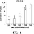
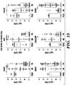
- the CTC marker gene is selected from Table 7 or Table 8. In some embodiments, the CTC marker gene is selected from the group consisting of: ABI3BP; ADAMTS5; ADAMTSL1; ANG; ARSA; C1RL; C3; C4A; C4B; CCDC80; CD109; CHI3L1; CLEC3B; CMTM3; CMTM7; COL14A1; COL1A2; COL3A1; COL4A6; CSF1; DAG1; DCN; DMKN; FBLN1; FGF1; FMOD; GPC3; GPC4; HMGB1; IFNAR2; IGFBP5; IL16; LAMA4; LTBP4; MFAP1A; NID2; OGN; PDAP1; PF4; PLAT; PODN; PRELP; RSPO1; SERPING1; SLURP 1; SOD3; SPARC; SPOCK2; SPON2; SU
- the CTC marker gene is selected from the group consisting of: ALDH1A1; ALDH1A2; IGFBP5; KLF4; DCN; SPARC; WNT; TGFB2; VEGF; COL1A2; COL3A1; and TIMP2. In some embodiments, the CTC marker gene is selected from the group consisting of: ALDH1A2; IGFBP5; KLF4; DCN; and SPARC.
- a method of treating cancer in a subject comprising administering a therapeutically effective amount of a CTC marker gene-targeted therapy to the subject.
- the cancer is pancreatic cancer.
- the CTC marker gene-targeted therapy comprises an inhibitor of a CTC marker gene.
- the inhibitor is an antibody reagent.
- the inhibitor is an inhibitory nucleic acid reagent.
- the CTC marker gene-targeted therapy comprises a CTC marker gene-binding antibody reagent and a chemotherapeutic agent.
- the subject is a subject determined to have an elevated level of CTCs and/or an elevated level of a CTC marker gene present in the blood and/or stroma of the cancer.
- a method of determining if a subject is likely to respond to treatment with a CTC marker gene-targeted therapy comprising measuring the level of a CTC marker gene expression product present in the blood and/or stroma of a cancer; and determining that the subject is likely to respond to the treatment if the level of the expression product is increased relative to a reference level.
- the method further comprises a first step of isolating the CTCs from the sample.
- the cancer is pancreatic cancer.
- the expression product is a nucleic acid.
- the level of the expression product is determined using a method selected from the group consisting of RT-PCR; quantitative RT-PCR; Northern blot; microarray based expression analysis; next-generation sequencing; and RNA in situ hybridization.
- the expression product is a polypeptide.
- the level of the expression product is determined using a method selected from the group consisting of: Western blot; immunoprecipitation; enzyme-linked immunosorbent assay (ELISA); radioimmunological assay (RIA); sandwich assay; fluorescence in situ hybridization (FISH); immunohistological staining; radioimmunometric assay; immunofluoresence assay; mass spectroscopy; FACS; and immunoelectrophoresis assay.
- the PC-CTC marker gene is selected from Table 7 or Table 8.
- the CTC marker gene is selected from the group consisting of: ABI3BP; ADAMTS5; ADAMTSL1; ANG; ARSA; C1RL; C3; C4A; C4B; CCDC80; CD109; CHI3L1; CLEC3B; CMTM3; CMTM7; COL14A1; COL1A2; COL3A1; COL4A6; CSF1; DAG1; DCN; DMKN; FBLN1; FGF1; FMOD; GPC3; GPC4; HMGB1; IFNAR2; IGFBP5; IL16; LAMA4; LTBP4; MFAP1A; NID2; OGN; PDAP1; PF4; PLAT; PODN; PRELP; RSPO1; SERPING1; SLURP 1; SOD3; SPARC; SPOCK2; SPON2;
- the CTC marker gene is selected from the group consisting of: ALDH1A1; ALDH1A2; IGFBP5; KLF4; DCN; SPARC; WNT; TGFB2; VEGF; COL1A2; COL3A1; and TIMP2. In some embodiments, the CTC marker gene is selected from the group consisting of: ALDH1A2; IGFBP5; KLF4; DCN; and SPARC.
- a method of monitoring the treatment of a subject comprising: administering a cancer therapy to a subject in need thereof; measuring the level of a CTC marker gene expression product present in the blood and/or stroma of a cancer; and determining that the subject is responding if the level of the CTC marker gene expression product is decreased relative to the reference level and determining that the subject is not responding to the treatment if the CTC marker gene expression product is not decreased relative to the reference level.
- the cancer is pancreatic cancer.
- the reference level is the level of the gene expression product in the patient prior to the administering step.
- the method further comprises a first step of isolating the CTCs from the sample.
- the expression product is a nucleic acid. In some embodiments, the level of the expression product is determined using a method selected from the group consisting of RT-PCR; quantitative RT-PCR; Northern blot; microarray based expression analysis; next-generation sequencing; and RNA in situ hybridization. In some embodiments, the expression product is a polypeptide.
- the level of the expression product is determined using a method selected from the group consisting of: Western blot; immunoprecipitation; enzyme-linked immunosorbent assay (ELISA); radioimmunological assay (RIA); sandwich assay; fluorescence in situ hybridization (FISH); immunohistological staining; radioimmunometric assay; immunofluoresence assay; mass spectroscopy; FACS; and immunoelectrophoresis assay.
- the PC-CTC marker gene is selected from Table 7 or Table 8.
- the CTC marker gene is selected from the group consisting of: ABI3BP; ADAMTS5; ADAMTSL1; ANG; ARSA; C1RL; C3; C4A; C4B; CCDC80; CD109; CHI3L1; CLEC3B; CMTM3; CMTM7; COL14A1; COL1A2; COL3A1; COL4A6; CSF1; DAG1; DCN; DMKN; FBLN1; FGF1; FMOD; GPC3; GPC4; HMGB1; IFNAR2; IGFBP5; IL16; LAMA4; LTBP4; MFAP1A; NID2; OGN; PDAP1; PF4; PLAT; PODN; PRELP; RSPO1; SERPING1; SLURP 1; SOD3; SPARC; SPOCK2; SPON2; SULF1 ; SULF2; TGFB2; TGM2;
- the CTC marker gene is selected from the group consisting of: ALDH1A1 ; ALDH1A2; IGFBP5; KLF4; DCN; SPARC; WNT; TGFB2; VEGF; COL1A2; COL3A1 ; and TIMP2. In some embodiments, the CTC marker gene is selected from the group consisting of: ALDH1A2; IGFBP5; KLF4; DCN; and SPARC.
- CTCs circulating tumor cells
- CTC marker genes i.e. CTC marker genes.
- CTC marker genes permit methods and assays for the detection and/or measurement of CTC levels, e.g. CTC levels in a sample from a subject. These methods and assays can provide improved speed and accuracy in the measurement of CTC levels.
- therapies can be targeted against CTCs by binding to and/or inhibiting these marker gene expression products to reduce the level and/or metastatic potential of CTCs.
- circulating tumor cell or “CTC” refers to tumor cells which are shed from a tumor and present in the blood, i.e. in circulation.
- Cell markers e.g. marker genes
- a CTC can be a pancreatic cancer CTC.
- CTCs circulating tumor cells
- the inventors have discovered that a number of genes are differentially regulated in CTCs, e.g. as compared to non-circulating tumor cells. Accordingly, there are provided herein methods and assays relating to the measurement of CTC levels. Elevated CTC levels can indicate a poor prognosis, e.g. an increased risk of metastatsis. Accordingly, provided herein are methods and assays related to the prognosis, risk assessment, and treatment of subjects having cancer. In certain embodiments, the assays and methods are directed to determination and/or measurement of the expression level of a gene product (e.g. protein and/or gene transcript such as mRNA) in a biological sample of a subject.
- a gene product e.g. protein and/or gene transcript such as mRNA
- the assays and methods are directed to determination of the expression level of a gene product of at least two genes in a biological sample of a subject, i.e. at least two genes, at least three genes, at least four genes , at least five genes, at least six genes, at least seven genes, at least eight genes, at least nine genes, at least 10 genes... at least 15 genes,...at least 25 genes,...at least 30 genes, or more genes, or any number of genes selected from Table 7, Table 8, and/or Table 14 as described herein.
- the marker gene(s) is selected from the group consisting of ABI3BP; ADAMTS5; ADAMTSL1; ANG; ARSA; C1RL; C3; C4A; C4B; CCDC80; CD109; CHI3L1; CLEC3B; CMTM3; CMTM7; COL14A1; COL1A2; COL3A1; COL4A6; CSF1; DAG1; DCN; DMKN; FBLN1; FGF1; FMOD; GPC3; GPC4; HMGB1; IFNAR2; IGFBP5; IL16; LAMA4; LTBP4; MFAP1A; NID2; OGN; PDAP1; PF4; PLAT; PODN; PRELP; RSPO1; SERPING1; SLURP1; SOD3; SPARC; SPOCK2; SPON2; SULF1; SULF2; TGFB2; TGM2;
- the assays, methods, and systems described herein are directed to determination of the expression level of a gene product of at least two genes in a biological sample of a subject, e.g. at least two genes, or at least three genes, or at least four genes, or, e.g.
- the marker gene(s) is selected from the group consisting of ALDH1A1; ALDH1A2; IGFBP5; KLF4; DCN; SPARC; WNT; TGFB2; VEGF; COL1A2; COL3A1; and TIMP2.
- the assays, methods, and systems described herein are directed to determination of the expression level of a gene product of at least two genes in a biological sample of a subject, e.g. at least two genes, or at least three genes, or at least four genes, or, e.g. all of the following genes: ALDH1A1; ALDH1A2; IGFBP5; KLF4; DCN; SPARC; WNT; TGFB2; VEGF; COL1A2; COL3A1; and TIMP2.
- the marker gene(s) is selected from the group consisting of ALDH1A2; IGFBP5; KLF4; DCN; and SPARC.
- the assays, methods, and systems described herein are directed to determination of the expression level of a gene product of at least two genes in a biological sample of a subject, e.g. at least two genes, or at least three genes, or at least four genes, or, e.g. all of the following genes: ALDH1A2; IGFBP5; KLF4; DCN; and SPARC.
- the marker gene(s) is selected from the group consisting of ALDH1A2; IGFBP5; KLF4; and DCN.
- the assays, methods, and systems described herein are directed to determination of the expression level of a gene product of at least two genes in a biological sample of a subject, e.g. at least two genes, or at least three genes, or e.g. all of the following genes: ALDH1A2; IGFBP5; KLF4; and DCN.
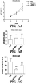
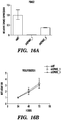
- the marker gene(s) is selected from the group consisting of TPT1; HMGB1; SPON 2; SPARC; and ARSA.
- the assays, methods, and systems described herein are directed to determination of the expression level of a gene product of at least two genes in a biological sample of a subject, e.g. at least two genes, or at least three genes, or at least four genes, or, e.g. all of the following genes: TPT1; HMGB1; SPON 2; SPARC; and ARSA.
- the marker gene(s) is selected from the group consisting of IL6ST; ARSA; TIMP2; CD55; SULF2; ITGA6; SDC4; CDON; and SV2A.
- the assays, methods, and systems described herein are directed to determination of the expression level of a gene product of at least two genes in a biological sample of a subject, e.g. at least two genes, or at least three genes, or at least four genes, or at least five genes, or at least six genes, or at least seven genes, or at least eight genes or, e.g.
- the level of polypeptide expression products are determined for the marker gene(s) is selected from the group consisting of IL6ST; ARSA; TIMP2; CD55; SULF2; ITGA6; SDC4; CDON; and SV2A, e.g. because, as described herein, RNA levels of cell surface proteins are lower than polypeptide levels.
- Table 7 Exemplary mouse marker genes MOUSE GENE SYMBOL Gene Name Abcblb ATP-binding cassette, sub-family B (MDR/TAP), member 1B Abi3bp ABI gene family, member 3 (NESH) binding protein Ablim3 actin binding LIM protein family, member 3 Acad9 acyl-Coenzyme A dehydrogenase family, member 9 Acbd3 acyl-Coenzyme A binding domain containing 3 Acin1 apoptotic chromatin condensation inducer 1 Actb actin, beta Actg1 predicted gene 8543; actin-like 8; predicted gene 7505; predicted gene 12715; predicted gene 12003; predicted gene 8399; predicted gene 6375; actin, gamma, cytoplasmic 1; similar to gamma-actin; predicted gene 4667; similar to cytoplasmic beta-actin; predicted gene 16385 Adamts5 similar to a disintegrin-like and metalloprotease (reprolysin type) with thrombospond
- Parp14 poly (ADP-ribose) polymerase family member 14 Parp4 poly (ADP-ribose) polymerase family, member 4 Parvb parvin, beta; similar to parvin, beta Pbx1 pre B-cell leukemia transcription factor 1; region containing RIKEN cDNA 2310056B04 gene; pre B-cell leukemia transcription factor 1 Pcdh15 protocadherin 15 Pcdhgb5 protocadherin gamma subfamily B, 5 Pcm1 pericentriolar material 1 Pdap1 PDGFA associated protein 1 Pdcd6ip programmed cell death 6 interacting protein Pde4dip phosphodiesterase 4D interacting protein (myomegalin) Pdia3 protein disulfide isomerase associated 3 Pdia4 protein disulfide isomerase associated 4 Pdpn podoplanin Pef1 penta-EF hand domain containing 1 Peli1 pellino 1 Per1 period homolog 1
- Slcl0a3 solute carrier family 10 sodium/bile acid cotransporter family
- member 3 Slc16a1 solute carrier family 16 monocarboxylic acid transporters
- member 1 Slc1a5 solute carrier family 1 neutral amino acid transporter
- member 5 Slc26a3 solute carrier family 26 member 3 Slc27a3 solute carrier family 27 (fatty acid transporter)
- member 3 Slc38a1 solute carrier family 38 member 1 Slc39a8 solute carrier family 39 (metal ion transporter), member 8 Slc43a3 solute carrier family 43, member 3 Slc4a4 solute carrier family 4 (anion exchanger), member 4 Slc6a4 solute carrier family 6 (neurotransmitter transporter, serotonin), member 4 Slc6a6 solute carrier family 6 (neurotransmitter transporter, taurine), member 6 Slc8a1 solute carrier family 8 (sodium/calcium exchanger), member
- ATP1A1 ATPase Na+/K+ transporting, alpha 1 polypeptide ATP1B1 ATPase, Na+/K+ transporting, beta 1 polypeptide ATP2B1 ATPase, Ca++ transporting, plasma membrane 1 ATP6V1A ATPase, H+ transporting, lysosomal 70kDa, V1 subunit A ATXN2 ataxin 2 B2M beta-2-microglobulin BAZ2A bromodomain adjacent to zinc finger domain, 2A BBS4 Bardet-Biedl syndrome 4 BBX bobby sox homolog (Drosophila) BCAM basal cell adhesion molecule (Lutheran blood group) BCL10 B-cell CLL/lymphoma 10; hypothetical LOC646626 BDP1 B double prime 1, subunit of RNA polymerase III transcription initiation factor IIIB BICC1 bicaudal C homolog 1 (Drosophila) BICD1 bicau
- F11R F11 receptor FAIM2 Fas apoptotic inhibitory molecule 2 FAM117A family with sequence similarity 117, member A FAM134B family with sequence similarity 134, member B FAM53B family with sequence similarity 53, member B FAM63B family with sequence similarity 63, member B FAM76A family with sequence similarity 76, member A FAM84B family with sequence similarity 84, member B FAS Fas (TNF receptor superfamily, member 6) FBLN1 fibulin 1 FERMT2 fermitin family homolog 2 (Drosophila) FGF1 fibroblast growth factor 1 (acidic) FHL1 four and a half LIM domains 1 FILIP1L filamin A interacting protein 1-like FKBP5
- PARP14 poly (ADP-ribose) polymerase family member 14 PARP4 poly (ADP-ribose) polymerase family, member 4 PARVB parvin, beta PBX1 pre-B-cell leukemia homeobox 1 PCDH15 protocadherin 15 PCDHGB5 protocadherin gamma subfamily B, 5 PCM1 pericentriolar material 1 PDAP1 PDGFA associated protein 1; similar to PDGFA associated protein 1 PDCD6IP programmed cell death 6 interacting protein PDE4DIP hypothetical protein LOC100134230; similar to KIAA0454 protein; similar to phosphodiesterase 4D interacting protein isoform 2; phosphodiesterase 4D interacting protein PDIA3 protein disulfide isomerase family A, member 3 PDIA4 protein disulfide isomerase family A, member 4 PDPN podoplanin PEF1 penta-EF-hand domain containing 1 PELI1 pellino homolog 1 (Drosophila)
- SLC10A3 solute carrier family 10 sodium/bile acid cotransporter family
- member 3 SLC16A1 solute carrier family 16 member 1 (monocarboxylic acid transporter 1)
- SLC1A5 solute carrier family 1 neutral amino acid transporter
- member 5 SLC26A3 solute carrier family 26 member 3 SLC27A3 solute carrier family 27 (fatty acid transporter)
- member 3 SLC38A1 solute carrier family 38 member 1 SLC39A8 solute carrier family 39 (zinc transporter)
- member 8 SLC43A3 solute carrier family 43
- member 3 SLC4A4 solute carrier family 4 sodium bicarbonate cotransporter
- member 4 SLC6A4 solute carrier family 6 neurotransmitter transporter, serotonin
- member 4 SLC6A6 solute carrier family 6 neurotransmitter transporter, taurine
- member 6 SLC8A1 solute carrier family 8 sodium/calcium exchanger
- member 1 SLC9A3R1 solute carrier family 9 member 1 SLC9A3R
- NCBI Gene ID numbers for each of the genes listed in Table 7 or Table 8 can be obtained by searching the "Gene" Database of the NCBI (available on the World Wide Web at http://www.ncbi.nlm.nih.gov/) using the common name as the query and selecting the first returned Homo sapiens (for the genes in Table 8) or Mus musculus gene (for the genes in Table 7).
- Other genes may be obtained using the UCSC genome browser (available on the World Wide Web at http://genome.ucsc.edu) using the Gene Sorter function.
- Human homologs of mouse genes can be readily identified, e.g. the identified homologs in the NCBI database, or by querying databases such as BLAST.
- the marker gene(s) are selected from the genes listed in Table 7, Table 8, or Table 14.
- the marker genes listed in Table 7, Table 8,, or Table 14 can be upregulated, e.g. for marker genes listed in Table 7, Table 8, or Table 14, if the measured marker gene expression in a cell or sample is higher as compared to a reference level of that marker gene's expression, then the cell is identificed as a CTC and/or the sample is identified as comprising CTCs. Preferably, once looks at a statistically significant change. However, even if a few genes in a group do not differ from normal, a sample can be identified as comprising CTCs if the overall change of the group shows a significant change, preferably a statistically significant change. All possible combinations of 2 or more of the indicated markers are contemplated herein.
- the reference can be a level of expression of the marker gene product in a cell or population of cells which are not CTCs, e.g. the average level in non-circulating tumor cells and/or circulating cells which are not cancer cells.
- the reference can also be a level of expression of the marker gene product in a control sample, a pooled sample of control individuals or a numeric value or range of values based on the same.
- the methods and assays described herein include (a) transforming the gene expression product into a detectable gene target; (b) measuring the amount of the detectable gene target; and (c) comparing the amount of the detectable gene target to an amount of a reference, wherein if the amount of the detectable gene target is statistically significantly different than the amount of the reference level, the presence and/or level of CTCs is determined. In some embodiments, if the amount of the detectable gene target is not statistically significantly different than the amount of the reference level, the sample is identified as not comprising CTCs.
- transforming refers to changing an object or a substance, e.g., biological sample, nucleic acid or protein, into another substance.
- the transformation can be physical, biological or chemical.
- Exemplary physical transformation includes, but not limited to, pre-treatment of a biological sample, e.g., from whole blood to blood serum by differential centrifugation.
- a biological/chemical transformation can involve at least one enzyme and/or a chemical reagent in a reaction.
- a DNA sample can be digested into fragments by one or more restriction enzyme, or an exogenous molecule can be attached to a fragmented DNA sample with a ligase.
- a DNA sample can undergo enzymatic replication, e.g., by polymerase chain reaction (PCR).
- PCR polymerase chain reaction
- ELISA enzyme linked immunosorbent assay
- RIA radioimmunological assay
- FISH fluorescent in situ hybridization
- immunohistological staining immunoelectrophoresis
- immunoprecipitation immunofluorescence using detection reagents such as an antibody or protein binding agents.
- a peptide can be detected in a subject by introducing into a subject a labeled anti-peptide antibody and other types of detection agent.
- the antibody can be labeled with a radioactive marker whose presence and location in the subject is detected by standard imaging techniques.
- antibodies for the polypeptide expression products of the marker genes described herein are commercially available and can be used for the purposes of the invention to measure protein expression levels, e.g. anti-IGFBP5 (Cat. No. 4255; Abcam; Cambridge, MA).
- anti-IGFBP5 Cat. No. 4255; Abcam; Cambridge, MA
- amino acid sequences for the marker genes described herein are known and publically available at NCBI website, one of skill in the art can raise their own antibodies against these proteins of interest for the purpose of the invention.
- the amino acid sequences of the marker genes described herein have been assigned NCBI accession numbers for different species such as human, mouse and rat.
- immunohistochemistry is the application of immunochemistry to tissue sections
- ICC is the application of immunochemistry to cells or tissue imprints after they have undergone specific cytological preparations such as, for example, liquid-based preparations.
- Immunochemistry is a family of techniques based on the use of an antibody, wherein the antibodies are used to specifically target molecules inside or on the surface of cells. The antibody typically contains a marker that will undergo a biochemical reaction, and thereby experience a change color, upon encountering the targeted molecules.
- signal amplification can be integrated into the particular protocol, wherein a secondary antibody, that includes the marker stain or marker signal, follows the application of a primary specific antibody.
- the assay can be a Western blot analysis.
- proteins can be separated by two-dimensional gel electrophoresis systems. Two-dimensional gel electrophoresis is well known in the art and typically involves iso-electric focusing along a first dimension followed by SDS-PAGE electrophoresis along a second dimension. These methods also require a considerable amount of cellular material.
- the analysis of 2D SDS-PAGE gels can be performed by determining the intensity of protein spots on the gel, or can be performed using immune detection.
- protein samples are analyzed by mass spectroscopy.
- Immunological tests can be used with the methods and assays described herein and include, for example, competitive and non-competitive assay systems using techniques such as Western blots, radioimmunoassay (RIA), ELISA (enzyme linked immunosorbent assay), "sandwich” immunoassays, immunoprecipitation assays, immunodiffusion assays, agglutination assays, e.g. latex agglutination, complement-fixation assays, immunoradiometric assays, fluorescent immunoassays, e.g.
- FIA fluorescence-linked immunoassay
- CLIA chemiluminescence immunoassays
- ELIA electrochemiluminescence immunoassay
- CIA counting immunoassay
- LFIA lateral flow tests or immunoassay
- MIA magnetic immunoassay
- protein A immunoassays Methods for performing such assays are known in the art, provided an appropriate antibody reagent is available.
- the immunoassay can be a quantitative or a semi-quantitative immunoassay.
- An immunoassay is a biochemical test that measures the concentration of a substance in a biological sample, typically a fluid sample such as serum, using the interaction of an antibody or antibodies to its antigen.
- the assay takes advantage of the highly specific binding of an antibody with its antigen.
- specific binding of the target polypeptides with respective proteins or protein fragments, or an isolated peptide, or a fusion protein described herein occurs in the immunoassay to form a target protein/peptide complex.
- the complex is then detected by a variety of methods known in the art.
- An immunoassay also often involves the use of a detection antibody.
- Enzyme-linked immunosorbent assay also called ELISA, enzyme immunoassay or EIA
- ELISA enzyme immunoassay
- EIA enzyme immunoassay
- an ELISA involving at least one antibody with specificity for the particular desired antigen i.e. a marker gene polypeptide as described herein
- a known amount of sample and/or antigen is immobilized on a solid support (usually a polystyrene micro titer plate). Immobilization can be either non-specific (e.g., by adsorption to the surface) or specific (e.g. where another antibody immobilized on the surface is used to capture antigen or a primary antibody). After the antigen is immobilized, the detection antibody is added, forming a complex with the antigen.
- the detection antibody can be covalently linked to an enzyme, or can itself be detected by a secondary antibody which is linked to an enzyme through bio-conjugation.
- the plate is typically washed with a mild detergent solution to remove any proteins or antibodies that are not specifically bound.
- the plate is developed by adding an enzymatic substrate to produce a visible signal, which indicates the quantity of antigen in the sample.
- Older ELISAs utilize chromogenic substrates, though newer assays employ fluorogenic substrates with much higher sensitivity.
- a competitive ELISA is used.
- Purified antibodies that are directed against a target polypeptide or fragment thereof are coated on the solid phase of multi-well plate, i.e., conjugated to a solid surface.
- a second batch of purified antibodies that are not conjugated on any solid support is also needed.
- These non-conjugated purified antibodies are labeled for detection purposes, for example, labeled with horseradish peroxidase to produce a detectable signal.
- a sample e.g., tumor, blood, serum or urine
- a known amount of desired antigen e.g., a known volume or concentration of a sample comprising a target polypeptide
- desired antigen e.g., a known volume or concentration of a sample comprising a target polypeptide
- the mixture is then are added to coated wells to form competitive combination.
- a complex of labeled antibody reagent-antigen will form. This complex is free in solution and can be washed away. Washing the wells will remove the complex.
- TMB (3, 3', 5, 5'-tetramethylbenzidene) color development substrate for localization of horseradish peroxidase-conjugated antibodies in the wells.
- TMB 3, 3', 5, 5'-tetramethylbenzidene
- TMB 3, 3', 5, 5'-tetramethylbenzidene
- the levels of a polypeptide in a sample can be detected by a lateral flow immunoassay test (LFIA), also known as the immunochromatographic assay, or strip test.
- LFIAs are a simple device intended to detect the presence (or absence) of antigen, e.g. a polypeptide, in a fluid sample.
- LFIA tests are used for medical diagnostics either for home testing, point of care testing, or laboratory use.
- LFIA tests are a form of immunoassay in which the test sample flows along a solid substrate via capillary action.
- LFIAs are essentially immunoassays adapted to operate along a single axis to suit the test strip format or a dipstick format. Strip tests are extremely versatile and can be easily modified by one skilled in the art for detecting an enormous range of antigens from fluid samples such as urine, blood, water, and/or homogenized tumor samples etc.
- Strip tests are also known as dip stick test, the name bearing from the literal action of "dipping" the test strip into a fluid sample to be tested.
- LFIA strip tests are easy to use, require minimum training and can easily be included as components of point-of-care test (POCT) diagnostics to be use on site in the field.
- LFIA tests can be operated as either competitive or sandwich assays.
- Sandwich LFIAs are similar to sandwich ELISA. The sample first encounters colored particles which are labeled with antibodies raised to the target antigen. The test line will also contain antibodies to the same target, although it may bind to a different epitope on the antigen. The test line will show as a colored band in positive samples.
- the lateral flow immunoassay can be a double antibody sandwich assay, a competitive assay, a quantitative assay or variations thereof.
- Competitive LFIAs are similar to competitive ELISA. The sample first encounters colored particles which are labeled with the target antigen or an analogue. The test line contains antibodies to the target/its analogue. Unlabelled antigen in the sample will block the binding sites on the antibodies preventing uptake of the colored particles. The test line will show as a colored band in negative samples.
- lateral flow technology It is also possible to apply multiple capture zones to create a multiplex test.
- a polypeptide or fragment thereof can be dissociated with detergents and heat, and separated on an SDS-PAGE gel before being transferred to a solid support, such as a nitrocellulose or PVDF membrane.
- a solid support such as a nitrocellulose or PVDF membrane.
- the membrane is incubated with an antibody reagent specific for the target polypeptide or a fragment thereof. The membrane is then washed to remove unbound proteins and proteins with non-specific binding.
- Detectably labeled enzyme-linked secondary or detection antibodies can then be used to detect and assess the amount of polypeptide in the sample tested.
- the intensity of the signal from the detectable label corresponds to the amount of enzyme present, and therefore the amount of polypeptide.
- Levels can be quantified, for example by densitometry
- Flow cytometry is a well-known technique for analyzing and sorting cells (or other small particles) suspended in a fluid stream. This technique allows simultaneous analysis of the physical and/or chemical characteristics of single cells flowing through an optical, electronic, or magnetic detection apparatus.
- the flow cytometer consists of a flow cell which carries the cells in a fluid stream in single file through a light source with excites the fluorescently labeled detection marker(s) (for example, antibody reagents) and measures the fluorescent character of the cell.
- the fluid stream is then ejected through a nozzle and a charging ring, under pressure, which breaks the fluid into droplets.
- the flow cell device and fluid stream is calibrated such that there is a relatively large distance between individual cells or bound groups of cells, resulting in a low probability that any droplet contains more than a single cell or bound group of cells.
- the charging ring charges the droplets based on the fluorescence characteristic of the cell which is contained therein.
- the charged droplets are then deflected by an electrostatically-charged deflection system which diverts the droplets into various containers based upon their charge (related to the fluorescence intensity of the cell).
- a FACS system e.g.
- the FACSARIATM flow cytometer (BD Biosciences) and FLOWJOTM Version 7.6.4 (TreeStar)) can detect and record the number of total cells as well as the number of cells which display one or more fluorescent characteristics, e.g. the total number of cells bound by one or more antibody reagents specific for a CTC marker gene.
- the gene expression products as described herein can be instead determined by determining the level of messenger RNA (mRNA) expression of genes associated with the marker genes described herein.
- mRNA messenger RNA
- Such molecules can be isolated, derived, or amplified from a biological sample, such as a tumor biopsy. Detection of mRNA expression is known by persons skilled in the art, and comprise, for example but not limited to, PCR procedures, RT-PCR, quantitative PCR or RT-PCR, Northern blot analysis, differential gene expression, RNA protection assay, microarray analysis, hybridization methods, next-generation sequencing etc.
- Non-limiting examples of next-generation sequencing technologies can include Ion Torrent, Illumina, SOLiD, 454; Massively Parallel Signature Sequencing solid-phase, reversible dye-terminator sequencing; and DNA nanoball sequencing.
- the PCR procedure describes a method of gene amplification which is comprised of (i) sequence-specific hybridization of primers to specific genes or sequences within a nucleic acid sample or library, (ii) subsequent amplification involving multiple rounds of annealing, elongation, and denaturation using a thermostable DNA polymerase, and (iii) screening the PCR products for a band of the correct size.
- the primers used are oligonucleotides of sufficient length and appropriate sequence to provide initiation of polymerization, i.e. each primer is specifically designed to be complementary to a strand of the genomic locus to be amplified.
- mRNA level of gene expression products described herein can be determined by reverse-transcription (RT) PCR and by quantitative RT-PCR (QRT-PCR) or real-time PCR methods.
- RT reverse-transcription
- QRT-PCR quantitative RT-PCR
- Methods of RT-PCR and QRT-PCR are well known in the art.
- the nucleic acid sequences of the marker genes described herein have been assigned NCBI accession numbers for different species such as human, mouse and rat. Accordingly, a skilled artisan can design an appropriate primer based on the known sequence for determining the mRNA level of the respective gene.
- Nucleic acid and ribonucleic acid (RNA) molecules can be isolated from a particular biological sample using any of a number of procedures, which are well-known in the art, the particular isolation procedure chosen being appropriate for the particular biological sample.
- freeze-thaw and alkaline lysis procedures can be useful for obtaining nucleic acid molecules from solid materials
- heat and alkaline lysis procedures can be useful for obtaining nucleic acid molecules from urine
- proteinase K extraction can be used to obtain nucleic acid from blood ( Roiff, A et al. PCR: Clinical Diagnostics and Research, Springer (1994 )).
- the PCR procedure describes a method of gene amplification which is comprised of (i) sequence-specific hybridization of primers to specific genes within a nucleic acid sample or library, (ii) subsequent amplification involving multiple rounds of annealing, elongation, and denaturation using a DNA polymerase, and (iii) screening the PCR products for a band of the correct size.
- the primers used are oligonucleotides of sufficient length and appropriate sequence to provide initiation of polymerization, i.e. each primer is specifically designed to be complementary to each strand of the nucleic acid molecule to be amplified.
- mRNA level of gene expression products described herein can be determined by reverse-transcription (RT) PCR and by quantitative RT-PCR (QRT-PCR) or real-time PCR methods.
- RT reverse-transcription
- QRT-PCR quantitative RT-PCR
- real-time PCR methods Methods of RT-PCR and QRT-PCR are well known in the art.
- one or more of the reagents can comprise a detectable label and/or comprise the ability to generate a detectable signal (e.g. by catalyzing reaction converting a compound to a detectable product).
- Detectable labels can comprise, for example, a light-absorbing dye, a fluorescent dye, or a radioactive label. Detectable labels, methods of detecting them, and methods of incorporating them into reagents (e.g. antibodies and nucleic acid probes) are well known in the art.
- detectable labels can include labels that can be detected by spectroscopic, photochemical, biochemical, immunochemical, electromagnetic, radiochemical, or chemical means, such as fluorescence, chemifluoresence, or chemiluminescence, or any other appropriate means.
- the detectable labels used in the methods described herein can be primary labels (where the label comprises a moiety that is directly detectable or that produces a directly detectable moiety) or secondary labels (where the detectable label binds to another moiety to produce a detectable signal, e.g., as is common in immunological labeling using secondary and tertiary antibodies).
- the detectable label can be linked by covalent or non-covalent means to the reagent.
- a detectable label can be linked such as by directly labeling a molecule that achieves binding to the reagent via a ligand-receptor binding pair arrangement or other such specific recognition molecules.
- Detectable labels can include, but are not limited to radioisotopes, bioluminescent compounds, chromophores, antibodies, chemiluminescent compounds, fluorescent compounds, metal chelates, and enzymes.
- the detection reagent is label with a fluorescent compound.
- a detectable label can be a fluorescent dye molecule, or fluorophore including, but not limited to fluorescein, phycoerythrin, phycocyanin, o-phthaldehyde, fluorescamine, Cy3TM, Cy5TM, allophycocyanine, Texas Red, peridenin chlorophyll, cyanine, tandem conjugates such as phycoerythrin-Cy5TM, green fluorescent protein, rhodamine, fluorescein isothiocyanate (FITC) and Oregon GreenTM, rhodamine and derivatives (e.g., Texas red and tetrarhodimine isothiocynate (TRITC)), biotin, phycoerythrin, AMCA, CyDyesTM, 6-carboxy
- a detectable label can be a radiolabel including, but not limited to 3 H, 125 I, 35 S, 14 C, 32 P, and 33 P.
- a detectable label can be an enzyme including, but not limited to horseradish peroxidase and alkaline phosphatase.
- An enzymatic label can produce, for example, a chemiluminescent signal, a color signal, or a fluorescent signal.
- Enzymes contemplated for use to detectably label an antibody reagent include, but are not limited to, malate dehydrogenase, staphylococcal nuclease, delta-V-steroid isomerase, yeast alcohol dehydrogenase, alpha-glycerophosphate dehydrogenase, triose phosphate isomerase, horseradish peroxidase, alkaline phosphatase, asparaginase, glucose oxidase, beta-galactosidase, ribonuclease, urease, catalase, glucose-VI-phosphate dehydrogenase, glucoamylase and acetylcholinesterase.
- a detectable label is a chemiluminescent label, including, but not limited to lucigenin, luminol, luciferin, isoluminol, theromatic acridinium ester, imidazole, acridinium salt and oxalate ester.
- a detectable label can be a spectral colorimetric label including, but not limited to colloidal gold or colored glass or plastic (e.g., polystyrene, polypropylene, and latex) beads.
- detection reagents can also be labeled with a detectable tag, such as c-Myc, HA, VSV-G, HSV, FLAG, V5, HIS, or biotin.
- a detectable tag such as c-Myc, HA, VSV-G, HSV, FLAG, V5, HIS, or biotin.
- Other detection systems can also be used, for example, a biotin-streptavidin system.
- the antibodies immunoreactive (i.e. specific for) with the biomarker of interest is biotinylated. Quantity of biotinylated antibody bound to the biomarker is determined using a streptavidin-peroxidase conjugate and a chromagenic substrate.
- streptavidin peroxidase detection kits are commercially available, e.g. from DAKO; Carpinteria, CA.
- a reagent can also be detectably labeled using fluorescence emitting metals such as 152 Eu, or others of the lanthanide series. These metals can be attached to the reagent using such metal chelating groups as diethylenetriaminepentaacetic acid (DTPA) or ethylenediaminetetraacetic acid (EDTA).
- DTPA diethylenetriaminepentaacetic acid
- EDTA ethylenediaminetetraacetic acid
- the level of expression products of more than one gene can be determined simultaneously (e.g. a multiplex assay) or in parallel. In some embodiments, the level of expression products of no more than 200 other genes is determined. In some embodiments, the level of expression products of no more than 100 other genes is determined. In some embodiments, the level of expression products of no more than 20 other genes is determined. In some embodiments, the level of expression products of no more than 10 other genes is determined.
- sample or "test sample” as used herein denotes a sample taken or isolated from a biological organism, e.g., a tumor sample from a subject.
- exemplary biological samples include, but are not limited to, a biofluid sample; serum; plasma; urine; saliva; a tumor sample; a tumor biopsy and/or tissue sample etc.
- the term also includes a mixture of the above-mentioned samples.
- test sample also includes untreated or pretreated (or pre-processed) biological samples.
- a test sample can comprise cells from subject.
- a test sample can be a tumor cell test sample, e.g. the sample can comprise cancerous cells, cells from a tumor, and/or a tumor biopsy.
- the test sample can be a blood sample.
- the test sample can be obtained by removing a sample of cells from a subject, but can also be accomplished by using previously isolated cells (e.g. isolated at a prior timepoint and isolated by the same or another person). In addition, the test sample can be freshly collected or a previously collected sample.
- the test sample can be an untreated test sample.
- untreated test sample refers to a test sample that has not had any prior sample pre-treatment except for dilution and/or suspension in a solution.
- Exemplary methods for treating a test sample include, but are not limited to, centrifugation, filtration, sonication, homogenization, heating, freezing and thawing, and combinations thereof.
- the test sample can be a frozen test sample, e.g., a frozen tissue. The frozen sample can be thawed before employing methods, assays and systems described herein. After thawing, a frozen sample can be centrifuged before being subjected to methods, assays and systems described herein.
- the test sample is a clarified test sample, for example, by centrifugation and collection of a supernatant comprising the clarified test sample.
- a test sample can be a pre-processed test sample, for example, supernatant or filtrate resulting from a treatment selected from the group consisting of centrifugation, filtration, thawing, purification, and any combinations thereof.
- the test sample can be treated with a chemical and/or biological reagent. Chemical and/or biological reagents can be employed to protect and/or maintain the stability of the sample, including biomolecules (e.g., nucleic acid and protein) therein, during processing.
- biomolecules e.g., nucleic acid and protein
- One exemplary reagent is a protease inhibitor, which is generally used to protect or maintain the stability of protein during processing.
- protease inhibitor which is generally used to protect or maintain the stability of protein during processing.
- the skilled artisan is well aware of methods and processes appropriate for pre-processing of biological samples required for determination of the level of an expression product as described herein.
- the methods, assays, and systems described herein can further comprise a step of obtaining a test sample from a subject.
- the subject can be a human subject.
- the methods and assays described herein can further comprise a step of isolating CTCs or potential CTCs from a sample prior to measuring the level the expression product of one or more of the marker genes described herein.
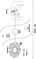
- CTCs can be isolated from, e.g. a blood sample by hydrodynamic size-based separation and/or immunodepletetion of other cell types present in blood samples.
- the CTC-iChip, described in the Examples herein combines these two approaches to isolate CTCs.
- Subjects with high, or at least detectable, levels of CTCs are most likely to benefit from treatment with therapies that specifically target CTCs. Accordingly, provided herein is a method of determining if a subject is likely to respond to treatment with a CTC marker gene-targeted therapy, the method comprising: measuring the level of a CTC marker gene expression product present in the blood and/or stroma of a cancer; and determining that the subject is likely to respond to the treatment if the level of the expression product is increased relative to a reference level.
- CTC marker gene-targeted therapies are discussed below herein.
- Decreased levels of CTCs after administration of a therapy can be indicative of an improvement in the condition of the subject, e.g. the cancer is reduced in size, growth, and/or metastatic potential.
- a method of monitoring the treatment of a subject comprising administering a cancer therapy to a subject in need thereof; measuring the level of a CTC marker gene expression product present in the blood and/or stroma of a cancer; and determining that the subject is responding if the level of the CTC marker gene expression product is decreased relative to the reference level and determining that the subject is not responding to the treatment if the CTC marker gene expression product is not decreased relative to the reference level.
- the therapy is a chemotherapy, surgical therapy, and/or radiation therapy.
- the therapy is a CTC marker gene-targeted therapy.
- the reference level is the level of the gene expression product in the patient prior to the administering step.
- the CTC marker genes described herein can be targeted directly and/or used to physically target a chemotherapeutic agent to reduce the levels and/or pathogenic activity of CTCs (e.g. metastatic activity). Accordingly, described herein is a method of treating cancer in a subject, the method comprising administering a therapeutically effective amount of a CTC marker gene-targeted therapy to the subject.
- the subject is a subject determined to have an elevated level of CTCs and/or an elevated level of a CTC marker gene present in the blood and/or stroma of the cancer.
- the CTC marker gene-targeted therapy can comprise an inhibitor of a CTC marker gene, e.g. the CTC marker gene-targeted therapy can inhibit the level and/or activity of a CTC marker gene.
- the term "inhibitor” refers to an agent which can decrease the expression and/or activity of the targeted expression product (e.g. mRNA encoding the target or a target polypeptide), e.g. by at least 10% or more, e.g. by 10% or more, 50% or more, 70% or more, 80% or more, 90% or more, 95% or more, or 98 % or more.
- the efficacy of an inhibitor of a CTC marker gene e.g.
- the CTC marker gene its ability to decrease the level and/or activity of the CTC marker gene can be determined, e.g. by measuring the level of an expression product and/or the activity of the CTC marker gene.
- Methods for measuring the level of a given mRNA and/or polypeptide are known to one of skill in the art, e.g. RTPCR with primers can be used to determine the level of RNA and Western blotting with an antibody can be used to determine the level of a polypeptide.
- the activity of, e.g. a CTC marker gene can be determined, e.g. by measuring the levels and/or survival of CTCs using methods known in the art and described elsewhere herein.
- the inhibitor of a CTC marker gene can be an inhibitory nucleic acid; an aptamer; an antibody reagent; an antibody; or a small molecule.
- the inhibitor of a CTC marker gene can be an antibody reagent.
- an "antibody” refers to IgG, IgM, IgA, IgD or IgE molecules or antigen-specific antibody fragments thereof (including, but not limited to, a Fab, F(ab') 2 , Fv, disulphide linked Fv, scFv, single domain antibody, closed conformation multispecific antibody, disulphide-linked scfv, diabody), whether derived from any species that naturally produces an antibody, or created by recombinant DNA technology; whether isolated from serum, B-cells, hybridomas, transfectomas, yeast or bacteria.
- an "antigen” is a molecule that is bound by a binding site on an antibody agent.
- antigens are bound by antibody ligands and are capable of raising an antibody response in vivo.
- An antigen can be a polypeptide, protein, nucleic acid or other molecule or portion thereof.
- antigenic determinant refers to an epitope on the antigen recognized by an antigen-binding molecule, and more particularly, by the antigen-binding site of said molecule.
- an antibody reagent refers to a polypeptide that includes at least one immunoglobulin variable domain or immunoglobulin variable domain sequence and which specifically binds a given antigen.
- An antibody reagent can comprise an antibody or a polypeptide comprising an antigen-binding domain of an antibody.
- an antibody reagent can comprise a monoclonal antibody or a polypeptide comprising an antigen-binding domain of a monoclonal antibody.
- an antibody can include a heavy (H) chain variable region (abbreviated herein as VH), and a light (L) chain variable region (abbreviated herein as VL).
- an antibody in another example, includes two heavy (H) chain variable regions and two light (L) chain variable regions.
- antibody reagent encompasses antigen-binding fragments of antibodies (e.g., single chain antibodies, Fab and sFab fragments, F(ab')2, Fd fragments, Fv fragments, scFv, and domain antibodies (dAb) fragments (see, e.g. de Wildt et al., Eur J. Immunol. 1996; 26(3):629-39 ; which is incorporated by reference herein in its entirety)) as well as complete antibodies.
- An antibody can have the structural features of IgA, IgG, IgE, IgD, IgM (as well as subtypes and combinations thereof).
- Antibodies can be from any source, including mouse, rabbit, pig, rat, and primate (human and non-human primate) and primatized antibodies. Antibodies also include midibodies, humanized antibodies, chimeric antibodies, and the like.
- VH and VL regions can be further subdivided into regions of hypervariability, termed “complementarity determining regions” ("CDR"), interspersed with regions that are more conserved, termed “framework regions” ("FR").
- CDR complementarity determining regions
- FR framework regions
- the extent of the framework region and CDRs has been precisely defined (see, Kabat, E. A., et al. (1991) Sequences of Proteins of Immunological Interest, Fifth Edition, U.S. Department of Health and Human Services, NIH Publication No. 91-3242 , and Chothia, C. et al. (1987) J. Mol. Biol. 196:901-917 ; which are incorporated by reference herein in their entireties).
- Each VH and VL is typically composed of three CDRs and four FRs, arranged from amino-terminus to carboxy-terminus in the following order: FR1, CDR1, FR2, CDR2, FR3, CDR3, FR4.
- antigen-binding fragment or "antigen-binding domain”, which are used interchangeably herein are used to refer to one or more fragments of a full length antibody that retain the ability to specifically bind to a target of interest.
- binding fragments encompassed within the term "antigen-binding fragment” of a full length antibody include (i) a Fab fragment, a monovalent fragment consisting of the VL, VH, CL and CH1 domains; (ii) a F(ab')2 fragment, a bivalent fragment including two Fab fragments linked by a disulfide bridge at the hinge region; (iii) an Fd fragment consisting of the VH and CH1 domains; (iv) an Fv fragment consisting of the VL and VH domains of a single arm of an antibody, (v) a dAb fragment ( Ward et al., (1989) Nature 341:544-546 ; which is incorporated by reference herein in its entirety), which consists of a VH or VL
- specific binding refers to a chemical interaction between two molecules, compounds, cells and/or particles wherein the first entity binds to the second, target entity with greater specificity and affinity than it binds to a third entity which is a non-target.
- specific binding can refer to an affinity of the first entity for the second target entity which is at least 10 times, at least 50 times, at least 100 times, at least 500 times, at least 1000 times or greater than the affinity for the third nontarget entity.
- a recombinant humanized antibody can be further optimized to decrease potential immunogenicity, while maintaining functional activity, for therapy in humans.
- functional activity means a polypeptide capable of displaying one or more known functional activities associated with a recombinant antibody or antibody reagent thereof as described herein. Such functional activities include, e.g. the ability to bind to a given CTC marker gene.
- the inhibitor of a CTC marker gene can be an inhibitory nucleic acid reagent.
- the inhibitory nucleic acid is an inhibitory RNA (iRNA). Double-stranded RNA molecules (dsRNA) have been shown to block gene expression in a highly conserved regulatory mechanism known as RNA interference (RNAi).
- RNAi RNA interference
- the inhibitory nucleic acids described herein can include an RNA strand (the antisense strand) having a region which is 30 nucleotides or less in length, i.e., 15-30 nucleotides in length, generally 19-24 nucleotides in length, which region is substantially complementary to at least part the targeted mRNA transcript. The use of these iRNAs enables the targeted degradation of mRNA transcripts, resulting in decreased expression and/or activity of the target.
- RNA refers to an agent that contains RNA as that term is defined herein, and which mediates the targeted cleavage of an RNA transcript via an RNA-induced silencing complex (RISC) pathway.
- RISC RNA-induced silencing complex
- an iRNA as described herein effects inhibition of the expression and/or activity of the target mRNA.
- contacting a cell with the inhibitor e.g.
- an iRNA results in a decrease in the target mRNA level in a cell by at least about 5%, about 10%, about 20%, about 30%, about 40%, about 50%, about 60%, about 70%, about 80%, about 90%, about 95%, about 99%, up to and including 100% of the target mRNA level found in the cell without the presence of the iRNA.
- the iRNA can be a dsRNA.
- a dsRNA includes two RNA strands that are sufficiently complementary to hybridize to form a duplex structure under conditions in which the dsRNA will be used.
- One strand of a dsRNA (the antisense strand) includes a region of complementarity that is substantially complementary, and generally fully complementary, to a target sequence.
- the target sequence can be derived from the sequence of an mRNA formed during the expression of the target.
- the other strand (the sense strand) includes a region that is complementary to the antisense strand, such that the two strands hybridize and form a duplex structure when combined under suitable conditions.
- the duplex structure is between 15 and 30 inclusive, more generally between 18 and 25 inclusive, yet more generally between 19 and 24 inclusive, and most generally between 19 and 21 base pairs in length, inclusive.
- the region of complementarity to the target sequence is between 15 and 30 inclusive, more generally between 18 and 25 inclusive, yet more generally between 19 and 24 inclusive, and most generally between 19 and 21 nucleotides in length, inclusive.
- the dsRNA is between 15 and 20 nucleotides in length, inclusive, and in other embodiments, the dsRNA is between 25 and 30 nucleotides in length, inclusive.
- RNA targeted for cleavage will most often be part of a larger RNA molecule, often an mRNA molecule.
- a "part" of an mRNA target is a contiguous sequence of an mRNA target of sufficient length to be a substrate for RNAi-directed cleavage (i.e., cleavage through a RISC pathway).
- dsRNAs having duplexes as short as 9 base pairs can, under some circumstances, mediate RNAi-directed RNA cleavage.
- a target will be at least 15 nucleotides in length, preferably 15-30 nucleotides in length.
- the RNA of an iRNA is chemically modified to enhance stability or other beneficial characteristics.
- the nucleic acids featured in the invention may be synthesized and/or modified by methods well established in the art, such as those described in " Current protocols in nucleic acid chemistry,” Beaucage, S.L. et al. (Edrs.), John Wiley & Sons, Inc., New York, NY, USA , which is hereby incorporated herein by reference.
- Modifications include, for example, (a) end modifications, e.g., 5' end modifications (phosphorylation, conjugation, inverted linkages, etc.) 3' end modifications (conjugation, DNA nucleotides, inverted linkages, etc.), (b) base modifications, e.g., replacement with stabilizing bases, destabilizing bases, or bases that base pair with an expanded repertoire of partners, removal of bases (abasic nucleotides), or conjugated bases, (c) sugar modifications (e.g., at the 2' position or 4' position) or replacement of the sugar, as well as (d) backbone modifications, including modification or replacement of the phosphodiester linkages.
- end modifications e.g., 5' end modifications (phosphorylation, conjugation, inverted linkages, etc.) 3' end modifications (conjugation, DNA nucleotides, inverted linkages, etc.
- base modifications e.g., replacement with stabilizing bases, destabilizing bases, or bases that base pair with an expanded repertoire of partners
- RNA compounds useful in the embodiments described herein include, but are not limited to RNAs containing modified backbones or no natural internucleoside linkages.
- RNAs having modified backbones include, among others, those that do not have a phosphorus atom in the backbone.
- modified RNAs that do not have a phosphorus atom in their internucleoside backbone can also be considered to be oligonucleosides.
- the modified RNA will have a phosphorus atom in its internucleoside backbone.
- Modified RNA backbones can include, for example, phosphorothioates, chiral phosphorothioates, phosphorodithioates, phosphotriesters, aminoalkylphosphotriesters, methyl and other alkyl phosphonates including 3'-alkylene phosphonates and chiral phosphonates, phosphinates, phosphoramidates including 3'-amino phosphoramidate and aminoalkylphosphoramidates, thionophosphoramidates, thionoalkylphosphonates, thionoalkylphosphotriesters, and boranophosphates having normal 3'-5' linkages, 2'-5' linked analogs of these, and those) having inverted polarity wherein the adjacent pairs of nucleoside units are linked 3'-5' to 5'-3' or 2'-5' to 5'-2'.
- Modified RNA backbones that do not include a phosphorus atom therein have backbones that are formed by short chain alkyl or cycloalkyl internucleoside linkages, mixed heteroatoms and alkyl or cycloalkyl internucleoside linkages, or one or more short chain heteroatomic or heterocyclic internucleoside linkages.
- patents that teach the preparation of the above oligonucleosides include, but are not limited to, U.S. Pat. Nos. 5,034,506 ; 5,166,315 ; 5,185,444 ; 5,214,134 ; 5,216,141 ; 5,235,033 ; 5,64,562 ; 5,264,564 ; 5,405,938 ; 5,434,257 ; 5,466,677 ; 5,470,967 ; 5,489,677 ; 5,541,307 ; 5,561,225 ; 5,596,086 ; 5,602,240 ; 5,608,046 ; 5,610,289 ; 5,618,704 ; 5,623,070 ; 5,663,312 ; 5,633,360 ; 5,677,437 ; and, 5,677,439 , each of which is herein incorporated by reference.
- RNA mimetics suitable or contemplated for use in iRNAs both the sugar and the internucleoside linkage, i.e., the backbone, of the nucleotide units are replaced with novel groups.
- the base units are maintained for hybridization with an appropriate nucleic acid target compound.
- One such oligomeric compound, an RNA mimetic that has been shown to have excellent hybridization properties, is referred to as a peptide nucleic acid (PNA).
- PNA peptide nucleic acid
- the sugar backbone of an RNA is replaced with an amide containing backbone, in particular an aminoethylglycine backbone.
- the nucleobases are retained and are bound directly or indirectly to aza nitrogen atoms of the amide portion of the backbone. Representative U.S.
- PNA compounds include, but are not limited to, U.S. Pat. Nos. 5,539,082 ; 5,714,331 ; and 5,719,262 , each of which is herein incorporated by reference. Further teaching of PNA compounds can be found, for example, in Nielsen et al., Science, 1991, 254, 1497-1500 .
- RNAs with phosphorothioate backbones and oligonucleosides with heteroatom backbones and in particular --CH 2 --NH--CH 2 --,-CH 2 --N(CH 3 )--O--CH 2 -- [known as a methylene (methylimino) or MMI backbone], --CH 2 --O-N(CH 3 )--CH 2 --, --CH 2 --N(CH 3 )--N(CH 3 )--CH 2 -- and -N(CH 3 )--CH 2 --CH 2 --[wherein the native phosphodiester backbone is represented as -O--P--O--CH 2 --] of the above-referenced U.S. Pat. No.
- RNAs featured herein have morpholino backbone structures of the above-referenced U.S. Pat. No. 5,034,506 .
- Modified RNAs can also contain one or more substituted sugar moieties.
- the iRNAs, e.g., dsRNAs, featured herein can include one of the following at the 2' position: OH; F; O-, S-, or N-alkyl; O-, S-, or N-alkenyl; O-, S- or N-alkynyl; or O-alkyl-O-alkyl, wherein the alkyl, alkenyl and alkynyl may be substituted or unsubstituted C 1 to C 10 alkyl or C 2 to C 10 alkenyl and alkynyl.
- Exemplary suitable modifications include O[(CH 2 ) n O] m CH 3 , O(CH 2 ) ⁇ n OCH 3 , O(CH 2 ) n NH 2 , O(CH 2 ) n CH 3 , O(CH 2 ) n ONH 2 , and O(CH 2 ) n ON[(CH 2 ) n CH 3 )] 2 , where n and m are from 1 to about 10.
- dsRNAs include one of the following at the 2' position: C 1 to C 10 lower alkyl, substituted lower alkyl, alkaryl, aralkyl, O-alkaryl or O-aralkyl, SH, SCH 3 , OCN, Cl, Br, CN, CF 3 , OCF 3 , SOCH 3 , SO 2 CH 3 , ONO 2 , NO 2 , N 3 , NH 2 , heterocycloalkyl, heterocycloalkaryl, aminoalkylamino, polyalkylamino, substituted silyl, an RNA cleaving group, a reporter group, an intercalator, a group for improving the pharmacokinetic properties of an iRNA, or a group for improving the pharmacodynamic properties of an iRNA, and other substituents having similar properties.
- the modification includes a 2'-methoxyethoxy (2'-O-CH 2 CH 2 OCH 3 , also known as 2'-O-(2-methoxyethyl) or 2'-MOE) ( Martin et al., Helv. Chim. Acta, 1995, 78:486-504 ) i.e., an alkoxy-alkoxy group.
- 2'-methoxyethoxy 2'-O-CH 2 CH 2 OCH 3
- 2'-MOE 2'-methoxyethoxy
- 2'-dimethylaminooxyethoxy i.e., a O(CH 2 ) 2 ON(CH 3 ) 2 group, also known as 2'-DMAOE, as described in examples herein below
- 2'-dimethylaminoethoxyethoxy also known in the art as 2'-O-dimethylaminoethoxyethyl or 2'-DMAEOE
- 2'-O--CH 2 --O---CH 2 --N(CH 2 ) 2 also described in examples herein below.
- modifications include 2'-methoxy (2'-OCH 3 ), 2'-aminopropoxy (2'-OCH 2 CH 2 CH 2 NH 2 ) and 2'-fluoro (2'-F). Similar modifications can also be made at other positions on the RNA of an iRNA, particularly the 3' position of the sugar on the 3' terminal nucleotide or in 2'-5' linked dsRNAs and the 5' position of 5' terminal nucleotide. iRNAs may also have sugar mimetics such as cyclobutyl moieties in place of the pentofuranosyl sugar. Representative U.S. patents that teach the preparation of such modified sugar structures include, but are not limited to, U.S. Pat. Nos.
- nucleobase can also include nucleobase (often referred to in the art simply as “base”) modifications or substitutions.
- base nucleobase
- unmodified or “natural” nucleobases include the purine bases adenine (A) and guanine (G), and the pyrimidine bases thymine (T), cytosine (C) and uracil (U).
- Modified nucleobases include other synthetic and natural nucleobases such as 5-methylcytosine (5-me-C), 5-hydroxymethyl cytosine, xanthine, hypoxanthine, 2-aminoadenine, 6-methyl and other alkyl derivatives of adenine and guanine, 2-propyl and other alkyl derivatives of adenine and guanine, 2-thiouracil, 2-thiothymine and 2-thiocytosine, 5-halouracil and cytosine, 5-propynyl uracil and cytosine, 6-azo uracil, cytosine and thymine, 5-uracil (pseudouracil), 4-thiouracil, 8-halo, 8-amino, 8-thiol, 8-thioalkyl, 8-hydroxyl anal other 8-substituted adenines and guanines, 5-halo, particularly 5-bromo, 5-trifluoromethyl and other 5-substi
- nucleobases include those disclosed in U.S. Pat. No. 3,687,808 , those disclosed in Modified Nucleosides in Biochemistry, Biotechnology and Medicine, Herdewijn, P. ed. Wiley-VCH, 2008 ; those disclosed in The Concise Encyclopedia Of Polymer Science And Engineering, pages 858-859, Kroschwitz, J. L, ed. John Wiley & Sons, 1990 , these disclosed by Englisch et al., Angewandte Chemie, International Edition, 1991, 30, 613 , and those disclosed by Sanghvi, Y S., Chapter 15, dsRNA Research and Applications, pages 289-302, Crooke, S. T.
- nucleobases are particularly useful for increasing the binding affinity of the oligomeric compounds featured in the invention.
- These include 5-substituted pyrimidines, 6-azapyrimidines and N-2, N-6 and 0-6 substituted purines, including 2-aminopropyladenine, 5-propynyluracil and 5-propynylcytosine.
- 5-methylcytosine substitutions have been shown to increase nucleic acid duplex stability by 0.6-1.2°C ( Sanghvi, Y. S., Crooke, S. T. and Lebleu, B., Eds., dsRNA Research and Applications, CRC Press, Boca Raton, 1993, pp. 276-278 ) and are exemplary base substitutions, even more particularly when combined with 2'-0-methoxyethyl sugar modifications.
- RNA of an iRNA can also be modified to include one or more locked nucleic acids (LNA).
- LNA locked nucleic acids
- a locked nucleic acid is a nucleotide having a modified ribose moiety in which the ribose moiety comprises an extra bridge connecting the 2' and 4' carbons. This structure effectively "locks" the ribose in the 3'-endo structural conformation.
- the addition of locked nucleic acids to siRNAs has been shown to increase siRNA stability in serum, and to reduce off-target effects ( Elmen, J. et al., (2005) Nucleic Acids Research 33(1):439-447 ; Mook, OR. et al., (2007) Mol Canc Ther 6(3):833-843 ; Grunweller, A.
- RNA of an iRNA featured in the invention involves chemically linking to the RNA one or more ligands, moieties or conjugates that enhance the activity, cellular distribution, pharmacokinetic properties, or cellular uptake of the iRNA.
- moieties include but are not limited to lipid moieties such as a cholesterol moiety ( Letsinger et al., Proc. Natl. Acid. Sci. USA, 1989, 86: 6553-6556 ), cholic acid ( Manoharan et al., Biorg. Med. Chem. Let., 1994, 4:1053-1060 ), a thioether, e.g., beryl-S-tritylthiol ( Manoharan et al., Ann.
- Acids Res., 1990, 18:3777-3783 a polyamine or a polyethylene glycol chain ( Manoharan et al., Nucleosides & Nucleotides, 1995, 14:969-973 ), or adamantane acetic acid ( Manoharan et al., Tetrahedron Lett., 1995, 36:3651-3654 ), a palmityl moiety ( Mishra et al., Biochim. Biophys. Acta, 1995, 1264:229-237 ), or an octadecylamine or hexylamino-carbonyloxycholesterol moiety ( Crooke et al., J. Pharmacol. Exp. Ther., 1996, 277:923-937 ).
- the CTC marker gene-targeted therapy can comprise an agent that binds to the CTC marker gene expression product and an agent that is chemotherapeutic.
- the CTC marker gene-targeted therapy comprises a CTC marker gene-binding antibody reagent and a chemotherapeutic agent.
- a CTC marker gene-binding antibody reagent can be an antibody reagent that binds, e.g. a CTC marker gene polypeptide.
- the binding antibody reagent can be an inhibitor or can exhibit no inhibitory effect on its own.
- the CTC marker gene-targeted therapy comprises a CTC marker gene-binding antibody reagent that binds a marker gene selected from Table 14. In some embodiments, the CTC marker gene-targeted therapy comprises a CTC marker gene-binding antibody reagent that binds a marker gene selected from the group consisting of: IL6ST, SULF2, and SV2A.
- chemotherapeutic agent refers to any chemical or biological agent with therapeutic usefulness in the treatment of diseases characterized by abnormal cell growth. Such diseases include tumors, neoplasms and cancer as well as diseases characterized by hyperplastic growth. These agents can function to inhibit a cellular activity upon which the cancer cell depends for continued proliferation.
- a chemotherapeutic agent is a cell cycle inhibitor or a cell division inhibitor. Categories of chemotherapeutic agents that are useful in the methods of the invention include alkylating/alkaloid agents, antimetabolites, hormones or hormone analogs, and miscellaneous antineoplastic drugs. Most of these agents are directly or indirectly toxic to cancer cells.
- a chemotherapeutic agent is a radioactive molecule.
- a chemotherapeutic agent of use e.g. see Slapak and Kufe, Principles of Cancer Therapy, Chapter 86 in Harrison's Principles of Internal Medicine, 14th editi on; Perry et al. , Chemotherapy, Ch. 17 in Abeloff, Clinical Oncology 2nd ed. 2000 Churchill Livingstone, Inc ; Baltzer L, Berkery R (eds): Oncology Pocket Guide to Chemotherapy, 2nd ed. St.
- the chemotherapeutic agent can be a cytotoxic chemotherapeutic.
- cytotoxic agent refers to a substance that inhibits or prevents the function of cells and/or causes destruction of cells.
- the term is intended to include radioactive isotopes (e.g.
- I131, I125, Y90, Re186, Re188, Sm153, Bi212, P32 and radioactive isotopes of Lu chemotherapeutic agents
- toxins such as small molecule toxins or enzymatically active toxins of bacterial, fungal, plant or animal origin, including fragments and/or variants thereof.
- Non-limiting examples of chemotherapeutic agents can include gemcitabine, cisplastin, paclitaxel, carboplatin, bortezomib, AMG479, vorinostat, rituximab, temozolomide, rapamycin, ABT-737, PI-103; alkylating agents such as thiotepa and CYTOXAN® cyclosphosphamide; alkyl sulfonates such as busulfan, improsulfan and piposulfan; aziridines such as benzodopa, carboquone, meturedopa, and uredopa; ethylenimines and methylamelamines including altretamine, triethylenemelamine, trietylenephosphoramide, triethiylenethiophosphoramide and trimethylolomelamine; acetogenins (especially bullatacin and bullatacinone); a camptothecin (including the synthetic
- calicheamicin especially calicheamicin gamma1I and calicheamicin omegaI1 (see, e.g. , Agnew, Chem. Intl. Ed. Engl., 33: 183-186 (1994 )); dynemicin, including dynemicin A; bisphosphonates, such as clodronate; an esperamicin; as well as neocarzinostatin chromophore and related chromoprotein enediyne antiobiotic chromophores), aclacinomysins, actinomycin, authramycin, azaserine, bleomycins, cactinomycin, carabicin, caminomycin, carzinophilin, chromomycinis, dactinomycin, daunorubicin, detorubicin, 6-diazo-5-oxo-L-norleucine, ADRIAMYCIN® doxorubicin (
- TAXOL® paclitaxel (Bristol-Myers Squibb Oncology, Princeton, N.J.), ABRAXANE® Cremophor-free, albumin-engineered nanoparticle formulation of paclitaxel (American Pharmaceutical Partners, Schaumberg, Ill.), and TAXOTERE® doxetaxel (Rhone-Poulenc Rorer, Antony, France); chloranbucil; GEMZAR® gemcitabine; 6-thioguanine; mercaptopurine; methotrexate; platinum analogs such as cisplatin, oxaliplatin and carboplatin; vinblastine; platinum; etoposide (VP-16); ifosfamide; mitoxantrone; vincristine; NAVELBINE.TMvinorelbine; novantrone; teniposide; edatrexate; daunomycin; aminopterin; xeloda; ibandronate;
- the binding antibody reagent and the chemotherapeutic agent can be directly conjugated and/or bound to each other, e.g. an antibody-drug conjugate.
- binding can be non-covalent, e.g., by hydrogen, electrostatic, or van der waals interactions, however, binding may also be covalent.
- conjugated is meant the covalent linkage of at least two molecules.
- the composition can be an antibody-drug conjugate.
- the binding antibody reagent can be bound to and/or conjugated to multiple chemotherapeutic molecules.
- the ratio of a given chemotherapeutic molecule to the binding antibody reagent molecule can be from about 1:1 to about 1,000:1, e.g. a single antibody binding reagent molecule can be linked to, conjugated to, etc. from about 1 to about 1,000 individual chemotherapeutic molecules.
- the binding antibody reagent and the chemotherapeutic agent can be present in a scaffold material.
- Scaffold materials suitable for use in therapeutic compositions are known in the art and can include, but are not limited to, a nanoparticle; a matrix; a hydrogel; and a biomaterial, biocompatible, and/or biodegradable scaffold material.
- nanoparticle refers to particles that are on the order of about 10 -9 or one billionth of a meter.
- the term “nanoparticle” includes nanospheres; nanorods; nanoshells; and nanoprisms; and these nanoparticles may be part of a nanonetwork.
- nanoparticles also encompasses liposomes and lipid particles having the size of a nanoparticle.
- matrix refers to a 3-dimensional structure comprising the components of a compostion described herein (e.g. a binding reagent, kinase inhibitor, and/or EGFR inhibitor).
- Non-limiting examples of matrix structures include foams; hydrogels; electrospun fibers; gels; fiber mats; sponges; 3-dimensional scaffolds; non-woven mats; woven materials; knit materials; fiber bundles; and fibers and other material formats (See, e.g. Rockwood et al.
- the scaffold is a hydrogel.
- hydrogel refers to a three-dimensional polymeric structure that is insoluble in water but which is capable of absorbing and retaining large quantities of water to form a stable, often soft and pliable, structure.
- water can penetrate in between the polymer chains of the polymer network, subsequently causing swelling and the formation of a hydrogel.
- hydrogels are superabsorbent.
- Hydrogels have many desirable properties for biomedical applications. For example, they can be made nontoxic and compatible with tissue, and they are highly permeable to water, ions, and small molecules. Hydrogels are super-absorbent (they can contain over 99% water) and can be comprised of natural (e.g., silk) or synthetic polymers, e.g., PEG.
- biomaterial refers to a material that is biocompatible and biodegradable.
- biocompatible refers to substances that are not toxic to cells.
- a substance is considered to be “biocompatible” if its addition to cells in vitro results in less than or equal to approximately 20% cell death.
- a substance is considered to be “biocompatible” if its addition to cells in vivo does not induce inflammation and/or other adverse effects in vivo.
- biodegradable refers to substances that are degraded under physiological conditions.
- a biodegradable substance is a substance that is broken down by cellular machinery.
- a biodegradable substance is a substance that is broken down by chemical processes.
- the methods described herein relate to treating a subject having or diagnosed as having cancer with a CTC marker-gene targeted therapy.
- the cancer can be pancreatic cancer.
- Subjects having cancer can be identified by a physician using current methods of diagnosing cancer.
- Symptoms and/or complications of cancer, e.g. pancreatic cancer, which characterize these conditions and aid in diagnosis are well known in the art and include but are not limited to, pain in the upper abdomen, heartburn, nausea, vomiting, diarrhea, cachexia, jaundice, pulmonary embolism, Trousseau syndrome, and diabetes mellitus. Tests that may aid in a diagnosis of, e.g.
- pancreatic cancer include, but are not limited to, liver function tests, CA19-9 tests, CT and endoscopic ultrasound.
- a family history of pancreatic cancer or exposure to risk factors for pancreatic cancer e.g. smoking or drinking
- compositions and methods described herein can be administered to a subject having or diagnosed as having cancer, e.g. pancreatic cancer.
- the methods described herein comprise administering an effective amount of compositions described herein, e.g. a CTC marker-gene targeted therapy to a subject in order to alleviate a symptom of a cancer.
- a CTC marker-gene targeted therapy e.g. a CTC marker-gene targeted therapy
- Alleviating a symptom of a cancer is ameliorating any condition or symptom associated with the cancer. As compared with an equivalent untreated control, such reduction is by at least 5%, 10%, 20%, 40%, 50%, 60%, 80%, 90%, 95%, 99% or more as measured by any standard technique.
- a variety of means for administering the compositions described herein to subjects are known to those of skill in the art.
- Such methods can include, but are not limited to oral, parenteral, intravenous, intramuscular, subcutaneous, transdermal, airway (aerosol), pulmonary, cutaneous, topical, injection, or intratumoral administration. Administration can be local or systemic.
- an effective amount refers to the amount of a CTC marker-gene targeted therapy needed to alleviate at least one or more symptom of the disease or disorder, and relates to a sufficient amount of pharmacological composition to provide the desired effect.
- the term "therapeutically effective amount” therefore refers to an amount of CTC marker-gene targeted therapy that is sufficient to provide a particular anti-cancer effect when administered to a typical subject.
- An effective amount as used herein, in various contexts, would also include an amount sufficient to delay the development of a symptom of the disease, alter the course of a symptom disease (for example but not limited to, slowing the progression of a symptom of the disease), or reverse a symptom of the disease. Thus, it is not generally practicable to specify an exact "effective amount”. However, for any given case, an appropriate "effective amount” can be determined by one of ordinary skill in the art using only routine experimentation.
- Effective amounts, toxicity, and therapeutic efficacy can be determined by standard pharmaceutical procedures in cell cultures or experimental animals, e.g ., for determining the LD50 (the dose lethal to 50% of the population) and the ED50 (the dose therapeutically effective in 50% of the population).
- the dosage can vary depending upon the dosage form employed and the route of administration utilized.
- the dose ratio between toxic and therapeutic effects is the therapeutic index and can be expressed as the ratio LD50/ED50.
- Compositions and methods that exhibit large therapeutic indices are preferred.
- a therapeutically effective dose can be estimated initially from cell culture assays.
- a dose can be formulated in animal models to achieve a circulating plasma concentration range that includes the IC50 (i.e., the concentration of a CTC marker-gene targeted therapy, which achieves a half-maximal inhibition of symptoms) as determined in cell culture, or in an appropriate animal model.
- IC50 i.e., the concentration of a CTC marker-gene targeted therapy, which achieves a half-maximal inhibition of symptoms
- Levels in plasma can be measured, for example, by high performance liquid chromatography.
- the effects of any particular dosage can be monitored by a suitable bioassay, e.g., assay for CTC levels, among others.
- the dosage can be determined by a physician and adjusted, as necessary, to suit observed effects of the treatment.
- the technology described herein relates to a pharmaceutical composition
- a pharmaceutical composition comprising a CTC marker-gene targeted therapy as described herein, and optionally a pharmaceutically acceptable carrier.
- Pharmaceutically acceptable carriers and diluents include saline, aqueous buffer solutions, solvents and/or dispersion media. The use of such carriers and diluents is well known in the art.
- materials which can serve as pharmaceutically-acceptable carriers include: (1) sugars, such as lactose, glucose and sucrose; (2) starches, such as corn starch and potato starch; (3) cellulose, and its derivatives, such as sodium carboxymethyl cellulose, methylcellulose, ethyl cellulose, microcrystalline cellulose and cellulose acetate; (4) powdered tragacanth; (5) malt; (6) gelatin; (7) lubricating agents, such as magnesium stearate, sodium lauryl sulfate and talc; (8) excipients, such as cocoa butter and suppository waxes; (9) oils, such as peanut oil, cottonseed oil, safflower oil, sesame oil, olive oil, corn oil and soybean oil; (10) glycols, such as propylene glycol; (11) polyols, such as glycerin, sorbitol, mannitol and polyethylene glycol (PEG); (12) esters, such as e
- wetting agents, coloring agents, release agents, coating agents, sweetening agents, flavoring agents, perfuming agents, preservative and antioxidants can also be present in the formulation.
- the terms such as “excipient”, “carrier”, “pharmaceutically acceptable carrier” or the like are used interchangeably herein.
- the carrier inhibits the degradation of the active agent, e.g. a CTC marker-gene targeted therapy as described herein.
- the pharmaceutical composition comprising a CTC marker-gene targeted therapy as described herein can be a parenteral dose form. Since administration of parenteral dosage forms typically bypasses the patient's natural defenses against contaminants, parenteral dosage forms are preferably sterile or capable of being sterilized prior to administration to a patient. Examples of parenteral dosage forms include, but are not limited to, solutions ready for injection, dry products ready to be dissolved or suspended in a pharmaceutically acceptable vehicle for injection, suspensions ready for injection, and emulsions. In addition, controlled-release parenteral dosage forms can be prepared for administration of a patient, including, but not limited to, DUROS®-type dosage forms and dose-dumping.
- Suitable vehicles that can be used to provide parenteral dosage forms of a CTC marker-gene targeted therapy as disclosed within are well known to those skilled in the art. Examples include, without limitation: sterile water; water for injection USP; saline solution; glucose solution; aqueous vehicles such as but not limited to, sodium chloride injection, Ringer's injection, dextrose Injection, dextrose and sodium chloride injection, and lactated Ringer's injection; water-miscible vehicles such as, but not limited to, ethyl alcohol, polyethylene glycol, and propylene glycol; and non-aqueous vehicles such as, but not limited to, corn oil, cottonseed oil, peanut oil, sesame oil, ethyl oleate, isopropyl myristate, and benzyl benzoate.
- Compounds that alter or modify the solubility of a pharmaceutically acceptable salt of a CTC marker-gene targeted therapy as disclosed herein can also be incorporated into the parenteral dosage
- compositions comprising a CTC marker-gene targeted therapy can also be formulated to be suitable for oral administration, for example as discrete dosage forms, such as, but not limited to, tablets (including without limitation scored or coated tablets), pills, caplets, capsules, chewable tablets, powder packets, cachets, troches, wafers, aerosol sprays, or liquids, such as but not limited to, syrups, elixirs, solutions or suspensions in an aqueous liquid, a non-aqueous liquid, an oil-in-water emulsion, or a water-in-oil emulsion.
- Such compositions contain a predetermined amount of the pharmaceutically acceptable salt of the disclosed compounds, and may be prepared by methods of pharmacy well known to those skilled in the art. See generally, Remington: The Science and Practice of Pharmacy, 21st Ed., Lippincott, Williams, and Wilkins, Philadelphia PA. (2005 ).
- Conventional dosage forms generally provide rapid or immediate drug release from the formulation. Depending on the pharmacology and pharmacokinetics of the drug, use of conventional dosage forms can lead to wide fluctuations in the concentrations of the drug in a patient's blood and other tissues. These fluctuations can impact a number of parameters, such as dose frequency, onset of action, duration of efficacy, maintenance of therapeutic blood levels, toxicity, side effects, and the like.
- controlled-release formulations can be used to control a drug's onset of action, duration of action, plasma levels within the therapeutic window, and peak blood levels.
- controlled- or extended-release dosage forms or formulations can be used to ensure that the maximum effectiveness of a drug is achieved while minimizing potential adverse effects and safety concerns, which can occur both from under-dosing a drug (i.e., going below the minimum therapeutic levels) as well as exceeding the toxicity level for the drug.
- the CTC marker-gene targeted therapy can be administered in a sustained release formulation.
- Controlled-release pharmaceutical products have a common goal of improving drug therapy over that achieved by their non-controlled release counterparts.
- the use of an optimally designed controlled-release preparation in medical treatment is characterized by a minimum of drug substance being employed to cure or control the condition in a minimum amount of time.
- Advantages of controlled-release formulations include: 1) extended activity of the drug; 2) reduced dosage frequency; 3) increased patient compliance; 4) usage of less total drug; 5) reduction in local or systemic side effects; 6) minimization of drug accumulation; 7) reduction in blood level fluctuations; 8) improvement in efficacy of treatment; 9) reduction of potentiation or loss of drug activity; and 10) improvement in speed of control of diseases or conditions.
- Kim, Cherng-ju Controlled Release Dosage Form Design, 2 (Technomic Publishing, Lancaster, Pa.: 2000 ).
- Controlled-release formulations are designed to initially release an amount of drug (active ingredient) that promptly produces the desired therapeutic effect, and gradually and continually release other amounts of drug to maintain this level of therapeutic or prophylactic effect over an extended period of time. In order to maintain this constant level of drug in the body, the drug must be released from the dosage form at a rate that will replace the amount of drug being metabolized and excreted from the body.
- Controlled-release of an active ingredient can be stimulated by various conditions including, but not limited to, pH, ionic strength, osmotic pressure, temperature, enzymes, water, and other physiological conditions or compounds.
- a variety of known controlled- or extended-release dosage forms, formulations, and devices can be adapted for use with the salts and compositions of the disclosure. Examples include, but are not limited to, those described in U.S. Pat. Nos.: 3,845,770 ; 3,916,899 ; 3,536,809 ; 3,598,123 ; 4,008,719 ; 5674,533 ; 5,059,595 ; 5,591 ,767 ; 5,120,548 ; 5,073,543 ; 5,639,476 ; 5,354,556 ; 5,733,566 ; and 6,365,185 B1 ; each of which is incorporated herein by reference.
- dosage forms can be used to provide slow or controlled-release of one or more active ingredients using, for example, hydroxypropylmethyl cellulose, other polymer matrices, gels, permeable membranes, osmotic systems (such as OROS® (Alza Corporation, Mountain View, Calif. USA)), or a combination thereof to provide the desired release profile in varying proportions.
- active ingredients for example, hydroxypropylmethyl cellulose, other polymer matrices, gels, permeable membranes, osmotic systems (such as OROS® (Alza Corporation, Mountain View, Calif. USA)), or a combination thereof to provide the desired release profile in varying proportions.
- OROS® Alza Corporation, Mountain View, Calif. USA
- the methods described herein can further comprise administering a second agent and/or treatment to the subject, e.g. as part of a combinatorial therapy.
- a second agent and/or treatment can include radiation therapy, surgery, and chemotherapeutic agents as described above herein.
- an effective dose of a composition comprising a CTC marker gene-targeted therapy as described herein can be administered to a patient once.
- an effective dose of a composition comprising a CTC marker gene-targeted therapy can be administered to a patient repeatedly.
- subjects can be administered a therapeutic amount of a composition comprising a CTC marker gene-targeted therapy, such as, e.g. 0.1 mg/kg, 0.5 mg/kg, 1.0 mg/kg, 2.0 mg/kg, 2.5 mg/kg, 5 mg/kg, 10 mg/kg, 15 mg/kg, 20 mg/kg, 25 mg/kg, 30 mg/kg, 40 mg/kg, 50 mg/kg, or more.
- the treatments can be administered on a less frequent basis. For example, after treatment biweekly for three months, treatment can be repeated once per month, for six months or a year or longer.
- Treatment according to the methods described herein can reduce levels of a marker or symptom of a condition, e.g. CTC levels by at least 10%, at least 15%, at least 20%, at least 25%, at least 30%, at least 40%, at least 50%, at least 60%, at least 70%, at least 80 % or at least 90% or more.
- the dosage of a composition as described herein can be determined by a physician and adjusted, as necessary, to suit observed effects of the treatment. With respect to duration and frequency of treatment, it is typical for skilled clinicians to monitor subjects in order to determine when the treatment is providing therapeutic benefit, and to determine whether to increase or decrease dosage, increase or decrease administration frequency, discontinue treatment, resume treatment, or make other alterations to the treatment regimen.
- the dosing schedule can vary from once a week to daily depending on a number of clinical factors, such as the subject's sensitivity to the CTC marker gene-targeted therapy.
- the desired dose or amount of activation can be administered at one time or divided into subdoses, e.g., 2-4 subdoses and administered over a period of time, e.g., at appropriate intervals through the day or other appropriate schedule.
- administration can be chronic, e.g., one or more doses and/or treatments daily over a period of weeks or months.
- dosing and/or treatment schedules are administration daily, twice daily, three times daily or four or more times daily over a period of 1 week, 2 weeks, 3 weeks, 4 weeks, 1 month, 2 months, 3 months, 4 months, 5 months, or 6 months, or more.
- a composition comprising a CTC marker gene-targeted therapy can be administered over a period of time, such as over a 5 minute, 10 minute, 15 minute, 20 minute, or 25 minute period.
- the dosage ranges for the administration of a CTC marker gene-targeted therapy depend upon, for example, the form of the CTC marker gene-targeted therapy, its potency, and the extent to which symptoms, markers, or indicators of a condition described herein are desired to be reduced, for example the percentage reduction desired for CTC levels.
- the dosage should not be so large as to cause adverse side effects.
- the dosage will vary with the age, condition, and sex of the patient and can be determined by one of skill in the art.
- the dosage can also be adjusted by the individual physician in the event of any complication.
- a CTC marker gene-targeted therapy in, e.g. the treatment of a condition described herein, or to induce a response as described herein (e.g. reduction of CTC levels) can be determined by the skilled clinician.
- a treatment is considered "effective treatment," as the term is used herein, if one or more of the signs or symptoms of a condition described herein are altered in a beneficial manner, other clinically accepted symptoms are improved, or even ameliorated, or a desired response is induced e.g., by at least 10% following treatment according to the methods described herein.
- Efficacy can be assessed, for example, by measuring a marker, indicator, symptom, and/or the incidence of a condition treated according to the methods described herein or any other measurable parameter appropriate, e.g. tumor size and/or growth. Efficacy can also be measured by a failure of an individual to worsen as assessed by hospitalization, or need for medical interventions (i.e., progression of the disease is halted). Methods of measuring these indicators are known to those of skill in the art and/or are described herein. Treatment includes any treatment of a disease in an individual or an animal (some non-limiting examples include a human or an animal) and includes: (1) inhibiting the disease, e.g., preventing a worsening of symptoms (e.g.
- An effective amount for the treatment of a disease means that amount which, when administered to a subject in need thereof, is sufficient to result in effective treatment as that term is defined herein, for that disease.
- Efficacy of an agent can be determined by assessing physical indicators of a condition or desired response, (e.g. CTC levels). It is well within the ability of one skilled in the art to monitor efficacy of administration and/or treatment by measuring any one of such parameters, or any combination of parameters. Efficacy can be assessed in animal models of a condition described herein, for example treatment of cancer, e.g. pancreatic cancer. When using an experimental animal model, efficacy of treatment is evidenced when a statistically significant change in a marker is observed, e.g. a change in CTC levels.
- “decrease”, “reduced”, “reduction”, or “inhibit” are all used herein to mean a decrease by a statistically significant amount.
- “reduce,” “reduction” or “decrease” or “inhibit” typically means a decrease by at least 10% as compared to a reference level (e.g.
- “reduction” or “inhibition” does not encompass a complete inhibition or reduction as compared to a reference level.
- “Complete inhibition” is a 100% inhibition as compared to a reference level.
- a decrease can be preferably down to a level accepted as within the range of normal for an individual without a given disorder.
- the terms “increased”, “increase”, “enhance”, or “activate” are all used herein to mean an increase by a statically significant amount.
- the terms “increased”, “increase”, “enhance”, or “activate” can mean an increase of at least 10% as compared to a reference level, for example an increase of at least about 20%, or at least about 30%, or at least about 40%, or at least about 50%, or at least about 60%, or at least about 70%, or at least about 80%, or at least about 90% or up to and including a 100% increase or any increase between 10-100% as compared to a reference level, or at least about a 2-fold, or at least about a 3-fold, or at least about a 4-fold, or at least about a 5-fold or at least about a 10-fold increase, or any increase between 2-fold and 10-fold or greater as compared to a reference level.
- a "increase” is a statistically significant increase in such level.
- a "subject” means a human or animal. Usually the animal is a vertebrate such as a primate, rodent, domestic animal or game animal. Primates include chimpanzees, cynomologous monkeys, spider monkeys, and macaques, e.g., Rhesus. Rodents include mice, rats, woodchucks, ferrets, rabbits and hamsters.
- Domestic and game animals include cows, horses, pigs, deer, bison, buffalo, feline species, e.g., domestic cat, canine species, e.g., dog, fox, wolf, avian species, e.g., chicken, emu, ostrich, and fish, e.g., trout, catfish and salmon.
- the subject is a mammal, e.g., a primate, e.g., a human.
- the terms, "individual,” “patient” and “subject” are used interchangeably herein.
- the subject is a mammal.
- the mammal can be a human, non-human primate, mouse, rat, dog, cat, horse, or cow, but is not limited to these examples. Mammals other than humans can be advantageously used as subjects that represent animal models of cancer.
- a subject can be male or female.
- a subject can be one who has been previously diagnosed with or identified as suffering from or having a condition in need of treatment (e.g. cancer) or one or more complications related to such a condition, and optionally, have already undergone treatment for cancer or the one or more complications related to cancer.
- a subject can also be one who has not been previously diagnosed as having cancer or one or more complications related to cancer.
- a subject can be one who exhibits one or more risk factors for cancer or one or more complications related to cancer or a subject who does not exhibit risk factors.
- a "subject in need" of treatment for a particular condition can be a subject having that condition, diagnosed as having that condition, or at risk of developing that condition.
- cancer refers to an uncontrolled growth of cells which interferes with the normal functioning of the bodily organs and systems.
- a subject who has a cancer or a tumor is a subject having objectively measurable cancer cells present in the subject's body. Included in this definition are benign and malignant cancers, as well as dormant tumors or micrometastases. Cancers which migrate from their original location and seed vital organs can eventually lead to the death of the subject through the functional deterioration of the affected organs.
- agent refers generally to any entity which is normally not present or not present at the levels being administered to a cell, tissue or subject.
- An agent can be selected from a group including but not limited to: polynucleotides; polypeptides; small molecules; and antibodies or antigen-binding fragments thereof.
- a polynucleotide can be RNA or DNA, and can be single or double stranded, and can be selected from a group including, for example, nucleic acids and nucleic acid analogues that encode a polypeptide.
- a polypeptide can be, but is not limited to, a naturally-occurring polypeptide, a mutated polypeptide or a fragment thereof that retains the function of interest.
- agents include, but are not limited to a nucleic acid aptamer, peptide-nucleic acid (PNA), locked nucleic acid (LNA), small organic or inorganic molecules; saccharide; oligosaccharides; polysaccharides; biological macromolecules, peptidomimetics; nucleic acid analogs and derivatives; extracts made from biological materials such as bacteria, plants, fungi, or mammalian cells or tissues and naturally occurring or synthetic compositions.
- PNA peptide-nucleic acid
- LNA locked nucleic acid
- An agent can be applied to the media, where it contacts the cell and induces its effects.
- an agent can be intracellular as a result of introduction of a nucleic acid sequence encoding the agent into the cell and its transcription resulting in the production of the nucleic acid and/or protein environmental stimuli within the cell.
- the agent is any chemical, entity or moiety, including without limitation synthetic and naturally-occurring non-proteinaceous entities.
- the agent is a small molecule having a chemical moiety selected, for example, from unsubstituted or substituted alkyl, aromatic, or heterocyclyl moieties including macrolides, leptomycins and related natural products or analogues thereof. Agents can be known to have a desired activity and/or property, or can be selected from a library of diverse compounds.
- small molecule can refer to compounds that are "natural product-like,” however, the term “small molecule” is not limited to "natural product-like” compounds. Rather, a small molecule is typically characterized in that it contains several carbon-carbon bonds, and has a molecular weight more than about 50, but less than about 5000 Daltons (5 kD). Preferably the small molecule has a molecular weight of less than 3 kD, still more preferably less than 2 kD, and most preferably less than 1 kD. In some cases it is preferred that a small molecule have a molecular mass equal to or less than 700 Daltons.
- Aptamers are short synthetic single-stranded oligonucleotides that specifically bind to various molecular targets such as small molecules, proteins, nucleic acids, and even cells and tissues. These small nucleic acid molecules can form secondary and tertiary structures capable of specifically binding proteins or other cellular targets, and are essentially a chemical equivalent of antibodies. Aptamers are highly specific, relatively small in size, and non-immunogenic. Aptamers are generally selected from a biopanning method known as SELEX (Systematic Evolution of Ligands by Exponential enrichment) ( Ellington et al. Nature. 1990;346(6287):818-822 ; Tuerk et al., Science.
- SELEX Systematic Evolution of Ligands by Exponential enrichment
- protein and “polypeptide” are used interchangeably herein to designate a series of amino acid residues, connected to each other by peptide bonds between the alpha-amino and carboxy groups of adjacent residues.
- protein and “polypeptide” refer to a polymer of amino acids, including modified amino acids (e.g., phosphorylated, glycated, glycosylated, etc.) and amino acid analogs, regardless of its size or function.
- modified amino acids e.g., phosphorylated, glycated, glycosylated, etc.
- amino acid analogs regardless of its size or function.
- polypeptide proteins and “polypeptide” are used interchangeably herein when referring to a gene product and fragments thereof.
- exemplary polypeptides or proteins include gene products, naturally occurring proteins, homologs, orthologs, paralogs, fragments and other equivalents, variants, fragments, and analogs of the foregoing.
- nucleic acid or “nucleic acid sequence” refers to any molecule, preferably a polymeric molecule, incorporating units of ribonucleic acid, deoxyribonucleic acid or an analog thereof.
- the nucleic acid can be either single-stranded or double-stranded.
- a single-stranded nucleic acid can be one nucleic acid strand of a denatured double- stranded DNA. Alternatively, it can be a single-stranded nucleic acid not derived from any double-stranded DNA.
- the nucleic acid can be DNA.
- nucleic acid can be RNA.
- Suitable nucleic acid molecules are DNA, including genomic DNA or cDNA. Other suitable nucleic acid molecules are RNA, including mRNA.
- the terms “treat,” “treatment,” “treating,” or “amelioration” refer to therapeutic treatments, wherein the object is to reverse, alleviate, ameliorate, inhibit, slow down or stop the progression or severity of a condition associated with a disease or disorder, e.g. cancer.
- the term “treating” includes reducing or alleviating at least one adverse effect or symptom of a condition, disease or disorder associated with a cancer.
- Treatment is generally “effective” if one or more symptoms or clinical markers are reduced. Alternatively, treatment is “effective” if the progression of a disease is reduced or halted.
- treatment includes not just the improvement of symptoms or markers, but also a cessation of, or at least slowing of, progress or worsening of symptoms compared to what would be expected in the absence of treatment.
- Beneficial or desired clinical results include, but are not limited to, alleviation of one or more symptom(s), diminishment of extent of disease, stabilized ( i.e., not worsening) state of disease, delay or slowing of disease progression, amelioration or palliation of the disease state, remission (whether partial or total), and/or decreased mortality, whether detectable or undetectable.
- treatment also includes providing relief from the symptoms or side-effects of the disease (including palliative treatment).
- the term "pharmaceutical composition” refers to the active agent in combination with a pharmaceutically acceptable carrier e.g. a carrier commonly used in the pharmaceutical industry.
- a pharmaceutically acceptable carrier e.g. a carrier commonly used in the pharmaceutical industry.
- pharmaceutically acceptable is employed herein to refer to those compounds, materials, compositions, and/or dosage forms which are, within the scope of sound medical judgment, suitable for use in contact with the tissues of human beings and animals without excessive toxicity, irritation, allergic response, or other problem or complication, commensurate with a reasonable benefit/risk ratio.
- administering refers to the placement of a compound as disclosed herein into a subject by a method or route which results in at least partial delivery of the agent at a desired site.
- Pharmaceutical compositions comprising the compounds disclosed herein can be administered by any appropriate route which results in an effective treatment in the subject.
- statically significant or “significantly” refers to statistical significance and generally means a two standard deviation (2SD) or greater difference.
- compositions, methods, and respective component(s) thereof are used in reference to compositions, methods, and respective component(s) thereof, that are essential to the method or composition, yet open to the inclusion of unspecified elements, whether essential or not.
- compositions, methods, and respective components thereof as described herein, which are exclusive of any element not recited in that description of the embodiment.
- the term "consisting essentially of' refers to those elements required for a given embodiment. The term permits the presence of elements that do not materially affect the basic and novel or functional characteristic(s) of that embodiment.
- CPCB Current Protocols in Cell Biology
- CPCB Current Protocols in Cell Biology
- Culture of Animal Cells A Manual of Basic Technique by R. Ian Freshney, Publisher: Wiley-Liss; 5th edition (2005 ), Animal Cell Culture Methods (Methods in Cell Biology, Vol. 57, Jennie P. Mather and David Barnes editors, Academic Press, 1st edition, 1998 ) which are all incorporated by reference herein in their entireties.
- EXAMPLE 1 Single Cell RNA-Sequencing of Mouse Pancreatic Circulating Tumor Cells Reveals their Expression of ECM Proteins
- Circulating Tumor Cells are shed from primary tumors into the bloodstream, mediating the hematogenous spread of cancer to distant organs.
- CTCs Circulating Tumor Cells
- a pancreatic cancer mouse model a microfluidic device was applied to isolate CTCs independently of tumor epitopes, subjecting these to single cell RNA-sequencing.
- CTCs clustered into multiple subsets, distinct from primary tumors and cancer cell lines. While proliferative signatures were generally low, CTCs were enriched for MAPK, as well as WNT, TGF- ⁇ , Neurotrophin, Toll-like receptor, and B-cell receptor signaling pathways.
- CTCs were highly enriched for expression of the stem-cell associated gene Aldh1a2.
- Igfbp5 and Klf4 were correlated with a subset of primary tumor cells localized to the epithelial/stromal boundary, consistent with the presence of both epithelial and mesenchymal markers in CTCs.
- Pancreatic ductal adenocarcinoma is the fourth leading cause of cancer deaths in the US, with a 6% overall survival at 5 years (Society, 2013).
- the high mortality of this cancer stems from the rapid dissemination of tumor cells leading to widespread metastasis.
- CTCs circulating tumor cells
- CTCs are rare, estimated at one to ten tumor cells among ten billion normal blood cells in a milliliter of blood.
- their isolation and molecular analysis has posed a significant technological challenge (Pantel et al., 2008; Yu et al., 2011).
- CTC populations Given their role in blood-borne metastasis, CTC populations are likely to be enriched for metastatic precursors, and their analysis may identify potential therapeutic targets, as well as providing opportunities for early detection of pancreatic cancer.
- mice pancreatic cancer models have provided important insight into the progression of this disease. Specifically, the genetically engineered LSL-Kras G12D , Trp53 flox / flox or + , Pdx1-Cre (KPC) mouse model recapitulates the histological progression from preneoplastic pancreatic intraepithelial neoplasia (PanIN) lesions to invasive carcinoma (Bardeesy et al., 2006). Recent studies have suggested that epithelial-to-mesenchymal transition (EMT) occurs early in this model potentially enhancing tumor invasiveness (Rhim et al., 2012).
- EMT epithelial-to-mesenchymal transition
- RNA sequencing of CTC-enriched populations was performed, thereby identifying activation of non-canonical WNT signaling as a recurrent event, potentially contributing to the anoikis resistance of circulating epithelial cells (Yu et al., 2012).
- analysis of purified CTC populations was accomplished using single molecule RNA sequencing, combined with digital subtraction of matched leukocyte RNA reads, so as to derive a CTC-enriched expression signature.
- transcriptomic analysis of such partially purified cell populations is limited by depth of coverage to the most highly differentially expressed genes, and such studies of bulk CTC populations cannot resolve the degree of heterogeneity across these poorly understood cell populations
- the CTC-iChip which allows high efficiency negative depletion of normal blood cells, leaving unattached CTCs in solution where they can be selected and analyzed as single cells (Ozkumur et al., 2013) was used.
- tumor epitope-specific capture such as targeting the epithelial marker EpCAM
- the CTC-iChip is unbiased in isolating cancer cells with both epithelial and mesenchymal characteristics. Further, the high quality of RNA purified from viable, untagged CTCs is particularly well suited for detailed transcriptomic analysis.
- Described herein is a comprehensive transcriptome analysis of CTCs at the single cell level, pointing to distinct cell subsets within CTC populations, signaling pathways that are enriched in CTCs, and identifying unique CTC markers and therapeutic targets.
- the CTC-iChip an integrated microfluidic cell separation platform applied directly to whole blood specimens for isolation of CTCs (Ozkumur et al., 2013) was used in the experiments described herein. It combines initial hydrodynamic size-based separation of all nucleated cells (leukocytes (WBC) and CTCs) away from red blood cells, platelets and plasma, with subsequent inertial focusing of the nucleated cells within a single streamline to achieve high efficiency in-line magnetic sorting.
- WBC leukocytes
- CTC recovery was measured as a mean of 95% (+/- 3% std), using GFP-tagged NB508 mouse pancreatic cancer cells spiked into whole mouse blood and processed through the CTC-iChip ( Figs. 4A-4C ).
- NB508 cells were previously generated from a pancreatic tumor arising in the same Kras / Trp53 -driven KPC mouse model (Bardeesy et al., 2006). In comparison, only 35% recovery of the same cells was achieved using an alternative microfluidic platform based on anti-EpCAM capture of mouse CTCs (Yu et al., 2012).
- CTCs were readily distinguished from WBCs in the CTC-iChip product, enabling single cell manipulation without requiring staining for epithelial-specific cell surface epitopes, such as EpCAM.
- RNA-sequencing was also performed on 12 WBCs from a control mouse, 12 mouse embryonic fibroblasts (MEFs), and 16 single cells from the mouse NB508 pancreatic cancer cell line. Over 90% of single cells from NB508 and MEF cultures met criteria for sequencing quality, highlighting the high frequency of CTCs with compromised RNA templates under the same conditions.
- RNA sequencing performance was comparable for all samples analyzed, with a mean 4.4-8.5 million reads, of which a mean 46-61% were uniquely aligned to the genome ( Figs. 5A-5C ).
- Genome aligned reads were annotated and counted using UCSC Known Gene transcriptome reference and normalized in reads per million (RPM). Normalized reads were then analyzed by unsupervised hierarchical clustering (data not shown).
- Single cell transcriptomes from MEFs, the NB508 pancreatic cancer cell line and normal WBCs were tightly clustered, supporting the analytic reliability of the RNA sequencing strategy. Five distinct clusters of candidate CTCs were identified, all of which were distinct from matched primary tumor sequences, as well as from cancer-derived cell lines. Principal component analysis demonstrates the clustering and inter-relationships of these different groups ( Fig. 2 ).
- the uniform genetic drivers of PDAC in the KPC mouse model made it possible to quantify measures of cellular heterogeneity in CTCs derived from individual mice and across different mice.
- Single cell heterogeneity within each CTC cluster was assessed by calculating the intra-cluster correlation coefficients, where lower correlation coefficients reflect higher heterogeneity ( Figs. 5A-5C ).
- CTC clusters showed considerably more heterogeneity (mean 0.42, 95% CI 0.36-0.47) than single cells derived from the NB508 cancer cell line (mean 0.86, 95% CI 0.80-0.91, p-value 1.2 x 10 -15 ).
- a conditional Tomato/EGFP (mT/mG) expression marker (Muzumdar et al., 2007) was crossed with the KPC mouse to generate a lineage-tagged mouse tumor (KPC-mT/mG), which could be used to isolate individual EGFP positive primary tumor cells away from contaminating stromal cells .
- a primary tumor (TuGMP3) was disaggregated into single cell suspension and 20 EGFP positive cells were subjected to RNA sequencing. The single primary tumor cells clustered well within the previously analyzed bulk tumor material (data not shown), with a heterogeneity score (mean 0.38, 95% CI 0.28-0.47) similar to that of CTCs (p-value 0.49).
- CTCs Clustered separately from the primary tumor (both bulk tumor and isolated single cells) and from the tumor-derived cell line, with comparable degrees of intercellular heterogeneity between CTCs and primary tumor cells.
- hematopoietic markers Ptprc / Cd45, Csf3r / Cd114, Cd14, Fcgr3 / Cd16, Itga2blCd41, Itgb3 / Cd61 ) were present in normal WBCs, and absent in NB508 and MEFs. Some expression of hematopoietic markers was detectable in the bulk primary tumor samples, consistent with varying degrees of leukocytic infiltrates.
- Clusters 1 and 9 were more complex, with the former noteworthy for enrichment of platelet markers CD41 ( Itga2b ) and CD61 (Itgb3) (hence denoted CTC-plt), and the latter having a prominent cellular proliferation signature (CTC-pro).
- each candidate CTC cluster a non-parametric differential gene expression analysis including a rank product (RP) methodology adapted to variations in absolute transcript levels and differences in transcriptome representation from cell to cell was used (Breitling et al., 2004).
- RP rank product
- FDR ⁇ 0.01 very stringent parameters
- the control comparison of primary tumors versus WBCs identified 927 genes relatively overexpressed in tumors and 293 genes high in WBCs, including the expected differential expression of epithelial tumor markers keratin 7, 8, 18, and 19, versus the leukocyte specific CD45 (data not shown).
- Comparing the "classical" CTC-c cluster to WBCs also showed enrichment for cytokeratin 18 and 19 in CTCs versus CD45 in WBCs, validating the RP methodology to identify relevant differentially expressed genes between single cell populations.
- CTC-c The most abundant CTC cluster, CTC-c, comprised 41 of 75 cells (55%) meeting established criteria for epithelial tumor cells (versus CTC-plt: 32%; CTC-pro: 13%).
- MP7 mouse with multiple gross metastases
- CTC-c cells had 878 transcripts increased in expression and 774 genes with reduced expression (Table 2).
- KEGG Kyoto Encyclopedia of Genes and Genomes
- MAPK mitogen-activated protein kinase
- MSigDB Kras dependency signatures were enriched in primary tumors compared with CTCs, the latter had increased expression of Braf, Mras and Rras2, pointing to alternative paths to further activate MAPK in CTCs. This finding is consistent with another study that identified the MAPK pathway as being the most highly enriched in pancreatic CTCs using microarray based methodologies (Sergeant et al., 2012).
- CTC enriched genes also had representation of well established signaling pathways involved with metastasis, including TGF- ⁇ (Ikushima and Miyazono, 2010; Siegel and Massague, 2003), WNT (Anastas and Moon, 2013; Clevers and Nusse, 2012; Katoh and Katoh, 2007), and VEGF (Carmeliet and Jain, 2011; Folkman, 1995).
- TGF- ⁇ Ikushima and Miyazono, 2010; Siegel and Massague, 2003
- WNT Anastas and Moon, 2013; Clevers and Nusse, 2012; Katoh and Katoh, 2007
- VEGF Carmeliet and Jain, 2011; Folkman, 1995.
- Wnt4 and Tgfb2 were most highly enriched in CTCs relative to primary tumor, implicating autocrine signaling involving these major pathways.
- CTC expression analyses also revealed activation of unexpected signaling pathways, including the neurotrophin, toll-like receptor, and B-cell receptor pathways.
- Neurotrophin pathway activation has been reported in pancreatic cancer, particularly in association with increased perineural invasion (Miknyoczki et al., 1996; Miknyoczki et al., 1999; Ohta et al., 1997; Wang et al., 2009; Zhang et al., 2005).
- Toll-like receptor and B-cell receptor pathways had less representation among CTC reads, but they suggest aberrant activation of immunomodulatory signaling components.
- the establishment of CTC-derived cultures will be required to test the functional significance of these activated signaling pathways.
- CTC-plt cluster includes CTCs that exhibit strong platelet markers, most likely derived from transcripts encoded by adherent platelets.
- CTC-plt cells maintained their distinct segregation from CTC-c even after digital removal of all annotated platelet transcripts (data not shown). It is therefore possible that the adherence of abundant platelets may modulate the intrinsic CTC expression profile, as recently suggested by in vitro modeling experiments (Labelle et al., 2011).
- the CTC-pro cluster was most similar to both the NB508 pancreatic cancer cell line and MEFs, and it was enriched for the cellular proliferation marker Mki67 when compared to CTC-c. Multiple lineages are likely to have contributed to this complex grouping: CTCs from KPC mice with tumor-restricted, lineage-tagged EGFP expression clustered with CTC-pro (data not shown), noteworthy for abundant expression of Mki67 and an annotated cell cycle signature in MSigDB (Whitfield et al., 2002) (data not shown).
- CTC-pro cluster appears to represent a grouping of highly proliferative cells, of which a subset are tumor-derived.
- CTC-c classical subset
- CTC-plt second platelet adherent group
- CTC-pro proliferative signatures
- Pancreatic CTCs Co-express Epithelial, Mesenchymal, and Stem Cell Markers.
- the relevance of EMT to early metastasis in pancreatic cancer has been supported by lineage tracing studies in the KPC mouse model (Rhim et al., 2012).
- human breast cancer CTCs a distribution of epithelial and mesenchymal markers within individual CTCs was recently reported by the inventors, reflecting both tumor histology and response or resistance to diverse therapies (Yu et al., 2013).
- E epithelial
- M mesenchymal
- CTC-c cells Compared with the primary tumor, CTC-c cells demonstrated clear loss of the epithelial markers E-cadherin ( Cdh1 ) and Muc1, whereas mesenchymal transcripts were mixed, with some showing increased expression ( Cdhll, Vim ) and others with reduced levels ( S100a4, Itga5, Sdc1 ) ( Figs. 3A and 3B ). Notably, even the mesenchymal genes that were upregulated in CTCs showed a high degree of heterogeneous expression across single cells (data not shown). In contrast, loss of epithelial marks, including E-cadherin ( Cdh1 ) was nearly universal across all classical CTCs.
- CTCs are also thought to be enriched for metastatic precursors, capable of initiating metastatic tumor deposits.
- the relationship between such precursor cells and postulated cancer stem cells is uncertain, as is the relevance of established stem cell markers in identifying these cells.
- Proposed pancreatic cancer stem cell genes (Rasheed and Matsui, 2012; Rasheed et al., 2010) were evaluated in the single cell RNA sequencing reads ( Fig. 3B ).
- candidate markers tested Aldh1a1, Aldh1a2, Prom1 / Cd133, Cd44, Met, EpCAM .
- Aldh1a2 As a potential stem-like marker expressed by CTCs, its expression within matched primary tumors was tested using RNA in situ hybridization (RNA-ISH). Expression patterns within tumors were heterogeneous: Aldh1a2 expressing cells were primarily localized within the "stromal” or non-epithelial (i.e. keratin low) compartment of the tumor (data not shown). The origin of these non-epithelial cells, which are particularly abundant in pancreatic cancer, is likely to be mixed.
- RNA-ISH RNA in situ hybridization
- Aldh1a2-positive cells were present diffusely in the stromal compartment, as well as comprising a small subpopulation of the epithelial (keratin high) component (data not shown).
- classical CTCs which are keratin-high, express the stem cell-associated gene Aldh1a2, whose expression in primary tumors is restricted to the stromal (keratin low) compartment and only a small subpopulation of epithelial cells.
- RNA-ISH Dcn was expressed diffusely in the stromal elements of the tumor ( Fig. 6 ).
- both Igfbp5 and Klf4 were expressed focally, predominantly within stromal-appearing cells that border the epithelial compartments of the tumor (data not shown).
- RNA-ISH of EGFP lineage restricted primary tumors confirmed that the Igfbp5 positive cells at the epithelial/stromal interface are of tumor origin (data not shown).
- analysis of Klf4 in this EGFP-tagged tumor also found expression in a subset of epithelial ducts (data not shown).
- CTCs were noteworthy for high level expression of genes implicated in stromal cell matrix.
- Gene ontology analysis of all CTC-enriched genes identified 60 extracellular proteins (GO:0044421, OR 1.7, q-value 6.4 x 10 -3 ), of which 32 are found in proteinaceous extracellular matrix (ECM) (GO:0005578, OR 2.4, q-value 4.8 x 10 -3 ).
- stromal-derived ECM genes is a common feature of all classical CTCs, yet a mouse-specific bias in distribution among these genes was evident, despite their identical Kras/p53 genetic drivers. This mouse-specific clustering was evident in the unsupervised analysis (p-value ⁇ 2.2 x 10 -16 ). For instance, sub-cluster 3 was over-represented with single CTCs from mouse MP6, while sub-cluster 4 was enriched for mouse MP7, and sub-cluster 5 for mouse MP2.
- Human Pancreatic CTCs Express the ECM Protein SPARC.
- CTCs were isolated from the blood of metastatic PDAC patients and subjected to single cell RNA-sequencing. Analysis of 7 pancreatic CTCs from 3 patients revealed that the majority expressed keratins defining their epithelial origins and a total of 13 of 60 extracellular protein genes enriched in mouse CTCs were expressed at high levels (>100 rpm) in at least one human pancreatic CTC ( Fig. 7 ).
- Human SPARC was the only gene found at high levels in all human pancreatic CTCs.
- RNA-ISH of Sparc / SPARC in both mouse and human PDAC found expression confined primarily to the stromal compartment of tumors (data not shown).
- SPARC expression was found in 196/198 (99%) human primary PDAC tumors and 36% of positive tumors had some detectable SPARC in epithelial tumor cells albeit the minority of the overall signal.
- the presence of SPARC as an extracellular protein permits antibody directed therapies that target SPARC.
- Described herein is a detailed analysis of CTC composition and diversity, using single cell RNA sequencing.
- high quality transcriptomes were achieved in 93 single mouse pancreatic CTCs, which were compared with 20 single cells from matched primary tumors, as well as bulk tumor preparations, and with 16 cells from an immortalized cell line established from the same mouse pancreatic tumor model.
- the use of a mouse model, which closely matches human PDAC, made it possible to compare primary tumor specimens isolated simultaneously with the CTCs. Given the shared Kras / Trp53 genetic drivers in the KPC mouse model, it was also possible to examine CTC heterogeneity within individual mice and across different animals.
- CTC-iChip technology enabled the selection of untagged CTCs, irrespective of their cell surface epitopes, thus avoiding any bias associated with tumor marker-specific cell purification.
- these observations include the following: 1. CTCs cluster into multiple subsets, including a major "classical CTC" group, and others that are marked by platelet-derived markers or proliferative signatures; 2. While individual mouse tumors may produce CTCs that fit into each of these clusters, there are unique patterns to CTCs derived from individual mice, despite their shared genetic drivers; 3.
- Common markers shared by virtually all classical CTCs include both epithelial and mesenchymal markers, the Aldh1a2 stem cell marker, and two highly expressed transcripts ( Igfbp5 and Klf4 ) that identify foci localized to the epithelial/stromal boundary of primary tumors; and 4.
- the most highly enriched CTC-specific transcripts shared by almost all classical CTCs encode extracellular matrix proteins associated with the tumor stromal compartment.
- pancreatic cancer CTCs uniformly lose expression of the epithelial marker E-cadherin ( Cdh1 ), a key feature of epithelial-to-mesenchymal transition.
- Cdh1 epithelial marker
- the cells do not lose expression of other epithelial markers, such as cytokeratins, nor is there a consistent increase in classical EMT mesenchymal markers such as vimentin.
- most classical CTCs appear arrested in a biphenotypic state.
- cytokeratins present in the epithelial components of the primary tumor
- stromal a putative pancreatic cancer stem cell marker (Rasheed and Matsui, 2012; Rasheed et al., 2010). Whether Aldh1a2 is a functionally significant marker of cellular plasticity in metastatic precursors remains to be determined.
- CTCs The ultimate goal of detailed molecular analysis of CTCs is to understand the process by which they are generated and their therapeutic vulnerabilities.
- an important observation derived from the present single CTC RNA sequencing analysis is the unexpected expression of extracellular proteins with a preponderance of proteins found in ECM.
- Dcn and Sparc Two of the most abundant and commonly shared ECM proteins in CTCs are Dcn and Sparc, both of which are established tumor stromal genes.
- Sparc expressing stroma appears to bind albumin-conjugated chemotherapy-containing nanoparticles (nab-paclitaxel) allowing for increased cytotoxicity and efficacy in human PDAC (Neuzillet et al., 2013; Von Hoff et al., 2011; Yardley, 2013).
- pancreatic cancer stroma as a means of improving delivery of chemotherapeutics and stripping tumor cells of their supportive microenvironment (Neesse et al., 2011; Olive et al., 2009; Provenzano et al., 2012; Rasheed et al., 2012).
- the finding that these gene products are also expressed by CTCs indicates that antibody-directed therapies can be used not only against primary tumor stroma, but also to target tumor cells as they transit in the blood.
- the present CTC analyses to extend from matching them to known tumor-defining markers to interrogating them for unique properties that distinguish them from most primary tumor cells and may underlie their ability to survive in the bloodstream and generate distant metastases.
- Such insights into the cellular process of human cancer metastasis are critical to the goal of ultimately preventing the spread of a primary tumor to distant organs.
- mice and cell lines Mice and cell lines. Mice with pancreatic cancer used in these experiments express Cre driven by Pdxl, LSL-Kras G12D , and Trp53 lox /+ or Trp53 lox / lox as previously described (Bardeesy et al., 2006).
- EGFP pancreatic lineage tagged KPC mice were generated by breeding the mT/mG mouse (Jackson Laboratory - Gt(ROSA)26Sortm4(ACTB-tdTomato,-EGFP)Luo/J) into the breeder pairs used for KPC mouse generation. Normal FVB mice were purchased from Jackson Laboratory. All mice care and procedures were done under MGH SRAC approved protocols.
- RNA in situ Hybridization (RNA-ISH ) .
- RNA-ISH was performed according to the Affymetrix QuantiGene ViewRNA ISH Tissue-2 Plex AssayTM.
- mice and cell lines Mice and cell lines. Mice with pancreatic cancer used in these experiments express Cre driven by Pdxl, LSL-Kras G12D , and Trp53 lox /+ or Trp53 lox / lox (otherwise referred to as KPC) as previously described (Bardeesy et al., 2006).
- EGFP pancreatic lineage tagged KPC mice were generated by breeding the mT/mG mouse (Purchased from the Jackson Laboratory - Gt(ROSA)26Sortm4(ACTB-tdTomato,-EGFP)Luo/J) into the breeder pairs used for KPC mouse generation. Normal FVB mice were purchased from Jackson Laboratory. All mice care and procedures were done under MGH SRAC approved protocols.
- a mouse pancreatic cell line NB508 ( Pdx1-Cre / Kras G12D / Trp53 lox /+ ) previously generated from primary tumors developed in this endogenous model was GFP transfected by lentivirus (NB508-GFP). This cell line was used for spiked cell experiments and orthotopic tumor formation.
- NB508-GFP Cell lines were maintained in standard culture conditions using RPMI-1640 medium + 10% FBS + 1% Pen/Strep (Gibco/Invitrogen).
- NB508-GFP cells were orthotopically injected into the pancreas of healthy syngeneic (FVB background) mice. Briefly, mice were anesthesized with isofluorane and the left abdominal wall was treated with Nair® hair removal product, and sterilized with 70% ethanol. A small incision was made on the upper left lateral abdominal wall and the pancreas was mobilized. Approximately 1 million NB508-GFP cells in PBS in a total volume of 0.1 mL was injected into the pancreas. The peritoneum and abdominal wall was closed by sterile surgical staples. The tumors were allowed to grow for 2 weeks, at which time blood was obtained by cardiocentesis for CTC-iChip processing.
- Spiked cell experiments were conducted to validate the system by spiking ⁇ 1000 GFP expressing NB508 cells into 1 mL of healthy mouse blood and processing to determine recovery efficiency. Orthotopic models were used to validate recovery efficiency as well as initially determine expected depletion efficiency from tumor-bearing mice. In these experiments, enriched samples were evaluated for the number of GFP+ cells observed in the product.
- Immunostaining of CTCs Isolated from the endogenous model. Isolated CTCs were spun onto glass slides and immunostained using a primary-secondary approach. Primary antibodies were rabbit anti-wide spectrum cytokeratin (1:50, Abcam ab9377), and goat anti-mouse CD45 (1:500, R&D systems AF114). Secondary immunofluorescent-tagged antibodies were used for signal amplification. These were donkey anti-rabbit Alexa Fluor 594 (1:500, Invitrogen A-21207), and donkey anti-goat Alexa Fluor 488 (1:500, Invitrogen A-11055). Nuclei were then counterstained with DAPI and the slides were rinsed with PBS, cover slipped and stored at 4°C.
- RNA protective lysis buffer (10X PCR Buffer II, 25mM MgCl2, 10% NP40, 0.1 M DTT, SUPERase-In, Rnase Inhibitor, 0.5 uM UP1 Primer, 10mM dNTP and Nuclease-free water) and immediately flash frozen in liquid nitrogen.
- RNA samples from extracted single circulating tumor cells were thawed on ice and incubated at 70°C for 90 seconds.
- samples were treated with reverse transcription master mix (0.05 uL RNase inhibitor, 0.07uL T4 gene 32 protein, and 0.33uL SuperScriptTM III Reverse Transcriptase per IX volume) and incubated on thermocycler at 50°C for 30 minutes and 70°C for 15 minutes.
- EXOSAP mix 1.0uL of EXOSAP mix was added to each sample, which was incubated at 37°C for 30 minutes and inactivated at 80°C for 25 minutes.
- a 3'-poly-A tail was added to the cDNA in each sample by incubating in master mix (0.6uL 10X PCR Buffer II, 0.36uL 25mM MgCl 2 , 0.18uL 100mM dATP, 0.3uL Terminal Transferase, 0.3uL RNase H, and 4.26uL H 2 O per IX volume) at 37°C for 15 minutes and inactivated at 70°C for 10 minutes.
- a second strand cDNA was synthesis by dividing each sample into 4 and incubating in master mix (2.2uL 10X High Fidelity PCR Buffer, 1.76uL 2.5mM each dNTP, 0.066uL UP2 Primer at 00 ⁇ M, 0.88uL 50mM MgSO 4 , 0.44uL Platinum Taq DNA Polymerase, and 13.654uL H 2 O per IX volume) at 95°C for 3 minutes, 50°C for 2 minutes, and 72°C for 10 minutes.
- master mix 2.2uL 10X High Fidelity PCR Buffer, 1.76uL 2.5mM each dNTP, 0.066uL UP2 Primer at 00 ⁇ M, 0.88uL 50mM MgSO 4 , 0.44uL Platinum Taq DNA Polymerase, and 13.654uL H 2 O per IX volume
- PCR amplification (95°C for 3 minutes, 20 cycles of 95°C for 30 seconds, 67°C for 1 minute, and 72°C for 6 minutes 6 seconds) was performed with master mix (4.1uL 10X High Fidelity PCR Buffer, 1.64uL 50mM MgSO 4 ,4.1uL 2.5mM each dNTP, 0.82uL AUP1 Primer at 100uM, 0.82uL AUP2 Primer at 100uM, 0.82uL Platinum Taq DNA Polymerase, and 6.7uL H 2 O per IX volume).
- the 4 reactions of each sample were pooled and purified using the QIAGEN PCR Purification Kit (Cat. No 28106) and eluted in 50uL EB buffer.
- Samples were selected by testing for genes Gapdh, ActB, Ptprc (CD45), Krt8, Krtl8, Krt19, and Pdx1 using qPCR. Each sample was again divided in 4 and a second round of PCR amplification (9 cycles of 98°C for 3 minutes, 67°C for 1 minute, and 72°C for 6 minutes 6 seconds) was performed with master mix (9uL 10X High Fidelity PCR Buffer, 3.6uL 50mM MgSO 4 , 13.5uL 2.5mM each dNTP, 0.9uL AUP1 Primer at 100uM, 0.9uL AUP2 Primer at 100uM, 1.8uL Platinum Taq DNA Polymerase, and 59.1uL H 2 O per IX volume). Samples were pooled and purified using Agencourt AMPure XP beads and eluted in 40uL IX low TE buffer.
- samples were purified twice using the Agencourt AMPure XPTM beads and eluted in 22uL low TE buffer. Following a round of PCR Amplification (95°C for 5 minutes, 12 cycles of 95°C for 15 seconds, 62°C for 15 seconds, and 70°C for 1 minute, and 70°C for 5 minutes), the libraries were purified with AMPure XP beads. Finally, to quantify the amount of ligated DNA, SOLiD Library TaqMan Quantitation KitTM was used to perform qPCR. Completed barcoded libraries were then subjected to emulsion PCR with template beads preparation and sequenced on the ABI 5500XLTM.
- RNA in situ Hybridization (RNA-ISH). Paraffin-embedded tissue blocks were freshly cut and frozen at -80°C. Upon removal from the freezer, slides were baked for 1 hr at 60°C and fixed in %10 formaldehyde for 1 hr at room temperature (RT). Paraffin was removed using Histo-ClearTM and RNA-ISHTM was performed according to the Affymetrix QuantiGene ViewRNA ISH Tissue-2 Plex AssayTM. Tissue sections were permeabilized by pretreating in buffer solution for 10 min at 95°C and digested with protease for 10 min, before being fixed at RT in 5% formaldehyde.
- Target probe sets were applied and hybridized to the tissue by incubating for 2 hr at 40°C.
- Type 1 probes were used at a dilution of 1:50 and included Aldhla2 (VB1-14197), Dcn (VB1-14962), Klf4 (VB1-14988), Igfbp5 (VB1-14987), and Sparc (VB1-14196).
- Type 6 probes included EGFP (VF6-13336) at 1:50 and pooled Krt8 (VB6-11060) and Krt18 (VB6-11059) at 1:100 each. Signal was amplified through the sequential hybridization of PreAmplifier and Amplifer QT mixes to the target probe set.
- Target mRNA molecules were detected by applying Type 6 Label Probe with Fast Blue substrate and Type 1 Label Probe with Fast Red substrate. Tissue was counterstained with Gill's Hemotoxylin for 10 sec at RT. DAPI (Invitrogen, D3571; 3.0 ⁇ g/ml) staining was performed for 1 min. Fluorescence microscopy using a Nikon 90i was used to visualize target mRNAs. Type 1 probes were detected in the Cy3 channel and Type 6 probes in the Cy5 channel. Merged images were generated using NIS-ElementsTM software.
- Unsupervised hierarchical clustering and principal components analysis The minimum of 1 and the smallest positive value of the rpm matrix was added to the rpm matrix to eliminate zeros. The result was then log10 transformed, yielding what is termed the log10(rpm) matrix. The rows (corresponding to genes) of the log10(rpm) matrix with the top 2000 standard deviations were retained and the rest of the rows discarded. The result was then median polished. The result was clustered using agglomerative hierarchical clustering with average linkage with distance metric equal to 1 minus the Pearson correlation coefficient. The principal components of the log10(rpm) matrix were computed and the coordinates of the samples with respect to the first three principal components were plotted.
- a statistic, M was defined as the mean over the clusters of the mean over all the pairs of samples in the cluster of the atanh of the correlation coefficient between the two columns of the rpm matrix corresponding to the pair.
- the "mean intra-cluster correlation coefficient" was defined as tanh(M).
- the jackknife estimator was used with respect to the samples to estimate a standard deviation, s, of the statistic.
- the 95% CI was defined as tanh (M ⁇ s ⁇ -1 (0.975)), where ⁇ is the cumulative distribution function of the standard normal distribution.
- Gene set enrichment Enrichment was considered in four gene set collections: (1) all of KEGGTM, as found in DAVIDTM 6.7 (Huang da et al., 2009), (2) Gene Ontology (GO) using GO_BP as found in DAVID 6.7,and (3) GO_CC as found in DAVID 6.7.
- Sets of genes found to be differentially expressed were tested for enrichment in the gene set collections using a hypergeometric test for each gene set in the collection. The resulting p-values for each collection were converted to FDR estimates using the Benjamini-Hochberg method (Benjamini and Hochberg, 1995).
- SPARC was selected as an initial gene to evaluate.
- SPARC RNA-ISH in mouse and human primary tumors demonstrated significant expression in the stromal cells of the tumor that provides essential microenvironmental signals to tumors.
- Much effort in the field focuses on targeting the stroma of PDAC for therapeutic efficacy [1-4] making SPARC a CTC therapeutic target as well as a stromal directed target.
- a total of 196/198 (99%) of human pancreatic tumors were positive for SPARC and 36% with clear epithelial tumor cell expression.
- SPARC function in human pancreatic cancer was done using short hairpin RNA interferences (shRNA) on the two cell lines with highest SPARC expression (PDAC2 and PDAC3). Multiple in vitro assays were done including proliferation, migration, invasion, scratch, and soft agar. The most profound effects of suppressing SPARC expression was on migratory behavior ( Fig. 9 and data not shown), indicating SPARC is not only present in many CTCs, but has functional consequences when inhibited in cell line models.
- shRNA short hairpin RNA interferences
- RNA expression of cell surface markers tend to be lower than actual protein levels on cells.
- IL6ST antibodies to IL6ST, SULF2, and SV2A for therapeutic utility.
- Circulating tumor cells are shed from primary tumors into the bloodstream, mediating the hematogenous spread of cancer to distant organs.
- CTCs Circulating tumor cells
- genomewide expression profiles of CTCs were compared with matched primary tumors in a mouse model of pancreatic cancer, isolating individual CTCs using epitope-independent microfluidic capture, followed by single-cell RNA sequencing.
- CTCs clustered separately from primary tumors and tumor-derived cell lines, showing lowproliferative signatures, enrichment for Aldhla2, biphenotypic expression of epithelial and mesenchymal markers, and expression of Igfbp5, a gene transcript enriched at the epithelial-stromal interface.
- stromal-derived extracellular matrix (ECM) proteins including SPARC, whose knockdown in cancer cells suppresses cell migration and invasiveness.
- ECM extracellular matrix
- RNA in situ hybridization identified rare epithelial tumor cells expressing this stem cell marker, but the majority of expression was present within the cancer associated stromal cells ( Fig. 12A ), consistent with immunohistochemistry for ALDH protein in human PDAC (Rasheed et al.,2010).
- RNA-ISH was utilized in primary tumor specimens to identify the potential colocalization of these three highly enriched CTC genes.
- Klf4 is expressed in epithelial components of the primary tumor ( Fig. 12B ).
- Igfbp5 is of particular interest, in that it is expressed focally at the tumor epithelial-stromal interface ( Fig. 12C ). It is contemplated herein that this geographic area is enriched for cancer cells undergoing EMT, contributing to the mixed epithelial/stromal transcriptional programs evident by RNA-seq of single CTCs.
- CTCs In addition to highly expressing Dcn, CTCs consistently had high levels of multiple ECM gene transcripts.
- GO analysis of all CTC-enriched genes (Table 3) identified 32 proteinaceous ECM genes (GO:0005578, OR 2.4, q-value 4.8 3 10.3). These genes are normally expressed in reactive stromal cells, rather than in epithelial cancer cells, and while recent studies have highlighted the importance of the stroma in supporting pancreatic cancer pathogenesis and metastasis (Feig et al., 2012; Neesse et al., 2011, 2013; Olive et al., 2009; Provenzano et al., 2012), the expression of these stroma-associated ECM genes within tumor cells in circulation was unexpected.
- CTCs were compared with purified EGFP-tagged primary tumor single cells (TuGMP3) and bulk tumor samples (tumor cells admixed with reactive stromal cells).
- TuGMP3 purified EGFP-tagged primary tumor single cells
- TuGMP3 tumor cells admixed with reactive stromal cells.
- Six proteinaceous ECM genes were highly expressed by CTCs and by stromal component, but not by epithelial cells within primary tumors: Dcn, Sparc, Ccdc80, Colla2, Col3a1, and Timp2 (data not shown).
- RNA-ISH analysis of both Dcn and Sparc confirmed diffuse expression in stromal elements of mouse primary tumors, with rare areas where these transcripts are colocalized with keratin-expressing cells at the epithelial-stromal border (data not shown).
- SPARC is a ECM protein gene.
- RNA-ISH analysis of 198 primary human PDACs demonstrates abundant stromal cell expression of SPARC transcripts in 99% of cases, with up to a third of tumors with rare epithelial cells expressing this ECM gene product (data not shown). Consistent with these observations, RNA-seq of EGFP-tagged single primary tumor cells (data not shown) identified only 1 of 20 cells (5%) with coexpression of high levels (>100 rpm) of Sparc and Krt19.
- ECM genes are a common feature of all keratin-rich classical CTCs. This is in marked contrast to the primary tumor, where these gene products are secreted by supporting stromal cells and not by the epithelial cancer cells. However, rare cells at the epithelial-stromal interface of primary tumors do appear to express both keratins and ECM genes, consistent with the pattern observed in CTCs themselves.
- the core matrisome protein SPARC was particularly enriched in pancreatic CTCs being expressed at high levels (>100 rpm) in 100% of pancreatic CTCs compared to 31% of breast and 9% of prostate CTCs.
- the notable differences in ECM protein gene expression across human epithelial CTCs suggest microenvironment tissue specificity as well as probable redundancies in ECM protein signaling.
- the consistent expression of ECM gene family members in human CTCs indicates that their upregulation contributes either to the generation of CTCs from primary tumors or to the survival of cancer cells deprived of microenvironmental signals as they circulate in the bloodstream.
- Described herein is the detailed analysis of CTC composition and diversity in pancreatic cancer, using single-cell RNA-seq.
- High-quality transcriptomes were achieved in 93 single mouse pancreatic CTCs, which were compared with bulk and single-cell preparations from matched primary tumors and from an immortalized cell line established from the same mouse pancreatic tumor model.
- the use of the KPC mouse model made it possible to compare simultaneously isolated primary tumor specimens and CTCs, and it allowed measurements of CTC heterogeneityacross multiple mice sharing the same Kras/Trp53 genetic drivers.
- CTC expression profiles cluster into three classes, including a major "classical CTC" group, and others that are defined by plateletderived markers or proliferative signatures.
- Common features shared by virtually all classical CTCs include expression of both epithelial and mesenchymal markers, the stem cell-associated gene Aldhla2, and three highly expressed transcripts, Klf4, Igfbp5, and Dcn. The specific localization of IgfbpS-expressing cells at the epithelial-stromal boundary within primary tumors may point to a region that contributes significantly to CTC generation.
- the most highly enriched CTC-specific transcripts shared by almost all classical CTCs encode extracellular matrix proteins, such as Sparc.
- CTCs derived from different tumors may be somatically acquired genetic and epigenetic changes.
- Multiple mouse tumors contributed to each of the three distinct clusters of CTCs.
- the identification of platelet-associated and proliferative CTC subsets as being tumor-derived is established by their inclusion of lineage-tagged tumor cells.
- the more characteristic expression pattern exhibited by the classical CTC cluster enabled detailed comparison with primary tumor cells, thereby providing further insight into the origin and properties of CTCs.
- the gene names listed in Table 13 and 12 are common names. NCBI Gene ID numbers for each of the genes listed in Table 13 and 12 can be obtained by searching the "Gene" Database of the NCBI (available on the World Wide Web at http://www.ncbi.nlm.nih.gov/) using the common name as the query and selecting the first returned Homo sapiens gene. Other genes may be obtained using the UCSC genome browser (available on the World Wide Web at http://genome.ucsc.edu) using the Gene Sorter function. In certain embodiments, the marker gene(s) are selected from the genes listed in Table 13 and/or 12.
- the marker gene(s) is selected from a marker gene indicated to be upregulated in at least one type of CTC in Table 13, e.g. marker genes 1-142. In some embodiments, the marker gene(s) is selected from a marker gene indicated to be upregulated in at least one type of CTC in Table 12, e.g. marker genes listed in the columns labeled "CTC-c vs. Primary Tumor Enriched Gene” or "CTC-c vs. WBC".
- the marker genes listed in Table 13 or 12 can be upregulated, e.g. for marker genes listed in Table 13 and/or 12, if the measured marker gene expression in a cell or sample is higher as compared to a reference level of that marker gene's expression, then the cell is identificed as a CTC and/or the sample is identified as comprising CTCs. Preferably, once looks at a statistically significant change. However, even if a few genes in a group do not differ from normal, a sample can be identified as comprising CTCs if the overall change of the group shows a significant change, preferably a statistically significant change. All possible combinations of 2 or more of the indicated markers are contemplated herein.
Landscapes
- Health & Medical Sciences (AREA)
- Life Sciences & Earth Sciences (AREA)
- Chemical & Material Sciences (AREA)
- Engineering & Computer Science (AREA)
- Immunology (AREA)
- Organic Chemistry (AREA)
- Molecular Biology (AREA)
- Pathology (AREA)
- Analytical Chemistry (AREA)
- Proteomics, Peptides & Aminoacids (AREA)
- General Health & Medical Sciences (AREA)
- Urology & Nephrology (AREA)
- Biomedical Technology (AREA)
- Hematology (AREA)
- Biotechnology (AREA)
- Physics & Mathematics (AREA)
- Biochemistry (AREA)
- Microbiology (AREA)
- Hospice & Palliative Care (AREA)
- Oncology (AREA)
- Genetics & Genomics (AREA)
- Medicinal Chemistry (AREA)
- Wood Science & Technology (AREA)
- Zoology (AREA)
- Cell Biology (AREA)
- Bioinformatics & Cheminformatics (AREA)
- General Physics & Mathematics (AREA)
- Food Science & Technology (AREA)
- Biophysics (AREA)
- General Engineering & Computer Science (AREA)
- Public Health (AREA)
- General Chemical & Material Sciences (AREA)
- Animal Behavior & Ethology (AREA)
- Chemical Kinetics & Catalysis (AREA)
- Veterinary Medicine (AREA)
- Nuclear Medicine, Radiotherapy & Molecular Imaging (AREA)
- Pharmacology & Pharmacy (AREA)
- Gastroenterology & Hepatology (AREA)
- Measuring Or Testing Involving Enzymes Or Micro-Organisms (AREA)
- Medicines That Contain Protein Lipid Enzymes And Other Medicines (AREA)
Applications Claiming Priority (4)
| Application Number | Priority Date | Filing Date | Title |
|---|---|---|---|
| US201361918816P | 2013-12-20 | 2013-12-20 | |
| US201461937883P | 2014-02-10 | 2014-02-10 | |
| PCT/US2014/071169 WO2015095527A1 (en) | 2013-12-20 | 2014-12-18 | Methods and assays relating to circulating tumor cells |
| EP14872915.5A EP3082840B1 (de) | 2013-12-20 | 2014-12-18 | Verfahren und tests im zusammenhang mit zirkulierenden tumorzellen |
Related Parent Applications (2)
| Application Number | Title | Priority Date | Filing Date |
|---|---|---|---|
| EP14872915.5A Division-Into EP3082840B1 (de) | 2013-12-20 | 2014-12-18 | Verfahren und tests im zusammenhang mit zirkulierenden tumorzellen |
| EP14872915.5A Division EP3082840B1 (de) | 2013-12-20 | 2014-12-18 | Verfahren und tests im zusammenhang mit zirkulierenden tumorzellen |
Publications (1)
| Publication Number | Publication Date |
|---|---|
| EP3865144A1 true EP3865144A1 (de) | 2021-08-18 |
Family
ID=53403695
Family Applications (2)
| Application Number | Title | Priority Date | Filing Date |
|---|---|---|---|
| EP21156942.1A Pending EP3865144A1 (de) | 2013-12-20 | 2014-12-18 | Verfahren und tests im zusammenhang mit zirkulierenden tumorzellen |
| EP14872915.5A Active EP3082840B1 (de) | 2013-12-20 | 2014-12-18 | Verfahren und tests im zusammenhang mit zirkulierenden tumorzellen |
Family Applications After (1)
| Application Number | Title | Priority Date | Filing Date |
|---|---|---|---|
| EP14872915.5A Active EP3082840B1 (de) | 2013-12-20 | 2014-12-18 | Verfahren und tests im zusammenhang mit zirkulierenden tumorzellen |
Country Status (7)
| Country | Link |
|---|---|
| US (2) | US10900083B2 (de) |
| EP (2) | EP3865144A1 (de) |
| JP (3) | JP6599334B2 (de) |
| CN (2) | CN111729090A (de) |
| AU (2) | AU2014364520B2 (de) |
| CA (1) | CA2934344A1 (de) |
| WO (1) | WO2015095527A1 (de) |
Families Citing this family (34)
| Publication number | Priority date | Publication date | Assignee | Title |
|---|---|---|---|---|
| EP3865144A1 (de) * | 2013-12-20 | 2021-08-18 | The General Hospital Corporation | Verfahren und tests im zusammenhang mit zirkulierenden tumorzellen |
| KR102614839B1 (ko) | 2016-04-15 | 2023-12-15 | 버클리 라잇츠, 인크. | 펜 내 분석들을 위한 방법들, 시스템들 및 키트들 |
| CN110225972A (zh) | 2016-09-15 | 2019-09-10 | 奥明塔生物技术公司 | 免疫组库序列扩增方法及应用 |
| JP7237837B2 (ja) | 2016-09-30 | 2023-03-13 | ノヴァフラックス・インコーポレイテッド | 洗浄および汚染除去用組成物 |
| WO2018084836A1 (en) * | 2016-11-02 | 2018-05-11 | Development Center For Biotechnology | Anti-tmcc3 immunoconjugates and uses thereof |
| AU2018235021A1 (en) * | 2017-03-16 | 2019-09-12 | Université Libre de Bruxelles | Detection, quantification and/or isolation of circulating tumor cells based on the expression of CD321 marker |
| CN106939340A (zh) * | 2017-03-31 | 2017-07-11 | 哈尔滨医科大学 | 一种与肺腺癌转移及预后相关的分子标记物及其应用 |
| CN109554450A (zh) * | 2017-09-25 | 2019-04-02 | 上海艾壳基因技术有限公司 | 循环肿瘤细胞捕获并进行全基因转录组检测的方法 |
| EP3721209B1 (de) * | 2017-10-15 | 2024-02-07 | Berkeley Lights, Inc. | Verfahren für in-pen-tests |
| CN109988825B (zh) * | 2017-12-29 | 2023-03-10 | 杭州莲和医学检验所有限公司 | 构建细胞全转录本扩增产物的方法及其应用 |
| US20210055302A1 (en) * | 2018-01-15 | 2021-02-25 | Epiaxis Therapeutics Pty Ltd | Agents and methods for predicting response to therapy |
| US11345878B2 (en) * | 2018-04-03 | 2022-05-31 | Novaflux Inc. | Cleaning composition with superabsorbent polymer |
| WO2020047170A1 (en) * | 2018-08-28 | 2020-03-05 | Duke University | Biomarkers, compositions, and methods for diagnosing and treating subjects exposed to protein/heparin complexes |
| JP2022500029A (ja) * | 2018-09-11 | 2022-01-04 | ザ ジェネラル ホスピタル コーポレイション | 肝臓疾患を検出する方法 |
| US11662341B2 (en) | 2018-10-10 | 2023-05-30 | Augmenta Bioworks, Inc. | Methods for isolating immune binding proteins |
| CN109266714A (zh) * | 2018-10-12 | 2019-01-25 | 广州医科大学 | 一种循环肿瘤细胞的检测方法 |
| WO2020093026A1 (en) * | 2018-11-01 | 2020-05-07 | Academia Sinica | Prognosis and treatment of metastatic cancer |
| CN109439732B (zh) * | 2018-12-25 | 2024-04-12 | 迈迪速能医学技术(天津)有限公司 | 一种用于三维无创肿瘤早筛的试剂盒 |
| WO2020223454A1 (en) * | 2019-04-30 | 2020-11-05 | The Regents Of The University Of California | Broad-spectrum nanoparticle enabled genomic detection |
| CN110229903A (zh) * | 2019-06-25 | 2019-09-13 | 台州市立医院 | Podn作为诊断甲状腺癌的分子标志物 |
| WO2021067872A1 (en) | 2019-10-03 | 2021-04-08 | Novaflux, Inc. | Oral cavity cleaning composition, method, and apparatus |
| JP7475626B2 (ja) * | 2019-11-01 | 2024-04-30 | 学校法人近畿大学 | バイオマーカーを用いた哺乳動物胚の選別方法 |
| CN111073980B (zh) * | 2020-01-02 | 2020-10-27 | 牡丹江医学院 | 一种肝内胆管细胞癌的诊断方法及诊断试剂盒 |
| CN114058699B (zh) * | 2020-07-31 | 2023-04-07 | 中国科学院上海营养与健康研究所 | Ppdpf在胰腺癌诊断及药物制备中的应用 |
| JP7477872B2 (ja) | 2020-09-30 | 2024-05-07 | 国立大学法人大阪大学 | 癌に関連する新規バイオマーカー |
| CN112992266B (zh) * | 2021-02-05 | 2021-09-21 | 深圳裕康医学检验实验室 | 一种评估肿瘤免疫耗竭状态的方法、装置和存储介质 |
| CN112852816B (zh) * | 2021-02-25 | 2023-03-31 | 中国医科大学附属盛京医院 | 一种针对dnajb11基因的靶向抑制剂及其应用 |
| CN114875135B (zh) * | 2021-12-25 | 2023-01-10 | 济宁医学院 | Ogn基因作为生物标志物在制备诊断胰腺癌的试剂盒中的应用 |
| CN114317747A (zh) * | 2021-12-28 | 2022-04-12 | 深圳市人民医院 | Swi5在结肠癌的预后中的应用 |
| CN114317775B (zh) * | 2022-01-12 | 2023-06-30 | 中国人民解放军军事科学院军事医学研究院 | 一种NCOA4的RNA m6A修饰作为γ射线辐射标志物的应用 |
| WO2023178264A2 (en) * | 2022-03-16 | 2023-09-21 | Empirico Inc. | Treatment of hgfac related diseases and disorders |
| CN115058516A (zh) * | 2022-04-28 | 2022-09-16 | 北京大学第三医院(北京大学第三临床医学院) | 用于上皮性卵巢癌诊断的标志物及其应用 |
| CN114940714B (zh) * | 2022-05-20 | 2023-06-16 | 湖南师范大学 | 肝癌标志物soat1单克隆抗体制备及其应用 |
| CN116694756A (zh) * | 2023-06-28 | 2023-09-05 | 复旦大学附属华山医院 | 家族性成人肌阵挛癫痫的诊断标志物和应用 |
Citations (155)
| Publication number | Priority date | Publication date | Assignee | Title |
|---|---|---|---|---|
| US3536809A (en) | 1969-02-17 | 1970-10-27 | Alza Corp | Medication method |
| US3598123A (en) | 1969-04-01 | 1971-08-10 | Alza Corp | Bandage for administering drugs |
| US3687808A (en) | 1969-08-14 | 1972-08-29 | Univ Leland Stanford Junior | Synthetic polynucleotides |
| US3845770A (en) | 1972-06-05 | 1974-11-05 | Alza Corp | Osmatic dispensing device for releasing beneficial agent |
| US3916899A (en) | 1973-04-25 | 1975-11-04 | Alza Corp | Osmotic dispensing device with maximum and minimum sizes for the passageway |
| US4008719A (en) | 1976-02-02 | 1977-02-22 | Alza Corporation | Osmotic system having laminar arrangement for programming delivery of active agent |
| US4135884A (en) | 1977-08-29 | 1979-01-23 | Shen James T | Gamma stick |
| US4305924A (en) | 1979-08-08 | 1981-12-15 | Ventrex Laboratories, Inc. | Method and apparatus for performing in vitro clinical diagnostic tests using a solid phase assay system |
| US4444880A (en) | 1982-07-27 | 1984-04-24 | Syva Company | Periodate removal of ascorbate interference in dipsticks for immunoassays |
| US4469863A (en) | 1980-11-12 | 1984-09-04 | Ts O Paul O P | Nonionic nucleic acid alkyl and aryl phosphonates and processes for manufacture and use thereof |
| US4476301A (en) | 1982-04-29 | 1984-10-09 | Centre National De La Recherche Scientifique | Oligonucleotides, a process for preparing the same and their application as mediators of the action of interferon |
| US4845205A (en) | 1985-01-08 | 1989-07-04 | Institut Pasteur | 2,N6 -disubstituted and 2,N6 -trisubstituted adenosine-3'-phosphoramidites |
| US4943522A (en) | 1987-06-01 | 1990-07-24 | Quidel | Lateral flow, non-bibulous membrane assay protocols |
| US4981957A (en) | 1984-07-19 | 1991-01-01 | Centre National De La Recherche Scientifique | Oligonucleotides with modified phosphate and modified carbohydrate moieties at the respective chain termini |
| US5023243A (en) | 1981-10-23 | 1991-06-11 | Molecular Biosystems, Inc. | Oligonucleotide therapeutic agent and method of making same |
| US5034506A (en) | 1985-03-15 | 1991-07-23 | Anti-Gene Development Group | Uncharged morpholino-based polymers having achiral intersubunit linkages |
| US5059595A (en) | 1989-03-22 | 1991-10-22 | Bioresearch, S.P.A. | Pharmaceutical compositions containing 5-methyltetrahydrofolic acid, 5-formyltetrahydrofolic acid and their pharmaceutically acceptable salts in controlled-release form active in the therapy of organic mental disturbances |
| US5073543A (en) | 1988-07-21 | 1991-12-17 | G. D. Searle & Co. | Controlled release formulations of trophic factors in ganglioside-lipsome vehicle |
| US5118800A (en) | 1983-12-20 | 1992-06-02 | California Institute Of Technology | Oligonucleotides possessing a primary amino group in the terminal nucleotide |
| US5120548A (en) | 1989-11-07 | 1992-06-09 | Merck & Co., Inc. | Swelling modulated polymeric drug delivery device |
| US5134066A (en) | 1989-08-29 | 1992-07-28 | Monsanto Company | Improved probes using nucleosides containing 3-dezauracil analogs |
| US5166315A (en) | 1989-12-20 | 1992-11-24 | Anti-Gene Development Group | Sequence-specific binding polymers for duplex nucleic acids |
| US5175273A (en) | 1988-07-01 | 1992-12-29 | Genentech, Inc. | Nucleic acid intercalating agents |
| US5177195A (en) | 1991-01-08 | 1993-01-05 | Imperial Chemical Industries Plc | Disazo dyes |
| US5185444A (en) | 1985-03-15 | 1993-02-09 | Anti-Gene Deveopment Group | Uncharged morpolino-based polymers having phosphorous containing chiral intersubunit linkages |
| US5188897A (en) | 1987-10-22 | 1993-02-23 | Temple University Of The Commonwealth System Of Higher Education | Encapsulated 2',5'-phosphorothioate oligoadenylates |
| US5214134A (en) | 1990-09-12 | 1993-05-25 | Sterling Winthrop Inc. | Process of linking nucleosides with a siloxane bridge |
| US5216141A (en) | 1988-06-06 | 1993-06-01 | Benner Steven A | Oligonucleotide analogs containing sulfur linkages |
| US5235033A (en) | 1985-03-15 | 1993-08-10 | Anti-Gene Development Group | Alpha-morpholino ribonucleoside derivatives and polymers thereof |
| US5264564A (en) | 1989-10-24 | 1993-11-23 | Gilead Sciences | Oligonucleotide analogs with novel linkages |
| US5264423A (en) | 1987-03-25 | 1993-11-23 | The United States Of America As Represented By The Department Of Health And Human Services | Inhibitors for replication of retroviruses and for the expression of oncogene products |
| US5276019A (en) | 1987-03-25 | 1994-01-04 | The United States Of America As Represented By The Department Of Health And Human Services | Inhibitors for replication of retroviruses and for the expression of oncogene products |
| US5278302A (en) | 1988-05-26 | 1994-01-11 | University Patents, Inc. | Polynucleotide phosphorodithioates |
| US5319080A (en) | 1991-10-17 | 1994-06-07 | Ciba-Geigy Corporation | Bicyclic nucleosides, oligonucleotides, process for their preparation and intermediates |
| US5321131A (en) | 1990-03-08 | 1994-06-14 | Hybridon, Inc. | Site-specific functionalization of oligodeoxynucleotides for non-radioactive labelling |
| US5354556A (en) | 1984-10-30 | 1994-10-11 | Elan Corporation, Plc | Controlled release powder and process for its preparation |
| US5359044A (en) | 1991-12-13 | 1994-10-25 | Isis Pharmaceuticals | Cyclobutyl oligonucleotide surrogates |
| US5367066A (en) | 1984-10-16 | 1994-11-22 | Chiron Corporation | Oligonucleotides with selectably cleavable and/or abasic sites |
| US5399676A (en) | 1989-10-23 | 1995-03-21 | Gilead Sciences | Oligonucleotides with inverted polarity |
| US5405938A (en) | 1989-12-20 | 1995-04-11 | Anti-Gene Development Group | Sequence-specific binding polymers for duplex nucleic acids |
| US5405939A (en) | 1987-10-22 | 1995-04-11 | Temple University Of The Commonwealth System Of Higher Education | 2',5'-phosphorothioate oligoadenylates and their covalent conjugates with polylysine |
| US5432272A (en) | 1990-10-09 | 1995-07-11 | Benner; Steven A. | Method for incorporating into a DNA or RNA oligonucleotide using nucleotides bearing heterocyclic bases |
| US5434257A (en) | 1992-06-01 | 1995-07-18 | Gilead Sciences, Inc. | Binding compentent oligomers containing unsaturated 3',5' and 2',5' linkages |
| US5446137A (en) | 1993-12-09 | 1995-08-29 | Syntex (U.S.A.) Inc. | Oligonucleotides containing 4'-substituted nucleotides |
| US5455233A (en) | 1989-11-30 | 1995-10-03 | University Of North Carolina | Oligoribonucleoside and oligodeoxyribonucleoside boranophosphates |
| US5457187A (en) | 1993-12-08 | 1995-10-10 | Board Of Regents University Of Nebraska | Oligonucleotides containing 5-fluorouracil |
| US5459255A (en) | 1990-01-11 | 1995-10-17 | Isis Pharmaceuticals, Inc. | N-2 substituted purines |
| US5466786A (en) | 1989-10-24 | 1995-11-14 | Gilead Sciences | 2'modified nucleoside and nucleotide compounds |
| US5466677A (en) | 1993-03-06 | 1995-11-14 | Ciba-Geigy Corporation | Dinucleoside phosphinates and their pharmaceutical compositions |
| US5470967A (en) | 1990-04-10 | 1995-11-28 | The Dupont Merck Pharmaceutical Company | Oligonucleotide analogs with sulfamate linkages |
| US5476925A (en) | 1993-02-01 | 1995-12-19 | Northwestern University | Oligodeoxyribonucleotides including 3'-aminonucleoside-phosphoramidate linkages and terminal 3'-amino groups |
| US5484908A (en) | 1991-11-26 | 1996-01-16 | Gilead Sciences, Inc. | Oligonucleotides containing 5-propynyl pyrimidines |
| US5489677A (en) | 1990-07-27 | 1996-02-06 | Isis Pharmaceuticals, Inc. | Oligonucleoside linkages containing adjacent oxygen and nitrogen atoms |
| US5502177A (en) | 1993-09-17 | 1996-03-26 | Gilead Sciences, Inc. | Pyrimidine derivatives for labeled binding partners |
| US5514785A (en) | 1990-05-11 | 1996-05-07 | Becton Dickinson And Company | Solid supports for nucleic acid hybridization assays |
| US5519134A (en) | 1994-01-11 | 1996-05-21 | Isis Pharmaceuticals, Inc. | Pyrrolidine-containing monomers and oligomers |
| US5519126A (en) | 1988-03-25 | 1996-05-21 | University Of Virginia Alumni Patents Foundation | Oligonucleotide N-alkylphosphoramidates |
| US5525711A (en) | 1994-05-18 | 1996-06-11 | The United States Of America As Represented By The Secretary Of The Department Of Health And Human Services | Pteridine nucleotide analogs as fluorescent DNA probes |
| US5539082A (en) | 1993-04-26 | 1996-07-23 | Nielsen; Peter E. | Peptide nucleic acids |
| US5541307A (en) | 1990-07-27 | 1996-07-30 | Isis Pharmaceuticals, Inc. | Backbone modified oligonucleotide analogs and solid phase synthesis thereof |
| US5541316A (en) | 1992-02-11 | 1996-07-30 | Henkel Kommanditgesellschaft Auf Aktien | Process for the production of polysaccharide-based polycarboxylates |
| US5550111A (en) | 1984-07-11 | 1996-08-27 | Temple University-Of The Commonwealth System Of Higher Education | Dual action 2',5'-oligoadenylate antiviral derivatives and uses thereof |
| US5552540A (en) | 1987-06-24 | 1996-09-03 | Howard Florey Institute Of Experimental Physiology And Medicine | Nucleoside derivatives |
| US5561225A (en) | 1990-09-19 | 1996-10-01 | Southern Research Institute | Polynucleotide analogs containing sulfonate and sulfonamide internucleoside linkages |
| US5567811A (en) | 1990-05-03 | 1996-10-22 | Amersham International Plc | Phosphoramidite derivatives, their preparation and the use thereof in the incorporation of reporter groups on synthetic oligonucleotides |
| US5571799A (en) | 1991-08-12 | 1996-11-05 | Basco, Ltd. | (2'-5') oligoadenylate analogues useful as inhibitors of host-v5.-graft response |
| US5576427A (en) | 1993-03-30 | 1996-11-19 | Sterling Winthrop, Inc. | Acyclic nucleoside analogs and oligonucleotide sequences containing them |
| US5587361A (en) | 1991-10-15 | 1996-12-24 | Isis Pharmaceuticals, Inc. | Oligonucleotides having phosphorothioate linkages of high chiral purity |
| US5591767A (en) | 1993-01-25 | 1997-01-07 | Pharmetrix Corporation | Liquid reservoir transdermal patch for the administration of ketorolac |
| US5591722A (en) | 1989-09-15 | 1997-01-07 | Southern Research Institute | 2'-deoxy-4'-thioribonucleosides and their antiviral activity |
| US5594121A (en) | 1991-11-07 | 1997-01-14 | Gilead Sciences, Inc. | Enhanced triple-helix and double-helix formation with oligomers containing modified purines |
| US5596086A (en) | 1990-09-20 | 1997-01-21 | Gilead Sciences, Inc. | Modified internucleoside linkages having one nitrogen and two carbon atoms |
| US5596091A (en) | 1994-03-18 | 1997-01-21 | The Regents Of The University Of California | Antisense oligonucleotides comprising 5-aminoalkyl pyrimidine nucleotides |
| US5597909A (en) | 1994-08-25 | 1997-01-28 | Chiron Corporation | Polynucleotide reagents containing modified deoxyribose moieties, and associated methods of synthesis and use |
| US5602240A (en) | 1990-07-27 | 1997-02-11 | Ciba Geigy Ag. | Backbone modified oligonucleotide analogs |
| US5608046A (en) | 1990-07-27 | 1997-03-04 | Isis Pharmaceuticals, Inc. | Conjugated 4'-desmethyl nucleoside analog compounds |
| US5610289A (en) | 1990-07-27 | 1997-03-11 | Isis Pharmaceuticals, Inc. | Backbone modified oligonucleotide analogues |
| US5610300A (en) | 1992-07-01 | 1997-03-11 | Ciba-Geigy Corporation | Carbocyclic nucleosides containing bicyclic rings, oligonucleotides therefrom, process for their preparation, their use and intermediates |
| US5614617A (en) | 1990-07-27 | 1997-03-25 | Isis Pharmaceuticals, Inc. | Nuclease resistant, pyrimidine modified oligonucleotides that detect and modulate gene expression |
| US5618704A (en) | 1990-07-27 | 1997-04-08 | Isis Pharmacueticals, Inc. | Backbone-modified oligonucleotide analogs and preparation thereof through radical coupling |
| US5623070A (en) | 1990-07-27 | 1997-04-22 | Isis Pharmaceuticals, Inc. | Heteroatomic oligonucleoside linkages |
| US5622871A (en) | 1987-04-27 | 1997-04-22 | Unilever Patent Holdings B.V. | Capillary immunoassay and device therefor comprising mobilizable particulate labelled reagents |
| US5625050A (en) | 1994-03-31 | 1997-04-29 | Amgen Inc. | Modified oligonucleotides and intermediates useful in nucleic acid therapeutics |
| US5627053A (en) | 1994-03-29 | 1997-05-06 | Ribozyme Pharmaceuticals, Inc. | 2'deoxy-2'-alkylnucleotide containing nucleic acid |
| US5633360A (en) | 1992-04-14 | 1997-05-27 | Gilead Sciences, Inc. | Oligonucleotide analogs capable of passive cell membrane permeation |
| US5639873A (en) | 1992-02-05 | 1997-06-17 | Centre National De La Recherche Scientifique (Cnrs) | Oligothionucleotides |
| US5639476A (en) | 1992-01-27 | 1997-06-17 | Euro-Celtique, S.A. | Controlled release formulations coated with aqueous dispersions of acrylic polymers |
| US5646265A (en) | 1990-01-11 | 1997-07-08 | Isis Pharmceuticals, Inc. | Process for the preparation of 2'-O-alkyl purine phosphoramidites |
| US5658873A (en) | 1993-04-10 | 1997-08-19 | Degussa Aktiengesellschaft | Coated sodium percarbonate particles, a process for their production and detergent, cleaning and bleaching compositions containing them |
| US5663312A (en) | 1993-03-31 | 1997-09-02 | Sanofi | Oligonucleotide dimers with amide linkages replacing phosphodiester linkages |
| US5670633A (en) | 1990-01-11 | 1997-09-23 | Isis Pharmaceuticals, Inc. | Sugar modified oligonucleotides that detect and modulate gene expression |
| US5674533A (en) | 1994-07-07 | 1997-10-07 | Recordati, S.A., Chemical And Pharmaceutical Company | Pharmaceutical composition for the controlled release of moguisteine in a liquid suspension |
| US5677439A (en) | 1990-08-03 | 1997-10-14 | Sanofi | Oligonucleotide analogues containing phosphate diester linkage substitutes, compositions thereof, and precursor dinucleotide analogues |
| US5677437A (en) | 1990-07-27 | 1997-10-14 | Isis Pharmaceuticals, Inc. | Heteroatomic oligonucleoside linkages |
| US5681941A (en) | 1990-01-11 | 1997-10-28 | Isis Pharmaceuticals, Inc. | Substituted purines and oligonucleotide cross-linking |
| US5714331A (en) | 1991-05-24 | 1998-02-03 | Buchardt, Deceased; Ole | Peptide nucleic acids having enhanced binding affinity, sequence specificity and solubility |
| US5719262A (en) | 1993-11-22 | 1998-02-17 | Buchardt, Deceased; Ole | Peptide nucleic acids having amino acid side chains |
| US5733566A (en) | 1990-05-15 | 1998-03-31 | Alkermes Controlled Therapeutics Inc. Ii | Controlled release of antiparasitic agents in animals |
| US5750692A (en) | 1990-01-11 | 1998-05-12 | Isis Pharmaceuticals, Inc. | Synthesis of 3-deazapurines |
| US5770460A (en) | 1991-01-11 | 1998-06-23 | Quidel Corporation | One-step lateral flow nonbibulous assay |
| US6015886A (en) | 1993-05-24 | 2000-01-18 | Chemgenes Corporation | Oligonucleotide phosphate esters |
| US6028188A (en) | 1993-11-16 | 2000-02-22 | Genta Incorporated | Synthetic oligomers having chirally pure phosphonate internucleosidyl linkages mixed with non-phosphonate internucleosidyl linkages |
| US6124445A (en) | 1994-11-23 | 2000-09-26 | Isis Pharmaceuticals, Inc. | Phosphotriester oligonucleotides, amidities and method of preparation |
| US6147200A (en) | 1999-08-19 | 2000-11-14 | Isis Pharmaceuticals, Inc. | 2'-O-acetamido modified monomers and oligomers |
| US6160109A (en) | 1995-10-20 | 2000-12-12 | Isis Pharmaceuticals, Inc. | Preparation of phosphorothioate and boranophosphate oligomers |
| US6166197A (en) | 1995-03-06 | 2000-12-26 | Isis Pharmaceuticals, Inc. | Oligomeric compounds having pyrimidine nucleotide (S) with 2'and 5 substitutions |
| US6169170B1 (en) | 1994-03-18 | 2001-01-02 | Lynx Therapeutics, Inc. | Oligonucleotide N3′→N5′Phosphoramidate Duplexes |
| US6172209B1 (en) | 1997-02-14 | 2001-01-09 | Isis Pharmaceuticals Inc. | Aminooxy-modified oligonucleotides and methods for making same |
| US6187598B1 (en) | 1987-04-27 | 2001-02-13 | Conopco Inc. | Capillary immunoassay and device therefor comprising mobilizable particulate labelled reagents |
| US6222025B1 (en) | 1995-03-06 | 2001-04-24 | Isis Pharmaceuticals, Inc. | Process for the synthesis of 2′-O-substituted pyrimidines and oligomeric compounds therefrom |
| US6235887B1 (en) | 1991-11-26 | 2001-05-22 | Isis Pharmaceuticals, Inc. | Enhanced triple-helix and double-helix formation directed by oligonucleotides containing modified pyrimidines |
| US6239265B1 (en) | 1990-01-11 | 2001-05-29 | Isis Pharmaceuticals, Inc. | Oligonucleotides having chiral phosphorus linkages |
| US6268490B1 (en) | 1997-03-07 | 2001-07-31 | Takeshi Imanishi | Bicyclonucleoside and oligonucleotide analogues |
| US6277603B1 (en) | 1991-12-24 | 2001-08-21 | Isis Pharmaceuticals, Inc. | PNA-DNA-PNA chimeric macromolecules |
| US6326199B1 (en) | 1991-12-24 | 2001-12-04 | Isis Pharmaceuticals, Inc. | Gapped 2′ modified oligonucleotides |
| US6346614B1 (en) | 1992-07-23 | 2002-02-12 | Hybridon, Inc. | Hybrid oligonucleotide phosphorothioates |
| US6365185B1 (en) | 1998-03-26 | 2002-04-02 | University Of Cincinnati | Self-destructing, controlled release peroral drug delivery system |
| US6444423B1 (en) | 1996-06-07 | 2002-09-03 | Molecular Dynamics, Inc. | Nucleosides comprising polydentate ligands |
| US6485982B1 (en) | 1988-06-27 | 2002-11-26 | Armkel, Llc | Test device and method for colored particle immunoassay |
| US6524681B1 (en) | 1997-04-08 | 2003-02-25 | 3M Innovative Properties Company | Patterned surface friction materials, clutch plate members and methods of making and using same |
| US6528640B1 (en) | 1997-11-05 | 2003-03-04 | Ribozyme Pharmaceuticals, Incorporated | Synthetic ribonucleic acids with RNAse activity |
| US6531590B1 (en) | 1998-04-24 | 2003-03-11 | Isis Pharmaceuticals, Inc. | Processes for the synthesis of oligonucleotide compounds |
| US6534639B1 (en) | 1999-07-07 | 2003-03-18 | Isis Pharmaceuticals, Inc. | Guanidinium functionalized oligonucleotides and method/synthesis |
| US6565808B2 (en) | 2001-05-18 | 2003-05-20 | Acon Laboratories | Line test device and methods of use |
| US6608035B1 (en) | 1994-10-25 | 2003-08-19 | Hybridon, Inc. | Method of down-regulating gene expression |
| US6617438B1 (en) | 1997-11-05 | 2003-09-09 | Sirna Therapeutics, Inc. | Oligoribonucleotides with enzymatic activity |
| US6639062B2 (en) | 1997-02-14 | 2003-10-28 | Isis Pharmaceuticals, Inc. | Aminooxy-modified nucleosidic compounds and oligomeric compounds prepared therefrom |
| US6670461B1 (en) | 1997-09-12 | 2003-12-30 | Exiqon A/S | Oligonucleotide analogues |
| US6835826B2 (en) | 1999-09-10 | 2004-12-28 | Geron Corporation | Oligonucleotide N3′→P5′ Thiophosphoramidates: their synthesis and use |
| US6858715B2 (en) | 1999-02-04 | 2005-02-22 | Isis Pharmaceuticals, Inc. | Process for the synthesis of oligomeric compounds |
| US6867294B1 (en) | 1998-07-14 | 2005-03-15 | Isis Pharmaceuticals, Inc. | Gapped oligomers having site specific chiral phosphorothioate internucleoside linkages |
| US6867289B1 (en) | 1998-10-26 | 2005-03-15 | Board Of Regents, The University Of Texas Systems | Thio-modified aptamer synthetic methods and compositions |
| US6878805B2 (en) | 2002-08-16 | 2005-04-12 | Isis Pharmaceuticals, Inc. | Peptide-conjugated oligomeric compounds |
| US6998484B2 (en) | 2000-10-04 | 2006-02-14 | Santaris Pharma A/S | Synthesis of purine locked nucleic acid analogues |
| US7015315B1 (en) | 1991-12-24 | 2006-03-21 | Isis Pharmaceuticals, Inc. | Gapped oligonucleotides |
| US7045610B2 (en) | 1998-04-03 | 2006-05-16 | Epoch Biosciences, Inc. | Modified oligonucleotides for mismatch discrimination |
| US7053207B2 (en) | 1999-05-04 | 2006-05-30 | Exiqon A/S | L-ribo-LNA analogues |
| US7084125B2 (en) | 1999-03-18 | 2006-08-01 | Exiqon A/S | Xylo-LNA analogues |
| WO2007033167A2 (en) * | 2005-09-12 | 2007-03-22 | The Regents Of The University Of Michigan | Compositions and methods for detecting and treating cancer |
| US7273933B1 (en) | 1998-02-26 | 2007-09-25 | Isis Pharmaceuticals, Inc. | Methods for synthesis of oligonucleotides |
| US7321029B2 (en) | 2000-01-21 | 2008-01-22 | Geron Corporation | 2′-arabino-fluorooligonucleotide N3′→P5′ phosphoramidates: their synthesis and use |
| US7399845B2 (en) | 2006-01-27 | 2008-07-15 | Isis Pharmaceuticals, Inc. | 6-modified bicyclic nucleic acid analogs |
| US7427672B2 (en) | 2003-08-28 | 2008-09-23 | Takeshi Imanishi | Artificial nucleic acids of n-o bond crosslinkage type |
| US7495088B1 (en) | 1989-12-04 | 2009-02-24 | Enzo Life Sciences, Inc. | Modified nucleotide compounds |
| WO2009095925A2 (en) * | 2008-01-31 | 2009-08-06 | Compugen Ltd. | Polypeptides and polynucleotides, and uses thereof as a drug target for producing drugs and biologics |
| US7834171B2 (en) | 2003-04-02 | 2010-11-16 | Dharmacon, Inc. | Modified polynucleotides for reducing off-target effects in RNA interference |
| US20110009960A1 (en) | 2001-11-16 | 2011-01-13 | Allergan, Inc. | Prosthetic fabric structure |
| US7919612B2 (en) | 1996-06-06 | 2011-04-05 | Isis Pharmaceuticals, Inc. | 2′-substituted oligomeric compounds and compositions for use in gene modulations |
| US7960360B2 (en) | 2006-11-13 | 2011-06-14 | Jennifer Rubin Grandis | Antisense guanidinium peptide nucleic acid (GPNA) oligonucleotides as antitumor agents |
| US20110167602A1 (en) | 2001-11-16 | 2011-07-14 | Allergan, Inc. | Immunoneutral silk-fiber-based medical devices |
| US8084600B2 (en) | 2006-05-04 | 2011-12-27 | Novartis Ag | Short interfering ribonucleic acid (siRNA) with improved pharmacological properties |
| US8124745B2 (en) | 2002-11-05 | 2012-02-28 | Isis Pharmaceuticals, Inc | Polycyclic sugar surrogate-containing oligomeric compounds and compositions for use in gene modulation |
| US8172901B2 (en) | 2007-03-20 | 2012-05-08 | Allergan, Inc. | Prosthetic device and method of manufacturing the same |
| US8309707B2 (en) | 2008-09-06 | 2012-11-13 | Chemgenes Corporation | RNA synthesis-phosphoramidites for synthetic RNA in the reverse direction, and application in convenient introduction of ligands, chromophores and modifications of synthetic RNA at the 3′-end |
| US9579673B2 (en) | 2014-06-09 | 2017-02-28 | The Procter & Gamble Company | Flushing dispensers for delivering a consistent consumer experience |
Family Cites Families (17)
| Publication number | Priority date | Publication date | Assignee | Title |
|---|---|---|---|---|
| AUPR633101A0 (en) * | 2001-07-13 | 2001-08-02 | University Of Sydney, The | Method |
| EP1599606A1 (de) | 2003-03-03 | 2005-11-30 | Arizona Board of Regents on behalf of The University of Arizona | Benutzung von protein thyrosine phosphatase (prl-1) als marker und therapeutisches zielmolekul für pankreaskrebs |
| WO2006089091A2 (en) * | 2005-02-18 | 2006-08-24 | Memorial Sloan-Kettering Cancer Center | Methods for detecting minimum residual disease |
| ITVI20050059A1 (it) * | 2005-03-04 | 2006-09-05 | Consorzio Per Gli Studi Universitari In Verona | Nuovi biomarcatori per la diagnosi e/o la prognosi di neoplasie in animali |
| US20090317836A1 (en) * | 2006-01-30 | 2009-12-24 | The Scripps Research Institute | Methods for Detection of Circulating Tumor Cells and Methods of Diagnosis of Cancer in Mammalian Subject |
| WO2009051734A1 (en) | 2007-10-17 | 2009-04-23 | The General Hospital Corporation | Microchip-based devices for capturing circulating tumor cells and methods of their use |
| US8445225B2 (en) * | 2008-09-05 | 2013-05-21 | The Scripps Research Institute | Methods for the detection of circulating tumor cells |
| US20110020221A1 (en) * | 2009-04-09 | 2011-01-27 | The Johns Hopkins University | Cancer stem cell expression patterns and compounds to target cancer stem cells |
| US20120329061A1 (en) * | 2010-03-11 | 2012-12-27 | Board Of Regents, The University Of Texas System | Quantitative rt-pcr detection for genes involved in epithelial mesenchymal transition in peripheral blood of cancer patients |
| WO2012006421A2 (en) | 2010-07-07 | 2012-01-12 | The Regents Of The University Of Michigan | Diagnosis and treatment of breast cancer |
| US20120100560A1 (en) * | 2010-07-23 | 2012-04-26 | The Johns Hopkins University | Device for capture, enumeration, and profiling of circulating tumor cells |
| WO2013020139A1 (en) * | 2011-08-04 | 2013-02-07 | Iv Diagnostics, Llc | Aptamer for the capture, diagnosis, enumeration, and eradication of circulating tumor cells |
| WO2013109944A1 (en) * | 2012-01-18 | 2013-07-25 | The Trustees Of The University Of Pennsylvania | Methods for assessing risk for cancer using biomarkers |
| BR112014018630A2 (pt) * | 2012-02-09 | 2017-07-04 | Var2 Pharmaceuticals Aps | alvejamento de glicanos de sulfato de condroitina |
| US20150030615A1 (en) * | 2012-03-09 | 2015-01-29 | Verastem, Inc. | Biomarkers for cancer stem cells and related methods of use |
| EP3865144A1 (de) * | 2013-12-20 | 2021-08-18 | The General Hospital Corporation | Verfahren und tests im zusammenhang mit zirkulierenden tumorzellen |
| JP2021502066A (ja) * | 2017-11-06 | 2021-01-28 | ジェネンテック, インコーポレイテッド | がんの診断及び療法 |
-
2014
- 2014-12-18 EP EP21156942.1A patent/EP3865144A1/de active Pending
- 2014-12-18 JP JP2016541175A patent/JP6599334B2/ja active Active
- 2014-12-18 US US15/105,137 patent/US10900083B2/en active Active
- 2014-12-18 AU AU2014364520A patent/AU2014364520B2/en active Active
- 2014-12-18 CA CA2934344A patent/CA2934344A1/en active Pending
- 2014-12-18 WO PCT/US2014/071169 patent/WO2015095527A1/en active Application Filing
- 2014-12-18 CN CN202010511628.3A patent/CN111729090A/zh active Pending
- 2014-12-18 EP EP14872915.5A patent/EP3082840B1/de active Active
- 2014-12-18 CN CN201480076017.3A patent/CN106456694B/zh active Active
-
2019
- 2019-10-02 JP JP2019181785A patent/JP6912538B2/ja active Active
-
2020
- 2020-01-07 AU AU2020200114A patent/AU2020200114B2/en active Active
- 2020-11-23 US US17/101,028 patent/US20210130910A1/en active Pending
-
2021
- 2021-07-08 JP JP2021113478A patent/JP7330235B2/ja active Active
Patent Citations (172)
| Publication number | Priority date | Publication date | Assignee | Title |
|---|---|---|---|---|
| US3536809A (en) | 1969-02-17 | 1970-10-27 | Alza Corp | Medication method |
| US3598123A (en) | 1969-04-01 | 1971-08-10 | Alza Corp | Bandage for administering drugs |
| US3687808A (en) | 1969-08-14 | 1972-08-29 | Univ Leland Stanford Junior | Synthetic polynucleotides |
| US3845770A (en) | 1972-06-05 | 1974-11-05 | Alza Corp | Osmatic dispensing device for releasing beneficial agent |
| US3916899A (en) | 1973-04-25 | 1975-11-04 | Alza Corp | Osmotic dispensing device with maximum and minimum sizes for the passageway |
| US4008719A (en) | 1976-02-02 | 1977-02-22 | Alza Corporation | Osmotic system having laminar arrangement for programming delivery of active agent |
| US4135884A (en) | 1977-08-29 | 1979-01-23 | Shen James T | Gamma stick |
| US4305924A (en) | 1979-08-08 | 1981-12-15 | Ventrex Laboratories, Inc. | Method and apparatus for performing in vitro clinical diagnostic tests using a solid phase assay system |
| US4469863A (en) | 1980-11-12 | 1984-09-04 | Ts O Paul O P | Nonionic nucleic acid alkyl and aryl phosphonates and processes for manufacture and use thereof |
| US5023243A (en) | 1981-10-23 | 1991-06-11 | Molecular Biosystems, Inc. | Oligonucleotide therapeutic agent and method of making same |
| US4476301A (en) | 1982-04-29 | 1984-10-09 | Centre National De La Recherche Scientifique | Oligonucleotides, a process for preparing the same and their application as mediators of the action of interferon |
| US4444880A (en) | 1982-07-27 | 1984-04-24 | Syva Company | Periodate removal of ascorbate interference in dipsticks for immunoassays |
| US5118800A (en) | 1983-12-20 | 1992-06-02 | California Institute Of Technology | Oligonucleotides possessing a primary amino group in the terminal nucleotide |
| US5550111A (en) | 1984-07-11 | 1996-08-27 | Temple University-Of The Commonwealth System Of Higher Education | Dual action 2',5'-oligoadenylate antiviral derivatives and uses thereof |
| US4981957A (en) | 1984-07-19 | 1991-01-01 | Centre National De La Recherche Scientifique | Oligonucleotides with modified phosphate and modified carbohydrate moieties at the respective chain termini |
| US5367066A (en) | 1984-10-16 | 1994-11-22 | Chiron Corporation | Oligonucleotides with selectably cleavable and/or abasic sites |
| US5354556A (en) | 1984-10-30 | 1994-10-11 | Elan Corporation, Plc | Controlled release powder and process for its preparation |
| US4845205A (en) | 1985-01-08 | 1989-07-04 | Institut Pasteur | 2,N6 -disubstituted and 2,N6 -trisubstituted adenosine-3'-phosphoramidites |
| US5235033A (en) | 1985-03-15 | 1993-08-10 | Anti-Gene Development Group | Alpha-morpholino ribonucleoside derivatives and polymers thereof |
| US5034506A (en) | 1985-03-15 | 1991-07-23 | Anti-Gene Development Group | Uncharged morpholino-based polymers having achiral intersubunit linkages |
| US5185444A (en) | 1985-03-15 | 1993-02-09 | Anti-Gene Deveopment Group | Uncharged morpolino-based polymers having phosphorous containing chiral intersubunit linkages |
| US5286717A (en) | 1987-03-25 | 1994-02-15 | The United States Of America As Represented By The Department Of Health And Human Services | Inhibitors for replication of retroviruses and for the expression of oncogene products |
| US5276019A (en) | 1987-03-25 | 1994-01-04 | The United States Of America As Represented By The Department Of Health And Human Services | Inhibitors for replication of retroviruses and for the expression of oncogene products |
| US5264423A (en) | 1987-03-25 | 1993-11-23 | The United States Of America As Represented By The Department Of Health And Human Services | Inhibitors for replication of retroviruses and for the expression of oncogene products |
| US5622871A (en) | 1987-04-27 | 1997-04-22 | Unilever Patent Holdings B.V. | Capillary immunoassay and device therefor comprising mobilizable particulate labelled reagents |
| US6187598B1 (en) | 1987-04-27 | 2001-02-13 | Conopco Inc. | Capillary immunoassay and device therefor comprising mobilizable particulate labelled reagents |
| US4943522A (en) | 1987-06-01 | 1990-07-24 | Quidel | Lateral flow, non-bibulous membrane assay protocols |
| US5552540A (en) | 1987-06-24 | 1996-09-03 | Howard Florey Institute Of Experimental Physiology And Medicine | Nucleoside derivatives |
| US5188897A (en) | 1987-10-22 | 1993-02-23 | Temple University Of The Commonwealth System Of Higher Education | Encapsulated 2',5'-phosphorothioate oligoadenylates |
| US5405939A (en) | 1987-10-22 | 1995-04-11 | Temple University Of The Commonwealth System Of Higher Education | 2',5'-phosphorothioate oligoadenylates and their covalent conjugates with polylysine |
| US5519126A (en) | 1988-03-25 | 1996-05-21 | University Of Virginia Alumni Patents Foundation | Oligonucleotide N-alkylphosphoramidates |
| US5453496A (en) | 1988-05-26 | 1995-09-26 | University Patents, Inc. | Polynucleotide phosphorodithioate |
| US5278302A (en) | 1988-05-26 | 1994-01-11 | University Patents, Inc. | Polynucleotide phosphorodithioates |
| US5216141A (en) | 1988-06-06 | 1993-06-01 | Benner Steven A | Oligonucleotide analogs containing sulfur linkages |
| US6485982B1 (en) | 1988-06-27 | 2002-11-26 | Armkel, Llc | Test device and method for colored particle immunoassay |
| US5175273A (en) | 1988-07-01 | 1992-12-29 | Genentech, Inc. | Nucleic acid intercalating agents |
| US5073543A (en) | 1988-07-21 | 1991-12-17 | G. D. Searle & Co. | Controlled release formulations of trophic factors in ganglioside-lipsome vehicle |
| US5059595A (en) | 1989-03-22 | 1991-10-22 | Bioresearch, S.P.A. | Pharmaceutical compositions containing 5-methyltetrahydrofolic acid, 5-formyltetrahydrofolic acid and their pharmaceutically acceptable salts in controlled-release form active in the therapy of organic mental disturbances |
| US5134066A (en) | 1989-08-29 | 1992-07-28 | Monsanto Company | Improved probes using nucleosides containing 3-dezauracil analogs |
| US5591722A (en) | 1989-09-15 | 1997-01-07 | Southern Research Institute | 2'-deoxy-4'-thioribonucleosides and their antiviral activity |
| US5399676A (en) | 1989-10-23 | 1995-03-21 | Gilead Sciences | Oligonucleotides with inverted polarity |
| US5466786A (en) | 1989-10-24 | 1995-11-14 | Gilead Sciences | 2'modified nucleoside and nucleotide compounds |
| US5264564A (en) | 1989-10-24 | 1993-11-23 | Gilead Sciences | Oligonucleotide analogs with novel linkages |
| US5466786B1 (en) | 1989-10-24 | 1998-04-07 | Gilead Sciences | 2' Modified nucleoside and nucleotide compounds |
| US5120548A (en) | 1989-11-07 | 1992-06-09 | Merck & Co., Inc. | Swelling modulated polymeric drug delivery device |
| US5455233A (en) | 1989-11-30 | 1995-10-03 | University Of North Carolina | Oligoribonucleoside and oligodeoxyribonucleoside boranophosphates |
| US7495088B1 (en) | 1989-12-04 | 2009-02-24 | Enzo Life Sciences, Inc. | Modified nucleotide compounds |
| US5405938A (en) | 1989-12-20 | 1995-04-11 | Anti-Gene Development Group | Sequence-specific binding polymers for duplex nucleic acids |
| US5166315A (en) | 1989-12-20 | 1992-11-24 | Anti-Gene Development Group | Sequence-specific binding polymers for duplex nucleic acids |
| US5459255A (en) | 1990-01-11 | 1995-10-17 | Isis Pharmaceuticals, Inc. | N-2 substituted purines |
| US5681941A (en) | 1990-01-11 | 1997-10-28 | Isis Pharmaceuticals, Inc. | Substituted purines and oligonucleotide cross-linking |
| US5587469A (en) | 1990-01-11 | 1996-12-24 | Isis Pharmaceuticals, Inc. | Oligonucleotides containing N-2 substituted purines |
| US5670633A (en) | 1990-01-11 | 1997-09-23 | Isis Pharmaceuticals, Inc. | Sugar modified oligonucleotides that detect and modulate gene expression |
| US6239265B1 (en) | 1990-01-11 | 2001-05-29 | Isis Pharmaceuticals, Inc. | Oligonucleotides having chiral phosphorus linkages |
| US5646265A (en) | 1990-01-11 | 1997-07-08 | Isis Pharmceuticals, Inc. | Process for the preparation of 2'-O-alkyl purine phosphoramidites |
| US5750692A (en) | 1990-01-11 | 1998-05-12 | Isis Pharmaceuticals, Inc. | Synthesis of 3-deazapurines |
| US5536821A (en) | 1990-03-08 | 1996-07-16 | Worcester Foundation For Biomedical Research | Aminoalkylphosphorothioamidate oligonucleotide deratives |
| US5563253A (en) | 1990-03-08 | 1996-10-08 | Worcester Foundation For Biomedical Research | Linear aminoalkylphosphoramidate oligonucleotide derivatives |
| US5321131A (en) | 1990-03-08 | 1994-06-14 | Hybridon, Inc. | Site-specific functionalization of oligodeoxynucleotides for non-radioactive labelling |
| US5470967A (en) | 1990-04-10 | 1995-11-28 | The Dupont Merck Pharmaceutical Company | Oligonucleotide analogs with sulfamate linkages |
| US5567811A (en) | 1990-05-03 | 1996-10-22 | Amersham International Plc | Phosphoramidite derivatives, their preparation and the use thereof in the incorporation of reporter groups on synthetic oligonucleotides |
| US5514785A (en) | 1990-05-11 | 1996-05-07 | Becton Dickinson And Company | Solid supports for nucleic acid hybridization assays |
| US5733566A (en) | 1990-05-15 | 1998-03-31 | Alkermes Controlled Therapeutics Inc. Ii | Controlled release of antiparasitic agents in animals |
| US5541307A (en) | 1990-07-27 | 1996-07-30 | Isis Pharmaceuticals, Inc. | Backbone modified oligonucleotide analogs and solid phase synthesis thereof |
| US5618704A (en) | 1990-07-27 | 1997-04-08 | Isis Pharmacueticals, Inc. | Backbone-modified oligonucleotide analogs and preparation thereof through radical coupling |
| US5602240A (en) | 1990-07-27 | 1997-02-11 | Ciba Geigy Ag. | Backbone modified oligonucleotide analogs |
| US5489677A (en) | 1990-07-27 | 1996-02-06 | Isis Pharmaceuticals, Inc. | Oligonucleoside linkages containing adjacent oxygen and nitrogen atoms |
| US5608046A (en) | 1990-07-27 | 1997-03-04 | Isis Pharmaceuticals, Inc. | Conjugated 4'-desmethyl nucleoside analog compounds |
| US5623070A (en) | 1990-07-27 | 1997-04-22 | Isis Pharmaceuticals, Inc. | Heteroatomic oligonucleoside linkages |
| US5677437A (en) | 1990-07-27 | 1997-10-14 | Isis Pharmaceuticals, Inc. | Heteroatomic oligonucleoside linkages |
| US5614617A (en) | 1990-07-27 | 1997-03-25 | Isis Pharmaceuticals, Inc. | Nuclease resistant, pyrimidine modified oligonucleotides that detect and modulate gene expression |
| US5610289A (en) | 1990-07-27 | 1997-03-11 | Isis Pharmaceuticals, Inc. | Backbone modified oligonucleotide analogues |
| US5677439A (en) | 1990-08-03 | 1997-10-14 | Sanofi | Oligonucleotide analogues containing phosphate diester linkage substitutes, compositions thereof, and precursor dinucleotide analogues |
| US5214134A (en) | 1990-09-12 | 1993-05-25 | Sterling Winthrop Inc. | Process of linking nucleosides with a siloxane bridge |
| US5561225A (en) | 1990-09-19 | 1996-10-01 | Southern Research Institute | Polynucleotide analogs containing sulfonate and sulfonamide internucleoside linkages |
| US5596086A (en) | 1990-09-20 | 1997-01-21 | Gilead Sciences, Inc. | Modified internucleoside linkages having one nitrogen and two carbon atoms |
| US5432272A (en) | 1990-10-09 | 1995-07-11 | Benner; Steven A. | Method for incorporating into a DNA or RNA oligonucleotide using nucleotides bearing heterocyclic bases |
| US5177195A (en) | 1991-01-08 | 1993-01-05 | Imperial Chemical Industries Plc | Disazo dyes |
| US5770460A (en) | 1991-01-11 | 1998-06-23 | Quidel Corporation | One-step lateral flow nonbibulous assay |
| US5714331A (en) | 1991-05-24 | 1998-02-03 | Buchardt, Deceased; Ole | Peptide nucleic acids having enhanced binding affinity, sequence specificity and solubility |
| US5571799A (en) | 1991-08-12 | 1996-11-05 | Basco, Ltd. | (2'-5') oligoadenylate analogues useful as inhibitors of host-v5.-graft response |
| US5587361A (en) | 1991-10-15 | 1996-12-24 | Isis Pharmaceuticals, Inc. | Oligonucleotides having phosphorothioate linkages of high chiral purity |
| US5393878A (en) | 1991-10-17 | 1995-02-28 | Ciba-Geigy Corporation | Bicyclic nucleosides, oligonucleotides, process for their preparation and intermediates |
| US5319080A (en) | 1991-10-17 | 1994-06-07 | Ciba-Geigy Corporation | Bicyclic nucleosides, oligonucleotides, process for their preparation and intermediates |
| US5594121A (en) | 1991-11-07 | 1997-01-14 | Gilead Sciences, Inc. | Enhanced triple-helix and double-helix formation with oligomers containing modified purines |
| US5484908A (en) | 1991-11-26 | 1996-01-16 | Gilead Sciences, Inc. | Oligonucleotides containing 5-propynyl pyrimidines |
| US6235887B1 (en) | 1991-11-26 | 2001-05-22 | Isis Pharmaceuticals, Inc. | Enhanced triple-helix and double-helix formation directed by oligonucleotides containing modified pyrimidines |
| US6380368B1 (en) | 1991-11-26 | 2002-04-30 | Isis Pharmaceuticals, Inc. | Enhanced triple-helix and double-helix formation with oligomers containing modified pyrimidines |
| US5359044A (en) | 1991-12-13 | 1994-10-25 | Isis Pharmaceuticals | Cyclobutyl oligonucleotide surrogates |
| US6326199B1 (en) | 1991-12-24 | 2001-12-04 | Isis Pharmaceuticals, Inc. | Gapped 2′ modified oligonucleotides |
| US6277603B1 (en) | 1991-12-24 | 2001-08-21 | Isis Pharmaceuticals, Inc. | PNA-DNA-PNA chimeric macromolecules |
| US7015315B1 (en) | 1991-12-24 | 2006-03-21 | Isis Pharmaceuticals, Inc. | Gapped oligonucleotides |
| US5639476A (en) | 1992-01-27 | 1997-06-17 | Euro-Celtique, S.A. | Controlled release formulations coated with aqueous dispersions of acrylic polymers |
| US5639873A (en) | 1992-02-05 | 1997-06-17 | Centre National De La Recherche Scientifique (Cnrs) | Oligothionucleotides |
| US5541316A (en) | 1992-02-11 | 1996-07-30 | Henkel Kommanditgesellschaft Auf Aktien | Process for the production of polysaccharide-based polycarboxylates |
| US5633360A (en) | 1992-04-14 | 1997-05-27 | Gilead Sciences, Inc. | Oligonucleotide analogs capable of passive cell membrane permeation |
| US5434257A (en) | 1992-06-01 | 1995-07-18 | Gilead Sciences, Inc. | Binding compentent oligomers containing unsaturated 3',5' and 2',5' linkages |
| US5610300A (en) | 1992-07-01 | 1997-03-11 | Ciba-Geigy Corporation | Carbocyclic nucleosides containing bicyclic rings, oligonucleotides therefrom, process for their preparation, their use and intermediates |
| US5700920A (en) | 1992-07-01 | 1997-12-23 | Novartis Corporation | Carbocyclic nucleosides containing bicyclic rings, oligonucleotides therefrom, process for their preparation, their use and intermediates |
| US6346614B1 (en) | 1992-07-23 | 2002-02-12 | Hybridon, Inc. | Hybrid oligonucleotide phosphorothioates |
| US6683167B2 (en) | 1992-07-23 | 2004-01-27 | University Of Massachusetts Worcester | Hybrid oligonucleotide phosphorothioates |
| US5591767A (en) | 1993-01-25 | 1997-01-07 | Pharmetrix Corporation | Liquid reservoir transdermal patch for the administration of ketorolac |
| US5476925A (en) | 1993-02-01 | 1995-12-19 | Northwestern University | Oligodeoxyribonucleotides including 3'-aminonucleoside-phosphoramidate linkages and terminal 3'-amino groups |
| US5466677A (en) | 1993-03-06 | 1995-11-14 | Ciba-Geigy Corporation | Dinucleoside phosphinates and their pharmaceutical compositions |
| US5576427A (en) | 1993-03-30 | 1996-11-19 | Sterling Winthrop, Inc. | Acyclic nucleoside analogs and oligonucleotide sequences containing them |
| US5663312A (en) | 1993-03-31 | 1997-09-02 | Sanofi | Oligonucleotide dimers with amide linkages replacing phosphodiester linkages |
| US5658873A (en) | 1993-04-10 | 1997-08-19 | Degussa Aktiengesellschaft | Coated sodium percarbonate particles, a process for their production and detergent, cleaning and bleaching compositions containing them |
| US5539082A (en) | 1993-04-26 | 1996-07-23 | Nielsen; Peter E. | Peptide nucleic acids |
| US6015886A (en) | 1993-05-24 | 2000-01-18 | Chemgenes Corporation | Oligonucleotide phosphate esters |
| US5502177A (en) | 1993-09-17 | 1996-03-26 | Gilead Sciences, Inc. | Pyrimidine derivatives for labeled binding partners |
| US6028188A (en) | 1993-11-16 | 2000-02-22 | Genta Incorporated | Synthetic oligomers having chirally pure phosphonate internucleosidyl linkages mixed with non-phosphonate internucleosidyl linkages |
| US5719262A (en) | 1993-11-22 | 1998-02-17 | Buchardt, Deceased; Ole | Peptide nucleic acids having amino acid side chains |
| US5457187A (en) | 1993-12-08 | 1995-10-10 | Board Of Regents University Of Nebraska | Oligonucleotides containing 5-fluorouracil |
| US5446137A (en) | 1993-12-09 | 1995-08-29 | Syntex (U.S.A.) Inc. | Oligonucleotides containing 4'-substituted nucleotides |
| US5446137B1 (en) | 1993-12-09 | 1998-10-06 | Behringwerke Ag | Oligonucleotides containing 4'-substituted nucleotides |
| US5519134A (en) | 1994-01-11 | 1996-05-21 | Isis Pharmaceuticals, Inc. | Pyrrolidine-containing monomers and oligomers |
| US6169170B1 (en) | 1994-03-18 | 2001-01-02 | Lynx Therapeutics, Inc. | Oligonucleotide N3′→N5′Phosphoramidate Duplexes |
| US5596091A (en) | 1994-03-18 | 1997-01-21 | The Regents Of The University Of California | Antisense oligonucleotides comprising 5-aminoalkyl pyrimidine nucleotides |
| US5627053A (en) | 1994-03-29 | 1997-05-06 | Ribozyme Pharmaceuticals, Inc. | 2'deoxy-2'-alkylnucleotide containing nucleic acid |
| US5625050A (en) | 1994-03-31 | 1997-04-29 | Amgen Inc. | Modified oligonucleotides and intermediates useful in nucleic acid therapeutics |
| US5525711A (en) | 1994-05-18 | 1996-06-11 | The United States Of America As Represented By The Secretary Of The Department Of Health And Human Services | Pteridine nucleotide analogs as fluorescent DNA probes |
| US5674533A (en) | 1994-07-07 | 1997-10-07 | Recordati, S.A., Chemical And Pharmaceutical Company | Pharmaceutical composition for the controlled release of moguisteine in a liquid suspension |
| US5597909A (en) | 1994-08-25 | 1997-01-28 | Chiron Corporation | Polynucleotide reagents containing modified deoxyribose moieties, and associated methods of synthesis and use |
| US6608035B1 (en) | 1994-10-25 | 2003-08-19 | Hybridon, Inc. | Method of down-regulating gene expression |
| US6124445A (en) | 1994-11-23 | 2000-09-26 | Isis Pharmaceuticals, Inc. | Phosphotriester oligonucleotides, amidities and method of preparation |
| US6166197A (en) | 1995-03-06 | 2000-12-26 | Isis Pharmaceuticals, Inc. | Oligomeric compounds having pyrimidine nucleotide (S) with 2'and 5 substitutions |
| US6222025B1 (en) | 1995-03-06 | 2001-04-24 | Isis Pharmaceuticals, Inc. | Process for the synthesis of 2′-O-substituted pyrimidines and oligomeric compounds therefrom |
| US6160109A (en) | 1995-10-20 | 2000-12-12 | Isis Pharmaceuticals, Inc. | Preparation of phosphorothioate and boranophosphate oligomers |
| US7919612B2 (en) | 1996-06-06 | 2011-04-05 | Isis Pharmaceuticals, Inc. | 2′-substituted oligomeric compounds and compositions for use in gene modulations |
| US6444423B1 (en) | 1996-06-07 | 2002-09-03 | Molecular Dynamics, Inc. | Nucleosides comprising polydentate ligands |
| US6639062B2 (en) | 1997-02-14 | 2003-10-28 | Isis Pharmaceuticals, Inc. | Aminooxy-modified nucleosidic compounds and oligomeric compounds prepared therefrom |
| US6172209B1 (en) | 1997-02-14 | 2001-01-09 | Isis Pharmaceuticals Inc. | Aminooxy-modified oligonucleotides and methods for making same |
| US6268490B1 (en) | 1997-03-07 | 2001-07-31 | Takeshi Imanishi | Bicyclonucleoside and oligonucleotide analogues |
| US6524681B1 (en) | 1997-04-08 | 2003-02-25 | 3M Innovative Properties Company | Patterned surface friction materials, clutch plate members and methods of making and using same |
| US6794499B2 (en) | 1997-09-12 | 2004-09-21 | Exiqon A/S | Oligonucleotide analogues |
| US6670461B1 (en) | 1997-09-12 | 2003-12-30 | Exiqon A/S | Oligonucleotide analogues |
| US6528640B1 (en) | 1997-11-05 | 2003-03-04 | Ribozyme Pharmaceuticals, Incorporated | Synthetic ribonucleic acids with RNAse activity |
| US6617438B1 (en) | 1997-11-05 | 2003-09-09 | Sirna Therapeutics, Inc. | Oligoribonucleotides with enzymatic activity |
| US7273933B1 (en) | 1998-02-26 | 2007-09-25 | Isis Pharmaceuticals, Inc. | Methods for synthesis of oligonucleotides |
| US6365185B1 (en) | 1998-03-26 | 2002-04-02 | University Of Cincinnati | Self-destructing, controlled release peroral drug delivery system |
| US7045610B2 (en) | 1998-04-03 | 2006-05-16 | Epoch Biosciences, Inc. | Modified oligonucleotides for mismatch discrimination |
| US6531590B1 (en) | 1998-04-24 | 2003-03-11 | Isis Pharmaceuticals, Inc. | Processes for the synthesis of oligonucleotide compounds |
| USRE39464E1 (en) | 1998-07-14 | 2007-01-09 | Isis Pharmaceuticals Inc. | Oligonucleolotides having site specific chiral phosphorothioate internucleoside linkages |
| US6867294B1 (en) | 1998-07-14 | 2005-03-15 | Isis Pharmaceuticals, Inc. | Gapped oligomers having site specific chiral phosphorothioate internucleoside linkages |
| US6867289B1 (en) | 1998-10-26 | 2005-03-15 | Board Of Regents, The University Of Texas Systems | Thio-modified aptamer synthetic methods and compositions |
| US6858715B2 (en) | 1999-02-04 | 2005-02-22 | Isis Pharmaceuticals, Inc. | Process for the synthesis of oligomeric compounds |
| US7041816B2 (en) | 1999-02-04 | 2006-05-09 | Isis Pharmaceuticals, Inc. | Process for the synthesis of oligomeric compounds |
| US7084125B2 (en) | 1999-03-18 | 2006-08-01 | Exiqon A/S | Xylo-LNA analogues |
| US7053207B2 (en) | 1999-05-04 | 2006-05-30 | Exiqon A/S | L-ribo-LNA analogues |
| US6534639B1 (en) | 1999-07-07 | 2003-03-18 | Isis Pharmaceuticals, Inc. | Guanidinium functionalized oligonucleotides and method/synthesis |
| US6147200A (en) | 1999-08-19 | 2000-11-14 | Isis Pharmaceuticals, Inc. | 2'-O-acetamido modified monomers and oligomers |
| US6835826B2 (en) | 1999-09-10 | 2004-12-28 | Geron Corporation | Oligonucleotide N3′→P5′ Thiophosphoramidates: their synthesis and use |
| US7321029B2 (en) | 2000-01-21 | 2008-01-22 | Geron Corporation | 2′-arabino-fluorooligonucleotide N3′→P5′ phosphoramidates: their synthesis and use |
| US7989603B2 (en) | 2000-01-21 | 2011-08-02 | Geron Corporation | 2′-arabino-fluorooligonucleotide N3′-P5′ phosphoramidates: their synthesis and use |
| US8377644B2 (en) | 2000-01-21 | 2013-02-19 | Geron Corporation | 2′-arabino-fluorooligonucleotide N3′→P5′ phosphoramidates: their synthesis and use |
| US6998484B2 (en) | 2000-10-04 | 2006-02-14 | Santaris Pharma A/S | Synthesis of purine locked nucleic acid analogues |
| US6565808B2 (en) | 2001-05-18 | 2003-05-20 | Acon Laboratories | Line test device and methods of use |
| US20120296352A1 (en) | 2001-11-16 | 2012-11-22 | Allergan, Inc. | Sericin extracted fabrics |
| US20110009960A1 (en) | 2001-11-16 | 2011-01-13 | Allergan, Inc. | Prosthetic fabric structure |
| US20110167602A1 (en) | 2001-11-16 | 2011-07-14 | Allergan, Inc. | Immunoneutral silk-fiber-based medical devices |
| US6878805B2 (en) | 2002-08-16 | 2005-04-12 | Isis Pharmaceuticals, Inc. | Peptide-conjugated oligomeric compounds |
| US8124745B2 (en) | 2002-11-05 | 2012-02-28 | Isis Pharmaceuticals, Inc | Polycyclic sugar surrogate-containing oligomeric compounds and compositions for use in gene modulation |
| US7834171B2 (en) | 2003-04-02 | 2010-11-16 | Dharmacon, Inc. | Modified polynucleotides for reducing off-target effects in RNA interference |
| US7427672B2 (en) | 2003-08-28 | 2008-09-23 | Takeshi Imanishi | Artificial nucleic acids of n-o bond crosslinkage type |
| WO2007033167A2 (en) * | 2005-09-12 | 2007-03-22 | The Regents Of The University Of Michigan | Compositions and methods for detecting and treating cancer |
| US7399845B2 (en) | 2006-01-27 | 2008-07-15 | Isis Pharmaceuticals, Inc. | 6-modified bicyclic nucleic acid analogs |
| US8084600B2 (en) | 2006-05-04 | 2011-12-27 | Novartis Ag | Short interfering ribonucleic acid (siRNA) with improved pharmacological properties |
| US7960360B2 (en) | 2006-11-13 | 2011-06-14 | Jennifer Rubin Grandis | Antisense guanidinium peptide nucleic acid (GPNA) oligonucleotides as antitumor agents |
| US8172901B2 (en) | 2007-03-20 | 2012-05-08 | Allergan, Inc. | Prosthetic device and method of manufacturing the same |
| WO2009095925A2 (en) * | 2008-01-31 | 2009-08-06 | Compugen Ltd. | Polypeptides and polynucleotides, and uses thereof as a drug target for producing drugs and biologics |
| US8309707B2 (en) | 2008-09-06 | 2012-11-13 | Chemgenes Corporation | RNA synthesis-phosphoramidites for synthetic RNA in the reverse direction, and application in convenient introduction of ligands, chromophores and modifications of synthetic RNA at the 3′-end |
| US9579673B2 (en) | 2014-06-09 | 2017-02-28 | The Procter & Gamble Company | Flushing dispensers for delivering a consistent consumer experience |
Non-Patent Citations (122)
| Title |
|---|
| "Current Protocols in Protein Science (CPPS", vol. 57, 1998, JOHN WILEY AND SONS, INC., article "Animal Cell Culture Methods" |
| "Enzymology: Guide to Molecular Cloning Techniques", vol. 152, 1987, ACADEMIC PRESS INC. |
| "Methods in Immunodiagnosis", 1980, JOHN WILEY & SONS |
| "Modified Nucleosides in Biochemistry, Biotechnology and Medicine", 2008, WILEY-VCH |
| "The Cancer Chemotherapy Handbook", 1993, MOSBY-YEAR BOOK |
| ADANY, R.HEIMER, R.CATERSON, B.SORRELL, J.M.IOZZO, R.V.: "Altered expression of chondroitin sulfate proteoglycan in the stroma of human colon carcinoma. Hypomethylation of PG-40 gene correlates with increased PG-40 content and mRNA levels", THE JOURNAL OF BIOLOGICAL CHEMISTRY, vol. 265, 1990, pages 11389 - 11396, XP055090593 |
| AGNEW, CHEM. INTL. ED. ENGL., vol. 33, 1994, pages 183 - 186 |
| ANASTAS, J.N.MOON, R.T.: "WNT signalling pathways as therapeutic targets in cancer", NAT REV CANCER, vol. 13, 2013, pages 11 - 26, XP055121831, DOI: 10.1038/nrc3419 |
| BANDALA, C.A. MILIAR-GARCIAC.M. MEJIA-BARRADASM. ANAYA-RUIZJ.P. LUNA-ARIASC.I. BAZAN-MENDEZM. GOMEZ-LOPEZS. JUAREZ-MENDEZE. LARA-P: "Synaptic vesicle protein 2 (SV2) isoforms", ASIAN PAC J CANCER PREV, vol. 13, no. 10, 2012, pages 5063 - 7 |
| BARDEESY, N.AGUIRRE, A.J.CHU, G.C.CHENG, K.H.LOPEZ, L.V.HEZEL, A.F.FENG, B.BRENNAN, C.WEISSLEDER, R.MAHMOOD, U. ET AL.: "Both p 16(Ink4a) and the p I 9(Arf)-p53 pathway constrain progression of pancreatic adenocarcinoma in the mouse", PROC NATL ACAD SCI USA, vol. 103, 2006, pages 5947 - 5952 |
| BAYLISS, T.J.J.T. SMITHM. SCHUSTERK.H. DRAGNEVJ.R. RIGAS: "A humanized anti-IL-6 antibody (ALD518) in non-small cell lung cancer", EXPERT OPIN BIOL THER, vol. 11, no. 12, 2011, pages 1663 - 8 |
| BENJAMINI, Y.HOCHBERG, Y.: "Controlling the False Discovery Rate: A Practical and Powerful Approach to Multiple Testing", JOURNAL OF THE ROYAL STATISTICAL SOCIETY SERIES B (METHODOLOGICAL, vol. 57, 1995, pages 289 - 300 |
| BETTS, B.C.E.T. ST ANGELOM. KENNEDYJ.W. YOUNG: "Anti-IL6-receptor-alpha (tocilizumab) does not inhibit human monocyte-derived dendritic cell maturation or alloreactive T-cell responses", BLOOD, vol. 118, no. 19, 2011, pages 5340 - 3 |
| BIANKIN, A.V.WADDELL, N.KASSAHN, K.S.GINGRAS, M.C.MUTHUSWAMY, L.B.JOHNS, A.L.MILLER, D.K.WILSON, P.J.PATCH, A.M.WU, J. ET AL.: "Pancreatic cancer genomes reveal aberrations in axon guidance pathway genes", NATURE, vol. 491, 2012, pages 399 - 405, XP037198995, DOI: 10.1038/nature11547 |
| BOSTROM, P.SAINIO, A.KAKKO, T.SAVONTAUS, M.SODERSTROM, M.JARVELAINEN, H.: "Localization of decorin gene expression in normal human breast tissue and in benign and malignant tumors of the human breast", HISTOCHEMISTRY AND CELL BIOLOGY, vol. 139, 2013, pages 161 - 171, XP035156659, DOI: 10.1007/s00418-012-1026-0 |
| BREITLING, R.ARMENGAUD, P.AMTMANN, A.HERZYK, P.: "Rank products: a simple, yet powerful, new method to detect differentially regulated genes in replicated microarray experiments", FEBS LETTERS, vol. 573, 2004, pages 83 - 92, XP004536206, DOI: 10.1016/j.febslet.2004.07.055 |
| BREMBECK, F.H.RUSTGI, A.K.: "The tissue-dependent keratin 19 gene transcription is regulated by GKLF/KLF4 and Spl", THE JOURNAL OF BIOLOGICAL CHEMISTRY, vol. 275, 2000, pages 28230 - 28239 |
| CANN, G.M.GULZAR, Z.G.COOPER, S.LI, R.LUO, S.TAT, M.STUART, S.SCHROTH, G.SRINIVAS, S.RONAGHI, M. ET AL.: "mRNA-Seq of single prostate cancer circulating tumor cells reveals recapitulation of gene expression and pathways found in prostate cancer", PLOS ONE, vol. 7, 2012, pages e49144 |
| CARMELIET, P.JAIN, R.K.: "Molecular mechanisms and clinical applications of angiogenesis", NATURE, vol. 473, 2011, pages 298 - 307, XP037159434, DOI: 10.1038/nature10144 |
| CHANG, P.C.T.Y. WANGY.T. CHANGC.Y. CHUC.L. LEEH.W. HSUT.A. ZHOUZ. WUR.H. KIMS.J. DESAI: "Autophagy Pathway Is Required for IL-6 Induced Neuroendocrine Differentiation and Chemoresistance of Prostate Cancer LNCaP Cells", PLOS ONE, vol. 9, no. 2, 2014, pages e88556 |
| CHEN, C.L.MAHALINGAM, D.OSMULSKI, P.JADHAV, R.R.WANG, C.M.LEACH, R.J.CHANG, T.C.WEITMAN, S.D.KUMAR, A.P.SUN, L. ET AL.: "Single-cell analysis of circulating tumor cells identifies cumulative expression patterns of EMT-related genes in metastatic prostate cancer", THE PROSTATE, vol. 73, 2013, pages 813 - 826 |
| CHEN, Y.I. NOWAKJ. HUANGP.C. KENGH. SUNH. XUG. WEIS.O. LEE: "Erk/MAP kinase signaling pathway and neuroendocrine differentiation of non-small-cell lung cancer", J THORAC ONCOL, vol. 9, no. 1, 2014, pages 50 - 8 |
| CHOTHIA, C. ET AL., J. MOL. BIOL., vol. 196, 1987, pages 901 - 917 |
| CLEVERS, H.NUSSE, R.: "Wnt/beta-catenin signaling and disease", CELL, vol. 149, 2012, pages 1192 - 1205 |
| CROOKE ET AL., J. PHARMACOL. EXP. THER., vol. 277, 1996, pages 923 - 937 |
| DEBES, J.D.D.J. TINDALL: "Mechanisms of androgen-refractory prostate cancer", N ENGL J MED, vol. 351, no. 15, 2004, pages 1488 - 90 |
| DURRANT L G ET AL: "IMMUNIZATION AGAINST TUMOR CELL SURFACE COMPLEMENT-REGULATORY PROTEINS", CURRENT OPINION IN INVESTIGATIONAL DRUGS, CURRENT DRUGS, LONDON, GB, vol. 2, no. 7, 1 July 2001 (2001-07-01), pages 959 - 966, XP009016769, ISSN: 0967-8298 * |
| ELLINGTON ET AL., NATURE, vol. 346, no. 6287, 1990, pages 818 - 822 |
| ELMEN, J. ET AL., NUCLEIC ACIDS RESEARCH, vol. 33, no. 1, 2005, pages 439 - 447 |
| ENGLISCH ET AL., ANGEWANDTE CHEMIE, INTERNATIONAL EDITION, vol. 30, 1991, pages 613 |
| FEIG, C.GOPINATHAN, A.NEESSE, A.CHAN, D.S.COOK, N.TUVESON, D.A.: "The pancreas cancer microenvironment", CLIN CANCER RES, vol. 18, 2012, pages 4266 - 4276 |
| FIDLER, I.J.: "The pathogenesis of cancer metastasis: the 'seed and soil' hypothesis revisited", NAT REV CANCER, vol. 3, 2003, pages 453 - 458 |
| FOLKMAN, J.: "Angiogenesis in cancer, vascular, rheumatoid and other disease", NATURE MEDICINE, vol. 7, 1995, pages 27 - 31, XP000605147, DOI: 10.1038/nm0195-27 |
| GENTLEMAN, R.C.CAREY, V.J.BATES, D.M.BOLSTAD, B.DETTLING, M.DUDOIT, S.ELLIS, B.GAUTIER, L.GE, Y.GENTRY, J. ET AL.: "Bioconductor: open software development for computational biology and bioinformatics", GENOME BIOLOGY, vol. 5, 2004, pages R80, XP021012842, DOI: 10.1186/gb-2004-5-10-r80 |
| GRUNWELLER, A. ET AL., NUCLEIC ACIDS RESEARCH, vol. 31, no. 12, 2003, pages 3185 - 3193 |
| HENKE, A.GRACE, O.C.ASHLEY, G.R.STEWART, G.D.RIDDICK, A.C.YEUN, H.O'DONNELL, M.ANDERSON, R.A.THOMSON, A.A.: "Stromal expression of decorin, Semaphorin6D, SPARC, Sproutyl and Tsukushi in developing prostate and decreased levels of decorin in prostate cancer", PLOS ONE, vol. 7, 2012, pages e42516 |
| HUANG DA ET AL., GENE ONTOLOGY, 2009 |
| HUANG DA, W.SHERMAN, B.T.LEMPICKI, R.A.: "Systematic and integrative analysis of large gene lists using DAVID bioinformatics resources", NAT PROTOC, vol. 4, 2009, pages 44 - 57, XP009153774, DOI: 10.1038/nprot.2008.211 |
| HUNZELMANN, N.SCHONHERR, E.BONNEKOH, B.HARTMANN, C.KRESSE, H.KRIEG, T.: "Altered immunohistochemical expression of small proteoglycans in the tumor tissue and stroma of basal cell carcinoma", THE JOURNAL OF INVESTIGATIVE DERMATOLOGY, vol. 104, 1995, pages 509 - 513, XP000937837, DOI: 10.1111/1523-1747.ep12605979 |
| HUR, K.T.S. HANE.J. JUNGJ. YUH.J. LEEW.H. KIMA. GOELH.K. YANG: "Up-regulated expression of sulfatases (SULF1 and SULF2) as prognostic and metastasis predictive markers in human gastric cancer", J PATHOL, vol. 228, no. 1, 2012, pages 88 - 98 |
| IKUSHIMA, H.MIYAZONO, K.: "TGFbeta signalling: a complex web in cancer progression", NAT REV CANCER, vol. 10, 2010, pages 415 - 424, XP055219157, DOI: 10.1038/nrc2853 |
| IOZZO, R.V.COHEN, I.: "Altered proteoglycan gene expression and the tumor stroma", EXS, vol. 70, 1994, pages 199 - 214 |
| JOHNSON, S.K.DENNIS, R.A.BARONE, G.W.LAMPS, L.W.HAUN, R.S.: "Differential expression of insulin-like growth factor binding protein-5 in pancreatic adenocarcinomas: identification using DNA microarray", MOLECULAR CARCINOGENESIS, vol. 45, 2006, pages 814 - 827 |
| JOHNSON, S.K.HAUN, R.S.: "Insulin-like growth factor binding protein-5 influences pancreatic cancer cell growth", WORLD JOURNAL OF GASTROENTEROLOGY, vol. WJG I5, 2009, pages 3355 - 3366 |
| KABANOV ET AL., FEBS LETT., vol. 259, 1990, pages 327 - 330 |
| KALLURI, R.WEINBERG, R.A.: "The basics of epithelial-mesenchymal transition", J CLIN INVEST, vol. 119, 2009, pages 1420 - 1428, XP055271670, DOI: 10.1172/JCI39104 |
| KATOH, M.KATOH, M.: "WNT signaling pathway and stem cell signaling network", CLIN CANCER RES, vol. 13, 2007, pages 4042 - 4045, XP055080256, DOI: 10.1158/1078-0432.CCR-06-2316 |
| KHURANA, A.D. JUNG-BEOMX. HES.H. KIMR.C. BUSBYL. LORENZONM. VILLAA. BALDIJ. MOLINAM.P. GOETZ: "Matrix detachment and proteasomal inhibitors diminish Sulf-2 expression in breast cancer cell lines and mouse xenografts", CLIN EXP METASTASIS, vol. 30, no. 4, 2013, pages 407 - 15 |
| KRAUSE, D.S.ITO, T.FACKLER, M.J.SMITH, O.M.COLLECTOR, M.I.SHARKIS, S.J.MAY, W.S.: "Characterization of murine CD34, a marker for hematopoietic progenitor and stem cells", BLOOD, vol. 84, 1994, pages 691 - 701 |
| LABELLE, M.BEGUM, S.HYNES, R.O.: "Direct signaling between platelets and cancer cells induces an epithelial-mesenchymal-like transition and promotes metastasis", CANCER CELL, vol. 20, 2011, pages 576 - 590, XP028112772, DOI: 10.1016/j.ccr.2011.09.009 |
| LETSINGER ET AL., PROC. NATL. ACID. SCI. USA, vol. 86, 1989, pages 6553 - 6556 |
| LIPIANSKAYA, J.A. COHENC.J. CHENE. HSIAJ. SQUIRESZ. LIY. ZHANGW. LIX. CHENH. XU: "Androgen-deprivation therapy-induced aggressive prostate cancer with neuroendocrine differentiation", ASIAN J ANDROL, 2014 |
| LUI, N.S.A. VAN ZANTES.D. ROSEND.M. JABLONSH. LEMJABBAR-ALAOUI: "SULF2 expression by immunohistochemistry and overall survival in oesophageal cancer: a cohort study", BMJ OPEN, vol. 2, no. 6, 2012 |
| MANOHARAN ET AL., ANN. N.Y. ACAD. SCI., vol. 660, 1992, pages 306 - 309 |
| MANOHARAN ET AL., BIORG. MED. CHEM. LET., vol. 3, 1993, pages 2765 - 2770 |
| MANOHARAN ET AL., BIORG. MED. CHEM. LET., vol. 4, 1994, pages 1053 - 1060 |
| MANOHARAN ET AL., NUCLEOSIDES & NUCLEOTIDES, vol. 14, 1995, pages 969 - 973 |
| MANOHARAN ET AL., TETRAHEDRON LETT., vol. 36, 1995, pages 3651 - 3654 |
| MARTIN ET AL., HELV. CHIM. ACTA, vol. 78, 1995, pages 486 - 504 |
| MIKNYOCZKI, S.J.KLEIN-SZANTO, A.J.RUGGERI, B.A.: "Neurotrophin-Trk receptor interactions in neoplasia: a possible role in interstitial and perineural invasion in ductal pancreatic cancer", CRITICAL REVIEWS IN ONCOGENESIS, vol. 7, 1996, pages 89 - 100 |
| MIKNYOCZKI, S.J.LANG, D.HUANG, L.KLEIN-SZANTO, A.J.DIONNE, C.A.RUGGERI, B.A.: "Neurotrophins and Trk receptors in human pancreatic ductal adenocarcinoma: expression patterns and effects on in vitro invasive behavior", INTERNATIONAL JOURNAL OF CANCER JOURNAL INTERNATIONAL DU CANCER, vol. 81, 1999, pages 417 - 427, XP002196104 |
| MISHRA ET AL., BIOCHIM. BIOPHYS. ACTA, vol. 1264, 1995, pages 229 - 237 |
| MOOK, OR. ET AL., MOL CANE THER, vol. 6, no. 3, 2007, pages 833 - 843 |
| MU, Y.CHEN, Y.ZHANG, G.ZHAN, X.LI, Y.LIU, T.LI, G.LI, M.XIAO, Z.GONG, X. ET AL.: "Identification of stromal differentially expressed proteins in the colon carcinoma by quantitative proteomics", ELECTROPHORESIS, vol. 34, 2013, pages 1679 - 1692 |
| MUZUMDAR, M.D.TASIC, B.MIYAMICHI, K.LI, L.LUO, L.: "A global double-fluorescent Cre reporter mouse", GENESIS, vol. 45, 2007, pages 593 - 605, XP055011399, DOI: 10.1002/dvg.20335 |
| NAGASAKI, T.M. HARAH. NAKANISHIH. TAKAHASHIM. SATOH. TAKEYAMA: "Interleukin-6 released by colon cancer-associated fibroblasts is critical for tumour angiogenesis: anti-interleukin-6 receptor antibody suppressed angiogenesis and inhibited tumour-stroma interaction", BR J CANCER, vol. 110, no. 2, 2014, pages 469 - 78, XP055746397, DOI: 10.1038/bjc.2013.748 |
| NASH, M.A.DEAVERS, M.T.FREEDMAN, R.S.: "The expression of decorin in human ovarian tumors", CLIN CANCER RES, vol. 8, 2002, pages 1754 - 1760 |
| NEESSE, A.FRESE, K.K.BAPIRO, T.E.NAKAGAWA, T.STERNLICHT, M.D.SEELEY, T.W.PILARSKY, C.JODRELL, D.I.SPONG, S.M.TUVESON, D.A.: "CTGF antagonism with mAb FG-3019 enhances chemotherapy response without increasing drug delivery in murine ductal pancreas cancer", PROC NATL ACAD SCI U S A, vol. 110, 2013, pages 12325 - 12330 |
| NEESSE, A.MICHL, P.FRESE, K.K.FEIG, C.COOK, N.JACOBETZ, M.A.LOLKEMA, M.P.BUCHHOLZ, M.OLIVE, K.P.GRESS, T.M. ET AL.: "Stromal biology and therapy in pancreatic cancer", GUT, vol. 60, no. 6, 2011, pages 861 - 868, XP055464876, DOI: 10.1136/gut.2010.226092 |
| NEUZILLET, C.TIJERAS-RABALLAND, A.CROS, J.FAIVRE, S.HAMMEL, P.RAYMOND, E.: "Stromal expression of SPARC in pancreatic adenocarcinoma", CANCER METASTASIS REVIEWS |
| NI ET AL., CURR MED CHEM, vol. 18, no. 27, 2011, pages 4206 - 14 |
| NIELSEN ET AL., SCIENCE, vol. 254, 1991, pages 1497 - 1500 |
| NORIO YAMAKAWA ET AL: "The Increased Expression of Integrin [alpha]6 (ITGA6) Enhances Drug Resistance in EVI1high Leukemia", PLOS ONE, vol. 7, no. 1, 25 January 2012 (2012-01-25), pages e30706, XP055324390, DOI: 10.1371/journal.pone.0030706 * |
| OBERHAUSER ET AL., NUCL. ACIDS RES., vol. 20, 1992, pages 533 - 538 |
| OELLERICH, M., J. CLIN. CHEM. CLIN. BIOCHEM., vol. 22, 1984, pages 895 - 904 |
| OGINO, S.KAWASAKI, T.BRAHMANDAM, M.YAN, L.CANTOR, M.NAMGYAL, C.MINO-KENUDSON, M.LAUWERS, G.Y.LODA, M.FUCHS, C.S.: "Sensitive sequencing method for KRAS mutation detection by Pyrosequencing", THE JOURNAL OF MOLECULAR DIAGNOSTICS : JMD, vol. 7, 2005, pages 413 - 421 |
| OHTA, T.NUMATA, M.TSUKIOKA, Y.FUTAGAMI, F.KAYAHARA, M.KITAGAWA, H.NAGAKAWA, T.YAMAMOTO, M.WAKAYAMA, T.KITAMURA, Y. ET AL.: "Neurotrophin-3 expression in human pancreatic cancers", THE JOURNAL OF PATHOLOGY, vol. 181, 1997, pages 405 - 412 |
| OLIVE, K.P.JACOBETZ, M.A.DAVIDSON, C.J.GOPINATHAN, A.MCINTYRE, D.HONESS, D.MADHU, B.GOLDGRABEN, M.A.CALDWELL, M.E.ALLARD, D. ET AL: "Inhibition of Hedgehog signaling enhances delivery of chemotherapy in a mouse model of pancreatic cancer", SCIENCE, vol. 324, no. 5933, 2009, pages 1457 - 1461, XP055021087, DOI: 10.1126/science.1171362 |
| OZKUMUR, E.SHAH, A.M.CICILIANO, J.C.EMMINK, B.L.MIYAMOTO, D.T.BRACHTEL, E.YU, M.CHEN, P.I.MORGAN, B.TRAUTWEIN, J. ET AL.: "Inertial focusing for tumor antigen-dependent and -independent sorting of rare circulating tumor cells", SCI TRANSL MED, vol. 5, 2013, pages 179 - 47, XP055624651, DOI: 10.1126/scitranslmed.3005616 |
| PANTEL, K.BRAKENHOFF, R.H.BRANDT, B.: "Detection, clinical relevance and specific biological properties of disseminating tumour cells", NAT REV CANCER, vol. 8, 2008, pages 329 - 340 |
| PERRY ET AL.: "Clinical Oncology", 2000, CHURCHILL LIVINGSTONE, INC, article "Chemotherapy" |
| PHILLIPS, J.J.E. HUILLARDA.E. ROBINSONA. WARDD.H. LUMM.Y. POLLEYS.D. ROSEND.H. ROWITCHZ. WERB: "Heparan sulfate sulfatase SULF2 regulates PDGFRalpha signaling and growth in human and mouse malignant glioma", J CLIN INVEST, vol. 122, no. 3, 2012, pages 911 - 22 |
| POWELL, A.A.TALASAZ, A.H.ZHANG, H.CORAM, M.A.REDDY, A.DENG, G.TELLI, M.L.ADVANI, R.H.CARLSON, R.W.MOLLICK, J.A. ET AL.: "Single cell profiling of circulating tumor cells: transcriptional heterogeneity and diversity from breast cancer cell lines", PLOS ONE, vol. 7, 2012, pages e33788 |
| PRASAD, N.B.BIANKIN, A.V.FUKUSHIMA, N.MAITRA, A.DHARA, S.ELKAHLOUN, A.G.HRUBAN, R.H.GOGGINS, M.LEACH, S.D.: "Gene expression profiles in pancreatic intraepithelial neoplasia reflect the effects of Hedgehog signaling on pancreatic ductal epithelial cells", CANCER RES, vol. 65, 2005, pages 1619 - 1626 |
| PROVENZANO, P.P.CUEVAS, C.CHANG, A.E.GOEL, V.K.VON HOFF, D.D.HINGORANI, S.R.: "Enzymatic targeting of the stroma ablates physical barriers to treatment of pancreatic ductal adenocarcinoma", CANCER CELL, vol. 21, no. 3, 2012, pages 418 - 429, XP028473642, DOI: 10.1016/j.ccr.2012.01.007 |
| RAMSKOLD, D.LUO, S.WANG, Y.C.LI, R.DENG, Q.FARIDANI, O.R.DANIELS, G.A.KHREBTUKOVA, I.LORING, J.F.LAURENT, L.C. ET AL.: "Full-length mRNA-Seq from single-cell levels of RNA and individual circulating tumor cells", NAT BIOTECHNOL, vol. 30, 2012, pages 777 - 782, XP037004921, DOI: 10.1038/nbt.2282 |
| RASHEED, Z.A.MATSUI, W.: "Biological and clinical relevance of stem cells in pancreatic adenocarcinoma", JOURNAL OF GASTROENTEROLOGY AND HEPATOLOGY, vol. 2, 2012, pages 15 - 18 |
| RASHEED, Z.A.MATSUI, W.MAITRA, A.: "Pancreatic Cancer and Tumor Microenvironment", 2012, TRIVANDRUM, article "Pathology of pancreatic stroma in PDAC" |
| RASHEED, Z.A.YANG, J.WANG, Q.KOWALSKI, J.FREED, I.MURTER, C.HONG, S.M.KOORSTRA, J.B.RAJESHKUMAR, N.V.HE, X. ET AL.: "Prognostic significance of tumorigenic cells with mesenchymal features in pancreatic adenocarcinoma", J NATL CANCER INST, vol. 102, 2010, pages 340 - 351 |
| RHIM, A.D.MIREK, E.T.AIELLO, N.M.MAITRA, A.BAILEY, J.M.MCALLISTER, F.REICHERT, M.BEATTY, G.L.RUSTGI, A.K.VONDERHEIDE, R.H. ET AL.: "EMT and dissemination precede pancreatic tumor formation", CELL, vol. 148, 2012, pages 349 - 361, XP055223014, DOI: 10.1016/j.cell.2011.11.025 |
| ROCKWOOD ET AL., NATURE PROTOCOLS, vol. 6, 2011, pages 1612 - 1631 |
| ROMAN NAWROTH ET AL: "Extracellular Sulfatases, Elements of the Wnt Signaling Pathway, Positively Regulate Growth and Tumorigenicity of Human Pancreatic Cancer Cells", PLOS ONE, vol. 2, no. 4, 25 April 2007 (2007-04-25), pages e392, XP055352813, DOI: 10.1371/journal.pone.0000392 * |
| SAISON-BEHMOARAS ET AL., EMBO J, vol. 10, 1991, pages 1111 - 1118 |
| SERGEANT, G.VAN EIJSDEN, R.ROSKAMS, T.VAN DUPPEN, V.TOPAL, B.: "Pancreatic cancer circulating tumour cells express a cell motility gene signature that predicts survival after surgery", BMC CANCER, vol. 12, 2012, pages 527, XP021141214, DOI: 10.1186/1471-2407-12-527 |
| SHEA ET AL., NUCL. ACIDS RES., vol. 18, 1990, pages 3777 - 3783 |
| SIEGEL, P.M.MASSAGUE, J.: "Cytostatic and apoptotic actions of TGF-beta in homeostasis and cancer", NAT REV CANCER, vol. 3, 2003, pages 807 - 821, XP002523059, DOI: 10.1038/NRC.1208 |
| SLAPAKKUFE: "Harrison's Principles of Internal Medicine", article "Principles of Cancer Therapy" |
| SVINARCHUK ET AL., BIOCHIMIE, vol. 75, 1993, pages 49 - 54 |
| TAKAHASHI, K.YAMANAKA, S.: "Induction of pluripotent stem cells from mouse embryonic and adult fibroblast cultures by defined factors", CELL, vol. 126, 2006, pages 663 - 676 |
| TANG, F.BARBACIORU, C.NORDMAN, E.LI, B.XU, N.BASHKIROV, V.I.LAO, K.SURANI, M.A.: "RNA-Seq analysis to capture the transcriptome landscape of a single cell", NAT PROTOC, vol. 5, 2010, pages 516 - 535, XP009162232, DOI: 10.1038/nprot.2009.236 |
| TING, D.T.D. LIPSONS. PAULB.W. BRANNIGANS. AKHAVANFARDE.J. COFFMANG. CONTINOV. DESHPANDEA.J. IAFRATES. LETOVSKY: "Aberrant overexpression of satellite repeats in pancreatic and other epithelial cancers", SCIENCE, vol. 331, no. 6017, 2011, pages 593 - 6, XP055337713, DOI: 10.1126/science.1200801 |
| TOWBIN, PROC. NAT. ACAD. SCI., vol. 76, 1979, pages 4350 |
| TRAPNELL, C.PACHTER, L.SALZBERG, S.L.: "TopHat: discovering splice junctions with RNA-Seq", BIOINFORMATICS, vol. 25, 2009, pages 1105 - 1111, XP055597009, DOI: 10.1093/bioinformatics/btp120 |
| TUERK ET AL., SCIENCE, vol. 249, no. 4968, 1990, pages 505 - 510 |
| VON HOFF, D.D.RAMANATHAN, R.K.BORAD, M.J.LAHERU, D.A.SMITH, L.S.WOOD, T.E.KORN, R.L.DESAI, N.TRIEU, V.IGLESIAS, J.L. ET AL.: "Gemcitabine plus nab-paclitaxel is an active regimen in patients with advanced pancreatic cancer: a phase I/II trial", J CLIN ONCOL, vol. 29, 2011, pages 4548 - 4554, XP055243146, DOI: 10.1200/JCO.2011.36.5742 |
| VOORHEES, P.M.R.F. MANGESP. SONNEVELDS. JAGANNATHG. SOMLOA. KRISHNANS. LENTZSCHR.C. FRANKS. ZWEEGMANP.W. WIJERMANS: "A phase 2 multicentre study of siltuximab, an anti-interleukin-6 monoclonal antibody, in patients with relapsed or refractory multiple myeloma", BR J HAEMATOL, vol. 161, no. 3, 2013, pages 357 - 66 |
| WANG, J.B. WEIC.T. ALBARRACINJ. HUS.C. ABRAHAMY. WU: "Invasive neuroendocrine carcinoma of the breast: a population-based Study from the Surveillance, Epidemiology and End Results (SEER) database", BMC CANCER, vol. 14, no. 1, 2014, pages 147, XP021178204, DOI: 10.1186/1471-2407-14-147 |
| WANG, W.ZHAO, H.ZHANG, S.KANG, E.CHEN, Y.NI, C.ZHANG, S.ZHU, M.: "Patterns of expression and function of the p75(NGFR) protein in pancreatic cancer cells and tumours", EUROPEAN JOURNAL OF SURGICAL ONCOLOGY : THE JOURNAL OF THE EUROPEAN SOCIETY OF SURGICAL ONCOLOGY AND THE BRITISH ASSOCIATION OF SURGICAL ONCOLOGY, vol. 35, 2009, pages 826 - 832, XP026217615, DOI: 10.1016/j.ejso.2008.10.013 |
| WARD ET AL., NATURE, vol. 341, 1989, pages 544 - 546 |
| WEI, D.WANG, L.KANAI, M.JIA, Z.LE, X.LI, Q.WANG, H.XIE, K.: "KLF4alpha upregulation promotes cell cycle progression and reduces survival time of patients with pancreatic cancer", GASTROENTEROLOGY, vol. 139, 2010, pages 2135 - 2145, XP027523614 |
| WEIGELT, B.WESSELS, L.F.BOSMA, A.J.GLAS, A.M.NUYTEN, D.S.HE, Y.D.DAI, H.PETERSE, J.L.VAN'T VEER, L.J.: "No common denominator for breast cancer lymph node metastasis", BR J CANCER, vol. 93, 2005, pages 924 - 932 |
| WELTY, C.J.COLEMAN, I.COLEMAN, R.LAKELY, B.XIA, J.CHEN, S.GULATI, R.LARSON, S.R.LANGE, P.H.MONTGOMERY, B. ET AL.: "Single cell transcriptomic analysis of prostate cancer cells", BMC MOLECULAR BIOLOGY, vol. 14, 2013, pages 6, XP021140908, DOI: 10.1186/1471-2199-14-6 |
| WHITFIELD, M.L.SHERLOCK, G.SALDANHA, A.J.MURRAY, J.I.BALL, C.A.ALEXANDER, K.E.MATESE, J.C.PEROU, C.M.HURT, M.M.BROWN, P.O. ET AL.: "Identification of genes periodically expressed in the human cell cycle and their expression in tumors", MOLECULAR BIOLOGY OF THE CELL, vol. 13, 2002, pages 1977 - 2000, XP055093733, DOI: 10.1091/mbc.02-02-0030 |
| WILDT ET AL., EUR J. IMMUNOL., vol. 26, no. 3, 1996, pages 629 - 39 |
| YARDLEY, D.A.: "nab-Paclitaxel mechanisms of action and delivery", JOURNAL OF CONTROLLED RELEASE : OFFICIAL JOURNAL OF THE CONTROLLED RELEASE SOCIETY, vol. 170, 2013, pages 365 - 372, XP028691883, DOI: 10.1016/j.jconrel.2013.05.041 |
| YASUHITO TERUI ET AL: "Blockade of bulky lymphoma-associated CD55 expression by RNA interference overcomes resistance to complement-dependent cytotoxicity with rituximab", CANCER SCIENCE, vol. 97, no. 1, 1 January 2006 (2006-01-01), pages 72 - 79, XP055032086, ISSN: 1347-9032, DOI: 10.1111/j.1349-7006.2006.00139.x * |
| YU, M.STOTT, S.TONER, M.MAHESWARAN, S.HABER, D.A.: "Circulating tumor cells: approaches to isolation and characterization", J CELL BIOL, vol. 192, 2011, pages 373 - 382, XP055020222, DOI: 10.1083/jcb.201010021 |
| YU, M.TING, D.T.STOTT, S.L.WITTNER, B.S.OZSOLAK, F.PAUL, S.CICILIANO, J.C.SMAS, M.E.WINOKUR, D.GILMAN, A.J. ET AL.: "RNA sequencing of pancreatic circulating tumour cells implicates WNT signalling in metastasis", NATURE, vol. 487, 2012, pages 510 - 513, XP037238349, DOI: 10.1038/nature11217 |
| ZAROGOULIDIS, P.L. YARMUSK. ZAROGOULIDIS: "New insights for IL-6 targeted therapy as an adjuvant treatment for non-small-cell lung cancer", THER DELIV, vol. 4, no. 10, 2013, pages 1221 - 3 |
| ZENG, Y.J.W. LAIL. LIUH. WUX.X. LUOJ. WANGZ.H. CHU: "Prognostic Significance of Neuroendocrine Differentiation in Colorectal Adenocarcinoma After Radical Operation: a Meta-analysis", J GASTROINTEST SURG, 2014 |
| ZHANG, Y.DANG, C.MA, Q.SHIMAHARA, Y.: "Expression of nerve growth factor receptors and their prognostic value in human pancreatic cancer", ONCOLOGY REPORTS, vol. 14, 2005, pages 161 - 171 |
| ZHENG, X.X. GAIS. HANC.D. MOSERC. HUA.M. SHIRER.A. FLOYDL.R. ROBERTS, GENES CHROMOSOMES CANCER, vol. 52, no. 3, 2013, pages 225 - 36 |
Also Published As
| Publication number | Publication date |
|---|---|
| EP3082840A4 (de) | 2017-11-22 |
| CA2934344A1 (en) | 2015-06-25 |
| JP2017503488A (ja) | 2017-02-02 |
| JP2020022469A (ja) | 2020-02-13 |
| CN106456694A (zh) | 2017-02-22 |
| AU2014364520B2 (en) | 2020-01-02 |
| WO2015095527A1 (en) | 2015-06-25 |
| EP3082840B1 (de) | 2021-03-24 |
| AU2020200114A1 (en) | 2020-01-30 |
| CN111729090A (zh) | 2020-10-02 |
| US20160312298A1 (en) | 2016-10-27 |
| AU2020200114B2 (en) | 2022-01-06 |
| JP6599334B2 (ja) | 2019-10-30 |
| JP7330235B2 (ja) | 2023-08-21 |
| AU2014364520A1 (en) | 2016-07-07 |
| US20210130910A1 (en) | 2021-05-06 |
| US10900083B2 (en) | 2021-01-26 |
| JP6912538B2 (ja) | 2021-08-04 |
| JP2021168674A (ja) | 2021-10-28 |
| EP3082840A1 (de) | 2016-10-26 |
| CN106456694B (zh) | 2020-06-30 |
Similar Documents
| Publication | Publication Date | Title |
|---|---|---|
| US20210130910A1 (en) | Methods and assays relating to circulating tumor cells | |
| US20190263912A1 (en) | Modulation of intestinal epithelial cell differentiation, maintenance and/or function through t cell action | |
| US11644466B2 (en) | Methods for treating, preventing and predicting risk of developing breast cancer | |
| US20160041153A1 (en) | Biomarker compositions and markers | |
| US20140220580A1 (en) | Biomarker compositions and methods | |
| US20210040442A1 (en) | Modulation of epithelial cell differentiation, maintenance and/or function through t cell action, and markers and methods of use thereof | |
| US20140256564A1 (en) | Methods of using hur-associated biomarkers to facilitate the diagnosis of, monitoring the disease status of, and the progression of treatment of breast cancers | |
| US20150301058A1 (en) | Biomarker compositions and methods | |
| AU2012294458A1 (en) | Biomarker compositions and methods | |
| WO2014193999A2 (en) | Biomarker methods and compositions | |
| AU2013229762A1 (en) | Biomarker compositions and methods | |
| WO2016149542A1 (en) | Methods for diagnosing and treating follicular lymphoma | |
| WO2020163639A1 (en) | Methods for targeted treatment and prediction of patient survival in cancer | |
| US20170198359A1 (en) | Methods and compositions for treatment or diagnosis of cancers related to gabra3 | |
| US20220235357A1 (en) | Compositions and methods for identifying and inhibiting a pan-cancer cellular transition of adipose-derived stromal cells | |
| US20230119171A1 (en) | Biomarker panels for stratification of response to immune checkpoint blockade in cancer | |
| WO2019031637A1 (ko) | p53-비돌연변이 암에 대한 암 마커 유전자 및 치료제 스크리닝 방법 | |
| US20240158871A1 (en) | Pan-Cancer Classification Based on FMRP Pathway Activity that Informs Differential Prognosis and Therapeutic Responses | |
| CN106119400B (zh) | C2cd4c在制备诊治下咽癌产品中的应用 | |
| US20190105340A1 (en) | Methods and compositions for targeting vascular mimicry | |
| KR20230062435A (ko) | 면역억제물질 예측용 유전자 집단의 선별방법 및 이를 이용한 면역억제능 평가방법 | |
| NZ615544A (en) | A prognostic and therapeutic signature for malignant melanoma |
Legal Events
| Date | Code | Title | Description |
|---|---|---|---|
| PUAI | Public reference made under article 153(3) epc to a published international application that has entered the european phase |
Free format text: ORIGINAL CODE: 0009012 |
|
| STAA | Information on the status of an ep patent application or granted ep patent |
Free format text: STATUS: THE APPLICATION HAS BEEN PUBLISHED |
|
| AC | Divisional application: reference to earlier application |
Ref document number: 3082840 Country of ref document: EP Kind code of ref document: P |
|
| AK | Designated contracting states |
Kind code of ref document: A1 Designated state(s): AL AT BE BG CH CY CZ DE DK EE ES FI FR GB GR HR HU IE IS IT LI LT LU LV MC MK MT NL NO PL PT RO RS SE SI SK SM TR |
|
| STAA | Information on the status of an ep patent application or granted ep patent |
Free format text: STATUS: REQUEST FOR EXAMINATION WAS MADE |
|
| 17P | Request for examination filed |
Effective date: 20220210 |
|
| RBV | Designated contracting states (corrected) |
Designated state(s): AL AT BE BG CH CY CZ DE DK EE ES FI FR GB GR HR HU IE IS IT LI LT LU LV MC MK MT NL NO PL PT RO RS SE SI SK SM TR |
|
| P01 | Opt-out of the competence of the unified patent court (upc) registered |
Effective date: 20230606 |