EP3859328A1 - Capactively coupled reims technique and optically transparent counter electrode - Google Patents
Capactively coupled reims technique and optically transparent counter electrode Download PDFInfo
- Publication number
- EP3859328A1 EP3859328A1 EP21156646.8A EP21156646A EP3859328A1 EP 3859328 A1 EP3859328 A1 EP 3859328A1 EP 21156646 A EP21156646 A EP 21156646A EP 3859328 A1 EP3859328 A1 EP 3859328A1
- Authority
- EP
- European Patent Office
- Prior art keywords
- sample
- electrode
- reims
- growth medium
- analysing
- Prior art date
- Legal status (The legal status is an assumption and is not a legal conclusion. Google has not performed a legal analysis and makes no representation as to the accuracy of the status listed.)
- Pending
Links
Images



Classifications
-
- G—PHYSICS
- G01—MEASURING; TESTING
- G01N—INVESTIGATING OR ANALYSING MATERIALS BY DETERMINING THEIR CHEMICAL OR PHYSICAL PROPERTIES
- G01N33/00—Investigating or analysing materials by specific methods not covered by groups G01N1/00 - G01N31/00
- G01N33/48—Biological material, e.g. blood, urine; Haemocytometers
- G01N33/483—Physical analysis of biological material
- G01N33/4833—Physical analysis of biological material of solid biological material, e.g. tissue samples, cell cultures
-
- G—PHYSICS
- G01—MEASURING; TESTING
- G01N—INVESTIGATING OR ANALYSING MATERIALS BY DETERMINING THEIR CHEMICAL OR PHYSICAL PROPERTIES
- G01N21/00—Investigating or analysing materials by the use of optical means, i.e. using sub-millimetre waves, infrared, visible or ultraviolet light
- G01N21/17—Systems in which incident light is modified in accordance with the properties of the material investigated
- G01N21/25—Colour; Spectral properties, i.e. comparison of effect of material on the light at two or more different wavelengths or wavelength bands
- G01N21/31—Investigating relative effect of material at wavelengths characteristic of specific elements or molecules, e.g. atomic absorption spectrometry
-
- G—PHYSICS
- G01—MEASURING; TESTING
- G01N—INVESTIGATING OR ANALYSING MATERIALS BY DETERMINING THEIR CHEMICAL OR PHYSICAL PROPERTIES
- G01N21/00—Investigating or analysing materials by the use of optical means, i.e. using sub-millimetre waves, infrared, visible or ultraviolet light
- G01N21/62—Systems in which the material investigated is excited whereby it emits light or causes a change in wavelength of the incident light
- G01N21/63—Systems in which the material investigated is excited whereby it emits light or causes a change in wavelength of the incident light optically excited
- G01N21/65—Raman scattering
-
- H—ELECTRICITY
- H01—ELECTRIC ELEMENTS
- H01J—ELECTRIC DISCHARGE TUBES OR DISCHARGE LAMPS
- H01J49/00—Particle spectrometers or separator tubes
- H01J49/004—Combinations of spectrometers, tandem spectrometers, e.g. MS/MS, MSn
-
- H—ELECTRICITY
- H01—ELECTRIC ELEMENTS
- H01J—ELECTRIC DISCHARGE TUBES OR DISCHARGE LAMPS
- H01J49/00—Particle spectrometers or separator tubes
- H01J49/02—Details
- H01J49/04—Arrangements for introducing or extracting samples to be analysed, e.g. vacuum locks; Arrangements for external adjustment of electron- or ion-optical components
- H01J49/0431—Arrangements for introducing or extracting samples to be analysed, e.g. vacuum locks; Arrangements for external adjustment of electron- or ion-optical components for liquid samples
- H01J49/0445—Arrangements for introducing or extracting samples to be analysed, e.g. vacuum locks; Arrangements for external adjustment of electron- or ion-optical components for liquid samples with means for introducing as a spray, a jet or an aerosol
-
- H—ELECTRICITY
- H01—ELECTRIC ELEMENTS
- H01J—ELECTRIC DISCHARGE TUBES OR DISCHARGE LAMPS
- H01J49/00—Particle spectrometers or separator tubes
- H01J49/02—Details
- H01J49/04—Arrangements for introducing or extracting samples to be analysed, e.g. vacuum locks; Arrangements for external adjustment of electron- or ion-optical components
- H01J49/0459—Arrangements for introducing or extracting samples to be analysed, e.g. vacuum locks; Arrangements for external adjustment of electron- or ion-optical components for solid samples
-
- H—ELECTRICITY
- H01—ELECTRIC ELEMENTS
- H01J—ELECTRIC DISCHARGE TUBES OR DISCHARGE LAMPS
- H01J49/00—Particle spectrometers or separator tubes
- H01J49/02—Details
- H01J49/04—Arrangements for introducing or extracting samples to be analysed, e.g. vacuum locks; Arrangements for external adjustment of electron- or ion-optical components
- H01J49/0468—Arrangements for introducing or extracting samples to be analysed, e.g. vacuum locks; Arrangements for external adjustment of electron- or ion-optical components with means for heating or cooling the sample
- H01J49/049—Arrangements for introducing or extracting samples to be analysed, e.g. vacuum locks; Arrangements for external adjustment of electron- or ion-optical components with means for heating or cooling the sample with means for applying heat to desorb the sample; Evaporation
Definitions
- the present invention relates generally to mass spectrometers and in particular to the analysis of material by rapid evaporation ionisation mass spectrometry ("REIMS").
- REIMS rapid evaporation ionisation mass spectrometry
- Rapid evaporation ionisation mass spectrometry is a relatively new technique that is useful for the analysis of many different samples including the identification of tissue such as food. For example, it is known to use rapid evaporation ionisation mass spectrometry to determine the animal of origin of a food sample and also the pathological state of a tissue sample. It is also known to use rapid evaporation ionisation mass spectrometry to identify microbes, yeast and fungi.
- the known approach for analysing bacterial colonies by rapid evaporation ionisation mass spectrometry involves using bipolar electrosurgical forceps and an electrosurgical RF generator.
- a bacterial colony is scraped from the surface of an agar layer using the bipolar electrosurgical forceps and a short burst of RF voltage from the electrosurgical RF generator is applied between the bipolar electrosurgical forceps.
- the RF voltage which is applied to the electrosurgical forceps has the result of rapidly heating the particular portion of the bacterial colony which is being analysed due to its nonzero impedance.
- the rapid heating of the microbial mass results in an aerosol being generated.
- the aerosol is transferred directly into a mass spectrometer and the aerosol sample may then be analysed by the mass spectrometer. It is known for the control system of the mass spectrometer to utilise multivariate statistical analysis in order to help distinguish and identify different samples.
- Rapid evaporation ionisation mass spectrometry is, therefore, a form of mass spectrometry that uses high frequency energy to ablate or vaporise a sample wherein the resulting vapour or aerosol is then subjected to mass spectrometry.
- MALDI Matrix Assisted Laser Desorption lonisation
- this reference discloses effectively resistive heating of a sample wherein current flows from one electrode through the sample to the other electrode. Power is dissipated in the sample via resistive heating.
- a method of analysis comprising:
- the sample may be cultured on or within a solid or liquid culture or growth medium wherein both the sample and culture or growth medium are provided on the insulating substrate which may comprise a container, petri dish, a vial or a microtitre plate.
- a bottom surface of the container, petri dish, vial or microtitre plate may be brought into contact with the first electrode which may be positioned below the container, petri dish, vial or microtitre plate.
- the second electrode may be brought into contact with the sample and/or culture or growth medium.
- the insulating substrate is located between the first and second electrodes when the AC or RF voltage is applied to the first and second electrodes. As a result, electrical energy is predominantly capacitively coupled into the sample.
- the known arrangement referred to above does not disclose capacitively coupling electrical energy into a sample by bringing a first electrode into contact with the bottom surface of a container, petri dish, vial or microtitre plate containing the sample whilst a second electrode is brought into contact with the sample such that the housing or body of the insulating substrate (i.e. insulating container, petri dish, vial or microtitre plate) is intermediate or between the first and second electrodes when the AC or RF voltage is applied to the first and second electrodes.
- the housing or body of the insulating substrate i.e. insulating container, petri dish, vial or microtitre plate
- the method may further comprise locating the insulating substrate upon the first electrode, wherein the insulating substrate (e.g. container, petri dish, vial or microtitre plate) is optically transparent or optically translucent and wherein the first electrode is also substantially optically transparent or translucent.
- the method may further comprise passing light or photons through the first (transparent) electrode and the (transparent) insulating substrate in order to illuminate, image or analyse the sample.
- the known arrangement does not disclose providing an optically transparent electrode or passing light through an optically transparent electrode in order to illuminate a sample.
- the sample may comprise a biological, bacterial, fungal or yeast sample or a cell line which has been cultured on to or within a culture or growth medium.
- the culture or growth medium may comprise a solid or liquid culture or growth medium.
- the culture or growth medium may comprise an agar-based medium, a carbohydrate matrix or another solid growth medium.
- the culture or growth medium may comprise a liquid medium, a cell growth medium such as but not limited to DME (Dulbecco's Modified Eagle's medium), a modified DME medium (e.g. glucose or glutamine free), RPMI (Roswell Park Memorial Institute medium), MEM (Minimum Essential Medium), IMDM (Iscove's Modified Dulbecco's Medium) or another liquid growth medium.
- DME Dulbecco's Modified Eagle's medium
- a modified DME medium e.g. glucose or glutamine free
- RPMI Roswell Park Memorial Institute medium
- MEM Minimum Essential Medium
- IMDM Iscove's Modified Dulbecco's Medium
- the sample may be spun down using a centrifuge to form a pellet and the supernate may be discarded or used for subsequent analysis.
- the sample or pellet may be smeared onto a glass slide or other insulating surface, or may be anlaysed in situ.
- the sample may be provided on or in a container, a petri dish, a vial or a microtitre or microwell plate.
- the microtitre or microwell plate may, for example, comprise 6, 24, 96, 384 or 1536 wells.
- the step of providing the sample on the insulating substrate may further comprise providing the sample and optional culture or growth medium on a first surface of the insulating substrate.
- the step of contacting the insulating substrate with the first electrode may further comprise contacting the insulating substrate with the first electrode on a second surface of the insulating substrate which is opposed to the first surface.
- the step of contacting the insulating substrate with the first electrode may be such that the first electrode does not contact the sample.
- the step of contacting the insulating substrate with the first electrode may be such that the first electrode does not contact any culture or growth medium.
- the step of applying an AC or RF voltage to the first and second electrodes may be such that electrical energy is predominantly capacitively coupled into the sample.
- the transfer of electrical energy into the sample may cause the aerosol to be generated.
- the method may further comprise ionising at least some of the aerosol so as to generate analyte ions.
- the method may further comprise directing at least some of the aerosol into a vacuum chamber of a mass spectrometer.
- the method may further comprise ionising at least some of the aerosol within a or the vacuum chamber of the mass spectrometer so as to generate a plurality of analyte ions.
- the method may further comprise mass analysing the analyte ions.
- the method may further comprise obtaining mass spectral data corresponding to one or more locations on or in the sample.
- the method may further comprise spectroscopically imaging or analysing the sample.
- the step of spectroscopically imaging or analysing the sample may further comprise spectroscopically imaging or analysing the sample at substantially the same time as obtaining mass spectral data corresponding to one or more locations on or in the sample.
- the step of spectroscopically imaging or analysing the sample may comprise subjecting the sample to Raman spectroscopy and/or to infra-red ("IR") spectroscopy.
- the step of subjecting the sample to Raman spectroscopy and/or to infra-red (“IR") spectroscopy may further comprise: (i) determining one or more physico-chemical properties of the sample; (ii) determining one or more chemical properties of the sample; (iii) determining one or more absorption properties of the sample; or (iv) determining one or more vibrational and/or rotational modes or states of the sample.
- the method may further comprise using the obtained mass spectral data to identify one or more biological substances, one or more bacterial strains, one or more fungal strains, one or more yeast strains or one or more cell lines located at one or more locations on or in the sample.
- the method may further comprise optically or visually identifying one or more regions of interest on or in the sample.
- the step of optically or visually identifying one or more regions of interest on or in the sample may comprise using a video camera or a digital camera to obtain one or more images of the sample.
- the method may further comprise processing the one or more images of the sample in order to determine one or more regions of interest on or in the sample.
- the first and second electrodes may comprise a rapid evaporation ionization mass spectrometry (“REIMS”) device.
- REIMS rapid evaporation ionization mass spectrometry
- the rapid evaporation ionization mass spectrometry (“REIMS”) device may comprise a monopolar device.
- the step of applying the AC or RF voltage to the first and second electrodes may further comprise applying one or more pulses of the AC or RF voltage to the first and second electrodes.
- the step of applying the AC or RF voltage to the first and second electrodes may cause heat to be dissipated into the sample.
- the sample may comprise a biological, bacterial, fungal or yeast sample or a cell line which has been cultured on to or within a culture or growth medium.
- the culture or growth medium may comprise a solid or liquid culture or growth medium.
- the culture or growth medium may comprise an agar-based medium, a carbohydrate matrix or another solid growth medium.
- the culture or growth medium may comprise a liquid medium, a cell growth medium such as but not limited to DME (Dulbecco's Modified Eagle's medium), a modified DME medium (e.g. glucose or glutamine free), RPMI (Roswell Park Memorial Institute medium), MEM (Minimum Essential Medium), IMDM (Iscove's Modified Dulbecco's Medium) or another liquid growth medium.
- DME Dulbecco's Modified Eagle's medium
- a modified DME medium e.g. glucose or glutamine free
- RPMI Roswell Park Memorial Institute medium
- MEM Minimum Essential Medium
- IMDM Iscove's Modified Dulbecco's Medium
- the sample may be spun down using a centrifuge to form a pellet and the supernate may be discarded or used for subsequent analysis.
- the sample or pellet may be smeared onto a glass slide or other insulating surface, or may be anlaysed in situ.
- the method may further comprise determining a spatial distribution of one or more excreted substances emanating from one or more biological substances, one or more bacterial colonies, one or more fungal colonies, one or more yeast colonies or one or more cell lines which have been cultured on or within the culture or growth medium.
- the one or more excreted substances may be selected from the group consisting of: (i) one or more metabolites; (ii) one or more primary metabolites; (iii) one or more secondary metabolites; (iv) one or more lipopeptides; (v) surfactin; (vi) one or more quorum sensing molecules; (vii) 2-Heptyl-3-hydroxy-4(1H)-quinolone or 2-heptyl-3,4-dihydroxyquinoline ("PQS" or Pseudomonas quinolone signal); (viii) 4-hydroxy-2-heptylquinoline ("HHQ”); (ix) one or more antibiotics; (x) one or more alkaloids; (xi) one or more terpenoids; (xii) one or more glycosides; (xiii) one or more natural phenols; (xiv) one or more phenazines; (xv) one or more biphenyls and dibenzofurans; (xvi)
- the method may further comprise providing the sample in a container, petri dish, a vial or a microtitre or microwell plate.
- the microtitre or microwell plate may, for example, comprise 6, 24, 96, 384 or 1536 wells.
- the first electrode may comprise a mesh electrode.
- the first electrode may comprise a substrate which is substantially optically transparent or optically translucent.
- the substrate may comprise glass, plastic, polycarbonate, poly(methyl methacrylate), plexiglas (RTM) or quartz.
- the substrate may further comprise a conductive layer or a conductive coating.
- the conductive layer or the conductive coating may be substantially optically transparent or optically translucent.
- the conductive layer or the conductive coating is selected from the group consisting of: (i) a conductive oxide layer or coating; (ii) indium-tin oxide; (iii) aluminium-doped zinc oxide ("AZO"); (iv) indium-doped cadmium oxide; (v) aluminium-doped zinc oxide ("AZO”); (vi) gallium-doped zinc oxide ("GZO”); (vii) indium-doped zinc oxide ("IZO”); (viii) a metallic layer; (ix) a carbon nanotube conductive coating; (x) a graphene film; (xi) one or more silver nanowires covered with graphene; (xii) a polymeric layer; (xiii) polyaniline; or (xiv) a poly(3,4-ethylenedioxythiophene) :poly(styrenesulfonate) (“PEDOT:PSS”) composite.
- PEDOT:PSS poly(3,4-ethylenedi
- REIMS rapid evaporation ionization mass spectrometry
- a method of mass spectrometry comprising a method as described above.
- analysis apparatus comprising:
- the sample may be cultured on or within a culture medium wherein both the sample and culture medium are provided on the insulating substrate which may comprise a container, petri dish, a vial or microtitre or microwell plate.
- the microtitre or microwell plate may, for example, comprise 6, 24, 96, 384 or 1536 wells.
- a bottom surface of the container, petri dish, vial or microtitre plate may be brought into contact with the first electrode which may be positioned below the container, petri dish, vial or microtitre plate.
- the second electrode may be brought into contact with the sample and/or culture medium.
- the insulating substrate is located in between the first and second electrodes when the AC or RF voltage is applied to the first and second electrodes.
- electrical energy is predominantly capacitively coupled into the sample.
- the known arrangement referred to above does not disclose capacitively coupling electrical energy into a sample by bringing a first electrode into contact with the bottom surface of a container, petri dish, vial or petri dish containing the sample whilst a second electrode is brought into contact with the sample such that the housing or body of the insulating substrate (i.e. insulating container, petri dish, vial or microtitre plate) is intermediate or between the first and second electrodes when the AC or RF voltage is applied to the first and second electrodes.
- the housing or body of the insulating substrate i.e. insulating container, petri dish, vial or microtitre plate
- the insulating substrate may be located, in use, upon the first electrode and the insulating substrate (e.g. container, petri dish, vial or microtitre plate) may be optically transparent or optically translucent and the first electrode may also be substantially optically transparent or translucent.
- the apparatus may further comprise a device arranged and adapted to pass light or photons through the first (transparent) electrode and the (transparent) insulating substrate in order to illuminate, image or analyse the sample.
- the known arrangement does not disclose providing an optically transparent electrode or passing light through an optically transparent electrode in order to illuminate a sample.
- the sample may comprise a biological, bacterial, fungal or yeast sample or a cell line which has been cultured on to or within a culture or growth medium.
- the culture or growth medium may comprise a solid or liquid culture or growth medium.
- the culture or growth medium may comprise an agar-based medium, a carbohydrate matrix or another solid growth medium.
- the culture or growth medium may comprise a liquid medium, a cell growth medium such as but not limited to DME (Dulbecco's Modified Eagle's medium), a modified DME medium (e.g. glucose or glutamine free), RPMI (Roswell Park Memorial Institute medium), MEM (Minimum Essential Medium), IMDM (Iscove's Modified Dulbecco's Medium) or another liquid growth medium.
- DME Dulbecco's Modified Eagle's medium
- a modified DME medium e.g. glucose or glutamine free
- RPMI Roswell Park Memorial Institute medium
- MEM Minimum Essential Medium
- IMDM Iscove's Modified Dulbecco's Medium
- the sample may be spun down using a centrifuge to form a pellet and the supernate may be discarded or used for subsequent analysis.
- the sample or pellet may be smeared onto a glass slide or other insulating surface, or may be anlaysed in situ.
- the sample may be provided on or in a container, petri dish, vial or a microtitre or microwell plate.
- the microtitre or microwell plate may, for example, comprise 6, 24, 96, 384 or 1536 wells.
- the sample and optionally a culture or growth medium may be provided on a first surface of the insulating substrate.
- the first electrode may be arranged to contact a second surface of the insulating substrate which is opposed to the first surface.
- the first electrode does not contact the sample in use.
- the first electrode does not contact any culture or growth medium in use.
- the device for applying an AC or RF voltage to the first and second electrodes may be arranged and adapted to predominantly capacitively couple electrical energy into the sample.
- the transfer of electrical energy into the sample may cause the aerosol to be generated.
- the apparatus may further comprise an ion source for ionising at least some of the aerosol so as to generate analyte ions.
- the apparatus may further comprise a device for directing at least some of the aerosol into a vacuum chamber of a mass spectrometer.
- the apparatus may further comprise an ion source located within a or the vacuum chamber of the mass spectrometer for ionising at least some the aerosol so as to generate a plurality of analyte ions.
- the apparatus may further comprise a mass analyser for mass analysing the analyte ions.
- the mass analyser may be further arranged and adapted to obtain mass spectral data corresponding to one or more locations on or in the sample.
- the apparatus may further comprise a spectroscopic imaging or analysing device for spectroscopically imaging or analysing the sample.
- the spectroscopic imaging or analysing device may be arranged and adapted to spectroscopically image or analyse the sample at substantially the same time as the mass analyser obtains mass spectral data corresponding to one or more locations on or in the sample.
- the spectroscopic imaging or analysing device may comprise a Raman spectroscope and/or an infra-red ("IR”) spectroscope.
- IR infra-red
- the Raman spectroscope and/or the infra-red (“IR") spectroscope may be arranged and adapted: (i) to determine one or more physico-chemical properties of the sample; (ii) to determine one or more chemical properties of the sample; (iii) to determine one or more absorption properties of the sample; or (iv) to determine one or more vibrational and/or rotational modes or states of the sample.
- the apparatus may further comprise a control system arranged and adapted to use the obtained mass spectral data to identify one or more biological substances, one or more bacterial strains, one or more fungal strains, one or more yeast strains or one or more cell lines located at one or more locations on or in the sample.
- a control system arranged and adapted to use the obtained mass spectral data to identify one or more biological substances, one or more bacterial strains, one or more fungal strains, one or more yeast strains or one or more cell lines located at one or more locations on or in the sample.
- the apparatus may further comprise a device for optically or visually identifying one or more regions of interest on or in the sample.
- the apparatus may further comprise a video camera or a digital camera for obtaining one or more images of the sample.
- the apparatus may further comprise a processor for processing the one or more images of the sample in order to determine one or more regions of interest on or in the sample.
- the first and second electrodes may comprise a rapid evaporation ionization mass spectrometry (“REIMS”) device.
- REIMS rapid evaporation ionization mass spectrometry
- the rapid evaporation ionization mass spectrometry (“REIMS”) device may comprise a monopolar device.
- the AC or RF voltage device may be arranged and adapted to apply one or more pulses of the AC or RF voltage to the first and second electrodes.
- the AC or RF voltage device may be arranged and adapted to apply the AC or RF voltage to the first and second electrodes in order to cause heat to be dissipated into the sample.
- the sample may comprise a biological, bacterial, fungal or yeast sample or a cell line which has been cultured on to or within a solid or liquid culture or growth medium.
- the culture or growth medium may comprise an agar-based medium, a carbohydrate matrix or another solid growth medium.
- the apparatus may further comprise a device for determining a spatial distribution of one or more excreted substances emanating from one or more biological substances, one or more bacterial colonies, one or more fungal, one or more yeast colonies or one or more cell lines which have been cultured on or within the culture or growth medium.
- the one or more excreted substances may be selected from the group consisting of: (i) one or more metabolites; (ii) one or more primary metabolites; (iii) one or more secondary metabolites; (iv) one or more lipopeptides; (v) surfactin; (vi) one or more quorum sensing molecules; (vii) 2-Heptyl-3-hydroxy-4(1 H)-quinolone or 2-heptyl-3,4-dihydroxyquinoline ("PQS" or Pseudomonas quinolone signal); (viii) 4-hydroxy-2-heptylquinoline ("HHQ”); (ix) one or more antibiotics; (x) one or more alkaloids; (xi) one or more terpenoids; (xii) one or more glycosides; (xiii) one or more natural phenols; (xiv) one or more phenazines; (xv) one or more biphenyls and dibenzofurans; (xvi)
- the sample may be provided in a container, petri dish, vial or a microtitre or microwell plate.
- the microtitre or microwell plate may, for example, comprise 6, 24, 96, 384 or 1536 wells.
- the first electrode may comprise a mesh electrode.
- the first electrode may comprise a substrate which is substantially optically transparent or optically translucent.
- the substrate may comprise glass, plastic, polycarbonate, poly(methyl methacrylate), plexiglas (RTM) or quartz.
- the substrate may further comprise a conductive layer or a conductive coating.
- the conductive layer or the conductive coating may be substantially optically transparent or optically translucent.
- the conductive layer or the conductive coating may be selected from the group consisting of: (i) a conductive oxide layer or coating; (ii) indium-tin oxide; (iii) aluminium-doped zinc oxide ("AZO"); (iv) indium-doped cadmium oxide; (v) aluminium-doped zinc oxide ("AZO”); (vi) gallium-doped zinc oxide ("GZO”); (vii) indium-doped zinc oxide ("IZO”); (viii) a metallic layer; (ix) a carbon nanotube conductive coating; (x) a graphene film; (xi) one or more silver nanowires covered with graphene; (xii) a polymeric layer; (xiii) polyaniline; or (xiv) a poly(3,4-ethylenedioxythiophene) :poly(styrenesulfonate) (“PEDOT:PSS”) composite.
- PEDOT:PSS poly(3,4-ethylened
- a rapid evaporation ionization mass spectrometry (“REIMS”) device comprising apparatus as described above.
- a mass spectrometer comprising apparatus as described above.
- a method of analysis comprising:
- a transparent counter electrode may be used in conjunction with rapid evaporation ionisation mass spectrometry in order to facilitate backlighting of a sample.
- the transparent counter electrode also facilitates simultaneous spectroscopic imaging of the sample.
- Various embodiments are contemplated wherein a sample may be subjected to simultaneous REIMS and Raman spectroscopy or to simultaneous REIMS and infra-red ("IR") spectroscopy.
- Various embodiments relate to the field of microorganism identification including the identification of bacteria, yeast, fungi or cell lines using rapid evaporation ionisation mass spectrometry.
- Other applications are contemplated which relate to rapid evaporation ionisation mass spectrometry tissue imaging.
- automated rapid evaporation ionisation mass spectrometry analysis of bacteria, yeast, fungal colonies or cell lines grown on agar or in a liquid growth medium may be performed.
- the colonies grown on agar or other culture media may be contained in a standard petri dish.
- the colonies can be analysed directly from the petri dish (or other sample plate) without requiring any substantive intervention from a user or operator and without requiring any prior sample preparation.
- a spatial distribution of one or more excreted substances emanating from one or more biological substances, one or more bacterial colonies, one or more fungal colonies, one or more yeast colonies or one or more cell lines which have been cultured on or within a culture or growth medium may be determined.
- the one or more excreted substances may be selected from the group consisting of: (i) one or more metabolites; (ii) one or more primary metabolites; (iii) one or more secondary metabolites; (iv) one or more lipopeptides; (v) surfactin; (vi) one or more quorum sensing molecules; (vii) 2-Heptyl-3-hydroxy-4(1 H)-quinolone or 2-heptyl-3,4-dihydroxyquinoline ("PQS" or Pseudomonas quinolone signal); (viii) 4-hydroxy-2-heptylquinoline ("HHQ”); (ix) one or more antibiotics; (x) one or more alkaloids; (xi) one or more terpenoids; (xii) one or more glycosides; (xiii) one or more natural phenols; (xiv) one or more phenazines; (xv) one or more biphenyls and dibenzofurans; (xvi)
- the method may further comprise ionising at least some of the aerosol so as to generate analyte ions.
- the method may further comprise directing at least some of the aerosol into a vacuum chamber of a mass spectrometer.
- the method may further comprise ionising at least some the aerosol within a or the vacuum chamber of the mass spectrometer so as to generate a plurality of analyte ions.
- the method may further comprise mass analysing the analyte ions.
- the method may further comprise obtaining mass spectral data corresponding to one or more locations on or in the sample.
- the step of passing light or photons through the first electrode may further comprise spectroscopically imaging or analysing the sample.
- the step of spectroscopically imaging or analysing the sample may further comprise spectroscopically imaging or analysing the sample at substantially the same time as obtaining mass spectral data corresponding to one or more locations on or in the sample.
- the step of spectroscopically imaging or analysing the sample may comprise subjecting the sample to Raman spectroscopy and/or to infra-red ("IR") spectroscopy.
- the step of subjecting the sample to Raman spectroscopy and/or to infra-red (“IR") spectroscopy may further comprise: (i) determining one or more physico-chemical properties of the sample; (ii) determining one or more chemical properties of the sample; (iii) determining one or more absorption properties of the sample; or (iv) determining one or more vibrational and/or rotational modes or states of the sample.
- the light or the photons may either: (i) be in the visible spectrum or have a wavelength in the range 390-700 nm; (ii) be in the near infra-red or have a wavelength in the range 700-1400 nm; or (iii) be in the near ultra-violet or have a wavelength in the range 300-390 nm.
- the method may further comprise using the obtained mass spectral data to identify one or more biological substances, one or more bacterial strains, one or more fungal strains, one or more yeast strains or one or more cell lines located at one or more locations on or in the sample.
- the method may further comprise optically or visually identifying one or more regions of interest on or in the sample.
- the step of optically or visually identifying one or more regions of interest on or in the sample may comprise using a video camera or a digital camera to obtain one or more images of the sample.
- the method may further comprise processing the one or more images of the sample in order to determine one or more regions of interest on or in the sample.
- the first and second electrodes may comprise a rapid evaporation ionization mass spectrometry (“REIMS”) device.
- REIMS rapid evaporation ionization mass spectrometry
- the rapid evaporation ionization mass spectrometry (“REIMS”) device may comprise a monopolar device.
- the step of applying the AC or RF voltage to the first and second electrodes may further comprise applying one or more pulses of the AC or RF voltage to the first and second electrodes.
- the step of applying the AC or RF voltage to the first and second electrodes may cause heat to be dissipated into the sample.
- the sample may comprise a biological, bacterial, fungal or yeast sample or a cell line which has been cultured on to or within a solid or liquid culture or growth medium.
- the culture or growth medium may comprise an agar-based medium, a carbohydrate matrix or another solid growth medium.
- the culture or growth medium may comprise a liquid medium, a cell growth medium such as but not limited to DME (Dulbecco's Modified Eagle's medium), a modified DME medium (e.g. glucose or glutamine free), RPMI (Roswell Park Memorial Institute medium), MEM (Minimum Essential Medium), IMDM (Iscove's Modified Dulbecco's Medium) or another liquid growth medium.
- DME Dulbecco's Modified Eagle's medium
- a modified DME medium e.g. glucose or glutamine free
- RPMI Roswell Park Memorial Institute medium
- MEM Minimum Essential Medium
- IMDM Iscove's Modified Dulbecco's Medium
- the sample may be spun down using a centrifuge to form a pellet and the supernate may be discarded or used for subsequent analysis.
- the sample or pellet may be smeared onto a glass slide or other insulating surface, or may be anlaysed in situ.
- the method may further comprise determining a spatial distribution of one or more excreted substances emanating from one or more biological substances, one or more bacterial colonies, one or more fungal colonies or one or more yeast colonies which have been cultured on to or within the culture or growth medium.
- the one or more excreted substances may be selected from the group consisting of: (i) one or more metabolites; (ii) one or more primary metabolites; (iii) one or more secondary metabolites; (iv) one or more lipopeptides; (v) surfactin; (vi) one or more quorum sensing molecules; (vii) 2-Heptyl-3-hydroxy-4(1 H)-quinolone or 2-heptyl-3,4-dihydroxyquinoline ("PQS" or Pseudomonas quinolone signal); (viii) 4-hydroxy-2-heptylquinoline ("HHQ”); (ix) one or more antibiotics; (x) one or more alkaloids; (xi) one or more terpenoids; (xii) one or more glycosides; (xiii) one or more natural phenols; (xiv) one or more phenazines; (xv) one or more biphenyls and dibenzofurans; (xvi)
- the method may further comprise providing the sample in a container, petri dish or a microtitre or microwell plate.
- the microtitre or microwell plate may, for example, comprise 6, 24, 96, 384 or 1536 wells.
- the first electrode may comprise a mesh electrode.
- the first electrode may comprise a substrate which is substantially optically transparent or optically translucent.
- the substrate may comprise glass, plastic, polycarbonate, poly(methyl methacrylate), plexiglas (RTM) or quartz.
- the substrate may further comprise a conductive layer or a conductive coating.
- the conductive layer or the conductive coating may be substantially optically transparent or optically translucent.
- the conductive layer or the conductive coating may be selected from the group consisting of: (i) a conductive oxide layer or coating; (ii) indium-tin oxide; (iii) aluminium-doped zinc oxide ("AZO"); (iv) indium-doped cadmium oxide; (v) aluminium-doped zinc oxide ("AZO”); (vi) gallium-doped zinc oxide ("GZO”); (vii) indium-doped zinc oxide ("IZO”); (viii) a metallic layer; (ix) a carbon nanotube conductive coating; (x) a graphene film; (xi) one or more silver nanowires covered with graphene; (xii) a polymeric layer; (xiii) polyaniline; or (xiv) a poly(3,4-ethylenedioxythiophene) :poly(styrenesulfonate) (“PEDOT:PSS”) composite.
- PEDOT:PSS poly(3,4-ethylened
- REIMS rapid evaporation ionization mass spectrometry
- a method of mass spectrometry comprising a method as described above.
- analysis apparatus comprising:
- the apparatus may further comprise an ion source for ionising at least some of the aerosol so as to generate analyte ions.
- the apparatus may further comprise a device for directing at least some of the aerosol into a vacuum chamber of a mass spectrometer.
- the apparatus may further comprise an ion source located within a or the vacuum chamber of the mass spectrometer for ionising at least some the aerosol so as to generate a plurality of analyte ions.
- the apparatus may further comprise a mass analyser for mass analysing the analyte ions.
- the mass analyser may be arranged and adapted to obtain mass spectral data corresponding to one or more locations on or in the sample.
- the first device may comprise a spectroscopic imaging or analysing device for spectroscopically imaging or analysing the sample.
- the spectroscopic imaging or analysing device may be arranged and adapted to spectroscopically image or analyse the sample at substantially the same time as the mass analyser obtains mass spectral data corresponding to one or more locations on or in the sample.
- the spectroscopic imaging or analysing device may comprise a Raman spectroscope and/or an infra-red ("IR”) spectroscope.
- IR infra-red
- the Raman spectroscope and/or the infra-red (“IR") spectroscope may be arranged and adapted: (i) to determine one or more physico-chemical properties of the sample; (ii) to determine one or more chemical properties of the sample; (iii) to determine one or more absorption properties of the sample; or (iv) to determine one or more vibrational and/or rotational modes or states of the sample.
- the light or the photons may either: (i) be in the visible spectrum or have a wavelength in the range 390-700 nm; (ii) be in the near infra-red or have a wavelength in the range 700-1400 nm; or (iii) be in the near ultra-violet or have a wavelength in the range 300-390 nm.
- the apparatus may further comprise a control system which is arranged and adapted to use the obtained mass spectral data to identify one or more biological substances, one or more bacterial strains, one or more fungal strains, one or more yeast strains or one or more cell lines located at one or more locations on or in the sample.
- a control system which is arranged and adapted to use the obtained mass spectral data to identify one or more biological substances, one or more bacterial strains, one or more fungal strains, one or more yeast strains or one or more cell lines located at one or more locations on or in the sample.
- the apparatus may further comprise a control system which is arranged and adapted to optically or visually identify one or more regions of interest on or in the sample.
- the apparatus may further comprise a video camera or a digital camera for obtaining one or more images of the sample.
- the apparatus may further comprise a processor for processing the one or more images of the sample in order to determine one or more regions of interest on or in the sample.
- the first and second electrodes may comprise a rapid evaporation ionization mass spectrometry (“REIMS”) device.
- REIMS rapid evaporation ionization mass spectrometry
- the rapid evaporation ionization mass spectrometry (“REIMS”) device may comprise a monopolar device.
- the device for applying the AC or RF voltage to the first and second electrodes may be arranged and adapted to apply one or more pulses of the AC or RF voltage to the first and second electrodes.
- the device for applying the AC or RF voltage to the first and second electrodes may be arranged and adapted to cause heat to be dissipated into the sample.
- the sample may comprise a biological, bacterial, fungal or yeast sample or a cell line which has been cultured on to or within a solid or liquid culture or growth medium.
- the culture or growth medium may comprise an agar-based medium, a carbohydrate matrix or another solid growth medium.
- the culture or growth medium may comprise a liquid medium, a cell growth medium such as but not limited to DME (Dulbecco's Modified Eagle's medium), a modified DME medium (e.g. glucose or glutamine free), RPMI (Roswell Park Memorial Institute medium), MEM (Minimum Essential Medium), IMDM (Iscove's Modified Dulbecco's Medium) or another liquid growth medium.
- DME Dulbecco's Modified Eagle's medium
- a modified DME medium e.g. glucose or glutamine free
- RPMI Roswell Park Memorial Institute medium
- MEM Minimum Essential Medium
- IMDM Iscove's Modified Dulbecco's Medium
- the sample may be spun down using a centrifuge to form a pellet and the supernate may be discarded or used for subsequent analysis.
- the sample or pellet may be smeared onto a glass slide or other insulating surface, or may be anlaysed in situ.
- the apparatus may further comprise a device for determining a spatial distribution of one or more excreted substances emanating from one or more biological substances, one or more bacterial colonies, one or more fungal colonies, one or more yeast colonies or one or more cell lines which have been cultured on or within the culture or growth medium.
- the one or more excreted substances may be selected from the group consisting of: (i) one or more metabolites; (ii) one or more primary metabolites; (iii) one or more secondary metabolites; (iv) one or more lipopeptides; (v) surfactin; (vi) one or more quorum sensing molecules; (vii) 2-Heptyl-3-hydroxy-4(1H)-quinolone or 2-heptyl-3,4-dihydroxyquinoline ("PQS" or Pseudomonas quinolone signal); (viii) 4-hydroxy-2-heptylquinoline ("HHQ”); (ix) one or more antibiotics; (x) one or more alkaloids; (xi) one or more terpenoids; (xii) one or more glycosides; (xiii) one or more natural phenols; (xiv) one or more phenazines; (xv) one or more biphenyls and dibenzofurans; (xvi)
- the sample may be provided in a container, petri dish or or a microtitre or microwell plate.
- the microtitre or microwell plate may, for example, comprise 6, 24, 96, 384 or 1536 wells.
- the first electrode may comprise a mesh electrode.
- the first electrode may comprise a substrate which is substantially optically transparent or optically translucent.
- the substrate may comprise glass, plastic, polycarbonate, poly(methyl methacrylate), plexiglas (RTM) or quartz.
- the substrate may further comprise a conductive layer or a conductive coating.
- the conductive layer or the conductive coating may be substantially optically transparent or optically translucent.
- the conductive layer or the conductive coating may be selected from the group consisting of: (i) a conductive oxide layer or coating; (ii) indium-tin oxide; (iii) aluminium-doped zinc oxide ("AZO"); (iv) indium-doped cadmium oxide; (v) aluminium-doped zinc oxide ("AZO”); (vi) gallium-doped zinc oxide ("GZO”); (vii) indium-doped zinc oxide ("IZO”); (viii) a metallic layer; (ix) a carbon nanotube conductive coating; (x) a graphene film; (xi) one or more silver nanowires covered with graphene; (xii) a polymeric layer; (xiii) polyaniline; or (xiv) a poly(3,4-ethylenedioxythiophene) :poly(styrenesulfonate) (“PEDOT:PSS”) composite.
- PEDOT:PSS poly(3,4-ethylened
- a rapid evaporation ionization mass spectrometry (“REIMS”) device comprising apparatus as described above.
- a mass spectrometer comprising apparatus as described above.
- a two-electrode (bipolar) sampling probe might be used to analyse a sample.
- a two-electrode sampling probe is potentially problematic since a two-electrode sampling probe has an increased physical size at the point of contact with the sample. It is also necessary to avoid electrical breakdown between the two electrodes forming the probe and to take steps to avoid potential problems due to cross contamination effects.
- a single analysis probe e.g. monopolar electrode
- an additional probe or counter electrode
- this approach has been demonstrated to work (to some extent) it suffers from the problem that it requires an additional probe (or counter electrode) to be provided which needs to contact the bulk media or agar.
- this approach suffers from the problem of having a restricted available analysis area as the heating effect scales quadratically with the current density. It has been recognised that ideally the sampling probe area should be relatively small whereas the additional probe (or counter electrode) should be relatively large so as to avoid heating at the additional probe (or counter electrode).
- a sample to be analysed may be located or provided on a culture or growth medium such as agar.
- the sample and the culture or growth medium on which it may have been grown may be located or provided in a petri dish, vial or other container such as a microtitre plate or microwell plate.
- the microtitre or microwell plate may, for example, comprise 6, 24, 96, 384 or 1536 wells.
- the sample which has been grown on the culture or growth medium in the petri dish can be analysed in situ in the petri dish. This is of particular interest since conventional approaches require a sample portion to be scraped off the surface of the agar or other culture or growth medium and to be analysed whilst being held by a pair of electrosurgical forceps.
- the petri dish or other container in which the sample and culture or growth medium may be provided may be made from glass or plastic.
- the petri dish or other container may be essentially electrically non-conductive or may comprise an electrical insulator.
- a first or counter electrode may be located underneath (or otherwise in close proximity to) the lower or bottom surface of a petri dish or other container so that the petri dish or other container (which is an insulator) sits or otherwise rests upon the first or counter electrode.
- the first or counter electrode may be arranged to have a similar surface area to that of the petri dish or other container which contains the sample to be analysed.
- a rapid evaporation ionisation mass spectrometry (“REIMS”) sampling system may be provided which comprises a first (counter) electrode which is placed below or underneath an insulating petri dish (or other container) and a second (sample probe) electrode which is brought into direct physical contact with the sample to be analysed in the petri dish (or other container).
- REIMS rapid evaporation ionisation mass spectrometry
- a REIMS sample probe electrode may be brought into contact (from above) with a microbe colony to be analysed and identified whilst a first or counter electrode is located below the petri dish or other container containing the sample to be analysed.
- An AC or RF voltage may be applied between the REIMS sample probe electrode (which is direct contact with the sample to be analysed) and the REIMS counter electrode (which is in contact with the rear or lower surface of the petri dish or other container containing the sample and optional culture or growth medium). It will be apparent, therefore, that the REIMS counter electrode is not brought into direct physical with the sample to be analysed. This enables the REIMS counter electrode to be reused without suffering from any cross contamination problems.
- the application of the AC or RF voltage to the first (counter) electrode and the second (sample probe) electrode causes a current to flow but it will be apparent that since the REIMS counter electrode is in electrical or direct physical contact with an insulator (i.e. the bottom of a petri dish) then the petri dish and the contents of the petri dish (i.e. the culture or growth medium) effectively form a dielectric of a capacitor. Therefore, as a result, electrical energy is predominantly capacitively coupled into the sample.
- the first (counter) electrode does not make direct physical contact with the sample to be analysed and energy is capacitively coupled into the sample in order to rapidly heat and vaporise the sample thereby generating an aerosol to be analysed.
- the frequency of the applied AC or RF voltage may be maximized so as to ensure that the electrical impedance of the bulk material is essentially minimised thereby resulting in maximum energy dissipation into the sample which is desired to be analysed.
- AC or RF energy is capacitively coupled into the microbe colony with the result that a portion of the microbe colony in the immediate vicinity of the REIMS probe electrode is vapourised so as to form an aerosol.
- Fig. 1 shows an embodiment comprising a commercial robotic system 1 such as, for example, a Tecan EVO (RTM) robotic system together with a Picolo (RTM) colony identification system.
- the commercial robotic system 1 has been modified so as to include a REIMS sampling probe electrode 2 and a REIMS counter electrode 9.
- the REIMS sampling probe electrode 2 and the REIMS counter electrode 9 are connected to an electrosurgical RF generator 6 via a feed electrode 10a and a counter or return electrode 10b respectively.
- Colonies grown on a culture or growth medium (such as agar) in a petri dish 4 or other container may be identified using a video camera system 3 which may, for example, be attached to a robotic arm of the robotic system 1. Images of any colonies which have grown on the culture or growth medium may then be processed by digital image processing software. According to an embodiment Picolo (RTM) digital imaging processing software may be used to process images of any colonies which have grown on the culture or growth medium.
- RTM Picolo
- the robotic arm of the robotic system 1 may be arranged to position the video camera system 3 above the petri dish 4 so that one or more digital pictures of the colony may be recorded.
- the digital image processing software may then analyse the picture(s) and identify the colonies. Colonies may be selected according to various predetermined criteria or may be manually selected from a video picture.
- the REIMS sampling probe (or head) 2 may be moved directly above selected colonies and the position of the surface of a colony relative to e.g. the tip 5 of the REIMS sampling probe 2 may be determined using a built-in capacitive probe. According to various embodiments the tip 5 of the REIMS sampling probe 2 may be moved so that the tip 5 of the sampling probe 2 just contacts and makes physical contact with a selected colony. REIMS sampling may then be performed by briefly energizing the RF generator 6.
- One pole of the RF generator 6 may be connected to the tip 5 of the REIMS sampling probe 2 via the feed electrode cable 10a and the other pole of the RF generator 6 may be connected to the counter electrode 9 (located in use underneath the petri dish 4 or other container) by the counter electrode cable 10b.
- a RF voltage may be applied to a selected colony by, for example, applying a pulse of a RF voltage via the cables 10a,10b to the tip 5 of the REIMS sampling probe 2 and also to the REIMS counter electrode 9 located underneath the petri dish 4 or other container.
- surgical fumes or an aerosol of sample material may be generated.
- the surgical fumes or aerosol of sample material may then be passed via a tube 8 or other conduit (which may be attached to the REIMS sampling probe 2) directly into the housing of a mass spectrometer 7.
- the surgical fumes or aerosol may be ionised within a vacuum chamber of the mass spectrometer 7 by an ion source which may be located within the vacuum chamber of the mass spectrometer 7.
- the counter electrode 9 may comprise a transparent substrate (e.g. glass or plastic) having a transparent conductive coating on e.g. an upper surface so as to form a transparent electrode on the transparent substrate.
- a transparent substrate e.g. glass or plastic
- a transparent conductive coating on e.g. an upper surface so as to form a transparent electrode on the transparent substrate.
- the counter electrode 4 may instead comprise an electrically conductive mesh.
- the mesh electrode can generate shadows which can complicate the automated identification of colonies. For this reason a transparent electrode having a transparent conductive coating or layer is particularly of interest.
- a transparent (or translucent) support plate or substrate may be provided which may be coated with a transparent (or translucent) conductive layer.
- the support plate or substrate may comprise a transparent or translucent glass plate which may coated with a transparent or translucent conductive layer on an upper surface of the glass support plate. It will be apparent, therefore, that the transparent or translucent conductive coating or layer will then be direct contact with the bottom of a petri dish 4 or other container.
- the support plate or substrate may, for example, be coated with an indium-tin oxide (“ITO") layer. Coating the support plate or substrate with an indium-tin oxide layer is beneficial since the indium-tin oxide layer will have a significant conductivity and will also have sufficient mechanical strength for this application.
- ITO indium-tin oxide
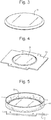
- Fig. 2 shows a glass support plate 11 according to an embodiment which was fabricated.
- the glass support plate 11 was of optical grade and the top and bottom surfaces of the glass support plate 11 were polished.
- An indium-tin oxide layer 12 was then deposited onto the glass support plate 11 using a Physical Vapor Deposition ("PVD”) process so that a layer of optically transparent indium-tin oxide having a thickness of about 1-2 ⁇ m was deposited onto the upper surface of the glass support plate 11.
- PVD Physical Vapor Deposition
- the indium-tin oxide layer 12 is optically transparent and has sufficient transmission so as to enable bacterial colonies which may be present on a culture or growth medium such as agar located within a petri dish 4 or other container to be illuminated.
- the petri dish 4 may be placed in use on top of the glass support plate 11.
- the petri dish 4 or other container may be illuminated from below so that light passes in turn through the support plate 11, through the transparent layer 12 and then passes through the petri dish 4 in order to illuminate the culture or growth medium and any colonies growing on the culture or growth medium. Illuminating the petri dish 4 from below is particularly beneficial as it allows colony boundaries to be more easily determined and thus identified.
- Fig. 3 shows a glass support plate 11 according to an embodiment wherein the glass support plate 11 is arranged to have a 45° angled or bevelled edge.
- the angled or bevelled edge facilitates electrical contact at the side with the upper transparent conductive electrode 12 such as a layer of indium-tin oxide 12.
- a metal plate 13 may be placed onto the glass support plate 11 and may be secured to a light box using springs.
- the edge of the opening in the metal plate 13 may be arranged so as to connect to the angled edge of the glass support plate 11 thereby ensuring that a reliable electrical contact is made between the metal plate 13 and the conductive electrode layer 12 on the upper surface of the glass support plate 11.
- the metal plate 13 also enables the glass support plate 11 to be precisely located or positioned.
- Fig. 5 shows an embodiment wherein a petri dish 4 or other container is located on a transparent conductive counter electrode comprising a glass support plate 11 having an upper conductive electrode layer 12.
- the petri dish 4 or other container may be fabricated from glass or plastic and hence is an insulator.
- the sampling head or REIMS probe 2 may then be moved so that the tip 5 of the REIMS sampling probe 2 comes into direct contact with the colony mass with the height being determined by, for example, using a capacitive liquid level sensor located in the robotic arm of the robotic system 1.
- the RF generator 6 may be energised for a short period of time (e.g. a pulse of 1 s) with the result that electrical energy is capacitively coupled into the sample in close proximity to tip 5 of the REIMS sampling probe 2.
- the result of the applied RF voltage is a rapid evaporation of the sample to form an aerosol which may then be aspirated into a vacuum chamber of the mass spectrometer 7 via a tube 8.
- the aerosol comprises analyte which may be ionised by, for example, an ion source located within a vacuum chamber of the mass spectrometer 7 with the result that analyte ions are then mass analysed by the mass spectrometer 7.
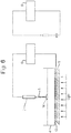
- Fig. 6 shows an embodiment showing a glass support plate 11 having an upper conductive layer 12.
- the glass support plate 11 and the conductive layer 12 may be both transparent (or at least translucent) and enable light from a light source (not shown) to pass through the glass support plate 11, the conductive layer 12 and the base of a petri dish 4 or other container located on the upper surface of the glass support plate 11 (and hence in direct contact with the conductive layer 12).
- a REIMS sampling probe 2 comprising a tubular sampling head with a sampling tip 5 is shown.
- the tip 5 of the REIMS sampling probe 2 is in direct contact with a colony 14 on the surface of a culture or growth medium located in the petri dish 4 or other container.
- the REIMS sampling probe 2 and the upper conductive layer 12 of the glass support plate 11 are shown to be in electrical contact with an RF electrosurgical generator 6.
- Fig. 6 also shows a simplified equivalent electrical circuit of the overall system.
- Current is arranged to pass through resistive layers (e.g. the colony and the agar culture or growth medium) and also through capacitive layers (e.g. the interface between the agar and the petri dish 4 and the interface between the petri dish 4 and the indium-tin oxide or other conductive layer 12).
- resistive layers e.g. the colony and the agar culture or growth medium
- capacitive layers e.g. the interface between the agar and the petri dish 4 and the interface between the petri dish 4 and the indium-tin oxide or other conductive layer 12.
- the result of capacitively coupling electrical energy into the sample is the generation of an aerosol comprising analytes which are subsequently mass analysed by a mass spectrometer 7.
- Fig. 7 shows a sample total ion current TIC which was obtained when a yeast colony was mass analysed according to an embodiment as described above wherein a REIMS sampling probe 2 was used to sample a yeast colony on a culture or growth medium. A high signal intensity was obtained and the signal was obtained for the duration of the applied RF voltage pulse.
- Fig. 8 shows three mass spectra relating to skin samples, yeast samples and normaflore samples wherein the samples were provided on a culture or growth medium and were obtained using a REIMS sampling probe 2 as discussed above. It is apparent from Fig. 8 that a high signal to noise ("S/N") ratio was obtained and that the different mass spectra enabled different colonies to be identified.
- S/N signal to noise
- the mass spectral peak at mass to charge ratio of 693.43 corresponds to a lipid which is common to all three samples.
- the different mass spectral peaks correspond predominantly to the colony being analysed with the agar culture or growth medium having an insignificant impact upon the resulting mass spectra.
- the REIMS sampling probe 2 is able to identify various different biological substances on an agar substrate without needing to scrap sample from the agar substrate.
- the method may be utilised with micro-REIMS imaging experiments wherein, for example, a sample may be microtome sectioned and then a thin slice may be mounted on to a glass slide.
- the method may also be used with simultaneous spectroscopy and rapid evaporation ionisation mass spectrometry imaging.
- the support plate 11 may be fabricated from materials other than glass such as a plastic such as polycarbonate, poly(methyl)methacrylate or plexiglas (RTM). Alternatively, the support plate 11 may be fabricated from quartz or another transparent insulator material.
- the support plate 11 is totally optically transparent.
- the support plate 11 may be fabricated from an opaque, translucent or semi-translucent material.
- the conductive layer 12 provided on the support plate 11 comprises indium-tin oxide.
- the conductive layer 12 may comprise other transparent conductive oxides such as aluminum-doped zinc oxide ("AZO"), indium-doped cadmium oxide, aluminum-doped zinc oxide (“AZO”), gallium-doped zinc oxide (“GZO”) or indium-doped zinc oxide (“IZO").
- AZO aluminum-doped zinc oxide
- AZO indium-doped cadmium oxide
- AZO aluminum-doped zinc oxide
- GZO gallium-doped zinc oxide
- IZO indium-doped zinc oxide
- the conductive layer 12 on the surface of the support plate 11 may comprise a thin translucent or transparent metallic layer.
- the translucent or transparent layer 12 on the surface of the support plate 11 may comprise a carbon nanotube conductive coating, a graphene film or silver nanowires covered with graphene.
- the transparent or translucent layer 12 on the surface of the support plate 11 may alternatively comprise a conductive polymeric layer such as polyaniline or poly(3,4-ethylenedioxythiophene):poly(styrenesulfonate) (“PEDOT:PSS”) composite.
- a conductive polymeric layer such as polyaniline or poly(3,4-ethylenedioxythiophene):poly(styrenesulfonate) (“PEDOT:PSS”) composite.
- the counter electrode 9,12 may comprise a conductive plastic foil.
- the transparent supporting plate 11 and the conductive plastic foil may be assembled using a fixture that holds them together.
- the culture or growth medium may comprise a liquid medium, a cell growth medium such as but not limited to DME (Dulbecco's Modified Eagle's medium), a modified DME medium (e.g. glucose or glutamine free), RPMI (Roswell Park Memorial Institute medium), MEM (Minimum Essential Medium), IMDM (Iscove's Modified Dulbecco's Medium) or another liquid growth medium.
- DME Dulbecco's Modified Eagle's medium
- a modified DME medium e.g. glucose or glutamine free
- RPMI Roswell Park Memorial Institute medium
- MEM Minimum Essential Medium
- IMDM Iscove's Modified Dulbecco's Medium
- the sample may be spun down using a centrifuge to form a pellet and the supernate may be discarded or used for subsequent analysis.
- the sample or pellet may be smeared onto a glass slide or other insulating surface, or may be anlaysed in situ.
Landscapes
- Chemical & Material Sciences (AREA)
- Health & Medical Sciences (AREA)
- Physics & Mathematics (AREA)
- Life Sciences & Earth Sciences (AREA)
- Analytical Chemistry (AREA)
- Engineering & Computer Science (AREA)
- Biomedical Technology (AREA)
- Biochemistry (AREA)
- General Physics & Mathematics (AREA)
- Immunology (AREA)
- Pathology (AREA)
- General Health & Medical Sciences (AREA)
- Spectroscopy & Molecular Physics (AREA)
- Molecular Biology (AREA)
- Biophysics (AREA)
- Hematology (AREA)
- Optics & Photonics (AREA)
- Urology & Nephrology (AREA)
- Food Science & Technology (AREA)
- Medicinal Chemistry (AREA)
- Nuclear Medicine, Radiotherapy & Molecular Imaging (AREA)
- Dispersion Chemistry (AREA)
- Measuring Or Testing Involving Enzymes Or Micro-Organisms (AREA)
- Apparatus Associated With Microorganisms And Enzymes (AREA)
- Other Investigation Or Analysis Of Materials By Electrical Means (AREA)
- Investigating, Analyzing Materials By Fluorescence Or Luminescence (AREA)
Abstract
Description
- This application claims priority from and the benefit of United Kingdom patent application No.
1517195.2 filed on 29 September 2015 - The present invention relates generally to mass spectrometers and in particular to the analysis of material by rapid evaporation ionisation mass spectrometry ("REIMS").
- Rapid evaporation ionisation mass spectrometry ("REIMS") is a relatively new technique that is useful for the analysis of many different samples including the identification of tissue such as food. For example, it is known to use rapid evaporation ionisation mass spectrometry to determine the animal of origin of a food sample and also the pathological state of a tissue sample. It is also known to use rapid evaporation ionisation mass spectrometry to identify microbes, yeast and fungi.
- The known approach for analysing bacterial colonies by rapid evaporation ionisation mass spectrometry involves using bipolar electrosurgical forceps and an electrosurgical RF generator. A bacterial colony is scraped from the surface of an agar layer using the bipolar electrosurgical forceps and a short burst of RF voltage from the electrosurgical RF generator is applied between the bipolar electrosurgical forceps. For example, it is known to apply 60 W of power in a bipolar mode at a frequency of 470 kHz sinusoid. The RF voltage which is applied to the electrosurgical forceps has the result of rapidly heating the particular portion of the bacterial colony which is being analysed due to its nonzero impedance. The rapid heating of the microbial mass results in an aerosol being generated. The aerosol is transferred directly into a mass spectrometer and the aerosol sample may then be analysed by the mass spectrometer. It is known for the control system of the mass spectrometer to utilise multivariate statistical analysis in order to help distinguish and identify different samples.
- Rapid evaporation ionisation mass spectrometry is, therefore, a form of mass spectrometry that uses high frequency energy to ablate or vaporise a sample wherein the resulting vapour or aerosol is then subjected to mass spectrometry.
- Conventional rapid evaporation ionisation mass spectrometry analysis involves ensuring that the sample is in direct electrical (and physical) contact with a RF voltage supply. This approach works well for tissue identification either ex vivo or in vivo. However, such an approach is problematic if it is desired to process a sample which is housed in a container. In particular, the known approach is problematic if it is desired to process a bacterial culture grown on agar in a petri dish.
- It is also problematic to attempt to use the known arrangement for the analysis of tissue sections mounted on a glass slide when it is desired to analyse the tissue sections optically and also to subject the tissue sections to REIMS microprobe imaging.
- For completeness, it should be mentioned that other forms of analysis are known such as Matrix Assisted Laser Desorption lonisation ("MALDI") analysis. However, such approaches are quite different to REIMS and involve looking at the protein/peptide fingerprint of a sample. This is a relatively slow process that requires significant sample preparation and hence such a process is problematic.
- N. Strittmatter, M. Rebec, E. Jones, O. Golf, A. Abdolrasouli, J. Balog, V. Behrends, K. Veselkov, Z. Takats "Characterization and Identification of Clinically Relevant Microorganisms Using Rapid Evaporative Ionization Mass Spectrometry" Anal. Chem. 2014, 86, 6555-6562 discloses a known arrangement wherein two hand-held electrodes in the form of a forceps are used to scrape microbial biomass off from an agar surface. The two electrodes are then squeezed together so as to pinch the biomass between the tips of the forceps. RF power is then applied to the biomass and an aerosol containing analytes is passed to a mass spectrometer for analysis. Accordingly, this reference discloses effectively resistive heating of a sample wherein current flows from one electrode through the sample to the other electrode. Power is dissipated in the sample via resistive heating.
- It is desired to provide an improved method of analysing a sample and in particular an improved method of analysing a sample which may comprise a biological sample which has been grown on a culture medium and which is provided in, for example, a petri dish.
- According to an aspect there is provided a method of analysis comprising:
- providing a sample on an insulating substrate;
- contacting the insulating substrate with a first electrode and contacting the sample with a second electrode; and
- applying an AC or RF voltage to the first and second electrodes in order to generate an aerosol from the sample.
- The known arrangement disclosed in N. Strittmatter, M. Rebec, E. Jones, O. Golf, A. Abdolrasouli, J. Balog, V. Behrends, K. Veselkov, Z. Takats "Characterization and Identification of Clinically Relevant Microorganisms Using Rapid Evaporative Ionization Mass Spectrometry" Anal. Chem. 2014, 86, 6555-6562 does not disclose contacting an insulating substrate such as a petri dish with a first electrode and contacting the sample with a second electrode.
- In particular, according to various embodiments the sample may be cultured on or within a solid or liquid culture or growth medium wherein both the sample and culture or growth medium are provided on the insulating substrate which may comprise a container, petri dish, a vial or a microtitre plate. A bottom surface of the container, petri dish, vial or microtitre plate may be brought into contact with the first electrode which may be positioned below the container, petri dish, vial or microtitre plate. The second electrode may be brought into contact with the sample and/or culture or growth medium. It will be appreciated that the insulating substrate is located between the first and second electrodes when the AC or RF voltage is applied to the first and second electrodes. As a result, electrical energy is predominantly capacitively coupled into the sample. The known arrangement referred to above does not disclose capacitively coupling electrical energy into a sample by bringing a first electrode into contact with the bottom surface of a container, petri dish, vial or microtitre plate containing the sample whilst a second electrode is brought into contact with the sample such that the housing or body of the insulating substrate (i.e. insulating container, petri dish, vial or microtitre plate) is intermediate or between the first and second electrodes when the AC or RF voltage is applied to the first and second electrodes.
- The method may further comprise locating the insulating substrate upon the first electrode, wherein the insulating substrate (e.g. container, petri dish, vial or microtitre plate) is optically transparent or optically translucent and wherein the first electrode is also substantially optically transparent or translucent. The method may further comprise passing light or photons through the first (transparent) electrode and the (transparent) insulating substrate in order to illuminate, image or analyse the sample. The known arrangement does not disclose providing an optically transparent electrode or passing light through an optically transparent electrode in order to illuminate a sample.
- The sample may comprise a biological, bacterial, fungal or yeast sample or a cell line which has been cultured on to or within a culture or growth medium. The culture or growth medium may comprise a solid or liquid culture or growth medium.
- For example, the culture or growth medium may comprise an agar-based medium, a carbohydrate matrix or another solid growth medium.
- Alternatively, the culture or growth medium may comprise a liquid medium, a cell growth medium such as but not limited to DME (Dulbecco's Modified Eagle's medium), a modified DME medium (e.g. glucose or glutamine free), RPMI (Roswell Park Memorial Institute medium), MEM (Minimum Essential Medium), IMDM (Iscove's Modified Dulbecco's Medium) or another liquid growth medium.
- The sample may be spun down using a centrifuge to form a pellet and the supernate may be discarded or used for subsequent analysis.
- The sample or pellet may be smeared onto a glass slide or other insulating surface, or may be anlaysed in situ.
- The sample may be provided on or in a container, a petri dish, a vial or a microtitre or microwell plate. The microtitre or microwell plate may, for example, comprise 6, 24, 96, 384 or 1536 wells.
- The step of providing the sample on the insulating substrate may further comprise providing the sample and optional culture or growth medium on a first surface of the insulating substrate.
- The step of contacting the insulating substrate with the first electrode may further comprise contacting the insulating substrate with the first electrode on a second surface of the insulating substrate which is opposed to the first surface.
- The step of contacting the insulating substrate with the first electrode may be such that the first electrode does not contact the sample.
- The step of contacting the insulating substrate with the first electrode may be such that the first electrode does not contact any culture or growth medium.
- The step of applying an AC or RF voltage to the first and second electrodes may be such that electrical energy is predominantly capacitively coupled into the sample.
- The transfer of electrical energy into the sample may cause the aerosol to be generated.
- The method may further comprise ionising at least some of the aerosol so as to generate analyte ions.
- The method may further comprise directing at least some of the aerosol into a vacuum chamber of a mass spectrometer.
- The method may further comprise ionising at least some of the aerosol within a or the vacuum chamber of the mass spectrometer so as to generate a plurality of analyte ions.
- The method may further comprise mass analysing the analyte ions.
- The method may further comprise obtaining mass spectral data corresponding to one or more locations on or in the sample.
- The method may further comprise spectroscopically imaging or analysing the sample.
- The step of spectroscopically imaging or analysing the sample may further comprise spectroscopically imaging or analysing the sample at substantially the same time as obtaining mass spectral data corresponding to one or more locations on or in the sample.
- The step of spectroscopically imaging or analysing the sample may comprise subjecting the sample to Raman spectroscopy and/or to infra-red ("IR") spectroscopy.
- The step of subjecting the sample to Raman spectroscopy and/or to infra-red ("IR") spectroscopy may further comprise: (i) determining one or more physico-chemical properties of the sample; (ii) determining one or more chemical properties of the sample; (iii) determining one or more absorption properties of the sample; or (iv) determining one or more vibrational and/or rotational modes or states of the sample.
- The method may further comprise using the obtained mass spectral data to identify one or more biological substances, one or more bacterial strains, one or more fungal strains, one or more yeast strains or one or more cell lines located at one or more locations on or in the sample.
- The method may further comprise optically or visually identifying one or more regions of interest on or in the sample.
- The step of optically or visually identifying one or more regions of interest on or in the sample may comprise using a video camera or a digital camera to obtain one or more images of the sample.
- The method may further comprise processing the one or more images of the sample in order to determine one or more regions of interest on or in the sample.
- The first and second electrodes may comprise a rapid evaporation ionization mass spectrometry ("REIMS") device.
- The rapid evaporation ionization mass spectrometry ("REIMS") device may comprise a monopolar device.
- The step of applying the AC or RF voltage to the first and second electrodes may further comprise applying one or more pulses of the AC or RF voltage to the first and second electrodes.
- The step of applying the AC or RF voltage to the first and second electrodes may cause heat to be dissipated into the sample.
- The sample may comprise a biological, bacterial, fungal or yeast sample or a cell line which has been cultured on to or within a culture or growth medium. The culture or growth medium may comprise a solid or liquid culture or growth medium.
- For example, the culture or growth medium may comprise an agar-based medium, a carbohydrate matrix or another solid growth medium.
- Alternatively, the culture or growth medium may comprise a liquid medium, a cell growth medium such as but not limited to DME (Dulbecco's Modified Eagle's medium), a modified DME medium (e.g. glucose or glutamine free), RPMI (Roswell Park Memorial Institute medium), MEM (Minimum Essential Medium), IMDM (Iscove's Modified Dulbecco's Medium) or another liquid growth medium.
- The sample may be spun down using a centrifuge to form a pellet and the supernate may be discarded or used for subsequent analysis.
- The sample or pellet may be smeared onto a glass slide or other insulating surface, or may be anlaysed in situ.
- The method may further comprise determining a spatial distribution of one or more excreted substances emanating from one or more biological substances, one or more bacterial colonies, one or more fungal colonies, one or more yeast colonies or one or more cell lines which have been cultured on or within the culture or growth medium.
- The one or more excreted substances may be selected from the group consisting of: (i) one or more metabolites; (ii) one or more primary metabolites; (iii) one or more secondary metabolites; (iv) one or more lipopeptides; (v) surfactin; (vi) one or more quorum sensing molecules; (vii) 2-Heptyl-3-hydroxy-4(1H)-quinolone or 2-heptyl-3,4-dihydroxyquinoline ("PQS" or Pseudomonas quinolone signal); (viii) 4-hydroxy-2-heptylquinoline ("HHQ"); (ix) one or more antibiotics; (x) one or more alkaloids; (xi) one or more terpenoids; (xii) one or more glycosides; (xiii) one or more natural phenols; (xiv) one or more phenazines; (xv) one or more biphenyls and dibenzofurans; (xvi) one or more beta-lactams; (xvii) one or more polyketides; (xviii) one or more fatty acid synthase products; (xix) one or more nonribosomal peptides; (xx) one or more ribosomal peptides; and (xxi) one or more drugs or toxins.
- The method may further comprise providing the sample in a container, petri dish, a vial or a microtitre or microwell plate. The microtitre or microwell plate may, for example, comprise 6, 24, 96, 384 or 1536 wells.
- The first electrode may comprise a mesh electrode.
- The first electrode may comprise a substrate which is substantially optically transparent or optically translucent.
- The substrate may comprise glass, plastic, polycarbonate, poly(methyl methacrylate), plexiglas (RTM) or quartz.
- The substrate may further comprise a conductive layer or a conductive coating.
- The conductive layer or the conductive coating may be substantially optically transparent or optically translucent.
- The conductive layer or the conductive coating is selected from the group consisting of: (i) a conductive oxide layer or coating; (ii) indium-tin oxide; (iii) aluminium-doped zinc oxide ("AZO"); (iv) indium-doped cadmium oxide; (v) aluminium-doped zinc oxide ("AZO"); (vi) gallium-doped zinc oxide ("GZO"); (vii) indium-doped zinc oxide ("IZO"); (viii) a metallic layer; (ix) a carbon nanotube conductive coating; (x) a graphene film; (xi) one or more silver nanowires covered with graphene; (xii) a polymeric layer; (xiii) polyaniline; or (xiv) a poly(3,4-ethylenedioxythiophene) :poly(styrenesulfonate) ("PEDOT:PSS") composite.
- According to an aspect there is provided a method of rapid evaporation ionization mass spectrometry ("REIMS") comprising a method as described above.
- According to an aspect there is provided a method of mass spectrometry comprising a method as described above.
- According to an aspect there is provided analysis apparatus comprising:
- a first electrode for contacting an insulating substrate upon which a sample is located in use;
- a second electrode for contacting the sample; and
- a device for applying an AC or RF voltage to the first and second electrodes in order to generate an aerosol from the sample.
- The known arrangement disclosed in N. Strittmatter, M. Rebec, E. Jones, O. Golf, A. Abdolrasouli, J. Balog, V. Behrends, K. Veselkov, Z. Takats "Characterization and Identification of Clinically Relevant Microorganisms Using Rapid Evaporative Ionization Mass Spectrometry" Anal. Chem. 2014, 86, 6555-6562 does not disclose contacting an insulating substrate such as a petri dish with a first electrode and contacting the sample with a second electrode.
- In particular, according to various embodiments the sample may be cultured on or within a culture medium wherein both the sample and culture medium are provided on the insulating substrate which may comprise a container, petri dish, a vial or microtitre or microwell plate. The microtitre or microwell plate may, for example, comprise 6, 24, 96, 384 or 1536 wells. A bottom surface of the container, petri dish, vial or microtitre plate may be brought into contact with the first electrode which may be positioned below the container, petri dish, vial or microtitre plate. The second electrode may be brought into contact with the sample and/or culture medium. It will be appreciated that the insulating substrate is located in between the first and second electrodes when the AC or RF voltage is applied to the first and second electrodes. As a result, electrical energy is predominantly capacitively coupled into the sample. The known arrangement referred to above does not disclose capacitively coupling electrical energy into a sample by bringing a first electrode into contact with the bottom surface of a container, petri dish, vial or petri dish containing the sample whilst a second electrode is brought into contact with the sample such that the housing or body of the insulating substrate (i.e. insulating container, petri dish, vial or microtitre plate) is intermediate or between the first and second electrodes when the AC or RF voltage is applied to the first and second electrodes.
- According to an embodiment the insulating substrate may be located, in use, upon the first electrode and the insulating substrate (e.g. container, petri dish, vial or microtitre plate) may be optically transparent or optically translucent and the first electrode may also be substantially optically transparent or translucent. The apparatus may further comprise a device arranged and adapted to pass light or photons through the first (transparent) electrode and the (transparent) insulating substrate in order to illuminate, image or analyse the sample. The known arrangement does not disclose providing an optically transparent electrode or passing light through an optically transparent electrode in order to illuminate a sample.
- The sample may comprise a biological, bacterial, fungal or yeast sample or a cell line which has been cultured on to or within a culture or growth medium. The culture or growth medium may comprise a solid or liquid culture or growth medium.
- For example, the culture or growth medium may comprise an agar-based medium, a carbohydrate matrix or another solid growth medium.
- Alternatively, the culture or growth medium may comprise a liquid medium, a cell growth medium such as but not limited to DME (Dulbecco's Modified Eagle's medium), a modified DME medium (e.g. glucose or glutamine free), RPMI (Roswell Park Memorial Institute medium), MEM (Minimum Essential Medium), IMDM (Iscove's Modified Dulbecco's Medium) or another liquid growth medium.
- The sample may be spun down using a centrifuge to form a pellet and the supernate may be discarded or used for subsequent analysis.
- The sample or pellet may be smeared onto a glass slide or other insulating surface, or may be anlaysed in situ.
- The sample may be provided on or in a container, petri dish, vial or a microtitre or microwell plate. The microtitre or microwell plate may, for example, comprise 6, 24, 96, 384 or 1536 wells.
- The sample and optionally a culture or growth medium may be provided on a first surface of the insulating substrate.
- The first electrode may be arranged to contact a second surface of the insulating substrate which is opposed to the first surface.
- According to an embodiment the first electrode does not contact the sample in use.
- According to an embodiment the first electrode does not contact any culture or growth medium in use.
- The device for applying an AC or RF voltage to the first and second electrodes may be arranged and adapted to predominantly capacitively couple electrical energy into the sample.
- The transfer of electrical energy into the sample may cause the aerosol to be generated.
- The apparatus may further comprise an ion source for ionising at least some of the aerosol so as to generate analyte ions.
- The apparatus may further comprise a device for directing at least some of the aerosol into a vacuum chamber of a mass spectrometer.
- The apparatus may further comprise an ion source located within a or the vacuum chamber of the mass spectrometer for ionising at least some the aerosol so as to generate a plurality of analyte ions.
- The apparatus may further comprise a mass analyser for mass analysing the analyte ions.
- The mass analyser may be further arranged and adapted to obtain mass spectral data corresponding to one or more locations on or in the sample.
- The apparatus may further comprise a spectroscopic imaging or analysing device for spectroscopically imaging or analysing the sample.
- The spectroscopic imaging or analysing device may be arranged and adapted to spectroscopically image or analyse the sample at substantially the same time as the mass analyser obtains mass spectral data corresponding to one or more locations on or in the sample.
- The spectroscopic imaging or analysing device may comprise a Raman spectroscope and/or an infra-red ("IR") spectroscope.
- The Raman spectroscope and/or the infra-red ("IR") spectroscope may be arranged and adapted: (i) to determine one or more physico-chemical properties of the sample; (ii) to determine one or more chemical properties of the sample; (iii) to determine one or more absorption properties of the sample; or (iv) to determine one or more vibrational and/or rotational modes or states of the sample.
- The apparatus may further comprise a control system arranged and adapted to use the obtained mass spectral data to identify one or more biological substances, one or more bacterial strains, one or more fungal strains, one or more yeast strains or one or more cell lines located at one or more locations on or in the sample.
- The apparatus may further comprise a device for optically or visually identifying one or more regions of interest on or in the sample.
- The apparatus may further comprise a video camera or a digital camera for obtaining one or more images of the sample.
- The apparatus may further comprise a processor for processing the one or more images of the sample in order to determine one or more regions of interest on or in the sample.
- The first and second electrodes may comprise a rapid evaporation ionization mass spectrometry ("REIMS") device.
- The rapid evaporation ionization mass spectrometry ("REIMS") device may comprise a monopolar device.
- The AC or RF voltage device may be arranged and adapted to apply one or more pulses of the AC or RF voltage to the first and second electrodes.
- The AC or RF voltage device may be arranged and adapted to apply the AC or RF voltage to the first and second electrodes in order to cause heat to be dissipated into the sample.
- The sample may comprise a biological, bacterial, fungal or yeast sample or a cell line which has been cultured on to or within a solid or liquid culture or growth medium.
- The culture or growth medium may comprise an agar-based medium, a carbohydrate matrix or another solid growth medium.
- The apparatus may further comprise a device for determining a spatial distribution of one or more excreted substances emanating from one or more biological substances, one or more bacterial colonies, one or more fungal, one or more yeast colonies or one or more cell lines which have been cultured on or within the culture or growth medium.
- The one or more excreted substances may be selected from the group consisting of: (i) one or more metabolites; (ii) one or more primary metabolites; (iii) one or more secondary metabolites; (iv) one or more lipopeptides; (v) surfactin; (vi) one or more quorum sensing molecules; (vii) 2-Heptyl-3-hydroxy-4(1 H)-quinolone or 2-heptyl-3,4-dihydroxyquinoline ("PQS" or Pseudomonas quinolone signal); (viii) 4-hydroxy-2-heptylquinoline ("HHQ"); (ix) one or more antibiotics; (x) one or more alkaloids; (xi) one or more terpenoids; (xii) one or more glycosides; (xiii) one or more natural phenols; (xiv) one or more phenazines; (xv) one or more biphenyls and dibenzofurans; (xvi) one or more beta-lactams; (xvii) one or more polyketides; (xviii) one or more fatty acid synthase products; (xix) one or more nonribosomal peptides; (xx) one or more ribosomal peptides; and (xxi) one or more drugs or toxins.
- The sample may be provided in a container, petri dish, vial or a microtitre or microwell plate. The microtitre or microwell plate may, for example, comprise 6, 24, 96, 384 or 1536 wells.
- The first electrode may comprise a mesh electrode.
- The first electrode may comprise a substrate which is substantially optically transparent or optically translucent.
- The substrate may comprise glass, plastic, polycarbonate, poly(methyl methacrylate), plexiglas (RTM) or quartz.
- The substrate may further comprise a conductive layer or a conductive coating.
- The conductive layer or the conductive coating may be substantially optically transparent or optically translucent.
- The conductive layer or the conductive coating may be selected from the group consisting of: (i) a conductive oxide layer or coating; (ii) indium-tin oxide; (iii) aluminium-doped zinc oxide ("AZO"); (iv) indium-doped cadmium oxide; (v) aluminium-doped zinc oxide ("AZO"); (vi) gallium-doped zinc oxide ("GZO"); (vii) indium-doped zinc oxide ("IZO"); (viii) a metallic layer; (ix) a carbon nanotube conductive coating; (x) a graphene film; (xi) one or more silver nanowires covered with graphene; (xii) a polymeric layer; (xiii) polyaniline; or (xiv) a poly(3,4-ethylenedioxythiophene) :poly(styrenesulfonate) ("PEDOT:PSS") composite.
- According to an aspect there is provided a rapid evaporation ionization mass spectrometry ("REIMS") device comprising apparatus as described above.
- According to an aspect there is provided a mass spectrometer comprising apparatus as described above.
- According to an aspect there is provided a method of analysis comprising:
- locating a sample on a first electrode;
- passing light or photons through the first electrode in order to illuminate, image or analyse the sample;
- contacting the sample with a second electrode; and
- applying an AC or RF voltage to the first and second electrodes in order to generate an aerosol from the sample.
- The arrangement disclosed in N. Strittmatter, M. Rebec, E. Jones, O. Golf, A. Abdolrasouli, J. Balog, V. Behrends, K. Veselkov, Z. Takats "Characterization and Identification of Clinically Relevant Microorganisms Using Rapid Evaporative Ionization Mass Spectrometry" Anal. Chem. 2014, 86, 6555-6562 does not disclose passing light or photons through an electrode in order to illuminate, image or analyse a sample.
- According to various embodiments a transparent counter electrode may be used in conjunction with rapid evaporation ionisation mass spectrometry in order to facilitate backlighting of a sample.
- The transparent counter electrode also facilitates simultaneous spectroscopic imaging of the sample. Various embodiments are contemplated wherein a sample may be subjected to simultaneous REIMS and Raman spectroscopy or to simultaneous REIMS and infra-red ("IR") spectroscopy.
- Various embodiments relate to the field of microorganism identification including the identification of bacteria, yeast, fungi or cell lines using rapid evaporation ionisation mass spectrometry. Other applications are contemplated which relate to rapid evaporation ionisation mass spectrometry tissue imaging.
- According to other embodiments automated rapid evaporation ionisation mass spectrometry analysis of bacteria, yeast, fungal colonies or cell lines grown on agar or in a liquid growth medium may be performed. The colonies grown on agar or other culture media may be contained in a standard petri dish. Of particular interest is the fact that the colonies can be analysed directly from the petri dish (or other sample plate) without requiring any substantive intervention from a user or operator and without requiring any prior sample preparation.
- According to an embodiment a spatial distribution of one or more excreted substances emanating from one or more biological substances, one or more bacterial colonies, one or more fungal colonies, one or more yeast colonies or one or more cell lines which have been cultured on or within a culture or growth medium may be determined. The one or more excreted substances may be selected from the group consisting of: (i) one or more metabolites; (ii) one or more primary metabolites; (iii) one or more secondary metabolites; (iv) one or more lipopeptides; (v) surfactin; (vi) one or more quorum sensing molecules; (vii) 2-Heptyl-3-hydroxy-4(1 H)-quinolone or 2-heptyl-3,4-dihydroxyquinoline ("PQS" or Pseudomonas quinolone signal); (viii) 4-hydroxy-2-heptylquinoline ("HHQ"); (ix) one or more antibiotics; (x) one or more alkaloids; (xi) one or more terpenoids; (xii) one or more glycosides; (xiii) one or more natural phenols; (xiv) one or more phenazines; (xv) one or more biphenyls and dibenzofurans; (xvi) one or more beta-lactams; (xvii) one or more polyketides; (xviii) one or more fatty acid synthase products; (xix) one or more nonribosomal peptides; (xx) one or more ribosomal peptides; and (xxi) one or more drugs or toxins. It is known to use genetic engineering or modification of microbes and bacteria to force the production of novel compounds. This technique may be used to monitor the production of the compound and may also be used to screen the micro-organisms for unwanted mutations.
- It will be apparent, therefore, that the various embodiments have significant benefits compared with known method of REIMS analysis.
- The method may further comprise ionising at least some of the aerosol so as to generate analyte ions.
- The method may further comprise directing at least some of the aerosol into a vacuum chamber of a mass spectrometer.
- The method may further comprise ionising at least some the aerosol within a or the vacuum chamber of the mass spectrometer so as to generate a plurality of analyte ions.
- The method may further comprise mass analysing the analyte ions.
- The method may further comprise obtaining mass spectral data corresponding to one or more locations on or in the sample.
- The step of passing light or photons through the first electrode may further comprise spectroscopically imaging or analysing the sample.
- The step of spectroscopically imaging or analysing the sample may further comprise spectroscopically imaging or analysing the sample at substantially the same time as obtaining mass spectral data corresponding to one or more locations on or in the sample.
- The step of spectroscopically imaging or analysing the sample may comprise subjecting the sample to Raman spectroscopy and/or to infra-red ("IR") spectroscopy.
- The step of subjecting the sample to Raman spectroscopy and/or to infra-red ("IR") spectroscopy may further comprise: (i) determining one or more physico-chemical properties of the sample; (ii) determining one or more chemical properties of the sample; (iii) determining one or more absorption properties of the sample; or (iv) determining one or more vibrational and/or rotational modes or states of the sample.
- The light or the photons may either: (i) be in the visible spectrum or have a wavelength in the range 390-700 nm; (ii) be in the near infra-red or have a wavelength in the range 700-1400 nm; or (iii) be in the near ultra-violet or have a wavelength in the range 300-390 nm.
- The method may further comprise using the obtained mass spectral data to identify one or more biological substances, one or more bacterial strains, one or more fungal strains, one or more yeast strains or one or more cell lines located at one or more locations on or in the sample.
- The method may further comprise optically or visually identifying one or more regions of interest on or in the sample.
- The step of optically or visually identifying one or more regions of interest on or in the sample may comprise using a video camera or a digital camera to obtain one or more images of the sample.
- The method may further comprise processing the one or more images of the sample in order to determine one or more regions of interest on or in the sample.
- The first and second electrodes may comprise a rapid evaporation ionization mass spectrometry ("REIMS") device.
- The rapid evaporation ionization mass spectrometry ("REIMS") device may comprise a monopolar device.
- The step of applying the AC or RF voltage to the first and second electrodes may further comprise applying one or more pulses of the AC or RF voltage to the first and second electrodes.
- The step of applying the AC or RF voltage to the first and second electrodes may cause heat to be dissipated into the sample.
- The sample may comprise a biological, bacterial, fungal or yeast sample or a cell line which has been cultured on to or within a solid or liquid culture or growth medium.
- The culture or growth medium may comprise an agar-based medium, a carbohydrate matrix or another solid growth medium.
- Alternatively, the culture or growth medium may comprise a liquid medium, a cell growth medium such as but not limited to DME (Dulbecco's Modified Eagle's medium), a modified DME medium (e.g. glucose or glutamine free), RPMI (Roswell Park Memorial Institute medium), MEM (Minimum Essential Medium), IMDM (Iscove's Modified Dulbecco's Medium) or another liquid growth medium.
- The sample may be spun down using a centrifuge to form a pellet and the supernate may be discarded or used for subsequent analysis.
- The sample or pellet may be smeared onto a glass slide or other insulating surface, or may be anlaysed in situ.
- The method may further comprise determining a spatial distribution of one or more excreted substances emanating from one or more biological substances, one or more bacterial colonies, one or more fungal colonies or one or more yeast colonies which have been cultured on to or within the culture or growth medium.
- The one or more excreted substances may be selected from the group consisting of: (i) one or more metabolites; (ii) one or more primary metabolites; (iii) one or more secondary metabolites; (iv) one or more lipopeptides; (v) surfactin; (vi) one or more quorum sensing molecules; (vii) 2-Heptyl-3-hydroxy-4(1 H)-quinolone or 2-heptyl-3,4-dihydroxyquinoline ("PQS" or Pseudomonas quinolone signal); (viii) 4-hydroxy-2-heptylquinoline ("HHQ"); (ix) one or more antibiotics; (x) one or more alkaloids; (xi) one or more terpenoids; (xii) one or more glycosides; (xiii) one or more natural phenols; (xiv) one or more phenazines; (xv) one or more biphenyls and dibenzofurans; (xvi) one or more beta-lactams; (xvii) one or more polyketides; (xviii) one or more fatty acid synthase products; (xix) one or more nonribosomal peptides; (xx) one or more ribosomal peptides; and (xxi) one or more drugs or toxins.
- The method may further comprise providing the sample in a container, petri dish or a microtitre or microwell plate. The microtitre or microwell plate may, for example, comprise 6, 24, 96, 384 or 1536 wells.
- The first electrode may comprise a mesh electrode.
- The first electrode may comprise a substrate which is substantially optically transparent or optically translucent.
- The substrate may comprise glass, plastic, polycarbonate, poly(methyl methacrylate), plexiglas (RTM) or quartz.
- The substrate may further comprise a conductive layer or a conductive coating.
- The conductive layer or the conductive coating may be substantially optically transparent or optically translucent.
- The conductive layer or the conductive coating may be selected from the group consisting of: (i) a conductive oxide layer or coating; (ii) indium-tin oxide; (iii) aluminium-doped zinc oxide ("AZO"); (iv) indium-doped cadmium oxide; (v) aluminium-doped zinc oxide ("AZO"); (vi) gallium-doped zinc oxide ("GZO"); (vii) indium-doped zinc oxide ("IZO"); (viii) a metallic layer; (ix) a carbon nanotube conductive coating; (x) a graphene film; (xi) one or more silver nanowires covered with graphene; (xii) a polymeric layer; (xiii) polyaniline; or (xiv) a poly(3,4-ethylenedioxythiophene) :poly(styrenesulfonate) ("PEDOT:PSS") composite.
- According to an aspect there is provided a method of rapid evaporation ionization mass spectrometry ("REIMS") comprising a method as described above.
- According to an aspect there is provided a method of mass spectrometry comprising a method as described above.
- According to an aspect there is provided analysis apparatus comprising:
- a first electrode;
- a first device for passing light or photons through the first electrode in order to illuminate, image or analyse, in use, a sample located on the first electrode;
- a second electrode for contacting the sample; and
- a second device for applying an AC or RF voltage to the first and second electrodes in order to generate an aerosol from the sample.
- The arrangement disclosed in N. Strittmatter, M. Rebec, E. Jones, O. Golf, A. Abdolrasouli, J. Balog, V. Behrends, K. Veselkov, Z. Takats "Characterization and Identification of Clinically Relevant Microorganisms Using Rapid Evaporative Ionization Mass Spectrometry" Anal. Chem. 2014, 86, 6555-6562 does not disclose passing light or photons through an electrode in order to illuminate, image or analyse a sample.
- The apparatus may further comprise an ion source for ionising at least some of the aerosol so as to generate analyte ions.
- The apparatus may further comprise a device for directing at least some of the aerosol into a vacuum chamber of a mass spectrometer.
- The apparatus may further comprise an ion source located within a or the vacuum chamber of the mass spectrometer for ionising at least some the aerosol so as to generate a plurality of analyte ions.
- The apparatus may further comprise a mass analyser for mass analysing the analyte ions.
- The mass analyser may be arranged and adapted to obtain mass spectral data corresponding to one or more locations on or in the sample.
- The first device may comprise a spectroscopic imaging or analysing device for spectroscopically imaging or analysing the sample.
- The spectroscopic imaging or analysing device may be arranged and adapted to spectroscopically image or analyse the sample at substantially the same time as the mass analyser obtains mass spectral data corresponding to one or more locations on or in the sample.
- The spectroscopic imaging or analysing device may comprise a Raman spectroscope and/or an infra-red ("IR") spectroscope.
- The Raman spectroscope and/or the infra-red ("IR") spectroscope may be arranged and adapted: (i) to determine one or more physico-chemical properties of the sample; (ii) to determine one or more chemical properties of the sample; (iii) to determine one or more absorption properties of the sample; or (iv) to determine one or more vibrational and/or rotational modes or states of the sample.
- According to an embodiment the light or the photons may either: (i) be in the visible spectrum or have a wavelength in the range 390-700 nm; (ii) be in the near infra-red or have a wavelength in the range 700-1400 nm; or (iii) be in the near ultra-violet or have a wavelength in the range 300-390 nm.
- The apparatus may further comprise a control system which is arranged and adapted to use the obtained mass spectral data to identify one or more biological substances, one or more bacterial strains, one or more fungal strains, one or more yeast strains or one or more cell lines located at one or more locations on or in the sample.
- The apparatus may further comprise a control system which is arranged and adapted to optically or visually identify one or more regions of interest on or in the sample.
- The apparatus may further comprise a video camera or a digital camera for obtaining one or more images of the sample.
- The apparatus may further comprise a processor for processing the one or more images of the sample in order to determine one or more regions of interest on or in the sample.
- The first and second electrodes may comprise a rapid evaporation ionization mass spectrometry ("REIMS") device.
- The rapid evaporation ionization mass spectrometry ("REIMS") device may comprise a monopolar device.
- The device for applying the AC or RF voltage to the first and second electrodes may be arranged and adapted to apply one or more pulses of the AC or RF voltage to the first and second electrodes.
- The device for applying the AC or RF voltage to the first and second electrodes may be arranged and adapted to cause heat to be dissipated into the sample.
- The sample may comprise a biological, bacterial, fungal or yeast sample or a cell line which has been cultured on to or within a solid or liquid culture or growth medium.
- The culture or growth medium may comprise an agar-based medium, a carbohydrate matrix or another solid growth medium.
- Alternatively, the culture or growth medium may comprise a liquid medium, a cell growth medium such as but not limited to DME (Dulbecco's Modified Eagle's medium), a modified DME medium (e.g. glucose or glutamine free), RPMI (Roswell Park Memorial Institute medium), MEM (Minimum Essential Medium), IMDM (Iscove's Modified Dulbecco's Medium) or another liquid growth medium.
- The sample may be spun down using a centrifuge to form a pellet and the supernate may be discarded or used for subsequent analysis.
- The sample or pellet may be smeared onto a glass slide or other insulating surface, or may be anlaysed in situ.
- The apparatus may further comprise a device for determining a spatial distribution of one or more excreted substances emanating from one or more biological substances, one or more bacterial colonies, one or more fungal colonies, one or more yeast colonies or one or more cell lines which have been cultured on or within the culture or growth medium.
- The one or more excreted substances may be selected from the group consisting of: (i) one or more metabolites; (ii) one or more primary metabolites; (iii) one or more secondary metabolites; (iv) one or more lipopeptides; (v) surfactin; (vi) one or more quorum sensing molecules; (vii) 2-Heptyl-3-hydroxy-4(1H)-quinolone or 2-heptyl-3,4-dihydroxyquinoline ("PQS" or Pseudomonas quinolone signal); (viii) 4-hydroxy-2-heptylquinoline ("HHQ"); (ix) one or more antibiotics; (x) one or more alkaloids; (xi) one or more terpenoids; (xii) one or more glycosides; (xiii) one or more natural phenols; (xiv) one or more phenazines; (xv) one or more biphenyls and dibenzofurans; (xvi) one or more beta-lactams; (xvii) one or more polyketides; (xviii) one or more fatty acid synthase products; (xix) one or more nonribosomal peptides; (xx) one or more ribosomal peptides; and (xxi) one or more drugs or toxins.
- The sample may be provided in a container, petri dish or or a microtitre or microwell plate. The microtitre or microwell plate may, for example, comprise 6, 24, 96, 384 or 1536 wells.
- The first electrode may comprise a mesh electrode.
- The first electrode may comprise a substrate which is substantially optically transparent or optically translucent.
- The substrate may comprise glass, plastic, polycarbonate, poly(methyl methacrylate), plexiglas (RTM) or quartz.
- The substrate may further comprise a conductive layer or a conductive coating.
- The conductive layer or the conductive coating may be substantially optically transparent or optically translucent.
- The conductive layer or the conductive coating may be selected from the group consisting of: (i) a conductive oxide layer or coating; (ii) indium-tin oxide; (iii) aluminium-doped zinc oxide ("AZO"); (iv) indium-doped cadmium oxide; (v) aluminium-doped zinc oxide ("AZO"); (vi) gallium-doped zinc oxide ("GZO"); (vii) indium-doped zinc oxide ("IZO"); (viii) a metallic layer; (ix) a carbon nanotube conductive coating; (x) a graphene film; (xi) one or more silver nanowires covered with graphene; (xii) a polymeric layer; (xiii) polyaniline; or (xiv) a poly(3,4-ethylenedioxythiophene) :poly(styrenesulfonate) ("PEDOT:PSS") composite.
- According to an aspect there is provided a rapid evaporation ionization mass spectrometry ("REIMS") device comprising apparatus as described above.
- According to an aspect there is provided a mass spectrometer comprising apparatus as described above.
- Various embodiments will now be described, by way of example only, and with reference to the accompanying drawings in which:
-
Fig. 1 shows a schematic of an automated microbial sampling system according to an embodiment and shows a REIMS sampling probe located above a sample which is provided in a petri dish; -
Fig. 2 shows a schematic of an embodiment wherein a counter electrode is provided comprising a transparent conductive indium-tin oxide layer which is deposited upon a glass support plate; -
Fig. 3 shows in greater detail a circular glass support plate which may be coated with a transparent conductive layer in order to form a transparent counter electrode according to an embodiment; -
Fig. 4 shows a glass support plate located in a metal holder according to an embodiment; -
Fig. 5 shows a petri dish resting on a transparent counter electrode assembly according to an embodiment; -
Fig. 6 shows a transparent counter electrode according to an embodiment located below a petri dish which contains a culture or growth medium and a microorganism colony which has been cultured on the culture or growth medium together with a simplified equivalent electrical circuit, wherein the colony may be illuminated from below the petri dish in order to aid microorganism identification and wherein a REIMS probe may be used to sample and analyse the microorganism colony; -
Fig. 7 shows a total ion current ("TIC") obtained by analysing a sample according to an embodiment; and -
Fig. 8 shows three mass spectra which were obtained by analysing samples using the disclosed REIMS apparatus according to various embodiments and illustrates the ability of the disclosed REIMS apparatus to be able to differentiate between different types of colonies. - Various embodiments will now be described in the context of seeking to aid high throughput microbe identification. However, it should be understood that the various embodiments described herewith are merely illustrative and that the present invention is not intended to be limited to microbe identification.
- According to an arrangement it is contemplated that a two-electrode (bipolar) sampling probe might be used to analyse a sample. However, using a two-electrode sampling probe is potentially problematic since a two-electrode sampling probe has an increased physical size at the point of contact with the sample. It is also necessary to avoid electrical breakdown between the two electrodes forming the probe and to take steps to avoid potential problems due to cross contamination effects.
- According to other arrangements a single analysis probe (e.g. monopolar electrode) may be used to vaporize or ablate a colony and an additional probe (or counter electrode) may be used to make direct electrical and physical contact with the bulk media or agar. Although this approach has been demonstrated to work (to some extent) it suffers from the problem that it requires an additional probe (or counter electrode) to be provided which needs to contact the bulk media or agar. Furthermore, this approach suffers from the problem of having a restricted available analysis area as the heating effect scales quadratically with the current density. It has been recognised that ideally the sampling probe area should be relatively small whereas the additional probe (or counter electrode) should be relatively large so as to avoid heating at the additional probe (or counter electrode).
- Further embodiments which are of particular interest will now be described in more detail below. These embodiments are of particular interest since they eliminate any need for an additional probe (or counter electrode) to be placed in direct physical contact with the bulk material e.g. agar or other culture or growth medium thereby avoiding the problem of cross contamination.
- The various embodiments which will now be described in more detail below are of particular interest since they enable a precise region of a colony to be analysed without suffering from heating effects at the counter electrode. Furthermore, these embodiments do not suffer from potential cross contamination effects. A yet further beneficial effect is that the various embodiments described below significantly improve the ability to illuminate a sample in order to identify one or more regions of interest to be analysed.
- According to various embodiments a sample to be analysed (such as a microbe colony) may be located or provided on a culture or growth medium such as agar. The sample and the culture or growth medium on which it may have been grown may be located or provided in a petri dish, vial or other container such as a microtitre plate or microwell plate. The microtitre or microwell plate may, for example, comprise 6, 24, 96, 384 or 1536 wells. As will become more apparent below, the sample which has been grown on the culture or growth medium in the petri dish can be analysed in situ in the petri dish. This is of particular interest since conventional approaches require a sample portion to be scraped off the surface of the agar or other culture or growth medium and to be analysed whilst being held by a pair of electrosurgical forceps.
- The petri dish or other container in which the sample and culture or growth medium may be provided may be made from glass or plastic. As a result, the petri dish or other container may be essentially electrically non-conductive or may comprise an electrical insulator.
- According to various embodiments a first or counter electrode may be located underneath (or otherwise in close proximity to) the lower or bottom surface of a petri dish or other container so that the petri dish or other container (which is an insulator) sits or otherwise rests upon the first or counter electrode.
- According to various embodiments the first or counter electrode may be arranged to have a similar surface area to that of the petri dish or other container which contains the sample to be analysed.
- According to various embodiments a rapid evaporation ionisation mass spectrometry ("REIMS") sampling system may be provided which comprises a first (counter) electrode which is placed below or underneath an insulating petri dish (or other container) and a second (sample probe) electrode which is brought into direct physical contact with the sample to be analysed in the petri dish (or other container).
- According to an embodiment a REIMS sample probe electrode may be brought into contact (from above) with a microbe colony to be analysed and identified whilst a first or counter electrode is located below the petri dish or other container containing the sample to be analysed.
- An AC or RF voltage may be applied between the REIMS sample probe electrode (which is direct contact with the sample to be analysed) and the REIMS counter electrode (which is in contact with the rear or lower surface of the petri dish or other container containing the sample and optional culture or growth medium). It will be apparent, therefore, that the REIMS counter electrode is not brought into direct physical with the sample to be analysed. This enables the REIMS counter electrode to be reused without suffering from any cross contamination problems.
- According to various embodiments the application of the AC or RF voltage to the first (counter) electrode and the second (sample probe) electrode causes a current to flow but it will be apparent that since the REIMS counter electrode is in electrical or direct physical contact with an insulator (i.e. the bottom of a petri dish) then the petri dish and the contents of the petri dish (i.e. the culture or growth medium) effectively form a dielectric of a capacitor. Therefore, as a result, electrical energy is predominantly capacitively coupled into the sample.
- It will be understood that this approach is therefore quite different from the known approach wherein bipolar electrosurgical forceps are used to remove a sample from a culture or growth medium and wherein both electrodes are brought into direct physical and electrical contact with the sample.
- In contrast to the known approach, the first (counter) electrode does not make direct physical contact with the sample to be analysed and energy is capacitively coupled into the sample in order to rapidly heat and vaporise the sample thereby generating an aerosol to be analysed.
- The frequency of the applied AC or RF voltage may be maximized so as to ensure that the electrical impedance of the bulk material is essentially minimised thereby resulting in maximum energy dissipation into the sample which is desired to be analysed. As a result, AC or RF energy is capacitively coupled into the microbe colony with the result that a portion of the microbe colony in the immediate vicinity of the REIMS probe electrode is vapourised so as to form an aerosol.
- A particular embodiment will now be described in more detail with reference to
Fig. 1. Fig. 1 shows an embodiment comprising a commercialrobotic system 1 such as, for example, a Tecan EVO (RTM) robotic system together with a Picolo (RTM) colony identification system. The commercialrobotic system 1 has been modified so as to include a REIMSsampling probe electrode 2 and a REIMS counter electrode 9. The REIMSsampling probe electrode 2 and the REIMS counter electrode 9 are connected to anelectrosurgical RF generator 6 via afeed electrode 10a and a counter or returnelectrode 10b respectively. - Colonies grown on a culture or growth medium (such as agar) in a
petri dish 4 or other container may be identified using a video camera system 3 which may, for example, be attached to a robotic arm of therobotic system 1. Images of any colonies which have grown on the culture or growth medium may then be processed by digital image processing software. According to an embodiment Picolo (RTM) digital imaging processing software may be used to process images of any colonies which have grown on the culture or growth medium. - The robotic arm of the
robotic system 1 may be arranged to position the video camera system 3 above thepetri dish 4 so that one or more digital pictures of the colony may be recorded. The digital image processing software may then analyse the picture(s) and identify the colonies. Colonies may be selected according to various predetermined criteria or may be manually selected from a video picture. - The REIMS sampling probe (or head) 2 may be moved directly above selected colonies and the position of the surface of a colony relative to e.g. the
tip 5 of theREIMS sampling probe 2 may be determined using a built-in capacitive probe. According to various embodiments thetip 5 of theREIMS sampling probe 2 may be moved so that thetip 5 of thesampling probe 2 just contacts and makes physical contact with a selected colony. REIMS sampling may then be performed by briefly energizing theRF generator 6. - One pole of the
RF generator 6 may be connected to thetip 5 of theREIMS sampling probe 2 via thefeed electrode cable 10a and the other pole of theRF generator 6 may be connected to the counter electrode 9 (located in use underneath thepetri dish 4 or other container) by thecounter electrode cable 10b. - A RF voltage may be applied to a selected colony by, for example, applying a pulse of a RF voltage via the
cables tip 5 of theREIMS sampling probe 2 and also to the REIMS counter electrode 9 located underneath thepetri dish 4 or other container. As a result of capacitively coupling electrical energy into the sample, surgical fumes or an aerosol of sample material may be generated. The surgical fumes or aerosol of sample material may then be passed via atube 8 or other conduit (which may be attached to the REIMS sampling probe 2) directly into the housing of a mass spectrometer 7. According to an embodiment the surgical fumes or aerosol may be ionised within a vacuum chamber of the mass spectrometer 7 by an ion source which may be located within the vacuum chamber of the mass spectrometer 7. - In order for the image recognition software to be able to identify a colony as effectively as possible further embodiments which will be discussed in more detail below enable the
petri dish 4 or other container to be illuminated from below thepetri dish 4 or other container. Illumination of sample in apetri dish 4 or other container from below is beneficial since this enables clear colony boundaries to be observed by, for example, the camera system 3. Furthermore, illuminating thepetri dish 4 or other container from below removes or substantially removes reflections (which would otherwise be observed) and also reduces the complexity of the overall optical system including the video camera system 3. - According to various embodiments which are of particular interest and which will be described in more detail below, the counter electrode 9 may comprise a transparent substrate (e.g. glass or plastic) having a transparent conductive coating on e.g. an upper surface so as to form a transparent electrode on the transparent substrate.
- Other embodiments are also contemplated wherein the
counter electrode 4 may instead comprise an electrically conductive mesh. Such an approach has been demonstrated to work in principle although the mesh electrode can generate shadows which can complicate the automated identification of colonies. For this reason a transparent electrode having a transparent conductive coating or layer is particularly of interest. - According to embodiments which are of particular interest a transparent (or translucent) support plate or substrate may be provided which may be coated with a transparent (or translucent) conductive layer. The support plate or substrate may comprise a transparent or translucent glass plate which may coated with a transparent or translucent conductive layer on an upper surface of the glass support plate. It will be apparent, therefore, that the transparent or translucent conductive coating or layer will then be direct contact with the bottom of a
petri dish 4 or other container. According to various embodiments the support plate or substrate may, for example, be coated with an indium-tin oxide ("ITO") layer. Coating the support plate or substrate with an indium-tin oxide layer is beneficial since the indium-tin oxide layer will have a significant conductivity and will also have sufficient mechanical strength for this application. -
Fig. 2 shows aglass support plate 11 according to an embodiment which was fabricated. Theglass support plate 11 was of optical grade and the top and bottom surfaces of theglass support plate 11 were polished. An indium-tin oxide layer 12 was then deposited onto theglass support plate 11 using a Physical Vapor Deposition ("PVD") process so that a layer of optically transparent indium-tin oxide having a thickness of about 1-2 µm was deposited onto the upper surface of theglass support plate 11. - The indium-
tin oxide layer 12 is optically transparent and has sufficient transmission so as to enable bacterial colonies which may be present on a culture or growth medium such as agar located within apetri dish 4 or other container to be illuminated. Thepetri dish 4 may be placed in use on top of theglass support plate 11. Thepetri dish 4 or other container may be illuminated from below so that light passes in turn through thesupport plate 11, through thetransparent layer 12 and then passes through thepetri dish 4 in order to illuminate the culture or growth medium and any colonies growing on the culture or growth medium. Illuminating thepetri dish 4 from below is particularly beneficial as it allows colony boundaries to be more easily determined and thus identified. -
Fig. 3 shows aglass support plate 11 according to an embodiment wherein theglass support plate 11 is arranged to have a 45° angled or bevelled edge. The angled or bevelled edge facilitates electrical contact at the side with the upper transparentconductive electrode 12 such as a layer of indium-tin oxide 12. - As shown in
Fig. 4 , ametal plate 13 may be placed onto theglass support plate 11 and may be secured to a light box using springs. The edge of the opening in themetal plate 13 may be arranged so as to connect to the angled edge of theglass support plate 11 thereby ensuring that a reliable electrical contact is made between themetal plate 13 and theconductive electrode layer 12 on the upper surface of theglass support plate 11. Themetal plate 13 also enables theglass support plate 11 to be precisely located or positioned. -
Fig. 5 shows an embodiment wherein apetri dish 4 or other container is located on a transparent conductive counter electrode comprising aglass support plate 11 having an upperconductive electrode layer 12. Thepetri dish 4 or other container may be fabricated from glass or plastic and hence is an insulator. - After one or more colonies which have grown on the culture or growth medium in the
petri dish 4 or other container have been optically identified by, for example, the camera system 3 then the sampling head orREIMS probe 2 may then be moved so that thetip 5 of theREIMS sampling probe 2 comes into direct contact with the colony mass with the height being determined by, for example, using a capacitive liquid level sensor located in the robotic arm of therobotic system 1. - According to various embodiments the
RF generator 6 may be energised for a short period of time (e.g. a pulse of 1 s) with the result that electrical energy is capacitively coupled into the sample in close proximity to tip 5 of theREIMS sampling probe 2. The result of the applied RF voltage is a rapid evaporation of the sample to form an aerosol which may then be aspirated into a vacuum chamber of the mass spectrometer 7 via atube 8. The aerosol comprises analyte which may be ionised by, for example, an ion source located within a vacuum chamber of the mass spectrometer 7 with the result that analyte ions are then mass analysed by the mass spectrometer 7. -
Fig. 6 shows an embodiment showing aglass support plate 11 having an upperconductive layer 12. Theglass support plate 11 and theconductive layer 12 may be both transparent (or at least translucent) and enable light from a light source (not shown) to pass through theglass support plate 11, theconductive layer 12 and the base of apetri dish 4 or other container located on the upper surface of the glass support plate 11 (and hence in direct contact with the conductive layer 12). - A
REIMS sampling probe 2 comprising a tubular sampling head with asampling tip 5 is shown. Thetip 5 of theREIMS sampling probe 2 is in direct contact with acolony 14 on the surface of a culture or growth medium located in thepetri dish 4 or other container. TheREIMS sampling probe 2 and the upperconductive layer 12 of theglass support plate 11 are shown to be in electrical contact with an RFelectrosurgical generator 6. -
Fig. 6 also shows a simplified equivalent electrical circuit of the overall system. Current is arranged to pass through resistive layers (e.g. the colony and the agar culture or growth medium) and also through capacitive layers (e.g. the interface between the agar and thepetri dish 4 and the interface between thepetri dish 4 and the indium-tin oxide or other conductive layer 12). The result of capacitively coupling electrical energy into the sample is the generation of an aerosol comprising analytes which are subsequently mass analysed by a mass spectrometer 7. -
Fig. 7 shows a sample total ion current TIC which was obtained when a yeast colony was mass analysed according to an embodiment as described above wherein aREIMS sampling probe 2 was used to sample a yeast colony on a culture or growth medium. A high signal intensity was obtained and the signal was obtained for the duration of the applied RF voltage pulse. -
Fig. 8 shows three mass spectra relating to skin samples, yeast samples and normaflore samples wherein the samples were provided on a culture or growth medium and were obtained using aREIMS sampling probe 2 as discussed above. It is apparent fromFig. 8 that a high signal to noise ("S/N") ratio was obtained and that the different mass spectra enabled different colonies to be identified. The mass spectral peak at mass to charge ratio of 693.43 corresponds to a lipid which is common to all three samples. The different mass spectral peaks correspond predominantly to the colony being analysed with the agar culture or growth medium having an insignificant impact upon the resulting mass spectra. - It is apparent, therefore, that the
REIMS sampling probe 2 according to various embodiments is able to identify various different biological substances on an agar substrate without needing to scrap sample from the agar substrate. - Various further embodiments are also contemplated. According to an embodiment the method may be utilised with micro-REIMS imaging experiments wherein, for example, a sample may be microtome sectioned and then a thin slice may be mounted on to a glass slide.
- The method may also be used with simultaneous spectroscopy and rapid evaporation ionisation mass spectrometry imaging.
- According to other embodiments the
support plate 11 may be fabricated from materials other than glass such as a plastic such as polycarbonate, poly(methyl)methacrylate or plexiglas (RTM). Alternatively, thesupport plate 11 may be fabricated from quartz or another transparent insulator material. - It will also be understood that it is not essential that the
support plate 11 is totally optically transparent. For example, thesupport plate 11 may be fabricated from an opaque, translucent or semi-translucent material. - It is also not essential that the
conductive layer 12 provided on thesupport plate 11 comprises indium-tin oxide. For example, other embodiments are contemplated wherein theconductive layer 12 may comprise other transparent conductive oxides such as aluminum-doped zinc oxide ("AZO"), indium-doped cadmium oxide, aluminum-doped zinc oxide ("AZO"), gallium-doped zinc oxide ("GZO") or indium-doped zinc oxide ("IZO"). - According to other embodiments the
conductive layer 12 on the surface of thesupport plate 11 may comprise a thin translucent or transparent metallic layer. - Alternatively, the translucent or
transparent layer 12 on the surface of thesupport plate 11 may comprise a carbon nanotube conductive coating, a graphene film or silver nanowires covered with graphene. - The transparent or
translucent layer 12 on the surface of thesupport plate 11 may alternatively comprise a conductive polymeric layer such as polyaniline or poly(3,4-ethylenedioxythiophene):poly(styrenesulfonate) ("PEDOT:PSS") composite. - It is also contemplated that the
counter electrode 9,12 may comprise a conductive plastic foil. The transparent supportingplate 11 and the conductive plastic foil may be assembled using a fixture that holds them together. - Other embodiments are contemplated wherien the culture or growth medium may comprise a liquid medium, a cell growth medium such as but not limited to DME (Dulbecco's Modified Eagle's medium), a modified DME medium (e.g. glucose or glutamine free), RPMI (Roswell Park Memorial Institute medium), MEM (Minimum Essential Medium), IMDM (Iscove's Modified Dulbecco's Medium) or another liquid growth medium.
- The sample may be spun down using a centrifuge to form a pellet and the supernate may be discarded or used for subsequent analysis.
- The sample or pellet may be smeared onto a glass slide or other insulating surface, or may be anlaysed in situ.
- Although the present invention has been described with reference to preferred embodiments, it will be understood by those skilled in the art that various changes in form and detail may be made without departing from the scope of the invention as set forth in the accompanying claims.
Claims (15)
- A method of analysis comprising:locating a sample on a first electrode;passing light or photons through said first electrode in order to illuminate, image or analyse said sample;contacting said sample with a second electrode; andapplying an AC or RF voltage to said first and second electrodes in order to generate an aerosol from said sample.
- A method as claimed in claim 1, further comprising ionising at least some of the aerosol so as to generate analyte ions.
- A method as claimed in claim 2, further comprising mass analysing the analyte ions, and obtaining mass spectral data corresponding to one or more locations on or in the sample.
- A method as claimed in claim 1, 2, or 3, wherein the step of passing light or photons through the first electrode further comprises spectroscopically imaging or analysing the sample.
- A method as claimed in claim 4, wherein the step of spectroscopically imaging or analysing the sample comprises subjecting the sample to Raman spectroscopy and/or infra-red ("IR") spectroscopy.
- A method according to claim 5, wherein step of subjecting the sample to Raman spectroscopy and/or to infra-red ("IR") spectroscopy may further comprise: (i) determining one or more physico-chemical properties of the sample; (ii) determining one or more chemical properties of the sample; (iii) determining one or more absorption properties of the sample; or (iv) determining one or more vibrational and/or rotational modes or states of the sample
- A method according to any preceding claim, further comprising a step of determining a spatial distribution of one or more excreted substances emanating from one or more biological substances, one or more bacterial colonies, one or more fungal colonies or one or more yeast colonies which have been cultured on to or within the culture or growth medium.
- A method according to any preceding claim, wherein the first electrode comprises a mesh electrode.
- A method according to any preceding claim, wherein the first electrode comprises a substrate which is substantially optically transparent or optically translucent.
- A method according to claim 9, wherein the substrate comprises a conductive layer or conductive coating.
- A method of rapid evaporation ionization mass spectrometry ("REIMS") comprising a method as claimed in any preceding claim.
- Analysis apparatus comprising:a first electrode;a first device for passing light or photons through said first electrode in order to illuminate, image or analyse, in use, a sample located on said first electrode;a second electrode for contacting said sample; anda second device for applying an AC or RF voltage to said first and second electrodes in order to generate an aerosol from said sample.
- An analysis apparatus according to claim 12, further comprising an ion source configured to ionise at least some of the aerosol so as to generate analyte ions, and a mass analyser configured to mass analyse the analyte ions.
- An analysis apparatus according to claim 13, wherein the first device comprises a spectroscopic imaging or analysing device for spectroscopically imaging or analysing the sample, wherein the spectroscopic imaging or analysing device is arranged and adapted to spectroscopically image or analyse the sample at substantially the same time as the mass analyser obtains mass spectral data corresponding to one or more locations on or in the sample.
- A rapid evaporation ionization mass spectrometry ("REIMS") device comprising apparatus as claimed in any one of claims 12, 13, or 14.
Applications Claiming Priority (3)
| Application Number | Priority Date | Filing Date | Title |
|---|---|---|---|
| GBGB1517195.2A GB201517195D0 (en) | 2015-09-29 | 2015-09-29 | Capacitively coupled reims technique and optically transparent counter electrode |
| EP16778093.1A EP3356817B1 (en) | 2015-09-29 | 2016-09-22 | Capacitively coupled reims technique |
| PCT/GB2016/052956 WO2017055812A1 (en) | 2015-09-29 | 2016-09-22 | Capacitively coupled reims technique and optically transparent counter electrode |
Related Parent Applications (2)
| Application Number | Title | Priority Date | Filing Date |
|---|---|---|---|
| EP16778093.1A Division-Into EP3356817B1 (en) | 2015-09-29 | 2016-09-22 | Capacitively coupled reims technique |
| EP16778093.1A Division EP3356817B1 (en) | 2015-09-29 | 2016-09-22 | Capacitively coupled reims technique |
Publications (1)
| Publication Number | Publication Date |
|---|---|
| EP3859328A1 true EP3859328A1 (en) | 2021-08-04 |
Family
ID=54544274
Family Applications (2)
| Application Number | Title | Priority Date | Filing Date |
|---|---|---|---|
| EP16778093.1A Active EP3356817B1 (en) | 2015-09-29 | 2016-09-22 | Capacitively coupled reims technique |
| EP21156646.8A Pending EP3859328A1 (en) | 2015-09-29 | 2016-09-22 | Capactively coupled reims technique and optically transparent counter electrode |
Family Applications Before (1)
| Application Number | Title | Priority Date | Filing Date |
|---|---|---|---|
| EP16778093.1A Active EP3356817B1 (en) | 2015-09-29 | 2016-09-22 | Capacitively coupled reims technique |
Country Status (5)
| Country | Link |
|---|---|
| US (2) | US11031223B2 (en) |
| EP (2) | EP3356817B1 (en) |
| CN (2) | CN108474782B (en) |
| GB (1) | GB201517195D0 (en) |
| WO (1) | WO2017055812A1 (en) |
Families Citing this family (2)
| Publication number | Priority date | Publication date | Assignee | Title |
|---|---|---|---|---|
| GB201702643D0 (en) * | 2017-02-17 | 2017-04-05 | Micromass Ltd | Analysis of food samples |
| NL2031837B1 (en) * | 2022-05-12 | 2023-11-17 | Portolab B V | Method to combine optical imaging spectroscopy and analytical spectrometry |
Citations (1)
| Publication number | Priority date | Publication date | Assignee | Title |
|---|---|---|---|---|
| WO2013098645A2 (en) * | 2011-12-28 | 2013-07-04 | Medimass, Ltd. | System and method for rapid evaporative ionization of liquid phase samples |
Family Cites Families (240)
| Publication number | Priority date | Publication date | Assignee | Title |
|---|---|---|---|---|
| US525799A (en) | 1894-09-11 | Device for balancing rotating parts | ||
| US3479545A (en) | 1967-05-16 | 1969-11-18 | Hughes Aircraft Co | Surface ionization apparatus and electrode means for accelerating the ions in a curved path |
| US3770954A (en) | 1971-12-29 | 1973-11-06 | Gen Electric | Method and apparatus for analysis of impurities in air and other gases |
| US4408125A (en) | 1981-07-13 | 1983-10-04 | University Of Utah | Modular pyrolysis inlet and method for pyrolyzing compounds for analysis by mass spectrometer |
| EP0169469A3 (en) | 1984-07-27 | 1987-10-14 | BASF Corporation | Analytical apparatus |
| US5053343A (en) | 1985-02-15 | 1991-10-01 | Environmental Technologies Group, Inc. | Selective ionization of gas constituents using electrolytic reactions |
| IL81375A (en) | 1987-01-23 | 1990-11-05 | Univ Ramot | Method and apparatus for producing ions by surface ionization of energy-rich molecules and atoms |
| USH414H (en) | 1987-03-20 | 1988-01-05 | The United States Of America As Represented By The Secretary Of The Army | Surface ionization source |
| JPS63243864A (en) | 1987-03-31 | 1988-10-11 | Shimadzu Corp | Gas chromatograph mass spectrometer interface |
| US4835383A (en) | 1987-08-06 | 1989-05-30 | Phrasor Scientific, Inc. | High mass ion detection system and method |
| US4935624A (en) | 1987-09-30 | 1990-06-19 | Cornell Research Foundation, Inc. | Thermal-assisted electrospray interface (TAESI) for LC/MS |
| US4883958A (en) | 1988-12-16 | 1989-11-28 | Vestec Corporation | Interface for coupling liquid chromatography to solid or gas phase detectors |
| JPH031435A (en) | 1989-05-29 | 1991-01-08 | Kokuritsu Kogai Kenkyusho | Ionizing method for mass spectrogram |
| US5033541A (en) | 1989-11-17 | 1991-07-23 | Cetac Technologies, Inc. | Double pass tandem cooling aerosol condenser |
| GB9000547D0 (en) * | 1990-01-10 | 1990-03-14 | Vg Instr Group | Glow discharge spectrometry |
| US5257991A (en) | 1990-11-15 | 1993-11-02 | Laserscope | Instrumentation for directing light at an angle |
| US5454274A (en) | 1991-09-25 | 1995-10-03 | Cetac Technologies Inc. | Sequential combination low temperature condenser and enclosed filter solvent removal system, and method of use |
| JPH05251038A (en) | 1992-03-04 | 1993-09-28 | Hitachi Ltd | Plasma ion mass spectrometry device |
| US5374755A (en) | 1992-07-17 | 1994-12-20 | Millipore Corporation | Liquid chromatography stationary phases with reduced silanol interactions |
| JP3367719B2 (en) | 1993-09-20 | 2003-01-20 | 株式会社日立製作所 | Mass spectrometer and electrostatic lens |
| JPH07130325A (en) | 1993-10-29 | 1995-05-19 | Hitachi Ltd | Mass spectrometer |
| US5509916A (en) | 1994-08-12 | 1996-04-23 | Valleylab Inc. | Laser-assisted electrosurgery system |
| GB9416331D0 (en) | 1994-08-12 | 1994-10-05 | Amp Gmbh | Stranded electrical wire for use with IDC |
| US6032673A (en) | 1994-10-13 | 2000-03-07 | Femrx, Inc. | Methods and devices for tissue removal |
| US5830214A (en) | 1994-11-08 | 1998-11-03 | Heartport, Inc. | Fluid-evacuating electrosurgical device |
| US5559326A (en) | 1995-07-28 | 1996-09-24 | Hewlett-Packard Company | Self generating ion device for mass spectrometry of liquids |
| WO1997034534A1 (en) | 1996-03-18 | 1997-09-25 | Femrx, Inc. | Method and device for tissue vaporization and extraction |
| US5800597A (en) | 1997-01-21 | 1998-09-01 | Whatman Inc. | Integral coalescer filter-membrane device to provide a filtered gas stream and system employing such device |
| EP1650549A3 (en) * | 1996-04-30 | 2009-11-25 | FUJIFILM Corporation | Surface plasmon sensor |
| US5836909A (en) | 1996-09-13 | 1998-11-17 | Cosmescu; Ioan | Automatic fluid control system for use in open and laparoscopic laser surgery and electrosurgery and method therefor |
| FR2755500B1 (en) * | 1996-11-04 | 1998-12-11 | Gaz De France | MODULAR FLAME HANGING DEVICE FOR LOW-POLLUTANT GAS MIXTURE BURNER WITH SELF-STABILIZED FLAME, AND BURNER PROVIDED WITH SUCH A DEVICE |
| US5969352A (en) | 1997-01-03 | 1999-10-19 | Mds Inc. | Spray chamber with dryer |
| US5828062A (en) | 1997-03-03 | 1998-10-27 | Waters Investments Limited | Ionization electrospray apparatus for mass spectrometry |
| JPH10247472A (en) | 1997-03-04 | 1998-09-14 | Shimadzu Corp | Surface ionization type ionizer |
| JPH10302710A (en) | 1997-04-22 | 1998-11-13 | Shimadzu Corp | Surface ionization type ionizer |
| JP3592494B2 (en) * | 1997-08-22 | 2004-11-24 | 日本電子株式会社 | Atmospheric pressure laser vaporization mass spectrometer and method |
| US6348688B1 (en) | 1998-02-06 | 2002-02-19 | Perseptive Biosystems | Tandem time-of-flight mass spectrometer with delayed extraction and method for use |
| US5920068A (en) | 1998-03-05 | 1999-07-06 | Micron Technology, Inc. | Analysis of semiconductor surfaces by secondary ion mass spectrometry |
| US6849847B1 (en) | 1998-06-12 | 2005-02-01 | Agilent Technologies, Inc. | Ambient pressure matrix-assisted laser desorption ionization (MALDI) apparatus and method of analysis |
| CA2339552A1 (en) * | 1998-08-05 | 2000-02-17 | National Research Council Of Canada | Apparatus and method for desolvating and focussing ions for introduction into a mass spectrometer |
| US7517348B2 (en) | 1998-09-03 | 2009-04-14 | Rubicor Medical, Inc. | Devices and methods for performing procedures on a breast |
| US7329253B2 (en) | 2003-12-09 | 2008-02-12 | Rubicor Medical, Inc. | Suction sleeve and interventional devices having such a suction sleeve |
| JP2000097913A (en) | 1998-09-24 | 2000-04-07 | Shimadzu Corp | Surface ionization type ionizer |
| JP2000180413A (en) | 1998-12-11 | 2000-06-30 | Lion Corp | Ionizing aid and method of ionizing substances |
| WO2000036398A2 (en) | 1998-12-14 | 2000-06-22 | Deutsches Krebsforschungszentrum Stiftung des öffentlichen Rechts | Method and devices for detecting optical properties, especially luminescence reactions and refraction behaviour of molecules which are directly or indirectly bound on a support |
| US6280302B1 (en) | 1999-03-24 | 2001-08-28 | Flow International Corporation | Method and apparatus for fluid jet formation |
| GB9911336D0 (en) | 1999-05-15 | 1999-07-14 | Graseby Dynamics Ltd | Separation and collection of analyte materials |
| US6375635B1 (en) | 1999-05-18 | 2002-04-23 | Hydrocision, Inc. | Fluid jet surgical instruments |
| DE19934173A1 (en) | 1999-07-21 | 2001-01-25 | Max Planck Gesellschaft | Fragmenting clusters with a carrier substance, using a reagent charge which forms part of the cluster fragments to produce and manipulate electrically neutral particles effectively |
| US6333632B1 (en) | 1999-09-16 | 2001-12-25 | Rae Systems, Inc. | Alternating current discharge ionization detector |
| JP3743240B2 (en) | 1999-12-24 | 2006-02-08 | 株式会社日立製作所 | Liquid chromatograph mass spectrometer |
| WO2001060265A1 (en) | 2000-02-17 | 2001-08-23 | Kanag Baska | Surgical snare |
| US6777672B1 (en) | 2000-02-18 | 2004-08-17 | Bruker Daltonics, Inc. | Method and apparatus for a multiple part capillary device for use in mass spectrometry |
| US7435579B2 (en) * | 2000-04-17 | 2008-10-14 | Purdue Research Foundation | Biosensor and related method |
| AU2001263712A1 (en) | 2000-06-20 | 2002-01-02 | Ammann-Technik Ag | Device for producing a liquid jet for removing especially biological tissue, anduse thereof |
| AU2001288270A1 (en) | 2000-08-16 | 2002-02-25 | Vanderbilt University | System and method of infrared matrix-assisted laser desorption/ionization mass spectrometry in polyacrylamide gels |
| JP4578672B2 (en) | 2000-12-05 | 2010-11-10 | キヤノンアネルバ株式会社 | Ionizer for mass spectrometry and ionization method |
| US6744043B2 (en) | 2000-12-08 | 2004-06-01 | Mds Inc. | Ion mobilty spectrometer incorporating an ion guide in combination with an MS device |
| US20040088333A1 (en) | 2002-01-25 | 2004-05-06 | David Sidman | Apparatus method and system for tracking information access |
| US20030119193A1 (en) | 2001-04-25 | 2003-06-26 | Robert Hess | System and method for high throughput screening of droplets |
| US6744040B2 (en) | 2001-06-13 | 2004-06-01 | Bruker Daltonics, Inc. | Means and method for a quadrupole surface induced dissociation quadrupole time-of-flight mass spectrometer |
| JP3725803B2 (en) | 2001-06-15 | 2005-12-14 | 株式会社東芝 | Semiconductor wafer impurity measurement method and semiconductor wafer impurity measurement program |
| US7586088B2 (en) | 2001-06-21 | 2009-09-08 | Micromass Uk Limited | Mass spectrometer and method of mass spectrometry |
| JP3660279B2 (en) | 2001-07-23 | 2005-06-15 | 株式会社日立製作所 | Sample ionizer and mass spectrometer |
| US8734441B2 (en) * | 2001-08-15 | 2014-05-27 | Nuortho Surgical, Inc. | Interfacing media manipulation with non-ablation radiofrequency energy system and method |
| GB0120131D0 (en) | 2001-08-17 | 2001-10-10 | Micromass Ltd | Maldi target plate |
| EP1291659A3 (en) | 2001-09-06 | 2008-05-21 | Sysmex Corporation | Automatic sample analyzer and its components |
| CA2462854A1 (en) | 2001-10-05 | 2003-04-17 | Yale University | Method and apparatus to produce ions and nanodrops from taylor cones of volatile liquids at reduced pressures |
| US6756586B2 (en) | 2001-10-15 | 2004-06-29 | Vanderbilt University | Methods and apparatus for analyzing biological samples by mass spectrometry |
| JP3822090B2 (en) * | 2001-10-26 | 2006-09-13 | シャープ株式会社 | Test circuit for charge detection circuit, LSI, and test method for charge detection circuit |
| EP1448769A4 (en) | 2001-10-31 | 2006-02-01 | Ionfinity Llc | Soft ionization device and applications thereof |
| US7005633B2 (en) | 2002-02-08 | 2006-02-28 | Ionalytics Corporation | Method and apparatus for desolvating ions for introduction into a FAIMS analyzer region |
| WO2004030024A2 (en) | 2002-05-31 | 2004-04-08 | University Of Florida Research Foundation, Inc. | Methods and devices for laser desorption chemical ionization |
| US7078681B2 (en) * | 2002-09-18 | 2006-07-18 | Agilent Technologies, Inc. | Multimode ionization source |
| EP1550145B1 (en) | 2002-10-10 | 2018-01-03 | Universita' Degli Studi Di Milano | Ionization source for mass spectrometry analysis |
| EP1555954B1 (en) | 2002-10-17 | 2010-04-14 | Braun GmbH | Mouth rinse and spray nozzle for creating a liquid jet and teeth-cleaning system |
| GB0226160D0 (en) | 2002-11-08 | 2002-12-18 | Diagnoswiss Sa | Apparatus for dispensing a sample in electrospray mass spectrometers |
| JP2004212073A (en) | 2002-12-27 | 2004-07-29 | Hitachi Ltd | Dangerous goods detection device and dangerous goods detection method |
| JP2004264043A (en) | 2003-01-31 | 2004-09-24 | National Institute Of Advanced Industrial & Technology | Ionizer and micro area analyzer |
| AU2003901589A0 (en) * | 2003-04-04 | 2003-05-01 | Griffith University | Novel photoelectrichemical oxygen demand assay |
| KR100466866B1 (en) | 2003-04-24 | 2005-01-24 | 전명기 | Electrode for radiofrequency tissue ablation |
| US20080073512A1 (en) | 2003-06-06 | 2008-03-27 | Waters Investments Limited | Methods, compositions and devices for performing ionization desorption on silicon derivatives |
| WO2004112074A2 (en) | 2003-06-07 | 2004-12-23 | Willoughby Ross C | Laser desorption ion source |
| CA2976507C (en) | 2003-06-09 | 2020-05-12 | Perkinelmer Health Sciences Canada, Inc. | Mass spectrometer interface |
| CA2475654A1 (en) | 2003-07-21 | 2005-01-21 | Fmc Technologies, Inc. | Friction drive conveyor |
| US20050017091A1 (en) | 2003-07-22 | 2005-01-27 | Omax Corporation | Abrasive water-jet cutting nozzle having a vented water-jet pathway |
| US20050061779A1 (en) * | 2003-08-06 | 2005-03-24 | Walter Blumenfeld | Laser ablation feedback spectroscopy |
| US20050077644A1 (en) | 2003-08-14 | 2005-04-14 | Bryan David E. | High pressure liquid jet cutting system and method for forming polymer pellets |
| JP2005108578A (en) | 2003-09-30 | 2005-04-21 | Hitachi Ltd | Mass spectrometer |
| US7217919B2 (en) | 2004-11-02 | 2007-05-15 | Analytica Of Branford, Inc. | Method and apparatus for multiplexing plural ion beams to a mass spectrometer |
| US7365309B2 (en) | 2003-12-22 | 2008-04-29 | Micromass Uk Limited | Mass spectrometer |
| JP3823321B2 (en) | 2003-12-25 | 2006-09-20 | 有限会社エスアールジェイ | Balloon control device |
| US7260914B2 (en) | 2003-12-27 | 2007-08-28 | Floral Transport Systems, Llc | Method and apparatus for packaging horticultural products |
| AU2005200016B2 (en) | 2004-01-09 | 2010-12-09 | John Bean Technologies Corporation | Method and system for portioning workpieces to user-scanned shape and other specifications |
| GB0514964D0 (en) | 2005-07-21 | 2005-08-24 | Ms Horizons Ltd | Mass spectrometer devices & methods of performing mass spectrometry |
| US7005635B2 (en) | 2004-02-05 | 2006-02-28 | Metara, Inc. | Nebulizer with plasma source |
| US7294841B2 (en) | 2004-02-06 | 2007-11-13 | Micromass Uk Limited | Mass spectrometer |
| US7265362B2 (en) | 2004-02-06 | 2007-09-04 | Micromass Uk Limited | Mass spectrometer |
| US6974957B2 (en) | 2004-02-18 | 2005-12-13 | Nanomat, Inc. | Ionization device for aerosol mass spectrometer and method of ionization |
| CA2564612A1 (en) * | 2004-02-26 | 2005-09-09 | Thomsen Bioscience A/S | Method, chip, device and system for collection of biological particles |
| CN101073137A (en) | 2004-03-30 | 2007-11-14 | 普渡研究基金会 | Method and system for desorption electrospray ionization |
| US7335897B2 (en) | 2004-03-30 | 2008-02-26 | Purdue Research Foundation | Method and system for desorption electrospray ionization |
| US20070023631A1 (en) | 2004-03-30 | 2007-02-01 | Zoltan Takats | Parallel sample handling for high-throughput mass spectrometric analysis |
| US7199364B2 (en) | 2004-05-21 | 2007-04-03 | Thermo Finnigan Llc | Electrospray ion source apparatus |
| US7238936B2 (en) | 2004-07-02 | 2007-07-03 | Thermo Finnigan Llc | Detector with increased dynamic range |
| US7094135B2 (en) | 2004-08-10 | 2006-08-22 | International Waterjet Parts, Inc. | Abrasivejet cutting head with back-flow prevention valve |
| US7126134B2 (en) * | 2004-08-19 | 2006-10-24 | Palo Alto Research Center Incorporated | Sample manipulator |
| JP4492267B2 (en) * | 2004-09-16 | 2010-06-30 | 株式会社日立製作所 | Mass spectrometer |
| DE102004053064B4 (en) | 2004-11-03 | 2007-11-08 | Bruker Daltonik Gmbh | Ionization by droplet impact |
| US6998622B1 (en) | 2004-11-17 | 2006-02-14 | Agilent Technologies, Inc. | On-axis electron impact ion source |
| IL166115A (en) | 2005-01-03 | 2012-06-28 | Dan Adam | Interactive ultrasound-based depth measurement for medical applications |
| US7735146B2 (en) | 2005-01-27 | 2010-06-08 | The George Washington University | Protein microscope |
| GB2425178A (en) | 2005-04-13 | 2006-10-18 | Univ Sheffield | Analysis of biological samples by mass spectrometry |
| US7196525B2 (en) | 2005-05-06 | 2007-03-27 | Sparkman O David | Sample imaging |
| US7351960B2 (en) | 2005-05-16 | 2008-04-01 | Thermo Finnigan Llc | Enhanced ion desolvation for an ion mobility spectrometry device |
| JP4734628B2 (en) | 2005-05-24 | 2011-07-27 | 国立大学法人山梨大学 | Collision-induced ionization method and apparatus |
| GB0511332D0 (en) | 2005-06-03 | 2005-07-13 | Micromass Ltd | Mass spectrometer |
| US20070023677A1 (en) | 2005-06-29 | 2007-02-01 | Perkins Patrick D | Multimode ionization source and method for screening molecules |
| FI20055380L (en) | 2005-07-01 | 2007-01-02 | M Real Oyj | Method for coating cellulose particles, coated cellulose particles and their use in paper and cardboard manufacturing |
| JP2007051934A (en) | 2005-08-18 | 2007-03-01 | Toppan Printing Co Ltd | Mass axis calibration method in time-of-flight secondary ion mass spectrometry |
| US7828948B1 (en) | 2005-10-06 | 2010-11-09 | Sandia Corporation | Preconcentration and separation of analytes in microchannels |
| US20070114394A1 (en) | 2005-10-21 | 2007-05-24 | Gerald Combs | Method and system for determining and quantifying specific trace elements in samples of complex materials |
| US20070094389A1 (en) | 2005-10-23 | 2007-04-26 | Bill Nussey | Provision of rss feeds based on classification of content |
| JP4863692B2 (en) | 2005-11-02 | 2012-01-25 | 株式会社島津製作所 | Image mass spectrometer |
| US7459676B2 (en) | 2005-11-21 | 2008-12-02 | Thermo Finnigan Llc | MALDI/LDI source |
| US7629424B2 (en) | 2005-12-09 | 2009-12-08 | Pryog, Llc | Metal-containing compositions and method of making same |
| US7947039B2 (en) | 2005-12-12 | 2011-05-24 | Covidien Ag | Laparoscopic apparatus for performing electrosurgical procedures |
| JP2007170870A (en) | 2005-12-19 | 2007-07-05 | Protosera Inc | Insitu detection method using mass analysis |
| CN102646570B (en) | 2006-01-02 | 2016-03-09 | 卓漂仪谱公司 | Multi-dimensional ion mobility spectrometry apparatus and method |
| TWI271771B (en) | 2006-01-27 | 2007-01-21 | Univ Nat Sun Yat Sen | Electrospray-assisted laser desorption ionization devices, mass spectrometers, and methods for mass spectrometry |
| JP4982087B2 (en) | 2006-02-08 | 2012-07-25 | 株式会社日立製作所 | Mass spectrometer and mass spectrometry method |
| EP1821093B1 (en) | 2006-02-15 | 2018-04-04 | Commissariat à l'Énergie Atomique et aux Énergies Alternatives | Method and device for measuring permeation |
| US7462824B2 (en) | 2006-04-28 | 2008-12-09 | Yang Wang | Combined ambient desorption and ionization source for mass spectrometry |
| EP1855306B1 (en) | 2006-05-11 | 2019-11-13 | ISB - Ion Source & Biotechnologies S.R.L. | Ionization source and method for mass spectrometry |
| US7777181B2 (en) | 2006-05-26 | 2010-08-17 | Ionsense, Inc. | High resolution sampling system for use with surface ionization technology |
| HU226837B1 (en) | 2006-05-31 | 2009-12-28 | Semmelweis Egyetem | Flow-ray desorption ionization method and device |
| JP5142580B2 (en) | 2006-06-29 | 2013-02-13 | キヤノン株式会社 | Surface analysis method and surface analysis apparatus |
| EP2040824A2 (en) | 2006-07-11 | 2009-04-01 | Excellims Corporation | Methods and apparatus for the ion mobility based separation and collection of molecules |
| US7816644B2 (en) | 2006-08-18 | 2010-10-19 | Agilent Technologies, Inc. | Photoactivated collision induced dissociation (PACID) (apparatus and method) |
| US7718958B2 (en) | 2006-11-17 | 2010-05-18 | National Sun Yat-Sen University | Mass spectroscopic reaction-monitoring method |
| US20090065714A1 (en) | 2006-11-30 | 2009-03-12 | Keady John P | Eletrofluid collisional accelerator and fusion reactor |
| US7960711B1 (en) | 2007-01-22 | 2011-06-14 | Chem-Space Associates, Inc. | Field-free electrospray nebulizer |
| JP5152600B2 (en) | 2007-02-21 | 2013-02-27 | ウオーターズ・テクノロジーズ・コーポレイシヨン | Porous inorganic / organic hybrid particles for chromatographic separation with high organic content and improved pore shape |
| US20080221478A1 (en) | 2007-03-07 | 2008-09-11 | Ritchie Paul G | Integrated Imaging and Biopsy System with Integrated Control Interface |
| US8193487B2 (en) | 2007-03-16 | 2012-06-05 | Inficon, Inc. | Portable light emitting sampling probe |
| JP4957336B2 (en) * | 2007-03-30 | 2012-06-20 | カシオ計算機株式会社 | Biopolymer analyzer and biopolymer analysis method |
| EP2149050A2 (en) | 2007-04-27 | 2010-02-03 | The Regents Of The University Of California | Device and methods for detection of airborne agents |
| US7564028B2 (en) | 2007-05-01 | 2009-07-21 | Virgin Instruments Corporation | Vacuum housing system for MALDI-TOF mass spectrometry |
| ITRM20070312A1 (en) | 2007-06-05 | 2008-12-06 | Uni Degli Studi Di Foggia | SAMPLE HOLDER DEVICE FOR ROOMS FOR IONIZATION OF MASS SPECTROMETERS |
| US20080312651A1 (en) | 2007-06-15 | 2008-12-18 | Karl Pope | Apparatus and methods for selective heating of tissue |
| US20090082637A1 (en) | 2007-09-21 | 2009-03-26 | Michael Galperin | Multi-modality fusion classifier with integrated non-imaging factors |
| EP2060919A1 (en) | 2007-11-13 | 2009-05-20 | Nederlandse Organisatie voor toegepast- natuurwetenschappelijk onderzoek TNO | MALDI matrix and MALDI method |
| CN100521064C (en) | 2007-12-03 | 2009-07-29 | 厦门大学 | Matrix auxiliary laser decomposition-absorption-particle bundle collision decomposition flying time mass spectrograph |
| US8044346B2 (en) | 2007-12-21 | 2011-10-25 | Licentia Oy | Method and system for desorbing and ionizing chemical compounds from surfaces |
| US20100186524A1 (en) | 2008-02-05 | 2010-07-29 | Enertechnix, Inc | Aerosol Collection and Microdroplet Delivery for Analysis |
| US8207494B2 (en) | 2008-05-01 | 2012-06-26 | Indiana University Research And Technology Corporation | Laser ablation flowing atmospheric-pressure afterglow for ambient mass spectrometry |
| US7772548B2 (en) | 2008-05-12 | 2010-08-10 | Shimadzu Corporation | “Droplet pickup ion source” coupled to mobility analyzer apparatus and method |
| AU2009259705A1 (en) | 2008-05-29 | 2009-12-23 | Universitaetsklinikum Muenster | Ion source means for desorption / ionisation of analyte substances and method of desorbing / ionising of analyte substances |
| WO2009157312A1 (en) | 2008-06-27 | 2009-12-30 | 国立大学法人山梨大学 | Ionization analysis method and device |
| US20100022824A1 (en) | 2008-07-22 | 2010-01-28 | Cybulski James S | Tissue modification devices and methods of using the same |
| US9572497B2 (en) | 2008-07-25 | 2017-02-21 | Helmholtz Zentrum Munchen Deutsches Forschungszentrum Fur Gesundheit Und Umwelt (Gmbh) | Quantitative multi-spectral opto-acoustic tomography (MSOT) of tissue biomarkers |
| CN201266145Y (en) | 2008-08-07 | 2009-07-01 | 云南烟草科学研究院 | Flue gas trapping absorption apparatus |
| WO2010039675A1 (en) | 2008-09-30 | 2010-04-08 | Prosolia, Inc. | Method and apparatus for embedded heater for desorption and ionization of analytes |
| US8187864B2 (en) | 2008-10-01 | 2012-05-29 | The Governing Council Of The University Of Toronto | Exchangeable sheets pre-loaded with reagent depots for digital microfluidics |
| CN101413905B (en) | 2008-10-10 | 2011-03-16 | 深圳大学 | X ray differentiation interference phase contrast imaging system |
| CN102232238B (en) | 2008-10-13 | 2015-08-05 | 普度研究基金会 | For transfer ions for the system and method analyzed |
| WO2010048548A2 (en) | 2008-10-23 | 2010-04-29 | Woods Hole Oceanographic Institution | Gas chromatograph-combustion system and method for mass spectrometry |
| WO2010075265A2 (en) | 2008-12-23 | 2010-07-01 | The Government Of The United States Of America , As Represented By The Secretary, Department Of Health And Human Services, Centers For Disease Control And Prevention | Lung aerosol collection device |
| WO2010081830A1 (en) | 2009-01-14 | 2010-07-22 | Sociedad Europea De Análisis Diferencial De Movilidad, S.L. | Improved ionizer for vapor analysis decoupling the ionization region from the analyzer |
| JP5188405B2 (en) | 2009-01-21 | 2013-04-24 | 株式会社日立ハイテクノロジーズ | Mass spectrometer |
| US20120085649A1 (en) * | 2010-03-09 | 2012-04-12 | Virginia Tech Intellectual Properties, Inc. | Dielectrophoresis devices and methods therefor |
| WO2010114976A1 (en) | 2009-04-01 | 2010-10-07 | Prosolia, Inc. | Method and system for surface sampling |
| CN101871914A (en) | 2009-04-24 | 2010-10-27 | 岛津分析技术研发(上海)有限公司 | Method and device thereof for desorption ionization |
| EP3633710B1 (en) | 2009-04-30 | 2022-12-07 | Purdue Research Foundation | Ion generation using wetted porous material |
| CN110226948A (en) | 2009-05-27 | 2019-09-13 | 英国质谱有限公司 | System and method for identifying biological tissue |
| US8598521B2 (en) | 2009-08-17 | 2013-12-03 | Temple University Of The Commonwealth System Of Higher Education | Vaporization device and method for imaging mass spectrometry |
| DE102009050040B4 (en) | 2009-08-28 | 2014-10-30 | Bruker Daltonik Gmbh | Inlet of ions in mass spectrometer through Laval nozzles |
| US8299444B2 (en) | 2009-09-02 | 2012-10-30 | Shimadzu Research Laboratory (Shanghai) Co. Ltd. | Ion source |
| EP2674756B1 (en) | 2009-09-10 | 2016-08-03 | Bruker Daltonik GmbH | Determining the expression status of human epidermal growth factor receptor 2 (HER2) in biological samples |
| US9326814B2 (en) * | 2009-09-15 | 2016-05-03 | The Arizona Board Of Regents On Behalf Of The University Of Arizona | System and method for predicting lesion size shortly after onset of RF energy delivery |
| GB2466350B (en) | 2009-11-30 | 2011-06-08 | Microsaic Systems Ltd | Mass spectrometer system |
| JP5900965B2 (en) | 2010-03-19 | 2016-04-06 | 山科精器株式会社 | Endoscopic catheter |
| CN101819179B (en) | 2010-04-01 | 2012-12-12 | 公安部第三研究所 | Single-pipe double-chamber suction test paper dual-sampling cation and anion mobility spectrum dual measuring pipe |
| US8834462B2 (en) | 2010-06-01 | 2014-09-16 | Covidien Lp | System and method for sensing tissue characteristics |
| US8309916B2 (en) | 2010-08-18 | 2012-11-13 | Thermo Finnigan Llc | Ion transfer tube having single or multiple elongate bore segments and mass spectrometer system |
| US9032951B2 (en) | 2010-08-24 | 2015-05-19 | Trudell Medical International | Aerosol delivery device |
| US8384020B2 (en) | 2010-09-24 | 2013-02-26 | Ut-Battelle, Llc | Spatially resolved thermal desorption/ionization coupled with mass spectrometry |
| US8519330B2 (en) | 2010-10-01 | 2013-08-27 | Ut-Battelle, Llc | Systems and methods for laser assisted sample transfer to solution for chemical analysis |
| CN102445544B (en) | 2010-10-15 | 2013-10-30 | 中国科学院计算技术研究所 | Method and system for increasing judgment accuracy of monoisotopic peaks |
| CN102121921A (en) | 2010-12-10 | 2011-07-13 | 北京汇丰隆经济技术开发有限公司 | Matrix addition system for mass spectrum analysis |
| JP6080311B2 (en) | 2011-04-20 | 2017-02-15 | マイクロマス ユーケー リミテッド | Atmospheric pressure ion source with high-speed spray and target interaction |
| WO2012146787A1 (en) | 2011-04-28 | 2012-11-01 | Philip Morris Products S.A. | Computer-assisted structure identification |
| US9157921B2 (en) | 2011-05-18 | 2015-10-13 | Purdue Research Foundation | Method for diagnosing abnormality in tissue samples by combination of mass spectral and optical imaging |
| US10362381B2 (en) | 2011-06-01 | 2019-07-23 | Staton Techiya, Llc | Methods and devices for radio frequency (RF) mitigation proximate the ear |
| GB201109414D0 (en) | 2011-06-03 | 2011-07-20 | Micromass Ltd | Diathermy -ionisation technique |
| WO2012174437A1 (en) | 2011-06-17 | 2012-12-20 | Waters Technologies Corporation | Methods and devices for open-bed atmospheric collection for supercritical fluid chromatography |
| US8470153B2 (en) | 2011-07-22 | 2013-06-25 | Tecan Trading Ag | Cartridge and system for manipulating samples in liquid droplets |
| FR2979353B1 (en) * | 2011-08-30 | 2015-01-09 | Biomerieux Sa | METHODS FOR SEEDING CROP MEDIA ON PETRI BOXES BY VIBRATION FREQUENCIES |
| CN102367424B (en) | 2011-09-30 | 2012-09-26 | 河北农业大学 | Toonae sinensis AUH-Julong21 and application thereof in liquiritigenin conversion |
| WO2013061155A1 (en) | 2011-10-26 | 2013-05-02 | Dvs Sciences Inc. | Sample transferring apparatus for mass cytometry |
| US8970840B2 (en) * | 2011-12-09 | 2015-03-03 | The United States of America, as represented by the Secretary of the Department of Health and Human Services, Centers for Disease Control and Prevention | Method and apparatus for aerosol analysis using optical spectroscopy |
| WO2013093517A1 (en) | 2011-12-23 | 2013-06-27 | Micromass Uk Limited | Interfacing capillary electrophoresis to a mass spectrometer via an impactor spray ionization source |
| EP3699950B1 (en) | 2011-12-28 | 2026-01-28 | Micromass UK Limited | Collision ion generator and separator |
| US20130178845A1 (en) | 2012-01-05 | 2013-07-11 | Boston Scientific Scimed, Inc. | Devices and methods for bipolar and monopolar procedures |
| CN104081494B (en) | 2012-01-06 | 2016-12-28 | 洛桑联邦理工学院 | electrostatic spray ionization method |
| DE102012200211A1 (en) * | 2012-01-09 | 2013-07-11 | Carl Zeiss Nts Gmbh | Device and method for surface treatment of a substrate |
| US9111738B2 (en) * | 2012-01-17 | 2015-08-18 | Pen Inc. | Nonradioactive ionization source driver |
| CN103295873B (en) | 2012-03-01 | 2016-11-02 | 株式会社岛津制作所 | A method and device for generating ions for analysis under low pressure |
| WO2013148162A1 (en) | 2012-03-29 | 2013-10-03 | University Of Vermont | Novel methods of microorganism detection and identification |
| FR2990217B1 (en) | 2012-05-03 | 2016-02-05 | Biomerieux Sa | PROCESS FOR OBTAINING PEPTIDES |
| CN104603289A (en) | 2012-06-15 | 2015-05-06 | 哈里·斯泰利 | Methods of detecting a disease or condition |
| US9289256B2 (en) * | 2012-06-28 | 2016-03-22 | Ethicon Endo-Surgery, Llc | Surgical end effectors having angled tissue-contacting surfaces |
| CA2882003C (en) | 2012-08-17 | 2020-11-10 | Japan Science And Technology Agency | Method and apparatus for analyzing biomolecules using raman spectroscopy |
| CN102800553B (en) | 2012-09-02 | 2015-07-29 | 王利兵 | A kind of gas phase liquid chromatogram-electronics bombardment electron spray dual ion source flight time mass spectrum system |
| CN102879453B (en) * | 2012-09-04 | 2015-08-26 | 吴传勇 | Method and the device of the charged particle in handling liquids is come based on electrophoresis |
| CN102924993B (en) | 2012-11-06 | 2016-05-25 | 长兴(中国)投资有限公司 | Acrylate group modified polysiloxanes purposes and composition thereof |
| WO2014106165A1 (en) | 2012-12-30 | 2014-07-03 | Brigham And Women's Hospital, Inc. | System and method for analysis of bio-metabolites for-use in image-guided surgery |
| ITUD20130021A1 (en) | 2013-02-20 | 2014-08-21 | Alifax Holding S P A | PROCEDURE FOR THE IDENTIFICATION OF BACTERIAL CLASSES THROUGH GAS CHROMATOGRAPHY / MASS SPECTROMETRY IN BIOLOGICAL SAMPLES |
| CA2905316C (en) | 2013-03-14 | 2021-10-19 | Micromass Uk Limited | Data dependent control of the intensity of ions separated in multiple dimensions |
| US9255907B2 (en) | 2013-03-14 | 2016-02-09 | Empire Technology Development Llc | Identification of surgical smoke |
| US20160022816A1 (en) | 2013-03-14 | 2016-01-28 | Empire Technology Developement LLC | Identification of surgical smoke |
| US20140268134A1 (en) | 2013-03-15 | 2014-09-18 | Electro Scientific Industries, Inc. | Laser sampling methods for reducing thermal effects |
| JP6134975B2 (en) * | 2013-04-08 | 2017-05-31 | 富士フイルム株式会社 | Measuring device, measuring apparatus and method |
| CN103335984B (en) | 2013-06-09 | 2015-10-28 | 清华大学 | A kind of incorporeity wall micro-array chip based on LSPR and application thereof |
| FI124792B (en) | 2013-06-20 | 2015-01-30 | Helsingin Yliopisto | Method and apparatus for ionizing particles of a sample gas stream |
| WO2015004457A1 (en) | 2013-07-09 | 2015-01-15 | Micromass Uk Limited | Intelligent dynamic range enhancement |
| US9305761B2 (en) | 2013-08-14 | 2016-04-05 | Waters Technologies Corporation | Ion source for mass spectrometer and method of producing analyte ion stream |
| AU2014331866A1 (en) | 2013-10-09 | 2016-04-21 | University Of Maryland, Baltimore | Methods for identifying fungi |
| US20160341712A1 (en) | 2013-10-23 | 2016-11-24 | Brigham And Women's Hospital, Inc. | System and method for analyzing tissue intra-operatively using mass spectrometry |
| US9768004B2 (en) | 2013-11-26 | 2017-09-19 | Waters Technologies Corporation | Systems, devices, and methods for connecting a chromatography system to a mass spectrometer |
| WO2015132579A1 (en) * | 2014-03-04 | 2015-09-11 | Micromass Uk Limited | Sample introduction system for spectrometers |
| CN104062348A (en) | 2014-07-07 | 2014-09-24 | 南昌大学第二附属医院 | Method for directly analyzing lung cancer tissue sample based on tissue electro-spray ionization mass spectrometry |
| US10186626B2 (en) * | 2014-08-08 | 2019-01-22 | Forwarding Technology Ltd | Photon-effect transistor |
| FR3026189B1 (en) | 2014-09-22 | 2019-11-08 | Universite Des Sciences Et Technologies De Lille | REAL TIME IN VIVO MOLECULAR ANALYSIS DEVICE |
| US9558924B2 (en) | 2014-12-09 | 2017-01-31 | Morpho Detection, Llc | Systems for separating ions and neutrals and methods of operating the same |
| US11342170B2 (en) | 2015-03-06 | 2022-05-24 | Micromass Uk Limited | Collision surface for improved ionisation |
| GB2552602B (en) | 2015-03-06 | 2020-12-30 | Micromass Ltd | Desorption electrospray ionisation mass spectrometry ("DESI-MS") analysis of swabs |
| GB2556436B (en) | 2015-03-06 | 2022-01-26 | Micromass Ltd | Cell population analysis |
| WO2016142675A1 (en) | 2015-03-06 | 2016-09-15 | Micromass Uk Limited | Imaging guided ambient ionisation mass spectrometry |
| EP3075321A1 (en) | 2015-04-03 | 2016-10-05 | Ryan, Maximillian Frederick | A system for generating data relating to a tissue core biopsy, and the use of the system to detect tissue abnormalities, in particular cancerous tissue, in a patient |
-
2015
- 2015-09-29 GB GBGB1517195.2A patent/GB201517195D0/en not_active Ceased
-
2016
- 2016-09-22 CN CN201680059614.4A patent/CN108474782B/en active Active
- 2016-09-22 US US15/764,791 patent/US11031223B2/en active Active
- 2016-09-22 EP EP16778093.1A patent/EP3356817B1/en active Active
- 2016-09-22 EP EP21156646.8A patent/EP3859328A1/en active Pending
- 2016-09-22 CN CN202010611251.9A patent/CN111707813B/en active Active
- 2016-09-22 WO PCT/GB2016/052956 patent/WO2017055812A1/en not_active Ceased
-
2020
- 2020-01-31 US US16/778,423 patent/US11133164B2/en active Active
Patent Citations (1)
| Publication number | Priority date | Publication date | Assignee | Title |
|---|---|---|---|---|
| WO2013098645A2 (en) * | 2011-12-28 | 2013-07-04 | Medimass, Ltd. | System and method for rapid evaporative ionization of liquid phase samples |
Non-Patent Citations (4)
| Title |
|---|
| C.-C. HSU ET AL: "Microscopy ambient ionization top-down mass spectrometry reveals developmental patterning", PROCEEDINGS OF THE NATIONAL ACADEMY OF SCIENCES, vol. 110, no. 37, 22 August 2013 (2013-08-22), US, pages 14855 - 14860, XP055318089, ISSN: 0027-8424, DOI: 10.1073/pnas.1310618110 * |
| JULIA BALOG ET AL: "Identification of Biological Tissues by Rapid Evaporative Ionization Mass Spectrometry", ANALYTICAL CHEMISTRY, vol. 82, no. 17, 1 September 2010 (2010-09-01), pages 7343 - 7350, XP055047004, ISSN: 0003-2700, DOI: 10.1021/ac101283x * |
| N. STRITTMATTERM. REBECE. JONESO. GOLFA. ABDOLRASOULIJ. BALOGV. BEHRENDSK. VESELKOVZ. TAKATS: "Characterization and Identification of Clinically Relevant Microorganisms Using Rapid Evaporative Ionization Mass Spectrometry", ANAL. CHEM., vol. 86, 2014, pages 6555 - 6562, XP055250718, DOI: 10.1021/ac501075f |
| OTTMAR GOLF ET AL: "Rapid Evaporative Ionization Mass Spectrometry Imaging Platform for Direct Mapping from Bulk Tissue and Bacterial Growth Media", ANALYTICAL CHEMISTRY, vol. 87, no. 5, 3 March 2015 (2015-03-03), US, pages 2527 - 2534, XP055272752, ISSN: 0003-2700, DOI: 10.1021/ac5046752 * |
Also Published As
| Publication number | Publication date |
|---|---|
| US20200168445A1 (en) | 2020-05-28 |
| US20180286652A1 (en) | 2018-10-04 |
| GB201517195D0 (en) | 2015-11-11 |
| EP3356817A1 (en) | 2018-08-08 |
| EP3356817B1 (en) | 2021-03-24 |
| CN111707813A (en) | 2020-09-25 |
| WO2017055812A1 (en) | 2017-04-06 |
| US11031223B2 (en) | 2021-06-08 |
| US11133164B2 (en) | 2021-09-28 |
| CN108474782B (en) | 2020-07-24 |
| CN111707813B (en) | 2023-02-17 |
| CN108474782A (en) | 2018-08-31 |
Similar Documents
| Publication | Publication Date | Title |
|---|---|---|
| Ogrinc et al. | Water-assisted laser desorption/ionization mass spectrometry for minimally invasive in vivo and real-time surface analysis using SpiderMass | |
| Spraker et al. | Imaging mass spectrometry for natural products discovery: a review of ionization methods | |
| Bailey et al. | Chemical characterization of latent fingerprints by matrix-assisted laser desorption ionization, time-of-flight secondary ion mass spectrometry, mega electron volt secondary mass spectrometry, gas chromatography/mass spectrometry, X-ray photoelectron spectroscopy, and attenuated total reflection Fourier transform infrared spectroscopic imaging: an intercomparison | |
| Bradshaw et al. | Towards the integration of matrix assisted laser desorption ionisation mass spectrometry imaging into the current fingermark examination workflow | |
| Kerian et al. | Touch spray mass spectrometry for in situ analysis of complex samples | |
| Hu et al. | Electrospray ionization using wooden tips | |
| Zhang et al. | Ambient ionization and miniature mass spectrometry systems for disease diagnosis and therapeutic monitoring | |
| JP5492552B2 (en) | Method and apparatus for desorption ionization by liquid jet | |
| Lanni et al. | Correlated imaging with C60-SIMS and confocal Raman microscopy: visualization of cell-scale molecular distributions in bacterial biofilms | |
| Rao et al. | Ambient DESI and LESA-MS analysis of proteins adsorbed to a biomaterial surface using in-situ surface tryptic digestion | |
| US20140264004A1 (en) | Systems and methods for analyzing a sample using a mass spectrometry probe configured to contact the sample | |
| US11133164B2 (en) | Capacitively coupled REIMS technique and optically transparent counter electrode | |
| Janfelt et al. | Ambient mass spectrometry imaging: a comparison of desorption ionization by sonic spray and electrospray | |
| WO2015120452A1 (en) | Nondestructive collection of evidence | |
| Soini et al. | In situ surface sampling of biological objects and preconcentration of their volatiles for chromatographic analysis | |
| Nowicka et al. | Combined negative dielectrophoresis with a flexible SERS platform as a novel strategy for rapid detection and identification of bacteria | |
| Bodai et al. | Effect of electrode geometry on the classification performance of rapid evaporative ionization mass spectrometric (REIMS) bacterial identification | |
| CN107818908B (en) | A combined device for differential ion mobility spectrometry and high-field asymmetric waveform ion mobility spectrometry | |
| García-Rojas et al. | Mass spectrometry imaging of thin-layer chromatography plates using laser desorption/low-temperature plasma ionisation | |
| Frisch et al. | Desorption electrospray ionization mass spectrometry imaging of powder-treated fingermarks on forensic gelatin lifters and its application for separating overlapping fingermarks | |
| Takeda et al. | Innovations in analytical oncology-status quo of mass spectrometry-based diagnostics for malignant tumor | |
| So et al. | Development of tip-desorption electrospray ionization coupled with ion mobility-mass spectrometry for fast screening of carbapenemase-producing bacteria | |
| Chen et al. | Towards practical endoscopic mass spectrometry | |
| Griffiths et al. | Liquid extraction surface analysis (LESA) high-field asymmetric waveform ion mobility spectrometry (FAIMS) mass spectrometry for In situ analysis of intact proteins | |
| Specker et al. | Investigation of Microbial Cooperation via Imaging Mass Spectrometry Analysis of Bacterial Colonies Grown on Agar and in Tissue During Infection. |
Legal Events
| Date | Code | Title | Description |
|---|---|---|---|
| PUAI | Public reference made under article 153(3) epc to a published international application that has entered the european phase |
Free format text: ORIGINAL CODE: 0009012 |
|
| STAA | Information on the status of an ep patent application or granted ep patent |
Free format text: STATUS: THE APPLICATION HAS BEEN PUBLISHED |
|
| AC | Divisional application: reference to earlier application |
Ref document number: 3356817 Country of ref document: EP Kind code of ref document: P |
|
| AK | Designated contracting states |
Kind code of ref document: A1 Designated state(s): AL AT BE BG CH CY CZ DE DK EE ES FI FR GB GR HR HU IE IS IT LI LT LU LV MC MK MT NL NO PL PT RO RS SE SI SK SM TR |
|
| STAA | Information on the status of an ep patent application or granted ep patent |
Free format text: STATUS: REQUEST FOR EXAMINATION WAS MADE |
|
| 17P | Request for examination filed |
Effective date: 20220201 |
|
| RBV | Designated contracting states (corrected) |
Designated state(s): AL AT BE BG CH CY CZ DE DK EE ES FI FR GB GR HR HU IE IS IT LI LT LU LV MC MK MT NL NO PL PT RO RS SE SI SK SM TR |
|
| STAA | Information on the status of an ep patent application or granted ep patent |
Free format text: STATUS: EXAMINATION IS IN PROGRESS |
|
| 17Q | First examination report despatched |
Effective date: 20230419 |