EP3827082B1 - Icam-1 aptamers, diagnostic and therapeutic uses thereof - Google Patents
Icam-1 aptamers, diagnostic and therapeutic uses thereof Download PDFInfo
- Publication number
- EP3827082B1 EP3827082B1 EP19745118.0A EP19745118A EP3827082B1 EP 3827082 B1 EP3827082 B1 EP 3827082B1 EP 19745118 A EP19745118 A EP 19745118A EP 3827082 B1 EP3827082 B1 EP 3827082B1
- Authority
- EP
- European Patent Office
- Prior art keywords
- aptamer
- icam
- imaging
- moiety
- aptamers
- Prior art date
- Legal status (The legal status is an assumption and is not a legal conclusion. Google has not performed a legal analysis and makes no representation as to the accuracy of the status listed.)
- Active
Links
Images




Classifications
-
- C—CHEMISTRY; METALLURGY
- C12—BIOCHEMISTRY; BEER; SPIRITS; WINE; VINEGAR; MICROBIOLOGY; ENZYMOLOGY; MUTATION OR GENETIC ENGINEERING
- C12N—MICROORGANISMS OR ENZYMES; COMPOSITIONS THEREOF; PROPAGATING, PRESERVING, OR MAINTAINING MICROORGANISMS; MUTATION OR GENETIC ENGINEERING; CULTURE MEDIA
- C12N15/00—Mutation or genetic engineering; DNA or RNA concerning genetic engineering, vectors, e.g. plasmids, or their isolation, preparation or purification; Use of hosts therefor
- C12N15/09—Recombinant DNA-technology
- C12N15/11—DNA or RNA fragments; Modified forms thereof; Non-coding nucleic acids having a biological activity
- C12N15/115—Aptamers, i.e. nucleic acids binding a target molecule specifically and with high affinity without hybridising therewith ; Nucleic acids binding to non-nucleic acids, e.g. aptamers
-
- G—PHYSICS
- G01—MEASURING; TESTING
- G01N—INVESTIGATING OR ANALYSING MATERIALS BY DETERMINING THEIR CHEMICAL OR PHYSICAL PROPERTIES
- G01N33/00—Investigating or analysing materials by specific methods not covered by groups G01N1/00 - G01N31/00
- G01N33/48—Biological material, e.g. blood, urine; Haemocytometers
- G01N33/50—Chemical analysis of biological material, e.g. blood, urine; Testing involving biospecific ligand binding methods; Immunological testing
-
- C—CHEMISTRY; METALLURGY
- C12—BIOCHEMISTRY; BEER; SPIRITS; WINE; VINEGAR; MICROBIOLOGY; ENZYMOLOGY; MUTATION OR GENETIC ENGINEERING
- C12N—MICROORGANISMS OR ENZYMES; COMPOSITIONS THEREOF; PROPAGATING, PRESERVING, OR MAINTAINING MICROORGANISMS; MUTATION OR GENETIC ENGINEERING; CULTURE MEDIA
- C12N2310/00—Structure or type of the nucleic acid
- C12N2310/10—Type of nucleic acid
- C12N2310/16—Aptamers
-
- C—CHEMISTRY; METALLURGY
- C12—BIOCHEMISTRY; BEER; SPIRITS; WINE; VINEGAR; MICROBIOLOGY; ENZYMOLOGY; MUTATION OR GENETIC ENGINEERING
- C12N—MICROORGANISMS OR ENZYMES; COMPOSITIONS THEREOF; PROPAGATING, PRESERVING, OR MAINTAINING MICROORGANISMS; MUTATION OR GENETIC ENGINEERING; CULTURE MEDIA
- C12N2310/00—Structure or type of the nucleic acid
- C12N2310/30—Chemical structure
- C12N2310/32—Chemical structure of the sugar
- C12N2310/322—2'-R Modification
-
- C—CHEMISTRY; METALLURGY
- C12—BIOCHEMISTRY; BEER; SPIRITS; WINE; VINEGAR; MICROBIOLOGY; ENZYMOLOGY; MUTATION OR GENETIC ENGINEERING
- C12N—MICROORGANISMS OR ENZYMES; COMPOSITIONS THEREOF; PROPAGATING, PRESERVING, OR MAINTAINING MICROORGANISMS; MUTATION OR GENETIC ENGINEERING; CULTURE MEDIA
- C12N2320/00—Applications; Uses
- C12N2320/10—Applications; Uses in screening processes
-
- C—CHEMISTRY; METALLURGY
- C12—BIOCHEMISTRY; BEER; SPIRITS; WINE; VINEGAR; MICROBIOLOGY; ENZYMOLOGY; MUTATION OR GENETIC ENGINEERING
- C12N—MICROORGANISMS OR ENZYMES; COMPOSITIONS THEREOF; PROPAGATING, PRESERVING, OR MAINTAINING MICROORGANISMS; MUTATION OR GENETIC ENGINEERING; CULTURE MEDIA
- C12N2320/00—Applications; Uses
- C12N2320/30—Special therapeutic applications
- C12N2320/34—Allele or polymorphism specific uses
Definitions
- the present invention provides nucleic acid aptamers binding to the Intercellular Adhesion Molecule-1 (ICAM-1), derivatives and conjugates thereof and their use as diagnostic tools, particularly for the imaging of organs and tissues expressing ICAM-1, or as therapeutic agents for prevention or treatment of ICAM-1-related diseases.
- ICAM-1 Intercellular Adhesion Molecule-1
- aptamers functional oligonucleotide-based biomolecules, called aptamers, have attracted great interest as potential alternatives to antibodies.
- the technology of aptamer selection has been drawn to the attention of the scientific community due to its applicability in diagnosis and treatment of diseases.
- the oligonucleotide aptamers range in size from about 20 to about 80 bases (8 to 25 kDa) and their structures are responsible for intramolecular interactions ( Levy-Nissenbaum E. et al., Trends Biotechnol. 2008, 26(8), 442-449 ).
- Aptamers bind to their targets by interactions between aromatic compounds, base pairings by hydrogen ligation, van der Waals interactions, and electrostatic interactions between charged groups or hydrogen bonds. In consequence, aptamers undergo conformational changes after target recognition and biomolecular interaction.
- oligonucleotides effective recognition tools for diagnosis and therapy.
- the application of aptamers in biological fields is mainly limited due to its degradation by ribozymes. Chemical modifications are required in order to protect them against nucleases, improving their thermal stability and their pharmacokinetic properties.
- the exchange of an OH at the 2'-position of the ribose by 2'-F or 2'-NH 2 can be performed to improve aptamer stability in the cellular environment.
- Other alterations of aptamers can include terminal capping with small molecules such as amine, a phosphate group, or residue of thymidine and other non-natural bases ( Gao S. et al, Anal. Bioanal. Chem. 2016, 408(17), 4567-4573 ).
- the aptamers are selected by an in vitro process through the Systematic Evolution of Binding by Exponential Enrichment (SELEX). This method was concomitantly described by Tuerk and Gold (Science, 1990, 249, 505-510 ) and Ellington and Szostak (Nature, 1990, 346, 818-822 ). SELEX involves the progressive selection of aptamers by the repetition of binding cycles, elution, and amplification of ligands from a random nucleic acid library, to select sequences with a higher binding affinity for the target.
- SELEX involves the progressive selection of aptamers by the repetition of binding cycles, elution, and amplification of ligands from a random nucleic acid library, to select sequences with a higher binding affinity for the target.
- cell-SELEX A new application of this technology, called “cell-SELEX” has been developed allowing the selection of aptamers that bind to specific target cells ( de Franciscis V. et al., Methods Mol Biol., 2016, 1380, 33-46 ).
- the selection parameters can be easily manipulated to obtain more efficient aptamers for a wide range of conditions (pH, temperature or buffer composition) ( Radom F. et al, Biotechnol. Adv. 2013, 31(8), 1260-1274 ).
- Some modifications have been included in the traditional SELEX method, such as affinity chromatography, capillary electrophoresis and filtration membranes to maximize affinity and specificity and to improve the selection speed and success rate of the selected aptamers ( Stoltenburg R et al, Biomol. Eng. 2007, 24(4), 381-403 ).
- the characteristics of the selected oligonucleotides are identified using various physical, chemical and biological assays ( Song KM et al, Sensors, 2012, 12(1), 612-631 ).
- the aptamers When compared to antibodies, the aptamers have a relatively small size, which facilitates their chemical synthesis and possible modifications. They are biocompatible and poorly immunogenic in vivo. They have high selectivity and the ability to bind and recognize specific targets, presenting an affinity constant (Kd) with nanomolar range, compared to antibodies (Kd in the milli/micromolar range). Also, they penetrate tissues faster and more efficiently because of their significantly lower molecular weight and can distinguish extracellular or intracellular domains of proteins, which cannot be differentiated by antibodies ( Gopinath S.C. et al, J. Gen. Virol., 2006, 87(3), 479-487 ).
- aptamers are superior to traditional peptide- or protein-based ligands, which make them particularly suitable for molecular imaging. Therefore, aptamers are considered to be extremely useful to guide various imaging contrast agents to the target tissues or cells for optical, magnetic resonance, nuclear, computed tomography, ultrasound and multimodality imaging.
- Intercellular adhesion molecules are structurally related transmembrane glycoproteins of the immunoglobulin supergene family and are ligands for the ⁇ 2 integrin molecules present on leukocytes ( Almenar-Queralt A. et al., Am. J. Pathol., 1995, 147(5), 1278-1288 ; Hubbard AK et al, Free Radic. Biol. Med., 2000, 28, 1379-1386 ). Of the five ICAMs identified, ICAM-1 is the most extensively studied ( Koning et al., Endothelium, 2002, 9, 161-171 ; Muro et al., Curr. Pharm. Des., 2005, 11, 2383-2401 ).
- ICAM-1 Intercellular Adhesion Molecule 1
- ICAM1 Intercellular Adhesion Molecule 1
- This gene encodes a cell surface which is typically expressed on endothelial cells and cells of the immune system. It binds to integrin of type CD11a / CD18, or CD11b / CD18 and is also exploited by rhinovirus as a receptor.
- ICAM-1 specifically participates in trafficking of inflammatory cells, in leukocyte effector functions, in adhesion of antigen-presenting cells to T lymphocytes, in microbial pathogenesis, and in signal transduction pathways through outside-in signaling events.
- This adhesion molecule is localized to both the apical and basolateral surface of endothelial cells, making it ideally positioned to facilitate transendothelial migration of leukocytes.
- ICAM-1 (along with VCAM-1) is considered to represent the most important adhesion molecule for leukocyte recruitment to inflammed sites and it is associated with a number of inflammatory and immune responses, as well as with epithelial tumorigenesis in the metastatic process.
- Non-invasive in vivo molecular imaging of endothelial ICAM-1 expression could therefore provide valuable insights in the progression of cardiovascular disease-related inflammation to improve diagnosis and treatment.
- contrast-enhanced ultrasonography has dramatically improved the imaging of small blood vessels.
- the microbubble-based contrast agents currently used in clinical practice lack affinity for the lesions, resulting in imaging duration only about 2-5 min.
- the ultrasound molecular imaging technique has undergone a revolutionary progress and become the focus of ultrasound applications.
- the endothelial ICAM-1 is one of the most promising targets.
- US 5,756,291 discloses a method to obtain aptamers against target proteins such as ICAM-1, VCAM, Factor X, PDGF, FGF, E-selectin, thrombin and bradykinin.
- target proteins such as ICAM-1, VCAM, Factor X, PDGF, FGF, E-selectin, thrombin and bradykinin.
- ICAM-1 the target is represented by five peptides from its amino terminus, from 17 to 19 residues in length, encompassing its active domain.
- the oligonucleotides mixture is radiolabeled with 32 P, incubated with the target and then loaded on a gel. The pool enrichment is followed by gel shift method and selection of the best candidate. DNA species were selected for their slower migration compared to the starting pool.
- the selected aptamer could bind ICAM-1 in the context of the cellular membrane, thus resulting a good tool for in vivo purposes.
- WO2015/031694 and WO2014/068408 disclose aptamers that bind to microvesicle surface antigens, including microvesicles derived from various diseases, e.g. from cells indicative of cancer, and are useful as therapeutics in or diagnostics of cancer and/or other diseases where microvesicles are implicated.
- the gene ICAM-1 is mentioned therein as one of the possible biomarkers.
- WO2010/144295 discloses compositions for inducing or enhancing immunogenicity of a tumor comprising bi- and multi-specific aptamers binding to a tumor cell and an immune cell. These compositions have broad applicability in the treatment of many diseases, including cancer.
- WO2005/110489 discloses a method to increase the ligand antagonist range of a receptor-binding aptamer.
- the aptamer is joined to a high molecular weight steric group able to prevent the binding of the target to a second molecule.
- the aptamer is directed against the domain 2 of the extracellular portion of the target, whereas the steric group prevents the binding of ICAM-1 natural ligand, the integrin LFA-1, to the domain 1.
- no sequences of aptamers with affinity for ICAM-1 are disclosed.
- the present invention is based on the identification of RNA aptamers, which are able to bind ICAM-1 molecule with high affinity.
- the aptamers of the invention solve the problem of specifically recognize the target at the site where it is physiologically present, i.e. on the cell surface where ICAM-1 is expressed, thus demonstrating to be suitable for their use in vivo.
- the aptamers herein described have been found able to bind directly the cells overexpressing ICAM-1 on their surface.
- RNA molecules were assayed for its binding to COS7 cells transiently transfected with human ICAM-1. Following to repeated selection steps, the RNA molecules showing highest COS7-ICAM-1 binding were isolated and their sequence and ICAM-1 binding affinity were determined.
- the aptamer having the following sequence: UCAUGAUACGUUGCGUGAGCAACUGCGGCGCUAAAGUUUGGUUGACUAGUAC AUG (SEQ ID NO: 1) showed the highest binding affinity to ICAM-1.
- Aptamers containing SEQ ID NO: 1 proved likewise able to bind ICAM-1 and particularly the aptamer having the following sequence:
- the invention provides an aptamer able to bind to Intercellular Adhesion Molecule-1 (ICAM-1) and comprising the RNA sequence SEQ ID NO: 1.
- IAM-1 Intercellular Adhesion Molecule-1
- the above aptamer has a length of up to 100 nucleotides.
- the dissociation constants of the aptamers containing SEQ ID NO: 1 and showing ICAM-1 binding ability were found within a range from 500 nM to 50 nM.
- the invention provides an aptamer able to bind to Intercellular Adhesion Molecule-1 (ICAM-1) and comprising the RNA sequence SEQ ID NO: 2.
- IAM-1 Intercellular Adhesion Molecule-1
- RNA aptamers defined above are characterized by being nuclease-resistant.
- RNA aptamers defined above are characterized by having all the pyrimidine residues modified to 2'-fluoropyrimidines.
- a preferred aptamer of the invention consists of SEQ ID NO: 2. More preferably, it consists of SEQ ID NO: 1.
- Aptamers of the invention can be modified, e.g. to increase their resistance to nucleases, to modulate their pharmacokinetics, or to be conjugated with diagnostic or therapeutic moieties.
- a RNA aptamer of the invention has at least one or all of the pyrimidine residues modified to 2'-fluoropyrimidines. Furthermore, its modification may include a chemical substitution at a sugar position, a chemical substitution at a phosphate position and a chemical substitution at a base position of the nucleic acid.
- the modification is selected from the group consisting of: biotinylation, incorporation of a fluorescent label, incorporation of a modified nucleotide, 2'-pyrimidine modification, 3'-position capping, conjugation to a linker, conjugation to a compound or a drug, conjugation to a cytotoxic moiety, and labeling with a fluorophore, a radioisotope, an ultrasound contrast agent or a reporter moiety.
- the position of the modification can be varied depending on the type of moiety that is attached to the aptamer.
- the invention also relates to an aptamer as defined above, for use as a medicament.
- an object of the invention is an aptamer as defined above for use in the treatment of an ICAM-1 related state, disorder, dysfunction, condition or disease.
- said ICAM-1-related state, disorder, dysfunction, condition or disease is inflammation or an inflammation-associated disease.
- Exemplary applications include the diagnosis or therapy of: vascular inflammation in atherosclerosis and myocardial infarction; diseases affecting the cardiovascular system, such as myocarditis; inflammatory cardiomyopathy and heart failure.
- the invention relates to an aptamer as defined above for use as in vivo contrast agent for diagnostic imaging.
- said diagnostic imaging involves the imaging of a body tissue or organ system expressing ICAM-1.
- said tissue and organ system are endothelium and blood vessels, respectively.
- the invention relates to the use of an aptamer as defined above in in vitro / ex vivo diagnostic imaging.
- said diagnostic imaging involves the imaging of a body tissue or organ system expressing ICAM-1.
- said tissue and organ system are endothelium and blood vessels, respectively.
- Suitable imaging techniques include magnetic resonance imaging, positron-emission tomography (PET), computed tomography (CT), ultrasound, photoacoustic imaging (PAI), near-infrared fluorescence (NIRF), single photon emission computed tomography (SPECT).
- PET positron-emission tomography
- CT computed tomography
- PAI photoacoustic imaging
- NIRF near-infrared fluorescence
- SPECT single photon emission computed tomography
- the reporter moiety linked to the aptamer is generally selected from: molecules capable of generating a fluorescent signal, such as fluorescein; FITC; Alexa dyes; Cy dyes; DyLight dyes; IRDye dyes or VivoTag dyes; optical moieties, including agents that may be used to produce contrast or signal using optical imaging; magnetic moieties, including a chelating agent for magnetic resonance agents which is able to form stable complexes with paramagnetic metal ions; radiolabel moieties; X-ray moieties that may be used to produce contrast or signal using X-ray imaging, such as iodinated organic molecules or chelates of heavy metal ions; ultrasound imaging moieties that may be used to produce contrast or signal using ultrasound targeted microbubbles; and photoacoustic imaging moieties, including photoacoustic imaging-compatible agents.
- a fluorescent signal such as fluorescein; FITC; Alexa dyes; Cy dyes; DyLight dyes; IRDye dyes or Vivo
- the aptamer and the reporter moiety or label may be linked either covalently or noncovalently, optionally by interposition of a suitable linker or spacer, including peptides, amino acids or nucleic acids. Furthermore, the aptamer and the reporter moiety or label may be linked using a tag system, including biotin/avidin, biotin/streptavidin, biotin/NeutrAvidin, or digoxigenin (DIG) systems.
- a tag system including biotin/avidin, biotin/streptavidin, biotin/NeutrAvidin, or digoxigenin (DIG) systems.
- the invention provides a composition comprising an aptamer as herein defined.
- the ingredients of the composition can be varied depending on the intended use, whether for diagnostic, therapeutic or imaging applications.
- the composition may contain a RNA molecule and one or more therapeutic compounds and/or one or more imaging agents.
- the composition is used for the imaging of a target tissue bearing ICAM-1 and comprises the aptamer conjugated or labeled with a reporter moiety as above defined.
- the composition can be e.g. in the form of a liposome or nanoparticles and it is suitable for parenteral administration, particularly for intravenous administration.
- Said composition can be used for visualizing ICAM-1 expressing tissues or organs, such as inflamed endothelium and vasculature.
- Flow cytometry data acquisition was performed using a BD Accuri TM C6 flow cytometer (BD Bioscience). RT-qPCR was carried out by StepOne TM Plus Real-Time PCR System (Applied Biosystems). Gel visualization was performed with Gel Doc EZ System (Bio-Rad). ELONA data were acquired by Multiskan TM FC Microplate Photometer (ThermoFisher Scientific).
- a cell-SELEX approach was followed using the same cell line with and without the overexpressed target, instead of two cell lines (positive/negative) that differ also for other proteins present on the cell surface.
- COS7 cells were transiently transfected with the expression plasmid pCMV6-ICAM-1.
- a cell-SELEX protocol was performed transiently transfecting the human ICAM-1 cDNA in negative COS7 cells, used as a recipient cell line.
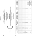
- the approach included 12 cycles of counter-selection/selection steps of a starting highly complex library on WT (COS7-WT) and transiently transfected COS7 cells (COS7-ICAM-1), respectively, according to the scheme in Figure 1-a , in which at each round a selective pressure was generated ( Figure 1-b ).
- the pool was transcribed using a mutant form of T7 RNA polymerase able to incorporate 2'-fluoro pyrimidines in the RNA sequences.
- binding assays by RT-qPCR were performed in order to select the sequences able to bind COS7-ICAM-1. Essentially, DNA sequences were amplified and transcribed. Then, RNA sequences were incubated at 50 nM, as final concentration, for 15 minutes at 37°C, after pre-treatment with yeast tRNA 200 ⁇ g/mL, on COS7-WT and COS7-ICAM-1. Following incubation, cells were washed 3 times with PBS and recovered in TRIsure reagent. An RNA sequence used as reference control was spotted in each point for the normalization. A fold ratio was calculated comparing binding values of COS7-ICAM-1 over COS7-WT.
- the 84mer original molecule 12c-10 corresponding to SEQ ID NO: 2
- the sequence was shortened by isolating the more structured region (characterized by stems, loops, bulges and/or hairpins) including the degenerate portion of the starting library, taking into account that the folding of the short version has to be maintained.
- RNA sequence 10.T was incubated at 50nM, as final concentration, for 15 minutes at 37°C on COS7-WT and COS7-ICAM-1.
- the ratio comparing binding values of COS7-ICAM-1 over COS7-WT (fold change) is reported in Figure 5 , showing a value of 2.2.
- This result confirmed the ability of the aptamer 10.T (SEQ ID NO: 1), as well as of the original molecule 12c-10 (SEQ ID NO: 2), to bind the target ICAM-1 in its physiological conformation exposed on the membrane of the cell surface.
- aptamer 10.T was further investigated in a different experiment.
- the complexes were successively incubated with Streptavidin Agarose beads. After several washings and denaturation, samples were analyzed by immunoblotting, as shown in Figure 7 .
- Aptamer 10.T was tested for stability in human serum. It was incubated in 87% human serum at 37°C. The samples were collected at different times (T0, 1, 2, 4, 8, 12, 24, 48, 72h), incubated with proteinase K for 1h at 37°C in order to degrade serum proteins and loaded on a denaturing gel. Results showed an half-life for aptamer 10.T between 4 and 8h. Stability results are shown in Figure 6 .
- RNA sequence was conjugated at its 5'-end to the commercial dye Alexa Fluor 488 after insertion of a C 12 -amino linker (5'-C 12 -NH 2 ).
- the amino linker was inserted at the 5'-terminal phosphate by condensation with a C 12 aliphatic diamine in basic catalysis.
- the resulting free NH 2 moiety was coupled with the commercially available Alexa Fluor 488-NHS ester, to form a covalent amide bond.
- Alexa Fluor 488-NHS ester was dissolved in high-quality anhydrous dimethylformamide (DMF) or dimethylsulfoxide (DMSO), and the reaction was carried out in 0.1-0.2 M sodium bicarbonate buffer, pH 8.3, at room temperature. Purification was performed by PAGE followed by HPLC.
- DMF dimethylformamide
- DMSO dimethylsulfoxide
- Example 6 Affinity assays of aptamer 10.T and its conjugate thereof
- the aptamer 10.T was incubated on COS7-ICAM-1 cells at increasing concentrations (10-50-100-250-500-1000 nM) for 15 minutes at 37°C after pre-treatment with yeast tRNA 200 ⁇ g/mL.
- the same experiment was carried out with the unrelated aptamer A10, as negative control (sequence disclosed in Lupold S.E. et al., Cancer Res. 2002, 62, 4029-4033 ).
- the binding was evaluated by RT-qPCR. As can be seen from Figure 8 , the K d value resulted 375.7 ⁇ 151.7nM.
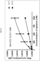
- aptamer 10.T K d value by using another technique, the conjugate of aptamer 10.T with Alexa Fluor-488 obtained in Example 5 was incubated on COS7-WT and COS7-ICAM-1 at increasing concentrations (10-50-100-200-400-800-1000-1500-2000 nM) at 37°C for 30 minutes, after pre-treatment with yeast tRNA 200 ⁇ g/mL. The experiment was analyzed by flow cytometry and performed 6 times, giving a K d of 115.5 ⁇ 49.9 nM as a result (see the results in Figure 9 ).
- HMEC-1 cells were stimulated with TNFalpha for 48h to induce ICAM-1 expression.
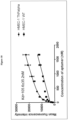
- the experiment was performed in the same conditions used for COS7 cells, in triplicate, giving a K d of 105.6 ⁇ 35.2 nM (see the results in Figure 10 ).
- the ELONA assay was performed to calculate a K d value for human serum albumin (HSA).
- HSA human serum albumin
- Biotinylated aptamer 10.T was incubated at increasing concentrations (1-10-100-1000 nM) on 96 well microtiter high binding plates previously coated or not-coated with HSA 25 nM. It was not possible to calculate K d value, indicating that 10.T does not react with HSA up to 1000 nM.
- a biotinylated polyclonal antibody anti-HSA was used as positive control. The results of this experiment are reported in Figure 11 .
Landscapes
- Life Sciences & Earth Sciences (AREA)
- Health & Medical Sciences (AREA)
- Engineering & Computer Science (AREA)
- Genetics & Genomics (AREA)
- Biomedical Technology (AREA)
- Molecular Biology (AREA)
- Chemical & Material Sciences (AREA)
- Biotechnology (AREA)
- Organic Chemistry (AREA)
- General Engineering & Computer Science (AREA)
- Wood Science & Technology (AREA)
- Bioinformatics & Cheminformatics (AREA)
- Zoology (AREA)
- Biochemistry (AREA)
- Microbiology (AREA)
- General Health & Medical Sciences (AREA)
- Physics & Mathematics (AREA)
- Plant Pathology (AREA)
- Biophysics (AREA)
- Urology & Nephrology (AREA)
- Hematology (AREA)
- Immunology (AREA)
- General Physics & Mathematics (AREA)
- Food Science & Technology (AREA)
- Medicinal Chemistry (AREA)
- Analytical Chemistry (AREA)
- Cell Biology (AREA)
- Pathology (AREA)
- Pharmaceuticals Containing Other Organic And Inorganic Compounds (AREA)
- Medicines Containing Antibodies Or Antigens For Use As Internal Diagnostic Agents (AREA)
- Magnetic Resonance Imaging Apparatus (AREA)
- Nuclear Medicine (AREA)
- Measuring Or Testing Involving Enzymes Or Micro-Organisms (AREA)
Description
- The present invention provides nucleic acid aptamers binding to the Intercellular Adhesion Molecule-1 (ICAM-1), derivatives and conjugates thereof and their use as diagnostic tools, particularly for the imaging of organs and tissues expressing ICAM-1, or as therapeutic agents for prevention or treatment of ICAM-1-related diseases.
- Recently, functional oligonucleotide-based biomolecules, called aptamers, have attracted great interest as potential alternatives to antibodies. The technology of aptamer selection has been drawn to the attention of the scientific community due to its applicability in diagnosis and treatment of diseases.
- The oligonucleotide aptamers range in size from about 20 to about 80 bases (8 to 25 kDa) and their structures are responsible for intramolecular interactions (Levy-Nissenbaum E. et al., Trends Biotechnol. 2008, 26(8), 442-449).
- Aptamers bind to their targets by interactions between aromatic compounds, base pairings by hydrogen ligation, van der Waals interactions, and electrostatic interactions between charged groups or hydrogen bonds. In consequence, aptamers undergo conformational changes after target recognition and biomolecular interaction.
- These biological, physical and chemical properties make these oligonucleotides effective recognition tools for diagnosis and therapy. The application of aptamers in biological fields is mainly limited due to its degradation by ribozymes. Chemical modifications are required in order to protect them against nucleases, improving their thermal stability and their pharmacokinetic properties.
- Among the modifications, the exchange of an OH at the 2'-position of the ribose by 2'-F or 2'-NH2 can be performed to improve aptamer stability in the cellular environment. Other alterations of aptamers can include terminal capping with small molecules such as amine, a phosphate group, or residue of thymidine and other non-natural bases (Gao S. et al, Anal. Bioanal. Chem. 2016, 408(17), 4567-4573).
- The aptamers are selected by an in vitro process through the Systematic Evolution of Binding by Exponential Enrichment (SELEX). This method was concomitantly described by Tuerk and Gold (Science, 1990, 249, 505-510) and Ellington and Szostak (Nature, 1990, 346, 818-822). SELEX involves the progressive selection of aptamers by the repetition of binding cycles, elution, and amplification of ligands from a random nucleic acid library, to select sequences with a higher binding affinity for the target.
- A new application of this technology, called "cell-SELEX" has been developed allowing the selection of aptamers that bind to specific target cells (de Franciscis V. et al., Methods Mol Biol., 2016, 1380, 33-46).
- The selection parameters can be easily manipulated to obtain more efficient aptamers for a wide range of conditions (pH, temperature or buffer composition) (Radom F. et al, Biotechnol. Adv. 2013, 31(8), 1260-1274). Some modifications have been included in the traditional SELEX method, such as affinity chromatography, capillary electrophoresis and filtration membranes to maximize affinity and specificity and to improve the selection speed and success rate of the selected aptamers (Stoltenburg R et al, Biomol. Eng. 2007, 24(4), 381-403). The characteristics of the selected oligonucleotides are identified using various physical, chemical and biological assays (Song KM et al, Sensors, 2012, 12(1), 612-631).
- Once selected, they can be synthesized in great quantity with precision and reproducibility by chemical reactions. These chemical processes are more cost-effective than the production of antibodies. When compared to antibodies, the aptamers have a relatively small size, which facilitates their chemical synthesis and possible modifications. They are biocompatible and poorly immunogenic in vivo. They have high selectivity and the ability to bind and recognize specific targets, presenting an affinity constant (Kd) with nanomolar range, compared to antibodies (Kd in the milli/micromolar range). Also, they penetrate tissues faster and more efficiently because of their significantly lower molecular weight and can distinguish extracellular or intracellular domains of proteins, which cannot be differentiated by antibodies (Gopinath S.C. et al, J. Gen. Virol., 2006, 87(3), 479-487).
- The strong target affinity/selectivity, cost-effectivity, chemical versatility and safety of aptamers are superior to traditional peptide- or protein-based ligands, which make them particularly suitable for molecular imaging. Therefore, aptamers are considered to be extremely useful to guide various imaging contrast agents to the target tissues or cells for optical, magnetic resonance, nuclear, computed tomography, ultrasound and multimodality imaging.
- Intercellular adhesion molecules (ICAMs) are structurally related transmembrane glycoproteins of the immunoglobulin supergene family and are ligands for the β2 integrin molecules present on leukocytes (Almenar-Queralt A. et al., Am. J. Pathol., 1995, 147(5), 1278-1288; Hubbard AK et al, Free Radic. Biol. Med., 2000, 28, 1379-1386). Of the five ICAMs identified, ICAM-1 is the most extensively studied (Koning et al., Endothelium, 2002, 9, 161-171; Muro et al., Curr. Pharm. Des., 2005, 11, 2383-2401). ICAM-1 (Intercellular Adhesion Molecule 1) is a protein that in humans is encoded by the ICAM1 gene. This gene encodes a cell surface which is typically expressed on endothelial cells and cells of the immune system. It binds to integrin of type CD11a / CD18, or CD11b / CD18 and is also exploited by rhinovirus as a receptor.
- ICAM-1 specifically participates in trafficking of inflammatory cells, in leukocyte effector functions, in adhesion of antigen-presenting cells to T lymphocytes, in microbial pathogenesis, and in signal transduction pathways through outside-in signaling events. This adhesion molecule is localized to both the apical and basolateral surface of endothelial cells, making it ideally positioned to facilitate transendothelial migration of leukocytes. In fact, ICAM-1 (along with VCAM-1) is considered to represent the most important adhesion molecule for leukocyte recruitment to inflammed sites and it is associated with a number of inflammatory and immune responses, as well as with epithelial tumorigenesis in the metastatic process. These properties makes ICAM-1 a potential target for diagnostic applications.
- Non-invasive in vivo molecular imaging of endothelial ICAM-1 expression could therefore provide valuable insights in the progression of cardiovascular disease-related inflammation to improve diagnosis and treatment.
- In recent years, contrast-enhanced ultrasonography (CEUS) has dramatically improved the imaging of small blood vessels. The microbubble-based contrast agents currently used in clinical practice lack affinity for the lesions, resulting in imaging duration only about 2-5 min. With the development of targeted ultrasound contrast agents and the appearance of nanobubbles, the ultrasound molecular imaging technique has undergone a revolutionary progress and become the focus of ultrasound applications. Also in this context the endothelial ICAM-1 is one of the most promising targets.
-
US 5,756,291 discloses a method to obtain aptamers against target proteins such as ICAM-1, VCAM, Factor X, PDGF, FGF, E-selectin, thrombin and bradykinin. As far as ICAM-1 is concerned, the target is represented by five peptides from its amino terminus, from 17 to 19 residues in length, encompassing its active domain. The oligonucleotides mixture is radiolabeled with 32P, incubated with the target and then loaded on a gel. The pool enrichment is followed by gel shift method and selection of the best candidate. DNA species were selected for their slower migration compared to the starting pool. However, there is no evidence that the selected aptamer could bind ICAM-1 in the context of the cellular membrane, thus resulting a good tool for in vivo purposes. -
WO2015/031694 andWO2014/068408 disclose aptamers that bind to microvesicle surface antigens, including microvesicles derived from various diseases, e.g. from cells indicative of cancer, and are useful as therapeutics in or diagnostics of cancer and/or other diseases where microvesicles are implicated. The gene ICAM-1 is mentioned therein as one of the possible biomarkers. -
WO2010/144295 discloses compositions for inducing or enhancing immunogenicity of a tumor comprising bi- and multi-specific aptamers binding to a tumor cell and an immune cell. These compositions have broad applicability in the treatment of many diseases, including cancer. -
WO2005/110489 discloses a method to increase the ligand antagonist range of a receptor-binding aptamer. The aptamer is joined to a high molecular weight steric group able to prevent the binding of the target to a second molecule. In the ICAM-1 context, the aptamer is directed against thedomain 2 of the extracellular portion of the target, whereas the steric group prevents the binding of ICAM-1 natural ligand, the integrin LFA-1, to thedomain 1. However, no sequences of aptamers with affinity for ICAM-1 are disclosed. - The present invention is based on the identification of RNA aptamers, which are able to bind ICAM-1 molecule with high affinity. In particular, the aptamers of the invention solve the problem of specifically recognize the target at the site where it is physiologically present, i.e. on the cell surface where ICAM-1 is expressed, thus demonstrating to be suitable for their use in vivo. In fact, the aptamers herein described have been found able to bind directly the cells overexpressing ICAM-1 on their surface.
- To that purpose, a library of RNA molecules was assayed for its binding to COS7 cells transiently transfected with human ICAM-1. Following to repeated selection steps, the RNA molecules showing highest COS7-ICAM-1 binding were isolated and their sequence and ICAM-1 binding affinity were determined.
- The aptamer having the following sequence:
UCAUGAUACGUUGCGUGAGCAACUGCGGCGCUAAAGUUUGGUUGACUAGUAC AUG (SEQ ID NO: 1) showed the highest binding affinity to ICAM-1. - Aptamers containing SEQ ID NO: 1 proved likewise able to bind ICAM-1 and particularly the aptamer having the following sequence:

- Therefore, in a first aspect the invention provides an aptamer able to bind to Intercellular Adhesion Molecule-1 (ICAM-1) and comprising the RNA sequence SEQ ID NO: 1.
- In one embodiment, the above aptamer has a length of up to 100 nucleotides.
- The dissociation constants of the aptamers containing SEQ ID NO: 1 and showing ICAM-1 binding ability, determined by either RT-qPCR or flow-cytometry, were found within a range from 500 nM to 50 nM.
- In a further aspect, the invention provides an aptamer able to bind to Intercellular Adhesion Molecule-1 (ICAM-1) and comprising the RNA sequence SEQ ID NO: 2.
- In a preferred embodiment the RNA aptamers defined above are characterized by being nuclease-resistant.
- In a more preferred embodiment the RNA aptamers defined above are characterized by having all the pyrimidine residues modified to 2'-fluoropyrimidines.
- A preferred aptamer of the invention consists of SEQ ID NO: 2. More preferably, it consists of SEQ ID NO: 1.
- Aptamers of the invention can be modified, e.g. to increase their resistance to nucleases, to modulate their pharmacokinetics, or to be conjugated with diagnostic or therapeutic moieties.
- Preferably, a RNA aptamer of the invention has at least one or all of the pyrimidine residues modified to 2'-fluoropyrimidines. Furthermore, its modification may include a chemical substitution at a sugar position, a chemical substitution at a phosphate position and a chemical substitution at a base position of the nucleic acid. In some embodiments, the modification is selected from the group consisting of: biotinylation, incorporation of a fluorescent label, incorporation of a modified nucleotide, 2'-pyrimidine modification, 3'-position capping, conjugation to a linker, conjugation to a compound or a drug, conjugation to a cytotoxic moiety, and labeling with a fluorophore, a radioisotope, an ultrasound contrast agent or a reporter moiety. The position of the modification can be varied depending on the type of moiety that is attached to the aptamer.
- The invention also relates to an aptamer as defined above, for use as a medicament.
- In particular, an object of the invention is an aptamer as defined above for use in the treatment of an ICAM-1 related state, disorder, dysfunction, condition or disease. Preferably, said ICAM-1-related state, disorder, dysfunction, condition or disease is inflammation or an inflammation-associated disease.
- Exemplary applications include the diagnosis or therapy of: vascular inflammation in atherosclerosis and myocardial infarction; diseases affecting the cardiovascular system, such as myocarditis; inflammatory cardiomyopathy and heart failure.
- Moreover, the invention relates to an aptamer as defined above for use as in vivo contrast agent for diagnostic imaging. Preferably, said diagnostic imaging involves the imaging of a body tissue or organ system expressing ICAM-1. In a preferred embodiment said tissue and organ system are endothelium and blood vessels, respectively.
- In another embodiment the invention relates to the use of an aptamer as defined above in in vitro/ex vivo diagnostic imaging. Preferably, said diagnostic imaging involves the imaging of a body tissue or organ system expressing ICAM-1. In a preferred embodiment said tissue and organ system are endothelium and blood vessels, respectively.
- Suitable imaging techniques include magnetic resonance imaging, positron-emission tomography (PET), computed tomography (CT), ultrasound, photoacoustic imaging (PAI), near-infrared fluorescence (NIRF), single photon emission computed tomography (SPECT).
- For imaging applications, the reporter moiety linked to the aptamer is generally selected from: molecules capable of generating a fluorescent signal, such as fluorescein; FITC; Alexa dyes; Cy dyes; DyLight dyes; IRDye dyes or VivoTag dyes; optical moieties, including agents that may be used to produce contrast or signal using optical imaging; magnetic moieties, including a chelating agent for magnetic resonance agents which is able to form stable complexes with paramagnetic metal ions; radiolabel moieties; X-ray moieties that may be used to produce contrast or signal using X-ray imaging, such as iodinated organic molecules or chelates of heavy metal ions; ultrasound imaging moieties that may be used to produce contrast or signal using ultrasound targeted microbubbles; and photoacoustic imaging moieties, including photoacoustic imaging-compatible agents.
- The aptamer and the reporter moiety or label may be linked either covalently or noncovalently, optionally by interposition of a suitable linker or spacer, including peptides, amino acids or nucleic acids. Furthermore, the aptamer and the reporter moiety or label may be linked using a tag system, including biotin/avidin, biotin/streptavidin, biotin/NeutrAvidin, or digoxigenin (DIG) systems.
- In a further aspect, the invention provides a composition comprising an aptamer as herein defined. The ingredients of the composition can be varied depending on the intended use, whether for diagnostic, therapeutic or imaging applications. In particular the composition may contain a RNA molecule and one or more therapeutic compounds and/or one or more imaging agents.
- In one embodiment, the composition is used for the imaging of a target tissue bearing ICAM-1 and comprises the aptamer conjugated or labeled with a reporter moiety as above defined. The composition can be e.g. in the form of a liposome or nanoparticles and it is suitable for parenteral administration, particularly for intravenous administration. Said composition can be used for visualizing ICAM-1 expressing tissues or organs, such as inflamed endothelium and vasculature.
-
- SEQ ID NO: 1 sets out sequence from the 5' of the aptamer 10.T (55 nt)

- SEQ ID NO: 2 sets out sequence from the 5' of the
aptamer 12c-10 (84 nt)
-
-
Figure 1 . Scheme of anti-ICAM-1 cell-SELEX procedure. -
Figure 2 . Dendrogram (by MUSCLE algorithm) for the visualization of the enriched sequences obtained by cloning the last cycle of anti-ICAM-1 cell-SELEX. -
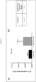
Figure 3 . RT-qPCR binding assay for the selected sequence by anti-ICAM-1 cell-SELEX. a) Experimental triplicates of the three sequences by RT-qPCR biding assay; b) Summary table of experimental triplicate fold change. Each bar shows the mean ± standard deviation values from three experiments. -
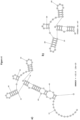
Figure 4 . Predicted secondary structure of the selected aptamer anti-ICAM-1 and relative shortened version by RNA structure 5.8 program. a) long 12c-10 sequence (SEQ ID NO: 2) and b) short 10.T sequence (SEQ ID NO: 1) predicted secondary structures with lower free energy. -
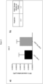
Figure 5 . RT-qPCR binding assay for the 10.T sequence selected by anti-ICAM-1 cell-SELEX. a) RT-qPCR for the 10.T sequence selected to perform experimental triplicates; b) summary table of experimental triplicate fold ratio. Each bar shows the mean ± standard deviation values from three experiments. -
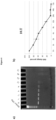
Figure 6 .10 . T sequence stability in human serum. a) 10.T sequence samples collected at different times were loaded on a denaturing gel and b) bands were quantified by ImageJ program. -
Figure 7 .10 . T sequence pull-down assay. a) Structure of oligonucleotides biotinylated at 5'-end; b) 300µg or c) 450µg of COS7, transiently transfected with ICAM-1 cDNA, lysate were incubated with 1µM of biotinylated 10.T sequence and an ICAM-1-unrelated 2'F-RNA sequence used as negative control. -
Figure 8 . Determination of binding affinity of 10.T sequence to COS7-ICAM-1 by RT-qPCR binding assay. The recovered RNA (µM) of varying concentrations of 10.T sequence and the unrelated sequence A10 were plotted to determine dissociation constant Kd by using GraphPad software. The experiment was repeated three times and the error bars represent the standard deviation of means. -
Figure 9 . Determination of binding affinity of Alexa488-10.T conjugate to COS7-WT and COS7-ICAM-1 by flow cytometry. a) Structure of 10.T sequence conjugated to Alexa-488 fluorophore at the 5'-end by a C12 amino linker. b) The mean fluorescence intensity of varying concentration of Alexa488-10.T conjugate obtained was plotted to determine dissociation constant Kd by using GraphPad software. The experiment was repeated six times and error bars represent the standard deviation of means. -
Figure 10 . Determination of binding affinity of Alexa488-10.T conjugate to HMEC1-WT and HMEC1-TNFalpha by flow cytometry. The mean fluorescence intensity of varying concentration of Alexa488-10.T conjugate obtained was plotted to determine dissociation constant Kd by using GraphPad software. The experiment was repeated three times and error bars represent the standard deviation of means. -
Figure 11 .10 . T sequence Kd evaluation for HSA by ELONA assay. Absorbance at 450nm for 10.T sequence (a) and polyclonal antibody anti-HSA (b). - Flow cytometry data acquisition was performed using a BD Accuri™ C6 flow cytometer (BD Bioscience). RT-qPCR was carried out by StepOne™ Plus Real-Time PCR System (Applied Biosystems). Gel visualization was performed with Gel Doc EZ System (Bio-Rad). ELONA data were acquired by Multiskan™ FC Microplate Photometer (ThermoFisher Scientific).
-
- ICAM-1
- Intercellular Adhesion Molecule -1
- SELEX
- Systematic Evolution of Ligands by Exponential enrichment
- RNA
- Ribonucleic acid
- DNA
- Deoxyribonucleic acid
- WT
- Wild type
- nt
- nucleotides
- HSA
- Human serum albumin
- Kd
- Constant of dissociation
- HMEC-1
- Human mammary epithelial cells -1
- TNFalpha
- Tumor necrosis factor alpha
- COS7
- CV-1 (simian) in Origin with SV40 genetic material cells
- Rt-q PCR
- Real-time polymerase chain reaction
- To specifically select an aptamer for ICAM-1, a cell-SELEX approach was followed using the same cell line with and without the overexpressed target, instead of two cell lines (positive/negative) that differ also for other proteins present on the cell surface.
- To this end, COS7 cells were transiently transfected with the expression plasmid pCMV6-ICAM-1.
- In detail, to selectively target ICAM-1, a cell-SELEX protocol was performed transiently transfecting the human ICAM-1 cDNA in negative COS7 cells, used as a recipient cell line. The approach included 12 cycles of counter-selection/selection steps of a starting highly complex library on WT (COS7-WT) and transiently transfected COS7 cells (COS7-ICAM-1), respectively, according to the scheme in
Figure 1-a , in which at each round a selective pressure was generated (Figure 1-b ). - Before each round of cell-SELEX, the pool was transcribed using a mutant form of T7 RNA polymerase able to incorporate 2'-fluoro pyrimidines in the RNA sequences.
- At the end of cell-SELEX protocol, the last cycle was cloned and 164 samples were sequenced. Resulting sequences were analyzed for enrichment by alignment and a dendrogram was produced for the visualization of identical sequences or sequences with a single mismatch by MUSCLE algorithm.
- Following the analysis of the enriched sequences, binding assays by RT-qPCR were performed in order to select the sequences able to bind COS7-ICAM-1. Essentially, DNA sequences were amplified and transcribed. Then, RNA sequences were incubated at 50 nM, as final concentration, for 15 minutes at 37°C, after pre-treatment with yeast tRNA 200µg/mL, on COS7-WT and COS7-ICAM-1. Following incubation, cells were washed 3 times with PBS and recovered in TRIsure reagent. An RNA sequence used as reference control was spotted in each point for the normalization. A fold ratio was calculated comparing binding values of COS7-ICAM-1 over COS7-WT. Twelve sequences representatives of couples or groups of identical sequences were screened. Those with higher fold ratio were chosen for further analysis performing experimental triplicates. Results showed that 12c-10, consisting of SEQ ID NO: 2, confirmed the highest binding for COS7-ICAM-1 with respect to COS7-WT, as illustrated in
Figure 3 (the fold change, i.e. the ratio between binding value of COS7-ICAM-1 and binding value of COS7-WT, is 3.13). - In order to obtain a shorter sequence useful for imaging applications, the 84mer
original molecule 12c-10, corresponding to SEQ ID NO: 2, was truncated to obtain the shorter 55mer sequence 10.T, corresponding to SEQ ID NO:1. - The sequence was shortened by isolating the more structured region (characterized by stems, loops, bulges and/or hairpins) including the degenerate portion of the starting library, taking into account that the folding of the short version has to be maintained.
- To the aim of testing if the short aptamer 10.T (SEQ ID NO: 1) contained the active site of the
original molecule 12c-10 (SEQ ID NO: 2) and preserved high binding and affinity to COS7-ICAM-1, binding assays were performed maintaining the same conditions used for long aptamers screening. - The RNA sequence 10.T was incubated at 50nM, as final concentration, for 15 minutes at 37°C on COS7-WT and COS7-ICAM-1. The ratio comparing binding values of COS7-ICAM-1 over COS7-WT (fold change) is reported in
Figure 5 , showing a value of 2.2. This result confirmed the ability of the aptamer 10.T (SEQ ID NO: 1), as well as of theoriginal molecule 12c-10 (SEQ ID NO: 2), to bind the target ICAM-1 in its physiological conformation exposed on the membrane of the cell surface. - The binding of aptamer 10.T was further investigated in a different experiment. A pull-down assay with aptamer 10.T biotinylated at 5'-end was performed to verify that the sequence bound its target, ICAM-1. COS7 cells transiently transfected with ICAM-1 cDNA for 48h were collected and 300µg of lysate were incubated with 1µM of biotinylated aptamer 10.T and an ICAM-1-unrelated 2'F-RNA sequence used as negative control. The complexes were successively incubated with Streptavidin Agarose beads. After several washings and denaturation, samples were analyzed by immunoblotting, as shown in
Figure 7 . - Results indicated that 10.T binds ICAM-1, whereas the ICAM-1-unrelated 2'F-RNA sequence and an untreated sample gave no signal after hybridization with anti-ICAM-1 antibody. This experiment was performed in duplicate.
- Aptamer 10.T was tested for stability in human serum. It was incubated in 87% human serum at 37°C. The samples were collected at different times (T0, 1, 2, 4, 8, 12, 24, 48, 72h), incubated with proteinase K for 1h at 37°C in order to degrade serum proteins and loaded on a denaturing gel. Results showed an half-life for aptamer 10.T between 4 and 8h. Stability results are shown in
Figure 6 . - To demonstrate the potential use of aptamer 10.T in imaging applications, the RNA sequence was conjugated at its 5'-end to the commercial dye Alexa Fluor 488 after insertion of a C12-amino linker (5'-C12-NH2). The amino linker was inserted at the 5'-terminal phosphate by condensation with a C12 aliphatic diamine in basic catalysis. The resulting free NH2 moiety was coupled with the commercially available Alexa Fluor 488-NHS ester, to form a covalent amide bond. The Alexa Fluor 488-NHS ester was dissolved in high-quality anhydrous dimethylformamide (DMF) or dimethylsulfoxide (DMSO), and the reaction was carried out in 0.1-0.2 M sodium bicarbonate buffer, pH 8.3, at room temperature. Purification was performed by PAGE followed by HPLC.
- In order to investigate the affinity to the target, the aptamer 10.T was incubated on COS7-ICAM-1 cells at increasing concentrations (10-50-100-250-500-1000 nM) for 15 minutes at 37°C after pre-treatment with yeast tRNA 200µg/mL. The same experiment was carried out with the unrelated aptamer A10, as negative control (sequence disclosed in Lupold S.E. et al., Cancer Res. 2002, 62, 4029-4033). The binding was evaluated by RT-qPCR. As can be seen from
Figure 8 , the Kd value resulted 375.7±151.7nM. - To confirm or deeply investigate aptamer 10.T Kd value by using another technique, the conjugate of aptamer 10.T with Alexa Fluor-488 obtained in Example 5 was incubated on COS7-WT and COS7-ICAM-1 at increasing concentrations (10-50-100-200-400-800-1000-1500-2000 nM) at 37°C for 30 minutes, after pre-treatment with yeast tRNA 200µg/mL. The experiment was analyzed by flow cytometry and performed 6 times, giving a Kd of 115.5 ± 49.9 nM as a result (see the results in
Figure 9 ). - A comparable result was obtained on induced HMEC-1 cell line. HMEC-1 cells were stimulated with TNFalpha for 48h to induce ICAM-1 expression. The experiment was performed in the same conditions used for COS7 cells, in triplicate, giving a Kd of 105.6 ± 35.2 nM (see the results in
Figure 10 ). - The ELONA assay was performed to calculate a Kd value for human serum albumin (HSA). Biotinylated aptamer 10.T was incubated at increasing concentrations (1-10-100-1000 nM) on 96 well microtiter high binding plates previously coated or not-coated with HSA 25 nM. It was not possible to calculate Kd value, indicating that 10.T does not react with HSA up to 1000 nM. For each experiment a biotinylated polyclonal antibody anti-HSA was used as positive control. The results of this experiment are reported in
Figure 11 .
Claims (18)
- An aptamer that binds to Intercellular Adhesion Molecule-1 (ICAM-1), comprising the RNA sequence SEQ ID NO: 1.
- The aptamer of claim 1, characterized in that it has a length of up to 100 nucleotides.
- The aptamer of claims 1 or 2, which is able to bind to ICAM-1 with a dissociation constant falling within a range from 500 nM to 50 nM.
- The aptamer of claims 1 through 3, comprising the RNA sequence SEQ ID NO: 2.
- The aptamer of claims 1 through 4, wherein all the pyrimidine residues are modified to 2' - fluoropyrimidines.
- The aptamer of claim 5, which is further modified to comprise at least one chemical modification, wherein said modification is selected from: chemical substitution at a sugar position; chemical substitution at a phosphate position; and a chemical substitution at a base position of the nucleic acid.
- The aptamer of claim 6, wherein the modification is selected from the group consisting of incorporation of a modified nucleotide, conjugation to a compound, and labeling with a reporter moiety.
- The aptamer of claim 7, wherein the reporter moiety is selected from the group consisting of a fluorophore moiety, a magnetic or paramagnetic moiety, a radiolabel moiety, an affinity label, an X-ray moiety, an ultrasound imaging moiety, a photoacoustic imaging moiety and a nanoparticle-based moiety.
- An aptamer as defined in claims 1-8, for use as a medicament.
- An aptamer according to claim 9 for use in the treatment of a ICAM-1 related state, disorder, dysfunction, condition or disease.
- An aptamer for use according to claim 10, wherein said ICAM-1-related state, disorder, dysfunction, condition or disease is inflammation or an inflammation-associated disease.
- An aptamer as defined in claims 1-8, for use as in vivo contrast agent for diagnostic imaging.
- An aptamer for use according to claim 12, wherein said diagnostic imaging involves the imaging of a body tissue or organ system expressing ICAM-1.
- An aptamer for use according to claim 13, wherein said tissue and organ system are endothelium and blood vessels, respectively.
- Use of an aptamer according to claims 1-8 in in vitro/ex vivo diagnostic imaging.
- Use of an aptamer according to claim 15, wherein said diagnostic imaging involves the imaging of a body tissue or organ system expressing ICAM-1.
- Use of an aptamer according to claim 16, wherein said tissue and organ system are endothelium and blood vessels, respectively.
- A diagnostic, therapeutic or imaging composition comprising an aptamer as defined in claims 1-8, with pharmaceutically acceptable carriers and excipients.
Applications Claiming Priority (2)
| Application Number | Priority Date | Filing Date | Title |
|---|---|---|---|
| EP18185794 | 2018-07-26 | ||
| PCT/EP2019/069917 WO2020020947A1 (en) | 2018-07-26 | 2019-07-24 | Icam-1 aptamers, diagnostic and therapeutic uses thereof |
Publications (3)
| Publication Number | Publication Date |
|---|---|
| EP3827082A1 EP3827082A1 (en) | 2021-06-02 |
| EP3827082B1 true EP3827082B1 (en) | 2025-05-14 |
| EP3827082C0 EP3827082C0 (en) | 2025-05-14 |
Family
ID=63077776
Family Applications (1)
| Application Number | Title | Priority Date | Filing Date |
|---|---|---|---|
| EP19745118.0A Active EP3827082B1 (en) | 2018-07-26 | 2019-07-24 | Icam-1 aptamers, diagnostic and therapeutic uses thereof |
Country Status (5)
| Country | Link |
|---|---|
| US (1) | US11274308B2 (en) |
| EP (1) | EP3827082B1 (en) |
| JP (1) | JP7434276B2 (en) |
| CN (1) | CN112469828B (en) |
| WO (1) | WO2020020947A1 (en) |
Families Citing this family (3)
| Publication number | Priority date | Publication date | Assignee | Title |
|---|---|---|---|---|
| WO2021086266A1 (en) * | 2019-10-28 | 2021-05-06 | National University Of Singapore | Dnmt1 -specific aptamers and production and uses thereof |
| MX2022016049A (en) * | 2020-06-25 | 2023-02-09 | Procter & Gamble | Aptamers for personal health care applications. |
| AU2022410882A1 (en) * | 2021-12-14 | 2024-05-09 | The Procter & Gamble Company | Aptamers for personal health care applications |
Family Cites Families (13)
| Publication number | Priority date | Publication date | Assignee | Title |
|---|---|---|---|---|
| US5756291A (en) | 1992-08-21 | 1998-05-26 | Gilead Sciences, Inc. | Aptamers specific for biomolecules and methods of making |
| US20050187174A1 (en) * | 2001-05-18 | 2005-08-25 | Sirna Therapeutics, Inc. | RNA interference mediated inhibition of intercellular adhesion molecule (ICAM) gene expression using short interfering nucleic acid (siNA) |
| US7767803B2 (en) * | 2002-06-18 | 2010-08-03 | Archemix Corp. | Stabilized aptamers to PSMA and their use as prostate cancer therapeutics |
| MXPA06011965A (en) | 2004-04-13 | 2007-04-17 | Osi Eyetech Inc | Enhanced biologically active conjugates. |
| US20130209514A1 (en) | 2009-06-09 | 2013-08-15 | Eli Gilboa | Aptamer-targeted costimulatory ligand aptamer |
| WO2011081904A1 (en) * | 2009-12-14 | 2011-07-07 | The United States Of America, As Represented By The Secretary, Department Of Health And Human Services | Delivery of transthyretin across the blood-brain barrier as a treatment for alzheimer's disease |
| WO2011134060A1 (en) * | 2010-04-27 | 2011-11-03 | National Research Council Of Canada | Anti-icam-1 single domain antibody and uses thereof |
| KR20150090072A (en) * | 2012-10-23 | 2015-08-05 | 카리스 라이프 사이언스 스위스 홀딩스 게엠베하 | Aptamers and uses thereof |
| US9422563B2 (en) * | 2013-03-18 | 2016-08-23 | University Of The Witwatersrand, Johannesburg | CD7 receptor aptamers |
| CN104212800B (en) * | 2013-05-07 | 2017-01-18 | 南方医科大学 | Nucleic acid aptamer for specific binding with human epidermal growth factor receptor type III variant and application of nucleic acid aptamer |
| WO2015031694A2 (en) * | 2013-08-28 | 2015-03-05 | Caris Science, Inc. | Oligonucleotide probes and uses thereof |
| CN106053848A (en) * | 2015-11-01 | 2016-10-26 | 李倩 | Kit for oculopathy detection and detection method |
| CN105779459A (en) * | 2016-04-10 | 2016-07-20 | 杨岭燕 | Aptamer for diagnosing rupture of fetal membrane in gynaecology and obstetrics and kit thereof |
-
2019
- 2019-07-24 WO PCT/EP2019/069917 patent/WO2020020947A1/en not_active Ceased
- 2019-07-24 JP JP2021504188A patent/JP7434276B2/en active Active
- 2019-07-24 CN CN201980049353.1A patent/CN112469828B/en active Active
- 2019-07-24 US US17/262,835 patent/US11274308B2/en active Active
- 2019-07-24 EP EP19745118.0A patent/EP3827082B1/en active Active
Also Published As
| Publication number | Publication date |
|---|---|
| US20210292762A1 (en) | 2021-09-23 |
| CN112469828B (en) | 2024-04-30 |
| EP3827082A1 (en) | 2021-06-02 |
| EP3827082C0 (en) | 2025-05-14 |
| CN112469828A (en) | 2021-03-09 |
| JP2021531020A (en) | 2021-11-18 |
| WO2020020947A1 (en) | 2020-01-30 |
| US11274308B2 (en) | 2022-03-15 |
| JP7434276B2 (en) | 2024-02-20 |
Similar Documents
| Publication | Publication Date | Title |
|---|---|---|
| US6613526B2 (en) | Systematic evolution of ligands by exponential enrichment: tissue selex | |
| Zhou et al. | Aptamers as targeted therapeutics: current potential and challenges | |
| AU767501B2 (en) | Tenascin-C nucleic acid ligands | |
| US11274308B2 (en) | ICAM-1 aptamers, diagnostic and therapeutic uses thereof | |
| JP6442941B2 (en) | Vascular endothelial growth factor binding nucleic acid aptamer and use thereof | |
| JP5673992B2 (en) | Vascular endothelial growth factor binding aptamer | |
| WO2015117206A1 (en) | Improved aptamers | |
| Uhlig et al. | Alkynes as an eco-compatible “on-call” functionality orthogonal to biological conditions in water | |
| EP4031667B1 (en) | Ca-ix aptamers and diagnostic and therapeutic uses thereof | |
| CN113913419A (en) | Preparation method of bivalent circular DNA chain, bivalent circular nucleic acid aptamer and application thereof | |
| WO2023211299A1 (en) | Dna aptamer having affinity for pd-l1 protein and its use, functional covalent complex and its use | |
| JP2024046855A (en) | DNA aptamer capable of binding to integrin α7β1 receptor | |
| CN118931908B (en) | Nucleic acid aptamer combined with protein phosphatase 1 regulatory subunit 26 protein, screening method and application thereof | |
| WO2021015234A1 (en) | Chimeric molecule, pharmaceutical composition, method for cleaving target nucleic acid, and kit for target nucleic acid cleavage or diagnosis | |
| US7005260B1 (en) | Tenascin-C nucleic acid ligands | |
| Tan et al. | Aptamer properties, functions, and applications | |
| KR20080077450A (en) | Acridine-alpha helical peptide conjugates that specifically bind to target RA | |
| JP2025541178A (en) | Compositions and methods related to multiparatopic aptamers | |
| White | Framework for a Fibrin-Specific Aptamer for Targeted Delivery of Fibrinolytic Escherichia Coli | |
| JP2004503214A (en) | Cell binding nucleic acid molecule (aptamer) | |
| KR20260056195A (en) | Intracellular targeting of oligonucleotides | |
| WO2025231297A1 (en) | Mapping spatial locations of lipid particles using nucleic acid barcodes | |
| US20210222166A1 (en) | Lna based nanodevice |
Legal Events
| Date | Code | Title | Description |
|---|---|---|---|
| STAA | Information on the status of an ep patent application or granted ep patent |
Free format text: STATUS: UNKNOWN |
|
| STAA | Information on the status of an ep patent application or granted ep patent |
Free format text: STATUS: THE INTERNATIONAL PUBLICATION HAS BEEN MADE |
|
| PUAI | Public reference made under article 153(3) epc to a published international application that has entered the european phase |
Free format text: ORIGINAL CODE: 0009012 |
|
| STAA | Information on the status of an ep patent application or granted ep patent |
Free format text: STATUS: REQUEST FOR EXAMINATION WAS MADE |
|
| 17P | Request for examination filed |
Effective date: 20210217 |
|
| AK | Designated contracting states |
Kind code of ref document: A1 Designated state(s): AL AT BE BG CH CY CZ DE DK EE ES FI FR GB GR HR HU IE IS IT LI LT LU LV MC MK MT NL NO PL PT RO RS SE SI SK SM TR |
|
| DAV | Request for validation of the european patent (deleted) | ||
| DAX | Request for extension of the european patent (deleted) | ||
| STAA | Information on the status of an ep patent application or granted ep patent |
Free format text: STATUS: EXAMINATION IS IN PROGRESS |
|
| 17Q | First examination report despatched |
Effective date: 20220928 |
|
| GRAP | Despatch of communication of intention to grant a patent |
Free format text: ORIGINAL CODE: EPIDOSNIGR1 |
|
| STAA | Information on the status of an ep patent application or granted ep patent |
Free format text: STATUS: GRANT OF PATENT IS INTENDED |
|
| INTG | Intention to grant announced |
Effective date: 20250102 |
|
| GRAS | Grant fee paid |
Free format text: ORIGINAL CODE: EPIDOSNIGR3 |
|
| GRAA | (expected) grant |
Free format text: ORIGINAL CODE: 0009210 |
|
| STAA | Information on the status of an ep patent application or granted ep patent |
Free format text: STATUS: THE PATENT HAS BEEN GRANTED |
|
| AK | Designated contracting states |
Kind code of ref document: B1 Designated state(s): AL AT BE BG CH CY CZ DE DK EE ES FI FR GB GR HR HU IE IS IT LI LT LU LV MC MK MT NL NO PL PT RO RS SE SI SK SM TR |
|
| REG | Reference to a national code |
Ref country code: GB Ref legal event code: FG4D |
|
| REG | Reference to a national code |
Ref country code: CH Ref legal event code: EP |
|
| REG | Reference to a national code |
Ref country code: IE Ref legal event code: FG4D |
|
| U01 | Request for unitary effect filed |
Effective date: 20250514 |
|
| U07 | Unitary effect registered |
Designated state(s): AT BE BG DE DK EE FI FR IT LT LU LV MT NL PT RO SE SI Effective date: 20250520 |
|
| U20 | Renewal fee for the european patent with unitary effect paid |
Year of fee payment: 7 Effective date: 20250728 |
|
| PG25 | Lapsed in a contracting state [announced via postgrant information from national office to epo] |
Ref country code: ES Free format text: LAPSE BECAUSE OF FAILURE TO SUBMIT A TRANSLATION OF THE DESCRIPTION OR TO PAY THE FEE WITHIN THE PRESCRIBED TIME-LIMIT Effective date: 20250514 |
|
| PG25 | Lapsed in a contracting state [announced via postgrant information from national office to epo] |
Ref country code: GR Free format text: LAPSE BECAUSE OF FAILURE TO SUBMIT A TRANSLATION OF THE DESCRIPTION OR TO PAY THE FEE WITHIN THE PRESCRIBED TIME-LIMIT Effective date: 20250815 Ref country code: NO Free format text: LAPSE BECAUSE OF FAILURE TO SUBMIT A TRANSLATION OF THE DESCRIPTION OR TO PAY THE FEE WITHIN THE PRESCRIBED TIME-LIMIT Effective date: 20250814 |
|
| PG25 | Lapsed in a contracting state [announced via postgrant information from national office to epo] |
Ref country code: PL Free format text: LAPSE BECAUSE OF FAILURE TO SUBMIT A TRANSLATION OF THE DESCRIPTION OR TO PAY THE FEE WITHIN THE PRESCRIBED TIME-LIMIT Effective date: 20250514 |
|
| PGFP | Annual fee paid to national office [announced via postgrant information from national office to epo] |
Ref country code: GB Payment date: 20250728 Year of fee payment: 7 |
|
| PG25 | Lapsed in a contracting state [announced via postgrant information from national office to epo] |
Ref country code: HR Free format text: LAPSE BECAUSE OF FAILURE TO SUBMIT A TRANSLATION OF THE DESCRIPTION OR TO PAY THE FEE WITHIN THE PRESCRIBED TIME-LIMIT Effective date: 20250514 |
|
| PGFP | Annual fee paid to national office [announced via postgrant information from national office to epo] |
Ref country code: CH Payment date: 20250801 Year of fee payment: 7 |
|
| PG25 | Lapsed in a contracting state [announced via postgrant information from national office to epo] |
Ref country code: RS Free format text: LAPSE BECAUSE OF FAILURE TO SUBMIT A TRANSLATION OF THE DESCRIPTION OR TO PAY THE FEE WITHIN THE PRESCRIBED TIME-LIMIT Effective date: 20250814 |
|
| PG25 | Lapsed in a contracting state [announced via postgrant information from national office to epo] |
Ref country code: IS Free format text: LAPSE BECAUSE OF FAILURE TO SUBMIT A TRANSLATION OF THE DESCRIPTION OR TO PAY THE FEE WITHIN THE PRESCRIBED TIME-LIMIT Effective date: 20250914 |
|
| PG25 | Lapsed in a contracting state [announced via postgrant information from national office to epo] |
Ref country code: SM Free format text: LAPSE BECAUSE OF FAILURE TO SUBMIT A TRANSLATION OF THE DESCRIPTION OR TO PAY THE FEE WITHIN THE PRESCRIBED TIME-LIMIT Effective date: 20250514 |
|
| PG25 | Lapsed in a contracting state [announced via postgrant information from national office to epo] |
Ref country code: CZ Free format text: LAPSE BECAUSE OF FAILURE TO SUBMIT A TRANSLATION OF THE DESCRIPTION OR TO PAY THE FEE WITHIN THE PRESCRIBED TIME-LIMIT Effective date: 20250514 |
|
| PG25 | Lapsed in a contracting state [announced via postgrant information from national office to epo] |
Ref country code: SK Free format text: LAPSE BECAUSE OF FAILURE TO SUBMIT A TRANSLATION OF THE DESCRIPTION OR TO PAY THE FEE WITHIN THE PRESCRIBED TIME-LIMIT Effective date: 20250514 |
|
| PLBE | No opposition filed within time limit |
Free format text: ORIGINAL CODE: 0009261 |
|
| STAA | Information on the status of an ep patent application or granted ep patent |
Free format text: STATUS: NO OPPOSITION FILED WITHIN TIME LIMIT |
|
| REG | Reference to a national code |
Ref country code: CH Ref legal event code: L10 Free format text: ST27 STATUS EVENT CODE: U-0-0-L10-L00 (AS PROVIDED BY THE NATIONAL OFFICE) Effective date: 20260325 |
|
| 26N | No opposition filed |
Effective date: 20260217 |