本発明は内皮細胞に選択的に結合する、または内皮細胞を選択的に標識する核酸分子、腫瘍疾患を同定する方法、かかる方法で使用される核酸分子ならびにかかる核酸分子の使用法に関する。
【0001】
この種の方法およびそこで使用される核酸分子は公知のものであって、アプタマーとも呼ばれる。関連する総説はOsborneら、”Aptamers as therapeutic and diagnostic reagents: problems and prospects”, Curr. Opin. Chem. Biol. 1997, 1: 5−9.に見出せる。
アプタマーは高親和性のRNAおよびDNAオリゴヌクレオチドまたはポリヌクレオチドであり、それらの特異的な空間的構造により標的分子に高い親和性を有する。かかるアプタマーを見出すためには、全く異なる配列および二次構造を有する多数のオリゴヌクレオチドを無作為かつ酵素的に生成し、これから標的分子に高い親和性を示すオリゴヌクレオチドを選択、濃縮するといったコンビナトリアルアプローチがしばしば用いられる。この技術はまたSELEX(systematic evolution of ligands by exponential enrichment)とも呼ばれる。
【0002】
一次構造が公知の場合、かかるオリゴ−ポリヌクレオチド(アプタマー)は例えばDNA/RNA合成機を使って合成してもよい。
アプタマーに対する標的構造は、レセプターまたは他のタンパク質、脂質構造もしくは炭水化物構造であってよい。テオフィリン(Jenisonら、”High−resolution molecular discrimination by RNA”, Science 1994; 263 (5152): 1425−9を参照)、ATP(Tangら、”Rational design of allosteric ribozymes”, Chem. Biol.1997; 4 (6): 453−9を参照)、アルギニン(Geigerら、”RNA aptamers that bind L−arginine with sub−micromolar dissociation constants and high enancio−selectivity” Nucl. Acids Res. 1996; 24 (6): 1029−36を参照)のような小分子に対して高い親和性を示すアプタマーがすでに発見されている。同様に、自然環境では通常いずれの核酸にも結合しないタンパク質結合アプタマーも見出されている。Morrisら、”High affinity ligands from in vitro selection: Complex targets”; Proc. Natl. Acad. Sci. USA 1998; 95: 2902−2907は、細胞膜に対して向けられるアプタマーの開発に成功した。
【0003】
当技術分野の現状は、引用した文献の著者らは複合生体材料に結合するまたはそれを標識する核酸分子の完全な配列を示していないという欠点をもつ。比較的高分子量の構造の標識について研究しているMorrisらは、単に配列モチーフを示しているに過ぎない。この刊行物によれば、かかる配列モチーフを含む核酸分子は赤血球細胞膜から人工的に生成した小胞(赤血球ゴースト)に結合する。
上記の点から、本発明の1つの目的は最初に記載したタイプとはまた別の核酸分子、特に内皮細胞に選択的に結合し標識できる核酸分子を提供することである。
【0004】
本発明によれば、この目的は添付の配列表の配列番号1から配列番号21のヌクレオチド配列の少なくとも1つを含む核酸分子により達成される。
その結果、本発明の基礎をなす目的は完全に達成される。それは発明者らが、上記ヌクレオチド配列の少なくとも1つを含む核酸分子は、天然の生物材料のみならず技術的方法により生成したその一部分までも認識および結合し、かつ内皮細胞に特異的であるということを認識しているからである。発明者らの発見によれば表中の配列の少なくとも1つを含む核酸分子は使用される生物材料に対する高い特異性により識別される。
【0005】
しかし、添付した配列表の配列番号1から配列番号21のヌクレオチド配列の1つを有する核酸分子が好ましい。
発明者らのデータから、クレームのヌクレオチド配列の1つを有する核酸分子を最初に記載した方法で使用する場合に本発明の根拠であるかかる目的が完全に達成されることが明らかになった。驚くべきことにヌクレオチド配列によりあらかじめ決定されている核酸分子の二次構造は生物材料に特異性の高い結合を付与するに十分な状態である。
【0006】
さらに核酸分子内の非機能性部分の少なくとも1つのヌクレオチドが置換または欠失している場合が好ましい。
これは、発明者らが特定の核酸分子の配列中の特定の機能性領域が生物組織との選択的結合を司っていることを見出したことによる。これらの領域は「ヘアピンループ」または「ふくらみ」の二次構造中に折りたたまれた配列とともに位置する。これらの機能性領域の塩基配列がマッチする場合、非機能性部分の配列が互いに異なる2つの核酸分子またはアプタマーは、同じ構造に結合できる。かかる相違には単一または複数のヌクレオチドのヌクレオチド交換および欠失の双方、ならびに鎖の末端切断または末端修飾が含まれる。
【0007】
本発明のさらなる目的は、配列番号1から配列番号21のヌクレオチド配列の1つを含む核酸分子を使用して内皮細胞に選択的に結合するまたは内皮細胞を選択的に標識する方法であり、またこれらの配列に関しては核酸分子の非機能性部分の少なくとも1つのヌクレオチドが置換または欠失している配列変異が包含される。
配列番号1から配列番号21の配列は内皮細胞に特異的に結合する。血管の管腔側を裏打ちするこれらの細胞はとりわけ脈管形成、血管形成および血液−組織関門を経た物質のトランスサイトーシスにおいて重要な役割を担う。これに関連して、かかる方法は内皮細胞の分化および腫瘍供給血管または炎症性内皮細胞の特異的病変の標識を研究するために特に好適である。
【0008】
この核酸分子が治療薬と連結されている場合もまた好ましい。
クレームの核酸分子は治療薬をその標的位置に輸送するための標的特異的担体として機能する可能性を有する。この「薬剤標的化」により非病原性組織との望ましくない相互作用の大部分は避けられるため、発明者らは治療薬を標的特異的に使用することにより、特に用量の低減または望ましくない副作用の徹底的な逓減を期待している。
【0009】
比較的最近の癌研究試験では腫瘍の血液供給の遮断が治療のための有望なアプローチであることが示されたため、好適な標的組織は特に腫瘍組織である(総説文献Ceresh, D.A.,”Death to a blood vessel, death to a tumor”, Nat. Med. 1998, 4(4): 395−6; Molema ら, ”Rocking the foundations of solid tumor growth by attacking the tumor’s blood supply”, Immunol. Today 1998, 19(9): 392−4を参照)。かかる方法はまた、新規の核酸配列を用いて有利に行うことができる。
【0010】
本発明に至る実験中、発明者らは添付の配列表中の配列番号9のヌクレオチド配列を含む核酸分子により腫瘍特異的組織が選択的に同定できることを見出した。これにより、これらの核酸分子を使用すれば変性した腫瘍組織と健康な組織との特異性の高い識別が可能である。このように発明者らはそれぞれの標的に向けられた治療のための確実な基礎を提供する使用法を提供する。
これに関連する本発明のさらなる目的は、添付の配列表の配列番号1のヌクレオチド配列を含むまたは含んでなる核酸分子の腫瘍疾患検出への使用ならびに腫瘍疾患検出のためのそれぞれの方法であり、
a)生物材料を準備し、
b)腫瘍マーカーを検出する、
工程を含む。ただしこの検出は添付の配列表の配列番号1のヌクレオチド配列を含むまたは含んでなる核酸分子を用いて行う。
【0011】
発明者らのさらなる研究から、添付の配列表の配列番号1のヌクレオチド配列を有する核酸分子によりネズミ/ウシタンパク質「pigpen」に対する相同体を介して腫瘍組織と健常組織との識別が起こることを見出した。このタンパク質は現在に至るまで比較的知られていない内皮タンパク質であり、分子量は67kDaである。さらに、その腫瘍組織との関連は現在知られていない。このように、このタンパク質はRNA結合ドメインおよびトランス活性化ドメインを有する炭水化物結合タンパク質としてこれまでに記載されているが、それが核膜と結合する可能性がある;例えばAlliegroら、”Protein heterogeneity in the coiled body compartment”, Experimental Cell Research 239, 60−68 (1998)を参照。
【0012】
上記の点から、本発明の目的の1つは添付の配列表の配列番号9のヌクレオチド配列を含む核酸分子の腫瘍疾患検出のための使用であり、その検出はネズミ/ウシタンパク質「pigpen」に対するヒト相同体を介して起こる。もう1つの目的はネズミ/ウシタンパク質「pigpen」に対するヒト相同体を介する腫瘍疾患の検出方法である。
クレームの核酸分子のさらなる適用は、炎症性内皮病変への治療薬の輸送である。可能性のある治療薬は、例えば抗脈管形成または抗炎症物質である。
【0013】
機能担持DNAセグメントを用いてクレームの特異的核酸分子を伸張することにより、限局的に活性な免疫治療薬の調製も可能である。本明細書で意図するのは多大な免疫刺激活性を有する「CpG oligos」の使用である。
本願の発明者らは、クレームの核酸分子を上記の病状の治療に使用しうることを初めて認識した。
かかる核酸分子はまた診断薬と連結するのが特に好ましい。
【0014】
診断および上記の薬剤ターゲッティングにおいても通常使用されるモノクローナル抗体に比べ、かかる修飾核酸分子またはアプタマーは多数の利点を有する。配列に基づく二次構造の形成のために、可能性のある結合リガンドのレパートリーはモノクローナル抗体の調製に利用可能な免疫レパートリーより実質的に広い。
さらに、アプタマーは実質的により迅速に得られ、コスト効果も高い。可能性のある何百万ものリガンドを3〜4週間のうちに試験できる。これに対し抗体産生ハイブリドーマ細胞の調製は実験動物のワクチン注射から始めなければならない。このワクチン注射は数週間続く。これに加え、200から300のハイブリドーマ細胞系を試験するには何ヶ月も要する。
【0015】
生物においてそれらの構造が原因で機能を遂行する細胞外オリゴヌクレオチド/ポリヌクレオチドの存在は知られていないので、これらの核酸分子またはアプタマーはまた、抗体による認識を逃れた「通常の」免疫システムの「異常な」エピトープを認識する可能性も有する。
クレームの核酸分子の特に有利な点は、役に立つモノクローナル抗体に対し結合特異性および親和性を示すことである。このためそれらは診断法または薬剤ターゲッティング法で使用されるのに特に好適である。診断法に関しては、意図される方法は特に診断薬がヒトの体の外部で使用されるものである。
【0016】
診断法の枠組み内で特に好適な診断薬は、例えばフルオレスセインイソチオシアネート(FITC)などの蛍光化合物をはじめとするビオチン、ジゴキシゲニンおよびその誘導体、酵素マーカー、赤外線マーカー、ならびにキレート剤が挙げられる。従って、核酸分子に連結された診断薬が上記物質を含んでなる群から選択される方法が特に好ましい。
さらに、組織切片の組織検査における上記の核酸分子の使用法も本発明の一部をなす。
【0017】
発明者らはクレームの核酸分子を組織学的薬剤として直接使用することができ、これにより組織中の生物標的構造のin situでの局在化が可能となることを認識している。かかる使用法の特定の利点はクレームの核酸分子の複合生物材料、好ましくは内皮細胞に対する最初に言及した特異性である。これにより、バックグラウンドシグナルまたは非特異的交差反応の妨害なしにこれらの組織の特異的染色が可能となる。
【0018】
該核酸分子が天然複合生物材料に選択的に結合する場合、記載したいずれの方法においても上記の核酸分子の使用が特に好ましい。
発明者らは複雑な天然の生物組織および内皮細胞それぞれに結合する核酸分子を初めて提供する。これに対し、Morrisらにより提供された配列モチーフはそこで使用された核酸分子またはアプタマーの一部であるが、赤血球細胞膜から人工的に生成された小胞に結合する。この方法で生成された膜の完全性は生理的赤血球細胞膜に必ずしも一致する必要はない。複雑な生物組織のモデルとして赤血球成分を選択することもまた、ここでは適当ではない。赤血球は高度に分化しているという特色を有する。赤血球は生物の他の典型的な細胞とは多数の特性が異なる:タンパク合成をせず、細胞の集合体に統合されず、酸素運搬に関して高度に専門化しているなど。
【0019】
発明者らは特異的核酸分子を選択するために内皮細胞系の天然細胞を生物材料として使用した。このことは、これらの核酸分子はまたin vivoでも確実に使用できるという利点を有する。
機能性核酸分子を濃縮するため、発明者らは以下の工程からなる方法を用いた:
a)異なる核酸分子ライブラリーを標的構造とともにインキュベートし;
b)機能性核酸分子を選択および濃縮し;
c)選択された核酸分子を単離する。この時、濃縮を調節するために選択された核酸分子の蛍光標識を用いる。
【0020】
発明者らは特異的核酸分子またはアプタマーを見出すために選択された核酸分子は通常放射性標識によって濃縮する必要はないが、ここでは好適な検出法を組み合わせて蛍光標識を使用できるものと考えている。機能性核酸分子の特異的結合の特性が、かかる蛍光標識により保持できるとは予期されていなかったが、それは好ましくも配列の5’または3’末端で保持されていた。それまで行われていた公知の方法では、連続的な選択ラウンドのプール中の機能性核酸分子はもっぱら濃縮中に取り込まれた放射性dNTPのシンチレーション測定により制御されていた。
【0021】
放射性物質を取り扱う研究と結びついた帰結は、特定の同位元素研究所の建設、廃棄物処理の問題、放射性物質の輸送および保管に関する厳しい規制、健康が損なわれる危険性、高いコストなど、一般に公知である。これらの問題は、蛍光標識のような別の検出技術を用いることにより避けることができる。
新規の濃縮法は極めて経済的であることが証明された。FACS測定の繰り返しサイクルにより、完全な内皮細胞のそれら本来の環境に埋め込まれている多様な標的に対して同時選択が可能となるのみならず、蛍光標識を用いるFACSによりin situにおいて個々の特異的結合アプタマーの評価が可能となる。
【0022】
上記し、さらに以下にも説明されるかかる特徴は、本発明の範囲内であればそれぞれの場合に示された組合せのみならず他の組合せあるいは単独でも使用できる。
以下、さらなる特徴おより利点を示す実施例を挙げて、本発明を説明する。
実施例1 生物標的構造としての内皮細胞
特異的アプタマー選択のために使用された標的構造は、ラット由来の内皮前立腺細胞系である、細胞系YPEN−1(CRL−2222, American Type Culture Collection, Manassas, USA)である。この細胞は密集前の内皮細胞層を掻き取ることにより採取する。トリパンブルーで染色後、Neubauer計数チャンバー中で完全な細胞の数を計数して細胞濃度を見積もる。
実施例2 開始ライブラリー(ssDNAライブラリー)およびプライマー
特異的核酸分子またはアプタマーが選択される合成開始ライブラリー(MWG Biotech AG, Ebersberg, Germany)は、およそ96merのポリヌクレオチドからなり、プライマーハイブリダイゼーション部位である配列が明らかな18個のヌクレオチドによって約60の無作為に連鎖された塩基(60N)の両側が挟まれている(5’−ATA CCA GCT TAT TCA ATT−[60N]−AGA TAG TAA GTG CAA TCT−3’)。特異的核酸分子は固相合成(Operon Technologies, Inc., Alamenda USA)により合成されるFTIC標識およびビオチン標識HPLC精製プライマー(5’−FITC−18C−ATA CCA GCT TAT TCA ATT−3’および5’−BBB−AGA TTGCAC TTA CTA TCT−3’、ただし18Cは18−炭素エチレングリコールスペーサー、「FITC」および「B」はそれぞれFTIC分子およびビオチン分子)を用いる以下に説明するPCR法により増幅する。
実施例3 内皮細胞に特異的な核酸分子またはアプタマーの取得
特異的核酸分子を得るには数回の繰り返し選択が必要である。
【0023】
光保護条件下で以下の工程を実施した。
第1ラウンド
実施例2の開始ライブラリー(1×10¹⁵配列)の約1.7molのDNAを選択バッファー(50mM Tris−HCl(pH7.4), 5mM KCl, 100mM NaCl, 1mM MgCl2, 0.1%NaN3)(第1ラウンドでは1ml、その後のラウンドでは200ml)に溶解する。該DNAを変性させ(80℃、10分)、0℃、10分間で再生させる。細胞表面および反応血管壁へのポリヌクレオチドの非特異的結合を減少させるため、5モル過剰のtRNA(Gibco, Karlsruhe, Germany)およびウシ血清アルブミン(BSA)をそれぞれの場合に加える。無関係の表面タンパク質に高い親和性を示すDNA分子を除去するために、該DNA溶液を3×10⁷ 個のN9小グリア細胞とともに37℃/5% CO2で30分間プレインキュベートする。
【0024】
この小グリア細胞を遠心分離で除去し、その上清を106個のYPEN内皮細胞と37℃/5% CO2でインキュベートする。各回1mlの選択バッファー(0.2% BSA添加)で遠心分離して3回洗浄した後、PCR法でアプタマーを増幅するため、鋳型として内皮細胞に結合した核酸分子を使用する(タグポリメラーゼおよびdNTPはPromega, Mannheim, Germanyから入手)。
PCR法はCraemri and Stemmer, ”10(20)−fold aptamer library amplification without gel purification”, Nucl. Acids Res. 1993; 21(18): 4410の条件に従って行う。使用するプライマーは上記のFITCおよびビオチン標識プライマーである。
FITC標識した一本鎖の取得
蛍光細胞計算法による分析のために、FITC標識ssDNAを以下のように調製する:磁石スタンド中で適当量の磁気ストレプトアビジンビーズ(Dynabeads M−280 Streptavidin; Dynal Hamburg,Germany)を用いて磁気を帯びた鎖中のdsDNA PCR生成物を単離することによりビオチン化した鎖からFITC複合ssDNAを精製し、次いで製造業者の指示に従いアルカリ変性させ、最終容量200mlの選択バッファー中に採取する。続いてそれぞれの場合に得られたssDNA溶液を選択ラウンドのためのプールとして供する。
第2および第3選択ラウンド
ポリスチレン製の反応管中で小グリア細胞に対して新規のFITCssDNAプールを再びプレインキュベートする。この選択は(同様にポリスチレン製反応管中で)1.5×10⁵個の内皮細胞に対して行う。37℃で30分間インキュベートした後、0.2%BSA添加選択バッファー1mlを用いる3回の洗浄により、特異性の低いDNA結合およびその5’修飾が結合の欠如を引き起こす配列から該細胞を分離する。内皮細胞−核酸分子複合体の蛍光はフローサイトメーター(FACS)で測定する。ssDNAは前ラウンドと同様に増幅する。
第4から第8選択ラウンド
小グリア細胞の再プレインキュベーション(上記)後、1×10⁵個の内皮細胞に対してかかる選択を行う。
【0025】
ポリスチレン製反応管をポリプロピレン製反応管に変えることにより分離マトリックス結合配列を確実に除去する。30分間のインキュベーションおよび細胞の洗浄(前ラウンドに従う)後、フローサイトメーター(選択ラウンドあたり5000細胞を計数)で蛍光強度を測定する。上記に従い細胞結合核酸分子をPCR増幅する。
第8ラウンドのプールからのそれぞれのクローンの取得
第7回目の選択ラウンド(第8ラウンドプール)からの特異的核酸分子を、非修飾プライマーを用いてPCR増幅し、大腸菌(E.coli)中にクローン化する(TA−Cloning, Invitrogen Groningen, The Netherlands)。アルカリ溶解によりそれぞれのクローンのプラスミドを単離し、該挿入体(PCR生成物)を選択ラウンドと同様に修飾プライマーを用いてPCR増幅する。上記のように一本鎖が得られる。
【0026】
このFITCアプタマー溶液を用いてフローサイトメトリー(FACS)および組織結合検査を行い個々の配列の結合特異性を同定する。
FACS測定
連続的な選択ラウンドにおけるアプタマーの特異的結合および個々のクローンの特異的結合はFACS(fluorescence−activated cell sorting:蛍光活性化細胞選別)により測定する。
【0027】
細胞は(シグナル産生結果として)、それらの蛍光標識の強度に比例して電磁放射線を放出する。この放射線を検出し、定量する。このように、該放射線により細胞に結合した蛍光標識リガンド量の結果が得られる。蛍光強度をシグナル産生結果の関数としてヒストグラムにプロットする。蛍光増加の方向への強い偏りが、本明細書で用いられた生物標的材料および内皮細胞に対する特異的核酸分子またはアプタマーの高い親和性の尺度となる。
(a)連続的選択ラウンドにおけるFITC連結アプタマーのFACS分析
FITC連結アプタマーの特異的結合を第2回およびそれ以降の選択ラウンドにおいて測定する。核酸分子またはアプタマーを内皮細胞と(上記に従い)インキュベートした後、内皮細胞を選択バッファー(0.2%BSA添加)で2回洗浄し、細胞蛍光をFACSで測定する。
(b)個々のFITC連結クローンの特異的結合のFACS分析
この目的のために5×10⁴のYPEN内皮細胞を選択バッファー(1mg/ml tRNA添加;20分、0℃)とプレインキュベートし、上記のFITC−ssDNA溶液(1mg/ml tRNA添加;37℃/5% CO2/30分)とインキュベートする。該細胞を選択バッファー(0.2%BSA添加)で2回洗浄し、細胞蛍光をFACSで測定する。
組織切片の組織結合検査
得られた個々のFITC修飾核酸分子またはアプタマーの結合を、ラットグリア芽腫の凍結組織切片の組織結合検査により試験する。組織切片を1mg/ml tRNAを含む選択バッファーとプレインキュベートする(20分、40℃)。プレインキュベートした組織切片を上記のFITC−アプタマー溶液(1mg/ml tRNA添加)で被覆し、室温で40分間インキュベートする。この組織切片を選択バッファーで2回洗浄し、蛍光顕微鏡検査法により免疫蛍光を定量する。
【0028】
特異性の高い核酸分子またはアプタマーは、グリア芽腫組織の新しく形成された微小管の選択的染色によりそれら自身を識別し、かつ他の非内皮細胞組織とは最小の交差反応を示すことが分かった。このように該核酸分子は内皮細胞染色のための組織プローブとして特に好適である。
実施例 4 (内皮細胞)−特異的抗原に特異的な核酸分子またはアプタマー
以下に示すヌクレオチド配列は上記方法により得られ、本発明の一部をなす。プライマーハイブリダイゼーション部位は実際のアプタマー配列の5’および3’に位置する(太字)。
核酸分子1(一次構造)
5’−ATA CCA GCT TAT TCA ATT GGA CCC AAG GTA TTT CCT CGC GTT CGT
AAT CAG TGG GAG TGG TGT TTG TGT TCC GGT GTG AGA TAG TAA GTG CAA
TCT−3’
核酸分子2(一次構造)
5’−ATA CCA GCT TAT TCA ATT CTA CCA GAC TGT CTT TCA CCC TGC GCC
GTT GTG GTC TGT TCG TTG TTC TAG TTG TTT TC AGA TAG TAA GTG CAA
TCT−3’
核酸分子3(一次構造)
5’−ATA CCA GCT TAT TCA ATT GTC AGC TTT TTA GTG ATT TTG GGT TTT
TTG GTG TAC GTC CCT GTA AAT HAG TTT CAG TCG AGA TAG TAA GTG CAA
TCT−3’
核酸分子4(一次構造)
5’−ATA CCA GCT TAT TCA ATT GAG TGC TGA TTC CCG TTT CTC TCT GGT
ATC GAA TTG AGG TCG TTT GTG TGT GAG TTG GCT AGA TAG TAA GTG CAA
TCT−3’
核酸分子5(一次構造)
ATA CCA GCT TAT TCA ATT CCA ACA CCA TAA CCT TTC TTT GAC CTG
ACT TTA GCC GTA ATG TAT TTG GGC CAT CCC CTT AGA TAG TAA GTG CAA
TCT−3’
核酸分子6(一次構造)
5’−−ATA CCA GCT TAT TCA ATT GGG TGG AGA TAG TAG GTG CAA TCT ATA
CCA GCT TAT TCA ATT GCC CCG ATT TGG ATG TAA TTA TTG CGC GTG TAT
TTT TGA TTG TAT AAA GTG TTG CTA CA AGA TAG TAA GTG CAA TCT−3’
核酸分子7(一次構造)
5’−−ATA CCA GCT TAT TCA ATT AGG CAG ATG AGA AGT TAA GGC GGT GCT
ATA GAT GGA CCA TTT AGG ATT TTA TGG TTG GGC GT AGA TAG TAA GTG
CAA TCT−3’
核酸分子8(一次構造)
5’−−ATA CCA GCT TAT TCA ATT CCC GCC TTT ACT TGG GAG ATT ATC ACC
GCG GTA TAT AAA TAC TGT TCG GAG TTC TGT GT AGA TAG TAA GTG CAA
TCT−3’
核酸分子9(一次構造)
5’−−ATA CCA GCT TAT TCA ATT AGG CGG TGC ATT GTG GTT GGT AGT ATA
CAT GAG GTT TGG TTG AGA CTA GTC GCA AGA TAT AGA TAG TAA GTG CAA
TCT−−3’
核酸分子10(一次構造)
5’−−ATA CCA GCT TAT TCA ATT CTG TTG GAC ATT CAA AAG ACT AGT TCA
CGT CCG TTG CCC ATT CTT CCC TTT GTT GAC TGC T AGA TAG TAA GTG
CAA TCT−3’
核酸分子11(一次構造)
5’−−ATA CCA GCT TAT TCA ATT CGG CTA GGC TCC ATT AAG GGT GAC TTA
TGG GCC AAA GTC CCG TGC TTG TTC GTG TGG GTG AGA TAG TAA GTG CAA
TCT−3’
核酸分子12(一次構造)
5’−−ATA CCA GCT TAT TCA ATT CCA AAG GTA AAC CGC ATA ATA AGG GTA
TGT ATT AAA TTG TGT GGT GAT GAC TGA TGC CAT A AGA TAG TAA GTG
CAA TCT−3’
核酸分子13(一次構造)
5’−ATA CCA GCT TAT TCA ATT GAG GAT CAC CTG CTC TGC CAC CCT TTT
TAA CGT GGG GTT ACA TTT GCT GAA GGG CTT G AGA TAG TAA GTG CAA
TCT−3’
核酸分子14(一次構造)
5’−ATA CCA GCT TAT TCA ATT GTA CCA GCC GAG ATC TTT TTT GAC GAT
ATG TGT TTT TTT TGA GGT GTT GAG TTT AGT GTG AGA TAG TAA GTG CAA
TCT−3’
核酸分子15(一次構造)
5’−ATA CCA GCT TAT TCA ATT GCG ATA AAT TTT GCT AAG TGC GGT CAA
GAC TGT GTT CGT GT AGA TAG TAA GTG CAA TCT−3’
核酸分子16(一次構造)
5’−ATA CCA GCT TAT TCA ATT GCC ATA CCG TAG TTA GCA TAT GTA GTG
TT AGA TAG TAA GTG CAA TCT−3’
核酸分子17(一次構造)
5’−ATA CCA GCT TAT TCA ATT GCG CCT TTA AAT ATA ACC CGA GTG CTT
TGT TTG AAC TGG TGT TCC GGA TGG CCT GTG TTG AGA TAG TAA GTG CAA
TCT−3’
核酸分子18(一次構造)
5’−ATA CCA GCT TAT TCA ATT GCT TGC ATC GTC ATT ATG AGG TGG ATT
CAA CTG TTT TTG ACT TTT TGC CCC TGG ACG CTG AGA TAG TAA GTG CAA
TCT−3’
核酸分子19(一次構造)
5’−ATA CCA GCT TAT TCA ATT GGT TAC CAT TCT GGT GGG ACC CGT GTT
GCC TGG ATG TGT TTT AGT TTT TTT GGT GTT TT AGA TAG TAA GTG CAA
TCT−3’
核酸分子20(一次構造)
5’ATA CCA GCT TAT TCA ATT GGT TGG AGA CCT TAT TGG CAG CAT GCA
GGG CCC TCA GCT GTG CAA CCC CGG TTT CCG TT AGA TAG TAA GTG CAA
TCT−3’
核酸分子21(一次構造)
5’−ATA CCA GCT TAT TCA ATT CAC ACA TGC GCC TTA GTT AGC CCT GGT
TGT TG AGA TAG TAA GTG CAA TCT−3’
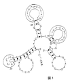
実施例 5 特異的核酸分子またはアプタマーの構造モデル
図1は特異的ヌクレオチド配列またはアプタマー配列の仮説の二次構造を示している。種々の「ヘアピンループ」または「ふくらみ」中に位置する活性結合部位が示されている。図に示された核酸分子はリンカー部分(n)を介して5’末端がビオチン標識(BBB)されている。黒丸は塩基対を表す。位置情報は、分子中のヌクレオチドの位置を示す。
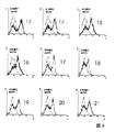
実施例 6 クレームの核酸分子のヒストグラム
図2および図3は見出された核酸分子のFACSヒストグラムを表す。蛍光強度(F)を横軸に任意対数単位でプロットし、縦軸は任意線形単位でプロットされたシグナル産生結果(E)の数を示す。このヒストグラムは核酸分子配列番号1から21の標的組織への結合特異性を示す。ヒストグラム中の核酸分子のプロットされた線(配列番号1から21、太字)の蛍光の強さが増加する方向への移動は、対照と比較してこれらの分子の標的組織に対する親和性が高いことを表している。示された対照は、それぞれの場合においてFITCに同様に連結された非特異的1本鎖DNAを用いた場合の蛍光である(非特異的ssDNA,灰色)。このように21個の核酸分子すべてが、使用した内皮細胞に高い親和性を有するという特徴があった。
実施例 7 選択ラウンドの複合プール中の核酸分子のヒストグラム
図4において第2(−−−−)、第4(・・・・)および第7(−・−・−・−・−)選択ラウンド由来のアプタマーのプールのFACSヒストグラムが重ねてある。プロットされた選択ラウンドのヒストグラムの線のますます強い蛍光方向への移動は、連続的な選択ラウンドのプール中の、結合核酸分子(アプタマー)の特異性が高まっていることを示す。使用した対照はFITCに同様に連結された非特異的1本鎖DNAを用いた場合の蛍光シグナルである(非特異的ssDNA,灰色)。
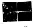
実施例 8 配列番号9のヌクレオチド配列を含む核酸分子の腫瘍−および「pigpen」−特異的アプタマーとしての同定
発明者らが行った組織分析から、また実施例3の最後の部分の記載から、ラット脳のグリア芽腫の組織切片中で、すべての新規核酸分子のうち配列番号9のヌクレオチド配列を含む核酸分子が最も強い蛍光シグナルを示し、かつそれは固形腫瘍が成長している領域に特異的であることが分かった。これは図5に示されている。図5の写真Aでは複雑なグリア芽腫中に埋め込まれている微小管の選択的染色が示されている。BにはDAPIを用いる細胞核の対比染色が示されており、細胞に富む腫瘍領域では上記のアプタマーにより微小管が選択的に標識されるが(写真の右側の明るい糸状構造が染色された微小管を表す)、腫瘍周囲領域の脈管組織は標識されないことを示している(写真の左側)。Cでは、DAKO, Hamburg, Germanyで市販されている内皮CD−31 mAbによる二重染色が示されており、両方の蛍光シグナルが重なり、またアプタマー陽性細胞を内皮細胞として決定している。Dでは、モノクローナル抗体CD−31および非特異的配列−ランダムアプタマーによる染色を行ったが、内皮細胞または脈管付随組織の二重染色とはならず、シグナルの重なりもなかった、Eにおいてはピペットチップを用いて合わさった細胞層を傷つけて採取した内皮細胞培養物の染色が示されている。ここでアプタマーは密集前の創傷循環領域において、内皮細胞への結合を有意に増加させた。目盛り線は50μmを表す。
【0029】
本試験は配列番号9のヌクレオチド配列を含むアプタマーが病的な微小管の複雑な構築体を特異的に染色することを示す。DAPIを用いる細胞の核の対比染色は細胞に富む腫瘍領域中の微小管を選択的に標的化するが腫瘍周囲領域の脈管組織は標的化しないことを示す。発明者らはさらなる実験で(本明細書には示していない)それぞれのアプタマーでラットの正常脳を染色したところ、内皮細胞または脈管付随組織のいずれもが染色されず、蛍光顕微鏡検査法で検出できなかった。腫瘍組織切片を内皮細胞に向けられたマウスIgG、例えば上記のCD−31抗体で染色し、次いでそれぞれのアプタマーで標識することにより、アプタマーの標的が内皮細胞であることが確認された。創傷内皮細胞組織を染色すると、この病変領域においてアプタマーの分子標的の著しいアップレギュレーションが示され、すなわちこれは単一層が合わさった領域に比べて増殖が高まっているとことを特徴とする領域を表す。
【0030】
配列番号9のヌクレオチド配列を含むアプタマーにより認識された標的を同定するために以下の工程を用いた:1mg(100μl)の磁気ストレプトアビジンビーズを1mlの選択バッファー中でインキュベート(30分、室温)(MWG−Biotech AG, Ebersberg, Germany)して200pmolのトリプルビオチン化(trB)アプタマーによりコーティングした。対照として100μlの磁気ビーズを200pmolの非選択FITC−ssDNA(trB−96−nt)でコーティングした。Klocker ら, Journal of Neuroscience 19, 8517−8527 (1999)の記載に従って1.5×10⁹個のYpen−1内皮細胞をプロテアーゼ阻害剤存在下で溶解させた。遠心分離後、タンパク質のペレットを400μlの選択バッファーに再懸濁し超音波処理(0℃、20秒)し、非特異的競合剤として100倍過剰のtRNA(20nmol)を添加し、アプタマーで被覆した磁気ビーズとともに選択バッファー中でインキュベートした(総容量1.5ml、0℃、15分)。このタンパク質−アプタマー−磁気ビーズ複合体を磁石スタンド中で除去し、5回洗浄した(1回目の洗浄:150mMのNaClを含む選択バッファー1ml;2−5回目の洗浄:100mMのNaClおよび2nmolのtRNAを含む選択バッファー200μl)。30μlの1M NaCl中でインキュベート(0℃、30分)することによりアプタマーでコーティングしたビーズからタンパク質を除去し、クーマシーブルーで染色後PAGEにより分析した。
【0031】
図6は本実験の結果を示す。レーンAには分子マーカー、Bレーンには配列番号9のヌクレオチド配列を含むアプタマーを用いて単離した標的、Cレーンには対照として非選択ssDNA(96−nt)を用いて精製した単離物質、Dレーンには処理していない内皮細胞が表されている。それぞれのアプタマーを含む反応により単離された標的バンドはアスタリスクにより示され、それは67kDaのタンパク質であることが同定された。
【0032】
この標的タンパク質は以下のように同定された:
それぞれのゲル片をゲルから切り取り、例えばShevchenkoら, Anal. Chem. 68, 850−858 (1996)の記載に従ってトリプシン消化を行った。改変した点を以下に示す:切り取ったバンドを完全に脱染して、25mM炭酸水素アンモニウム(pH 8.1)中67ng/μlの濃度のブタトリプシン(配列決定グレード、修飾、Promega, Manheim Germany)を用いて37℃で3時間消化した。ペプチドマッピングおよびタンデム質量分析によるトリプシン断片の配列決定に先立って、50%トリフルオロ酢酸/50%水溶液、次いで50%トリフルオロ酢酸/50%アセトニトリル溶液を用いて該ペプチド混合物をゲルから抽出する。該抽出物を真空乾燥させる。乾燥させたペプチドを0.1%トリフルオロ酢酸に再溶解させ、C18球形シリカ逆相物質(ZipTipC18 TM, Millipore)を充填した既製のピペットチップを用いて精製する。50%メタノール/1%ギ酸10μlを用いて該ペプチドを溶出させ、ナノエレクトロスプレータンデム質量分析(Q−Tof, Midromass, Manchester, England)により配列決定する。
【0033】
発明者らは配列番号9のヌクレオチド配列を含むアプタマーにより認識される標的に対し論じられた分析を行った。そうすることにより、質量分析またはタンデム質量分析それぞれにより該タンパク質の3つのトリプシンペプチド断片が分析、配列決定された。その結果、かかるアプタマーにより検出された標的はマウス「pigpen」タンパク質に対する相同体であることが分かった。
【図面の簡単な説明】
【図1】
それぞれ特異的核酸分子またはアプタマーの二次構造の模式図である。
【図2】
クレームの核酸分子のフローサイトメトリーによる核酸分子の特徴を示す。
【図3】
クレームの核酸分子のフローサイトメトリーによる核酸分子の特徴を示す。
【図4】
連続的な選択実験のプール中の該特異的核酸分子またはアプタマーの濃縮を示す。
【図5】
配列番号9のヌクレオチド配列を有する該核酸分子の、ラットグリア芽腫の凍結組織切片の染色を示す。
【図6】
配列番号9のヌクレオチドを有する該核酸分子により認識された標的を分析するためのクーマシーブルー染色ポリアクリルアミドゲルを示す。
【配列表】
The present invention relates to nucleic acid molecules that selectively bind to or label endothelial cells, methods for identifying tumor diseases, nucleic acid molecules used in such methods, and methods of using such nucleic acid molecules.
[0001]
Such methods and the nucleic acid molecules used therein are known and are also called aptamers. A related review is Osborne et al., "Aptamers as therapeutic and diagnostic reagents: problems and products", Curr. Opin. Chem. Biol. 1997, 1: 5-9. Can be found in
Aptamers are high affinity RNA and DNA oligonucleotides or polynucleotides that have a high affinity for a target molecule due to their specific spatial structure. In order to find such aptamers, a combinatorial approach of randomly and enzymatically producing a large number of oligonucleotides having completely different sequences and secondary structures, and then selecting and enriching oligonucleotides having high affinity for a target molecule. Is often used. This technology is also called SELEX (systematic evolution of ligands by exponential enrichment).
[0002]
Where the primary structure is known, such oligo-polynucleotides (aptamers) may be synthesized, for example, using a DNA / RNA synthesizer.
The target structure for the aptamer may be a receptor or other protein, lipid structure or carbohydrate structure. Theophylline (see Jenison et al., "High-resolution @ molecular discrimination by RNA", Science 1994; 263 (5152): 1425-9), ATP (Tang et al., "Ratio design. Cosmochemistry. (6): 453-9), arginine (Geiger et al., "RNA aptamers that bind L-arginine with sub-micromolar dissociation constants and high-energy sci- ent. 36) Aptamers exhibiting high affinity for the small molecule UNA is already found. Similarly, protein binding aptamers that do not normally bind to any nucleic acid in the natural environment have been found. Morris et al., "High affinity ligands from in vitro selection: Complex targets"; Proc. Natl. Acad. Sci. USA 1998; 95: 2902-2907 have successfully developed aptamers directed against cell membranes.
[0003]
The current state of the art has the disadvantage that the authors of the cited documents do not show the complete sequence of the nucleic acid molecule that binds to or labels the composite biomaterial. Morris et al., Who study labels with relatively high molecular weight structures, merely show sequence motifs. According to this publication, nucleic acid molecules containing such sequence motifs bind to vesicles artificially generated from red blood cell membranes (red blood cell ghosts).
In view of the above, one object of the present invention is to provide nucleic acid molecules other than the type initially described, in particular nucleic acid molecules which can selectively bind and label endothelial cells.
[0004]
According to the invention, this object is achieved by a nucleic acid molecule comprising at least one of the nucleotide sequences SEQ ID No. 1 to SEQ ID No. 21 of the attached sequence listing.
As a result, the object underlying the invention is completely achieved. It has been suggested by the inventors that nucleic acid molecules comprising at least one of the above nucleotide sequences recognize and bind not only to natural biological materials but also to parts thereof produced by technical methods, and are specific for endothelial cells. This is because they recognize that. According to our findings, nucleic acid molecules comprising at least one of the sequences in the table are distinguished by a high specificity for the biological material used.
[0005]
However, nucleic acid molecules having one of the nucleotide sequences SEQ ID NO: 1 to SEQ ID NO: 21 of the attached sequence listing are preferred.
Our data show that such objectives underlying the present invention are fully achieved when a nucleic acid molecule having one of the claimed nucleotide sequences is used in the method described at the outset. Surprisingly, the secondary structure of the nucleic acid molecule, predetermined by the nucleotide sequence, is sufficient to confer highly specific binding to the biological material.
[0006]
It is further preferred that at least one nucleotide of the non-functional part in the nucleic acid molecule is substituted or deleted.
This is because the inventors have found that a particular functional region in the sequence of a particular nucleic acid molecule is responsible for selective binding to biological tissue. These regions are located with the folded sequence in the "hairpin loop" or "bulge" secondary structure. When the base sequences of these functional regions match, two nucleic acid molecules or aptamers whose non-functional portions have different sequences can bind to the same structure. Such differences include both nucleotide exchanges and deletions of single or multiple nucleotides, as well as chain truncations or modifications.
[0007]
A further object of the present invention is a method for selectively binding to or labeling endothelial cells using a nucleic acid molecule comprising one of the nucleotide sequences SEQ ID NO: 1 to SEQ ID NO: 21, These sequences include sequence variations in which at least one nucleotide of the non-functional portion of the nucleic acid molecule has been substituted or deleted.
The sequences of SEQ ID NO: 1 to SEQ ID NO: 21 specifically bind to endothelial cells. These cells lining the luminal side of blood vessels play an important role, inter alia, in angiogenesis, angiogenesis and transcytosis of substances via the blood-tissue barrier. In this context, such methods are particularly suitable for studying the differentiation of endothelial cells and the labeling of tumor-supplying vessels or specific lesions of inflammatory endothelial cells.
[0008]
It is also preferred if the nucleic acid molecule is linked to a therapeutic agent.
The claimed nucleic acid molecule has the potential to function as a target-specific carrier for transporting a therapeutic agent to its target location. Because this “drug targeting” avoids most of the undesirable interactions with non-pathogenic tissues, we use target-specific therapeutic agents, especially for reduced doses or unwanted side effects. We expect a thorough reduction.
[0009]
Suitable target tissues are in particular tumor tissues, since relatively recent cancer research studies have shown that blocking the blood supply of tumors is a promising approach for treatment (reviewed literature Ceresh, DA, "Death to a blood vessel, death to a tumour", Nat. Med. 1998, 4 (4): 395-6; Molema et al., "Rocking the basics of the breakdown of solids. Today 1998, 19 (9): 392-4). Such a method can also be advantageously performed using the novel nucleic acid sequences.
[0010]
During the experiments leading to the present invention, the inventors have found that a tumor-specific tissue can be selectively identified by a nucleic acid molecule comprising the nucleotide sequence of SEQ ID NO: 9 in the attached sequence listing. Thus, the use of these nucleic acid molecules enables highly specific discrimination between denatured tumor tissue and healthy tissue. Thus, we provide a use that provides a solid basis for treatment targeted to each target.
A further object of the invention in this context is the use of a nucleic acid molecule comprising or comprising the nucleotide sequence of SEQ ID NO: 1 of the attached sequence listing for the detection of tumor diseases and respective methods for the detection of tumor diseases,
a) prepare biological material,
b) detecting a tumor marker;
Process. However, this detection is performed using a nucleic acid molecule comprising or comprising the nucleotide sequence of SEQ ID NO: 1 in the attached sequence listing.
[0011]
Further studies by the inventors have found that a nucleic acid molecule having the nucleotide sequence of SEQ ID NO: 1 in the attached Sequence Listing causes a discrimination between tumor tissue and healthy tissue via a homologue to the murine / bovine protein "pigpen". Was. This protein is a relatively unknown endothelial protein to date and has a molecular weight of 67 kDa. Moreover, its association with tumor tissue is currently unknown. Thus, although this protein has been previously described as a carbohydrate binding protein with an RNA binding domain and a transactivation domain, it may bind to the nuclear membrane; see, for example, Alliegro et al., "Protein heterogeneity in See the coiled body comparison ", Experimental Cell Research 239, 60-68 (1998).
[0012]
In view of the above, one of the objects of the present invention is the use of a nucleic acid molecule comprising the nucleotide sequence of SEQ ID NO: 9 in the attached sequence listing for the detection of a tumor disease, the detection being directed against the murine / bovine protein "pigpen". Occurs through the human homolog. Another object is a method of detecting a tumor disease via a human homolog to the murine / bovine protein "pigpen".
A further application of the claimed nucleic acid molecules is the delivery of therapeutic agents to inflammatory endothelial lesions. Possible therapeutic agents are, for example, anti-angiogenic or anti-inflammatory substances.
[0013]
By extending the specific nucleic acid molecules of the claims with the function-carrying DNA segments, it is also possible to prepare locally active immunotherapeutics. Contemplated herein is the use of "CpG oligos", which have enormous immunostimulatory activity.
The inventors of the present application have recognized for the first time that the claimed nucleic acid molecules can be used in the treatment of the above-mentioned conditions.
It is particularly preferred that such nucleic acid molecules are also linked to a diagnostic agent.
[0014]
Such modified nucleic acid molecules or aptamers have a number of advantages over commonly used monoclonal antibodies in diagnosis and drug targeting as described above. Due to the formation of sequence-based secondary structures, the repertoire of potential binding ligands is substantially broader than the immune repertoire available for preparing monoclonal antibodies.
In addition, aptamers are obtained substantially faster and are cost effective. Millions of potential ligands can be tested in 3-4 weeks. In contrast, preparation of antibody-producing hybridoma cells must begin with vaccination of experimental animals. This vaccination lasts several weeks. In addition, it takes months to test 200 to 300 hybridoma cell lines.
[0015]
Because the existence of extracellular oligonucleotides / polynucleotides that perform functions due to their structure in organisms is not known, these nucleic acid molecules or aptamers also provide a "normal" immune system that escaped antibody recognition. It also has the potential to recognize "abnormal" epitopes.
A particular advantage of the claimed nucleic acid molecules is that they exhibit binding specificity and affinity for useful monoclonal antibodies. They are therefore particularly suitable for use in diagnostic or drug targeting methods. With respect to diagnostic methods, contemplated methods are particularly those where the diagnostic agent is used outside the human body.
[0016]
Particularly suitable diagnostic agents within the framework of diagnostic methods include, for example, biotin, digoxigenin and its derivatives, including fluorescent compounds such as fluorescein isothiocyanate (FITC), enzyme markers, infrared markers, and chelating agents. Therefore, a method in which the diagnostic agent linked to the nucleic acid molecule is selected from the group comprising the above substances is particularly preferred.
Furthermore, the use of the above-described nucleic acid molecules in histological examination of tissue sections also forms part of the present invention.
[0017]
The inventors have recognized that the claimed nucleic acid molecules can be used directly as histological agents, which allows for the in situ localization of biological target structures in tissues. A particular advantage of such a use is the first mentioned specificity of the claimed nucleic acid molecules for complex biological materials, preferably for endothelial cells. This allows for specific staining of these tissues without interfering with background signals or non-specific cross-reactivity.
[0018]
When the nucleic acid molecule selectively binds to a natural complex biological material, the use of the above-described nucleic acid molecule is particularly preferred in any of the described methods.
We provide for the first time a nucleic acid molecule that binds to complex natural biological tissues and endothelial cells, respectively. In contrast, the sequence motif provided by Morris et al. Is part of the nucleic acid molecule or aptamer used therein, but binds to vesicles artificially generated from red blood cell membranes. The integrity of the membrane produced in this way need not necessarily correspond to the physiological red blood cell membrane. Selecting the red blood cell component as a model for complex biological tissue is also not appropriate here. Erythrocytes have the characteristic of being highly differentiated. Erythrocytes differ in many properties from other typical cells of an organism: they do not synthesize proteins, are not integrated into cell aggregates, and are highly specialized in oxygen transport.
[0019]
We used natural cells of the endothelial cell line as biological material to select specific nucleic acid molecules. This has the advantage that these nucleic acid molecules can also be used reliably in vivo.
To enrich functional nucleic acid molecules, we used a method consisting of the following steps:
a) incubating different nucleic acid molecule libraries with the target structure;
b) selecting and enriching the functional nucleic acid molecule;
c) Isolate the selected nucleic acid molecule. At this time, a fluorescent label of the selected nucleic acid molecule is used to control the concentration.
[0020]
We believe that the nucleic acid molecule selected to find a specific nucleic acid molecule or aptamer typically does not need to be enriched by a radioactive label, but believes that fluorescent labels can be used in combination with a suitable detection method. . Although the specific binding properties of the functional nucleic acid molecule were not expected to be retained by such fluorescent labels, it was preferably retained at the 5 'or 3' end of the sequence. In the known method performed until then, the functional nucleic acid molecules in the pool of successive rounds of selection were controlled exclusively by scintillation measurements of radioactive dNTPs incorporated during enrichment.
[0021]
The consequences associated with research dealing with radioactive materials are generally known, such as the construction of certain isotope laboratories, waste management issues, strict regulations on the transport and storage of radioactive materials, the dangers of compromising health, and high costs. is there. These problems can be avoided by using alternative detection techniques such as fluorescent labels.
The new enrichment method has proven to be very economical. Repeated cycles of FACS measurements not only allow simultaneous selection of intact endothelial cells against a variety of targets embedded in their native environment, but also individual specific in situ by FACS using fluorescent labels. The evaluation of the binding aptamer becomes possible.
[0022]
Such features described above and further below can be used not only in the combinations indicated in each case, but also in other combinations or alone, within the scope of the invention.
Hereinafter, the present invention will be described with reference to examples showing further features and advantages.
Example 1内皮 Endothelial cells as biological target structures
The target structure used for the specific aptamer selection is the cell line YPEN-1 (CRL-2222, American Type Culture Collection, Manassas, USA), a rat-derived endothelial prostate cell line. The cells are collected by scraping the endothelial cell layer before confluence. After staining with trypan blue, the number of intact cells is counted in a Neubauer counting chamber to estimate the cell concentration.
Example 2Starting library (ssDNA library) and primers
A synthetic initiation library from which specific nucleic acid molecules or aptamers are selected (MWG Biotech AG, Ebersberg, Germany) consists of approximately 96-mer polynucleotides and is approximately 60 nucleotides by 18 nucleotides whose primer hybridization site is known. Are sandwiched on both sides of the randomly linked base (60N) (5′-ATA CCA GCT TAT TCA ATT- [60N] -AGA TAG TAA GTG CAA TCT-3 ′). Specific nucleic acid molecules are FTIC-labeled and biotin-labeled HPLC-purified primers (5'-FITC-18C-ATA CCA GCT TAT TCA ATT-3 'and 5') synthesized by solid phase synthesis (Operon Technologies, Inc., Alamenda USA). -BBB-AGA TTGCAC TTA CTA TCT-3 ', where 18C is an 18-carbon ethylene glycol spacer, and "FITC" and "B" are FTIC molecules and biotin molecules, respectively), and are amplified by the PCR method described below.
Example 3取得 Acquisition of nucleic acid molecules or aptamers specific to endothelial cells
Several repetitive selections are required to obtain a specific nucleic acid molecule.
[0023]
The following steps were performed under light protection conditions.
1st round
Starting library of Example 2 (1 × 10FifteenAbout 1.7 mol of DNA of the sequence) was selected using a selection buffer (50 mM Tris-HCl (pH 7.4), 5 mM KCl, 100 mM NaCl, 1 mM MgCl).2, 0.1% NaN3) (1 ml for the first round, 200 ml for subsequent rounds). The DNA is denatured (80 ° C, 10 minutes) and regenerated at 0 ° C, 10 minutes. A 5 molar excess of tRNA (Gibco, Karlsruhe, Germany) and bovine serum albumin (BSA) are added in each case to reduce non-specific binding of the polynucleotide to the cell surface and to the reaction vessel wall. To remove DNA molecules that show a high affinity for irrelevant surface proteins, the DNA solution is7 With 5 N9 microglial cells at 37 ° C / 5% CO2For 30 minutes.
[0024]
The microglial cells are removed by centrifugation and the supernatant is6YPEN endothelial cells and 37 ° C / 5% CO2Incubate with After centrifugation with 1 ml of selection buffer (containing 0.2% BSA) and washing three times each time, a nucleic acid molecule bound to endothelial cells is used as a template (tag polymerase and dNTP) to amplify aptamers by PCR. From Promega, Mannheim, Germany).
The PCR method is described in Craemri and Stemmer, "10 (20) -fold aptamer library amplification with gel purification", Nucl. Acids Res. 1993; 21 (18): Performed under the conditions of 4410. The primers used are the FITC and biotin-labeled primers described above.
Obtaining FITC-labeled single strand
For analysis by fluorescence cytometry, FITC-labeled ssDNA is prepared as follows: In a magnetic stand, magnetize using an appropriate amount of magnetic streptavidin beads (Dynabeads M-280 Streptavidin; Dynal Hamburg, Germany). The FITC-complexed ssDNA is purified from the biotinylated strand by isolating the dsDNA PCR product in the folded strand, then alkali-denatured according to the manufacturer's instructions, and harvested in a final volume of 200 ml selection buffer. Subsequently, the ssDNA solution obtained in each case serves as a pool for a selection round.
2nd and 3rd rounds
Reincubate the new FITCssDNA pool against microglia in a polystyrene reaction tube. The selection was 1.5 × 10 (also in a polystyrene reaction tube).5Perform on individual endothelial cells. After incubation at 37 ° C. for 30 minutes, the cells are separated from sequences with low specificity DNA binding and its 5 ′ modification causing loss of binding by three washes with 1 ml of selection buffer supplemented with 0.2% BSA. . The fluorescence of the endothelial cell-nucleic acid molecule complex is measured with a flow cytometer (FACS). The ssDNA is amplified as in the previous round.
4th to 8th selection round
After re-preincubation of microglial cells (described above), 1 × 10⁵Such selection is performed on individual endothelial cells.
[0025]
Changing the polystyrene reaction tube to a polypropylene reaction tube ensures that the separation matrix binding sequence is removed. After incubation for 30 minutes and washing of the cells (according to the previous round), the fluorescence intensity is measured on a flow cytometer (counting 5000 cells per selection round). The cell-associated nucleic acid molecule is PCR amplified as described above.
Acquisition of individual clones from pool in round 8
Specific nucleic acid molecules from the seventh round of selection (the eighth round pool) are PCR amplified using unmodified primers and cloned into E. coli (TA-Cloning, Invitrogen Groningen, The). Etherlands). The plasmid of each clone is isolated by alkaline lysis and the insert (PCR product) is PCR amplified using modified primers as in the selection round. A single strand is obtained as described above.
[0026]
Using this FITC aptamer solution, flow cytometry (FACS) and tissue binding test are performed to identify the binding specificity of each sequence.
FACS measurement
The specific binding of aptamers and the specific binding of individual clones in successive rounds of selection is determined by FACS (fluorescence-activated cell sorting).
[0027]
Cells (as a result of signal production) emit electromagnetic radiation in proportion to the intensity of their fluorescent label. This radiation is detected and quantified. Thus, a result of the amount of fluorescently labeled ligand bound to cells by the radiation is obtained. The fluorescence intensity is plotted on a histogram as a function of the signal production result. A strong bias in the direction of increased fluorescence is a measure of the high affinity of the specific nucleic acid molecule or aptamer for the biological target materials and endothelial cells used herein.
(A) FACS analysis of FITC-linked aptamers in successive rounds of selection
The specific binding of the FITC-linked aptamer is measured in the second and subsequent rounds of selection. After incubating the nucleic acid molecule or aptamer with endothelial cells (as described above), the endothelial cells are washed twice with selection buffer (with 0.2% BSA) and cytofluorescence is measured by FACS.
(B) FACS analysis of specific binding of individual FITC-linked clones
5 × 10 for this purpose4Of YPEN endothelial cells were preincubated with selection buffer (1 mg / ml tRNA added; 20 minutes, 0 ° C.), and the above FITC-ssDNA solution (1 mg / ml tRNA added; 37 ° C./5% CO 2)2/ 30 min). The cells are washed twice with selection buffer (with 0.2% BSA) and cell fluorescence is measured by FACS.
Tissue bonding test of tissue section
The binding of each of the resulting FITC-modified nucleic acid molecules or aptamers is tested by tissue binding studies on frozen tissue sections of rat glioblastoma. Pre-incubate tissue sections with selection buffer containing 1 mg / ml tRNA (20 min, 40 ° C.). Pre-incubated tissue sections are coated with the FITC-aptamer solution described above (with 1 mg / ml tRNA added) and incubated for 40 minutes at room temperature. The tissue sections are washed twice with selection buffer and immunofluorescence is quantified by fluorescence microscopy.
[0028]
Highly specific nucleic acid molecules or aptamers identify themselves by selective staining of newly formed microtubules in glioblastoma tissue and show minimal cross-reactivity with other non-endothelial cell tissues Was. Thus, the nucleic acid molecule is particularly suitable as a tissue probe for endothelial cell staining.
Example 4(Endothelial cell)-a nucleic acid molecule or aptamer specific to a specific antigen
The nucleotide sequences shown below are obtained by the above method and form part of the present invention. Primer hybridization sites are located 5 'and 3' to the actual aptamer sequence (bold).
Nucleic acid molecule 1 (primary structure)
5'-ATA CCA GCT TAT TCA ATT GGA CCC AAG GTA TTT CCT CGC GTT CGT
AAT CAG TGG GAG TGG TGT TTG TGT TCC GGT GTG AGA TAG TAA GTG CAA
TCT-3 '
Nucleic acid molecule 2 (primary structure)
5'-ATA CCA GCT TAT TCA ATT CTA CCA GAC TGT CTT TCA CCC TGC GCC
GTT GTG GTC TGT TCG TTG TTC TAG TTG TTT TC AGA TAG TAG TAA GTG CAA
TCT-3 '
Nucleic acid molecule 3 (primary structure)
5'-ATA CCA GCT TAT TCA ATT GTC AGC TTT TTA GTG ATT TTG GGT TTT
TTG GTG TAC GTC CCT GTA AAT HAG TTT CAG TCG AGA TAG TAA GTG CAA
TCT-3 '
Nucleic acid molecule 4 (primary structure)
5'-ATA CCA GCT TAT TCA ATT GAG TGC TGA TTC CCG TTT CTC TCT GGT
ATC GAA TTG AGG TCG TTT GTG TGT GAG TTG GCT AGA TAG TAA GTG CAA
TCT-3 '
Nucleic acid molecule 5 (primary structure)
ATA CCA GCT TAT TCA ATT CCA ACA CCA TAA CCT TTC TTT GAC CTG
ACT TTA GCC GTA ATG TAT TTG GGC CAT CCC CTT AGA TAG TAA GTG CAA
TCT-3 '
Nucleic acid molecule 6 (primary structure)
5 '-ATA CCA GCT TAT TCA ATT GGG TGG AGA TAG TAG GTG CAA TCT ATA
CCA GCT TAT TCA ATT GCC CCG ATT TGG ATG TAA TTA TTG CGC GTG TAT
TTT TGA TTG TAT AAA GTG TTG CTA CA AGA TAG TAA GTG CAA TCT-3 '
Nucleic acid molecule 7 (primary structure)
5 '--- ATA CCA GCT TAT TCA ATT AGG CAG ATG AGA AGT TAA GGC GGT GCT
ATA GAT GGA CCA TTT AGG ATT TTA TGG TTG GGC GT AGA TAG TAA GTG
CAA TCT-3 '
Nucleic acid molecule 8 (primary structure)
5 '--- ATA CCA GCT TAT TCA ATT CCC GCC TTT ACT TGG GAG ATT ATC ACC
GCG GTA TAT AAA TAC TGT TCG GAG TTC TGT GT AGA TAG TAA GTG CAA
TCT-3 '
Nucleic acid molecule 9 (primary structure)
5 '--- ATA CCA GCT TAT TCA ATT AGG CGG TGC ATT GTG GTT GGT AGT ATA
CAT GAG GTT TGG TTG AGA CTA GTC GCA AGA TAT AGA TAG TAA GTG CAA
TCT--3 '
Nucleic acid molecule 10 (primary structure)
5 '--- ATA CCA GCT TAT TCA ATT CTG TTG GAC ATT CAA AAG ACT AGT TCA
CGT CCG TTG CCC ATT CTT CCC TTT GTT GAC TGC T AGA TAG TAA GTG
CAA TCT-3 '
Nucleic acid molecule 11 (primary structure)
5 '--- ATA CCA GCT TAT TCA ATT CGG CTA GGC TCC ATT AAG GGT GAC TTA
TGG GCC AAA GTC CCG TGC TTG TTC GTG TGG GTG AGA TAG TAA GTG CAA
TCT-3 '
Nucleic acid molecule 12 (primary structure)
5 '-ATA CCA GCT TAT TCA ATT CCA AAG GTA AAC CGC ATA ATA ATA AGG GTA
TGT ATT AAA TTG TGT GGT GAT GAC TGA TGC CAT A AGA TAG TAA GTG
CAA TCT-3 '
Nucleic acid molecule 13 (primary structure)
5'-ATA CCA GCT TAT TCA ATT GAG GAT CAC CTG CTC TGC CAC CCT TTT
TAA CGT GGG GTT ACA TTT GCT GAA GGG CTT G AGA TAG TAG TAA GTG CAA
TCT-3 '
Nucleic acid molecule 14 (primary structure)
5'-ATA CCA GCT TAT TCA ATT GTA CCA GCC GAG ATC TTT TTT GAC GAT
ATG TGT TTT TTT TGA GGT GTT GAG TTT AGT GTG AGA TAG TAA GTG CAA
TCT-3 '
Nucleic acid molecule 15 (primary structure)
5'-ATA CCA GCT TAT TCA ATT GCG ATA AAT TTT GCT AAG TGC GGT CAA
GAC TGT GTT CGT GT AGA TAG TAA GTG CAA TCT-3 '
Nucleic acid molecule 16 (primary structure)
5'-ATA CCA GCT TAT TCA ATT GCC ATA CCG TAG TTA GCA TAT GTA GTG
TT AGA TAG TAA GTG CAA TCT-3 '
Nucleic acid molecule 17 (primary structure)
5'-ATA CCA GCT TAT TCA ATT GCG CCT TTA AAT ATA ACC CGA GTG CTT
TGT TTG AAC TGG TGT TCC GGA TGG CCT GTG TTG AGA TAG TAA GTG CAA
TCT-3 '
Nucleic acid molecule 18 (primary structure)
5'-ATA CCA GCT TAT TCA ATT GCT TGC ATC GTC ATT ATG AGG TGG ATT
CAA CTG TTT TTG ACT TTT TGC CCC TGG ACG CTG AGA TAG TAA GTG CAA
TCT-3 '
Nucleic acid molecule 19 (primary structure)
5'-ATA CCA GCT TAT TCA ATT GGT TAC CAT TCT GGT GGG ACC CGT GTT
GCC TGG ATG TGT TTT AGT TTT TTT GGT GTT TT AGA TAG TAA GTG CAA
TCT-3 '
Nucleic acid molecule 20 (primary structure)
5 'ATA CCA GCT TAT TCA ATT GGT TGG AGA CCT TAT TGG CAG CAT GCA
GGG CCC TCA GCT GTG CAA CCC CGG TTT CCG TT AGA TAG TAA GTG CAA
TCT-3 '
Nucleic acid molecule 21 (primary structure)
5'-ATA CCA GCT TAT TCA ATT CAC ACA TGC GCC TTA GTT AGC CCT GGT
TGT TG AGA TAG TAA GTG CAA TCT-3 '
Example 5構造 Structural model of specific nucleic acid molecule or aptamer
FIG. 1 shows the hypothetical secondary structure of a specific nucleotide sequence or aptamer sequence. Active binding sites located in various "hairpin loops" or "bulges" are indicated. The nucleic acid molecule shown in the figure is biotin-labeled (BBB) at the 5 'end via a linker portion (n). Black circles represent base pairs. The position information indicates the position of the nucleotide in the molecule.
Example 6ヒ ス ト グ ラ ム Histogram of the claimed nucleic acid molecule
FIGS. 2 and 3 show FACS histograms of the found nucleic acid molecules. The fluorescence intensity (F) is plotted in arbitrary logarithmic units on the horizontal axis, and the vertical axis indicates the number of signal production results (E) plotted in arbitrary linear units. This histogram shows the binding specificity of nucleic acid molecules SEQ ID NOS: 1 to 21 to the target tissue. Movement of the plotted lines of nucleic acid molecules in the histogram (SEQ ID NOs: 1 to 21, bold) in the direction of increasing fluorescence intensity indicates that these molecules have a higher affinity for the target tissue as compared to the control. Is represented. The control shown is the fluorescence in each case using non-specific single-stranded DNA also linked to FITC (non-specific ssDNA, gray). Thus, all 21 nucleic acid molecules were characterized by having high affinity for the endothelial cells used.
Example 7ヒ ス ト グ ラ ム Histogram of nucleic acid molecules in the composite pool of the selection round
In FIG. 4 the FACS histograms of the pools of aptamers from the second (----), fourth (-...) and seventh (-...-...-) selection rounds are overlaid. Movement of the plotted selection round histogram lines in the direction of increasingly strong fluorescence indicates an increased specificity of the bound nucleic acid molecule (aptamer) in the pool of successive selection rounds. The control used is the fluorescent signal when using non-specific single-stranded DNA similarly linked to FITC (non-specific ssDNA, gray).
Example 8同 定 Identification of nucleic acid molecules comprising the nucleotide sequence of SEQ ID NO: 9 as tumor- and “pigpen” -specific aptamers
From the tissue analysis performed by the inventors and from the description in the last part of Example 3, in a rat brain glioblastoma tissue section, a nucleic acid comprising the nucleotide sequence of SEQ ID NO: 9 among all novel nucleic acid molecules. The molecule showed the strongest fluorescent signal and was found to be specific to the area where the solid tumor was growing. This is shown in FIG. Photo A of FIG. 5 shows selective staining of microtubules embedded in complex glioblastoma. B shows the counterstaining of the cell nucleus using DAPI, and in the cell-rich tumor area, the microtubules are selectively labeled by the aptamer (the microtubules stained with the bright filamentous structure on the right side of the photograph). ), Indicating that the vascular tissue in the peri-tumor region is not labeled (left side of the photograph). In C, double staining with endothelial CD-31 mAb, commercially available from DAKO, Hamburg, Germany, is shown, both fluorescent signals overlap and aptamer positive cells are determined as endothelial cells. In D, staining with monoclonal antibody CD-31 and non-specific sequence-random aptamer did not result in double staining of endothelial cells or vascular associated tissue and no signal overlap. In E, pipette Shown is the staining of endothelial cell cultures harvested using a chip to injure the combined cell layers. Here, the aptamer significantly increased the binding to endothelial cells in the wound circulation area before confluence. The scale line represents 50 μm.
[0029]
This test shows that the aptamer comprising the nucleotide sequence of SEQ ID NO: 9 specifically stains the pathological microtubule complex construct. Counterstaining of the nuclei of cells with DAPI indicates that it selectively targets microtubules in the cell-rich tumor region but not vascular tissue in the peri-tumor region. In a further experiment, the inventors stained normal rat brains with the respective aptamers (not shown here) and found that neither endothelial cells nor vascular associated tissues were stained and fluorescence microscopy. Could not be detected. Tumor tissue sections were stained with mouse IgG directed against endothelial cells, such as the CD-31 antibody described above, and then labeled with each aptamer, confirming that the aptamer target was endothelial cells. Staining of wound endothelial cell tissue shows a marked up-regulation of aptamer molecular targets in this lesion area, which represents an area characterized by increased proliferation compared to a monolayered area .
[0030]
The following steps were used to identify the target recognized by the aptamer containing the nucleotide sequence of SEQ ID NO: 9: 1 mg (100 μl) magnetic streptavidin beads were incubated in 1 ml of selection buffer (30 min, room temperature) ( (MWG-Biotech AG, Ebersberg, Germany) and coated with 200 pmol of triple biotinylated (trB) aptamer. As a control, 100 μl of magnetic beads were coated with 200 pmol of non-selective FITC-ssDNA (trB-96-nt). Krocker et al., Journal of Neuroscience 19, 8517-8527 (1999).9Individual Ypen-1 endothelial cells were lysed in the presence of a protease inhibitor. After centrifugation, the protein pellet was resuspended in 400 μl of selection buffer, sonicated (0 ° C., 20 seconds), 100-fold excess of tRNA (20 nmol) was added as a non-specific competitor, and coated with aptamer Incubate with magnetic beads in selection buffer (total volume 1.5 ml, 0 ° C., 15 minutes). The protein-aptamer-magnetic bead complex was removed in a magnetic stand and washed five times (first wash: 1 ml of selection buffer containing 150 mM NaCl; 2-5 wash: 100 mM NaCl and 2 nmol tRNA 200 μl of selection buffer containing Proteins were removed from the aptamer-coated beads by incubation in 30 μl of 1 M NaCl (0 ° C., 30 minutes), stained with Coomassie blue and analyzed by PAGE.
[0031]
FIG. 6 shows the results of this experiment. Lane A is a molecular marker, Lane B is a target isolated using an aptamer containing the nucleotide sequence of SEQ ID NO: 9, and Lane C is an isolated material purified using unselected ssDNA (96-nt) as a control. , D lane shows untreated endothelial cells. The target band isolated from the reaction containing each aptamer is indicated by an asterisk and it was identified as a 67 kDa protein.
[0032]
This target protein was identified as follows:
Each piece of gel was cut from the gel and used, for example, in Shevchenko et al., Anal. Chem. 68, 850-858 (1996). The modifications are shown below: The excised band was completely destained and porcine trypsin (sequencing grade, modification, Promega, Manheim Germany) at a concentration of 67 ng / μl in 25 mM ammonium bicarbonate (pH 8.1). For 3 hours at 37 ° C. Prior to peptide mapping and sequencing of tryptic fragments by tandem mass spectrometry, the peptide mixture is extracted from the gel using a 50% trifluoroacetic acid / 50% aqueous solution followed by a 50% trifluoroacetic acid / 50% acetonitrile solution. The extract is dried under vacuum. The dried peptide was redissolved in 0.1% trifluoroacetic acid and the C18 spherical silica reversed phase material (ZipTipC18 TM, {Millipore) using a ready-made pipette tip. The peptide is eluted with 10 μl of 50% methanol / 1% formic acid and sequenced by nano-electrospray tandem mass spectrometry (Q-Tof, Micromass, Manchester, England).
[0033]
The inventors have performed the discussed analysis on targets recognized by aptamers comprising the nucleotide sequence of SEQ ID NO: 9. In doing so, three tryptic peptide fragments of the protein were analyzed and sequenced by mass spectrometry or tandem mass spectrometry, respectively. As a result, the target detected by such aptamer was found to be a homolog to the mouse "pigpen" protein.
[Brief description of the drawings]
FIG.
FIG. 2 is a schematic diagram of the secondary structure of a specific nucleic acid molecule or aptamer, respectively.
FIG. 2
Figure 3 shows the characteristics of the nucleic acid molecule by flow cytometry of the claimed nucleic acid molecule.
FIG. 3
Figure 3 shows the characteristics of the nucleic acid molecule by flow cytometry of the claimed nucleic acid molecule.
FIG. 4
Figure 2 shows the enrichment of the specific nucleic acid molecule or aptamer in a pool of successive selection experiments.
FIG. 5
Figure 7 shows staining of frozen tissue sections of rat glioblastoma with the nucleic acid molecule having the nucleotide sequence of SEQ ID NO: 9.
FIG. 6
FIG. 9 shows a Coomassie blue stained polyacrylamide gel for analyzing targets recognized by the nucleic acid molecule having nucleotides of SEQ ID NO: 9.
[Sequence list]