EP3779786B1 - Verfahren und system auf der grundlage von tiefenlernen zur klassifizierung von diabetischen netzhautbildern - Google Patents
Verfahren und system auf der grundlage von tiefenlernen zur klassifizierung von diabetischen netzhautbildern Download PDFInfo
- Publication number
- EP3779786B1 EP3779786B1 EP18914825.7A EP18914825A EP3779786B1 EP 3779786 B1 EP3779786 B1 EP 3779786B1 EP 18914825 A EP18914825 A EP 18914825A EP 3779786 B1 EP3779786 B1 EP 3779786B1
- Authority
- EP
- European Patent Office
- Prior art keywords
- lesion
- image
- candidate
- model
- region
- Prior art date
- Legal status (The legal status is an assumption and is not a legal conclusion. Google has not performed a legal analysis and makes no representation as to the accuracy of the status listed.)
- Active
Links
Images


Classifications
-
- A—HUMAN NECESSITIES
- A61—MEDICAL OR VETERINARY SCIENCE; HYGIENE
- A61B—DIAGNOSIS; SURGERY; IDENTIFICATION
- A61B5/00—Measuring for diagnostic purposes; Identification of persons
- A61B5/145—Measuring characteristics of blood in vivo, e.g. gas concentration or pH-value ; Measuring characteristics of body fluids or tissues, e.g. interstitial fluid or cerebral tissue
- A61B5/14532—Measuring characteristics of blood in vivo, e.g. gas concentration or pH-value ; Measuring characteristics of body fluids or tissues, e.g. interstitial fluid or cerebral tissue for measuring glucose, e.g. by tissue impedance measurement
-
- G—PHYSICS
- G06—COMPUTING OR CALCULATING; COUNTING
- G06F—ELECTRIC DIGITAL DATA PROCESSING
- G06F18/00—Pattern recognition
- G06F18/20—Analysing
- G06F18/21—Design or setup of recognition systems or techniques; Extraction of features in feature space; Blind source separation
- G06F18/214—Generating training patterns; Bootstrap methods, e.g. bagging or boosting
-
- A—HUMAN NECESSITIES
- A61—MEDICAL OR VETERINARY SCIENCE; HYGIENE
- A61B—DIAGNOSIS; SURGERY; IDENTIFICATION
- A61B5/00—Measuring for diagnostic purposes; Identification of persons
- A61B5/72—Signal processing specially adapted for physiological signals or for diagnostic purposes
- A61B5/7235—Details of waveform analysis
- A61B5/7264—Classification of physiological signals or data, e.g. using neural networks, statistical classifiers, expert systems or fuzzy systems
- A61B5/7267—Classification of physiological signals or data, e.g. using neural networks, statistical classifiers, expert systems or fuzzy systems involving training the classification device
-
- G—PHYSICS
- G06—COMPUTING OR CALCULATING; COUNTING
- G06F—ELECTRIC DIGITAL DATA PROCESSING
- G06F18/00—Pattern recognition
- G06F18/20—Analysing
- G06F18/24—Classification techniques
- G06F18/241—Classification techniques relating to the classification model, e.g. parametric or non-parametric approaches
- G06F18/2411—Classification techniques relating to the classification model, e.g. parametric or non-parametric approaches based on the proximity to a decision surface, e.g. support vector machines
-
- G—PHYSICS
- G06—COMPUTING OR CALCULATING; COUNTING
- G06F—ELECTRIC DIGITAL DATA PROCESSING
- G06F18/00—Pattern recognition
- G06F18/20—Analysing
- G06F18/25—Fusion techniques
- G06F18/254—Fusion techniques of classification results, e.g. of results related to same input data
- G06F18/256—Fusion techniques of classification results, e.g. of results related to same input data of results relating to different input data, e.g. multimodal recognition
-
- G—PHYSICS
- G06—COMPUTING OR CALCULATING; COUNTING
- G06F—ELECTRIC DIGITAL DATA PROCESSING
- G06F18/00—Pattern recognition
- G06F18/20—Analysing
- G06F18/29—Graphical models, e.g. Bayesian networks
-
- G—PHYSICS
- G06—COMPUTING OR CALCULATING; COUNTING
- G06N—COMPUTING ARRANGEMENTS BASED ON SPECIFIC COMPUTATIONAL MODELS
- G06N20/00—Machine learning
- G06N20/10—Machine learning using kernel methods, e.g. support vector machines [SVM]
-
- G—PHYSICS
- G06—COMPUTING OR CALCULATING; COUNTING
- G06N—COMPUTING ARRANGEMENTS BASED ON SPECIFIC COMPUTATIONAL MODELS
- G06N3/00—Computing arrangements based on biological models
- G06N3/02—Neural networks
- G06N3/04—Architecture, e.g. interconnection topology
- G06N3/045—Combinations of networks
-
- G—PHYSICS
- G06—COMPUTING OR CALCULATING; COUNTING
- G06N—COMPUTING ARRANGEMENTS BASED ON SPECIFIC COMPUTATIONAL MODELS
- G06N3/00—Computing arrangements based on biological models
- G06N3/02—Neural networks
- G06N3/04—Architecture, e.g. interconnection topology
- G06N3/0464—Convolutional networks [CNN, ConvNet]
-
- G—PHYSICS
- G06—COMPUTING OR CALCULATING; COUNTING
- G06N—COMPUTING ARRANGEMENTS BASED ON SPECIFIC COMPUTATIONAL MODELS
- G06N3/00—Computing arrangements based on biological models
- G06N3/02—Neural networks
- G06N3/08—Learning methods
-
- G—PHYSICS
- G06—COMPUTING OR CALCULATING; COUNTING
- G06N—COMPUTING ARRANGEMENTS BASED ON SPECIFIC COMPUTATIONAL MODELS
- G06N3/00—Computing arrangements based on biological models
- G06N3/02—Neural networks
- G06N3/08—Learning methods
- G06N3/09—Supervised learning
-
- G—PHYSICS
- G06—COMPUTING OR CALCULATING; COUNTING
- G06T—IMAGE DATA PROCESSING OR GENERATION, IN GENERAL
- G06T5/00—Image enhancement or restoration
- G06T5/20—Image enhancement or restoration using local operators
-
- G—PHYSICS
- G06—COMPUTING OR CALCULATING; COUNTING
- G06T—IMAGE DATA PROCESSING OR GENERATION, IN GENERAL
- G06T5/00—Image enhancement or restoration
- G06T5/70—Denoising; Smoothing
-
- G—PHYSICS
- G06—COMPUTING OR CALCULATING; COUNTING
- G06T—IMAGE DATA PROCESSING OR GENERATION, IN GENERAL
- G06T7/00—Image analysis
- G06T7/0002—Inspection of images, e.g. flaw detection
- G06T7/0012—Biomedical image inspection
-
- G—PHYSICS
- G06—COMPUTING OR CALCULATING; COUNTING
- G06T—IMAGE DATA PROCESSING OR GENERATION, IN GENERAL
- G06T7/00—Image analysis
- G06T7/0002—Inspection of images, e.g. flaw detection
- G06T7/0012—Biomedical image inspection
- G06T7/0014—Biomedical image inspection using an image reference approach
-
- G—PHYSICS
- G06—COMPUTING OR CALCULATING; COUNTING
- G06V—IMAGE OR VIDEO RECOGNITION OR UNDERSTANDING
- G06V10/00—Arrangements for image or video recognition or understanding
- G06V10/40—Extraction of image or video features
- G06V10/44—Local feature extraction by analysis of parts of the pattern, e.g. by detecting edges, contours, loops, corners, strokes or intersections; Connectivity analysis, e.g. of connected components
- G06V10/443—Local feature extraction by analysis of parts of the pattern, e.g. by detecting edges, contours, loops, corners, strokes or intersections; Connectivity analysis, e.g. of connected components by matching or filtering
- G06V10/449—Biologically inspired filters, e.g. difference of Gaussians [DoG] or Gabor filters
- G06V10/451—Biologically inspired filters, e.g. difference of Gaussians [DoG] or Gabor filters with interaction between the filter responses, e.g. cortical complex cells
- G06V10/454—Integrating the filters into a hierarchical structure, e.g. convolutional neural networks [CNN]
-
- G—PHYSICS
- G06—COMPUTING OR CALCULATING; COUNTING
- G06V—IMAGE OR VIDEO RECOGNITION OR UNDERSTANDING
- G06V10/00—Arrangements for image or video recognition or understanding
- G06V10/70—Arrangements for image or video recognition or understanding using pattern recognition or machine learning
- G06V10/764—Arrangements for image or video recognition or understanding using pattern recognition or machine learning using classification, e.g. of video objects
-
- G—PHYSICS
- G06—COMPUTING OR CALCULATING; COUNTING
- G06V—IMAGE OR VIDEO RECOGNITION OR UNDERSTANDING
- G06V10/00—Arrangements for image or video recognition or understanding
- G06V10/70—Arrangements for image or video recognition or understanding using pattern recognition or machine learning
- G06V10/77—Processing image or video features in feature spaces; using data integration or data reduction, e.g. principal component analysis [PCA] or independent component analysis [ICA] or self-organising maps [SOM]; Blind source separation
- G06V10/774—Generating sets of training patterns; Bootstrap methods, e.g. bagging or boosting
-
- G—PHYSICS
- G06—COMPUTING OR CALCULATING; COUNTING
- G06V—IMAGE OR VIDEO RECOGNITION OR UNDERSTANDING
- G06V10/00—Arrangements for image or video recognition or understanding
- G06V10/70—Arrangements for image or video recognition or understanding using pattern recognition or machine learning
- G06V10/77—Processing image or video features in feature spaces; using data integration or data reduction, e.g. principal component analysis [PCA] or independent component analysis [ICA] or self-organising maps [SOM]; Blind source separation
- G06V10/80—Fusion, i.e. combining data from various sources at the sensor level, preprocessing level, feature extraction level or classification level
- G06V10/809—Fusion, i.e. combining data from various sources at the sensor level, preprocessing level, feature extraction level or classification level of classification results, e.g. where the classifiers operate on the same input data
- G06V10/811—Fusion, i.e. combining data from various sources at the sensor level, preprocessing level, feature extraction level or classification level of classification results, e.g. where the classifiers operate on the same input data the classifiers operating on different input data, e.g. multi-modal recognition
-
- G—PHYSICS
- G06—COMPUTING OR CALCULATING; COUNTING
- G06V—IMAGE OR VIDEO RECOGNITION OR UNDERSTANDING
- G06V10/00—Arrangements for image or video recognition or understanding
- G06V10/70—Arrangements for image or video recognition or understanding using pattern recognition or machine learning
- G06V10/82—Arrangements for image or video recognition or understanding using pattern recognition or machine learning using neural networks
-
- G—PHYSICS
- G06—COMPUTING OR CALCULATING; COUNTING
- G06V—IMAGE OR VIDEO RECOGNITION OR UNDERSTANDING
- G06V40/00—Recognition of biometric, human-related or animal-related patterns in image or video data
- G06V40/10—Human or animal bodies, e.g. vehicle occupants or pedestrians; Body parts, e.g. hands
- G06V40/18—Eye characteristics, e.g. of the iris
- G06V40/197—Matching; Classification
-
- G—PHYSICS
- G16—INFORMATION AND COMMUNICATION TECHNOLOGY [ICT] SPECIALLY ADAPTED FOR SPECIFIC APPLICATION FIELDS
- G16H—HEALTHCARE INFORMATICS, i.e. INFORMATION AND COMMUNICATION TECHNOLOGY [ICT] SPECIALLY ADAPTED FOR THE HANDLING OR PROCESSING OF MEDICAL OR HEALTHCARE DATA
- G16H30/00—ICT specially adapted for the handling or processing of medical images
- G16H30/40—ICT specially adapted for the handling or processing of medical images for processing medical images, e.g. editing
-
- G—PHYSICS
- G16—INFORMATION AND COMMUNICATION TECHNOLOGY [ICT] SPECIALLY ADAPTED FOR SPECIFIC APPLICATION FIELDS
- G16H—HEALTHCARE INFORMATICS, i.e. INFORMATION AND COMMUNICATION TECHNOLOGY [ICT] SPECIALLY ADAPTED FOR THE HANDLING OR PROCESSING OF MEDICAL OR HEALTHCARE DATA
- G16H50/00—ICT specially adapted for medical diagnosis, medical simulation or medical data mining; ICT specially adapted for detecting, monitoring or modelling epidemics or pandemics
- G16H50/20—ICT specially adapted for medical diagnosis, medical simulation or medical data mining; ICT specially adapted for detecting, monitoring or modelling epidemics or pandemics for computer-aided diagnosis, e.g. based on medical expert systems
-
- G—PHYSICS
- G06—COMPUTING OR CALCULATING; COUNTING
- G06T—IMAGE DATA PROCESSING OR GENERATION, IN GENERAL
- G06T2207/00—Indexing scheme for image analysis or image enhancement
- G06T2207/20—Special algorithmic details
- G06T2207/20081—Training; Learning
-
- G—PHYSICS
- G06—COMPUTING OR CALCULATING; COUNTING
- G06T—IMAGE DATA PROCESSING OR GENERATION, IN GENERAL
- G06T2207/00—Indexing scheme for image analysis or image enhancement
- G06T2207/20—Special algorithmic details
- G06T2207/20084—Artificial neural networks [ANN]
-
- G—PHYSICS
- G06—COMPUTING OR CALCULATING; COUNTING
- G06T—IMAGE DATA PROCESSING OR GENERATION, IN GENERAL
- G06T2207/00—Indexing scheme for image analysis or image enhancement
- G06T2207/20—Special algorithmic details
- G06T2207/20172—Image enhancement details
- G06T2207/20182—Noise reduction or smoothing in the temporal domain; Spatio-temporal filtering
-
- G—PHYSICS
- G06—COMPUTING OR CALCULATING; COUNTING
- G06T—IMAGE DATA PROCESSING OR GENERATION, IN GENERAL
- G06T2207/00—Indexing scheme for image analysis or image enhancement
- G06T2207/30—Subject of image; Context of image processing
- G06T2207/30004—Biomedical image processing
- G06T2207/30041—Eye; Retina; Ophthalmic
-
- G—PHYSICS
- G06—COMPUTING OR CALCULATING; COUNTING
- G06T—IMAGE DATA PROCESSING OR GENERATION, IN GENERAL
- G06T2207/00—Indexing scheme for image analysis or image enhancement
- G06T2207/30—Subject of image; Context of image processing
- G06T2207/30004—Biomedical image processing
- G06T2207/30096—Tumor; Lesion
-
- G—PHYSICS
- G06—COMPUTING OR CALCULATING; COUNTING
- G06V—IMAGE OR VIDEO RECOGNITION OR UNDERSTANDING
- G06V2201/00—Indexing scheme relating to image or video recognition or understanding
- G06V2201/03—Recognition of patterns in medical or anatomical images
-
- G—PHYSICS
- G16—INFORMATION AND COMMUNICATION TECHNOLOGY [ICT] SPECIALLY ADAPTED FOR SPECIFIC APPLICATION FIELDS
- G16H—HEALTHCARE INFORMATICS, i.e. INFORMATION AND COMMUNICATION TECHNOLOGY [ICT] SPECIALLY ADAPTED FOR THE HANDLING OR PROCESSING OF MEDICAL OR HEALTHCARE DATA
- G16H50/00—ICT specially adapted for medical diagnosis, medical simulation or medical data mining; ICT specially adapted for detecting, monitoring or modelling epidemics or pandemics
- G16H50/70—ICT specially adapted for medical diagnosis, medical simulation or medical data mining; ICT specially adapted for detecting, monitoring or modelling epidemics or pandemics for mining of medical data, e.g. analysing previous cases of other patients
Definitions
- the present invention relates to the field of artificial intelligence, and discloses a method and system for classifying diabetic retina images based on deep learning.
- Diabetic retinopathy is one of the serious complications of diabetes, and also the main cause of blindness in people 20 to 65 years old. It not only causes great harm and burden to the society and the families of diabetics, but also greatly reduces the quality of life of diabetics.
- blindness caused by diabetic retinopathy is preventable, early detection and early intervention are the most effective means to prevent blindness caused by diabetes.
- patients basically have no discomfort, so retinopathy is easy to be ignored without screening, which delays treatment and causes irreversible damage to vision.
- deep learning is widely used in medical image processing, which can greatly improve the efficiency of clinical screening.
- mature deep learning models all use a supervised learning model.
- a large amount of high-quality labeled medical image data is difficult to obtain, resulting in medical images used in deep learning training generally lagging behind natural images; especially in diabetic retinopathy, the patients' retinal fundus images are complicated and a variety of lesions coexist, so it is difficult to improve the detection efficiency with limited materials. Therefore, how to use a small training set to obtain a model with strong generalization ability is an urgent problem to be solved in the application of deep learning in the medical field.
- Punnolil Adarsh "A novel approach for diagnosis and severity grading of diabetic maculopathy", INTERNATIONAL CONFERENCE ON ADVANCES IN COMPUTING, COMMUNICATIONS AND INFORMATICS (ICACCI), 22 August 2013 (2013-08-22), pages 1230-1235, Piscataway, NJ, USA , presents an effective automated system for detection and severity grading of maculopathy. For this, centre of optic disc is detected and the favea region is localized using superior and inferior vascular arcades within the retina. Macular regions are then marked based on international grading standards.
- Piotr Chudzik ET AL "Exudate segmentation using fully convolutional neural networks and inception modules", PROGRESS IN BIOMEDICAL OPTICS AND IMAGING, vol. 10574, 2 March 2018 (2018-03-02), pages 1057430-1 to1057430-8, SPIE - INTERNATIONAL SOCIETY FOR OPTICAL ENGINEERING, BELLINGHAM, WA, US , discloses an automatic method for the detection and segmentation of exudates in fundus nibs. A fully convolutional neural network architecture with inception modules is proposed.
- the present invention proposes a method and system for classifying diabetic retina images based on deep learning, which reduces the requirement for the description ability of the network model so as to make the model easy to train, and can locate and outline the lesion regions for different lesions so as to facilitate clinical screening.
- the invention is set out in the appended set of claims.
- the present invention relates to a method for classifying diabetic retina images based on deep learning, which comprises the following steps:
- the microhemangioma lesion recognition model is obtained by extracting a candidate microhemangioma lesion region in the fundus image, labeling the region as a microhemangioma or non-microhemangioma lesion region, and then inputting the result into a CNN model for training;
- microhemangioma lesion recognition model is obtained based on CNN model training through the following steps:
- Both the hemorrhage lesion recognition model and the exudation lesion recognition model are obtained based on FCN model training through the following steps:
- the labeling a region in the fundus image also generates a test set, which is used to test the trained model to evaluate the recognition ability of the trained model.
- the labeling a region in the fundus image also generates a validation set, which is used for correction in the model training to prevent network overfitting.
- An image processing may comprise the following steps:
- the SVM classifier is obtained by classifying and training the lesion feature information corresponding to the training set.
- the lesion feature information includes, but is not limited to, the number, area, shape, grayscale, roundness, and aspect ratio of the lesion region.
- the system for classifying diabetic retina images comprising means for performing the above method, comprising:
- the present invention respectively recognizes microhemangioma, hemorrhage, and exudation lesions based on deep learning, and can automatically label the position and size of the lesion regions. Compared with the traditional method of artificially extracting features in combination with image processing, the present invention reduces the difficulty in developing the diabetic retinopathy recognition system. Because the present invention adopts different neural network models for different lesions, the saved model has higher accuracy and stronger applicability for specific lesion recognition. The present invention, integrating multiple features of three lesions including microhemangioma, hemorrhage and exudation for classification, has higher classification accuracy, and can more effectively assist doctors in clinical screening.
- Example 1 related to a method for classifying diabetic retina images based on deep learning, which comprised the following steps:
- the microhemangioma lesion recognition model was obtained by extracting a candidate microhemangioma lesion region in the fundus image, labeling the region as a microhemangioma or non-microhemangioma lesion region, and then inputting the result into a CNN model for training;
- microhemangioma lesion recognition model was obtained based on CNN model training through the following steps:
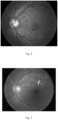
- microhemangioma lesion region finally obtained was shown in Fig. 3 .
- the labeling a region in the fundus image also generated a test set, which was used to test the trained model to evaluate the recognition ability of the trained model.
- the labeling a region in the fundus image also generated a validation set, which was used for correction in the model training so as to adjust the network parameters and prevent network overfitting; the validation set could also be used to determine the network structure and control the model complexity; depending on the different validation set, there were differences in the results obtained after the input of the test set; according to the situation, we could choose the optimal model that met our needs.
- the image processing algorithm comprised the following specific steps:
- Using the FCN model for hemorrhage and exudation recognition could also adjust the proportion of lesion samples, pass-examination samples and miss-examination samples in the training samples according to the actual data distribution, so as to improve the accuracy and generalization ability of the model without redesigning the algorithm, thereby reducing the intensity of algorithm development and improving the efficiency of algorithm development.
- the area of the hemorrhage lesion region was generally large, it was suitable to use the trained hemorrhage lesion recognition model to segment the hemorrhage region in the image.
- exudation lesion recognition model could obtain good recognition results.
- 300 training samples without exudation and 100 samples with lesions similar to exudation were added for training.
- the resulting model had a sensitivity of 86% and a specificity of 87% for judging whether the image had exudation.
- the effect of recognizing exudation was shown in Fig. 5 .
- the recognition result of each lesion could not be 100% accurate, staging directly according to the clinical staging criteria based on the recognition result of each lesion would result in a lower specificity of the image diagnosis result. Therefore, we used the recognition results of the three lesions as features to train the SVM classifier to judge the final diagnosis result of the image, finding that the sensitivity of DR3 recognition was more than 99%, the sensitivity of DR2 recognition reached 85%, the sensitivity of DR1 recognition reached 80%, and the specificity was 80%.
- the lesion feature information extracted from the recognition results of the microhemangioma lesion recognition model, the hemorrhage lesion recognition model and the exudation lesion recognition model was combined and inputted into the SVM classifier for training, thus obtaining the trained SVM classifier.
- a system for classifying diabetic retina images based on the above method comprising:
- the example of the present invention is scalable and currently includes models for recognizing three typical diabetic fundus lesions. With the pathological changes of the disease and the need for detection, we can train the recognition model of the corresponding lesion with the deep learning technology, and increase the recognition module of the corresponding lesion.
Landscapes
- Engineering & Computer Science (AREA)
- Theoretical Computer Science (AREA)
- Health & Medical Sciences (AREA)
- Physics & Mathematics (AREA)
- General Physics & Mathematics (AREA)
- General Health & Medical Sciences (AREA)
- Artificial Intelligence (AREA)
- Evolutionary Computation (AREA)
- Computer Vision & Pattern Recognition (AREA)
- Medical Informatics (AREA)
- Life Sciences & Earth Sciences (AREA)
- Software Systems (AREA)
- Data Mining & Analysis (AREA)
- Computing Systems (AREA)
- Multimedia (AREA)
- Biomedical Technology (AREA)
- General Engineering & Computer Science (AREA)
- Molecular Biology (AREA)
- Databases & Information Systems (AREA)
- Mathematical Physics (AREA)
- Public Health (AREA)
- Biophysics (AREA)
- Nuclear Medicine, Radiotherapy & Molecular Imaging (AREA)
- Radiology & Medical Imaging (AREA)
- Computational Linguistics (AREA)
- Evolutionary Biology (AREA)
- Bioinformatics & Computational Biology (AREA)
- Bioinformatics & Cheminformatics (AREA)
- Pathology (AREA)
- Epidemiology (AREA)
- Primary Health Care (AREA)
- Veterinary Medicine (AREA)
- Heart & Thoracic Surgery (AREA)
- Surgery (AREA)
- Animal Behavior & Ethology (AREA)
- Quality & Reliability (AREA)
- Ophthalmology & Optometry (AREA)
- Emergency Medicine (AREA)
- Biodiversity & Conservation Biology (AREA)
- Human Computer Interaction (AREA)
Claims (7)
- Verfahren zur Klassifizierung von diabetischen Netzhautbildern, das auf Deep Learning basiert, wobei das Verfahren die folgenden Schritte umfasst:Erhalten eines zu erkennenden Fundusbildes;Importieren desselben erhaltenen, zu erkennenden Fundusbildes in ein Modell zur Erkennung von Mikrohämangiomläsionen, ein Modell zur Erkennung von Hämorrhagieläsionen und ein Modell zur Erkennung von Exsudationsläsionen; Extrahieren von Läsionsmerkmalinformationen aus den Erkennungsergebnissen, und daran anschließend Verwenden eines trainierten SVM (Support Vector Machine, Unterstützungsvektormaschinen) -Klassifikators, um die extrahierten Läsionsmerkmalinformationen zu klassifizieren, um ein Klassifizierungsergebnis des Läsionsgrades zu erhalten, das dem Fundusbild entspricht;wobei das Verfahren dadurch gekennzeichnet ist, dass:
das Erkennungsmodell für Mikrohämangiomläsionen durch Trainieren eines CNN-Modells in folgenden Schritten gewonnen wird:A1. Bildvorverarbeitung: Extrahieren eines Grünkanalbildes und Verwenden einer r-Polynom-Transformation zur Korrektur der Graustufen des Grünkanalbildes, und danach Entrauschen des Bildes mittels Gauß-Filterung, um ein korrigiertes Bild I' W zu erhalten;A2. Extrahieren der Mikrohämangiomläsion-Kandidatenregion Icandidate : zufälliges Auswählen eines Pixelpunkts im korrigierten Bild I' W und Verwenden dieses Pixelpunkts als Referenz sowie eines Winkels α als Schrittgröße, um lineare Strukturelemente in verschiedenen Maßstäben zu erzeugen; Verwenden der erzeugten linearen Strukturelemente zur Durchführung einer morphologischen Verarbeitung des korrigierten Bildes I' W, um die Ergebnisse der linearen Strukturelemente in verschiedenen Maßstäben zu erhalten; Halten eines Minimalantwortergebnisses Iclosed, das jedem Pixelpunkt entspricht, um Icandidate = Iclosed - I' W zu erhalten, und daran anschließend Durchführen einer hybriden Schwellenwertsegmentierungs-Extraktion an Icandidate, wobei die hybride Schwellenwertsegmentierungs-Extraktion auf den folgenden Bedingungen basiert: wobei K eine Konstante ist, die die maximale Anzahl der Mikrohämangiomläsion-Kandidatenregionen bei der morphologischen Verarbeitung darstellt, und CC eine Funktion zur Zählung der Anzahl der Läsions-Kandidatenregionen ist;
t1 ein minimaler Schwellenwert ist, tu ein maximaler Schwellenwert ist, tk ein Schwellenwert ist, der die CC-Bedingungen erfüllt, und ts ein Schwellenwert ist, der graduell in einer Schrittgröße mit einem Wert im Bereich von 0,001-0,004 ansteigt;Erhöhen von ts von einem minimalen Graustufenwert von Icandidate auf einen maximalen Graustufenwert von Icandidate entsprechend der Schrittgröße, bis ein durch die CC-Funktion gezählter Wert die Bedingungen der vorstehend erwähnten Formeln erfüllt, und anschließend Verwenden des Schwellenwerts tK, um Icandidate zu binarisieren, um eine binäre Karte der Mikrohämangiomläsion-Kandidatenregion zu extrahieren;A3. Datenetikettierung: Etikettieren der Segmentierungs-Extraktionsergebnisse der Mikrohämangiomläsion-Kandidatenregion Icandidate als Läsion oder Nicht-Läsion, um einen Trainingssatz zu erzeugen; undA4. Modelltraining: Eingeben des Trainingssatzes in das CNN-Modell zum Trainieren, um das Erkennungsmodell für Mikrohämangiomläsionen zu erhalten, und wobei sowohl das Modell zur Erkennung von Hämorrhagieläsionen als auch das Modell zur Erkennung von Exsudationsläsionen basierend auf dem FCN-Modelltraining durch die folgenden Schritte erhalten werden:B1. Etikettieren einer Region im Fundusbild als Läsion oder Nicht-Läsion durch Bildverarbeitung, um einen Trainingssatz von etikettierten Daten zu erzeugen, wobei die Etikettierung als Hämorrhagie- oder Nicht-Hämorrhagieläsionsregion im Fundusbild für das Erkennungsmodell für Hämorrhagieläsionen bereitgestellt wird und die Etikettierung als Exsudationsläsionsregion oder Nicht-Exsudationsläsionsregion im Fundusbild für das Erkennungsmodell für Exsudationsläsionen bereitgestellt wird; undB2. Verwenden einer U-Netz-Struktur, um das FCN-Modell aufzubauen, und jedes Mal zufällig Heranziehen eines Teils der etikettierten Daten aus dem Trainingssatz zum Trainieren, um ein trainiertes Erkennungsmodell für Läsionen zu erhalten, wobei DICE als Kostenfunktion für das Trainieren des FCN-Modells verwendet wird. - Verfahren zur Klassifizierung von diabetischen Netzhautbildern, das auf Deep Learning basiert, nach Anspruch 1, wobei die r-Polynomtransformation wie folgt aussieht:

- Verfahren zur Klassifizierung von diabetischen Netzhautbildern, das auf Deep Learning basiert, nach Anspruch 1, wobei die Etikettierung einer Region im Fundusbild auch einen Testsatz erzeugt, der zum Testen eines trainierten Modells verwendet wird, um die Erkennungsfähigkeit des trainierten Modells zu bewerten.
- Verfahren zur Klassifizierung von diabetischen Netzhautbildern, das auf Deep Learning basiert, nach Anspruch 1 oder 3, wobei das Etikettieren einer Region im Fundusbild auch einen Validierungssatz erzeugt, der zur Korrektur beim Modelltraining verwendet wird, um eine Überanpassung des Netzwerks zu verhindern.
- Verfahren zur Klassifizierung von diabetischen Netzhautbildern, das auf Deep Learning basiert, nach Anspruch 1, wobei der SVM-Klassifikator durch Klassifizieren und Trainieren von Läsionsmerkmalinformationen entsprechend dem Trainingssatz erhalten wird.
- Verfahren zur Klassifizierung von diabetischen Netzhautbildern, das auf Deep Learning basiert, nach Anspruch 5, wobei die Läsionsmerkmalsinformationen eine Anzahl, Fläche, Form, Graustufe, Rundheit und ein Seitenverhältnis einer Läsionsregion beinhalten.
- System zur Klassifizierung von diabetischen Netzhautbildern, das Mittel zur Durchführung des Verfahrens nach einem der vorstehenden Ansprüche umfasst, umfassend:ein Erkennungsmodul für Mikrohämangiome, das verwendet wird, um ein zu untersuchendes Bild unter Verwendung des Erkennungsmodells für Mikrohämangiomläsionen zu erkennen und eine Mikrohämangiomläsionsstelle in dem zu untersuchenden Bild zu etikettieren, um entsprechende Läsionskennparameter zu erhalten;ein Erkennungsmodul für Hämorrhagie, das verwendet wird, um ein zu untersuchendes Bild unter Verwendung des Erkennungsmodells für Hämorrhagieläsionen zu erkennen und eine erkannte Hämorrhagieläsionsregion zu segmentieren, um entsprechende Läsionskennparameter zu erhalten;ein Erkennungsmodul für Exsudationen, das verwendet wird, um ein zu untersuchendes Bild unter Verwendung des Erkennungsmodells für Exsudationsläsionen zu erkennen und eine erkannte Exsudationläsionsregion zu segmentieren, um entsprechende Läsionskennparameter zu erhalten; undein Klassifizierungsmodul, das verwendet wird, um die Kennparameter jeder Läsionsregion zu klassifizieren, die durch die Erkennung des zu untersuchenden Bildes erhalten werden, um so Klassifizierungsergebnisse des Läsionsgrades des zu untersuchenden Bildes zu erhalten.
Applications Claiming Priority (2)
| Application Number | Priority Date | Filing Date | Title |
|---|---|---|---|
| CN201810330385.6A CN108615051B (zh) | 2018-04-13 | 2018-04-13 | 基于深度学习的糖尿病视网膜图像分类方法及系统 |
| PCT/CN2018/098390 WO2019196268A1 (zh) | 2018-04-13 | 2018-08-02 | 基于深度学习的糖尿病视网膜图像分类方法及系统 |
Publications (3)
| Publication Number | Publication Date |
|---|---|
| EP3779786A1 EP3779786A1 (de) | 2021-02-17 |
| EP3779786A4 EP3779786A4 (de) | 2022-01-12 |
| EP3779786B1 true EP3779786B1 (de) | 2024-10-02 |
Family
ID=63659944
Family Applications (1)
| Application Number | Title | Priority Date | Filing Date |
|---|---|---|---|
| EP18914825.7A Active EP3779786B1 (de) | 2018-04-13 | 2018-08-02 | Verfahren und system auf der grundlage von tiefenlernen zur klassifizierung von diabetischen netzhautbildern |
Country Status (4)
| Country | Link |
|---|---|
| US (1) | US11132799B2 (de) |
| EP (1) | EP3779786B1 (de) |
| CN (1) | CN108615051B (de) |
| WO (1) | WO2019196268A1 (de) |
Families Citing this family (76)
| Publication number | Priority date | Publication date | Assignee | Title |
|---|---|---|---|---|
| CN108596895B (zh) * | 2018-04-26 | 2020-07-28 | 上海鹰瞳医疗科技有限公司 | 基于机器学习的眼底图像检测方法、装置及系统 |
| CN111199794B (zh) * | 2018-11-19 | 2024-03-01 | 复旦大学附属眼耳鼻喉科医院 | 一种适用于高度近视白内障的手术智能决策系统及其建立方法 |
| CN109602391A (zh) * | 2019-01-04 | 2019-04-12 | 平安科技(深圳)有限公司 | 眼底出血点的自动检测方法、装置及计算机可读存储介质 |
| CN109886946B (zh) * | 2019-02-18 | 2023-05-23 | 广州视源电子科技股份有限公司 | 基于深度学习的早期老年性黄斑病变弱监督分类方法 |
| CN110009626A (zh) * | 2019-04-11 | 2019-07-12 | 北京百度网讯科技有限公司 | 用于生成图像的方法和装置 |
| CN110264443B (zh) * | 2019-05-20 | 2024-04-16 | 平安科技(深圳)有限公司 | 基于特征可视化的眼底图像病变标注方法、装置及介质 |
| KR102295426B1 (ko) * | 2019-07-05 | 2021-08-30 | 순천향대학교 산학협력단 | 인공지능 기반의 안구질병 검출장치 및 방법 |
| CN110610756A (zh) * | 2019-07-26 | 2019-12-24 | 赛诺威盛科技(北京)有限公司 | 基于dicom图像信息实现胶片自动分类打印的方法 |
| CN110414607A (zh) * | 2019-07-31 | 2019-11-05 | 中山大学 | 胶囊内窥镜图像的分类方法、装置、设备及介质 |
| CN110490138A (zh) * | 2019-08-20 | 2019-11-22 | 北京大恒普信医疗技术有限公司 | 一种数据处理方法及装置、存储介质、电子设备 |
| CN110555845B (zh) * | 2019-09-27 | 2024-09-17 | 北京鹰瞳远见信息科技有限公司 | 眼底oct图像识别方法及设备 |
| CN110853009B (zh) * | 2019-11-11 | 2023-04-28 | 北京端点医药研究开发有限公司 | 基于机器学习的视网膜病理图像分析系统 |
| CN110840402B (zh) * | 2019-11-19 | 2021-02-26 | 山东大学 | 一种基于机器学习的房颤信号识别方法及系统 |
| CN111179258A (zh) * | 2019-12-31 | 2020-05-19 | 中山大学中山眼科中心 | 一种识别视网膜出血图像的人工智能方法及系统 |
| CN113283270B (zh) * | 2020-02-20 | 2024-08-23 | 京东方科技集团股份有限公司 | 图像处理方法和装置、筛查系统、计算机可读存储介质 |
| CN111815574B (zh) * | 2020-06-18 | 2022-08-12 | 南通大学 | 一种基于粗糙集神经网络的眼底视网膜血管图像分割方法 |
| CN111754486B (zh) * | 2020-06-24 | 2023-08-15 | 北京百度网讯科技有限公司 | 图像处理方法、装置、电子设备及存储介质 |
| CN112053321A (zh) * | 2020-07-30 | 2020-12-08 | 中山大学中山眼科中心 | 一种识别高度近视视网膜病变的人工智能系统 |
| CN112037187B (zh) * | 2020-08-24 | 2024-03-26 | 宁波市眼科医院 | 一种眼底低质量图片的智能优化系统 |
| CN111968107B (zh) * | 2020-08-31 | 2024-03-12 | 合肥奥比斯科技有限公司 | 基于不确定度的早产儿视网膜病plus病变分类系统 |
| CN112016626B (zh) * | 2020-08-31 | 2023-12-01 | 中科泰明(南京)科技有限公司 | 基于不确定度的糖尿病视网膜病变分类系统 |
| CN112185523B (zh) * | 2020-09-30 | 2023-09-08 | 南京大学 | 基于多尺度卷积神经网络的糖尿病视网膜病变分类方法 |
| CN112233087A (zh) * | 2020-10-14 | 2021-01-15 | 武汉楚精灵医疗科技有限公司 | 一种基于人工智能的眼科超声疾病诊断方法和系统 |
| CN112184697B (zh) * | 2020-10-15 | 2022-10-04 | 桂林电子科技大学 | 基于果蝇优化算法的糖尿病视网膜病变分级深度学习方法 |
| CN112241766B (zh) * | 2020-10-27 | 2023-04-18 | 西安电子科技大学 | 基于样本生成和迁移学习的肝脏ct图像多病变分类方法 |
| CN112330624B (zh) * | 2020-11-02 | 2025-06-06 | 腾讯科技(深圳)有限公司 | 医学图像处理方法和装置 |
| CN112446860B (zh) * | 2020-11-23 | 2024-04-16 | 中山大学中山眼科中心 | 一种基于迁移学习的糖尿病黄斑水肿自动筛查方法 |
| CN112489003A (zh) * | 2020-11-25 | 2021-03-12 | 哈尔滨理工大学 | 一种基于深度学习的糖尿病视网膜病变区域定位检测方法 |
| CN112580580A (zh) * | 2020-12-28 | 2021-03-30 | 厦门理工学院 | 一种基于数据增强与模型融合的病理性近视识别方法 |
| CN112634243B (zh) * | 2020-12-28 | 2022-08-05 | 吉林大学 | 一种强干扰因素下基于深度学习的图像分类识别系统 |
| WO2022142368A1 (en) * | 2020-12-29 | 2022-07-07 | Aimomics (Shanghai) Intelligent Technology Co., Ltd | Rapid screen system based on eye region image |
| TWI789199B (zh) * | 2021-01-13 | 2023-01-01 | 高雄醫學大學 | 眼底鏡影像預測糖尿病性腎病變期程之方法及其系統 |
| CN112869697A (zh) * | 2021-01-20 | 2021-06-01 | 深圳硅基智能科技有限公司 | 同时识别糖尿病视网膜病变的分期和病变特征的判断方法 |
| CN112734774B (zh) * | 2021-01-28 | 2023-03-21 | 依未科技(北京)有限公司 | 一种高精度眼底血管提取方法、装置、介质、设备和系统 |
| CN112883962B (zh) * | 2021-01-29 | 2023-07-18 | 北京百度网讯科技有限公司 | 眼底图像识别方法、装置、设备、存储介质以及程序产品 |
| US20220245811A1 (en) * | 2021-02-01 | 2022-08-04 | AI Optics Inc. | Analysis of retinal imaging using video |
| CN112869704B (zh) * | 2021-02-02 | 2022-06-17 | 苏州大学 | 一种基于循环自适应多目标加权网络的糖尿病视网膜病变区域自动分割方法 |
| CN112819797B (zh) * | 2021-02-06 | 2023-09-19 | 国药集团基因科技有限公司 | 糖尿病性视网膜病变分析方法、装置、系统及存储介质 |
| CN115132326B (zh) * | 2021-03-24 | 2024-08-30 | 深圳硅基智能科技有限公司 | 基于眼底图像的糖尿病视网膜病变的评估系统及评估方法 |
| CN113344842A (zh) * | 2021-03-24 | 2021-09-03 | 同济大学 | 一种超声图像的血管标注方法 |
| CN112926537B (zh) * | 2021-04-07 | 2024-05-28 | 南方科技大学 | 图像处理方法、装置、电子设备以及存储介质 |
| CN115191930B (zh) * | 2021-04-14 | 2026-03-06 | 依未科技(北京)有限公司 | 糖尿病评估装置和糖尿病评估方法 |
| CN113012148A (zh) * | 2021-04-14 | 2021-06-22 | 中国人民解放军总医院第一医学中心 | 一种基于眼底影像的糖尿病肾病-非糖尿病肾病鉴别诊断装置 |
| CN115272386B (zh) * | 2021-04-30 | 2025-09-30 | 中国医学科学院基础医学研究所 | 基于自动生成标签的脑出血和周围水肿的多分支分割系统 |
| CN113487621B (zh) * | 2021-05-25 | 2024-07-12 | 山东省眼科研究所 | 医学图像分级方法、装置、电子设备及可读存储介质 |
| CN113627231B (zh) * | 2021-06-16 | 2023-10-31 | 温州医科大学 | 一种基于机器视觉的视网膜oct图像中液体区域自动分割方法 |
| CN113537298A (zh) * | 2021-06-23 | 2021-10-22 | 广东省人民医院 | 一种视网膜图像分类方法及装置 |
| CN113744212B (zh) * | 2021-08-23 | 2024-06-11 | 江苏大学 | 一种基于显微光谱图像采集和图像处理算法的粮食真菌孢子智能识别方法 |
| CN113793308A (zh) * | 2021-08-25 | 2021-12-14 | 北京科技大学 | 一种基于神经网络的球团矿质量智能评级方法及装置 |
| CN113705569A (zh) * | 2021-08-31 | 2021-11-26 | 北京理工大学重庆创新中心 | 一种图像标注方法及系统 |
| CN114287878B (zh) * | 2021-10-18 | 2024-11-22 | 江西财经大学 | 一种基于注意力模型的糖尿病性视网膜病变病灶图像识别方法 |
| CN113962978A (zh) * | 2021-10-29 | 2022-01-21 | 北京富通东方科技有限公司 | 一种眼部运动损伤检测阅片方法及系统 |
| CN114305391B (zh) * | 2021-11-04 | 2024-07-19 | 上海市儿童医院 | 一种深度学习对尿道下裂阴茎弯曲度的测量方法 |
| CN114494196B (zh) * | 2022-01-26 | 2023-11-17 | 南通大学 | 基于遗传模糊树的视网膜糖尿病变深度网络检测方法 |
| CN114520044A (zh) * | 2022-02-16 | 2022-05-20 | 复旦大学 | 基于深度学习的视网膜眼底图像半自动标注装置 |
| CN114359279B (zh) * | 2022-03-18 | 2022-06-03 | 武汉楚精灵医疗科技有限公司 | 图像处理方法、装置、计算机设备及存储介质 |
| CN114898172B (zh) * | 2022-04-08 | 2024-04-02 | 辽宁师范大学 | 基于多特征dag网络的糖尿病视网膜病变分类建模方法 |
| CN114792382B (zh) * | 2022-04-27 | 2025-04-18 | 东北大学 | 基于切片病灶检测的糖尿病性视网膜病变等级分类装置 |
| CN114882218A (zh) * | 2022-04-28 | 2022-08-09 | 桂林理工大学 | 一种基于嵌入残差卷积和注意力模块的u型糖尿病视网膜渗出物分割算法 |
| CN115170503B (zh) * | 2022-07-01 | 2023-12-19 | 上海市第一人民医院 | 基于决策规则和深度神经网络的眼底图像视野分类方法及装置 |
| KR102892960B1 (ko) * | 2022-11-24 | 2025-11-28 | 주식회사 에이아이포이에시스 | 당뇨망막병증 분류 장치 및 방법 |
| CN116596845B (zh) * | 2023-04-07 | 2026-02-06 | 浙江工业大学 | 一种基于弱监督学习的uwf眼底图像病灶区域定位方法 |
| CN116168255B (zh) * | 2023-04-10 | 2023-12-08 | 武汉大学人民医院(湖北省人民医院) | 一种长尾分布鲁棒的视网膜oct图像分类方法 |
| CN116524257A (zh) * | 2023-04-24 | 2023-08-01 | 依未科技(北京)有限公司 | 眼底图像分类方法及装置、图像处理方法 |
| US20240386571A1 (en) * | 2023-05-18 | 2024-11-21 | Neuralink Corp. | Vascular Segmentation and Avoidance Using Imaging |
| CN116721760B (zh) * | 2023-06-12 | 2024-04-26 | 东北林业大学 | 融合生物标志物的多任务糖尿病性视网膜病变检测算法 |
| CN116977320B (zh) * | 2023-08-14 | 2024-04-26 | 中山火炬开发区人民医院 | Ct+mri病变区域突出预估系统 |
| CN117496231B (zh) * | 2023-10-24 | 2024-07-16 | 国药(武汉)精准医疗科技有限公司 | 糖尿病性视网膜病变图像分类方法、装置以及计算机设备 |
| CN117557840B (zh) * | 2023-11-10 | 2024-05-24 | 中国矿业大学 | 一种基于小样本学习的眼底病变分级方法 |
| CN117456594B (zh) * | 2023-11-15 | 2024-08-16 | 深圳市眼科医院(深圳市眼病防治研究所) | 一种基于深度学习的眼球超声图像自动分类方法及系统 |
| CN117475270B (zh) * | 2023-11-20 | 2024-07-23 | 深圳市眼科医院(深圳市眼病防治研究所) | 糖尿病视网膜病变图像的病灶识别方法及系统 |
| CN117893836B (zh) * | 2024-03-13 | 2024-05-28 | 中国人民解放军总医院第一医学中心 | 基于眼底血管几何参数预测糖尿病肾病的方法和系统 |
| CN118975775B (zh) * | 2024-08-10 | 2025-12-02 | 厦门眼科中心有限公司 | 基于人工智能的自动分析的眼底病变色彩诊断系统 |
| CN119399524B (zh) * | 2024-10-18 | 2025-11-25 | 南京邮电大学 | 一种面向小样本的糖尿病视网膜病变的分类方法及系统 |
| CN119963949B (zh) * | 2025-04-09 | 2025-06-20 | 宝鸡市人民医院 | 一种外周血管影像辅助分割方法及系统 |
| CN121437539B (zh) * | 2025-12-30 | 2026-02-27 | 长春理工大学 | 基于多分支与视觉曼巴的糖尿病性视网膜病变分割方法 |
Family Cites Families (15)
| Publication number | Priority date | Publication date | Assignee | Title |
|---|---|---|---|---|
| EP1740168A2 (de) * | 2004-04-21 | 2007-01-10 | Advanced Ocular Systems Limited | Antiprostaglandine zur behandlung von augenpathologien |
| US9795301B2 (en) * | 2010-05-25 | 2017-10-24 | The General Hospital Corporation | Apparatus, systems, methods and computer-accessible medium for spectral analysis of optical coherence tomography images |
| EP4057215A1 (de) * | 2013-10-22 | 2022-09-14 | Eyenuk, Inc. | Systeme und verfahren zur automatischen analyse von netzhautbildern |
| CN103870838A (zh) * | 2014-03-05 | 2014-06-18 | 南京航空航天大学 | 糖尿病视网膜病变的眼底图像特征提取方法 |
| WO2016132115A1 (en) * | 2015-02-16 | 2016-08-25 | University Of Surrey | Detection of microaneurysms |
| WO2017031099A1 (en) * | 2015-08-20 | 2017-02-23 | Ohio University | Devices and methods for classifying diabetic and macular degeneration |
| CN105513077B (zh) * | 2015-12-11 | 2019-01-04 | 北京大恒图像视觉有限公司 | 一种用于糖尿病性视网膜病变筛查的系统 |
| CN111095261A (zh) * | 2017-04-27 | 2020-05-01 | 视网膜病答案有限公司 | 眼底图像自动分析系统和方法 |
| CN107123124B (zh) * | 2017-05-04 | 2020-05-12 | 季鑫 | 视网膜图像分析方法、装置和计算设备 |
| CN107423571B (zh) * | 2017-05-04 | 2018-07-06 | 深圳硅基仿生科技有限公司 | 基于眼底图像的糖尿病视网膜病变识别系统 |
| CN107203778A (zh) * | 2017-05-05 | 2017-09-26 | 平安科技(深圳)有限公司 | 视网膜病变程度等级检测系统及方法 |
| CN107330449A (zh) * | 2017-06-13 | 2017-11-07 | 瑞达昇科技(大连)有限公司 | 一种糖尿病性视网膜病变体征检测方法及装置 |
| CN107680684B (zh) * | 2017-10-12 | 2021-05-07 | 百度在线网络技术(北京)有限公司 | 用于获取信息的方法及装置 |
| US11132797B2 (en) * | 2017-12-28 | 2021-09-28 | Topcon Corporation | Automatically identifying regions of interest of an object from horizontal images using a machine learning guided imaging system |
| US12008748B2 (en) * | 2018-05-31 | 2024-06-11 | Vuno, Inc. | Method for classifying fundus image of subject and device using same |
-
2018
- 2018-04-13 CN CN201810330385.6A patent/CN108615051B/zh active Active
- 2018-08-02 EP EP18914825.7A patent/EP3779786B1/de active Active
- 2018-08-02 WO PCT/CN2018/098390 patent/WO2019196268A1/zh not_active Ceased
-
2020
- 2020-03-30 US US16/835,199 patent/US11132799B2/en active Active
Also Published As
| Publication number | Publication date |
|---|---|
| US20200234445A1 (en) | 2020-07-23 |
| EP3779786A4 (de) | 2022-01-12 |
| EP3779786A1 (de) | 2021-02-17 |
| US11132799B2 (en) | 2021-09-28 |
| CN108615051B (zh) | 2020-09-15 |
| WO2019196268A1 (zh) | 2019-10-17 |
| CN108615051A (zh) | 2018-10-02 |
Similar Documents
| Publication | Publication Date | Title |
|---|---|---|
| US11132799B2 (en) | Method and system for classifying diabetic retina images based on deep learning | |
| US20220076420A1 (en) | Retinopathy recognition system | |
| CN107358605B (zh) | 用于识别糖尿病视网膜病变的深度神经网络装置及系统 | |
| CN109377474B (zh) | 一种基于改进Faster R-CNN的黄斑定位方法 | |
| Mookiah et al. | Evolutionary algorithm based classifier parameter tuning for automatic diabetic retinopathy grading: A hybrid feature extraction approach | |
| Sopharak et al. | Automatic detection of diabetic retinopathy exudates from non-dilated retinal images using mathematical morphology methods | |
| US11200707B2 (en) | Method of modifying a retina fundus image for a deep learning model | |
| Zhao et al. | A deep learning framework for identifying zone I in RetCam images | |
| Muramatsu et al. | Automated detection and classification of major retinal vessels for determination of diameter ratio of arteries and veins | |
| CN106780439A (zh) | 一种筛选眼底图像的方法 | |
| Fu et al. | A GAN-based deep enhancer for quality enhancement of retinal images photographed by a handheld fundus camera | |
| Rahim et al. | Novel fundus image preprocessing for retcam images to improve deep learning classification of retinopathy of prematurity | |
| Kumari et al. | Deep learning based detection of diabetic retinopathy using retinal fundus images | |
| Yao et al. | DeepGraFT: A novel semantic segmentation auxiliary ROI-based deep learning framework for effective fundus tessellation classification | |
| Ashame et al. | Abnormality Detection in Eye Fundus Retina | |
| Athab et al. | Disc and Cup Segmentation for Glaucoma Detection | |
| Singh et al. | Refuge challenge 2018-task 2: Deep optic disc and cup segmentation in fundus images using u-net and multi-scale feature matching networks | |
| Shabu et al. | DeepExuDetectNet: Diabetic Retinopathy Diagnosis: Blood Vessel Segmentation and Exudates Disease Detection in Fundus Images | |
| Al-Saadi et al. | An Automated Wavelet Scattering Network Classification Using Three Stages of Cataract Disease | |
| Abirami et al. | Diabetic Retinopathy Image Recognition Using Artificial Intelligence | |
| Devi et al. | Composite filtering and morphology for optic disc segmentation from retinal images | |
| Venkatalakshmi et al. | Graphical user interface for enhanced retinal image analysis for diagnosing diabetic retinopathy | |
| Yashwanth et al. | Automated hypertensive retinopathy detection method using deep learning | |
| Salem et al. | Glaucoma Diagnosis with Fundus Image Analysis Using Cup-To-Disc Ratios | |
| Hann et al. | Diabetic retinopathy screening using computer vision |
Legal Events
| Date | Code | Title | Description |
|---|---|---|---|
| STAA | Information on the status of an ep patent application or granted ep patent |
Free format text: STATUS: THE INTERNATIONAL PUBLICATION HAS BEEN MADE |
|
| PUAI | Public reference made under article 153(3) epc to a published international application that has entered the european phase |
Free format text: ORIGINAL CODE: 0009012 |
|
| STAA | Information on the status of an ep patent application or granted ep patent |
Free format text: STATUS: REQUEST FOR EXAMINATION WAS MADE |
|
| 17P | Request for examination filed |
Effective date: 20201112 |
|
| AK | Designated contracting states |
Kind code of ref document: A1 Designated state(s): AL AT BE BG CH CY CZ DE DK EE ES FI FR GB GR HR HU IE IS IT LI LT LU LV MC MK MT NL NO PL PT RO RS SE SI SK SM TR |
|
| AX | Request for extension of the european patent |
Extension state: BA ME |
|
| DAV | Request for validation of the european patent (deleted) | ||
| DAX | Request for extension of the european patent (deleted) | ||
| A4 | Supplementary search report drawn up and despatched |
Effective date: 20211208 |
|
| RIC1 | Information provided on ipc code assigned before grant |
Ipc: A61B 5/00 20060101ALI20211203BHEP Ipc: G16H 50/20 20180101ALI20211203BHEP Ipc: G16H 30/40 20180101ALI20211203BHEP Ipc: G06T 7/00 20170101ALI20211203BHEP Ipc: G06N 20/10 20190101ALI20211203BHEP Ipc: G06N 3/08 20060101ALI20211203BHEP Ipc: G06N 3/04 20060101ALI20211203BHEP Ipc: G06K 9/00 20060101ALI20211203BHEP Ipc: A61B 5/145 20060101ALI20211203BHEP Ipc: G06K 9/46 20060101ALI20211203BHEP Ipc: G06K 9/62 20060101AFI20211203BHEP |
|
| STAA | Information on the status of an ep patent application or granted ep patent |
Free format text: STATUS: EXAMINATION IS IN PROGRESS |
|
| 17Q | First examination report despatched |
Effective date: 20230426 |
|
| REG | Reference to a national code |
Ref country code: DE Ref legal event code: R079 Free format text: PREVIOUS MAIN CLASS: G06K0009620000 Ipc: G06V0010764000 Ref document number: 602018075090 Country of ref document: DE |
|
| GRAP | Despatch of communication of intention to grant a patent |
Free format text: ORIGINAL CODE: EPIDOSNIGR1 |
|
| STAA | Information on the status of an ep patent application or granted ep patent |
Free format text: STATUS: GRANT OF PATENT IS INTENDED |
|
| RIC1 | Information provided on ipc code assigned before grant |
Ipc: G06N 3/08 20060101ALI20240321BHEP Ipc: G16H 50/20 20180101ALI20240321BHEP Ipc: G16H 30/40 20180101ALI20240321BHEP Ipc: G06T 7/00 20170101ALI20240321BHEP Ipc: G06N 20/10 20190101ALI20240321BHEP Ipc: A61B 5/00 20060101ALI20240321BHEP Ipc: A61B 5/145 20060101ALI20240321BHEP Ipc: G06F 18/25 20230101ALI20240321BHEP Ipc: G06N 3/045 20230101ALI20240321BHEP Ipc: G06V 10/44 20220101ALI20240321BHEP Ipc: G06V 10/774 20220101ALI20240321BHEP Ipc: G06V 10/80 20220101ALI20240321BHEP Ipc: G06V 10/82 20220101ALI20240321BHEP Ipc: G06V 40/18 20220101ALI20240321BHEP Ipc: G06V 10/764 20220101AFI20240321BHEP |
|
| INTG | Intention to grant announced |
Effective date: 20240405 |
|
| GRAS | Grant fee paid |
Free format text: ORIGINAL CODE: EPIDOSNIGR3 |
|
| RAP3 | Party data changed (applicant data changed or rights of an application transferred) |
Owner name: BOZHON PRECISION INDUSTRY TECHNOLOGY CO., LTD. |
|
| GRAA | (expected) grant |
Free format text: ORIGINAL CODE: 0009210 |
|
| STAA | Information on the status of an ep patent application or granted ep patent |
Free format text: STATUS: THE PATENT HAS BEEN GRANTED |
|
| AK | Designated contracting states |
Kind code of ref document: B1 Designated state(s): AL AT BE BG CH CY CZ DE DK EE ES FI FR GB GR HR HU IE IS IT LI LT LU LV MC MK MT NL NO PL PT RO RS SE SI SK SM TR |
|
| REG | Reference to a national code |
Ref country code: GB Ref legal event code: FG4D |
|
| REG | Reference to a national code |
Ref country code: CH Ref legal event code: EP |
|
| REG | Reference to a national code |
Ref country code: IE Ref legal event code: FG4D |
|
| REG | Reference to a national code |
Ref country code: DE Ref legal event code: R096 Ref document number: 602018075090 Country of ref document: DE |
|
| REG | Reference to a national code |
Ref country code: LT Ref legal event code: MG9D |
|
| REG | Reference to a national code |
Ref country code: NL Ref legal event code: MP Effective date: 20241002 |
|
| REG | Reference to a national code |
Ref country code: AT Ref legal event code: MK05 Ref document number: 1729031 Country of ref document: AT Kind code of ref document: T Effective date: 20241002 |
|
| PG25 | Lapsed in a contracting state [announced via postgrant information from national office to epo] |
Ref country code: NL Free format text: LAPSE BECAUSE OF FAILURE TO SUBMIT A TRANSLATION OF THE DESCRIPTION OR TO PAY THE FEE WITHIN THE PRESCRIBED TIME-LIMIT Effective date: 20241002 |
|
| PG25 | Lapsed in a contracting state [announced via postgrant information from national office to epo] |
Ref country code: NL Free format text: LAPSE BECAUSE OF FAILURE TO SUBMIT A TRANSLATION OF THE DESCRIPTION OR TO PAY THE FEE WITHIN THE PRESCRIBED TIME-LIMIT Effective date: 20241002 |
|
| PG25 | Lapsed in a contracting state [announced via postgrant information from national office to epo] |
Ref country code: PT Free format text: LAPSE BECAUSE OF FAILURE TO SUBMIT A TRANSLATION OF THE DESCRIPTION OR TO PAY THE FEE WITHIN THE PRESCRIBED TIME-LIMIT Effective date: 20250203 Ref country code: HR Free format text: LAPSE BECAUSE OF FAILURE TO SUBMIT A TRANSLATION OF THE DESCRIPTION OR TO PAY THE FEE WITHIN THE PRESCRIBED TIME-LIMIT Effective date: 20241002 Ref country code: IS Free format text: LAPSE BECAUSE OF FAILURE TO SUBMIT A TRANSLATION OF THE DESCRIPTION OR TO PAY THE FEE WITHIN THE PRESCRIBED TIME-LIMIT Effective date: 20250202 |
|
| PG25 | Lapsed in a contracting state [announced via postgrant information from national office to epo] |
Ref country code: FI Free format text: LAPSE BECAUSE OF FAILURE TO SUBMIT A TRANSLATION OF THE DESCRIPTION OR TO PAY THE FEE WITHIN THE PRESCRIBED TIME-LIMIT Effective date: 20241002 |
|
| PG25 | Lapsed in a contracting state [announced via postgrant information from national office to epo] |
Ref country code: BG Free format text: LAPSE BECAUSE OF FAILURE TO SUBMIT A TRANSLATION OF THE DESCRIPTION OR TO PAY THE FEE WITHIN THE PRESCRIBED TIME-LIMIT Effective date: 20241002 |
|
| PG25 | Lapsed in a contracting state [announced via postgrant information from national office to epo] |
Ref country code: ES Free format text: LAPSE BECAUSE OF FAILURE TO SUBMIT A TRANSLATION OF THE DESCRIPTION OR TO PAY THE FEE WITHIN THE PRESCRIBED TIME-LIMIT Effective date: 20241002 |
|
| PG25 | Lapsed in a contracting state [announced via postgrant information from national office to epo] |
Ref country code: NO Free format text: LAPSE BECAUSE OF FAILURE TO SUBMIT A TRANSLATION OF THE DESCRIPTION OR TO PAY THE FEE WITHIN THE PRESCRIBED TIME-LIMIT Effective date: 20250102 |
|
| PG25 | Lapsed in a contracting state [announced via postgrant information from national office to epo] |
Ref country code: LV Free format text: LAPSE BECAUSE OF FAILURE TO SUBMIT A TRANSLATION OF THE DESCRIPTION OR TO PAY THE FEE WITHIN THE PRESCRIBED TIME-LIMIT Effective date: 20241002 Ref country code: AT Free format text: LAPSE BECAUSE OF FAILURE TO SUBMIT A TRANSLATION OF THE DESCRIPTION OR TO PAY THE FEE WITHIN THE PRESCRIBED TIME-LIMIT Effective date: 20241002 Ref country code: GR Free format text: LAPSE BECAUSE OF FAILURE TO SUBMIT A TRANSLATION OF THE DESCRIPTION OR TO PAY THE FEE WITHIN THE PRESCRIBED TIME-LIMIT Effective date: 20250103 |
|
| PG25 | Lapsed in a contracting state [announced via postgrant information from national office to epo] |
Ref country code: CZ Free format text: LAPSE BECAUSE OF FAILURE TO SUBMIT A TRANSLATION OF THE DESCRIPTION OR TO PAY THE FEE WITHIN THE PRESCRIBED TIME-LIMIT Effective date: 20241002 Ref country code: PL Free format text: LAPSE BECAUSE OF FAILURE TO SUBMIT A TRANSLATION OF THE DESCRIPTION OR TO PAY THE FEE WITHIN THE PRESCRIBED TIME-LIMIT Effective date: 20241002 |
|
| PG25 | Lapsed in a contracting state [announced via postgrant information from national office to epo] |
Ref country code: RS Free format text: LAPSE BECAUSE OF FAILURE TO SUBMIT A TRANSLATION OF THE DESCRIPTION OR TO PAY THE FEE WITHIN THE PRESCRIBED TIME-LIMIT Effective date: 20250102 |
|
| PG25 | Lapsed in a contracting state [announced via postgrant information from national office to epo] |
Ref country code: SM Free format text: LAPSE BECAUSE OF FAILURE TO SUBMIT A TRANSLATION OF THE DESCRIPTION OR TO PAY THE FEE WITHIN THE PRESCRIBED TIME-LIMIT Effective date: 20241002 |
|
| REG | Reference to a national code |
Ref country code: DE Ref legal event code: R097 Ref document number: 602018075090 Country of ref document: DE |
|
| PG25 | Lapsed in a contracting state [announced via postgrant information from national office to epo] |
Ref country code: DK Free format text: LAPSE BECAUSE OF FAILURE TO SUBMIT A TRANSLATION OF THE DESCRIPTION OR TO PAY THE FEE WITHIN THE PRESCRIBED TIME-LIMIT Effective date: 20241002 |
|
| PG25 | Lapsed in a contracting state [announced via postgrant information from national office to epo] |
Ref country code: EE Free format text: LAPSE BECAUSE OF FAILURE TO SUBMIT A TRANSLATION OF THE DESCRIPTION OR TO PAY THE FEE WITHIN THE PRESCRIBED TIME-LIMIT Effective date: 20241002 |
|
| PG25 | Lapsed in a contracting state [announced via postgrant information from national office to epo] |
Ref country code: RO Free format text: LAPSE BECAUSE OF FAILURE TO SUBMIT A TRANSLATION OF THE DESCRIPTION OR TO PAY THE FEE WITHIN THE PRESCRIBED TIME-LIMIT Effective date: 20241002 |
|
| PG25 | Lapsed in a contracting state [announced via postgrant information from national office to epo] |
Ref country code: SK Free format text: LAPSE BECAUSE OF FAILURE TO SUBMIT A TRANSLATION OF THE DESCRIPTION OR TO PAY THE FEE WITHIN THE PRESCRIBED TIME-LIMIT Effective date: 20241002 |
|
| PG25 | Lapsed in a contracting state [announced via postgrant information from national office to epo] |
Ref country code: IT Free format text: LAPSE BECAUSE OF FAILURE TO SUBMIT A TRANSLATION OF THE DESCRIPTION OR TO PAY THE FEE WITHIN THE PRESCRIBED TIME-LIMIT Effective date: 20241002 |
|
| PLBE | No opposition filed within time limit |
Free format text: ORIGINAL CODE: 0009261 |
|
| STAA | Information on the status of an ep patent application or granted ep patent |
Free format text: STATUS: NO OPPOSITION FILED WITHIN TIME LIMIT |
|
| PG25 | Lapsed in a contracting state [announced via postgrant information from national office to epo] |
Ref country code: SE Free format text: LAPSE BECAUSE OF FAILURE TO SUBMIT A TRANSLATION OF THE DESCRIPTION OR TO PAY THE FEE WITHIN THE PRESCRIBED TIME-LIMIT Effective date: 20241002 |
|
| 26N | No opposition filed |
Effective date: 20250703 |
|
| PGFP | Annual fee paid to national office [announced via postgrant information from national office to epo] |
Ref country code: DE Payment date: 20250820 Year of fee payment: 8 |
|
| REG | Reference to a national code |
Ref country code: CH Ref legal event code: H13 Free format text: ST27 STATUS EVENT CODE: U-0-0-H10-H13 (AS PROVIDED BY THE NATIONAL OFFICE) Effective date: 20260324 |
|
| PG25 | Lapsed in a contracting state [announced via postgrant information from national office to epo] |
Ref country code: MC Free format text: LAPSE BECAUSE OF FAILURE TO SUBMIT A TRANSLATION OF THE DESCRIPTION OR TO PAY THE FEE WITHIN THE PRESCRIBED TIME-LIMIT Effective date: 20241002 |
|
| PG25 | Lapsed in a contracting state [announced via postgrant information from national office to epo] |
Ref country code: LU Free format text: LAPSE BECAUSE OF NON-PAYMENT OF DUE FEES Effective date: 20250802 |
|
| PG25 | Lapsed in a contracting state [announced via postgrant information from national office to epo] |
Ref country code: CH Free format text: LAPSE BECAUSE OF NON-PAYMENT OF DUE FEES Effective date: 20250831 |
|
| GBPC | Gb: european patent ceased through non-payment of renewal fee |
Effective date: 20250802 |