EP3752839B1 - Nanoskalige biochemische probenvorbereitung und -analyse - Google Patents
Nanoskalige biochemische probenvorbereitung und -analyse Download PDFInfo
- Publication number
- EP3752839B1 EP3752839B1 EP19710820.2A EP19710820A EP3752839B1 EP 3752839 B1 EP3752839 B1 EP 3752839B1 EP 19710820 A EP19710820 A EP 19710820A EP 3752839 B1 EP3752839 B1 EP 3752839B1
- Authority
- EP
- European Patent Office
- Prior art keywords
- sample
- cells
- cell
- biological
- tissue
- Prior art date
- Legal status (The legal status is an assumption and is not a legal conclusion. Google has not performed a legal analysis and makes no representation as to the accuracy of the status listed.)
- Active
Links
Images


Classifications
-
- B—PERFORMING OPERATIONS; TRANSPORTING
- B01—PHYSICAL OR CHEMICAL PROCESSES OR APPARATUS IN GENERAL
- B01L—CHEMICAL OR PHYSICAL LABORATORY APPARATUS FOR GENERAL USE
- B01L3/00—Containers or dishes for laboratory use, e.g. laboratory glassware; Droppers
- B01L3/50—Containers for the purpose of retaining a material to be analysed, e.g. test tubes
- B01L3/502—Containers for the purpose of retaining a material to be analysed, e.g. test tubes with fluid transport, e.g. in multi-compartment structures
- B01L3/5027—Containers for the purpose of retaining a material to be analysed, e.g. test tubes with fluid transport, e.g. in multi-compartment structures by integrated microfluidic structures, i.e. dimensions of channels and chambers are such that surface tension forces are important, e.g. lab-on-a-chip
- B01L3/502753—Containers for the purpose of retaining a material to be analysed, e.g. test tubes with fluid transport, e.g. in multi-compartment structures by integrated microfluidic structures, i.e. dimensions of channels and chambers are such that surface tension forces are important, e.g. lab-on-a-chip characterised by bulk separation arrangements on lab-on-a-chip devices, e.g. for filtration or centrifugation
-
- A—HUMAN NECESSITIES
- A61—MEDICAL OR VETERINARY SCIENCE; HYGIENE
- A61B—DIAGNOSIS; SURGERY; IDENTIFICATION
- A61B10/00—Instruments for taking body samples for diagnostic purposes; Other methods or instruments for diagnosis, e.g. for vaccination diagnosis, sex determination or ovulation-period determination; Throat striking implements
- A61B10/02—Instruments for taking cell samples or for biopsy
-
- B—PERFORMING OPERATIONS; TRANSPORTING
- B01—PHYSICAL OR CHEMICAL PROCESSES OR APPARATUS IN GENERAL
- B01J—CHEMICAL OR PHYSICAL PROCESSES, e.g. CATALYSIS OR COLLOID CHEMISTRY; THEIR RELEVANT APPARATUS
- B01J19/00—Chemical, physical or physico-chemical processes in general; Their relevant apparatus
- B01J19/0046—Sequential or parallel reactions, e.g. for the synthesis of polypeptides or polynucleotides; Apparatus and devices for combinatorial chemistry or for making molecular arrays
-
- B—PERFORMING OPERATIONS; TRANSPORTING
- B01—PHYSICAL OR CHEMICAL PROCESSES OR APPARATUS IN GENERAL
- B01L—CHEMICAL OR PHYSICAL LABORATORY APPARATUS FOR GENERAL USE
- B01L3/00—Containers or dishes for laboratory use, e.g. laboratory glassware; Droppers
-
- B—PERFORMING OPERATIONS; TRANSPORTING
- B01—PHYSICAL OR CHEMICAL PROCESSES OR APPARATUS IN GENERAL
- B01L—CHEMICAL OR PHYSICAL LABORATORY APPARATUS FOR GENERAL USE
- B01L3/00—Containers or dishes for laboratory use, e.g. laboratory glassware; Droppers
- B01L3/50—Containers for the purpose of retaining a material to be analysed, e.g. test tubes
- B01L3/508—Containers for the purpose of retaining a material to be analysed, e.g. test tubes rigid containers not provided for above
- B01L3/5085—Containers for the purpose of retaining a material to be analysed, e.g. test tubes rigid containers not provided for above for multiple samples, e.g. microtitration plates
- B01L3/50853—Containers for the purpose of retaining a material to be analysed, e.g. test tubes rigid containers not provided for above for multiple samples, e.g. microtitration plates with covers or lids
-
- B—PERFORMING OPERATIONS; TRANSPORTING
- B01—PHYSICAL OR CHEMICAL PROCESSES OR APPARATUS IN GENERAL
- B01L—CHEMICAL OR PHYSICAL LABORATORY APPARATUS FOR GENERAL USE
- B01L3/00—Containers or dishes for laboratory use, e.g. laboratory glassware; Droppers
- B01L3/50—Containers for the purpose of retaining a material to be analysed, e.g. test tubes
- B01L3/508—Containers for the purpose of retaining a material to be analysed, e.g. test tubes rigid containers not provided for above
- B01L3/5088—Containers for the purpose of retaining a material to be analysed, e.g. test tubes rigid containers not provided for above confining liquids at a location by surface tension, e.g. virtual wells on plates, wires
-
- C—CHEMISTRY; METALLURGY
- C12—BIOCHEMISTRY; BEER; SPIRITS; WINE; VINEGAR; MICROBIOLOGY; ENZYMOLOGY; MUTATION OR GENETIC ENGINEERING
- C12M—APPARATUS FOR ENZYMOLOGY OR MICROBIOLOGY; APPARATUS FOR CULTURING MICROORGANISMS FOR PRODUCING BIOMASS, FOR GROWING CELLS OR FOR OBTAINING FERMENTATION OR METABOLIC PRODUCTS, i.e. BIOREACTORS OR FERMENTERS
- C12M23/00—Constructional details, e.g. recesses, hinges
- C12M23/58—Reaction vessels connected in series or in parallel
-
- C—CHEMISTRY; METALLURGY
- C12—BIOCHEMISTRY; BEER; SPIRITS; WINE; VINEGAR; MICROBIOLOGY; ENZYMOLOGY; MUTATION OR GENETIC ENGINEERING
- C12M—APPARATUS FOR ENZYMOLOGY OR MICROBIOLOGY; APPARATUS FOR CULTURING MICROORGANISMS FOR PRODUCING BIOMASS, FOR GROWING CELLS OR FOR OBTAINING FERMENTATION OR METABOLIC PRODUCTS, i.e. BIOREACTORS OR FERMENTERS
- C12M41/00—Means for regulation, monitoring, measurement or control, e.g. flow regulation
-
- C—CHEMISTRY; METALLURGY
- C12—BIOCHEMISTRY; BEER; SPIRITS; WINE; VINEGAR; MICROBIOLOGY; ENZYMOLOGY; MUTATION OR GENETIC ENGINEERING
- C12Q—MEASURING OR TESTING PROCESSES INVOLVING ENZYMES, NUCLEIC ACIDS OR MICROORGANISMS; COMPOSITIONS OR TEST PAPERS THEREFOR; PROCESSES OF PREPARING SUCH COMPOSITIONS; CONDITION-RESPONSIVE CONTROL IN MICROBIOLOGICAL OR ENZYMOLOGICAL PROCESSES
- C12Q1/00—Measuring or testing processes involving enzymes, nucleic acids or microorganisms; Compositions therefor; Processes of preparing such compositions
- C12Q1/68—Measuring or testing processes involving enzymes, nucleic acids or microorganisms; Compositions therefor; Processes of preparing such compositions involving nucleic acids
- C12Q1/6876—Nucleic acid products used in the analysis of nucleic acids, e.g. primers or probes
- C12Q1/6881—Nucleic acid products used in the analysis of nucleic acids, e.g. primers or probes for tissue or cell typing, e.g. human leukocyte antigen [HLA] probes
-
- C—CHEMISTRY; METALLURGY
- C40—COMBINATORIAL TECHNOLOGY
- C40B—COMBINATORIAL CHEMISTRY; LIBRARIES, e.g. CHEMICAL LIBRARIES
- C40B60/00—Apparatus specially adapted for use in combinatorial chemistry or with libraries
- C40B60/04—Integrated apparatus specially adapted for both screening libraries and identifying library members
-
- G—PHYSICS
- G01—MEASURING; TESTING
- G01N—INVESTIGATING OR ANALYSING MATERIALS BY DETERMINING THEIR CHEMICAL OR PHYSICAL PROPERTIES
- G01N1/00—Sampling; Preparing specimens for investigation
- G01N1/28—Preparing specimens for investigation including physical details of (bio-)chemical methods covered elsewhere, e.g. G01N33/50, C12Q
- G01N1/2813—Producing thin layers of samples on a substrate, e.g. smearing, spinning-on
-
- G—PHYSICS
- G01—MEASURING; TESTING
- G01N—INVESTIGATING OR ANALYSING MATERIALS BY DETERMINING THEIR CHEMICAL OR PHYSICAL PROPERTIES
- G01N1/00—Sampling; Preparing specimens for investigation
- G01N1/28—Preparing specimens for investigation including physical details of (bio-)chemical methods covered elsewhere, e.g. G01N33/50, C12Q
- G01N1/30—Staining; Impregnating ; Fixation; Dehydration; Multistep processes for preparing samples of tissue, cell or nucleic acid material and the like for analysis
- G01N1/31—Apparatus therefor
- G01N1/312—Apparatus therefor for samples mounted on planar substrates
-
- G—PHYSICS
- G01—MEASURING; TESTING
- G01N—INVESTIGATING OR ANALYSING MATERIALS BY DETERMINING THEIR CHEMICAL OR PHYSICAL PROPERTIES
- G01N1/00—Sampling; Preparing specimens for investigation
- G01N1/28—Preparing specimens for investigation including physical details of (bio-)chemical methods covered elsewhere, e.g. G01N33/50, C12Q
- G01N1/34—Purifying; Cleaning
-
- G—PHYSICS
- G01—MEASURING; TESTING
- G01N—INVESTIGATING OR ANALYSING MATERIALS BY DETERMINING THEIR CHEMICAL OR PHYSICAL PROPERTIES
- G01N30/00—Investigating or analysing materials by separation into components using adsorption, absorption or similar phenomena or using ion-exchange, e.g. chromatography or field flow fractionation
- G01N30/02—Column chromatography
- G01N30/88—Integrated analysis systems specially adapted therefor, not covered by a single one of the groups G01N30/04 - G01N30/86
-
- G—PHYSICS
- G01—MEASURING; TESTING
- G01N—INVESTIGATING OR ANALYSING MATERIALS BY DETERMINING THEIR CHEMICAL OR PHYSICAL PROPERTIES
- G01N33/00—Investigating or analysing materials by specific methods not covered by groups G01N1/00 - G01N31/00
- G01N33/48—Biological material, e.g. blood, urine; Haemocytometers
- G01N33/483—Physical analysis of biological material
- G01N33/4833—Physical analysis of biological material of solid biological material, e.g. tissue samples, cell cultures
-
- G—PHYSICS
- G01—MEASURING; TESTING
- G01N—INVESTIGATING OR ANALYSING MATERIALS BY DETERMINING THEIR CHEMICAL OR PHYSICAL PROPERTIES
- G01N33/00—Investigating or analysing materials by specific methods not covered by groups G01N1/00 - G01N31/00
- G01N33/48—Biological material, e.g. blood, urine; Haemocytometers
- G01N33/50—Chemical analysis of biological material, e.g. blood, urine; Testing involving biospecific ligand binding methods; Immunological testing
- G01N33/68—Chemical analysis of biological material, e.g. blood, urine; Testing involving biospecific ligand binding methods; Immunological testing involving proteins, peptides or amino acids
- G01N33/6803—General methods of protein analysis not limited to specific proteins or families of proteins
- G01N33/6848—Methods of protein analysis involving mass spectrometry
-
- G—PHYSICS
- G02—OPTICS
- G02B—OPTICAL ELEMENTS, SYSTEMS OR APPARATUS
- G02B21/00—Microscopes
- G02B21/32—Micromanipulators structurally combined with microscopes
-
- B—PERFORMING OPERATIONS; TRANSPORTING
- B01—PHYSICAL OR CHEMICAL PROCESSES OR APPARATUS IN GENERAL
- B01J—CHEMICAL OR PHYSICAL PROCESSES, e.g. CATALYSIS OR COLLOID CHEMISTRY; THEIR RELEVANT APPARATUS
- B01J2219/00—Chemical, physical or physico-chemical processes in general; Their relevant apparatus
- B01J2219/00274—Sequential or parallel reactions; Apparatus and devices for combinatorial chemistry or for making arrays; Chemical library technology
- B01J2219/00277—Apparatus
- B01J2219/00279—Features relating to reactor vessels
- B01J2219/00306—Reactor vessels in a multiple arrangement
- B01J2219/00313—Reactor vessels in a multiple arrangement the reactor vessels being formed by arrays of wells in blocks
- B01J2219/00315—Microtiter plates
- B01J2219/00317—Microwell devices, i.e. having large numbers of wells
-
- B—PERFORMING OPERATIONS; TRANSPORTING
- B01—PHYSICAL OR CHEMICAL PROCESSES OR APPARATUS IN GENERAL
- B01J—CHEMICAL OR PHYSICAL PROCESSES, e.g. CATALYSIS OR COLLOID CHEMISTRY; THEIR RELEVANT APPARATUS
- B01J2219/00—Chemical, physical or physico-chemical processes in general; Their relevant apparatus
- B01J2219/00274—Sequential or parallel reactions; Apparatus and devices for combinatorial chemistry or for making arrays; Chemical library technology
- B01J2219/00277—Apparatus
- B01J2219/00351—Means for dispensing and evacuation of reagents
- B01J2219/00364—Pipettes
- B01J2219/00367—Pipettes capillary
-
- B—PERFORMING OPERATIONS; TRANSPORTING
- B01—PHYSICAL OR CHEMICAL PROCESSES OR APPARATUS IN GENERAL
- B01J—CHEMICAL OR PHYSICAL PROCESSES, e.g. CATALYSIS OR COLLOID CHEMISTRY; THEIR RELEVANT APPARATUS
- B01J2219/00—Chemical, physical or physico-chemical processes in general; Their relevant apparatus
- B01J2219/00274—Sequential or parallel reactions; Apparatus and devices for combinatorial chemistry or for making arrays; Chemical library technology
- B01J2219/00583—Features relative to the processes being carried out
- B01J2219/00603—Making arrays on substantially continuous surfaces
- B01J2219/00605—Making arrays on substantially continuous surfaces the compounds being directly bound or immobilised to solid supports
- B01J2219/00614—Delimitation of the attachment areas
- B01J2219/00617—Delimitation of the attachment areas by chemical means
- B01J2219/00619—Delimitation of the attachment areas by chemical means using hydrophilic or hydrophobic regions
-
- B—PERFORMING OPERATIONS; TRANSPORTING
- B01—PHYSICAL OR CHEMICAL PROCESSES OR APPARATUS IN GENERAL
- B01J—CHEMICAL OR PHYSICAL PROCESSES, e.g. CATALYSIS OR COLLOID CHEMISTRY; THEIR RELEVANT APPARATUS
- B01J2219/00—Chemical, physical or physico-chemical processes in general; Their relevant apparatus
- B01J2219/00274—Sequential or parallel reactions; Apparatus and devices for combinatorial chemistry or for making arrays; Chemical library technology
- B01J2219/00583—Features relative to the processes being carried out
- B01J2219/00603—Making arrays on substantially continuous surfaces
- B01J2219/00605—Making arrays on substantially continuous surfaces the compounds being directly bound or immobilised to solid supports
- B01J2219/00614—Delimitation of the attachment areas
- B01J2219/00621—Delimitation of the attachment areas by physical means, e.g. trenches, raised areas
-
- B—PERFORMING OPERATIONS; TRANSPORTING
- B01—PHYSICAL OR CHEMICAL PROCESSES OR APPARATUS IN GENERAL
- B01J—CHEMICAL OR PHYSICAL PROCESSES, e.g. CATALYSIS OR COLLOID CHEMISTRY; THEIR RELEVANT APPARATUS
- B01J2219/00—Chemical, physical or physico-chemical processes in general; Their relevant apparatus
- B01J2219/00274—Sequential or parallel reactions; Apparatus and devices for combinatorial chemistry or for making arrays; Chemical library technology
- B01J2219/00583—Features relative to the processes being carried out
- B01J2219/00603—Making arrays on substantially continuous surfaces
- B01J2219/00659—Two-dimensional arrays
-
- B—PERFORMING OPERATIONS; TRANSPORTING
- B01—PHYSICAL OR CHEMICAL PROCESSES OR APPARATUS IN GENERAL
- B01J—CHEMICAL OR PHYSICAL PROCESSES, e.g. CATALYSIS OR COLLOID CHEMISTRY; THEIR RELEVANT APPARATUS
- B01J2219/00—Chemical, physical or physico-chemical processes in general; Their relevant apparatus
- B01J2219/00274—Sequential or parallel reactions; Apparatus and devices for combinatorial chemistry or for making arrays; Chemical library technology
- B01J2219/0068—Means for controlling the apparatus of the process
- B01J2219/00702—Processes involving means for analysing and characterising the products
-
- B—PERFORMING OPERATIONS; TRANSPORTING
- B01—PHYSICAL OR CHEMICAL PROCESSES OR APPARATUS IN GENERAL
- B01J—CHEMICAL OR PHYSICAL PROCESSES, e.g. CATALYSIS OR COLLOID CHEMISTRY; THEIR RELEVANT APPARATUS
- B01J2219/00—Chemical, physical or physico-chemical processes in general; Their relevant apparatus
- B01J2219/00274—Sequential or parallel reactions; Apparatus and devices for combinatorial chemistry or for making arrays; Chemical library technology
- B01J2219/00718—Type of compounds synthesised
- B01J2219/0072—Organic compounds
- B01J2219/00725—Peptides
-
- B—PERFORMING OPERATIONS; TRANSPORTING
- B01—PHYSICAL OR CHEMICAL PROCESSES OR APPARATUS IN GENERAL
- B01J—CHEMICAL OR PHYSICAL PROCESSES, e.g. CATALYSIS OR COLLOID CHEMISTRY; THEIR RELEVANT APPARATUS
- B01J2219/00—Chemical, physical or physico-chemical processes in general; Their relevant apparatus
- B01J2219/00274—Sequential or parallel reactions; Apparatus and devices for combinatorial chemistry or for making arrays; Chemical library technology
- B01J2219/00718—Type of compounds synthesised
- B01J2219/0072—Organic compounds
- B01J2219/0074—Biological products
- B01J2219/00743—Cells
-
- B—PERFORMING OPERATIONS; TRANSPORTING
- B01—PHYSICAL OR CHEMICAL PROCESSES OR APPARATUS IN GENERAL
- B01L—CHEMICAL OR PHYSICAL LABORATORY APPARATUS FOR GENERAL USE
- B01L2200/00—Solutions for specific problems relating to chemical or physical laboratory apparatus
- B01L2200/14—Process control and prevention of errors
- B01L2200/142—Preventing evaporation
-
- B—PERFORMING OPERATIONS; TRANSPORTING
- B01—PHYSICAL OR CHEMICAL PROCESSES OR APPARATUS IN GENERAL
- B01L—CHEMICAL OR PHYSICAL LABORATORY APPARATUS FOR GENERAL USE
- B01L2300/00—Additional constructional details
- B01L2300/08—Geometry, shape and general structure
- B01L2300/0809—Geometry, shape and general structure rectangular shaped
- B01L2300/0819—Microarrays; Biochips
-
- B—PERFORMING OPERATIONS; TRANSPORTING
- B01—PHYSICAL OR CHEMICAL PROCESSES OR APPARATUS IN GENERAL
- B01L—CHEMICAL OR PHYSICAL LABORATORY APPARATUS FOR GENERAL USE
- B01L2300/00—Additional constructional details
- B01L2300/08—Geometry, shape and general structure
- B01L2300/0809—Geometry, shape and general structure rectangular shaped
- B01L2300/0822—Slides
-
- B—PERFORMING OPERATIONS; TRANSPORTING
- B01—PHYSICAL OR CHEMICAL PROCESSES OR APPARATUS IN GENERAL
- B01L—CHEMICAL OR PHYSICAL LABORATORY APPARATUS FOR GENERAL USE
- B01L2300/00—Additional constructional details
- B01L2300/08—Geometry, shape and general structure
- B01L2300/0887—Laminated structure
-
- G—PHYSICS
- G01—MEASURING; TESTING
- G01N—INVESTIGATING OR ANALYSING MATERIALS BY DETERMINING THEIR CHEMICAL OR PHYSICAL PROPERTIES
- G01N1/00—Sampling; Preparing specimens for investigation
- G01N1/28—Preparing specimens for investigation including physical details of (bio-)chemical methods covered elsewhere, e.g. G01N33/50, C12Q
- G01N1/2813—Producing thin layers of samples on a substrate, e.g. smearing, spinning-on
- G01N2001/2833—Collecting samples on a sticky, tacky, adhesive surface
- G01N2001/284—Collecting samples on a sticky, tacky, adhesive surface using local activation of adhesive, i.e. Laser Capture Microdissection
-
- G—PHYSICS
- G01—MEASURING; TESTING
- G01N—INVESTIGATING OR ANALYSING MATERIALS BY DETERMINING THEIR CHEMICAL OR PHYSICAL PROPERTIES
- G01N30/00—Investigating or analysing materials by separation into components using adsorption, absorption or similar phenomena or using ion-exchange, e.g. chromatography or field flow fractionation
- G01N30/02—Column chromatography
- G01N30/88—Integrated analysis systems specially adapted therefor, not covered by a single one of the groups G01N30/04 - G01N30/86
- G01N2030/8809—Integrated analysis systems specially adapted therefor, not covered by a single one of the groups G01N30/04 - G01N30/86 analysis specially adapted for the sample
- G01N2030/8813—Integrated analysis systems specially adapted therefor, not covered by a single one of the groups G01N30/04 - G01N30/86 analysis specially adapted for the sample biological materials
- G01N2030/8831—Integrated analysis systems specially adapted therefor, not covered by a single one of the groups G01N30/04 - G01N30/86 analysis specially adapted for the sample biological materials involving peptides or proteins
Definitions
- Embodiments of the disclosure generally relate to methods for biochemical analysis.
- MS Mass spectrometry
- Document WO2017144886 A1 discloses a method for preparing a biological sample, comprising the steps of: obtaining a biological tissue sample via tissue laser-capture microdissection;
- compositions and methods for processing and analysis of small cell populations and biological samples e.g., a robotically controlled chip-based nanodroplet platform.
- the methods described herein can reduce total processing volumes from conventional volumes to nanoliter volumes within a single reactor vessel (e.g., within a single droplet reactor) while minimizing losses, such as due to sample evaporation.
- Embodiments described herein can provide advantages over existing methods, which can require samples including a minimum of thousands of cells to provide in-depth proteome profiling. As described herein, embodiments of the disclosure can dramatically enhance the efficiency and recovery of sample processing through downscaling total processing volumes to the nanoliter range, while substantially avoiding sample loss.
- the present invention provides a method according to claim 1. Preferred embodiments are provided in the dependent claims.
- Described herein are methods for preparing a biological sample comprising obtaining a biological sample, and providing a platform.
- the platform includes at least one reactor vessel having one or more hydrophilic surfaces configured for containment of the biological sample, wherein the hydrophilic surfaces have a non-zero, total surface area less than 25 mm 2 .
- the hydrophilic surfaces of the at least one reactor vessel have a total surface area of less than 1 mm 2 .
- the method includes transferring a first volume (a non-zero amount less than 1000 nL) of the biological sample to a single reactor vessel. Furthermore the methods include processing the biological sample in the single reactor vessel to yield a processed sample, and collecting a second volume of the processed sample (e.g., the second volume is a fraction of the first volume ranging from about 10 to about 100 %).
- the biological sample include tissues and can include at least one of biopsies, cell homogenates, cell fractions, cultured cells, non-cultured cells, whole blood, plasma, and biological fluids.
- the biological sample is less than 1000 nL. In other embodiments, the biological sample is less than 100 nL.
- the methods of obtaining the biological sample may include, for example, dispensing cellular material from suspension and fluorescence-activated cell sorting.
- the method may further comprise at least two reactor vessels, wherein the at least two reactor vessels are separated by a hydrophobic surface.
- the biological sample for the methods described herein may include a non-zero amount of cells less than 5000 cells, less than 100 cells or less than 10 cells.
- the methods described herein further include analyzing the collected second volume (e.g., the second volume is a fraction of the first volume ranging from about 10 to about 100 %) of the processed biological sample, and the analyzing step is configured to identify at least one unique species within the processed biological sample.
- the analyzing step identifies at least 1,000 unique species, at least 3,000 unique species, or at least 5,000 unique species. In various embodiments, analyzing can identify greater than 3,000 unique species from 10 or less cells.
- the unique species may include at least one of proteins or fragments thereof, lipids, or metabolites.
- the methods described herein further include analyzing the collected second volume, and wherein the analyzing step comprises mass spectrometry or flow cytometry.
- the platform of the methods described herein includes a glass chip.
- the glass chip is pre-coated, e.g., with chromium, aluminum, or gold.
- the glass chip includes a substrate containing the at least one reactor vessel, a spacer containing an aperture positioned on the substrate, and a cover positioned on the spacer, wherein the aperture is dimensioned to surround the at least one reactor vessel when the spacer is positioned on the substrate.
- the steps involving dispensing and aspiration of sample and processing reagents are performed in a humidity-controlled chamber (e.g., which is maintained from about 80% to about 95%.
- Processing the biological sample may include at least one of cell lysis, analyte extraction and solubilization, denaturation, reduction, alkylation, chemical and enzymatic reactions, concentration, and incubation.
- the methods described herein include collecting the processed sample into a capillary.
- collecting the processed sample into a capillary includes aspirating the processed sample into the capillary and washing the single reactor vessel with a solvent. Additionally, the capillary may be sealed from the external environment after the processed sample is collected therein.
- the methods described herein include biological samples comprising of tissues.
- the tissue includes laser-capture microdissected tissues, e.g., having dimensions less than about 1 mm.
- Embodiments of the present disclosure relate to methods for preparation and analytical analysis of biological samples. More particularly, embodiments of the present disclosure relate to preparation and analysis of biological samples having nanoscale volumes, interchangeably referred to herein as nanoPOTS: Nanowell-based Preparation in One-pot for Trace Samples. As discussed in detail below, increased efficiency and recovery of proteomic sample processing by downscaling total preparation volumes to the nanoliter range (e.g., from the range of about 100 ⁇ L to about less than 5 ⁇ L).
- proteomic sample preparation and analysis for small cell populations can be improved, for example by reducing the total processing volume to the nanoliter range within a single reactor vessel.
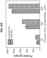
- the present platform, NanoPOTS can enable each sample to be processed within a 200 nL or smaller droplet that is contained in a wall-less glass reactor having a diameter of approximately 1 mm (e.g., total surface area of about 0.8 mm 2 ).
- a 100 ⁇ L typical sample preparation volume in 0.5 mL-centrifuge tubes (127.4 mm 2 ) the surface area was reduced by a factor of ⁇ 160, greatly reducing adsorptive losses.
- NanoPOTS When combined with analysis by ultrasensitive liquid chromatography-mass spectroscopy (LC-MS), biological samples prepared using nanoPOTS can enable deep profiling of greater than about 3000 proteins from as few as about 10 HeLa cells, a level of proteome coverage that has not been previously achieved for fewer than 10,000 mammalian cells.
- NanoPOTS can enable robust, quantitative and reproducible analyses and provide in-depth characterization of tissue substructures by profiling thin sections of single human islets isolated from clinical pancreatic specimens.
- FC and MC are also inherently targeted techniques with limited multiplexing capacity.
- MS mass spectrometry
- Efforts to improve sample preparation procedures include the use of low-binding sample tubes and ⁇ one pot' digestion protocols to limit total surface exposure (Sun, X, et al., Wisniewski, J et al, Chen, Q et al, Chen W. et al, Waanders, L. et al, Huang, E. et al, and Wang, N. et al).
- Trifluoroethanol-based protein extraction and denaturation (Wisniewski, J. et al 2011), filter-aided sample preparation, 13 MS-friendly surfactants (Waanders, L. et al, and Huang E., et al), high-temperature trypsin digestion (Chen, W.
- Samples may be any liquid, semi-solid or solid substance (or material).
- a sample can be a biological sample or a sample obtained from a biological material.
- a biological sample can be any solid or fluid sample obtained from, excreted by or secreted by any living organism, including without limitation, single celled organisms, such as bacteria, yeast, protozoans, and amoebas among others, multicellular organisms (such as plants or animals, including samples from a healthy or apparently healthy human subject or a human patient affected by a condition or disease to be diagnosed or investigated, such as cancer).
- a biological sample can be a biological fluid obtained from, for example, blood, plasma, serum, urine, bile, ascites, saliva, cerebrospinal fluid, aqueous or vitreous humor, or any bodily secretion, a transudate, an exudate (for example, fluid obtained from an abscess or any other site of infection or inflammation), or fluid obtained from a joint (for example, a normal joint or a joint affected by disease).
- a biological fluid obtained from, for example, blood, plasma, serum, urine, bile, ascites, saliva, cerebrospinal fluid, aqueous or vitreous humor, or any bodily secretion, a transudate, an exudate (for example, fluid obtained from an abscess or any other site of infection or inflammation), or fluid obtained from a joint (for example, a normal joint or a joint affected by disease).
- a biological sample can also be a sample obtained from any organ or tissue (including a biopsy or autopsy specimen, such as a tumor biopsy) or can include a cell (whether a primary cell or cultured cell) or medium conditioned by any cell, tissue or organ.
- a biological sample can be a nuclear extract.
- a biological sample can be bacterial cytoplasm.
- a sample can be a test sample.
- a test sample can be a cell, a tissue or cell pellet section prepared from a biological sample obtained from a subject.
- the subject can be one that is at risk or has acquired a particular condition or disease.
- the sample can be cells isolated from whole blood or cell isolated from histological thin sections.
- Illustrative biological samples include nanoscale biological samples (e.g., containing low- or subnanogram (e.g., less than about 1 ng) amounts of protein which may be processed in a single nanowell or subdivided into multiple nanowells).
- the sample is a biological sample and the biological sample is a tissue, and the tissue may be fixed.
- Tissues may be fixed by either perfusion with or submersion in a fixative, such as an aldehyde (such as formaldehyde, paraformaldehyde, glutaraldehyde, and the like).
- a fixative such as an aldehyde (such as formaldehyde, paraformaldehyde, glutaraldehyde, and the like).
- fixatives include oxidizing agents (for example, metallic ions and complexes, such as osmium tetroxide and chromic acid), protein-denaturing agents (for example, acetic acid, methanol, and ethanol), fixatives of unknown mechanism (for example, mercuric chloride, acetone, and picric acid), combination reagents (for example, Carnoy's fixative, methacarn, Bouin's fluid, B5 fixative, Rossman's fluid, and Gendre's fluid), microwaves, and miscellaneous (for example, excluded volume fixation and vapor fixation).
- Additives also may be included in the fixative, such as buffers, detergents, tannic acid, phenol, metal salts (for example, zinc chloride, zinc sulfate, and lithium salts), and lanthanum.
- the method for preparing a biological sample includes displacing a volume of biological sample to a single reactor vessel.
- the volume of biological sample is a non-zero amount less than 1 ⁇ L.
- the volume of biological sample may be a non-zero amount less than about 500 nL, less than about 400 nL, less than about 300 nL, less than about 200 nL, less than about 190 nL, less than about 180 nL, less than about 170 nL, less than about 160 nL, less than about 150 nL less than about 140 nL, less than about 130 nL, less than about 120 nL, less than about 110 nL, less than about 100 nL, less than about 90 nL, less than about 80 nL, less than about 70 nL, less than about 60 nL, less than about 50 nL, less than about 40 nL, less than about 30 nL, less than about 20 nL, less than about 10 nL, or less than
- the biological sample may be measured by their confluence.
- Confluency refers to cells in contact with one another on a surface (e.g., a tissue culture vessel, a petri dish, a well, and the like). For example, it can be expressed as an estimated (or counted) percentage, e.g., 10% confluency means that 10% of the surface, e.g., of a tissue culture vessel, is covered with cells, 100% means that it is entirely covered.
- adherent cells grow two dimensionally on the surface of a tissue culture well, plate or flask.
- Non-adherent cells can be spun down, pulled down by a vacuum, or tissue culture medium aspiration off the top of the cell population, or removed by aspiration or vacuum removal from the bottom of the vessel.
- the biological sample may include HeLa cells, A549 cells, CHO cells or MCF7 cells, K562 cells, or THP-1 cells, microbial cells, plant cells, or virtually any other biological material.
- the biological sample may include of primary or immortalized cells.
- primary or immortalized cells include but are not limited to, mesenchymal stem cells, lung cells, neuronal cells, fibroblasts, human umbilical vein (HUVEC) cells, and human embryonic kidney (HEK) cells, primary or immortalized hematopoietic stem cell (HSC), T cells, natural killer (NK) cells, cytokineinduced killer (CIK) cells, human cord blood CD34+ cells, B cells.
- HSC primary or immortalized hematopoietic stem cell
- T cells may include CD8+ or CD4+ T cells.
- the CD8+ subpopulation of the CD3+ T cells are used.
- CD8+ T cells may be purified from the PBMC population by positive isolation using anti-CD8 beads.
- the biological sample includes tissues, including but not limited, liver tissue, brain tissue, pancreatic tissue, breast cancer tissue, or plant tissue.
- the biological sample is collected and prepared using standard techniques. In examples not part of the present invention, cultured cells are collected and centrifuged. The pellet is then washed and re-suspended.
- the suspended cells are concentration to obtain desired cell numbers. In embodiments, the desired number of cells can be readily optimized. In certain aspects, the number of cells is 1 cell, 2 cells, 3 cells, 4 cells, 5 cells, 6 cells, 7 cells, 8 cells, 9 cells, 10 cells, 15 cells, 20 cells, 30 cells, 40 cells, 50 cells, 100 cells, 200 cells. In further embodiments, the sample is then adjusted to obtain a nano liter cell suspension (e.g., a 50 nL cell suspension).
- a nano liter cell suspension e.g., a 50 nL cell suspension
- the biological sample is a laser microdissected tissue.
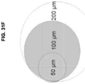
- the tissue is less than about 1000 ⁇ m, less than about 900 ⁇ m, less than about 800 ⁇ m, less than about 700 ⁇ m, less than about 600 ⁇ m, less than about 500 ⁇ m, less than about 400 ⁇ m, less than about 300 ⁇ m, less than about 200 ⁇ m, less than about 100 ⁇ m, less than about 50 ⁇ m, less than about 40 ⁇ m, less than about 30 ⁇ m, less than about 20 ⁇ m, less than about 10 ⁇ m, less than about 5 ⁇ m.
- a robotic nanoliter dispensing platform 100 can be employed to perform sample processing steps associated with bottom-up proteomics (e.g., robotic platform (Vandermarlier, E et al)).
- dispensing platform 100 can include a translatable stage 102 configured to receive a chip 104.
- the chip 104 can be configured to retain biological samples and reagents dispensed therein for further processing.
- the robotic platform 100 can be configured to provide submicron positioning accuracy and capacity for accurately handling picoliter volumes to dispense cells and reagents into reactor vessels formed in the chip 104 for further processing (e.g., to yield a processed sample).and to retrieve samples for subsequent analysis.
- Biological samples and/or reagents can be dispensed in the chip 104 via a syringe pump 206 including a picoliter dispensing tip 110 under the control of a controller, which can include one or more user interfaces for receiving commands from a user.
- the syringe pump 106 can be in fluid communication with a source of the biological samples (not shown) and one or more reservoirs 114 containing reagents.
- the platform 100 can further include a camera 116 or other imaging device for viewing dispensing of the biological samples and/or reagents.
- the total volume of biological samples and/or reagents can be less than 200 nL (in particular embodiments, a non-zero amount of less than 200 nL).
- Embodiments of the method can dramatically reduce surface contact to minimize sample loss while also enhancing reaction kinetics.
- the nanoPOTS platform described herein can reduce the total processing volumes (for example, the volume of the biological sample plus the total volume of all the reagents for processing) from the conventional tens or hundreds of microliters to less than 5,000 nL, less than 3,000 nL, less than 2,000 nL, less than 1,000 nL, less than 500 nL, less than 400 nL, less than 300 nL, less than 200 nL, less than 100 nL, less than 50 nL, less than 20 nL, less than 10 nL, less than 5nL.
- the total processing volumes for example, the volume of the biological sample plus the total volume of all the reagents for processing
- the biological sample is processed in a single reactor vessel to yield a processed sample.
- the single reactor vessel avoids the need to transfer samples to multiple reactor vessels for processing and therefore avoids the corresponding sample losses that such steps incur.
- the biological sample is processed in a single reactor vessel, a cocktail containing a reducing agent (e.g., dithiothreitol) is added and the sample is incubated.
- a cocktail containing a reducing agent e.g., dithiothreitol
- the pH is between 5 and 10, preferably 5, 5.5, 6, 6.5, 7, 7.5, 8, 8.5, 9, 9.5 or 10. More preferably, a solution pH value of 8 may be used.
- a protease is then added to the single reactor vessel (e.g., trypsin or LysC).
- the addition of a protease allows digestion of the polypeptides.
- the process may be performed in a humidity-controlled chamber.
- the humidity-controlled chamber is maintained at a relative humidity within the range from about 80% to about 100%, e.g., at about 95% humidity.
- a cover plate may be employed to minimize evaporation.
- methods disclosed herein may include steps such as washing steps to maximize recovery (e.g., into a capillary).
- the capillary can be fused or sealed from the external environment and stored.
- the processed biological sample is washed with a buffer (e.g., with water containing formic acid), and in some examples, multiple washing steps are performed, for example, 2 washing steps.
- Storage of the processed biological sample in the capillary may be short term (e.g., at about -20°C for less than 6 months) or long term (e.g., at about -70°C for greater than 12 months).
- the chip can be inverted during sample incubation to prevent the sample from settling on the reactor vessel surface (see FIGS. 34A and 34B ).
- droplets containing the biological sample may hang below the reactor vessel surface.
- Embodiments of the disclosed methods, as described herein, can have broad application in the fields of proteomics, metabolomics, and lipidomics, as such robust analysis from small samples have not been achievable using previously developed procedures.
- this description of potential applications is non-limiting and one skilled in the art will appreciate that embodiments of the disclosure can be employed in other applications without limit.
- biological samples processed according to embodiments of the disclosed methods may be analyzed using a variety of methods.
- the methods used to analyze the processed biological sample can include, but are not limited to, quantitative proteomic analysis methods.
- the processed biological sample may be analyzed mass spectrometry.
- Chromatography can also be employed for peptide separation.
- Liquid chromatography or capillary electrophoresis can be coupled to mass spectrometry, particularly with an electrospray ionization source.
- a transfer device e.g., a transfer capillary
- the microextraction column can, in turn, be coupled to the head of the liquid chromatography column.
- the transfer capillary may also be directly coupled to the head of the liquid chromatography column.
- the analyzing the processed biological sample can identify unique species, including but not limited to proteins or fragments thereof, lipids, or metabolites.
- analyzing the processed biological sample can identify at least about 1,000 unique species (e.g., proteins or fragments thereof, lipids, and/or metabolites). In additional embodiments, the processed biological sample can identify at least 2,000 unique species, at least 3,000 unique species, at least 4,000 unique species, at least 5,000 unique species, at least 7,000 unique species. In other embodiments, the number of unique species identified can be at least 500 or more proteins and/or 100 or more metabolites or lipids.
- the methods described herein can allow for the identification and quantitative measurements from less than about 200 cells (e.g., from the range of about 1 to about 50 mammalian cells). In particular embodiments, method described herein enables for identification of over 3,000 unique species from about 10-50 mammalian cells.
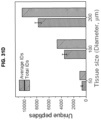
- nanowell sample processing can be coupled with laser-capture microdissection (LCM) for deep proteome analysis of heterogeneous tissue thin sections with ⁇ 100 ⁇ m resolution.
- LCM laser-capture microdissection
- LCM can differentiate and isolate subsections of tissue with high specificity
- sample requirements for proteomics can limit the resolution of LCM to large or pooled thin sections comprising thousands or tens of thousands of cells and millimeter or larger dimensions.
- Such heterogeneous tissues can confound molecular analysis due to a blurring of cellular constituents and their respective contributions.
- embodiments of the presently disclosed methods can provide proteomic analysis of LCM-isolated tissues by reducing sample size by approximately 2 orders of magnitude, to less than about 50 cells, which can enable both high resolution proteomic imaging (e.g., less than about 100 ⁇ m) as well as isolation of specific tissues from much smaller samples, such as smears from fine needle aspiration biopsies.
- LCM can be used to excise and transfer select tissue from thin section to the nanowell.
- an LCM e.g., Zeiss PALM Microbeam LCM ®
- FFPE paraffin embedded
- the Zeiss system can provide submicron resolution and it can be equipped with laser-pressure catapulting to eject excised samples to a variety of substrates, including centrifuge tube lids and slides (e.g., 25 ⁇ 75 mm).
- the Zeiss LCM can be compatible with standard glass slides for archived specimens as well as LCM-dedicated polymer membrane-coated slides.
- the nanowells can be configured for compatibility with the 25 ⁇ 75 mm form factor. This can allow for direct coupling and facilitate transfer from thin sections to the nanowells. As discussed in greater detail below, the nanowells can have a diameter of about 0.5 mm to about 1.5 mm. The spacing between the nanowell slide and the thin section slide may be adjusted to achieve the requisite transfer accuracy. Nanowell surface treatments may be implemented as needed to ensure adhesion of the catapulted tissue upon contact. As an alternative approach, excised samples can be catapulted into centrifuge tube caps and micromanipulationbased strategies can be used to transfer the sample to the nanowell.
- sample processing is seamlessly integrated with LCM by providing a capture liquid in or on a reactor vessel.
- This method can avoid manual transfer of dissected tissues to the nanowells that is required in a conventional LCM system.
- tissue pieces may be collected into microtubes by gravity or catapulted into tube caps prefilled with extraction solution or adhesive coating, depending on the instrument vendor and configuration.
- these collection approaches cannot be automatically integrated with a nanoPOTS system because the rapid evaporation of nanoliter-scale extraction solution and the prohibitive absorptive losses of proteins on the adhesive silicone coating. Utilizing a sacrificial capture medium in the nanowells addresses this challenge.
- the capture liquid has an ultra-low vapor pressure ( less than or equal to 0.8 mbar at room temperature), and evaporates very slowly under ambient conditions, which allows for long working times and uninterrupted sample collection.
- the evaporation times of 100 nL to 300 nL dimethyl sulfoxide (DMSO) droplets were 194 min to 416 min, which were >50 times longer than for water droplets. Such prolonged times are sufficient to collect up to hundreds of tissue samples in each chip.
- the capture liquid can be completely removed by gentle heating or vacuum, eliminating any possible interference during subsequent sample processing and analysis steps. Compared with other low-vapor-pressure solvents such as dimethylformamide, the capture liquid should have a lower toxicity, thus enabling its use as a storage solvent for cells.
- An illustrative capture liquid is dimethyl sulfoxide (DMSO).
- DMSO dimethyl sulfoxide
- the freezing point of DMSO is 18.5 °C, which should facilitate chip and sample transfer between histology and analytical labs without the risk of sample mixing or losses during shipping.
- DMSO significantly increases the sensitivity of protein identification of brain tissues, which may be ascribed to improved protein extraction efficiency as explained in more detail below.
- the amount of capture liquid provided in each nanowell may be sufficient to cover a portion of, or the entire surface, of the nanowell.
- the capture liquid may be present in an amount of at least 1 nL to 1000 nL.
- Embodiments of the methods described herein can be used for molecular characterization of tissue cellular heterogeneity or pathology in a variety of diseases.
- Exemplary diseases can include, but are not limited to, inflammatory diseases, metabolic diseases, cancers, neoplasias, and the like.
- metabolic disease can include its customary and ordinary meaning and can refer to diabetes, including type II diabetes, insulin-deficiency, insulin-resistance, insulin-resistance related disorders, glucose intolerance, syndrome X, inflammatory and immune disorders, osteoarthritis, dyslipidemia, metabolic syndrome, non-alcoholic fatty liver, abnormal lipid metabolism, neurodegenerative disorders, sleep apnea, hypertension, high cholesterol, atherogenic dyslipidemia, hyperlipidemic conditions such as atherosclerosis, hypercholesterolemia, and other coronary artery diseases in mammals, and other disorders of metabolism.
- the methods as used herein can be used in characterizing type 1 or type 2 diabetes.
- neoplasia can include its customary and ordinary meaning and can refer to a disease or disorder characterized by excess proliferation or reduced apoptosis.
- Illustrative neoplasms for which the embodiment may be used include, but are not limited to pancreatic cancer, leukemias (e.g., acute leukemia, acute lymphocytic leukemia, acute myelocytic leukemia, acute myeloblastic leukemia, acute promyelocytic leukemia, acute myelomonocytic leukemia, acute monocytic leukemia, acute erythroleukemia, chronic leukemia, chronic myelocytic leukemia, chronic lymphocytic leukemia), polycythemia vera, lymphoma (Hodgkin's disease, non-Hodgkin's disease), Waldenstrom's macroglobulinemia, heavy chain disease, and solid tumors such as sarcomas and carcinomas (e.g., fibrosarcoma
- biological sample can include its customary and ordinary meaning and can refers to a sample obtained from a biological subject, including sample of biological tissue or fluid origin obtained in vivo or in vitro. Such samples can be, but are not limited to, body fluid (e.g., blood, blood plasma, serum, or urine), organs, tissues, fractions, and cells isolated from mammals including, humans. Biological samples also may include sections of the biological sample including tissues (e.g., sectional portions of an organ or tissue). Biological samples may also include extracts from a biological sample, for example, an antigen from a biological fluid (e.g., blood or urine).
- a biological sample may be of prokaryotic origin or eukaryotic origin (e.g., insects, protozoa, birds, fish, or reptiles).
- the biological sample can be mammalian (e.g., rat, mouse, cow, dog, donkey, guinea pig, or rabbit).
- the biological sample can be of primate origin (e.g., example, chimpanzee, or human).
- transitional term “comprising,” which is synonymous with “including,” “containing,” or “characterized by,” is inclusive or open-ended and does not exclude additional, unrecited elements or method steps.
- the transitional phrase “consisting of' excludes any element, step, or ingredient not specified in the claim.
- the transitional phrase “consisting essentially of” limits the scope of a claim to the specified materials or steps "and those that do not materially affect the basic and novel characteristic(s)" of the claimed embodiments.
- Detectable moiety or a “label” can include its customary and ordinary meaning and it can refer to a composition detectable by spectroscopic, photochemical, biochemical, immunochemical, or chemical means.
- useful labels include 32 P, 35 S, fluorescent dyes, electron-dense reagents, enzymes ( e.g., as commonly used in an ELISA), biotin-streptavidin, dioxigenin, haptens and proteins for which antisera or monoclonal antibodies are available, or nucleic acid molecules with a sequence complementary to a target.
- the detectable moiety can generate a measurable signal, such as a radioactive, chromogenic, or fluorescent signal, that can be used to quantify the amount of bound detectable moiety in a sample. Quantitation of the signal can be achieved by, e.g., scintillation counting, densitometry, mass spectrometry, and/or flow cytometry.
- FFPE formalin fixed paraffin embedded tissue
- FFPE samples can be derived from tissues (often suspected tumor samples) that are fixed with formalin to preserve structural-spatial and biomolecule characteristics (e.g., cytoskeletal and protein structure) and then embedded in a type of paraffin wax so the tissue can be sliced.
- Formalin can irreversibly cross-link proteins via the amino groups, thus preserving the structural integrity of the cells so they can be stained with dyes or with immunostains used to analyze for abnormalities in the tissue that indicate altered cellular conditions, e.g., cancer.
- hydrophilic surface can include its customary and ordinary meaning and it can refer to a surface to have native hydrophilic property such as glass or fused silica, or which either hydrophilic compounds are covalently or non-covalently attached or which is formed of a polymer that has hydrophilic properties.
- the polymer with hydrophilic properties can be an organic polymer, (e.g., polyacrylamide, polyacrylic acid, polyacrylimide, polyelectrolytes, polyethylenimin, polyethylenglycol, polyethylenoxid, polyvinylalcohol, polyvinylpyrrolidon polystyrenesulfonic acid, copolymers of styrene and maleic acid, vinyl methyl ether malic acid copolymer, and polyvinylsulfonic acid.
- organic polymer e.g., polyacrylamide, polyacrylic acid, polyacrylimide, polyelectrolytes, polyethylenimin, polyethylenglycol, polyethylenoxid, polyvinylalcohol, polyvinylpyrrolidon polystyrenesulfonic acid, copolymers of styrene and maleic acid, vinyl methyl ether malic acid copolymer, and polyvinylsulfonic
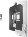
- the chip 200 can include a substrate 202, a spacer 204, one or more sealing membrane 206, and a cover 210.
- the spacer 204 can overlie the substrate 202
- the sealing membrane 206 can overlie the spacer 204
- the cover 210 can overlie the sealing membrane 206.
- the substrate 202 includes a physical and/or chemical pattern 212 that defines at least one reactor vessel having one or more hydrophilic and optionally hydrophobic surfaces configured for containment of a biological sample.
- the hydrophilic surfaces have a non-zero total surface area less than 5 mm 2 .
- the spacer 204 can contain a first aperture 204a and the sealing membrane 206 can include a second aperture 206a.
- the first and second apertures 204a, 206a can be dimensioned to accommodate the pattern 212 of reactor vessels when the chip 200 is assembled.
- at least the first aperture 204a of the spacer can surround the pattern 212 of reactor vessels.
- the sealing membrane 206 can be interposed between the spacer 204 and the cover 210 and it can be configured to form a fluid-tight seal between the spacer 204 and the cover 210.
- the sealing membrane can be interposed between the substrate and the spacer. Formation of fluid-tight seals using the sealing membrane can minimize evaporation of reactor vessel contents when performing incubation during sample preparation, as discussed below.
- other sealing mechanisms can be employed and the sealing membrane can be omitted.
- the cover 210 can be pre-coated with a layer of sealing membrane such as PDMS (polydimethylsiloxane).
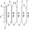
- FIGS. 3A-3E illustrate an exemplary forming the pattern 212 by photolithography.
- a substrate 300 coated with an anti-reflective coating 302 and photoresist 304 is illustrated.
- a photomask 306 can be used in conjunction with light 310 (e.g., ultraviolet light) to transfer a geometric pattern of the photomask 306 to the photoresist 304.
- the anti-reflective coating 302 can be configured to control reflection and absorption of the light 310. As shown in FIGS.
- the portions of the photomask 306 and anti-reflective coating 302 outside the transferred pattern can be removed by a chemical etching to yield a patterned substrate 312 that includes pillars 314 defining wells 316 therebetween of predetermined depth within the substrate 300.
- the photomask 306 and anti-reflective coating 302 remaining on the upper surface of the pillars 312 are removed with further chemical etching, as shown in FIGS. 3D-3E .
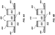
- FIGS. 4A-4B illustrate examples of the patterned substrate 412 defining reactor vessels configured for multiple-step proteomic sample processing.
- a hydrophobic coating can be deposited on the patterned substrate 412, adjacent to the pillars 414, to form a hydrophobic surface 402.
- a hydrophilic coating can be deposited on the patterned substrate 412 on the upper surface of the pillars 414 to form a hydrophilic surface 404.
- a hydrophobic coating can be omitted and the bare surface of the substrate can form the hydrophilic surface 404. So configured, the upper surface of each pillar 414 with the hydrophilic surface 404 can define the lateral boundary of respective reactor vessels 400.
- the patterned pillars 414 can reduce surface area contact relative to the use of concave wells.
- the locations of the hydrophobic and hydrophilic coatings can be reversed. That is, the hydrophilic coating can be deposited on the patterned substrate 412 adjacent to the pillars 414 (e.g., within the wells 416) to form the hydrophilic surface 404.
- the hydrophilic coating can be deposited on the patterned substrate 412 adjacent to the pillars 414 (e.g., within the wells 416) to form the hydrophilic surface 404.
- a hydrophobic coating can be omitted and the bare surface of the substrate can form the hydrophilic surface 404.
- the hydrophobic coating can be deposited on the patterned substrate 412 on the upper surface of the pillars 414 to form the hydrophobic surface 402. So configured, the wells 416 with the hydrophilic surface 404 can define the lateral boundary of respective reactor vessels 410.
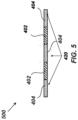
- a patterned substrate 500 can be formed by a pattern of hydrophobic surfaces 402 and hydrophilic surfaces 404 alone, without pillars 414 or wells 416.
- the hydrophobic surfaces 402 and the hydrophilic surfaces can be provided as discussed above and they can define the lateral extent of the one or more reactor vessels 420.
- a chip 600 that includes a substrate 601 and reactor vessel pillars 602 is inverted during processing of a biological sample.
- a droplet 603 of a capture liquid suspends from the hydrophilic surface of the reactor vessel pillar 602.
- the capture liquid droplet 603 contains a biological sample 604 that can be subjected to processing.
- the RapiGest-based one-pot protocol (Waters, Milford, USA) was adapted for proteomic sample preparation with minimal modification ( FIGS. 7-8 ). Briefly, after cells or other tissue samples were deposited into each chamber of the array, microscopic imaging was used for sample size quantification (cell number, tissue dimensions, etc.). A cocktail containing RapiGest and dithiothreitol was added and incubated at 70°C to lyse cells, extract and denature proteins, as well as reduce disulfide bonds in a single step. The proteins were alkylated and digested using a two-step enzymatic hydrolysis.
- the solution was acidified to cleave and inactivate the RapiGest surfactant.
- Manipulations were conducted in a humidified chamber, and the cover plate was sealed to the nanowell chip during extended incubation steps to minimize evaporation of the nanoliter droplets.
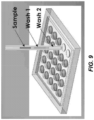
- the prepared sample was collected into a fused-silica capillary, followed by a two-step wash of the nanowell to maximize recovery ( FIG. 7 ).
- the collector capillary can be fully sealed and stored in a freezer for months without observable sample loss.
- the capillary also simplified downstream solid-phase extraction-based cleanup and LC-MS analysis by enabling direct coupling with standard fittings.
- FIG. 1 The sensitivity by processing 10-141 cultured HeLa cells with nanoPOTS ( FIG.1 ) was evaluated. Three different blank controls were used to confirm negligible carryover and contamination from the SPE and LC columns, reagents, and cell supernatant, respectively ( FIG. 15 ). In contrast to the control samples, all cell-containing samples showed feature-rich base peak chromatogram profiles, and the number of peaks and their intensities increased with the number of cells ( FIG. 13A-13C ). The percentage of peptides having tryptic cleavage sites ranged from 97.4% to 97.9%, while the percentage of peptides having tryptic missed cleavage sites ranged from 23.2% to 27.8% ( FIG.
- FIG. 24A-24C The ability to identify an average of 3,092 proteins in as small as ⁇ 10 cells ( FIG. 24A-24C ) represents a >500-fold decrease in sample size to achieve similar proteome coverage relative to previously reported methods (Sun, X et al, Chen, W et al., Wannders, L. et al, Huang, E. et al, and Wang, N. et al) (Table 1, below).
- Table 1 Reported protein identification results with cell number lower than 2,000 Cell # Cell type Identified protein # Sample preparation method 100 DLD-1 635 High temperature trypsin digestion 1 250 DLD-1 759 High temperature trypsin digestion 1 500 DLD-1 1060 High temperature trypsin digestion 1 MCF-7 187 Acetone precipitation 2 Hela 905 FASP 3 1000 MCF-7 271 Acetone precipitation 2 Hela 1536 FASP 3 2000 HEK 239T 1270 Spin tip 4
- the proteins were matched identified from 10-14 cells to the reported databases containing protein copy numbers per HeLa cell (Wisniewski, J. et al 2014, and Volpe, P. et al).
- the absolute copy numbers of 40 proteins in HeLa cell were precisely quantified using spiked-in protein epitope signature tags (PrEST) in combination with SILAC-based isotopic labeling (Volpe, P. et al). Thirty-four of the 40 proteins were identified, and the 6 missed proteins were low in abundance.
- the corresponding protein copy number per cell ranged from about 5 ⁇ 10 4 to about 2 ⁇ 10 7 (Table 2), with 3 expressed at ⁇ 10 5 copies/cell.
- the detection limit of nanoPOTS for protein is ⁇ 5 ⁇ 10 5 copies, or ⁇ 830 zmol.
- Table 2 Copy numbers per HeLa cell for proteins identified from 10-14 cells (copy number obtained from PrEST-SILAC method (Li, et al. and Zeiler, M. et al.).
- Deionized water (18.2 M ⁇ ) was purified using a Barnstead Nanopure Infinity system (Los Angeles, USA).
- Dithiothreitol (DTT) and iodoacetamide (IAA) were purchased from Thermo Scientific (St. Louis, USA) and freshly prepared in 50 mM ammonium bicarbonate buffer each day before use.
- RapiGest SF surfactant (Waters, Milford, USA) was dissolved in 50 mM ammonium bicarbonate buffer with a concentration 0.2% (m/m), aliquoted, and stored at -20 °C until use. Trypsin (MS grade) and Lys-C (MS grade) were products of Promega (Madison, USA).
- Other unmentioned reagents were obtained from Sigma-Aldrich (St. Louis, USA).
- the photomask was designed with AutoCAD and printed with a direct-write lithography system (SF-100, Intelligent Micro Patterning LLC, St. Russia, USA).
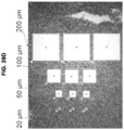
- An array of 3 ⁇ 7 spots with diameters of 1 mm and a spacing of 4.5 mm was designed on a 25 mm ⁇ 75 mm glass slide (soda lime) that was pre-coated with chromium and photoresist (Telic Company, Valencia, USA).
- chromium and photoresist Telic Company, Valencia, USA.
- FIG. 3A After photoresist exposure ( FIG. 3A ), development, and chromium etching (Transene, Danvers, USA; FIG. 3B ), the glass slide was hard baked at 110 °C for 10 min.
- the back side of the slide was protected with packing tape and the glass substrate surface was etched around the patterned photoresist/Cr features using wet etching solution containing 1 M HF, 0.5 M NH 4 F, and 0.75 M HNO 3 at 40 °C for 10 min to reach a depth of 10 ⁇ m ( FIG. 3C ).
- the remaining photoresist was removed using AZ 400T stripper.
- the glass slide was thoroughly rinsed with water, dried using compressed nitrogen, and further dried in an oven at 120 °C for 2 h.
- the chip surface was then cleaned and activated with oxygen plasma treatment for 3 minutes using a March Plasma Systems PX250 (Concord, USA).
- the glass surface that was not protected with Cr was rendered hydrophobic with a fluorosilane solution containing 2% (v/v) heptadecafluoro-1,1,2,2-tetrahydrodecyl)dimethylchlorosilane (PFDS) in 2,2,4-trimethylpentane ( FIG. 3D ) for 30 min.
- the residual silane solution was removed by immersing the chip in 2,2,4-trimethylpentane followed by ethanol. Remaining chromium was removed using chromium etchant (Transene), leaving elevated hydrophilic nanowells on a hydrophobic background ( FIG. 3E ).
- the glass spacer was fabricated by milling a standard microscope slide (25 mm ⁇ 75 mm ⁇ 1 mm) with a CNC machine (Minitech Machinery Corporation, Norcross, USA). Epoxy was used to glue the patterned chip and the glass spacer together.
- the glass cover was fabricated by spin coating a thin layer of polydimethylsiloxane (PDMS) membrane (10- ⁇ m thickness) onto a standard glass microscope slide of the same dimensions. Briefly, Dow Coming Sylgard 184 silicone base was mixed with its curing reagent at a ratio of 10:1 (w/w) and degassed for 20 min.
- PDMS polydimethylsiloxane
- the mixture was coated on the slide by spinning at 500 rpm for 30 s followed by 3000 rpm for 5 min (WS-650, Laurell Technologies, North Wales, USA). Finally, the PDMS membrane was cured at 70 °C for 10 hours. A piece of Parafilm (Bemis Company, Oshkosh, USA) was precisely cut to serve as moisture barrier between the glass spacer and the glass cover.
- Nanoliter-scale liquid handling system
- the capillary probe was fabricated by heating pulling a fused silica capillary (200 ⁇ m i.d., 360 ⁇ m o.d., Polymicro Technologies, Phoenix, USA) to generate a tapered tip (30 ⁇ m i.d., 50 ⁇ m o.d.).
- a home-built program with LabView (Version 2015, National Instruments, Austin, USA) was used to synchronously control the movement of the 3D stages and the liquid dispensing of the syringe pump. To minimize evaporation during the liquid handling procedure, the whole system was enclosed in a Lexan chamber maintained at 95% relative humidity.
- the syringe pump was set at a withdraw rate of 9 ⁇ L/min and an infusion rate of 3 ⁇ L/min.
- the translation stages were operated at a start speed of 1 cm/s, a maximum speed of 30 cm/s, and an acceleration time of 0.5 s. In the typical setup, it took total ⁇ 2 min to dispense one reagent to all the 21 droplets in single chip including the time for withdrawing reagent into the capillary probe, moving of the robotic stages, and dispensing 50 nL reagent into each droplet.
- the nanowells can be scaled up with the present photolithography-based microfabrication technique. Up to 350 nanowells can be fabricated on a 25 mm ⁇ 75 mm microscope slide and further scale-up is possible with larger substrates.
- the robot can be simply configured to fit different formats of nanowell array. Because of the high liquid handling speed, 350 droplets could be addressed in ⁇ 30 min.
- HeLa was grown in Eagle's Minimum Essential Medium (EMEM) supplemented with 10% fetal bovine serum (FBS) and 1 ⁇ penicillin streptomycin.
- EMEM Eagle's Minimum Essential Medium
- FBS fetal bovine serum
- HeLa cells were collected in a 10 mL tube and centrifuged at 1200 rpm for 10 minutes to remove culture media. The cell pellet was further washed three times with 10 mL of 1 ⁇ PBS buffer. The cells were then suspended in 1 mL PBS buffer and counted to obtain cell concentration. Eppendorf protein low-binding vials (0.5 mL) were used throughout the process. Cells were lysed at a concentration of 5 ⁇ 10 5 /mL in 0.1% RapiGest and 5 mM DTT in 50 mM ammonium bicarbonate (ABC).
- ABS ammonium bicarbonate
- the cell lysate was diluted in 50 mM ABC buffer and aliquoted to different vails with a volume of 5 ⁇ L.
- 5 ⁇ L of IAA solution (30 mM in 50 mM ABC) was dispensed to alkylate sulfhydryl groups by incubating the vials in the dark for 30 minutes at room temperature.
- 5 ⁇ L of Lys-C (0.25 ng in 50 mM ABC) was added and incubated at 37 °C for 4 h.
- 5 ⁇ L of Trypsin (0.25 ng in 50 mM ABC) was added and incubated overnight at 37 °C.
- 5 ⁇ L of formic acid solution (30%, v/v) were dispensed and allowed to incubate for 1 h at room temperature to cleave RapiGest surfactant for downstream analysis.
- the chip was washed with isopropanol and water to minimize contamination.
- the liquid handling system was configured to minimize cross contamination by adjusting the vertical distance between the probe tip and the nanowell surface, which was previously termed semi-contact dispensing (Zhu, Y. et al 2014).
- cells were collected in a 10 mL tube and centrifuged at 1200 rpm for 10 minutes to remove culture media. The cell pellet was further washed three times with 10 mL of 1 ⁇ PBS buffer. The cells were then suspended in 1 mL PBS buffer and counted to obtain cell concentration. Cell concentrations were adjusted by serially diluting them in PBS to obtain different cell numbers in nanowells. After dispensing 50 nL of cell suspension into each nanowell, we observed that the distribution of cell numbers in nanowell was stochastic, especially for lowconcentration cell suspensions. Thus, the accurate cell number in each nanowell was counted using an inverted microscope and indexed to the two-dimensional spatial position of the corresponding nanowell.
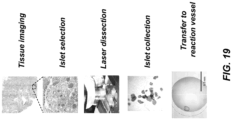
- LCM tissues For LCM tissues, a high precision tweezer with a tip of 20 ⁇ m (TerraUniversal, Buellton, USA) was used to transfer tissue pieces from collection tubes into individual nanowells under a stereomicroscope (SMZ1270, Nikon, Japan). ImageJ software 37 was used to measure the area of LCM islets to calculate islet equivalents (IEQ) and cell numbers.
- IEQ islet equivalents
- RapiGest (Yu et al. 2003) (0.2%) solution with 10 mM DTT in 50 mM ammonium bicarbonate (ABC) was added into the nanodroplets that had been preloaded with cells.
- Ammonium bicarbonate (ABC)
- the cover was then sealed to the nanodroplet chip, which was incubated in 70 °C for 30 min to achieve cell lysis, protein denaturation, and disulfide reduction.
- the second step 50 nL of IAA solution (30 mM in 50 mM ABC) was dispensed to alkylate sulfhydryl groups by incubating the chip in the dark for 30 minutes at room temperature.
- 50 nL enzyme solution containing 0.25 ng Lys-C in 50 mM ABC was added and incubated at 37 °C for 4 h for predigestion.
- 50 nL of enzyme solution containing 0.25 ng trypsin in 50 mM ABC was added into each droplet and incubated overnight at 37 °C for tryptic digestion.
- formic acid solution (30%, v/v) was dispensed and allowed to incubate for 1 h at room temperature to cleave RapiGest surfactant for downstream analysis.
- the chip was completely sealed during cell counting, incubation, and transfer procedures. During each dispensing step, the chip was opened and closed within the humidity chamber to minimize droplet evaporation.
- the total dispensed volume in each droplet was 300 nL, and the final volume was typically ⁇ 200 nL, some evaporative losses clearly occurred.
- Digested peptide samples in each nanowell were collected and stored in a section of fused silica capillary (5 cm long, 150 ⁇ m i.d., 360 ⁇ m o.d.). Before sample collection, the capillary was connected to the syringe pump and filled with water containing 0.1% formic acid (LC Buffer A) as carrier. A plug of air (10 nL, 0.5 mm in length) was aspirated into the front end of the capillary to separate sample from carrier. The capillary-to-nanowell distance was adjusted to ⁇ 20 ⁇ m to allow majority of sample to be aspirated into the capillary.
- LC Buffer A formic acid
- the nanowell was twice washed with 200-nL buffer A and the wash solutions were also collected in the same capillary.
- a section of capillary containing a train of plugs consisting of carrier, air bubble, sample, and wash solutions was then cut from the syringe pump.
- the capillary section was sealed with Parafilm at both ends and stored at -20 °C for short-term storage or -70 °C for long-term storage.
- the SPE precolumn and LC column were slurry-packed with 3- ⁇ m C18 packing material (300- ⁇ pore size, Phenomenex, Terrence, USA) as described previously (Shen, Y. et al 2004, and Shen, Y. et al. 2003).
- the SPE column was prepared from a 4-cm-long fused silica capillary (100 ⁇ m i.d., 360 ⁇ m o.d., Polymicro Technologies, Phoenix, AZ).
- the LC column was prepared from a 70-cm Self-Pack PicoFrit column with an i.d. of 30 ⁇ m and a tip size of 10 ⁇ m (New Objective, Woburn, USA).
- the sample storage capillary was connected to the SPE column with a PEEK union (Valco instruments, Houston, USA).
- Sample was loaded and desalted in the SPE precolumn by infusing buffer A (0.1% formic acid in water) at a flow rate of 500 nL/min for 20 minutes with an nanoACQUITY UPLC pump (Waters, Milford, USA).
- the SPE precolumn was reconnected to the LC column with a low-dead-volume PEEK union (Valco, Houston, USA).
- the LC separation flow rate was 60 nL/min, which was split from 400 nL/min with a nanoACQUITY UPLC pump (Waters, Milford, USA).
- a linear 150-min gradient of 5-28% buffer B (0.1% formic acid in acetonitrile) was used for separation.
- the LC column was washed by ramping buffer B to 80% in 20 minutes, and finally re-equilibrated with buffer A for another 20 minutes.
- An Obitrap Fusion Lumos Tribrid MS (ThermoFisher) was employed for all data collection. Electrospray voltage of 1.9 kV was applied at the source. The ion transfer tube was set at 150 °C for desolvation. S-lens RF level was set at 30. A full MS scan range of 375-1575 and Obitrap resolution of 120,000 (at m / z 200) was used for all samples. The AGC target and maximum injection time were set as 1E6 and 246 ms. Data-dependent acquisition (DDA) mode was used to trigger precursor isolation and sequencing.
- DDA Data-dependent acquisition
- Precursor ions with charges of +2 to +7 were isolated with an m / z window of 2 and fragmented by high energy dissociation (HCD) with a collision energy of 28%.
- the signal intensity threshold was set at 6000.
- dynamic exclusion with duration of 90 s and mass tolerance of ⁇ 10 ppm was utilized.
- MS/MS scans were performed in the Obitrap.
- the AGC target was fixed at 1E5. For different sample inputs, different scan resolutions and injection times were used to maximize sensitivity (240k and 502 ms for blank control and ⁇ 10-cell samples; 120k and 246 ms for ⁇ 40-cell samples; 60k and 118 ms for ⁇ 140-cell samples).
- the minimum peptide length was 7 amino acids and maximum peptide mass was 4600 Da.
- the allowed missed cleavages for each peptide was 2.
- the second peptide search was activated to identify co-eluting and cofragmented peptides from one MS/MS spectrum. Both peptides and proteins were filtered with a maximum false discovery rate (FDR) of 0.01.
- FDR maximum false discovery rate
- the Match Between Runs feature with a match window of 0.7 min and alignment window of 20 min, was activated to increase peptide/protein identification of low-cell-number samples.
- LFQ calculations were performed separately in each parameter group that containing similar cell loading. Both unique and razor peptides were selected for protein quantification. Requiring MS/MS for LFQ comparisons was not activated to increase the quantifiable proteins in low-cell-number samples. Other unmentioned parameters were the default settings of the Maxquant software.
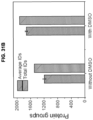
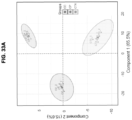
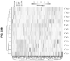
- Perseus (Tyanova, S. et al. 2016) was used to perform data analysis and extraction. To identify the significantly changed proteins from a non-diabetic donor and a T1D donor, the datasets were filtered to contain 3 valid LFQ intensity values in at least one group. The missing values were imputed from normal distribution with a width of 0.3 and a down shift of 1.8. Two sample T-test with a minimal fold change of 2 and a FDR of 0.01 was performed for statistical analysis. The extracted data were further processed and visualized with OriginLab 2017. Global scaling normalization was achieved using scaling coefficients calculated as the ratio of peptide abundance to the median peptide abundance measured for each loading set.
- Coefficients of variation were calculated by dividing the standard deviation of normalized intensities by the mean intensity across the datasets of similar loading.
- the Violin plot was generated with an online tool (BoxPlotR, http://shiny.chemgrid.org/boxplotr/) (Spitzer, M. et al).
- the nanoPOTS platform provided a robust, semi-automated nanodroplet-based proteomic processing system for handling extremely small biological samples down to as few as 10 cells with high processing efficiency and minimal sample loss.
- This capability opens up many potential biomedical applications from small cell populations and clinical specimens such as tissue sections for characterizing tissue or cellular heterogeneity.
- Reproducible quantitative proteome measurements with coverage of 2000-3,000 protein groups from as few as 10 mammalian cells or single human islet cross sections ( ⁇ 100 cells) from clinical specimens were demonstrated. While several previous efforts have pursued the analysis of ⁇ 2000 cells, most of these methods lacked the robustness and reproducibility for biological applications because of the highly manual processes involved (Li, S. et al 2015, Chen, Q. et al 2015, Chen, W.
- the nanoPOTS platform not only provided unparalleled proteome coverage for analyzing 10-100 cells, but also offered a number of technical advantages for achieving a high degree of robustness and reproducibility for high throughput processing and quantitative measurements when coupled with LC-MS.
- the platform effectively addressed the bottleneck of sample losses during proteomics sample preparation by performing all of the multi-step reactions within a single nanodroplet of ⁇ 200 nL volume, while all previous methods still suffer from a significant degree of protein/peptide losses during processing.
- the nanodroplet processing mechanism allowed us to perform each reaction at optimal concentrations. For example, by preserving the 20-50:1 ratio (Vandermarlier, E.
- the nanoPOTS Compared with other microfluidic platforms having closed microchannels and chambers (White, A. et al. 2011, and Zhu, Y. et al. 2010), the nanoPOTS has an open structure, which is inherently suitable for integration with upstream and downstream proteomic workflows, including sample isolation for processing and transfer for LC-MS analysis.
- the method was used to analyze cross-sections of individual human islets having a thickness of 10 ⁇ m ( FIG. 19 ) that were isolated by laser microdissection from clinical pancreatic specimens ( FIG. 18A-18D ).
- the islet equivalents (IEQ) were calculated to be from 0.06 to 0.17, corresponding to approximately 91 to 266 cells based on their volumes and a previous quantitative study (Zeiler, M. et al.) (Table 3).
- Table 3 Calculation of cell number and islet equivalents with islet areas (Pisania, A. et al 2010).
- Islet Area ⁇ m 2
- Islet volume ⁇ m 3
- Cell Number Islet equivalents IEQ No1 30197 301973 266 0.17 No2 16200 162004 143 0.09
- No3 21286 212862 188 0.12 No4 11133 111330 98 0.06
- No6 22428 224280 198 0.13
- No8 15186 151860 134 0.09
- nanoPOTS chip directly interfaced with fluorescence-activated cell sorting (FACS) for cell isolation.
- FACS fluorescence-activated cell sorting
- nanoPOTS represented a highly promising platform towards single mammalian cell proteomics with optimized processing volumes and further refinements to the LC-MS platform.
- the total processing volume could be reduced to the lownanoliter range to further minimize sample loss.
- FACS or other cell isolation techniques should be used to isolate single cells into nanowells without the minimal exogenous contamination from, e.g., secreted proteins or lysed cells.
- NanoLC columns with narrower bore Shen, Y. et al. 2004, and Shen, Y. 2003
- ESI emitter technology accommodating the lower resulting flow rates (Smit, R.
- nanoPOTS should also provide a viable path towards tissue imaging at the proteome level by performing in-depth spatially resolved proteome measurements for specific cellular regions.
- Example 3 nanoPOTS with LCM and capture liquid
- Nanowells are prepopulated with DMSO droplets to serve as a sacrificial capture medium for small tissue samples in the nanoPOTS chip ( FIGS. 29A-29E ) as described below in detail.
- DTT Dithiothreitol
- IAA iodoacetamide
- DDM n-dodecyl- ⁇ -D-maltoside
- DDM Mayer's hematoxylin
- eosin Y alcoholic solution
- Scott's Tap Water Substitute DMSO were purchased from Sigma-Aldrich.
- Trypsin (MS grade) and Lys-C (MS grade) were from Promega (Madison, WI). Other unmentioned reagents were obtained from ThermoFisher.
- Nanowell chip fabrication The nanowell chip consisted of three parts including a nanowell-containing substrate, a spacer, and a cover plate.
- the nanowell substrate was fabricated with the similar procedures described previously. ( Liu, Anal. Chem. 2017, 89(1), 822-829 ; Zhu, Anal. Chem. 2010, 82 (19), 8361-8366 ) Briefly, a glass slide (25 mm ⁇ 75 mm) with pre-coated chromium and photoresist (Telic company, Valencia, CA) was used as starting material.
- Standard photolithography and wet etching procedures were employed to generate an array of pedestals with a diameter of 1.2 mm, a height of 10 ⁇ m, and a spacing of 4.5 mm between adjacent pedestals on the slide.
- the exposed surfaces surrounding the pedestals were treated to be hydrophobic with 2% (v/v) heptadecafluoro-1,1,2,2-tetrahydrodecyl)dimethylchlorosilane (PFDS) (Sigma Aldrich) in 2,2,4-trimethylpentane.
- PFDS heptadecafluoro-1,1,2,2-tetrahydrodecyl)dimethylchlorosilane
- the glass spacer was laser-machined (Coherent Inc., Santa Clara, CA) on a standard 1.2-mm-thick microscope slide. The machining process removed the center region of the slide, leaving a thin frame of ⁇ 5 mm in width.
- the machined slide was glued to the nanowell substrate using a silicone adhesive, and served as a spacer to limit the headspace of the nanowells after reversibly sealing to a cover plate to minimize evaporation during incubation steps, while prevent contact of the droplet reactors with the cover plate.
- the cover plate was produced by spin coating of a thin layer of Sylgard 184 and its curing reagent (10/1, v/v) (Dow Coming) at a spin speed of 500 rpm for 30 s followed by 3000 rpm for 5 min.
- the cover plate was baked at 70 °C for 10 hours to generate a ⁇ 30- ⁇ m-thick polydimethylsiloxane (PDMS) layer.
- PDMS polydimethylsiloxane
- Rats were anesthetized by intra-peritoneal injection of chloral hydrate. Rat brain was dissected and snap frozen in liquid nitrogen. The brains were stored at -80 °C until use. A cryostat (NX-70, Thermo Scientific, St. Louis, MO) was used to cut tissues to a thickness of 12 ⁇ m. The chuck and blade temperatures were set as -16 °C and -20 °C, respectively. The tissue sections were deposited on PEN membrane slides (Carl Zeiss Microscopy, Germany) and stored at -80 °C.
- the tissue section was removed from the freezer or dry ice box and immediately immersed into 70% ethanol to fix proteins.
- the tissue was then rehydrated in deionized water for 30 s and stained in Mayer's hematoxylin solution for 1 min. Excess dye was rinsed with water and the tissue was blued in Scott's Tap Water Substitute for 15 s.
- 70% ethanol was used to dehydrate the tissue and a 50% dilution of eosin Y solution (v/v in ethanol) was applied for 1-2 s by a quick dip.
- the tissue sample was further dehydrated by immersion twice in 95% ethanol for 30 s, twice in 100% ethanol for 30 s, and finally in xylene for 2 min. All the procedures were performed in a fume hood and the slide was blotted on absorbent paper between different solutions to minimize carry over. The processed tissue could be directly used for LCM or stored at -80 °C until use.
- FIG. 29B An array of DMSO droplets with a volume of 200 nL were deposited on nanowells using a nanoliter-dispensing robotic system ( FIG. 29B ).
- a PALM microbeam laser capture microdissection system (Carl Zeiss MicroImaging, Kunststoff, Germany) was employed.
- the nanowell chip was fixed on a standard adapter for microscope slide (SlideCollector 48, Carl Zeiss MicroImaging) and then mounted on the robotic arm of the LCM system ( FIG. 29C ).
- the brain tissues were cut at an energy level of 42, and catapulted into DMSO droplet using the "CenterRoboLPC" function with an energy level of delta 15 and a focus level of delta 10.
- Tissue samples in the nanowell chip could be processed directly or stored at -20 °C.
- NanoPOTS proteomic sample processing Before processing, DMSO droplets were evaporated to dryness by keeping the nanowell chip in a vacuum desiccator for 10 to 15 min ( FIG. 29E ). Reagent dispensing was performed using the robotic system as described previously. ( Zhu, Anal. Chem. 2013, 85 (14), 6723-6731 ; Zhu, Sci. Rep. 2015, 5, 9551 ; Zhu, Sci. Rep. 2014, 4, 5046 ) Briefly, 100 nL 1 ⁇ PBS buffer containing 0.2% DDM surfactant and 5 mM DTT was added into each nanowell. The chip was incubated at 70 °C for 1 h for protein extraction and denaturation.
- Proteins were then alkylated by adding 50 nL of 30 mM IAA in 50 mM ammonium bicarbonate (ABC) in each reaction and then incubating for 40 min in the dark. A two-step digestion was performed at 37 °C with Lys-C and trypsin for 4 h and 8 h, respectively. Finally, the digested peptide samples were collected and stored in a fused silica capillary (4 cm long, 200 ⁇ m i.d., 360 ⁇ m o.d.). Each nanowell was washed twice with 200 nL, 0.1% formic acid aqueous buffer and the wash solution was also collected into the same capillary to maximize sample recovery.
- a two-step digestion was performed at 37 °C with Lys-C and trypsin for 4 h and 8 h, respectively.
- the digested peptide samples were collected and stored in a fused silica capillary (4 cm long, 200 ⁇ m i.d., 360 ⁇ m
- the distance between the capillary distal end and the nanowell surface was kept at 100 ⁇ m during the sample aspiration process.
- the capillary was sealed with Parafilm at both ends and stored at -70 °C until analyzed.
- NanoLC-MS/MS for protein identification Samples in the collection capillary were desalted and concentrated on a solid phase extraction (SPE) column (75-pm-i.d. fused silica capillary packed with 3 ⁇ m, 300 ⁇ pore size C18 particles, Phenomenex, Terrence, CA). Peptides were separated using a 60-cm-long, 30-pm-i.d. nanoLC column (3 ⁇ m, 300 ⁇ pore size C18 particles, Phenomenex) with an integrated electrospray emitter (Self-Pack PicoFrit column, New Objective, Woburn, MA).
- SPE solid phase extraction
- a nanoUPLC pump (Dionex UltiMate NCP-3200RS, Thermo Scientific, Waltham, MI) was used to deliver mobile phase to the LC column.
- a tee interface was used to split the LC flow rate from 300 nL/min to 50 nl/min for the 30-pm-i.d. LC column.
- a linear 100-min gradient starting from 8% buffer B (0.1% formic acid in acetonitrile; buffer A: 0.1% formic acid in water) to 22%, followed by a 15-min linear increase to 35% buffer B.
- the column was washed with 90% buffer B for 5 min and re-equilibrated with 2% buffer B for 20 min prior to the subsequent analysis.
- Peptides were ionized at the nanospray source using a potential of 2 kV.
- An Obitrap Fusion Lumos Tribrid MS (ThermoFisher) operated in data dependent mode to automatically switch between full scan MS and MS/MS acquisition with a cycle time of 2 s.
- the ion transfer capillary was heated to 250 °C to accelerate desolvation, and the S lens was set at 30.
- Full-scan MS spectra ( m / z 375-1600) were acquired in the Orbitrap analyzer with 120,000 resolution ( m / z 200), and AGC target of 3 ⁇ 10 6 , and a maximum ion accumulation time of 246 ms.
- Precursor ions with charges from +2 to +7 were isolated with an m / z window of 2 and were sequentially fragmented by high energy dissociation (HCD) with a collision energy of 30%.
- the AGC target was set at 1 ⁇ 10 5 .
- MS/MS scan spectra were acquired in the Orbitrap with an ion accumulation time of 502 ms and resolution of 240,000 for 50- ⁇ m-diameter tissue sample, an ion accumulation time of 246 ms and 120,000 resolution for 100- ⁇ m-diameter tissue sample, or an ion accumulation time of 118 ms and 60,000 resolution for 200- ⁇ m-diameter tissue samples, respectively.
- FDR false discovery rate
- Contamination and reverse identification was filtered with Perseus (version 1.5.6.0).
- the LFQ intensities were transformed with log2 function, and then filtered to contain > 70% valid values in at least one group. The missing values were imputed by normal distribution in each column with a width of 0.3 and a down shift of 1.8.
- the results were exported to a table and visualized with OriginPro 2017 and an online tool powered by R language.
- the capture efficiency with square tissues having side lengths of 20 ⁇ m, 50 ⁇ m, 100 ⁇ m, and 200 ⁇ m using a 12- ⁇ m-thick breast cancer tissue section from a previous study was evaluated.
- tissue samples with square side lengths from 20 ⁇ m to 100 ⁇ m a total of 75 cuts were collected into three droplets for each size.
- tissue samples (200 ⁇ m) a total of 27 cuts were collected.
- the "CenterRoboLPC" function in which the catapult laser pulse was applied at the centroid of pre-cut tissue piece, was used instead of commonly-used "RoboLPC".
- the "CenterRoboLPC" function provided better control on the catapult trajectory of tissue pieces from slide to DMSO droplets.
- tissue pieces with a diameter of 20 ⁇ m correspond to single cells in most of mammalian tissues, demonstrating the potential of the present approach for single-cell isolation and analysis.
- FIG. 31A A Venn diagram of total protein identifications indicates that most of the proteins obtained from DMSO-free samples were included in that of DMSO-collected samples ( FIG. 31B ). This demonstrates that the use of DMSO droplets did not generate any negative effects on the proteomic analysis.
- proteome coverage significantly increased for small tissue samples.
- a possible explanation for this result is that protein extraction efficiency was improved after hydrophobic lipids were removed by DMSO in the brain tissue.
- Protein extraction from tissue samples was found to be more challenging than for cultured cells, especially for tissue containing high lipid content such as brain.
- Various approaches have been developed to address this challenge by employing strong detergents or organic solvent in the extraction buffer.
- DMSO is expected to have high solubility for most lipids, and thus could dissolve them prior to protein extraction.
- sample losses in detergent removing steps including buffer exchange and spin columns was avoided using the inventive approach described herein.
- FIGS. 31D and 31E show the linear increase of unique peptide and protein identifications with tissue size. As expected, nearly all peptides and proteins identified in the smaller tissues were also identified in larger tissues ( FIG. 31D ), demonstrating analytical sensitivity dominated the proteome coverage.
- the LCM-DMSO-nanoPOTS system provided >25 times better spatial resolution with higher proteome coverage.
- the 1918 total proteins identified from 200- ⁇ m-diameter cortex tissues were submitted for Gene Ontology Cellular Component (GOCC) analysis. As shown in FIG. 31G , we observed a high percentage (56%) of membrane proteins and half of them (28%) were localized in plasma membrane, although no specific sample preparation procedures were used for membrane proteins. 10% synapse proteins and 7% axon proteins (not shown in FIG. 31G ), which are vital for brain function, were also observed. In brain, the major neurotransmitters are glutamate and GABA, which play excitatory and inhibitory functions, respectively.
- GABA receptors GABA receptors
- GABA receptors GABA receptors
- GABA receptors GABA receptors
- GBRB1, GABRB2, GABRB2, and GABRG2 GABA receptors
- GABA receptors GABA receptors
- GRM2, GRM3, GRM5 GPR158, GRIK3, GRIN1, GRIN2a, and GRIN2b.
- the spatial distances were from 116 ⁇ m to 716 ⁇ m between the same regions, and from 424 ⁇ m to 1,727 ⁇ m between different regions ( FIG. 32A ), showing the high spatial resolution of the present measurement.
- six samples were processed and four of them were submitted for LC-MS analysis ( FIG. 32B ).
- MLR Match Between Runs
- AMT LC retention times
- CTX and CP shows lower in correlation coefficients from 0.94 to 0.97, while CC has lowest correlations (from 0.83 to 0.91) with the other two regions. Such differences are also indicated in the morphology of the brain tissue ( FIG. 32A ).
- the LCM-DMSO-nanoPOTS system was tested to see if it could be applied to distinguish different tissue types.
- Unsupervised principal component analysis (PCA) was used to process the LFQ intensity data from the 12 tissue samples. As shown in FIG. 33A , the three tissue regions were segregated based on component 1 and component 2, which accounted for 65.5% and 15.6%, respectively. All four biological replicates were well clustered within the corresponding tissue region without overlap with other regions, suggesting the present system can efficiently distinguish tissue types based on their protein expressions.
- HCA hierarchical clustering analysis
- the results described herein demonstrate that the LCM- capture liquid-nanoPOTS platform significantly advances spatially-resolved proteomics by improving the resolution and increasing the sensitivity.
- the use of DMSO droplets not only served to efficiently capture dissected tissue pieces as small as 20- ⁇ m diameter (single-cell scale) into nanowells, but also significantly improved the proteome coverage.
- the whole workflow can be fully automated without manual transfer, and thus sample loss and protein contamination is minimized.
- This platform may play an important role in proteomic analyses and may be applied to various fields including biomedical research, clinical diagnosis, microbial community, and plant science.
- the LCM- capture liquid-nanoPOTS platform should be readily extended to other omics studies requiring tissue isolation and nanoscale processing, such as transcriptomics, lipidomics, and metabolomics.
- ranges specifically include the values provided as endpoint values of the range.
- a range of 1 to 100 specifically includes the end point values of 1 and 100. It will be understood that any sub-ranges or individual values in a range or sub-range that are included in the description herein can be excluded from the claims herein.
Landscapes
- Health & Medical Sciences (AREA)
- Chemical & Material Sciences (AREA)
- Life Sciences & Earth Sciences (AREA)
- Engineering & Computer Science (AREA)
- Analytical Chemistry (AREA)
- Physics & Mathematics (AREA)
- General Health & Medical Sciences (AREA)
- Molecular Biology (AREA)
- Biomedical Technology (AREA)
- Organic Chemistry (AREA)
- Immunology (AREA)
- Biochemistry (AREA)
- Pathology (AREA)
- General Physics & Mathematics (AREA)
- Hematology (AREA)
- Chemical Kinetics & Catalysis (AREA)
- Proteomics, Peptides & Aminoacids (AREA)
- Zoology (AREA)
- Wood Science & Technology (AREA)
- Bioinformatics & Cheminformatics (AREA)
- Urology & Nephrology (AREA)
- Clinical Laboratory Science (AREA)
- Microbiology (AREA)
- Biotechnology (AREA)
- Medicinal Chemistry (AREA)
- Biophysics (AREA)
- Cell Biology (AREA)
- Food Science & Technology (AREA)
- General Engineering & Computer Science (AREA)
- Genetics & Genomics (AREA)
- Optics & Photonics (AREA)
- Sustainable Development (AREA)
- General Chemical & Material Sciences (AREA)
- Animal Behavior & Ethology (AREA)
- Veterinary Medicine (AREA)
- Public Health (AREA)
- Surgery (AREA)
- Medical Informatics (AREA)
- Heart & Thoracic Surgery (AREA)
- Bioinformatics & Computational Biology (AREA)
Claims (12)
- Verfahren zum Vorbereiten einer biologischen Probe (604), umfassend die folgenden Schritte:Erhalten einer biologischen Gewebeprobe durch Gewebe-Laser-Capture-Mikrodissektion;Bereitstellen einer Plattform (100, 200), die mindestens ein Reaktorgefäß beinhaltet, das eine oder mehrere hydrophile Oberflächen (400, 410, 420) aufweist, die zum Enthalten der biologischen Gewebeprobe ausgelegt sind, wobei die hydrophilen Oberflächen (404) eine von Null verschiedene Gesamtoberfläche kleiner 25 mm2 aufweisen;Einführen einer Auffangflüssigkeit (603) in oder auf das mindestens eine Reaktorgefäß, wobei die Auffangflüssigkeit bei Raumtemperatur einen Dampfdruck von kleiner oder gleich 0,8 mbar aufweist;Übertragen eines ersten Volumens der biologischen Gewebeprobe in das mindestens eine Reaktorgefäß, das die Auffangflüssigkeit beinhaltet, wobei das erste Volumen eine von Null verschiedene Menge kleiner 1000 nL ist;vollständiges oder partielles Verdampfenlassen der Auffangflüssigkeit nach dem Übertragen des ersten Volumens der biologischen Gewebeprobe in das mindestens eine Reaktorgefäß; anschließendVerarbeiten der biologischen Gewebeprobe in dem mindestens einen Reaktorgefäß, um eine verarbeitete Probe zu erzielen, undSammeln eines zweiten Volumens der verarbeiteten Probe.
- Verfahren nach Anspruch 1, wobei die biologische Gewebeprobe aus einer Biopsie stammt.
- Verfahren nach Anspruch 1 oder Anspruch 2, wobei das zweite Volumen ein Bruchteil des ersten Volumens im Bereich von etwa 10 bis etwa 100 % ist.
- Verfahren nach einem der Ansprüche 1 bis 3, wobei die biologische Probe eine von Null verschiedene Menge an Zellen kleiner 5000 Zellen umfasst.
- Verfahren nach einem der Ansprüche 1 bis 4, wobei die biologische Probe eine von Null verschiedene Menge an Zellen kleiner 100 Zellen umfasst.
- Verfahren nach einem der Ansprüche 1 bis 5, ferner umfassend Analysieren des gesammelten zweiten Volumens der verarbeiteten biologischen Probe, wobei der Analyseschritt dazu ausgelegt ist, mindestens eine einzigartige Art innerhalb der verarbeiteten biologischen Probe zu identifizieren.
- Verfahren nach einem der Ansprüche 1 bis 6, wobei das Vorbereiten der biologischen Gewebeprobe mindestens eines von Zelllyse, Analytextraktion und Solubilisierung, Denaturierung, Reduktion, Alkylierung, chemischen und enzymatischen Reaktionen, Konzentration und Inkubation umfasst.
- Verfahren nach einem der Ansprüche 1 bis 7, wobei die biologische Gewebeprobe, die durch die ein Laser-Capture-Mikrodissektion erhalten wurde, Abmessungen kleiner etwa 1 mm aufweist.
- Verfahren nach einem der Ansprüche 1 bis 8, wobei die Auffangflüssigkeit Dimethylsulfoxid ist.
- Verfahren nach einem der Ansprüche 1 bis 9, wobei die Auffangflüssigkeit in einer Menge von mindestens 1 nL bis 1000 nL in jedem von dem mindestens einen Reaktorgefäß vorhanden ist.
- Verfahren nach einem der Ansprüche 1 bis 10, wobei die biologische Gewebeprobe Gehirngewebe ist.
- Verfahren nach Anspruch 1, wobei die Auffangflüssigkeit mindestens einen interessierenden Analyten aus der biologischen Gewebeprobe extrahiert.
Applications Claiming Priority (3)
| Application Number | Priority Date | Filing Date | Title |
|---|---|---|---|
| US201662418544P | 2016-11-07 | 2016-11-07 | |
| US15/897,022 US11123732B2 (en) | 2016-11-07 | 2018-02-14 | Nanoscale biochemical sample preparation and analysis |
| PCT/US2019/017009 WO2019160739A2 (en) | 2016-11-07 | 2019-02-07 | Nanoscale biochemical sample preparation and analysis |
Publications (3)
| Publication Number | Publication Date |
|---|---|
| EP3752839A2 EP3752839A2 (de) | 2020-12-23 |
| EP3752839B1 true EP3752839B1 (de) | 2024-11-20 |
| EP3752839C0 EP3752839C0 (de) | 2024-11-20 |
Family
ID=60480420
Family Applications (2)
| Application Number | Title | Priority Date | Filing Date |
|---|---|---|---|
| EP17805039.9A Active EP3535418B1 (de) | 2016-11-07 | 2017-11-07 | Nanoskalige biochemische probenvorbereitung und analyse |
| EP19710820.2A Active EP3752839B1 (de) | 2016-11-07 | 2019-02-07 | Nanoskalige biochemische probenvorbereitung und -analyse |
Family Applications Before (1)
| Application Number | Title | Priority Date | Filing Date |
|---|---|---|---|
| EP17805039.9A Active EP3535418B1 (de) | 2016-11-07 | 2017-11-07 | Nanoskalige biochemische probenvorbereitung und analyse |
Country Status (3)
| Country | Link |
|---|---|
| US (2) | US11123732B2 (de) |
| EP (2) | EP3535418B1 (de) |
| WO (2) | WO2018085835A1 (de) |
Families Citing this family (17)
| Publication number | Priority date | Publication date | Assignee | Title |
|---|---|---|---|---|
| CN109563539A (zh) | 2016-06-15 | 2019-04-02 | 慕尼黑路德维希马克西米利安斯大学 | 使用dna纳米技术的单分子检测或定量 |
| US11719702B2 (en) | 2016-11-07 | 2023-08-08 | Battelle Memorial Institute | Methods and systems of proteome analysis and imaging |
| WO2018085835A1 (en) | 2016-11-07 | 2018-05-11 | Battelle Memorial Institute | Nanoscale biochemical sample preparation and analysis |
| WO2019232226A1 (en) * | 2018-05-31 | 2019-12-05 | Battelle Memorial Institute | Methods and systems of proteome analysis and imaging |
| WO2020076330A1 (en) * | 2018-10-11 | 2020-04-16 | Xinova, LLC | Concentration of water contaminants via rapid dispensing by microdispenser |
| US11435302B2 (en) * | 2018-11-13 | 2022-09-06 | The Trustees Of Princeton University | X-ray assisted electron microscopy staining procedure |
| CN110044670B (zh) * | 2019-03-27 | 2022-08-09 | 广州金域司法鉴定技术有限公司 | 毛发中毒品提取试剂盒、提取方法和检测方法 |
| DE102019209746A1 (de) * | 2019-07-03 | 2021-01-07 | Robert Bosch Gmbh | Mikrofluidische Vorrichtung zum Prozessieren und Aliquotieren einer Probenflüssigkeit, Verfahren und Steuergerät zum Betreiben einer mikrofluidischen Vorrichtung und mikrofluidisches System zum Durchführen einer Analyse einer Probenflüssigkeit |
| US20210080469A1 (en) * | 2019-09-16 | 2021-03-18 | Battelle Memorial Institute | Anoliter-scale sample processing and mass spectrometry acquisition method for single cell proteomics |
| CN111272936B (zh) * | 2020-03-13 | 2022-05-13 | 中国石油大学(华东) | 一种利用正构烷烃残留率划分原油生物降解级别的方法 |
| CN111289667B (zh) * | 2020-03-13 | 2022-06-24 | 中国石油大学(华东) | 一种利用正构烷烃损失率划分原油生物降解级别的方法 |
| CN111398503B (zh) * | 2020-04-16 | 2021-01-15 | 中国农业科学院蜜蜂研究所 | 一种用于检测蜂王浆主蛋白4的试剂盒及检测方法 |
| EP4204158B1 (de) | 2020-09-16 | 2025-11-05 | Novilytic, LLC | Proteoformspezifische prozessvalidierung |
| US20240316556A1 (en) * | 2021-05-07 | 2024-09-26 | BioSkryb Genomics, Inc. | High-throughput analysis of biomolecules |
| EP4112170A1 (de) | 2021-06-29 | 2023-01-04 | ETH Zurich | Mikrofluidische vorrichtungen und verfahren zur zellanalyse |
| CN113533595A (zh) * | 2021-07-16 | 2021-10-22 | 郭金喜 | 一种快速检测驼乳或驴乳中抗生素含量的装置及方法 |
| USD1049132S1 (en) * | 2022-06-24 | 2024-10-29 | 10X Genomics, Inc. | Display screen or portion thereof with graphical user interface |
Citations (1)
| Publication number | Priority date | Publication date | Assignee | Title |
|---|---|---|---|---|
| US20100261159A1 (en) * | 2000-10-10 | 2010-10-14 | Robert Hess | Apparatus for assay, synthesis and storage, and methods of manufacture, use, and manipulation thereof |
Family Cites Families (11)
| Publication number | Priority date | Publication date | Assignee | Title |
|---|---|---|---|---|
| US4741619A (en) | 1987-05-05 | 1988-05-03 | Molecular Devices Corporation | Hydrophilic microplates for vertical beam photometry |
| DE102004041941B4 (de) | 2004-08-30 | 2007-01-11 | P.A.L.M. Microlaser Technologies Ag | Verfahren zur Gewinnung von biologischen Objekten mit einer Aufnahmeeinheit |
| US7989157B2 (en) | 2005-01-11 | 2011-08-02 | Medtronic, Inc. | Solution for storing bioprosthetic tissue used in a biological prosthesis |
| JP5155160B2 (ja) | 2005-06-30 | 2013-02-27 | バイオクラテス ライフ サイエンシズ アクチェンゲゼルシャフト | 代謝産物特性の定量分析のための装置 |
| US10725020B2 (en) * | 2007-11-14 | 2020-07-28 | Curiox Biosystems Pte Ltd. | High throughput miniaturized assay system and methods |
| US20130109024A1 (en) * | 2010-05-28 | 2013-05-02 | Sridharan Rajagopalan | Obtaining analytes from a tissue specimen |
| EP2732053B1 (de) * | 2011-07-14 | 2017-10-25 | Progenity, Inc. | Systeme, vorrichtungen und verfahren für biochemische analysen |
| IN2014DN08135A (de) * | 2012-03-16 | 2015-05-01 | Life Technologies Corp | |
| GB201603088D0 (en) | 2016-02-23 | 2016-04-06 | Isis Innovation | Cell sorting |
| WO2017205687A1 (en) | 2016-05-27 | 2017-11-30 | Takara Bio Usa, Inc. | Contact dispensing of cells into multi-well devices |
| WO2018085835A1 (en) | 2016-11-07 | 2018-05-11 | Battelle Memorial Institute | Nanoscale biochemical sample preparation and analysis |
-
2017
- 2017-11-07 WO PCT/US2017/060399 patent/WO2018085835A1/en not_active Ceased
- 2017-11-07 EP EP17805039.9A patent/EP3535418B1/de active Active
-
2018
- 2018-02-14 US US15/897,022 patent/US11123732B2/en active Active
-
2019
- 2019-02-07 EP EP19710820.2A patent/EP3752839B1/de active Active
- 2019-02-07 WO PCT/US2019/017009 patent/WO2019160739A2/en not_active Ceased
-
2021
- 2021-08-17 US US17/404,478 patent/US12005452B2/en active Active
Patent Citations (1)
| Publication number | Priority date | Publication date | Assignee | Title |
|---|---|---|---|---|
| US20100261159A1 (en) * | 2000-10-10 | 2010-10-14 | Robert Hess | Apparatus for assay, synthesis and storage, and methods of manufacture, use, and manipulation thereof |
Also Published As
| Publication number | Publication date |
|---|---|
| WO2019160739A3 (en) | 2019-10-17 |
| US11123732B2 (en) | 2021-09-21 |
| WO2019160739A2 (en) | 2019-08-22 |
| EP3752839A2 (de) | 2020-12-23 |
| EP3535418A1 (de) | 2019-09-11 |
| US20220080420A1 (en) | 2022-03-17 |
| EP3535418B1 (de) | 2021-12-15 |
| US12005452B2 (en) | 2024-06-11 |
| US20180169657A1 (en) | 2018-06-21 |
| EP3752839C0 (de) | 2024-11-20 |
| WO2018085835A1 (en) | 2018-05-11 |
Similar Documents
| Publication | Publication Date | Title |
|---|---|---|
| US12005452B2 (en) | Nanoscale biochemical sample preparation and analysis | |
| US20230341409A1 (en) | Methods and systems of proteome analysis and imaging | |
| Zhu et al. | Nanodroplet processing platform for deep and quantitative proteome profiling of 10–100 mammalian cells | |
| Zhu et al. | Spatially resolved proteome mapping of laser capture microdissected tissue with automated sample transfer to nanodroplets | |
| Xu et al. | Benchtop-compatible sample processing workflow for proteome profiling of< 100 mammalian cells | |
| Cong et al. | Improved single-cell proteome coverage using narrow-bore packed NanoLC columns and ultrasensitive mass spectrometry | |
| US20240385088A1 (en) | Actuated sample handling system | |
| Zhu et al. | Proteomic analysis of single mammalian cells enabled by microfluidic nanodroplet sample preparation and ultrasensitive NanoLC‐MS | |
| Couvillion et al. | New mass spectrometry technologies contributing towards comprehensive and high throughput omics analyses of single cells | |
| Ostasiewicz et al. | Proteome, phosphoproteome, and N-glycoproteome are quantitatively preserved in formalin-fixed paraffin-embedded tissue and analyzable by high-resolution mass spectrometry | |
| Sanchez-Avila et al. | Easy and accessible workflow for label-free single-cell proteomics | |
| Johnston et al. | Rapid, one-step sample processing for label-free single-cell proteomics | |
| Fulcher et al. | Parallel measurement of transcriptomes and proteomes from same single cells using nanodroplet splitting | |
| Johnson et al. | Capillary electrophoresis coupled to electrospray ionization tandem mass spectrometry for ultra-sensitive proteomic analysis of limited samples | |
| Nwosu et al. | In-depth mass spectrometry-based proteomics of formalin-fixed, paraffin-embedded tissues with a spatial resolution of 50–200 μm | |
| Molden et al. | Middle‐down and top‐down mass spectrometric analysis of co‐occurring histone modifications | |
| Wang et al. | High-throughput solvent assisted ionization inlet for use in mass spectrometry | |
| Kwon et al. | Hanging drop sample preparation improves sensitivity of spatial proteomics | |
| Dong et al. | Infrared laser ablation microsampling with a reflective objective | |
| EP3803413A1 (de) | Verfahren und systeme zur proteomanalyse und -bildgebung | |
| Wevers et al. | Opportunities and challenges for sample preparation and enrichment in mass spectrometry for single‐cell metabolomics | |
| Liang et al. | In-depth mass spectrometry-based single-cell and nanoscale proteomics | |
| Blackler et al. | Proteomic analysis of nuclei dissected from fixed rat brain tissue using expression microdissection | |
| Dong et al. | Infrared laser ablation microsampling for small volume proteomics | |
| US12491516B2 (en) | Nanowell array device for high throughput sample analysis |
Legal Events
| Date | Code | Title | Description |
|---|---|---|---|
| STAA | Information on the status of an ep patent application or granted ep patent |
Free format text: STATUS: UNKNOWN |
|
| STAA | Information on the status of an ep patent application or granted ep patent |
Free format text: STATUS: THE INTERNATIONAL PUBLICATION HAS BEEN MADE |
|
| PUAI | Public reference made under article 153(3) epc to a published international application that has entered the european phase |
Free format text: ORIGINAL CODE: 0009012 |
|
| STAA | Information on the status of an ep patent application or granted ep patent |
Free format text: STATUS: REQUEST FOR EXAMINATION WAS MADE |
|
| 17P | Request for examination filed |
Effective date: 20200909 |
|
| AK | Designated contracting states |
Kind code of ref document: A2 Designated state(s): AL AT BE BG CH CY CZ DE DK EE ES FI FR GB GR HR HU IE IS IT LI LT LU LV MC MK MT NL NO PL PT RO RS SE SI SK SM TR |
|
| AX | Request for extension of the european patent |
Extension state: BA ME |
|
| DAV | Request for validation of the european patent (deleted) | ||
| DAX | Request for extension of the european patent (deleted) | ||
| RIN1 | Information on inventor provided before grant (corrected) |
Inventor name: KELLY, RYAN, T. Inventor name: ZHU, YING Inventor name: SMITH, RICHARD, D. |
|
| STAA | Information on the status of an ep patent application or granted ep patent |
Free format text: STATUS: EXAMINATION IS IN PROGRESS |
|
| 17Q | First examination report despatched |
Effective date: 20221010 |
|
| GRAP | Despatch of communication of intention to grant a patent |
Free format text: ORIGINAL CODE: EPIDOSNIGR1 |
|
| STAA | Information on the status of an ep patent application or granted ep patent |
Free format text: STATUS: GRANT OF PATENT IS INTENDED |
|
| INTG | Intention to grant announced |
Effective date: 20240624 |
|
| GRAS | Grant fee paid |
Free format text: ORIGINAL CODE: EPIDOSNIGR3 |
|
| GRAA | (expected) grant |
Free format text: ORIGINAL CODE: 0009210 |
|
| STAA | Information on the status of an ep patent application or granted ep patent |
Free format text: STATUS: THE PATENT HAS BEEN GRANTED |
|
| AK | Designated contracting states |
Kind code of ref document: B1 Designated state(s): AL AT BE BG CH CY CZ DE DK EE ES FI FR GB GR HR HU IE IS IT LI LT LU LV MC MK MT NL NO PL PT RO RS SE SI SK SM TR |
|
| REG | Reference to a national code |
Ref country code: GB Ref legal event code: FG4D |
|
| REG | Reference to a national code |
Ref country code: CH Ref legal event code: EP |
|
| REG | Reference to a national code |
Ref country code: DE Ref legal event code: R096 Ref document number: 602019062192 Country of ref document: DE |
|
| REG | Reference to a national code |
Ref country code: IE Ref legal event code: FG4D |
|
| U01 | Request for unitary effect filed |
Effective date: 20241204 |
|
| U07 | Unitary effect registered |
Designated state(s): AT BE BG DE DK EE FI FR IT LT LU LV MT NL PT RO SE SI Effective date: 20241212 |
|
| U20 | Renewal fee for the european patent with unitary effect paid |
Year of fee payment: 7 Effective date: 20250110 |
|
| PG25 | Lapsed in a contracting state [announced via postgrant information from national office to epo] |
Ref country code: HR Free format text: LAPSE BECAUSE OF FAILURE TO SUBMIT A TRANSLATION OF THE DESCRIPTION OR TO PAY THE FEE WITHIN THE PRESCRIBED TIME-LIMIT Effective date: 20241120 Ref country code: IS Free format text: LAPSE BECAUSE OF FAILURE TO SUBMIT A TRANSLATION OF THE DESCRIPTION OR TO PAY THE FEE WITHIN THE PRESCRIBED TIME-LIMIT Effective date: 20250320 |
|
| PG25 | Lapsed in a contracting state [announced via postgrant information from national office to epo] |
Ref country code: ES Free format text: LAPSE BECAUSE OF FAILURE TO SUBMIT A TRANSLATION OF THE DESCRIPTION OR TO PAY THE FEE WITHIN THE PRESCRIBED TIME-LIMIT Effective date: 20241120 |
|
| PG25 | Lapsed in a contracting state [announced via postgrant information from national office to epo] |
Ref country code: NO Free format text: LAPSE BECAUSE OF FAILURE TO SUBMIT A TRANSLATION OF THE DESCRIPTION OR TO PAY THE FEE WITHIN THE PRESCRIBED TIME-LIMIT Effective date: 20250220 |
|
| PG25 | Lapsed in a contracting state [announced via postgrant information from national office to epo] |
Ref country code: GR Free format text: LAPSE BECAUSE OF FAILURE TO SUBMIT A TRANSLATION OF THE DESCRIPTION OR TO PAY THE FEE WITHIN THE PRESCRIBED TIME-LIMIT Effective date: 20250221 |
|
| PG25 | Lapsed in a contracting state [announced via postgrant information from national office to epo] |
Ref country code: PL Free format text: LAPSE BECAUSE OF FAILURE TO SUBMIT A TRANSLATION OF THE DESCRIPTION OR TO PAY THE FEE WITHIN THE PRESCRIBED TIME-LIMIT Effective date: 20241120 |
|
| PG25 | Lapsed in a contracting state [announced via postgrant information from national office to epo] |
Ref country code: RS Free format text: LAPSE BECAUSE OF FAILURE TO SUBMIT A TRANSLATION OF THE DESCRIPTION OR TO PAY THE FEE WITHIN THE PRESCRIBED TIME-LIMIT Effective date: 20250220 |
|
| PG25 | Lapsed in a contracting state [announced via postgrant information from national office to epo] |
Ref country code: SM Free format text: LAPSE BECAUSE OF FAILURE TO SUBMIT A TRANSLATION OF THE DESCRIPTION OR TO PAY THE FEE WITHIN THE PRESCRIBED TIME-LIMIT Effective date: 20241120 |
|
| PG25 | Lapsed in a contracting state [announced via postgrant information from national office to epo] |
Ref country code: SK Free format text: LAPSE BECAUSE OF FAILURE TO SUBMIT A TRANSLATION OF THE DESCRIPTION OR TO PAY THE FEE WITHIN THE PRESCRIBED TIME-LIMIT Effective date: 20241120 |
|
| PG25 | Lapsed in a contracting state [announced via postgrant information from national office to epo] |
Ref country code: CZ Free format text: LAPSE BECAUSE OF FAILURE TO SUBMIT A TRANSLATION OF THE DESCRIPTION OR TO PAY THE FEE WITHIN THE PRESCRIBED TIME-LIMIT Effective date: 20241120 |
|
| PG25 | Lapsed in a contracting state [announced via postgrant information from national office to epo] |
Ref country code: MC Free format text: LAPSE BECAUSE OF FAILURE TO SUBMIT A TRANSLATION OF THE DESCRIPTION OR TO PAY THE FEE WITHIN THE PRESCRIBED TIME-LIMIT Effective date: 20241120 |
|
| PLBE | No opposition filed within time limit |
Free format text: ORIGINAL CODE: 0009261 |
|
| STAA | Information on the status of an ep patent application or granted ep patent |
Free format text: STATUS: NO OPPOSITION FILED WITHIN TIME LIMIT |
|
| REG | Reference to a national code |
Ref country code: CH Ref legal event code: PL |
|
| PG25 | Lapsed in a contracting state [announced via postgrant information from national office to epo] |
Ref country code: CH Free format text: LAPSE BECAUSE OF NON-PAYMENT OF DUE FEES Effective date: 20250228 |
|
| 26N | No opposition filed |
Effective date: 20250821 |
|
| GBPC | Gb: european patent ceased through non-payment of renewal fee |
Effective date: 20250220 |