EP3721875A1 - Hyperbranched polyglycerol-coated particles and methods of making and using thereof - Google Patents
Hyperbranched polyglycerol-coated particles and methods of making and using thereof Download PDFInfo
- Publication number
- EP3721875A1 EP3721875A1 EP20166770.6A EP20166770A EP3721875A1 EP 3721875 A1 EP3721875 A1 EP 3721875A1 EP 20166770 A EP20166770 A EP 20166770A EP 3721875 A1 EP3721875 A1 EP 3721875A1
- Authority
- EP
- European Patent Office
- Prior art keywords
- hpg
- nps
- pla
- particles
- cells
- Prior art date
- Legal status (The legal status is an assumption and is not a legal conclusion. Google has not performed a legal analysis and makes no representation as to the accuracy of the status listed.)
- Granted
Links
- 239000002245 particle Substances 0.000 title claims abstract description 168
- 229920000223 polyglycerol Polymers 0.000 title claims abstract description 24
- 238000000034 method Methods 0.000 title abstract description 72
- 239000000463 material Substances 0.000 claims abstract description 71
- 108090000623 proteins and genes Proteins 0.000 claims abstract description 43
- 102000004169 proteins and genes Human genes 0.000 claims abstract description 37
- 230000002209 hydrophobic effect Effects 0.000 claims abstract description 33
- 125000000524 functional group Chemical group 0.000 claims abstract description 30
- -1 poly(lactic acid) Polymers 0.000 claims description 118
- 206010028980 Neoplasm Diseases 0.000 claims description 110
- 210000004027 cell Anatomy 0.000 claims description 109
- 229920000642 polymer Polymers 0.000 claims description 82
- 239000003814 drug Substances 0.000 claims description 81
- 239000000203 mixture Substances 0.000 claims description 63
- 230000008685 targeting Effects 0.000 claims description 59
- KLWPJMFMVPTNCC-UHFFFAOYSA-N Camptothecin Natural products CCC1(O)C(=O)OCC2=C1C=C3C4Nc5ccccc5C=C4CN3C2=O KLWPJMFMVPTNCC-UHFFFAOYSA-N 0.000 claims description 58
- VSJKWCGYPAHWDS-FQEVSTJZSA-N camptothecin Chemical compound C1=CC=C2C=C(CN3C4=CC5=C(C3=O)COC(=O)[C@]5(O)CC)C4=NC2=C1 VSJKWCGYPAHWDS-FQEVSTJZSA-N 0.000 claims description 58
- 229940127093 camptothecin Drugs 0.000 claims description 58
- VSJKWCGYPAHWDS-UHFFFAOYSA-N dl-camptothecin Natural products C1=CC=C2C=C(CN3C4=CC5=C(C3=O)COC(=O)C5(O)CC)C4=NC2=C1 VSJKWCGYPAHWDS-UHFFFAOYSA-N 0.000 claims description 58
- 239000000427 antigen Substances 0.000 claims description 50
- 102000036639 antigens Human genes 0.000 claims description 50
- 108091007433 antigens Proteins 0.000 claims description 50
- 239000003795 chemical substances by application Substances 0.000 claims description 45
- 238000011282 treatment Methods 0.000 claims description 41
- 239000000243 solution Substances 0.000 claims description 36
- 150000001299 aldehydes Chemical class 0.000 claims description 31
- 201000011510 cancer Diseases 0.000 claims description 31
- 238000009472 formulation Methods 0.000 claims description 27
- 229920000747 poly(lactic acid) Polymers 0.000 claims description 22
- 239000000839 emulsion Substances 0.000 claims description 21
- 150000001875 compounds Chemical class 0.000 claims description 20
- 229940124597 therapeutic agent Drugs 0.000 claims description 19
- AOJJSUZBOXZQNB-TZSSRYMLSA-N Doxorubicin Chemical compound O([C@H]1C[C@@](O)(CC=2C(O)=C3C(=O)C=4C=CC=C(C=4C(=O)C3=C(O)C=21)OC)C(=O)CO)[C@H]1C[C@H](N)[C@H](O)[C@H](C)O1 AOJJSUZBOXZQNB-TZSSRYMLSA-N 0.000 claims description 17
- 150000001412 amines Chemical class 0.000 claims description 17
- 229920001577 copolymer Polymers 0.000 claims description 17
- 239000000725 suspension Substances 0.000 claims description 16
- 238000002347 injection Methods 0.000 claims description 15
- 239000007924 injection Substances 0.000 claims description 15
- 230000000069 prophylactic effect Effects 0.000 claims description 13
- 229930012538 Paclitaxel Natural products 0.000 claims description 11
- 239000002246 antineoplastic agent Substances 0.000 claims description 11
- 229960001592 paclitaxel Drugs 0.000 claims description 11
- RCINICONZNJXQF-MZXODVADSA-N taxol Chemical compound O([C@@H]1[C@@]2(C[C@@H](C(C)=C(C2(C)C)[C@H](C([C@]2(C)[C@@H](O)C[C@H]3OC[C@]3([C@H]21)OC(C)=O)=O)OC(=O)C)OC(=O)[C@H](O)[C@@H](NC(=O)C=1C=CC=CC=1)C=1C=CC=CC=1)O)C(=O)C1=CC=CC=C1 RCINICONZNJXQF-MZXODVADSA-N 0.000 claims description 11
- 239000000032 diagnostic agent Substances 0.000 claims description 10
- 229940039227 diagnostic agent Drugs 0.000 claims description 10
- 239000002417 nutraceutical Substances 0.000 claims description 10
- 235000021436 nutraceutical agent Nutrition 0.000 claims description 10
- QFJCIRLUMZQUOT-HPLJOQBZSA-N sirolimus Chemical compound C1C[C@@H](O)[C@H](OC)C[C@@H]1C[C@@H](C)[C@H]1OC(=O)[C@@H]2CCCCN2C(=O)C(=O)[C@](O)(O2)[C@H](C)CC[C@H]2C[C@H](OC)/C(C)=C/C=C/C=C/[C@@H](C)C[C@@H](C)C(=O)[C@H](OC)[C@H](O)/C(C)=C/[C@@H](C)C(=O)C1 QFJCIRLUMZQUOT-HPLJOQBZSA-N 0.000 claims description 10
- 229920000954 Polyglycolide Polymers 0.000 claims description 9
- 229920002988 biodegradable polymer Polymers 0.000 claims description 9
- 239000004621 biodegradable polymer Substances 0.000 claims description 9
- 239000003112 inhibitor Substances 0.000 claims description 8
- 239000008194 pharmaceutical composition Substances 0.000 claims description 8
- 210000004881 tumor cell Anatomy 0.000 claims description 8
- 230000001028 anti-proliverative effect Effects 0.000 claims description 7
- 239000003937 drug carrier Substances 0.000 claims description 7
- 150000002923 oximes Chemical class 0.000 claims description 7
- 239000007787 solid Substances 0.000 claims description 7
- GHASVSINZRGABV-UHFFFAOYSA-N Fluorouracil Chemical compound FC1=CNC(=O)NC1=O GHASVSINZRGABV-UHFFFAOYSA-N 0.000 claims description 6
- NWIBSHFKIJFRCO-WUDYKRTCSA-N Mytomycin Chemical compound C1N2C(C(C(C)=C(N)C3=O)=O)=C3[C@@H](COC(N)=O)[C@@]2(OC)[C@@H]2[C@H]1N2 NWIBSHFKIJFRCO-WUDYKRTCSA-N 0.000 claims description 6
- 108010073929 Vascular Endothelial Growth Factor A Proteins 0.000 claims description 6
- 102000005789 Vascular Endothelial Growth Factors Human genes 0.000 claims description 6
- 108010019530 Vascular Endothelial Growth Factors Proteins 0.000 claims description 6
- 229920003232 aliphatic polyester Polymers 0.000 claims description 6
- ZAHRKKWIAAJSAO-UHFFFAOYSA-N rapamycin Natural products COCC(O)C(=C/C(C)C(=O)CC(OC(=O)C1CCCCN1C(=O)C(=O)C2(O)OC(CC(OC)C(=CC=CC=CC(C)CC(C)C(=O)C)C)CCC2C)C(C)CC3CCC(O)C(C3)OC)C ZAHRKKWIAAJSAO-UHFFFAOYSA-N 0.000 claims description 6
- 229960002930 sirolimus Drugs 0.000 claims description 6
- 102000001301 EGF receptor Human genes 0.000 claims description 5
- 239000004037 angiogenesis inhibitor Substances 0.000 claims description 5
- 229960004679 doxorubicin Drugs 0.000 claims description 5
- 239000002502 liposome Substances 0.000 claims description 5
- UEJJHQNACJXSKW-UHFFFAOYSA-N 2-(2,6-dioxopiperidin-3-yl)-1H-isoindole-1,3(2H)-dione Chemical compound O=C1C2=CC=CC=C2C(=O)N1C1CCC(=O)NC1=O UEJJHQNACJXSKW-UHFFFAOYSA-N 0.000 claims description 4
- MLDQJTXFUGDVEO-UHFFFAOYSA-N BAY-43-9006 Chemical compound C1=NC(C(=O)NC)=CC(OC=2C=CC(NC(=O)NC=3C=C(C(Cl)=CC=3)C(F)(F)F)=CC=2)=C1 MLDQJTXFUGDVEO-UHFFFAOYSA-N 0.000 claims description 4
- DLGOEMSEDOSKAD-UHFFFAOYSA-N Carmustine Chemical compound ClCCNC(=O)N(N=O)CCCl DLGOEMSEDOSKAD-UHFFFAOYSA-N 0.000 claims description 4
- 108060006698 EGF receptor Proteins 0.000 claims description 4
- FBOZXECLQNJBKD-ZDUSSCGKSA-N L-methotrexate Chemical compound C=1N=C2N=C(N)N=C(N)C2=NC=1CN(C)C1=CC=C(C(=O)N[C@@H](CCC(O)=O)C(O)=O)C=C1 FBOZXECLQNJBKD-ZDUSSCGKSA-N 0.000 claims description 4
- 239000005551 L01XE03 - Erlotinib Substances 0.000 claims description 4
- 239000002147 L01XE04 - Sunitinib Substances 0.000 claims description 4
- 239000005511 L01XE05 - Sorafenib Substances 0.000 claims description 4
- RJURFGZVJUQBHK-UHFFFAOYSA-N actinomycin D Natural products CC1OC(=O)C(C(C)C)N(C)C(=O)CN(C)C(=O)C2CCCN2C(=O)C(C(C)C)NC(=O)C1NC(=O)C1=C(N)C(=O)C(C)=C2OC(C(C)=CC=C3C(=O)NC4C(=O)NC(C(N5CCCC5C(=O)N(C)CC(=O)N(C)C(C(C)C)C(=O)OC4C)=O)C(C)C)=C3N=C21 RJURFGZVJUQBHK-UHFFFAOYSA-N 0.000 claims description 4
- 229960004562 carboplatin Drugs 0.000 claims description 4
- JCKYGMPEJWAADB-UHFFFAOYSA-N chlorambucil Chemical compound OC(=O)CCCC1=CC=C(N(CCCl)CCCl)C=C1 JCKYGMPEJWAADB-UHFFFAOYSA-N 0.000 claims description 4
- 229960004630 chlorambucil Drugs 0.000 claims description 4
- DQLATGHUWYMOKM-UHFFFAOYSA-L cisplatin Chemical compound N[Pt](N)(Cl)Cl DQLATGHUWYMOKM-UHFFFAOYSA-L 0.000 claims description 4
- 229960004316 cisplatin Drugs 0.000 claims description 4
- 229930013356 epothilone Natural products 0.000 claims description 4
- AAKJLRGGTJKAMG-UHFFFAOYSA-N erlotinib Chemical compound C=12C=C(OCCOC)C(OCCOC)=CC2=NC=NC=1NC1=CC=CC(C#C)=C1 AAKJLRGGTJKAMG-UHFFFAOYSA-N 0.000 claims description 4
- 229960002949 fluorouracil Drugs 0.000 claims description 4
- GOTYRUGSSMKFNF-UHFFFAOYSA-N lenalidomide Chemical compound C1C=2C(N)=CC=CC=2C(=O)N1C1CCC(=O)NC1=O GOTYRUGSSMKFNF-UHFFFAOYSA-N 0.000 claims description 4
- 229960000485 methotrexate Drugs 0.000 claims description 4
- 229960004857 mitomycin Drugs 0.000 claims description 4
- 229960001972 panitumumab Drugs 0.000 claims description 4
- 102000027426 receptor tyrosine kinases Human genes 0.000 claims description 4
- 108091008598 receptor tyrosine kinases Proteins 0.000 claims description 4
- STQGQHZAVUOBTE-UHFFFAOYSA-N 7-Cyan-hept-2t-en-4,6-diinsaeure Natural products C1=2C(O)=C3C(=O)C=4C(OC)=CC=CC=4C(=O)C3=C(O)C=2CC(O)(C(C)=O)CC1OC1CC(N)C(O)C(C)O1 STQGQHZAVUOBTE-UHFFFAOYSA-N 0.000 claims description 3
- ZDZOTLJHXYCWBA-VCVYQWHSSA-N N-debenzoyl-N-(tert-butoxycarbonyl)-10-deacetyltaxol Chemical compound O([C@H]1[C@H]2[C@@](C([C@H](O)C3=C(C)[C@@H](OC(=O)[C@H](O)[C@@H](NC(=O)OC(C)(C)C)C=4C=CC=CC=4)C[C@]1(O)C3(C)C)=O)(C)[C@@H](O)C[C@H]1OC[C@]12OC(=O)C)C(=O)C1=CC=CC=C1 ZDZOTLJHXYCWBA-VCVYQWHSSA-N 0.000 claims description 3
- 229940123237 Taxane Drugs 0.000 claims description 3
- 229940121369 angiogenesis inhibitor Drugs 0.000 claims description 3
- 231100000433 cytotoxic Toxicity 0.000 claims description 3
- 229940127089 cytotoxic agent Drugs 0.000 claims description 3
- 230000001472 cytotoxic effect Effects 0.000 claims description 3
- STQGQHZAVUOBTE-VGBVRHCVSA-N daunorubicin Chemical compound O([C@H]1C[C@@](O)(CC=2C(O)=C3C(=O)C=4C=CC=C(C=4C(=O)C3=C(O)C=21)OC)C(C)=O)[C@H]1C[C@H](N)[C@H](O)[C@H](C)O1 STQGQHZAVUOBTE-VGBVRHCVSA-N 0.000 claims description 3
- 229960000975 daunorubicin Drugs 0.000 claims description 3
- HESCAJZNRMSMJG-KKQRBIROSA-N epothilone A Chemical class C/C([C@@H]1C[C@@H]2O[C@@H]2CCC[C@@H]([C@@H]([C@@H](C)C(=O)C(C)(C)[C@@H](O)CC(=O)O1)O)C)=C\C1=CSC(C)=N1 HESCAJZNRMSMJG-KKQRBIROSA-N 0.000 claims description 3
- 230000002265 prevention Effects 0.000 claims description 3
- NDMPLJNOPCLANR-UHFFFAOYSA-N 3,4-dihydroxy-15-(4-hydroxy-18-methoxycarbonyl-5,18-seco-ibogamin-18-yl)-16-methoxy-1-methyl-6,7-didehydro-aspidospermidine-3-carboxylic acid methyl ester Natural products C1C(CC)(O)CC(CC2(C(=O)OC)C=3C(=CC4=C(C56C(C(C(O)C7(CC)C=CCN(C67)CC5)(O)C(=O)OC)N4C)C=3)OC)CN1CCC1=C2NC2=CC=CC=C12 NDMPLJNOPCLANR-UHFFFAOYSA-N 0.000 claims description 2
- AOJJSUZBOXZQNB-VTZDEGQISA-N 4'-epidoxorubicin Chemical compound O([C@H]1C[C@@](O)(CC=2C(O)=C3C(=O)C=4C=CC=C(C=4C(=O)C3=C(O)C=21)OC)C(=O)CO)[C@H]1C[C@H](N)[C@@H](O)[C@H](C)O1 AOJJSUZBOXZQNB-VTZDEGQISA-N 0.000 claims description 2
- 102400000068 Angiostatin Human genes 0.000 claims description 2
- 108010079709 Angiostatins Proteins 0.000 claims description 2
- 108010006654 Bleomycin Proteins 0.000 claims description 2
- CMSMOCZEIVJLDB-UHFFFAOYSA-N Cyclophosphamide Chemical compound ClCCN(CCCl)P1(=O)NCCCO1 CMSMOCZEIVJLDB-UHFFFAOYSA-N 0.000 claims description 2
- PMATZTZNYRCHOR-CGLBZJNRSA-N Cyclosporin A Chemical compound CC[C@@H]1NC(=O)[C@H]([C@H](O)[C@H](C)C\C=C\C)N(C)C(=O)[C@H](C(C)C)N(C)C(=O)[C@H](CC(C)C)N(C)C(=O)[C@H](CC(C)C)N(C)C(=O)[C@@H](C)NC(=O)[C@H](C)NC(=O)[C@H](CC(C)C)N(C)C(=O)[C@H](C(C)C)NC(=O)[C@H](CC(C)C)N(C)C(=O)CN(C)C1=O PMATZTZNYRCHOR-CGLBZJNRSA-N 0.000 claims description 2
- 108010036949 Cyclosporine Proteins 0.000 claims description 2
- UHDGCWIWMRVCDJ-CCXZUQQUSA-N Cytarabine Chemical compound O=C1N=C(N)C=CN1[C@H]1[C@@H](O)[C@H](O)[C@@H](CO)O1 UHDGCWIWMRVCDJ-CCXZUQQUSA-N 0.000 claims description 2
- 108010092160 Dactinomycin Proteins 0.000 claims description 2
- 102400001047 Endostatin Human genes 0.000 claims description 2
- 108010079505 Endostatins Proteins 0.000 claims description 2
- HTIJFSOGRVMCQR-UHFFFAOYSA-N Epirubicin Natural products COc1cccc2C(=O)c3c(O)c4CC(O)(CC(OC5CC(N)C(=O)C(C)O5)c4c(O)c3C(=O)c12)C(=O)CO HTIJFSOGRVMCQR-UHFFFAOYSA-N 0.000 claims description 2
- XDXDZDZNSLXDNA-TZNDIEGXSA-N Idarubicin Chemical compound C1[C@H](N)[C@H](O)[C@H](C)O[C@H]1O[C@@H]1C2=C(O)C(C(=O)C3=CC=CC=C3C3=O)=C3C(O)=C2C[C@@](O)(C(C)=O)C1 XDXDZDZNSLXDNA-TZNDIEGXSA-N 0.000 claims description 2
- XDXDZDZNSLXDNA-UHFFFAOYSA-N Idarubicin Natural products C1C(N)C(O)C(C)OC1OC1C2=C(O)C(C(=O)C3=CC=CC=C3C3=O)=C3C(O)=C2CC(O)(C(C)=O)C1 XDXDZDZNSLXDNA-UHFFFAOYSA-N 0.000 claims description 2
- 239000002136 L01XE07 - Lapatinib Substances 0.000 claims description 2
- 239000003798 L01XE11 - Pazopanib Substances 0.000 claims description 2
- GQYIWUVLTXOXAJ-UHFFFAOYSA-N Lomustine Chemical compound ClCCN(N=O)C(=O)NC1CCCCC1 GQYIWUVLTXOXAJ-UHFFFAOYSA-N 0.000 claims description 2
- 229930192392 Mitomycin Natural products 0.000 claims description 2
- 102000004887 Transforming Growth Factor beta Human genes 0.000 claims description 2
- 108090001012 Transforming Growth Factor beta Proteins 0.000 claims description 2
- JXLYSJRDGCGARV-WWYNWVTFSA-N Vinblastine Natural products O=C(O[C@H]1[C@](O)(C(=O)OC)[C@@H]2N(C)c3c(cc(c(OC)c3)[C@]3(C(=O)OC)c4[nH]c5c(c4CCN4C[C@](O)(CC)C[C@H](C3)C4)cccc5)[C@@]32[C@H]2[C@@]1(CC)C=CCN2CC3)C JXLYSJRDGCGARV-WWYNWVTFSA-N 0.000 claims description 2
- 229940122803 Vinca alkaloid Drugs 0.000 claims description 2
- 229930183665 actinomycin Natural products 0.000 claims description 2
- RJURFGZVJUQBHK-IIXSONLDSA-N actinomycin D Chemical compound C[C@H]1OC(=O)[C@H](C(C)C)N(C)C(=O)CN(C)C(=O)[C@@H]2CCCN2C(=O)[C@@H](C(C)C)NC(=O)[C@H]1NC(=O)C1=C(N)C(=O)C(C)=C2OC(C(C)=CC=C3C(=O)N[C@@H]4C(=O)N[C@@H](C(N5CCC[C@H]5C(=O)N(C)CC(=O)N(C)[C@@H](C(C)C)C(=O)O[C@@H]4C)=O)C(C)C)=C3N=C21 RJURFGZVJUQBHK-IIXSONLDSA-N 0.000 claims description 2
- 229940009456 adriamycin Drugs 0.000 claims description 2
- 229940100198 alkylating agent Drugs 0.000 claims description 2
- 239000002168 alkylating agent Substances 0.000 claims description 2
- XCPGHVQEEXUHNC-UHFFFAOYSA-N amsacrine Chemical compound COC1=CC(NS(C)(=O)=O)=CC=C1NC1=C(C=CC=C2)C2=NC2=CC=CC=C12 XCPGHVQEEXUHNC-UHFFFAOYSA-N 0.000 claims description 2
- 229960001220 amsacrine Drugs 0.000 claims description 2
- 229940045799 anthracyclines and related substance Drugs 0.000 claims description 2
- 230000000340 anti-metabolite Effects 0.000 claims description 2
- 230000002927 anti-mitotic effect Effects 0.000 claims description 2
- 229940100197 antimetabolite Drugs 0.000 claims description 2
- 239000002256 antimetabolite Substances 0.000 claims description 2
- FZCSTZYAHCUGEM-UHFFFAOYSA-N aspergillomarasmine B Natural products OC(=O)CNC(C(O)=O)CNC(C(O)=O)CC(O)=O FZCSTZYAHCUGEM-UHFFFAOYSA-N 0.000 claims description 2
- 229940120638 avastin Drugs 0.000 claims description 2
- 229960003005 axitinib Drugs 0.000 claims description 2
- RITAVMQDGBJQJZ-FMIVXFBMSA-N axitinib Chemical compound CNC(=O)C1=CC=CC=C1SC1=CC=C(C(\C=C\C=2N=CC=CC=2)=NN2)C2=C1 RITAVMQDGBJQJZ-FMIVXFBMSA-N 0.000 claims description 2
- 229960000397 bevacizumab Drugs 0.000 claims description 2
- 230000003115 biocidal effect Effects 0.000 claims description 2
- 229960001561 bleomycin Drugs 0.000 claims description 2
- OYVAGSVQBOHSSS-UAPAGMARSA-O bleomycin A2 Chemical compound N([C@H](C(=O)N[C@H](C)[C@@H](O)[C@H](C)C(=O)N[C@@H]([C@H](O)C)C(=O)NCCC=1SC=C(N=1)C=1SC=C(N=1)C(=O)NCCC[S+](C)C)[C@@H](O[C@H]1[C@H]([C@@H](O)[C@H](O)[C@H](CO)O1)O[C@@H]1[C@H]([C@@H](OC(N)=O)[C@H](O)[C@@H](CO)O1)O)C=1N=CNC=1)C(=O)C1=NC([C@H](CC(N)=O)NC[C@H](N)C(N)=O)=NC(N)=C1C OYVAGSVQBOHSSS-UAPAGMARSA-O 0.000 claims description 2
- 229960005243 carmustine Drugs 0.000 claims description 2
- 229960005395 cetuximab Drugs 0.000 claims description 2
- 229960001265 ciclosporin Drugs 0.000 claims description 2
- 229960004397 cyclophosphamide Drugs 0.000 claims description 2
- 229930182912 cyclosporin Natural products 0.000 claims description 2
- 229960003901 dacarbazine Drugs 0.000 claims description 2
- 229960000640 dactinomycin Drugs 0.000 claims description 2
- 239000003534 dna topoisomerase inhibitor Substances 0.000 claims description 2
- 229960001904 epirubicin Drugs 0.000 claims description 2
- 229940082789 erbitux Drugs 0.000 claims description 2
- 229960001433 erlotinib Drugs 0.000 claims description 2
- VJJPUSNTGOMMGY-MRVIYFEKSA-N etoposide Chemical compound COC1=C(O)C(OC)=CC([C@@H]2C3=CC=4OCOC=4C=C3[C@@H](O[C@H]3[C@@H]([C@@H](O)[C@@H]4O[C@H](C)OC[C@H]4O3)O)[C@@H]3[C@@H]2C(OC3)=O)=C1 VJJPUSNTGOMMGY-MRVIYFEKSA-N 0.000 claims description 2
- 229960005420 etoposide Drugs 0.000 claims description 2
- LIQODXNTTZAGID-OCBXBXKTSA-N etoposide phosphate Chemical compound COC1=C(OP(O)(O)=O)C(OC)=CC([C@@H]2C3=CC=4OCOC=4C=C3[C@@H](O[C@H]3[C@@H]([C@@H](O)[C@@H]4O[C@H](C)OC[C@H]4O3)O)[C@@H]3[C@@H]2C(OC3)=O)=C1 LIQODXNTTZAGID-OCBXBXKTSA-N 0.000 claims description 2
- 229960000752 etoposide phosphate Drugs 0.000 claims description 2
- ODKNJVUHOIMIIZ-RRKCRQDMSA-N floxuridine Chemical compound C1[C@H](O)[C@@H](CO)O[C@H]1N1C(=O)NC(=O)C(F)=C1 ODKNJVUHOIMIIZ-RRKCRQDMSA-N 0.000 claims description 2
- 229960000961 floxuridine Drugs 0.000 claims description 2
- 229960000390 fludarabine Drugs 0.000 claims description 2
- GIUYCYHIANZCFB-FJFJXFQQSA-N fludarabine phosphate Chemical compound C1=NC=2C(N)=NC(F)=NC=2N1[C@@H]1O[C@H](COP(O)(O)=O)[C@@H](O)[C@@H]1O GIUYCYHIANZCFB-FJFJXFQQSA-N 0.000 claims description 2
- SDUQYLNIPVEERB-QPPQHZFASA-N gemcitabine Chemical compound O=C1N=C(N)C=CN1[C@H]1C(F)(F)[C@H](O)[C@@H](CO)O1 SDUQYLNIPVEERB-QPPQHZFASA-N 0.000 claims description 2
- 229960005277 gemcitabine Drugs 0.000 claims description 2
- 239000000017 hydrogel Substances 0.000 claims description 2
- 229960000908 idarubicin Drugs 0.000 claims description 2
- HOMGKSMUEGBAAB-UHFFFAOYSA-N ifosfamide Chemical compound ClCCNP1(=O)OCCCN1CCCl HOMGKSMUEGBAAB-UHFFFAOYSA-N 0.000 claims description 2
- 229960001101 ifosfamide Drugs 0.000 claims description 2
- 239000007972 injectable composition Substances 0.000 claims description 2
- 229960004768 irinotecan Drugs 0.000 claims description 2
- UWKQSNNFCGGAFS-XIFFEERXSA-N irinotecan Chemical compound C1=C2C(CC)=C3CN(C(C4=C([C@@](C(=O)OC4)(O)CC)C=4)=O)C=4C3=NC2=CC=C1OC(=O)N(CC1)CCC1N1CCCCC1 UWKQSNNFCGGAFS-XIFFEERXSA-N 0.000 claims description 2
- 229960004891 lapatinib Drugs 0.000 claims description 2
- BCFGMOOMADDAQU-UHFFFAOYSA-N lapatinib Chemical compound O1C(CNCCS(=O)(=O)C)=CC=C1C1=CC=C(N=CN=C2NC=3C=C(Cl)C(OCC=4C=C(F)C=CC=4)=CC=3)C2=C1 BCFGMOOMADDAQU-UHFFFAOYSA-N 0.000 claims description 2
- 229960004942 lenalidomide Drugs 0.000 claims description 2
- 229960002247 lomustine Drugs 0.000 claims description 2
- 229960004961 mechlorethamine Drugs 0.000 claims description 2
- HAWPXGHAZFHHAD-UHFFFAOYSA-N mechlorethamine Chemical compound ClCCN(C)CCCl HAWPXGHAZFHHAD-UHFFFAOYSA-N 0.000 claims description 2
- 239000004530 micro-emulsion Substances 0.000 claims description 2
- CFCUWKMKBJTWLW-BKHRDMLASA-N mithramycin Chemical compound O([C@@H]1C[C@@H](O[C@H](C)[C@H]1O)OC=1C=C2C=C3C[C@H]([C@@H](C(=O)C3=C(O)C2=C(O)C=1C)O[C@@H]1O[C@H](C)[C@@H](O)[C@H](O[C@@H]2O[C@H](C)[C@H](O)[C@H](O[C@@H]3O[C@H](C)[C@@H](O)[C@@](C)(O)C3)C2)C1)[C@H](OC)C(=O)[C@@H](O)[C@@H](C)O)[C@H]1C[C@@H](O)[C@H](O)[C@@H](C)O1 CFCUWKMKBJTWLW-BKHRDMLASA-N 0.000 claims description 2
- LBWFXVZLPYTWQI-IPOVEDGCSA-N n-[2-(diethylamino)ethyl]-5-[(z)-(5-fluoro-2-oxo-1h-indol-3-ylidene)methyl]-2,4-dimethyl-1h-pyrrole-3-carboxamide;(2s)-2-hydroxybutanedioic acid Chemical compound OC(=O)[C@@H](O)CC(O)=O.CCN(CC)CCNC(=O)C1=C(C)NC(\C=C/2C3=CC(F)=CC=C3NC\2=O)=C1C LBWFXVZLPYTWQI-IPOVEDGCSA-N 0.000 claims description 2
- 229940080607 nexavar Drugs 0.000 claims description 2
- DWAFYCQODLXJNR-BNTLRKBRSA-L oxaliplatin Chemical compound O1C(=O)C(=O)O[Pt]11N[C@@H]2CCCC[C@H]2N1 DWAFYCQODLXJNR-BNTLRKBRSA-L 0.000 claims description 2
- 229960001756 oxaliplatin Drugs 0.000 claims description 2
- 229960000639 pazopanib Drugs 0.000 claims description 2
- CUIHSIWYWATEQL-UHFFFAOYSA-N pazopanib Chemical compound C1=CC2=C(C)N(C)N=C2C=C1N(C)C(N=1)=CC=NC=1NC1=CC=C(C)C(S(N)(=O)=O)=C1 CUIHSIWYWATEQL-UHFFFAOYSA-N 0.000 claims description 2
- 229960003171 plicamycin Drugs 0.000 claims description 2
- CPTBDICYNRMXFX-UHFFFAOYSA-N procarbazine Chemical compound CNNCC1=CC=C(C(=O)NC(C)C)C=C1 CPTBDICYNRMXFX-UHFFFAOYSA-N 0.000 claims description 2
- 229960000624 procarbazine Drugs 0.000 claims description 2
- 229940120975 revlimid Drugs 0.000 claims description 2
- 229960003787 sorafenib Drugs 0.000 claims description 2
- WINHZLLDWRZWRT-ATVHPVEESA-N sunitinib Chemical compound CCN(CC)CCNC(=O)C1=C(C)NC(\C=C/2C3=CC(F)=CC=C3NC\2=O)=C1C WINHZLLDWRZWRT-ATVHPVEESA-N 0.000 claims description 2
- 229960001796 sunitinib Drugs 0.000 claims description 2
- 229940034785 sutent Drugs 0.000 claims description 2
- 229940120982 tarceva Drugs 0.000 claims description 2
- NRUKOCRGYNPUPR-QBPJDGROSA-N teniposide Chemical compound COC1=C(O)C(OC)=CC([C@@H]2C3=CC=4OCOC=4C=C3[C@@H](O[C@H]3[C@@H]([C@@H](O)[C@@H]4O[C@@H](OC[C@H]4O3)C=3SC=CC=3)O)[C@@H]3[C@@H]2C(OC3)=O)=C1 NRUKOCRGYNPUPR-QBPJDGROSA-N 0.000 claims description 2
- 229960001278 teniposide Drugs 0.000 claims description 2
- ZRKFYGHZFMAOKI-QMGMOQQFSA-N tgfbeta Chemical compound C([C@H](NC(=O)[C@H](C(C)C)NC(=O)CNC(=O)[C@H](CCC(O)=O)NC(=O)[C@H](CCCNC(N)=N)NC(=O)[C@H](CC(N)=O)NC(=O)[C@H](CC(C)C)NC(=O)[C@H]([C@@H](C)O)NC(=O)[C@H](CCC(O)=O)NC(=O)[C@H]([C@@H](C)O)NC(=O)[C@H](CC(C)C)NC(=O)CNC(=O)[C@H](C)NC(=O)[C@H](CO)NC(=O)[C@H](CCC(N)=O)NC(=O)[C@@H](NC(=O)[C@H](C)NC(=O)[C@H](C)NC(=O)[C@@H](NC(=O)[C@H](CC(C)C)NC(=O)[C@@H](N)CCSC)C(C)C)[C@@H](C)CC)C(=O)N[C@@H]([C@@H](C)O)C(=O)N[C@@H](C(C)C)C(=O)N[C@@H](CC=1C=CC=CC=1)C(=O)N[C@@H](C)C(=O)N1[C@@H](CCC1)C(=O)N[C@@H]([C@@H](C)O)C(=O)N[C@@H](CC(N)=O)C(=O)N[C@@H](CCC(O)=O)C(=O)N[C@@H](C)C(=O)N[C@@H](CC=1C=CC=CC=1)C(=O)N[C@@H](CCCNC(N)=N)C(=O)N[C@@H](C)C(=O)N[C@@H](CC(C)C)C(=O)N1[C@@H](CCC1)C(=O)N1[C@@H](CCC1)C(=O)N[C@@H](CCCNC(N)=N)C(=O)N[C@@H](CCC(O)=O)C(=O)N[C@@H](CCCNC(N)=N)C(=O)N[C@@H](CO)C(=O)N[C@@H](CCCNC(N)=N)C(=O)N[C@@H](CC(C)C)C(=O)N[C@@H](CC(C)C)C(O)=O)C1=CC=C(O)C=C1 ZRKFYGHZFMAOKI-QMGMOQQFSA-N 0.000 claims description 2
- 229960003433 thalidomide Drugs 0.000 claims description 2
- 229940034915 thalomid Drugs 0.000 claims description 2
- 229940044693 topoisomerase inhibitor Drugs 0.000 claims description 2
- UCFGDBYHRUNTLO-QHCPKHFHSA-N topotecan Chemical compound C1=C(O)C(CN(C)C)=C2C=C(CN3C4=CC5=C(C3=O)COC(=O)[C@]5(O)CC)C4=NC2=C1 UCFGDBYHRUNTLO-QHCPKHFHSA-N 0.000 claims description 2
- 229960000303 topotecan Drugs 0.000 claims description 2
- 239000005483 tyrosine kinase inhibitor Substances 0.000 claims description 2
- 229940121358 tyrosine kinase inhibitor Drugs 0.000 claims description 2
- ZOCKGBMQLCSHFP-KQRAQHLDSA-N valrubicin Chemical compound O([C@H]1C[C@](CC2=C(O)C=3C(=O)C4=CC=CC(OC)=C4C(=O)C=3C(O)=C21)(O)C(=O)COC(=O)CCCC)[C@H]1C[C@H](NC(=O)C(F)(F)F)[C@H](O)[C@H](C)O1 ZOCKGBMQLCSHFP-KQRAQHLDSA-N 0.000 claims description 2
- 229960000653 valrubicin Drugs 0.000 claims description 2
- 229960003048 vinblastine Drugs 0.000 claims description 2
- JXLYSJRDGCGARV-XQKSVPLYSA-N vincaleukoblastine Chemical compound C([C@@H](C[C@]1(C(=O)OC)C=2C(=CC3=C([C@]45[C@H]([C@@]([C@H](OC(C)=O)[C@]6(CC)C=CCN([C@H]56)CC4)(O)C(=O)OC)N3C)C=2)OC)C[C@@](C2)(O)CC)N2CCC2=C1NC1=CC=CC=C21 JXLYSJRDGCGARV-XQKSVPLYSA-N 0.000 claims description 2
- OGWKCGZFUXNPDA-XQKSVPLYSA-N vincristine Chemical compound C([N@]1C[C@@H](C[C@]2(C(=O)OC)C=3C(=CC4=C([C@]56[C@H]([C@@]([C@H](OC(C)=O)[C@]7(CC)C=CCN([C@H]67)CC5)(O)C(=O)OC)N4C=O)C=3)OC)C[C@@](C1)(O)CC)CC1=C2NC2=CC=CC=C12 OGWKCGZFUXNPDA-XQKSVPLYSA-N 0.000 claims description 2
- 229960004528 vincristine Drugs 0.000 claims description 2
- OGWKCGZFUXNPDA-UHFFFAOYSA-N vincristine Natural products C1C(CC)(O)CC(CC2(C(=O)OC)C=3C(=CC4=C(C56C(C(C(OC(C)=O)C7(CC)C=CCN(C67)CC5)(O)C(=O)OC)N4C=O)C=3)OC)CN1CCC1=C2NC2=CC=CC=C12 OGWKCGZFUXNPDA-UHFFFAOYSA-N 0.000 claims description 2
- 229960004355 vindesine Drugs 0.000 claims description 2
- GBABOYUKABKIAF-GHYRFKGUSA-N vinorelbine Chemical compound C1N(CC=2C3=CC=CC=C3NC=22)CC(CC)=C[C@H]1C[C@]2(C(=O)OC)C1=CC([C@]23[C@H]([C@]([C@H](OC(C)=O)[C@]4(CC)C=CCN([C@H]34)CC2)(O)C(=O)OC)N2C)=C2C=C1OC GBABOYUKABKIAF-GHYRFKGUSA-N 0.000 claims description 2
- 229960002066 vinorelbine Drugs 0.000 claims description 2
- 190000008236 carboplatin Chemical compound 0.000 claims 2
- VEEGZPWAAPPXRB-BJMVGYQFSA-N (3e)-3-(1h-imidazol-5-ylmethylidene)-1h-indol-2-one Chemical compound O=C1NC2=CC=CC=C2\C1=C/C1=CN=CN1 VEEGZPWAAPPXRB-BJMVGYQFSA-N 0.000 claims 1
- NMWKYTGJWUAZPZ-WWHBDHEGSA-N (4S)-4-[[(4R,7S,10S,16S,19S,25S,28S,31R)-31-[[(2S)-2-[[(1R,6R,9S,12S,18S,21S,24S,27S,30S,33S,36S,39S,42R,47R,53S,56S,59S,62S,65S,68S,71S,76S,79S,85S)-47-[[(2S)-2-[[(2S)-4-amino-2-[[(2S)-2-[[(2S)-2-[[(2S)-2-[[(2S)-2-[[(2S)-2-amino-3-methylbutanoyl]amino]-3-methylbutanoyl]amino]-3-hydroxypropanoyl]amino]-3-(1H-imidazol-4-yl)propanoyl]amino]-3-phenylpropanoyl]amino]-4-oxobutanoyl]amino]-3-carboxypropanoyl]amino]-18-(4-aminobutyl)-27,68-bis(3-amino-3-oxopropyl)-36,71,76-tribenzyl-39-(3-carbamimidamidopropyl)-24-(2-carboxyethyl)-21,56-bis(carboxymethyl)-65,85-bis[(1R)-1-hydroxyethyl]-59-(hydroxymethyl)-62,79-bis(1H-imidazol-4-ylmethyl)-9-methyl-33-(2-methylpropyl)-8,11,17,20,23,26,29,32,35,38,41,48,54,57,60,63,66,69,72,74,77,80,83,86-tetracosaoxo-30-propan-2-yl-3,4,44,45-tetrathia-7,10,16,19,22,25,28,31,34,37,40,49,55,58,61,64,67,70,73,75,78,81,84,87-tetracosazatetracyclo[40.31.14.012,16.049,53]heptaoctacontane-6-carbonyl]amino]-3-methylbutanoyl]amino]-7-(3-carbamimidamidopropyl)-25-(hydroxymethyl)-19-[(4-hydroxyphenyl)methyl]-28-(1H-imidazol-4-ylmethyl)-10-methyl-6,9,12,15,18,21,24,27,30-nonaoxo-16-propan-2-yl-1,2-dithia-5,8,11,14,17,20,23,26,29-nonazacyclodotriacontane-4-carbonyl]amino]-5-[[(2S)-1-[[(2S)-1-[[(2S)-3-carboxy-1-[[(2S)-1-[[(2S)-1-[[(1S)-1-carboxyethyl]amino]-4-methyl-1-oxopentan-2-yl]amino]-4-methyl-1-oxopentan-2-yl]amino]-1-oxopropan-2-yl]amino]-1-oxopropan-2-yl]amino]-3-(1H-imidazol-4-yl)-1-oxopropan-2-yl]amino]-5-oxopentanoic acid Chemical compound CC(C)C[C@H](NC(=O)[C@H](CC(C)C)NC(=O)[C@H](CC(O)=O)NC(=O)[C@H](C)NC(=O)[C@H](Cc1c[nH]cn1)NC(=O)[C@H](CCC(O)=O)NC(=O)[C@@H]1CSSC[C@H](NC(=O)[C@@H](NC(=O)[C@@H]2CSSC[C@@H]3NC(=O)[C@H](Cc4ccccc4)NC(=O)[C@H](CCC(N)=O)NC(=O)[C@@H](NC(=O)[C@H](Cc4c[nH]cn4)NC(=O)[C@H](CO)NC(=O)[C@H](CC(O)=O)NC(=O)[C@@H]4CCCN4C(=O)[C@H](CSSC[C@H](NC(=O)[C@@H](NC(=O)CNC(=O)[C@H](Cc4c[nH]cn4)NC(=O)[C@H](Cc4ccccc4)NC3=O)[C@@H](C)O)C(=O)N[C@@H](CCCNC(N)=N)C(=O)N[C@@H](Cc3ccccc3)C(=O)N[C@@H](CC(C)C)C(=O)N[C@@H](C(C)C)C(=O)N[C@@H](CCC(N)=O)C(=O)N[C@@H](CCC(O)=O)C(=O)N[C@@H](CC(O)=O)C(=O)N[C@@H](CCCCN)C(=O)N3CCC[C@H]3C(=O)N[C@@H](C)C(=O)N2)NC(=O)[C@H](CC(O)=O)NC(=O)[C@H](CC(N)=O)NC(=O)[C@H](Cc2ccccc2)NC(=O)[C@H](Cc2c[nH]cn2)NC(=O)[C@H](CO)NC(=O)[C@@H](NC(=O)[C@@H](N)C(C)C)C(C)C)[C@@H](C)O)C(C)C)C(=O)N[C@@H](Cc2c[nH]cn2)C(=O)N[C@@H](CO)C(=O)NCC(=O)N[C@@H](Cc2ccc(O)cc2)C(=O)N[C@@H](C(C)C)C(=O)NCC(=O)N[C@@H](C)C(=O)N[C@@H](CCCNC(N)=N)C(=O)N1)C(=O)N[C@@H](C)C(O)=O NMWKYTGJWUAZPZ-WWHBDHEGSA-N 0.000 claims 1
- FDKXTQMXEQVLRF-ZHACJKMWSA-N (E)-dacarbazine Chemical compound CN(C)\N=N\c1[nH]cnc1C(N)=O FDKXTQMXEQVLRF-ZHACJKMWSA-N 0.000 claims 1
- 102400001320 Transforming growth factor alpha Human genes 0.000 claims 1
- 101800004564 Transforming growth factor alpha Proteins 0.000 claims 1
- 239000002254 cytotoxic agent Substances 0.000 claims 1
- 229960003668 docetaxel Drugs 0.000 claims 1
- 150000003883 epothilone derivatives Chemical class 0.000 claims 1
- 239000007764 o/w emulsion Substances 0.000 claims 1
- UGGWPQSBPIFKDZ-KOTLKJBCSA-N vindesine Chemical compound C([C@@H](C[C@]1(C(=O)OC)C=2C(=CC3=C([C@]45[C@H]([C@@]([C@H](O)[C@]6(CC)C=CCN([C@H]56)CC4)(O)C(N)=O)N3C)C=2)OC)C[C@@](C2)(O)CC)N2CCC2=C1N=C1[C]2C=CC=C1 UGGWPQSBPIFKDZ-KOTLKJBCSA-N 0.000 claims 1
- 239000007762 w/o emulsion Substances 0.000 claims 1
- 238000000576 coating method Methods 0.000 abstract description 39
- 210000004369 blood Anatomy 0.000 abstract description 28
- 239000008280 blood Substances 0.000 abstract description 28
- 239000011248 coating agent Substances 0.000 abstract description 22
- 125000002887 hydroxy group Chemical group [H]O* 0.000 abstract description 21
- 230000004087 circulation Effects 0.000 abstract description 9
- 238000010521 absorption reaction Methods 0.000 abstract description 4
- 239000011258 core-shell material Substances 0.000 abstract description 3
- 239000002105 nanoparticle Substances 0.000 description 372
- QXRSDHAAWVKZLJ-OXZHEXMSSA-N Epothilone B Natural products O=C1[C@H](C)[C@H](O)[C@@H](C)CCC[C@@]2(C)O[C@H]2C[C@@H](/C(=C\c2nc(C)sc2)/C)OC(=O)C[C@H](O)C1(C)C QXRSDHAAWVKZLJ-OXZHEXMSSA-N 0.000 description 90
- HESCAJZNRMSMJG-HGYUPSKWSA-N epothilone A Natural products O=C1[C@H](C)[C@H](O)[C@H](C)CCC[C@H]2O[C@H]2C[C@@H](/C(=C\c2nc(C)sc2)/C)OC(=O)C[C@H](O)C1(C)C HESCAJZNRMSMJG-HGYUPSKWSA-N 0.000 description 90
- QXRSDHAAWVKZLJ-PVYNADRNSA-N epothilone B Chemical compound C/C([C@@H]1C[C@@H]2O[C@]2(C)CCC[C@@H]([C@@H]([C@@H](C)C(=O)C(C)(C)[C@@H](O)CC(=O)O1)O)C)=C\C1=CSC(C)=N1 QXRSDHAAWVKZLJ-PVYNADRNSA-N 0.000 description 90
- 229920001223 polyethylene glycol Polymers 0.000 description 84
- 229940079593 drug Drugs 0.000 description 59
- 210000001519 tissue Anatomy 0.000 description 58
- 239000011859 microparticle Substances 0.000 description 49
- IAZDPXIOMUYVGZ-UHFFFAOYSA-N Dimethylsulphoxide Chemical compound CS(C)=O IAZDPXIOMUYVGZ-UHFFFAOYSA-N 0.000 description 42
- 208000037265 diseases, disorders, signs and symptoms Diseases 0.000 description 37
- 239000002202 Polyethylene glycol Substances 0.000 description 36
- 235000018102 proteins Nutrition 0.000 description 34
- XLYOFNOQVPJJNP-UHFFFAOYSA-N water Substances O XLYOFNOQVPJJNP-UHFFFAOYSA-N 0.000 description 31
- YMWUJEATGCHHMB-UHFFFAOYSA-N Dichloromethane Chemical compound ClCCl YMWUJEATGCHHMB-UHFFFAOYSA-N 0.000 description 30
- 201000010099 disease Diseases 0.000 description 29
- 108090000765 processed proteins & peptides Proteins 0.000 description 29
- 239000003446 ligand Substances 0.000 description 26
- WEVYAHXRMPXWCK-UHFFFAOYSA-N Acetonitrile Chemical compound CC#N WEVYAHXRMPXWCK-UHFFFAOYSA-N 0.000 description 24
- XEKOWRVHYACXOJ-UHFFFAOYSA-N Ethyl acetate Chemical compound CCOC(C)=O XEKOWRVHYACXOJ-UHFFFAOYSA-N 0.000 description 21
- OKKJLVBELUTLKV-UHFFFAOYSA-N Methanol Chemical compound OC OKKJLVBELUTLKV-UHFFFAOYSA-N 0.000 description 21
- 230000006870 function Effects 0.000 description 20
- 102000004196 processed proteins & peptides Human genes 0.000 description 20
- 241000699670 Mus sp. Species 0.000 description 19
- 206010064930 age-related macular degeneration Diseases 0.000 description 19
- RTZKZFJDLAIYFH-UHFFFAOYSA-N Diethyl ether Chemical compound CCOCC RTZKZFJDLAIYFH-UHFFFAOYSA-N 0.000 description 17
- 239000011162 core material Substances 0.000 description 17
- 238000011534 incubation Methods 0.000 description 17
- 210000000056 organ Anatomy 0.000 description 17
- ZMXDDKWLCZADIW-UHFFFAOYSA-N N,N-Dimethylformamide Chemical compound CN(C)C=O ZMXDDKWLCZADIW-UHFFFAOYSA-N 0.000 description 16
- 238000010168 coupling process Methods 0.000 description 16
- 239000000975 dye Substances 0.000 description 15
- 230000000694 effects Effects 0.000 description 15
- 230000001225 therapeutic effect Effects 0.000 description 15
- CSCPPACGZOOCGX-UHFFFAOYSA-N Acetone Chemical compound CC(C)=O CSCPPACGZOOCGX-UHFFFAOYSA-N 0.000 description 14
- 239000013543 active substance Substances 0.000 description 14
- 210000004204 blood vessel Anatomy 0.000 description 14
- 238000005859 coupling reaction Methods 0.000 description 14
- 208000006552 Lewis Lung Carcinoma Diseases 0.000 description 13
- 230000015572 biosynthetic process Effects 0.000 description 13
- 230000008878 coupling Effects 0.000 description 13
- 238000012377 drug delivery Methods 0.000 description 13
- 230000003993 interaction Effects 0.000 description 13
- 239000000047 product Substances 0.000 description 13
- 208000037803 restenosis Diseases 0.000 description 13
- 239000002904 solvent Substances 0.000 description 13
- PEDCQBHIVMGVHV-UHFFFAOYSA-N Glycerine Chemical compound OCC(O)CO PEDCQBHIVMGVHV-UHFFFAOYSA-N 0.000 description 12
- 230000001413 cellular effect Effects 0.000 description 12
- 238000006243 chemical reaction Methods 0.000 description 12
- 230000003013 cytotoxicity Effects 0.000 description 12
- 231100000135 cytotoxicity Toxicity 0.000 description 12
- 208000018463 endometrial serous adenocarcinoma Diseases 0.000 description 12
- 230000014759 maintenance of location Effects 0.000 description 12
- 108020004414 DNA Proteins 0.000 description 11
- 108010063954 Mucins Proteins 0.000 description 11
- 102000015728 Mucins Human genes 0.000 description 11
- 210000000612 antigen-presenting cell Anatomy 0.000 description 11
- 230000017531 blood circulation Effects 0.000 description 11
- 239000000872 buffer Substances 0.000 description 11
- 229920001606 poly(lactic acid-co-glycolic acid) Polymers 0.000 description 11
- 239000004472 Lysine Substances 0.000 description 10
- KDXKERNSBIXSRK-UHFFFAOYSA-N Lysine Natural products NCCCCC(N)C(O)=O KDXKERNSBIXSRK-UHFFFAOYSA-N 0.000 description 10
- 241001465754 Metazoa Species 0.000 description 10
- 230000008901 benefit Effects 0.000 description 10
- 230000012010 growth Effects 0.000 description 10
- 230000001976 improved effect Effects 0.000 description 10
- 125000005647 linker group Chemical group 0.000 description 10
- 210000004185 liver Anatomy 0.000 description 10
- 210000002966 serum Anatomy 0.000 description 10
- JQWHASGSAFIOCM-UHFFFAOYSA-M sodium periodate Chemical compound [Na+].[O-]I(=O)(=O)=O JQWHASGSAFIOCM-UHFFFAOYSA-M 0.000 description 10
- 239000003981 vehicle Substances 0.000 description 10
- 108010022366 Carcinoembryonic Antigen Proteins 0.000 description 9
- 102100025475 Carcinoembryonic antigen-related cell adhesion molecule 5 Human genes 0.000 description 9
- 239000000227 bioadhesive Substances 0.000 description 9
- 238000000338 in vitro Methods 0.000 description 9
- 238000001727 in vivo Methods 0.000 description 9
- 208000002780 macular degeneration Diseases 0.000 description 9
- 239000002609 medium Substances 0.000 description 9
- 102000039446 nucleic acids Human genes 0.000 description 9
- 108020004707 nucleic acids Proteins 0.000 description 9
- 150000007523 nucleic acids Chemical class 0.000 description 9
- 210000000329 smooth muscle myocyte Anatomy 0.000 description 9
- 239000000126 substance Substances 0.000 description 9
- 230000001988 toxicity Effects 0.000 description 9
- 231100000419 toxicity Toxicity 0.000 description 9
- LMDZBCPBFSXMTL-UHFFFAOYSA-N 1-ethyl-3-(3-dimethylaminopropyl)carbodiimide Chemical compound CCN=C=NCCCN(C)C LMDZBCPBFSXMTL-UHFFFAOYSA-N 0.000 description 8
- 108091023037 Aptamer Proteins 0.000 description 8
- XKRFYHLGVUSROY-UHFFFAOYSA-N Argon Chemical compound [Ar] XKRFYHLGVUSROY-UHFFFAOYSA-N 0.000 description 8
- LFQSCWFLJHTTHZ-UHFFFAOYSA-N Ethanol Chemical compound CCO LFQSCWFLJHTTHZ-UHFFFAOYSA-N 0.000 description 8
- 239000000969 carrier Substances 0.000 description 8
- 230000003833 cell viability Effects 0.000 description 8
- 208000035475 disorder Diseases 0.000 description 8
- 238000009826 distribution Methods 0.000 description 8
- 238000002296 dynamic light scattering Methods 0.000 description 8
- 238000007912 intraperitoneal administration Methods 0.000 description 8
- 239000003960 organic solvent Substances 0.000 description 8
- 238000007911 parenteral administration Methods 0.000 description 8
- 102000005962 receptors Human genes 0.000 description 8
- 108020003175 receptors Proteins 0.000 description 8
- 229940021995 DNA vaccine Drugs 0.000 description 7
- 108010037362 Extracellular Matrix Proteins Proteins 0.000 description 7
- 102000010834 Extracellular Matrix Proteins Human genes 0.000 description 7
- 241000282414 Homo sapiens Species 0.000 description 7
- 206010025421 Macule Diseases 0.000 description 7
- 230000001070 adhesive effect Effects 0.000 description 7
- 230000003110 anti-inflammatory effect Effects 0.000 description 7
- 210000004443 dendritic cell Anatomy 0.000 description 7
- 239000002552 dosage form Substances 0.000 description 7
- 229940093499 ethyl acetate Drugs 0.000 description 7
- 235000019439 ethyl acetate Nutrition 0.000 description 7
- 210000002744 extracellular matrix Anatomy 0.000 description 7
- OVBPIULPVIDEAO-LBPRGKRZSA-N folic acid Chemical compound C=1N=C2NC(N)=NC(=O)C2=NC=1CNC1=CC=C(C(=O)N[C@@H](CCC(O)=O)C(O)=O)C=C1 OVBPIULPVIDEAO-LBPRGKRZSA-N 0.000 description 7
- 229920001477 hydrophilic polymer Polymers 0.000 description 7
- 238000004519 manufacturing process Methods 0.000 description 7
- 239000000546 pharmaceutical excipient Substances 0.000 description 7
- 229920002451 polyvinyl alcohol Polymers 0.000 description 7
- 230000008569 process Effects 0.000 description 7
- 125000006239 protecting group Chemical group 0.000 description 7
- 210000001525 retina Anatomy 0.000 description 7
- 238000001179 sorption measurement Methods 0.000 description 7
- 210000000952 spleen Anatomy 0.000 description 7
- 238000003786 synthesis reaction Methods 0.000 description 7
- 210000003462 vein Anatomy 0.000 description 7
- DNIAPMSPPWPWGF-UHFFFAOYSA-N Propylene glycol Chemical compound CC(O)CO DNIAPMSPPWPWGF-UHFFFAOYSA-N 0.000 description 6
- WYURNTSHIVDZCO-UHFFFAOYSA-N Tetrahydrofuran Chemical compound C1CCOC1 WYURNTSHIVDZCO-UHFFFAOYSA-N 0.000 description 6
- 230000035508 accumulation Effects 0.000 description 6
- 238000009825 accumulation Methods 0.000 description 6
- 238000002399 angioplasty Methods 0.000 description 6
- 239000007864 aqueous solution Substances 0.000 description 6
- 239000002585 base Substances 0.000 description 6
- 230000015556 catabolic process Effects 0.000 description 6
- 229920002678 cellulose Polymers 0.000 description 6
- 230000006378 damage Effects 0.000 description 6
- 238000006731 degradation reaction Methods 0.000 description 6
- 238000005538 encapsulation Methods 0.000 description 6
- 238000001914 filtration Methods 0.000 description 6
- 210000001035 gastrointestinal tract Anatomy 0.000 description 6
- 229920001600 hydrophobic polymer Polymers 0.000 description 6
- 239000003999 initiator Substances 0.000 description 6
- 210000004072 lung Anatomy 0.000 description 6
- 230000003211 malignant effect Effects 0.000 description 6
- 239000011159 matrix material Substances 0.000 description 6
- 230000000717 retained effect Effects 0.000 description 6
- 239000003381 stabilizer Substances 0.000 description 6
- 239000004094 surface-active agent Substances 0.000 description 6
- 239000003826 tablet Substances 0.000 description 6
- 238000004627 transmission electron microscopy Methods 0.000 description 6
- 210000003606 umbilical vein Anatomy 0.000 description 6
- 230000002792 vascular Effects 0.000 description 6
- VHYFNPMBLIVWCW-UHFFFAOYSA-N 4-Dimethylaminopyridine Chemical compound CN(C)C1=CC=NC=C1 VHYFNPMBLIVWCW-UHFFFAOYSA-N 0.000 description 5
- CTKINSOISVBQLD-UHFFFAOYSA-N Glycidol Chemical compound OCC1CO1 CTKINSOISVBQLD-UHFFFAOYSA-N 0.000 description 5
- FAPWRFPIFSIZLT-UHFFFAOYSA-M Sodium chloride Chemical compound [Na+].[Cl-] FAPWRFPIFSIZLT-UHFFFAOYSA-M 0.000 description 5
- 210000001744 T-lymphocyte Anatomy 0.000 description 5
- 208000027418 Wounds and injury Diseases 0.000 description 5
- 230000004913 activation Effects 0.000 description 5
- 238000007792 addition Methods 0.000 description 5
- 238000013459 approach Methods 0.000 description 5
- 210000001367 artery Anatomy 0.000 description 5
- 239000012620 biological material Substances 0.000 description 5
- 239000002775 capsule Substances 0.000 description 5
- 125000002091 cationic group Chemical group 0.000 description 5
- 235000010980 cellulose Nutrition 0.000 description 5
- 239000000562 conjugate Substances 0.000 description 5
- 238000000502 dialysis Methods 0.000 description 5
- 150000002009 diols Chemical group 0.000 description 5
- 239000012636 effector Substances 0.000 description 5
- 108020001507 fusion proteins Proteins 0.000 description 5
- 102000037865 fusion proteins Human genes 0.000 description 5
- 239000011521 glass Substances 0.000 description 5
- 238000003384 imaging method Methods 0.000 description 5
- 230000028993 immune response Effects 0.000 description 5
- 230000001965 increasing effect Effects 0.000 description 5
- 238000001802 infusion Methods 0.000 description 5
- 230000002401 inhibitory effect Effects 0.000 description 5
- 150000002632 lipids Chemical class 0.000 description 5
- 230000007935 neutral effect Effects 0.000 description 5
- 230000002611 ovarian Effects 0.000 description 5
- 230000035699 permeability Effects 0.000 description 5
- 239000004417 polycarbonate Substances 0.000 description 5
- 229920001184 polypeptide Polymers 0.000 description 5
- 235000019422 polyvinyl alcohol Nutrition 0.000 description 5
- 239000000843 powder Substances 0.000 description 5
- 239000003755 preservative agent Substances 0.000 description 5
- 230000035755 proliferation Effects 0.000 description 5
- 230000004044 response Effects 0.000 description 5
- 150000003384 small molecules Chemical class 0.000 description 5
- 238000003756 stirring Methods 0.000 description 5
- 230000009466 transformation Effects 0.000 description 5
- 229960005486 vaccine Drugs 0.000 description 5
- YBJHBAHKTGYVGT-ZKWXMUAHSA-N (+)-Biotin Chemical compound N1C(=O)N[C@@H]2[C@H](CCCCC(=O)O)SC[C@@H]21 YBJHBAHKTGYVGT-ZKWXMUAHSA-N 0.000 description 4
- FBDOJYYTMIHHDH-OZBJMMHXSA-N (19S)-19-ethyl-19-hydroxy-17-oxa-3,13-diazapentacyclo[11.8.0.02,11.04,9.015,20]henicosa-2,4,6,8,10,14,20-heptaen-18-one Chemical compound CC[C@@]1(O)C(=O)OCC2=CN3Cc4cc5ccccc5nc4C3C=C12 FBDOJYYTMIHHDH-OZBJMMHXSA-N 0.000 description 4
- RJWBTWIBUIGANW-UHFFFAOYSA-N 4-chlorobenzenesulfonic acid Chemical class OS(=O)(=O)C1=CC=C(Cl)C=C1 RJWBTWIBUIGANW-UHFFFAOYSA-N 0.000 description 4
- 102000000844 Cell Surface Receptors Human genes 0.000 description 4
- 108010001857 Cell Surface Receptors Proteins 0.000 description 4
- 241000283074 Equus asinus Species 0.000 description 4
- 101000623901 Homo sapiens Mucin-16 Proteins 0.000 description 4
- DGAQECJNVWCQMB-PUAWFVPOSA-M Ilexoside XXIX Chemical compound C[C@@H]1CC[C@@]2(CC[C@@]3(C(=CC[C@H]4[C@]3(CC[C@@H]5[C@@]4(CC[C@@H](C5(C)C)OS(=O)(=O)[O-])C)C)[C@@H]2[C@]1(C)O)C)C(=O)O[C@H]6[C@@H]([C@H]([C@@H]([C@H](O6)CO)O)O)O.[Na+] DGAQECJNVWCQMB-PUAWFVPOSA-M 0.000 description 4
- 102100023123 Mucin-16 Human genes 0.000 description 4
- 108700020796 Oncogene Proteins 0.000 description 4
- 102000043276 Oncogene Human genes 0.000 description 4
- 206010033128 Ovarian cancer Diseases 0.000 description 4
- 108010039918 Polylysine Proteins 0.000 description 4
- 239000004372 Polyvinyl alcohol Substances 0.000 description 4
- 239000000853 adhesive Substances 0.000 description 4
- 230000001093 anti-cancer Effects 0.000 description 4
- 230000002769 anti-restenotic effect Effects 0.000 description 4
- 229910052786 argon Inorganic materials 0.000 description 4
- 230000037396 body weight Effects 0.000 description 4
- PFKFTWBEEFSNDU-UHFFFAOYSA-N carbonyldiimidazole Chemical compound C1=CN=CN1C(=O)N1C=CN=C1 PFKFTWBEEFSNDU-UHFFFAOYSA-N 0.000 description 4
- 229940044683 chemotherapy drug Drugs 0.000 description 4
- 210000003161 choroid Anatomy 0.000 description 4
- 210000004351 coronary vessel Anatomy 0.000 description 4
- 230000007423 decrease Effects 0.000 description 4
- 150000002148 esters Chemical class 0.000 description 4
- 239000012634 fragment Substances 0.000 description 4
- 150000004676 glycans Chemical class 0.000 description 4
- 239000003102 growth factor Substances 0.000 description 4
- 150000002429 hydrazines Chemical class 0.000 description 4
- 238000002513 implantation Methods 0.000 description 4
- 238000011081 inoculation Methods 0.000 description 4
- 210000000265 leukocyte Anatomy 0.000 description 4
- 230000001926 lymphatic effect Effects 0.000 description 4
- 239000004005 microsphere Substances 0.000 description 4
- 230000035515 penetration Effects 0.000 description 4
- 239000005014 poly(hydroxyalkanoate) Substances 0.000 description 4
- 229920000903 polyhydroxyalkanoate Polymers 0.000 description 4
- 229920000656 polylysine Polymers 0.000 description 4
- 229920005862 polyol Polymers 0.000 description 4
- 150000003077 polyols Chemical class 0.000 description 4
- 229920001282 polysaccharide Polymers 0.000 description 4
- 239000005017 polysaccharide Substances 0.000 description 4
- 229920000136 polysorbate Polymers 0.000 description 4
- 238000011002 quantification Methods 0.000 description 4
- 239000000523 sample Substances 0.000 description 4
- 239000011734 sodium Substances 0.000 description 4
- 229910052708 sodium Inorganic materials 0.000 description 4
- 239000012279 sodium borohydride Substances 0.000 description 4
- 229910000033 sodium borohydride Inorganic materials 0.000 description 4
- 238000000935 solvent evaporation Methods 0.000 description 4
- 238000007920 subcutaneous administration Methods 0.000 description 4
- 239000006228 supernatant Substances 0.000 description 4
- 229920001059 synthetic polymer Polymers 0.000 description 4
- 210000003954 umbilical cord Anatomy 0.000 description 4
- 238000002255 vaccination Methods 0.000 description 4
- 210000005166 vasculature Anatomy 0.000 description 4
- GVJHHUAWPYXKBD-UHFFFAOYSA-N (±)-α-Tocopherol Chemical compound OC1=C(C)C(C)=C2OC(CCCC(C)CCCC(C)CCCC(C)C)(C)CCC2=C1C GVJHHUAWPYXKBD-UHFFFAOYSA-N 0.000 description 3
- VBICKXHEKHSIBG-UHFFFAOYSA-N 1-monostearoylglycerol Chemical compound CCCCCCCCCCCCCCCCCC(=O)OCC(O)CO VBICKXHEKHSIBG-UHFFFAOYSA-N 0.000 description 3
- 238000005160 1H NMR spectroscopy Methods 0.000 description 3
- SZJXEIBPJWMWQR-UHFFFAOYSA-N 2-methylpropane-1,1,1-triol Chemical group CC(C)C(O)(O)O SZJXEIBPJWMWQR-UHFFFAOYSA-N 0.000 description 3
- CERZMXAJYMMUDR-QBTAGHCHSA-N 5-amino-3,5-dideoxy-D-glycero-D-galacto-non-2-ulopyranosonic acid Chemical compound N[C@@H]1[C@@H](O)CC(O)(C(O)=O)O[C@H]1[C@H](O)[C@H](O)CO CERZMXAJYMMUDR-QBTAGHCHSA-N 0.000 description 3
- QTBSBXVTEAMEQO-UHFFFAOYSA-N Acetic acid Chemical compound CC(O)=O QTBSBXVTEAMEQO-UHFFFAOYSA-N 0.000 description 3
- 102100035526 B melanoma antigen 1 Human genes 0.000 description 3
- 102000004506 Blood Proteins Human genes 0.000 description 3
- 108010017384 Blood Proteins Proteins 0.000 description 3
- 108091003079 Bovine Serum Albumin Proteins 0.000 description 3
- 201000009030 Carcinoma Diseases 0.000 description 3
- PHEDXBVPIONUQT-UHFFFAOYSA-N Cocarcinogen A1 Natural products CCCCCCCCCCCCCC(=O)OC1C(C)C2(O)C3C=C(C)C(=O)C3(O)CC(CO)=CC2C2C1(OC(C)=O)C2(C)C PHEDXBVPIONUQT-UHFFFAOYSA-N 0.000 description 3
- 208000001333 Colorectal Neoplasms Diseases 0.000 description 3
- 102000009465 Growth Factor Receptors Human genes 0.000 description 3
- 108010009202 Growth Factor Receptors Proteins 0.000 description 3
- 101000874316 Homo sapiens B melanoma antigen 1 Proteins 0.000 description 3
- 101000880770 Homo sapiens Protein SSX2 Proteins 0.000 description 3
- KFZMGEQAYNKOFK-UHFFFAOYSA-N Isopropanol Chemical compound CC(C)O KFZMGEQAYNKOFK-UHFFFAOYSA-N 0.000 description 3
- 102000003735 Mesothelin Human genes 0.000 description 3
- 108090000015 Mesothelin Proteins 0.000 description 3
- 241000699666 Mus <mouse, genus> Species 0.000 description 3
- 206010029113 Neovascularisation Diseases 0.000 description 3
- 108091034117 Oligonucleotide Proteins 0.000 description 3
- 206010061535 Ovarian neoplasm Diseases 0.000 description 3
- 229920003171 Poly (ethylene oxide) Polymers 0.000 description 3
- 108700020978 Proto-Oncogene Proteins 0.000 description 3
- 102000052575 Proto-Oncogene Human genes 0.000 description 3
- 102100030086 Receptor tyrosine-protein kinase erbB-2 Human genes 0.000 description 3
- 206010038563 Reocclusion Diseases 0.000 description 3
- 229930006000 Sucrose Natural products 0.000 description 3
- CZMRCDWAGMRECN-UGDNZRGBSA-N Sucrose Chemical compound O[C@H]1[C@H](O)[C@@H](CO)O[C@@]1(CO)O[C@@H]1[C@H](O)[C@@H](O)[C@H](O)[C@@H](CO)O1 CZMRCDWAGMRECN-UGDNZRGBSA-N 0.000 description 3
- 230000003187 abdominal effect Effects 0.000 description 3
- 230000002159 abnormal effect Effects 0.000 description 3
- 239000002253 acid Substances 0.000 description 3
- 230000001154 acute effect Effects 0.000 description 3
- 125000003172 aldehyde group Chemical group 0.000 description 3
- 125000003277 amino group Chemical group 0.000 description 3
- 239000003242 anti bacterial agent Substances 0.000 description 3
- 229940088710 antibiotic agent Drugs 0.000 description 3
- 239000003963 antioxidant agent Substances 0.000 description 3
- 235000006708 antioxidants Nutrition 0.000 description 3
- 239000011324 bead Substances 0.000 description 3
- 239000011230 binding agent Substances 0.000 description 3
- 230000035587 bioadhesion Effects 0.000 description 3
- 239000013060 biological fluid Substances 0.000 description 3
- 150000001720 carbohydrates Chemical class 0.000 description 3
- 125000003178 carboxy group Chemical group [H]OC(*)=O 0.000 description 3
- 150000001732 carboxylic acid derivatives Chemical class 0.000 description 3
- 239000001913 cellulose Substances 0.000 description 3
- 230000001684 chronic effect Effects 0.000 description 3
- KRKNYBCHXYNGOX-UHFFFAOYSA-N citric acid Chemical compound OC(=O)CC(O)(C(O)=O)CC(O)=O KRKNYBCHXYNGOX-UHFFFAOYSA-N 0.000 description 3
- 230000021615 conjugation Effects 0.000 description 3
- 238000013270 controlled release Methods 0.000 description 3
- 239000007822 coupling agent Substances 0.000 description 3
- UREBDLICKHMUKA-CXSFZGCWSA-N dexamethasone Chemical compound C1CC2=CC(=O)C=C[C@]2(C)[C@]2(F)[C@@H]1[C@@H]1C[C@@H](C)[C@@](C(=O)CO)(O)[C@@]1(C)C[C@@H]2O UREBDLICKHMUKA-CXSFZGCWSA-N 0.000 description 3
- 229960003957 dexamethasone Drugs 0.000 description 3
- 238000010586 diagram Methods 0.000 description 3
- 239000003085 diluting agent Substances 0.000 description 3
- 239000006185 dispersion Substances 0.000 description 3
- 239000002612 dispersion medium Substances 0.000 description 3
- 230000003511 endothelial effect Effects 0.000 description 3
- 238000011156 evaluation Methods 0.000 description 3
- 208000030533 eye disease Diseases 0.000 description 3
- 239000012091 fetal bovine serum Substances 0.000 description 3
- 239000000945 filler Substances 0.000 description 3
- 239000012530 fluid Substances 0.000 description 3
- 238000005227 gel permeation chromatography Methods 0.000 description 3
- PCHJSUWPFVWCPO-UHFFFAOYSA-N gold Chemical compound [Au] PCHJSUWPFVWCPO-UHFFFAOYSA-N 0.000 description 3
- 239000010931 gold Substances 0.000 description 3
- 229910052737 gold Inorganic materials 0.000 description 3
- 230000007062 hydrolysis Effects 0.000 description 3
- 238000006460 hydrolysis reaction Methods 0.000 description 3
- 230000036039 immunity Effects 0.000 description 3
- 238000003364 immunohistochemistry Methods 0.000 description 3
- 230000001939 inductive effect Effects 0.000 description 3
- 208000014674 injury Diseases 0.000 description 3
- 108010044426 integrins Proteins 0.000 description 3
- 102000006495 integrins Human genes 0.000 description 3
- 238000011835 investigation Methods 0.000 description 3
- 238000011068 loading method Methods 0.000 description 3
- 230000004807 localization Effects 0.000 description 3
- 239000000314 lubricant Substances 0.000 description 3
- 210000002540 macrophage Anatomy 0.000 description 3
- 206010025482 malaise Diseases 0.000 description 3
- 230000007246 mechanism Effects 0.000 description 3
- 239000003921 oil Substances 0.000 description 3
- 238000001543 one-way ANOVA Methods 0.000 description 3
- 210000003200 peritoneal cavity Anatomy 0.000 description 3
- 210000004303 peritoneum Anatomy 0.000 description 3
- 238000005191 phase separation Methods 0.000 description 3
- PHEDXBVPIONUQT-RGYGYFBISA-N phorbol 13-acetate 12-myristate Chemical compound C([C@]1(O)C(=O)C(C)=C[C@H]1[C@@]1(O)[C@H](C)[C@H]2OC(=O)CCCCCCCCCCCCC)C(CO)=C[C@H]1[C@H]1[C@]2(OC(C)=O)C1(C)C PHEDXBVPIONUQT-RGYGYFBISA-N 0.000 description 3
- 230000000649 photocoagulation Effects 0.000 description 3
- 229920000036 polyvinylpyrrolidone Polymers 0.000 description 3
- 239000001267 polyvinylpyrrolidone Substances 0.000 description 3
- 235000013855 polyvinylpyrrolidone Nutrition 0.000 description 3
- 239000002244 precipitate Substances 0.000 description 3
- 210000003583 retinal pigment epithelium Anatomy 0.000 description 3
- 150000003839 salts Chemical class 0.000 description 3
- 239000011780 sodium chloride Substances 0.000 description 3
- 239000012798 spherical particle Substances 0.000 description 3
- 230000002269 spontaneous effect Effects 0.000 description 3
- 108010088201 squamous cell carcinoma-related antigen Proteins 0.000 description 3
- 238000010561 standard procedure Methods 0.000 description 3
- 239000005720 sucrose Substances 0.000 description 3
- 235000000346 sugar Nutrition 0.000 description 3
- 238000001356 surgical procedure Methods 0.000 description 3
- 230000002459 sustained effect Effects 0.000 description 3
- 230000008961 swelling Effects 0.000 description 3
- YLQBMQCUIZJEEH-UHFFFAOYSA-N tetrahydrofuran Natural products C=1C=COC=1 YLQBMQCUIZJEEH-UHFFFAOYSA-N 0.000 description 3
- 238000002560 therapeutic procedure Methods 0.000 description 3
- 108010030690 tissue polypeptide specific antigen Proteins 0.000 description 3
- 230000000699 topical effect Effects 0.000 description 3
- 230000029663 wound healing Effects 0.000 description 3
- YHTTWXCDIRTOQX-FQJIPJFPSA-N (6S,9S,15S,18R,23R,26S,29S)-18-amino-6-(4-aminobutyl)-9,26-bis(carboxymethyl)-15-[3-(diaminomethylideneamino)propyl]-2,5,8,11,14,17,25,28-octaoxo-20,21-dithia-1,4,7,10,13,16,24,27-octazabicyclo[27.3.0]dotriacontane-23-carboxylic acid Chemical compound NCCCC[C@@H]1NC(=O)[C@H](CC(O)=O)NC(=O)CNC(=O)[C@H](CCCN=C(N)N)NC(=O)[C@@H](N)CSSC[C@H](NC(=O)[C@H](CC(O)=O)NC(=O)[C@@H]2CCCN2C(=O)CNC1=O)C(O)=O YHTTWXCDIRTOQX-FQJIPJFPSA-N 0.000 description 2
- ODIGIKRIUKFKHP-UHFFFAOYSA-N (n-propan-2-yloxycarbonylanilino) acetate Chemical compound CC(C)OC(=O)N(OC(C)=O)C1=CC=CC=C1 ODIGIKRIUKFKHP-UHFFFAOYSA-N 0.000 description 2
- 108091032973 (ribonucleotides)n+m Proteins 0.000 description 2
- 102000040650 (ribonucleotides)n+m Human genes 0.000 description 2
- HZAXFHJVJLSVMW-UHFFFAOYSA-N 2-Aminoethan-1-ol Chemical compound NCCO HZAXFHJVJLSVMW-UHFFFAOYSA-N 0.000 description 2
- FWBHETKCLVMNFS-UHFFFAOYSA-N 4',6-Diamino-2-phenylindol Chemical compound C1=CC(C(=N)N)=CC=C1C1=CC2=CC=C(C(N)=N)C=C2N1 FWBHETKCLVMNFS-UHFFFAOYSA-N 0.000 description 2
- 229960000549 4-dimethylaminophenol Drugs 0.000 description 2
- 102100030310 5,6-dihydroxyindole-2-carboxylic acid oxidase Human genes 0.000 description 2
- 102100022907 Acrosin-binding protein Human genes 0.000 description 2
- 102100024321 Alkaline phosphatase, placental type Human genes 0.000 description 2
- CIWBSHSKHKDKBQ-JLAZNSOCSA-N Ascorbic acid Chemical compound OC[C@H](O)[C@H]1OC(=O)C(O)=C1O CIWBSHSKHKDKBQ-JLAZNSOCSA-N 0.000 description 2
- 238000011729 BALB/c nude mouse Methods 0.000 description 2
- 108060000903 Beta-catenin Proteins 0.000 description 2
- 102000015735 Beta-catenin Human genes 0.000 description 2
- 201000004569 Blindness Diseases 0.000 description 2
- 206010006187 Breast cancer Diseases 0.000 description 2
- 208000026310 Breast neoplasm Diseases 0.000 description 2
- 239000004322 Butylated hydroxytoluene Substances 0.000 description 2
- NLZUEZXRPGMBCV-UHFFFAOYSA-N Butylhydroxytoluene Chemical compound CC1=CC(C(C)(C)C)=C(O)C(C(C)(C)C)=C1 NLZUEZXRPGMBCV-UHFFFAOYSA-N 0.000 description 2
- 238000011740 C57BL/6 mouse Methods 0.000 description 2
- 101100298998 Caenorhabditis elegans pbs-3 gene Proteins 0.000 description 2
- 102100025570 Cancer/testis antigen 1 Human genes 0.000 description 2
- 229920002134 Carboxymethyl cellulose Polymers 0.000 description 2
- 208000005590 Choroidal Neovascularization Diseases 0.000 description 2
- 206010060823 Choroidal neovascularisation Diseases 0.000 description 2
- 108010035532 Collagen Proteins 0.000 description 2
- 102000008186 Collagen Human genes 0.000 description 2
- 206010009944 Colon cancer Diseases 0.000 description 2
- 102100025571 Cutaneous T-cell lymphoma-associated antigen 1 Human genes 0.000 description 2
- 108010041986 DNA Vaccines Proteins 0.000 description 2
- IAZDPXIOMUYVGZ-WFGJKAKNSA-N Dimethyl sulfoxide Chemical compound [2H]C([2H])([2H])S(=O)C([2H])([2H])[2H] IAZDPXIOMUYVGZ-WFGJKAKNSA-N 0.000 description 2
- 239000006144 Dulbecco’s modified Eagle's medium Substances 0.000 description 2
- 239000001856 Ethyl cellulose Substances 0.000 description 2
- ZZSNKZQZMQGXPY-UHFFFAOYSA-N Ethyl cellulose Chemical compound CCOCC1OC(OC)C(OCC)C(OCC)C1OC1C(O)C(O)C(OC)C(CO)O1 ZZSNKZQZMQGXPY-UHFFFAOYSA-N 0.000 description 2
- 108010060374 FSH Receptors Proteins 0.000 description 2
- 102000018343 Follicle stimulating hormone receptors Human genes 0.000 description 2
- 101710113436 GTPase KRas Proteins 0.000 description 2
- IAJILQKETJEXLJ-UHFFFAOYSA-N Galacturonsaeure Natural products O=CC(O)C(O)C(O)C(O)C(O)=O IAJILQKETJEXLJ-UHFFFAOYSA-N 0.000 description 2
- 229920002683 Glycosaminoglycan Polymers 0.000 description 2
- 108010004889 Heat-Shock Proteins Proteins 0.000 description 2
- 102000002812 Heat-Shock Proteins Human genes 0.000 description 2
- 241000282412 Homo Species 0.000 description 2
- 101000756551 Homo sapiens Acrosin-binding protein Proteins 0.000 description 2
- 101000856237 Homo sapiens Cancer/testis antigen 1 Proteins 0.000 description 2
- 101000856239 Homo sapiens Cutaneous T-cell lymphoma-associated antigen 1 Proteins 0.000 description 2
- 101001054842 Homo sapiens Leucine zipper protein 4 Proteins 0.000 description 2
- 101001036406 Homo sapiens Melanoma-associated antigen C1 Proteins 0.000 description 2
- 101000679365 Homo sapiens Putative tyrosine-protein phosphatase TPTE Proteins 0.000 description 2
- 101001012157 Homo sapiens Receptor tyrosine-protein kinase erbB-2 Proteins 0.000 description 2
- 101000825254 Homo sapiens Sperm protein associated with the nucleus on the X chromosome B1 Proteins 0.000 description 2
- 101000824971 Homo sapiens Sperm surface protein Sp17 Proteins 0.000 description 2
- 101000856240 Homo sapiens cTAGE family member 2 Proteins 0.000 description 2
- 241000701806 Human papillomavirus Species 0.000 description 2
- OAKJQQAXSVQMHS-UHFFFAOYSA-N Hydrazine Chemical compound NN OAKJQQAXSVQMHS-UHFFFAOYSA-N 0.000 description 2
- 229920002153 Hydroxypropyl cellulose Polymers 0.000 description 2
- 108060003951 Immunoglobulin Proteins 0.000 description 2
- 102000008394 Immunoglobulin Fragments Human genes 0.000 description 2
- 108010021625 Immunoglobulin Fragments Proteins 0.000 description 2
- 108090000723 Insulin-Like Growth Factor I Proteins 0.000 description 2
- UQSXHKLRYXJYBZ-UHFFFAOYSA-N Iron oxide Chemical compound [Fe]=O UQSXHKLRYXJYBZ-UHFFFAOYSA-N 0.000 description 2
- 208000008839 Kidney Neoplasms Diseases 0.000 description 2
- XUJNEKJLAYXESH-REOHCLBHSA-N L-Cysteine Chemical compound SC[C@H](N)C(O)=O XUJNEKJLAYXESH-REOHCLBHSA-N 0.000 description 2
- 108090001090 Lectins Proteins 0.000 description 2
- 102000004856 Lectins Human genes 0.000 description 2
- 206010024264 Lethargy Diseases 0.000 description 2
- 102100026910 Leucine zipper protein 4 Human genes 0.000 description 2
- 108010010995 MART-1 Antigen Proteins 0.000 description 2
- 102000043129 MHC class I family Human genes 0.000 description 2
- 108091054437 MHC class I family Proteins 0.000 description 2
- 102100028389 Melanoma antigen recognized by T-cells 1 Human genes 0.000 description 2
- GCKMFJBGXUYNAG-HLXURNFRSA-N Methyltestosterone Chemical compound C1CC2=CC(=O)CC[C@]2(C)[C@@H]2[C@@H]1[C@@H]1CC[C@](C)(O)[C@@]1(C)CC2 GCKMFJBGXUYNAG-HLXURNFRSA-N 0.000 description 2
- 102100034256 Mucin-1 Human genes 0.000 description 2
- 102000001621 Mucoproteins Human genes 0.000 description 2
- 108010093825 Mucoproteins Proteins 0.000 description 2
- BKAYIFDRRZZKNF-VIFPVBQESA-N N-acetylcarnosine Chemical compound CC(=O)NCCC(=O)N[C@H](C(O)=O)CC1=CN=CN1 BKAYIFDRRZZKNF-VIFPVBQESA-N 0.000 description 2
- 238000005481 NMR spectroscopy Methods 0.000 description 2
- 208000034530 PLAA-associated neurodevelopmental disease Diseases 0.000 description 2
- 229930040373 Paraformaldehyde Natural products 0.000 description 2
- ISWSIDIOOBJBQZ-UHFFFAOYSA-N Phenol Chemical compound OC1=CC=CC=C1 ISWSIDIOOBJBQZ-UHFFFAOYSA-N 0.000 description 2
- 239000004952 Polyamide Substances 0.000 description 2
- 239000004698 Polyethylene Substances 0.000 description 2
- 239000004793 Polystyrene Substances 0.000 description 2
- ZTHYODDOHIVTJV-UHFFFAOYSA-N Propyl gallate Chemical compound CCCOC(=O)C1=CC(O)=C(O)C(O)=C1 ZTHYODDOHIVTJV-UHFFFAOYSA-N 0.000 description 2
- 102100037686 Protein SSX2 Human genes 0.000 description 2
- 102100022578 Putative tyrosine-protein phosphatase TPTE Human genes 0.000 description 2
- 108091030071 RNAI Proteins 0.000 description 2
- 239000002262 Schiff base Substances 0.000 description 2
- 108020004459 Small interfering RNA Proteins 0.000 description 2
- DBMJMQXJHONAFJ-UHFFFAOYSA-M Sodium laurylsulphate Chemical compound [Na+].CCCCCCCCCCCCOS([O-])(=O)=O DBMJMQXJHONAFJ-UHFFFAOYSA-M 0.000 description 2
- 102100022326 Sperm protein associated with the nucleus on the X chromosome B1 Human genes 0.000 description 2
- 102100036234 Synaptonemal complex protein 1 Human genes 0.000 description 2
- 230000024932 T cell mediated immunity Effects 0.000 description 2
- 102000002689 Toll-like receptor Human genes 0.000 description 2
- 108020000411 Toll-like receptor Proteins 0.000 description 2
- 239000007983 Tris buffer Substances 0.000 description 2
- 229920004890 Triton X-100 Polymers 0.000 description 2
- 102000003425 Tyrosinase Human genes 0.000 description 2
- 108060008724 Tyrosinase Proteins 0.000 description 2
- 108010000134 Vascular Cell Adhesion Molecule-1 Proteins 0.000 description 2
- 241000700605 Viruses Species 0.000 description 2
- 206010052428 Wound Diseases 0.000 description 2
- 210000001015 abdomen Anatomy 0.000 description 2
- 208000028752 abnormal posture Diseases 0.000 description 2
- 239000004480 active ingredient Substances 0.000 description 2
- 208000026935 allergic disease Diseases 0.000 description 2
- 238000000540 analysis of variance Methods 0.000 description 2
- 238000004458 analytical method Methods 0.000 description 2
- 230000033115 angiogenesis Effects 0.000 description 2
- 238000010539 anionic addition polymerization reaction Methods 0.000 description 2
- 125000000129 anionic group Chemical group 0.000 description 2
- 239000003945 anionic surfactant Substances 0.000 description 2
- 239000002260 anti-inflammatory agent Substances 0.000 description 2
- 229940121363 anti-inflammatory agent Drugs 0.000 description 2
- 150000004982 aromatic amines Chemical class 0.000 description 2
- VSRXQHXAPYXROS-UHFFFAOYSA-N azanide;cyclobutane-1,1-dicarboxylic acid;platinum(2+) Chemical compound [NH2-].[NH2-].[Pt+2].OC(=O)C1(C(O)=O)CCC1 VSRXQHXAPYXROS-UHFFFAOYSA-N 0.000 description 2
- 210000003719 b-lymphocyte Anatomy 0.000 description 2
- 230000001580 bacterial effect Effects 0.000 description 2
- SQVRNKJHWKZAKO-UHFFFAOYSA-N beta-N-Acetyl-D-neuraminic acid Natural products CC(=O)NC1C(O)CC(O)(C(O)=O)OC1C(O)C(O)CO SQVRNKJHWKZAKO-UHFFFAOYSA-N 0.000 description 2
- 239000012867 bioactive agent Substances 0.000 description 2
- 235000020958 biotin Nutrition 0.000 description 2
- 239000011616 biotin Substances 0.000 description 2
- 229960002685 biotin Drugs 0.000 description 2
- 229920001400 block copolymer Polymers 0.000 description 2
- 210000001772 blood platelet Anatomy 0.000 description 2
- 210000004556 brain Anatomy 0.000 description 2
- 208000030303 breathing problems Diseases 0.000 description 2
- 235000010354 butylated hydroxytoluene Nutrition 0.000 description 2
- 229940095259 butylated hydroxytoluene Drugs 0.000 description 2
- 238000000237 capillary viscometry Methods 0.000 description 2
- 235000014633 carbohydrates Nutrition 0.000 description 2
- 238000001460 carbon-13 nuclear magnetic resonance spectrum Methods 0.000 description 2
- 239000001768 carboxy methyl cellulose Substances 0.000 description 2
- 235000010948 carboxy methyl cellulose Nutrition 0.000 description 2
- 125000002843 carboxylic acid group Chemical group 0.000 description 2
- 239000008112 carboxymethyl-cellulose Substances 0.000 description 2
- 229940105329 carboxymethylcellulose Drugs 0.000 description 2
- 230000010261 cell growth Effects 0.000 description 2
- 238000005119 centrifugation Methods 0.000 description 2
- 238000012512 characterization method Methods 0.000 description 2
- OSASVXMJTNOKOY-UHFFFAOYSA-N chlorobutanol Chemical compound CC(C)(O)C(Cl)(Cl)Cl OSASVXMJTNOKOY-UHFFFAOYSA-N 0.000 description 2
- MYSWGUAQZAJSOK-UHFFFAOYSA-N ciprofloxacin Chemical compound C12=CC(N3CCNCC3)=C(F)C=C2C(=O)C(C(=O)O)=CN1C1CC1 MYSWGUAQZAJSOK-UHFFFAOYSA-N 0.000 description 2
- 229920001436 collagen Polymers 0.000 description 2
- 208000029742 colonic neoplasm Diseases 0.000 description 2
- 239000003431 cross linking reagent Substances 0.000 description 2
- XUJNEKJLAYXESH-UHFFFAOYSA-N cysteine Natural products SCC(N)C(O)=O XUJNEKJLAYXESH-UHFFFAOYSA-N 0.000 description 2
- 235000018417 cysteine Nutrition 0.000 description 2
- 230000001086 cytosolic effect Effects 0.000 description 2
- 210000001151 cytotoxic T lymphocyte Anatomy 0.000 description 2
- 230000007547 defect Effects 0.000 description 2
- 230000003111 delayed effect Effects 0.000 description 2
- 238000001514 detection method Methods 0.000 description 2
- 238000009792 diffusion process Methods 0.000 description 2
- 238000010790 dilution Methods 0.000 description 2
- 239000012895 dilution Substances 0.000 description 2
- AFOSIXZFDONLBT-UHFFFAOYSA-N divinyl sulfone Chemical compound C=CS(=O)(=O)C=C AFOSIXZFDONLBT-UHFFFAOYSA-N 0.000 description 2
- GVGUFUZHNYFZLC-UHFFFAOYSA-N dodecyl benzenesulfonate;sodium Chemical compound [Na].CCCCCCCCCCCCOS(=O)(=O)C1=CC=CC=C1 GVGUFUZHNYFZLC-UHFFFAOYSA-N 0.000 description 2
- 239000008393 encapsulating agent Substances 0.000 description 2
- 239000002158 endotoxin Substances 0.000 description 2
- 230000032050 esterification Effects 0.000 description 2
- 238000005886 esterification reaction Methods 0.000 description 2
- 229920001249 ethyl cellulose Polymers 0.000 description 2
- 235000019325 ethyl cellulose Nutrition 0.000 description 2
- 230000005284 excitation Effects 0.000 description 2
- 239000000284 extract Substances 0.000 description 2
- 210000005002 female reproductive tract Anatomy 0.000 description 2
- 235000019152 folic acid Nutrition 0.000 description 2
- 239000011724 folic acid Substances 0.000 description 2
- 239000007789 gas Substances 0.000 description 2
- 230000014509 gene expression Effects 0.000 description 2
- 230000009368 gene silencing by RNA Effects 0.000 description 2
- 229960004580 glibenclamide Drugs 0.000 description 2
- ZNNLBTZKUZBEKO-UHFFFAOYSA-N glyburide Chemical compound COC1=CC=C(Cl)C=C1C(=O)NCCC1=CC=C(S(=O)(=O)NC(=O)NC2CCCCC2)C=C1 ZNNLBTZKUZBEKO-UHFFFAOYSA-N 0.000 description 2
- 239000008187 granular material Substances 0.000 description 2
- 230000036541 health Effects 0.000 description 2
- 238000004128 high performance liquid chromatography Methods 0.000 description 2
- 230000004727 humoral immunity Effects 0.000 description 2
- 239000001863 hydroxypropyl cellulose Substances 0.000 description 2
- 235000010977 hydroxypropyl cellulose Nutrition 0.000 description 2
- 229920003088 hydroxypropyl methyl cellulose Polymers 0.000 description 2
- 239000001866 hydroxypropyl methyl cellulose Substances 0.000 description 2
- 235000010979 hydroxypropyl methyl cellulose Nutrition 0.000 description 2
- UFVKGYZPFZQRLF-UHFFFAOYSA-N hydroxypropyl methyl cellulose Chemical compound OC1C(O)C(OC)OC(CO)C1OC1C(O)C(O)C(OC2C(C(O)C(OC3C(C(O)C(O)C(CO)O3)O)C(CO)O2)O)C(CO)O1 UFVKGYZPFZQRLF-UHFFFAOYSA-N 0.000 description 2
- 229960005540 iRGD Drugs 0.000 description 2
- 230000001900 immune effect Effects 0.000 description 2
- 210000000987 immune system Anatomy 0.000 description 2
- 102000018358 immunoglobulin Human genes 0.000 description 2
- 229960003444 immunosuppressant agent Drugs 0.000 description 2
- 230000001861 immunosuppressant effect Effects 0.000 description 2
- 239000003018 immunosuppressive agent Substances 0.000 description 2
- 238000009169 immunotherapy Methods 0.000 description 2
- 239000007943 implant Substances 0.000 description 2
- 238000000099 in vitro assay Methods 0.000 description 2
- 239000004615 ingredient Substances 0.000 description 2
- 229910010272 inorganic material Inorganic materials 0.000 description 2
- NOESYZHRGYRDHS-UHFFFAOYSA-N insulin Chemical compound N1C(=O)C(NC(=O)C(CCC(N)=O)NC(=O)C(CCC(O)=O)NC(=O)C(C(C)C)NC(=O)C(NC(=O)CN)C(C)CC)CSSCC(C(NC(CO)C(=O)NC(CC(C)C)C(=O)NC(CC=2C=CC(O)=CC=2)C(=O)NC(CCC(N)=O)C(=O)NC(CC(C)C)C(=O)NC(CCC(O)=O)C(=O)NC(CC(N)=O)C(=O)NC(CC=2C=CC(O)=CC=2)C(=O)NC(CSSCC(NC(=O)C(C(C)C)NC(=O)C(CC(C)C)NC(=O)C(CC=2C=CC(O)=CC=2)NC(=O)C(CC(C)C)NC(=O)C(C)NC(=O)C(CCC(O)=O)NC(=O)C(C(C)C)NC(=O)C(CC(C)C)NC(=O)C(CC=2NC=NC=2)NC(=O)C(CO)NC(=O)CNC2=O)C(=O)NCC(=O)NC(CCC(O)=O)C(=O)NC(CCCNC(N)=N)C(=O)NCC(=O)NC(CC=3C=CC=CC=3)C(=O)NC(CC=3C=CC=CC=3)C(=O)NC(CC=3C=CC(O)=CC=3)C(=O)NC(C(C)O)C(=O)N3C(CCC3)C(=O)NC(CCCCN)C(=O)NC(C)C(O)=O)C(=O)NC(CC(N)=O)C(O)=O)=O)NC(=O)C(C(C)CC)NC(=O)C(CO)NC(=O)C(C(C)O)NC(=O)C1CSSCC2NC(=O)C(CC(C)C)NC(=O)C(NC(=O)C(CCC(N)=O)NC(=O)C(CC(N)=O)NC(=O)C(NC(=O)C(N)CC=1C=CC=CC=1)C(C)C)CC1=CN=CN1 NOESYZHRGYRDHS-UHFFFAOYSA-N 0.000 description 2
- 238000013152 interventional procedure Methods 0.000 description 2
- 230000007794 irritation Effects 0.000 description 2
- 210000003734 kidney Anatomy 0.000 description 2
- 239000002523 lectin Substances 0.000 description 2
- 210000005240 left ventricle Anatomy 0.000 description 2
- 230000003902 lesion Effects 0.000 description 2
- 229920006008 lipopolysaccharide Polymers 0.000 description 2
- 239000007788 liquid Substances 0.000 description 2
- 208000020816 lung neoplasm Diseases 0.000 description 2
- 210000001365 lymphatic vessel Anatomy 0.000 description 2
- 239000003550 marker Substances 0.000 description 2
- 230000001404 mediated effect Effects 0.000 description 2
- 239000002207 metabolite Substances 0.000 description 2
- 229910044991 metal oxide Inorganic materials 0.000 description 2
- 150000004706 metal oxides Chemical class 0.000 description 2
- 238000001690 micro-dialysis Methods 0.000 description 2
- 238000002493 microarray Methods 0.000 description 2
- 230000005012 migration Effects 0.000 description 2
- 238000013508 migration Methods 0.000 description 2
- 230000004048 modification Effects 0.000 description 2
- 238000012986 modification Methods 0.000 description 2
- 239000002808 molecular sieve Substances 0.000 description 2
- 239000000178 monomer Substances 0.000 description 2
- 210000003097 mucus Anatomy 0.000 description 2
- 229920005615 natural polymer Polymers 0.000 description 2
- 231100000956 nontoxicity Toxicity 0.000 description 2
- 235000016709 nutrition Nutrition 0.000 description 2
- 235000019198 oils Nutrition 0.000 description 2
- 230000006548 oncogenic transformation Effects 0.000 description 2
- 150000002924 oxiranes Chemical class 0.000 description 2
- 229920002866 paraformaldehyde Polymers 0.000 description 2
- 230000010412 perfusion Effects 0.000 description 2
- 238000002428 photodynamic therapy Methods 0.000 description 2
- 108010031345 placental alkaline phosphatase Proteins 0.000 description 2
- BASFCYQUMIYNBI-UHFFFAOYSA-N platinum Chemical compound [Pt] BASFCYQUMIYNBI-UHFFFAOYSA-N 0.000 description 2
- 229920001983 poloxamer Polymers 0.000 description 2
- 229920001308 poly(aminoacid) Polymers 0.000 description 2
- 229920001281 polyalkylene Polymers 0.000 description 2
- 229920002647 polyamide Polymers 0.000 description 2
- 229920000768 polyamine Polymers 0.000 description 2
- 229920000573 polyethylene Polymers 0.000 description 2
- 239000004626 polylactic acid Substances 0.000 description 2
- 238000006116 polymerization reaction Methods 0.000 description 2
- 229920001451 polypropylene glycol Polymers 0.000 description 2
- 229920002223 polystyrene Polymers 0.000 description 2
- 229920002689 polyvinyl acetate Polymers 0.000 description 2
- 239000011118 polyvinyl acetate Substances 0.000 description 2
- 239000011148 porous material Substances 0.000 description 2
- BDAWXSQJJCIFIK-UHFFFAOYSA-N potassium methoxide Chemical compound [K+].[O-]C BDAWXSQJJCIFIK-UHFFFAOYSA-N 0.000 description 2
- 230000003389 potentiating effect Effects 0.000 description 2
- 238000001556 precipitation Methods 0.000 description 2
- 238000002360 preparation method Methods 0.000 description 2
- 150000003141 primary amines Chemical class 0.000 description 2
- 229940023143 protein vaccine Drugs 0.000 description 2
- 230000002685 pulmonary effect Effects 0.000 description 2
- 108010014186 ras Proteins Proteins 0.000 description 2
- 102000016914 ras Proteins Human genes 0.000 description 2
- 230000002441 reversible effect Effects 0.000 description 2
- 238000001338 self-assembly Methods 0.000 description 2
- URGAHOPLAPQHLN-UHFFFAOYSA-N sodium aluminosilicate Chemical compound [Na+].[Al+3].[O-][Si]([O-])=O.[O-][Si]([O-])=O URGAHOPLAPQHLN-UHFFFAOYSA-N 0.000 description 2
- 229940080264 sodium dodecylbenzenesulfonate Drugs 0.000 description 2
- 235000019333 sodium laurylsulphate Nutrition 0.000 description 2
- RPENMORRBUTCPR-UHFFFAOYSA-M sodium;1-hydroxy-2,5-dioxopyrrolidine-3-sulfonate Chemical group [Na+].ON1C(=O)CC(S([O-])(=O)=O)C1=O RPENMORRBUTCPR-UHFFFAOYSA-M 0.000 description 2
- WQQPDTLGLVLNOH-UHFFFAOYSA-M sodium;4-hydroxy-4-oxo-3-sulfobutanoate Chemical class [Na+].OC(=O)CC(C([O-])=O)S(O)(=O)=O WQQPDTLGLVLNOH-UHFFFAOYSA-M 0.000 description 2
- 238000000527 sonication Methods 0.000 description 2
- 125000006850 spacer group Chemical group 0.000 description 2
- 238000004659 sterilization and disinfection Methods 0.000 description 2
- 150000008163 sugars Chemical class 0.000 description 2
- 230000004083 survival effect Effects 0.000 description 2
- 208000024891 symptom Diseases 0.000 description 2
- 238000000015 thermotherapy Methods 0.000 description 2
- 125000003396 thiol group Chemical group [H]S* 0.000 description 2
- 150000003573 thiols Chemical class 0.000 description 2
- KBPHJBAIARWVSC-XQIHNALSSA-N trans-lutein Natural products CC(=C/C=C/C=C(C)/C=C/C=C(C)/C=C/C1=C(C)CC(O)CC1(C)C)C=CC=C(/C)C=CC2C(=CC(O)CC2(C)C)C KBPHJBAIARWVSC-XQIHNALSSA-N 0.000 description 2
- 230000001131 transforming effect Effects 0.000 description 2
- 238000002054 transplantation Methods 0.000 description 2
- 150000004072 triols Chemical class 0.000 description 2
- LENZDBCJOHFCAS-UHFFFAOYSA-N tris Chemical compound OCC(N)(CO)CO LENZDBCJOHFCAS-UHFFFAOYSA-N 0.000 description 2
- 230000036326 tumor accumulation Effects 0.000 description 2
- 230000004614 tumor growth Effects 0.000 description 2
- 239000000439 tumor marker Substances 0.000 description 2
- MYPYJXKWCTUITO-UHFFFAOYSA-N vancomycin Natural products O1C(C(=C2)Cl)=CC=C2C(O)C(C(NC(C2=CC(O)=CC(O)=C2C=2C(O)=CC=C3C=2)C(O)=O)=O)NC(=O)C3NC(=O)C2NC(=O)C(CC(N)=O)NC(=O)C(NC(=O)C(CC(C)C)NC)C(O)C(C=C3Cl)=CC=C3OC3=CC2=CC1=C3OC1OC(CO)C(O)C(O)C1OC1CC(C)(N)C(O)C(C)O1 MYPYJXKWCTUITO-UHFFFAOYSA-N 0.000 description 2
- YTZALCGQUPRCGW-ZSFNYQMMSA-N verteporfin Chemical compound N1C(C=C2C(=C(CCC(O)=O)C(C=C3C(CCC(=O)OC)=C(C)C(N3)=C3)=N2)C)=C(C=C)C(C)=C1C=C1C2=CC=C(C(=O)OC)[C@@H](C(=O)OC)[C@@]2(C)C3=N1 YTZALCGQUPRCGW-ZSFNYQMMSA-N 0.000 description 2
- 230000003612 virological effect Effects 0.000 description 2
- 238000003260 vortexing Methods 0.000 description 2
- 238000005406 washing Methods 0.000 description 2
- 229920003169 water-soluble polymer Polymers 0.000 description 2
- XMAYWYJOQHXEEK-OZXSUGGESA-N (2R,4S)-ketoconazole Chemical compound C1CN(C(=O)C)CCN1C(C=C1)=CC=C1OC[C@@H]1O[C@@](CN2C=NC=C2)(C=2C(=CC(Cl)=CC=2)Cl)OC1 XMAYWYJOQHXEEK-OZXSUGGESA-N 0.000 description 1
- JNYAEWCLZODPBN-JGWLITMVSA-N (2r,3r,4s)-2-[(1r)-1,2-dihydroxyethyl]oxolane-3,4-diol Chemical compound OC[C@@H](O)[C@H]1OC[C@H](O)[C@H]1O JNYAEWCLZODPBN-JGWLITMVSA-N 0.000 description 1
- LNAZSHAWQACDHT-XIYTZBAFSA-N (2r,3r,4s,5r,6s)-4,5-dimethoxy-2-(methoxymethyl)-3-[(2s,3r,4s,5r,6r)-3,4,5-trimethoxy-6-(methoxymethyl)oxan-2-yl]oxy-6-[(2r,3r,4s,5r,6r)-4,5,6-trimethoxy-2-(methoxymethyl)oxan-3-yl]oxyoxane Chemical compound CO[C@@H]1[C@@H](OC)[C@H](OC)[C@@H](COC)O[C@H]1O[C@H]1[C@H](OC)[C@@H](OC)[C@H](O[C@H]2[C@@H]([C@@H](OC)[C@H](OC)O[C@@H]2COC)OC)O[C@@H]1COC LNAZSHAWQACDHT-XIYTZBAFSA-N 0.000 description 1
- JKQXZKUSFCKOGQ-JLGXGRJMSA-N (3R,3'R)-beta,beta-carotene-3,3'-diol Chemical compound C([C@H](O)CC=1C)C(C)(C)C=1/C=C/C(/C)=C/C=C/C(/C)=C/C=C/C=C(C)C=CC=C(C)C=CC1=C(C)C[C@@H](O)CC1(C)C JKQXZKUSFCKOGQ-JLGXGRJMSA-N 0.000 description 1
- GEBPWVCWXASBMU-NLKCOZIFSA-N (4S,5S,6R)-5-acetamido-4-acetyl-2,4-dihydroxy-6-[(1R,2R)-1,2,3-trihydroxypropyl]oxane-2-carboxylic acid Chemical compound C(C)(=O)[C@@]1(CC(C(O)=O)(O)O[C@H]([C@@H]1NC(C)=O)[C@H](O)[C@H](O)CO)O GEBPWVCWXASBMU-NLKCOZIFSA-N 0.000 description 1
- OMJKFYKNWZZKTK-POHAHGRESA-N (5z)-5-(dimethylaminohydrazinylidene)imidazole-4-carboxamide Chemical compound CN(C)N\N=C1/N=CN=C1C(N)=O OMJKFYKNWZZKTK-POHAHGRESA-N 0.000 description 1
- 108010022379 (N-acetylneuraminyl)-galactosylglucosylceramide N-acetylgalactosaminyltransferase Proteins 0.000 description 1
- 125000003088 (fluoren-9-ylmethoxy)carbonyl group Chemical group 0.000 description 1
- TZCPCKNHXULUIY-RGULYWFUSA-N 1,2-distearoyl-sn-glycero-3-phosphoserine Chemical compound CCCCCCCCCCCCCCCCCC(=O)OC[C@H](COP(O)(=O)OC[C@H](N)C(O)=O)OC(=O)CCCCCCCCCCCCCCCCC TZCPCKNHXULUIY-RGULYWFUSA-N 0.000 description 1
- FDCJDKXCCYFOCV-UHFFFAOYSA-N 1-hexadecoxyhexadecane Chemical compound CCCCCCCCCCCCCCCCOCCCCCCCCCCCCCCCC FDCJDKXCCYFOCV-UHFFFAOYSA-N 0.000 description 1
- IIZPXYDJLKNOIY-JXPKJXOSSA-N 1-palmitoyl-2-arachidonoyl-sn-glycero-3-phosphocholine Chemical compound CCCCCCCCCCCCCCCC(=O)OC[C@H](COP([O-])(=O)OCC[N+](C)(C)C)OC(=O)CCC\C=C/C\C=C/C\C=C/C\C=C/CCCCC IIZPXYDJLKNOIY-JXPKJXOSSA-N 0.000 description 1
- 238000001644 13C nuclear magnetic resonance spectroscopy Methods 0.000 description 1
- GCKMFJBGXUYNAG-UHFFFAOYSA-N 17alpha-methyltestosterone Natural products C1CC2=CC(=O)CCC2(C)C2C1C1CCC(C)(O)C1(C)CC2 GCKMFJBGXUYNAG-UHFFFAOYSA-N 0.000 description 1
- VQKDJSXHVSAIAR-UHFFFAOYSA-N 1h-imidazol-2-yl carbamate Chemical compound NC(=O)OC1=NC=CN1 VQKDJSXHVSAIAR-UHFFFAOYSA-N 0.000 description 1
- WSNDAYQNZRJGMJ-UHFFFAOYSA-N 2,2,2-trifluoroethanone Chemical compound FC(F)(F)[C]=O WSNDAYQNZRJGMJ-UHFFFAOYSA-N 0.000 description 1
- RPZANUYHRMRTTE-UHFFFAOYSA-N 2,3,4-trimethoxy-6-(methoxymethyl)-5-[3,4,5-trimethoxy-6-(methoxymethyl)oxan-2-yl]oxyoxane;1-[[3,4,5-tris(2-hydroxybutoxy)-6-[4,5,6-tris(2-hydroxybutoxy)-2-(2-hydroxybutoxymethyl)oxan-3-yl]oxyoxan-2-yl]methoxy]butan-2-ol Chemical compound COC1C(OC)C(OC)C(COC)OC1OC1C(OC)C(OC)C(OC)OC1COC.CCC(O)COC1C(OCC(O)CC)C(OCC(O)CC)C(COCC(O)CC)OC1OC1C(OCC(O)CC)C(OCC(O)CC)C(OCC(O)CC)OC1COCC(O)CC RPZANUYHRMRTTE-UHFFFAOYSA-N 0.000 description 1
- 125000001917 2,4-dinitrophenyl group Chemical group [H]C1=C([H])C(=C([H])C(=C1*)[N+]([O-])=O)[N+]([O-])=O 0.000 description 1
- GVJXGCIPWAVXJP-UHFFFAOYSA-N 2,5-dioxo-1-oxoniopyrrolidine-3-sulfonate Chemical compound ON1C(=O)CC(S(O)(=O)=O)C1=O GVJXGCIPWAVXJP-UHFFFAOYSA-N 0.000 description 1
- 150000003923 2,5-pyrrolediones Chemical group 0.000 description 1
- SMZOUWXMTYCWNB-UHFFFAOYSA-N 2-(2-methoxy-5-methylphenyl)ethanamine Chemical compound COC1=CC=C(C)C=C1CCN SMZOUWXMTYCWNB-UHFFFAOYSA-N 0.000 description 1
- NIXOWILDQLNWCW-UHFFFAOYSA-N 2-Propenoic acid Natural products OC(=O)C=C NIXOWILDQLNWCW-UHFFFAOYSA-N 0.000 description 1
- POAOYUHQDCAZBD-UHFFFAOYSA-N 2-butoxyethanol Chemical compound CCCCOCCO POAOYUHQDCAZBD-UHFFFAOYSA-N 0.000 description 1
- CTXGTHVAWRBISV-UHFFFAOYSA-N 2-hydroxyethyl dodecanoate Chemical compound CCCCCCCCCCCC(=O)OCCO CTXGTHVAWRBISV-UHFFFAOYSA-N 0.000 description 1
- RFVNOJDQRGSOEL-UHFFFAOYSA-N 2-hydroxyethyl octadecanoate Chemical compound CCCCCCCCCCCCCCCCCC(=O)OCCO RFVNOJDQRGSOEL-UHFFFAOYSA-N 0.000 description 1
- VCNPGCHIKPSUSP-UHFFFAOYSA-N 2-hydroxypropyl tetradecanoate Chemical compound CCCCCCCCCCCCCC(=O)OCC(C)O VCNPGCHIKPSUSP-UHFFFAOYSA-N 0.000 description 1
- AEDQNOLIADXSBB-UHFFFAOYSA-N 3-(dodecylazaniumyl)propanoate Chemical compound CCCCCCCCCCCCNCCC(O)=O AEDQNOLIADXSBB-UHFFFAOYSA-N 0.000 description 1
- LKKMLIBUAXYLOY-UHFFFAOYSA-N 3-Amino-1-methyl-5H-pyrido[4,3-b]indole Chemical compound N1C2=CC=CC=C2C2=C1C=C(N)N=C2C LKKMLIBUAXYLOY-UHFFFAOYSA-N 0.000 description 1
- HVCOBJNICQPDBP-UHFFFAOYSA-N 3-[3-[3,5-dihydroxy-6-methyl-4-(3,4,5-trihydroxy-6-methyloxan-2-yl)oxyoxan-2-yl]oxydecanoyloxy]decanoic acid;hydrate Chemical compound O.OC1C(OC(CC(=O)OC(CCCCCCC)CC(O)=O)CCCCCCC)OC(C)C(O)C1OC1C(O)C(O)C(O)C(C)O1 HVCOBJNICQPDBP-UHFFFAOYSA-N 0.000 description 1
- WEVYNIUIFUYDGI-UHFFFAOYSA-N 3-[6-[4-(trifluoromethoxy)anilino]-4-pyrimidinyl]benzamide Chemical compound NC(=O)C1=CC=CC(C=2N=CN=C(NC=3C=CC(OC(F)(F)F)=CC=3)C=2)=C1 WEVYNIUIFUYDGI-UHFFFAOYSA-N 0.000 description 1
- RJWBTWIBUIGANW-UHFFFAOYSA-M 4-chlorobenzenesulfonate Chemical class [O-]S(=O)(=O)C1=CC=C(Cl)C=C1 RJWBTWIBUIGANW-UHFFFAOYSA-M 0.000 description 1
- 125000004217 4-methoxybenzyl group Chemical group [H]C1=C([H])C(=C([H])C([H])=C1OC([H])([H])[H])C([H])([H])* 0.000 description 1
- 125000006181 4-methyl benzyl group Chemical group [H]C1=C([H])C(=C([H])C([H])=C1C([H])([H])[H])C([H])([H])* 0.000 description 1
- 101710163881 5,6-dihydroxyindole-2-carboxylic acid oxidase Proteins 0.000 description 1
- PJJGZPJJTHBVMX-UHFFFAOYSA-N 5,7-Dihydroxyisoflavone Chemical compound C=1C(O)=CC(O)=C(C2=O)C=1OC=C2C1=CC=CC=C1 PJJGZPJJTHBVMX-UHFFFAOYSA-N 0.000 description 1
- 101710163573 5-hydroxyisourate hydrolase Proteins 0.000 description 1
- RZTAMFZIAATZDJ-HNNXBMFYSA-N 5-o-ethyl 3-o-methyl (4s)-4-(2,3-dichlorophenyl)-2,6-dimethyl-1,4-dihydropyridine-3,5-dicarboxylate Chemical compound CCOC(=O)C1=C(C)NC(C)=C(C(=O)OC)[C@@H]1C1=CC=CC(Cl)=C1Cl RZTAMFZIAATZDJ-HNNXBMFYSA-N 0.000 description 1
- FHVDTGUDJYJELY-UHFFFAOYSA-N 6-{[2-carboxy-4,5-dihydroxy-6-(phosphanyloxy)oxan-3-yl]oxy}-4,5-dihydroxy-3-phosphanyloxane-2-carboxylic acid Chemical compound O1C(C(O)=O)C(P)C(O)C(O)C1OC1C(C(O)=O)OC(OP)C(O)C1O FHVDTGUDJYJELY-UHFFFAOYSA-N 0.000 description 1
- WLCZTRVUXYALDD-IBGZPJMESA-N 7-[[(2s)-2,6-bis(2-methoxyethoxycarbonylamino)hexanoyl]amino]heptoxy-methylphosphinic acid Chemical compound COCCOC(=O)NCCCC[C@H](NC(=O)OCCOC)C(=O)NCCCCCCCOP(C)(O)=O WLCZTRVUXYALDD-IBGZPJMESA-N 0.000 description 1
- 102100030840 AT-rich interactive domain-containing protein 4B Human genes 0.000 description 1
- 241000220436 Abrus Species 0.000 description 1
- RZVAJINKPMORJF-UHFFFAOYSA-N Acetaminophen Chemical class CC(=O)NC1=CC=C(O)C=C1 RZVAJINKPMORJF-UHFFFAOYSA-N 0.000 description 1
- 244000251953 Agaricus brunnescens Species 0.000 description 1
- 235000001674 Agaricus brunnescens Nutrition 0.000 description 1
- 108010012934 Albumin-Bound Paclitaxel Proteins 0.000 description 1
- 108010088751 Albumins Proteins 0.000 description 1
- 102000009027 Albumins Human genes 0.000 description 1
- 102000005369 Aldehyde Dehydrogenase Human genes 0.000 description 1
- 108020002663 Aldehyde Dehydrogenase Proteins 0.000 description 1
- 102100032959 Alpha-actinin-4 Human genes 0.000 description 1
- 101710115256 Alpha-actinin-4 Proteins 0.000 description 1
- 102100023635 Alpha-fetoprotein Human genes 0.000 description 1
- 101710085003 Alpha-tubulin N-acetyltransferase Proteins 0.000 description 1
- 101710085461 Alpha-tubulin N-acetyltransferase 1 Proteins 0.000 description 1
- QGZKDVFQNNGYKY-UHFFFAOYSA-O Ammonium Chemical compound [NH4+] QGZKDVFQNNGYKY-UHFFFAOYSA-O 0.000 description 1
- 241000252082 Anguilla anguilla Species 0.000 description 1
- 108700031308 Antennapedia Homeodomain Proteins 0.000 description 1
- 108020005544 Antisense RNA Proteins 0.000 description 1
- 101100067974 Arabidopsis thaliana POP2 gene Proteins 0.000 description 1
- 244000105624 Arachis hypogaea Species 0.000 description 1
- 235000010777 Arachis hypogaea Nutrition 0.000 description 1
- 208000023275 Autoimmune disease Diseases 0.000 description 1
- 101150076489 B gene Proteins 0.000 description 1
- 241000894006 Bacteria Species 0.000 description 1
- 240000003521 Bauhinia purpurea Species 0.000 description 1
- 235000011462 Bauhinia purpurea Nutrition 0.000 description 1
- 208000003174 Brain Neoplasms Diseases 0.000 description 1
- 206010055113 Breast cancer metastatic Diseases 0.000 description 1
- 101100005789 Caenorhabditis elegans cdk-4 gene Proteins 0.000 description 1
- 241001481746 Cancer antennarius Species 0.000 description 1
- 241000282472 Canis lupus familiaris Species 0.000 description 1
- 241000283707 Capra Species 0.000 description 1
- OKTJSMMVPCPJKN-UHFFFAOYSA-N Carbon Chemical compound [C] OKTJSMMVPCPJKN-UHFFFAOYSA-N 0.000 description 1
- 102100026548 Caspase-8 Human genes 0.000 description 1
- 108090000994 Catalytic RNA Proteins 0.000 description 1
- 102000053642 Catalytic RNA Human genes 0.000 description 1
- 108010051109 Cell-Penetrating Peptides Proteins 0.000 description 1
- 102000020313 Cell-Penetrating Peptides Human genes 0.000 description 1
- 229920000623 Cellulose acetate phthalate Polymers 0.000 description 1
- DQEFEBPAPFSJLV-UHFFFAOYSA-N Cellulose propionate Chemical compound CCC(=O)OCC1OC(OC(=O)CC)C(OC(=O)CC)C(OC(=O)CC)C1OC1C(OC(=O)CC)C(OC(=O)CC)C(OC(=O)CC)C(COC(=O)CC)O1 DQEFEBPAPFSJLV-UHFFFAOYSA-N 0.000 description 1
- 229920002284 Cellulose triacetate Polymers 0.000 description 1
- LZZYPRNAOMGNLH-UHFFFAOYSA-M Cetrimonium bromide Chemical compound [Br-].CCCCCCCCCCCCCCCC[N+](C)(C)C LZZYPRNAOMGNLH-UHFFFAOYSA-M 0.000 description 1
- JZUFKLXOESDKRF-UHFFFAOYSA-N Chlorothiazide Chemical compound C1=C(Cl)C(S(=O)(=O)N)=CC2=C1NCNS2(=O)=O JZUFKLXOESDKRF-UHFFFAOYSA-N 0.000 description 1
- 244000045195 Cicer arietinum Species 0.000 description 1
- 235000010523 Cicer arietinum Nutrition 0.000 description 1
- 208000005443 Circulating Neoplastic Cells Diseases 0.000 description 1
- 244000060011 Cocos nucifera Species 0.000 description 1
- 235000013162 Cocos nucifera Nutrition 0.000 description 1
- 241000196222 Codium fragile Species 0.000 description 1
- 102100035167 Coiled-coil domain-containing protein 54 Human genes 0.000 description 1
- 108010053085 Complement Factor H Proteins 0.000 description 1
- 102100035432 Complement factor H Human genes 0.000 description 1
- 108010062580 Concanavalin A Proteins 0.000 description 1
- RYGMFSIKBFXOCR-UHFFFAOYSA-N Copper Chemical compound [Cu] RYGMFSIKBFXOCR-UHFFFAOYSA-N 0.000 description 1
- 206010056489 Coronary artery restenosis Diseases 0.000 description 1
- 241000699800 Cricetinae Species 0.000 description 1
- 108010025464 Cyclin-Dependent Kinase 4 Proteins 0.000 description 1
- 102000013701 Cyclin-Dependent Kinase 4 Human genes 0.000 description 1
- 108010072210 Cyclophilin C Proteins 0.000 description 1
- LAJXCUNOQSHRJO-ZYGJITOWSA-N Cytochalasin E Chemical compound C([C@H]1[C@@H]2[C@@H]([C@]3(O[C@H]3[C@@H]3/C=C/C[C@H](C)C(=O)[C@](C)(O)/C=C/OC(=O)O[C@@]23C(=O)N1)C)C)C1=CC=CC=C1 LAJXCUNOQSHRJO-ZYGJITOWSA-N 0.000 description 1
- AEMOLEFTQBMNLQ-AQKNRBDQSA-N D-glucopyranuronic acid Chemical compound OC1O[C@H](C(O)=O)[C@@H](O)[C@H](O)[C@H]1O AEMOLEFTQBMNLQ-AQKNRBDQSA-N 0.000 description 1
- LXJXRIRHZLFYRP-VKHMYHEASA-N D-glyceraldehyde 3-phosphate Chemical compound O=C[C@H](O)COP(O)(O)=O LXJXRIRHZLFYRP-VKHMYHEASA-N 0.000 description 1
- SHZGCJCMOBCMKK-UHFFFAOYSA-N D-mannomethylose Natural products CC1OC(O)C(O)C(O)C1O SHZGCJCMOBCMKK-UHFFFAOYSA-N 0.000 description 1
- WQZGKKKJIJFFOK-QTVWNMPRSA-N D-mannopyranose Chemical compound OC[C@H]1OC(O)[C@@H](O)[C@@H](O)[C@@H]1O WQZGKKKJIJFFOK-QTVWNMPRSA-N 0.000 description 1
- 101100481408 Danio rerio tie2 gene Proteins 0.000 description 1
- 240000008853 Datura stramonium Species 0.000 description 1
- 102000000541 Defensins Human genes 0.000 description 1
- 108010002069 Defensins Proteins 0.000 description 1
- OKKJLVBELUTLKV-MZCSYVLQSA-N Deuterated methanol Chemical compound [2H]OC([2H])([2H])[2H] OKKJLVBELUTLKV-MZCSYVLQSA-N 0.000 description 1
- 229920002307 Dextran Polymers 0.000 description 1
- 241001227124 Dialytes Species 0.000 description 1
- 101100216227 Dictyostelium discoideum anapc3 gene Proteins 0.000 description 1
- IIUZTXTZRGLYTI-UHFFFAOYSA-N Dihydrogriseofulvin Natural products COC1CC(=O)CC(C)C11C(=O)C(C(OC)=CC(OC)=C2Cl)=C2O1 IIUZTXTZRGLYTI-UHFFFAOYSA-N 0.000 description 1
- 101100118548 Drosophila melanogaster Egfr gene Proteins 0.000 description 1
- 108010024212 E-Selectin Proteins 0.000 description 1
- 102100023471 E-selectin Human genes 0.000 description 1
- 102100037241 Endoglin Human genes 0.000 description 1
- 108010036395 Endoglin Proteins 0.000 description 1
- 102000004190 Enzymes Human genes 0.000 description 1
- 108090000790 Enzymes Proteins 0.000 description 1
- 108010066687 Epithelial Cell Adhesion Molecule Proteins 0.000 description 1
- 102000018651 Epithelial Cell Adhesion Molecule Human genes 0.000 description 1
- 241000283073 Equus caballus Species 0.000 description 1
- 244000041539 Erythrina corallodendron Species 0.000 description 1
- 240000006212 Erythrina crista-galli Species 0.000 description 1
- 102100038595 Estrogen receptor Human genes 0.000 description 1
- LYCAIKOWRPUZTN-UHFFFAOYSA-N Ethylene glycol Chemical compound OCCO LYCAIKOWRPUZTN-UHFFFAOYSA-N 0.000 description 1
- 241000549194 Euonymus europaeus Species 0.000 description 1
- 229940124602 FDA-approved drug Drugs 0.000 description 1
- 108010087819 Fc receptors Proteins 0.000 description 1
- 102000009109 Fc receptors Human genes 0.000 description 1
- 102100038652 Ferritin heavy polypeptide-like 17 Human genes 0.000 description 1
- 102100027603 Fetal and adult testis-expressed transcript protein Human genes 0.000 description 1
- 108010073385 Fibrin Proteins 0.000 description 1
- 102000009123 Fibrin Human genes 0.000 description 1
- BWGVNKXGVNDBDI-UHFFFAOYSA-N Fibrin monomer Chemical compound CNC(=O)CNC(=O)CN BWGVNKXGVNDBDI-UHFFFAOYSA-N 0.000 description 1
- 108010049003 Fibrinogen Proteins 0.000 description 1
- 102000008946 Fibrinogen Human genes 0.000 description 1
- 108090000386 Fibroblast Growth Factor 1 Proteins 0.000 description 1
- 102100031706 Fibroblast growth factor 1 Human genes 0.000 description 1
- 108090000380 Fibroblast growth factor 5 Proteins 0.000 description 1
- 102100028073 Fibroblast growth factor 5 Human genes 0.000 description 1
- 102100020714 Fragile X mental retardation 1 neighbor protein Human genes 0.000 description 1
- PNNNRSAQSRJVSB-SLPGGIOYSA-N Fucose Natural products C[C@H](O)[C@@H](O)[C@H](O)[C@H](O)C=O PNNNRSAQSRJVSB-SLPGGIOYSA-N 0.000 description 1
- 206010017533 Fungal infection Diseases 0.000 description 1
- 241000233866 Fungi Species 0.000 description 1
- 102100039717 G antigen 1 Human genes 0.000 description 1
- 102100039788 GTPase NRas Human genes 0.000 description 1
- 102100040510 Galectin-3-binding protein Human genes 0.000 description 1
- 101710197901 Galectin-3-binding protein Proteins 0.000 description 1
- 241000287828 Gallus gallus Species 0.000 description 1
- 108010010803 Gelatin Proteins 0.000 description 1
- WQZGKKKJIJFFOK-GASJEMHNSA-N Glucose Natural products OC[C@H]1OC(O)[C@H](O)[C@@H](O)[C@@H]1O WQZGKKKJIJFFOK-GASJEMHNSA-N 0.000 description 1
- 102100041003 Glutamate carboxypeptidase 2 Human genes 0.000 description 1
- ZWZWYGMENQVNFU-UHFFFAOYSA-N Glycerophosphorylserin Natural products OC(=O)C(N)COP(O)(=O)OCC(O)CO ZWZWYGMENQVNFU-UHFFFAOYSA-N 0.000 description 1
- 244000068988 Glycine max Species 0.000 description 1
- 235000010469 Glycine max Nutrition 0.000 description 1
- 229930186217 Glycolipid Natural products 0.000 description 1
- 208000009329 Graft vs Host Disease Diseases 0.000 description 1
- UXWOXTQWVMFRSE-UHFFFAOYSA-N Griseoviridin Natural products O=C1OC(C)CC=C(C(NCC=CC=CC(O)CC(O)C2)=O)SCC1NC(=O)C1=COC2=N1 UXWOXTQWVMFRSE-UHFFFAOYSA-N 0.000 description 1
- 108010036972 HLA-A11 Antigen Proteins 0.000 description 1
- 108010074032 HLA-A2 Antigen Proteins 0.000 description 1
- 102000025850 HLA-A2 Antigen Human genes 0.000 description 1
- 241000237367 Helix aspersa Species 0.000 description 1
- 241000237369 Helix pomatia Species 0.000 description 1
- 241000238071 Homarus americanus Species 0.000 description 1
- 101000773083 Homo sapiens 5,6-dihydroxyindole-2-carboxylic acid oxidase Proteins 0.000 description 1
- 101000792935 Homo sapiens AT-rich interactive domain-containing protein 4B Proteins 0.000 description 1
- 101000737052 Homo sapiens Coiled-coil domain-containing protein 54 Proteins 0.000 description 1
- 101100118549 Homo sapiens EGFR gene Proteins 0.000 description 1
- 101000866749 Homo sapiens Elongation factor 2 Proteins 0.000 description 1
- 101001031604 Homo sapiens Ferritin heavy polypeptide-like 17 Proteins 0.000 description 1
- 101000937113 Homo sapiens Fetal and adult testis-expressed transcript protein Proteins 0.000 description 1
- 101000932499 Homo sapiens Fragile X mental retardation 1 neighbor protein Proteins 0.000 description 1
- 101000886137 Homo sapiens G antigen 1 Proteins 0.000 description 1
- 101000744505 Homo sapiens GTPase NRas Proteins 0.000 description 1
- 101000892862 Homo sapiens Glutamate carboxypeptidase 2 Proteins 0.000 description 1
- 101000975496 Homo sapiens Keratin, type II cytoskeletal 8 Proteins 0.000 description 1
- 101000934372 Homo sapiens Macrosialin Proteins 0.000 description 1
- 101000578784 Homo sapiens Melanoma antigen recognized by T-cells 1 Proteins 0.000 description 1
- 101000576802 Homo sapiens Mesothelin Proteins 0.000 description 1
- 101000610208 Homo sapiens Poly(A) polymerase gamma Proteins 0.000 description 1
- 101001136592 Homo sapiens Prostate stem cell antigen Proteins 0.000 description 1
- 101000725916 Homo sapiens Putative tumor antigen NA88-A Proteins 0.000 description 1
- 101001062222 Homo sapiens Receptor-binding cancer antigen expressed on SiSo cells Proteins 0.000 description 1
- 101000591201 Homo sapiens Receptor-type tyrosine-protein phosphatase kappa Proteins 0.000 description 1
- 101001073409 Homo sapiens Retrotransposon-derived protein PEG10 Proteins 0.000 description 1
- 101000973629 Homo sapiens Ribosome quality control complex subunit NEMF Proteins 0.000 description 1
- 101000643620 Homo sapiens Synaptonemal complex protein 1 Proteins 0.000 description 1
- 101000666379 Homo sapiens Transcription factor Dp family member 3 Proteins 0.000 description 1
- 101000671653 Homo sapiens U3 small nucleolar RNA-associated protein 14 homolog A Proteins 0.000 description 1
- 241000701044 Human gammaherpesvirus 4 Species 0.000 description 1
- 229920000663 Hydroxyethyl cellulose Polymers 0.000 description 1
- 239000004354 Hydroxyethyl cellulose Substances 0.000 description 1
- AVXURJPOCDRRFD-UHFFFAOYSA-N Hydroxylamine Chemical compound ON AVXURJPOCDRRFD-UHFFFAOYSA-N 0.000 description 1
- 102000004877 Insulin Human genes 0.000 description 1
- 108090001061 Insulin Proteins 0.000 description 1
- 108090001117 Insulin-Like Growth Factor II Proteins 0.000 description 1
- 102100037852 Insulin-like growth factor I Human genes 0.000 description 1
- 102100025947 Insulin-like growth factor II Human genes 0.000 description 1
- 108010030506 Integrin alpha6beta4 Proteins 0.000 description 1
- 238000012695 Interfacial polymerization Methods 0.000 description 1
- 102100033420 Keratin, type I cytoskeletal 19 Human genes 0.000 description 1
- 108010066302 Keratin-19 Proteins 0.000 description 1
- WTDRDQBEARUVNC-LURJTMIESA-N L-DOPA Chemical compound OC(=O)[C@@H](N)CC1=CC=C(O)C(O)=C1 WTDRDQBEARUVNC-LURJTMIESA-N 0.000 description 1
- WTDRDQBEARUVNC-UHFFFAOYSA-N L-Dopa Natural products OC(=O)C(N)CC1=CC=C(O)C(O)=C1 WTDRDQBEARUVNC-UHFFFAOYSA-N 0.000 description 1
- 102100031413 L-dopachrome tautomerase Human genes 0.000 description 1
- 101710093778 L-dopachrome tautomerase Proteins 0.000 description 1
- SHZGCJCMOBCMKK-DHVFOXMCSA-N L-fucopyranose Chemical compound C[C@@H]1OC(O)[C@@H](O)[C@H](O)[C@@H]1O SHZGCJCMOBCMKK-DHVFOXMCSA-N 0.000 description 1
- AEMOLEFTQBMNLQ-HNFCZKTMSA-N L-idopyranuronic acid Chemical compound OC1O[C@@H](C(O)=O)[C@@H](O)[C@H](O)[C@H]1O AEMOLEFTQBMNLQ-HNFCZKTMSA-N 0.000 description 1
- 238000008214 LDL Cholesterol Methods 0.000 description 1
- 240000006568 Lathyrus odoratus Species 0.000 description 1
- 244000043158 Lens esculenta Species 0.000 description 1
- 235000010666 Lens esculenta Nutrition 0.000 description 1
- 241001523405 Limax Species 0.000 description 1
- 241000239220 Limulus polyphemus Species 0.000 description 1
- 102000057248 Lipoprotein(a) Human genes 0.000 description 1
- 108010033266 Lipoprotein(a) Proteins 0.000 description 1
- 240000005110 Lotus tetragonolobus Species 0.000 description 1
- 235000010642 Lotus tetragonolobus Nutrition 0.000 description 1
- 206010025323 Lymphomas Diseases 0.000 description 1
- 102000016200 MART-1 Antigen Human genes 0.000 description 1
- 102000043131 MHC class II family Human genes 0.000 description 1
- 108091054438 MHC class II family Proteins 0.000 description 1
- 241001521394 Maackia amurensis Species 0.000 description 1
- 241000218211 Maclura Species 0.000 description 1
- 102100025136 Macrosialin Human genes 0.000 description 1
- 241000219822 Macrotyloma axillare Species 0.000 description 1
- 235000001504 Macrotyloma uniflorum var. uniflorum Nutrition 0.000 description 1
- 206010064912 Malignant transformation Diseases 0.000 description 1
- 241000124008 Mammalia Species 0.000 description 1
- 108010031099 Mannose Receptor Proteins 0.000 description 1
- 108010071463 Melanoma-Specific Antigens Proteins 0.000 description 1
- 102000007557 Melanoma-Specific Antigens Human genes 0.000 description 1
- 102100039447 Melanoma-associated antigen C1 Human genes 0.000 description 1
- 102000051089 Melanotransferrin Human genes 0.000 description 1
- 108700038051 Melanotransferrin Proteins 0.000 description 1
- 206010027406 Mesothelioma Diseases 0.000 description 1
- 108010006035 Metalloproteases Proteins 0.000 description 1
- 102000005741 Metalloproteases Human genes 0.000 description 1
- 206010051676 Metastases to peritoneum Diseases 0.000 description 1
- CERQOIWHTDAKMF-UHFFFAOYSA-N Methacrylic acid Chemical compound CC(=C)C(O)=O CERQOIWHTDAKMF-UHFFFAOYSA-N 0.000 description 1
- 229920000168 Microcrystalline cellulose Polymers 0.000 description 1
- 108010006519 Molecular Chaperones Proteins 0.000 description 1
- 244000302512 Momordica charantia Species 0.000 description 1
- 235000009811 Momordica charantia Nutrition 0.000 description 1
- 108010008707 Mucin-1 Proteins 0.000 description 1
- 241000699660 Mus musculus Species 0.000 description 1
- 101100481410 Mus musculus Tek gene Proteins 0.000 description 1
- 241000204022 Mycoplasma gallisepticum Species 0.000 description 1
- 102000003505 Myosin Human genes 0.000 description 1
- 108060008487 Myosin Chemical class 0.000 description 1
- OVBPIULPVIDEAO-UHFFFAOYSA-N N-Pteroyl-L-glutaminsaeure Natural products C=1N=C2NC(N)=NC(=O)C2=NC=1CNC1=CC=C(C(=O)NC(CCC(O)=O)C(O)=O)C=C1 OVBPIULPVIDEAO-UHFFFAOYSA-N 0.000 description 1
- OVRNDRQMDRJTHS-KEWYIRBNSA-N N-acetyl-D-galactosamine Chemical compound CC(=O)N[C@H]1C(O)O[C@H](CO)[C@H](O)[C@@H]1O OVRNDRQMDRJTHS-KEWYIRBNSA-N 0.000 description 1
- MBLBDJOUHNCFQT-UHFFFAOYSA-N N-acetyl-D-galactosamine Natural products CC(=O)NC(C=O)C(O)C(O)C(O)CO MBLBDJOUHNCFQT-UHFFFAOYSA-N 0.000 description 1
- SQVRNKJHWKZAKO-PFQGKNLYSA-N N-acetyl-beta-neuraminic acid Chemical compound CC(=O)N[C@@H]1[C@@H](O)C[C@@](O)(C(O)=O)O[C@H]1[C@H](O)[C@H](O)CO SQVRNKJHWKZAKO-PFQGKNLYSA-N 0.000 description 1
- SUHQNCLNRUAGOO-UHFFFAOYSA-N N-glycoloyl-neuraminic acid Natural products OCC(O)C(O)C(O)C(NC(=O)CO)C(O)CC(=O)C(O)=O SUHQNCLNRUAGOO-UHFFFAOYSA-N 0.000 description 1
- FDJKUWYYUZCUJX-UHFFFAOYSA-N N-glycolyl-beta-neuraminic acid Natural products OCC(O)C(O)C1OC(O)(C(O)=O)CC(O)C1NC(=O)CO FDJKUWYYUZCUJX-UHFFFAOYSA-N 0.000 description 1
- FDJKUWYYUZCUJX-KVNVFURPSA-N N-glycolylneuraminic acid Chemical compound OC[C@H](O)[C@H](O)[C@@H]1O[C@](O)(C(O)=O)C[C@H](O)[C@H]1NC(=O)CO FDJKUWYYUZCUJX-KVNVFURPSA-N 0.000 description 1
- 241000272041 Naja Species 0.000 description 1
- 108091061960 Naked DNA Proteins 0.000 description 1
- DDUHZTYCFQRHIY-UHFFFAOYSA-N Negwer: 6874 Natural products COC1=CC(=O)CC(C)C11C(=O)C(C(OC)=CC(OC)=C2Cl)=C2O1 DDUHZTYCFQRHIY-UHFFFAOYSA-N 0.000 description 1
- 206010029260 Neuroblastoma Diseases 0.000 description 1
- 102000002111 Neuropilin Human genes 0.000 description 1
- 108050009450 Neuropilin Proteins 0.000 description 1
- ZBBHBTPTTSWHBA-UHFFFAOYSA-N Nicardipine Chemical compound COC(=O)C1=C(C)NC(C)=C(C(=O)OCCN(C)CC=2C=CC=CC=2)C1C1=CC=CC([N+]([O-])=O)=C1 ZBBHBTPTTSWHBA-UHFFFAOYSA-N 0.000 description 1
- RMINQIRDFIBNLE-NNRWGFCXSA-N O-[N-acetyl-alpha-neuraminyl-(2->6)-N-acetyl-alpha-D-galactosaminyl]-L-serine Chemical compound O1[C@H](OC[C@H](N)C(O)=O)[C@H](NC(=O)C)[C@@H](O)[C@@H](O)[C@H]1CO[C@@]1(C(O)=O)O[C@@H]([C@H](O)[C@H](O)CO)[C@H](NC(C)=O)[C@@H](O)C1 RMINQIRDFIBNLE-NNRWGFCXSA-N 0.000 description 1
- 208000022873 Ocular disease Diseases 0.000 description 1
- 241000283973 Oryctolagus cuniculus Species 0.000 description 1
- 101710160107 Outer membrane protein A Proteins 0.000 description 1
- 108060006580 PRAME Proteins 0.000 description 1
- 102000036673 PRAME Human genes 0.000 description 1
- 235000019483 Peanut oil Nutrition 0.000 description 1
- 102000035195 Peptidases Human genes 0.000 description 1
- 108091005804 Peptidases Proteins 0.000 description 1
- 102000015731 Peptide Hormones Human genes 0.000 description 1
- 108010038988 Peptide Hormones Proteins 0.000 description 1
- 102100024968 Peptidyl-prolyl cis-trans isomerase C Human genes 0.000 description 1
- RVGRUAULSDPKGF-UHFFFAOYSA-N Poloxamer Chemical compound C1CO1.CC1CO1 RVGRUAULSDPKGF-UHFFFAOYSA-N 0.000 description 1
- 102100040153 Poly(A) polymerase gamma Human genes 0.000 description 1
- 229920006022 Poly(L-lactide-co-glycolide)-b-poly(ethylene glycol) Polymers 0.000 description 1
- 229920001305 Poly(isodecyl(meth)acrylate) Polymers 0.000 description 1
- 229920002845 Poly(methacrylic acid) Polymers 0.000 description 1
- 229920002319 Poly(methyl acrylate) Polymers 0.000 description 1
- 229920002732 Polyanhydride Polymers 0.000 description 1
- 229920000805 Polyaspartic acid Polymers 0.000 description 1
- 229920002565 Polyethylene Glycol 400 Polymers 0.000 description 1
- 229920002873 Polyethylenimine Polymers 0.000 description 1
- 108010020346 Polyglutamic Acid Proteins 0.000 description 1
- 229920000331 Polyhydroxybutyrate Polymers 0.000 description 1
- 229920001710 Polyorthoester Polymers 0.000 description 1
- 239000004721 Polyphenylene oxide Substances 0.000 description 1
- 239000004743 Polypropylene Substances 0.000 description 1
- ZLMJMSJWJFRBEC-UHFFFAOYSA-N Potassium Chemical compound [K] ZLMJMSJWJFRBEC-UHFFFAOYSA-N 0.000 description 1
- 108091000054 Prion Proteins 0.000 description 1
- 102000029797 Prion Human genes 0.000 description 1
- 102100025803 Progesterone receptor Human genes 0.000 description 1
- 102100036735 Prostate stem cell antigen Human genes 0.000 description 1
- 239000004365 Protease Substances 0.000 description 1
- 102000003923 Protein Kinase C Human genes 0.000 description 1
- 108090000315 Protein Kinase C Proteins 0.000 description 1
- 102100027596 Putative tumor antigen NA88-A Human genes 0.000 description 1
- 239000012980 RPMI-1640 medium Substances 0.000 description 1
- 101710100968 Receptor tyrosine-protein kinase erbB-2 Proteins 0.000 description 1
- 101710100969 Receptor tyrosine-protein kinase erbB-3 Proteins 0.000 description 1
- 102100029986 Receptor tyrosine-protein kinase erbB-3 Human genes 0.000 description 1
- 102100029981 Receptor tyrosine-protein kinase erbB-4 Human genes 0.000 description 1
- 101710100963 Receptor tyrosine-protein kinase erbB-4 Proteins 0.000 description 1
- 102100029165 Receptor-binding cancer antigen expressed on SiSo cells Human genes 0.000 description 1
- 102100034089 Receptor-type tyrosine-protein phosphatase kappa Human genes 0.000 description 1
- 102100035844 Retrotransposon-derived protein PEG10 Human genes 0.000 description 1
- 108090000621 Ribonuclease P Proteins 0.000 description 1
- 102000004167 Ribonuclease P Human genes 0.000 description 1
- 102100022213 Ribosome quality control complex subunit NEMF Human genes 0.000 description 1
- 240000004808 Saccharomyces cerevisiae Species 0.000 description 1
- 101100123851 Saccharomyces cerevisiae (strain ATCC 204508 / S288c) HER1 gene Proteins 0.000 description 1
- 240000006028 Sambucus nigra Species 0.000 description 1
- 235000003142 Sambucus nigra Nutrition 0.000 description 1
- 108090000184 Selectins Proteins 0.000 description 1
- 102000003800 Selectins Human genes 0.000 description 1
- 208000019802 Sexually transmitted disease Diseases 0.000 description 1
- 101710173693 Short transient receptor potential channel 1 Proteins 0.000 description 1
- 101710173694 Short transient receptor potential channel 2 Proteins 0.000 description 1
- 208000000453 Skin Neoplasms Diseases 0.000 description 1
- PMZURENOXWZQFD-UHFFFAOYSA-L Sodium Sulfate Chemical compound [Na+].[Na+].[O-]S([O-])(=O)=O PMZURENOXWZQFD-UHFFFAOYSA-L 0.000 description 1
- 229920002125 Sokalan® Polymers 0.000 description 1
- 102000013275 Somatomedins Human genes 0.000 description 1
- 102100022441 Sperm surface protein Sp17 Human genes 0.000 description 1
- 108010090804 Streptavidin Proteins 0.000 description 1
- 241000282887 Suidae Species 0.000 description 1
- QAOWNCQODCNURD-UHFFFAOYSA-L Sulfate Chemical compound [O-]S([O-])(=O)=O QAOWNCQODCNURD-UHFFFAOYSA-L 0.000 description 1
- 101710143177 Synaptonemal complex protein 1 Proteins 0.000 description 1
- 108091008874 T cell receptors Proteins 0.000 description 1
- 230000005867 T cell response Effects 0.000 description 1
- 102000016266 T-Cell Antigen Receptors Human genes 0.000 description 1
- 108700019889 TEL-AML1 fusion Proteins 0.000 description 1
- 101150031162 TM4SF1 gene Proteins 0.000 description 1
- GKLVYJBZJHMRIY-OUBTZVSYSA-N Technetium-99 Chemical compound [99Tc] GKLVYJBZJHMRIY-OUBTZVSYSA-N 0.000 description 1
- 108010017842 Telomerase Proteins 0.000 description 1
- JZRWCGZRTZMZEH-UHFFFAOYSA-N Thiamine Natural products CC1=C(CCO)SC=[N+]1CC1=CN=C(C)N=C1N JZRWCGZRTZMZEH-UHFFFAOYSA-N 0.000 description 1
- 208000007536 Thrombosis Diseases 0.000 description 1
- 102100038129 Transcription factor Dp family member 3 Human genes 0.000 description 1
- 108010033576 Transferrin Receptors Proteins 0.000 description 1
- 102100026144 Transferrin receptor protein 1 Human genes 0.000 description 1
- 102100034902 Transmembrane 4 L6 family member 1 Human genes 0.000 description 1
- 206010052779 Transplant rejections Diseases 0.000 description 1
- 235000021307 Triticum Nutrition 0.000 description 1
- 241000209140 Triticum Species 0.000 description 1
- LVTKHGUGBGNBPL-UHFFFAOYSA-N Trp-P-1 Chemical compound N1C2=CC=CC=C2C2=C1C(C)=C(N)N=C2C LVTKHGUGBGNBPL-UHFFFAOYSA-N 0.000 description 1
- 102000004243 Tubulin Human genes 0.000 description 1
- 108090000704 Tubulin Proteins 0.000 description 1
- 101710175714 Tyrosine aminotransferase Proteins 0.000 description 1
- 102100040099 U3 small nucleolar RNA-associated protein 14 homolog A Human genes 0.000 description 1
- 208000025865 Ulcer Diseases 0.000 description 1
- 240000003864 Ulex europaeus Species 0.000 description 1
- 235000010730 Ulex europaeus Nutrition 0.000 description 1
- 208000031271 Unwanted pregnancy Diseases 0.000 description 1
- COQLPRJCUIATTQ-UHFFFAOYSA-N Uranyl acetate Chemical compound O.O.O=[U]=O.CC(O)=O.CC(O)=O COQLPRJCUIATTQ-UHFFFAOYSA-N 0.000 description 1
- 108010059993 Vancomycin Proteins 0.000 description 1
- 108010053099 Vascular Endothelial Growth Factor Receptor-2 Proteins 0.000 description 1
- 102000009484 Vascular Endothelial Growth Factor Receptors Human genes 0.000 description 1
- 108010034265 Vascular Endothelial Growth Factor Receptors Proteins 0.000 description 1
- 208000024248 Vascular System injury Diseases 0.000 description 1
- 208000012339 Vascular injury Diseases 0.000 description 1
- 229930003779 Vitamin B12 Natural products 0.000 description 1
- 229930003427 Vitamin E Natural products 0.000 description 1
- 108010031318 Vitronectin Proteins 0.000 description 1
- 102100035140 Vitronectin Human genes 0.000 description 1
- 208000000208 Wet Macular Degeneration Diseases 0.000 description 1
- 241000021375 Xenogenes Species 0.000 description 1
- JKQXZKUSFCKOGQ-LQFQNGICSA-N Z-zeaxanthin Natural products C([C@H](O)CC=1C)C(C)(C)C=1C=CC(C)=CC=CC(C)=CC=CC=C(C)C=CC=C(C)C=CC1=C(C)C[C@@H](O)CC1(C)C JKQXZKUSFCKOGQ-LQFQNGICSA-N 0.000 description 1
- QOPRSMDTRDMBNK-RNUUUQFGSA-N Zeaxanthin Natural products CC(=C/C=C/C=C(C)/C=C/C=C(C)/C=C/C1=C(C)CCC(O)C1(C)C)C=CC=C(/C)C=CC2=C(C)CC(O)CC2(C)C QOPRSMDTRDMBNK-RNUUUQFGSA-N 0.000 description 1
- 229920002494 Zein Polymers 0.000 description 1
- NNLVGZFZQQXQNW-ADJNRHBOSA-N [(2r,3r,4s,5r,6s)-4,5-diacetyloxy-3-[(2s,3r,4s,5r,6r)-3,4,5-triacetyloxy-6-(acetyloxymethyl)oxan-2-yl]oxy-6-[(2r,3r,4s,5r,6s)-4,5,6-triacetyloxy-2-(acetyloxymethyl)oxan-3-yl]oxyoxan-2-yl]methyl acetate Chemical compound O([C@@H]1O[C@@H]([C@H]([C@H](OC(C)=O)[C@H]1OC(C)=O)O[C@H]1[C@@H]([C@@H](OC(C)=O)[C@H](OC(C)=O)[C@@H](COC(C)=O)O1)OC(C)=O)COC(=O)C)[C@@H]1[C@@H](COC(C)=O)O[C@@H](OC(C)=O)[C@H](OC(C)=O)[C@H]1OC(C)=O NNLVGZFZQQXQNW-ADJNRHBOSA-N 0.000 description 1
- JLCPHMBAVCMARE-UHFFFAOYSA-N [3-[[3-[[3-[[3-[[3-[[3-[[3-[[3-[[3-[[3-[[3-[[5-(2-amino-6-oxo-1H-purin-9-yl)-3-[[3-[[3-[[3-[[3-[[3-[[5-(2-amino-6-oxo-1H-purin-9-yl)-3-[[5-(2-amino-6-oxo-1H-purin-9-yl)-3-hydroxyoxolan-2-yl]methoxy-hydroxyphosphoryl]oxyoxolan-2-yl]methoxy-hydroxyphosphoryl]oxy-5-(5-methyl-2,4-dioxopyrimidin-1-yl)oxolan-2-yl]methoxy-hydroxyphosphoryl]oxy-5-(6-aminopurin-9-yl)oxolan-2-yl]methoxy-hydroxyphosphoryl]oxy-5-(6-aminopurin-9-yl)oxolan-2-yl]methoxy-hydroxyphosphoryl]oxy-5-(6-aminopurin-9-yl)oxolan-2-yl]methoxy-hydroxyphosphoryl]oxy-5-(6-aminopurin-9-yl)oxolan-2-yl]methoxy-hydroxyphosphoryl]oxyoxolan-2-yl]methoxy-hydroxyphosphoryl]oxy-5-(5-methyl-2,4-dioxopyrimidin-1-yl)oxolan-2-yl]methoxy-hydroxyphosphoryl]oxy-5-(4-amino-2-oxopyrimidin-1-yl)oxolan-2-yl]methoxy-hydroxyphosphoryl]oxy-5-(5-methyl-2,4-dioxopyrimidin-1-yl)oxolan-2-yl]methoxy-hydroxyphosphoryl]oxy-5-(5-methyl-2,4-dioxopyrimidin-1-yl)oxolan-2-yl]methoxy-hydroxyphosphoryl]oxy-5-(6-aminopurin-9-yl)oxolan-2-yl]methoxy-hydroxyphosphoryl]oxy-5-(6-aminopurin-9-yl)oxolan-2-yl]methoxy-hydroxyphosphoryl]oxy-5-(4-amino-2-oxopyrimidin-1-yl)oxolan-2-yl]methoxy-hydroxyphosphoryl]oxy-5-(4-amino-2-oxopyrimidin-1-yl)oxolan-2-yl]methoxy-hydroxyphosphoryl]oxy-5-(4-amino-2-oxopyrimidin-1-yl)oxolan-2-yl]methoxy-hydroxyphosphoryl]oxy-5-(6-aminopurin-9-yl)oxolan-2-yl]methoxy-hydroxyphosphoryl]oxy-5-(4-amino-2-oxopyrimidin-1-yl)oxolan-2-yl]methyl [5-(6-aminopurin-9-yl)-2-(hydroxymethyl)oxolan-3-yl] hydrogen phosphate Polymers Cc1cn(C2CC(OP(O)(=O)OCC3OC(CC3OP(O)(=O)OCC3OC(CC3O)n3cnc4c3nc(N)[nH]c4=O)n3cnc4c3nc(N)[nH]c4=O)C(COP(O)(=O)OC3CC(OC3COP(O)(=O)OC3CC(OC3COP(O)(=O)OC3CC(OC3COP(O)(=O)OC3CC(OC3COP(O)(=O)OC3CC(OC3COP(O)(=O)OC3CC(OC3COP(O)(=O)OC3CC(OC3COP(O)(=O)OC3CC(OC3COP(O)(=O)OC3CC(OC3COP(O)(=O)OC3CC(OC3COP(O)(=O)OC3CC(OC3COP(O)(=O)OC3CC(OC3COP(O)(=O)OC3CC(OC3COP(O)(=O)OC3CC(OC3COP(O)(=O)OC3CC(OC3COP(O)(=O)OC3CC(OC3COP(O)(=O)OC3CC(OC3CO)n3cnc4c(N)ncnc34)n3ccc(N)nc3=O)n3cnc4c(N)ncnc34)n3ccc(N)nc3=O)n3ccc(N)nc3=O)n3ccc(N)nc3=O)n3cnc4c(N)ncnc34)n3cnc4c(N)ncnc34)n3cc(C)c(=O)[nH]c3=O)n3cc(C)c(=O)[nH]c3=O)n3ccc(N)nc3=O)n3cc(C)c(=O)[nH]c3=O)n3cnc4c3nc(N)[nH]c4=O)n3cnc4c(N)ncnc34)n3cnc4c(N)ncnc34)n3cnc4c(N)ncnc34)n3cnc4c(N)ncnc34)O2)c(=O)[nH]c1=O JLCPHMBAVCMARE-UHFFFAOYSA-N 0.000 description 1
- 210000000683 abdominal cavity Anatomy 0.000 description 1
- 230000001594 aberrant effect Effects 0.000 description 1
- 229940028652 abraxane Drugs 0.000 description 1
- 239000008351 acetate buffer Substances 0.000 description 1
- BZKPWHYZMXOIDC-UHFFFAOYSA-N acetazolamide Chemical compound CC(=O)NC1=NN=C(S(N)(=O)=O)S1 BZKPWHYZMXOIDC-UHFFFAOYSA-N 0.000 description 1
- 229960000571 acetazolamide Drugs 0.000 description 1
- 125000002777 acetyl group Chemical group [H]C([H])([H])C(*)=O 0.000 description 1
- 229960004150 aciclovir Drugs 0.000 description 1
- MKUXAQIIEYXACX-UHFFFAOYSA-N aciclovir Chemical compound N1C(N)=NC(=O)C2=C1N(COCCO)C=N2 MKUXAQIIEYXACX-UHFFFAOYSA-N 0.000 description 1
- 238000005903 acid hydrolysis reaction Methods 0.000 description 1
- 230000002378 acidificating effect Effects 0.000 description 1
- 230000010933 acylation Effects 0.000 description 1
- 238000005917 acylation reaction Methods 0.000 description 1
- 230000002411 adverse Effects 0.000 description 1
- 230000002776 aggregation Effects 0.000 description 1
- 238000004220 aggregation Methods 0.000 description 1
- 125000003158 alcohol group Chemical group 0.000 description 1
- 150000001298 alcohols Chemical class 0.000 description 1
- 229940072056 alginate Drugs 0.000 description 1
- 235000010443 alginic acid Nutrition 0.000 description 1
- 229920000615 alginic acid Polymers 0.000 description 1
- 125000005262 alkoxyamine group Chemical group 0.000 description 1
- 150000008055 alkyl aryl sulfonates Chemical class 0.000 description 1
- 229920013820 alkyl cellulose Polymers 0.000 description 1
- 150000008051 alkyl sulfates Chemical class 0.000 description 1
- 229940045714 alkyl sulfonate alkylating agent Drugs 0.000 description 1
- 150000008052 alkyl sulfonates Chemical class 0.000 description 1
- 230000029936 alkylation Effects 0.000 description 1
- 238000005804 alkylation reaction Methods 0.000 description 1
- JKQXZKUSFCKOGQ-LOFNIBRQSA-N all-trans-Zeaxanthin Natural products CC(=C/C=C/C=C(C)/C=C/C=C(C)/C=C/C1=C(C)CC(O)CC1(C)C)C=CC=C(/C)C=CC2=C(C)CC(O)CC2(C)C JKQXZKUSFCKOGQ-LOFNIBRQSA-N 0.000 description 1
- 150000004347 all-trans-retinol derivatives Chemical class 0.000 description 1
- WQZGKKKJIJFFOK-PHYPRBDBSA-N alpha-D-galactose Chemical compound OC[C@H]1O[C@H](O)[C@H](O)[C@@H](O)[C@H]1O WQZGKKKJIJFFOK-PHYPRBDBSA-N 0.000 description 1
- 108010026331 alpha-Fetoproteins Proteins 0.000 description 1
- 150000001408 amides Chemical class 0.000 description 1
- 125000000539 amino acid group Chemical group 0.000 description 1
- 125000002344 aminooxy group Chemical group [H]N([H])O[*] 0.000 description 1
- 239000002280 amphoteric surfactant Substances 0.000 description 1
- 150000008064 anhydrides Chemical class 0.000 description 1
- 238000010171 animal model Methods 0.000 description 1
- 239000005557 antagonist Substances 0.000 description 1
- 239000000730 antalgic agent Substances 0.000 description 1
- 230000003466 anti-cipated effect Effects 0.000 description 1
- 230000002924 anti-infective effect Effects 0.000 description 1
- 230000002141 anti-parasite Effects 0.000 description 1
- 230000000692 anti-sense effect Effects 0.000 description 1
- 230000000469 anti-sperm effect Effects 0.000 description 1
- 230000000890 antigenic effect Effects 0.000 description 1
- 229940006133 antiglaucoma drug and miotics carbonic anhydrase inhibitors Drugs 0.000 description 1
- 229940030600 antihypertensive agent Drugs 0.000 description 1
- 239000002220 antihypertensive agent Substances 0.000 description 1
- 239000003080 antimitotic agent Substances 0.000 description 1
- 239000003972 antineoplastic antibiotic Substances 0.000 description 1
- 229940041181 antineoplastic drug Drugs 0.000 description 1
- 230000003078 antioxidant effect Effects 0.000 description 1
- 229940027983 antiseptic and disinfectant quaternary ammonium compound Drugs 0.000 description 1
- 239000003443 antiviral agent Substances 0.000 description 1
- 229940121357 antivirals Drugs 0.000 description 1
- 230000001640 apoptogenic effect Effects 0.000 description 1
- 239000000010 aprotic solvent Substances 0.000 description 1
- 239000007900 aqueous suspension Substances 0.000 description 1
- 150000001502 aryl halides Chemical class 0.000 description 1
- 235000010323 ascorbic acid Nutrition 0.000 description 1
- 229960005070 ascorbic acid Drugs 0.000 description 1
- 239000011668 ascorbic acid Substances 0.000 description 1
- 238000003149 assay kit Methods 0.000 description 1
- 239000012298 atmosphere Substances 0.000 description 1
- 229960003159 atovaquone Drugs 0.000 description 1
- KUCQYCKVKVOKAY-CTYIDZIISA-N atovaquone Chemical compound C1([C@H]2CC[C@@H](CC2)C2=C(C(C3=CC=CC=C3C2=O)=O)O)=CC=C(Cl)C=C1 KUCQYCKVKVOKAY-CTYIDZIISA-N 0.000 description 1
- 230000001363 autoimmune Effects 0.000 description 1
- 230000006472 autoimmune response Effects 0.000 description 1
- 108010056708 bcr-abl Fusion Proteins Proteins 0.000 description 1
- 102000004441 bcr-abl Fusion Proteins Human genes 0.000 description 1
- 230000009286 beneficial effect Effects 0.000 description 1
- 229960000686 benzalkonium chloride Drugs 0.000 description 1
- UREZNYTWGJKWBI-UHFFFAOYSA-M benzethonium chloride Chemical compound [Cl-].C1=CC(C(C)(C)CC(C)(C)C)=CC=C1OCCOCC[N+](C)(C)CC1=CC=CC=C1 UREZNYTWGJKWBI-UHFFFAOYSA-M 0.000 description 1
- 229960001950 benzethonium chloride Drugs 0.000 description 1
- 125000003236 benzoyl group Chemical group [H]C1=C([H])C([H])=C(C([H])=C1[H])C(*)=O 0.000 description 1
- 125000001797 benzyl group Chemical group [H]C1=C([H])C([H])=C(C([H])=C1[H])C([H])([H])* 0.000 description 1
- CADWTSSKOVRVJC-UHFFFAOYSA-N benzyl(dimethyl)azanium;chloride Chemical compound [Cl-].C[NH+](C)CC1=CC=CC=C1 CADWTSSKOVRVJC-UHFFFAOYSA-N 0.000 description 1
- 125000001584 benzyloxycarbonyl group Chemical group C(=O)(OCC1=CC=CC=C1)* 0.000 description 1
- 208000036815 beta tubulin Diseases 0.000 description 1
- WQZGKKKJIJFFOK-VFUOTHLCSA-N beta-D-glucose Chemical compound OC[C@H]1O[C@@H](O)[C@H](O)[C@@H](O)[C@@H]1O WQZGKKKJIJFFOK-VFUOTHLCSA-N 0.000 description 1
- 230000000975 bioactive effect Effects 0.000 description 1
- 239000000560 biocompatible material Substances 0.000 description 1
- 238000006065 biodegradation reaction Methods 0.000 description 1
- 230000005540 biological transmission Effects 0.000 description 1
- 239000000090 biomarker Substances 0.000 description 1
- 229960000074 biopharmaceutical Drugs 0.000 description 1
- 229920001222 biopolymer Polymers 0.000 description 1
- JZUVESQYEHERMD-UHFFFAOYSA-N bis[(4-nitrophenyl)methyl] carbonate Chemical compound C1=CC([N+](=O)[O-])=CC=C1COC(=O)OCC1=CC=C([N+]([O-])=O)C=C1 JZUVESQYEHERMD-UHFFFAOYSA-N 0.000 description 1
- 230000000903 blocking effect Effects 0.000 description 1
- 210000000601 blood cell Anatomy 0.000 description 1
- 210000000988 bone and bone Anatomy 0.000 description 1
- 239000002639 bone cement Substances 0.000 description 1
- 210000000481 breast Anatomy 0.000 description 1
- 239000007975 buffered saline Substances 0.000 description 1
- DQXBYHZEEUGOBF-UHFFFAOYSA-N but-3-enoic acid;ethene Chemical compound C=C.OC(=O)CC=C DQXBYHZEEUGOBF-UHFFFAOYSA-N 0.000 description 1
- 235000019282 butylated hydroxyanisole Nutrition 0.000 description 1
- FFGPTBGBLSHEPO-UHFFFAOYSA-N carbamazepine Chemical compound C1=CC2=CC=CC=C2N(C(=O)N)C2=CC=CC=C21 FFGPTBGBLSHEPO-UHFFFAOYSA-N 0.000 description 1
- 229960000623 carbamazepine Drugs 0.000 description 1
- 150000001718 carbodiimides Chemical class 0.000 description 1
- 125000000837 carbohydrate group Chemical group 0.000 description 1
- 229910052799 carbon Inorganic materials 0.000 description 1
- 239000003489 carbonate dehydratase inhibitor Substances 0.000 description 1
- 150000004649 carbonic acid derivatives Chemical class 0.000 description 1
- 150000007942 carboxylates Chemical class 0.000 description 1
- 238000012754 cardiac puncture Methods 0.000 description 1
- 235000021466 carotenoid Nutrition 0.000 description 1
- 150000001747 carotenoids Chemical class 0.000 description 1
- 239000003054 catalyst Substances 0.000 description 1
- 239000003093 cationic surfactant Substances 0.000 description 1
- 239000006143 cell culture medium Substances 0.000 description 1
- 230000003915 cell function Effects 0.000 description 1
- 230000033077 cellular process Effects 0.000 description 1
- 230000007541 cellular toxicity Effects 0.000 description 1
- 229920002301 cellulose acetate Polymers 0.000 description 1
- 229920006217 cellulose acetate butyrate Polymers 0.000 description 1
- 229940081734 cellulose acetate phthalate Drugs 0.000 description 1
- 229920003086 cellulose ether Polymers 0.000 description 1
- 229920006218 cellulose propionate Polymers 0.000 description 1
- 229960000800 cetrimonium bromide Drugs 0.000 description 1
- 230000008859 change Effects 0.000 description 1
- 238000010382 chemical cross-linking Methods 0.000 description 1
- 125000003636 chemical group Chemical group 0.000 description 1
- 239000013626 chemical specie Substances 0.000 description 1
- 230000000973 chemotherapeutic effect Effects 0.000 description 1
- 238000002512 chemotherapy Methods 0.000 description 1
- 229960004926 chlorobutanol Drugs 0.000 description 1
- 229960002155 chlorothiazide Drugs 0.000 description 1
- 235000019365 chlortetracycline Nutrition 0.000 description 1
- 229960003405 ciprofloxacin Drugs 0.000 description 1
- 239000007979 citrate buffer Substances 0.000 description 1
- 238000003776 cleavage reaction Methods 0.000 description 1
- 238000005354 coacervation Methods 0.000 description 1
- 239000008199 coating composition Substances 0.000 description 1
- AGVAZMGAQJOSFJ-WZHZPDAFSA-M cobalt(2+);[(2r,3s,4r,5s)-5-(5,6-dimethylbenzimidazol-1-yl)-4-hydroxy-2-(hydroxymethyl)oxolan-3-yl] [(2r)-1-[3-[(1r,2r,3r,4z,7s,9z,12s,13s,14z,17s,18s,19r)-2,13,18-tris(2-amino-2-oxoethyl)-7,12,17-tris(3-amino-3-oxopropyl)-3,5,8,8,13,15,18,19-octamethyl-2 Chemical compound [Co+2].N#[C-].[N-]([C@@H]1[C@H](CC(N)=O)[C@@]2(C)CCC(=O)NC[C@@H](C)OP(O)(=O)O[C@H]3[C@H]([C@H](O[C@@H]3CO)N3C4=CC(C)=C(C)C=C4N=C3)O)\C2=C(C)/C([C@H](C\2(C)C)CCC(N)=O)=N/C/2=C\C([C@H]([C@@]/2(CC(N)=O)C)CCC(N)=O)=N\C\2=C(C)/C2=N[C@]1(C)[C@@](C)(CC(N)=O)[C@@H]2CCC(N)=O AGVAZMGAQJOSFJ-WZHZPDAFSA-M 0.000 description 1
- 210000001072 colon Anatomy 0.000 description 1
- 239000003086 colorant Substances 0.000 description 1
- 239000004074 complement inhibitor Substances 0.000 description 1
- 239000003184 complementary RNA Substances 0.000 description 1
- 239000012141 concentrate Substances 0.000 description 1
- 230000001143 conditioned effect Effects 0.000 description 1
- 230000001268 conjugating effect Effects 0.000 description 1
- 230000008602 contraction Effects 0.000 description 1
- 229910052802 copper Inorganic materials 0.000 description 1
- 239000010949 copper Substances 0.000 description 1
- 235000005687 corn oil Nutrition 0.000 description 1
- 239000002285 corn oil Substances 0.000 description 1
- 238000007887 coronary angioplasty Methods 0.000 description 1
- 238000004132 cross linking Methods 0.000 description 1
- 238000012258 culturing Methods 0.000 description 1
- DZPQCIIHBSGJDD-QZPZKAQASA-N cytochalasin-E Natural products C[C@@H]1CC=C[C@@H]2[C@H](O)[C@@H](C)C(=C3[C@H](Cc4ccccc4)NC(=O)[C@@]23OC(=O)OC=C[C@](C)(O)C1=O)C DZPQCIIHBSGJDD-QZPZKAQASA-N 0.000 description 1
- POZRVZJJTULAOH-LHZXLZLDSA-N danazol Chemical compound C1[C@]2(C)[C@H]3CC[C@](C)([C@](CC4)(O)C#C)[C@@H]4[C@@H]3CCC2=CC2=C1C=NO2 POZRVZJJTULAOH-LHZXLZLDSA-N 0.000 description 1
- 229960000766 danazol Drugs 0.000 description 1
- 230000034994 death Effects 0.000 description 1
- 238000000354 decomposition reaction Methods 0.000 description 1
- 230000007850 degeneration Effects 0.000 description 1
- 239000007857 degradation product Substances 0.000 description 1
- 230000001419 dependent effect Effects 0.000 description 1
- 230000008021 deposition Effects 0.000 description 1
- 206010012601 diabetes mellitus Diseases 0.000 description 1
- 238000003745 diagnosis Methods 0.000 description 1
- 150000004845 diazirines Chemical group 0.000 description 1
- 235000014113 dietary fatty acids Nutrition 0.000 description 1
- 230000004069 differentiation Effects 0.000 description 1
- 102000038379 digestive enzymes Human genes 0.000 description 1
- 108091007734 digestive enzymes Proteins 0.000 description 1
- FDPIMTJIUBPUKL-UHFFFAOYSA-N dimethylacetone Natural products CCC(=O)CC FDPIMTJIUBPUKL-UHFFFAOYSA-N 0.000 description 1
- 238000003618 dip coating Methods 0.000 description 1
- 239000002270 dispersing agent Substances 0.000 description 1
- 238000004090 dissolution Methods 0.000 description 1
- 125000002228 disulfide group Chemical group 0.000 description 1
- POULHZVOKOAJMA-UHFFFAOYSA-M dodecanoate Chemical compound CCCCCCCCCCCC([O-])=O POULHZVOKOAJMA-UHFFFAOYSA-M 0.000 description 1
- 229940115080 doxil Drugs 0.000 description 1
- 239000000890 drug combination Substances 0.000 description 1
- 239000003596 drug target Substances 0.000 description 1
- 230000002900 effect on cell Effects 0.000 description 1
- 230000008030 elimination Effects 0.000 description 1
- 238000003379 elimination reaction Methods 0.000 description 1
- 238000004945 emulsification Methods 0.000 description 1
- 239000003995 emulsifying agent Substances 0.000 description 1
- 210000002889 endothelial cell Anatomy 0.000 description 1
- 230000002708 enhancing effect Effects 0.000 description 1
- 230000006862 enzymatic digestion Effects 0.000 description 1
- 210000002919 epithelial cell Anatomy 0.000 description 1
- 108700020302 erbB-2 Genes Proteins 0.000 description 1
- 210000003743 erythrocyte Anatomy 0.000 description 1
- 108010038795 estrogen receptors Proteins 0.000 description 1
- BEFDCLMNVWHSGT-UHFFFAOYSA-N ethenylcyclopentane Chemical compound C=CC1CCCC1 BEFDCLMNVWHSGT-UHFFFAOYSA-N 0.000 description 1
- 238000006266 etherification reaction Methods 0.000 description 1
- MWFJXRUUPWIALU-UHFFFAOYSA-N ethyl ethenesulfonate Chemical compound CCOS(=O)(=O)C=C MWFJXRUUPWIALU-UHFFFAOYSA-N 0.000 description 1
- 239000005038 ethylene vinyl acetate Substances 0.000 description 1
- 235000008995 european elder Nutrition 0.000 description 1
- 238000001704 evaporation Methods 0.000 description 1
- 238000002474 experimental method Methods 0.000 description 1
- 229930195729 fatty acid Natural products 0.000 description 1
- 239000000194 fatty acid Substances 0.000 description 1
- 150000004665 fatty acids Chemical class 0.000 description 1
- 230000002349 favourable effect Effects 0.000 description 1
- 229960003580 felodipine Drugs 0.000 description 1
- 229950003499 fibrin Drugs 0.000 description 1
- 229940012952 fibrinogen Drugs 0.000 description 1
- 238000011049 filling Methods 0.000 description 1
- 239000010419 fine particle Substances 0.000 description 1
- 238000002073 fluorescence micrograph Methods 0.000 description 1
- 239000007850 fluorescent dye Substances 0.000 description 1
- 125000001207 fluorophenyl group Chemical group 0.000 description 1
- 229940014144 folate Drugs 0.000 description 1
- 229960000304 folic acid Drugs 0.000 description 1
- 235000013305 food Nutrition 0.000 description 1
- 125000002485 formyl group Chemical group [H]C(*)=O 0.000 description 1
- 239000003205 fragrance Substances 0.000 description 1
- 238000004108 freeze drying Methods 0.000 description 1
- 230000002538 fungal effect Effects 0.000 description 1
- ZZUFCTLCJUWOSV-UHFFFAOYSA-N furosemide Chemical compound C1=C(Cl)C(S(=O)(=O)N)=CC(C(O)=O)=C1NCC1=CC=CO1 ZZUFCTLCJUWOSV-UHFFFAOYSA-N 0.000 description 1
- 229960003883 furosemide Drugs 0.000 description 1
- 229930182830 galactose Natural products 0.000 description 1
- WIGCFUFOHFEKBI-UHFFFAOYSA-N gamma-tocopherol Natural products CC(C)CCCC(C)CCCC(C)CCCC1CCC2C(C)C(O)C(C)C(C)C2O1 WIGCFUFOHFEKBI-UHFFFAOYSA-N 0.000 description 1
- 230000002496 gastric effect Effects 0.000 description 1
- 229920000159 gelatin Polymers 0.000 description 1
- 239000008273 gelatin Substances 0.000 description 1
- 235000019322 gelatine Nutrition 0.000 description 1
- 235000011852 gelatine desserts Nutrition 0.000 description 1
- 238000002523 gelfiltration Methods 0.000 description 1
- 239000008103 glucose Substances 0.000 description 1
- 229940127208 glucose-lowering drug Drugs 0.000 description 1
- 229940097043 glucuronic acid Drugs 0.000 description 1
- 229940075507 glyceryl monostearate Drugs 0.000 description 1
- 229940075529 glyceryl stearate Drugs 0.000 description 1
- 208000024908 graft versus host disease Diseases 0.000 description 1
- DDUHZTYCFQRHIY-RBHXEPJQSA-N griseofulvin Chemical compound COC1=CC(=O)C[C@@H](C)[C@@]11C(=O)C(C(OC)=CC(OC)=C2Cl)=C2O1 DDUHZTYCFQRHIY-RBHXEPJQSA-N 0.000 description 1
- 229960002867 griseofulvin Drugs 0.000 description 1
- 239000000122 growth hormone Substances 0.000 description 1
- 125000005179 haloacetyl group Chemical group 0.000 description 1
- 210000002216 heart Anatomy 0.000 description 1
- 244000000013 helminth Species 0.000 description 1
- 208000014951 hematologic disease Diseases 0.000 description 1
- 230000002440 hepatic effect Effects 0.000 description 1
- 229920001519 homopolymer Polymers 0.000 description 1
- 230000003054 hormonal effect Effects 0.000 description 1
- 229940088597 hormone Drugs 0.000 description 1
- 239000005556 hormone Substances 0.000 description 1
- 239000003688 hormone derivative Substances 0.000 description 1
- 238000002657 hormone replacement therapy Methods 0.000 description 1
- 239000012943 hotmelt Substances 0.000 description 1
- 229940084986 human chorionic gonadotropin Drugs 0.000 description 1
- 230000028996 humoral immune response Effects 0.000 description 1
- 229940042795 hydrazides for tuberculosis treatment Drugs 0.000 description 1
- 150000002431 hydrogen Chemical class 0.000 description 1
- 239000001257 hydrogen Substances 0.000 description 1
- 229910052739 hydrogen Inorganic materials 0.000 description 1
- 150000002433 hydrophilic molecules Chemical class 0.000 description 1
- 229920013821 hydroxy alkyl cellulose Polymers 0.000 description 1
- 235000019447 hydroxyethyl cellulose Nutrition 0.000 description 1
- 229910052588 hydroxylapatite Inorganic materials 0.000 description 1
- 206010020718 hyperplasia Diseases 0.000 description 1
- 239000012216 imaging agent Substances 0.000 description 1
- 150000002463 imidates Chemical class 0.000 description 1
- 210000002865 immune cell Anatomy 0.000 description 1
- 229940127121 immunoconjugate Drugs 0.000 description 1
- 230000002796 immunocontraceptive effect Effects 0.000 description 1
- 230000002163 immunogen Effects 0.000 description 1
- 230000005847 immunogenicity Effects 0.000 description 1
- 230000002637 immunotoxin Effects 0.000 description 1
- 229940051026 immunotoxin Drugs 0.000 description 1
- 239000002596 immunotoxin Substances 0.000 description 1
- 231100000608 immunotoxin Toxicity 0.000 description 1
- 238000011065 in-situ storage Methods 0.000 description 1
- 230000002757 inflammatory effect Effects 0.000 description 1
- 230000028709 inflammatory response Effects 0.000 description 1
- 230000005764 inhibitory process Effects 0.000 description 1
- 229940102213 injectable suspension Drugs 0.000 description 1
- 150000002484 inorganic compounds Chemical class 0.000 description 1
- 239000011147 inorganic material Substances 0.000 description 1
- 229940125396 insulin Drugs 0.000 description 1
- 230000002608 insulinlike Effects 0.000 description 1
- 210000000936 intestine Anatomy 0.000 description 1
- 238000001361 intraarterial administration Methods 0.000 description 1
- 230000003834 intracellular effect Effects 0.000 description 1
- 239000007928 intraperitoneal injection Substances 0.000 description 1
- 238000001990 intravenous administration Methods 0.000 description 1
- 230000009545 invasion Effects 0.000 description 1
- 238000012977 invasive surgical procedure Methods 0.000 description 1
- 239000003456 ion exchange resin Substances 0.000 description 1
- 229920003303 ion-exchange polymer Polymers 0.000 description 1
- JEIPFZHSYJVQDO-UHFFFAOYSA-N iron(III) oxide Inorganic materials O=[Fe]O[Fe]=O JEIPFZHSYJVQDO-UHFFFAOYSA-N 0.000 description 1
- 239000012948 isocyanate Chemical group 0.000 description 1
- 150000002513 isocyanates Chemical group 0.000 description 1
- 150000002540 isothiocyanates Chemical group 0.000 description 1
- 239000007951 isotonicity adjuster Substances 0.000 description 1
- 229960004125 ketoconazole Drugs 0.000 description 1
- 230000002147 killing effect Effects 0.000 description 1
- 238000002372 labelling Methods 0.000 description 1
- 150000002605 large molecules Chemical class 0.000 description 1
- 238000013532 laser treatment Methods 0.000 description 1
- 229940070765 laurate Drugs 0.000 description 1
- 229940094506 lauryl betaine Drugs 0.000 description 1
- IZWSFJTYBVKZNK-UHFFFAOYSA-N lauryl sulfobetaine Chemical compound CCCCCCCCCCCC[N+](C)(C)CCCS([O-])(=O)=O IZWSFJTYBVKZNK-UHFFFAOYSA-N 0.000 description 1
- 239000000787 lecithin Substances 0.000 description 1
- 235000010445 lecithin Nutrition 0.000 description 1
- 229940067606 lecithin Drugs 0.000 description 1
- 208000032839 leukemia Diseases 0.000 description 1
- 108010022197 lipoprotein cholesterol Proteins 0.000 description 1
- 238000010859 live-cell imaging Methods 0.000 description 1
- 230000007774 longterm Effects 0.000 description 1
- 208000018769 loss of vision Diseases 0.000 description 1
- 231100000864 loss of vision Toxicity 0.000 description 1
- 231100000053 low toxicity Toxicity 0.000 description 1
- 235000012680 lutein Nutrition 0.000 description 1
- 229960005375 lutein Drugs 0.000 description 1
- KBPHJBAIARWVSC-RGZFRNHPSA-N lutein Chemical compound C([C@H](O)CC=1C)C(C)(C)C=1\C=C\C(\C)=C\C=C\C(\C)=C\C=C\C=C(/C)\C=C\C=C(/C)\C=C\[C@H]1C(C)=C[C@H](O)CC1(C)C KBPHJBAIARWVSC-RGZFRNHPSA-N 0.000 description 1
- ORAKUVXRZWMARG-WZLJTJAWSA-N lutein Natural products CC(=C/C=C/C=C(C)/C=C/C=C(C)/C=C/C1=C(C)CCCC1(C)C)C=CC=C(/C)C=CC2C(=CC(O)CC2(C)C)C ORAKUVXRZWMARG-WZLJTJAWSA-N 0.000 description 1
- 239000001656 lutein Substances 0.000 description 1
- 210000001165 lymph node Anatomy 0.000 description 1
- 210000005210 lymphoid organ Anatomy 0.000 description 1
- 229920001427 mPEG Polymers 0.000 description 1
- 229920002521 macromolecule Polymers 0.000 description 1
- 229940092110 macugen Drugs 0.000 description 1
- 230000005291 magnetic effect Effects 0.000 description 1
- 239000000696 magnetic material Substances 0.000 description 1
- 238000012423 maintenance Methods 0.000 description 1
- 230000036212 malign transformation Effects 0.000 description 1
- 230000036210 malignancy Effects 0.000 description 1
- 238000005259 measurement Methods 0.000 description 1
- 229960003464 mefenamic acid Drugs 0.000 description 1
- 201000001441 melanoma Diseases 0.000 description 1
- 229910052751 metal Inorganic materials 0.000 description 1
- 239000002184 metal Substances 0.000 description 1
- 239000013528 metallic particle Substances 0.000 description 1
- 150000002739 metals Chemical class 0.000 description 1
- 125000000956 methoxy group Chemical group [H]C([H])([H])O* 0.000 description 1
- VKQFCGNPDRICFG-UHFFFAOYSA-N methyl 2-methylpropyl 2,6-dimethyl-4-(2-nitrophenyl)-1,4-dihydropyridine-3,5-dicarboxylate Chemical compound COC(=O)C1=C(C)NC(C)=C(C(=O)OCC(C)C)C1C1=CC=CC=C1[N+]([O-])=O VKQFCGNPDRICFG-UHFFFAOYSA-N 0.000 description 1
- 229920000609 methyl cellulose Polymers 0.000 description 1
- 239000001923 methylcellulose Substances 0.000 description 1
- 235000010981 methylcellulose Nutrition 0.000 description 1
- 229960001566 methyltestosterone Drugs 0.000 description 1
- AQCHWTWZEMGIFD-UHFFFAOYSA-N metolazone Chemical compound CC1NC2=CC(Cl)=C(S(N)(=O)=O)C=C2C(=O)N1C1=CC=CC=C1C AQCHWTWZEMGIFD-UHFFFAOYSA-N 0.000 description 1
- 229960002817 metolazone Drugs 0.000 description 1
- 239000000693 micelle Substances 0.000 description 1
- 238000012737 microarray-based gene expression Methods 0.000 description 1
- 244000005700 microbiome Species 0.000 description 1
- 235000019813 microcrystalline cellulose Nutrition 0.000 description 1
- 239000008108 microcrystalline cellulose Substances 0.000 description 1
- 229940016286 microcrystalline cellulose Drugs 0.000 description 1
- 238000000386 microscopy Methods 0.000 description 1
- 210000004088 microvessel Anatomy 0.000 description 1
- BMGQWWVMWDBQGC-IIFHNQTCSA-N midostaurin Chemical compound CN([C@H]1[C@H]([C@]2(C)O[C@@H](N3C4=CC=CC=C4C4=C5C(=O)NCC5=C5C6=CC=CC=C6N2C5=C43)C1)OC)C(=O)C1=CC=CC=C1 BMGQWWVMWDBQGC-IIFHNQTCSA-N 0.000 description 1
- 229950010895 midostaurin Drugs 0.000 description 1
- 230000003278 mimic effect Effects 0.000 description 1
- 230000000116 mitigating effect Effects 0.000 description 1
- 239000001788 mono and diglycerides of fatty acids Substances 0.000 description 1
- 210000001616 monocyte Anatomy 0.000 description 1
- 230000016379 mucosal immune response Effects 0.000 description 1
- 238000012243 multiplex automated genomic engineering Methods 0.000 description 1
- 210000000663 muscle cell Anatomy 0.000 description 1
- QCTVGFNUKWXQNN-UHFFFAOYSA-N n-(2-hydroxypropyl)octadecanamide Chemical compound CCCCCCCCCCCCCCCCCC(=O)NCC(C)O QCTVGFNUKWXQNN-UHFFFAOYSA-N 0.000 description 1
- PUPNJSIFIXXJCH-UHFFFAOYSA-N n-(4-hydroxyphenyl)-2-(1,1,3-trioxo-1,2-benzothiazol-2-yl)acetamide Chemical compound C1=CC(O)=CC=C1NC(=O)CN1S(=O)(=O)C2=CC=CC=C2C1=O PUPNJSIFIXXJCH-UHFFFAOYSA-N 0.000 description 1
- DVEKCXOJTLDBFE-UHFFFAOYSA-N n-dodecyl-n,n-dimethylglycinate Chemical compound CCCCCCCCCCCC[N+](C)(C)CC([O-])=O DVEKCXOJTLDBFE-UHFFFAOYSA-N 0.000 description 1
- AEMBWNDIEFEPTH-UHFFFAOYSA-N n-tert-butyl-n-ethylnitrous amide Chemical compound CCN(N=O)C(C)(C)C AEMBWNDIEFEPTH-UHFFFAOYSA-N 0.000 description 1
- XBXCNNQPRYLIDE-UHFFFAOYSA-M n-tert-butylcarbamate Chemical compound CC(C)(C)NC([O-])=O XBXCNNQPRYLIDE-UHFFFAOYSA-M 0.000 description 1
- 239000002539 nanocarrier Substances 0.000 description 1
- 239000002077 nanosphere Substances 0.000 description 1
- 229930014626 natural product Natural products 0.000 description 1
- 230000008692 neointimal formation Effects 0.000 description 1
- 208000025402 neoplasm of esophagus Diseases 0.000 description 1
- CERZMXAJYMMUDR-UHFFFAOYSA-N neuraminic acid Natural products NC1C(O)CC(O)(C(O)=O)OC1C(O)C(O)CO CERZMXAJYMMUDR-UHFFFAOYSA-N 0.000 description 1
- 210000002569 neuron Anatomy 0.000 description 1
- 239000002858 neurotransmitter agent Substances 0.000 description 1
- 229960001783 nicardipine Drugs 0.000 description 1
- 229960001597 nifedipine Drugs 0.000 description 1
- HYIMSNHJOBLJNT-UHFFFAOYSA-N nifedipine Chemical compound COC(=O)C1=C(C)NC(C)=C(C(=O)OC)C1C1=CC=CC=C1[N+]([O-])=O HYIMSNHJOBLJNT-UHFFFAOYSA-N 0.000 description 1
- 229960000227 nisoldipine Drugs 0.000 description 1
- 229920001220 nitrocellulos Polymers 0.000 description 1
- 231100001083 no cytotoxicity Toxicity 0.000 description 1
- 210000004882 non-tumor cell Anatomy 0.000 description 1
- 239000002736 nonionic surfactant Substances 0.000 description 1
- 231100000252 nontoxic Toxicity 0.000 description 1
- 230000003000 nontoxic effect Effects 0.000 description 1
- 238000010534 nucleophilic substitution reaction Methods 0.000 description 1
- 238000011580 nude mouse model Methods 0.000 description 1
- 235000015097 nutrients Nutrition 0.000 description 1
- 229920002114 octoxynol-9 Polymers 0.000 description 1
- 238000011275 oncology therapy Methods 0.000 description 1
- 150000002894 organic compounds Chemical class 0.000 description 1
- 150000002905 orthoesters Chemical class 0.000 description 1
- 230000002018 overexpression Effects 0.000 description 1
- 239000007800 oxidant agent Substances 0.000 description 1
- 230000004792 oxidative damage Effects 0.000 description 1
- 230000001590 oxidative effect Effects 0.000 description 1
- 125000005740 oxycarbonyl group Chemical group [*:1]OC([*:2])=O 0.000 description 1
- 239000006174 pH buffer Substances 0.000 description 1
- 230000000242 pagocytic effect Effects 0.000 description 1
- 206010033675 panniculitis Diseases 0.000 description 1
- 229960005489 paracetamol Drugs 0.000 description 1
- 230000005298 paramagnetic effect Effects 0.000 description 1
- 244000045947 parasite Species 0.000 description 1
- 230000007310 pathophysiology Effects 0.000 description 1
- 239000000312 peanut oil Substances 0.000 description 1
- XYJRXVWERLGGKC-UHFFFAOYSA-D pentacalcium;hydroxide;triphosphate Chemical compound [OH-].[Ca+2].[Ca+2].[Ca+2].[Ca+2].[Ca+2].[O-]P([O-])([O-])=O.[O-]P([O-])([O-])=O.[O-]P([O-])([O-])=O XYJRXVWERLGGKC-UHFFFAOYSA-D 0.000 description 1
- JLFNLZLINWHATN-UHFFFAOYSA-N pentaethylene glycol Chemical compound OCCOCCOCCOCCOCCO JLFNLZLINWHATN-UHFFFAOYSA-N 0.000 description 1
- 239000000813 peptide hormone Substances 0.000 description 1
- 229940023041 peptide vaccine Drugs 0.000 description 1
- 208000010918 peritoneal neoplasm Diseases 0.000 description 1
- 229950003203 pexelizumab Drugs 0.000 description 1
- 210000001539 phagocyte Anatomy 0.000 description 1
- 229940124531 pharmaceutical excipient Drugs 0.000 description 1
- 229960003742 phenol Drugs 0.000 description 1
- 150000002989 phenols Chemical class 0.000 description 1
- UYWQUFXKFGHYNT-UHFFFAOYSA-N phenylmethyl ester of formic acid Natural products O=COCC1=CC=CC=C1 UYWQUFXKFGHYNT-UHFFFAOYSA-N 0.000 description 1
- NMHMNPHRMNGLLB-UHFFFAOYSA-N phloretic acid Chemical compound OC(=O)CCC1=CC=C(O)C=C1 NMHMNPHRMNGLLB-UHFFFAOYSA-N 0.000 description 1
- 239000008363 phosphate buffer Substances 0.000 description 1
- 150000003904 phospholipids Chemical class 0.000 description 1
- 210000000608 photoreceptor cell Anatomy 0.000 description 1
- 230000004962 physiological condition Effects 0.000 description 1
- 239000000049 pigment Substances 0.000 description 1
- 239000004014 plasticizer Substances 0.000 description 1
- 229910052697 platinum Inorganic materials 0.000 description 1
- 229960000502 poloxamer Drugs 0.000 description 1
- 229920000724 poly(L-arginine) polymer Polymers 0.000 description 1
- 229920000233 poly(alkylene oxides) Polymers 0.000 description 1
- 229920001490 poly(butyl methacrylate) polymer Polymers 0.000 description 1
- 229920000111 poly(butyric acid) Polymers 0.000 description 1
- 229920001483 poly(ethyl methacrylate) polymer Polymers 0.000 description 1
- 229920001200 poly(ethylene-vinyl acetate) Polymers 0.000 description 1
- 239000005015 poly(hydroxybutyrate) Substances 0.000 description 1
- 229920000212 poly(isobutyl acrylate) Polymers 0.000 description 1
- 229920000205 poly(isobutyl methacrylate) Polymers 0.000 description 1
- 229940065514 poly(lactide) Drugs 0.000 description 1
- 229920001306 poly(lactide-co-caprolactone) Polymers 0.000 description 1
- 229920000196 poly(lauryl methacrylate) Polymers 0.000 description 1
- 229920003229 poly(methyl methacrylate) Polymers 0.000 description 1
- 229920000184 poly(octadecyl acrylate) Polymers 0.000 description 1
- 229920001481 poly(stearyl methacrylate) Polymers 0.000 description 1
- 229920001515 polyalkylene glycol Polymers 0.000 description 1
- 108010011110 polyarginine Proteins 0.000 description 1
- 108010064470 polyaspartate Proteins 0.000 description 1
- 229920000515 polycarbonate Polymers 0.000 description 1
- 229920000728 polyester Polymers 0.000 description 1
- 229920000570 polyether Polymers 0.000 description 1
- 229920002523 polyethylene Glycol 1000 Polymers 0.000 description 1
- 229920000139 polyethylene terephthalate Polymers 0.000 description 1
- 239000005020 polyethylene terephthalate Substances 0.000 description 1
- 229920002643 polyglutamic acid Polymers 0.000 description 1
- 229940056099 polyglyceryl-4 oleate Drugs 0.000 description 1
- 229920000129 polyhexylmethacrylate Polymers 0.000 description 1
- 229920000197 polyisopropyl acrylate Polymers 0.000 description 1
- 229930001119 polyketide Natural products 0.000 description 1
- 150000003881 polyketide derivatives Chemical class 0.000 description 1
- 239000000580 polymer-drug conjugate Substances 0.000 description 1
- 239000004926 polymethyl methacrylate Substances 0.000 description 1
- 229920000182 polyphenyl methacrylate Polymers 0.000 description 1
- 229920001155 polypropylene Polymers 0.000 description 1
- 229920001296 polysiloxane Polymers 0.000 description 1
- 229940068965 polysorbates Drugs 0.000 description 1
- 229920002635 polyurethane Polymers 0.000 description 1
- 239000004814 polyurethane Substances 0.000 description 1
- 229920000915 polyvinyl chloride Polymers 0.000 description 1
- 239000004800 polyvinyl chloride Substances 0.000 description 1
- 229920001290 polyvinyl ester Polymers 0.000 description 1
- 229920001289 polyvinyl ether Polymers 0.000 description 1
- 229920001291 polyvinyl halide Polymers 0.000 description 1
- 150000004032 porphyrins Chemical class 0.000 description 1
- 229910052700 potassium Inorganic materials 0.000 description 1
- 239000011591 potassium Substances 0.000 description 1
- 230000002335 preservative effect Effects 0.000 description 1
- 238000012545 processing Methods 0.000 description 1
- 229940002612 prodrug Drugs 0.000 description 1
- 239000000651 prodrug Substances 0.000 description 1
- 108090000468 progesterone receptors Proteins 0.000 description 1
- 230000002035 prolonged effect Effects 0.000 description 1
- 230000001737 promoting effect Effects 0.000 description 1
- 230000000644 propagated effect Effects 0.000 description 1
- LBUSGXDHOHEPQQ-UHFFFAOYSA-N propane-1,1,1-triol Chemical compound CCC(O)(O)O LBUSGXDHOHEPQQ-UHFFFAOYSA-N 0.000 description 1
- 239000000473 propyl gallate Substances 0.000 description 1
- 235000010388 propyl gallate Nutrition 0.000 description 1
- 229940075579 propyl gallate Drugs 0.000 description 1
- 210000002307 prostate Anatomy 0.000 description 1
- 238000000425 proton nuclear magnetic resonance spectrum Methods 0.000 description 1
- 239000008213 purified water Substances 0.000 description 1
- 150000003856 quaternary ammonium compounds Chemical class 0.000 description 1
- 239000012857 radioactive material Substances 0.000 description 1
- 230000009257 reactivity Effects 0.000 description 1
- 230000009467 reduction Effects 0.000 description 1
- 230000002829 reductive effect Effects 0.000 description 1
- 230000022983 regulation of cell cycle Effects 0.000 description 1
- 230000013878 renal filtration Effects 0.000 description 1
- 230000008439 repair process Effects 0.000 description 1
- 230000001846 repelling effect Effects 0.000 description 1
- 230000002207 retinal effect Effects 0.000 description 1
- 108090000064 retinoic acid receptors Proteins 0.000 description 1
- 102000003702 retinoic acid receptors Human genes 0.000 description 1
- 108091092562 ribozyme Proteins 0.000 description 1
- 238000007142 ring opening reaction Methods 0.000 description 1
- 230000000894 saliuretic effect Effects 0.000 description 1
- 238000004621 scanning probe microscopy Methods 0.000 description 1
- 108010078070 scavenger receptors Proteins 0.000 description 1
- 102000014452 scavenger receptors Human genes 0.000 description 1
- 230000007017 scission Effects 0.000 description 1
- 230000028327 secretion Effects 0.000 description 1
- 239000008159 sesame oil Substances 0.000 description 1
- 235000011803 sesame oil Nutrition 0.000 description 1
- SQVRNKJHWKZAKO-OQPLDHBCSA-N sialic acid Chemical compound CC(=O)N[C@@H]1[C@@H](O)C[C@@](O)(C(O)=O)OC1[C@H](O)[C@H](O)CO SQVRNKJHWKZAKO-OQPLDHBCSA-N 0.000 description 1
- 125000005629 sialic acid group Chemical group 0.000 description 1
- 230000011664 signaling Effects 0.000 description 1
- 229940126586 small molecule drug Drugs 0.000 description 1
- UKLNMMHNWFDKNT-UHFFFAOYSA-M sodium chlorite Chemical compound [Na+].[O-]Cl=O UKLNMMHNWFDKNT-UHFFFAOYSA-M 0.000 description 1
- 229960002218 sodium chlorite Drugs 0.000 description 1
- AJPJDKMHJJGVTQ-UHFFFAOYSA-M sodium dihydrogen phosphate Chemical compound [Na+].OP(O)([O-])=O AJPJDKMHJJGVTQ-UHFFFAOYSA-M 0.000 description 1
- HRZFUMHJMZEROT-UHFFFAOYSA-L sodium disulfite Chemical compound [Na+].[Na+].[O-]S(=O)S([O-])(=O)=O HRZFUMHJMZEROT-UHFFFAOYSA-L 0.000 description 1
- 239000004296 sodium metabisulphite Substances 0.000 description 1
- 235000010262 sodium metabisulphite Nutrition 0.000 description 1
- 229910000162 sodium phosphate Inorganic materials 0.000 description 1
- GEHJYWRUCIMESM-UHFFFAOYSA-L sodium sulfite Chemical compound [Na+].[Na+].[O-]S([O-])=O GEHJYWRUCIMESM-UHFFFAOYSA-L 0.000 description 1
- IDXHDUOOTUFFOX-UHFFFAOYSA-M sodium;2-[2-hydroxyethyl-[2-(tetradecanoylamino)ethyl]amino]acetate Chemical compound [Na+].CCCCCCCCCCCCCC(=O)NCCN(CCO)CC([O-])=O IDXHDUOOTUFFOX-UHFFFAOYSA-M 0.000 description 1
- 235000010199 sorbic acid Nutrition 0.000 description 1
- 239000004334 sorbic acid Substances 0.000 description 1
- 229940075582 sorbic acid Drugs 0.000 description 1
- 229940100515 sorbitan Drugs 0.000 description 1
- 241000894007 species Species 0.000 description 1
- 238000001228 spectrum Methods 0.000 description 1
- 230000003393 splenic effect Effects 0.000 description 1
- 238000001694 spray drying Methods 0.000 description 1
- 206010041823 squamous cell carcinoma Diseases 0.000 description 1
- 238000011272 standard treatment Methods 0.000 description 1
- SFVFIFLLYFPGHH-UHFFFAOYSA-M stearalkonium chloride Chemical compound [Cl-].CCCCCCCCCCCCCCCCCC[N+](C)(C)CC1=CC=CC=C1 SFVFIFLLYFPGHH-UHFFFAOYSA-M 0.000 description 1
- 210000000130 stem cell Anatomy 0.000 description 1
- 230000001954 sterilising effect Effects 0.000 description 1
- 230000003637 steroidlike Effects 0.000 description 1
- 230000004936 stimulating effect Effects 0.000 description 1
- 239000011550 stock solution Substances 0.000 description 1
- 210000002784 stomach Anatomy 0.000 description 1
- 238000003860 storage Methods 0.000 description 1
- 229960005322 streptomycin Drugs 0.000 description 1
- 210000004304 subcutaneous tissue Anatomy 0.000 description 1
- 238000006467 substitution reaction Methods 0.000 description 1
- 108010029014 succinylconcanavalin A Proteins 0.000 description 1
- LSNNMFCWUKXFEE-UHFFFAOYSA-L sulfite Chemical class [O-]S([O-])=O LSNNMFCWUKXFEE-UHFFFAOYSA-L 0.000 description 1
- 125000000020 sulfo group Chemical group O=S(=O)([*])O[H] 0.000 description 1
- BDHFUVZGWQCTTF-UHFFFAOYSA-M sulfonate Chemical compound [O-]S(=O)=O BDHFUVZGWQCTTF-UHFFFAOYSA-M 0.000 description 1
- YROXIXLRRCOBKF-UHFFFAOYSA-N sulfonylurea Chemical class OC(=N)N=S(=O)=O YROXIXLRRCOBKF-UHFFFAOYSA-N 0.000 description 1
- YBBRCQOCSYXUOC-UHFFFAOYSA-N sulfuryl dichloride Chemical class ClS(Cl)(=O)=O YBBRCQOCSYXUOC-UHFFFAOYSA-N 0.000 description 1
- 239000013589 supplement Substances 0.000 description 1
- 230000001629 suppression Effects 0.000 description 1
- 238000013268 sustained release Methods 0.000 description 1
- 239000012730 sustained-release form Substances 0.000 description 1
- 101150047061 tag-72 gene Proteins 0.000 description 1
- 239000003760 tallow Substances 0.000 description 1
- DKPFODGZWDEEBT-QFIAKTPHSA-N taxane Chemical class C([C@]1(C)CCC[C@@H](C)[C@H]1C1)C[C@H]2[C@H](C)CC[C@@H]1C2(C)C DKPFODGZWDEEBT-QFIAKTPHSA-N 0.000 description 1
- RCINICONZNJXQF-XAZOAEDWSA-N taxol® Chemical compound O([C@@H]1[C@@]2(CC(C(C)=C(C2(C)C)[C@H](C([C@]2(C)[C@@H](O)C[C@H]3OC[C@]3(C21)OC(C)=O)=O)OC(=O)C)OC(=O)[C@H](O)[C@@H](NC(=O)C=1C=CC=CC=1)C=1C=CC=CC=1)O)C(=O)C1=CC=CC=C1 RCINICONZNJXQF-XAZOAEDWSA-N 0.000 description 1
- 229920001897 terpolymer Polymers 0.000 description 1
- 125000005931 tert-butyloxycarbonyl group Chemical group [H]C([H])([H])C(OC(*)=O)(C([H])([H])[H])C([H])([H])[H] 0.000 description 1
- 210000001550 testis Anatomy 0.000 description 1
- 229940085503 testred Drugs 0.000 description 1
- 235000019157 thiamine Nutrition 0.000 description 1
- KYMBYSLLVAOCFI-UHFFFAOYSA-N thiamine Chemical compound CC1=C(CCO)SCN1CC1=CN=C(C)N=C1N KYMBYSLLVAOCFI-UHFFFAOYSA-N 0.000 description 1
- 239000011721 thiamine Substances 0.000 description 1
- 229960003495 thiamine Drugs 0.000 description 1
- RTKIYNMVFMVABJ-UHFFFAOYSA-L thimerosal Chemical compound [Na+].CC[Hg]SC1=CC=CC=C1C([O-])=O RTKIYNMVFMVABJ-UHFFFAOYSA-L 0.000 description 1
- 229940033663 thimerosal Drugs 0.000 description 1
- 210000001578 tight junction Anatomy 0.000 description 1
- NLVFBUXFDBBNBW-PBSUHMDJSA-N tobramycin Chemical compound N[C@@H]1C[C@H](O)[C@@H](CN)O[C@@H]1O[C@H]1[C@H](O)[C@@H](O[C@@H]2[C@@H]([C@@H](N)[C@H](O)[C@@H](CO)O2)O)[C@H](N)C[C@@H]1N NLVFBUXFDBBNBW-PBSUHMDJSA-N 0.000 description 1
- 229960000707 tobramycin Drugs 0.000 description 1
- 235000010384 tocopherol Nutrition 0.000 description 1
- 229930003799 tocopherol Natural products 0.000 description 1
- 229960001295 tocopherol Drugs 0.000 description 1
- 239000011732 tocopherol Substances 0.000 description 1
- 238000011200 topical administration Methods 0.000 description 1
- 231100000331 toxic Toxicity 0.000 description 1
- 230000002588 toxic effect Effects 0.000 description 1
- 238000000844 transformation Methods 0.000 description 1
- 230000001052 transient effect Effects 0.000 description 1
- 125000004044 trifluoroacetyl group Chemical group FC(C(=O)*)(F)F 0.000 description 1
- 125000000026 trimethylsilyl group Chemical group [H]C([H])([H])[Si]([*])(C([H])([H])[H])C([H])([H])[H] 0.000 description 1
- GXPHKUHSUJUWKP-UHFFFAOYSA-N troglitazone Chemical compound C1CC=2C(C)=C(O)C(C)=C(C)C=2OC1(C)COC(C=C1)=CC=C1CC1SC(=O)NC1=O GXPHKUHSUJUWKP-UHFFFAOYSA-N 0.000 description 1
- 229960001641 troglitazone Drugs 0.000 description 1
- GXPHKUHSUJUWKP-NTKDMRAZSA-N troglitazone Natural products C([C@@]1(OC=2C(C)=C(C(=C(C)C=2CC1)O)C)C)OC(C=C1)=CC=C1C[C@H]1SC(=O)NC1=O GXPHKUHSUJUWKP-NTKDMRAZSA-N 0.000 description 1
- 230000004565 tumor cell growth Effects 0.000 description 1
- 231100000588 tumorigenic Toxicity 0.000 description 1
- 230000000381 tumorigenic effect Effects 0.000 description 1
- 231100000397 ulcer Toxicity 0.000 description 1
- 208000024719 uterine cervix neoplasm Diseases 0.000 description 1
- 238000001291 vacuum drying Methods 0.000 description 1
- MYPYJXKWCTUITO-LYRMYLQWSA-N vancomycin Chemical compound O([C@@H]1[C@@H](O)[C@H](O)[C@@H](CO)O[C@H]1OC1=C2C=C3C=C1OC1=CC=C(C=C1Cl)[C@@H](O)[C@H](C(N[C@@H](CC(N)=O)C(=O)N[C@H]3C(=O)N[C@H]1C(=O)N[C@H](C(N[C@@H](C3=CC(O)=CC(O)=C3C=3C(O)=CC=C1C=3)C(O)=O)=O)[C@H](O)C1=CC=C(C(=C1)Cl)O2)=O)NC(=O)[C@@H](CC(C)C)NC)[C@H]1C[C@](C)(N)[C@H](O)[C@H](C)O1 MYPYJXKWCTUITO-LYRMYLQWSA-N 0.000 description 1
- 229960003165 vancomycin Drugs 0.000 description 1
- 210000003556 vascular endothelial cell Anatomy 0.000 description 1
- 210000004509 vascular smooth muscle cell Anatomy 0.000 description 1
- 239000013598 vector Substances 0.000 description 1
- 235000015112 vegetable and seed oil Nutrition 0.000 description 1
- 239000008158 vegetable oil Substances 0.000 description 1
- 230000035899 viability Effects 0.000 description 1
- HHJUWIANJFBDHT-KOTLKJBCSA-N vindesine Chemical compound C([N@]1C[C@@H](C[C@]2(C(=O)OC)C=3C(=CC4=C([C@]56[C@H]([C@@]([C@H](O)[C@]7(CC)C=CCN([C@H]67)CC5)(O)C(N)=O)N4C)C=3)OC)C[C@@](C1)(O)CC)CC1=C2NC2=CC=CC=C12 HHJUWIANJFBDHT-KOTLKJBCSA-N 0.000 description 1
- 125000000391 vinyl group Chemical group [H]C([*])=C([H])[H] 0.000 description 1
- 230000000007 visual effect Effects 0.000 description 1
- 230000004393 visual impairment Effects 0.000 description 1
- 229940061392 visudyne Drugs 0.000 description 1
- 229940088594 vitamin Drugs 0.000 description 1
- 229930003231 vitamin Natural products 0.000 description 1
- 235000013343 vitamin Nutrition 0.000 description 1
- 239000011782 vitamin Substances 0.000 description 1
- 235000019163 vitamin B12 Nutrition 0.000 description 1
- 239000011715 vitamin B12 Substances 0.000 description 1
- 235000019165 vitamin E Nutrition 0.000 description 1
- 239000011709 vitamin E Substances 0.000 description 1
- 229940046009 vitamin E Drugs 0.000 description 1
- 239000002699 waste material Substances 0.000 description 1
- 230000004580 weight loss Effects 0.000 description 1
- 125000001834 xanthenyl group Chemical group C1=CC=CC=2OC3=CC=CC=C3C(C12)* 0.000 description 1
- FJHBOVDFOQMZRV-XQIHNALSSA-N xanthophyll Natural products CC(=C/C=C/C=C(C)/C=C/C=C(C)/C=C/C1=C(C)CC(O)CC1(C)C)C=CC=C(/C)C=CC2C=C(C)C(O)CC2(C)C FJHBOVDFOQMZRV-XQIHNALSSA-N 0.000 description 1
- 235000010930 zeaxanthin Nutrition 0.000 description 1
- 239000001775 zeaxanthin Substances 0.000 description 1
- 229940043269 zeaxanthin Drugs 0.000 description 1
- 239000005019 zein Substances 0.000 description 1
- 229940093612 zein Drugs 0.000 description 1
- GVJHHUAWPYXKBD-IEOSBIPESA-N α-tocopherol Chemical compound OC1=C(C)C(C)=C2O[C@@](CCC[C@H](C)CCC[C@H](C)CCCC(C)C)(C)CCC2=C1C GVJHHUAWPYXKBD-IEOSBIPESA-N 0.000 description 1
Images


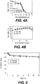


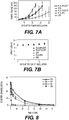
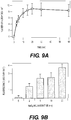
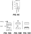
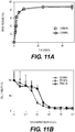


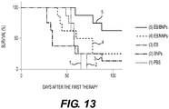
Classifications
-
- A—HUMAN NECESSITIES
- A61—MEDICAL OR VETERINARY SCIENCE; HYGIENE
- A61K—PREPARATIONS FOR MEDICAL, DENTAL OR TOILETRY PURPOSES
- A61K31/00—Medicinal preparations containing organic active ingredients
- A61K31/33—Heterocyclic compounds
- A61K31/395—Heterocyclic compounds having nitrogen as a ring hetero atom, e.g. guanethidine or rifamycins
- A61K31/41—Heterocyclic compounds having nitrogen as a ring hetero atom, e.g. guanethidine or rifamycins having five-membered rings with two or more ring hetero atoms, at least one of which being nitrogen, e.g. tetrazole
- A61K31/4164—1,3-Diazoles
- A61K31/4178—1,3-Diazoles not condensed 1,3-diazoles and containing further heterocyclic rings, e.g. pilocarpine, nitrofurantoin
-
- A—HUMAN NECESSITIES
- A61—MEDICAL OR VETERINARY SCIENCE; HYGIENE
- A61K—PREPARATIONS FOR MEDICAL, DENTAL OR TOILETRY PURPOSES
- A61K8/00—Cosmetics or similar toiletry preparations
- A61K8/02—Cosmetics or similar toiletry preparations characterised by special physical form
- A61K8/0241—Containing particulates characterized by their shape and/or structure
-
- A—HUMAN NECESSITIES
- A61—MEDICAL OR VETERINARY SCIENCE; HYGIENE
- A61K—PREPARATIONS FOR MEDICAL, DENTAL OR TOILETRY PURPOSES
- A61K31/00—Medicinal preparations containing organic active ingredients
- A61K31/33—Heterocyclic compounds
- A61K31/395—Heterocyclic compounds having nitrogen as a ring hetero atom, e.g. guanethidine or rifamycins
- A61K31/41—Heterocyclic compounds having nitrogen as a ring hetero atom, e.g. guanethidine or rifamycins having five-membered rings with two or more ring hetero atoms, at least one of which being nitrogen, e.g. tetrazole
- A61K31/425—Thiazoles
- A61K31/427—Thiazoles not condensed and containing further heterocyclic rings
-
- A—HUMAN NECESSITIES
- A61—MEDICAL OR VETERINARY SCIENCE; HYGIENE
- A61K—PREPARATIONS FOR MEDICAL, DENTAL OR TOILETRY PURPOSES
- A61K31/00—Medicinal preparations containing organic active ingredients
- A61K31/33—Heterocyclic compounds
- A61K31/395—Heterocyclic compounds having nitrogen as a ring hetero atom, e.g. guanethidine or rifamycins
- A61K31/435—Heterocyclic compounds having nitrogen as a ring hetero atom, e.g. guanethidine or rifamycins having six-membered rings with one nitrogen as the only ring hetero atom
- A61K31/47—Quinolines; Isoquinolines
- A61K31/4738—Quinolines; Isoquinolines ortho- or peri-condensed with heterocyclic ring systems
- A61K31/4745—Quinolines; Isoquinolines ortho- or peri-condensed with heterocyclic ring systems condensed with ring systems having nitrogen as a ring hetero atom, e.g. phenantrolines
-
- A—HUMAN NECESSITIES
- A61—MEDICAL OR VETERINARY SCIENCE; HYGIENE
- A61K—PREPARATIONS FOR MEDICAL, DENTAL OR TOILETRY PURPOSES
- A61K47/00—Medicinal preparations characterised by the non-active ingredients used, e.g. carriers or inert additives; Targeting or modifying agents chemically bound to the active ingredient
- A61K47/30—Macromolecular organic or inorganic compounds, e.g. inorganic polyphosphates
- A61K47/34—Macromolecular compounds obtained otherwise than by reactions only involving carbon-to-carbon unsaturated bonds, e.g. polyesters, polyamino acids, polysiloxanes, polyphosphazines, copolymers of polyalkylene glycol or poloxamers
-
- A—HUMAN NECESSITIES
- A61—MEDICAL OR VETERINARY SCIENCE; HYGIENE
- A61K—PREPARATIONS FOR MEDICAL, DENTAL OR TOILETRY PURPOSES
- A61K47/00—Medicinal preparations characterised by the non-active ingredients used, e.g. carriers or inert additives; Targeting or modifying agents chemically bound to the active ingredient
- A61K47/50—Medicinal preparations characterised by the non-active ingredients used, e.g. carriers or inert additives; Targeting or modifying agents chemically bound to the active ingredient the non-active ingredient being chemically bound to the active ingredient, e.g. polymer-drug conjugates
- A61K47/51—Medicinal preparations characterised by the non-active ingredients used, e.g. carriers or inert additives; Targeting or modifying agents chemically bound to the active ingredient the non-active ingredient being chemically bound to the active ingredient, e.g. polymer-drug conjugates the non-active ingredient being a modifying agent
- A61K47/62—Medicinal preparations characterised by the non-active ingredients used, e.g. carriers or inert additives; Targeting or modifying agents chemically bound to the active ingredient the non-active ingredient being chemically bound to the active ingredient, e.g. polymer-drug conjugates the non-active ingredient being a modifying agent the modifying agent being a protein, peptide or polyamino acid
-
- A—HUMAN NECESSITIES
- A61—MEDICAL OR VETERINARY SCIENCE; HYGIENE
- A61K—PREPARATIONS FOR MEDICAL, DENTAL OR TOILETRY PURPOSES
- A61K47/00—Medicinal preparations characterised by the non-active ingredients used, e.g. carriers or inert additives; Targeting or modifying agents chemically bound to the active ingredient
- A61K47/50—Medicinal preparations characterised by the non-active ingredients used, e.g. carriers or inert additives; Targeting or modifying agents chemically bound to the active ingredient the non-active ingredient being chemically bound to the active ingredient, e.g. polymer-drug conjugates
- A61K47/69—Medicinal preparations characterised by the non-active ingredients used, e.g. carriers or inert additives; Targeting or modifying agents chemically bound to the active ingredient the non-active ingredient being chemically bound to the active ingredient, e.g. polymer-drug conjugates the conjugate being characterised by physical or galenical forms, e.g. emulsion, particle, inclusion complex, stent or kit
- A61K47/6921—Medicinal preparations characterised by the non-active ingredients used, e.g. carriers or inert additives; Targeting or modifying agents chemically bound to the active ingredient the non-active ingredient being chemically bound to the active ingredient, e.g. polymer-drug conjugates the conjugate being characterised by physical or galenical forms, e.g. emulsion, particle, inclusion complex, stent or kit the form being a particulate, a powder, an adsorbate, a bead or a sphere
- A61K47/6927—Medicinal preparations characterised by the non-active ingredients used, e.g. carriers or inert additives; Targeting or modifying agents chemically bound to the active ingredient the non-active ingredient being chemically bound to the active ingredient, e.g. polymer-drug conjugates the conjugate being characterised by physical or galenical forms, e.g. emulsion, particle, inclusion complex, stent or kit the form being a particulate, a powder, an adsorbate, a bead or a sphere the form being a solid microparticle having no hollow or gas-filled cores
- A61K47/6929—Medicinal preparations characterised by the non-active ingredients used, e.g. carriers or inert additives; Targeting or modifying agents chemically bound to the active ingredient the non-active ingredient being chemically bound to the active ingredient, e.g. polymer-drug conjugates the conjugate being characterised by physical or galenical forms, e.g. emulsion, particle, inclusion complex, stent or kit the form being a particulate, a powder, an adsorbate, a bead or a sphere the form being a solid microparticle having no hollow or gas-filled cores the form being a nanoparticle, e.g. an immuno-nanoparticle
- A61K47/6931—Medicinal preparations characterised by the non-active ingredients used, e.g. carriers or inert additives; Targeting or modifying agents chemically bound to the active ingredient the non-active ingredient being chemically bound to the active ingredient, e.g. polymer-drug conjugates the conjugate being characterised by physical or galenical forms, e.g. emulsion, particle, inclusion complex, stent or kit the form being a particulate, a powder, an adsorbate, a bead or a sphere the form being a solid microparticle having no hollow or gas-filled cores the form being a nanoparticle, e.g. an immuno-nanoparticle the material constituting the nanoparticle being a polymer
- A61K47/6935—Medicinal preparations characterised by the non-active ingredients used, e.g. carriers or inert additives; Targeting or modifying agents chemically bound to the active ingredient the non-active ingredient being chemically bound to the active ingredient, e.g. polymer-drug conjugates the conjugate being characterised by physical or galenical forms, e.g. emulsion, particle, inclusion complex, stent or kit the form being a particulate, a powder, an adsorbate, a bead or a sphere the form being a solid microparticle having no hollow or gas-filled cores the form being a nanoparticle, e.g. an immuno-nanoparticle the material constituting the nanoparticle being a polymer the polymer being obtained otherwise than by reactions involving carbon to carbon unsaturated bonds, e.g. polyesters, polyamides or polyglycerol
- A61K47/6937—Medicinal preparations characterised by the non-active ingredients used, e.g. carriers or inert additives; Targeting or modifying agents chemically bound to the active ingredient the non-active ingredient being chemically bound to the active ingredient, e.g. polymer-drug conjugates the conjugate being characterised by physical or galenical forms, e.g. emulsion, particle, inclusion complex, stent or kit the form being a particulate, a powder, an adsorbate, a bead or a sphere the form being a solid microparticle having no hollow or gas-filled cores the form being a nanoparticle, e.g. an immuno-nanoparticle the material constituting the nanoparticle being a polymer the polymer being obtained otherwise than by reactions involving carbon to carbon unsaturated bonds, e.g. polyesters, polyamides or polyglycerol the polymer being PLGA, PLA or polyglycolic acid
-
- A—HUMAN NECESSITIES
- A61—MEDICAL OR VETERINARY SCIENCE; HYGIENE
- A61K—PREPARATIONS FOR MEDICAL, DENTAL OR TOILETRY PURPOSES
- A61K8/00—Cosmetics or similar toiletry preparations
- A61K8/18—Cosmetics or similar toiletry preparations characterised by the composition
- A61K8/72—Cosmetics or similar toiletry preparations characterised by the composition containing organic macromolecular compounds
- A61K8/90—Block copolymers
-
- A—HUMAN NECESSITIES
- A61—MEDICAL OR VETERINARY SCIENCE; HYGIENE
- A61K—PREPARATIONS FOR MEDICAL, DENTAL OR TOILETRY PURPOSES
- A61K9/00—Medicinal preparations characterised by special physical form
- A61K9/0012—Galenical forms characterised by the site of application
- A61K9/0014—Skin, i.e. galenical aspects of topical compositions
-
- A—HUMAN NECESSITIES
- A61—MEDICAL OR VETERINARY SCIENCE; HYGIENE
- A61K—PREPARATIONS FOR MEDICAL, DENTAL OR TOILETRY PURPOSES
- A61K9/00—Medicinal preparations characterised by special physical form
- A61K9/0012—Galenical forms characterised by the site of application
- A61K9/0019—Injectable compositions; Intramuscular, intravenous, arterial, subcutaneous administration; Compositions to be administered through the skin in an invasive manner
-
- A—HUMAN NECESSITIES
- A61—MEDICAL OR VETERINARY SCIENCE; HYGIENE
- A61K—PREPARATIONS FOR MEDICAL, DENTAL OR TOILETRY PURPOSES
- A61K9/00—Medicinal preparations characterised by special physical form
- A61K9/0012—Galenical forms characterised by the site of application
- A61K9/0053—Mouth and digestive tract, i.e. intraoral and peroral administration
-
- A—HUMAN NECESSITIES
- A61—MEDICAL OR VETERINARY SCIENCE; HYGIENE
- A61K—PREPARATIONS FOR MEDICAL, DENTAL OR TOILETRY PURPOSES
- A61K9/00—Medicinal preparations characterised by special physical form
- A61K9/14—Particulate form, e.g. powders, Processes for size reducing of pure drugs or the resulting products, Pure drug nanoparticles
- A61K9/16—Agglomerates; Granulates; Microbeadlets ; Microspheres; Pellets; Solid products obtained by spray drying, spray freeze drying, spray congealing,(multiple) emulsion solvent evaporation or extraction
- A61K9/1605—Excipients; Inactive ingredients
- A61K9/1629—Organic macromolecular compounds
- A61K9/1641—Organic macromolecular compounds obtained otherwise than by reactions only involving carbon-to-carbon unsaturated bonds, e.g. polyethylene glycol, poloxamers
- A61K9/1647—Polyesters, e.g. poly(lactide-co-glycolide)
-
- A—HUMAN NECESSITIES
- A61—MEDICAL OR VETERINARY SCIENCE; HYGIENE
- A61K—PREPARATIONS FOR MEDICAL, DENTAL OR TOILETRY PURPOSES
- A61K9/00—Medicinal preparations characterised by special physical form
- A61K9/48—Preparations in capsules, e.g. of gelatin, of chocolate
- A61K9/50—Microcapsules having a gas, liquid or semi-solid filling; Solid microparticles or pellets surrounded by a distinct coating layer, e.g. coated microspheres, coated drug crystals
- A61K9/5005—Wall or coating material
- A61K9/5021—Organic macromolecular compounds
- A61K9/5031—Organic macromolecular compounds obtained otherwise than by reactions only involving carbon-to-carbon unsaturated bonds, e.g. polyethylene glycol, poly(lactide-co-glycolide)
-
- A—HUMAN NECESSITIES
- A61—MEDICAL OR VETERINARY SCIENCE; HYGIENE
- A61K—PREPARATIONS FOR MEDICAL, DENTAL OR TOILETRY PURPOSES
- A61K9/00—Medicinal preparations characterised by special physical form
- A61K9/48—Preparations in capsules, e.g. of gelatin, of chocolate
- A61K9/50—Microcapsules having a gas, liquid or semi-solid filling; Solid microparticles or pellets surrounded by a distinct coating layer, e.g. coated microspheres, coated drug crystals
- A61K9/51—Nanocapsules; Nanoparticles
- A61K9/5107—Excipients; Inactive ingredients
- A61K9/513—Organic macromolecular compounds; Dendrimers
- A61K9/5146—Organic macromolecular compounds; Dendrimers obtained otherwise than by reactions only involving carbon-to-carbon unsaturated bonds, e.g. polyethylene glycol, polyamines, polyanhydrides
- A61K9/5153—Polyesters, e.g. poly(lactide-co-glycolide)
-
- A—HUMAN NECESSITIES
- A61—MEDICAL OR VETERINARY SCIENCE; HYGIENE
- A61P—SPECIFIC THERAPEUTIC ACTIVITY OF CHEMICAL COMPOUNDS OR MEDICINAL PREPARATIONS
- A61P35/00—Antineoplastic agents
-
- A—HUMAN NECESSITIES
- A61—MEDICAL OR VETERINARY SCIENCE; HYGIENE
- A61Q—SPECIFIC USE OF COSMETICS OR SIMILAR TOILETRY PREPARATIONS
- A61Q17/00—Barrier preparations; Preparations brought into direct contact with the skin for affording protection against external influences, e.g. sunlight, X-rays or other harmful rays, corrosive materials, bacteria or insect stings
- A61Q17/04—Topical preparations for affording protection against sunlight or other radiation; Topical sun tanning preparations
-
- C—CHEMISTRY; METALLURGY
- C07—ORGANIC CHEMISTRY
- C07C—ACYCLIC OR CARBOCYCLIC COMPOUNDS
- C07C59/00—Compounds having carboxyl groups bound to acyclic carbon atoms and containing any of the groups OH, O—metal, —CHO, keto, ether, groups, groups, or groups
- C07C59/01—Saturated compounds having only one carboxyl group and containing hydroxy or O-metal groups
- C07C59/06—Glycolic acid
-
- C—CHEMISTRY; METALLURGY
- C07—ORGANIC CHEMISTRY
- C07C—ACYCLIC OR CARBOCYCLIC COMPOUNDS
- C07C59/00—Compounds having carboxyl groups bound to acyclic carbon atoms and containing any of the groups OH, O—metal, —CHO, keto, ether, groups, groups, or groups
- C07C59/01—Saturated compounds having only one carboxyl group and containing hydroxy or O-metal groups
- C07C59/08—Lactic acid
-
- C—CHEMISTRY; METALLURGY
- C08—ORGANIC MACROMOLECULAR COMPOUNDS; THEIR PREPARATION OR CHEMICAL WORKING-UP; COMPOSITIONS BASED THEREON
- C08G—MACROMOLECULAR COMPOUNDS OBTAINED OTHERWISE THAN BY REACTIONS ONLY INVOLVING UNSATURATED CARBON-TO-CARBON BONDS
- C08G83/00—Macromolecular compounds not provided for in groups C08G2/00 - C08G81/00
- C08G83/002—Dendritic macromolecules
- C08G83/005—Hyperbranched macromolecules
-
- A—HUMAN NECESSITIES
- A61—MEDICAL OR VETERINARY SCIENCE; HYGIENE
- A61K—PREPARATIONS FOR MEDICAL, DENTAL OR TOILETRY PURPOSES
- A61K2800/00—Properties of cosmetic compositions or active ingredients thereof or formulation aids used therein and process related aspects
- A61K2800/40—Chemical, physico-chemical or functional or structural properties of particular ingredients
- A61K2800/41—Particular ingredients further characterized by their size
- A61K2800/412—Microsized, i.e. having sizes between 0.1 and 100 microns
-
- A—HUMAN NECESSITIES
- A61—MEDICAL OR VETERINARY SCIENCE; HYGIENE
- A61K—PREPARATIONS FOR MEDICAL, DENTAL OR TOILETRY PURPOSES
- A61K2800/00—Properties of cosmetic compositions or active ingredients thereof or formulation aids used therein and process related aspects
- A61K2800/40—Chemical, physico-chemical or functional or structural properties of particular ingredients
- A61K2800/41—Particular ingredients further characterized by their size
- A61K2800/413—Nanosized, i.e. having sizes below 100 nm
-
- A—HUMAN NECESSITIES
- A61—MEDICAL OR VETERINARY SCIENCE; HYGIENE
- A61K—PREPARATIONS FOR MEDICAL, DENTAL OR TOILETRY PURPOSES
- A61K2800/00—Properties of cosmetic compositions or active ingredients thereof or formulation aids used therein and process related aspects
- A61K2800/40—Chemical, physico-chemical or functional or structural properties of particular ingredients
- A61K2800/60—Particulates further characterized by their structure or composition
- A61K2800/61—Surface treated
- A61K2800/62—Coated
- A61K2800/624—Coated by macromolecular compounds
-
- A—HUMAN NECESSITIES
- A61—MEDICAL OR VETERINARY SCIENCE; HYGIENE
- A61K—PREPARATIONS FOR MEDICAL, DENTAL OR TOILETRY PURPOSES
- A61K39/00—Medicinal preparations containing antigens or antibodies
Definitions
- This invention is in the field of particles, such as microparticles and/or nanoparticles, coated with hyperbranched polyglycerol, wherein the coating can be tuned to provide stealth or adhesive properties.
- NPs nanoparticles
- MPS mononuclear phagocytic system
- PEG has considerable limitations. For instance, it is known that PEG chains can adopt a variety of configurations on the surface, depending on PEG surface density, and the most effective densities are often difficult to achieve.
- Core-shell particles such as microparticles and nanoparticles, and methods of making and using, are described herein.
- the core is formed of or contains a hydrophobic material or more hydrophobic material, such as a polymer.
- the shell is formed of or contains hyperbranched polyglycerol (HPG).
- HPG hyperbranched polyglycerol
- the HPG can be covalently bound to the one or more materials that form the core such that upon self-assembly, particles are formed in which the hydrophobic or more hydrophobic materials form the core and the HPG forms a coating on the particle.
- the HPG coating can be modified to adjust the properties of the particles.
- unmodified HPG coatings impart stealth properties to the particles which resist non-specific protein absorption and increase circulation in the blood.
- the hydroxyl groups on the HPG coating can be chemically modified to form functional groups that react with functional groups on tissue or otherwise interact with tissue to adhere the particles to the tissue, cells, or extracellular materials, such as proteins.
- Such functional groups include, but not limited to, aldehydes, amines, and O-substituted oximes.
- the surface of the particles can further be modified with one or more targeting moieties or covalently bound to HPG via a coupling agent or spacer in organic solvents such as dichloromethane (DCM), dimethylformamide (DMF), dimethyl sulfoxide (DMSO) or tetrahydrofuran (THF).
- DCM dichloromethane
- DMF dimethylformamide
- DMSO dimethyl sulfoxide
- THF tetrahydrofuran
- the polymer is functionalized/modified before nanoparticle formation.
- the targeting moieties may be attached to NPs after the synthesis of NPs in aqueous solution or other protic solution such as alcohol.
- HPG coated NPs can be transformed to aldehyde terminated NPs by NaIO 4 treatment (or carboxylic acid terminated by NaIO 4 treatment followed by sodium chlorite treatment) so the targeting moieties may be directly covalently attached to NPs via aldehyde (or carboxylic acid) groups on NPs and functional groups (amine, hydrazine, aminooxy and their derivatives) on the targeting moieties or indirectly attached to the NPs via coupling agents or spacers (such as aminooxy modified biotin and cysteine).
- NaIO 4 treatment or carboxylic acid terminated by NaIO 4 treatment followed by sodium chlorite treatment
- the targeting moieties may be directly covalently attached to NPs via aldehyde (or carboxylic acid) groups on NPs and functional groups (amine, hydrazine, aminooxy and their derivatives) on the targeting moieties or indirectly attached to the NPs via coupling agents or spacers (such as aminooxy modified biotin and cysteine).
- the particles can further contain one or more therapeutic agents, diagnostic agents, prophylactic agents, and/or nutraceuticals.
- the one or more agents can be covalently or non-covalently associated with the particles.
- the agents can be encapsulated within the particle, for example, dispersed within the core; non-covalently associated with the surface of the particles, covalently-associated with the surface of the particles, or combinations thereof.
- the particles are useful in methods for delivery of therapeutic, nutraceutical, diagnostic and prophylactic agents and/or nutraceuticals.
- HPG coatings can also be used to alter the surface properties of other moieties, such as delivery vehicles (liposomes, micelles, protein aggregates), metals and metal oxides, (thiolated gold conjugated to HPG).
- delivery vehicles liposomes, micelles, protein aggregates
- metals and metal oxides thiolated gold conjugated to HPG.
- HPG can impart stealth properties to these materials.
- the vicinyl diol groups can be transformed to functional groups that promote adhesion of the vehicle to biological materials, such as tissue, cells, and/or proteins.
- Effective amount or “therapeutically effective amount”, as used herein, refers to an amount of drug effective to alleviate, delay onset of, or prevent one or more symptoms of a disease or disorder.
- treating can include preventing a disease, disorder or condition from occurring in an animal which may be predisposed to the disease, disorder and/or condition but has not yet been diagnosed as having it; inhibiting the disease, disorder or condition, e.g., impeding its progress; and relieving the disease, disorder, or condition, e.g., causing regression of the disease, disorder and/or condition.
- Treating the disease, disorder, or condition can include ameliorating at least one symptom of the particular disease, disorder, or condition, even if the underlying pathophysiology is not affected, such as treating the pain of a subject by administration of an analgesic agent even though such agent does not treat the cause of the pain.
- Parenteral administration means administration by any method other than through the digestive tract or non-invasive topical or regional routes.
- parenteral administration may include administration to a patient intravenously, intradermally, intraperitoneally, intrapleurally, intratracheally, intramuscularly, subcutaneously, subjunctivally, by injection, and by infusion.
- Enteral administration means administration via absorption through the gastrointestinal tract. Enteral administration can include oral and sublingual administration, gastric administration, or rectal administration.
- Pulmonary administration means administration into the lungs by inhalation or endotracheal administration.
- inhalation refers to intake of air to the alveoli. The intake of air can occur through the mouth or nose.
- bioactive agent and “active agent”, as used interchangeably herein, include, without limitation, physiologically or pharmacologically active substances that act locally or systemically in the body.
- a bioactive agent is a substance used for the treatment (e.g., therapeutic agent), prevention (e.g., prophylactic agent), diagnosis (e.g., diagnostic agent), cure or mitigation of disease or illness, a substance which affects the structure or function of the body, or pro-drugs, which become biologically active or more active after they have been placed in a predetermined physiological environment.
- Biocompatible and biologically compatible generally refer to materials that are, along with any metabolites or degradation products thereof, generally non-toxic to the recipient, and do not cause any significant adverse effects to the recipient.
- biocompatible materials are materials which do not elicit a significant inflammatory or immune response when administered to a patient.
- biodegradable generally refers to a material that will degrade or erode under physiologic conditions to smaller units or chemical species that are capable of being metabolized, eliminated, or excreted by the subject.
- the degradation time is a function of composition and morphology. Degradation times can be from hours to weeks.
- pharmaceutically acceptable refers to compounds, materials, compositions, and/or dosage forms which are, within the scope of sound medical judgment, suitable for use in contact with the tissues of human beings and animals without excessive toxicity, irritation, allergic response, or other problems or complications commensurate with a reasonable benefit/risk ratio, in accordance with the guidelines of agencies such as the Food and Drug Administration.
- a “pharmaceutically acceptable carrier”, as used herein, refers to all components of a pharmaceutical formulation which facilitate the delivery of the composition in vivo.
- Pharmaceutically acceptable carriers include, but are not limited to, diluents, preservatives, binders, lubricants, disintegrators, swelling agents, fillers, stabilizers, and combinations thereof.
- molecular weight generally refers to the mass or average mass of a material. If a polymer or oligomer, the molecular weight can refer to the relative average chain length or relative chain mass of the bulk polymer. In practice, the molecular weight of polymers and oligomers can be estimated or characterized in various ways including gel permeation chromatography (GPC) or capillary viscometry. GPC molecular weights are reported as the weight-average molecular weight (M w ) as opposed to the number-average molecular weight (M n ). Capillary viscometry provides estimates of molecular weight as the inherent viscosity determined from a dilute polymer solution using a particular set of concentration, temperature, and solvent conditions.
- small molecule generally refers to an organic molecule that is less than about 2000 g/mol in molecular weight, less than about 1500 g/mol, less than about 1000 g/mol, less than about 800 g/mol, or less than about 500 g/mol. Small molecules are non-polymeric and/or non-oligomeric.
- copolymer generally refers to a single polymeric material that is comprised of two or more different monomers.
- the copolymer can be of any form, such as random, block, graft, etc.
- the copolymers can have any end-group, including capped or acid end groups.
- Hydrophilic refers to the property of having affinity for water.
- hydrophilic polymers or hydrophilic polymer segments
- hydrophilic polymer segments are polymers (or polymer segments) which are primarily soluble in aqueous solutions and/or have a tendency to absorb water.
- hydrophilic a polymer the more hydrophilic a polymer is, the more that polymer tends to dissolve in, mix with, or be wetted by water.
- Hydrophobic refers to the property of lacking affinity for, or even repelling water. For example, the more hydrophobic a polymer (or polymer segment), the more that polymer (or polymer segment) tends to not dissolve in, not mix with, or not be wetted by water.
- Hydrophilicity and hydrophobicity can be spoken of in relative terms, such as, but not limited to, a spectrum of hydrophilicity/hydrophobicity within a group of polymers or polymer segments.
- hydrophobic polymer can be defined based on the polymer's relative hydrophobicity when compared to another, more hydrophilic polymer.
- lipophilic refers to compounds having an affinity for lipids.
- amphiphilic refers to a molecule combining hydrophilic and lipophilic (hydrophobic) properties.
- Nanoparticle generally refers to a particle having a diameter, such as an average diameter, from about 10 nm up to but not including about 1 micron, preferably from 100 nm to about 1 micron.
- the particles can have any shape. Nanoparticles having a spherical shape are generally referred to as “nanospheres”.
- Microparticle generally refers to a particle having a diameter, such as an average diameter, from about 1 micron to about 100 microns, preferably from about 1 to about 50 microns, more preferably from about 1 to about 30 microns, most preferably from about 1 micron to about 10 microns.
- the microparticles can have any shape. Microparticles having a spherical shape are generally referred to as "microspheres”.
- Mean particle size as used herein, generally refers to the statistical mean particle size (diameter) of the particles in a population of particles.
- the diameter of an essentially spherical particle may refer to the physical or hydrodynamic diameter.
- the diameter of a non-spherical particle may refer preferentially to the hydrodynamic diameter.
- the diameter of a non-spherical particle may refer to the largest linear distance between two points on the surface of the particle.
- Mean particle size can be measured using methods known in the art, such as dynamic light scattering.
- a monodisperse distribution refers to particle distributions in which 90% or more of the distribution lies within 15% of the median particle size, more preferably within 10% of the median particle size, most preferably within 5% of the median particle size.
- “Pharmaceutically acceptable”, as used herein, refers to compounds, carriers, excipients, compositions, and/or dosage forms which are, within the scope of sound medical judgment, suitable for use in contact with the tissues of human beings and animals without excessive toxicity, irritation, allergic response, or other problem or complication, commensurate with a reasonable benefit/risk ratio.
- Branch point refers to a portion of a polymer-drug conjugate that serves to connect one or more hydrophilic polymer segments to one or more hydrophobic polymer segments.
- transplant refers to a polymeric device or element that is structured, sized, or otherwise configured to be implanted, preferably by injection or surgical implantation, in a specific region of the body so as to provide therapeutic benefit by releasing one or more agents over an extended period of time at the site of implantation.
- targeting moiety refers to a moiety that binds to or localizes to a specific locale.
- the moiety may be, for example, a protein, nucleic acid, nucleic acid analog, carbohydrate, or small molecule.
- the locale may be a tissue, a particular cell type, or a subcellular compartment.
- the targeting moiety or a sufficient plurality of targeting moieties may be used to direct the localization of a particle or an active entity.
- the active entity may be useful for therapeutic, prophylactic, or diagnostic purposes.
- reactive coupling group refers to any chemical functional group capable of reacting with a second functional group to form a covalent bond.
- the selection of reactive coupling groups is within the ability of the skilled artisan.
- Examples of reactive coupling groups can include primary amines (-NH 2 ) and amine-reactive linking groups such as isothiocyanates, isocyanates, acyl azides, NHS esters, sulfonyl chlorides, aldehydes, glyoxals, epoxides, oxiranes, carbonates, aryl halides, imidoesters, carbodiimides, anhydrides, and fluorophenyl esters.
- reactive coupling groups can include aldehydes (-COH) and aldehyde reactive linking groups such as hydrazides, alkoxyamines, and primary amines.
- reactive coupling groups can include thiol groups (-SH) and sulfhydryl reactive groups such as maleimides, haloacetyls, and pyridyl disulfides.
- reactive coupling groups can include photoreactive coupling groups such as aryl azides or diazirines.
- the coupling reaction may include the use of a catalyst, heat, pH buffers, light, or a combination thereof.
- protective group refers to a functional group that can be added to and/or substituted for another desired functional group to protect the desired functional group from certain reaction conditions and selectively removed and/or replaced to deprotect or expose the desired functional group.
- Protective groups are known to the skilled artisan. Suitable protective groups may include those described in Greene, T.W. and Wuts, P.G.M., Protective Groups in Organic Synthesis, (1991 ). Acid sensitive protective groups include dimethoxytrityl (DMT), tert-butylcarbamate (tBoc) and trifluoroacetyl (tFA).
- Base sensitive protective groups include 9-fluorenylmethoxycarbonyl (Fmoc), isobutyrl (iBu), benzoyl (Bz) and phenoxyacetyl (pac).
- Other protective groups include acetamidomethyl, acetyl, tert-amyloxycarbonyl, benzyl, benzyloxycarbonyl, 2-(4-biph ⁇ nylyl)-2-propy!oxycarbonyl, 2- bromobenzyloxycarbonyl, tert-butyl 7 tert-butyloxycarbonyl, 1-carbobenzoxamido-2,2.2- trifluoroethyl, 2,6-dichlorobenzyl, 2-(3,5-dimethoxyphenyl)-2-propyloxycarbonyl, 2,4-dinitrophenyl, dithiasuccinyl, formyl, 4-methoxybenzenesulfonyl, 4-methoxybenzy
- the core of the particles is formed of or contains one or more hydrophobic or more hydrophobic materials, such as one or more polymeric materials (e.g., homopolymer, copolymer, terpolymer, etc.).
- the material may be biodegradable or non-biodegradable.
- the one or more materials are one or more biodegradable polymers.
- synthetic polymers are preferred, although natural polymers may be used and have equivalent or even better properties, especially some of the natural biopolymers which degrade by hydrolysis, such as some of the polyhydroxyalkanoates.
- Representative synthetic polymers are: poly(hydroxy acids) such as poly(lactic acid), poly(glycolic acid), and poly(lactic acid-co-glycolic acid), poly(lactide), poly(glycolide), poly(lactide-co-glycolide), polyanhydrides, polyorthoesters, polyamides, polycarbonates, polyalkylenes such as polyethylene and polypropylene, polyalkylene glycols such as poly(ethylene glycol), polyalkylene oxides such as poly(ethylene oxide), polyalkylene terepthalates such as poly(ethylene terephthalate), polyvinyl alcohols, polyvinyl ethers, polyvinyl esters, polyvinyl halides such as poly(vinyl chloride), polyvinylpyrrolidone, polysiloxa
- Examples of preferred natural polymers include proteins such as albumin, collagen, gelatin and prolamines, for example, zein, and polysaccharides such as alginate, cellulose derivatives and polyhydroxyalkanoates, for example, polyhydroxybutyrate.
- the in vivo stability of the microparticles can be adjusted during the production by using polymers such as poly(lactide-co-glycolide) copolymerized with polyethylene glycol (PEG). If PEG is exposed on the external surface, it may increase the time these materials circulate due to the hydrophilicity of PEG.
- non-biodegradable polymers examples include ethylene vinyl acetate, poly(meth)acrylic acid, polyamides, copolymers and mixtures thereof.
- the hydrophobic polymer is an aliphatic polyester. In preferred embodiments, the hydrophobic polymer is poly(lactic acid), poly(glycolic acid), or poly(lactic acid-co-glycolic acid).
- the particles are designed to release molecules to be encapsulated or attached over a period of days to weeks.
- Factors that affect the duration of release include pH of the surrounding medium (higher rate of release at pH 5 and below due to acid catalyzed hydrolysis of PLGA) and polymer composition.
- Aliphatic polyesters differ in hydrophobicity and that in turn affects the degradation rate.
- the hydrophobic poly (lactic acid) (PLA), more hydrophilic poly (glycolic acid) PGA and their copolymers, poly (lactide-co-glycolide) (PLGA) have various release rates.
- the degradation rate of these polymers, and often the corresponding drug release rate can vary from days (PGA) to months (PLA) and is easily manipulated by varying the ratio of PLA to PGA.
- the core can be formed of copolymers including amphiphilic copolymers such as PLGA-PEG or PLURONICS (block copolymers of polyethylene oxide-polypropylene glycol) but this may decrease the benefit of the polyglycerol molecules discussed below.
- amphiphilic copolymers such as PLGA-PEG or PLURONICS (block copolymers of polyethylene oxide-polypropylene glycol) but this may decrease the benefit of the polyglycerol molecules discussed below.
- lipids may also be incorporated including lipids, fatty acids, and phospholipids. These may be dispersed in or on the particles, or interspersed with the polyglycerol coatings discussed below.
- the particles described herein contain a shell or coating containing hyperbranched polyglycerol (HPG).
- HPG hyperbranched polyglycerol
- Hyperbranched polyglycerol is a highly branched polyol containing a polyether scaffold.
- Hyperbranched polyglycerol can be prepared using techniques known in the art. It can be formed from controlled etherification of glycerol via cationic or anionic ring opening multibranching polymerization of glycidol. For example, an initiator having multiple reactive sites is reacted with glycidol in the presence of a base to form hyperbranched polyglycerol (HPG).
- HPG hyperbranched polyglycerol
- Suitable initiators include, but are not limited to, polyols, e.g., triols, tetraols, pentaols, or greater and polyamines, e.g., triamines, tetraamines, pentaamines, etc.
- the initiator is 1,1,1-trihydroxymethyl propane (THP).
- a formula for hyperbranched polyglycerol as described in EP 2754684 is wherein o, p and q are independently integers from 1-100, wherein A 1 and A 2 are independently wherein 1, m and n are independently integers from 1-100. wherein A 3 and A 4 are defined as A 1 and A 2 , with the proviso that A 3 and A 4 are hydrogen, n and m are each 1 for terminal residues.
- the surface properties of the HPG can be tuned based on the chemistry of vicinal diols.
- the surface properties can be tuned to provide stealth particles, i.e., particles that are not cleared by the MPS due to the presence of the hydroxyl groups; adhesive (sticky) particles, i.e., particles that adhere to the surface of tissues, for example, due to the presence of one or more reactive functional groups, such as aldehydes, amines, oxime, or O-substituted oxime that can be prepared from the vicinal hydroxyl moieties; or targeting by the introduction of one or more targeting moieties which can be conjugated directly or indirectly to the vicinal hydroxyl moieties.
- Indirectly refers to transformation of the hydroxy groups to reactive functional groups that can react with functional groups on molecules to be attached to the surface, such as active agents and/or targeting moieties, etc.
- a schematic of this tunability is shown in Figure 1 .
- the hyperbranched nature of the polyglycerol allows for a much higher density of hydroxyl groups, reactive functional groups, and/or targeting moieties than polyethylene glycol.
- the particles described herein can have a density of surface functionality (e.g., hydroxyl groups, reactive functional groups, and/or targeting moieties) of at least about 1, 2, 3, 4, 5, 6, 7, or 8 groups/nm 2 .
- the molecular weight of the HPG can vary.
- the molecular weight can vary depending on the molecular weight and/or hydrophobicity of the core materials.
- the molecular weight of the HPG is generally from about 1,000 to about 1,000,000 Daltons, from about 1,000 to about 500,000 Daltons, from about 1,000 to about 250,000 Daltons, or from about 1,000 to about 100,000 Daltons.
- the weight percent of HPG of the copolymer is from about 1% to about 50%, such as about 1, 2, 3, 4, 5, 10, 15, 20, 25, 30, 35, 40, 45 or 50%.
- the HPG is covalently coupled to a hydrophobic material or a more hydrophobic material, such as a polymer.
- a hydrophobic material such as a polymer.
- particles are formed containing a core containing the hydrophobic material and a shell or coating of UPG.
- HPG coupled to the polymer PLA is shown below:
- the particles described herein may contain one or more molecules encapsulated within and/or attached to the surface of the particles.
- the molecules can be covalently or non-covalently associated with the particles.
- the molecules are targeting moieties which are covalently associated with the particles.
- the targeting moieties are covalently bound to the HPG coating via the hydroxy groups on HPG.
- the targeting moieties can be bound directly to HPG or via a coupling agent.
- the particles have encapsulated therein one or more therapeutic agents, diagnostic agents, prophylactic agents, and/or nutraceuticals.
- the particles contain both targeting agents which are covalently or non-covalently associated with the particles and one or more therapeutic agents, diagnostic agents, prophylactic agents, and/or nutraceuticals which are covalently or non-covalently associated with the particles.
- Molecules can be bound to the hydroxy groups on HPG before or after particle formation. Representative methodologies for conjugated molecules to the hydroxy groups on HPG are described below.
- One useful protocol involves the "activation" of hydroxyl groups with carbonyldiimidazole (CDI) in aprotic solvents such as DMSO, acetone, or THF.
- CDI forms an imidazolyl carbamate complex with the hydroxyl group which may be displaced by binding the free amino group of a ligand such as a protein.
- the reaction is an N-nucleophilic substitution and results in a stable N-alkylcarbamate linkage of the ligand to the polymer.
- the "coupling" of the ligand to the "activated" polymer matrix is maximal in the pH range of 9-10 and normally requires at least 24 hrs.
- the resulting ligand-polymer complex is stable and resists hydrolysis for extended periods of time.
- Another coupling method involves the use of 1-ethyl-3-(3-dimethylaminopropyl) carbodiimide (EDAC) or "water-soluble CDI" in conjunction with N-hydroxylsulfosuccinimide (sulfo NHS) to couple the exposed carboxylic groups of polymers to the free amino groups of ligands in a totally aqueous environment at the physiological pH of 7.0.
- EDAC and sulfo-NHS form an activated ester with the carboxylic acid groups of the polymer which react with the amine end of a ligand to form a peptide bond.
- the resulting peptide bond is resistant to hydrolysis.
- the use of sulfo-NHS in the reaction increases the efficiency of the EDAC coupling by a factor of ten-fold and provides for exceptionally gentle conditions that ensure the viability of the ligand-polymer complex.
- a useful coupling procedure for attaching ligands with free hydroxyl and carboxyl groups to polymers involves the use of the cross-linking agent, divinylsulfone. This method would be useful for attaching sugars or other hydroxylic compounds with bioadhesive properties to hydroxylic matrices.
- the activation involves the reaction of divinylsulfone to the hydroxyl groups of the polymer, forming the vinylsulfonyl ethyl ether of the polymer.
- the vinyl groups will couple to alcohols, phenols and even amines.
- Activation and coupling take place at pH 11.
- the linkage is stable in the pH range from 1-8 and is suitable for transit through the intestine.
- the hydroxyl groups can be converted to reactive functional group that can react with a reactive functional group on the molecule to be attached.
- the hydroxyl groups on HPG can be converted to aldehydes, amines, or O-substituted oximes which can react with reactive functional groups on molecules to be attached. Such transformations can be done before or after particle formation.
- Coupling is preferably by covalent binding but it may also be indirect, for example, through a linker bound to the polymer or through an interaction between two molecules such as strepavidin and biotin. It may also be by electrostatic attraction by dip-coating.
- the coupling methods can be done before or after particle formation.
- Therapeutic agent therapeutic agent, diagnostic agents, prophylactic agents, and/or nutraceuticals
- Agents to be delivered include therapeutic, nutritional, diagnostic, and prophylactic compounds. Proteins, peptides, carbohydrates, polysaccharides, nucleic acid molecules, and organic molecules, as well as diagnostic agents, can be delivered.
- the preferred materials to be incorporated are drugs and imaging agents.
- Therapeutic agents include antibiotics, antivirals, anti-parasites (helminths, protozoans), anti-cancer (referred to herein as "chemotherapeutics", including cytotoxic drugs such as doxorubicin, cyclosporine, mitomycin C, cisplatin and carboplatin, BCNU, 5FU, methotrexate, adriamycin, camptothecin, epothilones A-F, and taxol), antibodies and bioactive fragments thereof (including humanized, single chain, and chimeric antibodies), antigen and vaccine formulations, peptide drugs, anti-inflammatories, nutraceuticals such as vitamins, and oligonucleotide drugs (including DNA, RNAs, antisense, aptamers, ribozymes, external guide sequences for ribonuclease P, and triplex forming agents).
- chemotherapeutics including cytotoxic drugs such as doxorubicin, cyclosporine, mito
- Particularly preferred drugs to be delivered include anti-angiogenic agents, antiproliferative and chemotherapeutic agents such as rampamycin. Incorporated into microparticles, these agents may be used to treat cancer or eye diseases, or prevent restenosis following administration into the blood vessels.
- diagnostic materials include paramagnetic molecules, fluorescent compounds, magnetic molecules, and radionuclides.
- Exemplary materials include, but are not limited to, metal oxides, such as iron oxide, metallic particles, such as gold particles, etc. Biomarkers can also be conjugated to the surface for diagnostic applications.
- Active agents may be formulated alone or with excipients or encapsulated on, in or incorporated into the microparticles or nanoparticles.
- Active agents include therapeutic, prophylactic, neutraceutical and diagnostic agents. Any suitable agent may be used. These include organic compounds, inorganic compounds, proteins, polysaccharides, nucleic acids or other materials that can be incorporated using standard techniques.
- Active agents include synthetic and natural proteins (including enzymes, peptide-hormones, receptors, growth factors, antibodies, signaling molecules), and synthetic and natural nucleic acids (including RNA, DNA, anti-sense RNA, triplex DNA, inhibitory RNA (RNAi), and oligonucleotides), and biologically active portions thereof.
- Suitable active agents have a size greater than about 1,000 Da for small peptides and polypeptides, more typically at least about 5,000 Da and often 10,000 Da or more for proteins.
- Nucleic acids are more typically listed in terms of base pairs or bases (collectively "bp"). Nucleic acids with lengths above about 10 bp are typically used in the present method.
- useful lengths of nucleic acids for probing or therapeutic use will be in the range from about 20 bp (probes; inhibitory RNAs, etc.) to tens of thousands of bp for genes and vectors.
- the active agents may also be hydrophilic molecules, preferably having a low molecular weight.
- useful proteins include hormones such as insulin and growth hormones including somatomedins.
- useful drugs include neurotransmitters such as L-DOPA, antihypertensives or saluretics such as Metolazone from Searle Pharmaceuticals, carbonic anhydrase inhibitors such as Acetazolamide from Lederle Pharmaceuticals, insulin like drugs such as glyburide, a blood glucose lowering drug of the sulfonylurea class, synthetic hormones such as Android F from Brown Pharmaceuticals and Testred® (methyltestosterone) from ICN Pharmaceuticals.
- anti-cancer agents include, but are not limited to, alkylating agents (such as cisplatin, carboplatin, oxaliplatin, mechlorethamine, cyclophosphamide, chlorambucil, dacarbazine, lomustine, carmustine, procarbazine, chlorambucil and ifosfamide), antimetabolites (such as fluorouracil (5-FU), gemcitabine, methotrexate, cytosine arabinoside, fludarabine, and floxuridine), antimitotics (including taxanes such as paclitaxel and decetaxel, epothilones A-F, and vinca alkaloids such as vincristine, vinblastine, vinorelbine, and vindesine), anthracyclines (including doxorubicin, daunorubicin, valrubicin, idarubicin, and epirubicin, as well as actinomycins such as actin
- VEGF vascular endothelial growth factor
- AVASTIN® bevacizumab
- TAALOMID® thalidomide
- RTK receptor tyrosine kinase
- SUTENT® sunitinib
- tyrosine kinase inhibitors such as sorafenib (Nexavar®), erlotinib (Tarceva®), pazopanib, axitinib, and lapatinib
- transforming growth factor-a or transforming growth factor- ⁇ inhibitors and antibodies to the epidermal growth factor receptor such as panitumumab (VECTIBIX®) and cetuximab (ERBITUX®).
- drugs can belong to four classes: class I (high permeability, high solubility), class II (high permeability, low solubility), class III (low permeability, high solubility) or class IV (low permeability, low solubility).
- Suitable active agents also include poorly soluble compounds; such as drugs that are classified as class II or class IV compounds using the BCS.
- class II compounds include: acyclovir, nifedipine, danazol, ketoconazole, mefenamic acid, nisoldipine, nicardipine, felodipine, atovaquone, griseofulvin, troglitazone glibenclamide and carbamazepine.
- class IV compounds include: chlorothiazide, furosemide, tobramycin, cefuroxmine, and paclitaxel.
- radioactive materials such as Technetium99 ( 99m Tc) or magnetic materials such as Fe 2 O 3 could be used.
- examples of other materials include gases or gas emitting compounds, which are radioopaque.
- the biodegradable polymers may encapsulate cellular materials, such as for example, cellular materials to be delivered to antigen presenting cells as described below to induce immunological responses.
- Peptide, protein, and DNA based vaccines may be used to induce immunity to various diseases or conditions. For example, sexually transmitted diseases and unwanted pregnancy are world-wide problems affecting the health and welfare of women. Effective vaccines to induce specific immunity within the female genital tract could greatly reduce the risk of STDs, while vaccines that provoke anti-sperm antibodies would function as immunocontraceptives. Extensive studies have demonstrated that vaccination at a distal site-orally, nasally, or rectally, for example-can induce mucosal immunity within the female genital tract. Of these options, oral administration has gained the most interest because of its potential for patient compliance, easy administration and suitability for widespread use. Oral vaccination with proteins is possible, but is usually inefficient or requires very high doses. Oral vaccination with DNA, while potentially effective at lower doses, has been ineffective in most cases because 'naked DNA' is susceptible to both the stomach acidity and digestive enzymes in the gastrointestinal tract
- DNA-mediated immunity is needed to detect and destroy virus-infected cells.
- Most traditional vaccines e.g. protein-based vaccines
- DNA-based vaccine represents a unique means to vaccinate against a virus or parasite because a DNA based vaccine can induce both humoral and cell-mediated immunity.
- DNA-based vaccines are potentially safer than traditional vaccines.
- DNA vaccines are relatively more stable and more cost-effective for manufacturing and storage.
- DNA vaccines consist of two major components - DNA carriers (or delivery vehicles) and DNAs encoding antigens. DNA carriers protect DNA from degradation, and can facilitate DNA entry to specific tissues or cells and expression at an efficient level.
- Biodegradable polymer particles offer several advantages for use as DNA delivery vehicles for DNA based vaccines.
- the polymer particles can be biodegradable and biocompatible, and they have been used successfully in past therapeutic applications to induce mucosal or humoral immune responses.
- Polymer biodegradation products are typically formed at a relatively slow rate, are biologically compatible, and result in metabolizable moieties.
- Biodegradable polymer particles can be manufactured at sizes ranging from diameters of several microns (microparticles) to particles having diameters of less than one micron (nanoparticles).
- DCs Dendritic cells
- CTL cytotoxic T-cell
- Th1 CD4+ T-helper
- Immature DCs can internalize and process cellular materials, such as DNA encoding antigens, and induce cellular immunologic responses to disease effectors.
- disease effector agents refers to agents that are central to the causation of a disease state in a subject.
- these disease effector agents are disease-causing cells which may be circulating in the bloodstream, thereby making them readily accessible to extracorporeal manipulations and treatments.
- diseases-causing cells include malignant T cells, malignant B cells, T cells and B cells which mediate an autoimmune response, and virally or bacterially infected white blood cells which express on their surface viral or bacterial peptides or proteins.
- Exemplary disease categories giving rise to disease-causing cells include leukemia, lymphoma, autoimmune disease, graft versus host disease, and tissue rejection.
- Disease associated antigens which mediate these disease states and which are derived from disease-causing cells include peptides that bind to a MHC Class I site, a MHC Class II site, or to a heat shock protein which is involved in transporting peptides to and from MHC sites (i.e., a chaperone).
- Disease associated antigens also include viral or bacterial peptides which are expressed on the surface of infected white blood cells, usually in association with an MHC Class I or Class II molecule.
- disease-causing cells include those isolated from surgically excised specimens from solid tumors, such as lung, colon, brain, kidney or skin cancers. These cells can be manipulated extracorporeally in analogous fashion to blood leukocytes, after they are brought into suspension or propagated in tissue culture. Alternatively, in some instances, it has been shown that the circulating blood of patients with solid tumors can contain malignant cells that have broken off from the tumors and entered the circulation. These circulating tumor cells can provide an easily accessible source of cancer cells which may be rendered apoptotic and presented to the antigen presenting cells.
- disease effector agents include microbes such as bacteria, fungi, yeast, viruses which express or encode disease-associated antigens, and prions.
- the disease effector agents are presented to the antigen presenting cells using biodegradable polymer microparticles as delivery vehicles.
- the loaded microparticles are exposed to immature antigen presenting cells, which internalize the microparticles and process the material within the microparticles.
- the microparticles may be administered to the patient and the interaction between the microparticles and the antigen presenting cells may occur in vivo.
- the microparticles are placed in an incubation bag with the immature antigen presenting cells, and the microparticles are phagocytosed by the antigen presenting cells during the incubation period.
- the resulting antigen presenting cells are then administered to the patient to induce an immune response to the disease causing agent.
- Other agents include cell penetrating peptides, such as TAT, Antennapedia, polyarginine and poly-lysine analogues.
- the particles can be modified to facilitate targeting through the attachment of targeting molecules.
- target molecules include proteins, peptides, nucleic acids, lipids, saccharides, or polysaccharides, or small molecules that bind to one or more targets associated with an organ, tissue, cell, or extracellular matrix, or specific type of tumor or infected cell.
- the degree of specificity with which the particles are targeted can be modulated through the selection of a targeting molecule with the appropriate affinity and specificity.
- a targeting moiety can be a polypeptide, such as an antibody that specifically recognizes a tumor marker that is present exclusively or in higher amounts on a malignant cell (e.g., a tumor antigen).
- Targeting molecules can also include neuropilins and endothelial targeting molecules, integrins, selectins, and adhesion molecules.
- Targeting molecules can be covalently bound to particles using a variety of methods known in the art.
- the targeting moieties are covalently associated with the polymer, preferably via a linker cleaved at the site of delivery.
- the nanoparticles can contain one or more polymer conjugates containing end-to-end linkages between the polymer and a targeting element or a detectable label.
- a modified polymer can be a PLA-HPG-peptide block polymer.
- targeting moieties include peptides such as iRGD, LyP1; small molecule such as folate, aptamers and antibodies or their combinations at various molar ratios.
- the targeting element of the nanoparticle can be an antibody or antigen binding fragment thereof.
- the targeting elements should have an affinity for a cell-surface receptor or cell-surface antigen on the target cells and result in internalization of the particle within the target cell.
- the targeting element can specifically recognize and bind to a target molecule specific for a cell type, a tissue type, or an organ.
- the target molecule can be a cell surface polypeptide, lipid, or glycolipid.
- the target molecule can be a receptor that is selectively expressed on a specific cell surface, a tissue or an organ.
- Cell specific markers can be for specific types of cells including, but not limited to stem cells, blood cells, immune cells, muscle cells, nerve cells, cancer cells, virally infected cells, and organ specific cells.
- the cell markers can be specific for endothelial, ectodermal, or mesenchymal cells. Representative cell specific markers include, but are not limited to cancer specific markers.
- Additional targets that can be recognized by the targeting element include VEGF/KDR, Tie2, vascular cell adhesion molecule (VCAM), endoglin and ⁇ 5 ⁇ 3 integrin/vitronectin.
- the targeting peptides can be covalently associated with the polymer of the outer shell and the covalent association can be mediated by a linker.
- the targeting element specifically binds to an antigen that is expressed by tumor cells.
- the antigen expressed by the tumor may be specific to the tumor, or may be expressed at a higher level on the tumor cells as compared to non-tumor cells.
- Antigenic markers such as serologically defined markers known as tumor associated antigens, which are either uniquely expressed by cancer cells or are present at markedly higher levels (e.g., elevated in a statistically significant manner) in subjects having a malignant condition relative to appropriate controls, are contemplated for use in certain embodiments.
- Tumor-associated antigens may include, for example, cellular oncogene-encoded products or aberrantly expressed proto-oncogene-encoded products (e.g., products encoded by the neu, ras, trk, and kit genes), or mutated forms of growth factor receptor or receptor-like cell surface molecules (e.g., surface receptor encoded by the c-erb B gene).
- Other tumor-associated antigens include molecules that may be directly involved in transformation events, or molecules that may not be directly involved in oncogenic transformation events but are expressed by tumor cells (e.g., carcinoembryonic antigen, CA-125, melonoma associated antigens, etc.) (see, e.g., U.S. Pat. No.
- Genes that encode cellular tumor associated antigens include cellular oncogenes and proto-oncogenes that are aberrantly expressed.
- cellular oncogenes encode products that are directly relevant to the transformation of the cell, and because of this, these antigens are particularly preferred targets for immunotherapy.
- An example is the tumorigenic neu gene that encodes a cell surface molecule involved in oncogenic transformation.
- Other examples include the ras, kit, and trk genes.
- the products of proto-oncogenes may be aberrantly expressed (e.g., overexpressed), and this aberrant expression can be related to cellular transformation.
- the product encoded by proto-oncogenes can be targeted.
- Some oncogenes encode growth factor receptor molecules or growth factor receptor-like molecules that are expressed on the tumor cell surface.
- An example is the cell surface receptor encoded by the c-erbB gene.
- Other tumor-associated antigens may or may not be directly involved in malignant transformation. These antigens, however, are expressed by certain tumor cells and may therefore provide effective targets.
- Some examples are carcinoembryonic antigen (CEA), CA 125 (associated with ovarian carcinoma), and melanoma specific antigens.
- tumor associated antigens are detectable in samples of readily obtained biological fluids such as serum or mucosal secretions.
- One such marker is CA125, a carcinoma associated antigen that is also shed into the bloodstream, where it is detectable in serum (e.g., Bast, et al., N. Eng. J. Med., 309:883 (1983 ); Lloyd, et al., Int. J. Canc., 71:842 (1997 ).
- CA125 levels in serum and other biological fluids have been measured along with levels of other markers, for example, carcinoembryonic antigen (CEA), squamous cell carcinoma antigen (SCC), tissue polypeptide specific antigen (TPS), sialyl TN mucin (STN), and placental alkaline phosphatase (PLAP), in efforts to provide diagnostic and/or prognostic profiles of ovarian and other carcinomas (e.g., Sarandakou, et al., Acta Oncol., 36:755 (1997 ); Sarandakou, et al., Eur. J. Gynaecol.
- CEA carcinoembryonic antigen
- SCC squamous cell carcinoma antigen
- TPS tissue polypeptide specific antigen
- STN sialyl TN mucin
- PLAP placental alkaline phosphatase
- Elevated serum CA125 may also accompany neuroblastoma (e.g., Hirokawa, et al., Surg. Today, 28:349 (1998 ), while elevated CEA and SCC, among others, may accompany colorectal cancer ( Gebauer, et al., Anticancer Res., 17(4B):2939 (1997 )).
- mesothelin is detectable only as a cell-associated tumor marker and has not been found in soluble form in serum from ovarian cancer patients, or in medium conditioned by OVCAR-3 cells ( Chang, et al., Int. J. Cancer, 50:373 (1992 )).
- Structurally related human mesothelin polypeptides also include tumor-associated antigen polypeptides such as the distinct mesothelin related antigen (MRA) polypeptide, which is detectable as a naturally occurring soluble antigen in biological fluids from patients having malignancies (see WO 00/50900 ).
- MRA mesothelin related antigen
- a tumor antigen may include a cell surface molecule.
- Tumor antigens of known structure and having a known or described function include the following cell surface receptors: HER1 (GenBank Accession No. U48722), HER2 ( Yoshino, et al., J. Immunol., 152:2393 (1994 ); Disis, et al., Canc. Res., 54:16 (1994 ); GenBank Acc. Nos. X03363 and M17730), HER3 (GenBank Acc. Nos. U29339 and M34309), HER4 ( Plowman, et al., Nature, 366:473 (1993 ); GenBank Acc. Nos.
- EGFR epidermal growth factor receptor
- vascular endothelial cell growth factor GenBank No. M32977
- vascular endothelial cell growth factor receptor GenBank Acc. Nos. AF022375, 1680143, U48801 and X62568
- insulin-like growth factor-I GenBank Acc. Nos. X00173, X56774, X56773, X06043, European Patent No. GB 2241703
- insulin-like growth factor-II GeneBank Acc. Nos.
- X03562, X00910, M17863 and M17862), transferrin receptor Trowbridge and Omary, Proc. Nat. Acad. USA, 78:3039 (1981 ); GenBank Acc. Nos. X01060 and M11507), estrogen receptor (GenBank Acc. Nos. M38651, X03635, X99101, U47678 and M12674), progesterone receptor (GenBank Acc. Nos. X51730, X69068 and M15716), follicle stimulating hormone receptor (FSH-R) (GenBank Acc. Nos. Z34260 and M65085), retinoic acid receptor (GenBank Acc. Nos.
- any of the CTA class of receptors including in particular HOM-MEL-40 antigen encoded by the SSX2 gene (GenBank Acc. Nos. X86175, U90842, U90841 and X86174), carcinoembryonic antigen (CEA, Gold and Freedman, J. Exp. Med., 121:439 (1985 ); GenBank Acc. Nos. M59710, M59255 and M29540), and PyLT (GenBank Acc. Nos.
- PSA prostate surface antigen
- ⁇ -human chorionic gonadotropin ⁇ -HCG ⁇ -human chorionic gonadotropin ⁇ -HCG
- Tumor antigens of interest include antigens regarded in the art as "cancer/testis” (CT) antigens that are immunogenic in subjects having a malignant condition ( Scanlan, et al., Cancer Immun., 4:1 (2004 )).
- CT antigens include at least 19 different families of antigens that contain one or more members and that are capable of inducing an immune response, including but not limited to MAGEA (CT1); BAGE (CT2); MAGEB (CT3); GAGE (CT4); SSX (CT5); NY-ESO-1 (CT6); MAGEC (CT7); SYCP1 (C8); SPANXB1 (CT11.2); NA88 (CT18); CTAGE (CT21); SPA17 (CT22); OY-TES-1 (CT23); CAGE (CT26); HOM-TES-85 (CT28); HCA661 (CT30); NY-SAR-35 (CT38); FATE (CT43); and TPTE (CT44).
- CT1 MAGEA
- Additional tumor antigens that can be targeted include, but not limited to, alpha-actinin-4, Bcr-Abl fusion protein, Casp-8, beta-catenin, cdc27, cdk4, cdkn2a, coa-1, dek-can fusion protein, EF2, ETV6-AML1 fusion protein, LDLR-fucosyltransferaseAS fusion protein, HLA-A2, HLA-A11, hsp70-2, KIAAO205, Mart2, Mum-1, 2, and 3, neo-PAP, myosin class I, OS-9, pml-RAR ⁇ fusion protein, PTPRK, K-ras, N-ras, Triosephosphate isomeras, Bage-1, Gage 3,4,5,6,7, GnTV, Herv-K-mel, Lü-1, MageA1,2,3,4,6,10,12, Mage-C2, NA-
- the targeting element can be a peptide.
- the plaque targeted peptide can be, but is not limited to, one or more of the following: RGD, iRGD(CRGDK/RGPD/EC), LyP-1, P3(CKGGRAKDC), or their combinations at various molar ratios.
- the targeting peptides can be covalently associated with the polymer and the covalent association can be mediated by a linker.
- the targeting element can be an antibody or an antigen-binding fragment thereof.
- the antibody can be any type of immunoglobulin that is known in the art.
- the antibody can be of any isotype, e.g., IgA, IgD, IgE, IgG, IgM, etc.
- the antibody can be monoclonal or polyclonal.
- the antibody can be a naturally-occurring antibody, e.g., an antibody isolated and/or purified from a mammal, e.g., mouse, rabbit, goat, horse, chicken, hamster, human, etc.
- the antibody can be a genetically-engineered antibody, e.g., a humanized antibody or a chimeric antibody.
- the antibody can be in monomeric or polymeric form.
- the antigen binding portion of the antibody can be any portion that has at least one antigen binding site, such as Fab, F(ab') 2 , dsFv, sFv, diabodies, and triabodies.
- the antibody is a single chain antibody.
- Aptamers are oligonucleotide or peptide sequences with the capacity to recognize virtually any class of target molecules with high affinity and specificity. Aptamers bind to targets such as small organics, peptides, proteins, cells, and tissues. Unlike antibodies, some aptamers exhibit stereoselectivity. The aptamers can be designed to bind to specific targets expressed on cells, tissues or organs.
- the outer surface of the microparticle may be treated using a mannose amine, thereby mannosylating the outer surface of the microparticle. This treatment may cause the microparticle to bind to the target cell or tissue at a mannose receptor on the antigen presenting cell surface.
- surface conjugation with an immunoglobulin molecule containing an Fc portion targeting Fc receptor
- HSP receptor heat shock protein moiety
- phosphatidylserine scavenger receptors
- LPS lipopolysaccharide
- Lectins that can be covalently attached to microparticles to render them target specific to the mucin and mucosal cell layer include lectins isolated from Abrus precatroius, Agaricus bisporus, Anguilla anguilla, Arachis hypogaea, Pandeiraea simplicifolia, Bauhinia purpurea, Caragan arobrescens, Cicer arietinum, Codium fragile, Datura stramonium, Dolichos biflorus, Erythrina corallodendron, Erythrina cristagalli, Euonymus europaeus, Glycine max, Helix aspersa, Helix pomatia, Lathyrus odoratus, Lens culinaris, Limulus polyphemus, Lysopersicon esculentum, Maclura pomifera, Momordica charantia, Mycoplasma gallisepticum, Naja mocambique, as
- any positively charged ligand such as polyethyleneimine or polylysine
- any microparticle may improve bioadhesion due to the electrostatic attraction of the cationic groups coating the beads to the net negative charge of the mucus.
- Any ligand with a high binding affinity for mucin could also be covalently linked to most microparticles with the appropriate chemistry, and be expected to influence the binding of microparticles to the gut.
- polyclonal antibodies raised against components of mucin or else intact mucin, when covalently coupled to microparticles, would provide for increased bioadhesion.
- antibodies directed against specific cell surface receptors exposed on the lumenal surface of the intestinal tract would increase the residence time of beads, when coupled to microparticles using the appropriate chemistry.
- the ligand affinity need not be based only on electrostatic charge, but other useful physical parameters such as solubility in mucin or else specific affinity to carbohydrate groups.
- any of the natural components of mucin in either pure or partially purified form to the microparticles would decrease the surface tension of the bead-gut interface and increase the solubility of the bead in the mucin layer.
- useful ligands would include but not be limited to the following: sialic acid, neuraminic acid, n-acetyl-neuraminic acid, n-glycolylneuraminic acid, 4-acetyl-n-acetylneuraminic acid, diacetyl-n-acetylneuraminic acid, glucuronic acid, iduronic acid, galactose, glucose, mannose, fucose, any of the partially purified fractions prepared by chemical treatment of naturally occurring mucin, e.g., mucoproteins, mucopolysaccharides and mucopolysaccharide-protein complexes, and antibodies immunoreactive against proteins or sugar structure on the mucosal surface.
- polyamino acids containing extra pendant carboxylic acid side groups e.g., polyaspartic acid and polyglutamic acid
- polyamino acids containing extra pendant carboxylic acid side groups e.g., polyaspartic acid and polyglutamic acid
- polyamino acids in the 15,000 to 50,000 kDa molecular weight range would yield chains of 120 to 425 amino acid residues attached to the surface of the microparticles.
- the polyamino chains would increase bioadhesion by means of chain entanglement in mucin strands as well as by increased carboxylic charge.
- the HPG-coated particles can be modified by covalently attaching PEG to the surface. This can be achieved by converting the vicinyl diol groups to aldehydes and then reacting the aldehydes with functional groups on PEG, such as aliphatic amines, aromatic amines, hydrazines and thiols.
- the linker has end groups such as aliphatic amines, aromatic amines, hydrazines, thiols and O-substituted oxyamines.
- the bond inserted in the linker can be disulfide, orthoester and peptides sensitive to proteases.
- PEG with a functional group or a linker can form a bond with aldehyde on PLA-HPGALD and reversed the bioadhesive(sticky) state of PLA-HPGALD to stealth state.
- This bond or the linker is labile to pH change or high concentration of peptides, proteins and other biomolecules.
- the bond attaching the PEG to PLA-HPGALD can be reversed or cleaved to release the PEG in response to environment and exposed the PLA-HPGALD particles to the environment. Subsequently, the particles will interact with the tissue and attach the particles to the tissues or extracellular materials such as proteins.
- the environment can be acidic environment in tumors, reducing environment in tumors, protein rich environment in tissues.
- the particles can be formulated with appropriate pharmaceutically acceptable carriers into pharmaceutical compositions for administration to an individual in need thereof.
- the formulations can be administered enterally (e.g., oral) or parenterally (e.g., by injection or infusion). Other routes of administration include, but are not limited to, transdermal.
- parenteral administration means administration by any method other than through the digestive tract or non-invasive topical or regional routes.
- parenteral administration may include administration to a patient intravenously, intradermally, intraarterially, intraperitoneally, intralesionally, intracranially, intraarticularly, intraprostatically, intrapleurally, intratracheally, intravitreally, intratumorally, intramuscularly, subcutaneously, subconjunctivally, intravesicularly, intrapericardially, intraumbilically, by injection, and by infusion.
- Parenteral formulations can be prepared as aqueous compositions using techniques is known in the art.
- such compositions can be prepared as injectable formulations, for example, solutions or suspensions; solid forms suitable for using to prepare solutions or suspensions upon the addition of a reconstitution medium prior to injection; emulsions, such as water-in-oil (w/o) emulsions, oil-in-water (o/w) emulsions, and microemulsions thereof, liposomes, or emulsomes.
- injectable formulations for example, solutions or suspensions
- solid forms suitable for using to prepare solutions or suspensions upon the addition of a reconstitution medium prior to injection emulsions, such as water-in-oil (w/o) emulsions, oil-in-water (o/w) emulsions, and microemulsions thereof, liposomes, or emulsomes.
- emulsions such as water-in-oil (w/o) emulsions
- the carrier can be a solvent or dispersion medium containing, for example, water, ethanol, one or more polyols (e.g., glycerol, propylene glycol, and liquid polyethylene glycol), oils, such as vegetable oils (e.g., peanut oil, corn oil, sesame oil, etc.), and combinations thereof.
- polyols e.g., glycerol, propylene glycol, and liquid polyethylene glycol
- oils such as vegetable oils (e.g., peanut oil, corn oil, sesame oil, etc.)
- the proper fluidity can be maintained, for example, by the use of a coating, such as lecithin, by the maintenance of the required particle size in the case of dispersion and/or by the use of surfactants.
- isotonic agents for example, sugars or sodium chloride.
- Solutions and dispersions of the active compounds as the free acid or base or pharmacologically acceptable salts thereof can be prepared in water or another solvent or dispersing medium suitably mixed with one or more pharmaceutically acceptable excipients including, but not limited to, surfactants, dispersants, emulsifiers, pH modifying agents, viscosity modifying agents, and combination thereof.
- Suitable surfactants may be anionic, cationic, amphoteric or nonionic surface active agents.
- Suitable anionic surfactants include, but are not limited to, those containing carboxylate, sulfonate and sulfate ions.
- anionic surfactants include sodium, potassium, ammonium of long chain alkyl sulfonates and alkyl aryl sulfonates such as sodium dodecylbenzene sulfonate; dialkyl sodium sulfosuccinates, such as sodium dodecylbenzene sulfonate; dialkyl sodium sulfosuccinates, such as sodium bis-(2-ethylthioxyl)-sulfosuccinate; and alkyl sulfates such as sodium lauryl sulfate.
- Cationic surfactants include, but are not limited to, quaternary ammonium compounds such as benzalkonium chloride, benzethonium chloride, cetrimonium bromide, stearyl dimethylbenzyl ammonium chloride, polyoxyethylene and coconut amine.
- nonionic surfactants include ethylene glycol monostearate, propylene glycol myristate, glyceryl monostearate, glyceryl stearate, polyglyceryl-4-oleate, sorbitan acylate, sucrose acylate, PEG-150 laurate, PEG-400 monolaurate, polyoxyethylene monolaurate, polysorbates, polyoxyethylene octylphenylether, PEG-1000 cetyl ether, polyoxyethylene tridecyl ether, polypropylene glycol butyl ether, Poloxamer® 401, stearoyl monoisopropanolamide, and polyoxyethylene hydrogenated tallow amide.
- amphoteric surfactants include sodium N-dodecyl-.beta.-alanine, sodium N-lauryl-.beta.-iminodipropionate, myristoamphoacetate, lauryl betaine and lauryl sulfobetaine.
- the formulation can contain a preservative to prevent the growth of microorganisms. Suitable preservatives include, but are not limited to, parabens, chlorobutanol, phenol, sorbic acid, and thimerosal.
- the formulation may also contain an antioxidant to prevent degradation of the active agent(s).
- the formulation is typically buffered to a pH of 3-8 for parenteral administration upon reconstitution.
- Suitable buffers include, but are not limited to, phosphate buffers, acetate buffers, and citrate buffers.
- Water soluble polymers are often used in formulations for parenteral administration. Suitable water-soluble polymers include, but are not limited to, polyvinylpyrrolidone, dextran, carboxymethylcellulose, and polyethylene glycol.
- Sterile injectable solutions can be prepared by incorporating the active compounds in the required amount in the appropriate solvent or dispersion medium with one or more of the excipients listed above, as required, followed by filtered sterilization.
- dispersions are prepared by incorporating the various sterilized active ingredients into a sterile vehicle which contains the basic dispersion medium and the required other ingredients from those listed above.
- the preferred methods of preparation are vacuum-drying and freeze-drying techniques which yield a powder of the active ingredient plus any additional desired ingredient from a previously sterile-filtered solution thereof.
- the powders can be prepared in such a manner that the particles are porous in nature, which can increase dissolution of the particles. Methods for making porous particles are well known in the art.
- Enteral formulations are prepared using pharmaceutically acceptable carriers.
- carrier includes, but is not limited to, diluents, preservatives, binders, lubricants, disintegrators, swelling agents, fillers, stabilizers, and combinations thereof.
- Polymers used in the dosage form include hydrophobic or hydrophilic polymers and pH dependent or independent polymers.
- Preferred hydrophobic and hydrophilic polymers include, but are not limited to, hydroxypropyl methylcellulose, hydroxypropyl cellulose, hydroxyethyl cellulose, carboxy methylcellulose, polyethylene glycol, ethylcellulose, microcrystalline cellulose, polyvinyl pyrrolidone, polyvinyl alcohol, polyvinyl acetate, and ion exchange resins.
- Carrier also includes all components of the coating composition which may include plasticizers, pigments, colorants, stabilizing agents, and glidants.
- Formulations can be prepared using one or more pharmaceutically acceptable excipients, including diluents, preservatives, binders, lubricants, disintegrators, swelling agents, fillers, stabilizers, and combinations thereof.
- Controlled release dosage formulations can be prepared as described in standard references such as " Pharmaceutical dosage form tablets”, eds. Liberman et. al. (New York, Marcel Dekker, Inc., 1989 ), “ Remington - The science and practice of pharmacy", 20th ed., Lippincott Williams & Wilkins, Baltimore, MD, 2000 , and " Pharmaceutical dosage forms and drug delivery systems", 6th Edition, Ansel et al., (Media, PA: Williams and Wilkins, 1995 ). These references provide information on excipients, materials, equipment and process for preparing tablets and capsules and delayed release dosage forms of tablets, capsules, and granules. These references provide information on carriers, materials, equipment and process for preparing tablets and capsules and delayed release dosage forms of tablets, capsules, and granules.
- Stabilizers are used to inhibit or retard drug decomposition reactions which include, by way of example, oxidative reactions.
- Suitable stabilizers include, but are not limited to, antioxidants, butylated hydroxytoluene (BHT); ascorbic acid, its salts and esters; Vitamin E, tocopherol and its salts; sulfites such as sodium metabisulphite; cysteine and its derivatives; citric acid; propyl gallate, and butylated hydroxyanisole (BHA).
- HPG Hyperbranched polyglycerol
- Hyperbranched polyglycerol can be prepared using techniques known in the art. For example, an initiator having multiple reactive sites is reacted with glycidol in the presence of a base to form hyperbranched polyglycerol (HPG).
- HPG hyperbranched polyglycerol
- Suitable initiators include, but are not limited to, polyols, e.g., triols, tetraols, pentaols, or greater and polyamines, e.g., triamines, tetraamines, pentaamines, etc.
- the initiator is 1,1,1-trihydroxymethyl propane (THP).
- Hyperbranched polyglycerol can be covalently bound to one or more materials, such as a polymer, that form the core of the particles using methodologies known in the art.
- HPG can be covalently coupled to a polymer having carboxylic acid groups, such as PLA, PGA, or PLGA using DIC/DMAP.
- the HPG can be functionalized to introduce one or more reactive functional groups that alter the surface properties of the particles.
- HPG-coated particles prevent non-specific adsorption of serum proteins and increase the blood circulation of the particles.
- Such particles are referred to as stealth particle.
- the hydroxyl groups on HPG can be chemically modified to cause the particles to stick to biological material, such as tissues, organs, cells, etc.
- Such functional groups include aldehydes, amines, O-substituted oximes, and combinations thereof.
- a synthetic scheme for such chemical conversions is shown in Figure 1 .
- microencapsulation techniques include, but are not limited to, spray drying, interfacial polymerization, hot melt encapsulation, phase separation encapsulation (spontaneous emulsion microencapsulation, solvent evaporation microencapsulation, and solvent removal microencapsulation), coacervation, low temperature microsphere formation, and phase inversion nanoencapsulation (PIN).
- the particles are prepared using an emulsion-based technique.
- the particles are prepared using a double emulsion solvent evaporation technique.
- the amphiphilic material and the hydrophobic cationic material are dissolved in a suitable organic solvent, such as methylene chloride or dichloromethane (DCM), with or without a therapeutic agent.
- DCM dichloromethane
- the siRNA is reconstituted in purified water, such as HyPureTM molecular biology grade water (Hyclone Laboratories, Inc., Logan, Utah). The siRNA solution is added dropwise to the solution of the amphiphilic material and the hydrophobic cationic material and emulsified to form a first emulsion.
- the emulsion is added to an aqueous solution of surfactant, such as PVA, to form a double emulsion.
- the final emulsion is added to water and stirred for an extended period of time (e.g., 3 hours) to allow the organic solvent to evaporate and the particles to harden. Residual organic solvent and/or unencapsulated molecules are removed by washing.
- an extended period of time e.g. 3 hours
- phase separation microencapsulation techniques a polymer solution is stirred, optionally in the presence of one or more active agents to be encapsulated. While continuing to uniformly suspend the material through stirring, a nonsolvent for the polymer is slowly added to the solution to decrease the polymer's solubility. Depending on the solubility of the polymer in the solvent and nonsolvent, the polymer either precipitates or phase separates into a polymer rich and a polymer poor phase. Under proper conditions, the polymer in the polymer rich phase will migrate to the interface with the continuous phase, encapsulating the active agent(s) in a droplet with an outer polymer shell.
- Spontaneous emulsification involves solidifying emulsified liquid polymer droplets formed above by changing temperature, evaporating solvent, or adding chemical cross-linking agents.
- One or more active agents to be incorporated are optionally added to the solution, and the mixture is suspended in an aqueous solution that contains a surface active agent such as poly(vinyl alcohol).
- a surface active agent such as poly(vinyl alcohol).
- the resulting emulsion is stirred until most of the organic solvent evaporated, leaving solid microparticles/nanoparticles. This method is useful for relatively stable polymers like polyesters and polystyrene.
- Nanoparticles can also be formed using the phase inversion nanoencapsulation (PIN) method, wherein a polymer is dissolved in a "good” solvent, fine particles of a substance to be incorporated, such as a drug, are mixed or dissolved in the polymer solution, and the mixture is poured into a strong non solvent for the polymer, to spontaneously produce, under favorable conditions, polymeric microspheres, wherein the polymer is either coated with the particles or the particles are dispersed in the polymer.
- PIN phase inversion nanoencapsulation
- Nanoparticles can be prepared using microfluidic devices.
- a polymeric material is mixed with a drug or drug combinations in a water miscible organic solvent.
- the water miscible organic solvent can be one or more of the following: acetone, ethanol, methanol, isopropyl alcohol, acetonitrile and Dimethyl sulfoxide (DMSO).
- DMSO Dimethyl sulfoxide
- the resulting mixture solution is then added to an aqueous solution to yield nanoparticle solution.
- the targeted peptides or fluorophores or drugs may be associated with the surface of, encapsulated within, surrounded by, and/or distributed throughout the polymeric matrix of the particles.
- the particles may have any zeta potential.
- the particles can have a zeta potential from -300 mV to + 300 mV, -100 mV to +100 mV, from -50 mV to +50 mV, from -40 mV to +40 mV, from -30 mV to +30 mV, from -20 mV to +20 mV, from -10 mV to + 10mV, or from -5mV to +5 mV.
- the particles can have a negative or positive zeta potential.
- the particles have a substantially neutral zeta potential, i.e. the zeta potential is approximately 0 mV.
- the particles have a zeta potential of approximately -30 to about 30 mV, preferably from about -20 to about 20 mV, more preferably from about -10 to about 10 mV.
- the particles may have any diameter.
- the particles can have a diameter of between about 1 nm and about 1000 microns, about 1 nm and about 100 microns, about 1 nm and about 10 microns, about 1 nm and about 1000 nm, about 1 nm and about 500 nm, about 1 nm and about 250 nm, or about 1 nm and about 100 nm.
- the particle is a nanoparticle having a diameter from about 25 nm to about 250 nm.
- the particles are nanoparticles having a diameter from about 180 nm to about 250 nm, preferably from about 180 nm to about 230 nm.
- Particles size typically is based on a population, wherein 60, 70, 80, 85, 90, or 95% of the population has the desired size range.
- the polydispersity is from about 0.05 to 0.30, preferably from about 0.05 to about 0.25, more preferably from about 0.05 to about 0.20, more preferably from about 0.05 to about 0.15, most preferably from about 0.05 to about 0.10.
- the particles described herein can be used for a variety of applications including drug delivery, tissue engineering, etc.
- the particles are "stealth" particles, where the hydroxyl groups on HPG increase circulation in the blood stream by resisting non-specific serum-protein absorption and subsequent uptake by the MPS. This can allow targeted particles to be delivered to the desired site for drug release.
- the vicinyl diol groups can be converted to functional groups that adhere to biological materials, such as tissue, organs, cells, proteins, etc. Such particles are referred to as "sticky".
- PEG has been widely used as a coating in biomaterials and drug delivery systems. It is commonly accepted that the properties of PEG result from a combination of its neutral charge, molecular flexibility, and hydrophilicity. The use of PEG has become so dominant in the field of particulate drug delivery, that alternatives are rarely examined.
- HPG is a hyperbranched, hydrophilic polymer with a high density of hydroxyl groups on its surface: it is more hydrophilic than PEG and has been demonstrated to have better compatibility and non-specific resistance to biomolecules than PEG in certain applications. HPG is well known to have a lower intrinsic viscosity than linear PEG, which decreases the possibility of red cell aggregation when present in the circulation.
- HPG has been explored in a variety of biomedical settings, principally for coatings on implanted materials, it has never before been tested as a surface coating for drug delivery systems.
- the examples demonstrate that HPG has substantially improved properties compared to PEG when conjugated to NPs and that these properties result from its higher hydrophilicity leading to a better effect and more stability in suspension. Therefore, NPs coated with HPG should be more effective in clinical medicine than NPs coated with PEG.
- Certain properties of the PLA-HPG conjugate are important for the observed effects. Because high molecular weight HPG has better resistance of non-specific adsorption to biomolecules, the low molecular weight components were removed from the synthesized HPG by multiple solvent precipitations and dialysis.
- PLA was selected as the hydrophobic core material because it is biodegradable, has a long history of clinical use, and is the major component of a NP system that is advancing in clinical trials.
- the previous approach was to first functionalize the HPG with an amine and then conjugate the carboxylic group on PLA to the amine. This approach is efficient but cannot be used to make HPG as surface coatings since any amines that do not react with PLA will lead to a net positive charge on the neutral HPG surface and reduce the ability of HPG to resist adsorption of other molecules on the surface.
- the approach in the examples used a one-step esterification between PLA and HPG, which maintained the charge neutral state of the HPG.
- NPs coated with certain coatings tend to escape the MPS and circulate longer in blood.
- PEG coatings can significantly enhance blood circulation of NPs and reduce accumulation in the liver, the majority of injected PEG-coated NPs still accumulate in the liver.
- HPG coating on PLA NPs produced much lower NP accumulation in the liver.
- the absolute value of biodistribution of NPs varies. This makes it difficult to compare the data to other NP formulations.
- the liver to blood ratio of PLA-HPG NPs is comparable to that of BIND-014, a PLA-PEG NP formulation in clinical trials, that was optimized from more than 100 NP formulations.
- the spleen to blood ratio of PLA-HPG NPs is even lower than that of BIND-014.
- Nanoparticles accumulate in tumors through the enhanced permeability and retention (EPR) effect, which results from leaky vasculature of the tumor.
- EPR enhanced permeability and retention
- Both PLA-HPG NPs and PLA-PEG NPs have a hydrodynamic diameter of 100nm.
- tumor accumulation of PLA-HPG NPs is ⁇ 3 times greater than accumulation of PLA-PEG NPs.
- This enhanced tumor accumulation of PLA-HPG NPs over PLA-PEG NPs may be due to the enhanced blood circulation time, which is conferred by their surface coatings. Penetration of the PLA-HPG NPs was further confirmed by immunohistochemistry.
- PLA-HPG NPs were improved carriers for drugs
- therapeutic studies of PLA-HPG NPs having encapsulated therein the chemotherapy drug camptothecin (CPT) were performed on mice bearing subcutaneous LLC tumors.
- CPT was selected because it is known to be effective against a wide variety of tumors, but it is limited in clinical use by very low solubility and side effects.
- PLA-HPG /CPT NPs provided significantly better tumor treatment than PLA-PEG/CPT NPs.
- PLA-HPG and PLA-PEG NPs had similar weight percentage of surface coating; both PLA-HPG/CPT NPs and PLA-PEG/CPT NPs had enhanced in vitro cytotoxicity; PLA-HPG/CPT NPs were similar in size to PLA-PEG/CPT NPs; PLA-HPG/CPT and PLA-PEG/CPT NPs showed similar in vitro release profiles of CPT.
- the most notable difference between the two NP formulations is the presence of HPG versus PEG. Therefore, it is believed that the improved therapeutic effectiveness of PLA-HPG/CPT NPs is due to the greater emulsion stability, improved blood circulation time, improved biodistribution, and improved tumor penetration that result from HPG.
- nanoparticulates offer distinct advantages over larger systems.
- the small size enables them to extravasate through blood vessels and tissue. This is especially important for tumor vessels, which are often dilated and fenestrated with an average pore size less than a micron, compared to normal tissue.
- solid nanoparticles made from biodegradable polymers and encapsulating drug are ideal for sustained intracellular drug delivery, especially for drugs whose targets are cytoplasmic.
- An example of this application with dexamethasone-loaded nanoparticles locally delivered to vascular smooth muscle cells showed greater and sustained anti-proliferative activity compared to free drug, indicating more efficient interaction of the drug with cytoplasmic glucorticoid receptors.
- the dosage loading varies depending on the nature of encapsulant. Up to 80% of initial total amount of agent to be incorporated can be encapsulated in the microparticles.
- microparticles are useful in drug delivery (as used herein "drug” includes therapeutic, nutritional, diagnostic and prophylactic agents), whether injected intravenously, subcutaneously, or intramuscularly, administered to the nasal or pulmonary system, administered to a mucosal surface (vaginal, rectal, buccal, sublingual), or encapsulated for oral delivery.
- drug includes therapeutic, nutritional, diagnostic and prophylactic agents
- microparticle includes “nanoparticles” unless otherwise stated.
- the dosage is determined using standard techniques based on the drug to be delivered and the method and form of administration.
- microparticles may be administered as a dry powder, as an aqueous suspension (in water, saline, buffered saline, etc.), in a hydrogel, organogel, or liposome, in capsules, tablets, troches, or other standard pharmaceutical excipient.
- aqueous suspension in water, saline, buffered saline, etc.
- hydrogel, organogel, or liposome in capsules, tablets, troches, or other standard pharmaceutical excipient.
- the microparticles are modified to include ligands for mucosal proteins or extracellular matrix as described above.
- PTCA Percutaneous transluminal coronary angioplasty
- the mechanism of acute reocclusion appears to involve several factors and may result from vascular recoil with resultant closure of the artery and/or deposition of blood platelets along the damaged length of the newly opened blood vessel followed by formation of a fibrin/red blood cell thrombus.
- Restenosis (chronic reclosure) after angioplasty is a more gradual process than acute reocclusion: 30% of patients with subtotal lesions and 50% of patients with chronic total lesions will go on to restenosis after angioplasty.
- the exact hormonal and cellular processes promoting restenosis are still being determined, it is currently understood that the process of PTCA, besides opening the artherosclerotically obstructed artery, also injures resident coronary arterial smooth muscle cells (SMC).
- SMC coronary arterial smooth muscle cells
- adhering platelets, infiltrating macrophages, leukocytes, or the smooth muscle cells (SMC) themselves release cell derived growth factors with subsequent proliferation and migration of medial SMC through the internal elastic lamina to the area of the vessel intima.
- ABG coronary artery bypass graft
- methods for preventing restenosis, or treating incipient forms, are being aggressively pursued.
- One possible method for preventing restenosis is the administration of anti-inflammatory compounds that block local invasion/activation of monocytes thus preventing the secretion of growth factors that may trigger SMC proliferation and migration.
- Other potentially anti-restenotic compounds include antiproliferative agents that can inhibit SMC proliferation, such as rapamycin and paclitaxel. Rapamycin is generally considered an immunosuppressant best known as an organ transplant rejection inhibitor. However, rapamycin is also used to treat severe yeast infections and certain forms of cancer.
- Paclitaxel known by its trade name Taxol®, is used to treat a variety of cancers, most notably breast cancer.
- anti-inflammatory and antiproliferative compounds can be toxic when administered systemically in anti-restenotic-effective amounts.
- the exact cellular functions that must be inhibited and the duration of inhibition needed to achieve prolonged vascular patency (greater than six months) are not presently known.
- each drug may require its own treatment duration and delivery rate. Therefore, in situ, or site-specific drug delivery using anti-restenotic coated stents has become the focus of intense clinical investigation.
- Recent human clinical studies on stent-based delivery of rapamycin and paclitaxel have demonstrated excellent short-term anti-restenotic effectiveness. Stents, however, have drawbacks due to the very high mechanical stresses, the need for an elaborate procedure for stent placement, and manufacturing concerns associated with expansion and contraction.
- Nanoparticles engineered to display an overall positive charge facilitated adhesion to the negatively charged arterial wall and showed a 7 to 10-fold greater arterial localized drug levels compared to the unmodified nano-particles in different models. This was demonstrated to have efficacy in preventing coronary artery restenosis in dogs and pigs ( Labhasetwar, et al. J Pharm Sci 87, 1229-1234 (1998 )). Nanoparticles loaded with dexamethasone and passively retained in arteries showed reduction in neointimal formation after vascular injury ( Guzman, et al. Circulation 94, 1441-1448 (1996 )).
- microparticles can be used in these procedures to prevent or reduce restenosis.
- Microparticles can be delivered at the time of bypass surgery, transplant surgery or angioplasty to prevent or minimize restenosis.
- the microparticles can be administered directly to the endothelial surface as a powder or suspension, during or after the angioplasty, or coated onto or as a component of a stent which is applied at the time of treatment.
- the microparticles can also be administered in conjunction with coronary artery bypass surgery.
- particles are prepared with appropriate agents such as anti-inflammatories or anti-proliferatives. These particles are made to adhere to the outside of the vessel graft by addition of adhesive ligands as described above.
- a similar approach can be used to add anti-inflammatory or immunosuppressant loaded particles to any transplanted organs or tissues.
- the drug to be delivered is preferably an anti-proliferative such as taxol, rapamycin, sirulimus, or other antibiotic inhibiting proliferation of smooth muscle cells, alone or in combination with an anti-inflammatory, such as the steroidal anti-inflammatory dexamethasone.
- the drug is encapsulated within and optionally also bound to the microparticles.
- the preferred size of the microparticles is less than one micron, more preferably approximately 100 nm in diameter.
- the polymer is preferably a polymer such as poly(lactic acid-co-glycolic acid) or polyhydroxyalkanoate which degrades over a period of weeks to months.
- the microparticles have a high density of an adhesive molecule on the surface such as one that adds charge for electrostatic adhesion, or one that binds to extracellular matrix or cellular material, or otherwise inert molecules such as an antibody to extracellular matrix component.
- Biotinylated particles have a higher level of adhesion to the tissue.
- Passive delivery may also be targeted to tumors. Aggressive tumors inherently develop leaky vasculature with 100 to 800 nm pores due to rapid formation of vessels that must serve the fast-growing tumor. This defect in vasculature coupled with poor lymphatic drainage serves to enhance the permeation and retention of nanoparticles within the tumor region. This is often called the EPR effect. This phenomenon is a form of 'passive targeting'.
- the basis for increased tumor specificity is the differential accumulation of drug-loaded nanoparticles in tumor tissue versus normal cells, which results from particle size rather than binding. Normal tissues contain capillaries with tight junctions that are less permeable to nanosized particles. Passive targeting can therefore result in increases in drug concentrations in solid tumors of several-fold relative to those obtained with free drugs.
- Passive delivery may also be directed to lymphoid organs of the mammalian immune system, such as lymphatic vessels and spleen. These organs are finely structured and specialized in eliminating invaders that have gained entry to tissue fluids. Nanoparticles may easily penetrate into lymphatic vessels taking advantage of the thin walls and fenestrated architecture of lymphatic microvessels. Passive targeting to the spleen is via a process of filtration. Indeed the spleen filters the blood of foreign particles larger than 200 nm. This function facilitates splenic targeting with nanoparticles encapsulating drug for effective treatments against several hematological diseases.
- Liposomal formulations include those of doxorubicin (DOXIL®1/CAELYX®1 AND MYOCET®1) and daunorubicin (DAUNOSOME®1).
- DOXIL®1/CAELYX®1 AND MYOCET®1 doxorubicin
- DAUNOSOME®1 daunorubicin
- ABRAXANE® an albumin-bound paclitaxel nanoparticles formulation as an injectable suspension for the treatment of metastatic breast cancer.
- other solid nanoparticle-based cancer therapies have been approved for clinical trials, for example a Phase 1 clinical trial has been approved that will evaluate the safety of hepatic arterial infusion of REXIN-GTM (a targeted nanoparticle vector system with a proprietary mutant cell-cycle control gene, i.e. anti-cancer gene) as an intervention for colorectal cancer.
- REXIN-GTM a targeted nanoparticle vector system with a proprietary mutant cell-cycle control gene, i.e. anti-cancer gene
- the particles described herein should be efficacious in the treatment of tumors, especially those where targeting is beneficial and delivery of high doses of chemotherapeutic desirable.
- An important feature of targeted particle delivery is the ability to simultaneously carry a high density of drug while displaying ligands on the surface of the particle.
- other drug carrier systems such as immunotoxins or drug-immunoconjugate, which are made by tethering drug molecules to antibodies or synthetic polymers, usually deliver less than 10 drug molecules per carrier to target cells.
- Targeted high density nanoparticles on the other hand can deliver thousands of drug molecules on the surface, and millions of molecules in their interior.
- E-selectin which is involved in the arrest of circulating immune system cells and is differentially upregulated with inflammatory and immune processes and should be useful to enhance delivery of therapeutic agents to the vasculature including tumor blood vessels through selective targeting.
- a second important class of targets is receptors involved in the uptake of vitamin B12, folic acid, biotin and thiamine. These are differentially overexpressed on the surface of cancer cells creating a possible target for several types of cancer, including ovarian, breast, lung, renal and colorectal cancers.
- One of the most promising strategies for enhancing active immunotherapy and inducing potent vaccination is targeting of antigen-loaded nanoparticles to antigen-presenting cells such as dendritic cells (DCs).
- DCs dendritic cells
- Nanoparticles incorporating toll-like receptors (TLRs) in biodegradable PLGA have shown efficient delivery of antigen to DC and potent activation of the T cell immune response.
- Nanoparticles produced by the present techniques have many thousands of ligands on their surface. This is a particularly useful feature for ligands that in their monomer form have a weak affinity to their target receptors, such as single chain variable fragments (scFv), which in most cases must be reengineered into multimers to increase their avidity of interaction to target cells or peptide/Major histocompatability complex (peptide/MHC), which have weak affinity to target T cell receptors.
- scFv single chain variable fragments
- peptide/MHC peptide/MHC
- multivalency increases the avidity of interaction of peptide/MHC to the T cell up to 100 fold facilitating enhanced interactions and effective drug delivery to target antigen-specific T cells.
- Macular degeneration is a chronic eye disease that occurs when tissue in the macula, the part of the retina that is responsible for central vision, deteriorates. Degeneration of the macula causes blurred central vision or a blind spot in the center of your visual field. Macular degeneration occurs most often in people over 60 years old, in which case it is called Age-Related Macular Degeneration (ARMD) or (AMD). AMD is the leading cause of blindness in the United States and many European countries. About 85 - 90% of AMD cases are the dry, atrophic, or nonexudative form, in which yellowish spots of fatty deposits called drusen appear on the macula.
- AMD Age-Related Macular Degeneration
- the remaining AMD cases are the wet form, so called because of leakage into the retina from newly forming blood vessels in the choroid, a part of the eye behind the retina.
- blood vessels in the choroid bring nutrients to and carry waste products away from the retina.
- CNV choroidal neovascularization
- the neovascular "wet” form of AMD is responsible for most (90%) of the severe loss of vision. There is no cure available for "wet" or "dry” AMD.
- Treatments for wet AMD include photocoagulation therapy, photodynamic therapy, and transpupillary thermotherapy.
- AMD treatment with transpupillary thermotherapy (TTT) photocoagulation is a method of delivering heat to the back of the patient's eye using an 810 nm infrared laser, which results in closure of choroidal vessels.
- AMD treatment with photocoagulation therapy involves a laser aimed at leakage points of neovascularizations behind the retina to prevent leakage of the blood vessel.
- Photodynamic therapy employs the photoreactivity of a molecule of the porphyrin type, called verteporphin or Visudyne, which can be performed on leaky subfoveal or juxtafoveal neovascularizations.
- MACUGEN® is an FDA approved drug that inhibits abnormal blood vessel growth by attacking a protein that causes abnormal blood vessel growth.
- angiogenesis inhibitors such as anti-VEGF antibody, and anti-VEGF aptamer (NX-1838), integrin antagonists to inhibit angiogenesis has also been proposed, and PKC412, an inhibitor of protein kinase C.
- Cytochalasin E Cytochalasin E (Cyto E), a natural product of a fungal species that inhibits the growth of new blood vessels is also being investigated to determine if it will block growth of abnormal blood vessels in humans. The role of hormone replacement therapy is being investigated for treatment of AMD in women.
- Treatments shown to inhibit progression of AMD include supplements containing antioxidants.
- the use of a gentle "sub-threshold" diode laser treatment that minimizes damage to the retina is being investigated for treatment of "dry” AMD.
- Another potential treatment for AMD includes rheopheresis, which is a form of therapeutic blood filtration that removes "vascular risk factor” including LDL cholesterol, fibrinogen, and lipoprotein A. Rheopheresis has not yet been FDA-approved, but is available in Canada and Europe.
- RPE Retinal Pigment Epithelium
- metalloproteinase modulators include metalloproteinase modulators, inhibitors of A2E, a vitamin A derivative, which accumulates in the human eye with age, and carotenoids, zeaxanthin and lutein.
- PEXELIZUMAB® Alexion Pharmaceuticals, Inc., Cheshire, CT, USA
- a potential inhibitor is relatively small, broad-acting C inhibitory protein (termed OmCI), described by Nunn, et al. J Immunol. 2005 15;174(4):2084-91 .
- Ocular delivery of drug-loaded, sustained-release and optionally targeted nanoparticles by intravitreal administration is a promising route for eye disease because it eliminates the need for multiple injections of drug into the eye. Coupled with the problem of retention of adequate concentrations of therapeutic agent in the pre-corneal area ( Mainardes, et al. Curr. Drug Targets 6, 363-371 (2005 )), biodegradable nanoparticles delivered intravitreally have demonstrated localization in the retinal pigment epithelium ( Bourges, et al. Invest. Ophthalmol. Vis Sci 44, 3562-3569 (2003 )) and greater therapeutic efficacy in ocular disease such as autoimmune uveoretinitis ( de Kozak, et al. Eur. J. Immunol. 34, 3702-3712 (2004 )).
- the drug is encapsulated with, and optionally also bound to the microparticles.
- the preferred size of the microparticles is approximately 100 nm in diameter.
- the polymer is preferably a polymer such as poly(lactic acid-co-glycolic acid) or polyhydroxyalkanoate which degrades over a period of weeks to months.
- degradable particles less than one micron in diameter, preferably about 100 nm in diameter, are distributed within the eye by subretinal injection or intravitreally injection, where they degrade over a period of from several weeks to several months.
- the microparticles have a high density of adhesive molecules to retinal epithelial cells.
- the microparticles can be dispersed on or within a tissue engineering matrix for delivery of growth factors or modulatory compounds, as demonstrated in the examples.
- tissue engineering including materials formed of synthetic polymer, decellularized matrix, collagen, and decellularized tissue. These can be in the form of fibrous matrices or materials such as those used in bone repair or replacement, which consist primarily of materials such as hydroxyapatite.
- nanoparticles delivering molecules which are used to enhance wound healing such as antibiotics, growth, angiogenesis stimulating molecules, and other types of drugs, can be applied to wound healing matrices, implants, dressings, bone cements, and other devices which are applied to the site of injury.
- Preferred antibiotics include vancomycin, ciprofloxacin and anti-infective peptides such as the defensin molecules.
- re-vascularization of these grafts can be a problem, hence VEGF, FGF and PDGF could be included in the particles.
- Ligands can be selected to enhance the particles being retained at the site, by binding to extracellular matrix or through non-specific electrostatic binding.
- other ligands can be selected to enhance the interaction of particles or matrix with cells that are either added to the material prior to implantation or migrate into the material after implantation.
- H 2 N-PEG(5000)-OCH 3 was obtained from Laysan.
- Anhydrous dimethylformide, dichloromethane, diisopropylcarboimide, dimethylaminopyridie, potassium methoxide, camptothecin, polyvinyl alcohol, paraformaldehyde, TWEEN® 80, and 1,1,1-trihydroxymethyl propane were obtained from the Sigma-Aldrich.
- Donkey normal serum and Rabbit-anti-CD31 antibody were obtained from Abcam and the Donkey-anti-rabbit secondary antibody tagged with Alexa488 fluorophore was obtained from Invitrogen.
- Cell titer blue was obtained from Promega.
- Microdialysis tubing was from Thermo Scientific.
- Phorbol 12-myristate 13-acetate (PMA) was from Abcam.
- Lewis lung carcinoma (LLC) cell line was obtained from the American Type Culture Collection (ATCC) (Manassa, VA). LLC cells were maintained in Dulbecco's Modified Eagle Medium (DMEM) supplemented with 10% fetal bovine serum (FBS) and 1% penicillin-streptomycin at 37 °C under 5% CO2 humidified atmosphere. U937 was maintained in RPMI1640 supplemented with 10% FBS. Differentiation of U937 to macrophage was induced by PMA (50 ng/ml).
- DMEM Dulbecco's Modified Eagle Medium
- FBS fetal bovine serum
- PMA 50 ng/ml
- Hyperbranched polyglycerol was synthesized by anionic polymerization. Briefly, 4.6 mmol 1,1,1-trihydroxypropane (THP) was added into an argon protected flask in a 95°C oil bath and 1.5 mmol KOCH 3 was added. The system was hooked up to a vacuum pump and left under vacuum for 30 min. The system was refilled with argon and 25 ml glycidol was added by a syringe pump over 12 hours. The HPG was dissolved in methanol and precipitated by addition of acetone. HPG was purified 2-3 times with methanol/acetone precipitation.
- HPG low molecular weight HPG
- 2-5 ml HPG was placed in a 10ml dialysis tube (0.5-1k cut-off) and dialyzed against deionized (DI) water. The water was replaced two times every 12 hours. HPG was precipitated with acetone and then dried under vacuum at 80°C for 12h.
- DI deionized
- PLA (5 g) and 2.15 g HPG were dissolved in dimethyl formamide (DMF) and dried over molecular sieves overnight. 0.06 ml diisopropylcarboimide (DIC) and 10 mg 4-(N,N-dimethylamino)pyridine (DMAP) were added and the reaction proceeded for 5 days at room temperature under stirring. The product was precipitated by pouring the reaction into cold diethyl ether (ether) and collecting the precipitate by centrifugation. The product was redissolved in dichloromethane (DCM) and precipitated again with a cold mixture of ether and methanol. The product was washed with a cold mixture of ether and methanol. The polymer was dried under vacuum for 2 days.
- DCM dichloromethane
- PLA-PEG 2.6 g PLA and 1.0 g MPEG-NH 2 were dissolved in DMF and dried over molecular sieves overnight. 0.038 ml DIC was added and the reaction proceeded for 2 days at room temperature under stirring. The product was precipitated by pouring the reaction into cold ether and collecting the precipitate by centrifugation. The product was redissolved in DCM and precipitated again with cold ether, washed with a cold mixture of ether and methanol and dried under vacuum for 2 days.
- the PLA-PEG NPs were made using a single emulsion technique. 50 mg PLA-PEG copolymer dissolved in 1.5-3.0 ml ethyl acetate/DMSO (4:1) was added to 4 ml DI water with 2.5% PVA under vortexing and subjected to probe sonication for 3 cycles of 10 sec each. The resulting emulsion was diluted in 20 ml DI water with 0.1% TWEEN® 80 with stirring. The emulsion was stirred for at least 5 hours or attached to a ratovapor to evaporate the ethyl acetate and then the solution was applied to an Amico ultra centrifuge filtration unit (100k cut-off). The NPs were washed by filtration for 2 times then suspended in a 10% sucrose solution.
- Both particles have a biodegradable PLA core, which can be used to load hydrophobic agents, and a hydrophilic shell of HPG or PEG.
- HPG was made by anionic polymerization and characterized by 1 H NMR and 13 C NMR.
- PLA-HPG copolymer was synthesized by esterification and the conjugation of PLA-HPG was confirmed by 1 H NMR.
- the weight percentage of HPG in PLA-HPG was about 29% as calculated from the NMR results.
- PLA-HPG NPs were made from a single emulsion as described above.
- PLA-PEG copolymer was synthesized by the conjugation of PLA-COOH with amine terminated mPEG and also characterized with 1 H NMR. The weight percentage of PEG was about 26% as calculated from the NMR results.
- the NPs were characterized with TEM. A drop of nanoparticle suspension was applied on the top of carbon coated copper grids and most of the droplet was removed with a piece of filter paper. The thin layer of NPs suspension was dried for 5-10min and then a droplet of uranyl acetate was applied. Most of the droplet was removed with a filter paper and left to dry for 5 min. The sample was mounted for imaging with TEM. The size distribution of NPs was analyzed in Image J. The hydrodynamic size of NPs was determined by dynamic laser scattering (DLS). NPs suspension was diluted with DI water to 0.05mg/ml and 1 ml was loaded into the cell for detection.
- DLS dynamic laser scattering
- DMSO dimethyl methacrylate
- concentration of the dye in NPs 990 ⁇ L DMSO was added to 10 ⁇ L NPs in aqueous solution. The solution was vortexed and left in the dark for 10 min. The concentration of the dye was quantified with a plate reader by fluorescence of the DiD dye at 670 nm with an excitation wavelength at 644 nm.
- the amount of CPT encapsulated in NPs was determined by fluorescence of CPT at 428 nm with an excitation wavelength at 370 nm.
- the fluorescence of CPT was measured and the amount of CPT was determined by comparing to a standard curve.
- TEM Transmission electronic microscopy
- the hydrodynamic diameter of NPs was 100 nm as measured by dynamic light scattering (DLS) (Table 1).
- DLS dynamic light scattering
- CPT loading of both PLA-HPG and PLA-PEG NPs is 5%.
- the NPs loaded with CPT have a larger fraction in the upper size range and larger size of hydrodynamic diameters by TEM imaging.
- the agent was released over a period of about 1 week ( Figure 3 ). Both NPs showed similar patterns of CPT release.
- PLA-HPG/CPT NPs remain suspended in solution significantly longer than PLA-PEG/CPT NPs, indicating greater stability of PLA-HPG/CPT NPs in suspension.
- Table 1 Average diameter of PLA-HPG nanoparticles, PLA-PEG nanoparticles, PLA-HPG/camptothecin (CPT) nanoparticles, and PLA-PEG/CPT nanoparticles.
- NPs Diameter (nm) PLA-HPG 102.1 ⁇ 3.1 PLA-PEG 103.3 ⁇ 1.0 PLA-HPG/CPT 158.4 ⁇ 8.8 PLA-PEG/CPT 142.3 ⁇ 5.2
- Microdialysis tubes were filled with 100 ⁇ L of NPs loaded with 1,1'-dioctadecyl-3,3,3',3'-tetramethylindodicarbocyanine, 4-chlorobenzenesulfonate Salt (DiD) and placed on a floater in a large beaker with 4L PBS at 37°C. Tubes were removed in triplicates at different time points. The PBS was changed every 12 hours. The dye left in the dialysis tube was quantified by fluorescence.
- DID 4-chlorobenzenesulfonate Salt
- PLA-HPG NPs The surface properties of PLA-HPG NPs were evaluated in vitro by measuring cell uptake by macrophages. Both PLA-HPG NPs and PLA-PEG NPs showed significantly lower cell uptake compared to that of the plain PLA NPs. PLA-HPG /CPT NPs were evaluated for cell toxicity to LLC cells. The controls chosen for this study were PLA-PEG /CPT NPs and free CPT.
- NPs were loaded with 0.2% fluorescence dye (DiD from Invitrogen).
- 14 C57BL/6 mice received tail vein injection of 150 ⁇ L DiD loaded NPs (3 mg/ml in PBS solution).
- 1 hr, 2 hr, 4 hr, 8 hr, 24 hr, 48 hr and 72 hr 10-20 ⁇ L blood were collected from each mouse by tail snipping. The blood was lyophilized.
- the biodistribution of DiD-loaded NPs was evaluated in Balb/C mice bearing subcutaneous LLC tumors. LLC cells (1 ⁇ 10 6 cells, 0.1 ml) were injected subcutaneously into Balb/C female mice (6 week old, Charles River Laboratories), and nanoparticle administration was started after 7 days, a time when the average tumor volume reached approximately 100 mm 3 .
- mice Thirty mice were divided into four groups with 7-8 animals in each group. The average size and size variation of the tumors in all groups were comparable.
- 150 ⁇ L DiD loaded NPs (3 mg/ml in PBS solution) were administrated intravenously through tail vein. At 12 h and 24 h, the mice were euthanized and blood was collected with cardiac puncture. After perfusion through the left ventricle with PBS, the organs were collected. The blood and organs were lyophilized.
- NPs were injected intravenously and blood analyzed periodically for the presence of particles.
- NPs were loaded with 0.2% 1,1'-Dioctadecyl-3,3,3',3'-tetramethylindo-dicarbocyanine, 4-Chlorobenzenesulfonate Salt (DiD).
- DiD is a hydrophobic dye, which has been widely used as a marker for NPs.
- DiD-loaded NPs release a minimal amount of dye (approximately 20%) over 5 days of continuous incubation in PBS ( Figure 5 ). Both PLA-HPG and PLA-PEG NPs were loaded with equivalent amounts of DiD. Comparing the PLA-PEG NPs, the PLA-HPG showed a longer time of circulation after administration ( Figure 6A ). The elimination half-life of PLA-HPG NPs (10.3 hr) was significantly longer than PLA-PEG NPs (6.8 hr): half-lives were determined by fitting with a two-compartment model ( Figure 6B ). A negligible amount of either NP was found in circulation 2 days after administration.
- TBS (20 mM Tris PLA-HPG 7.6, 140 mM NaCl
- NPs were found beyond the boundaries of blood vessel lumens, indicating that the NPs penetrated deep into the tumor tissue after extravasating through the tumor vascular after intravenous administration.
- Treatments were administrated 2 times at 7 and 11 days with a dose of CPT (5 mg/kg) each time.
- the tumor volumes and body weights of the mice were measured and recorded every two days.
- Animals were euthanized when the tumor size exceeded 2000 mm 3 , total body weight loss exceeded 20%, or when other signals of sickness, such as breathing problems, failure to eat and drink, lethargy or abnormal posture, were observed.
- One-way ANOVA analysis was performed to determine the statistical significance of treatment-related changes in tumour volume of animals and p ⁇ 0.05 was considered to be significant.
- Example 9 Synthesis of functionalized HPG-coated nanoparticles and Evaluation of reversibility of stealth properties of nanoparticles in blood circulation
- PLA-HPG NPs (0.1 mg/ml) in a 96-well plate (small vial) were ®with 2 mM Na 2 SO 3 .
- the NPs were washed two times with DI water in an ACROPREP filter plate with 100k cut-off (or AMICON® ultra filter 0.5 ml with 100k cut-off) and then suspended in DI water.
- the aldehydes on NPs were quantified with an aldehyde quantification assay kit (ABCAM®).
- the PLA-HPG NPs were used as a background subtraction control.
- the amount of aldehyde was calculated by comparing to a reference curve. The reference curve was made by using the aldehyde standard provided with the kit.
- the amount of aldehyde on each particle was calculated based on 100 nm hydrodynamic diameter of NPs and an assumed NP density of 1.0 g/cm 3 .
- NPs load with DiD dye were suspended in PBS buffer containing 15% glycerol and 0.01% TRITON®-X100 at a concentration of 1 mg/ml in a 384-well plate.
- the NPs were arrayed on lysine coated slides using a SPOTBOT® microrrayer from ARRAYIT®. After 1 hour incubation in a humidity chamber, the printed slides were washed extensively with PBS 3 times, 5 min each. After a quick rinse with DI water, the slides were blow-dried with argon and subjected for imaging.
- the NPs were transferred to an AcroPrep filter plate with 100k cut-off (or amicon ultra filter 0.5 ml with 100k cut-off or gel filtration for proteins and other large molecules) and washed two times with DI water or buffer.
- PLA-HPG ALD NPs PLA-HPG ALD NPs with different concentrations of aldehydes were prepared using a high-throughput procedure, where regular 96-well plates and 96-well filter plates were used to prepare the NPs and printed onto polylysine coated slides with a microarrayer.
- NPs load with DiD dye were suspended in PBS buffer containing 15% glycerol and 0.01% TRITON®-X100 at a concentration of 1mg/ml in a 384-well plate.
- the NPs were arrayed on lysine coated slides using a SPOTBOT® microrrayer from ARRAYIT®. After 1 hour incubation in a humidity chamber, the printed slides were washed extensively with PBS 3 times, 5 min each. After a quick rinse with DI water, the slides were blow-dried with argon and subjected for imaging.
- the bioadhesive property of PLA-HPG NPs on tissues was evaluated by applying suspended NPs ex vivo to the luminal surface of human umbilical vein.
- the umbilical cord was obtained from the Vascular Biology & Therapeutics Core Facility at Yale University and used within 12 hours.
- the umbilical cord was cut into 10 cm length and washed with Ringer's buffer.
- the vein was perfused with 30 ml Ringer's buffer.
- PLA-HPG NPs and PLA-HPG ALD NPs (1mg/ml) in Ringer's buffer were injected into vein and both ends of the vein were sealed.
- the sealed umbilical cords were immersed into Ringer's buffer and incubated at 37 °C for 2 h. After incubation, the vein in the cord was perfused with plenty of Ringer's buffer and frozen in OCT.
- the frozen cords were sectioned into 10-20 ⁇ m slices and mounted on glass slides. The slices were visualized with a fluorescence microscope.
- ANOVA One-way analysis of variance
- PLA-HPG ALD NPs (sticky, also referred to herein as bioadhesive nanoparticles, BNPs) could be reversed to PLA-HPG Reversed (stealth) NPs by NaBH 4 treatment, though one alcohol group is lost with the reduction-reversal cycle since each vicinal diol on HPG is oxidized by NaIO 4 to an aldehyde and each aldehyde is reduced to a single alcohol by NaBH 4 .
- the results are shown in Figure 8 .
- the blood circulation confirmed that the PLA-HPG ALD NPs lost almost all their stickiness after treatment with NaBH 4 .
- the back and forth tunability also demonstrated the robustness of the HPG coating on the nanoparticles.
- the PLA-HPG NPs (NNPs) without NaIO 4 treatment did not adhere to glass slides and only background signal was detected. However, by transforming the surface property with NaIO 4 , the amount of NPs immobilized on the glass slide increased as a function of duration of NaIO 4 treatment, indicating that the bioadhesive property of the PLA-HPG NPs can be tuned by control of NaIO 4 treatment.
- the bioadhesive property of PLA-HPG NPs on tissues was evaluated by applying suspended NPs in vivo to the peritoneal cavity of nude mice.
- Six BALB/c nude mice were divided into 2 groups, 3 mice per group.
- IR-780 loaded PLA-HPG ALD NPs (BNPs) and IR-780 loaded PLA-HPG NPs (NNPs) were administrated intraperitoneally into two groups of mice respectively. Each mouse received 100 ⁇ L NP suspensions.
- the fluorescence was monitored with live imaging (Xenogen) over time. The quantification of fluorescence retained in intraperitoneal cavity over time was determined.
- IP administration the majority of PLA-HPG NPs disappeared from the IP cavity within first 24 hours.
- PLA-HPG ALD NPs were retained in the intraperitoneal cavity for at least 5 days. Even at the end of the 10th day, PLA-HPG ALD NPs were still detectable at the intraperitoneal cavity. Free dye was also used as a control but it was all cleared in less than 4 hours.
- PLA-HPG ALD NPs will interact with tissues since the bioadhesive property of PLA-HPG ALD NPs is resulted from the Schiff-base bond between the aldehyde groups on PLA-HPG ALD NPs and the amine groups in tissue surface.
- the nanoparticles were characterized with TEM with the method described by Deng et al., Biomaterials 35:6595-6602 (2014 ).
- the hydrodynamic size of NPs was determined by dynamic laser scattering (DLS).
- the amount of EB encapsulated in NPs was determined by HPLC with a C18 analytical column (PHENOMENEX®). Acetonitrile / water were used as mobile phase and the wavelength of the UV detector was set at 240 nm.
- EB loaded NPs suspension (10 mg) in a dialysis tube (10K cut-off) was dialyzed against 40 ml PBS. At each time point, PBS solution was removed and replaced with 40 ml fresh PBS. The EB released into PBS was extracted with ethylacetate (EA) twice, 5ml each. The EA was evaporated to concentrate the extracts that were then lyophilized. The lyophilized extracts were dissolved in acetonitrile / water (50:50, volume) mixture for HPLC analysis.
- EA ethylacetate
- UPSC cells 180 ⁇ L were plated in each well of a 96 well plate at a density of 2,000/well and left in a 37°C incubator overnight. 20 ⁇ l of free EB, EB/PLA-HPG NPs, or EB/PLA-HPG ALD NPs in the medium were added to each well. The cells were incubated for 72 h and cell viability was quantified with Cell Titer Blue.
- the medium was aspirated and the slides were washed with 20 ml PBS for 4 times.
- the cells were stained with Hoechst (for nuclei, blue) and live/dead stain (green for live cells and red for dead cells) and then imaged under fluorescence microscope. The number of cells was counted by the number of nuclei with Image J - particles analysis.
- PLA-HPG ALD NPs could be used as a new vehicle of chemotherapy drugs in intraperitoneal delivery since the PLA-HPG ALD NPs have significant extended retention in the intraperitoneal cavity ( Figure 10 ).
- epothilone B was used as the model drug. EB was encapsulated into the PLA-HPG copolymer to make the EB/PLA-HPG NPs and then the EB/PLA-HPG NPs were oxidized by NaIO 4 to EB/PLA-HPG ALD NPs.
- the encapsulation efficiency was about 50% and the loading of EB in EB/PLA-HPG NPs and EB/PLA-HPG ALD NPs were 2.5% and 1.2% respectively. There was no significant size or morphology difference between the EB/PLA-HPG NPs and EB/PLA-HPG ALD NPs from both DLS measurement (Table 2). Table 2. Diameter and polydispersity index (PDI) of EB-containing nanoparticles. Nanoparticles Diameter (nm) PDI EB/PLA-HPG 127 0.225 EB/PLA-HPG ALD 127 0.233
- the control release curve was measured against PBS. The majority (about 80%) of the EB was released after eight hours and there is no significant difference between EB/PLA-HPG NPs and EB/PLA-HPG ALD NPs ( Figure 11A ).
- the in vitro cytotoxicity of EB/PLA-HPG ALD NPs against uterine papillary serous carcinoma (UPSC) cells was also investigated, with EB/PLA-HPG NPs and free EB used as controls. The free EB showed the highest toxicity ( Figure 11B ).
- the cytotoxicity of EB/PLA-HPG ALD NPs was comparable to that of EB/PLA-HPG NPs.
- the difference between the free EB and NP formulations of EB could be due to the retention of the EB in NPs, resulting in a low concentration of EB in the NP formulations compared to free EB.
- the enhanced toxicity of EB/PLA-HPG ALD NPs was due to the interaction between the PLA-HPG ALD NPs and the cells, resulting in much higher cell uptake of PLA-HPG ALD NPs compared to PLA-HPG NPs.
- cytotoxic efficiency of EB/PLA-HPG ALD NPs was evaluated in vitro using lysine coated slides with surface-attached EB/PLA-HPG ALD NPs and UPSC cells.
- Each lysine coated slide was divided into four quadrants with a hydrophobic pen and EB/PLA-HPG ALD NPs, EB/PLA-HPG NPs, blank PLA-HPG ALD NPs and PBS were applied to each of quadrants. Control slide with PBS applied to all four quadrants were also prepared. After incubation and extensive washes, the slides were exposed to cell culture medium with UPSC. After 24 hours, the cells on each quadrant were imaged and quantified.
- PLA-HPG ALD NPs may be due to several factors: 1) aldehydes are widely present in foods, fragrances, and metabolites, so high tolerance of aldehydes is expected, which is further suggested by the wide existence of aldehyde dehydrogenase which can detoxify aldehydes efficiently ( Vasiliou et al., Chem Biol Interact 202:2-10 (2013 )); 2) aldehydes are covalently attached on nanoparticles, although the toxicity of low molecular weight free aldehydes is well known ( O'Brien et al., Crit. Rev. Toxicol. 35:609-662 (2005 )); and 3) the majority of the aldehyde interaction with proteins or other molecules is through reversible Schiff-base bonds so it cannot permanently modify or damage the biomolecules.
- Both EB/PLA-HPG ALD NPs and EB/PLA-HPG NPs have similar size, morphology and controlled release profile. In the regular in vitro assay, both NPs show identical cytotoxicity profile. However, in the in vitro assay on lysine coated glass slides, the UPSC cell growth on EB/PLA-HPG ALD NPs coated lysine slides was significantly suppressed when compared to the slides treated with EB/PLA-HPG NPs, blank PLA-HPG ALD NPs and control lysine slides.
- EB/PLA-HPG ALD NPs could achieve a much higher local concentration of drugs on the surface of peritoneum, which could suppress the growth of the cancer cells adhered to peritoneum and thus eliminate the PC. Further, unlike microparticles, which tends to accumulate at lower abdomen, the small size of the PLA-HPG ALD NPs allows the diffusion of the NPs to the whole intraperitoneal cavity.
- the standard IP procedure is accomplished through a surgically implanted catheter that allows passage of fluids containing dissolved or suspended chemotherapeutic drug into the abdomen of a patient.
- the retention of the drug depends mainly on the duration of the infusion.
- PLA-HPG ALD NPs encapsulated with drugs should have a great application in this area by significantly extending the retention of drugs at the administration site.
- mice Forty BALB/c Nude mice (5-6 weeks old, Charles River Laboratories) were intraperitoneally injected with 1x10 6 UPSC cells. After 1 week, drug treatments were started with PBS control, blank PLA-HPG ALD control and 3 EB formulations.
- mice were treated with a dose of EB (0.5mg/kg) every week for 5 weeks.
- the body weights of the mice were measured twice a week. Animals were euthanized when the body weight loss exceeded 20%, or when other signals of sickness, such as breathing problems, failure to eat and drink, lethargy or abnormal posture, were observed.
Abstract
Description
- This invention was made with government support under Grant Nos. EB000487 and CA149128 awarded by the National Institutes of Health. The government has certain rights in the invention.
- This invention is in the field of particles, such as microparticles and/or nanoparticles, coated with hyperbranched polyglycerol, wherein the coating can be tuned to provide stealth or adhesive properties.
- Over the past decade, nanotechnology has been explored to improve bioavailability, lower side effects, and enhance targeting of therapeutic agents for a wide variety of diseases. When agents are administered systemically, the therapeutic effect is typically lowered by rapid clearance through enzymatic digestion, renal filtration, and mononuclear phagocytic system (MPS) uptake. Encapsulating the agent in nanoparticles (NPs) has been investigated to modulate these factors, as the precisely engineered NPs can protect the agent from rapid clearance but also help it reach the target site more efficiently and preferentially. Widely used materials for producing NPs include polymers, lipids and some inorganic materials. However, encapsulation of therapeutic agents in NPs does not ensure successful delivery. In fact, particulates are often more efficiently cleared from the blood by MPS uptake, particularly by phagocytic cells in the liver, leading to rapid loss of NPs and their associated drugs from circulation, which limits their ability to reach non-liver targets.
- It is well-known that surface modification of NPs with substances that prevent non-specific adsorption can reduce their interaction with serum proteins and increase the blood circulation of the NPs. An ideal surface coating resists non-specific adsorption of proteins and facilitates the attachment of other functionalities, such as targeting ligands, to the particle. To resist non-specific adsorption in physiological conditions, materials for coating are usually charge neutral, hydrophilic, and stable in physiological environments. Among the few materials used as coating for NPs, PEG has become ubiquitous. The advantages of PEG as a coating of NPs for drug delivery include its low toxicity, low immunogenicity, and resistance to non-specific adsorption of biomolecules. PEG has so dominated the field of surface coatings that new approaches are rarely investigated.
- However, PEG has considerable limitations. For instance, it is known that PEG chains can adopt a variety of configurations on the surface, depending on PEG surface density, and the most effective densities are often difficult to achieve.
- There exists a need for particles with improved coatings, in which the coatings can be tuned to provide stealth or adhesive properties and can further be modified with targeting moieties, and which overcome the limitations associated with polyethylene glycol coatings.
- Therefore, it is an object of the invention to provide particles with improved coatings, in which the coatings can be tuned to provide stealth or adhesive properties and can further be modified with targeting moieties, and which overcome the limitations associated with polyethylene glycol coatings.
- Core-shell particles, such as microparticles and nanoparticles, and methods of making and using, are described herein. The core is formed of or contains a hydrophobic material or more hydrophobic material, such as a polymer. The shell is formed of or contains hyperbranched polyglycerol (HPG). The HPG can be covalently bound to the one or more materials that form the core such that upon self-assembly, particles are formed in which the hydrophobic or more hydrophobic materials form the core and the HPG forms a coating on the particle.
- The HPG coating can be modified to adjust the properties of the particles. For example, unmodified HPG coatings impart stealth properties to the particles which resist non-specific protein absorption and increase circulation in the blood. Alternatively, the hydroxyl groups on the HPG coating can be chemically modified to form functional groups that react with functional groups on tissue or otherwise interact with tissue to adhere the particles to the tissue, cells, or extracellular materials, such as proteins. Such functional groups include, but not limited to, aldehydes, amines, and O-substituted oximes.
- The surface of the particles can further be modified with one or more targeting moieties or covalently bound to HPG via a coupling agent or spacer in organic solvents such as dichloromethane (DCM), dimethylformamide (DMF), dimethyl sulfoxide (DMSO) or tetrahydrofuran (THF). In some embodiments, the polymer is functionalized/modified before nanoparticle formation. Alternatively, the targeting moieties may be attached to NPs after the synthesis of NPs in aqueous solution or other protic solution such as alcohol. For example, HPG coated NPs can be transformed to aldehyde terminated NPs by NaIO4 treatment (or carboxylic acid terminated by NaIO4 treatment followed by sodium chlorite treatment) so the targeting moieties may be directly covalently attached to NPs via aldehyde (or carboxylic acid) groups on NPs and functional groups (amine, hydrazine, aminooxy and their derivatives) on the targeting moieties or indirectly attached to the NPs via coupling agents or spacers (such as aminooxy modified biotin and cysteine).
- The particles can further contain one or more therapeutic agents, diagnostic agents, prophylactic agents, and/or nutraceuticals. The one or more agents can be covalently or non-covalently associated with the particles. The agents can be encapsulated within the particle, for example, dispersed within the core; non-covalently associated with the surface of the particles, covalently-associated with the surface of the particles, or combinations thereof.
- The particles are useful in methods for delivery of therapeutic, nutraceutical, diagnostic and prophylactic agents and/or nutraceuticals.
- HPG coatings can also be used to alter the surface properties of other moieties, such as delivery vehicles (liposomes, micelles, protein aggregates), metals and metal oxides, (thiolated gold conjugated to HPG). HPG can impart stealth properties to these materials. Alternatively, the vicinyl diol groups can be transformed to functional groups that promote adhesion of the vehicle to biological materials, such as tissue, cells, and/or proteins.
-
-
Figure 1 is a schematic showing the synthesis of stealth nanoparticles and sticky nanoparticles. -
Figures 2A, 2B, 2C and2D are graphs showing frequency (%) as a function of particle size for PLA-HPG nanoparticles. -
Figure 3 is a graph showing drug release (%) as a function of time (hours) for PLA-HPG/camptothecin (CPT) and PLA-PEG/CPT nanoparticles. -
Figure 4A is a graph showing cell viability (%) as a function of camptothecin (CPT) concentration (µM) for free CPT, PLA-HPG/CPT nanoparticles, and PLA-PEG/CPT nanoparticles.Figure 4B is a graph showing cell viability (%) as a function of nanoparticle (NP) concentration (mg/ml) for PLA-HPG nanoparticles and PLA-PEG nanoparticles -
Figure 5 is a graph showing dye retention (%) of PLA-HPG and PLA-PEG nanoparticles as a function of incubation time in PBS. -
Figure 6A is a graph showing the dose in blood (%) as a function of time (hours) for PLA-HPG and PLA-PEG nanoparticles.Figure 6B is a graph showing the dose in blood (%) as a function of time (hours) for PLA-HPG and PLA-PEG nanoparticles.Figures 6C and6D are graphs showing the dose of PLA-HPG or PLA-PEG NPs per gram tissue (%) as a function of tissue at 12 hours (Figure 6C ) and 24 hours (Figure 6D). Figures 6E and 6F are graphs showing the dose of PLA-HPG or PLA-PEG NPs per organ (%) as a function of organ at 12 hours (Figure 6E ) and 24 hours (Figure 6F ). -
Figure 7A is a graph showing tumor volume (mm3) as a function of time after tumor inoculation (days) for PLA-HPG nanoparticles, PBS, PLA-PEG/CPT nanoparticles, PLA-HPG/CPT nanoparticles, and free CPT.Figure 7B is a graph showing the weight of mice (grams) as a function of time after tumor inoculation (days) for PLA-HPG nanoparticles, PBS, PLA-PEG/CPT nanoparticles, PLA-HPG/CPT nanoparticles, and free CPT. -
Figure 8 is a graph showing percent dose in blood (%) as a function of time (hours) for PLA-HPG (PH), PLA-PEG (PP), PLA-HPGALD (PA), and PLA-HPGReversed (PLA-HPG NPs reversed from PLA-HPGALD NPs) (PR) nanoparticles. -
Figure 9A is a graph showing the number of aldehyde groups/nm2 on stealth NPs as a function of incubation time with NaIO4. Data are shown as mean ± SD (n = 4).Figure 9B is a graph showing surface immobilization of DiD loaded PLA-HPG NPs treated with NaIO4 for different period of time on lysine coated slides. Data are shown as mean ± SD (n = 4).Figure 9C is a graph if relative fluorescence (%) obtained from sections of umbilical cord incubated with DiD-loaded PLA-HPGALD and PLA-HPG NPs at 1mg/ml on the lumen side of umbilical vein for 2 hours in Ringer's buffer at 37°C. The fluorescence was quantified and normalized to the average fluorescence of the PLA-HPGALD on umbilical vein. Data are shown as mean ± SD (n = 6). -
Figure 10A is a diagram of PLA-HPGALD NPs adhered to the surface of abdominal tissues and PLA-HPG NPs diffused to all abdominal cavity and removed by lymphatic drainage.Figure 10B is a diagram of metastasized tumor cells attached to the surface of abdominal tissues.Figure 10C is a diagram showing tumor cell growth is suppressed by PLA-HPGALD/EB NPs on the abdominal tissues. -
Figure 11A is a graph of drug release in (%) from EB/PLA-HPGALD NPs (EB/BNPs) and EB/PLA-HPG NPs (EB/NNPs) over time. Data are shown as mean ± SD (n=4).Figure 11B is a graph of cell viability (%) of USPC cells after incubation with free EB, EB/PLA-HPG NPs (EB/NNPs) and EB/PLA-HPGALD NPs (EB/BNPs) for 3 days. Data are shown as mean ± SD (n=8).Figure 11C is a graph of cell viability (%) of USPC cells after incubation with blank PLA-HPGALD NPs (BNPs) and blank PLA-HPG NPs (NNPs) for 3 days. Data are shown as mean ± SD (n=8).Figure 11D is a graph of cell viability (%) of Hela and HUVEC cells after incubation with blank PLA-HPGALD NPs (BNPs) and blank PLA-HPG NPs (NNPs) for 3 days. Data are shown as mean ± SD (n=8).Figure 11E is a graph of cell viability (%) of multiple cell lines after incubation with blank PLA-HPGALD NPs (BNPs) and blank PLA-HPG NPs (NNPs) for 3 days. Data are shown as mean ± SD (n=8). -
Figure 12 is a graph of the percentage surface density (%) of USPC cells attached to slides coated with EB/PLA-HPGALD NPs (EB/BNPs), PLA-HPGALD NPs (BNPs), EB/PLA-HPG NPs (EB/NNPs), or PBS (Control). The surface density of cells was normalized to the PBS control. Data are shown as mean ± SD (n=3). -
Figure 13 is a Kaplan Meier survival curve of mice bearing intraperitoneal USPC tumors and treated with intraperitoneal injection of PBS (1), PLA-HPGALD NPs in PBS (BNPs, (2)), free EB (3), EB/PLA-HPG NPs in PBS (EB/NNPs, (4)), and EB/PLA-HPGALD NPs in PBS (EB/BNPs, (5)). - "Effective amount" or "therapeutically effective amount", as used herein, refers to an amount of drug effective to alleviate, delay onset of, or prevent one or more symptoms of a disease or disorder.
- The terms "treating" or "preventing", as used herein, can include preventing a disease, disorder or condition from occurring in an animal which may be predisposed to the disease, disorder and/or condition but has not yet been diagnosed as having it; inhibiting the disease, disorder or condition, e.g., impeding its progress; and relieving the disease, disorder, or condition, e.g., causing regression of the disease, disorder and/or condition. Treating the disease, disorder, or condition can include ameliorating at least one symptom of the particular disease, disorder, or condition, even if the underlying pathophysiology is not affected, such as treating the pain of a subject by administration of an analgesic agent even though such agent does not treat the cause of the pain.
- "Parenteral administration", as used herein, means administration by any method other than through the digestive tract or non-invasive topical or regional routes. For example, parenteral administration may include administration to a patient intravenously, intradermally, intraperitoneally, intrapleurally, intratracheally, intramuscularly, subcutaneously, subjunctivally, by injection, and by infusion.
- "Enteral administration", as used herein, means administration via absorption through the gastrointestinal tract. Enteral administration can include oral and sublingual administration, gastric administration, or rectal administration.
- "Pulmonary administration", as used herein, means administration into the lungs by inhalation or endotracheal administration. As used herein, the term "inhalation" refers to intake of air to the alveoli. The intake of air can occur through the mouth or nose.
- The terms "bioactive agent" and "active agent", as used interchangeably herein, include, without limitation, physiologically or pharmacologically active substances that act locally or systemically in the body. A bioactive agent is a substance used for the treatment (e.g., therapeutic agent), prevention (e.g., prophylactic agent), diagnosis (e.g., diagnostic agent), cure or mitigation of disease or illness, a substance which affects the structure or function of the body, or pro-drugs, which become biologically active or more active after they have been placed in a predetermined physiological environment.
- "Biocompatible" and "biologically compatible", as used herein, generally refer to materials that are, along with any metabolites or degradation products thereof, generally non-toxic to the recipient, and do not cause any significant adverse effects to the recipient. Generally speaking, biocompatible materials are materials which do not elicit a significant inflammatory or immune response when administered to a patient.
- The term "biodegradable" as used herein, generally refers to a material that will degrade or erode under physiologic conditions to smaller units or chemical species that are capable of being metabolized, eliminated, or excreted by the subject. The degradation time is a function of composition and morphology. Degradation times can be from hours to weeks.
- The term "pharmaceutically acceptable", as used herein, refers to compounds, materials, compositions, and/or dosage forms which are, within the scope of sound medical judgment, suitable for use in contact with the tissues of human beings and animals without excessive toxicity, irritation, allergic response, or other problems or complications commensurate with a reasonable benefit/risk ratio, in accordance with the guidelines of agencies such as the Food and Drug Administration. A "pharmaceutically acceptable carrier", as used herein, refers to all components of a pharmaceutical formulation which facilitate the delivery of the composition in vivo. Pharmaceutically acceptable carriers include, but are not limited to, diluents, preservatives, binders, lubricants, disintegrators, swelling agents, fillers, stabilizers, and combinations thereof.
- The term "molecular weight", as used herein, generally refers to the mass or average mass of a material. If a polymer or oligomer, the molecular weight can refer to the relative average chain length or relative chain mass of the bulk polymer. In practice, the molecular weight of polymers and oligomers can be estimated or characterized in various ways including gel permeation chromatography (GPC) or capillary viscometry. GPC molecular weights are reported as the weight-average molecular weight (Mw) as opposed to the number-average molecular weight (Mn). Capillary viscometry provides estimates of molecular weight as the inherent viscosity determined from a dilute polymer solution using a particular set of concentration, temperature, and solvent conditions.
- The term "small molecule", as used herein, generally refers to an organic molecule that is less than about 2000 g/mol in molecular weight, less than about 1500 g/mol, less than about 1000 g/mol, less than about 800 g/mol, or less than about 500 g/mol. Small molecules are non-polymeric and/or non-oligomeric.
- The term "copolymer" as used herein, generally refers to a single polymeric material that is comprised of two or more different monomers. The copolymer can be of any form, such as random, block, graft, etc. The copolymers can have any end-group, including capped or acid end groups.
- "Hydrophilic," as used herein, refers to the property of having affinity for water. For example, hydrophilic polymers (or hydrophilic polymer segments) are polymers (or polymer segments) which are primarily soluble in aqueous solutions and/or have a tendency to absorb water. In general, the more hydrophilic a polymer is, the more that polymer tends to dissolve in, mix with, or be wetted by water.
- "Hydrophobic," as used herein, refers to the property of lacking affinity for, or even repelling water. For example, the more hydrophobic a polymer (or polymer segment), the more that polymer (or polymer segment) tends to not dissolve in, not mix with, or not be wetted by water.
- Hydrophilicity and hydrophobicity can be spoken of in relative terms, such as, but not limited to, a spectrum of hydrophilicity/hydrophobicity within a group of polymers or polymer segments. In some embodiments wherein two or more polymers are being discussed, the term "hydrophobic polymer" can be defined based on the polymer's relative hydrophobicity when compared to another, more hydrophilic polymer.
- The term "lipophilic", as used herein, refers to compounds having an affinity for lipids.
- The term "amphiphilic", as used herein, refers to a molecule combining hydrophilic and lipophilic (hydrophobic) properties.
- "Nanoparticle", as used herein, generally refers to a particle having a diameter, such as an average diameter, from about 10 nm up to but not including about 1 micron, preferably from 100 nm to about 1 micron. The particles can have any shape. Nanoparticles having a spherical shape are generally referred to as "nanospheres".
- "Microparticle", as used herein, generally refers to a particle having a diameter, such as an average diameter, from about 1 micron to about 100 microns, preferably from about 1 to about 50 microns, more preferably from about 1 to about 30 microns, most preferably from about 1 micron to about 10 microns. The microparticles can have any shape. Microparticles having a spherical shape are generally referred to as "microspheres".
- "Mean particle size" as used herein, generally refers to the statistical mean particle size (diameter) of the particles in a population of particles. The diameter of an essentially spherical particle may refer to the physical or hydrodynamic diameter. The diameter of a non-spherical particle may refer preferentially to the hydrodynamic diameter. As used herein, the diameter of a non-spherical particle may refer to the largest linear distance between two points on the surface of the particle. Mean particle size can be measured using methods known in the art, such as dynamic light scattering.
- "Monodisperse" and "homogeneous size distribution", are used interchangeably herein and describe a population of nanoparticles or microparticles where all of the particles are the same or nearly the same size. As used herein, a monodisperse distribution refers to particle distributions in which 90% or more of the distribution lies within 15% of the median particle size, more preferably within 10% of the median particle size, most preferably within 5% of the median particle size.
- "Pharmaceutically acceptable", as used herein, refers to compounds, carriers, excipients, compositions, and/or dosage forms which are, within the scope of sound medical judgment, suitable for use in contact with the tissues of human beings and animals without excessive toxicity, irritation, allergic response, or other problem or complication, commensurate with a reasonable benefit/risk ratio.
- "Branch point", as used herein, refers to a portion of a polymer-drug conjugate that serves to connect one or more hydrophilic polymer segments to one or more hydrophobic polymer segments.
- "Implant," as generally used herein, refers to a polymeric device or element that is structured, sized, or otherwise configured to be implanted, preferably by injection or surgical implantation, in a specific region of the body so as to provide therapeutic benefit by releasing one or more agents over an extended period of time at the site of implantation.
- The term "targeting moiety", as used herein, refers to a moiety that binds to or localizes to a specific locale. The moiety may be, for example, a protein, nucleic acid, nucleic acid analog, carbohydrate, or small molecule. The locale may be a tissue, a particular cell type, or a subcellular compartment. The targeting moiety or a sufficient plurality of targeting moieties may be used to direct the localization of a particle or an active entity. The active entity may be useful for therapeutic, prophylactic, or diagnostic purposes.
- The term "reactive coupling group", as used herein, refers to any chemical functional group capable of reacting with a second functional group to form a covalent bond. The selection of reactive coupling groups is within the ability of the skilled artisan. Examples of reactive coupling groups can include primary amines (-NH2) and amine-reactive linking groups such as isothiocyanates, isocyanates, acyl azides, NHS esters, sulfonyl chlorides, aldehydes, glyoxals, epoxides, oxiranes, carbonates, aryl halides, imidoesters, carbodiimides, anhydrides, and fluorophenyl esters. Most of these conjugate to amines by either acylation or alkylation. Examples of reactive coupling groups can include aldehydes (-COH) and aldehyde reactive linking groups such as hydrazides, alkoxyamines, and primary amines. Examples of reactive coupling groups can include thiol groups (-SH) and sulfhydryl reactive groups such as maleimides, haloacetyls, and pyridyl disulfides. Examples of reactive coupling groups can include photoreactive coupling groups such as aryl azides or diazirines. The coupling reaction may include the use of a catalyst, heat, pH buffers, light, or a combination thereof.
- The term "protective group", as used herein, refers to a functional group that can be added to and/or substituted for another desired functional group to protect the desired functional group from certain reaction conditions and selectively removed and/or replaced to deprotect or expose the desired functional group. Protective groups are known to the skilled artisan. Suitable protective groups may include those described in Greene, T.W. and Wuts, P.G.M., Protective Groups in Organic Synthesis, (1991). Acid sensitive protective groups include dimethoxytrityl (DMT), tert-butylcarbamate (tBoc) and trifluoroacetyl (tFA). Base sensitive protective groups include 9-fluorenylmethoxycarbonyl (Fmoc), isobutyrl (iBu), benzoyl (Bz) and phenoxyacetyl (pac). Other protective groups include acetamidomethyl, acetyl, tert-amyloxycarbonyl, benzyl, benzyloxycarbonyl, 2-(4-biphεnylyl)-2-propy!oxycarbonyl, 2- bromobenzyloxycarbonyl, tert-butyl7 tert-butyloxycarbonyl, 1-carbobenzoxamido-2,2.2- trifluoroethyl, 2,6-dichlorobenzyl, 2-(3,5-dimethoxyphenyl)-2-propyloxycarbonyl, 2,4-dinitrophenyl, dithiasuccinyl, formyl, 4-methoxybenzenesulfonyl, 4-methoxybenzyl, 4- methylbenzyl, o-nitrophenylsulfenyl, 2-phenyl-2-propyloxycarbonyl, α-2,4,5- tetramethylbenzyloxycarbonyl, p-toluenesulfonyl, xanthenyl, benzyl ester, N- hydroxysuccinimide ester, p-nitrobenzyl ester, p-nitrophenyl ester, phenyl ester, p- nitrocarbonate, p-nitrobenzylcarbonate, trimethylsilyl and pentachlorophenyl ester.
- The core of the particles is formed of or contains one or more hydrophobic or more hydrophobic materials, such as one or more polymeric materials (e.g., homopolymer, copolymer, terpolymer, etc.). The material may be biodegradable or non-biodegradable. In some embodiments, the one or more materials are one or more biodegradable polymers.
- In general, synthetic polymers are preferred, although natural polymers may be used and have equivalent or even better properties, especially some of the natural biopolymers which degrade by hydrolysis, such as some of the polyhydroxyalkanoates. Representative synthetic polymers are: poly(hydroxy acids) such as poly(lactic acid), poly(glycolic acid), and poly(lactic acid-co-glycolic acid), poly(lactide), poly(glycolide), poly(lactide-co-glycolide), polyanhydrides, polyorthoesters, polyamides, polycarbonates, polyalkylenes such as polyethylene and polypropylene, polyalkylene glycols such as poly(ethylene glycol), polyalkylene oxides such as poly(ethylene oxide), polyalkylene terepthalates such as poly(ethylene terephthalate), polyvinyl alcohols, polyvinyl ethers, polyvinyl esters, polyvinyl halides such as poly(vinyl chloride), polyvinylpyrrolidone, polysiloxanes, poly(vinyl alcohols), poly(vinyl acetate), polystyrene, polyurethanes and co-polymers thereof, derivativized celluloses such as alkyl cellulose, hydroxyalkyl celluloses, cellulose ethers, cellulose esters, nitro celluloses, methyl cellulose, ethyl cellulose, hydroxypropyl cellulose, hydroxy-propyl methyl cellulose, hydroxybutyl methyl cellulose, cellulose acetate, cellulose propionate, cellulose acetate butyrate, cellulose acetate phthalate, carboxylethyl cellulose, cellulose triacetate, and cellulose sulfate sodium salt (jointly referred to herein as "synthetic celluloses"), polymers of acrylic acid, methacrylic acid or copolymers or derivatives thereof including esters, poly(methyl methacrylate), poly(ethyl methacrylate), poly(butylmethacrylate), poly(isobutyl methacrylate), poly(hexylmethacrylate), poly(isodecyl methacrylate), poly(lauryl methacrylate), poly(phenyl methacrylate), poly(methyl acrylate), poly(isopropyl acrylate), poly(isobutyl acrylate), and poly(octadecyl acrylate) (jointly referred to herein as "polyacrylic acids"), poly(butyric acid), poly(valeric acid), and poly(lactide-co-caprolactone), copolymers and blends thereof. As used herein, "derivatives" include polymers having substitutions, additions of chemical groups and other modifications routinely made by those skilled in the art.
- Examples of preferred natural polymers include proteins such as albumin, collagen, gelatin and prolamines, for example, zein, and polysaccharides such as alginate, cellulose derivatives and polyhydroxyalkanoates, for example, polyhydroxybutyrate. The in vivo stability of the microparticles can be adjusted during the production by using polymers such as poly(lactide-co-glycolide) copolymerized with polyethylene glycol (PEG). If PEG is exposed on the external surface, it may increase the time these materials circulate due to the hydrophilicity of PEG.
- Examples of preferred non-biodegradable polymers include ethylene vinyl acetate, poly(meth)acrylic acid, polyamides, copolymers and mixtures thereof.
- In certain embodiments, the hydrophobic polymer is an aliphatic polyester. In preferred embodiments, the hydrophobic polymer is poly(lactic acid), poly(glycolic acid), or poly(lactic acid-co-glycolic acid).
- The particles are designed to release molecules to be encapsulated or attached over a period of days to weeks. Factors that affect the duration of release include pH of the surrounding medium (higher rate of release at
pH 5 and below due to acid catalyzed hydrolysis of PLGA) and polymer composition. Aliphatic polyesters differ in hydrophobicity and that in turn affects the degradation rate. The hydrophobic poly (lactic acid) (PLA), more hydrophilic poly (glycolic acid) PGA and their copolymers, poly (lactide-co-glycolide) (PLGA) have various release rates. The degradation rate of these polymers, and often the corresponding drug release rate, can vary from days (PGA) to months (PLA) and is easily manipulated by varying the ratio of PLA to PGA. - The core can be formed of copolymers including amphiphilic copolymers such as PLGA-PEG or PLURONICS (block copolymers of polyethylene oxide-polypropylene glycol) but this may decrease the benefit of the polyglycerol molecules discussed below.
- Other materials may also be incorporated including lipids, fatty acids, and phospholipids. These may be dispersed in or on the particles, or interspersed with the polyglycerol coatings discussed below.
- The particles described herein contain a shell or coating containing hyperbranched polyglycerol (HPG).
- Hyperbranched polyglycerol is a highly branched polyol containing a polyether scaffold. Hyperbranched polyglycerol can be prepared using techniques known in the art. It can be formed from controlled etherification of glycerol via cationic or anionic ring opening multibranching polymerization of glycidol. For example, an initiator having multiple reactive sites is reacted with glycidol in the presence of a base to form hyperbranched polyglycerol (HPG). Suitable initiators include, but are not limited to, polyols, e.g., triols, tetraols, pentaols, or greater and polyamines, e.g., triamines, tetraamines, pentaamines, etc. In one embodiment, the initiator is 1,1,1-trihydroxymethyl propane (THP).
- A formula for hyperbranched polyglycerol as described in
EP 2754684 is
wherein A1 and A2 are independently
wherein A3 and A4 are defined as A1 and A2, with the proviso that A3 and A4 are hydrogen, n and m are each 1 for terminal residues. - The surface properties of the HPG can be tuned based on the chemistry of vicinal diols. For example, the surface properties can be tuned to provide stealth particles, i.e., particles that are not cleared by the MPS due to the presence of the hydroxyl groups; adhesive (sticky) particles, i.e., particles that adhere to the surface of tissues, for example, due to the presence of one or more reactive functional groups, such as aldehydes, amines, oxime, or O-substituted oxime that can be prepared from the vicinal hydroxyl moieties; or targeting by the introduction of one or more targeting moieties which can be conjugated directly or indirectly to the vicinal hydroxyl moieties. Indirectly refers to transformation of the hydroxy groups to reactive functional groups that can react with functional groups on molecules to be attached to the surface, such as active agents and/or targeting moieties, etc. A schematic of this tunability is shown in
Figure 1 . - The hyperbranched nature of the polyglycerol allows for a much higher density of hydroxyl groups, reactive functional groups, and/or targeting moieties than polyethylene glycol. For example, the particles described herein can have a density of surface functionality (e.g., hydroxyl groups, reactive functional groups, and/or targeting moieties) of at least about 1, 2, 3, 4, 5, 6, 7, or 8 groups/nm2.
- The molecular weight of the HPG can vary. For example, in those embodiments wherein the HPG is covalently attached to the materials or polymers that form the core, the molecular weight can vary depending on the molecular weight and/or hydrophobicity of the core materials. The molecular weight of the HPG is generally from about 1,000 to about 1,000,000 Daltons, from about 1,000 to about 500,000 Daltons, from about 1,000 to about 250,000 Daltons, or from about 1,000 to about 100,000 Daltons. In those embodiments wherein the HPG is covalently bound to the core materials, the weight percent of HPG of the copolymer is from about 1% to about 50%, such as about 1, 2, 3, 4, 5, 10, 15, 20, 25, 30, 35, 40, 45 or 50%.
- In some embodiments, the HPG is covalently coupled to a hydrophobic material or a more hydrophobic material, such as a polymer. Upon self-assembly, particles are formed containing a core containing the hydrophobic material and a shell or coating of UPG. HPG coupled to the polymer PLA is shown below:

- The particles described herein may contain one or more molecules encapsulated within and/or attached to the surface of the particles. The molecules can be covalently or non-covalently associated with the particles. In some embodiments, the molecules are targeting moieties which are covalently associated with the particles. In particular embodiments, the targeting moieties are covalently bound to the HPG coating via the hydroxy groups on HPG. The targeting moieties can be bound directly to HPG or via a coupling agent. In other embodiments, the particles have encapsulated therein one or more therapeutic agents, diagnostic agents, prophylactic agents, and/or nutraceuticals. In some embodiments, the particles contain both targeting agents which are covalently or non-covalently associated with the particles and one or more therapeutic agents, diagnostic agents, prophylactic agents, and/or nutraceuticals which are covalently or non-covalently associated with the particles.
- Molecules can be bound to the hydroxy groups on HPG before or after particle formation. Representative methodologies for conjugated molecules to the hydroxy groups on HPG are described below.
- One useful protocol involves the "activation" of hydroxyl groups with carbonyldiimidazole (CDI) in aprotic solvents such as DMSO, acetone, or THF. CDI forms an imidazolyl carbamate complex with the hydroxyl group which may be displaced by binding the free amino group of a ligand such as a protein. The reaction is an N-nucleophilic substitution and results in a stable N-alkylcarbamate linkage of the ligand to the polymer. The "coupling" of the ligand to the "activated" polymer matrix is maximal in the pH range of 9-10 and normally requires at least 24 hrs. The resulting ligand-polymer complex is stable and resists hydrolysis for extended periods of time.
- Another coupling method involves the use of 1-ethyl-3-(3-dimethylaminopropyl) carbodiimide (EDAC) or "water-soluble CDI" in conjunction with N-hydroxylsulfosuccinimide (sulfo NHS) to couple the exposed carboxylic groups of polymers to the free amino groups of ligands in a totally aqueous environment at the physiological pH of 7.0. Briefly, EDAC and sulfo-NHS form an activated ester with the carboxylic acid groups of the polymer which react with the amine end of a ligand to form a peptide bond. The resulting peptide bond is resistant to hydrolysis. The use of sulfo-NHS in the reaction increases the efficiency of the EDAC coupling by a factor of ten-fold and provides for exceptionally gentle conditions that ensure the viability of the ligand-polymer complex.
- By using either of these protocols it is possible to "activate" almost all polymers containing either hydroxyl or carboxyl groups in a suitable solvent system that will not dissolve the polymer matrix.
- A useful coupling procedure for attaching ligands with free hydroxyl and carboxyl groups to polymers involves the use of the cross-linking agent, divinylsulfone. This method would be useful for attaching sugars or other hydroxylic compounds with bioadhesive properties to hydroxylic matrices. Briefly, the activation involves the reaction of divinylsulfone to the hydroxyl groups of the polymer, forming the vinylsulfonyl ethyl ether of the polymer. The vinyl groups will couple to alcohols, phenols and even amines. Activation and coupling take place at
pH 11. The linkage is stable in the pH range from 1-8 and is suitable for transit through the intestine. - Alternatively, the hydroxyl groups can be converted to reactive functional group that can react with a reactive functional group on the molecule to be attached. For example, the hydroxyl groups on HPG can be converted to aldehydes, amines, or O-substituted oximes which can react with reactive functional groups on molecules to be attached. Such transformations can be done before or after particle formation.
- Any suitable coupling method known to those skilled in the art for the coupling of ligands and polymers with double bonds, including the use of UV crosslinking, may be used for attachment of molecules to the polymer.
- Coupling is preferably by covalent binding but it may also be indirect, for example, through a linker bound to the polymer or through an interaction between two molecules such as strepavidin and biotin. It may also be by electrostatic attraction by dip-coating.
- The coupling methods can be done before or after particle formation.
- Agents to be delivered include therapeutic, nutritional, diagnostic, and prophylactic compounds. Proteins, peptides, carbohydrates, polysaccharides, nucleic acid molecules, and organic molecules, as well as diagnostic agents, can be delivered. The preferred materials to be incorporated are drugs and imaging agents. Therapeutic agents include antibiotics, antivirals, anti-parasites (helminths, protozoans), anti-cancer (referred to herein as "chemotherapeutics", including cytotoxic drugs such as doxorubicin, cyclosporine, mitomycin C, cisplatin and carboplatin, BCNU, 5FU, methotrexate, adriamycin, camptothecin, epothilones A-F, and taxol), antibodies and bioactive fragments thereof (including humanized, single chain, and chimeric antibodies), antigen and vaccine formulations, peptide drugs, anti-inflammatories, nutraceuticals such as vitamins, and oligonucleotide drugs (including DNA, RNAs, antisense, aptamers, ribozymes, external guide sequences for ribonuclease P, and triplex forming agents).
- Particularly preferred drugs to be delivered include anti-angiogenic agents, antiproliferative and chemotherapeutic agents such as rampamycin. Incorporated into microparticles, these agents may be used to treat cancer or eye diseases, or prevent restenosis following administration into the blood vessels.
- Representative classes of diagnostic materials include paramagnetic molecules, fluorescent compounds, magnetic molecules, and radionuclides. Exemplary materials include, but are not limited to, metal oxides, such as iron oxide, metallic particles, such as gold particles, etc. Biomarkers can also be conjugated to the surface for diagnostic applications.
- One or more active agents may be formulated alone or with excipients or encapsulated on, in or incorporated into the microparticles or nanoparticles. Active agents include therapeutic, prophylactic, neutraceutical and diagnostic agents. Any suitable agent may be used. These include organic compounds, inorganic compounds, proteins, polysaccharides, nucleic acids or other materials that can be incorporated using standard techniques.
- Active agents include synthetic and natural proteins (including enzymes, peptide-hormones, receptors, growth factors, antibodies, signaling molecules), and synthetic and natural nucleic acids (including RNA, DNA, anti-sense RNA, triplex DNA, inhibitory RNA (RNAi), and oligonucleotides), and biologically active portions thereof. Suitable active agents have a size greater than about 1,000 Da for small peptides and polypeptides, more typically at least about 5,000 Da and often 10,000 Da or more for proteins. Nucleic acids are more typically listed in terms of base pairs or bases (collectively "bp"). Nucleic acids with lengths above about 10 bp are typically used in the present method. More typically, useful lengths of nucleic acids for probing or therapeutic use will be in the range from about 20 bp (probes; inhibitory RNAs, etc.) to tens of thousands of bp for genes and vectors. The active agents may also be hydrophilic molecules, preferably having a low molecular weight.
- Examples of useful proteins include hormones such as insulin and growth hormones including somatomedins. Examples of useful drugs include neurotransmitters such as L-DOPA, antihypertensives or saluretics such as Metolazone from Searle Pharmaceuticals, carbonic anhydrase inhibitors such as Acetazolamide from Lederle Pharmaceuticals, insulin like drugs such as glyburide, a blood glucose lowering drug of the sulfonylurea class, synthetic hormones such as Android F from Brown Pharmaceuticals and Testred® (methyltestosterone) from ICN Pharmaceuticals.
- Representative anti-cancer agents include, but are not limited to, alkylating agents (such as cisplatin, carboplatin, oxaliplatin, mechlorethamine, cyclophosphamide, chlorambucil, dacarbazine, lomustine, carmustine, procarbazine, chlorambucil and ifosfamide), antimetabolites (such as fluorouracil (5-FU), gemcitabine, methotrexate, cytosine arabinoside, fludarabine, and floxuridine), antimitotics (including taxanes such as paclitaxel and decetaxel, epothilones A-F, and vinca alkaloids such as vincristine, vinblastine, vinorelbine, and vindesine), anthracyclines (including doxorubicin, daunorubicin, valrubicin, idarubicin, and epirubicin, as well as actinomycins such as actinomycin D), cytotoxic antibiotics (including mitomycin, plicamycin, and bleomycin), topoisomerase inhibitors (including camptothecins such as camptothecin, irinotecan, and topotecan as well as derivatives of epipodophyllotoxins such as amsacrine, etoposide, etoposide phosphate, and teniposide), and combinations thereof. Other suitable anti-cancer agents include angiogenesis inhibitors including antibodies to vascular endothelial growth factor (VEGF) such as bevacizumab (AVASTIN®), other anti-VEGF compounds; thalidomide (THALOMID®) and derivatives thereof such as lenalidomide (REVLIMID®); endostatin; angiostatin; receptor tyrosine kinase (RTK) inhibitors such as sunitinib (SUTENT®); tyrosine kinase inhibitors such as sorafenib (Nexavar®), erlotinib (Tarceva®), pazopanib, axitinib, and lapatinib; transforming growth factor-a or transforming growth factor-β inhibitors, and antibodies to the epidermal growth factor receptor such as panitumumab (VECTIBIX®) and cetuximab (ERBITUX®).
- Under the Biopharmaceutical Classification System (BCS), drugs can belong to four classes: class I (high permeability, high solubility), class II (high permeability, low solubility), class III (low permeability, high solubility) or class IV (low permeability, low solubility). Suitable active agents also include poorly soluble compounds; such as drugs that are classified as class II or class IV compounds using the BCS. Examples of class II compounds include: acyclovir, nifedipine, danazol, ketoconazole, mefenamic acid, nisoldipine, nicardipine, felodipine, atovaquone, griseofulvin, troglitazone glibenclamide and carbamazepine. Examples of class IV compounds include: chlorothiazide, furosemide, tobramycin, cefuroxmine, and paclitaxel.
- For imaging, radioactive materials such as Technetium99 (99mTc) or magnetic materials such as Fe2O3 could be used. Examples of other materials include gases or gas emitting compounds, which are radioopaque.
- Alternatively, the biodegradable polymers may encapsulate cellular materials, such as for example, cellular materials to be delivered to antigen presenting cells as described below to induce immunological responses.
- Peptide, protein, and DNA based vaccines may be used to induce immunity to various diseases or conditions. For example, sexually transmitted diseases and unwanted pregnancy are world-wide problems affecting the health and welfare of women. Effective vaccines to induce specific immunity within the female genital tract could greatly reduce the risk of STDs, while vaccines that provoke anti-sperm antibodies would function as immunocontraceptives. Extensive studies have demonstrated that vaccination at a distal site-orally, nasally, or rectally, for example-can induce mucosal immunity within the female genital tract. Of these options, oral administration has gained the most interest because of its potential for patient compliance, easy administration and suitability for widespread use. Oral vaccination with proteins is possible, but is usually inefficient or requires very high doses. Oral vaccination with DNA, while potentially effective at lower doses, has been ineffective in most cases because 'naked DNA' is susceptible to both the stomach acidity and digestive enzymes in the gastrointestinal tract
- Cell-mediated immunity is needed to detect and destroy virus-infected cells. Most traditional vaccines (e.g. protein-based vaccines) can only induce humoral immunity. DNA-based vaccine represents a unique means to vaccinate against a virus or parasite because a DNA based vaccine can induce both humoral and cell-mediated immunity. In addition, DNA-based vaccines are potentially safer than traditional vaccines. DNA vaccines are relatively more stable and more cost-effective for manufacturing and storage. DNA vaccines consist of two major components - DNA carriers (or delivery vehicles) and DNAs encoding antigens. DNA carriers protect DNA from degradation, and can facilitate DNA entry to specific tissues or cells and expression at an efficient level.
- Biodegradable polymer particles offer several advantages for use as DNA delivery vehicles for DNA based vaccines. The polymer particles can be biodegradable and biocompatible, and they have been used successfully in past therapeutic applications to induce mucosal or humoral immune responses. Polymer biodegradation products are typically formed at a relatively slow rate, are biologically compatible, and result in metabolizable moieties. Biodegradable polymer particles can be manufactured at sizes ranging from diameters of several microns (microparticles) to particles having diameters of less than one micron (nanoparticles).
- Dendritic cells (DCs) are recognized to be powerful antigen presenting cells for inducing cellular immunologic responses in humans. DCs prime both CD8 + cytotoxic T-cell (CTL) and CD4+ T-helper (Th1) responses. DCs are capable of capturing and processing antigens, and migrating to the regional lymph nodes to present the captured antigens and induce T-cell responses. Immature DCs can internalize and process cellular materials, such as DNA encoding antigens, and induce cellular immunologic responses to disease effectors.
- As used herein, the term "disease effector agents" refers to agents that are central to the causation of a disease state in a subject. In certain circumstances, these disease effector agents are disease-causing cells which may be circulating in the bloodstream, thereby making them readily accessible to extracorporeal manipulations and treatments. Examples of such disease-causing cells include malignant T cells, malignant B cells, T cells and B cells which mediate an autoimmune response, and virally or bacterially infected white blood cells which express on their surface viral or bacterial peptides or proteins. Exemplary disease categories giving rise to disease-causing cells include leukemia, lymphoma, autoimmune disease, graft versus host disease, and tissue rejection. Disease associated antigens which mediate these disease states and which are derived from disease-causing cells include peptides that bind to a MHC Class I site, a MHC Class II site, or to a heat shock protein which is involved in transporting peptides to and from MHC sites (i.e., a chaperone). Disease associated antigens also include viral or bacterial peptides which are expressed on the surface of infected white blood cells, usually in association with an MHC Class I or Class II molecule.
- Other disease-causing cells include those isolated from surgically excised specimens from solid tumors, such as lung, colon, brain, kidney or skin cancers. These cells can be manipulated extracorporeally in analogous fashion to blood leukocytes, after they are brought into suspension or propagated in tissue culture. Alternatively, in some instances, it has been shown that the circulating blood of patients with solid tumors can contain malignant cells that have broken off from the tumors and entered the circulation. These circulating tumor cells can provide an easily accessible source of cancer cells which may be rendered apoptotic and presented to the antigen presenting cells.
- In addition to disease-causing cells, disease effector agents include microbes such as bacteria, fungi, yeast, viruses which express or encode disease-associated antigens, and prions.
- The disease effector agents are presented to the antigen presenting cells using biodegradable polymer microparticles as delivery vehicles. The loaded microparticles are exposed to immature antigen presenting cells, which internalize the microparticles and process the material within the microparticles. The microparticles may be administered to the patient and the interaction between the microparticles and the antigen presenting cells may occur in vivo. In a preferred embodiment, the microparticles are placed in an incubation bag with the immature antigen presenting cells, and the microparticles are phagocytosed by the antigen presenting cells during the incubation period. The resulting antigen presenting cells are then administered to the patient to induce an immune response to the disease causing agent.
- Other agents include cell penetrating peptides, such as TAT, Antennapedia, polyarginine and poly-lysine analogues.
- The particles, such as the surface of the particles, can be modified to facilitate targeting through the attachment of targeting molecules. Exemplary target molecules include proteins, peptides, nucleic acids, lipids, saccharides, or polysaccharides, or small molecules that bind to one or more targets associated with an organ, tissue, cell, or extracellular matrix, or specific type of tumor or infected cell. The degree of specificity with which the particles are targeted can be modulated through the selection of a targeting molecule with the appropriate affinity and specificity. For example, a targeting moiety can be a polypeptide, such as an antibody that specifically recognizes a tumor marker that is present exclusively or in higher amounts on a malignant cell (e.g., a tumor antigen). Suitable targeting molecules that can be used to direct nanoparticles to cells and tissues of interest, as well as methods of conjugating target molecules to nanoparticles, are known in the art. See, for example, Ruoslahti, et al. Nat. Rev. Cancer, 2:83-90 (2002). Targeting molecules can also include neuropilins and endothelial targeting molecules, integrins, selectins, and adhesion molecules.
- Targeting molecules can be covalently bound to particles using a variety of methods known in the art. In some embodiments, the targeting moieties are covalently associated with the polymer, preferably via a linker cleaved at the site of delivery.
- The nanoparticles can contain one or more polymer conjugates containing end-to-end linkages between the polymer and a targeting element or a detectable label. For example, a modified polymer can be a PLA-HPG-peptide block polymer.
- Examples of targeting moieties include peptides such as iRGD, LyP1; small molecule such as folate, aptamers and antibodies or their combinations at various molar ratios.
- The targeting element of the nanoparticle can be an antibody or antigen binding fragment thereof. The targeting elements should have an affinity for a cell-surface receptor or cell-surface antigen on the target cells and result in internalization of the particle within the target cell.
- The targeting element can specifically recognize and bind to a target molecule specific for a cell type, a tissue type, or an organ. The target molecule can be a cell surface polypeptide, lipid, or glycolipid. The target molecule can be a receptor that is selectively expressed on a specific cell surface, a tissue or an organ. Cell specific markers can be for specific types of cells including, but not limited to stem cells, blood cells, immune cells, muscle cells, nerve cells, cancer cells, virally infected cells, and organ specific cells. The cell markers can be specific for endothelial, ectodermal, or mesenchymal cells. Representative cell specific markers include, but are not limited to cancer specific markers.
- Additional targets that can be recognized by the targeting element include VEGF/KDR, Tie2, vascular cell adhesion molecule (VCAM), endoglin and α5β3 integrin/vitronectin. The targeting peptides can be covalently associated with the polymer of the outer shell and the covalent association can be mediated by a linker.
- In one embodiment the targeting element specifically binds to an antigen that is expressed by tumor cells. The antigen expressed by the tumor may be specific to the tumor, or may be expressed at a higher level on the tumor cells as compared to non-tumor cells. Antigenic markers such as serologically defined markers known as tumor associated antigens, which are either uniquely expressed by cancer cells or are present at markedly higher levels (e.g., elevated in a statistically significant manner) in subjects having a malignant condition relative to appropriate controls, are contemplated for use in certain embodiments.
- Tumor-associated antigens may include, for example, cellular oncogene-encoded products or aberrantly expressed proto-oncogene-encoded products (e.g., products encoded by the neu, ras, trk, and kit genes), or mutated forms of growth factor receptor or receptor-like cell surface molecules (e.g., surface receptor encoded by the c-erb B gene). Other tumor-associated antigens include molecules that may be directly involved in transformation events, or molecules that may not be directly involved in oncogenic transformation events but are expressed by tumor cells (e.g., carcinoembryonic antigen, CA-125, melonoma associated antigens, etc.) (see, e.g.,
U.S. Pat. No. 6,699,475 ; Jager, et al., Int. J. Cancer, 106:817-20 (2003); Kennedy, et al., Int. Rev. Immunol., 22:141-72 (2003); Scanlan, et al. Cancer Immun., 4:1 (2004)). - Genes that encode cellular tumor associated antigens include cellular oncogenes and proto-oncogenes that are aberrantly expressed. In general, cellular oncogenes encode products that are directly relevant to the transformation of the cell, and because of this, these antigens are particularly preferred targets for immunotherapy. An example is the tumorigenic neu gene that encodes a cell surface molecule involved in oncogenic transformation. Other examples include the ras, kit, and trk genes. The products of proto-oncogenes (the normal genes which are mutated to form oncogenes) may be aberrantly expressed (e.g., overexpressed), and this aberrant expression can be related to cellular transformation. Thus, the product encoded by proto-oncogenes can be targeted. Some oncogenes encode growth factor receptor molecules or growth factor receptor-like molecules that are expressed on the tumor cell surface. An example is the cell surface receptor encoded by the c-erbB gene. Other tumor-associated antigens may or may not be directly involved in malignant transformation. These antigens, however, are expressed by certain tumor cells and may therefore provide effective targets. Some examples are carcinoembryonic antigen (CEA), CA 125 (associated with ovarian carcinoma), and melanoma specific antigens.
- In ovarian and other carcinomas, for example, tumor associated antigens are detectable in samples of readily obtained biological fluids such as serum or mucosal secretions. One such marker is CA125, a carcinoma associated antigen that is also shed into the bloodstream, where it is detectable in serum (e.g., Bast, et al., N. Eng. J. Med., 309:883 (1983); Lloyd, et al., Int. J. Canc., 71:842 (1997). CA125 levels in serum and other biological fluids have been measured along with levels of other markers, for example, carcinoembryonic antigen (CEA), squamous cell carcinoma antigen (SCC), tissue polypeptide specific antigen (TPS), sialyl TN mucin (STN), and placental alkaline phosphatase (PLAP), in efforts to provide diagnostic and/or prognostic profiles of ovarian and other carcinomas (e.g., Sarandakou, et al., Acta Oncol., 36:755 (1997); Sarandakou, et al., Eur. J. Gynaecol. Oncol., 19:73 (1998); Meier, et al., Anticancer Res., 17(4B):2945 (1997); Kudoh, et al., Gynecol. Obstet. Invest., 47:52 (1999)). Elevated serum CA125 may also accompany neuroblastoma (e.g., Hirokawa, et al., Surg. Today, 28:349 (1998), while elevated CEA and SCC, among others, may accompany colorectal cancer (Gebauer, et al., Anticancer Res., 17(4B):2939 (1997)).
- The tumor associated antigen, mesothelin, defined by reactivity with monoclonal antibody K-1, is present on a majority of squamous cell carcinomas including epithelial ovarian, cervical, and esophageal tumors, and on mesotheliomas (Chang, et al., Cancer Res., 52:181 (1992); Chang, et al., Int. J. Cancer, 50:373 (1992); Chang, et al., Int. J. Cancer, 51:548 (1992); Chang, et al., Proc. Natl. Acad. Sci. USA, 93:136 (1996); Chowdhury, et al., Proc. Natl. Acad. Sci. USA, 95:669 (1998)). Using MAb K-1, mesothelin is detectable only as a cell-associated tumor marker and has not been found in soluble form in serum from ovarian cancer patients, or in medium conditioned by OVCAR-3 cells (Chang, et al., Int. J. Cancer, 50:373 (1992)). Structurally related human mesothelin polypeptides, however, also include tumor-associated antigen polypeptides such as the distinct mesothelin related antigen (MRA) polypeptide, which is detectable as a naturally occurring soluble antigen in biological fluids from patients having malignancies (see
WO 00/50900 - A tumor antigen may include a cell surface molecule. Tumor antigens of known structure and having a known or described function, include the following cell surface receptors: HER1 (GenBank Accession No. U48722), HER2 (Yoshino, et al., J. Immunol., 152:2393 (1994); Disis, et al., Canc. Res., 54:16 (1994); GenBank Acc. Nos. X03363 and M17730), HER3 (GenBank Acc. Nos. U29339 and M34309), HER4 (Plowman, et al., Nature, 366:473 (1993); GenBank Acc. Nos. L07868 and T64105), epidermal growth factor receptor (EGFR) (GenBank Acc. Nos. U48722, and KO3193), vascular endothelial cell growth factor (GenBank No. M32977), vascular endothelial cell growth factor receptor (GenBank Acc. Nos. AF022375, 1680143, U48801 and X62568), insulin-like growth factor-I (GenBank Acc. Nos. X00173, X56774, X56773, X06043, European Patent No.
GB 2241703 PCT Publication No. WO 96/40039 U.S. Pat. Nos. 5,683,886 and5,571,711 ), GAGE (GenBank Acc. Nos. AF055475, AF055474, AF055473, U19147, U19146, U19145, U19144, U19143 and U19142), any of the CTA class of receptors including in particular HOM-MEL-40 antigen encoded by the SSX2 gene (GenBank Acc. Nos. X86175, U90842, U90841 and X86174), carcinoembryonic antigen (CEA, Gold and Freedman, J. Exp. Med., 121:439 (1985); GenBank Acc. Nos. M59710, M59255 and M29540), and PyLT (GenBank Acc. Nos. J02289 and J02038); p97 (melanotransferrin) (Brown, et al., J. Immunol., 127:539-46 (1981); Rose, et al., Proc. Natl. Acad. Sci. USA, 83:1261-61 (1986)). - Additional tumor associated antigens include prostate surface antigen (PSA) (
U.S. Pat. Nos. 6,677,157 ;6,673,545 ); β-human chorionic gonadotropin β-HCG) (McManus, et al., Cancer Res., 36:3476-81 (1976); Yoshimura, et al., Cancer, 73:2745-52 (1994); Yamaguchi, et al., Br. J. Cancer, 60:382-84 (1989): Alfthan, et al., Cancer Res., 52:4628-33 (1992)); glycosyltransferase β-1,4-N-acetylgalactosaminyltransferases (GalNAc) (Hoon, et al., Int. J. Cancer, 43:857-62 (1989); Ando, et al., Int. J. Cancer, 40:12-17 (1987); Tsuchida, et al., J. Natl. Cancer, 78:45-54 (1987); Tsuchida, et al., J. Natl. Cancer, 78:55-60 (1987)); NUC18 (Lehmann, et al., Proc. Natl. Acad. Sci. USA, 86:9891-95 (1989); Lehmann, et al., Cancer Res., 47:841-45 (1987)); melanoma antigen gp75 (Vijayasardahi, et al., J. Exp. Med., 171:1375-80 (1990); GenBank Accession No. X51455);human cytokeratin 8; high molecular weight melanoma antigen (Natali, et al., Cancer, 59:55-63 (1987); keratin 19 (Datta, et al., J. Clin. Oncol., 12:475-82 (1994)). - Tumor antigens of interest include antigens regarded in the art as "cancer/testis" (CT) antigens that are immunogenic in subjects having a malignant condition (Scanlan, et al., Cancer Immun., 4:1 (2004)). CT antigens include at least 19 different families of antigens that contain one or more members and that are capable of inducing an immune response, including but not limited to MAGEA (CT1); BAGE (CT2); MAGEB (CT3); GAGE (CT4); SSX (CT5); NY-ESO-1 (CT6); MAGEC (CT7); SYCP1 (C8); SPANXB1 (CT11.2); NA88 (CT18); CTAGE (CT21); SPA17 (CT22); OY-TES-1 (CT23); CAGE (CT26); HOM-TES-85 (CT28); HCA661 (CT30); NY-SAR-35 (CT38); FATE (CT43); and TPTE (CT44).
- Additional tumor antigens that can be targeted, including a tumor-associated or tumor-specific antigen, include, but not limited to, alpha-actinin-4, Bcr-Abl fusion protein, Casp-8, beta-catenin, cdc27, cdk4, cdkn2a, coa-1, dek-can fusion protein, EF2, ETV6-AML1 fusion protein, LDLR-fucosyltransferaseAS fusion protein, HLA-A2, HLA-A11, hsp70-2, KIAAO205, Mart2, Mum-1, 2, and 3, neo-PAP, myosin class I, OS-9, pml-RARα fusion protein, PTPRK, K-ras, N-ras, Triosephosphate isomeras, Bage-1, Gage 3,4,5,6,7, GnTV, Herv-K-mel, Lage-1, MageA1,2,3,4,6,10,12, Mage-C2, NA-88, NY-Eso-1/Lage-2, SP17, SSX-2, and TRP2-Int2, MelanA (MART-I), gp100 (Pmel 17), tyrosinase, TRP-1, TRP-2, MAGE-1, MAGE-3, BAGE, GAGE-1, GAGE-2, p15(58), CEA, RAGE, NY-ESO (LAGE), SCP-1, Hom/Mel-40, PRAME, p53, H-Ras, HER-2/neu, BCR-ABL, E2A-PRL, H4-RET, IGH-IGK, MYL-RAR, Epstein Barr virus antigens, EBNA, human papillomavirus (HPV) antigens E6 and E7, TSP-180, MAGE-4, MAGE-5, MAGE-6, p185erbB2, p180erbB-3, c-met, nm-23H1, PSA, TAG-72-4, CA 19-9, CA 72-4, CAM 17.1, NuMa, K-ras, β-Catenin, CDK4, Mum-1, p16, TAGE, PSMA, PSCA, CT7, telomerase, 43-9F, 5T4, 791Tgp72, α-fetoprotein, 13HCG, BCA225, BTAA, CA 125, CA 15-3 (CA 27.29\BCAA), CA 195, CA 242, CA-50, CAM43, CD68\KP1, CO-029, FGF-5, G250, Ga733 (EpCAM), HTgp-175, M344, MA-50, MG7-Ag, MOV18, NB\70K, NY-CO-1, RCAS1, SDCCAG16, TA-90 (Mac-2 binding protein\cyclophilin C-associated protein), TAAL6, TAG72, TLP, and TPS. Other tumor-associated and tumor-specific antigens are known to those of skill in the art and are suitable for targeting by the fusion proteins.
- The targeting element can be a peptide. Specifically, the plaque targeted peptide can be, but is not limited to, one or more of the following: RGD, iRGD(CRGDK/RGPD/EC), LyP-1, P3(CKGGRAKDC), or their combinations at various molar ratios. The targeting peptides can be covalently associated with the polymer and the covalent association can be mediated by a linker.
- The targeting element can be an antibody or an antigen-binding fragment thereof. The antibody can be any type of immunoglobulin that is known in the art. For instance, the antibody can be of any isotype, e.g., IgA, IgD, IgE, IgG, IgM, etc. The antibody can be monoclonal or polyclonal. The antibody can be a naturally-occurring antibody, e.g., an antibody isolated and/or purified from a mammal, e.g., mouse, rabbit, goat, horse, chicken, hamster, human, etc. Alternatively, the antibody can be a genetically-engineered antibody, e.g., a humanized antibody or a chimeric antibody. The antibody can be in monomeric or polymeric form. The antigen binding portion of the antibody can be any portion that has at least one antigen binding site, such as Fab, F(ab')2, dsFv, sFv, diabodies, and triabodies. In certain embodiments, the antibody is a single chain antibody.
- Aptamers are oligonucleotide or peptide sequences with the capacity to recognize virtually any class of target molecules with high affinity and specificity. Aptamers bind to targets such as small organics, peptides, proteins, cells, and tissues. Unlike antibodies, some aptamers exhibit stereoselectivity. The aptamers can be designed to bind to specific targets expressed on cells, tissues or organs.
- The outer surface of the microparticle may be treated using a mannose amine, thereby mannosylating the outer surface of the microparticle. This treatment may cause the microparticle to bind to the target cell or tissue at a mannose receptor on the antigen presenting cell surface. Alternatively, surface conjugation with an immunoglobulin molecule containing an Fc portion (targeting Fc receptor), heat shock protein moiety (HSP receptor), phosphatidylserine (scavenger receptors), and lipopolysaccharide (LPS) are additional receptor targets on cells or tissue.
- Lectins that can be covalently attached to microparticles to render them target specific to the mucin and mucosal cell layer include lectins isolated from Abrus precatroius, Agaricus bisporus, Anguilla anguilla, Arachis hypogaea, Pandeiraea simplicifolia, Bauhinia purpurea, Caragan arobrescens, Cicer arietinum, Codium fragile, Datura stramonium, Dolichos biflorus, Erythrina corallodendron, Erythrina cristagalli, Euonymus europaeus, Glycine max, Helix aspersa, Helix pomatia, Lathyrus odoratus, Lens culinaris, Limulus polyphemus, Lysopersicon esculentum, Maclura pomifera, Momordica charantia, Mycoplasma gallisepticum, Naja mocambique, as well as the lectins Concanavalin A, Succinyl-Concanavalin A, Triticum vulgaris, Ulex europaeus I, II and III, Sambucus nigra, Maackia amurensis, Limax fluvus, Homarus americanus, Cancer antennarius, and Lotus tetragonolobus.
- The attachment of any positively charged ligand, such as polyethyleneimine or polylysine, to any microparticle may improve bioadhesion due to the electrostatic attraction of the cationic groups coating the beads to the net negative charge of the mucus. The mucopolysaccharides and mucoproteins of the mucin layer, especially the sialic acid residues, are responsible for the negative charge coating. Any ligand with a high binding affinity for mucin could also be covalently linked to most microparticles with the appropriate chemistry, and be expected to influence the binding of microparticles to the gut. For example, polyclonal antibodies raised against components of mucin or else intact mucin, when covalently coupled to microparticles, would provide for increased bioadhesion. Similarly, antibodies directed against specific cell surface receptors exposed on the lumenal surface of the intestinal tract would increase the residence time of beads, when coupled to microparticles using the appropriate chemistry. The ligand affinity need not be based only on electrostatic charge, but other useful physical parameters such as solubility in mucin or else specific affinity to carbohydrate groups.
- The covalent attachment of any of the natural components of mucin in either pure or partially purified form to the microparticles would decrease the surface tension of the bead-gut interface and increase the solubility of the bead in the mucin layer. The list of useful ligands would include but not be limited to the following: sialic acid, neuraminic acid, n-acetyl-neuraminic acid, n-glycolylneuraminic acid, 4-acetyl-n-acetylneuraminic acid, diacetyl-n-acetylneuraminic acid, glucuronic acid, iduronic acid, galactose, glucose, mannose, fucose, any of the partially purified fractions prepared by chemical treatment of naturally occurring mucin, e.g., mucoproteins, mucopolysaccharides and mucopolysaccharide-protein complexes, and antibodies immunoreactive against proteins or sugar structure on the mucosal surface.
- The attachment of polyamino acids containing extra pendant carboxylic acid side groups, e.g., polyaspartic acid and polyglutamic acid, should also provide a useful means of increasing bioadhesiveness. Using polyamino acids in the 15,000 to 50,000 kDa molecular weight range would yield chains of 120 to 425 amino acid residues attached to the surface of the microparticles. The polyamino chains would increase bioadhesion by means of chain entanglement in mucin strands as well as by increased carboxylic charge.
- The HPG-coated particles can be modified by covalently attaching PEG to the surface. This can be achieved by converting the vicinyl diol groups to aldehydes and then reacting the aldehydes with functional groups on PEG, such as aliphatic amines, aromatic amines, hydrazines and thiols. The linker has end groups such as aliphatic amines, aromatic amines, hydrazines, thiols and O-substituted oxyamines. The bond inserted in the linker can be disulfide, orthoester and peptides sensitive to proteases.
- PEG with a functional group or a linker can form a bond with aldehyde on PLA-HPGALD and reversed the bioadhesive(sticky) state of PLA-HPGALD to stealth state. This bond or the linker is labile to pH change or high concentration of peptides, proteins and other biomolecules. After administration systematically or locally, the bond attaching the PEG to PLA-HPGALD can be reversed or cleaved to release the PEG in response to environment and exposed the PLA-HPGALD particles to the environment. Subsequently, the particles will interact with the tissue and attach the particles to the tissues or extracellular materials such as proteins. The environment can be acidic environment in tumors, reducing environment in tumors, protein rich environment in tissues.
- The particles can be formulated with appropriate pharmaceutically acceptable carriers into pharmaceutical compositions for administration to an individual in need thereof. The formulations can be administered enterally (e.g., oral) or parenterally (e.g., by injection or infusion). Other routes of administration include, but are not limited to, transdermal.
- The compounds can be formulated for parenteral administration. "Parenteral administration", as used herein, means administration by any method other than through the digestive tract or non-invasive topical or regional routes. For example, parenteral administration may include administration to a patient intravenously, intradermally, intraarterially, intraperitoneally, intralesionally, intracranially, intraarticularly, intraprostatically, intrapleurally, intratracheally, intravitreally, intratumorally, intramuscularly, subcutaneously, subconjunctivally, intravesicularly, intrapericardially, intraumbilically, by injection, and by infusion.
- Parenteral formulations can be prepared as aqueous compositions using techniques is known in the art. Typically, such compositions can be prepared as injectable formulations, for example, solutions or suspensions; solid forms suitable for using to prepare solutions or suspensions upon the addition of a reconstitution medium prior to injection; emulsions, such as water-in-oil (w/o) emulsions, oil-in-water (o/w) emulsions, and microemulsions thereof, liposomes, or emulsomes.
- The carrier can be a solvent or dispersion medium containing, for example, water, ethanol, one or more polyols (e.g., glycerol, propylene glycol, and liquid polyethylene glycol), oils, such as vegetable oils (e.g., peanut oil, corn oil, sesame oil, etc.), and combinations thereof. The proper fluidity can be maintained, for example, by the use of a coating, such as lecithin, by the maintenance of the required particle size in the case of dispersion and/or by the use of surfactants. In many cases, it will be preferable to include isotonic agents, for example, sugars or sodium chloride.
- Solutions and dispersions of the active compounds as the free acid or base or pharmacologically acceptable salts thereof can be prepared in water or another solvent or dispersing medium suitably mixed with one or more pharmaceutically acceptable excipients including, but not limited to, surfactants, dispersants, emulsifiers, pH modifying agents, viscosity modifying agents, and combination thereof.
- Suitable surfactants may be anionic, cationic, amphoteric or nonionic surface active agents. Suitable anionic surfactants include, but are not limited to, those containing carboxylate, sulfonate and sulfate ions. Examples of anionic surfactants include sodium, potassium, ammonium of long chain alkyl sulfonates and alkyl aryl sulfonates such as sodium dodecylbenzene sulfonate; dialkyl sodium sulfosuccinates, such as sodium dodecylbenzene sulfonate; dialkyl sodium sulfosuccinates, such as sodium bis-(2-ethylthioxyl)-sulfosuccinate; and alkyl sulfates such as sodium lauryl sulfate. Cationic surfactants include, but are not limited to, quaternary ammonium compounds such as benzalkonium chloride, benzethonium chloride, cetrimonium bromide, stearyl dimethylbenzyl ammonium chloride, polyoxyethylene and coconut amine. Examples of nonionic surfactants include ethylene glycol monostearate, propylene glycol myristate, glyceryl monostearate, glyceryl stearate, polyglyceryl-4-oleate, sorbitan acylate, sucrose acylate, PEG-150 laurate, PEG-400 monolaurate, polyoxyethylene monolaurate, polysorbates, polyoxyethylene octylphenylether, PEG-1000 cetyl ether, polyoxyethylene tridecyl ether, polypropylene glycol butyl ether, Poloxamer® 401, stearoyl monoisopropanolamide, and polyoxyethylene hydrogenated tallow amide. Examples of amphoteric surfactants include sodium N-dodecyl-.beta.-alanine, sodium N-lauryl-.beta.-iminodipropionate, myristoamphoacetate, lauryl betaine and lauryl sulfobetaine.
- The formulation can contain a preservative to prevent the growth of microorganisms. Suitable preservatives include, but are not limited to, parabens, chlorobutanol, phenol, sorbic acid, and thimerosal. The formulation may also contain an antioxidant to prevent degradation of the active agent(s).
- The formulation is typically buffered to a pH of 3-8 for parenteral administration upon reconstitution. Suitable buffers include, but are not limited to, phosphate buffers, acetate buffers, and citrate buffers.
- Water soluble polymers are often used in formulations for parenteral administration. Suitable water-soluble polymers include, but are not limited to, polyvinylpyrrolidone, dextran, carboxymethylcellulose, and polyethylene glycol.
- Sterile injectable solutions can be prepared by incorporating the active compounds in the required amount in the appropriate solvent or dispersion medium with one or more of the excipients listed above, as required, followed by filtered sterilization. Generally, dispersions are prepared by incorporating the various sterilized active ingredients into a sterile vehicle which contains the basic dispersion medium and the required other ingredients from those listed above. In the case of sterile powders for the preparation of sterile injectable solutions, the preferred methods of preparation are vacuum-drying and freeze-drying techniques which yield a powder of the active ingredient plus any additional desired ingredient from a previously sterile-filtered solution thereof. The powders can be prepared in such a manner that the particles are porous in nature, which can increase dissolution of the particles. Methods for making porous particles are well known in the art.
- Enteral formulations are prepared using pharmaceutically acceptable carriers. As generally used herein "carrier" includes, but is not limited to, diluents, preservatives, binders, lubricants, disintegrators, swelling agents, fillers, stabilizers, and combinations thereof. Polymers used in the dosage form include hydrophobic or hydrophilic polymers and pH dependent or independent polymers. Preferred hydrophobic and hydrophilic polymers include, but are not limited to, hydroxypropyl methylcellulose, hydroxypropyl cellulose, hydroxyethyl cellulose, carboxy methylcellulose, polyethylene glycol, ethylcellulose, microcrystalline cellulose, polyvinyl pyrrolidone, polyvinyl alcohol, polyvinyl acetate, and ion exchange resins.
- Carrier also includes all components of the coating composition which may include plasticizers, pigments, colorants, stabilizing agents, and glidants. Formulations can be prepared using one or more pharmaceutically acceptable excipients, including diluents, preservatives, binders, lubricants, disintegrators, swelling agents, fillers, stabilizers, and combinations thereof.
- Controlled release dosage formulations can be prepared as described in standard references such as "Pharmaceutical dosage form tablets", eds. Liberman et. al. (New York, Marcel Dekker, Inc., 1989), "Remington - The science and practice of pharmacy", 20th ed., Lippincott Williams & Wilkins, Baltimore, MD, 2000, and "Pharmaceutical dosage forms and drug delivery systems", 6th Edition, Ansel et al., (Media, PA: Williams and Wilkins, 1995). These references provide information on excipients, materials, equipment and process for preparing tablets and capsules and delayed release dosage forms of tablets, capsules, and granules. These references provide information on carriers, materials, equipment and process for preparing tablets and capsules and delayed release dosage forms of tablets, capsules, and granules.
- Stabilizers are used to inhibit or retard drug decomposition reactions which include, by way of example, oxidative reactions. Suitable stabilizers include, but are not limited to, antioxidants, butylated hydroxytoluene (BHT); ascorbic acid, its salts and esters; Vitamin E, tocopherol and its salts; sulfites such as sodium metabisulphite; cysteine and its derivatives; citric acid; propyl gallate, and butylated hydroxyanisole (BHA).
- Hyperbranched polyglycerol can be prepared using techniques known in the art. For example, an initiator having multiple reactive sites is reacted with glycidol in the presence of a base to form hyperbranched polyglycerol (HPG). Suitable initiators include, but are not limited to, polyols, e.g., triols, tetraols, pentaols, or greater and polyamines, e.g., triamines, tetraamines, pentaamines, etc. In one embodiment, the initiator is 1,1,1-trihydroxymethyl propane (THP).
- Hyperbranched polyglycerol (HPG) can be covalently bound to one or more materials, such as a polymer, that form the core of the particles using methodologies known in the art. For example, HPG can be covalently coupled to a polymer having carboxylic acid groups, such as PLA, PGA, or PLGA using DIC/DMAP.
- The HPG can be functionalized to introduce one or more reactive functional groups that alter the surface properties of the particles. For example, HPG-coated particles prevent non-specific adsorption of serum proteins and increase the blood circulation of the particles. Such particles are referred to as stealth particle. However, the hydroxyl groups on HPG can be chemically modified to cause the particles to stick to biological material, such as tissues, organs, cells, etc. Such functional groups include aldehydes, amines, O-substituted oximes, and combinations thereof. A synthetic scheme for such chemical conversions is shown in
Figure 1 . - Methods of making polymeric particles are known in the art. Common microencapsulation techniques include, but are not limited to, spray drying, interfacial polymerization, hot melt encapsulation, phase separation encapsulation (spontaneous emulsion microencapsulation, solvent evaporation microencapsulation, and solvent removal microencapsulation), coacervation, low temperature microsphere formation, and phase inversion nanoencapsulation (PIN). A brief summary of these methods is presented below.
- In some embodiments, the particles are prepared using an emulsion-based technique. In particular embodiments, the particles are prepared using a double emulsion solvent evaporation technique. For example, the amphiphilic material and the hydrophobic cationic material are dissolved in a suitable organic solvent, such as methylene chloride or dichloromethane (DCM), with or without a therapeutic agent. The siRNA is reconstituted in purified water, such as HyPureTM molecular biology grade water (Hyclone Laboratories, Inc., Logan, Utah). The siRNA solution is added dropwise to the solution of the amphiphilic material and the hydrophobic cationic material and emulsified to form a first emulsion. The emulsion is added to an aqueous solution of surfactant, such as PVA, to form a double emulsion. The final emulsion is added to water and stirred for an extended period of time (e.g., 3 hours) to allow the organic solvent to evaporate and the particles to harden. Residual organic solvent and/or unencapsulated molecules are removed by washing. Other emulsion emulsion-based procedures are described below.
- In phase separation microencapsulation techniques, a polymer solution is stirred, optionally in the presence of one or more active agents to be encapsulated. While continuing to uniformly suspend the material through stirring, a nonsolvent for the polymer is slowly added to the solution to decrease the polymer's solubility. Depending on the solubility of the polymer in the solvent and nonsolvent, the polymer either precipitates or phase separates into a polymer rich and a polymer poor phase. Under proper conditions, the polymer in the polymer rich phase will migrate to the interface with the continuous phase, encapsulating the active agent(s) in a droplet with an outer polymer shell.
- Spontaneous emulsification involves solidifying emulsified liquid polymer droplets formed above by changing temperature, evaporating solvent, or adding chemical cross-linking agents. The physical and chemical properties of the encapsulant, as well as the properties of the one or more active agents optionally incorporated into the nascent particles, dictates suitable methods of encapsulation. Factors such as hydrophobicity, molecular weight, chemical stability, and thermal stability affect encapsulation.
- Methods for forming microspheres using solvent evaporation techniques are described in E. Mathiowitz et al., J. Scanning Microscopy, 4:329 (1990); L.R. Beck et al., Fertil. Steril., 31:545 (1979); L.R. Beck et al, Am. J Obstet. Gynecol., 135(3) (1979); S. Benita et al., J. Pharm. Sci., 73:1721 (1984); and
U.S. Patent No. 3,960,757 to Morishita et al. The polymer is dissolved in a volatile organic solvent, such as methylene chloride. One or more active agents to be incorporated are optionally added to the solution, and the mixture is suspended in an aqueous solution that contains a surface active agent such as poly(vinyl alcohol). The resulting emulsion is stirred until most of the organic solvent evaporated, leaving solid microparticles/nanoparticles. This method is useful for relatively stable polymers like polyesters and polystyrene. - Nanoparticles can also be formed using the phase inversion nanoencapsulation (PIN) method, wherein a polymer is dissolved in a "good" solvent, fine particles of a substance to be incorporated, such as a drug, are mixed or dissolved in the polymer solution, and the mixture is poured into a strong non solvent for the polymer, to spontaneously produce, under favorable conditions, polymeric microspheres, wherein the polymer is either coated with the particles or the particles are dispersed in the polymer. See, e.g.,
U.S. Patent No. 6,143,211 to Mathiowitz, et al. The method can be used to produce monodisperse populations of nanoparticles and microparticles in a wide range of sizes, including, for example, about 100 nanometers to about 10 microns. - Nanoparticles can be prepared using microfluidic devices. A polymeric material is mixed with a drug or drug combinations in a water miscible organic solvent. The water miscible organic solvent can be one or more of the following: acetone, ethanol, methanol, isopropyl alcohol, acetonitrile and Dimethyl sulfoxide (DMSO). The resulting mixture solution is then added to an aqueous solution to yield nanoparticle solution. The targeted peptides or fluorophores or drugs may be associated with the surface of, encapsulated within, surrounded by, and/or distributed throughout the polymeric matrix of the particles.
- The particles may have any zeta potential. The particles can have a zeta potential from -300 mV to + 300 mV, -100 mV to +100 mV, from -50 mV to +50 mV, from -40 mV to +40 mV, from -30 mV to +30 mV, from -20 mV to +20 mV, from -10 mV to + 10mV, or from -5mV to +5 mV. The particles can have a negative or positive zeta potential. In some embodiments the particles have a substantially neutral zeta potential, i.e. the zeta potential is approximately 0 mV. In preferred embodiments the particles have a zeta potential of approximately -30 to about 30 mV, preferably from about -20 to about 20 mV, more preferably from about -10 to about 10 mV.
- The particles may have any diameter. The particles can have a diameter of between about 1 nm and about 1000 microns, about 1 nm and about 100 microns, about 1 nm and about 10 microns, about 1 nm and about 1000 nm, about 1 nm and about 500 nm, about 1 nm and about 250 nm, or about 1 nm and about 100 nm. In preferred embodiments, the particle is a nanoparticle having a diameter from about 25 nm to about 250 nm. In more preferred embodiments, the particles are nanoparticles having a diameter from about 180 nm to about 250 nm, preferably from about 180 nm to about 230 nm. Particles size typically is based on a population, wherein 60, 70, 80, 85, 90, or 95% of the population has the desired size range.
- The polydispersity is from about 0.05 to 0.30, preferably from about 0.05 to about 0.25, more preferably from about 0.05 to about 0.20, more preferably from about 0.05 to about 0.15, most preferably from about 0.05 to about 0.10.
- The particles described herein can be used for a variety of applications including drug delivery, tissue engineering, etc. In some embodiments, the particles are "stealth" particles, where the hydroxyl groups on HPG increase circulation in the blood stream by resisting non-specific serum-protein absorption and subsequent uptake by the MPS. This can allow targeted particles to be delivered to the desired site for drug release. Alternatively, the vicinyl diol groups can be converted to functional groups that adhere to biological materials, such as tissue, organs, cells, proteins, etc. Such particles are referred to as "sticky".
- PEG has been widely used as a coating in biomaterials and drug delivery systems. It is commonly accepted that the properties of PEG result from a combination of its neutral charge, molecular flexibility, and hydrophilicity. The use of PEG has become so dominant in the field of particulate drug delivery, that alternatives are rarely examined.
- HPG is a hyperbranched, hydrophilic polymer with a high density of hydroxyl groups on its surface: it is more hydrophilic than PEG and has been demonstrated to have better compatibility and non-specific resistance to biomolecules than PEG in certain applications. HPG is well known to have a lower intrinsic viscosity than linear PEG, which decreases the possibility of red cell aggregation when present in the circulation.
- While HPG has been explored in a variety of biomedical settings, principally for coatings on implanted materials, it has never before been tested as a surface coating for drug delivery systems. The examples demonstrate that HPG has substantially improved properties compared to PEG when conjugated to NPs and that these properties result from its higher hydrophilicity leading to a better effect and more stability in suspension. Therefore, NPs coated with HPG should be more effective in clinical medicine than NPs coated with PEG.
- Certain properties of the PLA-HPG conjugate are important for the observed effects. Because high molecular weight HPG has better resistance of non-specific adsorption to biomolecules, the low molecular weight components were removed from the synthesized HPG by multiple solvent precipitations and dialysis.
- PLA was selected as the hydrophobic core material because it is biodegradable, has a long history of clinical use, and is the major component of a NP system that is advancing in clinical trials. To covalently attach the PLA to HPG, the previous approach was to first functionalize the HPG with an amine and then conjugate the carboxylic group on PLA to the amine. This approach is efficient but cannot be used to make HPG as surface coatings since any amines that do not react with PLA will lead to a net positive charge on the neutral HPG surface and reduce the ability of HPG to resist adsorption of other molecules on the surface. To avoid this, the approach in the examples used a one-step esterification between PLA and HPG, which maintained the charge neutral state of the HPG.
- Blood circulation and biodistribution are standard methods used to examine the surface effect in vivo. NPs coated with certain coatings tend to escape the MPS and circulate longer in blood. Although it is known that PEG coatings can significantly enhance blood circulation of NPs and reduce accumulation in the liver, the majority of injected PEG-coated NPs still accumulate in the liver. In contrast, the HPG coating on PLA NPs produced much lower NP accumulation in the liver. With different markers, labeling and detection methods, the absolute value of biodistribution of NPs varies. This makes it difficult to compare the data to other NP formulations. However the liver to blood ratio of PLA-HPG NPs , approximately 1/3 at 12 hr and approximately 1 at 24 hr, is comparable to that of BIND-014, a PLA-PEG NP formulation in clinical trials, that was optimized from more than 100 NP formulations. Surprisingly, the spleen to blood ratio of PLA-HPG NPs, approximately 2/3 at 12h and ∼1 at 24 h, is even lower than that of BIND-014.
- Nanoparticles accumulate in tumors through the enhanced permeability and retention (EPR) effect, which results from leaky vasculature of the tumor. Both PLA-HPG NPs and PLA-PEG NPs have a hydrodynamic diameter of 100nm. Notably, tumor accumulation of PLA-HPG NPs is ∼3 times greater than accumulation of PLA-PEG NPs. This enhanced tumor accumulation of PLA-HPG NPs over PLA-PEG NPs may be due to the enhanced blood circulation time, which is conferred by their surface coatings. Penetration of the PLA-HPG NPs was further confirmed by immunohistochemistry.
- To demonstrate that PLA-HPG NPs were improved carriers for drugs, therapeutic studies of PLA-HPG NPs having encapsulated therein the chemotherapy drug camptothecin (CPT) were performed on mice bearing subcutaneous LLC tumors. CPT was selected because it is known to be effective against a wide variety of tumors, but it is limited in clinical use by very low solubility and side effects. PLA-HPG /CPT NPs provided significantly better tumor treatment than PLA-PEG/CPT NPs. In most respects, the two NPs were similar: PLA-HPG and PLA-PEG NPs had similar weight percentage of surface coating; both PLA-HPG/CPT NPs and PLA-PEG/CPT NPs had enhanced in vitro cytotoxicity; PLA-HPG/CPT NPs were similar in size to PLA-PEG/CPT NPs; PLA-HPG/CPT and PLA-PEG/CPT NPs showed similar in vitro release profiles of CPT. The most notable difference between the two NP formulations is the presence of HPG versus PEG. Therefore, it is believed that the improved therapeutic effectiveness of PLA-HPG/CPT NPs is due to the greater emulsion stability, improved blood circulation time, improved biodistribution, and improved tumor penetration that result from HPG.
- The submicron size of nanoparticulates offers distinct advantages over larger systems. First, the small size enables them to extravasate through blood vessels and tissue. This is especially important for tumor vessels, which are often dilated and fenestrated with an average pore size less than a micron, compared to normal tissue. Second, solid nanoparticles made from biodegradable polymers and encapsulating drug are ideal for sustained intracellular drug delivery, especially for drugs whose targets are cytoplasmic. An example of this application with dexamethasone-loaded nanoparticles locally delivered to vascular smooth muscle cells showed greater and sustained anti-proliferative activity compared to free drug, indicating more efficient interaction of the drug with cytoplasmic glucorticoid receptors. The dosage loading varies depending on the nature of encapsulant. Up to 80% of initial total amount of agent to be incorporated can be encapsulated in the microparticles.
- The microparticles are useful in drug delivery (as used herein "drug" includes therapeutic, nutritional, diagnostic and prophylactic agents), whether injected intravenously, subcutaneously, or intramuscularly, administered to the nasal or pulmonary system, administered to a mucosal surface (vaginal, rectal, buccal, sublingual), or encapsulated for oral delivery. As noted above, the term "microparticle" includes "nanoparticles" unless otherwise stated. The dosage is determined using standard techniques based on the drug to be delivered and the method and form of administration. The microparticles may be administered as a dry powder, as an aqueous suspension (in water, saline, buffered saline, etc.), in a hydrogel, organogel, or liposome, in capsules, tablets, troches, or other standard pharmaceutical excipient.
- In a preferred embodiment for delivery to a mucosal surface, the microparticles are modified to include ligands for mucosal proteins or extracellular matrix as described above.
- Percutaneous transluminal coronary angioplasty (PTCA) is a procedure in which a small balloon-tipped catheter is passed down a narrowed coronary artery and then expanded to re-open the artery. It is currently performed in approximately 250,000-300,000 patients each year. The major advantage of this therapy is that patients in which the procedure is successful need not undergo the more invasive surgical procedure of coronary artery bypass graft. A major difficulty with PTCA is the problem of post-angioplasty closure of the vessel, both immediately after PTCA (acute reocclusion) and in the long term (restenosis).
- The mechanism of acute reocclusion appears to involve several factors and may result from vascular recoil with resultant closure of the artery and/or deposition of blood platelets along the damaged length of the newly opened blood vessel followed by formation of a fibrin/red blood cell thrombus. Restenosis (chronic reclosure) after angioplasty is a more gradual process than acute reocclusion: 30% of patients with subtotal lesions and 50% of patients with chronic total lesions will go on to restenosis after angioplasty. Although the exact hormonal and cellular processes promoting restenosis are still being determined, it is currently understood that the process of PTCA, besides opening the artherosclerotically obstructed artery, also injures resident coronary arterial smooth muscle cells (SMC). In response to this injury, adhering platelets, infiltrating macrophages, leukocytes, or the smooth muscle cells (SMC) themselves release cell derived growth factors with subsequent proliferation and migration of medial SMC through the internal elastic lamina to the area of the vessel intima. Further proliferation and hyperplasia of intimal SMC and, most significantly, production of large amounts of extracellular matrix over a period of 3-6 months, results in the filling in and narrowing of the vascular space sufficient to significantly obstruct coronary blood flow.
- The treatment of restenosis requires additional, generally more invasive, procedures, including coronary artery bypass graft (CABG) in severe cases. Consequently, methods for preventing restenosis, or treating incipient forms, are being aggressively pursued. One possible method for preventing restenosis is the administration of anti-inflammatory compounds that block local invasion/activation of monocytes thus preventing the secretion of growth factors that may trigger SMC proliferation and migration. Other potentially anti-restenotic compounds include antiproliferative agents that can inhibit SMC proliferation, such as rapamycin and paclitaxel. Rapamycin is generally considered an immunosuppressant best known as an organ transplant rejection inhibitor. However, rapamycin is also used to treat severe yeast infections and certain forms of cancer. Paclitaxel, known by its trade name Taxol®, is used to treat a variety of cancers, most notably breast cancer.
- However, anti-inflammatory and antiproliferative compounds can be toxic when administered systemically in anti-restenotic-effective amounts. Furthermore, the exact cellular functions that must be inhibited and the duration of inhibition needed to achieve prolonged vascular patency (greater than six months) are not presently known. Moreover, it is believed that each drug may require its own treatment duration and delivery rate. Therefore, in situ, or site-specific drug delivery using anti-restenotic coated stents has become the focus of intense clinical investigation. Recent human clinical studies on stent-based delivery of rapamycin and paclitaxel have demonstrated excellent short-term anti-restenotic effectiveness. Stents, however, have drawbacks due to the very high mechanical stresses, the need for an elaborate procedure for stent placement, and manufacturing concerns associated with expansion and contraction.
- One of the most promising applications for targeted drug delivery using nanoparticles is in local application using interventional procedures such as catheters. Potential applications have focused on intra-arterial drug delivery to localize therapeutic agents in the arterial wall to inhibit restenosis (Labhasetwar, et al. J Pharm Sci 87, 1229-1234 (1998); Song, et al. J Control Release 54, 201-211 (1998)). Restenosis is the re-obstruction of an artery following interventional procedures such as balloon angioplasty or stenting as described above. Drug loaded nanoparticles are delivered to the arterial lumen via catheters and retained by virtue of their size, or they may be actively targeted to the arterial wall by non-specific interactions such as charged particles or particles that target the extracellular matrix. Surface-modified nanoparticles, engineered to display an overall positive charge facilitated adhesion to the negatively charged arterial wall and showed a 7 to 10-fold greater arterial localized drug levels compared to the unmodified nano-particles in different models. This was demonstrated to have efficacy in preventing coronary artery restenosis in dogs and pigs (Labhasetwar, et al. J Pharm Sci 87, 1229-1234 (1998)). Nanoparticles loaded with dexamethasone and passively retained in arteries showed reduction in neointimal formation after vascular injury (Guzman, et al. Circulation 94, 1441-1448 (1996)).
- The microparticles (and/or nanoparticles) can be used in these procedures to prevent or reduce restenosis. Microparticles can be delivered at the time of bypass surgery, transplant surgery or angioplasty to prevent or minimize restenosis. The microparticles can be administered directly to the endothelial surface as a powder or suspension, during or after the angioplasty, or coated onto or as a component of a stent which is applied at the time of treatment. The microparticles can also be administered in conjunction with coronary artery bypass surgery. In this application, particles are prepared with appropriate agents such as anti-inflammatories or anti-proliferatives. These particles are made to adhere to the outside of the vessel graft by addition of adhesive ligands as described above. A similar approach can be used to add anti-inflammatory or immunosuppressant loaded particles to any transplanted organs or tissues.
- In this embodiment, the drug to be delivered is preferably an anti-proliferative such as taxol, rapamycin, sirulimus, or other antibiotic inhibiting proliferation of smooth muscle cells, alone or in combination with an anti-inflammatory, such as the steroidal anti-inflammatory dexamethasone. The drug is encapsulated within and optionally also bound to the microparticles. The preferred size of the microparticles is less than one micron, more preferably approximately 100 nm in diameter. The polymer is preferably a polymer such as poly(lactic acid-co-glycolic acid) or polyhydroxyalkanoate which degrades over a period of weeks to months. Preferably the microparticles have a high density of an adhesive molecule on the surface such as one that adds charge for electrostatic adhesion, or one that binds to extracellular matrix or cellular material, or otherwise inert molecules such as an antibody to extracellular matrix component. Biotinylated particles have a higher level of adhesion to the tissue.
- Passive delivery may also be targeted to tumors. Aggressive tumors inherently develop leaky vasculature with 100 to 800 nm pores due to rapid formation of vessels that must serve the fast-growing tumor. This defect in vasculature coupled with poor lymphatic drainage serves to enhance the permeation and retention of nanoparticles within the tumor region. This is often called the EPR effect. This phenomenon is a form of 'passive targeting'. The basis for increased tumor specificity is the differential accumulation of drug-loaded nanoparticles in tumor tissue versus normal cells, which results from particle size rather than binding. Normal tissues contain capillaries with tight junctions that are less permeable to nanosized particles. Passive targeting can therefore result in increases in drug concentrations in solid tumors of several-fold relative to those obtained with free drugs.
- Passive delivery may also be directed to lymphoid organs of the mammalian immune system, such as lymphatic vessels and spleen. These organs are finely structured and specialized in eliminating invaders that have gained entry to tissue fluids. Nanoparticles may easily penetrate into lymphatic vessels taking advantage of the thin walls and fenestrated architecture of lymphatic microvessels. Passive targeting to the spleen is via a process of filtration. Indeed the spleen filters the blood of foreign particles larger than 200 nm. This function facilitates splenic targeting with nanoparticles encapsulating drug for effective treatments against several hematological diseases.
- Both liposomal and solid nanoparticles formulations have received clinical approval for delivery of anticancer drugs. Liposomal formulations include those of doxorubicin (
DOXIL® 1/CAELYX® 1 AND MYOCET®1) and daunorubicin (DAUNOSOME®1). The mechanism of drug release from liposomes is not clear, but is thought to depend on diffusion of the drug from the carrier into the tumor interstitium. This is followed by subsequent uptake of the released drug by tumor cells. The mechanism of release is still poorly understood, which hinders advanced applications involving the addition of active ligands for cellular targeting in vivo. Recently, the FDA approved ABRAXANE®, an albumin-bound paclitaxel nanoparticles formulation as an injectable suspension for the treatment of metastatic breast cancer. In addition, other solid nanoparticle-based cancer therapies have been approved for clinical trials, for example aPhase 1 clinical trial has been approved that will evaluate the safety of hepatic arterial infusion of REXIN-G™ (a targeted nanoparticle vector system with a proprietary mutant cell-cycle control gene, i.e. anti-cancer gene) as an intervention for colorectal cancer. - The particles described herein should be efficacious in the treatment of tumors, especially those where targeting is beneficial and delivery of high doses of chemotherapeutic desirable. An important feature of targeted particle delivery is the ability to simultaneously carry a high density of drug while displaying ligands on the surface of the particle. It is well known that other drug carrier systems, such as immunotoxins or drug-immunoconjugate, which are made by tethering drug molecules to antibodies or synthetic polymers, usually deliver less than 10 drug molecules per carrier to target cells. Targeted high density nanoparticles on the other hand can deliver thousands of drug molecules on the surface, and millions of molecules in their interior.
- One important target is E-selectin, which is involved in the arrest of circulating immune system cells and is differentially upregulated with inflammatory and immune processes and should be useful to enhance delivery of therapeutic agents to the vasculature including tumor blood vessels through selective targeting. A second important class of targets is receptors involved in the uptake of vitamin B12, folic acid, biotin and thiamine. These are differentially overexpressed on the surface of cancer cells creating a possible target for several types of cancer, including ovarian, breast, lung, renal and colorectal cancers. One of the most promising strategies for enhancing active immunotherapy and inducing potent vaccination is targeting of antigen-loaded nanoparticles to antigen-presenting cells such as dendritic cells (DCs). Nanoparticles incorporating toll-like receptors (TLRs) in biodegradable PLGA have shown efficient delivery of antigen to DC and potent activation of the T cell immune response.
- The overall strength of nanoparticles binding to a target is a function of both affinity of the ligand-target interaction and the number of targeting ligands presented on the particle surface. Nanoparticles produced by the present techniques have many thousands of ligands on their surface. This is a particularly useful feature for ligands that in their monomer form have a weak affinity to their target receptors, such as single chain variable fragments (scFv), which in most cases must be reengineered into multimers to increase their avidity of interaction to target cells or peptide/Major histocompatability complex (peptide/MHC), which have weak affinity to target T cell receptors. For example, multivalency increases the avidity of interaction of peptide/MHC to the T cell up to 100 fold facilitating enhanced interactions and effective drug delivery to target antigen-specific T cells.
- Macular degeneration (MD) is a chronic eye disease that occurs when tissue in the macula, the part of the retina that is responsible for central vision, deteriorates. Degeneration of the macula causes blurred central vision or a blind spot in the center of your visual field. Macular degeneration occurs most often in people over 60 years old, in which case it is called Age-Related Macular Degeneration (ARMD) or (AMD). AMD is the leading cause of blindness in the United States and many European countries. About 85 - 90% of AMD cases are the dry, atrophic, or nonexudative form, in which yellowish spots of fatty deposits called drusen appear on the macula. The remaining AMD cases are the wet form, so called because of leakage into the retina from newly forming blood vessels in the choroid, a part of the eye behind the retina. Normally, blood vessels in the choroid bring nutrients to and carry waste products away from the retina. Sometimes the fine blood vessels in the choroid underlying the macula begin to proliferate, a process called choroidal neovascularization (CNV). When those blood vessels proliferate, they leak, causing damage to cells in the macula often leading to the death of such cells. The neovascular "wet" form of AMD is responsible for most (90%) of the severe loss of vision. There is no cure available for "wet" or "dry" AMD.
- The exact causes of AMD are not known, however, contributing factors have been identified. Factors that contribute to AMD include reactive oxidants which cause oxidative damage to the cells of the retina and the macula, high serum low density cholesterol lipoprotein (LDL) concentration, and neovascularization of the choroid tissue underlying the photoreceptor cells in the macula.
- Treatments for wet AMD include photocoagulation therapy, photodynamic therapy, and transpupillary thermotherapy. AMD treatment with transpupillary thermotherapy (TTT) photocoagulation is a method of delivering heat to the back of the patient's eye using an 810 nm infrared laser, which results in closure of choroidal vessels. AMD treatment with photocoagulation therapy involves a laser aimed at leakage points of neovascularizations behind the retina to prevent leakage of the blood vessel. Photodynamic therapy (PDT) employs the photoreactivity of a molecule of the porphyrin type, called verteporphin or Visudyne, which can be performed on leaky subfoveal or juxtafoveal neovascularizations. MACUGEN® is an FDA approved drug that inhibits abnormal blood vessel growth by attacking a protein that causes abnormal blood vessel growth.
- Other potential treatments for "wet" AMD that are under investigation include angiogenesis inhibitors, such as anti-VEGF antibody, and anti-VEGF aptamer (NX-1838), integrin antagonists to inhibit angiogenesis has also been proposed, and PKC412, an inhibitor of protein kinase C. Cytochalasin E (Cyto E), a natural product of a fungal species that inhibits the growth of new blood vessels is also being investigated to determine if it will block growth of abnormal blood vessels in humans. The role of hormone replacement therapy is being investigated for treatment of AMD in women.
- There are no treatments available to reverse "dry" AMD. Treatments shown to inhibit progression of AMD include supplements containing antioxidants. The use of a gentle "sub-threshold" diode laser treatment that minimizes damage to the retina is being investigated for treatment of "dry" AMD. Another potential treatment for AMD includes rheopheresis, which is a form of therapeutic blood filtration that removes "vascular risk factor" including LDL cholesterol, fibrinogen, and lipoprotein A. Rheopheresis has not yet been FDA-approved, but is available in Canada and Europe. Other treatments for AMD under investigation include culturing and transplantation of cells of the Retinal Pigment Epithelium (RPE), metalloproteinase modulators, inhibitors of A2E, a vitamin A derivative, which accumulates in the human eye with age, and carotenoids, zeaxanthin and lutein.
- There have been a number of studies indicating that macular degeneration is caused by, or associated with, a defect in complement factor H (Haines, et al. Science. 2005 308(5720):419-21; Edwards, et al. Science. 2005 15;308(5720):421-4; Klein, et al. Science. 2005;308(5720):385-9). This leads to a method of treatment or prevention of the macular degeneration through administration of one of the known complement inhibitors, such as antibodies (antibody fragments, recombinant antibodies, single chain antibodies, humanized and chimeric antibodies) to C3b or a component thereof. An example is PEXELIZUMAB® (Alexion Pharmaceuticals, Inc., Cheshire, CT, USA), a humanized, monoclonal, single-chain antibody fragment that inhibits C5, thereby blocking its cleavage into active forms. A potential inhibitor is relatively small, broad-acting C inhibitory protein (termed OmCI), described by Nunn, et al. J Immunol. 2005 15;174(4):2084-91.
- Ocular delivery of drug-loaded, sustained-release and optionally targeted nanoparticles by intravitreal administration is a promising route for eye disease because it eliminates the need for multiple injections of drug into the eye. Coupled with the problem of retention of adequate concentrations of therapeutic agent in the pre-corneal area (Mainardes, et al. Curr. Drug Targets 6, 363-371 (2005)), biodegradable nanoparticles delivered intravitreally have demonstrated localization in the retinal pigment epithelium (Bourges, et al. Invest. Ophthalmol. Vis Sci 44, 3562-3569 (2003)) and greater therapeutic efficacy in ocular disease such as autoimmune uveoretinitis (de Kozak, et al. Eur. J. Immunol. 34, 3702-3712 (2004)).
- In this embodiment, the drug is encapsulated with, and optionally also bound to the microparticles. The preferred size of the microparticles is approximately 100 nm in diameter. The polymer is preferably a polymer such as poly(lactic acid-co-glycolic acid) or polyhydroxyalkanoate which degrades over a period of weeks to months.
- In the preferred embodiment, degradable particles less than one micron in diameter, preferably about 100 nm in diameter, are distributed within the eye by subretinal injection or intravitreally injection, where they degrade over a period of from several weeks to several months. In the most preferred case, the microparticles have a high density of adhesive molecules to retinal epithelial cells.
- The microparticles can be dispersed on or within a tissue engineering matrix for delivery of growth factors or modulatory compounds, as demonstrated in the examples. Many types of materials are known for use in tissue engineering, including materials formed of synthetic polymer, decellularized matrix, collagen, and decellularized tissue. These can be in the form of fibrous matrices or materials such as those used in bone repair or replacement, which consist primarily of materials such as hydroxyapatite. In another embodiment, nanoparticles delivering molecules which are used to enhance wound healing such as antibiotics, growth, angiogenesis stimulating molecules, and other types of drugs, can be applied to wound healing matrices, implants, dressings, bone cements, and other devices which are applied to the site of injury. Preferred antibiotics include vancomycin, ciprofloxacin and anti-infective peptides such as the defensin molecules. In addition, re-vascularization of these grafts can be a problem, hence VEGF, FGF and PDGF could be included in the particles.
- The advantage of these particles is that they adhere to the implanted/applied material, where they are retained at the site of injury to provide sustained treatment. Mixtures releasing different amounts or different drugs at different times are particularly advantageous for treatment of wounds such as diabetic wound ulcers. Ligands can be selected to enhance the particles being retained at the site, by binding to extracellular matrix or through non-specific electrostatic binding. In addition, other ligands can be selected to enhance the interaction of particles or matrix with cells that are either added to the material prior to implantation or migrate into the material after implantation.
- Polylactic acid (Mw = 20.2 kDa, Mn = 12.4kDa) was obtained from Lactel.
- H2N-PEG(5000)-OCH3 was obtained from Laysan.
- Anhydrous dimethylformide, dichloromethane, diisopropylcarboimide, dimethylaminopyridie, potassium methoxide, camptothecin, polyvinyl alcohol, paraformaldehyde,
TWEEN® - Anhydrous dry ether, methanol, acetonitrile and dimethylsulfoxide were obtained from J.T. Baker.
- 1,1'-Dioctadecyl-3,3,3',3'- Tetramethylindodicarbocyanine,4 Chlorobenzenesulfonate Salt (DiD) and DAPI stain were obtained from Invitrogen.
- Super frost microscope slides were obtained from Thermo Scientific.
- Donkey normal serum and Rabbit-anti-CD31 antibody were obtained from Abcam and the Donkey-anti-rabbit secondary antibody tagged with Alexa488 fluorophore was obtained from Invitrogen.
- Cell titer blue was obtained from Promega.
- Microdialysis tubing was from Thermo Scientific.
- Phorbol 12-myristate 13-acetate (PMA) was from Abcam.
- Lewis lung carcinoma (LLC) cell line was obtained from the American Type Culture Collection (ATCC) (Manassa, VA). LLC cells were maintained in Dulbecco's Modified Eagle Medium (DMEM) supplemented with 10% fetal bovine serum (FBS) and 1% penicillin-streptomycin at 37 °C under 5% CO2 humidified atmosphere. U937 was maintained in RPMI1640 supplemented with 10% FBS. Differentiation of U937 to macrophage was induced by PMA (50 ng/ml).
- Hyperbranched polyglycerol (HPG) was synthesized by anionic polymerization. Briefly, 4.6 mmol 1,1,1-trihydroxypropane (THP) was added into an argon protected flask in a 95°C oil bath and 1.5 mmol KOCH3 was added. The system was hooked up to a vacuum pump and left under vacuum for 30 min. The system was refilled with argon and 25 ml glycidol was added by a syringe pump over 12 hours. The HPG was dissolved in methanol and precipitated by addition of acetone. HPG was purified 2-3 times with methanol/acetone precipitation. To further remove the low molecular weight HPG, 2-5 ml HPG was placed in a 10ml dialysis tube (0.5-1k cut-off) and dialyzed against deionized (DI) water. The water was replaced two times every 12 hours. HPG was precipitated with acetone and then dried under vacuum at 80°C for 12h.
- PLA (5 g) and 2.15 g HPG were dissolved in dimethyl formamide (DMF) and dried over molecular sieves overnight. 0.06 ml diisopropylcarboimide (DIC) and 10 mg 4-(N,N-dimethylamino)pyridine (DMAP) were added and the reaction proceeded for 5 days at room temperature under stirring. The product was precipitated by pouring the reaction into cold diethyl ether (ether) and collecting the precipitate by centrifugation. The product was redissolved in dichloromethane (DCM) and precipitated again with a cold mixture of ether and methanol. The product was washed with a cold mixture of ether and methanol. The polymer was dried under vacuum for 2 days.
- To synthesize PLA-PEG, 2.6 g PLA and 1.0 g MPEG-NH2 were dissolved in DMF and dried over molecular sieves overnight. 0.038 ml DIC was added and the reaction proceeded for 2 days at room temperature under stirring. The product was precipitated by pouring the reaction into cold ether and collecting the precipitate by centrifugation. The product was redissolved in DCM and precipitated again with cold ether, washed with a cold mixture of ether and methanol and dried under vacuum for 2 days.
- Fifty mg of PLA-HPG copolymer dissolved in 1.5-3.0 ml of ethyl acetate/dimethyl sulfoxide (DMSO) (4:1) was added to 4 ml DI water under vortexing and subjected to probe sonication for 3 cycles at 10 sec each. The resulting emulsion was diluted in 20 ml DI water under stirring. It was stirred for at least 5 hours or attached to a ratovapor to evaporate the ethyl acetate and then applied to an AMICO® ultra centrifuge filtration unit (100k cut-off). The NPs were washed by
filtration 2 times then suspended in a 10% sucrose solution. The NPs were kept frozen at -20°C. - The PLA-PEG NPs were made using a single emulsion technique. 50 mg PLA-PEG copolymer dissolved in 1.5-3.0 ml ethyl acetate/DMSO (4:1) was added to 4 ml DI water with 2.5% PVA under vortexing and subjected to probe sonication for 3 cycles of 10 sec each. The resulting emulsion was diluted in 20 ml DI water with 0.1
% TWEEN® 80 with stirring. The emulsion was stirred for at least 5 hours or attached to a ratovapor to evaporate the ethyl acetate and then the solution was applied to an Amico ultra centrifuge filtration unit (100k cut-off). The NPs were washed by filtration for 2 times then suspended in a 10% sucrose solution. - 1H NMR spectra for HPG and PLA-HPG block-copolymer were recorded on a 400 MHz Agilent instrument using DMSO-d6 as solvent. Inverse gated 13C NMR spectra for HPG were recorded on a 600 MHz Agilent instrument with methanol-d4 as solvent.
- The
DPn (number-average degree of polymerization) for HPG was calculated according to the inverse gated 13C NMR spectra for HPG with the following equation:
The Mn of HPG is calculated with the following equation:
- Both particles have a biodegradable PLA core, which can be used to load hydrophobic agents, and a hydrophilic shell of HPG or PEG. HPG was made by anionic polymerization and characterized by 1H NMR and 13C NMR. PLA-HPG copolymer was synthesized by esterification and the conjugation of PLA-HPG was confirmed by 1H NMR. The weight percentage of HPG in PLA-HPG was about 29% as calculated from the NMR results.
- PLA-HPG NPs were made from a single emulsion as described above. PLA-PEG copolymer was synthesized by the conjugation of PLA-COOH with amine terminated mPEG and also characterized with 1H NMR. The weight percentage of PEG was about 26% as calculated from the NMR results.
- The NPs were characterized with TEM. A drop of nanoparticle suspension was applied on the top of carbon coated copper grids and most of the droplet was removed with a piece of filter paper. The thin layer of NPs suspension was dried for 5-10min and then a droplet of uranyl acetate was applied. Most of the droplet was removed with a filter paper and left to dry for 5 min. The sample was mounted for imaging with TEM. The size distribution of NPs was analyzed in Image J. The hydrodynamic size of NPs was determined by dynamic laser scattering (DLS). NPs suspension was diluted with DI water to 0.05mg/ml and 1 ml was loaded into the cell for detection.
- To determine the concentration of the dye in NPs, 990 µL DMSO was added to 10 µL NPs in aqueous solution. The solution was vortexed and left in the dark for 10 min. The concentration of the dye was quantified with a plate reader by fluorescence of the DiD dye at 670 nm with an excitation wavelength at 644 nm.
- The amount of CPT encapsulated in NPs was determined by fluorescence of CPT at 428 nm with an excitation wavelength at 370 nm. One volume of NP suspension was diluted in acidified DMSO (IN HCl : DMSO = 1:100, volume ratio) at least 10 fold. The fluorescence of CPT was measured and the amount of CPT was determined by comparing to a standard curve.
- Transmission electronic microscopy (TEM) confirmed the spherical shape of the PLA-HPG and PLA-PEG NPs (
Figures 2A, 2B, 2C and2D ). The hydrodynamic diameter of NPs was 100 nm as measured by dynamic light scattering (DLS) (Table 1). In this study, CPT loading of both PLA-HPG and PLA-PEG NPs is 5%. The NPs loaded with CPT have a larger fraction in the upper size range and larger size of hydrodynamic diameters by TEM imaging. When the NPs were loaded with CPT and incubated in buffered water, the agent was released over a period of about 1 week (Figure 3 ). Both NPs showed similar patterns of CPT release. After 24 hr of incubation, over half of the CPT was released from PLA-HPG (59%) and PLA-PEG (56%) NPs. The rest of the encapsulated drug was slowly released over a period of 1 week. PLA-HPG/CPT NPs remain suspended in solution significantly longer than PLA-PEG/CPT NPs, indicating greater stability of PLA-HPG/CPT NPs in suspension.Table 1. Average diameter of PLA-HPG nanoparticles, PLA-PEG nanoparticles, PLA-HPG/camptothecin (CPT) nanoparticles, and PLA-PEG/CPT nanoparticles. NPs Diameter (nm) PLA-HPG 102.1 ± 3.1 PLA-PEG 103.3 ± 1.0 PLA-HPG/CPT 158.4 ± 8.8 PLA-PEG/CPT 142.3 ± 5.2 - A suspension containing 3 mg NPs in a dialysis tube (10K cut-off) was dialyzed against 40 ml PBS. At each time point, 970 µL solution was removed and the rest was replaced with 40 ml fresh PBS. To quantify the CPT in the 970 µL dialyte, 30 µL of quantification fluid (DMSO: 10% SDS: 1 N HCL =1:1:1, volume ratio) was added and the CPT concentration was quantified at EX/EM 370/428 nm with a plate reader.
- Microdialysis tubes were filled with 100 µL of NPs loaded with 1,1'-dioctadecyl-3,3,3',3'-tetramethylindodicarbocyanine, 4-chlorobenzenesulfonate Salt (DiD) and placed on a floater in a large beaker with 4L PBS at 37°C. Tubes were removed in triplicates at different time points. The PBS was changed every 12 hours. The dye left in the dialysis tube was quantified by fluorescence.
- 180 µL of LLC cells was plated in each well of a 96 well plate at a density of 5,000/well and left in a 37°C incubator overnight. 20 µl of free CPT or NPs in medium were added to each well. The cells were incubated for 72 h and cell viability was quantified with Cell Titer Blue.
- The surface properties of PLA-HPG NPs were evaluated in vitro by measuring cell uptake by macrophages. Both PLA-HPG NPs and PLA-PEG NPs showed significantly lower cell uptake compared to that of the plain PLA NPs. PLA-HPG /CPT NPs were evaluated for cell toxicity to LLC cells. The controls chosen for this study were PLA-PEG /CPT NPs and free CPT.
- Both NP formulations showed the significantly improved cytotoxicity profile (
Figure 4A ). To show that this toxicity is due to the CPT, and not the polymers, we examined the effect of blank NPs on LLC cells: both blank NP formulations showed no toxicity (Figure 4B ). - All animal care and studies were approved by Yale's Institutional Animal Care and Use Committee (IACUC). Both NPs were loaded with 0.2% fluorescence dye (DiD from Invitrogen). 14 C57BL/6 mice (n=7 per group) received tail vein injection of 150 µL DiD loaded NPs (3 mg/ml in PBS solution). At 5 min, 15 min, 30 min, 1 hr, 2 hr, 4 hr, 8 hr, 24 hr, 48 hr and 72 hr 10-20 µL blood were collected from each mouse by tail snipping. The blood was lyophilized. To quantify the fluorescence in the blood, 100 µl of DMSO and 1ml of acetonitrile were added and homogenized with a homogenizer. The homogenized solution was spun on a benchtop centrifuge at 13,000 RPM and then 0.8 ml supernatant was removed and added to an eppendorf tube. All the acetonitrile was evaporated with a SpeedVac and the dye in the DMSO was quantified at Ex/Em 644/670 nm with a plate reader.
- The biodistribution of DiD-loaded NPs was evaluated in Balb/C mice bearing subcutaneous LLC tumors. LLC cells (1 × 10⁶ cells, 0.1 ml) were injected subcutaneously into Balb/C female mice (6 week old, Charles River Laboratories), and nanoparticle administration was started after 7 days, a time when the average tumor volume reached approximately 100 mm3.
- Thirty mice were divided into four groups with 7-8 animals in each group. The average size and size variation of the tumors in all groups were comparable. 150 µL DiD loaded NPs (3 mg/ml in PBS solution) were administrated intravenously through tail vein. At 12 h and 24 h, the mice were euthanized and blood was collected with cardiac puncture. After perfusion through the left ventricle with PBS, the organs were collected. The blood and organs were lyophilized.
- To quantify the dye in the organs and tumors except lung and spleen, 1 ml DMSO was added and homogenized. The homogenized solution was spun on a benchtop centrifuge at 13,000 RPM and then 0.1 ml of the supernatant was added to a 96 well plate and the dye in the DMSO was quantified at Ex/Em 644/670 nm with a plate reader.
- To quantify the dye in lung and spleen, 1 ml of DMSO was added and homogenized. The homogenized solution was spun on a benchtop centrifuge at 13,000 RPM and then 0.1 ml supernatant was added to 1 ml acetonitrile. The solution was spun on a benchtop centrifuge at 13,000 RPM and then 0.8 ml supernatant was removed and added to an eppendorf tube.
- All the acetonitrile was evaporated with a SpeedVac and the dye was quantified in the DMSO at Ex/Ex 644/670 nm with a plate reader. One-way analysis of variance (ANOVA) was performed to determine the statistical significance of the dose distribution in organs and blood, p<0.05 was considered to be significant.
- To demonstrate the effect of the HPG coating on the nanoparticle surface in vivo, NPs were injected intravenously and blood analyzed periodically for the presence of particles. To permit quantification of NP concentration in the blood, NPs were loaded with 0.2% 1,1'-Dioctadecyl-3,3,3',3'-tetramethylindo-dicarbocyanine, 4-Chlorobenzenesulfonate Salt (DiD). DiD is a hydrophobic dye, which has been widely used as a marker for NPs.
- DiD-loaded NPs release a minimal amount of dye (approximately 20%) over 5 days of continuous incubation in PBS (
Figure 5 ). Both PLA-HPG and PLA-PEG NPs were loaded with equivalent amounts of DiD. Comparing the PLA-PEG NPs, the PLA-HPG showed a longer time of circulation after administration (Figure 6A ). The elimination half-life of PLA-HPG NPs (10.3 hr) was significantly longer than PLA-PEG NPs (6.8 hr): half-lives were determined by fitting with a two-compartment model (Figure 6B ). A negligible amount of either NP was found incirculation 2 days after administration. - To further demonstrate the effect of the PLA-HPG NPs, the biodistribution of the NPs in mice with subcutaneous LLC tumors was studied. Fluorescence measured in tissues was normalized to percent dose/gram tissue (
Figures 6C and6D ). There was no significant difference in the accumulation of either PLA-HPG or PLA-PEG NPs in brain, heart, kidney, lung and spleen at 12 and 24 hr. However, compared to the PLA-PEG NPs, the PLA-HPG NPs were present in significantly higher concentration in the tumor and blood, but significantly lower concentration in the liver at 12 hr after injection. These differences persisted at 24 hr (although were not statistically significant in blood and tumor). To better understand the impact of overall NP distribution we calculated the total mass of NPs in each organ, tumor and the whole blood by multiplying the percent dose/gram tissue with tissue weight (Figures 6E and 6F ). The majority of NPs were in either liver or blood at both time points. At 12 hours, two times as many PLA-HPG NPs were present as PLA-PEG NPs in whole blood and one third as many PLA-HPG NPs, compared to PLA-PEG NPs, were present in the liver. At 24 hours, PLA-HPG accumulation in the liver was half of PLA-PEG though both NPs were present in similar quantities in blood. - Nine balb/c mice (n=3 per group) bearing LLC tumors underwent tail vein injection of 150 µL NPs (3 mg/ml in PBS solution) or PBS control. At 12 hrs, animals were sacrificed. After perfusion through the left ventricle with PBS the tumors were dissected and frozen in OCT. The tumors were sectioned at 10 µm thickness and immobilized onto SUPERFROST® Microscope slides. The tumor sections were fixed in 4% paraformaldehyde in PBS for 30 min and then washed with TBS (20 mM Tris PLA-HPG 7.6, 140 mM NaCl) 3
times 5 min each. Samples were blocked in TBS with 1% BSA and 5% donkey normal serum for 1 h and then incubated with Rabbit-anti-CD31 antibody (1:50 dilution in TBS with 1% BSA and 5% donkey normal serum). The sections were washed withTBS 3times 5 min each and then incubated with Donkey-anti-rabbit secondary antibody tagged with Alexa488 fluorophore (Invitrogen, 1:200 dilution in TBS with 1% BSA and 5% donkey normal serum) for 1 hour and washed again withTBS 3 times, 5 min each. Several drops of DAPI were placed on each slide and the slides were covered with a coverslip. Images were taken with a Zeiss fluorescence microscope. - To visualize the penetration of NPs in tumors, immunohistochemistry on cryo-sections of tumors that were treated with PLA-HPG NPs was performed. NPs were found beyond the boundaries of blood vessel lumens, indicating that the NPs penetrated deep into the tumor tissue after extravasating through the tumor vascular after intravenous administration.
- Forty C57BL/6 mice (6 weeks old, Charles River Laboratories) were subcutaneously injected 1x10⁶ LLC cells. After 7 days, a time when the average tumor volume reached approximately 100 mm3, 40 mice were equally divided into 5 groups with comparable average size distribution of tumors. After grouping the mice, drug treatments were started immediately with PBS control, blank PLA-HPG NPs control and 3 CPT formulations: 1) PBS (PLA-HPG=7.4); 2) blank PLA-HPG NPs in PBS; 3) CPT DMSO solution (2.5mg/ml, DMSO : PBS10x = 9:1 volume ratio); 4) PLA-PEG/CPT NPs in PBS; 5) PLA-HPG/CPT NPs in PBS. Treatments were administrated 2 times at 7 and 11 days with a dose of CPT (5 mg/kg) each time. The tumor volumes and body weights of the mice were measured and recorded every two days. The tumor volumes were calculated with the formula Volume = LW2/2, where L and W are the long and short diameter of a tumor respectively. Animals were euthanized when the tumor size exceeded 2000 mm3, total body weight loss exceeded 20%, or when other signals of sickness, such as breathing problems, failure to eat and drink, lethargy or abnormal posture, were observed. One-way ANOVA analysis was performed to determine the statistical significance of treatment-related changes in tumour volume of animals and p < 0.05 was considered to be significant.
- To compare the therapeutic effect of PLA-HPG NPs with the optimized PLA-PEG NPs, CPT-loaded NPs intravenously were injected in mice bearing LLC subcutaneous tumors at a CPT dose of 5 mg/kg at 7 and 11 days after tumor inoculation (
Figure 7A ). The growth rate of tumors treated with the blank PLA-HPG NPs was indistinguishable from that of tumors treated with PBS control, indicating that PLA-HPG alone has no effect on the tumor growth, as we anticipated. The tumor growth rate in mice given PLA-HPG/CPT NPs was significantly lower than that of the mice treated with PBS, free CPT, or PLA-PEG/CPT NPs. Interestingly, no significant in vivo toxicity was observed for all formulations (Figure 7B ). - PLA-HPG NPs (0.1 mg/ml) in a 96-well plate (small vial) were ®with 2 mM Na2SO3. The NPs were washed two times with DI water in an ACROPREP filter plate with 100k cut-off (or AMICON® ultra filter 0.5 ml with 100k cut-off) and then suspended in DI water.
- The aldehydes on NPs were quantified with an aldehyde quantification assay kit (ABCAM®). The PLA-HPG NPs were used as a background subtraction control. The amount of aldehyde was calculated by comparing to a reference curve. The reference curve was made by using the aldehyde standard provided with the kit. The amount of aldehyde on each particle was calculated based on 100 nm hydrodynamic diameter of NPs and an assumed NP density of 1.0 g/cm3. For microarray printing, NPs load with DiD dye were suspended in PBS buffer containing 15% glycerol and 0.01% TRITON®-X100 at a concentration of 1 mg/ml in a 384-well plate. The NPs were arrayed on lysine coated slides using a SPOTBOT® microrrayer from ARRAYIT®. After 1 hour incubation in a humidity chamber, the printed slides were washed extensively with
PBS 3 times, 5 min each. After a quick rinse with DI water, the slides were blow-dried with argon and subjected for imaging. - For ligand or protein attachment, in a 96-well plate (or small vials), PLA-HPGALD NPs were incubated with ligands or proteins (NaCNBH4 should be added for proteins or ligands modified with amines or hydrazines) for 1 min-12 hours and the reaction was quenched with an excess amount of hydroxylamine (or ethanolamine for proteins or ligands modified with amines or hydrazines) solution in TRIS buffer (PH=7.4). The NPs were transferred to an AcroPrep filter plate with 100k cut-off (or amicon ultra filter 0.5 ml with 100k cut-off or gel filtration for proteins and other large molecules) and washed two times with DI water or buffer.
- To reduce the PLA-HPGALD NPs back to PLA-HPG NPs (also referred to herein as non-bioadhesive nanoparticles, NNPs), PLA-HPGALD NPs were incubated with NaBH4 in NaH2PO4 (0.2M, PH=8.0) and the reaction was quenched with acetic acid and neutralized with PBS buffer. The NPs were washed with DI water twice. The blood circulation experiments were performed using the method in Example 6.
- Polylysine coated glass slides were used as a tissue mimic to evaluate the bioadhesive property of PLA-HPGALD NPs (BNPs). PLA-HPGALD NPs with different concentrations of aldehydes were prepared using a high-throughput procedure, where regular 96-well plates and 96-well filter plates were used to prepare the NPs and printed onto polylysine coated slides with a microarrayer.
- For microarray printing, NPs load with DiD dye were suspended in PBS buffer containing 15% glycerol and 0.01% TRITON®-X100 at a concentration of 1mg/ml in a 384-well plate. The NPs were arrayed on lysine coated slides using a SPOTBOT® microrrayer from ARRAYIT®. After 1 hour incubation in a humidity chamber, the printed slides were washed extensively with
PBS 3 times, 5 min each. After a quick rinse with DI water, the slides were blow-dried with argon and subjected for imaging. - The bioadhesive property of PLA-HPG NPs on tissues was evaluated by applying suspended NPs ex vivo to the luminal surface of human umbilical vein. The umbilical cord was obtained from the Vascular Biology & Therapeutics Core Facility at Yale University and used within 12 hours.
- The umbilical cord was cut into 10 cm length and washed with Ringer's buffer. The vein was perfused with 30 ml Ringer's buffer. PLA-HPG NPs and PLA-HPGALD NPs (1mg/ml) in Ringer's buffer were injected into vein and both ends of the vein were sealed. The sealed umbilical cords were immersed into Ringer's buffer and incubated at 37 °C for 2 h. After incubation, the vein in the cord was perfused with plenty of Ringer's buffer and frozen in OCT. The frozen cords were sectioned into 10-20µm slices and mounted on glass slides. The slices were visualized with a fluorescence microscope. One-way analysis of variance (ANOVA) was performed to determine the statistical significance, p<0.05 was considered to be significant.
- PLA-HPGALD NPs (sticky, also referred to herein as bioadhesive nanoparticles, BNPs) could be reversed to PLA-HPGReversed (stealth) NPs by NaBH4 treatment, though one alcohol group is lost with the reduction-reversal cycle since each vicinal diol on HPG is oxidized by NaIO4 to an aldehyde and each aldehyde is reduced to a single alcohol by NaBH4. The results are shown in
Figure 8 . The blood circulation confirmed that the PLA-HPGALD NPs lost almost all their stickiness after treatment with NaBH4. The back and forth tunability also demonstrated the robustness of the HPG coating on the nanoparticles. - The PLA-HPG NPs (NNPs) without NaIO4 treatment did not adhere to glass slides and only background signal was detected. However, by transforming the surface property with NaIO4, the amount of NPs immobilized on the glass slide increased as a function of duration of NaIO4 treatment, indicating that the bioadhesive property of the PLA-HPG NPs can be tuned by control of NaIO4 treatment.
- The results are shown in
Figures 9A, 9B , and9C . After incubation for 2 hours with both NPs and extensive washing, PLA-HPGALD showed substantially higher retention on the luminal side of umbilical vein compared to that of PLA-HPG NPs (P<0.05). The fluorescence intensity was quantified from the fluorescence images. - The bioadhesive property of PLA-HPG NPs on tissues was evaluated by applying suspended NPs in vivo to the peritoneal cavity of nude mice. Six BALB/c nude mice were divided into 2 groups, 3 mice per group. IR-780 loaded PLA-HPGALD NPs (BNPs) and IR-780 loaded PLA-HPG NPs (NNPs) were administrated intraperitoneally into two groups of mice respectively. Each mouse received 100 µL NP suspensions. The fluorescence was monitored with live imaging (Xenogen) over time. The quantification of fluorescence retained in intraperitoneal cavity over time was determined. After IP administration, the majority of PLA-HPG NPs disappeared from the IP cavity within first 24 hours. In contrast, PLA-HPGALD NPs were retained in the intraperitoneal cavity for at least 5 days. Even at the end of the 10th day, PLA-HPGALD NPs were still detectable at the intraperitoneal cavity. Free dye was also used as a control but it was all cleared in less than 4 hours.
- This result indicates the application of PLA-HPG NPs in local delivery where an extended retention at delivery sites is needed. The density of the aldehydes on NPs can be controlled thereby providing tunability in the behavior of the PLA-HPGALD for local delivery.
- These examples show that PLA-HPGALD NPs will interact with tissues since the bioadhesive property of PLA-HPGALD NPs is resulted from the Schiff-base bond between the aldehyde groups on PLA-HPGALD NPs and the amine groups in tissue surface.
- The nanoparticles were characterized with TEM with the method described by Deng et al., Biomaterials 35:6595-6602 (2014). The hydrodynamic size of NPs was determined by dynamic laser scattering (DLS).
- The amount of EB encapsulated in NPs was determined by HPLC with a C18 analytical column (PHENOMENEX®). Acetonitrile / water were used as mobile phase and the wavelength of the UV detector was set at 240 nm.
- EB loaded NPs suspension (10 mg) in a dialysis tube (10K cut-off) was dialyzed against 40 ml PBS. At each time point, PBS solution was removed and replaced with 40 ml fresh PBS. The EB released into PBS was extracted with ethylacetate (EA) twice, 5ml each. The EA was evaporated to concentrate the extracts that were then lyophilized. The lyophilized extracts were dissolved in acetonitrile / water (50:50, volume) mixture for HPLC analysis.
- UPSC cells (180 µL) were plated in each well of a 96 well plate at a density of 2,000/well and left in a 37°C incubator overnight. 20 µl of free EB, EB/PLA-HPG NPs, or EB/PLA-HPGALD NPs in the medium were added to each well. The cells were incubated for 72 h and cell viability was quantified with Cell Titer Blue.
- Six lysine coated slides (25mm x 75mm) were divided into two groups and each slide was divided to four quadrants with a pap pen (Abcam). For the first group, 100 µl EB/PLA-HPGALD NPs (1mg/ml), EB/PLA-HPG NPs (1mg/ml), blank PLA-HPGALD NPs (1mg/ml) and PBS; and for the second group, PBS was applied to the quadrants of each slide. After 30 min incubation, each slide was washed extensively with plenty of PBS and placed to into a 10-cm dish filled with 20 ml medium with a density of UPSC cells at 2x10⁵/ml. After incubation at 37°C for 24 hours, the medium was aspirated and the slides were washed with 20 ml PBS for 4 times. The cells were stained with Hoechst (for nuclei, blue) and live/dead stain (green for live cells and red for dead cells) and then imaged under fluorescence microscope. The number of cells was counted by the number of nuclei with Image J - particles analysis.
- PLA-HPGALD NPs could be used as a new vehicle of chemotherapy drugs in intraperitoneal delivery since the PLA-HPGALD NPs have significant extended retention in the intraperitoneal cavity (
Figure 10 ). To demonstrate this use of PLA-HPGALD NPs, epothilone B (EB) was used as the model drug. EB was encapsulated into the PLA-HPG copolymer to make the EB/PLA-HPG NPs and then the EB/PLA-HPG NPs were oxidized by NaIO4 to EB/PLA-HPGALD NPs. The encapsulation efficiency was about 50% and the loading of EB in EB/PLA-HPG NPs and EB/PLA-HPGALD NPs were 2.5% and 1.2% respectively. There was no significant size or morphology difference between the EB/PLA-HPG NPs and EB/PLA-HPGALD NPs from both DLS measurement (Table 2).Table 2. Diameter and polydispersity index (PDI) of EB-containing nanoparticles. Nanoparticles Diameter (nm) PDI EB/PLA-HPG 127 0.225 EB/PLA-HPGALD 127 0.233 - The control release curve was measured against PBS. The majority (about 80%) of the EB was released after eight hours and there is no significant difference between EB/PLA-HPG NPs and EB/PLA-HPGALD NPs (
Figure 11A ). The in vitro cytotoxicity of EB/PLA-HPGALD NPs against uterine papillary serous carcinoma (UPSC) cells was also investigated, with EB/PLA-HPG NPs and free EB used as controls. The free EB showed the highest toxicity (Figure 11B ). The cytotoxicity of EB/PLA-HPGALD NPs was comparable to that of EB/PLA-HPG NPs. The difference between the free EB and NP formulations of EB could be due to the retention of the EB in NPs, resulting in a low concentration of EB in the NP formulations compared to free EB. The enhanced toxicity of EB/PLA-HPGALD NPs was due to the interaction between the PLA-HPGALD NPs and the cells, resulting in much higher cell uptake of PLA-HPGALD NPs compared to PLA-HPG NPs. - To confirm that the cytotoxicity is due to the EB, and not the polymers, the effect of blank PLA-HPGALD NPs and PLA-HPG NPs on UPSC cells was examined. Both blank NPs did not show any toxicity up to 1mg/ml NPs, as shown in
Figure 11C (this concentration of blank NPs was much higher than the concentration of the EB loaded NPs used in above studies). To demonstrate the safety of the PLA-HPGALD NPs, the cytotoxicity of blank PLA-HPGALD NPs (PLA-HPG NPs was used as a control) on human umbilical vein endothelial cells (HUVEC) and Hela cells was examined. Both blank NPs did not show any toxicity (Figure 11D ). Further, the cytotoxicity of blank PLA-HPGALD NPs at 1mg/ml on variety of lab cell lines was tested and no cytotoxicity was observed on all these cell lines (Figure 11E ). - The cytotoxic efficiency of EB/PLA-HPGALD NPs was evaluated in vitro using lysine coated slides with surface-attached EB/PLA-HPGALD NPs and UPSC cells. Each lysine coated slide was divided into four quadrants with a hydrophobic pen and EB/PLA-HPGALD NPs, EB/PLA-HPG NPs, blank PLA-HPGALD NPs and PBS were applied to each of quadrants. Control slide with PBS applied to all four quadrants were also prepared. After incubation and extensive washes, the slides were exposed to cell culture medium with UPSC. After 24 hours, the cells on each quadrant were imaged and quantified. There was no significant difference among EB/PLA-HPG NPs, blank PLA-HPGALD NPs and PBS. However the cell attachment on EB/PLA-HPGALD NPs coated area was significant suppressed (
Figure 12 ). The EB released from NPs had no effect to cells in the medium because the cell density of PBS control was similar to that of the PBS control on slides where all four quadrants are applied with PBS. This result could be due to the fact that the EB is slowly released from EB/PLA-HPGALD NPs and forms a high local drug concentration around the cells closely interacted with EB/PLA-HPGALD NPs. - These studies confirmed that changing the surface property of NPs to bioadhesiveness could extend the retention of the intraperitoneally administrated NPs. Further, the vehicles, the blank PLA-HPGALD NPs, did not show any cytotoxicity on multiple cell lines for 3 days and up to 1 mg/ml of particles. Moreover, no weight loss or sickness was observed when the mice were injected with 5 mgs of blank PLA-HPGALD NPs intraperitoneally, once a week for 3 weeks. It is believed the non-toxicity of PLA-HPGALD NPs may be due to several factors: 1) aldehydes are widely present in foods, fragrances, and metabolites, so high tolerance of aldehydes is expected, which is further suggested by the wide existence of aldehyde dehydrogenase which can detoxify aldehydes efficiently (Vasiliou et al., Chem Biol Interact 202:2-10 (2013)); 2) aldehydes are covalently attached on nanoparticles, although the toxicity of low molecular weight free aldehydes is well known (O'Brien et al., Crit. Rev. Toxicol. 35:609-662 (2005)); and 3) the majority of the aldehyde interaction with proteins or other molecules is through reversible Schiff-base bonds so it cannot permanently modify or damage the biomolecules.
- Both EB/PLA-HPGALD NPs and EB/PLA-HPG NPs have similar size, morphology and controlled release profile. In the regular in vitro assay, both NPs show identical cytotoxicity profile. However, in the in vitro assay on lysine coated glass slides, the UPSC cell growth on EB/PLA-HPGALD NPs coated lysine slides was significantly suppressed when compared to the slides treated with EB/PLA-HPG NPs, blank PLA-HPGALD NPs and control lysine slides. The amount of EB/PLA-HPGALD NPs attached on slides was very small and except the area containing the EB/PLA-HPGALD NPs, no other area had any effect on cell growth. Adhering to the peritoneum is a prerequisite for the disseminated cancer cells to establish peritoneal carcinomatosis ("PC") in the peritoneal cavity (Aoyagi et al., World journal of gastroenterolog : WJG 20:12493-12500 (2014)). In addition to slowly releasing drugs to the whole peritoneal cavity, EB/PLA-HPGALD NPs could achieve a much higher local concentration of drugs on the surface of peritoneum, which could suppress the growth of the cancer cells adhered to peritoneum and thus eliminate the PC. Further, unlike microparticles, which tends to accumulate at lower abdomen, the small size of the PLA-HPGALD NPs allows the diffusion of the NPs to the whole intraperitoneal cavity.
- It is important to emphasize that the unique stickiness of NPs disclosed herein to tissues or tissue mimics is the result of an interaction between the stickiness of the aldehyde group and the extremely high density of immobilized aldehydes on the NPs. The hyperbranched structure of HPG enables the ratio of aldehyde/PLA to reach 17.
- Ex vivo studies confirmed the stickiness of NPs to diverse tissue surfaces, such as the luminal surface of umbilical vein. No deep penetration of NPs was observed in these settings, which suggests high localization of NPs after topical delivery. Injection of PLA-HPGALD NPs into the intraperitoneal cavity or subcutaneous tissue resulted in much longer retention at these sites. IP administration of chemotherapeutic drugs such as paclitaxel has become a standard treatment for certain ovarian and colon cancers (Armstrong,et al., ANZ J Surg, 77:209-213 (2007); Gervais et al., Journal of surgical oncology, 108:438-443 (2013)). The standard IP procedure is accomplished through a surgically implanted catheter that allows passage of fluids containing dissolved or suspended chemotherapeutic drug into the abdomen of a patient. In this situation, the retention of the drug depends mainly on the duration of the infusion. PLA-HPGALD NPs encapsulated with drugs should have a great application in this area by significantly extending the retention of drugs at the administration site.
- Forty BALB/c Nude mice (5-6 weeks old, Charles River Laboratories) were intraperitoneally injected with 1x10⁶ UPSC cells. After 1 week, drug treatments were started with PBS control, blank PLA-HPGALD control and 3 EB formulations. The injected formulations were as follows: 1) PBS (PH=7.4); 2) blank PLA-HPGALD in PBS; 3) free EB solution (5mg/ml stock solution in 30% PEG400/0.5
% TWEEN® 80/5% propylene glycol/64.5% water, diluted to needed concentration with PBS before use); 4) EB/PLA-HPG NPs in PBS; 5) EB/PLA-HPGALD NPs in PBS. Treatments were administrated intraperitoneally with a dose of EB (0.5mg/kg) every week for 5 weeks. The body weights of the mice were measured twice a week. Animals were euthanized when the body weight loss exceeded 20%, or when other signals of sickness, such as breathing problems, failure to eat and drink, lethargy or abnormal posture, were observed. - The Kaplan-Meier survival curve reelecting the efficacy of each of the treatment formulations is presented in
Figure 13 . - In addition to the introduction of a new delivery vehicle, this study demonstrates that a new PC animal model could be established by IP inoculation of UPSC cells. The highly aggressive characteristics of UPSC include early peritoneal or lymphatic spread as well as inborn resistance to chemotherapy. The USPC cell line used in this study is a primary cell line from a patient. Due to the overexpression of β-tubulin III, this cell line is resistant to platinum/Taxane but not to EB (Rogue et al., Cancer, 119(14):2582-2592 (2013)). EB, a macrocyclic polyketide, has shown a significantly improved potency for killing UPSC when compared to paclitaxel. However, the effect of IP administration EB for treatment of PC is transient because it is a small molecule drug. Therefore, a vehicle with controlled release profile of EB is disclosed herein could achieve better bioavailability of EB for IP delivery.
- Aspects of the invention include:
-
Aspect 1. Particles comprising- (a) a core comprising one or more hydrophobic or more hydrophobic materials; and
- (b) a shell comprising hyperbranched polyglycerol.
- 2. The particles of
aspect 1, wherein the one or more hydrophobic or more hydrophobic materials is a polymer. -
Aspect 3. The particles ofaspect 2, wherein the polymer is biodegradable. -
Aspect 4. The particles ofaspect 3, wherein the biodegradable polymer is an aliphatic polyester. -
Aspect 5. The particles ofaspect 4, wherein the aliphatic polyester is selected from the group consisting of poly(lactic acid), poly(glycolic acid), and copolymers thereof. -
Aspect 6. The particles ofaspect 5, wherein the aliphatic polyester is polylactic acid. - Aspect 7. The particles of any one of aspects 1-6, wherein the hyperbranched polyglycerol is covalently bound to the one or more hydrophobic materials.
-
Aspect 8. The particles of aspect 7, wherein the hyperbranched polyglycerol is functionalized with one or more reactive functional groups that adhere to tissue, cells, and/or proteins. - Aspect 9. The particles of
aspect 8, wherein the one or more reactive functional groups are selected from the group consisting of aldehydes, amines, O-substituted oximes, and combinations thereof. -
Aspect 10. The particles of any one of aspects 1-8, further comprising one or more therapeutic agents, diagnostic agents, prophylactic agents, nutraceuticals, or combinations thereof. -
Aspect 11. The particles ofaspect 10, wherein the one or more agents are encapsulated within the particle, associated with the surface of the particle, or combinations thereof. - Aspect 12. The particles of
aspect 11, wherein the one or more agents are encapsulated within the particle. - Aspect 13. The particles of any one of aspects 1-12, wherein the particle further comprises one or more targeting agents.
- Aspect 14. The particles of aspect 13, wherein the one or more targeting agents are covalently bound to the shell.
-
Aspect 15. The particles of any one of aspects 1-14, wherein the average diameter of the particles is from about 1 nm to about 1 mm. - Aspect 16. A pharmaceutical composition comprising the particles of any one of aspects 1-15 and one or more pharmaceutically acceptable carriers.
- Aspect 17. The composition of aspect 16, wherein the one or more carriers are suitable for parenteral administration.
- Aspect 18. The composition of aspect 16, wherein the one or more carriers are suitable for enteral administration.
- Aspect 19. The composition of aspect 16, wherein the one or more carriers are suitable for topical administration.
-
Aspect 20. A method for delivering one or more therapeutic agents, diagnostic agents, prophylactic agents, nutraceuticals, or combinations thereof to a patient in need thereof, the method comprising administering an effective amount of the particles of any one of aspects 10-15 or the composition of any one of aspects 16-19. - Aspect 21. A method of reducing uptake of particles by the MPS, the method comprising administering the particles of any one of aspects 1-7 and 10-15.
- Aspect 22. A method of adhering particles to tissue, the method comprising administering the particles of any one of aspects 8-15.
Claims (15)
- A pharmaceutical composition for enteral or parenteral delivery comprising a pharmaceutically acceptable carrier and particles comprising(a) a core comprising one or more hydrophobic or more hydrophobic materials; and(b) a shell comprising hyperbranched polyglycerol,the particles further comprising one or more anti-cancer therapeutic agents.
- The composition of claim 1, wherein the one or more hydrophobic or more hydrophobic materials is a preferably biodegradable polymer.
- The composition of claim 2, wherein the polymer is a biodegradable aliphatic polyester, such as poly(lactic acid), poly(glycolic acid), or a copolymer thereof.
- The composition of any one of claims 1-3, wherein the hyperbranched polyglycerol is covalently bound to the one or more hydrophobic materials.
- The composition of claim 4, wherein the hyperbranched polyglycerol is functionalized with one or more reactive functional groups that adhere to tissue, cells, and/or proteins , the one or more reactive functional groups preferably being selected from the group consisting of aldehydes, amines, O-substituted oximes, and combinations thereof.
- The composition of any one of claims 1-5, further comprising one or more further therapeutic agents, diagnostic agents, prophylactic agents, nutraceuticals, or combinations thereof.
- The composition of claim 1, wherein the one or more anti-cancer therapeutic agents are encapsulated within the particle, associated with the surface of the particle, or combinations thereof.
- The composition of any one of claims 1-7, wherein the particles further comprise one or more targeting agents, preferably one that binds to a antigen that is expressed by tumor cells., preferably covalently bound to the shell.
- The composition of any one of claims 1-8, wherein the average diameter of the particles is from about 1 nm to about 1 mm.
- The composition of any one of claims 1-9, wherein the anti-cancer therapeutic agent is a cytotoxic drug such as doxorubicin, cyclosporine, mitomycin C, cisplatin, carboplatin, BCNU, 5FU, methotrexate, adriamycin, camptothecin, an epothilone A-F, or taxol; an anti-angiogenic agent, an antiproliferative agent or any other chemotherapeutic agent, such as rapamycin.
- The composition of Claim 10 wherein the anti-cancer therapeutic agent is an alkylating agent (such as cisplatin, carboplatin, oxaliplatin, mechlorethamine, cyclophosphamide, chlorambucil, dacarbazine, lomustine, carmustine, procarbazine, chlorambucil and ifosfamide), an antimetabolite (such as fluorouracil (5-FU), gemcitabine, methotrexate, cytosine arabinoside, fludarabine, and floxuridine), an antimitotic (including taxanes such as paclitaxel and docetaxel, epothilones A-F, and vinca alkaloids such as vincristine, vinblastine, vinorelbine, and vindesine), an anthracycline (including doxorubicin, daunorubicin, valrubicin, idarubicin, and epirubicin, as well as actinomycins such as actinomycin D), a cytotoxic antibiotic (including mitomycin, plicamycin, and bleomycin), a topoisomerase inhibitor (including camptothecins such as camptothecin, irinotecan, and topotecan as well as derivatives of epipodophyllotoxins such as amsacrine, etoposide, etoposide phosphate, and teniposide), and combinations thereof, an angiogenesis inhibitor including antibodies to vascular endothelial growth factor (VEGF) such as bevacizumab (AVASTIN®), other anti-VEGF compounds, thalidomide (THALOMID®) and derivatives thereof such as lenalidomide (REVLIMID®), endostatin, angiostatin, receptor tyrosine kinase (RTK) inhibitors such as sunitinib (SUTENT®), tyrosine kinase inhibitors such as sorafenib (Nexavar®), erlotinib (Tarceva®), pazopanib, axitinib, and lapatinib, transforming growth factor-α or transforming growth factor-β inhibitors, or an antibody to the epidermal growth factor receptor such as panitumumab (VECTIBIX®) and cetuximab (ERBITUX®).
- The pharmaceutical composition of any one of claims 1-11 wherein the composition is: an orally-administrable formulation or an injectable formulation.
- The pharmaceutical composition of any one of claims 1-11 wherein the composition is: a solution or suspension; a solid form suitable for using to prepare a solution or suspension upon the addition of a reconstitution medium prior to injection; an emulsion, such as a water-in-oil (w/o) emulsion, an oil-in-water (o/w) emulsion, or a microemulsions thereof; a liposome; an emulsome; a hydrogel or organogel.
- A combination of (i) the pharmaceutical composition of any one of claims 1-11 in a solid form suitable for use to prepare a solution or suspension upon the addition of a reconstitution medium prior to injection and (ii) a reconstitution medium suitable for injection.
- The composition of any one of claims 1-13, or the combination of Claim 14, for use in the treatment or prevention of cancer.
Priority Applications (1)
| Application Number | Priority Date | Filing Date | Title |
|---|---|---|---|
| EP23201185.8A EP4331618A2 (en) | 2014-05-09 | 2015-05-11 | Hyperbranched polyglycerol-coated particles and methods of making and using thereof |
Applications Claiming Priority (3)
| Application Number | Priority Date | Filing Date | Title |
|---|---|---|---|
| US201461991025P | 2014-05-09 | 2014-05-09 | |
| EP15789223.3A EP3140269B1 (en) | 2014-05-09 | 2015-05-11 | Hyperbranched polyglycerol-coated particles and methods of making and using thereof |
| PCT/US2015/030169 WO2015172149A1 (en) | 2014-05-09 | 2015-05-11 | Hyperbranched polyglycerol-coated particles and methods of making and using thereof |
Related Parent Applications (2)
| Application Number | Title | Priority Date | Filing Date |
|---|---|---|---|
| EP15789223.3A Division EP3140269B1 (en) | 2014-05-09 | 2015-05-11 | Hyperbranched polyglycerol-coated particles and methods of making and using thereof |
| EP15789223.3A Division-Into EP3140269B1 (en) | 2014-05-09 | 2015-05-11 | Hyperbranched polyglycerol-coated particles and methods of making and using thereof |
Related Child Applications (2)
| Application Number | Title | Priority Date | Filing Date |
|---|---|---|---|
| EP23201185.8A Division EP4331618A2 (en) | 2014-05-09 | 2015-05-11 | Hyperbranched polyglycerol-coated particles and methods of making and using thereof |
| EP23201185.8A Division-Into EP4331618A2 (en) | 2014-05-09 | 2015-05-11 | Hyperbranched polyglycerol-coated particles and methods of making and using thereof |
Publications (3)
| Publication Number | Publication Date |
|---|---|
| EP3721875A1 true EP3721875A1 (en) | 2020-10-14 |
| EP3721875C0 EP3721875C0 (en) | 2023-11-08 |
| EP3721875B1 EP3721875B1 (en) | 2023-11-08 |
Family
ID=54393090
Family Applications (4)
| Application Number | Title | Priority Date | Filing Date |
|---|---|---|---|
| EP20166770.6A Active EP3721875B1 (en) | 2014-05-09 | 2015-05-11 | Particles coated with hyperbranched polyglycerol and methods for their preparation |
| EP15789223.3A Active EP3140269B1 (en) | 2014-05-09 | 2015-05-11 | Hyperbranched polyglycerol-coated particles and methods of making and using thereof |
| EP23201185.8A Pending EP4331618A2 (en) | 2014-05-09 | 2015-05-11 | Hyperbranched polyglycerol-coated particles and methods of making and using thereof |
| EP15789794.3A Pending EP3139909A4 (en) | 2014-05-09 | 2015-05-11 | Topical formulation of hyperbranched polyglycerol-coated particles thereof |
Family Applications After (3)
| Application Number | Title | Priority Date | Filing Date |
|---|---|---|---|
| EP15789223.3A Active EP3140269B1 (en) | 2014-05-09 | 2015-05-11 | Hyperbranched polyglycerol-coated particles and methods of making and using thereof |
| EP23201185.8A Pending EP4331618A2 (en) | 2014-05-09 | 2015-05-11 | Hyperbranched polyglycerol-coated particles and methods of making and using thereof |
| EP15789794.3A Pending EP3139909A4 (en) | 2014-05-09 | 2015-05-11 | Topical formulation of hyperbranched polyglycerol-coated particles thereof |
Country Status (5)
| Country | Link |
|---|---|
| US (7) | US10238581B2 (en) |
| EP (4) | EP3721875B1 (en) |
| ES (1) | ES2964536T3 (en) |
| PL (2) | PL3140269T3 (en) |
| WO (2) | WO2015172153A1 (en) |
Families Citing this family (27)
| Publication number | Priority date | Publication date | Assignee | Title |
|---|---|---|---|---|
| US11918695B2 (en) | 2014-05-09 | 2024-03-05 | Yale University | Topical formulation of hyperbranched polymer-coated particles |
| EP3721875B1 (en) | 2014-05-09 | 2023-11-08 | Yale University | Particles coated with hyperbranched polyglycerol and methods for their preparation |
| WO2016183209A1 (en) * | 2015-05-11 | 2016-11-17 | Yale University | Topical formulation of hyperbranched polymer-coated particles |
| SG11201803663XA (en) | 2015-11-12 | 2018-05-30 | Graybug Vision Inc | Aggregating microparticles for therapy |
| DE102016000533A1 (en) * | 2016-01-21 | 2017-07-27 | Beiersdorf Ag | New octocrylene-free sunscreen with diethylaminohydroxybenzylhexylbenzoate |
| AU2017221405A1 (en) | 2016-02-16 | 2018-09-20 | Carnegie Mellon University | Compositions for enhancing targeted gene editing and methods of use thereof |
| EP3429652A4 (en) * | 2016-03-17 | 2019-11-27 | Aushealth Corporate Pty Ltd | Medical devices using coated polymers |
| CA3017779A1 (en) * | 2016-03-17 | 2017-09-21 | Tekcyte Pty Ltd | Anti-fouling and/or anti-thrombotic medical devices |
| BR112019019452A2 (en) | 2017-03-23 | 2020-04-14 | Graybug Vision Inc | compound, and, use of a compound |
| US20200113821A1 (en) | 2017-04-04 | 2020-04-16 | Yale University | Compositions and methods for in utero delivery |
| CN111201040A (en) | 2017-05-10 | 2020-05-26 | 灰色视觉公司 | Sustained release microparticles and suspensions thereof for medical therapy |
| US20210189431A1 (en) | 2018-08-10 | 2021-06-24 | Yale University | Compositions and methods for embryonic gene editing in vitro |
| CN109608647B (en) * | 2018-12-25 | 2022-01-04 | 上海交通大学医学院 | Active oxygen responsive polymers, supports and uses thereof |
| WO2020257779A1 (en) | 2019-06-21 | 2020-12-24 | Yale University | Hydroxymethyl-modified gamma-pna compositions and methods of use thereof |
| WO2020257776A1 (en) | 2019-06-21 | 2020-12-24 | Yale University | Peptide nucleic acid compositions with modified hoogsteen binding segments and methods of use thereof |
| US20220280656A1 (en) | 2019-07-31 | 2022-09-08 | Yale University | Compositions and methods for treating sickle cell disease |
| WO2021050568A1 (en) * | 2019-09-09 | 2021-03-18 | Yale University | Nanoparticles for selective tissue or cellular uptake |
| US20230086800A1 (en) * | 2020-03-06 | 2023-03-23 | The Board Of Trustees Of The Leland Stanford Junior University | Antibody fragments conjugated to peg-plga nanoparticles improve immunotherapy against cancer cells |
| JP2021147329A (en) * | 2020-03-16 | 2021-09-27 | 株式会社リコー | Method for producing particles |
| CN111632196A (en) * | 2020-06-01 | 2020-09-08 | 内蒙古自治区中医药研究所 | Preparation method of acellular matrix for removing alpha-galactosyl antigen |
| CN112126070B (en) * | 2020-08-27 | 2022-04-26 | 合肥飞木生物科技有限公司 | Hyperbranched polyglycerol ester polymer bio-based plasticizer and preparation method and application thereof |
| WO2022165403A1 (en) * | 2021-02-01 | 2022-08-04 | Yale University | Chemotherapeutic bioadhesive particles with immunostimulatory molecules for cancer treatment |
| WO2022261115A1 (en) | 2021-06-07 | 2022-12-15 | Yale University | Peptide nucleic acids for spatiotemporal control of crispr-cas binding |
| CN113975249B (en) * | 2021-10-14 | 2023-01-03 | 中山大学·深圳 | Preparation of Tris-BNP nano-particles and application thereof in treatment of skin diseases |
| WO2023159114A2 (en) * | 2022-02-16 | 2023-08-24 | The Cleveland Clinic Foundation | Lymphatic delivery of nanoparticles to treat neurodegenerative, neurological, and eye conditions |
| US20230355801A1 (en) | 2022-05-09 | 2023-11-09 | Yale University | Immunotherapy of skin cancer using mpla-loaded hpg nanoparticles |
| WO2024026041A1 (en) * | 2022-07-29 | 2024-02-01 | Board Of Trustees Of Michigan State University | Remote control and quantitative monitoring of drug release from nanoparticles based on magnetic particle imaging |
Citations (11)
| Publication number | Priority date | Publication date | Assignee | Title |
|---|---|---|---|---|
| US3960757A (en) | 1973-06-27 | 1976-06-01 | Toyo Jozo Co., Ltd. | Process for encapsulation of medicaments |
| GB2241703A (en) | 1990-03-05 | 1991-09-11 | Korea Inst Sci & Tech | Preparation of IGF-1 and plasmids for use therein |
| US5571711A (en) | 1993-06-17 | 1996-11-05 | Ludwig Institute For Cancer Research | Isolated nucleic acid molecules coding for BAGE tumor rejection antigen precursors |
| WO1996040039A2 (en) | 1995-06-07 | 1996-12-19 | Ludwig Institute For Cancer Research | Isolated nucleic acid molecules, peptides which form complexes with mhc molecule hla-a2 and uses thereof |
| WO2000050900A2 (en) | 1999-02-26 | 2000-08-31 | Pacific Northwest Research Institute | Methods and compositions for diagnosing carcinomas |
| US6143211A (en) | 1995-07-21 | 2000-11-07 | Brown University Foundation | Process for preparing microparticles through phase inversion phenomena |
| US6673545B2 (en) | 2000-07-28 | 2004-01-06 | Incyte Corporation | Prostate cancer markers |
| US6677157B1 (en) | 1998-08-28 | 2004-01-13 | Uropath Pty Ltd., A.C.N. | Method of diagnosis of prostate cancer |
| US6699475B1 (en) | 1987-09-02 | 2004-03-02 | Therion Biologics Corporation | Recombinant pox virus for immunization against tumor-associated antigens |
| WO2012156094A1 (en) * | 2011-05-18 | 2012-11-22 | Freie Universität Berlin | Nanoparticles functionalized with dendritic polyglycerol sulfate |
| EP2754684A1 (en) | 2013-01-13 | 2014-07-16 | Greenseal Chemicals NV | Method for the production of hyperbranched polyglycerol |
Family Cites Families (105)
| Publication number | Priority date | Publication date | Assignee | Title |
|---|---|---|---|---|
| CA149128A (en) | 1913-03-04 | 1913-07-08 | Melon James Trumle | Evaporator for petroleum oil |
| GB929401A (en) | 1958-12-22 | 1963-06-19 | Upjohn Co | Encapsulated emulsions and processes for their preparation |
| US3031928A (en) * | 1959-02-13 | 1962-05-01 | Baird Atomic Inc | Control of flexible surfaces by means of a probe |
| GB969808A (en) | 1962-06-08 | 1964-09-16 | Boots Pure Drug Co Ltd | Anthelmintic compositions and compounds |
| US3854480A (en) | 1969-04-01 | 1974-12-17 | Alza Corp | Drug-delivery system |
| US3832253A (en) | 1973-03-21 | 1974-08-27 | Baxter Laboratories Inc | Method of making an inflatable balloon catheter |
| IT1148784B (en) | 1980-04-09 | 1986-12-03 | Eurand Spa | PROCEDURE FOR THE PREPARATION OF MICRO CAPSULES IN A LIQUID VEHICLE |
| US4675189A (en) | 1980-11-18 | 1987-06-23 | Syntex (U.S.A.) Inc. | Microencapsulation of water soluble active polypeptides |
| US4452775A (en) | 1982-12-03 | 1984-06-05 | Syntex (U.S.A.) Inc. | Cholesterol matrix delivery system for sustained release of macromolecules |
| CA1200416A (en) | 1983-05-13 | 1986-02-11 | Societe Des Produits Nestle S.A. | Food process |
| US4714680B1 (en) | 1984-02-06 | 1995-06-27 | Univ Johns Hopkins | Human stem cells |
| US4965204A (en) | 1984-02-06 | 1990-10-23 | The Johns Hopkins University | Human stem cells and monoclonal antibodies |
| US4794000A (en) | 1987-01-08 | 1988-12-27 | Synthetic Blood Corporation | Coacervate-based oral delivery system for medically useful compositions |
| US5034506A (en) | 1985-03-15 | 1991-07-23 | Anti-Gene Development Group | Uncharged morpholino-based polymers having achiral intersubunit linkages |
| US4667013A (en) | 1986-05-02 | 1987-05-19 | Union Carbide Corporation | Process for alkylene oxide polymerization |
| US5075109A (en) | 1986-10-24 | 1991-12-24 | Southern Research Institute | Method of potentiating an immune response |
| US5422251A (en) | 1986-11-26 | 1995-06-06 | Princeton University | Triple-stranded nucleic acids |
| FR2608988B1 (en) | 1986-12-31 | 1991-01-11 | Centre Nat Rech Scient | PROCESS FOR THE PREPARATION OF COLLOIDAL DISPERSIBLE SYSTEMS OF A SUBSTANCE, IN THE FORM OF NANOPARTICLES |
| US5601835A (en) | 1987-04-29 | 1997-02-11 | Massachusetts Institute Of Technology | Polymeric device for controlled drug delivery to the CNS |
| US5114719A (en) | 1987-04-29 | 1992-05-19 | Sabel Bernhard A | Extended drug delivery of small, water-soluble molecules |
| US4883666A (en) | 1987-04-29 | 1989-11-28 | Massachusetts Institute Of Technology | Controlled drug delivery system for treatment of neural disorders |
| US5133974A (en) | 1989-05-05 | 1992-07-28 | Kv Pharmaceutical Company | Extended release pharmaceutical formulations |
| US5061620A (en) | 1990-03-30 | 1991-10-29 | Systemix, Inc. | Human hematopoietic stem cell |
| JPH04167172A (en) | 1990-10-31 | 1992-06-15 | Nec Corp | Vector processor |
| US6451968B1 (en) | 1991-05-24 | 2002-09-17 | Isis Pharmaceuticals, Inc. | Peptide nucleic acids |
| US5539082A (en) | 1993-04-26 | 1996-07-23 | Nielsen; Peter E. | Peptide nucleic acids |
| US5714331A (en) | 1991-05-24 | 1998-02-03 | Buchardt, Deceased; Ole | Peptide nucleic acids having enhanced binding affinity, sequence specificity and solubility |
| US5407686A (en) | 1991-11-27 | 1995-04-18 | Sidmak Laboratories, Inc. | Sustained release composition for oral administration of active ingredient |
| GB9126677D0 (en) | 1991-12-16 | 1992-02-12 | Johnson Matthey Plc | Improvements in chemical compounds |
| US5356802A (en) | 1992-04-03 | 1994-10-18 | The Johns Hopkins University | Functional domains in flavobacterium okeanokoites (FokI) restriction endonuclease |
| US5436150A (en) | 1992-04-03 | 1995-07-25 | The Johns Hopkins University | Functional domains in flavobacterium okeanokoities (foki) restriction endonuclease |
| US5487994A (en) | 1992-04-03 | 1996-01-30 | The Johns Hopkins University | Insertion and deletion mutants of FokI restriction endonuclease |
| AU691194B2 (en) | 1993-06-25 | 1998-05-14 | Yale University | Chemically modified oligonucleotide for site-directed mutagenesis |
| US5962426A (en) | 1993-06-25 | 1999-10-05 | Yale University | Triple-helix forming oligonucleotides for targeted mutagenesis |
| US5527675A (en) | 1993-08-20 | 1996-06-18 | Millipore Corporation | Method for degradation and sequencing of polymers which sequentially eliminate terminal residues |
| DE4331012A1 (en) | 1993-09-13 | 1995-03-16 | Bayer Ag | Nucleic acid-binding oligomers with N-branching for therapy and diagnostics |
| US5409813A (en) | 1993-09-30 | 1995-04-25 | Systemix, Inc. | Method for mammalian cell separation from a mixture of cell populations |
| GB9400411D0 (en) | 1994-01-11 | 1994-03-09 | Johnson Matthey Plc | Improvements in chemical compounds |
| US5677136A (en) | 1994-11-14 | 1997-10-14 | Systemix, Inc. | Methods of obtaining compositions enriched for hematopoietic stem cells, compositions derived therefrom and methods of use thereof |
| AU6043296A (en) | 1995-06-06 | 1996-12-24 | Yale University | Chemically modified oligonucleotide for site-directed mutage nesis |
| US7279463B2 (en) | 1995-06-07 | 2007-10-09 | Yale University | Triple-helix forming oligonucleotides for targeted mutagenesis |
| WO1996040271A1 (en) | 1995-06-07 | 1996-12-19 | Yale University | Treatment of hemoglobinopathies |
| US5736152A (en) | 1995-10-27 | 1998-04-07 | Atrix Laboratories, Inc. | Non-polymeric sustained release delivery system |
| AU3585397A (en) | 1996-06-27 | 1998-01-14 | G.D. Searle & Co. | Particles comprising amphiphilic copolymers, having a cross-linked shell domain and an interior core domain, useful for pharmaceutical and other applications |
| US5945337A (en) | 1996-10-18 | 1999-08-31 | Quality Biological, Inc. | Method for culturing CD34+ cells in a serum-free medium |
| US5786571A (en) | 1997-05-09 | 1998-07-28 | Lexmark International, Inc. | Wrapped temperature sensing assembly |
| GB9710809D0 (en) | 1997-05-23 | 1997-07-23 | Medical Res Council | Nucleic acid binding proteins |
| GB9710807D0 (en) | 1997-05-23 | 1997-07-23 | Medical Res Council | Nucleic acid binding proteins |
| US20030044978A1 (en) | 1998-02-05 | 2003-03-06 | Novartis Corporation | Expanded and genetically modified populations of human hematopoietic stem cells |
| US6140081A (en) | 1998-10-16 | 2000-10-31 | The Scripps Research Institute | Zinc finger binding domains for GNN |
| US6453242B1 (en) | 1999-01-12 | 2002-09-17 | Sangamo Biosciences, Inc. | Selection of sites for targeting by zinc finger proteins and methods of designing zinc finger proteins to bind to preselected sites |
| US6534261B1 (en) | 1999-01-12 | 2003-03-18 | Sangamo Biosciences, Inc. | Regulation of endogenous gene expression in cells using zinc finger proteins |
| WO2001000788A2 (en) | 1999-06-25 | 2001-01-04 | Northwestern University | Compositions, kits, and methods for modulating survival and differentiation of multi-potential hematopoietic progenitor cells |
| US6919208B2 (en) | 2000-05-22 | 2005-07-19 | The Children's Hospital Of Philadelphia | Methods and compositions for enhancing the delivery of a nucleic acid to a cell |
| AU7814301A (en) | 2000-08-02 | 2002-02-13 | Slil Biomedical Corp | Cyclic polyamine compounds for cancer therapy |
| US7067617B2 (en) | 2001-02-21 | 2006-06-27 | The Scripps Research Institute | Zinc finger binding domains for nucleotide sequence ANN |
| US20040197892A1 (en) | 2001-04-04 | 2004-10-07 | Michael Moore | Composition binding polypeptides |
| JP2005500061A (en) | 2001-08-20 | 2005-01-06 | ザ スクリップス リサーチ インスティテュート | Zinc finger binding domain for CNN |
| CN1620503A (en) | 2001-12-14 | 2005-05-25 | 耶鲁大学 | Intracellular generation of single-stranded DNA |
| AU2003220119A1 (en) | 2002-03-07 | 2003-09-22 | University Of Delaware | Methods, compositions, and kits for enhancing oligonucleotide-mediated nucleic acid sequence alteration using compositions comprising a histone deacetylase inhibitor, lambda phage beta protein, or hydroxyurea |
| EP1610829B1 (en) | 2003-04-04 | 2010-01-20 | Tissuemed Limited | Tissue-adhesive formulations |
| TWI248947B (en) * | 2003-07-21 | 2006-02-11 | Ind Tech Res Inst | Biodegradable copolymer, and polymeric micelle composition containing the copolymer |
| US20050055078A1 (en) * | 2003-09-04 | 2005-03-10 | Medtronic Vascular, Inc. | Stent with outer slough coating |
| US7371781B2 (en) | 2003-09-22 | 2008-05-13 | University Of Utah Research Foundation | Tumor environment-induced ligand-expressing nanocarrier system |
| US6984899B1 (en) | 2004-03-01 | 2006-01-10 | The United States Of America As Represented By The Secretary Of The Navy | Wind dam electric generator and method |
| US20070072815A1 (en) | 2004-05-04 | 2007-03-29 | Kmiec Eric B | Methods and kits to increase the efficiency of oligonucleotide-directed nucleic acid sequence alteration |
| EP1768692B8 (en) | 2004-07-01 | 2015-06-17 | Yale University | Targeted and high density drug loaded polymeric materials |
| US9492400B2 (en) | 2004-11-04 | 2016-11-15 | Massachusetts Institute Of Technology | Coated controlled release polymer particles as efficient oral delivery vehicles for biopharmaceuticals |
| US8658608B2 (en) | 2005-11-23 | 2014-02-25 | Yale University | Modified triple-helix forming oligonucleotides for targeted mutagenesis |
| CA2631422A1 (en) | 2005-11-28 | 2007-05-31 | The Scripps Research Institute | Zinc finger binding domains for tnn |
| US20070154989A1 (en) | 2006-01-03 | 2007-07-05 | The Scripps Research Institute | Zinc finger domains specifically binding agc |
| JP2008053276A (en) | 2006-08-22 | 2008-03-06 | Nec Electronics Corp | Method of manufacturing semiconductor device |
| US20100172882A1 (en) | 2007-01-11 | 2010-07-08 | Glazer Peter M | Compositions and methods for targeted inactivation of hiv cell surface receptors |
| US10265407B2 (en) | 2007-02-15 | 2019-04-23 | Yale University | Modular nanodevices for smart adaptable vaccines |
| US8889117B2 (en) | 2007-02-15 | 2014-11-18 | Yale University | Modular nanoparticles for adaptable vaccines |
| WO2008109347A2 (en) | 2007-03-02 | 2008-09-12 | Yale University | Methods for ex vivo administration of drugs to grafts using polymeric nanoparticles |
| US20090155371A1 (en) | 2007-12-17 | 2009-06-18 | Sojka Milan F | Compositions Comprising Solid Particles Entrapped In Collapsed Polymeric Microspheres, And Methods Of Making The Same |
| EP2252269A1 (en) * | 2008-02-11 | 2010-11-24 | Technion Research and Development Foundation, Ltd. | Casein particles encapsulating therapeutically active agents and uses thereof |
| US9241898B2 (en) | 2008-03-11 | 2016-01-26 | Yale University | Compositions and methods for controlled delivery of inhibitory ribonucleic acids |
| WO2009123934A2 (en) * | 2008-03-29 | 2009-10-08 | Emory University | Branched multifunctional nanoparticle conjugates and their use |
| US20090312402A1 (en) | 2008-05-20 | 2009-12-17 | Contag Christopher H | Encapsulated nanoparticles for drug delivery |
| PL2774608T3 (en) * | 2008-06-16 | 2020-05-18 | Pfizer Inc. | Drug loaded polymeric nanoparticles and methods of making and using same |
| US8309356B2 (en) | 2009-04-01 | 2012-11-13 | Yale University | Pseudocomplementary oligonucleotides for targeted gene therapy |
| WO2010123983A1 (en) | 2009-04-21 | 2010-10-28 | Yale University | Compostions and methods for targeted gene therapy |
| US8715736B2 (en) * | 2009-04-30 | 2014-05-06 | Florida Agricultural And Mechanical University | Nanoparticle formulations for skin delivery |
| WO2011053989A2 (en) | 2009-11-02 | 2011-05-05 | Yale University | Polymeric materials loaded with mutagenic and recombinagenic nucleic acids |
| BR112012014080A2 (en) | 2009-12-10 | 2015-10-27 | Univ Iowa State Res Found | method for modifying genetic material, method for generating a nucleic acid, effector endonuclease monomer such, method for generating an aninal, method for generating a plant, method for directed genetic recombination, nucleic acid, expression cassette, and host cell |
| CN102753198B (en) * | 2009-12-18 | 2014-08-27 | 巴斯夫欧洲公司 | Hyperbranched polyester having a hydrophobic core for solubilizing active ingredients of low solubility |
| DE102010001905A1 (en) | 2010-02-12 | 2011-08-18 | ROVI Cosmetics International GmbH, 36381 | UV protection agent for the skin or the hair with a UV filter substance encapsulated in a vesicular carrier system |
| WO2011133803A1 (en) | 2010-04-21 | 2011-10-27 | Helix Therapeutics, Inc. | Compositions and methods for targeted inactivation of hiv cell surface receptors |
| WO2011133802A1 (en) | 2010-04-21 | 2011-10-27 | Helix Therapeutics, Inc. | Compositions and methods for treatment of lysosomal storage disorders |
| GB201021499D0 (en) | 2010-12-20 | 2011-02-02 | Loxbridge Res Llp | Detection of quantative genetic differnces |
| US8865129B2 (en) | 2011-05-05 | 2014-10-21 | New York University | Lanthanoid complex capsule and particle contrast agents, methods of making and using thereof |
| WO2013082529A1 (en) | 2011-12-02 | 2013-06-06 | Yale University | Enzymatic synthesis of poly(amine-co-esters) and methods of use thereof for gene delivery |
| US9895451B2 (en) | 2011-12-02 | 2018-02-20 | Yale University | Formulations for targeted release of agents to low pH tissue environments or cellular compartments and methods of use thereof |
| US9272043B2 (en) | 2011-12-02 | 2016-03-01 | Yale University | Enzymatic synthesis of poly(amine-co-esters) and methods of use thereof for gene delivery |
| WO2013166487A1 (en) | 2012-05-04 | 2013-11-07 | Yale University | Highly penetrative nanocarriers for treatment of cns disease |
| PE20190844A1 (en) | 2012-05-25 | 2019-06-17 | Emmanuelle Charpentier | MODULATION OF TRANSCRIPTION WITH ADDRESSING RNA TO GENERIC DNA |
| KR20230065381A (en) | 2012-07-25 | 2023-05-11 | 더 브로드 인스티튜트, 인코퍼레이티드 | Inducible dna binding proteins and genome perturbation tools and applications thereof |
| EP2943566A1 (en) | 2013-01-08 | 2015-11-18 | Fred Hutchinson Cancer Research Center | Compositions and methods for expansion of embryonic hematopoietic stem cells |
| JP5893598B2 (en) | 2013-10-17 | 2016-03-23 | 日清食品ホールディングス株式会社 | Thermoplastic resin sheet and molded body |
| CN106455542A (en) | 2014-03-26 | 2017-02-22 | 布里格姆及妇女医院股份有限公司 | Compositions and methods for ex vivo expansion of human hematopoietic stem/progenitor cells |
| EP3721875B1 (en) | 2014-05-09 | 2023-11-08 | Yale University | Particles coated with hyperbranched polyglycerol and methods for their preparation |
| WO2016183209A1 (en) | 2015-05-11 | 2016-11-17 | Yale University | Topical formulation of hyperbranched polymer-coated particles |
| US20160030349A1 (en) | 2014-08-01 | 2016-02-04 | Boehringer Ingelheim Vetmedica Gmbh | Nanoparticles, methods of preparation, and uses thereof |
-
2015
- 2015-05-11 EP EP20166770.6A patent/EP3721875B1/en active Active
- 2015-05-11 EP EP15789223.3A patent/EP3140269B1/en active Active
- 2015-05-11 PL PL15789223.3T patent/PL3140269T3/en unknown
- 2015-05-11 WO PCT/US2015/030187 patent/WO2015172153A1/en active Application Filing
- 2015-05-11 WO PCT/US2015/030169 patent/WO2015172149A1/en active Application Filing
- 2015-05-11 EP EP23201185.8A patent/EP4331618A2/en active Pending
- 2015-05-11 PL PL20166770.6T patent/PL3721875T3/en unknown
- 2015-05-11 US US15/309,741 patent/US10238581B2/en active Active
- 2015-05-11 ES ES20166770T patent/ES2964536T3/en active Active
- 2015-05-11 US US15/309,733 patent/US10272019B2/en active Active
- 2015-05-11 EP EP15789794.3A patent/EP3139909A4/en active Pending
-
2016
- 2016-09-19 US US15/269,456 patent/US10660828B2/en active Active
-
2019
- 2019-03-25 US US16/363,823 patent/US11364182B2/en active Active
- 2019-04-09 US US16/379,224 patent/US10758459B2/en active Active
-
2020
- 2020-05-22 US US16/882,000 patent/US11826438B2/en active Active
-
2022
- 2022-06-16 US US17/842,376 patent/US11896686B2/en active Active
Patent Citations (12)
| Publication number | Priority date | Publication date | Assignee | Title |
|---|---|---|---|---|
| US3960757A (en) | 1973-06-27 | 1976-06-01 | Toyo Jozo Co., Ltd. | Process for encapsulation of medicaments |
| US6699475B1 (en) | 1987-09-02 | 2004-03-02 | Therion Biologics Corporation | Recombinant pox virus for immunization against tumor-associated antigens |
| GB2241703A (en) | 1990-03-05 | 1991-09-11 | Korea Inst Sci & Tech | Preparation of IGF-1 and plasmids for use therein |
| US5571711A (en) | 1993-06-17 | 1996-11-05 | Ludwig Institute For Cancer Research | Isolated nucleic acid molecules coding for BAGE tumor rejection antigen precursors |
| US5683886A (en) | 1993-06-17 | 1997-11-04 | Ludwig Institute For Cancer Research | Tumor rejection antigens which correspond to amino acid sequences in tumor rejection antigen precursor bage, and uses thereof |
| WO1996040039A2 (en) | 1995-06-07 | 1996-12-19 | Ludwig Institute For Cancer Research | Isolated nucleic acid molecules, peptides which form complexes with mhc molecule hla-a2 and uses thereof |
| US6143211A (en) | 1995-07-21 | 2000-11-07 | Brown University Foundation | Process for preparing microparticles through phase inversion phenomena |
| US6677157B1 (en) | 1998-08-28 | 2004-01-13 | Uropath Pty Ltd., A.C.N. | Method of diagnosis of prostate cancer |
| WO2000050900A2 (en) | 1999-02-26 | 2000-08-31 | Pacific Northwest Research Institute | Methods and compositions for diagnosing carcinomas |
| US6673545B2 (en) | 2000-07-28 | 2004-01-06 | Incyte Corporation | Prostate cancer markers |
| WO2012156094A1 (en) * | 2011-05-18 | 2012-11-22 | Freie Universität Berlin | Nanoparticles functionalized with dendritic polyglycerol sulfate |
| EP2754684A1 (en) | 2013-01-13 | 2014-07-16 | Greenseal Chemicals NV | Method for the production of hyperbranched polyglycerol |
Non-Patent Citations (69)
| Title |
|---|
| "GenBan k", Database accession no. M34309 |
| "GenBank", Database accession no. AF055473 |
| "Pharmaceutical dosage form tablets", 1989, MARCEL DEKKER, INC. |
| "Remington - The science and practice of pharmacy", 2000, LIPPINCOTT WILLIAMS & WILKINS |
| ADEMA ET AL., J. BIOL. CHEM., vol. 269, 1994, pages 20126 |
| ALFTHAN ET AL., CANCER RES., vol. 52, 1992, pages 4628 - 33 |
| ANDO ET AL., INT. J. CANCER, vol. 40, 1987, pages 12 - 17 |
| ANSEL ET AL.: "Pharmaceutical dosage forms and drug delivery systems", 1995, WILLIAMS AND WILKINS |
| AOYAGI ET AL., WORLDJOURNAL OFGASTROENTEROLOG : WJG, vol. 20, 2014, pages 12493 - 12500 |
| ARMSTRONG, ANZ J SURG, vol. 77, 2007, pages 209 - 213 |
| BARNES ET AL., PROC. NAT. ACAD. SCI. USA, vol. 86, 1989, pages 7159 |
| BAST ET AL., N. ENG. J. MED., vol. 309, 1983, pages 883 |
| BOURGES ET AL., INVEST. OPHTHALMOL. VIS SCI, vol. 44, 2003, pages 3562 - 3569 |
| BROWN ET AL., J. IMMUNOL., vol. 127, 1981, pages 539 - 46 |
| C. MUGABE ET AL: "In vivo Evaluation of Mucoadhesive Nanoparticulate Docetaxel for Intravesical Treatment of Non-Muscle-Invasive Bladder Cancer", CLINICAL CANCER RESEARCH, vol. 17, no. 9, 28 February 2011 (2011-02-28), pages 2788 - 2798, XP055085110, ISSN: 1078-0432, DOI: 10.1158/1078-0432.CCR-10-2981 * |
| CHANG ET AL., INT. J. CANCER, vol. 51, 1992, pages 548 |
| CHANG ET AL., PROC. NATL. ACAD. SCI. USA, vol. 93, 1996, pages 136 |
| CHOWDHURY ET AL., PROC. NATL. ACAD. SCI. USA, vol. 95, 1998, pages 669 |
| DATTA ET AL., J. CLIN. ONCOL., vol. 12, 1994, pages 475 - 82 |
| DE KOZAK ET AL., EUR. J. IMMUNOL., vol. 34, 2004, pages 3702 - 3712 |
| DENG ET AL., BIOMATERIALS, vol. 35, 2014, pages 6595 - 6602 |
| DENG YANG ET AL: "The effect of hyperbranched polyglycerol coatings on drug delivery using degradable polymer nanoparticles", BIOMATERIALS, ELSEVIER, AMSTERDAM, NL, vol. 35, no. 24, 9 May 2014 (2014-05-09), pages 6595 - 6602, XP029030460, ISSN: 0142-9612, DOI: 10.1016/J.BIOMATERIALS.2014.04.038 * |
| DISIS ET AL., CANC. RES., vol. 54, 1994, pages 16 |
| E. MATHIOWITZ ET AL., J. SCANNING MICROSCOPY, vol. 4, 1990, pages 329 |
| EDWARDS ET AL., SCIENCE, vol. 308, no. 5720, 2005, pages 385 - 21 |
| GAO X ET AL: "Synthesis and physicochemical characterization of a novel amphiphilic polylactic acid-hyperbranched polyglycerol conjugate for protein delivery", JOURNAL OF CONTROLLED RELEASE, ELSEVIER, vol. 140, no. 2, 3 December 2009 (2009-12-03), pages 141 - 147, XP026741910, ISSN: 0168-3659, [retrieved on 20090813], DOI: 10.1016/J.JCONREL.2009.08.003 * |
| GEBAUER ET AL., ANTICANCER RES., vol. 17, no. 4B, 1997, pages 2939 |
| GERVAIS ET AL., JOURNAL OF SURGICAL ONCOLOGY, vol. 108, 2013, pages 43 8 - 443 |
| GOLDFREEDMAN, J. EXP. MED., vol. 121, 1985, pages 439 |
| GREENE, T.W.WUTS, P.G.M., PROTECTIVE GROUPS IN ORGANIC SYNTHESIS, 1991 |
| GUZMAN ET AL., CIRCULATION, vol. 94, 1996, pages 1441 - 1448 |
| HANMEI BAO ET AL: "OX26 modified hyperbranched polyglycerol-conjugated poly(lactic--glycolic acid) nanoparticles: synthesis, characterization and evaluation of its brain delivery ability", JOURNAL OF MATERIALS SCIENCE: MATERIALS IN MEDICINE, KLUWER ACADEMIC PUBLISHERS, BO, vol. 23, no. 8, 9 May 2012 (2012-05-09), pages 1891 - 1901, XP035087817, ISSN: 1573-4838, DOI: 10.1007/S10856-012-4658-7 * |
| HIROKAWA ET AL., SURG. TODAY, vol. 28, 1998, pages 349 |
| HOON ET AL., INT. J. CANCER, vol. 43, 1989, pages 857 - 62 |
| JAGER ET AL., INT. J. CANCER, vol. 106, 2003, pages 817 - 20 |
| KENNEDY ET AL., INT. REV. IMMUNOL., vol. 22, 2003, pages 141 - 72 |
| KUDOH ET AL., GYNECOL. OBSTET. INVEST., vol. 47, 1999, pages 52 |
| L.R. BECK ET AL., AM. JOBSTET. GYNECOL., vol. 135, no. 3, 1979 |
| L.R. BECK ET AL., FERTIL. STERIL., vol. 31, 1979, pages 545 |
| LABHASETWAR ET AL., J PHARM SCI, vol. 87, 1998, pages 1229 - 1234 |
| LABHASETWAR ET AL., JPHARM SCI, vol. 87, 1998, pages 1229 - 1234 |
| LEHMANN ET AL., CANCER RES., vol. 47, 1987, pages 841 - 45 |
| LEHMANN ET AL., PROC. NATL. ACAD. SCI. USA, vol. 86, 1989, pages 9891 - 95 |
| LI MIN ET AL: "Methotrexate-conjugated and hyperbranched polyglycerol-grafted Fe3O4magnetic nanoparticles for targeted anticancer effects", EUROPEAN JOURNAL OF PHARMACEUTICAL SCIENCES, ELSEVIER, AMSTERDAM, NL, vol. 48, no. 1, 26 October 2012 (2012-10-26), pages 111 - 120, XP028975329, ISSN: 0928-0987, DOI: 10.1016/J.EJPS.2012.10.008 * |
| LLOYD ET AL., INT. J. CANE., vol. 71, 1997, pages 842 |
| MAINARDES ET AL., CURR. DRUG TARGETS, vol. 6, 2005, pages 363 - 371 |
| MCMANUS ET AL., CANCER RES., vol. 36, 1976, pages 3476 - 81 |
| NATALI ET AL., CANCER, vol. 59, 1987, pages 55 - 63 |
| NUNN ET AL., J IMMUNOL., vol. 174, no. 4, 2005, pages 2084 - 91 |
| O'BRIEN ET AL., CRIT. REV. TOXICOL., vol. 35, 2005, pages 609 - 662 |
| PLOWMAN ET AL., NATURE, vol. 366, 1993, pages 473 |
| ROGUE ET AL., CANCER, vol. 119, no. 14, 2013, pages 2582 - 2592 |
| ROSE ET AL., PROC. NATL. ACAD. SCI. USA, vol. 83, 1986, pages 1261 - 61 |
| RUOSLAHTI ET AL., NAT. REV. CANCER, vol. 2, 2002, pages 83 - 90 |
| S. BENITA ET AL., J. PHARM. SCI., vol. 73, 1984, pages 1721 |
| SARANDAKOU ET AL., ACTA ONCOL., vol. 36, 1997, pages 755 |
| SARANDAKOU ET AL., EUR. J. GYNAECOL. ONCOL., vol. 19, 1998, pages 73 |
| SCANLAN ET AL., CANCER IMMUN., vol. 4, 2004, pages 1 |
| SONG ET AL., J CONTROL RELEASE, vol. 54, 1998, pages 201 - 211 |
| TOPALIAN ET AL., PROC. NAT. ACAD. SCI. USA, vol. 91, 1994, pages 9461 |
| TROWBRIDGEOMARY, PROC. NAT. ACAD. USA, vol. 78, 1981, pages 3039 |
| TSUCHIDA ET AL., J. NATL. CANCER, vol. 78, 1987, pages 55 - 60 |
| VAN DEN BRUGGEN ET AL., SCIENCE, vol. 254, 1991, pages 1643 |
| VASILIOU ET AL., CHEM BIOL INTERACT, vol. 202, 2013, pages 2 - 10 |
| VIJAYASARDAHI ET AL., J. EXP. MED., vol. 171, 1990, pages 1375 - 80 |
| WEBER ET AL., J. CLIN. INVEST, vol. 102, 1998, pages 1258 |
| YAMAGUCHI ET AL., BR. J. CANCER, vol. 60, 1989, pages 382 - 84 |
| YOSHIMURA ET AL., CANCER, vol. 73, 1994, pages 2745 - 52 |
| YOSHINO ET AL., J. IMMUNOL., vol. 152, 1994, pages 2393 |
Also Published As
| Publication number | Publication date |
|---|---|
| US20220323313A1 (en) | 2022-10-13 |
| EP3140269A4 (en) | 2018-01-03 |
| EP3139909A1 (en) | 2017-03-15 |
| US20170000737A1 (en) | 2017-01-05 |
| US20200000687A1 (en) | 2020-01-02 |
| US10238581B2 (en) | 2019-03-26 |
| ES2964536T3 (en) | 2024-04-08 |
| EP3140269A1 (en) | 2017-03-15 |
| US20170266119A1 (en) | 2017-09-21 |
| EP4331618A2 (en) | 2024-03-06 |
| EP3721875C0 (en) | 2023-11-08 |
| US20190380921A1 (en) | 2019-12-19 |
| EP3721875B1 (en) | 2023-11-08 |
| PL3721875T3 (en) | 2024-03-04 |
| WO2015172149A1 (en) | 2015-11-12 |
| US20210030634A1 (en) | 2021-02-04 |
| PL3140269T3 (en) | 2024-03-11 |
| EP3140269C0 (en) | 2023-11-29 |
| US10660828B2 (en) | 2020-05-26 |
| US10272019B2 (en) | 2019-04-30 |
| US11826438B2 (en) | 2023-11-28 |
| US11896686B2 (en) | 2024-02-13 |
| EP3140269B1 (en) | 2023-11-29 |
| US11364182B2 (en) | 2022-06-21 |
| EP3139909A4 (en) | 2018-01-03 |
| US20180177688A1 (en) | 2018-06-28 |
| WO2015172153A1 (en) | 2015-11-12 |
| US10758459B2 (en) | 2020-09-01 |
Similar Documents
| Publication | Publication Date | Title |
|---|---|---|
| US11896686B2 (en) | Hyperbranched polyglycerol-coated particles and methods of making and using thereof | |
| JP6407218B2 (en) | Polymeric substances that are targeted and loaded with drugs at high density | |
| EP1752142B1 (en) | Pegylated nanoparticles | |
| Noorani | Preclinical evaluation of albendazole albumin nanoparticles for the treatment of ovarian cancer (In vitro and In vivo assessment) | |
| CHUNXIA | Wheat germ agglutinin-conjugated poly-dl-lactic-co-glycolic acid (PLGA) nanoparticles for enhanced uptake and retention of paclitaxel by colon cancer cells | |
| Kiran et al. | Advancement of Polymer-Based Nanocarrier System in Drug Delivery |
Legal Events
| Date | Code | Title | Description |
|---|---|---|---|
| PUAI | Public reference made under article 153(3) epc to a published international application that has entered the european phase |
Free format text: ORIGINAL CODE: 0009012 |
|
| STAA | Information on the status of an ep patent application or granted ep patent |
Free format text: STATUS: THE APPLICATION HAS BEEN PUBLISHED |
|
| AC | Divisional application: reference to earlier application |
Ref document number: 3140269 Country of ref document: EP Kind code of ref document: P |
|
| AK | Designated contracting states |
Kind code of ref document: A1 Designated state(s): AL AT BE BG CH CY CZ DE DK EE ES FI FR GB GR HR HU IE IS IT LI LT LU LV MC MK MT NL NO PL PT RO RS SE SI SK SM TR |
|
| STAA | Information on the status of an ep patent application or granted ep patent |
Free format text: STATUS: REQUEST FOR EXAMINATION WAS MADE |
|
| 17P | Request for examination filed |
Effective date: 20210409 |
|
| RBV | Designated contracting states (corrected) |
Designated state(s): AL AT BE BG CH CY CZ DE DK EE ES FI FR GB GR HR HU IE IS IT LI LT LU LV MC MK MT NL NO PL PT RO RS SE SI SK SM TR |
|
| STAA | Information on the status of an ep patent application or granted ep patent |
Free format text: STATUS: EXAMINATION IS IN PROGRESS |
|
| 17Q | First examination report despatched |
Effective date: 20221004 |
|
| GRAP | Despatch of communication of intention to grant a patent |
Free format text: ORIGINAL CODE: EPIDOSNIGR1 |
|
| STAA | Information on the status of an ep patent application or granted ep patent |
Free format text: STATUS: GRANT OF PATENT IS INTENDED |
|
| RIC1 | Information provided on ipc code assigned before grant |
Ipc: A61P 35/00 20060101ALI20230418BHEP Ipc: C08G 59/06 20060101ALI20230418BHEP Ipc: C08G 59/08 20060101ALI20230418BHEP Ipc: A61L 17/12 20060101ALI20230418BHEP Ipc: A61K 47/34 20170101ALI20230418BHEP Ipc: A61Q 17/00 20060101ALI20230418BHEP Ipc: A61K 31/4178 20060101ALI20230418BHEP Ipc: A61K 31/427 20060101ALI20230418BHEP Ipc: A61K 31/4745 20060101ALI20230418BHEP Ipc: C07C 59/06 20060101ALI20230418BHEP Ipc: C07C 59/08 20060101ALI20230418BHEP Ipc: A61K 9/51 20060101AFI20230418BHEP |
|
| RIC1 | Information provided on ipc code assigned before grant |
Ipc: A61K 47/69 20170101ALI20230424BHEP Ipc: A61K 47/62 20170101ALI20230424BHEP Ipc: C07C 59/08 20060101ALI20230424BHEP Ipc: C07C 59/06 20060101ALI20230424BHEP Ipc: A61K 31/4745 20060101ALI20230424BHEP Ipc: A61K 31/427 20060101ALI20230424BHEP Ipc: A61K 31/4178 20060101ALI20230424BHEP Ipc: A61Q 17/00 20060101ALI20230424BHEP Ipc: A61K 47/34 20170101ALI20230424BHEP Ipc: A61L 17/12 20060101ALI20230424BHEP Ipc: C08G 59/08 20060101ALI20230424BHEP Ipc: C08G 59/06 20060101ALI20230424BHEP Ipc: A61P 35/00 20060101ALI20230424BHEP Ipc: A61K 9/51 20060101AFI20230424BHEP |
|
| INTG | Intention to grant announced |
Effective date: 20230524 |
|
| RAP3 | Party data changed (applicant data changed or rights of an application transferred) |
Owner name: YALE UNIVERSITY |
|
| RIN1 | Information on inventor provided before grant (corrected) |
Inventor name: SALTZMAN, WILLIAM M Inventor name: EDIRIWICKREMA, ASIRI Inventor name: DENG, YANG |
|
| GRAS | Grant fee paid |
Free format text: ORIGINAL CODE: EPIDOSNIGR3 |
|
| GRAA | (expected) grant |
Free format text: ORIGINAL CODE: 0009210 |
|
| STAA | Information on the status of an ep patent application or granted ep patent |
Free format text: STATUS: THE PATENT HAS BEEN GRANTED |
|
| AC | Divisional application: reference to earlier application |
Ref document number: 3140269 Country of ref document: EP Kind code of ref document: P |
|
| AK | Designated contracting states |
Kind code of ref document: B1 Designated state(s): AL AT BE BG CH CY CZ DE DK EE ES FI FR GB GR HR HU IE IS IT LI LT LU LV MC MK MT NL NO PL PT RO RS SE SI SK SM TR |
|
| REG | Reference to a national code |
Ref country code: GB Ref legal event code: FG4D |
|
| REG | Reference to a national code |
Ref country code: CH Ref legal event code: EP |
|
| REG | Reference to a national code |
Ref country code: DE Ref legal event code: R096 Ref document number: 602015086501 Country of ref document: DE |
|
| REG | Reference to a national code |
Ref country code: IE Ref legal event code: FG4D |
|
| U01 | Request for unitary effect filed |
Effective date: 20231108 |
|
| U07 | Unitary effect registered |
Designated state(s): AT BE BG DE DK EE FI FR IT LT LU LV MT NL PT SE SI Effective date: 20231113 |
|
| PG25 | Lapsed in a contracting state [announced via postgrant information from national office to epo] |
Ref country code: GR Free format text: LAPSE BECAUSE OF FAILURE TO SUBMIT A TRANSLATION OF THE DESCRIPTION OR TO PAY THE FEE WITHIN THE PRESCRIBED TIME-LIMIT Effective date: 20240209 |
|
| REG | Reference to a national code |
Ref country code: ES Ref legal event code: FG2A Ref document number: 2964536 Country of ref document: ES Kind code of ref document: T3 Effective date: 20240408 |
|
| PG25 | Lapsed in a contracting state [announced via postgrant information from national office to epo] |
Ref country code: IS Free format text: LAPSE BECAUSE OF FAILURE TO SUBMIT A TRANSLATION OF THE DESCRIPTION OR TO PAY THE FEE WITHIN THE PRESCRIBED TIME-LIMIT Effective date: 20240308 |