EP3712245B1 - Componential and drug efficacy analysis method - Google Patents
Componential and drug efficacy analysis method Download PDFInfo
- Publication number
- EP3712245B1 EP3712245B1 EP20154632.2A EP20154632A EP3712245B1 EP 3712245 B1 EP3712245 B1 EP 3712245B1 EP 20154632 A EP20154632 A EP 20154632A EP 3712245 B1 EP3712245 B1 EP 3712245B1
- Authority
- EP
- European Patent Office
- Prior art keywords
- amount
- container
- temperature
- chemical substance
- drug
- Prior art date
- Legal status (The legal status is an assumption and is not a legal conclusion. Google has not performed a legal analysis and makes no representation as to the accuracy of the status listed.)
- Active
Links
Images


Classifications
-
- G—PHYSICS
- G01—MEASURING; TESTING
- G01N—INVESTIGATING OR ANALYSING MATERIALS BY DETERMINING THEIR CHEMICAL OR PHYSICAL PROPERTIES
- G01N33/00—Investigating or analysing materials by specific methods not covered by groups G01N1/00 - G01N31/00
- G01N33/48—Biological material, e.g. blood, urine; Haemocytometers
- G01N33/50—Chemical analysis of biological material, e.g. blood, urine; Testing involving biospecific ligand binding methods; Immunological testing
- G01N33/5005—Chemical analysis of biological material, e.g. blood, urine; Testing involving biospecific ligand binding methods; Immunological testing involving human or animal cells
- G01N33/5008—Chemical analysis of biological material, e.g. blood, urine; Testing involving biospecific ligand binding methods; Immunological testing involving human or animal cells for testing or evaluating the effect of chemical or biological compounds, e.g. drugs, cosmetics
- G01N33/502—Chemical analysis of biological material, e.g. blood, urine; Testing involving biospecific ligand binding methods; Immunological testing involving human or animal cells for testing or evaluating the effect of chemical or biological compounds, e.g. drugs, cosmetics for testing non-proliferative effects
-
- B—PERFORMING OPERATIONS; TRANSPORTING
- B01—PHYSICAL OR CHEMICAL PROCESSES OR APPARATUS IN GENERAL
- B01L—CHEMICAL OR PHYSICAL LABORATORY APPARATUS FOR GENERAL USE
- B01L7/00—Heating or cooling apparatus; Heat insulating devices
-
- C—CHEMISTRY; METALLURGY
- C12—BIOCHEMISTRY; BEER; SPIRITS; WINE; VINEGAR; MICROBIOLOGY; ENZYMOLOGY; MUTATION OR GENETIC ENGINEERING
- C12M—APPARATUS FOR ENZYMOLOGY OR MICROBIOLOGY; APPARATUS FOR CULTURING MICROORGANISMS FOR PRODUCING BIOMASS, FOR GROWING CELLS OR FOR OBTAINING FERMENTATION OR METABOLIC PRODUCTS, i.e. BIOREACTORS OR FERMENTERS
- C12M25/00—Means for supporting, enclosing or fixing the microorganisms, e.g. immunocoatings
-
- C—CHEMISTRY; METALLURGY
- C12—BIOCHEMISTRY; BEER; SPIRITS; WINE; VINEGAR; MICROBIOLOGY; ENZYMOLOGY; MUTATION OR GENETIC ENGINEERING
- C12M—APPARATUS FOR ENZYMOLOGY OR MICROBIOLOGY; APPARATUS FOR CULTURING MICROORGANISMS FOR PRODUCING BIOMASS, FOR GROWING CELLS OR FOR OBTAINING FERMENTATION OR METABOLIC PRODUCTS, i.e. BIOREACTORS OR FERMENTERS
- C12M29/00—Means for introduction, extraction or recirculation of materials, e.g. pumps
-
- C—CHEMISTRY; METALLURGY
- C12—BIOCHEMISTRY; BEER; SPIRITS; WINE; VINEGAR; MICROBIOLOGY; ENZYMOLOGY; MUTATION OR GENETIC ENGINEERING
- C12M—APPARATUS FOR ENZYMOLOGY OR MICROBIOLOGY; APPARATUS FOR CULTURING MICROORGANISMS FOR PRODUCING BIOMASS, FOR GROWING CELLS OR FOR OBTAINING FERMENTATION OR METABOLIC PRODUCTS, i.e. BIOREACTORS OR FERMENTERS
- C12M41/00—Means for regulation, monitoring, measurement or control, e.g. flow regulation
- C12M41/46—Means for regulation, monitoring, measurement or control, e.g. flow regulation of cellular or enzymatic activity or functionality, e.g. cell viability
-
- C—CHEMISTRY; METALLURGY
- C12—BIOCHEMISTRY; BEER; SPIRITS; WINE; VINEGAR; MICROBIOLOGY; ENZYMOLOGY; MUTATION OR GENETIC ENGINEERING
- C12M—APPARATUS FOR ENZYMOLOGY OR MICROBIOLOGY; APPARATUS FOR CULTURING MICROORGANISMS FOR PRODUCING BIOMASS, FOR GROWING CELLS OR FOR OBTAINING FERMENTATION OR METABOLIC PRODUCTS, i.e. BIOREACTORS OR FERMENTERS
- C12M47/00—Means for after-treatment of the produced biomass or of the fermentation or metabolic products, e.g. storage of biomass
- C12M47/20—Heating or cooling
-
- G—PHYSICS
- G01—MEASURING; TESTING
- G01N—INVESTIGATING OR ANALYSING MATERIALS BY DETERMINING THEIR CHEMICAL OR PHYSICAL PROPERTIES
- G01N33/00—Investigating or analysing materials by specific methods not covered by groups G01N1/00 - G01N31/00
- G01N33/15—Medicinal preparations ; Physical properties thereof, e.g. dissolubility
-
- G—PHYSICS
- G01—MEASURING; TESTING
- G01N—INVESTIGATING OR ANALYSING MATERIALS BY DETERMINING THEIR CHEMICAL OR PHYSICAL PROPERTIES
- G01N33/00—Investigating or analysing materials by specific methods not covered by groups G01N1/00 - G01N31/00
- G01N33/48—Biological material, e.g. blood, urine; Haemocytometers
- G01N33/50—Chemical analysis of biological material, e.g. blood, urine; Testing involving biospecific ligand binding methods; Immunological testing
- G01N33/5005—Chemical analysis of biological material, e.g. blood, urine; Testing involving biospecific ligand binding methods; Immunological testing involving human or animal cells
- G01N33/5008—Chemical analysis of biological material, e.g. blood, urine; Testing involving biospecific ligand binding methods; Immunological testing involving human or animal cells for testing or evaluating the effect of chemical or biological compounds, e.g. drugs, cosmetics
- G01N33/5044—Chemical analysis of biological material, e.g. blood, urine; Testing involving biospecific ligand binding methods; Immunological testing involving human or animal cells for testing or evaluating the effect of chemical or biological compounds, e.g. drugs, cosmetics involving specific cell types
- G01N33/5067—Liver cells
-
- G—PHYSICS
- G01—MEASURING; TESTING
- G01N—INVESTIGATING OR ANALYSING MATERIALS BY DETERMINING THEIR CHEMICAL OR PHYSICAL PROPERTIES
- G01N35/00—Automatic analysis not limited to methods or materials provided for in any single one of groups G01N1/00 - G01N33/00; Handling materials therefor
- G01N35/10—Devices for transferring samples or any liquids to, in, or from, the analysis apparatus, e.g. suction devices, injection devices
- G01N35/1009—Characterised by arrangements for controlling the aspiration or dispense of liquids
-
- B—PERFORMING OPERATIONS; TRANSPORTING
- B01—PHYSICAL OR CHEMICAL PROCESSES OR APPARATUS IN GENERAL
- B01L—CHEMICAL OR PHYSICAL LABORATORY APPARATUS FOR GENERAL USE
- B01L3/00—Containers or dishes for laboratory use, e.g. laboratory glassware; Droppers
- B01L3/50—Containers for the purpose of retaining a material to be analysed, e.g. test tubes
- B01L3/508—Containers for the purpose of retaining a material to be analysed, e.g. test tubes rigid containers not provided for above
- B01L3/5085—Containers for the purpose of retaining a material to be analysed, e.g. test tubes rigid containers not provided for above for multiple samples, e.g. microtitration plates
-
- B—PERFORMING OPERATIONS; TRANSPORTING
- B01—PHYSICAL OR CHEMICAL PROCESSES OR APPARATUS IN GENERAL
- B01L—CHEMICAL OR PHYSICAL LABORATORY APPARATUS FOR GENERAL USE
- B01L9/00—Supporting devices; Holding devices
- B01L9/54—Supports specially adapted for pipettes and burettes
- B01L9/543—Supports specially adapted for pipettes and burettes for disposable pipette tips, e.g. racks or cassettes
-
- G—PHYSICS
- G01—MEASURING; TESTING
- G01N—INVESTIGATING OR ANALYSING MATERIALS BY DETERMINING THEIR CHEMICAL OR PHYSICAL PROPERTIES
- G01N35/00—Automatic analysis not limited to methods or materials provided for in any single one of groups G01N1/00 - G01N33/00; Handling materials therefor
- G01N2035/00346—Heating or cooling arrangements
- G01N2035/00425—Heating or cooling means associated with pipettes or the like, e.g. for supplying sample/reagent at given temperature
-
- G—PHYSICS
- G01—MEASURING; TESTING
- G01N—INVESTIGATING OR ANALYSING MATERIALS BY DETERMINING THEIR CHEMICAL OR PHYSICAL PROPERTIES
- G01N35/00—Automatic analysis not limited to methods or materials provided for in any single one of groups G01N1/00 - G01N33/00; Handling materials therefor
- G01N35/02—Automatic analysis not limited to methods or materials provided for in any single one of groups G01N1/00 - G01N33/00; Handling materials therefor using a plurality of sample containers moved by a conveyor system past one or more treatment or analysis stations
- G01N35/04—Details of the conveyor system
- G01N2035/0401—Sample carriers, cuvettes or reaction vessels
- G01N2035/0418—Plate elements with several rows of samples
Definitions
- the present invention relates to an in vitro evaluation of a total picture of pharmacokinetics, including uptake, metabolism, and excretion, useful for new drug development.
- the process of new drug development necessarily involves conducting a clinical trial ("human clinical trial"), which involves administration into the human body and verification of the effect, under the regulation of the Act on Pharmaceuticals and Medical Devices and such human clinical trials and animal testing entail enormous development costs.
- the cost of overall new drug development has recently increased with these development costs being the greatest cause.
- a major cause of the cost is that insufficient efficacy and toxicity of some drug candidates are not detected in non-clinical animal experiments in the earlier period of the development process and the insufficient efficacy or the toxicity may be found for the first time in a human clinical trial in the later period of the development process, failing to avoid useless development costs and costs for the human clinical trial.
- a drug needs to be taken up by the liver and metabolized there into a metabolite or left as an original compound (a parent compound) without being metabolized, then excreted to the basolateral side, and recirculated in the bloodstream to the target organ in order to exhibit its efficacy in the body. Therefore, such an in vitro testing system that can measure the amount of a recirculating candidate pharmaceutical compound excreted to the basolateral side after administration and evaluate the efficacy, which is one of the most important indexes in the new drug development, could be a very useful pharmacokinetics evaluating system.
- such a testing system would provide a total picture of the pharmacokinetics including the amount excreted from cells into bile duct (the amount of loss), which is the amount excreted into a bile duct and then out of the body as urine or feces, and the amount retained in the cells and thereby the distribution ratio of such fractions.
- Patent Literature 1 and 2 disclose techniques for administering a drug to culture cells and evaluating the amount excreted from bile ducts of the cells (the amount of loss). These evaluation methods evaluate the amount of drug loss, which is an amount of a drug that is administered, but excreted into a bile duct without exhibiting toxicity or efficacy and then excreted out of the body as urine or feces.
- Patent Literature 3 discloses a method of screening a candidate compound for susceptibility to biliary excretion.
- the method includes the steps of providing a culture of hepatocytes, the culture having at least one bile canaliculus; exposing a candidate compound to the culture; and determining an amount of candidate compound in the at least one bile canaliculus, the amount of candidate compound in the at least one bile canaliculus indicating the susceptibility of the candidate compound to biliary excretion.
- Patent Literature 4 shows a temperature gradient calorimeter and method of calculating heats of reactions is disclosed.
- the calorimeter has a two dimensional array of reaction chambers located in a thermally conductive substrate.
- a first heat transfer medium is in thermal contact with the thermal conductive substrate and is located at one region of the array of reaction chambers.
- a second heat transfer medium is in thermal contact with the thermal conductive substrate and is located on the opposite side of the array from the first heat transfer medium.
- the first and second heat transfer mediums are at two different temperatures.
- the fluorescence intensities are measured for reactant samples located in the array of reaction chambers.
- Lalloo et al. disclose a study wherein the intestinal transport kinetics of CPT were characterized using Caco-2 cells, MDCKII wild-type cells and MDCKII cells transfected with human P-glycoprotein (PGP) (ABCB1) or human multidrug resistance protein 2 (MRP2) (ABCC2). The effects of drug concentration, inhibitors and temperature on CPT directional permeability were determined. Liu et al.
- hepatocellular drug disposition wherein a sandwich culture using rat primary hepatocytes was started. After five days culture, the hepatocytes were incubated with a dosing solution including CDF or Rhodamine 123. Three distinct sequences were then performed in parallel: disrupting and maintaining the tight junctions comprising a bile canalicular network at 37 °C, and maintaining the network at 4 °C. Supernatant fractions were collected from each sequence, and followed by the cell lysate collection.
- the methods of the conventional art are methods for evaluating the amount of biliary excretion, which is an amount of a component having no efficacy, that is, a method for evaluating an amount lost from the body.
- a method for analyzing the amount of a component excreted into the basolateral side was not established.
- a system that quantifies the administered drug in fractions, as to where in the cells the administered drug is excreted and where is the drug is retained such as one excreted to the basolateral side (the basal/basolateral side), one excreted into the lumen side (the apical side), and one retained in the cells, provides a total picture of pharmacokinetics in vitro, thereby enabling a highly accurate evaluation of the efficacy.
- an evaluation method has not been established in the conventional art either.
- a componential analyzer comprising: a temperature controlling unit for controlling temperature in a plurality of containers; and an analyzing unit for measuring a component in the plurality of containers and analyzing the measured component; wherein the plurality of containers includes at least a first container and a second container; the first container and the second container each contain a first buffer solution; the temperature controlling unit controls temperature in the first container and temperature in the second container so that the temperatures are different from each other; and the analyzing unit measures an amount of the component excreted from a cell in the first container to the first buffer solution in the first container and an amount of the component excreted from a cell in the second container to the first buffer solution in the first container, and analyzes an amount of the component excreted via transporters in the cells.
- Application of the present invention enables direct evaluation of components, such as drugs, excreted to the basolateral side of cells. Furthermore, quantification of fractions of candidate pharmaceutical compounds (a parent compound and its metabolites), one excreted to the basolateral (basal/basolateral) side by transporters and/or diffusion, one excreted to the lumen (apical) side, and one remained in the cells, and determination of the total amount of the administered candidate pharmaceutical compounds and the distribution ratio of the fractions enable evaluation of the kinetics of the administered candidate pharmaceutical compounds, thereby improving the accuracy of in vitro screening of an enormous number of candidate pharmaceutical compounds for drug candidates exhibiting the efficacy. As a result, it becomes possible to select candidate pharmaceutical compounds in an early stage and reduce useless animal experiments and unnecessary human clinical trials. It contributes to the reduction of attrition rate, which are heavy loads for pharmaceutical companies.
- Embodiment 1 belongs to the invention only in as far as they are in common with Embodiment 2. Those parts of Embodiment 1 not covered by Embodiment 2, do not belong to the invention.
- the present invention is described in connection with methods for evaluating medicinal properties.
- these are examples for illustrating the claimed invention and the methods of componential analysis according to the claimed invention can be, needless to say, used to evaluate chemical substances other than drug and also to evaluate drug metabolites.
- specific conditions such as buffer solution, temperature, and time indicated in these embodiments are for the illustration purpose and other conditions having similar effects without departing from the technical idea of the present invention can be, needless to say, used.
- This embodiment describes a method using at least 3 sequences including sequences 1-3.
- Each sequence contains a holding region for holding cells.
- These holding regions may contain separate containers for respective sequences or one container may be divided into a plurality of holding regions for respective sequences.
- Step 0 Preparation and culture of hepatocytes>
- preparation and culture of hepatocytes are conducted to examine the effect of the drug.
- An example is illustrated below.
- Hepatocytes were prepared by in situ collagenase perfusion method. The details are as follows. A rat (5-6 week-old) is anesthetized with pentobarbital and laparotomy is performed to insert a catheter into the portal vein and inject the preperfusion solution (a Ca 2+ and Mg 2+ free Hanks' solution containing EGTA). After confirming that blood is sufficiently removed from the liver, the perfusion is stopped. The perfusate is changed into a collagenase solution to conduct perfusion. In this embodiment, the perfusion is conducted with a Hanks' solution containing 0.05% collagenase, but the collagenase solution is not limited to this. Once confirmed that the tissue between the cells was digested by collagenase, the perfusion is stopped.
- the preperfusion solution a Ca 2+ and Mg 2+ free Hanks' solution containing EGTA.
- the liver is excised, sliced in a cooled Hanks' solution, and separated into cells by pipetting. Damaged hepatocytes are removed by centrifugation at 500 G for 5 minutes with an isotonic Percoll solution. The viability of the resultant hepatocytes is determined by the trypan blue-exclusion test. Hepatocytes having a viability of 85% or more are used for culture. Here, hepatocytes having a viability of 85% or more are used for culture, but such culture is, needless to say, not necessarily limited to the condition. In addition, the preparation of hepatocytes is not necessarily limited to that by in situ collagenase perfusion method. The hepatocytes to be used are not limited to those derived from a rat and the strain of rat is not limited. This embodiment uses hepatocytes, but cells are not limited to this.
- Hepatocytes prepared by in situ collagenase perfusion method as described above are suspended in a medium.
- the hepatocytes are suspended at a density of 5 ⁇ 10 5 cells/mL in a medium and plated onto a commercially available culture dish coated with collagen.
- the density upon plating, the medium, and the culture plate 001 are not particularly limited.
- the culture plate is illustrated in Figure 1 .
- a 24 well culture plate having 24 culture regions (wells, 002) is here illustrated, but the culture plate is not limited to this as long as it can contain predetermined cells and containers in other shapes may be used.
- culture is started under conditions of 5% CO 2 and 37°C using a CO 2 incubator. After 18 hours or more, the first medium change is conducted.
- the medium to be used for culture 18 hours after the plating is not particularly limited, in this embodiment, the medium (FCS -), a medium in which FCS is removed from the medium (10% FCS+), supplemented with Matrigel is used. After this, medium change with the medium (FCS -) is conducted every 24 hours. As described later, testing is conducted under three different conditions (sequences 1, 2, and 3: described in detail in step 5) in step 5, three independent culture plates with the same conditions are prepared at this point. In steps 0 to 4 and step 5, the same testing operations are conducted in all the three sequences.
- the cells cultured in step 0 are conditioned in a suitable condition for the drug evaluation.
- a suitable condition for the drug evaluation An example is illustrated below.
- the culture supernatant of the cells cultured in step 0 for 4 days is removed and 400 ⁇ L of Hanks' solution is added as a buffer.
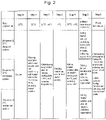
- the cells are incubated at 37°C for 10 minutes ( Figure 2 , step 1).
- the type and amount of buffer are not particularly limited.
- step 1 the operations of step 1 are repeated twice.
- buffer solution e.g., Hanks' solution
- the number of times step 1 is repeated can be, needless to say, changed depending on the types of buffer solution and cells used.
- the drug solution to be evaluated is administered to cells.
- An example is illustrated below.
- the buffer is removed, and then 200 ⁇ L of 10 ⁇ M CDF (a fluorescent reagent) is added to the well.
- the plate is incubated at 37°C for 30 minutes and then maintained at 4°C for 5 minutes ( Figure 2 , step 2).
- the type, concentration, and amount of the reagent are not particularly limited. CDF emits fluorescence and can be therefore easily quantified by a plate reader as a model reagent.
- the concentration of CDF may be a concentration conventionally used for a fluorescence assay of cells. This concentration is preferably applied as an amount of the reagent for the detection with a plate reader.
- 30 minutes of the incubation time was determined based on the result of a preliminary examination that the time required for getting the equilibrium between uptake and excretion of a drug is 30 minutes.
- the plate temperature was lowered to 4°C after incubation for 30 minutes for the purpose of preventing the leakage of the administered drug from the cells. This is because lowering the temperature in the container prevents cells from excreting the drug.
- the temperature is lowered to 4°C, but the temperature may be any temperature as long as the leakage of the administered drug from the cells can be suppressed at the temperature.
- the method for the suppression is not limited to lowering the temperature, but can be any method by which leakage of the administered drug from the cells can be suppressed, for example, by administering an inhibitor.
- the temperature in step 2 is lower than the plate temperature in step 1. This is because the temperature for the conditioning in step 1 is preferably a temperature that activates absorption and excretion of the drug by cells (e.g., 37°C), in contrast to step 2.
- the timing of the drug administration is not limited to that of this embodiment.
- the drug may be administered on the day before or a few days before the testing day.
- the drug may be administered in step 0 and steps 1 and 2 can be omitted.
- the drug solution other than that transferred into the cells in step 3 is washed away.
- An example is described below.
- the cells were then washed 3 times with 400 ⁇ L of ice-cold Hanks' solution while keeping the plate at 4°C ( Figure 2 , step 3).
- the purpose of keeping the plate at 4°C is the same as that described above.
- the amount of the Hanks' solution for washing was determined to be 400 ⁇ L for the purpose of removing medium ingredients remaining on the inner wall of the well because the amount of the medium in culture is 400 ⁇ L.
- the number of washing in general biochemistry assays is usually three times, which is adopted here. Conditions for the washing are not limited. This step removes anything other than the drug solution to be measured, making it possible to improve the accuracy of the efficacy evaluation in the later steps.
- this step aims to obtain the data for the aforementioned index and the drug is leaked from the cells in a predetermined time before evaluating the cells in respective sequences in step 5 described later. An example is illustrated below.
- a buffer solution e.g., Hanks' solution
- Hanks' solution for the pretreatment is first administered, the plate is incubated at 37°C for 30 minutes, and the Hanks' solution (supernatant) containing the drug was collected ( Figure 2 , step 4).
- the buffer solution may be the same as or different from the buffer solution used in step 5.
- the plate was maintained at 37°C so that the drug accumulated in the hepatocytes ( Figure 3 , 101) by step 3 is excreted into the Hanks' solution via the first basolateral efflux, that is, the passively diffusion ( Figure 2 , step 4, (1)') and/or via the transporter (TP) ( Figure 2 , step 4, (2)' and 102 in the image view).
- the basolateral efflux in step 4 is defined as the first basolateral efflux and the basolateral efflux in step 5 as the second basolateral efflux. Essentially, both steps represent the basolateral efflux(supernatant).
- Transporters are membrane proteins that are responsible for transportation of substance and expressed on the cell membrane. They are responsible for active transportation of substance between inside and outside of the cell. The passive diffusion is excretion to outside of the cell other than that via the transporter and includes the leakage through the cell membrane.
- the drug excreted into the Hanks' solution in step 4 was defined as a fraction of basolateral efflux, because the upper surface of the cells facing the Hanks' solution corresponds to the basal/basolateral face ( Figure 3 , 104), which is considered to be a part facing the blood vessel in the body.
- Step 5 which is explained next, includes collecting the drug in three different operations (sequences 1, 2, and 3) and an index for examining whether the drug tends to remain in the cell can be acquired by, prior to step 5, obtaining the first fraction of basolateral efflux (the drug excreted into Hanks' solution) in step 4 as mentioned above.
- the Hanks' solution used to obtain the fraction of the basolateral efflux may be of any sequence.
- Hanks' solutions of all sequences 1-3 described later or a part thereof may be evaluated.
- Steps 3 and 4 are steps of conducting the analysis according to the present invention in more detail to conduct highly accurate evaluation.
- the aforementioned steps 0-4 are conducted in at least sequences 1-3, as shown in Figure 2 .
- steps 5 operations are conducted under different conditions for sequences 1-3 so as to obtain a plurality of index data sought in the present disclosure by the analysis described later. An example is illustrated below.
- sequence 1 the drug is excreted from the cell into a buffer solution (e.g., Hanks' (-) solution) in an amount equal to the sum of biliary excretion (3) and the second basolateral efflux (that is, diffusion (1) and excretion via transporter (TP) (2)) discharge.
- Hanks' (-) solution is Hanks' solution free of calcium and magnesium ions and used, for example, when cell-cell adhesion is not enhanced intentionally as in this sequence.
- a chelator such as EGTA is used as described in the following.
- 200 ⁇ L of Hanks' (-) solution containing 1 mM EGTA was added.
- EGTA has a chelating effect of suppressing the effect of Ca 2+ and Mg 2+ involved in the adhesion of intercellular adhesion molecules and is a reagent for removing cell-cell adhesion. This causes the disruption of bile ducts formed during the culture process. The area was incubated at 37°C for 30 minutes, and then the supernatant was collected.
- the chelating reagent is not limited to EGTA, as long as it has a chelating effect. Type and amount of the buffer containing EGTA and temperature and time of the incubation are not particularly limited.
- step 5 the drug remaining in the cells ( Figure 3 , step 5 and the image drawing, (1) and (2)) and the drug excreted into the bile canaliculi ( Figure 3 , step 5 and the image drawing, (3)) upon the completion of step 4 are all excreted into the supernatant and collected ( Figure 3 , step 5, sequence 1).
- step 5 (3) is defined as a biliary-excreted fraction, because the cell membrane part around the gap formed in the cell-cell adhesion part corresponds to an apical face ( Figure 3 , 105) and the gap is assumed to be a bile canaliculus ( Figure 3 , 103).
- step 5 sequence 1 the temperature is maintained at 37°C, and therefore the fractions excreted from the cells to the basolateral side include the fraction via passive diffusion ( Figure 3 , step 5, (1)) and the fraction via the transporter ( Figure 3 , step 5, (2)).
- sequence 2 is a sequence where the disruption of bile canaliculi is not induced.
- the area was incubated at 37°C for 30 minutes, and then the supernatant was collected.
- the drug remaining in the cells ( Figure 3 , step 5 and the image view, (1) and (2)) upon the completion of step 4 is excreted into the supernatant and collected ( Figure 3 , step 5, sequence 2).
- the drug excreted into the supernatant is defined as a second fraction of basolateral efflux.
- step 5 the temperature is maintained at 37°C, and therefore the fractions excreted from the cells to the basolateral side include the fraction via passive diffusion ( Figure 3 , step 5, (1)) and the fraction via the transporter ( Figure 3 , step 5, (2)).
- each of the amount of the drug excreted to the bile canaliculi, (3) and the amount of the drug of the second fraction of basolateral efflux can be calculated.
- Sequence 3 has a temperature condition lower than that in sequences 1 and 2 as described later, and thereby the excretion of the drug via the transporter (1) is suppressed and only the amount of the drug excreted by diffusion (2) is excreted in the second fraction of basolateral efflux.
- step 5 sequence 2 a sequence having a low temperature of 4°C is adopted to suppress the transporter activity, and thereby quantify only the amount excreted by passive diffusion ( Figure 3 , step 5, (1)). Comparing this with step 5, sequence 2 makes it possible to calculate (quantify) the amount of the basolateral efflux fraction via transporter. For each drug, a specific transporter is present for moving between inside and outside of the cell.
- the ability to exclude the amount of excretion by passive diffusion and quantify the amount of excretion via the transporter means the ability to quantify the amount of excretion specific for the drug.
- Type and amount of the buffer and temperature and time of the incubation are not particularly limited.
- the fraction excreted via the transporter can be calculated by quantifying the drug collected from sequence 2 and sequence 3 in step 5. Although the calculation of excreted fractions using sequences 1, 2, and 3 is illustrated in this embodiment, the number of sequences is not limited to three.
- step 6 the same operations are conducted for the 3 sequences again.
- step 6 the drug collection for quantifying the fraction remaining in the cells is conducted.
- fluorescent agent is quantified using a plate reader, it is preferred that, for example, 200 ⁇ L of a Hanks solution containing 1% surfactants is added and suspended and the total amount is collected ( Figure 2 , step 6). This disrupts the cell membrane and makes it possible to have the drug remaining in the cells excreted.
- the resultant samples are transferred to a culture plate and fluorescence was measured using a plate reader. Wells containing only the Hanks' solution are prepared for the blank measurement. Fluorescence intensity is measured with an excitation wavelength of 484 nm and an absorption wavelength of 519 nm.
- LCMS mass spectrometry
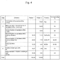
- the distribution ratio to respective fractions is calculated based on the fluorescence intensities reflecting the amounts of the drug obtained in the aforementioned steps.
- the scoring calculated for CDF is illustrated.
- the calculated scores are not limited to these.
- a score for evaluating basolateral efflux via transporter is calculated as
- a score for evaluating the biliary excretion can be calculated as
- the ratio of the amount of drug excreted via transporter to the amount of drug excreted to the basolateral side (RexEMTP) and the ratio of the amount of drug excreted into the bile duct to the total amount of drug taken up into the cells (BiRD) for CDF are 41.08 and 18.58, respectively, while those for Rhodamine123 are 52.52 and 4.92, respectively, indicating that CDF is a drug that has an stronger tendency to be excreted into the bile canaliculi but not to the basolateral side than Rhodamine123 does.
- the present invention makes it possible to evaluate compounds on the pharmacokinetics they exhibit.
- Embodiment 2 describes the determination of distribution ratio and scoring of respective fractions based on the measurement result when a method different from embodiment 1 is used.
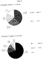
- a circle graph such as pattern 2 in Figure 5 can be generated and visual understanding of a total picture of the distribution ratio of respective fractions becomes possible.
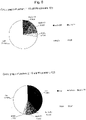
- the Sup fraction which is an index for tendency to remain or not to remain in the cells, for CDF is 70.82, while that for Rhodamine123 is 39.29 ( Figure 6 , pattern 2), indicating that CDF has a tendency to be excreted to the basolateral side. This is consistent with the evaluation of the fraction remaining in the cells in embodiment 1.
- Embodiment 3 not being part of the invention, describes an example of apparatus automating a series of steps described in embodiments 1 and 2. About purposes of operations and functions of configurations of the apparatus described later, descriptions may be omitted when the descriptions are overlapped with those of the aforementioned embodiments 1 and 2.
- sequences 1-3 shown in embodiments 1-2 are described using expressions such as "a well for sequence 1," "a well for sequence 2,” and "a well for sequence 3.”
- a plurality of containers in the aforementioned "well culture plate” may be defined for respective sequences or a plurality of wells in a well culture plate may be divided and the divided regions may be defined as, for example, "a first container,” “a second container,” and "a third container.”
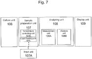
- the apparatus includes a culture unit 106, a sample preparation unit 107 (including an input unit 107A), an analyzing unit 108 and a display unit 109, as shown in Figure 7 .
- the sample preparation unit 107 has a temperature controlling unit 107B for controlling temperature in respective containers (plates) described later, a liquid-feeding unit 107C capable of supplying or collecting liquid in a container, and the like.
- the analyzing unit 108 has a measurement unit 108A for measuring the amount of a component such as a drug and analysis unit 108B for analyzing each of: an amount excreted via transporter, an amount excreted via the bile duct, an amount remaining in the cells, and an amount excreted via a route (diffusion) other than the transporter and the bile duct based on the amount of the component such as a drug obtained in the measurement unit.
- the above conformation of the apparatus is an example and, for example, other configurations may be, needless to say, adopted, for example, one in which only the analysis unit operates in a separate apparatus and the information obtained by the measurement unit is transmitted to the separate apparatus.
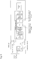
- sample preparation unit aims to prepare respective fractions to be analyzed automatically as described in embodiments 1-2. Respective components will be described in connection with the flow chart described later.
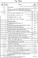
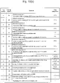
- Figure 10 is a flow chart of operations of the automatic measuring apparatus.
- the flow chart of Figure 10 is merely an example and the operations may be different from this when a timing of the drug administration is different as described in embodiment 1, ⁇ step 2>.
- plates having a plurality of cell holding regions (wells) are used as an example for illustrating the containers for holding cells, but the containers are not limited to plates and any containers that can contain cells may be, needless to say, used.
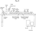
- Hepatocytes are first cultured in the culture unit 106 ( Figures 7 and 10 ) ( Figure 10(a) substep 1). Subsequently, the plates containing the cultured hepatocytes are transferred on a plate holder 210 having a first temperature controlling function and a plate holder (211) having a second temperature controlling function in the sample preparation unit ( Figures 7 and 10 ) ( Figure 10(a) , substep 2).
- An aspiration head 205 equipped with an aspiration nozzle to aspirate liquid, installed in the liquid-feeding unit 107C moves to a well 002, filled with medium to be removed, in a culture plate 001 on the aforementioned plate holder and aspirates the medium from the well to remove the total amount ( Figure 10 , substep 3).
- the removed medium is collected (discarded) in the waste liquid tank 206.
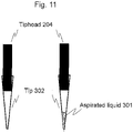
- a tip 302 for containing liquid 301 from a tip rack 207 storing a plurality of tips is attached onto a tiphead 204 installed to an aspiration nozzle.
- the tiphead moves to a room temperature drug solution rack 209 and aspirate a buffer ( Figure 10 , substep 4).
- the tiphead moves to a target well and introduces the buffer ( Figure 10(a) , substep 5) and then the tiphead moves to a dust box 214 and discards the tip.
- An exchangeable tip is used here to prevent contaminations, but the tip is not limited to this.
- the absorption head moves to the well filled with the buffer and removes the buffer ( Figure 10(a) , substep 6).
- the removed medium is collected and discarded in the waste liquid tank 206. This step is repeated twice in total (washing step) ( Figure 10(a) , substep 7).
- a tip in the tip rack 207 is attached onto the tiphead 204, the tiphead moves to the room temperature drug solution rack 209, and aspirates the buffer ( Figure 9 , substep 8).
- the tiphead moves to the target well and introduces the buffer and then the tiphead moves to the dust box 214 and discards the tip.
- the plate is allowed to stand at 37°C for 10 minutes (conditioning) ( Figure 10(a) , substep 9).
- the aspiration head 205 moves to the target well filled with the buffer and removes the total amount of the buffer ( Figure 9 (a), substep 10).
- a tip in the tip rack 207 is attached onto the tiphead 204, the tiphead moves to the room temperature drug solution rack 209, and aspirates the drug solution ( Figure 10(a) , substep 11).
- the tiphead moves to the target well and introduces the drug solution ( Figure 10(a) , substep 12) and then the tiphead 204 moves to the dust box 214 and discards the tip.
- the plate is allowed to stand at 37°C for 30 minutes ( Figure 10(a) , substep 12).
- the temperature of the plate holder 210 having the first temperature controlling function and the plate holder 211 having the second temperature controlling function which are an example configuration of the container holding unit for holding containers, is changed from 37°C to 4°C ( Figure 10(a) , substep 13), and then the aspiration head 205 moves to the well filled with the drug solution and removes a total amount of the drug solution ( Figure 10(a) , substep 14).
- the temperature controlling unit in this embodiment is described as a plate folder having a temperature controlling function, the temperature controlling unit may be, needless to say, separated from the container holding unit.
- a tip in the tip rack 207 is attached onto the tiphead 204, the tiphead moves to a refrigerated drug solution rack 208, and aspirates a buffer ( Figure 10(a) , substep 15).
- a refrigerated drug solution is used to stop active biological phenomena such as the transporter activity, as described above.
- the refrigerated drug solution rack 208 aims to hold a drug solution, a buffer, and the like at a low temperature for the purpose.
- the tiphead moves to the target well and introduces the buffer ( Figure 10(a) , substep 16) and then the tiphead 204 moves to the dust box 214 and discards the tip.
- the absorption head moves to the well filled with the buffer and removes the buffer ( Figure 10(a) , substep 17).
- the removed medium is discarded in the waste liquid tank 206. This step is repeated three times in total (washing step) ( Figure 10(a) , substep 18).
- the temperature of the plate holder 210 having the first temperature controlling function and the plate holder 211 having the second temperature controlling function is changed from 4°C to 37°C ( Figure 10(a) , substep 19), a tip in the tip rack 207 is attached onto the tiphead 204, and the tiphead moves to a refrigerated drug solution rack 208 and aspirate the buffer ( Figure 10(a) , substep 20).
- the tiphead moves to the target well and introduces the buffer ( Figure 10(a) , substep 21) and then the tiphead 204 moves to the dust box 214 and discards the tip.
- the plate is allowed to stand at 37°C for 30 minutes ( Figure 10(a) , substep 20).
- the temperature of the plate holder 210 having the first temperature controlling function and the plate holder 211 having the second temperature controlling function is changed from 37°C to 4°C ( Figure 10(a) , substep 22), and then a tip in the tip rack 207 is attached onto the tiphead 204, the tiphead moves to the well filled with the buffer containing the drug, aspirates the buffer (supernatant) containing the drug, and dispenses it into collection plates for collection on the first plate holder 212 and the second plate holder 213 (collection) ( Figure 10(a) , substep 23).
- the temperature of the plate holder 210 having the first temperature controlling function is changed from 4°C to 37°C ( Figure 10(a) , substep 24), a tip in the tip rack 207 is attached onto the tiphead 204, and the tiphead moves to the room temperature drug solution rack (209) and aspirates a buffer containing EGTA ( Figure 10(a) , substep 24).
- the tip head moves to a well for sequence 1 on the first plate holder 212, the buffer containing EGTA is introduced ( Figure 10(b) , substep 25), then the tiphead 204 moves to the dust box 214, and the tip is discarded.
- the plate is allowed to stand at 37°C for 30 minutes ( Figure 10(b) , substep 26).
- a tip in the tip rack 207 is attached onto the tiphead 204, the tiphead moves to the room temperature drug solution rack 209, and aspirates the buffer ( Figure 10(b) , substep 27).
- the tip head moves to a well for sequence 2 on the first plate holder 212, the buffer is introduced ( Figure 10(b) , substep 28), then the tiphead 204 moves to the dust box 214, and the tip is discarded.
- the plate is allowed to stand at 37°C for 30 minutes ( Figure 10(b) , substep 28).
- a tip in the tip rack 207 is attached onto the tiphead 204, the tiphead moves to the refrigerated drug solution rack 209, and aspirates the buffer ( Figure 10(b) , substep 29).
- the tip head moves to a well for sequence 3 on the second plate holder 213, the buffer is introduced ( Figure 10(b) , substep 30), then the tiphead 204 moves to the dust box 214, and the tip is discarded.
- the plate is allowed to stand at 4°C for 30 minutes ( Figure 10(b) , substep 30).
- a tip in the tip rack 207 is attached onto the tiphead 204 and the tiphead moves to the well filled with an EGTA buffer containing the drug, aspirates the EGTA buffer (supernatant) containing the drug, and dispenses (collects) the buffer into a collection plate for collection on the first plate holder 212 ( Figure 10(b) , substep 31).
- a tip in the tip rack 207 is attached onto the tiphead 204 and the tiphead moves to the well filled with the buffer containing the drug, aspirates the buffer (supernatant) containing the drug, and dispenses (collects) the buffer into a collection plate for collection on the first plate holder 212 ( Figure 10(b) , substep 32).
- a tip in the tip rack 207 is attached onto the tiphead 204, the tiphead moves to the well filled with the EGTA buffer containing the drug, aspirates the buffer (supernatant) containing the drug, and dispenses (collects) the buffer into the collection plate for collection on the first plate holder 213 ( Figure 10(b) , substep 33).
- the temperatures of the plate holder 210 having the first temperature controlling function and the plate holder 211 having the second temperature controlling function are both changed to room temperature ( Figure 10(b) , substep 34), a tip in the tip rack 207 is attached onto the tiphead 204, and the tiphead moves to the room temperature drug solution rack 209 and aspirates 1% TritonX-100 or pure water/methanol ( Figure 10(b) , substep 35).
- the tip head moves to wells for sequences 1, 2, and 3 on the plate holder (210) having the first temperature controlling function and the plate holder 211 having the second temperature controlling function, and introduces 1% TritonX-100 or pure water/methanol ( Figure 10(b) , substep 36), the tiphead 204 moves to the dust box 214, and the tip is discarded.
- a tip in the tip rack 207 is attached onto the tiphead 204 and the tiphead moves to a well filled with the aforementioned reagent, aspirates the total amount of the cell suspension, and dispenses the suspension into the collection plates for collection on the first plate holder 212 and the second plate holder 213 (collection) ( Figure 10(b) , substep 37).
- the collected drug in the culture plate is transferred to the measurement unit ( Figure 10(b) , substep 38).
- the measurement unit the measurement of the drug by a plate reader or LCMS is conducted ( Figure 10(b) , substep 39).
- the distribution ratio and the score of respective fractions are calculated based on the measurement results ( Figure 10(b) , substep 40). Then, the resultant calculated values are displayed on a display unit ( Figure 10(b) , substep 41).
Landscapes
- Health & Medical Sciences (AREA)
- Life Sciences & Earth Sciences (AREA)
- Engineering & Computer Science (AREA)
- Chemical & Material Sciences (AREA)
- Bioinformatics & Cheminformatics (AREA)
- Biomedical Technology (AREA)
- Immunology (AREA)
- Zoology (AREA)
- Wood Science & Technology (AREA)
- Organic Chemistry (AREA)
- Biochemistry (AREA)
- General Health & Medical Sciences (AREA)
- Biotechnology (AREA)
- Microbiology (AREA)
- Molecular Biology (AREA)
- Analytical Chemistry (AREA)
- Cell Biology (AREA)
- Hematology (AREA)
- Urology & Nephrology (AREA)
- Sustainable Development (AREA)
- Genetics & Genomics (AREA)
- General Engineering & Computer Science (AREA)
- Pathology (AREA)
- Physics & Mathematics (AREA)
- General Physics & Mathematics (AREA)
- Medicinal Chemistry (AREA)
- Food Science & Technology (AREA)
- Toxicology (AREA)
- Tropical Medicine & Parasitology (AREA)
- Clinical Laboratory Science (AREA)
- Gastroenterology & Hepatology (AREA)
- Pharmacology & Pharmacy (AREA)
- Biophysics (AREA)
- Chemical Kinetics & Catalysis (AREA)
- Measuring Or Testing Involving Enzymes Or Micro-Organisms (AREA)
- Investigating Or Analysing Biological Materials (AREA)
- Apparatus Associated With Microorganisms And Enzymes (AREA)
Applications Claiming Priority (4)
| Application Number | Priority Date | Filing Date | Title |
|---|---|---|---|
| JP2013246906A JP6087264B2 (ja) | 2013-11-29 | 2013-11-29 | 成分分析装置、薬効分析装置、及び分析方法 |
| EP17186423.4A EP3272852B1 (en) | 2013-11-29 | 2014-11-26 | Componential analyzer, drug efficacy analyzer, and analysis method |
| PCT/JP2014/081135 WO2015080106A1 (ja) | 2013-11-29 | 2014-11-26 | 成分分析装置、薬効分析装置、及び分析方法 |
| EP14866627.4A EP3023489B1 (en) | 2013-11-29 | 2014-11-26 | Componential analyzer, drug efficacy analyzer, and analysis method |
Related Parent Applications (3)
| Application Number | Title | Priority Date | Filing Date |
|---|---|---|---|
| EP14866627.4A Division EP3023489B1 (en) | 2013-11-29 | 2014-11-26 | Componential analyzer, drug efficacy analyzer, and analysis method |
| EP17186423.4A Division-Into EP3272852B1 (en) | 2013-11-29 | 2014-11-26 | Componential analyzer, drug efficacy analyzer, and analysis method |
| EP17186423.4A Division EP3272852B1 (en) | 2013-11-29 | 2014-11-26 | Componential analyzer, drug efficacy analyzer, and analysis method |
Publications (2)
| Publication Number | Publication Date |
|---|---|
| EP3712245A1 EP3712245A1 (en) | 2020-09-23 |
| EP3712245B1 true EP3712245B1 (en) | 2022-05-11 |
Family
ID=53199046
Family Applications (3)
| Application Number | Title | Priority Date | Filing Date |
|---|---|---|---|
| EP20154632.2A Active EP3712245B1 (en) | 2013-11-29 | 2014-11-26 | Componential and drug efficacy analysis method |
| EP14866627.4A Active EP3023489B1 (en) | 2013-11-29 | 2014-11-26 | Componential analyzer, drug efficacy analyzer, and analysis method |
| EP17186423.4A Active EP3272852B1 (en) | 2013-11-29 | 2014-11-26 | Componential analyzer, drug efficacy analyzer, and analysis method |
Family Applications After (2)
| Application Number | Title | Priority Date | Filing Date |
|---|---|---|---|
| EP14866627.4A Active EP3023489B1 (en) | 2013-11-29 | 2014-11-26 | Componential analyzer, drug efficacy analyzer, and analysis method |
| EP17186423.4A Active EP3272852B1 (en) | 2013-11-29 | 2014-11-26 | Componential analyzer, drug efficacy analyzer, and analysis method |
Country Status (5)
| Country | Link |
|---|---|
| US (3) | US9880153B2 (cg-RX-API-DMAC7.html) |
| EP (3) | EP3712245B1 (cg-RX-API-DMAC7.html) |
| JP (1) | JP6087264B2 (cg-RX-API-DMAC7.html) |
| CN (3) | CN105378053B (cg-RX-API-DMAC7.html) |
| WO (1) | WO2015080106A1 (cg-RX-API-DMAC7.html) |
Families Citing this family (6)
| Publication number | Priority date | Publication date | Assignee | Title |
|---|---|---|---|---|
| JP6087264B2 (ja) | 2013-11-29 | 2017-03-01 | 株式会社日立ハイテクノロジーズ | 成分分析装置、薬効分析装置、及び分析方法 |
| JP6758026B2 (ja) | 2015-04-17 | 2020-09-23 | 株式会社日立ハイテク | 成分分析装置、薬剤成分分析装置、成分分析方法及び薬剤成分分析方法 |
| JP6582070B2 (ja) * | 2018-01-22 | 2019-09-25 | 株式会社日立ハイテクノロジーズ | 分析方法 |
| JP7113132B2 (ja) * | 2019-02-26 | 2022-08-04 | 株式会社日立ハイテク | 細胞内における化合物の動態解析法 |
| WO2021014519A1 (ja) * | 2019-07-22 | 2021-01-28 | 株式会社日立ハイテク | 試薬キット |
| WO2023084648A1 (ja) * | 2021-11-10 | 2023-05-19 | 株式会社日立ハイテク | 薬剤相互作用解析方法 |
Family Cites Families (16)
| Publication number | Priority date | Publication date | Assignee | Title |
|---|---|---|---|---|
| US5255976A (en) | 1992-07-10 | 1993-10-26 | Vertex Pharmaceuticals Incorporated | Temperature gradient calorimeter |
| WO1994012662A1 (en) | 1992-11-25 | 1994-06-09 | Merck & Co., Inc. | Hepatic model |
| CA2130013C (en) * | 1993-09-10 | 1999-03-30 | Rolf Moser | Apparatus for automatic performance of temperature cycles |
| US6277655B1 (en) * | 1994-10-13 | 2001-08-21 | Solvo Biotechnology | Assay and reagent kit for evaluation of multi-drug resistance in cells |
| US6022733A (en) | 1997-12-02 | 2000-02-08 | Tam; Yun K. | Simulated biological dissolution and absorption system |
| US7498164B2 (en) * | 1998-05-16 | 2009-03-03 | Applied Biosystems, Llc | Instrument for monitoring nucleic acid sequence amplification reaction |
| US7601494B2 (en) | 1999-03-17 | 2009-10-13 | The University Of North Carolina At Chapel Hill | Method of screening candidate compounds for susceptibility to biliary excretion |
| DK1163517T3 (da) * | 1999-03-17 | 2006-08-21 | Univ North Carolina | Fremgangsmåde til screening af kandidatforbindelser med hensyn til disponering for galdesten |
| AR035231A1 (es) * | 2002-03-11 | 2004-05-05 | Ypf S A | Un equipo para analizar el crecimiento de microorganismos y procedimiento para cuantificar la concentracion de microorganismos |
| US20070184548A1 (en) * | 2002-12-23 | 2007-08-09 | Lim Hi Tan | Device for carrying out chemical or biological reactions |
| CN101977643A (zh) * | 2008-03-24 | 2011-02-16 | 奥林巴斯株式会社 | 药剂投放装置 |
| CN102272288B (zh) | 2009-01-08 | 2015-03-11 | 株式会社日立制作所 | 动物肝细胞的培养方法 |
| EP2471908B1 (en) | 2009-08-26 | 2016-10-05 | The University of Tokyo | Method for culture of hepatocytes |
| US8945865B2 (en) | 2011-03-10 | 2015-02-03 | Eisai R&D Management Co., Ltd. | Method for screening for compound capable of enhancing or inhibiting OATP1B1 transport activity, and method for determining expression level of OATP1B1 |
| US20130164335A1 (en) | 2011-12-27 | 2013-06-27 | National Health Research Institutes | Methods and compositions for cellular drug release |
| JP6087264B2 (ja) * | 2013-11-29 | 2017-03-01 | 株式会社日立ハイテクノロジーズ | 成分分析装置、薬効分析装置、及び分析方法 |
-
2013
- 2013-11-29 JP JP2013246906A patent/JP6087264B2/ja active Active
-
2014
- 2014-11-26 CN CN201480040060.4A patent/CN105378053B/zh active Active
- 2014-11-26 EP EP20154632.2A patent/EP3712245B1/en active Active
- 2014-11-26 US US14/914,278 patent/US9880153B2/en active Active
- 2014-11-26 EP EP14866627.4A patent/EP3023489B1/en active Active
- 2014-11-26 CN CN201710860736.XA patent/CN107513550B/zh active Active
- 2014-11-26 WO PCT/JP2014/081135 patent/WO2015080106A1/ja not_active Ceased
- 2014-11-26 CN CN201710862144.1A patent/CN107677779B/zh active Active
- 2014-11-26 EP EP17186423.4A patent/EP3272852B1/en active Active
-
2017
- 2017-12-19 US US15/847,092 patent/US10684276B2/en active Active
-
2020
- 2020-04-23 US US16/856,459 patent/US11789013B2/en active Active
Also Published As
| Publication number | Publication date |
|---|---|
| JP2015105842A (ja) | 2015-06-08 |
| US9880153B2 (en) | 2018-01-30 |
| CN107677779B (zh) | 2020-06-12 |
| CN107513550A (zh) | 2017-12-26 |
| EP3023489A4 (en) | 2016-09-21 |
| US20200249223A1 (en) | 2020-08-06 |
| US11789013B2 (en) | 2023-10-17 |
| US20160209403A1 (en) | 2016-07-21 |
| EP3023489A1 (en) | 2016-05-25 |
| EP3272852B1 (en) | 2020-03-18 |
| EP3023489B1 (en) | 2017-11-01 |
| EP3712245A1 (en) | 2020-09-23 |
| WO2015080106A1 (ja) | 2015-06-04 |
| US10684276B2 (en) | 2020-06-16 |
| CN105378053A (zh) | 2016-03-02 |
| CN107513550B (zh) | 2022-06-21 |
| US20180106783A1 (en) | 2018-04-19 |
| CN105378053B (zh) | 2017-10-20 |
| JP6087264B2 (ja) | 2017-03-01 |
| CN107677779A (zh) | 2018-02-09 |
| EP3272852A1 (en) | 2018-01-24 |
Similar Documents
| Publication | Publication Date | Title |
|---|---|---|
| US11789013B2 (en) | Componential analyzer, drug efficacy analyzer, and analysis method | |
| Bircsak et al. | A 3D microfluidic liver model for high throughput compound toxicity screening in the OrganoPlate® | |
| Gijzen et al. | Culture and analysis of kidney tubuloids and perfused tubuloid cells-on-a-chip | |
| Peel et al. | Introducing an automated high content confocal imaging approach for Organs-on-Chips | |
| US9772325B2 (en) | Method for measuring bile salt export transport and/or formation activity | |
| Gerets et al. | Multiplexing cell viability assays | |
| CN103645332A (zh) | 自动化高通量检测胆固醇以及其它分析物细胞流出的多功能工作站 | |
| Xu et al. | Assessment of hepatotoxicity potential of drug candidate molecules including kinase inhibitors by hepatocyte imaging assay technology and bile flux imaging assay technology | |
| JP7113132B2 (ja) | 細胞内における化合物の動態解析法 | |
| Pohan et al. | Multiparametric High‐Content Assays to Measure Cell Health and Oxidative Damage as a Model for Drug‐Induced Liver Injury | |
| CN107406814B (zh) | 成分分析装置、药剂成分分析装置、成分分析方法及药剂成分分析方法 | |
| JP6582070B2 (ja) | 分析方法 | |
| JP6282362B2 (ja) | 成分分析装置、薬効分析装置、及び分析方法 | |
| EP3189131A1 (en) | In vitro biliary excretion assay |
Legal Events
| Date | Code | Title | Description |
|---|---|---|---|
| PUAI | Public reference made under article 153(3) epc to a published international application that has entered the european phase |
Free format text: ORIGINAL CODE: 0009012 |
|
| STAA | Information on the status of an ep patent application or granted ep patent |
Free format text: STATUS: REQUEST FOR EXAMINATION WAS MADE |
|
| 17P | Request for examination filed |
Effective date: 20200331 |
|
| AC | Divisional application: reference to earlier application |
Ref document number: 3023489 Country of ref document: EP Kind code of ref document: P Ref document number: 3272852 Country of ref document: EP Kind code of ref document: P |
|
| AK | Designated contracting states |
Kind code of ref document: A1 Designated state(s): AL AT BE BG CH CY CZ DE DK EE ES FI FR GB GR HR HU IE IS IT LI LT LU LV MC MK MT NL NO PL PT RO RS SE SI SK SM TR |
|
| GRAP | Despatch of communication of intention to grant a patent |
Free format text: ORIGINAL CODE: EPIDOSNIGR1 |
|
| STAA | Information on the status of an ep patent application or granted ep patent |
Free format text: STATUS: GRANT OF PATENT IS INTENDED |
|
| INTG | Intention to grant announced |
Effective date: 20210729 |
|
| GRAJ | Information related to disapproval of communication of intention to grant by the applicant or resumption of examination proceedings by the epo deleted |
Free format text: ORIGINAL CODE: EPIDOSDIGR1 |
|
| STAA | Information on the status of an ep patent application or granted ep patent |
Free format text: STATUS: REQUEST FOR EXAMINATION WAS MADE |
|
| REG | Reference to a national code |
Ref country code: DE Ref legal event code: R079 Ref document number: 602014083764 Country of ref document: DE Free format text: PREVIOUS MAIN CLASS: C12M0001340000 Ipc: G01N0033500000 |
|
| GRAP | Despatch of communication of intention to grant a patent |
Free format text: ORIGINAL CODE: EPIDOSNIGR1 |
|
| INTC | Intention to grant announced (deleted) | ||
| STAA | Information on the status of an ep patent application or granted ep patent |
Free format text: STATUS: GRANT OF PATENT IS INTENDED |
|
| RIC1 | Information provided on ipc code assigned before grant |
Ipc: G01N 35/04 20060101ALI20211210BHEP Ipc: G01N 35/00 20060101ALI20211210BHEP Ipc: B01L 9/00 20060101ALI20211210BHEP Ipc: B01L 3/00 20060101ALI20211210BHEP Ipc: B01L 7/00 20060101ALI20211210BHEP Ipc: G01N 33/15 20060101ALI20211210BHEP Ipc: C12M 1/00 20060101ALI20211210BHEP Ipc: C12M 1/34 20060101ALI20211210BHEP Ipc: G01N 33/50 20060101AFI20211210BHEP |
|
| INTG | Intention to grant announced |
Effective date: 20220113 |
|
| GRAS | Grant fee paid |
Free format text: ORIGINAL CODE: EPIDOSNIGR3 |
|
| GRAA | (expected) grant |
Free format text: ORIGINAL CODE: 0009210 |
|
| STAA | Information on the status of an ep patent application or granted ep patent |
Free format text: STATUS: THE PATENT HAS BEEN GRANTED |
|
| AC | Divisional application: reference to earlier application |
Ref document number: 3023489 Country of ref document: EP Kind code of ref document: P Ref document number: 3272852 Country of ref document: EP Kind code of ref document: P |
|
| AK | Designated contracting states |
Kind code of ref document: B1 Designated state(s): AL AT BE BG CH CY CZ DE DK EE ES FI FR GB GR HR HU IE IS IT LI LT LU LV MC MK MT NL NO PL PT RO RS SE SI SK SM TR |
|
| REG | Reference to a national code |
Ref country code: GB Ref legal event code: FG4D |
|
| REG | Reference to a national code |
Ref country code: CH Ref legal event code: EP |
|
| REG | Reference to a national code |
Ref country code: AT Ref legal event code: REF Ref document number: 1491841 Country of ref document: AT Kind code of ref document: T Effective date: 20220515 |
|
| REG | Reference to a national code |
Ref country code: DE Ref legal event code: R096 Ref document number: 602014083764 Country of ref document: DE |
|
| REG | Reference to a national code |
Ref country code: IE Ref legal event code: FG4D |
|
| REG | Reference to a national code |
Ref country code: LT Ref legal event code: MG9D |
|
| REG | Reference to a national code |
Ref country code: NL Ref legal event code: MP Effective date: 20220511 |
|
| REG | Reference to a national code |
Ref country code: AT Ref legal event code: MK05 Ref document number: 1491841 Country of ref document: AT Kind code of ref document: T Effective date: 20220511 |
|
| PG25 | Lapsed in a contracting state [announced via postgrant information from national office to epo] |
Ref country code: SE Free format text: LAPSE BECAUSE OF FAILURE TO SUBMIT A TRANSLATION OF THE DESCRIPTION OR TO PAY THE FEE WITHIN THE PRESCRIBED TIME-LIMIT Effective date: 20220511 Ref country code: PT Free format text: LAPSE BECAUSE OF FAILURE TO SUBMIT A TRANSLATION OF THE DESCRIPTION OR TO PAY THE FEE WITHIN THE PRESCRIBED TIME-LIMIT Effective date: 20220912 Ref country code: NO Free format text: LAPSE BECAUSE OF FAILURE TO SUBMIT A TRANSLATION OF THE DESCRIPTION OR TO PAY THE FEE WITHIN THE PRESCRIBED TIME-LIMIT Effective date: 20220811 Ref country code: NL Free format text: LAPSE BECAUSE OF FAILURE TO SUBMIT A TRANSLATION OF THE DESCRIPTION OR TO PAY THE FEE WITHIN THE PRESCRIBED TIME-LIMIT Effective date: 20220511 Ref country code: LT Free format text: LAPSE BECAUSE OF FAILURE TO SUBMIT A TRANSLATION OF THE DESCRIPTION OR TO PAY THE FEE WITHIN THE PRESCRIBED TIME-LIMIT Effective date: 20220511 Ref country code: HR Free format text: LAPSE BECAUSE OF FAILURE TO SUBMIT A TRANSLATION OF THE DESCRIPTION OR TO PAY THE FEE WITHIN THE PRESCRIBED TIME-LIMIT Effective date: 20220511 Ref country code: GR Free format text: LAPSE BECAUSE OF FAILURE TO SUBMIT A TRANSLATION OF THE DESCRIPTION OR TO PAY THE FEE WITHIN THE PRESCRIBED TIME-LIMIT Effective date: 20220812 Ref country code: FI Free format text: LAPSE BECAUSE OF FAILURE TO SUBMIT A TRANSLATION OF THE DESCRIPTION OR TO PAY THE FEE WITHIN THE PRESCRIBED TIME-LIMIT Effective date: 20220511 Ref country code: ES Free format text: LAPSE BECAUSE OF FAILURE TO SUBMIT A TRANSLATION OF THE DESCRIPTION OR TO PAY THE FEE WITHIN THE PRESCRIBED TIME-LIMIT Effective date: 20220511 Ref country code: BG Free format text: LAPSE BECAUSE OF FAILURE TO SUBMIT A TRANSLATION OF THE DESCRIPTION OR TO PAY THE FEE WITHIN THE PRESCRIBED TIME-LIMIT Effective date: 20220811 Ref country code: AT Free format text: LAPSE BECAUSE OF FAILURE TO SUBMIT A TRANSLATION OF THE DESCRIPTION OR TO PAY THE FEE WITHIN THE PRESCRIBED TIME-LIMIT Effective date: 20220511 |
|
| PG25 | Lapsed in a contracting state [announced via postgrant information from national office to epo] |
Ref country code: RS Free format text: LAPSE BECAUSE OF FAILURE TO SUBMIT A TRANSLATION OF THE DESCRIPTION OR TO PAY THE FEE WITHIN THE PRESCRIBED TIME-LIMIT Effective date: 20220511 Ref country code: PL Free format text: LAPSE BECAUSE OF FAILURE TO SUBMIT A TRANSLATION OF THE DESCRIPTION OR TO PAY THE FEE WITHIN THE PRESCRIBED TIME-LIMIT Effective date: 20220511 Ref country code: LV Free format text: LAPSE BECAUSE OF FAILURE TO SUBMIT A TRANSLATION OF THE DESCRIPTION OR TO PAY THE FEE WITHIN THE PRESCRIBED TIME-LIMIT Effective date: 20220511 Ref country code: IS Free format text: LAPSE BECAUSE OF FAILURE TO SUBMIT A TRANSLATION OF THE DESCRIPTION OR TO PAY THE FEE WITHIN THE PRESCRIBED TIME-LIMIT Effective date: 20220911 |
|
| PG25 | Lapsed in a contracting state [announced via postgrant information from national office to epo] |
Ref country code: SM Free format text: LAPSE BECAUSE OF FAILURE TO SUBMIT A TRANSLATION OF THE DESCRIPTION OR TO PAY THE FEE WITHIN THE PRESCRIBED TIME-LIMIT Effective date: 20220511 Ref country code: SK Free format text: LAPSE BECAUSE OF FAILURE TO SUBMIT A TRANSLATION OF THE DESCRIPTION OR TO PAY THE FEE WITHIN THE PRESCRIBED TIME-LIMIT Effective date: 20220511 Ref country code: RO Free format text: LAPSE BECAUSE OF FAILURE TO SUBMIT A TRANSLATION OF THE DESCRIPTION OR TO PAY THE FEE WITHIN THE PRESCRIBED TIME-LIMIT Effective date: 20220511 Ref country code: EE Free format text: LAPSE BECAUSE OF FAILURE TO SUBMIT A TRANSLATION OF THE DESCRIPTION OR TO PAY THE FEE WITHIN THE PRESCRIBED TIME-LIMIT Effective date: 20220511 Ref country code: DK Free format text: LAPSE BECAUSE OF FAILURE TO SUBMIT A TRANSLATION OF THE DESCRIPTION OR TO PAY THE FEE WITHIN THE PRESCRIBED TIME-LIMIT Effective date: 20220511 Ref country code: CZ Free format text: LAPSE BECAUSE OF FAILURE TO SUBMIT A TRANSLATION OF THE DESCRIPTION OR TO PAY THE FEE WITHIN THE PRESCRIBED TIME-LIMIT Effective date: 20220511 |
|
| REG | Reference to a national code |
Ref country code: DE Ref legal event code: R097 Ref document number: 602014083764 Country of ref document: DE |
|
| PLBE | No opposition filed within time limit |
Free format text: ORIGINAL CODE: 0009261 |
|
| STAA | Information on the status of an ep patent application or granted ep patent |
Free format text: STATUS: NO OPPOSITION FILED WITHIN TIME LIMIT |
|
| PG25 | Lapsed in a contracting state [announced via postgrant information from national office to epo] |
Ref country code: AL Free format text: LAPSE BECAUSE OF FAILURE TO SUBMIT A TRANSLATION OF THE DESCRIPTION OR TO PAY THE FEE WITHIN THE PRESCRIBED TIME-LIMIT Effective date: 20220511 |
|
| 26N | No opposition filed |
Effective date: 20230214 |
|
| PG25 | Lapsed in a contracting state [announced via postgrant information from national office to epo] |
Ref country code: SI Free format text: LAPSE BECAUSE OF FAILURE TO SUBMIT A TRANSLATION OF THE DESCRIPTION OR TO PAY THE FEE WITHIN THE PRESCRIBED TIME-LIMIT Effective date: 20220511 |
|
| PG25 | Lapsed in a contracting state [announced via postgrant information from national office to epo] |
Ref country code: MC Free format text: LAPSE BECAUSE OF FAILURE TO SUBMIT A TRANSLATION OF THE DESCRIPTION OR TO PAY THE FEE WITHIN THE PRESCRIBED TIME-LIMIT Effective date: 20220511 |
|
| REG | Reference to a national code |
Ref country code: BE Ref legal event code: MM Effective date: 20221130 |
|
| PG25 | Lapsed in a contracting state [announced via postgrant information from national office to epo] |
Ref country code: LU Free format text: LAPSE BECAUSE OF NON-PAYMENT OF DUE FEES Effective date: 20221126 |
|
| PG25 | Lapsed in a contracting state [announced via postgrant information from national office to epo] |
Ref country code: IE Free format text: LAPSE BECAUSE OF NON-PAYMENT OF DUE FEES Effective date: 20221126 |
|
| PG25 | Lapsed in a contracting state [announced via postgrant information from national office to epo] |
Ref country code: BE Free format text: LAPSE BECAUSE OF NON-PAYMENT OF DUE FEES Effective date: 20221130 |
|
| PG25 | Lapsed in a contracting state [announced via postgrant information from national office to epo] |
Ref country code: IT Free format text: LAPSE BECAUSE OF FAILURE TO SUBMIT A TRANSLATION OF THE DESCRIPTION OR TO PAY THE FEE WITHIN THE PRESCRIBED TIME-LIMIT Effective date: 20220511 |
|
| PG25 | Lapsed in a contracting state [announced via postgrant information from national office to epo] |
Ref country code: CY Free format text: LAPSE BECAUSE OF FAILURE TO SUBMIT A TRANSLATION OF THE DESCRIPTION OR TO PAY THE FEE WITHIN THE PRESCRIBED TIME-LIMIT Effective date: 20220511 |
|
| PG25 | Lapsed in a contracting state [announced via postgrant information from national office to epo] |
Ref country code: MK Free format text: LAPSE BECAUSE OF FAILURE TO SUBMIT A TRANSLATION OF THE DESCRIPTION OR TO PAY THE FEE WITHIN THE PRESCRIBED TIME-LIMIT Effective date: 20220511 Ref country code: HU Free format text: LAPSE BECAUSE OF FAILURE TO SUBMIT A TRANSLATION OF THE DESCRIPTION OR TO PAY THE FEE WITHIN THE PRESCRIBED TIME-LIMIT; INVALID AB INITIO Effective date: 20141126 |
|
| PG25 | Lapsed in a contracting state [announced via postgrant information from national office to epo] |
Ref country code: MT Free format text: LAPSE BECAUSE OF FAILURE TO SUBMIT A TRANSLATION OF THE DESCRIPTION OR TO PAY THE FEE WITHIN THE PRESCRIBED TIME-LIMIT Effective date: 20220511 |
|
| PG25 | Lapsed in a contracting state [announced via postgrant information from national office to epo] |
Ref country code: BG Free format text: LAPSE BECAUSE OF FAILURE TO SUBMIT A TRANSLATION OF THE DESCRIPTION OR TO PAY THE FEE WITHIN THE PRESCRIBED TIME-LIMIT Effective date: 20220511 |
|
| PG25 | Lapsed in a contracting state [announced via postgrant information from national office to epo] |
Ref country code: BG Free format text: LAPSE BECAUSE OF FAILURE TO SUBMIT A TRANSLATION OF THE DESCRIPTION OR TO PAY THE FEE WITHIN THE PRESCRIBED TIME-LIMIT Effective date: 20220511 |
|
| PGFP | Annual fee paid to national office [announced via postgrant information from national office to epo] |
Ref country code: DE Payment date: 20241001 Year of fee payment: 11 |
|
| PGFP | Annual fee paid to national office [announced via postgrant information from national office to epo] |
Ref country code: GB Payment date: 20241002 Year of fee payment: 11 |
|
| PGFP | Annual fee paid to national office [announced via postgrant information from national office to epo] |
Ref country code: FR Payment date: 20241001 Year of fee payment: 11 |
|
| PGFP | Annual fee paid to national office [announced via postgrant information from national office to epo] |
Ref country code: CH Payment date: 20241201 Year of fee payment: 11 |
|
| REG | Reference to a national code |
Ref country code: CH Ref legal event code: U11 Free format text: ST27 STATUS EVENT CODE: U-0-0-U10-U11 (AS PROVIDED BY THE NATIONAL OFFICE) Effective date: 20251201 |
|
| PG25 | Lapsed in a contracting state [announced via postgrant information from national office to epo] |
Ref country code: TR Free format text: LAPSE BECAUSE OF FAILURE TO SUBMIT A TRANSLATION OF THE DESCRIPTION OR TO PAY THE FEE WITHIN THE PRESCRIBED TIME-LIMIT Effective date: 20220511 |