EP3709263B1 - Verfahren und system zur überwachung eines biologischen prozesses - Google Patents
Verfahren und system zur überwachung eines biologischen prozesses Download PDFInfo
- Publication number
- EP3709263B1 EP3709263B1 EP19162761.1A EP19162761A EP3709263B1 EP 3709263 B1 EP3709263 B1 EP 3709263B1 EP 19162761 A EP19162761 A EP 19162761A EP 3709263 B1 EP3709263 B1 EP 3709263B1
- Authority
- EP
- European Patent Office
- Prior art keywords
- image
- images
- time point
- abnormal tissue
- acquired
- Prior art date
- Legal status (The legal status is an assumption and is not a legal conclusion. Google has not performed a legal analysis and makes no representation as to the accuracy of the status listed.)
- Active
Links
Images
Classifications
-
- G—PHYSICS
- G06—COMPUTING OR CALCULATING; COUNTING
- G06T—IMAGE DATA PROCESSING OR GENERATION, IN GENERAL
- G06T7/00—Image analysis
- G06T7/0002—Inspection of images, e.g. flaw detection
- G06T7/0012—Biomedical image inspection
- G06T7/0014—Biomedical image inspection using an image reference approach
- G06T7/0016—Biomedical image inspection using an image reference approach involving temporal comparison
-
- G—PHYSICS
- G06—COMPUTING OR CALCULATING; COUNTING
- G06T—IMAGE DATA PROCESSING OR GENERATION, IN GENERAL
- G06T7/00—Image analysis
- G06T7/10—Segmentation; Edge detection
- G06T7/11—Region-based segmentation
-
- A—HUMAN NECESSITIES
- A61—MEDICAL OR VETERINARY SCIENCE; HYGIENE
- A61B—DIAGNOSIS; SURGERY; IDENTIFICATION
- A61B5/00—Measuring for diagnostic purposes; Identification of persons
- A61B5/48—Other medical applications
- A61B5/4842—Monitoring progression or stage of a disease
-
- G—PHYSICS
- G06—COMPUTING OR CALCULATING; COUNTING
- G06T—IMAGE DATA PROCESSING OR GENERATION, IN GENERAL
- G06T11/00—2D [Two Dimensional] image generation
- G06T11/003—Reconstruction from projections, e.g. tomography
- G06T11/008—Specific post-processing after tomographic reconstruction, e.g. voxelisation, metal artifact correction
-
- G—PHYSICS
- G06—COMPUTING OR CALCULATING; COUNTING
- G06T—IMAGE DATA PROCESSING OR GENERATION, IN GENERAL
- G06T5/00—Image enhancement or restoration
- G06T5/50—Image enhancement or restoration using two or more images, e.g. averaging or subtraction
-
- G—PHYSICS
- G06—COMPUTING OR CALCULATING; COUNTING
- G06T—IMAGE DATA PROCESSING OR GENERATION, IN GENERAL
- G06T5/00—Image enhancement or restoration
- G06T5/90—Dynamic range modification of images or parts thereof
- G06T5/92—Dynamic range modification of images or parts thereof based on global image properties
-
- G—PHYSICS
- G06—COMPUTING OR CALCULATING; COUNTING
- G06T—IMAGE DATA PROCESSING OR GENERATION, IN GENERAL
- G06T7/00—Image analysis
- G06T7/30—Determination of transform parameters for the alignment of images, i.e. image registration
-
- G—PHYSICS
- G06—COMPUTING OR CALCULATING; COUNTING
- G06T—IMAGE DATA PROCESSING OR GENERATION, IN GENERAL
- G06T7/00—Image analysis
- G06T7/30—Determination of transform parameters for the alignment of images, i.e. image registration
- G06T7/38—Registration of image sequences
-
- G—PHYSICS
- G16—INFORMATION AND COMMUNICATION TECHNOLOGY [ICT] SPECIALLY ADAPTED FOR SPECIFIC APPLICATION FIELDS
- G16H—HEALTHCARE INFORMATICS, i.e. INFORMATION AND COMMUNICATION TECHNOLOGY [ICT] SPECIALLY ADAPTED FOR THE HANDLING OR PROCESSING OF MEDICAL OR HEALTHCARE DATA
- G16H30/00—ICT specially adapted for the handling or processing of medical images
-
- G—PHYSICS
- G16—INFORMATION AND COMMUNICATION TECHNOLOGY [ICT] SPECIALLY ADAPTED FOR SPECIFIC APPLICATION FIELDS
- G16H—HEALTHCARE INFORMATICS, i.e. INFORMATION AND COMMUNICATION TECHNOLOGY [ICT] SPECIALLY ADAPTED FOR THE HANDLING OR PROCESSING OF MEDICAL OR HEALTHCARE DATA
- G16H50/00—ICT specially adapted for medical diagnosis, medical simulation or medical data mining; ICT specially adapted for detecting, monitoring or modelling epidemics or pandemics
- G16H50/20—ICT specially adapted for medical diagnosis, medical simulation or medical data mining; ICT specially adapted for detecting, monitoring or modelling epidemics or pandemics for computer-aided diagnosis, e.g. based on medical expert systems
-
- A—HUMAN NECESSITIES
- A61—MEDICAL OR VETERINARY SCIENCE; HYGIENE
- A61B—DIAGNOSIS; SURGERY; IDENTIFICATION
- A61B5/00—Measuring for diagnostic purposes; Identification of persons
- A61B5/05—Detecting, measuring or recording for diagnosis by means of electric currents or magnetic fields; Measuring using microwaves or radio waves
- A61B5/055—Detecting, measuring or recording for diagnosis by means of electric currents or magnetic fields; Measuring using microwaves or radio waves involving electronic [EMR] or nuclear [NMR] magnetic resonance, e.g. magnetic resonance imaging
-
- G—PHYSICS
- G06—COMPUTING OR CALCULATING; COUNTING
- G06T—IMAGE DATA PROCESSING OR GENERATION, IN GENERAL
- G06T2207/00—Indexing scheme for image analysis or image enhancement
- G06T2207/10—Image acquisition modality
- G06T2207/10072—Tomographic images
- G06T2207/10081—Computed x-ray tomography [CT]
-
- G—PHYSICS
- G06—COMPUTING OR CALCULATING; COUNTING
- G06T—IMAGE DATA PROCESSING OR GENERATION, IN GENERAL
- G06T2207/00—Indexing scheme for image analysis or image enhancement
- G06T2207/10—Image acquisition modality
- G06T2207/10072—Tomographic images
- G06T2207/10088—Magnetic resonance imaging [MRI]
-
- G—PHYSICS
- G06—COMPUTING OR CALCULATING; COUNTING
- G06T—IMAGE DATA PROCESSING OR GENERATION, IN GENERAL
- G06T2207/00—Indexing scheme for image analysis or image enhancement
- G06T2207/10—Image acquisition modality
- G06T2207/10132—Ultrasound image
-
- G—PHYSICS
- G06—COMPUTING OR CALCULATING; COUNTING
- G06T—IMAGE DATA PROCESSING OR GENERATION, IN GENERAL
- G06T2207/00—Indexing scheme for image analysis or image enhancement
- G06T2207/20—Special algorithmic details
- G06T2207/20212—Image combination
- G06T2207/20224—Image subtraction
-
- G—PHYSICS
- G06—COMPUTING OR CALCULATING; COUNTING
- G06T—IMAGE DATA PROCESSING OR GENERATION, IN GENERAL
- G06T2207/00—Indexing scheme for image analysis or image enhancement
- G06T2207/30—Subject of image; Context of image processing
- G06T2207/30004—Biomedical image processing
-
- G—PHYSICS
- G06—COMPUTING OR CALCULATING; COUNTING
- G06T—IMAGE DATA PROCESSING OR GENERATION, IN GENERAL
- G06T2207/00—Indexing scheme for image analysis or image enhancement
- G06T2207/30—Subject of image; Context of image processing
- G06T2207/30004—Biomedical image processing
- G06T2207/30016—Brain
-
- G—PHYSICS
- G06—COMPUTING OR CALCULATING; COUNTING
- G06T—IMAGE DATA PROCESSING OR GENERATION, IN GENERAL
- G06T2207/00—Indexing scheme for image analysis or image enhancement
- G06T2207/30—Subject of image; Context of image processing
- G06T2207/30004—Biomedical image processing
- G06T2207/30096—Tumor; Lesion
Definitions
- the present disclosure is directed, in general, to imaging techniques for imaging biological objects, like tissues, and more specifically to the use of Magnetic Resonance Imaging (MRI) for monitoring a biological process over time.
- MRI Magnetic Resonance Imaging
- MRI, computed tomography (CT), ultrasound and other imaging techniques are extensively used for diagnosis and monitoring of various diseases. They offer a wide variety of complementary image contrasts that allow evaluating biological processes related to certain pathologies.
- follow-up imaging is often required in order to identify and quantify disease activity and monitor treatment response.
- the evolution of the pathology can be identified visually comparing scans side-by-side acquired at different time points.
- this manual assessment is tedious, time-consuming and prone to errors, which is reflected in relatively low inter-rater agreement (see for instance Altay et al., Reliability of classifying multiple sclerosis disease activity using magnetic resonance imaging in a multiple sclerosis clinic, JAMA neurology. 70(3):338-44 (2013 )).
- several semi- or fully-automated approaches have been proposed for time series analyses.
- a first technique proposes to detect tissue change independently at two time points, wherein longitudinal assessment is computed afterwards through the comparison between two obtained masks segmenting the tissue of interest in both time points, and a second technique proposes a tissue change detection, wherein the differences in consecutive scans are analyzed.
- abnormal tissue masks are determined by segmenting tissue changes separately in each time point. Based on the obtained abnormal tissue masks of each single time point, changes over time are subsequently computed to evaluate a progression of a disease. Misclassification of disease progression can occur due to false negatives in one of the masks or due to under- or over-segmentation of the abnormal tissue areas, i.e. incomplete segmentation or part of it was not detected. These possible inconsistencies can corrupt the abnormal tissue volume change measurements yielding incorrect evaluation of the disease progression.
- the second technique simultaneous analysis of whole image time series, i.e. of all images from all time points.
- Disease progression is evaluated based on the intensity level using e.g. subtraction images of consecutive scans or on the deformation level through deformation fields after applying image registration techniques between consecutive scans.
- the main problem of this is the number of false positives captured from the subtracted images due to image artefacts and misalignments after registration. These misalignments are more likely to occur at the interface between different tissues.
- Other problems linked to deformation fields are the lower sensitivity to disease progression due to poor detection of very subtle changes between time points.
- An objective of the present invention is to propose a method and a system overcoming the previously mentioned problems, and which notably improves the quantification of a biological process progression and thereby the biological process monitoring by establishing notably an automated and reliable detection of new abnormal tissue.
- the present invention proposes to monitor a biological process such as a disease progression, by acquiring two or more images at different points in time and using the information gathered from each time point separately as well as longitudinally between time points (i.e. the difference) for detecting said new abnormal tissue based.
- Contrasts of the acquired images are submitted to a pre-processing pipeline based on registration, intensity inhomogeneity correction, and intensity normalization.
- difference images are obtained by subtracting the normalized images from consecutive time points for each available contrast.
- the subtraction images i.e. the obtained difference images, are summed up in order to obtain a joint difference image.
- the abnormal tissue mask obtained initially is overlaid on the joint difference image and voxels within the mask with intensity equal or higher to a predetermined threshold on the joint difference image are assigned to a class of biological process, wherein then each class of said voxels is displayed for monitoring a progression/change of the biological process with respect to the time in a map. Said map shows therefore the progression of the biological process with respect to the time.
- the present invention proposes to combine the first technique (abnormal tissue mask comparison) and the second technique (i.e. based on difference measurements) in order to yield a more robust approach for evaluation of disease progression.
- a disease progression map is obtained, where tissue alterations between consecutive scans are indicated.
- this invention uses the complete palette of available contrasts (e.g. different weightings in MRI or images in CT with and without contrast agent) in order to capture complementary information regarding the biological processes.
- the approach aims at solving the technical issues when the first or the second technique is used alone. Consequently, the number of misclassifications related to the disease progression is reduced.
- the present invention may help a physician to monitor a biological process involving a tissue alteration and/or apparition of abnormal tissue, notably in an organ like a brain. Additional features and advantages of the disclosure will be described hereinafter that form the object of the claims. Those skilled in the art will appreciate that they may readily use the concept and the specific embodiment disclosed as a basis for modifying or designing other structures for carrying out the same purposes of the present disclosure.
- FIGURES 1 to 5 discussed below, and the various embodiments used to describe the principles of the present disclosure in this patent document are by way of illustration only and should not be construed in any way to limit the scope of the disclosure. Those skilled in the art will understand that the principles of the present disclosure may be implemented in any suitably arranged device. The numerous innovative teachings of the present application will be described with reference to exemplary non-limiting embodiments.
- Figure 1 describes the different steps of the method 100 for monitoring a biological process and which are carried out by the system according to the invention.
- the method 100 comprises notably the following steps:
- the method according to the invention constrains the evaluation of abnormal tissue progression in the subtracted image to regions where abnormal tissue is present rather than to the whole object (e.g. organ) of interest. This decreases the chance of misclassifications in challenging areas due to registration errors or image artefacts.
- the method according to the invention also reduces misclassifications due to under- or over-segmentation of abnormal tissue when volume changes between two time points are computed by comparing abnormal tissue masks as required by prior art techniques.
- information from more than one available contrast can be used by combining the respective difference images (defined here as joint difference image).
- the joint difference image captures the complementary tissue alterations in consecutive time points, and is less prone to artefacts or misalignment effects compared to a single difference image.
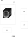
- Figure 2 illustrates a system 200 for monitoring a biological process by automatically creating a progression map of said biological process, the system comprising:
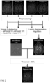
- FIG. 3 shows a conceptual overview of the method according to the invention.
- the method is used to obtain the disease progression map of follow-up in multiple sclerosis (MS) lesions (considered as abnormal tissue in this case).
- the MRI complete palette of available image contrasts in this specific case is composed by T1 MPRAGE and T2 FLAIR contrasts from two time points (time point 1 - hereafter TP1, and time point 2 - hereafter TP2). All the images undergo a pre-processing step based on registration to MPRAGE TP2, N4 bias field correction and histogram matching for intensity normalization.
- the segmentation algorithm proposed by Fartaria et al.
- the difference image from FLAIR is obtained considering the TP2 as the first term of the equation because lesions appear as hyperintense signal: FLAIR TP2 - FLAIR TP1.
- FLAIR and MPRAGE difference images are combined in a joint difference image.
- Voxels assigned to lesion class in the lesion mask with intensity equal or higher than 30% in the joint difference image are assigned to the disease progression class.
- all the voxels identified as part of disease progression are shown in a binary map as disease progression map. In this particular case, the disease progression map corresponds to areas of new and enlarged lesions in a MS patient.
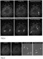
- Figure 4 shows FLAIR images of axial slices of a brain acquired at different time points, the respective lesion masks, and the results of disease progression from the proposed method and from the first technique. False positives in the disease progression map (see white arrows) appeared due to under- or over-segmentation of lesions in one of the time points.
- Figure 5 shows FLAIR images of axial slices of a brain acquired at different time points, and the results of disease progression, overlaid in the combined difference map, from the second technique and from the proposed method. False positives observed in the disease progression map (see white arrows) are mainly due to registration misalignments. Such false positives are not observed in images obtained according to the present invention.
- the proposed technique takes the advantages of the first and second techniques, reducing the number of misclassifications yielding a more reliable disease progression evaluation.
Landscapes
- Engineering & Computer Science (AREA)
- Health & Medical Sciences (AREA)
- Physics & Mathematics (AREA)
- Theoretical Computer Science (AREA)
- General Physics & Mathematics (AREA)
- Medical Informatics (AREA)
- Computer Vision & Pattern Recognition (AREA)
- General Health & Medical Sciences (AREA)
- Public Health (AREA)
- Life Sciences & Earth Sciences (AREA)
- Biomedical Technology (AREA)
- Radiology & Medical Imaging (AREA)
- Nuclear Medicine, Radiotherapy & Molecular Imaging (AREA)
- Pathology (AREA)
- Quality & Reliability (AREA)
- Epidemiology (AREA)
- Primary Health Care (AREA)
- Biophysics (AREA)
- Heart & Thoracic Surgery (AREA)
- Molecular Biology (AREA)
- Surgery (AREA)
- Animal Behavior & Ethology (AREA)
- Veterinary Medicine (AREA)
- Data Mining & Analysis (AREA)
- Databases & Information Systems (AREA)
- Magnetic Resonance Imaging Apparatus (AREA)
Claims (6)
- Verfahren (100) zum Überwachen eines biologischen Prozesses, wobei das Verfahren Folgendes umfasst:- Erlangen (102) einer Maske abnormalen Gewebes aus einer Segmentierung abnormalen Gewebes eines Bildes eines Objekts, das zu analysierendes Gewebe umfasst, wobei das Bild zu einem als Referenzzeitpunkt bezeichneten Zeitpunkt t0 erfasst wurde;- Registrieren (103) anderer Bilder des Objekts auf der Maske abnormalen Gewebes, wobei die anderen Bilder zu anderen Zeitpunkten erfasst wurden;- Normalisieren (104) von Bildkontrasten der anderen Bilder in Bezug auf die Kontraste des zum Referenzzeitpunkt erfassten Bildes;- Subtrahieren (105) der normalisierten Bilder für jeden verfügbaren Kontrast einer Palette verfügbarer Bildkontraste, wobei sich die verfügbaren Kontraste auf die unterschiedlichen Gewichtungen in MRI oder Bilder in CT mit und ohne Kontrastmittel beziehen, um Differenzbilder zu erlangen, wobei, in dem Fall, dass das abnormale Gewebe in einem der verfügbaren Kontraste als hyperintenses Signal dargestellt wird, das normalisierte Bild von dem zum Referenzzeitpunkt erfassten Bild subtrahiert wird, um die Differenzbilder zu berechnen, und in dem Fall, dass das abnormale Gewebe in einem der verfügbaren Kontraste als hypointenses Signal dargestellt wird, das zum Referenzzeitpunkt erfasste Bild von dem normalisierten Bild subtrahiert wird, wobei alle erlangten Differenzbilder zwischen 0 und 1 skaliert werden;- Erstellen (106) eines gemeinsamen Differenzbildes durch Summieren der zuvor erlangten Differenzbilder;- Erstellen (107) einer Verlaufsübersicht des biologischen Prozesses durch Überlagern der erlangten Maske abnormalen Gewebes und des gemeinsamen Differenzbildes nach Anwenden eines vordefinierten Schwellenwerts, wobei Gewebevoxel innerhalb der Maske abnormalen Gewebes mit einer Intensität, die gleich dem vordefinierte Schwellenwert oder höher als dieser ist, auf dem gemeinsamen Differenzbild als eine abnormale Veränderung des Gewebes darstellend betrachtet und einer Klasse eines biologischen Prozesses zugeordnet werden.
- Verfahren nach Anspruch 1, umfassend- Durchführen (101) der Segmentierung abnormalen Gewebes anhand des Bildes des Objekts.
- Verfahren nach Anspruch 1 oder 2, wobei der Normalisierungsschritt iteratives Verarbeiten jedes einzelnen der anderen Bilder umfasst, wobei für jeden Kontrast die Kontrastintensitäten eines Bildes zu einem früheren Zeitpunkt unter Verwendung eines geeigneten Verfahrens auf die Kontrastintensitäten des zum Referenzzeitpunkt erfassten Bildes normalisiert werden.
- Verfahren nach einem der Ansprüche 1 bis 3, wobei die Subtraktion für normalisierte Bilder durchgeführt wird, die aus Bildern stammen, die zu aufeinanderfolgenden Zeitpunkten erfasst wurden, und wobei das zum Referenzzeitpunkt erfasste Bild als das normalisierte Bild zum Referenzzeitpunkt betrachtet wird.
- Verfahren nach Anspruch 1, umfassend Gruppieren aller Voxel, die der gleichen Klasse eines biologischen Prozesses zugeordnet sind, in einem Bild des Objekts.
- System (200) zum Überwachen eines biologischen Prozesses, wobei das System Folgendes umfasst:- optional eine Magnetresonanzbildgebungs-(MRI-)Vorrichtung (201), die zum Erfassen von Bildern eines Objekts konfiguriert ist, um eine Objektgewebeanalyse zu ermöglichen;- einen Speicher (202) zum Speichern von Daten, die zum Erstellen einer Verlaufsübersicht erforderlich sind;- eine Verarbeitungseinheit (203), die zum Verarbeiten der zum Erstellen der Verlaufsübersicht erforderlichen Daten konfiguriert ist, wobei die Verarbeitungseinheit 203 insbesondere ein Werkzeug zur Segmentierung abnormalen Gewebes umfasst;- eine Anzeige (204) zum Anzeigen der Verlaufsübersicht;wobei das erfindungsgemäße System (200) zum Durchführen der Schritte des Verfahrens nach einem der Ansprüche 1 bis 5 konfiguriert ist.
Priority Applications (3)
| Application Number | Priority Date | Filing Date | Title |
|---|---|---|---|
| EP19162761.1A EP3709263B1 (de) | 2019-03-14 | 2019-03-14 | Verfahren und system zur überwachung eines biologischen prozesses |
| CN202010170187.5A CN111696113B (zh) | 2019-03-14 | 2020-03-12 | 用于监视生物过程的方法和系统 |
| US16/819,402 US11335001B2 (en) | 2019-03-14 | 2020-03-16 | Method and system for monitoring a biological process |
Applications Claiming Priority (1)
| Application Number | Priority Date | Filing Date | Title |
|---|---|---|---|
| EP19162761.1A EP3709263B1 (de) | 2019-03-14 | 2019-03-14 | Verfahren und system zur überwachung eines biologischen prozesses |
Publications (3)
| Publication Number | Publication Date |
|---|---|
| EP3709263A1 EP3709263A1 (de) | 2020-09-16 |
| EP3709263B1 true EP3709263B1 (de) | 2024-08-07 |
| EP3709263C0 EP3709263C0 (de) | 2024-08-07 |
Family
ID=66041101
Family Applications (1)
| Application Number | Title | Priority Date | Filing Date |
|---|---|---|---|
| EP19162761.1A Active EP3709263B1 (de) | 2019-03-14 | 2019-03-14 | Verfahren und system zur überwachung eines biologischen prozesses |
Country Status (3)
| Country | Link |
|---|---|
| US (1) | US11335001B2 (de) |
| EP (1) | EP3709263B1 (de) |
| CN (1) | CN111696113B (de) |
Families Citing this family (2)
| Publication number | Priority date | Publication date | Assignee | Title |
|---|---|---|---|---|
| CN111931772B (zh) * | 2020-09-18 | 2021-02-09 | 平安科技(深圳)有限公司 | 医学图像处理方法、装置、设备及存储介质 |
| CN115797729B (zh) * | 2023-01-29 | 2023-05-09 | 有方(合肥)医疗科技有限公司 | 模型训练方法及装置、运动伪影识别及提示的方法及装置 |
Family Cites Families (17)
| Publication number | Priority date | Publication date | Assignee | Title |
|---|---|---|---|---|
| IT1289809B1 (it) * | 1996-12-27 | 1998-10-16 | Ist Trentino Di Cultura | Procedimento e sistema automatico per ottenere mappe di contenuto d'acqua e/o di permettivita'elettrica da immagini di risonanza |
| AU2001257313A1 (en) * | 2000-05-18 | 2001-11-26 | Yeda Research And Development Co..Ltd. | Method and apparatus for the detection and diagnosis of cancer, specifically breast cancer using diffusion mri |
| US7309867B2 (en) * | 2003-04-18 | 2007-12-18 | Medispectra, Inc. | Methods and apparatus for characterization of tissue samples |
| US8050734B2 (en) * | 2005-09-07 | 2011-11-01 | General Electric Company | Method and system for performing patient specific analysis of disease relevant changes of a disease in an anatomical structure |
| JP5079008B2 (ja) * | 2006-09-19 | 2012-11-21 | シナーク・インコーポレイテッド | 軟骨構造に関連した病変表示測度及びその自動数量化 |
| US8512249B2 (en) * | 2006-12-21 | 2013-08-20 | Bracco International Bv | Detection of the detachment of immobilized contrast agent in medical imaging applications |
| US8073235B2 (en) * | 2007-08-13 | 2011-12-06 | Pioneer Hi-Bred International, Inc. | Method and system for digital image analysis of ear traits |
| FR2946171B1 (fr) * | 2009-05-29 | 2011-07-15 | Groupe Des Ecoles De Telecommunications Get Ecole Nationale Superieure Des Telecommunications Enst | Procede de quantification de l'evolution de pathologies impliquant des changements de volumes de corps, notamment de tumeurs |
| US9404986B2 (en) * | 2011-05-06 | 2016-08-02 | The Regents Of The University Of California | Measuring biological tissue parameters using diffusion magnetic resonance imaging |
| US9092691B1 (en) * | 2014-07-18 | 2015-07-28 | Median Technologies | System for computing quantitative biomarkers of texture features in tomographic images |
| WO2017011532A1 (en) * | 2015-07-13 | 2017-01-19 | The Trustees Of Columbia University In The City Of New York | Processing candidate abnormalities in medical imagery based on a hierarchical classification |
| CN108351685B (zh) * | 2015-08-15 | 2022-07-08 | 谷歌有限责任公司 | 用于与真实和虚拟对象交互的基于生物力学的眼睛信号的系统和方法 |
| US20170071470A1 (en) * | 2015-09-15 | 2017-03-16 | Siemens Healthcare Gmbh | Framework for Abnormality Detection in Multi-Contrast Brain Magnetic Resonance Data |
| US20170337682A1 (en) * | 2016-05-18 | 2017-11-23 | Siemens Healthcare Gmbh | Method and System for Image Registration Using an Intelligent Artificial Agent |
| CN108693491B (zh) * | 2017-04-07 | 2022-03-25 | 康奈尔大学 | 稳健的定量磁化率成像系统和方法 |
| CN107316334B (zh) * | 2017-07-31 | 2020-02-14 | 华东师范大学 | 个性化精准磁共振影像方法 |
| CN108171697B (zh) * | 2018-01-05 | 2022-03-01 | 北京航空航天大学 | 一种基于簇的wmh自动提取系统 |
-
2019
- 2019-03-14 EP EP19162761.1A patent/EP3709263B1/de active Active
-
2020
- 2020-03-12 CN CN202010170187.5A patent/CN111696113B/zh active Active
- 2020-03-16 US US16/819,402 patent/US11335001B2/en active Active
Also Published As
| Publication number | Publication date |
|---|---|
| US11335001B2 (en) | 2022-05-17 |
| EP3709263A1 (de) | 2020-09-16 |
| CN111696113A (zh) | 2020-09-22 |
| US20200294237A1 (en) | 2020-09-17 |
| CN111696113B (zh) | 2023-11-21 |
| EP3709263C0 (de) | 2024-08-07 |
Similar Documents
| Publication | Publication Date | Title |
|---|---|---|
| US7653263B2 (en) | Method and system for volumetric comparative image analysis and diagnosis | |
| US7933440B2 (en) | Method and system for evaluating two time-separated medical images | |
| JP5699936B2 (ja) | 医用画像処理装置の作動方法、装置およびプログラム | |
| Xu et al. | A hybrid method for airway segmentation and automated measurement of bronchial wall thickness on CT | |
| Azhari et al. | Tumor detection in medical imaging: a survey | |
| Colliot et al. | Segmentation of focal cortical dysplasia lesions on MRI using level set evolution | |
| JP2010207572A (ja) | 障害のコンピュータ支援検出 | |
| US11058383B2 (en) | Apparatus for the detection of opacities in X-ray images | |
| EP3709263B1 (de) | Verfahren und system zur überwachung eines biologischen prozesses | |
| JPWO2017086433A1 (ja) | 医用画像処理方法及び装置及びシステム及びプログラム | |
| US10943350B2 (en) | Automated segmentation of histological sections for vasculature quantification | |
| US20090069665A1 (en) | Automatic Lesion Correlation in Multiple MR Modalities | |
| Włodarczyk et al. | Fast automated segmentation of wrist bones in magnetic resonance images | |
| Bauer et al. | Airway tree reconstruction based on tube detection | |
| Heinrich et al. | Non-local shape descriptor: A new similarity metric for deformable multi-modal registration | |
| US20150125056A1 (en) | Method of classification of organs from a tomographic image | |
| Dufresne et al. | Joint registration and change detection in longitudinal brain MRI | |
| Razavi et al. | Towards accurate segmentation of fibroglandular tissue in breast MRI using fuzzy c-means and skin-folds removal | |
| Delmoral et al. | Segmentation of pathological liver tissue with dilated fully convolutional networks: A preliminary study | |
| Sørensen et al. | Learning COPD sensitive filters in pulmonary CT | |
| Ravikumar et al. | Unfolding the medial temporal lobe cortex to characterize neurodegeneration due to Alzheimer’s disease pathology using ex vivo imaging | |
| Viswanath et al. | Empirical evaluation of bias field correction algorithms for computer-aided detection of prostate cancer on T2w MRI | |
| Norouzi et al. | A New Automatic Change Detection Frame-work Based on Region Growing and Weighted Local Mutual Information: Analysis of Breast Tumor Response to Chemotherapy in Serial MR Images | |
| Karmakar et al. | Detailed investigation of lumen-based tomographic co-registration | |
| Kaftan et al. | Locally adaptive fuzzy pulmonary vessel segmentation in contrast enhanced CT data |
Legal Events
| Date | Code | Title | Description |
|---|---|---|---|
| PUAI | Public reference made under article 153(3) epc to a published international application that has entered the european phase |
Free format text: ORIGINAL CODE: 0009012 |
|
| STAA | Information on the status of an ep patent application or granted ep patent |
Free format text: STATUS: THE APPLICATION HAS BEEN PUBLISHED |
|
| AK | Designated contracting states |
Kind code of ref document: A1 Designated state(s): AL AT BE BG CH CY CZ DE DK EE ES FI FR GB GR HR HU IE IS IT LI LT LU LV MC MK MT NL NO PL PT RO RS SE SI SK SM TR |
|
| AX | Request for extension of the european patent |
Extension state: BA ME |
|
| STAA | Information on the status of an ep patent application or granted ep patent |
Free format text: STATUS: REQUEST FOR EXAMINATION WAS MADE |
|
| 17P | Request for examination filed |
Effective date: 20210315 |
|
| RBV | Designated contracting states (corrected) |
Designated state(s): AL AT BE BG CH CY CZ DE DK EE ES FI FR GB GR HR HU IE IS IT LI LT LU LV MC MK MT NL NO PL PT RO RS SE SI SK SM TR |
|
| STAA | Information on the status of an ep patent application or granted ep patent |
Free format text: STATUS: EXAMINATION IS IN PROGRESS |
|
| 17Q | First examination report despatched |
Effective date: 20220510 |
|
| RAP1 | Party data changed (applicant data changed or rights of an application transferred) |
Owner name: CENTRE HOSPITALIER UNIVERSITAIRE VAUDOIS Owner name: SIEMENS HEALTHINEERS AG |
|
| GRAP | Despatch of communication of intention to grant a patent |
Free format text: ORIGINAL CODE: EPIDOSNIGR1 |
|
| STAA | Information on the status of an ep patent application or granted ep patent |
Free format text: STATUS: GRANT OF PATENT IS INTENDED |
|
| INTG | Intention to grant announced |
Effective date: 20240424 |
|
| RIN1 | Information on inventor provided before grant (corrected) |
Inventor name: BACH CUADRA, MERITXELL Inventor name: GRANZIERA, CRISTINA Inventor name: MARECHAL, BENEDICTE Inventor name: KOBER, TOBIAS Inventor name: FARTARIA DE OLIVEIRA, MARIO JOAO |
|
| GRAS | Grant fee paid |
Free format text: ORIGINAL CODE: EPIDOSNIGR3 |
|
| GRAA | (expected) grant |
Free format text: ORIGINAL CODE: 0009210 |
|
| STAA | Information on the status of an ep patent application or granted ep patent |
Free format text: STATUS: THE PATENT HAS BEEN GRANTED |
|
| AK | Designated contracting states |
Kind code of ref document: B1 Designated state(s): AL AT BE BG CH CY CZ DE DK EE ES FI FR GB GR HR HU IE IS IT LI LT LU LV MC MK MT NL NO PL PT RO RS SE SI SK SM TR |
|
| REG | Reference to a national code |
Ref country code: GB Ref legal event code: FG4D |
|
| REG | Reference to a national code |
Ref country code: CH Ref legal event code: EP |
|
| REG | Reference to a national code |
Ref country code: IE Ref legal event code: FG4D |
|
| REG | Reference to a national code |
Ref country code: DE Ref legal event code: R096 Ref document number: 602019056433 Country of ref document: DE |
|
| U01 | Request for unitary effect filed |
Effective date: 20240807 |
|
| U07 | Unitary effect registered |
Designated state(s): AT BE BG DE DK EE FI FR IT LT LU LV MT NL PT SE SI Effective date: 20240821 |
|
| PG25 | Lapsed in a contracting state [announced via postgrant information from national office to epo] |
Ref country code: NO Free format text: LAPSE BECAUSE OF FAILURE TO SUBMIT A TRANSLATION OF THE DESCRIPTION OR TO PAY THE FEE WITHIN THE PRESCRIBED TIME-LIMIT Effective date: 20241107 |
|
| PG25 | Lapsed in a contracting state [announced via postgrant information from national office to epo] |
Ref country code: GR Free format text: LAPSE BECAUSE OF FAILURE TO SUBMIT A TRANSLATION OF THE DESCRIPTION OR TO PAY THE FEE WITHIN THE PRESCRIBED TIME-LIMIT Effective date: 20241108 Ref country code: PL Free format text: LAPSE BECAUSE OF FAILURE TO SUBMIT A TRANSLATION OF THE DESCRIPTION OR TO PAY THE FEE WITHIN THE PRESCRIBED TIME-LIMIT Effective date: 20240807 |
|
| PG25 | Lapsed in a contracting state [announced via postgrant information from national office to epo] |
Ref country code: IS Free format text: LAPSE BECAUSE OF FAILURE TO SUBMIT A TRANSLATION OF THE DESCRIPTION OR TO PAY THE FEE WITHIN THE PRESCRIBED TIME-LIMIT Effective date: 20241207 |
|
| PG25 | Lapsed in a contracting state [announced via postgrant information from national office to epo] |
Ref country code: HR Free format text: LAPSE BECAUSE OF FAILURE TO SUBMIT A TRANSLATION OF THE DESCRIPTION OR TO PAY THE FEE WITHIN THE PRESCRIBED TIME-LIMIT Effective date: 20240807 |
|
| PG25 | Lapsed in a contracting state [announced via postgrant information from national office to epo] |
Ref country code: ES Free format text: LAPSE BECAUSE OF FAILURE TO SUBMIT A TRANSLATION OF THE DESCRIPTION OR TO PAY THE FEE WITHIN THE PRESCRIBED TIME-LIMIT Effective date: 20240807 Ref country code: RS Free format text: LAPSE BECAUSE OF FAILURE TO SUBMIT A TRANSLATION OF THE DESCRIPTION OR TO PAY THE FEE WITHIN THE PRESCRIBED TIME-LIMIT Effective date: 20241107 |
|
| PG25 | Lapsed in a contracting state [announced via postgrant information from national office to epo] |
Ref country code: RS Free format text: LAPSE BECAUSE OF FAILURE TO SUBMIT A TRANSLATION OF THE DESCRIPTION OR TO PAY THE FEE WITHIN THE PRESCRIBED TIME-LIMIT Effective date: 20241107 Ref country code: PL Free format text: LAPSE BECAUSE OF FAILURE TO SUBMIT A TRANSLATION OF THE DESCRIPTION OR TO PAY THE FEE WITHIN THE PRESCRIBED TIME-LIMIT Effective date: 20240807 Ref country code: NO Free format text: LAPSE BECAUSE OF FAILURE TO SUBMIT A TRANSLATION OF THE DESCRIPTION OR TO PAY THE FEE WITHIN THE PRESCRIBED TIME-LIMIT Effective date: 20241107 Ref country code: IS Free format text: LAPSE BECAUSE OF FAILURE TO SUBMIT A TRANSLATION OF THE DESCRIPTION OR TO PAY THE FEE WITHIN THE PRESCRIBED TIME-LIMIT Effective date: 20241207 Ref country code: HR Free format text: LAPSE BECAUSE OF FAILURE TO SUBMIT A TRANSLATION OF THE DESCRIPTION OR TO PAY THE FEE WITHIN THE PRESCRIBED TIME-LIMIT Effective date: 20240807 Ref country code: GR Free format text: LAPSE BECAUSE OF FAILURE TO SUBMIT A TRANSLATION OF THE DESCRIPTION OR TO PAY THE FEE WITHIN THE PRESCRIBED TIME-LIMIT Effective date: 20241108 Ref country code: ES Free format text: LAPSE BECAUSE OF FAILURE TO SUBMIT A TRANSLATION OF THE DESCRIPTION OR TO PAY THE FEE WITHIN THE PRESCRIBED TIME-LIMIT Effective date: 20240807 |
|
| U1N | Appointed representative for the unitary patent procedure changed after the registration of the unitary effect |
Representative=s name: FISCHER, MICHAEL; DE |
|
| PG25 | Lapsed in a contracting state [announced via postgrant information from national office to epo] |
Ref country code: SM Free format text: LAPSE BECAUSE OF FAILURE TO SUBMIT A TRANSLATION OF THE DESCRIPTION OR TO PAY THE FEE WITHIN THE PRESCRIBED TIME-LIMIT Effective date: 20240807 |
|
| U20 | Renewal fee for the european patent with unitary effect paid |
Year of fee payment: 7 Effective date: 20250320 |
|
| PG25 | Lapsed in a contracting state [announced via postgrant information from national office to epo] |
Ref country code: CZ Free format text: LAPSE BECAUSE OF FAILURE TO SUBMIT A TRANSLATION OF THE DESCRIPTION OR TO PAY THE FEE WITHIN THE PRESCRIBED TIME-LIMIT Effective date: 20240807 |
|
| PG25 | Lapsed in a contracting state [announced via postgrant information from national office to epo] |
Ref country code: SK Free format text: LAPSE BECAUSE OF FAILURE TO SUBMIT A TRANSLATION OF THE DESCRIPTION OR TO PAY THE FEE WITHIN THE PRESCRIBED TIME-LIMIT Effective date: 20240807 |
|
| PLBE | No opposition filed within time limit |
Free format text: ORIGINAL CODE: 0009261 |
|
| STAA | Information on the status of an ep patent application or granted ep patent |
Free format text: STATUS: NO OPPOSITION FILED WITHIN TIME LIMIT |
|
| PGFP | Annual fee paid to national office [announced via postgrant information from national office to epo] |
Ref country code: GB Payment date: 20250403 Year of fee payment: 7 |
|
| 26N | No opposition filed |
Effective date: 20250508 |
|
| PG25 | Lapsed in a contracting state [announced via postgrant information from national office to epo] |
Ref country code: MC Free format text: LAPSE BECAUSE OF FAILURE TO SUBMIT A TRANSLATION OF THE DESCRIPTION OR TO PAY THE FEE WITHIN THE PRESCRIBED TIME-LIMIT Effective date: 20240807 |
|
| REG | Reference to a national code |
Ref country code: CH Ref legal event code: H13 Free format text: ST27 STATUS EVENT CODE: U-0-0-H10-H13 (AS PROVIDED BY THE NATIONAL OFFICE) Effective date: 20251023 |