EP3693394A1 - Antigen binding proteins - Google Patents
Antigen binding proteins Download PDFInfo
- Publication number
- EP3693394A1 EP3693394A1 EP20153215.7A EP20153215A EP3693394A1 EP 3693394 A1 EP3693394 A1 EP 3693394A1 EP 20153215 A EP20153215 A EP 20153215A EP 3693394 A1 EP3693394 A1 EP 3693394A1
- Authority
- EP
- European Patent Office
- Prior art keywords
- seq
- antigen binding
- binding protein
- antibody
- bcma
- Prior art date
- Legal status (The legal status is an assumption and is not a legal conclusion. Google has not performed a legal analysis and makes no representation as to the accuracy of the status listed.)
- Withdrawn
Links
Images


Classifications
-
- A—HUMAN NECESSITIES
- A61—MEDICAL OR VETERINARY SCIENCE; HYGIENE
- A61K—PREPARATIONS FOR MEDICAL, DENTAL OR TOILETRY PURPOSES
- A61K47/00—Medicinal preparations characterised by the non-active ingredients used, e.g. carriers or inert additives; Targeting or modifying agents chemically bound to the active ingredient
- A61K47/50—Medicinal preparations characterised by the non-active ingredients used, e.g. carriers or inert additives; Targeting or modifying agents chemically bound to the active ingredient the non-active ingredient being chemically bound to the active ingredient, e.g. polymer-drug conjugates
-
- C—CHEMISTRY; METALLURGY
- C07—ORGANIC CHEMISTRY
- C07K—PEPTIDES
- C07K16/00—Immunoglobulins [IGs], e.g. monoclonal or polyclonal antibodies
- C07K16/18—Immunoglobulins [IGs], e.g. monoclonal or polyclonal antibodies against material from animals or humans
- C07K16/28—Immunoglobulins [IGs], e.g. monoclonal or polyclonal antibodies against material from animals or humans against receptors, cell surface antigens or cell surface determinants
- C07K16/2878—Immunoglobulins [IGs], e.g. monoclonal or polyclonal antibodies against material from animals or humans against receptors, cell surface antigens or cell surface determinants against the NGF-receptor/TNF-receptor superfamily, e.g. CD27, CD30, CD40, CD95
-
- A—HUMAN NECESSITIES
- A61—MEDICAL OR VETERINARY SCIENCE; HYGIENE
- A61K—PREPARATIONS FOR MEDICAL, DENTAL OR TOILETRY PURPOSES
- A61K47/00—Medicinal preparations characterised by the non-active ingredients used, e.g. carriers or inert additives; Targeting or modifying agents chemically bound to the active ingredient
- A61K47/50—Medicinal preparations characterised by the non-active ingredients used, e.g. carriers or inert additives; Targeting or modifying agents chemically bound to the active ingredient the non-active ingredient being chemically bound to the active ingredient, e.g. polymer-drug conjugates
- A61K47/51—Medicinal preparations characterised by the non-active ingredients used, e.g. carriers or inert additives; Targeting or modifying agents chemically bound to the active ingredient the non-active ingredient being chemically bound to the active ingredient, e.g. polymer-drug conjugates the non-active ingredient being a modifying agent
- A61K47/68—Medicinal preparations characterised by the non-active ingredients used, e.g. carriers or inert additives; Targeting or modifying agents chemically bound to the active ingredient the non-active ingredient being chemically bound to the active ingredient, e.g. polymer-drug conjugates the non-active ingredient being a modifying agent the modifying agent being an antibody, an immunoglobulin or a fragment thereof, e.g. an Fc-fragment
- A61K47/6801—Drug-antibody or immunoglobulin conjugates defined by the pharmacologically or therapeutically active agent
- A61K47/6803—Drugs conjugated to an antibody or immunoglobulin, e.g. cisplatin-antibody conjugates
- A61K47/6811—Drugs conjugated to an antibody or immunoglobulin, e.g. cisplatin-antibody conjugates the drug being a protein or peptide, e.g. transferrin or bleomycin
- A61K47/6817—Toxins
-
- A—HUMAN NECESSITIES
- A61—MEDICAL OR VETERINARY SCIENCE; HYGIENE
- A61K—PREPARATIONS FOR MEDICAL, DENTAL OR TOILETRY PURPOSES
- A61K47/00—Medicinal preparations characterised by the non-active ingredients used, e.g. carriers or inert additives; Targeting or modifying agents chemically bound to the active ingredient
- A61K47/50—Medicinal preparations characterised by the non-active ingredients used, e.g. carriers or inert additives; Targeting or modifying agents chemically bound to the active ingredient the non-active ingredient being chemically bound to the active ingredient, e.g. polymer-drug conjugates
- A61K47/51—Medicinal preparations characterised by the non-active ingredients used, e.g. carriers or inert additives; Targeting or modifying agents chemically bound to the active ingredient the non-active ingredient being chemically bound to the active ingredient, e.g. polymer-drug conjugates the non-active ingredient being a modifying agent
- A61K47/68—Medicinal preparations characterised by the non-active ingredients used, e.g. carriers or inert additives; Targeting or modifying agents chemically bound to the active ingredient the non-active ingredient being chemically bound to the active ingredient, e.g. polymer-drug conjugates the non-active ingredient being a modifying agent the modifying agent being an antibody, an immunoglobulin or a fragment thereof, e.g. an Fc-fragment
- A61K47/6835—Medicinal preparations characterised by the non-active ingredients used, e.g. carriers or inert additives; Targeting or modifying agents chemically bound to the active ingredient the non-active ingredient being chemically bound to the active ingredient, e.g. polymer-drug conjugates the non-active ingredient being a modifying agent the modifying agent being an antibody, an immunoglobulin or a fragment thereof, e.g. an Fc-fragment the modifying agent being an antibody or an immunoglobulin bearing at least one antigen-binding site
- A61K47/6849—Medicinal preparations characterised by the non-active ingredients used, e.g. carriers or inert additives; Targeting or modifying agents chemically bound to the active ingredient the non-active ingredient being chemically bound to the active ingredient, e.g. polymer-drug conjugates the non-active ingredient being a modifying agent the modifying agent being an antibody, an immunoglobulin or a fragment thereof, e.g. an Fc-fragment the modifying agent being an antibody or an immunoglobulin bearing at least one antigen-binding site the antibody targeting a receptor, a cell surface antigen or a cell surface determinant
-
- A—HUMAN NECESSITIES
- A61—MEDICAL OR VETERINARY SCIENCE; HYGIENE
- A61P—SPECIFIC THERAPEUTIC ACTIVITY OF CHEMICAL COMPOUNDS OR MEDICINAL PREPARATIONS
- A61P29/00—Non-central analgesic, antipyretic or antiinflammatory agents, e.g. antirheumatic agents; Non-steroidal antiinflammatory drugs [NSAID]
-
- A—HUMAN NECESSITIES
- A61—MEDICAL OR VETERINARY SCIENCE; HYGIENE
- A61P—SPECIFIC THERAPEUTIC ACTIVITY OF CHEMICAL COMPOUNDS OR MEDICINAL PREPARATIONS
- A61P35/00—Antineoplastic agents
-
- A—HUMAN NECESSITIES
- A61—MEDICAL OR VETERINARY SCIENCE; HYGIENE
- A61P—SPECIFIC THERAPEUTIC ACTIVITY OF CHEMICAL COMPOUNDS OR MEDICINAL PREPARATIONS
- A61P35/00—Antineoplastic agents
- A61P35/02—Antineoplastic agents specific for leukemia
-
- A—HUMAN NECESSITIES
- A61—MEDICAL OR VETERINARY SCIENCE; HYGIENE
- A61P—SPECIFIC THERAPEUTIC ACTIVITY OF CHEMICAL COMPOUNDS OR MEDICINAL PREPARATIONS
- A61P37/00—Drugs for immunological or allergic disorders
- A61P37/02—Immunomodulators
-
- C—CHEMISTRY; METALLURGY
- C07—ORGANIC CHEMISTRY
- C07K—PEPTIDES
- C07K16/00—Immunoglobulins [IGs], e.g. monoclonal or polyclonal antibodies
- C07K16/18—Immunoglobulins [IGs], e.g. monoclonal or polyclonal antibodies against material from animals or humans
- C07K16/28—Immunoglobulins [IGs], e.g. monoclonal or polyclonal antibodies against material from animals or humans against receptors, cell surface antigens or cell surface determinants
-
- A—HUMAN NECESSITIES
- A61—MEDICAL OR VETERINARY SCIENCE; HYGIENE
- A61K—PREPARATIONS FOR MEDICAL, DENTAL OR TOILETRY PURPOSES
- A61K39/00—Medicinal preparations containing antigens or antibodies
- A61K2039/505—Medicinal preparations containing antigens or antibodies comprising antibodies
-
- C—CHEMISTRY; METALLURGY
- C07—ORGANIC CHEMISTRY
- C07K—PEPTIDES
- C07K2317/00—Immunoglobulins specific features
- C07K2317/20—Immunoglobulins specific features characterized by taxonomic origin
- C07K2317/24—Immunoglobulins specific features characterized by taxonomic origin containing regions, domains or residues from different species, e.g. chimeric, humanized or veneered
-
- C—CHEMISTRY; METALLURGY
- C07—ORGANIC CHEMISTRY
- C07K—PEPTIDES
- C07K2317/00—Immunoglobulins specific features
- C07K2317/30—Immunoglobulins specific features characterized by aspects of specificity or valency
- C07K2317/33—Crossreactivity, e.g. for species or epitope, or lack of said crossreactivity
-
- C—CHEMISTRY; METALLURGY
- C07—ORGANIC CHEMISTRY
- C07K—PEPTIDES
- C07K2317/00—Immunoglobulins specific features
- C07K2317/40—Immunoglobulins specific features characterized by post-translational modification
- C07K2317/41—Glycosylation, sialylation, or fucosylation
-
- C—CHEMISTRY; METALLURGY
- C07—ORGANIC CHEMISTRY
- C07K—PEPTIDES
- C07K2317/00—Immunoglobulins specific features
- C07K2317/50—Immunoglobulins specific features characterized by immunoglobulin fragments
- C07K2317/56—Immunoglobulins specific features characterized by immunoglobulin fragments variable (Fv) region, i.e. VH and/or VL
- C07K2317/565—Complementarity determining region [CDR]
-
- C—CHEMISTRY; METALLURGY
- C07—ORGANIC CHEMISTRY
- C07K—PEPTIDES
- C07K2317/00—Immunoglobulins specific features
- C07K2317/50—Immunoglobulins specific features characterized by immunoglobulin fragments
- C07K2317/56—Immunoglobulins specific features characterized by immunoglobulin fragments variable (Fv) region, i.e. VH and/or VL
- C07K2317/567—Framework region [FR]
-
- C—CHEMISTRY; METALLURGY
- C07—ORGANIC CHEMISTRY
- C07K—PEPTIDES
- C07K2317/00—Immunoglobulins specific features
- C07K2317/70—Immunoglobulins specific features characterized by effect upon binding to a cell or to an antigen
- C07K2317/72—Increased effector function due to an Fc-modification
-
- C—CHEMISTRY; METALLURGY
- C07—ORGANIC CHEMISTRY
- C07K—PEPTIDES
- C07K2317/00—Immunoglobulins specific features
- C07K2317/70—Immunoglobulins specific features characterized by effect upon binding to a cell or to an antigen
- C07K2317/73—Inducing cell death, e.g. apoptosis, necrosis or inhibition of cell proliferation
- C07K2317/732—Antibody-dependent cellular cytotoxicity [ADCC]
-
- C—CHEMISTRY; METALLURGY
- C07—ORGANIC CHEMISTRY
- C07K—PEPTIDES
- C07K2317/00—Immunoglobulins specific features
- C07K2317/70—Immunoglobulins specific features characterized by effect upon binding to a cell or to an antigen
- C07K2317/76—Antagonist effect on antigen, e.g. neutralization or inhibition of binding
-
- C—CHEMISTRY; METALLURGY
- C07—ORGANIC CHEMISTRY
- C07K—PEPTIDES
- C07K2317/00—Immunoglobulins specific features
- C07K2317/70—Immunoglobulins specific features characterized by effect upon binding to a cell or to an antigen
- C07K2317/77—Internalization into the cell
-
- C—CHEMISTRY; METALLURGY
- C07—ORGANIC CHEMISTRY
- C07K—PEPTIDES
- C07K2317/00—Immunoglobulins specific features
- C07K2317/90—Immunoglobulins specific features characterized by (pharmaco)kinetic aspects or by stability of the immunoglobulin
- C07K2317/92—Affinity (KD), association rate (Ka), dissociation rate (Kd) or EC50 value
Definitions
- the present invention relates to antigen binding proteins and fragments thereof that specifically bind B cell maturation antigen (BCMA) and in particular human BCMA (hBCMA).
- BCMA B cell maturation antigen
- hBCMA human BCMA
- the present invention also concerns methods of treating diseases or disorders with said antigen binding fragments, pharmaceutical compositions comprising said antigen binding fragments and methods of manufacture.
- Other embodiments of the present invention will be apparent from the description below.
- BCMA (CD269 or TNFRSF17) is a member of the TNF receptor superfamily. It is a non-glycosylated integral membrane receptor for the ligands BAFF and APRIL. BCMA's ligands can also bind additional receptors: TACI (Transmembrane Activator and Calcium modulator and cyclophilin ligand Interactor), which binds APRIL and BAFF; as well as BAFF-R (BAFF Receptor or BR3), which shows restricted but high affinity for BAFF. Together, these receptors and their corresponding ligands regulate different aspects of humoral immunity, B-cell development and homeostasis.
- TACI Transmembrane Activator and Calcium modulator and cyclophilin ligand Interactor
- BCMA's expression is typically restricted to the B-cell lineage and is reported to increase in terminal B-cell differentiation.
- BCMA is expressed by human plasma blasts, plasma cells from tonsils, spleen and bone marrow, but also by tonsillar memory B cells and by germinal centre B cells, which have a TACI-BAFFR low phenotype (Darce et al, 2007).
- BCMA is virtually absent on naive and memory B-cells (Novak et al., 2004a and b).
- the BCMA antigen is expressed on the cell surface so is accessible to the antibody, but is also expressed in the golgi.
- BCMA signalling typically linked with B-cell survival and proliferation, is important in the late stages of B-cell differentiation, as well as the survival of long lived bone marrow plasma cells (O'Connor et al., 2004) and plasmablasts (Avery et al., 2003). Furthermore, as BCMA binds APRIL with high affinity, the BCMA-APRIL signalling axis is suggested to predominate at the later stages of B-cell differentiation, perhaps being the most physiologically relevant interaction.
- MM Multiple Myeloma
- MM is a clonal B-cell malignancy that occurs in multiple sites within the bone marrow before spreading to the circulation; either de novo, or as a progression from monoclonal gammopathy of undetermined significance (MGUS). It is commonly characterised by increases in paraprotein and osteoclast activity, as well as hypercalcaemia, cytopenia, renal dysfunction, hyperviscosity and peripheral neuropathy. Decreases in both normal antibody levels and numbers of neutrophils are also common, leading to a life threatening susceptibility to infection. BCMA has been implicated in the growth and survival of myeloma cell lines in vitro (Novak et al., 2004a and b; Moreaux et al., 2004).
- BCMA expression (both transcript and protein) is reported to correlate with disease progression in MM.
- MMC Multiple Myeloma Cells
- Gene expression analysis has been used to compare human myeloma cells with purified plasma cells from patients with MGUS and from normal bone marrow as well as with primary tumour cells from B-cell lineage leukaemias (Bellucci et al, 2005).
- the BCMA gene was highly expressed in all myeloma samples.
- CLL B-cell Chronic Lymphocytic Leukaemia
- ALL pre-B Acute Lymphocytic Leukaemia
- T-ALL T-cell ALL
- the present invention provides antigen binding proteins which bind to membrane bound targets and wherein the antigen binding protein is capable of internalisation.
- an immunoconjugate comprising the antigen binding protein of the present invention and a cytotoxic agent.
- the antigen binding protein has ADCC effector function for example the antigen binding protein has enhanced ADCC effector function.
- the present invention provides antigen binding proteins which specifically bind to BCMA, for example antibodies which specifically bind to BCMA and which inhibit the binding of BAFF and/or APRIL to the BCMA receptor.
- the present invention also provides antigen binding proteins which specifically bind to BCMA and which inhibits the binding of BAFF and/or APRIL to BCMA wherein the antigen binding protein is capable of binding to Fc ⁇ RIIIA or is capable of Fc ⁇ RIIIA mediated effector function.
- the antigen binding proteins of the present invention specifically bind to BCMA and inhibit the binding of BAFF and/or APRIL to BCMA wherein the antigen binding protein has enhanced binding to Fc ⁇ RIIIA or has enhanced Fc ⁇ RIIIA mediated effector function.
- the antigen binding protein is capable of internalisation.
- an antigen binding protein according to the invention as herein described which binds to non-membrane bound BCMA, for example to serum BCMA.
- an immunoconjugate comprising the antigen binding protein of the present invention and a cytotoxic agent.
- the antigen binding proteins are conjugated to a toxin such as an auristatin.
- the drug conjugate is vcMMAE or mcMMAF.
- the antigen binding proteins of the present invention are related to, or derived from a murine monoclonal antibody CA8.
- the CA8 murine heavy chain variable region amino acid sequence is provided as SEQ ID NO. 7 and the CA8 murine light chain variable region amino acid sequence is provided as SEQ ID NO. 9.
- the antigen binding proteins of the present invention are related to, or derived from a murine monoclonal antibody S336105A07.
- the S336105A07 murine heavy chain variable region amino acid sequence is provided as SEQ ID NO. 140 and the S336105A07 murine light chain variable region amino acid sequence is provided as SEQ ID NO. 144.
- the heavy chain variable regions (VH) of the present invention may comprise the following CDRs or variants of these CDR's (as defined by Kabat ( Kabat et al; Sequences of proteins of Immunological Interest NIH, 1987 )):
- the light chain variable regions (VL) of the present invention may comprise the following CDRs or variants of these CDR's (as defined by Kabat ( Kabat et al; Sequences of proteins of Immunological Interest NIH, 1987 )):
- the invention also provides a polynucleotide sequence encoding a heavy chain variable region of any of the antigen-binding proteins described herein, and a polynucleotide encoding a light chain variable region of any of the antigen-binding proteins described herein.
- the invention also provides a polynucleotide sequence encoding a heavy chain of any of the antigen-binding proteins described herein, and a polynucleotide encoding a light chain of any of the antigen-binding proteins described herein.
- polynucleotides represent the coding sequence which corresponds to the equivalent polypeptide sequences, however it will be understood that such polynucleotide sequences could be cloned into an expression vector along with a start codon, an appropriate signal sequence and a stop codon.
- the invention also provides a recombinant transformed or transfected host cell comprising one or more polynucleotides encoding a heavy chain and/or a light chain of any of the antigen-binding proteins described herein.
- the invention further provides a method for the production of any of the antigen-binding proteins described herein which method comprises the step of culturing a host cell comprising a first and second vector, said first vector comprising a polynucleotide encoding a heavy chain of any of the antigen-binding proteins described herein and said second vector comprising a polynucleotide encoding a light chain of any of the antigen-binding proteins described herein, in a suitable culture media, for example serum- free culture media.
- a suitable culture media for example serum- free culture media.
- the invention further provides a pharmaceutical composition comprising an antigen-binding protein as described herein and a pharmaceutically acceptable carrier.
- the present invention provides a method of treatment or prophylaxis of a disease or disorder responsive to inhibiting or blocking BCMA such as the modulation of the interaction between BCMA and its ligands, BAFF or APRIL which method comprises the step of administering to said patient a therapeutically effective amount of the antigen binding protein thereof as described herein.
- B cell related disorders or diseases such as antibody mediated or plasma cell mediated diseases or plasma cell malignancies such as for example Multiple Myeloma (MM).
- antigen binding proteins especially antibodies that specifically bind BCMA (e.g. hBCMA) and modulate (i.e. inhibit or block) the interaction between BCMA and its ligands such as BAFF and/or APRIL in the treatment of diseases and disorders responsive to modulation of that interaction.
- a method of treating a human patient afflicted with a B cell related disorders or diseases such as antibody mediated or plasma cell mediated diseases or plasma cell malignancies such as for example Multiple Myeloma (MM) comprises the step of administering to said patient a therapeutically effective amount of the antigen binding protein as described herein.
- a B cell related disorders or diseases such as antibody mediated or plasma cell mediated diseases or plasma cell malignancies such as for example Multiple Myeloma (MM)
- MM Multiple Myeloma
- a method of treating a human patient afflicted with Rheumatoid Arthritis, Psoriasis, Type 1 Diabetes Mellitus or Multiple Sclerosis comprises the step of administering to said patient a therapeutically effective amount of the antigen binding protein as described herein.
- the present invention provides antigen binding proteins which bind to membrane bound targets and wherein the antigen binding protein is capable of internalisation.
- an immunoconjugate comprising the antigen binding protein of the present invention and a cytotoxic agent.
- the antigen binding protein has ADCC effector function for example the antigen binding protein has enhanced ADCC effector function.
- antigen binding proteins or fragments thereof which specifically bind to BCMA, for example which specifically binds human BCMA (hBCMA) and which inhibit the binding of BAFF and/or APRIL to the BCMA receptor.
- hBCMA human BCMA
- the antigen binding proteins or fragments of the present invention specifically bind to BCMA and inhibit the binding of BAFF and/or APRIL to BCMA wherein the antigen binding proteins or fragments thereof have the ability to bind to Fc ⁇ RIIIA and mediate FcgRIIIA mediated effector functions, or have enhanced Fc ⁇ RIIIA mediated effector function.
- the antigen binding proteins are capable of internalisation.
- an antigen binding protein according to the invention as herein described which binds to non-membrane bound BCMA, for example to serum BCMA.
- an antigen binding protein as herein described wherein the antigen binding protein comprises CDRH3 of SEQ ID NO.3 or a variant of SEQ ID NO. 3.
- an antigen binding protein as herein described wherein the antigen binding protein further comprises one or more of: CDR H1 of SEQ. ID. NO: 1, CDRH2: SEQ. ID. NO: 2: CDRL1: SEQ. ID. NO: 4, CDRL2: SEQ. ID. NO: 5 and/or CDRL3: SEQ. ID. NO: 6 and or variants thereof.
- an antigen binding protein as herein described wherein the antigen binding protein comprises CDRH3 of SEQ ID NO.184 or a variant of SEQ ID NO. 184.
- an antigen binding protein as herein described wherein the antigen binding protein further comprises one or more of: CDR H1 of SEQ. ID. NO: 182, CDRH2: SEQ. ID. NO: 183: CDRL1: SEQ. ID. NO: 185, CDRL2: SEQ. ID. NO: 186 and/or CDRL3: SEQ. ID. NO: 187 and or variants thereof.
- the antigen binding protein comprises CDR H3 of SEQ. ID. NO: 3: CDRH2: SEQ. ID. NO: 2: CDR H1 of SEQ. ID. NO:1: CDRL1: SEQ. ID. NO: 4: CDRL2: SEQ. ID. NO: 5 and CDRL3: SEQ. ID. NO: 6.
- the antigen binding protein comprises CDR H3 of SEQ. ID. NO: 184: CDRH2: SEQ. ID. NO: 183: CDR H1 of SEQ. ID. NO:182: CDRL1: SEQ. ID. NO: 185: CDRL2: SEQ. ID. NO: 186 and CDRL3: SEQ. ID. NO: 187.
- the antigen binding proteins of the present invention may comprise heavy chain variable regions and light chain variable regions of the invention which may be formatted into the structure of a natural antibody or functional fragment or equivalent thereof.
- An antigen binding protein of the invention may therefore comprise the VH regions of the invention formatted into a full length antibody, a (Fab')2 fragment, a Fab fragment, or equivalent thereof (such as scFV, bi- tri- or tetra-bodies, Tandabs etc.), when paired with an appropriate light chain.
- the antibody may be an IgG1, IgG2, IgG3, or IgG4; or IgM; IgA, IgE or IgD or a modified variant thereof.
- the constant domain of the antibody heavy chain may be selected accordingly.
- the light chain constant domain may be a kappa or lambda constant domain.
- the antigen binding protein may comprise modifications of all classes e.g. IgG dimers, Fc mutants that no longer bind Fc receptors or mediate C1q binding.
- the antigen binding protein may also be a chimeric antibody of the type described in WO86/01533 which comprises an antigen binding region and a non-immunoglobulin region.
- the constant region is selected according to any functionality required e.g. an IgG1 may demonstrate lytic ability through binding to complement and/or will mediate ADCC (antibody dependent cell cytotoxicity).
- the antigen binding proteins of the present invention are derived from the murine antibody having the variable regions as described in SEQ ID NO:7 and SEQ ID NO:9 or non-murine equivalents thereof, such as rat, human, chimeric or humanised variants thereof, for example they are derived from the antibody having the variable heavy chain sequences as described in SEQ ID NO:11, SEQ ID NO:13, SEQ ID NO:15, SEQ ID NO:17, SEQ ID NO:19, SEQ ID NO:21, SEQ ID NO:23, SEQ ID NO:25, SEQ ID NO:27 and SEQ ID NO:29 and/or the variable light chain sequences as described in SEQ ID NO:31, SEQ ID NO:33 and/or SEQ ID NO:35.
- antigen binding proteins of the present invention are derived from an antibody having the variable heavy chain sequences as described in SEQ ID NO:116 or SEQ ID NO:118 and/or the variable light chain sequences as described in SEQ ID NO:120, or SEQ ID NO:122.
- antigen binding proteins of the present invention are derived from an antibody having the variable heavy chain sequences as described in SEQ ID NO:140 and/or the variable light chain sequences as described in SEQ ID NO:144.
- an antigen binding protein comprising an isolated heavy chain variable domain selected from any one of the following: SEQ ID NO:11, SEQ ID NO:13, SEQ ID NO:15, SEQ ID NO:17, SEQ ID NO:19, SEQ ID NO:21, SEQ ID NO:23, SEQ ID NO:25, SEQ ID NO:27, SEQ ID NO:29, SEQ ID NO:116 or SEQ ID NO:118.
- an antigen binding protein comprising an isolated light chain variable domain selected from any one of the following: SEQ ID NO:31, SEQ ID NO:33 or SEQ ID NO:35, SEQ ID NO:120 or SEQ ID NO:122.
- an antigen binding protein comprising an isolated heavy chain variable domain selected from any one of the following: SEQ ID NO:11, SEQ ID NO:13, SEQ ID NO:15, SEQ ID NO:17, SEQ ID NO:19, SEQ ID NO:21, SEQ ID NO:23, SEQ ID NO:25, SEQ ID NO:27 and SEQ ID NO:29 and an isolated light chain variable domain selected from any one of the following: SEQ ID NO:31, SEQ ID NO:33 and/or SEQ ID NO:35.
- the antigen binding protein of the present invention comprises a heavy chain variable region encoded by SEQ. ID. NO:23 and a light chain variable region encoded by SEQ. ID. NO:31
- the antigen binding protein of the present invention comprises a heavy chain variable region encoded by SEQ. ID. NO:27 and a light chain variable region encoded by SEQ. ID. NO:31
- the antigen binding protein of the present invention comprises a heavy chain variable region encoded by SEQ. ID. NO:29 and a light chain variable region encoded by SEQ. ID. NO:31.
- the antigen binding protein of the present invention comprises a heavy chain variable region encoded by SEQ. ID. NO:116 and a light chain variable region encoded by SEQ. ID. NO:120
- the antigen binding protein of the present invention comprises a heavy chain variable region encoded by SEQ. ID. NO:118 and a light chain variable region encoded by SEQ. ID. NO:122
- a polynucleotide encoding an isolated variable heavy chain said polynucleotide comprising SEQ. ID. NO. 12, or SEQ. ID. NO. 14, or SEQ. ID. NO. 16, or SEQ. ID. NO. 18, or SEQ. ID. NO. 20, or SEQ. ID. NO. 22, or SEQ. ID. NO. 24, or SEQ. ID. NO. 26, or SEQ. ID. NO. 28, or SEQ. ID. NO. 30 or SEQ. ID. NO. 117 or SEQ. ID. NO. 119 or SEQ. ID. NO. 141..
- polynucleotide encoding an isolated variable light chain said polynucleotide comprising SEQ. ID. NO. 32, or SEQ. ID. NO. 34, or SEQ. ID. NO. 36 or SEQ. ID. NO. 121 or SEQ. ID. NO.123 or SEQ. ID. NO. 145.
- a polynucleotide encoding an isolated variable heavy chain said polynucleotide comprising SEQ. ID. NO. 24, or SEQ. ID. NO. 28 or SEQ. ID. NO. 30 and a polynucleotide encoding an isolated variable light chain said polynucleotide comprising SEQ. ID. NO. 32, or SEQ. ID. NO. 34.
- a polynucleotide encoding an isolated variable heavy chain said polynucleotide comprising SEQ. ID. NO. 24 and a polynucleotide encoding an isolated variable light chain said polynucleotide comprising SEQ. ID. NO.32.
- a polynucleotide encoding an isolated variable heavy chain said polynucleotide comprising SEQ. ID. NO. 117 and a polynucleotide encoding an isolated variable light chain said polynucleotide comprising SEQ. ID. NO.121.
- a polynucleotide encoding an isolated variable heavy chain said polynucleotide comprising SEQ. ID. NO. 119 and a polynucleotide encoding an isolated variable light chain said polynucleotide comprising SEQ. ID. NO.123.
- a polynucleotide encoding an isolated variable heavy chain said polynucleotide comprising SEQ. ID. NO. 141 and a polynucleotide encoding an isolated variable light chain said polynucleotide comprising SEQ. ID. NO.145.
- the antigen binding protein may comprise any one of the variable heavy chains as described herein in combination with any one of the light chains as described herein.
- the antigen binding protein is an antibody or antigen binding fragment thereof comprising one or more CDR's according to the invention described herein, or one or both of the heavy or light chain variable domains according to the invention described herein.
- the antigen binding protein binds primate BCMA.
- the antigen binding protein additionally binds non-human primate BCMA, for example cynomolgus macaque monkey BCMA.
- the antigen binding protein is selected from the group consisting of a dAb, Fab, Fab', F(ab') 2 , Fv, diabody, triabody, tetrabody, miniantibody, and a minibody,.
- the antigen binding protein is a humanised or chimaeric antibody, in a further aspect the antibody is humanised.
- the antibody is a monoclonal antibody.
- an antibody with the heavy chain sequence of SEQ ID NO: 55 and a light chain sequence as set forth in SEQ ID NO: 63.
- an antigen binding protein which competes with an antigen binding protein of the invention as herein described.
- an antigen binding protein which competes with an antigen binding protein which comprises the heavy chain variable sequence of SEQ ID NO 23 and the light chain variable region of SEQ ID NO 31.
- an antigen binding protein which competes with an antigen binding protein which comprises a heavy chain variable sequence selected from one of SEQ ID NO 27, SEQ ID NO 29, SEQ ID NO 116, SEQ ID NO 118 and SEQ ID NO 140 and a light chain variable region selected from one of SEQ ID NO 31, SEQ ID NO 120, SEQ ID NO 122 and SEQ ID NO 144.
- the antigen binding protein binds to human BCMA with high affinity for example when measured by Biacore the antigen binding protein binds to human BCMA with an affinity of 20nM or less or an affinity of 15nM or less or an affinity of 5nM or less or an affinity of 1000 pM or less or an affinity of 500pM or less or an affinity of 400pM or less, or 300pM or less or for example about 120pM.
- the antigen binding protein binds to human BCMA when measured by Biacore of between about 100pM and about 500pM or between about 100pM and about 400pM, or between about 100pM and about 300pM.
- the antigen binding protein binds BCMA with an affinity of less than 150pm. In one such embodiment, this is measured by Biacore, for example as set out in Example 4.
- the antigen binding protein binds to human BCMA and neutralises the binding of the ligands BAFF and/or APRIL to the BCMA receptor in a cell neutralisation assay wherein the antigen binding protein has an IC50 of between about 1nM and about 500nM, or between about 1nM and about 100nM, or between about 1nM and about 50nM, or between about 1nM and about 25nM, or between about 5nM and about 15nM.
- the antigen binding protein binds BCMA and neutralises BCMA in a cell neutralisation assay wherein the antigen binding protein has an IC50 of about 10nM. In one such embodiment, this is measured by a cell neutralisation assay, for example as set out in Example 4.6.
- the antigen binding proteins for example antibodies of the present invention may be produced by transfection of a host cell with an expression vector comprising the coding sequence for the antigen binding protein of the invention.
- An expression vector or recombinant plasmid is produced by placing these coding sequences for the antigen binding protein in operative association with conventional regulatory control sequences capable of controlling the replication and expression in, and/or secretion from, a host cell.
- Regulatory sequences include promoter sequences, e.g., CMV promoter, and signal sequences which can be derived from other known antibodies.
- a second expression vector can be produced having a DNA sequence which encodes a complementary antigen binding protein light or heavy chain.
- this second expression vector is identical to the first except insofar as the coding sequences and selectable markers are concerned, so to ensure as far as possible that each polypeptide chain is functionally expressed.
- the heavy and light chain coding sequences for the antigen binding protein may reside on a single vector.
- a selected host cell is co-transfected by conventional techniques with both the first and second vectors (or simply transfected by a single vector) to create the transfected host cell of the invention comprising both the recombinant or synthetic light and heavy chains.
- the transfected cell is then cultured by conventional techniques to produce the engineered antigen binding protein of the invention.
- the antigen binding protein which includes the association of both the recombinant heavy chain and/or light chain is screened from culture by appropriate assay, such as ELISA or RIA. Similar conventional techniques may be employed to construct other antigen binding proteins.
- Suitable vectors for the cloning and subcloning steps employed in the methods and construction of the compositions of this invention may be selected by one of skill in the art.
- the conventional pUC series of cloning vectors may be used.
- One vector, pUC19 is commercially available from supply houses, such as Amersham (Buckinghamshire, United Kingdom) or Pharmacia (Uppsala, Sweden).
- any vector which is capable of replicating readily has an abundance of cloning sites and selectable genes (e.g., antibiotic resistance), and is easily manipulated may be used for cloning.
- the selection of the cloning vector is not a limiting factor in this invention.
- the expression vectors may also be characterized by genes suitable for amplifying expression of the heterologous DNA sequences, e.g., the mammalian dihydrofolate reductase gene (DHFR).
- Other vector sequences include a poly A signal sequence, such as from bovine growth hormone (BGH) and the betaglobin promoter sequence (betaglopro).
- BGH bovine growth hormone
- betaglopro betaglobin promoter sequence
- replicons, selection genes, enhancers, promoters, signal sequences and the like may be obtained from commercial or natural sources or synthesized by known procedures for use in directing the expression and/or secretion of the product of the recombinant DNA in a selected host.
- Other appropriate expression vectors of which numerous types are known in the art for mammalian, bacterial, insect, yeast, and fungal expression may also be selected for this purpose.
- the present invention also encompasses a cell line transfected with a recombinant plasmid containing the coding sequences of the antigen binding proteins of the present invention.
- Host cells useful for the cloning and other manipulations of these cloning vectors are also conventional. However, cells from various strains of E. Coli may be used for replication of the cloning vectors and other steps in the construction of antigen binding proteins of this invention.
- Suitable host cells or cell lines for the expression of the antigen binding proteins of the invention include mammalian cells such as NS0, Sp2/0, CHO (e.g. DG44), COS, HEK, a fibroblast cell (e.g., 3T3), and myeloma cells, for example it may be expressed in a CHO or a myeloma cell.
- mammalian cells such as NS0, Sp2/0, CHO (e.g. DG44), COS, HEK, a fibroblast cell (e.g., 3T3), and myeloma cells, for example it may be expressed in a CHO or a myeloma cell.
- Human cells may be used, thus enabling the molecule to be modified with human glycosylation patterns.
- other eukaryotic cell lines may be employed.
- the selection of suitable mammalian host cells and methods for transformation, culture, amplification, screening and product production and purification are known in
- Bacterial cells may prove useful as host cells suitable for the expression of the recombinant Fabs or other embodiments of the present invention (see, e.g., Plückthun, A., Immunol. Rev., 130:151-188 (1992 )).
- any recombinant Fab produced in a bacterial cell would have to be screened for retention of antigen binding ability. If the molecule expressed by the bacterial cell was produced in a properly folded form, that bacterial cell would be a desirable host, or in alternative embodiments the molecule may express in the bacterial host and then be subsequently re-folded.
- strains of E. Coli used for expression are well-known as host cells in the field of biotechnology.
- Various strains of B. Subtilis, Streptomyces, other bacilli and the like may also be employed in this method.
- strains of yeast cells known to those skilled in the art are also available as host cells, as well as insect cells, e.g. Drosophila and Lepidoptera and viral expression systems. See, e.g. Miller et al., Genetic Engineering, 8:277-298, Plenum Press (1986 ) and references cited therein.
- the general methods by which the vectors may be constructed, the transfection methods required to produce the host cells of the invention, and culture methods necessary to produce the antigen binding protein of the invention from such host cell may all be conventional techniques.
- the culture method of the present invention is a serum-free culture method, usually by culturing cells serum-free in suspension.
- the antigen binding proteins of the invention may be purified from the cell culture contents according to standard procedures of the art, including ammonium 16eroxidi precipitation, affinity columns, column chromatography, gel electrophoresis and the like. Such techniques are within the skill of the art and do not limit this invention. For example, preparations of altered antibodies are described in WO 99/58679 and WO 96/16990 .
- Yet another method of expression of the antigen binding proteins may utilize expression in a transgenic animal, such as described in U. S. Patent No. 4,873,316 .
- This relates to an expression system using the animals casein promoter which when transgenically incorporated into a mammal permits the female to produce the desired recombinant protein in its milk.
- a method of producing an antibody of the invention which method comprises the step of culturing a host cell transformed or transfected with a vector encoding the light and/or heavy chain of the antibody of the invention and recovering the antibody thereby produced.
- a method of producing an anti-BCMA antibody of the present invention which binds to and neutralises the activity of human BCMA comprises the steps of; providing a first vector encoding a heavy chain of the antibody; providing a second vector encoding a light chain of the antibody; transforming a mammalian host cell (e.g. CHO) with said first and second vectors; culturing the host cell of step (c) under conditions conducive to the secretion of the antibody from said host cell into said culture media; recovering the secreted antibody of step (d).
- a mammalian host cell e.g. CHO
- the antibody is then examined for in vitro activity by use of an appropriate assay.
- Presently conventional ELISA assay formats are employed to assess qualitative and quantitative binding of the antibody to BCMA. Additionally, other in vitro assays may also be used to verify neutralizing efficacy prior to subsequent human clinical studies performed to evaluate the persistence of the antibody in the body despite the usual clearance mechanisms.
- the dose and duration of treatment relates to the relative duration of the molecules of the present invention in the human circulation, and can be adjusted by one of skill in the art depending upon the condition being treated and the general health of the patient. It is envisaged that repeated dosing (e.g. once a week or once every two weeks or once every 3 weeks) over an extended time period (e.g. four to six months) maybe required to achieve maximal therapeutic efficacy..
- a recombinant transformed, transfected or transduced host cell comprising at least one expression cassette, for example where the expression cassette comprises a polynucleotide encoding a heavy chain of an antigen binding protein according to the invention described herein and further comprises a polynucleotide encoding a light chain of an antigen binding protein according to the invention described herein or where there are two expression cassettes and the 1 st encodes the light chain and the second encodes the heavy chain.
- the first expression cassette comprises a polynucleotide encoding a heavy chain of an antigen binding protein comprising a constant region or antigen binding fragment thereof which is linked to a constant region according to the invention described herein and further comprises a second cassette comprising a polynucleotide encoding a light chain of an antigen binding protein comprising a constant region or antigen binding fragment thereof which is linked to a constant region according to the invention described herein for example the first expression cassette comprises a polynucleotide encoding a heavy chain selected from SEQ. ID. NO:56, or SEQ. ID. NO: 60 or SEQ. ID. NO: 62 and a second expression cassette comprising a polynucleotide encoding a light chain selected from SEQ. ID. NO: 64 or SEQ. ID. NO: 66.
- a stably transformed host cell comprising a vector comprising one or more expression cassettes encoding a heavy chain and/or a light chain of the antibody comprising a constant region or antigen binding fragment thereof which is linked to a constant region as described herein.
- host cells may comprise a first vector encoding the light chain and a second vector encoding the heavy chain, for example the first vector encodes a heavy chain selected from SEQ. ID. NO: 55, or SEQ. ID. NO: 59 or SEQ. ID. NO: 61 and a second vector encoding a light chain for example the light chain of SEQ ID NO: 63 or SEQ. ID. NO: 65.
- the first vector encodes a heavy chain selected from SEQ. ID. NO: 55 and a second vector encoding a light chain for example the light chain of SEQ ID NO: 63.
- Examples of such cell lines include CHO or NS0.
- a method for the production of an antibody comprising a constant region or antigen binding fragment thereof which is linked to a constant region comprises the step of culturing a host cell in a culture media, for example serum- free culture media.
- composition comprising an antigen binding protein and a pharmaceutically acceptable carrier.
- kit-of-parts comprising the composition according to the invention described herein described together with instructions for use.
- the mode of administration of the therapeutic agent of the invention may be any suitable route which delivers the agent to the host.
- the antigen binding proteins, and pharmaceutical compositions of the invention are particularly useful for parenteral administration, i.e., subcutaneously (s.c.), intrathecally, intraperitoneally, intramuscularly (i.m.) or intravenously (i.v.).
- the antigen binding proteins of the present invention are administered intravenously or subcutaneously.
- Therapeutic agents of the invention may be prepared as pharmaceutical compositions containing an effective amount of the antigen binding protein of the invention as an active ingredient in a pharmaceutically acceptable carrier.
- the prophylactic agent of the invention is an aqueous suspension or solution containing the antigen binding protein in a form ready for injection.
- the suspension or solution is buffered at physiological pH.
- the compositions for parenteral administration will comprise a solution of the antigen binding protein of the invention or a cocktail thereof dissolved in a pharmaceutically acceptable carrier.
- the carrier is an aqueous carrier.
- a variety of aqueous carriers may be employed, e.g., 0.9% saline, 0.3% glycine, and the like.
- compositions may contain pharmaceutically acceptable auxiliary substances as required to approximate physiological conditions such as pH adjusting and buffering agents, etc.
- concentration of the antigen binding protein of the invention in such pharmaceutical formulation can vary widely, i.e., from less than about 0.5%, usually at or at least about 1% to as much as about 15 or 20% by weight and will be selected primarily based on fluid volumes, viscosities, etc., according to the particular mode of administration selected.
- a pharmaceutical composition of the invention for intravenous infusion could be made up to contain about 250 ml of sterile Ringer's solution, and about 1 to about 30 or 5 mg to about 25 mg of an antigen binding protein of the invention per ml of Ringer's solution.
- Actual methods for preparing parenterally administrable compositions are well known or will be apparent to those skilled in the art and are described in more detail in, for example, Remington's Pharmaceutical Science, 15th ed., Mack Publishing Company, Easton, Pennsylvania .
- For the preparation of intravenously administrable antigen binding protein formulations of the invention see Lasmar U and Parkins D "The formulation of Biopharmaceutical products", Pharma.
- the therapeutic agent of the invention when in a pharmaceutical preparation, is present in unit dose forms.
- Suitable doses may be calculated for patients according to their weight, for example suitable doses may be in the range of about 0.1 to about 20mg/kg, for example about 1 to about 20mg/kg, for example about 10 to about 20mg/kg or for example about 1 to about 15mg/kg, for example about 10 to about 15mg/kg.
- suitable doses may be within the range of about 0.1 to about 1000 mg, for example about 0.1 to about 500mg, for example about 500mg, for example about 0.1 to about 100mg, or about 0.1 to about 80mg, or about 0.1 to about 60mg, or about 0.1 to about 40mg, or for example about 1 to about 100mg, or about 1 to about 50mg, of an antigen binding protein of this invention, which may be administered parenterally, for example subcutaneously, intravenously or intramuscularly. Such dose may, if necessary, be repeated at appropriate time intervals selected as appropriate by a physician.
- antigen binding proteins described herein can be lyophilized for storage and reconstituted in a suitable carrier prior to use. This technique has been shown to be effective with conventional immunoglobulins and art-known peroxidise and reconstitution techniques can be employed.
- an antigen binding protein as herein described for use in a medicament.
- an antigen binding protein according to the invention as herein described for use in the treatment of rheumatoid arthitis, Type 1 Diabetes Mellitus, multiple sclerosis or psoriasis wherein said method comprises the step of administering to said patient a therapeutically effective amount of the antigen binding protein as described herein.
- methods for treating cancer in a human comprising administering to said human an antigen binding protein that specifically binds to BCMA.
- the antigen binding protein is part of an immunoconjugate.
- an antigen binding protein according to the invention as herein described for use in the treatment of a B-cell mediated or plasma cell mediated disease or antibody mediated disease or disorder selected from Multiple Myeloma (MM), chronic lymphocytic leukemia (CLL), Non-secretory multiple myeloma, Smoldering multiple myeloma, Monoclonal gammopathy of undetermined significance (MGUS), Solitary plasmacytoma (Bone, Extramedullary), Lymphoplasmacytic lymphoma (LPL), Waldenstrom's Macroglobulinemia, Plasma cell leukemia,, Primary Amyloidosis (AL), Heavy chain disease, Systemic lupus erythematosus (SLE), POEMS syndrome / osteosclerotic myeloma, Type I and II cryoglobulinemia, Light chain deposition disease, Goodpasture's syndrome, Idiopathic thrombocytopenic purpura (ITP), Acute MM MM
- CLL chronic lymph
- B-cell disorders can be divided into defects of B-cell development/immunoglobulin production (immunodeficiencies) and excessive/uncontrolled proliferation (lymphomas, leukemias).
- B-cell disorder refers to both types of diseases, and methods are provided for treating B-cell disorders with an antigen binding protein.
- the disease or disorder is selected from the group consisting of Multiple Myeloma (MM), Chronic Lymphocytic Leukaemia (CLL), Solitary Plasmacytoma (Bone, Extramedullary), Waldenstrom's Macroglobulinemia.
- MM Multiple Myeloma
- CLL Chronic Lymphocytic Leukaemia
- Solitary Plasmacytoma Bone, Extramedullary
- Waldenstrom's Macroglobulinemia is selected from the group consisting of Multiple Myeloma (MM), Chronic Lymphocytic Leukaemia (CLL), Solitary Plasmacytoma (Bone, Extramedullary), Waldenstrom's Macroglobulinemia.
- the disease is Multiple Myeloma, Smoldering Multiple Myeloma (SMM) or Solitary Plasmacytoma (Bone, Extramedullary).
- the disease is Multiple Myeloma.
- the disease is Systemic lupus erythematosus (SLE)
- the disease is Idiopathic thrombocytopenic purpura (ITP)
- the antigen binding protein as described herein for use in the treatment or prophylaxis of diseases and disorders responsive to modulation (such as inhibiting or blocking) of the interaction between BCMA and the ligands BAFF and APRIL.
- the antigen binding protein as described herein for use in the treatment or prophylaxis of an antibody mediated or plasma cell mediated disease or disorder selected from rheumatoid arthitis, Type 1 Diabeted Mellitus, multiple sclerosis or psoriasis.
- an antibody mediated or plasma cell mediated disease or disorder selected from Multiple Myeloma (MM), chronic lymphocytic leukemia (CLL), Monoclonal gammopathy of undetermined significance (MGUS), Smoldering multiple myeloma (SMM), Solitary Plasmacytoma (Bone, Extramedullary), Waldenstrom's Macroglobulinemia , Primary Amyloidosis (AL), Heavy chain disease, Systemic lupus erythematosus (SLE), POEMS syndrome / osteosclerotic myeloma, Type I and II cryoglobulinemia, Light chain deposition disease, Goodpastures syndrome, Idiopathic thrombocytopenic purpura (ITP), Acute glomerulonephritis, Pemphigus and Pemphigoid disorders and Epidermolysis bullosa acquisita, any
- the invention provides a pharmaceutical composition
- a pharmaceutical composition comprising an antigen binding protein of the present invention or a functional fragment thereof and a pharmaceutically acceptable carrier for treatment or prophylaxis of rheumatoid arthitis, Type 1 Diabetes Mellitus, multiple sclerosis or psoriasis or an antibody mediated or plasma cell mediated disease or disorder selected from selected from Multiple Myeloma (MM), chronic lymphocytic leukemia (CLL), Monoclonal gammopathy of undetermined significance (MGUS), Smoldering multiple myeloma (SMM), Solitary Plasmacytoma (Bone, Extramedullary), Waldenstrom's Macroglobulinemia , Primary Amyloidosis (AL), Heavy chain disease, Systemic lupus erythematosus (SLE), POEMS syndrome / osteosclerotic myeloma, Type I and II cryoglobulinemia, Light chain deposition disease, Goodpastures syndrome, Idiopathic thrombocyto
- a method of treating a human patient afflicted with rheumatoid arthitis, Type 1 Diabetes Mellitus, multiple sclerosis or psoriasis or an antibody mediated or plasma cell mediated disorder or disease which method comprises the step of administering a therapeutically effective amount of the antigen binding protein according to the invention as described herein, for example there is provided a method of treating a human patient afflicted with an antibody mediated or plasma cell mediated disease or disorder selected from
- MM Multiple Myeloma
- cancer As used herein, the terms “cancer,” “neoplasm,” and “tumor” are used interchangeably and, in either the singular or plural form, refer to cells that have undergone a malignant transformation that makes them pathological to the host organism.
- Primary cancer cells can be readily distinguished from non-cancerous cells by well-established techniques, particularly histological examination.
- the definition of a cancer cell includes not only a primary cancer cell, but any cell derived from a cancer cell ancestor. This includes metastasized cancer cells, and in vitro cultures and cell lines derived from cancer cells.
- a "clinically detectable" tumor is one that is detectable on the basis of tumor mass; e.g., by procedures such as computed tomography (CT) scan, magnetic resonance imaging (MRI), X-ray, ultrasound or palpation on physical examination, and/or which is detectable because of the expression of one or more cancer-specific antigens in a sample obtainable from a patient.
- CT computed tomography
- MRI magnetic resonance imaging
- X-ray X-ray
- ultrasound or palpation e.g., ultrasound or palpation on physical examination
- Tumors may be a hematopoietic (or hematologic or hematological or blood-related) cancer, for example, cancers derived from blood cells or immune cells, which may be referred to as "liquid tumors.”
- liquid tumors Specific examples of clinical conditions based on hematologic tumors include leukemias such as chronic myelocytic leukemia, acute myelocytic leukemia, chronic lymphocytic leukemia and acute lymphocytic leukemia; plasma cell malignancies such as multiple myeloma, MGUS and Waldenstrom's macroglobulinemia; lymphomas such as non-Hodgkin's lymphoma, Hodgkin's lymphoma; and the like.
- leukemias such as chronic myelocytic leukemia, acute myelocytic leukemia, chronic lymphocytic leukemia and acute lymphocytic leukemia
- plasma cell malignancies such as multiple myeloma, MGUS
- the cancer may be any cancer in which an abnormal number of blast cells or unwanted cell proliferation is present or that is diagnosed as a hematological cancer, including both lymphoid and myeloid malignancies.
- Myeloid malignancies include, but are not limited to, acute myeloid (or myelocytic or myelogenous or myeloblastic) leukemia (undifferentiated or differentiated), acute promyeloid (or promyelocytic or promyelogenous or promyeloblastic) leukemia, acute myelomonocytic (or myelomonoblastic) leukemia, acute monocytic (or monoblastic) leukemia, erythroleukemia and megakaryocytic (or megakaryoblastic) leukemia.
- leukemias may be referred together as acute myeloid (or myelocytic or myelogenous) leukemia (AML).
- Myeloid malignancies also include myeloproliferative disorders (MPD) which include, but are not limited to, chronic myelogenous (or myeloid) leukemia (CML), chronic myelomonocytic leukemia (CMML), essential thrombocythemia (or thrombocytosis), and polcythemia vera (PCV).
- CML chronic myelogenous leukemia
- CMML chronic myelomonocytic leukemia
- PCV polcythemia vera
- Myeloid malignancies also include myelodysplasia (or myelodysplastic syndrome or MDS), which may be referred to as refractory anemia (RA), refractory anemia with excess blasts (RAEB), and refractory anemia with excess blasts in transformation (RAEBT); as well as myelofibrosis (MFS) with or without agnogenic myeloid metaplasia.
- myelodysplasia or myelodysplastic syndrome or MDS
- MDS myelodysplasia
- RA refractory anemia
- RAEB refractory anemia with excess blasts
- RAEBT refractory anemia with excess blasts in transformation
- MFS myelofibrosis
- Hematopoietic cancers also include lymphoid malignancies, which may affect the lymph nodes, spleens, bone marrow, peripheral blood, and/or extranodal sites.
- Lymphoid cancers include B-cell malignancies, which include, but are not limited to, B-cell non-Hodgkin's lymphomas (B-NHLs).
- B-NHLs may be indolent (or low-grade), intermediate-grade (or aggressive) or high-grade (very aggressive).
- Indolent Bcell lymphomas include follicular lymphoma (FL); small lymphocytic lymphoma (SLL); marginal zone lymphoma (MZL) including nodal MZL, extranodal MZL, splenic MZL and splenic MZL with villous lymphocytes; lymphoplasmacytic lymphoma (LPL); and mucosa-associated-lymphoid tissue (MALT or extranodal marginal zone) lymphoma.
- FL follicular lymphoma
- SLL small lymphocytic lymphoma
- MZL marginal zone lymphoma
- LPL lymphoplasmacytic lymphoma
- MALT mucosa-associated-lymphoid tissue
- Intermediate-grade B-NHLs include mantle cell lymphoma (MCL) with or without leukemic involvement, diffuse large cell lymphoma (DLBCL), follicular large cell (or grade 3 or grade 3B) lymphoma, and primary mediastinal lymphoma (PML).
- MCL mantle cell lymphoma
- DLBCL diffuse large cell lymphoma
- follicular large cell or grade 3 or grade 3B lymphoma
- PML primary mediastinal lymphoma
- High-grade B-NHLs include Burkitt's lymphoma (BL), Burkitt-like lymphoma, small non-cleaved cell lymphoma (SNCCL) and lymphoblastic lymphoma.
- B-NHLs include immunoblastic lymphoma (or immunocytoma), primary effusion lymphoma, HIV associated (or AIDS related) lymphomas, and post-transplant lymphoproliferative disorder (PTLD) or lymphoma.
- B-cell malignancies also include, but are not limited to, chronic lymphocytic leukemia (CLL), prolymphocytic leukemia (PLL), Waldenstrom's macroglobulinemia (WM), hairy cell leukemia (HCL), large granular lymphocyte (LGL) leukemia, acute lymphoid (or lymphocytic or lymphoblastic) leukemia, and Castleman's disease.
- CLL chronic lymphocytic leukemia
- PLL prolymphocytic leukemia
- WM Waldenstrom's macroglobulinemia
- HCL hairy cell leukemia
- LGL large granular lymphocyte
- LAman's disease Castleman's disease.
- NHL may also include T-cell non-Hodgkin's lymphoma s(T-NHLs), which include, but are not limited to T-cell non-Hodgkin's lymphoma not otherwise specified (NOS), peripheral T-cell lymphoma (PTCL), anaplastic large cell lymphoma (ALCL), angioimmunoblastic lymphoid disorder (AILD), nasal natural killer (NK) cell / T-cell lymphoma, gamma/delta lymphoma, cutaneous T cell lymphoma, mycosis fungoides, and Sezary syndrome.
- T-NHLs T-cell non-Hodgkin's lymphoma s
- T-NHLs T-cell non-Hodgkin's lymphoma not otherwise specified
- PTCL peripheral T-cell lymphoma
- ALCL anaplastic large cell lymphoma
- AILD angioimmunoblastic lymphoid disorder
- NK nasal natural killer
- Hematopoietic cancers also include Hodgkin's lymphoma (or disease) including classical Hodgkin's lymphoma, nodular sclerosing Hodgkin's lymphoma, mixed cellularity Hodgkin's lymphoma, lymphocyte predominant (LP) Hodgkin's lymphoma, nodular LP Hodgkin's lymphoma, and lymphocyte depleted Hodgkin's lymphoma.
- Hematopoietic cancers also include plasma cell diseases or cancers such as multiple myeloma (MM) including smoldering MM, monoclonal gammopathy of undetermined (or unknown or unclear) significance (MGUS), plasmacytoma (bone, extramedullary), lymphoplasmacytic lymphoma (LPL), Waldenstrom's Macroglobulinemia, plasma cell leukemia, and primary amyloidosis (AL).
- MM multiple myeloma
- MGUS monoclonal gammopathy of undetermined (or unknown or unclear) significance
- MGUS monoclonal gammopathy of undetermined (or unknown or unclear) significance
- plasmacytoma bone, extramedullary
- LPL lymphoplasmacytic lymphoma
- Waldenstrom's Macroglobulinemia plasma cell leukemia
- plasma cell leukemia and primary amyloidosis
- AL primary amyloidosis
- Hematopoietic cancers may also
- Tissues which include hematopoietic cells referred herein to as "hematopoietic cell tissues” include bone marrow; peripheral blood; thymus; and peripheral lymphoid tissues, such as spleen, lymph nodes, lymphoid tissues associated with mucosa (such as the gut-associated lymphoid tissues), tonsils, Peyer's patches and appendix, and lymphoid tissues associated with other mucosa, for example, the bronchial linings.
- hematopoietic cell tissues include bone marrow; peripheral blood; thymus; and peripheral lymphoid tissues, such as spleen, lymph nodes, lymphoid tissues associated with mucosa (such as the gut-associated lymphoid tissues), tonsils, Peyer's patches and appendix, and lymphoid tissues associated with other mucosa, for example, the bronchial linings.
- antigen binding protein refers to antibodies, antibody fragments and other protein constructs which are capable of binding to and neutralising human BCMA.
- Fv, Fc, Fd, Fab, or F(ab)2 are used with their standard meanings (see, e.g., Harlow et al., Antibodies A Laboratory Manual, Cold Spring Harbor Laboratory, (1988 )).
- antibody is used herein in the broadest sense and specifically covers monoclonal antibodies (including full length monoclonal antibodies), polyclonal antibodies, multispecific antibodies (e.g. bispecific antibodies)
- monoclonal antibody refers to an antibody obtained from a population of substantially homogenous antibodies i.e. the individual antibodies comprising the population are identical except for possible naturally occurring mutations that may be present in minor amounts. Monoclonal antibodies are highly specific being directed against a single antigenic binding site. Furthermore, in contrast to polyclonal antibody preparations which typically include different antibodies directed against different determinants (epitopes), each monoclonal antibody is directed against a single determinant on the antigen.
- a “chimeric antibody” refers to a type of engineered antibody in which a portion of the heavy and/ or light chain is identical with or homologous to corresponding sequences in antibodies derived from a particular donor antibody class or subclass, while the remainder of the chain(s) is identical with or homologous to corresponding sequences in antibodies derived from another species or belonging to another antibody class or subclass, as well as fragments of such antibodies, so long as they exhibit the desired biological activity ( US Patent No. 4, 816,567 and Morrison et al. Proc. Natl. Acad. Sci. USA 81:6851-6855) (1984 )).
- a “humanised antibody” refers to a type of engineered antibody having its CDRs derived from a non-human donor immunoglobulin, the remaining immunoglobulin-derived parts of the molecule being derived from one (or more) human immunoglobulin(s).
- framework support residues may be altered to preserve binding affinity (see, e.g., Queen et al., Proc. Natl Acad Sci USA, 86:10029-10032 (1989 ), Hodgson et al., Bio/Technology, 9:421 (1991 )).
- a suitable human acceptor antibody may be one selected from a conventional database, e.g., the KABAT® database, Los Alamos database, and Swiss Protein database, by homology to the nucleotide and amino acid sequences of the donor antibody.
- a human antibody characterized by a homology to the framework regions of the donor antibody (on an amino acid basis) may be suitable to provide a heavy chain constant region and/or a heavy chain variable framework region for insertion of the donor CDRs.
- a suitable acceptor antibody capable of donating light chain constant or variable framework regions may be selected in a similar manner. It should be noted that the acceptor antibody heavy and light chains are not required to originate from the same acceptor antibody.
- the prior art describes several ways of producing such humanised antibodies - see for example EP-A-0239400 and EP-A-054951 .
- nucleic acids For nucleic acids, the term “substantial identity” indicates that two nucleic acids, or designated sequences thereof, when optimally aligned and compared, are identical, with appropriate nucleotide insertions or deletions, in at least about 80% of the nucleotides, at least about 90% to about 95%, or at least about 98% to about 99.5% of the nucleotides. Alternatively, substantial identity exists when the segments will hybridize under selective hybridization conditions, to the complement of the strand. "Identity,” means, for polynucleotides and polypeptides, as the case may be, the comparison calculated using an algorithm provided in (1) and (2) below:
- Isolated means altered “by the hand of man” from its natural state, has been changed or removed from its original environment, or both.
- a polynucleotide or a polypeptide naturally present in a living organism is not “isolated,” but the same polynucleotide or polypeptide separated from the coexisting materials of its natural state is “isolated”, including but not limited to when such polynucleotide or polypeptide is introduced back into a cell, even if the cell is of the same species or type as that from which the polynucleotide or polypeptide was separated.
- antigen binding protein binds human BCMA (hBCMA) with no or insignificant binding to other human proteins.
- hBCMA human BCMA
- antigen binding proteins of the invention may also be cross-reactive with other forms of BCMA, for example primate BCMA.
- the antigen binding protein does not bind to TACI or BAFF-R.
- inhibitors as used throughout the present specification in relation to antigen binding proteins of the invention means that the biological activity of BCMA is reduced in the presence of the antigen binding proteins of the present invention in comparison to the activity of BCMA in the absence of such antigen binding proteins. Inhibition may be due but not limited to one or more of blocking ligand binding, preventing the ligand activating the receptor, and/ or down regulating the BCMA. Inhibits can also refer to an antigen binding protein binding to BCMA and causing cell apoptosis or ADCC.
- the antibodies of the invention may neutralise the activity of the BCMA ligands BAFF and/or APRIL binding to BCMA.
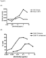
- Levels of neutralisation can be measured in several ways, for example by use of the assays as set out in the examples below, for example in 4.4 in an H929 cell NFkB signalling assay.
- the BCMA ligands BAFF and APRIL are able to induce NFkB signalling and downstream events following binding to BCMA.
- the neutralisation of BCMA in this assay is measured by assessing the ability of anti-BCMA monoclonal antibodies to inhibit BAFF or APRIL driven NFkB induction.
- Antibodies which are considered to have neutralising activity against human BCMA would have an IC50 of less than 30 micrograms/ml, or less than 20 micrograms/ml, or less than 10 micrograms/ml, or less than 5 micrograms/ml or less than 1 micrograms/ml or less than 0.1 micrograms/ml in the H929 stimulation assay as set out in Example 4.4
- CDRs are defined as the complementarity determining region amino acid sequences of an antibody which are the hypervariable domains of immunoglobulin heavy and light chains. There are three heavy chain and three light chain CDRs (or CDR regions) in the variable portion of an immunoglobulin. Thus, “CDRs” as used herein may refer to all three heavy chain CDRs, or all three light chain CDRs (or both all heavy and all light chain CDRs, if appropriate).
- CDRs provide the majority of contact residues for the binding of the antibody to the antigen or epitope.
- CDRs of interest in this invention are derived from donor antibody variable heavy and light chain sequences, and include analogs of the naturally occurring CDRs, which analogs also share or retain the same antigen binding specificity and/or neutralizing ability as the donor antibody from which they were derived.
- the CDR sequences of antibodies can be determined by the Kabat numbering system ( Kabat et al; (Sequences of proteins of Immunological Interest NIH, 1987 ), alternatively they can be determined using the Chothia numbering system ( Al-Lazikani et al., (1997) JMB 273,927-948 ), the contact definition method ( MacCallum R.M., and Martin A.C.R.
- the minimum overlapping region using at least two of the Kabat, Chothia, AbM and contact methods can be determined to provide the "minimum binding unit".
- the minimum binding unit may be a sub-portion of a CDR.
- Table A represents one definition using each numbering convention for each CDR or binding unit.
- the Kabat numbering scheme is used in Table X to number the variable domain amino acid sequence. It should be noted that some of the CDR definitions may vary depending on the individual publication used.
- Table A Kabat CDR Chothia CDR AbM CDR Contact CDR Minimum binding unit H1 31-35/35A/35B 26-32/33/34 26-35/35A/35B 30-35/35A/35B 31-32 H2 50-65 52-56 50-58 47-58 52-56 H3 95-102 95-102 95-102 93-101 95-101 L1 24-34 24-34 24-34 30-36 30-34 L2 50-56 50-56 50-56 46-55 50-55 L3 89-97 89-97 89-97 89-96 89-96
- Variant refers to at least one, two or three amino acid changes in the sequence. These amino acid changes may be deletion, substitution or addition but are preferably substitution. In one such embodiment the substitutions are conservative substitutions. In an alternative embodiment the variant sequence contains at least one substitution whilst retaining the canonical of the antigen binding protein.
- the complementarity determining regions (CDRs) L1, L2, L3, H1 and H2 tend to structurally exhibit one of a finite number of main chain conformations.
- the particular canonical structure class of a CDR is defined by both the length of the CDR and by the loop packing, determined by residues located at key positions in both the CDRs and the framework regions (structurally determining residues or SDRs).
- VH and VL are used herein to refer to the heavy chain variable domain and light chain variable domain respectively of an antibody.
- domain refers to a folded protein structure which has tertiary structure independent of the rest of the protein. Generally, domains are responsible for discrete functional properties of proteins and in many cases may be added, removed or transferred to other proteins without loss of function of the remainder of the protein and/or of the domain.
- An "antibody single variable domain” is a folded polypeptide domain comprising sequences characteristic of antibody variable domains.
- variable domains and modified variable domains, for example, in which one or more loops have been replaced by sequences which are not characteristic of antibody variable domains, or antibody variable domains which have been truncated or comprise N- or C-terminal extensions, as well as folded fragments of variable domains which retain at least the binding activity and specificity of the full-length domain.
- immunoglobulin single variable domain refers to an antibody variable domain (VH, VHH, VL) that specifically binds an antigen or epitope independently of a different V region or domain.
- An immunoglobulin single variable domain can be present in a format (e.g., homo- or hetero-multimer) with other, different variable regions or variable domains where the other regions or domains are not required for antigen binding by the single immunoglobulin variable domain (i.e., where the immunoglobulin single variable domain binds antigen independently of the additional variable domains).
- a “domain antibody” or “dAb” is the same as an "immunoglobulin single variable domain" which is capable of binding to an antigen as the term is used herein.
- An immunoglobulin single variable domain may be a human antibody variable domain, but also includes single antibody variable domains from other species such as rodent (for example, as disclosed in WO 00/29004 ), nurse shark and Camelid VHH dAbs.
- Camelid VHH are immunoglobulin single variable domain polypeptides that are derived from species including camel, llama, alpaca, dromedary, and guanaco, which produce heavy chain antibodies naturally devoid of light chains.
- Such VHH domains may be humanised according to standard techniques available in the art, and such domains are still considered to be "domain antibodies" according to the invention.
- VH includes camelid VHH domains.
- NARV are another type of immunoglobulin single variable domain which were identified in cartilaginous fish including the nurse shark. These domains are also known as Novel Antigen Receptor variable region (commonly abbreviated to V(NAR) or NARV). For further details see Mol. Immunol. 44, 656-665 (2006 ) and US20050043519A .
- Epitope-binding domain refers to a domain that specifically binds an antigen or epitope independently of a different V region or domain, this may be a domain antibody (dAb), for example a human, camelid or shark immunoglobulin single variable domain or it may be a domain which is a derivative of a scaffold selected from the group consisting of CTLA-4 (Evibody); lipocalin; Protein A derived molecules such as Z-domain of Protein A (Affibody, SpA), A-domain (Avimer/Maxibody); Heat shock proteins such as GroEI and GroES; 29eroxidise29g (trans-body); ankyrin repeat protein (DARPin); peptide aptamer; C-type lectin domain (Tetranectin); human ⁇ -crystallin and human ubiquitin (affilins); PDZ domains; scorpion toxinkunitz type domains of human protease inhibitors; and fibronectin (adnectin); which has been
- CTLA-4 Cytotoxic T Lymphocyte-associated Antigen 4
- CTLA-4 is a CD28-family receptor expressed on mainly CD4+ T-cells. Its extracellular domain has a variable domain-like Ig fold. Loops corresponding to CDRs of antibodies can be substituted with heterologous sequence to confer different binding properties.
- CTLA-4 molecules engineered to have different binding specificities are also known as Evibodies.
- Lipocalins are a family of extracellular proteins which transport small hydrophobic molecules such as steroids, bilins, retinoids and lipids.
- Anticalins are between 160-180 amino acids in size, and are derived from lipocalins. For further details see Biochim Biophys Acta 1482: 337-350 (2000 ), US7250297B1 and US20070224633
- An affibody is a scaffold derived from Protein A of Staphylococcus aureus which can be engineered to bind to antigen.
- the domain consists of a three-helical bundle of approximately 58 amino acids. Libraries have been generated by randomisation of surface residues. For further details see Protein Eng. Des. Sel. 17, 455-462 (2004 ) and EP1641818A1
- Avimers are multidomain proteins derived from the A-domain scaffold family.
- the native domains of approximately 35 amino acids adopt a defined disulphide bonded structure. Diversity is generated by shuffling of the natural variation exhibited by the family of A-domains. For further details see Nature Biotechnology 23(12), 1556 - 1561 (2005 ) and Expert Opinion on Investigational Drugs 16(6), 909-917 (June 2007 )
- Transferrin is a monomeric serum transport glycoprotein. Transferrins can be engineered to bind different target antigens by insertion of peptide sequences in a permissive surface loop. Examples of engineered transferrins scaffolds include the Trans-body. For further details see J. Biol. Chem 274, 24066-24073 (1999 ).
- DARPins Designed Ankyrin Repeat Proteins
- Ankyrin which is a family of proteins that mediate attachment of integral membrane proteins to the cytoskeleton.
- a single ankyrin repeat is a 33 residue motif consisting of two ⁇ -helices and a ⁇ -turn. They can be engineered to bind different target antigens by randomising residues in the first ⁇ -helix and a ⁇ -turn of each repeat. Their binding interface can be increased by increasing the number of modules (a method of affinity maturation).
- Fibronectin is a scaffold which can be engineered to bind to antigen.
- Adnectins consists of a backbone of the natural amino acid sequence of the 10 th domain of the 15 repeating units of human fibronectin type III (FN3). Three loops at one end of the ⁇ -sandwich can be engineered to enable an Adnectin to specifically recognize a therapeutic target of interest. For further details see Protein Eng. Des. Sel. 18, 435-444 (2005 ), US20080139791 , WO2005056764 and US6818418B1 .
- Peptide aptamers are combinatorial recognition molecules that consist of a constant scaffold protein, typically thioredoxin (TrxA) which contains a constrained variable peptide loop inserted at the active site.
- TrxA thioredoxin
- Microbodies are derived from naturally occurring microproteins of 25-50 amino acids in length which contain 3-4 cysteine bridges - examples of microproteins include KalataB1 and conotoxin and knottins.
- the microproteins have a loop which can be engineered to include upto 25 amino acids without affecting the overall fold of the microprotein.
- engineered knottin domains see WO2008098796 .
- epitope binding domains include proteins which have been used as a scaffold to engineer different target antigen binding properties include human ⁇ -crystallin and human ubiquitin (affilins), kunitz type domains of human protease inhibitors, PDZ-domains of the Ras-binding protein AF-6, scorpion toxins (charybdotoxin), C-type lectin domain (tetranectins) are reviewed in Chapter 7 - Non-Antibody Scaffolds from Handbook of Therapeutic Antibodies (2007, edited by Stefan Dubel ) and Protein Science 15:14-27 (2006 ). Epitope binding domains of the present invention could be derived from any of these alternative protein domains.
- the term "antigen-binding site” refers to a site on a protein which is capable of specifically binding to antigen, this may be a single domain, for example an epitope-binding domain, or it may be paired VH/VL domains as can be found on a standard antibody.
- single-chain Fv (ScFv) domains can provide antigen-binding sites.
- mAbdAb and dAbmAb are used herein to refer to antigen-binding proteins of the present invention.
- the two terms can be used interchangeably, and are intended to have the same meaning as used herein.
- antigen binding protein refers to antibodies, antibody fragments for example a domain antibody (dAb), ScFv, Fab, Fab2, and other protein constructs.
- Antigen binding molecules may comprise at least one Ig variable domain, for example antibodies, domain antibodies (dAbs), Fab, Fab', F(ab')2, Fv, ScFv, diabodies, mAbdAbs, affibodies, heteroconjugate antibodies or bispecific antibodies.
- the antigen binding molecule is an antibody.
- the antigen binding molecule is a dAb, i.e.
- Antigen binding molecules may be capable of binding to two targets, i.e. they may be dual targeting proteins.
- Antigen binding molecules may be a combination of antibodies and antigen binding fragments such as for example, one or more domain antibodies and/or one or more ScFvs linked to a monoclonal antibody.
- Antigen binding molecules may also comprise a non-Ig domain for example a domain which is a derivative of a scaffold selected from the group consisting of CTLA-4 (Evibody); lipocalin; Protein A derived molecules such as Z-domain of Protein A (Affibody, SpA), A-domain (Avimer/Maxibody); Heat shock proteins such as GroEI and GroES; 31eroxidise31g (trans-body); ankyrin repeat protein (DARPin); peptide aptamer; C-type lectin domain (Tetranectin); human ⁇ -crystallin and human ubiquitin (affilins); PDZ domains; scorpion toxinkunitz type domains of human protease inhibitors; and fibronectin (adnectin); which has been subjected to protein engineering in order to obtain binding to OSM.
- a non-Ig domain for example a domain which is a derivative of a scaffold selected from the group consisting of CTLA-4 (Evibody); lipo
- antigen binding protein will be capable of antagonising and/or neutralising human OSM.
- an antigen binding protein may inhibit and or block OSM activity by binding to OSM and preventing a natural ligand from binding and/or activating the gp130 receptor.
- ADCC Antibody dependant cell mediated cytotoxic activity
- CDC Complement-dependant cytotoxic activity
- Fc-mediated phagocytosis Fc-mediated phagocytosis and antibody recycling via the FcRn receptor.
- effector functionalities including ADCC and ADCP are mediated by the interaction of the heavy chain constant region with a family of Fc ⁇ receptors present on the surface of immune cells. In humans these include Fc ⁇ RI (CD64), Fc ⁇ RII (CD32) and Fc ⁇ RIII (CD16).
- Interaction between the antigen binding protein bound to antigen and the formation of the Fc/ Fc ⁇ complex induces a range of effects including cytotoxicity, immune cell activation, phagocytosis and release of inflammatory cytokines.
- the interaction between the constant region of an antigen binding protein and various Fc receptors (FcR) is believed to mediate the effector functions of the antigen binding protein.
- Significant biological effects can be a consequence of effector functionality, in particular, antibody-dependent cellular cytotoxicity (ADCC), fixation of complement (complement dependent cytotoxicity or CDC), and half-life/clearance of the antigen binding protein.
- effector function can be measured in a number of ways including for example via binding of the Fc ⁇ RIII to Natural Killer cells or via Fc ⁇ RI to monocytes/macrophages to measure for ADCC effector function.
- an antigen binding protein of the present invention can be assessed for ADCC effector function in a Natural Killer cell assay. Examples of such assays can be found in Shields et al, 2001 The Journal of Biological Chemistry, Vol.
- IgG4 and IgG2 isotypes essentially lack the functions of a) activation of complement by the classical pathway; and b) antibody-dependent cellular cytotoxicity.
- Various modifications to the heavy chain constant region of antigen binding proteins may be carried out depending on the desired effector property.
- IgG1 constant regions containing specific mutations have separately been described to reduce binding to Fc receptors and therefore reduce ADCC and CDC ( Duncan et al. Nature 1988, 332; 563-564 ; Lund et al. J. Immunol. 1991, 147; 2657-2662 ; Chappel et al. PNAS 1991, 88; 9036-9040 ; Burton and Woof, Adv. Immunol.
- an antigen binding protein comprising a constant region such that the antigen binding protein has reduced ADCC and/or complement activation or effector functionality.
- the heavy chain constant region may comprise a naturally disabled constant region of IgG2 or IgG4 isotype or a mutated IgG1 constant region. Examples of suitable modifications are described in EP0307434 .
- One example comprises the substitutions of alanine residues at positions 235 and 237 (EU index numbering).
- the antigen binding protein of the invention herein described is mutated at positions 239 and 332, for example S239D and I332E or in a further embodiment it is mutated at three or more positions selected from 239 and 332 and 330, for example S239D and I332E and A330L. (EU index numbering).
- an antigen binding protein comprising a heavy chain constant region with an altered glycosylation profile such that the antigen binding protein has enhanced effector function.
- the antigen binding protein has enhanced ADCC or enhanced CDC or wherein it has both enhanced ADCC and CDC effector function.
- suitable methodologies to produce antigen binding proteins with an altered glycosylation profile are described in WO2003011878 , WO2006014679 and EP1229125 , all of which can be applied to the antigen binding proteins of the present invention.
- the present invention also provides a method for the production of an antigen binding protein according to the invention comprising the steps of:
- an antigen binding protein comprising a chimaeric heavy chain constant region for example an antigen binding protein comprising a chimaeric heavy chain constant region with at least one CH2 domain from IgG3 such that the antigen binding protein has enhanced effector function, for example wherein it has enhanced ADCC or enhanced CDC, or enhanced ADCC and CDC functions,.
- the antigen binding protein may comprise one CH2 domain from IgG3 or both CH2 domains may be from IgG3.
- an antigen binding protein comprising a heavy chain constant region which comprises a mutated and chimaeric heavy chain constant region for example wherein an antigen binding protein comprising at least one CH2 domain from IgG3 and one CH2 domain from IgG1, wherein the IgG1 CH2 domain has one or more mutations at positions selected from 239 and 332 and 330 (for example the mutations may be selected from S239D and I332E and A330L) such that the antigen binding protein has enhanced effector function, for example wherein it has one or more of the following functions, enhanced ADCC or enhanced CDC, for example wherein it has enhanced ADCC and enhanced CDC.
- the IgG1 CH2 domain has the mutations S239D and I332E.
- an antigen binding protein comprising a chimaeric heavy chain constant region and which has an altered glycosylation profile.
- the heavy chain constant region comprises at least one CH2 domain from IgG3 and one CH2 domain from IgG1 and has an altered glycosylation profile such that the ratio of fucose to mannose is 0.8:3 or less, for example wherein the antigen binding protein is defucosylated so that said antigen binding protein has an enhanced effector function in comparison with an equivalent antigen binding protein with an immunoglobulin heavy chain constant region lacking said mutations and altered glycosylation profile, for example wherein it has one or more of the following functions, enhanced ADCC or enhanced CDC, for example wherein it has enhanced ADCC and enhanced CDC
- the antigen binding protein has at least one IgG3 CH2 domain and at least one heavy chain constant domain from IgG1 wherein both IgG CH2 domains are mutated in accordance with the limitations described herein.
- an antigen binding protein comprising a mutated and chimeric heavy chain constant region wherein said antigen binding protein has an altered glycosylation profile such that the antigen binding protein has enhanced effector function, for example wherein it has one or more of the following functions, enhanced ADCC or enhanced CDC.
- the mutations are selected from positions 239 and 332 and 330, for example the mutations are selected from S239D and I332E and A330L.
- the heavy chain constant region comprises at least one CH2 domain from IgG3 and one Ch2 domain from IgG1.
- the heavy chain constant region has an altered glycosylation profile such that the ratio of fucose to mannose is 0.8:3 or less for example the antigen binding protein is defucosylated, so that said antigen binding protein has an enhanced effector function in comparison with an equivalent non-chimaeric antigen binding protein or with an immunoglobulin heavy chain constant region lacking said mutations and altered glycosylation profile.
- an immunoconjugate (interchangeably referred to as "antibody-drug conjugates,” or “ADCs")comprising an antigen binding protein according to the invention as herein described including, but not limited to, an antibody conjugated to one or more cytotoxic agents, such as a chemotherapeutic agent, a drug, a growth inhibitory agent, a toxin (e.g., a protein toxin, an enzymatically active toxin of bacterial, fungal, plant, or animal origin, or fragments thereof), or a radioactive isotope (i.e., a radioconjugate).
- cytotoxic agents such as a chemotherapeutic agent, a drug, a growth inhibitory agent, a toxin (e.g., a protein toxin, an enzymatically active toxin of bacterial, fungal, plant, or animal origin, or fragments thereof), or a radioactive isotope (i.e., a radioconjugate).
- Immunoconjugates have been used for the local delivery of cytotoxic agents, i.e., drugs that kill or inhibit the growth or proliferation of cells, in the treatment of cancer ( Lambert, J. (2005) Curr. Opinion in Pharmacology 5:543-549 ; Wu et al. (2005) Nature Biotechnology 23(9):1137-1146 ; Payne, G. (2003) i 3:207-212; Syrigos and Epenetos (1999) Anticancer Research 19:605-614 ; Niculescu-Duvaz and Springer (1997) Adv. Drug Deliv. Rev. 26:151-172 ; U.S. Pat. No. 4,975,278 ).
- cytotoxic agents i.e., drugs that kill or inhibit the growth or proliferation of cells
- Immunoconjugates allow for the targeted delivery of a drug moiety to a tumor, and intracellular accumulation therein, where systemic administration of unconjugated drugs may result in unacceptable levels of toxicity to normal cells as well as the tumor cells sought to be eliminated (Appel et al., Lancet (Mar. 15, 1986) pp. 603-05 ; Thorpe (1985) "Antibody Carriers Of Cytotoxic Agents In Cancer Therapy: A Review," in Monoclonal Antibodies '84: Biological And Clinical Applications (A. Pinchera et al., eds) pp. 475-506 . Both polyclonal antibodies and monoclonal antibodies have been reported as useful in these strategies ( Rowland et al., (1986) Cancer Immunol. Immunother.
- Drugs used in these methods include daunomycin, doxorubicin, methotrexate, and vindesine (Rowland et al., (1986) supra).
- Toxins used in antibody-toxin conjugates include bacterial toxins such as diphtheria toxin, plant toxins such as ricin, small molecule toxins such as geldanamycin ( Mandler et al (2000) J. Nat. Cancer Inst. 92(19):1573-1581 ; Mandler et al (2000) Bioorganic & Med. Chem. Letters 10:1025-1028 ; Mandler et al (2002) Bioconjugate Chem.
- the present invention includes immunoconjugates having the following general structure: ABP ⁇ Linker n ⁇ Ctx m Wherein ABP is an antigen binding protein
- an immunoconjugate comprises an antigen binding protein, including but not limited to, an antibody and a chemotherapeutic agent or other toxin.
- Chemotherapeutic agents useful in the generation of immunoconjugates are described herein.
- Enzymatically active toxins and fragments thereof that can be used include diphtheria A chain, nonbinding active fragments of diphtheria toxin, exotoxin A chain (from Pseudomonas aeruginosa), ricin A chain, abrin A chain, modeccin A chain, alpha-sarcin, Aleurites fordii proteins, dianthin proteins, Phytolaca americana proteins (PAPI, PAPII, and PAP-S), momordica charantia inhibitor, curcin, crotin, sapaonaria officinalis inhibitor, gelonin, mitogellin, restrictocin, phenomycin, enomycin, and the tricothecenes.
- radionuclides are available for the production of radioconjugated antibodies. Examples include 211 At, 212 Bi, 131 I, 131 In, 90 Y, and 186 Re.
- Antigen binding proteins of the present invention may also be conjugated to one or more toxins, including, but not limited to, a calicheamicin, maytansinoids, dolastatins, aurostatins, a trichothecene, and CC1065, and the derivatives of these toxins that have toxin activity.
- toxins including, but not limited to, a calicheamicin, maytansinoids, dolastatins, aurostatins, a trichothecene, and CC1065, and the derivatives of these toxins that have toxin activity.
- Suitable cytotoxic agents include, but are not limited to, an auristatin including dovaline-valine-dolaisoleunine-dolaproine-phenylalanine (MMAF) and monomethyl auristatin E (MMAE) as well as ester forms of MMAE, a DNA minor groove binding agent, a DNA minor groove alkylating agent, an enediyne, a lexitropsin, a duocarmycin, a taxane, including paclitaxel and docetaxel, a puromycin, a dolastatin, a maytansinoid, and a vinca alkaloid.
- an auristatin including dovaline-valine-dolaisoleunine-dolaproine-phenylalanine (MMAF) and monomethyl auristatin E (MMAE) as well as ester forms of MMAE
- a DNA minor groove binding agent a DNA minor groove alkylating agent
- cytotoxic agents include topotecan, morpholino-doxorubicin, rhizoxin, cyanomorpholino-doxorubicin, dolastatin-10, echinomycin, combretatstatin, chalicheamicin, maytansine, DM-1, DM-4, netropsin.
- Other suitable cytotoxic agents include anti-tubulin agents, such as an auristatin, a vinca alkaloid, a podophyllotoxin, a taxane, a baccatin derivative, a cryptophysin, a maytansinoid, a combretastatin, or a dolastatin.
- Antitubulin agent include dimethylvaline-valine-dolaisoleuine-dolaproine-phenylalanine-p-phenylened- iamine (AFP), MMAF, MMAE, auristatin E, vincristine, vinblastine, vindesine, vinorelbine, VP-16, camptothecin, paclitaxel, docetaxel, epothilone A, epothilone B, nocodazole, colchicines, colcimid, estramustine, cemadotin, discodermolide, maytansine, DM-1, DM-4 or eleutherobin.
- AFP dimethylvaline-valine-dolaisoleuine-dolaproine-phenylalanine-p-phenylened- iamine
- MMAF MMAF
- MMAE auristatin E
- vincristine vinblastine
- vindesine vinorelbine
- Antibody drug conjugates were produced by conjugating the small molecule anti-tubulin agent monomethylauristatin E (MMAE) or monomethylauristatin F (MMAF) to the antibodies.
- MMAE monomethylauristatin E
- MMAF monomethylauristatin F
- the linker consists of a thiol-reactive maleimide, a caproyl spacer, the dipeptide valine-citrulline, and p-aminobenzyloxycarbonyl, a self-immolative fragmenting group.
- MMAF a protease-resistant maleimidocaproyl linker is used.
- the conjugation process leads to heterogeneity in drug-antibody attachment, varying in both the number of drugs bound to each antibody molecule (mole ratio [MR]), and the site of attachment.
- the overall average drug-to-antibody MR is approximately
- the points of attachment are cysteines produced by mild reduction of the interchain disulfides of the antibody which is carried out whilst antibodies are immobilised on Protein G affinity resin (thus enabling the use of large reagent excesses without intermediate purifications). While immobilized, a large excess of TCEP will fully reduce the interchain disulfides but has no impact upon the binding of the antibody to the resin.
- the number of thiols per antibody generated by this procedure depends upon the source and isotype of the antibodies. For example, human (and mouse-human chimeric) IgG1s have 4 reducible disulfides, and thus generate 8 thiols upon full reduction, whereas murine IgG1s have 5 reducible disulfides and produce 10 thiols. If ADCs with the maximal drug loading (e.g., 10 drugs per antibody for the murine IgG1s) are desired, then the maleimido-drug-linker can simply be added to the immobilized antibodies in sufficient excess to ensure complete conjugation.
- ADCs with the maximal drug loading e.g., 10 drugs per antibody for the murine IgG1s
- ADCs with fewer drugs per antibody can also be prepared from fully reduced antibodies by including a biologically inert capping agent such as N-ethyl maleimide (NEM) which occupies some of the available thiols on the antibody.
- NEM N-ethyl maleimide
- the maleimido-drug-linker and the capping agent are added simultaneously to the fully reduced antibody and in large excess (at least 3-fold), the two maleimide electrophiles compete for the limiting number of available thiols.
- the drug loading is determined by the relative thiol reaction rates of the drug-linker and capping agent, and thus can be considered to be under kinetic control.
- the relative reaction rates of maleimido-drug-linkers do vary significantly, and thus the molar ratio of drug-linker to NEM present in a reaction mix must be determined empirically to arrive at a panel of ADCs with a desired level of drug loading.
- the mole fraction of the drug linkers SGD-1006 (vcMMAE) and SGD-1269 (mcMMAF) in NEM mixtures which yield ADCs with approximately 4 drugs per antibody are summarized in Table 2 for common human and murine IgG isotypes.
- the immunoconjugate comprises an antigen binding protein or antibody conjugated to dolastatins or dolostatin peptidic analogs and derivatives, the auristatins ( U.S. Pat. Nos. 5,635,483 ; 5,780,588 ).
- Dolastatins and auristatins have been shown to interfere with microtubule dynamics, GTP hydrolysis, and nuclear and cellular division ( Woyke et al. (2001) Antimicrob. Agents and Chemother. 45(12):3580-3584 ) and have anticancer ( U.S. Pat. No. 5,663,149 ) and antifungal activity ( Pettit et al. (1998) Antimicrob. Agents Chemother.
- the dolastatin or auristatin (which are pentapeptide derivatives of dolastatins) drug moiety may be attached to the antibody through the N (amino) terminus or the C (carboxyl) terminus of the peptidic drug moiety ( WO 02/088172 ).
- Exemplary auristatin embodiments include the N-terminus linked monomethylauristatin drug moieties DE and DF, disclosed in "Monomethylvaline Compounds Capable of Conjugation to Ligands," U.S. Patent No. 7,498,298 , the disclosure of which is expressly incorporated by reference in its entirety.
- MMAE refers to monomethyl auristatin E.
- MMAF refers to dovaline-valine-dolaisoleuine-dolaproine-phenylalanine.
- peptide-based drug moieties can be prepared by forming a peptide bond between two or more amino acids and/or peptide fragments.
- Such peptide bonds can be prepared, for example, according to the liquid phase synthesis method (see E. Schroder and K. Lubke, "The Peptides,” volume 1, pp 76-136, 1965, Academic Press ) that is well known in the field of peptide chemistry.
- the auristatin/dolastatin drug moieties may be prepared according to the methods of: U.S. Pat. No. 5,635,483 ; U.S. Pat. No. 5,780,588 ; Pettit et al. (1989) J. Am. Chem. Soc.
- auristatins examples include MMAE and MMAF the structures of which are shown below:
- Maytansinoids are mitototic inhibitors which act by inhibiting tubulin polymerization. Maytansine was first isolated from the east African shrub Maytenus serrata ( U.S. Pat. No. 3,896,111 ). Subsequently, it was discovered that certain microbes also produce maytansinoids, such as maytansinol and C-3 maytansinol esters ( U.S. Pat. No. 4,151,042 ). Highly cytotoxic maytansinoid drugs drugs can be prepared from ansamitocin precursors produced by fermentation of microorganisms such as Actinosynnema. Methods for isolating ansamitocins are described in US Patent No. 6,573,074 .
- Synthetic maytansinol and derivatives and analogues thereof are disclosed, for example, in U.S. Pat. Nos. 4,137,230 ; 4,248,870 ; 4,256,746 ; 4,260,608 ; 4,265,814 ; 4,294,757 ; 4,307,016 ; 4,308,268 ; 4,308,269 ; 4,309,428 ; 4,313,946 ; 4,315,929 ; 4,317,821 ; 4,322,348 ; 4,331,598 ; 4,361,650 ; 4,364,866 ; 4,424,219 ; 4,450,254 ; 4,362,663 ; and 4,371,533 .
- Antibody-maytansinoid conjugates are prepared by chemically linking an antibody to a maytansinoid molecule without significantly diminishing the biological activity of either the antibody or the maytansinoid molecule. See, e.g., U.S. Pat. No. 5,208,020 .
- An average of 3-4 maytansinoid molecules conjugated per antibody molecule has shown efficacy in enhancing cytotoxicity of target cells without negatively affecting the function or solubility of the antibody, although even one molecule of toxin/antibody would be expected to enhance cytotoxicity over the use of naked antibody.
- Maytansinoids are well known in the art and can be synthesized by known techniques or isolated from natural sources. Suitable maytansinoids are disclosed, for example, in U.S.
- Maytansinoids are maytansinol and maytansinol analogues modified in the aromatic ring or at other positions of the maytansinol molecule, such as various maytansinol esters. Methods for preparing matansinoids for linkage with antibodies are disclosed in US Patent Nos. 6,570,024 and 6,884,874 .
- the calicheamicin family of antibiotics is capable of producing double-stranded DNA breaks at sub-picomolar concentrations.
- For the preparation of conjugates of the calicheamicin family see U.S. Pat. Nos. 5,712,374 , 5,714,586 , 5,739,116 , 5,767,285 , 5,770,701 , 5,770,710 , 5,773,001 , 5,877,296 (all to American Cyanamid Company).
- Structural analogues of calicheamicin which may be used include, but are not limited to, .gamma.1I, .alpha.2I, .alpha.3I, N-acetyl-.gamma.1I, PSAG and .theta.I1 ( Hinman et al., Cancer Research 53:3336-3342 (1993 ), Lode et al., Cancer Research 58:2925-2928 (1998 ) and the aforementioned U.S. patents to American Cyanamid).
- Another anti-tumor drug that the antibody can be conjugated is QFA which is an antifolate.
- QFA is an antifolate.
- Both calicheamicin and QFA have intracellular sites of action and do not readily cross the plasma membrane. Therefore, cellular uptake of these agents through antibody mediated internalization greatly enhances their cytotoxic effects.
- antitumor agents that can be conjugated to the antibodies include BCNU, streptozoicin, vincristine and 5-fluorouracil, the family of agents known collectively LL-E33288 complex described in U.S. Pat. Nos. 5,053,394 , 5,770,710 , as well as esperamicins ( U.S. Pat. No. 5,877,296 ).
- Enzymatically active toxins and fragments thereof which can be used include diphtheria A chain, nonbinding active fragments of diphtheria toxin, exotoxin A chain (from Pseudomonas aeruginosa), ricin A chain, abrin A chain, modeccin A chain, alpha-sarcin, Aleurites fordii proteins, dianthin proteins, Phytolaca americana proteins (PAPI, PAPII, and PAP-S), momordica charantia inhibitor, curcin, crotin, sapaonaria officinalis inhibitor, gelonin, mitogellin, restrictocin, phenomycin, enomycin and the tricothecenes. See, for example, WO 93/21232 published Oct. 28, 1993 .
- the present invention further contemplates an immunoconjugate formed between an antibody and a compound with nucleolytic activity (e.g., a ribonuclease or a DNA endonuclease such as a deoxyribonuclease; DNase).
- a compound with nucleolytic activity e.g., a ribonuclease or a DNA endonuclease such as a deoxyribonuclease; DNase.
- the antibody may comprise a highly radioactive atom.
- radioactive isotopes are available for the production of radioconjugated antibodies. Examples include At211, 1131, 1125, Y90, Re186, Re188, Sm153, Bi212, P32, Pb212 and radioactive isotopes of Lu.
- the conjugate When used for detection, it may comprise a radioactive atom for scintigraphic studies, for example tc99m or 1123, or a spin label for nuclear magnetic resonance (NMR) imaging (also known as magnetic resonance imaging, mri), such as iodine-123 again, iodine-131, indium-111, fluorine-19, carbon-13, nitrogen-15, oxygen-17, gadolinium, manganese or iron.
- NMR nuclear magnetic resonance
- mri nuclear magnetic resonance
- the radio- or other labels may be incorporated in the conjugate in known ways.
- the peptide may be biosynthesized or may be synthesized by chemical amino acid synthesis using suitable amino acid precursors involving, for example, fluorine-19 in place of hydrogen.
- Labels such as tc99m or 1123, Re186, Re188 and In111 can be attached via a cysteine residue in the peptide.
- Yttrium-90 can be attached via a lysine residue.
- the IODOGEN method Fraker et al. (1978) Biochem. Biophys. Res. Commun. 80: 49-57 ) can be used to incorporate iodine-123. " Monoclonal Antibodies in Immunoscintigraphy" (Chatal, CRC Press 1989 ) describes other methods in detail.
- the antibody can be conjugated directly to the cytotoxic agent or via a linker.
- Suitable linkers include, for example, cleavable and non-cleavable linkers.
- a cleavable linker is typically susceptible to cleavage under intracellular conditions.
- Suitable cleavable linkers include, for example, a peptide linker cleavable by an intracellular protease, such as lysosomal protease or an endosomal protease.
- the linker can be a dipeptide linker, such as a valine-citrulline (val-cit) or a phenylalanine-lysine (phe-lys) linker.
- Other suitable linkers include linkers hydrolyzable at a pH of less than 5.5, such as a hydrazone linker. Additional suitable cleavable linkers include disulfide linkers.
- Conjugates of the antigen binding protein and cytotoxic agent may be made using a variety of bifunctional protein coupling agents such as N-succinimidyl-3-(2-pyridyldithio)propionate (SPDP), succinimidyl-4-(N-maleimidomethyl) cyclohexane-1-carboxylate (SMCC), iminothiolane (IT), bifunctional derivatives of imidoesters (such as dimethyl adipimidate HCI), active esters (such as disuccinimidyl suberate), aldehydes (such as glutaraldehyde), bis-azido compounds (such as bis(p-azidobenzoyl) hexanediamine), bis-diazonium derivatives (such as bis-(p-diazoniumbenzoyl)-ethylenediamine), diisocyanates (such as toluene 2,6-diisocyanate), and bis-active fluorine compounds (
- linker may be composed of one or more linker components.
- exemplary linker components include 6-maleimidocaproyl (“MC”), maleimidopropanoyl (“MP”), valine-citrulline (“val-cit”), alanine-phenylalanine (“ala-phe”), p-aminobenzyloxycarbonyl (“PAB”), N-Succinimidyl 4-(2-pyridylthio)pentanoate (“SPP”), N-Succinimidyl 4-(N-maleimidomethyl)cyclohexane-1 carboxylate (“SMCC”), and N-Succinimidyl (4-iodo-acetyl)aminobenzoate (“SIAB”).
- MC 6-maleimidocaproyl
- MP maleimidopropanoyl
- val-cit valine-citrulline
- ala-phe alanine-phenylalanine
- PAB p
- Linkers may also comprises amino acids and/or amino acid analogs.
- Amino acid linker components include a dipeptide, a tripeptide, a tetrapeptide or a pentapeptide.
- Exemplary dipeptides include: valine-citrulline (vc or val-cit), alanine-phenylalanine (af or ala-phe).
- Exemplary tripeptides include: glycine-valine-citrulline (gly-val-cit) and glycine-glycine-glycine (gly-gly-gly).
- Amino acid residues which comprise an amino acid linker component include those occurring naturally, as well as minor amino acids and non-naturally occurring amino acid analogs, such as citrulline.
- Amino acid linker components can be designed and optimized in their selectivity for enzymatic cleavage by a particular enzyme, for example, a tumor-associated protease, cathepsin B, C and D, or a plasmin protease.
- Antigen binding proteins and antibodies may be made reactive for conjugation with linker reagents.
- Nucleophilic groups on antibodies include, but are not limited to: (i) N-terminal amine groups, (ii) side chain amine groups, e.g., lysine, (iii) side chain thiol groups, e.g. cysteine, and (iv) sugar hydroxyl or amino groups where the antibody is glycosylated.
- Amine, thiol, and hydroxyl groups are nucleophilic and capable of reacting to form covalent bonds with electrophilic groups on linker moieties and linker reagents including: (i) active esters such as NHS esters, HOBt esters, haloformates, and acid halides; (ii) alkyl and benzyl halides such as haloacetamides; (iii) aldehydes, ketones, carboxyl, and maleimide groups. Certain antibodies have reducible interchain disulfides, i.e. cysteine bridges. Antibodies may be made reactive for conjugation with linker reagents by treatment with a reducing agent such as DTT (dithiothreitol).
- a reducing agent such as DTT (dithiothreitol).
- Each cysteine bridge will thus form, theoretically, two reactive thiol nucleophiles.
- Additional nucleophilic groups can be introduced into antibodies through the reaction of lysines with 2-iminothiolane (Traut's reagent) resulting in conversion of an amine into a thiol.
- Reactive thiol groups may be introduced into the antibody (or fragment thereof) by introducing one, two, three, four, or more cysteine residues (e.g., preparing mutant antibodies comprising one or more non-native cysteine amino acid residues).
- Antigen binding proteins and antibodies may also be modified to introduce electrophilic moieties, which can react with nucleophilic substituents on the linker reagent or drug.
- the sugars of glycosylated antibodies may be oxidized, e.g. with periodate oxidizing reagents, to form aldehyde or ketone groups which may react with the amine group of linker reagents or drug moieties.
- the resulting imine Schiff base groups may form a stable linkage, or may be reduced, e.g., by borohydride reagents to form stable amine linkages.
- reaction of the carbohydrate portion of a glycosylated antibody with either glactose oxidase or sodium meta-periodate may yield carbonyl (aldehyde and ketone) groups in the protein that can react with appropriate groups on the drug (Hermanson, Bioconjugate Techniques).
- proteins containing N-terminal serine or threonine residues can react with sodium meta-periodate, resulting in production of an aldehyde in place of the first amino acid ( Geoghegan & Stroh, (1992) Bioconjugate Chem. 3:138-146 ; U.S. Pat. No. 5,362,852 ).
- Such aldehydes can be reacted with a drug moiety or linker nucleophile.
- Nucleophilic groups on a drug moiety include, but are not limited to: amine, thiol, hydroxyl, hydrazide, oxime, hydrazine, thiosemicarbazone, hydrazine carboxylate, and arylhydrazide groups capable of reacting to form covalent bonds with electrophilic groups on linker moieties and linker reagents including: (i) active esters such as NHS esters, HOBt esters, haloformates, and acid halides; (ii) alkyl and benzyl halides such as haloacetamides; (iii) aldehydes, ketones, carboxyl, and maleimide groups.
- the linker is cleavable by a cleaving agent that is present in the intracellular environment (e.g., within a lysosome or endosome or caveolea).
- the linker can be, e.g., a peptidyl linker that is cleaved by an intracellular peptidase or protease enzyme, including, but not limited to, a lysosomal or endosomal protease.
- the peptidyl linker is at least two amino acids long or at least three amino acids long.
- Cleaving agents can include cathepsins B and D and plasmin, all of which are known to hydrolyze dipeptide drug derivatives resulting in the release of active drug inside target cells (see, e.g., Dubowchik and Walker, 1999, Pharm. Therapeutics 83:67-123 ).
- Peptidyl linkers may be cleavable by enzymes that are present cells.
- a peptidyl linker that is cleavable by the thiol-dependent protease cathepsin-B which is highly expressed in cancerous tissue, can be used (e.g., a Phe-Leu or a Gly-Phe-Leu-Gly (SEQ ID NO:50) linker).
- the peptidyl linker cleavable by an intracellular protease is a Val-Cit linker or a Phe-Lys linker (see, e.g., U.S. Pat. No. 6,214,345 , which describes the synthesis of doxorubicin with the val-cit linker).
- One advantage of using intracellular proteolytic release of the therapeutic agent is that the agent is typically attenuated when conjugated and the serum stabilities of the conjugates are typically high.
- the cleavable linker is pH-sensitive, i.e., sensitive to hydrolysis at certain pH values.
- the pH-sensitive linker hydrolyzable under acidic conditions.
- an acid-labile linker that is hydrolyzable in the lysosome e.g., a hydrazone, semicarbazone, thiosemicarbazone, cis-aconitic amide, orthoester, acetal, ketal, or the like
- an acid-labile linker that is hydrolyzable in the lysosome e.g., a hydrazone, semicarbazone, thiosemicarbazone, cis-aconitic amide, orthoester, acetal, ketal, or the like
- the hydrolyzable linker is a thioether linker (such as, e.g., a thioether attached to the therapeutic agent via an acylhydrazone bond (see, e.g., U.S. Pat. No. 5,622,929 )).
- the linker is cleavable under reducing conditions (e.g., a disulfide linker).
- a disulfide linker e.g., a disulfide linker.
- disulfide linkers are known in the art, including, for example, those that can be formed using SATA (N-succinimidyl-5-acetylthioacetate), SPDP (N-succinimidyl-3-(2-pyridyldithio)propionate), SPDB (N-succinimidyl-3-(2-pyridyldithio)butyrate) and SMPT (N-succinimidyl-oxycarbonyl-alphamethyl-alpha-(2-pyridyl-dithio)toluene)- , SPDB and SMPT (See, e.g., Thorpe et al., 1987, Cancer Res.
- the linker is a malonate linker ( Johnson et al., 1995, Anticancer Res. 15:1387-93 ), a maleimidobenzoyl linker ( Lau et al., 1995, Bioorg-Med-Chem. 3(10):1299-1304 ), or a 3'-N-amide analog ( Lau et al., 1995, Bioorg-Med-Chem. 3(10):1305-12 ).
- the linker is not substantially sensitive to the extracellular environment.
- “not substantially sensitive to the extracellular environment,” in the context of a linker means that no more than about 20%, typically no more than about 15%, more typically no more than about 10%, and even more typically no more than about 5%, no more than about 3%, or no more than about 1% of the linkers, in a sample of ADC or ADC derivative, are cleaved when the ADC or ADC derivative present in an extracellular environment (e.g., in plasma).
- Whether a linker is not substantially sensitive to the extracellular environment can be determined, for example, by incubating independently with plasma both (a) the ADC or ADC derivative (the “ADC sample”) and (b) an equal molar amount of unconjugated antibody or therapeutic agent (the “control sample”) for a predetermined time period (e.g., 2, 4, 8, 16, or 24 hours) and then comparing the amount of unconjugated antibody or therapeutic agent present in the ADC sample with that present in control sample, as measured, for example, by high performance liquid chromatography.
- a predetermined time period e.g., 2, 4, 8, 16, or 24 hours
- the linker promotes cellular internalization. In certain embodiments, the linker promotes cellular internalization when conjugated to the therapeutic agent (i.e., in the milieu of the linker-therapeutic agent moiety of the ADC or ADC derivate as described herein). In yet other embodiments, the linker promotes cellular internalization when conjugated to both the therapeutic agent and the antigen binding protein or antibody or derivative thereof (i.e., in the milieu of the ADC or ADC derivative as described herein).
- linkers that can be used with the present compositions and methods are described in WO 2004010957 entitled “Drug Conjugates and Their Use for Treating Cancer, An Autoimmune Disease or an Infectious Disease” filed Jul. 31, 2003, and U.S. Provisional Application No. 60/400,403 , entitled “Drug Conjugates and their use for treating cancer, an autoimmune disease or an infectious disease", filed Jul. 31, 2002 (the disclosure of which is incorporated by reference herein).
- a fusion protein comprising the antigen binding protein and cytotoxic agent may be made, e.g., by recombinant techniques or peptide synthesis.
- the length of DNA may comprise respective regions encoding the two portions of the conjugate either adjacent one another or separated by a region encoding a linker peptide which does not destroy the desired properties of the conjugate.
- the antibody may be conjugated to a "receptor” (such as streptavidin) for utilization in tumor pre-targeting wherein the antibody-receptor conjugate is administered to the patient, followed by removal of unbound conjugate from the circulation using a clearing agent and then administration of a "ligand” (e.g., avidin) which is conjugated to a cytotoxic agent (e.g., a radionucleotide).
- a "receptor” such as streptavidin
- a ligand e.g., avidin
- cytotoxic agent e.g., a radionucleotide
- Non Human antibody or antibody fragment thereof as used herein is meant to refer to antibodies or fragments thereof which originate from any species other than human wherein human includes chimeric antibodies.
- donor antibody refers to an antibody (monoclonal, and/or recombinant) which contributes the amino acid sequences of its variable domains, CDRs, or other functional fragments or analogs thereof to a first immunoglobulin partner, so as to provide the altered immunoglobulin coding region and resulting expressed altered antibody with the antigenic specificity and neutralizing activity characteristic of the donor antibody.
- acceptor antibody refers to an antibody (monoclonal and/or recombinant) heterologous to the donor antibody, which contributes all (or any portion, but preferably all) of the amino acid sequences encoding its heavy and/or light chain framework regions and/or its heavy and/or light chain constant regions to the first immunoglobulin partner.
- the human antibody is the acceptor antibody.
- Human acceptor sequence as used herein is meant to refer to a framework of an antibody or antibody fragment thereof comprising the amino acid sequence of a VH or VL framework derived from a human antibody or antibody fragment thereof or a human consensus sequence framework into which CDR's from a non-human species may be incorporated.
- incorporación of CDR's or hypervariable regions as used herein encompasses any means by which the non-human CDR's are situated with the human acceptor framework. It will be appreciated that this can be achieved in various ways, for example, nucleic acids encoding the desired amino acid sequence can be generated by mutating nucleic acids encoding the non-human variable domain sequence so that the framework residues thereof are changed to human acceptor framework residues, or by mutating nucleic acid encoding the human variable domain sequence so that the CDR's are changed to non-human residues, or by synthesizing nucleic acids encoding the desired sequence. In one embodiment the final sequence is generated in silico.
- the anti human BCMA mAb murine parental CA8 was identified from hybridomas derived from mice immunized with full length human BCMA.
- a BALB/c mouse was immunized i.p. with 25 ⁇ g of recombinant (rBCMA) protein combined with CFA.
- the mouse was boosted three times at one-month intervals with 25 ⁇ g of full length rBCMA protein + 10 ⁇ g monophosphoryl lipid A-stable emulsion (MPL-SE) (Corixa Corporation, Seattle, WA) and given a pre-fusion boost of 30 ⁇ g rBCMA protein i.v. 3 days prior to fusion.
- MPL-SE monophosphoryl lipid A-stable emulsion
- Hybridomas were either generated and cloned using the ClonaCell-HY hybridoma cloning kit (StemCell Technologies, Vancouver, BC) or using a conventional method.
- B cells from the spleens of the immunized animals were fused with Sp2/0 myeloma cells in the presence of PEG (Sigma-Aldrich, St. Louis, MO). After overnight recovery, fused cells were plated at limiting dilution in 96-well plates and subjected to hypoxanthine-aminopterin-thymidine selection. Hybridoma culture supernatants were examined for the presence of anti-BCMA antibodies by ELISA and flow cytometry.
- the anti human BCMA mAb murine parental S307118G03 was identified from hybridomas derived from SJL mice immunized with recombinant human BCMA/TNFRSF17-Fc chimera (R&D 193-Fc) using the RIMMS method (Rapid immunisation multiple sites).
- R&D 193-Fc recombinant human BCMA/TNFRSF17-Fc chimera
- RIMMS method Rapid immunisation multiple sites.
- 5ug protein per mouse was emulsified in AS02a adjuvant at 2 sites on back (over haunches and over shoulders) and subjacent to the major lymph nodes at 4 sites on front.
- 2.5ug protein per mouse in RIBI adjuvant was injected subjacent to the major lymph nodes at 4 sites on front.
- the animals were sacrificed.
- lymph nodes and spleen were excised, disrupted and a PEG1500 induced somatic cell fusion performed using a 3:1 ratio with mouse myeloma cells X63 AG8 653.GFP.Bcl-2.11 (BioCat 112754; R17209/58). The fusion was plated out into 10 x 96 well plates and screened directly from these.
- the anti human BCMA mAb murine parental S336105A07 was identified from hybridomas derived from identical immunisations.
- the lymph nodes and spleen were excised at day 14, disrupted, and a Cytopulse electrofusion was performed using a 1:1 ratio with mouse myeloma cells X63 AG8 653.GFP.Bcl-2.11 (BioCat 112754; R17209/58).
- the fusion was plated out into omnitrays containing semi solid medium prior to picking into 10 x 96 well plates and was screened directly from these 5 days later.
- the anti human BCMA murine parental mAbs S332121F02 and S332126E04 were identified from hybridomas derived from SJL mice immunized with recombinant Fc fusion of the extracellular domain of human BCMA (4-53)BCMA using the RIMMS method (Rapid immunisation).
- RIMMS method Rapid immunisation.
- 5ug protein per mouse was emulsified in AS02a adjuvant at 2 sites on back (over haunches and over shoulders) and subjacent to the major lymph nodes at 4 sites on front.
- 5ug recombinant cyno BCMA-Fc protein per mouse in RIBI adjuvant was injected subjacent to the major lymph nodes at 4 sites on front.
- the anti human BCMA murine parental mAb S322110D07 was identified from hybridomas derived from SJL mice immunised with recombinant Fc fusion of the extracellular domain of human BCMA (4-53) in complex with recombinant human April (R&D 5860-AP/CF) premixed at 1:1 molar ratio. The mice were immunized i.p.
- the anti human BCMA mAb murine parental S335115G01 and S335122F05 were identified from hybridomas derived from SJL mice immunized with a mixture of recombinant Fc fusion of the extracellular domain of human BCMA (4-53) and recombinant Fc fusion of the extracellular domain of cyno BCMA (4-52) using the RIMMS method (Rapid immunisation multiple sites).
- RIMMS method Rapid immunisation multiple sites.
- 5ug of each protein per mouse was emulsified in AS02a adjuvant and injected at 2 sites on the back (over haunches and over shoulders) and subjacent to the major lymph nodes at 4 sites on front.
- the forward primer for RT-PCR was a mixture of degenerate primers specific for murine immunoglobulin gene leader-sequences and the reverse primer was specific for the antibody constant regions.
- Reverse primers specific for IgG1, IgG2a and IgG2b were used in this case as the isotype was unknown.
- DNA multiple sequence alignments of the leader sequences of the mouse V H and V k genes were generated.
- the DNA expression constructs encoding the chimeric antibody were prepared de novo by build-up of overlapping oligonucleotides including restriction sites for cloning into mammalian expression vectors as well as a human signal sequence. HindIII and SpeI restriction sites were introduced to frame the VH domain containing the signal sequence for cloning into mammalian expression vectors containing the human ⁇ 1 constant region. HindIII and BsiWI restriction sites were introduced to frame the VL domain containing the signal sequence for cloning into mammalian expression vector containing the human kappa constant region.
- the DNA expression constructs encoding the humanised antibody variants were prepared de novo by build-up of overlapping oligonucleotides including restriction sites for cloning into mammalian expression vectors as well as a human signal sequence. HindIII and SpeI restriction sites were introduced to frame the VH domain containing the signal sequence for cloning into mammalian expression vectors containing the human ⁇ 1 constant region. HindIII and BsiWI restriction sites were introduced to frame the VL domain containing the signal sequence for cloning into mammalian expression vector containing the human kappa constant region.
- Expression plasmids encoding the heavy and light chains respectively were transiently co-transfected into HEK 293 6E cells and expressed at small scale to produce antibody. Antibodies were quantified by ELISA. ELISA plates were coated with anti human IgG (Sigma I3382) at 1mg/ml and blocked with blocking solution (4% BSA in Tris buffered saline). Various dilutions of the tissue culture supernatants were added and the plate was incubated for 1 hour at room temperature. Dilutions of a known standard antibody were also added to the plate.
- the plate was washed in TBST and binding was detected by the addition of a peroxidise labelled anti human kappa light chain antibody (Sigma A7164) at a dilution of 1/1000 in blocking solution.
- the plate was incubated for 1 hour at room temp before washing in TBST.
- the plate was developed by addition of OPD substrate (Sigma P9187) and colour development stopped by addition of 2M H2SO4.
- Absorbance was measured at 490nm and a standard curve plotted using data for the known standard dilutions. The standard curve was used to estimate the concentration of antibody in the tissue culture supernatants. Larger scale antibody preparations were purified using protein A and concentrations were measured using a Nanodrop (Thermo Scientific). Table 1.
- the heavy and light chains were co-transfected into CHO DG44 MS705 BioWa cells and expressed at scale to produce antibody. Briefly, 30 ⁇ g DNA was linearised overnight with Not1, the DNA was ethanol precipitated and re-dissolved in TE buffer. From culture, 2.4X10⁷ BioWa DG44 cells were obtained and washed in 14ml of warmed PBS-sucrose. The cells were spun and the pellet resuspended in 1.6ml of PBS-sucrose. Half (0.8ml) of aforementioned cells, suspended in PBS-sucrose, were added to a BioRad cuvette with the 30 ⁇ g of linearised DNA (in 50 ⁇ l TE buffer).
- a BioRad GenePulser was programmed to 380V with a capacitance of 25 ⁇ F and the cuvette was entered for electroporation.
- the resulting 850ul of electroporated cells and DNA were added to (80ml) warmed SFM512 medium (including phenol red, 2XHT (nucleosides), glutamax and Gibco supplement4).
- the resulting 80ml of cell suspension was transferred ⁇ (150 ⁇ l/well) to each well of one of 4 X 96-well plates.
- the medium was changed to nucleoside free by removing approximately 130 ⁇ l of conditioned and replacing with 150 ⁇ l of fresh selection medium SFM512 medium (including phenol red and glutamax). Every 3-4 days, 130-150 ⁇ l of conditioned medium was removed and replaced with fresh, selection medium.
- Wells were monitored for colour change and assayed for IgG concentration as discussed previously.
- the forward primer for RT-PCR was a mixture of degenerate primers specific for murine immunoglobulin gene leader-sequences and the reverse primer was specific for the antibody constant regions, in this case isotype IgG2a. Primers were designed based on a strategy described by Jones and Bendig (Bio/Technology 9:88, 1991 ). RT-PCR was carried out for both V-region sequences to enable subsequent verification of the correct V-region sequences. DNA sequence data was obtained for the V-region products generated by the RT-PCR.
- the DNA expression constructs encoding the chimeric antibodies were prepared de novo by infusion advantage PCR cloning (Clonetech) of the V-gene PCR products into mammalian expression vectors. This cloning method enabled fusion the murine variable regions to human IgG1 H chain and kappa L chain constant regions.
- Expression plasmids encoding the relevant heavy and light chains were transiently co-transfected into HEK 293 6E cells and expressed at small scale to produce antibody.
- the antibodies were Protein A purified from the supernatants and quantified using the Nanodrop spectrophotometer. 8 below) were transiently co-transfected into HEK 293 6E cells and expressed at small scale to produce antibody.
- the antibodies were Protein A purified from the supernatants and quantified using the Nanodrop spectrophotometer.
- Gammabind Plus Protein G Sepharose (GE Healthcare) resin slurry (75 uL) was added to a each well of a deep well (2 mL capacity) filter plate.
- the antibodies to be conjugated were grouped by species and isotype and up to 0.5 mg of each antibody transferred to each well of the plate. Each antibody was transferred to two separate wells to facilitate the preparation of two conjugates, with the drug-linkers SGD-1006 and SGD-1269.
- the filter plate was then shaken at 1200 RPM for 2 hours at 5 °C to bind the antibodies to the resin.
- the filter plate was then centrifuged at 500x g for 3 minutes to ensure complete pulldown of all fluids and resin to the bottom of the each well.
- the bound antibodies were then reduced by adding 500 uL of 10 mM TCEP in 100 mM KPO4, 150 mM NaCl, pH 7, 1mM EDTA and shaking for 30 minutes at 22 °C. Following reduction, the plate was again centrifuged to remove the TCEP solution and subsequently washed with PBS + 1mM EDTA, 1 mL per well. The wash solution was removed by centrifugation and the process repeated 3 times for a total of 4 washes. The bound and reduced antibodies were then conjugated using a mixture of NEM and drug linker prepared in accordance with the mole fractions indicated in Table 2. Table 2.
- Antibody (species / isotype) Reducible Disulfides SGD-1006 mole fraction SGD-1269 mole fraction Human IgG1* 4 0.675 0.688 Murine IgG1 5 0.500 0.586 Murine IgG2a 5 0.500 0.586 Murine IgG2b 6 0.463 0.481 * also for murine / human IgG1 chimerics
- this mix would then be prepared with 5.86 ⁇ L of SGD-1269 and 4.14 ⁇ L of NEM.
- the maleimide mix would then be diluted into 500 ⁇ L of PBS prior to addition to the immobilized reduced antibody.
- a single SGD-1269 / NEM mixed solution for each isotype was prepared by multiplying the number of wells containing that isotype by 10 ⁇ L per well then diluting into a volume of PBS equal to 500 ⁇ L times the number of wells.
- a total of eight drug-linker / NEM mixes were prepared-four with SGD-1006 and four with SGD-1269-and diluted into PBS. These mixes were then added to the reduced antibodies (500 ⁇ L per well) and the plate was shaken for 30 minutes at 22 °C. The plate was then centrifuged as above to remove the excess reaction solution, and subsequently washed 4 times with PBS as before.
- the bound ADCs were then eluted by adding 200 uL of 50 mM glycine pH 2.5 to each well and shaking the plate for 3 minutes at 1200 RPM. While shaking 20 uL of neutralization buffer (1M potassium phosphate, pH 7.4, 500 mM NaCl, 0.2% Tween-20) was added to each well of a 1 mL collection plate. The ADCs were then eluted into the collection plate by spinning at 1500x g for 6 minutes. The collection plate was then shaken briefly to ensure complete mixing of the neutralization buffer.
- neutralization buffer 1M potassium phosphate, pH 7.4, 500 mM NaCl, 0.2% Tween-20
- the concentration of each ADC was then determined with an absorbance plate reader by transferring the solutions into a UV assay plate (Costar model 3635, Corning) and measuring the optical density at 280 nm. An average IgG extinction coefficient of 1.45 mL mg-1 cm-1 was used to provide an adequate estimation of ADC concentration across the panel.
- a reversed phase protein HPLC method (described below) was used to estimate the drug loading of the isotype controls. For the plate containing the humanization variants of CA8 this method was used to estimate the loading of all ADCs directly.
- the reversed phase protein chromatography method for determining drug loading employs the PLRP-S polymeric stationary phase (Agilent Technologies). Since the antibodies were fully reduced during the conjugation process all of the antibody subunits elute from the column as single polypeptide chains allowing the subpopulations of light and heavy chain species with varying levels of drug loading to be evaluated separately. Thus, the analysis of these data allow for the calculation of the average light chain drug loading and the average heavy chain drug loading as independent factors which can then be combined to determine average antibody drug loading with the basic knowledge that each antibody is comprised of two light and two heavy chains.
- the chromatographic conditions were as follows: A PRLP-S column, 1000 ⁇ , 50 x 2.1 mm, 8 um particle size (Agilent Technologies) with water + 0.05% TFA as mobile phase A and acetonitrile + 0.01% TFA as mobile phase B; elution with a linear gradient of 27% B to 42% B in 12.5 minutes.
- Anti-BCMA antibodies were conjugated with SGD-1006 and SGD-1269 in three separate batches over a period of seven months. In the first batch a total of 29 antibodies were conjugated (resulting in 58 ADCs).
- the drug loading of each isotype control determined by PLRP chromatography and the data are summarized in Table 3. Table 3. Isotype SGD-1006 loading SGD-1269 loading cIgG1 (control P) 4.23 4.35 cIgG1 (control M) 4.42 4.41 mIgG1 4.26 4.04 mIgG2a 4.51 4.57 mIgG2b 4.39 4.18
- Cryopreserved transfected human, cyno BCMA and mock transfected HEK293 cells were recovered from LN2 storage.
- Assay wells were prepared with human chimeric CA8 antibody, at a range of different concentrations, mixed with human BCMA HEK293, cyno BCMA HEK293 and mock transfected cells respectively.
- Anti-human IgG FMAT Blue secondary conjugate was added for detection of human chimeric CA8.
- the assay plates were left for a minimum of 90 minutes before the result was read on the ABI8200 (FMAT) plate reader.
- CA8 antibody in chimeric form binds well to both human and cyno BCMA proteins expressed on HEK293 cells.
- Results are shown in Figure 1 .
- Chimeric CA8 antibodies were tested for binding to human BCMA and cyno BCMA expressed as Fc fusions.
- Human BCMA-Fc and cyno BCMA-Fc were coated to ELISA plates and the plates were blocked using BSA to reduce non specific binding.
- CA8 chimeric antibodies were added in a concentration range from 5ug/ml to 0.1ug/ml to the human and cyno BCMA coated ELISA plates. Any bound human chimeric CA8 antibody was detected using anti-human IgG HRP conjugated secondary antibody as appropriate.
- HRP substrate (TMB) was added to develop the ELISA. This showed that CA8 antibody binds to recombinant human and cyno BCMA in an ELISA assay.
- Results are shown in Figure 2 .
- CA8 chimera antibody was injected and captured on protein A. (A protein A derivitised sensorchip was used). Residual protein A binding was blocked with an injection of a high concentration of human IgG solution. BCMA-Fc, TACI-Fc or BAFF-R-Fc solutions were then tested for binding to the antibody. The 3 proteins were injected in sequence and binding events were measured. The surface was regenerated between injection of each protein.
- Multiple myeloma cell line H929 and ARH77-hBCMA 10B5 BCMA expressing transfectant cells were stained with murine S332211D07, S3332121F02 or S332126E04 or murine isotype control at 5 ⁇ g/mL.
- Multiple myeloma cell line H929 was stained with murine S307118G03.
- Cells were incubated for 20 mins at room temperature (RT) and then washed with FACS buffer (PBS + 0.5% BSA + 0.1% sodium azide) to remove unbound antibody.
- Cells were incubated with a secondary PE labelled antimouse IgG antibody for 15 minutes at RT and then washed with FACS buffer to remove unbound antibody. Cells were analysed by FACS to detect antibody bound to the cells.
- a panel of multiple myeloma cell lines were used to determine the binding of chimeric CA8.
- Cell lines H929, OPM-2, JJN-3 and U266 were stained with either chimeric CA8 or irrelevant antibody (Synagis) at varying concentrations for 20 minutes at RT.
- Cells were then washed with FACS buffer (PBS + 0.5% BSA + 0.1% sodium azide) to remove unbound antibody.
- FACS buffer PBS + 0.5% BSA + 0.1% sodium azide
- Cells were incubated with a secondary PE labelled anti-human IgG antibody for 15 minutes at RT and then washed with FACS buffer to remove unbound antibody.
- Cells were analysed by FACS and mean fluorescence intensity (MFI) values measured to determine binding.
- MFI mean fluorescence intensity
- ARH77-hBCMA 10B5 BCMA expressing transfectant cells or H929 cells were stained with either chimeric CA8 or humanised variants of CA8 designated J6M0, J6M1, J6M2, J9M0, J9M1, J9M2 at varying concentrations for 20 minutes at RT. Cells were then washed with FACS buffer (PBS + 0.5% BSA + 0.1% sodium azide) to remove unbound antibody. Cells were incubated with a secondary PE labelled anti-human IgG antibody for 15 minutes at RT and then washed with FACS buffer to remove unbound antibody. Cells were analysed by FACS and mean fluorescence intensity (MFI) values measured to determine binding.
- FACS buffer PBS + 0.5% BSA + 0.1% sodium azide
- the aim of this assay was to assess the ability of antibody CA8, and humanised version J6M0 in both wild type and afucosylated (Potelligent) form, at various concentrations, to neutralise the binding ability of either BCMA ligand, BAFF or APRIL.
- 96 well flat bottomed plates were coated overnight with 1 ⁇ g/mL solution of recombinant human BCMA Fc 4-53 in PBS. Following a wash step using 0.05% TWEEN20, plates were blocked with 2% Bovine Serum Albumin solution in PBS for 1 hour at room temperature.
- Biotinylated anti-human ligand (BAFF BAF124 or APRIL BAF884, R&D Systems) was added to the relevant wells at 50ng/mL and incubated for 1 hour.
- 50 ⁇ L of a 1:4000 dilution of Streptavidin-HRP (Amersham RPN4401) was added to each well and incubated for 30 minutes at room temperature.
- the wash process was repeated again followed by the addition of 100 ⁇ L of Tetramethylbenzidine substrate solution (T8665, Sigma) into each well. Plates were incubated for 20-25 minutes at room temperature, wrapped in foil. The reaction was stopped with the addition of 100 ⁇ L of 1M H 2 SO 4 .
- Optical density was determined at 450nm using Spectromax reader. See Figure 7A and B .
- the EC50 values calculated for chimeric CA8 were 0.695 ⁇ g/mL and 0.773 ⁇ g/mL respectively.
- the values for the humanised J6M0 were 0.776ng/ml and 0.630ng/ml.
- the vaues for the J6M0 potelligent version were 0.748 and 0.616ng/ml respectively.
- H-929 cells were plated at 75,000cells/well in a 96 well plate in serum free medium.
- the chimeric CA8 antibody was added 24 hours later to give final well concentrations up to 200ug/ml.
- BAFF or APRIL ligand were added to the cells to give final well concentrations of 0.6 or 0.3ug/ml respectively.
- the chimeric BCMA antibody CA8 neutralised both BAFF and APRIL induced NfkappaB cell signalling in H-929 cells. It was particularly potent at neutralising BAFF induced NfkappaB cell signalling in this cell type with a mean IC50 of 10nM, compared to 257nM for APRIL induced NfkappaB cell signalling.
- IC50s were 10nM for BAFF induced NfkappaB neutralisation and 257nM for APRIL induced NfkappaB neutralisation (mean of 2 independent experiments) are shown in Table 7.
- Table 7 BAFF induced IC50 APRIL induced IC50 ug/ml nM ug/ml nM BCMA antibody CA8 1.5 10 38.5 256.7
- H-929 cells were then plated at 7.5x10⁴cells/well in serum free medium. 30 minutes later the cells were lysed and phosphorylated NFkappaB levels measured using a MSD pNFkappaB assay.This is data from one experiment. Each data point is the mean/sd of two replicates. The data from this experiment is shown in Figure 7c .
- the IC50s for inhibition of BAFF and APRIL signalling were determined as 0.91ug/ml and 2.43ug/ml respectively.
- the initial screen of CA8 chimeric and humanised variants was carried out on the ProteOn XPR36 (Biorad).
- the method was as follows; Protein A was immobilised on a GLC chip (Biorad, Cat No: 176-5011) by primary amine coupling, CA8 variants were then captured on this surface and recombinant human BCMA (in house or commercial US Biological, B0410) materials (run 2 only)) passed over at 256, 64, 16, 4, 1nM with a 0nM injection (i.e. buffer alone) used to double reference the binding curves, the buffer used is the HBS-EP buffer. 50mM NaOH was used to regenerate the capture surface.
- Run 1 corresponds to the first screen of humanised CA8 variants (J0 to J5 series) and run 2 to the second screen of humanised CA8 variants (J5 to J9 series). Both runs were carried out at 25°C.
- Table 8 Run 1-Kinetics analyses of anti-BCMA molecules against Recombinant Human BCMA Human in house BCMA Cyno in house BCMA Sample name ka kd KD (nM) ka kd KD (nM) CA8 humanised J5M0 2.16E+05 1.88E-05 0.087 3.25E+05 8.14E-06 0.025 CA8 humanised J5M2 2.67E+05 3.21E-05 0.12 4.30E+05 4.70E-05 0.109 CA8 humanised J5M1 2.97E+05 4.32E-05 0.145 4.81E+05 5.41 E-05 0.112 CA8 humanised J4M1 2.54E+05 7.04E-05 0.278 3.50E+05 7.10E-05 0.203 CA8 humanised J4M2 2.51 E+05 7.06E-05 0.281 3.44E+05 6.15E-05 0.179 CA8 humanised J0M2 2.25E+05 6.97E-05 0.31 3.26E+05 1.84E-04 0.5
- Protein A was immobilised on a CM5 chip (GE Healthcare, Cat No: BR-1005-30) by primary amine coupling and this surface was then used to capture the antibody molecules.
- Recombinant human BCMA US Biological, B0410 was used as analyte at 256nM, 64nM, 16nM, 4nM and 1nM. Regeneration of the capture surface was carried out using 50mM NaOH. All binding curves were double referenced with a buffer injection (i.e. 0nM) and the data was fitted to the using the 1:1 model inherent to T100 evaluation software. The run was carried out at 37°C, using HBS-EP as the running buffer.
- Protein A was immobilised on a CM5 chip (GE Healthcare, Cat No: BR-1005-30) by primary amine coupling and this surface was then used to capture the antibody molecules.
- Recombinant human BCMA US Biological, B0410 was used as analyte at 256nM, 64nM, 16nM, 4nM and 1nM. Regeneration of the capture surface was carried out using 50mM NaOH. All binding curves were double referenced with a buffer injection (i.e. 0nM) the data was fitted to the using the 1:1 model inherent to T100 evaluation software. The run was carried out at 25°C and 37°C for experiment 1 and only 37°C for experiment 2 using HBS-EP as the running buffer.
- the initial screen of the new chimeric variants from the second batch of hybridomas was carried out on the ProteOn XPR36 (Biorad).
- the method was as follows; Protein A was immobilised on a GLM chip (Biorad, Cat No: 176-5012) by primary amine coupling, anti-BCMA variants were then captured on this surface and recombinant human BCMA (in house material) passed over at 256, 64, 16, 4, 1nM with a 0nM injection (i.e. buffer alone) used to double reference the binding curves, the buffer used is the HBS-EP buffer. Regeneration of the capture surface was carried out using 50mM NaOH. The data was fitted to the 1:1 model using the analysis software inherent to the ProteOn XPR36. The run was carried out at 25°C.
- Table 12 Kinetics analyses of anti-BCMA humanised molecules against Human BCMA In house human BCMA Sample name ka kd KD (nM) S332110D07 3.11 E+05 3.77E-03 12.100 S332121F02 3.73E+05 6.45E-03 17.300
- NK cells Human natural killer (NK) cells were incubated with europium labelled ARH77 BCMA transfected target cells (10B5) in the presence of varying concentrations of antibody at an E:T ratio of 5:1 for 2 hours. Europium release from the target cells was measured and specific lysis calculated.
- Human PBMC were incubated with europium labelled ARH77 BCMA transfected target cells (10B5) in the presence of varying concentrations of humanised versions of CA8 antibody (5ug/ml to 0.005ug/ml) at an E:T ratio of 5:1 for 2 hours. Europium release from the target cells was measured and specific lysis calculated.
- NK target cells Human natural killer (NK) target cells were incubated with europium labelled ARH77 BCMA transfected target cells (10B5) in the presence of varying concentrations of antibody at an E:T ratio of 5:1 for 2 hours. Europium release from the target cells was measured and specific lysis calculated. Result: all 4 antibodies tested showed ADCC activity against ARH77 10B5 cells. See Figure 10 .
- the antibody drug conjugates tested were added to wells containing multiple myeloma cells at concentrations ranging from 1ug/ml to 5ng/ml. The plates were incubated at 37°C for 96 hours at which point viable cells were quantitated using Cell titre Glo.
- the unconjugated chimeric CA8 antibody showed no significant growth inhibitory activity at the antibody concentrations that were tested.
- the chimeric CA8-mcMMAF antibody-drug conjugate showed greater growth inhibitory activity than the chimeric CA8-vcMMAE antibody-drug conjugate in all 4 of the multiple myeloma cell lines that were tested.
- chimeric CA8 Antibody Drug Conjugates ADC's cause growth inhibition in multiple myeloma cells
- the cell cycle of NCI-H929 cells was monitored by measuring cellular DNA content through fixed cell propidium iodide staining at multiple timepoints following chimeric CA8 antibody and chimeric CA8 ADC treatment.
- the chimeric CA8-mcMMAF ADC caused significant G2/M cell cycle arrest (4N DNA content) which peaked at 48 hours.
- G2/M cell cycle arrest 4N DNA content
- treatment with the chimeric CA8-mcMMAF ADC resulted in accumulation of a cell population with sub-2N DNA content, which is representative of cell death.
- the chimeric CA8-vcMMAE ADC had no significant effect on G2/M cell cycle arrest or sub-G1 accumulation. See Figure 12 .
- NCI-H929 cells were stained with an anti-phospho-Histone H3 antibody following treatment with increasing concentrations of unconjugated chimeric CA8, chimeric CA8-vcMMAE or chimeric CA8-mcMMAFfor 48 hours.
- chimeric CA8 ADCs Treatment with chimeric CA8 ADCs resulted in a dose-dependent accumulation of NCI-H929 cells that stained positive for 65eroxidi-Histone H3 (Thr11), a specific marker of mitotic cells.
- the chimeric CA8-mcMMAF ADC caused accumulation of 65eroxidi-Histone H3 positive cells at lower concentrations than the chimeric CA8-vcMMAE ADC. See Figure 13 .
- NCI-H929 cells were stained with an anti-Annexin-V antibody following treatment with increasing concentrations of unconjugated chimeric CA8, chimeric CA8-vcMMAE or chimeric CA8-mcMMAFfor 48 hours.
- Treatment with chimeric CA8 ADCs resulted in a dose-dependent accumulation of NCI-H929 cells that stained positive for Annexin-V, a specific marker of apoptosis.
- the chimeric CA8-mcMMAF ADC caused accumulation of Annexin-V positive cells at lower concentrations than the chimeric CA8- vcMMAE ADC. See Figure 14 .
- ADC Antibody-Drug Conjugate
- Cells were plated in 96-well plates (4,000 cells per well in 100uL of RPMI + 10% FBS) Naked antibody or ADC was added 6 hours after cell seeding and plates were incubated for 144 hours. Growth inhibition in the presence of the antibodies or ADCs was measured at 144 hours using Cell Titre glo. Data points represent the mean of triplicate CellTiterGlo measurements. Error bars represent standard error.
- IC 50 values represented in ng/mL for the anti BCMA antibody-drug conjugates in NCI-H929 and U266-B1 cells NCI-H929 mcMMAF vcMMAE OPM2 mcMMAF vcMMAE Average Average Average IC50 IC50 Average IC50 IC50 (ng/mL) (ng/mL) (ng/mL) (ng/mL) CA8 chimera 11.64 37.96 57.04 80.01 CA8 J6M0 5.97 27.67 87.22 121.2 CA8 J6M1 14.6 51.89 205.6 239.9 CA8 J6M2 9.5 39.71 112.9 144.7 CA8 J7M0 18.97 52.25 93.27 127.1 CA8 J7M1 17.87 43.97 95.35 107.5 CA8 J7M2 31.63 55.13 102.6 115.9 CA8 J8M0 15.67 59.94 89.95 132 CA8 J8M1 17.04 46.55 82.96
- the mcMMAF and vcMMAE antibody-drug conjugate forms of murine antibodies S322110D07, S332121F02 and S332136E04 showed significant growth inhibitory activity.
- the mcMMAF antibody-drug conjugate showed greater growth inhibitory activity than the vcMMAE antibody-drug conjugate in all of the murine anti-BCMA antibodies tested where activity was seen.
- IC50 figures are shown in Table 15a. See Figure 16 for dose response curves for these three antibodies and also S107118G03. Error bars represent standard error.
- NCI-H929, U266-B1, JJN3 and OPM2 cells for antibodies in Table 15b were treated with a different series of murine anti-BCMA antibody-drug conjugates to determine the ADC concentrations required for growth inhibition and death. IC50 figures are shown in Table 15b. All 5 antibodies shown on the table had significant ADC activity. Table 15a.
- IC 50 values represented in ng/mL for the anti BCMA antibody-drug conjugates in NCI-H929 and U266-B1 cells Antibody IC50 (ng/mL) NCI-H929 - U226-B1 -vcMMAE -mcMMAF - vcMMAE -mcMMAF S322110D07 mIgG1 28.4 6.7 - 53.3 33.3 S332121F02 mlqG1 24.5 7 - 2.3 2.5 S332126E04 mIgG1 46.8 9.7 - 27.1 10.6 Table 15b IC 50 values represented in ng/mL for the anti BCMA antibody-drug conjugates in NCI-H929, U266-B1, JJN3 and OPM2 cells NCI-H929 U266B1 JJN3 OPM2 Average IC50 (ng/mL) vcMMAE mcMMA F vcMMA E mcMMA F vcMMA E
- Afucosylated J6M0 conjugated to MMAE or MMAF was tested in ADCC assays using BCMA transfectants to ensure that its ADCC activity was not compromised by the conjugation.
- Europium labelled ARH77-10B5 cells were incubated with various J6M0 WT and Potelligent BCMA antibodies at concentrations up to 10000ng/ml for 30 minutes prior to the addition of PBMCs (PBMC: target cell ratio 50:1). Two hours later an aliquot of cell media was sampled and mixed with enhancement solution. After 30 minutes on a plate shaker, europium release was monitored on the Victor 2 1420 multi-label reader. Datapoints represent means of triplicate values. This data is representative of 2 experiments.
- Murine xenografts of human MM cell lines were tested to ensure that antibody potency detected in vitro can also be demonstrated in vivo.
- the cell line selected for xenograft studies was NCI-H929 which is sensitive to ADC and ADCC killing in vitro,.
- Studies were carried out in immunocompromised CB.17 SCID mice which lack T and B cells but maintain NK cells to allow for ADCC activity.
- human IgG1 can engage murine Fc receptors
- the Potelligent enhancement does not improve the affinity as it does with human Fc receptors.
- J6M0 antibody In order to independently analyze both the ADCC and ADC activities of J6M0 we tested J6M0 antibody in the presence and absence of MMAF or MMAE conjugation. By testing the unconjugated J6M0, any anti-tumour effects could be attributed to some combination of ADDC and functional inhibitory activity.
- mice with NCI-H929 tumours that had reached a volume of 200 mm 3 on average were treated with a human IgG1 control or the J6M0 antibody (unconjugated, MMAE or MMAF) twice weekly at a dose of 50 ug or 100ug, for 2 weeks.
- Results from this study show that a 100 ug dose of the J6M0-MMAF conjugate resulted in elimination of tumours in those mice which have completed the dosing.
- Plasma samples were collected in serum collection tubes. MM patient samples were from a variety of stages (progressive disease, remission, relapsed, newly diagnosed, and others). The Blood samples were spun at 10,000 rpm for 10 minutes and serum transferred into sterile micro-centrifuge plastic tubes.
- 96 well micro-plates were coated with 100ul per well capture antibody and incubated overnight at 4oC.
- the plates were washed three times with wash buffer (0.05% Tween 20 in PBS, pH 7.2) and blocked with 300ul of 1% BSA in PBS at room temperature for 2 hours.
- the plates were washed three times with washing buffer.
- 100ul of serum sample or standard was added into each well and incubated for 2 hours at room temperature.
- the plates were washed three times with washing buffer and then 100ul of the detection antibody was added to each well and incubated 2 hours at room temperature.
- 100ul of Streptavidin-HRP was added in each well after washing plates three times and incubated in dark room for 20 minutes.
- the plates were washed three times and added 50ul stop solution and then determined by micro-plate reader with 570nM wavelength.
Abstract
Description
- The present invention relates to antigen binding proteins and fragments thereof that specifically bind B cell maturation antigen (BCMA) and in particular human BCMA (hBCMA).
- The present invention also concerns methods of treating diseases or disorders with said antigen binding fragments, pharmaceutical compositions comprising said antigen binding fragments and methods of manufacture. Other embodiments of the present invention will be apparent from the description below.
- BCMA (CD269 or TNFRSF17) is a member of the TNF receptor superfamily. It is a non-glycosylated integral membrane receptor for the ligands BAFF and APRIL. BCMA's ligands can also bind additional receptors: TACI (Transmembrane Activator and Calcium modulator and cyclophilin ligand Interactor), which binds APRIL and BAFF; as well as BAFF-R (BAFF Receptor or BR3), which shows restricted but high affinity for BAFF. Together, these receptors and their corresponding ligands regulate different aspects of humoral immunity, B-cell development and homeostasis.
- BCMA's expression is typically restricted to the B-cell lineage and is reported to increase in terminal B-cell differentiation. BCMA is expressed by human plasma blasts, plasma cells from tonsils, spleen and bone marrow, but also by tonsillar memory B cells and by germinal centre B cells, which have a TACI-BAFFR low phenotype (Darce et al, 2007). BCMA is virtually absent on naive and memory B-cells (Novak et al., 2004a and b). The BCMA antigen is expressed on the cell surface so is accessible to the antibody, but is also expressed in the golgi. As suggested by its expression profile, BCMA signalling, typically linked with B-cell survival and proliferation, is important in the late stages of B-cell differentiation, as well as the survival of long lived bone marrow plasma cells (O'Connor et al., 2004) and plasmablasts (Avery et al., 2003). Furthermore, as BCMA binds APRIL with high affinity, the BCMA-APRIL signalling axis is suggested to predominate at the later stages of B-cell differentiation, perhaps being the most physiologically relevant interaction.
- Multiple Myeloma (MM) is a clonal B-cell malignancy that occurs in multiple sites within the bone marrow before spreading to the circulation; either de novo, or as a progression from monoclonal gammopathy of undetermined significance (MGUS). It is commonly characterised by increases in paraprotein and osteoclast activity, as well as hypercalcaemia, cytopenia, renal dysfunction, hyperviscosity and peripheral neuropathy. Decreases in both normal antibody levels and numbers of neutrophils are also common, leading to a life threatening susceptibility to infection. BCMA has been implicated in the growth and survival of myeloma cell lines in vitro (Novak et al., 2004a and b; Moreaux et al., 2004).
- BCMA expression (both transcript and protein) is reported to correlate with disease progression in MM. Using Affymetrix microarrays, it was demonstrated that the TACI and BCMA genes were over-expressed in Multiple Myeloma Cells (MMC) compared with their normal counterparts (Moreaux et al, 2004). Gene expression analysis has been used to compare human myeloma cells with purified plasma cells from patients with MGUS and from normal bone marrow as well as with primary tumour cells from B-cell lineage leukaemias (Bellucci et al, 2005). The BCMA gene was highly expressed in all myeloma samples. Although purified plasma cells from patients with MGUS had lower expression of BCMA, there was no significant difference when compared with the expression found in normal plasma cells or myeloma cells. In contrast, BCMA expression was significantly lower in B-cell Chronic Lymphocytic Leukaemia (CLL), pre-B Acute Lymphocytic Leukaemia (ALL) and T-cell ALL (T-ALL). Mouse models that transgenically over-express BAFF or APRIL have a significant increase in B-cell lymphomas (Batten et al., 2004 - BAFF; Planelles et al., 2004 - APRIL). In humans, excess BAFF and APRIL have been detected in the sera and micro-environments of patients with a number of B-cell malignancies, as well as other B-cell disorders.
- All patent and literature references disclosed within the present specification are expressly and entirely incorporated herein by reference.
-
-
Figure 1 : FMAT Binding Assay - Figure showing the results of the FMAT assay for CA8 antibody binding to human and cyno BCMA expressing HEK293 cells. Human chimeric CA8 binds well to human and cyno BCMA expressing cells. -
Figure 2 : ELISA Binding Assay - Figure showing the ELISA results for CA8 antibodies binding to human and cyno BCMA recombinant proteins. This clearly shows that human chimeric CA8 antibodies bind to human and cyno BCMA proteins equally. -
Figure 3 : BiaCore Binding Assay - Figure showing the binding of CA8 to BCMA-Fc, TACI-Fc and BAFF-R-Fc proteins in the Biacore experiment. CA8 chimera antibody does not bind to TACI or BAFF-R proteins. -
Figure 4 : Cell binding assay - Figure showing binding of murine S307118G03, S3222110D07, S332121F02 and S332126E04 to H929 multiple myeloma cells and S3322110D07, S332121F02 and S332126E04 to the BCMA transfected ARH77 cells as determined by FACS.
Multiple myeloma cell line H929 or ARH77-hBCMA 10B5 BCMA expressing transfectant cells were stained with either murine anti BCMA antibodies (solid histogram) or murine IgG2a isotype control (open histograms). Cells were analysed by FACS to detect antibody bound to the cells. -
Figure 5 : Cell binding assay - Figure showing binding of chimeric CA8 to a panel of multiple myeloma cell lines as determined by FACS. Binding to H929, OPM-2, JJN-3 and U266 was tested by flow cytometry and mean fluorescence intensity (MFI) values measured to determine binding. Synagis was used as an irrelevant isotype control. -
Figure 6 : Cell binding assay - Figure showing binding curves of humanised CA8 variants to BCMA transfected ARH77 cells (A) and multiple myeloma H929 cells (B) as determined by FACS. Humanised variants J6M0, J6M1, J6M2, J9M0, J9M1 and J9M2 were tested by flow cytometry and mean fluorescence intensity (MFI) values measured to determine binding compared to the CA8 chimera. -
Figure 7 : Ligand neutralisation assays -
(A and B) Figure showing the ability of CA8 and J6M0 to neutralise binding of recombinant BAFF or APRIL to recombinant BCMA coated on an ELISA plate. OD values were used to calculate the antibody mediated inhibition of the maximal signal achieved by the relevant ligand alone binding to recombinant BCMA. Data is reported as percentage inhibition of the maximal signal. Antibodies tested were chimeric CA8 and humanised CA8 version J6M0 in both wild type and afucosylated (Potelligent) form.
(A) Neutralisation of BAFF ligand binding; (B)- Neutralisation APRIL ligand binding.
(C) - Figure showing the ability of J6M0 BCMA antibody in inhibition of BAFF or APRIL induced phosphorylation of NFKappaB in H929 cells. H-929 cells were washed 3 times to remove any sBCMA and resuspended in serum free medium. J6M0 potelligent antibody was added to a 96 well plate to give a final well concentrations up to 100ug/ml along with BAFF or APRIL ligand to give a final well concentration of 0.6 or 0.2 ug/ml respectively. H-929 cells were then plated at 7.5x10⁴cells/well in serum free medium. 30 minutes later the cells were lysed and phosphorylated NFkappaB levels measured using a MSD pNFkappaB assay. MSD reader 502819.This is data from one independent experiments. Each data point is the mean/sd of two replicates. -
Figure 8 : ADCC assay - Figure showing ADCC activity of chimeric CA8 and defucosylated (Fc enhanced) CA8 with target cells expressing BCMA.
Human NK cells were incubated with europium labelled ARH77 10B5 BCMA transfected target cells in the presence of varying concentrations of antibody. Europium release from the target cells was measured and specific lysis calculated. (A) ADCC dose response curves of chimeric CA8 compared to isotype control. (B) ADCC dose response curves for chimeric CA8 and defucosylated chimeric CA8 (Fc enhanced), against the BCMA expressing cell line ARH77 10B5. -
Figure 9 : ADCC assay - Figure showing ADCC assay on CA8 humanised antibodies using ARH77 BCMA expressing target cells.
Human PBMC were incubated with europium labelled ARH77 BCMA transfected target cells in the presence of a range of concentrations of the J5, J6, J7, J8 or J9 series of humanised CA8 antibodies. Europium release from the target cells was measured and specific lysis calculated. EC50 values are shown in ug/ml. -
Figure 10 : ADCC assay - Figure showing ADCC activity of chimeric, S332121F02 (A), S3322110D07 (B) S307118G03 (C) and humanised S307118G03 H3L0 (D) against ARH7710B5 target cells with purified NK cells as effector cells. Human NK target cells were incubated with europium labelled ARH77 10B5 BCMA transfected target cells in the presence of varying concentrations of antibody. Europium release from the target cells was measured and specific lysis calculated. -
Figure 11 : Viability assay dose response curves - Figure showing dose response curves in a cell viability assay for chimeric CA8 antibody, chimeric CA8-vcMMAE and chimeric CA8-mcMMAF antibody-drug conjugates in human multiple myeloma cell lines (A) NCI-H929 (B) U266-B1 (C) JJN3 and (D) OPM2. Antibody was added to the cells and the number of viable cells after 96 hours measured using CelltiterGlo.Data points represent the mean of triplicate CellTiterGlo measurements. Error bars represent standard error. -
Figure 12 : Impact of CA8 chimeric antibody on cell cycle.
(A) Cell cycle histograms of NCI-H929 cells treated with unconjugated chimeric CA8, chimeric CA8-vcMMAE ADC or chimeric CA8-mcMMAF ADC at 50ng/mL for the timepoints indicated. Pactitaxel (100nM) was used as a positive control for G2/M cell cycle arrest and cell death. Control human IgG1 was used as a negative control. Cell cycle analysis was carried out at the times shown on the graphs. (B) Quantification of the 4N DNA cell population indicative of G2/M arrest and (C) sub-2N DNA cell population indicative of cell death for each of the treatments indicated. Cells were seeded in 12-well plates (2x10⁵ cells per well in 1 mL of RPMI + 10% FBS). Antibody or ADC was added 6 hours after cell seeding. -
Figure 13 : Impact of chimeric CA8 on phospho-histone H3.
Chimeric CA8 ADC treatment results in increased phospho-Histone H3 staining of NCI-H929 cells. (A,B) Dot plots of cells stained with propidium iodide to measure DNA content (FL3-H) x-axis and anti-phospho-Histone H3 (Thr11) antibody (FL1-H) y-axis after treatment with either Control IgG (A) or chimeric CA8-mcMMAF (B). (C) Quantification of phospho-Histone H3 positive NCI-H929 cells after a 48 hour treatment with the indicated concentrations of chimeric CA8 ADCs. Pactitaxel (100nM) was used as a positive control for mitotic arrest and control chimera IgG1 was used as a negative control. Cells were seeded in 12-well plates (2x10⁵ cells per well in 1 mL of RPMI + 10% FBS). Antibody or ADC was added 6 hours after cell seeding. -
Figure 14 : Impact of chimeric CA8 on Annexin-V.
Chimeric CA8 ADC treatment results in increased Annexin-V staining of NCI-H929 cells.
(A) Histograms of Annexin-V-FITC (FL1-H; top panels) and Live cell propidium iodide staining (FL3-H; bottom panels) after treatment with increasing concentrations of chimeric CA8 ADCs (B) Quantification of Annexin-V positive NCI-H929 cells after a 96 hour treatment with the indicated concentrations of chimeric CA8 ADCs. Pactitaxel (100nM) was used as a positive control for apoptosis and control chimera IgG1 was used as a negative control. Cells were seeded in 12-well plates (2x10⁵ cells per well in 1 mL of RPMI + 10% FBS). Antibody or ADC was added 6 hours after cell seeding. -
Figure 15 : Viability assay dose response curves - Figure showing dose response curves for the unconjugated (Naked) and vcMMAE and mcMMAF antibody-drug conjugates of chimeric CA8 or humanized J6M0 antibodies. Antibody drug conjugates were tested against human multiple myeloma cell lines NCI-H929 and OPM2. -
Figure 16 : Viability assay dose response curves - Figure showing dose response curves for the unconjugated antibodies, vcMMAE and mcMMAF antibody-drug conjugates of murine anti-BCMA antibodies S332121F02, S322110D07, S332126E04 and S307118G03 in human multiple myeloma cell lines NCI-H929 and U266-B1. -
Figure 17 ADCC activity of ADC J6M0 molecules - Figure showing ADCC assay on J6M0 antibodies using ARH77 BCMA expressing target cells. Human PBMC were incubated with europium labelled ARH77 BCMA transfected target cells in the presence of a range of concentrations of J6M0 WT and potelligent BCMA antibodies conjugated to MMAE, MMAF, or unconjugated Europium release was monitored on theVictor 2 1420 multilabel reader. -
Figure 18 ADCC dose response curves of CA8 J6M0 Potelligent against a panel of 5 multiple myeloma lines - Human PBMC were incubated with multiple myeloma target cells in the presence of varying concentrations of CA8 J6M0 potelligent antibody at an E:T ratio of 50:1 for 18 hours. The percentage of target cells remaining in the effecter plus target mixture was then measured by FACS using a fluorescently labelled anti-CD138 antibody to detect the target cells and the percent cytotoxicity calculated. A) Example dose response curves for CA8 J6M0 potelligent against the five multiple myeloma cell lines tested. Each data point is from a singlicate value. -
Figure 19 Effect of dose escalation of J6M0 and drug conjugated J6M0 on the growth and establishment of NCI-H929 cells in CB.17 SCID mice Calculated tumour volumes of NCI-H929 tumours in CB17 SCID mice following twice weekly intraperitoneal dosing of either 50 or 100ug J6M0 anti-BCMA or IgG1 isotype control unconjugated, or conjugated to MMAE or MMAF for 2 weeks. Data points represent mean tumour volume of n=5 per group -
Figure 20 - Determination of soluble BCMA levels in serum from healthy volunteers and myeloma patients. Serum samples were collected from MM patient samples were from a variety of stages (progressive disease, remission, relapsed, newly diagnosed, and others). The samples shown in the figure are those from serum diluted 1/500 prior to the assay.
A Human BCMA/TNFRSF17 sandwich ELISA kit from R& D Systems which measures soluble human BCMA levels was used to detect BCMA following the standard protocol provided with the kit. - The present invention provides antigen binding proteins which bind to membrane bound targets and wherein the antigen binding protein is capable of internalisation. In a further embodiment there is provided an immunoconjugate comprising the antigen binding protein of the present invention and a cytotoxic agent. In a further embodiment the antigen binding protein has ADCC effector function for example the antigen binding protein has enhanced ADCC effector function.
- The present invention provides antigen binding proteins which specifically bind to BCMA, for example antibodies which specifically bind to BCMA and which inhibit the binding of BAFF and/or APRIL to the BCMA receptor. The present invention also provides antigen binding proteins which specifically bind to BCMA and which inhibits the binding of BAFF and/or APRIL to BCMA wherein the antigen binding protein is capable of binding to FcγRIIIA or is capable of FcγRIIIA mediated effector function.
- The antigen binding proteins of the present invention specifically bind to BCMA and inhibit the binding of BAFF and/or APRIL to BCMA wherein the antigen binding protein has enhanced binding to FcγRIIIA or has enhanced FcγRIIIA mediated effector function. In one embodiment the antigen binding protein is capable of internalisation.
- In one aspect of the invention there is provided an antigen binding protein according to the invention as herein described which binds to non-membrane bound BCMA, for example to serum BCMA.
- In one embodiment of the present invention there is provided an immunoconjugate comprising the antigen binding protein of the present invention and a cytotoxic agent.
In a further embodiment the antigen binding proteins are conjugated to a toxin such as an auristatin. In yet a further embodiment the drug conjugate is vcMMAE or mcMMAF. - The antigen binding proteins of the present invention are related to, or derived from a murine monoclonal antibody CA8. The CA8 murine heavy chain variable region amino acid sequence is provided as SEQ ID NO. 7 and the CA8 murine light chain variable region amino acid sequence is provided as SEQ ID NO. 9.
- The antigen binding proteins of the present invention are related to, or derived from a murine monoclonal antibody S336105A07. The S336105A07 murine heavy chain variable region amino acid sequence is provided as SEQ ID NO. 140 and the S336105A07 murine light chain variable region amino acid sequence is provided as SEQ ID NO. 144.
- Other murine monoclonal antibodies from which antigen binding proteins of the present invention may also be derived are included in Table C.
- The heavy chain variable regions (VH) of the present invention may comprise the following CDRs or variants of these CDR's (as defined by Kabat (Kabat et al; Sequences of proteins of Immunological Interest NIH, 1987)):
- CDRH1 is provided as SEQ ID NO. 1 or SEQ ID NO. 182
- CDRH2 is provided as SEQ ID NO. 2 or SEQ ID NO. 183
- CDRH3 is provided as SEQ ID NO. 3 or SEQ ID NO. 184
- The light chain variable regions (VL) of the present invention may comprise the following CDRs or variants of these CDR's (as defined by Kabat (Kabat et al; Sequences of proteins of Immunological Interest NIH, 1987)):
- CDRL1 is provided as SEQ ID NO. 4 or SEQ ID NO. 185
- CDRL2 is provided as SEQ ID NO. 5 or SEQ ID NO. 186
- CDRL3 is provided as SEQ ID NO. 6 or SEQ ID NO. 187
- The invention also provides a polynucleotide sequence encoding a heavy chain variable region of any of the antigen-binding proteins described herein, and a polynucleotide encoding a light chain variable region of any of the antigen-binding proteins described herein.
The invention also provides a polynucleotide sequence encoding a heavy chain of any of the antigen-binding proteins described herein, and a polynucleotide encoding a light chain of any of the antigen-binding proteins described herein. - Such polynucleotides represent the coding sequence which corresponds to the equivalent polypeptide sequences, however it will be understood that such polynucleotide sequences could be cloned into an expression vector along with a start codon, an appropriate signal sequence and a stop codon.
- The invention also provides a recombinant transformed or transfected host cell comprising one or more polynucleotides encoding a heavy chain and/or a light chain of any of the antigen-binding proteins described herein.
- The invention further provides a method for the production of any of the antigen-binding proteins described herein which method comprises the step of culturing a host cell comprising a first and second vector, said first vector comprising a polynucleotide encoding a heavy chain of any of the antigen-binding proteins described herein and said second vector comprising a polynucleotide encoding a light chain of any of the antigen-binding proteins described herein, in a suitable culture media, for example serum- free culture media.
- The invention further provides a pharmaceutical composition comprising an antigen-binding protein as described herein and a pharmaceutically acceptable carrier.
- In a further aspect, the present invention provides a method of treatment or prophylaxis of a disease or disorder responsive to inhibiting or blocking BCMA such as the modulation of the interaction between BCMA and its ligands, BAFF or APRIL which method comprises the step of administering to said patient a therapeutically effective amount of the antigen binding protein thereof as described herein.
- It is therefore an object of the present invention to provide a therapeutic approach to the treatment of B cell related disorders or diseases such as antibody mediated or plasma cell mediated diseases or plasma cell malignancies such as for example Multiple Myeloma (MM). In particular it is an object of the present invention to provide antigen binding proteins, especially antibodies that specifically bind BCMA (e.g. hBCMA) and modulate (i.e. inhibit or block) the interaction between BCMA and its ligands such as BAFF and/or APRIL in the treatment of diseases and disorders responsive to modulation of that interaction.
- In another aspect of the present invention there is provided a method of treating a human patient afflicted with a B cell related disorders or diseases such as antibody mediated or plasma cell mediated diseases or plasma cell malignancies such as for example Multiple Myeloma (MM) which method comprises the step of administering to said patient a therapeutically effective amount of the antigen binding protein as described herein.
- In another aspect of the present invention there is provided a method of treating a human patient afflicted with Rheumatoid Arthritis, Psoriasis,
Type 1 Diabetes Mellitus or Multiple Sclerosis which method comprises the step of administering to said patient a therapeutically effective amount of the antigen binding protein as described herein. - The present invention provides antigen binding proteins which bind to membrane bound targets and wherein the antigen binding protein is capable of internalisation. In a further embodiment there is provided an immunoconjugate comprising the antigen binding protein of the present invention and a cytotoxic agent. In a further embodiment the antigen binding protein has ADCC effector function for example the antigen binding protein has enhanced ADCC effector function.
- In one such embodiment there is provided antigen binding proteins or fragments thereof which specifically bind to BCMA, for example which specifically binds human BCMA (hBCMA) and which inhibit the binding of BAFF and/or APRIL to the BCMA receptor.
- In a further embodiment the antigen binding proteins or fragments of the present invention specifically bind to BCMA and inhibit the binding of BAFF and/or APRIL to BCMA wherein the antigen binding proteins or fragments thereof have the ability to bind to FcγRIIIA and mediate FcgRIIIA mediated effector functions, or have enhanced FcγRIIIA mediated effector function. In one embodiment of the invention as herein provided the antigen binding proteins are capable of internalisation.
- In one aspect of the invention there is provided an antigen binding protein according to the invention as herein described which binds to non-membrane bound BCMA, for example to serum BCMA.
- In one aspect of the invention there is provided an antigen binding protein as herein described wherein the antigen binding protein comprises CDRH3 of SEQ ID NO.3 or a variant of SEQ ID NO. 3.
- In a further aspect of the invention there is provided an antigen binding protein as herein described wherein the antigen binding protein further comprises one or more of: CDR H1 of SEQ. ID. NO: 1, CDRH2: SEQ. ID. NO: 2: CDRL1: SEQ. ID. NO: 4, CDRL2: SEQ. ID. NO: 5 and/or CDRL3: SEQ. ID. NO: 6 and or variants thereof.
- In one aspect of the invention there is provided an antigen binding protein as herein described wherein the antigen binding protein comprises CDRH3 of SEQ ID NO.184 or a variant of SEQ ID NO. 184.
- In a further aspect of the invention there is provided an antigen binding protein as herein described wherein the antigen binding protein further comprises one or more of: CDR H1 of SEQ. ID. NO: 182, CDRH2: SEQ. ID. NO: 183: CDRL1: SEQ. ID. NO: 185, CDRL2: SEQ. ID. NO: 186 and/or CDRL3: SEQ. ID. NO: 187 and or variants thereof.
- In yet a further aspect the antigen binding protein comprises CDR H3 of SEQ. ID. NO: 3: CDRH2: SEQ. ID. NO: 2: CDR H1 of SEQ. ID. NO:1: CDRL1: SEQ. ID. NO: 4: CDRL2: SEQ. ID. NO: 5 and CDRL3: SEQ. ID. NO: 6.
- In yet a further aspect the antigen binding protein comprises CDR H3 of SEQ. ID. NO: 184: CDRH2: SEQ. ID. NO: 183: CDR H1 of SEQ. ID. NO:182: CDRL1: SEQ. ID. NO: 185: CDRL2: SEQ. ID. NO: 186 and CDRL3: SEQ. ID. NO: 187.
- The antigen binding proteins of the present invention may comprise heavy chain variable regions and light chain variable regions of the invention which may be formatted into the structure of a natural antibody or functional fragment or equivalent thereof. An antigen binding protein of the invention may therefore comprise the VH regions of the invention formatted into a full length antibody, a (Fab')2 fragment, a Fab fragment, or equivalent thereof (such as scFV, bi- tri- or tetra-bodies, Tandabs etc.), when paired with an appropriate light chain. The antibody may be an IgG1, IgG2, IgG3, or IgG4; or IgM; IgA, IgE or IgD or a modified variant thereof. The constant domain of the antibody heavy chain may be selected accordingly. The light chain constant domain may be a kappa or lambda constant domain. Furthermore, the antigen binding protein may comprise modifications of all classes e.g. IgG dimers, Fc mutants that no longer bind Fc receptors or mediate C1q binding. The antigen binding protein may also be a chimeric antibody of the type described in
WO86/01533 - The constant region is selected according to any functionality required e.g. an IgG1 may demonstrate lytic ability through binding to complement and/or will mediate ADCC (antibody dependent cell cytotoxicity).
- The antigen binding proteins of the present invention are derived from the murine antibody having the variable regions as described in SEQ ID NO:7 and SEQ ID NO:9 or non-murine equivalents thereof, such as rat, human, chimeric or humanised variants thereof, for example they are derived from the antibody having the variable heavy chain sequences as described in SEQ ID NO:11, SEQ ID NO:13, SEQ ID NO:15, SEQ ID NO:17, SEQ ID NO:19, SEQ ID NO:21, SEQ ID NO:23, SEQ ID NO:25, SEQ ID NO:27 and SEQ ID NO:29 and/or the variable light chain sequences as described in SEQ ID NO:31, SEQ ID NO:33 and/or SEQ ID NO:35.
- In another embodiment the antigen binding proteins of the present invention are derived from an antibody having the variable heavy chain sequences as described in SEQ ID NO:116 or SEQ ID NO:118 and/or the variable light chain sequences as described in SEQ ID NO:120, or SEQ ID NO:122.
- In another embodiment the antigen binding proteins of the present invention are derived from an antibody having the variable heavy chain sequences as described in SEQ ID NO:140 and/or the variable light chain sequences as described in SEQ ID NO:144.
- In one aspect of the invention there is provided an antigen binding protein comprising an isolated heavy chain variable domain selected from any one of the following: SEQ ID NO:11, SEQ ID NO:13, SEQ ID NO:15, SEQ ID NO:17, SEQ ID NO:19, SEQ ID NO:21, SEQ ID NO:23, SEQ ID NO:25, SEQ ID NO:27, SEQ ID NO:29, SEQ ID NO:116 or SEQ ID NO:118.
- In another aspect of the invention there is provided an antigen binding protein comprising an isolated light chain variable domain selected from any one of the following: SEQ ID NO:31, SEQ ID NO:33 or SEQ ID NO:35, SEQ ID NO:120 or SEQ ID NO:122.
- In a further aspect of the invention there is provided an antigen binding protein comprising an isolated heavy chain variable domain selected from any one of the following: SEQ ID NO:11, SEQ ID NO:13, SEQ ID NO:15, SEQ ID NO:17, SEQ ID NO:19, SEQ ID NO:21, SEQ ID NO:23, SEQ ID NO:25, SEQ ID NO:27 and SEQ ID NO:29 and an isolated light chain variable domain selected from any one of the following: SEQ ID NO:31, SEQ ID NO:33 and/or SEQ ID NO:35.
- In one aspect the antigen binding protein of the present invention comprises a heavy chain variable region encoded by SEQ. ID. NO:23 and a light chain variable region encoded by SEQ. ID. NO:31 In one aspect the antigen binding protein of the present invention comprises a heavy chain variable region encoded by SEQ. ID. NO:27 and a light chain variable region encoded by SEQ. ID. NO:31. In one aspect the antigen binding protein of the present invention comprises a heavy chain variable region encoded by SEQ. ID. NO:29 and a light chain variable region encoded by SEQ. ID. NO:31.
- In one aspect the antigen binding protein of the present invention comprises a heavy chain variable region encoded by SEQ. ID. NO:116 and a light chain variable region encoded by SEQ. ID. NO:120
- In one aspect the antigen binding protein of the present invention comprises a heavy chain variable region encoded by SEQ. ID. NO:118 and a light chain variable region encoded by SEQ. ID. NO:122
- In one aspect there is provided a polynucleotide encoding an isolated variable heavy chain said polynucleotide comprising SEQ. ID. NO. 12, or SEQ. ID. NO. 14, or SEQ. ID. NO. 16, or SEQ. ID. NO. 18, or SEQ. ID. NO. 20, or SEQ. ID. NO. 22, or SEQ. ID. NO. 24, or SEQ. ID. NO. 26, or SEQ. ID. NO. 28, or SEQ. ID. NO. 30 or SEQ. ID. NO. 117 or SEQ. ID. NO. 119 or SEQ. ID. NO. 141..
- In one aspect there is provided a polynucleotide encoding an isolated variable light chain said polynucleotide comprising SEQ. ID. NO. 32, or SEQ. ID. NO. 34, or SEQ. ID. NO. 36 or SEQ. ID. NO. 121 or SEQ. ID. NO.123 or SEQ. ID. NO. 145.
- In a further aspect there is provided a polynucleotide encoding an isolated variable heavy chain said polynucleotide comprising SEQ. ID. NO. 24, or SEQ. ID. NO. 28 or SEQ. ID. NO. 30 and a polynucleotide encoding an isolated variable light chain said polynucleotide comprising SEQ. ID. NO. 32, or SEQ. ID. NO. 34.
- In yet a further aspect there is provided a polynucleotide encoding an isolated variable heavy chain said polynucleotide comprising SEQ. ID. NO. 24 and a polynucleotide encoding an isolated variable light chain said polynucleotide comprising SEQ. ID. NO.32.
- In yet a further aspect there is provided a polynucleotide encoding an isolated variable heavy chain said polynucleotide comprising SEQ. ID. NO. 117 and a polynucleotide encoding an isolated variable light chain said polynucleotide comprising SEQ. ID. NO.121.
- In yet a further aspect there is provided a polynucleotide encoding an isolated variable heavy chain said polynucleotide comprising SEQ. ID. NO. 119 and a polynucleotide encoding an isolated variable light chain said polynucleotide comprising SEQ. ID. NO.123.
- In yet a further aspect there is provided a polynucleotide encoding an isolated variable heavy chain said polynucleotide comprising SEQ. ID. NO. 141 and a polynucleotide encoding an isolated variable light chain said polynucleotide comprising SEQ. ID. NO.145.
- In a further aspect the antigen binding protein may comprise any one of the variable heavy chains as described herein in combination with any one of the light chains as described herein.
- In one aspect the antigen binding protein is an antibody or antigen binding fragment thereof comprising one or more CDR's according to the invention described herein, or one or both of the heavy or light chain variable domains according to the invention described herein. In one embodiment the antigen binding protein binds primate BCMA. In one such embodiment the antigen binding protein additionally binds non-human primate BCMA, for example cynomolgus macaque monkey BCMA.
- In another aspect the antigen binding protein is selected from the group consisting of a dAb, Fab, Fab', F(ab')2, Fv, diabody, triabody, tetrabody, miniantibody, and a minibody,.
- In one aspect of the present invention the antigen binding protein is a humanised or chimaeric antibody, in a further aspect the antibody is humanised.
- In one aspect the antibody is a monoclonal antibody.
- In one aspect of the present invention there is provided an antibody with the heavy chain sequence as set forth in SEQ ID NO: 55 or SEQ ID NO: 59 or SEQ ID NO: 61.
In one aspect of the present invention there is provided an antibody with the light chain sequence as set forth in SEQ ID NO: 63 or SEQ ID NO: 65. - In a further aspect of the invention there is provided an antibody with the heavy chain sequence of SEQ ID NO: 55 and a light chain sequence as set forth in SEQ ID NO: 63.
- In one embodiment there is provided an antigen binding protein which competes with an antigen binding protein of the invention as herein described. In one such embodiment there is therefore provided an antigen binding protein which competes with an antigen binding protein which comprises the heavy chain variable sequence of SEQ ID NO 23 and the light chain variable region of SEQ ID NO 31.
- In a further embodiment there is therefore provided an antigen binding protein which competes with an antigen binding protein which comprises a heavy chain variable sequence selected from one of SEQ ID NO 27, SEQ ID NO 29, SEQ ID NO 116, SEQ ID NO 118 and SEQ ID NO 140 and a light chain variable region selected from one of SEQ ID NO 31,
SEQ ID NO 120, SEQ ID NO 122 and SEQ ID NO 144. - In another aspect the antigen binding protein binds to human BCMA with high affinity for example when measured by Biacore the antigen binding protein binds to human BCMA with an affinity of 20nM or less or an affinity of 15nM or less or an affinity of 5nM or less or an affinity of 1000 pM or less or an affinity of 500pM or less or an affinity of 400pM or less, or 300pM or less or for example about 120pM. In a further embodiment the antigen binding protein binds to human BCMA when measured by Biacore of between about 100pM and about 500pM or between about 100pM and about 400pM, or between about 100pM and about 300pM. In one embodiment of the present invention the antigen binding protein binds BCMA with an affinity of less than 150pm.
In one such embodiment, this is measured by Biacore, for example as set out in Example 4. - In another aspect the antigen binding protein binds to human BCMA and neutralises the binding of the ligands BAFF and/or APRIL to the BCMA receptor in a cell neutralisation assay wherein the antigen binding protein has an IC50 of between about 1nM and about 500nM, or between about 1nM and about 100nM, or between about 1nM and about 50nM, or between about 1nM and about 25nM, or between about 5nM and about 15nM. In a further embodiment of the present invention the antigen binding protein binds BCMA and neutralises BCMA in a cell neutralisation assay wherein the antigen binding protein has an IC50 of about 10nM.
In one such embodiment, this is measured by a cell neutralisation assay, for example as set out in Example 4.6. - The antigen binding proteins, for example antibodies of the present invention may be produced by transfection of a host cell with an expression vector comprising the coding sequence for the antigen binding protein of the invention. An expression vector or recombinant plasmid is produced by placing these coding sequences for the antigen binding protein in operative association with conventional regulatory control sequences capable of controlling the replication and expression in, and/or secretion from, a host cell. Regulatory sequences include promoter sequences, e.g., CMV promoter, and signal sequences which can be derived from other known antibodies. Similarly, a second expression vector can be produced having a DNA sequence which encodes a complementary antigen binding protein light or heavy chain. In certain embodiments this second expression vector is identical to the first except insofar as the coding sequences and selectable markers are concerned, so to ensure as far as possible that each polypeptide chain is functionally expressed. Alternatively, the heavy and light chain coding sequences for the antigen binding protein may reside on a single vector.
- A selected host cell is co-transfected by conventional techniques with both the first and second vectors (or simply transfected by a single vector) to create the transfected host cell of the invention comprising both the recombinant or synthetic light and heavy chains. The transfected cell is then cultured by conventional techniques to produce the engineered antigen binding protein of the invention. The antigen binding protein which includes the association of both the recombinant heavy chain and/or light chain is screened from culture by appropriate assay, such as ELISA or RIA. Similar conventional techniques may be employed to construct other antigen binding proteins.
- Suitable vectors for the cloning and subcloning steps employed in the methods and construction of the compositions of this invention may be selected by one of skill in the art. For example, the conventional pUC series of cloning vectors may be used. One vector, pUC19, is commercially available from supply houses, such as Amersham (Buckinghamshire, United Kingdom) or Pharmacia (Uppsala, Sweden). Additionally, any vector which is capable of replicating readily, has an abundance of cloning sites and selectable genes (e.g., antibiotic resistance), and is easily manipulated may be used for cloning. Thus, the selection of the cloning vector is not a limiting factor in this invention.
The expression vectors may also be characterized by genes suitable for amplifying expression of the heterologous DNA sequences, e.g., the mammalian dihydrofolate reductase gene (DHFR). Other vector sequences include a poly A signal sequence, such as from bovine growth hormone (BGH) and the betaglobin promoter sequence (betaglopro). The expression vectors useful herein may be synthesized by techniques well known to those skilled in this art.
The components of such vectors, e.g. replicons, selection genes, enhancers, promoters, signal sequences and the like, may be obtained from commercial or natural sources or synthesized by known procedures for use in directing the expression and/or secretion of the product of the recombinant DNA in a selected host. Other appropriate expression vectors of which numerous types are known in the art for mammalian, bacterial, insect, yeast, and fungal expression may also be selected for this purpose.
The present invention also encompasses a cell line transfected with a recombinant plasmid containing the coding sequences of the antigen binding proteins of the present invention. Host cells useful for the cloning and other manipulations of these cloning vectors are also conventional. However, cells from various strains of E. Coli may be used for replication of the cloning vectors and other steps in the construction of antigen binding proteins of this invention. - Suitable host cells or cell lines for the expression of the antigen binding proteins of the invention include mammalian cells such as NS0, Sp2/0, CHO (e.g. DG44), COS, HEK, a fibroblast cell (e.g., 3T3), and myeloma cells, for example it may be expressed in a CHO or a myeloma cell. Human cells may be used, thus enabling the molecule to be modified with human glycosylation patterns. Alternatively, other eukaryotic cell lines may be employed. The selection of suitable mammalian host cells and methods for transformation, culture, amplification, screening and product production and purification are known in the art. See, e.g., Sambrook et al., cited above.
- Bacterial cells may prove useful as host cells suitable for the expression of the recombinant Fabs or other embodiments of the present invention (see, e.g., Plückthun, A., Immunol. Rev., 130:151-188 (1992)). However, due to the tendency of proteins expressed in bacterial cells to be in an unfolded or improperly folded form or in a non-glycosylated form, any recombinant Fab produced in a bacterial cell would have to be screened for retention of antigen binding ability. If the molecule expressed by the bacterial cell was produced in a properly folded form, that bacterial cell would be a desirable host, or in alternative embodiments the molecule may express in the bacterial host and then be subsequently re-folded. For example, various strains of E. Coli used for expression are well-known as host cells in the field of biotechnology. Various strains of B. Subtilis, Streptomyces, other bacilli and the like may also be employed in this method.
Where desired, strains of yeast cells known to those skilled in the art are also available as host cells, as well as insect cells, e.g. Drosophila and Lepidoptera and viral expression systems. See, e.g. Miller et al., Genetic Engineering, 8:277-298, Plenum Press (1986) and references cited therein. - The general methods by which the vectors may be constructed, the transfection methods required to produce the host cells of the invention, and culture methods necessary to produce the antigen binding protein of the invention from such host cell may all be conventional techniques. Typically, the culture method of the present invention is a serum-free culture method, usually by culturing cells serum-free in suspension. Likewise, once produced, the antigen binding proteins of the invention may be purified from the cell culture contents according to standard procedures of the art, including ammonium 16eroxidi precipitation, affinity columns, column chromatography, gel electrophoresis and the like. Such techniques are within the skill of the art and do not limit this invention. For example, preparations of altered antibodies are described in
WO 99/58679 WO 96/16990
Yet another method of expression of the antigen binding proteins may utilize expression in a transgenic animal, such as described inU. S. Patent No. 4,873,316 . This relates to an expression system using the animals casein promoter which when transgenically incorporated into a mammal permits the female to produce the desired recombinant protein in its milk.
In a further embodiment of the invention there is provided a method of producing an antibody of the invention which method comprises the step of culturing a host cell transformed or transfected with a vector encoding the light and/or heavy chain of the antibody of the invention and recovering the antibody thereby produced. - In accordance with the present invention there is provided a method of producing an anti-BCMA antibody of the present invention which binds to and neutralises the activity of human BCMA which method comprises the steps of;
providing a first vector encoding a heavy chain of the antibody;
providing a second vector encoding a light chain of the antibody;
transforming a mammalian host cell (e.g. CHO) with said first and second vectors;
culturing the host cell of step (c) under conditions conducive to the secretion of the antibody from said host cell into said culture media;
recovering the secreted antibody of step (d). - Once expressed by the desired method, the antibody is then examined for in vitro activity by use of an appropriate assay. Presently conventional ELISA assay formats are employed to assess qualitative and quantitative binding of the antibody to BCMA. Additionally, other in vitro assays may also be used to verify neutralizing efficacy prior to subsequent human clinical studies performed to evaluate the persistence of the antibody in the body despite the usual clearance mechanisms.
The dose and duration of treatment relates to the relative duration of the molecules of the present invention in the human circulation, and can be adjusted by one of skill in the art depending upon the condition being treated and the general health of the patient. It is envisaged that repeated dosing (e.g. once a week or once every two weeks or once every 3 weeks) over an extended time period (e.g. four to six months) maybe required to achieve maximal therapeutic efficacy.. - In one embodiment of the present invention there is provided a recombinant transformed, transfected or transduced host cell comprising at least one expression cassette, for example where the expression cassette comprises a polynucleotide encoding a heavy chain of an antigen binding protein according to the invention described herein and further comprises a polynucleotide encoding a light chain of an antigen binding protein according to the invention described herein or where there are two expression cassettes and the 1st encodes the light chain and the second encodes the heavy chain. For example in one embodiment the first expression cassette comprises a polynucleotide encoding a heavy chain of an antigen binding protein comprising a constant region or antigen binding fragment thereof which is linked to a constant region according to the invention described herein and further comprises a second cassette comprising a polynucleotide encoding a light chain of an antigen binding protein comprising a constant region or antigen binding fragment thereof which is linked to a constant region according to the invention described herein for example the first expression cassette comprises a polynucleotide encoding a heavy chain selected from SEQ. ID. NO:56, or SEQ. ID. NO: 60 or SEQ. ID. NO: 62 and a second expression cassette comprising a polynucleotide encoding a light chain selected from SEQ. ID. NO: 64 or SEQ. ID. NO: 66.
- In another embodiment of the invention there is provided a stably transformed host cell comprising a vector comprising one or more expression cassettes encoding a heavy chain and/or a light chain of the antibody comprising a constant region or antigen binding fragment thereof which is linked to a constant region as described herein. For example such host cells may comprise a first vector encoding the light chain and a second vector encoding the heavy chain, for example the first vector encodes a heavy chain selected from SEQ. ID. NO: 55, or SEQ. ID. NO: 59 or SEQ. ID. NO: 61 and a second vector encoding a light chain for example the light chain of SEQ ID NO: 63 or SEQ. ID. NO: 65. In one such example the first vector encodes a heavy chain selected from SEQ. ID. NO: 55 and a second vector encoding a light chain for example the light chain of SEQ ID NO: 63.
- In another embodiment of the present invention there is provided a host cell according to the invention described herein wherein the cell is eukaryotic, for example where the cell is mammalian. Examples of such cell lines include CHO or NS0.
- In another embodiment of the present invention there is provided a method for the production of an antibody comprising a constant region or antigen binding fragment thereof which is linked to a constant region according to the invention described herein which method comprises the step of culturing a host cell in a culture media, for example serum- free culture media.
- In another embodiment of the present invention there is provided a method according to the invention described herein wherein said antibody is further purified to at least 95% or greater (e.g. 98% or greater) with respect to said antibody containing serum- free culture media.
- In yet another embodiment there is provided a pharmaceutical composition comprising an antigen binding protein and a pharmaceutically acceptable carrier.
- In another embodiment of the present invention there is provided a kit-of-parts comprising the composition according to the invention described herein described together with instructions for use.
- The mode of administration of the therapeutic agent of the invention may be any suitable route which delivers the agent to the host. The antigen binding proteins, and pharmaceutical compositions of the invention are particularly useful for parenteral administration, i.e., subcutaneously (s.c.), intrathecally, intraperitoneally, intramuscularly (i.m.) or intravenously (i.v.). In one such embodiment the antigen binding proteins of the present invention are administered intravenously or subcutaneously.
- Therapeutic agents of the invention may be prepared as pharmaceutical compositions containing an effective amount of the antigen binding protein of the invention as an active ingredient in a pharmaceutically acceptable carrier. In one embodiment the prophylactic agent of the invention is an aqueous suspension or solution containing the antigen binding protein in a form ready for injection. In one embodiment the suspension or solution is buffered at physiological pH. In one embodiment the compositions for parenteral administration will comprise a solution of the antigen binding protein of the invention or a cocktail thereof dissolved in a pharmaceutically acceptable carrier. In one embodiment the carrier is an aqueous carrier. A variety of aqueous carriers may be employed, e.g., 0.9% saline, 0.3% glycine, and the like. These solutions may be made sterile and generally free of particulate matter. These solutions may be sterilized by conventional, well known sterilization techniques (e.g., filtration). The compositions may contain pharmaceutically acceptable auxiliary substances as required to approximate physiological conditions such as pH adjusting and buffering agents, etc. The concentration of the antigen binding protein of the invention in such pharmaceutical formulation can vary widely, i.e., from less than about 0.5%, usually at or at least about 1% to as much as about 15 or 20% by weight and will be selected primarily based on fluid volumes, viscosities, etc., according to the particular mode of administration selected.
Thus, a pharmaceutical composition of the invention for intravenous infusion could be made up to contain about 250 ml of sterile Ringer's solution, and about 1 to about 30 or 5 mg to about 25 mg of an antigen binding protein of the invention per ml of Ringer's solution. Actual methods for preparing parenterally administrable compositions are well known or will be apparent to those skilled in the art and are described in more detail in, for example, Remington's Pharmaceutical Science, 15th ed., Mack Publishing Company, Easton, Pennsylvania. For the preparation of intravenously administrable antigen binding protein formulations of the invention see Lasmar U and Parkins D "The formulation of Biopharmaceutical products", Pharma. Sci.Tech.today, page 129-137, Vol.3 (3rd April 2000); Wang, W "Instability, stabilisation and formulation of liquid protein pharmaceuticals", Int. J. Pharm 185 (1999) 129-188; Stability of Protein Pharmaceuticals Part A and B ed Ahern T.J., Manning M.C., New York, NY: Plenum Press (1992); Akers,M.J. "Excipient-Drug interactions in Parenteral Formulations", J.Pharm Sci 91 (2002) 2283-2300; Imamura, K et al "Effects of types of sugar on stabilization of Protein in the dried state", J Pharm Sci 92 (2003) 266-274; Izutsu, Kkojima, S. "Excipient crystalinity and its protein-structure-stabilizing effect during freeze-drying", J Pharm. Pharmacol, 54 (2002) 1033-1039; Johnson, R, "Mannitol-sucrose mixtures-versatile formulations for protein peroxidise19g19n", J. Pharm. Sci, 91 (2002) 914-922; and Ha,E Wang W, Wang Y.j. "Peroxide formation in ) the entire contents of which are incorporated herein by reference and to which the reader is specifically referred.
In one embodiment the therapeutic agent of the invention, when in a pharmaceutical preparation, is present in unit dose forms. The appropriate therapeutically effective dose will be determined readily by those of skill in the art. Suitable doses may be calculated for patients according to their weight, for example suitable doses may be in the range of about 0.1 to about 20mg/kg, for example about 1 to about 20mg/kg, for example about 10 to about 20mg/kg or for example about 1 to about 15mg/kg, for example about 10 to about 15mg/kg. To effectively treat conditions such as Multiple myeloma, SLE or IPT in a human, suitable doses may be within the range of about 0.1 to about 1000 mg, for example about 0.1 to about 500mg, for example about 500mg, for example about 0.1 to about 100mg, or about 0.1 to about 80mg, or about 0.1 to about 60mg, or about 0.1 to about 40mg, or for example about 1 to about 100mg, or about 1 to about 50mg, of an antigen binding protein of this invention, which may be administered parenterally, for example subcutaneously, intravenously or intramuscularly. Such dose may, if necessary, be repeated at appropriate time intervals selected as appropriate by a physician. - The antigen binding proteins described herein can be lyophilized for storage and reconstituted in a suitable carrier prior to use. This technique has been shown to be effective with conventional immunoglobulins and art-known peroxidise and reconstitution techniques can be employed.
- In another aspect of the invention there is provided an antigen binding protein as herein described for use in a medicament.
- In one aspect of the present invention there is provided an antigen binding protein according to the invention as herein described for use in the treatment of rheumatoid arthitis,
Type 1 Diabetes Mellitus, multiple sclerosis or psoriasis wherein said method comprises the step of administering to said patient a therapeutically effective amount of the antigen binding protein as described herein. - In one embodiment of the present invention, methods are provided for treating cancer in a human comprising administering to said human an antigen binding protein that specifically binds to BCMA. In some instances the antigen binding protein is part of an immunoconjugate.
- In another aspect of the present invention there is provided an antigen binding protein according to the invention as herein described for use in the treatment of a B-cell mediated or plasma cell mediated disease or antibody mediated disease or disorder selected from Multiple Myeloma (MM), chronic lymphocytic leukemia (CLL), Non-secretory multiple myeloma, Smoldering multiple myeloma, Monoclonal gammopathy of undetermined significance (MGUS), Solitary plasmacytoma (Bone, Extramedullary), Lymphoplasmacytic lymphoma (LPL), Waldenstrom's Macroglobulinemia, Plasma cell leukemia,, Primary Amyloidosis (AL), Heavy chain disease, Systemic lupus erythematosus (SLE), POEMS syndrome / osteosclerotic myeloma, Type I and II cryoglobulinemia, Light chain deposition disease, Goodpasture's syndrome, Idiopathic thrombocytopenic purpura (ITP), Acute glomerulonephritis, Pemphigus and Pemphigoid disorders, and Epidermolysis bullosa acquisita; or any Non-Hodgkin's Lymphoma B-cell leukemia or Hodgkin's lymphoma (HL) with BCMA expression or any diseases in which patients develop neutralising antibodies to recombinant protein replacement therapy wherein said method comprises the step of administering to said patient a therapeutically effective amount of the antigen binding protein as described herein.
- B-cell disorders can be divided into defects of B-cell development/immunoglobulin production (immunodeficiencies) and excessive/uncontrolled proliferation (lymphomas, leukemias). As used herein, B-cell disorder refers to both types of diseases, and methods are provided for treating B-cell disorders with an antigen binding protein.
- In a particular aspect, the disease or disorder is selected from the group consisting of Multiple Myeloma (MM), Chronic Lymphocytic Leukaemia (CLL), Solitary Plasmacytoma (Bone, Extramedullary), Waldenstrom's Macroglobulinemia.
- In one aspect of the present invention the disease is Multiple Myeloma, Smoldering Multiple Myeloma (SMM) or Solitary Plasmacytoma (Bone, Extramedullary).
- In one aspect of the present invention the disease is Multiple Myeloma.
- In one aspect of the present invention the disease is Systemic lupus erythematosus (SLE)
- In one aspect of the present invention the disease is Idiopathic thrombocytopenic purpura (ITP)
- Use of the antigen binding protein as described herein in the manufacture of a medicament for the treatment of diseases and disorders as described herein is also provided.
- For example in one aspect of the invention there is provided the use of the antigen binding protein as described herein for use in the treatment or prophylaxis of diseases and disorders responsive to modulation (such as inhibiting or blocking) of the interaction between BCMA and the ligands BAFF and APRIL.
- In another aspect of the invention there is provided the use of the antigen binding protein as described herein for use in the treatment or prophylaxis of an antibody mediated or plasma cell mediated disease or disorder selected from rheumatoid arthitis,
Type 1 Diabeted Mellitus, multiple sclerosis or psoriasis. - In another aspect of the invention there is provided the use of the antigen binding protein as described herein for use in the treatment or prophylaxis of an antibody mediated or plasma cell mediated disease or disorder selected from Multiple Myeloma (MM), chronic lymphocytic leukemia (CLL), Monoclonal gammopathy of undetermined significance (MGUS), Smoldering multiple myeloma (SMM), Solitary Plasmacytoma (Bone, Extramedullary), Waldenstrom's Macroglobulinemia , Primary Amyloidosis (AL), Heavy chain disease, Systemic lupus erythematosus (SLE), POEMS syndrome / osteosclerotic myeloma, Type I and II cryoglobulinemia, Light chain deposition disease, Goodpastures syndrome, Idiopathic thrombocytopenic purpura (ITP), Acute glomerulonephritis, Pemphigus and Pemphigoid disorders and Epidermolysis bullosa acquisita, any Non-Hodgkin Lymphoma and Leukemia with BCMA expression or any diseases in which patients develop neutralising antibodies to recombinant protein replacement therapy wherein said method comprises the step of administering to said patient a therapeutically effective amount of the antigen binding protein as described herein.
- In one aspect, the invention provides a pharmaceutical composition comprising an antigen binding protein of the present invention or a functional fragment thereof and a pharmaceutically acceptable carrier for treatment or prophylaxis of rheumatoid arthitis,
Type 1 Diabetes Mellitus, multiple sclerosis or psoriasis or an antibody mediated or plasma cell mediated disease or disorder selected from selected from Multiple Myeloma (MM), chronic lymphocytic leukemia (CLL), Monoclonal gammopathy of undetermined significance (MGUS), Smoldering multiple myeloma (SMM), Solitary Plasmacytoma (Bone, Extramedullary), Waldenstrom's Macroglobulinemia , Primary Amyloidosis (AL), Heavy chain disease, Systemic lupus erythematosus (SLE), POEMS syndrome / osteosclerotic myeloma, Type I and II cryoglobulinemia, Light chain deposition disease, Goodpastures syndrome, Idiopathic thrombocytopenic purpura (ITP), Acute glomerulonephritis, Pemphigus and Pemphigoid disorders and Epidermolysis bullosa acquisita, any Non-Hodgkin Lymphoma and Leukemia with BCMA expression or any diseases in which patients develop neutralising antibodies to recombinant protein replacement therapy wherein said method comprises the step of administering to said patient a therapeutically effective amount of the antigen binding protein as described herein. - In another embodiment of the present invention there is provided a method of treating a human patient afflicted with rheumatoid arthitis, Type 1 Diabetes Mellitus, multiple sclerosis or psoriasis or an antibody mediated or plasma cell mediated disorder or disease which method comprises the step of administering a therapeutically effective amount of the antigen binding protein according to the invention as described herein, for example there is provided a method of treating a human patient afflicted with an antibody mediated or plasma cell mediated disease or disorder selected from In another aspect of the present invention there is provided an antigen binding protein according to the invention as herein described for use in the treatment of an antibody mediated or plasma cell mediated disease or disorder selected from Multiple Myeloma (MM), Chronic Lymphocytic Leukaemia (CLL)Monoclonal gammopathy of undetermined significance (MGUS), Smoldering multiple myeloma (SMM), Solitary Plasmacytoma (Bone, Extramedullary), Waldenstrom's Macroglobulinemia , Primary Amyloidosis (AL), Heavy chain disease, Systemic lupus erythematosus (SLE), POEMS syndrome / osteosclerotic myeloma, Type I and II cryoglobulinemia, Light chain deposition disease, Goodpastures syndrome, Idiopathic thrombocytopenic purpura (ITP), Acute glomerulonephritis, Pemphigus and Pemphigoid disorders and Epidermolysis bullosa acquisita, any Non-Hodgkin Lymphoma and Leukemia with BCMA expression or any diseases in which patients develop neutralising antibodies to recombinant protein replacement therapy wherein said method comprises the step of administering a pharmaceutical composition comprising an antigen binding protein according to the invention herein in combination with a pharmaceutically acceptable carrier.
- In a further embodiment there is provided a method of treating a human patient afflicted with Multiple Myeloma (MM).
- As used herein, the terms "cancer," "neoplasm," and "tumor" are used interchangeably and, in either the singular or plural form, refer to cells that have undergone a malignant transformation that makes them pathological to the host organism. Primary cancer cells can be readily distinguished from non-cancerous cells by well-established techniques, particularly histological examination. The definition of a cancer cell, as used herein, includes not only a primary cancer cell, but any cell derived from a cancer cell ancestor. This includes metastasized cancer cells, and in vitro cultures and cell lines derived from cancer cells. When referring to a type of cancer that normally manifests as a solid tumor, a "clinically detectable" tumor is one that is detectable on the basis of tumor mass; e.g., by procedures such as computed tomography (CT) scan, magnetic resonance imaging (MRI), X-ray, ultrasound or palpation on physical examination, and/or which is detectable because of the expression of one or more cancer-specific antigens in a sample obtainable from a patient. Tumors may be a hematopoietic (or hematologic or hematological or blood-related) cancer, for example, cancers derived from blood cells or immune cells, which may be referred to as "liquid tumors." Specific examples of clinical conditions based on hematologic tumors include leukemias such as chronic myelocytic leukemia, acute myelocytic leukemia, chronic lymphocytic leukemia and acute lymphocytic leukemia; plasma cell malignancies such as multiple myeloma, MGUS and Waldenstrom's macroglobulinemia; lymphomas such as non-Hodgkin's lymphoma, Hodgkin's lymphoma; and the like.
- The cancer may be any cancer in which an abnormal number of blast cells or unwanted cell proliferation is present or that is diagnosed as a hematological cancer, including both lymphoid and myeloid malignancies. Myeloid malignancies include, but are not limited to, acute myeloid (or myelocytic or myelogenous or myeloblastic) leukemia (undifferentiated or differentiated), acute promyeloid (or promyelocytic or promyelogenous or promyeloblastic) leukemia, acute myelomonocytic (or myelomonoblastic) leukemia, acute monocytic (or monoblastic) leukemia, erythroleukemia and megakaryocytic (or megakaryoblastic) leukemia. These leukemias may be referred together as acute myeloid (or myelocytic or myelogenous) leukemia (AML). Myeloid malignancies also include myeloproliferative disorders (MPD) which include, but are not limited to, chronic myelogenous (or myeloid) leukemia (CML), chronic myelomonocytic leukemia (CMML), essential thrombocythemia (or thrombocytosis), and polcythemia vera (PCV). Myeloid malignancies also include myelodysplasia (or myelodysplastic syndrome or MDS), which may be referred to as refractory anemia (RA), refractory anemia with excess blasts (RAEB), and refractory anemia with excess blasts in transformation (RAEBT); as well as myelofibrosis (MFS) with or without agnogenic myeloid metaplasia.
- Hematopoietic cancers also include lymphoid malignancies, which may affect the lymph nodes, spleens, bone marrow, peripheral blood, and/or extranodal sites. Lymphoid cancers include B-cell malignancies, which include, but are not limited to, B-cell non-Hodgkin's lymphomas (B-NHLs). B-NHLs may be indolent (or low-grade), intermediate-grade (or aggressive) or high-grade (very aggressive). Indolent Bcell lymphomas include follicular lymphoma (FL); small lymphocytic lymphoma (SLL); marginal zone lymphoma (MZL) including nodal MZL, extranodal MZL, splenic MZL and splenic MZL with villous lymphocytes; lymphoplasmacytic lymphoma (LPL); and mucosa-associated-lymphoid tissue (MALT or extranodal marginal zone) lymphoma. Intermediate-grade B-NHLs include mantle cell lymphoma (MCL) with or without leukemic involvement, diffuse large cell lymphoma (DLBCL), follicular large cell (or
grade 3 or grade 3B) lymphoma, and primary mediastinal lymphoma (PML). High-grade B-NHLs include Burkitt's lymphoma (BL), Burkitt-like lymphoma, small non-cleaved cell lymphoma (SNCCL) and lymphoblastic lymphoma. Other B-NHLs include immunoblastic lymphoma (or immunocytoma), primary effusion lymphoma, HIV associated (or AIDS related) lymphomas, and post-transplant lymphoproliferative disorder (PTLD) or lymphoma. B-cell malignancies also include, but are not limited to, chronic lymphocytic leukemia (CLL), prolymphocytic leukemia (PLL), Waldenstrom's macroglobulinemia (WM), hairy cell leukemia (HCL), large granular lymphocyte (LGL) leukemia, acute lymphoid (or lymphocytic or lymphoblastic) leukemia, and Castleman's disease. NHL may also include T-cell non-Hodgkin's lymphoma s(T-NHLs), which include, but are not limited to T-cell non-Hodgkin's lymphoma not otherwise specified (NOS), peripheral T-cell lymphoma (PTCL), anaplastic large cell lymphoma (ALCL), angioimmunoblastic lymphoid disorder (AILD), nasal natural killer (NK) cell / T-cell lymphoma, gamma/delta lymphoma, cutaneous T cell lymphoma, mycosis fungoides, and Sezary syndrome. - Hematopoietic cancers also include Hodgkin's lymphoma (or disease) including classical Hodgkin's lymphoma, nodular sclerosing Hodgkin's lymphoma, mixed cellularity Hodgkin's lymphoma, lymphocyte predominant (LP) Hodgkin's lymphoma, nodular LP Hodgkin's lymphoma, and lymphocyte depleted Hodgkin's lymphoma. Hematopoietic cancers also include plasma cell diseases or cancers such as multiple myeloma (MM) including smoldering MM, monoclonal gammopathy of undetermined (or unknown or unclear) significance (MGUS), plasmacytoma (bone, extramedullary), lymphoplasmacytic lymphoma (LPL), Waldenstrom's Macroglobulinemia, plasma cell leukemia, and primary amyloidosis (AL). Hematopoietic cancers may also include other cancers of additional hematopoietic cells, including polymorphonuclear leukocytes (or neutrophils), basophils, eosinophils,, dendritic cells, platelets, erythrocytes and natural killer cells. Tissues which include hematopoietic cells referred herein to as "hematopoietic cell tissues" include bone marrow; peripheral blood; thymus; and peripheral lymphoid tissues, such as spleen, lymph nodes, lymphoid tissues associated with mucosa (such as the gut-associated lymphoid tissues), tonsils, Peyer's patches and appendix, and lymphoid tissues associated with other mucosa, for example, the bronchial linings.
- The term "antigen binding protein" as used herein refers to antibodies, antibody fragments and other protein constructs which are capable of binding to and neutralising human BCMA.
- The terms Fv, Fc, Fd, Fab, or F(ab)2 are used with their standard meanings (see, e.g., Harlow et al., Antibodies A Laboratory Manual, Cold Spring Harbor Laboratory, (1988)).
- The term "antibody" is used herein in the broadest sense and specifically covers monoclonal antibodies (including full length monoclonal antibodies), polyclonal antibodies, multispecific antibodies (e.g. bispecific antibodies)
- The term "monoclonal antibody" as used herein refers to an antibody obtained from a population of substantially homogenous antibodies i.e. the individual antibodies comprising the population are identical except for possible naturally occurring mutations that may be present in minor amounts. Monoclonal antibodies are highly specific being directed against a single antigenic binding site. Furthermore, in contrast to polyclonal antibody preparations which typically include different antibodies directed against different determinants (epitopes), each monoclonal antibody is directed against a single determinant on the antigen.
- A "chimeric antibody" refers to a type of engineered antibody in which a portion of the heavy and/ or light chain is identical with or homologous to corresponding sequences in antibodies derived from a particular donor antibody class or subclass, while the remainder of the chain(s) is identical with or homologous to corresponding sequences in antibodies derived from another species or belonging to another antibody class or subclass, as well as fragments of such antibodies, so long as they exhibit the desired biological activity (
US Patent No. 4, 816,567 and Morrison et al. Proc. Natl. Acad. Sci. USA 81:6851-6855) (1984 )). - A "humanised antibody" refers to a type of engineered antibody having its CDRs derived from a non-human donor immunoglobulin, the remaining immunoglobulin-derived parts of the molecule being derived from one (or more) human immunoglobulin(s). In addition, framework support residues may be altered to preserve binding affinity (see, e.g., Queen et al., Proc. Natl Acad Sci USA, 86:10029-10032 (1989), Hodgson et al., Bio/Technology, 9:421 (1991)). A suitable human acceptor antibody may be one selected from a conventional database, e.g., the KABAT® database, Los Alamos database, and Swiss Protein database, by homology to the nucleotide and amino acid sequences of the donor antibody. A human antibody characterized by a homology to the framework regions of the donor antibody (on an amino acid basis) may be suitable to provide a heavy chain constant region and/or a heavy chain variable framework region for insertion of the donor CDRs. A suitable acceptor antibody capable of donating light chain constant or variable framework regions may be selected in a similar manner. It should be noted that the acceptor antibody heavy and light chains are not required to originate from the same acceptor antibody. The prior art describes several ways of producing such humanised antibodies - see for example
EP-A-0239400 andEP-A-054951 - For nucleic acids, the term "substantial identity" indicates that two nucleic acids, or designated sequences thereof, when optimally aligned and compared, are identical, with appropriate nucleotide insertions or deletions, in at least about 80% of the nucleotides, at least about 90% to about 95%, or at least about 98% to about 99.5% of the nucleotides. Alternatively, substantial identity exists when the segments will hybridize under selective hybridization conditions, to the complement of the strand. "Identity," means, for polynucleotides and polypeptides, as the case may be, the comparison calculated using an algorithm provided in (1) and (2) below:
- (1) Identity for polynucleotides is calculated by multiplying the total number of nucleotides in a given sequence by the integer defining the percent identity divided by 100 and then subtracting that product from said total number of nucleotides in said sequence, or:
nn ≤ xn - (xn • y),
wherein nn is the number of nucleotide alterations, xn is the total number of nucleotides in a given sequence, y is 0.95 for 95%, 0.97 for 97% or 1.00 for 100%, and • is the symbol for the multiplication operator, and wherein any non-integer product of xn and y is rounded down to the nearest integer prior to subtracting it from xn. Alterations of a polynucleotide sequence encoding a polypeptide may create nonsense, missense or frameshift mutations in this coding sequence and thereby alter the polypeptide encoded by the polynucleotide following such alterations. - (2) Identity for polypeptides is calculated by multiplying the total number of amino acids by the integer defining the percent identity divided by 100 and then subtracting that product from said total number of amino acids, or:
na ≤ xa - (xa • y),
wherein na is the number of amino acid alterations, xa is the total number of amino acids in the sequence, y is 0.95 for 95%, 0.97 for 97% or 1.00 for 100%, and • is the symbol for the multiplication operator, and wherein any non-integer product of xa and y is rounded down to the nearest integer prior to subtracting it from xa
For nucleotide and amino acid sequences, the term "identical" indicates the degree of identity between two nucleic acid or amino acid sequences when optimally aligned and compared with appropriate insertions or deletions. - "Isolated" means altered "by the hand of man" from its natural state, has been changed or removed from its original environment, or both. For example, a polynucleotide or a polypeptide naturally present in a living organism is not "isolated," but the same polynucleotide or polypeptide separated from the coexisting materials of its natural state is "isolated", including but not limited to when such polynucleotide or polypeptide is introduced back into a cell, even if the cell is of the same species or type as that from which the polynucleotide or polypeptide was separated.
- Throughout the present specification and the accompanying claims the term "comprising" and "comprises" incorporates "consisting of" and "consists of". That is, these words are intended to convey the possible inclusion of other elements or integers not specifically recited, where the context allows.
- The term "specifically binds" as used throughout the present specification in relation to antigen binding proteins of the invention means that the antigen binding protein binds human BCMA (hBCMA) with no or insignificant binding to other human proteins. The term however does not exclude the fact that antigen binding proteins of the invention may also be cross-reactive with other forms of BCMA, for example primate BCMA. For example in one embodiment the antigen binding protein does not bind to TACI or BAFF-R.
- The term "inhibits" as used throughout the present specification in relation to antigen binding proteins of the invention means that the biological activity of BCMA is reduced in the presence of the antigen binding proteins of the present invention in comparison to the activity of BCMA in the absence of such antigen binding proteins. Inhibition may be due but not limited to one or more of blocking ligand binding, preventing the ligand activating the receptor, and/ or down regulating the BCMA. Inhibits can also refer to an antigen binding protein binding to BCMA and causing cell apoptosis or ADCC.The antibodies of the invention may neutralise the activity of the BCMA ligands BAFF and/or APRIL binding to BCMA. Levels of neutralisation can be measured in several ways, for example by use of the assays as set out in the examples below, for example in 4.4 in an H929 cell NFkB signalling assay. The BCMA ligands BAFF and APRIL are able to induce NFkB signalling and downstream events following binding to BCMA. The neutralisation of BCMA in this assay is measured by assessing the ability of anti-BCMA monoclonal antibodies to inhibit BAFF or APRIL driven NFkB induction.
- If an antibody or antigen binding fragment thereof is capable of neutralisation then this is indicative of inhibition of the interaction between human BAFF or APRIL and BCMA. Antibodies which are considered to have neutralising activity against human BCMA would have an IC50 of less than 30 micrograms/ml, or less than 20 micrograms/ml, or less than 10 micrograms/ml, or less than 5 micrograms/ml or less than 1 micrograms/ml or less than 0.1 micrograms/ml in the H929 stimulation assay as set out in Example 4.4
- "CDRs" are defined as the complementarity determining region amino acid sequences of an antibody which are the hypervariable domains of immunoglobulin heavy and light chains. There are three heavy chain and three light chain CDRs (or CDR regions) in the variable portion of an immunoglobulin. Thus, "CDRs" as used herein may refer to all three heavy chain CDRs, or all three light chain CDRs (or both all heavy and all light chain CDRs, if appropriate).
- CDRs provide the majority of contact residues for the binding of the antibody to the antigen or epitope. CDRs of interest in this invention are derived from donor antibody variable heavy and light chain sequences, and include analogs of the naturally occurring CDRs, which analogs also share or retain the same antigen binding specificity and/or neutralizing ability as the donor antibody from which they were derived.
The CDR sequences of antibodies can be determined by the Kabat numbering system (Kabat et al; (Sequences of proteins of Immunological Interest NIH, 1987), alternatively they can be determined using the Chothia numbering system (Al-Lazikani et al., (1997) JMB 273,927-948), the contact definition method (MacCallum R.M., and Martin A.C.R. and Thornton J.M, (1996), Journal of Molecular Biology, 262 (5), 732-745) or any other established method for numbering the residues in an antibody and determining CDRs known to the skilled man in the art
Other numbering conventions for CDR sequences available to a skilled person include "AbM" (University of Bath) and "contact" (University College London) methods. The minimum overlapping region using at least two of the Kabat, Chothia, AbM and contact methods can be determined to provide the "minimum binding unit". The minimum binding unit may be a sub-portion of a CDR. - Table A below represents one definition using each numbering convention for each CDR or binding unit. The Kabat numbering scheme is used in Table X to number the variable domain amino acid sequence. It should be noted that some of the CDR definitions may vary depending on the individual publication used.
Table A Kabat CDR Chothia CDR AbM CDR Contact CDR Minimum binding unit H1 31-35/35A/35B 26-32/33/34 26-35/35A/35B 30-35/35A/35B 31-32 H2 50-65 52-56 50-58 47-58 52-56 H3 95-102 95-102 95-102 93-101 95-101 L1 24-34 24-34 24-34 30-36 30-34 L2 50-56 50-56 50-56 46-55 50-55 L3 89-97 89-97 89-97 89-96 89-96 - Throughout this specification, amino acid residues in antibody sequences are numbered according to the Kabat scheme. Similarly, the terms "CDR", "CDRL1", "CDRL2", "CDRL3", "CDRH1", "CDRH2", "CDRH3" follow the Kabat numbering system as set forth in Kabat et al; Sequences of proteins of Immunological Interest NIH, 1987.
- The terms "Variant" refers to at least one, two or three amino acid changes in the sequence. These amino acid changes may be deletion, substitution or addition but are preferably substitution. In one such embodiment the substitutions are conservative substitutions.
In an alternative embodiment the variant sequence contains at least one substitution whilst retaining the canonical of the antigen binding protein.
The complementarity determining regions (CDRs) L1, L2, L3, H1 and H2 tend to structurally exhibit one of a finite number of main chain conformations. The particular canonical structure class of a CDR is defined by both the length of the CDR and by the loop packing, determined by residues located at key positions in both the CDRs and the framework regions (structurally determining residues or SDRs). Martin and Thornton (1996; J Mol Biol 263:800-815) have generated an automatic method to define the "key residue" canonical templates. Cluster analysis is used to define the canonical classes for sets of CDRs, and canonical templates are then identified by analysing buried hydrophobics, hydrogen-bonding residues, and e.g. conserved glycines. The CDRs of antibody sequences can be assigned to canonical classes by comparing the sequences to the key residue templates and scoring each template using identity or similarity matrices. - The terms "VH" and "VL" are used herein to refer to the heavy chain variable domain and light chain variable domain respectively of an antibody.
- As used herein the term "domain" refers to a folded protein structure which has tertiary structure independent of the rest of the protein. Generally, domains are responsible for discrete functional properties of proteins and in many cases may be added, removed or transferred to other proteins without loss of function of the remainder of the protein and/or of the domain. An "antibody single variable domain" is a folded polypeptide domain comprising sequences characteristic of antibody variable domains. It therefore includes complete antibody variable domains and modified variable domains, for example, in which one or more loops have been replaced by sequences which are not characteristic of antibody variable domains, or antibody variable domains which have been truncated or comprise N- or C-terminal extensions, as well as folded fragments of variable domains which retain at least the binding activity and specificity of the full-length domain.
- The phrase "immunoglobulin single variable domain" refers to an antibody variable domain (VH, VHH, VL) that specifically binds an antigen or epitope independently of a different V region or domain. An immunoglobulin single variable domain can be present in a format (e.g., homo- or hetero-multimer) with other, different variable regions or variable domains where the other regions or domains are not required for antigen binding by the single immunoglobulin variable domain (i.e., where the immunoglobulin single variable domain binds antigen independently of the additional variable domains). A "domain antibody" or "dAb" is the same as an "immunoglobulin single variable domain" which is capable of binding to an antigen as the term is used herein. An immunoglobulin single variable domain may be a human antibody variable domain, but also includes single antibody variable domains from other species such as rodent (for example, as disclosed in
WO 00/29004 US20050043519A . - The term "Epitope-binding domain" refers to a domain that specifically binds an antigen or epitope independently of a different V region or domain, this may be a domain antibody (dAb), for example a human, camelid or shark immunoglobulin single variable domain or it may be a domain which is a derivative of a scaffold selected from the group consisting of CTLA-4 (Evibody); lipocalin; Protein A derived molecules such as Z-domain of Protein A (Affibody, SpA), A-domain (Avimer/Maxibody); Heat shock proteins such as GroEI and GroES; 29eroxidise29g (trans-body); ankyrin repeat protein (DARPin); peptide aptamer; C-type lectin domain (Tetranectin); human γ-crystallin and human ubiquitin (affilins); PDZ domains; scorpion toxinkunitz type domains of human protease inhibitors; and fibronectin (adnectin); which has been subjected to protein engineering in order to obtain binding to a ligand other than the natural ligand.
- CTLA-4 (Cytotoxic T Lymphocyte-associated Antigen 4) is a CD28-family receptor expressed on mainly CD4+ T-cells. Its extracellular domain has a variable domain-like Ig fold. Loops corresponding to CDRs of antibodies can be substituted with heterologous sequence to confer different binding properties. CTLA-4 molecules engineered to have different binding specificities are also known as Evibodies. For further details see Journal of Immunological Methods 248 (1-2), 31-45 (2001) Lipocalins are a family of extracellular proteins which transport small hydrophobic molecules such as steroids, bilins, retinoids and lipids. They have a rigid β-sheet secondary structure with a numer of loops at the open end of the conical structure which can be engineered to bind to different target antigens. Anticalins are between 160-180 amino acids in size, and are derived from lipocalins. For further details see Biochim Biophys Acta 1482: 337-350 (2000),
US7250297B1 andUS20070224633 - An affibody is a scaffold derived from Protein A of Staphylococcus aureus which can be engineered to bind to antigen. The domain consists of a three-helical bundle of approximately 58 amino acids. Libraries have been generated by randomisation of surface residues. For further details see Protein Eng. Des. Sel. 17, 455-462 (2004) and
EP1641818A1 - Avimers are multidomain proteins derived from the A-domain scaffold family. The native domains of approximately 35 amino acids adopt a defined disulphide bonded structure. Diversity is generated by shuffling of the natural variation exhibited by the family of A-domains. For further details see Nature Biotechnology 23(12), 1556 - 1561 (2005) and Expert Opinion on Investigational Drugs 16(6), 909-917 (June 2007)
- A Transferrin is a monomeric serum transport glycoprotein. Transferrins can be engineered to bind different target antigens by insertion of peptide sequences in a permissive surface loop. Examples of engineered transferrins scaffolds include the Trans-body. For further details see J. Biol. Chem 274, 24066-24073 (1999).
- Designed Ankyrin Repeat Proteins (DARPins) are derived from Ankyrin which is a family of proteins that mediate attachment of integral membrane proteins to the cytoskeleton. A single ankyrin repeat is a 33 residue motif consisting of two α-helices and a β-turn. They can be engineered to bind different target antigens by randomising residues in the first α-helix and a β-turn of each repeat. Their binding interface can be increased by increasing the number of modules (a method of affinity maturation). For further details see J. Mol. Biol. 332, 489-503 (2003), PNAS 100(4), 1700-1705 (2003) and J. Mol. Biol. 369, 1015-1028 (2007) and
US20040132028A1 . - Fibronectin is a scaffold which can be engineered to bind to antigen. Adnectins consists of a backbone of the natural amino acid sequence of the 10th domain of the 15 repeating units of human fibronectin type III (FN3). Three loops at one end of the β-sandwich can be engineered to enable an Adnectin to specifically recognize a therapeutic target of interest. For further details see Protein Eng. Des. Sel. 18, 435-444 (2005),
US20080139791 ,WO2005056764 andUS6818418B1 . - Peptide aptamers are combinatorial recognition molecules that consist of a constant scaffold protein, typically thioredoxin (TrxA) which contains a constrained variable peptide loop inserted at the active site. For further details see Expert Opin. Biol. Ther. 5, 783-797 (2005).
- Microbodies are derived from naturally occurring microproteins of 25-50 amino acids in length which contain 3-4 cysteine bridges - examples of microproteins include KalataB1 and conotoxin and knottins. The microproteins have a loop which can be engineered to include upto 25 amino acids without affecting the overall fold of the microprotein. For further details of engineered knottin domains, see
WO2008098796 . - Other epitope binding domains include proteins which have been used as a scaffold to engineer different target antigen binding properties include human γ-crystallin and human ubiquitin (affilins), kunitz type domains of human protease inhibitors, PDZ-domains of the Ras-binding protein AF-6, scorpion toxins (charybdotoxin), C-type lectin domain (tetranectins) are reviewed in Chapter 7 - Non-Antibody Scaffolds from Handbook of Therapeutic Antibodies (2007, edited by Stefan Dubel) and Protein Science 15:14-27 (2006). Epitope binding domains of the present invention could be derived from any of these alternative protein domains.
- As used herein, the term "antigen-binding site" refers to a site on a protein which is capable of specifically binding to antigen, this may be a single domain, for example an epitope-binding domain, or it may be paired VH/VL domains as can be found on a standard antibody. In some embodiments of the invention single-chain Fv (ScFv) domains can provide antigen-binding sites.
- The terms "mAbdAb" and dAbmAb" are used herein to refer to antigen-binding proteins of the present invention. The two terms can be used interchangeably, and are intended to have the same meaning as used herein.
- The term "antigen binding protein" as used herein refers to antibodies, antibody fragments for example a domain antibody (dAb), ScFv, Fab, Fab2, and other protein constructs. Antigen binding molecules may comprise at least one Ig variable domain, for example antibodies, domain antibodies (dAbs), Fab, Fab', F(ab')2, Fv, ScFv, diabodies, mAbdAbs, affibodies, heteroconjugate antibodies or bispecific antibodies. In one embodiment the antigen binding molecule is an antibody. In another embodiment the antigen binding molecule is a dAb, i.e. an immunoglobulin single variable domain such as a VH, VHH or VL that specifically binds an antigen or epitope independently of a different V region or domain. Antigen binding molecules may be capable of binding to two targets, i.e. they may be dual targeting proteins. Antigen binding molecules may be a combination of antibodies and antigen binding fragments such as for example, one or more domain antibodies and/or one or more ScFvs linked to a monoclonal antibody. Antigen binding molecules may also comprise a non-Ig domain for example a domain which is a derivative of a scaffold selected from the group consisting of CTLA-4 (Evibody); lipocalin; Protein A derived molecules such as Z-domain of Protein A (Affibody, SpA), A-domain (Avimer/Maxibody); Heat shock proteins such as GroEI and GroES; 31eroxidise31g (trans-body); ankyrin repeat protein (DARPin); peptide aptamer; C-type lectin domain (Tetranectin); human γ-crystallin and human ubiquitin (affilins); PDZ domains; scorpion toxinkunitz type domains of human protease inhibitors; and fibronectin (adnectin); which has been subjected to protein engineering in order to obtain binding to OSM. As used herein "antigen binding protein" will be capable of antagonising and/or neutralising human OSM. In addition, an antigen binding protein may inhibit and or block OSM activity by binding to OSM and preventing a natural ligand from binding and/or activating the gp130 receptor.
- The term "Effector Function" as used herein is meant to refer to one or more of Antibody dependant cell mediated cytotoxic activity (ADCC), Complement-dependant cytotoxic activity (CDC) mediated responses, Fc-mediated phagocytosis and antibody recycling via the FcRn receptor. For IgG antibodies, effector functionalities including ADCC and ADCP are mediated by the interaction of the heavy chain constant region with a family of Fcγ receptors present on the surface of immune cells. In humans these include FcγRI (CD64), FcγRII (CD32) and FcγRIII (CD16). Interaction between the antigen binding protein bound to antigen and the formation of the Fc/ Fcγ complex induces a range of effects including cytotoxicity, immune cell activation, phagocytosis and release of inflammatory cytokines.
The interaction between the constant region of an antigen binding protein and various Fc receptors (FcR) is believed to mediate the effector functions of the antigen binding protein. Significant biological effects can be a consequence of effector functionality, in particular, antibody-dependent cellular cytotoxicity (ADCC), fixation of complement (complement dependent cytotoxicity or CDC), and half-life/clearance of the antigen binding protein. Usually, the ability to mediate effector function requires binding of the antigen binding protein to an antigen and not all antigen binding proteins will mediate every effector function.
Effector function can be measured in a number of ways including for example via binding of the FcγRIII to Natural Killer cells or via FcγRI to monocytes/macrophages to measure for ADCC effector function. For example an antigen binding protein of the present invention can be assessed for ADCC effector function in a Natural Killer cell assay. Examples of such assays can be found in Shields et al, 2001 The Journal of Biological Chemistry, Vol. 276, p6591-6604; Chappel et al, 1993 The Journal of Biological Chemistry, Vol 268, p25124-25131; Lazar et al, 2006 PNAS, 103; 4005-4010.
Examples of assays to determine CDC function include that described in 1995 J Imm Meth 184:29-38. - Some isotypes of human constant regions, in particular IgG4 and IgG2 isotypes, essentially lack the functions of a) activation of complement by the classical pathway; and b) antibody-dependent cellular cytotoxicity. Various modifications to the heavy chain constant region of antigen binding proteins may be carried out depending on the desired effector property. IgG1 constant regions containing specific mutations have separately been described to reduce binding to Fc receptors and therefore reduce ADCC and CDC (Duncan et al. Nature 1988, 332; 563-564; Lund et al. J. Immunol. 1991, 147; 2657-2662; Chappel et al. PNAS 1991, 88; 9036-9040; Burton and Woof, Adv. Immunol. 1992, 51;1-84; Morgan et al., Immunology 1995, 86; 319-324; Hezareh et al., J. Virol. 2001, 75 (24); 12161-12168). In one embodiment of the present invention there is provided an antigen binding protein comprising a constant region such that the antigen binding protein has reduced ADCC and/or complement activation or effector functionality. In one such embodiment the heavy chain constant region may comprise a naturally disabled constant region of IgG2 or IgG4 isotype or a mutated IgG1 constant region. Examples of suitable modifications are described in
EP0307434 . One example comprises the substitutions of alanine residues at positions 235 and 237 (EU index numbering). - Human IgG1 constant regions containing specific mutations or altered glycosylation on residue Asn297 have also been described to enhance binding to Fc receptors. In some cases these mutations have also been shown to enhance ADCC and CDC (Lazar et al. ; Shields et al. ; Nechansky et al. Mol Immunol, 2007, 44; 1815-1817).
In one embodiment of the present invention, such mutations are in one or more of positions selected from 239, 332 and 330 (IgG1), or the equivalent positions in other IgG isotypes. Examples of suitable mutations are S239D and I332E and A330L. In one embodiment the antigen binding protein of the invention herein described is mutated at positions 239 and 332, for example S239D and I332E or in a further embodiment it is mutated at three or more positions selected from 239 and 332 and 330, for example S239D and I332E and A330L. (EU index numbering). - In an alternative embodiment of the present invention, there is provided an antigen binding protein comprising a heavy chain constant region with an altered glycosylation profile such that the antigen binding protein has enhanced effector function. For example, wherein the antigen binding protein has enhanced ADCC or enhanced CDC or wherein it has both enhanced ADCC and CDC effector function. Examples of suitable methodologies to produce antigen binding proteins with an altered glycosylation profile are described in
WO2003011878 ,WO2006014679 andEP1229125 , all of which can be applied to the antigen binding proteins of the present invention. - The present invention also provides a method for the production of an antigen binding protein according to the invention comprising the steps of:
- a) culturing a recombinant host cell comprising an expression vector comprising the isolated nucleic acid as described herein, wherein the FUT8 gene encoding alpha-1,6-fucosyltransferase has been inactivated in the recombinant host cell; and
- b) recovering the antigen binding protein.
- In one embodiment of the present invention there is provided an antigen binding protein comprising a chimaeric heavy chain constant region for example an antigen binding protein comprising a chimaeric heavy chain constant region with at least one CH2 domain from IgG3 such that the antigen binding protein has enhanced effector function, for example wherein it has enhanced ADCC or enhanced CDC, or enhanced ADCC and CDC functions,. In one such embodiment, the antigen binding protein may comprise one CH2 domain from IgG3 or both CH2 domains may be from IgG3.
- Also provided is a method of producing an antigen binding protein according to the invention comprising the steps of:
- a) culturing a recombinant host cell comprising an expression vector comprising an isolated nucleic acid as described herein wherein the expression vector comprises a nucleic acid sequence encoding an Fc domain having both IgG1 and IgG3 Fc domain amino acid residues; and
- b) recovering the antigen binding protein.
- It will be apparent to those skilled in the art that such modifications may not only be used alone but may be used in combination with each other in order to further enhance effector function.
In one such embodiment of the present invention there is provided an antigen binding protein comprising a heavy chain constant region which comprises a mutated and chimaeric heavy chain constant region for example wherein an antigen binding protein comprising at least one CH2 domain from IgG3 and one CH2 domain from IgG1, wherein the IgG1 CH2 domain has one or more mutations at positions selected from 239 and 332 and 330 (for example the mutations may be selected from S239D and I332E and A330L) such that the antigen binding protein has enhanced effector function, for example wherein it has one or more of the following functions, enhanced ADCC or enhanced CDC, for example wherein it has enhanced ADCC and enhanced CDC. In one embodiment the IgG1 CH2 domain has the mutations S239D and I332E. - In an alternative embodiment of the present invention there is provided an antigen binding protein comprising a chimaeric heavy chain constant region and which has an altered glycosylation profile. In one such embodiment the heavy chain constant region comprises at least one CH2 domain from IgG3 and one CH2 domain from IgG1 and has an altered glycosylation profile such that the ratio of fucose to mannose is 0.8:3 or less, for example wherein the antigen binding protein is defucosylated so that said antigen binding protein has an enhanced effector function in comparison with an equivalent antigen binding protein with an immunoglobulin heavy chain constant region lacking said mutations and altered glycosylation profile, for example wherein it has one or more of the following functions, enhanced ADCC or enhanced CDC, for example wherein it has enhanced ADCC and enhanced CDC In an alternative embodiment the antigen binding protein has at least one IgG3 CH2 domain and at least one heavy chain constant domain from IgG1 wherein both IgG CH2 domains are mutated in accordance with the limitations described herein.
In one aspect of the invention there is provided a method of producing an antigen binding protein according to the invention described herein comprising the steps of: - a) culturing a recombinant host cell containing an expression vector containing an isolated nucleic acid as described herein, said expression vector further comprising a Fc nucleic acid sequence encoding a chimeric Fc domain having both IgG1 and IgG3 Fc domain amino acid residues, and wherein the FUT8 gene encoding alpha-1,6-fucosyltransferase has been inactivated in the recombinant host cell;and
- b) recovering the antigen binding protein .
- In yet another embodiment of the present invention there is provided an antigen binding protein comprising a mutated and chimeric heavy chain constant region wherein said antigen binding protein has an altered glycosylation profile such that the antigen binding protein has enhanced effector function, for example wherein it has one or more of the following functions, enhanced ADCC or enhanced CDC. In one embodiment the mutations are selected from positions 239 and 332 and 330, for example the mutations are selected from S239D and I332E and A330L. In a further embodiment the heavy chain constant region comprises at least one CH2 domain from IgG3 and one Ch2 domain from IgG1. In one embodiment the heavy chain constant region has an altered glycosylation profile such that the ratio of fucose to mannose is 0.8:3 or less for example the antigen binding protein is defucosylated, so that said antigen binding protein has an enhanced effector function in comparison with an equivalent non-chimaeric antigen binding protein or with an immunoglobulin heavy chain constant region lacking said mutations and altered glycosylation profile.
- Also provided is an immunoconjugate (interchangeably referred to as "antibody-drug conjugates," or "ADCs")comprising an antigen binding protein according to the invention as herein described including, but not limited to, an antibody conjugated to one or more cytotoxic agents, such as a chemotherapeutic agent, a drug, a growth inhibitory agent, a toxin (e.g., a protein toxin, an enzymatically active toxin of bacterial, fungal, plant, or animal origin, or fragments thereof), or a radioactive isotope (i.e., a radioconjugate).
- Immunoconjugates have been used for the local delivery of cytotoxic agents, i.e., drugs that kill or inhibit the growth or proliferation of cells, in the treatment of cancer (Lambert, J. (2005) Curr. Opinion in Pharmacology 5:543-549; Wu et al. (2005) Nature Biotechnology 23(9):1137-1146; Payne, G. (2003) i 3:207-212; Syrigos and Epenetos (1999) Anticancer Research 19:605-614; Niculescu-Duvaz and Springer (1997) Adv. Drug Deliv. Rev. 26:151-172;
U.S. Pat. No. 4,975,278 ). Immunoconjugates allow for the targeted delivery of a drug moiety to a tumor, and intracellular accumulation therein, where systemic administration of unconjugated drugs may result in unacceptable levels of toxicity to normal cells as well as the tumor cells sought to be eliminated (Baldwin et al., Lancet (Mar. 15, 1986) pp. 603-05; Thorpe (1985) "Antibody Carriers Of Cytotoxic Agents In Cancer Therapy: A Review," in Monoclonal Antibodies '84: Biological And Clinical Applications (A. Pinchera et al., eds) pp. 475-506. Both polyclonal antibodies and monoclonal antibodies have been reported as useful in these strategies (Rowland et al., (1986) Cancer Immunol. Immunother. 21:183-87). Drugs used in these methods include daunomycin, doxorubicin, methotrexate, and vindesine (Rowland et al., (1986) supra). Toxins used in antibody-toxin conjugates include bacterial toxins such as diphtheria toxin, plant toxins such as ricin, small molecule toxins such as geldanamycin (Mandler et al (2000) J. Nat. Cancer Inst. 92(19):1573-1581; Mandler et al (2000) Bioorganic & Med. Chem. Letters 10:1025-1028; Mandler et al (2002) Bioconjugate Chem. 13:786-791), maytansinoids (EP 1391213 ; Liu et al., (1996) Proc. Natl. Acad. Sci. USA 93:8618-8623), and calicheamicin (Lode et al (1998) Cancer Res. 58:2928; Hinman et al (1993) Cancer Res. 53:3336-3342).
In one embodiment, the present invention includes immunoconjugates having the following general structure:
- Linker is either absent or any a cleavable or non-cleavable linker described herein
- Ctx is any cytotoxic agent described herein
- n is 0, 1, 2, or 3 and
- m is 1, 2, 3, 4, 5, 6, 7, 8, 9 or 10.
- In certain embodiments, an immunoconjugate comprises an antigen binding protein, including but not limited to, an antibody and a chemotherapeutic agent or other toxin. Chemotherapeutic agents useful in the generation of immunoconjugates are described herein. Enzymatically active toxins and fragments thereof that can be used include diphtheria A chain, nonbinding active fragments of diphtheria toxin, exotoxin A chain (from Pseudomonas aeruginosa), ricin A chain, abrin A chain, modeccin A chain, alpha-sarcin, Aleurites fordii proteins, dianthin proteins, Phytolaca americana proteins (PAPI, PAPII, and PAP-S), momordica charantia inhibitor, curcin, crotin, sapaonaria officinalis inhibitor, gelonin, mitogellin, restrictocin, phenomycin, enomycin, and the tricothecenes. See, e.g.,
WO 93/21232 published Oct. 28, 1993 - Antigen binding proteins of the present invention may also be conjugated to one or more toxins, including, but not limited to, a calicheamicin, maytansinoids, dolastatins, aurostatins, a trichothecene, and CC1065, and the derivatives of these toxins that have toxin activity. Suitable cytotoxic agents include, but are not limited to, an auristatin including dovaline-valine-dolaisoleunine-dolaproine-phenylalanine (MMAF) and monomethyl auristatin E (MMAE) as well as ester forms of MMAE, a DNA minor groove binding agent, a DNA minor groove alkylating agent, an enediyne, a lexitropsin, a duocarmycin, a taxane, including paclitaxel and docetaxel, a puromycin, a dolastatin, a maytansinoid, and a vinca alkaloid. Specific cytotoxic agents include topotecan, morpholino-doxorubicin, rhizoxin, cyanomorpholino-doxorubicin, dolastatin-10, echinomycin, combretatstatin, chalicheamicin, maytansine, DM-1, DM-4, netropsin. Other suitable cytotoxic agents include anti-tubulin agents, such as an auristatin, a vinca alkaloid, a podophyllotoxin, a taxane, a baccatin derivative, a cryptophysin, a maytansinoid, a combretastatin, or a dolastatin. Antitubulin agent include dimethylvaline-valine-dolaisoleuine-dolaproine-phenylalanine-p-phenylened- iamine (AFP), MMAF, MMAE, auristatin E, vincristine, vinblastine, vindesine, vinorelbine, VP-16, camptothecin, paclitaxel, docetaxel, epothilone A, epothilone B, nocodazole, colchicines, colcimid, estramustine, cemadotin, discodermolide, maytansine, DM-1, DM-4 or eleutherobin.
- Antibody drug conjugates were produced by conjugating the small molecule anti-tubulin agent monomethylauristatin E (MMAE) or monomethylauristatin F (MMAF) to the antibodies. In the case of MMAE the linker consists of a thiol-reactive maleimide, a caproyl spacer, the dipeptide valine-citrulline, and p-aminobenzyloxycarbonyl, a self-immolative fragmenting group. In the case of MMAF a protease-resistant maleimidocaproyl linker is used. The conjugation process leads to heterogeneity in drug-antibody attachment, varying in both the number of drugs bound to each antibody molecule (mole ratio [MR]), and the site of attachment. The most prevalent species is the material with an MR = 4; less prevalent are materials with MR of 0, 2, 6, and 8. The overall average drug-to-antibody MR is approximately 4.
- The points of attachment are cysteines produced by mild reduction of the interchain disulfides of the antibody which is carried out whilst antibodies are immobilised on Protein G affinity resin (thus enabling the use of large reagent excesses without intermediate purifications). While immobilized, a large excess of TCEP will fully reduce the interchain disulfides but has no impact upon the binding of the antibody to the resin.
- The number of thiols per antibody generated by this procedure depends upon the source and isotype of the antibodies. For example, human (and mouse-human chimeric) IgG1s have 4 reducible disulfides, and thus generate 8 thiols upon full reduction, whereas murine IgG1s have 5 reducible disulfides and produce 10 thiols. If ADCs with the maximal drug loading (e.g., 10 drugs per antibody for the murine IgG1s) are desired, then the maleimido-drug-linker can simply be added to the immobilized antibodies in sufficient excess to ensure complete conjugation. However, ADCs with fewer drugs per antibody can also be prepared from fully reduced antibodies by including a biologically inert capping agent such as N-ethyl maleimide (NEM) which occupies some of the available thiols on the antibody. When the maleimido-drug-linker and the capping agent are added simultaneously to the fully reduced antibody and in large excess (at least 3-fold), the two maleimide electrophiles compete for the limiting number of available thiols. In this fashion, the drug loading is determined by the relative thiol reaction rates of the drug-linker and capping agent, and thus can be considered to be under kinetic control. The relative reaction rates of maleimido-drug-linkers do vary significantly, and thus the molar ratio of drug-linker to NEM present in a reaction mix must be determined empirically to arrive at a panel of ADCs with a desired level of drug loading. The mole fraction of the drug linkers SGD-1006 (vcMMAE) and SGD-1269 (mcMMAF) in NEM mixtures which yield ADCs with approximately 4 drugs per antibody are summarized in Table 2 for common human and murine IgG isotypes.
- In some embodiments, the immunoconjugate comprises an antigen binding protein or antibody conjugated to dolastatins or dolostatin peptidic analogs and derivatives, the auristatins (
U.S. Pat. Nos. 5,635,483 ;5,780,588 ). Dolastatins and auristatins have been shown to interfere with microtubule dynamics, GTP hydrolysis, and nuclear and cellular division (Woyke et al. (2001) Antimicrob. Agents and Chemother. 45(12):3580-3584) and have anticancer (U.S. Pat. No. 5,663,149 ) and antifungal activity (Pettit et al. (1998) Antimicrob. Agents Chemother. 42:2961-2965). The dolastatin or auristatin (which are pentapeptide derivatives of dolastatins) drug moiety may be attached to the antibody through the N (amino) terminus or the C (carboxyl) terminus of the peptidic drug moiety (WO 02/088172 - Exemplary auristatin embodiments include the N-terminus linked monomethylauristatin drug moieties DE and DF, disclosed in "Monomethylvaline Compounds Capable of Conjugation to Ligands,"
U.S. Patent No. 7,498,298 , the disclosure of which is expressly incorporated by reference in its entirety. As used herein, the abbreviation "MMAE" refers to monomethyl auristatin E. As used herein the abbreviation "MMAF" refers to dovaline-valine-dolaisoleuine-dolaproine-phenylalanine. - Typically, peptide-based drug moieties can be prepared by forming a peptide bond between two or more amino acids and/or peptide fragments. Such peptide bonds can be prepared, for example, according to the liquid phase synthesis method (see E. Schroder and K. Lubke, "The Peptides," ) that is well known in the field of peptide chemistry. The auristatin/dolastatin drug moieties may be prepared according to the methods of:
U.S. Pat. No. 5,635,483 ;U.S. Pat. No. 5,780,588 ; Pettit et al. (1989) J. Am. Chem. Soc. 111:5463-5465; Pettit et al. (1998) Anti-Cancer Drug Design 13:243-277; Pettit, G. R., et al. Synthesis, 1996, 719-725; and Pettit et al. (1996) J. Chem. Soc. Perkin Trans. 15:859-863. See also Doronina (2003) Nat Biotechnol 21(7):778-784; "Monomethylvaline Compounds Capable of Conjugation to Ligands,"U.S. Patent No. 7,498,298, filed Nov. 5, 2004 , hereby incorporated by reference in its entirety (disclosing, e.g., linkers and methods of preparing monomethylvaline compounds such as MMAE and MMAF conjugated to linkers). Biologically active organic compounds which act as cytotoxic agents, specifically pentapeptides, are disclosed inUS Patent Nos. 6,884,869 ;7,498,298 ;7,098,308 ;7,256,257 ; and7,423,116 .. Monoclonal antibodies linked with MMAE adn MMAF as well as various derivatives of auristatins and methods of making them are described inUS Patent NO. 7,964,566 . - Examples of auristatins include MMAE and MMAF the structures of which are shown below:


- Maytansinoids are mitototic inhibitors which act by inhibiting tubulin polymerization. Maytansine was first isolated from the east African shrub Maytenus serrata (
U.S. Pat. No. 3,896,111 ). Subsequently, it was discovered that certain microbes also produce maytansinoids, such as maytansinol and C-3 maytansinol esters (U.S. Pat. No. 4,151,042 ). Highly cytotoxic maytansinoid drugs drugs can be prepared from ansamitocin precursors produced by fermentation of microorganisms such as Actinosynnema. Methods for isolating ansamitocins are described inUS Patent No. 6,573,074 . Synthetic maytansinol and derivatives and analogues thereof are disclosed, for example, inU.S. Pat. Nos. 4,137,230 ;4,248,870 ;4,256,746 ;4,260,608 ;4,265,814 ;4,294,757 ;4,307,016 ;4,308,268 ;4,308,269 ;4,309,428 ;4,313,946 ;4,315,929 ;4,317,821 ;4,322,348 ;4,331,598 ;4,361,650 ;4,364,866 ;4,424,219 ;4,450,254 ;4,362,663 ; and4,371,533 . - Antibody-maytansinoid conjugates are prepared by chemically linking an antibody to a maytansinoid molecule without significantly diminishing the biological activity of either the antibody or the maytansinoid molecule. See, e.g.,
U.S. Pat. No. 5,208,020 . An average of 3-4 maytansinoid molecules conjugated per antibody molecule has shown efficacy in enhancing cytotoxicity of target cells without negatively affecting the function or solubility of the antibody, although even one molecule of toxin/antibody would be expected to enhance cytotoxicity over the use of naked antibody. Maytansinoids are well known in the art and can be synthesized by known techniques or isolated from natural sources. Suitable maytansinoids are disclosed, for example, inU.S. Pat. No. 5,208,020 and in the other patents and nonpatent publications referred to hereinabove. Maytansinoids are maytansinol and maytansinol analogues modified in the aromatic ring or at other positions of the maytansinol molecule, such as various maytansinol esters. Methods for preparing matansinoids for linkage with antibodies are disclosed inUS Patent Nos. 6,570,024 and6,884,874 . - The calicheamicin family of antibiotics is capable of producing double-stranded DNA breaks at sub-picomolar concentrations. For the preparation of conjugates of the calicheamicin family, see
U.S. Pat. Nos. 5,712,374 ,5,714,586 ,5,739,116 ,5,767,285 ,5,770,701 ,5,770,710 ,5,773,001 ,5,877,296 (all to American Cyanamid Company). Structural analogues of calicheamicin which may be used include, but are not limited to, .gamma.1I, .alpha.2I, .alpha.3I, N-acetyl-.gamma.1I, PSAG and .theta.I1 (Hinman et al., Cancer Research 53:3336-3342 (1993), Lode et al., Cancer Research 58:2925-2928 (1998) and the aforementioned U.S. patents to American Cyanamid). Another anti-tumor drug that the antibody can be conjugated is QFA which is an antifolate. Both calicheamicin and QFA have intracellular sites of action and do not readily cross the plasma membrane. Therefore, cellular uptake of these agents through antibody mediated internalization greatly enhances their cytotoxic effects. - Other antitumor agents that can be conjugated to the antibodies include BCNU, streptozoicin, vincristine and 5-fluorouracil, the family of agents known collectively LL-E33288 complex described in
U.S. Pat. Nos. 5,053,394 ,5,770,710 , as well as esperamicins (U.S. Pat. No. 5,877,296 ). - Enzymatically active toxins and fragments thereof which can be used include diphtheria A chain, nonbinding active fragments of diphtheria toxin, exotoxin A chain (from Pseudomonas aeruginosa), ricin A chain, abrin A chain, modeccin A chain, alpha-sarcin, Aleurites fordii proteins, dianthin proteins, Phytolaca americana proteins (PAPI, PAPII, and PAP-S), momordica charantia inhibitor, curcin, crotin, sapaonaria officinalis inhibitor, gelonin, mitogellin, restrictocin, phenomycin, enomycin and the tricothecenes. See, for example,
WO 93/21232 published Oct. 28, 1993 - The present invention further contemplates an immunoconjugate formed between an antibody and a compound with nucleolytic activity (e.g., a ribonuclease or a DNA endonuclease such as a deoxyribonuclease; DNase).
- For selective destruction of the tumor, the antibody may comprise a highly radioactive atom. A variety of radioactive isotopes are available for the production of radioconjugated antibodies. Examples include At211, 1131, 1125, Y90, Re186, Re188, Sm153, Bi212, P32, Pb212 and radioactive isotopes of Lu. When the conjugate is used for detection, it may comprise a radioactive atom for scintigraphic studies, for example tc99m or 1123, or a spin label for nuclear magnetic resonance (NMR) imaging (also known as magnetic resonance imaging, mri), such as iodine-123 again, iodine-131, indium-111, fluorine-19, carbon-13, nitrogen-15, oxygen-17, gadolinium, manganese or iron.
The radio- or other labels may be incorporated in the conjugate in known ways. For example, the peptide may be biosynthesized or may be synthesized by chemical amino acid synthesis using suitable amino acid precursors involving, for example, fluorine-19 in place of hydrogen. Labels such as tc99m or 1123, Re186, Re188 and In111 can be attached via a cysteine residue in the peptide. Yttrium-90 can be attached via a lysine residue. The IODOGEN method (Fraker et al. (1978) Biochem. Biophys. Res. Commun. 80: 49-57) can be used to incorporate iodine-123. "Monoclonal Antibodies in Immunoscintigraphy" (Chatal, CRC Press 1989) describes other methods in detail. - In antibody drug conjugates, the antibody can be conjugated directly to the cytotoxic agent or via a linker. Suitable linkers include, for example, cleavable and non-cleavable linkers. A cleavable linker is typically susceptible to cleavage under intracellular conditions. Suitable cleavable linkers include, for example, a peptide linker cleavable by an intracellular protease, such as lysosomal protease or an endosomal protease. In exemplary embodiments, the linker can be a dipeptide linker, such as a valine-citrulline (val-cit) or a phenylalanine-lysine (phe-lys) linker. Other suitable linkers include linkers hydrolyzable at a pH of less than 5.5, such as a hydrazone linker. Additional suitable cleavable linkers include disulfide linkers.
- Bristol-Myers Squibb has described particular lysosomal enzyme-cleavable antitumor drug conjugates. See, for example,
U.S. Pat. No. 6,214,345 . Seattle Genetics has published applicationsU.S. Pat. Appl. 2003/0096743 andU.S. Pat. Appl. 2003/0130189 , which describe p-aminobenzylethers in drug delivery agents. The linkers described in these applications are limited to aminobenzyl ether compositions. - Conjugates of the antigen binding protein and cytotoxic agent may be made using a variety of bifunctional protein coupling agents such as N-succinimidyl-3-(2-pyridyldithio)propionate (SPDP), succinimidyl-4-(N-maleimidomethyl) cyclohexane-1-carboxylate (SMCC), iminothiolane (IT), bifunctional derivatives of imidoesters (such as dimethyl adipimidate HCI), active esters (such as disuccinimidyl suberate), aldehydes (such as glutaraldehyde), bis-azido compounds (such as bis(p-azidobenzoyl) hexanediamine), bis-diazonium derivatives (such as bis-(p-diazoniumbenzoyl)-ethylenediamine), diisocyanates (such as
toluene 2,6-diisocyanate), and bis-active fluorine compounds (such as 1,5-difluoro-2,4-dinitrobenzene). - Additionally the linker may be composed of one or more linker components. Exemplary linker components include 6-maleimidocaproyl ("MC"), maleimidopropanoyl ("MP"), valine-citrulline ("val-cit"), alanine-phenylalanine ("ala-phe"), p-aminobenzyloxycarbonyl ("PAB"), N-Succinimidyl 4-(2-pyridylthio)pentanoate ("SPP"), N-Succinimidyl 4-(N-maleimidomethyl)cyclohexane-1 carboxylate ("SMCC"), and N-Succinimidyl (4-iodo-acetyl)aminobenzoate ("SIAB"). Additional linker components are known in the art and some are described herein. See also "Monomethylvaline Compounds Capable of Conjugation to Ligands,"
U.S. Patent No. US7,498,298, filed Nov. 5, 2004 , the contents of which are hereby incorporated by reference in its entirety. - Linkers may also comprises amino acids and/or amino acid analogs. Amino acid linker components include a dipeptide, a tripeptide, a tetrapeptide or a pentapeptide. Exemplary dipeptides include: valine-citrulline (vc or val-cit), alanine-phenylalanine (af or ala-phe). Exemplary tripeptides include: glycine-valine-citrulline (gly-val-cit) and glycine-glycine-glycine (gly-gly-gly). Amino acid residues which comprise an amino acid linker component include those occurring naturally, as well as minor amino acids and non-naturally occurring amino acid analogs, such as citrulline. Amino acid linker components can be designed and optimized in their selectivity for enzymatic cleavage by a particular enzyme, for example, a tumor-associated protease, cathepsin B, C and D, or a plasmin protease.
- Antigen binding proteins and antibodies may be made reactive for conjugation with linker reagents. Nucleophilic groups on antibodies include, but are not limited to: (i) N-terminal amine groups, (ii) side chain amine groups, e.g., lysine, (iii) side chain thiol groups, e.g. cysteine, and (iv) sugar hydroxyl or amino groups where the antibody is glycosylated. Amine, thiol, and hydroxyl groups are nucleophilic and capable of reacting to form covalent bonds with electrophilic groups on linker moieties and linker reagents including: (i) active esters such as NHS esters, HOBt esters, haloformates, and acid halides; (ii) alkyl and benzyl halides such as haloacetamides; (iii) aldehydes, ketones, carboxyl, and maleimide groups. Certain antibodies have reducible interchain disulfides, i.e. cysteine bridges. Antibodies may be made reactive for conjugation with linker reagents by treatment with a reducing agent such as DTT (dithiothreitol). Each cysteine bridge will thus form, theoretically, two reactive thiol nucleophiles. Additional nucleophilic groups can be introduced into antibodies through the reaction of lysines with 2-iminothiolane (Traut's reagent) resulting in conversion of an amine into a thiol. Reactive thiol groups may be introduced into the antibody (or fragment thereof) by introducing one, two, three, four, or more cysteine residues (e.g., preparing mutant antibodies comprising one or more non-native cysteine amino acid residues).
- Antigen binding proteins and antibodies may also be modified to introduce electrophilic moieties, which can react with nucleophilic substituents on the linker reagent or drug. The sugars of glycosylated antibodies may be oxidized, e.g. with periodate oxidizing reagents, to form aldehyde or ketone groups which may react with the amine group of linker reagents or drug moieties. The resulting imine Schiff base groups may form a stable linkage, or may be reduced, e.g., by borohydride reagents to form stable amine linkages. In one embodiment, reaction of the carbohydrate portion of a glycosylated antibody with either glactose oxidase or sodium meta-periodate may yield carbonyl (aldehyde and ketone) groups in the protein that can react with appropriate groups on the drug (Hermanson, Bioconjugate Techniques). In another embodiment, proteins containing N-terminal serine or threonine residues can react with sodium meta-periodate, resulting in production of an aldehyde in place of the first amino acid (Geoghegan & Stroh, (1992) Bioconjugate Chem. 3:138-146;
U.S. Pat. No. 5,362,852 ). Such aldehydes can be reacted with a drug moiety or linker nucleophile. - Nucleophilic groups on a drug moiety include, but are not limited to: amine, thiol, hydroxyl, hydrazide, oxime, hydrazine, thiosemicarbazone, hydrazine carboxylate, and arylhydrazide groups capable of reacting to form covalent bonds with electrophilic groups on linker moieties and linker reagents including: (i) active esters such as NHS esters, HOBt esters, haloformates, and acid halides; (ii) alkyl and benzyl halides such as haloacetamides; (iii) aldehydes, ketones, carboxyl, and maleimide groups. In some embodiments, the linker is cleavable by a cleaving agent that is present in the intracellular environment (e.g., within a lysosome or endosome or caveolea). The linker can be, e.g., a peptidyl linker that is cleaved by an intracellular peptidase or protease enzyme, including, but not limited to, a lysosomal or endosomal protease. Typically, the peptidyl linker is at least two amino acids long or at least three amino acids long. Cleaving agents can include cathepsins B and D and plasmin, all of which are known to hydrolyze dipeptide drug derivatives resulting in the release of active drug inside target cells (see, e.g., Dubowchik and Walker, 1999, Pharm. Therapeutics 83:67-123). Peptidyl linkers may be cleavable by enzymes that are present cells. For example, a peptidyl linker that is cleavable by the thiol-dependent protease cathepsin-B, which is highly expressed in cancerous tissue, can be used (e.g., a Phe-Leu or a Gly-Phe-Leu-Gly (SEQ ID NO:50) linker). Other such linkers are described, e.g., in
U.S. Pat. No. 6,214,345 . In specific embodiments, the peptidyl linker cleavable by an intracellular protease is a Val-Cit linker or a Phe-Lys linker (see, e.g.,U.S. Pat. No. 6,214,345 , which describes the synthesis of doxorubicin with the val-cit linker). One advantage of using intracellular proteolytic release of the therapeutic agent is that the agent is typically attenuated when conjugated and the serum stabilities of the conjugates are typically high. - In other embodiments, the cleavable linker is pH-sensitive, i.e., sensitive to hydrolysis at certain pH values. Typically, the pH-sensitive linker hydrolyzable under acidic conditions. For example, an acid-labile linker that is hydrolyzable in the lysosome (e.g., a hydrazone, semicarbazone, thiosemicarbazone, cis-aconitic amide, orthoester, acetal, ketal, or the like) can be used. (See, e.g.,
U.S. Pat. Nos. 5,122,368 ;5,824,805 ;5,622,929 ; Dubowchik and Walker, 1999, Pharm. Therapeutics 83:67-123; Neville et al., 1989, Biol. Chem. 264:14653-14661.) Such linkers are relatively stable under neutral pH conditions, such as those in the blood, but are unstable at below pH 5.5 or 5.0, the approximate pH of the lysosome. In certain embodiments, the hydrolyzable linker is a thioether linker (such as, e.g., a thioether attached to the therapeutic agent via an acylhydrazone bond (see, e.g.,U.S. Pat. No. 5,622,929 )). - In yet other embodiments, the linker is cleavable under reducing conditions (e.g., a disulfide linker). A variety of disulfide linkers are known in the art, including, for example, those that can be formed using SATA (N-succinimidyl-5-acetylthioacetate), SPDP (N-succinimidyl-3-(2-pyridyldithio)propionate), SPDB (N-succinimidyl-3-(2-pyridyldithio)butyrate) and SMPT (N-succinimidyl-oxycarbonyl-alphamethyl-alpha-(2-pyridyl-dithio)toluene)- , SPDB and SMPT (See, e.g., Thorpe et al., 1987, Cancer Res. 47:5924-5931; Wawrzynczak et al., In Immunoconjugates: Antibody Conjugates in Radioimagery and Therapy of Cancer (C. W. Vogel ed., Oxford U. Press, 1987. See also
U.S. Pat. No. 4,880,935 .) - In yet other specific embodiments, the linker is a malonate linker (Johnson et al., 1995, Anticancer Res. 15:1387-93), a maleimidobenzoyl linker (Lau et al., 1995, Bioorg-Med-Chem. 3(10):1299-1304), or a 3'-N-amide analog (Lau et al., 1995, Bioorg-Med-Chem. 3(10):1305-12).
- Typically, the linker is not substantially sensitive to the extracellular environment. As used herein, "not substantially sensitive to the extracellular environment," in the context of a linker, means that no more than about 20%, typically no more than about 15%, more typically no more than about 10%, and even more typically no more than about 5%, no more than about 3%, or no more than about 1% of the linkers, in a sample of ADC or ADC derivative, are cleaved when the ADC or ADC derivative present in an extracellular environment (e.g., in plasma). Whether a linker is not substantially sensitive to the extracellular environment can be determined, for example, by incubating independently with plasma both (a) the ADC or ADC derivative (the "ADC sample") and (b) an equal molar amount of unconjugated antibody or therapeutic agent (the "control sample") for a predetermined time period (e.g., 2, 4, 8, 16, or 24 hours) and then comparing the amount of unconjugated antibody or therapeutic agent present in the ADC sample with that present in control sample, as measured, for example, by high performance liquid chromatography.
- In other, non-mutually exclusive embodiments, the linker promotes cellular internalization. In certain embodiments, the linker promotes cellular internalization when conjugated to the therapeutic agent (i.e., in the milieu of the linker-therapeutic agent moiety of the ADC or ADC derivate as described herein). In yet other embodiments, the linker promotes cellular internalization when conjugated to both the therapeutic agent and the antigen binding protein or antibody or derivative thereof (i.e., in the milieu of the ADC or ADC derivative as described herein).
- A variety of linkers that can be used with the present compositions and methods are described in
WO 2004010957 entitled "Drug Conjugates and Their Use for Treating Cancer, An Autoimmune Disease or an Infectious Disease" filed Jul. 31, 2003, andU.S. Provisional Application No. 60/400,403 - Alternatively, a fusion protein comprising the antigen binding protein and cytotoxic agent may be made, e.g., by recombinant techniques or peptide synthesis. The length of DNA may comprise respective regions encoding the two portions of the conjugate either adjacent one another or separated by a region encoding a linker peptide which does not destroy the desired properties of the conjugate.
- In yet another embodiment, the antibody may be conjugated to a "receptor" (such as streptavidin) for utilization in tumor pre-targeting wherein the antibody-receptor conjugate is administered to the patient, followed by removal of unbound conjugate from the circulation using a clearing agent and then administration of a "ligand" (e.g., avidin) which is conjugated to a cytotoxic agent (e.g., a radionucleotide).
- The term "Non Human antibody or antibody fragment thereof" as used herein is meant to refer to antibodies or fragments thereof which originate from any species other than human wherein human includes chimeric antibodies.
- The term "donor antibody" refers to an antibody (monoclonal, and/or recombinant) which contributes the amino acid sequences of its variable domains, CDRs, or other functional fragments or analogs thereof to a first immunoglobulin partner, so as to provide the altered immunoglobulin coding region and resulting expressed altered antibody with the antigenic specificity and neutralizing activity characteristic of the donor antibody.
- The term "acceptor antibody" refers to an antibody (monoclonal and/or recombinant) heterologous to the donor antibody, which contributes all (or any portion, but preferably all) of the amino acid sequences encoding its heavy and/or light chain framework regions and/or its heavy and/or light chain constant regions to the first immunoglobulin partner. The human antibody is the acceptor antibody. The term "Human acceptor sequence" as used herein is meant to refer to a framework of an antibody or antibody fragment thereof comprising the amino acid sequence of a VH or VL framework derived from a human antibody or antibody fragment thereof or a human consensus sequence framework into which CDR's from a non-human species may be incorporated.
- The term "incorporation" of CDR's or hypervariable regions as used herein encompasses any means by which the non-human CDR's are situated with the human acceptor framework. It will be appreciated that this can be achieved in various ways, for example, nucleic acids encoding the desired amino acid sequence can be generated by mutating nucleic acids encoding the non-human variable domain sequence so that the framework residues thereof are changed to human acceptor framework residues, or by mutating nucleic acid encoding the human variable domain sequence so that the CDR's are changed to non-human residues, or by synthesizing nucleic acids encoding the desired sequence. In one embodiment the final sequence is generated in silico.
- The present invention is now described by way of example only. The appended claims may include a generalisation of one of more of the following examples.
- The anti human BCMA mAb murine parental CA8 was identified from hybridomas derived from mice immunized with full length human BCMA. A BALB/c mouse was immunized i.p. with 25 µg of recombinant (rBCMA) protein combined with CFA. The mouse was boosted three times at one-month intervals with 25 µg of full length rBCMA protein + 10 µg monophosphoryl lipid A-stable emulsion (MPL-SE) (Corixa Corporation, Seattle, WA) and given a pre-fusion boost of 30 µg rBCMA protein i.v. 3 days prior to fusion. Hybridomas were either generated and cloned using the ClonaCell-HY hybridoma cloning kit (StemCell Technologies, Vancouver, BC) or using a conventional method. In the conventional method, B cells from the spleens of the immunized animals were fused with Sp2/0 myeloma cells in the presence of PEG (Sigma-Aldrich, St. Louis, MO). After overnight recovery, fused cells were plated at limiting dilution in 96-well plates and subjected to hypoxanthine-aminopterin-thymidine selection. Hybridoma culture supernatants were examined for the presence of anti-BCMA antibodies by ELISA and flow cytometry.
- The anti human BCMA mAb murine parental S307118G03 was identified from hybridomas derived from SJL mice immunized with recombinant human BCMA/TNFRSF17-Fc chimera (R&D 193-Fc) using the RIMMS method (Rapid immunisation multiple sites). At
Day 0, 5ug protein per mouse was emulsified in AS02a adjuvant at 2 sites on back (over haunches and over shoulders) and subjacent to the major lymph nodes at 4 sites on front. On day 6 and day 11 2.5ug protein per mouse in RIBI adjuvant was injected subjacent to the major lymph nodes at 4 sites on front. Onday 14 the animals were sacrificed. The lymph nodes and spleen were excised, disrupted and a PEG1500 induced somatic cell fusion performed using a 3:1 ratio with mouse myeloma cells X63 AG8 653.GFP.Bcl-2.11 (BioCat 112754; R17209/58). The fusion was plated out into 10 x 96 well plates and screened directly from these. - The anti human BCMA mAb murine parental S336105A07 was identified from hybridomas derived from identical immunisations. The lymph nodes and spleen were excised at
day 14, disrupted, and a Cytopulse electrofusion was performed using a 1:1 ratio with mouse myeloma cells X63 AG8 653.GFP.Bcl-2.11 (BioCat 112754; R17209/58). The fusion was plated out into omnitrays containing semi solid medium prior to picking into 10 x 96 well plates and was screened directly from these 5 days later. - The anti human BCMA murine parental mAbs S332121F02 and S332126E04 were identified from hybridomas derived from SJL mice immunized with recombinant Fc fusion of the extracellular domain of human BCMA (4-53)BCMA using the RIMMS method (Rapid immunisation). At
Day 0, 5ug protein per mouse was emulsified in AS02a adjuvant at 2 sites on back (over haunches and over shoulders) and subjacent to the major lymph nodes at 4 sites on front. On day 6 5ug recombinant cyno BCMA-Fc protein per mouse in RIBI adjuvant was injected subjacent to the major lymph nodes at 4 sites on front. On day 11 2.5ug recombinant human BCMA-Fc and 2.5ug recombinant cyno BCMA-Fc per mouse in RIBI adjuvant was injected subjacent to the major lymph nodes at 4 sites on front. Onday 14 the animals were sacrificed and cells treated as for S307118G03. - The anti human BCMA murine parental mAb S322110D07 was identified from hybridomas derived from SJL mice immunised with recombinant Fc fusion of the extracellular domain of human BCMA (4-53) in complex with recombinant human April (R&D 5860-AP/CF) premixed at 1:1 molar ratio. The mice were immunized i.p. with 5ug April/Cyno BCMA-Fc complex in PBS, suspended in RIBI adjuvant, 100ul dose per mouse and boosted 3 times at 3-4 week intervals with 2.5ug April/Cyno BCMA-Fc complex in PBS, suspended in RIBI adjuvant, 100ul dose per mouse injected via intraperitoneal route and given a pre-fusion boost of the
same immunogen 1 day prior to fusion and treated as for S307118G03. - The anti human BCMA mAb murine parental S335115G01 and S335122F05 were identified from hybridomas derived from SJL mice immunized with a mixture of recombinant Fc fusion of the extracellular domain of human BCMA (4-53) and recombinant Fc fusion of the extracellular domain of cyno BCMA (4-52) using the RIMMS method (Rapid immunisation multiple sites). At
Day day 14 the animals were sacrificed. The lymph nodes and spleen were excised, disrupted and a Cytopulse electrofusion was performed using a 1:1 ratio with mouse myeloma cells X63 AG8 653.GFP.Bcl-2.11 (BioCat 112754; R17209/58). The fusion was plated out into omnitrays containing semi solid medium prior to picking into 32 x 96 well plates and was screened directly from these 5 days later. - Total RNA was extracted from CA8 hybridoma cells, heavy and light variable domain cDNA sequence was then generated by reverse transcription and polymerase chain reaction (RT-PCR). The forward primer for RT-PCR was a mixture of degenerate primers specific for murine immunoglobulin gene leader-sequences and the reverse primer was specific for the antibody constant regions. Reverse primers specific for IgG1, IgG2a and IgG2b were used in this case as the isotype was unknown. To design the primers, DNA multiple sequence alignments of the leader sequences of the mouse VH and Vk genes were generated.
- The DNA expression constructs encoding the chimeric antibody were prepared de novo by build-up of overlapping oligonucleotides including restriction sites for cloning into mammalian expression vectors as well as a human signal sequence. HindIII and SpeI restriction sites were introduced to frame the VH domain containing the signal sequence for cloning into mammalian expression vectors containing the human γ1 constant region. HindIII and BsiWI restriction sites were introduced to frame the VL domain containing the signal sequence for cloning into mammalian expression vector containing the human kappa constant region.
- The DNA expression constructs encoding the humanised antibody variants were prepared de novo by build-up of overlapping oligonucleotides including restriction sites for cloning into mammalian expression vectors as well as a human signal sequence. HindIII and SpeI restriction sites were introduced to frame the VH domain containing the signal sequence for cloning into mammalian expression vectors containing the human γ1 constant region. HindIII and BsiWI restriction sites were introduced to frame the VL domain containing the signal sequence for cloning into mammalian expression vector containing the human kappa constant region.
- Expression plasmids encoding the heavy and light chains respectively were transiently co-transfected into HEK 293 6E cells and expressed at small scale to produce antibody. Antibodies were quantified by ELISA. ELISA plates were coated with anti human IgG (Sigma I3382) at 1mg/ml and blocked with blocking solution (4% BSA in Tris buffered saline). Various dilutions of the tissue culture supernatants were added and the plate was incubated for 1 hour at room temperature. Dilutions of a known standard antibody were also added to the plate. The plate was washed in TBST and binding was detected by the addition of a peroxidise labelled anti human kappa light chain antibody (Sigma A7164) at a dilution of 1/1000 in blocking solution. The plate was incubated for 1 hour at room temp before washing in TBST. The plate was developed by addition of OPD substrate (Sigma P9187) and colour development stopped by addition of 2M H2SO4. Absorbance was measured at 490nm and a standard curve plotted using data for the known standard dilutions. The standard curve was used to estimate the concentration of antibody in the tissue culture supernatants. Larger scale antibody preparations were purified using protein A and concentrations were measured using a Nanodrop (Thermo Scientific).
Table 1. Design of CA8 variable heavy and light humanised variants Humanised VH Template Backmutations (Kabat#) J0 Straight graft of CA8 VH CDRs onto IGHV1_69 + JH1 minigene None J1 J0 G27Y, S30T J2 J1 A93T J3 J2 A24G, K73T J4 J3 M48I, V67A, J5 J3 N99D J6 J0 N99D J7 J1 N99D J8 J2 N99D J9 J4 N99D M0 Straight graft of CA8 VL CDRs onto IGKV1_39 + JK2 minigene None M1 M0 F71Y M2 M1 M4L, K45E, - To generate defucosylated antibodies the heavy and light chains respectively were co-transfected into CHO DG44 MS705 BioWa cells and expressed at scale to produce antibody. Briefly, 30µg DNA was linearised overnight with Not1, the DNA was ethanol precipitated and re-dissolved in TE buffer. From culture, 2.4X10⁷ BioWa DG44 cells were obtained and washed in 14ml of warmed PBS-sucrose. The cells were spun and the pellet resuspended in 1.6ml of PBS-sucrose. Half (0.8ml) of aforementioned cells, suspended in PBS-sucrose, were added to a BioRad cuvette with the 30µg of linearised DNA (in 50µl TE buffer). A BioRad GenePulser was programmed to 380V with a capacitance of 25µF and the cuvette was entered for electroporation. The resulting 850ul of electroporated cells and DNA were added to (80ml) warmed SFM512 medium (including phenol red, 2XHT (nucleosides), glutamax and Gibco supplement4). Finally, the resulting 80ml of cell suspension was transferred □ (150µl/well) to each well of one of 4 X 96-well plates. After 48 hours, the medium was changed to nucleoside free by removing approximately 130µl of conditioned and replacing with 150µl of fresh selection medium SFM512 medium (including phenol red and glutamax). Every 3-4 days, 130-150µl of conditioned medium was removed and replaced with fresh, selection medium. Wells were monitored for colour change and assayed for IgG concentration as discussed previously.
- Total RNA was extracted from S307118G03, S332121F02, S332126E04, S322110D07, S336105A07, S335115G01 and S335122F05 hybridoma cells. Heavy and light variable domain cDNA sequence was then generated by reverse transcription and polymerase chain reaction (RT-PCR). The forward primer for RT-PCR was a mixture of degenerate primers specific for murine immunoglobulin gene leader-sequences and the reverse primer was specific for the antibody constant regions, in this case isotype IgG2a. Primers were designed based on a strategy described by Jones and Bendig (Bio/Technology 9:88, 1991). RT-PCR was carried out for both V-region sequences to enable subsequent verification of the correct V-region sequences. DNA sequence data was obtained for the V-region products generated by the RT-PCR.
- The DNA expression constructs encoding the chimeric antibodies were prepared de novo by infusion advantage PCR cloning (Clonetech) of the V-gene PCR products into mammalian expression vectors. This cloning method enabled fusion the murine variable regions to human IgG1 H chain and kappa L chain constant regions.
- Cloning was carried out as for paragraph 2.3.
- Expression plasmids encoding the relevant heavy and light chains (listed in Table 8 below) were transiently co-transfected into HEK 293 6E cells and expressed at small scale to produce antibody. The antibodies were Protein A purified from the supernatants and quantified using the Nanodrop spectrophotometer. 8 below) were transiently co-transfected into HEK 293 6E cells and expressed at small scale to produce antibody. The antibodies were Protein A purified from the supernatants and quantified using the Nanodrop spectrophotometer.
-


- Gammabind Plus Protein G Sepharose (GE Healthcare) resin slurry (75 uL) was added to a each well of a deep well (2 mL capacity) filter plate. The antibodies to be conjugated were grouped by species and isotype and up to 0.5 mg of each antibody transferred to each well of the plate. Each antibody was transferred to two separate wells to facilitate the preparation of two conjugates, with the drug-linkers SGD-1006 and SGD-1269. The filter plate was then shaken at 1200 RPM for 2 hours at 5 °C to bind the antibodies to the resin. The filter plate was then centrifuged at 500x g for 3 minutes to ensure complete pulldown of all fluids and resin to the bottom of the each well.
- The bound antibodies were then reduced by adding 500 uL of 10 mM TCEP in 100 mM KPO4, 150 mM NaCl,
pH 7, 1mM EDTA and shaking for 30 minutes at 22 °C. Following reduction, the plate was again centrifuged to remove the TCEP solution and subsequently washed with PBS + 1mM EDTA, 1 mL per well. The wash solution was removed by centrifugation and the process repeated 3 times for a total of 4 washes. The bound and reduced antibodies were then conjugated using a mixture of NEM and drug linker prepared in accordance with the mole fractions indicated in Table 2.Table 2. Antibody (species / isotype) Reducible Disulfides SGD-1006 mole fraction SGD-1269 mole fraction Human IgG1* 4 0.675 0.688 Murine IgG1 5 0.500 0.586 Murine IgG2a 5 0.500 0.586 Murine IgG2b 6 0.463 0.481 * also for murine / human IgG1 chimerics - Separate mixtures of NEM and drug linker were thus prepared for each antibody species / isotype using 10 mM DMSO stock solutions of SGD-1006, SGD-1269 (See Table B) and NEM. When mixed at the appropriate ratio the total maleimide concentration was therefore still 10 mM, and this value was used to calculate the volume of maleimide solution to be added to each well. For example for a murine IgG1 with 5 reducible disulfides (10 available thiols when reduced) 0.5 mg of antibody at 150 kD is 3.33 nmol corresponding to 33.3 nmol of thiol. A 3-fold excess is therefore 100 nmol of total maleimide or 10 µL of the 10 mM drug linker / NEM mix. For the SGD-1269 conjugate this mix would then be prepared with 5.86 µL of SGD-1269 and 4.14 µL of NEM. The maleimide mix would then be diluted into 500 µL of PBS prior to addition to the immobilized reduced antibody. In practice, since multiple antibodies of each isotype were conjugated simultaneously a single SGD-1269 / NEM mixed solution for each isotype was prepared by multiplying the number of wells containing that isotype by 10 µL per well then diluting into a volume of PBS equal to 500 µL times the number of wells. In like fashion a total of eight drug-linker / NEM mixes were prepared-four with SGD-1006 and four with SGD-1269-and diluted into PBS. These mixes were then added to the reduced antibodies (500 µL per well) and the plate was shaken for 30 minutes at 22 °C. The plate was then centrifuged as above to remove the excess reaction solution, and subsequently washed 4 times with PBS as before.
- The bound ADCs were then eluted by adding 200 uL of 50 mM glycine pH 2.5 to each well and shaking the plate for 3 minutes at 1200 RPM. While shaking 20 uL of neutralization buffer (1M potassium phosphate, pH 7.4, 500 mM NaCl, 0.2% Tween-20) was added to each well of a 1 mL collection plate. The ADCs were then eluted into the collection plate by spinning at 1500x g for 6 minutes. The collection plate was then shaken briefly to ensure complete mixing of the neutralization buffer.
- The concentration of each ADC was then determined with an absorbance plate reader by transferring the solutions into a UV assay plate (Costar model 3635, Corning) and measuring the optical density at 280 nm. An average IgG extinction coefficient of 1.45 mL mg-1 cm-1 was used to provide an adequate estimation of ADC concentration across the panel. To confirm successful conjugation, a reversed phase protein HPLC method (described below) was used to estimate the drug loading of the isotype controls. For the plate containing the humanization variants of CA8 this method was used to estimate the loading of all ADCs directly.
- The reversed phase protein chromatography method for determining drug loading employs the PLRP-S polymeric stationary phase (Agilent Technologies). Since the antibodies were fully reduced during the conjugation process all of the antibody subunits elute from the column as single polypeptide chains allowing the subpopulations of light and heavy chain species with varying levels of drug loading to be evaluated separately. Thus, the analysis of these data allow for the calculation of the average light chain drug loading and the average heavy chain drug loading as independent factors which can then be combined to determine average antibody drug loading with the basic knowledge that each antibody is comprised of two light and two heavy chains. The chromatographic conditions were as follows: A PRLP-S column, 1000 Å, 50 x 2.1 mm, 8 um particle size (Agilent Technologies) with water + 0.05% TFA as mobile phase A and acetonitrile + 0.01% TFA as mobile phase B; elution with a linear gradient of 27% B to 42% B in 12.5 minutes.
- Anti-BCMA antibodies were conjugated with SGD-1006 and SGD-1269 in three separate batches over a period of seven months. In the first batch a total of 29 antibodies were conjugated (resulting in 58 ADCs). The drug loading of each isotype control determined by PLRP chromatography and the data are summarized in Table 3.
Table 3. Isotype SGD-1006 loading SGD-1269 loading cIgG1 (control P) 4.23 4.35 cIgG1 (control M) 4.42 4.41 mIgG1 4.26 4.04 mIgG2a 4.51 4.57 mIgG2b 4.39 4.18 - For the second batch an additional 25 antibodies were conjugated (resulting in 50 ADCs). The drug loading of each isotype control was again determined by PLRP chromatography and the data are summarized in Table 4.
Table 4. Isotype SGD-1006 loading SGD-1269 loading cIgG1 3.96 3.78 mIgG1 3.95 3.32 mIgG2a 4.53 3.60 mIgG2b 4.32 3.49 - In the
third batch 30 antibodies were conjugated (resulting in 60 ADCs), including 13 humanized variants of CA8. In this final batch, the drug loading of all ADCs were determined and are summarized in the following two plate maps. (Table 5 & 6)

- Mean drug loading and %CV are indicated for each isotype series at the bottom. An uncharacteristically large variability in drug loading was observed for the SGD-1269 ADCs prepared with mlgG2b antibodies; the reason for this is unclear. Also, the Fc-enhanced CA8 antibodies yielded somewhat lower drug loading levels than the other CA8 human variants; to address this, additional Fc-enhanced CA8 was conjugated in a solution-phase reaction to better match the drug loading achieved for the other antibodies.
- Cryopreserved transfected human, cyno BCMA and mock transfected HEK293 cells were recovered from LN2 storage. Assay wells were prepared with human chimeric CA8 antibody, at a range of different concentrations, mixed with human BCMA HEK293, cyno BCMA HEK293 and mock transfected cells respectively. Anti-human IgG FMAT Blue secondary conjugate was added for detection of human chimeric CA8. The assay plates were left for a minimum of 90 minutes before the result was read on the ABI8200 (FMAT) plate reader.
- This showed that the CA8 antibody in chimeric form binds well to both human and cyno BCMA proteins expressed on HEK293 cells.
- Results are shown in
Figure 1 . - Chimeric CA8 antibodies were tested for binding to human BCMA and cyno BCMA expressed as Fc fusions. Human BCMA-Fc and cyno BCMA-Fc were coated to ELISA plates and the plates were blocked using BSA to reduce non specific binding. CA8 chimeric antibodies were added in a concentration range from 5ug/ml to 0.1ug/ml to the human and cyno BCMA coated ELISA plates. Any bound human chimeric CA8 antibody was detected using anti-human IgG HRP conjugated secondary antibody as appropriate. HRP substrate (TMB) was added to develop the ELISA. This showed that CA8 antibody binds to recombinant human and cyno BCMA in an ELISA assay.
- Results are shown in
Figure 2 . - CA8 chimera antibody was injected and captured on protein A. (A protein A derivitised sensorchip was used). Residual protein A binding was blocked with an injection of a high concentration of human IgG solution. BCMA-Fc, TACI-Fc or BAFF-R-Fc solutions were then tested for binding to the antibody. The 3 proteins were injected in sequence and binding events were measured. The surface was regenerated between injection of each protein.
- Sensorgrams were analysed in the Biaevaluation program. Double reference subtraction was done to remove instrument noise and any non-specific binding from the sensorgram curves.
- This showed that CA8 was specific for binding to BCMA binding and not to TACI and BAFFR.
- Binding of the CA8 antibody to BCMA-Fc, TACI-Fc and BAFF-R-Fc was plotted out as shown in
Figure 3 . - 4.4.1 Binding of murine anti BCMA antibodies to multiple myeloma cells and BCMA expressing cells
- Multiple myeloma cell line H929 and ARH77-hBCMA 10B5 BCMA expressing transfectant cells were stained with murine S332211D07, S3332121F02 or S332126E04 or murine isotype control at 5 µg/mL. Multiple myeloma cell line H929 was stained with murine S307118G03. Cells were incubated for 20 mins at room temperature (RT) and then washed with FACS buffer (PBS + 0.5% BSA + 0.1% sodium azide) to remove unbound antibody. Cells were incubated with a secondary PE labelled antimouse IgG antibody for 15 minutes at RT and then washed with FACS buffer to remove unbound antibody. Cells were analysed by FACS to detect antibody bound to the cells.
- The results (
Figure 4 ) showed that all 4 murine antibodies bound to the H929 multiple myeloma cell line and the three antibodies tested on ARH77 BCMA transfected cells bound to these. - A panel of multiple myeloma cell lines were used to determine the binding of chimeric CA8. Cell lines H929, OPM-2, JJN-3 and U266 were stained with either chimeric CA8 or irrelevant antibody (Synagis) at varying concentrations for 20 minutes at RT. Cells were then washed with FACS buffer (PBS + 0.5% BSA + 0.1% sodium azide) to remove unbound antibody. Cells were incubated with a secondary PE labelled anti-human IgG antibody for 15 minutes at RT and then washed with FACS buffer to remove unbound antibody. Cells were analysed by FACS and mean fluorescence intensity (MFI) values measured to determine binding.
- Result:s showed that chimeric CA8 bound to multiple myeloma cell lines H929, OPM-2, JJN-3 & U266 in a dose dependent manner (
Figure 5 ). - ARH77-hBCMA 10B5 BCMA expressing transfectant cells or H929 cells were stained with either chimeric CA8 or humanised variants of CA8 designated J6M0, J6M1, J6M2, J9M0, J9M1, J9M2 at varying concentrations for 20 minutes at RT. Cells were then washed with FACS buffer (PBS + 0.5% BSA + 0.1% sodium azide) to remove unbound antibody. Cells were incubated with a secondary PE labelled anti-human IgG antibody for 15 minutes at RT and then washed with FACS buffer to remove unbound antibody. Cells were analysed by FACS and mean fluorescence intensity (MFI) values measured to determine binding.
- Results showed that chimeric CA8 and all antibodies tested apart from J9M2 bound to ARH77-hBCMA 10B5 BCMA expressing transfectant cells and H929 cells in a dose dependent manner (
Figure 6 ). - The aim of this assay was to assess the ability of antibody CA8, and humanised version J6M0 in both wild type and afucosylated (Potelligent) form, at various concentrations, to neutralise the binding ability of either BCMA ligand, BAFF or APRIL.
96 well flat bottomed plates were coated overnight with 1µg/mL solution of recombinant human BCMA Fc 4-53 in PBS. Following a wash step using 0.05% TWEEN20, plates were blocked with 2% Bovine Serum Albumin solution in PBS for 1 hour at room temperature. Plates were washed as before and 40µL of each antibody (murine IgG, murine CA8, and chimeric CA8), starting at 10µg/mL, titrated at 1 in 2 in duplicate was added to the relevant wells and incubated for 1hour at room temperature. 40µL of 2% BSA was added to the relevant control wells. 10µL of either recombinant human BAFF (2149-BF/CF, R&D Systems) or recombinant human APRIL (5860-AP/CF, R&D Systems) was added at 30ng/mL and 750ng/mL respectively, giving a final concentration of 6ng/mL and 150ng/mL respectively in each well. Equivalent volume of 2% BSA was added to the relevant control wells. Plates were allowed to incubate for 2 hours at room temperature, after which they were washed as before. Biotinylated anti-human ligand (BAFF BAF124 or APRIL BAF884, R&D Systems) was added to the relevant wells at 50ng/mL and incubated for 1 hour. Following a wash step, 50µL of a 1:4000 dilution of Streptavidin-HRP (Amersham RPN4401) was added to each well and incubated for 30 minutes at room temperature. The wash process was repeated again followed by the addition of 100µL of Tetramethylbenzidine substrate solution (T8665, Sigma) into each well. Plates were incubated for 20-25 minutes at room temperature, wrapped in foil. The reaction was stopped with the addition of 100µL of 1M H2SO4. Optical density was determined at 450nm using Spectromax reader. SeeFigure 7A andB . - In a plate based assay for neutralisation of binding of BAFF or APRIL to BCMA, the EC50 values calculated for chimeric CA8 were 0.695µg/mL and 0.773µg/mL respectively. The values for the humanised J6M0 were 0.776ng/ml and 0.630ng/ml. The vaues for the J6M0 potelligent version were 0.748 and 0.616ng/ml respectively..
- In one set of experiments, H-929 cells were plated at 75,000cells/well in a 96 well plate in serum free medium. The chimeric CA8 antibody was added 24 hours later to give final well concentrations up to 200ug/ml. Ten minutes later, BAFF or APRIL ligand were added to the cells to give final well concentrations of 0.6 or 0.3ug/ml respectively. After 30 minutes the cells were lysed and phosphorylated NfkappaB levels measured using a MSD pNFkappaB assay.
- The chimeric BCMA antibody CA8 neutralised both BAFF and APRIL induced NfkappaB cell signalling in H-929 cells. It was particularly potent at neutralising BAFF induced NfkappaB cell signalling in this cell type with a mean IC50 of 10nM, compared to 257nM for APRIL induced NfkappaB cell signalling.
- IC50s were 10nM for BAFF induced NfkappaB neutralisation and 257nM for APRIL induced NfkappaB neutralisation (mean of 2 independent experiments) are shown in Table 7.
Table 7 BAFF induced IC50 APRIL induced IC50 ug/ml nM ug/ml nM BCMA antibody CA8 1.5 10 38.5 256.7 - A further set of experiments were carried out to aim to understand why there was such a discrepancy between the potency in neutralisation of APRIL and BAFF in the cell based system. Following the discovery of the soluble form of BCMA the experimental design was changed to include a step where the H929 cells were washed prior to the assay to reduce the interference from the antibody binding to soluble BCMA. H-929 cells were washed 3 times to remove any sBCMA and resuspended in serum free medium. J6M0 potelligent antibody was added to a 96 well plate to give a final well concentrations up to 100ug/ml along with BAFF or APRIL ligand to give a final well concentration of 0.6 or 0.2 ug/ml respectively. H-929 cells were then plated at 7.5x10⁴cells/well in serum free medium. 30 minutes later the cells were lysed and phosphorylated NFkappaB levels measured using a MSD pNFkappaB assay.This is data from one experiment. Each data point is the mean/sd of two replicates.The data from this experiment is shown in
Figure 7c . The IC50s for inhibition of BAFF and APRIL signalling were determined as 0.91ug/ml and 2.43ug/ml respectively. - The initial screen of CA8 chimeric and humanised variants was carried out on the ProteOn XPR36 (Biorad). The method was as follows; Protein A was immobilised on a GLC chip (Biorad, Cat No: 176-5011) by primary amine coupling, CA8 variants were then captured on this surface and recombinant human BCMA (in house or commercial US Biological, B0410) materials (run 2 only)) passed over at 256, 64, 16, 4, 1nM with a 0nM injection (i.e. buffer alone) used to double reference the binding curves, the buffer used is the HBS-EP buffer. 50mM NaOH was used to regenerate the capture surface. The data was fitted to the 1:1 model using the analysis software inherent to the ProteOn XPR36..
Run 1 corresponds to the first screen of humanised CA8 variants (J0 to J5 series) and run 2 to the second screen of humanised CA8 variants (J5 to J9 series). Both runs were carried out at 25°C. - The data obtained from run1 are set out in Table 8 and data from
run 2 are set in Table 9 Several molecules in the Run 2 (Table 09) failed to give affinity values measurable by ProteOn, this was due to the off-rate being beyond the sensitivity of the machine in this assay, this does however indicate that all these molecules bind tightly to recombinant human BCMA. FromRun 1 the data indicates that some constructs did not show any binding to recombinant cyno BCMA,.Table 8: Run 1-Kinetics analyses of anti-BCMA molecules against Recombinant Human BCMA Human in house BCMA Cyno in house BCMA Sample name ka kd KD (nM) ka kd KD (nM) CA8 humanised J5M0 2.16E+05 1.88E-05 0.087 3.25E+05 8.14E-06 0.025 CA8 humanised J5M2 2.67E+05 3.21E-05 0.12 4.30E+05 4.70E-05 0.109 CA8 humanised J5M1 2.97E+05 4.32E-05 0.145 4.81E+05 5.41 E-05 0.112 CA8 humanised J4M1 2.54E+05 7.04E-05 0.278 3.50E+05 7.10E-05 0.203 CA8 humanised J4M2 2.51 E+05 7.06E-05 0.281 3.44E+05 6.15E-05 0.179 CA8 humanised J0M2 2.25E+05 6.97E-05 0.31 3.26E+05 1.84E-04 0.563 CA8 humanised J3M2 2.66E+05 9.64E-05 0.362 3.69E+05 5.87E-05 0.159 CA8 humanised J0M1 2.31 E+05 8.60E-05 0.373 3.32E+05 1.67E-04 0.503 CA8 humanised J0M0 2.45E+05 1.06E-04 0.435 3.58E+05 2.32E-04 0.648 CA8 humanised J3M1 2.85E+05 1.25E-04 0.438 4.04E+05 7.93E-05 0.196 CA8 humanised J2M2 2.05E+05 9.87E-05 0.482 2.98E+05 3.17E-05 0.106 CA8 Chimera 2.41 E+05 1.25E-04 0.519 3.82E+05 1.74E-04 0.457 CA8 humanised J2M1 2.04E+05 1.72E-04 0.842 2.96E+05 6.46E-05 0.218 CA8 humanised J4M0 2.42E+05 2.20E-04 0.906 3.34E+05 2.89E-04 0.866 CA8 humanised J1M2 2.15E+05 2.46E-04 1.14 3.19E+05 9.67E-05 0.303 CA8 humanised J3M0 2.08E+05 2.85E-04 1.37 2.93E+05 1.54E-04 0.526 CA8 humanised J1M1 2.27E+05 3.43E-04 1.51 3.33E+05 1.47E-04 0.442 CA8 humanised J2M0 1.95E+05 3.77E-04 1.94 2.81 E+05 1.51 E-04 0.538 CA8 humanised J1M0 1.78E+05 5.02E-04 2.82 2.47E+05 2.10E-04 0.849 S307118G03 Chimera 4.75E+05 1.95E-03 4.11 No Analysable Binding S307118G03 humanised H3L1 4.69E+05 2.28E-03 4.86 No Analysable Binding S307118G03 humanised H3L0 2.86E+05 1.52E-03 5.31 No Analysable Binding S307118G03 humanised H2L0 3.78E+05 2.41 E-03 6.36 No Analysable Binding S307118G03 humanised H2L1 3.38E+05 2.15E-03 6.37 No Analysable Binding S307118G03 humanised H4L1 No Analysable Binding No Analysable Binding Table 9. Run 2-Kinetics analyses of anti-BCMA molecules against Recombinant Human BCMA For antibodies J8M0, J9M0, J8M1, J9M2, J7M2, J5M0, J7M1, J7M0, J8M2, J9M1, J5M2, J5M1 the off rate was beyond the sensitivity of the assay hence no data shown. Human in house BCMA commercial human BCMA Cyno in house BCMA Sample Name ka kd KD (nM) ka kd KD (nM) ka kd KD (nM) CA8 Chimera 2.51E+0 5 1.03E -04 0.41 2 7.05E+0 5 9.79E -05 0.13 9 5.89E+0 4 1.21E-04 2.060 CA8 humanised J6M1 2.17E+0 5 2.70E -05 0.12 4 5.92E+0 5 3.75E -05 0.06 3 4.88E+0 4 2.58E-04 5.300 CA8 humanised J6M0 2.40E+0 5 7.40E -05 0.30 8 6.23E+0 5 5.37E -05 0.08 6 5.64E+0 4 3.18E-04 5.630 CA8 humanised J6M2 2.01E+0 5 4.06E -05 0.20 2 5.63E+0 5 3.97E -05 0.07 1 4.41 E+0 4 3.02E-04 6.860 S307118G03 H5L0 No Analysable Binding V weak signal No Analysable Binding S307118G03 H5L1 No Analysable Binding V weak signal No Analysable Binding S307118G03Chimera 4.79E+0 5 1.65E -03 3.44 1.55E+0 6 1.48E -03 0.95 6 No Analysable Binding - Protein A was immobilised on a CM5 chip (GE Healthcare, Cat No: BR-1005-30) by primary amine coupling and this surface was then used to capture the antibody molecules. Recombinant human BCMA (US Biological, B0410) was used as analyte at 256nM, 64nM, 16nM, 4nM and 1nM. Regeneration of the capture surface was carried out using 50mM NaOH. All binding curves were double referenced with a buffer injection (i.e. 0nM) and the data was fitted to the using the 1:1 model inherent to T100 evaluation software. The run was carried out at 37°C, using HBS-EP as the running buffer.
- The results showed the molecules tested with the exception of J9M2 bind to recombinant human BCMA, with similar affinity as the chimeric molecule. Data generated from this experiment are presented in table 10.
Table 10: Kinetics analysis of anti-BCMA humanised molecules against Recombinant Human BCMA Human commercial BCMA Cyno in house BCMA Sample name ka kd KD (nM) ka kd KD (nM) CA8 humanised J9M1 1.96E+07 3.50E-04 0.018 6.77E+05 2.99E-04 0.442 CA8 humanised J9M0 4.95E+06 1.74E-04 0.035 7.03E+05 3.24E-04 0.46 CA8 Chimera 3.27E+07 1.18E-03 0.036 1.15E+06 3.49E-04 0.305 CA8 humanised J8M1 2.66E+06 1.34E-04 0.05 2.82E+05 3.62E-04 1.284 CA8 humanised 2.44E+06 1.26E-04 0.052 3.89E+05 4.18E-04 1.076 J8M0 CA8 humanised J7M1 2.35E+06 1.31 E-04 0.056 3.70E+05 3.91 E-04 1.057 CA8 humanised J8M2 2.63E+06 1.50E-04 0.057 3.83E+05 5.06E-04 1.324 CA8 humanised J7M2 2.37E+06 1.35E-04 0.057 3.46E+05 4.47E-04 1.293 CA8 humanised J7M0 2.36E+06 1.51E-04 0.064 3.21 E+05 3.67E-04 1.143 CA8 humanised J9M2 No Analysable Binding 4.88E+05 2.52E-04 0.515 - Protein A was immobilised on a CM5 chip (GE Healthcare, Cat No: BR-1005-30) by primary amine coupling and this surface was then used to capture the antibody molecules. Recombinant human BCMA (US Biological, B0410) was used as analyte at 256nM, 64nM, 16nM, 4nM and 1nM. Regeneration of the capture surface was carried out using 50mM NaOH. All binding curves were double referenced with a buffer injection (i.e. 0nM) the data was fitted to the using the 1:1 model inherent to T100 evaluation software. The run was carried out at 25°C and 37°C for
experiment 1 and only 37°C forexperiment 2 using HBS-EP as the running buffer. - The both runs identified J9M0 as the best molecule in term of overall affinity to human BCMA. Data generated from this experiment are presented in table 11.
Table 11 Kinetics analyses of anti-BCMA humanised molecules against Human BCMA Human commercial BCMA 25°C 37° C Experiment 1 Experiment 1Experiment 2Sample ka kd KD (nM) ka kd KD (nM) ka kd KD (nM) J9M0 1.59E+ 06 3.38E-05 0.021 3.75E+ 06 1.58E-04 0.042 3.62E+ 06 1.89E-04 0.052 J6M0 1.01E+ 06 1.22E-04 0.121 2.12E+ 06 1.48E-03 0.698 3.78E+ 06 1.88E-03 0.498 Chimera CA8 1.88E+ 06 2.63E-04 0.140 1.72E+ 07 8.72E-04 0.051 1.88E+ 07 1.04E-03 0.055 - The initial screen of the new chimeric variants from the second batch of hybridomas was carried out on the ProteOn XPR36 (Biorad). The method was as follows; Protein A was immobilised on a GLM chip (Biorad, Cat No: 176-5012) by primary amine coupling, anti-BCMA variants were then captured on this surface and recombinant human BCMA (in house material) passed over at 256, 64, 16, 4, 1nM with a 0nM injection (i.e. buffer alone) used to double reference the binding curves, the buffer used is the HBS-EP buffer. Regeneration of the capture surface was carried out using 50mM NaOH. The data was fitted to the 1:1 model using the analysis software inherent to the ProteOn XPR36. The run was carried out at 25°C.
- Data generated from this experiment are presented in table 12.
Table 12: Kinetics analyses of anti-BCMA humanised molecules against Human BCMA In house human BCMA Sample name ka kd KD (nM) S332110D07 3.11 E+05 3.77E-03 12.100 S332121F02 3.73E+05 6.45E-03 17.300 - Human natural killer (NK) cells were incubated with europium labelled ARH77 BCMA transfected target cells (10B5) in the presence of varying concentrations of antibody at an E:T ratio of 5:1 for 2 hours. Europium release from the target cells was measured and specific lysis calculated.
- Result: Chimeric CA8 and defucosylated chimeric CA8 killed BCMA expressing target cells via ADCC. The defucosylated chimeric antibody showed more potent ADCC activity, as measured by a higher percent lysis achieved with all the target cells tested and a ten-fold lower EC50 on the high BCMA expressing target cell line 10B5, compared to the parent chimeric antibody. See
Figure 8A and 8B . - Human PBMC were incubated with europium labelled ARH77 BCMA transfected target cells (10B5) in the presence of varying concentrations of humanised versions of CA8 antibody (5ug/ml to 0.005ug/ml) at an E:T ratio of 5:1 for 2 hours. Europium release from the target cells was measured and specific lysis calculated.
- Result: All the J5, J6, J7 J8 & J9 series of humanised variants of CA8 showed ADCC activity against the ARH77 high BCMA expressing cell line 10B5 in a dose dependent manner. ADCC was at a similar level as that found in the experiments using chimeric CA8 molecule. See
Figure 9 . - Human natural killer (NK) target cells were incubated with europium labelled ARH77 BCMA transfected target cells (10B5) in the presence of varying concentrations of antibody at an E:T ratio of 5:1 for 2 hours. Europium release from the target cells was measured and specific lysis calculated. Result: all 4 antibodies tested showed ADCC activity against ARH77 10B5 cells. See
Figure 10 . - Measuring ADC activity of chimeric CA8 antibody, chimeric CA8-mcMMAF antibody drug conjugates and chimeric CA8-vcMMAE antibody drug conjugates against human multiple myeloma cell lines. Muliple Myeloma cell lines were treated with chimeric CA8 antibody-drug conjugates to determine the ADC concentrations required for growth inhibition and death.
- The antibody drug conjugates tested were added to wells containing multiple myeloma cells at concentrations ranging from 1ug/ml to 5ng/ml. The plates were incubated at 37°C for 96 hours at which point viable cells were quantitated using Cell titre Glo. The unconjugated chimeric CA8 antibody showed no significant growth inhibitory activity at the antibody concentrations that were tested. The chimeric CA8-mcMMAF antibody-drug conjugate showed greater growth inhibitory activity than the chimeric CA8-vcMMAE antibody-drug conjugate in all 4 of the multiple myeloma cell lines that were tested. See
Figure 11 and Table 13Table 13 IC50 values represented in ng/mL for the chimeric CA8-vcMMAE and the chimeric CA8-mcMMAF antibody-drug conjugates in 4 different multiple myeloma cell lines Multiple Myeloma cell lines IC50 (ng/mL) CA8 chimera - vcMMAE CA8 chimera - mcMMAF NCI-H929 29.5 8.8 U266-B1 18.9 9.7 JJN3 21.8 12.4 OPM2 92.7 58.1 - To determine the mechanism that chimeric CA8 Antibody Drug Conjugates (ADC's) cause growth inhibition in multiple myeloma cells, the cell cycle of NCI-H929 cells was monitored by measuring cellular DNA content through fixed cell propidium iodide staining at multiple timepoints following chimeric CA8 antibody and chimeric CA8 ADC treatment.
- At the chimeric CA8 ADC concentration tested (50ng/mL), the chimeric CA8-mcMMAF ADC caused significant G2/M cell cycle arrest (4N DNA content) which peaked at 48 hours. At the
later timepoints Figure 12 . - To determine if the accumulation of cells with 4N DNA content is a specific result of mitotic arrest induced by the chimeric CA8 ADCs NCI-H929 cells were stained with an anti-phospho-Histone H3 antibody following treatment with increasing concentrations of unconjugated chimeric CA8, chimeric CA8-vcMMAE or chimeric CA8-
mcMMAFfor 48 hours. - Treatment with chimeric CA8 ADCs resulted in a dose-dependent accumulation of NCI-H929 cells that stained positive for 65eroxidi-Histone H3 (Thr11), a specific marker of mitotic cells. The chimeric CA8-mcMMAF ADC caused accumulation of 65eroxidi-Histone H3 positive cells at lower concentrations than the chimeric CA8-vcMMAE ADC. See
Figure 13 . - To determine if the accumulation of cells with sub-2N DNA content is a specific result of apoptosis induced by the chimeric CA8 ADCs, NCI-H929 cells were stained with an anti-Annexin-V antibody following treatment with increasing concentrations of unconjugated chimeric CA8, chimeric CA8-vcMMAE or chimeric CA8-
mcMMAFfor 48 hours. Treatment with chimeric CA8 ADCs resulted in a dose-dependent accumulation of NCI-H929 cells that stained positive for Annexin-V, a specific marker of apoptosis. The chimeric CA8-mcMMAF ADC caused accumulation of Annexin-V positive cells at lower concentrations than the chimeric CA8- vcMMAE ADC. SeeFigure 14 . - Cells were plated in 96-well plates (4,000 cells per well in 100uL of RPMI + 10% FBS) Naked antibody or ADC was added 6 hours after cell seeding and plates were incubated for 144 hours. Growth inhibition in the presence of the antibodies or ADCs was measured at 144 hours using Cell Titre glo. Data points represent the mean of triplicate CellTiterGlo measurements. Error bars represent standard error.
- Multiple Myeloma cell lines NCI-H929 and OPM2 were treated with humanized CA8 anti-BCMA antibody-drug conjugates to determine the ADC concentrations required for growth inhibition and death. The mcMMAF and vcMMAE antibody-drug conjugate forms of these antibodies showed significant growth inhibitory activity comparable to that found with the CA8 chimera. Variant J6M0 showed higher potency than the chimera and data is shown in
figure 15 in H929 cells and OPM2 cells.. The mcMMAF antibody-drug conjugate showed greater growth inhibitory activity than the vcMMAE antibody-drug conjugate for all antibodies in both cell lines tested. Results for all humanized variants are shown in Table 14.Table 14. IC50 values represented in ng/mL for the anti BCMA antibody-drug conjugates in NCI-H929 and U266-B1 cells NCI-H929 mcMMAF vcMMAE OPM2 mcMMAF vcMMAE Average Average Average IC50 IC50 Average IC50 IC50 (ng/mL) (ng/mL) (ng/mL) (ng/mL) CA8 chimera 11.64 37.96 57.04 80.01 CA8 J6M0 5.97 27.67 87.22 121.2 CA8 J6M1 14.6 51.89 205.6 239.9 CA8 J6M2 9.5 39.71 112.9 144.7 CA8 J7M0 18.97 52.25 93.27 127.1 CA8 J7M1 17.87 43.97 95.35 107.5 CA8 J7M2 31.63 55.13 102.6 115.9 CA8 J8M0 15.67 59.94 89.95 132 CA8 J8M1 17.04 46.55 82.96 115.8 CA8 J8M2 15.08 55.98 72.63 124.5 CA8 J9M0 14.95 48.5 58.6 109.8 CA8 J9M1 15.19 55.1 55.88 115 CA8 J9M2 20.87 55.77 80.35 111.7 - Cells were plated in 96-well plates (4,000 cells per well in 100uL of RPMI + 10% FBS) Antibody or ADC was added 6 hours after cell seeding and plates were incubated for 144 hours. Growth inhibition in the presence of the ADCs was measured at 144 hours using Cell Titre glo. The mean of triplicate CellTiterGlo measurements are shown. Table 15a and 15b are from experiments carried out at different times on different series of antibodies. Multiple Myeloma cell lines NCI-H929 and U266-B1 were used for antibodies in Table 15a.
- The mcMMAF and vcMMAE antibody-drug conjugate forms of murine antibodies S322110D07, S332121F02 and S332136E04 showed significant growth inhibitory activity. The mcMMAF antibody-drug conjugate showed greater growth inhibitory activity than the vcMMAE antibody-drug conjugate in all of the murine anti-BCMA antibodies tested where activity was seen. IC50 figures are shown in Table 15a. See
Figure 16 for dose response curves for these three antibodies and also S107118G03. Error bars represent standard error. NCI-H929, U266-B1, JJN3 and OPM2 cells for antibodies in Table 15b were treated with a different series of murine anti-BCMA antibody-drug conjugates to determine the ADC concentrations required for growth inhibition and death. IC50 figures are shown in Table 15b. All 5 antibodies shown on the table had significant ADC activity.Table 15a. IC50 values represented in ng/mL for the anti BCMA antibody-drug conjugates in NCI-H929 and U266-B1 cells Antibody IC50 (ng/mL) NCI-H929 -U226-B1 -vcMMAE -mcMMAF -vcMMAE -mcMMAF S322110D07 mIgG1 28.4 6.7 -53.3 33.3 S332121F02 mlqG1 24.5 7 -2.3 2.5 S332126E04 mIgG1 46.8 9.7 -27.1 10.6 Table 15b IC50 values represented in ng/mL for the anti BCMA antibody-drug conjugates in NCI-H929, U266-B1, JJN3 and OPM2 cells NCI-H929 U266B1 JJN3 OPM2 Average IC50 (ng/mL) vcMMAE mcMMA F vcMMA E mcMMA F vcMMA E mcMMA F vcMMAE S335115G01 14.9 4.2 38.8 18.5 73.9 45.8 162.4 S336105A07 17.8 5.1 21.4 9.3 54.2 23.2 95.5 S335122F05 10.9 4.2 21.1 14.1 29.5 25.5 98.4 S335106E08 19.2 7.9 36.8 32.6 189.8 214.1 243.9 S335128A12 86.3 28.3 101.8 104.1 >500 >500 >500 - Afucosylated J6M0 conjugated to MMAE or MMAF was tested in ADCC assays using BCMA transfectants to ensure that its ADCC activity was not compromised by the conjugation. Europium labelled ARH77-10B5 cells were incubated with various J6M0 WT and Potelligent BCMA antibodies at concentrations up to 10000ng/ml for 30 minutes prior to the addition of PBMCs (PBMC: target cell ratio 50:1). Two hours later an aliquot of cell media was sampled and mixed with enhancement solution. After 30 minutes on a plate shaker, europium release was monitored on the
Victor 2 1420 multi-label reader. Datapoints represent means of triplicate values. This data is representative of 2 experiments. - There were no significant differences in ADCC potency between the unconjugated and ADC forms of J6M0 Potelligent. In the same experiment a wild type version of J6M0 was included to show how the potency compares to the afucosylated version. As expected, defucosylation resulted in a lower EC50 and higher maximal lysis. No lysis was observed with the Fc disabled form of J6M0. (
Figure 17 ) - Human PBMC were incubated with multiple myeloma target cells at an E:T ratio of 50:1 in presence of varying concentrations of afucosylated (Potelligent) J6M0 The percentage of target cells remaining in the effector + target cell mixture after 18 hours was measured by FACS using a fluorescently labelled anti-CD138 antibody to detect the target cells and the percent lysis calculated. This is representative of several experiments.
J6M0 Potelligent antibody showed ADCC activity against all five multiple myeloma target cell lines tested. This was important to test since earlier studies were carried out using transfected cells. Results are shown inFigure 18 . Full dataset with multiple donors is shown in Table 16 The potencies were all in a similar range as those found with the transfectants. The ADCC activity was not directly related to BCMA surface expression on these cell lines.Table 16 EC50 values generated on 13 independent assays using 11 donors (designated A-K) across the five multiple myeloma cell lines. Donor EC 50 (ng/mL) H929 RPMI 8226 JJN-3 OPM-2 U266 A 1.43 NA 1.64 NA NA B 0.57 NA NA NA NA C 0.73 NA 1.01 NA NA C 1.81 NA NA NA NA A 2.05 NA NA NA NA D NA 4.09 NA NA NA E NA NA 14.4 NA NA F 2.18 NA NA NA NA G NA NA 26.3 NA NA H 4.79 N4 111.3 NA NA I NA NA 40.1 NA NA J 2.19 20.4 4.89 NA NA K ND ND 4.52 4.15 9.04 - 6.1 Murine xenografts of human MM cell lines were tested to ensure that antibody potency detected in vitro can also be demonstrated in vivo. The cell line selected for xenograft studies was NCI-H929 which is sensitive to ADC and ADCC killing in vitro,. Studies were carried out in immunocompromised CB.17 SCID mice which lack T and B cells but maintain NK cells to allow for ADCC activity. However it should be noted that although human IgG1 can engage murine Fc receptors, the Potelligent enhancement does not improve the affinity as it does with human Fc receptors.
- In order to independently analyze both the ADCC and ADC activities of J6M0 we tested J6M0 antibody in the presence and absence of MMAF or MMAE conjugation. By testing the unconjugated J6M0, any anti-tumour effects could be attributed to some combination of ADDC and functional inhibitory activity.
- Mice with NCI-H929 tumours that had reached a volume of 200 mm3 on average were treated with a human IgG1 control or the J6M0 antibody (unconjugated, MMAE or MMAF) twice weekly at a dose of 50 ug or 100ug, for 2 weeks. Results from this study show that a 100 ug dose of the J6M0-MMAF conjugate resulted in elimination of tumours in those mice which have completed the dosing. The J6M0-MMAF mice were maintained for 40 days after the last dose with no recurrence of tumour occurring. These results from this experiment demonstrate that MMAF conjugation had increased anti-tumour activity over both unconjugated J6M0 antibody and J6M0-MMAE conjugate See
Figure 19 . - 7.1 It is currently unknown whether BCMA is present extracellularly and can be detected in the blood. In this work, we determined the serum level of human BCMA from MM patients. Serum samples from 54 MM and plasma cell dyscrasia patients and 20 normal control samples were analyzed by ELISA. Human Subject Approval was obtained from Western Institutional Review Board.
- Blood, from patients and normal controls in the clinic, were collected in serum collection tubes. MM patient samples were from a variety of stages (progressive disease, remission, relapsed, newly diagnosed, and others). The Blood samples were spun at 10,000 rpm for 10 minutes and serum transferred into sterile micro-centrifuge plastic tubes.
- A Human BCMA/TNFRSF17 ELISA kit from R& D Systems (catalog # DY193E) which measures soluble human BCMA levels was used to detect BCMA following the standard protocol supplied with the kit.
- Briefly, 96 well micro-plates were coated with 100ul per well capture antibody and incubated overnight at 4oC. The plates were washed three times with wash buffer (0.05
% Tween 20 in PBS, pH 7.2) and blocked with 300ul of 1% BSA in PBS at room temperature for 2 hours. The plates were washed three times with washing buffer. 100ul of serum sample or standard was added into each well and incubated for 2 hours at room temperature. The plates were washed three times with washing buffer and then 100ul of the detection antibody was added to each well and incubated 2 hours at room temperature. 100ul of Streptavidin-HRP was added in each well after washing plates three times and incubated in dark room for 20 minutes. The plates were washed three times and added 50ul stop solution and then determined by micro-plate reader with 570nM wavelength. - A series of assays were carried out in order to determine the serum dilution factor appropriate for the levels of BCMA which were present. A dilution factor of 1:500 was found to be suitable for the majority of samples and is the dilution factor used in the data shown in
Figure 20 . The full data set is shown in Table 17. - Patient and normal control serum samples diluted and run in triplicates had BCMA levels determined. The serum levels of BCMA were significantly elevated in the sera from MM patients compared with normal controls in this study.When the disease subset was divided further there was a trend towards elevated serum levels of BCMA in the sera from progressing MM patients compared with those in remission.. This is the first report identifying serum BCMA in any human disease and suggests that these levels may be a novel biomarker for monitoring disease status and therapeutic response of MM patients and for other patients with plasma cell mediated diseases.
Table 17. Figures represent serum concentration of soluble BCMA in ng/ml calculated from samples diluted at 1/50, 1/500 and 1/5000. P values were calculated using the one tailed T-Test and 95% significance values are below the table. 1-5000 Normal Myeloma: Progressive Myeloma: Stable Myeloma: Remission Myeloma: Other MGUS Other Plasma Cell Dyscrasias Mean 14.130 500.804 154.762 151.201 94.457 84.912 22.838 1-500 Triplicate Normal Myeloma: Prog ressive Myeloma: Stable Myeloma: Remission Myeloma: Other MGUS Other Plasma Cell Dyscrasias Mean 15.901 215.877 81.135 43.294 97.584 53.894 22.838 1-500 Single Normal Myeloma: Progressive Myeloma: Stable Myeloma: Remission Myeloma: Other MGUS Other Plasma Cell Dyscrasias Mean 16.620 207.028 61.576 42.796 71.372 40.623 14.099 1-50 Trial 1Normal Myeloma: Prog ressive Myeloma: Stable Myeloma: Remission Myeloma: Other MGUS Other Plasma Cell Dyscrasias Mean 25.568 129.544 41.983 40.507 65.120 42.067 51.650 1-50 Trial 2Normal Myeloma: Progressive Myeloma: Stable Myeloma: Remission Myeloma: Other MGUS Other Plasma Cell Dyscrasias Mean 17.160 119.220 34.567 34.264 54.780 26.333 51.650 P-Values (One Tailed T-Test, 95% Significance)
∼ 1-500 Single
Normal vs Progressive: p=.0010*
Progressive vs Remission:p=.0146*
∼ 1-500 Triplicate
Normal vs Progressive: p=.0004*
Progressive vs Remission: p=.0091*
∼ 1-50Trial 1
Normal vs Progressive: p=.0171*
Progressive vs Remission: p=.0777
∼ 1-50Trial 2
Normal vs Progressive: p=.0184*
Progressive vs Remission: p=.0876
* shows significance - Sequence Summary (Table C)
Description Amino acid sequence Polynucleotide sequence CA8 CDRH1 SEQ.I.D.NO:1 n/a CA8 CDRH2 SEQ.I.D.NO:2 n/a CA8 CDRH3 SEQ.I.D.NO:3 n/a CA8 CDRL1 SEQ.I.D.NO:4 n/a CA8 CDRL2 SEQ.I.D.NO:5 n/a CA8 CDRL3 SEQ.I.D.NO:6 n/a CA8 VH domain (murine) SEQ.I.D.NO:7 SEQ.I.D.NO:8 CA8 VL domain (murine) SEQ.I.D.NO:9 SEQ.I.D.NO:10 CA8 Humanised VH J0 SEQ.I.D.NO:11 SEQ.I.D.NO:12 CA8 Humanised VH J1 SEQ.I.D.NO:13 SEQ.I.D.NO:14 CA8 Humanised VH J2 SEQ.I.D.NO:15 SEQ.I.D.NO:16 CA8 Humanised VH J3 SEQ.I.D.NO:17 SEQ.I.D.NO:18 CA8 Humanised VH J4 SEQ.I.D.NO:19 SEQ.I.D.NO:20 CA8 Humanised VH J5 SEQ.I.D.NO:21 SEQ.I.D.NO:22 CA8 Humanised VH J6 SEQ.I.D.NO:23 SEQ.I.D.NO:24 CA8 Humanised VH J7 SEQ.I.D.NO:25 SEQ.I.D.NO:26 CA8 Humanised VH J8 SEQ.I.D.NO:27 SEQ.I.D.NO:28 CA8 Humanised VH J9 SEQ.I.D.NO:29 SEQ.I.D.NO:30 CA8 Humanised VL M0 SEQ.I.D. NO:31 SEQ.I.D.NO:32 CA8 Humanised VL M1 SEQ.I.D. NO:33 SEQ.I.D.NO:34 CA8 Humanised VL M2 SEQ.I.D. NO:35 SEQ.I.D.NO:36 Human BCMA CD33-hBCMA ECD (1-53) TEV-Fc SEQ.I.D.NO:37 SEQ.I.D.NO:38 Human BCMA CD33-hBCMA ECD (4-53) TEV-Fc SEQ.I.D.NO:39 SEQ.I.D.NO:40 Cyno BCMA CD33 cyno BCMA ECD (4-52) TEV-Fc SEQ.I.D.NO:41 SEQ.I.D.NO:42 CA8 J0 Humanised heavy chain SEQ.I.D.NO:43 SEQ.I.D.NO:44 CA8 J1 Humanised heavy chain SEQ.I.D.NO:45 SEQ.I.D.NO:46 CA8 J2 Humanised heavy chain SEQ.I.D.NO:47 SEQ.I.D.NO:48 CA8 J3 Humanised heavy chain SEQ.I.D.NO:49 SEQ.I.D.NO:50 CA8 J4 Humanised heavy chain SEQ.I.D.NO:51 SEQ.I.D.NO:52 CA8 J5 Humanised heavy chain SEQ.I.D.NO:53 SEQ.I.D.NO:54 CA8 J6 Humanised heavy chain SEQ.I.D.NO:55 SEQ.I.D.NO:56 CA8 J7 Humanised heavy chain SEQ.I.D.NO:57 SEQ.I.D.NO:58 CA8 J8 Humanised heavy chain SEQ.I.D.NO:59 SEQ.I.D.NO:60 CA8 J9 Humanised heavy chain SEQ.I.D.NO:61 SEQ.I.D.NO:62 CA8 M0 Humanised light chain SEQ.I.D.NO:63 SEQ.I.D.NO:64 CA8 M1 Humanised light chain SEQ.I.D.NO:65 SEQ.I.D.NO:66 CA8 M2 Humanised light chain SEQ.I.D.NO:67 SEQ.I.D.NO:68 S307118G03 VH domain (murine) SEQ.I.D.NO:69 SEQ.I.D.NO:70 S307118G03 VL domain (murine) SEQ.I.D.NO:71 SEQ.I.D.NO:72 S307118G03 heavy chain (chimeric) SEQ.I.D.NO:73 SEQ.I.D.NO:74 S307118G03 light chain(chimeric) SEQ.I.D.NO:75 SEQ.I.D.NO:76 S307118G03 Humanised VH H0 SEQ.I.D.NO:77 SEQ.I.D.NO:78 S307118G03 Humanised VH H1 SEQ.I.D.NO:79 SEQ.I.D.NO:80 S307118G03 humanised VH H2 SEQ.I.D.NO:81 SEQ.I.D.NO:82 S307118G03 humanised VH H3 SEQ.I.D.NO:83 SEQ.I.D.NO:84 S307118G03 humanised VH H4 SEQ.I.D.NO:85 SEQ.I.D.NO:86 S307118G03 humanised VH H5 SEQ.I.D.NO:87 SEQ.I.D.NO:88 S307118G03 humanised VL L0 SEQ.I.D.NO:89 SEQ.I.D.NO:90 S307118G03 humanised VL L1 SEQ.I.D.NO:91 SEQ.I.D.NO:92 S307118G03 CDRH1 SEQ.I.D.NO:93 S307118G03 CDRH2 SEQ.I.D.NO:94 S307118G03 CDRH3 SEQ.I.D.NO:95 S307118G03 CDRL1 SEQ.I.D.NO:96 S307118G03 CDRL2 SEQ.I.D.NO:97 S307118G03 CDRL3 SEQ.I.D.NO:98 S307118G03 humanised H5 CDRH3 SEQ.I.D.NO:99 S307118G03 H0 Humanised heavy chain SEQ.I.D.NO:100 SEQ.I.D.NO:101 S307118G03 H1 humanised heavy chain SEQ.I.D.NO:102 SEQ.I.D.NO:103 S307118G03 H2 humanised heavy chain SEQ.I.D.NO:104 SEQ.I.D.NO:105 S307118G03 H3 humanised heavy chain SEQ.I.D.NO:106 SEQ.I.D.NO:107 S307118G03 H4 humanised heavy chain SEQ.I.D.NO:108 SEQ.I.D.NO:109 S307118G03 H5 humanised heavy chain SEQ.I.D.NO:110 SEQ.I.D.NO:111 S307118G03 L0 humanised light chain SEQ.I.D.NO:112 SEQ.I.D.NO:113 S307118G03 L1 humanised light chain SEQ.I.D.NO:114 SEQ.I.D.NO:115 S332121 F02 murine variable heavy chain SEQ.I.D.NO:116 SEQ.I.D.NO:117 S332121 F02 chimeric variable heavy chain SEQ.I.D.NO:118 SEQ.I.D.NO:119 S332121 F02 murine variable light chain SEQ.I.D.NO:120 SEQ.I.D.NO:121 S332121 F02 chimeric variable light chain SEQ.I.D.NO:122 SEQ.I.D.NO:123 S322110D07 murine variable heavy chain SEQ.I.D.NO:124 SEQ.I.D.NO:125 S322110D07 chimeric heavy chain SEQ.I.D.NO:126 SEQ.I.D.NO:127 S322110D07 murine variable light chain SEQ.I.D.NO:128 SEQ.I.D.NO:129 S322110D07 chimeric light chain SEQ.I.D.NO:130 SEQ.I.D.NO:131 S332126E04 murine variable heavy chain SEQ.I.D.NO:132 SEQ.I.D.NO:133 S332126E04 Chimeric heavy chain SEQ.I.D.NO:134 SEQ.I.D.NO:135 S332126E04 murine variable light chain SEQ.I.D.NO:136 SEQ.I.D.NO:137 S332126E04 Chimeric light chain SEQ.I.D.NO:138 SEQ.I.D.NO:139 S336105A07 murine variable heavy chain SEQ.I.D.NO:140 SEQ.I.D.NO:141 S336105A07 Chimeric heavy chain SEQ.I.D.NO:142 SEQ.I.D.NO:143 S336105A07 murine variable light chain SEQ.I.D.NO:144 SEQ.I.D.NO:145 S336105A07 chimeric light chain SEQ.I.D.NO:146 SEQ.I.D.NO:147 S335115G01 murine variable heavy chain SEQ.I.D.NO:148 SEQ.I.D.NO:149 S335115G01 Chimeric heavy chain SEQ.I.D.NO:150 SEQ.I.D.NO:151 S335115G01 murine variable light chain SEQ.I.D.NO:152 SEQ.I.D.NO:153 S335115G01 Chimeric light chain SEQ.I.D.NO:154 SEQ.I.D.NO:155 S335122F05 murine variable heavy chain SEQ.I.D.NO:156 SEQ.I.D.NO:158 S335122F05 Chimeric heavy chain SEQ.I.D.NO:158 SEQ.I.D.NO:159 S335122F05 murine variable light chain SEQ.I.D.NO:160 SEQ.I.D.NO:161 S335122F05 Chimeric light chain SEQ.I.D.NO:162 SEQ.I.D.NO:163 S332121 F02 CDRH1 SEQ.I.D.NO: 164 S332121 F02 CDRH2 SEQ.I.D.NO: 165 S332121 F02 CDRH3 SEQ.I.D.NO: 166 S332121 F02 CDRL1 SEQ.I.D.NO: 167 S332121 F02 CDRL2 SEQ.I.D.NO: 168 S332121 F02 CDRL3 SEQ.I.D.NO: 169 S322110D07 CDRH1 SEQ.I.D.NO: 170 S322110D07 CDRH2 SEQ.I.D.NO: 171 S322110D07 CDRH3 SEQ.I.D.NO: 172 S322110D07CDRL1 SEQ.I.D.NO: 173 S322110D07 CDRL2 SEQ.I.D.NO: 174 S322110D07 CDRL3 SEQ.I.D.NO: 175 S332126E04CDRH1 SEQ.I.D.NO: 176 S332126E04 CDRH2 SEQ.I.D.NO: 177 S332126E04 CDRH3 SEQ.I.D.NO: 178 S332126E04 CDRL1 SEQ.I.D.NO: 179 S332126E04 CDRL2 SEQ.I.D.NO: 180 S332126E04 CDRL3 SEQ.I.D.NO: 181 S336105A07 CDRH1 SEQ.I.D.NO: 182 S336105A07 CDRH2 SEQ.I.D.NO: 183 S336105A07 CDRH3 SEQ.I.D.NO: 184 S336105A07 CDRL1 SEQ.I.D.NO: 185 S336105A07 CDRL2 SEQ.I.D.NO: 186 S336105A07 CDRL3 SEQ.I.D.NO: 187 S335115G01 CDRH1 SEQ.I.D.NO: 188 S335115G01 CDRH2 SEQ.I.D.NO: 189 S335115G01 CDRH3 SEQ.I.D.NO: 190 S335115G01 CDRL1 SEQ.I.D.NO: 191 S335115G01 CDRL2 SEQ.I.D.NO: 192 S335115G01 CDRL3 SEQ.I.D.NO: 193 S335122F05 CDRH1 SEQ.I.D.NO: 194 S335122F05 CDRH2 SEQ.I.D.NO: 195 S335122F05 CDRH3 SEQ.I.D.NO: 196 S335122F05 CDRL1 SEQ.I.D.NO: 197 S335122F05 CDRL2 SEQ.I.D.NO: 198 S335122F05 CDRL3 SEQ.I.D.NO: 199 -
- SEQ ID 1 - CA8 CDRH1
NYWMH - SEQ ID 2 - CA8 CDRH2
ATYRGHSDTYYNQKFKG - SEQ ID 3 - CA8 CDRH3
GAIYNGYDVLDN - SEQ ID 4 - CA8 CDRL1
SASQDISNYLN - SEQ ID 5 - CA8 CDRL2
YTSNLHS - SEQ ID 6 - CA8 CDRL3
QQYRKLPWT


- SEQ ID 94 - S307118G03 CDRH2
EIYPNNGGITYNQKFKG - SEQ ID 95 - S307118G03 CDRH3
GYEFVY - SEQ ID 96 - S307118G03 CDRL1
SASQGISNYLN - SEQ ID 97 - S307118G03 CDRL2
YTSSLHS - SEQ ID 98 - S307118G03 CDRL3
QQYSKLPWT - SEQ ID 99 - S307118G03 humanised H5 CDRH3
GYEFDY


- SEQ.I.D.NO: 164 - S332121F02 CDRH1
DYYNM - SEQ.I.D.NO: 165 - S332121F02 CDRH2
VINPYNGGTDYNQKFG - SEQ.I.D.NO: 166 - S332121F02 CDRH3
SVYDYPFDY - SEQ.I.D.NO: 167 - S332121F02 CDRL1
RASESVSIHGTHLMH - SEQ.I.D.NO: 168 - S332121F02 CDRL2
AASNLES - SEQ.I.D.NO: 169 - S332121F02 CDRL3
QQSIEDPRT - SEQ.I.D.NO: 170 - S322110D07 CDRH1
DYSID - SEQ.I.D.NO: 171 -S322110D07 CDRH2
DIDPNYGDPIYNHKFKG - SEQ.I.D.NO: 172 - S322110D07 CDRH3
RATGTDWFAF - SEQ.I.D.NO: 173 - S322110D07CDRL1
RASENIYNNLA - SEQ.I.D.NO: 174 - S322110D07 CDRL2
AATILAD - SEQ.I.D.NO: 175 - S322110D07 CDRL3
QHFWGTPLT - SEQ.I.D.NO: 176 - S332126E04CDRH1
NYWMH - SEQ.I.D.NO: 177 - S332126E04 CDRH2
IIHPNSGSTNYNEKFKS - SEQ.I.D.NO: 178 - S332126E04 CDRH3
GIYDYPFAY - SEQ.I.D.NO: 179 - S332126E04 CDRL1
RASESVSIHGTHLMH - SEQ.I.D.NO: 180 - S332126E04 CDRL2
AASNLES - SEQ.I.D.NO: 181 - S332126E04 CDRL3
QQSIEDPYT - SEQ.I.D.NO: 182 - S336105A07 CDRH1
RYWMS - SEQ.I.D.NO: 183 - S336105A07 CDRH2
EINPDRSTINYAPSLKD - SEQ.I.D.NO: 184 - S336105A07 CDRH3
FYYDYEGAMDY - SEQ.I.D.NO: 185 - S336105A07 CDRL1
KASQNVDTNVA - SEQ.I.D.NO: 186 - S336105A07 CDRL2
SASYRFS - SEQ.I.D.NO: 187 - S336105A07 CDRL3
QQYNSFPFT - SEQ.I.D.NO: 188 - S335115G01 CDRH1
SYWMH - SEQ.I.D.NO: 189 - S335115G01 CDRH2
VIDPSDSYTNYNQKFKG - SEQ.I.D.NO: 190 - S335115G01 CDRH3
QVFDYPMDY - SEQ.I.D.NO: 191 - S335115G01 CDRL1
RASESVSIHGTHLMH - SEQ.I.D.NO: 192 - S335115G01 CDRL2
AASNLES - SEQ.I.D.NO: 193 - S335115G01 CDRL3
QQSIEDPWT - SEQ.I.D.NO: 194 - S335122F05 CDRH1
DYEMH - SEQ.I.D.NO: 195 - S335122F05 CDRH2
AIDPETGGTAYNQKFKG - SEQ.I.D.NO: 196 - S335122F05 CDRH3
SIYDYYFDY - SEQ.I.D.NO: 197 - S335122F05 CDRL1
RASESVSIHGTHLMH - SEQ.I.D.NO: 198 - S335122F05 CDRL2
AASNLES - SEQ.I.D.NO: 199 - S335122F05 CDRL3
QQSIEYPRT


Claims (20)
- An antigen binding protein which specifically binds to BCMA and which inhibits the binding of BAFF and/or APRIL to BCMA wherein the antigen binding protein is capable of binding to FcγRIIIA or is capable of FcγRIIIA mediated effector function and wherein the antigen binding protein is capable of internalisation.
- The antigen binding protein according to claim 1 wherein the antigen binding fragment does not bind to Taci.
- The antigen binding protein according to any preceding claim wherein the antigen binding protein comprises CDRH3 of SEQ ID NO.3 or a variant of SEQ ID NO.3.
- The antigen binding protein according to claim 3 wherein the antigen binding protein further comprises one or more of: CDR H1 of SEQ. ID. NO: 1, CDRH2: SEQ. ID. NO: 2: CDRL1: SEQ. ID. NO: 4, CDRL2: SEQ. ID. NO: 5 and/or CDRL3: SEQ. ID. NO: 6.
- The antigen binding protein according any preceding claim wherein the antigen binding protein comprises:i) CDRH3 as set out in SEQ ID NO. 3ii) CDRH1 as set out in SEQ ID NO. 1iii) CDRH2 as set out in SEQ ID NO. 2iv) CDRL1 as set out in SEQ ID NO. 4v) CDRL2 as set out in SEQ ID NO.5; andvi) CDRL3 as set out in SEQ ID NO.6.
- The antigen binding protein according to any preceding claim which comprises a heavy chain variable region encoded by any one of SEQ. ID. NO:23 or SEQ. ID. NO:27 Or SEQ. ID. NO:29.
- The antigen binding protein according to any preceding claim which comprises a light chain variable region encoded by any one of SEQ. ID. NO:31 or SEQ. ID. NO:33.
- The antigen binding protein according to any preceding claim wherein the antigen binding protein comprises a heavy chain variable region encoded by SEQ. ID. NO:23 and a light chain variable region encoded by SEQ. ID. NO:31.
- The antigen binding protein according to any preceding claim wherein the antigen binding protein comprises a heavy chain encoded by SEQ. ID. NO:27 and a light chain encoded by SEQ. ID. NO:31.
- The antigen binding protein according to any preceding claim wherein the antigen binding protein additionally binds non-human primate BCMA.
- The antigen binding protein according to any one of the preceding claims and wherein the antigen binding protein binds BCMA with an affinity of stronger than 150pM.
- An immunoconjugate comprising the antigen binding protein of any one of claims 1 to11 and a cytotoxic agent.
- The immunoconjugate of claim 12 wherein the antigen binding protein is linked to the cytotoxic agent via a linker.
- The immunoconjugate of claim 12 or 13 wherein the cytotoxic agent is an auristatin or a dolostatin.
- The immunoconjugate of any one of claims 12 to 14 wherein the cytotoxic agent is selected from MMAE and MMAF.
- The immunoconjugate of any one of claims 12 to 15 wherein said linker is a non-cleavable linker
- The immunoconjugate of any one of claims 12 to 16 wherein the linker is selected from 6-maleimidocaproyl (MC), maleimidopropanoyl (MP), valine-citrulline (val-cit), alanine-phenylalanine (ala-phe), p-aminobenzyloxycarbonyl (PAB), N-Succinimidyl 4-(2-pyridylthio)pentanoate (SPP), N-succinimidyl 4-(N-maleimidomethyl)cyclohexane-1 carboxylate (SMCC), and N-Succinimidyl (4-iodo-acetyl) aminobenzoate (SIAB).
- A pharmaceutical composition comprising an antigen binding protein or immunoconjugate according to any preceding claim and a pharmaceutically acceptable carrier.
- A method of treating a human patient afflicted with an inflammatory disorder or disease which method comprises the step of administering the composition of claim 18.
- Use of the composition of claim 18 in treating a human patient afflicted with a B cell lymphoma such as Multiple myeloma (MM) or Chronic Lymphocytic Leukaemia (CLL).
Applications Claiming Priority (4)
| Application Number | Priority Date | Filing Date | Title |
|---|---|---|---|
| US201161490732P | 2011-05-27 | 2011-05-27 | |
| US201261647196P | 2012-05-15 | 2012-05-15 | |
| EP12723206.4A EP2714737A1 (en) | 2011-05-27 | 2012-05-24 | Bcma (cd269/tnfrsf17) -binding proteins |
| PCT/EP2012/059762 WO2012163805A1 (en) | 2011-05-27 | 2012-05-24 | Bcma (cd269/tnfrsf17) -binding proteins |
Related Parent Applications (1)
| Application Number | Title | Priority Date | Filing Date |
|---|---|---|---|
| EP12723206.4A Division EP2714737A1 (en) | 2011-05-27 | 2012-05-24 | Bcma (cd269/tnfrsf17) -binding proteins |
Publications (1)
| Publication Number | Publication Date |
|---|---|
| EP3693394A1 true EP3693394A1 (en) | 2020-08-12 |
Family
ID=46148885
Family Applications (4)
| Application Number | Title | Priority Date | Filing Date |
|---|---|---|---|
| EP12723206.4A Withdrawn EP2714737A1 (en) | 2011-05-27 | 2012-05-24 | Bcma (cd269/tnfrsf17) -binding proteins |
| EP23183917.6A Pending EP4338754A2 (en) | 2011-05-27 | 2012-05-24 | Antigen binding proteins |
| EP18163942.8A Active EP3415531B1 (en) | 2011-05-27 | 2012-05-24 | Bcma (cd269/tnfrsf17) - binding proteins |
| EP20153215.7A Withdrawn EP3693394A1 (en) | 2011-05-27 | 2012-05-24 | Antigen binding proteins |
Family Applications Before (3)
| Application Number | Title | Priority Date | Filing Date |
|---|---|---|---|
| EP12723206.4A Withdrawn EP2714737A1 (en) | 2011-05-27 | 2012-05-24 | Bcma (cd269/tnfrsf17) -binding proteins |
| EP23183917.6A Pending EP4338754A2 (en) | 2011-05-27 | 2012-05-24 | Antigen binding proteins |
| EP18163942.8A Active EP3415531B1 (en) | 2011-05-27 | 2012-05-24 | Bcma (cd269/tnfrsf17) - binding proteins |
Country Status (36)
| Country | Link |
|---|---|
| US (1) | US20140105915A1 (en) |
| EP (4) | EP2714737A1 (en) |
| JP (4) | JP6263467B2 (en) |
| KR (1) | KR101972446B1 (en) |
| CN (2) | CN103562225B (en) |
| AR (1) | AR086579A1 (en) |
| AU (1) | AU2012264890C1 (en) |
| BR (1) | BR112013028779B8 (en) |
| CA (1) | CA2833820C (en) |
| CL (2) | CL2013003373A1 (en) |
| CO (1) | CO6811809A2 (en) |
| CR (1) | CR20130624A (en) |
| DK (1) | DK3415531T3 (en) |
| DO (1) | DOP2013000280A (en) |
| EA (2) | EA201790330A1 (en) |
| ES (1) | ES2953190T3 (en) |
| FI (2) | FI3415531T3 (en) |
| FR (1) | FR23C1032I1 (en) |
| HR (1) | HRP20231066T1 (en) |
| HU (2) | HUE063461T2 (en) |
| IL (2) | IL228784B (en) |
| LT (1) | LT3415531T (en) |
| MA (1) | MA35208B1 (en) |
| MX (1) | MX351069B (en) |
| MY (1) | MY177970A (en) |
| NL (1) | NL301241I2 (en) |
| PE (1) | PE20141045A1 (en) |
| PL (1) | PL3415531T3 (en) |
| PT (1) | PT3415531T (en) |
| RS (1) | RS64791B1 (en) |
| SG (1) | SG194176A1 (en) |
| SI (1) | SI3415531T1 (en) |
| TW (2) | TWI609883B (en) |
| UY (1) | UY34101A (en) |
| WO (1) | WO2012163805A1 (en) |
| ZA (1) | ZA201308635B (en) |
Families Citing this family (188)
| Publication number | Priority date | Publication date | Assignee | Title |
|---|---|---|---|---|
| EE05673B1 (en) | 1999-08-17 | 2013-08-15 | Biogen, Inc. | BAFF receptor (BCMA), an immunoregulatory agent |
| PL3415531T3 (en) * | 2011-05-27 | 2024-02-26 | Glaxo Group Limited | Bcma (cd269/tnfrsf17) - binding proteins |
| UA112434C2 (en) | 2011-05-27 | 2016-09-12 | Ґлаксо Ґруп Лімітед | ANTIGENCY BINDING SPECIFICALLY Binds to ALL |
| TWI679212B (en) | 2011-11-15 | 2019-12-11 | 美商安進股份有限公司 | Binding molecules for e3 of bcma and cd3 |
| EP3689383A1 (en) | 2012-04-11 | 2020-08-05 | The U.S.A. As Represented By The Secretary, Department Of Health And Human Services | Chimeric antigen receptors targeting b-cell maturation antigen |
| EP2914628A1 (en) | 2012-11-01 | 2015-09-09 | Max-Delbrück-Centrum für Molekulare Medizin | An antibody that binds cd269 (bcma) suitable for use in the treatment of plasma cell diseases such as multiple myeloma and autoimmune diseases |
| US9243058B2 (en) | 2012-12-07 | 2016-01-26 | Amgen, Inc. | BCMA antigen binding proteins |
| TW201425336A (en) * | 2012-12-07 | 2014-07-01 | Amgen Inc | BCMA antigen binding proteins |
| EP2762497A1 (en) * | 2013-02-05 | 2014-08-06 | EngMab AG | Bispecific antibodies against CD3epsilon and BCMA |
| EP2762496A1 (en) * | 2013-02-05 | 2014-08-06 | EngMab AG | Method for the selection of antibodies against BCMA |
| US10077315B2 (en) * | 2013-02-05 | 2018-09-18 | Engmab Sàrl | Bispecific antibodies against CD3 and BCMA |
| JP2016507066A (en) * | 2013-02-08 | 2016-03-07 | インスティテュート フォー ミエローマ アンド ボーン キャンサー リサーチ | Improved diagnostic, prognostic, and monitoring methods for multiple myeloma, chronic lymphocytic leukemia, and B-cell non-Hodgkin lymphoma |
| AR095374A1 (en) * | 2013-03-15 | 2015-10-14 | Amgen Res (Munich) Gmbh | UNION MOLECULES FOR BCMA AND CD3 |
| GB2513405A (en) * | 2013-04-26 | 2014-10-29 | Adc Biotechnology Ltd | Method of synthesising ADCs using affinity resins |
| EP3019523A4 (en) | 2013-07-09 | 2016-12-28 | Annexon Inc | Methods of treatment for alzheimer's disease and huntington's disease |
| ES2959480T3 (en) | 2014-02-07 | 2024-02-26 | Univ Mcmaster | T cell coupler - trifunctional antigen and methods and uses thereof |
| ES2737690T3 (en) * | 2014-02-27 | 2020-01-15 | Ucl Business Ltd | APRIL variants |
| EP3119423B1 (en) | 2014-03-15 | 2022-12-14 | Novartis AG | Treatment of cancer using chimeric antigen receptor |
| KR102405492B1 (en) | 2014-04-30 | 2022-06-03 | 막스-델부뤽-센트럼 퓌어 몰레쿨라레 메디친 인 데어 헬 름홀츠-게마인샤프트 | Humanized antibodies against cd269(bcma) |
| SG10201810723VA (en) | 2014-06-06 | 2018-12-28 | Bluebird Bio Inc | Improved t cell compositions |
| EP3193915A1 (en) | 2014-07-21 | 2017-07-26 | Novartis AG | Combinations of low, immune enhancing. doses of mtor inhibitors and cars |
| EP3172237A2 (en) * | 2014-07-21 | 2017-05-31 | Novartis AG | Treatment of cancer using humanized anti-bcma chimeric antigen receptor |
| KR20170037625A (en) | 2014-07-21 | 2017-04-04 | 노파르티스 아게 | Treatment of cancer using a cll-1 chimeric antigen receptor |
| EP2982692A1 (en) | 2014-08-04 | 2016-02-10 | EngMab AG | Bispecific antibodies against CD3epsilon and BCMA |
| MX2017003645A (en) | 2014-09-17 | 2017-05-30 | Novartis Ag | Targeting cytotoxic cells with chimeric receptors for adoptive immunotherapy. |
| GB201419185D0 (en) * | 2014-10-28 | 2014-12-10 | Adc Biotechnology Ltd | Method of synthesising ADCs using affinity resin |
| AU2015339012B2 (en) | 2014-10-31 | 2020-11-05 | Abbvie Biotherapeutics Inc. | Anti-CS1 antibodies and antibody drug conjugates |
| EP3023437A1 (en) | 2014-11-20 | 2016-05-25 | EngMab AG | Bispecific antibodies against CD3epsilon and BCMA |
| EP3029068A1 (en) | 2014-12-03 | 2016-06-08 | EngMab AG | Bispecific antibodies against CD3epsilon and BCMA for use in the treatment of diseases |
| LT3628687T (en) | 2014-12-12 | 2021-11-10 | 2Seventy Bio, Inc. | Bcma chimeric antigen receptors |
| US11161907B2 (en) | 2015-02-02 | 2021-11-02 | Novartis Ag | Car-expressing cells against multiple tumor antigens and uses thereof |
| MY196625A (en) * | 2015-04-13 | 2023-04-23 | Pfizer | Therapeutic Antibodies and Their Uses |
| US10294304B2 (en) * | 2015-04-13 | 2019-05-21 | Pfizer Inc. | Chimeric antigen receptors targeting B-cell maturation antigen |
| EP3286211A1 (en) | 2015-04-23 | 2018-02-28 | Novartis AG | Treatment of cancer using chimeric antigen receptor and protein kinase a blocker |
| DK3331910T3 (en) * | 2015-08-03 | 2020-03-16 | Engmab Sarl | MONOCLONAL ANTIBODIES AGAINST HUMANT B CELL REFRIGERATION ANTIGEN (BCMA) |
| US11667691B2 (en) | 2015-08-07 | 2023-06-06 | Novartis Ag | Treatment of cancer using chimeric CD3 receptor proteins |
| RU2018119706A (en) * | 2015-10-30 | 2019-12-02 | Глэксосмитклайн Интеллекчуал Проперти Дивелопмент Лимитед | FORECAST METHOD |
| KR20190008171A (en) * | 2015-11-13 | 2019-01-23 | 더 유나이티드 스테이츠 오브 어메리카, 애즈 리프리젠티드 바이 더 세크러테리, 디파트먼트 오브 헬쓰 앤드 휴먼 서비씨즈 | Anti-BCMA polypeptides and proteins |
| CN116334143A (en) | 2015-11-23 | 2023-06-27 | 诺华股份有限公司 | Optimized lentiviral transfer vectors and uses thereof |
| EP4285923A3 (en) * | 2015-11-25 | 2024-03-06 | Visterra, Inc. | Antibody molecules to april and uses thereof |
| BR112018011228A2 (en) | 2015-12-01 | 2019-01-15 | Glaxosmithkline Ip Dev Ltd | combination treatments and their uses and methods |
| WO2017099712A1 (en) | 2015-12-07 | 2017-06-15 | Bluebird Bio, Inc. | Improved t cell compositions |
| JP2019500394A (en) | 2015-12-30 | 2019-01-10 | ノバルティス アーゲー | Immune effector cell therapy with enhanced efficacy |
| US11698369B2 (en) | 2016-01-12 | 2023-07-11 | Oncotracker, Inc. | Methods for monitoring immune status of a subject |
| US20170233484A1 (en) * | 2016-02-17 | 2017-08-17 | Seattle Genetics, Inc. | Bcma antibodies and use of same to treat cancer and immunological disorders |
| RU2018134771A (en) | 2016-03-04 | 2020-04-06 | Новартис Аг | CELLS EXPRESSING MANY MOLECULES OF CHIMERIC ANTIGENIC RECEPTORS (CAR), AND THEIR APPLICATION |
| WO2017165683A1 (en) | 2016-03-23 | 2017-09-28 | Novartis Ag | Cell secreted minibodies and uses thereof |
| AU2017240150C1 (en) * | 2016-04-01 | 2022-10-27 | Kite Pharma, Inc. | BCMA binding molecules and methods of use thereof |
| BR112018070073A2 (en) | 2016-04-01 | 2019-02-12 | Kite Pharma, Inc. | chimeric antigen and t-cell receptors and methods of use |
| CR20210084A (en) | 2016-04-01 | 2021-03-26 | Amgen Inc | Chimeric receptors and methods of use thereof |
| ES2944607T3 (en) | 2016-04-15 | 2023-06-22 | Novartis Ag | Compositions and methods for the selective expression of chimeric receptors for the antigen |
| US11623958B2 (en) | 2016-05-20 | 2023-04-11 | Harpoon Therapeutics, Inc. | Single chain variable fragment CD3 binding proteins |
| SG11201810327XA (en) | 2016-05-20 | 2018-12-28 | Harpoon Therapeutics Inc | Single domain serum albumin binding protein |
| BR112018076767A2 (en) | 2016-06-21 | 2019-04-02 | Teneobio, Inc. | cd3 binding antibodies |
| CN110267677A (en) | 2016-08-01 | 2019-09-20 | 诺华股份有限公司 | Use the Chimeric antigen receptor treating cancer combined with former M2 macrophage molecule inhibitor |
| MX2019002903A (en) * | 2016-09-14 | 2019-07-04 | Janssen Biotech Inc | Chimeric antigen receptors comprising bcma-specific fibronectin type iii domains and uses thereof. |
| WO2018052503A1 (en) | 2016-09-14 | 2018-03-22 | Teneobio, Inc. | Cd3 binding antibodies |
| MX2019004621A (en) | 2016-11-02 | 2019-11-28 | Engmab Sarl | Bispecific antibody against bcma and cd3 and an immunological drug for combined use in treating multiple myeloma. |
| WO2018111340A1 (en) | 2016-12-16 | 2018-06-21 | Novartis Ag | Methods for determining potency and proliferative function of chimeric antigen receptor (car)-t cells |
| AU2017382251A1 (en) * | 2016-12-21 | 2019-07-11 | Teneobio, Inc. | Anti-BCMA heavy chain-only antibodies |
| BR112019014986A8 (en) * | 2017-01-23 | 2022-10-18 | Carsgen Therapeutics Co Ltd | ANTIBODY THAT TARGETS BCMA AND USE THEREOF |
| US11535662B2 (en) | 2017-01-26 | 2022-12-27 | Novartis Ag | CD28 compositions and methods for chimeric antigen receptor therapy |
| CN110582509A (en) | 2017-01-31 | 2019-12-17 | 诺华股份有限公司 | Treatment of cancer using chimeric T cell receptor proteins with multispecific properties |
| CN110461335A (en) | 2017-02-17 | 2019-11-15 | 弗雷德哈钦森癌症研究中心 | For treating the conjoint therapy of BCMA associated cancer and autoimmunity sexual maladjustment |
| EP3589662A4 (en) | 2017-02-28 | 2020-12-30 | Harpoon Therapeutics, Inc. | Inducible monovalent antigen binding protein |
| AU2018228719B2 (en) | 2017-02-28 | 2023-03-30 | Affimed Gmbh | Tandem diabody for CD16A-directed NK-cell engagement |
| EP3589647A1 (en) | 2017-02-28 | 2020-01-08 | Novartis AG | Shp inhibitor compositions and uses for chimeric antigen receptor therapy |
| CN108624549B (en) * | 2017-03-23 | 2020-11-20 | 上海奥浦迈生物科技有限公司 | CHO DG44 culture medium and application thereof |
| EP3615068A1 (en) | 2017-04-28 | 2020-03-04 | Novartis AG | Bcma-targeting agent, and combination therapy with a gamma secretase inhibitor |
| US20200055948A1 (en) | 2017-04-28 | 2020-02-20 | Novartis Ag | Cells expressing a bcma-targeting chimeric antigen receptor, and combination therapy with a gamma secretase inhibitor |
| JP7090347B2 (en) | 2017-05-12 | 2022-06-24 | ハープーン セラピューティクス,インク. | Mesothelin-binding protein |
| AU2018367896B2 (en) | 2017-05-12 | 2023-06-01 | Crispr Therapeutics Ag | Materials and methods for engineering cells and uses thereof in immuno-oncology |
| US11166985B2 (en) | 2017-05-12 | 2021-11-09 | Crispr Therapeutics Ag | Materials and methods for engineering cells and uses thereof in immuno-oncology |
| WO2018231944A1 (en) | 2017-06-13 | 2018-12-20 | Berenson James R | Diagnostic, prognostic, and monitoring methods for solid tumor cancers |
| WO2018231949A1 (en) | 2017-06-14 | 2018-12-20 | Dana-Farber Cancer Institute, Inc. | B-cell maturation antigen (bcma)-directed nanoparticles |
| WO2018229715A1 (en) | 2017-06-16 | 2018-12-20 | Novartis Ag | Compositions comprising anti-cd32b antibodies and methods of use thereof |
| CN117567624A (en) | 2017-06-20 | 2024-02-20 | 特纳奥尼股份有限公司 | Heavy chain-only anti-BCMA antibodies |
| KR20200035066A (en) * | 2017-08-01 | 2020-04-01 | 메디뮨 엘엘씨 | BCMA monoclonal antibody-drug conjugate |
| EP3694878A1 (en) | 2017-09-14 | 2020-08-19 | GlaxoSmithKline Intellectual Property Development Limited | Combination treatment for cancer |
| US20200254093A1 (en) * | 2017-09-14 | 2020-08-13 | Glaxosmithkline Intellectual Property Development Limited | Combination treatment for cancer |
| WO2019053613A2 (en) | 2017-09-14 | 2019-03-21 | Glaxosmithkline Intellectual Property Development Limited | Combination treatment for cancer |
| AU2018341541B2 (en) * | 2017-09-29 | 2021-12-23 | Dana‐Farber Cancer Institute, Inc. | Anti-BCMA antibody having high affinity for BCMA and pharmaceutical composition for treatment of cancer, comprising same |
| WO2019071358A1 (en) | 2017-10-12 | 2019-04-18 | Mcmaster University | T cell-antigen coupler with y182t mutation and methods and uses thereof |
| PE20201183A1 (en) | 2017-10-13 | 2020-11-03 | Harpoon Therapeutics Inc | TRISPECIFIC PROTEINS AND METHODS OF USE |
| US20200339704A1 (en) | 2017-10-18 | 2020-10-29 | James E. Bradner | Compositions and methods for selective protein degradation |
| EP3700933A1 (en) | 2017-10-25 | 2020-09-02 | Novartis AG | Antibodies targeting cd32b and methods of use thereof |
| US20210179709A1 (en) | 2017-10-31 | 2021-06-17 | Novartis Ag | Anti-car compositions and methods |
| BR112020008323A2 (en) | 2017-11-01 | 2020-11-03 | Juno Therapeutics Inc | antibodies and chimeric antigen receptors specific for b cell maturation antigen |
| CN111417653A (en) | 2017-11-01 | 2020-07-14 | 蜂鸟生物科技控股私人有限公司 | CD47 antigen binding molecules |
| GB201720426D0 (en) * | 2017-12-07 | 2018-01-24 | Hummingbird Bioscience Pte Ltd | CD47 and BCMA antigen-binding molecules |
| KR20200116077A (en) | 2017-11-01 | 2020-10-08 | 주노 쎄러퓨티크스 인코퍼레이티드 | Chimeric antigen receptor and coding polynucleotide specific for B cell maturation antigen |
| WO2019099639A1 (en) | 2017-11-15 | 2019-05-23 | Navartis Ag | Bcma-targeting chimeric antigen receptor, cd19-targeting chimeric antigen receptor, and combination therapies |
| TW201925782A (en) | 2017-11-30 | 2019-07-01 | 瑞士商諾華公司 | BCMA-targeting chimeric antigen receptor, and uses thereof |
| US11629183B2 (en) | 2017-12-29 | 2023-04-18 | University Of Florida Research Foundation, Inc. | Monoclonal antibodies targeting microtubule-binding domain of tau protein and methods of detecting tau protein in vivo |
| EP3737408A1 (en) | 2018-01-08 | 2020-11-18 | Novartis AG | Immune-enhancing rnas for combination with chimeric antigen receptor therapy |
| EP3746116A1 (en) | 2018-01-31 | 2020-12-09 | Novartis AG | Combination therapy using a chimeric antigen receptor |
| AU2019214183B2 (en) * | 2018-02-01 | 2022-04-07 | Innovent Biologics (Suzhou) Co., Ltd. | Fully human anti-B cell maturation antigen (BCMA) single chain variable fragment, and application thereof |
| TWI728308B (en) * | 2018-02-01 | 2021-05-21 | 大陸商南京馴鹿醫療技術有限公司 | A chimeric antigen receptor (car) binding to bcma and use thereof |
| EP3752203A1 (en) | 2018-02-13 | 2020-12-23 | Novartis AG | Chimeric antigen receptor therapy in combination with il-15r and il15 |
| EA202091974A1 (en) * | 2018-02-21 | 2020-12-08 | Селджин Корпорейшн | BCMA ANTIBODIES BINDERS AND THEIR APPLICATION |
| EP3774910A1 (en) * | 2018-04-05 | 2021-02-17 | Novartis AG | Trispecific binding molecules against cancers and uses thereof |
| CN110372796B (en) * | 2018-04-12 | 2023-05-02 | 上海赛比曼生物科技有限公司 | Chimeric antigen receptor targeting BCMA and preparation method and application thereof |
| CR20200549A (en) | 2018-04-13 | 2021-03-18 | Affimed Gmbh | Nk cell engaging antibody fusion constructs |
| EP3784351A1 (en) | 2018-04-27 | 2021-03-03 | Novartis AG | Car t cell therapies with enhanced efficacy |
| EP3788369A1 (en) | 2018-05-01 | 2021-03-10 | Novartis Ag | Biomarkers for evaluating car-t cells to predict clinical outcome |
| JP2021522820A (en) | 2018-05-11 | 2021-09-02 | クリスパー セラピューティクス アクチェンゲゼルシャフト | Methods and compositions for treating cancer |
| WO2019227003A1 (en) | 2018-05-25 | 2019-11-28 | Novartis Ag | Combination therapy with chimeric antigen receptor (car) therapies |
| TW202016136A (en) | 2018-06-01 | 2020-05-01 | 瑞士商諾華公司 | Binding molecules against bcma and uses thereof |
| WO2019237035A1 (en) | 2018-06-08 | 2019-12-12 | Intellia Therapeutics, Inc. | Compositions and methods for immunooncology |
| MX2020013443A (en) | 2018-06-13 | 2021-02-26 | Novartis Ag | Bcma chimeric antigen receptors and uses thereof. |
| AR116109A1 (en) | 2018-07-10 | 2021-03-31 | Novartis Ag | DERIVATIVES OF 3- (5-AMINO-1-OXOISOINDOLIN-2-IL) PIPERIDINE-2,6-DIONA AND USES OF THE SAME |
| US11110123B2 (en) | 2018-07-17 | 2021-09-07 | Triumvira Immunologics Usa, Inc. | T cell-antigen coupler with various construct optimizations |
| KR20210034032A (en) | 2018-07-19 | 2021-03-29 | 리제너론 파아마슈티컬스, 인크. | Bispecific anti-BCMA x anti-CD3 antibodies and uses thereof |
| WO2020047449A2 (en) | 2018-08-31 | 2020-03-05 | Novartis Ag | Methods of making chimeric antigen receptor-expressing cells |
| SG11202101825QA (en) | 2018-08-31 | 2021-03-30 | Novartis Ag | Methods of making chimeric antigen receptor-expressing cells |
| JP7425049B2 (en) | 2018-09-25 | 2024-01-30 | ハープーン セラピューティクス,インク. | DLL3 binding protein and method of use |
| WO2020073917A1 (en) * | 2018-10-09 | 2020-04-16 | Single Cell Technology, Inc. | Anti-bcma antibodies |
| WO2020089794A1 (en) | 2018-10-31 | 2020-05-07 | Glaxosmithkline Intellectual Property Development Limited | Methods of treating cancer |
| CA3118397A1 (en) * | 2018-11-01 | 2020-05-07 | Shandong Newtime Pharmaceutical Co., Ltd. | Bispecific antibody targeting cd3 and bcma, and uses thereof |
| EP3897637A1 (en) | 2018-12-20 | 2021-10-27 | Novartis AG | Dosing regimen and pharmaceutical combination comprising 3-(1-oxoisoindolin-2-yl)piperidine-2,6-dione derivatives |
| WO2020160365A1 (en) | 2019-02-01 | 2020-08-06 | Glaxosmithkline Intellectual Property Development Limited | Belantamab mafodotin in combination with pembrolizumab for treating cancer |
| CN113329792A (en) | 2019-02-15 | 2021-08-31 | 诺华股份有限公司 | Substituted 3- (1-oxoisoindolin-2-yl) piperidine-2, 6-dione derivatives and uses thereof |
| EA202192019A1 (en) | 2019-02-15 | 2021-11-02 | Новартис Аг | DERIVATIVES OF 3- (1-OXO-5- (PIPERIDIN-4-YL) ISOINDOLIN-2-YL) PIPERIDINE-2,6-DIONE AND ROUTES OF THEIR APPLICATION |
| CA3126087A1 (en) | 2019-02-25 | 2020-09-03 | Novartis Ag | Mesoporous silica particles compositions for viral delivery |
| EP3942025A1 (en) | 2019-03-21 | 2022-01-26 | Novartis AG | Car-t cell therapies with enhanced efficacy |
| US20220168389A1 (en) | 2019-04-12 | 2022-06-02 | Novartis Ag | Methods of making chimeric antigen receptor-expressing cells |
| WO2020219742A1 (en) | 2019-04-24 | 2020-10-29 | Novartis Ag | Compositions and methods for selective protein degradation |
| BR112021021542A2 (en) | 2019-04-30 | 2022-04-19 | Crispr Therapeutics Ag | Allogeneic cell therapy of b-cell malignancies using genetically modified t-cells that target cd19 |
| EP3972651A2 (en) | 2019-05-20 | 2022-03-30 | Novartis AG | Mcl-1 inhibitor antibody-drug conjugates and methods of use |
| MX2021014553A (en) * | 2019-05-31 | 2022-05-19 | Medimmune Llc | Combination therapy. |
| AU2020291938A1 (en) | 2019-06-14 | 2022-01-20 | Teneobio, Inc. | Multispecific heavy chain antibodies binding to CD22 and CD3 |
| AU2020322588A1 (en) | 2019-07-30 | 2022-02-03 | Jiangsu Hansoh Pharmaceutical Group Co., Ltd. | Anti-BCMA antibody, antigen-binding fragment thereof and medical use thereof |
| US20220289859A1 (en) | 2019-08-06 | 2022-09-15 | Glaxosmithkline Intellectual Property Development Limited | Biopharmacuetical Compositions and Related Methods |
| CN112409482B (en) * | 2019-08-20 | 2022-08-26 | 杭州尚健生物技术有限公司 | BCMA antibodies |
| WO2021068761A1 (en) | 2019-10-10 | 2021-04-15 | 苏州亲为药业有限公司 | Humanized monoclonal antibody targeting bcma and having human monkey cross-reactivity |
| EP4065158A2 (en) | 2019-11-26 | 2022-10-05 | Novartis AG | Chimeric antigen receptors binding bcma and cd19 and uses thereof |
| WO2021104371A1 (en) * | 2019-11-26 | 2021-06-03 | Shanghai Epimab Biotherapeutics Co., Ltd. | Antibodies to cd3 and bcma, and bispecific binding proteins made therefrom |
| BR112022011902A2 (en) | 2019-12-20 | 2022-09-06 | Novartis Ag | COMBINATION THERAPIES |
| KR20220110224A (en) * | 2020-01-03 | 2022-08-05 | 바이오션, 인코포레이티드 | Antibodies that bind BCMA and uses thereof |
| KR20220012314A (en) * | 2020-01-03 | 2022-02-03 | 살루브리스 (청두) 바이오테크 코., 리미티드 | Antibodies that bind BCMA and uses thereof |
| CN111234020B (en) * | 2020-01-23 | 2020-10-23 | 和铂医药(苏州)有限公司 | BCMA binding protein and preparation method and application thereof |
| US20230067202A1 (en) | 2020-01-28 | 2023-03-02 | Glaxosmithkline Intellectual Property Development Limited | Combination Treatments and Uses and Methods Thereof |
| CN113248611A (en) * | 2020-02-13 | 2021-08-13 | 湖南华康恒健生物技术有限公司 | anti-BCMA antibody, pharmaceutical composition and application thereof |
| US20230256017A1 (en) | 2020-02-27 | 2023-08-17 | Jennifer Brogdon | Methods of making chimeric antigen receptor-expressing cells |
| CN115397460A (en) | 2020-02-27 | 2022-11-25 | 诺华股份有限公司 | Methods of making cells expressing chimeric antigen receptors |
| KR20230009450A (en) | 2020-05-11 | 2023-01-17 | 얀센 바이오테크 인코포레이티드 | Methods for Treating Multiple Myeloma |
| AU2021288224A1 (en) | 2020-06-11 | 2023-01-05 | Novartis Ag | ZBTB32 inhibitors and uses thereof |
| CN115850484A (en) * | 2020-06-18 | 2023-03-28 | 重庆精准生物技术有限公司 | Single-chain antibody targeting CD70 and application thereof |
| MX2022015852A (en) | 2020-06-23 | 2023-01-24 | Novartis Ag | Dosing regimen comprising 3-(1-oxoisoindolin-2-yl)piperidine-2,6- dione derivatives. |
| EP4188549A1 (en) | 2020-08-03 | 2023-06-07 | Novartis AG | Heteroaryl substituted 3-(1-oxoisoindolin-2-yl)piperidine-2,6-dione derivatives and uses thereof |
| TW202227124A (en) | 2020-08-21 | 2022-07-16 | 瑞士商諾華公司 | Compositions and methods for in vivo generation of car expressing cells |
| TW202233677A (en) | 2020-11-02 | 2022-09-01 | 新加坡商蜂鳥生物科技私人有限公司 | Bcma/taci antigen-binding molecules |
| KR20230143605A (en) | 2020-11-24 | 2023-10-12 | 노파르티스 아게 | BCL-XL inhibitor antibody-drug conjugate and method of use thereof |
| TW202302841A (en) | 2020-12-31 | 2023-01-16 | 美商薩那生物科技公司 | Methods and compositions for modulating car-t activity |
| US20220251241A1 (en) * | 2021-02-08 | 2022-08-11 | Ngm Biopharmaceuticals, Inc. | Htra1-binding agents and methods of use thereof |
| TW202304979A (en) | 2021-04-07 | 2023-02-01 | 瑞士商諾華公司 | USES OF ANTI-TGFβ ANTIBODIES AND OTHER THERAPEUTIC AGENTS FOR THE TREATMENT OF PROLIFERATIVE DISEASES |
| CA3218362A1 (en) | 2021-04-27 | 2022-11-03 | Novartis Ag | Viral vector production system |
| EP4340851A1 (en) | 2021-05-19 | 2024-03-27 | Sana Biotechnology, Inc. | Hypoimmunogenic rhd negative primary t cells |
| AU2022283291A1 (en) | 2021-05-27 | 2023-11-02 | Sana Biotechnology, Inc. | Hypoimmunogenic cells comprising engineered hla-e or hla-g |
| CN117396513A (en) | 2021-05-28 | 2024-01-12 | 葛兰素史密斯克莱知识产权发展有限公司 | Combination therapy for the treatment of cancer |
| CN115521381A (en) | 2021-06-24 | 2022-12-27 | 益科思特(北京)医药科技发展有限公司 | Bispecific antibody combining BCMA and CD3, preparation method and application thereof |
| US11453723B1 (en) | 2021-06-25 | 2022-09-27 | Mcmaster University | BCMA T cell-antigen couplers and uses thereof |
| AU2022301302A1 (en) | 2021-07-01 | 2024-01-25 | Indapta Therapeutics, Inc. | Engineered natural killer (nk) cells and related methods |
| IL310089A (en) | 2021-07-14 | 2024-03-01 | Sana Biotechnology Inc | Altered expression of y chromosome-linked antigens in hypoimmunogenic cells |
| KR20240040786A (en) | 2021-08-03 | 2024-03-28 | 글락소스미스클라인 인털렉츄얼 프로퍼티 디벨로프먼트 리미티드 | Biopharmaceutical compositions and stable isotope labeled peptide mapping methods |
| TW202321457A (en) | 2021-08-04 | 2023-06-01 | 美商薩那生物科技公司 | Use of cd4-targeted viral vectors |
| CA3227613A1 (en) | 2021-08-11 | 2023-02-16 | William Dowdle | Inducible systems for altering gene expression in hypoimmunogenic cells |
| CN117858901A (en) | 2021-08-20 | 2024-04-09 | 诺华股份有限公司 | Method for preparing cells expressing chimeric antigen receptor |
| WO2023057893A1 (en) | 2021-10-05 | 2023-04-13 | Glaxosmithkline Intellectual Property Development Limited | Combination therapies for treating cancer |
| WO2023069790A1 (en) | 2021-10-22 | 2023-04-27 | Sana Biotechnology, Inc. | Methods of engineering allogeneic t cells with a transgene in a tcr locus and associated compositions and methods |
| WO2023081754A1 (en) * | 2021-11-04 | 2023-05-11 | Dana-Farber Cancer Institute, Inc. | Developing inducible cluster chimeric antigen receptor (ccar) constructs |
| WO2023104138A1 (en) * | 2021-12-09 | 2023-06-15 | 江苏先声药业有限公司 | Bcma antibody and use thereof |
| TW202342498A (en) | 2021-12-17 | 2023-11-01 | 美商薩那生物科技公司 | Modified paramyxoviridae fusion glycoproteins |
| WO2023115041A1 (en) | 2021-12-17 | 2023-06-22 | Sana Biotechnology, Inc. | Modified paramyxoviridae attachment glycoproteins |
| WO2023122337A1 (en) | 2021-12-23 | 2023-06-29 | Sana Biotechnology, Inc. | Chimeric antigen receptor (car) t cells for treating autoimmune disease and associated methods |
| WO2023133595A2 (en) | 2022-01-10 | 2023-07-13 | Sana Biotechnology, Inc. | Methods of ex vivo dosing and administration of lipid particles or viral vectors and related systems and uses |
| WO2023144702A1 (en) | 2022-01-25 | 2023-08-03 | Glaxosmithkline Intellectual Property Development Limited | Combination therapy for cancer |
| WO2023150518A1 (en) | 2022-02-01 | 2023-08-10 | Sana Biotechnology, Inc. | Cd3-targeted lentiviral vectors and uses thereof |
| WO2023150647A1 (en) | 2022-02-02 | 2023-08-10 | Sana Biotechnology, Inc. | Methods of repeat dosing and administration of lipid particles or viral vectors and related systems and uses |
| WO2023158836A1 (en) | 2022-02-17 | 2023-08-24 | Sana Biotechnology, Inc. | Engineered cd47 proteins and uses thereof |
| WO2023178357A1 (en) | 2022-03-18 | 2023-09-21 | Evolveimmune Therapeutics, Inc. | Bispecific antibody fusion molecules and methods of use thereof |
| WO2023193015A1 (en) | 2022-04-01 | 2023-10-05 | Sana Biotechnology, Inc. | Cytokine receptor agonist and viral vector combination therapies |
| WO2023215725A1 (en) | 2022-05-02 | 2023-11-09 | Fred Hutchinson Cancer Center | Compositions and methods for cellular immunotherapy |
| WO2023214325A1 (en) | 2022-05-05 | 2023-11-09 | Novartis Ag | Pyrazolopyrimidine derivatives and uses thereof as tet2 inhibitors |
| WO2023232912A1 (en) * | 2022-05-31 | 2023-12-07 | Oncopeptides Innovation 1 Ab | Novel polypeptides |
| WO2024007020A1 (en) | 2022-06-30 | 2024-01-04 | Indapta Therapeutics, Inc. | Combination of engineered natural killer (nk) cells and antibody therapy and related methods |
| WO2024026377A1 (en) | 2022-07-27 | 2024-02-01 | Sana Biotechnology, Inc. | Methods of transduction using a viral vector and inhibitors of antiviral restriction factors |
| WO2024064838A1 (en) | 2022-09-21 | 2024-03-28 | Sana Biotechnology, Inc. | Lipid particles comprising variant paramyxovirus attachment glycoproteins and uses thereof |
Citations (77)
| Publication number | Priority date | Publication date | Assignee | Title |
|---|---|---|---|---|
| US3896111A (en) | 1973-02-20 | 1975-07-22 | Research Corp | Ansa macrolides |
| US4137230A (en) | 1977-11-14 | 1979-01-30 | Takeda Chemical Industries, Ltd. | Method for the production of maytansinoids |
| US4151042A (en) | 1977-03-31 | 1979-04-24 | Takeda Chemical Industries, Ltd. | Method for producing maytansinol and its derivatives |
| US4248870A (en) | 1978-10-27 | 1981-02-03 | Takeda Chemical Industries, Ltd. | Maytansinoids and use |
| US4256746A (en) | 1978-11-14 | 1981-03-17 | Takeda Chemical Industries | Dechloromaytansinoids, their pharmaceutical compositions and method of use |
| US4260608A (en) | 1978-11-14 | 1981-04-07 | Takeda Chemical Industries, Ltd. | Maytansinoids, pharmaceutical compositions thereof and methods of use thereof |
| US4265814A (en) | 1978-03-24 | 1981-05-05 | Takeda Chemical Industries | Matansinol 3-n-hexadecanoate |
| US4294757A (en) | 1979-01-31 | 1981-10-13 | Takeda Chemical Industries, Ltd | 20-O-Acylmaytansinoids |
| US4307016A (en) | 1978-03-24 | 1981-12-22 | Takeda Chemical Industries, Ltd. | Demethyl maytansinoids |
| US4308269A (en) | 1979-06-11 | 1981-12-29 | Takeda Chemical Industries, Ltd. | Maytansinoids, pharmaceutical compositions thereof and method of use thereof |
| US4308268A (en) | 1979-06-11 | 1981-12-29 | Takeda Chemical Industries, Ltd. | Maytansinoids, pharmaceutical compositions thereof and method of use thereof |
| US4309428A (en) | 1979-07-30 | 1982-01-05 | Takeda Chemical Industries, Ltd. | Maytansinoids |
| US4313946A (en) | 1981-01-27 | 1982-02-02 | The United States Of America As Represented By The Secretary Of Agriculture | Chemotherapeutically active maytansinoids from Trewia nudiflora |
| US4315929A (en) | 1981-01-27 | 1982-02-16 | The United States Of America As Represented By The Secretary Of Agriculture | Method of controlling the European corn borer with trewiasine |
| US4317821A (en) | 1979-06-08 | 1982-03-02 | Takeda Chemical Industries, Ltd. | Maytansinoids, their use and pharmaceutical compositions thereof |
| US4322348A (en) | 1979-06-05 | 1982-03-30 | Takeda Chemical Industries, Ltd. | Maytansinoids |
| US4331598A (en) | 1979-09-19 | 1982-05-25 | Takeda Chemical Industries, Ltd. | Maytansinoids |
| EP0054951A1 (en) | 1980-12-24 | 1982-06-30 | Chugai Seiyaku Kabushiki Kaisha | Dibenz(b,f)(1,4)oxazepine derivatives, process for preparing the same, and pharmaceutical compositions comprising the same |
| US4362663A (en) | 1979-09-21 | 1982-12-07 | Takeda Chemical Industries, Ltd. | Maytansinoid compound |
| US4364866A (en) | 1979-09-21 | 1982-12-21 | Takeda Chemical Industries, Ltd. | Maytansinoids |
| US4371533A (en) | 1980-10-08 | 1983-02-01 | Takeda Chemical Industries, Ltd. | 4,5-Deoxymaytansinoids, their use and pharmaceutical compositions thereof |
| US4424219A (en) | 1981-05-20 | 1984-01-03 | Takeda Chemical Industries, Ltd. | 9-Thiomaytansinoids and their pharmaceutical compositions and use |
| US4450254A (en) | 1980-11-03 | 1984-05-22 | Standard Oil Company | Impact improvement of high nitrile resins |
| WO1986001533A1 (en) | 1984-09-03 | 1986-03-13 | Celltech Limited | Production of chimeric antibodies |
| EP0239400A2 (en) | 1986-03-27 | 1987-09-30 | Medical Research Council | Recombinant antibodies and methods for their production |
| EP0307434A1 (en) | 1987-03-18 | 1989-03-22 | Medical Res Council | Altered antibodies. |
| US4816567A (en) | 1983-04-08 | 1989-03-28 | Genentech, Inc. | Recombinant immunoglobin preparations |
| US4873316A (en) | 1987-06-23 | 1989-10-10 | Biogen, Inc. | Isolation of exogenous recombinant proteins from the milk of transgenic mammals |
| US4880935A (en) | 1986-07-11 | 1989-11-14 | Icrf (Patents) Limited | Heterobifunctional linking agents derived from N-succinimido-dithio-alpha methyl-methylene-benzoates |
| US4975278A (en) | 1988-02-26 | 1990-12-04 | Bristol-Myers Company | Antibody-enzyme conjugates in combination with prodrugs for the delivery of cytotoxic agents to tumor cells |
| US5053394A (en) | 1988-09-21 | 1991-10-01 | American Cyanamid Company | Targeted forms of methyltrithio antitumor agents |
| US5122368A (en) | 1988-02-11 | 1992-06-16 | Bristol-Myers Squibb Company | Anthracycline conjugates having a novel linker and methods for their production |
| US5208020A (en) | 1989-10-25 | 1993-05-04 | Immunogen Inc. | Cytotoxic agents comprising maytansinoids and their therapeutic use |
| WO1993021232A1 (en) | 1992-04-10 | 1993-10-28 | Research Development Foundation | IMMUNOTOXINS DIRECTED AGAINST c-erbB-2 (HER-2/neu) RELATED SURFACE ANTIGENS |
| US5362852A (en) | 1991-09-27 | 1994-11-08 | Pfizer Inc. | Modified peptide derivatives conjugated at 2-hydroxyethylamine moieties |
| WO1996016990A1 (en) | 1994-12-02 | 1996-06-06 | The Wellcome Foundation Limited | Humanized antibodies to cd38 |
| US5622929A (en) | 1992-01-23 | 1997-04-22 | Bristol-Myers Squibb Company | Thioether conjugates |
| US5635483A (en) | 1992-12-03 | 1997-06-03 | Arizona Board Of Regents Acting On Behalf Of Arizona State University | Tumor inhibiting tetrapeptide bearing modified phenethyl amides |
| US5663149A (en) | 1994-12-13 | 1997-09-02 | Arizona Board Of Regents Acting On Behalf Of Arizona State University | Human cancer inhibitory pentapeptide heterocyclic and halophenyl amides |
| US5712374A (en) | 1995-06-07 | 1998-01-27 | American Cyanamid Company | Method for the preparation of substantiallly monomeric calicheamicin derivative/carrier conjugates |
| US5714586A (en) | 1995-06-07 | 1998-02-03 | American Cyanamid Company | Methods for the preparation of monomeric calicheamicin derivative/carrier conjugates |
| US5739116A (en) | 1994-06-03 | 1998-04-14 | American Cyanamid Company | Enediyne derivatives useful for the synthesis of conjugates of methyltrithio antitumor agents |
| US5770710A (en) | 1987-10-30 | 1998-06-23 | American Cyanamid Company | Antitumor and antibacterial substituted disulfide derivatives prepared from compounds possessing a methlytrithio group |
| US5770701A (en) | 1987-10-30 | 1998-06-23 | American Cyanamid Company | Process for preparing targeted forms of methyltrithio antitumor agents |
| US5780588A (en) | 1993-01-26 | 1998-07-14 | Arizona Board Of Regents | Elucidation and synthesis of selected pentapeptides |
| US5824805A (en) | 1995-12-22 | 1998-10-20 | King; Dalton | Branched hydrazone linkers |
| WO1999058679A1 (en) | 1998-05-09 | 1999-11-18 | Glaxo Group Limited | Antibodies to cd23, derivatives thereof, and their therapeutic uses |
| WO2000029004A1 (en) | 1998-11-18 | 2000-05-25 | Peptor Ltd. | Small functional units of antibody heavy chain variable regions |
| WO2000061739A1 (en) | 1999-04-09 | 2000-10-19 | Kyowa Hakko Kogyo Co., Ltd. | Method for controlling the activity of immunologically functional molecule |
| US6214345B1 (en) | 1993-05-14 | 2001-04-10 | Bristol-Myers Squibb Co. | Lysosomal enzyme-cleavable antitumor drug conjugates |
| WO2002031240A2 (en) | 2000-10-06 | 2002-04-18 | Milliken & Company | Face plate for spun-like textured yarn |
| EP1229125A1 (en) | 1999-10-19 | 2002-08-07 | Kyowa Hakko Kogyo Co., Ltd. | Process for producing polypeptide |
| WO2002088172A2 (en) | 2001-04-30 | 2002-11-07 | Seattle Genetics, Inc. | Pentapeptide compounds and uses related thereto |
| WO2003011878A2 (en) | 2001-08-03 | 2003-02-13 | Glycart Biotechnology Ag | Antibody glycosylation variants having increased antibody-dependent cellular cytotoxicity |
| US20030096743A1 (en) | 2001-09-24 | 2003-05-22 | Seattle Genetics, Inc. | p-Amidobenzylethers in drug delivery agents |
| US6570024B2 (en) | 2000-09-12 | 2003-05-27 | Smithkline Beecham Corporation | Process and intermediates |
| US6573074B2 (en) | 2000-04-12 | 2003-06-03 | Smithkline Beecham Plc | Methods for ansamitocin production |
| WO2004010957A2 (en) | 2002-07-31 | 2004-02-05 | Seattle Genetics, Inc. | Drug conjugates and their use for treating cancer, an autoimmune disease or an infectious disease |
| EP1391213A1 (en) | 2002-08-21 | 2004-02-25 | Boehringer Ingelheim International GmbH | Compositions and methods for treating cancer using maytansinoid CD44 antibody immunoconjugates and chemotherapeutic agents |
| US20040132028A1 (en) | 2000-09-08 | 2004-07-08 | Stumpp Michael Tobias | Collection of repeat proteins comprising repeat modules |
| US6818418B1 (en) | 1998-12-10 | 2004-11-16 | Compound Therapeutics, Inc. | Protein scaffolds for antibody mimics and other binding proteins |
| US20050043519A1 (en) | 2001-08-10 | 2005-02-24 | Helen Dooley | Antigen binding domains |
| WO2005056764A2 (en) | 2003-12-05 | 2005-06-23 | Compound Therapeutics, Inc. | Inhibitors of type 2 vascular endothelial growth factor receptors |
| US6946292B2 (en) | 2000-10-06 | 2005-09-20 | Kyowa Hakko Kogyo Co., Ltd. | Cells producing antibody compositions with increased antibody dependent cytotoxic activity |
| WO2006014679A1 (en) | 2004-07-21 | 2006-02-09 | Glycofi, Inc. | Immunoglobulins comprising predominantly a glcnac2man3glcnac2 glycoform |
| EP1641818A1 (en) | 2003-07-04 | 2006-04-05 | Affibody AB | Polypeptides having binding affinity for her2 |
| WO2007011041A1 (en) | 2005-07-22 | 2007-01-25 | Kyowa Hakko Kogyo Co., Ltd. | Genetically modified antibody composition |
| US20070148165A1 (en) | 2005-07-22 | 2007-06-28 | Kyowa Hakko Kogyo Co., Ltd. | Recombinant antibody composition |
| US7250297B1 (en) | 1997-09-26 | 2007-07-31 | Pieris Ag | Anticalins |
| US7256257B2 (en) | 2001-04-30 | 2007-08-14 | Seattle Genetics, Inc. | Pentapeptide compounds and uses related thereto |
| US20070224633A1 (en) | 2003-08-25 | 2007-09-27 | Pieris Ag | Muteins of Tear Lipocalin |
| US20080139791A1 (en) | 1998-12-10 | 2008-06-12 | Adnexus Therapeutics, Inc. | Pharmaceutically acceptable Fn3 Polypeptides for human treatments |
| WO2008098796A1 (en) | 2007-02-16 | 2008-08-21 | Nascacell Technologies Ag | Polypeptide comprising a knottin protein moiety |
| US7498298B2 (en) | 2003-11-06 | 2009-03-03 | Seattle Genetics, Inc. | Monomethylvaline compounds capable of conjugation to ligands |
| WO2010104949A2 (en) * | 2009-03-10 | 2010-09-16 | Biogen Idec Ma Inc. | Anti-bcma antibodies |
| WO2011108008A2 (en) * | 2010-03-04 | 2011-09-09 | Transgene Biotek Ltd. | Antibody for targeted induction of apoptosis, cdc and adcc mediated killing of cancer cells, tbl-cln1 |
| WO2012066058A1 (en) * | 2010-11-16 | 2012-05-24 | Boehringer Ingelheim International Gmbh | Agents and methods for treating diseases that correlate with bcma expression |
Family Cites Families (12)
| Publication number | Priority date | Publication date | Assignee | Title |
|---|---|---|---|---|
| EE05673B1 (en) * | 1999-08-17 | 2013-08-15 | Biogen, Inc. | BAFF receptor (BCMA), an immunoregulatory agent |
| CN101371923B (en) * | 1999-08-17 | 2013-04-17 | 比奥根艾迪克Ma公司 | BAFF receptor (BCMA), an immunoregulatory agent |
| CA2427858A1 (en) * | 2000-11-03 | 2002-05-10 | University Of Vermont And State Agricultural College | Compositions for inhibiting grb7 |
| US20030012783A1 (en) * | 2001-02-20 | 2003-01-16 | Wayne Kindsvogel | Antibodies that bind both BCMA and TACI |
| AU2003221256A1 (en) * | 2002-02-21 | 2003-09-09 | Biogen Idec Ma Inc. | Use of bcma as an immunoregulatory agent |
| CA2554526A1 (en) * | 2004-01-29 | 2005-08-18 | Genentech, Inc. | Variants of the extracellular domain of bcma and uses thereof |
| WO2006067210A1 (en) * | 2004-12-23 | 2006-06-29 | Laboratoires Serono S.A. | Bcma polypeptides and uses thereof |
| JP2009540835A (en) * | 2006-06-22 | 2009-11-26 | ジェネンテック・インコーポレーテッド | Methods and compositions for targeting hepsin |
| CA2661872A1 (en) * | 2006-08-28 | 2008-03-06 | Ares Trading S.A. | Process for the purification of fc-fusion proteins |
| CA2710912A1 (en) * | 2008-01-15 | 2009-07-23 | F. Hoffmann-La Roche Ag | Afucosylated antibodies against ccr5 and their use |
| PL3415531T3 (en) | 2011-05-27 | 2024-02-26 | Glaxo Group Limited | Bcma (cd269/tnfrsf17) - binding proteins |
| UA112434C2 (en) * | 2011-05-27 | 2016-09-12 | Ґлаксо Ґруп Лімітед | ANTIGENCY BINDING SPECIFICALLY Binds to ALL |
-
2012
- 2012-05-24 PL PL18163942.8T patent/PL3415531T3/en unknown
- 2012-05-24 HU HUE18163942A patent/HUE063461T2/en unknown
- 2012-05-24 SI SI201232040T patent/SI3415531T1/en unknown
- 2012-05-24 EA EA201790330A patent/EA201790330A1/en unknown
- 2012-05-24 CA CA2833820A patent/CA2833820C/en active Active
- 2012-05-24 MY MYPI2013702251A patent/MY177970A/en unknown
- 2012-05-24 EP EP12723206.4A patent/EP2714737A1/en not_active Withdrawn
- 2012-05-24 HR HRP20231066TT patent/HRP20231066T1/en unknown
- 2012-05-24 AU AU2012264890A patent/AU2012264890C1/en active Active
- 2012-05-24 BR BR112013028779A patent/BR112013028779B8/en active IP Right Grant
- 2012-05-24 LT LTEP18163942.8T patent/LT3415531T/en unknown
- 2012-05-24 WO PCT/EP2012/059762 patent/WO2012163805A1/en active Application Filing
- 2012-05-24 EP EP23183917.6A patent/EP4338754A2/en active Pending
- 2012-05-24 ES ES18163942T patent/ES2953190T3/en active Active
- 2012-05-24 MX MX2013013942A patent/MX351069B/en active IP Right Grant
- 2012-05-24 CN CN201280025793.1A patent/CN103562225B/en active Active
- 2012-05-24 DK DK18163942.8T patent/DK3415531T3/en active
- 2012-05-24 RS RS20231039A patent/RS64791B1/en unknown
- 2012-05-24 US US14/122,391 patent/US20140105915A1/en not_active Abandoned
- 2012-05-24 KR KR1020137034803A patent/KR101972446B1/en active IP Right Grant
- 2012-05-24 EA EA201391457A patent/EA028220B1/en not_active IP Right Cessation
- 2012-05-24 PT PT181639428T patent/PT3415531T/en unknown
- 2012-05-24 FI FIEP18163942.8T patent/FI3415531T3/en active
- 2012-05-24 JP JP2014511888A patent/JP6263467B2/en active Active
- 2012-05-24 CN CN201610808775.0A patent/CN106279418A/en active Pending
- 2012-05-24 EP EP18163942.8A patent/EP3415531B1/en active Active
- 2012-05-24 EP EP20153215.7A patent/EP3693394A1/en not_active Withdrawn
- 2012-05-24 SG SG2013076138A patent/SG194176A1/en unknown
- 2012-05-24 PE PE2013002545A patent/PE20141045A1/en active IP Right Grant
- 2012-05-25 TW TW101118855A patent/TWI609883B/en active
- 2012-05-25 UY UY0001034101A patent/UY34101A/en not_active Application Discontinuation
- 2012-05-25 TW TW106128318A patent/TWI644924B/en active
- 2012-05-28 AR ARP120101871A patent/AR086579A1/en active IP Right Grant
-
2013
- 2013-10-08 IL IL228784A patent/IL228784B/en active IP Right Grant
- 2013-11-18 ZA ZA2013/08635A patent/ZA201308635B/en unknown
- 2013-11-19 CO CO13271946A patent/CO6811809A2/en active IP Right Grant
- 2013-11-25 CL CL2013003373A patent/CL2013003373A1/en unknown
- 2013-11-26 DO DO2013000280A patent/DOP2013000280A/en unknown
- 2013-11-27 CR CR20130624A patent/CR20130624A/en unknown
- 2013-12-25 MA MA36613A patent/MA35208B1/en unknown
-
2016
- 2016-10-12 CL CL2016002585A patent/CL2016002585A1/en unknown
-
2017
- 2017-10-25 IL IL255253A patent/IL255253A0/en unknown
- 2017-12-18 JP JP2017241606A patent/JP2018087190A/en active Pending
-
2019
- 2019-04-22 JP JP2019080646A patent/JP7018910B2/en active Active
-
2021
- 2021-11-19 JP JP2021188586A patent/JP7260621B2/en active Active
-
2023
- 2023-09-07 FI FIC20230029C patent/FIC20230029I1/en unknown
- 2023-09-07 NL NL301241C patent/NL301241I2/en unknown
- 2023-09-07 FR FR23C1032C patent/FR23C1032I1/en active Active
- 2023-10-27 HU HUS2300027C patent/HUS2300027I1/en unknown
Patent Citations (88)
| Publication number | Priority date | Publication date | Assignee | Title |
|---|---|---|---|---|
| US3896111A (en) | 1973-02-20 | 1975-07-22 | Research Corp | Ansa macrolides |
| US4151042A (en) | 1977-03-31 | 1979-04-24 | Takeda Chemical Industries, Ltd. | Method for producing maytansinol and its derivatives |
| US4137230A (en) | 1977-11-14 | 1979-01-30 | Takeda Chemical Industries, Ltd. | Method for the production of maytansinoids |
| US4265814A (en) | 1978-03-24 | 1981-05-05 | Takeda Chemical Industries | Matansinol 3-n-hexadecanoate |
| US4307016A (en) | 1978-03-24 | 1981-12-22 | Takeda Chemical Industries, Ltd. | Demethyl maytansinoids |
| US4361650A (en) | 1978-03-24 | 1982-11-30 | Takeda Chemical Industries, Ltd. | Fermentation process of preparing demethyl maytansinoids |
| US4248870A (en) | 1978-10-27 | 1981-02-03 | Takeda Chemical Industries, Ltd. | Maytansinoids and use |
| US4256746A (en) | 1978-11-14 | 1981-03-17 | Takeda Chemical Industries | Dechloromaytansinoids, their pharmaceutical compositions and method of use |
| US4260608A (en) | 1978-11-14 | 1981-04-07 | Takeda Chemical Industries, Ltd. | Maytansinoids, pharmaceutical compositions thereof and methods of use thereof |
| US4294757A (en) | 1979-01-31 | 1981-10-13 | Takeda Chemical Industries, Ltd | 20-O-Acylmaytansinoids |
| US4322348A (en) | 1979-06-05 | 1982-03-30 | Takeda Chemical Industries, Ltd. | Maytansinoids |
| US4317821A (en) | 1979-06-08 | 1982-03-02 | Takeda Chemical Industries, Ltd. | Maytansinoids, their use and pharmaceutical compositions thereof |
| US4308269A (en) | 1979-06-11 | 1981-12-29 | Takeda Chemical Industries, Ltd. | Maytansinoids, pharmaceutical compositions thereof and method of use thereof |
| US4308268A (en) | 1979-06-11 | 1981-12-29 | Takeda Chemical Industries, Ltd. | Maytansinoids, pharmaceutical compositions thereof and method of use thereof |
| US4309428A (en) | 1979-07-30 | 1982-01-05 | Takeda Chemical Industries, Ltd. | Maytansinoids |
| US4331598A (en) | 1979-09-19 | 1982-05-25 | Takeda Chemical Industries, Ltd. | Maytansinoids |
| US4364866A (en) | 1979-09-21 | 1982-12-21 | Takeda Chemical Industries, Ltd. | Maytansinoids |
| US4362663A (en) | 1979-09-21 | 1982-12-07 | Takeda Chemical Industries, Ltd. | Maytansinoid compound |
| US4371533A (en) | 1980-10-08 | 1983-02-01 | Takeda Chemical Industries, Ltd. | 4,5-Deoxymaytansinoids, their use and pharmaceutical compositions thereof |
| US4450254A (en) | 1980-11-03 | 1984-05-22 | Standard Oil Company | Impact improvement of high nitrile resins |
| EP0054951A1 (en) | 1980-12-24 | 1982-06-30 | Chugai Seiyaku Kabushiki Kaisha | Dibenz(b,f)(1,4)oxazepine derivatives, process for preparing the same, and pharmaceutical compositions comprising the same |
| US4313946A (en) | 1981-01-27 | 1982-02-02 | The United States Of America As Represented By The Secretary Of Agriculture | Chemotherapeutically active maytansinoids from Trewia nudiflora |
| US4315929A (en) | 1981-01-27 | 1982-02-16 | The United States Of America As Represented By The Secretary Of Agriculture | Method of controlling the European corn borer with trewiasine |
| US4424219A (en) | 1981-05-20 | 1984-01-03 | Takeda Chemical Industries, Ltd. | 9-Thiomaytansinoids and their pharmaceutical compositions and use |
| US4816567A (en) | 1983-04-08 | 1989-03-28 | Genentech, Inc. | Recombinant immunoglobin preparations |
| WO1986001533A1 (en) | 1984-09-03 | 1986-03-13 | Celltech Limited | Production of chimeric antibodies |
| EP0239400A2 (en) | 1986-03-27 | 1987-09-30 | Medical Research Council | Recombinant antibodies and methods for their production |
| US4880935A (en) | 1986-07-11 | 1989-11-14 | Icrf (Patents) Limited | Heterobifunctional linking agents derived from N-succinimido-dithio-alpha methyl-methylene-benzoates |
| EP0307434A1 (en) | 1987-03-18 | 1989-03-22 | Medical Res Council | Altered antibodies. |
| US4873316A (en) | 1987-06-23 | 1989-10-10 | Biogen, Inc. | Isolation of exogenous recombinant proteins from the milk of transgenic mammals |
| US5770701A (en) | 1987-10-30 | 1998-06-23 | American Cyanamid Company | Process for preparing targeted forms of methyltrithio antitumor agents |
| US5770710A (en) | 1987-10-30 | 1998-06-23 | American Cyanamid Company | Antitumor and antibacterial substituted disulfide derivatives prepared from compounds possessing a methlytrithio group |
| US5122368A (en) | 1988-02-11 | 1992-06-16 | Bristol-Myers Squibb Company | Anthracycline conjugates having a novel linker and methods for their production |
| US4975278A (en) | 1988-02-26 | 1990-12-04 | Bristol-Myers Company | Antibody-enzyme conjugates in combination with prodrugs for the delivery of cytotoxic agents to tumor cells |
| US5053394A (en) | 1988-09-21 | 1991-10-01 | American Cyanamid Company | Targeted forms of methyltrithio antitumor agents |
| US5208020A (en) | 1989-10-25 | 1993-05-04 | Immunogen Inc. | Cytotoxic agents comprising maytansinoids and their therapeutic use |
| US5362852A (en) | 1991-09-27 | 1994-11-08 | Pfizer Inc. | Modified peptide derivatives conjugated at 2-hydroxyethylamine moieties |
| US5622929A (en) | 1992-01-23 | 1997-04-22 | Bristol-Myers Squibb Company | Thioether conjugates |
| WO1993021232A1 (en) | 1992-04-10 | 1993-10-28 | Research Development Foundation | IMMUNOTOXINS DIRECTED AGAINST c-erbB-2 (HER-2/neu) RELATED SURFACE ANTIGENS |
| US5635483A (en) | 1992-12-03 | 1997-06-03 | Arizona Board Of Regents Acting On Behalf Of Arizona State University | Tumor inhibiting tetrapeptide bearing modified phenethyl amides |
| US5780588A (en) | 1993-01-26 | 1998-07-14 | Arizona Board Of Regents | Elucidation and synthesis of selected pentapeptides |
| US6214345B1 (en) | 1993-05-14 | 2001-04-10 | Bristol-Myers Squibb Co. | Lysosomal enzyme-cleavable antitumor drug conjugates |
| US5739116A (en) | 1994-06-03 | 1998-04-14 | American Cyanamid Company | Enediyne derivatives useful for the synthesis of conjugates of methyltrithio antitumor agents |
| US5767285A (en) | 1994-06-03 | 1998-06-16 | American Cyanamid Company | Linkers useful for the synthesis of conjugates of methyltrithio antitumor agents |
| US5773001A (en) | 1994-06-03 | 1998-06-30 | American Cyanamid Company | Conjugates of methyltrithio antitumor agents and intermediates for their synthesis |
| US5877296A (en) | 1994-06-03 | 1999-03-02 | American Cyanamid Company | Process for preparing conjugates of methyltrithio antitumor agents |
| WO1996016990A1 (en) | 1994-12-02 | 1996-06-06 | The Wellcome Foundation Limited | Humanized antibodies to cd38 |
| US5663149A (en) | 1994-12-13 | 1997-09-02 | Arizona Board Of Regents Acting On Behalf Of Arizona State University | Human cancer inhibitory pentapeptide heterocyclic and halophenyl amides |
| US5714586A (en) | 1995-06-07 | 1998-02-03 | American Cyanamid Company | Methods for the preparation of monomeric calicheamicin derivative/carrier conjugates |
| US5712374A (en) | 1995-06-07 | 1998-01-27 | American Cyanamid Company | Method for the preparation of substantiallly monomeric calicheamicin derivative/carrier conjugates |
| US5824805A (en) | 1995-12-22 | 1998-10-20 | King; Dalton | Branched hydrazone linkers |
| US7250297B1 (en) | 1997-09-26 | 2007-07-31 | Pieris Ag | Anticalins |
| WO1999058679A1 (en) | 1998-05-09 | 1999-11-18 | Glaxo Group Limited | Antibodies to cd23, derivatives thereof, and their therapeutic uses |
| WO2000029004A1 (en) | 1998-11-18 | 2000-05-25 | Peptor Ltd. | Small functional units of antibody heavy chain variable regions |
| US20080139791A1 (en) | 1998-12-10 | 2008-06-12 | Adnexus Therapeutics, Inc. | Pharmaceutically acceptable Fn3 Polypeptides for human treatments |
| US6818418B1 (en) | 1998-12-10 | 2004-11-16 | Compound Therapeutics, Inc. | Protein scaffolds for antibody mimics and other binding proteins |
| WO2000061739A1 (en) | 1999-04-09 | 2000-10-19 | Kyowa Hakko Kogyo Co., Ltd. | Method for controlling the activity of immunologically functional molecule |
| US7214775B2 (en) | 1999-04-09 | 2007-05-08 | Kyowa Hakko Kogyo Co., Ltd. | Method of modulating the activity of functional immune molecules |
| EP1229125A1 (en) | 1999-10-19 | 2002-08-07 | Kyowa Hakko Kogyo Co., Ltd. | Process for producing polypeptide |
| US6573074B2 (en) | 2000-04-12 | 2003-06-03 | Smithkline Beecham Plc | Methods for ansamitocin production |
| US20040132028A1 (en) | 2000-09-08 | 2004-07-08 | Stumpp Michael Tobias | Collection of repeat proteins comprising repeat modules |
| US6570024B2 (en) | 2000-09-12 | 2003-05-27 | Smithkline Beecham Corporation | Process and intermediates |
| US6884874B2 (en) | 2000-09-12 | 2005-04-26 | Smithkline Beecham Corporation | Process and intermediates |
| WO2002031240A2 (en) | 2000-10-06 | 2002-04-18 | Milliken & Company | Face plate for spun-like textured yarn |
| US6946292B2 (en) | 2000-10-06 | 2005-09-20 | Kyowa Hakko Kogyo Co., Ltd. | Cells producing antibody compositions with increased antibody dependent cytotoxic activity |
| US6884869B2 (en) | 2001-04-30 | 2005-04-26 | Seattle Genetics, Inc. | Pentapeptide compounds and uses related thereto |
| US7256257B2 (en) | 2001-04-30 | 2007-08-14 | Seattle Genetics, Inc. | Pentapeptide compounds and uses related thereto |
| US7423116B2 (en) | 2001-04-30 | 2008-09-09 | Seattle Genetics Inc. | Pentapeptide compounds and uses related thereto |
| US7098308B2 (en) | 2001-04-30 | 2006-08-29 | Seattle Genetics, Inc. | Pentapeptide compounds and uses related thereto |
| WO2002088172A2 (en) | 2001-04-30 | 2002-11-07 | Seattle Genetics, Inc. | Pentapeptide compounds and uses related thereto |
| WO2003011878A2 (en) | 2001-08-03 | 2003-02-13 | Glycart Biotechnology Ag | Antibody glycosylation variants having increased antibody-dependent cellular cytotoxicity |
| US20050043519A1 (en) | 2001-08-10 | 2005-02-24 | Helen Dooley | Antigen binding domains |
| US20030130189A1 (en) | 2001-09-24 | 2003-07-10 | Senter Peter D. | P-amidobenzylethers in drug delivery agents |
| US20030096743A1 (en) | 2001-09-24 | 2003-05-22 | Seattle Genetics, Inc. | p-Amidobenzylethers in drug delivery agents |
| WO2004010957A2 (en) | 2002-07-31 | 2004-02-05 | Seattle Genetics, Inc. | Drug conjugates and their use for treating cancer, an autoimmune disease or an infectious disease |
| EP1391213A1 (en) | 2002-08-21 | 2004-02-25 | Boehringer Ingelheim International GmbH | Compositions and methods for treating cancer using maytansinoid CD44 antibody immunoconjugates and chemotherapeutic agents |
| EP1641818A1 (en) | 2003-07-04 | 2006-04-05 | Affibody AB | Polypeptides having binding affinity for her2 |
| US20070224633A1 (en) | 2003-08-25 | 2007-09-27 | Pieris Ag | Muteins of Tear Lipocalin |
| US7964566B2 (en) | 2003-11-06 | 2011-06-21 | Seattle Genetics, Inc. | Monomethylvaline compounds capable of conjugation to ligands |
| US7498298B2 (en) | 2003-11-06 | 2009-03-03 | Seattle Genetics, Inc. | Monomethylvaline compounds capable of conjugation to ligands |
| WO2005056764A2 (en) | 2003-12-05 | 2005-06-23 | Compound Therapeutics, Inc. | Inhibitors of type 2 vascular endothelial growth factor receptors |
| WO2006014679A1 (en) | 2004-07-21 | 2006-02-09 | Glycofi, Inc. | Immunoglobulins comprising predominantly a glcnac2man3glcnac2 glycoform |
| US20070148165A1 (en) | 2005-07-22 | 2007-06-28 | Kyowa Hakko Kogyo Co., Ltd. | Recombinant antibody composition |
| WO2007011041A1 (en) | 2005-07-22 | 2007-01-25 | Kyowa Hakko Kogyo Co., Ltd. | Genetically modified antibody composition |
| WO2008098796A1 (en) | 2007-02-16 | 2008-08-21 | Nascacell Technologies Ag | Polypeptide comprising a knottin protein moiety |
| WO2010104949A2 (en) * | 2009-03-10 | 2010-09-16 | Biogen Idec Ma Inc. | Anti-bcma antibodies |
| WO2011108008A2 (en) * | 2010-03-04 | 2011-09-09 | Transgene Biotek Ltd. | Antibody for targeted induction of apoptosis, cdc and adcc mediated killing of cancer cells, tbl-cln1 |
| WO2012066058A1 (en) * | 2010-11-16 | 2012-05-24 | Boehringer Ingelheim International Gmbh | Agents and methods for treating diseases that correlate with bcma expression |
Non-Patent Citations (73)
| Title |
|---|
| AKERS,M.J.: "Excipient-Drug interactions in Parenteral Formulations", J.PHARM SCI, vol. 91, 2002, pages 2283 - 2300, XP001130297, DOI: 10.1002/jps.10154 |
| BALDWIN ET AL., LANCET, 15 March 1986 (1986-03-15), pages 603 - 05 |
| BIOCHIM BIOPHYS ACTA, vol. 1482, 2000, pages 337 - 350 |
| BURTON AND WOOF, ADV. IMMUNOL., vol. 51, 1992, pages 1 - 84 |
| C. PETERS ET AL: "Antibody-drug conjugates as novel anti-cancer chemotherapeutics", BIOSCIENCE REPORTS, vol. 35, no. 4, 12 June 2015 (2015-06-12), US, pages e00225 - e00225, XP055301629, ISSN: 0144-8463, DOI: 10.1042/BSR20150089 * |
| CHAPPEL ET AL., PNAS, vol. 88, 1991, pages 9036 - 9040 |
| CHAPPEL ET AL., THE JOURNAL OF BIOLOGICAL CHEMISTRY, vol. 268, 1993, pages 25124 - 25131 |
| DORONINA, NAT BIOTECHNOL, vol. 21, no. 7, 2003, pages 778 - 784 |
| DUBOWCHIKWALKER, PHARM. THERAPEUTICS, vol. 83, 1999, pages 67 - 123 |
| DUNCAN ET AL., NATURE, vol. 332, 1988, pages 563 - 564 |
| E. SCHRODERK. LUBKE: "The Peptides", vol. 1, 1965, ACADEMIC PRESS, pages: 76 - 136 |
| EXPERT OPIN. BIOL. THER., vol. 5, 2005, pages 783 - 797 |
| EXPERT OPINION ON INVESTIGATIONAL DRUGS, vol. 16, no. 6, June 2007 (2007-06-01), pages 909 - 917 |
| FRAKER ET AL., BIOCHEM. BIOPHYS. RES. COMMUN., vol. 80, 1978, pages 49 - 57 |
| GEOGHEGANSTROH, BIOCONJUGATE CHEM., vol. 3, 1992, pages 138 - 146 |
| HA,E WANG WWANG Y.J.: "Peroxide formation in polysorbate 80 and protein stability", J. PHARM SCI, vol. 91, 2002, pages 2252 - 2264, XP001148131, DOI: 10.1002/jps.10216 |
| HEZAREH ET AL., J. VIROL., vol. 75, no. 24, 2001, pages 12161 - 12168 |
| HINMAN ET AL., CANCER RES., vol. 53, 1993, pages 3336 - 3342 |
| HINMAN ET AL., CANCER RESEARCH, vol. 53, 1993, pages 3336 - 3342 |
| HODGSON ET AL., BIO/TECHNOLOGY, vol. 9, 1991, pages 421 |
| IMAMURA, K ET AL.: "Effects of types of sugar on stabilization of Protein in the dried state", J PHARM SCI, vol. 92, 2003, pages 266 - 274 |
| IZUTSU, KKOJIMA, S.: "Excipient crystalinity and its protein-structure-stabilizing effect during freeze-drying", J PHARM. PHARMACOL, vol. 54, 2002, pages 1033 - 1039 |
| J IMM METH, vol. 184, 1995, pages 29 - 38 |
| J. BIOL. CHEM, vol. 274, 1999, pages 24066 - 24073 |
| J. MOL. BIOL., vol. 332, 2003, pages 489 - 503 |
| J. MOL. BIOL., vol. 369, 2007, pages 1015 - 1028 |
| JOHNSON ET AL., ANTICANCER RES., vol. 15, 1995, pages 1387 - 93 |
| JOHNSON, R: "Mannitol-sucrose mixtures-versatile formulations for protein peroxidise19g19n", J. PHARM. SCI, vol. 91, 2002, pages 914 - 922, XP002502335, DOI: 10.1002/jps.10094 |
| JOURNAL OF IMMUNOLOGICAL METHODS, vol. 248, no. 1-2, 2001, pages 31 - 45 |
| LAMBERT, J., CURR. OPINION IN PHARMACOLOGY, vol. 5, 2005, pages 543 - 549 |
| LASMAR UPARKINS D: "The formulation of Biopharmaceutical products", PHARMA. SCI.TECH.TODAY, vol. 3, 3 April 2000 (2000-04-03), pages 129 - 137, XP055048909, DOI: 10.1016/S1461-5347(00)00248-0 |
| LAU ET AL., BIOORG-MED-CHEM., vol. 3, no. 10, 1995, pages 1305 - 1304 |
| LAZAR ET AL., PNAS, vol. 103, 2006, pages 4005 - 4010 |
| LIU ET AL., PROC. NATL. ACAD. SCI. USA, vol. 93, 1996, pages 8618 - 8623 |
| L-LAZIKANI ET AL., JMB, vol. 273, 1997, pages 927 - 948 |
| LODE ET AL., CANCER RES., vol. 58, 1998, pages 2928 |
| LODE ET AL., CANCER RESEARCH, vol. 58, 1998, pages 2925 - 2928 |
| LUND ET AL., J. IMMUNOL., vol. 147, 1991, pages 2657 - 2662 |
| MACCALLUM R.M.MARTIN A.C.R.THORNTON J.M, JOURNAL OF MOLECULAR BIOLOGY, vol. 262, no. 5, 1996, pages 732 - 745 |
| MANDLER ET AL., BIOCONJUGATE CHEM., vol. 13, 2002, pages 786 - 791 |
| MANDLER ET AL., BIOORGANIC & MED. CHEM. LETTERS, vol. 10, 2000, pages 1025 - 1028 |
| MANDLER ET AL., J. NAT. CANCER INST., vol. 92, no. 19, 2000, pages 1573 - 1581 |
| MARTINTHORNTON, J MOL BIOL, vol. 263, 1996, pages 800 - 815 |
| MOL. IMMUNOL., vol. 44, 2006, pages 656 - 665 |
| MORGAN ET AL., IMMUNOLOGY, vol. 86, 1995, pages 319 - 324 |
| MORRISON ET AL., PROC. NATL. ACAD. SCI. USA, vol. 81, 1984, pages 6851 - 6855 |
| NECHANSKY ET AL., MOL IMMUNOL, vol. 44, 2007, pages 1815 - 1817 |
| NEVILLE ET AL., BIOL. CHEM., vol. 264, 1989, pages 14653 - 14661 |
| NICULESCU-DUVAZSPRINGER, ADV. DRUG DELIV. REV., vol. 26, 1997, pages 151 - 172 |
| PAYNE, G., I, vol. 3, 2003, pages 207 - 212 |
| PETTIT ET AL., ANTI-CANCER DRUG DESIGN, vol. 13, 1998, pages 243 - 277 |
| PETTIT ET AL., ANTIMICROB. AGENTS CHEMOTHER., vol. 42, 1998, pages 2961 - 2965 |
| PETTIT ET AL., J. AM. CHEM. SOC., vol. 111, 1989, pages 5463 - 5465 |
| PETTIT ET AL., J. CHEM. SOC. PERKIN TRANS., vol. 15, 1996, pages 859 - 863 |
| PETTIT, G. R. ET AL., SYNTHESIS, 1996, pages 719 - 725 |
| PLUCKTHUN, A., IMMUNOL. REV., vol. 130, 1992, pages 151 - 188 |
| PNAS, vol. 100, no. 4, 2003, pages 1700 - 1705 |
| PROTEIN ENG. DES. SEL., vol. 17, 2004, pages 455 - 462 |
| PROTEIN ENG. DES. SEL., vol. 18, 2005, pages 435 - 444 |
| PROTEIN SCIENCE, vol. 15, 2006, pages 14 - 27 |
| QUEEN ET AL., PROC. NATL ACAD SCI USA, vol. 86, 1989, pages 10029 - 10032 |
| ROWLAND ET AL., CANCER IMMUNOL. IMMUNOTHER., vol. 21, 1986, pages 183 - 298 |
| RYAN MAUREEN C ET AL: "Antibody targeting of B-cell maturation antigen on malignant plasma cells", MOLECULAR CANCER THERAPEUTICS, AMERICAN ASSOCIATION OF CANCER RESEARCH, US, vol. 6, no. 11, 1 November 2007 (2007-11-01), pages 3009 - 3018, XP002581270, ISSN: 1535-7163 * |
| SHIELDS ET AL., J BIOL CHEM, vol. 276, 2001, pages 6591 - 6604 |
| SHIELDS ET AL., THE JOURNAL OF BIOLOGICAL CHEMISTRY, vol. 276, 2001, pages 6591 - 6604 |
| SYRIGOSEPENETOS, ANTICANCER RESEARCH, vol. 19, 1999, pages 605 - 614 |
| THORPE ET AL., CANCER RES., vol. 47, 1987, pages 5924 - 5931 |
| THORPE ET AL.: "Monoclonal Antibodies '84: Biological And Clinical Applications", 1985, article "Antibody Carriers Of Cytotoxic Agents In Cancer Therapy: A Review", pages: 475 - 506 |
| WANG, W: "Instability, stabilisation and formulation of liquid protein pharmaceuticals", INT. J. PHARM, vol. 185, 1999, pages 129 - 188 |
| WAWRZYNCZAK ET AL.: "Immunoconjugates: Antibody Conjugates in Radioimagery and Therapy of Cancer", 1987, OXFORD U. PRESS |
| WOYKE ET AL., ANTIMICROB. AGENTS AND CHEMOTHER., vol. 45, no. 12, 2001, pages 3580 - 3584 |
| WU ET AL., NATURE BIOTECHNOLOGY, vol. 23, no. 12, 2005, pages 1137 - 1146 |
| YAMANE-OHNUKI N ET AL: "Establishment of FUT8 knockout chinese hanster ovary cells: an ideal host cell line for producing completely defucosylated antibodies with enhanced antibody-dependent cellular cytotoxicity", BIOTECHNOLOGY AND BIOENGINEERING, WILEY & SONS, HOBOKEN, NJ, US, vol. 87, no. 5, 6 August 2004 (2004-08-06), pages 614 - 622, XP002983758, ISSN: 0006-3592, DOI: 10.1002/BIT.20151 * |
Also Published As
Similar Documents
| Publication | Publication Date | Title |
|---|---|---|
| US11419945B2 (en) | Antigen binding proteins | |
| JP7260621B2 (en) | BCMA (CD269/TNFRSF17) binding protein | |
| JP6817674B2 (en) | Anti-NTB-A antibody and related compositions and methods | |
| CA2862319C (en) | Antibodies to integrin .alpha.v.beta.6 and use of same to treat cancer | |
| JP6454269B2 (en) | CD33 antibody and its use to treat cancer | |
| JP2018027948A (en) | Therapeutic combination and method of treating melanoma | |
| CN114828887A (en) | anti-alphaVbeta 6 antibodies and antibody-drug conjugates | |
| NZ616433B2 (en) | Bcma (cd269/tnfrsf17) -binding proteins |
Legal Events
| Date | Code | Title | Description |
|---|---|---|---|
| PUAI | Public reference made under article 153(3) epc to a published international application that has entered the european phase |
Free format text: ORIGINAL CODE: 0009012 |
|
| STAA | Information on the status of an ep patent application or granted ep patent |
Free format text: STATUS: REQUEST FOR EXAMINATION WAS MADE |
|
| 17P | Request for examination filed |
Effective date: 20200122 |
|
| AC | Divisional application: reference to earlier application |
Ref document number: 2714737 Country of ref document: EP Kind code of ref document: P |
|
| AK | Designated contracting states |
Kind code of ref document: A1 Designated state(s): AL AT BE BG CH CY CZ DE DK EE ES FI FR GB GR HR HU IE IS IT LI LT LU LV MC MK MT NL NO PL PT RO RS SE SI SK SM TR |
|
| AX | Request for extension of the european patent |
Extension state: BA ME |
|
| REG | Reference to a national code |
Ref country code: HK Ref legal event code: DE Ref document number: 40022680 Country of ref document: HK |
|
| STAA | Information on the status of an ep patent application or granted ep patent |
Free format text: STATUS: THE APPLICATION IS DEEMED TO BE WITHDRAWN |
|
| 18D | Application deemed to be withdrawn |
Effective date: 20210213 |