ES2953190T3 - BCMA binding proteins (CD269/TNFRSF17) - Google Patents
BCMA binding proteins (CD269/TNFRSF17) Download PDFInfo
- Publication number
- ES2953190T3 ES2953190T3 ES18163942T ES18163942T ES2953190T3 ES 2953190 T3 ES2953190 T3 ES 2953190T3 ES 18163942 T ES18163942 T ES 18163942T ES 18163942 T ES18163942 T ES 18163942T ES 2953190 T3 ES2953190 T3 ES 2953190T3
- Authority
- ES
- Spain
- Prior art keywords
- antibody
- bcma
- cells
- cell
- antigen
- Prior art date
- Legal status (The legal status is an assumption and is not a legal conclusion. Google has not performed a legal analysis and makes no representation as to the accuracy of the status listed.)
- Active
Links
- 108010008014 B-Cell Maturation Antigen Proteins 0.000 title abstract description 122
- 101000801255 Homo sapiens Tumor necrosis factor receptor superfamily member 17 Proteins 0.000 title abstract description 52
- 102100033726 Tumor necrosis factor receptor superfamily member 17 Human genes 0.000 title description 11
- 102000014914 Carrier Proteins Human genes 0.000 title description 4
- 108091008324 binding proteins Proteins 0.000 title description 4
- BGFTWECWAICPDG-UHFFFAOYSA-N 2-[bis(4-chlorophenyl)methyl]-4-n-[3-[bis(4-chlorophenyl)methyl]-4-(dimethylamino)phenyl]-1-n,1-n-dimethylbenzene-1,4-diamine Chemical compound C1=C(C(C=2C=CC(Cl)=CC=2)C=2C=CC(Cl)=CC=2)C(N(C)C)=CC=C1NC(C=1)=CC=C(N(C)C)C=1C(C=1C=CC(Cl)=CC=1)C1=CC=C(Cl)C=C1 BGFTWECWAICPDG-UHFFFAOYSA-N 0.000 title 1
- 230000027455 binding Effects 0.000 claims abstract description 112
- 238000009739 binding Methods 0.000 claims abstract description 112
- 238000011282 treatment Methods 0.000 claims abstract description 26
- 239000008194 pharmaceutical composition Substances 0.000 claims abstract description 13
- 210000004027 cell Anatomy 0.000 claims description 259
- 241000282414 Homo sapiens Species 0.000 claims description 91
- 238000000034 method Methods 0.000 claims description 82
- 206010035226 Plasma cell myeloma Diseases 0.000 claims description 79
- 108091007433 antigens Proteins 0.000 claims description 53
- 102000036639 antigens Human genes 0.000 claims description 53
- 239000000427 antigen Substances 0.000 claims description 52
- 208000034578 Multiple myelomas Diseases 0.000 claims description 46
- 208000037265 diseases, disorders, signs and symptoms Diseases 0.000 claims description 37
- 230000014509 gene expression Effects 0.000 claims description 37
- 201000000050 myeloid neoplasm Diseases 0.000 claims description 37
- 239000012636 effector Substances 0.000 claims description 33
- 229940127089 cytotoxic agent Drugs 0.000 claims description 25
- 229940127121 immunoconjugate Drugs 0.000 claims description 25
- MFRNYXJJRJQHNW-DEMKXPNLSA-N (2s)-2-[[(2r,3r)-3-methoxy-3-[(2s)-1-[(3r,4s,5s)-3-methoxy-5-methyl-4-[methyl-[(2s)-3-methyl-2-[[(2s)-3-methyl-2-(methylamino)butanoyl]amino]butanoyl]amino]heptanoyl]pyrrolidin-2-yl]-2-methylpropanoyl]amino]-3-phenylpropanoic acid Chemical compound CN[C@@H](C(C)C)C(=O)N[C@@H](C(C)C)C(=O)N(C)[C@@H]([C@@H](C)CC)[C@H](OC)CC(=O)N1CCC[C@H]1[C@H](OC)[C@@H](C)C(=O)N[C@H](C(O)=O)CC1=CC=CC=C1 MFRNYXJJRJQHNW-DEMKXPNLSA-N 0.000 claims description 24
- 230000001404 mediated effect Effects 0.000 claims description 24
- 108010059074 monomethylauristatin F Proteins 0.000 claims description 24
- 201000010099 disease Diseases 0.000 claims description 23
- 239000013598 vector Substances 0.000 claims description 23
- 239000002254 cytotoxic agent Substances 0.000 claims description 22
- 231100000599 cytotoxic agent Toxicity 0.000 claims description 22
- 239000013604 expression vector Substances 0.000 claims description 21
- 150000007523 nucleic acids Chemical group 0.000 claims description 19
- IEDXPSOJFSVCKU-HOKPPMCLSA-N [4-[[(2S)-5-(carbamoylamino)-2-[[(2S)-2-[6-(2,5-dioxopyrrolidin-1-yl)hexanoylamino]-3-methylbutanoyl]amino]pentanoyl]amino]phenyl]methyl N-[(2S)-1-[[(2S)-1-[[(3R,4S,5S)-1-[(2S)-2-[(1R,2R)-3-[[(1S,2R)-1-hydroxy-1-phenylpropan-2-yl]amino]-1-methoxy-2-methyl-3-oxopropyl]pyrrolidin-1-yl]-3-methoxy-5-methyl-1-oxoheptan-4-yl]-methylamino]-3-methyl-1-oxobutan-2-yl]amino]-3-methyl-1-oxobutan-2-yl]-N-methylcarbamate Chemical group CC[C@H](C)[C@@H]([C@@H](CC(=O)N1CCC[C@H]1[C@H](OC)[C@@H](C)C(=O)N[C@H](C)[C@@H](O)c1ccccc1)OC)N(C)C(=O)[C@@H](NC(=O)[C@H](C(C)C)N(C)C(=O)OCc1ccc(NC(=O)[C@H](CCCNC(N)=O)NC(=O)[C@@H](NC(=O)CCCCCN2C(=O)CCC2=O)C(C)C)cc1)C(C)C IEDXPSOJFSVCKU-HOKPPMCLSA-N 0.000 claims description 18
- 108010093470 monomethyl auristatin E Proteins 0.000 claims description 18
- 208000035475 disorder Diseases 0.000 claims description 14
- 238000004519 manufacturing process Methods 0.000 claims description 14
- 208000010839 B-cell chronic lymphocytic leukemia Diseases 0.000 claims description 12
- 206010021245 Idiopathic thrombocytopenic purpura Diseases 0.000 claims description 12
- 208000031981 Thrombocytopenic Idiopathic Purpura Diseases 0.000 claims description 12
- 201000003710 autoimmune thrombocytopenic purpura Diseases 0.000 claims description 12
- 201000000596 systemic lupus erythematosus Diseases 0.000 claims description 12
- 125000003275 alpha amino acid group Chemical group 0.000 claims description 11
- 201000005328 monoclonal gammopathy of uncertain significance Diseases 0.000 claims description 11
- 208000017604 Hodgkin disease Diseases 0.000 claims description 10
- 206010025323 Lymphomas Diseases 0.000 claims description 10
- 206010053869 POEMS syndrome Diseases 0.000 claims description 10
- 208000033559 Waldenström macroglobulinemia Diseases 0.000 claims description 10
- 230000011748 cell maturation Effects 0.000 claims description 9
- 208000021519 Hodgkin lymphoma Diseases 0.000 claims description 8
- 208000010747 Hodgkins lymphoma Diseases 0.000 claims description 8
- 208000015914 Non-Hodgkin lymphomas Diseases 0.000 claims description 8
- 208000007452 Plasmacytoma Diseases 0.000 claims description 8
- 210000000988 bone and bone Anatomy 0.000 claims description 8
- 238000012258 culturing Methods 0.000 claims description 8
- 208000037914 B-cell disorder Diseases 0.000 claims description 7
- 229960002173 citrulline Drugs 0.000 claims description 7
- 208000023761 AL amyloidosis Diseases 0.000 claims description 6
- 208000005531 Immunoglobulin Light-chain Amyloidosis Diseases 0.000 claims description 6
- 206010036673 Primary amyloidosis Diseases 0.000 claims description 6
- 208000016025 Waldenstroem macroglobulinemia Diseases 0.000 claims description 6
- 206010002022 amyloidosis Diseases 0.000 claims description 6
- 239000003937 drug carrier Substances 0.000 claims description 6
- JJAHTWIKCUJRDK-UHFFFAOYSA-N succinimidyl 4-(N-maleimidomethyl)cyclohexane-1-carboxylate Chemical compound C1CC(CN2C(C=CC2=O)=O)CCC1C(=O)ON1C(=O)CCC1=O JJAHTWIKCUJRDK-UHFFFAOYSA-N 0.000 claims description 6
- 206010056508 Acquired epidermolysis bullosa Diseases 0.000 claims description 5
- 206010018366 Glomerulonephritis acute Diseases 0.000 claims description 5
- 206010034277 Pemphigoid Diseases 0.000 claims description 5
- 241000721454 Pemphigus Species 0.000 claims description 5
- 231100000851 acute glomerulonephritis Toxicity 0.000 claims description 5
- 201000003278 cryoglobulinemia Diseases 0.000 claims description 5
- 201000011114 epidermolysis bullosa acquisita Diseases 0.000 claims description 5
- 208000025750 heavy chain disease Diseases 0.000 claims description 5
- 201000009234 osteosclerotic myeloma Diseases 0.000 claims description 5
- BQWBEDSJTMWJAE-UHFFFAOYSA-N (2,5-dioxopyrrolidin-1-yl) 4-[(2-iodoacetyl)amino]benzoate Chemical compound C1=CC(NC(=O)CI)=CC=C1C(=O)ON1C(=O)CCC1=O BQWBEDSJTMWJAE-UHFFFAOYSA-N 0.000 claims description 4
- AGGWFDNPHKLBBV-YUMQZZPRSA-N (2s)-2-[[(2s)-2-amino-3-methylbutanoyl]amino]-5-(carbamoylamino)pentanoic acid Chemical compound CC(C)[C@H](N)C(=O)N[C@H](C(O)=O)CCCNC(N)=O AGGWFDNPHKLBBV-YUMQZZPRSA-N 0.000 claims description 4
- 208000024869 Goodpasture syndrome Diseases 0.000 claims description 4
- OMNVYXHOSHNURL-WPRPVWTQSA-N Ala-Phe Chemical compound C[C@H](N)C(=O)N[C@H](C(O)=O)CC1=CC=CC=C1 OMNVYXHOSHNURL-WPRPVWTQSA-N 0.000 claims description 3
- 108010011559 alanylphenylalanine Proteins 0.000 claims description 3
- 208000031223 plasma cell leukemia Diseases 0.000 claims description 3
- AFZIRBOYYNKYFJ-UHFFFAOYSA-N 4-pyridin-2-ylsulfanylpentanoic acid Chemical compound OC(=O)CCC(C)SC1=CC=CC=N1 AFZIRBOYYNKYFJ-UHFFFAOYSA-N 0.000 claims description 2
- 208000004736 B-Cell Leukemia Diseases 0.000 claims description 2
- 208000004346 Smoldering Multiple Myeloma Diseases 0.000 claims 1
- 230000008021 deposition Effects 0.000 claims 1
- 208000015270 non-secretory plasma cell myeloma Diseases 0.000 claims 1
- 125000001325 propanoyl group Chemical group O=C([*])C([H])([H])C([H])([H])[H] 0.000 claims 1
- 208000010721 smoldering plasma cell myeloma Diseases 0.000 claims 1
- 102000025171 antigen binding proteins Human genes 0.000 abstract description 141
- 108091000831 antigen binding proteins Proteins 0.000 abstract description 141
- 102000006942 B-Cell Maturation Antigen Human genes 0.000 abstract description 119
- 102000046935 human TNFRSF17 Human genes 0.000 abstract description 42
- 239000012634 fragment Substances 0.000 abstract description 27
- 102000005962 receptors Human genes 0.000 abstract description 12
- 108020003175 receptors Proteins 0.000 abstract description 12
- 238000001514 detection method Methods 0.000 abstract description 4
- 229940049595 antibody-drug conjugate Drugs 0.000 description 104
- 239000000611 antibody drug conjugate Substances 0.000 description 88
- 108090000623 proteins and genes Proteins 0.000 description 71
- 102000004169 proteins and genes Human genes 0.000 description 66
- 235000018102 proteins Nutrition 0.000 description 65
- 239000003814 drug Substances 0.000 description 63
- 125000005647 linker group Chemical group 0.000 description 63
- 230000010056 antibody-dependent cellular cytotoxicity Effects 0.000 description 59
- 229940079593 drug Drugs 0.000 description 47
- 206010028980 Neoplasm Diseases 0.000 description 45
- 230000000694 effects Effects 0.000 description 35
- 238000003556 assay Methods 0.000 description 33
- 241001529936 Murinae Species 0.000 description 31
- 230000006870 function Effects 0.000 description 30
- 235000001014 amino acid Nutrition 0.000 description 27
- 239000000203 mixture Substances 0.000 description 25
- 101710181056 Tumor necrosis factor ligand superfamily member 13B Proteins 0.000 description 24
- 102100036922 Tumor necrosis factor ligand superfamily member 13B Human genes 0.000 description 24
- 150000001413 amino acids Chemical class 0.000 description 24
- 239000003446 ligand Substances 0.000 description 24
- 108090000765 processed proteins & peptides Proteins 0.000 description 24
- 201000011510 cancer Diseases 0.000 description 23
- 238000010367 cloning Methods 0.000 description 23
- 108010047041 Complementarity Determining Regions Proteins 0.000 description 22
- 239000000562 conjugate Substances 0.000 description 22
- 238000002474 experimental method Methods 0.000 description 21
- 108060003951 Immunoglobulin Proteins 0.000 description 20
- 102000018358 immunoglobulin Human genes 0.000 description 20
- 210000002966 serum Anatomy 0.000 description 20
- 239000003795 chemical substances by application Substances 0.000 description 19
- 230000004540 complement-dependent cytotoxicity Effects 0.000 description 19
- 239000000243 solution Substances 0.000 description 19
- 238000006386 neutralization reaction Methods 0.000 description 18
- 108020004414 DNA Proteins 0.000 description 17
- 238000001943 fluorescence-activated cell sorting Methods 0.000 description 17
- 229910052693 Europium Inorganic materials 0.000 description 16
- 210000003719 b-lymphocyte Anatomy 0.000 description 16
- 239000000872 buffer Substances 0.000 description 16
- 231100000673 dose–response relationship Toxicity 0.000 description 16
- OGPBJKLSAFTDLK-UHFFFAOYSA-N europium atom Chemical compound [Eu] OGPBJKLSAFTDLK-UHFFFAOYSA-N 0.000 description 16
- 230000035772 mutation Effects 0.000 description 15
- 241000699666 Mus <mouse, genus> Species 0.000 description 14
- 210000004408 hybridoma Anatomy 0.000 description 14
- 108091033319 polynucleotide Proteins 0.000 description 14
- 102000040430 polynucleotide Human genes 0.000 description 14
- 239000002157 polynucleotide Substances 0.000 description 14
- 102000004196 processed proteins & peptides Human genes 0.000 description 14
- -1 promoters Proteins 0.000 description 14
- 150000003573 thiols Chemical class 0.000 description 14
- 230000021615 conjugation Effects 0.000 description 13
- 208000032839 leukemia Diseases 0.000 description 13
- 210000004180 plasmocyte Anatomy 0.000 description 13
- 229920001184 polypeptide Polymers 0.000 description 13
- 238000002965 ELISA Methods 0.000 description 12
- HEMHJVSKTPXQMS-UHFFFAOYSA-M Sodium hydroxide Chemical compound [OH-].[Na+] HEMHJVSKTPXQMS-UHFFFAOYSA-M 0.000 description 12
- 230000004927 fusion Effects 0.000 description 12
- 210000001165 lymph node Anatomy 0.000 description 12
- 101710117290 Aldo-keto reductase family 1 member C4 Proteins 0.000 description 11
- 102100024952 Protein CBFA2T1 Human genes 0.000 description 11
- 238000005516 engineering process Methods 0.000 description 11
- 230000001965 increasing effect Effects 0.000 description 11
- 230000002829 reductive effect Effects 0.000 description 11
- 102000035195 Peptidases Human genes 0.000 description 10
- 108091005804 Peptidases Proteins 0.000 description 10
- 230000035508 accumulation Effects 0.000 description 10
- 238000009825 accumulation Methods 0.000 description 10
- 239000003153 chemical reaction reagent Substances 0.000 description 10
- 230000009089 cytolysis Effects 0.000 description 10
- 229930188854 dolastatin Natural products 0.000 description 10
- 230000013595 glycosylation Effects 0.000 description 10
- 238000006206 glycosylation reaction Methods 0.000 description 10
- 230000012010 growth Effects 0.000 description 10
- 230000002401 inhibitory effect Effects 0.000 description 10
- 230000036515 potency Effects 0.000 description 10
- 231100000765 toxin Toxicity 0.000 description 10
- 239000003053 toxin Substances 0.000 description 10
- 108700012359 toxins Proteins 0.000 description 10
- ORFNVPGICPYLJV-YTVPMEHESA-N (2s)-2-[[(2r,3r)-3-[(2s)-1-[(3r,4s,5s)-4-[[(2s)-2-[[(2s)-2-[6-(2,5-dioxopyrrol-1-yl)hexanoyl-methylamino]-3-methylbutanoyl]amino]-3-methylbutanoyl]-methylamino]-3-methoxy-5-methylheptanoyl]pyrrolidin-2-yl]-3-methoxy-2-methylpropanoyl]amino]-3-phenylpropan Chemical compound C([C@H](NC(=O)[C@H](C)[C@@H](OC)[C@@H]1CCCN1C(=O)C[C@H]([C@H]([C@@H](C)CC)N(C)C(=O)[C@@H](NC(=O)[C@H](C(C)C)N(C)C(=O)CCCCCN1C(C=CC1=O)=O)C(C)C)OC)C(O)=O)C1=CC=CC=C1 ORFNVPGICPYLJV-YTVPMEHESA-N 0.000 description 9
- 241000699670 Mus sp. Species 0.000 description 9
- HDFGOPSGAURCEO-UHFFFAOYSA-N N-ethylmaleimide Chemical compound CCN1C(=O)C=CC1=O HDFGOPSGAURCEO-UHFFFAOYSA-N 0.000 description 9
- 108010076504 Protein Sorting Signals Proteins 0.000 description 9
- 238000010790 dilution Methods 0.000 description 9
- 239000012895 dilution Substances 0.000 description 9
- 150000002148 esters Chemical class 0.000 description 9
- 238000011068 loading method Methods 0.000 description 9
- 230000003472 neutralizing effect Effects 0.000 description 9
- 102000039446 nucleic acids Human genes 0.000 description 9
- 108020004707 nucleic acids Proteins 0.000 description 9
- 239000002773 nucleotide Substances 0.000 description 9
- 125000003729 nucleotide group Chemical group 0.000 description 9
- 230000000144 pharmacologic effect Effects 0.000 description 9
- 238000002360 preparation method Methods 0.000 description 9
- 239000000047 product Substances 0.000 description 9
- 241000894007 species Species 0.000 description 9
- 229940124597 therapeutic agent Drugs 0.000 description 9
- NFGXHKASABOEEW-UHFFFAOYSA-N 1-methylethyl 11-methoxy-3,7,11-trimethyl-2,4-dodecadienoate Chemical compound COC(C)(C)CCCC(C)CC=CC(C)=CC(=O)OC(C)C NFGXHKASABOEEW-UHFFFAOYSA-N 0.000 description 8
- 241001465754 Metazoa Species 0.000 description 8
- GHAZCVNUKKZTLG-UHFFFAOYSA-N N-ethyl-succinimide Natural products CCN1C(=O)CCC1=O GHAZCVNUKKZTLG-UHFFFAOYSA-N 0.000 description 8
- 239000004365 Protease Substances 0.000 description 8
- 230000001580 bacterial effect Effects 0.000 description 8
- HXCHCVDVKSCDHU-LULTVBGHSA-N calicheamicin Chemical compound C1[C@H](OC)[C@@H](NCC)CO[C@H]1O[C@H]1[C@H](O[C@@H]2C\3=C(NC(=O)OC)C(=O)C[C@](C/3=C/CSSSC)(O)C#C\C=C/C#C2)O[C@H](C)[C@@H](NO[C@@H]2O[C@H](C)[C@@H](SC(=O)C=3C(=C(OC)C(O[C@H]4[C@@H]([C@H](OC)[C@@H](O)[C@H](C)O4)O)=C(I)C=3C)OC)[C@@H](O)C2)[C@@H]1O HXCHCVDVKSCDHU-LULTVBGHSA-N 0.000 description 8
- 229930195731 calicheamicin Natural products 0.000 description 8
- 239000002609 medium Substances 0.000 description 8
- 210000003819 peripheral blood mononuclear cell Anatomy 0.000 description 8
- 230000000750 progressive effect Effects 0.000 description 8
- 235000019419 proteases Nutrition 0.000 description 8
- QWPXBEHQFHACTK-KZVYIGENSA-N (10e,12e)-86-chloro-12,14,4-trihydroxy-85,14-dimethoxy-33,2,7,10-tetramethyl-15,16-dihydro-14h-7-aza-1(6,4)-oxazina-3(2,3)-oxirana-8(1,3)-benzenacyclotetradecaphane-10,12-dien-6-one Chemical compound CN1C(=O)CC(O)C2(C)OC2C(C)C(OC(=O)N2)CC2(O)C(OC)\C=C\C=C(C)\CC2=CC(OC)=C(Cl)C1=C2 QWPXBEHQFHACTK-KZVYIGENSA-N 0.000 description 7
- 108010087819 Fc receptors Proteins 0.000 description 7
- 102000009109 Fc receptors Human genes 0.000 description 7
- 108091028043 Nucleic acid sequence Proteins 0.000 description 7
- 238000007792 addition Methods 0.000 description 7
- 239000002246 antineoplastic agent Substances 0.000 description 7
- 108010044540 auristatin Proteins 0.000 description 7
- 238000006243 chemical reaction Methods 0.000 description 7
- 238000002347 injection Methods 0.000 description 7
- 239000007924 injection Substances 0.000 description 7
- 230000003834 intracellular effect Effects 0.000 description 7
- 238000005259 measurement Methods 0.000 description 7
- 238000000159 protein binding assay Methods 0.000 description 7
- 210000000952 spleen Anatomy 0.000 description 7
- 108090000672 Annexin A5 Proteins 0.000 description 6
- 102000004121 Annexin A5 Human genes 0.000 description 6
- 208000028564 B-cell non-Hodgkin lymphoma Diseases 0.000 description 6
- PEEHTFAAVSWFBL-UHFFFAOYSA-N Maleimide Chemical compound O=C1NC(=O)C=C1 PEEHTFAAVSWFBL-UHFFFAOYSA-N 0.000 description 6
- QWPXBEHQFHACTK-UHFFFAOYSA-N Maytansinol Natural products CN1C(=O)CC(O)C2(C)OC2C(C)C(OC(=O)N2)CC2(O)C(OC)C=CC=C(C)CC2=CC(OC)=C(Cl)C1=C2 QWPXBEHQFHACTK-UHFFFAOYSA-N 0.000 description 6
- 108010008281 Recombinant Fusion Proteins Proteins 0.000 description 6
- 102000007056 Recombinant Fusion Proteins Human genes 0.000 description 6
- PXIPVTKHYLBLMZ-UHFFFAOYSA-N Sodium azide Chemical compound [Na+].[N-]=[N+]=[N-] PXIPVTKHYLBLMZ-UHFFFAOYSA-N 0.000 description 6
- 102100029690 Tumor necrosis factor receptor superfamily member 13C Human genes 0.000 description 6
- 230000001154 acute effect Effects 0.000 description 6
- 150000001299 aldehydes Chemical class 0.000 description 6
- 238000004458 analytical method Methods 0.000 description 6
- 230000015572 biosynthetic process Effects 0.000 description 6
- 210000001185 bone marrow Anatomy 0.000 description 6
- 150000001875 compounds Chemical class 0.000 description 6
- 235000018417 cysteine Nutrition 0.000 description 6
- 150000002019 disulfides Chemical class 0.000 description 6
- 230000009036 growth inhibition Effects 0.000 description 6
- 201000007919 lymphoplasmacytic lymphoma Diseases 0.000 description 6
- 201000007924 marginal zone B-cell lymphoma Diseases 0.000 description 6
- 208000021937 marginal zone lymphoma Diseases 0.000 description 6
- 230000002285 radioactive effect Effects 0.000 description 6
- 238000006467 substitution reaction Methods 0.000 description 6
- 238000012360 testing method Methods 0.000 description 6
- 102000008203 CTLA-4 Antigen Human genes 0.000 description 5
- 108010021064 CTLA-4 Antigen Proteins 0.000 description 5
- 108010016626 Dipeptides Proteins 0.000 description 5
- 108010021625 Immunoglobulin Fragments Proteins 0.000 description 5
- 102000008394 Immunoglobulin Fragments Human genes 0.000 description 5
- 208000031422 Lymphocytic Chronic B-Cell Leukemia Diseases 0.000 description 5
- 208000010190 Monoclonal Gammopathy of Undetermined Significance Diseases 0.000 description 5
- 229930012538 Paclitaxel Natural products 0.000 description 5
- 108010073443 Ribi adjuvant Proteins 0.000 description 5
- 239000006146 Roswell Park Memorial Institute medium Substances 0.000 description 5
- FAPWRFPIFSIZLT-UHFFFAOYSA-M Sodium chloride Chemical compound [Na+].[Cl-] FAPWRFPIFSIZLT-UHFFFAOYSA-M 0.000 description 5
- 239000002671 adjuvant Substances 0.000 description 5
- 230000006907 apoptotic process Effects 0.000 description 5
- 210000004369 blood Anatomy 0.000 description 5
- 239000008280 blood Substances 0.000 description 5
- 238000012054 celltiter-glo Methods 0.000 description 5
- 230000001413 cellular effect Effects 0.000 description 5
- 238000003776 cleavage reaction Methods 0.000 description 5
- 230000000295 complement effect Effects 0.000 description 5
- 238000007796 conventional method Methods 0.000 description 5
- XUJNEKJLAYXESH-UHFFFAOYSA-N cysteine Natural products SCC(N)C(O)=O XUJNEKJLAYXESH-UHFFFAOYSA-N 0.000 description 5
- 230000003013 cytotoxicity Effects 0.000 description 5
- 231100000135 cytotoxicity Toxicity 0.000 description 5
- 238000009472 formulation Methods 0.000 description 5
- 201000005787 hematologic cancer Diseases 0.000 description 5
- 208000024200 hematopoietic and lymphoid system neoplasm Diseases 0.000 description 5
- 230000003053 immunization Effects 0.000 description 5
- 238000002649 immunization Methods 0.000 description 5
- 238000000338 in vitro Methods 0.000 description 5
- 208000015266 indolent plasma cell myeloma Diseases 0.000 description 5
- 230000005764 inhibitory process Effects 0.000 description 5
- 230000003993 interaction Effects 0.000 description 5
- 210000004698 lymphocyte Anatomy 0.000 description 5
- 239000000463 material Substances 0.000 description 5
- 210000000822 natural killer cell Anatomy 0.000 description 5
- 229960001592 paclitaxel Drugs 0.000 description 5
- 239000011347 resin Substances 0.000 description 5
- 229920005989 resin Polymers 0.000 description 5
- 239000000523 sample Substances 0.000 description 5
- 230000007017 scission Effects 0.000 description 5
- 239000012679 serum free medium Substances 0.000 description 5
- 230000011664 signaling Effects 0.000 description 5
- 238000010186 staining Methods 0.000 description 5
- RCINICONZNJXQF-MZXODVADSA-N taxol Chemical compound O([C@@H]1[C@@]2(C[C@@H](C(C)=C(C2(C)C)[C@H](C([C@]2(C)[C@@H](O)C[C@H]3OC[C@]3([C@H]21)OC(C)=O)=O)OC(=O)C)OC(=O)[C@H](O)[C@@H](NC(=O)C=1C=CC=CC=1)C=1C=CC=CC=1)O)C(=O)C1=CC=CC=C1 RCINICONZNJXQF-MZXODVADSA-N 0.000 description 5
- 210000001519 tissue Anatomy 0.000 description 5
- 238000005406 washing Methods 0.000 description 5
- JWDFQMWEFLOOED-UHFFFAOYSA-N (2,5-dioxopyrrolidin-1-yl) 3-(pyridin-2-yldisulfanyl)propanoate Chemical compound O=C1CCC(=O)N1OC(=O)CCSSC1=CC=CC=N1 JWDFQMWEFLOOED-UHFFFAOYSA-N 0.000 description 4
- VPFUWHKTPYPNGT-UHFFFAOYSA-N 3-(3,4-dihydroxyphenyl)-1-(5-hydroxy-2,2-dimethylchromen-6-yl)propan-1-one Chemical compound OC1=C2C=CC(C)(C)OC2=CC=C1C(=O)CCC1=CC=C(O)C(O)=C1 VPFUWHKTPYPNGT-UHFFFAOYSA-N 0.000 description 4
- 108700022150 Designed Ankyrin Repeat Proteins Proteins 0.000 description 4
- AOJJSUZBOXZQNB-TZSSRYMLSA-N Doxorubicin Chemical compound O([C@H]1C[C@@](O)(CC=2C(O)=C3C(=O)C=4C=CC=C(C=4C(=O)C3=C(O)C=21)OC)C(=O)CO)[C@H]1C[C@H](N)[C@H](O)[C@H](C)O1 AOJJSUZBOXZQNB-TZSSRYMLSA-N 0.000 description 4
- DHMQDGOQFOQNFH-UHFFFAOYSA-N Glycine Chemical compound NCC(O)=O DHMQDGOQFOQNFH-UHFFFAOYSA-N 0.000 description 4
- 208000022435 Light chain deposition disease Diseases 0.000 description 4
- 102000019298 Lipocalin Human genes 0.000 description 4
- 108050006654 Lipocalin Proteins 0.000 description 4
- 229930126263 Maytansine Natural products 0.000 description 4
- 102100031942 Oncostatin-M Human genes 0.000 description 4
- 208000006664 Precursor Cell Lymphoblastic Leukemia-Lymphoma Diseases 0.000 description 4
- 201000004681 Psoriasis Diseases 0.000 description 4
- 206010067584 Type 1 diabetes mellitus Diseases 0.000 description 4
- NLMBVBUNULOTNS-HOKPPMCLSA-N [4-[[(2s)-5-(carbamoylamino)-2-[[(2s)-2-[6-(2,5-dioxopyrrol-1-yl)hexanoylamino]-3-methylbutanoyl]amino]pentanoyl]amino]phenyl]methyl n-[(2s)-1-[[(2s)-1-[[(3r,4s,5s)-1-[(2s)-2-[(1r,2r)-3-[[(1s,2r)-1-hydroxy-1-phenylpropan-2-yl]amino]-1-methoxy-2-methyl-3-o Chemical compound C1([C@H](O)[C@@H](C)NC(=O)[C@H](C)[C@@H](OC)[C@@H]2CCCN2C(=O)C[C@H]([C@H]([C@@H](C)CC)N(C)C(=O)[C@@H](NC(=O)[C@H](C(C)C)N(C)C(=O)OCC=2C=CC(NC(=O)[C@H](CCCNC(N)=O)NC(=O)[C@@H](NC(=O)CCCCCN3C(C=CC3=O)=O)C(C)C)=CC=2)C(C)C)OC)=CC=CC=C1 NLMBVBUNULOTNS-HOKPPMCLSA-N 0.000 description 4
- 230000004075 alteration Effects 0.000 description 4
- 125000000539 amino acid group Chemical group 0.000 description 4
- 230000004071 biological effect Effects 0.000 description 4
- 230000022131 cell cycle Effects 0.000 description 4
- 230000008878 coupling Effects 0.000 description 4
- 238000010168 coupling process Methods 0.000 description 4
- 238000005859 coupling reaction Methods 0.000 description 4
- 230000001472 cytotoxic effect Effects 0.000 description 4
- 230000034994 death Effects 0.000 description 4
- 230000001419 dependent effect Effects 0.000 description 4
- AMRJKAQTDDKMCE-UHFFFAOYSA-N dolastatin Chemical compound CC(C)C(N(C)C)C(=O)NC(C(C)C)C(=O)N(C)C(C(C)C)C(OC)CC(=O)N1CCCC1C(OC)C(C)C(=O)NC(C=1SC=CN=1)CC1=CC=CC=C1 AMRJKAQTDDKMCE-UHFFFAOYSA-N 0.000 description 4
- 125000002485 formyl group Chemical group [H]C(*)=O 0.000 description 4
- 101150023212 fut8 gene Proteins 0.000 description 4
- 239000003112 inhibitor Substances 0.000 description 4
- 238000007912 intraperitoneal administration Methods 0.000 description 4
- 210000003563 lymphoid tissue Anatomy 0.000 description 4
- WKPWGQKGSOKKOO-RSFHAFMBSA-N maytansine Chemical compound CO[C@@H]([C@@]1(O)C[C@](OC(=O)N1)([C@H]([C@@H]1O[C@@]1(C)[C@@H](OC(=O)[C@H](C)N(C)C(C)=O)CC(=O)N1C)C)[H])\C=C\C=C(C)\CC2=CC(OC)=C(Cl)C1=C2 WKPWGQKGSOKKOO-RSFHAFMBSA-N 0.000 description 4
- 230000004048 modification Effects 0.000 description 4
- 238000012986 modification Methods 0.000 description 4
- 201000006417 multiple sclerosis Diseases 0.000 description 4
- 230000000269 nucleophilic effect Effects 0.000 description 4
- 239000012071 phase Substances 0.000 description 4
- 150000003141 primary amines Chemical class 0.000 description 4
- 230000035755 proliferation Effects 0.000 description 4
- 238000011002 quantification Methods 0.000 description 4
- 230000009467 reduction Effects 0.000 description 4
- 238000009256 replacement therapy Methods 0.000 description 4
- 238000011160 research Methods 0.000 description 4
- 206010039073 rheumatoid arthritis Diseases 0.000 description 4
- JQWHASGSAFIOCM-UHFFFAOYSA-M sodium periodate Chemical compound [Na+].[O-]I(=O)(=O)=O JQWHASGSAFIOCM-UHFFFAOYSA-M 0.000 description 4
- 239000005720 sucrose Substances 0.000 description 4
- 238000003786 synthesis reaction Methods 0.000 description 4
- 230000001225 therapeutic effect Effects 0.000 description 4
- 208000024893 Acute lymphoblastic leukemia Diseases 0.000 description 3
- 208000014697 Acute lymphocytic leukaemia Diseases 0.000 description 3
- 108090000342 C-Type Lectins Proteins 0.000 description 3
- 102000003930 C-Type Lectins Human genes 0.000 description 3
- 108090000712 Cathepsin B Proteins 0.000 description 3
- 102000004225 Cathepsin B Human genes 0.000 description 3
- 108091026890 Coding region Proteins 0.000 description 3
- SHZGCJCMOBCMKK-UHFFFAOYSA-N D-mannomethylose Natural products CC1OC(O)C(O)C(O)C1O SHZGCJCMOBCMKK-UHFFFAOYSA-N 0.000 description 3
- WQZGKKKJIJFFOK-QTVWNMPRSA-N D-mannopyranose Chemical compound OC[C@H]1OC(O)[C@@H](O)[C@@H](O)[C@@H]1O WQZGKKKJIJFFOK-QTVWNMPRSA-N 0.000 description 3
- IAZDPXIOMUYVGZ-UHFFFAOYSA-N Dimethylsulphoxide Chemical compound CS(C)=O IAZDPXIOMUYVGZ-UHFFFAOYSA-N 0.000 description 3
- LFQSCWFLJHTTHZ-UHFFFAOYSA-N Ethanol Chemical compound CCO LFQSCWFLJHTTHZ-UHFFFAOYSA-N 0.000 description 3
- 108010067306 Fibronectins Proteins 0.000 description 3
- 102000016359 Fibronectins Human genes 0.000 description 3
- WSFSSNUMVMOOMR-UHFFFAOYSA-N Formaldehyde Chemical compound O=C WSFSSNUMVMOOMR-UHFFFAOYSA-N 0.000 description 3
- PNNNRSAQSRJVSB-SLPGGIOYSA-N Fucose Natural products C[C@H](O)[C@@H](O)[C@H](O)[C@H](O)C=O PNNNRSAQSRJVSB-SLPGGIOYSA-N 0.000 description 3
- 241000282412 Homo Species 0.000 description 3
- 101000979342 Homo sapiens Nuclear factor NF-kappa-B p105 subunit Proteins 0.000 description 3
- SHZGCJCMOBCMKK-DHVFOXMCSA-N L-fucopyranose Chemical compound C[C@@H]1OC(O)[C@@H](O)[C@H](O)[C@@H]1O SHZGCJCMOBCMKK-DHVFOXMCSA-N 0.000 description 3
- 101100425747 Mus musculus Tnfrsf17 gene Proteins 0.000 description 3
- 102100023050 Nuclear factor NF-kappa-B p105 subunit Human genes 0.000 description 3
- 108010079855 Peptide Aptamers Proteins 0.000 description 3
- 102000003992 Peroxidases Human genes 0.000 description 3
- 108010039491 Ricin Proteins 0.000 description 3
- 238000011579 SCID mouse model Methods 0.000 description 3
- 241000239226 Scorpiones Species 0.000 description 3
- 208000027585 T-cell non-Hodgkin lymphoma Diseases 0.000 description 3
- 210000001744 T-lymphocyte Anatomy 0.000 description 3
- PZBFGYYEXUXCOF-UHFFFAOYSA-N TCEP Chemical compound OC(=O)CCP(CCC(O)=O)CCC(O)=O PZBFGYYEXUXCOF-UHFFFAOYSA-N 0.000 description 3
- 101710178300 Tumor necrosis factor receptor superfamily member 13C Proteins 0.000 description 3
- 108090000848 Ubiquitin Proteins 0.000 description 3
- 102000044159 Ubiquitin Human genes 0.000 description 3
- 150000001412 amines Chemical class 0.000 description 3
- 125000003277 amino group Chemical group 0.000 description 3
- 229940125644 antibody drug Drugs 0.000 description 3
- 229940041181 antineoplastic drug Drugs 0.000 description 3
- 230000000903 blocking effect Effects 0.000 description 3
- 125000003178 carboxy group Chemical group [H]OC(*)=O 0.000 description 3
- 230000025084 cell cycle arrest Effects 0.000 description 3
- 230000030833 cell death Effects 0.000 description 3
- 230000024245 cell differentiation Effects 0.000 description 3
- 230000005754 cellular signaling Effects 0.000 description 3
- 230000008859 change Effects 0.000 description 3
- 238000004587 chromatography analysis Methods 0.000 description 3
- 208000032852 chronic lymphocytic leukemia Diseases 0.000 description 3
- 239000012228 culture supernatant Substances 0.000 description 3
- 125000000151 cysteine group Chemical class N[C@@H](CS)C(=O)* 0.000 description 3
- 238000012217 deletion Methods 0.000 description 3
- 230000037430 deletion Effects 0.000 description 3
- 238000003745 diagnosis Methods 0.000 description 3
- 229940042399 direct acting antivirals protease inhibitors Drugs 0.000 description 3
- 238000000684 flow cytometry Methods 0.000 description 3
- 201000003444 follicular lymphoma Diseases 0.000 description 3
- 210000003958 hematopoietic stem cell Anatomy 0.000 description 3
- 125000002887 hydroxy group Chemical group [H]O* 0.000 description 3
- 210000002865 immune cell Anatomy 0.000 description 3
- 230000001900 immune effect Effects 0.000 description 3
- 208000015181 infectious disease Diseases 0.000 description 3
- 238000003780 insertion Methods 0.000 description 3
- 230000037431 insertion Effects 0.000 description 3
- 150000002576 ketones Chemical class 0.000 description 3
- 230000000670 limiting effect Effects 0.000 description 3
- 230000002132 lysosomal effect Effects 0.000 description 3
- 210000003712 lysosome Anatomy 0.000 description 3
- 230000001868 lysosomic effect Effects 0.000 description 3
- 230000036210 malignancy Effects 0.000 description 3
- 230000003211 malignant effect Effects 0.000 description 3
- 239000003550 marker Substances 0.000 description 3
- 230000036456 mitotic arrest Effects 0.000 description 3
- 201000005962 mycosis fungoides Diseases 0.000 description 3
- 206010028537 myelofibrosis Diseases 0.000 description 3
- 239000013642 negative control Substances 0.000 description 3
- 238000010899 nucleation Methods 0.000 description 3
- 239000012038 nucleophile Substances 0.000 description 3
- 239000000137 peptide hydrolase inhibitor Substances 0.000 description 3
- 108040007629 peroxidase activity proteins Proteins 0.000 description 3
- 239000013641 positive control Substances 0.000 description 3
- 239000002243 precursor Substances 0.000 description 3
- 238000011321 prophylaxis Methods 0.000 description 3
- 230000008929 regeneration Effects 0.000 description 3
- 238000011069 regeneration method Methods 0.000 description 3
- 230000002441 reversible effect Effects 0.000 description 3
- 238000012216 screening Methods 0.000 description 3
- 230000028327 secretion Effects 0.000 description 3
- 239000007787 solid Substances 0.000 description 3
- 239000000126 substance Substances 0.000 description 3
- 239000000758 substrate Substances 0.000 description 3
- 235000000346 sugar Nutrition 0.000 description 3
- 230000004083 survival effect Effects 0.000 description 3
- 239000000725 suspension Substances 0.000 description 3
- SBUXRMKDJWEXRL-ZWKOTPCHSA-N trans-body Chemical compound O=C([C@@H]1N(C2=O)[C@H](C3=C(C4=CC=CC=C4N3)C1)CC)N2C1=CC=C(F)C=C1 SBUXRMKDJWEXRL-ZWKOTPCHSA-N 0.000 description 3
- 229930013292 trichothecene Natural products 0.000 description 3
- 239000003981 vehicle Substances 0.000 description 3
- 231100000747 viability assay Toxicity 0.000 description 3
- 238000003026 viability measurement method Methods 0.000 description 3
- 239000011534 wash buffer Substances 0.000 description 3
- JSHOVKSMJRQOGY-UHFFFAOYSA-N (2,5-dioxopyrrolidin-1-yl) 4-(pyridin-2-yldisulfanyl)butanoate Chemical compound O=C1CCC(=O)N1OC(=O)CCCSSC1=CC=CC=N1 JSHOVKSMJRQOGY-UHFFFAOYSA-N 0.000 description 2
- GKSPIZSKQWTXQG-UHFFFAOYSA-N (2,5-dioxopyrrolidin-1-yl) 4-[1-(pyridin-2-yldisulfanyl)ethyl]benzoate Chemical compound C=1C=C(C(=O)ON2C(CCC2=O)=O)C=CC=1C(C)SSC1=CC=CC=N1 GKSPIZSKQWTXQG-UHFFFAOYSA-N 0.000 description 2
- ALBODLTZUXKBGZ-JUUVMNCLSA-N (2s)-2-amino-3-phenylpropanoic acid;(2s)-2,6-diaminohexanoic acid Chemical compound NCCCC[C@H](N)C(O)=O.OC(=O)[C@@H](N)CC1=CC=CC=C1 ALBODLTZUXKBGZ-JUUVMNCLSA-N 0.000 description 2
- 102100024341 10 kDa heat shock protein, mitochondrial Human genes 0.000 description 2
- WEZDRVHTDXTVLT-GJZGRUSLSA-N 2-[[(2s)-2-[[(2s)-2-[(2-aminoacetyl)amino]-3-phenylpropanoyl]amino]-4-methylpentanoyl]amino]acetic acid Chemical compound OC(=O)CNC(=O)[C@H](CC(C)C)NC(=O)[C@@H](NC(=O)CN)CC1=CC=CC=C1 WEZDRVHTDXTVLT-GJZGRUSLSA-N 0.000 description 2
- FBUTXZSKZCQABC-UHFFFAOYSA-N 2-amino-1-methyl-7h-purine-6-thione Chemical compound S=C1N(C)C(N)=NC2=C1NC=N2 FBUTXZSKZCQABC-UHFFFAOYSA-N 0.000 description 2
- NDMPLJNOPCLANR-UHFFFAOYSA-N 3,4-dihydroxy-15-(4-hydroxy-18-methoxycarbonyl-5,18-seco-ibogamin-18-yl)-16-methoxy-1-methyl-6,7-didehydro-aspidospermidine-3-carboxylic acid methyl ester Natural products C1C(CC)(O)CC(CC2(C(=O)OC)C=3C(=CC4=C(C56C(C(C(O)C7(CC)C=CCN(C67)CC5)(O)C(=O)OC)N4C)C=3)OC)CN1CCC1=C2NC2=CC=CC=C12 NDMPLJNOPCLANR-UHFFFAOYSA-N 0.000 description 2
- LGZKGOGODCLQHG-CYBMUJFWSA-N 5-[(2r)-2-hydroxy-2-(3,4,5-trimethoxyphenyl)ethyl]-2-methoxyphenol Chemical compound C1=C(O)C(OC)=CC=C1C[C@@H](O)C1=CC(OC)=C(OC)C(OC)=C1 LGZKGOGODCLQHG-CYBMUJFWSA-N 0.000 description 2
- 108010066676 Abrin Proteins 0.000 description 2
- 208000031261 Acute myeloid leukaemia Diseases 0.000 description 2
- HJCMDXDYPOUFDY-WHFBIAKZSA-N Ala-Gln Chemical compound C[C@H](N)C(=O)N[C@H](C(O)=O)CCC(N)=O HJCMDXDYPOUFDY-WHFBIAKZSA-N 0.000 description 2
- 206010073478 Anaplastic large-cell lymphoma Diseases 0.000 description 2
- 102000008102 Ankyrins Human genes 0.000 description 2
- 108010049777 Ankyrins Proteins 0.000 description 2
- 101000669426 Aspergillus restrictus Ribonuclease mitogillin Proteins 0.000 description 2
- 208000023275 Autoimmune disease Diseases 0.000 description 2
- 208000003950 B-cell lymphoma Diseases 0.000 description 2
- 208000011691 Burkitt lymphomas Diseases 0.000 description 2
- 241000282836 Camelus dromedarius Species 0.000 description 2
- 108090000258 Cathepsin D Proteins 0.000 description 2
- 102000003908 Cathepsin D Human genes 0.000 description 2
- 108010059013 Chaperonin 10 Proteins 0.000 description 2
- 241000251730 Chondrichthyes Species 0.000 description 2
- 208000035473 Communicable disease Diseases 0.000 description 2
- 108700032819 Croton tiglium crotin II Proteins 0.000 description 2
- XZMCDFZZKTWFGF-UHFFFAOYSA-N Cyanamide Chemical compound NC#N XZMCDFZZKTWFGF-UHFFFAOYSA-N 0.000 description 2
- 102000016911 Deoxyribonucleases Human genes 0.000 description 2
- 108010053770 Deoxyribonucleases Proteins 0.000 description 2
- 102000016607 Diphtheria Toxin Human genes 0.000 description 2
- 108010053187 Diphtheria Toxin Proteins 0.000 description 2
- KCXVZYZYPLLWCC-UHFFFAOYSA-N EDTA Chemical compound OC(=O)CN(CC(O)=O)CCN(CC(O)=O)CC(O)=O KCXVZYZYPLLWCC-UHFFFAOYSA-N 0.000 description 2
- 238000012286 ELISA Assay Methods 0.000 description 2
- 102000004190 Enzymes Human genes 0.000 description 2
- 108090000790 Enzymes Proteins 0.000 description 2
- 241000588724 Escherichia coli Species 0.000 description 2
- 101710082714 Exotoxin A Proteins 0.000 description 2
- 102000002090 Fibronectin type III Human genes 0.000 description 2
- PXGOKWXKJXAPGV-UHFFFAOYSA-N Fluorine Chemical compound FF PXGOKWXKJXAPGV-UHFFFAOYSA-N 0.000 description 2
- 108700004714 Gelonium multiflorum GEL Proteins 0.000 description 2
- 241000251152 Ginglymostoma cirratum Species 0.000 description 2
- 239000004471 Glycine Substances 0.000 description 2
- 108010004889 Heat-Shock Proteins Proteins 0.000 description 2
- 102000002812 Heat-Shock Proteins Human genes 0.000 description 2
- 241000238631 Hexapoda Species 0.000 description 2
- 101000830600 Homo sapiens Tumor necrosis factor ligand superfamily member 13 Proteins 0.000 description 2
- 101000851434 Homo sapiens Tumor necrosis factor ligand superfamily member 13B Proteins 0.000 description 2
- OAKJQQAXSVQMHS-UHFFFAOYSA-N Hydrazine Chemical compound NN OAKJQQAXSVQMHS-UHFFFAOYSA-N 0.000 description 2
- UFHFLCQGNIYNRP-UHFFFAOYSA-N Hydrogen Chemical compound [H][H] UFHFLCQGNIYNRP-UHFFFAOYSA-N 0.000 description 2
- 206010061598 Immunodeficiency Diseases 0.000 description 2
- 108700005091 Immunoglobulin Genes Proteins 0.000 description 2
- 102000006496 Immunoglobulin Heavy Chains Human genes 0.000 description 2
- 108010019476 Immunoglobulin Heavy Chains Proteins 0.000 description 2
- ZCYVEMRRCGMTRW-AHCXROLUSA-N Iodine-123 Chemical compound [123I] ZCYVEMRRCGMTRW-AHCXROLUSA-N 0.000 description 2
- XEEYBQQBJWHFJM-UHFFFAOYSA-N Iron Chemical compound [Fe] XEEYBQQBJWHFJM-UHFFFAOYSA-N 0.000 description 2
- FADYJNXDPBKVCA-UHFFFAOYSA-N L-Phenylalanyl-L-lysin Natural products NCCCCC(C(O)=O)NC(=O)C(N)CC1=CC=CC=C1 FADYJNXDPBKVCA-UHFFFAOYSA-N 0.000 description 2
- 208000006404 Large Granular Lymphocytic Leukemia Diseases 0.000 description 2
- 208000032004 Large-Cell Anaplastic Lymphoma Diseases 0.000 description 2
- 201000003791 MALT lymphoma Diseases 0.000 description 2
- 208000025205 Mantle-Cell Lymphoma Diseases 0.000 description 2
- 201000003793 Myelodysplastic syndrome Diseases 0.000 description 2
- 206010067387 Myelodysplastic syndrome transformation Diseases 0.000 description 2
- 208000014767 Myeloproliferative disease Diseases 0.000 description 2
- ZDZOTLJHXYCWBA-VCVYQWHSSA-N N-debenzoyl-N-(tert-butoxycarbonyl)-10-deacetyltaxol Chemical compound O([C@H]1[C@H]2[C@@](C([C@H](O)C3=C(C)[C@@H](OC(=O)[C@H](O)[C@@H](NC(=O)OC(C)(C)C)C=4C=CC=CC=4)C[C@]1(O)C3(C)C)=O)(C)[C@@H](O)C[C@H]1OC[C@]12OC(=O)C)C(=O)C1=CC=CC=C1 ZDZOTLJHXYCWBA-VCVYQWHSSA-N 0.000 description 2
- 238000005481 NMR spectroscopy Methods 0.000 description 2
- 229930193140 Neomycin Natural products 0.000 description 2
- 108091034117 Oligonucleotide Proteins 0.000 description 2
- 102000000470 PDZ domains Human genes 0.000 description 2
- 108050008994 PDZ domains Proteins 0.000 description 2
- 208000027190 Peripheral T-cell lymphomas Diseases 0.000 description 2
- 206010057249 Phagocytosis Diseases 0.000 description 2
- BELBBZDIHDAJOR-UHFFFAOYSA-N Phenolsulfonephthalein Chemical compound C1=CC(O)=CC=C1C1(C=2C=CC(O)=CC=2)C2=CC=CC=C2S(=O)(=O)O1 BELBBZDIHDAJOR-UHFFFAOYSA-N 0.000 description 2
- 235000009074 Phytolacca americana Nutrition 0.000 description 2
- 240000007643 Phytolacca americana Species 0.000 description 2
- 241000288906 Primates Species 0.000 description 2
- 208000033766 Prolymphocytic Leukemia Diseases 0.000 description 2
- 241000589517 Pseudomonas aeruginosa Species 0.000 description 2
- 208000009527 Refractory anemia Diseases 0.000 description 2
- 208000033501 Refractory anemia with excess blasts Diseases 0.000 description 2
- 206010072684 Refractory cytopenia with unilineage dysplasia Diseases 0.000 description 2
- 240000003946 Saponaria officinalis Species 0.000 description 2
- 101000677856 Stenotrophomonas maltophilia (strain K279a) Actin-binding protein Smlt3054 Proteins 0.000 description 2
- 208000031672 T-Cell Peripheral Lymphoma Diseases 0.000 description 2
- 208000029052 T-cell acute lymphoblastic leukemia Diseases 0.000 description 2
- 239000006180 TBST buffer Substances 0.000 description 2
- 229940123237 Taxane Drugs 0.000 description 2
- 102100024554 Tetranectin Human genes 0.000 description 2
- 108010022394 Threonine synthase Proteins 0.000 description 2
- 101710120037 Toxin CcdB Proteins 0.000 description 2
- 102000004338 Transferrin Human genes 0.000 description 2
- 108090000901 Transferrin Proteins 0.000 description 2
- 239000007984 Tris EDTA buffer Substances 0.000 description 2
- 240000001866 Vernicia fordii Species 0.000 description 2
- 229940122803 Vinca alkaloid Drugs 0.000 description 2
- VWQVUPCCIRVNHF-OUBTZVSYSA-N Yttrium-90 Chemical compound [90Y] VWQVUPCCIRVNHF-OUBTZVSYSA-N 0.000 description 2
- JLCPHMBAVCMARE-UHFFFAOYSA-N [3-[[3-[[3-[[3-[[3-[[3-[[3-[[3-[[3-[[3-[[3-[[5-(2-amino-6-oxo-1H-purin-9-yl)-3-[[3-[[3-[[3-[[3-[[3-[[5-(2-amino-6-oxo-1H-purin-9-yl)-3-[[5-(2-amino-6-oxo-1H-purin-9-yl)-3-hydroxyoxolan-2-yl]methoxy-hydroxyphosphoryl]oxyoxolan-2-yl]methoxy-hydroxyphosphoryl]oxy-5-(5-methyl-2,4-dioxopyrimidin-1-yl)oxolan-2-yl]methoxy-hydroxyphosphoryl]oxy-5-(6-aminopurin-9-yl)oxolan-2-yl]methoxy-hydroxyphosphoryl]oxy-5-(6-aminopurin-9-yl)oxolan-2-yl]methoxy-hydroxyphosphoryl]oxy-5-(6-aminopurin-9-yl)oxolan-2-yl]methoxy-hydroxyphosphoryl]oxy-5-(6-aminopurin-9-yl)oxolan-2-yl]methoxy-hydroxyphosphoryl]oxyoxolan-2-yl]methoxy-hydroxyphosphoryl]oxy-5-(5-methyl-2,4-dioxopyrimidin-1-yl)oxolan-2-yl]methoxy-hydroxyphosphoryl]oxy-5-(4-amino-2-oxopyrimidin-1-yl)oxolan-2-yl]methoxy-hydroxyphosphoryl]oxy-5-(5-methyl-2,4-dioxopyrimidin-1-yl)oxolan-2-yl]methoxy-hydroxyphosphoryl]oxy-5-(5-methyl-2,4-dioxopyrimidin-1-yl)oxolan-2-yl]methoxy-hydroxyphosphoryl]oxy-5-(6-aminopurin-9-yl)oxolan-2-yl]methoxy-hydroxyphosphoryl]oxy-5-(6-aminopurin-9-yl)oxolan-2-yl]methoxy-hydroxyphosphoryl]oxy-5-(4-amino-2-oxopyrimidin-1-yl)oxolan-2-yl]methoxy-hydroxyphosphoryl]oxy-5-(4-amino-2-oxopyrimidin-1-yl)oxolan-2-yl]methoxy-hydroxyphosphoryl]oxy-5-(4-amino-2-oxopyrimidin-1-yl)oxolan-2-yl]methoxy-hydroxyphosphoryl]oxy-5-(6-aminopurin-9-yl)oxolan-2-yl]methoxy-hydroxyphosphoryl]oxy-5-(4-amino-2-oxopyrimidin-1-yl)oxolan-2-yl]methyl [5-(6-aminopurin-9-yl)-2-(hydroxymethyl)oxolan-3-yl] hydrogen phosphate Polymers Cc1cn(C2CC(OP(O)(=O)OCC3OC(CC3OP(O)(=O)OCC3OC(CC3O)n3cnc4c3nc(N)[nH]c4=O)n3cnc4c3nc(N)[nH]c4=O)C(COP(O)(=O)OC3CC(OC3COP(O)(=O)OC3CC(OC3COP(O)(=O)OC3CC(OC3COP(O)(=O)OC3CC(OC3COP(O)(=O)OC3CC(OC3COP(O)(=O)OC3CC(OC3COP(O)(=O)OC3CC(OC3COP(O)(=O)OC3CC(OC3COP(O)(=O)OC3CC(OC3COP(O)(=O)OC3CC(OC3COP(O)(=O)OC3CC(OC3COP(O)(=O)OC3CC(OC3COP(O)(=O)OC3CC(OC3COP(O)(=O)OC3CC(OC3COP(O)(=O)OC3CC(OC3COP(O)(=O)OC3CC(OC3COP(O)(=O)OC3CC(OC3CO)n3cnc4c(N)ncnc34)n3ccc(N)nc3=O)n3cnc4c(N)ncnc34)n3ccc(N)nc3=O)n3ccc(N)nc3=O)n3ccc(N)nc3=O)n3cnc4c(N)ncnc34)n3cnc4c(N)ncnc34)n3cc(C)c(=O)[nH]c3=O)n3cc(C)c(=O)[nH]c3=O)n3ccc(N)nc3=O)n3cc(C)c(=O)[nH]c3=O)n3cnc4c3nc(N)[nH]c4=O)n3cnc4c(N)ncnc34)n3cnc4c(N)ncnc34)n3cnc4c(N)ncnc34)n3cnc4c(N)ncnc34)O2)c(=O)[nH]c1=O JLCPHMBAVCMARE-UHFFFAOYSA-N 0.000 description 2
- 238000002835 absorbance Methods 0.000 description 2
- 239000002253 acid Substances 0.000 description 2
- 208000017733 acquired polycythemia vera Diseases 0.000 description 2
- 230000003213 activating effect Effects 0.000 description 2
- 230000004913 activation Effects 0.000 description 2
- 125000000217 alkyl group Chemical group 0.000 description 2
- 108010001818 alpha-sarcin Proteins 0.000 description 2
- 239000012491 analyte Substances 0.000 description 2
- 230000001093 anti-cancer Effects 0.000 description 2
- 230000000259 anti-tumor effect Effects 0.000 description 2
- 230000000890 antigenic effect Effects 0.000 description 2
- 230000008901 benefit Effects 0.000 description 2
- 230000001588 bifunctional effect Effects 0.000 description 2
- 108010006025 bovine growth hormone Proteins 0.000 description 2
- 230000011712 cell development Effects 0.000 description 2
- LGZKGOGODCLQHG-UHFFFAOYSA-N combretastatin Natural products C1=C(O)C(OC)=CC=C1CC(O)C1=CC(OC)=C(OC)C(OC)=C1 LGZKGOGODCLQHG-UHFFFAOYSA-N 0.000 description 2
- 230000024203 complement activation Effects 0.000 description 2
- 239000002299 complementary DNA Substances 0.000 description 2
- 238000002591 computed tomography Methods 0.000 description 2
- 239000003636 conditioned culture medium Substances 0.000 description 2
- 238000010276 construction Methods 0.000 description 2
- 239000013068 control sample Substances 0.000 description 2
- 238000012136 culture method Methods 0.000 description 2
- 230000001351 cycling effect Effects 0.000 description 2
- 231100000433 cytotoxic Toxicity 0.000 description 2
- 238000013461 design Methods 0.000 description 2
- 102000004419 dihydrofolate reductase Human genes 0.000 description 2
- 206010013023 diphtheria Diseases 0.000 description 2
- 238000010494 dissociation reaction Methods 0.000 description 2
- 230000005593 dissociations Effects 0.000 description 2
- VHJLVAABSRFDPM-QWWZWVQMSA-N dithiothreitol Chemical compound SC[C@@H](O)[C@H](O)CS VHJLVAABSRFDPM-QWWZWVQMSA-N 0.000 description 2
- 229960003668 docetaxel Drugs 0.000 description 2
- 229960004679 doxorubicin Drugs 0.000 description 2
- 229940088598 enzyme Drugs 0.000 description 2
- HESCAJZNRMSMJG-HGYUPSKWSA-N epothilone A Natural products O=C1[C@H](C)[C@H](O)[C@H](C)CCC[C@H]2O[C@H]2C[C@@H](/C(=C\c2nc(C)sc2)/C)OC(=O)C[C@H](O)C1(C)C HESCAJZNRMSMJG-HGYUPSKWSA-N 0.000 description 2
- 210000003527 eukaryotic cell Anatomy 0.000 description 2
- 238000011156 evaluation Methods 0.000 description 2
- 239000013613 expression plasmid Substances 0.000 description 2
- 239000012530 fluid Substances 0.000 description 2
- 230000002538 fungal effect Effects 0.000 description 2
- XKUKSGPZAADMRA-UHFFFAOYSA-N glycyl-glycyl-glycine Chemical compound NCC(=O)NCC(=O)NCC(O)=O XKUKSGPZAADMRA-UHFFFAOYSA-N 0.000 description 2
- 239000001963 growth medium Substances 0.000 description 2
- 230000002489 hematologic effect Effects 0.000 description 2
- 238000004128 high performance liquid chromatography Methods 0.000 description 2
- 102000050326 human TNFSF13B Human genes 0.000 description 2
- 229910052739 hydrogen Inorganic materials 0.000 description 2
- 239000001257 hydrogen Substances 0.000 description 2
- 230000007062 hydrolysis Effects 0.000 description 2
- 238000006460 hydrolysis reaction Methods 0.000 description 2
- NPZTUJOABDZTLV-UHFFFAOYSA-N hydroxybenzotriazole Chemical class O=C1C=CC=C2NNN=C12 NPZTUJOABDZTLV-UHFFFAOYSA-N 0.000 description 2
- 229940072221 immunoglobulins Drugs 0.000 description 2
- 238000001802 infusion Methods 0.000 description 2
- 238000001990 intravenous administration Methods 0.000 description 2
- 208000037393 large granular lymphocyte leukemia Diseases 0.000 description 2
- 239000007788 liquid Substances 0.000 description 2
- 235000018977 lysine Nutrition 0.000 description 2
- 238000002595 magnetic resonance imaging Methods 0.000 description 2
- 125000005439 maleimidyl group Chemical group C1(C=CC(N1*)=O)=O 0.000 description 2
- 210000004962 mammalian cell Anatomy 0.000 description 2
- 208000020968 mature T-cell and NK-cell non-Hodgkin lymphoma Diseases 0.000 description 2
- 239000012528 membrane Substances 0.000 description 2
- ANZJBCHSOXCCRQ-FKUXLPTCSA-N mertansine Chemical compound CO[C@@H]([C@@]1(O)C[C@H](OC(=O)N1)[C@@H](C)[C@@H]1O[C@@]1(C)[C@@H](OC(=O)[C@H](C)N(C)C(=O)CCS)CC(=O)N1C)\C=C\C=C(C)\CC2=CC(OC)=C(Cl)C1=C2 ANZJBCHSOXCCRQ-FKUXLPTCSA-N 0.000 description 2
- 230000000394 mitotic effect Effects 0.000 description 2
- 108010010621 modeccin Proteins 0.000 description 2
- 210000004877 mucosa Anatomy 0.000 description 2
- 208000016586 myelodysplastic syndrome with excess blasts Diseases 0.000 description 2
- 229960004927 neomycin Drugs 0.000 description 2
- IDBIFFKSXLYUOT-UHFFFAOYSA-N netropsin Chemical compound C1=C(C(=O)NCCC(N)=N)N(C)C=C1NC(=O)C1=CC(NC(=O)CN=C(N)N)=CN1C IDBIFFKSXLYUOT-UHFFFAOYSA-N 0.000 description 2
- 210000000440 neutrophil Anatomy 0.000 description 2
- 230000009871 nonspecific binding Effects 0.000 description 2
- 239000002777 nucleoside Substances 0.000 description 2
- 230000003287 optical effect Effects 0.000 description 2
- 210000002741 palatine tonsil Anatomy 0.000 description 2
- 238000007911 parenteral administration Methods 0.000 description 2
- 210000005259 peripheral blood Anatomy 0.000 description 2
- 239000011886 peripheral blood Substances 0.000 description 2
- 230000008782 phagocytosis Effects 0.000 description 2
- 229960003531 phenolsulfonphthalein Drugs 0.000 description 2
- 108010076042 phenomycin Proteins 0.000 description 2
- 230000026731 phosphorylation Effects 0.000 description 2
- 238000006366 phosphorylation reaction Methods 0.000 description 2
- 208000010626 plasma cell neoplasm Diseases 0.000 description 2
- 210000003720 plasmablast Anatomy 0.000 description 2
- 239000013612 plasmid Substances 0.000 description 2
- 238000007747 plating Methods 0.000 description 2
- 208000037244 polycythemia vera Diseases 0.000 description 2
- 238000003752 polymerase chain reaction Methods 0.000 description 2
- 208000017805 post-transplant lymphoproliferative disease Diseases 0.000 description 2
- 230000003389 potentiating effect Effects 0.000 description 2
- 125000002924 primary amino group Chemical group [H]N([H])* 0.000 description 2
- 230000008569 process Effects 0.000 description 2
- 208000037821 progressive disease Diseases 0.000 description 2
- XJMOSONTPMZWPB-UHFFFAOYSA-M propidium iodide Chemical compound [I-].[I-].C12=CC(N)=CC=C2C2=CC=C(N)C=C2[N+](CCC[N+](C)(CC)CC)=C1C1=CC=CC=C1 XJMOSONTPMZWPB-UHFFFAOYSA-M 0.000 description 2
- 238000000746 purification Methods 0.000 description 2
- RXWNCPJZOCPEPQ-NVWDDTSBSA-N puromycin Chemical compound C1=CC(OC)=CC=C1C[C@H](N)C(=O)N[C@H]1[C@@H](O)[C@H](N2C3=NC=NC(=C3N=C2)N(C)C)O[C@@H]1CO RXWNCPJZOCPEPQ-NVWDDTSBSA-N 0.000 description 2
- 208000023933 refractory anemia with excess blasts in transformation Diseases 0.000 description 2
- 230000001105 regulatory effect Effects 0.000 description 2
- 230000010076 replication Effects 0.000 description 2
- 230000004044 response Effects 0.000 description 2
- 238000010839 reverse transcription Methods 0.000 description 2
- 238000012552 review Methods 0.000 description 2
- 239000012146 running buffer Substances 0.000 description 2
- 230000035945 sensitivity Effects 0.000 description 2
- 150000003384 small molecules Chemical class 0.000 description 2
- 239000011780 sodium chloride Substances 0.000 description 2
- 230000003393 splenic effect Effects 0.000 description 2
- 230000006641 stabilisation Effects 0.000 description 2
- 238000011105 stabilization Methods 0.000 description 2
- 238000003860 storage Methods 0.000 description 2
- 238000007920 subcutaneous administration Methods 0.000 description 2
- 150000008163 sugars Chemical class 0.000 description 2
- 229940036185 synagis Drugs 0.000 description 2
- 230000008685 targeting Effects 0.000 description 2
- DKPFODGZWDEEBT-QFIAKTPHSA-N taxane Chemical class C([C@]1(C)CCC[C@@H](C)[C@H]1C1)C[C@H]2[C@H](C)CC[C@@H]1C2(C)C DKPFODGZWDEEBT-QFIAKTPHSA-N 0.000 description 2
- 108010013645 tetranectin Proteins 0.000 description 2
- SRVJKTDHMYAMHA-WUXMJOGZSA-N thioacetazone Chemical compound CC(=O)NC1=CC=C(\C=N\NC(N)=S)C=C1 SRVJKTDHMYAMHA-WUXMJOGZSA-N 0.000 description 2
- 125000003396 thiol group Chemical group [H]S* 0.000 description 2
- CNHYKKNIIGEXAY-UHFFFAOYSA-N thiolan-2-imine Chemical compound N=C1CCCS1 CNHYKKNIIGEXAY-UHFFFAOYSA-N 0.000 description 2
- 238000001890 transfection Methods 0.000 description 2
- 239000012581 transferrin Substances 0.000 description 2
- 230000032258 transport Effects 0.000 description 2
- 150000003327 trichothecene derivatives Chemical class 0.000 description 2
- LWIHDJKSTIGBAC-UHFFFAOYSA-K tripotassium phosphate Chemical compound [K+].[K+].[K+].[O-]P([O-])([O-])=O LWIHDJKSTIGBAC-UHFFFAOYSA-K 0.000 description 2
- 210000004881 tumor cell Anatomy 0.000 description 2
- 229960004528 vincristine Drugs 0.000 description 2
- OGWKCGZFUXNPDA-XQKSVPLYSA-N vincristine Chemical compound C([N@]1C[C@@H](C[C@]2(C(=O)OC)C=3C(=CC4=C([C@]56[C@H]([C@@]([C@H](OC(C)=O)[C@]7(CC)C=CCN([C@H]67)CC5)(O)C(=O)OC)N4C=O)C=3)OC)C[C@@](C1)(O)CC)CC1=C2NC2=CC=CC=C12 OGWKCGZFUXNPDA-XQKSVPLYSA-N 0.000 description 2
- OGWKCGZFUXNPDA-UHFFFAOYSA-N vincristine Natural products C1C(CC)(O)CC(CC2(C(=O)OC)C=3C(=CC4=C(C56C(C(C(OC(C)=O)C7(CC)C=CCN(C67)CC5)(O)C(=O)OC)N4C=O)C=3)OC)CN1CCC1=C2NC2=CC=CC=C12 OGWKCGZFUXNPDA-UHFFFAOYSA-N 0.000 description 2
- 229960004355 vindesine Drugs 0.000 description 2
- UGGWPQSBPIFKDZ-KOTLKJBCSA-N vindesine Chemical compound C([C@@H](C[C@]1(C(=O)OC)C=2C(=CC3=C([C@]45[C@H]([C@@]([C@H](O)[C@]6(CC)C=CCN([C@H]56)CC4)(O)C(N)=O)N3C)C=2)OC)C[C@@](C2)(O)CC)N2CCC2=C1N=C1[C]2C=CC=C1 UGGWPQSBPIFKDZ-KOTLKJBCSA-N 0.000 description 2
- JFCFGYGEYRIEBE-YVLHJLIDSA-N wob38vs2ni Chemical compound CO[C@@H]([C@@]1(O)C[C@H](OC(=O)N1)[C@@H](C)[C@@H]1O[C@@]1(C)[C@@H](OC(=O)[C@H](C)N(C)C(=O)CCC(C)(C)S)CC(=O)N1C)\C=C\C=C(C)\CC2=CC(OC)=C(Cl)C1=C2 JFCFGYGEYRIEBE-YVLHJLIDSA-N 0.000 description 2
- AADVCYNFEREWOS-UHFFFAOYSA-N (+)-DDM Natural products C=CC=CC(C)C(OC(N)=O)C(C)C(O)C(C)CC(C)=CC(C)C(O)C(C)C=CC(O)CC1OC(=O)C(C)C(O)C1C AADVCYNFEREWOS-UHFFFAOYSA-N 0.000 description 1
- UFIVODCEJLHUTQ-UHFFFAOYSA-N (2,5-dioxopyrrolidin-1-yl) 2-(1-phenylethyldisulfanyl)-2h-pyridine-1-carboxylate Chemical compound C=1C=CC=CC=1C(C)SSC1C=CC=CN1C(=O)ON1C(=O)CCC1=O UFIVODCEJLHUTQ-UHFFFAOYSA-N 0.000 description 1
- VQZYZXLBKBUOHE-UHFFFAOYSA-N (2,5-dioxopyrrolidin-1-yl) 3-(pyridin-2-yldisulfanyl)butanoate Chemical compound C=1C=CC=NC=1SSC(C)CC(=O)ON1C(=O)CCC1=O VQZYZXLBKBUOHE-UHFFFAOYSA-N 0.000 description 1
- WOWDZACBATWTAU-FEFUEGSOSA-N (2s)-2-[[(2s)-2-(dimethylamino)-3-methylbutanoyl]amino]-n-[(3r,4s,5s)-1-[(2s)-2-[(1r,2r)-3-[[(1s,2r)-1-hydroxy-1-phenylpropan-2-yl]amino]-1-methoxy-2-methyl-3-oxopropyl]pyrrolidin-1-yl]-3-methoxy-5-methyl-1-oxoheptan-4-yl]-n,3-dimethylbutanamide Chemical compound CC(C)[C@H](N(C)C)C(=O)N[C@@H](C(C)C)C(=O)N(C)[C@@H]([C@@H](C)CC)[C@H](OC)CC(=O)N1CCC[C@H]1[C@H](OC)[C@@H](C)C(=O)N[C@H](C)[C@@H](O)C1=CC=CC=C1 WOWDZACBATWTAU-FEFUEGSOSA-N 0.000 description 1
- XSAKVDNHFRWJKS-IIZANFQQSA-N (2s)-n-benzyl-1-[(2s)-1-[(2s)-2-[[(2s)-2-[[(2s)-2-(dimethylamino)-3-methylbutanoyl]amino]-3-methylbutanoyl]-methylamino]-3-methylbutanoyl]pyrrolidine-2-carbonyl]pyrrolidine-2-carboxamide Chemical compound CC(C)[C@H](N(C)C)C(=O)N[C@@H](C(C)C)C(=O)N(C)[C@@H](C(C)C)C(=O)N1CCC[C@H]1C(=O)N1[C@H](C(=O)NCC=2C=CC=CC=2)CCC1 XSAKVDNHFRWJKS-IIZANFQQSA-N 0.000 description 1
- IEUUDEWWMRQUDS-UHFFFAOYSA-N (6-azaniumylidene-1,6-dimethoxyhexylidene)azanium;dichloride Chemical compound Cl.Cl.COC(=N)CCCCC(=N)OC IEUUDEWWMRQUDS-UHFFFAOYSA-N 0.000 description 1
- INAUWOVKEZHHDM-PEDBPRJASA-N (7s,9s)-6,9,11-trihydroxy-9-(2-hydroxyacetyl)-7-[(2r,4s,5s,6s)-5-hydroxy-6-methyl-4-morpholin-4-yloxan-2-yl]oxy-4-methoxy-8,10-dihydro-7h-tetracene-5,12-dione;hydrochloride Chemical compound Cl.N1([C@H]2C[C@@H](O[C@@H](C)[C@H]2O)O[C@H]2C[C@@](O)(CC=3C(O)=C4C(=O)C=5C=CC=C(C=5C(=O)C4=C(O)C=32)OC)C(=O)CO)CCOCC1 INAUWOVKEZHHDM-PEDBPRJASA-N 0.000 description 1
- FJQZXCPWAGYPSD-UHFFFAOYSA-N 1,3,4,6-tetrachloro-3a,6a-diphenylimidazo[4,5-d]imidazole-2,5-dione Chemical compound ClN1C(=O)N(Cl)C2(C=3C=CC=CC=3)N(Cl)C(=O)N(Cl)C12C1=CC=CC=C1 FJQZXCPWAGYPSD-UHFFFAOYSA-N 0.000 description 1
- VILFTWLXLYIEMV-UHFFFAOYSA-N 1,5-difluoro-2,4-dinitrobenzene Chemical compound [O-][N+](=O)C1=CC([N+]([O-])=O)=C(F)C=C1F VILFTWLXLYIEMV-UHFFFAOYSA-N 0.000 description 1
- MQLACMBJVPINKE-UHFFFAOYSA-N 10-[(3-hydroxy-4-methoxyphenyl)methylidene]anthracen-9-one Chemical compound C1=C(O)C(OC)=CC=C1C=C1C2=CC=CC=C2C(=O)C2=CC=CC=C21 MQLACMBJVPINKE-UHFFFAOYSA-N 0.000 description 1
- PNDPGZBMCMUPRI-HVTJNCQCSA-N 10043-66-0 Chemical compound [131I][131I] PNDPGZBMCMUPRI-HVTJNCQCSA-N 0.000 description 1
- XXMFJKNOJSDQBM-UHFFFAOYSA-N 2,2,2-trifluoroacetic acid;hydrate Chemical compound [OH3+].[O-]C(=O)C(F)(F)F XXMFJKNOJSDQBM-UHFFFAOYSA-N 0.000 description 1
- YBBNVCVOACOHIG-UHFFFAOYSA-N 2,2-diamino-1,4-bis(4-azidophenyl)-3-butylbutane-1,4-dione Chemical compound C=1C=C(N=[N+]=[N-])C=CC=1C(=O)C(N)(N)C(CCCC)C(=O)C1=CC=C(N=[N+]=[N-])C=C1 YBBNVCVOACOHIG-UHFFFAOYSA-N 0.000 description 1
- BMUXBWLKTHLRQC-UHFFFAOYSA-N 2-azanylethanoic acid Chemical compound NCC(O)=O.NCC(O)=O.NCC(O)=O BMUXBWLKTHLRQC-UHFFFAOYSA-N 0.000 description 1
- YIMDLWDNDGKDTJ-QLKYHASDSA-N 3'-deamino-3'-(3-cyanomorpholin-4-yl)doxorubicin Chemical compound N1([C@H]2C[C@@H](O[C@@H](C)[C@H]2O)O[C@H]2C[C@@](O)(CC=3C(O)=C4C(=O)C=5C=CC=C(C=5C(=O)C4=C(O)C=32)OC)C(=O)CO)CCOCC1C#N YIMDLWDNDGKDTJ-QLKYHASDSA-N 0.000 description 1
- QUHGSDZVAPFNLV-UHFFFAOYSA-N 4-[(5-acetamidofuran-2-carbonyl)amino]-n-[3-(dimethylamino)propyl]-1-propylpyrrole-2-carboxamide Chemical compound C1=C(C(=O)NCCCN(C)C)N(CCC)C=C1NC(=O)C1=CC=C(NC(C)=O)O1 QUHGSDZVAPFNLV-UHFFFAOYSA-N 0.000 description 1
- VTBWEIQIRVBELM-UHFFFAOYSA-P 4-[2-[(4-diazoniobenzoyl)amino]ethylcarbamoyl]benzenediazonium Chemical compound [N+](#N)C1=CC=C(C(=O)NCCNC(C2=CC=C(C=C2)[N+]#N)=O)C=C1 VTBWEIQIRVBELM-UHFFFAOYSA-P 0.000 description 1
- YRNWIFYIFSBPAU-UHFFFAOYSA-N 4-[4-(dimethylamino)phenyl]-n,n-dimethylaniline Chemical compound C1=CC(N(C)C)=CC=C1C1=CC=C(N(C)C)C=C1 YRNWIFYIFSBPAU-UHFFFAOYSA-N 0.000 description 1
- STQGQHZAVUOBTE-UHFFFAOYSA-N 7-Cyan-hept-2t-en-4,6-diinsaeure Natural products C1=2C(O)=C3C(=O)C=4C(OC)=CC=CC=4C(=O)C3=C(O)C=2CC(O)(C(C)=O)CC1OC1CC(N)C(O)C(C)O1 STQGQHZAVUOBTE-UHFFFAOYSA-N 0.000 description 1
- 208000030507 AIDS Diseases 0.000 description 1
- 208000030090 Acute Disease Diseases 0.000 description 1
- 102100036775 Afadin Human genes 0.000 description 1
- QGZKDVFQNNGYKY-OUBTZVSYSA-N Ammonia-15N Chemical compound [15NH3] QGZKDVFQNNGYKY-OUBTZVSYSA-N 0.000 description 1
- 244000303258 Annona diversifolia Species 0.000 description 1
- 235000002198 Annona diversifolia Nutrition 0.000 description 1
- 108010083359 Antigen Receptors Proteins 0.000 description 1
- 102000006306 Antigen Receptors Human genes 0.000 description 1
- 108090001008 Avidin Proteins 0.000 description 1
- 102000016605 B-Cell Activating Factor Human genes 0.000 description 1
- 108010028006 B-Cell Activating Factor Proteins 0.000 description 1
- 108010046304 B-Cell Activation Factor Receptor Proteins 0.000 description 1
- 208000036170 B-Cell Marginal Zone Lymphoma Diseases 0.000 description 1
- 238000011725 BALB/c mouse Methods 0.000 description 1
- 208000032791 BCR-ABL1 positive chronic myelogenous leukemia Diseases 0.000 description 1
- 229930190007 Baccatin Natural products 0.000 description 1
- 235000014469 Bacillus subtilis Nutrition 0.000 description 1
- 108091003079 Bovine Serum Albumin Proteins 0.000 description 1
- AUJXLBOHYWTPFV-BLWRDSOESA-N CS[C@H]1SC[C@H]2N(C)C(=O)[C@@H](C)NC(=O)[C@H](COC(=O)[C@@H](C(C)C)N(C)C(=O)[C@@H]1N(C)C(=O)[C@@H](C)NC(=O)[C@H](COC(=O)[C@@H](C(C)C)N(C)C2=O)NC(=O)c1cnc2ccccc2n1)NC(=O)c1cnc2ccccc2n1 Chemical compound CS[C@H]1SC[C@H]2N(C)C(=O)[C@@H](C)NC(=O)[C@H](COC(=O)[C@@H](C(C)C)N(C)C(=O)[C@@H]1N(C)C(=O)[C@@H](C)NC(=O)[C@H](COC(=O)[C@@H](C(C)C)N(C)C2=O)NC(=O)c1cnc2ccccc2n1)NC(=O)c1cnc2ccccc2n1 AUJXLBOHYWTPFV-BLWRDSOESA-N 0.000 description 1
- OYPRJOBELJOOCE-UHFFFAOYSA-N Calcium Chemical compound [Ca] OYPRJOBELJOOCE-UHFFFAOYSA-N 0.000 description 1
- KLWPJMFMVPTNCC-UHFFFAOYSA-N Camptothecin Natural products CCC1(O)C(=O)OCC2=C1C=C3C4Nc5ccccc5C=C4CN3C2=O KLWPJMFMVPTNCC-UHFFFAOYSA-N 0.000 description 1
- 101710158575 Cap-specific mRNA (nucleoside-2'-O-)-methyltransferase Proteins 0.000 description 1
- 101710167800 Capsid assembly scaffolding protein Proteins 0.000 description 1
- OKTJSMMVPCPJKN-OUBTZVSYSA-N Carbon-13 Chemical compound [13C] OKTJSMMVPCPJKN-OUBTZVSYSA-N 0.000 description 1
- DLGOEMSEDOSKAD-UHFFFAOYSA-N Carmustine Chemical compound ClCCNC(=O)N(N=O)CCCl DLGOEMSEDOSKAD-UHFFFAOYSA-N 0.000 description 1
- 208000005024 Castleman disease Diseases 0.000 description 1
- 102000003902 Cathepsin C Human genes 0.000 description 1
- 108090000267 Cathepsin C Proteins 0.000 description 1
- 108010023798 Charybdotoxin Proteins 0.000 description 1
- 208000010833 Chronic myeloid leukaemia Diseases 0.000 description 1
- 108091035707 Consensus sequence Proteins 0.000 description 1
- 241000759568 Corixa Species 0.000 description 1
- 102000001493 Cyclophilins Human genes 0.000 description 1
- 108010068682 Cyclophilins Proteins 0.000 description 1
- 102000004127 Cytokines Human genes 0.000 description 1
- 108090000695 Cytokines Proteins 0.000 description 1
- WEAHRLBPCANXCN-UHFFFAOYSA-N Daunomycin Natural products CCC1(O)CC(OC2CC(N)C(O)C(C)O2)c3cc4C(=O)c5c(OC)cccc5C(=O)c4c(O)c3C1 WEAHRLBPCANXCN-UHFFFAOYSA-N 0.000 description 1
- 102000007260 Deoxyribonuclease I Human genes 0.000 description 1
- 108010008532 Deoxyribonuclease I Proteins 0.000 description 1
- AADVCYNFEREWOS-OBRABYBLSA-N Discodermolide Chemical compound C=C\C=C/[C@H](C)[C@H](OC(N)=O)[C@@H](C)[C@H](O)[C@@H](C)C\C(C)=C/[C@H](C)[C@@H](O)[C@@H](C)\C=C/[C@@H](O)C[C@@H]1OC(=O)[C@H](C)[C@@H](O)[C@H]1C AADVCYNFEREWOS-OBRABYBLSA-N 0.000 description 1
- 206010061818 Disease progression Diseases 0.000 description 1
- OFDNQWIFNXBECV-UHFFFAOYSA-N Dolastatin 10 Natural products CC(C)C(N(C)C)C(=O)NC(C(C)C)C(=O)N(C)C(C(C)CC)C(OC)CC(=O)N1CCCC1C(OC)C(C)C(=O)NC(C=1SC=CN=1)CC1=CC=CC=C1 OFDNQWIFNXBECV-UHFFFAOYSA-N 0.000 description 1
- LQKSHSFQQRCAFW-UHFFFAOYSA-N Dolastatin 15 Natural products COC1=CC(=O)N(C(=O)C(OC(=O)C2N(CCC2)C(=O)C2N(CCC2)C(=O)C(C(C)C)N(C)C(=O)C(NC(=O)C(C(C)C)N(C)C)C(C)C)C(C)C)C1CC1=CC=CC=C1 LQKSHSFQQRCAFW-UHFFFAOYSA-N 0.000 description 1
- 241000255581 Drosophila <fruit fly, genus> Species 0.000 description 1
- 206010013710 Drug interaction Diseases 0.000 description 1
- 238000008157 ELISA kit Methods 0.000 description 1
- 108010009858 Echinomycin Proteins 0.000 description 1
- 241000196324 Embryophyta Species 0.000 description 1
- QXRSDHAAWVKZLJ-OXZHEXMSSA-N Epothilone B Natural products O=C1[C@H](C)[C@H](O)[C@@H](C)CCC[C@@]2(C)O[C@H]2C[C@@H](/C(=C\c2nc(C)sc2)/C)OC(=O)C[C@H](O)C1(C)C QXRSDHAAWVKZLJ-OXZHEXMSSA-N 0.000 description 1
- 208000031637 Erythroblastic Acute Leukemia Diseases 0.000 description 1
- 208000036566 Erythroleukaemia Diseases 0.000 description 1
- 208000032027 Essential Thrombocythemia Diseases 0.000 description 1
- 108050001049 Extracellular proteins Proteins 0.000 description 1
- 108010040476 FITC-annexin A5 Proteins 0.000 description 1
- 108010088842 Fibrinolysin Proteins 0.000 description 1
- 108050009401 Fibronectin type III Proteins 0.000 description 1
- GHASVSINZRGABV-UHFFFAOYSA-N Fluorouracil Chemical compound FC1=CNC(=O)NC1=O GHASVSINZRGABV-UHFFFAOYSA-N 0.000 description 1
- 230000005526 G1 to G0 transition Effects 0.000 description 1
- 229910052688 Gadolinium Inorganic materials 0.000 description 1
- 108010015133 Galactose oxidase Proteins 0.000 description 1
- JRZJKWGQFNTSRN-UHFFFAOYSA-N Geldanamycin Natural products C1C(C)CC(OC)C(O)C(C)C=C(C)C(OC(N)=O)C(OC)CCC=C(C)C(=O)NC2=CC(=O)C(OC)=C1C2=O JRZJKWGQFNTSRN-UHFFFAOYSA-N 0.000 description 1
- SXRSQZLOMIGNAQ-UHFFFAOYSA-N Glutaraldehyde Chemical compound O=CCCCC=O SXRSQZLOMIGNAQ-UHFFFAOYSA-N 0.000 description 1
- 108010009504 Gly-Phe-Leu-Gly Proteins 0.000 description 1
- 108090000288 Glycoproteins Proteins 0.000 description 1
- 102000003886 Glycoproteins Human genes 0.000 description 1
- 102100021519 Hemoglobin subunit beta Human genes 0.000 description 1
- 108091005904 Hemoglobin subunit beta Proteins 0.000 description 1
- 102100026122 High affinity immunoglobulin gamma Fc receptor I Human genes 0.000 description 1
- 101000690301 Homo sapiens Aldo-keto reductase family 1 member C4 Proteins 0.000 description 1
- 101000869050 Homo sapiens Caveolae-associated protein 2 Proteins 0.000 description 1
- 101000938351 Homo sapiens Ephrin type-A receptor 3 Proteins 0.000 description 1
- 101001027128 Homo sapiens Fibronectin Proteins 0.000 description 1
- 101000913074 Homo sapiens High affinity immunoglobulin gamma Fc receptor I Proteins 0.000 description 1
- 101000917826 Homo sapiens Low affinity immunoglobulin gamma Fc region receptor II-a Proteins 0.000 description 1
- 101000917824 Homo sapiens Low affinity immunoglobulin gamma Fc region receptor II-b Proteins 0.000 description 1
- 101000917858 Homo sapiens Low affinity immunoglobulin gamma Fc region receptor III-A Proteins 0.000 description 1
- 101000917839 Homo sapiens Low affinity immunoglobulin gamma Fc region receptor III-B Proteins 0.000 description 1
- 101000958041 Homo sapiens Musculin Proteins 0.000 description 1
- 101001116548 Homo sapiens Protein CBFA2T1 Proteins 0.000 description 1
- 101000914514 Homo sapiens T-cell-specific surface glycoprotein CD28 Proteins 0.000 description 1
- 101000795169 Homo sapiens Tumor necrosis factor receptor superfamily member 13C Proteins 0.000 description 1
- 102000003839 Human Proteins Human genes 0.000 description 1
- 108090000144 Human Proteins Proteins 0.000 description 1
- 208000037147 Hypercalcaemia Diseases 0.000 description 1
- 102100026120 IgG receptor FcRn large subunit p51 Human genes 0.000 description 1
- 101710177940 IgG receptor FcRn large subunit p51 Proteins 0.000 description 1
- 206010053574 Immunoblastic lymphoma Diseases 0.000 description 1
- 208000029462 Immunodeficiency disease Diseases 0.000 description 1
- 108010054477 Immunoglobulin Fab Fragments Proteins 0.000 description 1
- 102000001706 Immunoglobulin Fab Fragments Human genes 0.000 description 1
- RHGKLRLOHDJJDR-BYPYZUCNSA-N L-citrulline Chemical compound NC(=O)NCCC[C@H]([NH3+])C([O-])=O RHGKLRLOHDJJDR-BYPYZUCNSA-N 0.000 description 1
- FBOZXECLQNJBKD-ZDUSSCGKSA-N L-methotrexate Chemical compound C=1N=C2N=C(N)N=C(N)C2=NC=1CN(C)C1=CC=C(C(=O)N[C@@H](CCC(O)=O)C(O)=O)C=C1 FBOZXECLQNJBKD-ZDUSSCGKSA-N 0.000 description 1
- 241000282852 Lama guanicoe Species 0.000 description 1
- 208000031671 Large B-Cell Diffuse Lymphoma Diseases 0.000 description 1
- 241000255777 Lepidoptera Species 0.000 description 1
- 206010024305 Leukaemia monocytic Diseases 0.000 description 1
- 102100029204 Low affinity immunoglobulin gamma Fc region receptor II-a Human genes 0.000 description 1
- 102100029185 Low affinity immunoglobulin gamma Fc region receptor III-B Human genes 0.000 description 1
- 208000028018 Lymphocytic leukaemia Diseases 0.000 description 1
- 239000004472 Lysine Substances 0.000 description 1
- KDXKERNSBIXSRK-UHFFFAOYSA-N Lysine Natural products NCCCCC(N)C(O)=O KDXKERNSBIXSRK-UHFFFAOYSA-N 0.000 description 1
- 241000282567 Macaca fascicularis Species 0.000 description 1
- 206010064912 Malignant transformation Diseases 0.000 description 1
- 241000124008 Mammalia Species 0.000 description 1
- PWHULOQIROXLJO-UHFFFAOYSA-N Manganese Chemical compound [Mn] PWHULOQIROXLJO-UHFFFAOYSA-N 0.000 description 1
- 241001441512 Maytenus serrata Species 0.000 description 1
- 108010052285 Membrane Proteins Proteins 0.000 description 1
- 102000018697 Membrane Proteins Human genes 0.000 description 1
- 244000302512 Momordica charantia Species 0.000 description 1
- 235000009811 Momordica charantia Nutrition 0.000 description 1
- 208000033761 Myelogenous Chronic BCR-ABL Positive Leukemia Diseases 0.000 description 1
- 208000033776 Myeloid Acute Leukemia Diseases 0.000 description 1
- GXCLVBGFBYZDAG-UHFFFAOYSA-N N-[2-(1H-indol-3-yl)ethyl]-N-methylprop-2-en-1-amine Chemical compound CN(CCC1=CNC2=C1C=CC=C2)CC=C GXCLVBGFBYZDAG-UHFFFAOYSA-N 0.000 description 1
- RHGKLRLOHDJJDR-UHFFFAOYSA-N Ndelta-carbamoyl-DL-ornithine Natural products OC(=O)C(N)CCCNC(N)=O RHGKLRLOHDJJDR-UHFFFAOYSA-N 0.000 description 1
- 108010042309 Netropsin Proteins 0.000 description 1
- KYRVNWMVYQXFEU-UHFFFAOYSA-N Nocodazole Chemical compound C1=C2NC(NC(=O)OC)=NC2=CC=C1C(=O)C1=CC=CS1 KYRVNWMVYQXFEU-UHFFFAOYSA-N 0.000 description 1
- 101100081884 Oryza sativa subsp. japonica OSA15 gene Proteins 0.000 description 1
- 101150082245 PSAG gene Proteins 0.000 description 1
- 206010033661 Pancytopenia Diseases 0.000 description 1
- 208000002774 Paraproteinemias Diseases 0.000 description 1
- 102000015094 Paraproteins Human genes 0.000 description 1
- 108010064255 Paraproteins Proteins 0.000 description 1
- 108010033276 Peptide Fragments Proteins 0.000 description 1
- 102000007079 Peptide Fragments Human genes 0.000 description 1
- RFCVXVPWSPOMFJ-STQMWFEESA-N Phe-Leu Chemical group CC(C)C[C@@H](C(O)=O)NC(=O)[C@@H](N)CC1=CC=CC=C1 RFCVXVPWSPOMFJ-STQMWFEESA-N 0.000 description 1
- 101100413173 Phytolacca americana PAP2 gene Proteins 0.000 description 1
- 208000021161 Plasma cell disease Diseases 0.000 description 1
- 229920002535 Polyethylene Glycol 1500 Polymers 0.000 description 1
- 206010065857 Primary Effusion Lymphoma Diseases 0.000 description 1
- 101710130420 Probable capsid assembly scaffolding protein Proteins 0.000 description 1
- 108020004511 Recombinant DNA Proteins 0.000 description 1
- OWPCHSCAPHNHAV-UHFFFAOYSA-N Rhizoxin Natural products C1C(O)C2(C)OC2C=CC(C)C(OC(=O)C2)CC2CC2OC2C(=O)OC1C(C)C(OC)C(C)=CC=CC(C)=CC1=COC(C)=N1 OWPCHSCAPHNHAV-UHFFFAOYSA-N 0.000 description 1
- 108010083644 Ribonucleases Proteins 0.000 description 1
- 102000006382 Ribonucleases Human genes 0.000 description 1
- 241000283984 Rodentia Species 0.000 description 1
- 240000004808 Saccharomyces cerevisiae Species 0.000 description 1
- 101710204410 Scaffold protein Proteins 0.000 description 1
- MTCFGRXMJLQNBG-UHFFFAOYSA-N Serine Natural products OCC(N)C(O)=O MTCFGRXMJLQNBG-UHFFFAOYSA-N 0.000 description 1
- 208000009359 Sezary Syndrome Diseases 0.000 description 1
- 208000021388 Sezary disease Diseases 0.000 description 1
- 101000582398 Staphylococcus aureus Replication initiation protein Proteins 0.000 description 1
- 108010090804 Streptavidin Proteins 0.000 description 1
- 241000187747 Streptomyces Species 0.000 description 1
- 208000031673 T-Cell Cutaneous Lymphoma Diseases 0.000 description 1
- 102100027213 T-cell-specific surface glycoprotein CD28 Human genes 0.000 description 1
- 102000002933 Thioredoxin Human genes 0.000 description 1
- 208000005485 Thrombocytosis Diseases 0.000 description 1
- 102000002070 Transferrins Human genes 0.000 description 1
- 108010015865 Transferrins Proteins 0.000 description 1
- 108060008683 Tumor Necrosis Factor Receptor Proteins 0.000 description 1
- 101150117115 V gene Proteins 0.000 description 1
- 241001416177 Vicugna pacos Species 0.000 description 1
- JXLYSJRDGCGARV-WWYNWVTFSA-N Vinblastine Natural products O=C(O[C@H]1[C@](O)(C(=O)OC)[C@@H]2N(C)c3c(cc(c(OC)c3)[C@]3(C(=O)OC)c4[nH]c5c(c4CCN4C[C@](O)(CC)C[C@H](C3)C4)cccc5)[C@@]32[C@H]2[C@@]1(CC)C=CCN2CC3)C JXLYSJRDGCGARV-WWYNWVTFSA-N 0.000 description 1
- PVNFMCBFDPTNQI-UIBOPQHZSA-N [(1S,2R,5S,6S,16E,18E,20R,21S)-11-chloro-21-hydroxy-12,20-dimethoxy-2,5,9,16-tetramethyl-8,23-dioxo-4,24-dioxa-9,22-diazatetracyclo[19.3.1.110,14.03,5]hexacosa-10,12,14(26),16,18-pentaen-6-yl] acetate [(1S,2R,5S,6S,16E,18E,20R,21S)-11-chloro-21-hydroxy-12,20-dimethoxy-2,5,9,16-tetramethyl-8,23-dioxo-4,24-dioxa-9,22-diazatetracyclo[19.3.1.110,14.03,5]hexacosa-10,12,14(26),16,18-pentaen-6-yl] 3-methylbutanoate [(1S,2R,5S,6S,16E,18E,20R,21S)-11-chloro-21-hydroxy-12,20-dimethoxy-2,5,9,16-tetramethyl-8,23-dioxo-4,24-dioxa-9,22-diazatetracyclo[19.3.1.110,14.03,5]hexacosa-10,12,14(26),16,18-pentaen-6-yl] 2-methylpropanoate [(1S,2R,5S,6S,16E,18E,20R,21S)-11-chloro-21-hydroxy-12,20-dimethoxy-2,5,9,16-tetramethyl-8,23-dioxo-4,24-dioxa-9,22-diazatetracyclo[19.3.1.110,14.03,5]hexacosa-10,12,14(26),16,18-pentaen-6-yl] propanoate Chemical compound CO[C@@H]1\C=C\C=C(C)\Cc2cc(OC)c(Cl)c(c2)N(C)C(=O)C[C@H](OC(C)=O)[C@]2(C)OC2[C@H](C)[C@@H]2C[C@@]1(O)NC(=O)O2.CCC(=O)O[C@H]1CC(=O)N(C)c2cc(C\C(C)=C\C=C\[C@@H](OC)[C@@]3(O)C[C@H](OC(=O)N3)[C@@H](C)C3O[C@@]13C)cc(OC)c2Cl.CO[C@@H]1\C=C\C=C(C)\Cc2cc(OC)c(Cl)c(c2)N(C)C(=O)C[C@H](OC(=O)C(C)C)[C@]2(C)OC2[C@H](C)[C@@H]2C[C@@]1(O)NC(=O)O2.CO[C@@H]1\C=C\C=C(C)\Cc2cc(OC)c(Cl)c(c2)N(C)C(=O)C[C@H](OC(=O)CC(C)C)[C@]2(C)OC2[C@H](C)[C@@H]2C[C@@]1(O)NC(=O)O2 PVNFMCBFDPTNQI-UIBOPQHZSA-N 0.000 description 1
- BGQFGIYOLNQITR-UHFFFAOYSA-N [amino(phenyl)methoxy]-phenylmethanamine Chemical compound C=1C=CC=CC=1C(N)OC(N)C1=CC=CC=C1 BGQFGIYOLNQITR-UHFFFAOYSA-N 0.000 description 1
- 230000002159 abnormal effect Effects 0.000 description 1
- DHKHKXVYLBGOIT-UHFFFAOYSA-N acetaldehyde Diethyl Acetal Natural products CCOC(C)OCC DHKHKXVYLBGOIT-UHFFFAOYSA-N 0.000 description 1
- 150000001241 acetals Chemical class 0.000 description 1
- PMZXXNPJQYDFJX-UHFFFAOYSA-N acetonitrile;2,2,2-trifluoroacetic acid Chemical compound CC#N.OC(=O)C(F)(F)F PMZXXNPJQYDFJX-UHFFFAOYSA-N 0.000 description 1
- 230000002378 acidificating effect Effects 0.000 description 1
- 230000009471 action Effects 0.000 description 1
- 239000012190 activator Substances 0.000 description 1
- 239000004480 active ingredient Substances 0.000 description 1
- 208000021841 acute erythroid leukemia Diseases 0.000 description 1
- 230000009824 affinity maturation Effects 0.000 description 1
- 125000003295 alanine group Chemical group N[C@@H](C)C(=O)* 0.000 description 1
- 239000002168 alkylating agent Substances 0.000 description 1
- 229940100198 alkylating agent Drugs 0.000 description 1
- XAGFODPZIPBFFR-UHFFFAOYSA-N aluminium Chemical compound [Al] XAGFODPZIPBFFR-UHFFFAOYSA-N 0.000 description 1
- 229910052782 aluminium Inorganic materials 0.000 description 1
- 150000001408 amides Chemical class 0.000 description 1
- 230000003321 amplification Effects 0.000 description 1
- 230000003042 antagnostic effect Effects 0.000 description 1
- 239000003242 anti bacterial agent Substances 0.000 description 1
- 230000003466 anti-cipated effect Effects 0.000 description 1
- 230000003432 anti-folate effect Effects 0.000 description 1
- 230000000843 anti-fungal effect Effects 0.000 description 1
- 230000000118 anti-neoplastic effect Effects 0.000 description 1
- 229940088710 antibiotic agent Drugs 0.000 description 1
- 230000005888 antibody-dependent cellular phagocytosis Effects 0.000 description 1
- 229940127074 antifolate Drugs 0.000 description 1
- 229940121375 antifungal agent Drugs 0.000 description 1
- 239000007864 aqueous solution Substances 0.000 description 1
- 239000007900 aqueous suspension Substances 0.000 description 1
- 239000008135 aqueous vehicle Substances 0.000 description 1
- 125000003118 aryl group Chemical group 0.000 description 1
- 230000002238 attenuated effect Effects 0.000 description 1
- 244000052616 bacterial pathogen Species 0.000 description 1
- 210000003651 basophil Anatomy 0.000 description 1
- 239000011230 binding agent Substances 0.000 description 1
- 230000003115 biocidal effect Effects 0.000 description 1
- 230000008033 biological extinction Effects 0.000 description 1
- 239000000090 biomarker Substances 0.000 description 1
- 229960000074 biopharmaceutical Drugs 0.000 description 1
- 210000000601 blood cell Anatomy 0.000 description 1
- 210000001772 blood platelet Anatomy 0.000 description 1
- 229940098773 bovine serum albumin Drugs 0.000 description 1
- 239000006172 buffering agent Substances 0.000 description 1
- 239000011575 calcium Substances 0.000 description 1
- 229910052791 calcium Inorganic materials 0.000 description 1
- 238000004422 calculation algorithm Methods 0.000 description 1
- 238000004364 calculation method Methods 0.000 description 1
- VSJKWCGYPAHWDS-FQEVSTJZSA-N camptothecin Chemical compound C1=CC=C2C=C(CN3C4=CC5=C(C3=O)COC(=O)[C@]5(O)CC)C4=NC2=C1 VSJKWCGYPAHWDS-FQEVSTJZSA-N 0.000 description 1
- 229940127093 camptothecin Drugs 0.000 description 1
- 239000012830 cancer therapeutic Substances 0.000 description 1
- 150000001720 carbohydrates Chemical group 0.000 description 1
- 125000002915 carbonyl group Chemical group [*:2]C([*:1])=O 0.000 description 1
- 239000000969 carrier Substances 0.000 description 1
- 239000005018 casein Substances 0.000 description 1
- BECPQYXYKAMYBN-UHFFFAOYSA-N casein, tech. Chemical compound NCCCCC(C(O)=O)N=C(O)C(CC(O)=O)N=C(O)C(CCC(O)=N)N=C(O)C(CC(C)C)N=C(O)C(CCC(O)=O)N=C(O)C(CC(O)=O)N=C(O)C(CCC(O)=O)N=C(O)C(C(C)O)N=C(O)C(CCC(O)=N)N=C(O)C(CCC(O)=N)N=C(O)C(CCC(O)=N)N=C(O)C(CCC(O)=O)N=C(O)C(CCC(O)=O)N=C(O)C(COP(O)(O)=O)N=C(O)C(CCC(O)=N)N=C(O)C(N)CC1=CC=CC=C1 BECPQYXYKAMYBN-UHFFFAOYSA-N 0.000 description 1
- 235000021240 caseins Nutrition 0.000 description 1
- 210000004323 caveolae Anatomy 0.000 description 1
- 238000004113 cell culture Methods 0.000 description 1
- 238000010822 cell death assay Methods 0.000 description 1
- 230000032823 cell division Effects 0.000 description 1
- 230000007910 cell fusion Effects 0.000 description 1
- 210000000170 cell membrane Anatomy 0.000 description 1
- 239000006285 cell suspension Substances 0.000 description 1
- 238000003570 cell viability assay Methods 0.000 description 1
- 108091092356 cellular DNA Proteins 0.000 description 1
- 230000010001 cellular homeostasis Effects 0.000 description 1
- 230000004700 cellular uptake Effects 0.000 description 1
- 108010046713 cemadotin Proteins 0.000 description 1
- 229950009017 cemadotin Drugs 0.000 description 1
- 238000005119 centrifugation Methods 0.000 description 1
- 239000003638 chemical reducing agent Substances 0.000 description 1
- 210000004978 chinese hamster ovary cell Anatomy 0.000 description 1
- 230000001684 chronic effect Effects 0.000 description 1
- 201000010902 chronic myelomonocytic leukemia Diseases 0.000 description 1
- 235000013477 citrulline Nutrition 0.000 description 1
- 208000013056 classic Hodgkin lymphoma Diseases 0.000 description 1
- 230000010405 clearance mechanism Effects 0.000 description 1
- 239000013599 cloning vector Substances 0.000 description 1
- 238000007621 cluster analysis Methods 0.000 description 1
- 238000004440 column chromatography Methods 0.000 description 1
- 230000001010 compromised effect Effects 0.000 description 1
- 230000001268 conjugating effect Effects 0.000 description 1
- 108050003126 conotoxin Proteins 0.000 description 1
- 230000001276 controlling effect Effects 0.000 description 1
- 239000007822 coupling agent Substances 0.000 description 1
- 238000002788 crimping Methods 0.000 description 1
- 230000009260 cross reactivity Effects 0.000 description 1
- CNVQLPPZGABUCM-LIGYZCPXSA-N ctx toxin Chemical compound C([C@@H](C(=O)N[C@H](C(=O)N[C@@H](CC(N)=O)C(=O)N[C@H](C(=O)N[C@@H](CO)C(=O)N[C@H]1CSSC[C@H]2C(=O)N[C@H](C(N[C@@H](CC(N)=O)C(=O)N[C@@H](CCCCN)C(=O)N[C@@H](CCCCN)C(=O)N[C@H]3CSSC[C@@H](C(N[C@@H](CC=4C5=CC=CC=C5NC=4)C(=O)N[C@@H](CO)C(=O)N[C@H](C(=O)N[C@@H](CSSC[C@H](NC(=O)[C@H](CCCNC(N)=N)NC3=O)C(=O)N[C@@H](CC=3C=CC(O)=CC=3)C(=O)N[C@@H](CO)C(O)=O)C(=O)N[C@@H](CCC(N)=O)C(=O)N[C@@H](CCCNC(N)=N)C(=O)N[C@@H](CC(C)C)C(=O)N[C@@H](CC=3NC=NC=3)C(=O)N[C@@H](CC(N)=O)C(=O)N[C@@H]([C@@H](C)O)C(=O)N[C@@H](CO)C(=O)N[C@@H](CCCNC(N)=N)C(=O)NCC(=O)N[C@@H](CCCCN)C(=O)N2)C(C)C)=O)NC(=O)[C@H](CCC(O)=O)NC(=O)[C@H](CCCCN)NC(=O)[C@H](CO)NC(=O)[C@H]([C@@H](C)O)NC(=O)[C@H]([C@@H](C)O)NC1=O)=O)CCSC)C(C)C)[C@@H](C)O)NC(=O)[C@H]1NC(=O)CC1)C1=CC=CC=C1 CNVQLPPZGABUCM-LIGYZCPXSA-N 0.000 description 1
- 201000007241 cutaneous T cell lymphoma Diseases 0.000 description 1
- 208000024389 cytopenia Diseases 0.000 description 1
- 210000004292 cytoskeleton Anatomy 0.000 description 1
- 230000006378 damage Effects 0.000 description 1
- STQGQHZAVUOBTE-VGBVRHCVSA-N daunorubicin Chemical compound O([C@H]1C[C@@](O)(CC=2C(O)=C3C(=O)C=4C=CC=C(C=4C(=O)C3=C(O)C=21)OC)C(C)=O)[C@H]1C[C@H](N)[C@H](O)[C@H](C)O1 STQGQHZAVUOBTE-VGBVRHCVSA-N 0.000 description 1
- 230000007547 defect Effects 0.000 description 1
- 229940124447 delivery agent Drugs 0.000 description 1
- 210000004443 dendritic cell Anatomy 0.000 description 1
- 238000011161 development Methods 0.000 description 1
- 230000018109 developmental process Effects 0.000 description 1
- 206010012818 diffuse large B-cell lymphoma Diseases 0.000 description 1
- 125000005442 diisocyanate group Chemical group 0.000 description 1
- 238000007865 diluting Methods 0.000 description 1
- 230000005750 disease progression Effects 0.000 description 1
- 208000037765 diseases and disorders Diseases 0.000 description 1
- ZWIBGKZDAWNIFC-UHFFFAOYSA-N disuccinimidyl suberate Chemical compound O=C1CCC(=O)N1OC(=O)CCCCCCC(=O)ON1C(=O)CCC1=O ZWIBGKZDAWNIFC-UHFFFAOYSA-N 0.000 description 1
- VSJKWCGYPAHWDS-UHFFFAOYSA-N dl-camptothecin Natural products C1=CC=C2C=C(CN3C4=CC5=C(C3=O)COC(=O)C5(O)CC)C4=NC2=C1 VSJKWCGYPAHWDS-UHFFFAOYSA-N 0.000 description 1
- OFDNQWIFNXBECV-VFSYNPLYSA-N dolastatin 10 Chemical compound CC(C)[C@H](N(C)C)C(=O)N[C@@H](C(C)C)C(=O)N(C)[C@@H]([C@@H](C)CC)[C@H](OC)CC(=O)N1CCC[C@H]1[C@H](OC)[C@@H](C)C(=O)N[C@H](C=1SC=CN=1)CC1=CC=CC=C1 OFDNQWIFNXBECV-VFSYNPLYSA-N 0.000 description 1
- 108010045524 dolastatin 10 Proteins 0.000 description 1
- 239000002552 dosage form Substances 0.000 description 1
- 230000002222 downregulating effect Effects 0.000 description 1
- 238000012377 drug delivery Methods 0.000 description 1
- 239000003118 drug derivative Substances 0.000 description 1
- 238000009510 drug design Methods 0.000 description 1
- 229960005501 duocarmycin Drugs 0.000 description 1
- VQNATVDKACXKTF-XELLLNAOSA-N duocarmycin Chemical compound COC1=C(OC)C(OC)=C2NC(C(=O)N3C4=CC(=O)C5=C([C@@]64C[C@@H]6C3)C=C(N5)C(=O)OC)=CC2=C1 VQNATVDKACXKTF-XELLLNAOSA-N 0.000 description 1
- 229930184221 duocarmycin Natural products 0.000 description 1
- 230000002900 effect on cell Effects 0.000 description 1
- 239000012039 electrophile Substances 0.000 description 1
- 238000001962 electrophoresis Methods 0.000 description 1
- 238000004520 electroporation Methods 0.000 description 1
- XOPYFXBZMVTEJF-PDACKIITSA-N eleutherobin Chemical compound C(/[C@H]1[C@H](C(=CC[C@@H]1C(C)C)C)C[C@@H]([C@@]1(C)O[C@@]2(C=C1)OC)OC(=O)\C=C\C=1N=CN(C)C=1)=C2\CO[C@@H]1OC[C@@H](O)[C@@H](O)[C@@H]1OC(C)=O XOPYFXBZMVTEJF-PDACKIITSA-N 0.000 description 1
- XOPYFXBZMVTEJF-UHFFFAOYSA-N eleutherobin Natural products C1=CC2(OC)OC1(C)C(OC(=O)C=CC=1N=CN(C)C=1)CC(C(=CCC1C(C)C)C)C1C=C2COC1OCC(O)C(O)C1OC(C)=O XOPYFXBZMVTEJF-UHFFFAOYSA-N 0.000 description 1
- 230000008030 elimination Effects 0.000 description 1
- 238000003379 elimination reaction Methods 0.000 description 1
- 238000010828 elution Methods 0.000 description 1
- 239000000839 emulsion Substances 0.000 description 1
- 210000001163 endosome Anatomy 0.000 description 1
- 239000003623 enhancer Substances 0.000 description 1
- 230000002708 enhancing effect Effects 0.000 description 1
- 230000002255 enzymatic effect Effects 0.000 description 1
- 210000003979 eosinophil Anatomy 0.000 description 1
- YJGVMLPVUAXIQN-UHFFFAOYSA-N epipodophyllotoxin Natural products COC1=C(OC)C(OC)=CC(C2C3=CC=4OCOC=4C=C3C(O)C3C2C(OC3)=O)=C1 YJGVMLPVUAXIQN-UHFFFAOYSA-N 0.000 description 1
- HESCAJZNRMSMJG-KKQRBIROSA-N epothilone A Chemical compound C/C([C@@H]1C[C@@H]2O[C@@H]2CCC[C@@H]([C@@H]([C@@H](C)C(=O)C(C)(C)[C@@H](O)CC(=O)O1)O)C)=C\C1=CSC(C)=N1 HESCAJZNRMSMJG-KKQRBIROSA-N 0.000 description 1
- QXRSDHAAWVKZLJ-PVYNADRNSA-N epothilone B Chemical compound C/C([C@@H]1C[C@@H]2O[C@]2(C)CCC[C@@H]([C@@H]([C@@H](C)C(=O)C(C)(C)[C@@H](O)CC(=O)O1)O)C)=C\C1=CSC(C)=N1 QXRSDHAAWVKZLJ-PVYNADRNSA-N 0.000 description 1
- 210000003743 erythrocyte Anatomy 0.000 description 1
- FRPJXPJMRWBBIH-RBRWEJTLSA-N estramustine Chemical compound ClCCN(CCCl)C(=O)OC1=CC=C2[C@H]3CC[C@](C)([C@H](CC4)O)[C@@H]4[C@@H]3CCC2=C1 FRPJXPJMRWBBIH-RBRWEJTLSA-N 0.000 description 1
- 229960001842 estramustine Drugs 0.000 description 1
- 150000002170 ethers Chemical class 0.000 description 1
- VJJPUSNTGOMMGY-MRVIYFEKSA-N etoposide Chemical compound COC1=C(O)C(OC)=CC([C@@H]2C3=CC=4OCOC=4C=C3[C@@H](O[C@H]3[C@@H]([C@@H](O)[C@@H]4O[C@H](C)OC[C@H]4O3)O)[C@@H]3[C@@H]2C(OC3)=O)=C1 VJJPUSNTGOMMGY-MRVIYFEKSA-N 0.000 description 1
- 229960005420 etoposide Drugs 0.000 description 1
- 238000013401 experimental design Methods 0.000 description 1
- 238000010195 expression analysis Methods 0.000 description 1
- 238000000855 fermentation Methods 0.000 description 1
- 230000004151 fermentation Effects 0.000 description 1
- 210000002950 fibroblast Anatomy 0.000 description 1
- 238000001914 filtration Methods 0.000 description 1
- 150000002222 fluorine compounds Chemical class 0.000 description 1
- 229960002949 fluorouracil Drugs 0.000 description 1
- 239000011888 foil Substances 0.000 description 1
- 239000004052 folic acid antagonist Substances 0.000 description 1
- 229960004279 formaldehyde Drugs 0.000 description 1
- 235000019256 formaldehyde Nutrition 0.000 description 1
- 231100000221 frame shift mutation induction Toxicity 0.000 description 1
- 230000037433 frameshift Effects 0.000 description 1
- 238000004108 freeze drying Methods 0.000 description 1
- 102000037865 fusion proteins Human genes 0.000 description 1
- 108020001507 fusion proteins Proteins 0.000 description 1
- UIWYJDYFSGRHKR-UHFFFAOYSA-N gadolinium atom Chemical compound [Gd] UIWYJDYFSGRHKR-UHFFFAOYSA-N 0.000 description 1
- QTQAWLPCGQOSGP-GBTDJJJQSA-N geldanamycin Chemical compound N1C(=O)\C(C)=C/C=C\[C@@H](OC)[C@H](OC(N)=O)\C(C)=C/[C@@H](C)[C@@H](O)[C@H](OC)C[C@@H](C)CC2=C(OC)C(=O)C=C1C2=O QTQAWLPCGQOSGP-GBTDJJJQSA-N 0.000 description 1
- 238000010353 genetic engineering Methods 0.000 description 1
- 210000001102 germinal center b cell Anatomy 0.000 description 1
- 150000002333 glycines Chemical class 0.000 description 1
- 108010067216 glycyl-glycyl-glycine Proteins 0.000 description 1
- 210000000224 granular leucocyte Anatomy 0.000 description 1
- 230000003394 haemopoietic effect Effects 0.000 description 1
- 150000004820 halides Chemical class 0.000 description 1
- 230000036541 health Effects 0.000 description 1
- 238000010562 histological examination Methods 0.000 description 1
- 102000057382 human EPHA3 Human genes 0.000 description 1
- 102000046949 human MSC Human genes 0.000 description 1
- 102000054751 human RUNX1T1 Human genes 0.000 description 1
- 210000005260 human cell Anatomy 0.000 description 1
- 230000004727 humoral immunity Effects 0.000 description 1
- 238000009396 hybridization Methods 0.000 description 1
- 150000007857 hydrazones Chemical class 0.000 description 1
- XMBWDFGMSWQBCA-UHFFFAOYSA-N hydrogen iodide Chemical compound I XMBWDFGMSWQBCA-UHFFFAOYSA-N 0.000 description 1
- 230000002209 hydrophobic effect Effects 0.000 description 1
- 125000001165 hydrophobic group Chemical group 0.000 description 1
- 230000000148 hypercalcaemia Effects 0.000 description 1
- 208000030915 hypercalcemia disease Diseases 0.000 description 1
- 238000003384 imaging method Methods 0.000 description 1
- 150000002463 imidates Chemical class 0.000 description 1
- 230000002871 immunocytoma Effects 0.000 description 1
- 230000007813 immunodeficiency Effects 0.000 description 1
- 230000002163 immunogen Effects 0.000 description 1
- 230000016784 immunoglobulin production Effects 0.000 description 1
- 238000000126 in silico method Methods 0.000 description 1
- 238000000099 in vitro assay Methods 0.000 description 1
- 238000001727 in vivo Methods 0.000 description 1
- 238000010348 incorporation Methods 0.000 description 1
- APFVFJFRJDLVQX-AHCXROLUSA-N indium-111 Chemical compound [111In] APFVFJFRJDLVQX-AHCXROLUSA-N 0.000 description 1
- 229940055742 indium-111 Drugs 0.000 description 1
- 230000006698 induction Effects 0.000 description 1
- 230000001939 inductive effect Effects 0.000 description 1
- 230000002757 inflammatory effect Effects 0.000 description 1
- 210000000936 intestine Anatomy 0.000 description 1
- 230000007154 intracellular accumulation Effects 0.000 description 1
- 238000007918 intramuscular administration Methods 0.000 description 1
- 238000007913 intrathecal administration Methods 0.000 description 1
- 229910052742 iron Inorganic materials 0.000 description 1
- 125000000468 ketone group Chemical group 0.000 description 1
- 238000012933 kinetic analysis Methods 0.000 description 1
- 108700009084 lexitropsin Proteins 0.000 description 1
- 150000002632 lipids Chemical class 0.000 description 1
- 239000007791 liquid phase Substances 0.000 description 1
- 206010025135 lupus erythematosus Diseases 0.000 description 1
- 201000011649 lymphoblastic lymphoma Diseases 0.000 description 1
- 230000000527 lymphocytic effect Effects 0.000 description 1
- 208000003747 lymphoid leukemia Diseases 0.000 description 1
- 125000003588 lysine group Chemical group [H]N([H])C([H])([H])C([H])([H])C([H])([H])C([H])([H])C([H])(N([H])[H])C(*)=O 0.000 description 1
- 150000002669 lysines Chemical class 0.000 description 1
- 230000002101 lytic effect Effects 0.000 description 1
- 201000000564 macroglobulinemia Diseases 0.000 description 1
- 210000002540 macrophage Anatomy 0.000 description 1
- 230000014759 maintenance of location Effects 0.000 description 1
- 230000036212 malign transformation Effects 0.000 description 1
- 229910052748 manganese Inorganic materials 0.000 description 1
- 239000011572 manganese Substances 0.000 description 1
- 230000007246 mechanism Effects 0.000 description 1
- 201000000248 mediastinal malignant lymphoma Diseases 0.000 description 1
- 102000006240 membrane receptors Human genes 0.000 description 1
- 108020004084 membrane receptors Proteins 0.000 description 1
- 210000001806 memory b lymphocyte Anatomy 0.000 description 1
- 208000037819 metastatic cancer Diseases 0.000 description 1
- MYWUZJCMWCOHBA-VIFPVBQESA-N methamphetamine Chemical compound CN[C@@H](C)CC1=CC=CC=C1 MYWUZJCMWCOHBA-VIFPVBQESA-N 0.000 description 1
- 229960000485 methotrexate Drugs 0.000 description 1
- 125000002496 methyl group Chemical group [H]C([H])([H])* 0.000 description 1
- 238000002493 microarray Methods 0.000 description 1
- 244000005700 microbiome Species 0.000 description 1
- 210000002500 microbody Anatomy 0.000 description 1
- 230000008880 microtubule cytoskeleton organization Effects 0.000 description 1
- 235000013336 milk Nutrition 0.000 description 1
- 239000008267 milk Substances 0.000 description 1
- 210000004080 milk Anatomy 0.000 description 1
- 208000037524 mixed cellularity Hodgkin lymphoma Diseases 0.000 description 1
- 239000011259 mixed solution Substances 0.000 description 1
- 238000002156 mixing Methods 0.000 description 1
- 238000012544 monitoring process Methods 0.000 description 1
- 210000001616 monocyte Anatomy 0.000 description 1
- 201000006894 monocytic leukemia Diseases 0.000 description 1
- 229940035032 monophosphoryl lipid a Drugs 0.000 description 1
- 238000010172 mouse model Methods 0.000 description 1
- 108091005763 multidomain proteins Proteins 0.000 description 1
- OWIUPIRUAQMTTK-UHFFFAOYSA-M n-aminocarbamate Chemical compound NNC([O-])=O OWIUPIRUAQMTTK-UHFFFAOYSA-M 0.000 description 1
- 230000009826 neoplastic cell growth Effects 0.000 description 1
- 230000007935 neutral effect Effects 0.000 description 1
- 239000002547 new drug Substances 0.000 description 1
- 229950006344 nocodazole Drugs 0.000 description 1
- 238000003199 nucleic acid amplification method Methods 0.000 description 1
- 230000001293 nucleolytic effect Effects 0.000 description 1
- 150000003833 nucleoside derivatives Chemical class 0.000 description 1
- 125000003835 nucleoside group Chemical group 0.000 description 1
- 229920001542 oligosaccharide Polymers 0.000 description 1
- 150000002482 oligosaccharides Chemical class 0.000 description 1
- 238000011275 oncology therapy Methods 0.000 description 1
- 150000002905 orthoesters Chemical class 0.000 description 1
- 210000002997 osteoclast Anatomy 0.000 description 1
- 230000001590 oxidative effect Effects 0.000 description 1
- 150000002923 oximes Chemical class 0.000 description 1
- 238000010979 pH adjustment Methods 0.000 description 1
- 238000012856 packing Methods 0.000 description 1
- 239000002245 particle Substances 0.000 description 1
- 239000013618 particulate matter Substances 0.000 description 1
- 230000001575 pathological effect Effects 0.000 description 1
- 230000037361 pathway Effects 0.000 description 1
- 239000008188 pellet Substances 0.000 description 1
- 238000010647 peptide synthesis reaction Methods 0.000 description 1
- KHIWWQKSHDUIBK-UHFFFAOYSA-N periodic acid Chemical compound OI(=O)(=O)=O KHIWWQKSHDUIBK-UHFFFAOYSA-N 0.000 description 1
- 230000002093 peripheral effect Effects 0.000 description 1
- 208000033808 peripheral neuropathy Diseases 0.000 description 1
- 238000005502 peroxidation Methods 0.000 description 1
- 150000002978 peroxides Chemical class 0.000 description 1
- 230000002688 persistence Effects 0.000 description 1
- 210000001986 peyer's patch Anatomy 0.000 description 1
- 239000000546 pharmaceutical excipient Substances 0.000 description 1
- 239000000825 pharmaceutical preparation Substances 0.000 description 1
- 108010073101 phenylalanylleucine Proteins 0.000 description 1
- 230000004962 physiological condition Effects 0.000 description 1
- 229940012957 plasmin Drugs 0.000 description 1
- 229960001237 podophyllotoxin Drugs 0.000 description 1
- YJGVMLPVUAXIQN-XVVDYKMHSA-N podophyllotoxin Chemical compound COC1=C(OC)C(OC)=CC([C@@H]2C3=CC=4OCOC=4C=C3[C@H](O)[C@@H]3[C@@H]2C(OC3)=O)=C1 YJGVMLPVUAXIQN-XVVDYKMHSA-N 0.000 description 1
- YVCVYCSAAZQOJI-UHFFFAOYSA-N podophyllotoxin Natural products COC1=C(O)C(OC)=CC(C2C3=CC=4OCOC=4C=C3C(O)C3C2C(OC3)=O)=C1 YVCVYCSAAZQOJI-UHFFFAOYSA-N 0.000 description 1
- 229920001223 polyethylene glycol Polymers 0.000 description 1
- 235000010482 polyoxyethylene sorbitan monooleate Nutrition 0.000 description 1
- 239000000244 polyoxyethylene sorbitan monooleate Substances 0.000 description 1
- 229920000136 polysorbate Polymers 0.000 description 1
- 229920000053 polysorbate 80 Polymers 0.000 description 1
- 229940068968 polysorbate 80 Drugs 0.000 description 1
- 229910000160 potassium phosphate Inorganic materials 0.000 description 1
- 235000011009 potassium phosphates Nutrition 0.000 description 1
- 238000001556 precipitation Methods 0.000 description 1
- 208000025638 primary cutaneous T-cell non-Hodgkin lymphoma Diseases 0.000 description 1
- 208000003476 primary myelofibrosis Diseases 0.000 description 1
- 230000000069 prophylactic effect Effects 0.000 description 1
- 238000012342 propidium iodide staining Methods 0.000 description 1
- 235000019833 protease Nutrition 0.000 description 1
- 108020001580 protein domains Proteins 0.000 description 1
- 231100000654 protein toxin Toxicity 0.000 description 1
- 230000002797 proteolythic effect Effects 0.000 description 1
- 229950010131 puromycin Drugs 0.000 description 1
- AUJXLBOHYWTPFV-UHFFFAOYSA-N quinomycin A Natural products CN1C(=O)C(C)NC(=O)C(NC(=O)C=2N=C3C=CC=CC3=NC=2)COC(=O)C(C(C)C)N(C)C(=O)C2N(C)C(=O)C(C)NC(=O)C(NC(=O)C=3N=C4C=CC=CC4=NC=3)COC(=O)C(C(C)C)N(C)C(=O)C1CSC2SC AUJXLBOHYWTPFV-UHFFFAOYSA-N 0.000 description 1
- 239000011541 reaction mixture Substances 0.000 description 1
- 238000010188 recombinant method Methods 0.000 description 1
- 238000011084 recovery Methods 0.000 description 1
- 238000004064 recycling Methods 0.000 description 1
- 230000008085 renal dysfunction Effects 0.000 description 1
- 230000003362 replicative effect Effects 0.000 description 1
- OWPCHSCAPHNHAV-LMONGJCWSA-N rhizoxin Chemical compound C/C([C@H](OC)[C@@H](C)[C@@H]1C[C@H](O)[C@]2(C)O[C@@H]2/C=C/[C@@H](C)[C@]2([H])OC(=O)C[C@@](C2)(C[C@@H]2O[C@H]2C(=O)O1)[H])=C\C=C\C(\C)=C\C1=COC(C)=N1 OWPCHSCAPHNHAV-LMONGJCWSA-N 0.000 description 1
- 238000003118 sandwich ELISA Methods 0.000 description 1
- 230000003248 secreting effect Effects 0.000 description 1
- 238000004062 sedimentation Methods 0.000 description 1
- 150000007659 semicarbazones Chemical class 0.000 description 1
- 238000002864 sequence alignment Methods 0.000 description 1
- 125000003607 serino group Chemical group [H]N([H])[C@]([H])(C(=O)[*])C(O[H])([H])[H] 0.000 description 1
- 239000004017 serum-free culture medium Substances 0.000 description 1
- 230000003007 single stranded DNA break Effects 0.000 description 1
- 238000010530 solution phase reaction Methods 0.000 description 1
- 210000001082 somatic cell Anatomy 0.000 description 1
- 230000007480 spreading Effects 0.000 description 1
- 238000003892 spreading Methods 0.000 description 1
- 210000000130 stem cell Anatomy 0.000 description 1
- 230000001954 sterilising effect Effects 0.000 description 1
- 238000004659 sterilization and disinfection Methods 0.000 description 1
- 150000003431 steroids Chemical class 0.000 description 1
- 230000000638 stimulation Effects 0.000 description 1
- 238000003756 stirring Methods 0.000 description 1
- 239000011550 stock solution Substances 0.000 description 1
- 239000012089 stop solution Substances 0.000 description 1
- 229960001052 streptozocin Drugs 0.000 description 1
- ZSJLQEPLLKMAKR-GKHCUFPYSA-N streptozocin Chemical compound O=NN(C)C(=O)N[C@H]1[C@@H](O)O[C@H](CO)[C@@H](O)[C@@H]1O ZSJLQEPLLKMAKR-GKHCUFPYSA-N 0.000 description 1
- 125000001424 substituent group Chemical group 0.000 description 1
- 239000006228 supernatant Substances 0.000 description 1
- 239000013589 supplement Substances 0.000 description 1
- 230000002194 synthesizing effect Effects 0.000 description 1
- 238000007910 systemic administration Methods 0.000 description 1
- 230000009885 systemic effect Effects 0.000 description 1
- 238000002560 therapeutic procedure Methods 0.000 description 1
- 230000004797 therapeutic response Effects 0.000 description 1
- 150000003568 thioethers Chemical class 0.000 description 1
- ATGUDZODTABURZ-UHFFFAOYSA-N thiolan-2-ylideneazanium;chloride Chemical compound Cl.N=C1CCCS1 ATGUDZODTABURZ-UHFFFAOYSA-N 0.000 description 1
- 108060008226 thioredoxin Proteins 0.000 description 1
- 229940094937 thioredoxin Drugs 0.000 description 1
- 125000000341 threoninyl group Chemical group [H]OC([H])(C([H])([H])[H])C([H])(N([H])[H])C(*)=O 0.000 description 1
- 229940104230 thymidine Drugs 0.000 description 1
- 210000001541 thymus gland Anatomy 0.000 description 1
- RUELTTOHQODFPA-UHFFFAOYSA-N toluene 2,6-diisocyanate Chemical compound CC1=C(N=C=O)C=CC=C1N=C=O RUELTTOHQODFPA-UHFFFAOYSA-N 0.000 description 1
- 229960000303 topotecan Drugs 0.000 description 1
- UCFGDBYHRUNTLO-QHCPKHFHSA-N topotecan Chemical compound C1=C(O)C(CN(C)C)=C2C=C(CN3C4=CC5=C(C3=O)COC(=O)[C@]5(O)CC)C4=NC2=C1 UCFGDBYHRUNTLO-QHCPKHFHSA-N 0.000 description 1
- 230000002588 toxic effect Effects 0.000 description 1
- 230000001988 toxicity Effects 0.000 description 1
- 231100000419 toxicity Toxicity 0.000 description 1
- 230000009466 transformation Effects 0.000 description 1
- 230000001131 transforming effect Effects 0.000 description 1
- 230000009261 transgenic effect Effects 0.000 description 1
- LZAJKCZTKKKZNT-PMNGPLLRSA-N trichothecene Chemical compound C12([C@@]3(CC[C@H]2OC2C=C(CCC23C)C)C)CO1 LZAJKCZTKKKZNT-PMNGPLLRSA-N 0.000 description 1
- 239000003656 tris buffered saline Substances 0.000 description 1
- 230000036326 tumor accumulation Effects 0.000 description 1
- 230000004614 tumor growth Effects 0.000 description 1
- 102000003298 tumor necrosis factor receptor Human genes 0.000 description 1
- 235000013311 vegetables Nutrition 0.000 description 1
- 238000012795 verification Methods 0.000 description 1
- 229960003048 vinblastine Drugs 0.000 description 1
- JXLYSJRDGCGARV-XQKSVPLYSA-N vincaleukoblastine Chemical compound C([C@@H](C[C@]1(C(=O)OC)C=2C(=CC3=C([C@]45[C@H]([C@@]([C@H](OC(C)=O)[C@]6(CC)C=CCN([C@H]56)CC4)(O)C(=O)OC)N3C)C=2)OC)C[C@@](C2)(O)CC)N2CCC2=C1NC1=CC=CC=C21 JXLYSJRDGCGARV-XQKSVPLYSA-N 0.000 description 1
- GBABOYUKABKIAF-GHYRFKGUSA-N vinorelbine Chemical compound C1N(CC=2C3=CC=CC=C3NC=22)CC(CC)=C[C@H]1C[C@]2(C(=O)OC)C1=CC([C@]23[C@H]([C@]([C@H](OC(C)=O)[C@]4(CC)C=CCN([C@H]34)CC2)(O)C(=O)OC)N2C)=C2C=C1OC GBABOYUKABKIAF-GHYRFKGUSA-N 0.000 description 1
- 229960002066 vinorelbine Drugs 0.000 description 1
- 230000003612 virological effect Effects 0.000 description 1
- XLYOFNOQVPJJNP-UHFFFAOYSA-N water Substances O XLYOFNOQVPJJNP-UHFFFAOYSA-N 0.000 description 1
- XLYOFNOQVPJJNP-OUBTZVSYSA-N water-17o Chemical compound [17OH2] XLYOFNOQVPJJNP-OUBTZVSYSA-N 0.000 description 1
- 238000002689 xenotransplantation Methods 0.000 description 1
- 210000005253 yeast cell Anatomy 0.000 description 1
Classifications
-
- C—CHEMISTRY; METALLURGY
- C07—ORGANIC CHEMISTRY
- C07K—PEPTIDES
- C07K16/00—Immunoglobulins [IGs], e.g. monoclonal or polyclonal antibodies
- C07K16/18—Immunoglobulins [IGs], e.g. monoclonal or polyclonal antibodies against material from animals or humans
- C07K16/28—Immunoglobulins [IGs], e.g. monoclonal or polyclonal antibodies against material from animals or humans against receptors, cell surface antigens or cell surface determinants
- C07K16/2878—Immunoglobulins [IGs], e.g. monoclonal or polyclonal antibodies against material from animals or humans against receptors, cell surface antigens or cell surface determinants against the NGF-receptor/TNF-receptor superfamily, e.g. CD27, CD30, CD40, CD95
-
- A—HUMAN NECESSITIES
- A61—MEDICAL OR VETERINARY SCIENCE; HYGIENE
- A61K—PREPARATIONS FOR MEDICAL, DENTAL OR TOILETRY PURPOSES
- A61K47/00—Medicinal preparations characterised by the non-active ingredients used, e.g. carriers or inert additives; Targeting or modifying agents chemically bound to the active ingredient
- A61K47/50—Medicinal preparations characterised by the non-active ingredients used, e.g. carriers or inert additives; Targeting or modifying agents chemically bound to the active ingredient the non-active ingredient being chemically bound to the active ingredient, e.g. polymer-drug conjugates
- A61K47/51—Medicinal preparations characterised by the non-active ingredients used, e.g. carriers or inert additives; Targeting or modifying agents chemically bound to the active ingredient the non-active ingredient being chemically bound to the active ingredient, e.g. polymer-drug conjugates the non-active ingredient being a modifying agent
- A61K47/68—Medicinal preparations characterised by the non-active ingredients used, e.g. carriers or inert additives; Targeting or modifying agents chemically bound to the active ingredient the non-active ingredient being chemically bound to the active ingredient, e.g. polymer-drug conjugates the non-active ingredient being a modifying agent the modifying agent being an antibody, an immunoglobulin or a fragment thereof, e.g. an Fc-fragment
- A61K47/6801—Drug-antibody or immunoglobulin conjugates defined by the pharmacologically or therapeutically active agent
- A61K47/6803—Drugs conjugated to an antibody or immunoglobulin, e.g. cisplatin-antibody conjugates
- A61K47/6811—Drugs conjugated to an antibody or immunoglobulin, e.g. cisplatin-antibody conjugates the drug being a protein or peptide, e.g. transferrin or bleomycin
- A61K47/6817—Toxins
-
- A—HUMAN NECESSITIES
- A61—MEDICAL OR VETERINARY SCIENCE; HYGIENE
- A61K—PREPARATIONS FOR MEDICAL, DENTAL OR TOILETRY PURPOSES
- A61K47/00—Medicinal preparations characterised by the non-active ingredients used, e.g. carriers or inert additives; Targeting or modifying agents chemically bound to the active ingredient
- A61K47/50—Medicinal preparations characterised by the non-active ingredients used, e.g. carriers or inert additives; Targeting or modifying agents chemically bound to the active ingredient the non-active ingredient being chemically bound to the active ingredient, e.g. polymer-drug conjugates
- A61K47/51—Medicinal preparations characterised by the non-active ingredients used, e.g. carriers or inert additives; Targeting or modifying agents chemically bound to the active ingredient the non-active ingredient being chemically bound to the active ingredient, e.g. polymer-drug conjugates the non-active ingredient being a modifying agent
- A61K47/68—Medicinal preparations characterised by the non-active ingredients used, e.g. carriers or inert additives; Targeting or modifying agents chemically bound to the active ingredient the non-active ingredient being chemically bound to the active ingredient, e.g. polymer-drug conjugates the non-active ingredient being a modifying agent the modifying agent being an antibody, an immunoglobulin or a fragment thereof, e.g. an Fc-fragment
- A61K47/6835—Medicinal preparations characterised by the non-active ingredients used, e.g. carriers or inert additives; Targeting or modifying agents chemically bound to the active ingredient the non-active ingredient being chemically bound to the active ingredient, e.g. polymer-drug conjugates the non-active ingredient being a modifying agent the modifying agent being an antibody, an immunoglobulin or a fragment thereof, e.g. an Fc-fragment the modifying agent being an antibody or an immunoglobulin bearing at least one antigen-binding site
- A61K47/6849—Medicinal preparations characterised by the non-active ingredients used, e.g. carriers or inert additives; Targeting or modifying agents chemically bound to the active ingredient the non-active ingredient being chemically bound to the active ingredient, e.g. polymer-drug conjugates the non-active ingredient being a modifying agent the modifying agent being an antibody, an immunoglobulin or a fragment thereof, e.g. an Fc-fragment the modifying agent being an antibody or an immunoglobulin bearing at least one antigen-binding site the antibody targeting a receptor, a cell surface antigen or a cell surface determinant
-
- A—HUMAN NECESSITIES
- A61—MEDICAL OR VETERINARY SCIENCE; HYGIENE
- A61P—SPECIFIC THERAPEUTIC ACTIVITY OF CHEMICAL COMPOUNDS OR MEDICINAL PREPARATIONS
- A61P29/00—Non-central analgesic, antipyretic or antiinflammatory agents, e.g. antirheumatic agents; Non-steroidal antiinflammatory drugs [NSAID]
-
- A—HUMAN NECESSITIES
- A61—MEDICAL OR VETERINARY SCIENCE; HYGIENE
- A61P—SPECIFIC THERAPEUTIC ACTIVITY OF CHEMICAL COMPOUNDS OR MEDICINAL PREPARATIONS
- A61P35/00—Antineoplastic agents
-
- A—HUMAN NECESSITIES
- A61—MEDICAL OR VETERINARY SCIENCE; HYGIENE
- A61P—SPECIFIC THERAPEUTIC ACTIVITY OF CHEMICAL COMPOUNDS OR MEDICINAL PREPARATIONS
- A61P35/00—Antineoplastic agents
- A61P35/02—Antineoplastic agents specific for leukemia
-
- A—HUMAN NECESSITIES
- A61—MEDICAL OR VETERINARY SCIENCE; HYGIENE
- A61P—SPECIFIC THERAPEUTIC ACTIVITY OF CHEMICAL COMPOUNDS OR MEDICINAL PREPARATIONS
- A61P37/00—Drugs for immunological or allergic disorders
- A61P37/02—Immunomodulators
-
- C—CHEMISTRY; METALLURGY
- C07—ORGANIC CHEMISTRY
- C07K—PEPTIDES
- C07K16/00—Immunoglobulins [IGs], e.g. monoclonal or polyclonal antibodies
- C07K16/18—Immunoglobulins [IGs], e.g. monoclonal or polyclonal antibodies against material from animals or humans
- C07K16/28—Immunoglobulins [IGs], e.g. monoclonal or polyclonal antibodies against material from animals or humans against receptors, cell surface antigens or cell surface determinants
-
- A—HUMAN NECESSITIES
- A61—MEDICAL OR VETERINARY SCIENCE; HYGIENE
- A61K—PREPARATIONS FOR MEDICAL, DENTAL OR TOILETRY PURPOSES
- A61K39/00—Medicinal preparations containing antigens or antibodies
- A61K2039/505—Medicinal preparations containing antigens or antibodies comprising antibodies
-
- C—CHEMISTRY; METALLURGY
- C07—ORGANIC CHEMISTRY
- C07K—PEPTIDES
- C07K2317/00—Immunoglobulins specific features
- C07K2317/20—Immunoglobulins specific features characterized by taxonomic origin
- C07K2317/24—Immunoglobulins specific features characterized by taxonomic origin containing regions, domains or residues from different species, e.g. chimeric, humanized or veneered
-
- C—CHEMISTRY; METALLURGY
- C07—ORGANIC CHEMISTRY
- C07K—PEPTIDES
- C07K2317/00—Immunoglobulins specific features
- C07K2317/30—Immunoglobulins specific features characterized by aspects of specificity or valency
- C07K2317/33—Crossreactivity, e.g. for species or epitope, or lack of said crossreactivity
-
- C—CHEMISTRY; METALLURGY
- C07—ORGANIC CHEMISTRY
- C07K—PEPTIDES
- C07K2317/00—Immunoglobulins specific features
- C07K2317/40—Immunoglobulins specific features characterized by post-translational modification
- C07K2317/41—Glycosylation, sialylation, or fucosylation
-
- C—CHEMISTRY; METALLURGY
- C07—ORGANIC CHEMISTRY
- C07K—PEPTIDES
- C07K2317/00—Immunoglobulins specific features
- C07K2317/50—Immunoglobulins specific features characterized by immunoglobulin fragments
- C07K2317/56—Immunoglobulins specific features characterized by immunoglobulin fragments variable (Fv) region, i.e. VH and/or VL
- C07K2317/565—Complementarity determining region [CDR]
-
- C—CHEMISTRY; METALLURGY
- C07—ORGANIC CHEMISTRY
- C07K—PEPTIDES
- C07K2317/00—Immunoglobulins specific features
- C07K2317/50—Immunoglobulins specific features characterized by immunoglobulin fragments
- C07K2317/56—Immunoglobulins specific features characterized by immunoglobulin fragments variable (Fv) region, i.e. VH and/or VL
- C07K2317/567—Framework region [FR]
-
- C—CHEMISTRY; METALLURGY
- C07—ORGANIC CHEMISTRY
- C07K—PEPTIDES
- C07K2317/00—Immunoglobulins specific features
- C07K2317/70—Immunoglobulins specific features characterized by effect upon binding to a cell or to an antigen
- C07K2317/72—Increased effector function due to an Fc-modification
-
- C—CHEMISTRY; METALLURGY
- C07—ORGANIC CHEMISTRY
- C07K—PEPTIDES
- C07K2317/00—Immunoglobulins specific features
- C07K2317/70—Immunoglobulins specific features characterized by effect upon binding to a cell or to an antigen
- C07K2317/73—Inducing cell death, e.g. apoptosis, necrosis or inhibition of cell proliferation
- C07K2317/732—Antibody-dependent cellular cytotoxicity [ADCC]
-
- C—CHEMISTRY; METALLURGY
- C07—ORGANIC CHEMISTRY
- C07K—PEPTIDES
- C07K2317/00—Immunoglobulins specific features
- C07K2317/70—Immunoglobulins specific features characterized by effect upon binding to a cell or to an antigen
- C07K2317/76—Antagonist effect on antigen, e.g. neutralization or inhibition of binding
-
- C—CHEMISTRY; METALLURGY
- C07—ORGANIC CHEMISTRY
- C07K—PEPTIDES
- C07K2317/00—Immunoglobulins specific features
- C07K2317/70—Immunoglobulins specific features characterized by effect upon binding to a cell or to an antigen
- C07K2317/77—Internalization into the cell
-
- C—CHEMISTRY; METALLURGY
- C07—ORGANIC CHEMISTRY
- C07K—PEPTIDES
- C07K2317/00—Immunoglobulins specific features
- C07K2317/90—Immunoglobulins specific features characterized by (pharmaco)kinetic aspects or by stability of the immunoglobulin
- C07K2317/92—Affinity (KD), association rate (Ka), dissociation rate (Kd) or EC50 value
Abstract
La presente invención se refiere a proteínas de unión a antígenos y fragmentos de las mismas que se unen específicamente al antígeno de maduración de células B (BCMA), particularmente al BCMA humano (hBCMA) y que inhiben la unión de BAFF y APRIL al receptor de BCMA. Además se describen composiciones farmacéuticas, métodos de detección y tratamiento médico. (Traducción automática con Google Translate, sin valor legal)The present invention relates to antigen-binding proteins and fragments thereof that specifically bind to B cell maturation antigen (BCMA), particularly human BCMA (hBCMA) and that inhibit the binding of BAFF and APRIL to the receptor. BCMA. In addition, pharmaceutical compositions, detection methods and medical treatment are described. (Automatic translation with Google Translate, without legal value)
Description
DESCRIPCIÓNDESCRIPTION
Proteínas de unión a BCMA (CD269/TNFRSF17)BCMA binding proteins (CD269/TNFRSF17)
Campo de la invenciónfield of invention
La presente invención se refiere a proteínas de unión a antígeno y fragmentos de las mismas que se unen específicamente a antígeno de maduración de linfocitos B (BCMA) y en particular BCMA humano (hBCMA).The present invention relates to antigen binding proteins and fragments thereof that specifically bind to B cell maturation antigen (BCMA) and in particular human BCMA (hBCMA).
La presente invención también se refiere a procedimientos para tratar enfermedades o trastornos con dichos fragmentos de unión a antígeno, composiciones farmacéuticas que comprenden dichos fragmentos de unión a antígenos y procedimientos de preparación. Otras realizaciones de la presente invención resultarán evidentes a partir de la descripción posterior.The present invention also relates to methods for treating diseases or disorders with said antigen-binding fragments, pharmaceutical compositions comprising said antigen-binding fragments and methods of preparation. Other embodiments of the present invention will become apparent from the following description.
Antecedentes de la invenciónBackground of the invention
BCMA (CD269 o TNFRSF17) es un miembro de la superfamilia de receptores de TNF. Es un receptor de membrana integral no glucosilado para los ligandos BAFF y APRIL. Los ligandos de BCMA también puede unirse a receptores adicionales: TACI (activador transmembrana y modulador de calcio y agente de interacción con el ligando de ciclofilina), que se une a APRIL y BAFF; así como BAFF-R (receptor BAFF o BR3), que muestra afinidad restringida pero alta por BAFF. Juntos, estos receptores y sus ligandos correspondientes regulan diferentes aspectos de la inmunidad humoral, desarrollo de linfocitos B y homeostasis.BCMA (CD269 or TNFRSF17) is a member of the TNF receptor superfamily. It is a nonglycosylated integral membrane receptor for BAFF and APRIL ligands. BCMA ligands can also bind to additional receptors: TACI (transmembrane activator and calcium modulator and cyclophilin ligand interacting agent), which binds to APRIL and BAFF; as well as BAFF-R (BAFF receptor or BR3), which shows restricted but high affinity for BAFF. Together, these receptors and their corresponding ligands regulate different aspects of humoral immunity, B cell development, and homeostasis.
La expresión de BCMA está típicamente restringida al linaje de linfocitos B y se ha indicado que aumenta en diferenciación de linfocitos B terminales. BCMA se expresa por blastos de plasma humano, células plasmáticas de amígdalas, bazo y médula ósea, pero también por linfocitos B de memoria de las amígdalas y por linfocitos B de centro germinal, que tienen un fenotipo bajo de TACI-BAFFR (Darce y col, 2007). BCMA está prácticamente ausente en linfocitos B de memoria y vírgenes (Novak y col, 2004a y b). El antígeno BCMA se expresa en la superficie celular de modo que es accesible para el anticuerpo, pero también se expresa en el Golgi. Como se sugiere por su perfil de expresión, la señalización de BCMA, típicamente ligada con supervivencia y proliferación de linfocitos B, es importante en las etapas tardías de diferenciación de linfocitos B, así como la supervivencia de células plasmáticas de médula ósea de larga vida (O'Connor y col, 2004) y plasmoblastos (Avery y col, 2003). Además, como BCMA se une a APRIL con alta afinidad, se sugiere que el eje de señalización BCMA-APRIL predomina en las etapas tardías de diferenciación de linfocitos B, siendo quizás la interacción fisiológicamente más relevante.BCMA expression is typically restricted to the B cell lineage and has been indicated to increase in terminal B cell differentiation. BCMA is expressed by human plasma blasts, plasma cells of the tonsils, spleen and bone marrow, but also by tonsillar memory B cells and by germinal center B cells, which have a low TACI-BAFFR phenotype (Darce et al , 2007). BCMA is practically absent in memory and naïve B cells (Novak et al, 2004a and b). The BCMA antigen is expressed on the cell surface so that it is accessible to the antibody, but it is also expressed in the Golgi. As suggested by its expression profile, BCMA signaling, typically linked to B cell survival and proliferation, is important in the late stages of B cell differentiation, as well as the survival of long-lived bone marrow plasma cells ( O'Connor et al, 2004) and plasmablasts (Avery et al, 2003). Furthermore, as BCMA binds to APRIL with high affinity, it is suggested that the BCMA-APRIL signaling axis predominates in the late stages of B cell differentiation, being perhaps the most physiologically relevant interaction.
El mieloma múltiple (MM) es un tumor maligno de linfocitos B clonal que se produce en múltiples sitios dentro de la médula ósea antes de propagarse a la circulación, de novo, o como una progresión a partir de gammapatía monoclonal de importancia indeterminada (MGUS). Se caracteriza habitualmente por aumentos de actividad de osteoclastos y paraproteínas, así como hipercalcemia, citopenia, disfunción renal, hiperviscosidad y neuropatía periférica. También son habituales reducciones tanto en los niveles de anticuerpo normal como en los números de neutrófilos, lo que conduce a una susceptibilidad a infección con riesgo para la vida. Se ha implicado a BCMA en el crecimiento y supervivencia de líneas celulares de mieloma in vitro (Novak y col, 2004a y b; Moreaux y col, 2004).Multiple myeloma (MM) is a clonal B cell malignancy that occurs at multiple sites within the bone marrow before spreading to the circulation, de novo, or as a progression from monoclonal gammopathy of undetermined significance (MGUS). . It is usually characterized by increases in osteoclast and paraprotein activity, as well as hypercalcemia, cytopenia, renal dysfunction, hyperviscosity, and peripheral neuropathy. Reductions in both normal antibody levels and neutrophil numbers are also common, leading to life-threatening susceptibility to infection. BCMA has been implicated in the growth and survival of myeloma cell lines in vitro (Novak et al, 2004a and b; Moreaux et al, 2004).
Se ha indicado que la expresión de BCMA (tanto transcrito como proteína) se correlaciona con progresión de enfermedad en MM. Usando micromatrices Affymetrix, se demostró que los genes TACI y BCMA se sobreexpresaron en células de mieloma múltiple (MMC) en comparación con sus homólogos normales (Moreaux y col, 2004). Se ha usado análisis de expresión génica para comparar células de mieloma humanas con células plasmáticas purificadas de pacientes con MGUS y de médula ósea normal así como con células de tumor primario de leucemias de linaje de linfocitos B (Bellucci y col, 2005). El gen de BCMA se expresó en gran medida en todas las muestras de mieloma. Aunque las células plasmáticas purificadas de pacientes con MGUS tuvieron expresión más baja de BCMA, no hubo diferencia significativa en comparación con la expresión hallada en células plasmáticas normales o células de mieloma. Por el contrario, la expresión de BCMA fue significativamente más baja en leucemia linfocítica crónica de linfocitos B (CLL), leucemia linfocítica aguda pre-B (ALL) y ALL de linfocitos T (T-ALL).It has been indicated that BCMA expression (both transcript and protein) correlates with disease progression in MM. Using Affymetrix microarrays, TACI and BCMA genes were shown to be overexpressed in multiple myeloma cells (MMC) compared to their normal counterparts (Moreaux et al, 2004). Gene expression analysis has been used to compare human myeloma cells with purified plasma cells from MGUS patients and normal bone marrow as well as with primary tumor cells from B cell lineage leukemias (Bellucci et al, 2005). The BCMA gene was highly expressed in all myeloma samples. Although plasma cells purified from MGUS patients had lower expression of BCMA, there was no significant difference compared to the expression found in normal plasma cells or myeloma cells. In contrast, BCMA expression was significantly lower in B-cell chronic lymphocytic leukemia (CLL), pre-B acute lymphocytic leukemia (ALL), and T-cell ALL (T-ALL).
Los modelos de ratón que sobre-expresan de forma transgénica BAFF o APRIL tienen un aumento significativo en linfomas de linfocitos B (Batten y col, 2004 - BAFF; Planelles y col, 2004 - APRIL). En los seres humanos, se ha detectado exceso de BAFF y APRIL en los sueros y micro-ambientes de pacientes con varios tumores malignos de linfocitos B, así como otros trastornos de linfocitos B.Mouse models that transgenically overexpress BAFF or APRIL have a significant increase in B cell lymphomas (Batten et al, 2004 - BAFF; Planelles et al, 2004 - APRIL). In humans, excess BAFF and APRIL have been detected in the sera and microenvironments of patients with various B cell malignancies, as well as other B cell disorders.
Se ha mostrado que un anticuerpo SG1 anti-BCMA IgG1 bloquea la activación de NFkB dependiente de APRIL. Este anticuerpo aparentemente demostró citotoxicidad contra células de mieloma múltiple tanto como anticuerpo desnudo como conjugados anticuerpo-fármaco con mono metil auristatina F (MMFA). Se logró ADCC mejorada (100 veces) mediante quimerización con una región Fc humana que comprende mutaciones que mejoran la unión de FcyRIIIA (Ryan Maureen et al: "Antibody targeting of B-cell maturation antigen on malignant plasma cell", Modecular Cancer Therapeutics, American Association of Cáncer Research, US vol. 6, n.° 11, 1° de noviembre de 2001 (01-11-2001), páginas 3009-3018, ISSN: 1535-7163) También se han dado a conocer previamente IgG1 anti-BMCA quimérica (humanizada), incluyendo sus versiones afucosiladas, y se supone que tienen actividad ADCC (documento WO 2010/104949 A2).An anti-BCMA IgG1 SG1 antibody has been shown to block APRIL-dependent NFkB activation. This antibody apparently demonstrated cytotoxicity against multiple myeloma cells both as naked antibody and as antibody-drug conjugates with monomethyl auristatin F (MMFA). Enhanced ADCC (100-fold) was achieved by chimerization with a human Fc region comprising mutations that enhance FcyRIIIA binding (Ryan Maureen et al: "Antibody targeting of B-cell maturation antigen on malignant plasma cells", Modecular Cancer Therapeutics, American Association of Cancer Research, US vol. 6, No. 11, November 1, 2001 (01-11-2001), pages 3009-3018, ISSN: 1535-7163) Chimeric (humanized) anti-BMCA IgG1 have also been previously disclosed, including its afucosylated versions, and are assumed to have ADCC activity (WO 2010/104949 A2).
Breve Descripción de las FigurasBrief Description of the Figures
Figura 1: Ensayo de unión de FMAT - Figura que muestra los resultados del ensayo de FMAT con respecto a unión del anticuerpo CA8 con células HEK293 que expresan BCMA humano y de cynomolgus. El CA8 quimérico humano se une bien a células que expresan BCMA humano y de cynomolgus. Figure 1: FMAT binding assay - Figure showing the results of the FMAT assay with respect to binding of the CA8 antibody with HEK 2 93 cells expressing human and cynomolgus BCMA. Human chimeric CA8 binds well to cells expressing human and cynomolgus BCMA.
Figura 2: Ensayo de unión de ELISA - Figura que muestra los resultados de ELISA con respecto a anticuerpos CA8 que se unen a proteínas recombinantes de BCMA humanas y de cynomolgus. Esta muestra claramente que los anticuerpos CA8 quiméricos humanos se unen igualmente a proteínas BCMA humanas y de cynomolgus. Figure 2: ELISA binding assay - Figure showing ELISA results regarding CA8 antibodies binding to recombinant human and cynomolgus BCMA proteins. This clearly shows that human chimeric CA8 antibodies bind equally to human and cynomolgus BCMA proteins.
Figura 3: Ensayo de unión BiaCore - Figura que muestra la unión de CA8 con proteínas BCMA-Fc, TACI-Fc y BAFF-R-Fc en el experimento de Biacore. El anticuerpo quimérico CA8 no se une a proteínas TACI o BAFF-R. Figure 3: BiaCore binding assay - Figure showing the binding of CA8 to BCMA-Fc, TACI-Fc and BAFF-R-Fc proteins in the Biacore experiment. The chimeric CA8 antibody does not bind to TACI or BAFF-R proteins.
Figura 4: Ensayo de unión a células - Figura que muestra unión de S307118G03, S3222110D07, S332121F02 y S332126E04 murinos con células de mieloma múltiple H929 y S3322110D07, S332121F02 y S332126E04 con las células ARH77 transfectadas con BCMA como se determinó por FACS. Figure 4: Cell binding assay - Figure showing binding of murine S307118G03, S3222110D07, S332121F02 and S332126E04 to H929 multiple myeloma cells and S3322110D07, S332121F02 and S332126E04 to ARH77 cells transfected with BCMA as determined by FACS.
La línea celular de mieloma múltiple H929 o células transfectantes que expresaban BCMA ARH77-hBCMA 10B5 se tiñeron con anticuerpos murinos anti-BCMA (histograma sólido) o control de isotipo de IgG2a murino (histogramas abiertos). Las células se analizaron por FACS para detectar anticuerpo unido a las células.Multiple myeloma cell line H929 or transfectant cells expressing BCMA ARH77-hBCMA 10B5 were stained with murine anti-BCMA antibodies (solid histogram) or murine IgG2a isotype control (open histograms). Cells were analyzed by FACS to detect cell-bound antibody.
Figura 5: Ensayo de unión a células - Figura que muestra unión de CA8 quimérico con un panel de líneas celulares de mieloma múltiple como se determinó por FACS. La unión con H929, OPM-2, JJN-3 y U266 se ensayó por citometría de flujo y los valores de intensidad de fluorescencia media (MFI) se midieron para determinar la unión. Se usó Synagis como un control de isotipo irrelevante. Figure 5: Cell Binding Assay - Figure showing binding of chimeric CA8 to a panel of multiple myeloma cell lines as determined by FACS. Binding with H929, OPM-2, JJN-3 and U266 was assayed by flow cytometry and mean fluorescence intensity (MFI) values were measured to determine binding. Synagis was used as an irrelevant isotype control.
Figura 6: Ensayo de unión a células - Figura que muestra curvas de unión de variantes de CA8 humanizadas con células ARH77 transfectadas con BCMA (A) y células de mieloma múltiple H929 (B) como se determinó por FACS. Se ensayaron las variantes humanizadas, J6M0, J6M1, J6M2, J9M0, J9M1 y J9M2 por citometría de flujo y los valores de intensidad de fluorescencia media (IFM) se midieron para determinar la unión en comparación con la quimera de CA8. Figure 6: Cell binding assay - Figure showing binding curves of humanized CA8 variants with BCMA-transfected ARH77 cells (A) and H929 multiple myeloma cells (B) as determined by FACS. The humanized variants, J6M0, J6M1, J6M2, J9M0, J9M1 and J9M2 were tested by flow cytometry and mean fluorescence intensity (MFI) values were measured for binding compared to the CA8 chimera.
Figura 7: Ensayos de neutralización de ligando - Figure 7: Ligand neutralization assays -
(A y B) Figura que muestra la capacidad de CA8 y J6M0 para neutralizar la unión de BAFF o APRIL recombinante con BCMA recombinante con el que se ha revestido una placa de ELISA. Los valores de DO se usaron para calcular la inhibición mediada por anticuerpo de la señal máxima conseguida por el ligando relevante solo que se une a BCMA recombinante. Se presentan datos como porcentaje de inhibición de la señal máxima. Los anticuerpos ensayados fueron CA8 quimérico y CA8 humanizado versión J6M0 en forma tanto de tipo silvestre como afucosilado (Potelligent). (A and B) Figure showing the ability of CA8 and J6M0 to neutralize the binding of recombinant BAFF or APRIL to recombinant BCMA coated on an ELISA plate. The OD values were used to calculate the antibody-mediated inhibition of the maximum signal achieved by the relevant ligand alone binding to recombinant BCMA. Data are presented as percentage inhibition of the maximum signal. The antibodies tested were chimeric CA8 and humanized CA8 version J6M0 in both wild type and afucosylated form (Potelligent).
(A) Neutralización de unión del ligando BAFF, (B) Neutralización de unión de ligando APRIL. (A) Neutralization of BAFF ligand binding, (B) Neutralization of APRIL ligand binding.
(C) Figura que muestra la capacidad del anticuerpo de BCMA J6M0 en la inhibición de fosforilación inducida por APRIL o BAFF de NFkappaB en células H929. Las células H-929 se lavaron 3 veces para retirar cualquier sBCMA y se resuspendieron en medio sin suero. Se añadió anticuerpo J6M0 potelligent a una placa de 96 pocillos para proporcionar una concentración de pocillo final hasta 100 μg/ml junto con ligando BAFF o APRIL para proporcionar una concentración de pocillo final de 0,6 o 0,2 μg/ml respectivamente. Las células H-929 se sembraron después a 7,5 x 10⁴ células/pocillo en medio sin suero. 30 minutos después, las células se lisaron y se midieron los niveles de NFkappaB fosforilado usando un ensayo MSD de pNFkappaB. Lector de MSD 502819. Estos son datos de un experimento independiente. Cada punto de datos es la media/DT de dos repeticiones. (C) Figure showing the ability of BCMA antibody J6M0 in inhibiting APRIL- or BAFF-induced phosphorylation of NFkappaB in H929 cells. H-929 cells were washed 3 times to remove any sBCMA and resuspended in serum-free medium. Potelligent J6M0 antibody was added to a 96-well plate to provide a final well concentration up to 100 μg/ml along with BAFF or APRIL ligand to provide a final well concentration of 0.6 or 0.2 μg/ml respectively. H-929 cells were then seeded at 7.5 x 10⁴ cells/well in serum-free medium. 30 minutes later, cells were lysed and levels of phosphorylated NFkappaB were measured using a pNFkappaB MSD assay. MSD Reader 502819. This is data from an independent experiment. Each data point is the mean/SD of two replicates.
Figura 8: Ensayo de ADCC - Figura que muestra la actividad de ADCC de CA8 quimérico y CA8 desfucosilado (potenciado con Fc) con células diana que expresan BCMA. Figure 8: ADCC Assay - Figure showing the ADCC activity of chimeric CA8 and defucosylated (Fc-enhanced) CA8 with BCMA-expressing target cells.
Se incubaron células NK humanas con células diana transfectadas con BCMA ARH7710b5 marcadas con europio en presencia de diversas concentraciones de anticuerpo. Se midió la liberación de europio de las células diana y se calculó la lisis específica. (A) Curvas de respuesta a dosis de ADCC de CA8 quimérico en comparación con control de isotipo. (B) Curvas de respuesta a dosis de ADCC para CA8 quimérico y CA8 quimérico desfucosilado (potenciado con Fc), frente a la línea celular que expresa BCMA ARH7710b5. Human NK cells were incubated with europium-labeled BCMA ARH7710b5-transfected target cells in the presence of various concentrations of antibody. Europium release from target cells was measured and specific lysis was calculated. (A) ADCC dose response curves of chimeric CA8 compared to isotype control. (B) ADCC dose response curves for chimeric CA8 and defucosylated (Fc-enhanced) chimeric CA8, versus the BCMA-expressing cell line ARH7710b5.
Figura 9: Ensayo de ADCC - Figura que muestra el ensayo de ADCC en anticuerpos humanizados CA8 usando células diana que expresan ARH77 BCMa . Figure 9: ADCC assay - Figure showing the ADCC assay on humanized CA8 antibodies using target cells expressing ARH77 BCMa.
Se incubaron PBMC humanas con células diana transfectadas con BCMA ARH77 marcado con europio en presencia de una serie de concentraciones de las series J5, J6, J7, J8 o J9 de anticuerpos CA8 humanizados. Se midió la liberación de europio de las células diana y se calculó la lisis específica. Se muestran los valores de CE50 en gg/ml.Human PBMCs were incubated with target cells transfected with europium-labeled BCMA ARH77 in the presence of a range of concentrations of the J5, J6, J7, J8, or J9 series of humanized CA8 antibodies. Europium release from target cells was measured and specific lysis was calculated. EC50 values are shown in gg/ml.
Figura 10: Ensayo de ADCC - Figura que muestra actividad ADCC de H3L0 quimérico, S332121F02 (A), S3322110D07 (B) S307118G03 (C) y S307118G03 humanizado (D) frente a células diana ARH7710B5 con células NK purificadas como células efectoras. Las células diana NK humanas se incubaron con células diana transfectadas con BCMA ARH77 10b5 marcadas con europio en presencia de diversas concentraciones de anticuerpo. Se midió la liberación de europio de las células diana y se calculó la lisis específica. Figure 10: ADCC assay - Figure showing ADCC activity of chimeric H3L0, S332121F02 (A) , S3322110D07 (B) S307118G03 (C) and humanized S307118G03 (D) against ARH7710B5 target cells with purified NK cells as effector cells. Human NK target cells were incubated with europium-labeled BCMA ARH77 10b5-transfected target cells in the presence of various concentrations of antibody. Europium release from target cells was measured and specific lysis was calculated.
Figura 11: Curvas de respuesta a dosis de ensayo de viabilidad - Figura que muestra las curvas de respuesta a dosis en un ensayo de viabilidad celular con respecto al anticuerpo CA8 quimérico, conjugados de anticuerpo-fármaco CA8 quimérico-vcMMAE y CA8 quimérico-mcMMAF en líneas celulares de mieloma múltiple humanas (A) NCI-H929 (B) U266-B1 (C) JJN3 y (D) OPM2. Se añadió anticuerpo a las células y se midió el número de células viables después de 96 horas usando CelltiterGlo. Los puntos de datos representan la media de mediciones de CellTiterGlo por triplicado. Las barras de error representan el error típico. Figure 11: Viability Assay Dose Response Curves - Figure showing dose response curves in a cell viability assay with respect to chimeric CA8 antibody, chimeric CA8-vcMMAE antibody-drug conjugates and chimeric CA8-mcMMAF in human multiple myeloma cell lines (A) NCI-H929 (B) U266-B1 (C) JJN3 and (D) OPM2. Antibody was added to the cells and the number of viable cells was measured after 96 hours using CelltiterGlo. Data points represent the mean of triplicate CellTiterGlo measurements. Error bars represent standard error.
Figura 12: Impacto de anticuerpo quimérico CA8 en el ciclo celular.Figure 12: Impact of CA8 chimeric antibody on the cell cycle.
(A) Histogramas del ciclo celular de células NCI-H929 tratadas con CA8 quimérico no conjugado, ADC de CA8 quimérico-vcMMAE o ADC de CA8 quimérico- mcMMAF a 50 ng/ml para los puntos temporales indicados. Se usó Paclitaxel (100 nM) como un control positivo para detención del ciclo celular en G2/M y muerte celular. Se usó IgG1 humana de control como un control negativo. Se llevó a cabo análisis del ciclo celular en los tiempos mostrados en los gráficos. (B) Cuantificación de la población de células con ADN 4N indicativa de detención en G2/M y (C) población de células con ADN sub-2N indicativo de muerte celular para cada uno de los tratamientos indicados. Las células se sembraron en placas de 12 pocillos (2 x 10⁵ células por pocillo en 1 ml de RPMI FBS 10 %). Se añadió anticuerpo o ADC 6 horas después de la siembra de células. (A) Cell cycle histograms of NCI-H929 cells treated with unconjugated chimeric CA8, chimeric CA8-vcMMAE ADC, or chimeric CA8 ADC-mcMMAF at 50 ng/ml for the indicated time points. Paclitaxel (100 nM) was used as a positive control for cell cycle arrest in G2/M and cell death. Control human IgG1 was used as a negative control. Cell cycle analysis was carried out at the times shown in the graphs. (B) Quantification of the population of cells with 4N DNA indicative of arrest in G2/M and (C) population of cells with sub-2N DNA indicative of cell death for each of the indicated treatments. Cells were seeded in 12-well plates (2 x 10⁵ cells per well in 1 ml of 10% RPMI FBS). Antibody or ADC was added 6 hours after cell seeding.
Figura 13: Impacto de CA8 quimérico en fosfo-histona H3.Figure 13: Impact of chimeric CA8 on phospho-histone H3.
El tratamiento con CA8 ADC quimérico da como resultado aumento de la tinción de fosfo-histona H3 de células NCI-H929.Treatment with chimeric CA8 ADC results in increased phospho-histone H3 staining of NCI-H929 cells.
(A, B) representaciones de puntos de células teñidas con yoduro de propidio para medir el contenido de ADN (FL3-H), eje x, y anticuerpo anti-fosfo-histona H3 (Thr11) (FL1-H), eje y, después del tratamiento con IgG de control (A) o CA8 quimérico-mcMMAF (B). (C) Cuantificación de células NCI-H929 positivas para fosfo histona H3 después de un tratamiento de 48 horas con las concentraciones indicadas de ADC de CA8 quimérico. Se usó Paclitaxel (100 nM) como un control positivo para la detención mitótica y se usó IgG 1 quimérica de control como un control negativo. Las células se sembraron en placas de 12 pocillos (2 x 10⁵ células por pocillo en 1 ml de RPMI FBS 10 %). Se añadió anticuerpo o ADC 6 horas después de la siembra de células.( A,B ) Dot representations of cells stained with propidium iodide to measure DNA content (FL3-H), x-axis, and anti-phospho-histone H3 (Thr11) antibody (FL1-H), y-axis, after treatment with control IgG (A) or chimeric CA8-mcMMAF (B) . (C) Quantification of phospho-histone H3-positive NCI-H929 cells after 48-h treatment with the indicated concentrations of chimeric CA8 ADC. Paclitaxel (100 nM) was used as a positive control for mitotic arrest and control chimeric IgG 1 was used as a negative control. Cells were seeded in 12-well plates (2 x 10⁵ cells per well in 1 ml of 10% RPMI FBS). Antibody or ADC was added 6 hours after cell seeding.
Figura 14: Impacto de CA8 quimérico en Anexina-V.Figure 14: Impact of chimeric CA8 on Annexin-V.
El tratamiento con ADC CA8 quimérico da como resultado aumento de la tinción de Anexina-V de células NCI-H929.Treatment with chimeric CA8 ADC results in increased Annexin-V staining of NCI-H929 cells.
(A) Histogramas de Anexina-V-FITC (FL1-H; paneles superiores) y tinción con yoduro de propidio de células vivas (FL3-H; paneles inferiores) después del tratamiento con concentraciones crecientes de ADC de CA8 quiméricos. (B) Cuantificación de células NCI-H929 positivas para Anexina-V después de un tratamiento de 96 horas con las concentraciones indicadas de ADC de CA8 quimérico. Se usó Paclitaxel (100 nM) como un control positivo para apoptosis y se usó IgG 1 quimérica de control como un control negativo. Las células se sembraron en placas de 12 pocillos (2 x 10⁵ células por pocillo en 1 ml de RPMI FBS 10 %). Se añadió anticuerpo o ADC 6 horas después de la siembra de células. (A) Histograms of Annexin-V-FITC (FL1-H; upper panels) and propidium iodide staining of live cells (FL3-H; lower panels) after treatment with increasing concentrations of chimeric CA8 ADCs. (B) Quantification of Annexin-V-positive NCI-H929 cells after 96-h treatment with the indicated concentrations of chimeric CA8 ADC. Paclitaxel (100 nM) was used as a positive control for apoptosis and control chimeric IgG 1 was used as a negative control. Cells were seeded in 12-well plates (2 x 10⁵ cells per well in 1 ml of 10% RPMI FBS). Antibody or ADC was added 6 hours after cell seeding.
Figura 15: Curvas de respuesta a dosis de ensayo de viabilidad - Figura que muestra curvas de respuesta a dosis para el no conjugado (desnudo) y conjugados de anticuerpo-fármaco de vcMMAE y mcMMAF de anticuerpos CA8 quimérico o J6M0 humanizado. Se ensayaron conjugados de fármacos con anticuerpos frente a las líneas celulares de mieloma múltiple humano NCI-H929 y OPM2. Figure 15: Viability Assay Dose Response Curves - Figure showing dose response curves for the unconjugated (naked) and vcMMAE and mcMMAF antibody-drug conjugates of chimeric CA8 or humanized J6M0 antibodies. Antibody drug conjugates were tested against the human multiple myeloma cell lines NCI-H929 and OPM2.
Figura 16: Curvas de respuesta a dosis de ensayo de viabilidad - Figura que muestra las curvas de respuesta a dosis para los anticuerpos no conjugados, conjugados de anticuerpo-fármaco con vcMMAE y mcMMAF de anticuerpos murinos anti-BCMA S332121F02, S322110D07, S332126E04 y S307118G03 en líneas celulares de mieloma múltiple humano NCI-H929 y U266 -B1. Figure 16: Viability Assay Dose Response Curves - Figure showing dose response curves for unconjugated antibodies, antibody-drug conjugates with vcMMAE and mcMMAF of murine anti-BCMA antibodies S332121F02, S322110D07, S332126E04 and S307118G03 in multiple myeloma cell lines human NCI-H929 and U266 -B1.
Figura 17: Actividad ADCC de moléculas ADC J6M0 - Figura que muestra ensayo de ADCC en anticuerpos J6M0 usando células diana que expresan BCMA ARH77. Se incubaron PBMC humanas con células diana transfectadas con BCMA ARH77 marcadas con europio en presencia de una serie de concentraciones de anticuerpos de BCMA J6M0 WT y Potelligent conjugados con Mm AE, MMAF o se controló la liberación de europio no conjugado en el lector de multimarcador Victor 21420. Figure 17: ADCC activity of J6M0 ADC molecules - Figure showing ADCC assay on J6M0 antibodies using target cells expressing BCMA ARH77. Human PBMCs were incubated with europium-labeled BCMA ARH77-transfected target cells in the presence of a range of concentrations of BCMA J6M0 WT and Potelligent antibodies conjugated to Mm AE, MMAF or the release of unconjugated europium was monitored in the Victor multilabel reader. 21420.
Figura 18 Curvas de respuesta a dosis de ADCC de CA8 J6M0 Potelligent frente a un panel de 5 líneas de mieloma múltiple - Se incubaron PBMC humanas con células diana de mieloma múltiple en presencia de diversas concentraciones de anticuerpo CA8 J6M0 Potelligent a una relación E:T de 50:1 durante 18 horas. El porcentaje de células diana que permanecen en la mezcla de efector más diana se midió después por FACS usando un anticuerpo anti-CD138 marcado con fluorescencia para detectar las células diana y se calculó el porcentaje de citotoxicidad. A) Curvas de respuesta a dosis ejemplares para CA8 J6M0 Potelligent frente a las cinco líneas celulares de mieloma múltiple ensayadas. Cada punto de datos es de un valor de ensayo sencillo. Figure 18 ADCC dose response curves of CA8 J6M0 Potelligent versus a panel of 5 multiple myeloma lines - Human PBMCs were incubated with multiple myeloma target cells in the presence of various concentrations of CA8 J6M0 Potelligent antibody at an E:T ratio of 50:1 for 18 hours. The percentage of target cells remaining in the effector plus target mixture was then measured by FACS using a fluorescently labeled anti-CD138 antibody to detect the target cells and the percentage of cytotoxicity was calculated. A) Exemplary dose response curves for CA8 J6M0 Potelligent versus the five multiple myeloma cell lines tested. Each data point is a single test value.
Figura 19 Efecto de aumento de dosis de J6M0 y J6M0 conjugado con fármaco en el crecimiento y establecimiento de células NCI-H929 en ratones CB.17 SCID. Volúmenes tumorales calculados de tumores de NCI-H929 en ratones CB17 SCID después de dosificación intraperitoneal dos veces por semana de 50 ó 100 |ug de J6M0 anti-BCMA o control de isotipo de IgG1 no conjugado o conjugado con MMAE o MMAF durante 2 semanas. Los puntos de datos representan volumen tumoral medio de n = 5 por grupo. Figure 19 Effect of dose escalation of J6M0 and drug-conjugated J6M0 on the growth and establishment of NCI-H929 cells in CB.17 SCID mice. Calculated tumor volumes of NCI-H929 tumors in CB17 SCID mice after twice-weekly intraperitoneal dosing of 50 or 100 μg of anti-BCMA J6M0 or unconjugated or MMAE- or MMAF-conjugated IgG1 isotype control for 2 weeks. Data points represent mean tumor volume of n = 5 per group.
Figura 20 - Determinación de niveles de BCMA soluble en suero de voluntarios sanos y pacientes con mieloma.Figure 20 - Determination of soluble BCMA levels in serum of healthy volunteers and patients with myeloma.
Se recogieron muestras de suero de pacientes con MM. Las muestras fueron de una diversidad de etapas (enfermedad progresiva, remisión, recaída, nuevo diagnóstico y otros). Las muestras mostradas en la figura son las de suero diluido 1/500 antes del ensayo.Serum samples were collected from patients with MM. The samples were from a variety of stages (progressive disease, remission, relapse, new diagnosis and others). The samples shown in the figure are those of serum diluted 1/500 before the assay.
Se usó un kit de ELISA de tipo sándwich de BCMA/TNFRSF17 humano de R & D Systems que mide los niveles de BCMA humano soluble para detectar BCMA siguiendo el protocolo convencional proporcionado con el kit.A R&D Systems human BCMA/TNFRSF17 sandwich ELISA kit that measures soluble human BCMA levels was used to detect BCMA following the standard protocol provided with the kit.
Sumario de la InvenciónSummary of the Invention
La invención se define por las reivindicaciones adjuntas.The invention is defined by the attached claims.
Cualquier referencia en la descripción al procedimiento de tratamiento se refiere a los compuestos, composiciones farmacéuticas y medicamentos de la presente invención para su uso en un procedimiento para el tratamiento del cuerpo humano (o animal) mediante terapia (o para diagnóstico).Any reference in the description to the treatment method refers to the compounds, pharmaceutical compositions and medicaments of the present invention for use in a method for the treatment of the human (or animal) body by therapy (or for diagnosis).
Descripción Detallada de la InvenciónDetailed description of the invention
La presente invención proporciona un anticuerpo anti-antígeno de maduración de linfocitos B (CD269) que comprende una región variable de cadena pesada de SEC ID N°. 23 y una región variable de cadena ligera de SEC ID N°. 31. Se proporciona además un anticuerpo anti-antígeno de maduración de linfocitos B (CD269) que comprende una cadena pesada de SEC ID N°. 55 y una cadena ligera de SEC ID N°. 63. En un aspecto, el anticuerpo anti-antígeno de maduración de linfocitos B (CD269) de la invención tiene unión potenciada a Fcy RIIIA o tiene función efectora mediada por Fcy RIIIA potenciada, por ejemplo en un aspecto de la invención tal como se proporciona en el presente documento el anticuerpo está desfucosilado.The present invention provides an anti-B cell maturation antigen (CD269) antibody comprising a heavy chain variable region of SEQ ID NO. 23 and a light chain variable region of SEQ ID NO. 31. Further provided is an anti-B cell maturation antigen (CD269) antibody comprising a heavy chain of SEQ ID NO. 55 and a light chain of SEQ ID NO. 63. In one aspect, the anti-B cell maturation antigen (CD269) antibody of the invention has enhanced binding to Fc and RIIIA or has enhanced Fc and RIIIA-mediated effector function, for example in one aspect of the invention such as Provided herein the antibody is defucosylated.
Se dan a conocer en el presente documento proteínas de unión a antígeno que se unen a dianas unidas a membrana y siendo la proteína de unión a antígeno capaz de internalización. En una realización adicional se proporciona un inmunoconjugado que comprende la proteína de unión a antígeno de la presente invención y un agente citotóxico. En una realización adicional la proteína de unión a antígeno tiene función efectora ADCC, por ejemplo la proteína de unión a antígeno tiene función efectora ADCC potenciada.Disclosed herein are antigen-binding proteins that bind to membrane-bound targets and the antigen-binding protein is capable of internalization. In a further embodiment, an immunoconjugate comprising the antigen binding protein of the present invention and a cytotoxic agent is provided. In a further embodiment the antigen binding protein has ADCC effector function, for example the antigen binding protein has enhanced ADCC effector function.
En una realización tal se proporcionan proteínas de unión a antígeno o fragmentos de las mismas que se unen específicamente a BCMA, por ejemplo que se unen específicamente a BCMA humano (hBCMA) y que inhiben la unión de BAFF y/o APRIL con el receptor de Bc MA.In such an embodiment, antigen-binding proteins or fragments thereof are provided that specifically bind to BCMA, for example that specifically bind to human BCMA (hBCMA) and that inhibit the binding of BAFF and/or APRIL to the receptor. Bc MA.
En una realización adicional las proteínas de unión a antígeno o fragmentos se unen específicamente a BCMA e inhibe la unión de BAFF y/o APRIL con BCMA teniendo las proteínas de unión a antígeno o fragmentos de las mismas la capacidad para unirse a Fcy RIIIA y mediar en funciones efectoras mediadas por FcgRIIIA, o tener función efectora mediada por Fcy RIIIA potenciada. En una realización de la invención como se proporciona en el presente documento las proteínas de unión a antígeno son capaces de internalización. In a further embodiment the antigen binding proteins or fragments specifically bind to BCMA and inhibit the binding of BAFF and/or APRIL with BCMA having the antigen binding proteins or fragments thereof the ability to bind to Fc and RIIIA and mediate FcgRIIIA-mediated effector functions, or have enhanced Fc- and RIIIA-mediated effector function. In one embodiment of the invention as provided herein the antigen binding proteins are capable of internalization.
En un aspecto de la invención se proporciona una proteína de unión a antígeno de acuerdo con la invención como se describe en el presente documento que se une a BCMA no unido a membrana, por ejemplo a BCMA del suero. In one aspect of the invention there is provided an antigen binding protein according to the invention as described herein that binds non-membrane-bound BCMA, for example serum BCMA.
En un aspecto de la invención, la proteína de unión a antígeno tiene función efectora potenciada. En otro aspecto, la proteína de unión a antígeno está conjugada con un agente citotóxico. Aún en una realización adicional, la proteína de unión a antígeno tiene tanto función efectora potenciada como está conjugada con un agente citotóxico.In one aspect of the invention, the antigen binding protein has enhanced effector function. In another aspect, the antigen binding protein is conjugated to a cytotoxic agent. In yet a further embodiment, the antigen binding protein has both enhanced effector function and is conjugated to a cytotoxic agent.
Las proteínas de unión a antígeno de la presente invención pueden comprender regiones variables de cadena pesada y regiones variables de cadena ligera de la invención que pueden tomar el formato de la estructura de un anticuerpo natural o fragmento funcional o equivalente del mismo. Una proteína de unión a antígeno de la invención puede comprender por lo tanto las regiones VH de la invención con el formato de un anticuerpo de longitud completa, un fragmento (Fab’)2, un fragmento Fab o equivalente de los mismos (tales como scFv, bi- tri- o tetracuerpos, Tandab, etc.), cuando se emparejan con una cadena ligera apropiada. El anticuerpo puede ser un IgG1, IgG2, IgG3, o IgG4; o IgM; IgA, IgE o IgD o una variante modificada de los mismos. El dominio constante de la cadena pesada de anticuerpo puede seleccionarse en consecuencia. El dominio constante de cadena ligera puede ser un dominio constante kappa o lambda. Además, la proteína de unión a antígeno puede comprender modificaciones de todas las clases, por ejemplo dímeros de IgG, mutantes de Fc que ya no se unen a receptores de Fc o median en la unión de C1q. La proteína de unión a antígeno también puede ser un anticuerpo quimérico del tipo descrito en el documento WO86/01533 que comprende una reacción de unión a antígeno y una región no de inmunoglobulina.Antigen-binding proteins of the present invention may comprise heavy chain variable regions and light chain variable regions of the invention that may take the format of the structure of a natural antibody or functional fragment or equivalent thereof. An antigen binding protein of the invention may therefore comprise the VH regions of the invention in the format of a full-length antibody, a (Fab') 2 fragment, a Fab fragment or equivalent thereof (such as scFv , bi-tri- or tetrabodies, Tandab, etc.), when paired with an appropriate light chain. The antibody may be an IgG1, IgG2, IgG3, or IgG4; or IgM; IgA, IgE or IgD or a modified variant thereof. The constant domain of the antibody heavy chain can be selected accordingly. The light chain constant domain may be a kappa or lambda constant domain. Furthermore, the antigen binding protein may comprise modifications of all kinds, for example IgG dimers, Fc mutants that no longer bind to Fc receptors or mediate C1q binding. The antigen binding protein may also be a chimeric antibody of the type described in WO86/01533 comprising an antigen binding reaction and a non-immunoglobulin region.
La región constante se selecciona de acuerdo con cualquier funcionalidad requerida, por ejemplo una IgG1 puede demostrar capacidad lítica a través de unión a complemento y/o mediará en ADCC (citotoxicidad celular dependiente de anticuerpo).The constant region is selected according to any required functionality, for example an IgG1 may demonstrate lytic capacity through complement binding and/or will mediate ADCC (antibody-dependent cellular cytotoxicity).
En un aspecto la proteína de unión a antígeno es un anticuerpo o fragmento de unión a antígeno del mismo que comprende una o más CDR de acuerdo con la invención descrita en el presente documento, o uno o ambos de los dominios variables de cadena pesada o ligera de acuerdo con la invención descrita en el presente documento. En una realización la proteína de unión a antígeno se une a BCMA de primate. En una realización tal la proteína de unión a antígeno se une adicionalmente a BCMA de primate no humano, por ejemplo BCMA de macaco cynomolgus.In one aspect the antigen binding protein is an antibody or antigen binding fragment thereof comprising one or more CDRs according to the invention described herein, or one or both of the heavy or light chain variable domains. according to the invention described herein. In one embodiment the antigen binding protein binds to primate BCMA. In such an embodiment the antigen binding protein is additionally bound to non-human primate BCMA, for example cynomolgus macaque BCMA.
En otro aspecto, la proteína de unión a antígeno se selecciona del grupo que consiste en un dAb, Fab, Fab’, F(ab’)2, Fv, diacuerpo, triacuerpo, tetracuerpo, minianticuerpo y un minicuerpo.In another aspect, the antigen binding protein is selected from the group consisting of a dAb, Fab, Fab', F(ab')2, Fv, diabody, triabody, tetrabody, miniantibody and a minibody.
En un aspecto de la presente invención la proteína de unión a antígeno es un anticuerpo humanizado o quimérico, en un aspecto adicional el anticuerpo es humanizado.In one aspect of the present invention the antigen binding protein is a humanized or chimeric antibody, in a further aspect the antibody is humanized.
En un aspecto el anticuerpo es un anticuerpo monoclonal.In one aspect the antibody is a monoclonal antibody.
En otro aspecto la proteína de unión a antígeno se une a BCMA humano con alta afinidad, por ejemplo cuando se mide por Biacore la proteína de unión a antígeno se une a BCMA humano con una afinidad de 20 nM o menos, una afinidad de 15 nM o menos, una afinidad de 5 nM o menos, una afinidad de 1.000 μM o menos, una afinidad de 500 pM o menos, una afinidad de 400 μM o menos o 300 μM o menos o por ejemplo aproximadamente 120 pM. En una realización adicional la proteína de unión a antígeno se une a BCMA humano cuando se mide por Biacore de entre aproximadamente 100 pM y aproximadamente 500 pM, entre aproximadamente 100 pM y aproximadamente 400 pM o entre aproximadamente 100 pM y aproximadamente 300 pM. En una realización de la presente invención la proteína de unión a antígeno se une a BCMA con una afinidad de menos de 150 pM.In another aspect the antigen binding protein binds to human BCMA with high affinity, for example when measured by Biacore the antigen binding protein binds to human BCMA with an affinity of 20 nM or less, an affinity of 15 nM or less, an affinity of 5 nM or less, an affinity of 1,000 μM or less, an affinity of 500 pM or less, an affinity of 400 μM or less or 300 μM or less or for example about 120 pM. In a further embodiment the antigen binding protein binds to human BCMA when measured by Biacore at between about 100 pM and about 500 pM, between about 100 pM and about 400 pM, or between about 100 pM and about 300 pM. In one embodiment of the present invention the antigen binding protein binds to BCMA with an affinity of less than 150 pM.
En una realización tal, esto se mide por Biacore, por ejemplo como se expone en el Ejemplo 4.In such an embodiment, this is measured by Biacore, for example as set forth in Example 4.
En otro aspecto la proteína de unión a antígeno se une a BCMA humano y neutraliza la unión de los ligandos BAFF y/o APRIL con el receptor de BCMA en un ensayo de neutralización celular en el que la proteína de unión a antígeno tiene una CI50 de entre aproximadamente 1 nM y aproximadamente 500 nM, entre aproximadamente 1 nM y aproximadamente 100 nM, entre aproximadamente 1 nM y aproximadamente 50 nM, entre aproximadamente 1 nM y aproximadamente 25 nM o entre aproximadamente 5 nM y aproximadamente 15 nM. En una realización adicional de la presente invención la proteína de unión a antígeno se une a BCMA y neutraliza BCMA en un ensayo de neutralización celular en el que la proteína de unión a antígeno tiene una CI5 de aproximadamente 10 nM.In another aspect the antigen binding protein binds to human BCMA and neutralizes the binding of BAFF and/or APRIL ligands to the BCMA receptor in a cellular neutralization assay in which the antigen binding protein has an IC50 of between about 1 nM and about 500 nM, between about 1 nM and about 100 nM, between about 1 nM and about 50 nM, between about 1 nM and about 25 nM or between about 5 nM and about 15 nM. In a further embodiment of the present invention the antigen binding protein binds to BCMA and neutralizes BCMA in a cellular neutralization assay in which the antigen binding protein has an IC5 of approximately 10 nM.
En una realización tal, esto se mide por un ensayo de neutralización celular, por ejemplo como se expone en el Ejemplo 4.6.In one such embodiment, this is measured by a cell neutralization assay, for example as set forth in Example 4.6.
Las proteínas de unión a antígeno, por ejemplo anticuerpos de la presente invención pueden producirse por transfección de una célula huésped con un vector de expresión que comprende la secuencia codificante para la proteína de unión a antígeno de la invención. Un vector de expresión o plásmido recombinante se produce colocando estas secuencias codificantes para la proteína de unión a antígeno en asociación operativa con secuencias de control reguladoras convencionales capaces de controlar la replicación y expresión en, y/o secreción de, una célula huésped. Las secuencias reguladoras incluyen secuencias promotoras, por ejemplo, promotor de CMV y secuencias señal que pueden derivar de otros anticuerpos conocidos. De forma similar, puede producirse un segundo vector de expresión que tenga una secuencia de ADN que codifique una cadena pesada o ligera de proteína de unión a antígeno complementaria. En ciertas realizaciones este segundo vector de expresión es idéntico al primero excepto en lo que se refiere a las secuencias codificantes y marcadores seleccionables, para asegurar tanto como sea posible que cada cadena polipeptídica se exprese de forma funcional. Como alternativa, las secuencias codificantes de cadena pesada y ligera para la proteína de un antígeno pueden residir en un vector sencillo.Antigen-binding proteins, for example antibodies of the present invention can be produced by transfection of a host cell with an expression vector comprising the coding sequence for the antigen-binding protein of the invention. An expression vector or recombinant plasmid is produced by placing these coding sequences for the antigen binding protein in operative association with conventional regulatory control sequences capable of controlling replication and expression in, and/or secretion from, a host cell. Regulatory sequences include promoter sequences, for example, CMV promoter, and signal sequences that may be derived from other known antibodies. Similarly, a second expression vector can be produced that has a DNA sequence that encodes a complementary antigen-binding protein heavy or light chain. In certain embodiments this second expression vector is identical to the first except with respect to coding sequences and selectable markers, to ensure as much as possible that each polypeptide chain is functionally expressed. Alternatively, the heavy and light chain coding sequences for an antigen protein may reside in a single vector.
Una célula huésped seleccionada se co-transfecta por técnicas convencionales tanto con el primer como con el segundo vector (o simplemente se transfectan por un vector sencillo) para crear la célula huésped transfectada de la invención que comprende las cadenas ligeras y pesadas recombinantes o sintéticas. La célula transfectada se cultiva después por técnicas convencionales para producir la proteína de unión a antígeno modificada por ingeniería genética de la invención. La proteína de unión a antígeno que incluye la asociación de la cadena ligera y/o cadena pesada recombinante se explora a partir de cultivo por ensayo apropiado, tal como ELISA o RIA. Pueden emplearse técnicas convencionales similares para construir otras proteínas de unión a antígeno.A selected host cell is co-transfected by conventional techniques with both the first and second vectors (or simply transfected by a single vector) to create the transfected host cell of the invention comprising the recombinant or synthetic light and heavy chains. The transfected cell is then cultured by conventional techniques to produce the engineered antigen-binding protein of the invention. The antigen-binding protein that includes the association of the recombinant light chain and/or heavy chain is screened from culture by appropriate assay, such as ELISA or RIA. Similar conventional techniques can be used to construct other antigen-binding proteins.
Pueden seleccionarse vectores adecuados para las etapas de clonación y subclonación empleadas en los procedimientos y construcción de las composiciones de la presente invención por un experto en la materia. Por ejemplo, puede usarse la serie de pUC convencional de vectores de clonación. Un vector, pUC19 está disponible en el mercado de proveedores, tales como Amersham (Buckinghamshire, Reino Unido) o Pharmacia (Uppsala, Suecia). Adicionalmente, cualquier vector que sea capaz de replicar fácilmente, tenga una abundancia de sitios de clonación y genes seleccionables (por ejemplo, resistencia a antibióticos) y se manipule fácilmente puede usarse para clonación. Por lo tanto, la selección del vector de clonación no es un factor limitante en la presente invención.Vectors suitable for the cloning and subcloning steps used in the procedures and construction of the compositions of the present invention can be selected by one skilled in the art. For example, the conventional pUC series of cloning vectors can be used. One vector, pUC19, is commercially available from suppliers such as Amersham (Buckinghamshire, UK) or Pharmacia (Uppsala, Sweden). Additionally, any vector that is capable of replicating easily, has an abundance of cloning sites and selectable genes (e.g., antibiotic resistance), and is easily manipulated can be used for cloning. Therefore, the selection of the cloning vector is not a limiting factor in the present invention.
Los vectores de expresión también pueden caracterizarse por genes adecuados para amplificar la expresión de las secuencias de ADN heterólogas, por ejemplo, el gen de dihidrofolato reductasa de mamífero (DHFR). Otras secuencias de vector incluyen una secuencia señal de poli A, tal como de hormona de crecimiento bovina (BGH) y la secuencia promotora de betaglobina (betaglopro). Los receptores de expresión útiles en el presente documento pueden sintetizarse por técnicas bien conocidas por los expertos en la materia.Expression vectors may also be characterized by genes suitable for amplifying the expression of heterologous DNA sequences, for example, the mammalian dihydrofolate reductase (DHFR) gene. Other vector sequences include a poly A signal sequence, such as bovine growth hormone (BGH) and the betaglobin promoter sequence (betaglopro). Expression receptors useful herein can be synthesized by techniques well known to those skilled in the art.
Los componentes de tales vectores, por ejemplo, replicones, genes de selección, potenciadores, promotores, secuencias señal y similares, pueden obtenerse de fuentes comerciales o naturales o sintetizarse por procedimientos conocidos para su uso en la dirección de la expresión y/o secreción del producto del ADN recombinante en un huésped seleccionado. Otros vectores de expresión apropiados de los que se conocen numerosos tipos en la técnica para expresión de mamíferos, bacteriana, de insectos, de levadura y fúngica también pueden seleccionarse para este fin. The components of such vectors, for example, replicons, selection genes, enhancers, promoters, signal sequences and the like, can be obtained from commercial or natural sources or synthesized by known procedures for use in directing the expression and/or secretion of the recombinant DNA product in a selected host. Other suitable expression vectors of which numerous types are known in the art for mammalian, bacterial, insect, yeast and fungal expression may also be selected for this purpose.
La presente invención también abarca una línea celular transfectada con un plásmido recombinante que contiene las secuencias codificantes de las proteínas de unión a antígeno de la presente invención. Las células huésped útiles para la clonación y otras manipulaciones de estos vectores de clonación también son convencionales. Sin embargo, pueden usarse células de diversas cepas de E. coli para la replicación de los receptores de clonación y otras etapas en la construcción de proteínas de unión a antígeno de la presente invención.The present invention also encompasses a cell line transfected with a recombinant plasmid containing the coding sequences for the antigen binding proteins of the present invention. Host cells useful for cloning and other manipulations of these cloning vectors are also conventional. However, cells from various strains of E. coli can be used for cloning receptor replication and other steps in the construction of antigen-binding proteins of the present invention.
Las células huésped o líneas celulares adecuadas para la expresión de las proteínas de unión a antígeno de la invención incluyen células de mamífero tales como NS0, Sp2/0, CHO (por ejemplo DG44), COS, HEK, un fibroblasto (por ejemplo 3T3) y células de mieloma, por ejemplo puede expresarse en una célula de mieloma o CHO. Pueden usarse células humanas, permitiendo de este modo que la molécula se modifique con patrones de glucosilación humanos. Como alternativa, pueden emplearse otras líneas celulares eucariotas. Se conocen en la técnica la selección de células huésped de mamífero adecuadas y procedimientos para transformación, cultivo, amplificación, exploración y producción de productos y purificación. Véase, por ejemplo, Sambrook y col., mencionado anteriormente.Host cells or cell lines suitable for expression of the antigen binding proteins of the invention include mammalian cells such as NS0, Sp2/0, CHO (e.g. DG44), COS, HEK, a fibroblast (e.g. 3T3) and myeloma cells, for example it can be expressed in a myeloma or CHO cell. Human cells can be used, thus allowing the molecule to be modified with human glycosylation patterns. Alternatively, other eukaryotic cell lines can be used. The selection of suitable mammalian host cells and methods for transformation, culture, amplification, screening and production of products and purification are known in the art. See, for example, Sambrook et al., mentioned above.
Las células bacterianas puede demostrar ser útiles como células huésped adecuadas para la expresión de los Fab recombinantes u otras realizaciones de la presente invención (véase, por ejemplo, Plückthun, A., Immunol. Rev., 130: 151-188 (1992)). Sin embargo, debido a la tendencia de las proteínas expresadas en células bacterianas a estar en una forma desplegada o plegada de forma inapropiada o en una forma no glucosilada, cualquier Fab recombinante producido en una célula bacteriana tendría que explorarse con respecto a retención de capacidad de unión a antígeno. Si la molécula expresada por la célula bacteriana se produjo en una forma plegada de forma apropiada, esa célula bacteriana sería un huésped deseable, o en realizaciones alternativas la molécula puede expresarse en el huésped bacteriano y después volver a plegarse posteriormente. Por ejemplo, diversas cepas de E. coli usadas para expresión se conocen bien como células huésped en el campo de la biotecnología. También pueden emplearse diversas cepas de B. subtilis, Streptomyces, otros bacilos y similares en este procedimiento.Bacterial cells may prove useful as suitable host cells for the expression of recombinant Fabs or other embodiments of the present invention (see, for example, Plückthun, A., Immunol. Rev., 130: 151-188 (1992)). . However, due to the tendency of proteins expressed in bacterial cells to be in an inappropriately unfolded or folded form or in a non-glycosylated form, any recombinant Fab produced in a bacterial cell would have to be explored for retention of ability to antigen binding. If the molecule expressed by the bacterial cell was produced in a properly folded form, that bacterial cell would be a desirable host, or in alternative embodiments the molecule may be expressed in the bacterial host and then subsequently refolded. For example, various strains of E. coli used for expression are well known as host cells in the field of biotechnology. Various strains of B. subtilis, Streptomyces, other bacilli and the like can also be used in this procedure.
Cuando se desee, también están disponibles cepas de células de levadura conocidas por los expertos en la materia como células huésped, así como células de insecto, por ejemplo Drosophila y lepidópteros, y sistemas de expresión virales. Véase, por ejemplo Miller y col., Genetic Engineering, 8: 277-298, Plenum Press (1986) y referencias citadas en la misma.When desired, yeast cell strains known to those skilled in the art are also available. as host cells, as well as insect cells, for example Drosophila and Lepidoptera, and viral expression systems. See, for example, Miller et al., Genetic Engineering, 8: 277-298, Plenum Press (1986) and references cited therein.
Los procedimientos generales por los que los vectores pueden construirse, los procedimientos de transfección requeridos para producir las células huésped de la invención y los procedimientos de cultivo necesarios para producir la proteína de unión a antígeno de la invención de dicha célula huésped pueden ser todos técnicas convencionales. Típicamente, el procedimiento de cultivo de la presente invención es un procedimiento de cultivo sin suero, habitualmente cultivando células sin suero en suspensión. De forma similar, una vez producidas, las proteínas de unión a antígeno de la invención pueden purificarse de los contenidos de cultivo celular de acuerdo con procedimientos convencionales de la técnica, incluyendo precipitación con peróxido de amonio, columnas de afinidad, cromatografía en columna, electroforesis en gel y similares. Tales técnicas están dentro de la experiencia de la técnica y no limitan la presente invención. Por ejemplo, se describen preparaciones de anticuerpos alterados en los documentos WO 99/58679 y WO 96/16990.The general procedures by which the vectors can be constructed, the transfection procedures required to produce the host cells of the invention and the culture procedures necessary to produce the antigen-binding protein of the invention from said host cell may all be conventional techniques. . Typically, the culture method of the present invention is a serum-free culture method, typically culturing serum-free cells in suspension. Similarly, once produced, the antigen-binding proteins of the invention can be purified from cell culture contents according to conventional procedures in the art, including ammonium peroxide precipitation, affinity columns, column chromatography, electrophoresis. gel and similar. Such techniques are within the skill of the art and do not limit the present invention. For example, altered antibody preparations are described in WO 99/58679 and WO 96/16990.
Otro procedimiento más de expresión de las proteínas de unión a antígeno puede utilizar expresión en un animal transgénico, tal como se describe en la Patente de Estados Unidos N° 4.873.316. Esto se refiere a un sistema de expresión que usa el promotor de caseína de los animales que cuando se incorpora de forma transgénica en un mamífero permite a la hembra producir la proteína recombinante deseada en su leche.Yet another method of expressing antigen-binding proteins may utilize expression in a transgenic animal, as described in US Patent No. 4,873,316. This refers to an expression system that uses the animal casein promoter which when incorporated transgenically into a mammal allows the female to produce the desired recombinant protein in her milk.
En una realización adicional de la invención se proporciona un procedimiento para producir un anticuerpo de la invención comprendiendo dicho procedimiento la etapa de cultivar una célula huésped transformada o transfectada con un vector que codifica la cadena ligera y/o pesada del anticuerpo de la invención y recuperar el anticuerpo producido de este modo.In a further embodiment of the invention, a method is provided for producing an antibody of the invention, said method comprising the step of culturing a host cell transformed or transfected with a vector encoding the light and/or heavy chain of the antibody of the invention and recovering the antibody produced in this way.
De acuerdo con la presente invención se proporciona un procedimiento para producir un anticuerpo anti-BCMA de la presente invención que se una a, y neutralice la actividad de BCMA humano comprendiendo dicho procedimiento las etapas de:In accordance with the present invention there is provided a method for producing an anti-BCMA antibody of the present invention that binds to and neutralizes the activity of human BCMA, said method comprising the steps of:
proporcionar un primer vector que codifica una cadena pesada del anticuerpo;providing a first vector encoding an antibody heavy chain;
proporcionar un segundo vector que codifica una cadena ligera del anticuerpo;providing a second vector encoding a light chain of the antibody;
transformar una célula huésped de mamífero (por ejemplo CHO) con dichos primer y segundo vectores; transforming a mammalian host cell (e.g. CHO) with said first and second vectors;
cultivar la célula huésped de la etapa (c) en condiciones que conduzcan a la secreción del anticuerpo de dicha célula huésped en dicho medio de cultivo;culturing the host cell of step (c) under conditions conducive to secretion of the antibody from said host cell into said culture medium;
recuperar el anticuerpo secretado de la etapa (d).recover the secreted antibody from step (d).
Una vez expresado por el procedimiento deseado, el anticuerpo se examina después con respecto a actividad in vitro mediante el uso de un ensayo apropiado. Se emplean formatos de ensayo de ELISA convencionales en la actualidad para evaluar unión cualitativa y cuantitativa del anticuerpo con BCMA. Adicionalmente, también pueden usarse otros ensayos in vitro para verificar la eficacia de neutralización antes de estudios clínicos humanos posteriores realizados para evaluar la persistencia del anticuerpo en el cuerpo a pesar de los mecanismos de eliminación habituales. Once expressed by the desired method, the antibody is then examined for in vitro activity using an appropriate assay. Conventional ELISA assay formats are currently used to evaluate qualitative and quantitative antibody binding to BCMA. Additionally, other in vitro assays can also be used to verify neutralization efficacy prior to subsequent human clinical studies performed to evaluate the persistence of the antibody in the body despite usual clearance mechanisms.
La dosis y duración del tratamiento se refiere a la duración relativa de las moléculas de la presente invención en la circulación humana y pueden ajustarse por un experto en la materia dependiendo de la afección que se trate y la salud general del paciente. Se prevé que la dosificación repetida (por ejemplo una vez a la semana, una vez cada dos semanas o una vez cada tres semanas) durante un periodo de tiempo extendido (por ejemplo de cuatro a seis meses) puede requerirse para conseguir máxima eficacia terapéutica.The dosage and duration of treatment refers to the relative duration of the molecules of the present invention in the human circulation and can be adjusted by one skilled in the art depending on the condition being treated and the general health of the patient. It is anticipated that repeated dosing (e.g. once a week, once every two weeks or once every three weeks) over an extended period of time (e.g. four to six months) may be required to achieve maximum therapeutic efficacy.
En una realización de la presente invención se proporciona una célula huésped recombinante transformada, transfectada o transducida que comprende al menos un casete de expresión, por ejemplo en la que el casete de expresión comprende un polinucleótido que codifica una cadena pesada de una proteína de unión a antígeno de acuerdo con la invención descrita en el presente documento y comprende adicionalmente un polinucleótido que codifica una cadena ligera de una proteína de unión a antígeno de acuerdo con la invención descrita en el presente documento o en la que hay dos casetes de expresión y el primero codifica la cadena ligera y el segundo codifica la cadena pesada. Por ejemplo en una realización el primer casete de expresión comprende un polinucleótido que codifica una cadena pesada de una proteína de unión a antígeno que comprende una región constante o fragmento de unión a antígeno de la misma que está ligada a una región constante de acuerdo con la invención descrita en el presente documento y comprende adicionalmente un segundo casete que comprende un polinucleótido que codifica una cadena ligera de una proteína de unión a antígeno que comprende una región constante o fragmento de unión a antígeno de la misma que se liga a una región constante de acuerdo con la invención descrita en el presente documento, por ejemplo el primer casete de expresión comprende un polinucleótido que codifica una cadena pesada seleccionada de SEC ID N°: 56, y un segundo casete de expresión que comprende un polinucleótido que codifica una cadena ligera seleccionada de SEC ID N°: 64.In one embodiment of the present invention there is provided a transformed, transfected or transduced recombinant host cell comprising at least one expression cassette, for example wherein the expression cassette comprises a polynucleotide encoding a heavy chain of a binding protein. antigen according to the invention described herein and further comprising a polynucleotide encoding a light chain of an antigen binding protein according to the invention described herein or in which there are two expression cassettes and the first encodes the light chain and the second encodes the heavy chain. For example in one embodiment the first expression cassette comprises a polynucleotide encoding a heavy chain of an antigen binding protein comprising a constant region or antigen binding fragment thereof that is linked to a constant region according to the invention described herein and further comprising a second cassette comprising a polynucleotide encoding a light chain of an antigen-binding protein comprising a constant region or antigen-binding fragment thereof that binds to a constant region of according to the invention described herein document, for example the first expression cassette comprises a polynucleotide encoding a heavy chain selected from SEQ ID NO: 56, and a second expression cassette comprising a polynucleotide encoding a light chain selected from SEQ ID NO: 64.
En otra realización de la invención se proporciona una célula huésped transformada de forma estable que comprende un vector que comprende uno o más casetes de expresión que codifican una cadena pesada y/o una cadena ligera del anticuerpo que comprende una región constante o fragmento de unión a antígeno de la misma que está ligado a una región constante como se describe en el presente documento. Por ejemplo tales células huésped pueden comprender un primer vector que codifica la cadena ligera y un segundo vector que codifica la cadena pesada, por ejemplo el primer vector codifica una cadena pesada seleccionada de SEC ID N°: 55, y un segundo vector que codifica una cadena ligera, por ejemplo la cadena ligera de SEC ID N°: 63. En un ejemplo tal el primer vector codifica una cadena pesada seleccionada de SEC ID N°: 55 y un segundo vector que codifica una cadena ligera, por ejemplo la cadena ligera de SEC ID N°: 63.In another embodiment of the invention there is provided a stably transformed host cell comprising a vector comprising one or more expression cassettes encoding a heavy chain and/or a light chain of the antibody comprising a constant region or binding fragment. antigen thereof which is linked to a constant region as described herein. For example such host cells may comprise a first vector encoding the light chain and a second vector encoding the heavy chain, for example the first vector encoding a heavy chain selected from SEQ ID NO: 55, and a second vector encoding a light chain, for example the light chain of SEQ ID NO: 63. In such an example the first vector encodes a heavy chain selected from SEQ ID NO: 55 and a second vector encoding a light chain, for example the light chain of SEQ ID NO: 63.
En otra realización de la presente invención se proporciona una célula huésped de acuerdo con la invención descrita en el presente documento siendo la célula eucariota, por ejemplo siendo la célula de mamífero. Los ejemplos de tales líneas celulares incluyen CHO o NS0.In another embodiment of the present invention there is provided a host cell according to the invention described herein being the eukaryotic cell, for example being the mammalian cell. Examples of such cell lines include CHO or NS0.
En otra realización de la presente invención se proporciona un procedimiento para la producción de un anticuerpo que comprende una región constante o fragmento de unión a antígeno de la misma que está ligada con una región constante de acuerdo con la invención descrita en el presente documento comprendiendo dicho procedimiento la etapa de cultivar una célula huésped en un medio de cultivo, por ejemplo medio de cultivo sin suero.In another embodiment of the present invention there is provided a method for the production of an antibody comprising a constant region or antigen binding fragment thereof that is linked with a constant region according to the invention described herein comprising said procedure the step of culturing a host cell in a culture medium, for example serum-free culture medium.
En otra realización de la presente invención se proporciona un procedimiento de acuerdo con la invención descrita en el presente documento en el que dicho anticuerpo se purifica adicionalmente al menos a 95 % o más (por ejemplo 98 % o más) con respecto a dicho medio de cultivo sin suero que contiene anticuerpos.In another embodiment of the present invention there is provided a method according to the invention described herein in which said antibody is further purified to at least 95% or more (for example 98% or more) with respect to said means of culture without serum containing antibodies.
En otra realización más se proporciona una composición farmacéutica que comprende una proteína de unión a antígeno y un vehículo farmacéuticamente aceptable.In yet another embodiment there is provided a pharmaceutical composition comprising an antigen binding protein and a pharmaceutically acceptable carrier.
En otra realización de la presente invención se proporciona un equipo de herramientas que comprende la composición de acuerdo con la invención descrita en el presente documento junto con instrucciones para su uso.In another embodiment of the present invention a tool kit is provided comprising the composition according to the invention described herein together with instructions for its use.
El modo de administración del agente terapéutico de la invención puede ser cualquier vía adecuada que suministra el agente al huésped. Las proteínas de unión a antígeno y composiciones farmacéuticas de la presente invención son particularmente útiles para administración parenteral, es decir, por vía subcutánea (s.c.), vía intratecal, vía intraperitoneal, vía intramuscular (i.m.) o vía intravenosa (i.v.). En una realización tal las proteínas de unión a antígeno de la presente invención se administran por vía intravenosa o vía subcutánea.The mode of administration of the therapeutic agent of the invention may be any suitable route that delivers the agent to the host. The antigen-binding proteins and pharmaceutical compositions of the present invention are particularly useful for parenteral administration, that is, by the subcutaneous (s.c.) route, intrathecal route, intraperitoneal route, intramuscular (i.m.) route or intravenous (i.v.) route. In such an embodiment the antigen binding proteins of the present invention are administered intravenously or subcutaneously.
Los agentes terapéuticos de la invención pueden prepararse como composiciones farmacéuticas que contienen una cantidad eficaz de la proteína de unión a antígeno de la invención como un principio activo en un vehículo farmacéuticamente aceptable. En una realización el agente profiláctico de la invención es una suspensión o solución acuosa que contiene la proteína de unión a antígeno en una forma lista para inyección. En una realización la suspensión o solución se tampona a pH fisiológico. En una realización las composiciones para administración parenteral comprenderán una solución de la proteína de unión a antígeno de la invención o un cóctel de la misma disuelto en un vehículo farmacéuticamente aceptable. En una realización el vehículo es un vehículo acuoso. Puede emplearse una diversidad de vehículos acuosos, por ejemplo, solución salina 0,9 %, glicina 0,3 % y similares. Estas soluciones pueden hacerse estériles y generalmente sin materia en partículas. Estas soluciones pueden esterilizarse por técnicas de esterilización convencionales bien conocidas (por ejemplo, filtración). Las composiciones pueden contener sustancias adyuvantes farmacéuticamente aceptables según se requiera para aproximar las condiciones fisiológicas tales como ajuste de pH y agentes tamponantes, etc. La concentración de la proteína de unión a antígeno de la invención en dicha formulación farmacéutica puede variar ampliamente, es decir, de menos de aproximadamente 0,5 %, habitualmente a o al menos aproximadamente a 1 %, hasta tanto como aproximadamente 15 ó 20 % en peso y se seleccionará principalmente basándose en volúmenes de fluido, viscosidades, etc., de acuerdo con el modo particular de administración seleccionado.The therapeutic agents of the invention can be prepared as pharmaceutical compositions containing an effective amount of the antigen binding protein of the invention as an active ingredient in a pharmaceutically acceptable carrier. In one embodiment the prophylactic agent of the invention is an aqueous suspension or solution containing the antigen binding protein in a ready-for-injection form. In one embodiment the suspension or solution is buffered to physiological pH. In one embodiment the compositions for parenteral administration will comprise a solution of the antigen binding protein of the invention or a cocktail thereof dissolved in a pharmaceutically acceptable carrier. In one embodiment the vehicle is a water vehicle. A variety of aqueous vehicles can be used, for example, 0.9% saline, 0.3% glycine and the like. These solutions can be made sterile and generally free of particulate matter. These solutions can be sterilized by well-known conventional sterilization techniques (eg, filtration). The compositions may contain pharmaceutically acceptable adjuvant substances as required to approximate physiological conditions such as pH adjustment and buffering agents, etc. The concentration of the antigen binding protein of the invention in said pharmaceutical formulation can vary widely, that is, from less than about 0.5%, usually to or at least about 1%, to as much as about 15 or 20% in weight and will be selected primarily based on fluid volumes, viscosities, etc., according to the particular mode of administration selected.
Por lo tanto, una composición farmacéutica de la invención para infusión intravenosa puede prepararse para contener aproximadamente 250 ml de solución de Ringer estéril y de aproximadamente 1 a aproximadamente 30 o de 5 mg a aproximadamente 25 mg de una proteína de unión a antígeno de la invención por ml de solución de Ringer. Se conocen bien o resultarán evidentes para los expertos en la materia procedimientos concretos para preparar composiciones administrables por vía parenteral y se describen en más detalle en, por ejemplo, Remington’s Pharmaceutical Science, 15a ed., Mack Publishing Company, Easton, Pennsylvania. Para la preparación de formulaciones de proteína de unión a antígeno administrables por vía intravenosa de la invención véase Lasmar U y Parkins D “The formulation of Biopharmaceutical products”, Pharma. Sci.Tech.today, páginas 129-137, Vol.3 (3 de abril de 2000); Wang, W “Instability, stabilisation and formulation of liquid protein pharmaceuticals”, Int. J. Pharm 185 (1999) 129-188; Stability of Protein Pharmaceuticals Part A and B ed Ahern T. J., Manning M.C., Nueva York, NY: Plenum Press (1992); Akers, M. J. “Excipient-Drug interactions in Parenteral Formulations”, J. Pharm Sci 91 (2002) 2283-2300; Imamura, K y col “Effects of types of sugar on stabilization of Protein in the dried state”, J Pharm Sci 92 (2003) 266-274; Izutsu, Kkojima, S. “Excipient crystalinity and its protein-structure-stabilizing effect during freeze-drying”, J Pharm. Pharmacol, 54 (2002) 1033-1039; Johnson, R, “Mannitol-sucrose mixtures-versatile formulations for protein peroxidise 19g19n”, J. Pharm. Sci, 91 (2002) 914-922; y Ha, E Wang W, Wang Y.j. “Peroxide formation in polysorbate 80 and protein stability”, J. Pharm Sci, 91,2252-2264, (2002).Therefore, a pharmaceutical composition of the invention for intravenous infusion can be prepared to contain about 250 ml of sterile Ringer's solution and about 1 to about 30 or 5 mg to about 25 mg of an antigen-binding protein of the invention. per ml of Ringer's solution. Particular procedures for preparing parenterally administrable compositions are well known or will be apparent to those skilled in the art and are described in more detail in, for example, Remington's Pharmaceutical Science, 15th ed., Mack Publishing Company, Easton, Pennsylvania. For the preparation of intravenously administerable antigen-binding protein formulations of the invention see Lasmar U and Parkins D “The formulation of Biopharmaceutical products”, Pharma. Sci.Tech.today, pages 129-137, Vol.3 (April 3, 2000); Wang, W. “Instability, stabilization and formulation of liquid protein pharmaceuticals”, Int. J. Pharm 185 (1999) 129-188; Stability of Protein Pharmaceuticals Part A and B ed Ahern TJ, Manning MC, New York, NY: Plenum Press (1992); Akers, MJ “Excipient-Drug interactions in Parenteral Formulations”, J. Pharm Sci 91 (2002) 2283-2300; Imamura, K et al “Effects of types of sugar on stabilization of Protein in the dried state”, J Pharm Sci 92 (2003) 266-274; Izutsu, Kkojima, S. “Excipient crystalinity and its protein-structure-stabilizing effect during freeze-drying”, J Pharm. Pharmacol, 54 (2002) 1033-1039; Johnson, R, “Mannitol-sucrose mixtures-versatile formulations for protein peroxidise 19g19n”, J. Pharm. Sci, 91 (2002) 914-922; and Ha, E Wang W, Wang Yj “Peroxide formation in polysorbate 80 and protein stability”, J. Pharm Sci, 91,2252-2264, (2002).
En una realización, el agente terapéutico de la invención, cuando está en una preparación farmacéutica, está presente en formas de dosis unitarias. La dosis terapéuticamente eficaz apropiada se determinará fácilmente por los expertos en la materia. Las dosis adecuadas pueden calcularse para pacientes de acuerdo con su peso, por ejemplo las dosis adecuadas pueden estar en el intervalo de aproximadamente 0,1 a aproximadamente 20 mg/kg, por ejemplo de aproximadamente 1 a aproximadamente 20 mg/kg, por ejemplo de aproximadamente 10 a aproximadamente 20 mg/kg o por ejemplo de aproximadamente 1 a aproximadamente 15 mg/kg, por ejemplo de aproximadamente 10 a aproximadamente 15 mg/kg. Para tratar de forma eficaz afecciones tales como mieloma Múltiple, SLE o IPT en un ser humano, las dosis adecuadas pueden estar dentro del intervalo de aproximadamente 0,1 a aproximadamente 1000 mg, por ejemplo de aproximadamente 0,1 a aproximadamente 500 mg, por ejemplo de aproximadamente 500 mg, por ejemplo de aproximadamente 0,1 a aproximadamente 100 mg, de aproximadamente 0,1 a aproximadamente 80 mg, de aproximadamente de 0,1 a aproximadamente 60 mg, de aproximadamente 0,1 a aproximadamente 40 mg, por ejemplo de aproximadamente 1 a aproximadamente 100 mg, o de aproximadamente 1 a aproximadamente 50 mg, de una proteína de unión a antígeno de la presente invención, que puede administrarse por vía parenteral, por ejemplo por vía subcutánea, vía intravenosa o intramuscular. Dicha dosis puede, si es necesario, repetirse a intervalos de tiempo apropiados seleccionados según sea apropiado por un médico.In one embodiment, the therapeutic agent of the invention, when in a pharmaceutical preparation, is present in unit dosage forms. The appropriate therapeutically effective dose will be readily determined by those skilled in the art. Suitable doses may be calculated for patients according to their weight, for example suitable doses may be in the range of about 0.1 to about 20 mg/kg, for example from about 1 to about 20 mg/kg, for example about 10 to about 20 mg/kg or for example from about 1 to about 15 mg/kg, for example from about 10 to about 15 mg/kg. To effectively treat conditions such as Multiple Myeloma, SLE or IPT in a human, suitable doses may be within the range of about 0.1 to about 1000 mg, for example from about 0.1 to about 500 mg, per example about 500 mg, for example about 0.1 to about 100 mg, about 0.1 to about 80 mg, about 0.1 to about 60 mg, about 0.1 to about 40 mg, for example example of about 1 to about 100 mg, or about 1 to about 50 mg, of an antigen-binding protein of the present invention, which may be administered parenterally, for example subcutaneously, intravenously or intramuscularly. Said dose may, if necessary, be repeated at appropriate time intervals selected as appropriate by a physician.
Las proteínas de unión a antígeno descritas en el presente documento pueden liofilizarse para almacenamiento y reconstituirse en un vehículo adecuado antes de su uso. Se ha mostrado que esta técnica es eficaz con inmunoglobulinas convencionales y pueden emplearse técnicas de reconstitución y peroxidación conocidas en la técnica.The antigen binding proteins described herein can be lyophilized for storage and reconstituted in a suitable vehicle before use. This technique has been shown to be effective with conventional immunoglobulins and reconstitution and peroxidation techniques known in the art can be used.
En otro aspecto de la invención se proporciona una proteína de unión a antígeno como se describe en el presente documento para su uso en un medicamento.In another aspect of the invention there is provided an antigen binding protein as described herein for use in a medicament.
En un aspecto de la presente invención se proporciona una proteína de unión a antígeno de acuerdo con la invención como se describe en el presente documento para su uso en el tratamiento de artritis reumatoide, Diabetes Mellitus de Tipo 1, esclerosis múltiple o psoriasis comprendiendo dicho procedimiento la etapa de administrar a dicho paciente una cantidad terapéuticamente eficaz de la proteína de unión a antígeno como se ha descrito en el presente documento.In one aspect of the present invention there is provided an antigen binding protein according to the invention as described herein for use in the treatment of rheumatoid arthritis, Type 1 Diabetes Mellitus, multiple sclerosis or psoriasis comprising said method the step of administering to said patient a therapeutically effective amount of the antigen binding protein as described herein.
En una realización de la presente invención, se proporcionan procedimientos para tratar cáncer en un ser humano que comprenden administrar a dicho ser humano una proteína de unión a antígeno que se une específicamente a BCMa . En algunos casos la proteína de unión a antígeno es parte de un inmunoconjugado.In one embodiment of the present invention, methods are provided for treating cancer in a human comprising administering to said human an antigen-binding protein that specifically binds to BCMa. In some cases the antigen binding protein is part of an immunoconjugate.
En otro aspecto de la presente invención se proporciona una proteína de unión a antígeno de acuerdo con la invención como se describe en el presente documento para su uso en el tratamiento de una enfermedad mediada por linfocitos B o mediada por células plasmáticas o enfermedad o trastorno mediado por anticuerpos seleccionado de Mieloma Múltiple (MM), leucemia linfocítica crónica (CLL), mieloma múltiple no secretor, Mieloma Múltiple Indolente, gammapatía monoclonal de importancia indeterminada (MGUS), plasmacitoma solitario (Hueso, Extramedular), linfoma linfoplasmacítico (LPL), macroglobulinemia de Waldenstrom, leucemia de células plasmáticas, amiloidosis Primaria (AL), enfermedad de cadena pesada, lupus eritematoso sistémico (SLE), síndrome de POEMS / mieloma osteosclerótico, crioglobulinemia de Tipo I y II, enfermedad de deposición de cadena ligera, síndrome de Goodpasture, púrpura trombocitopénica idiopática (ITP), glomerulonefritis aguda, Pénfigo y trastornos penfigoides, Epidermólisis ampollosa adquirida; o cualquier leucemia de linfocitos B de Linfoma No de Hodgkin o linfoma de Hodgkin (HL) con expresión de BCMA o cualquier enfermedad en la que los pacientes desarrollen anticuerpos neutralizadores para terapia de reemplazo de proteína recombinante en la que dicho procedimiento comprende la etapa de administrar a dicho paciente una cantidad terapéuticamente eficaz de la proteína de unión a antígeno como se describe en el presente documento.In another aspect of the present invention there is provided an antigen-binding protein according to the invention as described herein for use in the treatment of a B cell-mediated or plasma cell-mediated disease or disorder. by antibodies selected from Multiple Myeloma (MM), chronic lymphocytic leukemia (CLL), non-secreting multiple myeloma, Indolent Multiple Myeloma, monoclonal gammopathy of undetermined significance (MGUS), solitary plasmacytoma (Bone, Extramedullary), lymphoplasmacytic lymphoma (LPL), macroglobulinemia Waldenstrom's, plasma cell leukemia, Primary amyloidosis (AL), heavy chain disease, systemic lupus erythematosus (SLE), POEMS syndrome/osteosclerotic myeloma, Type I and II cryoglobulinemia, light chain deposition disease, Goodpasture syndrome , idiopathic thrombocytopenic purpura (ITP), acute glomerulonephritis, Pemphigus and pemphigoid disorders, Epidermolysis bullosa acquisita; or any Non-Hodgkin Lymphoma B cell leukemia or Hodgkin lymphoma (HL) with BCMA expression or any disease in which patients develop neutralizing antibodies for recombinant protein replacement therapy in which said procedure comprises the step of administering to said patient a therapeutically effective amount of the antigen binding protein as described herein.
Los trastornos de linfocitos B pueden dividirse en defectos de desarrollo de linfocitos B/producción de inmunoglobulinas (inmunodeficiencias) y proliferación excesiva/incontrolada (linfomas, leucemias). Como se usa en el presente documento, trastornos de linfocitos B se refiere a ambos tipos de enfermedades, y se proporcionan procedimientos para tratar trastornos de linfocitos B con una proteína de unión a antígeno.B cell disorders can be divided into defects in B cell development/immunoglobulin production (immunodeficiencies) and excessive/uncontrolled proliferation (lymphomas, leukemias). As used herein, B cell disorders refers to both types of diseases, and methods are provided for treating B cell disorders with an antigen binding protein.
En un aspecto particular, la enfermedad o trastorno se selecciona del grupo que consiste en Mieloma Múltiple (MM), Leucemia Linfocítica Crónica (CLL), Plasmacitoma solitario (Hueso, Extramedular), Macroglobulinemia de Waldenstrom.In a particular aspect, the disease or disorder is selected from the group consisting of Multiple Myeloma (MM), Chronic Lymphocytic Leukemia (CLL), Solitary Plasmacytoma (Bone, Extramedullary), Waldenstrom Macroglobulinemia.
En un aspecto de la presente invención la enfermedad es Mieloma Múltiple, Mieloma Múltiple Indolente (SMM) o Plasmacitoma solitario (Hueso, Extramedular).In one aspect of the present invention the disease is Multiple Myeloma, Indolent Multiple Myeloma (SMM) or Solitary Plasmacytoma (Bone, Extramedullary).
En un aspecto de la presente invención la enfermedad es Mieloma Múltiple.In one aspect of the present invention the disease is Multiple Myeloma.
En un aspecto de la presente invención la enfermedad es ![]()
![]()
En un aspecto de la presente invención la enfermedad es púrpura trombocitopénica idiopática (ITP).In one aspect of the present invention the disease is idiopathic thrombocytopenic purpura (ITP).
También se proporciona uso de la proteína de unión a antígeno como se describe en el presente documento en la preparación de un medicamento para el tratamiento de enfermedades y trastornos como se describe en el presente documento.Also provided is use of the antigen binding protein as described herein in the preparation of a medicament for the treatment of diseases and disorders as described herein.
En otro aspecto de la divulgación se proporciona el uso de la proteína de unión a antígeno como se describe en el presente documento para su uso en el tratamiento o profilaxis de una enfermedad o trastorno mediado por anticuerpo o mediado por células plasmáticas seleccionado de artritis reumatoide, Diabetes Mellitus de Tipo 1, esclerosis múltiple o psoriasis.In another aspect of the disclosure there is provided the use of the antigen binding protein as described herein for use in the treatment or prophylaxis of an antibody-mediated or plasma cell-mediated disease or disorder selected from rheumatoid arthritis, Type 1 Diabetes Mellitus, multiple sclerosis or psoriasis.
En otro aspecto de la invención se proporciona el uso de la proteína de unión a antígeno como se describe en el presente documento para su uso en el tratamiento o profilaxis de una enfermedad o trastorno mediada por anticuerpo o mediada por células plasmáticas seleccionada de Mieloma Múltiple (MM), leucemia linfocítica crónica (CLL), gammapatía monoclonal de importancia indeterminada (MGUS), Mieloma Múltiple Indolente (SMM), plasmacitoma solitario (Hueso, Extramedular), Macroglobulinemia de Waldenstrom, Amiloidosis Primaria (AL), enfermedad de cadena pesada, lupus eritematoso sistémico (SLE), síndrome de POEMS / mieloma osteosclerótico, crioglobulinemia de Tipo I y II, enfermedad de deposición de cadena ligera, síndrome de Goodpasture, púrpura trombocitopénica idiopática (ITP), glomerulonefritis aguda, Pénfigo y trastornos penfigoides y Epidermólisis ampollosa adquirida, cualquier Linfoma No de Hodgkin y leucemia con expresión de BCMA o cualquier enfermedad en la que los pacientes desarrollen anticuerpos neutralizadores para terapia de reemplazo de proteína recombinante en la que dicho procedimiento comprende la etapa de administrar a dicho paciente una cantidad terapéuticamente eficaz de la proteína de unión a antígeno como se describe en el presente documento.In another aspect of the invention there is provided the use of the antigen binding protein as described herein for use in the treatment or prophylaxis of an antibody-mediated or plasma cell-mediated disease or disorder selected from Multiple Myeloma ( MM), chronic lymphocytic leukemia (CLL), monoclonal gammopathy of undetermined significance (MGUS), Indolent Multiple Myeloma (SMM), solitary plasmacytoma (Bone, Extramedullary), Waldenstrom's Macroglobulinemia, Primary Amyloidosis (AL), heavy chain disease, lupus systemic erythematosus (SLE), POEMS syndrome/osteosclerotic myeloma, cryoglobulinemia Type I and II, light chain deposition disease, Goodpasture syndrome, idiopathic thrombocytopenic purpura (ITP), acute glomerulonephritis, Pemphigus and pemphigoid disorders and Epidermolysis bullosa acquisita, any Non-Hodgkin's Lymphoma and leukemia with expression of BCMA or any disease in which patients develop neutralizing antibodies for recombinant protein replacement therapy in which said method comprises the step of administering to said patient a therapeutically effective amount of the protein of antigen binding as described herein.
En un aspecto, la invención proporciona una composición farmacéutica que comprende una proteína de unión a antígeno de la presente invención o un fragmento funcional de la misma y un vehículo farmacéuticamente aceptable para tratamiento o profilaxis de artritis reumatoide, Diabetes Mellitus de Tipo I, esclerosos múltiple o psoriasis, o una enfermedad o trastorno mediado por anticuerpos o mediado por células plasmáticas seleccionado de Mieloma Múltiple (MM), leucemia linfocítica crónica (CLL), gammapatía monoclonal de importancia indeterminada (MGUS), Mieloma Múltiple Indolente (SMM), plasmacitoma solitario (Hueso, Extramedular), Macroglobulinemia de Waldenstrom, Amiloidosis Primaria (AL), enfermedad de cadena pesada, lupus eritematoso sistémico (SLE), síndrome de POEMS / mieloma osteosclerótico, crioglobulinemia de Tipo I y II, enfermedad de deposición de cadena ligera, síndrome de Goodpasture, púrpura trombocitopénica idiopática (ITP), glomerulonefritis aguda, Pénfigo y trastornos Penfigoides y Epidermólisis ampollosa adquirida, cualquier Linfoma No de Hodgkin y leucemia con expresión de BCMA o cualquier enfermedad en la que los pacientes desarrollen anticuerpos neutralizadores para terapia de reemplazo de proteína recombinante en la que dicho procedimiento comprende la etapa de administrar a dicho paciente una cantidad terapéuticamente eficaz de la proteína de unión a antígeno descrita en el presente documento.In one aspect, the invention provides a pharmaceutical composition comprising an antigen-binding protein of the present invention or a functional fragment thereof and a pharmaceutically acceptable carrier for treatment or prophylaxis of rheumatoid arthritis, Type I Diabetes Mellitus, multiple sclerosis. or psoriasis, or an antibody-mediated or plasma cell-mediated disease or disorder selected from Multiple Myeloma (MM), Chronic Lymphocytic Leukemia (CLL), Monoclonal Gammopathy of Undetermined Significance (MGUS), Indolent Multiple Myeloma (SMM), Solitary Plasmacytoma ( Bone, Extramedullary), Waldenstrom macroglobulinemia, Primary Amyloidosis (AL), heavy chain disease, systemic lupus erythematosus (SLE), POEMS syndrome/osteosclerotic myeloma, Type I and II cryoglobulinemia, light chain deposition disease, Goodpasture, idiopathic thrombocytopenic purpura (ITP), acute glomerulonephritis, Pemphigus and Pemphigoid disorders and Epidermolysis bullosa acquisita, any Non-Hodgkin Lymphoma and leukemia with BCMA expression or any disease in which patients develop neutralizing antibodies for recombinant protein replacement therapy wherein said method comprises the step of administering to said patient a therapeutically effective amount of the antigen binding protein described herein.
En otra realización de la presente invención se proporciona un procedimiento para tratar a un paciente humano aquejado de artritis reumatoide, Diabetes Mellitus de Tipo I, esclerosos múltiple o psoriasis, o una enfermedad o trastorno mediado por anticuerpos o mediado por células plasmáticas comprendiendo dicho procedimiento la etapa de administrar una cantidad terapéuticamente eficaz de la proteína de unión a antígeno de acuerdo con la invención como se describe en el presente documento, por ejemplo se proporciona un procedimiento para tratar a un paciente humano aquejado de una enfermedad o trastorno mediada por anticuerpo o mediado por células plasmáticas seleccionado de Mieloma Múltiple (MM), leucemia linfocítica crónica (CLL), gammapatía monoclonal de importancia indeterminada (MGUS), Mieloma Múltiple Indolente (SMM), plasmacitoma solitario (Hueso, Extramedular), Macroglobulinemia de Waldenstrom, Amiloidosis Primaria (AL), enfermedad de cadena pesada, lupus eritematoso sistémico (SLE), síndrome de POEMS / mieloma osteosclerótico, crioglobulinemia de Tipo I y II, enfermedad de deposición de cadena ligera, síndrome de Goodpasture, púrpura trombocitopénica idiopática (ITP), glomerulonefritis aguda, Pénfigo y trastornos Penfigoides y Epidermólisis ampollosa adquirida, cualquier Linfoma No de Hodgkin y leucemia con expresión de BCMA o cualquier enfermedad en la que los pacientes desarrollen anticuerpos neutralizadores para terapia de reemplazo de proteína recombinante en la que dicho procedimiento comprende la etapa de administrar una composición farmacéutica que comprende una proteína de unión a antígeno de acuerdo con la invención en el presente documento en combinación con un vehículo farmacéuticamente aceptable. In another embodiment of the present invention, a procedure is provided for treating a human patient suffering from rheumatoid arthritis, Type I Diabetes Mellitus, multiple sclerosis or psoriasis, or an antibody-mediated or plasma cell-mediated disease or disorder, said procedure comprising the step of administering a therapeutically effective amount of the antigen binding protein according to the invention as described herein, for example a method is provided for treating a human patient afflicted with an antibody-mediated or mediated disease or disorder. by plasma cells selected from Multiple Myeloma (MM), Chronic Lymphocytic Leukemia (CLL), Monoclonal Gammopathy of Undetermined Significance (MGUS), Indolent Multiple Myeloma (SMM), Solitary Plasmacytoma (Bone, Extramedullary), Waldenstrom's Macroglobulinemia, Primary Amyloidosis (AL ), heavy chain disease, systemic lupus erythematosus (SLE), POEMS syndrome/osteosclerotic myeloma, cryoglobulinemia Type I and II, light chain deposition disease, Goodpasture syndrome, idiopathic thrombocytopenic purpura (ITP), acute glomerulonephritis, Pemphigus and disorders Pemphigoid and Epidermolysis bullosa acquisita, any Non-Hodgkin Lymphoma and leukemia with BCMA expression or any disease in which patients develop neutralizing antibodies for recombinant protein replacement therapy in which said procedure comprises the step of administering a pharmaceutical composition comprising an antigen binding protein according to the invention herein in combination with a pharmaceutically acceptable carrier.
En una realización adicional se proporciona un procedimiento para tratar a un paciente humano aquejado de Mieloma Múltiple (MM).In a further embodiment a method is provided for treating a human patient suffering from Multiple Myeloma (MM).
DefinicionesDefinitions
Como se usa en el presente documento, los términos “cáncer”, “neoplasia” y “tumor” se usan de forma intercambiable y, en la forma singular o plural, se refieren a células que han experimentado una transformación maligna que las hace patológicas para el organismo huésped. Las células cancerosas primarias pueden distinguirse fácilmente de células no cancerosas por técnicas bien establecidas, particularmente examen histológico. La definición de la célula cancerosa, como se usa en el presente documento, incluye no solamente una célula cancerosa primaria, sino cualquier célula derivada de un ancestro de célula cancerosa. Esto incluye células cancerosas metastatizadas, y cultivos in vitro y líneas celulares derivadas de células cancerosas. Cuando se hace referencia a un tipo de cáncer que normalmente se manifiesta como un tumor sólido, un tumor “clínicamente detectable” es uno que es detectable basándose en la masa tumoral; por ejemplo, por procedimientos tales como exploración de tomografía computarizada (CT), imagen de resonancia magnética (MRI), rayos X, ultrasonidos o palpación en examen físico, y/o que es detectable debido a la expresión de uno o más antígenos específicos de cáncer en una muestra que puede obtenerse de un paciente. Los tumores pueden ser un cáncer hematopoyético (o hematológico o relacionado con la sangre), por ejemplo, cánceres derivados de células sanguíneas o células inmunes, que pueden denominarse “tumores líquidos”. Los ejemplos específicos de afecciones clínicas basadas en tumores hematológicos incluyen leucemias tales como leucemia mielocítica crónica, leucemia mielocítica aguda, leucemia linfocítica crónica y leucemia linfocítica aguda; tumores malignos de células plasmáticas tales como mieloma múltiple, MGUS y macroglobulinemia de Waldenstrom; linfomas tales como linfoma no de Hodgkin, linfoma de Hodgkin y similares.As used herein, the terms “cancer,” “neoplasia,” and “tumor” are used interchangeably and, in the singular or plural form, refer to cells that have undergone a malignant transformation that renders them pathological for the host organism. Primary cancer cells can be easily distinguished from non-cancerous cells by well-established techniques, particularly histological examination. The definition of cancer cell, as used herein, includes not only a primary cancer cell, but any cell derived from a cancer cell ancestor. This includes metastasized cancer cells, and in vitro cultures and cell lines derived from cancer cells. When referring to a type of cancer that typically manifests as a solid tumor, a “clinically detectable” tumor is one that is detectable based on tumor mass; for example, by procedures such as computed tomography (CT) scanning, magnetic resonance imaging (MRI), cancer in a sample that can be obtained from a patient. The tumors may be a hematopoietic (or hematologic or blood-related) cancer, for example, cancers derived from blood cells or immune cells, which may be called “liquid tumors.” Specific examples of clinical conditions based on hematological tumors include leukemias such as chronic myelocytic leukemia, acute myelocytic leukemia, chronic lymphocytic leukemia, and acute lymphocytic leukemia; malignant plasma cell tumors such as multiple myeloma, MGUS and Waldenstrom's macroglobulinemia; lymphomas such as non-Hodgkin lymphoma, Hodgkin lymphoma and the like.
El cáncer puede ser cualquier cáncer en el que esté presente un número anómalo de blastocitos o proliferación de células no deseadas o que se diagnostique como un cáncer hematológico, incluyendo tumores malignos tanto linfoides como mieloides. Los tumores malignos mieloides incluyen, pero sin limitación, leucemia mieloide (o mielocítica, mielógena o mieloblástica) aguda (indiferenciada o diferenciada), leucemia promieloide (o promielocítica, promielógena o promieloblástica) aguda, leucemia mielomonocítica (o mielomonoblástica) aguda, leucemia monocítica (o monoblástica) aguda, eritroleucemia y leucemia megacariocítica (o megacarioblástica). Estas leucemias pueden denominarse juntas leucemia mieloide (o mielocítica o mielógena) aguda (AML). Los tumores malignos mieloides también incluyen trastornos mieloproliferativos (MPD) que incluyen, pero sin limitación, leucemia mielógena (o mieloide) crónica (CML), leucemia mielomonocítica crónica (CMMl), trombocitopenia (o trombocitosis) esencial y policitemia vera (PCV). Los tumores malignos mieloides también incluyen mielodisplasia (o síndrome mielodisplásico o MDS), que puede denominarse anemia refractaria (RA), anemia refractaria con exceso de blastos (RAEB) y anemia refractaria con exceso de blastos en transformación (RAEBT); así como mielofibrosis (MFS) con o sin metaplasia mieloide agnogénica.Cancer can be any cancer in which an abnormal number of blasts or proliferation of unwanted cells is present or is diagnosed as a hematological cancer, including both lymphoid and myeloid malignancies. Myeloid malignancies include, but are not limited to, acute (undifferentiated or differentiated) myeloid (or myelocytic, myelogenous or myeloblastic) leukemia, acute promyeloid (or promyelocytic, promyelogenic or promyeloblastic) leukemia, acute myelomonocytic (or myelomonoblastic) leukemia, monocytic leukemia ( or monoblastic) acute, erythroleukemia and megakaryocytic (or megakaryoblastic) leukemia. These leukemias may together be called acute myeloid (or myelocytic or myelogenous) leukemia (AML). Myeloid malignancies also include myeloproliferative disorders (MPD) including, but not limited to, chronic myelogenous (or myeloid) leukemia (CML), chronic myelomonocytic leukemia (CMMl), essential thrombocythemia (or thrombocytosis), and polycythemia vera (PCV). Myeloid malignancies also include myelodysplasia (or myelodysplastic syndrome or MDS), which may be called refractory anemia (RA), refractory anemia with excess blasts (RAEB), and refractory anemia with excess blasts in transformation (RAEBT); as well as myelofibrosis (MFS) with or without agnogenic myeloid metaplasia.
Los cánceres hematopoyéticos también incluyen tumores malignos linfoides, que pueden afectar a los ganglios linfáticos, bazos, médula ósea, sangre periférica y/o sitios extranodales. Los cánceres linfoides incluyen tumores malignos de linfocitos B, que incluyen, pero sin limitación, linfomas no de Hodgkin de linfocitos B (B-NHL). Los B-NHL puede ser indolentes (o de grado bajo), de grado intermedio (o agresivos) o de grado alto (muy agresivos). Los linfomas de linfocitos B indolentes incluyen linfoma folicular (FL); linfoma linfocítico pequeño (SLL); linfoma de zona marginal (MZL) incluyendo MZL nodal, MZL extranodal, MZL esplénico y MZL esplénico con linfocitos vellosos; linfoma linfoplasmacítico (LPL); y linfoma de tejido linfoide asociado a mucosa (MALT o zona margina extranodal). Los B-NHL de grado intermedio incluyen linfoma de células de manto (MCL) con o sin implicación leucémica, linfoma de células grandes difuso (DLBCL), linfoma de células grandes folicular (o grado 3 o grado 3B), y linfoma mediastinal primario (PML). Los B-NHL de grado alto incluyen linfoma de Burkitt (BL), linfoma de tipo Burkitt, linfoma de células pequeñas no escindidas (SNCCL) y linfoma linfoblástico. Otros B-NHL incluyen linfoma inmunoblástico (o immunocitoma), linfoma de efusión primaria, linfomas asociados con VIH (o relacionados con SIDA) y linfoma o trastorno linfoproliferativo postrasplante (PTLD). Los tumores malignos de linfocitos B también incluyen, pero sin limitación, leucemia linfocítica crónica (CLL), leucemia prolinfocítica (PLL), macroglobulinemia de Waldenstrom (WM), tricoleucemia (HCL), leucemia de linfocitos granulares grandes (LGL), leucemia linfoide (o linfocítica o linfoblástica) aguda y enfermedad de Castleman. El NHL también puede incluir linfomas no de Hodgkin de linfocitos T (T-NHL), que incluyen, pero sin limitación, linfoma no de Hodgkin de linfocitos T no especificado de otro modo (NOS), linfoma de linfocitos T periférico (PTCL), linfoma de células grandes anaplásicas (ALCL), trastorno linfoide angioinmunoblástico (AILD), linfoma de linfocitos T / linfocitos citolíticos naturales (NK) nasal, linfoma gamma/delta, linfoma de linfocitos T cutáneo, micosis fungoide y síndrome de Sezary.Hematopoietic cancers also include lymphoid malignancies, which may involve lymph nodes, spleens, bone marrow, peripheral blood, and/or extranodal sites. Lymphoid cancers include malignant B cell tumors, including, but not limited to, B cell non-Hodgkin lymphomas (B-NHL). B-NHL can be indolent (or low grade), intermediate grade (or aggressive) or high grade (very aggressive). Indolent B-cell lymphomas include follicular lymphoma (FL); small lymphocytic lymphoma (SLL); marginal zone lymphoma (MZL) including nodal MZL, extranodal MZL, splenic MZL, and splenic MZL with villous lymphocytes; lymphoplasmacytic lymphoma (LPL); and mucosa-associated lymphoid tissue lymphoma (MALT or extranodal marginal zone). Intermediate-grade B-NHL includes mantle cell lymphoma (MCL) with or without leukemic involvement, diffuse large cell lymphoma (DLBCL), follicular large cell lymphoma (or grade 3 or grade 3B), and primary mediastinal lymphoma ( PML). High-grade B-NHLs include Burkitt lymphoma (BL), Burkitt-type lymphoma, small uncleaved cell lymphoma (SNCCL), and lymphoblastic lymphoma. Other B-NHL include immunoblastic lymphoma (or immunocytoma), primary effusion lymphoma, HIV-associated (or AIDS-related) lymphomas, and lymphoma or post-transplant lymphoproliferative disorder (PTLD). Malignant B cell tumors also include, but are not limited to, chronic lymphocytic leukemia (CLL), prolymphocytic leukemia (PLL), Waldenstrom macroglobulinemia (WM), tricholeukemia (HCL), large granular lymphocyte leukemia (LGL), lymphoid leukemia ( or lymphocytic or lymphoblastic) acute and Castleman disease. NHL may also include T-cell non-Hodgkin lymphomas (T-NHL), including, but not limited to, T-cell non-Hodgkin lymphoma not otherwise specified (NOS), peripheral T-cell lymphoma (PTCL), anaplastic large cell lymphoma (ALCL), angioimmunoblastic lymphoid disorder (AILD), nasal T-cell/natural killer (NK) lymphoma, gamma/delta lymphoma, cutaneous T-cell lymphoma, mycosis fungoides, and Sezary syndrome.
Los cánceres hematopoyéticos también incluyen linfoma (o enfermedad) de Hodgkin incluyendo linfoma de Hodgkin clásico, linfoma de Hodgkin esclerosante nodular, linfoma de Hodgkin de celularidad mixta, linfoma de Hodgkin con linfocitos predominantes (LP), linfoma de Hodgkin LP nodular y linfoma de Hodgkin empobrecido con respecto a linfocitos. Los cánceres hematopoyéticos también incluyen enfermedades de células plasmáticas o cánceres tales como mieloma múltiple (MM), incluyendo MM indolente, gammapatía monoclonal de importancia indeterminada (o desconocida o poco clara) (MGUS), plasmacitoma (hueso, extramedular), linfoma linfoplasmacítico (LPL), Macroglobulinemia de Waldenstrom, leucemia de células plasmáticas y amiloidosis primaria (AL). Los cánceres hematopoyéticos también pueden incluir otros cánceres de células hematopoyéticas adicionales, incluyendo leucocitos polimorfonucleares (o neutrófilos), basófilos, eosinófilos, células dendríticas, plaquetas, eritrocitos y linfocitos citolíticos naturales. Los tejidos que incluyen células hematopoyéticas indicadas en el presente documento como “tejidos celulares hematopoyéticos” incluyen médula ósea, sangre periférica, timo y tejidos linfoides periféricos, tales como bazo, ganglios linfáticos, tejidos linfoides asociados con mucosa (tales como los tejidos linfoides asociados con intestino), amígdalas, placas de Peyer y apéndice, y tejidos linfoides asociados con otra mucosa, por ejemplo, los revestimientos bronquiales.Hematopoietic cancers also include Hodgkin lymphoma (or disease) including classical Hodgkin lymphoma, nodular sclerosing Hodgkin lymphoma, mixed cellularity Hodgkin lymphoma, lymphocyte predominant (LP) Hodgkin lymphoma, nodular LP Hodgkin lymphoma and Hodgkin lymphoma depleted with respect to lymphocytes. Hematopoietic cancers also include plasma cell diseases or cancers such as multiple myeloma (MM), including indolent MM, monoclonal gammopathy of undetermined significance (or unknown or unclear) (MGUS), plasmacytoma (bone, extramedullary), lymphoplasmacytic lymphoma (LPL), Waldenstrom macroglobulinemia, plasma cell leukemia and primary amyloidosis (AL). Hematopoietic cancers may also include other cancers of additional hematopoietic cells, including polymorphonuclear leukocytes (or neutrophils), basophils, eosinophils, dendritic cells, platelets, erythrocytes, and natural killer lymphocytes. Tissues that include hematopoietic cells indicated herein as "hematopoietic cell tissues" include bone marrow, peripheral blood, thymus and peripheral lymphoid tissues, such as spleen, lymph nodes, lymphoid tissues associated with mucosa (such as lymphoid tissues associated with intestine), tonsils, Peyer's patches and appendix, and lymphoid tissues associated with other mucosa, for example, the bronchial linings.
La expresión “proteína de unión a antígeno” como se usa en el presente documento se refiere a anticuerpos, fragmentos de anticuerpo y otras construcciones proteicas que son capaces de unirse a y neutralizar BCMA humano. The term “antigen binding protein” as used herein refers to antibodies, antibody fragments and other protein constructs that are capable of binding and neutralizing human BCMA.
Los términos Fv, Fc, Fd, Fab, o F(ab)2 se usan con sus significados convencionales (véase, por ejemplo, Harlow y col., Antibodies A Laboratory Manual, Cold Spring Harbor Laboratory, (1988)).The terms Fv, Fc, Fd, Fab, or F(ab) 2 are used with their conventional meanings (see, for example, Harlow et al., Antibodies A Laboratory Manual, Cold Spring Harbor Laboratory, (1988)).
El término “anticuerpo” se usa en el presente documento en el sentido más amplio y específicamente abarca anticuerpos monoclonales (incluyendo anticuerpos monoclonales de longitud completa), anticuerpos policlonales, anticuerpos multiespecíficos (por ejemplo anticuerpos biespecíficos).The term “antibody” is used herein in the broadest sense and specifically encompasses monoclonal antibodies (including full-length monoclonal antibodies), polyclonal antibodies, multispecific antibodies (e.g. bispecific antibodies).
La expresión “anticuerpo monoclonal” como se usa en el presente documento se refiere a un anticuerpo obtenido de una población de anticuerpos sustancialmente homogéneos, es decir los anticuerpos individuales que comprenden la población son idénticos excepto por posibles mutaciones de origen natural que pueden estar presentes en cantidades menores. Los anticuerpos monoclonales son altamente específicos que se dirigen contra un sitio de unión antigénico sencillo. Además, a diferencia de las preparaciones de anticuerpo policlonal que típicamente incluyen diferentes anticuerpos dirigidos contra diferentes determinantes (epítopos), cada anticuerpo monoclonal se dirige contra un determinante sencillo en el antígeno.The term “monoclonal antibody” as used herein refers to an antibody obtained from a population of substantially homogeneous antibodies, i.e. the individual antibodies comprising the population are identical except for possible naturally occurring mutations that may be present in smaller quantities. Monoclonal antibodies are highly specific antibodies that are directed against a single antigenic binding site. Furthermore, unlike polyclonal antibody preparations that typically include different antibodies directed against different determinants (epitopes), each monoclonal antibody is directed against a single determinant on the antigen.
Un “anticuerpo quimérico” se refiere a un tipo de anticuerpo obtenido por ingeniería genética en el que una parte de la cadena pesada y/o ligera es idéntica a u homóloga de secuencias correspondientes en anticuerpos derivadas de una clase o subclase de anticuerpo donador particular, mientras que el resto de la cadena o cadenas es idéntico a, u homólogo de secuencias correspondientes en anticuerpos derivados de otra especie o que pertenecen a otra clase o subclase de anticuerpo, así como fragmentos de tales anticuerpos, siempre que muestren la actividad biológica deseada (Patente de Estados Unidos N°4.816.567y Morrison y col. Proc. Nati. Acad. Sci. USA 81:6851-6855) (1984)). A “chimeric antibody” refers to a type of genetically engineered antibody in which a portion of the heavy and/or light chain is identical to or homologous to corresponding sequences in antibodies derived from a particular donor antibody class or subclass, while that the remainder of the chain or chains is identical to, or homologous to, corresponding sequences in antibodies derived from another species or belonging to another class or subclass of antibody, as well as fragments of such antibodies, provided that they show the desired biological activity ( Patent of United States No. 4,816,567 and Morrison et al. Proc. Nati. Acad. Sci. USA 81:6851-6855) ( 1984)).
Un “anticuerpo humanizado” se refiere a un tipo de anticuerpo obtenido por ingeniería genética que tiene sus CDR derivadas de una inmunoglobulina donadora no humana, derivando las partes derivadas de inmunoglobulina restantes de la molécula de una, o más, inmunoglobulinas humanas. Además, los restos de soporte del marco pueden alterarse para conservar la afinidad de unión (véase, por ejemplo, Queen y col., Proc. Natl Acad Sci USA, 86: 10029-10032 (1989), Hodgson y col., Bio/Technology, 9: 421 (1991)). Un anticuerpo aceptor humano adecuado puede ser uno seleccionado de una base de datos convencional, por ejemplo la base de datos KABAT®, base de datos de Los Alamos y base de datos Swiss Protein, por homología con las secuencias de nucleótidos y aminoácidos del anticuerpo donador. Un anticuerpo humano caracterizado por una homología con las regiones marco del anticuerpo donador (basándose en aminoácidos) puede ser adecuado para proporcionar una región constante de cadena pesada y/o una región marco variable de cadena pesada para inserción de las CDR donadoras. Un anticuerpo aceptor adecuado capaz de donar regiones marco constantes o variables de cadena ligera puede seleccionarse de una manera similar. Debería observarse que no se requiere que las cadenas pesada y ligera del anticuerpo aceptor se originen del mismo anticuerpo aceptor. La técnica anterior describe varios modos de producir tales anticuerpos humanizados, véase por ejemplo los documentos EP-A-0239400 y EP-A-054951.A “humanized antibody” refers to a type of genetically engineered antibody that has its CDRs derived from a non-human donor immunoglobulin, with the remaining immunoglobulin-derived parts of the molecule being derived from one, or more, human immunoglobulins. Furthermore, framework support residues can be altered to preserve binding affinity (see, for example, Queen et al., Proc. Natl Acad Sci USA, 86: 10029-10032 (1989), Hodgson et al., Bio/ Technology, 9: 421 (1991)). A suitable human acceptor antibody may be one selected from a conventional database, for example the KABAT® database, Los Alamos database and Swiss Protein database, by homology to the nucleotide and amino acid sequences of the donor antibody. . A human antibody characterized by homology to the framework regions of the donor antibody (based on amino acids) may be suitable to provide a heavy chain constant region and/or a heavy chain variable framework region for insertion of the donor CDRs. A suitable acceptor antibody capable of donating constant or variable light chain framework regions can be selected in a similar manner. It should be noted that the heavy and light chains of the acceptor antibody are not required to originate from the same acceptor antibody. The prior art describes various ways of producing such humanized antibodies, see for example EP-A-0239400 and EP-A-054951.
Para ácidos nucleicos, la expresión “identidad sustancial” indica que dos ácidos nucleicos, o secuencias designadas de los mismos, cuando se alinean y comparan de forma óptima, son idénticos, con inserciones o deleciones de nucleótidos apropiadas, en al menos aproximadamente 80 % de los nucleótidos, al menos aproximadamente 90 % a aproximadamente 95 %, o al menos aproximadamente 98 % a aproximadamente 99,5 % de los nucleótidos. Como alternativa, existe identidad sustancial cuando los segmentos hibridan en condiciones de hibridación selectivas, con el complemento de la cadena.For nucleic acids, the term “substantial identity” indicates that two nucleic acids, or designated sequences thereof, when optimally aligned and compared, are identical, with appropriate nucleotide insertions or deletions, in at least about 80%. the nucleotides, at least about 90% to about 95%, or at least about 98% to about 99.5% of the nucleotides. Alternatively, substantial identity exists when segments hybridize under selective hybridization conditions, with the complement of the strand.
“Identidad” significa, para polinucleótidos y polipéptidos, según sea el caso, la comparación calculada usando un algoritmo proporcionado en (1) y (2) a continuación:“Identity” means, for polynucleotides and polypeptides, as the case may be, the comparison calculated using an algorithm provided in (1) and (2) below:
(1) Se calcula la identidad para polinucleótidos multiplicando el número total de nucleótidos en una secuencia dada por el número entero que define el porcentaje de identidad dividido por 100 y después se resta ese producto de dicho número total de nucleótidos en dicha secuencia, o: (1) Identity for polynucleotides is calculated by multiplying the total number of nucleotides in a given sequence by the integer that defines the percent identity divided by 100 and then subtracting that product from said total number of nucleotides in said sequence, or:
nn ≤ xn - (xn • y),nn ≤ xn - (xn • y),
en la que nn es el número de alteraciones de nucleótidos, xn es el número total de nucleótidos en una secuencia dada, y es 0,95 para 95 %, 0,97 para 97 % o 1,00 para 100 % y • es el símbolo para el operador de multiplicación, y en la que cualquier número no entero producto de xn y se redondea hacia abajo al número entero más cercano antes de restarlo de xn. Las alteraciones de una secuencia polinucleotídica que codifica un polipéptido pueden crear mutaciones sin sentido, de sentido erróneo o de desplazamiento de la fase en esta secuencia codificante y de este modo alterar el polipéptido codificado por el polinucleótido tras dichas alteraciones.where nn is the number of nucleotide alterations, xn is the total number of nucleotides in a given sequence, and is 0.95 for 95%, 0.97 for 97%, or 1.00 for 100%, and • is the symbol for the multiplication operator, and in which any non-integer product of xn and is rounded down to the nearest integer before being subtracted from xn. Alterations of a polynucleotide sequence encoding a polypeptide can create nonsense, missense or frameshift mutations in this coding sequence and thereby alter the polypeptide encoded by the polynucleotide following such alterations.
(2) Se calcula la identidad para polipéptidos multiplicando el número total de aminoácidos por el número entero que define el porcentaje de identidad dividido por 100 y después se resta ese producto de dicho número total de aminoácidos, o:(2) Identity for polypeptides is calculated by multiplying the total number of amino acids by the integer that defines the percent identity divided by 100 and then subtracting that product from said total number of amino acids, or:
na ≤ xa - (xa • y),na ≤ xa - (xa • y),
en la que na es el número de alteraciones de aminoácidos, xa es el número total de aminoácidos en la secuencia, y es 0,95 para 95 %, 0,97 para 97 % o 1,00 para 100 % y • es el símbolo para el operador de multiplicación, y en la que cualquier producto no entero de xa y se redondea hacia abajo al número entero más cercano antes de restarlo de xa. where na is the number of amino acid alterations, xa is the total number of amino acids in the sequence, y is 0.95 for 95%, 0.97 for 97% or 1.00 for 100% and • is the symbol for the multiplication operator, and in which any non-integer product of xa and is rounded down to the nearest integer before being subtracted from xa.
Para secuencias de nucleótidos y aminoácidos, el término “idéntico” indica el grado de identidad entre dos secuencias de aminoácidos o ácidos nucleicos cuando se alinean de forma óptima y se comparan con inserciones o deleciones apropiadas.For nucleotide and amino acid sequences, the term “identical” indicates the degree of identity between two amino acid or nucleic acid sequences when optimally aligned and compared with appropriate insertions or deletions.
“Aislado” significa alterado “por la mano del hombre” de su estado natural, se ha cambiado o retirado de su ambiente original, o ambos. Por ejemplo, un polinucleótido o un polipéptido presente de forma natural en un organismo vivo no está “aislado”, pero el mismo polinucleótido o polipéptido separado de los materiales coexistentes de su estado natural está “aislado”, incluyendo pero sin limitación cuando dicho polinucleótido o polipéptido se introduce de nuevo en una célula, incluso si la célula es de la misma especie o tipo que de la que se separó el polinucleótido o polipéptido. “Isolated” means altered “by the hand of man” from its natural state, changed or removed from its original environment, or both. For example, a polynucleotide or polypeptide naturally present in a living organism is not “isolated,” but the same polynucleotide or polypeptide separated from the coexisting materials of its natural state is “isolated,” including but not limited to when said polynucleotide or polypeptide is isolated. polypeptide is introduced back into a cell, even if the cell is of the same species or type from which the polynucleotide or polypeptide was separated.
A lo largo de la presente memoria descriptiva y las reivindicaciones adjuntas la expresión “que comprende” y “comprende” incorpora “que consiste en” y “consiste en”. Es decir, estas palabras pretenden transmitir la posible inclusión de otros elementos o números enteros no enumerados de forma específica, cuando el contexto lo permita. Throughout this specification and the attached claims the expression “comprising” and “comprises” incorporates “consisting of” and “consists of”. That is, these words are intended to convey the possible inclusion of other elements or integers not specifically listed, when the context permits.
La expresión “se une específicamente” como se usa a lo largo de la presente memoria descriptiva en relación con proteínas de unión a antígeno de la invención significa que la proteína de unión a antígeno se une a BCMA humano (hBCMA) sin unión o con unión insignificante con otras proteínas humanas. La expresión sin embargo no excluye el hecho de que las proteínas de unión a antígeno de la invención pueden también reaccionar de forma cruzada con otras formas de BCMA, por ejemplo BCMA de primates. Por ejemplo en una realización la proteína de unión a antígeno no se une a TACI o BAFF-R.The term “specifically binds” as used throughout this specification in relation to antigen-binding proteins of the invention means that the antigen-binding protein binds to human BCMA (hBCMA) without binding or with binding. negligible with other human proteins. The term however does not exclude the fact that the antigen-binding proteins of the invention may also cross-react with other forms of BCMA, for example primate BCMA. For example in one embodiment the antigen binding protein does not bind TACI or BAFF-R.
El término “inhibe” como se usa a lo largo de la presente memoria descriptiva en relación con proteínas de unión a antígeno de la invención significa que la actividad biológica de BCMA se reduce en presencia de las proteínas de unión a antígeno de la presente invención en comparación con la actividad de BCMA en ausencia de tales proteínas de unión a antígeno. La inhibición puede deberse pero sin limitación a uno o más de bloquear unión de ligando, evitar que el ligando active el receptor y/o regular de forma negativa el BCMA. “Inhibe” también puede referirse a una proteína de unión a antígeno que se une a BCMA y provoca apoptosis celular o ADCC. Los anticuerpos de la invención pueden neutralizar la actividad de los ligandos de BCMA BAFf y/o APRIL que se unen a BCMA. Los niveles de neutralización pueden medirse de varias maneras, por ejemplo mediante el uso de los ensayos como se exponen en los ejemplos posteriores, por ejemplo en 4.4 en un ensayo de señalización de NFk B de células H929. Los ligandos de BCMA BAFF y APRIL son capaces de inducir señalización de NFk B y acontecimientos cadena abajo después de la unión a BCMA. La neutralización de BCMA en este ensayo se mide evaluando la capacidad de los anticuerpos monoclonales anti-BCMA para inhibir la inducción de NFk B conducida por BAFF o APRIL.The term "inhibits" as used throughout the present specification in relation to antigen-binding proteins of the invention means that the biological activity of BCMA is reduced in the presence of the antigen-binding proteins of the present invention in comparison with the activity of BCMA in the absence of such antigen-binding proteins. Inhibition may be due to but not limited to one or more of blocking ligand binding, preventing the ligand from activating the receptor and/or downregulating BCMA. “Inhibits” can also refer to an antigen-binding protein that binds to BCMA and causes cell apoptosis or ADCC. The antibodies of the invention can neutralize the activity of the BCMA ligands BAFf and/or APRIL that bind to BCMA. Neutralization levels can be measured in various ways, for example by using the assays as set out in the examples below, for example in 4.4 in an NF k B signaling assay of H929 cells. The BCMA ligands BAFF and APRIL are capable of inducing NF k B signaling and downstream events after binding to BCMA. BCMA neutralization in this assay is measured by evaluating the ability of anti-BCMA monoclonal antibodies to inhibit NF k B induction driven by BAFF or APRIL.
Si un anticuerpo o fragmento de unión a antígeno del mismo es capaz de neutralización entonces esto es indicativo de inhibición de la interacción entre BAFF o APRIL humanos y BCMa . Los anticuerpos que se considera que tienen actividad neutralizadora frente a BCMA humano tendrían una CI50 de menos de 30 microgramos/ml, menos de 20 microgramos/ml, menos de 10 microgramos/ml, menos de 5 microgramos/ml, menos de 1 microgramo/ml o menos de 0,1 microgramos/ml en el ensayo de estimulación de H929 como se expone en el Ejemplo 4.4If an antibody or antigen-binding fragment thereof is capable of neutralization then this is indicative of inhibition of the interaction between human BAFF or APRIL and BCMa. Antibodies considered to have neutralizing activity against human BCMA would have an IC50 of less than 30 micrograms/ml, less than 20 micrograms/ml, less than 10 micrograms/ml, less than 5 micrograms/ml, less than 1 microgram/ml. ml or less than 0.1 microgram/ml in the H929 stimulation assay as set forth in Example 4.4
Las “CDR” se definen como las secuencias de aminoácidos de región determinante de complementariedad de un anticuerpo que son los dominios hipervariables de cadenas pesadas y ligeras de inmunoglobulina. Hay tres CDR (o regiones CDR) de cadena pesada y tres de cadena ligera en la parte variable de una inmunoglobulina. Por lo tanto, las “CDR” como se usan en el presente documento pueden referirse a las tres CDR de cadena pesada o las tres CDR de cadena ligera (o todas las CDR tanto de cadena pesada como de cadena ligera, si es apropiado). “CDRs” are defined as the complementarity determining region amino acid sequences of an antibody that are the hypervariable domains of immunoglobulin heavy and light chains. There are three heavy chain and three light chain CDRs (or CDR regions) in the variable part of an immunoglobulin. Therefore, “CDRs” as used herein may refer to all three heavy chain CDRs or all three light chain CDRs (or all of both heavy chain and light chain CDRs, if appropriate).
Las CDR proporcionan la mayoría de los restos de contacto para la unión del anticuerpo con el antígeno o epítopo. Las CDR de interés de la presente invención derivan de secuencias de cadena pesada y ligera variable de anticuerpo donador, e incluyen análogos de las CDR de origen natural, compartiendo o conservando también dichos análogos la misma especificidad de unión a antígeno y/o capacidad de neutralización que el anticuerpo donador del que derivaron. The CDRs provide the majority of contact residues for binding of the antibody to the antigen or epitope. The CDRs of interest of the present invention are derived from variable heavy and light chain sequences of the donor antibody, and include analogues of the CDRs of natural origin, said analogues also sharing or conserving the same antigen binding specificity and/or neutralization capacity. than the donor antibody from which they were derived.
Las secuencias de CDR de anticuerpos pueden determinarse por el sistema de numeración de Kabat (Kabat y col; (Sequences of proteins of Immunological Interest NIH, 1987), como alternativa pueden determinarse usando el sistema de numeración de Chothia (Al-Lazikani y col., (1997) JMB 273,927-948), el procedimiento de definición del contacto (MacCallum R. M., y Martin A.C.R. y Thornton J. M, (1996), Journal of Molecular Biology, 262 (5), 732-745) o cualquier otro procedimiento establecido para enumerar los restos en un anticuerpo y determinar las CDR conocidas por los expertos en la materia.The CDR sequences of antibodies can be determined by the Kabat numbering system (Kabat et al; (Sequences of proteins of Immunological Interest NIH, 1987), alternatively they can be determined using the Chothia numbering system (Al-Lazikani et al. , (1997) JMB 273,927-948), the contact definition procedure (MacCallum R. M., and Martin A.C.R. and Thornton J. M, (1996), Journal of Molecular Biology, 262 (5), 732-745) or any other Established procedure for enumerating residues in an antibody and determining CDRs known to those skilled in the art.
Otras convenciones de numeración para secuencias de CDR disponibles para un experto en la materia incluyen procedimientos de “AbM” (Universidad de Bath) y de “contacto” (Escuela Universitaria de Londres). Puede determinarse que la región solapante mínima usando al menos dos de los procedimientos de Kabat, Chothia, AbM y contacto proporciona la “unidad de unión mínima”. La unidad de unión mínima puede ser una subporción de una CDR. Other numbering conventions for CDR sequences available to one skilled in the art include “AbM” (University of Bath) and “contact” (University School of London) procedures. The minimal overlapping region can be determined using at least two of the Kabat, Chothia, AbM and contact procedures to provide the “minimal binding unit”. The minimum binding unit may be a subportion of a CDR.
La Tabla A a continuación representa una definición usando cada convención de numeración para cada CDR o unidad de unión. El esquema de numeración de Kabat se usa en la Tabla X para enumerar la secuencia de aminoácidos de dominio variable. Debería observarse que algunas de las definiciones de CDR pueden variar dependiendo de la publicación individual usada.Table A below represents a definition using each numbering convention for each CDR or binding unit. The Kabat numbering scheme is used in Table X to list the variable domain amino acid sequence. It should be noted that some of the CDR definitions may vary depending on the individual publication used.
Tabla ATable A

A lo largo de la presente memoria descriptiva, los restos de aminoácidos en secuencias de anticuerpos se numeran de acuerdo con el esquema de Kabat. De forma similar, los términos “CDR”, “CDRL1”, “CDRL2”, “CDRL3”, “CDRH1”, “CDRH2”, “CDRH3” siguen el sistema de numeración de Kabat como se expone en Kabat y col; Sequences of proteins of Immunological Interest NIH, 1987.Throughout the present specification, amino acid residues in antibody sequences are numbered according to the Kabat scheme. Similarly, the terms “CDR”, “CDRL1”, “CDRL2”, “CDRL3”, “CDRH1”, “CDRH 2 ”, “CDRH3” follow the Kabat numbering system as set forth in Kabat et al; Sequences of proteins of Immunological Interest NIH, 1987.
El término “variante” se refiere a al menos uno, dos o tres cambios de aminoácidos en la secuencia. Estos cambios de aminoácidos pueden ser deleción, sustitución o adición pero son preferentemente sustitución. En una realización tal las sustituciones son sustituciones conservativas.The term "variant" refers to at least one, two or three amino acid changes in the sequence. These amino acid changes can be deletion, substitution or addition but are preferably substitution. In such an embodiment the substitutions are conservative substitutions.
En una realización alternativa la secuencia variante contiene al menos una sustitución conservando a la vez la canónica de la proteína de unión a antígeno.In an alternative embodiment, the variant sequence contains at least one substitution while conserving the canonical one of the antigen-binding protein.
Las regiones determinantes de complementariedad (CDR) L1, L2, L3, H1 y H2 tienden a mostrar estructuralmente una de un numero finito de conformaciones de cadena principal. La clase de estructura canónica particular de una CDR se define tanto por la longitud de la CDR como por el empaquetamiento del bucle, determinado por restos localizados en posiciones claves tanto en las CDR como en las regiones marco (restos de determinación estructural o SDR). Martin y Thornton (1996; J Mol Biol 263: 800-815) han generado un procedimiento automático para definir los moldes canónicos de “restos clave”. Se usa análisis de grupos para definir las clases canónicas para conjuntos de CDR y se identifican después los moldes canónicos analizando restos hidrófobos enterrados, de unión a hidrógeno y por ejemplo glicinas conservadas. Las CDR de las secuencias de anticuerpos pueden asignarse a clases canónicas comparando las secuencias con los moldes de restos clave y puntuando cada molde usando matrices de identidad o similitud. The complementarity determining regions (CDRs) L1, L2, L3, H1 and H 2 tend to structurally display one of a finite number of main chain conformations. The particular canonical structure class of a CDR is defined by both the length of the CDR and the packing of the loop, determined by residues located at key positions in both the CDRs and the framework regions (structural determining residues or SDRs). Martin and Thornton (1996; J Mol Biol 263: 800-815) have generated an automatic procedure to define the canonical casts of “key remains.” Cluster analysis is used to define canonical classes for CDR sets and canonical templates are then identified by analyzing buried hydrophobic residues, hydrogen binding and for example conserved glycines. The CDRs of antibody sequences can be assigned to canonical classes by comparing the sequences to key residue templates and scoring each template using identity or similarity matrices.
Los términos “VH” y “VL” se usan en el presente documento para referirse al dominio variable de cadena pesada y el dominio variable de cadena ligera respectivamente de un anticuerpo.The terms "VH" and "VL" are used herein to refer to the heavy chain variable domain and the light chain variable domain respectively of an antibody.
Como se usa en el presente documento el término “dominio” se refiere a una estructura proteica plegada que tiene estructura terciaria independiente del resto de la proteína. En general, los dominios son responsables de propiedades funcionales discretas de proteínas y en muchos casos pueden añadirse, retirarse o transferirse a otras proteínas sin pérdida de función del resto de la proteína y/o del dominio. Un “dominio variable sencillo de anticuerpo” es un dominio polipeptídico plegado que comprende secuencias características de dominios variables de anticuerpo. Incluye por lo tanto dominios variables de anticuerpo completo y dominios variables modificados, por ejemplo, en los que uno o más bucles se han reemplazado por secuencias que no son características de dominios variables de anticuerpo o dominios variables de anticuerpo que se han truncado o comprenden extensiones C terminales o N terminales, así como fragmentos plegados de dominios variables que conservan al menos la actividad de unión y especificidad del dominio de longitud completa.As used herein the term “domain” refers to a folded protein structure that has tertiary structure independent of the rest of the protein. In general, domains are responsible for discrete functional properties of proteins and in many cases can be added, removed, or transferred to other proteins without loss of function of the rest of the protein and/or the domain. A “single antibody variable domain” is a folded polypeptide domain comprising sequences characteristic of antibody variable domains. It therefore includes full-length antibody variable domains and modified variable domains, for example, in which one or more loops have been replaced by sequences that are not characteristic of antibody variable domains or antibody variable domains that have been truncated or comprise extensions. C termini or N termini, as well as folded fragments of variable domains that retain at least the binding activity and specificity of the full-length domain.
La frase “dominio variable sencillo de inmunoglobulina” se refiere a un dominio variable de anticuerpo (VH, VHH, VL) que se une específicamente a un antígeno o epítopo independientemente de un domino o región V diferente. Un dominio variable sencillo de inmunoglobulina puede estar presente en un formato (por ejemplo, homo o heteromultímero) con otras regiones variables o dominios variables diferentes en el que las otras regiones o dominios no se requieren para unión a antígeno por el dominio variable de inmunoglobulina sencillo (es decir, en el que el dominio variable sencillo de inmunoglobulina se une a antígeno independientemente de los dominios variables adicionales). Un “anticuerpo de dominio” o “dAb” es el mismo que un “dominio variable sencillo de inmunoglobulina” que es capaz de unirse a un antígeno como se usa el término en el presente documento. Un domino variable sencillo de inmunoglobulina puede ser un dominio variable de anticuerpo humano, pero también incluye dominios variables de anticuerpo sencillo de otras especies tales como dAb de roedor (por ejemplo, como se desvela en el documento WO 00/29004), tiburón nodriza y VHH de Camélido. Los VHH de Camélido son polipéptidos de dominio variable sencillo de inmunoglobulina que derivan de especies que incluyen camello, llama, alpaca, dromedario y guanaco, que producen anticuerpos de cadena pesada desprovistos de forma natural de cadenas ligeras. Tales dominios VHH pueden humanizarse de acuerdo con técnicas convencionales disponibles en la materia, y aún se considera que tales dominios son “anticuerpos de dominio” de acuerdo con la invención. Como se usa en el presente documento “VH” incluye dominios VHH de camélido. NARV son otro tipo de dominios variables sencillos de inmunoglobulina que se identificaron en peces cartilaginosos incluyendo el tiburón nodriza. Estos dominios también se conocen como región variable de Nuevo Receptor de Antígenos (habitualmente abreviado a V(NAR) o NARV). Para detalles adicionales véase Mol. Immunol. 44, 656-665 (2006) y documento US20050043519A.The phrase “immunoglobulin single variable domain” refers to an antibody variable domain (VH, VHH, VL) that specifically binds to an antigen or epitope independently of a different V domain or region. An immunoglobulin single variable domain may be present in a format (e.g., homo- or heteromultimer) with other variable regions or different variable domains in which the other regions or domains are not required for antigen binding by the single immunoglobulin variable domain. (i.e., in which the immunoglobulin single variable domain binds antigen independently of additional variable domains). A “domain antibody” or “dAb” is the same as an “immunoglobulin single variable domain” that is capable of binding to an antigen as the term is used herein. An immunoglobulin single variable domain may be a human antibody variable domain, but also includes single antibody variable domains from other species such as rodent dAb (for example, as disclosed in WO 00/29004), nurse shark and Camelid VHH. Camelid VHHs are immunoglobulin single variable domain polypeptides derived from species including camel, llama, alpaca, dromedary and guanaco, which produce heavy chain antibodies naturally devoid of light chains. Such VHH domains can be humanized according to conventional techniques available in the art, and such domains are still considered to be “domain antibodies” in accordance with the invention. As used herein “VH” includes camelid VHH domains. NARVs are another type of immunoglobulin single variable domains that have been identified in cartilaginous fishes including the nurse shark. These domains are also known as the New Antigen Receptor variable region (usually abbreviated to V(NAR) or NARV). For additional details see Mol. Immunol. 44, 656-665 (2006) and document US20050043519A.
La expresión “dominio de unión a epítopo” se refiere a un dominio que se une específicamente a un antígeno o epítopo independientemente de un domino o región V diferente, este puede ser un anticuerpo de dominio (dAb), por ejemplo un dominio variable sencillo de inmunoglobulina de humano, camélido o tiburón o puede ser un dominio que sea un derivado de un armazón seleccionado del grupo que consiste en CTLA-4 (Evibody); lipocalina; moléculas derivadas de Proteína A tales como dominio Z de Proteína A (Affibody, SpA), dominio A (Avimer/Maxibody); proteína de choque Térmico tales como GroEl y GroES; peroxidasa μg (transcuerpo); proteína de repetición de anquirina (DARPin); aptámero peptídico; dominio de lectinas de tipo C (Tetranectina); cristalina y humana y ubiquitina humana (afilinas); dominios PDZ; dominios de tipo toxina kunitz de escorpión de inhibidores de proteasa humanos; y fibronectina (adnectina); que se ha sometido a ingeniería proteica para obtener unión a un ligando distinto del ligando natural. The term "epitope binding domain" refers to a domain that specifically binds to an antigen or epitope independently of a different domain or V region, this may be a domain antibody (dAb), for example a single variable domain of human, camelid or shark immunoglobulin or may be a domain that is a derivative of a framework selected from the group consisting of CTLA-4 (Evibody); lipocalin; Molecules derived from Protein A such as Protein A Z domain (Affibody, SpA), A domain (Avimer/Maxibody); Heat shock protein such as GroEl and GroES; peroxidase μg (transbody); ankyrin repeat protein (DARPin); peptide aptamer; C-type lectin domain (Tetranectin); crystalline and human and human ubiquitin (aphylins); PDZ domains; scorpion kunitz toxin-like domains of human protease inhibitors; and fibronectin (adnectin); that has been subjected to protein engineering to obtain binding to a ligand other than the natural ligand.
CTLA-4 (Antígeno asociado con Linfocitos T Citotóxicos 4) es un receptor de la familia CD28 expresado principalmente en linfocitos T CD4+. Su dominio extracelular tiene un pliegue de Ig de tipo dominio variable. Los bucles correspondientes a CDR de anticuerpos pueden sustituirse con secuencia heteróloga para conferir diferentes propiedades de unión. Las moléculas CTLA-4 modificadas por ingeniería genética para tener especificidades de unión diferentes también se conocen como Evibodies. Para detalles adicionales véase Journal of Immunological Methods 248 (1-2), 31-45 (2001)CTLA-4 (Cytotoxic T Lymphocyte Associated Antigen 4) is a CD28 family receptor expressed mainly on CD4+ T lymphocytes. Its extracellular domain has a variable domain-type Ig fold. Loops corresponding to antibody CDRs can be replaced with heterologous sequence to confer different binding properties. CTLA-4 molecules engineered to have different binding specificities are also known as Evibodies. For additional details see Journal of Immunological Methods 248 (1-2), 31-45 (2001)
Las lipocalinas son una familia de proteínas extracelulares que transportan moléculas hidrófobas pequeñas tales como esteroides, bilinas, retinoides y lípidos. Tienen una estructura secundaria de lámina p rígida con varios bucles en el extremo abierto de la estructura cónica que puede modificarse por ingeniería genética para unirse a diferentes antígenos diana. Las anticalinas son de entre 160 y 180 aminoácidos de tamaño, y se derivan de lipocalinas. Para detalles adicionales véase Biochim Biophys Acta 1482: 337-350 (2000), documento US7250297B1 y documento US20070224633Lipocalins are a family of extracellular proteins that transport small hydrophobic molecules such as steroids, bilins, retinoids and lipids. They have a rigid p-sheet secondary structure with several loops at the open end of the conical structure that can be engineered to bind different target antigens. Anticalins are between 160 and 180 amino acids in size, and are derived from lipocalins. For additional details see Biochim Biophys Acta 1482: 337-350 (2000), document US7250297B1 and document US20070224633
Un affibody es un armazón derivado de Proteína A de Staphylococcus aureus que puede modificarse por ingeniería genética para unirse a antígeno. El dominio consiste en un haz de tres hélices de aproximadamente 58 aminoácidos. Se han generado bibliotecas por selección aleatoria de restos superficiales. Para detalles adicionales véase Protein Eng. Des. Sel. 17, 455-462 (2004) y documento EP1641818A1An affibody is a scaffold derived from Staphylococcus aureus Protein A that can be engineered to bind antigen. The domain consists of a three-helix bundle of approximately 58 amino acids. Libraries have been generated by random selection of surface remains. For additional details see Protein Eng. Des. Sel. 17, 455-462 (2004) and document EP1641818A1
Los avímeros son proteínas multidominio derivadas de la familia del armazón de dominio A. Los dominios nativos de aproximadamente 35 aminoácidos adoptan una estructura con enlaces disulfuro definida. Se genera diversidad por la redistribución de la variación natural mostrada por la familia de dominios A. Para detalles adicionales véase Nature Biotechnology 23(12), 1556 - 1561 (2005) y Expert Opinion on Investigational Drugs 16(6), 909-917 (junio de 2007) Avimers are multidomain proteins derived from the A-domain framework family. The native domains of approximately 35 amino acids adopt a defined disulfide-bonded structure. Diversity is generated by the redistribution of natural variation displayed by the A domain family. For additional details see Nature Biotechnology 23(12), 1556-1561 (2005) and Expert Opinion on Investigational Drugs 16(6), 909-917 ( June 2007)
Una Transferrina es una glicoproteína de transporte de suero monomérica. Las transferrinas pueden modificarse por ingeniería genética para unirse a diferentes antígenos diana por inserción de secuencias peptídicas en un bucle de superficie permisivo. Los ejemplos de armazones de transferrinas modificas por ingeniería genética incluyen el transcuerpo. Para detalles adicionales véase J. Biol. Chem 274, 24066-24073 (1999).A Transferrin is a monomeric serum transport glycoprotein. Transferrins can be engineered to bind to different target antigens by inserting peptide sequences into a loop of permissive surface. Examples of engineered transferrin scaffolds include the transbody. For additional details see J. Biol. Chem 274, 24066-24073 (1999).
Las Proteínas de Repetición de Anquirina Diseñadas (DARPin) derivan de Anquirina que es una familia de proteínas que median en la unión de proteínas de membrana integrales con el citoesqueleto. Una repetición de anquirina sencilla es un motivo de 33 restos que consiste en dos hélices a y un giro p. Pueden modificarse por ingeniería genética para unirse a diferentes antígenos diana seleccionando de forma aleatoria restos en la primera hélice a y un giro p de cada repetición. Su interfaz de unión puede aumentarse aumentando el número de módulos (un procedimiento para maduración de afinidad). Para detalles adicionales véase J. Mol. Biol. 332, 489-503 (2003), PNa S 100(4), 1700-1705 (2003) y J. Mol. Biol. 369, 1015-1028 (2007) y documento US20040132028A1.Designed Ankyrin Repeat Proteins (DARPin) are derived from Ankyrin which is a family of proteins that mediate the binding of integral membrane proteins to the cytoskeleton. A single ankyrin repeat is a 33-residue motif consisting of two a-helices and one p-turn. They can be engineered to bind different target antigens by randomly selecting residues in the first a-helix and one p-turn of each repeat. Its binding interface can be increased by increasing the number of modules (a procedure for affinity maturation). For additional details see J. Mol. Biol. 332, 489-503 (2003), PN a S 100(4), 1700-1705 (2003) and J. Mol. Biol. 369, 1015-1028 (2007) and document US20040132028A1.
La fibronectina es un armazón que puede modificarse por ingeniería genética para unirse a un antígeno. La adnectina consiste en una cadena principal de la secuencia de aminoácidos natural del 10° dominio de las 15 unidades repetidas de fibronectina humana de tipo III (FN3). Tres bucles en un extremo del sándwich p pueden modificarse por ingeniería genética para permitir que una adnectina reconozca específicamente una diana terapéutica de interés. Para detalles adicionales véase Protein Eng. Des. Sel. 18, 435-444 (2005), documentos US20080139791, WO2005056764 y US6818418B1.Fibronectin is a scaffold that can be genetically engineered to bind to an antigen. Adnectin consists of a backbone of the natural amino acid sequence of the 10th domain of the 15 repeat units of human fibronectin type III (FN3). Three loops at one end of the p-sandwich can be engineered to allow an adnectin to specifically recognize a therapeutic target of interest. For additional details see Protein Eng. Des. Sel. 18, 435-444 (2005), documents US20080139791, WO2005056764 and US6818418B1.
Los aptámeros peptídicos son moléculas de reconocimiento combinatorias que consisten en una proteína de armazón constante, típicamente tiorredoxina (TrxA) que contiene un bucle peptídico variable restringido insertado en el sitio activo. Para detalles adicionales véase Expert Opin. Biol. Ther. 5, 783-797 (2005).Peptide aptamers are combinatorial recognition molecules that consist of a constant scaffold protein, typically thioredoxin (TrxA) containing a restricted variable peptide loop inserted into the active site. For additional details see Expert Opinion. Biol. Ther. 5, 783-797 (2005).
Los microcuerpos derivan de microproteínas de origen natural de 25-50 aminoácidos de longitud que contienen 3-4 puentes de cisteína, los ejemplos de microproteínas incluyen KalataB1 y conotoxina y notinas. Las microproteínas tienen un bucle que puede modificarse por ingeniería genética para incluir hasta 25 aminoácidos sin afectar al plegamiento global de la microproteína. Para detalles adicionales de dominios de notina modificados por ingeniería genética, véase documento WO2008098796.Microbodies are derived from naturally occurring microproteins of 25-50 amino acids in length containing 3-4 cysteine bridges, examples of microproteins include KalataB1 and conotoxin and notins. Microproteins have a loop that can be engineered to include up to 25 amino acids without affecting the overall folding of the microprotein. For additional details of engineered notin domains, see WO2008098796.
Otros dominios de unión a epítopo incluyen proteínas que se han usado como un armazón para modificar por ingeniería genética diferentes propiedades de unión de antígeno diana, incluyen cristalina y humana y ubiquitina humana (afilinas), dominios de tipo kunitz de inhibidores de proteasa humana, dominios PDZ de la proteína de unión a Ras AF-6, toxinas de escorpión (caribdotoxina), dominio de lectinas de tipo C (tetranectinas) se revisan en el Capítulo 7 - Non-Antibody Scaffolds de Handbook of Therapeutic Antibodies (2007, editado por Stefan Dubel) y Protein Science 15:14-27 (2006). Los dominios de unión de epítopos de la presente invención podrían derivar de cualquiera de estos dominios proteicos alternativos.Other epitope-binding domains include proteins that have been used as a scaffold to engineer different target antigen binding properties, include human crystallin and human ubiquitin (afilins), kunitz-like domains of human protease inhibitors, Ras binding protein AF-6 PDZ, scorpion toxins (charybdotoxin), C-type lectin domain (tetranectins) are reviewed in Chapter 7 - Non-Antibody Scaffolds of Handbook of Therapeutic Antibodies (2007, edited by Stefan Dubel) and Protein Science 15:14-27 (2006). The epitope binding domains of the present invention could be derived from any of these alternative protein domains.
Como se usa en el presente documento, la expresión “sitio de unión a antígeno” se refiere a un sitio en una proteína que es capaz de unirse específicamente a antígeno, esto puede ser un dominio sencillo, por ejemplo un dominio de unión a epítopo, o pueden ser dominios emparejados VH/VL como se encuentran en un anticuerpo convencional. En algunas realizaciones de la invención los dominios Fv de cadena sencilla (ScFv) pueden proporcionar sitios de unión a antígeno.As used herein, the term “antigen binding site” refers to a site on a protein that is capable of specifically binding antigen, this may be a single domain, for example an epitope binding domain, or they may be paired VH / VL domains as found in a conventional antibody. In some embodiments of the invention the single chain Fv (ScFv) domains may provide antigen binding sites.
Los términos “mAbdAb” y dAbmAb” se usan en el presente documento para referirse a propiedades de unión a antígeno de la presente invención. Los dos términos pueden usarse de forma intercambiable y se pretende que tengan el mismo significado como se usan en el presente documento.The terms “mAbdAb” and dAbmAb” are used herein to refer to antigen binding properties of the present invention. The two terms may be used interchangeably and are intended to have the same meaning as used herein.
La expresión “proteína de unión a antígeno” como se usa en el presente documento se refiere a anticuerpos, fragmentos de anticuerpo por ejemplo un anticuerpo de dominio (dAb), ScFv, Fab, Fab2, y otras construcciones proteicas. Las moléculas de unión a antígeno pueden comprender al menos un dominio variable de Ig, por ejemplo anticuerpos, anticuerpos de dominio (dAb), Fab, Fab’, F(ab’)2, Fv, ScFv, diacuerpos, mAbdAb, affibodies, anticuerpos heteroconjugados o anticuerpos biespecíficos. En una realización la molécula de unión a antígeno es un anticuerpo. En otra realización la molécula de unión a antígeno es un dAb, es decir un dominio variable sencillo de inmunoglobulina tal como un VH, VHH o VL que se une específicamente a un antígeno o epítopo independientemente de un domino o región V diferente. Las moléculas de unión a antígeno pueden ser capaces de unirse a dos dianas, es decir pueden ser proteínas de dirección doble. Las moléculas de unión a antígeno pueden ser una combinación de anticuerpos y fragmentos de unión a antígeno tales como por ejemplo, uno o más anticuerpos de dominio y/o uno o más ScFv ligados a un anticuerpo monoclonal. Las moléculas de unión a antígeno también pueden comprender un dominio no de Ig por ejemplo un dominio que sea un derivado de un armazón seleccionado del grupo que consiste en CTLA-4 (Evibody); lipocalina; moléculas derivadas de Proteína A tales como dominio Z de Proteína A (Affibody, SpA), dominio A (Avimer/Maxibody); proteínas de choque Térmico tales como GroEl y GroES; peroxidasa μg (transcuerpo); proteína de repetición de anquirina (DARPin); aptámero peptídico; dominio de lectina de tipo C (Tetranectina); cristalina y humana y ubiquitina humana (afilinas); dominios PDZ; dominios de tipo toxina kunitz de escorpión de inhibidores de proteasa humana; y fibronectina (adnectina); que se ha sometido a ingeniería proteica para obtener unión a OSM. Como se usa en el presente documento “proteína de unión a antígeno” será capaz de antagonizar y/o neutralizar OSM humano. Además, una proteína de unión a antígeno puede inhibir y/o bloquear la actividad de OSM uniéndose a OSM y evitando que un ligando natural se una y/o active el receptor gp130.The term “antigen-binding protein” as used herein refers to antibodies, antibody fragments, for example a domain antibody (dAb), ScFv, Fab, Fab2, and other protein constructs. Antigen binding molecules may comprise at least one Ig variable domain, for example antibodies, domain antibodies (dAb), Fab, Fab', F(ab') 2 , Fv, ScFv, diabodies, mAbdAb, affibodies, antibodies heteroconjugates or bispecific antibodies. In one embodiment the antigen binding molecule is an antibody. In another embodiment the antigen binding molecule is a dAb, that is, a single immunoglobulin variable domain such as a VH, VHH or VL that specifically binds to an antigen or epitope independently of a different V domain or region. Antigen-binding molecules may be capable of binding to two targets, that is, they may be dual-targeting proteins. The antigen-binding molecules may be a combination of antibodies and antigen-binding fragments such as, for example, one or more domain antibodies and/or one or more ScFv linked to a monoclonal antibody. The antigen binding molecules may also comprise a non-Ig domain for example a domain that is a derivative of a framework selected from the group consisting of CTLA-4 (Evibody); lipocalin; Molecules derived from Protein A such as Protein A Z domain (Affibody, SpA), A domain (Avimer/Maxibody); Heat shock proteins such as GroEl and GroES; peroxidase μg (transbody); ankyrin repeat protein (DARPin); peptide aptamer; C-type lectin domain (Tetranectin); crystalline and human and human ubiquitin (aphylins); PDZ domains; scorpion kunitz toxin-like domains of human protease inhibitors; and fibronectin (adnectin); which has undergone protein engineering to obtain binding to OSM. As used herein “antigen binding protein” will be capable of antagonizing and/or neutralizing OSM. human. Additionally, an antigen-binding protein can inhibit and/or block OSM activity by binding to OSM and preventing a natural ligand from binding and/or activating the gp130 receptor.
La expresión “Función Efectora” como se usa en el presente documento pretende referirse a uno o más de actividad citotóxica mediada por células dependiente de anticuerpo (ADCC), respuestas mediadas por actividad citotóxica dependiente de complemento (CDC), fagocitosis mediada por Fc y reciclaje de anticuerpos mediante el receptor FcRn. Para anticuerpos de IgG, las funcionalidades efectoras que incluyen ADCC y ADCP están mediadas por interacción de la región constante de cadena pesada con una familia de receptores de Fcy presentes en la superficie de células inmunes. En seres humanos estos incluyen FcyRI (CD64), FcyRIi (CD32) y FcyRIII (CD16). La interacción entre la proteína de unión a antígeno unida al antígeno y la formación de complejos Fc/ Fcy induce una serie de efectos incluyendo citotoxicidad, activación de células inmunes, fagocitosis y liberación de citocinas inflamatorias.The term “Effector Function” as used herein is intended to refer to one or more of antibody-dependent cell-mediated cytotoxic activity (ADCC), complement-dependent cytotoxic activity (CDC)-mediated responses, Fc-mediated phagocytosis and recycling. of antibodies through the FcRn receptor. For IgG antibodies, effector functionalities including ADCC and ADCP are mediated by interaction of the heavy chain constant region with a family of Fcy receptors present on the surface of immune cells. In humans these include FcyRI (CD64), FcyRIi (CD32) and FcyRIII (CD16). The interaction between the antigen-binding protein bound to the antigen and the formation of Fc/ Fcy complexes induces a series of effects including cytotoxicity, activation of immune cells, phagocytosis and release of inflammatory cytokines.
Se cree que la interacción entre la región constante de una proteína de unión a antígeno y diversos receptores de Fc (FcR) median en las funciones efectoras de la proteína de unión a antígeno. Efectos biológicos significativos pueden ser una consecuencia de la funcionalidad efectora, en particular, citotoxicidad celular dependiente de anticuerpo (ADCC), fijación de complemento (citotoxicidad dependiente de complemento o CDC) y semivida/eliminación de la proteína de unión a antígeno. Habitualmente, la capacidad para mediar en la función efectora requiere unión de la proteína de unión a antígeno con un antígeno y no todas las proteínas de unión a antígeno mediarán en cada función efectora.The interaction between the constant region of an antigen-binding protein and various Fc receptors (FcRs) is thought to mediate the effector functions of the antigen-binding protein. Significant biological effects may be a consequence of effector functionality, in particular, antibody-dependent cellular cytotoxicity (ADCC), complement fixation (complement-dependent cytotoxicity or CDC), and antigen-binding protein half-life/elimination. Typically, the ability to mediate effector function requires binding of the antigen-binding protein to an antigen and not all antigen-binding proteins will mediate every effector function.
La función efectora puede medirse de varias maneras incluyendo por ejemplo mediante unión del FcyRIII con linfocíticos Citolíticos Naturales o mediante FcyRI con monocitos/macrófagos para medir con respecto a función efectora ADCC. Por ejemplo una proteína de unión a antígeno de la presente invención puede evaluarse con respecto a función efectora ADCC en un ensayo de linfocitos Citolíticos Naturales. Pueden encontrarse ejemplos de tales ensayos en Shields y col, 2001 The Journal of Biological Chemistry, Vol. 276, p 6591-6604; Chappel y col, 1993 The Journal of Biological Chemistry, Vol 268, p 25124-25131; Lazar y col, 2006 p Na S, 103; 4005-4010.Effector function can be measured in various ways including for example by binding of FcyRIII to Natural Killer lymphocytes or by FcyRI to monocytes/macrophages to measure ADCC effector function. For example, an antigen binding protein of the present invention can be evaluated for ADCC effector function in a Natural Killer cell assay. Examples of such assays can be found in Shields et al, 2001 The Journal of Biological Chemistry, Vol. 276, p 6591-6604; Chappel et al, 1993 The Journal of Biological Chemistry, Vol 268, p 25124-25131; Lazar et al, 2006 p N to S, 103; 4005-4010.
Los ejemplos de ensayos para determinar función CDC incluyen los descritos en 1995 J Imm Meth 184: 29-38. Examples of assays for determining CDC function include those described in 1995 J Imm Meth 184: 29-38.
Algunos isotipos de regiones constantes humanas, en particular isotipos de IgG4 e IgG2, esencialmente carecen de las funciones de a) activación de complemento por la ruta clásica; y b) citotoxicidad celular dependiente de anticuerpo. Pueden llevarse a cabo diversas modificaciones de la región constante de cadena pesada de proteínas de unión a antígeno dependiendo de la propiedad efectora deseada. Se ha descrito de forma separada que las regiones constantes de IgG1 que contienen mutaciones específicas reducen la unión a receptores de Fc y por lo tanto reducen ADCC y CDC (Duncan y col. Nature 1988, 332; 563-564; Lund y col. J. Immunol. 1991, 147; 2657-2662; Chappel y col. PNAS 1991,88; 9036-9040; Burton y Woof, Adv. Immunol. 1992, 51; 1 -84; Morgan y col., Immunology 1995, 86; 319-324; Hezareh y col., J. Virol. 2001,75 (24); 12161-12168).Some human constant region isotypes, particularly IgG4 and IgG2 isotypes, essentially lack the functions of a) complement activation by the classical pathway; and b) antibody-dependent cellular cytotoxicity. Various modifications to the heavy chain constant region of antigen-binding proteins can be carried out depending on the desired effector property. IgG1 constant regions containing specific mutations have been separately described to reduce binding to Fc receptors and therefore reduce ADCC and CDC (Duncan et al. Nature 1988, 332; 563-564; Lund et al. J Immunol. 1991, 147; 2657-2662; Chappel et al. PNAS 1991,88; 9036-9040; Burton and Woof, Adv. Immunol. 1992, 51; 1-84; Morgan et al., Immunology 1995, 86; 319-324; Hezareh et al., J. Virol. 2001,75 (24); 12161-12168).
En una realización de la presente invención se proporciona una proteína de unión a antígeno que comprende una región constante de modo que la proteína de unión a antígeno tiene ADCC reducida y/o activación del complemento o funcionalidad efectora. En una realización tal la región constante de cadena pesada puede comprender una región constante deshabilitada de forma natural de isotipo IgG2 o IgG4 o una región constante de IgG1 mutada. Se describen ejemplos de modificaciones adecuadas en el documento EP0307434. Un ejemplo comprende las sustituciones de restos de alanina en las posiciones 235 y 237 (numeración de índice de EU).In one embodiment of the present invention there is provided an antigen binding protein comprising a constant region such that the antigen binding protein has reduced ADCC and/or complement activation or effector functionality. In such an embodiment the heavy chain constant region may comprise a naturally disabled constant region of IgG2 or IgG4 isotype or a mutated IgG1 constant region. Examples of suitable modifications are described in EP0307434. An example involves substitutions of alanine residues at positions 235 and 237 (EU index numbering).
También se ha descrito que las regiones constantes de IgG1 humana que contienen mutaciones específicas o glucosilación alterada en el resto Asn297 potencian la unión con receptores de Fc. En algunos casos también se ha mostrado que estas mutaciones potencian ADCC y CDC (Lazar y col. PNAS 2006, 103; 4005-4010; Shields y col. J Biol Chem 2001,276; 6591-6604; Nechansky y col. Mol Immunol, 2007, 44; 1815-1817).Constant regions of human IgG1 containing specific mutations or altered glycosylation at residue Asn297 have also been described to enhance binding to Fc receptors. In some cases it has also been shown that these mutations enhance ADCC and CDC (Lazar et al. PNAS 2006, 103; 4005-4010; Shields et al. J Biol Chem 2001,276; 6591-6604; Nechansky et al. Mol Immunol, 2007, 44; 1815-1817).
En una realización de la presente invención, tales mutaciones están en una o más de las posiciones seleccionadas de 239, 332 y 330 (IgG1), o las posiciones equivalentes en otros isotipos de IgG. Los ejemplos de mutaciones adecuadas son S239D, I332E y A330L. En una realización la proteína de unión a antígeno de la invención descrita en el presente documento se muta en las posiciones 239 y 332, por ejemplo S239D e I332E o en una realización adicional está mutada en tres o más posiciones seleccionadas de 239, 332 y 330, por ejemplo S239D, I332E y A330L (numeración de índice de EU).In one embodiment of the present invention, such mutations are at one or more of positions selected from 239, 332 and 330 (IgG1), or equivalent positions in other IgG isotypes. Examples of suitable mutations are S239D, I332E and A330L. In one embodiment the antigen binding protein of the invention described herein is mutated at positions 239 and 332, for example S239D and I332E or in a further embodiment it is mutated at three or more positions selected from 239, 332 and 330 , for example S239D, I332E and A330L (EU index numbering).
En una realización alternativa de la presente invención, se proporciona una proteína de unión a antígeno que comprende una región constante de cadena pesada con un perfil de glucosilación alterado de modo que la proteína de unión a antígeno tiene función efectora potenciada. Por ejemplo, teniendo la proteína de unión a antígeno ADCC potenciada o CDC potenciada o teniendo función efectora tanto ADCC como CDC potenciada. Se describen ejemplos de metodologías adecuadas para producir proteínas de unión a antígeno con un perfil de glucosilación alterado en los documentos WO2003011878, WO2006014679 y EP1229125, todos los cuales pueden aplicarse a las proteínas de unión a antígeno de la presente invención. In an alternative embodiment of the present invention, an antigen-binding protein is provided comprising a heavy chain constant region with an altered glycosylation profile such that the antigen-binding protein has enhanced effector function. For example, having the antigen binding protein enhanced ADCC or enhanced CDC or having both ADCC and enhanced CDC effector function. Examples of methodologies suitable for producing antigen-binding proteins with an altered glycosylation profile are described in WO2003011878, WO2006014679 and EP1229125, all of which can be applied to the antigen-binding proteins of the present invention.
La presente invención también proporciona un procedimiento para la producción de una proteína de unión a antígeno de acuerdo con la invención que comprende las etapas de:The present invention also provides a process for the production of an antigen binding protein according to the invention comprising the steps of:
a) cultivar una célula huésped recombinante que comprende un vector de expresión que comprende el ácido nucleico aislado como se describe en el presente documento, en la que el gen FUT8 que codifica alfa-1,6-fucosiltransferasa se ha inactivado en la célula huésped recombinante; ya) culturing a recombinant host cell comprising an expression vector comprising the nucleic acid isolated as described herein, wherein the FUT8 gene encoding alpha-1,6-fucosyltransferase has been inactivated in the recombinant host cell ; and
b) recuperar la proteína de unión a antígeno.b) recover the antigen binding protein.
Tales procedimientos para la producción de proteínas de unión a antígeno pueden realizarse, por ejemplo, usando el sistema de tecnología POTELLIGENT™ disponible de BioWa, Inc. (Princeton, NJ) en el que CHOK1SV que carecen de una copia funcional del gen FUT8 producen anticuerpos monoclonales que tienen actividad de citotoxicidad mediada por células dependiente de anticuerpo (ADCC) potenciada que aumenta en relación con un anticuerpo monoclonal idéntico producido en una célula con un gen FUT8 funcional. Se describen aspectos del sistema de tecnología POTELLIGENT™ en los documentos US7214775, US6946292, WO0061739 y WO0231240 todos los cuales se incorporan en el presente documento por referencia. Los expertos habituales en la materia también reconocerán otros sistemas apropiados.Such procedures for the production of antigen-binding proteins can be performed, for example, using the POTELLIGENT™ technology system available from BioWa, Inc. (Princeton, NJ) in which CHOK1SV lacking a functional copy of the FUT8 gene produce antibodies. monoclonals that have enhanced antibody-dependent cell-mediated cytotoxicity (ADCC) activity that is increased relative to an identical monoclonal antibody produced in a cell with a functional FUT8 gene. Aspects of the POTELLIGENT™ technology system are described in US7214775, US6946292, WO0061739 and WO0231240 all of which are incorporated herein by reference. Those of ordinary skill in the art will also recognize other appropriate systems.
En una realización de la presente invención se proporciona una proteína de unión a antígeno que comprende una región constante de cadena pesada quimérica, por ejemplo una proteína de unión a antígeno que comprende una región constante de cadena pesada quimérica con al menos un dominio CH2 de IgG3 de modo que la proteína de unión a antígeno tenga función efectora potenciada, por ejemplo en la que tiene ADCC potenciada o CDC potenciada, o funciones ADCC y CDC potenciadas. En una realización tal, la proteína de unión a antígeno puede comprender un domino CH2 de IgG3 o ambos dominios CH2 pueden ser de IgG3.In one embodiment of the present invention there is provided an antigen binding protein comprising a chimeric heavy chain constant region, for example an antigen binding protein comprising a chimeric heavy chain constant region with at least one CH 2 domain. IgG3 such that the antigen binding protein has enhanced effector function, for example where it has enhanced ADCC or enhanced CDC, or enhanced ADCC and CDC functions. In such an embodiment, the antigen binding protein may comprise one CH 2 domain of IgG3 or both CH 2 domains may be of IgG3.
También se proporciona un procedimiento para producir una proteína de unión a antígeno de acuerdo con la invención que comprende las etapas de:Also provided is a method for producing an antigen binding protein according to the invention comprising the steps of:
a) cultivar una célula huésped recombinante que comprende un vector de expresión que comprende un ácido nucleico aislado como se ha descrito en el presente documento en la que el vector de expresión comprende una secuencia de ácido nucleico que codifica un dominio Fc que tiene restos de aminoácidos de dominios Fc tanto de IgG1 como de IgG3; ya) culturing a recombinant host cell comprising an expression vector comprising an isolated nucleic acid as described herein wherein the expression vector comprises a nucleic acid sequence encoding an Fc domain having amino acid residues of Fc domains of both IgG1 and IgG3; and
b) recuperar la proteína de unión a antígeno.b) recover the antigen binding protein.
Tales procedimientos para la producción de proteínas de unión a antígeno pueden realizarse, por ejemplo, usando el sistema de tecnología COMPLEGENT™ disponible de BioWa, Inc. (Princeton, NJ) y Kyowa Hakko Kogyo (ahora, Kyowa Hakko Kirin Co., Ltd.) Co., Ltd. en el que una célula huésped recombinante que comprende un vector de expresión en el que se expresa una secuencia de ácido nucleico que codifica un domino Fc quimérico que tiene restos de aminoácidos de dominio de Fc tanto de IgG1 como de IgG3 para producir una proteína de unión a antígeno que tiene actividad de citotoxicidad dependiente de complemento (CDC) potenciada que aumenta en relación con una proteína de unión a antígeno idéntica en lo demás que carece de un domino de Fc quimérico tal. Se describen aspectos del sistema de tecnología COMPLEGENT™ en los documentos WO2007011041 y US20070148165 cada uno de los cuales se incorpora en el presente documento por referencia. En una realización alternativa la actividad CDC puede aumentarse introduciendo mutaciones específicas de secuencia en la región Fc de una cadena IgG. Los expertos en la materia también reconocerán otros sistemas apropiados.Such procedures for the production of antigen-binding proteins can be performed, for example, using the COMPLEGENT™ technology system available from BioWa, Inc. (Princeton, NJ) and Kyowa Hakko Kogyo (now Kyowa Hakko Kirin Co., Ltd. ) Co., Ltd. wherein a recombinant host cell comprising an expression vector in which a nucleic acid sequence encoding a chimeric Fc domain having Fc domain amino acid residues of both IgG1 and IgG3 is expressed to produce an antigen-binding protein that has enhanced complement-dependent cytotoxicity (CDC) activity that increases relative to an otherwise identical antigen-binding protein lacking such a chimeric Fc domain. Aspects of the COMPLEGENT™ technology system are described in WO2007011041 and US20070148165, each of which is incorporated herein by reference. In an alternative embodiment CDC activity can be increased by introducing sequence-specific mutations into the Fc region of an IgG chain. Those skilled in the art will also recognize other appropriate systems.
Resultará evidente para los expertos en la materia que tales modificaciones pueden no solamente usarse solas sino que pueden usarse en combinación entre sí para potenciar adicionalmente la función efectora.It will be apparent to those skilled in the art that such modifications can not only be used alone but can be used in combination with each other to further enhance effector function.
En una realización tal de la presente invención se proporciona una proteína de unión a antígeno que comprende una región constante de cadena pesada que comprende una región constante de cadena pesada mutada y quimérica por ejemplo comprendiendo una proteína de unión a antígeno al menos un dominio CH2 de IgG3 y un dominio CH2 de IgG1, en la que el dominio c H2 de IgG 1 tiene una o más mutaciones en posiciones seleccionadas de 239, 332 y 330 (por ejemplo las mutaciones pueden seleccionarse de S239D, I332E y A330L) de modo que la proteína de unión a antígeno tiene función efectora potenciada, por ejemplo en la que tiene una o más de las siguientes funciones, ADCC potenciada o CDC potenciada, por ejemplo en la que tiene ADCC potenciada y CDC potenciada. En una realización el dominio CH2 de IgG1 tiene las mutaciones S239D e I332E.In one such embodiment of the present invention there is provided an antigen binding protein comprising a heavy chain constant region comprising a mutated and chimeric heavy chain constant region for example an antigen binding protein comprising at least one CH 2 domain. of IgG3 and a CH 2 domain of IgG1, in which the c H 2 domain of IgG 1 has one or more mutations at positions selected from 239, 332 and 330 (for example the mutations can be selected from S239D, I332E and A330L) of so that the antigen binding protein has enhanced effector function, for example where it has one or more of the following functions, enhanced ADCC or enhanced CDC, for example where it has enhanced ADCC and enhanced CDC. In one embodiment the CH 2 domain of IgG1 has the mutations S239D and I332E.
En una realización alternativa de la presente invención se proporciona una proteína de unión a antígeno que comprende una región constante de cadena pesada quimérica y que tiene un perfil de glucosilación alterado. En una realización tal la región constante de cadena pesada comprende al menos un dominio CH2 de IgG3 y un dominio CH2 de IgG1 y tiene un perfil de glucosilación alterado de modo que la relación de fucosa y manosa es 0,8:3 o menos, por ejemplo siendo la proteína de unión a antígeno desfucosilada de modo que dicha proteína de unión a antígeno tenga una función efectora potenciada en comparación con una proteína de unión a antígeno equivalente con una región constante de cadena pesada de inmunoglobulina que carece de dichas mutaciones y perfil de glucosilación alterado, por ejemplo en el que tiene una o más de las siguientes funciones, ADCC potenciada o CDC potenciada, por ejemplo en la que tiene ADCC potenciada y CDC potenciada.In an alternative embodiment of the present invention there is provided an antigen binding protein comprising a chimeric heavy chain constant region and having an altered glycosylation profile. In such an embodiment the heavy chain constant region comprises at least one CH 2 domain of IgG3 and one CH 2 domain of IgG1 and has an altered glycosylation profile such that the ratio of fucose to mannose is 0.8:3 or less. , for example being the antigen binding protein defucosylated so that said antigen binding protein has an enhanced effector function compared to an equivalent antigen-binding protein with an immunoglobulin heavy chain constant region lacking such mutations and altered glycosylation profile, for example wherein it has one or more of the following functions, enhanced ADCC or enhanced CDC, for example in which it has enhanced ADCC and enhanced CDC.
En una realización alternativa la proteína de unión a antígeno tiene al menos un dominio CH2 de IgG3 y al menos un dominio constante de cadena pesada de IgG1 estando ambos dominios CH2 de IgG mutados de acuerdo con las limitaciones descritas en el presente documento.In an alternative embodiment the antigen binding protein has at least one IgG3 CH 2 domain and at least one IgG1 heavy chain constant domain with both IgG CH 2 domains being mutated in accordance with the limitations described herein.
En un aspecto de la invención se proporciona un procedimiento para producir una proteína de unión a antígeno de acuerdo con la invención descrita en el presente documento que comprende las etapas de:In one aspect of the invention there is provided a process for producing an antigen binding protein according to the invention described herein comprising the steps of:
a) cultivar una célula huésped recombinante que contenga un vector de expresión que contenga un ácido nucleico aislado como se describe en el presente documento, comprendiendo adicionalmente dicho vector de expresión una secuencia de ácido nucleico de Fc que codifica un dominio Fc quimérico que tiene restos de aminoácidos de dominio Fc tanto de IgG1 como de IgG3 y en la que el gen FUT8 que codifica alfa-1,6-fucosiltransferasa se ha inactivado en la célula huésped recombinante; ya) culturing a recombinant host cell containing an expression vector containing an isolated nucleic acid as described herein, said expression vector further comprising an Fc nucleic acid sequence encoding a chimeric Fc domain having residues of Fc domain amino acids of both IgG1 and IgG3 and in which the FUT8 gene encoding alpha-1,6-fucosyltransferase has been inactivated in the recombinant host cell; and
b) recuperar la proteína de unión a antígeno.b) recover the antigen binding protein.
Tales procedimientos para la producción de proteínas de unión a antígeno pueden realizarse, por ejemplo, usando el sistema de tecnología ACCRETAMAB™ disponible de BioWa, Inc. (Princeton, NJ) que combina los sistemas de tecnología POTELLIGENT™ y COMPLEGENT™ para producir una proteína de unión a antígeno que tenga actividad potenciada tanto ADCC como CDC que aumenta en relación con un anticuerpo monoclonal idéntico en lo demás, que carezca de un dominio Fc quimérico y que tenga fucosa en el oligosacáridos.Such procedures for the production of antigen-binding proteins can be performed, for example, using the ACCRETAMAB™ technology system available from BioWa, Inc. (Princeton, NJ) which combines the POTELLIGENT™ and COMPLEGENT™ technology systems to produce a protein. antigen-binding antibody that has enhanced both ADCC and CDC activity that is increased relative to an otherwise identical monoclonal antibody, that lacks a chimeric Fc domain and that has fucose in the oligosaccharide.
En otra realización más de la presente invención se proporciona una proteína de unión a antígeno que comprende una región constante de cadena pesada mutada y quimérica teniendo dicha proteína de unión a antígeno un perfil de glucosilación alterado de modo que la proteína de unión a antígeno tiene función efectora potenciada, por ejemplo teniendo una o más de las siguientes funciones, ADCC potenciada o CDC potenciada. En una realización las mutaciones se seleccionan de las posiciones 239, 332 y 330, por ejemplo las mutaciones se seleccionan de S239D, I332E y A330L. En una realización adicional la región constante de cadena pesada comprende al menos un dominio CH2 de IgG3 y un domino CH2 de IgG1. En una realización la región constante de cadena pesada tiene un perfil de glucosilación alterado de modo que la relación de fucosa y manosa es de 0,8:3 o menos, por ejemplo la proteína de unión a antígeno está desfucosilada, de modo que dicha proteína de unión a antígeno tiene una función efectora potenciada en comparación con una proteína de unión a antígeno no quimérica equivalente o con una región constante de cadena pesada de inmunoglobulina que carece de dichas mutaciones y perfil de glucosilación alterado.In yet another embodiment of the present invention there is provided an antigen-binding protein comprising a chimeric, mutated heavy chain constant region, said antigen-binding protein having an altered glycosylation profile such that the antigen-binding protein has function. enhanced effector, for example having one or more of the following functions, enhanced ADCC or enhanced CDC. In one embodiment the mutations are selected from positions 239, 332 and 330, for example the mutations are selected from S239D, I332E and A330L. In a further embodiment the heavy chain constant region comprises at least one CH 2 domain of IgG3 and a CH 2 domain of IgG1. In one embodiment the heavy chain constant region has an altered glycosylation profile such that the ratio of fucose to mannose is 0.8:3 or less, for example the antigen binding protein is defucosylated, so that said protein The antigen-binding protein has an enhanced effector function compared to an equivalent non-chimeric antigen-binding protein or an immunoglobulin heavy chain constant region that lacks such mutations and altered glycosylation profile.
InmunoconjugadosImmunoconjugates
También se proporciona un inmunoconjugado (denominado de forma intercambiable “conjugados anticuerpo-fármaco” o “ADC”) que comprende una proteína de unión a antígeno de acuerdo con la invención como se describe en el presente documento incluyendo, pero sin limitación, un anticuerpo conjugado con uno o más agentes citotóxicos, tales como un agente quimioterapéutico, un fármaco, un agente inhibidor del crecimiento, una toxina (por ejemplo, una toxina proteica, una toxina enzimáticamente activa de origen bacteriano, fúngico, vegetal o animal, o fragmentos de la misma) o un isótopo radioactivo (es decir, un radioconjugado).Also provided is an immunoconjugate (interchangeably referred to as “antibody-drug conjugates” or “ADC”) comprising an antigen-binding protein according to the invention as described herein including, but not limited to, an antibody conjugate. with one or more cytotoxic agents, such as a chemotherapeutic agent, a drug, a growth inhibitory agent, a toxin (for example, a protein toxin, an enzymatically active toxin of bacterial, fungal, plant or animal origin, or fragments of the itself) or a radioactive isotope (i.e., a radioconjugate).
Se han usado inmunoconjugados para el suministro local de agentes citotóxicos, es decir, fármacos que matan o inhiben el crecimiento o proliferación de células, en el tratamiento de cáncer (Lambert, J. (2005) Curr. Opinion in Pharmacology 5: 543-549; Wu y col. (2005) Nature Biotechnology 23(9):1137-1146; Payne, G. (2003) i 3:207-212; Syrigos y Epenetos (1999) Anticancer Research 19: 605-614; Niculescu-Duvaz y Springer (1997) Adv. Drug Deliv. Rev. 26:151-172; Patente de Estados Unidos N° 4.975.278). Los inmunoconjugados posibilitan el suministro dirigido de un resto farmacológico a un tumor, y la acumulación intracelular en el mismo, en el que la administración sistémica de fármacos no conjugados puede dar como resultado niveles inaceptables de toxicidad para células normales así como para las células tumorales que se busca eliminar (Baldwin y col., Lancet (Mar. 15, 1986) pp. 603-05; Thorpe (1985) "Antibody Carriers Of Cytotoxic Agents In Cancer Therapy: A Review," en Monoclonal Antibodies '84: Biological And Clinical Applications (A. Pinchera y col., eds) pp. 475-506. Se ha indicado que tanto los anticuerpos policlonales como los anticuerpos monoclonales son útiles en estas estrategias (Rowland y col., (1986) Cancer Immunol. Immunother. 21: 183-87). Los fármacos usados en estos procedimientos incluyen daunomicina, doxorrubicina, metotrexato y vindesina (Rowland y col., (1986) mencionado anteriormente). Las toxinas usadas en conjugados de anticuerpo-toxina incluyen toxinas bacterianas tales como toxina diftérica, toxinas vegetales tales como ricina, toxinas de moléculas pequeñas tales como geldanamicina (Mandler y col (2000) J. Nat. Cancer Inst. 92(19): 1573-1581; Mandler y col (2000) Bioorganic & Med. Chem. Letters 10: 1025-1028; Mandler y col (2002) Bioconjugate Chem. 13: 786-791), maitansinoides (documento EP 1391213; Liu y col., (1996) Proc. Natl. Acad. Sci. USA 93: 8618-8623), y caliqueamicina (Lode y col (1998) Cáncer Res. 58: 2928; Hinman y col (1993) Cáncer Res. 53: 3336-3342).Immunoconjugates have been used for the local delivery of cytotoxic agents, that is, drugs that kill or inhibit the growth or proliferation of cells, in the treatment of cancer (Lambert, J. (2005) Curr. Opinion in Pharmacology 5: 543-549 ; Wu et al. (2005) Nature Biotechnology 23(9):1137-1146; Payne, G. (2003) i 3:207-212; Syrigos and Epenetos (1999) Anticancer Research 19: 605-614; Niculescu-Duvaz and Springer (1997) Adv. Drug Deliv. Rev. 26:151-172; US Patent No. 4,975,278). Immunoconjugates enable the targeted delivery of a drug moiety to a tumor, and intracellular accumulation therein, where systemic administration of unconjugated drugs can result in unacceptable levels of toxicity to normal cells as well as to tumor cells. sought to be eliminated (Baldwin et al., Lancet (Mar. 15, 1986) pp. 603-05; Thorpe (1985) "Antibody Carriers Of Cytotoxic Agents In Cancer Therapy: A Review," in Monoclonal Antibodies '84: Biological And Clinical Applications (A. Pinchera et al., eds) pp. 475-506. Both polyclonal antibodies and monoclonal antibodies have been indicated to be useful in these strategies (Rowland et al., (1986) Cancer Immunol. Immunother. 21: 183-87). Drugs used in these methods include daunomycin, doxorubicin, methotrexate and vindesine (Rowland et al., (1986) supra). vegetables such as ricin, small molecule toxins such as geldanamycin (Mandler et al (2000) J. Nat. Cancer Inst. 92(19): 1573-1581; Mandler et al (2000) Bioorganic & Med. Chem. Letters 10: 1025-1028; Mandler et al (2002) Bioconjugate Chem. 13: 786-791), maytansinoids (EP 1391213; Liu et al., (1996) Proc. Natl. Acad. Sci. USA 93: 8618-8623), and calicheamicin (Lode et al (1998) Cancer Res. 58: 2928; Hinman et al (1993) Cancer Res. 53: 3336-3342).
En una realización, la presente invención incluye inmunoconjugados que tienen la siguiente estructura general: In one embodiment, the present invention includes immunoconjugates having the following general structure:
ABP - ((Engarce)n - Ctx)mABP - ((Engarce)n - Ctx)m
En la que ABP es una proteína de unión a antígenoIn which ABP is an antigen binding protein
El enlazador está ausente o es cualquier engarce escindible o no escindible descrito en el presente documento The linker is absent or is any cleavable or non-cleavable linker described herein
Ctx es cualquier agente citotóxico descrito en el presente documentoCtx is any cytotoxic agent described herein
n es 0, 1,2 o 3 yn is 0, 1,2 or 3 and
m es 1,2, 3, 4, 5, 6, 7, 8, 9 o 10.m is 1,2, 3, 4, 5, 6, 7, 8, 9 or 10.
Se representan ejemplos de anticuerpos ligados con un engarce MC con auristatinas tales como MMAE y MMAF en la siguiente estructura:Examples of antibodies linked with an MC linker with auristatins such as MMAE and MMAF are represented in the following structure:

En ciertas realizaciones, un inmunoconjugado comprende una proteína de unión a antígeno, incluyendo pero sin limitación, un anticuerpo y un agente quimioterapéutico u otra toxina. Se describen en el presente documento agentes quimioterapéuticos útiles en la generación de inmunoconjugados. Las toxinas enzimáticamente activas y fragmentos de los mismos que pueden usarse incluyen cadena A de difteria, fragmentos activos que no se unen de la toxina diftérica, cadena A de exotoxina (de Pseudomonas aeruginosa), cadena A de ricina, cadena A de abrina, cadena A de modeccina, alfa-sarcina, proteínas de Aleurites fordii, proteínas de diantina, proteínas de Phytolacca americana, curcina, crotina, inhibidor de Saponaria officinalis, gelonina, mitogelina, restrictocina, fenomicina, neomicina y los tricotecenos. Véase, por ejemplo, documento WO 93/21232 publicado el 28 de octubre de 1993. Está disponible una diversidad de radionúclidos para la producción de anticuerpos radioconjugados. Los ejemplos incluyen 211At- 212Bi, 131I, 131In, 90Y y 186Re.In certain embodiments, an immunoconjugate comprises an antigen-binding protein, including but not limited to, an antibody and a chemotherapeutic agent or other toxin. Chemotherapeutic agents useful in the generation of immunoconjugates are described herein. Enzymatically active toxins and fragments thereof that can be used include diphtheria A chain, non-binding active fragments of diphtheria toxin, exotoxin A chain (from Pseudomonas aeruginosa), ricin A chain, abrin A chain, A of modeccin, alpha-sarcin, Aleurites fordii proteins, diantin proteins, Phytolacca Americana proteins, curcin, crotin, Saponaria officinalis inhibitor, gelonin, mitogelin, restrictocin, phenomycin, neomycin and trichothecenes. See, for example, WO 93/21232 published October 28, 1993. A variety of radionuclides are available for the production of radioconjugated antibodies. Examples include 211At- 212Bi, 131I, 131In, 90Y and 186Re.
Las proteínas de unión a antígeno de la presente invención también pueden conjugarse con una o más toxinas, incluyendo, pero sin limitación, una caliqueamicina, maitansinoides, dolastatinas, aurostatinas, un tricoteceno y CC1065, y los derivados de estas toxinas que tienen actividad tóxica. Los agentes citotóxicos adecuados incluyen, pero sin limitación, una auristatina incluyendo dovalina-valina-dolaisoleunina-dolaproina-fenilalanina (MMAF) y monometil auristatina E (MMAE) así como formas estéricas de MMAE, un agente de unión al surco menor del ADN, un agente alquilante del surco menor del ADN, una enediina, una lexitropsina, una duocarmicina, un taxano, incluyendo paclitaxel y docetaxel, una puromicina, una dolastatina, un maitansinoide y un alcaloide de la vinca. Los agentes citotóxicos específicos incluyen topotecan, morfolino-doxorrubicina, rizoxina, cianomorfolino-doxorrubicina, dolastatina-10, equinomicina, combretastatina, caliqueamicina, maitansina, DM-1, DM-4, netropsina. Otros agentes citotóxicos adecuados incluyen agentes antitubulina, tales como una auristatina, un alcaloide de la vinca, una podofilotoxina, un taxano, un derivado de bacatina, una criptofisina, un maitansinoide, una combretastatina o una dolastatina. Los agentes antitubulina incluyen dimetilvalina-valina-dolaisoleucina-dolaproina-fenilalanina-pfenilendiamina (AFP), MMAF, MMAE, auristatina E, vincristina, vinblastina, vindesina, vinorelbina, VP-16, camptotecina, paclitaxel, docetaxel, epotilona A, epotilona B, nocodazol, colchicinas, colcimida, estramustina, cemadotina, discodermolida, maitansina, DM-1, DM-4 o eleuterobina.The antigen binding proteins of the present invention may also be conjugated to one or more toxins, including, but not limited to, a calicheamicin, maytansinoids, dolastatins, aurostatins, a trichothecene and CC1065, and derivatives of these toxins that have toxic activity. Suitable cytotoxic agents include, but are not limited to, an auristatin including dovaline-valine-dolaisoleunine-dolaproine-phenylalanine (MMAF) and monomethyl auristatin E (MMAE) as well as steric forms of MMAE, a DNA minor groove binding agent, a DNA minor groove alkylating agent, an enediine, a lexitropsin, a duocarmycin, a taxane, including paclitaxel and docetaxel, a puromycin, a dolastatin, a maytansinoid and a vinca alkaloid. Specific cytotoxic agents include topotecan, morpholino-doxorubicin, rhizoxin, cyanomorpholino-doxorubicin, dolastatin-10, echinomycin, combretastatin, calicheamicin, maytansine, DM-1, DM-4, netropsin. Other agents Suitable cytotoxic agents include antitubulin agents, such as an auristatin, a vinca alkaloid, a podophyllotoxin, a taxane, a baccatin derivative, a cryptophysin, a maytansinoid, a combretastatin or a dolastatin. Antitubulin agents include dimethylvaline-valine-dolaisoleucine-dolaproin-phenylalanine-pphenylenediamine (AFP), MMAF, MMAE, auristatin E, vincristine, vinblastine, vindesine, vinorelbine, VP-16, camptothecin, paclitaxel, docetaxel, epothilone A, epothilone B, nocodazole, colchicines, colcimide, estramustine, cemadotin, discodermolide, maytansine, DM-1, DM-4 or eleutherobin.
Se produjeron conjugados de fármaco con anticuerpo conjugando el agente antitubulina de moléculas pequeñas monometil auristatina E (MMAE) o monometil auristatina F (MMAF) con los anticuerpos. En el caso de MMAE el engarce consiste en una maleimida sensible a tiol, un espaciador de caproilo, el dipéptido valina-citrulina y paminobenciloxicarbonilo, un grupo de fragmentación autodegradante. En el caso de MMAf se usa un engarce de maleimidocaproilo resistente a proteasa. El procedimiento de conjugación conduce a heterogeneidad en la unión de fármaco-anticuerpo, variando tanto el número de fármacos unido a cada molécula de anticuerpo (relación molar [MR]), como el sito de unión. La especie más prevalente es el material con una MR = 4; menos prevalentes son materiales con MR de 0, 2, 6 y 8. La m R de fármaco y anticuerpo media global es aproximadamente 4.Antibody-drug conjugates were produced by conjugating the small molecule antitubulin agent monomethyl auristatin E (MMAE) or monomethyl auristatin F (MMAF) to the antibodies. In the case of MMAE the linker consists of a thiol-sensitive maleimide, a caproyl spacer, the dipeptide valine-citrulline, and paminobenzyloxycarbonyl, a self-degrading cleavage group. In the case of MMA f, a protease-resistant maleimidocaproyl linker is used. The conjugation procedure leads to heterogeneity in drug-antibody binding, varying both the number of drugs bound to each antibody molecule (molar ratio [MR]) and the binding site. The most prevalent species is material with an MR = 4; Less prevalent are materials with MRs of 0, 2, 6, and 8. The overall mean mR of drug and antibody is approximately 4.
Producción de inmunoconjugadosProduction of immunoconjugates
Los puntos de unión son cisteínas producidas por reducción leve de los disulfuros intercatenarios del anticuerpo que se lleva a cabo mientras los anticuerpos se inmovilizan en resina de afinidad de Proteína G (permitiendo de este modo el uso de grandes excesos de reactivos sin purificaciones intermedias). Mientras esté inmovilizado, un gran exceso de TCEP reducirá completamente los disulfuros intercatenarios pero no tiene impacto sobre la unión del anticuerpo con la resina.The binding sites are cysteines produced by slight reduction of the interchain disulfides of the antibody that takes place while the antibodies are immobilized on Protein G affinity resin (thus allowing the use of large excesses of reagents without intermediate purifications). While immobilized, a large excess of TCEP will completely reduce interchain disulfides but has no impact on the binding of the antibody to the resin.
El número de tioles por anticuerpo generados por este procedimiento depende de la fuente e isotipo de los anticuerpos. Por ejemplo, los IgG1 humanos (y quiméricos ratón-humano) tienen 4 disulfuros reducibles y por lo tanto generan 8 tioles tras su reducción completa, mientras que los IgG1 murinos tienen 5 disulfuros reducibles y producen 10 tioles. Si se desean ADC con la máxima carga farmacológica (por ejemplo, 10 fármacos por anticuerpo para las IgG1 murinas), entonces puede añadirse simplemente el engarce de maleimido-fármaco a los anticuerpos inmovilizados en exceso suficiente para asegurar la conjugación completa. Sin embargo, también pueden prepararse ADC con menos fármacos por anticuerpo a partir de anticuerpos completamente reducidos incluyendo un agente de protección biológicamente inerte tal como N-etilmaleimida (NEM) que ocupa algunos de los tioles disponibles en el anticuerpo. Cuando se añade engarce de maleimido-fármaco y el agente de protección simultáneamente al anticuerpo completamente reducido y en gran exceso (al menos 3 veces), los dos electrófilos de maleimida compiten por el número limitante de tioles disponibles. De esta manera, la carga farmacológica se determina por las tasas de reacción de tiol relativas del engarce-fármaco y agente de protección, y de este modo puede considerarse que están bajo control cinético. Las tasas de reacción relativas de engarces de maleimido-fármaco varían significativamente y de este modo la relación molar de fármaco-engarce con NEM presente en una mezcla de reacción deben determinarse de forma empírica para llegar a un panel de ADC con un nivel deseado de carga farmacológica. La fracción molar de los engarces farmacológicos SGD-1006 (vcMMAE) y SGD-1269 (mcMMAF) en mezclas de NEM que producen ADC con aproximadamente 4 fármacos por anticuerpo se resumen en la Tabla 2 para isotipos de IgG humano y murino habituales.The number of thiols per antibody generated by this procedure depends on the source and isotype of the antibodies. For example, human (and mouse-human chimeric) IgG1s have 4 reducible disulfides and therefore generate 8 thiols upon complete reduction, while murine IgG1s have 5 reducible disulfides and produce 10 thiols. If ADCs with maximum drug loading are desired (e.g., 10 drugs per antibody for murine IgG1), then the maleimido-drug linker can simply be added to the immobilized antibodies in sufficient excess to ensure complete conjugation. However, ADCs with fewer drugs per antibody can also be prepared from fully reduced antibodies by including a biologically inert capping agent such as N-ethylmaleimide (NEM) that occupies some of the available thiols in the antibody. When maleimide-drug linker and capping agent are added simultaneously to the fully reduced antibody and in large excess (at least 3-fold), the two maleimide electrophiles compete for the limiting number of available thiols. In this way, the drug loading is determined by the relative thiol reaction rates of the linker-drug and capping agent, and thus can be considered to be under kinetic control. The relative reaction rates of maleimido-drug linkers vary significantly and thus the molar ratio of drug-linker to NEM present in a reaction mixture must be determined empirically to arrive at a panel of ADCs with a desired level of loading. pharmacological. The molar fraction of the drug linkers SGD-1006 (vcMMAE) and SGD-1269 (mcMMAF) in NEM mixtures producing ADCs with approximately 4 drugs per antibody are summarized in Table 2 for common human and murine IgG isotypes.
Auristatinas y dolastatinasAuristatins and dolastatins
En algunas realizaciones, el inmunoconjugado comprende una proteína de unión a antígeno o anticuerpo conjugado con dolastatinas o análogos peptídicos de dolastatinas y derivados, las auristatinas (Patentes de Estados Unidos N° 5.635.483; 5.780.588). Se ha mostrado que las dolastatinas y auristatinas interfieren con dinámica de microtúbulos, hidrólisis de GTP y división nuclear y celular (Woyke y col. (2001) Antimicrob. Agents and Chemother. 45(12): 3580 3584) y tienen actividad antineoplásica (Patente de Estados Unidos N° 5.663.149) y antifúngica (Pettit y col. (1998) Antimicrob. Agents Chemother. 42: 2961 -2965). El resto farmacológico de dolastatina o auristatina (que son derivados pentapeptídicos de dolastatinas) puede unirse al anticuerpo a través del extremo N (amino) terminal o el C (carboxilo) terminal del resto farmacológico peptídico (documento WO 02/088172).In some embodiments, the immunoconjugate comprises an antigen-binding protein or antibody conjugated to dolastatins or peptide analogs of dolastatins and derivatives, auristatins (U.S. Patent Nos. 5,635,483; 5,780,588). Dolastatins and auristatins have been shown to interfere with microtubule dynamics, GTP hydrolysis, and nuclear and cell division (Woyke et al. (2001) Antimicrob. Agents and Chemother. 45(12): 3580 3584) and have antineoplastic activity (Patent of United States No. 5,663,149) and antifungal (Pettit et al. (1998) Antimicrob. Agents Chemother. 42: 2961-2965). The drug moiety of dolastatin or auristatin (which are pentapeptide derivatives of dolastatins) can bind to the antibody through the N (amino) terminus or the C (carboxyl) terminus of the peptide drug moiety (WO 02/088172).
Las realizaciones de auristatina ejemplares incluyen los restos farmacológicos de monometil auristatina ligados en extremo N terminal DE y DF, desvelados en “Monomethylvaline Compounds Capable of Conjugation to Ligands,” Patente de Estados Unidos N° 7.498.298, la divulgación de la cual se incorpora expresamente por referencia en su totalidad. Como se usa en el presente documento, la abreviatura “MMAE” se refiere a monometil auristatina E. Como se usa en el presente documento la abreviatura “MMAF” se refiere a dovalina-valina-dolaisoleucina-dilaproinafenilalanina.Exemplary auristatin embodiments include the N-terminally linked monomethyl auristatin pharmacological moieties DE and DF, disclosed in “Monomethylvaline Compounds Capable of Conjugation to Ligands,” U.S. Patent No. 7,498,298, the disclosure of which is incorporated expressly by reference in its entirety. As used herein, the abbreviation “MMAE” refers to monomethyl auristatin E. As used herein the abbreviation “MMAF” refers to dovaline-valine-dolaisoleucine-dilaproinaphenylalanine.
Típicamente, pueden prepararse restos farmacológicos basados en péptidos formando un enlace peptídico entre dos o más aminoácidos y/o fragmentos peptídicos. Tales enlaces peptídicos pueden prepararse, por ejemplo, de acuerdo con el procedimiento de síntesis de fase líquida (véase E. Schroder y K. Lubke, “The Peptides,” volumen 1, pp 76-136, 1965, Academic Press) que se conoce bien en el campo de la química peptídica. Los restos farmacológicos de auristatina/dolastatina pueden prepararse de acuerdo con los procedimientos de la Patente de Estados Unidos N° 5.635.483; Patente de Estados Unidos N° 5.780.588; Pettit y col. (1989) J. Am. Chem. Soc. 111: 5463-5465; Pettit y col. (1998) Anti-Cancer Drug Design 13:243-277; Pettit, G. R., y col. Synthesis, 1996, 719-725; y Pettit y col. (1996) J. Chem. Soc. Perkin Trans. 15:859-863. Véase también Doronina (2003) Nat Biotechnol 21(7): 778-784; “Monomethylvaline Compounds Capable of Conjugation to Ligands”, Patente de Estados Unidos N° 7.498.298, presentada el 5 de noviembre de 2004, que se incorpora por la presente por referencia en su totalidad (que desvela, por ejemplo, engarces y procedimientos para preparar compuestos de monometil valina tales como MMAE y MMAF conjugados con engarces). Se desvelan compuestos biológicamente activos orgánicos que actúan como agentes citotóxicos, específicamente pentapéptidos, en las Patentes de Estados Unidos N° 6.884.869; 7.498.298; 7.098.308; 7.256.257; y 7.423.116. Se describen anticuerpos monoclonales ligados con MMAE y MMAF así como diversos derivados de auristatinas y procedimientos para prepararlos en la Patente de Estados Unidos N° 7.964.566.Typically, peptide-based pharmacological moieties can be prepared by forming a peptide bond between two or more amino acids and/or peptide fragments. Such peptide bonds can be prepared, for example, according to the liquid phase synthesis procedure (see E. Schroder and K. Lubke, “The Peptides,” volume 1, pp 76-136, 1965, Academic Press) that is well known in the field of peptide chemistry. The pharmacological moieties of auristatin/dolastatin can be prepared according to the procedures of US Patent No. 5,635,483; United States Patent No. 5,780,588; Pettit et al. (1989) J. Am. Chem. Soc. 111: 5463-5465; Pettit et al. (1998) Anti-Cancer Drug Design 13:243-277; Pettit, GR, et al. Synthesis, 1996, 719-725; and Pettit et al. (1996) J. Chem. Soc. Perkin Trans. 15:859-863. See also Doronina (2003) Nat Biotechnol 21(7): 778-784; “Monomethylvaline Compounds Capable of Conjugation to Ligands”, United States Patent No. 7,498,298, filed November 5, 2004, which is hereby incorporated by reference in its entirety (disclosing, for example, linkers and methods for prepare monomethyl valine compounds such as MMAE and MMAF conjugated with linkers). Organic biologically active compounds that act as cytotoxic agents, specifically pentapeptides, are disclosed in United States Patent Nos. 6,884,869; 7,498,298; 7,098,308; 7,256,257; and 7,423,116. Monoclonal antibodies linked to MMAE and MMAF as well as various auristatin derivatives and methods for preparing them are described in US Patent No. 7,964,566.
Los ejemplos de auristatinas incluyen MMAE y MMAF, las estructuras de las cuales se muestran a continuación:Examples of auristatins include MMAE and MMAF, the structures of which are shown below:

Maitansina y maitansinoidesMaytansine and maytansinoids
Los maitansinoides son inhibidores mitóticos que actúan inhibiendo la polimerización de tubulinas. Se aisló en primer lugar maitansina del arbusto Africano oriental Maytenus serrata (Patente de Estados Unidos N° 3.896.111). Posteriormente, se descubrió que ciertos microbios también producen maitansinoides, tales como maitansinol y ésteres de maitansinol C-3 (Patente de Estados Unidos N° 4.151.042). Pueden prepararse fármacos maitansinoides altamente citotóxicos a partir de precursores de ansamitocina producidos por fermentación de microorganismos tales como Actinosinema. Se describen procedimientos para aislar ansamitocinas en la Patente de Estados Unidos N° 6.573.074. Se desvelan maitansinol sintético y derivados y análogos del mismo, por ejemplo, en las Patentes de Estados Unidos N° 4.137.230; 4.248.870; 4.256.746; 4.260.608; 4.265.814; 4.294.757; 4.307.016; 4.308.268; 4.308.269; 4.309.428; 4.313.946; 4.315.929; 4.317.821; 4.322.348; 4.331.598; 4.361.650; 4.364.866; 4.424.219; 4.450.254; 4.362.663; y 4.371.533.Maytansinoids are mitotic inhibitors that act by inhibiting tubulin polymerization. Maytansine was first isolated from the East African shrub Maytenus serrata (US Patent No. 3,896,111). Subsequently, it was discovered that certain microbes also produce maytansinoids, such as maytansinol and maytansinol C-3 esters (US Patent No. 4,151,042). Highly cytotoxic maytansinoid drugs can be prepared from ansamitocin precursors produced by fermentation of microorganisms such as Actinosinema. Procedures for isolating ansamitocins are described in United States Patent No. 6,573,074. Synthetic maytansinol and derivatives and analogs thereof are disclosed, for example, in United States Patent Nos. 4,137,230; 4,248,870; 4,256,746; 4,260,608; 4,265,814; 4,294,757; 4,307,016; 4,308,268; 4,308,269; 4,309,428; 4,313,946; 4,315,929; 4,317,821; 4,322,348; 4,331,598; 4,361,650; 4,364,866; 4,424,219; 4,450,254; 4,362,663; and 4,371,533.
Se preparan conjugados de anticuerpos-maitansinoide ligando químicamente un anticuerpo con una molécula de maitansinoide sin reducir significativamente la actividad biológica del anticuerpo ni de la molécula de maitansinoide. Véase, por ejemplo, Patente de Estados Unidos N° 5.208.020. Una media de 3-4 moléculas de maitansinoide conjugadas por molécula de anticuerpo ha mostrado eficacia en la potenciación de citotoxicidad de células diana sin afectar de forma negativa a la función o solubilidad del anticuerpo, incluso aunque se esperaría que una molécula de toxina/anticuerpo potenciara la citotoxicidad frente al uso de anticuerpo desnudo. Los maitansinoides se conocen bien en la técnica y pueden sintetizarse por técnicas conocidas o aislarse a partir de fuentes naturales. Se desvelan maitansinoides adecuados, por ejemplo, en la Patente de Estados Unidos N° 5.208.020 y en las otras patentes y publicaciones no de patente a las que se ha hecho referencia anteriormente en el presente documento. Los maitansinoides son maitansinol y análogos de maitansinol modificados en el anillo aromático o en otras posiciones de la molécula de maitansinol, tales como diversos ésteres de maitansinol. Se desvelan procedimientos para preparar maitansinoides para engarce con anticuerpos en las Patentes de Estados Unidos N° 6.570.024 y 6.884.874.Antibody-maytansinoid conjugates are prepared by chemically ligating an antibody to a maytansinoid molecule without significantly reducing the biological activity of the antibody or the maytansinoid molecule. See, for example, United States Patent No. 5,208,020. An average of 3-4 conjugated maytansinoid molecules per antibody molecule have shown efficacy in enhancing target cell cytotoxicity without negatively affecting antibody function or solubility, even though one toxin/antibody molecule would be expected to enhance cytotoxicity versus the use of naked antibody. Maytansinoids are well known in the art and can be synthesized by known techniques or isolated from natural sources. Suitable maytansinoids are disclosed, for example, in United States Patent No. 5,208,020 and in the other patents and non-patent publications referred to hereinabove. Maytansinoids are maytansinol and maytansinol analogues modified at the aromatic ring or at other positions of the maytansinol molecule, such as various esters of maytansinol. Methods for preparing maytansinoids for linking with antibodies are disclosed in US Patent Nos. 6,570,024 and 6,884,874.
Caliqueamicina Calicheamicin
La familia de caliqueamicina de antibióticos es capaz de producir roturas de ADN bicatenarias a concentraciones subpicomolares. Para la preparación de conjugados de la familia de caliqueamicina véase Patentes de Estados Unidos N° 5.712.374, 5.714.586, 5.739.116, 5.767.285, 5.770.701, 5.770.710, 5.773.001, 5.877.296 (todas de American Cyanamid Company). Los análogos estructurales de caliqueamicina que pueden usarse incluyen, pero sin limitación, gamma.11, alfa.2I, .alfa.3I, N-acetil-gamma.11, PSAG y zeta.I1 (Hinman y col., Cancer Research 53: 3336-3342 (1993), Lode y col., Cancer Research 58: 2925-2928 (1998) y las patentes de Estados Unidos anteriormente mencionadas de American Cyanamid). Otro fármaco antitumoral con el que puede conjugarse el anticuerpo es QFA que es un antifolato. Tanto caliqueamicina como QFA tienen sitios intracelulares de acción y no cruzan fácilmente la membrana plasmática. Por lo tanto, la captación celular de estos agentes a través de internalización mediada por anticuerpo potencia en gran medida sus efectos citotóxicos.The calicheamicin family of antibiotics is capable of producing double-stranded DNA breaks at subpicomolar concentrations. For the preparation of conjugates of the calicheamicin family see United States Patent Nos. 5,712,374, 5,714,586, 5,739,116, 5,767,285, 5,770,701, 5,770,710, 5,773,001, 5,877,296 (all from American Cyanamid Company). Structural analogs of calicheamicin that can be used include, but are not limited to, gamma.11, alpha.2I, .alpha.3I, N-acetyl-gamma.11, PSAG and zeta.I1 (Hinman et al., Cancer Research 53: 3336-3342 (1993), Lode et al., Cancer Research 58: 2925-2928 (1998) and the aforementioned US patents for American Cyanamid). Another antitumor drug with which the antibody can be conjugated is QFA, which is an antifolate. Both calicheamicin and QFA have intracellular sites of action and do not readily cross the plasma membrane. Therefore, cellular uptake of these agents through antibody-mediated internalization greatly enhances their cytotoxic effects.
Otros agentes citotóxicosOther cytotoxic agents
Otros agentes antitumorales que pueden conjugarse con los anticuerpos incluyen BCNU, estreptozocina, vincristina y 5-fluorouracilo, la familia de agentes conocida colectivamente como complejo LL-E33288 descrita en las Patentes de Estados Unidos N° 5.053.394, 5.770.710, así como esperamicinasa (Patente de Estados Unidos N° 5.877.296). Other antitumor agents that can be conjugated to the antibodies include BCNU, streptozocin, vincristine and 5-fluorouracil, the family of agents known collectively as LL-E33288 complex described in US Patent Nos. 5,053,394, 5,770,710, as well as Esperamykinase (United States Patent No. 5,877,296).
Las toxinas enzimáticamente activas y fragmentos de las mismas que pueden usarse incluyen cadena A de difteria, fragmentos activos sin unión de toxina diftérica, cadena A de exotoxina (de Pseudomonas aeruginosa), cadena A de ricina, cadena A de abrina, cadena A de modeccina, alfa-sarcina, proteínas de Aleurites fordii, proteínas de diantina, proteínas de Phytolacca americana (PAPI, PAPII y PAP-S), inhibidor de Momordica charantia, curcina, crotina, inhibidor de Saponaria officinalis, gelonina, mitogelina, restrictocina, fenomicina, neomicina y los tricotecenos. Véase, por ejemplo, documento WO 93/21232 publicado el 28 de octubre de 1993.Enzymatically active toxins and fragments thereof that can be used include diphtheria A chain, non-binding active fragments of diphtheria toxin, exotoxin A chain (from Pseudomonas aeruginosa), ricin A chain, abrin A chain, modeccin A chain , alpha-sarcin, Aleurites fordii proteins, diantin proteins, Phytolacca Americana proteins (PAPI, PAPII and PAP-S), Momordica charantia inhibitor, curcin, crotin, Saponaria officinalis inhibitor, gelonin, mitogelin, restrictocin, phenomycin, neomycin and trichothecenes. See, for example, document WO 93/21232 published on October 28, 1993.
La presente invención contempla adicionalmente un inmunoconjugado formado entre un anticuerpo y un compuesto con actividad nucleolítica (por ejemplo, una ribonucleasa o una endonucleasa de ADN tal como una desoxirribonucleasa; DNasa).The present invention further contemplates an immunoconjugate formed between an antibody and a compound with nucleolytic activity (for example, a ribonuclease or a DNA endonuclease such as a deoxyribonuclease; DNase).
Para destrucción selectiva del tumor, el anticuerpo puede comprender un átomo altamente radiactivo. Está disponible una diversidad de isótopos radiactivos para la producción de anticuerpos radioconjugados. Los ejemplos incluyen At211, I131, I125, Y90, Re186, Re188, Sm153, Bi212, P32, Pb212 e isótopos radiactivos de Lu. Cuando el conjugado se usa para detección, este puede comprender un átomo radiactivo para estudios escintigráficos, por ejemplo tc99m o I123, o un marcador de espín para imágenes de resonancia magnética nuclear (RMN) (también conocida como imagen de resonancia magnética, mri), tal como yodo-123 de nuevo, yodo-131, indio-111, flúor-19, carbono-13, nitrógeno-15, oxígeno-17, gadolinio, manganeso o hierro.For selective tumor destruction, the antibody may comprise a highly radioactive atom. A variety of radioactive isotopes are available for the production of radioconjugated antibodies. Examples include At211, I131, I125, Y90, Re186, Re188, Sm153, Bi212, P32, Pb212 and radioactive isotopes of Lu. When the conjugate is used for detection, it may comprise a radioactive atom for scintigraphic studies, for example tc99m or I123, or a spin label for nuclear magnetic resonance (NMR) imaging (also known as magnetic resonance imaging, mri), such as iodine-123 again, iodine-131, indium-111, fluorine-19, carbon-13, nitrogen-15, oxygen-17, gadolinium, manganese or iron.
Los marcadores radiactivos o de otro tipo pueden incorporarse en el conjugado de maneras conocidas. Por ejemplo, el péptido puede biosintetizarse o puede sintetizarse por síntesis de aminoácidos químicos usando precursores de aminoácidos adecuados que implican, por ejemplo, flúor 19 en lugar de hidrógeno. Pueden unirse marcadores tales como tc99m o I123, Re186, Re188 e In111 mediante un resto de cisteína en el péptido. Puede unirse Itrio-90 mediante un resto de lisina. Puede usarse el procedimiento de IODOGEN (Fraker y col. (1978) Biochem. Biophys. Res. Commun.Radioactive or other labels can be incorporated into the conjugate in known ways. For example, the peptide may be biosynthesized or may be synthesized by chemical amino acid synthesis using suitable amino acid precursors involving, for example, fluorine-19 instead of hydrogen. Markers such as tc99m or I123, Re186, Re188 and In111 can be attached via a cysteine residue in the peptide. Yttrium-90 can be attached via a lysine residue. The IODOGEN procedure can be used (Fraker et al. (1978) Biochem. Biophys. Res. Commun.
80: 49-57) para incorporar yodo-123. “Monoclonal Antibodies in Immunoscintigraphy” (Chatal, CRC Press 1989) describe otros procedimientos en detalle.80: 49-57) to incorporate iodine-123. “Monoclonal Antibodies in Immunoscintigraphy” (Chatal, CRC Press 1989) describes other procedures in detail.
Preparación de ADCADC Preparation
En los conjugados de anticuerpo con fármaco, el anticuerpo puede conjugarse directamente con el agente citotóxico o mediante un engarce. Los engarces adecuados incluyen, por ejemplo, engarces escindibles y no escindibles. Un engarce escindible es típicamente susceptible de escisión en condiciones intracelulares. Los engarces escindibles adecuados incluyen, por ejemplo, un engarce peptídico escindible por una proteasa intracelular, tal como proteasa lisosomal o una proteasa endosomal. En realizaciones ejemplares, el engarce puede ser un engarce dipeptídico, tal como un engarce valina-citrulina (val-cit) o fenilalanina-lisina (phe-lys). Otros engarces adecuados incluyen engarces hidrolizables a un pH de menos de 5,5 tales como un engarce de hidrazona. Los engarces escindibles adecuados adicionales incluyen engarces de disulfuro.In antibody-drug conjugates, the antibody can be conjugated directly to the cytotoxic agent or via a linker. Suitable linkers include, for example, cleavable and non-cleavable linkers. A cleavable linker is typically susceptible to cleavage under intracellular conditions. Suitable cleavable linkers include, for example, a peptide linker cleavable by an intracellular protease, such as a lysosomal protease or an endosomal protease. In exemplary embodiments, the linker may be a dipeptide linker, such as a valine-citrulline (val-cit) or phenylalanine-lysine (phe-lys) linker. Other suitable linkers include linkers hydrolyzable at a pH of less than 5.5 such as a hydrazone linker. Additional suitable cleavable linkers include disulfide linkers.
Bristol-Myers Squibb ha descrito conjugados de fármacos antitumorales escindibles por enzima lisosomal particulares. Véase, por ejemplo, Patente de Estados Unidos N° 6.214.345. Seattle Genetics ha publicado la solicitud de Patente de Estados Unidos 2003/0096743 y solicitud de Patente de Estados Unidos 2003/0130189, que describen paminobenciléteres en agentes de suministro de fármacos. Los engarces descritos en estas solicitudes se limitan a composiciones de aminobenciléter.Particular lysosomal enzyme-cleavable antitumor drug conjugates have been described by Bristol-Myers Squibb. See, for example, United States Patent No. 6,214,345. Seattle Genetics has published US Patent Application 2003/0096743 and US Patent Application 2003/0130189, which describe paminobenzyl ethers in drug delivery agents. The linkers described in these applications are limited to aminobenzyl ether compositions.
Pueden prepararse conjugados de la proteína de unión a antígeno y el agente citotóxico usando una diversidad de agentes de acoplamiento de proteínas bifuncionales tales como N-succinimidil-3-(2-piridilditio)propionato (SPDP), succinimidil-4-(N-maleimidometil) ciclohexano-1-carboxilato (SMCC), iminotiolano (IT), derivados bifuncionales de imidoésteres (tales como dimetil adipimidato HCl), ásteres activos (tales como disuccinimidil suberato), aldehidos (tales como glutaraldehído), compuestos de bis-azido (tales como bis(p-azidobenzoil) hexanodiamina), derivados de bis-diazonio (tales como bis-(p-diazoniobenzoil)-etilendiamina), diisocianatos (tales como tolueno 2,6-diisocianato), y compuestos de flúor bis-activo (tales como 1,5-difluoro-2,4-dinitrobenceno).Conjugates of the antigen-binding protein and the cytotoxic agent can be prepared using a variety of bifunctional protein coupling agents such as N-succinimidyl-3-(2-pyridyldithio)propionate (SPDP), succinimidyl-4-(N-maleimidomethyl ) cyclohexane-1-carboxylate (SMCC), iminothiolane (IT), bifunctional derivatives of imidoesters (such as dimethyl adipimidate HCl), active esters (such as disuccinimidyl suberate), aldehydes (such as glutaraldehyde), bis-azido compounds (such as bis(p-azidobenzoyl) hexanediamine), bis-diazonium derivatives (such as bis-(p-diazoniobenzoyl)-ethylenediamine), diisocyanates (such as toluene 2,6-diisocyanate), and bis-active fluorine compounds (such as 1,5-difluoro-2,4-dinitrobenzene).
Adicionalmente el engarce puede estar compuesto de uno o más componentes de engarce. Los componentes de engarce ejemplares incluyen 6-maleimidocaproilo (“MC”), maleimidopropanoil (“MP”), valina-citrulina (“val-cit”), alanina-fenilalanina (“ala-phe”), p-aminobenciloxicarbonilo (“PAB”), N-Succinimidil 4-(2-piridiltio)pentanoato (“SPP”), N-Succinimidil 4-(N-maleimidometil)ciclohexano-1 carboxilato (“SMCC”), y N-Succinimidil (4-yodoacetil)aminobenzoato (“SIAB”). Se conocen en la técnica componentes de engarce adicionales y algunos se describen en el presente documento. Véase también “Monomethylvaline Compounds Capable of Conjugation to Ligands,” Patente de Estados Unidos N° US 7.498.298, presentada el 5 de noviembre de 2004.Additionally, the crimp may be composed of one or more crimp components. Exemplary linker components include 6-maleimidocaproyl (“MC”), maleimidopropanoyl (“MP”), valine-citrulline (“val-cit”), alanine-phenylalanine (“ala-phe”), p-aminobenzyloxycarbonyl (“PAB”). "), N-Succinimidyl 4-(2-pyridylthio)pentanoate ("SPP"), N-Succinimidyl 4-(N-maleimidomethyl)cyclohexane-1 carboxylate ("SMCC"), and N-Succinimidyl (4-iodoacetyl)aminobenzoate (“SIAB”). Additional crimping components are known in the art and some are described herein. See also “Monomethylvaline Compounds Capable of Conjugation to Ligands,” United States Patent No. US 7,498,298, filed November 5, 2004.
Los engarces también pueden comprender aminoácidos y/o análogos de aminoácidos. Los componentes de engarce aminoacidicos incluyen un dipéptido, un tripéptido, un tetrapéptido o un pentapéptido. Los dipéptidos ejemplares incluyen: valina-citrulina (vc o val-cit), alanina-fenilalanina (af o ala-phe). Los tripéptidos ejemplares incluyen: glicinavalina-citrulina (gly-val-cit) y glicina-glicina-glicina (gly-gly-gly). Los restos de aminoácidos que comprenden un componente de engarce aminoacidico incluyen los de origen natural, asi como aminoácidos menores y análogos de aminoácidos de origen no natural, tales como citrulina. Pueden diseñarse componentes de engarce aminoacidicos y optimizarse en su selectividad para escisión enzimática por una enzima particular, por ejemplo, una proteasa asociada a tumor, catepsina B, C y D o una proteasa de plasmita.The linkers may also comprise amino acids and/or amino acid analogs. Amino acid linker components include a dipeptide, a tripeptide, a tetrapeptide or a pentapeptide. Exemplary dipeptides include: valine-citrulline (vc or val-cit), alanine-phenylalanine (af or ala-phe). Exemplary tripeptides include: glycinavalin-citrulline (gly-val-cit) and glycine-glycine-glycine (gly-gly-gly). Amino acid residues comprising an amino acid linker component include those of natural origin, as well as minor amino acids and amino acid analogs of non-natural origin, such as citrulline. Amino acid linker components can be designed and optimized in their selectivity for enzymatic cleavage by a particular enzyme, for example, a tumor-associated protease, cathepsin B, C and D or a plasmite protease.
Las proteinas de unión a antigeno y anticuerpos pueden hacerse reactivos para conjugación con reactivos de engarce. Los grupos nucleófilos en anticuerpos incluyen, pero sin limitación: (i) grupos de amina N-terminal, (ii) grupos de amina de cadena lateral, por ejemplo, lisina, (iii) grupos de tiol de cadena lateral, por ejemplo, cisteina y (iv) grupos amino o hidroxilo de azúcares en los que el anticuerpo está glucosilado. Los grupos de amina, tiol e hidroxilo son nucleófilos y capaces de reaccionar para formar enlaces covalentes con grupos electrófilos en restos de engarce y reactivos de engarce incluyendo: (i) ésteres activos tales como ésteres de n Hs , ésteres de HOBt, haloformatos y haluros ácidos; (ii) alquil y bencil haluros tales como haloacetamidas; (iii) grupos de aldehidos, cetonas, carboxilo y maleimida. Ciertos anticuerpos tienen disulfuros intercatenarios reducibles, es decir puentes de cisteina. Los anticuerpos pueden hacerse reactivos para conjugación con reactivos de engarce por tratamiento con un agente reductor tal como DTT (ditiotreitol). Cada puente de cisteina formará por lo tanto, teóricamente, dos nucleófilos de tiol reactivos. Pueden introducirse grupos nucleófilos adicionales en anticuerpos a través de la reacción de lisinas con 2-iminotiolano (reactivo de Traut) dando como resultado conversión de una amina en un tiol. Pueden introducirse grupos de tiol reactivos en el anticuerpo (o fragmento del mismo) introduciendo uno, dos, tres, cuatro o más restos de cisteina (por ejemplo, preparando anticuerpos mutantes que comprenden uno o más restos de aminoácido cisteina no nativos).Antigen-binding proteins and antibodies can be made reagents for conjugation with linker reagents. Nucleophilic groups in antibodies include, but are not limited to: (i) N-terminal amine groups, (ii) side chain amine groups, e.g., lysine, (iii) side chain thiol groups, e.g., cysteine. and (iv) amino or hydroxyl groups of sugars in which the antibody is glycosylated. The amine, thiol and hydroxyl groups are nucleophiles and capable of reacting to form covalent bonds with electrophilic groups on linker moieties and linker reagents including: (i) active esters such as nHs esters, HOBt esters, haloformates and halides acids; (ii) alkyl and benzyl halides such as haloacetamides; (iii) aldehyde, ketone, carboxyl and maleimide groups. Certain antibodies have reducible interchain disulfides, i.e. cysteine bridges. Antibodies can be made reactive for conjugation with linker reagents by treatment with a reducing agent such as DTT (dithiothreitol). Each cysteine bridge will therefore theoretically form two reactive thiol nucleophiles. Additional nucleophilic groups can be introduced into antibodies through the reaction of lysines with 2-iminothiolane (Traut's reagent) resulting in conversion of an amine to a thiol. Reactive thiol groups can be introduced into the antibody (or fragment thereof) by introducing one, two, three, four or more cysteine residues (for example, by preparing mutant antibodies comprising one or more non-native cysteine amino acid residues).
Las proteinas de unión a antigenos y anticuerpos también pueden modificarse para introducir restos electrófilos, que pueden reaccionar con sustituyentes nucleófilos en el reactivo o fármaco de engarce. Los azúcares de anticuerpos glucosilados pueden oxidarse, por ejemplo, con reactivos oxidantes de peryodato, para formar aldehido o grupos de cetona que pueden reaccionar con el grupo amina de reactivos de engarce o restos farmacológicos. Los grupos base de Schiff de imina resultantes pueden formar un engarce estable, o pueden reducirse, por ejemplo, por reactivos de borohidruro para formar engarces de amina estables. En una realización, la reacción de la parte de carbohidrato de un anticuerpo glucosilado con galactosa oxidasa o metaperyodato de sodio puede producir grupos carbonilo (aldehido y cetona) en la proteina que pueden reaccionar con grupos apropiados en el fármaco (Hermanson, Bioconjugate Techniques). En otra realización, las proteinas que contienen restos de serina o treonina N terminales pueden reaccionar con metaperyodato sódico, dando como resultado producción de un aldehido en lugar del primer aminoácido (Geoghegan & Stroh, (1992) Bioconjugate Chem. 3: 138-146; Patente de Estados Unidos N° 5.362.852). Tales aldehidos pueden reaccionar con un resto farmacológico o nucleófilo de engarce.Antigen and antibody binding proteins can also be modified to introduce electrophilic moieties, which can react with nucleophilic substituents on the linker reagent or drug. Glycosylated antibody sugars can be oxidized, for example, with periodate oxidizing reagents, to form aldehyde or ketone groups that can react with the amine group of linker reagents or pharmacological moieties. The resulting imine Schiff base groups can form a stable linker, or can be reduced, for example, by borohydride reagents to form stable amine linkers. In one embodiment, the reaction of the carbohydrate part of a glycosylated antibody with galactose oxidase or sodium metaperiodate can produce carbonyl groups (aldehyde and ketone) on the protein that can react with appropriate groups on the drug (Hermanson, Bioconjugate Techniques). In another embodiment, proteins containing N-terminal serine or threonine residues can react with sodium metaperiodate, resulting in production of an aldehyde in place of the first amino acid (Geoghegan & Stroh, (1992) Bioconjugate Chem. 3: 138-146; United States Patent No. 5,362,852). Such aldehydes may react with a pharmacological moiety or linker nucleophile.
Los grupos nucleófilos en un resto farmacológico incluyen, pero sin limitación: amina, tiol, hidroxilo, hidracida, oxima, hidracina, tiosemicarbazona, carboxilato de hidracina y grupos de arilhidracida capaces de reaccionar para formar enlaces covalentes con grupos electrófilos en restos de engarce y reactivos de engarce que incluyen: (i) ésteres activos tales como ésteres de NHS, ésteres de HOBt, haloformatos y haluros ácidos; (ii) alquil y bencil haluros tales como haloacetamidas; (iii) grupos aldehidos, cetonas, carboxilo y maleimida.Nucleophilic groups on a drug moiety include, but are not limited to: amine, thiol, hydroxyl, hydrazide, oxime, hydrazine, thiosemicarbazone, hydrazine carboxylate, and arylhydrazide groups capable of reacting to form covalent bonds with electrophilic groups on linker and reagent moieties. linkers including: (i) active esters such as NHS esters, HOBt esters, haloformates and acid halides; (ii) alkyl and benzyl halides such as haloacetamides; (iii) aldehyde, ketone, carboxyl and maleimide groups.
En algunas realizaciones, el engarce es escindible por un agente de escisión que está presente en el ambiente intracelular (por ejemplo, dentro de un lisosoma, endosoma o caveola). El engarce puede ser, por ejemplo, un engarce de peptidilo que se escinde por una enzima peptidasa o proteasa intracelular, incluyendo, pero sin limitación, una proteasa lisosomal o endosomal. Tipicamente, el engarzador de peptidilo es de al menos dos aminoácidos de longitud o al menos tres aminoácidos de longitud. Los agentes de escisión pueden incluir catepsina B y D y plasmina, de todos los cuales se sabe que hidrolizan derivados farmacológicos dipeptidicos dando como resultado la liberación de fármaco activo dentro de células diana (véase, por ejemplo, Dubowchik y Walker, 1999, Pharm. Therapeutics 83: 67 123). Los engarzadores de peptidilo pueden ser escindibles por enzimas que están presentes en las células. Por ejemplo, puede usarse un engarce de peptidilo que es escindible por la proteasa dependiente de tiol catepsina B, que se expresa en gran medida en tejido canceroso (por ejemplo, un engarce de Phe-Leu o de Gly-Phe-Leu-Gly (SEC ID Ns: 50)). Otros engarces tales se describen, por ejemplo, en la Patente de Estados Unidos N° 6.214.345. En realizaciones específicas, el engarce de peptidilo escindible por una proteasa intracelular es un engarce Val-Cit o un engarce Phe-Lys (véase, por ejemplo Patente de Estados Unidos N° 6.214.345, que describe la síntesis de doxorrubicina con el engarce val-cit). Una ventaja de usar liberación proteolítica intracelular del agente terapéutico es que el agente se atenúa típicamente cuando se conjuga y las estabilidades en suero de los conjugados son típicamente altas.In some embodiments, the linker is cleavable by a cleavage agent that is present in the intracellular environment (e.g., within a lysosome, endosome, or caveolae). The linker may be, for example, a peptidyl linker that is cleaved by an intracellular peptidase or protease enzyme, including, but not limited to, a lysosomal or endosomal protease. Typically, the peptidyl linker is at least two amino acids in length or at least three amino acids in length. Cleavage agents may include cathepsin B and D and plasmin, all of which are known to hydrolyze dipeptide drug derivatives resulting in the release of active drug within target cells (see, for example, Dubowchik and Walker, 1999, Pharm. Therapeutics 83: 67 123). Peptidyl linkers can be cleaved by enzymes that are present in cells. By For example, a peptidyl linker that is cleavable by the thiol-dependent protease cathepsin B, which is highly expressed in cancer tissue, can be used (e.g., a Phe-Leu or Gly-Phe-Leu-Gly (SEC) linker). ID Nos: 50)). Other such linkers are described, for example, in United States Patent No. 6,214,345. In specific embodiments, the peptidyl linker cleavable by an intracellular protease is a Val-Cit linker or a Phe-Lys linker (see, for example, US Patent No. 6,214,345, which describes the synthesis of doxorubicin with the val linker -cit). An advantage of using intracellular proteolytic release of the therapeutic agent is that the agent is typically attenuated when conjugated and the serum stabilities of the conjugates are typically high.
En otras realizaciones, el engarzador escindible es sensible a pH, es decir, sensible a hidrólisis a ciertos valores de pH. Típicamente, el engarce sensible a pH es hidrolizable en condiciones ácidas. Por ejemplo, puede usarse un engarce lábil para ácido que es hidrolizable en el lisosoma (por ejemplo, una hidrazona, semicarbazona, tiosemicarbazona, amida cis-aconítica, ortoéster, acetal, cetal o similares). (Véase, por ejemplo, Patente de Estados Unidos N° 5.122.368; 5.824.805; 5.622.929; Dubowchik y Walker, 1999, Pharm. Therapeutics 83: 67-123; Neville y col., 1989, Biol. Chem. 264: 14653-14661). Tales engarces son relativamente estables en condiciones de pH neutro, tales como las de la sangre, pero son inestables a pH por debajo de 5,5 ó 5,0, el pH aproximado del lisosoma. En ciertas realizaciones, el engarce hidrolizable es un engarce de tioéter (tal como, por ejemplo, un tioéter unido al agente terapéutico mediante un enlace de acilhidrazona (véase, por ejemplo, Patente de Estados Unidos N° 5.622.929)). In other embodiments, the cleavable linker is pH sensitive, that is, sensitive to hydrolysis at certain pH values. Typically, the pH-sensitive linker is hydrolyzable under acidic conditions. For example, an acid-labile linker that is hydrolyzable in the lysosome may be used (e.g., a hydrazone, semicarbazone, thiosemicarbazone, cis-aconitic amide, orthoester, acetal, ketal, or the like). (See, for example, United States Patent Nos. 5,122,368; 5,824,805; 5,622,929; Dubowchik and Walker, 1999, Pharm. Therapeutics 83: 67-123; Neville et al., 1989, Biol. Chem. 264: 14653-14661). Such linkers are relatively stable under neutral pH conditions, such as those of blood, but are unstable at pH below 5.5 or 5.0, the approximate pH of the lysosome. In certain embodiments, the hydrolyzable linker is a thioether linker (such as, for example, a thioether linked to the therapeutic agent via an acylhydrazone linkage (see, for example, US Patent No. 5,622,929)).
En otras realizaciones más, el engarce es escindible en condiciones reductoras (por ejemplo, un engarce disulfuro). Se conoce una diversidad de engarces disulfuro en la técnica, incluyendo, por ejemplo, los que pueden formarse usando SATA (N-succinimidil-5-acetiltioacetato), SPDP (N-succinimidil-3-(2-piridilditio)propionato), SPDB (N-succinimidil-3-(2-piridilditio)butirato) y SMPT (N-succinimidil-oxicarbonil-alfa-metil-alfa-(2-piridil-ditio)tolueno), SPDB y SMPT (Véase, por ejemplo, Thorpe y col., 1987, Cancer Res. 47: 5924-5931; Wawrzynczak y col., In Immunoconjugates: Antibody Conjugates in Radioimagery and Therapy of Cancer (C. W. Vogel ed., Oxford U. Press, 1987. Véase también Patente de Estados Unidos N° 4.880.935.))In still other embodiments, the linker is cleavable under reducing conditions (e.g., a disulfide linker). A variety of disulfide linkers are known in the art, including, for example, those that can be formed using SATA (N-succinimidyl-5-acetylthioacetate), SPDP (N-succinimidyl-3-(2-pyridyldithio)propionate), SPDB ( N-succinimidyl-3-(2-pyridyldithio)butyrate) and SMPT (N-succinimidyl-oxycarbonyl-alpha-methyl-alpha-(2-pyridyldithio)toluene), SPDB and SMPT (See, for example, Thorpe et al ., 1987, Cancer Res. 4,880,935.))
En otras realizaciones específicas más, el engarce es un engarce de malonato (Johnson y col., 1995, Anticancer Res.In still other specific embodiments, the linker is a malonate linker (Johnson et al., 1995, Anticancer Res.
15: 1387-93), un engarce de maleimidobenzoilo (Lau y col., 1995, Bioorg-Med-Chem. 3(10): 1299-1304), o un análogo de 3’-N-amida (Lau y col., 1995, Bioorg-Med-Chem. 3(10): 1305-12).15: 1387-93), a maleimidobenzoyl linker (Lau et al., 1995, Bioorg-Med-Chem. 3(10): 1299-1304), or a 3'-N-amide analog (Lau et al. , 1995, Bioorg-Med-Chem. 3(10): 1305-12).
Típicamente, el engarce no es sustancialmente sensible al ambiente extracelular. Como se usa en el presente documento, “no sustancialmente sensible al ambiente extracelular”, en el contexto de un engarce, significa que no más de aproximadamente 20 %, típicamente no más de aproximadamente 15 %, más típicamente no más de aproximadamente 10 %, incluso más típicamente no más de aproximadamente 5 %, no más de aproximadamente 3 % o no más de aproximadamente 1 % de los engarces, en una muestra de ADC o derivado de ADC, se escinden cuando el ADC o derivado de ADC está presente en un ambiente extracelular (por ejemplo, en plasma). Si un engarce no es sustancialmente sensible al ambiente extracelular puede determinarse, por ejemplo, incubando de forma independiente con plasma tanto (a) el ADC o derivado de ADC (la “muestra de ADC”) como (b) una cantidad molar igual de anticuerpo no conjugado o agente terapéutico (la “muestra de control”) durante un periodo de tiempo predeterminado (por ejemplo, 2, 4, 8, 16 ó 24 horas) y comparando después la cantidad de anticuerpo no conjugado o agente terapéutico presente en la muestra de ADC con la presente en muestra de control, como se mide, por ejemplo, por cromatografía líquida de alto rendimiento.Typically, the linker is not substantially sensitive to the extracellular environment. As used herein, “not substantially sensitive to the extracellular environment,” in the context of a linker, means no more than about 20%, typically no more than about 15%, more typically no more than about 10%. even more typically no more than about 5%, no more than about 3% or no more than about 1% of the linkers, in a sample of ADC or ADC derivative, are cleaved when the ADC or ADC derivative is present in a extracellular environment (for example, in plasma). Whether a linker is not substantially sensitive to the extracellular environment can be determined, for example, by independently incubating with plasma both (a) the ADC or ADC derivative (the “ADC sample”) and (b) an equal molar amount of antibody. unconjugated antibody or therapeutic agent (the “control sample”) for a predetermined period of time (e.g., 2, 4, 8, 16, or 24 hours) and then comparing the amount of unconjugated antibody or therapeutic agent present in the sample of ADC with that present in control sample, as measured, for example, by high-performance liquid chromatography.
En otras realizaciones no mutuamente exclusivas, el engarce promueve la internalización celular. En ciertas realizaciones, el engarce promueve la internalización celular cuando se conjuga con el agente terapéutico (es decir, en el medio del resto de agente terapéutico-engarce del ADC o derivado de ADC como se describe en el presente documento). En otras realizaciones más, el engarce promueve la internalización celular cuando se conjuga tanto con el agente terapéutico como con la proteína de unión a antígeno o anticuerpo derivado del mismo (es decir, en el medio del ADC o derivado de ADC como se describe en el presente documento).In other non-mutually exclusive embodiments, the linker promotes cellular internalization. In certain embodiments, the linker promotes cellular internalization when conjugated to the therapeutic agent (i.e., in the midst of the therapeutic agent-linker moiety of the ADC or ADC derivative as described herein). In still other embodiments, the linker promotes cellular internalization when conjugated with both the therapeutic agent and the antigen-binding protein or antibody derived therefrom (i.e., in the medium of the ADC or ADC derivative as described in the present document).
Una diversidad de engarces que pueden usarse con las presentes composiciones y procedimientos se describen en el documento WO 2004010957 titulado “Drug Conjugates and Their Use for T reating Cancer, An Autoimmune Disease or an Infectious Disease” presentado el 31 de julio de 2003 y la Solicitud Provisional de Estados Unidos N° 60/400.403, titulada “Drug Conjugates and their use for treating cancer, an autoimmune disease or an infectious disease”, presentada el 31 de julio de 2002.A variety of linkers that can be used with the present compositions and methods are described in WO 2004010957 entitled "Drug Conjugates and Their Use for Treating Cancer, An Autoimmune Disease or an Infectious Disease" filed July 31, 2003 and Application United States Provisional No. 60/400,403, entitled “Drug Conjugates and their use for treating cancer, an autoimmune disease or an infectious disease,” filed July 31, 2002.
Como alternativa, puede prepararse una proteína de fusión que comprende la proteína de unión a antígeno y agente citotóxico, por ejemplo, por técnicas recombinantes o síntesis peptídica. La longitud del ADN puede comprender regiones respectivas que codifican las dos partes del conjugado adyacentes entre sí o separadas por una región que codifica un péptido de engarce que no destruye las propiedades deseadas del conjugado.Alternatively, a fusion protein comprising the antigen-binding protein and cytotoxic agent can be prepared, for example, by recombinant techniques or peptide synthesis. The length of the DNA may comprise respective regions that encode the two parts of the conjugate adjacent to each other or separated by a region that encodes a linker peptide that does not destroy the desired properties of the conjugate.
En otra realización más, el anticuerpo puede conjugarse con un “receptor” (tal como estreptavidina) para su utilización en pre-dirección de tumor en la que el conjugado de anticuerpo-receptor se administra al paciente, seguido de retirada de conjugado no unido de la circulación usando un agente de eliminación y la administración de un “ligando” (por ejemplo, avidina) que se conjuga con un agente citotóxico (por ejemplo, un radionucleótido).In yet another embodiment, the antibody may be conjugated to a "receptor" (such as streptavidin) for use in tumor pre-targeting in which the antibody-receptor conjugate is administered to the patient, followed by withdrawal. of unbound conjugate from the circulation using a removal agent and the administration of a “ligand” (e.g., avidin) that is conjugated to a cytotoxic agent (e.g., a radionucleotide).
La expresión “anticuerpo No Humano o fragmento de anticuerpo del mismo” como se usa en el presente documento pretende referirse a anticuerpos o fragmentos de los mismos que se originan en cualquier especie distinta de seres humanos, en la que humano incluye anticuerpos quiméricos.The term “Non-Human antibody or antibody fragment thereof” as used herein is intended to refer to antibodies or fragments thereof originating from any species other than humans, where “human” includes chimeric antibodies.
La expresión “anticuerpo donador” se refiere a un anticuerpo (monoclonal y/o recombinante) que contribuye a las secuencias de aminoácidos de sus dominios variables, CDR, u otros fragmentos funcionales o análogos de los mismos para un primer compañero de inmunoglobulina, para proporcionar la región codificante de inmunoglobulina alterada y anticuerpo alterado expresado resultante con la especificidad antigénica y actividad neutralizadora característica del anticuerpo donador.The term "donor antibody" refers to an antibody (monoclonal and/or recombinant) that contributes the amino acid sequences of its variable domains, CDR, or other functional fragments or analogs thereof to a first immunoglobulin partner, to provide the altered immunoglobulin coding region and the resulting altered expressed antibody with the antigenic specificity and neutralizing activity characteristic of the donor antibody.
La expresión “anticuerpo aceptor” se refiere a un anticuerpo (monoclonal y/o recombinante) heterólogo para el anticuerpo donador, que aporta todas (o cualquier parte, pero preferentemente todas) las secuencias de aminoácidos que codifican sus regiones marco de cadena pesada y/o ligera, y/o sus regiones constantes de cadena pesada y/o ligera para el primer compañero de inmunoglobulina. El anticuerpo humano es el anticuerpo aceptor.The term "acceptor antibody" refers to an antibody (monoclonal and/or recombinant) heterologous to the donor antibody, which provides all (or any part, but preferably all) of the amino acid sequences that encode its heavy chain framework regions and/or or light, and/or its heavy and/or light chain constant regions for the first immunoglobulin partner. The human antibody is the acceptor antibody.
La expresión “secuencia aceptora humana” como se usa en el presente documento pretende referirse a un marco de un anticuerpo o fragmento de anticuerpo del mismo que comprende la secuencia de aminoácidos de un marco VH o VL derivado de un anticuerpo humano o fragmento de anticuerpo del mismo o un marco de secuencia consenso humano en el que pueden incorporarse CDR de una especie no humana.The term “human acceptor sequence” as used herein is intended to refer to a framework of an antibody or antibody fragment thereof that comprises the amino acid sequence of a VH or VL framework derived from a human antibody or antibody fragment thereof. itself or a human consensus sequence framework into which CDRs from a non-human species can be incorporated.
El término “incorporación” de las CDR o regiones hipervariables como se usa en el presente documento abarca cualquier medio por el que las CDR no humanas se sitúan con el marco aceptor humano. Se apreciará que esto puede conseguirse de diversas formas, por ejemplo, pueden generarse ácidos nucleicos que codifiquen la secuencia de aminoácidos deseada mutando ácidos nucleicos que codifican la secuencia de dominio variable no humana de modo que los restos marco de la misma cambien a restos del marco aceptor humano o mutando ácido nucleico que codifica la secuencia de dominio variable humana de modo que las CDR cambian a restos no humanos, o sintetizando ácidos nucleicos que codifican la secuencia deseada. En una realización la secuencia final se genera in silico. The term “incorporation” of CDRs or hypervariable regions as used herein encompasses any means by which non-human CDRs are positioned with the human acceptor framework. It will be appreciated that this can be achieved in various ways, for example, nucleic acids encoding the desired amino acid sequence can be generated by mutating nucleic acids encoding the non-human variable domain sequence so that framework residues thereof change to in-frame residues. human acceptor or by mutating nucleic acid that encodes the human variable domain sequence so that the CDRs change to non-human residues, or by synthesizing nucleic acids that encode the desired sequence. In one embodiment the final sequence is generated in silico.
La presente invención se ilustra por los siguientes ejemplos.The present invention is illustrated by the following examples.
EjemplosExamples
Ejemplo 1 Generación y selección de anticuerpos monoclonalesExample 1 Generation and selection of monoclonal antibodies
1.1 Estrategias de inmunización1.1 Immunization strategies
El mAb anti BCMA humano murino parental CA8 se identificó a partir de hibridomas derivados de ratones inmunizados con BCMA humano de longitud completa. Se inmunizó un ratón BALB/c i.p. con 25 μg de proteína recombinante (rBCMA) combinada con CFA. El ratón se estimuló tres veces a intervalos de un mes con 25 μg de proteína rBCMA de longitud completa 10 μg de emulsión estable de monofosforil lípido A (MPL-SE) (Corixa Corporation, Seattle, WA) y se le proporcionó un refuerzo pre-fusión de 30 μg de proteína rBCMA i.v. 3 días antes de la fusión. Se generaron hibridomas y se clonaron usando el kit de clonación de hibridoma ClonaCell-HY (StemCell Technologies, Vancouver, BC) o usando un procedimiento convencional. En el procedimiento convencional, los linfocitos B de los bazos de los animales inmunizados se fusionaron con células de mieloma Sp2/0 en presencia de PEG (Sigma-Aldrich, St. Louis, MO). Después de recuperación durante una noche, las células fusionadas se sembraron a dilución limitante en placas de 96 pocillos y se sometieron a selección de hipoxantina-aminopterina-timidina. Los sobrenadantes de cultivo de hibridoma se examinaron con respecto a la presencia de anticuerpos anti-BCMA por ELISA y citometría de flujo. The parental murine anti-human BCMA mAb CA8 was identified from hybridomas derived from mice immunized with full-length human BCMA. A BALB/c mouse was immunized i.p. with 25 μg of recombinant protein (rBCMA) combined with CFA. The mouse was stimulated three times at one-month intervals with 25 μg of full-length rBCMA protein and 10 μg of monophosphoryl lipid A stable emulsion (MPL-SE) (Corixa Corporation, Seattle, WA) and given a pre-boost. fusion of 30 μg rBCMA protein i.v. 3 days before the merger. Hybridomas were generated and cloned using the ClonaCell-HY hybridoma cloning kit (StemCell Technologies, Vancouver, BC) or using a conventional procedure. In the conventional procedure, B lymphocytes from the spleens of immunized animals were fused with Sp2/0 myeloma cells in the presence of PEG (Sigma-Aldrich, St. Louis, MO). After overnight recovery, fused cells were plated at limiting dilution in 96-well plates and subjected to hypoxanthine-aminopterin-thymidine selection. Hybridoma culture supernatants were examined for the presence of anti-BCMA antibodies by ELISA and flow cytometry.
El mAb anti BCMA humano murino parental S307118G03 se identificó a partir de hibridomas derivados de ratones SJL inmunizados con quimera de TNFRSF17-Fc/BCMA humano recombinante (R&D 193-Fc) usando el procedimiento de RIMMS (múltiples sitios de inmunización rápida). El día 0, se emulsionaron 5 μg de proteína por ratón en adyuvante AS02a en 2 sitios en la espalda (sobre las caderas y sobre los hombros) y subyacentes a los ganglios linfáticos principales en 4 sitios en la parte frontal. El día 6 y el día 11 se inyectaron 2,5 μg de proteína por ratón en adyuvante RIBI subyacente a los ganglios linfáticos principales en 4 sitios en la parte frontal. El día 14 se sacrificaron los animales. Los ganglios linfáticos y el bazo se escindieron, se rompieron y se realizó una fusión de células somáticas inducida por PEG1500 usando una relación 3:1 con célula de mieloma de ratón X63 AG8653.GFP.Bcl-2.11 (BioCat 112754; R17209/58). La fusión se sembró en 10 placas de 96 pocillos y se exploró directamente a partir de estas.The parental murine anti-human BCMA mAb S307118G03 was identified from hybridomas derived from SJL mice immunized with recombinant human TNFRSF17-Fc/BCMA chimera (R&D 193-Fc) using the RIMMS (rapid multiple immunization site) procedure. On day 0, 5 μg of protein per mouse was emulsified in AS02a adjuvant at 2 sites on the back (over the hips and over the shoulders) and underlying the major lymph nodes at 4 sites on the front. On day 6 and day 11, 2.5 μg of protein per mouse in RIBI adjuvant was injected underlying the major lymph nodes at 4 sites in the frontal part. On the 14th the animals were sacrificed. Lymph nodes and spleen were excised, ruptured, and PEG1500-induced somatic cell fusion was performed using a 3:1 ratio with X63 mouse myeloma cell AG8653.GFP.Bcl-2.11 (BioCat 112754; R17209/58). . The fusion was plated into 10 96-well plates and screened directly from these.
El mAb anti BCMA humano murino parental S336105A07 se identificó a partir de hibridomas derivados de inmunizaciones idénticas. Los ganglios linfáticos y el bazo se escindieron el día 14, se rompieron y se realizó una electrofusión de Cytopulse usando una relación 1:1 con células de mieloma de ratón X63 AG8 653.GFP.Bcl-2.11 (BioCat 112754; R17209/58). La fusión se sembró en omnitrays que contenía medio semisólido antes de seleccionar en 10 placas de 96 pocillos y se exploró directamente a partir de estas 5 días después.The parental murine anti-human BCMA mAb S336105A07 was identified from hybridomas derived from identical immunizations. Lymph nodes and spleen were excised on day 14, ruptured, and Cytopulse electrofusion was performed using a 1:1 ratio with X63 AG8 653.GFP.Bcl-2.11 mouse myeloma cells. (BioCat 112754; R17209/58). The fusion was plated on omnitrays containing semi-solid medium before sorting into 10 96-well plates and screened directly from these 5 days later.
Los mAb parentales murinos anti BCMA humano S332121F02 y 332126E04 se identificaron a partir de hibridomas derivados de ratones SJL inmunizados con fusión de Fc recombinante del dominio extracelular de BCMA humano (4 53) BCMA usando el procedimiento de RIMMS (inmunización rápida). El día 0, se emulsionaron 5 qg de proteína por ratón en adyuvante AS02a en 2 sitios en la espalda (sobre las caderas y sobre los hombros) y subyacente a los ganglios linfáticos principales en 4 sitios en la parte frontal. El día 6 se inyectaron 5 qg de proteína BMCA-Fc de cynomolgus recombinante por ratón en adyuvante RIBI subyacente a los ganglios linfáticos principales en 4 sitios en la parte frontal. El día 11 se inyectaron 2,5 qg de BCMA-Fc recombinante humano y 2,5 qg de BCMA-Fc de cynomolgus recombinante por ratón en adyuvante RIBI subyacente a los ganglios linfáticos principales en 4 sitios en la parte frontal. El día 14 los animales se sacrificaron y las células se trataron como para S307118G03.The parental murine anti-human BCMA mAbs S332121F02 and 332126E04 were identified from hybridomas derived from SJL mice immunized with recombinant Fc fusion of the extracellular domain of human BCMA (4 53) BCMA using the RIMMS (rapid immunization) procedure. On day 0, 5 qg of protein per mouse was emulsified in AS02a adjuvant at 2 sites on the back (over the hips and over the shoulders) and underlying the major lymph nodes at 4 sites on the front. On day 6, 5 qg of recombinant cynomolgus BMCA-Fc protein per mouse in RIBI adjuvant was injected underlying the major lymph nodes at 4 sites in the frontal part. On day 11, 2.5 qg of recombinant human BCMA-Fc and 2.5 qg of recombinant cynomolgus BCMA-Fc per mouse in RIBI adjuvant were injected underlying the major lymph nodes at 4 sites in the frontal part. On day 14 the animals were sacrificed and the cells were treated as for S307118G03.
El mAb parental murino anti BCMA humano S322110D07 se identificó a partir de hibridomas derivados de ratones SJL inmunizados con fusión de Fc recombinante del dominio extracelular de BCMA humano (4-53) en complejo con April humano recombinante (R&D 5860-AP/CF) premezclado a relación molar 1:1. Los ratones se inmunizaron i.p. con 5 qg de complejo April/ BCMA-Fc de Cynomolgus en PBS, se suspendió en adyuvante RIBI, 100 ql de dosis por ratón y se reforzaron tres veces a intervalos de 3-4 semanas con 2,5 qg de complejo April/ BCMA-Fc de Cynomolgus en PBS, se suspendió en adyuvante RIBI, se inyectaron 100 ql de dosis por ratón por vía intraperitoneal y se proporcionó un refuerzo pre-fusión del mismo inmunógeno 1 día antes de la fusión y se trató como para S307118G03.The murine anti-human BCMA parental mAb S322110D07 was identified from hybridomas derived from SJL mice immunized with recombinant Fc fusion of the extracellular domain of human BCMA (4-53) in complex with premixed recombinant human April (R&D 5860-AP/CF). at 1:1 molar ratio. Mice were immunized i.p. with 5 qg April/BCMA-Fc complex from Cynomolgus in PBS, suspended in RIBI adjuvant, 100 ql dose per mouse and boosted three times at 3-4 week intervals with 2.5 qg April/BCMA-Fc complex Cynomolgus Fc in PBS, suspended in RIBI adjuvant, 100 ql dose per mouse was injected intraperitoneally and a pre-fusion boost of the same immunogen was given 1 day before fusion and treated as for S307118G03.
El mAb anti BCMA humano murino parental S335115G01 y S335122F05 se identificaron a partir de hibridomas derivados de ratones SJL inmunizados con una mezcla de fusión de Fc recombinante del dominio extracelular de BCMA humano (4-53) y fusión de Fc recombinante del domino extracelular de BCMA (4-52) de cynomolgus usando el procedimiento de RIMMs (múltiples sitios de inmunización rápida). El día 0, se emulsionaron 2,5 qg de cada proteína por ratón en adyuvante AS02a y se inyectaron en 2 sitios en la espalda (sobre las caderas y sobre los hombros) y subyacente a los ganglios linfáticos principales en 4 sitios en la parte frontal. El día 6 y el día 11 se inyectaron 2,5 qg de cada proteína por ratón en adyuvante subyacente a los ganglios linfáticos principales en 4 sitios en la parte frontal. El día 14 los animales se sacrificaron. Los ganglios linfáticos y de bazo se escindieron, se rompieron y se realizó una electrofusión de Cytopulse usando una relación 1:1 con células de mieloma de ratón X63 AG8 653.GFP.Bcl-2.11 (BioCat 112754; R17209/58). La fusión se sembró en omnitrays que contenían medio semisólido antes de seleccionar en 32 placas de 96 pocillos y se exploró directamente a partir de estos 5 días después.The parental murine anti-human BCMA mAb S335115G01 and S335122F05 were identified from hybridomas derived from SJL mice immunized with a mixture of recombinant Fc fusion of the extracellular domain of human BCMA (4-53) and recombinant Fc fusion of the extracellular domain of BCMA (4-52) of cynomolgus using the RIMM s (rapid immunization multiple site) procedure. On day 0, 2.5 qg of each protein per mouse was emulsified in AS02a adjuvant and injected at 2 sites on the back (over the hips and over the shoulders) and underlying the major lymph nodes at 4 sites on the front. . On day 6 and day 11, 2.5 qg of each protein per mouse in adjuvant was injected underlying the major lymph nodes at 4 sites in the frontal part. On the 14th the animals were sacrificed. Lymph nodes and spleen were excised, ruptured, and Cytopulse electrofusion was performed using a 1:1 ratio with X63 AG8 653.GFP.Bcl-2.11 mouse myeloma cells (BioCat 112754; R17209/58). The fusion was plated on omnitrays containing semi-solid medium before sorting into 32 96-well plates and screened directly from these 5 days later.
Ejemplo 2 Humanización.Example 2 Humanization.
2.1 Clonación de regiones variables de hibridoma de CA82.1 Cloning of CA8 hybridoma variable regions
Se extrajo ARN total de células de hibridoma de CA8, se generó después secuencia de ADNc de dominio variable pesado y ligero por transcripción inversa y reacción en cadena de la polimerasa (RT-PCR). El cebador directo para RT-PCR fue una mezcla de cebadores degenerados específicos para secuencias líder de gen de inmunoglobulina murina y el cebador inverso fue específico para las regiones constantes de anticuerpo. Se usaron en este caso cebadores inversos específicos para IgG1, IgG2a e IgG2b puesto que el isotipo era desconocido. Para diseñar los cebadores, se generaron alineamientos de múltiples secuencias de ADN de las secuencias líder de los genes de VH y Vk de ratón.Total RNA was extracted from CA8 hybridoma cells, then heavy and light variable domain cDNA sequence was generated by reverse transcription and polymerase chain reaction (RT-PCR). The forward primer for RT-PCR was a mixture of degenerate primers specific for murine immunoglobulin gene leader sequences and the reverse primer was specific for antibody constant regions. Specific reverse primers for IgG1, IgG2a and IgG2b were used in this case since the isotype was unknown. To design the primers, multiple DNA sequence alignments of the leader sequences of the mouse V H and Vk genes were generated.
2.2 Clonación de CA8 quimérico2.2 Cloning of chimeric CA8
Las construcciones de expresión de ADN que codifican el anticuerpo quimérico se prepararon de novo por acumulación de oligonucleótidos solapantes que incluían sitios de restricción para clonar en vectores de expresión de mamífero así como una secuencia señal humana. Se introdujeron sitios de restricción HindIII y Spel para enmarcar el dominio VH que contenía la secuencia señal para clonar en vectores de expresión de mamíferos que contenían la región constante humana y1. Se introdujeron sitios de restricción HindIII y BsiWI para enmarcar el dominio VL que contenía la secuencia señal para clonar en el vector de expresión de mamífero que contenía la región constante kappa humana.DNA expression constructs encoding the chimeric antibody were prepared de novo by accumulation of overlapping oligonucleotides that included restriction sites for cloning into mammalian expression vectors as well as a human signal sequence. HindIII and Spel restriction sites were introduced to frame the VH domain containing the signal sequence for cloning into mammalian expression vectors containing the human constant region and 1. HindIII and BsiWI restriction sites were introduced to frame the VL domain containing the signal sequence for cloning into the mammalian expression vector containing the human kappa constant region.
2.3 Clonación de las variantes de CA8 humanizadas2.3 Cloning of humanized CA8 variants
Las construcciones de expresión de ADN que codificaban las variantes de anticuerpo humanizado se prepararon de novo por acumulación de oligonucleótidos solapantes que incluían sitios de restricción para clonar en vectores de expresión de mamífero así como una secuencia señal humana. Se introdujeron sitios de restricción HindIII y SpeI para enmarcar el dominio VH que contenía la secuencia señal para clonar en vectores de expresión de mamífero que contenían la región constante y1 humana. Se introdujeron sitios de restricción HindIII y BsiWI para enmarcar el dominio VL que contenía la secuencia señal para clonar en el vector de expresión de mamífero que contenía la región constante kappa humana. DNA expression constructs encoding humanized antibody variants were prepared de novo by accumulation of overlapping oligonucleotides that included restriction sites for cloning into mammalian expression vectors as well as a human signal sequence. HindIII and SpeI restriction sites were introduced to frame the VH domain containing the signal sequence for cloning into mammalian expression vectors containing the human 1 and constant region. HindIII and BsiWI restriction sites were introduced to frame the VL domain containing the signal sequence for cloning into the mammalian expression vector containing the human kappa constant region.
2.4 Expresión de los anticuerpos CA8 recombinantes (incluyendo cuantificación de anticuerpos)2.4 Expression of recombinant CA8 antibodies (including antibody quantification)
Se cotransfectaron de forma transitoria células HEK 2936E con plásmido de expresión que codificaban las cadenas pesada y ligera respectivamente y se expresaron a pequeña escala para producir anticuerpo. Los anticuerpos se cuantificaron por ELISA. Se revistieron placas de ELISA con IgG anti humano (Sigma I3382) a 1 mg/ml y se bloquearon con solución de bloqueo (BSA al 4 % en solución salina tamponada con Tris). Se añadieron diversas diluciones de los sobrenadantes de cultivo tisular y la placa se incubó durante 1 hora a temperatura ambiente. También se añadieron diluciones de un anticuerpo convencional conocido a la placa. La placas se lavó en TBST y se detectó la unión mediante la adición de un anticuerpo de cadena ligera kappa antihumano marcado con peroxidasa (Sigma A7164) a una dilución de 1/1000 en solución de bloqueo. La placa se incubó durante 1 hora a temperatura ambiente antes de lavar en TBST. La placa se reveló por adición de sustrato OPD (Sigma P9187) y el revelado del color se detuvo mediante la adición de H2SO42 M. La absorbancia se midió a 490 nm y se representó una curva patrón usando datos para las diluciones convencionales conocidas. La curva patrón se usó para estimar la concentración de anticuerpo en los sobrenadantes de cultivo tisular. Se purificaron preparaciones de anticuerpo a mayor escala usando proteína A y las concentraciones se midieron usando un Nanodrop (Thermo Scientific).HEK 2936E cells were transiently cotransfected with expression plasmid encoding heavy and light chains respectively and expressed on a small scale to produce antibody. Antibodies were quantified by ELISA. ELISA plates were coated with anti-human IgG (Sigma I3382) at 1 mg/ml and blocked with blocking solution (4% BSA in Tris-buffered saline). Various dilutions of the tissue culture supernatants were added and the plate was incubated for 1 hour at room temperature. Dilutions of a known conventional antibody were also added to the plate. The plates were washed in TBST and binding was detected by the addition of a peroxidase-labeled anti-human kappa light chain antibody (Sigma A7164) at a 1/1000 dilution in blocking solution. The plate was incubated for 1 hour at room temperature before washing in TBST. The plate was developed by addition of OPD substrate (Sigma P9187) and color development was stopped by the addition of 2 M H 2 SO 4. Absorbance was measured at 490 nm and a standard curve was plotted using data for conventional dilutions. known. The standard curve was used to estimate the antibody concentration in the tissue culture supernatants. Larger scale antibody preparations were purified using protein A and concentrations were measured using a Nanodrop (Thermo Scientific).
Tabla 1. Diseño de variantes humanizadas pesadas y ligeras variables de CA8Table 1. Design of variable heavy and light humanized variants of CA8

2.5 Producción de anticuerpos desfucosilados2.5 Production of defucosylated antibodies
Para generar anticuerpos desfucosilados las cadenas pesada y ligera se cotransfectaron respectivamente en células CHO DG44 MS705 BioWa y se expresaron a escala para producir anticuerpo. Brevemente, se linealizaron 30 μg de ADN durante una noche con Not1, el ADN se precipitó con etanol y se volvió a disolver en tampón TE. A partir del cultivo, se obtuvieron 2,4 X 10⁷ células BioWa DG44 y se lavaron en 14 ml de PBS-sacarosa caliente. Las células se centrifugaron y el sedimento se resuspendió en 1,6 ml de PBS-sacarosa. La mitad (0,8 ml) de las células anteriormente mencionadas, suspendidas en PBS-sacarosa, se añadieron a una cubeta BioRad con los 30 μg del ADN linealizado (en 50 μl de tampón TE). Se programó un GenePulser BioRad a 380V con una capacitación de 25 pF y la cubeta se introdujo para electroporación. Los 850 pl resultantes de células electroporadas y ADN se añadieron a (80 ml de) medio SFM512 caliente (que incluía rojo fenol, 2 X HT (nucleósidos), glutamax y suplemento Gibco 4). Finalmente, los 80 ml resultantes de suspensión celular se transfirieron (150 μl/pocillo) a cada pocillo de una de 4 placas de 96 pocillos. Después de 48 horas, el medio se cambió a nucleósido libre retirando aproximadamente 130 pl de medio acondicionado y reemplazándolo con 150 pl de medio SFM512 de selección nuevo (que incluía rojo fenol y glutamax). Cada 3-4 días, se retiraron 130-150 pl de medio acondicionado y se reemplazó con medio de selección nuevo. Los pocillos se controlaron con respecto a cambio de color y se ensayaron con respecto a concentración de IgG como se ha analizado anteriormente.To generate defucosylated antibodies the heavy and light chains were respectively cotransfected into CHO DG44 MS705 BioWa cells and expressed at scale to produce antibody. Briefly, 30 μg of DNA was linearized overnight with Not1, the DNA was precipitated with ethanol, and redissolved in TE buffer. From the culture, 2.4 X 10⁷ BioWa DG44 cells were obtained and washed in 14 ml of warm PBS-sucrose. The cells were centrifuged and the pellet was resuspended in 1.6 ml of PBS-sucrose. Half (0.8 ml) of the aforementioned cells, suspended in PBS-sucrose, were added to a BioRad cuvette with the 30 μg of the linearized DNA (in 50 μl of TE buffer). A BioRad GenePulser was programmed at 380V with a capacitance of 25 pF and the cuvette was introduced for electroporation. The resulting 850 µl of electroporated cells and DNA were added to (80 ml of) warm SFM512 medium (including phenol red, 2 X HT (nucleosides), glutamax and Gibco 4 supplement). Finally, the resulting 80 ml of cell suspension was transferred (150 μl/well) to each well of one of 4 96-well plates. After 48 hours, the medium was changed to free nucleoside by removing approximately 130 µl of conditioned medium and replacing it with 150 µl of fresh SFM512 selection medium (which included phenol red and glutamax). Every 3-4 days, 130-150 μl of conditioned medium was removed and replaced with fresh selection medium. The wells were monitored for color change and tested for IgG concentration as discussed above.
2.6 Anticuerpos adicionales. Clonación de regiones variables de hibridoma2.6 Additional antibodies. Cloning of hybridoma variable regions
Se extrajo ARN total de células de hibridoma S307118G03, S332121F02, S332126E04, S322110D07, S336105A07, S335115G01 y S335122F05. Se generó después secuencia de ADNc de dominio variable pesado y ligero por transcripción inversa y reacción en cadena de la polimerasa (RT-PCR). El cebador directo para RT-PCR fue una mezcla de cebadores degenerados específicos para secuencias líder de gen de inmunoglobulina murina y el cebador inverso fue específico para las regiones constantes de anticuerpo, en este caso isotipo IgG2a. Se diseñaron cebadores basándose en una estrategia descrita por Jones y Bendig (Bio/Technology 9: 88, 1991). Se llevó a cabo RT-PCR para ambas secuencias de región V para permitir verificación posterior de las secuencias de región V correctas. Se obtuvieron datos de secuencia de ADN para los productos de región V generados por la RT-PCR.Total RNA was extracted from hybridoma cells S307118G03, S332121F02, S332126E04, S322110D07, S336105A07, S335115G01 and S335122F05. Heavy and light variable domain cDNA sequence was then generated by reverse transcription and polymerase chain reaction (RT-PCR). The forward primer for RT-PCR was a mixture of degenerate primers specific for murine immunoglobulin gene leader sequences and the primer The reverse was specific for the antibody constant regions, in this case IgG2a isotype. Primers were designed based on a strategy described by Jones and Bendig (Bio/Technology 9: 88, 1991). RT-PCR was carried out for both region V sequences to allow subsequent verification of the correct region V sequences. DNA sequence data were obtained for the V region products generated by RT-PCR.
2.7 Anticuerpos adicionales. Clonación de las quimeras2.7 Additional antibodies. Cloning of chimeras
Las construcciones de expresión de ADN que codificaban los anticuerpos quiméricos se prepararon de novo por clonación de PCR infusion advantage (Clonetech) de los productos de PCR del gen V en vectores de expresión de mamíferos. Este procedimiento de clonación permitió la fusión de las regiones variables murinas con regiones constantes de cadena L kappa y cadena H de IgG1 humana.DNA expression constructs encoding the chimeric antibodies were prepared de novo by infusion advantage PCR cloning (Clonetech) of the V gene PCR products into mammalian expression vectors. This cloning procedure allowed fusion of the murine variable regions with human IgG1 kappa L chain and H chain constant regions.
2.8 S307118G03. Clonación de las variantes humanizadas2.8 S307118G03. Cloning of humanized variants
La clonación se llevó a cabo como en el párrafo 2.3.Cloning was carried out as in paragraph 2.3.
2.9 S307118G03. Expresión de los anticuerpos recombinantes2.9 S307118G03. Expression of recombinant antibodies
Se cotransfectaron de forma transitoria plásmidos de expresión que codificaban las cadenas pesadas y ligeras relevantes (enumeradas en la Tabla 8 posterior) en células HEK 293 6E y se expresaron a pequeña escala para producir anticuerpo. Los anticuerpos se purificaron por Proteína A de los sobrenadantes y se cuantificaron usando el espectrofotómetro Nanodrop.Expression plasmids encoding the relevant heavy and light chains (listed in Table 8 below) were transiently cotransfected into HEK 293 6E cells and expressed on a small scale to produce antibody. Antibodies were purified by Protein A from the supernatants and quantified using the Nanodrop spectrophotometer.
Ejemplo 3 Conjugación de anticuerpos con vcMMAE y mcMMAF para formar conjugados de fármaco con anticuerpo (ADC)Example 3 Conjugation of antibodies with vcMMAE and mcMMAF to form antibody drug conjugates (ADC)
Tabla B Estructuras químicas de engarces farmacológicosTable B Chemical structures of pharmacological linkers
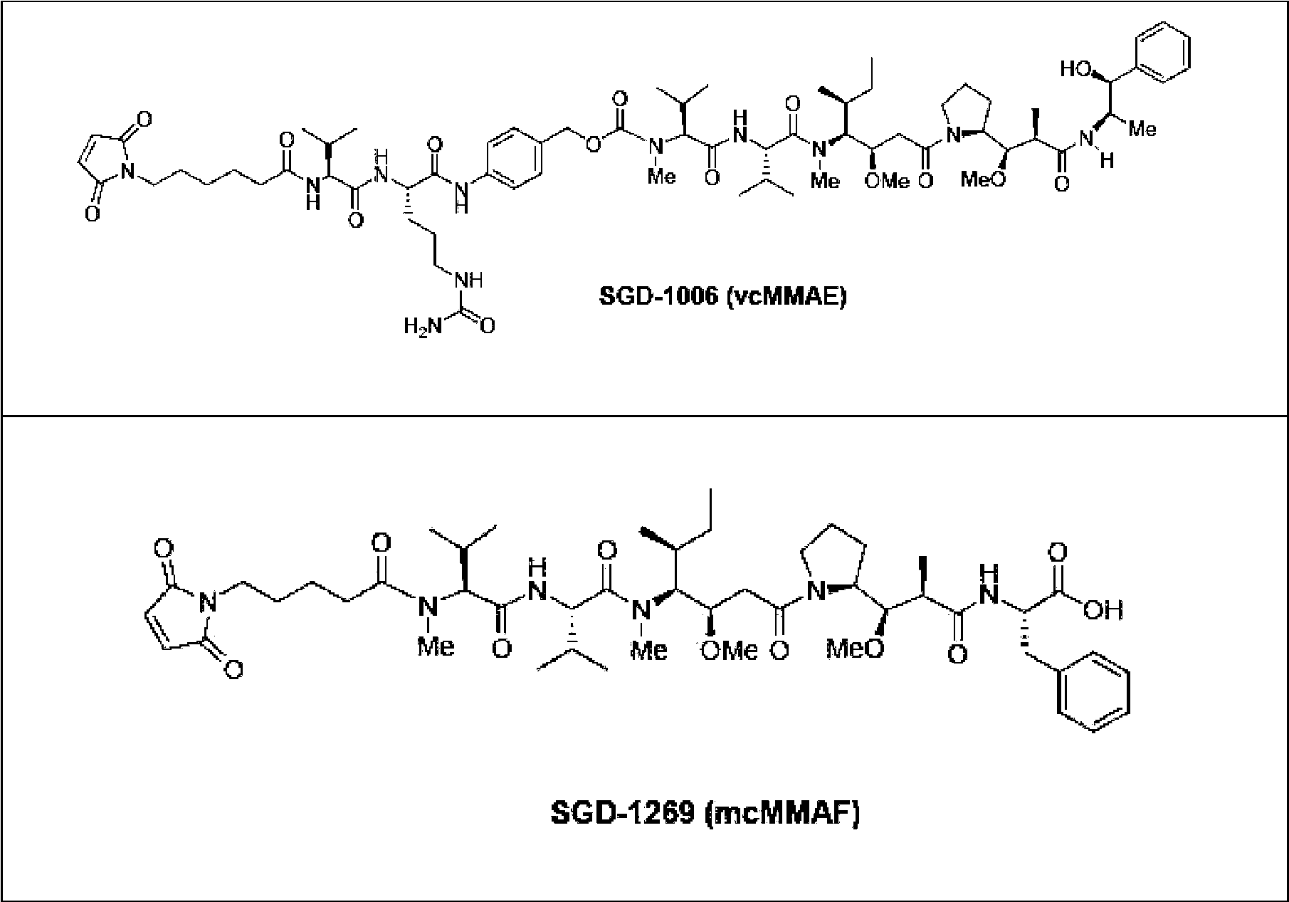
Se añadió suspensión de resina de Gammabind más Proteína G (GE Healthcare) (75 μl) a cada pocillo de una placa de filtrado de pocillo profundo (capacidad de 2 ml). Los anticuerpos para conjugar se agruparon por especie e isotipo y se transfirieron hasta 0,5 mg de cada anticuerpo a cada pocillo de la placa. Cada anticuerpo se transfirió a dos pocillos separados para facilitar la preparación de dos conjugados, con los engarces farmacológicos SGD-1006 y SGD-1269. La placa de filtrado se agitó después a 1200 RPM durante 2 horas a 5 °C para unir los anticuerpos de la resina. La placa de filtrado se centrifugó después a 500 x g durante 3 minutos para asegurar la sedimentación de todos los fluidos y resina al fondo de cada pocillo.Gammabind resin suspension plus Protein G (GE Healthcare) (75 μl) was added to each well of a deep-well filter plate (2 ml capacity). Antibodies to be conjugated were pooled by species and isotype, and up to 0.5 mg of each antibody was transferred to each well of the plate. Each antibody was transferred to two separate wells to facilitate the preparation of two conjugates, with the drug linkers SGD-1006 and SGD-1269. The filter plate was then shaken at 1200 RPM for 2 hours at 5 °C to bind the antibodies to the resin. The filter plate was then centrifuged at 500 x g for 3 minutes to ensure sedimentation of all fluids and resin to the bottom of each well.
Los anticuerpos unidos se redujeron después añadiendo 500 gl de TCEP 10 mM en KPO4100 mM, NaCl 150 mM, pH 7, EDTA 1mM y agitando durante 30 minutos a 22 °C. Después de la reducción, la placa se centrifugó de nuevo para retirar la solución de TCEP y posteriormente se lavó con PBS EDTA 1mM, 1 ml por pocillo. La solución de lavado se retiró por centrifugación y el procedimiento se repitió 3 veces para un total de 4 lavados. Los anticuerpos unidos o reducidos se conjugaron después usando una mezcla de n Em y engarce farmacológico preparado de acuerdo con las fracciones molares indicadas en la Tabla 2.The bound antibodies were then reduced by adding 500 μl of 10 mM TCEP in 100 mM KPO 4 , 150 mM NaCl, pH 7, 1 mM EDTA and shaking for 30 minutes at 22 °C. After reduction, the plate was centrifuged again to remove the TCEP solution and subsequently washed with 1mM PBS EDTA, 1 ml per well. The wash solution was removed by centrifugation and the procedure was repeated 3 times for a total of 4 washes. The bound or reduced antibodies were then conjugated using a mixture of n Em and pharmacological linker prepared according to the mole fractions indicated in Table 2.
Tabla 2.Table 2.

* también para quiméricos de IgG 1 murino / humano* also for murine/human IgG 1 chimerics
Se prepararon de este modo mezclas separadas de NEM y engarce farmacológico para cada especie / isotipo de anticuerpo usando soluciones madre de DMSO 10 mM de sGd -1006, SGD-1269 (Véase Tabla B) y NEM. Cuando se mezcla a la relación apropiada la concentración de maleimida total fue por lo tanto aún 10 mM, y este valor se usó para calcular el volumen de solución de maleimida para añadir a cada pocillo. Por ejemplo para una IgG 1 murina con 5 disulfuros reducibles (10 tioles disponibles cuando se reduce) 0,5 mg de anticuerpo a 150 kDa es 3,33 nmoles correspondientes a 33,3 nmoles de tiol. Un exceso de 3 veces es por lo tanto 100 nmoles de maleimida total o 10 gl de la mezcla de NEM /engarce farmacológico 10 mM. Para el conjugado SGD-1269 esta mezcla se prepararía después con 5,86 gl de SGD-1269 y 4,14 gl de NEM. La mezcla de maleimida se diluiría después en 500 gl de PBS antes de adición al anticuerpo reducido inmovilizado. En la práctica, puesto que se conjugaron múltiples anticuerpos de cada isotipo de forma simultánea se preparó una solución mixta sencilla de SGD-1269 / NEM para cada isotipo multiplicando el número de pocillos que contenían el isotipo por 10 gl por pocillo, diluyendo después en un volumen de PBS igual a 500 gl multiplicado por el número de pocillos. De forma similar se preparó un total de ocho mezclas de engarce farmacológico / NEM, cuatro con SGD-1006 y cuatro con SGD-1269, y se diluyeron en PBS. Estas mezclas se añadieron después a los anticuerpos reducidos (500 gl por pocillo) y la placa se agitó durante 30 minutos a 22 °C. La placa se centrifugó después como anteriormente para retirar la solución de reacción en exceso y posteriormente se lavó 4 veces con PBS como anteriormente.Separate mixtures of NEM and drug linker were thus prepared for each antibody species/isotype using 10 mM DMSO stock solutions of sGd-1006, SGD-1269 (See Table B) and NEM. When mixed at the appropriate ratio the total maleimide concentration was therefore still 10 mM, and this value was used to calculate the volume of maleimide solution to add to each well. For example for a murine IgG 1 with 5 reducible disulfides (10 thiols available when reduced) 0.5 mg of antibody at 150 kDa is 3.33 nmol corresponding to 33.3 nmol of thiol. A 3-fold excess is therefore 100 nmoles of total maleimide or 10 gl of the 10 mM NEM/drug linker mixture. For the SGD-1269 conjugate this mixture would then be prepared with 5.86 gl of SGD-1269 and 4.14 gl of NEM. The maleimide mixture would then be diluted in 500 μl of PBS before addition to the immobilized reduced antibody. In practice, since multiple antibodies of each isotype were conjugated simultaneously, a simple mixed solution of SGD-1269/NEM was prepared for each isotype by multiplying the number of wells containing the isotype by 10 gl per well, then diluting to a PBS volume equal to 500 gl multiplied by the number of wells. A total of eight NEM/pharmacological linker mixtures, four with SGD-1006 and four with SGD-1269, were similarly prepared and diluted in PBS. These mixtures were then added to the reduced antibodies (500 μl per well) and the plate was shaken for 30 minutes at 22°C. The plate was then centrifuged as above to remove excess reaction solution and subsequently washed 4 times with PBS as above.
Los ADC unidos se eluyeron después añadiendo 200 gl de glicina 50 mM pH 2,5 a cada pocillo y agitando la placa durante 3 minutos a 1200 RPM. En agitación se añadieron 20 gl de tampón de neutralización (fosfato potásico 1 M, pH 7,4, NaCl 500 mM, Tween-200,2 %) a cada pocillo de una placa de recogida de 1 ml. Los ADC se eluyeron después a la placa de recogida centrifugando a 1500 x g durante 6 minutos. La placa de recogida se agitó después brevemente para asegurar la mezcla completa del tampón de neutralización.The bound ADCs were then eluted by adding 200 μl of 50 mM glycine pH 2.5 to each well and shaking the plate for 3 minutes at 1200 RPM. With stirring, 20 gl of neutralization buffer (1 M potassium phosphate, pH 7.4, 500 mM NaCl, Tween-200.2%) was added to each well of a 1 ml collection plate. The ADCs were then eluted to the collection plate by centrifuging at 1500 x g for 6 minutes. The collection plate was then shaken briefly to ensure complete mixing of the neutralization buffer.
La concentración de cada ADC se determinó después con un lector de placa de absorbancia transfiriendo las soluciones a una placa de ensayo de UV (Costar modelo 3635, Corning) y midiendo la densidad óptica a 280 nm. Se usó un coeficiente de extinción de IgG medio de 1,45 ml mg-1 cm-1 para proporcionar una estimación adecuada de la concentración de ADC por todo el panel. Para confirmar la conjugación exitosa, se usó un procedimiento de HPLC de proteína de fase inversa (descrito posteriormente) para estimar la carga farmacológica de los controles de isotipo. Para la placa que contenía las variantes de humanización de CA8 se usó este procedimiento para estimar la carga de todos los ADC directamente.The concentration of each ADC was then determined with an absorbance plate reader by transferring the solutions to a UV assay plate (Costar model 3635, Corning) and measuring the optical density at 280 nm. A mean IgG extinction coefficient of 1.45 ml mg-1 cm-1 was used to provide an adequate estimate of the ADC concentration across the panel. To confirm successful conjugation, a reverse-phase protein HPLC procedure (described later) was used to estimate the drug load of isotype controls. For the plate containing the CA8 humanization variants, this procedure was used to estimate the loading of all ADCs directly.
El procedimiento de cromatografía de proteína de fase inversa para determinar carga farmacológica emplea la fase estacionaria polimérica PLRP-S (Agilent Technologies). Puesto que los anticuerpos se redujeron completamente durante el procedimiento de conjugación todas las subunidades de anticuerpo eluyen de la columna como cadenas polipeptídicas sencillas permitiendo que se evalúen de forma separada las subpoblaciones de especies de cadena ligera y pesada con diversos niveles de carga farmacológica. Por lo tanto, el análisis de estos datos permite el cálculo de la carga farmacológica de cadena ligera media y la carga farmacológica de cadena pesada media como factores independientes que pueden después combinarse para determinar la carga farmacológica de anticuerpo media con el conocimiento básico de que cada anticuerpo está comprendido por dos cadenas pesadas y dos ligeras. Las condiciones cromatográficas fueron como sigue: Una columna PRLP-S, 1000 Á, 50 x 2,1 mm, tamaño de partícula 8 gm (Agilent Technologies) con agua TFA 0,05 % como fase móvil A y acetonitrilo TFA 0,01 % como fase móvil B; elución con un gradiente lineal de B 27 % a B 42 % en 12,5 minutos.The reversed-phase protein chromatography procedure for determining drug loading employs the PLRP-S polymeric stationary phase (Agilent Technologies). Since the antibodies were completely reduced during the conjugation procedure all antibody subunits elute from the column as single polypeptide chains allowing subpopulations of light and heavy chain species with various levels of drug loading to be evaluated separately. Therefore, analysis of these data allows the calculation of the mean light chain drug load and the mean heavy chain drug load as independent factors that can then be combined to determine the mean antibody drug load with the basic knowledge that each Antibody is comprised of two heavy and two light chains. The chromatographic conditions were as follows: A PRLP-S column, 1000 Á, 50 x 2.1 mm, particle size 8 gm (Agilent Technologies) with 0.05% TFA water as mobile phase A and 0.01% TFA acetonitrile as mobile phase B; elution with a linear gradient from 27% B to 42% B in 12.5 minutes.
Los anticuerpos anti-BCMA se conjugaron con SGD-1006 y SGD-1269 en tres lotes separados durante un periodo de siete meses. En el primer lote se conjugó un total de 29 anticuerpos (dando como resultado 58 ADC). La carga farmacológica de cada control de isotipo determinada por cromatografía de PLRP y los datos se resumen en la Tabla 3.Anti-BCMA antibodies were conjugated to SGD-1006 and SGD-1269 in three separate batches over a period of seven months. A total of 29 antibodies were conjugated in the first batch (resulting in 58 ADCs). The drug load of each isotype control determined by PLRP chromatography and data are summarized in Table 3.
Tabla 3.Table 3.

Para el segundo lote se conjugaron 25 anticuerpos adicionales (dando como resultado 50 ADC). La carga farmacológica de cada control de isotipo se determinó de nuevo por cromatografía de PLRP y los datos se resumen en la Tabla 4.For the second batch, 25 additional antibodies were conjugated (resulting in 50 ADCs). The drug load of each isotype control was determined again by PLRP chromatography and the data are summarized in Table 4.
Tabla 4.Table 4.

En el tercer lote se conjugaron 30 anticuerpos (dando como resultado 60 ADC), incluyendo 13 variantes humanizadas de CA8. En este lote final, la carga farmacológica de todos los ADC se determinó y se resumen en los siguientes mapas de dos placas (Tabla 5 y 6).In the third batch, 30 antibodies were conjugated (resulting in 60 ADCs), including 13 humanized variants of CA8. In this final batch, the drug loading of all ADCs was determined and is summarized in the following two-plate maps (Table 5 and 6).
Tabla 5.Table 5.

Tabla 6. Table 6.

Se indica la carga farmacológica media y el % de CV para cada serie de isotipo en la parte de abajo. Se observó una variabilidad atípicamente grande de la carga farmacológica para los ADC SGD-1269 preparados con anticuerpos mIgG2b; la razón para esto no está clara. Además, los anticuerpos CA8 potenciados con Fc produjeron niveles de carga farmacológica algo inferiores a los de las otras variantes humanas de CA8; para abordar esto, se conjugó CA8 con Fc potenciado adicional en una reacción de fase de solución para alcanzar mejor la carga farmacológica conseguida para los otros anticuerpos.The mean drug load and % CV for each isotype series are indicated below. An atypically large variability of drug load was observed for SGD-1269 ADCs prepared with mIgG2b antibodies; the reason for this is not clear. Furthermore, Fc-enhanced CA8 antibodies produced somewhat lower drug loading levels than the other human CA8 variants; To address this, CA8 was conjugated to additional enhanced Fc in a solution phase reaction to better achieve the drug loading achieved for the other antibodies.
Ejemplo 4. Datos de uniónExample 4. Join data
4.1 Ensayo de unión de FMAT para mostrar unión de CA8 quimérico con células que expresan BCMA humano o de cynomolgus.4.1 FMAT binding assay to show binding of chimeric CA8 to cells expressing human or cynomolgus BCMA.
Se recuperaron células HEK293 humanas transfectadas crioconservadas, con BCMA de cynomolgus y con transfección simulada de almacenamiento en LN2. Se prepararon pocillos de ensayo con anticuerpo CA8 quimérico humano, a una serie de diferentes concentraciones, mezclado con células HEK293 con BCMA humano, HEK293 con BCMA de cynomolgus y con transfección simulada respectivamente. Se añadió conjugado secundario de Azul FMAT anti-IgG humana para detección de CA8 quimérico humano. Las placas de ensayo se dejaron durante un mínimo de 90 minutos antes de que el resultado se leyera en el lector de placas ABI8200 (FMAT).Cryopreserved, cynomolgus BCMA, and mock-transfected LN 2 storage transfected human HEK 2 93 cells were recovered. Test wells were prepared with human chimeric CA8 antibody, at a series of different concentrations, mixed with HEK 2 93 with human BCMA, HEK 2 93 with cynomolgus BCMA and mock transfected cells respectively. Secondary anti-human IgG FMAT Blue conjugate was added for detection of human chimeric CA8. The assay plates were left for a minimum of 90 minutes before the result was read on the ABI8200 plate reader (FMAT).
Esto mostró que el anticuerpo CA8 en forma quimérica se une bien a proteínas BCMA tanto humanas como de cynomolgus expresadas en células HEK293.This showed that the CA8 antibody in chimeric form binds well to both human and cynomolgus BCMA proteins expressed in HEK 2 93 cells.
Los resultados se muestran en la Figura 1.The results are shown in Figure 1.
4.2 Experimento de ELISA que muestra unión de CA8 quimérico con proteína BCMA recombinante 4.2 ELISA experiment showing binding of chimeric CA8 to recombinant BCMA protein
Se ensayaron anticuerpos CA8 quiméricos con respecto a unión con BCMA humano y BCMA de cynomolgus expresado como fusiones de Fc. Se revistieron placas de ELISA con Fc-BCMA humano y Fc-BCMA de cynomolgus y las placas se bloquearon usando BSA para reducir la unión no específica. Se añadieron anticuerpos quiméricos CA8 en un intervalo de concentración de 5 μg/ml a 0,1 μg/ml a las placas de ELISA revestidas con BCMA humano y de cynomolgus. Cualquier anticuerpo CA8 quimérico humano unido se detectó usando anticuerpo secundario conjugado con HRP anti-IgG humana según fuera apropiado. Se añadió sustrato de HRP (TMB) para desarrollar el ELISA. Este mostró que el anticuerpo CA8 se une a BCMA de cynomolgus y humano recombinante en un ensayo de ELISA. Chimeric CA8 antibodies were tested for binding to human BCMA and cynomolgus BCMA expressed as Fc fusions. ELISA plates were coated with human Fc-BCMA and cynomolgus Fc-BCMA and the plates were blocked using BSA to reduce non-specific binding. CA8 chimeric antibodies in a concentration range of 5 μg/ml to 0.1 μg/ml were added to the human and cynomolgus BCMA-coated ELISA plates. Any bound human chimeric CA8 antibody was detected using anti-human IgG HRP-conjugated secondary antibody as appropriate. HRP substrate (TMB) was added to develop the ELISA. This showed that the CA8 antibody binds to recombinant human and cynomolgus BCMA in an ELISA assay.
Los resultados se muestran en la Figura 2.The results are shown in Figure 2.
4.3 Experimento de Biacore para mostrar la unión de anticuerpo CA8 con proteínas BCMA y TACI para determinar la reactividad cruzada con proteína TACI.4.3 Biacore experiment to show the binding of CA8 antibody with BCMA and TACI proteins to determine cross-reactivity with TACI protein.
Se inyectó anticuerpo quimérico CA8 y se capturó en proteína A (se usó una microplaca sensora derivatizada con proteína A). Se bloqueó la unión de proteína A residual con una inyección de una alta concentración de solución de IgG humana. Se ensayaron después soluciones de BCMA-Fc, TACI-Fc o BAFF-R-Fc con respecto a unión con el anticuerpo. Las tres proteínas se inyectaron en secuencia y se midieron los acontecimientos de unión. La superficie se regeneró entre las inyecciones de cada proteína.Chimeric CA8 antibody was injected and captured on protein A (a protein A derivatized sensor microplate was used). Binding of residual protein A was blocked with an injection of a high concentration of human IgG solution. Solutions of BCMA-Fc, TACI-Fc or BAFF-R-Fc were then tested for binding to the antibody. The three proteins were injected in sequence and binding events were measured. The surface was regenerated between injections of each protein.
Se analizaron sensogramas en el programa Biaevaluation. Se realizó resta de la referencia doble para retirar el ruido del instrumento y cualquier unión no específica de las curvas del sensograma.Sensorograms were analyzed in the Biaevaluation program. Double baseline subtraction was performed to remove instrument noise and any non-specific binding from the sensorogram curves.
Esto mostró que CA8 era específico con respecto a unión para unión con BCMA y no con TACI y BAFFR.This showed that CA8 was specific with respect to binding to BCMA and not to TACI and BAFFR.
La unión del anticuerpo CA8 con BCMA-Fc, TACI-Fc y BAFF-R-Fc se representó como se muestra en la Figura 3.The binding of CA8 antibody to BCMA-Fc, TACI-Fc, and BAFF-R-Fc was depicted as shown in Figure 3.
4.4 Unión de células y datos de neutralización4.4 Cell binding and neutralization data
4.4.1 Unión de anticuerpos murinos anti BCMA con células de mieloma múltiple y células que expresan BCMA. 4.4.1 Binding of murine anti-BCMA antibodies with multiple myeloma cells and cells expressing BCMA.
Se tiñeron células de la línea celular de mieloma múltiple H929 y transfectantes que expresaban ARH77-hBCMA 10B5 BCMA con S332211 D07, S3332121F02 o S332126E04 murino o con el control de isotipo murino a 5 μg/ml. Se tiñó la línea celular de mieloma múltiple H929 con S307118G03 murino. Las células se incubaron durante 20 minutos a temperatura ambiente (TA) y después se lavaron con tampón FACS (PBS BSA 0,5 % azida sódica 0,1 %) para retirar anticuerpo no unido. Las células se incubaron con un anticuerpo anti-IgG de ratón marcado con PE secundario durante 15 minutos a TA y después se lavaron con tampón FACS para retirar anticuerpo no unido. Las células se analizaron por FACS para detectar anticuerpo unido a las células.Cells from the H929 multiple myeloma cell line and transfectants expressing ARH77-hBCMA 10B5 BCMA were stained with murine S332211 D07, S3332121F02, or S332126E04 or with the murine isotype control at 5 μg/ml. The H929 multiple myeloma cell line was stained with murine S307118G03. Cells were incubated for 20 minutes at room temperature (RT) and then washed with FACS buffer (PBS 0.5% BSA 0.1% sodium azide) to remove unbound antibody. Cells were incubated with a secondary PE-labeled anti-mouse IgG antibody for 15 minutes at RT and then washed with FACS buffer to remove unbound antibody. Cells were analyzed by FACS to detect cell-bound antibody.
Los resultados (Figura 4) mostraron que los 4 anticuerpos murinos se unieron a la línea celular de mieloma múltiple H929 y los tres anticuerpos ensayados en células transfectadas con BCMA ARH77 se unieron a estos.The results (Figure 4) showed that the 4 murine antibodies bound to the H929 multiple myeloma cell line and the three antibodies tested on cells transfected with BCMA ARH77 bound to them.
4.4.2 Curva de unión de CA8 quimérico con células de mieloma múltiple como se determinó por FACS4.4.2 Binding curve of chimeric CA8 with multiple myeloma cells as determined by FACS
Se usó un panel de líneas celulares de mieloma múltiple para determinar la unión de CA8 quimérico. Las líneas celulares H929, OPM-2, JJN-3 y U266 se tiñeron con CA8 quimérico o anticuerpo irrelevante (Synagis) a diversas concentraciones durante 20 minutos a TA. Las células se lavaron después con tampón FACS (PBS BSA 0,5 % azida sódica al 0,1 %) para retirar anticuerpo no unido. Las células se incubaron con un anticuerpo anti-IgG humano marcado con PE secundario durante 15 minutos a TA y después se lavaron con tampón FACS para retirar el anticuerpo no unido. Las células se analizaron por FACS y se midieron los valores de intensidad de fluorescencia media (IFM) para determinar la unión.A panel of multiple myeloma cell lines was used to determine binding of chimeric CA8. H929, OPM-2, JJN-3, and U266 cell lines were stained with chimeric CA8 or irrelevant antibody (Synagis) at various concentrations for 20 min at RT. The cells were then washed with FACS buffer (PBS 0.5% BSA 0.1% sodium azide) to remove unbound antibody. Cells were incubated with a secondary PE-labeled anti-human IgG antibody for 15 minutes at RT and then washed with FACS buffer to remove unbound antibody. Cells were analyzed by FACS and mean fluorescence intensity (MFI) values were measured to determine binding.
Los resultados mostraron que CA8 quimérico se unió a líneas celulares de mieloma múltiple H929, OPM-2, JJN-3 y U266 de una manera dependiente de dosis (Figura 5).The results showed that chimeric CA8 bound to multiple myeloma cell lines H929, OPM-2, JJN-3 and U266 in a dose-dependent manner (Figure 5).
4.4.3 Unión de CA8 humanizado con células transfectadas con BCMA según se determinó por FACS4.4.3 Binding of humanized CA8 to BCMA-transfected cells as determined by FACS
Se tiñeron células transfectantes que expresaban BCMA ARH77-hBCMA 10B5 o células H929 con CA8 quimérico o variantes humanizadas de CA8 designadas J6M0, J6M1, J6M2, J9M0, J9M1, J9M2 a diversas concentraciones durante 20 minutos a TA. Las células se lavaron después con tampón FACS (PBS BSA 0,5 % azida sódica 0,1 %) para retirar el anticuerpo no unido. Las células se incubaron con un anticuerpo anti-IgG humano marcado con PE secundario durante 15 minutos a TA y después se lavaron con tampón de FACS para retirar anticuerpo no unido. Las células se analizaron por FACS y se midieron los valores de intensidad de fluorescencia media (IFM) para determinar la unión. Transfectant cells expressing BCMA ARH77-hBCMA 10B5 or H929 cells were stained with chimeric CA8 or humanized variants of CA8 designated J6M0, J6M1, J6M2, J9M0, J9M1, J9M2 at various concentrations for 20 minutes at RT. The cells were then washed with FACS buffer (PBS 0.5% BSA 0.1% sodium azide) to remove unbound antibody. Cells were incubated with a secondary PE-labeled anti-human IgG antibody for 15 minutes at RT and then washed with FACS buffer to remove unbound antibody. Cells were analyzed by FACS and mean fluorescence intensity (MFI) values were measured to determine binding.
Los resultados mostraron que CA8 quimérico y todos los anticuerpos ensayados aparte de J9M2 se unieron a células transfectantes que expresaban BCMa ARH77-hBCMA 10B5 y células H929 de una manera dependiente de dosis (Figura 6).The results showed that chimeric CA8 and all antibodies tested other than J9M2 bound to transfectant cells expressing BCM to ARH77-hBCMA 10B5 and H929 cells in a dose-dependent manner (Figure 6).
4.5 Demostración de la capacidad de CA8 y la versión humanizada J6M0 para neutralizar la unión de BAFF o APRIL con BCMA recombinante.4.5 Demonstration of the ability of CA8 and the humanized version J6M0 to neutralize the binding of BAFF or APRIL with recombinant BCMA.
El objetivo de este ensayo fue evaluar la capacidad del anticuerpo CA8, y su versión humanizada J6M0, tanto en forma de tipo silvestre como afucosilada (Potelligent), a diversas concentraciones para neutralizar la capacidad de unión de uno de los ligandos de BCMA, Ba Ff o APRIL.The objective of this assay was to evaluate the ability of the CA8 antibody, and its humanized version J6M0, both in wild type and afucosylated form (Potelligent), at various concentrations to neutralize the binding capacity of one of the BCMA ligands, B a F f o APRIL.
Se revistieron durante una noche placas de fondo plano de 96 pocillos con solución de 1 μg/ml de BCMA humana recombinante Fc 4-53 en PBS. Después de una etapa de lavado usando TWEEN200,05 %, las placas se bloquearon con solución de Albúmina de Suero Bovino 2 % en PBS durante 1 hora a temperatura ambiente. Las placas se lavaron como anteriormente y se añadieron 40 pl de cada anticuerpo (IgG murino, c A8 murino y CA8 quimérico) comenzando a 10 μg/ml, valorado a 1 en 2 por duplicado a los pocillos relevantes y se incubó durante 1 hora a temperatura ambiente. Se añadieron 40 pl de BSA 2 % a los pocillos de control relevantes. Se añadieron 10 pl de BAFF humano recombinante (2149-BF/CF, R&D Systems) o APRIL humano recombinante (5860-AP/CF, R&D Systems) a 30 ng/ml y 750 ng/ml, respectivamente, dando una concentración final de 6 ng/ml y 150 ng/ml respectivamente en cada pocillo. Se añadió un volumen equivalente de BSA 2 % a los pocillos de control relevantes. Se permitió que las placas se incubaran durante 2 horas a temperatura ambiente, después de lo cual se lavaron como anteriormente. Se añadió ligando antihumano biotinilado (BAFF BAF124 o APRIL BAF884, R&D Systems) a los pocillos relevantes a 50 ng/ml y se incubó durante 1 hora. Después de una etapa de lavado, se añadieron 50 pl de una dilución 1:4000 de Estreptavidina-HRP (Amersham RPN4401) a cada pocillo y se incubó durante 30 minutos a temperatura ambiente. El proceso de lavado se repitió de nuevo seguido de la adición de 100 pl de solución de sustrato de Tetrametilbencidina (T8665, Sigma) a cada pocillo. Las placas se incubaron durante 20-25 minutos a temperatura ambiente, envueltas en papel de aluminio. La reacción se detuvo con la adición de 100 pl de H2SO4 1 M. Se determinó la densidad óptica a 450 nm usando lector Spectromax. Véase Figura 7A y B.96-well flat-bottom plates were coated overnight with 1 μg/ml solution of recombinant human BCMA Fc 4-53 in PBS. After a washing step using TWEEN200.05%, the plates were blocked with 2% Bovine Serum Albumin solution in PBS for 1 hour at room temperature. The plates were washed as above and 40 μl of each antibody (murine IgG, murine cA8 and chimeric CA8) starting at 10 μg/ml, titrated 1 in 2 in duplicate, was added to the relevant wells and incubated for 1 hour at room temperature. 40 μl of 2% BSA was added to the relevant control wells. 10 μl of recombinant human BAFF (2149-BF/CF, R&D Systems) or recombinant human APRIL (5860-AP/CF, R&D Systems) was added at 30 ng/ml and 750 ng/ml, respectively, giving a final concentration of 6 ng/ml and 150 ng/ml respectively in each well. An equivalent volume of 2% BSA was added to the relevant control wells. The plates were allowed to incubate for 2 hours at room temperature, after which they were washed as above. Biotinylated anti-human ligand (BAFF BAF124 or APRIL BAF884, R&D Systems) was added to the relevant wells at 50 ng/ml and incubated for 1 hour. After a washing step, 50 μl of a 1:4000 dilution of Streptavidin-HRP (Amersham RPN4401) was added to each well and incubated for 30 minutes at room temperature. The washing process was repeated again followed by the addition of 100 μl of Tetramethylbenzidine substrate solution (T8665, Sigma) to each well. The plates were incubated for 20-25 minutes at room temperature, wrapped in aluminum foil. The reaction was stopped with the addition of 100 μl of 1 M H 2 SO 4. The optical density at 450 nm was determined using a Spectromax reader. See Figure 7A and B.
En un ensayo basado en placas para neutralización de unión de BAFF o APRIL con BCMA, los valores de CE50 calculados para CA8 quimérico fueron 0,695 μg/ml y 0,773 μg/ml respectivamente. Los valores para el J6M0 humanizado fueron 0,776 ng/ml y 0,630 ng/ml. Los valores para la versión de J6M0 potelligent fueron 0,748 y 0,616 ng/ml respectivamente.In a plate-based assay for neutralization of BAFF or APRIL binding with BCMA, the calculated EC50 values for chimeric CA8 were 0.695 μg/ml and 0.773 μg/ml respectively. The values for humanized J6M0 were 0.776 ng/ml and 0.630 ng/ml. The values for the J6M0 potelligent version were 0.748 and 0.616 ng/ml respectively.
4.6 Efecto de CA8 quimerizado y anticuerpo de BCMA J6M0 humanizado en fosforilación inducida por BAFF o APRIL de NFk B en células H929.4.6 Effect of chimerized CA8 and humanized BCMA J6M0 antibody on BAFF- or APRIL-induced phosphorylation of NF k B in H929 cells.
En un conjunto de experimentos, se sembraron células H-929 a 75.000 células/pocillo en una placa de 96 pocillos en medio sin suero. El anticuerpo CA8 quimérico se añadió 24 horas después para proporcionar concentraciones finales en los pocillos de hasta 200 μg/ml. Diez minutos después, se añadió ligando BAFF o APRIL a las células para proporcionar concentraciones finales en los pocillos de 0,6 ó 0,3 μg/ml respectivamente. Después de 30 minutos las células se lisaron y se midieron los niveles de NfkappaB fosforilado usando un ensayo de pNFkappaB MSD.In one set of experiments, H-929 cells were seeded at 75,000 cells/well in a 96-well plate in serum-free medium. Chimeric CA8 antibody was added 24 hours later to provide final well concentrations of up to 200 μg/ml. Ten minutes later, BAFF or APRIL ligand was added to the cells to provide final well concentrations of 0.6 or 0.3 μg/ml, respectively. After 30 minutes the cells were lysed and the levels of phosphorylated NfkappaB were measured using a pNFkappaB MSD assay.
El anticuerpo de BCMA quimérico CA8 neutralizó la señalización de células NfkappaB inducida por BAFF y APRIL en células H-929. Fue particularmente potente en la neutralización de señalización de células NfkappaB inducida por BAFF en este tipo celular con una CI50 media de 10 nM, en comparación con 257 nM para señalización de células Nfkappap inducida por APRIL.Chimeric BCMA antibody CA8 neutralized NfkappaB cell signaling induced by BAFF and APRIL in H-929 cells. It was particularly potent in neutralizing BAFF-induced NfkappaB cell signaling in this cell type with a mean IC50 of 10 nM, compared to 257 nM for APRIL-induced NfkappaB cell signaling.
Datos promediados para 2 experimentosData averaged for 2 experiments
Las CI50 fueron 10 nM para neutralización de Nfkappap inducida por BAFF y 257 nM para neutralización de Nfkappap inducida por APRIL (media de 2 experimentos independientes) y se muestran en la Tabla 7.The IC50s were 10 nM for BAFF-induced Nfkappap neutralization and 257 nM for APRIL-induced Nfkappap neutralization (mean of 2 independent experiments) and are shown in Table 7.
Tabla 7Table 7

Se llevó a cabo un conjunto de experimentos adicional para dirigirse a entender por qué hubo una discrepancia tal entre la potencia de neutralización de APRIL y BAFF en el sistema basado en células. Después del descubrimiento de la forma soluble de BCMA el diseño experimental se cambió para incluir una etapa en la que las células H929 se lavaban antes del ensayo para reducir la interferencia de la unión del anticuerpo con BCMA soluble. Las células H-929 se lavaron 3 veces para retirar cualquier sBCMA y se resuspendieron en medio sin suero. Se añadió anticuerpo J6M0 potelligent a una placa de 96 pocillos para proporcionar concentraciones finales en los pocillos de hasta 100 μg/ml junto con ligando BAFF o APRIL para proporcionar una concentración final de los pocillos de 0,6 ó 0,2 μg/ml. Las células H-929 se sembraron después a 7,5 x 10⁴ células/pocillo en medio sin suero. 30 minutos después las células se lisaron y los niveles de NFkappaB fosforilado se midieron usando un ensayo de pNFkappaB MSD. Estos son datos de un experimento. Cada punto de datos es la medida/dt de dos repeticiones. Los datos de este experimentos se muestran en la Figura 7c. Las CI50 para inhibición de señalización por BAFF y APRIL se determinaron como 0,91 μg/ml y 2,43 μg/ml respectivamente.An additional set of experiments was conducted to address why there was such a discrepancy. between the neutralization potency of APRIL and BAFF in the cell-based system. After the discovery of the soluble form of BCMA the experimental design was changed to include a step in which H929 cells were washed before the assay to reduce interference from antibody binding with soluble BCMA. H-929 cells were washed 3 times to remove any sBCMA and resuspended in serum-free medium. Potelligent J6M0 antibody was added to a 96-well plate to provide final well concentrations of up to 100 μg/ml along with BAFF or APRIL ligand to provide a final well concentration of 0.6 or 0.2 μg/ml. H-929 cells were then seeded at 7.5 x 10⁴ cells/well in serum-free medium. 30 minutes later the cells were lysed and the levels of phosphorylated NFkappaB were measured using a pNFkappaB MSD assay. This is data from an experiment. Each data point is the measurement/dt of two repetitions. The data from this experiments are shown in Figure 7c. The IC50s for inhibition of signaling by BAFF and APRIL were determined as 0.91 μg/ml and 2.43 μg/ml respectively.
4.7 Análisis de ProteOn de construcciones humanizadas y quiméricas de CA8 anti-BCMA4.7 ProteOn Analysis of Humanized and Chimeric CA8 Anti-BCMA Constructs
La exploración inicial de variantes quiméricas y humanizadas de CA8 se llevó a cabo en el ProteOn XPR36 (Biorad). El procedimiento fue como sigue: se inmovilizó Proteína A en una microplaca de GLC (Biorad, Cat N°: 276-5011) por acoplamiento de amina primaria, se capturaron después variantes de CA8 en este superficie y se pasó BCMA humano recombinante (materiales internos o comerciales US Biological, B0410 (solamente ciclo 2)) a 256, 64, 16, 4, 1 nM con una inyección de 0 nM (es decir, solamente tampón) usada para doble referencia de las curvas de unión, el tampón usado es el tampón HBS-EP. Se usó NaOH 50 mM para regenerar la superficie de captura. Los datos se ajustaron al modelo 1:1 usando el software de análisis inherente al ProteOn XPR36. El ciclo 1 corresponde a la primera exploración de variantes de CA8 humanizadas (serie J0 a J5) y el ciclo 2, a la segunda exploración de variantes de CA8 humanizadas (serie J5 a J9). Ambos ciclos se llevaron a cabo a 25 °C.Initial screening for chimeric and humanized variants of CA8 was carried out on the ProteOn XPR36 (Biorad). The procedure was as follows: Protein A was immobilized on a GLC microplate (Biorad, Cat No.: 276-5011) by primary amine coupling, CA8 variants were then captured on this surface and recombinant human BCMA was passed (internal materials or commercial US Biological, B0410 (cycle 2 only)) at 256, 64, 16, 4, 1 nM with a 0 nM injection (i.e., buffer only) used for double referencing of the binding curves, the buffer used is the HBS-EP buffer. 50 mM NaOH was used to regenerate the capture surface. Data were fitted to the 1:1 model using the ProteOn XPR36 inherent analysis software. Cycle 1 corresponds to the first scan of humanized CA8 variants (series J0 to J5) and cycle 2, to the second scan of humanized CA8 variants (series J5 to J9). Both cycles were carried out at 25 °C.
Los datos obtenidos del ciclo 1 se exponen en la Tabla 8 y los datos del ciclo 2 se exponen en la Tabla 9. Varias moléculas en el Ciclo 2 (Tabla 09) no dieron valores de afinidad medibles por ProteOn, esto se debió a que la velocidad de disociación estaba más allá de la sensibilidad de la máquina en este ensayo. Esto no indica sin embargo, que todas estas moléculas se unan estrechamente a BCMA humano recombinante. Del Ciclo 1 los datos indican que algunas construcciones no mostraron ninguna unión con BCMA de cynomolgus recombinante.The data obtained from cycle 1 are presented in Table 8 and the data from cycle 2 are presented in Table 9. Several molecules in Cycle 2 (Table 9) did not give affinity values measurable by ProteOn, this was due to the fact that the Dissociation rate was beyond the sensitivity of the machine in this assay. This does not indicate, however, that all of these molecules bind closely to recombinant human BCMA. From Cycle 1 the data indicate that some constructs did not show any binding with recombinant cynomolgus BCMA.
Tabla 8: Análisis cinéticos de Ciclo 1 de moléculas anti-BCMA frente a BCMA Humano Recombinante Table 8: Cycle 1 kinetic analyzes of anti-BCMA molecules versus Recombinant Human BCMA


Tabla 9. Ciclo 2. Análisis cinéticos de moléculas anti-BCMA frente a BCMA Humano Recombinante Table 9. Cycle 2. Kinetic analyzes of anti-BCMA molecules versus Recombinant Human BCMA
Para los anticuerpos J8M0, J9M0, J8M1, J9M2, J7M2, J5M0, J7M1, J7M0, J8M2, J9M1, J5M2, J5M1 la velocidad de disociación estuvo más allá de la sensibilidad del ensayo, por lo tanto no se muestran datos.For antibodies J8M0, J9M0, J8M1, J9M2, J7M2, J5M0, J7M1, J7M0, J8M2, J9M1, J5M2, J5M1 the rate of dissociation was beyond the sensitivity of the assay, therefore data are not shown.

4.8. Análisis de BIAcore de construcciones humanizadas y quiméricas de CA8 anti-BCMA (serie J7 a J9)4.8. BIAcore analysis of humanized and chimeric CA8 anti-BCMA constructs (series J7 to J9)
Se inmovilizó proteína A en una microplaca CM5 (GE Healthcare, Cat Ns: BR-1005-30) por acoplamiento de amina primaria y esta superficie se usó después para capturar las moléculas de anticuerpo. Se usó BCMA humano recombinante (US Biological, B0410) como analito a 256 nM, 64 nM, 16 nM, 4 nM y 1 nM. La regeneración de la superficie de captura se llevó a cabo usando NaOH 50 mM. Se tomaron dobles referencias de todas las curvas de unión con una inyección de tampón (es decir, 0 nM) y los datos se ajustaron a los del uso del modelo 1:1 inherente al software de evaluación T100. El ciclo se llevó a cabo a 37 °C, usando HBS-EP como el tampón de ejecución.Protein A was immobilized on a CM5 microplate (GE Healthcare, Cat Ns: BR-1005-30) by primary amine coupling and this surface was then used to capture the antibody molecules. Recombinant human BCMA (US Biological, B0410) was used as analyte at 256 nM, 64 nM, 16 nM, 4 nM, and 1 nM. Regeneration of the capture surface was carried out using 50 mM NaOH. All binding curves were double referenced with one injection of buffer (i.e., 0 nM) and the data were fitted to those using the 1:1 model inherent to the T100 evaluation software. Cycling was carried out at 37 °C, using HBS-EP as the running buffer.
Los resultados mostraron que las moléculas ensayadas con la excepción de J9M2 se unen a BCMA humano recombinante, con afinidad similar a la molécula quimérica. Los datos generados de este experimento se presentan en la tabla 10.The results showed that the molecules tested with the exception of J9M2 bind to recombinant human BCMA, with similar affinity to the chimeric molecule. The data generated from this experiment is presented in table 10.
Tabla 10. Análisis cinético de moléculas humanizadas anti-BCMA frente a BCMA humano recombinante Table 10. Kinetic analysis of humanized anti-BCMA molecules versus recombinant human BCMA


4.9 Análisis de BIAcore de construcciones quiméricas y humanizadas de CA8 anti-BCMA J6M0 y J9M04.9 BIAcore analysis of chimeric and humanized CA8 anti-BCMA constructs J6M0 and J9M0
Se inmovilizó proteína A en una microplaca CM5 (GE Healthcare, Cat N°: BR-1005-30) por acoplamiento de amina primaria y esta superficie se usó después para capturar las moléculas de anticuerpo. Se usó BCMA humano recombinante (US Biological, B0410) como analito a 256 nM, 64 nM, 16 nM, 4 nM y 1 nM. Se llevó a cabo regeneración de la superficie de captura usando NaOH 50 mM. Se tomaron doble referencias de todas las curvas de unión con una inyección de tampón (es decir, 0 nM). Los datos se ajustaron a los del uso de modelo 1:1 inherente a software de evaluación T100. El ciclo se llevó a cabo a 25 °C y 37 °C para el experimento 1 y solamente 37 °C para el experimento 2 usando HBS-EP como el tampón de ejecución.Protein A was immobilized on a CM5 microplate (GE Healthcare, Cat No.: BR-1005-30) by primary amine coupling and this surface was then used to capture the antibody molecules. Recombinant human BCMA (US Biological, B0410) was used as analyte at 256 nM, 64 nM, 16 nM, 4 nM, and 1 nM. Regeneration of the capture surface was carried out using 50 mM NaOH. All binding curves were double referenced with one injection of buffer (i.e., 0 nM). The data were adjusted to those using the 1:1 model inherent to T100 evaluation software. Cycling was carried out at 25 °C and 37 °C for experiment 1 and only 37 °C for experiment 2 using HBS-EP as the running buffer.
Ambos ciclos identificaron J9M0 como la mejor molécula con respecto a la afinidad global por BCMA humano. Los datos generados de este experimento se presentan en la tabla 11.Both runs identified J9M0 as the best molecule with respect to overall affinity for human BCMA. The data generated from this experiment is presented in table 11.
Tabla 11 Análisis cinéticos de moléculas humanizadas anti-BCMA frente a BCMA humano Table 11 Kinetic analyzes of humanized anti-BCMA molecules versus human BCMA

4.10 Análisis de ProteOn de nuevas construcciones quiméricas anti-BCMA4.10 ProteOn Analysis of Novel Anti-BCMA Chimeric Constructs
La exploración inicial de las nuevas variantes quiméricas del segundo lote de hibridomas se llevó a cabo en el ProteOn XPR36 (Biorad). El procedimiento fue como sigue; la Proteína A se inmovilizó en una microplaca GLM (Biorad, Cat Ns: 176-5012) por acoplamiento de amina primaria, se capturaron después variantes anti-BCMA en esta superficie y se pasó BCMA humano recombinante (material interno) a 256, 64, 16, 4, 1 nM con una inyección 0 nM (es decir, tampón solamente) usada para realizar doble referencia de las curvas de unión, el tampón usado es el tampón HBS-EP. Se llevó a cabo regeneración de la superficie de captura usando NaOH 50 nM. Los datos se ajustaron al modelo 1:1 usando el software de análisis inherente al ProteOn XPR36. El ciclo se llevó a cabo a 25 °C.Initial screening of the new chimeric variants from the second batch of hybridomas was carried out on the ProteOn XPR36 (Biorad). The procedure was as follows; Protein A was immobilized on a GLM microplate (Biorad, Cat Nos: 176-5012) by primary amine coupling, anti-BCMA variants were then captured on this surface and recombinant human BCMA (internal material) was passed to 256, 64, 16, 4, 1 nM with a 0 nM injection (i.e., buffer only) used to double reference the binding curves, the buffer used is HBS-EP buffer. Regeneration of the capture surface was carried out using 50 nM NaOH. Data were fitted to the 1:1 model using the ProteOn XPR36 inherent analysis software. The cycle was carried out at 25 °C.
Se presentan datos generados de este experimento en la tabla 12.Data generated from this experiment is presented in table 12.
Tabla 12: Análisis cinéticos de moléculas humanizadas anti-BCMA frente a BCMA humano. Table 12: Kinetic analyzes of humanized anti-BCMA molecules against human BCMA.

Ejemplo 5 Ensayos de muerte celularExample 5 Cell death assays
5.1 Potencias de ADCC de CA8 quimérico y versión de CA8 quimérico desfucosilado en células ARH77 que expresan BCMA5.1 ADCC Potencies of Chimeric CA8 and Defucosylated Chimeric CA8 Version in ARH77 Cells Expressing BCMA
Se incubaron linfocitos citolíticos naturales (NK) humanos con células diana transfectadas con BCMA ARH77A marcadas con europio (10B5) en presencia de diversas concentraciones de anticuerpo a una relación E:T de 5:1 durante 2 horas. Se midió la liberación de europio de las células diana y se calculó la lisis específica.Human natural killer (NK) cells were incubated with europium (10B5)-labeled BCMA ARH77A-transfected target cells in the presence of various concentrations of antibody at an E:T ratio of 5:1 for 2 hours. Europium release from target cells was measured and specific lysis was calculated.
Resultado: CA8 quimérico y CA8 quimérico desfucosilado destruyeron células diana que expresaban BCMA mediante ADCC. El anticuerpo quimérico desfucosilado mostró actividad ADCC más potente, como se midió por un mayor porcentaje de lisis conseguido con todas las células diana ensayadas y una CE50 diez veces más baja en la línea celular diana que expresaba BCMA alto 10B5, en comparación con el anticuerpo quimérico precursor. Véase Figura 8A y 8B.Result: Chimeric CA8 and defucosylated chimeric CA8 killed BCMA-expressing target cells by ADCC. The defucosylated chimeric antibody showed more potent ADCC activity, as measured by a higher percentage of lysis achieved with all target cells tested and a ten-fold lower EC50 in the target cell line expressing high BCMA 10B5, compared to the antibody chimerical precursor. See Figure 8A and 8B.
5.2. Actividad ADCC de anticuerpos humanizados CA8 usando células diana que expresan BCMA ARH77 y PBMC como efectores5.2. ADCC activity of humanized CA8 antibodies using BCMA-expressing target cells ARH77 and PBMC as effectors
Se incubaron PBMC humanas con células diana transfectadas con BCMA ARH77 marcadas con europio (10B5) en presencia de diversas concentraciones de versiones humanizadas de anticuerpo CA8 (5 gg/ml a 0,005 gg/ml) a una relación E:T de 5:1 durante 2 horas. Se midió la liberación de europio de las células diana y se calculó la lisis específica. Resultado:Human PBMCs were incubated with europium (10B5)-labeled ARH77 BCMA-transfected target cells in the presence of various concentrations of humanized versions of CA8 antibody (5 gg/ml to 0.005 gg/ml) at an E:T ratio of 5:1 for 2 hours. Europium release from target cells was measured and specific lysis was calculated. Result:
Todas las series J5, J6, J7 J8 y J9 de variantes humanizadas de CA8 mostraron actividad ADCC contra la línea celular que expresaba BCMA con alto ARH7710B5 de una manera dependiente de dosis. La ADCC estuvo a un nivel similar al hallado en los experimentos usando molécula de CA8 quimérica. Véase Figura 9.All J5, J6, J7 J8 and J9 series of humanized CA8 variants showed ADCC activity against the ARH7710B5-high BCMA-expressing cell line in a dose-dependent manner. The ADCC was at a level similar to that found in experiments using chimeric CA8 molecule. See Figure 9.
5.3. Potencias de ADCC de S322110F02, S322110D07 y S307118G03 quiméricos y S307118G03 humanizado H3L0 frente a células ARH77 10B5 que expresan BCMA con linfocitos NK purificados como células efectoras5.3. ADCC potencies of chimeric S322110F02, S322110D07 and S307118G03 and humanized S307118G03 H3L0 against BCMA-expressing ARH77 10B5 cells with purified NK cells as effector cells
Se incubaron células diana citolíticas naturales (NK) humanas con células diana transfectadas con BCMA ARH77 marcadas con europio (10B5) en presencia de diversas concentraciones de anticuerpo a una relación de E:T de 5:1 durante 2 horas. Se midió la liberación de europio de las células diana y se calculó la lisis específica.Human natural killer (NK) target cells were incubated with europium (10B5)-labeled BCMA ARH77-transfected target cells in the presence of various concentrations of antibody at an E:T ratio of 5:1 for 2 hours. Europium release from target cells was measured and specific lysis was calculated.
Resultado: los 4 anticuerpos ensayados mostraron actividad ADCC frente a células ARH7710B5. Véase Figura 10.Result: the 4 antibodies tested showed ADCC activity against ARH7710B5 cells. See Figure 10.
5.4. Actividad de Conjugado Anticuerpo-Fármaco (ADC) de ADC de CA8 Quimérico5.4. Antibody-Drug Conjugate (ADC) Activity of Chimeric CA8 ADC
Medición de actividad ADC del anticuerpo CA8 quimérico, conjugados de fármaco con anticuerpo CA8 quiméricomcMMAF y conjugados de fármaco con anticuerpo CA8 quimérico-vcMMAE frente a líneas celulares de mieloma múltiple humano.Measurement of ADC activity of chimeric CA8 antibody, chimeric CA8 antibody drug conjugates mcMMAF and chimeric CA8 antibody drug conjugates - vcMMAE against human multiple myeloma cell lines.
Se trataron líneas celulares de mieloma múltiple con conjugados de fármaco-anticuerpo CA8 quimérico para determinar las concentraciones de ADC requeridas para inhibición de crecimiento y muerte. Multiple myeloma cell lines were treated with chimeric CA8 antibody-drug conjugates to determine the ADC concentrations required for growth inhibition and death.
Los conjugados de fármaco con anticuerpos ensayados se añadieron a pocilios que contenían células de mieloma múltiple a concentraciones que variaban de 1 gg/ml a 5 ng/ml. Las placas se incubaron a 37 °C durante 96 horas momento en el cual se cuantificaron las células viables usando Cell titre Glo. El anticuerpo CA8 quimérico no conjugado no mostró actividad inhibidora del crecimiento significativa a las concentraciones de anticuerpo que se ensayaron. El conjugado de anticuerpo-fármaco CA8 quimérico-mcMMAF mostró mayor actividad inhibidora del crecimiento que el conjugado de anticuerpo-fármaco CA8 quimérico-vcMMAE en las 4 de las líneas celulares de mieloma múltiple que se ensayaron. Véase Figura 11 y Tabla 13.The antibody-drug conjugates tested were added to wells containing multiple myeloma cells at concentrations ranging from 1 gg/ml to 5 ng/ml. The plates were incubated at 37°C for 96 hours at which time viable cells were quantified using Cell titre Glo. The unconjugated chimeric CA8 antibody did not show significant growth inhibitory activity at the antibody concentrations tested. The chimeric CA8-mcMMAF antibody-drug conjugate showed greater growth inhibitory activity than the chimeric CA8-vcMMAE antibody-drug conjugate in all 4 of the multiple myeloma cell lines tested. See Figure 11 and Table 13.
Tabla 13 Valores de CI50 representados en ng/ml para los conjugados de anticuerpo-fármaco CA8 quimérico-vcMMAE y CA8 quimérico-mcMMAF en 4 líneas celulares de mieloma múltiple. Table 13 IC 50 values represented in ng/ml for the antibody-drug conjugates chimeric CA8-vcMMAE and chimeric CA8-mcMMAF in 4 multiple myeloma cell lines.

5.5. Medición de la actividad de detención del ciclo celular de anticuerpo CA8 quimérico, conjugados de fármaco con anticuerpo CA8 quimérico-mcMMAF y conjugados de fármaco con anticuerpo CA8 quimérico-vcMMAE frente a línea celular de mieloma múltiple humano H929.5.5. Measurement of cell cycle arrest activity of chimeric CA8 antibody, chimeric CA8 antibody-mcMMAF drug conjugates, and chimeric CA8 antibody-vcMMAE drug conjugates against human multiple myeloma cell line H929.
Para determinar el mecanismo por el que los Conjugados de Fármaco con Anticuerpo CA8 quimérico (ADC) provocan inhibición de crecimiento en células de mieloma múltiple, el ciclo celular de células NCl-H929 se controló midiendo el contenido de ADN celular a través de tinción de yoduro de propidio de células fijadas en múltiples puntos temporales después de tratamiento con anticuerpo CA8 quimérico y ADC de CA8 quimérico.To determine the mechanism by which chimeric CA8 Antibody Drug Conjugates (ADC) cause growth inhibition in multiple myeloma cells, the cell cycle of NCl-H929 cells was monitored by measuring cellular DNA content through iodide staining. of propidium from fixed cells at multiple time points after treatment with chimeric CA8 antibody and chimeric CA8 ADC.
A la concentración de ADC de CA8 quimérico ensayada (50 ng/ml), el ADC CA8 quimérico-mcMMAF provocó detención del ciclo celular en G2/M significativa (contenido de ADN 4N) que alcanzó un pico a las 48 horas. En los puntos temporales posteriores de 48, 72 y 96 horas, el tratamiento con el ADC de CA8 quimérico-mcMMAF dio como resultado acumulación de una población celular con contenido de ADN por debajo de 2N, lo que es representativo de muerte celular. A la concentración ensayada de 50 ng/ml el ADC de CA8 quimérico-vcMMAE no tuvo efecto significativo en la detención del ciclo celular en G2/M o acumulación sub-G1. Véase Figura 12.At the chimeric CA8 ADC concentration tested (50 ng/ml), the chimeric CA8-mcMMAF ADC caused significant G2/M cell cycle arrest (4N DNA content) that peaked at 48 hours. At the later time points of 48, 72, and 96 hours, treatment with the chimeric CA8-mcMMAF ADC resulted in accumulation of a cell population with DNA content below 2N, which is representative of cell death. At the tested concentration of 50 ng/ml, the chimeric CA8-vcMMAE ADC had no significant effect on cell cycle arrest in G2/M or sub-G1 accumulation. See Figure 12.
5.6. Tinción con Fosfo-Histona-H3 (Thr11) como un marcador para conjugado de fármaco con anticuerpo CA8 quimérico-mcMMAF y conjugado de fármaco con anticuerpo CA8 quimérico-vcMMAE indujo detención mitótica. 5.6. Staining with Phospho-Histone-H3 (Thr11) as a marker for chimeric CA8 antibody-mcMMAF drug conjugate and chimeric CA8 antibody-vcMMAE drug conjugate induced mitotic arrest.
Para determinar si la acumulación de células con contenido de ADN 4N es un resultado específico de la detención mitótica inducida por ADC con CA8 quimérico se tiñeron células NCI-H929 con un anticuerpo anti-fosfo-Histona H3 después del tratamiento con concentraciones crecientes de CA8 quimérico no conjugado, CA8 quimérico-vcMMAE o CA8 quimérico-mcMMAF durante 48 horas.To determine whether the accumulation of cells containing 4N DNA is a specific result of ADC-induced mitotic arrest with chimeric CA8, we stained NCI-H929 cells with an anti-phospho-Histone H3 antibody after treatment with increasing concentrations of chimeric CA8. unconjugated, chimeric CA8-vcMMAE or chimeric CA8-mcMMAF for 48 hours.
El tratamiento con ADC de CA8 quimérico dio como resultado una acumulación dependiente de dosis de células NCI-H929 que se tiñeron positivas para peróxido-Histona H3 (Thr11), un marcador específico de células mitóticas. El ADC de CA8 quimérico-mcMMAF provocó la acumulación de células positivas para peróxido-Histona H3 a concentraciones más bajas que el ADC de CA8 quimérico-vcMMAE. Véase Figura 13.ADC treatment of chimeric CA8 resulted in a dose-dependent accumulation of NCI-H929 cells that stained positive for peroxide-Histone H3 (Thr11), a specific marker of mitotic cells. The chimeric CA8-mcMMAF ADC caused accumulation of peroxide-Histone H3-positive cells at lower concentrations than the chimeric CA8-vcMMAE ADC. See Figure 13.
5.7 Medición de apoptosis en células NCI-H929 en respuesta a ADC de CA8 quimérico por tinción con respecto a Anexina-V.5.7 Measurement of apoptosis in NCI-H929 cells in response to chimeric CA8 ADC by Annexin-V staining.
Para determinar si la acumulación de células con contenido de ADN sub-2N es un resultado específico de apoptosis inducida por los ADC de CA8 quimérico, se tiñeron células NCI-H929 con un anticuerpo anti-Anexina V después del tratamiento con concentraciones crecientes de CA8 quimérico no conjugado, CA8 quimérico-vcMMAE o CA8 quimérico-mcMMAF durante 48 horas. El tratamiento con ADC de CA8 quimérico dio como resultado una acumulación dependiente de dosis de células NCI-H929 que se tiñeron positivas con respecto a Anexina-V, un marcador específico de apoptosis. El ADC de CA8 quimérico-mcMMAF provocó la acumulación de células positivas para Anexina-V a concentraciones más bajas que el ADC de CA8 quimérico-vcMMAE. Véase Figura 14.To determine whether the accumulation of cells with sub-2N DNA content is a specific result of apoptosis induced by chimeric CA8 ADCs, NCI-H929 cells were stained with an anti-Annexin V antibody after treatment with increasing concentrations of chimeric CA8. unconjugated, chimeric CA8-vcMMAE or chimeric CA8-mcMMAF for 48 hours. Treatment with chimeric CA8 ADCs resulted in a dose-dependent accumulation of NCI-H929 cells that stained positive for Annexin-V, a specific marker of apoptosis. The chimeric CA8-mcMMAF ADC caused the accumulation of Annexin-V-positive cells at lower concentrations than the chimeric CA8-vcMMAE ADC. See Figure 14.
5.8 Actividad de Conjugado de Anticuerpo-Fármaco (ADC) de variantes humanizadas de conjugados de anticuerpo anti-BCMA CA8-fármaco5.8 Antibody-Drug Conjugate (ADC) Activity of Humanized Variants of Anti-BCMA CA8 Antibody-Drug Conjugates
Las células se sembraron en placas de 96 pocillos (4.000 células por pocillo en 100 gl de RPMI FBS 10 %). Cells were seeded in 96-well plates (4,000 cells per well in 100 gl of 10% RPMI FBS).
Se añadió anticuerpo desnudo o ADC 6 horas después de la siembra de las células y las placas se incubaron durante 144 horas. Se midió la inhibición del crecimiento en presencia de los anticuerpos o ADC a las 144 horas usando Cell Titre glo. Los puntos de datos representan la media de mediciones de CellTiterGlo por triplicado. Las barras de error representan el error típico.Naked antibody or ADC was added 6 hours after cell plating and plates were incubated for 144 hours. Growth inhibition in the presence of the antibodies or ADC was measured at 144 hours using Cell Titre glo. Data points represent the mean of triplicate CellTiterGlo measurements. Error bars represent standard error.
Se trataron líneas celulares de mieloma múltiple NCI-H929 y OPM2 con conjugados de anticuerpo anti-BCMA CA8 humanizado-fármaco para determinar las concentraciones de ADC requeridas para inhibición del crecimiento y muerte. Las formas conjugadas de anticuerpo-fármaco con mcMMAF y vcMMAE de estos anticuerpos mostraron actividad inhibidora del crecimiento significativa comparable con la hallada con la quimera de CA8. La variante J6M0 mostró mayor potencia que la quimera y los datos se muestran en la Figura 15 en células H929 y células OPM2. El conjugado de anticuerpo-fármaco con mcMMAF mostró mayor actividad inhibidora del crecimiento que el conjugado de anticuerpo-fármaco con vcMMAE para todos los anticuerpos en ambas líneas celulares ensayadas. Se muestran resultados para todas las variantes humanizadas en la Tabla 14.Multiple myeloma cell lines NCI-H929 and OPM2 were treated with humanized anti-BCMA CA8 antibody-drug conjugates to determine ADC concentrations required for growth inhibition and death. The mcMMAF and vcMMAE antibody-drug conjugated forms of these antibodies showed significant growth inhibitory activity comparable to that found with the CA8 chimera. The J6M0 variant showed higher potency than the chimera and the data are shown in Figure 15 in H929 cells and OPM2 cells. The antibody-drug conjugate with mcMMAF showed greater growth inhibitory activity than the antibody-drug conjugate with vcMMAE for all antibodies in both cell lines tested. Results for all humanized variants are shown in Table 14.
Tabla 14. Valores de CI50 representados en ng/ml para los conjugados de anticuerpo anti BCMA-fármaco en células NCI-H929 y U266-B1 Table 14. IC 50 values represented in ng/ml for anti-BCMA antibody-drug conjugates in NCI-H929 and U266-B1 cells

5.9 Actividad de conjugado anticuerpo-fármaco (ADC) de otros conjugados de fármaco-anticuerpo anti-BCMA murino 5.9 Antibody-drug conjugate (ADC) activity of other murine anti-BCMA antibody-drug conjugates
Se sembraron células en placas de 96 pocillos (4.000 células por pocillo en 100 μl de RPMI FBS 10 %).Cells were seeded in 96-well plates (4,000 cells per well in 100 μl of 10% RPMI FBS).
Se añadió anticuerpo o ADC 6 horas después de la siembra de las células y las placas se incubaron durante 144 horas. Se midió la inhibición del crecimiento en presencia de los ADC a las 144 horas usando CellTiterGlo. Se muestra la media de mediciones de CellTiterGlo por triplicado. Las Tablas 15a y 15b son de experimentos llevados a cabo a diferentes tiempos en diferentes series de anticuerpos. Se usaron las líneas celulares de mieloma múltiple NCI-H929 y U266-B1 para los anticuerpos en la Tabla 15a.Antibody or ADC was added 6 hours after cell plating and plates were incubated for 144 hours. Growth inhibition in the presence of the ADCs was measured at 144 hours using CellTiterGlo. The average of triplicate CellTiterGlo measurements is shown. Tables 15a and 15b are from experiments carried out at different times on different sets of antibodies. Multiple myeloma cell lines NCI-H929 and U266-B1 were used for the antibodies in Table 15a.
Las formas conjugadas de anticuerpo-fármaco con mcMMAF y vcMMAE de los anticuerpos murinos S322110D07, S332121F02 y S332136E04 mostraron actividad inhibidora del crecimiento significativa. El conjugado de anticuerpofármaco con mcMMAF mostró mayor actividad inhibidora del crecimiento que el conjugado de anticuerpo-fármaco con vcMMAE en todos los anticuerpos anti-BCMA murinos ensayados en los que se vio actividad. Se muestran las cifras de CI50 en la Tabla 15a. Véase la Figura 16 para curvas de respuesta a dosis para esos tres anticuerpos y también S107118G03. Las barras de error representan el error típico. Las células NCI-H929, U266-B1, JJN3 y o Pm 2 para anticuerpos en la Tabla 15b se trataron con una serie diferente de conjugados de fármaco-anticuerpo anti-BCMA murino para determinar las concentraciones de ADC requeridas para inhibición del crecimiento y muerte. Se muestran las cifras de CI50 en la Tabla 15b. Los 5 anticuerpos mostrados en la tabla tuvieron actividad ADC significativa. mcMMAF and vcMMAE antibody-drug conjugated forms of murine antibodies S322110D07, S332121F02, and S332136E04 showed significant growth inhibitory activity. The antibody-drug conjugate with mcMMAF showed greater growth inhibitory activity than the antibody-drug conjugate with vcMMAE in all murine anti-BCMA antibodies tested in which activity was seen. The figures are shown of IC50 in Table 15a. See Figure 16 for dose response curves for those three antibodies and also S107118G03. Error bars represent standard error. NCI-H929, U266-B1, JJN3 and o P m 2 cells for antibodies in Table 15b were treated with a different series of murine anti-BCMA antibody-drug conjugates to determine the ADC concentrations required for growth inhibition and death. IC50 figures are shown in Table 15b. All 5 antibodies shown in the table had significant ADC activity.
Tabla 15a. Valores de CI50 representados en ng/ml para los conjugados de fármaco-anticuerpo anti-BCMA en células NCl-H929 y U266-B1 Table 15a. IC 50 values represented in ng/ml for anti-BCMA antibody-drug conjugates in NCl-H929 and U266-B1 cells

gg
Tabla 15b Valores de CI50 representados en ng/ml para los conjugados de fármaco-anticuerpo anti BCMA en células NCI-H929, U266-B1, JJN3 y OPM2 Table 15b IC 50 values represented in ng/ml for anti-BCMA drug-antibody conjugates in NCI-H929, U266-B1, JJN3 and OPM2 cells

5.10 Potencia de ADCC de J6M0 afucosilado conjugado (Potelligent)5.10 ADCC Potency of Conjugated Afucosylated J6M0 (Potelligent)
Se ensayó J6M0 afucosilado conjugado con MMAE o MMAF en ensayos de ADCC usando transfectantes de BCMA para asegurar que su actividad ADCC no se viera comprometida por la conjugación. Se incubaron células ARH77-10B5 marcadas con europio con diversos anticuerpos de BMCA Potelligent y J6M0 WT a concentraciones de hasta 10000 ng/ml durante 30 minutos antes de la adición de PBMC (relación de PBMC: células diana 50:1). Dos horas después se tomó una muestra de una alícuota de medio celular y se mezcló con solución de potenciación. Después de 30 minutos en un agitador de placas, se controló la liberación de europio en el lector de multimarcador Victor 2 1420. Los puntos de datos representan medias de los valores por triplicado. Estos datos son representativos de los 2 experimentos.Afucosylated J6M0 conjugated to MMAE or MMAF was tested in ADCC assays using BCMA transfectants to ensure that its ADCC activity was not compromised by conjugation. Europium-labeled ARH77-10B5 cells were incubated with various Potelligent and J6M0 WT BMCA antibodies at concentrations up to 10,000 ng/ml for 30 min before the addition of PBMC (ratio of PBMC:target cells 50:1). Two hours later, an aliquot of cell medium was sampled and mixed with potentiation solution. After 30 minutes on a plate shaker, europium release was monitored on the Victor 2 1420 multi-label reader. Data points represent means of triplicate values. These data are representative of the 2 experiments.
No hubo diferencias significativas en la potencia de ADCC entre las formas no conjugada y ADC de J6M0 Potelligent. En el mismo experimento se incluyó una versión de tipo silvestre de J6M0 para mostrar la comparación de la potencia con la versión afucosilada. Como se esperaba, la desfucosilación dio como resultado una CE50 menor y una lisis máxima mayor. No se observó lisis con la forma deshabilitada de Fc de J6M0 (Figura 17). There were no significant differences in ADCC potency between the unconjugated and ADC forms of J6M0 Potelligent. A wild-type version of J6M0 was included in the same experiment to show comparison of potency with the afucosylated version. As expected, defucosylation resulted in a lower EC50 and a higher maximum lysis. No lysis was observed with the Fc disabled form of J6M0 (Figure 17).
5.11 Potencia de ADCC de J6M0 afucosilado en líneas celulares MM5.11 ADCC potency of afucosylated J6M0 in MM cell lines
Se incubaron PBMC humanas con células diana de mieloma múltiple a una relación de E:T de 50:1 en presencia de diversas concentraciones de J6M0 afucosilado (Potelligent). El porcentaje de células diana restantes en la mezcla de efector células diana después de 18 horas se midió por FACS usando un anticuerpo anti-CD138 marcado con fluorescencia para detectar las células diana y se calculó el porcentaje de lisis. Éste es representativo de varios experimentos.Human PBMCs were incubated with multiple myeloma target cells at an E:T ratio of 50:1 in the presence of various concentrations of afucosylated J6M0 (Potelligent). The percentage of target cells remaining in the effector target cell mixture after 18 hours was measured by FACS using a fluorescently labeled anti-CD138 antibody to detect target cells and the percentage of lysis was calculated. This is representative of several experiments.
El anticuerpo J6M0 Potelligent mostró actividad ADCC contra las cinco líneas celulares diana de mieloma múltiple ensayadas. Fue importante ensayar esto puesto que se llevaron a cabo estudios anteriores usando células transfectadas. Los resultados se muestran en la Figura 18. Se muestra el conjunto de datos completo con múltiples donadores en la Tabla 16. Las potencias estuvieron todas en un intervalo similar al hallado con los transfectantes. La actividad ADCC no estaba directamente relacionada con la expresión de superficie de BCMA en estas líneas celulares. The J6M0 Potelligent antibody showed ADCC activity against all five multiple myeloma target cell lines tested. It was important to test this since previous studies were carried out using transfected cells. The results are shown in Figure 18. The complete data set with multiple donors is shown in Table 16. The potencies were all in a similar range to that found with the transfectants. ADCC activity was not directly related to BCMA surface expression in these cell lines.
Tabla 16. Valores de CE50 generados en 13 ensayos independientes usando 11 donares (designados A-K) a lo largo de las cinco líneas celulares de mieloma múltiple.Table 16. EC50 values generated in 13 independent assays using 11 donors (designated AK) across the five multiple myeloma cell lines.

Ejemplo 6. Datos de xenotrasplanteExample 6. Xenotransplant data
6.1. Se ensayaron xenotrasplantes murinos de líneas celulares MM humanas para asegurar que la potencia de anticuerpo detectada in vitro también podía demostrarse in vivo. La línea celular seleccionada para estudios de xenotrasplantes fue NCI-H929 que es sensible a muerte por ADC y ADCC in vitro. Se llevaron a cabo estudios en ratones Cb .17 SCID inmunocomprometidos que carecían de linfocitos T y B pero que mantenían linfocitos NK para posibilitar la actividad ADCC. Sin embargo debería observarse que aunque IgG1 humana puede entrar en contacto con receptores Fc murinos, la potenciación de Potelligent no mejora la afinidad como lo hace con receptores de Fc humanos.6.1. Murine xenografts of human MM cell lines were tested to ensure that the antibody potency detected in vitro could also be demonstrated in vivo. The cell line selected for xenotransplantation studies was NCI-H929, which is sensitive to death by ADC and ADCC in vitro. Studies were carried out in immunocompromised Cb .17 SCID mice that lacked T and B cells but maintained NK cells to enable ADCC activity. However it should be noted that although human IgG1 can contact murine Fc receptors, potentiation of Potelligent does not improve affinity as it does with human Fc receptors.
6.2. Impacto de J6M0 no conjugado y conjugado con MMAE o MMAF en crecimiento de tumor de NCI-H929 6.2. Impact of unconjugated and MMAE- or MMAF-conjugated J6M0 on tumor growth of NCI-H929
Para analizar de forma independiente las actividades tanto de ADCC como de ADC de J6M0 los inventores ensayaron el anticuerpo J6M0 en presencia y ausencia de conjugación con MMAF o MMAE. Ensayando el J6M0 no conjugado, cualquier efecto antitumoral podría atribuirse a alguna combinación de ADCC y actividad inhibidora funcional.To independently analyze the activities of both ADCC and ADC of J6M0 the inventors tested the J6M0 antibody in the presence and absence of conjugation with MMAF or MMAE. Testing unconjugated J6M0, any antitumor effect could be attributed to some combination of ADCC and functional inhibitory activity.
Los ratones con tumores NCI-H929 que habían alcanzado un volumen de 200 mm3 de media se trataron con un control de IgG1 humana o el anticuerpo J6M0 (no conjugado, MMAE o MMAF) dos veces por semana a una dosis de 50 pg o 100 μg, durante 2 semanas. Los resultados de este estudio muestran que una dosis de 100 μg del conjugado J6M0-MMAF dio como resultado eliminación de tumores en los ratones que habían completado la dosificación. Los ratones J6M0-MMAF se mantuvieron durante 40 días después de la última dosis sin que se produjera recurrencia del tumor. Estos resultados de este experimento demuestran que la conjugación con MMAF había aumentado la actividad antitumoral tanto frente a anticuerpo J6M0 no conjugado como frente a conjugado J6M0-MMAE. Véase Figura 19.Mice with NCI-H929 tumors that had reached a volume of 200 mm3 on average were treated with a control of human IgG1 or the J6M0 antibody (unconjugated, MMAE or MMAF) twice a week at a dose of 50 pg or 100 μg, for 2 weeks. The results of this study show that a 100 μg dose of the J6M0-MMAF conjugate resulted in tumor clearance in mice that had completed dosing. J6M0-MMAF mice were maintained for 40 days after the last dose without tumor recurrence. These results of this experiment demonstrate that conjugation with MMAF had increased the antitumor activity against both the unconjugated J6M0 antibody and the J6M0-MMAE conjugate. See Figure 19.
Ejemplo 7. Evaluación de Niveles de BCMA Soluble de Suero de Paciente con MMExample 7. Evaluation of Soluble BCMA Levels in Serum of a Patient with MM
7.1 No se sabe en la actualidad si BCMA está presente de forma extracelular y puede detectarse en la sangre. En este trabajo, los inventores determinaron el nivel en suero de BCMA humano de pacientes con MM. Las muestras de suero de 54 pacientes con discrasia de células plasmáticas y MM y 20 muestras de control normales se analizaron por ELISA. Se obtuvo la Aprobación para Sujetos Humanos de la Junta de Evaluación Institucional Occidental.7.1 It is not currently known whether BCMA is present extracellularly and can be detected in the blood. In this work, the inventors determined the serum level of human BCMA from patients with MM. Serum samples from 54 patients with plasma cell dyscrasia and MM and 20 normal control samples were analyzed by ELISA. Human Subjects Approval was obtained from the Western Institutional Review Board.
7.2 Evaluación de los niveles de BCMA Humano en Suero7.2 Evaluation of Human BCMA Levels in Serum
Se recogió sangre de pacientes y controles normales en la clínica, en tubos de recogida de suero. Las muestras de pacientes con MM fueron de una diversidad de etapas (enfermedad progresiva, remisión, recaída, nuevo diagnóstico y otras). Las muestras sanguíneas se centrifugaron a 10.000 rpm durante 10 minutos y el suero se transfirió a tubos de plástico de microcentrífuga estériles.Blood was collected from patients and normal controls in the clinic, in serum collection tubes. Samples of MM patients were of a variety of stages (progressive disease, remission, relapse, new diagnosis, and others). Blood samples were centrifuged at 10,000 rpm for 10 minutes and serum was transferred to sterile plastic microcentrifuge tubes.
Se usó un kit de ELISA de BCMA humano/TNFRSF17 de R&D Systems (N° de catalogo DY193E) que mide los niveles de BCMA humano soluble para detectar BCMA siguiendo el protocolo convencional proporcionado con el kit.An R&D Systems human BCMA/TNFRSF17 ELISA kit (catalog #DY193E) that measures levels of soluble human BCMA was used to detect BCMA following the standard protocol provided with the kit.
Brevemente, se revistieron microplacas de 96 pocillos con 100 pl por pocillo de anticuerpo de captura y se incubó durante una noche a 4 °C. Las placas se lavaron tres veces con tampón de lavado (Tween 200,05 % en PBS, pH 7,2) y se bloqueó con 300 pl de BSA 1 % en PBS a temperatura ambiente durante 2 horas. Las placas se lavaron tres veces con tampón de lavado. Se añadieron 100 pl de muestra de suero o patrón a cada pocillo y se incubó durante 2 horas a temperatura ambiente. Las placas se lavaron tres veces con tampón de lavado y después se añadieron 100 pl del anticuerpo de detección a cada pocillo y se incubaron durante 2 horas a temperatura ambiente. Se añadieron 100 pl de estreptavidina-HRP en cada pocillo después de lavar las placas tres veces y se incubaron en una habitación oscura durante 20 minutos. Las placas se lavaron tres veces y se añadieron 50 pl de solución de parada y después se determinó por lector de microplacas con longitud de onda de 570 nm.Briefly, 96-well microplates were coated with 100 μl per well of capture antibody and incubated overnight at 4°C. The plates were washed three times with wash buffer (200.05% Tween in PBS, pH 7.2) and blocked with 300 μl of 1% BSA in PBS at room temperature for 2 hours. The plates were washed three times with washing buffer. 100 μl of serum sample or standard was added to each well and incubated for 2 hours at room temperature. The plates were washed three times with wash buffer and then 100 μl of detection antibody was added to each well and incubated for 2 hours at room temperature. 100 μl of streptavidin-HRP was added to each well after washing the plates three times and incubated in a dark room for 20 minutes. The plates were washed three times and 50 μl of stop solution was added and then determined by microplate reader with a wavelength of 570 nm.
Se llevó a cabo una serie de ensayos para determinar el factor de dilución en suero apropiado para los niveles de BCMA que estaban presentes. Se descubrió que un factor de dilución de 1:500 era adecuado para la mayoría de las muestras y es el factor de dilución usado en los datos mostrados en la Figura 20. El conjunto de datos completo se muestra en la Tabla 17.A series of assays were carried out to determine the appropriate serum dilution factor for the levels of BCMA that were present. A dilution factor of 1:500 was found to be suitable for most samples and is the dilution factor used in the data shown in Figure 20. The complete data set is shown in Table 17.
Las muestras de suero de pacientes y de control normal diluidas y procesadas por triplicado tuvieron niveles de BCMA determinados. Los niveles en suero de BCMA estaban significativamente elevados en los sueros de pacientes de MM en comparación con controles normales en este estudio. Cuando el subconjunto de enfermedad se dividió adicionalmente hubo una tendencia hacia niveles en suero elevados de BCMA en los sueros de pacientes con MM en progreso en comparación con los de remisión. Este es el primer informe que identifica BCMA en suero en cualquier enfermedad humana y sugiere que estos niveles pueden ser un nuevo biomarcador para controlar el estado de enfermedad y la respuesta terapéutica de pacientes con MM y para otros pacientes con enfermedades mediadas por células plasmáticas.Patient and normal control serum samples diluted and processed in triplicate had determined BCMA levels. Serum levels of BCMA were significantly elevated in the sera of MM patients compared to normal controls in this study. When the disease subset was further divided there was a trend toward elevated serum levels of BCMA in the sera of patients with MM in progress compared to those in remission. This is the first report to identify serum BCMA in any human disease and suggests that these levels may be a new biomarker for monitoring disease status and therapeutic response for patients with MM and for other patients with plasma cell-mediated diseases.
Tabla 17. Cifras que representan la concentración en suero de BCMA soluble en ng/ml calculadas a partir de muestras diluidas a 1/50, 1/500 y 1/5000. Los P valores se calcularon usando el Ensayo de T de una cola y los valores a 95 % de significación están debajo de la tabla.Table 17. Figures representing the serum concentration of soluble BCMA in ng/ml calculated from samples diluted at 1/50, 1/500 and 1/5000. P values were calculated using the one-tailed T-test and the values at 95% significance are below the table.


P-Valores (Ensayo de T de una cola, 95 % de Significación)P-Values (One-tailed T-test, 95% Significance)
~ 1 -500 Sencillo~ 1 -500 Single
Normal frente a Progresivo: p = 0,0010*Normal vs. Progressive: p = 0.0010*
Progresivo frente a Remisión: p = 0,0146*Progressive vs. Remission: p = 0.0146*
~ 1 -500 por triplicado~1 -500 in triplicate
Normal frente a Progresivo: p = 0,0004*Normal vs. Progressive: p = 0.0004*
Progresivo frente a Remisión: p = 0,091*Progressive vs. Remission: p = 0.091*
~ 1 -50 Ensayo 1~ 1 -50 Trial 1
Normal frente a Progresivo: p = 0,0171*Normal vs. Progressive: p = 0.0171*
Progresivo frente a Remisión: p = 0,0777*Progressive vs. Remission: p = 0.0777*
~ 1 -50 Ensayo 2~ 1 -50 Trial 2
Normal frente a Progresivo: p = 0,0184*Normal vs. Progressive: p = 0.0184*
Progresivo frente a Remisión: p = 0,0876Progressive vs. Remission: p = 0.0876
* muestra la significación* shows the significance
Sumario de Secuencias (Tabla C) Sequence Summary (Table C)


Claims (21)
Applications Claiming Priority (2)
| Application Number | Priority Date | Filing Date | Title |
|---|---|---|---|
| US201161490732P | 2011-05-27 | 2011-05-27 | |
| US201261647196P | 2012-05-15 | 2012-05-15 |
Publications (1)
| Publication Number | Publication Date |
|---|---|
| ES2953190T3 true ES2953190T3 (en) | 2023-11-08 |
Family
ID=46148885
Family Applications (1)
| Application Number | Title | Priority Date | Filing Date |
|---|---|---|---|
| ES18163942T Active ES2953190T3 (en) | 2011-05-27 | 2012-05-24 | BCMA binding proteins (CD269/TNFRSF17) |
Country Status (36)
| Country | Link |
|---|---|
| US (1) | US20140105915A1 (en) |
| EP (4) | EP3415531B1 (en) |
| JP (4) | JP6263467B2 (en) |
| KR (1) | KR101972446B1 (en) |
| CN (2) | CN103562225B (en) |
| AR (1) | AR086579A1 (en) |
| AU (1) | AU2012264890C1 (en) |
| BR (1) | BR112013028779B8 (en) |
| CA (1) | CA2833820C (en) |
| CL (2) | CL2013003373A1 (en) |
| CO (1) | CO6811809A2 (en) |
| CR (1) | CR20130624A (en) |
| DK (1) | DK3415531T3 (en) |
| DO (1) | DOP2013000280A (en) |
| EA (2) | EA201790330A1 (en) |
| ES (1) | ES2953190T3 (en) |
| FI (2) | FI3415531T3 (en) |
| FR (1) | FR23C1032I1 (en) |
| HR (1) | HRP20231066T1 (en) |
| HU (2) | HUE063461T2 (en) |
| IL (2) | IL228784B (en) |
| LT (1) | LT3415531T (en) |
| MA (1) | MA35208B1 (en) |
| MX (1) | MX351069B (en) |
| MY (1) | MY177970A (en) |
| NL (1) | NL301241I2 (en) |
| PE (1) | PE20141045A1 (en) |
| PL (1) | PL3415531T3 (en) |
| PT (1) | PT3415531T (en) |
| RS (1) | RS64791B1 (en) |
| SG (1) | SG194176A1 (en) |
| SI (1) | SI3415531T1 (en) |
| TW (2) | TWI609883B (en) |
| UY (1) | UY34101A (en) |
| WO (1) | WO2012163805A1 (en) |
| ZA (1) | ZA201308635B (en) |
Families Citing this family (188)
| Publication number | Priority date | Publication date | Assignee | Title |
|---|---|---|---|---|
| WO2001012812A2 (en) | 1999-08-17 | 2001-02-22 | Biogen, Inc. | Baff receptor (bcma), an immunoregulatory agent |
| PE20141045A1 (en) * | 2011-05-27 | 2014-09-10 | Glaxo Group Ltd | BCMA BINDING PROTEINS (CD269 / TNFRSF17) |
| UA112434C2 (en) | 2011-05-27 | 2016-09-12 | Ґлаксо Ґруп Лімітед | ANTIGENCY BINDING SPECIFICALLY Binds to ALL |
| TWI679212B (en) | 2011-11-15 | 2019-12-11 | 美商安進股份有限公司 | Binding molecules for e3 of bcma and cd3 |
| CA2869562C (en) | 2012-04-11 | 2023-09-12 | The United States Of America, As Represented By The Secretary, Department Of Health And Human Services | Chimeric antigen receptors targeting b-cell maturation antigen |
| EP3508503B1 (en) | 2012-11-01 | 2022-11-02 | Max-Delbrück-Centrum für Molekulare Medizin in der Helmholtz-Gemeinschaft | Antibody against cd269 (bcma) |
| TW201425336A (en) * | 2012-12-07 | 2014-07-01 | Amgen Inc | BCMA antigen binding proteins |
| US9243058B2 (en) | 2012-12-07 | 2016-01-26 | Amgen, Inc. | BCMA antigen binding proteins |
| EP2762496A1 (en) * | 2013-02-05 | 2014-08-06 | EngMab AG | Method for the selection of antibodies against BCMA |
| EP2762497A1 (en) * | 2013-02-05 | 2014-08-06 | EngMab AG | Bispecific antibodies against CD3epsilon and BCMA |
| US10077315B2 (en) | 2013-02-05 | 2018-09-18 | Engmab Sàrl | Bispecific antibodies against CD3 and BCMA |
| JP2016507066A (en) * | 2013-02-08 | 2016-03-07 | インスティテュート フォー ミエローマ アンド ボーン キャンサー リサーチ | Improved diagnostic, prognostic, and monitoring methods for multiple myeloma, chronic lymphocytic leukemia, and B-cell non-Hodgkin lymphoma |
| AR095374A1 (en) * | 2013-03-15 | 2015-10-14 | Amgen Res (Munich) Gmbh | UNION MOLECULES FOR BCMA AND CD3 |
| GB2513405A (en) * | 2013-04-26 | 2014-10-29 | Adc Biotechnology Ltd | Method of synthesising ADCs using affinity resins |
| EP3019523A4 (en) | 2013-07-09 | 2016-12-28 | Annexon Inc | Methods of treatment for alzheimer's disease and huntington's disease |
| CN106459990B (en) | 2014-02-07 | 2021-06-01 | 麦克马斯特大学 | Trifunctional T cell antigen conjugate and preparation method and application thereof |
| EP3110838B1 (en) | 2014-02-27 | 2019-06-26 | UCL Business Plc. | April variants |
| EP3593812A3 (en) | 2014-03-15 | 2020-05-27 | Novartis AG | Treatment of cancer using chimeric antigen receptor |
| IL248257B1 (en) | 2014-04-30 | 2024-04-01 | Max Delbruck Centrum Fur Molekulare Medizin In Der Helmholtz Gemeinschaft | Humanized antibodies against cd269 (bcma) |
| CN111394317A (en) | 2014-06-06 | 2020-07-10 | 蓝鸟生物公司 | Improved T cell compositions |
| CN106687483B (en) * | 2014-07-21 | 2020-12-04 | 诺华股份有限公司 | Treatment of cancer using humanized anti-BCMA chimeric antigen receptors |
| AU2015292811B2 (en) | 2014-07-21 | 2019-12-19 | Novartis Ag | Treatment of cancer using a CLL-1 chimeric antigen receptor |
| EP3193915A1 (en) | 2014-07-21 | 2017-07-26 | Novartis AG | Combinations of low, immune enhancing. doses of mtor inhibitors and cars |
| EP2982692A1 (en) | 2014-08-04 | 2016-02-10 | EngMab AG | Bispecific antibodies against CD3epsilon and BCMA |
| DK3194443T3 (en) | 2014-09-17 | 2021-09-27 | Novartis Ag | TARGETING OF CYTOTOXIC CELLS WITH CHIMARY RECEPTORS IN CONNECTION WITH ADOPTIVE IMMUNTERAPHY |
| GB201419185D0 (en) * | 2014-10-28 | 2014-12-10 | Adc Biotechnology Ltd | Method of synthesising ADCs using affinity resin |
| US10011657B2 (en) | 2014-10-31 | 2018-07-03 | Abbvie Biotherapeutics Inc. | Anti-CS1 antibodies and antibody drug conjugates |
| EP3023437A1 (en) | 2014-11-20 | 2016-05-25 | EngMab AG | Bispecific antibodies against CD3epsilon and BCMA |
| EP3029068A1 (en) | 2014-12-03 | 2016-06-08 | EngMab AG | Bispecific antibodies against CD3epsilon and BCMA for use in the treatment of diseases |
| EA036379B1 (en) | 2014-12-12 | 2020-11-02 | Блубёрд Био, Инк. | Bcma chimeric antigen receptors |
| WO2016126608A1 (en) | 2015-02-02 | 2016-08-11 | Novartis Ag | Car-expressing cells against multiple tumor antigens and uses thereof |
| CA2925329C (en) * | 2015-04-13 | 2024-01-02 | Pfizer Inc. | Anti-bcma antibodies, anti-cd3 antibodies and bi-specific antibodies binding to bcma and cd3 |
| KR102349475B1 (en) * | 2015-04-13 | 2022-01-11 | 화이자 인코포레이티드 | Chimeric antigen receptors targeting b-cell maturation antigen |
| US20180298068A1 (en) | 2015-04-23 | 2018-10-18 | Novartis Ag | Treatment of cancer using chimeric antigen receptor and protein kinase a blocker |
| PT3331910T (en) * | 2015-08-03 | 2020-03-24 | Engmab Sarl | Monoclonal antibodies against bcma |
| EP3331913A1 (en) | 2015-08-07 | 2018-06-13 | Novartis AG | Treatment of cancer using chimeric cd3 receptor proteins |
| RU2018119706A (en) * | 2015-10-30 | 2019-12-02 | Глэксосмитклайн Интеллекчуал Проперти Дивелопмент Лимитед | FORECAST METHOD |
| EP3374396A1 (en) | 2015-11-13 | 2018-09-19 | The United States of America, as represented by the secretary, Department of Health and Human Services | Anti-bcma polypeptides and proteins |
| US10711282B2 (en) | 2015-11-23 | 2020-07-14 | Novartis Ag | Optimized lentiviral transfer vectors and uses thereof |
| FI3380522T3 (en) * | 2015-11-25 | 2024-01-16 | Visterra Inc | Antibody molecules to april and uses thereof |
| CN108884161A (en) | 2015-12-01 | 2018-11-23 | 葛兰素史密斯克莱知识产权发展有限公司 | Combination therapy and application thereof and method |
| WO2017099712A1 (en) | 2015-12-07 | 2017-06-15 | Bluebird Bio, Inc. | Improved t cell compositions |
| ES2944597T3 (en) | 2015-12-30 | 2023-06-22 | Novartis Ag | Enhanced Efficacy Immune Effector Cell Therapies |
| WO2017123741A1 (en) | 2016-01-12 | 2017-07-20 | Institute For Myeloma & Bone Cancer Research | Improved methods for monitoring immune status of a subject |
| SG11201806261XA (en) | 2016-02-17 | 2018-09-27 | Seattle Genetics Inc | Bcma antibodies and use of same to treat cancer and immunological disorders |
| CN109153714A (en) | 2016-03-04 | 2019-01-04 | 诺华股份有限公司 | Express the cell and application thereof of multiple Chimeric antigen receptor (CAR) molecule |
| US11549099B2 (en) | 2016-03-23 | 2023-01-10 | Novartis Ag | Cell secreted minibodies and uses thereof |
| IL261941B2 (en) | 2016-04-01 | 2023-11-01 | Kite Pharma Inc | Chimeric antigen and t cell receptors and methods of use |
| TWI761831B (en) * | 2016-04-01 | 2022-04-21 | 美商凱特製藥公司 | Chimeric antigen receptor(car) and t cell receptor (tcr) and uses thereof |
| CN109641008B (en) | 2016-04-01 | 2021-03-23 | 凯德药业股份有限公司 | Chimeric receptors and methods and uses thereof |
| US20230071283A1 (en) | 2016-04-15 | 2023-03-09 | Aimee S. Payne | Compositions and methods for selective protein expression |
| EA201892691A1 (en) | 2016-05-20 | 2019-04-30 | Харпун Терапьютикс, Инк. | SINGLE-DOMAIN PROTEIN, BINDING SERUM ALBUMIN |
| US11623958B2 (en) | 2016-05-20 | 2023-04-11 | Harpoon Therapeutics, Inc. | Single chain variable fragment CD3 binding proteins |
| CN109641049B (en) | 2016-06-21 | 2023-07-07 | 特尼奥生物股份有限公司 | CD3 binding antibodies |
| CA3032581A1 (en) | 2016-08-01 | 2018-02-08 | Novartis Ag | Treatment of cancer using a chimeric antigen receptor in combination with an inhibitor of a pro-m2 macrophage molecule |
| MA46236A (en) * | 2016-09-14 | 2019-07-24 | Janssen Biotech Inc | CHEMERICAL ANTIGENIC RECEPTORS INCLUDING AREAS OF FIBRONECTIN TYPE III SPECIFIC TO BCMA, AND CORRESPONDING USES |
| AU2017327723B2 (en) | 2016-09-14 | 2022-11-17 | Teneobio, Inc. | CD3 binding antibodies |
| US11124577B2 (en) | 2016-11-02 | 2021-09-21 | Engmab Sàrl | Bispecific antibody against BCMA and CD3 and an immunological drug for combined use in treating multiple myeloma |
| WO2018111340A1 (en) | 2016-12-16 | 2018-06-21 | Novartis Ag | Methods for determining potency and proliferative function of chimeric antigen receptor (car)-t cells |
| IL300729A (en) * | 2016-12-21 | 2023-04-01 | Teneobio Inc | Anti-bcma heavy chain-only antibodies |
| EP3572427A4 (en) * | 2017-01-23 | 2021-04-28 | Cafa Therapeutics Limited | Bcma-targeting antibody and use thereof |
| EP4043485A1 (en) | 2017-01-26 | 2022-08-17 | Novartis AG | Cd28 compositions and methods for chimeric antigen receptor therapy |
| EP3577134A1 (en) | 2017-01-31 | 2019-12-11 | Novartis AG | Treatment of cancer using chimeric t cell receptor proteins having multiple specificities |
| EP4269594A3 (en) | 2017-02-17 | 2023-12-20 | Fred Hutchinson Cancer Center | Combination therapies for treatment of bcma-related cancers and autoimmune disorders |
| DK3589655T3 (en) | 2017-02-28 | 2022-11-28 | Affimed Gmbh | TANDEM DIABODY FOR CD16A TARGETED NK CELL CAPTURE |
| WO2018160731A1 (en) | 2017-02-28 | 2018-09-07 | Novartis Ag | Shp inhibitor compositions and uses for chimeric antigen receptor therapy |
| US11535668B2 (en) | 2017-02-28 | 2022-12-27 | Harpoon Therapeutics, Inc. | Inducible monovalent antigen binding protein |
| CN108624549B (en) * | 2017-03-23 | 2020-11-20 | 上海奥浦迈生物科技有限公司 | CHO DG44 culture medium and application thereof |
| EP3615055A1 (en) | 2017-04-28 | 2020-03-04 | Novartis AG | Cells expressing a bcma-targeting chimeric antigen receptor, and combination therapy with a gamma secretase inhibitor |
| WO2018201051A1 (en) | 2017-04-28 | 2018-11-01 | Novartis Ag | Bcma-targeting agent, and combination therapy with a gamma secretase inhibitor |
| US11166985B2 (en) | 2017-05-12 | 2021-11-09 | Crispr Therapeutics Ag | Materials and methods for engineering cells and uses thereof in immuno-oncology |
| CN110891974B (en) | 2017-05-12 | 2021-08-06 | 哈普恩治疗公司 | Mesothelin binding proteins |
| MX2019013514A (en) | 2017-05-12 | 2020-01-20 | Crispr Therapeutics Ag | Materials and methods for engineering cells and uses thereof in immuno-oncology. |
| EP3639028A4 (en) | 2017-06-13 | 2021-04-14 | Inc. Onco Tracker | Diagnostic, prognostic, and monitoring methods for solid tumor cancers |
| EP3638319A1 (en) | 2017-06-14 | 2020-04-22 | Dana-Farber Cancer Institute, Inc. | B-cell maturation antigen (bcma)-directed nanoparticles |
| WO2018229715A1 (en) | 2017-06-16 | 2018-12-20 | Novartis Ag | Compositions comprising anti-cd32b antibodies and methods of use thereof |
| JP7240335B2 (en) * | 2017-06-20 | 2023-03-15 | テネオワン, インコーポレイテッド | Anti-BCMA heavy chain only antibody |
| US10988546B2 (en) | 2017-08-01 | 2021-04-27 | Medimmune, Llc | BCMA monoclonal antibody-drug conjugate |
| CN111108125A (en) * | 2017-09-14 | 2020-05-05 | 葛兰素史密斯克莱知识产权发展有限公司 | Combination therapy for cancer |
| CA3075716A1 (en) | 2017-09-14 | 2019-03-21 | Glaxosmithkline Intellectual Property Development Limited | Combination treatment for cancer |
| WO2019053613A2 (en) | 2017-09-14 | 2019-03-21 | Glaxosmithkline Intellectual Property Development Limited | Combination treatment for cancer |
| US11447559B2 (en) * | 2017-09-29 | 2022-09-20 | Mogam Institute For Biomedical Research | Anti-BCMA antibody having high affinity for BCMA and pharmaceutical composition for treatment of cancer, comprising same |
| AU2018349093A1 (en) | 2017-10-12 | 2020-05-28 | Mcmaster University | T cell-antigen coupler with Y182T mutation and methods and uses thereof |
| CN111630070A (en) | 2017-10-13 | 2020-09-04 | 哈普恩治疗公司 | Trispecific proteins and methods of use |
| KR20200086278A (en) | 2017-10-18 | 2020-07-16 | 노파르티스 아게 | Compositions and methods for selective proteolysis |
| WO2019081983A1 (en) | 2017-10-25 | 2019-05-02 | Novartis Ag | Antibodies targeting cd32b and methods of use thereof |
| US20210179709A1 (en) | 2017-10-31 | 2021-06-17 | Novartis Ag | Anti-car compositions and methods |
| US20220298254A1 (en) | 2017-11-01 | 2022-09-22 | Hummingbird Bioscience Holdings Pte. Ltd. | Cd47 antigen-binding molecules |
| EP3703750A1 (en) | 2017-11-01 | 2020-09-09 | Juno Therapeutics, Inc. | Chimeric antigen receptors specific for b-cell maturation antigen and encoding polynucleotides |
| KR20200074997A (en) | 2017-11-01 | 2020-06-25 | 주노 쎄러퓨티크스 인코퍼레이티드 | Antibodies and chimeric antigen receptors specific for B-cell maturation antigens |
| GB201720426D0 (en) * | 2017-12-07 | 2018-01-24 | Hummingbird Bioscience Pte Ltd | CD47 and BCMA antigen-binding molecules |
| TW201922774A (en) | 2017-11-15 | 2019-06-16 | 瑞士商諾華公司 | BCMA-targeting chimeric antigen receptor, CD19-targeting chimeric antigen receptor, and combination therapies |
| KR20200096253A (en) | 2017-11-30 | 2020-08-11 | 노파르티스 아게 | BCMA-targeting chimeric antigen receptor, and uses thereof |
| US11629183B2 (en) | 2017-12-29 | 2023-04-18 | University Of Florida Research Foundation, Inc. | Monoclonal antibodies targeting microtubule-binding domain of tau protein and methods of detecting tau protein in vivo |
| WO2019136432A1 (en) | 2018-01-08 | 2019-07-11 | Novartis Ag | Immune-enhancing rnas for combination with chimeric antigen receptor therapy |
| CA3090249A1 (en) | 2018-01-31 | 2019-08-08 | Novartis Ag | Combination therapy using a chimeric antigen receptor |
| CN111601825B (en) * | 2018-02-01 | 2022-11-29 | 信达生物制药(苏州)有限公司 | Fully human anti-B Cell Maturation Antigen (BCMA) single-chain antibody and application thereof |
| KR102160061B1 (en) * | 2018-02-01 | 2020-09-29 | 난징 아이에이에스오 바이오테라퓨틱스 컴퍼니, 리미티드 | Chimeric antigen receptor (CAR) binding to BCMA and uses thereof |
| EP3752203A1 (en) | 2018-02-13 | 2020-12-23 | Novartis AG | Chimeric antigen receptor therapy in combination with il-15r and il15 |
| JP2021514193A (en) * | 2018-02-21 | 2021-06-10 | セルジーン コーポレイション | BCMA-binding antibody and its use |
| CA3095093A1 (en) * | 2018-04-05 | 2019-10-10 | Novartis Ag | Trispecific binding molecules against cancers and uses thereof |
| CN116514995A (en) * | 2018-04-12 | 2023-08-01 | 上海赛比曼生物科技有限公司 | Chimeric antigen receptor targeting BCMA and preparation method and application thereof |
| SG11202010064QA (en) | 2018-04-13 | 2020-11-27 | Affimed Gmbh | Nk cell engaging antibody fusion constructs |
| US20210047405A1 (en) | 2018-04-27 | 2021-02-18 | Novartis Ag | Car t cell therapies with enhanced efficacy |
| EP3788369A1 (en) | 2018-05-01 | 2021-03-10 | Novartis Ag | Biomarkers for evaluating car-t cells to predict clinical outcome |
| KR20210008502A (en) | 2018-05-11 | 2021-01-22 | 크리스퍼 테라퓨틱스 아게 | Methods and compositions for treating cancer |
| EP3801769A1 (en) | 2018-05-25 | 2021-04-14 | Novartis AG | Combination therapy with chimeric antigen receptor (car) therapies |
| CN112384531A (en) | 2018-06-01 | 2021-02-19 | 诺华股份有限公司 | Binding molecules against BCMA and uses thereof |
| WO2019237035A1 (en) | 2018-06-08 | 2019-12-12 | Intellia Therapeutics, Inc. | Compositions and methods for immunooncology |
| TW202016139A (en) | 2018-06-13 | 2020-05-01 | 瑞士商諾華公司 | Bcma chimeric antigen receptors and uses thereof |
| AR116109A1 (en) | 2018-07-10 | 2021-03-31 | Novartis Ag | DERIVATIVES OF 3- (5-AMINO-1-OXOISOINDOLIN-2-IL) PIPERIDINE-2,6-DIONA AND USES OF THE SAME |
| US11110123B2 (en) | 2018-07-17 | 2021-09-07 | Triumvira Immunologics Usa, Inc. | T cell-antigen coupler with various construct optimizations |
| BR112021000186A2 (en) | 2018-07-19 | 2021-08-10 | Regeneron Pharmaceuticals, Inc. | bispecific anti-bcma x anti-cd3 antibodies and their uses |
| AU2019331496A1 (en) | 2018-08-31 | 2021-03-18 | Novartis Ag | Methods of making chimeric antigen receptor-expressing cells |
| EP3844265A2 (en) | 2018-08-31 | 2021-07-07 | Novartis AG | Methods of making chimeric antigen receptor-expressing cells |
| AU2019346466A1 (en) | 2018-09-25 | 2021-05-20 | Harpoon Therapeutics, Inc. | DLL3 binding proteins and methods of use |
| WO2020073917A1 (en) * | 2018-10-09 | 2020-04-16 | Single Cell Technology, Inc. | Anti-bcma antibodies |
| BR112021008055A2 (en) | 2018-10-31 | 2021-08-10 | Glaxosmithkline Intellectual Property Development Limited | methods for determining cancer prognosis in a patient, for predicting a patient's response to treatment with a bcma antigen binding protein, for selecting the dose of a bcma antigen binding protein, and for diagnosing cancer, pharmaceutical compositions, kit and use of a bcma antigen-binding protein to treat cancer |
| AU2019370339A1 (en) * | 2018-11-01 | 2021-06-10 | Shandong New Time Pharmaceutical Co., Ltd. | Bispecific antibody and use thereof |
| CA3123511A1 (en) | 2018-12-20 | 2020-06-25 | Novartis Ag | Dosing regimen and pharmaceutical combination comprising 3-(1-oxoisoindolin-2-yl)piperidine-2,6-dione derivatives |
| WO2020160375A1 (en) | 2019-02-01 | 2020-08-06 | Glaxosmithkline Intellectual Property Development Limited | Combination treatments for cancer comprising belantamab mafodotin and an anti ox40 antibody and uses and methods thereof |
| US20220144807A1 (en) | 2019-02-15 | 2022-05-12 | Novartis Ag | 3-(1-oxo-5-(piperidin-4-yl)isoindolin-2-yl)piperidine-2,6-dione derivatives and uses thereof |
| AU2020222346B2 (en) | 2019-02-15 | 2021-12-09 | Novartis Ag | Substituted 3-(1-oxoisoindolin-2-yl)piperidine-2,6-dione derivatives and uses thereof |
| EP3930763A1 (en) | 2019-02-25 | 2022-01-05 | Novartis AG | Mesoporous silica particles compositions for viral delivery |
| WO2020191316A1 (en) | 2019-03-21 | 2020-09-24 | Novartis Ag | Car-t cell therapies with enhanced efficacy |
| EP3953455A1 (en) | 2019-04-12 | 2022-02-16 | Novartis AG | Methods of making chimeric antigen receptor-expressing cells |
| WO2020219742A1 (en) | 2019-04-24 | 2020-10-29 | Novartis Ag | Compositions and methods for selective protein degradation |
| BR112021021542A2 (en) | 2019-04-30 | 2022-04-19 | Crispr Therapeutics Ag | Allogeneic cell therapy of b-cell malignancies using genetically modified t-cells that target cd19 |
| AU2020279979A1 (en) | 2019-05-20 | 2021-11-25 | Les Laboratoires Servier | Mcl-1 inhibitor antibody-drug conjugates and methods of use |
| CR20210685A (en) * | 2019-05-31 | 2022-05-13 | Medimmune Llc | Combination therapy |
| JOP20210323A1 (en) | 2019-06-14 | 2023-01-30 | Teneobio Inc | Multispecific heavy chain antibodies binding to cd22 and cd3 |
| CN112585168B (en) | 2019-07-30 | 2022-04-15 | 上海翰森生物医药科技有限公司 | anti-BCMA antibodies, antigen binding fragments thereof, and medical uses thereof |
| EP4010372A2 (en) | 2019-08-06 | 2022-06-15 | GlaxoSmithKline Intellectual Property Development Limited | Biopharmacuetical compositions and related methods |
| CN112409482B (en) * | 2019-08-20 | 2022-08-26 | 杭州尚健生物技术有限公司 | BCMA antibodies |
| JP2022551662A (en) | 2019-10-10 | 2022-12-12 | 蘇州親為薬業有限公司 | Humanized Monoclonal Antibodies with Human Monkey Cross Reactivity Targeting BCMA |
| CN114761037A (en) | 2019-11-26 | 2022-07-15 | 诺华股份有限公司 | Chimeric antigen receptor binding to BCMA and CD19 and uses thereof |
| US20230002489A1 (en) * | 2019-11-26 | 2023-01-05 | Shanghai Epimab Biotherapeutics Co., Ltd. | Antibodies to cd3 and bcma, and bispecific binding proteins made therefrom |
| CN115052662A (en) | 2019-12-20 | 2022-09-13 | 诺华股份有限公司 | Use of anti-TGF-beta antibodies and checkpoint inhibitors for treating proliferative diseases |
| KR20220012314A (en) * | 2020-01-03 | 2022-02-03 | 살루브리스 (청두) 바이오테크 코., 리미티드 | Antibodies that bind BCMA and uses thereof |
| US20230055711A1 (en) * | 2020-01-03 | 2023-02-23 | Biosion Inc. | Antibodies binding bcma and uses thereof |
| CN111234020B (en) * | 2020-01-23 | 2020-10-23 | 和铂医药(苏州)有限公司 | BCMA binding protein and preparation method and application thereof |
| JP2023512023A (en) | 2020-01-28 | 2023-03-23 | グラクソスミスクライン、インテレクチュアル、プロパティー、ディベロップメント、リミテッド | COMBINATION THERAPY AND USES AND METHODS THEREOF |
| CN113248611A (en) * | 2020-02-13 | 2021-08-13 | 湖南华康恒健生物技术有限公司 | anti-BCMA antibody, pharmaceutical composition and application thereof |
| KR20220146530A (en) | 2020-02-27 | 2022-11-01 | 노파르티스 아게 | Methods of Making Chimeric Antigen Receptor-Expressing Cells |
| KR20220147109A (en) | 2020-02-27 | 2022-11-02 | 노파르티스 아게 | Methods for making chimeric antigen receptor-expressing cells |
| KR20230009450A (en) | 2020-05-11 | 2023-01-17 | 얀센 바이오테크 인코포레이티드 | Methods for Treating Multiple Myeloma |
| KR20230024967A (en) | 2020-06-11 | 2023-02-21 | 노파르티스 아게 | ZBTB32 Inhibitors and Uses Thereof |
| CN115850484A (en) * | 2020-06-18 | 2023-03-28 | 重庆精准生物技术有限公司 | Single-chain antibody targeting CD70 and application thereof |
| EP4168007A1 (en) | 2020-06-23 | 2023-04-26 | Novartis AG | Dosing regimen comprising 3-(1-oxoisoindolin-2-yl)piperidine-2,6-dione derivatives |
| US20230271940A1 (en) | 2020-08-03 | 2023-08-31 | Novartis Ag | Heteroaryl substituted 3-(1-oxoisoindolin-2-yl)piperidine-2,6-dione derivatives and uses thereof |
| IL300489A (en) | 2020-08-21 | 2023-04-01 | Novartis Ag | Compositions and methods for in vivo generation of car expressing cells |
| EP4237451A1 (en) | 2020-11-02 | 2023-09-06 | Hummingbird Bioscience Pte. Ltd. | Bcma/taci antigen-binding molecules |
| KR20230143605A (en) | 2020-11-24 | 2023-10-12 | 노파르티스 아게 | BCL-XL inhibitor antibody-drug conjugate and method of use thereof |
| AU2021412988A1 (en) | 2020-12-31 | 2023-06-15 | Sana Biotechnology, Inc. | Methods and compositions for modulating car-t activity |
| US20220251241A1 (en) * | 2021-02-08 | 2022-08-11 | Ngm Biopharmaceuticals, Inc. | Htra1-binding agents and methods of use thereof |
| TW202304979A (en) | 2021-04-07 | 2023-02-01 | 瑞士商諾華公司 | USES OF ANTI-TGFβ ANTIBODIES AND OTHER THERAPEUTIC AGENTS FOR THE TREATMENT OF PROLIFERATIVE DISEASES |
| JP2024515793A (en) | 2021-04-27 | 2024-04-10 | ノバルティス アーゲー | Viral Vector Production System |
| BR112023024231A2 (en) | 2021-05-19 | 2024-01-30 | Sana Biotechnology Inc | HYPOIMMUNOGENIC RHD NEGATIVE PRIMARY T CELLS |
| IL308097A (en) | 2021-05-27 | 2023-12-01 | Sana Biotechnology Inc | Hypoimmunogenic cells comprising engineered hla-e or hla-g |
| CA3220227A1 (en) | 2021-05-28 | 2022-12-01 | Matthew Bruce | Combination therapies for treating cancer |
| CN115521381A (en) | 2021-06-24 | 2022-12-27 | 益科思特(北京)医药科技发展有限公司 | Bispecific antibody combining BCMA and CD3, preparation method and application thereof |
| US11453723B1 (en) | 2021-06-25 | 2022-09-27 | Mcmaster University | BCMA T cell-antigen couplers and uses thereof |
| WO2023278811A1 (en) | 2021-07-01 | 2023-01-05 | Indapta Therapeutics, Inc. | Engineered natural killer (nk) cells and related methods |
| CA3225283A1 (en) | 2021-07-14 | 2023-01-19 | Sonja SCHREPFER | Altered expression of y chromosome-linked antigens in hypoimmunogenic cells |
| WO2023012669A2 (en) | 2021-08-03 | 2023-02-09 | Glaxosmithkline Intellectual Property Development Limited | Biopharmaceutical compositions and stable isotope labeling peptide mapping method |
| TW202321457A (en) | 2021-08-04 | 2023-06-01 | 美商薩那生物科技公司 | Use of cd4-targeted viral vectors |
| IL310702A (en) | 2021-08-11 | 2024-04-01 | Sana Biotechnology Inc | Inducible systems for altering gene expression in hypoimmunogenic cells |
| AR126838A1 (en) | 2021-08-20 | 2023-11-22 | Novartis Ag | METHODS FOR PREPARING CELLS THAT EXPRESS CHIMERIC ANTIGEN RECEPTORS |
| CA3233953A1 (en) | 2021-10-05 | 2023-04-13 | Matthew Bruce | Combination therapies for treating cancer |
| WO2023069790A1 (en) | 2021-10-22 | 2023-04-27 | Sana Biotechnology, Inc. | Methods of engineering allogeneic t cells with a transgene in a tcr locus and associated compositions and methods |
| WO2023081754A1 (en) * | 2021-11-04 | 2023-05-11 | Dana-Farber Cancer Institute, Inc. | Developing inducible cluster chimeric antigen receptor (ccar) constructs |
| WO2023104138A1 (en) * | 2021-12-09 | 2023-06-15 | 江苏先声药业有限公司 | Bcma antibody and use thereof |
| TW202342757A (en) | 2021-12-17 | 2023-11-01 | 美商薩那生物科技公司 | Modified paramyxoviridae attachment glycoproteins |
| TW202342498A (en) | 2021-12-17 | 2023-11-01 | 美商薩那生物科技公司 | Modified paramyxoviridae fusion glycoproteins |
| WO2023122337A1 (en) | 2021-12-23 | 2023-06-29 | Sana Biotechnology, Inc. | Chimeric antigen receptor (car) t cells for treating autoimmune disease and associated methods |
| WO2023133595A2 (en) | 2022-01-10 | 2023-07-13 | Sana Biotechnology, Inc. | Methods of ex vivo dosing and administration of lipid particles or viral vectors and related systems and uses |
| WO2023144702A1 (en) | 2022-01-25 | 2023-08-03 | Glaxosmithkline Intellectual Property Development Limited | Combination therapy for cancer |
| WO2023150518A1 (en) | 2022-02-01 | 2023-08-10 | Sana Biotechnology, Inc. | Cd3-targeted lentiviral vectors and uses thereof |
| WO2023150647A1 (en) | 2022-02-02 | 2023-08-10 | Sana Biotechnology, Inc. | Methods of repeat dosing and administration of lipid particles or viral vectors and related systems and uses |
| WO2023158836A1 (en) | 2022-02-17 | 2023-08-24 | Sana Biotechnology, Inc. | Engineered cd47 proteins and uses thereof |
| WO2023178357A1 (en) | 2022-03-18 | 2023-09-21 | Evolveimmune Therapeutics, Inc. | Bispecific antibody fusion molecules and methods of use thereof |
| WO2023193015A1 (en) | 2022-04-01 | 2023-10-05 | Sana Biotechnology, Inc. | Cytokine receptor agonist and viral vector combination therapies |
| WO2023215725A1 (en) | 2022-05-02 | 2023-11-09 | Fred Hutchinson Cancer Center | Compositions and methods for cellular immunotherapy |
| WO2023214325A1 (en) | 2022-05-05 | 2023-11-09 | Novartis Ag | Pyrazolopyrimidine derivatives and uses thereof as tet2 inhibitors |
| WO2023232912A1 (en) * | 2022-05-31 | 2023-12-07 | Oncopeptides Innovation 1 Ab | Novel polypeptides |
| WO2024007020A1 (en) | 2022-06-30 | 2024-01-04 | Indapta Therapeutics, Inc. | Combination of engineered natural killer (nk) cells and antibody therapy and related methods |
| WO2024026377A1 (en) | 2022-07-27 | 2024-02-01 | Sana Biotechnology, Inc. | Methods of transduction using a viral vector and inhibitors of antiviral restriction factors |
| WO2024064838A1 (en) | 2022-09-21 | 2024-03-28 | Sana Biotechnology, Inc. | Lipid particles comprising variant paramyxovirus attachment glycoproteins and uses thereof |
Family Cites Families (89)
| Publication number | Priority date | Publication date | Assignee | Title |
|---|---|---|---|---|
| US3896111A (en) | 1973-02-20 | 1975-07-22 | Research Corp | Ansa macrolides |
| US4151042A (en) | 1977-03-31 | 1979-04-24 | Takeda Chemical Industries, Ltd. | Method for producing maytansinol and its derivatives |
| US4137230A (en) | 1977-11-14 | 1979-01-30 | Takeda Chemical Industries, Ltd. | Method for the production of maytansinoids |
| US4307016A (en) | 1978-03-24 | 1981-12-22 | Takeda Chemical Industries, Ltd. | Demethyl maytansinoids |
| US4265814A (en) | 1978-03-24 | 1981-05-05 | Takeda Chemical Industries | Matansinol 3-n-hexadecanoate |
| JPS5562090A (en) | 1978-10-27 | 1980-05-10 | Takeda Chem Ind Ltd | Novel maytansinoid compound and its preparation |
| JPS5566585A (en) | 1978-11-14 | 1980-05-20 | Takeda Chem Ind Ltd | Novel maytansinoid compound and its preparation |
| US4256746A (en) | 1978-11-14 | 1981-03-17 | Takeda Chemical Industries | Dechloromaytansinoids, their pharmaceutical compositions and method of use |
| JPS55164687A (en) | 1979-06-11 | 1980-12-22 | Takeda Chem Ind Ltd | Novel maytansinoid compound and its preparation |
| JPS55102583A (en) | 1979-01-31 | 1980-08-05 | Takeda Chem Ind Ltd | 20-acyloxy-20-demethylmaytansinoid compound |
| JPS55162791A (en) | 1979-06-05 | 1980-12-18 | Takeda Chem Ind Ltd | Antibiotic c-15003pnd and its preparation |
| JPS55164685A (en) | 1979-06-08 | 1980-12-22 | Takeda Chem Ind Ltd | Novel maytansinoid compound and its preparation |
| JPS55164686A (en) | 1979-06-11 | 1980-12-22 | Takeda Chem Ind Ltd | Novel maytansinoid compound and its preparation |
| US4309428A (en) | 1979-07-30 | 1982-01-05 | Takeda Chemical Industries, Ltd. | Maytansinoids |
| JPS5645483A (en) | 1979-09-19 | 1981-04-25 | Takeda Chem Ind Ltd | C-15003phm and its preparation |
| EP0028683A1 (en) | 1979-09-21 | 1981-05-20 | Takeda Chemical Industries, Ltd. | Antibiotic C-15003 PHO and production thereof |
| JPS5645485A (en) | 1979-09-21 | 1981-04-25 | Takeda Chem Ind Ltd | Production of c-15003pnd |
| WO1982001188A1 (en) | 1980-10-08 | 1982-04-15 | Takeda Chemical Industries Ltd | 4,5-deoxymaytansinoide compounds and process for preparing same |
| US4450254A (en) | 1980-11-03 | 1984-05-22 | Standard Oil Company | Impact improvement of high nitrile resins |
| JPS57106673A (en) | 1980-12-24 | 1982-07-02 | Chugai Pharmaceut Co Ltd | Dibenzo(b,f)(1,4)oxazepin derivative |
| US4313946A (en) | 1981-01-27 | 1982-02-02 | The United States Of America As Represented By The Secretary Of Agriculture | Chemotherapeutically active maytansinoids from Trewia nudiflora |
| US4315929A (en) | 1981-01-27 | 1982-02-16 | The United States Of America As Represented By The Secretary Of Agriculture | Method of controlling the European corn borer with trewiasine |
| JPS57192389A (en) | 1981-05-20 | 1982-11-26 | Takeda Chem Ind Ltd | Novel maytansinoid |
| US4816567A (en) | 1983-04-08 | 1989-03-28 | Genentech, Inc. | Recombinant immunoglobin preparations |
| GB8422238D0 (en) | 1984-09-03 | 1984-10-10 | Neuberger M S | Chimeric proteins |
| GB8607679D0 (en) | 1986-03-27 | 1986-04-30 | Winter G P | Recombinant dna product |
| US4880935A (en) | 1986-07-11 | 1989-11-14 | Icrf (Patents) Limited | Heterobifunctional linking agents derived from N-succinimido-dithio-alpha methyl-methylene-benzoates |
| JP3101690B2 (en) | 1987-03-18 | 2000-10-23 | エス・ビィ・2・インコーポレイテッド | Modifications of or for denatured antibodies |
| US4873316A (en) | 1987-06-23 | 1989-10-10 | Biogen, Inc. | Isolation of exogenous recombinant proteins from the milk of transgenic mammals |
| US4975278A (en) | 1988-02-26 | 1990-12-04 | Bristol-Myers Company | Antibody-enzyme conjugates in combination with prodrugs for the delivery of cytotoxic agents to tumor cells |
| US5770701A (en) | 1987-10-30 | 1998-06-23 | American Cyanamid Company | Process for preparing targeted forms of methyltrithio antitumor agents |
| US5606040A (en) | 1987-10-30 | 1997-02-25 | American Cyanamid Company | Antitumor and antibacterial substituted disulfide derivatives prepared from compounds possessing a methyl-trithio group |
| US5053394A (en) | 1988-09-21 | 1991-10-01 | American Cyanamid Company | Targeted forms of methyltrithio antitumor agents |
| FI102355B1 (en) | 1988-02-11 | 1998-11-30 | Bristol Myers Squibb Co | A method for preparing anthracycline immunoconjugates having a linking spacer |
| US5208020A (en) | 1989-10-25 | 1993-05-04 | Immunogen Inc. | Cytotoxic agents comprising maytansinoids and their therapeutic use |
| US5362852A (en) | 1991-09-27 | 1994-11-08 | Pfizer Inc. | Modified peptide derivatives conjugated at 2-hydroxyethylamine moieties |
| US5622929A (en) | 1992-01-23 | 1997-04-22 | Bristol-Myers Squibb Company | Thioether conjugates |
| ZA932522B (en) | 1992-04-10 | 1993-12-20 | Res Dev Foundation | Immunotoxins directed against c-erbB-2(HER/neu) related surface antigens |
| US5635483A (en) | 1992-12-03 | 1997-06-03 | Arizona Board Of Regents Acting On Behalf Of Arizona State University | Tumor inhibiting tetrapeptide bearing modified phenethyl amides |
| US5780588A (en) | 1993-01-26 | 1998-07-14 | Arizona Board Of Regents | Elucidation and synthesis of selected pentapeptides |
| US6214345B1 (en) | 1993-05-14 | 2001-04-10 | Bristol-Myers Squibb Co. | Lysosomal enzyme-cleavable antitumor drug conjugates |
| US5773001A (en) | 1994-06-03 | 1998-06-30 | American Cyanamid Company | Conjugates of methyltrithio antitumor agents and intermediates for their synthesis |
| GB9424449D0 (en) | 1994-12-02 | 1995-01-18 | Wellcome Found | Antibodies |
| US5663149A (en) | 1994-12-13 | 1997-09-02 | Arizona Board Of Regents Acting On Behalf Of Arizona State University | Human cancer inhibitory pentapeptide heterocyclic and halophenyl amides |
| US5714586A (en) | 1995-06-07 | 1998-02-03 | American Cyanamid Company | Methods for the preparation of monomeric calicheamicin derivative/carrier conjugates |
| US5712374A (en) | 1995-06-07 | 1998-01-27 | American Cyanamid Company | Method for the preparation of substantiallly monomeric calicheamicin derivative/carrier conjugates |
| DK0871490T3 (en) | 1995-12-22 | 2003-07-07 | Bristol Myers Squibb Co | Branched hydrazone linkers |
| DE19742706B4 (en) | 1997-09-26 | 2013-07-25 | Pieris Proteolab Ag | lipocalin muteins |
| GB9809839D0 (en) | 1998-05-09 | 1998-07-08 | Glaxo Group Ltd | Antibody |
| IL127127A0 (en) | 1998-11-18 | 1999-09-22 | Peptor Ltd | Small functional units of antibody heavy chain variable regions |
| US7115396B2 (en) | 1998-12-10 | 2006-10-03 | Compound Therapeutics, Inc. | Protein scaffolds for antibody mimics and other binding proteins |
| US6818418B1 (en) | 1998-12-10 | 2004-11-16 | Compound Therapeutics, Inc. | Protein scaffolds for antibody mimics and other binding proteins |
| DK2270150T4 (en) | 1999-04-09 | 2019-08-26 | Kyowa Hakko Kirin Co Ltd | PROCEDURE TO CONTROL THE ACTIVITY OF IMMUNOLOGICAL FUNCTIONAL MOLECULE. |
| CN101371923B (en) * | 1999-08-17 | 2013-04-17 | 比奥根艾迪克Ma公司 | BAFF receptor (BCMA), an immunoregulatory agent |
| WO2001012812A2 (en) * | 1999-08-17 | 2001-02-22 | Biogen, Inc. | Baff receptor (bcma), an immunoregulatory agent |
| CA2388245C (en) | 1999-10-19 | 2012-01-10 | Tatsuya Ogawa | The use of serum-free adapted rat cells for producing heterologous polypeptides |
| US6573074B2 (en) | 2000-04-12 | 2003-06-03 | Smithkline Beecham Plc | Methods for ansamitocin production |
| AU2002218166A1 (en) | 2000-09-08 | 2002-03-22 | Universitat Zurich | Collections of repeat proteins comprising repeat modules |
| AR030612A1 (en) | 2000-09-12 | 2003-08-27 | Smithkline Beecham Corp | PROCEDURE AND INTERMEDIATES |
| US6946292B2 (en) | 2000-10-06 | 2005-09-20 | Kyowa Hakko Kogyo Co., Ltd. | Cells producing antibody compositions with increased antibody dependent cytotoxic activity |
| US6374470B1 (en) | 2000-10-06 | 2002-04-23 | Milliken & Company | Face plate for spun-like textured yarn |
| AU2002220265A1 (en) * | 2000-11-03 | 2002-05-15 | University Of Vermont And State Agricultural College | Compositions for inhibiting grb7 |
| WO2002066516A2 (en) * | 2001-02-20 | 2002-08-29 | Zymogenetics, Inc. | Antibodies that bind both bcma and taci |
| US7256257B2 (en) | 2001-04-30 | 2007-08-14 | Seattle Genetics, Inc. | Pentapeptide compounds and uses related thereto |
| US6884869B2 (en) | 2001-04-30 | 2005-04-26 | Seattle Genetics, Inc. | Pentapeptide compounds and uses related thereto |
| KR20100018071A (en) | 2001-08-03 | 2010-02-16 | 글리카트 바이오테크놀로지 아게 | Antibody glycosylation variants having increased antibody-dependent cellular cytotoxicity |
| EP2277914A3 (en) | 2001-08-10 | 2012-08-08 | Aberdeen University | Antigen binding domains from fish |
| US7091186B2 (en) | 2001-09-24 | 2006-08-15 | Seattle Genetics, Inc. | p-Amidobenzylethers in drug delivery agents |
| AU2003221256A1 (en) * | 2002-02-21 | 2003-09-09 | Biogen Idec Ma Inc. | Use of bcma as an immunoregulatory agent |
| ES2369542T3 (en) | 2002-07-31 | 2011-12-01 | Seattle Genetics, Inc. | CONJUGATES OF AURISTATINE AND ITS USE TO TREAT CANCER, AN AUTOIMMUNE DISEASE OR AN INFECTIOUS DISEASE. |
| EP1391213A1 (en) | 2002-08-21 | 2004-02-25 | Boehringer Ingelheim International GmbH | Compositions and methods for treating cancer using maytansinoid CD44 antibody immunoconjugates and chemotherapeutic agents |
| EP1641818B1 (en) | 2003-07-04 | 2008-12-03 | Affibody AB | Polypeptides having binding affinity for her2 |
| WO2005019255A1 (en) | 2003-08-25 | 2005-03-03 | Pieris Proteolab Ag | Muteins of tear lipocalin |
| ES2697327T3 (en) | 2003-11-06 | 2019-01-23 | Seattle Genetics Inc | Intermediate compound for the preparation of conjugates comprising auristatin derivatives and a linker |
| AU2004296376B2 (en) | 2003-12-05 | 2010-03-04 | Bristol-Myers Squibb Company | Inhibitors of type 2 vascular endothelial growth factor receptors |
| JP2008505607A (en) * | 2004-01-29 | 2008-02-28 | ジェネンテック・インコーポレーテッド | BCMA extracellular domain variants and methods of use thereof |
| EP1776385A1 (en) | 2004-07-21 | 2007-04-25 | Glycofi, Inc. | Immunoglobulins comprising predominantly a glcnac2man3glcnac2 glycoform |
| WO2006067210A1 (en) * | 2004-12-23 | 2006-06-29 | Laboratoires Serono S.A. | Bcma polypeptides and uses thereof |
| CN101287765B (en) | 2005-07-22 | 2013-03-27 | 协和发酵麒麟株式会社 | Genetically modified antibody composition |
| US7923538B2 (en) | 2005-07-22 | 2011-04-12 | Kyowa Hakko Kirin Co., Ltd | Recombinant antibody composition |
| MX2008015882A (en) * | 2006-06-22 | 2009-01-12 | Genentech Inc | Methods and compositions for targeting hepsin. |
| US20100267932A1 (en) * | 2006-08-28 | 2010-10-21 | Alex Eon-Duval | Process for the purification of fc-fusion proteins |
| EP1958957A1 (en) | 2007-02-16 | 2008-08-20 | NascaCell Technologies AG | Polypeptide comprising a knottin protein moiety |
| CA2710912A1 (en) * | 2008-01-15 | 2009-07-23 | F. Hoffmann-La Roche Ag | Afucosylated antibodies against ccr5 and their use |
| WO2010104949A2 (en) * | 2009-03-10 | 2010-09-16 | Biogen Idec Ma Inc. | Anti-bcma antibodies |
| WO2011108008A2 (en) * | 2010-03-04 | 2011-09-09 | Transgene Biotek Ltd. | Antibody for targeted induction of apoptosis, cdc and adcc mediated killing of cancer cells, tbl-cln1 |
| JP2014500879A (en) * | 2010-11-16 | 2014-01-16 | ベーリンガー インゲルハイム インターナショナル ゲゼルシャフト ミット ベシュレンクテル ハフツング | Factors and methods for treating diseases correlated with BCMA expression |
| UA112434C2 (en) * | 2011-05-27 | 2016-09-12 | Ґлаксо Ґруп Лімітед | ANTIGENCY BINDING SPECIFICALLY Binds to ALL |
| PE20141045A1 (en) | 2011-05-27 | 2014-09-10 | Glaxo Group Ltd | BCMA BINDING PROTEINS (CD269 / TNFRSF17) |
-
2012
- 2012-05-24 PE PE2013002545A patent/PE20141045A1/en active IP Right Grant
- 2012-05-24 EA EA201790330A patent/EA201790330A1/en unknown
- 2012-05-24 HR HRP20231066TT patent/HRP20231066T1/en unknown
- 2012-05-24 EP EP18163942.8A patent/EP3415531B1/en active Active
- 2012-05-24 MY MYPI2013702251A patent/MY177970A/en unknown
- 2012-05-24 HU HUE18163942A patent/HUE063461T2/en unknown
- 2012-05-24 EP EP20153215.7A patent/EP3693394A1/en not_active Withdrawn
- 2012-05-24 MX MX2013013942A patent/MX351069B/en active IP Right Grant
- 2012-05-24 ES ES18163942T patent/ES2953190T3/en active Active
- 2012-05-24 AU AU2012264890A patent/AU2012264890C1/en active Active
- 2012-05-24 CN CN201280025793.1A patent/CN103562225B/en active Active
- 2012-05-24 EP EP23183917.6A patent/EP4338754A2/en active Pending
- 2012-05-24 EP EP12723206.4A patent/EP2714737A1/en not_active Withdrawn
- 2012-05-24 US US14/122,391 patent/US20140105915A1/en not_active Abandoned
- 2012-05-24 KR KR1020137034803A patent/KR101972446B1/en active IP Right Grant
- 2012-05-24 JP JP2014511888A patent/JP6263467B2/en active Active
- 2012-05-24 CA CA2833820A patent/CA2833820C/en active Active
- 2012-05-24 LT LTEP18163942.8T patent/LT3415531T/en unknown
- 2012-05-24 RS RS20231039A patent/RS64791B1/en unknown
- 2012-05-24 PT PT181639428T patent/PT3415531T/en unknown
- 2012-05-24 FI FIEP18163942.8T patent/FI3415531T3/en active
- 2012-05-24 SG SG2013076138A patent/SG194176A1/en unknown
- 2012-05-24 DK DK18163942.8T patent/DK3415531T3/en active
- 2012-05-24 EA EA201391457A patent/EA028220B1/en not_active IP Right Cessation
- 2012-05-24 WO PCT/EP2012/059762 patent/WO2012163805A1/en active Application Filing
- 2012-05-24 SI SI201232040T patent/SI3415531T1/en unknown
- 2012-05-24 PL PL18163942.8T patent/PL3415531T3/en unknown
- 2012-05-24 CN CN201610808775.0A patent/CN106279418A/en active Pending
- 2012-05-24 BR BR112013028779A patent/BR112013028779B8/en active IP Right Grant
- 2012-05-25 TW TW101118855A patent/TWI609883B/en active
- 2012-05-25 UY UY0001034101A patent/UY34101A/en not_active Application Discontinuation
- 2012-05-25 TW TW106128318A patent/TWI644924B/en active
- 2012-05-28 AR ARP120101871A patent/AR086579A1/en active IP Right Grant
-
2013
- 2013-10-08 IL IL228784A patent/IL228784B/en active IP Right Grant
- 2013-11-18 ZA ZA2013/08635A patent/ZA201308635B/en unknown
- 2013-11-19 CO CO13271946A patent/CO6811809A2/en active IP Right Grant
- 2013-11-25 CL CL2013003373A patent/CL2013003373A1/en unknown
- 2013-11-26 DO DO2013000280A patent/DOP2013000280A/en unknown
- 2013-11-27 CR CR20130624A patent/CR20130624A/en unknown
- 2013-12-25 MA MA36613A patent/MA35208B1/en unknown
-
2016
- 2016-10-12 CL CL2016002585A patent/CL2016002585A1/en unknown
-
2017
- 2017-10-25 IL IL255253A patent/IL255253A0/en unknown
- 2017-12-18 JP JP2017241606A patent/JP2018087190A/en active Pending
-
2019
- 2019-04-22 JP JP2019080646A patent/JP7018910B2/en active Active
-
2021
- 2021-11-19 JP JP2021188586A patent/JP7260621B2/en active Active
-
2023
- 2023-09-07 NL NL301241C patent/NL301241I2/en unknown
- 2023-09-07 FR FR23C1032C patent/FR23C1032I1/en active Active
- 2023-09-07 FI FIC20230029C patent/FIC20230029I1/en unknown
- 2023-10-27 HU HUS2300027C patent/HUS2300027I1/en unknown
Also Published As
Similar Documents
| Publication | Publication Date | Title |
|---|---|---|
| US11419945B2 (en) | Antigen binding proteins | |
| ES2953190T3 (en) | BCMA binding proteins (CD269/TNFRSF17) | |
| CN104936617B (en) | CD33 antibody and its purposes in treating cancer | |
| ES2719548T3 (en) | Humanized antibodies to liv-1 and use them to treat cancer | |
| AU2011320314B2 (en) | Novel EGFR-binding molecules and immunoconjugates thereof | |
| TW202330036A (en) | Manufacturing method of antibody-drug conjugates | |
| JP6449777B2 (en) | Anti-NTB-A antibodies and related compositions and methods | |
| TWI649334B (en) | antibody | |
| US20230256114A1 (en) | Novel maytansinoids as adc payloads and their use for the treatment of cancer | |
| US20230112620A1 (en) | Treatment of cancer in patients with soluble fr-alpha | |
| NZ616433B2 (en) | Bcma (cd269/tnfrsf17) -binding proteins |