EP3628245A2 - Method for designing and producing a shoulder surgery guide - Google Patents
Method for designing and producing a shoulder surgery guide Download PDFInfo
- Publication number
- EP3628245A2 EP3628245A2 EP19209711.1A EP19209711A EP3628245A2 EP 3628245 A2 EP3628245 A2 EP 3628245A2 EP 19209711 A EP19209711 A EP 19209711A EP 3628245 A2 EP3628245 A2 EP 3628245A2
- Authority
- EP
- European Patent Office
- Prior art keywords
- implant
- glenoid
- humeral
- bone
- shoulder
- Prior art date
- Legal status (The legal status is an assumption and is not a legal conclusion. Google has not performed a legal analysis and makes no representation as to the accuracy of the status listed.)
- Granted
Links
Images




Classifications
-
- A—HUMAN NECESSITIES
- A61—MEDICAL OR VETERINARY SCIENCE; HYGIENE
- A61B—DIAGNOSIS; SURGERY; IDENTIFICATION
- A61B34/00—Computer-aided surgery; Manipulators or robots specially adapted for use in surgery
- A61B34/10—Computer-aided planning, simulation or modelling of surgical operations
-
- A—HUMAN NECESSITIES
- A61—MEDICAL OR VETERINARY SCIENCE; HYGIENE
- A61B—DIAGNOSIS; SURGERY; IDENTIFICATION
- A61B17/00—Surgical instruments, devices or methods
- A61B17/16—Instruments for performing osteoclasis; Drills or chisels for bones; Trepans
- A61B17/17—Guides or aligning means for drills, mills, pins or wires
- A61B17/1739—Guides or aligning means for drills, mills, pins or wires specially adapted for particular parts of the body
-
- A—HUMAN NECESSITIES
- A61—MEDICAL OR VETERINARY SCIENCE; HYGIENE
- A61F—FILTERS IMPLANTABLE INTO BLOOD VESSELS; PROSTHESES; DEVICES PROVIDING PATENCY TO, OR PREVENTING COLLAPSING OF, TUBULAR STRUCTURES OF THE BODY, e.g. STENTS; ORTHOPAEDIC, NURSING OR CONTRACEPTIVE DEVICES; FOMENTATION; TREATMENT OR PROTECTION OF EYES OR EARS; BANDAGES, DRESSINGS OR ABSORBENT PADS; FIRST-AID KITS
- A61F2/00—Filters implantable into blood vessels; Prostheses, i.e. artificial substitutes or replacements for parts of the body; Appliances for connecting them with the body; Devices providing patency to, or preventing collapsing of, tubular structures of the body, e.g. stents
- A61F2/02—Prostheses implantable into the body
- A61F2/30—Joints
- A61F2/40—Joints for shoulders
- A61F2/4014—Humeral heads or necks; Connections of endoprosthetic heads or necks to endoprosthetic humeral shafts
-
- A—HUMAN NECESSITIES
- A61—MEDICAL OR VETERINARY SCIENCE; HYGIENE
- A61F—FILTERS IMPLANTABLE INTO BLOOD VESSELS; PROSTHESES; DEVICES PROVIDING PATENCY TO, OR PREVENTING COLLAPSING OF, TUBULAR STRUCTURES OF THE BODY, e.g. STENTS; ORTHOPAEDIC, NURSING OR CONTRACEPTIVE DEVICES; FOMENTATION; TREATMENT OR PROTECTION OF EYES OR EARS; BANDAGES, DRESSINGS OR ABSORBENT PADS; FIRST-AID KITS
- A61F2/00—Filters implantable into blood vessels; Prostheses, i.e. artificial substitutes or replacements for parts of the body; Appliances for connecting them with the body; Devices providing patency to, or preventing collapsing of, tubular structures of the body, e.g. stents
- A61F2/02—Prostheses implantable into the body
- A61F2/30—Joints
- A61F2/40—Joints for shoulders
- A61F2/4081—Glenoid components, e.g. cups
-
- G—PHYSICS
- G16—INFORMATION AND COMMUNICATION TECHNOLOGY [ICT] SPECIALLY ADAPTED FOR SPECIFIC APPLICATION FIELDS
- G16H—HEALTHCARE INFORMATICS, i.e. INFORMATION AND COMMUNICATION TECHNOLOGY [ICT] SPECIALLY ADAPTED FOR THE HANDLING OR PROCESSING OF MEDICAL OR HEALTHCARE DATA
- G16H40/00—ICT specially adapted for the management or administration of healthcare resources or facilities; ICT specially adapted for the management or operation of medical equipment or devices
- G16H40/60—ICT specially adapted for the management or administration of healthcare resources or facilities; ICT specially adapted for the management or operation of medical equipment or devices for the operation of medical equipment or devices
- G16H40/63—ICT specially adapted for the management or administration of healthcare resources or facilities; ICT specially adapted for the management or operation of medical equipment or devices for the operation of medical equipment or devices for local operation
-
- G—PHYSICS
- G16—INFORMATION AND COMMUNICATION TECHNOLOGY [ICT] SPECIALLY ADAPTED FOR SPECIFIC APPLICATION FIELDS
- G16H—HEALTHCARE INFORMATICS, i.e. INFORMATION AND COMMUNICATION TECHNOLOGY [ICT] SPECIALLY ADAPTED FOR THE HANDLING OR PROCESSING OF MEDICAL OR HEALTHCARE DATA
- G16H50/00—ICT specially adapted for medical diagnosis, medical simulation or medical data mining; ICT specially adapted for detecting, monitoring or modelling epidemics or pandemics
- G16H50/50—ICT specially adapted for medical diagnosis, medical simulation or medical data mining; ICT specially adapted for detecting, monitoring or modelling epidemics or pandemics for simulation or modelling of medical disorders
-
- A—HUMAN NECESSITIES
- A61—MEDICAL OR VETERINARY SCIENCE; HYGIENE
- A61B—DIAGNOSIS; SURGERY; IDENTIFICATION
- A61B17/00—Surgical instruments, devices or methods
- A61B17/16—Instruments for performing osteoclasis; Drills or chisels for bones; Trepans
- A61B17/17—Guides or aligning means for drills, mills, pins or wires
- A61B17/1739—Guides or aligning means for drills, mills, pins or wires specially adapted for particular parts of the body
- A61B17/1778—Guides or aligning means for drills, mills, pins or wires specially adapted for particular parts of the body for the shoulder
-
- A—HUMAN NECESSITIES
- A61—MEDICAL OR VETERINARY SCIENCE; HYGIENE
- A61B—DIAGNOSIS; SURGERY; IDENTIFICATION
- A61B17/00—Surgical instruments, devices or methods
- A61B2017/00526—Methods of manufacturing
-
- A—HUMAN NECESSITIES
- A61—MEDICAL OR VETERINARY SCIENCE; HYGIENE
- A61B—DIAGNOSIS; SURGERY; IDENTIFICATION
- A61B34/00—Computer-aided surgery; Manipulators or robots specially adapted for use in surgery
- A61B34/10—Computer-aided planning, simulation or modelling of surgical operations
- A61B2034/101—Computer-aided simulation of surgical operations
-
- A—HUMAN NECESSITIES
- A61—MEDICAL OR VETERINARY SCIENCE; HYGIENE
- A61B—DIAGNOSIS; SURGERY; IDENTIFICATION
- A61B34/00—Computer-aided surgery; Manipulators or robots specially adapted for use in surgery
- A61B34/10—Computer-aided planning, simulation or modelling of surgical operations
- A61B2034/101—Computer-aided simulation of surgical operations
- A61B2034/102—Modelling of surgical devices, implants or prosthesis
- A61B2034/104—Modelling the effect of the tool, e.g. the effect of an implanted prosthesis or for predicting the effect of ablation or burring
-
- A—HUMAN NECESSITIES
- A61—MEDICAL OR VETERINARY SCIENCE; HYGIENE
- A61B—DIAGNOSIS; SURGERY; IDENTIFICATION
- A61B34/00—Computer-aided surgery; Manipulators or robots specially adapted for use in surgery
- A61B34/10—Computer-aided planning, simulation or modelling of surgical operations
- A61B2034/108—Computer aided selection or customisation of medical implants or cutting guides
-
- G—PHYSICS
- G06—COMPUTING OR CALCULATING; COUNTING
- G06F—ELECTRIC DIGITAL DATA PROCESSING
- G06F30/00—Computer-aided design [CAD]
Definitions
- the presently disclosed subject matter relates to methods, systems and devices for pre-operatively planned shoulder surgery guides and implants.
- the presently disclosed subject matter also relates to the use of such surgery guides and implants in patients undergoing shoulder surgery.
- one or more of the bones of the shoulder are not only arthritic, but have also had previous conditions that have caused bone to wear away. In such cases, there may not be sufficient bone to adequately affix a prosthetic implant to the bone, or the bones may have been worn such that the orientation of a joint replacement cannot be satisfactorily determined to ensure a positive patient outcome.
- prosthetic implant devices such as glenoid implants and/or humeral implants. Failure to properly account for each factor can lead to improperly sized, misaligned and/or poorly affixed implants that result in a poor surgical outcome for the patient.
- the presently disclosed subject matter provides methods, systems and devices for pre-operatively planned shoulder surgery guides and implants.
- the presently disclosed subject matter also provides uses of such surgery guides and implants in patients undergoing shoulder surgery.
- Patients requiring shoulder surgery may have one or more of the bones of the shoulder that are not only arthritic, but may also have had previous conditions that have caused bone to wear away. In such cases, there may not be sufficient bone to adequately affix a prosthetic implant to the bone during a routine shoulder surgery. Indeed, the bones may have been worn such that the orientation of a joint replacement cannot be satisfactorily determined to ensure a positive patient outcome.
- the glenoid bone can be subject to increased wear due to bone arthritic conditions of the joint, and due to alterations of a normal soft tissue envelope surrounding the joint. In such cases, the orientation of the face of the glenoid portion of the scapula bone may be altered so that the humeral bone is no longer appropriately apposed to the glenoid surface. In the case where the glenoid is severely worn, there can be two or more risks a surgeon must balance in an attempt to improve shoulder function and pain relief.
- the patient may experience most operative complications related to subluxation or dislocation of the replaced shoulder joint. This can occur either due to passive inputs to the shoulder (e.g., leaning against it, or lying in bed), or due to active firing of surrounding soft tissue which is not able to be constrained by the replaced joint surfaces.
- a replacement prosthesis, or implant can be problematic.
- separation forces between the implant and the bone can increase, which in turn can increase the potential for loosening of the joint prosthesis in the bone.
- Implant loosening can be related to accelerated implant wear, bone erosion, increased tissue inflammation, joint synovitis, and pain.
- shoulder kinematics In patients that have undergone shoulder replacement surgery, range of motion and strength are dependent on shoulder kinematics, which are in turn dependent on a host of factors.
- Such factor can, for example, include for example implant size, implant position, the design of implant shape, the joint line and soft tissue tension.
- the size choices of implants can be limited to the lowest practically functional groups to reduce economic burden to the health care system.
- Current implant designs and methodologies are inadequate to address these challenges because they are of significant cost, require time to develop, include increased risk of implant failure, and rely on human judgment of potential outcomes post-operatively.
- the optimal positioning of shoulder implants during replacement surgery can include the patient size, relative bone wear, soft tissue strength and condition, six degrees-of-freedom positioning of the glenoid and/or the humeral prosthesis, selected implant size, preoperative patient activity and strength levels, post operative treatment protocols, size and density of patient bone. Additional factors can include patient smoking status, concomitant handicaps and/or patient problems. It can be quite difficult for a surgeon to understand and balance these factors simultaneously. In addition, only a few of these factors are able to be controlled by the surgeon. Finally, each factor does not necessarily have an equally weighted impact on patient outcome. Nevertheless, it is considered that the implant size, position, orientation and bone preparation of the glenoid and the humerus can have a significant impact on the surgical outcomes.
- a factor that further complicates, or makes more difficult, a surgeons task of optimally placing a replacement component or implant to counteract these risk is the fact that the condition of the scapula is such that few landmarks exists for the surgeon the comprehend the implant position within the bone. Thus, frequently a surgeon might find that the implant position is not replicating as was envisioned during the surgical intervention.
- Methods, systems and devices for pre-operatively planned shoulder surgery guides and implants.
- Methods, systems and devices are provided for the replacement of the shoulder joint, such as the glenohumeral joint, wherein the conditions of the humeral and soft tissue envelop is taken into consideration. More specifically, what is considered is that the shape and position of the glenoid implant is not based solely on what can be seen and measured on the scapula, but can be chosen, designed, planned and placed with incorporation of the same information related to the humerus.
- the shoulder is a two part joint, i.e. glenoid and humeral head, wherein both parts work in conjunction with one another, and the factors that affect performance of the device can in some embodiments include factors from both sides of the joint.
- Appropriate sizing of the prosthesis can be important to successful outcomes, knowing that oversized or "overstuffed” replacement shoulders are more likely to dislocate, loosen, be painful, and/or have decreased range of motion. Replaced joints where the orientation of the prostheses is improper increases the likelihood of implant dislocation and loosening. Additionally, over-reaming, or too much bone removal, either on the glenoid, or the humerus, can be the cause of implant loosening, "under-stuffing" or inappropriate articular surface placement which can increase pain and decrease range of motion.
- a glenoid implant designed and manufactured to specifically match the patient anatomy, including optima! humeral and/or glenoid implant size and shape, and taking into account one or more of the following factors: assessment of the humeral implant fit to the humeral bone; relative hardness of the patient bone preoperatively; height and diameter of the humeral head placed on the humeral stem; orientation, or "offset” of the humeral head; and optimal bone removal for preservation of soft tissue insertion and attachment.
- the creation of a shoulder surgery guide based on pre-operative planning can comprise one or more of the following steps, the combination and order of which can vary: aligning an anterior edge of a glenoid implant with an anterior edge of a glenoid bone; adjusting a retroversion of the glenoid implant; adjusting an augmentation of the glenoid implant; adjusting an inferior tilt of the glenoid implant; evaluating bone support for the glenoid implant, wherein an amount of a rear surface of the glenoid implant that is supported by or touching bone is assessed; adjusting the medialization of the glenoid implant by assessing the volumetric amount of bone needed to be removed by reaming, or the minimum total distance of reaming necessary, in order to optimize the bone to implant interface; analyzing the fixation support in the absence of central pegs that penetrate a vault media
- CT computed tomography
- a pre-operative planning method for designing a shoulder surgery guide is provided for designing a guide for the glenoid.
- Such a method can be separate from a pre-operative planning method for the humerus, or can in some embodiments be done in conjunction with the planning for the humerus, or humeral side of the joint.
- planning steps particular to the glenoid side of the joint can comprise analysis steps such as those depicted in Figures 1A-1I .
- a pre-operative planning method for the glenoid can comprise a step 101 , as depicted in Figure 1A , where the anterior edge 18 of glenoid implant 20 can be aligned 30 with anterior edge 16 of glenoid 12 of scapula bone 10 of a patient to be treated.
- this step can be accomplished virtually based on images, e.g. CT images or X-ray images, taken from a subject or patient prior to surgery.
- anterior edge 18 of glenoid implant 20 By aligning anterior edge 18 of glenoid implant 20 with anterior edge 16 of glenoid 12, data and information can be collected that informs the selection of a glenoid implant and/or supports the creation of a shoulder surgery guide device specific to the patient or subject to be treated.
- a pre-operative planning method for the glenoid can comprise a step 102, as depicted in Figure 1B , where the retroversion 32 of glenoid implant 20 is adjusted and/or measured.
- the retroversion is the placement or degree of posterior rotation of glenoid implant 20 when glenoid 12 , including posterior wear 14 (see Figure 1A ), is reamed or otherwise resurfaced to accommodate glenoid implant 20.
- adjusting the retroversion comprises adjusting the retroversion to be about 5 degrees (5°) to about 10 degrees (10°), with a maximum of 10°.
- this analysis can be accomplished virtually based on images taken from a subject or patient prior to surgery.
- data and information can be collected that informs the selection of a glenoid implant and/or supports the creation of a shoulder surgery guide device specific to the patient or subject to be treated.
- a pre-operative planning method for the glenoid can comprise a step 103, as depicted in Figure 1C , where a determination can be made as to the necessity of augmentation 34 to support glenoid implant 20.
- augmentation can be necessary and/or desirable to provide adequate support for the placement and/or attachment of implant 20.
- Such a step or analysis can in some embodiments comprise adjusting, sizing and/or measuring augmentation 34 needed. In some embodiments, this analysis can be accomplished virtually based on images taken from a subject or patient prior to surgery.
- data and information can be collected that informs the selection of a glenoid implant and/or supports the creation of a shoulder surgery guide device specific to the patient or subject to be treated.
- a pre-operative planning method for the glenoid can comprise a step 104, as depicted in Figure 1D , where the inferior tilt 36 of glenoid implant 20 can be measured and/or assessed.
- a measurement of inferior tilt 36 of glenoid implant 20 can be in comparison to the tilt of the native glenoid in a subject to be treated. In some embodiments, this analysis can be accomplished virtually based on images taken from a subject or patient prior to surgery.
- data and information can be collected that informs the selection of a glenoid implant and/or supports the creation of a shoulder surgery guide device specific to the patient or subject to be treated.
- a pre-operative planning method for the glenoid can comprise a step 105, as depicted in Figure 1E , where the bone support 38 for glenoid implant 20 can be measured and/or assessed.
- Such an assessment can in some embodiments comprise characterizing and/or quantifying the amount or degree of bone support 38 for back side 22 of implant 20, taking into consideration posterior wear 14 (see, e.g., Figures 1A or 1C ; or wear at other locations of glenoid 12 not depicted). In some embodiments, this analysis can be accomplished virtually based on images taken from a subject or patient prior to surgery.
- data and information can be collected that informs the selection of a glenoid implant and/or supports the creation of a shoulder surgery guide device specific to the patient or subject to be treated.
- a pre-operative planning method for the glenoid can comprise a step 106, as depicted in Figure 1F , where medialization 42 of glenoid implant 20 can be adjusted and/or characterized by assessing the volumetric amount 40 of bone needed to be removed by reaming. In some embodiments, this analysis can be accomplished virtually based on images taken from a subject or patient prior to surgery. By assessing the bone support 38, data and information can be collected that informs the selection of a glenoid implant and/or supports the creation of a shoulder surgery guide device specific to the patient or subject to be treated.
- a pre-operative planning method for the glenoid can comprise a step 107, as depicted in Figure 1G , where fixation support in the absence of a central peg 44 that penetrates a vault medially of scapula 10 can be analyzed.
- fixation support in the absence of a central peg 44 that penetrates a vault medially of scapula 10 can be analyzed.
- this analysis can be accomplished virtually based on images taken from a subject or patient prior to surgery.
- data and information can be collected that informs the selection of a glenoid implant and/or supports the creation of a shoulder surgery guide device specific to the patient or subject to be treated.
- a pre-operative planning method for the glenoid can comprise a step 108, as depicted in Figure 1H , where a joint line can be analyzed by comparing an original joint line 46 with a new joint line 48 as created when implant 20 is affixed to the glenoid surface of scapula 10.
- the degree to which the joint line changes or shifts, and/or the change in the angle, can be used in optimizing the implant 20 selection and/or placement.
- analyzing the joint line including comparing the original joint line and the new joint line, can comprise analyzing the humeral head lateralization.
- Humeral head lateralization can comprise the distance the humeral shaft is moved laterally relative to the scapula after the implants are placed. In some embodiments, this analysis can be accomplished virtually based on images taken from a subject or patient prior to surgery. By assessing the joint line, data and information can be collected that informs the selection of a glenoid implant and/or supports the creation of a shoulder surgery guide device specific to the patient or subject to be treated.
- a pre-operative planning method for the glenoid can comprise a step 109, as depicted in Figure 1I , where the widths of the glenoid implant 50a and the glenoid bone 50b can be measured and matched after reaming and aligning inferior 56 and superior 58 axes of the glenoid implant and bone.
- a glenoid implant 50a height 52a and width 54a can be measured and aligned with a glenoid bone 50b height 52b and width 54b along inferior 56 and superior 58 axes.
- this analysis can be accomplished virtually based on images taken from a subject or patient prior to surgery.
- Such planning steps particular to the glenoid side of the joint can comprise analysis steps such as those depicted in Figures 1A-1I , and can comprise all or some of the steps depicted in Figures 1A-1I , and in some aspects can be done in any order desired.
- analysis steps particular to fixation elements can be performed first followed by analysis steps particular to joint articulation.
- a pre-operative planning method for designing a shoulder surgery guide is provided for designed a guide for the humerus, or humeral bone.
- Such a method can be separate from a pre-operative planning method for the glenoid (discussed above and depicted in Figures 1a-1I ), or can in some embodiments be done in conjunction with the planning for the glenoid, or glenoid side of the joint.
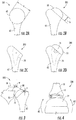
- Such planning steps particular to the humerus side of the joint can comprise analysis steps such as those depicted in Figures 2A-2D .
- a pre-operative planning method for the humerus can comprise a step 201, as depicted in Figure 2A , where the diameter d of humeral head 60 of humerus 62 can be measured.
- this analysis can be accomplished virtually based on images taken from a subject or patient prior to surgery.
- data and information can be collected that informs the selection of a humeral head implant and/or supports the creation of a shoulder surgery guide device specific to the patient or subject to be treated.
- a pre-operative planning method for the humerus can comprise a step 202, as depicted in Figure 2B , where the height h of humeral head 60 of humerus 62 can be measured.
- this analysis can be accomplished virtually based on images taken from a subject or patient prior to surgery.
- data and information can be collected that informs the selection of a humeral head implant and/or supports the creation of a shoulder surgery guide device specific to the patient or subject to be treated.
- a pre-operative planning method for the humerus can comprise a step 203, as depicted in Figure 2C , where the size of a humeral bone implant stem portion 70 can be determined from Houndsfield units (the Hounsfield scale, named after Sir Godfrey Newbold Hounsfield, is a quantitative scale for describing radiodensity) measured by CT scan. In some embodiments, this analysis can be accomplished virtually based on images taken from a subject or patient prior to surgery. By measuring the size of a humeral bone implant, data and information can be collected that informs the selection of a humeral head implant and/or supports the creation of a shoulder surgery guide device specific to the patient or subject to be treated.
- Houndsfield units the Hounsfield scale, named after Sir Godfrey Newbold Hounsfield, is a quantitative scale for describing radiodensity
- a pre-operative planning method for the humerus can comprise a step 204, as depicted in Figure 2D , where a best fit size of humeral implant 72 from a range of sizes can be determined.
- the range of sizes can be selected from the group consisting of length of stem, size of humeral stem, diameter of stem, size diameter of head, height of head, and offset of the center spherical head compared to the center of the face of the humeral stem.
- this analysis can be accomplished virtually based on images taken from a subject or patient prior to surgery.
- data and information can be collected that informs the selection of a humeral head implant and/or supports the creation of a shoulder surgery guide device specific to the patient or subject to be treated.
- Such planning steps particular to the humeral side of the joint can comprise analysis steps such as those depicted in Figures 2A-2D , and can comprise all or some of the steps depicted in Figures 2A-2D , and in some aspects can be done in any order desired.
- analysis steps particular to joint articulation can be performed first followed by analysis steps particular to fixation elements.
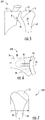
- a pre-operative planning method for designing a shoulder surgery guide can comprise comparing vectors 80 in three dimensions to measure the distance of relocation of humeral tuberosity 72 compared to the scapula 10, as depicted in analysis 205 in Figure 3 .
- These tendons control much of the rotation of the humerus about the scapula as well as having a part in elevating the humerus.
- kinematics and kinetics of the glenohumeral joint change.
- changing the direction of vector 80 can change wear patterns and range of motion (ROM) of the implanted device versus the native joint.
- ROM range of motion
- changing the magnitude of vector 80 by lengthening or increasing it with a joint prosthesis that is too large for the joint can result in decreased ROM, pain, and increased wear of the prosthetic components.
- changing the magnitude of vector 80 by decreasing or shortening it with a joint prosthesis that is too small for the joint can result in an unstable joint that may dislocate and can result in suboptimal mechanics for elevating the humerus.
- this analysis can be accomplished virtually based on images taken from a subject or patient prior to surgery.
- vector 80 in three dimensions to measure the distance of relocation of humeral tuberosity 72 compared to the scapula 10
- data and information can be collected that informs the selection of a humeral head implant, glenoid implant, and/or supports the creation of a shoulder surgery guide device specific to the patient or subject to be treated.
- a pre-operative planning method designing a shoulder surgery guide can comprise a step 206, as depicted in Figure 4 , where range of motion (ROM) analysis 82 can be conducted, including virtually positioning implants 20, 72 through extreme ranges of motion to measure impact locations and compensate for necessary functional ROM. In some embodiments, this analysis can be accomplished virtually based on images taken from a subject or patient prior to surgery. By measuring the ROM with respect to glenoid implants 20 and/or humeral implants 72, data and information can be collected that informs the selection of glenoid implant, a humeral head implant and/or supports the creation of a shoulder surgery guide device specific to the patient or subject to be treated.
- ROM range of motion
- a pre-operative planning method designing a shoulder surgery guide can comprise a step 207, as depicted in Figure 5 , where soft tissue, e.g. muscle, analysis is conducted.
- soft tissue analysis can comprise determining and/or assessing soft tissue insertion points (e.g., X, Y and Z ) and analyzing impacts on and/or impacts from use of one or more implants (glenoid and/or humeral).
- four rotator cuff muscles and their attachments points can be analyzed.
- analysis can comprise the subscapularis that attaches at an attachment point Y near the lesser tuberosity and at an attachment point X near the anterior glenoid.
- analysis can comprise the supraspinatus that attaches at an attachment point Z near the anterior greater tuberosity and above the scapular spine (shoulder blade; not shown).
- soft tissue analysis can comprise the infraspinatus that attaches at the greater tuberosity (posterior to supraspinatus) and below the scapular spine (posterior).
- soft tissue analysis can comprise the teres minor that attaches posterior on the humerus and on the inferior scapular boder. In some embodiments, this analysis can be accomplished virtually based on images taken from a subject or patient prior to surgery.
- data and information can be collected that informs the selection of a glenoid implant, a humeral head implant and/or supports the creation of a shoulder surgery guide device specific to the patient or subject to be treated.
- the disclosed pre-operative planning methods can further comprise designing a shoulder surgery guide device based upon parameters collected from the planning methods and analyses.
- a designed shoulder surgery guide can be produced, wherein the produced surgery guide is configured in accordance with parameters collected from the planning and analysis specific to the patient to be treated.
- a guide, and/or a prosthetic implant can be produced or made using a three dimensional (3D) printing device.
- a shoulder surgery guide device produced as disclosed herein can comprise a polymeric or metallic material.
- the disclosed pre-operative planning methods can further comprise identifying a prosthetic shoulder implant, and/or identifying a placement position for the prosthetic shoulder implant.
- the identification of a prosthetic shoulder implant and placement position takes into consideration at least one of the factors selected from the group consisting of adjustments in glenoid implant size, augmentation depth, augment position, positioning in six degrees of freedom, fixation type, fixation size, reaming depth, reaming diameter, reaming angle, and/or a combination thereof.
- the above method can further comprise a step of recommending implants and placement positions, with recommended adjustments in humerus stem size, length, head diameter, head height, head offset and rotation (axial).
- a prosthetic shoulder implant can in some embodiments comprise a glenoid implant.
- the above methods of creating a shoulder surgery guide based on pre-operative planning can further comprise one or more optimization steps.
- Such optimization steps can comprise the identification of procedural risks based on measurements of one or more of a plurality of factors.
- Such factors can in some embodiments comprise whether the glenoid face coverage is maximized (e.g. about 0 to about 2 mm), the overhang of the glenoid face is minimized (e.g.
- such optimization factors can comprise whether the glenoid retroversion is less than about 5 degrees to about 10 degrees, the seating of the glenoid implant is greater than about 80%, i.e. about 80% of the back side of the glenoid implant is supported by or touching bone, whether there is minimized penetration of the glenoid cortical wall anteriorily (e.g. about 0mm to about 3mm), and/or the depth of any glenoid implant augment feature is as minimal as possible.
- optimization factors can comprise whether there is less than about 1 mm of difference between the anatomic joint line and the new joint line with implants, there is minimized penetration of the glenoid cortical wall anteriorily, and/or there is maximized bone thickness behind the glenoid, preferably greater than 3mm.
- such optimization factors can comprise whether the orientation offset between the native glenoid and implant superior/inferior axis is minimized, preferably less than 5 degrees, the superior or inferior tilt versus native glenoid is minimized, preferably less than 5 degrees, there is less than about 5% to about 10% change in soft tissue length at extreme ranges of motion, there is maximized filing of the humeral metaphysis, in some embodiments greater than about 90% of metaphyseal bone filled based on and identification of metaphyseal bone by use of Houndsfield units, there is an absence of a humeral head overhang compared to the cut, or prepared surface of the humeral bone, there is minimal difference in humeral head diameter between anatomic and implant, in some embodiments less than about 3mm, there is minimal difference in humeral head height between anatomic and implant, in some embodiments less than about 1mm, and/or there is greater tuberosity to medial head edge comparison to anatomic, in some embodiments less than about 2mm.
- the penetration of the cortical wall anteriority of the vault can be assessed, as depicted in Figure 6.
- Figure 6 depicts step 208 of assessing the penetration of the cortical wall anteriorily of the vault 88 by a support structure 84 of glenoid implant 20.
- an additional or alternate support structure 86 can be used to affix implant 20 to glenoid 12.
- the width of the greater tuberosity to medial head edge with an implant can be compared to the anatomic width.
- the width 90 of the greater tuberosity to medial head edge with an implant 72 can be compared to the width of the anatomical humeral head.
- the planning methods and analysis steps disclosed herein can be done pre-operatively. That is, they can be done prior to surgery in a virtual or software-based environment. Such virtual simulations can in some embodiments be based on images or scans taken from a subject prior to surgery.
- imaging techniques e.g. computed tomography (CT), x-ray imaging, positron emission tomography (PET), ultrasound, etc.
- CT computed tomography
- PET positron emission tomography
- ultrasound etc.
- the analysis and results can be specific to the subject or patient and can take into consideration the particularities of that subject's condition.
- the subject matter described herein may be implemented in software in combination with hardware and/or firmware.
- the subject matter described herein may be implemented in software executed by a processor.
- the subject matter described herein may be implemented using a computer readable medium having stored thereon computer executable instructions that when executed by the processor of a computer control the computer to perform steps.
- Exemplary computer readable media suitable for implementing the subject matter described herein include non-transitory devices, such as disk memory devices, chip memory devices, programmable logic devices, and application specific integrated circuits,
- a computer readable medium that implements the subject matter described herein may be located on a single device or computing platform or may be distributed across multiple devices or computing platforms.
- the disclosed pre-operative planning methods can further comprise providing a computer readable medium having stored thereon executable instructions that when executed by the processor of a computer control the computer to perform one or more of the planning method and/or analysis steps.
- computer readable medium can have stored thereon executable instructions that when executed by the processor of a computer can control the computer to generate a virtual 3D model of a glenoid guide device reflecting one or more optimized parameters determined during pre-operative planning.
- computer readable medium can have stored thereon executable instructions that when executed by the processor of a computer control the computer to control a 3D printing device in communication with the computer, whereby the 3D printing device can print a glenoid guide device or humeral guide device for use in shoulder replacement surgery in a patient for which pre-operative planning method steps were conducted.
- a computer readable medium can be provided having stored thereon executable instructions that when executed by a processor of a computer can control the computer to generate a virtual 3D model of a glenoid implant device reflecting one or more optimized parameters determined during pre-operative planning.
- a computer readable medium is provided, wherein the computer readable medium has stored thereon executable instructions that when executed by the processor of a computer control the computer to perform one or more of the planning method and/or analysis steps as disclosed herein.
- computers, computing devices, hardware and/or functionality described herein may constitute a special purpose test device. Further, computers, computing devices, hardware and/or functionality described herein can improve the technological field of pre-operative planning for shoulder surgery and can improve generation of virtual modeling systems.
- a computing platform, computer, computing device, and/or hardware that implements the subject matter described herein may comprise a special purpose computing device usable to generate 3D models of glenoid and/or humeral implant devices, and/or for modeling and virtually simulating pre-operative shoulder surgery analysis.
- node refers to a physical computing platform including one or more processors and memory.
- the terms “function” or “module” refer to hardware, firmware, or software in combination with hardware and/or firmware for implementing features described herein.
- a computer readable medium having stored thereon executable instructions that when executed by the processor of a computer control the computer to perform steps comprising generating a virtual three dimensional model of a glenoid and/or humeral guide reflecting one or more optimized parameters determined during pre-operative planning based on the above method steps.
- a computer readable medium is provided, having stored thereon executable instructions that when executed by the processor of a computer control a 3D printing device in communication with the computer, whereby the 3D printing device prints a glenoid and/or humeral guide for use in shoulder replacement surgery in a patient for which the optimization analysis was conducted.
- shoulder surgery guides or guide devices can be designed, simulated and in some instances produced for use in shoulder replacement surgery.
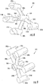
- a surgery guide device is depicted in Figures 8-11 .
- Figures 8 and 9 are rear and front perspective views, respectively, of a shoulder surgery guide
- Figure 10 is a plan view of a shoulder surgery guide.
- shoulder surgery guide 300 can in some embodiments comprise a plurality of peripheral guide structures 302 configured to align with the edge or rim of the glenoid face.
- peripheral guide structures 302 namely 302a, 302b, 302c, and 302d, are shown, but any number of peripheral guide structures 302, including for example 2, 3, 4, 5, 6, 7, 8, 9 or 10, could be used so long as there are a sufficient number to align and stabilize guide 300 on a glenoid face (see Figure 11 for a depiction of the guide in use).
- peripheral guide structures 302a, 302b, 302c, and 302d can each comprise a corresponding indentation or cupped surface 310a, 310b, 310c, and 310d that can be configured to wrap over the edge of the rim of the glenoid.
- Cupped surface 310a, 310b, 310c, and 310d can secure and stabilize guide 300 at the desired and predetermined (based on the pre-operative analysis and guide design) location on the glenoid.
- some peripheral guide structures may not include a cupped surface, or may include a different shaped structure, as needed to accommodate and align with a given point along the edge of a glenoid.
- Each peripheral guide structure 302, and corresponding cupped surface 310 can be predetermined and configured based on individual datum points collected during a pre-operative analysis and guide design, as disclosed herein.
- Peripheral guide structures 302a, 302b, 302c, and 302d generally extend radially from a hub structure 304, and can be positioned and secured to hub structure 304 by radial arms 308a, 308b, 308c, and 308d.
- the number of radial arms 308 will be dictated by, and correspond to, the number of peripheral guide structures 302.
- the length of radial arms 308 can be determined and configured based on individual datum points collected during a pre-operative analysis and guide design, as disclosed herein, such that each of the peripheral guide structures 302 align with the rim of the glenoid at the desired location.
- Hub structure 304 can comprise a central port 306 comprising a cylindrical opening extending through the entire length (see front view Figure 8 , rear view Figure 9 , and plan view Figure 10 ) of hub structure 304 and providing an opening through which a pin, drill or boing device can be guided to create an opening, i.e. drill a hole, and/or place a guide pin in the glenoid face.
- peripheral guide structures 302a, 302b, 302c, and 302d aligning with the rim or edge of the glenoid
- hub structure 304 by virtue of its attachment to each of peripheral guide structures 302a, 302b, 302c, and 302d, can be aligned at the predetermined and desired location on the face of a glenoid.
- the location of hub structure 304, and particularly central port 306, can be predetermined and configured based on pre-operative analysis such that central port 306 provides a steady and secure guide to the location on the glenoid where a prosthesis or implant is to be attached.
- Figure 11 depicts shoulder surgery guide 300 in use, or aligned with the face of glenoid 12 on scapula 10. Cupped surface 310a, 310b, 310c, and 310d wrap over the edge of the rim of the glenoid 12 such that guide 300 is aligned with and stabilized over glenoid 12.
- a pin, drill or boing device can be inserted into central port 306, which can guide the pin, drill or boing device to the precise location on glenoid 12 where a predetermined attachment point is located based on pre-operative analytics.
- a hybrid patient specific implant can be provided, in some embodiments a humeral implant, wherein the hybrid implant can comprise a fixation component and an articular component.
- the hybrid patient specific implant can comprise a standardized range of fixation components for securing the implant to the humerus.
- fixation component can comprise a stem comprising varying sizes, materials, coatings and surface treatments.
- an intermediate holder can be provided for securing the articular component to the fixation component.
- Such intermediate holder can vary in size, can comprise standardized materials and coatings, and can comprise a standardized connection between the fixation component, e.g stem, and holder.
- Such standardized connection can comprise threads, interlocking components, morse taper connections, snap fit connections (whether using snap rings or not), and the like.
- the customized patient specific articular component can comprise a desired articular shape and/or position based on the methods of analysis and optimization disclosed herein.
- the shape and position of the articular component can be centered or offset, and can have varying degrees of depth.
- the articular component can comprise a desired range of motion blockage. Range of motion tests with virtual pre-operative planning as discussed herein can reveal potential impingement of humeral polyethylene on scapula bone, or humeral tuberosities on acromion. In some aspects, an analysis comparing predicted range of motion based on necessary activities of daily living can be conducted. In some embodiments, a further step can include resolving any conflicts between impingement and activities of daily living needs. Taking these factors into consideration, the articular component shape and placement can then be optimized.
- the articular component shape can be adjusted.
- Such variations can comprise variations in radial location, depth/magnitude and/or angle.
- methods of treating a patient, and/or surgical methods are provided wherein one or more of the disclosed methods of analysis and optimization are performed on a patient in need of shoulder or other joint surgery.
- methods of treating a patient are provided wherein a disclosed method of analysis and optimization is performed, an optimized guide is designed and created, and one or more glenoid and/or humeral implants are designed, created, and/or selected.
- a method of treating a patient can comprise utilizing the pre-operative planning to design and optimize a guide and one or more glenoid and/or humeral implants, and the use of the guide to surgically implant the one or more glenoid and/or humeral prosthetic devices.
- kits can comprise a set of instructions for performing the disclosed pre-operative planning methods and analyses.
- a kit can further comprise one or more glenoid and/or humeral prosthetic devices, wherein the devices can be customizable or modular in design such that the prosthetic device can be optimized for the patient based on the pre-operative planning analysis.
- a kit can further comprise a guide for placing a prosthetic device during shoulder surgery, wherein the guide can be optimized for the patient based on the pre-operative planning analysis.
- a kit can further comprise a 3-D printing device for producing a guide and/or one or more glenoid and/or humeral prosthetic devices.
- a kit can further comprise a computer-readable medium (software) for use in conducting the pre-operative planning, and designing a guide, glenoid implant and/or humeral implant based on input parameters gathered during the disclosed methods of analysis.
- a patient can comprise a mammalian subject.
- the patient can be a human subject, including an adult, adolescent or child.
- the term "about,” when referring to a value or to an amount of mass, weight, time, volume, concentration or percentage is meant to encompass variations of in some embodiments ⁇ 20%, in some embodiments ⁇ 10%, in some embodiments ⁇ 5%, in some embodiments ⁇ 1%, in some embodiments ⁇ 0.5%, and in some embodiments ⁇ 0.1% from the specified amount, as such variations are appropriate to perform the disclosed method.
- the phrase “A, B, C, and/or D” includes A, B, C, and D individually, but also includes any and all combinations and subcombinations of A, B, C, and D.
- the phrase “consisting of” excludes any element, step, or ingredient not specified in the claim.
- the phrase “consists of” appears in a clause of the body of a claim, rather than immediately following the preamble, it limits only the element set forth in that clause; other elements are not excluded from the claim as a whole.
- signaling or “significant” relates to a statistical analysis of the probability that there is a non-random association between two or more entities. To determine whether or not a relationship is “significant” or has “significance”, statistical manipulations of the data can be performed to calculate a probability, expressed as a "p value”. Those p values that fall below a user-defined cutoff point are regarded as significant. In some embodiments, a p value less than or equal to 0,05, in some embodiments less than 0.01, in some embodiments less than 0.005, and in some embodiments less than 0.001, are regarded as significant. Accordingly, a p value greater than or equal to 0.05 is considered not significant.
Landscapes
- Health & Medical Sciences (AREA)
- Engineering & Computer Science (AREA)
- Biomedical Technology (AREA)
- Public Health (AREA)
- Life Sciences & Earth Sciences (AREA)
- General Health & Medical Sciences (AREA)
- Medical Informatics (AREA)
- Surgery (AREA)
- Heart & Thoracic Surgery (AREA)
- Veterinary Medicine (AREA)
- Animal Behavior & Ethology (AREA)
- Oral & Maxillofacial Surgery (AREA)
- Orthopedic Medicine & Surgery (AREA)
- Nuclear Medicine, Radiotherapy & Molecular Imaging (AREA)
- Molecular Biology (AREA)
- Epidemiology (AREA)
- Primary Health Care (AREA)
- Transplantation (AREA)
- Vascular Medicine (AREA)
- Cardiology (AREA)
- Dentistry (AREA)
- Robotics (AREA)
- Business, Economics & Management (AREA)
- General Business, Economics & Management (AREA)
- Data Mining & Analysis (AREA)
- Pathology (AREA)
- Databases & Information Systems (AREA)
- Prostheses (AREA)
- Surgical Instruments (AREA)
Abstract
Description
- This application claims the benefit of
U.S. Provisional Patent Application Serial No. 61/889,213, filed October 10, 2013 - The presently disclosed subject matter relates to methods, systems and devices for pre-operatively planned shoulder surgery guides and implants. The presently disclosed subject matter also relates to the use of such surgery guides and implants in patients undergoing shoulder surgery.
- Shoulder replacement is a common surgical operation that has achieved positive results for many patients. Indeed, approximately 10% of joint replacement procedures globally are related to the shoulder. Many shoulder procedures are performed in a patient where substantially normally bone exists for orientation and fixation of a prosthetic replacement, or resurfacing. In these cases, the need for the shoulder replacement can often times be related mostly to the arthritic condition of the joint, and relative absence of healthy cartilage.
- In some patients, however, one or more of the bones of the shoulder are not only arthritic, but have also had previous conditions that have caused bone to wear away. In such cases, there may not be sufficient bone to adequately affix a prosthetic implant to the bone, or the bones may have been worn such that the orientation of a joint replacement cannot be satisfactorily determined to ensure a positive patient outcome.
- There are a number of factors that complicate the selection, orientation and affixation of prosthetic implant devices, such as glenoid implants and/or humeral implants. Failure to properly account for each factor can lead to improperly sized, misaligned and/or poorly affixed implants that result in a poor surgical outcome for the patient.
- In order to increase the likelihood of successful patient outcomes in patients undergoing shoulder surgery, methods, systems and devices are needed that allow for the full understanding and incorporation of all necessary factors for optimization of shoulder implant selection and placement. Thus, a need remains for methods, systems and devices for pre-operatively planned shoulder surgery guides and implants that achieve desired outcomes.
- The presently disclosed subject matter provides methods, systems and devices for pre-operatively planned shoulder surgery guides and implants. The presently disclosed subject matter also provides uses of such surgery guides and implants in patients undergoing shoulder surgery.
- An object of the presently disclosed subject matter having been stated hereinabove, and which is achieved in whole or in part by the presently disclosed subject matter, other objects will become evident as the description proceeds when taken in connection with the accompanying Examples as best described hereinbelow.
- The presently disclosed subject matter can be better understood by referring to the following figures. The components in the figures are not necessarily to scale, emphasis instead being placed upon illustrating the principles of the presently disclosed subject matter (often schematically). In the figures, like reference numerals designate corresponding parts throughout the different views. A further understanding of the presently disclosed subject matter can be obtained by reference to an embodiment set forth in the illustrations of the accompanying drawings. Although the illustrated embodiment is merely exemplary of systems for carrying out the presently disclosed subject matter, both the organization and method of operation of the presently disclosed subject matter, in general, together with further objectives and advantages thereof, may be more easily understood by reference to the drawings and the following description. The drawings are not intended to limit the scope of this presently disclosed subject matter, which is set forth with particularity in the claims as appended or as subsequently amended, but merely to clarify and exemplify the presently disclosed subject matter.
- For a more complete understanding of the presently disclosed subject matter, reference is now made to the following drawings in which:
-
Figure 1A is a schematic illustration of a step in a pre-operative planning method for designing a shoulder surgery guide where the anterior edge of a glenoid implant is aligned with an anterior edge of a glenoid bone, according to an embodiment of the disclosed subject matter; -
Figure 1B is a schematic illustration of a step in a pre-operative planning method for designing a shoulder surgery guide where the retroversion of a glenoid implant is adjusted, according to an embodiment of the disclosed subject matter; -
Figure 1C is a schematic illustration of a step in a pre-operative planning method for designing a shoulder surgery guide where the augmentation of a glenoid implant is adjusted, according to an embodiment of the disclosed subject matter; -
Figure 1D is a schematic illustration of a step in a pre-operative planning method for designing a shoulder surgery guide where the inferior tilt of a glenoid implant is adjusted, according to an embodiment of the disclosed subject matter; -
Figure 1E is a schematic illustration of a step in a pre-operative planning method for designing a shoulder surgery guide where bone support for a glenoid implant is evaluated, according to an embodiment of the disclosed subject matter; -
Figure 1F is a schematic illustration of a step in a pre-operative planning method for designing a shoulder surgery guide where the medialization of a glenoid implant is adjusted by assessing the volumetric amount of bone needed to be removed by reaming, according to an embodiment of the disclosed subject matter; -
Figure 1G is a schematic illustration of a step in a pre-operative planning method for designing a shoulder surgery guide where fixation support in the absence of central pegs that penetrate a vault medially is analyzed, according to an embodiment of the disclosed subject matter; -
Figure 1H is a schematic illustration of a step in a pre-operative planning method for designing a shoulder surgery guide where a joint line is analyzed by comparing an original joint line and a new joint line, according to an embodiment of the disclosed subject matter; -
Figure 1I is a schematic illustration of a step in a pre-operative planning method for designing a shoulder surgery guide where widths of the glenoid implant and the glenoid bone are measured and matched after reaming and aligning inferior and superior axes of the glenoid implant and bone, according to an embodiment of the disclosed subject matter; -
Figure 2A is a schematic illustration of a step in a pre-operative planning method for designing a shoulder surgery guide where the diameter of a humeral head is determined, according to an embodiment of the disclosed subject matter; -
Figure 2B is a schematic illustration of a step in a pre-operative planning method for designing a shoulder surgery guide where the height of a humeral head is determined, according to an embodiment of the disclosed subject matter; -
Figure 2C is a schematic illustration of a step in a pre-operative planning method for designing a shoulder surgery guide where the size of a humeral bone implant from Houndsfield units measured by computed tomography scan is determined, according to an embodiment of the disclosed subject matter; -
Figure 2D is a schematic illustration of a step in a pre-operative planning method for designing a shoulder surgery guide where a best fit size of implant from a range of sizes is determined, according to an embodiment of the disclosed subject matter; -
Figure 3 is a schematic illustration of a step in a pre-operative planning method for designing a shoulder surgery guide where vectors are compared in three dimensions to measure the distance of relocation of humeral tuberosity compared to the scapula, according to an embodiment of the disclosed subject matter; -
Figure 4 is a schematic illustration of a step in a pre-operative planning method for designing a shoulder surgery guide where range of motion analysis is conducted, including virtually positioning implants through extreme ranges of motion to measure impact locations and compensate for necessary functional range of motion, according to an embodiment of the disclosed subject matter; -
Figure 5 is a schematic illustration of a step in a pre-operative planning method for designing a shoulder surgery guide where soft tissue analysis comprising determining key soft tissue insertion points is conducted, according to an embodiment of the disclosed subject matter; -
Figure 6 is a schematic illustration of a step in a pre-operative planning method for designing a shoulder surgery guide where penetration of the cortical wall anteriorily of the vault is assessed, according to an embodiment of the disclosed subject matter; -
Figure 7 is a schematic illustration of a step in a pre-operative planning method for designing a shoulder surgery guide where the width of the greater tuberosity to medial head edge with an implant is compared to the anatomic width, according to an embodiment of the disclosed subject matter; -
Figures 8 and 9 are rear and front perspective views, respectively, of a shoulder surgery guide, according to an embodiment of the disclosed subject matter; -
Figure 10 is a plan view a shoulder surgery guide, according to an embodiment of the disclosed subject matter; and -
Figure 11 is a perspective view of a shoulder surgery guide as used during shoulder surgery on a glenoid surface of a scapula, according to an embodiment of the disclosed subject matter. - Patients requiring shoulder surgery may have one or more of the bones of the shoulder that are not only arthritic, but may also have had previous conditions that have caused bone to wear away. In such cases, there may not be sufficient bone to adequately affix a prosthetic implant to the bone during a routine shoulder surgery. Indeed, the bones may have been worn such that the orientation of a joint replacement cannot be satisfactorily determined to ensure a positive patient outcome.
- The glenoid bone can be subject to increased wear due to bone arthritic conditions of the joint, and due to alterations of a normal soft tissue envelope surrounding the joint. In such cases, the orientation of the face of the glenoid portion of the scapula bone may be altered so that the humeral bone is no longer appropriately apposed to the glenoid surface. In the case where the glenoid is severely worn, there can be two or more risks a surgeon must balance in an attempt to improve shoulder function and pain relief.
- First, if the optimal orientation of the diseased but treated shoulder is not found and replicated with the prosthesis the patient may experience most operative complications related to subluxation or dislocation of the replaced shoulder joint. This can occur either due to passive inputs to the shoulder (e.g., leaning against it, or lying in bed), or due to active firing of surrounding soft tissue which is not able to be constrained by the replaced joint surfaces.
- Additionally, the fixation of a replacement prosthesis, or implant, to the native patient bone can be problematic. Frequently, in order to counteract the risks associated with joint subluxation and dislocation described above, it can be necessary for a surgeon to orient or position the replacement prosthesis or implant in a position better suited to resist imbalanced muscle forces. In such cases, separation forces between the implant and the bone can increase, which in turn can increase the potential for loosening of the joint prosthesis in the bone. Implant loosening can be related to accelerated implant wear, bone erosion, increased tissue inflammation, joint synovitis, and pain.
- In patients that have undergone shoulder replacement surgery, range of motion and strength are dependent on shoulder kinematics, which are in turn dependent on a host of factors. Such factor can, for example, include for example implant size, implant position, the design of implant shape, the joint line and soft tissue tension. In some cases it can be difficult to predict optimal implant size and position/orientation using currently available guides and implants. Often times a surgeon finds that there are too many variables to manage at one time. Moreover, the size choices of implants can be limited to the lowest practically functional groups to reduce economic burden to the health care system. Current implant designs and methodologies are inadequate to address these challenges because they are of significant cost, require time to develop, include increased risk of implant failure, and rely on human judgment of potential outcomes post-operatively.
- There are many factors that can affect the optimal positioning of shoulder implants during replacement surgery. For example, such factors can include the patient size, relative bone wear, soft tissue strength and condition, six degrees-of-freedom positioning of the glenoid and/or the humeral prosthesis, selected implant size, preoperative patient activity and strength levels, post operative treatment protocols, size and density of patient bone. Additional factors can include patient smoking status, concomitant handicaps and/or patient problems. It can be quite difficult for a surgeon to understand and balance these factors simultaneously. In addition, only a few of these factors are able to be controlled by the surgeon. Finally, each factor does not necessarily have an equally weighted impact on patient outcome. Nevertheless, it is considered that the implant size, position, orientation and bone preparation of the glenoid and the humerus can have a significant impact on the surgical outcomes.
- A factor that further complicates, or makes more difficult, a surgeons task of optimally placing a replacement component or implant to counteract these risk is the fact that the condition of the scapula is such that few landmarks exists for the surgeon the comprehend the implant position within the bone. Thus, frequently a surgeon might find that the implant position is not replicating as was envisioned during the surgical intervention.
- Others have attempted to improve a surgeon's chance of providing successful patient outcomes by providing operative techniques and tools. What is missing, however, is the ability to fully understand and incorporate multiple factors to optimize the implant selection and placement. Specifically, in some embodiments, the success of the surgery can be highly dependent on both the selection of the matching a prosthesis or prostheses (humeral and/or glenoid), as well as positioning of this prosthesis, as well as the soft tissue status before the surgery. There have been no previous attempts at including these factors in surgical planning and implant design.
- Disclosed herein are methods, systems and devices for pre-operatively planned shoulder surgery guides and implants. Methods, systems and devices are provided for the replacement of the shoulder joint, such as the glenohumeral joint, wherein the conditions of the humeral and soft tissue envelop is taken into consideration. More specifically, what is considered is that the shape and position of the glenoid implant is not based solely on what can be seen and measured on the scapula, but can be chosen, designed, planned and placed with incorporation of the same information related to the humerus. After all, the shoulder is a two part joint, i.e. glenoid and humeral head, wherein both parts work in conjunction with one another, and the factors that affect performance of the device can in some embodiments include factors from both sides of the joint.
- Appropriate sizing of the prosthesis can be important to successful outcomes, knowing that oversized or "overstuffed" replacement shoulders are more likely to dislocate, loosen, be painful, and/or have decreased range of motion. Replaced joints where the orientation of the prostheses is improper increases the likelihood of implant dislocation and loosening. Additionally, over-reaming, or too much bone removal, either on the glenoid, or the humerus, can be the cause of implant loosening, "under-stuffing" or inappropriate articular surface placement which can increase pain and decrease range of motion.
- Provided herein in some embodiments is a glenoid implant designed and manufactured to specifically match the patient anatomy, including optima! humeral and/or glenoid implant size and shape, and taking into account one or more of the following factors: assessment of the humeral implant fit to the humeral bone; relative hardness of the patient bone preoperatively; height and diameter of the humeral head placed on the humeral stem; orientation, or "offset" of the humeral head; and optimal bone removal for preservation of soft tissue insertion and attachment.
- Also provided herein are methods, systems and devices for creation of a shoulder surgery guide based on pre-operative planning which takes into consideration a plurality of factors and assessments. In some embodiments, the creation of a shoulder surgery guide based on pre-operative planning can comprise one or more of the following steps, the combination and order of which can vary: aligning an anterior edge of a glenoid implant with an anterior edge of a glenoid bone; adjusting a retroversion of the glenoid implant; adjusting an augmentation of the glenoid implant; adjusting an inferior tilt of the glenoid implant; evaluating bone support for the glenoid implant, wherein an amount of a rear surface of the glenoid implant that is supported by or touching bone is assessed; adjusting the medialization of the glenoid implant by assessing the volumetric amount of bone needed to be removed by reaming, or the minimum total distance of reaming necessary, in order to optimize the bone to implant interface; analyzing the fixation support in the absence of central pegs that penetrate a vault medially; analyzing the joint line, comprising comparing an original joint line and a new joint line, wherein the new joint line is substantially similar to the original joint line; measuring and matching widths of the glenoid implant and the glenoid bone after reaming and aligning inferior/superior axes of the glenoid implant and bone; assessing and adjusting as needed a thickness/height of the glenoid implant; assessing and adjusting as needed a depth of a glenoid fossa; assessing and adjusting as needed a thickness of a graft; determining a diameter of a humeral head; determining a height of the humeral head; determining a size of humeral bone implant from Houndsfield units measured by an imaging technique (e.g. computed tomography (CT) scan); and/or determining a best fit size of implant from a range of sizes, wherein the range of sizes is selected from the group consisting of length of stem, size of humeral stem, diameter of stem, size diameter of head, height of head, and offset of the center spherical head compared to the center of the face of the humeral stem.
- In some embodiments, a pre-operative planning method for designing a shoulder surgery guide is provided for designing a guide for the glenoid. Such a method can be separate from a pre-operative planning method for the humerus, or can in some embodiments be done in conjunction with the planning for the humerus, or humeral side of the joint. Such planning steps particular to the glenoid side of the joint can comprise analysis steps such as those depicted in
Figures 1A-1I . - For example, a pre-operative planning method for the glenoid can comprise a
step 101, as depicted inFigure 1A , where theanterior edge 18 ofglenoid implant 20 can be aligned 30 withanterior edge 16 of glenoid 12 ofscapula bone 10 of a patient to be treated. In some embodiments, this step, as with other pre-operative analyses disclosed herein, can be accomplished virtually based on images, e.g. CT images or X-ray images, taken from a subject or patient prior to surgery. By aligninganterior edge 18 ofglenoid implant 20 withanterior edge 16 of glenoid 12, data and information can be collected that informs the selection of a glenoid implant and/or supports the creation of a shoulder surgery guide device specific to the patient or subject to be treated. - In some embodiments, a pre-operative planning method for the glenoid can comprise a
step 102, as depicted inFigure 1B , where theretroversion 32 ofglenoid implant 20 is adjusted and/or measured. The retroversion is the placement or degree of posterior rotation ofglenoid implant 20 when glenoid 12 , including posterior wear 14 (seeFigure 1A ), is reamed or otherwise resurfaced to accommodateglenoid implant 20. Such a measurement ofretroversion 32 ofglenoid implant 20 can be in comparison to the retroversion of the native glenoid in a subject to be treated. In some embodiments, adjusting the retroversion comprises adjusting the retroversion to be about 5 degrees (5°) to about 10 degrees (10°), with a maximum of 10°. In some embodiments, this analysis can be accomplished virtually based on images taken from a subject or patient prior to surgery. By measuring and/or adjusting theretroversion 32 ofglenoid implant 20, data and information can be collected that informs the selection of a glenoid implant and/or supports the creation of a shoulder surgery guide device specific to the patient or subject to be treated. - In some embodiments, a pre-operative planning method for the glenoid can comprise a
step 103, as depicted inFigure 1C , where a determination can be made as to the necessity ofaugmentation 34 to supportglenoid implant 20. In some embodiments, particularly where glenoid 12 includes posterior wear 14 (or wear at other locations of glenoid 12 not depicted inFigure 1C ), augmentation can be necessary and/or desirable to provide adequate support for the placement and/or attachment ofimplant 20. Such a step or analysis can in some embodiments comprise adjusting, sizing and/or measuringaugmentation 34 needed. In some embodiments, this analysis can be accomplished virtually based on images taken from a subject or patient prior to surgery. By assessing the need foraugmentation 34, and/or determining the type and/or size ofaugmentation 34, data and information can be collected that informs the selection of a glenoid implant and/or supports the creation of a shoulder surgery guide device specific to the patient or subject to be treated. - In some embodiments, a pre-operative planning method for the glenoid can comprise a
step 104, as depicted inFigure 1D , where theinferior tilt 36 ofglenoid implant 20 can be measured and/or assessed. Such a measurement ofinferior tilt 36 ofglenoid implant 20 can be in comparison to the tilt of the native glenoid in a subject to be treated. In some embodiments, this analysis can be accomplished virtually based on images taken from a subject or patient prior to surgery. By assessing theinferior tilt 36 ofglenoid implant 20, data and information can be collected that informs the selection of a glenoid implant and/or supports the creation of a shoulder surgery guide device specific to the patient or subject to be treated. - In some embodiments, a pre-operative planning method for the glenoid can comprise a
step 105, as depicted inFigure 1E , where thebone support 38 forglenoid implant 20 can be measured and/or assessed. Such an assessment can in some embodiments comprise characterizing and/or quantifying the amount or degree ofbone support 38 for backside 22 ofimplant 20, taking into consideration posterior wear 14 (see, e.g.,Figures 1A or 1C ; or wear at other locations of glenoid 12 not depicted). In some embodiments, this analysis can be accomplished virtually based on images taken from a subject or patient prior to surgery. By assessing thebone support 38, data and information can be collected that informs the selection of a glenoid implant and/or supports the creation of a shoulder surgery guide device specific to the patient or subject to be treated. - In some embodiments, a pre-operative planning method for the glenoid can comprise a
step 106, as depicted inFigure 1F , wheremedialization 42 ofglenoid implant 20 can be adjusted and/or characterized by assessing thevolumetric amount 40 of bone needed to be removed by reaming. In some embodiments, this analysis can be accomplished virtually based on images taken from a subject or patient prior to surgery. By assessing thebone support 38, data and information can be collected that informs the selection of a glenoid implant and/or supports the creation of a shoulder surgery guide device specific to the patient or subject to be treated. - In some embodiments, a pre-operative planning method for the glenoid can comprise a
step 107, as depicted inFigure 1G , where fixation support in the absence of acentral peg 44 that penetrates a vault medially ofscapula 10 can be analyzed. In some embodiments, it is desirable to identify a location on the glenoid for attachment of a prosthesis using a peg or other fixation component without penetrating the anterior wall of the scapula. In some embodiments, this analysis can be accomplished virtually based on images taken from a subject or patient prior to surgery. By assessing the fixation support, data and information can be collected that informs the selection of a glenoid implant and/or supports the creation of a shoulder surgery guide device specific to the patient or subject to be treated. - In some embodiments, a pre-operative planning method for the glenoid can comprise a
step 108, as depicted inFigure 1H , where a joint line can be analyzed by comparing an originaljoint line 46 with a newjoint line 48 as created whenimplant 20 is affixed to the glenoid surface ofscapula 10. The degree to which the joint line changes or shifts, and/or the change in the angle, can be used in optimizing theimplant 20 selection and/or placement. In some embodiments, analyzing the joint line, including comparing the original joint line and the new joint line, can comprise analyzing the humeral head lateralization. Humeral head lateralization can comprise the distance the humeral shaft is moved laterally relative to the scapula after the implants are placed. In some embodiments, this analysis can be accomplished virtually based on images taken from a subject or patient prior to surgery. By assessing the joint line, data and information can be collected that informs the selection of a glenoid implant and/or supports the creation of a shoulder surgery guide device specific to the patient or subject to be treated. - In some embodiments, a pre-operative planning method for the glenoid can comprise a
step 109, as depicted inFigure 1I , where the widths of theglenoid implant 50a and theglenoid bone 50b can be measured and matched after reaming and aligning inferior 56 and superior 58 axes of the glenoid implant and bone. Particularly, in some embodiments, aglenoid 52a andimplant 50a heightwidth 54a can be measured and aligned with aglenoid 52b andbone 50b heightwidth 54b along inferior 56 and superior 58 axes. In some embodiments, this analysis can be accomplished virtually based on images taken from a subject or patient prior to surgery. By measuring the widths of theglenoid implant 50a and theglenoid bone 50b, and aligning inferior 56 and superior 58 axes of the glenoid implant and bone, data and information can be collected that informs the selection of a glenoid implant and/or supports the creation of a shoulder surgery guide device specific to the patient or subject to be treated. - Such planning steps particular to the glenoid side of the joint can comprise analysis steps such as those depicted in
Figures 1A-1I , and can comprise all or some of the steps depicted inFigures 1A-1I , and in some aspects can be done in any order desired. Alternatively, in some embodiments analysis steps particular to fixation elements can be performed first followed by analysis steps particular to joint articulation. - In some embodiments, a pre-operative planning method for designing a shoulder surgery guide is provided for designed a guide for the humerus, or humeral bone. Such a method can be separate from a pre-operative planning method for the glenoid (discussed above and depicted in
Figures 1a-1I ), or can in some embodiments be done in conjunction with the planning for the glenoid, or glenoid side of the joint. Such planning steps particular to the humerus side of the joint can comprise analysis steps such as those depicted inFigures 2A-2D . - For example, a pre-operative planning method for the humerus can comprise a
step 201, as depicted inFigure 2A , where the diameter d ofhumeral head 60 ofhumerus 62 can be measured. In some embodiments, this analysis can be accomplished virtually based on images taken from a subject or patient prior to surgery. By measuring diameter d ofhumeral head 60, data and information can be collected that informs the selection of a humeral head implant and/or supports the creation of a shoulder surgery guide device specific to the patient or subject to be treated. - In some embodiments, a pre-operative planning method for the humerus can comprise a
step 202, as depicted inFigure 2B , where the height h ofhumeral head 60 ofhumerus 62 can be measured. In some embodiments, this analysis can be accomplished virtually based on images taken from a subject or patient prior to surgery. By measuring height h ofhumeral head 60, data and information can be collected that informs the selection of a humeral head implant and/or supports the creation of a shoulder surgery guide device specific to the patient or subject to be treated. - In some embodiments, a pre-operative planning method for the humerus can comprise a
step 203, as depicted inFigure 2C , where the size of a humeral bone implant stemportion 70 can be determined from Houndsfield units (the Hounsfield scale, named after Sir Godfrey Newbold Hounsfield, is a quantitative scale for describing radiodensity) measured by CT scan. In some embodiments, this analysis can be accomplished virtually based on images taken from a subject or patient prior to surgery. By measuring the size of a humeral bone implant, data and information can be collected that informs the selection of a humeral head implant and/or supports the creation of a shoulder surgery guide device specific to the patient or subject to be treated. - In some embodiments, a pre-operative planning method for the humerus can comprise a
step 204, as depicted inFigure 2D , where a best fit size ofhumeral implant 72 from a range of sizes can be determined. In some embodiments, the range of sizes can be selected from the group consisting of length of stem, size of humeral stem, diameter of stem, size diameter of head, height of head, and offset of the center spherical head compared to the center of the face of the humeral stem. In some embodiments, this analysis can be accomplished virtually based on images taken from a subject or patient prior to surgery. By determining the most appropriate size ofhumeral implant 72, data and information can be collected that informs the selection of a humeral head implant and/or supports the creation of a shoulder surgery guide device specific to the patient or subject to be treated. - Such planning steps particular to the humeral side of the joint can comprise analysis steps such as those depicted in
Figures 2A-2D , and can comprise all or some of the steps depicted inFigures 2A-2D , and in some aspects can be done in any order desired. Alternatively, in some embodiments analysis steps particular to joint articulation can be performed first followed by analysis steps particular to fixation elements. - In some embodiments, a pre-operative planning method for designing a shoulder surgery guide can comprise comparing
vectors 80 in three dimensions to measure the distance of relocation ofhumeral tuberosity 72 compared to thescapula 10, as depicted inanalysis 205 inFigure 3 . For example, there are 3 rotator cuff tendons that attach to the proximal humerus in the area of the greater tuberosity and the scapula. Such attachment points are depicted as v and w, respectively, inFigure 3 . These tendons control much of the rotation of the humerus about the scapula as well as having a part in elevating the humerus. If the vector resolved from these 3 tendons changes, kinematics and kinetics of the glenohumeral joint (joint comprising the glenoid and humerus) change. For example, changing the direction ofvector 80 can change wear patterns and range of motion (ROM) of the implanted device versus the native joint. Additionally, in some embodiments, changing the magnitude ofvector 80 by lengthening or increasing it with a joint prosthesis that is too large for the joint can result in decreased ROM, pain, and increased wear of the prosthetic components. Finally, changing the magnitude ofvector 80 by decreasing or shortening it with a joint prosthesis that is too small for the joint can result in an unstable joint that may dislocate and can result in suboptimal mechanics for elevating the humerus. In some embodiments, this analysis can be accomplished virtually based on images taken from a subject or patient prior to surgery. By comparingvector 80 in three dimensions to measure the distance of relocation ofhumeral tuberosity 72 compared to thescapula 10, data and information can be collected that informs the selection of a humeral head implant, glenoid implant, and/or supports the creation of a shoulder surgery guide device specific to the patient or subject to be treated. - In some embodiments, a pre-operative planning method designing a shoulder surgery guide can comprise a
step 206, as depicted inFigure 4 , where range of motion (ROM)analysis 82 can be conducted, including virtually positioningimplants glenoid implants 20 and/orhumeral implants 72, data and information can be collected that informs the selection of glenoid implant, a humeral head implant and/or supports the creation of a shoulder surgery guide device specific to the patient or subject to be treated. - In some embodiments, a pre-operative planning method designing a shoulder surgery guide can comprise a
step 207, as depicted inFigure 5 , where soft tissue, e.g. muscle, analysis is conducted. In some aspects, soft tissue analysis can comprise determining and/or assessing soft tissue insertion points (e.g., X, Y and Z) and analyzing impacts on and/or impacts from use of one or more implants (glenoid and/or humeral). In some embodiments, four rotator cuff muscles and their attachments points can be analyzed. For example, in some aspects analysis can comprise the subscapularis that attaches at an attachment point Y near the lesser tuberosity and at an attachment point X near the anterior glenoid. In some aspects analysis can comprise the supraspinatus that attaches at an attachment point Z near the anterior greater tuberosity and above the scapular spine (shoulder blade; not shown). In some aspects, soft tissue analysis can comprise the infraspinatus that attaches at the greater tuberosity (posterior to supraspinatus) and below the scapular spine (posterior). In some aspects, soft tissue analysis can comprise the teres minor that attaches posterior on the humerus and on the inferior scapular boder. In some embodiments, this analysis can be accomplished virtually based on images taken from a subject or patient prior to surgery. By analyzing the soft tissue around the glenohumeral joint, data and information can be collected that informs the selection of a glenoid implant, a humeral head implant and/or supports the creation of a shoulder surgery guide device specific to the patient or subject to be treated. - In some embodiments, the disclosed pre-operative planning methods can further comprise designing a shoulder surgery guide device based upon parameters collected from the planning methods and analyses. In some embodiments, a designed shoulder surgery guide can be produced, wherein the produced surgery guide is configured in accordance with parameters collected from the planning and analysis specific to the patient to be treated. In some aspects, a guide, and/or a prosthetic implant, can be produced or made using a three dimensional (3D) printing device. In some embodiments, a shoulder surgery guide device produced as disclosed herein can comprise a polymeric or metallic material.
- In some embodiments, the disclosed pre-operative planning methods can further comprise identifying a prosthetic shoulder implant, and/or identifying a placement position for the prosthetic shoulder implant. The identification of a prosthetic shoulder implant and placement position takes into consideration at least one of the factors selected from the group consisting of adjustments in glenoid implant size, augmentation depth, augment position, positioning in six degrees of freedom, fixation type, fixation size, reaming depth, reaming diameter, reaming angle, and/or a combination thereof. The above method can further comprise a step of recommending implants and placement positions, with recommended adjustments in humerus stem size, length, head diameter, head height, head offset and rotation (axial). A prosthetic shoulder implant can in some embodiments comprise a glenoid implant.
- In some embodiments, the above methods of creating a shoulder surgery guide based on pre-operative planning can further comprise one or more optimization steps. Such optimization steps can comprise the identification of procedural risks based on measurements of one or more of a plurality of factors. Such factors can in some embodiments comprise whether the glenoid face coverage is maximized (e.g. about 0 to about 2 mm), the overhang of the glenoid face is minimized (e.g. about 0 to about 3 mm), and/or the bone removal on the glenoid face is minimized, such as for example less than about 2mm of depth, Continuing, in some embodiments such optimization factors can comprise whether the glenoid retroversion is less than about 5 degrees to about 10 degrees, the seating of the glenoid implant is greater than about 80%, i.e. about 80% of the back side of the glenoid implant is supported by or touching bone, whether there is minimized penetration of the glenoid cortical wall anteriorily (e.g. about 0mm to about 3mm), and/or the depth of any glenoid implant augment feature is as minimal as possible. Still yet, in some embodiments such optimization factors can comprise whether there is less than about 1 mm of difference between the anatomic joint line and the new joint line with implants, there is minimized penetration of the glenoid cortical wall anteriorily, and/or there is maximized bone thickness behind the glenoid, preferably greater than 3mm. In some embodiments such optimization factors can comprise whether the orientation offset between the native glenoid and implant superior/inferior axis is minimized, preferably less than 5 degrees, the superior or inferior tilt versus native glenoid is minimized, preferably less than 5 degrees, there is less than about 5% to about 10% change in soft tissue length at extreme ranges of motion, there is maximized filing of the humeral metaphysis, in some embodiments greater than about 90% of metaphyseal bone filled based on and identification of metaphyseal bone by use of Houndsfield units, there is an absence of a humeral head overhang compared to the cut, or prepared surface of the humeral bone, there is minimal difference in humeral head diameter between anatomic and implant, in some embodiments less than about 3mm, there is minimal difference in humeral head height between anatomic and implant, in some embodiments less than about 1mm, and/or there is greater tuberosity to medial head edge comparison to anatomic, in some embodiments less than about 2mm. In some embodiments, such procedural risks (any and/or all from the above list) can be determined virtually based on images taken from a subject prior to surgery.
- With respect to the above optimization steps that comprise the identification of procedural risks, in some embodiments the penetration of the cortical wall anteriority of the vault can be assessed, as depicted in
Figure 6. Figure 6 depicts step 208 of assessing the penetration of the cortical wall anteriorily of thevault 88 by asupport structure 84 ofglenoid implant 20. In some embodiments, an additional oralternate support structure 86 can be used to affiximplant 20 to glenoid 12. - Also with respect to the above optimization steps that comprise the identification of procedural risks, in some embodiments the width of the greater tuberosity to medial head edge with an implant can be compared to the anatomic width. For example, in
Figure 7 thewidth 90 of the greater tuberosity to medial head edge with animplant 72 can be compared to the width of the anatomical humeral head. - In some aspects, the planning methods and analysis steps disclosed herein can be done pre-operatively. That is, they can be done prior to surgery in a virtual or software-based environment. Such virtual simulations can in some embodiments be based on images or scans taken from a subject prior to surgery. Currently available and future imaging techniques, e.g. computed tomography (CT), x-ray imaging, positron emission tomography (PET), ultrasound, etc., can be used to capture images and data to be used in simulation-based analysis and planning to identify suitable prosthetic implants and/or design surgery guides. By using images captured from a subject or patient to be treated, the analysis and results can be specific to the subject or patient and can take into consideration the particularities of that subject's condition.
- The subject matter described herein may be implemented in software in combination with hardware and/or firmware. For example, the subject matter described herein may be implemented in software executed by a processor. In one exemplary implementation, the subject matter described herein may be implemented using a computer readable medium having stored thereon computer executable instructions that when executed by the processor of a computer control the computer to perform steps. Exemplary computer readable media suitable for implementing the subject matter described herein include non-transitory devices, such as disk memory devices, chip memory devices, programmable logic devices, and application specific integrated circuits, In addition, a computer readable medium that implements the subject matter described herein may be located on a single device or computing platform or may be distributed across multiple devices or computing platforms.
- As such, in some embodiments the disclosed pre-operative planning methods can further comprise providing a computer readable medium having stored thereon executable instructions that when executed by the processor of a computer control the computer to perform one or more of the planning method and/or analysis steps. For example, in some embodiments computer readable medium can have stored thereon executable instructions that when executed by the processor of a computer can control the computer to generate a virtual 3D model of a glenoid guide device reflecting one or more optimized parameters determined during pre-operative planning. Additionally, in some aspects, computer readable medium can have stored thereon executable instructions that when executed by the processor of a computer control the computer to control a 3D printing device in communication with the computer, whereby the 3D printing device can print a glenoid guide device or humeral guide device for use in shoulder replacement surgery in a patient for which pre-operative planning method steps were conducted.
- Further, in some aspects of the disclosed methods, systems and devices, a computer readable medium can be provided having stored thereon executable instructions that when executed by a processor of a computer can control the computer to generate a virtual 3D model of a glenoid implant device reflecting one or more optimized parameters determined during pre-operative planning. Thus, in some embodiments a computer readable medium is provided, wherein the computer readable medium has stored thereon executable instructions that when executed by the processor of a computer control the computer to perform one or more of the planning method and/or analysis steps as disclosed herein.
- It should be noted that the computers, computing devices, hardware and/or functionality described herein may constitute a special purpose test device. Further, computers, computing devices, hardware and/or functionality described herein can improve the technological field of pre-operative planning for shoulder surgery and can improve generation of virtual modeling systems.
- The subject matter described herein for generating 3D models of glenoid and/or humeral implant devices, and/or for modeling and virtually simulating pre-operative shoulder surgery analysis improves the likelihood of a positive outcome from shoulder surgery. It should also be noted that a computing platform, computer, computing device, and/or hardware that implements the subject matter described herein may comprise a special purpose computing device usable to generate 3D models of glenoid and/or humeral implant devices, and/or for modeling and virtually simulating pre-operative shoulder surgery analysis.
- As used herein, the term "node" refers to a physical computing platform including one or more processors and memory.
- As used herein, the terms "function" or "module" refer to hardware, firmware, or software in combination with hardware and/or firmware for implementing features described herein.
- In some embodiments a computer readable medium is provided, having stored thereon executable instructions that when executed by the processor of a computer control the computer to perform steps comprising generating a virtual three dimensional model of a glenoid and/or humeral guide reflecting one or more optimized parameters determined during pre-operative planning based on the above method steps. In some embodiments, a computer readable medium is provided, having stored thereon executable instructions that when executed by the processor of a computer control a 3D printing device in communication with the computer, whereby the 3D printing device prints a glenoid and/or humeral guide for use in shoulder replacement surgery in a patient for which the optimization analysis was conducted.
- Based on the pre-operative planning steps and analyses disclosed herein, in some embodiments shoulder surgery guides or guide devices can be designed, simulated and in some instances produced for use in shoulder replacement surgery. Such a surgery guide device is depicted in
Figures 8-11 .Figures 8 and 9 are rear and front perspective views, respectively, of a shoulder surgery guide, whileFigure 10 is a plan view of a shoulder surgery guide. As depicted inFigures 8-10 ,shoulder surgery guide 300 can in some embodiments comprise a plurality of peripheral guide structures 302 configured to align with the edge or rim of the glenoid face. InFigures 9-11 four peripheral guide structures 302, namely 302a, 302b, 302c, and 302d, are shown, but any number of peripheral guide structures 302, including for example 2, 3, 4, 5, 6, 7, 8, 9 or 10, could be used so long as there are a sufficient number to align and stabilizeguide 300 on a glenoid face (seeFigure 11 for a depiction of the guide in use). In some embodiments,peripheral guide structures cupped surface Cupped surface guide 300 at the desired and predetermined (based on the pre-operative analysis and guide design) location on the glenoid. In some embodiments, some peripheral guide structures may not include a cupped surface, or may include a different shaped structure, as needed to accommodate and align with a given point along the edge of a glenoid. Each peripheral guide structure 302, and corresponding cupped surface 310, can be predetermined and configured based on individual datum points collected during a pre-operative analysis and guide design, as disclosed herein. -
Peripheral guide structures hub structure 304, and can be positioned and secured tohub structure 304 byradial arms -
Hub structure 304 can comprise acentral port 306 comprising a cylindrical opening extending through the entire length (see front viewFigure 8 , rear viewFigure 9 , and plan viewFigure 10 ) ofhub structure 304 and providing an opening through which a pin, drill or boing device can be guided to create an opening, i.e. drill a hole, and/or place a guide pin in the glenoid face. Withperipheral guide structures hub structure 304, by virtue of its attachment to each ofperipheral guide structures hub structure 304, and particularlycentral port 306, can be predetermined and configured based on pre-operative analysis such thatcentral port 306 provides a steady and secure guide to the location on the glenoid where a prosthesis or implant is to be attached. -
Figure 11 depictsshoulder surgery guide 300 in use, or aligned with the face of glenoid 12 onscapula 10.Cupped surface guide 300 is aligned with and stabilized over glenoid 12. Withguide 300 in place on glenoid 12, a pin, drill or boing device can be inserted intocentral port 306, which can guide the pin, drill or boing device to the precise location on glenoid 12 where a predetermined attachment point is located based on pre-operative analytics. - In some embodiments, a hybrid patient specific implant can be provided, in some embodiments a humeral implant, wherein the hybrid implant can comprise a fixation component and an articular component. The hybrid patient specific implant can comprise a standardized range of fixation components for securing the implant to the humerus. Such fixation component can comprise a stem comprising varying sizes, materials, coatings and surface treatments.
- In some embodiments, an intermediate holder can be provided for securing the articular component to the fixation component. Such intermediate holder can vary in size, can comprise standardized materials and coatings, and can comprise a standardized connection between the fixation component, e.g stem, and holder. Such standardized connection can comprise threads, interlocking components, morse taper connections, snap fit connections (whether using snap rings or not), and the like.
- In some aspects, the customized patient specific articular component can comprise a desired articular shape and/or position based on the methods of analysis and optimization disclosed herein. By way of example and not limitation, the shape and position of the articular component can be centered or offset, and can have varying degrees of depth.
- In some aspects, the articular component can comprise a desired range of motion blockage. Range of motion tests with virtual pre-operative planning as discussed herein can reveal potential impingement of humeral polyethylene on scapula bone, or humeral tuberosities on acromion. In some aspects, an analysis comparing predicted range of motion based on necessary activities of daily living can be conducted. In some embodiments, a further step can include resolving any conflicts between impingement and activities of daily living needs. Taking these factors into consideration, the articular component shape and placement can then be optimized.
- Additionally, in some embodiments, the articular component shape can be adjusted. Such variations, based in some aspects on pre-operative planning as discussed herein, can comprise variations in radial location, depth/magnitude and/or angle.
- In some embodiments, methods of treating a patient, and/or surgical methods, are provided wherein one or more of the disclosed methods of analysis and optimization are performed on a patient in need of shoulder or other joint surgery. In some embodiments, methods of treating a patient are provided wherein a disclosed method of analysis and optimization is performed, an optimized guide is designed and created, and one or more glenoid and/or humeral implants are designed, created, and/or selected. In some embodiments, a method of treating a patient can comprise utilizing the pre-operative planning to design and optimize a guide and one or more glenoid and/or humeral implants, and the use of the guide to surgically implant the one or more glenoid and/or humeral prosthetic devices.
- In some embodiments, a kit is provided wherein the kit can comprise a set of instructions for performing the disclosed pre-operative planning methods and analyses. Such a kit can further comprise one or more glenoid and/or humeral prosthetic devices, wherein the devices can be customizable or modular in design such that the prosthetic device can be optimized for the patient based on the pre-operative planning analysis. In some embodiments, a kit can further comprise a guide for placing a prosthetic device during shoulder surgery, wherein the guide can be optimized for the patient based on the pre-operative planning analysis. In some embodiments, a kit can further comprise a 3-D printing device for producing a guide and/or one or more glenoid and/or humeral prosthetic devices. In some embodiments, a kit can further comprise a computer-readable medium (software) for use in conducting the pre-operative planning, and designing a guide, glenoid implant and/or humeral implant based on input parameters gathered during the disclosed methods of analysis.
- In some embodiments a patient can comprise a mammalian subject. In some embodiments, the patient can be a human subject, including an adult, adolescent or child.
- While the following terms are believed to be well understood by one of ordinary skill in the art, the following definitions are set forth to facilitate explanation of the presently disclosed subject matter.
- Unless defined otherwise, all technical and scientific terms used herein have the same meaning as commonly understood to one of ordinary skill in the art to which the presently disclosed subject matter belongs. Although any methods, devices, and materials similar or equivalent to those described herein can be used in the practice or testing of the presently disclosed subject matter, representative methods, devices, and materials are now described.
- Following long-standing patent law convention, the terms "a" and "an" mean "one or more" when used in this application, including the claims.
- Unless otherwise indicated, all numbers expressing quantities of ingredients, reaction conditions, and so forth used in the specification and claims are to be understood as being modified in all instances by the term "about". Accordingly, unless indicated to the contrary, the numerical parameters set forth in this specification and attached claims are approximations that can vary depending upon the desired properties sought to be obtained by the presently disclosed subject matter.
- As used herein, the term "about," when referring to a value or to an amount of mass, weight, time, volume, concentration or percentage is meant to encompass variations of in some embodiments ±20%, in some embodiments ±10%, in some embodiments ±5%, in some embodiments ±1%, in some embodiments ±0.5%, and in some embodiments ±0.1% from the specified amount, as such variations are appropriate to perform the disclosed method.
- As used herein, the term "and/or" when used in the context of a listing of entities, refers to the entities being present singly or in combination. Thus, for example, the phrase "A, B, C, and/or D" includes A, B, C, and D individually, but also includes any and all combinations and subcombinations of A, B, C, and D.
- The term "comprising", which is synonymous with "including," "containing," or "characterized by" is inclusive or open-ended and does not exclude additional, unrecited elements or method steps. "Comprising" is a term of art used in claim language which means that the named elements are present, but other elements can be added and still form a construct or method within the scope of the claim.
- As used herein, the phrase "consisting of" excludes any element, step, or ingredient not specified in the claim. When the phrase "consists of" appears in a clause of the body of a claim, rather than immediately following the preamble, it limits only the element set forth in that clause; other elements are not excluded from the claim as a whole.
- As used herein, the phrase "consisting essentially of" limits the scope of a claim to the specified materials or steps, plus those that do not materially affect the basic and novel characteristic(s) of the claimed subject matter.
- With respect to the terms "comprising", "consisting of", and "consisting essentially of", where one of these three terms is used herein, the presently disclosed and claimed subject matter can include the use of either of the other two terms.
- As used herein, "significance" or "significant" relates to a statistical analysis of the probability that there is a non-random association between two or more entities. To determine whether or not a relationship is "significant" or has "significance", statistical manipulations of the data can be performed to calculate a probability, expressed as a "p value". Those p values that fall below a user-defined cutoff point are regarded as significant. In some embodiments, a p value less than or equal to 0,05, in some embodiments less than 0.01, in some embodiments less than 0.005, and in some embodiments less than 0.001, are regarded as significant. Accordingly, a p value greater than or equal to 0.05 is considered not significant.
- It will be understood that various details of the presently disclosed subject matter may be changed without departing from the scope of the presently disclosed subject matter. Furthermore, the foregoing description is for the purpose of illustration only, and not for the purpose of limitation.
- Further embodiments, which can be combined with other embodiments described herein, are described in the following clauses. Any features described as part of one these clauses can be used on or in conjunction with other embodiments described herein to yield yet a further embodiment.
- 1. A pre-operative planning method for designing a shoulder surgery guide, the method comprising:
- aligning an anterior edge of a glenoid implant with an anterior edge of a glenoid bone;
- adjusting a retroversion of the glenoid implant;
- adjusting an augmentation of the glenoid implant;
- adjusting an inferior tilt of the glenoid implant;
- evaluating bone support for the glenoid implant, wherein an amount of a rear surface of the glenoid implant that is supported by or touching bone is assessed;
- adjusting a medialization of the glenoid implant by assessing the volumetric amount of bone needed to be removed by reaming, or the minimum total distance of reaming necessary, in order to optimize the bone to implant interface;
- analyzing fixation support in the absence of central pegs that penetrate a vault medially;
- analyzing a joint line, comprising comparing an original joint line and a new joint line, wherein the new joint line is substantially similar to the original joint line;
- measuring and matching widths of the glenoid implant and the glenoid bone after reaming and aligning inferior and superior axes of the glenoid implant and bone;
- assessing and adjusting as needed a thickness/height of the glenoid implant;
- assessing and adjusting as needed a depth of a glenoid fossa; and
- assessing and adjusting as needed a thickness of a graft.
- 2. The method of clause 1, wherein the planning method steps are done virtually based on images taken from a subject prior to surgery.
- 3. The method of clause 1, wherein adjusting the retroversion comprises adjusting the retroversion to be about 5 degrees (5°) to about 10 degrees (10°), with a maximum of 10°.
- 4. The method of clause 1, further comprising designing a shoulder surgery guide device based upon parameters collected from the planning method steps of claim 1.
- 5. The method of clause 4, further comprising producing a shoulder surgery guide device based upon parameters collected from the planning method steps.
- 6. The method of clause 5, wherein producing the shoulder surgery guide device comprises using a three dimensional printing device.
- 7. The method of clause 5, wherein the shoulder surgery guide device comprises a polymeric or metallic material.
- 8. The method of clause 1, further comprising identifying a prosthetic shoulder implant, and identifying a placement position for the prosthetic shoulder implant, wherein the identification of the prosthetic shoulder implant and placement position takes into consideration at least one of the factors selected from the group consisting of adjustments in glenoid implant size, augmentation depth, augment position, positioning in six degrees of freedom, fixation type, fixation size, reaming depth, reaming diameter, reaming angle, and/or a combination thereof.
- 9. The method of clause 8, wherein the prosthetic shoulder implant comprises a glenoid implant.
- 10. The method of clause 1, further comprising providing a computer readable medium having stored thereon executable instructions that when executed by the processor of a computer control the computer to perform one or more of the planning method steps.
- 11. The method of
clause 10, wherein the computer readable medium having stored thereon executable instructions that when executed by the processor of a computer control the computer to generate a virtual three dimensional (3D) model of a glenoid guide device reflecting one or more optimized parameters determined during pre-operative planning. - 12. The method of
clause 10, wherein the computer readable medium having stored thereon executable instructions that when executed by the processor of a computer control the computer to control a 3D printing device in communication with the computer, whereby the 3D printing device prints a glenoid guide device for use in shoulder replacement surgery in a patient for which pre-operative planning method steps were conducted. - 13. The method of
clause 10, wherein the computer readable medium having stored thereon executable instructions that when executed by the processor of a computer control the computer to generate a virtual 3D model of a glenoid implant device reflecting one or more optimized parameters determined during pre-operative planning. - 14. A computer readable medium having stored thereon executable instructions that when executed by the processor of a computer control the computer to perform one or more of the planning method steps of clause 1.
- 15. A pre-operative planning method for designing a shoulder surgery guide, the method comprising:
- determining a diameter of a humeral head;
- determining a height of the humeral head;
- determining a size of humeral bone implant from Houndsfield units measured by computed tomography scan; and
- determining a best fit size of implant from a range of sizes, wherein the range of sizes is selected from the group consisting of length of stem, size of humeral stem, diameter of stem, size diameter of head, height of head, and offset of the center spherical head compared to the center of the face of the humeral stem.
- 16. The method of clause 15, wherein the planning method steps are done virtually based on images taken from a subject prior to surgery.
- 17. The method of clause 15, further comprising designing a shoulder surgery guide device based upon parameters collected from the planning method steps of claim 1.
- 18. The method of clause 15, further comprising identifying a prosthetic shoulder implant, and identifying a placement position for the prosthetic shoulder implant, wherein the identification of the prosthetic shoulder implant and placement position takes into consideration at least one of the factors selected from the group consisting of adjustments in humerus stem size, length, head diameter, head height, head offset and rotation (axial), and/or combinations thereof.
- 19. The method of
clause 18, wherein the prosthetic shoulder implant comprises a humerus implant. - 20. The method of clause 15, further comprising providing a computer readable medium having stored thereon executable instructions that when executed by the processor of a computer control the computer to perform one or more of the planning method steps.
- 21. The method of
clause 20, wherein the computer readable medium having stored thereon executable instructions that when executed by the processor of a computer control the computer to generate a virtual 3D model of a humeral guide device reflecting one or more optimized parameters determined during pre-operative planning. - 22. The method of
clause 20, wherein the computer readable medium having stored thereon executable instructions that when executed by the processor of a computer control the computer to control a 3D printing device in communication with the computer, whereby the 3D printing device prints a humeral guide device for use in shoulder replacement surgery in a patient for which pre-operative planning method steps were conducted. - 23. The method of
clause 20, wherein the computer readable medium having stored thereon executable instructions that when executed by the processor of a computer control the computer to generate a virtual 3D model of a humeral implant device reflecting one or more optimized parameters determined during pre-operative planning. - 24. A computer readable medium having stored thereon executable instructions that when executed by the processor of a computer control the computer to perform one or more of the planning method steps of clause 15.
- 25. A pre-operative planning method for designing a shoulder surgery guide, the method comprising:
- aligning an anterior edge of a glenoid implant with an anterior edge of a glenoid bone;
- adjusting a retroversion of the glenoid implant;
- adjusting an augmentation of the glenoid implant;
- adjusting an inferior tilt of the glenoid implant;
- evaluating bone support for the glenoid implant, wherein an amount of a rear surface of the glenoid implant that is supported by or touching bone is assessed;
- adjusting the medialization of the glenoid implant by assessing the volumetric amount of bone needed to be removed by reaming, or the minimum total distance of reaming necessary, in order to optimize the bone to implant interface;
- analyzing the fixation support in the absence of central pegs that penetrate a vault medially;
- analyzing the joint line, comprising comparing an original joint line and a new joint line, wherein the new joint line is substantially similar to the original joint line;
- measuring and matching widths of the glenoid implant and the glenoid bone after reaming and aligning inferior/superior axes of the glenoid implant and bone;
- assessing and adjusting as needed a thickness/height of the glenoid implant;
- assessing and adjusting as needed a depth of a glenoid fossa;
- assessing and adjusting as needed a thickness of a graft;
- determining a diameter of a humeral head;
- determining a height of the humeral head;
- determining a size of humeral bone implant from Houndsfield units measured by CT scan; and
- determining a best fit size of implant from a range of sizes, wherein the range of sizes is selected from the group consisting of length of stem, size of humeral stem, diameter of stem, size diameter of head, height of head, and offset of the center spherical head compared to the center of the face of the humeral stem.
- 26. A pre-operative planning method for designing a shoulder surgery guide, the method comprising:
- comparing vectors in three dimensions, wherein the vectors comprise a distance and direction between tendon and muscle insertions on a scapula and a humerus of a subject, wherein the vectors measure the distance of relocation of humeral tuberosity compared to the scapula;
- determining a suitable implant based on comparison of the vectors; and
- designing a shoulder surgery guide based on the comparison of vectors and determination of implant.
- 27. The method of clause 26, wherein the planning method steps are done virtually based on images taken from a subject prior to surgery.
- 28. A pre-operative planning method for designing a shoulder surgery guide, the method comprising:
- conducting range of motion analysis, including virtually positioning implants through extreme ranges of motion to measure impact locations and compensate for necessary functional range of motion;
- determining a suitable implant based on comparison of the vectors; and
- designing a shoulder surgery guide based on the range of motion analysis and determination of implant.
- 29. The method of clause 28, wherein the planning method steps are done virtually based on images taken from a subject prior to surgery.
- 30. A pre-operative planning method for designing a shoulder surgery guide, the method comprising:
- conducting soft tissue analysis comprising determining key soft tissue insertion points;
- measuring distances in three dimensions for comparison to pre-operative conditions;
- assessing lengths at extreme ranges of motion, such that total soft tissue length change or contraction is substantially maintained within anatomical ranges in order to substantially achieve most common activities of daily living;
- determining a suitable implant based on comparison of the vectors; and
- designing a shoulder surgery guide based on the range of motion analysis and determination of implant.
- 31. The method of
clause 30, wherein the planning method steps are done virtually based on images taken from a subject prior to surgery. - 32. A pre-operative planning method for designing a shoulder surgery guide, the method comprising identifying procedural risks by determining:
- whether a glenoid face coverage is maximized;
- whether an overhang of the glenoid face is minimized;
- whether bone removal on the glenoid face is minimized;
- whether the glenoid retroversion is less than about 5 to about 10 degrees;
- whether seating of the glenoid implant is greater than about 80% of the implant coverage area;
- whether there is minimized penetration of the glenoid cortical wall anteriorily;
- whether there is greater than about 3mm bone thickness behind glenoid;
- whether the orientation offset between the native glenoid and implant superior/inferior axis is less than about 5 degrees;
- whether the superior or inferior tilt versus native glenoid is less than 5 degrees;
- whether there is an absence of a humeral head overhang compared to the cut, or prepared surface of the humeral bone;
- whether there is less than about 3mm difference in humeral head diameter between anatomic and implant;
- whether there is less than about 1mm difference in humeral head height between anatomic and implant; and
- whether there is less than about 2 mm greater tuberosity to medial head edge in comparison to anatomic;
- whereby procedural risks are identified; and
- designing a shoulder surgery guide based on the identified procedural risks.
- 33. The method of
clause 32, wherein the procedural risks are determined virtually based on images taken from a subject prior to surgery. - 34. The method of
clause 32, further comprising determining whether the depth of any glenoid implant augment feature is as minimal as possible. - 35. The method of
clause 32, further comprising determining whether there is than about 1 mm of difference between the anatomic joint line and the new joint line with implants. - 36. The method of
clause 32, further comprising determining whether there is less than about 5% to about 10% change in soft tissue length at extreme ranges of motion.
Claims (15)
- A pre-operative planning method for designing a shoulder surgery guide, the method comprising:determining a diameter of a humeral head;determining a height of the humeral head;determining a size of humeral bone implant from Houndsfield units measured by computed tomography scan; anddetermining a best fit size of implant from a range of sizes, wherein the range of sizes is selected from the group consisting of length of stem, size of humeral stem, diameter of stem, size diameter of head, height of head, and offset of the center spherical head compared to the center of the face of the humeral stem.
- The method of claim 1, wherein the planning method steps are done virtually based on images taken from a subject prior to surgery; or further comprising designing a shoulder surgery guide device based upon parameters collected from the planning method steps of claim 1.
- The method of claim 1, further comprising identifying a prosthetic shoulder implant, and identifying a placement position for the prosthetic shoulder implant, wherein the identification of the prosthetic shoulder implant and placement position takes into consideration at least one of the factors selected from the group consisting of adjustments in humerus stem size, length, head diameter, head height, head offset and rotation (axial), and/or combinations thereof, particularly wherein the prosthetic shoulder implant comprises a humerus implant.
- The method of claim 1, further comprising providing a computer readable medium having stored thereon executable instructions that when executed by the processor of a computer control the computer to perform one or more of the planning method steps.
- The method of claim 4,wherein the computer readable medium having stored thereon executable instructions that when executed by the processor of a computer control the computer to generate a virtual 3D model of a humeral guide device reflecting one or more optimized parameters determined during pre-operative planning; orwherein the computer readable medium having stored thereon executable instructions that when executed by the processor of a computer control the computer to control a 3D printing device in communication with the computer, whereby the 3D printing device prints a humeral guide device for use in shoulder replacement surgery in a patient for which pre-operative planning method steps were conducted; orwherein the computer readable medium having stored thereon executable instructions that when executed by the processor of a computer control the computer to generate a virtual 3D model of a humeral implant device reflecting one or more optimized parameters determined during pre-operative planning.
- A computer readable medium having stored thereon executable instructions that when executed by the processor of a computer control the computer to perform one or more of the planning method steps of claim 1.
- A pre-operative planning method for designing a shoulder surgery guide, the method comprising:aligning an anterior edge of a glenoid implant with an anterior edge of a glenoid bone;adjusting a retroversion of the glenoid implant;adjusting an augmentation of the glenoid implant;adjusting an inferior tilt of the glenoid implant;evaluating bone support for the glenoid implant, wherein an amount of a rear surface of the glenoid implant that is supported by or touching bone is assessed;adjusting the medialization of the glenoid implant by assessing the volumetric amount of bone needed to be removed by reaming, or the minimum total distance of reaming necessary, in order to optimize the bone to implant interface;analyzing the fixation support in the absence of central pegs that penetrate a vault medially;analyzing the joint line, comprising comparing an original joint line and a new joint line, wherein the new joint line is substantially similar to the original joint line;measuring and matching widths of the glenoid implant and the glenoid bone after reaming and aligning inferior/superior axes of the glenoid implant and bone;assessing and adjusting as needed a thickness/height of the glenoid implant;assessing and adjusting as needed a depth of a glenoid fossa;assessing and adjusting as needed a thickness of a graft;determining a diameter of a humeral head;determining a height of the humeral head;determining a size of humeral bone implant from Houndsfield units measured by CT scan; anddetermining a best fit size of implant from a range of sizes, wherein the range of sizes is selected from the group consisting of length of stem, size of humeral stem, diameter of stem, size diameter of head, height of head, and offset of the center spherical head compared to the center of the face of the humeral stem.
- A pre-operative planning method for designing a shoulder surgery guide, the method comprising:comparing vectors in three dimensions, wherein the vectors comprise a distance and direction between tendon and muscle insertions on a scapula and a humerus of a subject, wherein the vectors measure the distance of relocation of humeral tuberosity compared to the scapula;determining a suitable implant based on comparison of the vectors; anddesigning a shoulder surgery guide based on the comparison of vectors and determination of implant.
- The method of claim 8, wherein the planning method steps are done virtually based on images taken from a subject prior to surgery.
- A pre-operative planning method for designing a shoulder surgery guide, the method comprising:conducting range of motion analysis, including virtually positioning implants through extreme ranges of motion to measure impact locations and compensate for necessary functional range of motion;determining a suitable implant based on comparison of the vectors; anddesigning a shoulder surgery guide based on the range of motion analysis and determination of implant.
- The method of claim 10, wherein the planning method steps are done virtually based on images taken from a subject prior to surgery.
- A pre-operative planning method for designing a shoulder surgery guide, the method comprising:conducting soft tissue analysis comprising determining key soft tissue insertion points;measuring distances in three dimensions for comparison to preoperative conditions;assessing lengths at extreme ranges of motion, such that total soft tissue length change or contraction is substantially maintained within anatomical ranges in order to substantially achieve most common activities of daily living;determining a suitable implant based on comparison of the vectors; anddesigning a shoulder surgery guide based on the range of motion analysis and determination of implant.
- The method of claim 12, wherein the planning method steps are done virtually based on images taken from a subject prior to surgery.
- A pre-operative planning method for designing a shoulder surgery guide, the method comprising identifying procedural risks by determining:whether a glenoid face coverage is maximized;whether an overhang of the glenoid face is minimized;whether bone removal on the glenoid face is minimized;whether the glenoid retroversion is less than about 5 to about 10 degrees;whether seating of the glenoid implant is greater than about 80% of the implant coverage area;whether there is minimized penetration of the glenoid cortical wall anteriorily;whether there is greater than about 3mm bone thickness behind glenoid;whether the orientation offset between the native glenoid and implant superior/inferior axis is less than about 5 degrees;whether the superior or inferior tilt versus native glenoid is less than 5 degrees;whether there is an absence of a humeral head overhang compared to the cut, or prepared surface of the humeral bone;whether there is less than about 3mm difference in humeral head diameter between anatomic and implant;whether there is less than about 1 mm difference in humeral head height between anatomic and implant; andwhether there is less than about 2 mm greater tuberosity to medial head edge in comparison to anatomic;whereby procedural risks are identified; anddesigning a shoulder surgery guide based on the identified procedural risks.
- The method of claim 14,wherein the procedural risks are determined virtually based on images taken from a subject prior to surgery; orfurther comprising determining whether the depth of any glenoid implant augment feature is as minimal as possible; orfurther comprising determining whether there is than about 1 mm of difference between the anatomic joint line and the new joint line with implants; orfurther comprising determining whether there is less than about 5% to about 10% change in soft tissue length at extreme ranges of motion.
Priority Applications (1)
| Application Number | Priority Date | Filing Date | Title |
|---|---|---|---|
| EP25154176.9A EP4553847A3 (en) | 2013-10-10 | 2014-10-10 | Methods, systems and devices for pre-operatively planned shoulder surgery guides and implants |
Applications Claiming Priority (3)
| Application Number | Priority Date | Filing Date | Title |
|---|---|---|---|
| US201361889213P | 2013-10-10 | 2013-10-10 | |
| EP14830864.6A EP3057524B1 (en) | 2013-10-10 | 2014-10-10 | Method for designing and producing a shoulder surgery guide |
| PCT/IB2014/002819 WO2015052586A2 (en) | 2013-10-10 | 2014-10-10 | Methods, systems and devices for pre-operatively planned shoulder surgery guides and implants |
Related Parent Applications (1)
| Application Number | Title | Priority Date | Filing Date |
|---|---|---|---|
| EP14830864.6A Division EP3057524B1 (en) | 2013-10-10 | 2014-10-10 | Method for designing and producing a shoulder surgery guide |
Related Child Applications (1)
| Application Number | Title | Priority Date | Filing Date |
|---|---|---|---|
| EP25154176.9A Division EP4553847A3 (en) | 2013-10-10 | 2014-10-10 | Methods, systems and devices for pre-operatively planned shoulder surgery guides and implants |
Publications (3)
| Publication Number | Publication Date |
|---|---|
| EP3628245A2 true EP3628245A2 (en) | 2020-04-01 |
| EP3628245A3 EP3628245A3 (en) | 2020-04-22 |
| EP3628245B1 EP3628245B1 (en) | 2025-01-29 |
Family
ID=52424048
Family Applications (3)
| Application Number | Title | Priority Date | Filing Date |
|---|---|---|---|
| EP19209711.1A Active EP3628245B1 (en) | 2013-10-10 | 2014-10-10 | Method for designing and producing a shoulder surgery guide |
| EP14830864.6A Active EP3057524B1 (en) | 2013-10-10 | 2014-10-10 | Method for designing and producing a shoulder surgery guide |
| EP25154176.9A Pending EP4553847A3 (en) | 2013-10-10 | 2014-10-10 | Methods, systems and devices for pre-operatively planned shoulder surgery guides and implants |
Family Applications After (2)
| Application Number | Title | Priority Date | Filing Date |
|---|---|---|---|
| EP14830864.6A Active EP3057524B1 (en) | 2013-10-10 | 2014-10-10 | Method for designing and producing a shoulder surgery guide |
| EP25154176.9A Pending EP4553847A3 (en) | 2013-10-10 | 2014-10-10 | Methods, systems and devices for pre-operatively planned shoulder surgery guides and implants |
Country Status (5)
| Country | Link |
|---|---|
| US (6) | US10736697B2 (en) |
| EP (3) | EP3628245B1 (en) |
| AU (4) | AU2014333516B2 (en) |
| CA (1) | CA2927086C (en) |
| WO (1) | WO2015052586A2 (en) |
Families Citing this family (58)
| Publication number | Priority date | Publication date | Assignee | Title |
|---|---|---|---|---|
| US8549888B2 (en) | 2008-04-04 | 2013-10-08 | Nuvasive, Inc. | System and device for designing and forming a surgical implant |
| FR2932674B1 (en) | 2008-06-20 | 2011-11-18 | Tornier Sa | METHOD FOR MODELING A GLENOIDAL SURFACE OF AN OMOPLATE, DEVICE FOR IMPLANTING A GLENOIDAL COMPONENT OF A SHOULDER PROSTHESIS, AND METHOD FOR MANUFACTURING SUCH COMPOUND |
| WO2013174402A1 (en) * | 2012-05-23 | 2013-11-28 | Stryker Trauma Gmbh | Locking screw length measurement |
| US10499961B2 (en) | 2012-05-23 | 2019-12-10 | Stryker European Holdings I, Llc | Entry portal navigation |
| US9968408B1 (en) | 2013-03-15 | 2018-05-15 | Nuvasive, Inc. | Spinal balance assessment |
| FR3010628B1 (en) | 2013-09-18 | 2015-10-16 | Medicrea International | METHOD FOR REALIZING THE IDEAL CURVATURE OF A ROD OF A VERTEBRAL OSTEOSYNTHESIS EQUIPMENT FOR STRENGTHENING THE VERTEBRAL COLUMN OF A PATIENT |
| CN110123448A (en) | 2013-10-09 | 2019-08-16 | 纽文思公司 | The method for being designed in art during vertebra program of performing the operation and evaluating spine malformation correction |
| EP3628245B1 (en) | 2013-10-10 | 2025-01-29 | Stryker European Operations Limited | Method for designing and producing a shoulder surgery guide |
| WO2015056097A1 (en) * | 2013-10-17 | 2015-04-23 | Imascap | Methods, systems and devices for pre-operatively planned glenoid placement guides and uses thereof |
| FR3012030B1 (en) | 2013-10-18 | 2015-12-25 | Medicrea International | METHOD FOR REALIZING THE IDEAL CURVATURE OF A ROD OF A VERTEBRAL OSTEOSYNTHESIS EQUIPMENT FOR STRENGTHENING THE VERTEBRAL COLUMN OF A PATIENT |
| WO2015068035A1 (en) | 2013-11-08 | 2015-05-14 | Imascap | Methods, systems and devices for pre-operatively planned adaptive glenoid implants |
| EP3068317B1 (en) | 2013-11-13 | 2018-08-22 | Tornier | Shoulder patient specific instrument |
| US10575968B2 (en) | 2014-05-16 | 2020-03-03 | Howmedica Osteonics Corp. | Guides for fracture system |
| US9681960B2 (en) | 2014-05-16 | 2017-06-20 | Howmedica Osteonics Corp. | Guides for fracture system |
| EP3157425A4 (en) | 2014-06-17 | 2017-11-15 | Nuvasive, Inc. | Systems and methods for planning, performing, and assessing spinal correction during surgery |
| US10695099B2 (en) | 2015-02-13 | 2020-06-30 | Nuvasive, Inc. | Systems and methods for planning, performing, and assessing spinal correction during surgery |
| US10835322B2 (en) | 2015-04-24 | 2020-11-17 | Medtronic Navigation, Inc. | Direct visualization of a device location |
| CN104997562A (en) * | 2015-07-03 | 2015-10-28 | 陆声 | Navigation device capable of accurately locating hemiarthroplasty and manufacturing method of navigation device |
| AU2016349705B2 (en) | 2015-11-04 | 2021-07-29 | Medicrea International | Methods and Apparatus for spinal reconstructive surgery and measuring spinal length and intervertebral spacing, tension and rotation |
| WO2017105815A1 (en) | 2015-12-16 | 2017-06-22 | Tornier, Inc. | Patient specific instruments and methods for joint prosthesis |
| EP3868294B1 (en) | 2016-03-02 | 2024-04-24 | Nuvasive, Inc. | System for spinal correction surgical planning |
| EP3685804A1 (en) | 2016-03-25 | 2020-07-29 | Tornier | Joint prosthesis spacer |
| NL2016867B1 (en) * | 2016-05-31 | 2017-12-11 | Umc Utrecht Holding Bv | Implant, fitting plate and method for manufacturing an implant and fitting plate |
| EP3481318B1 (en) | 2016-07-08 | 2022-09-14 | Biomet Manufacturing, LLC | Reverse shoulder pre-operative planning |
| WO2018109556A1 (en) | 2016-12-12 | 2018-06-21 | Medicrea International | Systems and methods for patient-specific spinal implants |
| JP7165668B2 (en) | 2017-04-21 | 2022-11-04 | メディクレア インターナショナル | System for developing one or more patient-specific spinal implants |
| CN107252364B (en) * | 2017-07-06 | 2023-09-12 | 武汉市黄陂区人民医院 | A 3D printed humerus model and its preparation method |
| US11234721B2 (en) * | 2017-07-11 | 2022-02-01 | Howmedica Osteonics Corp. | Guides and instruments for improving accuracy of glenoid implant placement |
| EP3651662B1 (en) | 2017-07-11 | 2025-07-02 | Howmedica Osteonics Corp. | Patient specific humeral cutting guides |
| EP3664750A2 (en) | 2017-08-10 | 2020-06-17 | Tornier, Inc. | Patient specific glenoid bone augment components and methods of making and using the same |
| US10918422B2 (en) | 2017-12-01 | 2021-02-16 | Medicrea International | Method and apparatus for inhibiting proximal junctional failure |
| EP3731783A1 (en) | 2017-12-29 | 2020-11-04 | Tornier, Inc. | Patient specific humeral implant components |
| WO2019245868A1 (en) | 2018-06-19 | 2019-12-26 | Tornier, Inc. | Automated instrument or component assistance using mixed reality in orthopedic surgical procedures |
| JP2021529055A (en) * | 2018-06-26 | 2021-10-28 | オーストラリアン インスティテュート オブ ロボティック オーソピーディクス プロプライエタリー リミテッド | Implant goodness of fit analysis |
| EP3833277A1 (en) | 2018-08-10 | 2021-06-16 | Tornier, Inc. | Guides and instruments for improving accuracy of glenoid implant placement |
| US11944385B2 (en) | 2019-04-02 | 2024-04-02 | Medicrea International | Systems and methods for medical image analysis |
| US12564447B2 (en) | 2019-04-02 | 2026-03-03 | Medicrea International | Systems, methods, and devices for developing patient-specific spinal implants, treatments, operations, and/or procedures |
| US11877801B2 (en) | 2019-04-02 | 2024-01-23 | Medicrea International | Systems, methods, and devices for developing patient-specific spinal implants, treatments, operations, and/or procedures |
| US11925417B2 (en) | 2019-04-02 | 2024-03-12 | Medicrea International | Systems, methods, and devices for developing patient-specific spinal implants, treatments, operations, and/or procedures |
| WO2021021402A1 (en) * | 2019-07-31 | 2021-02-04 | Mayo Foundation For Medical Education And Research | Muscle length measurement |
| IT201900024069A1 (en) | 2019-12-16 | 2021-06-16 | Limacorporate Spa | Glenoid positioning device |
| US12465374B2 (en) | 2019-12-18 | 2025-11-11 | Howmedica Osteonics Corp. | Surgical guidance for surgical tools |
| US11769251B2 (en) | 2019-12-26 | 2023-09-26 | Medicrea International | Systems and methods for medical image analysis |
| US20230085093A1 (en) * | 2020-02-18 | 2023-03-16 | Howmedica Osteonics Corp. | Computerized prediction of humeral prosthesis for shoulder surgery |
| US11890058B2 (en) | 2021-01-21 | 2024-02-06 | Arthrex, Inc. | Orthopaedic planning systems and methods of repair |
| CN116829088A (en) * | 2021-02-05 | 2023-09-29 | 豪迈帝凯奥斯泰纳克公司 | Computer-Assisted Surgical Planning |
| US12324597B2 (en) | 2021-02-23 | 2025-06-10 | 3D-Side | Methods and systems for arthroplasty |
| US12178515B2 (en) | 2021-04-26 | 2024-12-31 | Arthrex, Inc. | Systems and methods for density calibration |
| US12318144B2 (en) | 2021-06-23 | 2025-06-03 | Medicrea International SA | Systems and methods for planning a patient-specific spinal correction |
| CN113576616B (en) * | 2021-08-04 | 2024-07-12 | 北京市春立正达医疗器械股份有限公司 | Tool for positioning center of glenoid |
| US12433677B2 (en) * | 2021-09-14 | 2025-10-07 | Arthrex, Inc. | Surgical planning systems and methods with postoperative feedback loops |
| US12193751B2 (en) | 2021-09-14 | 2025-01-14 | Arthrex, Inc. | Preoperative surgical planning systems and methods for generating and utilizing anatomical makeup classifications |
| US11759216B2 (en) | 2021-09-22 | 2023-09-19 | Arthrex, Inc. | Orthopaedic fusion planning systems and methods of repair |
| US20230148085A1 (en) * | 2021-11-10 | 2023-05-11 | 3D Shoulder, Llc | Systems and methods for patient specific shoulder arthroplasty |
| US20250191499A1 (en) * | 2023-12-07 | 2025-06-12 | Ricardo Vega | Method for Integrated 3D Printing of Articulated Models with Preserved Spatial Kinematics and Functional Movement |
| US12591216B1 (en) * | 2024-02-02 | 2026-03-31 | Restor3D, Inc. | Patient-specific medical devices based on bone density and processes for producing the same |
| WO2026078555A1 (en) * | 2024-10-08 | 2026-04-16 | Stryker European Operations Limited | Designing patient-specific surgical items |
| US12605251B1 (en) | 2025-03-28 | 2026-04-21 | Restor3D, Inc. | Patient-specific shoulder implants and processes for producing and using the same |
Family Cites Families (323)
| Publication number | Priority date | Publication date | Assignee | Title |
|---|---|---|---|---|
| US5370692A (en) * | 1992-08-14 | 1994-12-06 | Guild Associates, Inc. | Rapid, customized bone prosthesis |
| US5725586A (en) | 1995-09-29 | 1998-03-10 | Johnson & Johnson Professional, Inc. | Hollow bone prosthesis with tailored flexibility |
| US6126690A (en) | 1996-07-03 | 2000-10-03 | The Trustees Of Columbia University In The City Of New York | Anatomically correct prosthesis and method and apparatus for manufacturing prosthesis |
| US8617242B2 (en) | 2001-05-25 | 2013-12-31 | Conformis, Inc. | Implant device and method for manufacture |
| US7468075B2 (en) | 2001-05-25 | 2008-12-23 | Conformis, Inc. | Methods and compositions for articular repair |
| US8083745B2 (en) | 2001-05-25 | 2011-12-27 | Conformis, Inc. | Surgical tools for arthroplasty |
| US8556983B2 (en) | 2001-05-25 | 2013-10-15 | Conformis, Inc. | Patient-adapted and improved orthopedic implants, designs and related tools |
| US8480754B2 (en) | 2001-05-25 | 2013-07-09 | Conformis, Inc. | Patient-adapted and improved articular implants, designs and related guide tools |
| US8735773B2 (en) | 2007-02-14 | 2014-05-27 | Conformis, Inc. | Implant device and method for manufacture |
| US7799077B2 (en) | 2002-10-07 | 2010-09-21 | Conformis, Inc. | Minimally invasive joint implant with 3-dimensional geometry matching the articular surfaces |
| US7618451B2 (en) | 2001-05-25 | 2009-11-17 | Conformis, Inc. | Patient selectable joint arthroplasty devices and surgical tools facilitating increased accuracy, speed and simplicity in performing total and partial joint arthroplasty |
| WO2001067988A2 (en) | 2000-03-16 | 2001-09-20 | Eugene M. Wolf M.D. Inc | Total shoulder arthroplasty apparatus |
| US6772026B2 (en) | 2000-04-05 | 2004-08-03 | Therics, Inc. | System and method for rapidly customizing design, manufacture and/or selection of biomedical devices |
| US6701174B1 (en) | 2000-04-07 | 2004-03-02 | Carnegie Mellon University | Computer-aided bone distraction |
| US20020082741A1 (en) | 2000-07-27 | 2002-06-27 | Jyoti Mazumder | Fabrication of biomedical implants using direct metal deposition |
| US20130211531A1 (en) * | 2001-05-25 | 2013-08-15 | Conformis, Inc. | Patient-adapted and improved articular implants, designs and related guide tools |
| JP2005504563A (en) | 2001-05-25 | 2005-02-17 | イメージング セラピューティクス,インコーポレーテッド | Methods and compositions for resurfacing joints |
| US8439926B2 (en) | 2001-05-25 | 2013-05-14 | Conformis, Inc. | Patient selectable joint arthroplasty devices and surgical tools |
| US6699289B2 (en) | 2001-12-31 | 2004-03-02 | Depuy Orthopaedics, Inc. | Augmented glenoid component having an interrupted surface and associated method for securing the augmented glenoid component to a glenoid surface of a scapula |
| GB0200993D0 (en) | 2002-01-17 | 2002-03-06 | Depuy Int Ltd | Manufacturing a component with a near spherical surface |
| NO20020647A (en) | 2002-02-08 | 2003-07-28 | Scandinavian Customized Prosthesis Asa | System and procedure for preparation and transfer of specifications for patient-adapted prostheses |
| US8801720B2 (en) | 2002-05-15 | 2014-08-12 | Otismed Corporation | Total joint arthroplasty system |
| WO2004043305A1 (en) | 2002-11-07 | 2004-05-27 | Conformis, Inc. | Methods for determing meniscal size and shape and for devising treatment |
| CA2506849A1 (en) | 2002-12-04 | 2004-06-17 | Konstantinos Tsougarakis | Fusion of multiple imaging planes for isotropic imaging in mri and quantitative image analysis using isotropic or near-isotropic imaging |
| US7542791B2 (en) | 2003-01-30 | 2009-06-02 | Medtronic Navigation, Inc. | Method and apparatus for preplanning a surgical procedure |
| WO2004110309A2 (en) | 2003-06-11 | 2004-12-23 | Case Western Reserve University | Computer-aided-design of skeletal implants |
| WO2005016123A2 (en) * | 2003-08-11 | 2005-02-24 | Chudik Steven C M D | Devices and methods used for shoulder replacement |
| US20050065617A1 (en) | 2003-09-05 | 2005-03-24 | Moctezuma De La Barrera Jose Luis | System and method of performing ball and socket joint arthroscopy |
| US6944518B2 (en) | 2003-09-18 | 2005-09-13 | Depuy Products, Inc. | Customized prosthesis and method of designing and manufacturing a customized prosthesis by utilizing computed tomography data |
| US20220047278A1 (en) | 2003-11-25 | 2022-02-17 | Conformis, Inc. | Patient Selectable Joint Arthroplasty Devices and Surgical Tools |
| US8175683B2 (en) | 2003-12-30 | 2012-05-08 | Depuy Products, Inc. | System and method of designing and manufacturing customized instrumentation for accurate implantation of prosthesis by utilizing computed tomography data |
| EP1722705A2 (en) | 2004-03-10 | 2006-11-22 | Depuy International Limited | Orthopaedic operating systems, methods, implants and instruments |
| US9681925B2 (en) | 2004-04-21 | 2017-06-20 | Siemens Medical Solutions Usa, Inc. | Method for augmented reality instrument placement using an image based navigation system |
| US20060079963A1 (en) | 2004-10-07 | 2006-04-13 | Regan Hansen | Semiconstrained shoulder prosthetic for treatment of rotator cuff arthropathy |
| US8007448B2 (en) | 2004-10-08 | 2011-08-30 | Stryker Leibinger Gmbh & Co. Kg. | System and method for performing arthroplasty of a joint and tracking a plumb line plane |
| US20060136058A1 (en) | 2004-12-17 | 2006-06-22 | William Pietrzak | Patient specific anatomically correct implants to repair or replace hard or soft tissue |
| US20060184454A1 (en) | 2005-02-11 | 2006-08-17 | Mohan Ananda | System and method for copy monitoring and automated invoicing |
| GB0504172D0 (en) | 2005-03-01 | 2005-04-06 | King S College London | Surgical planning |
| US7983777B2 (en) | 2005-08-19 | 2011-07-19 | Mark Melton | System for biomedical implant creation and procurement |
| US20070118243A1 (en) | 2005-10-14 | 2007-05-24 | Vantus Technology Corporation | Personal fit medical implants and orthopedic surgical instruments and methods for making |
| US20070118055A1 (en) | 2005-11-04 | 2007-05-24 | Smith & Nephew, Inc. | Systems and methods for facilitating surgical procedures involving custom medical implants |
| EP1803513B1 (en) | 2005-12-30 | 2017-03-29 | Howmedica Osteonics Corp. | Method of manufacturing implants using laser |
| EP2649951A3 (en) | 2006-02-06 | 2013-12-25 | ConforMIS, Inc. | Patient selectable joint arthroplasty devices and surgical tools |
| US8623026B2 (en) | 2006-02-06 | 2014-01-07 | Conformis, Inc. | Patient selectable joint arthroplasty devices and surgical tools incorporating anatomical relief |
| US9808262B2 (en) | 2006-02-15 | 2017-11-07 | Howmedica Osteonics Corporation | Arthroplasty devices and related methods |
| US8864769B2 (en) | 2006-02-27 | 2014-10-21 | Biomet Manufacturing, Llc | Alignment guides with patient-specific anchoring elements |
| US9345548B2 (en) | 2006-02-27 | 2016-05-24 | Biomet Manufacturing, Llc | Patient-specific pre-operative planning |
| US9289253B2 (en) * | 2006-02-27 | 2016-03-22 | Biomet Manufacturing, Llc | Patient-specific shoulder guide |
| US20080257363A1 (en) | 2007-04-17 | 2008-10-23 | Biomet Manufacturing Corp. | Method And Apparatus For Manufacturing An Implant |
| CA2644574C (en) | 2006-03-17 | 2016-11-08 | Zimmer, Inc. | Methods of predetermining the contour of a resected bone surface and assessing the fit of a prosthesis on the bone |
| TW200821888A (en) | 2006-08-18 | 2008-05-16 | Smith & Amp Nephew Inc | Systems and methods for designing, analyzing and using orthopaedic devices |
| US8214016B2 (en) | 2006-12-12 | 2012-07-03 | Perception Raisonnement Action En Medecine | System and method for determining an optimal type and position of an implant |
| FR2911773B1 (en) | 2007-01-30 | 2009-03-27 | Tornier Sas | METHOD AND ASSEMBLY OF SURGICAL INSTRUMENTATION FOR POSITIONING A TOTAL REVERSE SHOULDER PROSTHESIS, AND CORRESPONDING PROSTHESIS |
| WO2008109751A1 (en) | 2007-03-06 | 2008-09-12 | The Cleveland Clinic Foundation | Method and apparatus for preparing for a surgical procedure |
| EP2157926B1 (en) | 2007-05-14 | 2015-04-22 | Queen's University At Kingston | Patient-specific surgical guidance tool |
| US8934961B2 (en) | 2007-05-18 | 2015-01-13 | Biomet Manufacturing, Llc | Trackable diagnostic scope apparatus and methods of use |
| US9283464B2 (en) | 2007-08-03 | 2016-03-15 | Lynqs, Inc. | Golf gaming systems and methods |
| JP2009056299A (en) | 2007-08-07 | 2009-03-19 | Stryker Leibinger Gmbh & Co Kg | Method and system for planning surgery |
| US8265949B2 (en) | 2007-09-27 | 2012-09-11 | Depuy Products, Inc. | Customized patient surgical plan |
| US8986309B1 (en) | 2007-11-01 | 2015-03-24 | Stephen B. Murphy | Acetabular template component and method of using same during hip arthrosplasty |
| US10687856B2 (en) | 2007-12-18 | 2020-06-23 | Howmedica Osteonics Corporation | System and method for image segmentation, bone model generation and modification, and surgical planning |
| US8545509B2 (en) | 2007-12-18 | 2013-10-01 | Otismed Corporation | Arthroplasty system and related methods |
| US8617171B2 (en) | 2007-12-18 | 2013-12-31 | Otismed Corporation | Preoperatively planning an arthroplasty procedure and generating a corresponding patient specific arthroplasty resection guide |
| US8160345B2 (en) | 2008-04-30 | 2012-04-17 | Otismed Corporation | System and method for image segmentation in generating computer models of a joint to undergo arthroplasty |
| US11344370B2 (en) | 2007-12-18 | 2022-05-31 | Howmedica Osteonics Corporation | Arthroplasty system and related methods |
| US8221430B2 (en) | 2007-12-18 | 2012-07-17 | Otismed Corporation | System and method for manufacturing arthroplasty jigs |
| US8777875B2 (en) | 2008-07-23 | 2014-07-15 | Otismed Corporation | System and method for manufacturing arthroplasty jigs having improved mating accuracy |
| WO2009089629A1 (en) | 2008-01-16 | 2009-07-23 | Orthosoft Inc. | Pinless technique for computer assisted orthopedic surgery |
| GB0803514D0 (en) | 2008-02-27 | 2008-04-02 | Depuy Int Ltd | Customised surgical apparatus |
| US8734455B2 (en) | 2008-02-29 | 2014-05-27 | Otismed Corporation | Hip resurfacing surgical guide tool |
| WO2009111481A1 (en) | 2008-03-03 | 2009-09-11 | Arthrosurface Incorporated | Bone resurfacing system and method |
| WO2009111626A2 (en) | 2008-03-05 | 2009-09-11 | Conformis, Inc. | Implants for altering wear patterns of articular surfaces |
| WO2009111656A1 (en) | 2008-03-05 | 2009-09-11 | Conformis, Inc. | Edge-matched articular implant |
| US8377073B2 (en) | 2008-04-21 | 2013-02-19 | Ray Wasielewski | Method of designing orthopedic implants using in vivo data |
| JP2011519713A (en) | 2008-05-12 | 2011-07-14 | コンフォーミス・インコーポレイテッド | Devices and methods for treatment of facet joints and other joints |
| FR2932674B1 (en) | 2008-06-20 | 2011-11-18 | Tornier Sa | METHOD FOR MODELING A GLENOIDAL SURFACE OF AN OMOPLATE, DEVICE FOR IMPLANTING A GLENOIDAL COMPONENT OF A SHOULDER PROSTHESIS, AND METHOD FOR MANUFACTURING SUCH COMPOUND |
| US8078440B2 (en) | 2008-09-19 | 2011-12-13 | Smith & Nephew, Inc. | Operatively tuning implants for increased performance |
| US8992538B2 (en) | 2008-09-30 | 2015-03-31 | DePuy Synthes Products, Inc. | Customized patient-specific acetabular orthopaedic surgical instrument and method of use and fabrication |
| US8160325B2 (en) | 2008-10-08 | 2012-04-17 | Fujifilm Medical Systems Usa, Inc. | Method and system for surgical planning |
| US8989460B2 (en) | 2009-02-25 | 2015-03-24 | Mohamed Rashwan Mahfouz | Deformable articulating template (formerly: customized orthopaedic implants and related methods) |
| US20220273450A1 (en) | 2009-02-25 | 2022-09-01 | Conformis, Inc. | Patient-Adapted and Improved Orthopedic Implants, Designs and Related Guide Tools |
| US8480753B2 (en) | 2009-02-27 | 2013-07-09 | Howmedica Osteonics Corp. | Spot facing trochlear groove |
| US8457930B2 (en) | 2009-04-15 | 2013-06-04 | James Schroeder | Personalized fit and functional designed medical prostheses and surgical instruments and methods for making |
| US20110190775A1 (en) | 2010-02-02 | 2011-08-04 | Ure Keith J | Device and method for achieving accurate positioning of acetabular cup during total hip replacement |
| US8702717B2 (en) | 2009-07-31 | 2014-04-22 | Zimmer Gmbh | Glenoid alignment tool |
| MX2012005649A (en) | 2009-11-17 | 2012-12-05 | Univ Kingston | Patient-specific guide for acetabular cup placement. |
| FR2955481B1 (en) | 2010-01-27 | 2013-06-14 | Tornier Sa | DEVICE AND METHOD FOR GLENOIDAL CHARACTERIZATION OF PROSTHETIC OR RESURFACING OMOPLATE |
| FR2955250B1 (en) | 2010-01-15 | 2012-02-03 | Tornier Sa | SURGICAL ASSISTANCE ASSEMBLY FOR THE IMPLANTATION OF A GLENOIDAL COMPONENT OF SHOULDER PROSTHESIS |
| GB0922640D0 (en) | 2009-12-29 | 2010-02-10 | Mobelife Nv | Customized surgical guides, methods for manufacturing and uses thereof |
| GB201001985D0 (en) | 2010-02-05 | 2010-03-24 | Materialise Nv | Guiding instruments and impactors for an acetabular cup implant, combinations therof, methods for manufacturing and uses thereof |
| US8482859B2 (en) | 2010-02-28 | 2013-07-09 | Osterhout Group, Inc. | See-through near-eye display glasses wherein image light is transmitted to and reflected from an optically flat film |
| GB201003921D0 (en) * | 2010-03-10 | 2010-04-21 | Depuy Orthopaedie Gmbh | Orthopaedic instrument |
| US9579106B2 (en) | 2010-03-31 | 2017-02-28 | New York Society For The Relief Of The Ruptured And Crippled, Maintaining The Hospital For Special Surgery | Shoulder arthroplasty instrumentation |
| WO2011130567A2 (en) | 2010-04-14 | 2011-10-20 | Smith & Nephew, Inc. | Systems and methods for patient- based computer assisted surgical procedures |
| AU2011258241A1 (en) | 2010-05-26 | 2013-01-10 | Orbis Medical Group Llc | Implantable prostheses |
| GB201009116D0 (en) | 2010-06-01 | 2010-07-14 | Materialise Nv | Acetabular cup reamer |
| US8532806B1 (en) | 2010-06-07 | 2013-09-10 | Marcos V. Masson | Process for manufacture of joint implants |
| AU2011265254B2 (en) | 2010-06-11 | 2016-05-19 | Smith & Nephew, Inc. | Patient-matched instruments |
| FR2962573B1 (en) | 2010-06-16 | 2019-04-12 | Imascap | METHOD FOR AUTOMATICALLY IDENTIFYING THE CONTOURS OF A PREDEFINED OS, DERIVED METHODS, AND CORRESPONDING COMPUTER PROGRAM PRODUCTS |
| WO2017066518A1 (en) | 2010-06-29 | 2017-04-20 | Mighty Oak Medical, Inc. | Patient-matched apparatus and methods for performing surgical procedures |
| US11039889B2 (en) | 2010-06-29 | 2021-06-22 | Mighty Oak Medical, Inc. | Patient-matched apparatus and methods for performing surgical procedures |
| WO2012021241A2 (en) | 2010-08-12 | 2012-02-16 | Smith & Nephew, Inc. | Methods and devices for installing standard and reverse shoulder implants |
| US8808302B2 (en) | 2010-08-12 | 2014-08-19 | DePuy Synthes Products, LLC | Customized patient-specific acetabular orthopaedic surgical instrument and method of use and fabrication |
| WO2012021764A2 (en) | 2010-08-13 | 2012-02-16 | Smith & Nephew, Inc. | Orthopaedic implants and methods |
| CN103209652B (en) | 2010-08-13 | 2016-06-29 | 史密夫和内修有限公司 | surgical guide |
| US9358019B2 (en) | 2010-08-23 | 2016-06-07 | Smith & Nephew, Inc. | Patient-matched instruments |
| AU2011296122B2 (en) | 2010-09-01 | 2014-09-18 | Mayo Foundation For Medical Education And Research | Method for optimization of joint arthroplasty component design |
| US9320608B2 (en) | 2010-09-01 | 2016-04-26 | Mayo Foundation For Medical Education And Research | Method for optimization of joint arthroplasty component design |
| CN103237510B (en) | 2010-10-14 | 2017-04-05 | 史密夫和内修有限公司 | The apparatus and method of patient's matching |
| WO2012058355A1 (en) | 2010-10-29 | 2012-05-03 | The Cleveland Clinic Foundation | System of preoperative planning and provision of patient-specific surgical aids |
| CA2816337C (en) | 2010-10-29 | 2019-10-22 | The Cleveland Clinic Foundation | System and method for association of a guiding aid with a patient tissue |
| US20120276509A1 (en) * | 2010-10-29 | 2012-11-01 | The Cleveland Clinic Foundation | System of preoperative planning and provision of patient-specific surgical aids |
| FR2967047B1 (en) | 2010-11-05 | 2013-09-20 | Aston Medical | JIG FOR INSTALLING A SHOULDER PROSTHESIS ON A GLEN |
| BE1019572A5 (en) | 2010-11-10 | 2012-08-07 | Materialise Nv | OPTIMIZED METHODS FOR THE PRODUCTION OF PATIENT-SPECIFIC MEDICAL TOOLS. |
| EP2452649A1 (en) | 2010-11-12 | 2012-05-16 | Deutsches Krebsforschungszentrum Stiftung des Öffentlichen Rechts | Visualization of anatomical data by augmented reality |
| EP2665435B1 (en) | 2011-01-20 | 2017-03-08 | Brainlab AG | Method for planning the positioning of a ball joint prosthesis |
| EP2665434B1 (en) | 2011-01-20 | 2017-08-02 | Brainlab AG | Method for planning the positioning of a ball joint prosthesis |
| EP2754419B1 (en) | 2011-02-15 | 2024-02-07 | ConforMIS, Inc. | Patient-adapted and improved orthopedic implants |
| US8983813B2 (en) | 2011-02-25 | 2015-03-17 | Optimized Ortho Pty Ltd | Computer-implemented method, a computing device and a computer readable storage medium for providing alignment information data for the alignment of an orthopaedic implant for a joint of a patient |
| US8551177B2 (en) | 2011-03-18 | 2013-10-08 | DePuy Synthes Products, LLC | Revision glenoid kit |
| US8764836B2 (en) | 2011-03-18 | 2014-07-01 | Lieven de Wilde | Circular glenoid method for shoulder arthroplasty |
| WO2012138996A1 (en) | 2011-04-08 | 2012-10-11 | The General Hospital Corporation | Glenoid component installation procedure and tooling for shoulder arthroplasty |
| WO2012141790A1 (en) | 2011-04-13 | 2012-10-18 | Synthes Usa, Llc | Patient specific joint prosthesis |
| US8830233B2 (en) | 2011-04-28 | 2014-09-09 | Howmedica Osteonics Corp. | Surgical case planning platform |
| US9662214B2 (en) | 2011-05-06 | 2017-05-30 | Zimmer, Inc. | Patient-specific manufacturing of porous metal prostheses |
| CA2835618C (en) | 2011-05-11 | 2021-02-23 | The Cleveland Clinic Foundation | Generating patient specific instruments for use as surgical aids |
| EP2713921B1 (en) | 2011-06-03 | 2017-10-11 | Smith & Nephew, Inc | Prosthesis guide comprising patient-matched features |
| US8532807B2 (en) | 2011-06-06 | 2013-09-10 | Biomet Manufacturing, Llc | Pre-operative planning and manufacturing method for orthopedic procedure |
| WO2012170376A2 (en) | 2011-06-08 | 2012-12-13 | Howmedica Osteonics Corp. | Patient-specific cutting guide for the shoulder |
| US10052114B2 (en) | 2011-07-12 | 2018-08-21 | Materialise, Nv | Shoulder base plate coverage and stability |
| BE1020070A5 (en) | 2011-07-12 | 2013-04-02 | Materialise Nv | SURGICAL INSTRUMENT FOR POSITIONING AN ALIGNMENT ELEMENT. |
| US20140142578A1 (en) | 2011-07-12 | 2014-05-22 | Materialise N.V. | Surgical instrument for the positioning of an alignment element |
| EP2731512B1 (en) | 2011-07-12 | 2020-10-07 | Materialise N.V. | Method of manufacturing a surgical instrument for the positioning of an alignment element |
| BR112014001295A2 (en) | 2011-07-20 | 2017-02-21 | Smith & Nephew Inc | systems and methods for optimizing the fit of an implant to anatomy |
| US8812708B2 (en) | 2011-07-22 | 2014-08-19 | Cisco Technology, Inc. | Transient unpruning for faster layer-two convergence |
| US9123155B2 (en) | 2011-08-09 | 2015-09-01 | Covidien Lp | Apparatus and method for using augmented reality vision system in surgical procedures |
| EP2744454B1 (en) | 2011-08-17 | 2017-05-03 | New York Society for the Ruptured and Crippled Maintaining the Hospital for Special Surgery | Instrument for orienting an acetabular cup |
| EP2757978B1 (en) | 2011-09-21 | 2016-08-17 | Materialise N.V. | Patient-specific surgical guide |
| KR20130046336A (en) | 2011-10-27 | 2013-05-07 | 삼성전자주식회사 | Multi-view device of display apparatus and contol method thereof, and display system |
| US9451973B2 (en) | 2011-10-27 | 2016-09-27 | Biomet Manufacturing, Llc | Patient specific glenoid guide |
| US9554910B2 (en) | 2011-10-27 | 2017-01-31 | Biomet Manufacturing, Llc | Patient-specific glenoid guide and implants |
| US9301812B2 (en) | 2011-10-27 | 2016-04-05 | Biomet Manufacturing, Llc | Methods for patient-specific shoulder arthroplasty |
| ES2635542T3 (en) * | 2011-10-27 | 2017-10-04 | Biomet Manufacturing, Llc | Glenoid guides specific to the patient |
| WO2013060842A1 (en) | 2011-10-28 | 2013-05-02 | Materialise N.V. | Guides with pressure points |
| ES3037161T3 (en) | 2011-10-28 | 2025-09-29 | Mat Nv | Shoulder guides |
| EP2670314B1 (en) | 2011-10-28 | 2014-08-13 | Materialise N.V. | Shoulder base plate coverage and stability |
| US9198732B2 (en) | 2012-01-04 | 2015-12-01 | The Cleveland Clinic Foundation | Apparatus and method for dictating at least one of a desired location and a desired trajectory for association of a landmark with a patient tissue |
| US9237950B2 (en) | 2012-02-02 | 2016-01-19 | Biomet Manufacturing, Llc | Implant with patient-specific porous structure |
| US20140188240A1 (en) | 2012-02-07 | 2014-07-03 | Conformis, Inc. | Methods and devices related to patient-adapted hip joint implants |
| CA3072704C (en) * | 2012-03-28 | 2022-03-22 | Orthosoft Ulc | Glenoid implant surgery using patient specific instrumentation |
| RU2579623C1 (en) | 2012-04-13 | 2016-04-10 | Конформис, Инк. | Devices and methods for additive manufacturing of implant components |
| ES2627254T3 (en) | 2012-04-23 | 2017-07-27 | Zimmer, Inc. | Glenoidal implant |
| US8843229B2 (en) | 2012-07-20 | 2014-09-23 | Biomet Manufacturing, Llc | Metallic structures having porous regions from imaged bone at pre-defined anatomic locations |
| US20150223941A1 (en) | 2012-08-27 | 2015-08-13 | Conformis, Inc. | Methods, Devices and Techniques for Improved Placement and Fixation of Shoulder Implant Components |
| US9411939B2 (en) | 2012-09-12 | 2016-08-09 | DePuy Synthes Products, Inc. | Method for producing patient-specific plate |
| US9204977B2 (en) | 2012-12-11 | 2015-12-08 | Biomet Manufacturing, Llc | Patient-specific acetabular guide for anterior approach |
| US9060788B2 (en) | 2012-12-11 | 2015-06-23 | Biomet Manufacturing, Llc | Patient-specific acetabular guide for anterior approach |
| US9668873B2 (en) | 2013-03-08 | 2017-06-06 | Biomet Manufacturing, Llc | Modular glenoid base plate with augments |
| US9839438B2 (en) | 2013-03-11 | 2017-12-12 | Biomet Manufacturing, Llc | Patient-specific glenoid guide with a reusable guide holder |
| US9498233B2 (en) | 2013-03-13 | 2016-11-22 | Biomet Manufacturing, Llc. | Universal acetabular guide and associated hardware |
| US9299138B2 (en) | 2013-03-14 | 2016-03-29 | DePuy Synthes Products, Inc. | Generating a patient-specific orthopaedic surgical plan from medical image data |
| US9737367B2 (en) | 2013-03-15 | 2017-08-22 | Conformis, Inc. | Historical patient-specific information for articular repair systems |
| WO2014145267A1 (en) * | 2013-03-15 | 2014-09-18 | Conformis, Inc. | Kinematic and parameterized modeling for patient-adapted implants, tools, and surgical procedures |
| EP2996589B1 (en) | 2013-03-15 | 2022-01-19 | Howmedica Osteonics Corporation | Generation of a mating surface model for patient specific cutting guide based on anatomical model segmentation |
| US10010431B2 (en) | 2013-03-27 | 2018-07-03 | Materialise N.V. | Customized surgical guide |
| US20140303990A1 (en) | 2013-04-05 | 2014-10-09 | Biomet Manufacturing Corp. | Integrated orthopedic planning and management process |
| US20140303938A1 (en) | 2013-04-05 | 2014-10-09 | Biomet Manufacturing Corp. | Integrated orthopedic planning and management process |
| WO2014180972A2 (en) | 2013-05-08 | 2014-11-13 | Materialise N.V. | System and method for three-dimensional population analysis of anatomic structures |
| EP3003197B1 (en) | 2013-05-30 | 2022-09-28 | EOS Imaging | Method for designing a patient specific orthopaedic device |
| US10467752B2 (en) | 2013-06-11 | 2019-11-05 | Atsushi Tanji | Bone cutting support system, information processing apparatus, image processing method, and image processing program |
| US9693785B2 (en) | 2013-07-02 | 2017-07-04 | Inova Health System | Device for a patient-specific acetabulum reaming and cup positioning guide |
| WO2015018921A1 (en) | 2013-08-08 | 2015-02-12 | Universiteit Gent | Surgical guiding instrument |
| US10016811B2 (en) | 2013-08-09 | 2018-07-10 | David J. Neal | Orthopedic implants and methods of manufacturing orthopedic implants |
| US9197607B2 (en) | 2013-08-15 | 2015-11-24 | Yahoo! Inc. | Enhancing network security |
| EP3628245B1 (en) | 2013-10-10 | 2025-01-29 | Stryker European Operations Limited | Method for designing and producing a shoulder surgery guide |
| WO2015056097A1 (en) | 2013-10-17 | 2015-04-23 | Imascap | Methods, systems and devices for pre-operatively planned glenoid placement guides and uses thereof |
| US20150112348A1 (en) | 2013-10-21 | 2015-04-23 | Biomet Manufacturing, Llc | Manipulate guide registration surface |
| EP2870934A1 (en) | 2013-11-08 | 2015-05-13 | Orthotaxy | Method for constructing a patient-specific surgical guide |
| WO2015068035A1 (en) | 2013-11-08 | 2015-05-14 | Imascap | Methods, systems and devices for pre-operatively planned adaptive glenoid implants |
| EP3068317B1 (en) | 2013-11-13 | 2018-08-22 | Tornier | Shoulder patient specific instrument |
| US20150150688A1 (en) | 2013-12-03 | 2015-06-04 | Biomet Manufacturing, Llc | Patient-Specific Glenoid Implant |
| EP3125759B1 (en) | 2014-03-27 | 2021-01-06 | Bresmedical Pty Limited | Computer aided surgical navigation and planning in implantology |
| DE102014008476A1 (en) | 2014-06-05 | 2015-12-17 | Michael Jagodzinski | Production of a Component for Cultivation of a Joint Surface Implant, Cultivation and Implantation of a Joint Surface Implant |
| US10762623B2 (en) | 2014-11-12 | 2020-09-01 | Materialise N.V. | System and method of improving surgical devices using captured images for efficient surgical plan development |
| US9713533B2 (en) | 2014-12-02 | 2017-07-25 | Biomet Manufacturing, Llc | In-line pegged hybrid glenoid |
| GB2534359A (en) | 2015-01-15 | 2016-07-27 | Corin Ltd | System and method for patient implant alignment |
| US20210030477A1 (en) | 2015-01-16 | 2021-02-04 | Think Surgical, Inc. | Computer assisted implant placement |
| US10013808B2 (en) | 2015-02-03 | 2018-07-03 | Globus Medical, Inc. | Surgeon head-mounted display apparatuses |
| US20160324580A1 (en) | 2015-03-23 | 2016-11-10 | Justin Esterberg | Systems and methods for assisted surgical navigation |
| WO2016154554A1 (en) | 2015-03-26 | 2016-09-29 | Biomet Manufacturing, Llc | Method and system for planning and performing arthroplasty procedures using motion-capture data |
| USRE49930E1 (en) | 2015-03-26 | 2024-04-23 | Universidade De Coimbra | Methods and systems for computer-aided surgery using intra-operative video acquired by a free moving camera |
| GB201505212D0 (en) | 2015-03-26 | 2015-05-13 | Imp Innovations Ltd | Prosthetic glenoid component |
| US9820868B2 (en) | 2015-03-30 | 2017-11-21 | Biomet Manufacturing, Llc | Method and apparatus for a pin apparatus |
| EP3291768B1 (en) | 2015-04-24 | 2019-11-27 | Biomet Manufacturing, LLC | Patient-specific augmented glenoid systems |
| EP3352696A1 (en) | 2015-08-12 | 2018-08-01 | The Cleveland Clinic Foundation | System and method for model-based surgical planning |
| GB201514534D0 (en) | 2015-08-14 | 2015-09-30 | Imp Innovations Ltd | Surgical tool guide |
| CN108495595B (en) | 2015-11-25 | 2022-02-01 | 软骨解决方案股份有限公司 | Methods, systems, and devices for repairing anatomical joint conditions |
| AU2016371923B2 (en) | 2015-12-17 | 2019-05-09 | Materialise N.V. | Pre-operative determination of implant configuration for soft-tissue balancing in orthopedic surgery |
| US10052170B2 (en) | 2015-12-18 | 2018-08-21 | MediLux Capitol Holdings, S.A.R.L. | Mixed reality imaging system, apparatus and surgical suite |
| DE102015226669B4 (en) | 2015-12-23 | 2022-07-28 | Siemens Healthcare Gmbh | Method and system for outputting augmented reality information |
| US11653976B2 (en) | 2016-03-02 | 2023-05-23 | Think Surgical, Inc. | Automated arthroplasty planning |
| US9861446B2 (en) | 2016-03-12 | 2018-01-09 | Philipp K. Lang | Devices and methods for surgery |
| CN109069208B (en) | 2016-03-14 | 2023-02-28 | 穆罕默德·R·马赫福兹 | Ultra-Wideband Positioning for Wireless Ultrasound Tracking and Communication |
| EP3685804A1 (en) | 2016-03-25 | 2020-07-29 | Tornier | Joint prosthesis spacer |
| US11090161B2 (en) | 2016-03-25 | 2021-08-17 | Howmedica Osteonics Corp. | Surgical instrumentation assembly, set and surgical shoulder repair method |
| WO2017184792A1 (en) | 2016-04-19 | 2017-10-26 | Imascap Sas | Pre-operatively planned humeral implant and planning method |
| US10194990B2 (en) | 2016-04-27 | 2019-02-05 | Arthrology Consulting, Llc | Method for augmenting a surgical field with virtual guidance content |
| US20190263068A1 (en) | 2016-06-10 | 2019-08-29 | Neograft Technologies, Inc. | System for producing a graft device with a three-dimensional covering |
| AU2017283631B2 (en) | 2016-06-17 | 2019-05-16 | Zimmer, Inc. | System and method for intraoperative surgical planning |
| EP3481318B1 (en) | 2016-07-08 | 2022-09-14 | Biomet Manufacturing, LLC | Reverse shoulder pre-operative planning |
| WO2021163039A1 (en) | 2020-02-10 | 2021-08-19 | Insight Medical Systems, Inc. | Systems and methods for sensory augmentation in medical procedures |
| US10398514B2 (en) | 2016-08-16 | 2019-09-03 | Insight Medical Systems, Inc. | Systems and methods for sensory augmentation in medical procedures |
| ES2992065T3 (en) | 2016-08-16 | 2024-12-09 | Insight Medical Systems Inc | Sensory augmentation systems in medical procedures |
| US10687901B2 (en) | 2016-08-17 | 2020-06-23 | Synaptive Medical (Barbados) Inc. | Methods and systems for registration of virtual space with real space in an augmented reality system |
| US11419680B2 (en) | 2016-10-07 | 2022-08-23 | New York Society For The Relief Of The Ruptured And Crippled, Maintaining The Hospital For Special Surgery | Patient specific 3-D interactive total joint model and surgical planning system |
| EP3547937B1 (en) | 2016-11-30 | 2022-07-06 | Blue Belt Technologies, Inc. | Implant cutting block pin placement |
| US10898348B2 (en) | 2016-12-13 | 2021-01-26 | Tornier | Surgical shoulder articulation replacement method |
| CN110191691B (en) | 2017-01-20 | 2022-03-22 | 拜欧米特制造有限责任公司 | Modular expansion component |
| US20210327304A1 (en) | 2017-01-24 | 2021-10-21 | Tienovix, Llc | System and method for augmented reality guidance for use of equpment systems |
| EP3574504A1 (en) | 2017-01-24 | 2019-12-04 | Tietronix Software, Inc. | System and method for three-dimensional augmented reality guidance for use of medical equipment |
| US11158415B2 (en) | 2017-02-16 | 2021-10-26 | Mako Surgical Corporation | Surgical procedure planning system with multiple feedback loops |
| US10010379B1 (en) | 2017-02-21 | 2018-07-03 | Novarad Corporation | Augmented reality viewing and tagging for medical procedures |
| EP4278956B1 (en) | 2017-03-10 | 2025-02-19 | Biomet Manufacturing, LLC | Augmented reality supported knee surgery |
| WO2018162079A1 (en) | 2017-03-10 | 2018-09-13 | Brainlab Ag | Augmented reality pre-registration |
| US10722310B2 (en) | 2017-03-13 | 2020-07-28 | Zimmer Biomet CMF and Thoracic, LLC | Virtual surgery planning system and method |
| IT201700041420A1 (en) | 2017-04-13 | 2018-10-13 | Limacorporate Spa | Scapular anchorage for the attachment of a glenoid component of a shoulder joint prosthesis to a scapula with impaired anatomy, and relative method of making said scapular anchorage |
| US10925658B2 (en) | 2017-04-21 | 2021-02-23 | Zimmer, Inc. | Guide wire alignment |
| EP3398551B1 (en) | 2017-05-03 | 2024-10-30 | Stryker European Operations Holdings LLC | Methods of pose estimation of three-dimensional bone models in surgical planning a total ankle replacement |
| EP3621542B1 (en) | 2017-05-09 | 2023-03-15 | Brainlab AG | Generation of augmented reality image of a medical device |
| EP3403617B1 (en) | 2017-05-19 | 2020-03-18 | Tornier | Augment insert, shoulder prosthesis and kit comprising the augment insert |
| US11202675B2 (en) | 2017-06-14 | 2021-12-21 | Brainlab Ag | Implant placement planning |
| US20190000629A1 (en) | 2017-06-28 | 2019-01-03 | Biomet Manufacturing, Llc | Intraoperative custom void filling methods, systems and apparatuses |
| US11234721B2 (en) | 2017-07-11 | 2022-02-01 | Howmedica Osteonics Corp. | Guides and instruments for improving accuracy of glenoid implant placement |
| US11166764B2 (en) | 2017-07-27 | 2021-11-09 | Carlsmed, Inc. | Systems and methods for assisting and augmenting surgical procedures |
| US11944546B2 (en) | 2017-08-09 | 2024-04-02 | Chulalongkorn University | Wholly patient-specific orthopedic implants and implanting apparatuses, system and manufacture thereof |
| EP3664750A2 (en) | 2017-08-10 | 2020-06-17 | Tornier, Inc. | Patient specific glenoid bone augment components and methods of making and using the same |
| EP3445048B8 (en) | 2017-08-15 | 2026-01-14 | Augmedics, Inc. | A graphical user interface for a surgical navigation system for providing an augmented reality image during operation |
| EP4245250A3 (en) | 2017-08-15 | 2023-09-27 | Holo Surgical Inc. | Surgical navigation system for providing an augmented reality image during operation |
| US11864978B2 (en) | 2017-09-07 | 2024-01-09 | Versitech Limited | Bone model, modelling process and system therefor |
| US10861236B2 (en) | 2017-09-08 | 2020-12-08 | Surgical Theater, Inc. | Dual mode augmented reality surgical system and method |
| US11033335B2 (en) | 2017-12-13 | 2021-06-15 | Formus Labs Limited | Placement of orthopaedic implant fixation apparatus |
| US11278413B1 (en) | 2018-02-06 | 2022-03-22 | Philipp K. Lang | Devices, systems, techniques and methods for determining the fit, size and/or shape of orthopedic implants using computer systems, artificial neural networks and artificial intelligence |
| US20190254753A1 (en) | 2018-02-19 | 2019-08-22 | Globus Medical, Inc. | Augmented reality navigation systems for use with robotic surgical systems and methods of their use |
| US10825563B2 (en) | 2018-05-14 | 2020-11-03 | Novarad Corporation | Aligning image data of a patient with actual views of the patient using an optical code affixed to the patient |
| WO2019245868A1 (en) | 2018-06-19 | 2019-12-26 | Tornier, Inc. | Automated instrument or component assistance using mixed reality in orthopedic surgical procedures |
| US11357576B2 (en) | 2018-07-05 | 2022-06-14 | Dentsply Sirona Inc. | Method and system for augmented reality guided surgery |
| EP3829494B1 (en) | 2018-07-27 | 2024-08-07 | DePuy Ireland Unlimited Company | Implants |
| EP3833277A1 (en) | 2018-08-10 | 2021-06-16 | Tornier, Inc. | Guides and instruments for improving accuracy of glenoid implant placement |
| EP3836832A4 (en) | 2018-08-17 | 2022-09-28 | Smith & Nephew, Inc. | PATIENT-SPECIFIC SURGICAL METHOD AND SYSTEM |
| WO2020056186A1 (en) | 2018-09-12 | 2020-03-19 | Carlsmed, Inc. | Systems and methods for orthopedic implants |
| EP3860495A1 (en) | 2018-10-04 | 2021-08-11 | Smith&Nephew, Inc. | Dual-position tracking hardware mount for surgical navigation |
| WO2020102665A1 (en) | 2018-11-16 | 2020-05-22 | Lang Philipp K | Augmented reality guidance for surgical procedures with adjustment of scale, convergence and focal plane or focal point of virtual data |
| US11287874B2 (en) | 2018-11-17 | 2022-03-29 | Novarad Corporation | Using optical codes with augmented reality displays |
| US11298118B1 (en) | 2018-11-19 | 2022-04-12 | Edward Y. Koo | Systems and methods for combined periocular direct-illumination and trans-conjunctival and trans-scleral retro-illumination during ophthalmic surgery |
| AT521922B1 (en) | 2018-11-23 | 2022-04-15 | Peter Schuller Goetzburg Dr | glenoid prosthesis |
| WO2020113165A1 (en) | 2018-11-29 | 2020-06-04 | Carlsmed, Inc. | Systems and methods for orthopedic implants |
| US12115083B2 (en) | 2018-12-14 | 2024-10-15 | Howmedica Osteonics Corp. | Augmented, just-in-time, patient-specific implant manufacture |
| US11471303B2 (en) | 2019-01-03 | 2022-10-18 | O'Grady Orthopaedics, LLC | Method and system for preparing bone for receiving an implant |
| US20220117755A1 (en) | 2019-02-05 | 2022-04-21 | Smith & Nephew, Inc. | Computer-assisted arthroplasty system to improve patellar performance |
| US11937885B2 (en) | 2019-03-04 | 2024-03-26 | Smith & Nephew, Inc. | Co-registration for augmented reality and surgical navigation |
| WO2020186194A1 (en) | 2019-03-13 | 2020-09-17 | Smith & Nephew Inc. | Augmented reality assisted surgical tool alignment |
| US11389304B1 (en) | 2019-05-06 | 2022-07-19 | Smith & Nephew, Inc. | Devices, systems and methods for providing instrument orientation feedback |
| CA3139123A1 (en) | 2019-05-09 | 2020-11-12 | Materialise N.V. | Surgery planning system with automated defect quantification |
| US20220211507A1 (en) | 2019-05-13 | 2022-07-07 | Howmedica Osteonics Corp. | Patient-matched orthopedic implant |
| US11413094B2 (en) | 2019-05-24 | 2022-08-16 | University Health Network | System and method for multi-client deployment of augmented reality instrument tracking |
| US11638613B2 (en) | 2019-05-29 | 2023-05-02 | Stephen B. Murphy | Systems and methods for augmented reality based surgical navigation |
| AU2019451654B2 (en) | 2019-06-21 | 2025-04-10 | Manish Shah | A jigfor guiding placement of glenoid component of the implant in shoulder replacement surgery |
| US12226163B2 (en) | 2019-06-28 | 2025-02-18 | Formus Labs Limited | Orthopaedic pre-operative planning system |
| WO2021007418A2 (en) | 2019-07-09 | 2021-01-14 | Materialise N.V. | Augmented reality assisted joint arthroplasty |
| US20220265355A1 (en) | 2019-07-16 | 2022-08-25 | Smith & Nephew, Inc. | Systems and methods for augmented reality assisted trauma fixation |
| WO2021016092A1 (en) | 2019-07-19 | 2021-01-28 | Smith & Nephew, Inc. | Methods and systems for implanting a joint implant |
| WO2021026029A1 (en) | 2019-08-02 | 2021-02-11 | Materialise N.V. | Shoulder reaming guide with checking feature |
| TWI741359B (en) | 2019-08-30 | 2021-10-01 | 國立中央大學 | Mixed reality system integrated with surgical navigation system |
| US20220338998A1 (en) | 2019-09-26 | 2022-10-27 | Mayo Foundation For Medical Education And Research | Method for Modeling Glenoid Anatomy and Optimization of Asymmetric Component Design |
| WO2021067343A1 (en) | 2019-10-03 | 2021-04-08 | Tornier, Inc. | Cascade of machine learning models to suggest implant components for use in orthopedic joint repair surgeries |
| KR20220129534A (en) | 2019-10-28 | 2022-09-23 | 발데마르 링크 게엠베하 운트 코.카게 | Computer-Aided Surgical Navigation Systems and Methods for Implementing 3D Scanning |
| US12349979B2 (en) | 2019-10-29 | 2025-07-08 | Howmedica Osteonics Corp. | Use of bony landmarks in computerized orthopedic surgical planning |
| SE543797C2 (en) | 2019-10-29 | 2021-07-27 | Ortoma Ab | Method for Planning an Orthopedic Procedure |
| CA3097720A1 (en) | 2019-10-30 | 2021-04-30 | Orthosoft Ulc | Surgery assistance system |
| CA3098168A1 (en) | 2019-11-04 | 2021-05-04 | Orthosoft Ulc | System and method for positioning of augment in glenoid surgery |
| IT201900021432A1 (en) | 2019-11-18 | 2021-05-18 | Limacorporate Spa | Glenoid component for shoulder prosthesis and relative shoulder prosthesis |
| US11992373B2 (en) | 2019-12-10 | 2024-05-28 | Globus Medical, Inc | Augmented reality headset with varied opacity for navigated robotic surgery |
| US12220176B2 (en) | 2019-12-10 | 2025-02-11 | Globus Medical, Inc. | Extended reality instrument interaction zone for navigated robotic |
| US12133772B2 (en) | 2019-12-10 | 2024-11-05 | Globus Medical, Inc. | Augmented reality headset for navigated robotic surgery |
| US11237627B2 (en) | 2020-01-16 | 2022-02-01 | Novarad Corporation | Alignment of medical images in augmented reality displays |
| JP7282816B2 (en) | 2020-02-04 | 2023-05-29 | グローバス メディカル インコーポレイティッド | Extended Reality Instrument Interaction Zones for Navigated Robotic Surgery |
| US12186026B2 (en) | 2020-02-07 | 2025-01-07 | Smith & Nephew, Inc. | Augmented reality ready optical tracking system |
| US11382699B2 (en) | 2020-02-10 | 2022-07-12 | Globus Medical Inc. | Extended reality visualization of optical tool tracking volume for computer assisted navigation in surgery |
| US20230085093A1 (en) | 2020-02-18 | 2023-03-16 | Howmedica Osteonics Corp. | Computerized prediction of humeral prosthesis for shoulder surgery |
| US20210251717A1 (en) | 2020-02-19 | 2021-08-19 | Globus Medical, Inc. | Extended reality headset opacity filter for navigated surgery |
| US11207150B2 (en) | 2020-02-19 | 2021-12-28 | Globus Medical, Inc. | Displaying a virtual model of a planned instrument attachment to ensure correct selection of physical instrument attachment |
| AU2021200854A1 (en) | 2020-03-03 | 2021-09-16 | Howmedica Osteonics Corp. | Glenoid implant with additively manufactured fixation posts |
| EP4125732A1 (en) | 2020-04-01 | 2023-02-08 | Arthrex, Inc. | Systems and methods of forming orthopaedic implants including printed augments |
| CA3180513A1 (en) | 2020-04-17 | 2021-10-21 | Exactech, Inc. | Method and system for modeling predictive outcomes of arthroplasty surgical procedures |
| US11607277B2 (en) | 2020-04-29 | 2023-03-21 | Globus Medical, Inc. | Registration of surgical tool with reference array tracked by cameras of an extended reality headset for assisted navigation during surgery |
| US11153555B1 (en) | 2020-05-08 | 2021-10-19 | Globus Medical Inc. | Extended reality headset camera system for computer assisted navigation in surgery |
| US11510750B2 (en) | 2020-05-08 | 2022-11-29 | Globus Medical, Inc. | Leveraging two-dimensional digital imaging and communication in medicine imagery in three-dimensional extended reality applications |
| US11382700B2 (en) | 2020-05-08 | 2022-07-12 | Globus Medical Inc. | Extended reality headset tool tracking and control |
| US11850158B2 (en) | 2020-05-26 | 2023-12-26 | Howmedica Osteonics Corp. | Orthopedic surgical implant device with porous material and fluid channels for cleaning the porous material |
| US11621086B2 (en) | 2020-06-04 | 2023-04-04 | Episurf Ip-Management Ab | Customization of individualized implant |
| US11382713B2 (en) | 2020-06-16 | 2022-07-12 | Globus Medical, Inc. | Navigated surgical system with eye to XR headset display calibration |
| US20220020219A1 (en) | 2020-07-15 | 2022-01-20 | Orthosoft Ulc | Augmented reality bone landmark display |
| US12236536B2 (en) | 2020-08-17 | 2025-02-25 | Russell Todd Nevins | System and method for location determination using a mixed reality device and a 3D spatial mapping camera |
| US12127769B2 (en) | 2020-11-20 | 2024-10-29 | Carlsmed, Inc. | Patient-specific jig for personalized surgery |
| EP4262605A1 (en) | 2020-12-15 | 2023-10-25 | MAKO Surgical Corp. | Dynamic gap capture and flexion widget |
| CA3203261A1 (en) | 2021-01-06 | 2022-07-14 | Benjamin William KENNY | Surgical system |
| US11172996B1 (en) | 2021-01-13 | 2021-11-16 | MediVis, Inc. | Instrument-based registration and alignment for augmented reality environments |
| EP4287977A4 (en) | 2021-02-02 | 2025-01-15 | Circinus Medical Technology LLC | SYSTEMS AND METHODS FOR SIMULATING THREE-DIMENSIONAL ORIENTATIONS OF SURGICAL HARDWARE DEVICES AROUND AN INSERTION POINT OF AN ANATOMY |
| CN116829088A (en) | 2021-02-05 | 2023-09-29 | 豪迈帝凯奥斯泰纳克公司 | Computer-Assisted Surgical Planning |
| US11737883B2 (en) | 2021-03-02 | 2023-08-29 | Arthrex, Inc. | Orthopaedic implants including breakaway fastener |
| WO2022192585A1 (en) | 2021-03-10 | 2022-09-15 | Onpoint Medical, Inc. | Augmented reality guidance for imaging systems and robotic surgery |
| US11819415B2 (en) | 2021-04-02 | 2023-11-21 | Arthrex, Inc. | Orthopaedic implant systems including internal networks and methods of repair |
| CA3216633A1 (en) | 2021-04-27 | 2022-11-03 | Nick Metcalfe | Orthopaedic implant systems including transfer features and methods for plan transfer |
| WO2023281477A1 (en) | 2021-07-08 | 2023-01-12 | Videntium, Inc. | Augmented/mixed reality system and method for orthopaedic arthroplasty |
| US20230056596A1 (en) | 2021-08-12 | 2023-02-23 | Smith & Nephew, Inc. | System and method for implant surface matching for joint revision surgery |
| WO2023039032A1 (en) | 2021-09-08 | 2023-03-16 | Arthrex, Inc. | Surgical systems and methods for positioning objects using augmented reality navigation |
| US20230079807A1 (en) | 2021-09-14 | 2023-03-16 | Arthrex, Inc. | Surgical planning systems and methods for providing surgical recommendations based on deviations from past preoperative surgical plans |
| US20230148085A1 (en) | 2021-11-10 | 2023-05-11 | 3D Shoulder, Llc | Systems and methods for patient specific shoulder arthroplasty |
| AU2022384283A1 (en) | 2021-11-12 | 2024-05-30 | Materialise Nv | Systems, methods and devices for augmented reality assisted surgery |
| US20230165639A1 (en) | 2021-12-01 | 2023-06-01 | Globus Medical, Inc. | Extended reality systems with three-dimensional visualizations of medical image scan slices |
| US11410769B1 (en) | 2021-12-08 | 2022-08-09 | Vent Creativity Corporation | Tactile solutions integration for patient specific treatment |
| EP4447824A1 (en) | 2021-12-17 | 2024-10-23 | Metamorphosis GmbH | Precise 3d-navigation based on imaging direction determination of single intraoperative 2d x-ray images |
| WO2023200562A1 (en) | 2022-04-12 | 2023-10-19 | Zimmer, Inc. | Patient-specific orthopedic implant evaluation |
-
2014
- 2014-10-10 EP EP19209711.1A patent/EP3628245B1/en active Active
- 2014-10-10 WO PCT/IB2014/002819 patent/WO2015052586A2/en not_active Ceased
- 2014-10-10 EP EP14830864.6A patent/EP3057524B1/en active Active
- 2014-10-10 AU AU2014333516A patent/AU2014333516B2/en active Active
- 2014-10-10 CA CA2927086A patent/CA2927086C/en active Active
- 2014-10-10 EP EP25154176.9A patent/EP4553847A3/en active Pending
- 2014-10-10 US US15/028,497 patent/US10736697B2/en active Active
-
2019
- 2019-09-29 AU AU2019236759A patent/AU2019236759B2/en active Active
-
2020
- 2020-07-01 US US16/918,347 patent/US12070272B2/en active Active
-
2021
- 2021-04-13 US US17/229,111 patent/US11399894B2/en active Active
- 2021-06-04 AU AU2021203700A patent/AU2021203700B2/en active Active
- 2021-07-30 AU AU2021209349A patent/AU2021209349B2/en active Active
-
2022
- 2022-07-27 US US17/874,472 patent/US12137982B2/en active Active
- 2022-07-27 US US17/874,452 patent/US12133691B2/en active Active
-
2024
- 2024-11-01 US US18/934,874 patent/US20250057599A1/en active Pending
Also Published As
| Publication number | Publication date |
|---|---|
| WO2015052586A2 (en) | 2015-04-16 |
| CA2927086C (en) | 2021-12-14 |
| AU2021209349A1 (en) | 2021-08-26 |
| WO2015052586A3 (en) | 2015-08-13 |
| US12133691B2 (en) | 2024-11-05 |
| US10736697B2 (en) | 2020-08-11 |
| US11399894B2 (en) | 2022-08-02 |
| US12137982B2 (en) | 2024-11-12 |
| AU2021209349B2 (en) | 2023-05-25 |
| AU2014333516A1 (en) | 2016-06-02 |
| AU2021203700A1 (en) | 2021-07-01 |
| US20250057599A1 (en) | 2025-02-20 |
| EP4553847A3 (en) | 2025-08-20 |
| AU2014333516B2 (en) | 2019-07-18 |
| US20210228277A1 (en) | 2021-07-29 |
| US20230109478A1 (en) | 2023-04-06 |
| CA2927086A1 (en) | 2015-04-16 |
| US20200330161A1 (en) | 2020-10-22 |
| US20230329791A1 (en) | 2023-10-19 |
| EP3628245B1 (en) | 2025-01-29 |
| EP3628245A3 (en) | 2020-04-22 |
| AU2019236759A1 (en) | 2019-10-24 |
| US20160296285A1 (en) | 2016-10-13 |
| US12070272B2 (en) | 2024-08-27 |
| AU2021203700B2 (en) | 2023-08-24 |
| EP4553847A2 (en) | 2025-05-14 |
| AU2019236759B2 (en) | 2021-05-06 |
| EP3057524B1 (en) | 2019-11-20 |
| EP3057524A2 (en) | 2016-08-24 |
Similar Documents
| Publication | Publication Date | Title |
|---|---|---|
| AU2021209349B2 (en) | Methods, systems and devices for pre-operatively planned shoulder surgery guides and implants | |
| AU2021201702B2 (en) | Methods, systems and devices for pre-operatively planned glenoid placement guides and uses thereof | |
| AU2019236644B2 (en) | Methods, systems and devices for pre-operatively planned adaptive glenoid implants |
Legal Events
| Date | Code | Title | Description |
|---|---|---|---|
| PUAI | Public reference made under article 153(3) epc to a published international application that has entered the european phase |
Free format text: ORIGINAL CODE: 0009012 |
|
| STAA | Information on the status of an ep patent application or granted ep patent |
Free format text: STATUS: THE APPLICATION HAS BEEN PUBLISHED |
|
| PUAL | Search report despatched |
Free format text: ORIGINAL CODE: 0009013 |
|
| AC | Divisional application: reference to earlier application |
Ref document number: 3057524 Country of ref document: EP Kind code of ref document: P |
|
| AK | Designated contracting states |
Kind code of ref document: A2 Designated state(s): AL AT BE BG CH CY CZ DE DK EE ES FI FR GB GR HR HU IE IS IT LI LT LU LV MC MK MT NL NO PL PT RO RS SE SI SK SM TR |
|
| AK | Designated contracting states |
Kind code of ref document: A3 Designated state(s): AL AT BE BG CH CY CZ DE DK EE ES FI FR GB GR HR HU IE IS IT LI LT LU LV MC MK MT NL NO PL PT RO RS SE SI SK SM TR |
|
| RIC1 | Information provided on ipc code assigned before grant |
Ipc: A61B 34/10 20160101ALI20200317BHEP Ipc: A61B 17/17 20060101AFI20200317BHEP Ipc: G16H 40/63 20180101ALI20200317BHEP |
|
| STAA | Information on the status of an ep patent application or granted ep patent |
Free format text: STATUS: REQUEST FOR EXAMINATION WAS MADE |
|
| 17P | Request for examination filed |
Effective date: 20201022 |
|
| RBV | Designated contracting states (corrected) |
Designated state(s): AL AT BE BG CH CY CZ DE DK EE ES FI FR GB GR HR HU IE IS IT LI LT LU LV MC MK MT NL NO PL PT RO RS SE SI SK SM TR |
|
| STAA | Information on the status of an ep patent application or granted ep patent |
Free format text: STATUS: EXAMINATION IS IN PROGRESS |
|
| 17Q | First examination report despatched |
Effective date: 20201202 |
|
| RAP3 | Party data changed (applicant data changed or rights of an application transferred) |
Owner name: IMASCAP |
|
| RAP1 | Party data changed (applicant data changed or rights of an application transferred) |
Owner name: STRYKER EUROPEAN OPERATIONS LIMITED |
|
| GRAP | Despatch of communication of intention to grant a patent |
Free format text: ORIGINAL CODE: EPIDOSNIGR1 |
|
| STAA | Information on the status of an ep patent application or granted ep patent |
Free format text: STATUS: GRANT OF PATENT IS INTENDED |
|
| INTG | Intention to grant announced |
Effective date: 20240826 |
|
| GRAS | Grant fee paid |
Free format text: ORIGINAL CODE: EPIDOSNIGR3 |
|
| GRAA | (expected) grant |
Free format text: ORIGINAL CODE: 0009210 |
|
| STAA | Information on the status of an ep patent application or granted ep patent |
Free format text: STATUS: THE PATENT HAS BEEN GRANTED |
|
| P01 | Opt-out of the competence of the unified patent court (upc) registered |
Free format text: CASE NUMBER: APP_65560/2024 Effective date: 20241212 |
|
| AC | Divisional application: reference to earlier application |
Ref document number: 3057524 Country of ref document: EP Kind code of ref document: P |
|
| AK | Designated contracting states |
Kind code of ref document: B1 Designated state(s): AL AT BE BG CH CY CZ DE DK EE ES FI FR GB GR HR HU IE IS IT LI LT LU LV MC MK MT NL NO PL PT RO RS SE SI SK SM TR |
|
| REG | Reference to a national code |
Ref country code: GB Ref legal event code: FG4D |
|
| REG | Reference to a national code |
Ref country code: CH Ref legal event code: EP |
|
| REG | Reference to a national code |
Ref country code: DE Ref legal event code: R096 Ref document number: 602014091513 Country of ref document: DE |
|
| REG | Reference to a national code |
Ref country code: IE Ref legal event code: FG4D |
|
| REG | Reference to a national code |
Ref country code: NL Ref legal event code: MP Effective date: 20250129 |
|
| PG25 | Lapsed in a contracting state [announced via postgrant information from national office to epo] |
Ref country code: NL Free format text: LAPSE BECAUSE OF FAILURE TO SUBMIT A TRANSLATION OF THE DESCRIPTION OR TO PAY THE FEE WITHIN THE PRESCRIBED TIME-LIMIT Effective date: 20250129 |
|
| PG25 | Lapsed in a contracting state [announced via postgrant information from national office to epo] |
Ref country code: RS Free format text: LAPSE BECAUSE OF FAILURE TO SUBMIT A TRANSLATION OF THE DESCRIPTION OR TO PAY THE FEE WITHIN THE PRESCRIBED TIME-LIMIT Effective date: 20250429 |
|
| PG25 | Lapsed in a contracting state [announced via postgrant information from national office to epo] |
Ref country code: FI Free format text: LAPSE BECAUSE OF FAILURE TO SUBMIT A TRANSLATION OF THE DESCRIPTION OR TO PAY THE FEE WITHIN THE PRESCRIBED TIME-LIMIT Effective date: 20250129 |
|
| PG25 | Lapsed in a contracting state [announced via postgrant information from national office to epo] |
Ref country code: PL Free format text: LAPSE BECAUSE OF FAILURE TO SUBMIT A TRANSLATION OF THE DESCRIPTION OR TO PAY THE FEE WITHIN THE PRESCRIBED TIME-LIMIT Effective date: 20250129 |
|
| PG25 | Lapsed in a contracting state [announced via postgrant information from national office to epo] |
Ref country code: ES Free format text: LAPSE BECAUSE OF FAILURE TO SUBMIT A TRANSLATION OF THE DESCRIPTION OR TO PAY THE FEE WITHIN THE PRESCRIBED TIME-LIMIT Effective date: 20250129 |
|
| REG | Reference to a national code |
Ref country code: LT Ref legal event code: MG9D |
|
| PG25 | Lapsed in a contracting state [announced via postgrant information from national office to epo] |
Ref country code: NO Free format text: LAPSE BECAUSE OF FAILURE TO SUBMIT A TRANSLATION OF THE DESCRIPTION OR TO PAY THE FEE WITHIN THE PRESCRIBED TIME-LIMIT Effective date: 20250429 Ref country code: IS Free format text: LAPSE BECAUSE OF FAILURE TO SUBMIT A TRANSLATION OF THE DESCRIPTION OR TO PAY THE FEE WITHIN THE PRESCRIBED TIME-LIMIT Effective date: 20250529 |
|
| REG | Reference to a national code |
Ref country code: AT Ref legal event code: MK05 Ref document number: 1762673 Country of ref document: AT Kind code of ref document: T Effective date: 20250129 |
|
| PG25 | Lapsed in a contracting state [announced via postgrant information from national office to epo] |
Ref country code: HR Free format text: LAPSE BECAUSE OF FAILURE TO SUBMIT A TRANSLATION OF THE DESCRIPTION OR TO PAY THE FEE WITHIN THE PRESCRIBED TIME-LIMIT Effective date: 20250129 |
|
| PG25 | Lapsed in a contracting state [announced via postgrant information from national office to epo] |
Ref country code: PT Free format text: LAPSE BECAUSE OF FAILURE TO SUBMIT A TRANSLATION OF THE DESCRIPTION OR TO PAY THE FEE WITHIN THE PRESCRIBED TIME-LIMIT Effective date: 20250529 Ref country code: LV Free format text: LAPSE BECAUSE OF FAILURE TO SUBMIT A TRANSLATION OF THE DESCRIPTION OR TO PAY THE FEE WITHIN THE PRESCRIBED TIME-LIMIT Effective date: 20250129 |
|
| PG25 | Lapsed in a contracting state [announced via postgrant information from national office to epo] |
Ref country code: GR Free format text: LAPSE BECAUSE OF FAILURE TO SUBMIT A TRANSLATION OF THE DESCRIPTION OR TO PAY THE FEE WITHIN THE PRESCRIBED TIME-LIMIT Effective date: 20250430 Ref country code: BG Free format text: LAPSE BECAUSE OF FAILURE TO SUBMIT A TRANSLATION OF THE DESCRIPTION OR TO PAY THE FEE WITHIN THE PRESCRIBED TIME-LIMIT Effective date: 20250129 |
|
| PG25 | Lapsed in a contracting state [announced via postgrant information from national office to epo] |
Ref country code: AT Free format text: LAPSE BECAUSE OF FAILURE TO SUBMIT A TRANSLATION OF THE DESCRIPTION OR TO PAY THE FEE WITHIN THE PRESCRIBED TIME-LIMIT Effective date: 20250129 |
|
| PG25 | Lapsed in a contracting state [announced via postgrant information from national office to epo] |
Ref country code: SE Free format text: LAPSE BECAUSE OF FAILURE TO SUBMIT A TRANSLATION OF THE DESCRIPTION OR TO PAY THE FEE WITHIN THE PRESCRIBED TIME-LIMIT Effective date: 20250129 |
|
| PG25 | Lapsed in a contracting state [announced via postgrant information from national office to epo] |
Ref country code: SM Free format text: LAPSE BECAUSE OF FAILURE TO SUBMIT A TRANSLATION OF THE DESCRIPTION OR TO PAY THE FEE WITHIN THE PRESCRIBED TIME-LIMIT Effective date: 20250129 |
|
| PG25 | Lapsed in a contracting state [announced via postgrant information from national office to epo] |
Ref country code: DK Free format text: LAPSE BECAUSE OF FAILURE TO SUBMIT A TRANSLATION OF THE DESCRIPTION OR TO PAY THE FEE WITHIN THE PRESCRIBED TIME-LIMIT Effective date: 20250129 |
|
| PGFP | Annual fee paid to national office [announced via postgrant information from national office to epo] |
Ref country code: IT Payment date: 20250922 Year of fee payment: 12 |
|
| PGFP | Annual fee paid to national office [announced via postgrant information from national office to epo] |
Ref country code: GB Payment date: 20250904 Year of fee payment: 12 |
|
| PGFP | Annual fee paid to national office [announced via postgrant information from national office to epo] |
Ref country code: FR Payment date: 20250908 Year of fee payment: 12 |
|
| PG25 | Lapsed in a contracting state [announced via postgrant information from national office to epo] |
Ref country code: EE Free format text: LAPSE BECAUSE OF FAILURE TO SUBMIT A TRANSLATION OF THE DESCRIPTION OR TO PAY THE FEE WITHIN THE PRESCRIBED TIME-LIMIT Effective date: 20250129 Ref country code: CZ Free format text: LAPSE BECAUSE OF FAILURE TO SUBMIT A TRANSLATION OF THE DESCRIPTION OR TO PAY THE FEE WITHIN THE PRESCRIBED TIME-LIMIT Effective date: 20250129 |
|
| PGFP | Annual fee paid to national office [announced via postgrant information from national office to epo] |
Ref country code: IE Payment date: 20250909 Year of fee payment: 12 |
|
| PG25 | Lapsed in a contracting state [announced via postgrant information from national office to epo] |
Ref country code: RO Free format text: LAPSE BECAUSE OF FAILURE TO SUBMIT A TRANSLATION OF THE DESCRIPTION OR TO PAY THE FEE WITHIN THE PRESCRIBED TIME-LIMIT Effective date: 20250129 |
|
| PG25 | Lapsed in a contracting state [announced via postgrant information from national office to epo] |
Ref country code: SK Free format text: LAPSE BECAUSE OF FAILURE TO SUBMIT A TRANSLATION OF THE DESCRIPTION OR TO PAY THE FEE WITHIN THE PRESCRIBED TIME-LIMIT Effective date: 20250129 |
|
| REG | Reference to a national code |
Ref country code: DE Ref legal event code: R097 Ref document number: 602014091513 Country of ref document: DE |
|
| PLBE | No opposition filed within time limit |
Free format text: ORIGINAL CODE: 0009261 |
|
| STAA | Information on the status of an ep patent application or granted ep patent |
Free format text: STATUS: NO OPPOSITION FILED WITHIN TIME LIMIT |
|
| 26N | No opposition filed |
Effective date: 20251030 |
|
| PGFP | Annual fee paid to national office [announced via postgrant information from national office to epo] |
Ref country code: DE Payment date: 20250902 Year of fee payment: 12 |