EP3608420B1 - Acides nucléiques et procédés de détection d'anomalies chromosomiques - Google Patents
Acides nucléiques et procédés de détection d'anomalies chromosomiques Download PDFInfo
- Publication number
- EP3608420B1 EP3608420B1 EP19185115.3A EP19185115A EP3608420B1 EP 3608420 B1 EP3608420 B1 EP 3608420B1 EP 19185115 A EP19185115 A EP 19185115A EP 3608420 B1 EP3608420 B1 EP 3608420B1
- Authority
- EP
- European Patent Office
- Prior art keywords
- sequencing
- mips
- sequence
- unique
- interest
- Prior art date
- Legal status (The legal status is an assumption and is not a legal conclusion. Google has not performed a legal analysis and makes no representation as to the accuracy of the status listed.)
- Active
Links
Images


Classifications
-
- C—CHEMISTRY; METALLURGY
- C12—BIOCHEMISTRY; BEER; SPIRITS; WINE; VINEGAR; MICROBIOLOGY; ENZYMOLOGY; MUTATION OR GENETIC ENGINEERING
- C12Q—MEASURING OR TESTING PROCESSES INVOLVING ENZYMES, NUCLEIC ACIDS OR MICROORGANISMS; COMPOSITIONS OR TEST PAPERS THEREFOR; PROCESSES OF PREPARING SUCH COMPOSITIONS; CONDITION-RESPONSIVE CONTROL IN MICROBIOLOGICAL OR ENZYMOLOGICAL PROCESSES
- C12Q1/00—Measuring or testing processes involving enzymes, nucleic acids or microorganisms; Compositions therefor; Processes of preparing such compositions
- C12Q1/68—Measuring or testing processes involving enzymes, nucleic acids or microorganisms; Compositions therefor; Processes of preparing such compositions involving nucleic acids
- C12Q1/6876—Nucleic acid products used in the analysis of nucleic acids, e.g. primers or probes
- C12Q1/6883—Nucleic acid products used in the analysis of nucleic acids, e.g. primers or probes for diseases caused by alterations of genetic material
-
- C—CHEMISTRY; METALLURGY
- C12—BIOCHEMISTRY; BEER; SPIRITS; WINE; VINEGAR; MICROBIOLOGY; ENZYMOLOGY; MUTATION OR GENETIC ENGINEERING
- C12Q—MEASURING OR TESTING PROCESSES INVOLVING ENZYMES, NUCLEIC ACIDS OR MICROORGANISMS; COMPOSITIONS OR TEST PAPERS THEREFOR; PROCESSES OF PREPARING SUCH COMPOSITIONS; CONDITION-RESPONSIVE CONTROL IN MICROBIOLOGICAL OR ENZYMOLOGICAL PROCESSES
- C12Q1/00—Measuring or testing processes involving enzymes, nucleic acids or microorganisms; Compositions therefor; Processes of preparing such compositions
- C12Q1/68—Measuring or testing processes involving enzymes, nucleic acids or microorganisms; Compositions therefor; Processes of preparing such compositions involving nucleic acids
- C12Q1/6811—Selection methods for production or design of target specific oligonucleotides or binding molecules
-
- C—CHEMISTRY; METALLURGY
- C12—BIOCHEMISTRY; BEER; SPIRITS; WINE; VINEGAR; MICROBIOLOGY; ENZYMOLOGY; MUTATION OR GENETIC ENGINEERING
- C12Q—MEASURING OR TESTING PROCESSES INVOLVING ENZYMES, NUCLEIC ACIDS OR MICROORGANISMS; COMPOSITIONS OR TEST PAPERS THEREFOR; PROCESSES OF PREPARING SUCH COMPOSITIONS; CONDITION-RESPONSIVE CONTROL IN MICROBIOLOGICAL OR ENZYMOLOGICAL PROCESSES
- C12Q1/00—Measuring or testing processes involving enzymes, nucleic acids or microorganisms; Compositions therefor; Processes of preparing such compositions
- C12Q1/68—Measuring or testing processes involving enzymes, nucleic acids or microorganisms; Compositions therefor; Processes of preparing such compositions involving nucleic acids
- C12Q1/6813—Hybridisation assays
- C12Q1/6816—Hybridisation assays characterised by the detection means
-
- C—CHEMISTRY; METALLURGY
- C12—BIOCHEMISTRY; BEER; SPIRITS; WINE; VINEGAR; MICROBIOLOGY; ENZYMOLOGY; MUTATION OR GENETIC ENGINEERING
- C12Q—MEASURING OR TESTING PROCESSES INVOLVING ENZYMES, NUCLEIC ACIDS OR MICROORGANISMS; COMPOSITIONS OR TEST PAPERS THEREFOR; PROCESSES OF PREPARING SUCH COMPOSITIONS; CONDITION-RESPONSIVE CONTROL IN MICROBIOLOGICAL OR ENZYMOLOGICAL PROCESSES
- C12Q1/00—Measuring or testing processes involving enzymes, nucleic acids or microorganisms; Compositions therefor; Processes of preparing such compositions
- C12Q1/68—Measuring or testing processes involving enzymes, nucleic acids or microorganisms; Compositions therefor; Processes of preparing such compositions involving nucleic acids
- C12Q1/6813—Hybridisation assays
- C12Q1/6827—Hybridisation assays for detection of mutation or polymorphism
-
- G—PHYSICS
- G16—INFORMATION AND COMMUNICATION TECHNOLOGY [ICT] SPECIALLY ADAPTED FOR SPECIFIC APPLICATION FIELDS
- G16B—BIOINFORMATICS, i.e. INFORMATION AND COMMUNICATION TECHNOLOGY [ICT] SPECIALLY ADAPTED FOR GENETIC OR PROTEIN-RELATED DATA PROCESSING IN COMPUTATIONAL MOLECULAR BIOLOGY
- G16B25/00—ICT specially adapted for hybridisation; ICT specially adapted for gene or protein expression
- G16B25/20—Polymerase chain reaction [PCR]; Primer or probe design; Probe optimisation
-
- G—PHYSICS
- G16—INFORMATION AND COMMUNICATION TECHNOLOGY [ICT] SPECIALLY ADAPTED FOR SPECIFIC APPLICATION FIELDS
- G16B—BIOINFORMATICS, i.e. INFORMATION AND COMMUNICATION TECHNOLOGY [ICT] SPECIALLY ADAPTED FOR GENETIC OR PROTEIN-RELATED DATA PROCESSING IN COMPUTATIONAL MOLECULAR BIOLOGY
- G16B30/00—ICT specially adapted for sequence analysis involving nucleotides or amino acids
- G16B30/10—Sequence alignment; Homology search
-
- G—PHYSICS
- G16—INFORMATION AND COMMUNICATION TECHNOLOGY [ICT] SPECIALLY ADAPTED FOR SPECIFIC APPLICATION FIELDS
- G16H—HEALTHCARE INFORMATICS, i.e. INFORMATION AND COMMUNICATION TECHNOLOGY [ICT] SPECIALLY ADAPTED FOR THE HANDLING OR PROCESSING OF MEDICAL OR HEALTHCARE DATA
- G16H50/00—ICT specially adapted for medical diagnosis, medical simulation or medical data mining; ICT specially adapted for detecting, monitoring or modelling epidemics or pandemics
- G16H50/20—ICT specially adapted for medical diagnosis, medical simulation or medical data mining; ICT specially adapted for detecting, monitoring or modelling epidemics or pandemics for computer-aided diagnosis, e.g. based on medical expert systems
-
- C—CHEMISTRY; METALLURGY
- C12—BIOCHEMISTRY; BEER; SPIRITS; WINE; VINEGAR; MICROBIOLOGY; ENZYMOLOGY; MUTATION OR GENETIC ENGINEERING
- C12Q—MEASURING OR TESTING PROCESSES INVOLVING ENZYMES, NUCLEIC ACIDS OR MICROORGANISMS; COMPOSITIONS OR TEST PAPERS THEREFOR; PROCESSES OF PREPARING SUCH COMPOSITIONS; CONDITION-RESPONSIVE CONTROL IN MICROBIOLOGICAL OR ENZYMOLOGICAL PROCESSES
- C12Q2600/00—Oligonucleotides characterized by their use
- C12Q2600/156—Polymorphic or mutational markers
-
- C—CHEMISTRY; METALLURGY
- C12—BIOCHEMISTRY; BEER; SPIRITS; WINE; VINEGAR; MICROBIOLOGY; ENZYMOLOGY; MUTATION OR GENETIC ENGINEERING
- C12Q—MEASURING OR TESTING PROCESSES INVOLVING ENZYMES, NUCLEIC ACIDS OR MICROORGANISMS; COMPOSITIONS OR TEST PAPERS THEREFOR; PROCESSES OF PREPARING SUCH COMPOSITIONS; CONDITION-RESPONSIVE CONTROL IN MICROBIOLOGICAL OR ENZYMOLOGICAL PROCESSES
- C12Q2600/00—Oligonucleotides characterized by their use
- C12Q2600/158—Expression markers
Definitions
- This invention relates to systems and methods for determining, inter alia, aneuploidies and chromosomal abnormalities in a subject in need thereof.
- Hsu Staebler et al. Though less common than trisomy 21, trisomy 18 (Edwards Syndrome) and trisomy 13 (Patau syndrome) occur in 1 in 5,500 and 1 in 17,200 live births, respectively. Hsu. A large variety of congenital defects, growth deficiencies, and intellectual disabilities are found in children with chromosomal aneuploidies, and these present life-long challenges to families and societies. Jones (2006) Smith's recognizable patterns of human malformation. Philadelphia: Elsevier Saunders .
- prenatal tests that can indicate increased risk for fetal aneuploidy
- invasive diagnostic tests such as amniocentesis or chorionic villus sampling, which are the current gold standard but are associated with a non-negligible risk of fetal loss.
- American College of Obstetricians and Gynecologists (2007) ACOG Practice Bulletin No. 88, December 2007 .
- More reliable, non-invasive tests for fetal aneuploidy have therefore long been sought. The most promising of these are based on the detection of fetal DNA in maternal plasma.
- the present disclosure comprises a method of detecting aneuploidy in a fetus comprising:
- the nucleic acid sample is DNA or RNA.
- the nucleic acid sample is genomic DNA.
- the blood sample may be a whole blood sample, a plasma sample, or a serum sample.
- the length of the first targeting polynucleotide arm may be between 14 and 30 base pairs.
- the length of the second targeting polynucleotide arm may be between 14 and 30 base pairs.
- Each of the targeting polynucleotide arms may have a melting temperature between 45 °C and 80 °C.
- Each of the targeting polynucleotide arms may have a GC content between 30% and 80%, or between 30% and 70%.
- the length of the first unique molecular tag may be between 4 and 15 base pairs.
- the length of the second unique molecular tag may be between 4 and 15 base pairs.
- Each of the unique molecular tags may have a melting temperature between 45 °C and 80 °C.
- Each of the unique molecular tags may have a GC content between 30% and 80%, or between 30% and 70%.
- polynucleotide linker is not substantially complementary to any genomic region of the subject.
- the polynucleotide linker may have a length of between 14 and 30 base pairs.
- the polynucleotide linker may have a melting temperature of between 45 °C and 80 °C.
- the polynucleotide linker may have a GC content between 30% and 80%, or between 30% and 70%.
- the polynucleotide linker may comprise at least one amplification primer.
- the polynucleotide linker may comprise a forward amplification primer and a reverse amplification primer, wherein the sequence of the forward amplification primer may comprise the nucleotide sequence of 5'-CTTCAGCTTCCCGATTACGG-3' (SEQ ID NO: 1) and wherein the sequence of the reverse amplification primer comprises the nucleotide sequence of 5'-GCACGATCCGACGGTAGTGT-3' (SEQ ID NO: 2).
- the polynucleotide linker may comprise the nucleotide sequence of 5'-CTTCAGCTTCCCGATTACGGGCACGATCCGACGGTAGTGT -3' (SEQ ID NO: 3).
- the first targeting polynucleotide arm may comprise the nucleotide sequence of 5'-CACTGCACTCCAGCCTGG-3' (SEQ ID NO: 4).
- the second targeting polynucleotide arm may comprise the nucleotide sequence of 5'-GAGGCTGAGGCAGGAGAA - 3' (SEQ ID NO: 5).
- the MIP may comprise the nucleotide sequence of 5'-CACTGCACTCCAGCCTGG(N 1-6 )CTTCAGCTTCCCGATTACGGGCACGATCCGACGGTAGTGT(N 7-12 )GAGGCTGAGGCAGGAGAA-3' (SEQ ID NO: 6), wherein (N 1-6 ) represents the first unique molecular tag and (N 7-12 ) represents the second unique molecular tag.
- the MIP may comprise the nucleotide sequence of any one of MIP 001-008 (SEQ ID NOS: 7-14).
- the population of MIPs may have a concentration between 10 fM and 100 nM.
- Each of the MIPs replicons can be a single-stranded circular nucleic acid molecule.
- the site capture metric may be a site capture efficiency index (SCE).
- SCE site capture efficiency index
- the site capture metric may be a site capture consistency measure (SCC).
- SCC site capture consistency measure
- Each of the MIPs replicons provided in step b) may be produced by:
- Each of the MIPs replicons can be a single-stranded circular nucleic acid molecule.
- the sequencing step of c) may comprise a next generation sequencing method, wherein the next generation sequencing method may comprise a massive parallel sequencing method, or a massive parallel short-read sequencing method
- the method may comprise, before the sequencing step of c), a PCR reaction to amplify the MIPs replicons for sequencing wherein the PCR reaction may be an indexing PCR reaction, wherein said indexing PCR reaction may introduce into each of the MIPs amplicons the following components: a pair of indexing primers, a unique sample barcode and a pair of sequencing adaptors, and wherein the barcoded MIPs amplicons may comprise in sequence the following components: a first sequencing adaptor - a first sequencing primer - the first unique targeting molecular tag - the first targeting polynucleotide arm - captured nucleic acid - the second targeting polynucleotide arm - the second unique targeting molecular tag - a unique sample barcode - a second sequencing primer - a second sequencing adaptor.
- the first plurality of target sequences of interest may be on a single chromosome.
- the second plurality of target sequences of interest may be on multiple chromosomes.
- the site capture metric determined at step f) may be the number of capture events determined at step d), and the site capture metric determined at step h) may be the number of capture events determined at step e).
- the method may further comprise computing a variability coefficient for a plurality of site capture metrics for a particular site, wherein each site capture metric in the plurality of site capture metrics is evaluated from a nucleic acid sample from a different subject, and wherein the at least one criterion used at steps g) and h) includes a requirement that the variability coefficient for the particular site is below a threshold value.
- the first measure determined at step j) may be a sum of the first subset of site capture metric and corresponds to a chromosome of interest
- the second measure determined at step j) may be a sum of the second subset of site capture metric and corresponds to chromosomes other than the chromosome of interest.
- the determining at step k) may comprise performing a statistical test to evaluate whether the test ratio obtained at step j) is statistically different from the plurality of reference ratios.
- the first population of amplicons may correspond to a chromosome of interest.
- the second population of amplicons may correspond to chromosomes other than the chromosome of interest.
- the test ratio and the reference ratios may be chromosomal fractions, wherein the chromosomal fractions may be defined by a ratio between a sum of all unique capture events from a chromosome of interest (S1) and a sum of all unique capture events from all chromosomes (S1+S2).
- the size of the MIP replicon may be between 80-90 base pairs.
- the sequencing step may have a read depth of between 6-8 million reads.
- the target sequence of interest may be located in an Alu element, wherein the target sequence of interest may be located in the right arm of an Alu element.
- the aneuploidy may be an autosomal aneuploidy, and the numbers of capture events determined in steps d) and e) may exclude any capture events from sex chromosomes.
- the aneuploidy may be a sex chromosome aneuploidy, and the numbers of capture events determined in steps d) and e) may include capture events from at least one sex chromosome.
- a method of detecting aneuploidy in a fetus comprising:
- the blood sample may be a whole blood sample, a plasma sample, or a serum sample.
- the length of the first targeting polynucleotide arm may be between 14 and 30 base pairs.
- the length of the second targeting polynucleotide arm may be between 14 and 30 base pairs.
- Each of the targeting polynucleotide arms may have a melting temperature between 45 °C and 80 °C.
- Each of the targeting polynucleotide arms may have a GC content between 30% and 80%, or between 30% and 70%.
- the length of the first unique molecular tag may be between 4 and 15 base pairs.
- the length of the second unique molecular tag may be between 4 and 15 base pairs.
- Each of the unique molecular tags may have a melting temperature between 45 °C and 80 °C.
- Each of the unique molecular tags may have a GC content between 30% and 80%, or between 30% and 70%.
- polynucleotide linker is not substantially complementary to any genomic region of the subject.
- the polynucleotide linker may have a length of between 20 and 1,000 base pairs.
- the polynucleotide linker may have a melting temperature of between 45 °C and 80 °C.
- the polynucleotide linker may have a GC content between 30% and 80%, or between 30% and 70%.
- the polynucleotide linker may comprise at least one amplification primer.
- the polynucleotide linker comprises a forward amplification primer and a reverse amplification primer.
- the sequence of the forward amplification primer may comprise the nucleotide sequence of 5' - CTTCAGCTTCCCGATTACGG - 3' (SEQ ID NO: 1).
- sequence of the reverse amplification primer comprises the nucleotide sequence of 5' - GCACGATCCGACGGTAGTGT - 3' (SEQ ID NO: 2).
- the polynucleotide linker may comprise the nucleotide sequence of 5' - CTTCAGCTTCCCGATTACGGGCACGATCCGACGGTAGTGT - 3' (SEQ ID NO: 3).
- the first targeting polynucleotide arm may comprise the nucleotide sequence of 5'-CACTGCACTCCAGCCTGG - 3' (SEQ ID NO: 4).
- the second targeting polynucleotide arm may comprise the nucleotide sequence of 5' - GAGGCTGAGGCAGGAGAA - 3' (SEQ ID NO: 5).
- the MIP may comprises the nucleotide sequence of 5'-CACTGCACTCCAGCCTGG(N 1-6 )CTTCAGCTTCCCGATTACGGGCACGATCCGACGGTAGTGT(N 7-12 )GAGGCTGAGGCAGGAGAA-3' (SEQ ID NO: 6), wherein (N 1-6 ) represents the first unique molecular tag and (N 7-12 ) represents the second unique molecular tag.
- the population of MIPs may have a concentration between 10 fM and 100 nM.
- the size of the MIP replicon may be between 80-90 base pairs.
- the sequencing step may have a read depth of between 6-8 million reads.
- aneuploidy refers to a chromosomal abnormality characterized by an abnormal variation in chromosome number, e.g., a number of chromosomes that is not an exact multiple of the haploid number of chromosomes.
- a euploid individual will have a number of chromosomes equaling 2n, where n is the number of chromosomes in the haploid individual. In humans, the haploid number is 23. Thus, a diploid individual will have 46 chromosomes.
- An aneuploid individual may contain an extra copy of a chromosome (trisomy of that chromosome) or lack a copy of the chromosome (monosomy of that chromosome).
- the abnormal variation is with respect to each individual chromosome.
- an individual with both a trisomy and a monosomy is aneuploid despite having 46 chromosomes.
- aneuploidy diseases or conditions include, but are not limited to, Down syndrome (trisomy of chromosome 21), Edwards syndrome (trisomy of chromosome 18), Patau syndrome (trisomy of chromosome 13), Turner syndrome (monosomy of the X chromosome in a female), and Klinefelter syndrome (an extra copy of the X chromosome in a male).
- non-aneuploid chromosomal abnormalities include trans-location (wherein a segment of a chromosome has been transferred to another chromosome), deletion (wherein a piece of a chromosome has been lost), and other types of chromosomal damage (e.g., Fragile X syndrome, which is caused by an X chromosome that is abnormally susceptible to damage).
- trans-location wherein a segment of a chromosome has been transferred to another chromosome
- deletion wherein a piece of a chromosome has been lost
- other types of chromosomal damage e.g., Fragile X syndrome, which is caused by an X chromosome that is abnormally susceptible to damage.
- the methods may be used to detect copy number variations.
- a "copy number variation” generally is a class or type of genetic variation or chromosomal aberration.
- a copy number variation can be a deletion (e.g. micro-deletion), duplication (e.g., a micro-duplication), or insertion (e.g., a micro-insertion).
- the prefix "micro” as used herein may refer to a segment of a nucleic acid less than 5 base pairs in length.
- a copy number variation can include one or more deletions (e.g.
- a duplication comprises an insertion.
- an insertion is a duplication.
- an insertion is not a duplication.
- a duplication of a sequence in a portion increases the counts for a portion in which the duplication is found.
- a duplication of a sequence in a portion increases the elevation or level.
- a duplication present in portions making up a first elevation or level increases the elevation or level relative to a second elevation or level where a duplication is absent.
- an insertion increases the counts of a portion and a sequence representing the insertion is present (i.e., duplicated) at another location within the same portion. In certain embodiments an insertion does not significantly increase the counts of a portion or elevation or level and the sequence that is inserted is not a duplication of a sequence within the same portion. In certain embodiments an insertion is not detected or represented as a duplication and a duplicate sequence representing the insertion is not present in the same portion.
- a copy number variation is a fetal copy number variation. Often, a fetal copy number variation is a copy number variation in the genome of a fetus. In some embodiments a copy number variation is a maternal and/or fetal copy number variation.
- a maternal and/or fetal copy number variation is a copy number variation within the genome of a pregnant female (e.g., a female subject bearing a fetus), a female subject that gave birth or a female capable of bearing a fetus.
- a copy number variation can be a heterozygous copy number variation where the variation (e.g., a duplication or deletion) is present on one allele of a genome.
- a copy number variation can be a homozygous copy number variation where the variation is present on both alleles of a genome.
- a copy number variation is a heterozygous or homozygous fetal copy number variation.
- a copy number variation is a heterozygous or homozygous maternal and/or fetal copy number variation.
- a copy number variation sometimes is present in a maternal genome and a fetal genome, a maternal genome and not a fetal genome, or a fetal genome and not a maternal genome.
- subject and “patient”, as used herein, refer to any animal, such as a dog, a cat, a bird, livestock, and particularly a mammal, and preferably a human.
- reference subject and “reference patients” refer to any subject or patient that exhibit known genotypes (e.g., known euploidy or aneuploidy).
- nucleic acid and nucleic acid molecules
- DNA molecules e.g., cDNA or genomic DNA
- RNA molecules e.g., mRNA
- DNA-RNA hybrids DNA-RNA hybrids
- analogs of the DNA or RNA generated using nucleotide analogs are used interchangeably and refer to DNA molecules (e.g., cDNA or genomic DNA), RNA molecules (e.g., mRNA), DNA-RNA hybrids, and analogs of the DNA or RNA generated using nucleotide analogs.
- the nucleic acid molecule can be a nucleotide, oligonucleotide, double-stranded DNA, single-stranded DNA, multi-stranded DNA, complementary DNA, genomic DNA, non-coding DNA, messenger RNA (mRNAs), microRNA (miRNAs), small nucleolar RNA (snoRNAs), ribosomal RNA (rRNA), transfer RNA (tRNA), small interfering RNA (siRNA), heterogeneous nuclear RNAs (hnRNA), or small hairpin RNA (shRNA).
- mRNAs messenger RNA
- miRNAs microRNA
- rRNA ribosomal RNA
- tRNA transfer RNA
- siRNA small interfering RNA
- hnRNA heterogeneous nuclear RNAs
- shRNA small hairpin RNA
- sample refers to a sample typically derived from a biological fluid, cell, tissue, organ, or organism, comprising a nucleic acid or a mixture of nucleic acids comprising at least one nucleic acid sequence that is to be screened for, e.g., aneuploidy or other chromosomal abnormalities.
- a sample is a blood sample such as a whole blood sample, a serum sample, or a plasma sample.
- the sample comprises at least one nucleic acid sequence whose genome is suspected of having undergone variation.
- Such samples include, but are not limited to sputum/oral fluid, amniotic fluid, blood, a blood fraction, or fine needle biopsy samples (e.g., surgical biopsy, fine needle biopsy, etc.) urine, peritoneal fluid, pleural fluid, and the like.
- the assays can be used to detect aneuploidy in samples from any mammal, including, but not limited to dogs, cats, horses, goats, sheep, cattle, pigs, etc.
- the sample may be used directly as obtained from the biological source or following a pretreatment to modify the character of the sample.
- pretreatment may include preparing plasma from blood, diluting viscous fluids and so forth.
- Methods of pretreatment may also involve, but are not limited to, filtration, precipitation, dilution, distillation, mixing, centrifugation, freezing, lyophilization, concentration, amplification, nucleic acid fragmentation, inactivation of interfering components, the addition of reagents, lysing, etc. If such methods of pretreatment are employed with respect to the sample, such pretreatment methods are typically such that the nucleic acid(s) of interest remain in the test sample, preferably at a concentration proportional to that in an untreated test sample (e.g., namely, a sample that is not subjected to any such pretreatment method(s)).
- an untreated test sample e.g., namely, a sample that is not subjected to any such pretreatment method(s)
- additional processing and/or purification steps may be performed to obtain nucleic acid fragments of a desired purity or size, using processing methods including but not limited to sonication, nebulization, gel purification, PCR purification systems, nuclease cleavage, size-specific capture or exclusion, targeted capture or a combination of these methods.
- cell-free DNA may be isolated from the sample prior to further analysis.
- the sample is from the subject whose euploidy or aneuploidy is to be determined by the systems and methods of the disclosure, also referred as "a test sample.”
- MIP refers to a molecular inversion probe (also known as a circular capture probe).
- the term “primer” or “probe” also may refer to a MIP.
- Molecular inversion probes are nucleic acid molecules that contain two targeting polynucleotide arms, one or more unique molecular tags (also known as unique molecular identifiers), and a polynucleotide linker (e.g., a universal backbone linker). See, for example, FIG. 5 .
- a MIP may comprise more than one unique molecular tags, such as, two unique molecular tags, three unique molecular tags, or more.
- the unique polynucleotide arms in each MIP are located at the 5' and 3' ends of the MIP, while the unique molecular tag(s) and the polynucleotide linker are located in the middle.
- the MIPs that are used in the disclosure comprise in sequence the following components: first targeting polynucleotide arm - first unique molecular tag - polynucleotide linker - second unique molecular tag - second targeting polynucleotide arm.
- the polynucleotide linker (or the backbone linker) in the MIPs are universal in all the MIPs used in a method of the disclosure.
- the unique polynucleotide arms are designed to hybridize immediately upstream and downstream of a specific target sequence (or site) of interest in a genomic nucleic acid sample.
- target sequence of interest and “target site of interest” are used interchangeably to refer to a portion of a genomic nucleic acid molecule that a MIP is designed to capture.
- the unique polynucleotide arms are complementary to the immediate upstream and downstream of one or more sequences of interest (or sites of interest) in a genomic nucleic acid sample. In some embodiments, these unique polynucleotide arms are complementary to one or more sequences of interest (or sites of interest) in a genomic nucleic acid sample.
- the targeting polynucleotide arms comprise a ligation sequence and an extension sequence.
- a MIP that comprises targeting polynucleotide arms that are complementary to a plurality of sequences of interest in a DNA sample may be referred to as a "repeat offender-MIP" or "RO-MIP.”
- a RO-MIP can target hundreds, thousands, hundreds of thousands, or millions of sequences of interest in a DNA sample (e.g., a sample comprising a human genome).
- a RO-MIP targets, for example, greater than 1,000, greater than 10,000, greater than 20,000, greater than 30,000, greater than 40,000, greater than 50,000, greater than 60,000, greater than 70,000, greater than 80,000, greater than 90,000, greater than100,000, greater than 200,000, greater than 300,000, greater than 400,000, greater than 500,000, greater than 600,000, greater than 700,000, greater than 800,000, greater than 900,000, and/or greater than 1,000,000 sequences of interest.
- a RO-MIP targets, for example, greater than 100,000, greater than 110,000, greater than 120,000, greater than 130,000, greater than 140,000, greater than 150,000, greater than 160,000, greater than 170,000, greater than 180,000, greater than 190,000, and/or greater than 200,000 sequences of interest, or any ranges between 100, 000 and 200,000 sequences of interest.
- a RO-MIP targets 140,000-160, 000 sequences of interest. These sequences of interest may be flanked by repeat sequences to which the targeting polynucleotide arms hybridize. In certain embodiments, the repeat sequences have 0, 1, 2, 3, 4, or more mismatches in hybridizing with the targeting polynucleotide arms.
- the repeat sequences have 0 or 1 mismatches in hybridizing with the targeting polynucleotide arms.
- a RO-MIP does not bind long interspersed nucleotide elements (LINE) in the genome.
- the unique molecular tags are short nucleotide sequences that are randomly generated. In certain embodiments, the unique molecular tags do not hybridize to any sequence or site located on a genomic nucleic acid fragment or in a genomic nucleic acid sample. In certain embodiments, the unique molecular tag is any tag with a suitable detectable label that can be incorporated into or attached to a nucleic acid (e.g., a polynucleotide) that allows detection and/or identification of nucleic acids that comprise or attach to the tag. In some embodiments the tag is incorporated into or attached to a nucleic acid during a sequencing method (e.g., by a polymerase).
- a sequencing method e.g., by a polymerase
- tags include nucleic acid tags, nucleic acid indexes or barcodes, a radiolabel (e.g., an isotope), metallic label, a fluorescent label, a chemiluminescent label, a phosphorescent label, a fluorophore quencher, a dye, a protein (e.g., an enzyme, an antibody or part thereof, a linker, a member of a binding pair), the like or combinations thereof.
- the tag e.g., a nucleic acid index or barcode
- the tag is a unique, known and/or identifiable sequence of nucleotides or nucleotide analogues.
- tags are six or more contiguous nucleotides.
- fluorophores are available with a variety of different excitation and emission spectra. Any suitable type and/or number of fluorophores can be used as a tag. In some embodiments 1 or more, 2 or more, 3 or more, 4 or more, 5 or more, 6 or more, 7 or more, 8 or more, 9 or more, 10 or more, 20 or more, 30 or more, 50 or more, 100 or more, 500 or more, 1000 or more, 10,000 or more, 100,000 or more different tags are utilized in a method described herein (e.g., a nucleic acid detection and/or sequencing method). In some embodiments, one or two types of tags (e.g., fluorescent labels) are linked to each nucleic acid in a library.
- tags e.g., fluorescent labels
- chromosome-specific tags are used to make chromosomal counting faster or easier.
- Detection and/or quantification of a tag can be performed by a suitable method, machine or apparatus, non-limiting examples of which include flow cytometry, quantitative polymerase chain reaction (qPCR), gel electrophoresis, a luminometer, a fluorometer, a spectrophotometer, a suitable gene- chip or microarray analysis, Western blot, mass spectrometry, chromatography, cytofluorimetric analysis, fluorescence microscopy, a suitable fluorescence or digital imaging method, confocal laser scanning microscopy, laser scanning cytometry, affinity chromatography, manual batch mode separation, electric field suspension, a suitable nucleic acid sequencing method and/or nucleic acid sequencing apparatus, the like and combinations thereof.
- the tag is suitable for use with microarray analysis.
- the MIPs are introduced to nucleic acids (e.g., nucleic acid fragments) to perform capture of target sequences or sites located on a nucleic acid sample (e.g., a genomic DNA).
- a nucleic acid sample e.g., a genomic DNA
- fragmenting may aid in capture of target nucleic acid by molecular inversion probes.
- the captured target may further be subjected to an enzymatic gap-filling and ligation step, such that a copy of the target sequence is incorporated into a circle.
- Capture efficiency of the MIP to the target sequence on the nucleic acid fragment can be improved by lengthening the hybridization and gap-filing incubation periods. (See, e.g., Turner E H, et al., Nat Methods. 2009 Apr. 6:1-2 .).
- MIP technology may be used to detect or amplify particular nucleic acid sequences in complex mixtures.
- One of the advantages of using the MIP technology is in its capacity for a high degree of multiplexing, which allows thousands of target sequences to be captured in a single reaction containing thousands of MIPs.
- MIP technology has also been applied to the identification of new drugrelated biomarkers. See, e.g., Caldwell et al., "CYP4F2 genetic variant alters required warfarin dose,” Blood, 111(8): 4106-4112 (2008 ); and McDonald et al., "CYP4F2 Is a Vitamin K1 Oxidase: An Explanation for Altered Warfarin Dose in Carriers of the V433M Variant," Molecular Pharmacology, 75: 1337-1346 (2009 ).
- Other MIP applications include drug development and safety research.
- capture refers to the binding or hybridization reaction between a molecular inversion probe and the corresponding targeting site.
- sensitivity refers to a statistical measure of performance of an assay (e.g., method, test), calculated by dividing the number of true positives by the sum of the true positives and the false negatives.
- the term "specificity”, as used herein, refers to a statistical measure of performance of an assay (e.g., method, test), calculated by dividing the number of true negatives by the sum of true negatives and false positives.
- MIP replicon refers to a circular nucleic acid molecule generated via a capturing reaction (e.g., a binding or hybridization reaction between a MIP and its targeted sequence).
- the MIP replicon is a single-stranded circular nucleic acid molecule.
- a targeting MIP captures or hybridizes to a target sequence or site. After the capturing reaction or hybridization, a ligation/extension mixture is introduced to extend and ligate the gap region between the two targeting polynucleotide arms to form single-stranded circular nucleotide molecules, i.e., a targeting MIP replicon.
- MIP replicons may be amplified through a polymerase chain reaction (PCR) to produce a plurality of targeting MIP amplicons, which are double-stranded nucleotide molecules.
- PCR polymerase chain reaction
- amplicon refers to a nucleic acid generated via amplification reaction.
- the amplicon is a single-stranded nucleic acid molecule.
- the amplicon is a single-stranded circular nucleic acid molecule.
- the amplicon is a double-stranded nucleic acid molecule.
- a MIP e.g., a RO-MIP captures or hybridizes to a target sequence or site.
- a ligation/extension mixture is introduced to extend and ligate the gap region between the two targeting polynucleotide arms to form a single-stranded circular nucleotide molecule, i.e., a MIP replicon.
- the MIP replicon may be amplified through a polymerase chain reaction (PCR) to produce a plurality of MIP amplicons, which are double-stranded nucleotide molecules.
- MIP replicons and amplicons can be produced from a first plurality of target sequences of interest (e.g., a chromosome being tested for aneuploidy) and a second plurality of target sequences of interest (e.g., target sequences distributed throughout the genome).
- a first plurality of target sequences of interest e.g., a chromosome being tested for aneuploidy
- a second plurality of target sequences of interest e.g., target sequences distributed throughout the genome
- sequence is used in a broad sense and may refer to any technique known in the art that allows the order of at least some consecutive nucleotides in at least part of a nucleic acid to be identified, including without limitation at least part of an extension product or a vector insert. Sequencing also may refer to a technique that allows the detection of differences between nucleotide bases in a nucleic acid sequence.
- Exemplary sequencing techniques include targeted sequencing, single molecule real-time sequencing, electron microscopy-based sequencing, transistor-mediated sequencing, direct sequencing, random shotgun sequencing, Sanger dideoxy termination sequencing, targeted sequencing, exon sequencing, whole-genome sequencing, sequencing by hybridization (e.g., in an array such as a microarray), pyrosequencing, capillary electrophoresis, gel electrophoresis, duplex sequencing, cycle sequencing, single-base extension sequencing, solid-phase sequencing, high-throughput sequencing, massively parallel signature sequencing, emulsion PCR, co-amplification at lower denaturation temperature-PCR (COLD-PCR), multiplex PCR, sequencing by reversible dye terminator, paired-end sequencing, near-term sequencing, exonuclease sequencing, sequencing by ligation, short-read sequencing, single-molecule sequencing, sequencing-by-synthesis, real-time sequencing, reverse-terminator sequencing, ion semiconductor sequencing, nanoball sequencing, nanopore sequencing, 454 sequencing, Solexa Genome Analyzer sequencing, mi
- sequencing comprises detecting the sequencing product using an instrument, for example but not limited to an ABI PRISM® 377 DNA Sequencer, an ABI PRISM® 310, 3100, 3100-Avant, 3730, or 373OxI Genetic Analyzer, an ABI PRISM® 3700 DNA Analyzer, or an Applied Biosystems SOLiDTM System (all from Applied Biosystems), a Genome Sequencer 20 System (Roche Applied Science), or a mass spectrometer.
- sequencing comprises emulsion PCR.
- sequencing comprises a high throughput sequencing technique, for example but not limited to, massively parallel signature sequencing (MPSS).
- MPSS massively parallel signature sequencing
- Microarray or “array” refers to a solid phase support having a surface, preferably but not exclusively a planar or substantially planar surface, which carries an array of sites containing nucleic acids such that each site of the array comprises substantially identical or identical copies of oligonucleotides or polynucleotides and is spatially defined and not overlapping with other member sites of the array; that is, the sites are spatially discrete.
- the array or microarray can also comprise a non-planar interrogatable structure with a surface such as a bead or a well.
- the oligonucleotides or polynucleotides of the array may be covalently bound to the solid support, or may be non-covalently bound.
- Conventional microarray technology is reviewed in, e.g., Schena, Ed., Microarrays: A Practical Approach, IRL Press, Oxford (2000 ).
- "Array analysis”, “analysis by array” or “analysis by microarray” refers to analysis, such as, e.g., sequence analysis, of one or more biological molecules using a microarray.
- each sample is hybridized individually to a single microarray.
- processing through-put can be enhanced by physically connecting multiple microarrays onto a single multi-microarray plate for convenient high-throughput handling.
- custom DNA microarrays for example from Affymetrix Inc. (Santa Clara,Calif., USA), can be manufactured to specifically quantify products of the RO-MIPs assay.
- compositions and methods described herein may be adapted and modified.
- Non-targeted "shotgun" methods inherently require large numbers of reads to achieve coverage of desired regions in chromosomes relevant to human aneuploidy.
- Targeted methods require the manipulation of a large number of PCR primers and multiplexing. Methods using a single primer pair in PCR amplification of repeat regions in library preparation may suffer from PCR artifacts producing ambiguities (interference) in product sequences, lowering the proportion of uniquely mapping reads and overall efficiency.
- Embodiments of the present disclosure provide a solution to the problems of existing sequencing methods to detect aneuploidy. These embodiments replace previous library preparations with a capture method using a small number of oligonucleotide MIPs comprising targeting polynucleotide arms that hybridize to repeat sequences, said arms being arms attached to high performance universal backbone structures. These MIPs are designed to flank and incorporate uniquely aligning sequences over the entire human genome, but are enriched for targets pertinent to the detection of common aneuploidies (e.g., trisomy of chromosome 21, 18, or 13). Contemplated methods of selecting capture molecules treat the need to select unique sequences in a desired area for quantitation, and not to rely on the presence of some unique sequences in the amplification of convenient repeat sequences.
- repeat sequences i.e., "repeat offenders”
- the use of repeat sequences allows dense tiling of a target area with little or no interference of similar sequences in the creation of barcoded targets for single molecule kinetics during library preparation.
- Single molecule analysis allows superior quantitation and chromosome counting. Alternative, the number of reads may be counted. However, single molecule analysis is unbiased, and so is less likely to affect the quantitation. By counting the molecular tags, the methods described herein obtain a more accurate picture of the relative abundance of each sequence in the original DNA sample.
- the present disclosure also provides a method that has economic benefits over previous methods.
- the methods provide savings from the use of a small number of capture reagents (primers) that still are capable of surveying genome-wide indices.
- the methods also provide a rapid analysis with a low read count in an assay that is easily multiplexed.
- multiple layers of unique molecular tags and/or bar codes can be used within the methods to identify specific primer species as well as to deconvolute multiplex data to trace signals back to individual samples.
- the methods can be used in ultra low coverage applications such as detecting trisomies in a 100% fetal sample, such as a product of conception, or a non-fetal diagnostic sample.
- a sample can be mixed (e.g., fetal vs. maternal or diseased vs.
- the methods also are fast as compared to whole genome sequencing, whole exome sequencing, and massively parallel shotgun sequencing.
- the methods of the disclosure are related to the field of genetic analysis. In general, these methods can be used as a rapid and economical means to detect and quantitate deletions and duplications of genetic features in a range extending from complete chromosomes and arms of chromosomes to microscopic deletions and duplications, submicroscopic deletions and deletions, and even single nucleotide features including single nucleotide polymorphisms, deletions, and insertions. In certain embodiments, the methods of the disclosure can be used to detect sub-chromosomal genetic lesions, e.g., microdeletions.
- Exemplary applications of the methods include pediatric diagnosis of aneuploidy, testing for product of conception or risk of premature abortion, noninvasive prenatal testing (both qualitative and quantitative genetic testing, such as detecting Mendelian disorders, insertions/deletions, and chromosomal imbalances), testing preimplantation genetics, tumor characterization, postnatal testing including cytogenetics, and mutagen effect monitoring.
- the nucleic acid molecules e.g., MIPs
- MIPs The nucleic acid molecules
- the exact targeting arm sequences are somewhat short for PCR primers, and hence will have very low melting temperatures in a PCR context.
- the primers will enhance binding specificity by cooperating to stabilize the interaction. If one arm has a high binding efficiency, the capture is enhanced even if the opposite arm has a lower efficiency.
- the additive length of the pair improves the "on/off" equilibrium for capture because the lower efficiency arm is more often in proximity of its target in a MIP than it would be as a free PCR primer.
- the methods provided by the disclosure have several advantages as compared to targeted sequencing.
- the methods described herein use a simultaneous recognition of two sequence elements at the point of capture, and the two arms are limited by proximity.
- a typical targeted sequencing method will allow a polymerase to initiate at a single site.
- the run on-product created by typical sequencing produces inefficiency, but may also produce internal or "off-target priming" with the second primer.
- the inherent "dual recognition" of the nucleic acids of the disclosure e.g., RO-MIPs
- a unique molecular tag may be placed at one site in the MIP backbone, but in standard targeted sequencing using a molecular identifier, a random sequence is used in both primers. Also, the methods provided by the disclosure allow for lower reagent costs since genome-wide coverage can be achieved with very few RO-MIPs compared to the hundreds or thousands of multiplexed, PCR primers required for targeted sequencing. Nevertheless, the methods of the disclosure enjoy most, if not all, of the economic and performance advantages that targeted sequencing displays over shotgun methods.
- the methods and nucleic acids of the present disclosure offer clear advantages over previously described genetic methods. For example, whole genome sequencing and massively parallel shotgun sequencing generally require costly analysis of large, non-informative portions of the genome; whereas the present methods can produce similar answers using a fraction of the genome, thereby reducing assay costs and time. Other approaches rely on selectively assaying informative portions of the genome.
- the present methods use a novel, comprehensive approach for identifying repeat, primer-binding sites that allow for greater assay design parameters (sequence agnostic - for example, not limited to repeat line elements), more candidate primers (e.g., because all potential primers are enumerated), simple, lower cost assays that are specific and sensitive enough for clinical utility, and a greater ability to multiplex.
- the methods and nucleic acids described herein have clear advantages over alternative methods for identifying target sites of interest across the genome that comprise repeat regions, for example, methods that use primers for capturing target sites of interest (or target sequences of interest) to detect chromosomal aneuploidies.
- the methods of the disclosure use MIPs for capturing target sites of interest (or target sequences of interest).
- the MIP replicons (or amplicons) generated in the methods described herein have a size of between 50 and 120 bps (e.g., 50, 55, 60, 65, 70, 75, 80, 85, 90, 95, 100, 105, 110, or 115 bps, or any size between 50 and 120 bps, or any range of size between 50 and 120 bps).
- the MIP replicons (or amplicons) have a size of between 80 and 90 bps, or between 80 and 100 bps, or between 80 and 110 bps, or between 80 and 120 bps, or between 70 and 90 bps, or between 70 and 100 bps, or between 70 and 110 bps, or between 70 and 120 bps.
- the MIP replicons have a size of between 80 and 90 bps.
- Primer capturing methods generate replicons (or amplicons) that are longer than the MIP replicons (or amplicons) generated in the methods described herein. Circulating DNA from plasma samples are often fragmented. When using such DNA as templates, shorter replicons (or amplicons) offer clear advantages over longer ones because shorter replicons (or amplicons) increase the likelihood of capturing short fragments. If an amplicon is long, it is less likely for short fragments to have both binding sites of such long amplicon. Moreover, the read depth per sample in known primer capturing methods is higher than that of the methods described herein.
- the methods described herein provide a read depth of less than 20 million reads per sample, or less than 19 million reads per sample, or less than 18, 17, 16, 15, 14, 13, 12, 11, 10, 9, 8, 7, 6, 5, 4, or 3 million reads per sample, but no less than 2 million reads per sample, or any range between 2 and 20 million reads per sample, or any range between 3 and 20 million reads per sample. In some embodiments, the methods described herein provide a read depth of between 6 and 8 million reads per sample, e.g., 6, 7, or 8 million reads per sample.
- the methods described herein target more sites of interests (or sequences of interest) genome-wide and/or on the chromosome of interest (e.g., chromosome 21) than primer capturing methods.
- the methods described herein have a total number of binding sites across the genome in a range of 50k to 250k (or any number or range between 50k and 250k). In some embodiments, the total number of binding sites across the genome is greater than 50k, 60k, 70k, 80k, 90k, 100k, 110k, 120k, 130k, 140k, 150k, 160k, 170k, 180k, 190k, 200k, 210k, 220k, 230k, or 240k.
- the total number of binding sites across the genome is between 125k-175k.
- the methods described herein have a total number of binding sites on a chromosome of interest (e.g., chromosome 21) in a range of 500 to 3000 sites (or any number or range between 500 and 3000 sites).
- the total number of binding sites on a chromosome of interest is greater than 500, 600, 700, 800, 900, 1000, 1100, 1200, 1300, 1400, 1500, 1600, 1700, 1800, 1900, 2000, 2100, 2200, 2300, 2400, 2500, 2600, 2700, 2800, 2900, or 3000 sites.
- the unique alignment rates are greater than 35%, 36%, 37%, 38%, 39%, 40%, 41%, 42%, 43%, 44%, 45%, 46%, 47%, 48%, 49%, or 50% or more.
- the term "unique alignment rate” refers to the percentage of total sequencing reads that are uniquely aligned to one chromosomal location on a subject genome (e.g., a human genome).
- the methods described herein use primer pairs that are not MIPs to capture, or bind to, target sites of interest (or target sequences of interest).
- the non-MIP primer pairs are arranged linearly or circularly.
- target sequence of interest and target site of interest are used interchangeably to refer to a portion of a genomic nucleic acid molecule that primer pairs are designed to capture, or bind to.
- one or more primer pairs are designed to hybridize immediately upstream and downstream of a specific target sequence (or site) of interest in a genomic nucleic acid sample.
- one or more primer pairs comprise sequences that are complementary to one or more sequences of interest (or sites of interest) in a genomic nucleic acid sample.
- the disclosure provides a method for detecting aneuploidy, or the absence of aneuploidy, in an individual or fetus in need thereof. In some embodiments, the disclosure provides a method for detecting aneuploidy, or the absence of aneuploidy, in an individual or fetus in need thereof. In some embodiments, the disclosure provides a method of detecting aneuploidy in a fetus comprising:
- the disclosure provides a method of detecting aneuploidy in a fetus comprising:
- the methods of the disclosure can be performed on a nucleic acid sample such as DNA or RNA, e.g., genomic DNA.
- a nucleic acid sample may be isolated in any manner known to a person of ordinary skill in the art (e.g., by centrifugation). The skilled worker will appreciate that the subject can be any human. When the euploidy, aneuploidy, or disease or condition is being detected in a fetus, the subject is a pregnant female.
- the methods of the disclosure use a single species of MIP.
- the methods are useful with 2, 3, 4, 5, 6, 7, 8, 9, 10, 11, 12, 13, 14, 15, 16, 17, 18, 19, 20, 21, 22, 23, 24, 25, 26, 27, 28, 29, 30, 31, 32, 33, 34, 35, 36, 37, 38, 39, 40, 41, 42, 43, 44, 45, 46, 47, 48, 49, 50, or more species of MIPs.
- multiple species of MIPs can be used to detect different diseases or conditions (e.g., chromosomal abnormalities such as aneuploidy) in a single sample.
- a single MIP can be used to detect different diseases or conditions (e.g., chromosomal abnormalities such as aneuploidy) in a single sample.
- first and second targeting polynucleotide arms can be varied as appropriate to provide efficient hybridization between the targeting polynucleotide and the nucleic acid sample.
- first and/or second targeting polynucleotide arms can be between 14 and 30 base pairs, e.g., 18-21 base pairs.
- the length of the first and/or second targeting polynucleotide arms is 14, 15, 16, 17, 18, 19, 20, 21, 22, 23, 24, 25, 26, 27, 28, 29, or 30 base pairs, or any range between 14 and 30 base pairs.
- the targeting polynucleotide arms have a melting temperature (T M ) between 45°C and 80°C (e.g., 45°C, 46°C, 47°C, 48°C, 49°C, 50°C, 51°C, 52°C, 53°C, 54°C, 55°C, 56°C, 57°C, 58°C, 59°C, 60°C, 61°C, 62°C, 63°C, 64°C, 65°C, 66°C, 67°C, 68°C, 69°C, 70°C, 71°C, 72°C, 73°C, 74°C, 75°C, 76°C, 77°C, 78°C, 79°C, or 80°C, or any range between 45°C and 80°C) and/or a GC content between 30% and 80% (e.g., approximately 30%, 35%, 40%, 45 %, 50%, 55%, 60%, 65%, 70%
- the targeting polynucleotide arms have a melting temperature (T M ) between 45°C and 80°C (e.g., 45°C, 46°C, 47°C, 48°C, 49°C, 50°C, 51°C, 52°C, 53°C, 54°C, 55°C, 56°C, 57°C, 58°C, 59°C, 60°C, 61°C, 62°C, 63°C, 64°C, 65°C, 66°C, 67°C, 68°C, 69°C, 70°C, 71°C, 72°C, 73°C, 74°C, 75°C, 76°C, 77°C, 78°C, 79°C, or 80°C, or any range between 45°C and 80°C) and/or a GC content between 30% and 70% (e.g., approximately 30%, 35%, 40%, 45 %, 50%, 55%, 60%, 65%, or 70%
- the targeting polynucleotide arms have a T M between 60 °C and 70 °C and/or a GC content between 30% and 70%. In certain embodiments, the targeting polynucleotide arms have at least one or more of the following: 1) a length of 14-30 nucleotides; 2) a T M between 45 °C and 80 °C; and 3) a GC content between 30% and 70%. In certain embodiments, the targeting polynucleotide arms have the same backbone sequence (i.e., the same polynucleotide linker) for post-capture amplification. In some embodiments, the sequence of the first targeting polynucleotide arm is CAC-TGCACTCCAGCCTGG.
- the sequence of the second targeting polynucleotide arm is GAGGCTGAGGCAGGAGAA.
- the targeting polynucleotide arms target, for example, greater than 1,000, greater than 10,000, greater than 20,000, greater than 30,000, greater than 40,000, greater than 50,000, greater than 60,000, greater than 70,000, greater than 80,000, greater than 90,000, greater than 100,000, greater than 200,000, greater than 300,000, greater than 400,000, greater than 500,000, greater than 600,000, greater than 700,000, greater than 800,000, greater than 900,000, and/or greater than 1,000,000 sequences of interest (or sites of interests).
- the target sequences of interest (or sites of interest) have a size of 50 - 150 bp (such as 50, 55, 60, 65, 70, 75, 80, 85, 90, 95, 100, 105, 110, 115, 120, 125, 130, 135, 140, 145, or 150 bp, or any range between 50-150 bp).
- a RO-MIP does not bind long interspersed nucleotide elements (LINE) in the genome.
- the MIPs described herein capture or bind to a plurality of Alu elements in the genome.
- Alu elements are the most abundant transposable elements in a human subject, having more than one million copies dispersed throughout the genome.
- Alu elements are repetitive sequences and have a length of about 300 base pairs. See FIG. 9 .
- the MIPs capture or bind to the right arm of Alu elements.
- the MIPs capture or bind to the left arm of Alu elements.
- the MIPs capture or bind to the 31-nt insertion region on the right arm of Alu elements (see FIG. 9 ).
- a MIP may comprise one or more unique molecular tag, e.g., 1, 2, 3, 4, or 5 unique molecular tags.
- the length of the first and/or second unique molecular tag is between 4 and 15 base pairs, e.g., 4, 5, 6, 7, 8, 9, 10, 11, 12, 13, 14, or 15 base pairs.
- each of the unique molecular tags has a melting temperature between 45 °C and 80 °C (e.g., 45 °C, 46 °C, 47 °C, 48 °C, 49 °C, 50 °C, 51 °C, 52 °C, 53 °C, 54 °C, 55 °C, 56 °C, 57 °C, 58 °C, 59 °C, 60 °C, 61 °C, 62 °C, 63 °C, 64 °C, or 65°C) and/or a GC content between 30% and 80% (e.g., approximately 30%, 35%, 40%, 45 %, 50%, 55%, 60%, 65%, 70%, 75%, or 80%, or any range between 30% and 80%, such as 30% to 70%).
- a GC content between 30% and 80% (e.g., approximately 30%, 35%, 40%, 45 %, 50%, 55%, 60%, 65%, 70%, 75%, or 80%,
- a polynucleotide linker bridges the gap between the two targeting polynucleotide arms.
- the polynucleotide linker is located directly between the first and second unique molecular tags.
- the polynucleotide linker is not substantially complementary to any genomic region of the subject.
- the polynucleotide linker has a length of between 20 and 1,000 base pairs (e.g., 20, 21, 22, 23, 24, 25, 26, 27, 28, 29, 30, 31, 32, 33, 34, 35, 36, 37, 38, 39, or 40 base pairs) and/or a melting temperature of between 45 °C and 80 °C (e.g., 45 °C, 46 °C, 47 °C, 48 °C, 49 °C, 50 °C, 51 °C, 52 °C, 53 °C, 54 °C, 55 °C, 56 °C, 57 °C, 58 °C, 59 °C, 60 °C, 61 °C, 62 °C, 63 °C, 64 °C, or 65°C) and/or a GC content between 30% and 80% (e.g., approximately 30%, 35%, 40%, 45 %, 50%, 55%, 60%, 65%, 70%, 75%, or 80%, or any range between 30%
- the polynucleotide linker comprises at least one amplification primer, e.g., a forward amplification primer and a reverse amplification primer.
- a forward amplification primer can comprise the nucleotide sequence of 5'-CTTCAGCTTCCCGATTACGG-3' (SEQ ID NO: 1) and/or the sequence of the reverse amplification primer can comprise the nucleotide sequence of 5'-GCAC-GATCCGACGGTAGTGT-3' (SEQ ID NO: 2).
- nucleotide sequence of the polynucleotide linker can comprise the nucleotide sequence of 5'-CTTCAGCTTCCCGATTACGGGCACGATCCGACGGTAG-TGT-3' (SEQ ID NO: 3).
- the MIP comprises the nucleotide sequence of 5'-CACTGCACTCCAGCCTGG(N 1-6 )CTTCAGCTTCCCGAT-TACGGGCACGATCCGACGGTAGTGT(N 7-12 ) GAGGCTGAGGCAGGAGAA-3' (SEQ ID NO: 6), wherein (N 1-6 ) represents the first unique molecular tag and (N 7-12 ) represents the second unique molecular tag.
- nucleic acid molecules comprising a nucleotide sequence of 5'- A - (N)x - B - (N)y - C -3', wherein (N)x represents a first unique molecular tag and (N)y represents a second unique molecular tag, and wherein X and Y are between 4 and 15 base pairs, wherein A i) comprises the sequence of 5'-TGCACTCCAGCCTG-3' (SEQ ID NO: 15), or a sequence that is at least 85% similar to the sequence of 5'-TGCACTCCAGCCTG-3' (SEQ ID NO: 15); and ii) has a length of no more than 30 base pairs, wherein C i) comprises the sequence of 5'-GAGGCTGAGGCAGGA-3' (SEQ ID NO: 16), or a sequence that is at least 85% similar to the sequence of 5'-GAGGCTGAGGCAGGA-3'(SEQ ID NO: 16); and ii) has a
- a i) comprises a sequence that is at least 90%, or 95%, similar to the sequence of 5'-TGCACTCCAGCCTG-3' (SEQ ID NO: 15); and ii) has a length of no more than 30 base pairs.
- C i) comprises a sequence that is at least 90%, or 95%, similar to the sequence of 5'-GAGGCTGAGGCAGGA-3'(SEQ ID NO: 16); and ii) has a length of no more than 30 base pairs.
- B i) comprises the sequence of 5'-CTTCAGCTTCCCGATTACGGGCACGATCCGACGGTAGTGT-3'(SEQ ID NO: 3); or a sequence that is at least 85% (or 90% or 95% ) similar to the sequence of 5'-CTTCAGCTTCCCGATTACGGGCACGATCCGACGG-TAGTGT-3'(SEQ ID NO: 3).
- a or C has a melting temperature between 45 °C and 80 °C.
- a or C has a GC content between 30% and 80%, or between 30% and 70%.
- nucleic acid molecules comprising a nucleotide sequence of 5'- A - (N)x - B - (N)y - C -3', wherein (N)x represents a first unique molecular tag and (N)y represents a second unique molecular tag, and wherein X and Y are between 4 and 15 base pairs, wherein A i) comprises the sequence of 5'-TCCTGCCTCAGCCTC-3' (SEQ ID NO: 17), or a sequence that is at least 85% similar to the sequence of 5'-TCCTGCCTCAGCCTC-3' (SEQ ID NO: 17); and ii) has a length of no more than 30 base pairs, and wherein C i) comprises the sequence of 5'-AGGCTGGAGTGC-3' (SEQ ID NO: 18), or a sequence that is at least 85% similar to the sequence of 5'-AGGCTGGAGTGC-3'(SEQ ID NO: 18); and ii) has a length
- a i) comprises a sequence that is at least 90% or 95% similar to the sequence of 5'-TCCTGCCTCAGCCTC-3' (SEQ ID NO: 17), and ii) has a length of no more than 30 base pairs.
- C i) comprises a sequence that is at least 90% or 95% similar to the sequence of 5'-AGGCTGGAGTGC-3' (SEQ ID NO: 18), and ii) has a length of no more than 30 base pairs.
- B comprises the sequence of 5'-CTTCAGCTTCCCGATTACGGGCACGATCCGACGGTAGTGT-3'(SEQ ID NO: 3), or a sequence that is at least 85% (or 90% or 95%) similar to the sequence of 5'-CTTCAGCTTCCCGATTACGGGCACGATCCGACGGTAGTGT-3'(SEQ ID NO: 3).
- a or C has a melting temperature between 45 °C and 80 °C.
- a or C has a GC content between 30% and 80%, or between 30% and 70%.
- the MIPs used in the methods described in are as follows, where the corresponding values for A, B, C, D, E, F, G, and H are as described in relation to Tables 1 and 2, and the corresponding values for the score are as described in relation to EQ.
- the population of MIPs used in a method of the disclosure has a concentration between 10 fM and 100 nM, for example, 0.5 nM.
- the concentration of MIPs used in a method of the disclosure will vary with the number of sequences being targeted, e.g., as calculated by multiplying the number of target sequences of interest by the number of genomic equivalents in a reaction (the "total target number").
- the approximate ratio of the number of MIP molecules to the total target number is 1:50, 1:100, 1:150, 1:200, 1:250, 1:300, 1:350, 1:400, 1:450, 1:500, 1:550, 1:600, 1:650, 1:700, 1:750, 1:800, 1:850, 1:900, 1:950, or 1:1,000.
- each of the MIPs replicons and/or amplicons is a single-stranded circular nucleic acid molecule.
- the MIPs replicons are produced by: i) the first and second targeting polynucleotide arms, respectively, hybridizing to the first and second regions in the nucleic acid sample, respectively, wherein the first and second regions flank a target sequence of interest; and ii) after the hybridization, using a ligation/extension mixture to extend and ligate the gap region between the two targeting polynucleotide arms to form single-stranded circular nucleic acid molecules.
- a MIP amplicon is produced by amplifying a MIP replicon, e.g., through PCR.
- the sequencing step comprises a next generation sequencing method, for example, a massive parallel sequencing method, or a short read sequencing method.
- sequencing may be by any method known in the art, for example, targeted sequencing, single molecule real-time sequencing, electron microscopy-based sequencing, transistor-mediated sequencing, direct sequencing, random shotgun sequencing, Sanger dideoxy termination sequencing, targeted sequencing, exon sequencing, whole-genome sequencing, sequencing by hybridization, pyrosequencing, capillary electrophoresis, gel electrophoresis, duplex sequencing, cycle sequencing, single-base extension sequencing, solid-phase sequencing, high-throughput sequencing, massively parallel signature sequencing, emulsion PCR, co-amplification at lower denaturation temperature-PCR (COLD-PCR), multiplex PCR, sequencing by reversible dye terminator, paired-end sequencing, near-term sequencing, exonuclease sequencing, sequencing by ligation, short-read sequencing, single-molecule sequencing, sequencing-by-synthesis, real-time sequencing, reverse-terminator
- sequencing comprises an detecting the sequencing product using an instrument, for example but not limited to an ABI PRISM® 377 DNA Sequencer, an ABI PRISM® 310, 3100, 3100-Avant, 3730, or 373OxI Genetic Analyzer, an ABI PRISM® 3700 DNA Analyzer, or an Applied Biosystems SOLiDTM System (all from Applied Biosystems), a Genome Sequencer 20 System (Roche Applied Science), or a mass spectrometer.
- sequencing comprises emulsion PCR.
- sequencing comprises a high throughput sequencing technique, for example but not limited to, massively parallel signature sequencing (MPSS).
- MPSS massively parallel signature sequencing
- a sequencing technique that can be used in the methods of the disclosure includes, for example, Illumina sequencing.
- Illumina sequencing is based on the amplification of DNA on a solid surface using fold-back PCR and anchored primers. Genomic DNA is fragmented, and adapters are added to the 5' and 3' ends of the fragments. DNA fragments that are attached to the surface of flow cell channels are extended and bridge amplified. The fragments become double stranded, and the double stranded molecules are denatured. Multiple cycles of the solid-phase amplification followed by denaturation can create several million clusters of approximately 1,000 copies of single-stranded DNA molecules of the same template in each channel of the flow cell.
- Primers, DNA polymerase and four fluorophore-labeled, reversibly terminating nucleotides are used to perform sequential sequencing. After nucleotide incorporation, a laser is used to excite the fluorophores, and an image is captured and the identity of the first base is recorded. The 3' terminators and fluorophores from each incorporated base are removed and the incorporation, detection and identification steps are repeated. Sequencing according to this technology is described in U.S. Pat. No. 7,960,120 ; U.S. Pat. No. 7,835,871 ; U.S. Pat. No. 7,232,656 ; U.S. Pat. No. 7,598,035 ; U.S. Pat. No. 6,911,345 ; U.S.
- a method of the disclosure comprises, before sequencing (e.g., the sequencing step of d) as described above), a PCR reaction to amplify the MIPs amplicons for sequencing.
- This PCR reaction may be an indexing PCR reaction.
- the indexing PCR reaction introduces into each of the MIPs amplicons the following components: a pair of indexing primers, a unique sample barcode and a pair of sequencing adaptors.
- the barcoded targeting MIPs amplicons comprise in sequence the following components: a first sequencing adaptor - a first sequencing primer - the first unique targeting molecular tag - the first targeting polynucleotide arm - captured nucleic acid - the second targeting polynucleotide arm - the second unique targeting molecular tag - a unique sample barcode - a second sequencing primer - a second sequencing adaptor.
- the first plurality of target sequences of interest is on a single chromosome.
- the second plurality of target sequences of interest are on multiple chromosomes. Because the a single MIP sequence can be used to target sequences of interest across an entire genome, in certain embodiments the methods of the disclosure provide the benefit of being able to detect aneuploidy of more than one chromosome at a time.
- the first plurality of target sequences can be defined as sequences on chromosome 21, and the second plurality of target sequences can be defined as sequences on the remaining chromosomes.
- the first plurality of target sequences can be defined as sequences on chromosome 13 and the second plurality of target sequences can be defined as sequences on the remaining chromosomes.
- the sequencing data from the same reaction can be used to detect both Down syndrome (trisomy 21) and Patau syndrome (trisomy 13).
- MIPs can be designed, and data can be analyzed, to detect 1, 2, 3, 4, 5, 6, 7, 8, 9, 10, or more conditions associated with aneuploidy, or other types of chromosomal or sub-chromosomal abnormalities.
- the disclosure provides a method of detecting aneuploidy in a fetus comprising:
- the disclosure provides a method of selecting a molecular inversion probe (MIP) from a plurality of candidate MIPs for using to detect aneuploidy in a subject, the method comprising:
- the MIP at step c) is selected such that a first ratio between the first number (A) and the fifth number (F) is larger than an equivalent ratio for a remaining set of the candidate MIPs. In certain embodiments, the MIP at step c) is selected such that a second ratio between the first number (A) and the third number (E) is larger than an equivalent ratio for a remaining set of the candidate MIPs. In certain embodiments, the MIP at step c) is selected such that a third ratio between the first number (A) and the second number (C) is larger than an equivalent ratio for a remaining set of the candidate MIPs.
- the MIP at step c) is selected such that a fourth ratio between a first sum of the first number (A) and the second number (C) and a second sum of the third, fourth, fifth, and six numbers (E, F, G, H) is larger than an equivalent ratio for a remaining set of the candidate MIPs.
- the MIP at step c) is selected such that a fifth ratio between a first weighted sum of the first number (A) and the second number (C) and a second weighted sum of the third, fourth, fifth, and six numbers (E, F, G, H) is larger than an equivalent ratio for a remaining set of the candidate MIPs.
- the MIP at step c) is selected such that a third weighted sum between the first number (A) and the second number (C) is larger than an equivalently weighted sum for a remaining set of the candidate MIPs.
- the MIP at step c) is selected such that a product between the fifth ratio (P1) and the third weighted sum (P2) is larger than an equivalent product for a remaining set of the candidate MIPs.
- the performance metric is calculated based on a total number of useful reads from the chromosome of interest.
- the ratio (K e ) is experimentally estimated.
- the disclosure also provides a nucleic acid molecule comprising a nucleotide sequence of 5'-CACTGCACTCCAGCCTGG (N 1-6 ) CTTCAGCTTCCCGATTACGGGCACGATCCGACGGTAG-TGT (N 7-12 ) GAGGCTGAGGCAGGAGAA-3' (SEQ ID NO: 6), wherein (N 1-6 ) represents a first unique molecular tag and (N 7-12 ) represents a second unique molecular tag.
- the length of the first unique molecular tag is between 4 and 15 base pairs.
- the length of the second unique molecular tag is between 4 and 15 base pairs.
- each of the unique targeting molecular tags has a melting temperature between 45 °C and 80 °C. In certain embodiments, each of the unique targeting molecular tags have a GC content between 30% and 80% or between 30% and 70%.
- the disclosure further provides a composition comprising any of the nucleic acid molecules described herein.
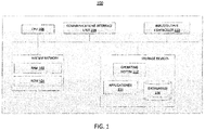
- FIG. 1 is a block diagram of a computing device 100 for performing any of the processes described herein, including processes 200, 300, and 500.
- processor or “computing device” refers to one or more computers, microprocessors, logic devices, servers, or other devices configured with hardware, firmware, and software to carry out one or more of the computerized techniques described herein.
- Processors and processing devices may also include one or more memory devices for storing inputs, outputs, and data which is currently being processed.
- the computing device 100 may include a "user interface," which may include, without limitation, any suitable combination of one or more input devices (e.g., keypads, touch screens, trackballs, voice recognition systems, etc.) and/or one or more output devices (e.g., visual displays, speakers, tactile displays, printing devices, etc.).
- the computing device 100 may include, without limitation, any suitable combination of one or more devices configured with hardware, firmware, and software to carry out one or more of the computerized techniques described herein.
- Each of the components described herein may be implemented on one or more computing devices 100.
- a plurality of the components of these systems may be included within one computing device 100.
- a component and a storage device may be implemented across several computing devices 100.
- the computing device 100 comprises at least one communications interface unit 108, an input/output controller 110, system memory, and one or more data storage devices.
- the system memory includes at least one random access memory (RAM 102) and at least one read-only memory (ROM 104). All of these elements are in communication with a central processing unit (CPU 106) to facilitate the operation of the computing device 100.
- the computing device 100 may be configured in many different ways. For example, the computing device 100 may be a conventional standalone computer or alternatively, the functions of computing device 100 may be distributed across multiple computer systems and architectures. In FIG. 1 , the computing device 100 is linked, via network or local network, to other servers or systems.
- the computing device 100 may be configured in a distributed architecture, wherein databases and processors are housed in separate units or locations. Some units perform primary processing functions and contain at a minimum a general controller or a processor and a system memory. In distributed architecture embodiments, each of these units may be attached via the communications interface unit 108 to a communications hub or port (not shown) that serves as a primary communication link with other servers, client or user computers and other related devices.
- the communications hub or port may have minimal processing capability itself, serving primarily as a communications router.
- a variety of communications protocols may be part of the system, including, but not limited to: Ethernet, SAP, SASTM, ATP, BLUETOOTHTM, GSM and TCP/IP.
- the CPU 106 comprises a processor, such as one or more conventional microprocessors and one or more supplementary co-processors such as math co-processors for offloading workload from the CPU 106.
- the CPU 106 is in communication with the communications interface unit 108 and the input/output controller 110, through which the CPU 106 communicates with other devices such as other servers, user terminals, or devices.
- the communications interface unit 108 and the input/output controller 110 may include multiple communication channels for simultaneous communication with, for example, other processors, servers or client terminals.
- the CPU 106 is also in communication with the data storage device.
- the data storage device may comprise an appropriate combination of magnetic, optical or semiconductor memory, and may include, for example, RAM 102, ROM 104, flash drive, an optical disc such as a compact disc or a hard disk or drive.
- the CPU 106 and the data storage device each may be, for example, located entirely within a single computer or other computing device; or connected to each other by a communication medium, such as a USB port, serial port cable, a coaxial cable, an Ethernet cable, a telephone line, a radio frequency transceiver or other similar wireless or wired medium or combination of the foregoing.
- the CPU 106 may be connected to the data storage device via the communications interface unit 108.
- the CPU 106 may be configured to perform one or more particular processing functions.
- the data storage device may store, for example, (i) an operating system 112 for the computing device 100; (ii) one or more applications 114 ( e.g ., computer program code or a computer program product) adapted to direct the CPU 106 in accordance with the systems and methods described here, and particularly in accordance with the processes described in detail with regard to the CPU 106; or (iii) database(s) 116 adapted to store information that may be utilized to store information required by the program.
- applications 114 e.g ., computer program code or a computer program product
- the operating system 112 and applications 114 may be stored, for example, in a compressed, an uncompiled and an encrypted format, and may include computer program code.
- the instructions of the program may be read into a main memory of the processor from a computer-readable medium other than the data storage device, such as from the ROM 104 or from the RAM 102. While execution of sequences of instructions in the program causes the CPU 106 to perform the process steps described herein, hard-wired circuitry may be used in place of, or in combination with, software instructions for embodiment of the processes of the present disclosure. Thus, the systems and methods described are not limited to any specific combination of hardware and software.
- Suitable computer program code may be provided for performing one or more functions as described herein.
- the program also may include program elements such as an operating system 112, a database management system and "device drivers" that allow the processor to interface with computer peripheral devices (e.g ., a video display, a keyboard, a computer mouse, etc.) via the input/output controller 110.
- computer peripheral devices e.g ., a video display, a keyboard, a computer mouse, etc.
- Non-volatile media include, for example, optical, magnetic, or opto-magnetic disks, or integrated circuit memory, such as flash memory.
- Volatile media include dynamic random access memory (DRAM), which typically constitutes the main memory.
- Computer-readable media include, for example, a floppy disk, a flexible disk, hard disk, magnetic tape, any other magnetic medium, a CD-ROM, DVD, any other optical medium, punch cards, paper tape, any other physical medium with patterns of holes, a RAM, a PROM, an EPROM or EEPROM (electronically erasable programmable read-only memory), a FLASH-EEPROM, any other memory chip or cartridge, or any other non-transitory medium from which a computer can read.
- a floppy disk a flexible disk, hard disk, magnetic tape, any other magnetic medium, a CD-ROM, DVD, any other optical medium, punch cards, paper tape, any other physical medium with patterns of holes, a RAM, a PROM, an EPROM or EEPROM (electronically erasable programmable read-only memory), a FLASH-EEPROM, any other memory chip or cartridge, or any other non-transitory medium from which a computer can read.
- Various forms of computer readable media may be involved in carrying one or more sequences of one or more instructions to the CPU 106 (or any other processor of a device described herein) for execution.
- the instructions may initially be borne on a magnetic disk of a remote computer (not shown).
- the remote computer can load the instructions into its dynamic memory and send the instructions over an Ethernet connection, cable line, or even telephone line using a modem.
- a communications device local to a computing device 100 e.g., a server
- the system bus carries the data to main memory, from which the processor retrieves and executes the instructions.
- the instructions received by main memory may optionally be stored in memory either before or after execution by the processor.
- instructions may be received via a communication port as electrical, electromagnetic or optical signals, which are exemplary forms of wireless communications or data streams that carry various types of information.
- FIG. 2 is a flowchart of a process 200 for designing and selecting a probe (e.g., a MIP), according to an illustrative embodiment.
- the process 200 includes the steps of determining a set of constraints (step 202), identifying primers using the set of constraints (step 204), performing an optimization technique to maximize the percentage of molecular captures on the target chromosome and to maximize the number of useful sites on the target chromosome (step 206), and selecting a probe based on the optimization technique (step 208).
- a set of constraints is determined.
- the set of constraints may be determined, for example, by CPU 106 using software or application(s) implemented thereon.
- the software or application(s) may also be used by CPU 106 to perform any one or more of the subsequent steps in process 200.
- the software and application(s) may be used by CPU 106 to find abundant primer pairs in a given reference genome (e.g., HG19) based on the determined constraints, and to automatically create suffix-array-based index for the genome file.
- the set of constraints may alternatively be referred to as algorithm flags.
- the constraints may include a length of the left primer, a minimum frequency of the primer-pair, a maximum distance between primers (e.g., amplicon length), a minimum and/or maximum total frequency of the primer, a minimum GC-content per primer in percent, a minimum amount of non-identical amplicons in percent, a distribution of primers in genome, or any suitable combination thereof.
- the following constraints may be used in designing primer pairs:
- a set of primers are identified using the set of constraints determined at step 202.
- any combination of the following parameters may be provided: the left primer sequences (e.g., as well as the number of their occurrences on the positive and negative strands of the genome), the right primer sequences (e.g., as well as the number of their occurrences on the positive and negative strands of the genome), the frequency of the pair (e.g., the left primer sequence and the right primer sequence paired together with the amplicon length limited by a constraint) including both unique and non-unique pairs, the frequency and percentage of the uniquely occurring amplicons, and the amplicon sequences from unique and non-unique pairs.
- each primer pair may be able to amplify multiple regions on the genome (e.g., more than hundreds, more than thousands, more than tens of thousands, more than hundreds of thousands, or more than millions).
- the generated primer pairs may identify or predict amplicon sites without allowing for any mismatches to occur in either the left primer sequence or in the right primer sequence (i.e., the left or right arms).
- a small number of mismatches may be allowed, such as allowing for:
- the amplicon prediction scheme described above provides the genomic coordinates of the predicted amplicons.
- the scheme may be divided into two parts. In a first part, the amplicon sites are identified without allowing for any mismatches to occur, and the genomic coordinates of the identified amplicon sites are not provided.
- the amplicon sites that include a small number of mismatches are identified, and the genomic coordinates of these amplicon sites are provided, as well as the genomic coordinate of the no-mismatch amplicon sites.
- Splitting up the scheme into these two modular parts may save computational complexity. However, in general, it will be understood that the two parts may be combined to provide the set of no-mismatch amplicon sites, mismatch amplicon sites, and their genomic coordinates in a single function.
- one or more of the amplicon sites identified at step 204 may be removed (e.g., by a filtering operation). For example, amplicon sites may be removed if the sites have mismatches that do not occur at least 3 base pairs (bp) from 5' end of the left primers and 3' end of the right primers.
- the amplicon sites of those primers that passed the filtering operation (hereinafter referred to as "candidate primers”) should enrich the chromosome of interest while targeting multiple regions of the reference genome (e.g., typically 2500 or more).
- both the left and right arm sequences of the candidate primers should have melting temperatures (T M ) ranging from low 60 to high 60s as computed by the nearest neighbor model of DNA binding stability, wherein empricial stability parameters are summed according to the nucleic acid sequence. See, e.g., Santa Lucia and Hicks 2004.
- T M melting temperatures
- the candidate primers should have high tolerance to mismatches occurring at 3bps on the 5' of the left arm and the 3' of the right arm.
- the remaining amplicon sites will be further processed in order to generate a set of parameter values for each candidate primer.
- the proportion of the number of amplicon sites coming from the chromosome of interest and the total number of amplicon sites that have passed the filtering operation will be calculated.
- the enrichment information e.g., the calculated proportion
- the associated amplicon sites information may be saved in a database, such as database 116.
- an optimization technique is performed to identify a primer with an optimal predicted performance.
- the optimization technique involves evaluating an objective function for each candidate primer.
- it may be desirable to use an objective function that maximizes a proportion of the captured sites that are on the chromosome of interest, compared to the number of captured sites that are on other chromosomes.
- the objective function may further maximize a number of sites from the chromosome of interest.
- the candidate primers may optionally include primers that have a high frequency of single nucleotide polymorphisms (SNP) in their predicted amplicon sites.
- SNP single nucleotide polymorphisms
- only a subset of the candidate primers with the highest proportion of sites coming from the chromosome of interest may be identified from among the primers with high overlaps and the remaining candidate primers may be spared for future use.
- the objective function for each candidate MIP may, in some embodiments, be established based on the following matrices: In the probe matrices above, rows labeled as "0 mismatch” indicates MIPs with perfect matches in both arms, and rows labeled as "1 mismatch” indicates primers that tolerates at most 1 mismatch in one of its arms. In Table 1, the column labeled as "unique” corresponds to the number of sites on the chromosome of interest that aligned only once to a respective MIP, where the alignment occurred on the chromosome of interest and on no other chromosome.
- the column labeled as “non-unique” corresponds to the number of sites on the chromosome of interest that aligned more than once to a respective MIP, where the alignment occurred multiple times on the chromosome of interest, on multiple chromosomes, or both.
- the column labeled as “unique” corresponds to the number of sites across all chromosomes (including all of chromosomes 1-22, X, and Y, for example) that aligned only once to a respective MIP.
- the value E includes a sum across multiple A i , which represents the number of unique sites with zero mismatches on the i-th chromosome.
- the column labeled as "non-unique" in Table 2 corresponds to the number of sites across all chromosomes that aligned more than once to a respective MIP, including instances where the alignment occurred multiple times on the same chromosome, on multiple chromosomes, or both.
- MIP that has a high percentage of sites that match the arm sequences (or at most have a small number of mismatches, such as one mismatch). It may further be desirable to select a MIP associated with a high percentage of unique sites. This may be represented by selecting MIPs that have values for A, C, E, or G (or any suitable combination thereof) to be relatively high compared to B, D, F, or H (or any suitable combination thereof). It may further be desirable to select a MIP associated with a high percentage of sites that are on the chromosome of interest. This may be represented by selecting MIPs that have values for A, B, C, or D (or any suitable combination thereof) to be relatively high compared to E, F, G, or H (or any suitable combination thereof).
- an objective function that maximizes or increases A/F may produce fewer ambiguous reads for candidate primers that tolerate 0 mismatches.
- an objective function that maximizes or increases A/E may produce target chromosome (e.g., chromosome 21) specific reads.
- an objective function that maximizes or increases A/C selects primers that have significantly more perfect matching sites than those that have 1 mismatch, and as a result, represents an efficient capture.
- an objective function that maximizes or increases A means that a large number of sites uniquely aligned to the chromosome of interest.
- an optimal primer can be selected to maximize an objective function of (A+C)/(E+F+G+H). To further illustrate this concept, three exemplary objective functions are explained in detail below.
- the numerator of EQ. 5 may be referred to herein as an adjusted number of usable sites (on the chromosome of interest).
- the denominator of EQ. 5 may be referred to herein as a number of sites on the genome.
- the value of P1 as defined in EQ. 5 may be referred to herein as an efficiency fraction or a useful fraction.
- the value for P defined in EQ. 9 may be compared to a predetermined threshold to assess whether to select an associated candidate primer or probe for further testing or for diagnosis.
- the predetermined threshold may be a value such as 1, 2, 3, 4, 5, 6, 7, 8, 9, 10, or any other suitable number.
- any of the above-described examples, and any combination of the examples, may be used as an objective function. Selecting a primer by optimizing an objective function in this manner has an advantage of reducing a necessary read depth to achieve a sufficient number of useful reads. In principle, the improvement in read depth scales linearly with an improvement in reduction of ambiguity.
- a primer is selected from the set of candidate primers based on the optimization technique performed at step 206.
- the selected primer may correspond to the primer with the optimal predicted performance, i.e., the primer that maximized the objective function as described in relation to step 206.
- a number of primers are designed, synthesized, and tested.
- a value is generated for each of one or more of the numbers of predicted site counts in Tables 1 and 2, based on a searching and counting process for unique and non-unique sites (with no mismatches or one mismatch) on each chromosome of interest and across the genome (including autosomes and sex chromosomes, for example).
- Any of the above-described examples or combinations of the above examples may be used as an objective function, to represent a score that is assigned to each primer and represents that primer's performance.
- the primers may be compared to one another based on their performance, and the best performing primers may be selected.
- a primer's score is compared to a predetermined threshold, to determine whether the primer is selected or not.
- the threshold may be set to a number, such as 2, 3, 4, 5, 6, 7, 8, 9, or 10.
- a primer with a high score (e.g., MIP 003 in the above table) was selected for further analysis.
- MIP 003 was selected because it was associated with a high value for the score as assessed according to EQ. 9.
- MIP 003 had a score of 8.557.
- Table 3 summarizes the predicted numbers of unique sites that aligned to MIP 003 by different chromosomes with no mismatch (A) and with one mismatch (C).
- process 200 may be used with any other embodiment of this disclosure.
- steps and descriptions described in relation to FIG. 2 may be done in alternative orders or in parallel to further the purpose of this disclosure.
- each of these steps may be performed in any order or in parallel or substantially simultaneously to reduce lag or increase the speed of the system or method.
- process 200 may be carried out using computing device 100, and more particularly, CPU 106 of computing device 100.
- FIG. 3 is a flowchart of a process 300 for predicting aneuploidy state in a test subject, according to an illustrative embodiment.
- the process 300 includes the steps of receiving sequencing data for a test subject (step 302), computing site capture metric for the test subject (step 304), receiving site capture metrics for a set of reference subjects (step 306), and predicting aneuploidy state in the test subject based on comparison of site capture metric for the test subject to site capture metrics for the reference subjects (step 308).
- the site capture metric is a site capture efficiency index (SCE), which is the ratio between the number of unique molecular identifier tags and the number of reads for each individual site.
- SCE site capture efficiency index
- the site capture metric is a site capture consistency measure (SCC), which is calculated as the coefficient of variability of SCE for each individual site.
- SCC site capture consistency measure
- chromosome 1 : 1-100 we have 100 aligned reads and 99 unique molecular identifier tags. Accordingly, the SCE will be 99%.
- out of 100 samples for chromosome 1 : 1-100 all 100 samples have over 90% SCE
- out of the 100 samples for chromosome 3 : 500-600 only 50 samples have over 90% SCE and the remaining 50 samples have less than 90% SCE. Accordingly, the SCC will indicate that chromosome 1 : 1-100 is a more consistent site compared to chromosome 3: 500-600.
- each RO-MIP includes in sequence a first targeting polynucleotide arm, a first unique targeting molecular tag, a polynucleotide linker, a second unique targeting molecular tag, and a second targeting polynucleotide arm.
- the first and second targeting polynucleotide arms are the same across the MIPs in the population, while the first and second unique targeting molecular tags are distinct across the MIPs in the population.
- RO-MIPs amplicons result from the capture of the sites, and the amplicons are sequenced to obtain the sequencing data.
- a chromosomal fraction is computed for the test subject by evaluating a ratio between a sum of all unique capture events from the chromosome of interest (S1) and a sum of all unique capture events from all chromosomes (S1+S2)..
- the chromosomal fraction provides a proportional measure of the chromosome of interest in a given sample (i.e., the proportion of reads that comes from the chromosome of interest).
- One example method of computing the chromosomal fraction is described in relation to steps 426, 428, and 430 in FIG. 4 .
- a set of chromosomal fractions for a set of reference subjects is received.
- the reference subjects may correspond to a group of people that exhibit a known euploidy state.
- the subjects may exhibit monosomy, disomy, or trisomy for a particular chromosome of interest.
- the chromosomal fractions for the reference subjects are computed in the same manner as was described in relation to step 304, but for each reference subject.
- the chromosomal fractions are representative of the ability of the selected RO-MIP to capture sequences on the chromosome of interest, as compared to other chromosomes.
- the chromosomal fraction for the test subject is compared to the chromosomal fractions for the reference subjects (obtained at step 306), and the euploidy state of the test subject is predicted based on this comparison.
- a statistical test may be used to compare the test chromosomal fraction to the population of reference chromosomal fractions, and determine whether the test chromosomal fraction belongs in any cluster of reference chromosomal fractions associated with the same euploidy state.
- test ratio may include the chromosomal fraction.
- FIG. 4 is a flowchart of a process 400 for predicting a euploidy state of a test subject, according to an illustrative embodiment.
- the process 400 may be used to implement the step 308 of the process 300 shown and described in relation to FIG. 3 .
- a chromosomal fraction may be used to predict a euploidy state in a test subject that has an unknown euploidy state.
- the process 400 includes the steps of receiving data recorded from S samples with known genotypes (step 402) and initializing a sample iteration parameter s to 1 (step 404). For each sample s, the process 400 includes filtering the sequencing reads to remove known artifacts (step 406), aligning the reads to the human genome (step 408), setting a site iteration parameter k to 1 (step 412), and determining a number of capture events for the k-th site (step 414).
- the process 400 further includes the steps of identifying a subset of the K sites that satisfy at least one criterion (step 424), computing a first sum S1 of tag counts from a chromosome of interest for a test sample t in the S samples (step 426), computing a second sum S2 of tag counts from chromosomes other than the chromosome of interest for the test sample t (step 428), computing a chromosomal fraction as S1/(S1+S2) for the test sample t (step 430), and selecting a euploidy state for the test sample t by comparing the chromosomal fraction for the test sample t to a set of reference chromosomal fractions (step 432).
- step 402 data recorded from a set of S samples is received, where the S samples are each obtained from a different subject. At least one of the S samples is obtained from a test subject, whose euploidy state may be unknown.
- the samples may be nucleic acid samples isolated from the subjects, and the data may include sequencing data obtained from the nucleic acid samples.
- the sequencing data is obtained by using a population of RO-MIPs to amplify a set of sites in the nucleic acid sample to produce a set of RO-MIPs amplicons.
- the RO-MIPs amplicons may then be sequenced to obtain the sequencing data received at step 402.
- a sample iteration parameter s is initialized to 1. As the S samples are processed, the sample iteration parameter s is incremented until each of the S samples is processed to determine the number of capture events for each site.
- the sequencing reads for sample s are filtered to remove known artifacts.
- the data received at step 402 may be processed to remove an effect of probe-to-probe interaction.
- the ligation and extension targeting arms of all RO-MIPs are matched to the paired-end sequence reads. Reads that failed to match both arms of the RO-MIPs are determined to be invalid and discarded. The arm sequences for the remaining valid reads are removed, and the molecular tags from both ligation and extension ends may be also removed from the reads. The removed molecular tags may be kept separately for further processing at step 414.
- the resulting trimmed reads are aligned to the human genome.
- an alignment tool may be used to align the reads to a reference human genome.
- an alignment score may be assessed for representing how well does a specific read align to the reference.
- Reads with alignment scores above a threshold may be referred to herein as primary alignments, and are retained.
- reads with alignment scores below the threshold may be referred to herein as secondary alignments, and are discarded. Any reads that aligned to multiple locations along the reference genome may be referred to herein as multi-alignments, and are discarded.
- a site iteration parameter k is initialized to one.
- the number of capture events for the k-th site is determined, and the site iteration parameter k is incremented at step 418 until all K sites have been considered.
- step 424 the process 400 proceeds to step 424 to identify a subset of the K sites that satisfies at least one criterion. For example, a site capture consistency measure may be evaluated as a coefficient of variation of the number of capture events across the S samples, and those sites having high coefficients of variation may be discarded.
- a sum S1 of the tag counts from the chromosome of interest for a test sample is computed, and at step 428, a sum S2 of the tag counts from chromosomes other than the chromosome of interest for the test sample is computed.
- a chromosomal fraction is computed as S1/(S1+S2). The chromosomal fraction for the test sample is then compared to a set of reference chromosomal fractions (that have been computed from reference subjects that have known euploidy states), and a statistical test is performed to select a predicted euploidy state for the test subject at step 432.
- compositions and methods described herein may be adapted and modified.
- Example 1 MIP design and method for capturing target sequences of interest Probe construction
- a single oligonucleotide ranging in size between 80 and 105bp (depending on the length of the first and second targeting polynucleotide arms) is synthesized as shown in FIG. 5 .
- the 6N boxes refer to molecular tag sequences that are used to quantitate capture events for each target sequence of interest. In this particular embodiment, instead of counting reads, the number of unique sequences per captured site are counted.
- the first and second targeting polynucleotide arms (at an empirically determined concentration) is mixed with csDNA extracted from 1-2 mL plasma in a 20 uL buffered reaction.
- the mixture is incubated in a thermocycler at temperatures that have been optimized for annealing of probes to the template (98 °C for 3 min ⁇ 85 °C for 30 min ⁇ 66 °C for 15 min). During this incubation, the probe molecules anneal to the csDNA template at specific chromosomal locations that are complementary to the probe sequence ( FIG. 6 , middle).
- the most easily predicted sites are those with sequences that are exactly complementary to the first and second targeting polynucleotide arms (invariant sites), but sites that have one or more variants in either arm are also targeted at somewhat lower efficiency.
- the optimal amount of MIP for each reaction is dependent on three main variables: 1) the number of genomes used as template, which can vary widely among individuals, 2) the overall number of sites targeted by the specific probe, and 3) the ratio of invariant sites to variant sites.
- the purified PCR products are pooled into a library such that all samples are at equal concentration, ensuring that the read budget is divided evenly across the samples.
- the library is sequenced using either single-end or paired-end sequencing, using 75-100 cycles in order to determine the full sequence of the site-specific gap. If single-end sequencing is used, the read will consist of the ligation arm followed by the molecular tag and the unique gap sequence that was filled in during the ext/lig step. Sequencing into the extension arm is unnecessary because the sequence is known from the probe.
- Example 2 Sequence data analysis and detection of aneuploidy in a subject
- Raw sequencing data must be processed in order for it to be useful in detecting aneuploidy.
- sequencing reads are filtered to remove known artifacts such as probe-to-probe interaction, backbone sequences or adapter sequences.
- the ligation and extension arms of the MIP i.e., the first and second targeting polynucleotide arms
- the arm sequences for the valid reads are trimmed to remove the non-genomic portions from the sequence data by deleting the sequence from the read files used in subsequent processing steps..
- the molecular tags from both the ligation and the extension ends are kept separately for counting of the capture events in a later step.
- the trimmed reads are aligned to the human genome (hg19) with the bowtie2 software program.
- the aligned reads are then filtered with the samtools software program to remove reads that do not align as a valid pair, or that align non-uniquely.
- the alignment and filtering parameters are carefully chosen to keep uniquely aligned reads only.
- the aligned and filtered reads (in bam format files) are examined to count the unique molecular tags for each targeted site with a unique MIP gap sequence (i.e., a unique target sequence of interest).
- a site capture efficiency index (SCE) is calculated for each individual site.
- a site capture consistency measure (SCC) is calculated as the coefficient of variability of SCE for each individual site. A portion of these sites are discarded based on site capture efficiency variability measures from experimental data. This step helps to reduce sample-to-sample variability in the chromosomal proportion and increase the Z-score of positive samples calculated in a later step. For a given sample, the sum (S1) of unique molecular tags counts from the remaining sites on the chromosome of interest (e.g., chromosome 21) is calculated and stored.
- the sum (S2) of the remaining reference sites is calculated and stored.
- the Z-score of the chromosome of interest e.g., chromosome 21
- normal fetal chromosome count i.e. normal karyotope fetus.
- the determination of aneuploidy (e.g., trisomy and monosomy) or euploidy is made when the absolute value of the Z-score of a given sample is above a certain threshold.
- This example describes using the method described herein to discriminate (or distinguish) between pregnant women carrying trisomy 13, 18, and 21 and healthy pregnant women.
- a training set of 48 plasma samples from pregnant women is used to determine Z-score cutoffs for detecting trisomy 13, 18, and 21. Each of the plasma samples is taken from a different pregnant woman.
- the 48 plasma samples contain 40 healthy samples, four Trisomy 21 samples, three Trisomy 18 samples, and one Trisomy 13 sample. Trisomy cases are confirmed by karyotype analysis of placental or fetal cells collected by CVS (Chorionic villus sampling) or amniocentesis.
- Site capture reaction Plasma DNA extracted from each of the 48 pregnant women is subjected to a site capture reaction as follows: the extracted plasma DNA ismixed with water, Ampligase buffer (1x), and the RO-MIP at the empirically determined probe concentration. The site capture reaction mixture is incubated in a thermal cycler at 98°C for 3 min, then 85°C for 30 min, then 66°C for 15 min. After the incubation, a master mixture containing dNTP (0.6 mM), NAD (0.4x), betaine (0.3M), Ampligase buffer (1x), water, Ampligase (5 units), and Phusion HF polymerase (0.4 units) is added to the site capture reaction mixture. The combined mixture is then incubated in a thermal cycler at 66°C for 60 min, 72°C for 30 min, and held at 4°C.
- Captured site amplification After the incubation, 20 ⁇ L of the combined mixture is added to a PCR master mixture containing forward (500 nM) and reverse index primers (500 nM), Phusion HF buffer (1x), dNTPs (0.2 mM), water, and Phusion HS polymerase (0.4 units). Each sample is barcoded with a unique reverse primer index.
- the PCR reaction mixture is incubated in a thermal cycler at 98°C for 3 min, then 20 cycles at 98°C for 10 sec, 65°C for 20 sec, and 72°C for 30 sec. The PCR reaction mixture is then held at 72°C for 5 min and then 4°C.
- Single-end sequencing The PCR amplified libraries are purified using AmpureXP beads and samples are pooled together at equal concentration (48 samples per pool). The multiplexed libraries are each loaded on a single SR flowcell and sequenced in Rapid Run mode on an HiSeq 2500 for 106 cycles.
- a chromosome proportion (P i ) is calculated for chromosomes 13, 18, and 21 by dividing the number of unique molecular tags/identifiers from reads that are uniquely aligned to each chromosome (i.e., chromosome 13, 18, or 21) (this numerator is analogous to the sum of "A” and "C") by the total number of unique molecular tags/identifiers from reads that are uniquely aligned to chromosomes 1 to 22 (this denominator is analogous to the sum of "A", “C", "E”, and "G”).
- the denominator here may not include reads that align to the X or Y chromosomes.
- the number of reads that align to the X or Y chromosomes may be significantly larger than the number of reads that align to other chromosomes, including the chromosome of interest.
- including the reads that align to the X or Y chromosomes in the denominator may significantly reduce the resulting ratio, and may introduce noise and distortion to the calculation of the z-statistic.
- the reads that align to the X or Y chromosomes may be excluded from both the numerator and the denominator.
- the chromosome of interest is a sex chromosome (e.g., when detecting a sex chromosome aneuploidy)
- the reads that align to the X and/or Y chromosomes may be included in the numerator and the denominator.
- the mean (x i ) and standard deviation (s i ) is calculated for the unaffected samples and used to calculate a Z-score ([P i - x i ]/s i ) for each sample.
- Analysis of the Z-scores reveals that all healthy samples have Z-scores ⁇ 3.0 for chromosomes 13, 18, and 21, while positive samples have Z-scores >3.0 for chromosomes 13, 18, and 21. Therefore, it is determined that samples with a Z-score >3.0 will be called positive for trisomy 13, 18, and 21. Samples with a Z-score ⁇ 3.0 will be called negative for trisomy 13, 18, and 21.
- a test set of 422 samples from pregnant women is used to determine sensitivity and specificity of the test. Each sample is from a different pregnant woman. These 422 samples contain 387 healthy samples, 21 T21 samples, 9 T18 samples, and 5 T13 samples. Trisomy cases are confirmed by karyotype analysis of placental or fetal cells collected by CVS or amniocentesis.
- Site capture reaction Plasma DNA extracted from each of the 422 pregnant women is subjected to a site capture reaction as follows: the extracted plasma DNA is mixed with water, Ampligase buffer (1x), and the RO-MIP at the empirically determined probe concentration. The capture reaction mixture is incubated in a thermal cycler at 98°C for 3 min, then 85°C for 30 min, then 66°C for 15 min. After the incubation, a master mixture containing dNTP (0.6 mM), NAD (0.4x), betaine (0.3M), Ampligase buffer (1x), water, Ampligase (5 units), and Phusion HF polymerase (0.4 units) is added to the site capture reaction mixture. The combined reaction mixture is incubated in a thermal cycler at 66°C for 60 min, 72°C for 30 min, and held at 4°C.
- Captured site amplification After the incubation, 20 ⁇ L of the combined mixture is added to a PCR master mixture containing forward (500 nM) and reverse index primers (500 nM), Phusion HF buffer (1x), dNTPs (0.2 mM), water, and Phusion HS polymerase (0.4 units). Each sample is barcoded with a unique reverse primer index.
- the PCR reaction mixture is incubated in a thermal cycler at 98°C for 3min, then 20 cycles at 98°C for 10 sec, 65°C for 20 sec, and 72°C for 30 sec. The PCR reaction mixture is held at 72°C for 5 min and then 4°C.
- the amplified libraries are purified using AmpureXP beads and samples are pooled together at equal concentration (48 samples per pool). There are 9 pools total: the first eight pools having 47 test samples and 1 control sample per pool and the ninth pool has 46 test samples and 1 control sample.
- the multiplexed libraries are each loaded on a single SR flowcell and sequenced in Rapid Run mode on an HiSeq 2500 for 106 cycles.
- Sensitivity e.g., the true positive rate, or the number of true positives divided by the sum of true positives and false negatives
- Sensitivity is >99.2% for chromosome13, >99.9% for chromosome 18, and >99.5% for chromosome 21.
- the graph in FIG. 10 shows the primer's test performance in detecting Trisomy 13.
- the y-axis in FIG. 10 represents the z-statistic
- the x-axis represents the five Trisomy 13 samples on the left (in no particular order) and the remaining 545 samples on the right (in order of decreasing z-statistic).
- all five Trisomy 13 samples had z-statistics higher than 3.0 and were correctly identified as positive samples using the primer, leading to a 5/5 sensitivity.
- a small number of healthy samples had z-statistics greater than 3.0, leading to a 99.2% specificity.
- FIG. 11 is similar to FIG. 10 , except that FIG. 11 shows the primer's test performance in detecting Trisomy 18. All nine Trisomy 18 samples had z-statistics higher than 3.0 and were correctly identified as positive samples using the primer, leading to a 9/9 sensitivity. Moreover, nearly all of the healthy samples were correctly identified as negative samples, leading to a specificity rate greater than 99.9%.
- FIG. 12 is also similar to FIG. 10 , except that FIG. 12 shows the primer's test performance in detecting Trisomy 21. All 21 Trisomy 21 samples were correctly identified as positive samples using the primer, leading to a 21/21 sensitivity. Moreover, nearly all of the healthy samples were correctly identified as negative samples, leading to a specificity rate greater than 99.8%.
- results shown in FIGS. 10-12 indicate that the systems and methods of the present disclosure provide a useful tool for selecting a primer with high sensitivity and specificity in detecting aneuploidy.
- Other methods may also have similar levels of performance (as measured by sensitivity and/or specificity, for example). For example, similar performance may be achieved using shotgun sequencing.
- the present disclosure has several advantages over other approaches because the present disclosure uses only a single primer, and may therefore be cheaper, simpler, and more efficient than those other approaches.
- the examples provided by this disclosure focus primarily on a number of different example embodiments of systems and methods to determine copy number variations, chromosomal abnormalities, or micro-deletions.
- variations in the general shape and design of one or more embodiments may be made without significantly changing the functions and operations of the present disclosure.
- the features and limitations described in any one embodiment may be applied to any other embodiment herein, and the descriptions and examples relating to one embodiment may be combined with any other embodiment in a suitable manner.
- the figures and examples provided in disclosure are intended to be only exemplary, and not limiting.
- the systems and/or methods described above may be applied to, or used in accordance with, other systems and/or methods, including systems and/or methods which may or may not be directly related to determining copy number variations.
Claims (17)
- Procédé de dépistage de l' aneuploïdie chez un fœtus, comprenant :a) l'obtention d'un échantillon d'acide nucléique isolé à partir d'un échantillon de sang maternel ;b) la capture d'une pluralité de séquences d'intérêt cibles dans l'échantillon d'acide nucléique obtenu dans l'étape a) en utilisant une ou plusieurs populations de sondes d'inversion moléculaire (MIP) pour produire une pluralité de réplicons, dans lequel chacune des MIP dans la population de MIP comprendi) deux bras polynucléotidiques de ciblage, dans lequel les paires de premier et deuxième bras polynucléotidiques de ciblage dans chacune des MIP sont identiques, et sont sensiblement complémentaires de première et deuxième régions dans l'acide nucléique qui, respectivement, flanquent chaque séquence dans la pluralité de séquences d'intérêt cibles ;ii) une ou plusieurs étiquettes moléculaires uniques, etiii) un lieur polynucléotidique ;c) le séquençage d'une pluralité d'amplicons de MIP qui sont amplifiés à partir des réplicons obtenus dans l'étape b) ;d) la détermination du nombre d'événements de capture de chacune d'une première population d'amplicons de la pluralité d'amplicons fournis dans l'étape c) sur la base du nombre des étiquettes moléculaires uniques de chaque MIP qui a amplifié un réplicon, dans lequel la première population d'amplicons est déterminée par la séquence de la séquence d'intérêt cible ;e) la détermination du nombre d'événements de capture de chacun d'une deuxième population d'amplicons de la pluralité d'amplicons fournis dans l'étape c) sur la base du nombre des étiquettes moléculaires uniques de chaque MIP qui a amplifié un réplicon, dans lequel la deuxième population d'amplicons est déterminée par la séquence de la séquence d'intérêt cible ;f) la détermination, pour chaque séquence d'intérêt cible à partir de laquelle la première population d'amplicons a été produite, d'une mesure métrique de capture de site sur la base, au moins en partie, du nombre d'événements de capture déterminé dans l'étape d) ;g) l'identification d'un premier sous-ensemble des mesures métriques de capture de site déterminées dans l'étape f) qui satisfont à au moins un critère ;h) la détermination, pour chaque séquence d'intérêt cible à partir de laquelle la deuxième population d'amplicons a été produite, d'une mesure métrique de capture de site sur la base, au moins en partie, du nombre d'événements de capture déterminé dans l'étape e) ;i) l'identification d'un deuxième sous-ensemble des mesures métriques de capture de site déterminées dans l'étape h) qui satisfont à au moins un critère ;j) la normalisation d'une première mesure déterminée à partir du premier sous-ensemble de mesures métriques de capture de site identifié dans l'étape g) par une deuxième mesure déterminée à partir du deuxième sous-ensemble de mesures métriques de capture de site identifié dans l'étape i) pour obtenir un rapport d'essai ;k) la comparaison du rapport d'essai à une pluralité de rapports de référence qui sont calculés sur la base d'échantillons d'acide nucléique de référence isolés à partir de sujets de référence connus pour présenter une euploïdie ou une aneuploïdie pour déterminer la présence ou l'absence d'un rapport d'essai indicatif d'une aneuploïdie chez le fœtus.
- Procédé selon la revendication 1, dans lequel lesdites une ou plusieurs étiquettes moléculaires sont au moins deux étiquettes moléculaires dans la séquence : premier bras polynucléotidique de ciblage - première étiquette moléculaire unique - lieur polynucléotidique - deuxième étiquette moléculaire unique - deuxième bras polynucléotidique de ciblage ; dans lequel les première et deuxième étiquettes moléculaires de ciblage uniques dans chacune des MIP en combinaison sont distinctes dans chacune des MIP.
- Procédé selon la revendication 1 ou 2, dans lequel l'échantillon d'acide nucléique est de l'ADN ou de l'ARN.
- Procédé selon l'une quelconque des revendications 1 à 3, dans lequel la mesure métrique de capture de site est un index d'efficacité de capture de site (SCE) ou est une mesure de cohérence de capture de site (SCC).
- Procédé selon l'une quelconque des revendications 1 à 4, dans lequel chacun des réplicons de MIP fournis dans l'étape b) est produit par :i) hybridation des premier et deuxième bras polynucléotidiques de ciblage, respectivement, aux première et deuxième régions dans l'échantillon d'acide nucléique, respectivement, dans lequel les première et deuxième régions flanquent une séquence d'intérêt cible ; etii) après l'hybridation, utilisation d'un mélange de ligature/extension pour étendre et ligaturer la région de brèche entre les deux bras polynucléotidiques de ciblage pour former des molécules d'acide nucléique circulaire simple brin.
- Procédé selon l'une quelconque des revendications 2 à 5, où le procédé comprend, avant l'étape de séquençage de c), une réaction de PCR d'indexation, dans lequel la réaction de PCR d'indexation introduit dans chacun des amplicons de MIP les composants suivants : une paire d'amorces d'indexation, un code-barres d'échantillon unique et une paire d'adaptateurs de séquençage,
de préférence dans lequel les amplicons de MIP à code-barres comprennent en séquence les composants suivants : un premier adaptateur de séquençage - une première amorce de séquençage- la première étiquette moléculaire de ciblage unique - le premier bras polynucléotidique de ciblage - l'acide nucléique capturé - le deuxième bras polynucléotidique de ciblage - la deuxième étiquette moléculaire de ciblage unique - un code-barres d'échantillon unique - une deuxième amorce de séquençage - un deuxième adaptateur de séquençage. - Procédé selon l'une quelconque des revendications 1 à 6, dans lequel le rapport d'essai et les rapports de référence sont des fractions chromosomiques, de préférence dans lequel les fractions chromosomiques sont définies par un rapport entre une somme de tous les événements de capture uniques d'un chromosome d'intérêt (S1) et une somme de tous les événements de capture uniques de tous les chromosomes (S1+S2) .
- Procédé de dépistage de l'aneuploïdie chez un fœtus, comprenant :a) l'obtention d'un échantillon d'ADN génomique à partir d'un échantillon de sang maternel ;b) l'ajout de l'échantillon d'ADN génomique dans chaque puits d'une plaque multipuits, dans lequel chaque puits de la plaque multipuits comprend un mélange de sondes, dans lequel le mélange de sondes comprend une population de sondes d'inversion moléculaire (MIP) et un tampon ; dans lequel chaque MIP dans la population de MIP comprendi) deux bras polynucléotidiques de ciblage, dans lequel les paires de premier et deuxième bras polynucléotidiques de ciblage dans chacune des MIP sont identiques, et sont sensiblement complémentaires de première et deuxième régions dans l'acide nucléique qui, respectivement, flanquent chaque séquence dans la pluralité de séquences d'intérêt cibles ;ii) une ou plusieurs étiquettes moléculaires uniques, etiii) un lieur polynucléotidique ;c) l'incubation de l'échantillon d'ADN génomique avec le mélange de sondes pour que les MIP capturent la pluralité de séquences d'intérêt cibles ;d) l'ajout d'un mélange d'extension/ligature à l'échantillon de c) pour que les MIP et la pluralité de séquences d'intérêt cibles forment une pluralité d'amplicons de MIP, dans lequel le mélange d'extension/ligature comprend une polymérase, une pluralité de dNTP, une ligase, et un tampon ;e) l'ajout d'un mélange d'exonucléases aux amplicons de MIP de ciblage et de contrôle pour éliminer les sondes en excès ou l'ADN génomique en excès ;f) l'ajout d'un mélange de PCR d'indexation à l'échantillon de e) pour ajouter une paire d'amorces d'indexation, un code-barres d'échantillon unique et une paire d'adaptateurs de séquençage à la pluralité d'amplicons ;g) l'utilisation d'un procédé de séquençage parallèle massif pour déterminer le nombre de lectures de séquençage d'une première population d'amplicons à code-barres fournis dans l'étape f) sur la base du nombre des étiquettes moléculaires de ciblage uniques, dans lequel la première population d'amplicons à code-barres est identifiée par la séquence de la séquence d'intérêt cible ;h) l'utilisation d'un procédé de séquençage parallèle massif pour déterminer le nombre de lectures de séquençage d'une deuxième population d'amplicons à code-barres fournis dans l'étape f) sur la base du nombre des étiquettes moléculaires de ciblage uniques, dans lequel la deuxième population d'amplicons à code-barres est identifiée par la séquence de la séquence d'intérêt cible ;i) calcul d'une mesure métrique de capture de site sur la base, au moins en partie, du nombre de premières lectures de séquençage déterminé dans l'étape g) et d'une pluralité de mesures métriques de capture de sonde de contrôle sur la base, au moins en partie, des nombres de deuxièmes lectures de séquençage déterminés dans l'étape h) ;j) l'identification d'un sous-ensemble de mesures métriques de capture de site de la population des amplicons de MIP qui ont des mesures métriques de capture de sonde de contrôle satisfaisant à au moins un critère ;k) la normalisation de la mesure métrique de capture de site par un facteur calculé à partir du sous-ensemble de mesures métriques de capture de sonde de contrôle satisfaisant à l'au moins un critère, pour obtenir une mesure métrique de capture de site normalisée d'essai ;l) la comparaison de la mesure métrique de capture de site normalisée d'essai à une pluralité de mesures métriques de capture de site normalisées de référence qui sont calculées sur la base d'échantillons d'ADN génomique de référence obtenus à partir de sujets de référence présentant des génotypes connus en utilisant les mêmes sites cibles et de contrôle, population cible, sous-ensemble de populations de contrôle dans les étapes b) à h) pour déterminer la présence ou l'absence d'une mesure métrique de capture de site normalisée d'essai indicative d'aneuploïdie chez le fœtus.
- Procédé selon la revendication 8, dans lequel lesdites une ou plusieurs étiquettes moléculaires sont au moins deux étiquettes moléculaires dans la séquence : premier bras polynucléotidique de ciblage - première étiquette moléculaire unique - lieur polynucléotidique - deuxième étiquette moléculaire unique - deuxième bras polynucléotidique de ciblage ; dans lequel les première et deuxième étiquettes moléculaires de ciblage uniques dans chacune des MIP en combinaison sont distinctes dans chacune des MIP.
- Procédé selon l'une quelconque des revendications 1 à 9, dans lequel l'échantillon de sang est un échantillon de sang total, un échantillon de plasma, ou un échantillon de sérum, de préférence un échantillon de plasma.
- Procédé selon l'une quelconque des revendications 1 à 10, dans lequela) la longueur du premier et/ou du deuxième bras polynucléotidique de ciblage est comprise entre 14 et 30 paires de bases ; et/ou dans lequel chacun des bras polynucléotidiques de ciblage a une température de fusion comprise entre 45 °C et 80 °C ; et/ou dans lequel chacun des bras polynucléotidiques de ciblage a une teneur en GC comprise entre 30 % et 80 %, de préférence entre 30 % et 70 % ; et/oub) dans lequel la longueur de la première et/ou la deuxième étiquette moléculaire unique est comprise entre 4 et 15 paires de bases ; et/ou dans lequel chacune des étiquettes moléculaires uniques a une température de fusion comprise entre 45 °C et 80 °C ; et/ouc) dans lequel le lieur polynucléotidique a une longueur comprise entre 20 et 1 000 paires de bases, et/ou dans lequel le lieur polynucléotidique a une température de fusion comprise entre 45 °C et 80 °C, et/ou dans lequel le lieur polynucléotidique a une teneur en GC comprise entre 30 % et 80 %, de préférence entre 30 % et 70 %.
- Procédé selon l'une quelconque des revendications 1 à 11, dans lequel le lieur polynucléotidique comprend au moins une amorce d'amplification, de préférence comprenant une amorce d'amplification sens et une amorce d'amplification antisens.
- Procédé selon la revendication 12, dans lequel :a) la séquence de l'amorce d'amplification sens comprend la séquence nucléotidique de :
5'-CTTCAGCTTCCCGATTACGG-3' (SEQ ID NO : 1) ; et/oub) la séquence de l'amorce d'amplification antisens comprend la séquence nucléotidique de
5'-GCACGATCCGACGGTAGTGT-3' (SEQ ID NO : 2). - Procédé selon l'une quelconque des revendications 1 à 13, dans lequel :a) le lieur polynucléotidique comprend la séquence nucléotidique de
5'-CTTCAGCTTCCCGATTACGGGCACGATCCGACGGTAGTGT-3' (SEQ ID NO : 3) . - Procédé selon l'une quelconque des revendications 1 à 14, dans lequel le premier bras polynucléotidique de ciblage comprend la séquence nucléotidique de 5'-CACTGCACTCCAGCCTGG-3' (SEQ ID NO : 4) ; et/ou le deuxième bras polynucléotidique de ciblage comprend la séquence nucléotidique de
5'-GAGGCTGAGGCAGGAGAA-3' (SEQ ID NO : 5). - Procédé selon l'une quelconque des revendications 1 à 15, dans lequel la MIP comprend la séquence nucléotidique de 5'-CACTGCACTCCAGCCTGG(N1-6)CTTCAGCTTCCCGATTACGGGCACGATCCGACGGTA GTGT (N7-12) GAGGCTGAGGCAGGAGAA-3' (SEQ ID NO : 6), où (N1-6) représente la première étiquette moléculaire unique et (N7-12) représente la deuxième étiquette moléculaire unique.
- Procédé selon l'une quelconque des revendications 1 à 16, dans lequel l'étape de séquençage a une profondeur de lecture comprise entre 6 et 8 millions de lectures.
Applications Claiming Priority (3)
| Application Number | Priority Date | Filing Date | Title |
|---|---|---|---|
| US201562198654P | 2015-07-29 | 2015-07-29 | |
| PCT/US2016/044914 WO2017020023A2 (fr) | 2015-07-29 | 2016-07-29 | Acides nucléiques et procédés de détection d'anomalies chromosomiques |
| EP16748036.7A EP3329010B1 (fr) | 2015-07-29 | 2016-07-29 | Acides nucléiques et procédés de détection d'anomalies chromosomiques |
Related Parent Applications (1)
| Application Number | Title | Priority Date | Filing Date |
|---|---|---|---|
| EP16748036.7A Division EP3329010B1 (fr) | 2015-07-29 | 2016-07-29 | Acides nucléiques et procédés de détection d'anomalies chromosomiques |
Publications (2)
| Publication Number | Publication Date |
|---|---|
| EP3608420A1 EP3608420A1 (fr) | 2020-02-12 |
| EP3608420B1 true EP3608420B1 (fr) | 2021-05-19 |
Family
ID=56610036
Family Applications (2)
| Application Number | Title | Priority Date | Filing Date |
|---|---|---|---|
| EP19185115.3A Active EP3608420B1 (fr) | 2015-07-29 | 2016-07-29 | Acides nucléiques et procédés de détection d'anomalies chromosomiques |
| EP16748036.7A Not-in-force EP3329010B1 (fr) | 2015-07-29 | 2016-07-29 | Acides nucléiques et procédés de détection d'anomalies chromosomiques |
Family Applications After (1)
| Application Number | Title | Priority Date | Filing Date |
|---|---|---|---|
| EP16748036.7A Not-in-force EP3329010B1 (fr) | 2015-07-29 | 2016-07-29 | Acides nucléiques et procédés de détection d'anomalies chromosomiques |
Country Status (8)
| Country | Link |
|---|---|
| US (3) | US10465245B2 (fr) |
| EP (2) | EP3608420B1 (fr) |
| JP (1) | JP6830094B2 (fr) |
| CN (1) | CN108350500A (fr) |
| CA (1) | CA2993347A1 (fr) |
| ES (1) | ES2745556T3 (fr) |
| HK (1) | HK1256543A1 (fr) |
| WO (1) | WO2017020023A2 (fr) |
Families Citing this family (19)
| Publication number | Priority date | Publication date | Assignee | Title |
|---|---|---|---|---|
| CA2760439A1 (fr) | 2009-04-30 | 2010-11-04 | Good Start Genetics, Inc. | Procedes et compositions d'evaluation de marqueurs genetiques |
| US9163281B2 (en) | 2010-12-23 | 2015-10-20 | Good Start Genetics, Inc. | Methods for maintaining the integrity and identification of a nucleic acid template in a multiplex sequencing reaction |
| US8209130B1 (en) | 2012-04-04 | 2012-06-26 | Good Start Genetics, Inc. | Sequence assembly |
| WO2016040446A1 (fr) | 2014-09-10 | 2016-03-17 | Good Start Genetics, Inc. | Procédés permettant la suppression sélective de séquences non cibles |
| WO2016112073A1 (fr) * | 2015-01-06 | 2016-07-14 | Good Start Genetics, Inc. | Criblage de variants structuraux |
| ES2745556T3 (es) * | 2015-07-29 | 2020-03-02 | Progenity Inc | Acidos nucleicos y métodos para detectar anomalías cromosómicas |
| CA3005101A1 (fr) * | 2015-11-16 | 2017-05-26 | Progenity, Inc. | Acides nucleiques et procedes de detection de l'etat de methylation |
| US10787699B2 (en) * | 2017-02-08 | 2020-09-29 | Microsoft Technology Licensing, Llc | Generating pluralities of primer and payload designs for retrieval of stored nucleotides |
| US11535886B2 (en) | 2017-06-02 | 2022-12-27 | Affymetrix, Inc. | Array-based methods for analysing mixed samples using different allele-specific labels, in particular for detection of fetal aneuploidies |
| WO2018223053A1 (fr) * | 2017-06-02 | 2018-12-06 | Affymetrix, Inc. | Procédés basés sur un réseau pour analyser des échantillons mixtes à l'aide de sondes spécifiques d'allèles marquées différemment |
| WO2018223055A1 (fr) * | 2017-06-02 | 2018-12-06 | Affymetrix, Inc. | Procédés basés sur un réseau pour analyser des échantillons mixtes à l'aide de sondes spécifiques d'allèles marquées différemment |
| AU2019247652A1 (en) | 2018-04-02 | 2020-10-15 | Enumera Molecular, Inc. | Methods, systems, and compositions for counting nucleic acid molecules |
| CN112739828B (zh) * | 2018-06-11 | 2024-04-09 | 深圳华大生命科学研究院 | 确定待测样本类型的方法及系统 |
| WO2020206170A1 (fr) | 2019-04-02 | 2020-10-08 | Progenity, Inc. | Procédés, systèmes et compositions de comptage de molécules d'acide nucléique |
| CN112063690A (zh) * | 2020-09-18 | 2020-12-11 | 北京求臻医学检验实验室有限公司 | 单分子探针多重靶向捕获文库的构建方法及应用 |
| EP4214243A1 (fr) | 2020-09-21 | 2023-07-26 | Progenity, Inc. | Compositions et procédés d'isolement d'adn acellulaire |
| CN112266948A (zh) * | 2020-11-06 | 2021-01-26 | 中山大学孙逸仙纪念医院 | 一种高通量靶向建库的方法和应用 |
| CN112458085A (zh) * | 2020-12-10 | 2021-03-09 | 北京求臻医学检验实验室有限公司 | 一种新型分子捕获优化探针及其文库构建方法 |
| WO2022140579A1 (fr) * | 2020-12-24 | 2022-06-30 | Progenity, Inc. | Procédés de préparation de dosages, systèmes et compositions pour déterminer une fraction fœtale |
Family Cites Families (42)
| Publication number | Priority date | Publication date | Assignee | Title |
|---|---|---|---|---|
| US5866337A (en) | 1995-03-24 | 1999-02-02 | The Trustees Of Columbia University In The City Of New York | Method to detect mutations in a nucleic acid using a hybridization-ligation procedure |
| US5750341A (en) | 1995-04-17 | 1998-05-12 | Lynx Therapeutics, Inc. | DNA sequencing by parallel oligonucleotide extensions |
| US6852487B1 (en) | 1996-02-09 | 2005-02-08 | Cornell Research Foundation, Inc. | Detection of nucleic acid sequence differences using the ligase detection reaction with addressable arrays |
| SE9601676D0 (sv) | 1996-04-30 | 1996-04-30 | Ulf Landegren | Improved probing of specific mucleic acids |
| WO1997045559A1 (fr) | 1996-05-29 | 1997-12-04 | Cornell Research Foundation, Inc. | Detection de differences dans des sequences d'acides nucleiques utilisant une combinaison de la detection par ligase et de reactions d'amplification en chaine par polymerase |
| US5817921A (en) | 1996-07-12 | 1998-10-06 | Advanced Technology Materials, Inc. | Piezoelectric enviromental fluid monitoring assembly and method |
| GB9620209D0 (en) | 1996-09-27 | 1996-11-13 | Cemu Bioteknik Ab | Method of sequencing DNA |
| US6054276A (en) | 1998-02-23 | 2000-04-25 | Macevicz; Stephen C. | DNA restriction site mapping |
| WO1999049079A1 (fr) | 1998-03-25 | 1999-09-30 | Ulf Landegren | Replication en cercle roulant destinee a des sondes cadenas |
| US6787308B2 (en) | 1998-07-30 | 2004-09-07 | Solexa Ltd. | Arrayed biomolecules and their use in sequencing |
| FR2784683B1 (fr) * | 1998-10-15 | 2002-12-13 | Genset Sa | Sondes fluorescentes de peinture chromosomique |
| GB9901475D0 (en) | 1999-01-22 | 1999-03-17 | Pyrosequencing Ab | A method of DNA sequencing |
| US6818395B1 (en) | 1999-06-28 | 2004-11-16 | California Institute Of Technology | Methods and apparatus for analyzing polynucleotide sequences |
| WO2001023610A2 (fr) | 1999-09-29 | 2001-04-05 | Solexa Ltd. | Sequençage de polynucleotides |
| EP1366192B8 (fr) | 2000-10-24 | 2008-10-29 | The Board of Trustees of the Leland Stanford Junior University | Caracterisation multiplex directe d'adn genomique |
| GB2378245A (en) | 2001-08-03 | 2003-02-05 | Mats Nilsson | Nucleic acid amplification method |
| WO2005014846A2 (fr) * | 2003-07-24 | 2005-02-17 | Sequenom, Inc. | Procedes pour reperer le risque de cancer du sein et traitements correspondants |
| ATE510024T1 (de) | 2003-10-31 | 2011-06-15 | Advanced Genetic Analysis Corp Ab | Verfahren zur herstellung eines gepaarten tag aus einer nukleinsäuresequenz sowie verfahren zur verwendung davon |
| WO2007145612A1 (fr) | 2005-06-06 | 2007-12-21 | 454 Life Sciences Corporation | Séquençage d'extrémités appariées |
| US10081839B2 (en) | 2005-07-29 | 2018-09-25 | Natera, Inc | System and method for cleaning noisy genetic data and determining chromosome copy number |
| US9424392B2 (en) | 2005-11-26 | 2016-08-23 | Natera, Inc. | System and method for cleaning noisy genetic data from target individuals using genetic data from genetically related individuals |
| US7329860B2 (en) | 2005-11-23 | 2008-02-12 | Illumina, Inc. | Confocal imaging methods and apparatus |
| US8828661B2 (en) | 2006-04-24 | 2014-09-09 | Fluidigm Corporation | Methods for detection and quantification of nucleic acid or protein targets in a sample |
| WO2007147076A2 (fr) * | 2006-06-14 | 2007-12-21 | Living Microsystems, Inc. | Méthodes pour le diagnostic de caractères anormaux fœtaux |
| EP4108780A1 (fr) * | 2006-06-14 | 2022-12-28 | Verinata Health, Inc. | Analyse de cellules rares utilisant la division d'échantillons et les marqueurs d'adn |
| US7754429B2 (en) | 2006-10-06 | 2010-07-13 | Illumina Cambridge Limited | Method for pair-wise sequencing a plurity of target polynucleotides |
| CN101652780B (zh) | 2007-01-26 | 2012-10-03 | 伊鲁米那股份有限公司 | 核酸测序系统以及方法 |
| US20080269068A1 (en) | 2007-02-06 | 2008-10-30 | President And Fellows Of Harvard College | Multiplex decoding of sequence tags in barcodes |
| CA2760439A1 (fr) * | 2009-04-30 | 2010-11-04 | Good Start Genetics, Inc. | Procedes et compositions d'evaluation de marqueurs genetiques |
| EP2473638B1 (fr) | 2009-09-30 | 2017-08-09 | Natera, Inc. | Méthode non invasive de détermination d'une ploïdie prénatale |
| EP2504448B1 (fr) * | 2009-11-25 | 2016-10-19 | Bio-Rad Laboratories, Inc. | Procédés et compositions destinés à détecter un matériel génétique |
| CA2767028A1 (fr) * | 2009-11-25 | 2011-06-03 | Quantalife, Inc. | Procedes et compositions destines a detecter un materiel genetique |
| WO2011085491A1 (fr) * | 2010-01-15 | 2011-07-21 | The University Of British Columbia | Amplification multiplex pour la détection de variations d'acide nucléique |
| US8759036B2 (en) * | 2011-03-21 | 2014-06-24 | Affymetrix, Inc. | Methods for synthesizing pools of probes |
| US10227635B2 (en) * | 2012-04-16 | 2019-03-12 | Molecular Loop Biosolutions, Llc | Capture reactions |
| US10497461B2 (en) * | 2012-06-22 | 2019-12-03 | Sequenom, Inc. | Methods and processes for non-invasive assessment of genetic variations |
| KR102393608B1 (ko) | 2012-09-04 | 2022-05-03 | 가던트 헬쓰, 인크. | 희귀 돌연변이 및 카피수 변이를 검출하기 위한 시스템 및 방법 |
| CN105074007A (zh) * | 2013-03-12 | 2015-11-18 | 考希尔股份有限公司 | 用于产前遗传分析的系统和方法 |
| MX2015012733A (es) * | 2013-03-15 | 2016-06-21 | Immucor Gti Diagnostics Inc | Metodos y composiciones para evaluar el estado renal usando adn libre de celulas de la orina. |
| US20150141257A1 (en) | 2013-08-02 | 2015-05-21 | Roche Nimblegen, Inc. | Sequence capture method using specialized capture probes (heatseq) |
| WO2017020024A2 (fr) | 2015-07-29 | 2017-02-02 | Progenity, Inc. | Systèmes et procédés d'analyse génétique |
| ES2745556T3 (es) | 2015-07-29 | 2020-03-02 | Progenity Inc | Acidos nucleicos y métodos para detectar anomalías cromosómicas |
-
2016
- 2016-07-29 ES ES16748036T patent/ES2745556T3/es active Active
- 2016-07-29 CA CA2993347A patent/CA2993347A1/fr not_active Abandoned
- 2016-07-29 WO PCT/US2016/044914 patent/WO2017020023A2/fr unknown
- 2016-07-29 EP EP19185115.3A patent/EP3608420B1/fr active Active
- 2016-07-29 US US15/224,463 patent/US10465245B2/en active Active
- 2016-07-29 JP JP2018503647A patent/JP6830094B2/ja active Active
- 2016-07-29 CN CN201680054740.0A patent/CN108350500A/zh active Pending
- 2016-07-29 EP EP16748036.7A patent/EP3329010B1/fr not_active Not-in-force
-
2018
- 2018-12-06 HK HK18115613.5A patent/HK1256543A1/zh unknown
-
2019
- 2019-10-08 US US16/596,118 patent/US20200032344A1/en not_active Abandoned
-
2020
- 2020-07-30 US US16/943,870 patent/US10947595B2/en active Active
Non-Patent Citations (1)
| Title |
|---|
| None * |
Also Published As
| Publication number | Publication date |
|---|---|
| EP3329010A2 (fr) | 2018-06-06 |
| JP6830094B2 (ja) | 2021-02-17 |
| EP3329010B1 (fr) | 2019-07-10 |
| EP3608420A1 (fr) | 2020-02-12 |
| ES2745556T3 (es) | 2020-03-02 |
| US20170183731A1 (en) | 2017-06-29 |
| CA2993347A1 (fr) | 2017-02-02 |
| WO2017020023A2 (fr) | 2017-02-02 |
| US10947595B2 (en) | 2021-03-16 |
| CN108350500A (zh) | 2018-07-31 |
| US20200032344A1 (en) | 2020-01-30 |
| JP2018524993A (ja) | 2018-09-06 |
| US10465245B2 (en) | 2019-11-05 |
| US20200354792A1 (en) | 2020-11-12 |
| WO2017020023A3 (fr) | 2017-03-09 |
| HK1256543A1 (zh) | 2019-09-27 |
Similar Documents
| Publication | Publication Date | Title |
|---|---|---|
| US10947595B2 (en) | Nucleic acids and methods for detecting chromosomal abnormalities | |
| TWI708848B (zh) | 使用不含細胞之dna片段大小以測定複製數變異之方法 | |
| EP3230469B1 (fr) | Utilisation de la taille de fragments d'adn acellulaire pour déterminer les variations du nombre de copies | |
| EP4043581A1 (fr) | Méthode pour générer un système d'essai de paralogue | |
| EP3596233B1 (fr) | Procédés et techniques d'évaluation d'un mosaïcisme génétique | |
| EP3377647B1 (fr) | Acides nucléiques et procédés de détection de l'état de méthylation | |
| US20120184449A1 (en) | Fetal genetic variation detection | |
| US20190309352A1 (en) | Multimodal assay for detecting nucleic acid aberrations | |
| US20230416826A1 (en) | Target-enriched multiplexed parallel analysis for assessment of fetal dna samples |
Legal Events
| Date | Code | Title | Description |
|---|---|---|---|
| PUAI | Public reference made under article 153(3) epc to a published international application that has entered the european phase |
Free format text: ORIGINAL CODE: 0009012 |
|
| STAA | Information on the status of an ep patent application or granted ep patent |
Free format text: STATUS: THE APPLICATION HAS BEEN PUBLISHED |
|
| AC | Divisional application: reference to earlier application |
Ref document number: 3329010 Country of ref document: EP Kind code of ref document: P |
|
| AK | Designated contracting states |
Kind code of ref document: A1 Designated state(s): AL AT BE BG CH CY CZ DE DK EE ES FI FR GB GR HR HU IE IS IT LI LT LU LV MC MK MT NL NO PL PT RO RS SE SI SK SM TR |
|
| STAA | Information on the status of an ep patent application or granted ep patent |
Free format text: STATUS: REQUEST FOR EXAMINATION WAS MADE |
|
| 17P | Request for examination filed |
Effective date: 20200727 |
|
| RBV | Designated contracting states (corrected) |
Designated state(s): AL AT BE BG CH CY CZ DE DK EE ES FI FR GB GR HR HU IE IS IT LI LT LU LV MC MK MT NL NO PL PT RO RS SE SI SK SM TR |
|
| GRAP | Despatch of communication of intention to grant a patent |
Free format text: ORIGINAL CODE: EPIDOSNIGR1 |
|
| STAA | Information on the status of an ep patent application or granted ep patent |
Free format text: STATUS: GRANT OF PATENT IS INTENDED |
|
| INTG | Intention to grant announced |
Effective date: 20201127 |
|
| GRAS | Grant fee paid |
Free format text: ORIGINAL CODE: EPIDOSNIGR3 |
|
| GRAA | (expected) grant |
Free format text: ORIGINAL CODE: 0009210 |
|
| STAA | Information on the status of an ep patent application or granted ep patent |
Free format text: STATUS: THE PATENT HAS BEEN GRANTED |
|
| AC | Divisional application: reference to earlier application |
Ref document number: 3329010 Country of ref document: EP Kind code of ref document: P |
|
| AK | Designated contracting states |
Kind code of ref document: B1 Designated state(s): AL AT BE BG CH CY CZ DE DK EE ES FI FR GB GR HR HU IE IS IT LI LT LU LV MC MK MT NL NO PL PT RO RS SE SI SK SM TR |
|
| REG | Reference to a national code |
Ref country code: GB Ref legal event code: FG4D |
|
| REG | Reference to a national code |
Ref country code: CH Ref legal event code: EP |
|
| REG | Reference to a national code |
Ref country code: DE Ref legal event code: R096 Ref document number: 602016058293 Country of ref document: DE |
|
| REG | Reference to a national code |
Ref country code: AT Ref legal event code: REF Ref document number: 1393996 Country of ref document: AT Kind code of ref document: T Effective date: 20210615 |
|
| REG | Reference to a national code |
Ref country code: IE Ref legal event code: FG4D |
|
| REG | Reference to a national code |
Ref country code: LT Ref legal event code: MG9D |
|
| REG | Reference to a national code |
Ref country code: AT Ref legal event code: MK05 Ref document number: 1393996 Country of ref document: AT Kind code of ref document: T Effective date: 20210519 |
|
| REG | Reference to a national code |
Ref country code: NL Ref legal event code: MP Effective date: 20210519 |
|
| PG25 | Lapsed in a contracting state [announced via postgrant information from national office to epo] |
Ref country code: FI Free format text: LAPSE BECAUSE OF FAILURE TO SUBMIT A TRANSLATION OF THE DESCRIPTION OR TO PAY THE FEE WITHIN THE PRESCRIBED TIME-LIMIT Effective date: 20210519 Ref country code: HR Free format text: LAPSE BECAUSE OF FAILURE TO SUBMIT A TRANSLATION OF THE DESCRIPTION OR TO PAY THE FEE WITHIN THE PRESCRIBED TIME-LIMIT Effective date: 20210519 Ref country code: LT Free format text: LAPSE BECAUSE OF FAILURE TO SUBMIT A TRANSLATION OF THE DESCRIPTION OR TO PAY THE FEE WITHIN THE PRESCRIBED TIME-LIMIT Effective date: 20210519 Ref country code: BG Free format text: LAPSE BECAUSE OF FAILURE TO SUBMIT A TRANSLATION OF THE DESCRIPTION OR TO PAY THE FEE WITHIN THE PRESCRIBED TIME-LIMIT Effective date: 20210819 Ref country code: AT Free format text: LAPSE BECAUSE OF FAILURE TO SUBMIT A TRANSLATION OF THE DESCRIPTION OR TO PAY THE FEE WITHIN THE PRESCRIBED TIME-LIMIT Effective date: 20210519 |
|
| PGFP | Annual fee paid to national office [announced via postgrant information from national office to epo] |
Ref country code: FR Payment date: 20210730 Year of fee payment: 6 |
|
| PG25 | Lapsed in a contracting state [announced via postgrant information from national office to epo] |
Ref country code: PL Free format text: LAPSE BECAUSE OF FAILURE TO SUBMIT A TRANSLATION OF THE DESCRIPTION OR TO PAY THE FEE WITHIN THE PRESCRIBED TIME-LIMIT Effective date: 20210519 Ref country code: LV Free format text: LAPSE BECAUSE OF FAILURE TO SUBMIT A TRANSLATION OF THE DESCRIPTION OR TO PAY THE FEE WITHIN THE PRESCRIBED TIME-LIMIT Effective date: 20210519 Ref country code: NO Free format text: LAPSE BECAUSE OF FAILURE TO SUBMIT A TRANSLATION OF THE DESCRIPTION OR TO PAY THE FEE WITHIN THE PRESCRIBED TIME-LIMIT Effective date: 20210819 Ref country code: PT Free format text: LAPSE BECAUSE OF FAILURE TO SUBMIT A TRANSLATION OF THE DESCRIPTION OR TO PAY THE FEE WITHIN THE PRESCRIBED TIME-LIMIT Effective date: 20210920 Ref country code: RS Free format text: LAPSE BECAUSE OF FAILURE TO SUBMIT A TRANSLATION OF THE DESCRIPTION OR TO PAY THE FEE WITHIN THE PRESCRIBED TIME-LIMIT Effective date: 20210519 Ref country code: SE Free format text: LAPSE BECAUSE OF FAILURE TO SUBMIT A TRANSLATION OF THE DESCRIPTION OR TO PAY THE FEE WITHIN THE PRESCRIBED TIME-LIMIT Effective date: 20210519 Ref country code: GR Free format text: LAPSE BECAUSE OF FAILURE TO SUBMIT A TRANSLATION OF THE DESCRIPTION OR TO PAY THE FEE WITHIN THE PRESCRIBED TIME-LIMIT Effective date: 20210820 Ref country code: IS Free format text: LAPSE BECAUSE OF FAILURE TO SUBMIT A TRANSLATION OF THE DESCRIPTION OR TO PAY THE FEE WITHIN THE PRESCRIBED TIME-LIMIT Effective date: 20210919 |
|
| PGFP | Annual fee paid to national office [announced via postgrant information from national office to epo] |
Ref country code: DE Payment date: 20210730 Year of fee payment: 6 Ref country code: GB Payment date: 20210726 Year of fee payment: 6 |
|
| PG25 | Lapsed in a contracting state [announced via postgrant information from national office to epo] |
Ref country code: NL Free format text: LAPSE BECAUSE OF FAILURE TO SUBMIT A TRANSLATION OF THE DESCRIPTION OR TO PAY THE FEE WITHIN THE PRESCRIBED TIME-LIMIT Effective date: 20210519 |
|
| PG25 | Lapsed in a contracting state [announced via postgrant information from national office to epo] |
Ref country code: RO Free format text: LAPSE BECAUSE OF FAILURE TO SUBMIT A TRANSLATION OF THE DESCRIPTION OR TO PAY THE FEE WITHIN THE PRESCRIBED TIME-LIMIT Effective date: 20210519 Ref country code: SM Free format text: LAPSE BECAUSE OF FAILURE TO SUBMIT A TRANSLATION OF THE DESCRIPTION OR TO PAY THE FEE WITHIN THE PRESCRIBED TIME-LIMIT Effective date: 20210519 Ref country code: CZ Free format text: LAPSE BECAUSE OF FAILURE TO SUBMIT A TRANSLATION OF THE DESCRIPTION OR TO PAY THE FEE WITHIN THE PRESCRIBED TIME-LIMIT Effective date: 20210519 Ref country code: DK Free format text: LAPSE BECAUSE OF FAILURE TO SUBMIT A TRANSLATION OF THE DESCRIPTION OR TO PAY THE FEE WITHIN THE PRESCRIBED TIME-LIMIT Effective date: 20210519 Ref country code: SK Free format text: LAPSE BECAUSE OF FAILURE TO SUBMIT A TRANSLATION OF THE DESCRIPTION OR TO PAY THE FEE WITHIN THE PRESCRIBED TIME-LIMIT Effective date: 20210519 Ref country code: EE Free format text: LAPSE BECAUSE OF FAILURE TO SUBMIT A TRANSLATION OF THE DESCRIPTION OR TO PAY THE FEE WITHIN THE PRESCRIBED TIME-LIMIT Effective date: 20210519 Ref country code: ES Free format text: LAPSE BECAUSE OF FAILURE TO SUBMIT A TRANSLATION OF THE DESCRIPTION OR TO PAY THE FEE WITHIN THE PRESCRIBED TIME-LIMIT Effective date: 20210519 |
|
| REG | Reference to a national code |
Ref country code: DE Ref legal event code: R097 Ref document number: 602016058293 Country of ref document: DE |
|
| REG | Reference to a national code |
Ref country code: CH Ref legal event code: PL |
|
| PLBE | No opposition filed within time limit |
Free format text: ORIGINAL CODE: 0009261 |
|
| STAA | Information on the status of an ep patent application or granted ep patent |
Free format text: STATUS: NO OPPOSITION FILED WITHIN TIME LIMIT |
|
| PG25 | Lapsed in a contracting state [announced via postgrant information from national office to epo] |
Ref country code: MC Free format text: LAPSE BECAUSE OF FAILURE TO SUBMIT A TRANSLATION OF THE DESCRIPTION OR TO PAY THE FEE WITHIN THE PRESCRIBED TIME-LIMIT Effective date: 20210519 |
|
| REG | Reference to a national code |
Ref country code: BE Ref legal event code: MM Effective date: 20210731 |
|
| 26N | No opposition filed |
Effective date: 20220222 |
|
| PG25 | Lapsed in a contracting state [announced via postgrant information from national office to epo] |
Ref country code: LI Free format text: LAPSE BECAUSE OF NON-PAYMENT OF DUE FEES Effective date: 20210731 Ref country code: CH Free format text: LAPSE BECAUSE OF NON-PAYMENT OF DUE FEES Effective date: 20210731 |
|
| PG25 | Lapsed in a contracting state [announced via postgrant information from national office to epo] |
Ref country code: IS Free format text: LAPSE BECAUSE OF FAILURE TO SUBMIT A TRANSLATION OF THE DESCRIPTION OR TO PAY THE FEE WITHIN THE PRESCRIBED TIME-LIMIT Effective date: 20210919 Ref country code: LU Free format text: LAPSE BECAUSE OF NON-PAYMENT OF DUE FEES Effective date: 20210729 Ref country code: AL Free format text: LAPSE BECAUSE OF FAILURE TO SUBMIT A TRANSLATION OF THE DESCRIPTION OR TO PAY THE FEE WITHIN THE PRESCRIBED TIME-LIMIT Effective date: 20210519 |
|
| PG25 | Lapsed in a contracting state [announced via postgrant information from national office to epo] |
Ref country code: IT Free format text: LAPSE BECAUSE OF FAILURE TO SUBMIT A TRANSLATION OF THE DESCRIPTION OR TO PAY THE FEE WITHIN THE PRESCRIBED TIME-LIMIT Effective date: 20210519 Ref country code: IE Free format text: LAPSE BECAUSE OF NON-PAYMENT OF DUE FEES Effective date: 20210729 Ref country code: BE Free format text: LAPSE BECAUSE OF NON-PAYMENT OF DUE FEES Effective date: 20210731 |
|
| REG | Reference to a national code |
Ref country code: DE Ref legal event code: R119 Ref document number: 602016058293 Country of ref document: DE |
|
| GBPC | Gb: european patent ceased through non-payment of renewal fee |
Effective date: 20220729 |
|
| PG25 | Lapsed in a contracting state [announced via postgrant information from national office to epo] |
Ref country code: FR Free format text: LAPSE BECAUSE OF NON-PAYMENT OF DUE FEES Effective date: 20220731 |
|
| PG25 | Lapsed in a contracting state [announced via postgrant information from national office to epo] |
Ref country code: GB Free format text: LAPSE BECAUSE OF NON-PAYMENT OF DUE FEES Effective date: 20220729 Ref country code: DE Free format text: LAPSE BECAUSE OF NON-PAYMENT OF DUE FEES Effective date: 20230201 |
|
| PG25 | Lapsed in a contracting state [announced via postgrant information from national office to epo] |
Ref country code: CY Free format text: LAPSE BECAUSE OF FAILURE TO SUBMIT A TRANSLATION OF THE DESCRIPTION OR TO PAY THE FEE WITHIN THE PRESCRIBED TIME-LIMIT Effective date: 20210519 |
|
| PG25 | Lapsed in a contracting state [announced via postgrant information from national office to epo] |
Ref country code: HU Free format text: LAPSE BECAUSE OF FAILURE TO SUBMIT A TRANSLATION OF THE DESCRIPTION OR TO PAY THE FEE WITHIN THE PRESCRIBED TIME-LIMIT; INVALID AB INITIO Effective date: 20160729 |