EP3606956B1 - Novel bispecific antigen binding molecules capable of specific binding to cd40 and to fap - Google Patents
Novel bispecific antigen binding molecules capable of specific binding to cd40 and to fap Download PDFInfo
- Publication number
- EP3606956B1 EP3606956B1 EP18716574.1A EP18716574A EP3606956B1 EP 3606956 B1 EP3606956 B1 EP 3606956B1 EP 18716574 A EP18716574 A EP 18716574A EP 3606956 B1 EP3606956 B1 EP 3606956B1
- Authority
- EP
- European Patent Office
- Prior art keywords
- seq
- amino acid
- acid sequence
- antigen binding
- fap
- Prior art date
- Legal status (The legal status is an assumption and is not a legal conclusion. Google has not performed a legal analysis and makes no representation as to the accuracy of the status listed.)
- Active
Links
- 230000027455 binding Effects 0.000 title claims description 630
- 239000000427 antigen Substances 0.000 title claims description 519
- 102000036639 antigens Human genes 0.000 title claims description 517
- 108091007433 antigens Proteins 0.000 title claims description 517
- 230000009870 specific binding Effects 0.000 title claims description 170
- 125000003275 alpha amino acid group Chemical group 0.000 claims description 459
- 108010054477 Immunoglobulin Fab Fragments Proteins 0.000 claims description 116
- 102000001706 Immunoglobulin Fab Fragments Human genes 0.000 claims description 116
- 206010028980 Neoplasm Diseases 0.000 claims description 68
- 102000009109 Fc receptors Human genes 0.000 claims description 60
- 108010087819 Fc receptors Proteins 0.000 claims description 60
- 108091033319 polynucleotide Proteins 0.000 claims description 55
- 239000002157 polynucleotide Substances 0.000 claims description 55
- 102000040430 polynucleotide Human genes 0.000 claims description 55
- 239000012634 fragment Substances 0.000 claims description 52
- 238000000034 method Methods 0.000 claims description 52
- 230000014509 gene expression Effects 0.000 claims description 48
- 238000006467 substitution reaction Methods 0.000 claims description 47
- 150000001413 amino acids Chemical class 0.000 claims description 36
- 230000035772 mutation Effects 0.000 claims description 36
- 239000012636 effector Substances 0.000 claims description 31
- 201000011510 cancer Diseases 0.000 claims description 29
- 239000003795 chemical substances by application Substances 0.000 claims description 25
- 238000011282 treatment Methods 0.000 claims description 24
- 239000013604 expression vector Substances 0.000 claims description 21
- 239000003814 drug Substances 0.000 claims description 20
- 239000008194 pharmaceutical composition Substances 0.000 claims description 19
- 239000000546 pharmaceutical excipient Substances 0.000 claims description 9
- 239000002246 antineoplastic agent Substances 0.000 claims description 8
- 229940127089 cytotoxic agent Drugs 0.000 claims description 7
- 238000002619 cancer immunotherapy Methods 0.000 claims description 2
- 238000012258 culturing Methods 0.000 claims description 2
- 230000005855 radiation Effects 0.000 claims description 2
- 101150013553 CD40 gene Proteins 0.000 claims 7
- 102100040245 Tumor necrosis factor receptor superfamily member 5 Human genes 0.000 claims 7
- 210000004027 cell Anatomy 0.000 description 246
- 108090000765 processed proteins & peptides Proteins 0.000 description 118
- 108090000623 proteins and genes Proteins 0.000 description 91
- 102000004169 proteins and genes Human genes 0.000 description 73
- 235000018102 proteins Nutrition 0.000 description 70
- 235000001014 amino acid Nutrition 0.000 description 65
- 102000004196 processed proteins & peptides Human genes 0.000 description 61
- 229920001184 polypeptide Polymers 0.000 description 59
- 230000004913 activation Effects 0.000 description 48
- 210000004443 dendritic cell Anatomy 0.000 description 45
- 229940024606 amino acid Drugs 0.000 description 36
- 230000006870 function Effects 0.000 description 34
- 238000004519 manufacturing process Methods 0.000 description 32
- 239000000203 mixture Substances 0.000 description 32
- 230000003827 upregulation Effects 0.000 description 32
- 125000000539 amino acid group Chemical group 0.000 description 30
- 230000002829 reductive effect Effects 0.000 description 30
- 239000011324 bead Substances 0.000 description 29
- 230000037396 body weight Effects 0.000 description 26
- 230000004048 modification Effects 0.000 description 25
- 238000012986 modification Methods 0.000 description 25
- 210000003719 b-lymphocyte Anatomy 0.000 description 24
- 208000037265 diseases, disorders, signs and symptoms Diseases 0.000 description 24
- 150000007523 nucleic acids Chemical class 0.000 description 24
- 239000002773 nucleotide Substances 0.000 description 24
- 125000003729 nucleotide group Chemical group 0.000 description 24
- 210000001744 T-lymphocyte Anatomy 0.000 description 23
- 102100025221 CD70 antigen Human genes 0.000 description 22
- 230000001270 agonistic effect Effects 0.000 description 22
- 210000004899 c-terminal region Anatomy 0.000 description 22
- 210000002950 fibroblast Anatomy 0.000 description 22
- 101000934356 Homo sapiens CD70 antigen Proteins 0.000 description 21
- 241001529936 Murinae Species 0.000 description 21
- 210000000612 antigen-presenting cell Anatomy 0.000 description 20
- 239000000872 buffer Substances 0.000 description 20
- 230000000694 effects Effects 0.000 description 20
- 125000002496 methyl group Chemical group [H]C([H])([H])* 0.000 description 20
- 102000039446 nucleic acids Human genes 0.000 description 20
- 108020004707 nucleic acids Proteins 0.000 description 20
- 201000010099 disease Diseases 0.000 description 19
- 238000011534 incubation Methods 0.000 description 19
- 239000013598 vector Substances 0.000 description 19
- 230000000890 antigenic effect Effects 0.000 description 17
- 102000005962 receptors Human genes 0.000 description 17
- 108020003175 receptors Proteins 0.000 description 17
- 210000004881 tumor cell Anatomy 0.000 description 17
- 108020004414 DNA Proteins 0.000 description 16
- 241000699666 Mus <mouse, genus> Species 0.000 description 16
- 230000001419 dependent effect Effects 0.000 description 16
- 239000000243 solution Substances 0.000 description 16
- 108060003951 Immunoglobulin Proteins 0.000 description 15
- FAPWRFPIFSIZLT-UHFFFAOYSA-M Sodium chloride Chemical compound [Na+].[Cl-] FAPWRFPIFSIZLT-UHFFFAOYSA-M 0.000 description 15
- 102000018358 immunoglobulin Human genes 0.000 description 15
- 239000013641 positive control Substances 0.000 description 15
- 230000003844 B-cell-activation Effects 0.000 description 14
- 101100099884 Homo sapiens CD40 gene Proteins 0.000 description 14
- 238000003556 assay Methods 0.000 description 14
- 230000001965 increasing effect Effects 0.000 description 14
- 102000008394 Immunoglobulin Fragments Human genes 0.000 description 13
- 108010021625 Immunoglobulin Fragments Proteins 0.000 description 13
- 241001465754 Metazoa Species 0.000 description 13
- 238000005516 engineering process Methods 0.000 description 13
- 108010072257 fibroblast activation protein alpha Proteins 0.000 description 13
- 230000028327 secretion Effects 0.000 description 13
- 239000006228 supernatant Substances 0.000 description 13
- 230000001225 therapeutic effect Effects 0.000 description 13
- NFGXHKASABOEEW-UHFFFAOYSA-N 1-methylethyl 11-methoxy-3,7,11-trimethyl-2,4-dodecadienoate Chemical compound COC(C)(C)CCCC(C)CC=CC(C)=CC(=O)OC(C)C NFGXHKASABOEEW-UHFFFAOYSA-N 0.000 description 12
- 230000004071 biological effect Effects 0.000 description 12
- 238000004113 cell culture Methods 0.000 description 12
- 238000001943 fluorescence-activated cell sorting Methods 0.000 description 12
- 238000000338 in vitro Methods 0.000 description 12
- 238000002347 injection Methods 0.000 description 12
- 239000007924 injection Substances 0.000 description 12
- 239000003550 marker Substances 0.000 description 12
- 238000002360 preparation method Methods 0.000 description 12
- 108010029697 CD40 Ligand Proteins 0.000 description 11
- 102100032937 CD40 ligand Human genes 0.000 description 11
- 101000914484 Homo sapiens T-lymphocyte activation antigen CD80 Proteins 0.000 description 11
- 102100026120 IgG receptor FcRn large subunit p51 Human genes 0.000 description 11
- 241000699670 Mus sp. Species 0.000 description 11
- 102100027222 T-lymphocyte activation antigen CD80 Human genes 0.000 description 11
- 230000003213 activating effect Effects 0.000 description 11
- 230000015572 biosynthetic process Effects 0.000 description 11
- 230000001404 mediated effect Effects 0.000 description 11
- 239000000725 suspension Substances 0.000 description 11
- 230000008685 targeting Effects 0.000 description 11
- 229940124597 therapeutic agent Drugs 0.000 description 11
- PIFFQYJYNWXNGE-UHFFFAOYSA-N 2,4-diacetylphloroglucinol Chemical compound CC(=O)C1=C(O)C=C(O)C(C(C)=O)=C1O PIFFQYJYNWXNGE-UHFFFAOYSA-N 0.000 description 10
- 108091026890 Coding region Proteins 0.000 description 10
- DHMQDGOQFOQNFH-UHFFFAOYSA-N Glycine Chemical compound NCC(O)=O DHMQDGOQFOQNFH-UHFFFAOYSA-N 0.000 description 10
- 108010073807 IgG Receptors Proteins 0.000 description 10
- 102000009490 IgG Receptors Human genes 0.000 description 10
- ZDXPYRJPNDTMRX-VKHMYHEASA-N L-glutamine Chemical compound OC(=O)[C@@H](N)CCC(N)=O ZDXPYRJPNDTMRX-VKHMYHEASA-N 0.000 description 10
- KDXKERNSBIXSRK-UHFFFAOYSA-N Lysine Natural products NCCCCC(N)C(O)=O KDXKERNSBIXSRK-UHFFFAOYSA-N 0.000 description 10
- 230000006052 T cell proliferation Effects 0.000 description 10
- 239000004480 active ingredient Substances 0.000 description 10
- 238000009472 formulation Methods 0.000 description 10
- 239000002609 medium Substances 0.000 description 10
- 210000001616 monocyte Anatomy 0.000 description 10
- 241000196324 Embryophyta Species 0.000 description 9
- 108010008655 Epstein-Barr Virus Nuclear Antigens Proteins 0.000 description 9
- 108010076504 Protein Sorting Signals Proteins 0.000 description 9
- 108060008683 Tumor Necrosis Factor Receptor Proteins 0.000 description 9
- 230000000139 costimulatory effect Effects 0.000 description 9
- 125000000151 cysteine group Chemical group N[C@@H](CS)C(=O)* 0.000 description 9
- 230000001976 improved effect Effects 0.000 description 9
- 238000001727 in vivo Methods 0.000 description 9
- 238000003780 insertion Methods 0.000 description 9
- 230000037431 insertion Effects 0.000 description 9
- 210000002540 macrophage Anatomy 0.000 description 9
- 239000000047 product Substances 0.000 description 9
- 210000002966 serum Anatomy 0.000 description 9
- 238000001542 size-exclusion chromatography Methods 0.000 description 9
- 238000013518 transcription Methods 0.000 description 9
- 230000035897 transcription Effects 0.000 description 9
- 102000003298 tumor necrosis factor receptor Human genes 0.000 description 9
- 108091032973 (ribonucleotides)n+m Proteins 0.000 description 8
- 102100034540 Adenomatous polyposis coli protein Human genes 0.000 description 8
- 102100035793 CD83 antigen Human genes 0.000 description 8
- 241000282412 Homo Species 0.000 description 8
- 101000946856 Homo sapiens CD83 antigen Proteins 0.000 description 8
- 101710177940 IgG receptor FcRn large subunit p51 Proteins 0.000 description 8
- ROHFNLRQFUQHCH-YFKPBYRVSA-N L-leucine Chemical compound CC(C)C[C@H](N)C(O)=O ROHFNLRQFUQHCH-YFKPBYRVSA-N 0.000 description 8
- KDXKERNSBIXSRK-YFKPBYRVSA-N L-lysine Chemical compound NCCCC[C@H](N)C(O)=O KDXKERNSBIXSRK-YFKPBYRVSA-N 0.000 description 8
- 108091007491 NSP3 Papain-like protease domains Proteins 0.000 description 8
- 108091028043 Nucleic acid sequence Proteins 0.000 description 8
- 108010004729 Phycoerythrin Proteins 0.000 description 8
- 108010003723 Single-Domain Antibodies Proteins 0.000 description 8
- 241000251539 Vertebrata <Metazoa> Species 0.000 description 8
- 238000001042 affinity chromatography Methods 0.000 description 8
- 230000003993 interaction Effects 0.000 description 8
- 239000003446 ligand Substances 0.000 description 8
- 150000003839 salts Chemical class 0.000 description 8
- 239000011780 sodium chloride Substances 0.000 description 8
- 210000001519 tissue Anatomy 0.000 description 8
- 239000003981 vehicle Substances 0.000 description 8
- 108010021064 CTLA-4 Antigen Proteins 0.000 description 7
- 102100039498 Cytotoxic T-lymphocyte protein 4 Human genes 0.000 description 7
- HNDVDQJCIGZPNO-YFKPBYRVSA-N L-histidine Chemical compound OC(=O)[C@@H](N)CC1=CN=CN1 HNDVDQJCIGZPNO-YFKPBYRVSA-N 0.000 description 7
- COLNVLDHVKWLRT-QMMMGPOBSA-N L-phenylalanine Chemical compound OC(=O)[C@@H](N)CC1=CC=CC=C1 COLNVLDHVKWLRT-QMMMGPOBSA-N 0.000 description 7
- AYFVYJQAPQTCCC-GBXIJSLDSA-N L-threonine Chemical compound C[C@@H](O)[C@H](N)C(O)=O AYFVYJQAPQTCCC-GBXIJSLDSA-N 0.000 description 7
- 241000124008 Mammalia Species 0.000 description 7
- 102100034922 T-cell surface glycoprotein CD8 alpha chain Human genes 0.000 description 7
- KZSNJWFQEVHDMF-UHFFFAOYSA-N Valine Natural products CC(C)C(N)C(O)=O KZSNJWFQEVHDMF-UHFFFAOYSA-N 0.000 description 7
- 125000003295 alanine group Chemical group N[C@@H](C)C(=O)* 0.000 description 7
- 230000004075 alteration Effects 0.000 description 7
- 230000004540 complement-dependent cytotoxicity Effects 0.000 description 7
- 238000004128 high performance liquid chromatography Methods 0.000 description 7
- HNDVDQJCIGZPNO-UHFFFAOYSA-N histidine Natural products OC(=O)C(N)CC1=CN=CN1 HNDVDQJCIGZPNO-UHFFFAOYSA-N 0.000 description 7
- 235000014304 histidine Nutrition 0.000 description 7
- 230000001900 immune effect Effects 0.000 description 7
- 230000028993 immune response Effects 0.000 description 7
- 230000001939 inductive effect Effects 0.000 description 7
- 210000000822 natural killer cell Anatomy 0.000 description 7
- 150000002482 oligosaccharides Chemical class 0.000 description 7
- 238000000746 purification Methods 0.000 description 7
- 230000004044 response Effects 0.000 description 7
- -1 see Hudson et al. Proteins 0.000 description 7
- 238000002198 surface plasmon resonance spectroscopy Methods 0.000 description 7
- 238000002560 therapeutic procedure Methods 0.000 description 7
- 238000001890 transfection Methods 0.000 description 7
- 230000009261 transgenic effect Effects 0.000 description 7
- 108091003079 Bovine Serum Albumin Proteins 0.000 description 6
- 102100032912 CD44 antigen Human genes 0.000 description 6
- 102000004127 Cytokines Human genes 0.000 description 6
- 108090000695 Cytokines Proteins 0.000 description 6
- 108700022150 Designed Ankyrin Repeat Proteins Proteins 0.000 description 6
- 239000006144 Dulbecco’s modified Eagle's medium Substances 0.000 description 6
- 238000002965 ELISA Methods 0.000 description 6
- WHUUTDBJXJRKMK-UHFFFAOYSA-N Glutamic acid Natural products OC(=O)C(N)CCC(O)=O WHUUTDBJXJRKMK-UHFFFAOYSA-N 0.000 description 6
- 101000868273 Homo sapiens CD44 antigen Proteins 0.000 description 6
- 101001057504 Homo sapiens Interferon-stimulated gene 20 kDa protein Proteins 0.000 description 6
- 101001055144 Homo sapiens Interleukin-2 receptor subunit alpha Proteins 0.000 description 6
- 102100027268 Interferon-stimulated gene 20 kDa protein Human genes 0.000 description 6
- XUJNEKJLAYXESH-REOHCLBHSA-N L-Cysteine Chemical compound SC[C@H](N)C(O)=O XUJNEKJLAYXESH-REOHCLBHSA-N 0.000 description 6
- DCXYFEDJOCDNAF-REOHCLBHSA-N L-asparagine Chemical compound OC(=O)[C@@H](N)CC(N)=O DCXYFEDJOCDNAF-REOHCLBHSA-N 0.000 description 6
- CKLJMWTZIZZHCS-REOHCLBHSA-N L-aspartic acid Chemical compound OC(=O)[C@@H](N)CC(O)=O CKLJMWTZIZZHCS-REOHCLBHSA-N 0.000 description 6
- AGPKZVBTJJNPAG-WHFBIAKZSA-N L-isoleucine Chemical compound CC[C@H](C)[C@H](N)C(O)=O AGPKZVBTJJNPAG-WHFBIAKZSA-N 0.000 description 6
- OUYCCCASQSFEME-QMMMGPOBSA-N L-tyrosine Chemical compound OC(=O)[C@@H](N)CC1=CC=C(O)C=C1 OUYCCCASQSFEME-QMMMGPOBSA-N 0.000 description 6
- 101000857304 Mus musculus Glomulin Proteins 0.000 description 6
- 230000005867 T cell response Effects 0.000 description 6
- 230000010056 antibody-dependent cellular cytotoxicity Effects 0.000 description 6
- 238000012512 characterization method Methods 0.000 description 6
- 230000000295 complement effect Effects 0.000 description 6
- 238000004132 cross linking Methods 0.000 description 6
- 235000018417 cysteine Nutrition 0.000 description 6
- XUJNEKJLAYXESH-UHFFFAOYSA-N cysteine Natural products SCC(N)C(O)=O XUJNEKJLAYXESH-UHFFFAOYSA-N 0.000 description 6
- 239000012091 fetal bovine serum Substances 0.000 description 6
- 238000000684 flow cytometry Methods 0.000 description 6
- 108020001507 fusion proteins Proteins 0.000 description 6
- 102000037865 fusion proteins Human genes 0.000 description 6
- 238000005734 heterodimerization reaction Methods 0.000 description 6
- 108020004999 messenger RNA Proteins 0.000 description 6
- 229920001542 oligosaccharide Polymers 0.000 description 6
- 230000022532 regulation of transcription, DNA-dependent Effects 0.000 description 6
- MTCFGRXMJLQNBG-REOHCLBHSA-N (2S)-2-Amino-3-hydroxypropansäure Chemical compound OC[C@H](N)C(O)=O MTCFGRXMJLQNBG-REOHCLBHSA-N 0.000 description 5
- JVJGCCBAOOWGEO-RUTPOYCXSA-N (2s)-2-[[(2s)-2-[[(2s)-2-[[(2s)-2-[[(2s)-4-amino-2-[[(2s,3s)-2-[[(2s,3s)-2-[[(2s)-2-azaniumyl-3-hydroxypropanoyl]amino]-3-methylpentanoyl]amino]-3-methylpentanoyl]amino]-4-oxobutanoyl]amino]-3-phenylpropanoyl]amino]-4-carboxylatobutanoyl]amino]-6-azaniumy Chemical compound OC[C@H](N)C(=O)N[C@@H]([C@@H](C)CC)C(=O)N[C@@H]([C@@H](C)CC)C(=O)N[C@@H](CC(N)=O)C(=O)N[C@H](C(=O)N[C@@H](CCC(O)=O)C(=O)N[C@@H](CCCCN)C(=O)N[C@@H](CC(C)C)C(O)=O)CC1=CC=CC=C1 JVJGCCBAOOWGEO-RUTPOYCXSA-N 0.000 description 5
- 102000017420 CD3 protein, epsilon/gamma/delta subunit Human genes 0.000 description 5
- 108050005493 CD3 protein, epsilon/gamma/delta subunit Proteins 0.000 description 5
- 241000283707 Capra Species 0.000 description 5
- 241000238631 Hexapoda Species 0.000 description 5
- QNAYBMKLOCPYGJ-REOHCLBHSA-N L-alanine Chemical compound C[C@H](N)C(O)=O QNAYBMKLOCPYGJ-REOHCLBHSA-N 0.000 description 5
- ODKSFYDXXFIFQN-BYPYZUCNSA-N L-arginine Chemical compound OC(=O)[C@@H](N)CCCN=C(N)N ODKSFYDXXFIFQN-BYPYZUCNSA-N 0.000 description 5
- WHUUTDBJXJRKMK-VKHMYHEASA-N L-glutamic acid Chemical compound OC(=O)[C@@H](N)CCC(O)=O WHUUTDBJXJRKMK-VKHMYHEASA-N 0.000 description 5
- 230000006044 T cell activation Effects 0.000 description 5
- 230000037453 T cell priming Effects 0.000 description 5
- 102100040247 Tumor necrosis factor Human genes 0.000 description 5
- 235000004279 alanine Nutrition 0.000 description 5
- 230000000340 anti-metabolite Effects 0.000 description 5
- 230000005888 antibody-dependent cellular phagocytosis Effects 0.000 description 5
- 229940100197 antimetabolite Drugs 0.000 description 5
- 239000002256 antimetabolite Substances 0.000 description 5
- 230000006907 apoptotic process Effects 0.000 description 5
- 239000011230 binding agent Substances 0.000 description 5
- 238000013368 capillary electrophoresis sodium dodecyl sulfate analysis Methods 0.000 description 5
- 150000001720 carbohydrates Chemical group 0.000 description 5
- 230000001413 cellular effect Effects 0.000 description 5
- 150000001875 compounds Chemical class 0.000 description 5
- 239000012228 culture supernatant Substances 0.000 description 5
- 230000007423 decrease Effects 0.000 description 5
- 208000035475 disorder Diseases 0.000 description 5
- 238000010494 dissociation reaction Methods 0.000 description 5
- 230000005593 dissociations Effects 0.000 description 5
- 239000003623 enhancer Substances 0.000 description 5
- 230000013595 glycosylation Effects 0.000 description 5
- 238000006206 glycosylation reaction Methods 0.000 description 5
- 230000036541 health Effects 0.000 description 5
- 230000035800 maturation Effects 0.000 description 5
- 125000001360 methionine group Chemical group N[C@@H](CCSC)C(=O)* 0.000 description 5
- 239000013642 negative control Substances 0.000 description 5
- 239000002953 phosphate buffered saline Substances 0.000 description 5
- 239000013612 plasmid Substances 0.000 description 5
- 230000001105 regulatory effect Effects 0.000 description 5
- 230000011664 signaling Effects 0.000 description 5
- 230000001988 toxicity Effects 0.000 description 5
- 231100000419 toxicity Toxicity 0.000 description 5
- 239000013638 trimer Substances 0.000 description 5
- 241000894006 Bacteria Species 0.000 description 4
- 206010006187 Breast cancer Diseases 0.000 description 4
- 229940123189 CD40 agonist Drugs 0.000 description 4
- 206010009944 Colon cancer Diseases 0.000 description 4
- 108010067306 Fibronectins Proteins 0.000 description 4
- 102000016359 Fibronectins Human genes 0.000 description 4
- WQZGKKKJIJFFOK-GASJEMHNSA-N Glucose Natural products OC[C@H]1OC(O)[C@H](O)[C@@H](O)[C@@H]1O WQZGKKKJIJFFOK-GASJEMHNSA-N 0.000 description 4
- 239000004471 Glycine Substances 0.000 description 4
- 101000611183 Homo sapiens Tumor necrosis factor Proteins 0.000 description 4
- 102000000588 Interleukin-2 Human genes 0.000 description 4
- 108010002350 Interleukin-2 Proteins 0.000 description 4
- 102000004889 Interleukin-6 Human genes 0.000 description 4
- 108090001005 Interleukin-6 Proteins 0.000 description 4
- LRQKBLKVPFOOQJ-YFKPBYRVSA-N L-norleucine Chemical compound CCCC[C@H]([NH3+])C([O-])=O LRQKBLKVPFOOQJ-YFKPBYRVSA-N 0.000 description 4
- 206010058467 Lung neoplasm malignant Diseases 0.000 description 4
- 206010025323 Lymphomas Diseases 0.000 description 4
- 239000004472 Lysine Substances 0.000 description 4
- OVRNDRQMDRJTHS-UHFFFAOYSA-N N-acelyl-D-glucosamine Natural products CC(=O)NC1C(O)OC(CO)C(O)C1O OVRNDRQMDRJTHS-UHFFFAOYSA-N 0.000 description 4
- MBLBDJOUHNCFQT-LXGUWJNJSA-N N-acetylglucosamine Natural products CC(=O)N[C@@H](C=O)[C@@H](O)[C@H](O)[C@H](O)CO MBLBDJOUHNCFQT-LXGUWJNJSA-N 0.000 description 4
- 108700026244 Open Reading Frames Proteins 0.000 description 4
- 229920002873 Polyethylenimine Polymers 0.000 description 4
- 241000288906 Primates Species 0.000 description 4
- 241000700159 Rattus Species 0.000 description 4
- 241000283984 Rodentia Species 0.000 description 4
- PXIPVTKHYLBLMZ-UHFFFAOYSA-N Sodium azide Chemical compound [Na+].[N-]=[N+]=[N-] PXIPVTKHYLBLMZ-UHFFFAOYSA-N 0.000 description 4
- 239000012505 Superdex™ Substances 0.000 description 4
- AYFVYJQAPQTCCC-UHFFFAOYSA-N Threonine Natural products CC(O)C(N)C(O)=O AYFVYJQAPQTCCC-UHFFFAOYSA-N 0.000 description 4
- 239000004473 Threonine Substances 0.000 description 4
- 238000010521 absorption reaction Methods 0.000 description 4
- 230000009471 action Effects 0.000 description 4
- 238000007792 addition Methods 0.000 description 4
- 230000002411 adverse Effects 0.000 description 4
- 238000004458 analytical method Methods 0.000 description 4
- 238000010171 animal model Methods 0.000 description 4
- 102000025171 antigen binding proteins Human genes 0.000 description 4
- 108091000831 antigen binding proteins Proteins 0.000 description 4
- 230000008901 benefit Effects 0.000 description 4
- 238000010367 cloning Methods 0.000 description 4
- 238000004590 computer program Methods 0.000 description 4
- 210000004748 cultured cell Anatomy 0.000 description 4
- 238000012217 deletion Methods 0.000 description 4
- 230000037430 deletion Effects 0.000 description 4
- 229940079593 drug Drugs 0.000 description 4
- 210000003979 eosinophil Anatomy 0.000 description 4
- 210000003527 eukaryotic cell Anatomy 0.000 description 4
- 230000004927 fusion Effects 0.000 description 4
- 239000000499 gel Substances 0.000 description 4
- 208000014829 head and neck neoplasm Diseases 0.000 description 4
- 239000004615 ingredient Substances 0.000 description 4
- 230000002401 inhibitory effect Effects 0.000 description 4
- 210000003292 kidney cell Anatomy 0.000 description 4
- 201000005202 lung cancer Diseases 0.000 description 4
- 208000020816 lung neoplasm Diseases 0.000 description 4
- 210000001165 lymph node Anatomy 0.000 description 4
- 235000018977 lysine Nutrition 0.000 description 4
- 210000004962 mammalian cell Anatomy 0.000 description 4
- 238000004949 mass spectrometry Methods 0.000 description 4
- 239000000463 material Substances 0.000 description 4
- 239000003094 microcapsule Substances 0.000 description 4
- 238000009258 post-therapy Methods 0.000 description 4
- 238000012545 processing Methods 0.000 description 4
- 230000002062 proliferating effect Effects 0.000 description 4
- 230000001737 promoting effect Effects 0.000 description 4
- RXWNCPJZOCPEPQ-NVWDDTSBSA-N puromycin Chemical compound C1=CC(OC)=CC=C1C[C@H](N)C(=O)N[C@H]1[C@@H](O)[C@H](N2C3=NC=NC(=C3N=C2)N(C)C)O[C@@H]1CO RXWNCPJZOCPEPQ-NVWDDTSBSA-N 0.000 description 4
- 210000000952 spleen Anatomy 0.000 description 4
- 239000003381 stabilizer Substances 0.000 description 4
- 230000004083 survival effect Effects 0.000 description 4
- 230000014616 translation Effects 0.000 description 4
- PJRSUKFWFKUDTH-JWDJOUOUSA-N (2s)-6-amino-2-[[2-[[(2s)-2-[[(2s,3s)-2-[[(2s)-2-[[2-[[(2s)-2-[[(2s)-6-amino-2-[[(2s)-2-[[(2s)-2-[[(2s)-2-[(2-aminoacetyl)amino]-4-methylsulfanylbutanoyl]amino]propanoyl]amino]-3-hydroxypropanoyl]amino]hexanoyl]amino]propanoyl]amino]acetyl]amino]propanoyl Chemical compound CSCC[C@H](NC(=O)CN)C(=O)N[C@@H](C)C(=O)N[C@@H](CO)C(=O)N[C@@H](CCCCN)C(=O)N[C@@H](C)C(=O)NCC(=O)N[C@@H](C)C(=O)N[C@@H]([C@@H](C)CC)C(=O)N[C@@H](C)C(=O)NCC(=O)N[C@@H](CCCCN)C(=O)N[C@@H]([C@@H](C)CC)C(=O)N[C@@H](C)C(=O)N[C@@H](CCCCN)C(=O)N[C@@H](C(C)C)C(=O)N[C@@H](C)C(=O)N[C@@H](CC(C)C)C(=O)N[C@@H](CCCCN)C(=O)N[C@@H](C)C(=O)N[C@@H](CC(C)C)C(N)=O PJRSUKFWFKUDTH-JWDJOUOUSA-N 0.000 description 3
- VDABVNMGKGUPEY-UHFFFAOYSA-N 6-carboxyfluorescein succinimidyl ester Chemical compound C=1C(O)=CC=C2C=1OC1=CC(O)=CC=C1C2(C1=C2)OC(=O)C1=CC=C2C(=O)ON1C(=O)CCC1=O VDABVNMGKGUPEY-UHFFFAOYSA-N 0.000 description 3
- 239000004475 Arginine Substances 0.000 description 3
- WVDDGKGOMKODPV-UHFFFAOYSA-N Benzyl alcohol Chemical compound OCC1=CC=CC=C1 WVDDGKGOMKODPV-UHFFFAOYSA-N 0.000 description 3
- 208000026310 Breast neoplasm Diseases 0.000 description 3
- 241000282693 Cercopithecidae Species 0.000 description 3
- 108010025905 Cystine-Knot Miniproteins Proteins 0.000 description 3
- 102000004190 Enzymes Human genes 0.000 description 3
- 108090000790 Enzymes Proteins 0.000 description 3
- 241000233866 Fungi Species 0.000 description 3
- 102000003886 Glycoproteins Human genes 0.000 description 3
- 108090000288 Glycoproteins Proteins 0.000 description 3
- 208000017604 Hodgkin disease Diseases 0.000 description 3
- 208000010747 Hodgkins lymphoma Diseases 0.000 description 3
- QIVBCDIJIAJPQS-VIFPVBQESA-N L-tryptophane Chemical compound C1=CC=C2C(C[C@H](N)C(O)=O)=CNC2=C1 QIVBCDIJIAJPQS-VIFPVBQESA-N 0.000 description 3
- 102000019298 Lipocalin Human genes 0.000 description 3
- 108050006654 Lipocalin Proteins 0.000 description 3
- 102000029749 Microtubule Human genes 0.000 description 3
- 108091022875 Microtubule Proteins 0.000 description 3
- 241000699660 Mus musculus Species 0.000 description 3
- OVRNDRQMDRJTHS-RTRLPJTCSA-N N-acetyl-D-glucosamine Chemical compound CC(=O)N[C@H]1C(O)O[C@H](CO)[C@@H](O)[C@@H]1O OVRNDRQMDRJTHS-RTRLPJTCSA-N 0.000 description 3
- 108010058846 Ovalbumin Proteins 0.000 description 3
- 206010057249 Phagocytosis Diseases 0.000 description 3
- 206010035226 Plasma cell myeloma Diseases 0.000 description 3
- 208000000453 Skin Neoplasms Diseases 0.000 description 3
- 208000005718 Stomach Neoplasms Diseases 0.000 description 3
- QIVBCDIJIAJPQS-UHFFFAOYSA-N Tryptophan Natural products C1=CC=C2C(CC(N)C(O)=O)=CNC2=C1 QIVBCDIJIAJPQS-UHFFFAOYSA-N 0.000 description 3
- 208000002495 Uterine Neoplasms Diseases 0.000 description 3
- 239000002253 acid Substances 0.000 description 3
- 239000000556 agonist Substances 0.000 description 3
- 239000003963 antioxidant agent Substances 0.000 description 3
- 235000006708 antioxidants Nutrition 0.000 description 3
- ODKSFYDXXFIFQN-UHFFFAOYSA-N arginine Natural products OC(=O)C(N)CCCNC(N)=N ODKSFYDXXFIFQN-UHFFFAOYSA-N 0.000 description 3
- 235000009697 arginine Nutrition 0.000 description 3
- 210000001772 blood platelet Anatomy 0.000 description 3
- 235000014633 carbohydrates Nutrition 0.000 description 3
- 238000005119 centrifugation Methods 0.000 description 3
- 230000008859 change Effects 0.000 description 3
- 210000004978 chinese hamster ovary cell Anatomy 0.000 description 3
- 208000029742 colonic neoplasm Diseases 0.000 description 3
- 238000002648 combination therapy Methods 0.000 description 3
- 206010052015 cytokine release syndrome Diseases 0.000 description 3
- 231100000433 cytotoxic Toxicity 0.000 description 3
- 230000001472 cytotoxic effect Effects 0.000 description 3
- 231100000135 cytotoxicity Toxicity 0.000 description 3
- 239000003405 delayed action preparation Substances 0.000 description 3
- 238000011161 development Methods 0.000 description 3
- 230000018109 developmental process Effects 0.000 description 3
- 230000029087 digestion Effects 0.000 description 3
- 239000003085 diluting agent Substances 0.000 description 3
- 238000002330 electrospray ionisation mass spectrometry Methods 0.000 description 3
- 210000002889 endothelial cell Anatomy 0.000 description 3
- 229940088598 enzyme Drugs 0.000 description 3
- 230000002349 favourable effect Effects 0.000 description 3
- 206010017758 gastric cancer Diseases 0.000 description 3
- 239000008103 glucose Substances 0.000 description 3
- 239000000710 homodimer Substances 0.000 description 3
- 230000002209 hydrophobic effect Effects 0.000 description 3
- 208000015181 infectious disease Diseases 0.000 description 3
- 238000001802 infusion Methods 0.000 description 3
- 238000001990 intravenous administration Methods 0.000 description 3
- 210000003734 kidney Anatomy 0.000 description 3
- 239000007788 liquid Substances 0.000 description 3
- 230000007246 mechanism Effects 0.000 description 3
- 210000004688 microtubule Anatomy 0.000 description 3
- 201000000050 myeloid neoplasm Diseases 0.000 description 3
- 108010068617 neonatal Fc receptor Proteins 0.000 description 3
- 230000007935 neutral effect Effects 0.000 description 3
- 210000000440 neutrophil Anatomy 0.000 description 3
- 229940092253 ovalbumin Drugs 0.000 description 3
- 210000001672 ovary Anatomy 0.000 description 3
- 238000010647 peptide synthesis reaction Methods 0.000 description 3
- 230000008782 phagocytosis Effects 0.000 description 3
- 230000036470 plasma concentration Effects 0.000 description 3
- 239000000843 powder Substances 0.000 description 3
- 239000003755 preservative agent Substances 0.000 description 3
- 230000035755 proliferation Effects 0.000 description 3
- 238000000159 protein binding assay Methods 0.000 description 3
- 230000005180 public health Effects 0.000 description 3
- 238000003259 recombinant expression Methods 0.000 description 3
- 238000012552 review Methods 0.000 description 3
- 239000012146 running buffer Substances 0.000 description 3
- 239000000523 sample Substances 0.000 description 3
- 230000003248 secreting effect Effects 0.000 description 3
- 201000000849 skin cancer Diseases 0.000 description 3
- 239000001509 sodium citrate Substances 0.000 description 3
- NLJMYIDDQXHKNR-UHFFFAOYSA-K sodium citrate Chemical compound O.O.[Na+].[Na+].[Na+].[O-]C(=O)CC(O)(CC([O-])=O)C([O-])=O NLJMYIDDQXHKNR-UHFFFAOYSA-K 0.000 description 3
- 238000002415 sodium dodecyl sulfate polyacrylamide gel electrophoresis Methods 0.000 description 3
- 239000001488 sodium phosphate Substances 0.000 description 3
- 229910000162 sodium phosphate Inorganic materials 0.000 description 3
- 239000002904 solvent Substances 0.000 description 3
- 241000894007 species Species 0.000 description 3
- 230000000087 stabilizing effect Effects 0.000 description 3
- 230000004936 stimulating effect Effects 0.000 description 3
- 201000011549 stomach cancer Diseases 0.000 description 3
- 208000024891 symptom Diseases 0.000 description 3
- 231100001274 therapeutic index Toxicity 0.000 description 3
- 238000011830 transgenic mouse model Methods 0.000 description 3
- 238000013519 translation Methods 0.000 description 3
- RYFMWSXOAZQYPI-UHFFFAOYSA-K trisodium phosphate Chemical compound [Na+].[Na+].[Na+].[O-]P([O-])([O-])=O RYFMWSXOAZQYPI-UHFFFAOYSA-K 0.000 description 3
- OUYCCCASQSFEME-UHFFFAOYSA-N tyrosine Natural products OC(=O)C(N)CC1=CC=C(O)C=C1 OUYCCCASQSFEME-UHFFFAOYSA-N 0.000 description 3
- 206010046766 uterine cancer Diseases 0.000 description 3
- XLYOFNOQVPJJNP-UHFFFAOYSA-N water Substances O XLYOFNOQVPJJNP-UHFFFAOYSA-N 0.000 description 3
- JARGNLJYKBUKSJ-KGZKBUQUSA-N (2r)-2-amino-5-[[(2r)-1-(carboxymethylamino)-3-hydroxy-1-oxopropan-2-yl]amino]-5-oxopentanoic acid;hydrobromide Chemical compound Br.OC(=O)[C@H](N)CCC(=O)N[C@H](CO)C(=O)NCC(O)=O JARGNLJYKBUKSJ-KGZKBUQUSA-N 0.000 description 2
- FWBHETKCLVMNFS-UHFFFAOYSA-N 4',6-Diamino-2-phenylindol Chemical compound C1=CC(C(=N)N)=CC=C1C1=CC2=CC=C(C(N)=N)C=C2N1 FWBHETKCLVMNFS-UHFFFAOYSA-N 0.000 description 2
- QTBSBXVTEAMEQO-UHFFFAOYSA-N Acetic acid Natural products CC(O)=O QTBSBXVTEAMEQO-UHFFFAOYSA-N 0.000 description 2
- 102100036475 Alanine aminotransferase 1 Human genes 0.000 description 2
- 108010082126 Alanine transaminase Proteins 0.000 description 2
- 108010049777 Ankyrins Proteins 0.000 description 2
- 102000008102 Ankyrins Human genes 0.000 description 2
- CIWBSHSKHKDKBQ-JLAZNSOCSA-N Ascorbic acid Chemical compound OC[C@H](O)[C@H]1OC(=O)C(O)=C1O CIWBSHSKHKDKBQ-JLAZNSOCSA-N 0.000 description 2
- DCXYFEDJOCDNAF-UHFFFAOYSA-N Asparagine Natural products OC(=O)C(N)CC(N)=O DCXYFEDJOCDNAF-UHFFFAOYSA-N 0.000 description 2
- 101000950981 Bacillus subtilis (strain 168) Catabolic NAD-specific glutamate dehydrogenase RocG Proteins 0.000 description 2
- 206010005949 Bone cancer Diseases 0.000 description 2
- 208000018084 Bone neoplasm Diseases 0.000 description 2
- 208000003174 Brain Neoplasms Diseases 0.000 description 2
- 108010063916 CD40 Antigens Proteins 0.000 description 2
- GAGWJHPBXLXJQN-UORFTKCHSA-N Capecitabine Chemical compound C1=C(F)C(NC(=O)OCCCCC)=NC(=O)N1[C@H]1[C@H](O)[C@H](O)[C@@H](C)O1 GAGWJHPBXLXJQN-UORFTKCHSA-N 0.000 description 2
- GAGWJHPBXLXJQN-UHFFFAOYSA-N Capecitabine Natural products C1=C(F)C(NC(=O)OCCCCC)=NC(=O)N1C1C(O)C(O)C(C)O1 GAGWJHPBXLXJQN-UHFFFAOYSA-N 0.000 description 2
- 108010022366 Carcinoembryonic Antigen Proteins 0.000 description 2
- 102100025475 Carcinoembryonic antigen-related cell adhesion molecule 5 Human genes 0.000 description 2
- 102000000844 Cell Surface Receptors Human genes 0.000 description 2
- 108010001857 Cell Surface Receptors Proteins 0.000 description 2
- 102100028757 Chondroitin sulfate proteoglycan 4 Human genes 0.000 description 2
- 108020004705 Codon Proteins 0.000 description 2
- 208000001333 Colorectal Neoplasms Diseases 0.000 description 2
- 108010047041 Complementarity Determining Regions Proteins 0.000 description 2
- 108020004635 Complementary DNA Proteins 0.000 description 2
- 241000699802 Cricetulus griseus Species 0.000 description 2
- FBPFZTCFMRRESA-FSIIMWSLSA-N D-Glucitol Natural products OC[C@H](O)[C@H](O)[C@@H](O)[C@H](O)CO FBPFZTCFMRRESA-FSIIMWSLSA-N 0.000 description 2
- FBPFZTCFMRRESA-JGWLITMVSA-N D-glucitol Chemical compound OC[C@H](O)[C@@H](O)[C@H](O)[C@H](O)CO FBPFZTCFMRRESA-JGWLITMVSA-N 0.000 description 2
- SHZGCJCMOBCMKK-UHFFFAOYSA-N D-mannomethylose Natural products CC1OC(O)C(O)C(O)C1O SHZGCJCMOBCMKK-UHFFFAOYSA-N 0.000 description 2
- WQZGKKKJIJFFOK-QTVWNMPRSA-N D-mannopyranose Chemical compound OC[C@H]1OC(O)[C@@H](O)[C@@H](O)[C@@H]1O WQZGKKKJIJFFOK-QTVWNMPRSA-N 0.000 description 2
- 238000001712 DNA sequencing Methods 0.000 description 2
- 229940124087 DNA topoisomerase II inhibitor Drugs 0.000 description 2
- AOJJSUZBOXZQNB-TZSSRYMLSA-N Doxorubicin Chemical compound O([C@H]1C[C@@](O)(CC=2C(O)=C3C(=O)C=4C=CC=C(C=4C(=O)C3=C(O)C=21)OC)C(=O)CO)[C@H]1C[C@H](N)[C@H](O)[C@H](C)O1 AOJJSUZBOXZQNB-TZSSRYMLSA-N 0.000 description 2
- 102000001301 EGF receptor Human genes 0.000 description 2
- 108060006698 EGF receptor Proteins 0.000 description 2
- 241000588724 Escherichia coli Species 0.000 description 2
- 208000000461 Esophageal Neoplasms Diseases 0.000 description 2
- 108010037362 Extracellular Matrix Proteins Proteins 0.000 description 2
- 102000010834 Extracellular Matrix Proteins Human genes 0.000 description 2
- 102000002090 Fibronectin type III Human genes 0.000 description 2
- GHASVSINZRGABV-UHFFFAOYSA-N Fluorouracil Chemical compound FC1=CNC(=O)NC1=O GHASVSINZRGABV-UHFFFAOYSA-N 0.000 description 2
- PNNNRSAQSRJVSB-SLPGGIOYSA-N Fucose Natural products C[C@H](O)[C@@H](O)[C@H](O)[C@H](O)C=O PNNNRSAQSRJVSB-SLPGGIOYSA-N 0.000 description 2
- 108010010803 Gelatin Proteins 0.000 description 2
- 102000016901 Glutamate dehydrogenase Human genes 0.000 description 2
- 102100026122 High affinity immunoglobulin gamma Fc receptor I Human genes 0.000 description 2
- 208000021519 Hodgkin lymphoma Diseases 0.000 description 2
- 101000869050 Homo sapiens Caveolae-associated protein 2 Proteins 0.000 description 2
- 101000916489 Homo sapiens Chondroitin sulfate proteoglycan 4 Proteins 0.000 description 2
- 101000913074 Homo sapiens High affinity immunoglobulin gamma Fc receptor I Proteins 0.000 description 2
- 101000917826 Homo sapiens Low affinity immunoglobulin gamma Fc region receptor II-a Proteins 0.000 description 2
- 101000917824 Homo sapiens Low affinity immunoglobulin gamma Fc region receptor II-b Proteins 0.000 description 2
- 101000917858 Homo sapiens Low affinity immunoglobulin gamma Fc region receptor III-A Proteins 0.000 description 2
- DGAQECJNVWCQMB-PUAWFVPOSA-M Ilexoside XXIX Chemical class C[C@@H]1CC[C@@]2(CC[C@@]3(C(=CC[C@H]4[C@]3(CC[C@@H]5[C@@]4(CC[C@@H](C5(C)C)OS(=O)(=O)[O-])C)C)[C@@H]2[C@]1(C)O)C)C(=O)O[C@H]6[C@@H]([C@H]([C@@H]([C@H](O6)CO)O)O)O.[Na+] DGAQECJNVWCQMB-PUAWFVPOSA-M 0.000 description 2
- 108091008028 Immune checkpoint receptors Proteins 0.000 description 2
- 102000037978 Immune checkpoint receptors Human genes 0.000 description 2
- 239000007836 KH2PO4 Substances 0.000 description 2
- 208000008839 Kidney Neoplasms Diseases 0.000 description 2
- 108010009384 L-Iditol 2-Dehydrogenase Proteins 0.000 description 2
- SHZGCJCMOBCMKK-DHVFOXMCSA-N L-fucopyranose Chemical compound C[C@@H]1OC(O)[C@@H](O)[C@H](O)[C@@H]1O SHZGCJCMOBCMKK-DHVFOXMCSA-N 0.000 description 2
- 229930182816 L-glutamine Natural products 0.000 description 2
- FFEARJCKVFRZRR-BYPYZUCNSA-N L-methionine Chemical compound CSCC[C@H](N)C(O)=O FFEARJCKVFRZRR-BYPYZUCNSA-N 0.000 description 2
- 125000000174 L-prolyl group Chemical group [H]N1C([H])([H])C([H])([H])C([H])([H])[C@@]1([H])C(*)=O 0.000 description 2
- 208000031671 Large B-Cell Diffuse Lymphoma Diseases 0.000 description 2
- 206010067125 Liver injury Diseases 0.000 description 2
- 102100029204 Low affinity immunoglobulin gamma Fc region receptor II-a Human genes 0.000 description 2
- 102100029193 Low affinity immunoglobulin gamma Fc region receptor III-A Human genes 0.000 description 2
- 102000043136 MAP kinase family Human genes 0.000 description 2
- 108091054455 MAP kinase family Proteins 0.000 description 2
- 239000012515 MabSelect SuRe Substances 0.000 description 2
- 241000829100 Macaca mulatta polyomavirus 1 Species 0.000 description 2
- 102000018697 Membrane Proteins Human genes 0.000 description 2
- 108010052285 Membrane Proteins Proteins 0.000 description 2
- 206010027476 Metastases Diseases 0.000 description 2
- ZDZOTLJHXYCWBA-VCVYQWHSSA-N N-debenzoyl-N-(tert-butoxycarbonyl)-10-deacetyltaxol Chemical compound O([C@H]1[C@H]2[C@@](C([C@H](O)C3=C(C)[C@@H](OC(=O)[C@H](O)[C@@H](NC(=O)OC(C)(C)C)C=4C=CC=CC=4)C[C@]1(O)C3(C)C)=O)(C)[C@@H](O)C[C@H]1OC[C@]12OC(=O)C)C(=O)C1=CC=CC=C1 ZDZOTLJHXYCWBA-VCVYQWHSSA-N 0.000 description 2
- 241000283973 Oryctolagus cuniculus Species 0.000 description 2
- 206010033128 Ovarian cancer Diseases 0.000 description 2
- 206010061535 Ovarian neoplasm Diseases 0.000 description 2
- MUBZPKHOEPUJKR-UHFFFAOYSA-N Oxalic acid Natural products OC(=O)C(O)=O MUBZPKHOEPUJKR-UHFFFAOYSA-N 0.000 description 2
- 108091007960 PI3Ks Proteins 0.000 description 2
- 229910019142 PO4 Inorganic materials 0.000 description 2
- 206010061902 Pancreatic neoplasm Diseases 0.000 description 2
- 108010079855 Peptide Aptamers Proteins 0.000 description 2
- 108091093037 Peptide nucleic acid Proteins 0.000 description 2
- ISWSIDIOOBJBQZ-UHFFFAOYSA-N Phenol Natural products OC1=CC=CC=C1 ISWSIDIOOBJBQZ-UHFFFAOYSA-N 0.000 description 2
- 108090000430 Phosphatidylinositol 3-kinases Proteins 0.000 description 2
- 102000003993 Phosphatidylinositol 3-kinases Human genes 0.000 description 2
- 239000002202 Polyethylene glycol Substances 0.000 description 2
- 101710089372 Programmed cell death protein 1 Proteins 0.000 description 2
- 102100040678 Programmed cell death protein 1 Human genes 0.000 description 2
- 102100023832 Prolyl endopeptidase FAP Human genes 0.000 description 2
- 206010060862 Prostate cancer Diseases 0.000 description 2
- 208000000236 Prostatic Neoplasms Diseases 0.000 description 2
- 108020004511 Recombinant DNA Proteins 0.000 description 2
- 208000015634 Rectal Neoplasms Diseases 0.000 description 2
- 208000006265 Renal cell carcinoma Diseases 0.000 description 2
- 240000004808 Saccharomyces cerevisiae Species 0.000 description 2
- MTCFGRXMJLQNBG-UHFFFAOYSA-N Serine Natural products OCC(N)C(O)=O MTCFGRXMJLQNBG-UHFFFAOYSA-N 0.000 description 2
- 102100026974 Sorbitol dehydrogenase Human genes 0.000 description 2
- 108010092262 T-Cell Antigen Receptors Proteins 0.000 description 2
- 102000016266 T-Cell Antigen Receptors Human genes 0.000 description 2
- FEWJPZIEWOKRBE-UHFFFAOYSA-N Tartaric acid Natural products [H+].[H+].[O-]C(=O)C(O)C(O)C([O-])=O FEWJPZIEWOKRBE-UHFFFAOYSA-N 0.000 description 2
- 102000003978 Tissue Plasminogen Activator Human genes 0.000 description 2
- 108090000373 Tissue Plasminogen Activator Proteins 0.000 description 2
- 239000000317 Topoisomerase II Inhibitor Substances 0.000 description 2
- 102000004338 Transferrin Human genes 0.000 description 2
- 108090000901 Transferrin Proteins 0.000 description 2
- 239000007983 Tris buffer Substances 0.000 description 2
- 102000000160 Tumor Necrosis Factor Receptor-Associated Peptides and Proteins Human genes 0.000 description 2
- 108010080432 Tumor Necrosis Factor Receptor-Associated Peptides and Proteins Proteins 0.000 description 2
- 208000007097 Urinary Bladder Neoplasms Diseases 0.000 description 2
- 241000700605 Viruses Species 0.000 description 2
- 239000012190 activator Substances 0.000 description 2
- 108091005764 adaptor proteins Proteins 0.000 description 2
- 102000035181 adaptor proteins Human genes 0.000 description 2
- 230000009824 affinity maturation Effects 0.000 description 2
- 230000002776 aggregation Effects 0.000 description 2
- 238000004220 aggregation Methods 0.000 description 2
- 239000002168 alkylating agent Substances 0.000 description 2
- 229940100198 alkylating agent Drugs 0.000 description 2
- 239000007864 aqueous solution Substances 0.000 description 2
- 235000009582 asparagine Nutrition 0.000 description 2
- 229960001230 asparagine Drugs 0.000 description 2
- 230000001580 bacterial effect Effects 0.000 description 2
- 239000008228 bacteriostatic water for injection Substances 0.000 description 2
- 239000002585 base Substances 0.000 description 2
- WQZGKKKJIJFFOK-VFUOTHLCSA-N beta-D-glucose Chemical compound OC[C@H]1O[C@@H](O)[C@H](O)[C@@H](O)[C@@H]1O WQZGKKKJIJFFOK-VFUOTHLCSA-N 0.000 description 2
- 238000012575 bio-layer interferometry Methods 0.000 description 2
- 229960004117 capecitabine Drugs 0.000 description 2
- YCIMNLLNPGFGHC-UHFFFAOYSA-N catechol Chemical compound OC1=CC=CC=C1O YCIMNLLNPGFGHC-UHFFFAOYSA-N 0.000 description 2
- 230000020411 cell activation Effects 0.000 description 2
- 230000022534 cell killing Effects 0.000 description 2
- 210000003169 central nervous system Anatomy 0.000 description 2
- 208000019065 cervical carcinoma Diseases 0.000 description 2
- 239000003153 chemical reaction reagent Substances 0.000 description 2
- 238000004587 chromatography analysis Methods 0.000 description 2
- 230000002759 chromosomal effect Effects 0.000 description 2
- 210000000349 chromosome Anatomy 0.000 description 2
- 230000001447 compensatory effect Effects 0.000 description 2
- 230000016396 cytokine production Effects 0.000 description 2
- 230000009089 cytolysis Effects 0.000 description 2
- 239000000824 cytostatic agent Substances 0.000 description 2
- 230000003013 cytotoxicity Effects 0.000 description 2
- 238000002784 cytotoxicity assay Methods 0.000 description 2
- 231100000263 cytotoxicity test Toxicity 0.000 description 2
- 238000007405 data analysis Methods 0.000 description 2
- 230000003247 decreasing effect Effects 0.000 description 2
- 238000001514 detection method Methods 0.000 description 2
- 238000003745 diagnosis Methods 0.000 description 2
- 206010012818 diffuse large B-cell lymphoma Diseases 0.000 description 2
- 238000010790 dilution Methods 0.000 description 2
- 239000012895 dilution Substances 0.000 description 2
- 239000000539 dimer Substances 0.000 description 2
- 239000006185 dispersion Substances 0.000 description 2
- 239000002612 dispersion medium Substances 0.000 description 2
- 231100000371 dose-limiting toxicity Toxicity 0.000 description 2
- 239000000839 emulsion Substances 0.000 description 2
- 239000002158 endotoxin Substances 0.000 description 2
- 230000010502 episomal replication Effects 0.000 description 2
- 210000003743 erythrocyte Anatomy 0.000 description 2
- 210000002744 extracellular matrix Anatomy 0.000 description 2
- 238000001914 filtration Methods 0.000 description 2
- 229960002949 fluorouracil Drugs 0.000 description 2
- 230000033581 fucosylation Effects 0.000 description 2
- 108010044804 gamma-glutamyl-seryl-glycine Proteins 0.000 description 2
- 238000001502 gel electrophoresis Methods 0.000 description 2
- 239000008273 gelatin Substances 0.000 description 2
- 229920000159 gelatin Polymers 0.000 description 2
- 235000019322 gelatine Nutrition 0.000 description 2
- 235000011852 gelatine desserts Nutrition 0.000 description 2
- 229960005277 gemcitabine Drugs 0.000 description 2
- SDUQYLNIPVEERB-QPPQHZFASA-N gemcitabine Chemical compound O=C1N=C(N)C=CN1[C@H]1C(F)(F)[C@H](O)[C@@H](CO)O1 SDUQYLNIPVEERB-QPPQHZFASA-N 0.000 description 2
- 235000013922 glutamic acid Nutrition 0.000 description 2
- 239000004220 glutamic acid Substances 0.000 description 2
- ZDXPYRJPNDTMRX-UHFFFAOYSA-N glutamine Natural products OC(=O)C(N)CCC(N)=O ZDXPYRJPNDTMRX-UHFFFAOYSA-N 0.000 description 2
- 235000004554 glutamine Nutrition 0.000 description 2
- 239000001963 growth medium Substances 0.000 description 2
- 229940093915 gynecological organic acid Drugs 0.000 description 2
- 201000010536 head and neck cancer Diseases 0.000 description 2
- 231100000753 hepatic injury Toxicity 0.000 description 2
- 239000000833 heterodimer Substances 0.000 description 2
- 210000003630 histaminocyte Anatomy 0.000 description 2
- 230000002519 immonomodulatory effect Effects 0.000 description 2
- 230000002163 immunogen Effects 0.000 description 2
- 230000005847 immunogenicity Effects 0.000 description 2
- 230000016784 immunoglobulin production Effects 0.000 description 2
- 229940072221 immunoglobulins Drugs 0.000 description 2
- 230000006872 improvement Effects 0.000 description 2
- 238000000099 in vitro assay Methods 0.000 description 2
- 230000036512 infertility Effects 0.000 description 2
- 230000010354 integration Effects 0.000 description 2
- 230000002147 killing effect Effects 0.000 description 2
- 239000002502 liposome Substances 0.000 description 2
- 210000005229 liver cell Anatomy 0.000 description 2
- 238000011068 loading method Methods 0.000 description 2
- 210000003563 lymphoid tissue Anatomy 0.000 description 2
- 125000003588 lysine group Chemical group [H]N([H])C([H])([H])C([H])([H])C([H])([H])C([H])([H])C([H])(N([H])[H])C(*)=O 0.000 description 2
- RLSSMJSEOOYNOY-UHFFFAOYSA-N m-cresol Chemical compound CC1=CC=CC(O)=C1 RLSSMJSEOOYNOY-UHFFFAOYSA-N 0.000 description 2
- 208000015486 malignant pancreatic neoplasm Diseases 0.000 description 2
- 210000004379 membrane Anatomy 0.000 description 2
- 239000012528 membrane Substances 0.000 description 2
- MYWUZJCMWCOHBA-VIFPVBQESA-N methamphetamine Chemical compound CN[C@@H](C)CC1=CC=CC=C1 MYWUZJCMWCOHBA-VIFPVBQESA-N 0.000 description 2
- 229930182817 methionine Natural products 0.000 description 2
- 244000005700 microbiome Species 0.000 description 2
- 210000002500 microbody Anatomy 0.000 description 2
- 238000010369 molecular cloning Methods 0.000 description 2
- 229910000402 monopotassium phosphate Inorganic materials 0.000 description 2
- 238000002703 mutagenesis Methods 0.000 description 2
- 231100000350 mutagenesis Toxicity 0.000 description 2
- 238000006386 neutralization reaction Methods 0.000 description 2
- 231100000252 nontoxic Toxicity 0.000 description 2
- 230000003000 nontoxic effect Effects 0.000 description 2
- 150000007524 organic acids Chemical class 0.000 description 2
- 235000005985 organic acids Nutrition 0.000 description 2
- 201000002528 pancreatic cancer Diseases 0.000 description 2
- 208000008443 pancreatic carcinoma Diseases 0.000 description 2
- 230000036961 partial effect Effects 0.000 description 2
- 230000037361 pathway Effects 0.000 description 2
- AQIXEPGDORPWBJ-UHFFFAOYSA-N pentan-3-ol Chemical compound CCC(O)CC AQIXEPGDORPWBJ-UHFFFAOYSA-N 0.000 description 2
- 210000003819 peripheral blood mononuclear cell Anatomy 0.000 description 2
- NBIIXXVUZAFLBC-UHFFFAOYSA-K phosphate Chemical compound [O-]P([O-])([O-])=O NBIIXXVUZAFLBC-UHFFFAOYSA-K 0.000 description 2
- 229920001223 polyethylene glycol Polymers 0.000 description 2
- GNSKLFRGEWLPPA-UHFFFAOYSA-M potassium dihydrogen phosphate Chemical compound [K+].OP(O)([O-])=O GNSKLFRGEWLPPA-UHFFFAOYSA-M 0.000 description 2
- 230000003389 potentiating effect Effects 0.000 description 2
- 230000002265 prevention Effects 0.000 description 2
- 238000007639 printing Methods 0.000 description 2
- 230000008569 process Effects 0.000 description 2
- QELSKZZBTMNZEB-UHFFFAOYSA-N propylparaben Chemical compound CCCOC(=O)C1=CC=C(O)C=C1 QELSKZZBTMNZEB-UHFFFAOYSA-N 0.000 description 2
- 239000012460 protein solution Substances 0.000 description 2
- 229950010131 puromycin Drugs 0.000 description 2
- 230000002285 radioactive effect Effects 0.000 description 2
- 238000003127 radioimmunoassay Methods 0.000 description 2
- 230000006798 recombination Effects 0.000 description 2
- 230000007115 recruitment Effects 0.000 description 2
- 206010038038 rectal cancer Diseases 0.000 description 2
- 201000001275 rectum cancer Diseases 0.000 description 2
- 230000010076 replication Effects 0.000 description 2
- GHMLBKRAJCXXBS-UHFFFAOYSA-N resorcinol Chemical compound OC1=CC=CC(O)=C1 GHMLBKRAJCXXBS-UHFFFAOYSA-N 0.000 description 2
- 230000000717 retained effect Effects 0.000 description 2
- 229940060040 selicrelumab Drugs 0.000 description 2
- 230000035945 sensitivity Effects 0.000 description 2
- 238000012163 sequencing technique Methods 0.000 description 2
- 125000003607 serino group Chemical group [H]N([H])[C@]([H])(C(=O)[*])C(O[H])([H])[H] 0.000 description 2
- 239000012679 serum free medium Substances 0.000 description 2
- 238000002741 site-directed mutagenesis Methods 0.000 description 2
- 239000011734 sodium Substances 0.000 description 2
- 229910052708 sodium Inorganic materials 0.000 description 2
- 239000000600 sorbitol Substances 0.000 description 2
- 206010041823 squamous cell carcinoma Diseases 0.000 description 2
- 238000011146 sterile filtration Methods 0.000 description 2
- 230000000638 stimulation Effects 0.000 description 2
- 239000000758 substrate Substances 0.000 description 2
- 238000003786 synthesis reaction Methods 0.000 description 2
- 125000003396 thiol group Chemical group [H]S* 0.000 description 2
- 125000000341 threoninyl group Chemical group [H]OC([H])(C([H])([H])[H])C([H])(N([H])[H])C(*)=O 0.000 description 2
- 210000001541 thymus gland Anatomy 0.000 description 2
- 210000001685 thyroid gland Anatomy 0.000 description 2
- 229960000187 tissue plasminogen activator Drugs 0.000 description 2
- 231100000331 toxic Toxicity 0.000 description 2
- 230000002588 toxic effect Effects 0.000 description 2
- SBUXRMKDJWEXRL-ZWKOTPCHSA-N trans-body Chemical compound O=C([C@@H]1N(C2=O)[C@H](C3=C(C4=CC=CC=C4N3)C1)CC)N2C1=CC=C(F)C=C1 SBUXRMKDJWEXRL-ZWKOTPCHSA-N 0.000 description 2
- 230000002103 transcriptional effect Effects 0.000 description 2
- 239000012581 transferrin Substances 0.000 description 2
- 230000032258 transport Effects 0.000 description 2
- GETQZCLCWQTVFV-UHFFFAOYSA-N trimethylamine Chemical compound CN(C)C GETQZCLCWQTVFV-UHFFFAOYSA-N 0.000 description 2
- LWIHDJKSTIGBAC-UHFFFAOYSA-K tripotassium phosphate Chemical compound [K+].[K+].[K+].[O-]P([O-])([O-])=O LWIHDJKSTIGBAC-UHFFFAOYSA-K 0.000 description 2
- LENZDBCJOHFCAS-UHFFFAOYSA-N tris Chemical compound OCC(N)(CO)CO LENZDBCJOHFCAS-UHFFFAOYSA-N 0.000 description 2
- 125000001493 tyrosinyl group Chemical group [H]OC1=C([H])C([H])=C(C([H])=C1[H])C([H])([H])C([H])(N([H])[H])C(*)=O 0.000 description 2
- 230000003612 virological effect Effects 0.000 description 2
- 210000005253 yeast cell Anatomy 0.000 description 2
- HDTRYLNUVZCQOY-UHFFFAOYSA-N α-D-glucopyranosyl-α-D-glucopyranoside Natural products OC1C(O)C(O)C(CO)OC1OC1C(O)C(O)C(O)C(CO)O1 HDTRYLNUVZCQOY-UHFFFAOYSA-N 0.000 description 1
- QBYIENPQHBMVBV-HFEGYEGKSA-N (2R)-2-hydroxy-2-phenylacetic acid Chemical compound O[C@@H](C(O)=O)c1ccccc1.O[C@@H](C(O)=O)c1ccccc1 QBYIENPQHBMVBV-HFEGYEGKSA-N 0.000 description 1
- FPVKHBSQESCIEP-UHFFFAOYSA-N (8S)-3-(2-deoxy-beta-D-erythro-pentofuranosyl)-3,6,7,8-tetrahydroimidazo[4,5-d][1,3]diazepin-8-ol Natural products C1C(O)C(CO)OC1N1C(NC=NCC2O)=C2N=C1 FPVKHBSQESCIEP-UHFFFAOYSA-N 0.000 description 1
- HNSDLXPSAYFUHK-UHFFFAOYSA-N 1,4-bis(2-ethylhexyl) sulfosuccinate Chemical compound CCCCC(CC)COC(=O)CC(S(O)(=O)=O)C(=O)OCC(CC)CCCC HNSDLXPSAYFUHK-UHFFFAOYSA-N 0.000 description 1
- NDMPLJNOPCLANR-UHFFFAOYSA-N 3,4-dihydroxy-15-(4-hydroxy-18-methoxycarbonyl-5,18-seco-ibogamin-18-yl)-16-methoxy-1-methyl-6,7-didehydro-aspidospermidine-3-carboxylic acid methyl ester Natural products C1C(CC)(O)CC(CC2(C(=O)OC)C=3C(=CC4=C(C56C(C(C(O)C7(CC)C=CCN(C67)CC5)(O)C(=O)OC)N4C)C=3)OC)CN1CCC1=C2NC2=CC=CC=C12 NDMPLJNOPCLANR-UHFFFAOYSA-N 0.000 description 1
- 108010082808 4-1BB Ligand Proteins 0.000 description 1
- 102000002627 4-1BB Ligand Human genes 0.000 description 1
- TVZGACDUOSZQKY-LBPRGKRZSA-N 4-aminofolic acid Chemical compound C1=NC2=NC(N)=NC(N)=C2N=C1CNC1=CC=C(C(=O)N[C@@H](CCC(O)=O)C(O)=O)C=C1 TVZGACDUOSZQKY-LBPRGKRZSA-N 0.000 description 1
- WYWHKKSPHMUBEB-UHFFFAOYSA-N 6-Mercaptoguanine Natural products N1C(N)=NC(=S)C2=C1N=CN2 WYWHKKSPHMUBEB-UHFFFAOYSA-N 0.000 description 1
- 108010085238 Actins Proteins 0.000 description 1
- 102000007469 Actins Human genes 0.000 description 1
- WQVFQXXBNHHPLX-ZKWXMUAHSA-N Ala-Ala-His Chemical compound C[C@H](N)C(=O)N[C@@H](C)C(=O)N[C@@H](Cc1cnc[nH]1)C(O)=O WQVFQXXBNHHPLX-ZKWXMUAHSA-N 0.000 description 1
- YYSWCHMLFJLLBJ-ZLUOBGJFSA-N Ala-Ala-Ser Chemical compound C[C@H](N)C(=O)N[C@@H](C)C(=O)N[C@@H](CO)C(O)=O YYSWCHMLFJLLBJ-ZLUOBGJFSA-N 0.000 description 1
- YYAVDNKUWLAFCV-ACZMJKKPSA-N Ala-Ser-Gln Chemical compound [H]N[C@@H](C)C(=O)N[C@@H](CO)C(=O)N[C@@H](CCC(N)=O)C(O)=O YYAVDNKUWLAFCV-ACZMJKKPSA-N 0.000 description 1
- 108010088751 Albumins Proteins 0.000 description 1
- 102000009027 Albumins Human genes 0.000 description 1
- 241000270728 Alligator Species 0.000 description 1
- QGZKDVFQNNGYKY-UHFFFAOYSA-O Ammonium Chemical compound [NH4+] QGZKDVFQNNGYKY-UHFFFAOYSA-O 0.000 description 1
- 108010083359 Antigen Receptors Proteins 0.000 description 1
- 102000006306 Antigen Receptors Human genes 0.000 description 1
- PTVGLOCPAVYPFG-CIUDSAMLSA-N Arg-Gln-Asp Chemical compound [H]N[C@@H](CCCNC(N)=N)C(=O)N[C@@H](CCC(N)=O)C(=O)N[C@@H](CC(O)=O)C(O)=O PTVGLOCPAVYPFG-CIUDSAMLSA-N 0.000 description 1
- PTNFNTOBUDWHNZ-GUBZILKMSA-N Asn-Arg-Met Chemical compound [H]N[C@@H](CC(N)=O)C(=O)N[C@@H](CCCNC(N)=N)C(=O)N[C@@H](CCSC)C(O)=O PTNFNTOBUDWHNZ-GUBZILKMSA-N 0.000 description 1
- MECFLTFREHAZLH-ACZMJKKPSA-N Asn-Glu-Cys Chemical compound C(CC(=O)O)[C@@H](C(=O)N[C@@H](CS)C(=O)O)NC(=O)[C@H](CC(=O)N)N MECFLTFREHAZLH-ACZMJKKPSA-N 0.000 description 1
- KHCNTVRVAYCPQE-CIUDSAMLSA-N Asn-Lys-Asn Chemical compound [H]N[C@@H](CC(N)=O)C(=O)N[C@@H](CCCCN)C(=O)N[C@@H](CC(N)=O)C(O)=O KHCNTVRVAYCPQE-CIUDSAMLSA-N 0.000 description 1
- 206010003571 Astrocytoma Diseases 0.000 description 1
- 108091008875 B cell receptors Proteins 0.000 description 1
- 102100024222 B-lymphocyte antigen CD19 Human genes 0.000 description 1
- 102100022005 B-lymphocyte antigen CD20 Human genes 0.000 description 1
- 108010074708 B7-H1 Antigen Proteins 0.000 description 1
- 102000008096 B7-H1 Antigen Human genes 0.000 description 1
- 206010004593 Bile duct cancer Diseases 0.000 description 1
- 206010005003 Bladder cancer Diseases 0.000 description 1
- 241000283690 Bos taurus Species 0.000 description 1
- 206010006143 Brain stem glioma Diseases 0.000 description 1
- 108090000342 C-Type Lectins Proteins 0.000 description 1
- 102000003930 C-Type Lectins Human genes 0.000 description 1
- 125000001433 C-terminal amino-acid group Chemical group 0.000 description 1
- 108010046080 CD27 Ligand Proteins 0.000 description 1
- 210000001266 CD8-positive T-lymphocyte Anatomy 0.000 description 1
- OYPRJOBELJOOCE-UHFFFAOYSA-N Calcium Chemical compound [Ca] OYPRJOBELJOOCE-UHFFFAOYSA-N 0.000 description 1
- 241000282465 Canis Species 0.000 description 1
- 241000282472 Canis lupus familiaris Species 0.000 description 1
- 101710167800 Capsid assembly scaffolding protein Proteins 0.000 description 1
- 201000009030 Carcinoma Diseases 0.000 description 1
- 208000017897 Carcinoma of esophagus Diseases 0.000 description 1
- 206010008342 Cervix carcinoma Diseases 0.000 description 1
- 241000282552 Chlorocebus aethiops Species 0.000 description 1
- 241000251730 Chondrichthyes Species 0.000 description 1
- 102000011413 Chondroitinases and Chondroitin Lyases Human genes 0.000 description 1
- 108010023736 Chondroitinases and Chondroitin Lyases Proteins 0.000 description 1
- KRKNYBCHXYNGOX-UHFFFAOYSA-K Citrate Chemical compound [O-]C(=O)CC(O)(CC([O-])=O)C([O-])=O KRKNYBCHXYNGOX-UHFFFAOYSA-K 0.000 description 1
- PTOAARAWEBMLNO-KVQBGUIXSA-N Cladribine Chemical compound C1=NC=2C(N)=NC(Cl)=NC=2N1[C@H]1C[C@H](O)[C@@H](CO)O1 PTOAARAWEBMLNO-KVQBGUIXSA-N 0.000 description 1
- 208000035473 Communicable disease Diseases 0.000 description 1
- 241000699800 Cricetinae Species 0.000 description 1
- CMSMOCZEIVJLDB-UHFFFAOYSA-N Cyclophosphamide Chemical compound ClCCN(CCCl)P1(=O)NCCCO1 CMSMOCZEIVJLDB-UHFFFAOYSA-N 0.000 description 1
- UHDGCWIWMRVCDJ-CCXZUQQUSA-N Cytarabine Chemical compound O=C1N=C(N)C=CN1[C@H]1[C@@H](O)[C@H](O)[C@@H](CO)O1 UHDGCWIWMRVCDJ-CCXZUQQUSA-N 0.000 description 1
- 241000701022 Cytomegalovirus Species 0.000 description 1
- FBPFZTCFMRRESA-KVTDHHQDSA-N D-Mannitol Chemical compound OC[C@@H](O)[C@@H](O)[C@H](O)[C@H](O)CO FBPFZTCFMRRESA-KVTDHHQDSA-N 0.000 description 1
- 102000053602 DNA Human genes 0.000 description 1
- 239000012625 DNA intercalator Substances 0.000 description 1
- 239000003155 DNA primer Substances 0.000 description 1
- 229920002307 Dextran Polymers 0.000 description 1
- 239000004375 Dextrin Substances 0.000 description 1
- 229920001353 Dextrin Polymers 0.000 description 1
- 108090000204 Dipeptidase 1 Proteins 0.000 description 1
- 206010061818 Disease progression Diseases 0.000 description 1
- KCXVZYZYPLLWCC-UHFFFAOYSA-N EDTA Chemical compound OC(=O)CN(CC(O)=O)CCN(CC(O)=O)CC(O)=O KCXVZYZYPLLWCC-UHFFFAOYSA-N 0.000 description 1
- 208000005189 Embolism Diseases 0.000 description 1
- 206010014733 Endometrial cancer Diseases 0.000 description 1
- 206010014759 Endometrial neoplasm Diseases 0.000 description 1
- 241000283086 Equidae Species 0.000 description 1
- 208000006168 Ewing Sarcoma Diseases 0.000 description 1
- 108050001049 Extracellular proteins Proteins 0.000 description 1
- 241000282326 Felis catus Species 0.000 description 1
- 108050009401 Fibronectin type III Proteins 0.000 description 1
- 238000012413 Fluorescence activated cell sorting analysis Methods 0.000 description 1
- 239000012739 FreeStyle 293 Expression medium Substances 0.000 description 1
- 101000766307 Gallus gallus Ovotransferrin Proteins 0.000 description 1
- 108700028146 Genetic Enhancer Elements Proteins 0.000 description 1
- 108700039691 Genetic Promoter Regions Proteins 0.000 description 1
- 201000010915 Glioblastoma multiforme Diseases 0.000 description 1
- WQWMZOIPXWSZNE-WDSKDSINSA-N Gln-Asp-Gly Chemical compound [H]N[C@@H](CCC(N)=O)C(=O)N[C@@H](CC(O)=O)C(=O)NCC(O)=O WQWMZOIPXWSZNE-WDSKDSINSA-N 0.000 description 1
- YYOBUPFZLKQUAX-FXQIFTODSA-N Glu-Asn-Glu Chemical compound [H]N[C@@H](CCC(O)=O)C(=O)N[C@@H](CC(N)=O)C(=O)N[C@@H](CCC(O)=O)C(O)=O YYOBUPFZLKQUAX-FXQIFTODSA-N 0.000 description 1
- 108010017213 Granulocyte-Macrophage Colony-Stimulating Factor Proteins 0.000 description 1
- 102100039620 Granulocyte-macrophage colony-stimulating factor Human genes 0.000 description 1
- 102000002812 Heat-Shock Proteins Human genes 0.000 description 1
- 108010004889 Heat-Shock Proteins Proteins 0.000 description 1
- 101000980825 Homo sapiens B-lymphocyte antigen CD19 Proteins 0.000 description 1
- 101000897405 Homo sapiens B-lymphocyte antigen CD20 Proteins 0.000 description 1
- 101001027128 Homo sapiens Fibronectin Proteins 0.000 description 1
- 101000935587 Homo sapiens Flavin reductase (NADPH) Proteins 0.000 description 1
- 101001041117 Homo sapiens Hyaluronidase PH-20 Proteins 0.000 description 1
- 101000878602 Homo sapiens Immunoglobulin alpha Fc receptor Proteins 0.000 description 1
- 101000878605 Homo sapiens Low affinity immunoglobulin epsilon Fc receptor Proteins 0.000 description 1
- 101000917839 Homo sapiens Low affinity immunoglobulin gamma Fc region receptor III-B Proteins 0.000 description 1
- 101000934338 Homo sapiens Myeloid cell surface antigen CD33 Proteins 0.000 description 1
- 101001117317 Homo sapiens Programmed cell death 1 ligand 1 Proteins 0.000 description 1
- 101000850762 Homo sapiens TNF receptor-associated factor 3 Proteins 0.000 description 1
- 108090000144 Human Proteins Proteins 0.000 description 1
- 102000003839 Human Proteins Human genes 0.000 description 1
- 108010003272 Hyaluronate lyase Proteins 0.000 description 1
- 102000001974 Hyaluronidases Human genes 0.000 description 1
- IOVUXUSIGXCREV-DKIMLUQUSA-N Ile-Leu-Phe Chemical compound CC[C@H](C)[C@H](N)C(=O)N[C@@H](CC(C)C)C(=O)N[C@H](C(O)=O)CC1=CC=CC=C1 IOVUXUSIGXCREV-DKIMLUQUSA-N 0.000 description 1
- 102000018071 Immunoglobulin Fc Fragments Human genes 0.000 description 1
- 108010091135 Immunoglobulin Fc Fragments Proteins 0.000 description 1
- 102000006496 Immunoglobulin Heavy Chains Human genes 0.000 description 1
- 108010019476 Immunoglobulin Heavy Chains Proteins 0.000 description 1
- 102000013463 Immunoglobulin Light Chains Human genes 0.000 description 1
- 108010065825 Immunoglobulin Light Chains Proteins 0.000 description 1
- 102000012745 Immunoglobulin Subunits Human genes 0.000 description 1
- 108010079585 Immunoglobulin Subunits Proteins 0.000 description 1
- 102100038005 Immunoglobulin alpha Fc receptor Human genes 0.000 description 1
- 238000012695 Interfacial polymerization Methods 0.000 description 1
- 102100037850 Interferon gamma Human genes 0.000 description 1
- 108010074328 Interferon-gamma Proteins 0.000 description 1
- 108020004684 Internal Ribosome Entry Sites Proteins 0.000 description 1
- 102220506058 Intraflagellar transport protein 57 homolog_K409D_mutation Human genes 0.000 description 1
- 206010061252 Intraocular melanoma Diseases 0.000 description 1
- 108091092195 Intron Proteins 0.000 description 1
- 102220519587 Keratinocyte-associated protein 3_K392D_mutation Human genes 0.000 description 1
- ONIBWKKTOPOVIA-BYPYZUCNSA-N L-Proline Chemical compound OC(=O)[C@@H]1CCCN1 ONIBWKKTOPOVIA-BYPYZUCNSA-N 0.000 description 1
- 235000014852 L-arginine Nutrition 0.000 description 1
- 229930064664 L-arginine Natural products 0.000 description 1
- ODKSFYDXXFIFQN-BYPYZUCNSA-P L-argininium(2+) Chemical compound NC(=[NH2+])NCCC[C@H]([NH3+])C(O)=O ODKSFYDXXFIFQN-BYPYZUCNSA-P 0.000 description 1
- FBOZXECLQNJBKD-ZDUSSCGKSA-N L-methotrexate Chemical compound C=1N=C2N=C(N)N=C(N)C2=NC=1CN(C)C1=CC=C(C(=O)N[C@@H](CCC(O)=O)C(O)=O)C=C1 FBOZXECLQNJBKD-ZDUSSCGKSA-N 0.000 description 1
- 125000000510 L-tryptophano group Chemical group [H]C1=C([H])C([H])=C2N([H])C([H])=C(C([H])([H])[C@@]([H])(C(O[H])=O)N([H])[*])C2=C1[H] 0.000 description 1
- KZSNJWFQEVHDMF-BYPYZUCNSA-N L-valine Chemical compound CC(C)[C@H](N)C(O)=O KZSNJWFQEVHDMF-BYPYZUCNSA-N 0.000 description 1
- 108091026898 Leader sequence (mRNA) Proteins 0.000 description 1
- ROHFNLRQFUQHCH-UHFFFAOYSA-N Leucine Natural products CC(C)CC(N)C(O)=O ROHFNLRQFUQHCH-UHFFFAOYSA-N 0.000 description 1
- 102100038007 Low affinity immunoglobulin epsilon Fc receptor Human genes 0.000 description 1
- 239000007993 MOPS buffer Substances 0.000 description 1
- 241000282567 Macaca fascicularis Species 0.000 description 1
- 239000004907 Macro-emulsion Substances 0.000 description 1
- 229930195725 Mannitol Natural products 0.000 description 1
- 208000000172 Medulloblastoma Diseases 0.000 description 1
- 102000012750 Membrane Glycoproteins Human genes 0.000 description 1
- 108010090054 Membrane Glycoproteins Proteins 0.000 description 1
- 206010027406 Mesothelioma Diseases 0.000 description 1
- 108020005196 Mitochondrial DNA Proteins 0.000 description 1
- 101000933447 Mus musculus Beta-glucuronidase Proteins 0.000 description 1
- 101000597780 Mus musculus Tumor necrosis factor ligand superfamily member 18 Proteins 0.000 description 1
- 102100025243 Myeloid cell surface antigen CD33 Human genes 0.000 description 1
- OVRNDRQMDRJTHS-FMDGEEDCSA-N N-acetyl-beta-D-glucosamine Chemical compound CC(=O)N[C@H]1[C@H](O)O[C@H](CO)[C@@H](O)[C@@H]1O OVRNDRQMDRJTHS-FMDGEEDCSA-N 0.000 description 1
- 102000003945 NF-kappa B Human genes 0.000 description 1
- 108010057466 NF-kappa B Proteins 0.000 description 1
- 230000006051 NK cell activation Effects 0.000 description 1
- 208000015914 Non-Hodgkin lymphomas Diseases 0.000 description 1
- 102000007399 Nuclear hormone receptor Human genes 0.000 description 1
- 206010030155 Oesophageal carcinoma Diseases 0.000 description 1
- 108091034117 Oligonucleotide Proteins 0.000 description 1
- 101710160107 Outer membrane protein A Proteins 0.000 description 1
- 229930012538 Paclitaxel Natural products 0.000 description 1
- 108090000526 Papain Proteins 0.000 description 1
- 208000000821 Parathyroid Neoplasms Diseases 0.000 description 1
- 241001494479 Pecora Species 0.000 description 1
- 208000002471 Penile Neoplasms Diseases 0.000 description 1
- 102000057297 Pepsin A Human genes 0.000 description 1
- 108090000284 Pepsin A Proteins 0.000 description 1
- 102000000447 Peptide-N4-(N-acetyl-beta-glucosaminyl) Asparagine Amidase Human genes 0.000 description 1
- 108010055817 Peptide-N4-(N-acetyl-beta-glucosaminyl) Asparagine Amidase Proteins 0.000 description 1
- WEMYTDDMDBLPMI-DKIMLUQUSA-N Phe-Ile-Lys Chemical compound CC[C@H](C)[C@@H](C(=O)N[C@@H](CCCCN)C(=O)O)NC(=O)[C@H](CC1=CC=CC=C1)N WEMYTDDMDBLPMI-DKIMLUQUSA-N 0.000 description 1
- KIQUCMUULDXTAZ-HJOGWXRNSA-N Phe-Tyr-Tyr Chemical compound N[C@@H](Cc1ccccc1)C(=O)N[C@@H](Cc1ccc(O)cc1)C(=O)N[C@@H](Cc1ccc(O)cc1)C(O)=O KIQUCMUULDXTAZ-HJOGWXRNSA-N 0.000 description 1
- 208000007913 Pituitary Neoplasms Diseases 0.000 description 1
- 201000005746 Pituitary adenoma Diseases 0.000 description 1
- 206010061538 Pituitary tumour benign Diseases 0.000 description 1
- 229920001213 Polysorbate 20 Polymers 0.000 description 1
- ZLMJMSJWJFRBEC-UHFFFAOYSA-N Potassium Chemical compound [K] ZLMJMSJWJFRBEC-UHFFFAOYSA-N 0.000 description 1
- 208000006994 Precancerous Conditions Diseases 0.000 description 1
- 101710130420 Probable capsid assembly scaffolding protein Proteins 0.000 description 1
- ONIBWKKTOPOVIA-UHFFFAOYSA-N Proline Natural products OC(=O)C1CCCN1 ONIBWKKTOPOVIA-UHFFFAOYSA-N 0.000 description 1
- 101710129873 Prolyl endopeptidase FAP Proteins 0.000 description 1
- 239000004365 Protease Substances 0.000 description 1
- IWYDHOAUDWTVEP-UHFFFAOYSA-N R-2-phenyl-2-hydroxyacetic acid Natural products OC(=O)C(O)C1=CC=CC=C1 IWYDHOAUDWTVEP-UHFFFAOYSA-N 0.000 description 1
- 241000700157 Rattus norvegicus Species 0.000 description 1
- 206010038389 Renal cancer Diseases 0.000 description 1
- 241000714474 Rous sarcoma virus Species 0.000 description 1
- 206010039491 Sarcoma Diseases 0.000 description 1
- 101710204410 Scaffold protein Proteins 0.000 description 1
- 229920005654 Sephadex Polymers 0.000 description 1
- 239000012507 Sephadex™ Substances 0.000 description 1
- 229920002684 Sepharose Polymers 0.000 description 1
- QMCDMHWAKMUGJE-IHRRRGAJSA-N Ser-Phe-Val Chemical compound [H]N[C@@H](CO)C(=O)N[C@@H](CC1=CC=CC=C1)C(=O)N[C@@H](C(C)C)C(O)=O QMCDMHWAKMUGJE-IHRRRGAJSA-N 0.000 description 1
- DKGRNFUXVTYRAS-UBHSHLNASA-N Ser-Ser-Trp Chemical compound [H]N[C@@H](CO)C(=O)N[C@@H](CO)C(=O)N[C@@H](CC1=CNC2=C1C=CC=C2)C(O)=O DKGRNFUXVTYRAS-UBHSHLNASA-N 0.000 description 1
- 108010071390 Serum Albumin Proteins 0.000 description 1
- 102000007562 Serum Albumin Human genes 0.000 description 1
- 208000021712 Soft tissue sarcoma Diseases 0.000 description 1
- 241000256251 Spodoptera frugiperda Species 0.000 description 1
- 241000191967 Staphylococcus aureus Species 0.000 description 1
- 229930006000 Sucrose Natural products 0.000 description 1
- CZMRCDWAGMRECN-UGDNZRGBSA-N Sucrose Chemical compound O[C@H]1[C@H](O)[C@@H](CO)O[C@@]1(CO)O[C@@H]1[C@H](O)[C@@H](O)[C@H](O)[C@@H](CO)O1 CZMRCDWAGMRECN-UGDNZRGBSA-N 0.000 description 1
- 108020005038 Terminator Codon Proteins 0.000 description 1
- 102100024554 Tetranectin Human genes 0.000 description 1
- 102000002933 Thioredoxin Human genes 0.000 description 1
- COYHRQWNJDJCNA-NUJDXYNKSA-N Thr-Thr-Thr Chemical compound C[C@@H](O)[C@H](N)C(=O)N[C@@H]([C@@H](C)O)C(=O)N[C@@H]([C@@H](C)O)C(O)=O COYHRQWNJDJCNA-NUJDXYNKSA-N 0.000 description 1
- 108091036066 Three prime untranslated region Proteins 0.000 description 1
- 208000024770 Thyroid neoplasm Diseases 0.000 description 1
- IVTVGDXNLFLDRM-HNNXBMFYSA-N Tomudex Chemical compound C=1C=C2NC(C)=NC(=O)C2=CC=1CN(C)C1=CC=C(C(=O)N[C@@H](CCC(O)=O)C(O)=O)S1 IVTVGDXNLFLDRM-HNNXBMFYSA-N 0.000 description 1
- 101710120037 Toxin CcdB Proteins 0.000 description 1
- 102000002070 Transferrins Human genes 0.000 description 1
- 108010015865 Transferrins Proteins 0.000 description 1
- HDTRYLNUVZCQOY-WSWWMNSNSA-N Trehalose Natural products O[C@@H]1[C@@H](O)[C@@H](O)[C@@H](CO)O[C@@H]1O[C@@H]1[C@H](O)[C@@H](O)[C@@H](O)[C@@H](CO)O1 HDTRYLNUVZCQOY-WSWWMNSNSA-N 0.000 description 1
- 108060008682 Tumor Necrosis Factor Proteins 0.000 description 1
- 102100035283 Tumor necrosis factor ligand superfamily member 18 Human genes 0.000 description 1
- 108091005956 Type II transmembrane proteins Proteins 0.000 description 1
- KHPLUFDSWGDRHD-SLFFLAALSA-N Tyr-Tyr-Pro Chemical compound C1C[C@@H](N(C1)C(=O)[C@H](CC2=CC=C(C=C2)O)NC(=O)[C@H](CC3=CC=C(C=C3)O)N)C(=O)O KHPLUFDSWGDRHD-SLFFLAALSA-N 0.000 description 1
- 108090000848 Ubiquitin Proteins 0.000 description 1
- 102000044159 Ubiquitin Human genes 0.000 description 1
- 208000023915 Ureteral Neoplasms Diseases 0.000 description 1
- 206010046458 Urethral neoplasms Diseases 0.000 description 1
- 208000006105 Uterine Cervical Neoplasms Diseases 0.000 description 1
- 201000005969 Uveal melanoma Diseases 0.000 description 1
- 244000000188 Vaccinium ovalifolium Species 0.000 description 1
- 201000003761 Vaginal carcinoma Diseases 0.000 description 1
- JXLYSJRDGCGARV-WWYNWVTFSA-N Vinblastine Natural products O=C(O[C@H]1[C@](O)(C(=O)OC)[C@@H]2N(C)c3c(cc(c(OC)c3)[C@]3(C(=O)OC)c4[nH]c5c(c4CCN4C[C@](O)(CC)C[C@H](C3)C4)cccc5)[C@@]32[C@H]2[C@@]1(CC)C=CCN2CC3)C JXLYSJRDGCGARV-WWYNWVTFSA-N 0.000 description 1
- JLCPHMBAVCMARE-UHFFFAOYSA-N [3-[[3-[[3-[[3-[[3-[[3-[[3-[[3-[[3-[[3-[[3-[[5-(2-amino-6-oxo-1H-purin-9-yl)-3-[[3-[[3-[[3-[[3-[[3-[[5-(2-amino-6-oxo-1H-purin-9-yl)-3-[[5-(2-amino-6-oxo-1H-purin-9-yl)-3-hydroxyoxolan-2-yl]methoxy-hydroxyphosphoryl]oxyoxolan-2-yl]methoxy-hydroxyphosphoryl]oxy-5-(5-methyl-2,4-dioxopyrimidin-1-yl)oxolan-2-yl]methoxy-hydroxyphosphoryl]oxy-5-(6-aminopurin-9-yl)oxolan-2-yl]methoxy-hydroxyphosphoryl]oxy-5-(6-aminopurin-9-yl)oxolan-2-yl]methoxy-hydroxyphosphoryl]oxy-5-(6-aminopurin-9-yl)oxolan-2-yl]methoxy-hydroxyphosphoryl]oxy-5-(6-aminopurin-9-yl)oxolan-2-yl]methoxy-hydroxyphosphoryl]oxyoxolan-2-yl]methoxy-hydroxyphosphoryl]oxy-5-(5-methyl-2,4-dioxopyrimidin-1-yl)oxolan-2-yl]methoxy-hydroxyphosphoryl]oxy-5-(4-amino-2-oxopyrimidin-1-yl)oxolan-2-yl]methoxy-hydroxyphosphoryl]oxy-5-(5-methyl-2,4-dioxopyrimidin-1-yl)oxolan-2-yl]methoxy-hydroxyphosphoryl]oxy-5-(5-methyl-2,4-dioxopyrimidin-1-yl)oxolan-2-yl]methoxy-hydroxyphosphoryl]oxy-5-(6-aminopurin-9-yl)oxolan-2-yl]methoxy-hydroxyphosphoryl]oxy-5-(6-aminopurin-9-yl)oxolan-2-yl]methoxy-hydroxyphosphoryl]oxy-5-(4-amino-2-oxopyrimidin-1-yl)oxolan-2-yl]methoxy-hydroxyphosphoryl]oxy-5-(4-amino-2-oxopyrimidin-1-yl)oxolan-2-yl]methoxy-hydroxyphosphoryl]oxy-5-(4-amino-2-oxopyrimidin-1-yl)oxolan-2-yl]methoxy-hydroxyphosphoryl]oxy-5-(6-aminopurin-9-yl)oxolan-2-yl]methoxy-hydroxyphosphoryl]oxy-5-(4-amino-2-oxopyrimidin-1-yl)oxolan-2-yl]methyl [5-(6-aminopurin-9-yl)-2-(hydroxymethyl)oxolan-3-yl] hydrogen phosphate Polymers Cc1cn(C2CC(OP(O)(=O)OCC3OC(CC3OP(O)(=O)OCC3OC(CC3O)n3cnc4c3nc(N)[nH]c4=O)n3cnc4c3nc(N)[nH]c4=O)C(COP(O)(=O)OC3CC(OC3COP(O)(=O)OC3CC(OC3COP(O)(=O)OC3CC(OC3COP(O)(=O)OC3CC(OC3COP(O)(=O)OC3CC(OC3COP(O)(=O)OC3CC(OC3COP(O)(=O)OC3CC(OC3COP(O)(=O)OC3CC(OC3COP(O)(=O)OC3CC(OC3COP(O)(=O)OC3CC(OC3COP(O)(=O)OC3CC(OC3COP(O)(=O)OC3CC(OC3COP(O)(=O)OC3CC(OC3COP(O)(=O)OC3CC(OC3COP(O)(=O)OC3CC(OC3COP(O)(=O)OC3CC(OC3COP(O)(=O)OC3CC(OC3CO)n3cnc4c(N)ncnc34)n3ccc(N)nc3=O)n3cnc4c(N)ncnc34)n3ccc(N)nc3=O)n3ccc(N)nc3=O)n3ccc(N)nc3=O)n3cnc4c(N)ncnc34)n3cnc4c(N)ncnc34)n3cc(C)c(=O)[nH]c3=O)n3cc(C)c(=O)[nH]c3=O)n3ccc(N)nc3=O)n3cc(C)c(=O)[nH]c3=O)n3cnc4c3nc(N)[nH]c4=O)n3cnc4c(N)ncnc34)n3cnc4c(N)ncnc34)n3cnc4c(N)ncnc34)n3cnc4c(N)ncnc34)O2)c(=O)[nH]c1=O JLCPHMBAVCMARE-UHFFFAOYSA-N 0.000 description 1
- 210000001015 abdomen Anatomy 0.000 description 1
- 238000002835 absorbance Methods 0.000 description 1
- 238000009825 accumulation Methods 0.000 description 1
- 239000008351 acetate buffer Substances 0.000 description 1
- DPXJVFZANSGRMM-UHFFFAOYSA-N acetic acid;2,3,4,5,6-pentahydroxyhexanal;sodium Chemical compound [Na].CC(O)=O.OCC(O)C(O)C(O)C(O)C=O DPXJVFZANSGRMM-UHFFFAOYSA-N 0.000 description 1
- 230000002378 acidificating effect Effects 0.000 description 1
- 239000013543 active substance Substances 0.000 description 1
- 230000033289 adaptive immune response Effects 0.000 description 1
- 210000005006 adaptive immune system Anatomy 0.000 description 1
- 239000000654 additive Substances 0.000 description 1
- 230000000996 additive effect Effects 0.000 description 1
- 239000002671 adjuvant Substances 0.000 description 1
- 230000001919 adrenal effect Effects 0.000 description 1
- 208000024447 adrenal gland neoplasm Diseases 0.000 description 1
- 230000008484 agonism Effects 0.000 description 1
- 238000012867 alanine scanning Methods 0.000 description 1
- 125000000217 alkyl group Chemical group 0.000 description 1
- 230000000172 allergic effect Effects 0.000 description 1
- HDTRYLNUVZCQOY-LIZSDCNHSA-N alpha,alpha-trehalose Chemical compound O[C@@H]1[C@@H](O)[C@H](O)[C@@H](CO)O[C@@H]1O[C@@H]1[C@H](O)[C@@H](O)[C@H](O)[C@@H](CO)O1 HDTRYLNUVZCQOY-LIZSDCNHSA-N 0.000 description 1
- WQZGKKKJIJFFOK-PHYPRBDBSA-N alpha-D-galactose Chemical compound OC[C@H]1O[C@H](O)[C@H](O)[C@@H](O)[C@H]1O WQZGKKKJIJFFOK-PHYPRBDBSA-N 0.000 description 1
- 235000008206 alpha-amino acids Nutrition 0.000 description 1
- 125000003277 amino group Chemical group 0.000 description 1
- 229960003896 aminopterin Drugs 0.000 description 1
- 238000012436 analytical size exclusion chromatography Methods 0.000 description 1
- 239000004037 angiogenesis inhibitor Substances 0.000 description 1
- 239000003242 anti bacterial agent Substances 0.000 description 1
- 230000001093 anti-cancer Effects 0.000 description 1
- 230000000259 anti-tumor effect Effects 0.000 description 1
- 230000005809 anti-tumor immunity Effects 0.000 description 1
- 229940124691 antibody therapeutics Drugs 0.000 description 1
- 230000005904 anticancer immunity Effects 0.000 description 1
- 229940121375 antifungal agent Drugs 0.000 description 1
- 239000003429 antifungal agent Substances 0.000 description 1
- 230000030741 antigen processing and presentation Effects 0.000 description 1
- 230000003078 antioxidant effect Effects 0.000 description 1
- 230000001640 apoptogenic effect Effects 0.000 description 1
- 238000013459 approach Methods 0.000 description 1
- 125000003118 aryl group Chemical group 0.000 description 1
- 235000010323 ascorbic acid Nutrition 0.000 description 1
- 229960005070 ascorbic acid Drugs 0.000 description 1
- 239000011668 ascorbic acid Substances 0.000 description 1
- 235000003704 aspartic acid Nutrition 0.000 description 1
- 239000012298 atmosphere Substances 0.000 description 1
- 208000010668 atopic eczema Diseases 0.000 description 1
- 230000005784 autoimmunity Effects 0.000 description 1
- 210000003651 basophil Anatomy 0.000 description 1
- 230000009286 beneficial effect Effects 0.000 description 1
- 229960000686 benzalkonium chloride Drugs 0.000 description 1
- 229960001950 benzethonium chloride Drugs 0.000 description 1
- UREZNYTWGJKWBI-UHFFFAOYSA-M benzethonium chloride Chemical compound [Cl-].C1=CC(C(C)(C)CC(C)(C)C)=CC=C1OCCOCC[N+](C)(C)CC1=CC=CC=C1 UREZNYTWGJKWBI-UHFFFAOYSA-M 0.000 description 1
- 235000019445 benzyl alcohol Nutrition 0.000 description 1
- CADWTSSKOVRVJC-UHFFFAOYSA-N benzyl(dimethyl)azanium;chloride Chemical compound [Cl-].C[NH+](C)CC1=CC=CC=C1 CADWTSSKOVRVJC-UHFFFAOYSA-N 0.000 description 1
- SQVRNKJHWKZAKO-UHFFFAOYSA-N beta-N-Acetyl-D-neuraminic acid Natural products CC(=O)NC1C(O)CC(O)(C(O)=O)OC1C(O)C(O)CO SQVRNKJHWKZAKO-UHFFFAOYSA-N 0.000 description 1
- OQFSQFPPLPISGP-UHFFFAOYSA-N beta-carboxyaspartic acid Natural products OC(=O)C(N)C(C(O)=O)C(O)=O OQFSQFPPLPISGP-UHFFFAOYSA-N 0.000 description 1
- 102000006635 beta-lactamase Human genes 0.000 description 1
- 230000003115 biocidal effect Effects 0.000 description 1
- 230000008033 biological extinction Effects 0.000 description 1
- 230000008827 biological function Effects 0.000 description 1
- 230000033228 biological regulation Effects 0.000 description 1
- OWMVSZAMULFTJU-UHFFFAOYSA-N bis-tris Chemical compound OCCN(CCO)C(CO)(CO)CO OWMVSZAMULFTJU-UHFFFAOYSA-N 0.000 description 1
- HUTDDBSSHVOYJR-UHFFFAOYSA-H bis[(2-oxo-1,3,2$l^{5},4$l^{2}-dioxaphosphaplumbetan-2-yl)oxy]lead Chemical compound [Pb+2].[Pb+2].[Pb+2].[O-]P([O-])([O-])=O.[O-]P([O-])([O-])=O HUTDDBSSHVOYJR-UHFFFAOYSA-H 0.000 description 1
- 201000000053 blastoma Diseases 0.000 description 1
- 239000008280 blood Substances 0.000 description 1
- 210000000988 bone and bone Anatomy 0.000 description 1
- 108010006025 bovine growth hormone Proteins 0.000 description 1
- 210000000481 breast Anatomy 0.000 description 1
- LRHPLDYGYMQRHN-UHFFFAOYSA-N butyl alcohol Substances CCCCO LRHPLDYGYMQRHN-UHFFFAOYSA-N 0.000 description 1
- 125000000484 butyl group Chemical group [H]C([*])([H])C([H])([H])C([H])([H])C([H])([H])[H] 0.000 description 1
- 239000006227 byproduct Substances 0.000 description 1
- 239000011575 calcium Substances 0.000 description 1
- 229910052791 calcium Inorganic materials 0.000 description 1
- 238000004422 calculation algorithm Methods 0.000 description 1
- 208000035269 cancer or benign tumor Diseases 0.000 description 1
- 125000003178 carboxy group Chemical group [H]OC(*)=O 0.000 description 1
- 239000001768 carboxy methyl cellulose Substances 0.000 description 1
- 239000000969 carrier Substances 0.000 description 1
- 230000021164 cell adhesion Effects 0.000 description 1
- 239000006143 cell culture medium Substances 0.000 description 1
- 230000003915 cell function Effects 0.000 description 1
- 201000010881 cervical cancer Diseases 0.000 description 1
- 239000002738 chelating agent Substances 0.000 description 1
- 238000006243 chemical reaction Methods 0.000 description 1
- 239000003638 chemical reducing agent Substances 0.000 description 1
- 238000002512 chemotherapy Methods 0.000 description 1
- 238000011210 chromatographic step Methods 0.000 description 1
- 229960002436 cladribine Drugs 0.000 description 1
- 208000013056 classic Hodgkin lymphoma Diseases 0.000 description 1
- 238000003776 cleavage reaction Methods 0.000 description 1
- 229960000928 clofarabine Drugs 0.000 description 1
- WDDPHFBMKLOVOX-AYQXTPAHSA-N clofarabine Chemical compound C1=NC=2C(N)=NC(Cl)=NC=2N1[C@@H]1O[C@H](CO)[C@@H](O)[C@@H]1F WDDPHFBMKLOVOX-AYQXTPAHSA-N 0.000 description 1
- 238000003501 co-culture Methods 0.000 description 1
- 230000004186 co-expression Effects 0.000 description 1
- 238000005354 coacervation Methods 0.000 description 1
- 238000000576 coating method Methods 0.000 description 1
- 201000010897 colon adenocarcinoma Diseases 0.000 description 1
- 108050003126 conotoxin Proteins 0.000 description 1
- 238000011109 contamination Methods 0.000 description 1
- 230000001276 controlling effect Effects 0.000 description 1
- 238000007796 conventional method Methods 0.000 description 1
- 108091008034 costimulatory receptors Proteins 0.000 description 1
- 239000013078 crystal Substances 0.000 description 1
- 208000030381 cutaneous melanoma Diseases 0.000 description 1
- HPXRVTGHNJAIIH-UHFFFAOYSA-N cyclohexanol Chemical compound OC1CCCCC1 HPXRVTGHNJAIIH-UHFFFAOYSA-N 0.000 description 1
- 229960004397 cyclophosphamide Drugs 0.000 description 1
- 229960000684 cytarabine Drugs 0.000 description 1
- 230000001461 cytolytic effect Effects 0.000 description 1
- 108091007930 cytoplasmic receptors Proteins 0.000 description 1
- 210000004292 cytoskeleton Anatomy 0.000 description 1
- 239000002254 cytotoxic agent Substances 0.000 description 1
- 229950007409 dacetuzumab Drugs 0.000 description 1
- 230000001934 delay Effects 0.000 description 1
- 230000004041 dendritic cell maturation Effects 0.000 description 1
- 235000019425 dextrin Nutrition 0.000 description 1
- 239000000032 diagnostic agent Substances 0.000 description 1
- 229940039227 diagnostic agent Drugs 0.000 description 1
- 235000014113 dietary fatty acids Nutrition 0.000 description 1
- 230000004069 differentiation Effects 0.000 description 1
- 210000002249 digestive system Anatomy 0.000 description 1
- UGMCXQCYOVCMTB-UHFFFAOYSA-K dihydroxy(stearato)aluminium Chemical compound CCCCCCCCCCCCCCCCCC(=O)O[Al](O)O UGMCXQCYOVCMTB-UHFFFAOYSA-K 0.000 description 1
- 238000006471 dimerization reaction Methods 0.000 description 1
- 230000003292 diminished effect Effects 0.000 description 1
- ZPWVASYFFYYZEW-UHFFFAOYSA-L dipotassium hydrogen phosphate Chemical compound [K+].[K+].OP([O-])([O-])=O ZPWVASYFFYYZEW-UHFFFAOYSA-L 0.000 description 1
- 229910000396 dipotassium phosphate Inorganic materials 0.000 description 1
- LOKCTEFSRHRXRJ-UHFFFAOYSA-I dipotassium trisodium dihydrogen phosphate hydrogen phosphate dichloride Chemical compound P(=O)(O)(O)[O-].[K+].P(=O)(O)([O-])[O-].[Na+].[Na+].[Cl-].[K+].[Cl-].[Na+] LOKCTEFSRHRXRJ-UHFFFAOYSA-I 0.000 description 1
- 229940042399 direct acting antivirals protease inhibitors Drugs 0.000 description 1
- 150000002016 disaccharides Chemical class 0.000 description 1
- 230000005750 disease progression Effects 0.000 description 1
- BNIILDVGGAEEIG-UHFFFAOYSA-L disodium hydrogen phosphate Chemical compound [Na+].[Na+].OP([O-])([O-])=O BNIILDVGGAEEIG-UHFFFAOYSA-L 0.000 description 1
- 229910000397 disodium phosphate Inorganic materials 0.000 description 1
- 239000002270 dispersing agent Substances 0.000 description 1
- 238000009826 distribution Methods 0.000 description 1
- 125000002228 disulfide group Chemical group 0.000 description 1
- 239000003534 dna topoisomerase inhibitor Substances 0.000 description 1
- 229960003668 docetaxel Drugs 0.000 description 1
- 239000002552 dosage form Substances 0.000 description 1
- 230000003828 downregulation Effects 0.000 description 1
- 229960004679 doxorubicin Drugs 0.000 description 1
- 238000012377 drug delivery Methods 0.000 description 1
- 238000007876 drug discovery Methods 0.000 description 1
- 230000036267 drug metabolism Effects 0.000 description 1
- 238000010828 elution Methods 0.000 description 1
- 201000008184 embryoma Diseases 0.000 description 1
- 230000001804 emulsifying effect Effects 0.000 description 1
- 210000003372 endocrine gland Anatomy 0.000 description 1
- 210000000750 endocrine system Anatomy 0.000 description 1
- 201000003914 endometrial carcinoma Diseases 0.000 description 1
- 210000002919 epithelial cell Anatomy 0.000 description 1
- 210000000981 epithelium Anatomy 0.000 description 1
- 239000006167 equilibration buffer Substances 0.000 description 1
- 201000004101 esophageal cancer Diseases 0.000 description 1
- 125000001495 ethyl group Chemical group [H]C([H])([H])C([H])([H])* 0.000 description 1
- VJJPUSNTGOMMGY-MRVIYFEKSA-N etoposide Chemical compound COC1=C(O)C(OC)=CC([C@@H]2C3=CC=4OCOC=4C=C3[C@@H](O[C@H]3[C@@H]([C@@H](O)[C@@H]4O[C@H](C)OC[C@H]4O3)O)[C@@H]3[C@@H]2C(OC3)=O)=C1 VJJPUSNTGOMMGY-MRVIYFEKSA-N 0.000 description 1
- 229960005420 etoposide Drugs 0.000 description 1
- 238000011156 evaluation Methods 0.000 description 1
- 230000007717 exclusion Effects 0.000 description 1
- 239000013613 expression plasmid Substances 0.000 description 1
- 201000001343 fallopian tube carcinoma Diseases 0.000 description 1
- 229930195729 fatty acid Natural products 0.000 description 1
- 239000000194 fatty acid Substances 0.000 description 1
- 239000010685 fatty oil Substances 0.000 description 1
- 210000003754 fetus Anatomy 0.000 description 1
- 229960000961 floxuridine Drugs 0.000 description 1
- ODKNJVUHOIMIIZ-RRKCRQDMSA-N floxuridine Chemical compound C1[C@H](O)[C@@H](CO)O[C@H]1N1C(=O)NC(=O)C(F)=C1 ODKNJVUHOIMIIZ-RRKCRQDMSA-N 0.000 description 1
- 229960000390 fludarabine Drugs 0.000 description 1
- GIUYCYHIANZCFB-FJFJXFQQSA-N fludarabine phosphate Chemical compound C1=NC=2C(N)=NC(F)=NC=2N1[C@@H]1O[C@H](COP(O)(O)=O)[C@@H](O)[C@@H]1O GIUYCYHIANZCFB-FJFJXFQQSA-N 0.000 description 1
- 201000003444 follicular lymphoma Diseases 0.000 description 1
- 239000012458 free base Substances 0.000 description 1
- 238000004108 freeze drying Methods 0.000 description 1
- 230000005714 functional activity Effects 0.000 description 1
- 229930182830 galactose Natural products 0.000 description 1
- 102000013069 gamma-Crystallins Human genes 0.000 description 1
- 108010079934 gamma-Crystallins Proteins 0.000 description 1
- 238000002523 gelfiltration Methods 0.000 description 1
- 210000004602 germ cell Anatomy 0.000 description 1
- 239000011521 glass Substances 0.000 description 1
- 208000005017 glioblastoma Diseases 0.000 description 1
- 125000000291 glutamic acid group Chemical group N[C@@H](CCC(O)=O)C(=O)* 0.000 description 1
- 125000003630 glycyl group Chemical group [H]N([H])C([H])([H])C(*)=O 0.000 description 1
- IKAIKUBBJHFNBZ-UHFFFAOYSA-N glycyl-lysine Chemical group NCCCCC(C(O)=O)NC(=O)CN IKAIKUBBJHFNBZ-UHFFFAOYSA-N 0.000 description 1
- 230000012010 growth Effects 0.000 description 1
- 230000003394 haemopoietic effect Effects 0.000 description 1
- 210000002443 helper t lymphocyte Anatomy 0.000 description 1
- 201000005787 hematologic cancer Diseases 0.000 description 1
- 208000024200 hematopoietic and lymphoid system neoplasm Diseases 0.000 description 1
- 210000003958 hematopoietic stem cell Anatomy 0.000 description 1
- 206010073071 hepatocellular carcinoma Diseases 0.000 description 1
- 238000001794 hormone therapy Methods 0.000 description 1
- 102000048776 human CD274 Human genes 0.000 description 1
- 102000046854 human TRAF3 Human genes 0.000 description 1
- 210000005260 human cell Anatomy 0.000 description 1
- 229960002773 hyaluronidase Drugs 0.000 description 1
- 210000004408 hybridoma Anatomy 0.000 description 1
- 235000011167 hydrochloric acid Nutrition 0.000 description 1
- 229920001477 hydrophilic polymer Polymers 0.000 description 1
- 229920001600 hydrophobic polymer Polymers 0.000 description 1
- 150000004679 hydroxides Chemical class 0.000 description 1
- 229920003063 hydroxymethyl cellulose Polymers 0.000 description 1
- 229940031574 hydroxymethyl cellulose Drugs 0.000 description 1
- 229940044700 hylenex Drugs 0.000 description 1
- 210000002865 immune cell Anatomy 0.000 description 1
- 230000032226 immune complex clearance Effects 0.000 description 1
- 230000003832 immune regulation Effects 0.000 description 1
- 210000000987 immune system Anatomy 0.000 description 1
- 229940127121 immunoconjugate Drugs 0.000 description 1
- 238000002513 implantation Methods 0.000 description 1
- 238000010348 incorporation Methods 0.000 description 1
- VBCVPMMZEGZULK-NRFANRHFSA-N indoxacarb Chemical compound C([C@@]1(OC2)C(=O)OC)C3=CC(Cl)=CC=C3C1=NN2C(=O)N(C(=O)OC)C1=CC=C(OC(F)(F)F)C=C1 VBCVPMMZEGZULK-NRFANRHFSA-N 0.000 description 1
- 239000000411 inducer Substances 0.000 description 1
- 230000006698 induction Effects 0.000 description 1
- 230000002757 inflammatory effect Effects 0.000 description 1
- 239000003112 inhibitor Substances 0.000 description 1
- 230000005764 inhibitory process Effects 0.000 description 1
- 230000000977 initiatory effect Effects 0.000 description 1
- 239000007972 injectable composition Substances 0.000 description 1
- 150000007529 inorganic bases Chemical class 0.000 description 1
- 230000035990 intercellular signaling Effects 0.000 description 1
- 230000017306 interleukin-6 production Effects 0.000 description 1
- 238000001361 intraarterial administration Methods 0.000 description 1
- 230000007728 intracellular signaling mechanism Effects 0.000 description 1
- 238000007918 intramuscular administration Methods 0.000 description 1
- 239000007927 intramuscular injection Substances 0.000 description 1
- 238000010255 intramuscular injection Methods 0.000 description 1
- 239000007928 intraperitoneal injection Substances 0.000 description 1
- 238000007913 intrathecal administration Methods 0.000 description 1
- 229940065638 intron a Drugs 0.000 description 1
- 238000011835 investigation Methods 0.000 description 1
- 238000004255 ion exchange chromatography Methods 0.000 description 1
- 239000003456 ion exchange resin Substances 0.000 description 1
- 229920003303 ion-exchange polymer Polymers 0.000 description 1
- AGPKZVBTJJNPAG-UHFFFAOYSA-N isoleucine Natural products CCC(C)C(N)C(O)=O AGPKZVBTJJNPAG-UHFFFAOYSA-N 0.000 description 1
- 229960000310 isoleucine Drugs 0.000 description 1
- FZWBNHMXJMCXLU-BLAUPYHCSA-N isomaltotriose Chemical compound O[C@@H]1[C@@H](O)[C@H](O)[C@@H](CO)O[C@@H]1OC[C@@H]1[C@@H](O)[C@H](O)[C@@H](O)[C@@H](OC[C@@H](O)[C@@H](O)[C@H](O)[C@@H](O)C=O)O1 FZWBNHMXJMCXLU-BLAUPYHCSA-N 0.000 description 1
- JJWLVOIRVHMVIS-UHFFFAOYSA-N isopropylamine Chemical compound CC(C)N JJWLVOIRVHMVIS-UHFFFAOYSA-N 0.000 description 1
- 239000007951 isotonicity adjuster Substances 0.000 description 1
- 201000010982 kidney cancer Diseases 0.000 description 1
- 229940043355 kinase inhibitor Drugs 0.000 description 1
- 238000002372 labelling Methods 0.000 description 1
- 230000003902 lesion Effects 0.000 description 1
- 231100000518 lethal Toxicity 0.000 description 1
- 230000001665 lethal effect Effects 0.000 description 1
- 125000001909 leucine group Chemical group [H]N(*)C(C(*)=O)C([H])([H])C(C([H])([H])[H])C([H])([H])[H] 0.000 description 1
- 208000032839 leukemia Diseases 0.000 description 1
- 150000002632 lipids Chemical class 0.000 description 1
- 210000004185 liver Anatomy 0.000 description 1
- 208000014018 liver neoplasm Diseases 0.000 description 1
- 230000005923 long-lasting effect Effects 0.000 description 1
- 210000004072 lung Anatomy 0.000 description 1
- 210000005265 lung cell Anatomy 0.000 description 1
- 210000004324 lymphatic system Anatomy 0.000 description 1
- 210000004698 lymphocyte Anatomy 0.000 description 1
- 208000003747 lymphoid leukemia Diseases 0.000 description 1
- 229920002521 macromolecule Polymers 0.000 description 1
- 238000012423 maintenance Methods 0.000 description 1
- 208000026037 malignant tumor of neck Diseases 0.000 description 1
- 238000007726 management method Methods 0.000 description 1
- 229960002510 mandelic acid Drugs 0.000 description 1
- 239000000594 mannitol Substances 0.000 description 1
- 235000010355 mannitol Nutrition 0.000 description 1
- 238000013507 mapping Methods 0.000 description 1
- 230000008774 maternal effect Effects 0.000 description 1
- 239000011159 matrix material Substances 0.000 description 1
- 206010027191 meningioma Diseases 0.000 description 1
- GLVAUDGFNGKCSF-UHFFFAOYSA-N mercaptopurine Chemical compound S=C1NC=NC2=C1NC=N2 GLVAUDGFNGKCSF-UHFFFAOYSA-N 0.000 description 1
- 229960001428 mercaptopurine Drugs 0.000 description 1
- 229910052751 metal Inorganic materials 0.000 description 1
- 239000002184 metal Chemical class 0.000 description 1
- 230000009401 metastasis Effects 0.000 description 1
- 229960000485 methotrexate Drugs 0.000 description 1
- 235000010270 methyl p-hydroxybenzoate Nutrition 0.000 description 1
- 239000004292 methyl p-hydroxybenzoate Substances 0.000 description 1
- 229960002216 methylparaben Drugs 0.000 description 1
- 239000004530 micro-emulsion Substances 0.000 description 1
- 239000004005 microsphere Substances 0.000 description 1
- 150000007522 mineralic acids Chemical class 0.000 description 1
- 238000002156 mixing Methods 0.000 description 1
- 239000000178 monomer Substances 0.000 description 1
- 150000002772 monosaccharides Chemical class 0.000 description 1
- 108091005763 multidomain proteins Proteins 0.000 description 1
- 210000000651 myofibroblast Anatomy 0.000 description 1
- 229950006780 n-acetylglucosamine Drugs 0.000 description 1
- 239000002088 nanocapsule Substances 0.000 description 1
- 239000002105 nanoparticle Substances 0.000 description 1
- 210000000653 nervous system Anatomy 0.000 description 1
- 239000002547 new drug Substances 0.000 description 1
- 239000002736 nonionic surfactant Substances 0.000 description 1
- 231100000956 nontoxicity Toxicity 0.000 description 1
- 238000010899 nucleation Methods 0.000 description 1
- 201000002575 ocular melanoma Diseases 0.000 description 1
- 239000003921 oil Substances 0.000 description 1
- 235000019198 oils Nutrition 0.000 description 1
- 230000003287 optical effect Effects 0.000 description 1
- 230000004768 organ dysfunction Effects 0.000 description 1
- 150000007530 organic bases Chemical class 0.000 description 1
- 230000008520 organization Effects 0.000 description 1
- LXCFILQKKLGQFO-UHFFFAOYSA-N p-hydroxybenzoic acid methyl ester Natural products COC(=O)C1=CC=C(O)C=C1 LXCFILQKKLGQFO-UHFFFAOYSA-N 0.000 description 1
- 229960001592 paclitaxel Drugs 0.000 description 1
- 210000000496 pancreas Anatomy 0.000 description 1
- 229940055729 papain Drugs 0.000 description 1
- 235000019834 papain Nutrition 0.000 description 1
- 230000000849 parathyroid Effects 0.000 description 1
- 210000002990 parathyroid gland Anatomy 0.000 description 1
- 244000052769 pathogen Species 0.000 description 1
- 230000001575 pathological effect Effects 0.000 description 1
- 230000007170 pathology Effects 0.000 description 1
- 229960005079 pemetrexed Drugs 0.000 description 1
- QOFFJEBXNKRSPX-ZDUSSCGKSA-N pemetrexed Chemical compound C1=N[C]2NC(N)=NC(=O)C2=C1CCC1=CC=C(C(=O)N[C@@H](CCC(O)=O)C(O)=O)C=C1 QOFFJEBXNKRSPX-ZDUSSCGKSA-N 0.000 description 1
- FPVKHBSQESCIEP-JQCXWYLXSA-N pentostatin Chemical compound C1[C@H](O)[C@@H](CO)O[C@H]1N1C(N=CNC[C@H]2O)=C2N=C1 FPVKHBSQESCIEP-JQCXWYLXSA-N 0.000 description 1
- 229960002340 pentostatin Drugs 0.000 description 1
- 229940111202 pepsin Drugs 0.000 description 1
- 239000000137 peptide hydrolase inhibitor Substances 0.000 description 1
- 108010021753 peptide-Gly-Leu-amide Proteins 0.000 description 1
- 230000002093 peripheral effect Effects 0.000 description 1
- 210000004303 peritoneum Anatomy 0.000 description 1
- 238000002823 phage display Methods 0.000 description 1
- 230000000144 pharmacologic effect Effects 0.000 description 1
- COLNVLDHVKWLRT-UHFFFAOYSA-N phenylalanine Natural products OC(=O)C(N)CC1=CC=CC=C1 COLNVLDHVKWLRT-UHFFFAOYSA-N 0.000 description 1
- 239000010452 phosphate Substances 0.000 description 1
- 239000008363 phosphate buffer Substances 0.000 description 1
- 235000011007 phosphoric acid Nutrition 0.000 description 1
- 150000003016 phosphoric acids Chemical class 0.000 description 1
- 239000003757 phosphotransferase inhibitor Substances 0.000 description 1
- 239000002504 physiological saline solution Substances 0.000 description 1
- 230000001817 pituitary effect Effects 0.000 description 1
- 208000021310 pituitary gland adenoma Diseases 0.000 description 1
- 239000013600 plasmid vector Substances 0.000 description 1
- 239000004033 plastic Substances 0.000 description 1
- 229920003023 plastic Polymers 0.000 description 1
- 210000002706 plastid Anatomy 0.000 description 1
- 108010054442 polyalanine Proteins 0.000 description 1
- 239000000256 polyoxyethylene sorbitan monolaurate Substances 0.000 description 1
- 235000010486 polyoxyethylene sorbitan monolaurate Nutrition 0.000 description 1
- 239000001267 polyvinylpyrrolidone Substances 0.000 description 1
- 229920000036 polyvinylpyrrolidone Polymers 0.000 description 1
- 235000013855 polyvinylpyrrolidone Nutrition 0.000 description 1
- 229910052700 potassium Inorganic materials 0.000 description 1
- 239000011591 potassium Substances 0.000 description 1
- 229910000160 potassium phosphate Inorganic materials 0.000 description 1
- 235000011009 potassium phosphates Nutrition 0.000 description 1
- 238000004321 preservation Methods 0.000 description 1
- 230000002335 preservative effect Effects 0.000 description 1
- 238000002203 pretreatment Methods 0.000 description 1
- 230000003449 preventive effect Effects 0.000 description 1
- MFDFERRIHVXMIY-UHFFFAOYSA-N procaine Chemical compound CCN(CC)CCOC(=O)C1=CC=C(N)C=C1 MFDFERRIHVXMIY-UHFFFAOYSA-N 0.000 description 1
- 229960004919 procaine Drugs 0.000 description 1
- 238000004393 prognosis Methods 0.000 description 1
- 230000000770 proinflammatory effect Effects 0.000 description 1
- 230000002035 prolonged effect Effects 0.000 description 1
- 230000000069 prophylactic effect Effects 0.000 description 1
- 238000011321 prophylaxis Methods 0.000 description 1
- 235000010232 propyl p-hydroxybenzoate Nutrition 0.000 description 1
- 239000004405 propyl p-hydroxybenzoate Substances 0.000 description 1
- 229960003415 propylparaben Drugs 0.000 description 1
- 238000001742 protein purification Methods 0.000 description 1
- 230000004850 protein–protein interaction Effects 0.000 description 1
- 230000002797 proteolythic effect Effects 0.000 description 1
- 230000006337 proteolytic cleavage Effects 0.000 description 1
- 239000003586 protic polar solvent Substances 0.000 description 1
- 238000007420 radioactive assay Methods 0.000 description 1
- 229960004432 raltitrexed Drugs 0.000 description 1
- 239000002464 receptor antagonist Substances 0.000 description 1
- 229940044551 receptor antagonist Drugs 0.000 description 1
- 238000010188 recombinant method Methods 0.000 description 1
- 238000005215 recombination Methods 0.000 description 1
- 208000015347 renal cell adenocarcinoma Diseases 0.000 description 1
- 201000007444 renal pelvis carcinoma Diseases 0.000 description 1
- 230000003362 replicative effect Effects 0.000 description 1
- 238000011160 research Methods 0.000 description 1
- 108091008146 restriction endonucleases Proteins 0.000 description 1
- 230000001177 retroviral effect Effects 0.000 description 1
- 238000003757 reverse transcription PCR Methods 0.000 description 1
- 229920002477 rna polymer Polymers 0.000 description 1
- 210000003935 rough endoplasmic reticulum Anatomy 0.000 description 1
- 102200020268 rs1057519181 Human genes 0.000 description 1
- 230000007017 scission Effects 0.000 description 1
- 210000000717 sertoli cell Anatomy 0.000 description 1
- 239000008159 sesame oil Substances 0.000 description 1
- 235000011803 sesame oil Nutrition 0.000 description 1
- SQVRNKJHWKZAKO-OQPLDHBCSA-N sialic acid Chemical compound CC(=O)N[C@@H]1[C@@H](O)C[C@@](O)(C(O)=O)OC1[C@H](O)[C@H](O)CO SQVRNKJHWKZAKO-OQPLDHBCSA-N 0.000 description 1
- 230000019491 signal transduction Effects 0.000 description 1
- 230000007781 signaling event Effects 0.000 description 1
- 210000003491 skin Anatomy 0.000 description 1
- 201000003708 skin melanoma Diseases 0.000 description 1
- 210000000813 small intestine Anatomy 0.000 description 1
- 210000000329 smooth muscle myocyte Anatomy 0.000 description 1
- 235000019812 sodium carboxymethyl cellulose Nutrition 0.000 description 1
- 229920001027 sodium carboxymethylcellulose Polymers 0.000 description 1
- 210000004872 soft tissue Anatomy 0.000 description 1
- 239000007787 solid Substances 0.000 description 1
- 238000010561 standard procedure Methods 0.000 description 1
- SFVFIFLLYFPGHH-UHFFFAOYSA-M stearalkonium chloride Chemical compound [Cl-].CCCCCCCCCCCCCCCCCC[N+](C)(C)CC1=CC=CC=C1 SFVFIFLLYFPGHH-UHFFFAOYSA-M 0.000 description 1
- 150000003431 steroids Chemical class 0.000 description 1
- 238000003860 storage Methods 0.000 description 1
- 238000007920 subcutaneous administration Methods 0.000 description 1
- 239000005720 sucrose Substances 0.000 description 1
- 235000000346 sugar Nutrition 0.000 description 1
- 150000008163 sugars Chemical class 0.000 description 1
- 230000002483 superagonistic effect Effects 0.000 description 1
- 230000008093 supporting effect Effects 0.000 description 1
- 230000001629 suppression Effects 0.000 description 1
- 239000004094 surface-active agent Substances 0.000 description 1
- 239000000375 suspending agent Substances 0.000 description 1
- 238000004114 suspension culture Methods 0.000 description 1
- 230000002459 sustained effect Effects 0.000 description 1
- 230000002195 synergetic effect Effects 0.000 description 1
- 238000007910 systemic administration Methods 0.000 description 1
- RCINICONZNJXQF-MZXODVADSA-N taxol Chemical compound O([C@@H]1[C@@]2(C[C@@H](C(C)=C(C2(C)C)[C@H](C([C@]2(C)[C@@H](O)C[C@H]3OC[C@]3([C@H]21)OC(C)=O)=O)OC(=O)C)OC(=O)[C@H](O)[C@@H](NC(=O)C=1C=CC=CC=1)C=1C=CC=CC=1)O)C(=O)C1=CC=CC=C1 RCINICONZNJXQF-MZXODVADSA-N 0.000 description 1
- 229940063683 taxotere Drugs 0.000 description 1
- NRUKOCRGYNPUPR-QBPJDGROSA-N teniposide Chemical compound COC1=C(O)C(OC)=CC([C@@H]2C3=CC=4OCOC=4C=C3[C@@H](O[C@H]3[C@@H]([C@@H](O)[C@@H]4O[C@@H](OC[C@H]4O3)C=3SC=CC=3)O)[C@@H]3[C@@H]2C(OC3)=O)=C1 NRUKOCRGYNPUPR-QBPJDGROSA-N 0.000 description 1
- 229960001278 teniposide Drugs 0.000 description 1
- 238000012360 testing method Methods 0.000 description 1
- 210000001550 testis Anatomy 0.000 description 1
- 108010013645 tetranectin Proteins 0.000 description 1
- 239000004308 thiabendazole Substances 0.000 description 1
- 108060008226 thioredoxin Proteins 0.000 description 1
- 229940094937 thioredoxin Drugs 0.000 description 1
- 210000000115 thoracic cavity Anatomy 0.000 description 1
- 229960003087 tioguanine Drugs 0.000 description 1
- MNRILEROXIRVNJ-UHFFFAOYSA-N tioguanine Chemical compound N1C(N)=NC(=S)C2=NC=N[C]21 MNRILEROXIRVNJ-UHFFFAOYSA-N 0.000 description 1
- 229940044693 topoisomerase inhibitor Drugs 0.000 description 1
- 230000005030 transcription termination Effects 0.000 description 1
- 238000012546 transfer Methods 0.000 description 1
- 230000001052 transient effect Effects 0.000 description 1
- 238000003146 transient transfection Methods 0.000 description 1
- 230000014621 translational initiation Effects 0.000 description 1
- 102000027257 transmembrane receptors Human genes 0.000 description 1
- 108091008578 transmembrane receptors Proteins 0.000 description 1
- 238000002054 transplantation Methods 0.000 description 1
- 150000003626 triacylglycerols Chemical class 0.000 description 1
- 125000000430 tryptophan group Chemical group [H]N([H])C(C(=O)O*)C([H])([H])C1=C([H])N([H])C2=C([H])C([H])=C([H])C([H])=C12 0.000 description 1
- 230000005909 tumor killing Effects 0.000 description 1
- 238000000870 ultraviolet spectroscopy Methods 0.000 description 1
- 241001430294 unidentified retrovirus Species 0.000 description 1
- 238000011144 upstream manufacturing Methods 0.000 description 1
- 210000000626 ureter Anatomy 0.000 description 1
- 201000005112 urinary bladder cancer Diseases 0.000 description 1
- 210000002229 urogenital system Anatomy 0.000 description 1
- 238000002255 vaccination Methods 0.000 description 1
- 238000001291 vacuum drying Methods 0.000 description 1
- 239000004474 valine Substances 0.000 description 1
- 125000002987 valine group Chemical group [H]N([H])C([H])(C(*)=O)C([H])(C([H])([H])[H])C([H])([H])[H] 0.000 description 1
- 229960003048 vinblastine Drugs 0.000 description 1
- JXLYSJRDGCGARV-XQKSVPLYSA-N vincaleukoblastine Chemical compound C([C@@H](C[C@]1(C(=O)OC)C=2C(=CC3=C([C@]45[C@H]([C@@]([C@H](OC(C)=O)[C@]6(CC)C=CCN([C@H]56)CC4)(O)C(=O)OC)N3C)C=2)OC)C[C@@](C2)(O)CC)N2CCC2=C1NC1=CC=CC=C21 JXLYSJRDGCGARV-XQKSVPLYSA-N 0.000 description 1
- 229960004528 vincristine Drugs 0.000 description 1
- OGWKCGZFUXNPDA-XQKSVPLYSA-N vincristine Chemical compound C([N@]1C[C@@H](C[C@]2(C(=O)OC)C=3C(=CC4=C([C@]56[C@H]([C@@]([C@H](OC(C)=O)[C@]7(CC)C=CCN([C@H]67)CC5)(O)C(=O)OC)N4C=O)C=3)OC)C[C@@](C1)(O)CC)CC1=C2NC2=CC=CC=C12 OGWKCGZFUXNPDA-XQKSVPLYSA-N 0.000 description 1
- OGWKCGZFUXNPDA-UHFFFAOYSA-N vincristine Natural products C1C(CC)(O)CC(CC2(C(=O)OC)C=3C(=CC4=C(C56C(C(C(OC(C)=O)C7(CC)C=CCN(C67)CC5)(O)C(=O)OC)N4C=O)C=3)OC)CN1CCC1=C2NC2=CC=CC=C12 OGWKCGZFUXNPDA-UHFFFAOYSA-N 0.000 description 1
- 229960004355 vindesine Drugs 0.000 description 1
- UGGWPQSBPIFKDZ-KOTLKJBCSA-N vindesine Chemical compound C([C@@H](C[C@]1(C(=O)OC)C=2C(=CC3=C([C@]45[C@H]([C@@]([C@H](O)[C@]6(CC)C=CCN([C@H]56)CC4)(O)C(N)=O)N3C)C=2)OC)C[C@@](C2)(O)CC)N2CCC2=C1N=C1[C]2C=CC=C1 UGGWPQSBPIFKDZ-KOTLKJBCSA-N 0.000 description 1
- GBABOYUKABKIAF-GHYRFKGUSA-N vinorelbine Chemical compound C1N(CC=2C3=CC=CC=C3NC=22)CC(CC)=C[C@H]1C[C@]2(C(=O)OC)C1=CC([C@]23[C@H]([C@]([C@H](OC(C)=O)[C@]4(CC)C=CCN([C@H]34)CC2)(O)C(=O)OC)N2C)=C2C=C1OC GBABOYUKABKIAF-GHYRFKGUSA-N 0.000 description 1
- 208000013013 vulvar carcinoma Diseases 0.000 description 1
- 238000005406 washing Methods 0.000 description 1
Images


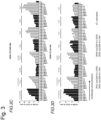
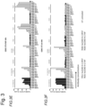
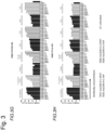

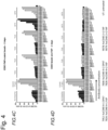
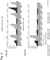
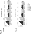
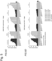

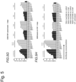
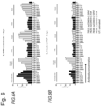

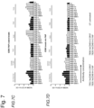
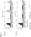


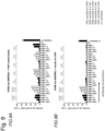


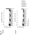
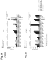
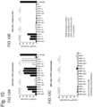

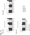

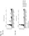
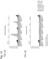


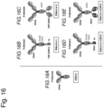

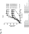
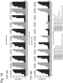


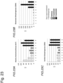

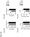
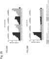

Classifications
-
- A—HUMAN NECESSITIES
- A61—MEDICAL OR VETERINARY SCIENCE; HYGIENE
- A61K—PREPARATIONS FOR MEDICAL, DENTAL OR TOILETRY PURPOSES
- A61K39/00—Medicinal preparations containing antigens or antibodies
- A61K39/395—Antibodies; Immunoglobulins; Immune serum, e.g. antilymphocytic serum
- A61K39/39533—Antibodies; Immunoglobulins; Immune serum, e.g. antilymphocytic serum against materials from animals
- A61K39/39558—Antibodies; Immunoglobulins; Immune serum, e.g. antilymphocytic serum against materials from animals against tumor tissues, cells, antigens
-
- A—HUMAN NECESSITIES
- A61—MEDICAL OR VETERINARY SCIENCE; HYGIENE
- A61P—SPECIFIC THERAPEUTIC ACTIVITY OF CHEMICAL COMPOUNDS OR MEDICINAL PREPARATIONS
- A61P35/00—Antineoplastic agents
-
- C—CHEMISTRY; METALLURGY
- C07—ORGANIC CHEMISTRY
- C07K—PEPTIDES
- C07K16/00—Immunoglobulins [IGs], e.g. monoclonal or polyclonal antibodies
- C07K16/18—Immunoglobulins [IGs], e.g. monoclonal or polyclonal antibodies against material from animals or humans
- C07K16/28—Immunoglobulins [IGs], e.g. monoclonal or polyclonal antibodies against material from animals or humans against receptors, cell surface antigens or cell surface determinants
- C07K16/2878—Immunoglobulins [IGs], e.g. monoclonal or polyclonal antibodies against material from animals or humans against receptors, cell surface antigens or cell surface determinants against the NGF-receptor/TNF-receptor superfamily, e.g. CD27, CD30, CD40, CD95
-
- C—CHEMISTRY; METALLURGY
- C07—ORGANIC CHEMISTRY
- C07K—PEPTIDES
- C07K16/00—Immunoglobulins [IGs], e.g. monoclonal or polyclonal antibodies
- C07K16/40—Immunoglobulins [IGs], e.g. monoclonal or polyclonal antibodies against enzymes
-
- A—HUMAN NECESSITIES
- A61—MEDICAL OR VETERINARY SCIENCE; HYGIENE
- A61K—PREPARATIONS FOR MEDICAL, DENTAL OR TOILETRY PURPOSES
- A61K39/00—Medicinal preparations containing antigens or antibodies
- A61K2039/505—Medicinal preparations containing antigens or antibodies comprising antibodies
-
- A—HUMAN NECESSITIES
- A61—MEDICAL OR VETERINARY SCIENCE; HYGIENE
- A61K—PREPARATIONS FOR MEDICAL, DENTAL OR TOILETRY PURPOSES
- A61K39/00—Medicinal preparations containing antigens or antibodies
- A61K2039/51—Medicinal preparations containing antigens or antibodies comprising whole cells, viruses or DNA/RNA
- A61K2039/515—Animal cells
- A61K2039/5154—Antigen presenting cells [APCs], e.g. dendritic cells or macrophages
-
- A—HUMAN NECESSITIES
- A61—MEDICAL OR VETERINARY SCIENCE; HYGIENE
- A61K—PREPARATIONS FOR MEDICAL, DENTAL OR TOILETRY PURPOSES
- A61K39/00—Medicinal preparations containing antigens or antibodies
- A61K2039/57—Medicinal preparations containing antigens or antibodies characterised by the type of response, e.g. Th1, Th2
- A61K2039/572—Medicinal preparations containing antigens or antibodies characterised by the type of response, e.g. Th1, Th2 cytotoxic response
-
- C—CHEMISTRY; METALLURGY
- C07—ORGANIC CHEMISTRY
- C07K—PEPTIDES
- C07K2317/00—Immunoglobulins specific features
- C07K2317/20—Immunoglobulins specific features characterized by taxonomic origin
- C07K2317/24—Immunoglobulins specific features characterized by taxonomic origin containing regions, domains or residues from different species, e.g. chimeric, humanized or veneered
-
- C—CHEMISTRY; METALLURGY
- C07—ORGANIC CHEMISTRY
- C07K—PEPTIDES
- C07K2317/00—Immunoglobulins specific features
- C07K2317/30—Immunoglobulins specific features characterized by aspects of specificity or valency
- C07K2317/31—Immunoglobulins specific features characterized by aspects of specificity or valency multispecific
-
- C—CHEMISTRY; METALLURGY
- C07—ORGANIC CHEMISTRY
- C07K—PEPTIDES
- C07K2317/00—Immunoglobulins specific features
- C07K2317/30—Immunoglobulins specific features characterized by aspects of specificity or valency
- C07K2317/33—Crossreactivity, e.g. for species or epitope, or lack of said crossreactivity
-
- C—CHEMISTRY; METALLURGY
- C07—ORGANIC CHEMISTRY
- C07K—PEPTIDES
- C07K2317/00—Immunoglobulins specific features
- C07K2317/30—Immunoglobulins specific features characterized by aspects of specificity or valency
- C07K2317/35—Valency
-
- C—CHEMISTRY; METALLURGY
- C07—ORGANIC CHEMISTRY
- C07K—PEPTIDES
- C07K2317/00—Immunoglobulins specific features
- C07K2317/50—Immunoglobulins specific features characterized by immunoglobulin fragments
- C07K2317/52—Constant or Fc region; Isotype
-
- C—CHEMISTRY; METALLURGY
- C07—ORGANIC CHEMISTRY
- C07K—PEPTIDES
- C07K2317/00—Immunoglobulins specific features
- C07K2317/50—Immunoglobulins specific features characterized by immunoglobulin fragments
- C07K2317/55—Fab or Fab'
-
- C—CHEMISTRY; METALLURGY
- C07—ORGANIC CHEMISTRY
- C07K—PEPTIDES
- C07K2317/00—Immunoglobulins specific features
- C07K2317/50—Immunoglobulins specific features characterized by immunoglobulin fragments
- C07K2317/56—Immunoglobulins specific features characterized by immunoglobulin fragments variable (Fv) region, i.e. VH and/or VL
- C07K2317/565—Complementarity determining region [CDR]
-
- C—CHEMISTRY; METALLURGY
- C07—ORGANIC CHEMISTRY
- C07K—PEPTIDES
- C07K2317/00—Immunoglobulins specific features
- C07K2317/50—Immunoglobulins specific features characterized by immunoglobulin fragments
- C07K2317/56—Immunoglobulins specific features characterized by immunoglobulin fragments variable (Fv) region, i.e. VH and/or VL
- C07K2317/567—Framework region [FR]
-
- C—CHEMISTRY; METALLURGY
- C07—ORGANIC CHEMISTRY
- C07K—PEPTIDES
- C07K2317/00—Immunoglobulins specific features
- C07K2317/50—Immunoglobulins specific features characterized by immunoglobulin fragments
- C07K2317/56—Immunoglobulins specific features characterized by immunoglobulin fragments variable (Fv) region, i.e. VH and/or VL
- C07K2317/569—Single domain, e.g. dAb, sdAb, VHH, VNAR or nanobody®
-
- C—CHEMISTRY; METALLURGY
- C07—ORGANIC CHEMISTRY
- C07K—PEPTIDES
- C07K2317/00—Immunoglobulins specific features
- C07K2317/60—Immunoglobulins specific features characterized by non-natural combinations of immunoglobulin fragments
- C07K2317/64—Immunoglobulins specific features characterized by non-natural combinations of immunoglobulin fragments comprising a combination of variable region and constant region components
-
- C—CHEMISTRY; METALLURGY
- C07—ORGANIC CHEMISTRY
- C07K—PEPTIDES
- C07K2317/00—Immunoglobulins specific features
- C07K2317/70—Immunoglobulins specific features characterized by effect upon binding to a cell or to an antigen
- C07K2317/71—Decreased effector function due to an Fc-modification
-
- C—CHEMISTRY; METALLURGY
- C07—ORGANIC CHEMISTRY
- C07K—PEPTIDES
- C07K2317/00—Immunoglobulins specific features
- C07K2317/70—Immunoglobulins specific features characterized by effect upon binding to a cell or to an antigen
- C07K2317/75—Agonist effect on antigen
-
- C—CHEMISTRY; METALLURGY
- C07—ORGANIC CHEMISTRY
- C07K—PEPTIDES
- C07K2317/00—Immunoglobulins specific features
- C07K2317/90—Immunoglobulins specific features characterized by (pharmaco)kinetic aspects or by stability of the immunoglobulin
- C07K2317/92—Affinity (KD), association rate (Ka), dissociation rate (Kd) or EC50 value
-
- C—CHEMISTRY; METALLURGY
- C07—ORGANIC CHEMISTRY
- C07K—PEPTIDES
- C07K2317/00—Immunoglobulins specific features
- C07K2317/90—Immunoglobulins specific features characterized by (pharmaco)kinetic aspects or by stability of the immunoglobulin
- C07K2317/94—Stability, e.g. half-life, pH, temperature or enzyme-resistance
Definitions
- the invention relates to novel bispecific antigen binding molecules, comprising (a) at least two Fab fragments capable of specific binding to CD40 connected to a Fc region comprising (i) a heavy chain variable region (V H CD40) comprising an amino acid sequence selected from the group consisting of SEQ ID NO: 171, SEQ ID NO: 172, SEQ ID NO: 173 and SEQ ID NO: 174, and (ii) a light chain variable region (V L CD40) comprising the amino acid sequence selected from the group consisting of SEQ ID NO: 175, SEQ ID NO: 176, SEQ ID NO: 177, and SEQ ID NO: 178, and (b) at least one antigen binding domain capable of specific binding to FAP comprising a heavy chain variable region (V H FAP) comprising an amino acid sequence of SEQ ID NO: 17, and a light chain variable region (V L FAP) comprising an amino acid sequence of SEQ ID NO: 18 connected to the C-terminus of the Fc region, wherein the Fc region is an
- TCR T-cell antigen receptor
- APCs antigen-presenting cells
- TNFR family costimulators show promise for several therapeutic applications in multiple fields including cancer, infectious disease, transplantation, and autoimmunity.
- the TNFR family member CD40 plays a key role in triggering immune responses by inducing maturation, survival, antigen presentation, cytokine production, and expression of costimulatory molecules of APCs, which then drive antigen-specific T cell responses and NK cell activation by proinflammatory cytokines.
- CD40 regulates immune responses against infections, tumors and self-antigens and its expression has been demonstrated on the surface of APCs such as B cells, dendritic cells (DCs), monocytes, and macrophages as well as platelets, and cells of non-hematopoietic origin such as myofibroblasts, fibroblasts, epithelial, and endothelial cells ( Elgueta R.
- CD40 ligand CD40L is expressed on activated CD4 + helper T cells, platelets, monocytic cells, natural killer cell, mast cells, and basophils ( Carbone E. et. al., J Exp Med. 1997;185(12): 2053-2060 , or Elgueta R. et al., Immunol Rev. 2009;229(1):152-72 ) .
- CD40 and CD40L are strongly upregulated in response to various immune stimulatory signals and CD40-CD40L interaction between APCs and CD4 + T cells contributes to increased APC activation and antigen-specific CD8 + T cell responses ( Bevan MJ., Nat Rev Immunol. 2014;4(8):595-602 ) . Similar immune stimulatory results were observed by using CD40 agonistic antibodies ( Vonderheide RH and Glennie MJ., Clin Cancer Res. 2013;19(S):1035-43 ).
- TNF receptor-associated factors leads to synergistic activation of mitogen-activated protein kinases (MAPKs), phosphoinositide 3-kinase (PI3K) as well as canonical and non-canonical nuclear factor ⁇ B (NP ⁇ B) signaling pathways ( Elgueta R. et al., Immunol Rev. 2009;229(1):152-72 ).
- agonistic CD40 antibodies in harnessing anti-tumor immunity. Beside its indirect mode of action by mediated tumor cell killing through the activation of the adaptive immune system, agonistic CD40 antibodies can induce direct tumor cell killing through inducing apoptosis of CD40-expressing solid tumor cells ( Eliopoulos AG. et al., Mol Cell Biol. 2000;20(15):5503-15 ).
- the direct CD40 antibody-mediated killing of tumor cells can provide a source of tumor antigens that can be processed and presented by APC simultaneously activated by CD40 engagement via anti-CD40 antibodies which then can induce tumor antigen-specific T cells, a postulated mechanism known as endogenous vaccination.
- CD40 engagement can mount in an efficient anti-cancer immune response
- agonistic CD40 antibodies have been used successfully in a variety of preclinical tumor models, both as a single-agent and in combination with chemotherapy ( Vonderheide RH and Glennie MJ., Clin Cancer Res. 2013;19(5): 1035-43 ).
- CD40 mAb are under investigation in clinical trials: Chi Lob 7/4 (CD40 agonistic IgG1 chimeric mAb; Cancer Research UK; Chowdhury F. et al., Cancer Immunol Res. 2013;2:229-40 ), ADC1013 (fully human, CD40 agonistic IgG1 antibody; Alligator Bioscience and Johnson & Johnson; Mangsbo SM. et al., Clin Cancer Res. 2015 Mar 1;21(5):1115-26 ), APX-005 (fully humanized, CD40 agonistic IgG1 mAb; Apexigen; Bjorck P. etal. J Immunother Cancer.
- SEA-CD40 CD40 agonistic IgG1 chimeric mAb; Seattle Genetics; Gardai SJ. et al. AACR 106th Annual Meeting 2015; April 18-22, abstract 2472
- RO7009789 fully human, CD40 super agonistic IgG2 mAb
- dacetuzumab CD40 partial agonistic IgG1 chimeric mAb; Seattle Genetics; Khubchandani S. et al., Curr Opin Investig Drugs. 2009;10:579-87 ) is investigated in a clinical phase II study.
- HL Hodgkin lymphoma
- DLBCL diffuse large B-cell lymphoma
- indolent lymphoma including follicular lymphoma
- CMC complement mediated cytotoxicity
- ADCC antibody dependent cellular cytoxicity
- CD40 agonistic antibodies in vivo require crosslinking CD40, bound by its Fab fragment on the target cell, to a Fcy receptor, bound by its Fc fragment on a cell other than the target cell as has been described for agonistic antibodies specific to other apoptosisinducing or immunomodulatory members of the TNFR-superfamily ( Dahan R., Cancer Cell. 2016 Jun 13; 29(6): 820-31 ; Li F. and Ravetch J. V. Science, 2011; 333, 1030-1034 ; TengM. W.
- the proposed mechanism includes Fc ⁇ receptor mediated clustering of CD40 transmembrane molecules on target cells and subsequent heightened CD40 signaling to achieve potent in vivo efficacy.
- CP-870,893 has shown clinical efficacy in patients with advanced cancer.
- Four out of 29 patients with advanced cancer showed partial responses after receiving a single intravenous infusion of CP-870,893 ( Vonderheide RH., J Clin Oncol. 2007 Mar 1;25(7):876-83 ) .
- One out of these four patients treated with 9 subsequent doses of CP-870,893 over one and a half years remained in complete remission for more than 5 years.
- CD40 antibodies can only be administered in relatively low doses due to dose-limiting toxicities such as cytokine release syndrome and thrombocyte/ endothelial cell activation, resulting in an insufficient activation of the pathway on target APCs and a narrow therapeutic index.
- dose-limiting toxicities such as cytokine release syndrome and thrombocyte/ endothelial cell activation, resulting in an insufficient activation of the pathway on target APCs and a narrow therapeutic index.
- the invention relates to new bispecific antigen binding molecules capable of specific binding to CD40 and a target cell antigen.
- the antigen binding molecules of the invention combine a moiety capable of preferred binding to tumor-specific or tumor-associated targets with a moiety capable of agonistic binding to CD40, wherein the activation of APCs through CD40 is provided by cross-linking through the target cell antigen, for example FAP expressed on tumor stroma cells and potentially also through FAP intermediately expressed in secondary lymphoid tissues.
- the FAP-dependent cross-linking of the bispecific antigen binding molecules confines the activation of CD40-expressing cells to the tumor tissue and potentially also to secondary lymphoid tissues such as tumor-draining lymph nodes.
- bispecific antigen binding molecules capable of specific binding to CD40 and to immune checkpoint receptors on activated T cells, such as CTLA-4 or PD-1
- targeting to a tumor target such as FAP enables CD40-mediated APC activation mainly in the tumor stroma and tumor-draining lymph nodes where fibroblasts express increased levels of FAP compared to other tissues.
- the antigen binding molecules of this invention may thus be able to trigger the CD40 receptor not only effectively, but also very selectively at the desired site while overcoming the need for FcyR cross-linking thereby reducing side effects.
- the present invention relates to bispecific antigen binding molecules combining at least two Fab fragments capable of specific binding to the costimulatory TNF receptor family member CD40 connected to a Fc region, with at least one antigen binding one antigen binding domain capable of specific binding to fibroblast activation protein (FAP) connected to the C-terminus of the Fc region.
- FAP fibroblast activation protein
- references in the description to methods of treatment refer to the compounds, pharmaceutical compositions and medicaments of the present invention for use in a method for treatment of the human (or animal) body by therapy (or for diagnosis).
- antigen binding molecule refers in its broadest sense to a molecule that specifically binds an antigenic determinant.
- antigen binding molecules are antibodies, antibody fragments and scaffold antigen binding proteins.
- the term "antigen binding domain capable of specific binding to a target cell antigen " or "moiety capable of specific binding to a target cell antigen” refers to a polypeptide molecule that specifically binds to an antigenic determinant.
- the antigen binding domain is able to activate signaling through its target cell antigen.
- the antigen binding domain is able to direct the entity to which it is attached (e.g. the CD40 agonist) to a target site, for example to a specific type of tumor cell or tumor stroma bearing the antigenic determinant.
- Antigen binding domains capable of specific binding to a target cell antigen include antibodies and fragments thereof as further defined herein.
- antigen binding domains capable of specific binding to a target cell antigen include scaffold antigen binding proteins as further defined herein, e.g. binding domains which are based on designed repeat proteins or designed repeat domains (see e.g. WO 2002/020565 ).
- the term "antigen binding domain capable of specific binding to a target cell antigen” refers to the part of the molecule that comprises the area which specifically binds to and is complementary to part or all of an antigen.
- a antigen binding domain capable of specific antigen binding may be provided, for example, by one or more antibody variable domains (also called antibody variable regions).
- an antigen binding domain capable of specific antigen binding comprises an antibody light chain variable region (VL) and an antibody heavy chain variable region (VH).
- VL antibody light chain variable region
- VH antibody heavy chain variable region
- the "antigen binding domain capable of specific binding to a target cell antigen” can also be a Fab fragment or a cross-Fab fragment.
- antibody herein is used in the broadest sense and encompasses various antibody structures, including but not limited to monoclonal antibodies, polyclonal antibodies, monospecific and multispecific antibodies (e.g., bispecific antibodies), and antibody fragments so long as they exhibit the desired antigen-binding activity.
- monoclonal antibody refers to an antibody obtained from a population of substantially homogeneous antibodies, i.e., the individual antibodies comprising the population are identical and/or bind the same epitope, except for possible variant antibodies, e.g. containing naturally occurring mutations or arising during production of a monoclonal antibody preparation, such variants generally being present in minor amounts.
- polyclonal antibody preparations typically include different antibodies directed against different determinants (epitopes)
- each monoclonal antibody of a monoclonal antibody preparation is directed against a single determinant on an antigen.
- the term " monospecific " antibody as used herein denotes an antibody that has one or more binding sites each of which bind to the same epitope of the same antigen.
- bispecific means that the antigen binding molecule is able to specifically bind to at least two distinct antigenic determinants. Typically, a bispecific antigen binding molecule comprises two antigen binding sites, each of which is specific for a different antigenic determinant. In certain embodiments the bispecific antigen binding molecule is capable of simultaneously binding two antigenic determinants, particularly two antigenic determinants expressed on two distinct cells.
- a bispecific antigen binding molecule as described herein can also form part of a multispecific antibody.
- valent as used within the current application denotes the presence of a specified number of binding sites specific for one distinct antigenic determinant in an antigen binding molecule that are specific for one distinct antigenic determinant.
- bivalent tetravalent
- hexavalent denote the presence of two binding sites, four binding sites, and six binding sites specific for a certain antigenic determinant, respectively, in an antigen binding molecule.
- the bispecific antigen binding molecules according to the invention can be monovalent for a certain antigenic determinant, meaning that they have only one binding site for said antigenic determinant or they can be bivalent or tetravalent for a certain antigenic determinant, meaning that they have two binding sites or four binding sites, respectively, for said antigenic determinant.
- full length antibody is used herein interchangeably to refer to an antibody having a structure substantially similar to a native antibody structure.
- Native antibodies refer to naturally occurring immunoglobulin molecules with varying structures.
- native IgG-class antibodies are heterotetrameric glycoproteins of about 150,000 daltons, composed of two light chains and two heavy chains that are disulfide-bonded. From N- to C-terminus, each heavy chain has a variable region (VH), also called a variable heavy domain or a heavy chain variable domain, followed by three constant domains (CH1, CH2, and CH3), also called a heavy chain constant region.
- VH variable region
- CH1, CH2, and CH3 constant domains
- each light chain has a variable region (VL), also called a variable light domain or a light chain variable domain, followed by a light chain constant domain (CL), also called a light chain constant region.
- the heavy chain of an antibody may be assigned to one of five types, called ⁇ (IgA), ⁇ (IgD), ⁇ (IgE), ⁇ (IgG), or ⁇ (IgM), some of which may be further divided into subtypes, e.g. ⁇ 1 (IgG1), ⁇ 2 (IgG2), ⁇ 3 (IgG3), ⁇ 4 (IgG4), ⁇ 1 (IgA1) and ⁇ 2 (IgA2).
- the light chain of an antibody may be assigned to one of two types, called kappa ( ⁇ ) and lambda ( ⁇ ), based on the amino acid sequence of its constant domain.
- antibody fragment refers to a molecule other than an intact antibody that comprises a portion of an intact antibody that binds the antigen to which the intact antibody binds.
- antibody fragments include but are not limited to Fv, Fab, Fab', Fab'-SH, F(ab') 2 ; diabodies, triabodies, tetrabodies, cross-Fab fragments; linear antibodies; single-chain antibody molecules (e.g. scFv); and single domain antibodies.
- scFv single domain antibodies.
- Diabodies are antibody fragments with two antigen-binding sites that may be bivalent or bispecific, see, for example, EP 404,097 ; WO 1993/01161 ; Hudson et al., Nat Med 9, 129-134 (2003 ); and Hollinger et al., Proc Natl Acad Sci USA 90, 6444-6448 (1993 ). Triabodies and tetrabodies are also described in Hudson et al., Nat Med 9, 129-134 (2003 ). Single-domain antibodies are antibody fragments comprising all or a portion of the heavy chain variable domain or all or a portion of the light chain variable domain of an antibody.
- a single-domain antibody is a human single-domain antibody (Domantis, Inc., Waltham, MA; see e.g. U.S. Patent No. 6,248,516 B1 ).
- Antibody fragments can be made by various techniques, including but not limited to proteolytic digestion of an intact antibody as well as production by recombinant host cells (e.g. E. coli or phage), as described herein.
- Papain digestion of intact antibodies produces two identical antigen-binding fragments, called "Fab” fragments containing each the heavy- and light-chain variable domains and also the constant domain of the light chain and the first constant domain (CH1) of the heavy chain.
- Fab fragment refers to an antibody fragment comprising a light chain fragment comprising a VL domain and a constant domain of a light chain (CL), and a VH domain and a first constant domain (CH1) of a heavy chain.
- Fab' fragments differ from Fab fragments by the addition of a few residues at the carboxy terminus of the heavy chain CH1 domain including one or more cysteins from the antibody hinge region.
- Fab'-SH are Fab' fragments wherein the cysteine residue(s) of the constant domains bear a free thiol group. Pepsin treatment yields an F(ab')2 fragment that has two antigen-combining sites (two Fab fragments) and a part of the Fc region. According to the present invention, the term “Fab fragment” also includes "cross-Fab fragments” or “crossover Fab fragments” as defined below.
- cross-Fab fragment or "xFab fragment” or “crossover Fab fragment” refers to a Fab fragment, wherein either the variable regions or the constant regions of the heavy and light chain are exchanged.
- Two different chain compositions of a crossover Fab molecule are possible and comprised in the bispecific antibodies of the invention: On the one hand, the variable regions of the Fab heavy and light chain are exchanged, i.e. the crossover Fab molecule comprises a peptide chain composed of the light chain variable region (VL) and the heavy chain constant region (CH1), and a peptide chain composed of the heavy chain variable region (VH) and the light chain constant region (CL).
- This crossover Fab molecule is also referred to as CrossFab (VLVH) .
- the crossover Fab molecule comprises a peptide chain composed of the heavy chain variable region (VH) and the light chain constant region (CL), and a peptide chain composed of the light chain variable region (VL) and the heavy chain constant region (CH1).
- This crossover Fab molecule is also referred to as CrossFab (CLCH1) .
- a “single chain Fab fragment” or “ scFab” is a polypeptide consisting of an antibody heavy chain variable domain (VH), an antibody constant domain 1 (CH1), an antibody light chain variable domain (VL), an antibody light chain constant domain (CL) and a linker, wherein said antibody domains and said linker have one of the following orders in N-terminal to C-terminal direction: a) VH-CH1-linker-VL-CL, b) VL-CL-linker-VH-CH1, c) VH-CL-linker-VL-CH1 or d) VL-CH1-linker-VH-CL; and wherein said linker is a polypeptide of at least 30 amino acids, preferably between 32 and 50 amino acids.
- Said single chain Fab fragments are stabilized via the natural disulfide bond between the CL domain and the CH1 domain.
- these single chain Fab molecules might be further stabilized by generation of interchain disulfide bonds via insertion of cysteine residues (e.g. position 44 in the variable heavy chain and position 100 in the variable light chain according to Kabat numbering).
- a “crossover single chain Fab fragment” or “x-scFab” is a is a polypeptide consisting of an antibody heavy chain variable domain (VH), an antibody constant domain 1 (CH1), an antibody light chain variable domain (VL), an antibody light chain constant domain (CL) and a linker, wherein said antibody domains and said linker have one of the following orders in N-terminal to C-terminal direction: a) VH-CL-linker-VL-CH1 and b) VL-CH1-linker-VH-CL; wherein VH and VL form together an antigen-binding site which binds specifically to an antigen and wherein said linker is a polypeptide of at least 30 amino acids.
- these x-scFab molecules might be further stabilized by generation of interchain disulfide bonds via insertion of cysteine residues (e.g. position 44 in the variable heavy chain and position 100 in the variable light chain according to Kabat numbering).
- a “ single-chain variable fragment (scFv)" is a fusion protein of the variable regions of the heavy (V H ) and light chains (V L ) of an antibody, connected with a short linker peptide of ten to about 25 amino acids.
- the linker is usually rich in glycine for flexibility, as well as serine or threonine for solubility, and can either connect the N-terminus of the V H with the C-terminus of the V L , or vice versa.
- This protein retains the specificity of the original antibody, despite removal of the constant regions and the introduction of the linker.
- scFv antibodies are, e.g. described in Houston, J.S., Methods in Enzymol. 203 (1991) 46-96 ).
- antibody fragments comprise single chain polypeptides having the characteristics of a VH domain, namely being able to assemble together with a VL domain, or of a VL domain, namely being able to assemble together with a VH domain to a functional antigen binding site and thereby providing the antigen binding property of full length antibodies.
- Scaffold antigen binding proteins are known in the art, for example, fibronectin and designed ankyrin repeat proteins (DARPins) have been used as alternative scaffolds for antigen-binding domains, see, e.g., Gebauer and Skerra, Engineered protein scaffolds as next-generation antibody therapeutics. Curr Opin Chem Biol 13:245-255 (2009 ) and Stumpp et al., Darpins: A new generation of protein therapeutics. Drug Discovery Today 13: 695-701 (2008 ).
- DARPins ankyrin repeat proteins
- a scaffold antigen binding protein is selected from the group consisting of CTLA-4 (Evibody), Lipocalins (Anticalin), a Protein A-derived molecule such as Z-domain of Protein A (Affibody), an A-domain (Avimer/Maxibody), a serum transferrin (trans-body); a designed ankyrin repeat protein (DARPin), a variable domain of antibody light chain or heavy chain (single-domain antibody, sdAb), a variable domain of antibody heavy chain (nanobody, aVH), V NAR fragments, a fibronectin (AdNectin), a C-type lectin domain (Tetranectin); a variable domain of a new antigen receptor beta-lactamase (V NAR fragments), a human gammacrystallin or ubiquitin (Affilin molecules); a kunitz type domain of human protease inhibitors, microbodies such as the proteins from the knottin
- CTLA-4 Cytotoxic T Lymphocyte-associated Antigen 4
- CTLA-4 is a CD28-family receptor expressed on mainly CD4 + T-cells. Its extracellular domain has a variable domain- like Ig fold. Loops corresponding to CDRs of antibodies can be substituted with heterologous sequence to confer different binding properties.
- CTLA-4 molecules engineered to have different binding specificities are also known as Evibodies (e.g. US7166697B1 ). Evibodies are around the same size as the isolated variable region of an antibody (e.g. a domain antibody). For further details see Journal of Immunological Methods 248 (1-2), 31-45 (2001 ).
- Lipocalins are a family of extracellular proteins which transport small hydrophobic molecules such as steroids, bilins, retinoids and lipids. They have a rigid beta-sheet secondary structure with a number of loops at the open end of the conical structure which can be engineered to bind to different target antigens. Anticalins are between 160-180 amino acids in size, and are derived from lipocalins. For further details see Biochim Biophys Acta 1482: 337-350 (2000 ), US7250297B1 and US20070224633 . An affibody is a scaffold derived from Protein A of Staphylococcus aureus which can be engineered to bind to antigen.
- the domain consists of a three-helical bundle of approximately 58 amino acids. Libraries have been generated by randomization of surface residues. For further details see Protein Eng. Des. Sel. 2004, 17, 455-462 and EP 1641818A1 . Avimers are multidomain proteins derived from the A-domain scaffold family. The native domains of approximately 35 amino acids adopt a defined disulfide bonded structure. Diversity is generated by shuffling of the natural variation exhibited by the family of A-domains. For further details see Nature Biotechnology 23(12), 1556 - 1561 (2005 ) and Expert Opinion on Investigational Drugs 16(6), 909-917 (June 2007 ). A transferrin is a monomeric serum transport glycoprotein.
- Transferrins can be engineered to bind different target antigens by insertion of peptide sequences in a permissive surface loop.
- engineered transferrin scaffolds include the Trans-body.
- Designed Ankyrin Repeat Proteins are derived from Ankyrin which is a family of proteins that mediate attachment of integral membrane proteins to the cytoskeleton.
- a single ankyrin repeat is a 33 residue motif consisting of two alpha-helices and a beta-turn. They can be engineered to bind different target antigens by randomizing residues in the first alpha-helix and a beta-turn of each repeat.
- a single-domain antibody is an antibody fragment consisting of a single monomeric variable antibody domain.
- the first single domains were derived from the variable domain of the antibody heavy chain from camelids (nanobodies or V H H fragments).
- the term single-domain antibody includes an autonomous human heavy chain variable domain (aVH) or V NAR fragments derived from sharks.
- Fibronectin is a scaffold which can be engineered to bind to antigen.
- Adnectins consists of a backbone of the natural amino acid sequence of the 10th domain of the 15 repeating units of human fibronectin type III (FN3). Three loops at one end of the beta. - sandwich can be engineered to enable an Adnectin to specifically recognize a therapeutic target of interest.
- Peptide aptamers are combinatorial recognition molecules that consist of a constant scaffold protein, typically thioredoxin (TrxA) which contains a constrained variable peptide loop inserted at the active site.
- TrxA thioredoxin
- Microbodies are derived from naturally occurring microproteins of 25-50 amino acids in length which contain 3-4 cysteine bridges - examples of microproteins include KalataBI and conotoxin and knottins. The microproteins have a loop which can beengineered to include upto 25 amino acids without affecting the overall fold of the microprotein. For further details of engineered knottin domains, see WO2008098796 .
- an " antigen binding molecule that binds to the same epitope " as a reference molecule refers to an antigen binding molecule that blocks binding of the reference molecule to its antigen in a competition assay by 50% or more, and conversely, the reference molecule blocks binding of the antigen binding molecule to its antigen in a competition assay by 50% or more.
- antigen binding domain or "antigen-binding site” refers to the part of an antigen binding molecule that comprises the area which specifically binds to and is complementary to part or all of an antigen. Where an antigen is large, an antigen binding molecule may only bind to a particular part of the antigen, which part is termed an epitope.
- An antigen binding domain may be provided by, for example, one or more variable domains (also called variable regions).
- an antigen binding domain comprises an antibody light chain variable region (VL) and an antibody heavy chain variable region (VH).
- antigenic determinant is synonymous with “antigen” and “epitope,” and refers to a site (e.g. a contiguous stretch of amino acids or a conformational configuration made up of different regions of non-contiguous amino acids) on a polypeptide macromolecule to which an antigen binding moiety binds, forming an antigen binding moietyantigen complex.
- Useful antigenic determinants can be found, for example, on the surfaces of tumor cells, on the surfaces of virus-infected cells, on the surfaces of other diseased cells, on the surface of immune cells, free in blood serum, and/or in the extracellular matrix (ECM).
- ECM extracellular matrix
- the proteins useful as antigens herein can be any native form the proteins from any vertebrate source, including mammals such as primates (e.g. humans) and rodents (e.g. mice and rats), unless otherwise indicated.
- the antigen is a human protein.
- the term encompasses the "full-length", unprocessed protein as well as any form of the protein that results from processing in the cell.
- the term also encompasses naturally occurring variants of the protein, e.g. splice variants or allelic variants.
- specific binding is meant that the binding is selective for the antigen and can be discriminated from unwanted or non-specific interactions.
- the ability of an antigen binding molecule to bind to a specific antigen can be measured either through an enzyme-linked immunosorbent assay (ELISA) or other techniques familiar to one of skill in the art, e.g. Surface Plasmon Resonance (SPR) technique (analyzed on a BIAcore instrument) ( Liljeblad et al., Glyco J 17, 323-329 (2000 )), and traditional binding assays ( Heeley, Endocr Res 28, 217-229 (2002 )).
- ELISA enzyme-linked immunosorbent assay
- SPR Surface Plasmon Resonance
- the extent of binding of an antigen binding molecule to an unrelated protein is less than about 10% of the binding of the antigen binding molecule to the antigen as measured, e.g. by SPR.
- an molecule that binds to the antigen has a dissociation constant (Kd) of ⁇ 1 ⁇ M, ⁇ 100 nM, ⁇ 10 nM, ⁇ 1 nM, ⁇ 0.1 nM, ⁇ 0.01 nM, or ⁇ 0.001 nM (e.g. 10 -8 M or less, e.g. from 10 -8 M to 10 -13 M, e.g. from 10 -9 M to 10 -13 M).
- Affinity or "binding affinity” refers to the strength of the sum total of non-covalent interactions between a single binding site of a molecule (e.g. an antibody) and its binding partner (e.g. an antigen). Unless indicated otherwise, as used herein, "binding affinity” refers to intrinsic binding affinity which reflects a 1:1 interaction between members of a binding pair (e.g. antibody and antigen).
- the affinity of a molecule X for its partner Y can generally be represented by the dissociation constant (Kd), which is the ratio of dissociation and association rate constants (koff and kon, respectively).
- Kd dissociation constant
- equivalent affinities may comprise different rate constants, as long as the ratio of the rate constants remains the same. Affinity can be measured by common methods known in the art, including those described herein. A particular method for measuring affinity is Surface Plasmon Resonance (SPR).
- An “ affinity matured” antibody refers to an antibody with one or more alterations in one or more hypervariable regions (HVRs), compared to a parent antibody which does not possess such alterations, such alterations resulting in an improvement in the affinity of the antibody for antigen.
- HVRs hypervariable regions
- a “target cell antigen" as used herein refers to an antigenic determinant presented on the surface of a target cell, in particular a target cell in a tumor such as a cancer cell or a cell of the tumor stroma.
- the target cell antigen is a tumor-associated antigen.
- a target cell antigen does not include immune checkpoint receptors on activated T cells, such as CTLA-4, PD-1 or PD-L1.
- the target cell antigen is an antigen on the surface of a tumor cell.
- the tumor target cell antigen is selected from the group consisting of Fibroblast Activation Protein (FAP), Carcinoembryonic Antigen (CEA), Melanoma-associated Chondroitin Sulfate Proteoglycan (MCSP), Epidermal Growth Factor Receptor (EGFR), CD19, CD20 and CD33.
- FAP Fibroblast Activation Protein
- CEA Carcinoembryonic Antigen
- MCSP Melanoma-associated Chondroitin Sulfate Proteoglycan
- EGFR Epidermal Growth Factor Receptor
- CD19, CD20 and CD33 CD19, CD20 and CD33.
- FAP Fibroblast Activation Protein
- Fibroblast activation protein also known as Prolyl endopeptidase FAP or Seprase (EC 3.4.21) refers to any native FAP from any vertebrate source, including mammals such as primates (e.g. humans) non-human primates (e.g. cynomolgus monkeys) and rodents (e.g. mice and rats), unless otherwise indicated.
- the term encompasses "full-length,” unprocessed FAP as well as any form of FAP that results from processing in the cell.
- the term also encompasses naturally occurring variants of FAP, e.g., splice variants or allelic variants.
- the antigen binding molecule of the invention is capable of specific binding to human, mouse and/or cynomolgus FAP.
- the amino acid sequence of human FAP is shown in UniProt (www.uniprot.org) accession no. Q12884 (version 149, SEQ ID NO:2), or NCBI (www.ncbi.nlm.nih.gov/) RefSeq NP_004451.2.
- the extracellular domain (ECD) of human FAP extends from amino acid position 26 to 760.
- the amino acid sequence of a His-tagged human FAP ECD is shown in SEQ ID NOs 142.
- the amino acid sequence of mouse FAP is shown in UniProt accession no.
- the extracellular domain (ECD) of mouse FAP extends from amino acid position 26 to 761.
- SEQ ID NO: 144 shows the amino acid of a His-tagged mouse FAP ECD.
- SEQ ID NO:145 shows the amino acid of a His-tagged cynomolgus FAP ECD.
- an anti-FAP binding molecule of the invention binds to the extracellular domain of FAP.
- variable region refers to the domain of an antibody heavy or light chain that is involved in binding the antigen binding molecule to antigen.
- the variable domains of the heavy chain and light chain (VH and VL, respectively) of a native antibody generally have similar structures, with each domain comprising four conserved framework regions (FRs) and three hypervariable regions (HVRs). See, e.g., Kindt et al., Kuby Immunology, 6th ed., W.H. Freeman and Co., page 91 (2007 ). A single VH or VL domain may be sufficient to confer antigen-binding specificity.
- HVR hypervariable region
- VH variable domain
- L1 variable domain
- L2 variable domain
- HVRs generally comprise amino acid residues from the hypervariable loops and/or from the "complementarity determining regions” (CDRs), the latter being of highest sequence variability and/or involved in antigen recognition.
- Exemplary hypervariable loops occur at amino acid residues 26-32 (L1), 50-52 (L2), 91-96 (L3), 26-32 (H1), 53-55 (H2), and 96-101 (H3).
- Exemplary CDRs CDR-L1, CDR-L2, CDR-L3, CDR-H1, CDR-H2, and CDR-H3 occur at amino acid residues 24-34 of L1, 50-56 of L2, 89-97 of L3, 31-35B of H1, 50-65 of H2, and 95-102 of H3.
- Hypervariable regions are also referred to as complementarity determining regions (CDRs), and these terms are used herein interchangeably in reference to portions of the variable region that form the antigen binding regions.
- CDRs complementarity determining regions
- CDR Definitions 1 CDR Kabat Chothia AbM 2 V H CDR1 31-35 26-32 26-35 V H CDR2 50-65 52-58 50-58 V H CDR3 95-102 95-102 95-102 V L CDR1 24-34 26-32 24-34 V L CDR2 50-56 50-52 50-56 V L CDR3 89-97 91-96 89-97 1 Numbering of all CDR definitions in Table A is according to the numbering conventions set forth by Kabat et al. (see below). 2 "AbM” with a lowercase “b” as used in Table A refers to the CDRs as defined by Oxford Molecular's "AbM” antibody modeling software.
- Kabat et al. also defined a numbering system for variable region sequences that is applicable to any antibody.
- One of ordinary skill in the art can unambiguously assign this system of "Kabat numbering" to any variable region sequence, without reliance on any experimental data beyond the sequence itself.
- Kabat numbering refers to the numbering system set forth by Kabat et al., U.S. Dept. of Health and Human Services, "Sequence of Proteins of Immunological Interest” (1983 ). Unless otherwise specified, references to the numbering of specific amino acid residue positions in an antibody variable region are according to the Kabat numbering system.
- CDRs generally comprise the amino acid residues that form the hypervariable loops.
- CDRs also comprise "specificity determining residues," or "SDRs,” which are residues that contact antigen. SDRs are contained within regions of the CDRs called abbreviated-CDRs, or a-CDRs.
- Exemplary a-CDRs (a-CDR-L1, a-CDR-L2, a-CDR-L3, a-CDR-H1, ⁇ -CDR-H2, and ⁇ -CDR-H3) occur at amino acid residues 31-34 of L1, 50-55 of L2, 89-96 of L3, 31-35B of H1, 50-58 of H2, and 95-102 of H3.
- HVR residues and other residues in the variable domain are numbered herein according to Kabat et al., supra.
- Framework refers to variable domain residues other than hypervariable region (HVR) residues.
- the FR of a variable domain generally consists of four FR domains: FR1, FR2, FR3, and FR4. Accordingly, the HVR and FR sequences generally appear in the following sequence in VH (or VL): FR1-H1(L1)-FR2-H2(L2)-FR3-H3(L3)-FR4.
- chimeric antibody refers to an antibody in which a portion of the heavy and/or light chain is derived from a particular source or species, while the remainder of the heavy and/or light chain is derived from a different source or species.
- the "class" of an antibody refers to the type of constant domain or constant region possessed by its heavy chain.
- the heavy chain constant domains that correspond to the different classes of immunoglobulins are called ⁇ , ⁇ , ⁇ , ⁇ , and ⁇ respectively..
- a “humanized” antibody refers to a chimeric antibody comprising amino acid residues from non-human HVRs and amino acid residues from human FRs.
- a humanized antibody will comprise substantially all of at least one, and typically two, variable domains, in which all or substantially all of the HVRs (e.g., CDRs) correspond to those of a non-human antibody, and all or substantially all of the FRs correspond to those of a human antibody.
- a humanized antibody optionally may comprise at least a portion of an antibody constant region derived from a human antibody.
- a " humanized form " of an antibody, e.g., a non-human antibody refers to an antibody that has undergone humanization.
- humanized antibodies encompassed by the present invention are those in which the constant region has been additionally modified or changed from that of the original antibody to generate the properties according to the invention, especially in regard to C1q binding and/or Fc receptor (FcR) binding.
- FcR Fc receptor
- a "human " antibody is one which possesses an amino acid sequence which corresponds to that of an antibody produced by a human or a human cell or derived from a non-human source that utilizes human antibody repertoires or other human antibody-encoding sequences. This definition of a human antibody specifically excludes a humanized antibody comprising non-human antigen-binding residues.
- Fc domain or " Fc region " herein is used to define a C-terminal region of an antibody heavy chain that contains at least a portion of the constant region.
- the term includes native sequence Fc regions and variant Fc regions.
- An IgG Fc region comprises an IgG CH2 and an IgG CH3 domain.
- the "CH2 domain” of a human IgG Fc region usually extends from an amino acid residue at about position 231 to an amino acid residue at about position 340.
- a carbohydrate chain is attached to the CH2 domain.
- the CH2 domain herein may be a native sequence CH2 domain or variant CH2 domain.
- the "CH3 domain” comprises the stretch of residues C-terminal to a CH2 domain in an Fc region (i.e.
- the CH3 region herein may be a native sequence CH3 domain or a variant CH3 domain (e.g. a CH3 domain with an introduced "protuberance” ("knob”) in one chain thereof and a corresponding introduced “cavity” ("hole”) in the other chain thereof; see US Patent No. 5,821,333 , expressly incorporated herein by reference).
- a human IgG heavy chain Fc region extends from Cys226, or from Pro230, to the carboxyl-terminus of the heavy chain.
- the C-terminal lysine (Lys447) of the Fc region may or may not be present.
- numbering of amino acid residues in the Fc region or constant region is according to the EU numbering system, also called the EU index, as described in Kabat et al., Sequences of Proteins of Immunological Interest, 5th Ed. Public Health Service, National Institutes of Health, Bethesda, MD, 1991 .
- knob-into-hole technology is described e.g. in US 5,731,168 ; US 7,695,936 ; Ridgway et al., Prot Eng 9, 617-621 (1996 ) and Carter, J Immunol Meth 248, 7-15 (2001 ).
- the method involves introducing a protuberance ("knob") at the interface of a first polypeptide and a corresponding cavity ("hole") in the interface of a second polypeptide, such that the protuberance can be positioned in the cavity so as to promote heterodimer formation and hinder homodimer formation.
- Protuberances are constructed by replacing small amino acid side chains from the interface of the first polypeptide with larger side chains (e.g. tyrosine or tryptophan).
- Compensatory cavities of identical or similar size to the protuberances are created in the interface of the second polypeptide by replacing large amino acid side chains with smaller ones (e.g. alanine or threonine).
- the protuberance and cavity can be made by altering the nucleic acid encoding the polypeptides, e.g. by site-specific mutagenesis, or by peptide synthesis.
- a knob modification comprises the amino acid substitution T366W in one of the two subunits of the Fc domain
- the hole modification comprises the amino acid substitutions T366S, L368A and Y407V in the other one of the two subunits of the Fc domain.
- the subunit of the Fc domain comprising the knob modification additionally comprises the amino acid substitution S354C
- the subunit of the Fc domain comprising the hole modification additionally comprises the amino acid substitution Y349C.
- a "region equivalent to the Fc region of an immunoglobulin" is intended to include naturally occurring allelic variants of the Fc region of an immunoglobulin as well as variants having alterations which produce substitutions, additions, or deletions but which do not decrease substantially the ability of the immunoglobulin to mediate effector functions (such as antibody-dependent cellular cytotoxicity).
- one or more amino acids can be deleted from the N-terminus or C-terminus of the Fc region of an immunoglobulin without substantial loss of biological function.
- Such variants can be selected according to general rules known in the art so as to have minimal effect on activity (see, e.g., Bowie, J. U. et al., Science 247:1306-10 (1990 )).
- effector function refers to those biological activities attributable to the Fc region of an antibody, which vary with the antibody isotype.
- antibody effector functions include: C1q binding and complement dependent cytotoxicity (CDC), Fc receptor binding, antibody-dependent cell-mediated cytotoxicity (ADCC), antibody-dependent cellular phagocytosis (ADCP), cytokine secretion, immune complex-mediated antigen uptake by antigen presenting cells, down regulation of cell surface receptors (e.g. B cell receptor), and B cell activation.
- Fc receptor binding dependent effector functions can be mediated by the interaction of the Fc-region of an antibody with Fc receptors (FcRs), which are specialized cell surface receptors on hematopoietic cells.
- Fc receptors belong to the immunoglobulin superfamily, and have been shown to mediate both the removal of antibody-coated pathogens by phagocytosis of immune complexes, and the lysis of erythrocytes and various other cellular targets (e.g. tumor cells) coated with the corresponding antibody, via antibody dependent cell mediated cytotoxicity (ADCC) (see e.g. Van de Winkel, J.G. and Anderson, C.L., J. Leukoc. Biol. 49 (1991) 511-524 ).
- ADCC antibody dependent cell mediated cytotoxicity
- FcRs are defined by their specificity for immunoglobulin isotypes: Fc receptors for IgG antibodies are referred to as FcyR. Fc receptor binding is described e.g. in Ravetch, J.V. and Kinet, J.P., Annu. Rev. Immunol. 9 (1991) 457-492 ; Capel, P.J., et al., Immunomethods 4 (1994) 25-34 ; de Haas, M., et al., J. Lab. Clin. Med. 126 (1995) 330-341 ; and Gessner, J.E., et al., Ann. Hematol. 76 (1998) 231-248 .
- FcyR Fc-region of IgG antibodies
- ADCC antibody-dependent cellular cytotoxicity
- Fcy receptors expressing cells such as cells, recombinantly expressing FcyRI and/or FcyRIIA or NK cells (expressing essentially FcyRIIIA). In particular, binding to FcyR on NK cells is measured.
- an "activating Fc receptor” is an Fc receptor that following engagement by an Fc region of an antibody elicits signaling events that stimulate the receptor-bearing cell to perform effector functions.
- Activating Fc receptors include FcyRIIIa (CD16a), FcyRI (CD64), FcyRIIa (CD32), and Fc ⁇ RI (CD89).
- a particular activating Fc receptor is human FcyRIIIa (see UniProt accession no. P08637, version 141).
- CD40 refers to any native CD40 from any vertebrate source, including mammals such as primates (e.g. humans) and rodents (e.g., mice and rats), unless otherwise indicated.
- the term encompasses "full-length,” unprocessed CD40 as well as any form of CD40 that results from processing in the cell.
- the term also encompasses naturally occurring variants of CD40, e.g., splice variants or allelic variants.
- SEQ ID NO: 1 Uniprot P25942, version 200
- SEQ ID NO: 146 Uniprot P27512, version 160.
- the CD40 antigen is a 50 kDa cell surface glycoprotein which belongs to the Tumor Necrosis Factor Receptor (TNF-R) family. ( Stamenkovic et al. (1989), EMBO J. 8: 1403-10 ). CD40 is expressed in many normal and tumor cell types, including B lymphocytes, dendritic cells, monocytes, macrophages, thymus epithelium, endothelial cells, fibroblasts, and smooth muscle cells. CD40 is expressed in all B-lymphomas and in 70% of all solid tumors and is up-regulated in antigen presenting cells (APCs) by maturation signals, such as IFN-gamma and GM-CSF.
- APCs antigen presenting cells
- CD40 activation also induces differentiation of monocytes into functional dendritic cells (DCs) and enhances cytolytic activity of NK cells through APC-CD40 induced cytokines.
- DCs functional dendritic cells
- CD40 plays an essential role in the initiation and enhancement of immune responses by inducing maturation of APCs, secretion of helper cytokines, upregulation of costimulatory molecules, and enhancement of effector functions.
- CD40 agonist as used herein includes any moiety that agonizes the CD40/CD40L interaction.
- CD40 as used in this context refers preferably to human CD40, thus the CD40 agonist is preferably an agonist of human CD40.
- the moiety will be an agonistic CD40 antibody or antibody fragment.
- anti-CD40 antibody refers to an antibody that is capable of binding CD40 with sufficient affinity such that the antibody is useful as a diagnostic and/or therapeutic agent in targeting CD40.
- the extent of binding of an anti-CD40 antibody to an unrelated, non-CD40 protein is less than about 10% of the binding of the antibody to CD40 as measured, e.g., by a radioimmunoassay (RIA) or flow cytometry (FACS).
- an antibody that binds to CD40 has a dissociation constant ( K D ) of ⁇ 1 ⁇ M, ⁇ 100 nM, ⁇ 10 nM, ⁇ 1 nM, ⁇ 0.1 nM, ⁇ 0.01 nM, or ⁇ 0.001 nM (e.g. 10 -6 M or less, e.g. from 10 -68 M to 10 -13 M, e.g., from 10 -8 M to 10 -10 M).
- K D dissociation constant
- peptide linker refers to a peptide comprising one or more amino acids, typically about 2 to 20 amino acids. Peptide linkers are known in the art or are described herein. Suitable, non-immunogenic linker peptides are, for example, (G 4 S) n , (SG 4 ) n or G 4 (SG 4 ) n peptide linkers, wherein “n” is generally a number between 1 and 10, typically between 2 and 4, in particular 2, i.e.
- GGGGS GGGGS
- SEQ ID NO: 147 GGGGSGGGGS (SEQ ID NO: 148), SGGGGSGGGG (SEQ ID NO: 149) and GGGGSGGGGSGGGG (SEQ ID NO: 150), but also include the sequences GSPGSSSSGS (SEQ ID NO:151), (G4S) 3 (SEQ ID NO: 152), (G4S) 4 (SEQ ID NO: 153), GSGSGSGS (SEQ ID NO: 154), GSGSGNGS (SEQ ID NO: 155), GGSGSGSG (SEQ ID NO: 156), GGSGSG (SEQ ID NO:157), GGSG (SEQ ID NO:158), GGSGNGSG (SEQ ID NO:159), GGNGSGSG (SEQ ID NO: 160) and GGNGSG (SEQ ID NO: 161).
- Peptide linkers of particular interest are (G4S) (SEQ ID NO: 147), (G 4 S) 2 or GGGGSGGGGS (SEQ ID NO: 148), (G4S) 3 (SEQ ID NO: 152) and (G4S) 4 (SEQ ID NO:153).
- amino acid denotes the group of naturally occurring carboxy ⁇ -amino acids comprising alanine (three letter code: ala, one letter code: A), arginine (arg, R), asparagine (asn, N), aspartic acid (asp, D), cysteine (cys, C), glutamine (gln, Q), glutamic acid (glu, E), glycine (gly, G), histidine (his, H), isoleucine (ile, I), leucine (leu, L), lysine (lys, K), methionine (met, M), phenylalanine (phe, F), proline (pro, P), serine (ser, S), threonine (thr, T), tryptophan (trp, W), tyrosine (tyr, Y), and valine (val, V).
- fused or " connected” is meant that the components (e.g. a heavy chain of an antibody and a Fab fragment) are linked by peptide bonds, either directly or via one or more peptide linkers.
- Percent (%) amino acid sequence identity with respect to a reference polypeptide (protein) sequence is defined as the percentage of amino acid residues in a candidate sequence that are identical with the amino acid residues in the reference polypeptide sequence, after aligning the sequences and introducing gaps, if necessary, to achieve the maximum percent sequence identity, and not considering any conservative substitutions as part of the sequence identity. Alignment for purposes of determining percent amino acid sequence identity can be achieved in various ways that are within the skill in the art, for instance, using publicly available computer software such as BLAST, BLAST-2, ALIGN. SAWI or Megalign (DNASTAR) software. Those skilled in the art can determine appropriate parameters for aligning sequences, including any algorithms needed to achieve maximal alignment over the full length of the sequences being compared.
- % amino acid sequence identity values are generated using the sequence comparison computer program ALIGN-2.
- the ALIGN-2 sequence comparison computer program was authored by Genentech, Inc., and the source code has been filed with user documentation in the U.S. Copyright Office, Washington D.C., 20559, where it is registered under U.S. Copyright Registration No. TXU510087.
- the ALIGN-2 program is publicly available from Genentech, Inc., South San Francisco, California, or may be compiled from the source code.
- the ALIGN-2 program should be compiled for use on a UNIX operating system, including digital UNIX V4.0D. All sequence comparison parameters are set by the ALIGN-2 program and do not vary.
- the % amino acid sequence identity of a given amino acid sequence A to, with, or against a given amino acid sequence B is calculated as follows: 100 times the fraction X/Y where X is the number of amino acid residues scored as identical matches by the sequence alignment program ALIGN-2 in that program's alignment of A and B, and where Y is the total number of amino acid residues in B.
- amino acid sequence variants of the bispecific antigen binding molecules provided herein are contemplated.
- Amino acid sequence variants of the TNF ligand trimer-containing antigen binding molecules may be prepared by introducing appropriate modifications into the nucleotide sequence encoding the molecules, or by peptide synthesis. Such modifications include, for example, deletions from, and/or insertions into and/or substitutions of residues within the amino acid sequences of the antibody. Any combination of deletion, insertion, and substitution can be made to arrive at the final construct, provided that the final construct possesses the desired characteristics, e.g., antigen-binding.
- Sites of interest for substitutional mutagenesis include the HVRs and Framework (FRs). Conservative substitutions are provided in Table B under the heading "Preferred Substitutions" and further described below in reference to amino acid side chain classes (1) to (6). Amino acid substitutions may be introduced into the molecule of interest and the products screened for a desired activity, e.g., retained/improved antigen binding, decreased immunogenicity, or improved ADCC or CDC.
- Amino acids may be grouped according to common side-chain properties:
- Non-conservative substitutions will entail exchanging a member of one of these classes for another class.
- amino acid sequence variants includes substantial variants wherein there are amino acid substitutions in one or more hypervariable region residues of a parent antigen binding molecule (e.g. a humanized or human antibody).
- a parent antigen binding molecule e.g. a humanized or human antibody.
- the resulting variant(s) selected for further study will have modifications (e.g., improvements) in certain biological properties (e.g., increased affinity, reduced immunogenicity) relative to the parent antigen binding molecule and/or will have substantially retained certain biological properties of the parent antigen binding molecule.
- An exemplary substitutional variant is an affinity matured antibody, which may be conveniently generated, e.g., using phage display-based affinity maturation techniques such as those described herein.
- one or more HVR residues are mutated and the variant antigen binding molecules displayed on phage and screened for a particular biological activity (e.g. binding affinity).
- substitutions, insertions, or deletions may occur within one or more HVRs so long as such alterations do not substantially reduce the ability of the antigen binding molecule to bind antigen.
- conservative alterations e.g., conservative substitutions as provided herein
- a useful method for identification of residues or regions of an antibody that may be targeted for mutagenesis is called "alanine scanning mutagenesis" as described by Cunningham and Wells (1989) Science, 244:1081-1085 .
- a residue or group of target residues e.g., charged residues such as Arg, Asp, His, Lys, and Glu
- a neutral or negatively charged amino acid e.g., alanine or polyalanine
- Further substitutions may be introduced at the amino acid locations demonstrating functional sensitivity to the initial substitutions.
- a crystal structure of an antigen-antigen binding molecule complex to identify contact points between the antibody and antigen. Such contact residues and neighboring residues may be targeted or eliminated as candidates for substitution.
- Variants may be screened to determine whether they contain the desired properties.
- Amino acid sequence insertions include amino- and/or carboxyl-terminal fusions ranging in length from one residue to polypeptides containing a hundred or more residues, as well as intrasequence insertions of single or multiple amino acid residues.
- terminal insertions include bispecific antigen binding molecules of the invention with an N-terminal methionyl residue.
- Other insertional variants of the molecule include the fusion to the N- or C-terminus to a polypeptide which increases the serum half-life of the bispecific antigen binding molecules.
- the bispecific antigen binding molecules provided herein are altered to increase or decrease the extent to which the antibody is glycosylated. Glycosylation variants of the molecules may be conveniently obtained by altering the amino acid sequence such that one or more glycosylation sites is created or removed. Where the TNF ligand trimer-containing antigen binding molecule comprises an Fc region, the carbohydrate attached thereto may be altered. Native antibodies produced by mammalian cells typically comprise a branched, biantennary oligosaccharide that is generally attached by an N-linkage to Asn297 of the CH2 domain of the Fc region. See, e.g., Wright et al. TIBTECH 15:26-32 (1997 ).
- the oligosaccharide may include various carbohydrates, e.g., mannose, N-acetyl glucosamine (GlcNAc), galactose, and sialic acid, as well as a fucose attached to a GlcNAc in the "stem" of the biantennary oligosaccharide structure.
- modifications of the oligosaccharide in TNF family ligand trimer-containing antigen binding molecule may be made in order to create variants with certain improved properties.
- variants of bispecific antigen binding molecules or antibodies of the invention are provided having a carbohydrate structure that lacks fucose attached (directly or indirectly) to an Fc region.
- Such fucosylation variants may have improved ADCC function, see e.g. US Patent Publication Nos. US 2003/0157108 (Presta, L. ) or US 2004/0093621 (Kyowa Hakko Kogyo Co., Ltd) .
- variants of the bispecific antigen binding molecules or antibodies of the invention are provided with bisected oligosaccharides, e.g., in which a biantennary oligosaccharide attached to the Fc region is bisected by GlcNAc.
- Such variants may have reduced fucosylation and/or improved ADCC function., see for example WO 2003/011878 (Jean-Mairet et al. ); US Patent No.
- cysteine engineered variants of the bispecific antigen binding molecules of the invention e.g., "thioMAbs”
- one or more residues of the molecule are substituted with cysteine residues.
- the substituted residues occur at accessible sites of the molecule.
- reactive thiol groups are thereby positioned at accessible sites of the antibody and may be used to conjugate the antibody to other moieties, such as drug moieties or linker-drug moieties, to create an immunoconjugate.
- any one or more of the following residues may be substituted with cysteine: V205 (Kabat numbering) of the light chain; A118 (EU numbering) of the heavy chain; and S400 (EU numbering) of the heavy chain Fc region.
- Cysteine engineered antigen binding molecules may be generated as described, e.g., in U.S. Patent No. 7,521,541 .
- polynucleotide refers to an isolated nucleic acid molecule or construct, e.g. messenger RNA (mRNA), virally-derived RNA, or plasmid DNA (pDNA).
- mRNA messenger RNA
- pDNA virally-derived RNA
- a polynucleotide may comprise a conventional phosphodiester bond or a non-conventional bond (e.g. an amide bond, such as found in peptide nucleic acids (PNA).
- PNA peptide nucleic acids
- nucleic acid molecule refers to any one or more nucleic acid segments, e.g. DNA or RNA fragments, present in a polynucleotide.
- nucleic acid molecule or polynucleotide is intended a nucleic acid molecule, DNA or RNA, which has been removed from its native environment.
- a recombinant polynucleotide encoding a polypeptide contained in a vector is considered isolated for the purposes of the present invention.
- Further examples of an isolated polynucleotide include recombinant polynucleotides maintained in heterologous host cells or purified (partially or substantially) polynucleotides in solution.
- An isolated polynucleotide includes a polynucleotide molecule contained in cells that ordinarily contain the polynucleotide molecule, but the polynucleotide molecule is present extrachromosomally or at a chromosomal location that is different from its natural chromosomal location.
- Isolated RNA molecules include in vivo or in vitro RNA transcripts of the present invention, as well as positive and negative strand forms, and double-stranded forms. Isolated polynucleotides or nucleic acids according to the present invention further include such molecules produced synthetically.
- a polynucleotide or a nucleic acid may be or may include a regulatory element such as a promoter, ribosome binding site, or a transcription terminator.
- nucleic acid or polynucleotide having a nucleotide sequence at least, for example, 95% "identical" to a reference nucleotide sequence of the present invention it is intended that the nucleotide sequence of the polynucleotide is identical to the reference sequence except that the polynucleotide sequence may include up to five point mutations per each 100 nucleotides of the reference nucleotide sequence.
- a polynucleotide having a nucleotide sequence at least 95% identical to a reference nucleotide sequence up to 5% of the nucleotides in the reference sequence may be deleted or substituted with another nucleotide, or a number of nucleotides up to 5% of the total nucleotides in the reference sequence may be inserted into the reference sequence.
- These alterations of the reference sequence may occur at the 5' or 3' terminal positions of the reference nucleotide sequence or anywhere between those terminal positions, interspersed either individually among residues in the reference sequence or in one or more contiguous groups within the reference sequence.
- any particular polynucleotide sequence is at least 80%, 85%, 90%, 95%, 96%, 97%, 98% or 99% identical to a nucleotide sequence of the present invention can be determined conventionally using known computer programs, such as the ones discussed above for polypeptides (e.g. ALIGN-2).
- expression cassette refers to a polynucleotide generated recombinantly or synthetically, with a series of specified nucleic acid elements that permit transcription of a particular nucleic acid in a target cell.
- the recombinant expression cassette can be incorporated into a plasmid, chromosome, mitochondrial DNA, plastid DNA, virus, or nucleic acid fragment.
- the recombinant expression cassette portion of an expression vector includes, among other sequences, a nucleic acid sequence to be transcribed and a promoter.
- the expression cassette of the invention comprises polynucleotide sequences that encode bispecific antigen binding molecules of the invention or fragments thereof.
- vector or "expression vector” is synonymous with "expression construct” and refers to a DNA molecule that is used to introduce and direct the expression of a specific gene to which it is operably associated in a target cell.
- the term includes the vector as a self-replicating nucleic acid structure as well as the vector incorporated into the genome of a host cell into which it has been introduced.
- the expression vector of the present invention comprises an expression cassette. Expression vectors allow transcription of large amounts of stable mRNA. Once the expression vector is inside the target cell, the ribonucleic acid molecule or protein that is encoded by the gene is produced by the cellular transcription and/or translation machinery.
- the expression vector of the invention comprises an expression cassette that comprises polynucleotide sequences that encode bispecific antigen binding molecules of the invention or fragments thereof.
- host cell refers to cells into which exogenous nucleic acid has been introduced, including the progeny of such cells.
- Host cells include “transformants” and “transformed cells,” which include the primary transformed cell and progeny derived therefrom without regard to the number of passages. Progeny may not be completely identical in nucleic acid content to a parent cell, but may contain mutations. Mutant progeny that have the same function or biological activity as screened or selected for in the originally transformed cell are included herein.
- a host cell is any type of cellular system that can be used to generate the bispecific antigen binding molecules of the present invention.
- Host cells include cultured cells, e.g.
- mammalian cultured cells such as CHO cells, BHK cells, NS0 cells, SP2/0 cells, YO myeloma cells, P3X63 mouse myeloma cells, PER cells, PER.C6 cells or hybridoma cells, yeast cells, insect cells, and plant cells, to name only a few, but also cells comprised within a transgenic animal, transgenic plant or cultured plant or animal tissue.
- an "effective amount” of an agent refers to the amount that is necessary to result in a physiological change in the cell or tissue to which it is administered.
- a “therapeutally effective amount" of an agent refers to an amount effective, at dosages and for periods of time necessary, to achieve the desired therapeutic or prophylactic result.
- a therapeutically effective amount of an agent for example eliminates, decreases, delays, minimizes or prevents adverse effects of a disease.
- mammals include, but are not limited to, domesticated animals (e.g. cows, sheep, cats, dogs, and horses), primates (e.g. humans and non-human primates such as monkeys), rabbits, and rodents (e.g. mice and rats). Particularly, the individual or subject is a human.
- domesticated animals e.g. cows, sheep, cats, dogs, and horses
- primates e.g. humans and non-human primates such as monkeys
- rabbits e.g. mice and rats
- rodents e.g. mice and rats
- composition refers to a preparation which is in such form as to permit the biological activity of an active ingredient contained therein to be effective, and which contains no additional components which are unacceptably toxic to a subject to which the formulation would be administered.
- a “pharmaly acceptable excipient” refers to an ingredient in a pharmaceutical composition, other than an active ingredient, which is nontoxic to a subject.
- a pharmaceutically acceptable excipient includes, but is not limited to, a buffer, a stabilizer, or a preservative.
- package insert is used to refer to instructions customarily included in commercial packages of therapeutic products, that contain information about the indications, usage, dosage, administration, combination therapy, contraindications and/or warnings concerning the use of such therapeutic products.
- treatment refers to clinical intervention in an attempt to alter the natural course of the individual being treated, and can be performed either for prophylaxis or during the course of clinical pathology. Desirable effects of treatment include, but are not limited to, preventing occurrence or recurrence of disease, alleviation of symptoms, diminishment of any direct or indirect pathological consequences of the disease, preventing metastasis, decreasing the rate of disease progression, amelioration or palliation of the disease state, and remission or improved prognosis.
- the molecules of the invention are used to delay development of a disease or to slow the progression of a disease.
- cancer refers to proliferative diseases, such as lymphomas, lymphocytic leukemias, lung cancer, non-small cell lung (NSCL) cancer, bronchioloalviolar cell lung cancer, bone cancer, pancreatic cancer, skin cancer, cancer of the head or neck, cutaneous or intraocular melanoma, uterine cancer, ovarian cancer, rectal cancer, cancer of the anal region, stomach cancer, gastric cancer, colon cancer, breast cancer, uterine cancer, carcinoma of the fallopian tubes, carcinoma of the endometrium, carcinoma of the cervix, carcinoma of the vagina, carcinoma of the vulva, Hodgkin's Disease, cancer of the esophagus, cancer of the small intestine, cancer of the endocrine system, cancer of the thyroid gland, cancer of the parathyroid gland, cancer of the adrenal gland, sarcoma of soft tissue, cancer of the urethra, cancer of the penis, prostate cancer, cancer of the bladder, cancer of the kidney
- chemotherapeutic agent refers to a chemical compound useful in the treatment of cancer.
- the chemotherapeutic agent is an antimetabolite.
- the antimetabolite is selected from the group consisting of Aminopterin, Methotrexate, Pemetrexed, Raltitrexed, Cladribine, Clofarabine, Fludarabine, Mercaptopurine, Pentostatin, Thioguanine, Capecitabine, Cytarabine, Fluorouracil, Floxuridine, and Gemcitabine.
- the antimetabolite is capecitabine or gemcitabine.
- the antimetabolite is fluorouracil.
- the chemotherapeutic agent is an agent that affects microtubule formation.
- the agent that affects microtubule formation is selected from the group consisting of: paclitaxel, docetaxel, vincristine, vinblastine, vindesine, vinorelbin, taxotere, etoposide, and teniposide.
- the chemotherapeutic agent is an alkylating agent such as cyclophosphamide.
- the chemotherapeutic agent is a cytotoxic antibiotic such as a topoisomerase II inhibitor.
- the topoisomerase II inhibitor is doxorubicin.
- novel bispecific antigen binding molecules with particularly advantageous properties such as producibility, stability, binding affinity, biological activity, targeting efficiency, reduced toxicity, an extended dosage range that can be given to a patient and thereby a possibly enhanced efficacy.
- these bispecific antigen binding molecules are characterized by targeted agonistic binding to CD40.
- the bispecific antigen binding molecule is a CD40 agonist that is targeted against a tumor associated target cell antigen, i.e. fibroblast activation protein (FAP).
- FAP fibroblast activation protein
- the bispecific antigen binding molecules disclosed herein comprise a Fc region composed of a first and a second subunit capable of stable association which comprises mutations that reduce effector function. The use of a Fc region comprising mutations that reduce or abolish effector function will prevent unspecific agonism by crosslinking via Fc receptors and will prevent ADCC of CD40 + cells.
- the bispecific antigen binding molecules as described herein possess the advantage over conventional antibodies capable of specific binding to CD40 in that they selectively induce immune response at the target cells, which are typically cancer cells or tumor stroma.
- the tumor-associated target cell antigen is FAP.
- bispecific antigen binding molecules are characterized by FAP-targeted agonistic binding to CD40.
- the bispecific antigen binding molecules are able to activate antigen presenting cells (APCs, Example 2.1), to activate human B cells (Examples 2.1.1 and 2.1.3), human Daudi cells (Example 2.1.2) and human monocyte-derived dendritic cells (moDCs, Example 2.1.4)
- the antigen binding domain capable of specific binding to CD40 comprises a heavy chain variable region (V H CD40) comprising (i) CDR-H1 comprising the amino acid sequence of SEQ ID NO: 19, (ii) CDR-H2 comprising the amino acid sequence of SEQ ID NO:20, and (iii) CDR-H3 comprising the amino acid sequence of SEQ ID NO:21, and a light chain variable region (V L CD40) comprising (iv) CDR-L1 comprising the amino acid sequence of SEQ ID NO:22, (v) CDR-L2 comprising the amino acid sequence of SEQ ID NO:23, and (vi) CDR-L3 comprising the amino acid sequence of SEQ ID NO:24.
- V H CD40 heavy chain variable region comprising (i) CDR-H1 comprising the amino acid sequence of SEQ ID NO: 19, (ii) CDR-H2 comprising the amino acid sequence of SEQ ID NO:20, and (iii) CDR-H3 comprising the amino acid sequence of S
- the antigen binding domain capable of specific binding to CD40 comprises a heavy chain variable region (V H CD40) comprising (i) CDR-H1 comprising the amino acid sequence of SEQ ID NO:27, (ii) CDR-H2 comprising the amino acid sequence of SEQ ID NO:28, and (iii) CDR-H3 comprising the amino acid sequence of SEQ ID NO:29, and a light chain variable region (V L CD40) comprising (iv) CDR-L1 comprising the amino acid sequence of SEQ ID NO:30, (v) CDR-L2 comprising the amino acid sequence of SEQ ID NO:31, and (vi) CDR-L3 comprising the amino acid sequence of SEQ ID NO:32.
- V H CD40 heavy chain variable region comprising (i) CDR-H1 comprising the amino acid sequence of SEQ ID NO:27, (ii) CDR-H2 comprising the amino acid sequence of SEQ ID NO:28, and (iii) CDR-H3 comprising the amino acid sequence
- the bispecific antigen binding molecule comprises an antigen binding domain capable of specific binding to CD40 and comprises a comprises a heavy chain variable region (V H CD40) comprising the amino acid sequence of SEQ ID NO:25 and a light chain variable region (V L CD40) comprising the amino acid sequence of SEQ ID NO:26.
- V H CD40 heavy chain variable region
- V L CD40 light chain variable region
- the bispecific antigen binding molecule comprises an antigen binding domain capable of specific binding to CD40 and comprises a comprises a heavy chain variable region (V H CD40) comprising the amino acid sequence of SEQ ID NO:33 and a light chain variable region (V L CD40) comprising the amino acid sequence of SEQ ID NO:34.
- V H CD40 heavy chain variable region
- V L CD40 light chain variable region
- a bispecific antigen binding molecule wherein the antigen binding domain capable of specific binding to CD40 comprises
- a bispecific antigen binding molecule wherein the antigen binding domain capable of specific binding to CD40 comprises a heavy chain variable region (V H CD40) comprising the amino acid sequence of SEQ ID NO:47 and a light chain variable region (V L CD40) comprising the amino acid sequence of SEQ ID NO:57.
- V H CD40 heavy chain variable region
- V L CD40 light chain variable region
- a bispecific antigen binding molecule wherein the antigen binding domain capable of specific binding to CD40 comprises
- a bispecific antigen binding molecule wherein the antigen binding domain capable of specific binding to CD40 comprises
- a bispecific antigen binding molecule wherein the antigen binding domain capable of specific binding to CD40 comprises a VH comprising the amino acid sequence of SEQ ID NO:171 and a VL comprising the amino acid sequence of SEQ ID NO:175.
- a bispecific antigen binding molecule wherein the antigen binding domain capable of specific binding to CD40 comprises
- a bispecific antigen binding molecule wherein the antigen binding domain capable of specific binding to CD40 comprises
- a bispecific antigen binding molecule wherein the antigen binding domain capable of specific binding to CD40 comprises a VH comprising the amino acid sequence of SEQ ID NO:179 and a VL comprising the amino acid sequence of SEQ ID NO:185 or wherein the antigen binding domain capable of specific binding to CD40 comprises a VH comprising the amino acid sequence of SEQ ID NO:182 and a VL comprising the amino acid sequence of SEQ ID NO:185.
- Bispecific antigen binding molecules wherein the target cell antigen is FAP
- the target cell antigen is Fibroblast Activation Protein (FAP).
- FAP binding moieties have been described in WO 2012/02006 . FAP binding moieties of particular interest are described below.
- a bispecific antigen binding molecule wherein the antigen binding domain capable of specific binding to FAP binds to a polypeptide comprising, or consisting of, the amino acid sequence of SEQ ID NO:2.
- a bispecific antigen binding molecule wherein the antigen binding domain capable of specific binding to Fibroblast Activation Protein (FAP) comprises
- V H FAP heavy chain variable region
- V L FAP light chain variable region
- CDR-L1 comprising the amino acid sequence of SEQ ID NO:6
- CDR-L2 comprising the amino acid sequence of SEQ ID NO:7
- CDR-L3 comprising the amino acid sequence of SEQ ID NO:8.
- the antigen binding domain capable of specific binding to FAP comprises a heavy chain variable region (V H FAP) comprising (i) CDR-H1 comprising the amino acid sequence of SEQ ID NO:11, (ii) CDR-H2 comprising the amino acid sequence of SEQ ID NO:12, and (iii) CDR-H3 comprising the amino acid sequence of SEQ ID NO:13, and a a light chain variable region (V L FAP) comprising (iv) CDR-L1 comprising the amino acid sequence of SEQ ID NO:14, (v) CDR-L2 comprising the amino acid sequence of SEQ ID NO:15, and (vi) CDR-L3 comprising the amino acid sequence of SEQ ID NO:16.
- V H FAP heavy chain variable region comprising (i) CDR-H1 comprising the amino acid sequence of SEQ ID NO:11, (ii) CDR-H2 comprising the amino acid sequence of SEQ ID NO:12, and (iii) CDR-H3 comprising the amino
- the antigen binding domain capable of specific binding to FAP comprises (a) a heavy chain variable region (V H FAP) comprising an amino acid sequence that is at least about 95%, 96%, 97%, 98%, 99% or 100% identical to the amino acid sequence of SEQ ID NO:9, and a light chain variable region (V L FAP) comprising an amino acid sequence that is at least about 95%, 96%, 97%, 98%, 99% or 100% identical to the amino acid sequence of SEQ ID NO:10, or (b) a heavy chain variable region (V H FAP) comprising an amino acid sequence that is at least about 95%, 96%, 97%, 98%, 99% or 100% identical to the amino acid sequence of SEQ ID NO:17, and a light chain variable region (V L FAP) comprising an amino acid sequence that is at least about 95%, 96%, 97%, 98%, 99% or 100% identical to the amino acid sequence of SEQ ID NO:18.
- V H FAP heavy chain variable region
- V L FAP light chain variable region
- the antigen binding domain capable of specific binding to FAP comprises a heavy chain variable region VH comprising the amino acid sequence of SEQ ID NO: 9 and a light chain variable region comprising an amino acid sequence of SEQ ID NO: 10 or the antigen binding domain capable of specific binding to FAP comprises a heavy chain variable region VH comprising an amino acid sequence of SEQ ID NO:17 and a light chain variable region comprising an amino acid sequence of SEQ ID NO:18.
- a bispecific antigen binding molecule comprising
- a bispecific antigen binding molecule comprising
- a bispecific antigen binding molecule comprising
- a bispecific antigen binding molecule comprising
- a bispecific antigen binding molecule wherein
- a bispecific antigen binding molecule wherein
- a bispecific antigen binding molecule wherein
- bispecifc antigen binding molecules comprising (a) one antigen binding domain capable of specific binding to a CD40, (b) one antigen binding domain capable of specific binding to FAP, and (c) a Fc domain composed of a first and a second subunit capable of stable association.
- a bispecific antigen binding molecule comprising (a) a first Fab fragment capable of specific binding to CD40, (b) a second Fab fragment capable of specific binding to FAP, and (c) a Fc domain composed of a first and a second subunit capable of stable association.
- a bispecific antigen binding molecule wherein said molecule comprises
- a bispecific antigen binding molecule comprising a first heavy chain (HC1) comprising the amino acid sequence of SEQ ID NO: 141, a second heavy chain (HC2) comprising the amino acid sequence of SEQ ID NO: 140, a first light chain comprising the amino acid sequence of SEQ ID NO:138 and a second light chain comprising the amino acid sequence of SEQ ID NO: 137.
- a bispecific antigen binding molecule wherein said molecule comprises
- a bispecific antigen binding molecule wherein said molecule comprises
- a bispecific antigen binding molecule comprising (i) a first Fab fragment capable of specific binding to CD40, comprising a heavy chain variable region (V H CD40) comprising an amino acid sequence of SEQ ID NO: 171 and a light chain variable region (V L CD40) comprising an amino acid sequence of SEQ ID NO: 175, and (ii) a second Fab fragment capable of specific binding to FAP, comprising a heavy chain variable region (V H FAP) comprising an amino acid sequence of SEQ ID NO: 17 and a light chain variable region (V L FAP) comprising an amino acid sequence of SEQ ID NO: 18.
- a bispecific antigen binding molecule comprising (i) a first Fab fragment capable of specific binding to CD40, comprising a heavy chain variable region (V H CD40) comprising an amino acid sequence of SEQ ID NO: 179 or SEQ ID NO: 182, and a light chain variable region (V L CD40) comprising an amino acid sequence of SEQ ID NO: 185, and (ii) a second Fab fragment capable of specific binding to FAP, comprising a heavy chain variable region (V H FAP) comprising an amino acid sequence of SEQ ID NO: 17 and a light chain variable region (V L FAP) comprising an amino acid sequence of SEQ ID NO:18.
- V H CD40 heavy chain variable region
- V L CD40 light chain variable region
- a bispecific antigen binding molecule comprising a first heavy chain (HC1) comprising the amino acid sequence of SEQ ID NO: 163, a second heavy chain (HC2) comprising the amino acid sequence of SEQ ID NO: 164, a first light chain comprising the amino acid sequence of SEQ ID NO: 165 and a second light chain comprising the amino acid sequence of SEQ ID NO: 162.
- Bispecific antigen binding molecules bivalent for binding to CD40 and monovalent for binding to the target cell antigen (2+1 format)
- a bispecific antigen binding molecule comprising
- bispecific antigen binding molecule wherein the bispecific antigen binding molecule is bivalent for CD40 and monovalent for the target cell antigen.
- the bispecific antigen binding molecule comprises
- the peptide linker comprises an amino acid sequence selected from SEQ ID NO: 147, SEQ ID NO: 148, SEQ ID NO: 152 and SEQ ID NO: 153. More particularly, the peptide linker comprises the SEQ ID NO: 153.
- the bispecific antigen binding molecule comprises
- the bispecific antigen binding molecule comprises
- the bispecific antigen binding molecule comprises
- a bispecific antigen binding molecule comprising
- a bispecific antigen binding molecule comprising
- a bispecific antigen binding molecule comprising
- the VH domain is a heavy chain variable region (V H FAP) comprising an amino acid sequence of SEQ ID NO:9 or of SEQ ID NO: 17 and the VL domain is a light chain variable region (V L FAP) comprising an amino acid sequence of SEQ ID NO: 10 or of SEQ ID NO: 18. More particularly, the VH domain is a heavy chain variable region (V H FAP) comprising an amino acid sequence of SEQ ID NO: 17 and the VL domain is a light chain variable region (V L FAP) comprising an amino acid sequence of SEQ ID NO: 18.
- a bispecific antigen binding molecule comprising
- the bispecific antigen binding molecule comprises
- bispecific antigen binding molecule wherein the bispecific antigen binding molecule comprises
- the Fab fragment capable of specific binding is a crossover fab fragment.
- a bispecific antigen binding molecule comprising two light chains, each comprising the amino acid sequence of SEQ ID NO: 137, one light chain comprising the amino acid sequence of SEQ ID NO: 138, a first heavy chain comprising the amino acid sequence of SEQ ID NO: 139, and a second heavy chain comprising the amino acid sequence of SEQ ID NO: 136.
- a bispecific antigen binding molecule comprising
- the VH-CL chain is connected to the C-terminus of the FC knob heavy chain.
- a bispecific antigen binding molecule comprising
- the VL-CH1 chain is connected to the C-terminus of the FC knob heavy chain.
- a bispecific antigen binding molecule comprising
- a bispecific antigen binding molecule comprising
- a bispecific antigen binding molecule comprising a first heavy chain comprising the amino acid sequence of SEQ ID NO: 164, a second heavy chain comprising the amino acid sequence of SEQ ID NO: 166, two light chains each comprising the amino acid sequence of SEQ ID NO: 165 and a light chain comprising the amino acid sequence of SEQ ID NO: 162.
- Bispecific antigen binding molecules bivalent for binding to CD40 and bivalent for binding to the target cell antigen (2+2 format)
- a bispecific antigen binding molecule comprising
- bispecific antigen binding molecule wherein the bispecific antigen binding molecule is bivalent for CD40 and bivalent for the target cell antigen.
- bispecific antigen binding molecule wherein the bispecific antigen binding molecule comprises
- a bispecific antigen binding molecule comprising
- Bispecific antigen binding molecules tetravalent for binding to CD40 and monovalent for binding to the target cell antigen (4+1 format)
- a bispecific antigen binding molecule comprising
- a bispecific antigen binding molecule wherein the four antigen binding domains capable of specific binding to CD40 are Fab fragments and each two thereof are fused to each other at the heavy chain, optionally via a peptide linker.
- the peptide linker comprises the amino acid sequence of SEQ ID NO: 148. More particularly, the antigen binding molecule comprises two heavy chains comprising each a VHCH1-peptide linker-VHCH1 fragment. In a particular aspect, the peptide linker has the amino acid sequence of SEQ ID NO:148.
- a bispecific antigen binding molecule comprising a VH and VL domain and wherein the VH domain is connected via a peptide linker to the C-terminus of the first subunit of the Fc domain and the VL domain is connected via a peptide linker to the C-terminus of the second subunit of the Fc domain.
- the bispecific antigen binding molecule comprises
- the peptide linker comprises an amino acid sequence selected from SEQ ID NO: 147, SEQ ID NO: 148, SEQ ID NO: 152 and SEQ ID NO: 153. More particularly, the peptide linker comprises the SEQ ID NO: 153.
- a bispecific antigen binding molecule comprising
- a bispecific antigen binding molecule comprising (a) four Fab fragments capable of specific binding to CD40, (b) a VH and a VL domain capable of specific binding to FAP, and (c) a Fc domain composed of a first and a second subunit capable of stable association.
- the VH domain is a heavy chain variable region (V H FAP) comprising an amino acid sequence of SEQ ID NO:9 or of SEQ ID NO: 17 and the VL domain is a light chain variable region (V L FAP) comprising an amino acid sequence of SEQ ID NO: 10 or of SEQ ID NO: 18. More particularly, the VH domain is a heavy chain variable region (V H FAP) comprising an amino acid sequence of SEQ ID NO: 17 and the VL domain is a light chain variable region (V L FAP) comprising an amino acid sequence of SEQ ID NO: 18.
- a bispecific antigen binding molecule comprising
- a bispecific antigen binding molecule capable of specific binding to murine CD40 comprising
- bispecific antigen binding molecule wherein the bispecific antigen binding molecule comprises
- the Fab fragment capable of specific binding is a crossover fab fragment.
- bispecific antigen binding molecule wherein the bispecific antigen binding molecule comprises
- the Fab fragment capable of specific binding is a crossover fab fragment.
- a bispecific antigen binding molecule comprising
- the VH-CL chain is connected to the C-terminus of the FC knob heavy chain.
- a bispecific antigen binding molecule comprising
- the VL-CH1 chain is connected to the C-terminus of the FC knob heavy chain.
- a bispecific antigen binding molecule comprising
- Bispecific antigen binding molecules tetravalent for binding to CD40 and bivalent for binding to the target cell antigen (4+2 format)
- a bispecific antigen binding molecule comprising
- a bispecific antigen binding molecule wherein the four antigen binding domains capable of specific binding to CD40 are Fab fragments and each two thereof are fused to each other, optionally via a peptide linker.
- the peptide linker comprises the amino acid sequence of SEQ ID NO: 148. More particularly, the antigen binding molecule comprises two heavy chains comprising each a VHCH1-peptide linker-VHCH1 fragment. In a particular aspect, the peptide linker has the amino acid sequence of SEQ ID NO:148.
- a bispecific antigen binding molecule wherein the antigen binding domains capable of specific binding to a target cell antigen are Fab fragments and wherein the first Fab fragment is connected via a peptide linker to the C-terminus of the first subunit of the Fc domain and the second Fab fragment is connected via a peptide linker to the C-terminus of the second subunit of the Fc domain.
- the two Fab fragments capable of specific binding to the target cell antigen are crossover Fab fragments each comprising a VL-CH1 chain and a VH-CL chain, and wherein the VL-CH1 chain is connected to the C-terminus of one of the two heavy chains.
- a bispecific antigen binding molecule comprising four light chains, each comprising the amino acid sequence of SEQ ID NO:86, two light chains, each comprising the amino acid sequence of SEQ ID NO:87, and two heavy chains comprising the amino acid sequence of SEQ ID NO:85.
- a bispecific antigen binding molecule capable of specific binding to murine CD40 comprising four light chains, each comprising the amino acid sequence of SEQ ID NO: 100, two light chains, each comprising the amino acid sequence of SEQ ID NO:99, and two heavy chains comprising the amino acid sequence of SEQ ID NO:98.
- the bispecific antigen binding molecules further comprise a Fc domain composed of a first and a second subunit capable of stable association.
- one or more amino acid modifications may be introduced into the Fc region of an antibody provided herein, thereby generating an Fc region variant.
- the Fc region variant may comprise a human Fc region sequence (e.g., a human IgG1, IgG2, IgG3 or IgG4 Fc region) comprising an amino acid modification (e.g. a substitution) at one or more amino acid positions.
- the Fc domain confers favorable pharmacokinetic properties to the bispecific antibodies, including a long serum half-life which contributes to good accumulation in the target tissue and a favorable tissue-blood distribution ratio. At the same time it may, however, lead to undesirable targeting of the bispecific antibodies to cells expressing Fc receptors rather than to the preferred antigen-bearing cells. Accordingly, the Fc domain of the bispecific antibodies of the invention exhibits reduced binding affinity to an Fc receptor and/or reduced effector function, as compared to a native IgG Fc domain, in particular an IgG1 Fc domain or an IgG4 Fc domain. More particularly, the Fc domain is an IgG1 Fc domain.
- the Fc domain (or the bispecific antigen binding molecule of the invention comprising said Fc domain) exhibits less than 50%, preferably less than 20%, more preferably less than 10% and most preferably less than 5% of the binding affinity to an Fc receptor, as compared to a native IgG1 Fc domain (or the bispecific antigen binding molecule of the invention comprising a native IgG1 Fc domain), and/or less than 50%, preferably less than 20%, more preferably less than 10% and most preferably less than 5% of the effector function, as compared to a native IgG1 Fc domain (or the bispecific antigen binding molecule of the invention comprising a native IgG1 Fc domain).
- the Fc domain (or the bispecific antigen binding molecule comprising said Fc domain) does not substantially bind to an Fc receptor and/or induce effector function.
- the Fc receptor is an Fc ⁇ receptor.
- the Fc receptor is a human Fc receptor.
- the Fc receptor is an activating Fc receptor.
- the Fc receptor is an activating human Fc ⁇ receptor, more specifically human FcyRIIIa, FcyRI or FcyRIIa, most specifically human FcyRIIIa.
- the Fc receptor is an inhibitory Fc receptor.
- the Fc receptor is an inhibitory human Fc ⁇ receptor, more specifically human FcyRIIB.
- the effector function is one or more of CDC, ADCC, ADCP, and cytokine secretion.
- the effector function is ADCC.
- the Fc domain domain exhibits substantially similar binding affinity to neonatal Fc receptor (FcRn), as compared to a native IgG1 Fc domain.
- Substantially similar binding to FcRn is achieved when the Fc domain (or the the bispecific antigen binding molecule of the invention comprising said Fc domain) exhibits greater than about 70%, particularly greater than about 80%, more particularly greater than about 90% of the binding affinity of a native IgG1 Fc domain (or the the bispecific antigen binding molecule of the invention comprising a native IgG1 Fc domain) to FcRn.
- the Fc domain is engineered to have reduced binding affinity to an Fc receptor and/or reduced effector function, as compared to a non-engineered Fc domain.
- the Fc domain of the bispecific antigen binding molecule of the invention comprises one or more amino acid mutation that reduces the binding affinity of the Fc domain to an Fc receptor and/or effector function. Typically, the same one or more amino acid mutation is present in each of the two subunits of the Fc domain.
- the amino acid mutation reduces the binding affinity of the Fc domain to an Fc receptor.
- the amino acid mutation reduces the binding affinity of the Fc domain to an Fc receptor by at least 2-fold, at least 5-fold, or at least 10-fold.
- the bispecific antigen binding molecule of the invention comprising an engineered Fc domain exhibits less than 20%, particularly less than 10%, more particularly less than 5% of the binding affinity to an Fc receptor as compared to bispecific antibodies of the invention comprising a non-engineered Fc domain.
- the Fc receptor is an Fc ⁇ receptor.
- the Fc receptor is a human Fc receptor.
- the Fc receptor is an inhibitory Fc receptor.
- the Fc receptor is an inhibitory human Fc ⁇ receptor, more specifically human FcyRIIB.
- the Fc receptor is an activating Fc receptor.
- the Fc receptor is an activating human Fc ⁇ receptor, more specifically human FcyRIIIa, FcyRI or FcyRIIa, most specifically human FcyRIIIa.
- binding to each of these receptors is reduced.
- binding affinity to a complement component, specifically binding affinity to C1q is also reduced.
- binding affinity to neonatal Fc receptor (FcRn) is not reduced. Substantially similar binding to FcRn, i.e.
- the Fc domain (or the bispecific antigen binding molecule comprising said Fc domain) exhibits greater than about 70% of the binding affinity of a non-engineered form of the Fc domain (or the bispecific antigen binding molecule comprising said non-engineered form of the Fc domain) to FcRn.
- the Fc domain, or the bispecific antigen binding molecule of the invention comprising said Fc domain may exhibit greater than about 80% and even greater than about 90% of such affinity.
- the Fc domain of the bispecific antigen binding molecule is engineered to have reduced effector function, as compared to a non-engineered Fc domain.
- the reduced effector function can include, but is not limited to, one or more of the following: reduced complement dependent cytotoxicity (CDC), reduced antibody-dependent cell-mediated cytotoxicity (ADCC), reduced antibody-dependent cellular phagocytosis (ADCP), reduced cytokine secretion, reduced immune complex-mediated antigen uptake by antigen-presenting cells, reduced binding to NK cells, reduced binding to macrophages, reduced binding to monocytes, reduced binding to polymorphonuclear cells, reduced direct signaling inducing apoptosis, reduced dendritic cell maturation, or reduced T cell priming.
- CDC complement dependent cytotoxicity
- ADCC reduced antibody-dependent cell-mediated cytotoxicity
- ADCP reduced antibody-dependent cellular phagocytosis
- reduced immune complex-mediated antigen uptake by antigen-presenting cells reduced binding to NK cells, reduced binding to macrophages, reduced binding to monocytes, reduced binding to polymorphonuclear cells, reduced direct signaling inducing apoptosis, reduced dend
- Antibodies with reduced effector function include those with substitution of one or more of Fc region residues 238, 265, 269, 270, 297, 327 and 329 ( U.S. Patent No. 6,737,056 ).
- Such Fc mutants include Fc mutants with substitutions at two or more of amino acid positions 265, 269, 270, 297 and 327, including the so-called "DANA" Fc mutant with substitution of residues 265 and 297 to alanine ( US Patent No. 7,332,581 ).
- Certain antibody variants with improved or diminished binding to FcRs are described, (e.g. U.S. Patent No. 6,737,056 ; WO 2004/056312 , and Shields, R.L. et al., J. Biol. Chem. 276 (2001) 6591-6604 ).
- the Fc domain comprises an amino acid substitution at a position of E233, L234, L235, N297, P331 and P329. In some aspects, the Fc domain comprises the amino acid substitutions L234A and L235A ("LALA"). In one such embodiment, the Fc domain is an IgG1 Fc domain, particularly a human IgG1 Fc domain. In one aspect, the Fc domain comprises an amino acid substitution at position P329. In a more specific aspect, the amino acid substitution is P329A or P329G, particularly P329G.
- the Fc domain comprises an amino acid substitution at position P329 and a further amino acid substitution selected from the group consisting of E233P, L234A, L235A, L235E, N297A, N297D or P331S.
- the Fc domain comprises the amino acid mutations L234A, L235A and P329G ("P329G LALA").
- P329G LALA amino acid substitutions almost completely abolishes Fc ⁇ receptor binding of a human IgG1 Fc domain, as described in PCT Patent Application No. WO 2012/130831 A1 .
- Said document also describes methods of preparing such mutant Fc domains and methods for determining its properties such as Fc receptor binding or effector functions.
- Such antibody is an IgG1 with mutations L234A and L235A or with mutations L234A, L235A and P329G (numbering according to EU index of Kabat et al, Kabat et al., Sequences of Proteins of Immunological Interest, 5th Ed. Public Health Service, National Institutes of Health, Bethesda, MD, 1991 ).
- the Fc domain is an IgG4 Fc domain.
- the Fc domain is an IgG4 Fc domain comprising an amino acid substitution at position S228 (Kabat numbering), particularly the amino acid substitution S228P.
- the Fc domain is an IgG4 Fc domain comprising amino acid substitutions L235E and S228P and P329G. This amino acid substitution reduces in vivo Fab arm exchange of IgG4 antibodies (see Stubenrauch et al., Drug Metabolism and Disposition 38, 84-91 (2010 )).
- Antibodies with increased half lives and improved binding to the neonatal Fc receptor (FcRn), which is responsible for the transfer of maternal IgGs to the fetus are described in US 2005/0014934 .
- Those antibodies comprise an Fc region with one or more substitutions therein which improve binding of the Fc region to FcRn.
- Such Fc variants include those with substitutions at one or more of Fc region residues: 238, 256, 265, 272, 286, 303, 305, 307, 311, 312, 317, 340, 356, 360, 362, 376, 378, 380, 382, 413, 424 or 434, e.g., substitution of Fc region residue 434 ( US Patent No. 7,371,826 ). See also Duncan, A.R. and Winter, G., Nature 322 (1988) 738-740 ; US 5,648,260 ; US 5,624,821 ; and WO 94/29351 concerning other examples of Fc region variants.
- Binding to Fc receptors can be easily determined e.g. by ELISA, or by Surface Plasmon Resonance (SPR) using standard instrumentation such as a BIAcore instrument (GE Healthcare), and Fc receptors such as may be obtained by recombinant expression.
- a suitable such binding assay is described herein.
- binding affinity of Fc domains or cell activating bispecific antigen binding molecules comprising an Fc domain for Fc receptors may be evaluated using cell lines known to express particular Fc receptors, such as human NK cells expressing FcyIIIa receptor. Effector function of an Fc domain, or bispecific antigen binding molecules of the invention comprising an Fc domain, can be measured by methods known in the art.
- a suitable assay for measuring ADCC is described herein.
- PBMC peripheral blood mononuclear cells
- NK Natural Killer
- ADCC activity of the molecule of interest may be assessed in vivo , e.g. in a animal model such as that disclosed in Clynes et al., Proc Natl Acad Sci USA 95, 652-656 (1998 ).
- bispecific antigen binding molecules comprising Fc domain modifications reducing Fc receptor binding and/or effector function.
- a bispecific antigen binding molecule comprising (a) at least one antigen binding domain capable of specific binding to CD40, (b) at least one antigen binding domain capable of specific binding to a target cell antigen, and (c) a Fc domain composed of a first and a second subunit capable of stable association, wherein the Fc domain comprises one or more amino acid substitution that reduces the binding affinity of the antibody to an Fc receptor, in particular towards Fc ⁇ receptor.
- the bispecific antigen binding molecule comprising (a) at least one antigen binding domain capable of specific binding to CD40, (b) at least one antigen binding domain capable of specific binding to a target cell antigen, and (c) a Fc domain composed of a first and a second subunit capable of stable association, wherein the Fc domain comprises one or more amino acid substitution that reduces effector function.
- the Fc domain is of human IgG1 subclass with the amino acid mutations L234A, L235A and P329G (numbering according to Kabat EU index).
- the bispecific antigen binding molecules comprise different antigen-binding sites, fused to one or the other of the two subunits of the Fc domain, thus the two subunits of the Fc domain may be comprised in two non-identical polypeptide chains. Recombinant co-expression of these polypeptides and subsequent dimerization leads to several possible combinations of the two polypeptides. To improve the yield and purity of the bispecific antigen binding molecules of the invention in recombinant production, it will thus be advantageous to introduce in the Fc domain of the bispecific antigen binding molecules of the invention a modification promoting the association of the desired polypeptides.
- a bispecific antigen binding molecule comprising (a) at least one antigen binding domain capable of specific binding to CD40, (b) at least one antigen binding domain capable of specific binding to a target cell antigen, and (c) a Fc domain composed of a first and a second subunit capable of stable association, wherein the Fc domain comprises a modification promoting the association of the first and second subunit of the Fc domain.
- the site of most extensive protein-protein interaction between the two subunits of a human IgG Fc domain is in the CH3 domain of the Fc domain.
- said modification is in the CH3 domain of the Fc domain.
- said modification is a so-called "knob-into-hole” modification, comprising a "knob” modification in one of the two subunits of the Fc domain and a "hole” modification in the other one of the two subunits of the Fc domain.
- the bispecific antigen binding molecule comprising (a) at least one antigen binding domain capable of specific binding to CD40, (b) at least one antigen binding domain capable of specific binding to a target cell antigen, and (c) a Fc domain composed of a first and a second subunit capable of stable association, wherein the first subunit of the Fc domain comprises knobs and the second subunit of the Fc domain comprises holes according to the knobs into holes method.
- the first subunit of the Fc domain comprises the amino acid substitutions S354C and T366W (EU numbering) and the second subunit of the Fc domain comprises the amino acid substitutions Y349C, T366S and Y407V (numbering according to Kabat EU index).
- the method involves introducing a protuberance ("knob") at the interface of a first polypeptide and a corresponding cavity ("hole") in the interface of a second polypeptide, such that the protuberance can be positioned in the cavity so as to promote heterodimer formation and hinder homodimer formation.
- Protuberances are constructed by replacing small amino acid side chains from the interface of the first polypeptide with larger side chains (e.g. tyrosine or tryptophan).
- Compensatory cavities of identical or similar size to the protuberances are created in the interface of the second polypeptide by replacing large amino acid side chains with smaller ones (e.g. alanine or threonine).
- an amino acid residue is replaced with an amino acid residue having a larger side chain volume, thereby generating a protuberance within the CH3 domain of the first subunit which is positionable in a cavity within the CH3 domain of the second subunit, and in the CH3 domain of the second subunit of the Fc domain an amino acid residue is replaced with an amino acid residue having a smaller side chain volume, thereby generating a cavity within the CH3 domain of the second subunit within which the protuberance within the CH3 domain of the first subunit is positionable.
- the protuberance and cavity can be made by altering the nucleic acid encoding the polypeptides, e.g.
- the threonine residue at position 366 is replaced with a tryptophan residue (T366W), and in the CH3 domain of the second subunit of the Fc domain the tyrosine residue at position 407 is replaced with a valine residue (Y407V).
- the threonine residue at position 366 is replaced with a serine residue (T366S) and the leucine residue at position 368 is replaced with an alanine residue (L368A).
- the serine residue at position 354 is replaced with a cysteine residue (S354C)
- the tyrosine residue at position 349 is replaced by a cysteine residue (Y349C).
- the first subunit of the Fc domain comprises the amino acid substitutions S354C and T366W (EU numbering) and the second subunit of the Fc domain comprises the amino acid substitutions Y349C, T366S and Y407V (numbering according to Kabat EU index).
- a modification promoting association of the first and the second subunit of the Fc domain comprises a modification mediating electrostatic steering effects, e.g. as described in PCT publication WO 2009/089004 .
- this method involves replacement of one or more amino acid residues at the interface of the two Fc domain subunits by charged amino acid residues so that homodimer formation becomes electrostatically unfavorable but heterodimerization electrostatically favorable.
- the C-terminus of the heavy chain of the bispecific antibody as reported herein can be a complete C-terminus ending with the amino acid residues PGK.
- the C-terminus of the heavy chain can be a shortened C-terminus in which one or two of the C terminal amino acid residues have been removed.
- the C-terminus of the heavy chain is a shortened C-terminus ending PG.
- a bispecific antibody comprising a heavy chain including a C-terminal CH3 domain as specified herein comprises the C-terminal glycine-lysine dipeptide (G446 and K447, numbering according to Kabat EU index).
- a bispecific antibody comprising a heavy chain including a C-terminal CH3 domain as specified herein, comprises a C-terminal glycine residue (G446, numbering according to Kabat EU index).
- the invention relates to a bispecific antigen binding molecule comprising (a) a first Fab fragment capable of specific binding to CD40, (b) a second Fab fragment capable of specific binding to a target cell antigen, and (c) a Fc domain composed of a first and a second subunit capable of stable association, wherein in one of the Fab fragments either the variable domains VH and VL or the constant domains CH1 and CL are exchanged.
- the bispecific antibodies are prepared according to the Crossmab technology.
- Multispecific antibodies with a domain replacement/exchange in one binding arm are described in detail in WO2009/080252 and Schaefer, W. et al, PNAS, 108 (2011) 11187-1191 . They clearly reduce the byproducts caused by the mismatch of a light chain against a first antigen with the wrong heavy chain against the second antigen (compared to approaches without such domain exchange).
- a bispecific antigen binding molecule comprising (a) a first Fab fragment capable of specific binding to CD40, (b) a second Fab fragment capable of specific binding to a target cell antigen, and (c) a Fc domain composed of a first and a second subunit capable of stable association, wherein in one of the Fab fragments the constant domains CL and CH1 are replaced by each other so that the CH1 domain is part of the light chain and the CL domain is part of the heavy chain. More particularly, in the second Fab fragment capable of specific binding to a target cell antigen the constant domains CL and CH1 are replaced by each other so that the CH1 domain is part of the light chain and the CL domain is part of the heavy chain.
- a bispecific antigen binding molecule comprising (a) a first Fab fragment capable of specific binding to CD40, (b) a second Fab fragment capable of specific binding to a target cell antigen, wherein the constant domains CL and CH1 are replaced by each other so that the CH1 domain is part of the light chain and the CL domain is part of the heavy chain.
- a monovalent bispecific antigen binding molecule is called a monovalent bispecific antigen binding molecule.
- a bispecific antigen binding molecule comprising (a) two light chains and two heavy chains of an antibody comprising two Fab fragments capable of specific binding to CD40 and the Fc domain, and (b) two additional Fab fragments capable of specific binding to a target cell antigen, wherein said additional Fab fragments are each connected via a peptide linker to the C-terminus of the heavy chains of (a).
- the additional Fab fragments are Fab fragments, wherein the variable domains VL and VH are replaced by each other so that the VH domain is part of the light chain and the VL domain is part of the heavy chain.
- a bispecific antigen binding molecule comprising (a) two light chains and two heavy chains of an antibody comprising two Fab fragments capable of specific binding to CD40 and the Fc domain, and (b) two additional Fab fragments capable of specific binding to a target cell antigen, wherein said two additional Fab fragments capable of specific binding to a target cell antigen are crossover Fab fragments wherein the variable domains VL and VH are replaced by each other and the VL-CH chains are each connected via a peptide linker to the C-terminus of the heavy chains of (a).
- the bispecific antigen binding molecule comprising (a) a first Fab fragment capable of specific binding to CD40, (b) a second Fab fragment capable of specific binding to a target cell antigen, and (c) a Fc domain composed of a first and a second subunit capable of stable association, can contain different charged amino acid substitutions (so-called "charged residues"). These modifications are introduced in the crossed or non-crossed CH1 and CL domains.
- a bispecific antigen binding molecule wherein in one of CL domains the amino acid at position 123 (EU numbering) has been replaced by arginine (R) and the amino acid at position 124 (EU numbering) has been substituted by lysine (K) and wherein in one of the CH1 domains the amino acids at position 147 (EU numbering) and at position 213 (EU numbering) have been substituted by glutamic acid (E).
- new antibodies and antibody fragments that specifically bind to CD40. These antibodies have superior properties compared to the known CD40 antibodies that make them especially suitable for the incorporation into bispecific antigen binding molecules comprising another antigen binding moiety capable of specific binding to a target cell antigen.
- the new antibodies are further characterized in that they are producable in high amounts and with high titers, that they show high thermal stability (as measured by the aggregation temperature T agg ), or in that they possess a high degree of humanness and may therefore be less immunogenic in the human body.
- the percentage of humanness of the VH and VL sequences as compared to the human germline sequences can be determined by the methods described in Abhinandan, K. R. and Martin, Andrew C. R. 2007, J. Mol. Biol. 2007, 369, 852-862 . The corresponding data are shown in Tables 24 and 25.
- an antibody that specifically binds to CD40 wherein said antibody comprises
- an antibody that competes for binding with an antibody that specifically binds to CD40, wherein said antibody comprises any of the heavy chain variable region VH and a light chain variable region VL of (i) to (xv) as defined herein before.
- an antibody that competes for binding with an antibody that specifically binds to CD40, wherein said antibody comprises a heavy chain variable region VH comprising an amino acid sequence of SEQ ID NO:171 and a light chain variable region VL comprising an amino acid sequence of SEQ ID NO:175.
- an antibody that specifically binds to CD40 wherein said antibody comprises
- an antibody that competes for binding with an antibody that specifically binds to CD40, wherein said antibody comprises any of the heavy chain variable region VH and a light chain variable region VL of (i) to (viii) as defined herein before.
- an antibody that competes for binding with an antibody that specifically binds to CD40, wherein said antibody comprises a heavy chain variable region VH comprising an amino acid sequence of SEQ ID NO: 179 and a light chain variable region VL comprising an amino acid sequence of SEQ ID NO: 185.
- said antibody comprises a heavy chain variable region VH comprising an amino acid sequence of SEQ ID NO: 182 and and a light chain variable region VL comprising an amino acid sequence of SEQ ID NO: 185.
- isolated polynucleotides encoding a bispecific antigen binding molecule as described herein or a fragment thereof or polynucleotides encoding an antibody as described herein.
- the isolated polynucleotides encoding bispecific antigen binding molecules as disclosed herein may be expressed as a single polynucleotide that encodes the entire antigen binding molecule or as multiple (e.g., two or more) polynucleotides that are co-expressed.
- Polypeptides encoded by polynucleotides that are co-expressed may associate through, e.g., disulfide bonds or other means to form a functional antigen binding molecule.
- the light chain portion of an immunoglobulin may be encoded by a separate polynucleotide from the heavy chain portion of the immunoglobulin. When co-expressed, the heavy chain polypeptides will associate with the light chain polypeptides to form the immunoglobulin.
- the isolated polynucleotide encodes a polypeptide comprised in the bispecific molecule as described herein.
- an isolated polynucleotide encoding a bispecific antigen binding molecule, comprising (a) at least one antigen binding domain capable of specific binding to CD40, (b) at least one antigen binding domain capable of specific binding to a target cell antigen, and (c) a Fc domain composed of a first and a second subunit capable of stable association.
- RNA messenger RNA
- RNA may be single stranded or double stranded.
- Bispecific antigen binding molecules may be obtained, for example, by recombinant production.
- For recombinant production one or more polynucleotide encoding the bispecific antigen binding molecule or polypeptide fragments thereof are provided.
- the one or more polynucleotide encoding the bispecific antigen binding molecule are isolated and inserted into one or more vectors for further cloning and/or expression in a host cell.
- Such polynucleotide may be readily isolated and sequenced using conventional procedures.
- a vector, preferably an expression vector, comprising one or more of the polynucleotides of the invention is provided.
- the expression vector can be part of a plasmid, virus, or may be a nucleic acid fragment.
- the expression vector includes an expression cassette into which the polynucleotide encoding the bispecific antigen binding molecule or polypeptide fragments thereof (i.e. the coding region) is cloned in operable association with a promoter and/or other transcription or translation control elements.
- a "coding region" is a portion of nucleic acid which consists of codons translated into amino acids.
- a "stop codon" (TAG, TGA, or TAA) is not translated into an amino acid, it may be considered to be part of a coding region, if present, but any flanking sequences, for example promoters, ribosome binding sites, transcriptional terminators, introns, 5' and 3' untranslated regions, and the like, are not part of a coding region.
- Two or more coding regions can be present in a single polynucleotide construct, e.g. on a single vector, or in separate polynucleotide constructs, e.g. on separate (different) vectors.
- any vector may contain a single coding region, or may comprise two or more coding regions, e.g.
- a vector of the present invention may encode one or more polypeptides, which are post- or co-translationally separated into the final proteins via proteolytic cleavage.
- a vector, polynucleotide, or nucleic acid as disclosed herein may encode heterologous coding regions, either fused or unfused to a polynucleotide encoding the bispecific antigen binding molecule or polypeptide fragments thereof, or variants or derivatives thereof.
- Heterologous coding regions include without limitation specialized elements or motifs, such as a secretory signal peptide or a heterologous functional domain. An operable association is when a coding region for a gene product, e.g.
- a polypeptide is associated with one or more regulatory sequences in such a way as to place expression of the gene product under the influence or control of the regulatory sequence(s).
- Two DNA fragments (such as a polypeptide coding region and a promoter associated therewith) are "operably associated” if induction of promoter function results in the transcription of mRNA encoding the desired gene product and if the nature of the linkage between the two DNA fragments does not interfere with the ability of the expression regulatory sequences to direct the expression of the gene product or interfere with the ability of the DNA template to be transcribed.
- a promoter region would be operably associated with a nucleic acid encoding a polypeptide if the promoter was capable of effecting transcription of that nucleic acid.
- the promoter may be a cell-specific promoter that directs substantial transcription of the DNA only in predetermined cells.
- Other transcription control elements besides a promoter, for example enhancers, operators, repressors, and transcription termination signals, can be operably associated with the polynucleotide to direct cell-specific transcription.
- transcription control regions which function in vertebrate cells, such as, but not limited to, promoter and enhancer segments from cytomegaloviruses (e.g. the immediate early promoter, in conjunction with intron-A), simian virus 40 (e.g. the early promoter), and retroviruses (such as, e.g. Rous sarcoma virus).
- transcription control regions include those derived from vertebrate genes such as actin, heat shock protein, bovine growth hormone and rabbit â-globin, as well as other sequences capable of controlling gene expression in eukaryotic cells.
- tissue-specific promoters and enhancers as well as inducible promoters (e.g. promoters inducible tetracyclins).
- inducible promoters e.g. promoters inducible tetracyclins
- translation control elements include, but are not limited to ribosome binding sites, translation initiation and termination codons, and elements derived from viral systems (particularly an internal ribosome entry site, or IRES, also referred to as a CITE sequence).
- the expression cassette may also include other features such as an origin of replication, and/or chromosome integration elements such as retroviral long terminal repeats (LTRs), or adeno-associated viral (AAV) inverted terminal repeats (ITRs).
- LTRs retroviral long terminal repeats
- AAV adeno-associated viral inverted terminal repeats
- Polynucleotide and nucleic acid coding regions as disclosed herein may be associated with additional coding regions which encode secretory or signal peptides, which direct the secretion of a polypeptide encoded by a polynucleotide as disclosed herein.
- additional coding regions which encode secretory or signal peptides, which direct the secretion of a polypeptide encoded by a polynucleotide as disclosed herein.
- DNA encoding a signal sequence may be placed upstream of the nucleic acid encoding the bispecific antigen binding molecule of the invention or polypeptide fragments thereof.
- proteins secreted by mammalian cells have a signal peptide or secretory leader sequence which is cleaved from the mature protein once export of the growing protein chain across the rough endoplasmic reticulum has been initiated.
- polypeptides secreted by vertebrate cells generally have a signal peptide fused to the N-terminus of the polypeptide, which is cleaved from the translated polypeptide to produce a secreted or "mature" form of the polypeptide.
- the native signal peptide e.g.
- an immunoglobulin heavy chain or light chain signal peptide is used, or a functional derivative of that sequence that retains the ability to direct the secretion of the polypeptide that is operably associated with it.
- a heterologous mammalian signal peptide, or a functional derivative thereof may be used.
- the wild-type leader sequence may be substituted with the leader sequence of human tissue plasminogen activator (TPA) or mouse ⁇ -glucuronidase.
- DNA encoding a short protein sequence that could be used to facilitate later purification (e.g. a histidine tag) or assist in labeling the fusion protein may be included within or at the ends of the polynucleotide encoding a bispecific antigen binding molecule or polypeptide fragments thereof.
- a host cell comprising one or more polynucleotides as disclosed herein is provided.
- a host cell comprising one or more vectors is provided.
- the polynucleotides and vectors may incorporate any of the features, singly or in combination, described herein in relation to polynucleotides and vectors, respectively.
- a host cell comprises (e.g. has been transformed or transfected with) a vector comprising a polynucleotide that encodes (part of) a bispecific antigen binding molecule.
- the term "host cell” refers to any kind of cellular system which can be engineered to generate the fusion proteins or fragments thereof.
- Host cells suitable for replicating and for supporting expression of antigen binding molecules are well known in the art. Such cells may be transfected or transduced as appropriate with the particular expression vector and large quantities of vector containing cells can be grown for seeding large scale fermenters to obtain sufficient quantities of the antigen binding molecule for clinical applications.
- Suitable host cells include prokaryotic microorganisms, such as E. coli, or various eukaryotic cells, such as Chinese hamster ovary cells (CHO), insect cells, or the like.
- polypeptides may be produced in bacteria in particular when glycosylation is not needed. After expression, the polypeptide may be isolated from the bacterial cell paste in a soluble fraction and can be further purified.
- eukaryotic microbes such as filamentous fungi or yeast are suitable cloning or expression hosts for polypeptide-encoding vectors, including fungi and yeast strains whose glycosylation pathways have been "humanized", resulting in the production of a polypeptide with a partially or fully human glycosylation pattern. See Gerngross, Nat Biotech 22, 1409-1414 (2004 ), and Li et al., Nat Biotech 24, 210-215 (2006 ).
- Suitable host cells for the expression of (glycosylated) polypeptides are also derived from multicellular organisms (invertebrates and vertebrates).
- invertebrate cells include plant and insect cells. Numerous baculoviral strains have been identified which may be used in conjunction with insect cells, particularly for transfection of Spodoptera frugiperda cells. Plant cell cultures can also be utilized as hosts. See e.g. US Patent Nos. 5,959,177 , 6,040,498 , 6,420,548 , 7,125,978 , and 6,417,429 (describing PLANTIBODIES TM technology for producing antibodies in transgenic plants). Vertebrate cells may also be used as hosts.
- mammalian cell lines that are adapted to grow in suspension may be useful.
- useful mammalian host cell lines are monkey kidney CV1 line transformed by SV40 (COS-7); human embryonic kidney line (293 or 293T cells as described, e.g., in Graham et al., J Gen Virol 36, 59 (1977 )), baby hamster kidney cells (BHK), mouse sertoli cells (TM4 cells as described, e.g., in Mather, Biol Reprod 23, 243-251 (1980 )), monkey kidney cells (CV1), African green monkey kidney cells (VERO-76), human cervical carcinoma cells (HELA), canine kidney cells (MDCK), buffalo rat liver cells (BRL 3A), human lung cells (W138), human liver cells (Hep G2), mouse mammary tumor cells (MMT 060562), TRI cells (as described, e.g., in Mather et al., Annals N.Y.
- CHO Chinese hamster ovary
- dhfr- CHO cells Urlaub et al., Proc Natl Acad Sci USA 77, 4216 (1980 )
- myeloma cell lines such as YO, NS0, P3X63 and Sp2/0.
- Yazaki and Wu Methods in Molecular Biology, Vol. 248 (B.K.C. Lo, ed., Humana Press, Totowa, NJ), pp. 255-268 (2003 ).
- Host cells include cultured cells, e.g., mammalian cultured cells, yeast cells, insect cells, bacterial cells and plant cells, to name only a few, but also cells comprised within a transgenic animal, transgenic plant or cultured plant or animal tissue.
- the host cell is a eukaryotic cell, preferably a mammalian cell, such as a Chinese Hamster Ovary (CHO) cell, a human embryonic kidney (HEK) cell or a lymphoid cell (e.g., Y0, NS0, Sp20 cell). Standard technologies are known in the art to express foreign genes in these systems.
- Cells expressing a polypeptide comprising either the heavy or the light chain of an immunoglobulin may be engineered so as to also express the other of the immunoglobulin chains such that the expressed product is an immunoglobulin that has both a heavy and a light chain.
- a method of producing a bispecific antigen binding molecule as disclosed herein or polypeptide fragments thereof comprises culturing a host cell comprising polynucleotides encoding the bispecific antigen binding molecule or polypeptide fragments thereof, as provided herein, under conditions suitable for expression of the bispecific antigen binding molecule or polypeptide fragments thereof, and recovering the bispecific antigen binding molecule or polypeptide fragments thereof from the host cell (or host cell culture medium).
- Bispecific molecules prepared as described herein may be purified by art-known techniques such as high performance liquid chromatography, ion exchange chromatography, gel electrophoresis, affinity chromatography, size exclusion chromatography, and the like.
- the actual conditions used to purify a particular protein will depend, in part, on factors such as net charge, hydrophobicity, hydrophilicity etc., and will be apparent to those having skill in the art.
- affinity chromatography purification an antibody, ligand, receptor or antigen can be used to which the bispecific antigen binding molecule binds.
- a matrix with protein A or protein G may be used.
- Sequential Protein A or G affinity chromatography and size exclusion chromatography can be used to isolate an antigen binding molecule essentially as described in the examples.
- the purity of the bispecific antigen binding molecule or fragments thereof can be determined by any of a variety of well-known analytical methods including gel electrophoresis, high pressure liquid chromatography, and the like.
- the bispecific antigen binding molecules expressed as described in the Examples were shown to be intact and properly assembled as demonstrated by reducing and nonreducing SDS-PAGE.
- antigen binding molecules provided herein may be characterized for their binding properties and/or biological activity by various assays known in the art. In particular, they are characterized by the assays described in more detail in the examples.
- Binding of the bispecific antigen binding molecule provided herein to the corresponding target expressing cells may be evaluated for example by using a murine fibroblast cell line expressing human Fibroblast Activation Protein (FAP) and flow cytometry (FACS) analysis. Binding of the bispecific antigen binding molecules provided herein to CD40 may be determined by using Raji cells as described in Example 4.2.8.
- FAP Fibroblast Activation Protein
- FACS flow cytometry
- Bispecific antigen binding molecules as disclosed herein are tested for biological activity.
- Biological activity may include efficacy and specificity of the bispecific antigen binding molecules.
- Efficacy and specificity are demonstrated by assays showing agonistic signaling through the CD40 receptor upon binding of the target antigen.
- in vitro T cell priming assays are conducted using dendritic cells (DCs) that have been incubated with the bispecific antigen binding molecules.
- DCs dendritic cells
- compositions comprising any of the bispecific antigen binding molecules provided herein, e.g., for use in any of the below therapeutic methods.
- a pharmaceutical composition comprises any of the bispecific antigen binding molecules provided herein and at least one pharmaceutically acceptable excipient.
- a pharmaceutical composition comprises any of the bispecific antigen binding molecules provided herein and at least one additional therapeutic agent, e.g., as described below.
- compositions as disclosed herein comprise a therapeutically effective amount of one or more bispecific antigen binding molecules dissolved or dispersed in a pharmaceutically acceptable excipient.
- pharmaceutically acceptable refers to molecular entities and compositions that are generally non-toxic to recipients at the dosages and concentrations employed, i.e. do not produce an adverse, allergic or other untoward reaction when administered to an animal, such as, for example, a human, as appropriate.
- the preparation of a pharmaceutical composition that contains at least one bispecific antigen binding molecule and optionally an additional active ingredient will be known to those of skill in the art in light of the present disclosure, as exemplified by Remington's Pharmaceutical Sciences, 18th Ed. Mack Printing Company, 1990 .
- compositions are lyophilized formulations or aqueous solutions.
- pharmaceutically acceptable excipient includes any and all solvents, buffers, dispersion media, coatings, surfactants, antioxidants, preservatives (e.g. antibacterial agents, antifungal agents), isotonic agents, salts, stabilizers and combinations thereof, as would be known to one of ordinary skill in the art.
- compositions include those designed for administration by injection, e.g. subcutaneous, intradermal, intralesional, intravenous, intraarterial intramuscular, intrathecal or intraperitoneal injection.
- the bispecific antigen binding molecules of the invention may be formulated in aqueous solutions, preferably in physiologically compatible buffers such as Hanks' solution, Ringer's solution, or physiological saline buffer.
- the solution may contain formulatory agents such as suspending, stabilizing and/or dispersing agents.
- the bispecific antigen binding molecules may be in powder form for constitution with a suitable vehicle, e.g., sterile pyrogen-free water, before use.
- Sterile injectable solutions are prepared by incorporating the antigen binding molecules of the invention in the required amount in the appropriate solvent with various of the other ingredients enumerated below, as required. Sterility may be readily accomplished, e.g., by filtration through sterile filtration membranes. Generally, dispersions are prepared by incorporating the various sterilized active ingredients into a sterile vehicle which contains the basic dispersion medium and/or the other ingredients. In the case of sterile powders for the preparation of sterile injectable solutions, suspensions or emulsion, the preferred methods of preparation are vacuum-drying or freeze-drying techniques which yield a powder of the active ingredient plus any additional desired ingredient from a previously sterile-filtered liquid medium thereof.
- the liquid medium should be suitably buffered if necessary and the liquid diluent first rendered isotonic prior to injection with sufficient saline or glucose.
- the composition must be stable under the conditions of manufacture and storage, and preserved against the contaminating action of microorganisms, such as bacteria and fungi. It will be appreciated that endotoxin contamination should be kept minimally at a safe level, for example, less than 0.5 ng/mg protein.
- Suitable pharmaceutically acceptable excipients include, but are not limited to: buffers such as phosphate, citrate, and other organic acids; antioxidants including ascorbic acid and methionine; preservatives (such as octadecyldimethylbenzyl ammonium chloride; hexamethonium chloride; benzalkonium chloride; benzethonium chloride; phenol, butyl or benzyl alcohol; alkyl parabens such as methyl or propyl paraben; catechol; resorcinol; cyclohexanol; 3-pentanol; and m-cresol); low molecular weight (less than about 10 residues) polypeptides; proteins, such as serum albumin, gelatin, or immunoglobulins; hydrophilic polymers such as polyvinylpyrrolidone; amino acids such as glycine, glutamine, asparagine, histidine, arginine, or lysine; monos
- Aqueous injection suspensions may contain compounds which increase the viscosity of the suspension, such as sodium carboxymethyl cellulose, sorbitol, dextran, or the like.
- the suspension may also contain suitable stabilizers or agents which increase the solubility of the compounds to allow for the preparation of highly concentrated solutions.
- suspensions of the active compounds may be prepared as appropriate oily injection suspensions.
- Suitable lipophilic solvents or vehicles include fatty oils such as sesame oil, or synthetic fatty acid esters, such as ethyl cleats or triglycerides, or liposomes.
- Active ingredients may be entrapped in microcapsules prepared, for example, by coacervation techniques or by interfacial polymerization, for example, hydroxymethylcellulose or gelatin-microcapsules and poly-(methylmethacylate) microcapsules, respectively, in colloidal drug delivery systems (for example, liposomes, albumin microspheres, microemulsions, nanoparticles and nanocapsules) or in macroemulsions.
- colloidal drug delivery systems for example, liposomes, albumin microspheres, microemulsions, nanoparticles and nanocapsules
- Sustained-release preparations may be prepared.
- sustained-release preparations include semipermeable matrices of solid hydrophobic polymers containing the polypeptide, which matrices are in the form of shaped articles, e.g. films, or microcapsules.
- prolonged absorption of an injectable composition can be brought about by the use in the compositions of agents delaying absorption, such as, for example, aluminum monostearate, gelatin or combinations thereof.
- Exemplary pharmaceutically acceptable excipients herein further include insterstitial drug dispersion agents such as soluble neutral-active hyaluronidase glycoproteins (sHASEGP), for example, human soluble PH-20 hyaluronidase glycoproteins, such as rHuPH20 (HYLENEX ® , Baxter International, Inc.).
- sHASEGP soluble neutral-active hyaluronidase glycoproteins
- rHuPH20 HYLENEX ® , Baxter International, Inc.
- Certain exemplary sHASEGPs and methods of use, including rHuPH20 are described in US Patent Publication Nos. 2005/0260186 and 2006/0104968 .
- a sHASEGP is combined with one or more additional glycosaminoglycanases such as chondroitinases.
- Exemplary lyophilized antibody formulations are described in US Patent No. 6,267,958 .
- Aqueous antibody formulations include those described in US Patent No. 6,171,586 and WO2006/044908 , the latter formulations including a histidine-acetate buffer.
- the antigen binding molecules may also be formulated as a depot preparation. Such long acting formulations may be administered by implantation (for example subcutaneously or intramuscularly) or by intramuscular injection.
- the fusion proteins may be formulated with suitable polymeric or hydrophobic materials (for example as emulsion in an acceptable oil) or ion exchange resins, or as sparingly soluble derivatives, for example, as a sparingly soluble salt.
- compositions comprising the bispecific antigen binding molecules as disclosed herein may be manufactured by means of conventional mixing, dissolving, emulsifying, encapsulating, entrapping or lyophilizing processes.
- Pharmaceutical compositions may be formulated in conventional manner using one or more physiologically acceptable carriers, diluents, excipients or auxiliaries which facilitate processing of the proteins into preparations that can be used pharmaceutically. Proper formulation is dependent upon the route of administration chosen.
- the bispecific antigen binding molecules may be formulated into a composition in a free acid or base, neutral or salt form.
- Pharmaceutically acceptable salts are salts that substantially retain the biological activity of the free acid or base. These include the acid addition salts, e.g. those formed with the free amino groups of a proteinaceous composition, or which are formed with inorganic acids such as for example, hydrochloric or phosphoric acids, or such organic acids as acetic, oxalic, tartaric or mandelic acid. Salts formed with the free carboxyl groups can also be derived from inorganic bases such as for example, sodium, potassium, ammonium, calcium or ferric hydroxides; or such organic bases as isopropylamine, trimethylamine, histidine or procaine. Pharmaceutical salts tend to be more soluble in aqueous and other protic solvents than are the corresponding free base forms.
- composition herein may also contain more than one active ingredients as necessary for the particular indication being treated, preferably those with complementary activities that do not adversely affect each other.
- active ingredients are suitably present in combination in amounts that are effective for the purpose intended.
- the formulations to be used for in vivo administration are generally sterile. Sterility may be readily accomplished, e.g., by filtration through sterile filtration membranes.
- bispecific antigen binding molecules may be used in therapeutic methods.
- bispecific antigen binding molecules as disclosed herein can be formulated, dosed, and administered in a fashion consistent with good medical practice.
- Factors for consideration in this context include the particular disorder being treated, the particular mammal being treated, the clinical condition of the individual patient, the cause of the disorder, the site of delivery of the agent, the method of administration, the scheduling of administration, and other factors known to medical practitioners.
- bispecific antigen binding molecules as disclosed herein for use as a medicament are provided.
- bispecific antigen binding molecules as disclosed herein for use (i) in inducing immune stimulation by CD40+ antigen-presenting cells (APCs), (ii) in stimulating tumor-specific T cell response, (iii) in causing apoptosis of tumor cells, (iv) in the treatment of cancer, (v) in delaying progression of cancer, (vi) in prolonging the survival of a patient suffering from cancer, (vii) in the treatment of infections are provided.
- bispecific antigen binding molecules as disclosed herein for use in treating a disease, in particular for use in the treatment of cancer are provided.
- bispecific antigen binding molecules as disclosed herein for use in a method of treatment are provided.
- the disease to be treated is cancer.
- the subject, patient, or "individual" in need of treatment is typically a mammal, more specifically a human.
- cancers include, but are not limited to, bladder cancer, brain cancer, head and neck cancer, pancreatic cancer, lung cancer, breast cancer, ovarian cancer, uterine cancer, cervical cancer, endometrial cancer, esophageal cancer, colon cancer, colorectal cancer, rectal cancer, gastric cancer, prostate cancer, blood cancer, skin cancer, squamous cell carcinoma, bone cancer, and kidney cancer.
- Other examples of cancer include carcinoma, lymphoma (e.g., Hodgkin's and non-Hodgkin's lymphoma), blastoma, sarcoma, and leukemia.
- cell proliferation disorders that can be treated using the bispecific antigen binding molecule or antibody of the invention include, but are not limited to neoplasms located in the: abdomen, bone, breast, digestive system, liver, pancreas, peritoneum, endocrine glands (adrenal, parathyroid, pituitary, testicles, ovary, thymus, thyroid), eye, head and neck, nervous system (central and peripheral), lymphatic system, pelvic, skin, soft tissue, spleen, thoracic region, and urogenital system. Also included are pre-cancerous conditions or lesions and cancer metastases.
- the cancer is chosen from the group consisting of renal cell cancer, skin cancer, lung cancer, colorectal cancer, breast cancer, brain cancer, head and neck cancer.
- the bispecific antigen binding molecule or antibody of the invention may not provide a cure but may provide a benefit.
- a physiological change having some benefit is also considered therapeutically beneficial.
- an amount of the bispecific antigen binding molecule or antibody as disclosed herein that provides a physiological change is considered an "effective amount” or a "therapeutically effective amount”.
- the appropriate dosage of a bispecific antigen binding molecule (when used alone or in combination with one or more other additional therapeutic agents) will depend on the type of disease to be treated, the route of administration, the body weight of the patient, the specific molecule, the severity and course of the disease, whether the bispecific antigen binding molecule is administered for preventive or therapeutic purposes, previous or concurrent therapeutic interventions, the patient's clinical history and response to the bispecific antigen binding molecule, and the discretion of the attending physician.
- the practitioner responsible for administration will, in any event, determine the concentration of active ingredient(s) in a composition and appropriate dose(s) for the individual subject.
- Various dosing schedules including but not limited to single or multiple administrations over various time-points, bolus administration, and pulse infusion are contemplated herein.
- the bispecific antigen binding molecule as disclosed herein is suitably administered to the patient at one time or over a series of treatments.
- about 1 ⁇ g/kg to 15 mg/kg (e.g. 0.1 mg/kg - 10 mg/kg) of the bispecific antigen binding molecule can be an initial candidate dosage for administration to the patient, whether, for example, by one or more separate administrations, or by continuous infusion.
- One typical daily dosage might range from about 1 ⁇ g/kg to 100 mg/kg or more, depending on the factors mentioned above.
- the treatment would generally be sustained until a desired suppression of disease symptoms occurs.
- a dose may also comprise from about 1 ⁇ g/kg body weight, about 5 ⁇ g/kg body weight, about 10 ⁇ g/kg body weight, about 50 ⁇ g/kg body weight, about 100 ⁇ g/kg body weight, about 200 ⁇ g/kg body weight, about 350 ⁇ g/kg body weight, about 500 ⁇ g/kg body weight, about 1 mg/kg body weight, about 5 mg/kg body weight, about 10 mg/kg body weight, about 50 mg/kg body weight, about 100 mg/kg body weight, about 200 mg/kg body weight, about 350 mg/kg body weight, about 500 mg/kg body weight, to about 1000 mg/kg body weight or more per administration, and any range derivable therein.
- a range of about 0.1 mg/kg body weight to about 20 mg/kg body weight, about 5 ⁇ g/kg body weight to about 1 mg/kg body weight etc. can be administered, based on the numbers described above.
- one or more doses of about 0.5 mg/kg, 2.0 mg/kg, 5.0 mg/kg or 10 mg/kg (or any combination thereof) may be administered to the patient.
- Such doses may be administered intermittently, e.g. every week or every three weeks (e.g. such that the patient receives from about two to about twenty, or e.g. about six doses of the fusion protein).
- the bispecific antigen binding molecule will be administered every three weeks.
- An initial higher loading dose, followed by one or more lower doses may be administered.
- other dosage regimens may be useful. The progress of this therapy is easily monitored by conventional techniques and assays.
- the bispecific antigen binding molecule as disclosed herein will generally be used in an amount effective to achieve the intended purpose.
- the bispecific antigen binding molecule, or pharmaceutical compositions thereof are administered or applied in a therapeutically effective amount. Determination of a therapeutically effective amount is well within the capabilities of those skilled in the art, especially in light of the detailed disclosure provided herein.
- a therapeutically effective dose can be estimated initially from in vitro assays, such as cell culture assays. A dose can then be formulated in animal models to achieve a circulating concentration range that includes the IC 50 as determined in cell culture. Such information can be used to more accurately determine useful doses in humans.
- Initial dosages can also be estimated from in vivo data, e.g., animal models, using techniques that are well known in the art. One having ordinary skill in the art could readily optimize administration to humans based on animal data.
- Dosage amount and interval may be adjusted individually to provide plasma levels of the bispecific antigen binding molecule of the invention which are sufficient to maintain therapeutic effect.
- Usual patient dosages for administration by injection range from about 0.1 to 50 mg/kg/day, typically from about 0.1 to 1 mg/kg/day.
- Therapeutically effective plasma levels may be achieved by administering multiple doses each day. Levels in plasma may be measured, for example, by HPLC. In cases of local administration or selective uptake, the effective local concentration of the bispecific antigen binding molecule or antibody may not be related to plasma concentration.
- One skilled in the art will be able to optimize therapeutically effective local dosages without undue experimentation.
- a therapeutically effective dose of the bispecific antigen binding molecule described herein will generally provide therapeutic benefit without causing substantial toxicity.
- Toxicity and therapeutic efficacy can be determined by standard pharmaceutical procedures in cell culture or experimental animals. Cell culture assays and animal studies can be used to determine the LD 50 (the dose lethal to 50% of a population) and the ED 50 (the dose therapeutically effective in 50% of a population). The dose ratio between toxic and therapeutic effects is the therapeutic index, which can be expressed as the ratio LD 50 /ED 50 .
- Bispecific antigen binding molecules that exhibit large therapeutic indices are preferred. In one aspect, the bispecific antigen binding molecule or antibody of the invention exhibits a high therapeutic index.
- the data obtained from cell culture assays and animal studies can be used in formulating a range of dosages suitable for use in humans.
- the dosage lies preferably within a range of circulating concentrations that include the ED50 with little or no toxicity.
- the dosage may vary within this range depending upon a variety of factors, e.g., the dosage form employed, the route of administration utilized, the condition of the subject, and the like.
- the exact formulation, route of administration and dosage can be chosen by the individual physician in view of the patient's condition (see, e.g., Fingl et al., 1975, in: The Pharmacological Basis of Therapeutics, Ch. 1, p. 1 , incorporated herein by reference in its entirety).
- the attending physician for patients treated would know how and when to terminate, interrupt, or adjust administration due to toxicity, organ dysfunction, and the like. Conversely, the attending physician would also know to adjust treatment to higher levels if the clinical response were not adequate (precluding toxicity).
- the magnitude of an administered dose in the management of the disorder of interest will vary with the severity of the condition to be treated, with the route of administration, and the like. The severity of the condition may, for example, be evaluated, in part, by standard prognostic evaluation methods. Further, the dose and perhaps dose frequency will also vary according to the age, body weight, and response of the individual patient.
- the bispecific antigen binding molecule may be administered in combination with one or more other agents in therapy.
- the bispecific antigen binding molecule or antibody may be co-administered with at least one additional therapeutic agent.
- therapeutic agent encompasses any agent that can be administered for treating a symptom or disease in an individual in need of such treatment.
- additional therapeutic agent may comprise any active ingredients suitable for the particular indication being treated, preferably those with complementary activities that do not adversely affect each other.
- an additional therapeutic agent is another anti-cancer agent, for example a microtubule disruptor, an antimetabolite, a topoisomerase inhibitor, a DNA intercalator, an alkylating agent, a hormonal therapy, a kinase inhibitor, a receptor antagonist, an activator of tumor cell apoptosis, or an antiangiogenic agent.
- an additional therapeutic agent is an immunomodulatory agent, a cytostatic agent, an inhibitor of cell adhesion, a cytotoxic or cytostatic agent, an activator of cell apoptosis, or an agent that increases the sensitivity of cells to apoptotic inducers.
- bispecific antigen binding molecules or pharmaceutical compositions comprising them for use in the treatment of cancer, wherein the bispecific antigen binding molecule is administered in combination with a chemotherapeutic agent, radiation and/ or other agents for use in cancer immunotherapy.
- Such other agents are suitably present in combination in amounts that are effective for the purpose intended.
- the effective amount of such other agents depends on the amount used, the type of disorder or treatment, and other factors discussed above.
- the bispecific antigen binding molecule or antibody as disclosed herein are generally used in the same dosages and with administration routes as described herein, or about from 1 to 99% of the dosages described herein, or in any dosage and by any route that is empirically/clinically determined to be appropriate.
- combination therapies noted above encompass combined administration (where two or more therapeutic agents are included in the same or separate compositions), and separate administration, in which case, administration of the bispecific antigen binding molecule or antibody can occur prior to, simultaneously, and/or following, administration of the additional therapeutic agent and/or adjuvant.
- an article of manufacture containing materials useful for the treatment, prevention and/or diagnosis of the disorders described above comprises a container and a label or package insert on or associated with the container.
- Suitable containers include, for example, bottles, vials, syringes, IV solution bags, etc.
- the containers may be formed from a variety of materials such as glass or plastic.
- the container holds a composition which is by itself or combined with another composition effective for treating, preventing and/or diagnosing the condition and may have a sterile access port (for example the container may be an intravenous solution bag or a vial having a stopper that is pierceable by a hypodermic injection needle).
- At least one active agent in the composition is a bispecific antigen binding molecule as disclosed herein.
- the label or package insert indicates that the composition is used for treating the condition of choice.
- the article of manufacture may comprise (a) a first container with a composition contained therein, wherein the composition comprises a bispecific antigen binding molecule; and (b) a second container with a composition contained therein, wherein the composition comprises a further cytotoxic or otherwise therapeutic agent.
- the article of manufacture may further comprise a package insert indicating that the compositions can be used to treat a particular condition.
- the article of manufacture may further comprise a second (or third) container comprising a pharmaceutically-acceptable buffer, such as bacteriostatic water for injection (BWFI), phosphate-buffered saline, Ringer's solution and dextrose solution. It may further include other materials desirable from a commercial and user standpoint, including other buffers, diluents, filters, needles, and syringes.
- BWFI bacteriostatic water for injection
- phosphate-buffered saline such as bacteriostatic water for injection (BWFI), phosphate-buffered saline, Ringer's solution and dextrose solution.
- BWFI bacteriostatic water for injection
- phosphate-buffered saline such as bacteriostatic water for injection (BWFI), phosphate-buffered saline, Ringer's solution and dextrose solution.
- BWFI bacteriostatic water for injection
- Ringer's solution such as phosphate
- DNA sequences were determined by double strand sequencing.
- Desired gene segments were either generated by PCR using appropriate templates or were synthesized by Geneart AG (Regensburg, Germany) from synthetic oligonucleotides and PCR products by automated gene synthesis. In cases where no exact gene sequence was available, oligonucleotide primers were designed based on sequences from closest homologues and the genes were isolated by RT-PCR from RNA originating from the appropriate tissue. The gene segments flanked by singular restriction endonuclease cleavage sites were cloned into standard cloning / sequencing vectors. The plasmid DNA was purified from transformed bacteria and concentration determined by UV spectroscopy. The DNA sequence of the subcloned gene fragments was confirmed by DNA sequencing. Gene segments were designed with suitable restriction sites to allow sub-cloning into the respective expression vectors. All constructs were designed with a 5'-end DNA sequence coding for a leader peptide which targets proteins for secretion in eukaryotic cells.
- Proteins were purified from filtered cell culture supernatants referring to standard protocols. In brief, antibodies were applied to a Protein A Sepharose column (GE healthcare) and washed with PBS. Elution of antibodies was achieved at pH 2.8 followed by immediate neutralization of the sample. Aggregated protein was separated from monomeric antibodies by size exclusion chromatography (Superdex 200, GE Healthcare) in PBS or in 20 mM Histidine, 150 mM NaCl pH 6.0. Monomeric antibody fractions were pooled, concentrated (if required) using e.g., a MILLIPORE Amicon Ultra (30 MWCO) centrifugal concentrator, frozen and stored at -20°C or -80°C. Part of the samples were provided for subsequent protein analytics and analytical characterization e.g. by SDS-PAGE, size exclusion chromatography (SEC) or mass spectrometry.
- SEC size exclusion chromatography
- the NuPAGE ® Pre-Cast gel system (Invitrogen) was used according to the manufacturer's instruction. In particular, 10% or 4-12% NuPAGE ® Novex ® Bis-TRIS Pre-Cast gels (pH 6.4) and a NuPAGE ® MES (reduced gels, with NuPAGE ® Antioxidant running buffer additive) or MOPS (non-reduced gels) running buffer was used.
- Size exclusion chromatography for the determination of the aggregation and oligomeric state of antibodies was performed by HPLC chromatography. Briefly, Protein A purified antibodies were applied to a Tosoh TSKgel G3000SW column in 300 mM NaCl, 50 mM KH 2 PO 4 /K 2 HPO 4 , pH 7.5 on an Agilent HPLC 1100 system or to a Superdex 200 column (GE Healthcare) in 2 x PBS on a Dionex HPLC-System. The eluted protein was quantified by UV absorbance and integration of peak areas. BioRad Gel Filtration Standard 151-1901 served as a standard.
- This section describes the characterization of the multispecific antibodies with VH/VL or CH/CL exchange (CrossMabs) with emphasis on their correct assembly.
- the expected primary structures were analyzed by electrospray ionization mass spectrometry (ESI-MS) of the deglycosylated intact CrossMabs and deglycosylated/FabALACTICA or alternatively deglycosylated/GingisKHAN digested CrossMabs.
- ESI-MS electrospray ionization mass spectrometry
- the CrossMabs were deglycosylated with N-Glycosidase F in a phosphate or Tris buffer at 37°C for up to 17 h at a protein concentration of 1 mg/ml.
- the FabALACTICA or GingisKHAN (Genovis AB; Sweden) digestions were performed in the buffers supplied by the vendor with 100 ⁇ g deglycosylated CrossMabs.
- Prior to mass spectrometry the samples were desalted via HPLC on a Sephadex G25 column (GE Healthcare). The total mass was determined via ESI-MS on a maXis 4G UHR-QTOF MS system (Bruker Daltonik) equipped with a TriVersa NanoMate source (Advion).
- variable heavy and light chain domains of the anti CD40 binder (SEQ ID NO:10 and SEQ ID NO:16 of WO 2006/128103 ) were cloned in frame with the corresponding constant heavy or light chains of human IgG1 in suitable expression plasmids.
- Expression of heavy and light chain is driven by a chimeric MPSV promoter consisting of the MPSV core promoter and a CMV enhancer element.
- the expression cassette also contains a synthetic polyA signal at the 3' end of the cDNAs.
- the plasmid vectors harbor an origin of replication (EBV OriP) for episomal maintenance of the plasmids.
- Amino acid and nucleotide sequences of the variable domains of the CD40 mAb and the FAP mAb are shown in Table 1 and 2, respectively.
- Different bispecific CD40-FAP antibodies have been prepared in 4+1 and 4+2 formats consisting of four CD40 binding moieties combined with either one or two FAP binding arms at the C-terminus of an Fc or in 2+1 and 2+2 formats consisting of two CD40 binding moieties combined with either one or two FAP binding arms at the C-terminus of an Fc ( Figures 1A to 1D ).
- the generation and preparation of FAP binder 28H1 is described in WO 2012/020006 A2 .
- the knob-into-hole technology was used to achieve heterodimerization.
- the S354C/T366W mutations were introduced in the first heavy chain HC1 (Fc knob heavy chain) and the Y349C/T366S/L368A/ Y407V mutations were introduced in the second heavy chain HC2 (Fc hole heavy chain).
- the CrossMab technology as described in WO 2010/145792 A1 ensured correct light chain pairing.
- an effector silent Fc (P329G; L234, L235A) was used to abrogate binding to Fc ⁇ receptors according to the method described in WO 2012/130831 A1 . Sequences of the bispecific molecules are shown in Table 3 and 4.
- All genes were transiently expressed under control of a chimeric MPSV promoter consisting of the MPSV core promoter combined with the CMV promoter enhancer fragment.
- the expression vector also contains the oriP region for episomal replication in EBNA (Epstein Barr Virus Nuclear Antigen) containing host cells.
- EBNA Epstein Barr Virus Nuclear Antigen
- Table 1 Amino acid sequences of the variable domains of the CD40 antibodies, the FAP antibody and DP47 antibody Description Sequence Seq ID No hu CD40 VH 25 hu CD40 VL 26 FAP 28H1 VH 9 FAP 28H1 VL 10 DP47 VH 71 DP47 VL 72 mu CD40 VH 33 mu CD40 VL 34
- Table 2 Nucleotide sequences of the variable domains of the CD40 antibodies, the FAP antibody and DP47 antibody Description Sequence Seq ID No hu CD40 VH 73 hu CD40 VL 74 FAP 28H1 VH 75 FAP 28H1 VL 76 DP47 VH 77 DP47 VL 78 mu CD40 VH 79 mu CD40 VL 80
- Table 3 Amino acid sequences of the CD40 IgG and the bispecific antigen binding molecules Construct Sequence Seq ID No P1AD4470 CD40 IgG CD40 IgG Heavy chain 81 CD40 light chain 82 P1AD44
- the molecules were produced by co-transfecting either HEK293-EBNA cells growing in suspension with the mammalian expression vectors using polyethylenimine (PEI) or co-transfecting CHO K1 cells growing in suspension with the mammalian expression using eviFECT.
- PEI polyethylenimine
- eviFECT co-transfecting CHO K1 cells growing in suspension with the mammalian expression using eviFECT.
- the cells were transfected with the corresponding expression vectors.
- HEK293 EBNA cells were cultivated in suspension serum free in Excell culture medium containing 6 mM L-glutamine and 250 mg/L G418.
- 600 mL tubespin flasks maximum. working volume 400 mL
- 600 million HEK293 EBNA cells were seeded 24 hours before transfection.
- 800 million cells were centrifuged for 5 min at 210 ⁇ g and supernatant was replaced by 20 mL pre-warmed CD CHO medium.
- Expression vectors were mixed in 20 mL CD CHO medium to a final amount of 400 ⁇ g DNA.
- HEK293 EBNA in suspension-adapted CHO K1 cells adapted to serum-free growth in suspension culture
- the cells were grown in eviGrow medium (evitria AG, Switzerland), a chemically defined, animal-component free, serum-free medium and transfected with eviFect (evitria AG, Switzerland).
- eviMake evitria AG, Switzerland
- a chemically defined, animal-component free, serum-free medium at 37°C and 5% CO 2 for 7 days.
- the secreted protein was purified from cell culture supernatants by affinity chromatography using Protein A affinity chromatography, followed by a size exclusion chromatographic step.
- Protein solution was neutralized by adding 1/10 of 0.5 M sodium phosphate, pH 8.0.
- the target protein was concentrated and filtrated prior loading on a HiLoad XK16/60 Superdex 200 column (GE Healthcare) equilibrated with 20 mM histidine, 140 mM sodium chloride, pH 6.0, 0.01% Tween20.
- the protein concentration of purified protein sample was determined by measuring the optical density (OD) at 280 nm, using the molar extinction coefficient calculated on the basis of the amino acid sequence.
- the aggregate content of the molecule was analyzed using a TSKgel G3000 SW XL analytical size-exclusion column (Tosoh) in 25 mM potassium phosphate, 125 mM sodium chloride, 200 mM L-arginine monohydrocloride, 0.02% (w/v) NaN 3 , pH 6.7 running buffer at 25°C.
- Table 5 Production yield and quality of bispecific CD40 antigen binding molecules Construct Yield [mg/L] Concentration [mg/ml] Monomer [%] HMW [%] LMW [%] huCD40-28H1; 4+1 48.2 8.9 97.8 1.2 1.0 huCD40-28H1; 4+2 48.1 9.2 99.7 0.3 - huCD40-28H1; 2+1 10.1 2.0 99.5 0.5 - huCD40-28H1; 2+2 26.7 2.0 100.0 - - huCD40-DP47; 4+1 15.5 2.3 98.7 1.3 - huCD40-DP47; 4+2 33.8 3.6 96.8 3.2 - huCD40 IgG 190.3 11.8 99.8 0.2 - muCD40-28H1; 4+1 3.4 0.5 92.4 7.6 - muCD40-28H1; 4+2 3.9 1.6 98.9 0.4 0.7 muCD40 I
- bispecific CD40-FAP antibodies In analogy to Example 1.1, different types of constructs of bispecific CD40-FAP antibodies have been prepared. For example, a bispecific antibody consisting of one CD40 binding moiety combined with one FAP binding moiety was prepared ( Figure 15A ). Because the CrossMab technology as described in WO 2010/145792 A1 was used to ensure correct light chain pairing, the format is called 1+1 crossmab. Another 2+1 format called "head-to-tail" was prepared wherein a CD40 binding Fab is fused to the N-terminus of a FAP binding Fab ( Figure 15B ) and a further 2+1 format consisting of two CD40 binding moieties combined with either one FAP binding crossfab at the C-terminus of an Fc ( Figure 1E ) was produced.
- variable heavy and light chain domains of the anti CD40 binder correspond to the CD40 binder as described in WO 2006/128103 (SEQ ID NO: 10 and SEQ ID NO: 16 of said document).
- the generation and preparation of FAP binder 28H1 is described in WO 2012/020006 A2 .
- the knob-into-hole technology was used to achieve heterodimerization.
- the S354C/T366W mutations have been introduced in the first heavy chain HC1 (Fc knob heavy chain) and the Y349C/T366S/L368A/ Y407V mutations are introduced in the second heavy chain HC2 (Fc hole heavy chain).
- the CrossMab technology as described in WO 2010/145792 A1 ensures correct light chain pairing.
- an effector silent Fc P329G; L234, 234A
- Amino acid Sequences of the bispecific molecules are shown in Table 6.
- All genes are transiently expressed under control of a chimeric MPSV promoter consisting of the MPSV core promoter combined with the CMV promoter enhancer fragment.
- the expression vector also contains the oriP region for episomal replication in EBNA (Epstein Barr Virus Nuclear Antigen) containing host cells.
- EBNA Epstein Barr Virus Nuclear Antigen
- Table 6 Amino acid sequences of the bispecific antigen binding molecules Construct Sequence Seq ID No P1AE0192 CD40 x FAP (28H1) (1+1) crossmab 28H1 light chain cross VHCL 162 28H1 (VLCH1)_FC knob_PGLALA 163 CD40 (VHCH1 charged)_Fc hole_PGLALA 164 CD40 light chain (charged) 165 P1AE0408 CD40 x FAP (28H1) (2+1) head to tail 28H1 light chain cross VHCL 162 CD40 light chain (charged) 165 CD40 (VHCH1 charged)_28H1 (VLCH1)_FC knob_PGLALA_ 166 CD40 (VHCH1 charged)_Fc hole_PGLALA_ 164 CD40 x FAP (28H1) (2+1) C-terminal crossfab fusion 28H1 light chain cross VHCL 162 CD40 light chain (charged) 165 CD40 (VHCH1 charged)_Fc knob_PGLALA_ 28
- Antibodies were expressed by transient transfection of HEK cells grown in suspension with expression vectors encoding the 4 different peptide chains.
- Transfection into HEK293-F cells was performed according to the cell supplier's instructions using Maxiprep (Qiagen) preparations of the antibody vectors, F17 medium (Invitrogen, USA), Peipro (Polyscience Europe GmbH) and an initial cell density of 1-2 million viable cells/ml in serum free FreeStyle 293 expression medium (Invitrogen).
- Cell culture supernatants were harvested after 7 days of cultivation in shake flasks or stirred fermenters by centrifugation at 14000 g for 30 minutes and filtered through a 0.22 ⁇ m filter.
- Antibodies were purified from cell culture supernatants by affinity chromatography using MabSelectSure-Sepharose TM (GE Healthcare, Sweden) chromatography. Briefly, sterile filtered cell culture supernatants were captured on a MabSelect SuRe resin equilibrated with PBS buffer (10 mM Na 2 HPO 4 , 1 mM KH 2 PO 4 , 137 mM NaCl and 2.7 mM KCl, pH 7.4), washed with equilibration buffer and eluted with 25 mM cirate, pH 3.0.
- PBS buffer 10 mM Na 2 HPO 4 , 1 mM KH 2 PO 4 , 137 mM NaCl and 2.7 mM KCl, pH 7.4
- NIH/3T3-huFAP clone 19 or mouse fibroblast activating protein (mFAP) expressing cells NIH/3T3-mFAP clone 26 The binding to cell surface FAP was tested using human fibroblast activating protein (huFAP) expressing cells NIH/3T3-huFAP clone 19 or mouse fibroblast activating protein (mFAP) expressing cells NIH/3T3-mFAP clone 26.
- NIH/3T3-huFAP clone 19 and NIH/3T3-mFAP clone 26 were generated by the transfection of the mouse embryonic fibroblast NIH/3T3 cell line (ATCC CRL-1658) with the expression vector pETR4921 to express under 1.5 ⁇ g/mL Puromycin selection huFAP or mFAP, respectively.
- NIH/3T3 wildtype (wt) cells that were not transfected with FAP and that do not express FAP were
- NIH/3T3-huFAP, NIH/3T3-mFAP or NIH/3T3-wt cells were cultured with 1x Dulbecco's Modified Eagle's Medium (DMEM) (gibco, Cat. No. 42430-025) supplemented with 10 % Fetal Bovine Serum (FBS) (life technologies, Cat. No. 16140, Lot No. 1797306A).
- DMEM Dulbecco's Modified Eagle's Medium
- FBS Fetal Bovine Serum
- For the NIH/3T3-huFAP and NIH/3T3-mFAP cells 1.5 ⁇ g/mL Puromycin (gibco, Cat. No. A11138-03) was added to the medium for selection of FAP-expressing cells.
- NIH/3T3 cells were removed from the plate by using enzyme-free Cell Dissociation Buffer (gibco, Cat. No. 13151014).
- 0.3 ⁇ 10 5 NIH/3T3-huFAP clone 19, NIH/3T3-mFAP clone 26 or NIH/3T3-wt were added in 200 ⁇ l of 1x DMEM 10 % FBS to each well of a round-bottom 96-well plate (greiner bio-one, cellstar, Cat. No. 650185). Plates were centrifuged 5 minutes at 1700 rpm and supernatants were flicked off. Cells were washed once with 200 ⁇ L of 4 °C cold FACS buffer (eBioscience, Cat. No.
- Cells were further stained with 25 ⁇ L/well of 4 °C cold secondary antibody solution (1:50 dilution of secondary antibody) containing R-Phycoerythrin (PE) conjugated AffiniPure F(ab') 2 Fragment Goat Anti-Human IgG, Fcy Fragment Specific (Jackson ImmunoResearch, Cat. No. 109-116-098) and incubated for 60 minutes at 4 °C in the dark.
- Cells were washed with 200 ⁇ l FACS buffer and resuspended in 85 ⁇ L/well FACS-buffer containing 0.2 ⁇ g/mL DAPI (Roche, Cat. No. 10236276001) and acquired the same day using 5-laser LSR-Fortessa (BD Bioscience with DIVA software). Data analysis was performed using the FlowJo version 10 software (FlowJo LLC).
- the bispecific antibodies monovalent or bivalent for FAP bind to human and mouse FAP-expressing target cells. Therefore, only FAP-targeted anti-CD40 antigen binding molecules show direct tumor-targeting properties.
- the bivalent FAP constructs with C-terminal FAP binding domains bind stronger than the monovalent construct with C-terminal FAP binding domain explained by a gain of avidity in the bivalent relative to the monovalent FAP format. The strongest FAP binding was observed for the 1+1 format (P1AE0192). No binding of the FAP-targeted antibodies to the NIH/3T3-wt cells was detected.
- the EC 50 values as measured for different bispecific antibodies are shown in Table 7 below.
- Daudi cells a human B lymphoblast cell line with high expression levels of human CD40 (ATCC CCL-213).
- Daudi cells were cultured with 1x Dulbecco's Modified Eagle's Medium (DMEM) (gibco, Cat. No. 42430-025) supplemented with 10 % Fetal Bovine Serum (FBS) (life technologies, Cat. No. 16140, Lot No. 1797306A).
- DMEM Dulbecco's Modified Eagle's Medium
- FBS Fetal Bovine Serum
- 0.3 ⁇ 10 5 Daudi cells were added in 200 ⁇ l of 1x DMEM with 10 % FBS to each well of a round-bottom 96-well plate (greiner bio-one, cellstar, Cat. No. 650185).
- Cells were further stained with 25 ⁇ L/well of 4 °C cold secondary antibody solution (1:50 dilution of secondary antibody) containing R-Phycoerythrin (PE) conjugated AffiniPure F(ab') 2 Fragment Goat Anti-Human IgG, Fcy Fragment Specific (Jackson ImmunoResearch, Cat. No. 109-116-098) and incubated for 60 minutes at 4 °C in the dark.
- Cells were washed with 200 ⁇ l FACS buffer and resuspended in 85 ⁇ L/well FACS-buffer containing 0.2 ⁇ g/mL DAPI (Roche, Cat. No. 10236276001) and acquired the same day using a 5-laser LSR-Fortessa (BD Bioscience with DIVA software). Data analysis was performed using the FlowJo version 10 software (FlowJo LLC).
- Table 8 Human CD40 binding characterization of CD40 antibodies in different bispecific antibody formats Molecule EC 50 [nM] P1AD4470 CD40 IgG1 PGLALA 0.104 P1AE0637 CD40 x FAP 4+1 with C-terminal crossfab 0.015 P1AD4453 CD40 x FAP 4+1 0.027 P1AD4574 CD40 x DP47 4+1 0.031 P1AD4455 CD40 x FAP 4+2 0.030 P1AD4465 CD40 x DP47 4+2 0.036 P1AA9641 CD40 x FAP 2+1 0.079 P1AE0408 CD40 x FAP 2+1 head-to-tail 0.044 P1AA9663 CD40 x FAP 2+2 0.096 P1AE0192 CD40 x FAP 1+1 21.628 P1AE0889 CD40 (VH2a/VL2a) x FAP 4+1 with C-terminal crossfab 0.046 P1AE0890 CD40 (VH2a/V
- APCs obtained from human buffy coats were incubated with the FAP-dependent agonistic anti-human CD40 antibodies and either FAP-coated beads or human FAP expressing NIH/3T3 cells. APC activation was measured by FACS and supernatant of the cells was analyzed for cytokines by enzyme-linked immunosorbent assay (ELISA).
- ELISA enzyme-linked immunosorbent assay
- NIH/3T3 cells expressing FAP were used as source of antigen for the bispecific antigen binding molecules.
- the NIH/3T3-huFAP cells were generated by transfection of mouse embryonic fibroblast NIH/3T3 cells (ATCC CRL-1658) with the expression pETR4921 plasmid encoding human FAP under a CMV promoter.
- the NIH/3T3-huFAP cells or NIH/3T3-wt cells were irradiated with 27 Gy using a RS 2000 irradiator (Rad Source Technologies to prevent the cells from proliferating.
- RPMI Roswell Park Memorial Institute medium
- PBMCs peripheral blood mononuclear cells
- 50 mL of buffy coat were diluted in the same volume of PBS (gibco, Cat. No. 10010023).
- 50 mL polypropylene centrifuge tubes (TPP, Cat. No. 91050) were supplied with 15 mL of Lymphoprep TM (STEMCELL Technologies, Cat. No. 07851) and 25 mL of the buffy coat/PBS solution per tube were carefully layered above the Lymphorep TM .
- the tubes were centrifuged at 2000 rpm for 24 minutes at room temperature with low acceleration and without break.
- the PBMCs were collected from the interface, washed three times with PBS, resuspended in 10 mL of PBS and cells were analyzed for cell type and number with a Beckman Coulter cell counter Ac ⁇ T TM 5diff OV (Beckman Coulter, Cat. No. 6605580).
- the CD14-positive fraction was removed by magnetic labeling of the CD14-positive cells with CD14 microbeads (Miltenyi, Cat. No. 130-050-201) and subsequent isolation with the autoMACS ® Pro Separator (Miltenyi, Cat. No. 130-092-545).
- the CD14-negative fraction was used for subsequent B cell isolation with the Miltenyi B cell isolation kit II (Cat. No. 130-091-151) and autoMACS ® separation.
- 1 ⁇ 10 5 B cells were added in 50 ⁇ l of R10 medium per well to the NIH/3T3 cells.
- FAP-targeted anti-human CD40 antibodies were added in 50 ⁇ l of R10 medium to the B cells at concentrations ranging from 1 ⁇ g/mL to 0.3 ng/mL (3x dilution series).
- the FAP-independent agonistic anti-human CD40 antibodies RO7009789 (IgG2, INN: Selicrelumab) and SGN-40 (IgG1, INN: Dacetuzumab) were used.
- Both antibodies are bivalent for CD40. Since it is described in the literature that the SGN-40 antibody requires Fc receptor cross-linking for biological activity ( C. Law et al., Cancer Res 2005, 65, 8331-8338 ), a mechanism that is as well under discussion for RO7009789 ( R. Dahan et al., Cancer Cell 2016, 29, 1-12 ), these two antibodies were incubated with a cross-linking goat anti-human IgG Fc ⁇ fragment specific F(ab') 2 fragment (Jackson ImmunoResearch, Cat. No. 109-006-008) for 30 minutes before addition to the B cells.
- anti-HLA-ABC FITC (BD Biosciences, clone G46-2.6, Cat. No. 555552)
- anti-human CD14 PerCP-Cy5.5 Biolegend, clone HCD14, Cat. No. 325622
- anti-human CD3 PerCP-Cy5.5 Biolegend, clone UCHT1, Cat. No. 300430
- anti-human CD70 PE Biolegend, clone 113-16, Cat. No. 355104
- anti-human CD86 PE-CF594 (BD Biosciences, clone FUN-1, Cat. No. 562390)
- anti-HLA-DR APC (BD Biosciences, clone G46-6, Cat. No.
- FIGS 3A to 3H show the FAP-dependent upregulation of B cell activation markers CD70 ( Figures 3A and 3B ), CD80 ( Figures 3C and 3D ), CD83 ( Figures 3E and 3F ) and CD86 ( Figures 3G and 3H ) by bispecific antigen binding molecules tetravalent for human CD40 and either mono- or bivalent for FAP.
- the bispecific antibody monovalent for FAP induced a similar increase of activation marker expression as the molecule with two FAP binding moieties.
- NIH/3T3-FAP cells upregulation of the B cell activation markers by the bispecific antigen binding molecules was comparable to the upregulation induced by the FAP-independent positive control antibodies. Without FAP present (NIH/3T3-wt cells) no increase of B cell activation markers was observed with the bispecific antigen binding molecules, while positive control antibodies induced an upregulation in the expression of these markers.
- B cells were isolated from buffy coats as described in section 2.1.1 and 1 ⁇ 10 5 B cells in 100 ⁇ l of R10 medium were added per well of a 96-well flat-bottom plate. Streptavidin Dynabeads ® (ThermoFisher Scientific, Cat. No.:11205D) were coated with biotinylated human or mouse FAP (produced in-house) (binding capacity of 6.5 ⁇ 10 4 beads: 0.01 ⁇ g of protein) according to the manufacturer's instructions and added to the B cells in a bead:cell ratio of 2:1 in 50 ⁇ l of R10 medium. As control non-coated beads were added to the B cells.
- Streptavidin Dynabeads ® ThermoFisher Scientific, Cat. No.:11205D
- biotinylated human or mouse FAP produced in-house
- the FAP-targeted anti-human CD40 antibodies or positive control antibodies were added in 50 ⁇ l of R10 medium to the B cells. After 2 days B cells were analyzed by FACS following the staining and analysis procedures specified in 2.1.1. Alternatively B cells were cultured for five days in presence of the agonistic anti-CD40 antibodies and 110 ⁇ l supernatant were taken for IL-6 measurement using the human IL-6 DuoSet ELISA kit (R&D, Cat. No. DY206-05). It is known that B cells produce increased amounts of IL-6 upon stimulation with the CD40 ligand without the need of additional B cell receptor stimuli ( M. Duddy et al., J. Immunol. 2004, 172, 3422-3427 ). The ELISA was performed as described in the protocol provided by the manufacturer with the only difference of using half of the recommended amounts for every step of the assay. B cells were analyzed by FACS following the staining and analysis procedures specified in section 2.1.1.
- Table 10 Activation of human B cells using FAP-coated Dynabeads ® shown as increase of CD86 expression Molecule EC 50 [nM] P1AD4470 CD40 IgG1 0.072 P1AE0637 CD40 x FAP 4+1 with C-terminal crossfab 0.202 P1AD4453 CD40 x FAP 4+1 0.280 P1AD4574 CD40 x DP47 4+1 n/a P1AD4455 CD40 x FAP 4+2 0.325 P1AD4465 CD40 x DP47 4+2 n/a P1AA9641 CD40 x FAP 2+1 0.909 P1AE0408 CD40 x FAP 2+1 head-to-tail 0.658 P1AA9663 CD40 x FAP 2+2 1.004 P1AE0192 CD40 x FAP 1+1 0.742 P1AE0889 CD40 (VH2a/VL2a) x FAP 4+1 with C-terminal crossfab 0.329 P1AE0890
- CD86 upregulation induced by FAP-dependent bispecific antigen binding molecules was slightly lower.
- CD70 and CD83 no or only very limited upregulation was observed with the bispecifc antibodies targeting FAP and CD40, while the positive control antibodies clearly showed an effect on these B cell activation markers ( Figures 5A to 5H ).
- Figures 6A and 6B show the effects of different agonistic anti-CD40 antibodies on B cell IL-6 production.
- FAP present IL-6 concentration in the supernatants were elevated to a similar extent for all agonistic anti-human CD40 antibodies tested (about 6-fold increase compared to untreated (UT) conditions).
- UT untreated
- PBMCs peripheral blood mononuclear cells
- monocyte-derived DCs 3 ⁇ 10 6 monocytes were seeded per well of a 6 well plate (TPP, Cat. No. 92006) with a density of 1 ⁇ 10 6 cells per mL.
- TPP 6 well plate
- % (v/v) Penicillin Streptomycin 2 % (v/v) human serum (heat inactivated for 30 minutes at 56°C)
- Sigma-Aldrich Cat. No.
- FACS staining was performed as specified in 2.1.1 using a mixture of fluorescently labelled antibodies consisting of anti-human CD86 BV421 (BD Biosciences, clone FUN-1, Cat. No. 562432), anti-human CD80 BV605 (BD Biosciences, clone L307.4, Cat. No. 563315), anti-HLA-ABC FITC (BD Biosciences, clone G46-2.6, Cat. No. 555552), anti-human CD1c PerCp-Cy5.5 (BD Biosciences, clone F10/21A3, Cat. No.
- anti-human CD70 PE Biolegend, clone 113-16, Cat. No. 355104
- anti-human CD11c PE-eF610 eBioscience, clone 3.9, Cat. No. 61-0116-42
- anti-human CD83 PE-Cy7 BD Biosciences, clone HB15e, Cat. No. 561132
- anti-human CD209 APC BD Biosciences, clone DCN46, Cat. No. 551545
- anti-human CD3 Alexa Fluor 700 eBioscience, clone OKT3, Cat. No. 56-0037-42
- anti-human CD14 APC-H7 BD Biosciences, clone M5E2, Cat. No.
- HEK-Blue TM CD40L cells (InvivoGen, Cat.No. hkb-cd40) were used as a reporter cell line to analyze human CD40 stimulation mediated by FAP-targeted anti-human CD40 binding molecules.
- HEK-Blue TM CD40L cells stably express the human CD40 receptor and NF ⁇ B-inducible secretion of embryonic alkaline phosphatase (SEAP). Binding of CD40L or agonistic anti-CD40 antibody to the CD40 receptor expressed on HEK-Blue TM CD40L cells triggers a signaling cascade leading to NE ⁇ B-mediated SEAP production. The amount of produced SEAP directly correlates with the extent of CD40 receptor activation.
- SEAP embryonic alkaline phosphatase
- the levels of secreted SEAP in the supernatant can be measured with a spectrophotometer at 620-655 nm.
- 0.5 ⁇ 10 5 HEK-Blue TM CD40L cells in 160 ⁇ l pre-warmed HEK-Blue TM detection medium (InvivoGen, Cat. No. hb-det2) were seeded per well of a 96-well flat-bottom plate (TPP, Cat. No. 92696).
- Streptavidin Dynabeads ® were coated with biotinylated murine FAP and added in 20 ⁇ l PBS in a bead:cell ratio of 2:1 (as described in 2.1.3). As control non-coated beads were added to the reporter cells.
- FAP-targeted anti-human CD40 antibodies were added in 20 ⁇ l PBS to the cells at concentrations ranging from 6.89 nM to 0.0032 nM (3x dilution series).
- control molecules antibodies tetravalent for human CD40 with one or two DP47 domains instead of a FAP binding domain and a FAP-independent cross-linked CD40 antibody (P1AD4470) were used. After 8 hours incubation at 37°C SEAP levels in the supernatant were measured by a spectrophotometer at 650 nm.
- FAP-targeted anti-human CD40 antibodies For all agonistic FAP-targeted anti-human CD40 antibodies a pronounced and comparable SEAP production was observed. In the case of FAP-targeted bispecific antibodies mono- or bivalent for human CD40 SEAP production was detected only in the presence of FAP. In contrast, reporter cells treated with FAP-targeted bispecific antibodies tetravalent for human CD40 secreted SEAP independently of FAP availability. However, in the presence of FAP higher SEAP levels were detected in supernatant of reporter cells treated with these antibodies. Moreover, the negative control antibodies tetravalent for human CD40 with one or two DP47 domains instead of a FAP binding domain induced comparable SEAP production in HEK-Blue TM CD40L cells in the presence and absence of FAP.
- the positive control antibody CD40 IgG (P1AD4470) + F(ab) induced similar levels of SEAP production as compared to FAP-targeted bispecific antibodies bivalent or tetravalent for human CD40 in the presence of FAP-coated beads ( Figures 8A, 8B , 19 A and B ).
- the EC 50 values as measured for the CD40 antibody as well as for different bispecific antibodies are shown in Table 11 below.
- Table 11 Activation of HEK-Blue TM CD40L cells using murine FAP-coated Dynabeads ® Molecule EC 50 [nM] P1AD4470 CD40 IgG1 PGLALA 0.125 P1AE0637 CD40 x FAP 4+1 with C-terminal crossfab 0.031 P1AD4453 CD40 x FAP 4+1 0.067 P1AD4574 CD40 x DP47 4+1 0.421 P1AD4455 CD40 x FAP 4+2 0.077 P1AA9641 CD40 x FAP 2+1 0.197 P1AE0408 CD40 x FAP 2+1 head-to-tail 0.167 P1AA9663 CD40 x FAP 2+2 0.238 P1AE0192 CD40 x FAP 1+1 0.639 P1AE0889 CD40 (VH2a/VL2a) x FAP 4+1 with C-terminal crossfab 0.044 P1AE0890 CD40 (VH2a/VL2a) x
- CD8 positive T cells were isolated from spleens of OT1 mice (CD8-positive T cells of these mice all possess a transgenic TCR recognizing SIINFEKL in the context of H2-Kb; C57BL/6-Tg(TcraTcrb)1100Mjb/Crl, Charles River), carboxyfluorescein succinimidyl ester (CFSE) labelled and added to the pulsed DCs.
- CFSE carboxyfluorescein succinimidyl ester
- DCs were isolated from the spleens of huCD40tg mice.
- HBSS Hank's Balanced Salt Solution
- HBSS Hank's Balanced Salt Solution
- Ca 2+ calcium 2+
- 250 ⁇ l of a 10 mg/mL solution of collagenase D (end concentration 1 mg/mL) Sigma-Aldrich, Cat. No. 11088866001
- 12.5 ⁇ l of a 10 mg/mL DNase solution end concentration 0.05 mg/mL
- Sigma-Aldrich D5025-150KU, Lot. No.
- SLBR0535V SLBR0535V
- a 3 mL syringe BD, Cat. No. 309658
- a 21G needle Brain, Cat. No. 46575257
- the spleen was ballooned and subsequently, with the help of scissors, torn into small pieces.
- 50 ⁇ L of 0.5 M ethylenediaminetetraacetic acid (EDTA) (Applichem, Cat. No. A4892.1000) were added, followed by a second incubation step at 37°C for five minutes.
- the solution containing splenocytes and small pieces of splenic tissue was filtered through a 40 ⁇ m filter (Corning, Cat. No.
- the supernatant was removed, the splenocytes were resuspended in 30 mL of R10 and cell count as well as viability were determined with the automated EVE cell counter (VWR, Cat. No. 734-2675).
- the mouse CD11c UltraPure microbeads (Miltenyi, Cat. No. 130-108-338) were used according to the manufacturer's instruction to isolate DCs by autoMACS ® separation. Subsequently 0.25 ⁇ 10 5 DCs were seeded in 50 ⁇ l of R10 per well of a 96-well flat-bottom plate. The DCs were then pulsed with SIINFEKL peptide (Ovalbumin residues 257-264) (Eurogentec, Cat. No.
- the spleen of an OT1 mouse was smashed through a 40 ⁇ m filter with the end of a 3 mL syringe plug into a 50 mL tube.
- the filter was washed with R10 and the splenocytes were centrifuged at 1500 rpm for 5 minutes at room temperature.
- 1 mL of 1x cell lysis buffer (diluted 1:10 with distilled water) was added to the cells and after four minutes of incubation at room temperature, 20 mL of R10 were added.
- the tube was centrifuged at 1500 rpm for 5 minutes at room temperature and the supernatant was discarded.
- CD8-positive cells were isolated in a negative selection process using the mouse CD8a + T Cell Isolation Kit (Miltenyi, Cat. No. 130-104-075) and autoMACS ® separation according to the manufacturer's instructions. CD8-positive cells that were found in the negative fraction after the separation were then washed with pre-warmed PBS, counted with the EVE cell counter and the cell number was adjusted to 2 ⁇ 10 7 cells/mL in pre-warmed PBS. 10 mM CFSE solution (CellTrace TM CFSE Cell Proliferation Kit, ThermoFisher, Cat.
- No. C34554 was 5000-fold diluted in pre-warmed PBS and added to the cells resuspended in PBS in a 1:1 ratio (CFSE end concentration 1 ⁇ M). After a short vortex, cells were incubated for five minutes at room temperature. The labelling reaction was stopped by adding 40 mL of pre-warmed R10 medium to the cells. After two washing steps with PBS, CD8-positive cells were resuspended in R10 and 0.5 ⁇ 10 5 cells were added in 100 ⁇ l R10 to the pulsed DCs.
- T cells were restimulated for intracellular cytokine staining (ICS) with 0.5 ⁇ g/mL of SIINFEKL and 2 ⁇ g/mL anti-mouse CD28 antibody (eBioscience, clone 37.51, Cat. No. 16-0281-86).
- SIINFEKL and 2 ⁇ g/mL anti-mouse CD28 antibody eBioscience, clone 37.51, Cat. No. 16-0281-86.
- BFA Brefeldin A
- 150 ⁇ l of the supernatant were taken for Luminex TM based multiplexed cytokine measurement.
- the Bio-Plex Pro TM Mouse Cytokine GrpI Panel 23-Plex kit (BioRad, Cat. No. M60009RDPD) was used according to the manufacturer's instructions.
- BioRad Cat. No. M60009RDPD
- For flow cytometry analysis of the T cells cells in the 96-well flat-bottom plates were transferred into 96-well round-bottom plates, washed once with PBS and incubated with 50 ⁇ l of 3 ⁇ g/mL of Fc receptor blocking Mouse IgG Isotype Control in PBS. After 15 minutes of incubation at 4°C, cells were washed with PBS and 50 ⁇ l of a mixture of fluorescently labelled antibodies in PBS was added to the cells.
- anti-mouse CD86 BV785 Biolegend, clone GL-1, Cat. No. 105043
- anti-I-A/I-E PerCp-Cy5.5 Biolegend, clone M5/114.15.2, Cat. No. 107626
- anti-mouse CD70 PE eBioscience, clone FR70, Cat. No. 12-0701-82
- anti-mouse CD3 PE-CF594 BD Biosciences, clone 145-2C11, Cat. No. 562286
- anti-mouse CD25 PE-Cy7 eBioscience, clone PC61.5, Cat. No.
- anti-mouse CD11c APC (BD Biosciences, clone HL3, Cat. No. 561119)
- anti-mouse CD44 Alexa Fluor 700 (BD Biosciences, clone IM7, Cat. No. 560567)
- anti-mouse CD8 APC-Cy7 Biolegend, clone 53-6.7, Cat. No. 100714).
- the viability dye Zombie Aqua TM was added to the antibody mixture. Cells were incubated for 30 minutes at 4°C with the extracellular staining antibody solution.
- Figures 9A to 9H show that DCs pulsed with low amounts of SIINFEKL and stimulated with different agonistic anti-CD40 antibodies are able to induce T cell proliferation.
- proliferation is only induced when FAP is provided in the assay.
- Levels of proliferation induced by DCs stimulated with the murine or the human version of the bispecific antigen binding molecules with four CD40 and two FAP binding moieties was comparable. This strongly suggests that downstream signaling of the human CD40 receptor expressed on DCs is not impaired in the huCD40tg mice.
- Cytokine concentration measurement in the supernatant showed an effect of the agonistic anti-CD40 antibodies on IL-2 ( Figure 9I ) and IL-12(p40) ( Figure 9J ) expression.
- IL-12(p40) With the FAP-dependent human anti-CD40 antibody elevated IL-12(p40) levels were detected only in the presence of FAP.
- the murine equivalent bispecific antigen binding molecule induced a markedly higher secretion of IL-12(p40).
- IL-2 secretion was increased to a similar extent with both, the anti-human CD40 and the anti-mouse CD40 bispecific antigen binding molecules in a FAP-dependent way.
- DCs were isolated from the spleens of huCD40tg mice and FAP-coated or non-coated Dynabeads ® as well as different agonistic anti-CD40 antibodies were added to the splenic DCs as described in section 2.2.1.
- OVA protein was used as antigen.
- TLR Toll-like receptor
- DCs were only labelled with the anti-DEC205 antibody without the addition of OVA.
- CD8-positive T cells were isolated from OT1 mice, CFSE labelled and added to the DCs as described in section 2.2.1.
- 150 ⁇ l of supernatant were taken for IFNy measurement using the Mouse IFN-gamma DuoSet ELISA kit (R&D, Cat. No. DY485-05). The ELISA was performed as described in the protocol provided by the manufacturer.
- Figures 10A to 10C , 11A to 11C , 12A to 12C and 22A to 22B show that DCs incubated with the OVA delivery reagent and stimulated with the bispecific antigen binding molecule targeting human CD40 and FAP can significantly enhance CD8 positive OT1 T cell proliferation ( Figures 10A to 10C and 22A to 22B ) as well as expression of the T cell activation markers CD25 ( Figures 11A to 11C ) and CD44 ( Figures 12A to 12C ). These effects were FAP-dependent.
- IFNy ELISA confirm the enhanced T cell activation due to the human anti-CD40 FAP- targeted antibody: IFNy levels were elevated in conditions with T cells cocultured with DCs treated with the anti-human CD40 FAP targeting antibody ( Figures 13A to 13C ). Effects of the murine anti-CD40 FAP-targeted antibody were comparable, underpinning the hypothesis that the huCD40tg mouse model provides a suitable system for measuring the effects of agonistic anti-human CD40 antibodies.
- Murine B cells were isolated from splenocytes using the mouse B cell isolation kit (Miltenyi, Cat. No. 130-090-862) according to the manufacturer's instructions. 1 ⁇ 10 5 B cells were seeded in 100 ⁇ l R10 per well of a 96-well flat-bottom plate.
- Dynabeads ® coated with murine biotinylated FAP (in-house production) (see 2.1.3 for detailed description) or non-coated Dynabeads as control were added in 50 ⁇ l R10 in a bead:cell ratio of 2:1.
- Agonistic anti-murine CD40 antibodies were added in 50 ⁇ l of R10 to the B cells.
- Antibody concentrations varied from 1 ⁇ g/mL to 0.3 ng/mL (3x dilution series).
- bispecific antigen binding molecules carrying four anti-mouse CD40 binding sites and either one or two FAP binding sites (28H1 FAP binder, equivalent to the FAP binding domain in anti-human CD40 bispecific antigen binding molecules) were compared to the FAP-independent FGK4.5 antibody (rat IgG2a, BioXcell Catalogue No. BE0016-2), which is bivalent for murine CD40.
- the biological activity of FGK4.5 is dependent on Fc receptor cross-linking (L. Richman et al., Cancer Immunol Res.
- FGK4.5 was pre-incubated for 30 minutes at room temperature with a goat anti-rat IgG (H+L) cross-linking antibody (Jackson ImmunoResearch, Cat. No. 112-005-003, Lot. No. 123801).
- B cells were analyzed for expression of activation markers by FACS.
- B cells were transferred into 96-well flat-bottom plates, washed with PBS and incubated with 50 ⁇ l of 3 ⁇ g/mL of Fc receptor blocking Mouse IgG Isotype Control in PBS. After 15 minutes of incubation at 4°C, cells were washed with PBS and 50 ⁇ l of a mixture of fluorescently labelled antibodies in PBS was added to the cells.
- anti-mouse CD19 BV605 (BD Biosciences, clone 1D3, Cat. No. 563148), anti-mouse CD86 BV785 (Biolegend, clone GL-1, Cat. No. 105043), anti-I-A/I-E PerCp-Cy5.5 (Biolegend, clone M5/114.15.2, Cat. No. 107626), anti-mouse CD70 PE (eBioscience, clone FR70, Cat. No. 12-0701-82), anti-mouse CD80 PE-CF594 (BD Biosciences, clone 16-10A1, Cat. No.
- anti-mouse CD3 PE-Cy7 (BD Biosciences, clone 145-2C11, Cat. No. 552774), anti-mouse NK1.1 PE-Cy7 (Biolegend, clone PK136, Cat. No. 108714), anti-mouse CD11c APC (BD Biosciences, clone HL3, Cat. No. 561119), anti-mouse CD45 Alexa Fluor 700 (eBioscience, clone 30-F11, Cat. No. 56-0451-82), anti-mouse CD8 APC-Cy7 (Biolegend, clone 53-6.7, Cat. No. 100714).
- the viability dye Zombie Aqua TM was added to the antibody mixture.
- Cells were incubated for 30 minutes at 4°C with the staining mixture, washed two times with PBS and then resuspended in 200 ⁇ l of PBS.
- FACS analysis was performed with a 5-laser LSR-Fortessa and data analysis was conducted using the FlowJo version 10 software.
- Viable, single cells were gated for CD11c-negative, CD3-negative and NK1.1-negative cells in order to exclude non-B cells.
- CD8- negative, CD19-positive cells were analyzed for expression of the B cell activation markers CD70, CD80, CD83 and CD86.
- CD86 expression increase was as well induced by all agonistic anti-CD40 antibodies tested and expression levels were comparable for all antibodies ( Figures 14E and 14F ). While CD86 upregulation was FAP-dependent with the tetravalent anti-mouse CD40 antibody possessing two FAP binding moieties, a FAP-independent effect was observed for the bispecific antigen binding molecule having only one FAP binding site. In addition, a FAP-independent upregulation of MHC-II expression was observed for all tested bispecific antigen binding molecules ( Figures 14G and 14H )
- the murine colon adenocarcinoma MC38_FAP transfectant tumor cell line with mouse FAP expression was obtained from an in-vivo-passage performed at Roche Glycart AG and after expansion deposited in the Glycart internal cell bank.
- MC38_muFAP_invipa cells were cultured in DMEM containing 10% FCS (PAA Laboratories, Austria), 1mM Pyruvate, 1x NEAA and 6 ⁇ g/ml Puromycine. Cells were cultured at 37 °C in a water-saturated atmosphere at 5 % CO 2 and were injected at in vitro passage 14 at a viability of 95%.
- mice were injected subcutaneously on study day 0 with 2 ⁇ 10 6 of MC38-FAP.
- day x when tumors reached 200mm 3 , 9 mice per group were injected i.p. with 200 ⁇ L of the different compounds.
- Mice in the vehicle group were injected with Histidine buffer and animals in the treatment groups were either injected with 10 mg/kg FGK4.5 or 15 mg/kg FGK4.5 4+1. Animals were controlled daily for clinical symptoms and detection of adverse effects such as weight loss. Termination criteria for animals were clinical sickness, impaired locomotion and scruffy fur.
- tumor, spleen, tumor-draining and tumor-non-draining lymph nodes were collected from three mice per group and analyzed by flow cytometry. In addition, serum from all sacrificed animals was collected to analyze serum enzymes indicative of liver injury.
- CD70 and CD86 expression both DC activation markers, was analyzed on DCs three days post therapy injection. Viable CD45-, CD3- and CD8-positive single cells were identified as CD8 + T cells.
- CD8 + T cells were analyzed for Ki67 FITC (eBioscience, clone SolA15, Cat. No. 11-5698-82) expression and total numbers of CD8 + T cell in the tumors were determined using absolute cell count beads (Invitrogen, Cat. No. 01-1234).
- FAP-targeted anti-CD40 antibodies induced a significant increase in DC activation (CD86 and CD70 expression) in tumor-draining lymph nodes three days post treatment ( Figures 24A and 24B ) and CD8 + T cell proliferation (Ki68 expression and cell numbers) in tumors eight days post treatment ( Figures 24C and 24D ) compared to vehicle-treated mice.
- the FAP-targeted anti-CD40 molecule with FAP-dependent activation of CD40 in a 4+1 format induces potent DC and T cell activation in tumor-bearing mice with reduced systemic toxicity compared to the untargeted anti-CD40 parental antibody FGK4.5.
- Anti-CD40 antibody S2C6 is disclosed in WO 2000/075348 and has the VH domain of SEQ ID NO: 129 and the VL domain of SEQ ID NO: 130. Variants thereof were created as described in the following.
- a suitable human acceptor framework during the humanization of the anti-CD40 binder S2C6, a combination of two methodologies was used. On the one hand, a classical approach was taken by searching for an acceptor framework with high sequence homology, grafting of the CDRs on this framework, and evaluating which back-mutations can be envisaged. More explicitly, each amino acid difference of the identified frameworks to the parental antibody was judged for impact on the structural integrity of the binder, and back mutations towards the parental sequence were introduced whenever appropriate. The structural assessment was based on Fv region homology models of both the parental antibody and its humanized versions created with an in-house antibody structure homology modeling tool implemented using the Biovia Discovery Studio Environment, version 4.5.
- the acceptor framework was chosen as described in Table 12 below: Table 12: Acceptor framework Murine V-region germline Choice of human acceptor V-region germline Identity to human V-region germline after grafting (BLASTp): S2C6 VH IGHV1-26*01 IGHV1-2*01 91.8% S2C6 VL IGKV1-110*01 IGKV2-30*02 92.0%
- Post-CDR3 framework regions were adapted from human IGHJ germline IGHJ6*01/02 (YYYYYGMDV WGQGTTVTVSS ) and human IGKJ germline IGKJ1*01 (WT FGQGTKVEIK ).
- the part relevant for the acceptor framework is indicated as underlined.
- Table 14 Amino acid sequences of the VH domains of humanized CD40 antibodies Description Sequence Seq ID No IMGT_hVH_1 45 IMGT_hVH_2 46 IMGT_hVH_3 47 IMGT_hVH_4 48 IMGT_hVH_5 49 IMGT_hVH_6 50 IMGT_hVH_7 51 IMGT_hVH_2_N 288A 52 IMGT_hVH_5_N 288A 53 IMGT_hVH_6_N 288A 54 IMGT_hVH_7_N 288A 55
- Table 15 Amino acid sequences of the VL domains of humanized CD40 antibodies Description Sequence Seq ID No IMGT_hVK_1 56 IMGT_hVK_2 57 IMGT_hV
- the humanized amino acid sequences for heavy and light chain variable regions of S2C6 variants were backtranslated in to DNA and the resulting cNDA were synthesized (GenArt) and then cloned into heavy chain expression vectors as fusion proteins with human IgG1 backbones /human CH1-Hinge-CH2-CH3 with LALA and PG mutations (Leucine 234 to Alanine, Leucine 235 to Alanine, Proline 329 to Glycine) abrogating effector functions or into light chain expression vectors as fusion proteins to human C-kappa.
- LC and HC Plasmids were then cotransfected into HEK293 and purified after 7 days from supernatants by standard methods for antibody purification.
- Example 4.1 for the identification of a suitable human acceptor framework during the humanization of the anti-CD40 binder S2C6 a combination of two methodologies was used. On the one hand, a classical approach was taken by searching for an acceptor framework with high sequence homology, grafting of the CDRs on this framework, and evaluating which back-mutations can be envisaged. More explicitly, each amino acid difference of the identified frameworks to the parental antibody was judged for impact on the structural integrity of the binder, and back mutations towards the parental sequence were introduced whenever appropriate. The structural assessment was based on Fv region homology models of both the parental antibody and its humanized versions created with an in-house antibody structure homology modeling tool implemented using the Biovia Discovery Studio Environment, version 4.5.
- acceptor framework 1 "IGHV1-IGKV2D"
- Murine V-region germline Choice of human acceptor V-region germline Identity to human V-region germline after grafting (BLASTp): S2C6 VH IGHV1-26*01 IGHV1-2*05 91.8 %
- Post-CDR3 framework regions were adapted from human IGHJ germline IGHJ6*01/02 (YYYYYGMDV WGQGTTVTVSS ) and human IGKJ germline IGKJ4*01/02 (LT FGGGTKVEIK ).
- the part relevant for the acceptor framework is indicated in bold script.
- back mutations from the human acceptor framework to the amino acid in the parental binder were introduced at positions H43 (Q>K), H44 (G>S), H69 (M>L), H71 (R>V), H73 (T>K), H88 (V>A) and H105 (Q>H) of the VH region and at positions L2 (I>V), L4 (M>V), L87 (Y>F) and L 104 (V>L) of the VL region.
- mutation T70S (VH) was included to study the effect of a slightly more hydrophilic residue at this position.
- Table 17 the Humanization variant VH-VL pairing matrix is shown:
- Table 18 Acceptor framework 2: "IGHV3-IGKV1"
- Murine V-region germline Choice of human acceptor V-region germline Identity to human V-region germline after grafting (BLASTp): S2C6 VH IGHV1-26*01 IGHV3-23*02 79.6%
- Post-CDR3 framework regions were adapted from human IGHJ germline IGHJ6*01/02 (YYYYYGMDV WGQGTTVTVSS ) and human IGKJ germline IGKJ4*01/02 (LT FGGGTKVEIK ).
- the part relevant for the acceptor framework is indicated in bold script.
- back mutations from the human acceptor framework to the amino acid in the parental binder were introduced at positions H44 (G>S), H49 (S>G), H71 (R>V), H78 (L>A), H94 (K>R) and H105 (Q>H) of the VH region and at positions L42 (K>Q), L43 (A>S) and L87 (Y>F) of the VL region.
- four positions in CDR-H2 were identified as promising candidates for forward mutations, i.e., amino acid exchanges from parental binder to human acceptor germline in order to increase overall human character, namely H60 (N>G), H61 (Q>D), H62 (K>S) and H63 (F>V).
- VH and VL domains of humanized CD40 antibodies based on acceptor framework 1 can be found in Table 17 below and the resulting VH and VL domains of humanized CD40 antibodies based on acceptor framework 2 are listed in Table 18 below.
- Table 20 Amino acid sequences of the VH and VL domains of humanized CD40 antibodies based on acceptor framework 1 Description Sequence Seq ID No VH1a 171 VH1b 172 VH1c 173 VH1d 174 VL1a 175 VL1b 176 VL1c 177 VL1d 178
- Table 21 Amino acid sequences of the VH and VL domains of humanized CD40 antibodies based on acceptor framework 2 Description Sequence Seq ID No VH2a 179 VH2b 180 VH2c 181 VH2d 182 VH2ab 183 VH2ac 184 VL2a 185 VL2b 186 VL2ab 187 VL2ac 188
- VH and VL new CD40 antibodies were expressed as huIgG1 antibodies with an effector silent Fc (P329G; L234, L235A) to abrogate binding to Fcy receptors according to the method described in WO 2012/130831 A1 .
- the antibodies were expressed by transient transfection of HEK293-F cells grown in suspension with expression vectors encoding the different peptide chains.
- Transfection into HEK293-F cells was performed according to the cell supplier's instructions using Maxiprep (Qiagen, Germany) preparations of the antibody vectors, F17 based medium (Invitrogen, USA), PEIpro (Polyscience Europe GmbH) and an initial cell density of 1-2 million viable cells/ml in serum free FreeStyle 293 expression medium (Invitrogen).
- Cell culture supernatants were harvested after 7 days of cultivation in shake flasks or stirred fermenters by centrifugation at 14000 g for 30 minutes and filtered through a 0.22 ⁇ m filter.
- the antibodies were purified from cell culture supernatants by affinity chromatography using MabSelectSure-Sepharose TM (GE Healthcare, Sweden) chromatography. Briefly, sterile filtered cell culture supernatants were captured on a MabSelect SuRe resin equilibrated with PBS buffer (10 mM Na 2 HPO 4 , 1 mM KH 2 PO 4 , 137 mM NaCl and 2.7 mM KCl, pH 7.4), washed with equilibration buffer and eluted with 25 mM citrate, pH 3.0.
- PBS buffer 10 mM Na 2 HPO 4 , 1 mM KH 2 PO 4 , 137 mM NaCl and 2.7 mM KCl, pH 7.4
- the production yield for the different humanized CD40 antibodies is shown in Table 21 as titer values calculated from the yield after preparative affinity chromatography using MabSelectSure-Sepharose TM chromatography.
- the aggregate content of the molecule was analyzed using a TSKgel G3000 SW XL analytical size-exclusion column (Tosoh) in 25 mM potassium phosphate, 125 mM sodium chloride, 200 mM L-arginine monohydrocloride, 0.02% (w/v) NaN 3 , pH 6.7 running buffer at 25°C.
- the thermal stability was monitored by Static Light Scattering (SLS) and by measuring the intrinsic protein fluorescence in response to applied temperature stress.
- SLS Static Light Scattering
- 30 ⁇ g of filtered protein sample with a protein concentration of 1 mg/ml was applied in duplicate to an Optim 2 instrument (Avacta Analytical Ltd). The temperature was ramped from 25 to 85 °C at 0.1 °C/min, with the radius and total scattering intensity being collected.
- the sample was excited at 266 nm and emission was collected between 275 nm and 460 nm.
- the aggregation temperature (Tagg) was between 64 and 69 °C and is provided in Table 24 or Table 25 below.
- Table 24 Production titer, humanness and aggregation temperature of humanized CD40 antibodies based on acceptor framework 2 Antibody VH /VL Titer [ ⁇ g/ml] humanness (VH / VL in %) Tagg P1AD4470 control 140 77.6/78 68 P1AE0400 VL2a/VH2a 219 77.6/78 69 P1AE0401 VL2a/VH2b 162 76.5/78 69 P1AE0402 VL2a/VH2c 196 77.6/78 69 P1AE0403 VL2a/VH2d 137 80.6/78 67 P1AE0404 VL2b/VH2a 165 77.6/76 69 P1AE0405 VL2b/VH2b 128 76.5/76 69 P1AE0406 VL2b/VH2c 154
- CD40 extracellular domain antigens for binding analysis were generated by gene synthesis (Eurofins Genomics GmbH service, Germany), cloned via unique restriction sites into Roche's in house expression vector using standard cloning procedures. Cloning of all constructs was verified by sequencing. All antigens were expressed under the control of the CMV-promoter.
- HEK293-F cells For transient expression of the CD40 extracellular domain constructs, suspension-adapted HEK293-F cells (Life Technologies, USA) were transfected with the respective plasmids: In general, 1L of HEK293-F cells at about 2 ⁇ 10 6 cells/ml were transfected with a total of 500 ⁇ g plasmid DNA complexed by the PEIpro Transfection Reagent (Polysciences Europe GmbH, Germany) according to manufacturer's instructions. After transfection, HEK293-F cells were incubated for 6 days. The cells were subsequently harvested by centrifugation and the protein-containing supernatant was filtered using a 0.22 ⁇ m vacuum filtration system (Millipore).
- PEIpro Transfection Reagent Polysciences Europe GmbH, Germany
- His-AviTag TM tagged proteins were purified by IMAC affinity chromatography using complete-His-Tag resin (Roche Diagnostics). After washing with 50 mM Na 2 PO 4 , 300 mM NaCl, pH 8.0, His-AviTag TM fusion proteins were eluted using washing buffer supplemented with 500 mM Imidazol at pH 7.0. Aggregated protein was separated from monomeric fusion proteins by size exclusion chromatography (Superdex 75, GE Healthcare) in 20 mM Tris, 150 mM NaCl, pH 7.4. Monomeric protein fractions were pooled, concentrated if required using e.g.
- Enzymatic site specific biotinylation of human or cynomolgus CD40 extracellular domain constructs containing a C-terminal AviTag TM was performed by using the BirA biotin-protein ligase kit (Avidity LLC, USA) according to manufactures instruction. Briefly, 1/10 volume of BiomixA (10X concentration: 0.5M bicine buffer, pH 8.3) and BiomixB (10X concentration: 100mM ATP, 100mM MgOAc, 500 ⁇ M d-biotin) was added to AviTag TM containing protein followed by addition of 2.5 ⁇ g BirA ligase per 10 nmol protein. The reaction mixture was incubated at 30°C for 1h and purified by size exclusion chromatography on a Superdex75 prep grade prepacked HiLoad column (GE Healthcare, Sweden).
- FACS Canto BD, Pharmingen
- the samples were desalted by HPLC on a Sephadex G25 5x250 mm column (Amersham Biosciences, Freiburg, Germany) using 40% acetonitrile with 2% formic acid (v/v).
- the total mass was determined by UHR-ESI-QTOF MS on a maXis 4G UHR-QTOF MS system (Bruker Daltonik, Bremen, Germany) equipped with a TriVersa NanoMate source (Advion, Ithaca, NY). Data acquisition was done at 900-4000 m/z (ISCID: 0.0 eV).
- the raw mass spectra were evaluated and transformed into individual relative molar masses using an in-house developed software tool.
- Samples are prepared at a concentration of 1 mg/mL in 20 mM Histidine/Histidine chloride, 140 mM NaCl, pH 6.0, transferred into an optical 384-well plate by centrifugation through a 0.4 ⁇ m filter plate and covered with paraffine oil.
- the hydrodynamic radius is measured repeatedly by dynamic light scattering on a DynaPro Plate Reader (Wyatt) while the samples are heated with a rate of 0.05 °C/min from 25 °C to 80 °C.
- samples were transferred into a 10 ⁇ L micro-cuvette array and static light scattering data as well as fluorescence data upon excitation with a 266 nm laser were recorded with an Optim 1000 instrument (Avacta Inc.), while they were heated at a rate of 0.1 °C/min from 25°C to 90 °C.
- the aggregation onset temperature is defined as the temperature at which the hydrodynamic radius (DLS) or the scattered light intensity (Optim1000) starts to increase.
- the melting temperature is defined as the inflection point in a graph showing fluorescence intensity vs. wavelength.
- the cDNAs encoding a VH domain and a VL domain as described in Example 4 were cloned in frame with the corresponding constant heavy or light chains of human IgG1 in suitable expression plasmids. Expression of heavy and light chain is driven by a chimeric MPSV promoter consisting of the MPSV core promoter and a CMV enhancer element.
- the expression cassette also contains a synthetic polyA signal at the 3' end of the cDNAs.
- the plasmid vectors harbor an origin of replication (EBV OriP) for episomal maintenance of the plasmids.
- different bispecific CD40-FAP antibodies are prepared in 4+1 format consisting of four CD40 binding moieties combined with one FAP binding moiety at the C-terminus of an Fc ( Figure 1A ) or in 2+1 and 2+2 formats consisting of two CD40 binding moieties combined with either one FAP binding moiety at the C-terminus of an Fc ( Figures 1C and 1E ) or two FAP binding moieties at the C-terminus of an Fc ( Figure 1D ).
- a bispecific antibody consisting of one CD40 binding moiety combined with one FAP binding moiety is prepared ( Figure 1F ).
- FAP binders 28H1 and 4B9 The generation and preparation of FAP binders 28H1 and 4B9 is described in WO 2012/020006 A2 , which is incorporated herein by reference.
- the knob-into-hole technology is used to achieve heterodimerization.
- the S354C/T366W mutations are introduced in the first heavy chain HC1 (Fc knob heavy chain) and the Y349C/T366S/L368A/Y407V mutations are introduced in the second heavy chain HC2 (Fc hole heavy chain).
- the CrossMab technology as described in WO 2010/145792 A1 ensures correct light chain pairing.
- an effector silent Fc (P329G; L234, 234A) is used to abrogate binding to Fcy receptors according to the method described in WO 2012/130831 A1 . Sequences of the bispecific molecules are shown in Table 27.
- All genes are transiently expressed under control of a chimeric MPSV promoter consisting of the MPSV core promoter combined with the CMV promoter enhancer fragment.
- the expression vector also contains the oriP region for episomal replication in EBNA (Epstein Barr Virus Nuclear Antigen) containing host cells.
- EBNA Epstein Barr Virus Nuclear Antigen
- Table 27 Amino acid sequences of the bispecific antigen binding molecules Construct Sequence Seq ID No CD40 (hVH3/hVK2) x FAP (4B9) (4+1) with C-terminal VH/VL hVH3_CD40 VHCH1-VHCH1-Fc knob PGLALA - 4B9 VH 131 hVH3_CD40 VHCH1-VHCH1-Fc hole_PGLALA-4B9 VL 132 hVK2_CD40 light chain 133 CD40 (hVH3/hVK2) x FAP (4B9) (2+1) with C-terminal VH/VL hVH3_CD40 VHCH1- Fc knob_PGLALA - 4B9 VH 134 hVH3_CD40 VHCH1- Fc hole_PGLALA - 4B9 VL 135 hVK2_CD40 light chain 133 CD40 (hVH3/hVK2) x FAP (4B9) (2+2)
- bispecific antibodies were prepared with the new humanized CD40 antibody variants as described in Example 4.2. Specific sequences of such bispecific antibodies are shown in Table 28 below.
- different bispecific CD40-FAP antibodies were prepared in 4+1 format consisting of four CD40 binding moieties combined with one FAP binding moiety as crossover fab fragment fused to the C-terminus of the Fc knob chain ( Figures 15F and 15G ) or in 2+1 format consisting of two CD40 binding moieties combined with either one FAP binding moiety as crossover fab fragment, wherein the VL-CH1 chain is fused at the C-terminus of the Fc knob chain ( Figure 15H ) or one FAP binding moiety as crossover fab fragment, wherein the VH-CL chain is fused at the C-terminus of the Fc knob chain ( Figure 15I ).
- Table 28 Amino acid sequences of bispecific antigen binding molecules Construct Sequence Seq ID No P1AE0889 CD40 (VH2a/VL2a) x FAP (28H1) (4+1) C-terminal crossfab fusion 28H1 light chain cross VHCL 162 VL2a (CD40) light chain (charged) 243 VH2a (CD40) (VHCH1 charged_VH2a (CD40) (VHCH1 charged)-Fc knob_PGLALA_ 28H1 (VLCH1) 244 VH2a (CD40) (VHCH1 charged_VH2a (CD40) (VHCH1 charged)-Fc hole_PGLALA 245 P1AE0890 CD40 (VH2d/VL2a) x FAP (28H1) (4+1) C-terminal crossfab fusion 28H1 light chain cross VHCL 162 VL2a (CD40) light chain (charged) 243 VH2d (CD40) (VHCH1 charged
- the bispecific antigen binding molecules targeting CD40 and fibroblast activation protein (FAP) were expressed by transient transfection of HEK cells grown in suspension with expression vectors encoding the 4 different peptide chains.
- Transfection into HEK293-F cells was performed according to the cell supplier's instructions using Maxiprep (Qiagen) preparations of the antibody vectors, F17 medium (Invitrogen, USA), Peipro (Polyscience Europe GmbH) and an initial cell density of 1-2 million viable cells/ml in serum free FreeStyle 293 expression medium (Invitrogen).
- Cell culture supernatants were harvested after 7 days of cultivation in shake flasks or stirred fermenters by centrifugation at 14000 g for 30 minutes and filtered through a 0.22 ⁇ m filter.
- the bispecific antibodies were purified from cell culture supernatants by affinity chromatography using MabSelectSure-Sepharose TM (GE Healthcare, Sweden) chromatography. Briefly, sterile filtered cell culture supernatants were captured on a MabSelect SuRe resin equilibrated with PBS buffer (10 mM Na 2 HPO 4 , 1 mM KH 2 PO 4 , 137 mM NaCl and 2.7 mM KCl, pH 7.4), washed with equilibration buffer and eluted with 25 mM cirate, pH 3.0.
- PBS buffer 10 mM Na 2 HPO 4 , 1 mM KH 2 PO 4 , 137 mM NaCl and 2.7 mM KCl, pH 7.4
- the binding to cell surface FAP was tested using human fibroblast activating protein (huFAP) expressing cells NH4/3T3-huFAP clone 19 or mouse fibroblast activating protein (mFAP) expressing cells NIH/3T3-mFAP clone 26 was tested as described in Example 1.4.1.
- EC 50 values as measured for some of the bispecific antigen binding molecules comprising humanized CD40 antibody variants are shown in Table 7.
- the capacity of the bispecific constructs to bind human FAP was assessed by surface plasmon resonance (SPR). All SPR experiments were performed on a Biacore T200 (Biacore) at 25 °C with HBS-EP as running buffer (0.01 M HEPES pH 7.4, 0.15 M NaCl, 3 mM EDTA, 0.005% Surfactant P20, (Biacore).
- His-tagged human dimeric FAP (recombinant FAP_ECD) was captured on a CM5 chip (GE Healthcare) immobilized with anti-His antibody (Qiagen Cat. No. 34660) by injection of 500 nM huFAP for 60 s at a flow rate of 10 uL/min, 10 nM murine FAP for 20 s at a flow rate of 20 uL/min and 10 nM cynoFAP for 20s at a flow rate of 20 uL/min. Immobilization levels for the anti-His antibody of up to 18000 resonance units (RU) were used.
- RU resonance units
- the bispecific antibodies as well as control molecules were immediately passed over the chip surface at a concentration ranging from 0.78-100 nM with a flow rate of 30 ⁇ L/minute for 280 s and a dissociation phase of 180 s.
- Bulk refractive index differences were corrected for by subtracting the response obtained in a reference flow cell, where no FAP was immobilized.
- Avidity was determined using the Langmuir 1: 1 curve fitting. For bivalent binding the same 1: 1 fitting was used leading to an apparent KD value.
- Table 30 Binding of exemplary bispecific CD40 x FAP antigen binding molecules to recombinant human FAP_ECD (Biacore) Ligand KD * (Avidity) ka (1/Ms) kd (1/s) Control 4B9 IgG1 0.08 nM 2.19E+06 1.72E-04 P1AD9139 4+1 with C-terminal VH/VL fusion 2.7 nM 5.76E+05 1.55E-03 P1AE0192 1+1 crossMab 2.2 nM 6.63E+05 1.45E-03 P1AE0408 2+1 head-to-tail format 6.0 nM 2.91E+05 1.74E-03 P1AE0637 4+1 with C-terminal crossFab 7.3 nM 2.82E+05 2.05E-03 Note: All K DS are dependent from the specific experimental conditions.
- the capacity of the bispecific constructs to bind human CD40 was assessed by surface plasmon resonance (SPR). All SPR experiments were performed on a Biacore T200 (Biacore) at 25 °C with HBS-EP as running buffer (0.01 M HEPES pH 7.4, 0.15 M NaCl, 3 mM EDTA, 0.005% Surfactant P20, (Biacore).
- association was measured by injection of human CD40 extra cellular domain in various concentrations in solution for 300 sec at a flow of 30 ⁇ l/min starting with 300 nM in 1:3 dilutions.
- the dissociation phase was monitored for up to 1200 sec and triggered by switching from the sample solution to running buffer.
- the surface was regenerated by 60 sec washing with a Glycine pH 2.1 solution at a flow rate of 30 ⁇ l/min.
- the binding to cell surface CD40 was tested using Daudi cells, a human B lymphoblast cell line with high expression levels of human CD40 (ATCC CCL-213) as described in Example 1.4.2.
- Exemplary EC 50 values as measured for some of the bispecific antigen binding molecules comprising humanized CD40 antibody variants are shown in Table 8.
- bispecific antigen binding molecules comprising humanized CD40 antibody variants were analyzed in accordance to the experiments described in Example 2. Exemplary data are provided in Tables 9, 10 or 11 as shown herein before.
Landscapes
- Health & Medical Sciences (AREA)
- Chemical & Material Sciences (AREA)
- Organic Chemistry (AREA)
- Medicinal Chemistry (AREA)
- Life Sciences & Earth Sciences (AREA)
- General Health & Medical Sciences (AREA)
- Immunology (AREA)
- Proteomics, Peptides & Aminoacids (AREA)
- Molecular Biology (AREA)
- Genetics & Genomics (AREA)
- Biophysics (AREA)
- Biochemistry (AREA)
- Veterinary Medicine (AREA)
- Pharmacology & Pharmacy (AREA)
- Animal Behavior & Ethology (AREA)
- Public Health (AREA)
- General Chemical & Material Sciences (AREA)
- Chemical Kinetics & Catalysis (AREA)
- Nuclear Medicine, Radiotherapy & Molecular Imaging (AREA)
- Engineering & Computer Science (AREA)
- Mycology (AREA)
- Biomedical Technology (AREA)
- Epidemiology (AREA)
- Bioinformatics & Cheminformatics (AREA)
- Microbiology (AREA)
- Oncology (AREA)
- Peptides Or Proteins (AREA)
- Medicines Containing Antibodies Or Antigens For Use As Internal Diagnostic Agents (AREA)
- Micro-Organisms Or Cultivation Processes Thereof (AREA)
- Preparation Of Compounds By Using Micro-Organisms (AREA)
- Medicines That Contain Protein Lipid Enzymes And Other Medicines (AREA)
Description
- The invention relates to novel bispecific antigen binding molecules, comprising (a) at least two Fab fragments capable of specific binding to CD40 connected to a Fc region comprising (i) a heavy chain variable region (VHCD40) comprising an amino acid sequence selected from the group consisting of SEQ ID NO: 171, SEQ ID NO: 172, SEQ ID NO: 173 and SEQ ID NO: 174, and (ii) a light chain variable region (VLCD40) comprising the amino acid sequence selected from the group consisting of SEQ ID NO: 175, SEQ ID NO: 176, SEQ ID NO: 177, and SEQ ID NO: 178,, and (b) at least one antigen binding domain capable of specific binding to FAP comprising a heavy chain variable region (VHFAP) comprising an amino acid sequence of SEQ ID NO: 17, and a light chain variable region (VLFAP) comprising an amino acid sequence of SEQ ID NO: 18 connected to the C-terminus of the Fc region, wherein the Fc region is an IgG1 Fc region or an IgG4 Fc region and wherein the Fc region comprises one or more amino acid substitution that reduces the binding affinity of the antibody to an Fc receptor and/or effector function.. The invention further relates to methods of producing these molecules and to using the same.
- Multiple molecular signals are required during the generation of a potent adaptive immune response. Signal one involves the binding of a T-cell antigen receptor (TCR) to its cognate antigen presented on the surface of antigen-presenting cells (APCs). Signal two consists of the engagement of costimulatory receptors with their respective ligands between T cells and APCs. One of the best studied and most important costimulatory effectors is the tumor necrosis factor receptor (TNFR) family member CD40 and its ligand CD40L (Elgueta R. et al., Immunol Rev. 2009;229(1):152-72). Several members of the TNFR family including CD40 function after initial T cell activation to sustain APC and T cell responses and thus have pivotal roles in the organization and function of the immune system (Watts T.H. (2005) Annu. Rev. Immunol. 23, 23-68). The combination of different costimulatory TNFR family members allows a sequential and transient regulation of APC and T cell activation and survival resulting in increased immune responses while maintaining tight control of APC and T cell function. Depending on the disease condition, stimulation via costimulatory TNF family members can exacerbate or ameliorate diseases. Activation or blockade of TNFR family costimulators shows promise for several therapeutic applications in multiple fields including cancer, infectious disease, transplantation, and autoimmunity.
- Among several costimulatory molecules, the TNFR family member CD40 plays a key role in triggering immune responses by inducing maturation, survival, antigen presentation, cytokine production, and expression of costimulatory molecules of APCs, which then drive antigen-specific T cell responses and NK cell activation by proinflammatory cytokines. CD40 regulates immune responses against infections, tumors and self-antigens and its expression has been demonstrated on the surface of APCs such as B cells, dendritic cells (DCs), monocytes, and macrophages as well as platelets, and cells of non-hematopoietic origin such as myofibroblasts, fibroblasts, epithelial, and endothelial cells (Elgueta R. et al., Immunol Rev. 2009;229(1):152-72). The CD40 ligand CD40L is expressed on activated CD4+ helper T cells, platelets, monocytic cells, natural killer cell, mast cells, and basophils (Carbone E. et. al., J Exp Med. 1997;185(12): 2053-2060 , or Elgueta R. et al., Immunol Rev. 2009;229(1):152-72). Expression of CD40 and CD40L is strongly upregulated in response to various immune stimulatory signals and CD40-CD40L interaction between APCs and CD4+ T cells contributes to increased APC activation and antigen-specific CD8+ T cell responses (Bevan MJ., Nat Rev Immunol. 2014;4(8):595-602). Similar immune stimulatory results were observed by using CD40 agonistic antibodies (Vonderheide RH and Glennie MJ., Clin Cancer Res. 2013;19(S):1035-43).
- Engagement of the type I transmembrane receptor CD40 by its natural ligand CD40L, a type II transmembrane protein or by agonistic antibodies promotes CD40 clustering and induces the recruitment of adapter proteins to the cytoplasmic receptor domain. The recruitment of these adapter proteins known as TNF receptor-associated factors (TRAFs) leads to synergistic activation of mitogen-activated protein kinases (MAPKs), phosphoinositide 3-kinase (PI3K) as well as canonical and non-canonical nuclear factor κB (NPκB) signaling pathways (Elgueta R. et al., Immunol Rev. 2009;229(1):152-72). In turn, this results in APC maturation and activation, which then maximizes antigen-specific T cell responses. Recent studies have shown two different modes of action of agonistic CD40 antibodies in harnessing anti-tumor immunity. Beside its indirect mode of action by mediated tumor cell killing through the activation of the adaptive immune system, agonistic CD40 antibodies can induce direct tumor cell killing through inducing apoptosis of CD40-expressing solid tumor cells (Eliopoulos AG. et al., Mol Cell Biol. 2000;20(15):5503-15). The direct CD40 antibody-mediated killing of tumor cells can provide a source of tumor antigens that can be processed and presented by APC simultaneously activated by CD40 engagement via anti-CD40 antibodies which then can induce tumor antigen-specific T cells, a postulated mechanism known as endogenous vaccination. Given that CD40 engagement can mount in an efficient anti-cancer immune response, agonistic CD40 antibodies have been used successfully in a variety of preclinical tumor models, both as a single-agent and in combination with chemotherapy (Vonderheide RH and Glennie MJ., Clin Cancer Res. 2013;19(5): 1035-43).
- To date, six CD40 mAb are under investigation in clinical trials: Chi Lob 7/4 (CD40 agonistic IgG1 chimeric mAb; Cancer Research UK; Chowdhury F. et al., Cancer Immunol Res. 2013;2:229-40), ADC1013 (fully human, CD40 agonistic IgG1 antibody; Alligator Bioscience and Johnson & Johnson; Mangsbo SM. et al., Clin Cancer Res. 2015 Mar 1;21(5):1115-26), APX-005 (fully humanized, CD40 agonistic IgG1 mAb; Apexigen; Bjorck P. etal. J Immunother Cancer. 2015; 3(Suppl 2): P198), SEA-CD40 (CD40 agonistic IgG1 chimeric mAb; Seattle Genetics; Gardai SJ. et al. AACR 106th Annual Meeting 2015; April 18-22, abstract 2472), as well as RO7009789 (fully human, CD40 super agonistic IgG2 mAb) are investigated in clinical phase I studies, and dacetuzumab (CD40 partial agonistic IgG1 chimeric mAb; Seattle Genetics; Khubchandani S. et al., Curr Opin Investig Drugs. 2009;10:579-87) is investigated in a clinical phase II study. Eligible patients for these studies have solid tumors, classical Hodgkin lymphoma (HL), diffuse large B-cell lymphoma (DLBCL), or indolent lymphoma (including follicular lymphoma). Diverse activities ranging from Fc-dependent cytotoxicity of CD40+ tumor cells via complement mediated cytotoxicity (CMC) or antibody dependent cellular cytoxicity (ADCC) to APC activation to induce anti-tumor T cell responses as well as macrophage activation to deplete tumor and tumor stroma have been shown for these CD40 agonistic antibodies. So far there is no conclusive explanation for this observed heterogeneity. However, recent studies indicate that this mode of action diversity can be explained, at least in part, by differences of the anti-CD40 antibodies in epitope specificity, isotype or Fc:FcγR interaction. For example, it appears that CD40 agonistic antibodies in vivo require crosslinking CD40, bound by its Fab fragment on the target cell, to a Fcy receptor, bound by its Fc fragment on a cell other than the target cell as has been described for agonistic antibodies specific to other apoptosisinducing or immunomodulatory members of the TNFR-superfamily (Dahan R., Cancer Cell. 2016 Jun 13; 29(6): 820-31 ; Li F. and Ravetch J. V. Science, 2011; 333, 1030-1034 ; TengM. W. et al., J. Immunol. 2009;183, 1911-1920). The proposed mechanism includes Fcγ receptor mediated clustering of CD40 transmembrane molecules on target cells and subsequent heightened CD40 signaling to achieve potent in vivo efficacy.
- The clinical development of agonistic CD40 antibodies has provided promising initial results. In a first clinical trial CP-870,893 has shown clinical efficacy in patients with advanced cancer. Four out of 29 patients with advanced cancer showed partial responses after receiving a single intravenous infusion of CP-870,893 (Vonderheide RH., J Clin Oncol. 2007 Mar 1;25(7):876-83). One out of these four patients treated with 9 subsequent doses of CP-870,893 over one and a half years remained in complete remission for more than 5 years. However, the most common side effects of CP-870,893 are cytokine release syndromes and thromboembolic events, so that with the dose schedules and routes of administration used the combined data of the
phase 1 clinical studies with more than 140 patients only indicates a limited clinical efficacy and a local administration of the antibody was suggested (Vonderheide RH, Glennie M, Clin Cancer Res. 2013, 19(5), 1035-1043). The lack of single agent responses occur in part due to severe on target/off tumor effects caused by broad CD40 expression, which results in dose limiting toxicity (e.g. cytokine release syndrome). The development of an agonistic CD40 antibody that specifically activates APCs when CD40 is cross-linked by a tumor-specific target could reduce side effects and decrease dose limitations, offering new therapeutic options with the potential to generate an efficient long lasting anti-cancer immunity. - The available pre-clinical and clinical data clearly demonstrate that there is a high clinical need for effective agonists of CD40 that are able to induce and enhance effective endogenous immune responses to cancer. However, almost never are the effects limited to a single type of cells or acting via a single mechanism and studies designed to elucidate inter- and intracellular signaling mechanisms have revealed increasing levels of complexity. Known CD40 antibodies can only be administered in relatively low doses due to dose-limiting toxicities such as cytokine release syndrome and thrombocyte/ endothelial cell activation, resulting in an insufficient activation of the pathway on target APCs and a narrow therapeutic index. Thus, there is a need of "targeted" agonists that preferably act on a single type of cells.
- The invention relates to new bispecific antigen binding molecules capable of specific binding to CD40 and a target cell antigen. The antigen binding molecules of the invention combine a moiety capable of preferred binding to tumor-specific or tumor-associated targets with a moiety capable of agonistic binding to CD40, wherein the activation of APCs through CD40 is provided by cross-linking through the target cell antigen, for example FAP expressed on tumor stroma cells and potentially also through FAP intermediately expressed in secondary lymphoid tissues. The FAP-dependent cross-linking of the bispecific antigen binding molecules confines the activation of CD40-expressing cells to the tumor tissue and potentially also to secondary lymphoid tissues such as tumor-draining lymph nodes. In contrast to bispecific antigen binding molecules capable of specific binding to CD40 and to immune checkpoint receptors on activated T cells, such as CTLA-4 or PD-1, targeting to a tumor target such as FAP enables CD40-mediated APC activation mainly in the tumor stroma and tumor-draining lymph nodes where fibroblasts express increased levels of FAP compared to other tissues. The antigen binding molecules of this invention may thus be able to trigger the CD40 receptor not only effectively, but also very selectively at the desired site while overcoming the need for FcyR cross-linking thereby reducing side effects.
- Wyzgol et al., The Journal of Immunology 2009 (183), 1851-1861, disclose soluble trimers of CD27L, CD40L, 4-1BBL and GITRL, in particular a single chain fusion protein scFv (FAP)-Flag-CD40L and describe that target cell surface antigen-selective activation was achieved although already significant activity toward antigen negative cells was displayed.
WO 2014/207064 discloses bispecific molecules that specifically bind to both CD40 and CTLA-4.US 2016/0045597 discloses a combination therapy of antibodies activating human CD40 and antibodies against human PD-L1. - The present invention relates to bispecific antigen binding molecules combining at least two Fab fragments capable of specific binding to the costimulatory TNF receptor family member CD40 connected to a Fc region, with at least one antigen binding one antigen binding domain capable of specific binding to fibroblast activation protein (FAP) connected to the C-terminus of the Fc region. These bispecific antigen binding molecules are advantageous as they will preferably activate costimulatory CD40 receptors at the site where FAP is expressed, due to their binding capability towards a target cell antigen.
- The invention is defined by the appended claims.
- Any references in the description to methods of treatment refer to the compounds, pharmaceutical compositions and medicaments of the present invention for use in a method for treatment of the human (or animal) body by therapy (or for diagnosis).
-
-
Figures 1A to 1F show schematic representations of the bispecific antigen binding molecules which specifically bind to human CD40 and to FAP.Fig. 1A shows a schematic representation of a bispecific CD40-FAP antibody in the 4+1 format consisting of four CD40 binding Fab domains combined with one FAP binding moiety with VH at the C-terminus of one heavy chain and VL at the C-terminus of the other heavy chain (tetravalent for CD40 and monovalent for FAP). The black point symbolizes knob-into-hole mutations.Fig. 1B shows a schematic representation of a bispecific CD40-FAP antibody in the 4+2 format consisting of four CD40 binding Fab domains combined with two FAP binding Fab domains fused each at the C-terminus of the heavy chains (tetravalent for CD40 and bivalent for FAP).Fig. 1C shows a schematic representation of a bispecific CD40-FAP antibody in the 2+1 format consisting of two CD40 binding Fab domains combined with one FAP binding moiety with VH at the C-terminus of one heavy chain and VL at the C-terminus of the other heavy chain (bivalent for CD40 and monovalent for FAP). The black point symbolizes knob-into-hole mutations.Fig. 1D shows a schematic representation of a bispecific CD40-FAP antibody in the 2+2 format consisting of two CD40 binding Fab domains combined with two FAP binding Fab domains fused each at the C-terminus of the heavy chains (bivalent for CD40 and bivalent for FAP).Fig. 1E shows a schematic representation of a bispecific CD40-FAP antibody in the 2+1 format consisting of two CD40 binding Fab domains combined with one FAP binding Fab domains fused at the C-terminus of one of the heavy chains (bivalent for CD40 and monovalent for FAP). The black point symbolizes knob-into-hole mutations.Fig. 1F shows a schematic representation of a bispecific CD40-FAP antibody in the 1+1 format consisting of one CD40 binding arm combined with one FAP binding arm (monovalent for CD40 and monovalent for FAP). The black point symbolizes knob-into-hole mutations. -
Figures 2A and 2B show the binding of human tetravalent anti-CD40 antibodies in a FAP-targeted monovalent or bivalent format to FAP negative tumor cells (Fig. 2A ) and FAP positive tumor cells (Fig. 2B ). The transgenic modified mouse embryonic fibroblast NIH/3T3-huFAP clone 19 expresses high levels of human fibroblast activation protein (huFAP), whereas the parental cell line NIH/3T3-wt expresses no huFAP. Only the tetravalent anti-CD40 antigen binding molecules with either one or two FAP binding moieties but not the non-FAP targeted formats efficiently bind to NIH/3T3-huFAP cells (Fig. 2B ). The bivalent FAP construct binds stronger than the monovalent construct. In contrast, no binding of the FAP-targeted anti-CD40 antibodies to the NIH/3T3-wt cells was detected (Fig. 2A ). Shown is the binding as median of fluorescence intensity (MFI) of phycoerythrin (PE)-labeled anti-human IgG Fcy-specific goat IgG F(ab')2 fragment which is used as secondary detection antibody. MFI was measured by flow cytometry. The x-axis shows the concentration of antibody constructs. -
Figures 3A to 3H show the in vitro activation of human B cells by mono- or bivalent FAP-targeted anti-CD40 constructs. With NIH/3T3-FAP cells the bispecific antibody monovalent for FAP induced a similar increase of B cell activation marker expression (CD70, CD80, CD83, and CD86) as the bivalent FAP-targeted molecule. Moreover, the B cell activation marker upregulation by FAP-targeted bispecific antigen-binding molecules was comparable to the upregulation induced by the FAP-independent positive control antibodies. In the absence of FAP (NIH/3T3-wt cells) no increase of B cell activation markers could be observed with the bispecific antigen binding molecules, while positive control antibodies induced an upregulation of activation marker. Shown is the percentage of CD70 (Fig.3A and 3B ), CD80 (Fig.3C and 3D ), CD83 (Fig. 3E and 3F ) and CD86 (Fig. 3G and 3H ) positive vital B cells after 2 days incubation with the indicated titrated antibodies. The x-axis shows the concentration of antibody constructs. The effect on NIH/3T3-FAP cells is shown InFig. 3A ,3C ,3E and3G , respectively, while the effect on NIH/3T3-wt cells is shown inFig. 3B ,3D ,3F and3H , respectively. -
Figures 4A to 4F and 5A to 5H show the in vitro activation of human B cells by mono- or bivalent FAP-targeted human anti-CD40 constructs in the presence of FAP-coated or uncoated Dynabeads® after 2 days (Fig. 4A to 4F ) or 5 days incubation (Fig. 5A to 5H ). After 2 days incubation with FAP-coated beads the bispecific antibodies monovalent for FAP induced a similar increase of B cell activation marker expression (CD70, CD83, and CD86) as the bivalent FAP-targeted molecules. Moreover, the B cell activation marker upregulation by FAP-targeted bispecific antigen-binding molecules was comparable to the upregulation induced by the FAP-independent positive control antibodies. In the absence of FAP (uncoated beads) no increase of B cell activation markers could be observed with the bispecific antigen binding molecules, while positive control antibodies induced an upregulation of activation markers. After 5 days B cell incubation with FAP-coated Dynabeads® FAP-targeted human anti-CD40 constructs induced a FAP-dependent upregulation of CD80 and CD86 expression on B cells. Compared to the FAP-independent upregulation of CD86 induced by RO7009789 or cross-linked SGN-40, CD86 upregulation induced by FAP-dependent bispecific antigen binding molecules was slightly lower. For CD70 and CD83 no or only very limited upregulation could be observed with the bispecifc antibodies targeting FAP and CD40, while the positive control antibodies clearly showed an effect on these B cell activation markers. Shown is the percentage of CD70 (Fig. 4A,4B ,5A and 5B , respectively), CD80 (Fig. 5C and 5D , respectively), CD83 (Fig. 4C,4D ,5E and 5F , respectively) and CD86 (Fig. 4E,4F ,5G and 5H , respectively) positive vital B cells after 2 days or 5 days incubation with the indicated titrated antibodies. The x-axis shows the concentration of antibody constructs. -
Figures 6A and 6B show the IL-6 secretion of human B cells treated with different FAP-targeted and untargeted agonistic anti-CD40 antibodies in the presence of FAP-coated (Fig. 6A ) or uncoated beads (Fig. 6B ) after 5 days incubation. In the presence of FAP the monovalent as well as the bivalent FAP-targeted anti-CD40 antibody induced a similar increased IL-6 secretion as compared to the FAP-independent positive control antibodies RO7009789 and SGN40. In contrast, B cells treated with the untargeted negative control antibodies expressed similar low IL-6 levels as untreated B cells. In the absence of FAP (uncoated beads) no increase in IL-6 production was detected with the bispecific antigen binding molecules. Shown is the IL-6 amount in the supernatant of human B cells cultured for five days with the indicated titrated antibodies measured by ELISA. The x-axis shows the concentration of antibody constructs. -
Figures 7A to 7H show the in vitro activation of human monocyte-derived DCs (moDCs) by mono- or bivalent FAP-targeted human anti-CD40 constructs in the presence of FAP-coated or uncoated Dynabeads® after 2 days incubation. In the presence of FAP-coated beads the bispecific antibody monovalent for FAP induced a similar increase of DC activation marker expression (CD70, CD80, and CD83) as the bivalent FAP-targeted molecule, whereas in the absence of FAP (uncoated beads) no activation marker upregulation was detected. Moreover, the upregulation of CD80 and CD83 by FAP-targeted bispecific antigen-binding molecules was comparable to the upregulation induced by the FAP-independent positive control antibodies. A higher upregulation of CD70 on DCs was observed for RO709789 compared to all other tested antibodies. In contrast, CD86 expression was not significantly changed on DCs incubated with the different anti-CD40 antibodies compared to untreated DCs. Shown is the percentage of CD70 (Fig.7A and7B), CD80 (Fig.7C and7D), CD83 (Fig.7E and7F) and CD86 (Fig.7G and7H) positive vital moDCs after 2 days incubation with the indicated titrated antibodies. The x-axis shows the concentration of antibody constructs. -
Figures 8A and 8B show the in vitro activation of HEK-Blue™ CD40L cells by mono- or bivalent FAP-targeted human anti-CD40 constructs in the presence of FAP-coated or uncoated Dynabeads® after 8 hours incubation. In the presence of FAP-coated beads the bispecific antibody monovalent for FAP and bivalent for CD40 induced a similar increase of SEAP production as the bispecific antibody bivalent for FAP and CD40, whereas in the absence of FAP (uncoated beads) no SEAP production was detected. Moreover, an upregulation of SEAP production by FAP-targeted antibodies tetravalent for CD40 was observed in the presence of FAP. However, SEAP production was also observed in the absence of FAP in the supernatant of reporter cells treated with FAP-targeted antibodies tetravalent for CD40. The negative control antibodies tetravalent for human CD40 with one or two DP47 domains instead of a FAP binding domain induced comparable SEAP production in HEK-Blue™ CD40L cells in the presence and absence of FAP and the positive control antibody SGN-40 + F(ab) induced similar levels of SEAP production as compared to FAP-targeted bispecific antibodies bivalent or tetravalent for human CD40 in the presence of FAP-coated beads. Shown is the absorption at a wavelength of 650 nm which correlates with the amount of hydrolyzed substrate by SEAP. The x-axis shows the concentration of antibody constructs. -
Figures 9A to 9H show the T cell priming of SIINFEKL-pulsed DCs activated by FAP-targeted anti-CD40 binding molecules. DCs isolated from huCD40 transgenic mice (similar expression pattern of human CD40 and mouse CD40), pulsed with low amounts of SIINFEKL and stimulated with FAP-dependent bispecific anti-CD40 antibodies as well as FAP-coated beads induced a strong proliferation of antigen-specific T cells. In contrast, in the absence of FAP (uncoated beads) no T cell proliferation was induced by DCs stimulated with FAP-targeted anti-CD40 antibodies. T cell proliferation levels induced by DCs stimulated with the murine or the human bispecific antigen binding molecules with four CD40 and two FAP binding moieties was comparable. No significant upregulation of the T cell activation markers CD44 and CD25 or IFNy production was observed for T cells co-cultured with DCs pre-stimulated with different agonistic anti-CD40 antibodies. Only DCs pulsed with high amounts of SIINFEKL induced a clear expression increase of these markers compared to the untreated condition. Shown is the percentage of proliferating (CFSE-low), IFNy, CD25, and CD44 positive vital CFSE-labeled murine CD3+CD8+ OT-1 T cells co-cultured with huCD40 tg DCs pre-incubated with the indicated titrated antibodies. The x-axis shows the concentration of antibody constructs. -
Figures 9I and 9J show the concentration of IL-2 and IL-12(p40) measured in the supernatant of T cell primed by SIINFEKL-pulsed FAP-targeted anti-CD40 antibody-activated DCs. In the co-culture of OT-1 T cells and huCD40 tg DCs pulsed with low amounts of SIINFEKL and stimulated with FAP-dependent bispecific anti-CD40 antibodies as well as FAP-coated beads increased IL-2 and IL-12(p40) levels were detected compared to OT-1 T cells co-cultured with huCD40 tg DCs pre-stimulated with FAP-targeted antibodies in the absence of FAP. Moreover, the murine bivalent FAP-targeted anti-CD40 antibody induced a markedly higher secretion of IL-12(p40) as the human equivalent bispecific antigen binding molecule. IL-2 secretion was increased to a similar extent with both, the anti-human CD40 and the anti-mouse CD40 bispecific antigen binding molecules in a FAP-dependent way. Shown is the IL-2 and IL-12(p40) amount in the supernatant of murine CD3+CD8+ OT-1 T cells co-cultured with huCD40 tg DCs pre-incubated with the indicated titrated antibodies measured by ELISA. The x-axis shows the concentration of antibody constructs. -
Figures 10 to 12 show the T cell priming of OVA-pulsed DCs activated by FAP-targeted anti-CD40 binding molecules. DCs isolated from huCD40 transgenic mice, treated with DEC205-OVA conjugate and stimulated with FAP-dependent bispecific anti-CD40 antibodies as well as FAP-coated beads induced a strong proliferation and CD25 as well as CD44 expression of antigen-specific T cells. In contrast, in the absence of FAP (uncoated beads) or OVA (DEC only) no T cell proliferation and activation was induced by DCs stimulated with FAP-targeted anti-CD40 antibodies. T cell proliferation and CD25 as well as CD44 expression levels induced by DCs stimulated with the murine or the human bispecific antigen binding molecules with four CD40 and two FAP binding moieties was comparable. DCs pulsed with high amounts of SIINFEKL instead of DEC205-OVA conjugate also induced a strong T cell proliferation and expression of the activation markers CD25 and CD44. Shown is the percentage of proliferating (CFSE-low) (Fig. 10A to 10C ), CD25 (Fig. 11A to 11C ) and CD44 (Fig. 12A to12C) positive vital CFSE-labeled murine CD3+CD8+ OT-1 T cells co-cultured with huCD40 tg DCs pre-incubated with the indicated titrated antibodies in the presence or absence of OVA. The x-axis shows the concentration of antibody constructs. -
Figures 13A to 13C show IFNy levels measured in the supernatants of T cells co-cultured with OVA-pulsed DCs activated by FAP-targeted anti-CD40 binding molecules. IFNy levels were elevated in conditions with T cells co-cultured with DCs treated with the anti-human CD40 FAP-targeting antibody in the presence of FAP (Fig.13A ). In addition, IFNy secretion was increased to a similar extent with both, the anti-human CD40 and the anti-mouse CD40 bispecific antigen binding molecules in a FAP-dependent way. Shown is the IFNy amount in the supernatant of murine CD3+CD8+ OT-1 T cells co-cultured with huCD40 tg DCs pre-incubated with the indicated titrated antibodies measured by ELISA. The x-axis shows the concentration of antibody constructs. -
Figure 14A to 14H show the in vitro activation of murine B cells by mono- or bivalent FAP-targeted mouse anti-CD40 constructs in the presence of FAP-coated or uncoated Dynabeads® after 2 days incubation. With FAP-coated beads the bispecific antibody monovalent for FAP induced a similar increase of B cell activation marker expression (CD70, CD80, and CD86) as the bivalent FAP-targeted molecule. Moreover, a significant B cell activation marker upregulation was also observed for B cells treated with the FAP-independent positive control antibody FGK4.5. In the absence of FAP (uncoated beads) no increase of the B cell activation markers CD70 and CD80 could be observed with the bispecific antigen binding molecules. In contrast, the positive control antibody induced an upregulation of CD70 and CD80 irrespective of FAP pre-treatment. While CD86 upregulation was FAP-dependent with the tetravalent anti-mouse CD40 antibody possessing two FAP binding moieties, a FAP-independent effect was observed for the bispecific antigen binding molecule having only one FAP binding site. In addition, a FAP-independent upregulation of MHC-II expression was observed for all tested bispecific antigen binding molecules. Shown is the percentage of CD70 (Fig.14A and 14B ), CD80 (Fig.14C and 14D ), CD86 (Fig.14E and14F) and MHCII (Fig.14G and 14H ) positive vital B cells after 2 days incubation with the indicated titrated antibodies. The x-axis shows the concentration of antibody constructs. -
Figures 15 A to G show schematic representations of the bispecific antigen binding molecules which specifically bind to human CD40 and to FAP or DP47.Fig. 15A shows a schematic representation of a bispecific CD40-DP47 antibody in the 4+1 format consisting of four CD40 binding Fab domains combined with one DP47 binding moiety with VH at the C-terminus of one heavy chain and VL at the C-terminus of the other heavy chain (tetravalent for CD40 and monovalent for DP47). The black point symbolizes knob-into-hole mutations.Fig. 15B shows a schematic representation of a bispecific CD40-DP47 antibody in the 4+2 format consisting of four CD40 binding Fab domains combined with two DP47 binding Fab domains fused each at the C-terminus of the heavy chains (tetravalent for CD40 and bivalent for DP47).Fig. 15C shows a schematic representation of a bispecific CD40-FAP antibody in the 1+1 format consisting of one CD40 binding arm combined with one FAP binding arm (monovalent for CD40 and monovalent for FAP).Fig. 15D shows a schematic scheme of an exemplary bispecific CD40-FAP antibody in the 2+1 format consisting of two CD40 binding Fab domains combined with one FAP binding Fab domain as part of one of the two CD40 binding arms.Fig. 15E shows a schematic representation of a bispecific CD40-FAP antibody in the 4+1 format consisting of four CD40 binding Fab domains combined with one FAP binding Fab domains fused at the C-terminus of one of the heavy chains (tetravalent for CD40 and monovalent for FAP).Fig. 15F shows a schematic representation of a bispecific CD40-FAP antibody in the 4+1 format consisting of four CD40 binding Fab domains combined with one FAP binding Fab domains fused at the C-terminus of one of the heavy chains. The VH2a and VL2a CD40 binding domains were obtained from an in-house humanization of the murine S2C6 CD40 binding domain.Fig. 15G shows a schematic representation of a bispecific CD40-FAP antibody in the 4+1 format consisting of four CD40 binding Fab domains combined with one FAP binding Fab domains fused at the C-terminus of one of the heavy chains. The VH2d and VL2a CD40 binding domains were obtained from an in-house humanization of the murine S2C6 CD40 binding domain.Fig. 15H shows a schematic representation of a bispecific CD40-FAP antibodies in the 2+1 format consisting of two CD40 binding moieties combined with one FAP binding moiety as crossover fab fragment, wherein the VL-CH1 chain is fused at the C-terminus of the Fc knob chain.Fig. 15I shows a schematic representation of a bispecific CD40-FAP antibodies in the 2+1 format consisting of two CD40 binding moieties combined with one FAP binding moiety as crossover fab fragment, wherein the VH-CL chain is fused at the C-terminus of the Fc knob chain. -
Figure 16A shows the parental murine FGK4.5 antibody (P1AD3449).Figures 16B to E show schematic representations of the bispecific antigen binding molecules which specifically bind to mouse CD40 and to FAP or DP47.Fig. 16B shows a schematic representation of a bispecific CD40-FAP antibody in the 4+1 format consisting of four mouse CD40 binding Fab domains combined with one FAP binding moiety with VH at the C-terminus of one heavy chain and VL at the C-terminus of the other heavy chain (tetravalent for CD40 and monovalent for FAP). The black point symbolizes DD/KK mutations in the Fc and binding to Fc receptors is inhibited by D270A/P329G mutations.Fig. 16C shows a schematic representation of a bispecific CD40-FAP antibody in the 4+2 format consisting of four mouse CD40 binding Fab domains combined with two FAP binding Fab domains fused each at the C-terminus of the heavy chains (tetravalent for CD40 and bivalent for FAP). Binding to Fc receptors is inhibited by D270A/P329G mutations.Fig. 16D shows a schematic representation of a bispecific CD40-DP47 antibody in the 4+1 format consisting of four mouse CD40 binding Fab domains combined with one DP47 binding moiety with VH at the C-terminus of one heavy chain and VL at the C-terminus of the other heavy chain (tetravalent for CD40 and monovalent for DP47). The black point symbolizes DD/KK mutations in the Fc and binding to Fc receptors is inhibited by D270A/P329G mutations.Fig. 16E shows a schematic representation of a bispecific CD40-DP47 antibody in the 4+2 format consisting of four mouse CD40 binding Fab domains combined with two DP47 binding Fab domains fused each at the C-terminus of the heavy chains (tetravalent for CD40 and bivalent for DP47). Binding to Fc receptors is inhibited by D270A/P329G mutations. -
Figure 17 shows the binding of human tetravalent, bivalent or monovalent anti-CD40 antibodies in a FAP-targeted monovalent or bivalent format to FAP positive tumor cells. The transgenic modified mouse embryonic fibroblast NIH/3T3-mFAP cell line expresses high levels of murine fibroblast activation protein (mFAP). All depicted constructs vary in their binding strength (EC50 values as well as signal strength) to NIH/3T3-mFAP cells. Only the anti-CD40 antigen binding molecules with either one or two FAP binding moieties but not the non-FAP-targeted formats (P1AD4574 and P1AD4465) efficiently bind to NIH/3T3-mFAP cells. The bivalent FAP constructs with C-terminal FAP binding domains bind stronger than the monovalent construct with C-terminal FAP binding domains. The strongest FAP binding was observed for the 1+1 format. Shown is the binding as median of fluorescence intensity (MFI) of phycoerythrin (PE)-labeled anti-human IgG Fcy-specific goat IgG F(ab')2 fragment which is used as secondary detection antibody. MFI was measured by flow cytometry. The x-axis shows the concentration of antibody constructs. -
Figure 18 shows the binding of human tetravalent, bivalent or monovalent anti-CD40 antibodies in a FAP-targeted monovalent or bivalent format to Daudi cells, a B lymphoblast cell line with high surface expression levels of human CD40. All depicted constructs bind to CD40 but vary in their binding strength (EC50 values as well as signal strength) to CD40-positive Daudi cells. Bivalent anti-CD40 antibodies show higher EC50 levels and reach higher binding plateaus compared to tetravalent anti-CD40 antibodies. The highest EC50 value combined with the lowest binding plateau was observed for the 1+1 format. Binding of anti-CD40 antibodies to cell surface proteins was detected with an anti-human IgG Fcy-specific goat IgG F(ab')2 fragment conjugated to phycoerythrin (PE) using FACS analysis. MFI was measured by flow cytometry and baseline corrected by subtracting the MFI of the blank control. The x-axis shows the concentration of antibody constructs. -
Figures 19A and 19B show the in vitro activation of HEK-Blue™ CD40L cells by mono- or bivalent FAP-targeted human anti-CD40 constructs in the presence of FAP-coated (Fig. 19A ) or uncoated Dynabeads® (Fig. 19B ) after 24 hours incubation. In the presence of FAP-coated beads the bispecific antibody monovalent for FAP and mono- or bivalent for CD40 induced a similar increase of SEAP production as the bispecific antibody bivalent for FAP and CD40, whereas in the absence of FAP (uncoated beads) no or low SEAP production was detected. Moreover, an upregulation of SEAP production by FAP-targeted antibodies tetravalent for CD40 was observed in the presence of FAP. However, SEAP production was also observed in the absence of FAP in the supernatant of reporter cells treated with FAP-targeted antibodies tetravalent for CD40. The negative control antibody tetravalent for human CD40 with one DP47 domain instead of a FAP binding domain induced comparable SEAP production in HEK-Blue™ CD40L cells in the presence and absence of FAP and the positive control antibody P1AD4470 + F(ab) induced similar levels of SEAP production as compared to FAP-targeted bispecific antibodies bivalent or tetravalent for human CD40 in the presence of FAP-coated beads. Shown is the absorption at a wavelength of 650 nm which correlates with the amount of hydrolyzed substrate by SEAP. The x-axis shows the concentration of antibody constructs. The EC50 values of HEK-Blue™ CD40L cell activation in the presence of FAP-coated beads are summarized in Table 11. The EC50 values of all tested antibodies tetravalent for CD40 were comparable and lower compared to the EC50 values of the depicted antibodies bivalent for CD40. The highest EC50 value was detected for the 1+1 format. -
Figures 20A and 20B show the in vitro activation of human Daudi cells by mono- or bivalent FAP-targeted human anti-CD40 constructs in the presence of FAP-coated (Fig. 20A ) or uncoated Dynabeads® (Fig. 20B ) after 2 days incubation. With FAP-coated beads the bispecific antibodies monovalent for FAP induced a similar increase of the B cell activation marker expression CD70 as the bivalent FAP-targeted molecules. Moreover, the B cell activation marker upregulation by FAP-targeted bispecific antigen-binding molecules was higher comparable to the upregulation induced by the FAP-independent positive control antibodies. In the absence of FAP (uncoated beads) no increase of CD70 could be observed with the depicted FAP-targeted bispecific antibodies mono- or bivalent for CD40, while tetravalent CD40 binding molecules induce an upregulation of CD70, but to a lesser extent than in the presence of FAP. Shown is the percentage of CD70 positive vital Daudi cells after 2 days incubation with the indicated titrated antibodies. The x-axis shows the concentration of antibody constructs. The EC50 values of activation in the presence of FAP-coated beads are summarized in Table 9. The EC50 values of all FAP-targeted antibodies tetravalent for CD40 were comparable and lower compared to the EC50 values of the depicted antibodies bivalent for CD40. The highest EC50 values were detected for the positive control antibody P1AD4470 and the 1+1 format. -
Figures 21A and 21B show the in vitro activation of human B cells by mono- or bivalent FAP-targeted human anti-CD40 constructs in the presence of FAP-coated (Fig. 21A ) or uncoated Dynabeads® (Fig. 21B ) after 2 days incubation. With FAP-coated beads the bispecific antibodies monovalent for FAP induced a similar increase of the B cell activation marker expression CD86 as the bivalent FAP-targeted molecules. Compared to the FAP-independent upregulation of CD86 induced by cross-linked CD40 antibody (PIAD4470), CD86 upregulation induced by FAP-dependent bispecific antigen binding molecules was slightly lower. In the absence of FAP (uncoated beads) no increase of CD86 expression could be observed with the bispecific antigen binding molecules, while positive control antibodies induced an upregulation of activation markers. Shown is the percentage of CD86 positive vital B cells after 2 days incubation with the indicated titrated antibodies. The x-axis shows the concentration of antibody constructs. The EC50 values of activation in the presence of FAP-coated beads are summarized in Table 10. The EC50 values of all FAP-targeted antibodies tetravalent for CD40 were comparable and lower compared to the EC50 values of the depicted FAP-targeted antibodies bivalent for CD40. The highest EC50 values were detected for the 2+1, 2+2, and 1+1 format. -
Figures 22A and 22B show the T cell priming of OVA-pulsed DCs activated by FAP-targeted anti-CD40 binding molecules in the presence (Figures 22A ) or absence (Figures 22B ) of FAP. DCs isolated from huCD40 transgenic mice, treated with DEC205-OVA conjugate and stimulated with FAP-dependent bispecific anti-CD40 antibodies as well as FAP-coated beads induced a strong proliferation of antigen-specific T cells. In contrast, in the absence of FAP (uncoated beads) no or little T cell proliferation and activation was induced by DCs stimulated with FAP-targeted anti-CD40 antibodies. T cell proliferation induced by DCs stimulated with the human bispecific antigen binding molecules with one, two or four CD40 and one or two FAP binding moieties was comparable. DCs pulsed with high amounts of SIINFEKL instead of DEC205-OVA conjugate induced a strong T cell proliferation. Shown is the percentage of proliferating (CFSE-low) vital CFSE-labeled murine CD3+CD8+ OT-1 T cells co-cultured with huCD40 tg DCs pre-incubated with the indicated titrated antibodies in the presence of OVA (Fig. 22A and 22B ). The x-axis shows the concentration of antibody constructs. -
Figures 23A to 23E and24A to 24D show enzyme serum levels, body weight, spleen weight, DC activation and T cell proliferation in mice injected with a FAP-expressing murine colon adenocarcinoma tumor cell line (MC38-FAP) and treated with either FGK4.5 (P1AD3449) or FGK4.5 xFAP 4+1 (P1AD4520) or vehicle alone. In contrast to mice treated with nontargeted CD40 mAb (FGK4.5), treatment with FAP-CD40 (FGK4.5) 4+1, (i.e a bispecific antibody tetravalent for CD40 and monovalent for FAP) did not induce liver injury as no increase in serum enzymes indicative of liver injury was observed. This is shown for 3 animals per group inFig. 23A for alanine aminotransferase (ALT), inFig. 23B for glutamate dehydrogenase (GDH) and inFig. 23C for sorbitol dehydrogenase (SDH). Moreover, no decrease in body weight (Fig. 23D ) and less increase in spleen weight (Fig. 23E ) was observed in mice treated with FGK4.5 x FAP (P1AD4520) compared to mice treated with the parental untargeted CD40 antibody (P1AD3449). DC activation in the tumor-draining lymph nodes three days post therapy injection (Fig. 24A and 24B ) and T cell proliferation in tumor eight days post therapy injection (Fig. 24C and 24D ) was significantly increased in FGK4.5- and FGK4.5 xFAP 4+1-treated animals compared to vehicle-treated animals. InFigures 23A to 23C the y-axis shows serum enzyme levels in units per liter and the x-axis shows individual mice treated with FGK4.5, FGK4.5 xFAP 4+1 or vehicle alone. InFig. 23D the y-axis shows the body weight in gram of mice treated FGK4.5, FGK4.5 xFAP 4+1 or vehicle alone and the x-axis shows the days post tumor injection.Fig. 23E shows the spleen weight of mice treated with FGK4.5, FGK4.5 xFAP 4+1 or vehicle alone three days post therapy injection.Figures 24A to 24D show the CD86 (Fig. 24A ) and CD70 expression (Fig. 24B ) of DCs in the tumor-draining lymph node and the Ki67 expression of CD8+ T cells (Fig. 24C ) and the total numbers of CD8+ T cell (Fig. 24D ) in the tumor three and eight days post therapy injection, respectively. ((*p < 0.05, **p < 0.01, ***p < 0.001, unpaired, two-tailed Student's test). -
Figures 25A and 25B show the in vitro activation of human B cells by bivalent FAP-targeted human anti-CD40 constructs in the presence of FAP-coated (Fig. 25A and25C ) or uncoated Dynabeads® (Fig. 25B and25D ) after 2 days incubation. Compared to the FAP-independent upregulation of CD70 and CD86 induced by the cross-linked CD40 antibody (P1AA5145), CD70 upregulation (Fig. 25A ) and CD86 upregulation (Fig. 25C )induced by FAP-dependent bispecific antigen binding molecules was slightly lower. In the absence of FAP (uncoated beads) no increase of CD70 (Fig. 25B ) or CD86 expression (Fig. 25D ) could be observed with the bispecific antigen binding molecules, while positive control antibodies induced an upregulation of activation markers. Shown is the percentage of CD70 or CD86 positive vital B cells after 2 days incubation with the indicated titrated antibodies. The x-axis shows the concentration of antibody constructs. - Unless defined otherwise, technical and scientific terms used herein have the same meaning as generally used in the art to which this invention belongs. For purposes of interpreting this specification, the following definitions will apply and whenever appropriate, terms used in the singular will also include the plural and vice versa.
- As used herein, the term "antigen binding molecule" refers in its broadest sense to a molecule that specifically binds an antigenic determinant. Examples of antigen binding molecules are antibodies, antibody fragments and scaffold antigen binding proteins.
- As used herein, the term "antigen binding domain capable of specific binding to a target cell antigen" or "moiety capable of specific binding to a target cell antigen" refers to a polypeptide molecule that specifically binds to an antigenic determinant. In one aspect, the antigen binding domain is able to activate signaling through its target cell antigen. In a particular aspect, the antigen binding domain is able to direct the entity to which it is attached (e.g. the CD40 agonist) to a target site, for example to a specific type of tumor cell or tumor stroma bearing the antigenic determinant. Antigen binding domains capable of specific binding to a target cell antigen include antibodies and fragments thereof as further defined herein. In addition, antigen binding domains capable of specific binding to a target cell antigen include scaffold antigen binding proteins as further defined herein, e.g. binding domains which are based on designed repeat proteins or designed repeat domains (see e.g.
WO 2002/020565 ). - In relation to an antibody or fragment thereof, the term "antigen binding domain capable of specific binding to a target cell antigen" refers to the part of the molecule that comprises the area which specifically binds to and is complementary to part or all of an antigen. A antigen binding domain capable of specific antigen binding may be provided, for example, by one or more antibody variable domains (also called antibody variable regions). Particularly, an antigen binding domain capable of specific antigen binding comprises an antibody light chain variable region (VL) and an antibody heavy chain variable region (VH). In another aspect, the "antigen binding domain capable of specific binding to a target cell antigen " can also be a Fab fragment or a cross-Fab fragment.
- The term "antibody" herein is used in the broadest sense and encompasses various antibody structures, including but not limited to monoclonal antibodies, polyclonal antibodies, monospecific and multispecific antibodies (e.g., bispecific antibodies), and antibody fragments so long as they exhibit the desired antigen-binding activity.
- The term "monoclonal antibody" as used herein refers to an antibody obtained from a population of substantially homogeneous antibodies, i.e., the individual antibodies comprising the population are identical and/or bind the same epitope, except for possible variant antibodies, e.g. containing naturally occurring mutations or arising during production of a monoclonal antibody preparation, such variants generally being present in minor amounts. In contrast to polyclonal antibody preparations, which typically include different antibodies directed against different determinants (epitopes), each monoclonal antibody of a monoclonal antibody preparation is directed against a single determinant on an antigen.
- The term "monospecific" antibody as used herein denotes an antibody that has one or more binding sites each of which bind to the same epitope of the same antigen. The term "bispecific" means that the antigen binding molecule is able to specifically bind to at least two distinct antigenic determinants. Typically, a bispecific antigen binding molecule comprises two antigen binding sites, each of which is specific for a different antigenic determinant. In certain embodiments the bispecific antigen binding molecule is capable of simultaneously binding two antigenic determinants, particularly two antigenic determinants expressed on two distinct cells. A bispecific antigen binding molecule as described herein can also form part of a multispecific antibody.
- The term "valent" as used within the current application denotes the presence of a specified number of binding sites specific for one distinct antigenic determinant in an antigen binding molecule that are specific for one distinct antigenic determinant. As such, the terms "bivalent", "tetravalent", and "hexavalent" denote the presence of two binding sites, four binding sites, and six binding sites specific for a certain antigenic determinant, respectively, in an antigen binding molecule. In particular aspects of the invention, the bispecific antigen binding molecules according to the invention can be monovalent for a certain antigenic determinant, meaning that they have only one binding site for said antigenic determinant or they can be bivalent or tetravalent for a certain antigenic determinant, meaning that they have two binding sites or four binding sites, respectively, for said antigenic determinant.
- The terms "full length antibody", "intact antibody", and "whole antibody" are used herein interchangeably to refer to an antibody having a structure substantially similar to a native antibody structure. "Native antibodies" refer to naturally occurring immunoglobulin molecules with varying structures. For example, native IgG-class antibodies are heterotetrameric glycoproteins of about 150,000 daltons, composed of two light chains and two heavy chains that are disulfide-bonded. From N- to C-terminus, each heavy chain has a variable region (VH), also called a variable heavy domain or a heavy chain variable domain, followed by three constant domains (CH1, CH2, and CH3), also called a heavy chain constant region. Similarly, from N- to C-terminus, each light chain has a variable region (VL), also called a variable light domain or a light chain variable domain, followed by a light chain constant domain (CL), also called a light chain constant region. The heavy chain of an antibody may be assigned to one of five types, called α (IgA), δ (IgD), ε (IgE), γ (IgG), or µ (IgM), some of which may be further divided into subtypes, e.g. γ1 (IgG1), γ2 (IgG2), γ3 (IgG3), γ4 (IgG4), α1 (IgA1) and α2 (IgA2). The light chain of an antibody may be assigned to one of two types, called kappa (κ) and lambda (λ), based on the amino acid sequence of its constant domain.
- An "antibody fragment" refers to a molecule other than an intact antibody that comprises a portion of an intact antibody that binds the antigen to which the intact antibody binds. Examples of antibody fragments include but are not limited to Fv, Fab, Fab', Fab'-SH, F(ab')2; diabodies, triabodies, tetrabodies, cross-Fab fragments; linear antibodies; single-chain antibody molecules (e.g. scFv); and single domain antibodies. For a review of certain antibody fragments, see Hudson et al., ). For a review of scFv fragments, see e.g. Plückthun, in The Pharmacology of Monoclonal Antibodies, vol. 113, Rosenburg and Moore eds., Springer-Verlag, New York, pp. 269-315 (1994); see also
WO 93/16185 U.S. Patent Nos. 5,571,894 and5,587,458 . For discussion of Fab and F(ab')2 fragments comprising salvage receptor binding epitope residues and having increased in vivo half-life, seeU.S. Patent No. 5,869,046 . Diabodies are antibody fragments with two antigen-binding sites that may be bivalent or bispecific, see, for example,EP 404,097 WO 1993/01161 U.S. Patent No. 6,248,516 B1 ). Antibody fragments can be made by various techniques, including but not limited to proteolytic digestion of an intact antibody as well as production by recombinant host cells (e.g. E. coli or phage), as described herein. - Papain digestion of intact antibodies produces two identical antigen-binding fragments, called "Fab" fragments containing each the heavy- and light-chain variable domains and also the constant domain of the light chain and the first constant domain (CH1) of the heavy chain. As used herein, Thus, the term "Fab fragment" refers to an antibody fragment comprising a light chain fragment comprising a VL domain and a constant domain of a light chain (CL), and a VH domain and a first constant domain (CH1) of a heavy chain. Fab' fragments differ from Fab fragments by the addition of a few residues at the carboxy terminus of the heavy chain CH1 domain including one or more cysteins from the antibody hinge region. Fab'-SH are Fab' fragments wherein the cysteine residue(s) of the constant domains bear a free thiol group. Pepsin treatment yields an F(ab')2 fragment that has two antigen-combining sites (two Fab fragments) and a part of the Fc region. According to the present invention, the term "Fab fragment" also includes "cross-Fab fragments" or "crossover Fab fragments" as defined below.
- The term "cross-Fab fragment" or "xFab fragment" or "crossover Fab fragment" refers to a Fab fragment, wherein either the variable regions or the constant regions of the heavy and light chain are exchanged. Two different chain compositions of a crossover Fab molecule are possible and comprised in the bispecific antibodies of the invention: On the one hand, the variable regions of the Fab heavy and light chain are exchanged, i.e. the crossover Fab molecule comprises a peptide chain composed of the light chain variable region (VL) and the heavy chain constant region (CH1), and a peptide chain composed of the heavy chain variable region (VH) and the light chain constant region (CL). This crossover Fab molecule is also referred to as CrossFab (VLVH). On the other hand, when the constant regions of the Fab heavy and light chain are exchanged, the crossover Fab molecule comprises a peptide chain composed of the heavy chain variable region (VH) and the light chain constant region (CL), and a peptide chain composed of the light chain variable region (VL) and the heavy chain constant region (CH1). This crossover Fab molecule is also referred to as CrossFab (CLCH1).
- A "single chain Fab fragment" or "scFab" is a polypeptide consisting of an antibody heavy chain variable domain (VH), an antibody constant domain 1 (CH1), an antibody light chain variable domain (VL), an antibody light chain constant domain (CL) and a linker, wherein said antibody domains and said linker have one of the following orders in N-terminal to C-terminal direction: a) VH-CH1-linker-VL-CL, b) VL-CL-linker-VH-CH1, c) VH-CL-linker-VL-CH1 or d) VL-CH1-linker-VH-CL; and wherein said linker is a polypeptide of at least 30 amino acids, preferably between 32 and 50 amino acids. Said single chain Fab fragments are stabilized via the natural disulfide bond between the CL domain and the CH1 domain. In addition, these single chain Fab molecules might be further stabilized by generation of interchain disulfide bonds via insertion of cysteine residues (e.g. position 44 in the variable heavy chain and
position 100 in the variable light chain according to Kabat numbering). - A "crossover single chain Fab fragment" or "x-scFab" is a is a polypeptide consisting of an antibody heavy chain variable domain (VH), an antibody constant domain 1 (CH1), an antibody light chain variable domain (VL), an antibody light chain constant domain (CL) and a linker, wherein said antibody domains and said linker have one of the following orders in N-terminal to C-terminal direction: a) VH-CL-linker-VL-CH1 and b) VL-CH1-linker-VH-CL; wherein VH and VL form together an antigen-binding site which binds specifically to an antigen and wherein said linker is a polypeptide of at least 30 amino acids. In addition, these x-scFab molecules might be further stabilized by generation of interchain disulfide bonds via insertion of cysteine residues (e.g. position 44 in the variable heavy chain and
position 100 in the variable light chain according to Kabat numbering). - A "single-chain variable fragment (scFv)" is a fusion protein of the variable regions of the heavy (VH) and light chains (VL) of an antibody, connected with a short linker peptide of ten to about 25 amino acids. The linker is usually rich in glycine for flexibility, as well as serine or threonine for solubility, and can either connect the N-terminus of the VH with the C-terminus of the VL, or vice versa. This protein retains the specificity of the original antibody, despite removal of the constant regions and the introduction of the linker. scFv antibodies are, e.g. described in Houston, J.S., Methods in Enzymol. 203 (1991) 46-96). In addition, antibody fragments comprise single chain polypeptides having the characteristics of a VH domain, namely being able to assemble together with a VL domain, or of a VL domain, namely being able to assemble together with a VH domain to a functional antigen binding site and thereby providing the antigen binding property of full length antibodies.
- "Scaffold antigen binding proteins" are known in the art, for example, fibronectin and designed ankyrin repeat proteins (DARPins) have been used as alternative scaffolds for antigen-binding domains, see, e.g., Gebauer and Skerra, Engineered protein scaffolds as next-generation antibody therapeutics. Curr Opin Chem Biol 13:245-255 (2009) and Stumpp et al., Darpins: A new generation of protein therapeutics. Drug Discovery Today 13: 695-701 (2008). In one aspect of the invention, a scaffold antigen binding protein is selected from the group consisting of CTLA-4 (Evibody), Lipocalins (Anticalin), a Protein A-derived molecule such as Z-domain of Protein A (Affibody), an A-domain (Avimer/Maxibody), a serum transferrin (trans-body); a designed ankyrin repeat protein (DARPin), a variable domain of antibody light chain or heavy chain (single-domain antibody, sdAb), a variable domain of antibody heavy chain (nanobody, aVH), VNAR fragments, a fibronectin (AdNectin), a C-type lectin domain (Tetranectin); a variable domain of a new antigen receptor beta-lactamase (VNAR fragments), a human gammacrystallin or ubiquitin (Affilin molecules); a kunitz type domain of human protease inhibitors, microbodies such as the proteins from the knottin family, peptide aptamers and fibronectin (adnectin). CTLA-4 (Cytotoxic T Lymphocyte-associated Antigen 4) is a CD28-family receptor expressed on mainly CD4+ T-cells. Its extracellular domain has a variable domain- like Ig fold. Loops corresponding to CDRs of antibodies can be substituted with heterologous sequence to confer different binding properties. CTLA-4 molecules engineered to have different binding specificities are also known as Evibodies (e.g.
US7166697B1 ). Evibodies are around the same size as the isolated variable region of an antibody (e.g. a domain antibody). For further details see Journal of Immunological Methods 248 (1-2), 31-45 (2001). Lipocalins are a family of extracellular proteins which transport small hydrophobic molecules such as steroids, bilins, retinoids and lipids. They have a rigid beta-sheet secondary structure with a number of loops at the open end of the conical structure which can be engineered to bind to different target antigens. Anticalins are between 160-180 amino acids in size, and are derived from lipocalins. For further details see Biochim Biophys Acta 1482: 337-350 (2000),US7250297B1 andUS20070224633 . An affibody is a scaffold derived from Protein A of Staphylococcus aureus which can be engineered to bind to antigen. The domain consists of a three-helical bundle of approximately 58 amino acids. Libraries have been generated by randomization of surface residues. For further details see Protein Eng. Des. Sel. 2004, 17, 455-462 andEP 1641818A1 . Avimers are multidomain proteins derived from the A-domain scaffold family. The native domains of approximately 35 amino acids adopt a defined disulfide bonded structure. Diversity is generated by shuffling of the natural variation exhibited by the family of A-domains. For further details see Nature Biotechnology 23(12), 1556 - 1561 (2005) and Expert Opinion on Investigational Drugs 16(6), 909-917 (June 2007). A transferrin is a monomeric serum transport glycoprotein. Transferrins can be engineered to bind different target antigens by insertion of peptide sequences in a permissive surface loop. Examples of engineered transferrin scaffolds include the Trans-body. For further details see J. Biol. Chem 274, 24066-24073 (1999). Designed Ankyrin Repeat Proteins (DARPins) are derived from Ankyrin which is a family of proteins that mediate attachment of integral membrane proteins to the cytoskeleton. A single ankyrin repeat is a 33 residue motif consisting of two alpha-helices and a beta-turn. They can be engineered to bind different target antigens by randomizing residues in the first alpha-helix and a beta-turn of each repeat. Their binding interface can be increased by increasing the number of modules (a method of affinity maturation). For further details see J. Mol. Biol. 332, 489-503 (2003), PNAS 100(4), 1700-1705 (2003) and J. Mol. Biol. 369, 1015-1028 (2007) andUS20040132028A1 . A single-domain antibody is an antibody fragment consisting of a single monomeric variable antibody domain. The first single domains were derived from the variable domain of the antibody heavy chain from camelids (nanobodies or VHH fragments). Furthermore, the term single-domain antibody includes an autonomous human heavy chain variable domain (aVH) or VNAR fragments derived from sharks. Fibronectin is a scaffold which can be engineered to bind to antigen. Adnectins consists of a backbone of the natural amino acid sequence of the 10th domain of the 15 repeating units of human fibronectin type III (FN3). Three loops at one end of the beta. - sandwich can be engineered to enable an Adnectin to specifically recognize a therapeutic target of interest. For further details see Protein Eng. Des. Sel. 18, 435- 444 (2005),US20080139791 ,WO2005056764 andUS6818418B1 . Peptide aptamers are combinatorial recognition molecules that consist of a constant scaffold protein, typically thioredoxin (TrxA) which contains a constrained variable peptide loop inserted at the active site. For further details see Expert Opin. Biol. Ther. 5, 783-797 (2005). Microbodies are derived from naturally occurring microproteins of 25-50 amino acids in length which contain 3-4 cysteine bridges - examples of microproteins include KalataBI and conotoxin and knottins. The microproteins have a loop which can beengineered to include upto 25 amino acids without affecting the overall fold of the microprotein. For further details of engineered knottin domains, seeWO2008098796 . - An "antigen binding molecule that binds to the same epitope" as a reference molecule refers to an antigen binding molecule that blocks binding of the reference molecule to its antigen in a competition assay by 50% or more, and conversely, the reference molecule blocks binding of the antigen binding molecule to its antigen in a competition assay by 50% or more.
- The term "antigen binding domain" or "antigen-binding site" refers to the part of an antigen binding molecule that comprises the area which specifically binds to and is complementary to part or all of an antigen. Where an antigen is large, an antigen binding molecule may only bind to a particular part of the antigen, which part is termed an epitope. An antigen binding domain may be provided by, for example, one or more variable domains (also called variable regions). Preferably, an antigen binding domain comprises an antibody light chain variable region (VL) and an antibody heavy chain variable region (VH).
- As used herein, the term "antigenic determinant" is synonymous with "antigen" and "epitope," and refers to a site (e.g. a contiguous stretch of amino acids or a conformational configuration made up of different regions of non-contiguous amino acids) on a polypeptide macromolecule to which an antigen binding moiety binds, forming an antigen binding moietyantigen complex. Useful antigenic determinants can be found, for example, on the surfaces of tumor cells, on the surfaces of virus-infected cells, on the surfaces of other diseased cells, on the surface of immune cells, free in blood serum, and/or in the extracellular matrix (ECM). The proteins useful as antigens herein can be any native form the proteins from any vertebrate source, including mammals such as primates (e.g. humans) and rodents (e.g. mice and rats), unless otherwise indicated. In a particular embodiment the antigen is a human protein. Where reference is made to a specific protein herein, the term encompasses the "full-length", unprocessed protein as well as any form of the protein that results from processing in the cell. The term also encompasses naturally occurring variants of the protein, e.g. splice variants or allelic variants.
- By "specific binding" is meant that the binding is selective for the antigen and can be discriminated from unwanted or non-specific interactions. The ability of an antigen binding molecule to bind to a specific antigen can be measured either through an enzyme-linked immunosorbent assay (ELISA) or other techniques familiar to one of skill in the art, e.g. Surface Plasmon Resonance (SPR) technique (analyzed on a BIAcore instrument) (Liljeblad et al., Glyco J 17, 323-329 (2000)), and traditional binding assays (Heeley, )). In one embodiment, the extent of binding of an antigen binding molecule to an unrelated protein is less than about 10% of the binding of the antigen binding molecule to the antigen as measured, e.g. by SPR. In certain embodiments, an molecule that binds to the antigen has a dissociation constant (Kd) of ≤ 1 µM, ≤ 100 nM, ≤ 10 nM, ≤ 1 nM, ≤ 0.1 nM, ≤ 0.01 nM, or ≤ 0.001 nM (e.g. 10-8 M or less, e.g. from 10-8 M to 10-13 M, e.g. from 10-9 M to 10-13 M).
- "Affinity" or "binding affinity" refers to the strength of the sum total of non-covalent interactions between a single binding site of a molecule (e.g. an antibody) and its binding partner (e.g. an antigen). Unless indicated otherwise, as used herein, "binding affinity" refers to intrinsic binding affinity which reflects a 1:1 interaction between members of a binding pair (e.g. antibody and antigen). The affinity of a molecule X for its partner Y can generally be represented by the dissociation constant (Kd), which is the ratio of dissociation and association rate constants (koff and kon, respectively). Thus, equivalent affinities may comprise different rate constants, as long as the ratio of the rate constants remains the same. Affinity can be measured by common methods known in the art, including those described herein. A particular method for measuring affinity is Surface Plasmon Resonance (SPR).
- An "affinity matured" antibody refers to an antibody with one or more alterations in one or more hypervariable regions (HVRs), compared to a parent antibody which does not possess such alterations, such alterations resulting in an improvement in the affinity of the antibody for antigen.
- A "target cell antigen" as used herein refers to an antigenic determinant presented on the surface of a target cell, in particular a target cell in a tumor such as a cancer cell or a cell of the tumor stroma. Thus, the target cell antigen is a tumor-associated antigen. In particular, a target cell antigen does not include immune checkpoint receptors on activated T cells, such as CTLA-4, PD-1 or PD-L1. In certain embodiments, the target cell antigen is an antigen on the surface of a tumor cell. In one aspect, the tumor target cell antigen is selected from the group consisting of Fibroblast Activation Protein (FAP), Carcinoembryonic Antigen (CEA), Melanoma-associated Chondroitin Sulfate Proteoglycan (MCSP), Epidermal Growth Factor Receptor (EGFR), CD19, CD20 and CD33. In particular, the tumor target cell antigen is Fibroblast Activation Protein (FAP).
- The term "Fibroblast activation protein (FAP)", also known as Prolyl endopeptidase FAP or Seprase (EC 3.4.21), refers to any native FAP from any vertebrate source, including mammals such as primates (e.g. humans) non-human primates (e.g. cynomolgus monkeys) and rodents (e.g. mice and rats), unless otherwise indicated. The term encompasses "full-length," unprocessed FAP as well as any form of FAP that results from processing in the cell. The term also encompasses naturally occurring variants of FAP, e.g., splice variants or allelic variants. In one embodiment, the antigen binding molecule of the invention is capable of specific binding to human, mouse and/or cynomolgus FAP. The amino acid sequence of human FAP is shown in UniProt (www.uniprot.org) accession no. Q12884 (version 149, SEQ ID NO:2), or NCBI (www.ncbi.nlm.nih.gov/) RefSeq NP_004451.2. The extracellular domain (ECD) of human FAP extends from
amino acid position 26 to 760. The amino acid sequence of a His-tagged human FAP ECD is shown in SEQ ID NOs 142. The amino acid sequence of mouse FAP is shown in UniProt accession no. P97321 (version 126, SEQ ID NO:143), or NCBI RefSeq NP_032012.1. The extracellular domain (ECD) of mouse FAP extends fromamino acid position 26 to 761. SEQ ID NO: 144 shows the amino acid of a His-tagged mouse FAP ECD. SEQ ID NO:145 shows the amino acid of a His-tagged cynomolgus FAP ECD. Preferably, an anti-FAP binding molecule of the invention binds to the extracellular domain of FAP. - The term "variable region" or "variable domain" refers to the domain of an antibody heavy or light chain that is involved in binding the antigen binding molecule to antigen. The variable domains of the heavy chain and light chain (VH and VL, respectively) of a native antibody generally have similar structures, with each domain comprising four conserved framework regions (FRs) and three hypervariable regions (HVRs). See, e.g., Kindt et al., Kuby Immunology, 6th ed., W.H. Freeman and Co., page 91 (2007). A single VH or VL domain may be sufficient to confer antigen-binding specificity.
- The term "hypervariable region" or "HVR," as used herein refers to each of the regions of an antibody variable domain which are hypervariable in sequence and/or form structurally defined loops ("hypervariable loops"). Generally, native four-chain antibodies comprise six HVRs; three in the VH (H1, H2, H3), and three in the VL (L1, L2, L3). HVRs generally comprise amino acid residues from the hypervariable loops and/or from the "complementarity determining regions" (CDRs), the latter being of highest sequence variability and/or involved in antigen recognition. Exemplary hypervariable loops occur at amino acid residues 26-32 (L1), 50-52 (L2), 91-96 (L3), 26-32 (H1), 53-55 (H2), and 96-101 (H3). (Chothia and Lesk, J. Mol. Biol. 196:901-917 (1987).) Exemplary CDRs (CDR-L1, CDR-L2, CDR-L3, CDR-H1, CDR-H2, and CDR-H3) occur at amino acid residues 24-34 of L1, 50-56 of L2, 89-97 of L3, 31-35B of H1, 50-65 of H2, and 95-102 of H3. (Kabat et al., Sequences of Proteins of Immunological Interest, 5th Ed. Public Health Service, National Institutes of Health, Bethesda, MD (1991).) Hypervariable regions (HVRs) are also referred to as complementarity determining regions (CDRs), and these terms are used herein interchangeably in reference to portions of the variable region that form the antigen binding regions. This particular region has been described by Kabat et al., U.S. Dept. of Health and Human Services, "Sequences of Proteins of Immunological Interest" (1983) and by Chothia et al., J. Mol. Biol. 196:901-917 (1987), where the definitions include overlapping or subsets of amino acid residues when compared against each other. Nevertheless, application of either definition to refer to a CDR of an antibody or variants thereof is intended to be within the scope of the term as defined and used herein. The appropriate amino acid residues which encompass the CDRs as defined by each of the above cited references are set forth below in Table A as a comparison. The exact residue numbers which encompass a particular CDR will vary depending on the sequence and size of the CDR. Those skilled in the art can routinely determine which residues comprise a particular CDR given the variable region amino acid sequence of the antibody.
TABLE A. CDR Definitions1 CDR Kabat Chothia AbM2 VH CDR1 31-35 26-32 26-35 VH CDR2 50-65 52-58 50-58 VH CDR3 95-102 95-102 95-102 VL CDR1 24-34 26-32 24-34 VL CDR2 50-56 50-52 50-56 VL CDR3 89-97 91-96 89-97 1Numbering of all CDR definitions in Table A is according to the numbering conventions set forth by Kabat et al. (see below).
2 "AbM" with a lowercase "b" as used in Table A refers to the CDRs as defined by Oxford Molecular's "AbM" antibody modeling software. - Kabat et al. also defined a numbering system for variable region sequences that is applicable to any antibody. One of ordinary skill in the art can unambiguously assign this system of "Kabat numbering" to any variable region sequence, without reliance on any experimental data beyond the sequence itself. As used herein, "Kabat numbering" refers to the numbering system set forth by Kabat et al., U.S. Dept. of Health and Human Services, "Sequence of Proteins of Immunological Interest" (1983). Unless otherwise specified, references to the numbering of specific amino acid residue positions in an antibody variable region are according to the Kabat numbering system.
- With the exception of CDR1 in VH, CDRs generally comprise the amino acid residues that form the hypervariable loops. CDRs also comprise "specificity determining residues," or "SDRs," which are residues that contact antigen. SDRs are contained within regions of the CDRs called abbreviated-CDRs, or a-CDRs. Exemplary a-CDRs (a-CDR-L1, a-CDR-L2, a-CDR-L3, a-CDR-H1, α-CDR-H2, and α-CDR-H3) occur at amino acid residues 31-34 of L1, 50-55 of L2, 89-96 of L3, 31-35B of H1, 50-58 of H2, and 95-102 of H3. (See Almagro and Fransson, Front. Biosci. 13:1619-1633 (2008).) Unless otherwise indicated, HVR residues and other residues in the variable domain (e.g., FR residues) are numbered herein according to Kabat et al., supra.
- "Framework" or "FR" refers to variable domain residues other than hypervariable region (HVR) residues. The FR of a variable domain generally consists of four FR domains: FR1, FR2, FR3, and FR4. Accordingly, the HVR and FR sequences generally appear in the following sequence in VH (or VL): FR1-H1(L1)-FR2-H2(L2)-FR3-H3(L3)-FR4.
- The term "chimeric" antibody refers to an antibody in which a portion of the heavy and/or light chain is derived from a particular source or species, while the remainder of the heavy and/or light chain is derived from a different source or species.
- The "class" of an antibody refers to the type of constant domain or constant region possessed by its heavy chain. There are five major classes of antibodies: IgA, IgD, IgE, IgG, and IgM, and several of these may be further divided into subclasses (isotypes), e.g. IgG1, IgG2, IgG3, IgG4, IgA1, and IgA2. The heavy chain constant domains that correspond to the different classes of immunoglobulins are called α, δ, ε, γ, and µ respectively..
- A "humanized" antibody refers to a chimeric antibody comprising amino acid residues from non-human HVRs and amino acid residues from human FRs. In certain embodiments, a humanized antibody will comprise substantially all of at least one, and typically two, variable domains, in which all or substantially all of the HVRs (e.g., CDRs) correspond to those of a non-human antibody, and all or substantially all of the FRs correspond to those of a human antibody. A humanized antibody optionally may comprise at least a portion of an antibody constant region derived from a human antibody. A "humanized form" of an antibody, e.g., a non-human antibody, refers to an antibody that has undergone humanization. Other forms of "humanized antibodies" encompassed by the present invention are those in which the constant region has been additionally modified or changed from that of the original antibody to generate the properties according to the invention, especially in regard to C1q binding and/or Fc receptor (FcR) binding.
- A "human" antibody is one which possesses an amino acid sequence which corresponds to that of an antibody produced by a human or a human cell or derived from a non-human source that utilizes human antibody repertoires or other human antibody-encoding sequences. This definition of a human antibody specifically excludes a humanized antibody comprising non-human antigen-binding residues.
- The term "Fc domain" or "Fc region" herein is used to define a C-terminal region of an antibody heavy chain that contains at least a portion of the constant region. The term includes native sequence Fc regions and variant Fc regions. An IgG Fc region comprises an IgG CH2 and an IgG CH3 domain. The "CH2 domain" of a human IgG Fc region usually extends from an amino acid residue at about position 231 to an amino acid residue at about position 340. In one embodiment, a carbohydrate chain is attached to the CH2 domain. The CH2 domain herein may be a native sequence CH2 domain or variant CH2 domain. The "CH3 domain" comprises the stretch of residues C-terminal to a CH2 domain in an Fc region (i.e. from an amino acid residue at about position 341 to an amino acid residue at about position 447 of an IgG). The CH3 region herein may be a native sequence CH3 domain or a variant CH3 domain (e.g. a CH3 domain with an introduced "protuberance" ("knob") in one chain thereof and a corresponding introduced "cavity" ("hole") in the other chain thereof; see
US Patent No. 5,821,333 , expressly incorporated herein by reference). Such variant CH3 domains may be used to promote heterodimerization of two non-identical antibody heavy chains as herein described. In one embodiment, a human IgG heavy chain Fc region extends from Cys226, or from Pro230, to the carboxyl-terminus of the heavy chain. However, the C-terminal lysine (Lys447) of the Fc region may or may not be present. Unless otherwise specified herein, numbering of amino acid residues in the Fc region or constant region is according to the EU numbering system, also called the EU index, as described in Kabat et al., Sequences of Proteins of Immunological Interest, 5th Ed. Public Health Service, National Institutes of Health, Bethesda, MD, 1991. - The "knob-into-hole" technology is described e.g. in
US 5,731,168 ;US 7,695,936 ; Ridgway et al., ) and Carter, J Immunol Meth 248, 7-15 (2001). Generally, the method involves introducing a protuberance ("knob") at the interface of a first polypeptide and a corresponding cavity ("hole") in the interface of a second polypeptide, such that the protuberance can be positioned in the cavity so as to promote heterodimer formation and hinder homodimer formation. Protuberances are constructed by replacing small amino acid side chains from the interface of the first polypeptide with larger side chains (e.g. tyrosine or tryptophan). Compensatory cavities of identical or similar size to the protuberances are created in the interface of the second polypeptide by replacing large amino acid side chains with smaller ones (e.g. alanine or threonine). The protuberance and cavity can be made by altering the nucleic acid encoding the polypeptides, e.g. by site-specific mutagenesis, or by peptide synthesis. In a specific embodiment a knob modification comprises the amino acid substitution T366W in one of the two subunits of the Fc domain, and the hole modification comprises the amino acid substitutions T366S, L368A and Y407V in the other one of the two subunits of the Fc domain. In a further specific embodiment, the subunit of the Fc domain comprising the knob modification additionally comprises the amino acid substitution S354C, and the subunit of the Fc domain comprising the hole modification additionally comprises the amino acid substitution Y349C. Introduction of these two cysteine residues results in the formation of a disulfide bridge between the two subunits of the Fc region, thus further stabilizing the dimer (Carter, J Immunol Methods 248, 7-15 (2001)). - A "region equivalent to the Fc region of an immunoglobulin" is intended to include naturally occurring allelic variants of the Fc region of an immunoglobulin as well as variants having alterations which produce substitutions, additions, or deletions but which do not decrease substantially the ability of the immunoglobulin to mediate effector functions (such as antibody-dependent cellular cytotoxicity). For example, one or more amino acids can be deleted from the N-terminus or C-terminus of the Fc region of an immunoglobulin without substantial loss of biological function. Such variants can be selected according to general rules known in the art so as to have minimal effect on activity (see, e.g., Bowie, J. U. et al., Science 247:1306-10 (1990)).
- The term "effector function" refers to those biological activities attributable to the Fc region of an antibody, which vary with the antibody isotype. Examples of antibody effector functions include: C1q binding and complement dependent cytotoxicity (CDC), Fc receptor binding, antibody-dependent cell-mediated cytotoxicity (ADCC), antibody-dependent cellular phagocytosis (ADCP), cytokine secretion, immune complex-mediated antigen uptake by antigen presenting cells, down regulation of cell surface receptors (e.g. B cell receptor), and B cell activation.
- Fc receptor binding dependent effector functions can be mediated by the interaction of the Fc-region of an antibody with Fc receptors (FcRs), which are specialized cell surface receptors on hematopoietic cells. Fc receptors belong to the immunoglobulin superfamily, and have been shown to mediate both the removal of antibody-coated pathogens by phagocytosis of immune complexes, and the lysis of erythrocytes and various other cellular targets (e.g. tumor cells) coated with the corresponding antibody, via antibody dependent cell mediated cytotoxicity (ADCC) (see e.g. Van de Winkel, J.G. and Anderson, C.L., J. Leukoc. Biol. 49 (1991) 511-524). FcRs are defined by their specificity for immunoglobulin isotypes: Fc receptors for IgG antibodies are referred to as FcyR. Fc receptor binding is described e.g. in Ravetch, J.V. and Kinet, J.P., Annu. Rev. Immunol. 9 (1991) 457-492; Capel, P.J., et al., Immunomethods 4 (1994) 25-34; de Haas, M., et al., J. Lab. Clin. Med. 126 (1995) 330-341; and Gessner, J.E., et al., Ann. Hematol. 76 (1998) 231-248.
- Cross-linking of receptors for the Fc-region of IgG antibodies (FcyR) triggers a wide variety of effector functions including phagocytosis, antibody-dependent cellular cytotoxicity, and release of inflammatory mediators, as well as immune complex clearance and regulation of antibody production. In humans, three classes of FcyR have been characterized, which are:
- FcyRI (CD64) binds monomeric IgG with high affinity and is expressed on macrophages, monocytes, neutrophils and eosinophils. Modification in the Fc-region IgG at least at one of the amino acid residues E233-G236, P238, D265, N297, A327 and P329 (numbering according to EU index of Kabat) reduce binding to FcyRI. IgG2 residues at positions 233-236, substituted into IgG1 and IgG4, reduced binding to FcyRI by 103-fold and eliminated the human monocyte response to antibody-sensitized red blood cells (Armour, K.L., et al., Eur. J. Immunol. 29 (1999) 2613-2624).
- FcyRII (CD32) binds complexed IgG with medium to low affinity and is widely expressed. This receptor can be divided into two sub-types, FcyRIIA and FcyRIIB. FcyRIIA is found on many cells involved in killing (e.g. macrophages, monocytes, neutrophils) and seems able to activate the killing process. FcyRIIB seems to play a role in inhibitory processes and is found on B cells, macrophages and on mast cells and eosinophils. On B-cells it seems to function to suppress further immunoglobulin production and isotype switching to, for example, the IgE class. On macrophages, FcyRIIB acts to inhibit phagocytosis as mediated through FcyRIIA. On eosinophils and mast cells the B-form may help to suppress activation of these cells through IgE binding to its separate receptor. Reduced binding for FcyRIIA is found e.g. for antibodies comprising an IgG Fc-region with mutations at least at one of the amino acid residues E233-G236, P238, D265, N297, A327, P329, D270, Q295, A327, R292, and K414 (numbering according to EU index of Kabat).
- FcyRIII (CD16) binds IgG with medium to low affinity and exists as two types. FcyRIIIA is found on NK cells, macrophages, eosinophils and some monocytes and T cells and mediates ADCC. FcyRIIIB is highly expressed on neutrophils. Reduced binding to FcyRIIIA is found e.g. for antibodies comprising an IgG Fc-region with mutation at least at one of the amino acid residues E233-G236, P238, D265, N297, A327, P329, D270, Q295, A327, S239, E269, E293, Y296, V303, A327, K338 and D376 (numbering according to EU index of Kabat).
- Mapping of the binding sites on human IgG1 for Fc receptors, the above mentioned mutation sites and methods for measuring binding to FcyRI and FcyRIIA are described in Shields, R.L., et al. J. Biol. Chem. 276 (2001) 6591-6604.
- The term "ADCC" or "antibody-dependent cellular cytotoxicity" is a function mediated by Fc receptor binding and refers to lysis of target cells by an antibody as reported herein in the presence of effector cells. The capacity of the antibody to induce the initial steps mediating ADCC is investigated by measuring their binding to Fcy receptors expressing cells, such as cells, recombinantly expressing FcyRI and/or FcyRIIA or NK cells (expressing essentially FcyRIIIA). In particular, binding to FcyR on NK cells is measured.
- An "activating Fc receptor" is an Fc receptor that following engagement by an Fc region of an antibody elicits signaling events that stimulate the receptor-bearing cell to perform effector functions. Activating Fc receptors include FcyRIIIa (CD16a), FcyRI (CD64), FcyRIIa (CD32), and FcαRI (CD89). A particular activating Fc receptor is human FcyRIIIa (see UniProt accession no. P08637, version 141).
- The term "CD40", as used herein, refers to any native CD40 from any vertebrate source, including mammals such as primates (e.g. humans) and rodents (e.g., mice and rats), unless otherwise indicated. The term encompasses "full-length," unprocessed CD40 as well as any form of CD40 that results from processing in the cell. The term also encompasses naturally occurring variants of CD40, e.g., splice variants or allelic variants. The amino acid sequence of an exemplary human CD40 is shown in SEQ ID NO: 1 (Uniprot P25942, version 200) and the amino acid sequence of an exemplary mouse CD40 is shown in SEQ ID NO: 146 (Uniprot P27512, version 160). The CD40 antigen is a 50 kDa cell surface glycoprotein which belongs to the Tumor Necrosis Factor Receptor (TNF-R) family. (Stamenkovic et al. (1989), EMBO J. 8: 1403-10). CD40 is expressed in many normal and tumor cell types, including B lymphocytes, dendritic cells, monocytes, macrophages, thymus epithelium, endothelial cells, fibroblasts, and smooth muscle cells. CD40 is expressed in all B-lymphomas and in 70% of all solid tumors and is up-regulated in antigen presenting cells (APCs) by maturation signals, such as IFN-gamma and GM-CSF. CD40 activation also induces differentiation of monocytes into functional dendritic cells (DCs) and enhances cytolytic activity of NK cells through APC-CD40 induced cytokines. Thus CD40 plays an essential role in the initiation and enhancement of immune responses by inducing maturation of APCs, secretion of helper cytokines, upregulation of costimulatory molecules, and enhancement of effector functions.
- The term "CD40 agonist" as used herein includes any moiety that agonizes the CD40/CD40L interaction. CD40 as used in this context refers preferably to human CD40, thus the CD40 agonist is preferably an agonist of human CD40. Typically, the moiety will be an agonistic CD40 antibody or antibody fragment.
- The terms "anti-CD40 antibody", "anti-CD40", "CD40 antibody and "an antibody that specifically binds to CD40" refer to an antibody that is capable of binding CD40 with sufficient affinity such that the antibody is useful as a diagnostic and/or therapeutic agent in targeting CD40. In one aspect, the extent of binding of an anti-CD40 antibody to an unrelated, non-CD40 protein is less than about 10% of the binding of the antibody to CD40 as measured, e.g., by a radioimmunoassay (RIA) or flow cytometry (FACS). In certain embodiments, an antibody that binds to CD40 has a dissociation constant ( KD) of ≤ 1µM,≤ 100 nM, ≤ 10 nM, ≤ 1 nM, ≤ 0.1 nM, ≤ 0.01 nM, or ≤ 0.001 nM (e.g. 10-6 M or less, e.g. from 10-68 M to 10-13 M, e.g., from 10-8M to 10-10 M).
- The term "peptide linker" refers to a peptide comprising one or more amino acids, typically about 2 to 20 amino acids. Peptide linkers are known in the art or are described herein. Suitable, non-immunogenic linker peptides are, for example, (G4S)n, (SG4)n or G4(SG4)n peptide linkers, wherein "n" is generally a number between 1 and 10, typically between 2 and 4, in particular 2, i.e. the peptides selected from the group consisting of GGGGS (SEQ ID NO: 147) GGGGSGGGGS (SEQ ID NO: 148), SGGGGSGGGG (SEQ ID NO: 149) and GGGGSGGGGSGGGG (SEQ ID NO: 150), but also include the sequences GSPGSSSSGS (SEQ ID NO:151), (G4S)3 (SEQ ID NO: 152), (G4S)4 (SEQ ID NO: 153), GSGSGSGS (SEQ ID NO: 154), GSGSGNGS (SEQ ID NO: 155), GGSGSGSG (SEQ ID NO: 156), GGSGSG (SEQ ID NO:157), GGSG (SEQ ID NO:158), GGSGNGSG (SEQ ID NO:159), GGNGSGSG (SEQ ID NO: 160) and GGNGSG (SEQ ID NO: 161). Peptide linkers of particular interest are (G4S) (SEQ ID NO: 147), (G4S)2 or GGGGSGGGGS (SEQ ID NO: 148), (G4S)3 (SEQ ID NO: 152) and (G4S)4 (SEQ ID NO:153).
- The term "amino acid" as used within this application denotes the group of naturally occurring carboxy α-amino acids comprising alanine (three letter code: ala, one letter code: A), arginine (arg, R), asparagine (asn, N), aspartic acid (asp, D), cysteine (cys, C), glutamine (gln, Q), glutamic acid (glu, E), glycine (gly, G), histidine (his, H), isoleucine (ile, I), leucine (leu, L), lysine (lys, K), methionine (met, M), phenylalanine (phe, F), proline (pro, P), serine (ser, S), threonine (thr, T), tryptophan (trp, W), tyrosine (tyr, Y), and valine (val, V).
- By "fused" or "connected" is meant that the components (e.g. a heavy chain of an antibody and a Fab fragment) are linked by peptide bonds, either directly or via one or more peptide linkers.
- "Percent (%) amino acid sequence identity" with respect to a reference polypeptide (protein) sequence is defined as the percentage of amino acid residues in a candidate sequence that are identical with the amino acid residues in the reference polypeptide sequence, after aligning the sequences and introducing gaps, if necessary, to achieve the maximum percent sequence identity, and not considering any conservative substitutions as part of the sequence identity. Alignment for purposes of determining percent amino acid sequence identity can be achieved in various ways that are within the skill in the art, for instance, using publicly available computer software such as BLAST, BLAST-2, ALIGN. SAWI or Megalign (DNASTAR) software. Those skilled in the art can determine appropriate parameters for aligning sequences, including any algorithms needed to achieve maximal alignment over the full length of the sequences being compared. For purposes herein, however, % amino acid sequence identity values are generated using the sequence comparison computer program ALIGN-2. The ALIGN-2 sequence comparison computer program was authored by Genentech, Inc., and the source code has been filed with user documentation in the U.S. Copyright Office, Washington D.C., 20559, where it is registered under U.S. Copyright Registration No. TXU510087. The ALIGN-2 program is publicly available from Genentech, Inc., South San Francisco, California, or may be compiled from the source code. The ALIGN-2 program should be compiled for use on a UNIX operating system, including digital UNIX V4.0D. All sequence comparison parameters are set by the ALIGN-2 program and do not vary. In situations where ALIGN-2 is employed for amino acid sequence comparisons, the % amino acid sequence identity of a given amino acid sequence A to, with, or against a given amino acid sequence B (which can alternatively be phrased as a given amino acid sequence A that has or comprises a certain % amino acid sequence identity to, with, or against a given amino acid sequence B) is calculated as follows:

- In certain embodiments, amino acid sequence variants of the bispecific antigen binding molecules provided herein are contemplated. For example, it may be desirable to improve the binding affinity and/or other biological properties of the TNF ligand trimer-containing antigen binding molecules. Amino acid sequence variants of the TNF ligand trimer-containing antigen binding molecules may be prepared by introducing appropriate modifications into the nucleotide sequence encoding the molecules, or by peptide synthesis. Such modifications include, for example, deletions from, and/or insertions into and/or substitutions of residues within the amino acid sequences of the antibody. Any combination of deletion, insertion, and substitution can be made to arrive at the final construct, provided that the final construct possesses the desired characteristics, e.g., antigen-binding. Sites of interest for substitutional mutagenesis include the HVRs and Framework (FRs). Conservative substitutions are provided in Table B under the heading "Preferred Substitutions" and further described below in reference to amino acid side chain classes (1) to (6). Amino acid substitutions may be introduced into the molecule of interest and the products screened for a desired activity, e.g., retained/improved antigen binding, decreased immunogenicity, or improved ADCC or CDC.
TABLE B Original Residue Exemplary Substitutions Preferred Substitutions Ala (A) Val; Leu; Ile Val Arg (R) Lys; Gln; Asn Lys Asn (N) Gln; His; Asp, Lys; Arg Gln Asp (D) Glu; Asn Glu Cys (C) Ser; Ala Ser Gln (Q) Asn; Glu Asn Glu (E) Asp; Gln Asp Gly (G) Ala Ala His (H) Asn; Gln; Lys; Arg Arg Ile (I) Leu; Val; Met; Ala; Phe; Norleucine Leu Leu (L) Norleucine; Ile; Val; Met; Ala; Phe Ile Lys (K) Arg; Gln; Asn Arg Met (M) Leu; Phe; Ile Leu Phe (F) Trp; Leu; Val; Ile; Ala; Tyr Tyr Pro (P) Ala Ala Ser (S) Thr Thr Thr (T) Val; Ser Ser Trp (W) Tyr; Phe Tyr Tyr (Y) Trp; Phe; Thr; Ser Phe Val (V) Ile; Leu; Met; Phe; Ala; Norleucine Leu - Amino acids may be grouped according to common side-chain properties:
- (1) hydrophobic: Norleucine, Met, Ala, Val, Leu, Ile;
- (2) neutral hydrophilic: Cys, Ser, Thr, Asn, Gln;
- (3) acidic: Asp, Glu;
- (4) basic: His, Lys, Arg;
- (5) residues that influence chain orientation: Gly, Pro;
- (6) aromatic: Trp, Tyr, Phe.
- Non-conservative substitutions will entail exchanging a member of one of these classes for another class.
- The term "amino acid sequence variants" includes substantial variants wherein there are amino acid substitutions in one or more hypervariable region residues of a parent antigen binding molecule (e.g. a humanized or human antibody). Generally, the resulting variant(s) selected for further study will have modifications (e.g., improvements) in certain biological properties (e.g., increased affinity, reduced immunogenicity) relative to the parent antigen binding molecule and/or will have substantially retained certain biological properties of the parent antigen binding molecule. An exemplary substitutional variant is an affinity matured antibody, which may be conveniently generated, e.g., using phage display-based affinity maturation techniques such as those described herein. Briefly, one or more HVR residues are mutated and the variant antigen binding molecules displayed on phage and screened for a particular biological activity (e.g. binding affinity). In certain embodiments, substitutions, insertions, or deletions may occur within one or more HVRs so long as such alterations do not substantially reduce the ability of the antigen binding molecule to bind antigen. For example, conservative alterations (e.g., conservative substitutions as provided herein) that do not substantially reduce binding affinity may be made in HVRs. A useful method for identification of residues or regions of an antibody that may be targeted for mutagenesis is called "alanine scanning mutagenesis" as described by Cunningham and Wells (1989) Science, 244:1081-1085. In this method, a residue or group of target residues (e.g., charged residues such as Arg, Asp, His, Lys, and Glu) are identified and replaced by a neutral or negatively charged amino acid (e.g., alanine or polyalanine) to determine whether the interaction of the antibody with antigen is affected. Further substitutions may be introduced at the amino acid locations demonstrating functional sensitivity to the initial substitutions. Alternatively, or additionally, a crystal structure of an antigen-antigen binding molecule complex to identify contact points between the antibody and antigen. Such contact residues and neighboring residues may be targeted or eliminated as candidates for substitution. Variants may be screened to determine whether they contain the desired properties.
- Amino acid sequence insertions include amino- and/or carboxyl-terminal fusions ranging in length from one residue to polypeptides containing a hundred or more residues, as well as intrasequence insertions of single or multiple amino acid residues. Examples of terminal insertions include bispecific antigen binding molecules of the invention with an N-terminal methionyl residue. Other insertional variants of the molecule include the fusion to the N- or C-terminus to a polypeptide which increases the serum half-life of the bispecific antigen binding molecules.
- In certain embodiments, the bispecific antigen binding molecules provided herein are altered to increase or decrease the extent to which the antibody is glycosylated. Glycosylation variants of the molecules may be conveniently obtained by altering the amino acid sequence such that one or more glycosylation sites is created or removed. Where the TNF ligand trimer-containing antigen binding molecule comprises an Fc region, the carbohydrate attached thereto may be altered. Native antibodies produced by mammalian cells typically comprise a branched, biantennary oligosaccharide that is generally attached by an N-linkage to Asn297 of the CH2 domain of the Fc region. See, e.g., Wright et al. TIBTECH 15:26-32 (1997). The oligosaccharide may include various carbohydrates, e.g., mannose, N-acetyl glucosamine (GlcNAc), galactose, and sialic acid, as well as a fucose attached to a GlcNAc in the "stem" of the biantennary oligosaccharide structure. In some embodiments, modifications of the oligosaccharide in TNF family ligand trimer-containing antigen binding molecule may be made in order to create variants with certain improved properties. In one aspect, variants of bispecific antigen binding molecules or antibodies of the invention are provided having a carbohydrate structure that lacks fucose attached (directly or indirectly) to an Fc region. Such fucosylation variants may have improved ADCC function, see e.g. US Patent Publication Nos.
US 2003/0157108 (Presta, L. ) orUS 2004/0093621 (Kyowa Hakko Kogyo Co., Ltd) . In another aspect, variants of the bispecific antigen binding molecules or antibodies of the invention are provided with bisected oligosaccharides, e.g., in which a biantennary oligosaccharide attached to the Fc region is bisected by GlcNAc. Such variants may have reduced fucosylation and/or improved ADCC function., see for exampleWO 2003/011878 (Jean-Mairet et al. );US Patent No. 6,602,684 (Umana et al. ); andUS 2005/0123546 (Umana et al. ). Variants with at least one galactose residue in the oligosaccharide attached to the Fc region are also provided. Such antibody variants may have improved CDC function and are described, e.g., inWO 1997/30087 (Patel et al. WO 1998/58964 (Raju, S. WO 1999/22764 (Raju, S. - In certain aspects, it may be desirable to create cysteine engineered variants of the bispecific antigen binding molecules of the invention, e.g., "thioMAbs," in which one or more residues of the molecule are substituted with cysteine residues. In particular aspects, the substituted residues occur at accessible sites of the molecule. By substituting those residues with cysteine, reactive thiol groups are thereby positioned at accessible sites of the antibody and may be used to conjugate the antibody to other moieties, such as drug moieties or linker-drug moieties, to create an immunoconjugate. In certain aspects, any one or more of the following residues may be substituted with cysteine: V205 (Kabat numbering) of the light chain; A118 (EU numbering) of the heavy chain; and S400 (EU numbering) of the heavy chain Fc region. Cysteine engineered antigen binding molecules may be generated as described, e.g., in
U.S. Patent No. 7,521,541 . - The term "polynucleotide" refers to an isolated nucleic acid molecule or construct, e.g. messenger RNA (mRNA), virally-derived RNA, or plasmid DNA (pDNA). A polynucleotide may comprise a conventional phosphodiester bond or a non-conventional bond (e.g. an amide bond, such as found in peptide nucleic acids (PNA). The term "nucleic acid molecule" refers to any one or more nucleic acid segments, e.g. DNA or RNA fragments, present in a polynucleotide.
- By "isolated" nucleic acid molecule or polynucleotide is intended a nucleic acid molecule, DNA or RNA, which has been removed from its native environment. For example, a recombinant polynucleotide encoding a polypeptide contained in a vector is considered isolated for the purposes of the present invention. Further examples of an isolated polynucleotide include recombinant polynucleotides maintained in heterologous host cells or purified (partially or substantially) polynucleotides in solution. An isolated polynucleotide includes a polynucleotide molecule contained in cells that ordinarily contain the polynucleotide molecule, but the polynucleotide molecule is present extrachromosomally or at a chromosomal location that is different from its natural chromosomal location. Isolated RNA molecules include in vivo or in vitro RNA transcripts of the present invention, as well as positive and negative strand forms, and double-stranded forms. Isolated polynucleotides or nucleic acids according to the present invention further include such molecules produced synthetically. In addition, a polynucleotide or a nucleic acid may be or may include a regulatory element such as a promoter, ribosome binding site, or a transcription terminator.
- By a nucleic acid or polynucleotide having a nucleotide sequence at least, for example, 95% "identical" to a reference nucleotide sequence of the present invention, it is intended that the nucleotide sequence of the polynucleotide is identical to the reference sequence except that the polynucleotide sequence may include up to five point mutations per each 100 nucleotides of the reference nucleotide sequence. In other words, to obtain a polynucleotide having a nucleotide sequence at least 95% identical to a reference nucleotide sequence, up to 5% of the nucleotides in the reference sequence may be deleted or substituted with another nucleotide, or a number of nucleotides up to 5% of the total nucleotides in the reference sequence may be inserted into the reference sequence. These alterations of the reference sequence may occur at the 5' or 3' terminal positions of the reference nucleotide sequence or anywhere between those terminal positions, interspersed either individually among residues in the reference sequence or in one or more contiguous groups within the reference sequence. As a practical matter, whether any particular polynucleotide sequence is at least 80%, 85%, 90%, 95%, 96%, 97%, 98% or 99% identical to a nucleotide sequence of the present invention can be determined conventionally using known computer programs, such as the ones discussed above for polypeptides (e.g. ALIGN-2).
- The term "expression cassette" refers to a polynucleotide generated recombinantly or synthetically, with a series of specified nucleic acid elements that permit transcription of a particular nucleic acid in a target cell. The recombinant expression cassette can be incorporated into a plasmid, chromosome, mitochondrial DNA, plastid DNA, virus, or nucleic acid fragment. Typically, the recombinant expression cassette portion of an expression vector includes, among other sequences, a nucleic acid sequence to be transcribed and a promoter. In certain embodiments, the expression cassette of the invention comprises polynucleotide sequences that encode bispecific antigen binding molecules of the invention or fragments thereof.
- The term "vector" or "expression vector" is synonymous with "expression construct" and refers to a DNA molecule that is used to introduce and direct the expression of a specific gene to which it is operably associated in a target cell. The term includes the vector as a self-replicating nucleic acid structure as well as the vector incorporated into the genome of a host cell into which it has been introduced. The expression vector of the present invention comprises an expression cassette. Expression vectors allow transcription of large amounts of stable mRNA. Once the expression vector is inside the target cell, the ribonucleic acid molecule or protein that is encoded by the gene is produced by the cellular transcription and/or translation machinery. In one embodiment, the expression vector of the invention comprises an expression cassette that comprises polynucleotide sequences that encode bispecific antigen binding molecules of the invention or fragments thereof.
- The terms "host cell", "host cell line," and "host cell culture" are used interchangeably and refer to cells into which exogenous nucleic acid has been introduced, including the progeny of such cells. Host cells include "transformants" and "transformed cells," which include the primary transformed cell and progeny derived therefrom without regard to the number of passages. Progeny may not be completely identical in nucleic acid content to a parent cell, but may contain mutations. Mutant progeny that have the same function or biological activity as screened or selected for in the originally transformed cell are included herein. A host cell is any type of cellular system that can be used to generate the bispecific antigen binding molecules of the present invention. Host cells include cultured cells, e.g. mammalian cultured cells, such as CHO cells, BHK cells, NS0 cells, SP2/0 cells, YO myeloma cells, P3X63 mouse myeloma cells, PER cells, PER.C6 cells or hybridoma cells, yeast cells, insect cells, and plant cells, to name only a few, but also cells comprised within a transgenic animal, transgenic plant or cultured plant or animal tissue.
- An "effective amount" of an agent refers to the amount that is necessary to result in a physiological change in the cell or tissue to which it is administered.
- A "therapeutically effective amount" of an agent, e.g. a pharmaceutical composition, refers to an amount effective, at dosages and for periods of time necessary, to achieve the desired therapeutic or prophylactic result. A therapeutically effective amount of an agent for example eliminates, decreases, delays, minimizes or prevents adverse effects of a disease.
- An "individual" or "subject" is a mammal. Mammals include, but are not limited to, domesticated animals (e.g. cows, sheep, cats, dogs, and horses), primates (e.g. humans and non-human primates such as monkeys), rabbits, and rodents (e.g. mice and rats). Particularly, the individual or subject is a human.
- The term "pharmaceutical composition" refers to a preparation which is in such form as to permit the biological activity of an active ingredient contained therein to be effective, and which contains no additional components which are unacceptably toxic to a subject to which the formulation would be administered.
- A "pharmaceutically acceptable excipient" refers to an ingredient in a pharmaceutical composition, other than an active ingredient, which is nontoxic to a subject. A pharmaceutically acceptable excipient includes, but is not limited to, a buffer, a stabilizer, or a preservative.
- The term "package insert" is used to refer to instructions customarily included in commercial packages of therapeutic products, that contain information about the indications, usage, dosage, administration, combination therapy, contraindications and/or warnings concerning the use of such therapeutic products.
- As used herein, "treatment" (and grammatical variations thereof such as "treat" or "treating") refers to clinical intervention in an attempt to alter the natural course of the individual being treated, and can be performed either for prophylaxis or during the course of clinical pathology. Desirable effects of treatment include, but are not limited to, preventing occurrence or recurrence of disease, alleviation of symptoms, diminishment of any direct or indirect pathological consequences of the disease, preventing metastasis, decreasing the rate of disease progression, amelioration or palliation of the disease state, and remission or improved prognosis. In some embodiments, the molecules of the invention are used to delay development of a disease or to slow the progression of a disease.
- The term "cancer" as used herein refers to proliferative diseases, such as lymphomas, lymphocytic leukemias, lung cancer, non-small cell lung (NSCL) cancer, bronchioloalviolar cell lung cancer, bone cancer, pancreatic cancer, skin cancer, cancer of the head or neck, cutaneous or intraocular melanoma, uterine cancer, ovarian cancer, rectal cancer, cancer of the anal region, stomach cancer, gastric cancer, colon cancer, breast cancer, uterine cancer, carcinoma of the fallopian tubes, carcinoma of the endometrium, carcinoma of the cervix, carcinoma of the vagina, carcinoma of the vulva, Hodgkin's Disease, cancer of the esophagus, cancer of the small intestine, cancer of the endocrine system, cancer of the thyroid gland, cancer of the parathyroid gland, cancer of the adrenal gland, sarcoma of soft tissue, cancer of the urethra, cancer of the penis, prostate cancer, cancer of the bladder, cancer of the kidney or ureter, renal cell carcinoma, carcinoma of the renal pelvis, mesothelioma, hepatocellular cancer, biliary cancer, neoplasms of the central nervous system (CNS), spinal axis tumors, brain stem glioma, glioblastoma multiforme, astrocytomas, schwanomas, ependymonas, medulloblastomas, meningiomas, squamous cell carcinomas, pituitary adenoma and Ewings sarcoma, including refractory versions of any of the above cancers, or a combination of one or more of the above cancers.
- The term "chemotherapeutic agent" as used herein refers to a chemical compound useful in the treatment of cancer. In one aspect, the chemotherapeutic agent is an antimetabolite. In one aspect, the antimetabolite is selected from the group consisting of Aminopterin, Methotrexate, Pemetrexed, Raltitrexed, Cladribine, Clofarabine, Fludarabine, Mercaptopurine, Pentostatin, Thioguanine, Capecitabine, Cytarabine, Fluorouracil, Floxuridine, and Gemcitabine. In one particular aspect, the antimetabolite is capecitabine or gemcitabine. In another aspect, the antimetabolite is fluorouracil. In one aspect, the chemotherapeutic agent is an agent that affects microtubule formation. In one aspect, the agent that affects microtubule formation is selected from the group consisting of: paclitaxel, docetaxel, vincristine, vinblastine, vindesine, vinorelbin, taxotere, etoposide, and teniposide. In another aspect, the chemotherapeutic agent is an alkylating agent such as cyclophosphamide. In one aspect, the chemotherapeutic agent is a cytotoxic antibiotic such as a topoisomerase II inhibitor. In one aspect, the topoisomerase II inhibitor is doxorubicin.
- Provided are novel bispecific antigen binding molecules with particularly advantageous properties such as producibility, stability, binding affinity, biological activity, targeting efficiency, reduced toxicity, an extended dosage range that can be given to a patient and thereby a possibly enhanced efficacy.
- In a particular aspect, these bispecific antigen binding molecules are characterized by targeted agonistic binding to CD40. In particular, the bispecific antigen binding molecule is a CD40 agonist that is targeted against a tumor associated target cell antigen, i.e. fibroblast activation protein (FAP). In another particular aspect, the bispecific antigen binding molecules disclosed herein comprise a Fc region composed of a first and a second subunit capable of stable association which comprises mutations that reduce effector function. The use of a Fc region comprising mutations that reduce or abolish effector function will prevent unspecific agonism by crosslinking via Fc receptors and will prevent ADCC of CD40+ cells.
- The bispecific antigen binding molecules as described herein possess the advantage over conventional antibodies capable of specific binding to CD40 in that they selectively induce immune response at the target cells, which are typically cancer cells or tumor stroma.
- In a particular aspect, the tumor-associated target cell antigen is FAP.
- These bispecific antigen binding molecules are characterized by FAP-targeted agonistic binding to CD40. In the presence of FAP-expressing cells the bispecific antigen binding molecules are able to activate antigen presenting cells (APCs, Example 2.1), to activate human B cells (Examples 2.1.1 and 2.1.3), human Daudi cells (Example 2.1.2) and human monocyte-derived dendritic cells (moDCs, Example 2.1.4)
- In one aspect, disclosed is a bispecific antigen binding molecule, wherein the antigen binding domain capable of specific binding to CD40 comprises a heavy chain variable region (VHCD40) comprising (i) CDR-H1 comprising the amino acid sequence of SEQ ID NO: 19, (ii) CDR-H2 comprising the amino acid sequence of SEQ ID NO:20, and (iii) CDR-H3 comprising the amino acid sequence of SEQ ID NO:21, and a light chain variable region (VLCD40) comprising (iv) CDR-L1 comprising the amino acid sequence of SEQ ID NO:22, (v) CDR-L2 comprising the amino acid sequence of SEQ ID NO:23, and (vi) CDR-L3 comprising the amino acid sequence of SEQ ID NO:24.
- In another aspect, disclosed is a bispecific antigen binding molecule, wherein the antigen binding domain capable of specific binding to CD40 comprises a heavy chain variable region (VHCD40) comprising (i) CDR-H1 comprising the amino acid sequence of SEQ ID NO:27, (ii) CDR-H2 comprising the amino acid sequence of SEQ ID NO:28, and (iii) CDR-H3 comprising the amino acid sequence of SEQ ID NO:29, and a light chain variable region (VLCD40) comprising (iv) CDR-L1 comprising the amino acid sequence of SEQ ID NO:30, (v) CDR-L2 comprising the amino acid sequence of SEQ ID NO:31, and (vi) CDR-L3 comprising the amino acid sequence of SEQ ID NO:32.
- In one aspect, the bispecific antigen binding molecule comprises an antigen binding domain capable of specific binding to CD40 and comprises a comprises a heavy chain variable region (VHCD40) comprising the amino acid sequence of SEQ ID NO:25 and a light chain variable region (VLCD40) comprising the amino acid sequence of SEQ ID NO:26.
- In another aspect, the bispecific antigen binding molecule comprises an antigen binding domain capable of specific binding to CD40 and comprises a comprises a heavy chain variable region (VHCD40) comprising the amino acid sequence of SEQ ID NO:33 and a light chain variable region (VLCD40) comprising the amino acid sequence of SEQ ID NO:34.
- In another aspect, disclosed is a bispecific antigen binding molecule, wherein the antigen binding domain capable of specific binding to CD40 comprises
- (i) a heavy chain variable region (VHCD40) comprising an amino acid sequence selected from the group consisting of SEQ ID NO:45, SEQ ID NO:46, SEQ ID NO:47, SEQ ID NO:48, SEQ ID NO:49, SEQ ID NO:50, SEQ ID NO:51, SEQ ID NO:52, SEQ ID NO:53, SEQ ID NO:54 and SEQ ID NO:55, and
- (ii) a light chain variable region (VLCD40) comprising the amino acid sequence selected from the group consisting of SEQ ID NO:56, SEQ ID NO:57, SEQ ID NO:58, SEQ ID NO:59, SEQ ID NO:60, SEQ ID NO:61, SEQ ID NO:62, SEQ ID NO:63 and SEQ ID NO:64.
- In one aspect, disclosed is a bispecific antigen binding molecule, wherein the antigen binding domain capable of specific binding to CD40 comprises a heavy chain variable region (VHCD40) comprising the amino acid sequence of SEQ ID NO:47 and a light chain variable region (VLCD40) comprising the amino acid sequence of SEQ ID NO:57.
- In a further aspect, provided is a bispecific antigen binding molecule, wherein the antigen binding domain capable of specific binding to CD40 comprises
- (i) a heavy chain variable region (VHCD40) comprising an amino acid sequence selected from the group consisting of SEQ ID NO:171, SEQ ID NO:172, SEQ ID NO:173 and SEQ ID NO:174, and
- (ii) a light chain variable region (VLCD40) comprising the amino acid sequence selected from the group consisting of SEQ ID NO: 175, SEQ ID NO: 176, SEQ ID NO: 177, and SEQ ID NO:178.
- In one aspect, provided is a bispecific antigen binding molecule, wherein the antigen binding domain capable of specific binding to CD40 comprises
- (a) a VH comprising the amino acid sequence of SEQ ID NO:171 and a VL comprising the amino acid sequence of SEQ ID NO: 175, or
- (b) a VH comprising the amino acid sequence of SEQ ID NO:173 and a VL comprising the amino acid sequence of SEQ ID NO: 177, or
- (c) a VH comprising the amino acid sequence of SEQ ID NO:174 and a VL comprising the amino acid sequence of SEQ ID NO:178, or
- (d) a VH comprising the amino acid sequence of SEQ ID NO:171 and a VL comprising the amino acid sequence of SEQ ID NO: 177, or
- (e) a VH comprising the amino acid sequence of SEQ ID NO:171 and a VL comprising the amino acid sequence of SEQ ID NO:178, or
- (f) a VH comprising the amino acid sequence of SEQ ID NO:173 and a VL comprising the amino acid sequence of SEQ ID NO: 175, or
- (g) a VH comprising the amino acid sequence of SEQ ID NO:173 and a VL comprising the amino acid sequence of SEQ ID NO:178, or
- (h) a VH comprising the amino acid sequence of SEQ ID NO:174 and a VL comprising the amino acid sequence of SEQ ID NO: 175, or
- (i) a VH comprising the amino acid sequence of SEQ ID NO:174 and a VL comprising the amino acid sequence of SEQ ID NO: 177, or
- (j) a VH comprising the amino acid sequence of SEQ ID NO:171 and a VL comprising the amino acid sequence of SEQ ID NO: 176, or
- (k) a VH comprising the amino acid sequence of SEQ ID NO:172 and a VL comprising the amino acid sequence of SEQ ID NO: 175, or
- (l) a VH comprising the amino acid sequence of SEQ ID NO:172 and a VL comprising the amino acid sequence of SEQ ID NO: 176, or
- (m) a VH comprising the amino acid sequence of SEQ ID NO:172 and a VL comprising the amino acid sequence of SEQ ID NO: 177, or
- (n) a VH comprising the amino acid sequence of SEQ ID NO:172 and a VL comprising the amino acid sequence of SEQ ID NO:178, or
- (o) a VH comprising the amino acid sequence of SEQ ID NO:173 and a VL comprising the amino acid sequence of SEQ ID NO: 176, or
- (p) a VH comprising the amino acid sequence of SEQ ID NO:174 and a VL comprising the amino acid sequence of SEQ ID NO:176.
- In a particular aspect, provided is a bispecific antigen binding molecule, wherein the antigen binding domain capable of specific binding to CD40 comprises a VH comprising the amino acid sequence of SEQ ID NO:171 and a VL comprising the amino acid sequence of SEQ ID NO:175.
- In yet another aspect, disclosed is a bispecific antigen binding molecule, wherein the antigen binding domain capable of specific binding to CD40 comprises
- (i) a heavy chain variable region (VHCD40) comprising an amino acid sequence selected from the group consisting of SEQ ID NO:179, SEQ ID NO:180, SEQ ID NO:181, SEQ ID NO:182, SEQ ID NO:183 and SEQ ID NO:184, and
- (ii) a light chain variable region (VLCD40) comprising the amino acid sequence selected from the group consisting of SEQ ID NO:185, SEQ ID NO:186, SEQ ID NO:187, and SEQ ID NO:188.
- In one aspect, disclosed is a bispecific antigen binding molecule, wherein the antigen binding domain capable of specific binding to CD40 comprises
- (a) a VH comprising the amino acid sequence of SEQ ID NO:179 and a VL comprising the amino acid sequence of SEQ ID NO:185, or
- (b) a VH comprising the amino acid sequence of SEQ ID NO:180 and a VL comprising the amino acid sequence of SEQ ID NO:185, or
- (c) a VH comprising the amino acid sequence of SEQ ID NO:181 and a VL comprising the amino acid sequence of SEQ ID NO:185, or
- (d) a VH comprising the amino acid sequence of SEQ ID NO:182 and a VL comprising the amino acid sequence of SEQ ID NO:185, or
- (e) a VH comprising the amino acid sequence of SEQ ID NO:179 and a VL comprising the amino acid sequence of SEQ ID NO:186, or
- (f) a VH comprising the amino acid sequence of SEQ ID NO:180 and a VL comprising the amino acid sequence of SEQ ID NO:186, or
- (g) a VH comprising the amino acid sequence of SEQ ID NO:181 and a VL comprising the amino acid sequence of SEQ ID NO:186, or
- (h) a VH comprising the amino acid sequence of SEQ ID NO:182 and a VL comprising the amino acid sequence of SEQ ID NO:186, or
- (i) a VH comprising the amino acid sequence of SEQ ID NO:183 and a VL comprising the amino acid sequence of SEQ ID NO:187, or
- (j) a VH comprising the amino acid sequence of SEQ ID NO:183 and a VL comprising the amino acid sequence of SEQ ID NO:188, or
- (k) a VH comprising the amino acid sequence of SEQ ID NO:184 and a VL comprising the amino acid sequence of SEQ ID NO:187, or
- (l) a VH comprising the amino acid sequence of SEQ ID NO:184 and a VL comprising the amino acid sequence of SEQ ID NO:188.
- In a particular aspect, disclosed is a bispecific antigen binding molecule, wherein the antigen binding domain capable of specific binding to CD40 comprises a VH comprising the amino acid sequence of SEQ ID NO:179 and a VL comprising the amino acid sequence of SEQ ID NO:185 or wherein the antigen binding domain capable of specific binding to CD40 comprises a VH comprising the amino acid sequence of SEQ ID NO:182 and a VL comprising the amino acid sequence of SEQ ID NO:185.
- In a particular aspect, the target cell antigen is Fibroblast Activation Protein (FAP). FAP binding moieties have been described in
WO 2012/02006 - In one aspect, provided is a bispecific antigen binding molecule, wherein the antigen binding domain capable of specific binding to FAP binds to a polypeptide comprising, or consisting of, the amino acid sequence of SEQ ID NO:2.
- In another aspect, disclosed is a bispecific antigen binding molecule, wherein the antigen binding domain capable of specific binding to Fibroblast Activation Protein (FAP) comprises
- (a) a heavy chain variable region (VHFAP) comprising (i) CDR-H1 comprising the amino acid sequence of SEQ ID NO:3, (ii) CDR-H2 comprising the amino acid sequence of SEQ ID NO:4, and (iii) CDR-H3 comprising the amino acid sequence of SEQ ID NO:5, and a light chain variable region (VLFAP) comprising (iv) CDR-L1 comprising the amino acid sequence of SEQ ID NO:6, (v) CDR-L2 comprising the amino acid sequence of SEQ ID NO:7, and (vi) CDR-L3 comprising the amino acid sequence of SEQ ID NO:8, or
- (b) a heavy chain variable region (VHFAP) comprising (i) CDR-H1 comprising the amino acid sequence of SEQ ID NO: 11, (ii) CDR-H2 comprising the amino acid sequence of SEQ ID NO:12, and (iii) CDR-H3 comprising the amino acid sequence of SEQ ID NO:13, and a a light chain variable region (VLFAP) comprising (iv) CDR-L1 comprising the amino acid sequence of SEQ ID NO:14, (v) CDR-L2 comprising the amino acid sequence of SEQ ID NO:15, and (vi) CDR-L3 comprising the amino acid sequence of SEQ ID NO: 16.
- In particular, disclosed is a bispecific antigen binding molecule, wherein the heavy chain variable region (VHFAP) comprising (i) CDR-H1 comprising the amino acid sequence of SEQ ID NO:3, (ii) CDR-H2 comprising the amino acid sequence of SEQ ID NO:4, and (iii) CDR-H3 comprising the amino acid sequence of SEQ ID NO:5, and the light chain variable region (VLFAP) comprising (iv) CDR-L1 comprising the amino acid sequence of SEQ ID NO:6, (v) CDR-L2 comprising the amino acid sequence of SEQ ID NO:7, and (vi) CDR-L3 comprising the amino acid sequence of SEQ ID NO:8. In another aspect, the antigen binding domain capable of specific binding to FAP comprises a heavy chain variable region (VHFAP) comprising (i) CDR-H1 comprising the amino acid sequence of SEQ ID NO:11, (ii) CDR-H2 comprising the amino acid sequence of SEQ ID NO:12, and (iii) CDR-H3 comprising the amino acid sequence of SEQ ID NO:13, and a a light chain variable region (VLFAP) comprising (iv) CDR-L1 comprising the amino acid sequence of SEQ ID NO:14, (v) CDR-L2 comprising the amino acid sequence of SEQ ID NO:15, and (vi) CDR-L3 comprising the amino acid sequence of SEQ ID NO:16.
- In a further aspect, disclosed is a bispecific antigen binding molecule, wherein the antigen binding domain capable of specific binding to FAP comprises (a) a heavy chain variable region (VHFAP) comprising an amino acid sequence that is at least about 95%, 96%, 97%, 98%, 99% or 100% identical to the amino acid sequence of SEQ ID NO:9, and a light chain variable region (VLFAP) comprising an amino acid sequence that is at least about 95%, 96%, 97%, 98%, 99% or 100% identical to the amino acid sequence of SEQ ID NO:10, or (b) a heavy chain variable region (VHFAP) comprising an amino acid sequence that is at least about 95%, 96%, 97%, 98%, 99% or 100% identical to the amino acid sequence of SEQ ID NO:17, and a light chain variable region (VLFAP) comprising an amino acid sequence that is at least about 95%, 96%, 97%, 98%, 99% or 100% identical to the amino acid sequence of SEQ ID NO:18.
- In one aspect, the antigen binding domain capable of specific binding to FAP comprises a heavy chain variable region VH comprising the amino acid sequence of SEQ ID NO: 9 and a light chain variable region comprising an amino acid sequence of SEQ ID NO: 10 or the antigen binding domain capable of specific binding to FAP comprises a heavy chain variable region VH comprising an amino acid sequence of SEQ ID NO:17 and a light chain variable region comprising an amino acid sequence of SEQ ID NO:18.
- In another aspect, disclosed is a bispecific antigen binding molecule, comprising
- (i) at least one antigen binding domain capable of specific binding to CD40, comprising a heavy chain variable region (VHCD40) comprising an amino acid sequence selected from the group consisting of SEQ ID NO:25, SEQ ID NO:45, SEQ ID NO:46, SEQ ID NO:47, SEQ ID NO:48, SEQ ID NO:49, SEQ ID NO:50, SEQ ID NO:51, SEQ ID NO:52, SEQ ID NO:53, SEQ ID NO:54 and SEQ ID NO:55, and a light chain variable region (VLCD40) comprising an amino acid sequence selected from the group consisting of SEQ ID NO:26, SEQ ID NO:56, SEQ ID NO:57, SEQ ID NO:58, SEQ ID NO:59, SEQ ID NO:60, SEQ ID NO:61, SEQ ID NO:62, SEQ ID NO:63 and SEQ ID NO:64, and
- (ii) at least one antigen binding domain capable of specific binding to FAP, comprising a heavy chain variable region (VHFAP) comprising an amino acid sequence of SEQ ID NO:9 and a light chain variable region (VLFAP) comprising an amino acid sequence of SEQ ID NO:10, or a heavy chain variable region (VHFAP) comprising an amino acid sequence of SEQ ID NO:17 and a light chain variable region (VLFAP) comprising an amino acid sequence of SEQ ID NO:18.
- In a further aspect, disclosed is a bispecific antigen binding molecule, comprising
- (i) at least one antigen binding domain capable of specific binding to CD40, comprising a heavy chain variable region (VHCD40) comprising an amino acid sequence selected from the group consisting of SEQ ID NO:171, SEQ ID NO:172, SEQ ID NO:173, SEQ ID NO:174, SEQ ID NO:179, SEQ ID NO:180, SEQ ID NO:181, SEQ ID NO:182, SEQ ID NO:183 and SEQ ID NO:184, and a light chain variable region (VLCD40) comprising an amino acid sequence selected from the group consisting of SEQ ID NO: 175, SEQ ID NO: 176, SEQ ID NO: 177, SEQ ID NO:178, SEQ ID NO:185, SEQ ID NO:186, SEQ ID NO:187 and SEQ ID NO:188, and
- (ii) at least one antigen binding domain capable of specific binding to FAP, comprising a heavy chain variable region (VHFAP) comprising an amino acid sequence of SEQ ID NO:9 and a light chain variable region (VLFAP) comprising an amino acid sequence of SEQ ID NO:10, or a heavy chain variable region (VHFAP) comprising an amino acid sequence of SEQ ID NO:17 and a light chain variable region (VLFAP) comprising an amino acid sequence of SEQ ID NO:18.
- In a particular aspect, provided is a bispecific antigen binding molecule, comprising
- (i) at least one antigen binding domain capable of specific binding to CD40, comprising a heavy chain variable region (VHCD40) comprising an amino acid sequence of SEQ ID NO:171 and a light chain variable region (VLCD40) comprising an amino acid sequence of SEQ ID NO:175, and
- (ii) at least one antigen binding domain capable of specific binding to FAP, comprising a heavy chain variable region (VHFAP) comprising an amino acid sequence of SEQ ID NO:9 and a light chain variable region (VLFAP) comprising an amino acid sequence of SEQ ID NO:10, or a heavy chain variable region (VHFAP) comprising an amino acid sequence of SEQ ID NO:17 and a light chain variable region (VLFAP) comprising an amino acid sequence of SEQ ID NO:18.
- In another particular aspect, disclosed is a bispecific antigen binding molecule, comprising
- (i) at least one antigen binding domain capable of specific binding to CD40, comprising a heavy chain variable region (VHCD40) comprising an amino acid of SEQ ID NO:179 or SEQ ID NO:182 and a light chain variable region (VLCD40) comprising an amino acid sequence of SEQ ID NO:185, and
- (ii) at least one antigen binding domain capable of specific binding to FAP, comprising a heavy chain variable region (VHFAP) comprising an amino acid sequence of SEQ ID NO:9 and a light chain variable region (VLFAP) comprising an amino acid sequence of SEQ ID NO:10, or a heavy chain variable region (VHFAP) comprising an amino acid sequence of SEQ ID NO:17 and a light chain variable region (VLFAP) comprising an amino acid sequence of SEQ ID NO:18.
- In a further aspect, disclosed is a bispecific antigen binding molecule, wherein
- (i) the antigen binding domain capable of specific binding to CD40 comprises a heavy chain variable region (VHCD40) comprising the amino acid sequence of SEQ ID NO: 25 and a light chain variable region (VLCD40) comprising an amino acid sequence of SEQ ID NO: 26 and
- (ii) the antigen binding domain capable of specific binding to FAP comprises a heavy chain variable region VH comprising an amino acid sequence of SEQ ID NO:9 and a light chain variable region comprising an amino acid sequence of SEQ ID NO:10.
- Furthermore, disclosed is a bispecific antigen binding molecule, wherein
- (i) the antigen binding domain capable of specific binding to CD40 comprises a heavy chain variable region (VHCD40) comprising the amino acid sequence of SEQ ID NO: 25 and a light chain variable region (VLCD40) comprising an amino acid sequence of SEQ ID NO: 26 and
- (ii) the antigen binding domain capable of specific binding to FAP comprises a heavy chain variable region VH comprising an amino acid sequence of SEQ ID NO:17 and a light chain variable region comprising an amino acid sequence of SEQ ID NO:18.
- In another aspect, disclosed is a bispecific antigen binding molecule, wherein
- (i) the antigen binding domain capable of specific binding to CD40 comprises a heavy chain variable region (VHCD40) comprising the amino acid sequence of SEQ ID NO: 47 and a light chain variable region (VLCD40) comprising an amino acid sequence of SEQ ID NO: 57 and
- (ii) the antigen binding domain capable of specific binding to FAP comprises a heavy chain variable region VH comprising an amino acid sequence of SEQ ID NO: 17 and a light chain variable region comprising an amino acid sequence of SEQ ID NO: 18.
- In another aspect, disclosed is a bispecific antigen binding molecule, wherein
- (i) the antigen binding domain capable of specific binding to CD40 comprises a heavy chain variable region (VHCD40) comprising an amino acid sequence selected from the group consisting of SEQ ID NO:171, SEQ ID NO: 172, SEQ ID NO: 173, SEQ ID NO: 174, SEQ ID NO:179, SEQ ID NO:180, SEQ ID NO:181, SEQ ID NO:182, SEQ ID NO:183 and SEQ ID NO: 184, and a light chain variable region (VLCD40) comprising an amino acid sequence selected from the group consisting of SEQ ID NO: 175, SEQ ID NO: 176, SEQ ID NO: 177, SEQ ID NO: 178, SEQ ID NO:185, SEQ ID NO:186, SEQ ID NO: 187 and SEQ ID NO:188, and
- (ii) the antigen binding domain capable of specific binding to FAP comprises a heavy chain variable region VH comprising an amino acid sequence of SEQ ID NO: 17 and a light chain variable region comprising an amino acid sequence of SEQ ID NO: 18.
- In one aspect, disclosed are bispecifc antigen binding molecules comprising (a) one antigen binding domain capable of specific binding to a CD40, (b) one antigen binding domain capable of specific binding to FAP, and (c) a Fc domain composed of a first and a second subunit capable of stable association.
- In a particular aspect, disclosed is a bispecific antigen binding molecule, wherein said molecule comprises (a) a first Fab fragment capable of specific binding to CD40, (b) a second Fab fragment capable of specific binding to FAP, and (c) a Fc domain composed of a first and a second subunit capable of stable association.
- In one aspect, disclosed is a bispecific antigen binding molecule, wherein said molecule comprises
- (i) a first Fab fragment capable of specific binding to CD40, comprising a heavy chain variable region (VHCD40) comprising an amino acid sequence selected from the group consisting of SEQ ID NO:25, SEQ ID NO:45, SEQ ID NO:46, SEQ ID NO:47, SEQ ID NO:48, SEQ ID NO:49, SEQ ID NO:50, SEQ ID NO:51, SEQ ID NO:52, SEQ ID NO:53, SEQ ID NO:54 and SEQ ID NO:55, and a light chain variable region (VLCD40) comprising an amino acid sequence selected from the group consisting of SEQ ID NO:26, SEQ ID NO:56, SEQ ID NO:57, SEQ ID NO:58, SEQ ID NO:59, SEQ ID NO:60, SEQ ID NO:61, SEQ ID NO:62, SEQ ID NO:63 and SEQ ID NO:64, and
- (ii) a second Fab fragment capable of specific binding to FAP, comprising a heavy chain variable region (VHFAP) comprising an amino acid sequence of SEQ ID NO:9 and a light chain variable region (VLFAP) comprising an amino acid sequence of SEQ ID NO:10, or a heavy chain variable region (VHFAP) comprising an amino acid sequence of SEQ ID NO:17 and a light chain variable region (VLFAP) comprising an amino acid sequence of SEQ ID NO:18.
- In one aspect, disclosed is a bispecific antigen binding molecule comprising a first heavy chain (HC1) comprising the amino acid sequence of SEQ ID NO: 141, a second heavy chain (HC2) comprising the amino acid sequence of SEQ ID NO: 140, a first light chain comprising the amino acid sequence of SEQ ID NO:138 and a second light chain comprising the amino acid sequence of SEQ ID NO: 137.
- In another aspect, disclosed is a bispecific antigen binding molecule, wherein said molecule comprises
- (i) a first Fab fragment capable of specific binding to CD40, comprising a heavy chain variable region (VHCD40) comprising an amino acid sequence selected from the group consisting of SEQ ID NO:25, SEQ ID NO:33, SEQ ID NO:171, SEQ ID NO:172, SEQ ID NO:173, SEQ ID NO:174, SEQ ID NO:179, SEQ ID NO:180, SEQ ID NO:181, SEQ ID NO:182, SEQ ID NO:183 and SEQ ID NO:184, and a light chain variable region (VLCD40) comprising an amino acid sequence selected from the group consisting of SEQ ID NO:26, SEQ ID NO:34, SEQ ID NO: 175, SEQ ID NO: 176, SEQ ID NO: 177, SEQ ID NO:178, SEQ ID NO:185, SEQ ID NO:186, SEQ ID NO:187 and SEQ ID NO:188, and
- (ii) a second Fab fragment capable of specific binding to FAP, comprising a heavy chain variable region (VHFAP) comprising an amino acid sequence of SEQ ID NO:9 and a light chain variable region (VLFAP) comprising an amino acid sequence of SEQ ID NO:10, or a heavy chain variable region (VHFAP) comprising an amino acid sequence of SEQ ID NO:17 and a light chain variable region (VLFAP) comprising an amino acid sequence of SEQ ID NO:18.
- In one particular aspect, disclosed is a bispecific antigen binding molecule, wherein said molecule comprises
- (i) a first Fab fragment capable of specific binding to CD40, comprising a heavy chain variable region (VHCD40) comprising an amino acid sequence selected from the group consisting of SEQ ID NO:171, SEQ ID NO:172, SEQ ID NO:173, SEQ ID NO:174, SEQ ID NO:179, SEQ ID NO:180, SEQ ID NO:181, SEQ ID NO:182, SEQ ID NO:183 and SEQ ID NO:184, and a light chain variable region (VLCD40) comprising an amino acid sequence selected from the group consisting of SEQ ID NO: 175, SEQ ID NO: 176, SEQ ID NO:177, SEQ ID NO: 178, SEQ ID NO:185, SEQ ID NO:186, SEQ ID NO:187 and SEQ ID NO:188, and
- (ii) a second Fab fragment capable of specific binding to FAP, comprising a heavy chain variable region (VHFAP) comprising an amino acid sequence of SEQ ID NO:9 and a light chain variable region (VLFAP) comprising an amino acid sequence of SEQ ID NO:10, or a heavy chain variable region (VHFAP) comprising an amino acid sequence of SEQ ID NO:17 and a light chain variable region (VLFAP) comprising an amino acid sequence of SEQ ID NO: 18.
- In a particular aspect, disclosed is a bispecific antigen binding molecule, wherein said molecule comprises (i) a first Fab fragment capable of specific binding to CD40, comprising a heavy chain variable region (VHCD40) comprising an amino acid sequence of SEQ ID NO: 171 and a light chain variable region (VLCD40) comprising an amino acid sequence of SEQ ID NO: 175, and (ii) a second Fab fragment capable of specific binding to FAP, comprising a heavy chain variable region (VHFAP) comprising an amino acid sequence of SEQ ID NO: 17 and a light chain variable region (VLFAP) comprising an amino acid sequence of SEQ ID NO: 18.
- In another particular aspect, disclosed is a bispecific antigen binding molecule, wherein said molecule comprises (i) a first Fab fragment capable of specific binding to CD40, comprising a heavy chain variable region (VHCD40) comprising an amino acid sequence of SEQ ID NO: 179 or SEQ ID NO: 182, and a light chain variable region (VLCD40) comprising an amino acid sequence of SEQ ID NO: 185, and (ii) a second Fab fragment capable of specific binding to FAP, comprising a heavy chain variable region (VHFAP) comprising an amino acid sequence of SEQ ID NO: 17 and a light chain variable region (VLFAP) comprising an amino acid sequence of SEQ ID NO:18.
- In a particular aspect, disclosed is a bispecific antigen binding molecule comprising a first heavy chain (HC1) comprising the amino acid sequence of SEQ ID NO: 163, a second heavy chain (HC2) comprising the amino acid sequence of SEQ ID NO: 164, a first light chain comprising the amino acid sequence of SEQ ID NO: 165 and a second light chain comprising the amino acid sequence of SEQ ID NO: 162.
- In another aspect, disclosed is a bispecific antigen binding molecule comprising
- (a) two antigen binding domains capable of specific binding to CD40,
- (b) one antigen binding domain capable of specific binding to the target cell antigen,
and - (c) a Fc domain composed of a first and a second subunit capable of stable association.
- Thus, provided is a bispecific antigen binding molecule, wherein the bispecific antigen binding molecule is bivalent for CD40 and monovalent for the target cell antigen.
- In one aspect, the bispecific antigen binding molecule comprises
- (a) two light chains and two heavy chains of an antibody comprising two Fab fragments capable of specific binding to CD40 and the Fc domain, and
- (b) a VH and VL domain capable of specific binding to a target cell antigen, wherein the VH domain and the VL domain are each connected via a peptide linker to one of the C-termini of the two heavy chains.
- In a particular aspect, the peptide linker comprises an amino acid sequence selected from SEQ ID NO: 147, SEQ ID NO: 148, SEQ ID NO: 152 and SEQ ID NO: 153. More particularly, the peptide linker comprises the SEQ ID NO: 153.
- In a particular aspect, the bispecific antigen binding molecule comprises
- (a) two light chains and two heavy chains of an antibody comprising two Fab fragments capable of specific binding to CD40 and the Fc domain, and
- (b) a VH and VL domain capable of specific binding to a target cell antigen, wherein the VH domain is connected via a peptide linker to the C-terminus of one of the heavy chains and wherein the VL domain is connected via a peptide linker to the C-terminus of the second heavy chain.
- In another particular aspect, the bispecific antigen binding molecule comprises
- (a) two light chains and two heavy chains of an antibody comprising two Fab fragments capable of specific binding to CD40 and the Fc domain, and
- (b) a VH and VL domain capable of specific binding to a target cell antigen, wherein the VH domain is connected via a peptide linker to the C-terminus of the Fc knob heavy chain and wherein the VL domain is connected via a peptide linker to the C-terminus of the Fc hole heavy chain.
- In one aspect, the bispecific antigen binding molecule comprises
- (a) two light chains and two heavy chains of an antibody comprising two Fab fragments capable of specific binding to CD40 and the Fc domain, and
- (b) a VH and VL domain capable of specific binding to a target cell antigen, wherein the VL domain is connected via a peptide linker to the C-terminus of the Fc knob heavy chain and wherein the VH domain is connected via a peptide linker to the C-terminus of the Fc hole heavy chain.
- In one aspect, disclosed is a bispecific antigen binding molecule, comprising
- (a) two Fab fragments capable of specific binding to CD40 connected to a Fc region, and
- (b) one antigen binding domain capable of specific binding to FAP connected to the C-terminus of the Fc region.
- In a particular aspect, disclosed is a bispecific antigen binding molecule comprising
- (a) two light chains and two heavy chains of an antibody comprising two Fab fragments capable of specific binding to CD40, and a Fc region, and
- (b) a VH and a VL of an antigen binding domain capable specific binding to FAP, wherein the VH is connected to the C-terminus of one of the two heavy chains of (a), and wherein the VL is connected to the C-terminus of the other of the two heavy chains of (a).
- In another aspect, disclosed is a bispecific antigen binding molecule, comprising
- (a) two heavy chains, each heavy chain comprising a VH and CH1 domain of a Fab fragment capable of specific binding to CD40 and a Fc region subunit,
- (b) two light chains, each light chain comprising a VL and CL domain of a Fab fragment capable of specific binding to CD40, and
- (c) a VH and a VL of an antigen binding domain capable of specific binding to FAP, wherein the VH is connected to the C-terminus of one of the two heavy chains of (a), and wherein the VL is connected to the C-terminus of the other of the two heavy chains of (a)
- In particular, the VH domain is a heavy chain variable region (VHFAP) comprising an amino acid sequence of SEQ ID NO:9 or of SEQ ID NO: 17 and the VL domain is a light chain variable region (VLFAP) comprising an amino acid sequence of SEQ ID NO: 10 or of SEQ ID NO: 18. More particularly, the VH domain is a heavy chain variable region (VHFAP) comprising an amino acid sequence of SEQ ID NO: 17 and the VL domain is a light chain variable region (VLFAP) comprising an amino acid sequence of SEQ ID NO: 18.
- In a particular aspect, disclosed is a bispecific antigen binding molecule comprising
- (a) two light chains, each comprising the amino acid sequence of SEQ ID NO:82, a first heavy chain comprising the amino acid sequence of SEQ ID NO:88, and a second heavy chain comprising the amino acid sequence of SEQ ID NO:89, or
- (b) two light chains, each comprising the amino acid sequence of SEQ ID NO: 133, a first heavy chain comprising the amino acid sequence of SEQ ID NO: 134, and a second heavy chain comprising the amino acid sequence of SEQ ID NO: 135.
- In another particular aspect, the bispecific antigen binding molecule comprises
- (a) two light chains and two heavy chains of an antibody comprising two Fab fragments capable of specific binding to CD40 and the Fc domain, and
- (b) a Fab fragment capable of specific binding to a target cell antigen, wherein the Fab fragment is connected via a peptide linker to the C-terminus of one of the heavy chains.
- In one aspect, disclosed is a bispecific antigen binding molecule, wherein the bispecific antigen binding molecule comprises
- (a) two heavy chains, each heavy chain comprising a VH and CH1 domain of a Fab fragment capable of specific binding to CD40, and a Fc region subunit,
- (b) two light chains, each light chain comprising a VL and CL domain of a Fab fragment capable of specific binding to CD40, and
- (c) one Fab fragment capable of specific binding to FAP, wherein the Fab fragment is connected to the C-terminus of one of the two heavy chains of (a).
- In particular, the Fab fragment capable of specific binding is a crossover fab fragment.
- In one aspect, disclosed is a bispecific antigen binding molecule comprising two light chains, each comprising the amino acid sequence of SEQ ID NO: 137, one light chain comprising the amino acid sequence of SEQ ID NO: 138, a first heavy chain comprising the amino acid sequence of SEQ ID NO: 139, and a second heavy chain comprising the amino acid sequence of SEQ ID NO: 136.
- In a further aspect, disclosed is a bispecific antigen binding molecule, comprising
- (a) two heavy chains, each heavy chain comprising a VH and CH1 domain of a Fab fragment capable of specific binding to CD40 and a Fc region subunit,
- (b) two light chains, each light chain comprising a VL and CL domain of a Fab fragment capable of specific binding to CD40, and
- (c) a crossover fab fragment capable of specific binding to FAP comprising a VL-CH1 chain and a VH-CL chain, wherein the VH-CL chain is connected to the C-terminus of one of the two heavy chains of (a).
- In one aspect, the VH-CL chain is connected to the C-terminus of the FC knob heavy chain.
- In another aspect, disclosed is a bispecific antigen binding molecule, comprising
- (a) two heavy chains, each heavy chain comprising a VH and CH1 domain of a Fab fragment capable of specific binding to CD40 and a Fc region subunit,
- (b) two light chains, each light chain comprising a VL and CL domain of a Fab fragment capable of specific binding to CD40, and
- (c) a crossover fab fragment capable of specific binding to FAP comprising a VL-CH1 chain and a VH-CL chain, wherein the VL-CH1 chain is connected to the C-terminus of one of the two heavy chains of (a).
- In one aspect, the VL-CH1 chain is connected to the C-terminus of the FC knob heavy chain.
- In a particular aspect, disclosed is a bispecific antigen binding molecule comprising
- (a) two light chains, each comprising the amino acid sequence of SEQ ID NO: 165, one light chain comprising the amino acid sequence of SEQ ID NO: 162, a first heavy chain comprising the amino acid sequence of SEQ ID NO: 167, and a second heavy chain comprising the amino acid sequence of SEQ ID NO: 168, or
- (b) two light chains, each comprising the amino acid sequence of SEQ ID NO:248, one light chain comprising the amino acid sequence of SEQ ID NO: 162, a first heavy chain comprising the amino acid sequence of SEQ ID NO:251, and a second heavy chain comprising the amino acid sequence of SEQ ID NO:252, or
- (c) two light chains, each comprising the amino acid sequence of SEQ ID NO:248, one light chain comprising the amino acid sequence of SEQ ID NO: 138, a first heavy chain comprising the amino acid sequence of SEQ ID NO:253, and a second heavy chain comprising the amino acid sequence of SEQ ID NO:252, or
- (d) two light chains, each comprising the amino acid sequence of SEQ ID NO:248, one light chain comprising the amino acid sequence of SEQ ID NO:254, a first heavy chain comprising the amino acid sequence of SEQ ID NO:255, and a second heavy chain comprising the amino acid sequence of SEQ ID NO:252, or
- (e) two light chains, each comprising the amino acid sequence of SEQ ID NO:256, one light chain comprising the amino acid sequence of SEQ ID NO:254, a first heavy chain comprising the amino acid sequence of SEQ ID NO:257, and a second heavy chain comprising the amino acid sequence of SEQ ID NO:258.
- In another aspect, disclosed is a bispecific antigen binding molecule, comprising
- (a) a heavy chain comprising a VH and CH1 domain of a Fab fragment capable of specific binding to CD40 and a Fc region subunit,
- (b) a heavy chain comprising a VH and CH1 domain of a Fab fragment capable of specific binding to CD40, a VL and CH1 domain of a Fab fragment capable of specific binding to FAP and a Fc region subunit,
- (c) two light chains, each light chain comprising a VL and CL domain of a Fab fragment capable of specific binding to CD40, and
- (d) a light chain comprising a VH and CL domain of a Fab fragment capable of specific binding to FAP.
- In particular, disclosed is a bispecific antigen binding molecule comprising a first heavy chain comprising the amino acid sequence of SEQ ID NO: 164, a second heavy chain comprising the amino acid sequence of SEQ ID NO: 166, two light chains each comprising the amino acid sequence of SEQ ID NO: 165 and a light chain comprising the amino acid sequence of SEQ ID NO: 162.
- In another aspect, disclosed is a bispecific antigen binding molecule comprising
- (a) two antigen binding domains capable of specific binding to CD40,
- (b) two antigen binding domains capable of specific binding to the target cell antigen,
and - (c) a Fc domain composed of a first and a second subunit capable of stable association.
- Thus, disclosed is a bispecific antigen binding molecule, wherein the bispecific antigen binding molecule is bivalent for CD40 and bivalent for the target cell antigen.
- In one aspect, disclosed is a bispecific antigen binding molecule, wherein the bispecific antigen binding molecule comprises
- (a) two heavy chains, each heavy chain comprising a VH and CH1 domain of a Fab fragment capable of specific binding to CD40, and a Fc region subunit,
- (b) two light chains, each light chain comprising a VL and CL domain of a Fab fragment capable of specific binding to CD40, and
- (c) two Fab fragments capable of specific binding to FAP, wherein one of the Fab fragments is connected to the C-terminus of one of the two heavy chains of (a), and the other of the Fab fragments is connected to the C-terminus of the other of the two heavy chains of (a).
- In a particular aspect, disclosed is a bispecific antigen binding molecule comprising
- (a) two light chains, each comprising the amino acid sequence of SEQ ID NO:86,
- two light chains, each comprising the amino acid sequence of SEQ ID NO:87, and
- two heavy chains, each comprising the amino acid sequence of SEQ ID NO:90, or
- (b) two light chains, each comprising the amino acid sequence of SEQ ID NO: 137,
- two light chains, each comprising the amino acid sequence of SEQ ID NO: 138, and
- two heavy chains, each comprising the amino acid sequence of SEQ ID NO: 136.
- In another aspect, disclosed is a bispecific antigen binding molecule comprising
- (a) four antigen binding domains capable of specific binding to CD40,
- (b) one antigen binding domain capable of specific binding to a target cell antigen,
and - (c) a Fc domain composed of a first and a second subunit capable of stable association:
Thus, disclosed is a bispecific antigen binding molecule, wherein the bispecific antigen binding molecule is tetravalent for CD40 and monovalent for the target cell antigen. - In one aspect, disclosed is a bispecific antigen binding molecule, wherein the four antigen binding domains capable of specific binding to CD40 are Fab fragments and each two thereof are fused to each other at the heavy chain, optionally via a peptide linker. In a particular aspect, the peptide linker comprises the amino acid sequence of SEQ ID NO: 148. More particularly, the antigen binding molecule comprises two heavy chains comprising each a VHCH1-peptide linker-VHCH1 fragment. In a particular aspect, the peptide linker has the amino acid sequence of SEQ ID NO:148.
- In another aspect, a bispecific antigen binding molecule is disclosed, wherein the antigen binding domain capable of specific binding to a target cell antigen comprises a VH and VL domain and wherein the VH domain is connected via a peptide linker to the C-terminus of the first subunit of the Fc domain and the VL domain is connected via a peptide linker to the C-terminus of the second subunit of the Fc domain.
- In a particular aspect, the bispecific antigen binding molecule comprises
- (a) four light chains, each light chain comprising a VL and CL domain of a Fab fragment capable of specific binding to CD40,
- (b) two heavy chains, wherein each of the heavy chain comprises a VH and CH1 domain of a Fab fragment capable of specific binding to CD40 fused to a VH and CH1 domain of a second Fab fragment capable of specific binding to CD40, and a Fc region subunit, and
- (c) a VH and VL domain capable of specific binding to a target cell antigen, wherein the VH domain is connected via a peptide linker to the C-terminus of one of the heavy chains and wherein the VL domain is connected via a peptide linker to the C-terminus of the second heavy chain.
- In a particular aspect, the peptide linker comprises an amino acid sequence selected from SEQ ID NO: 147, SEQ ID NO: 148, SEQ ID NO: 152 and SEQ ID NO: 153. More particularly, the peptide linker comprises the SEQ ID NO: 153.
- In one aspect, disclosed is a bispecific antigen binding molecule comprising
- (a) four light chains, each light chain comprising a light chain variable region (VLCD40) comprising an amino acid sequence selected from the group consisting of SEQ ID NO:26, SEQ ID NO:34, SEQ ID NO: 175, SEQ ID NO: 176, SEQ ID NO: 177, SEQ ID NO: 178, SEQ ID NO: 185, SEQ ID NO: 186, SEQ ID NO: 187 and SEQ ID NO: 188,
- (b) two heavy chains, wherein each of the heavy chain comprises VH-CH1-VH-CH1 and a Fc region subunit, and wherein both VH domains comprise a heavy chain variable region (VHCD40) comprising an amino acid sequence selected from the group consisting of SEQ ID NO:25, SEQ ID NO:33, SEQ ID NO: 171, SEQ ID NO: 172, SEQ ID NO: 173, SEQ ID NO: 174, SEQ ID NO: 179, SEQ ID NO: 180, SEQ ID NO: 181, SEQ ID NO: 182, SEQ ID NO: 183 and SEQ ID NO: 184, and
- (c) a VH and VL domain capable of specific binding to a target cell antigen, wherein the VH domain is connected via a peptide linker to the C-terminus of one of the heavy chains and wherein the VL domain is connected via a peptide linker to the C-terminus of the second heavy chain.
- In one aspect, disclosed is a bispecific antigen binding molecule, comprising (a) four Fab fragments capable of specific binding to CD40, (b) a VH and a VL domain capable of specific binding to FAP, and (c) a Fc domain composed of a first and a second subunit capable of stable association.
- In particular, the VH domain is a heavy chain variable region (VHFAP) comprising an amino acid sequence of SEQ ID NO:9 or of SEQ ID NO: 17 and the VL domain is a light chain variable region (VLFAP) comprising an amino acid sequence of SEQ ID NO: 10 or of SEQ ID NO: 18. More particularly, the VH domain is a heavy chain variable region (VHFAP) comprising an amino acid sequence of SEQ ID NO: 17 and the VL domain is a light chain variable region (VLFAP) comprising an amino acid sequence of SEQ ID NO: 18.
- In a particular aspect, disclosed is a bispecific antigen binding molecule comprising
- (a) four light chains, each comprising the amino acid sequence of SEQ ID NO:82, a first heavy chain comprising the amino acid sequence of SEQ ID NO:83, and a second heavy chain comprising the amino acid sequence of SEQ ID NO:84, or
- (b) four light chains, each comprising the amino acid sequence of SEQ ID NO: 133, a first heavy chain comprising the amino acid sequence of SEQ ID NO: 131, and a second heavy chain comprising the amino acid sequence of SEQ ID NO: 132.
- In another particular aspect, disclosed is a bispecific antigen binding molecule capable of specific binding to murine CD40 comprising
- (a) four light chains, each comprising the amino acid sequence of SEQ ID NO:97, a first heavy chain comprising the amino acid sequence of SEQ ID NO:95, and a second heavy chain comprising the amino acid sequence of SEQ ID NO:96.
- In one aspect, disclosed is a bispecific antigen binding molecule, wherein the bispecific antigen binding molecule comprises
- (a) four light chains, each light chain comprising a VL and CL domain of a Fab fragment capable of specific binding to CD40,
- (b) two heavy chains, wherein each of the heavy chain comprises a VH and CH1 domain of a Fab fragment capable of specific binding to CD40 fused to a VH and CH1 domain of a second Fab fragment capable of specific binding to CD40, and a Fc region subunit, and
- (c) one Fab fragment capable of specific binding to FAP, wherein the Fab fragment is connected to the C-terminus of one of the two heavy chains of (b).
- In particular, the Fab fragment capable of specific binding is a crossover fab fragment.
- In one aspect, disclosed is a bispecific antigen binding molecule, wherein the bispecific antigen binding molecule comprises
- (a) two heavy chains, each heavy chain comprising a VH and CH1 domain of a Fab fragment capable of specific binding to CD40, and a Fc region subunit,
- (b) two light chains, each light chain comprising a VL and CL domain of a Fab fragment capable of specific binding to CD40, and
- (c) one Fab fragment capable of specific binding to FAP, wherein the Fab fragment is connected to the C-terminus of one of the two heavy chains of (a).
- In particular, the Fab fragment capable of specific binding is a crossover fab fragment.
- In a further aspect, disclosed is a bispecific antigen binding molecule, comprising
- (a) four light chains, each light chain comprising a VL and CL domain of a Fab fragment capable of specific binding to CD40, wherein the VL comprises a light chain variable region (VLCD40) comprising an amino acid sequence selected from the group consisting of SEQ ID NO:26, SEQ ID NO:34, SEQ ID NO: 175, SEQ ID NO: 176, SEQ ID NO: 177, SEQ ID NO: 178, SEQ ID NO: 185, SEQ ID NO: 186, SEQ ID NO: 187 and SEQ ID NO: 188, and
- (b) two heavy chains, each heavy chain comprising a VH-CH1-VH-CH1 chain and a Fc region subunit, wherein both VH domains comprise a heavy chain variable region (VHCD40) comprising an amino acid sequence selected from the group consisting of SEQ ID NO:25, SEQ ID NO:33, SEQ ID NO:171, SEQ ID NO: 172, SEQ ID NO: 173, SEQ ID NO: 174, SEQ ID NO: 179, SEQ ID NO: 180, SEQ ID NO: 181, SEQ ID NO: 182, SEQ ID NO: 183 and SEQ ID NO: 184, and
- (c) a crossover Fab fragment capable of specific binding to FAP comprising a VL-CH1 chain and a VH-CL chain, wherein the VH-CL chain is connected to the C-terminus of one of the two heavy chains of (b).
- In one aspect, the VH-CL chain is connected to the C-terminus of the FC knob heavy chain.
- In a further aspect, disclosed is a bispecific antigen binding molecule, comprising
- (a) four light chains, each light chain comprising a VL and CL domain of a Fab fragment capable of specific binding to CD40, wherein the VL comprises a light chain variable region (VLCD40) comprising an amino acid sequence selected from the group consisting of SEQ ID NO:26, SEQ ID NO:34, SEQ ID NO: 175, SEQ ID NO: 176, SEQ ID NO: 177, SEQ ID NO: 178, SEQ ID NO: 185, SEQ ID NO: 186, SEQ ID NO: 187 and SEQ ID NO: 188, and
- (b) two heavy chains, each heavy chain comprising a VH-CH1-VH-CH1 chain and a Fc region subunit, wherein both VH domains comprise a heavy chain variable region (VHCD40) comprising an amino acid sequence selected from the group consisting of SEQ ID NO:25, SEQ ID NO:33, SEQ ID NO: 171, SEQ ID NO: 172, SEQ ID NO: 173, SEQ ID NO: 174, SEQ ID NO: 179, SEQ ID NO: 180, SEQ ID NO: 181, SEQ ID NO: 182, SEQ ID NO: 183 and SEQ ID NO: 184, and
- (c) a crossover fab fragment capable of specific binding to FAP comprising a VL-CH1 chain and a VH-CL chain, wherein the VL-CH1 chain is connected to the C-terminus of one of the two heavy chains of (b).
- In one aspect, the VL-CH1 chain is connected to the C-terminus of the FC knob heavy chain.
- In a particular aspect, disclosed is a bispecific antigen binding molecule comprising
- (a) four light chains, each comprising the amino acid sequence of SEQ ID NO: 165, one light chain comprising the amino acid sequence of SEQ ID NO: 162, a first heavy chain comprising the amino acid sequence of SEQ ID NO: 169, and a second heavy chain comprising the amino acid sequence of SEQ ID NO: 170, or
- (b) two light chains, each comprising the amino acid sequence of SEQ ID NO:243, one light chain comprising the amino acid sequence of SEQ ID NO: 162, a first heavy chain comprising the amino acid sequence of SEQ ID NO:244, and a second heavy chain comprising the amino acid sequence of SEQ ID NO:245, or
- (c) two light chains, each comprising the amino acid sequence of SEQ ID NO:243, one light chain comprising the amino acid sequence of SEQ ID NO: 162, a first heavy chain comprising the amino acid sequence of SEQ ID NO:246, and a second heavy chain comprising the amino acid sequence of SEQ ID NO:247, or
- (d) two light chains, each comprising the amino acid sequence of SEQ ID NO:248, one light chain comprising the amino acid sequence of SEQ ID NO: 162, a first heavy chain comprising the amino acid sequence of SEQ ID NO:249, and a second heavy chain comprising the amino acid sequence of SEQ ID NO:250.
- In another aspect, disclosed is a bispecific antigen binding molecule comprising
- (a) four antigen binding domains capable of specific binding to CD40,
- (b) two antigen binding domains capable of specific binding to a target cell antigen,
and - (c) a Fc domain composed of a first and a second subunit capable of stable association:
Thus, disclosed is a bispecific antigen binding molecule, wherein the bispecific antigen binding molecule is tetravalent for CD40 and bivalent for the target cell antigen. - In one aspect, disclosed is a bispecific antigen binding molecule, wherein the four antigen binding domains capable of specific binding to CD40 are Fab fragments and each two thereof are fused to each other, optionally via a peptide linker. In a particular aspect, the peptide linker comprises the amino acid sequence of SEQ ID NO: 148. More particularly, the antigen binding molecule comprises two heavy chains comprising each a VHCH1-peptide linker-VHCH1 fragment. In a particular aspect, the peptide linker has the amino acid sequence of SEQ ID NO:148.
- In another aspect, a bispecific antigen binding molecule is disclosed, wherein the antigen binding domains capable of specific binding to a target cell antigen are Fab fragments and wherein the first Fab fragment is connected via a peptide linker to the C-terminus of the first subunit of the Fc domain and the second Fab fragment is connected via a peptide linker to the C-terminus of the second subunit of the Fc domain. In one aspect, the two Fab fragments capable of specific binding to the target cell antigen are crossover Fab fragments each comprising a VL-CH1 chain and a VH-CL chain, and wherein the VL-CH1 chain is connected to the C-terminus of one of the two heavy chains.
- In a particular aspect, disclosed is a bispecific antigen binding molecule comprising four light chains, each comprising the amino acid sequence of SEQ ID NO:86, two light chains, each comprising the amino acid sequence of SEQ ID NO:87, and two heavy chains comprising the amino acid sequence of SEQ ID NO:85.
- In another aspect, disclosed is a bispecific antigen binding molecule capable of specific binding to murine CD40 comprising four light chains, each comprising the amino acid sequence of SEQ ID NO: 100, two light chains, each comprising the amino acid sequence of SEQ ID NO:99, and two heavy chains comprising the amino acid sequence of SEQ ID NO:98.
- The bispecific antigen binding molecules further comprise a Fc domain composed of a first and a second subunit capable of stable association.
- In certain aspects, one or more amino acid modifications may be introduced into the Fc region of an antibody provided herein, thereby generating an Fc region variant. The Fc region variant may comprise a human Fc region sequence (e.g., a human IgG1, IgG2, IgG3 or IgG4 Fc region) comprising an amino acid modification (e.g. a substitution) at one or more amino acid positions.
- The Fc domain confers favorable pharmacokinetic properties to the bispecific antibodies, including a long serum half-life which contributes to good accumulation in the target tissue and a favorable tissue-blood distribution ratio. At the same time it may, however, lead to undesirable targeting of the bispecific antibodies to cells expressing Fc receptors rather than to the preferred antigen-bearing cells. Accordingly, the Fc domain of the bispecific antibodies of the invention exhibits reduced binding affinity to an Fc receptor and/or reduced effector function, as compared to a native IgG Fc domain, in particular an IgG1 Fc domain or an IgG4 Fc domain. More particularly, the Fc domain is an IgG1 Fc domain.
- In one such aspect the Fc domain (or the bispecific antigen binding molecule of the invention comprising said Fc domain) exhibits less than 50%, preferably less than 20%, more preferably less than 10% and most preferably less than 5% of the binding affinity to an Fc receptor, as compared to a native IgG1 Fc domain (or the bispecific antigen binding molecule of the invention comprising a native IgG1 Fc domain), and/or less than 50%, preferably less than 20%, more preferably less than 10% and most preferably less than 5% of the effector function, as compared to a native IgG1 Fc domain (or the bispecific antigen binding molecule of the invention comprising a native IgG1 Fc domain). In one aspect, the Fc domain (or the bispecific antigen binding molecule comprising said Fc domain) does not substantially bind to an Fc receptor and/or induce effector function. In a particular aspect the Fc receptor is an Fcγ receptor. In one aspect, the Fc receptor is a human Fc receptor. In one aspect, the Fc receptor is an activating Fc receptor. In a specific aspect, the Fc receptor is an activating human Fcγ receptor, more specifically human FcyRIIIa, FcyRI or FcyRIIa, most specifically human FcyRIIIa. In one aspect, the Fc receptor is an inhibitory Fc receptor. In a specific aspect, the Fc receptor is an inhibitory human Fcγ receptor, more specifically human FcyRIIB. In one aspect the effector function is one or more of CDC, ADCC, ADCP, and cytokine secretion. In a particular aspect, the effector function is ADCC. In one aspect, the Fc domain domain exhibits substantially similar binding affinity to neonatal Fc receptor (FcRn), as compared to a native IgG1 Fc domain. Substantially similar binding to FcRn is achieved when the Fc domain (or the the bispecific antigen binding molecule of the invention comprising said Fc domain) exhibits greater than about 70%, particularly greater than about 80%, more particularly greater than about 90% of the binding affinity of a native IgG1 Fc domain (or the the bispecific antigen binding molecule of the invention comprising a native IgG1 Fc domain) to FcRn.
- The Fc domain is engineered to have reduced binding affinity to an Fc receptor and/or reduced effector function, as compared to a non-engineered Fc domain. In a particular aspect, the Fc domain of the bispecific antigen binding molecule of the invention comprises one or more amino acid mutation that reduces the binding affinity of the Fc domain to an Fc receptor and/or effector function. Typically, the same one or more amino acid mutation is present in each of the two subunits of the Fc domain. In one aspect, the amino acid mutation reduces the binding affinity of the Fc domain to an Fc receptor. In another aspect, the amino acid mutation reduces the binding affinity of the Fc domain to an Fc receptor by at least 2-fold, at least 5-fold, or at least 10-fold. In one aspect, the bispecific antigen binding molecule of the invention comprising an engineered Fc domain exhibits less than 20%, particularly less than 10%, more particularly less than 5% of the binding affinity to an Fc receptor as compared to bispecific antibodies of the invention comprising a non-engineered Fc domain. In a particular aspect, the Fc receptor is an Fcγ receptor. In other aspects, the Fc receptor is a human Fc receptor. In one aspect, the Fc receptor is an inhibitory Fc receptor. In a specific aspect, the Fc receptor is an inhibitory human Fcγ receptor, more specifically human FcyRIIB. In some aspects the Fc receptor is an activating Fc receptor. In a specific aspect, the Fc receptor is an activating human Fcγ receptor, more specifically human FcyRIIIa, FcyRI or FcyRIIa, most specifically human FcyRIIIa. Preferably, binding to each of these receptors is reduced. In some aspects, binding affinity to a complement component, specifically binding affinity to C1q, is also reduced. In one aspect, binding affinity to neonatal Fc receptor (FcRn) is not reduced. Substantially similar binding to FcRn, i.e. preservation of the binding affinity of the Fc domain to said receptor, is achieved when the Fc domain (or the bispecific antigen binding molecule comprising said Fc domain) exhibits greater than about 70% of the binding affinity of a non-engineered form of the Fc domain (or the bispecific antigen binding molecule comprising said non-engineered form of the Fc domain) to FcRn. The Fc domain, or the the bispecific antigen binding molecule of the invention comprising said Fc domain, may exhibit greater than about 80% and even greater than about 90% of such affinity. In certain embodiments the Fc domain of the bispecific antigen binding molecule is engineered to have reduced effector function, as compared to a non-engineered Fc domain. The reduced effector function can include, but is not limited to, one or more of the following: reduced complement dependent cytotoxicity (CDC), reduced antibody-dependent cell-mediated cytotoxicity (ADCC), reduced antibody-dependent cellular phagocytosis (ADCP), reduced cytokine secretion, reduced immune complex-mediated antigen uptake by antigen-presenting cells, reduced binding to NK cells, reduced binding to macrophages, reduced binding to monocytes, reduced binding to polymorphonuclear cells, reduced direct signaling inducing apoptosis, reduced dendritic cell maturation, or reduced T cell priming.
- Antibodies with reduced effector function include those with substitution of one or more of Fc region residues 238, 265, 269, 270, 297, 327 and 329 (
U.S. Patent No. 6,737,056 ). Such Fc mutants include Fc mutants with substitutions at two or more of amino acid positions 265, 269, 270, 297 and 327, including the so-called "DANA" Fc mutant with substitution of residues 265 and 297 to alanine (US Patent No. 7,332,581 ). Certain antibody variants with improved or diminished binding to FcRs are described, (e.g.U.S. Patent No. 6,737,056 ;WO 2004/056312 , and Shields, R.L. et al., J. Biol. Chem. 276 (2001) 6591-6604). - In one aspect, the Fc domain comprises an amino acid substitution at a position of E233, L234, L235, N297, P331 and P329. In some aspects, the Fc domain comprises the amino acid substitutions L234A and L235A ("LALA"). In one such embodiment, the Fc domain is an IgG1 Fc domain, particularly a human IgG1 Fc domain. In one aspect, the Fc domain comprises an amino acid substitution at position P329. In a more specific aspect, the amino acid substitution is P329A or P329G, particularly P329G. In one embodiment the Fc domain comprises an amino acid substitution at position P329 and a further amino acid substitution selected from the group consisting of E233P, L234A, L235A, L235E, N297A, N297D or P331S. In more particular embodiments the Fc domain comprises the amino acid mutations L234A, L235A and P329G ("P329G LALA"). The "P329G LALA" combination of amino acid substitutions almost completely abolishes Fcγ receptor binding of a human IgG1 Fc domain, as described in
PCT Patent Application No. WO 2012/130831 A1 . Said document also describes methods of preparing such mutant Fc domains and methods for determining its properties such as Fc receptor binding or effector functions. Such antibody is an IgG1 with mutations L234A and L235A or with mutations L234A, L235A and P329G (numbering according to EU index of Kabat et al, Kabat et al., Sequences of Proteins of Immunological Interest, 5th Ed. Public Health Service, National Institutes of Health, Bethesda, MD, 1991). - In one aspect, the Fc domain is an IgG4 Fc domain. In a more specific embodiment, the Fc domain is an IgG4 Fc domain comprising an amino acid substitution at position S228 (Kabat numbering), particularly the amino acid substitution S228P. In a more specific embodiment, the Fc domain is an IgG4 Fc domain comprising amino acid substitutions L235E and S228P and P329G. This amino acid substitution reduces in vivo Fab arm exchange of IgG4 antibodies (see Stubenrauch et al., Drug Metabolism and Disposition 38, 84-91 (2010)).
- Antibodies with increased half lives and improved binding to the neonatal Fc receptor (FcRn), which is responsible for the transfer of maternal IgGs to the fetus (Guyer, R.L. et al., J. Immunol. 117 (1976) 587-593, and Kim, J.K. et al., J. Immunol. 24 (1994) 2429-2434), are described in
US 2005/0014934 . Those antibodies comprise an Fc region with one or more substitutions therein which improve binding of the Fc region to FcRn. Such Fc variants include those with substitutions at one or more of Fc region residues: 238, 256, 265, 272, 286, 303, 305, 307, 311, 312, 317, 340, 356, 360, 362, 376, 378, 380, 382, 413, 424 or 434, e.g., substitution of Fc region residue 434 (US Patent No. 7,371,826 ). See also Duncan, A.R. and Winter, G., Nature 322 (1988) 738-740;US 5,648,260 ;US 5,624,821 ; andWO 94/29351 - Binding to Fc receptors can be easily determined e.g. by ELISA, or by Surface Plasmon Resonance (SPR) using standard instrumentation such as a BIAcore instrument (GE Healthcare), and Fc receptors such as may be obtained by recombinant expression. A suitable such binding assay is described herein. Alternatively, binding affinity of Fc domains or cell activating bispecific antigen binding molecules comprising an Fc domain for Fc receptors may be evaluated using cell lines known to express particular Fc receptors, such as human NK cells expressing FcyIIIa receptor. Effector function of an Fc domain, or bispecific antigen binding molecules of the invention comprising an Fc domain, can be measured by methods known in the art. A suitable assay for measuring ADCC is described herein. Other examples of in vitro assays to assess ADCC activity of a molecule of interest are described in
U.S. Patent No. 5,500,362 ; Hellstrom et al. Proc Natl Acad Sci USA 83, 7059-7063 (1986) and Hellstrom et al., Proc Natl Acad Sci USA 82, 1499-1502 (1985);U.S. Patent No. 5,821,337 ; Bruggemann et al., J Exp Med 166, 1351-1361 (1987). Alternatively, non-radioactive assays methods may be employed (see, for example, ACTI™ non-radioactive cytotoxicity assay for flow cytometry (CellTechnology, Inc. Mountain View, CA); and CytoTox 96® non-radioactive cytotoxicity assay (Promega, Madison, WI)). Useful effector cells for such assays include peripheral blood mononuclear cells (PBMC) and Natural Killer (NK) cells. Alternatively, or additionally, ADCC activity of the molecule of interest may be assessed in vivo, e.g. in a animal model such as that disclosed in Clynes et al., Proc Natl Acad Sci USA 95, 652-656 (1998). - The following section describes preferred aspects of the bispecific antigen binding molecules comprising Fc domain modifications reducing Fc receptor binding and/or effector function. In one aspect, described is a bispecific antigen binding molecule comprising (a) at least one antigen binding domain capable of specific binding to CD40, (b) at least one antigen binding domain capable of specific binding to a target cell antigen, and (c) a Fc domain composed of a first and a second subunit capable of stable association, wherein the Fc domain comprises one or more amino acid substitution that reduces the binding affinity of the antibody to an Fc receptor, in particular towards Fcγ receptor. In another aspect, disclosed is the bispecific antigen binding molecule comprising (a) at least one antigen binding domain capable of specific binding to CD40, (b) at least one antigen binding domain capable of specific binding to a target cell antigen, and (c) a Fc domain composed of a first and a second subunit capable of stable association, wherein the Fc domain comprises one or more amino acid substitution that reduces effector function. In particular aspect, the Fc domain is of human IgG1 subclass with the amino acid mutations L234A, L235A and P329G (numbering according to Kabat EU index).
- The bispecific antigen binding molecules comprise different antigen-binding sites, fused to one or the other of the two subunits of the Fc domain, thus the two subunits of the Fc domain may be comprised in two non-identical polypeptide chains. Recombinant co-expression of these polypeptides and subsequent dimerization leads to several possible combinations of the two polypeptides. To improve the yield and purity of the bispecific antigen binding molecules of the invention in recombinant production, it will thus be advantageous to introduce in the Fc domain of the bispecific antigen binding molecules of the invention a modification promoting the association of the desired polypeptides.
- Accordingly, disclosed is a bispecific antigen binding molecule comprising (a) at least one antigen binding domain capable of specific binding to CD40, (b) at least one antigen binding domain capable of specific binding to a target cell antigen, and (c) a Fc domain composed of a first and a second subunit capable of stable association, wherein the Fc domain comprises a modification promoting the association of the first and second subunit of the Fc domain. The site of most extensive protein-protein interaction between the two subunits of a human IgG Fc domain is in the CH3 domain of the Fc domain. Thus, in one aspect said modification is in the CH3 domain of the Fc domain.
- In a specific aspect said modification is a so-called "knob-into-hole" modification, comprising a "knob" modification in one of the two subunits of the Fc domain and a "hole" modification in the other one of the two subunits of the Fc domain. Thus, disclosed is the bispecific antigen binding molecule comprising (a) at least one antigen binding domain capable of specific binding to CD40, (b) at least one antigen binding domain capable of specific binding to a target cell antigen, and (c) a Fc domain composed of a first and a second subunit capable of stable association, wherein the first subunit of the Fc domain comprises knobs and the second subunit of the Fc domain comprises holes according to the knobs into holes method. In a particular aspect, the first subunit of the Fc domain comprises the amino acid substitutions S354C and T366W (EU numbering) and the second subunit of the Fc domain comprises the amino acid substitutions Y349C, T366S and Y407V (numbering according to Kabat EU index).
- The knob-into-hole technology is described e.g. in
US 5,731,168 ;US 7,695,936 ; Ridgway et al., ) and Carter, J Immunol Meth 248, 7-15 (2001). Generally, the method involves introducing a protuberance ("knob") at the interface of a first polypeptide and a corresponding cavity ("hole") in the interface of a second polypeptide, such that the protuberance can be positioned in the cavity so as to promote heterodimer formation and hinder homodimer formation. Protuberances are constructed by replacing small amino acid side chains from the interface of the first polypeptide with larger side chains (e.g. tyrosine or tryptophan). Compensatory cavities of identical or similar size to the protuberances are created in the interface of the second polypeptide by replacing large amino acid side chains with smaller ones (e.g. alanine or threonine). - Accordingly, in one aspect, in the CH3 domain of the first subunit of the Fc domain of the bispecific antigen binding molecules an amino acid residue is replaced with an amino acid residue having a larger side chain volume, thereby generating a protuberance within the CH3 domain of the first subunit which is positionable in a cavity within the CH3 domain of the second subunit, and in the CH3 domain of the second subunit of the Fc domain an amino acid residue is replaced with an amino acid residue having a smaller side chain volume, thereby generating a cavity within the CH3 domain of the second subunit within which the protuberance within the CH3 domain of the first subunit is positionable. The protuberance and cavity can be made by altering the nucleic acid encoding the polypeptides, e.g. by site-specific mutagenesis, or by peptide synthesis. In a specific aspect, in the CH3 domain of the first subunit of the Fc domain the threonine residue at position 366 is replaced with a tryptophan residue (T366W), and in the CH3 domain of the second subunit of the Fc domain the tyrosine residue at position 407 is replaced with a valine residue (Y407V). In one aspect, in the second subunit of the Fc domain additionally the threonine residue at position 366 is replaced with a serine residue (T366S) and the leucine residue at position 368 is replaced with an alanine residue (L368A).
- In yet a further aspect, in the first subunit of the Fc domain additionally the serine residue at position 354 is replaced with a cysteine residue (S354C), and in the second subunit of the Fc domain additionally the tyrosine residue at position 349 is replaced by a cysteine residue (Y349C). Introduction of these two cysteine residues results in formation of a disulfide bridge between the two subunits of the Fc domain, further stabilizing the dimer (Carter (2001), J Immunol Methods 248, 7-15). In a particular aspect, the first subunit of the Fc domain comprises the amino acid substitutions S354C and T366W (EU numbering) and the second subunit of the Fc domain comprises the amino acid substitutions Y349C, T366S and Y407V (numbering according to Kabat EU index).
- In an alternative aspect, a modification promoting association of the first and the second subunit of the Fc domain comprises a modification mediating electrostatic steering effects, e.g. as described in
PCT publication WO 2009/089004 . Generally, this method involves replacement of one or more amino acid residues at the interface of the two Fc domain subunits by charged amino acid residues so that homodimer formation becomes electrostatically unfavorable but heterodimerization electrostatically favorable. - The C-terminus of the heavy chain of the bispecific antibody as reported herein can be a complete C-terminus ending with the amino acid residues PGK. The C-terminus of the heavy chain can be a shortened C-terminus in which one or two of the C terminal amino acid residues have been removed. In one preferred aspect, the C-terminus of the heavy chain is a shortened C-terminus ending PG. In one aspect of all aspects as reported herein, a bispecific antibody comprising a heavy chain including a C-terminal CH3 domain as specified herein, comprises the C-terminal glycine-lysine dipeptide (G446 and K447, numbering according to Kabat EU index). In one embodiment of all aspects as reported herein, a bispecific antibody comprising a heavy chain including a C-terminal CH3 domain, as specified herein, comprises a C-terminal glycine residue (G446, numbering according to Kabat EU index).
- In one aspect, the invention relates to a bispecific antigen binding molecule comprising (a) a first Fab fragment capable of specific binding to CD40, (b) a second Fab fragment capable of specific binding to a target cell antigen, and (c) a Fc domain composed of a first and a second subunit capable of stable association, wherein in one of the Fab fragments either the variable domains VH and VL or the constant domains CH1 and CL are exchanged. The bispecific antibodies are prepared according to the Crossmab technology.
- Multispecific antibodies with a domain replacement/exchange in one binding arm (CrossMabVH-VL or CrossMabCH-CL) are described in detail in
WO2009/080252 and Schaefer, W. et al, PNAS, 108 (2011) 11187-1191. They clearly reduce the byproducts caused by the mismatch of a light chain against a first antigen with the wrong heavy chain against the second antigen (compared to approaches without such domain exchange). - In one aspect, disclosed is a bispecific antigen binding molecule comprising (a) a first Fab fragment capable of specific binding to CD40, (b) a second Fab fragment capable of specific binding to a target cell antigen, and (c) a Fc domain composed of a first and a second subunit capable of stable association, wherein in one of the Fab fragments the constant domains CL and CH1 are replaced by each other so that the CH1 domain is part of the light chain and the CL domain is part of the heavy chain. More particularly, in the second Fab fragment capable of specific binding to a target cell antigen the constant domains CL and CH1 are replaced by each other so that the CH1 domain is part of the light chain and the CL domain is part of the heavy chain.
- In a particular aspect, disclosed is a bispecific antigen binding molecule comprising (a) a first Fab fragment capable of specific binding to CD40, (b) a second Fab fragment capable of specific binding to a target cell antigen, wherein the constant domains CL and CH1 are replaced by each other so that the CH1 domain is part of the light chain and the CL domain is part of the heavy chain. Such a molecule is called a monovalent bispecific antigen binding molecule.
- In another aspect, disclosed is a bispecific antigen binding molecule, comprising (a) two light chains and two heavy chains of an antibody comprising two Fab fragments capable of specific binding to CD40 and the Fc domain, and (b) two additional Fab fragments capable of specific binding to a target cell antigen, wherein said additional Fab fragments are each connected via a peptide linker to the C-terminus of the heavy chains of (a). In a particular aspect, the additional Fab fragments are Fab fragments, wherein the variable domains VL and VH are replaced by each other so that the VH domain is part of the light chain and the VL domain is part of the heavy chain.
- Thus, in a particular aspect, disclosed is a bispecific antigen binding molecule, comprising (a) two light chains and two heavy chains of an antibody comprising two Fab fragments capable of specific binding to CD40 and the Fc domain, and (b) two additional Fab fragments capable of specific binding to a target cell antigen, wherein said two additional Fab fragments capable of specific binding to a target cell antigen are crossover Fab fragments wherein the variable domains VL and VH are replaced by each other and the VL-CH chains are each connected via a peptide linker to the C-terminus of the heavy chains of (a).
- In another aspect, and to further improve correct pairing, the bispecific antigen binding molecule comprising (a) a first Fab fragment capable of specific binding to CD40, (b) a second Fab fragment capable of specific binding to a target cell antigen, and (c) a Fc domain composed of a first and a second subunit capable of stable association, can contain different charged amino acid substitutions (so-called "charged residues"). These modifications are introduced in the crossed or non-crossed CH1 and CL domains. In a particular aspect, disclosed is a bispecific antigen binding molecule, wherein in one of CL domains the amino acid at position 123 (EU numbering) has been replaced by arginine (R) and the amino acid at position 124 (EU numbering) has been substituted by lysine (K) and wherein in one of the CH1 domains the amino acids at position 147 (EU numbering) and at position 213 (EU numbering) have been substituted by glutamic acid (E).
- In one aspect, provided are new antibodies and antibody fragments that specifically bind to CD40. These antibodies have superior properties compared to the known CD40 antibodies that make them especially suitable for the incorporation into bispecific antigen binding molecules comprising another antigen binding moiety capable of specific binding to a target cell antigen. The new antibodies are further characterized in that they are producable in high amounts and with high titers, that they show high thermal stability (as measured by the aggregation temperature Tagg), or in that they possess a high degree of humanness and may therefore be less immunogenic in the human body. The percentage of humanness of the VH and VL sequences as compared to the human germline sequences can be determined by the methods described in Abhinandan, K. R. and Martin, Andrew C. R. 2007, J. Mol. Biol. 2007, 369, 852-862. The corresponding data are shown in Tables 24 and 25.
- In one aspect, provided is an antibody that specifically binds to CD40, wherein said antibody comprises
- (i) a heavy chain variable region VH comprising an amino acid sequence of SEQ ID NO: 171 and a light chain variable region VL comprising an amino acid sequence of SEQ ID NO: 175,
- (ii) a heavy chain variable region VH comprising an amino acid sequence of SEQ ID NO: 173 and a light chain variable region VL comprising an amino acid sequence of SEQ ID NO: 177,
- (iii) a heavy chain variable region VH comprising an amino acid sequence of SEQ ID NO: 174 and a light chain variable region VL comprising an amino acid sequence of SEQ ID NO: 178,
- (iv) a heavy chain variable region VH comprising an amino acid sequence of SEQ ID NO: 171 and a light chain variable region VL comprising an amino acid sequence of SEQ ID NO: 177,
- (v) a heavy chain variable region VH comprising an amino acid sequence of SEQ ID NO: 171 and a light chain variable region VL comprising an amino acid sequence of SEQ ID NO: 178,
- (vi) a heavy chain variable region VH comprising an amino acid sequence of SEQ ID NO: 173 and a light chain variable region VL comprising an amino acid sequence of SEQ ID NO: 175, or
- (vii) a heavy chain variable region VH comprising an amino acid sequence of SEQ ID NO: 173 and a light chain variable region VL comprising an amino acid sequence of SEQ ID NO: 178, or
- (viii) a heavy chain variable region VH comprising an amino acid sequence of SEQ ID NO: 174 and a light chain variable region VL comprising an amino acid sequence of SEQ ID NO: 175, or
- (viii) a heavy chain variable region VH comprising an amino acid sequence of SEQ ID NO: 174 and a light chain variable region VL comprising an amino acid sequence of SEQ ID NO: 177, or
- (ix) a heavy chain variable region VH comprising an amino acid sequence of SEQ ID NO: 171 and a light chain variable region VL comprising an amino acid sequence of SEQ ID NO: 176, or
- (x) a heavy chain variable region VH comprising an amino acid sequence of SEQ ID NO: 172 and a light chain variable region VL comprising an amino acid sequence of SEQ ID NO: 175, or
- (xi) a heavy chain variable region VH comprising an amino acid sequence of SEQ ID NO: 172 and a light chain variable region VL comprising an amino acid sequence of SEQ ID NO: 176, or
- (xii) a heavy chain variable region VH comprising an amino acid sequence of SEQ ID NO: 172 and a light chain variable region VL comprising an amino acid sequence of SEQ ID NO: 177, or
- (xiii) a heavy chain variable region VH comprising an amino acid sequence of SEQ ID NO: 172 and a light chain variable region VL comprising an amino acid sequence of SEQ ID NO: 178, or
- (xiv) a heavy chain variable region VH comprising an amino acid sequence of SEQ ID NO: 173 and a light chain variable region VL comprising an amino acid sequence of SEQ ID NO: 176, or
- (xv) a heavy chain variable region VH comprising an amino acid sequence of SEQ ID NO: 174 and a light chain variable region VL comprising an amino acid sequence of SEQ ID NO: 176.
- In another aspect, provided is an antibody that competes for binding with an antibody that specifically binds to CD40, wherein said antibody comprises any of the heavy chain variable region VH and a light chain variable region VL of (i) to (xv) as defined herein before.
- In one aspect, provided is an antibody that competes for binding with an antibody that specifically binds to CD40, wherein said antibody comprises a heavy chain variable region VH comprising an amino acid sequence of SEQ ID NO:171 and a light chain variable region VL comprising an amino acid sequence of SEQ ID NO:175.
- In a further aspect, disclosed is an antibody that specifically binds to CD40, wherein said antibody comprises
- (i) a heavy chain variable region VH comprising an amino acid sequence of SEQ ID NO:179 and a light chain variable region VL comprising an amino acid sequence of SEQ ID NO:185,
- (ii) a heavy chain variable region VH comprising an amino acid sequence of SEQ ID NO:180 and a light chain variable region VL comprising an amino acid sequence of SEQ ID NO:185,
- (iii) a heavy chain variable region VH comprising an amino acid sequence of SEQ ID NO:181 and a light chain variable region VL comprising an amino acid sequence of SEQ ID NO:185,
- (iv) a heavy chain variable region VH comprising an amino acid sequence of SEQ ID NO:182 and a light chain variable region VL comprising an amino acid sequence of SEQ ID NO:185,
- (v) a heavy chain variable region VH comprising an amino acid sequence of SEQ ID NO:179 and a light chain variable region VL comprising an amino acid sequence of SEQ ID NO:186,
- (vi) a heavy chain variable region VH comprising an amino acid sequence of SEQ ID NO:180 and a light chain variable region VL comprising an amino acid sequence of SEQ ID NO:186, or
- (vii) a heavy chain variable region VH comprising an amino acid sequence of SEQ ID NO:181 and a light chain variable region VL comprising an amino acid sequence of SEQ ID NO:186, or
- (viii) a heavy chain variable region VH comprising an amino acid sequence of SEQ ID NO:1182 and a light chain variable region VL comprising an amino acid sequence of SEQ ID NO:186.
- In another aspect, disclosed is an antibody that competes for binding with an antibody that specifically binds to CD40, wherein said antibody comprises any of the heavy chain variable region VH and a light chain variable region VL of (i) to (viii) as defined herein before.
- In one aspect, disclosed is an antibody that competes for binding with an antibody that specifically binds to CD40, wherein said antibody comprises a heavy chain variable region VH comprising an amino acid sequence of SEQ ID NO: 179 and a light chain variable region VL comprising an amino acid sequence of SEQ ID NO: 185. In particular, disclosed is an antibody that specifically binds to CD40, wherein said antibody comprises a heavy chain variable region VH comprising an amino acid sequence of SEQ ID NO: 182 and and a light chain variable region VL comprising an amino acid sequence of SEQ ID NO: 185.
- Further provided are isolated polynucleotides encoding a bispecific antigen binding molecule as described herein or a fragment thereof or polynucleotides encoding an antibody as described herein.
- The isolated polynucleotides encoding bispecific antigen binding molecules as disclosed herein may be expressed as a single polynucleotide that encodes the entire antigen binding molecule or as multiple (e.g., two or more) polynucleotides that are co-expressed. Polypeptides encoded by polynucleotides that are co-expressed may associate through, e.g., disulfide bonds or other means to form a functional antigen binding molecule. For example, the light chain portion of an immunoglobulin may be encoded by a separate polynucleotide from the heavy chain portion of the immunoglobulin. When co-expressed, the heavy chain polypeptides will associate with the light chain polypeptides to form the immunoglobulin.
- In some aspects, the isolated polynucleotide encodes a polypeptide comprised in the bispecific molecule as described herein.
- In one aspect, disclosed is an isolated polynucleotide encoding a bispecific antigen binding molecule, comprising (a) at least one antigen binding domain capable of specific binding to CD40, (b) at least one antigen binding domain capable of specific binding to a target cell antigen, and (c) a Fc domain composed of a first and a second subunit capable of stable association.
- In certain aspects the polynucleotide or nucleic acid is DNA. In other aspects, a polynucleotide as disclosed herein is RNA, for example, in the form of messenger RNA (mRNA). RNA may be single stranded or double stranded.
- Bispecific antigen binding molecules may be obtained, for example, by recombinant production. For recombinant production one or more polynucleotide encoding the bispecific antigen binding molecule or polypeptide fragments thereof are provided. The one or more polynucleotide encoding the bispecific antigen binding molecule are isolated and inserted into one or more vectors for further cloning and/or expression in a host cell. Such polynucleotide may be readily isolated and sequenced using conventional procedures. In one aspect of the invention, a vector, preferably an expression vector, comprising one or more of the polynucleotides of the invention is provided. Methods which are well known to those skilled in the art can be used to construct expression vectors containing the coding sequence of the bispecific antigen binding molecule (fragment) along with appropriate transcriptional/translational control signals. These methods include in vitro recombinant DNA techniques, synthetic techniques and in vivo recombination/genetic recombination. See, for example, the techniques described in Maniatis et al., MOLECULAR CLONING: A LABORATORY MANUAL, Cold Spring Harbor Laboratory, N.Y. (1989); and Ausubel et al., CURRENT PROTOCOLS IN MOLECULAR BIOLOGY, Greene Publishing Associates and Wiley Interscience, N.Y. (1989). The expression vector can be part of a plasmid, virus, or may be a nucleic acid fragment. The expression vector includes an expression cassette into which the polynucleotide encoding the bispecific antigen binding molecule or polypeptide fragments thereof (i.e. the coding region) is cloned in operable association with a promoter and/or other transcription or translation control elements. As used herein, a "coding region" is a portion of nucleic acid which consists of codons translated into amino acids. Although a "stop codon" (TAG, TGA, or TAA) is not translated into an amino acid, it may be considered to be part of a coding region, if present, but any flanking sequences, for example promoters, ribosome binding sites, transcriptional terminators, introns, 5' and 3' untranslated regions, and the like, are not part of a coding region. Two or more coding regions can be present in a single polynucleotide construct, e.g. on a single vector, or in separate polynucleotide constructs, e.g. on separate (different) vectors. Furthermore, any vector may contain a single coding region, or may comprise two or more coding regions, e.g. a vector of the present invention may encode one or more polypeptides, which are post- or co-translationally separated into the final proteins via proteolytic cleavage. In addition, a vector, polynucleotide, or nucleic acid as disclosed herein may encode heterologous coding regions, either fused or unfused to a polynucleotide encoding the bispecific antigen binding molecule or polypeptide fragments thereof, or variants or derivatives thereof. Heterologous coding regions include without limitation specialized elements or motifs, such as a secretory signal peptide or a heterologous functional domain. An operable association is when a coding region for a gene product, e.g. a polypeptide, is associated with one or more regulatory sequences in such a way as to place expression of the gene product under the influence or control of the regulatory sequence(s). Two DNA fragments (such as a polypeptide coding region and a promoter associated therewith) are "operably associated" if induction of promoter function results in the transcription of mRNA encoding the desired gene product and if the nature of the linkage between the two DNA fragments does not interfere with the ability of the expression regulatory sequences to direct the expression of the gene product or interfere with the ability of the DNA template to be transcribed. Thus, a promoter region would be operably associated with a nucleic acid encoding a polypeptide if the promoter was capable of effecting transcription of that nucleic acid. The promoter may be a cell-specific promoter that directs substantial transcription of the DNA only in predetermined cells. Other transcription control elements, besides a promoter, for example enhancers, operators, repressors, and transcription termination signals, can be operably associated with the polynucleotide to direct cell-specific transcription.
- Suitable promoters and other transcription control regions are disclosed herein. A variety of transcription control regions are known to those skilled in the art. These include, without limitation, transcription control regions, which function in vertebrate cells, such as, but not limited to, promoter and enhancer segments from cytomegaloviruses (e.g. the immediate early promoter, in conjunction with intron-A), simian virus 40 (e.g. the early promoter), and retroviruses (such as, e.g. Rous sarcoma virus). Other transcription control regions include those derived from vertebrate genes such as actin, heat shock protein, bovine growth hormone and rabbit â-globin, as well as other sequences capable of controlling gene expression in eukaryotic cells. Additional suitable transcription control regions include tissue-specific promoters and enhancers as well as inducible promoters (e.g. promoters inducible tetracyclins). Similarly, a variety of translation control elements are known to those of ordinary skill in the art. These include, but are not limited to ribosome binding sites, translation initiation and termination codons, and elements derived from viral systems (particularly an internal ribosome entry site, or IRES, also referred to as a CITE sequence). The expression cassette may also include other features such as an origin of replication, and/or chromosome integration elements such as retroviral long terminal repeats (LTRs), or adeno-associated viral (AAV) inverted terminal repeats (ITRs).
- Polynucleotide and nucleic acid coding regions as disclosed herein may be associated with additional coding regions which encode secretory or signal peptides, which direct the secretion of a polypeptide encoded by a polynucleotide as disclosed herein. For example, if secretion of the bispecific antigen binding molecule or polypeptide fragments thereof is desired, DNA encoding a signal sequence may be placed upstream of the nucleic acid encoding the bispecific antigen binding molecule of the invention or polypeptide fragments thereof. According to the signal hypothesis, proteins secreted by mammalian cells have a signal peptide or secretory leader sequence which is cleaved from the mature protein once export of the growing protein chain across the rough endoplasmic reticulum has been initiated. Those of ordinary skill in the art are aware that polypeptides secreted by vertebrate cells generally have a signal peptide fused to the N-terminus of the polypeptide, which is cleaved from the translated polypeptide to produce a secreted or "mature" form of the polypeptide. In certain aspects, the native signal peptide, e.g. an immunoglobulin heavy chain or light chain signal peptide is used, or a functional derivative of that sequence that retains the ability to direct the secretion of the polypeptide that is operably associated with it. Alternatively, a heterologous mammalian signal peptide, or a functional derivative thereof, may be used. For example, the wild-type leader sequence may be substituted with the leader sequence of human tissue plasminogen activator (TPA) or mouse β-glucuronidase.
- DNA encoding a short protein sequence that could be used to facilitate later purification (e.g. a histidine tag) or assist in labeling the fusion protein may be included within or at the ends of the polynucleotide encoding a bispecific antigen binding molecule or polypeptide fragments thereof.
- In a further aspect, a host cell comprising one or more polynucleotides as disclosed herein is provided. In certain aspects, a host cell comprising one or more vectors is provided. The polynucleotides and vectors may incorporate any of the features, singly or in combination, described herein in relation to polynucleotides and vectors, respectively. In one aspect, a host cell comprises (e.g. has been transformed or transfected with) a vector comprising a polynucleotide that encodes (part of) a bispecific antigen binding molecule. As used herein, the term "host cell" refers to any kind of cellular system which can be engineered to generate the fusion proteins or fragments thereof. Host cells suitable for replicating and for supporting expression of antigen binding molecules are well known in the art. Such cells may be transfected or transduced as appropriate with the particular expression vector and large quantities of vector containing cells can be grown for seeding large scale fermenters to obtain sufficient quantities of the antigen binding molecule for clinical applications. Suitable host cells include prokaryotic microorganisms, such as E. coli, or various eukaryotic cells, such as Chinese hamster ovary cells (CHO), insect cells, or the like. For example, polypeptides may be produced in bacteria in particular when glycosylation is not needed. After expression, the polypeptide may be isolated from the bacterial cell paste in a soluble fraction and can be further purified. In addition to prokaryotes, eukaryotic microbes such as filamentous fungi or yeast are suitable cloning or expression hosts for polypeptide-encoding vectors, including fungi and yeast strains whose glycosylation pathways have been "humanized", resulting in the production of a polypeptide with a partially or fully human glycosylation pattern. See Gerngross, ), and Li et al., ).
- Suitable host cells for the expression of (glycosylated) polypeptides are also derived from multicellular organisms (invertebrates and vertebrates). Examples of invertebrate cells include plant and insect cells. Numerous baculoviral strains have been identified which may be used in conjunction with insect cells, particularly for transfection of Spodoptera frugiperda cells. Plant cell cultures can also be utilized as hosts. See e.g.
US Patent Nos. 5,959,177 ,6,040,498 ,6,420,548 ,7,125,978 , and6,417,429 (describing PLANTIBODIES™ technology for producing antibodies in transgenic plants). Vertebrate cells may also be used as hosts. For example, mammalian cell lines that are adapted to grow in suspension may be useful. Other examples of useful mammalian host cell lines are monkey kidney CV1 line transformed by SV40 (COS-7); human embryonic kidney line (293 or 293T cells as described, e.g., in Graham et al., )), baby hamster kidney cells (BHK), mouse sertoli cells (TM4 cells as described, e.g., in Mather, )), monkey kidney cells (CV1), African green monkey kidney cells (VERO-76), human cervical carcinoma cells (HELA), canine kidney cells (MDCK), buffalo rat liver cells (BRL 3A), human lung cells (W138), human liver cells (Hep G2), mouse mammary tumor cells (MMT 060562), TRI cells (as described, e.g., in Mather et al., Annals N.Y. Acad Sci 383, 44-68 (1982)),MRC 5 cells, and FS4 cells. Other useful mammalian host cell lines include Chinese hamster ovary (CHO) cells, including dhfr- CHO cells (Urlaub et al., Proc Natl Acad Sci USA 77, 4216 (1980)); and myeloma cell lines such as YO, NS0, P3X63 and Sp2/0. For a review of certain mammalian host cell lines suitable for protein production, see, e.g., Yazaki and Wu, Methods in Molecular Biology, Vol. 248 (B.K.C. Lo, ed., Humana Press, Totowa, NJ), pp. 255-268 (2003). Host cells include cultured cells, e.g., mammalian cultured cells, yeast cells, insect cells, bacterial cells and plant cells, to name only a few, but also cells comprised within a transgenic animal, transgenic plant or cultured plant or animal tissue. In one embodiment, the host cell is a eukaryotic cell, preferably a mammalian cell, such as a Chinese Hamster Ovary (CHO) cell, a human embryonic kidney (HEK) cell or a lymphoid cell (e.g., Y0, NS0, Sp20 cell). Standard technologies are known in the art to express foreign genes in these systems. Cells expressing a polypeptide comprising either the heavy or the light chain of an immunoglobulin, may be engineered so as to also express the other of the immunoglobulin chains such that the expressed product is an immunoglobulin that has both a heavy and a light chain. - In one aspect, a method of producing a bispecific antigen binding molecule as disclosed herein or polypeptide fragments thereof is provided, wherein the method comprises culturing a host cell comprising polynucleotides encoding the bispecific antigen binding molecule or polypeptide fragments thereof, as provided herein, under conditions suitable for expression of the bispecific antigen binding molecule or polypeptide fragments thereof, and recovering the bispecific antigen binding molecule or polypeptide fragments thereof from the host cell (or host cell culture medium).
- Bispecific molecules prepared as described herein may be purified by art-known techniques such as high performance liquid chromatography, ion exchange chromatography, gel electrophoresis, affinity chromatography, size exclusion chromatography, and the like. The actual conditions used to purify a particular protein will depend, in part, on factors such as net charge, hydrophobicity, hydrophilicity etc., and will be apparent to those having skill in the art. For affinity chromatography purification an antibody, ligand, receptor or antigen can be used to which the bispecific antigen binding molecule binds. For example, for affinity chromatography purification, a matrix with protein A or protein G may be used. Sequential Protein A or G affinity chromatography and size exclusion chromatography can be used to isolate an antigen binding molecule essentially as described in the examples. The purity of the bispecific antigen binding molecule or fragments thereof can be determined by any of a variety of well-known analytical methods including gel electrophoresis, high pressure liquid chromatography, and the like. For example, the bispecific antigen binding molecules expressed as described in the Examples were shown to be intact and properly assembled as demonstrated by reducing and nonreducing SDS-PAGE.
- The antigen binding molecules provided herein may be characterized for their binding properties and/or biological activity by various assays known in the art. In particular, they are characterized by the assays described in more detail in the examples.
- Binding of the bispecific antigen binding molecule provided herein to the corresponding target expressing cells may be evaluated for example by using a murine fibroblast cell line expressing human Fibroblast Activation Protein (FAP) and flow cytometry (FACS) analysis. Binding of the bispecific antigen binding molecules provided herein to CD40 may be determined by using Raji cells as described in Example 4.2.8.
- Bispecific antigen binding molecules as disclosed herein are tested for biological activity. Biological activity may include efficacy and specificity of the bispecific antigen binding molecules. Efficacy and specificity are demonstrated by assays showing agonistic signaling through the CD40 receptor upon binding of the target antigen. Furthermore in vitro T cell priming assays are conducted using dendritic cells (DCs) that have been incubated with the bispecific antigen binding molecules.
- In a further aspect, disclosed are pharmaceutical compositions comprising any of the bispecific antigen binding molecules provided herein, e.g., for use in any of the below therapeutic methods. In one aspect, a pharmaceutical composition comprises any of the bispecific antigen binding molecules provided herein and at least one pharmaceutically acceptable excipient. In another aspect, a pharmaceutical composition comprises any of the bispecific antigen binding molecules provided herein and at least one additional therapeutic agent, e.g., as described below.
- Pharmaceutical compositions as disclosed herein comprise a therapeutically effective amount of one or more bispecific antigen binding molecules dissolved or dispersed in a pharmaceutically acceptable excipient. The phrases "pharmaceutical or pharmacologically acceptable" refers to molecular entities and compositions that are generally non-toxic to recipients at the dosages and concentrations employed, i.e. do not produce an adverse, allergic or other untoward reaction when administered to an animal, such as, for example, a human, as appropriate. The preparation of a pharmaceutical composition that contains at least one bispecific antigen binding molecule and optionally an additional active ingredient will be known to those of skill in the art in light of the present disclosure, as exemplified by Remington's Pharmaceutical Sciences, 18th Ed. Mack Printing Company, 1990. In particular, the compositions are lyophilized formulations or aqueous solutions. As used herein, "pharmaceutically acceptable excipient" includes any and all solvents, buffers, dispersion media, coatings, surfactants, antioxidants, preservatives (e.g. antibacterial agents, antifungal agents), isotonic agents, salts, stabilizers and combinations thereof, as would be known to one of ordinary skill in the art.
- Parenteral compositions include those designed for administration by injection, e.g. subcutaneous, intradermal, intralesional, intravenous, intraarterial intramuscular, intrathecal or intraperitoneal injection. For injection, the bispecific antigen binding molecules of the invention may be formulated in aqueous solutions, preferably in physiologically compatible buffers such as Hanks' solution, Ringer's solution, or physiological saline buffer. The solution may contain formulatory agents such as suspending, stabilizing and/or dispersing agents. Alternatively, the bispecific antigen binding molecules may be in powder form for constitution with a suitable vehicle, e.g., sterile pyrogen-free water, before use. Sterile injectable solutions are prepared by incorporating the antigen binding molecules of the invention in the required amount in the appropriate solvent with various of the other ingredients enumerated below, as required. Sterility may be readily accomplished, e.g., by filtration through sterile filtration membranes. Generally, dispersions are prepared by incorporating the various sterilized active ingredients into a sterile vehicle which contains the basic dispersion medium and/or the other ingredients. In the case of sterile powders for the preparation of sterile injectable solutions, suspensions or emulsion, the preferred methods of preparation are vacuum-drying or freeze-drying techniques which yield a powder of the active ingredient plus any additional desired ingredient from a previously sterile-filtered liquid medium thereof. The liquid medium should be suitably buffered if necessary and the liquid diluent first rendered isotonic prior to injection with sufficient saline or glucose. The composition must be stable under the conditions of manufacture and storage, and preserved against the contaminating action of microorganisms, such as bacteria and fungi. It will be appreciated that endotoxin contamination should be kept minimally at a safe level, for example, less than 0.5 ng/mg protein. Suitable pharmaceutically acceptable excipients include, but are not limited to: buffers such as phosphate, citrate, and other organic acids; antioxidants including ascorbic acid and methionine; preservatives (such as octadecyldimethylbenzyl ammonium chloride; hexamethonium chloride; benzalkonium chloride; benzethonium chloride; phenol, butyl or benzyl alcohol; alkyl parabens such as methyl or propyl paraben; catechol; resorcinol; cyclohexanol; 3-pentanol; and m-cresol); low molecular weight (less than about 10 residues) polypeptides; proteins, such as serum albumin, gelatin, or immunoglobulins; hydrophilic polymers such as polyvinylpyrrolidone; amino acids such as glycine, glutamine, asparagine, histidine, arginine, or lysine; monosaccharides, disaccharides, and other carbohydrates including glucose, mannose, or dextrins; chelating agents such as EDTA; sugars such as sucrose, mannitol, trehalose or sorbitol; salt-forming counter-ions such as sodium; metal complexes (e.g. Zn-protein complexes); and/or non-ionic surfactants such as polyethylene glycol (PEG). Aqueous injection suspensions may contain compounds which increase the viscosity of the suspension, such as sodium carboxymethyl cellulose, sorbitol, dextran, or the like. Optionally, the suspension may also contain suitable stabilizers or agents which increase the solubility of the compounds to allow for the preparation of highly concentrated solutions. Additionally, suspensions of the active compounds may be prepared as appropriate oily injection suspensions. Suitable lipophilic solvents or vehicles include fatty oils such as sesame oil, or synthetic fatty acid esters, such as ethyl cleats or triglycerides, or liposomes.
- Active ingredients may be entrapped in microcapsules prepared, for example, by coacervation techniques or by interfacial polymerization, for example, hydroxymethylcellulose or gelatin-microcapsules and poly-(methylmethacylate) microcapsules, respectively, in colloidal drug delivery systems (for example, liposomes, albumin microspheres, microemulsions, nanoparticles and nanocapsules) or in macroemulsions. Such techniques are disclosed in Remington's Pharmaceutical Sciences (18th Ed. Mack Printing Company, 1990). Sustained-release preparations may be prepared. Suitable examples of sustained-release preparations include semipermeable matrices of solid hydrophobic polymers containing the polypeptide, which matrices are in the form of shaped articles, e.g. films, or microcapsules. In particular embodiments, prolonged absorption of an injectable composition can be brought about by the use in the compositions of agents delaying absorption, such as, for example, aluminum monostearate, gelatin or combinations thereof.
- Exemplary pharmaceutically acceptable excipients herein further include insterstitial drug dispersion agents such as soluble neutral-active hyaluronidase glycoproteins (sHASEGP), for example, human soluble PH-20 hyaluronidase glycoproteins, such as rHuPH20 (HYLENEX®, Baxter International, Inc.). Certain exemplary sHASEGPs and methods of use, including rHuPH20, are described in
US Patent Publication Nos. 2005/0260186 and2006/0104968 . In one aspect, a sHASEGP is combined with one or more additional glycosaminoglycanases such as chondroitinases. - Exemplary lyophilized antibody formulations are described in
US Patent No. 6,267,958 . Aqueous antibody formulations include those described inUS Patent No. 6,171,586 andWO2006/044908 , the latter formulations including a histidine-acetate buffer. - In addition to the compositions described previously, the antigen binding molecules may also be formulated as a depot preparation. Such long acting formulations may be administered by implantation (for example subcutaneously or intramuscularly) or by intramuscular injection. Thus, for example, the fusion proteins may be formulated with suitable polymeric or hydrophobic materials (for example as emulsion in an acceptable oil) or ion exchange resins, or as sparingly soluble derivatives, for example, as a sparingly soluble salt.
- Pharmaceutical compositions comprising the bispecific antigen binding molecules as disclosed herein may be manufactured by means of conventional mixing, dissolving, emulsifying, encapsulating, entrapping or lyophilizing processes. Pharmaceutical compositions may be formulated in conventional manner using one or more physiologically acceptable carriers, diluents, excipients or auxiliaries which facilitate processing of the proteins into preparations that can be used pharmaceutically. Proper formulation is dependent upon the route of administration chosen.
- The bispecific antigen binding molecules may be formulated into a composition in a free acid or base, neutral or salt form. Pharmaceutically acceptable salts are salts that substantially retain the biological activity of the free acid or base. These include the acid addition salts, e.g. those formed with the free amino groups of a proteinaceous composition, or which are formed with inorganic acids such as for example, hydrochloric or phosphoric acids, or such organic acids as acetic, oxalic, tartaric or mandelic acid. Salts formed with the free carboxyl groups can also be derived from inorganic bases such as for example, sodium, potassium, ammonium, calcium or ferric hydroxides; or such organic bases as isopropylamine, trimethylamine, histidine or procaine. Pharmaceutical salts tend to be more soluble in aqueous and other protic solvents than are the corresponding free base forms.
- The composition herein may also contain more than one active ingredients as necessary for the particular indication being treated, preferably those with complementary activities that do not adversely affect each other. Such active ingredients are suitably present in combination in amounts that are effective for the purpose intended.
- The formulations to be used for in vivo administration are generally sterile. Sterility may be readily accomplished, e.g., by filtration through sterile filtration membranes.
- Any of the bispecific antigen binding molecules provided herein may be used in therapeutic methods. For use in therapeutic methods, bispecific antigen binding molecules as disclosed herein can be formulated, dosed, and administered in a fashion consistent with good medical practice. Factors for consideration in this context include the particular disorder being treated, the particular mammal being treated, the clinical condition of the individual patient, the cause of the disorder, the site of delivery of the agent, the method of administration, the scheduling of administration, and other factors known to medical practitioners.
- In one aspect, bispecific antigen binding molecules as disclosed herein for use as a medicament are provided.
- In further aspects, bispecific antigen binding molecules as disclosed herein for use (i) in inducing immune stimulation by CD40+ antigen-presenting cells (APCs), (ii) in stimulating tumor-specific T cell response, (iii) in causing apoptosis of tumor cells, (iv) in the treatment of cancer, (v) in delaying progression of cancer, (vi) in prolonging the survival of a patient suffering from cancer, (vii) in the treatment of infections are provided. In a particular aspect, bispecific antigen binding molecules as disclosed herein for use in treating a disease, in particular for use in the treatment of cancer, are provided.
- In certain aspects, bispecific antigen binding molecules as disclosed herein for use in a method of treatment are provided. In one aspect, disclosed is a bispecific antigen binding molecule as described herein for use in the treatment of a disease in an individual in need thereof. In certain aspects the disease to be treated is cancer. The subject, patient, or "individual" in need of treatment is typically a mammal, more specifically a human.
- Examples of cancers include, but are not limited to, bladder cancer, brain cancer, head and neck cancer, pancreatic cancer, lung cancer, breast cancer, ovarian cancer, uterine cancer, cervical cancer, endometrial cancer, esophageal cancer, colon cancer, colorectal cancer, rectal cancer, gastric cancer, prostate cancer, blood cancer, skin cancer, squamous cell carcinoma, bone cancer, and kidney cancer. Other examples of cancer include carcinoma, lymphoma (e.g., Hodgkin's and non-Hodgkin's lymphoma), blastoma, sarcoma, and leukemia. Other cell proliferation disorders that can be treated using the bispecific antigen binding molecule or antibody of the invention include, but are not limited to neoplasms located in the: abdomen, bone, breast, digestive system, liver, pancreas, peritoneum, endocrine glands (adrenal, parathyroid, pituitary, testicles, ovary, thymus, thyroid), eye, head and neck, nervous system (central and peripheral), lymphatic system, pelvic, skin, soft tissue, spleen, thoracic region, and urogenital system. Also included are pre-cancerous conditions or lesions and cancer metastases. In certain embodiments the cancer is chosen from the group consisting of renal cell cancer, skin cancer, lung cancer, colorectal cancer, breast cancer, brain cancer, head and neck cancer. A skilled artisan readily recognizes that in many cases the bispecific antigen binding molecule or antibody of the invention may not provide a cure but may provide a benefit. In some aspects, a physiological change having some benefit is also considered therapeutically beneficial. Thus, in some aspects, an amount of the bispecific antigen binding molecule or antibody as disclosed herein that provides a physiological change is considered an "effective amount" or a "therapeutically effective amount".
- For the prevention or treatment of disease, the appropriate dosage of a bispecific antigen binding molecule (when used alone or in combination with one or more other additional therapeutic agents) will depend on the type of disease to be treated, the route of administration, the body weight of the patient, the specific molecule, the severity and course of the disease, whether the bispecific antigen binding molecule is administered for preventive or therapeutic purposes, previous or concurrent therapeutic interventions, the patient's clinical history and response to the bispecific antigen binding molecule, and the discretion of the attending physician. The practitioner responsible for administration will, in any event, determine the concentration of active ingredient(s) in a composition and appropriate dose(s) for the individual subject. Various dosing schedules including but not limited to single or multiple administrations over various time-points, bolus administration, and pulse infusion are contemplated herein.
- The bispecific antigen binding molecule as disclosed herein is suitably administered to the patient at one time or over a series of treatments. Depending on the type and severity of the disease, about 1 µg/kg to 15 mg/kg (e.g. 0.1 mg/kg - 10 mg/kg) of the bispecific antigen binding molecule can be an initial candidate dosage for administration to the patient, whether, for example, by one or more separate administrations, or by continuous infusion. One typical daily dosage might range from about 1 µg/kg to 100 mg/kg or more, depending on the factors mentioned above. For repeated administrations over several days or longer, depending on the condition, the treatment would generally be sustained until a desired suppression of disease symptoms occurs. One exemplary dosage of the bispecific antigen binding molecule would be in the range from about 0.005 mg/kg to about 10 mg/kg. In other examples, a dose may also comprise from about 1 µg/kg body weight, about 5 µg/kg body weight, about 10 µg/kg body weight, about 50 µg/kg body weight, about 100 µg/kg body weight, about 200 µg/kg body weight, about 350 µg/kg body weight, about 500 µg/kg body weight, about 1 mg/kg body weight, about 5 mg/kg body weight, about 10 mg/kg body weight, about 50 mg/kg body weight, about 100 mg/kg body weight, about 200 mg/kg body weight, about 350 mg/kg body weight, about 500 mg/kg body weight, to about 1000 mg/kg body weight or more per administration, and any range derivable therein. In examples of a derivable range from the numbers listed herein, a range of about 0.1 mg/kg body weight to about 20 mg/kg body weight, about 5 µg/kg body weight to about 1 mg/kg body weight etc., can be administered, based on the numbers described above. Thus, one or more doses of about 0.5 mg/kg, 2.0 mg/kg, 5.0 mg/kg or 10 mg/kg (or any combination thereof) may be administered to the patient. Such doses may be administered intermittently, e.g. every week or every three weeks (e.g. such that the patient receives from about two to about twenty, or e.g. about six doses of the fusion protein). In a particular aspect, the bispecific antigen binding molecule will be administered every three weeks. An initial higher loading dose, followed by one or more lower doses may be administered. However, other dosage regimens may be useful. The progress of this therapy is easily monitored by conventional techniques and assays.
- The bispecific antigen binding molecule as disclosed herein will generally be used in an amount effective to achieve the intended purpose. For use to treat or prevent a disease condition, the bispecific antigen binding molecule, or pharmaceutical compositions thereof, are administered or applied in a therapeutically effective amount. Determination of a therapeutically effective amount is well within the capabilities of those skilled in the art, especially in light of the detailed disclosure provided herein. For systemic administration, a therapeutically effective dose can be estimated initially from in vitro assays, such as cell culture assays. A dose can then be formulated in animal models to achieve a circulating concentration range that includes the IC50 as determined in cell culture. Such information can be used to more accurately determine useful doses in humans. Initial dosages can also be estimated from in vivo data, e.g., animal models, using techniques that are well known in the art. One having ordinary skill in the art could readily optimize administration to humans based on animal data.
- Dosage amount and interval may be adjusted individually to provide plasma levels of the bispecific antigen binding molecule of the invention which are sufficient to maintain therapeutic effect. Usual patient dosages for administration by injection range from about 0.1 to 50 mg/kg/day, typically from about 0.1 to 1 mg/kg/day. Therapeutically effective plasma levels may be achieved by administering multiple doses each day. Levels in plasma may be measured, for example, by HPLC. In cases of local administration or selective uptake, the effective local concentration of the bispecific antigen binding molecule or antibody may not be related to plasma concentration. One skilled in the art will be able to optimize therapeutically effective local dosages without undue experimentation.
- A therapeutically effective dose of the bispecific antigen binding molecule described herein will generally provide therapeutic benefit without causing substantial toxicity. Toxicity and therapeutic efficacy can be determined by standard pharmaceutical procedures in cell culture or experimental animals. Cell culture assays and animal studies can be used to determine the LD50 (the dose lethal to 50% of a population) and the ED50 (the dose therapeutically effective in 50% of a population). The dose ratio between toxic and therapeutic effects is the therapeutic index, which can be expressed as the ratio LD50/ED50. Bispecific antigen binding molecules that exhibit large therapeutic indices are preferred. In one aspect, the bispecific antigen binding molecule or antibody of the invention exhibits a high therapeutic index. The data obtained from cell culture assays and animal studies can be used in formulating a range of dosages suitable for use in humans. The dosage lies preferably within a range of circulating concentrations that include the ED50 with little or no toxicity. The dosage may vary within this range depending upon a variety of factors, e.g., the dosage form employed, the route of administration utilized, the condition of the subject, and the like. The exact formulation, route of administration and dosage can be chosen by the individual physician in view of the patient's condition (see, e.g., Fingl et al., 1975, in: The Pharmacological Basis of Therapeutics, Ch. 1, p. 1, incorporated herein by reference in its entirety).
- The attending physician for patients treated would know how and when to terminate, interrupt, or adjust administration due to toxicity, organ dysfunction, and the like. Conversely, the attending physician would also know to adjust treatment to higher levels if the clinical response were not adequate (precluding toxicity). The magnitude of an administered dose in the management of the disorder of interest will vary with the severity of the condition to be treated, with the route of administration, and the like. The severity of the condition may, for example, be evaluated, in part, by standard prognostic evaluation methods. Further, the dose and perhaps dose frequency will also vary according to the age, body weight, and response of the individual patient.
- The bispecific antigen binding molecule may be administered in combination with one or more other agents in therapy. For instance, the bispecific antigen binding molecule or antibody may be co-administered with at least one additional therapeutic agent. The term "therapeutic agent" encompasses any agent that can be administered for treating a symptom or disease in an individual in need of such treatment. Such additional therapeutic agent may comprise any active ingredients suitable for the particular indication being treated, preferably those with complementary activities that do not adversely affect each other. In certain embodiments, an additional therapeutic agent is another anti-cancer agent, for example a microtubule disruptor, an antimetabolite, a topoisomerase inhibitor, a DNA intercalator, an alkylating agent, a hormonal therapy, a kinase inhibitor, a receptor antagonist, an activator of tumor cell apoptosis, or an antiangiogenic agent. In certain aspects, an additional therapeutic agent is an immunomodulatory agent, a cytostatic agent, an inhibitor of cell adhesion, a cytotoxic or cytostatic agent, an activator of cell apoptosis, or an agent that increases the sensitivity of cells to apoptotic inducers.
- Thus, disclosed are bispecific antigen binding molecules or pharmaceutical compositions comprising them for use in the treatment of cancer, wherein the bispecific antigen binding molecule is administered in combination with a chemotherapeutic agent, radiation and/ or other agents for use in cancer immunotherapy.
- Such other agents are suitably present in combination in amounts that are effective for the purpose intended. The effective amount of such other agents depends on the amount used, the type of disorder or treatment, and other factors discussed above. The bispecific antigen binding molecule or antibody as disclosed herein are generally used in the same dosages and with administration routes as described herein, or about from 1 to 99% of the dosages described herein, or in any dosage and by any route that is empirically/clinically determined to be appropriate.
- Such combination therapies noted above encompass combined administration (where two or more therapeutic agents are included in the same or separate compositions), and separate administration, in which case, administration of the bispecific antigen binding molecule or antibody can occur prior to, simultaneously, and/or following, administration of the additional therapeutic agent and/or adjuvant.
- In another aspect, an article of manufacture containing materials useful for the treatment, prevention and/or diagnosis of the disorders described above is provided. The article of manufacture comprises a container and a label or package insert on or associated with the container. Suitable containers include, for example, bottles, vials, syringes, IV solution bags, etc. The containers may be formed from a variety of materials such as glass or plastic. The container holds a composition which is by itself or combined with another composition effective for treating, preventing and/or diagnosing the condition and may have a sterile access port (for example the container may be an intravenous solution bag or a vial having a stopper that is pierceable by a hypodermic injection needle). At least one active agent in the composition is a bispecific antigen binding molecule as disclosed herein.
- The label or package insert indicates that the composition is used for treating the condition of choice. Moreover, the article of manufacture may comprise (a) a first container with a composition contained therein, wherein the composition comprises a bispecific antigen binding molecule; and (b) a second container with a composition contained therein, wherein the composition comprises a further cytotoxic or otherwise therapeutic agent. The article of manufacture may further comprise a package insert indicating that the compositions can be used to treat a particular condition.
- Alternatively, or additionally, the article of manufacture may further comprise a second (or third) container comprising a pharmaceutically-acceptable buffer, such as bacteriostatic water for injection (BWFI), phosphate-buffered saline, Ringer's solution and dextrose solution. It may further include other materials desirable from a commercial and user standpoint, including other buffers, diluents, filters, needles, and syringes.
Table C (Sequences): SEQ ID NO: Name Sequence 1 hu CD40 
2 hu FAP 
3 FAP (28H1) CDR-H1 SHAMS 4 FAP (28H1) CDR-H2 AIWASGEQYYADSVKG 5 FAP (28H1) CDR-H3 GWLGNFDY 6 FAP (28H1) CDR-L1 RASQSVSRSYLA 7 FAP (28H1) CDR-L2 GASTRAT 8 FAP (28H1) CDR-L3 QQGQVIPPT 9 FAP(28H1) VH 
10 FAP(28H1) VL 
11 FAP(4B9) CDR-H1 SYAMS 12 FAP(4B9) CDR-H2 AIIGSGASTYYADSVKG 13 FAP(4B9) CDR-H3 GWFGGFNY 14 FAP(4B9) CDR-L1 RASQSVTSSYLA 15 FAP(4B9) CDR-L2 VGSRRAT 16 FAP(4B9) CDR-L3 QQGIMLPPT 17 FAP(4B9) VH 
18 FAP(4B9) VL 
19 hu CD40 CDR-H1 GYYIH 20 hu CD40 CDR-H2 RVIPNAGGTSYNQKFKG 21 hu CD40 CDR-H3 EGIYW 22 hu CD40 CDR-L1 RSSQSLVHSNGNTFLH 23 hu CD40 CDR-L2 TVSNRFS 24 hu CD40 CDR-L3 SQTTHVPWT 25 hu CD40 VH 
26 hu CD40 VL 
27 mu CD40 CDR-H1 DYYMA 28 mu CD40 CDR-H2 SISYDGSSTYYRDSVKG 29 mu CD40 CDR-H3 HSSYFDY 30 mu CD40 CDR-L1 ASDSVSTLMH 31 mu CD40 CDR-L2 LASHLES 32 mu CD40 CDR-L3 QQSWNDPWT 33 mu CD40 VH 
34 mu CD40 VL 

35 hu CD40 CDR-H1 long GYSFTGYYIH 36 hu CD40 CDR-H2 (hVH_1, hVH_2) RVIPNNGGTSYNQKFKG 37 hu CD40 CDR-H2 (hVH_3) RVIPNAGGTSYNQKFKG 38 hu CD40 CDR-H2 (hVH_4) RVIPQAGGTSYNQKFKG 39 hu CD40 CDR-H2 (hVH_5) RVIPNNGGTSYNQKFQG 40 hu CD40 CDR-H2 (hVH_6) RVIPNNGGTSYAQKFKG 41 hu CD40 CDR-H2 (hVH_7) RVIPNNGGTSYAQKFQG 42 hu CD40 CDR-H2 (hVH_5_N288A) RVIPNAGGTSYNQKFQG 43 hu CD40 CDR-H2 (hVH_6_N288A) RVIPNAGGTSYAQKFKG 44 hu CD40 CDR-H2 (hVH_7_N288A) RVIPNAGGTSYAQKFQG 45 hu CD40 hVH_1 see Table 14 46 hu CD40 hVH_2 see Table 14 47 hu CD40 hVH_3 see Table 14 48 hu CD40 hVH_4 see Table 14 49 hu CD40 hVH_5 see Table 14 50 hu CD40 hVH_6 see Table 14 51 hu CD40 hVH_7 see Table 14 52 hu CD40 hVH_2_N288A see Table 14 53 hu CD40 hVH_5_N288A see Table 14 54 hu CD40 hVH_6_N288A see Table 14 55 hu CD40 hVH_7_N288A see Table 14 56 hu CD40 hVK_1 see Table 15 57 hu CD40 hVK_2 see Table 15 58 hu CD40 hVK_3 see Table 15 59 hu CD40 hVK_4 see Table 15 60 hu CD40 hVK_5 see Table 15 61 hu CD40 hVK_6 see Table 15 62 hu CD40 hVK_7 see Table 15 63 hu CD40 hVK_8 see Table 15 64 hu CD40 hVK_9 see Table 15 65 DP47-CDR H1 SYAMS 66 DP47-CDR H2 AISGSGGSTYYADSVKG 67 DP47-CDR H3 GSGFDY 68 DP47-CDR L1 RASQSVSSSYLA 69 DP47-CDR L2 GASSRAT 70 DP47-CDR L3 QQYGSSPLT 71 DP47 VH 
72 DP47 VL 

73 hu CD40 VH (nucleotide sequence) see Table 2 74 hu CD40 VL (nucleotide sequence) see Table 2 75 FAP 28H1 VH (nucleotide sequence) see Table 2 76 FAP 28H1 VL (nucleotide sequence) see Table 2 77 DP47 VH (nucleotide sequence) see Table 2 78 DP47 VL(nucleotide sequence) see Table 2 79 mu CD40 VH (nucleotide sequence) see Table 2 80 mu CD40 VL (nucleotide sequence) see Table 2 81 CD40 IgG heavy chain see Table 3 82 CD40 light chain see Table 3 83 CD40 VHCH1-CD40 VHCH1-Fcknob_PGLALA -28H1 VH see Table 3 84 CD40 VHCH1-CD40 VHCH1-Fchole_PGLALA-28H1 VL see Table 3 85 CD40 VHCH1-CD40 VHCH1-Fc_PGLALA-28H1 VLCH1 EE see Table 3 86 CD40 light chain RK see Table 3 87 28H1 VHCL see Table 3 88 huCD40_Fchole_PGLALA_ 28H1 VL see Table 3 89 huCD40_Fcknob_PGLALA 28H1 VH see Table 3 90 huCD40-Fc_PGLALA_28H1_VLCH 1 EE see Table 3 91 CD40 VHCH1-CD40 VHCH1-Fcknob_PGLALA-DP47 VH see Table 3 92 CD40 VHCH1-CD40 VHCH1-Fchole_PGLALA-DP47 VL see Table 3 93 CD40 VHCH1-CD40 VHCH1-Fc_PGLALA-DP47 VLCH1 EE see Table 3 94 DP47VHCL see Table 3 95 muCD40 VHCH1-muCD40 VHCH1-FcKK DAPG - 28H1 VH see Table 3 96 muCD40 VHCH1-muCD40 VHCH1-FcDD DAPG - 28H1 VL see Table 3 97 mu CD40 light chain see Table 3 98 muCD40 VHCH1-muCD40 VHCH1-Fc_DAPG-28H1 VLCH1 see Table 3 99 28H1 VHCL (mu) see Table 3 100 mu CD40 light chain; 'RK' see Table 3 101 muCD40 VHCH1-muCD40 VHCH1-FcKK_DAPG-DP47 VH see Table 3 102 muCD40 VHCH1-mu CD40 VHCH1-FcDD DAPG-DP47 VL see Table 3 103 muCD40 VHCH1-muCD40 VHCH1-Fc_DAPG-28H1 VLCH1 see Table 3 104 DP47 VHCL (mu) see Table 3 105 CD40 IgG heavy chain (nucleotide sequence) see Table 4 106 CD40 light chain (nucleotide sequence) see Table 4 107 CD40 VHCH1-CD40 VHCH1-Fcknob_PGLALA -28H1 VH see Table 4 108 CD40 VHCH1-CD40 VHCH1-Fchole_PGLALA-28H1 VL see Table 4 109 CD40 VHCH1-CD40 VHCH1-Fc PGLALA-28H1 VLCH1 EE see Table 4 110 CD40 light chain RK see Table 4 111 28H1 VHCL see Table 4 112 huCD40_Fchole_PGLALA_ 28H1 VL see Table 4 113 huCD40_Fc knob_PGLALA_28H1 VH see Table 4 114 huCD40-Fc_PGLALA_28H1_VLCH 1 EE see Table 4 115 CD40 VHCH1-CD40 VHCH1-Fc knob see Table 4 PGLALA- DP47 VH 116 CD40 VHCH1-CD40 VHCH1-Fc hole PGLALA-DP47 VL see Table 4 117 CD40 VHCH1-CD40 VHCH1-Fc_PGLALA-DP47 VLCH1 EE see Table 4 118 DP47 VHCL see Table 4 119 muCD40 VHCH1-muCD40 VHCH1-FcKK_DAPG - 28H1 VH see Table 4 120 muCD40 VHCH1-muCD40 VHCH1-FcDD_DAPG - 28H1 VL see Table 4 121 mu CD40 light chain see Table 4 122 muCD40 VHCH1-muCD40 VHCH1-Fc_DAPG-28H1 VLCH1 see Table 4 123 28H1 VHCL (mu) see Table 4 124 mu CD40 light chain; 'RK' see Table 4 125 muCD40 VHCH1-muCD40 VHCH1-FcKK_DAPG-DP47 VH see Table 4 126 muCD40 VHCH1-mu CD40 VHCH1-FcDD_DAPG-DP47 VL see Table 4 127 muCD40 VHCH1-muCD40 VHCH1-Fc_DAPG-28H1 VLCH1 see Table 4 128 DP47 VHCL (mu) see Table 4 129 CD40 (S2C6) VH 
130 CD40 (S2C6) VL 
131 hVH3_CD40 VHCH1-VHCH1-Fc knob_PGLALA -4B9 VH see Table 27 132 hVH3_CD40 VHCH1-VHCH1-Fc hole_PGLALA-4B9 VL see Table 27 133 hVK2_CD40 light chain see Table 27 134 hVH3_CD40 VHCH1- Fc knob_PGLALA -4B9 VH see Table 27 135 hVH3_CD40 VHCH1- Fc hole_PGLALA -4B9 VL see Table 27 136 hVH3_CD40-Fc knob _PGLALA_4B9_VLCH1 'EE' see Table 27 137 hVK2_CD40 LC ,RK' see Table 27 138 4B9 VHCL see Table 27 139 hVH3_CD40-Fc hole_PGLALA 'EE' see Table 27 140 hVH3_CD40-Fc hole_PGLALA 'EE' see Table 27 141 4B9-Fc knob_PGLALA see Table 27 142 hu FAP ectodomain+poly-lys-tag+his6-tag 
143 mouse FAP UniProt no. P97321 144 Murine FAP ectodomain+poly-lys-tag+his6-tag 

145 Cynomolgus FAP ectodomain+poly-lys-tag+his6-tag 
146 murine CD40 
147 Peptide linker (G4S) GGGGS 148 Peptide linker (G4S)2 GGGGSGGGGS 149 Peptide linker (SG4)2 SGGGGSGGGG 150 Peptide linker G4(SG4)2 GGGGSGGGGSGGGG 151 peptide linker GSPGSSSSGS 152 (G4S)3 peptide linker GGGGSGGGGSGGGGS3 153 (G4S)4 peptide linker GGGGSGGGGSGGGGSGGGGS 154 peptide linker GSGSGSGS 155 peptide linker GSGSGNGS 156 peptide linker GGSGSGSG 157 peptide linker GGSGSG 158 peptide linker GGSG 159 peptide linker GGSGNGSG 160 peptide linker GGNGSGSG 161 peptide linker GGNGSG 162 28H1 light chain cross VHCL see Table 6 163 28H1 (VLCH1)_Fc knob_ PGLALA see Table 6 164 CD40 (VHCH1 charged)_Fc hole_PGLALA see Table 6 165 CD40 light chain (charged) see Table 6 166 CD40 (VHCH1 charged)_28H1 (VLCH1)_FC knob_ PGLALA see Table 6 167 CD40 (VHCH1 charged)_Fc knob_PGLALA_28H1 (VLCH1) see Table 6 168 CD40 (VHCH1 charged)_Fc hole_PGLALA see Table 6 169 CD40 (VHCH1 charged_CD40 (VHCH1 charged)-Fc knob_PGLALA_28H1 (VLCH1) see Table 6 170 CD40 (VHCH1 charged_CD40 (VHCH1 charged)-Fc hole_PGLALA see Table 6 171 VH1a (CD40) see Table 20 172 VH1b (CD40) see Table 20 173 VH1c (CD40) see Table 20 174 VH1d (CD40) see Table 20 175 VL1a (CD40) see Table 20 176 VL1b (CD40) see Table 20 177 VL1c (CD40) see Table 20 178 VL1d (CD40) see Table 20 179 VH2a (CD40) see Table 21 180 VH2b (CD40) see Table 21 181 VH2c (CD40) see Table 21 182 VH2d (CD40) see Table 21 183 VH2ab (CD40) see Table 21 184 VH2ac (CD40) see Table 21 185 VL2a (CD40) see Table 21 186 VL2b (CD40) see Table 21 187 VL2ab (CD40) see Table 21 188 VL2ac (CD40) see Table 21 199 P1AE0400 Heavy chain see Table 23 190 P1AE0400 light chain see Table 23 191 P1AE0401 heavy chain see Table 23 192 P1AE0401 light chain see Table 23 193 P1AE0402 heavy chain see Table 23 194 P1AE0402 light chain see Table 23 195 P1AE0403 heavy chain see Table 23 196 P1AE0403 light chain see Table 23 197 P1AE0404 heavy chain see Table 23 198 P1AE0404 light chain see Table 23 199 P1AE0405 heavy chain see Table 23 200 P1AE0405 light chain see Table 23 201 P1AE0406 heavy chain see Table 23 202 P1AE0406 light chain see Table 23 203 P1AE0407 heavy chain see Table 23 204 P1AE0407 light chain see Table 23 205 P1AE0817 heavy chain see Table 23 206 P1AE0817 light chain see Table 23 207 P1AE0818 heavy chain see Table 23 208 P1AE0818 light chain see Table 23 209 P1AE0819 heavy chain see Table 23 210 P1AE0819 light chain see Table 23 211 P1AE0993 heavy chain see Table 23 212 P1AE0993 light chain see Table 23 213 P1AE0996 heavy chain see Table 23 214 P1AE0996 light chain see Table 23 215 P1AE0997 heavy chain see Table 23 216 P1AE0997 light chain see Table 23 217 P1AE0998 heavy chain see Table 23 218 P1AE0998 light chain see Table 23 219 P1AE0999 heavy chain see Table 23 220 P1AE0999 light chain see Table 23 221 P1AE1000 heavy chain see Table 23 222 P1AE1000 light chain see Table 23 223 P1AE1001 heavy chain see Table 23 224 P1AE1001 light chain see Table 23 225 P1AE1002 heavy chain see Table 23 226 P1AE1002 light chain see Table 23 227 P1AE1003 heavy chain see Table 23 228 P1AE1003 light chain see Table 23 229 P1AE1004 heavy chain see Table 23 230 P1AE1004 light chain see Table 23 231 P1AE1005 heavy chain see Table 23 232 P1AE1005 light chain see Table 23 233 P1AE1006 heavy chain see Table 23 234 P1AE1006 light chain see Table 23 235 P1AE1007 heavy chain see Table 23 236 P1AE1007 light chain see Table 23 237 P1AE1125 heavy chain see Table 23 238 P1AE1125 light chain see Table 23 239 P1AE1126 heavy chain see Table 23 240 P1AE1126 light chain see Table 23 241 P1AE1135 heavy chain see Table 23 242 P1AE1135 light chain see Table 23 243 VL2a (CD40) light chain (charged) see Table 28 244 VH2a (CD40) (VHCH1 charged _VH2a (CD40) (VHCH1 charged)-Fc knob_PGLALA_28H1 (VLCH1) see Table 28 245 VH2a (CD40) (VHCH1 charged _VH2a (CD40) (VHCH1 charged)-Fc hole PGLALA see Table 28 246 VH2d (CD40) (VHCH1 charged_VH2d (CD40) (VHCH1 charged)-Fc knob_PGLALA_28H1 (VLCH1) see Table 28 247 VH2d (CD40) (VHCH1 charged_VH2d (CD40) (VHCH1 charged)-Fc hole PGLALA see Table 28 248 VL1a (CD40) light chain (charged) see Table 28 249 VH1a (CD40) (VHCH1) _VH1a (CD40) (VHCH1) Fc knob_PGLALA_28H1 (VLCH1) (charged) see Table 28 250 VH1a (CD40) (VHCH1) _VH1a (CD40) (VHCH1)-Fc hole_PGLALA (charged) see Table 28 251 VH1a (CD40) (VHCH1) Fc knob_PGLALA_28H1 (VLCH1) (charged) see Table 28 252 VH1a (CD40) (VHCH1) Fc hole_PGLALA (charged) see Table 28 253 VH1a (CD40) (VHCH1) Fc knob_PGLALA_4B9 (VLCH1) (charged) see Table 28 254 4B9 light chain cross VLCH see Table 28 255 VH1a (CD40) (VHCH1) Fc knob_PGLALA_4B9 (VHCL) (charged) see Table 28 256 VL1a (CD40) light chain see Table 28 257 VH1a (CD40) (VHCH1) Fc knob_PGLALA_4B9 (VHCL) see Table 28 258 VH1a (CD40) (VHCH1) Fc hole_PGLALA see Table 28 259 P1AE0816 heavy chain (control) see Table 23 260 P1AE0816 light chain (control) see Table 23 261 hu CD40 CDR-H1 (VH2ab) GYYMH 262 hu CD40 CDR-H2 (VH2ab) RVIPNAGGTSYNQKFKG 263 hu CD40 CDR-H2 (VH2ac) RVIPNAGGTSYNQKVKG 264 hu CD40 CDR-L1 (VL2ab) RASQSLVHSNGNTFLH 265 hu CD40 CDR-L1 (VL2ac) RSSQSIVHSNGNTFLH 266 Hu_CD40_ECD_His_Avi 
267 cyno_CD40_ECD_His_Avi 
268 Selicrelumab IgG2 heavy chain (control) 
269 Selicrelumab IgG2 light chain (control) 
- Standard methods were used to manipulate DNA as described in Sambrook et al., Molecular cloning: A laboratory manual; Cold Spring Harbor Laboratory Press, Cold Spring Harbor, New York, 1989. The molecular biological reagents were used according to the manufacturer's instructions. General information regarding the nucleotide sequences of human immunoglobulin light and heavy chains is given in: Kabat, E.A. et al., (1991) Sequences of Proteins of Immunological Interest, Fifth Ed., NIH Publication No 91-3242.
- DNA sequences were determined by double strand sequencing.
- Desired gene segments were either generated by PCR using appropriate templates or were synthesized by Geneart AG (Regensburg, Germany) from synthetic oligonucleotides and PCR products by automated gene synthesis. In cases where no exact gene sequence was available, oligonucleotide primers were designed based on sequences from closest homologues and the genes were isolated by RT-PCR from RNA originating from the appropriate tissue. The gene segments flanked by singular restriction endonuclease cleavage sites were cloned into standard cloning / sequencing vectors. The plasmid DNA was purified from transformed bacteria and concentration determined by UV spectroscopy. The DNA sequence of the subcloned gene fragments was confirmed by DNA sequencing. Gene segments were designed with suitable restriction sites to allow sub-cloning into the respective expression vectors. All constructs were designed with a 5'-end DNA sequence coding for a leader peptide which targets proteins for secretion in eukaryotic cells.
- Proteins were purified from filtered cell culture supernatants referring to standard protocols. In brief, antibodies were applied to a Protein A Sepharose column (GE healthcare) and washed with PBS. Elution of antibodies was achieved at pH 2.8 followed by immediate neutralization of the sample. Aggregated protein was separated from monomeric antibodies by size exclusion chromatography (
Superdex 200, GE Healthcare) in PBS or in 20 mM Histidine, 150 mM NaCl pH 6.0. Monomeric antibody fractions were pooled, concentrated (if required) using e.g., a MILLIPORE Amicon Ultra (30 MWCO) centrifugal concentrator, frozen and stored at -20°C or -80°C. Part of the samples were provided for subsequent protein analytics and analytical characterization e.g. by SDS-PAGE, size exclusion chromatography (SEC) or mass spectrometry. - The NuPAGE® Pre-Cast gel system (Invitrogen) was used according to the manufacturer's instruction. In particular, 10% or 4-12% NuPAGE® Novex® Bis-TRIS Pre-Cast gels (pH 6.4) and a NuPAGE® MES (reduced gels, with NuPAGE® Antioxidant running buffer additive) or MOPS (non-reduced gels) running buffer was used.
- Purity, antibody integrity and molecular weight of bispecific and control antibodies were analyzed by CE-SDS using microfluidic Labchip technology (Caliper Life Science, USA). 5 µl of protein solution was prepared for CE-SDS analysis using the HT Protein Express Reagent Kit according manufacturer's instructions and analysed on LabChip GXII system using a HT Protein Express Chip. Data were analyzed using LabChip GX Software version 3.0.618.0.
- Size exclusion chromatography (SEC) for the determination of the aggregation and oligomeric state of antibodies was performed by HPLC chromatography. Briefly, Protein A purified antibodies were applied to a Tosoh TSKgel G3000SW column in 300 mM NaCl, 50 mM KH2PO4/K2HPO4, pH 7.5 on an Agilent HPLC 1100 system or to a
Superdex 200 column (GE Healthcare) in 2 x PBS on a Dionex HPLC-System. The eluted protein was quantified by UV absorbance and integration of peak areas. BioRad Gel Filtration Standard 151-1901 served as a standard. - This section describes the characterization of the multispecific antibodies with VH/VL or CH/CL exchange (CrossMabs) with emphasis on their correct assembly. The expected primary structures were analyzed by electrospray ionization mass spectrometry (ESI-MS) of the deglycosylated intact CrossMabs and deglycosylated/FabALACTICA or alternatively deglycosylated/GingisKHAN digested CrossMabs.
- The CrossMabs were deglycosylated with N-Glycosidase F in a phosphate or Tris buffer at 37°C for up to 17 h at a protein concentration of 1 mg/ml. The FabALACTICA or GingisKHAN (Genovis AB; Sweden) digestions were performed in the buffers supplied by the vendor with 100 µg deglycosylated CrossMabs. Prior to mass spectrometry the samples were desalted via HPLC on a Sephadex G25 column (GE Healthcare). The total mass was determined via ESI-MS on a maXis 4G UHR-QTOF MS system (Bruker Daltonik) equipped with a TriVersa NanoMate source (Advion).
- The cDNAs encoding variable heavy and light chain domains of the anti CD40 binder (SEQ ID NO:10 and SEQ ID NO:16 of
WO 2006/128103 ) were cloned in frame with the corresponding constant heavy or light chains of human IgG1 in suitable expression plasmids. Expression of heavy and light chain is driven by a chimeric MPSV promoter consisting of the MPSV core promoter and a CMV enhancer element. The expression cassette also contains a synthetic polyA signal at the 3' end of the cDNAs. In addition the plasmid vectors harbor an origin of replication (EBV OriP) for episomal maintenance of the plasmids. Amino acid and nucleotide sequences of the variable domains of the CD40 mAb and the FAP mAb are shown in Table 1 and 2, respectively. - Different bispecific CD40-FAP antibodies have been prepared in 4+1 and 4+2 formats consisting of four CD40 binding moieties combined with either one or two FAP binding arms at the C-terminus of an Fc or in 2+1 and 2+2 formats consisting of two CD40 binding moieties combined with either one or two FAP binding arms at the C-terminus of an Fc (
Figures 1A to 1D ). The generation and preparation of FAP binder 28H1 is described inWO 2012/020006 A2 . To generate the 4+1 and the 2+1 molecules the knob-into-hole technology was used to achieve heterodimerization. The S354C/T366W mutations were introduced in the first heavy chain HC1 (Fc knob heavy chain) and the Y349C/T366S/L368A/ Y407V mutations were introduced in the second heavy chain HC2 (Fc hole heavy chain). In the 4+2 and 2+2 molecules the CrossMab technology as described inWO 2010/145792 A1 ensured correct light chain pairing. Independent of the bispecific format, in all cases an effector silent Fc (P329G; L234, L235A) was used to abrogate binding to Fcγ receptors according to the method described inWO 2012/130831 A1 . Sequences of the bispecific molecules are shown in Table 3 and 4. - Besides molecules targeting the human receptors also surrogate molecules were generated in the same formats that recognize the murine antigens. In these cases the heterodimerization of 4+1 molecules was achieved by DD/KK mutations (introduction of the Lys392Asp and Lys409Asp in the first heavy chain and introduction of the Glu356Lys and Asp399Lys mutation in the second heavy chain) in the Fc according to the method described in by Gunasekaran et al., J. Biol. Chem. 2010,19637-19646, while binding to Fc receptors was inhibited by D270A/P329G mutations in accordance with the method described in Baudino et al., J. Immunol. (2008), 181, 6664-9. or in
WO 2016/030350 A1 . - All genes were transiently expressed under control of a chimeric MPSV promoter consisting of the MPSV core promoter combined with the CMV promoter enhancer fragment. The expression vector also contains the oriP region for episomal replication in EBNA (Epstein Barr Virus Nuclear Antigen) containing host cells.
Table 1: Amino acid sequences of the variable domains of the CD40 antibodies, the FAP antibody and DP47 antibody Description Sequence Seq ID No hu CD40 VH 
25 hu CD40 VL 
26 FAP 28H1 VH 
9 FAP 28H1 VL 
10 DP47 VH 
71 DP47 VL 
72 mu CD40 VH 
33 mu CD40 VL 
34 Table 2: Nucleotide sequences of the variable domains of the CD40 antibodies, the FAP antibody and DP47 antibody Description Sequence Seq ID No hu CD40 VH 
73 
hu CD40 VL 
74 FAP 28H1 VH 
75 FAP 28H1 VL 
76 DP47 VH 
77 DP47 VL 
78 mu CD40 VH 
79 
mu CD40 VL 
80 Table 3: Amino acid sequences of the CD40 IgG and the bispecific antigen binding molecules Construct Sequence Seq ID No P1AD4470 CD40 IgG CD40 IgG Heavy chain 
81 CD40 light chain 
82 P1AD4453 CD40 x FAP (28H1) (4+1) CD40 VHCH1-CD40 VHCH1 - Fc knob PGLALA -28H1 VH 
83 
CD40 VHCH1-CD40 VHCH1-Fc hole PGLALA-28H1 VL 
84 CD40 light chain 
82 P1AD4455 CD40 x FAP (28H1) (4+2) CD40 VHCH1-CD40 VHCH1-Fc_PGLALA-28H1 VLCH1 'EE' 
85 
CD40-light chain; 'RK' 
86 28H1 VHCL 
87 P1AA9641 CD40 x FAP (28H1) (2+1) CD40_Fc hole_PGLALA_ 28H1 VL 
88 huCD40_Fc knob_PGLALA _28H1 VH 
89 
+ CD40 LC 
82 P1AA9663 CD40 x FAP (28H1) (2+2) CD40-28H1 2+2; 'EE' 
90 huCD40-Fc_PGLALA_2 8H1_VLCH1 'EE' + CD40 LC; ,RK' 
86 + 28H1 VHCL 
87 P1AD4574 CD40 x DP47 (4+1) CD40 VHCH1-CD40 VHCH1-Fc knob_PGLALA-DP47 VH 
91 
CD40 VHCH1-CD40 VHCH1-Fchole_PGLAL A-DP47 VL 
92 + CD40 light chain 
82 P1AD4465 CD40 x DP47 (4+2) CD40 VHCH1-CD40 VHCH1-Fc_PGLALA-DP47 VLCH1 'EE' 
93 
+ CD40 light chain; 'RK' 
86 + DP47VHCL 
94 P1AD4520 or P1AD9139 mu CD40 (FGK4.5) x FAP (28H1) (4+1) muCD40 VHCH1-muCD40 VHCH1-FcKK_DAPG - 28H1 VH 
95 muCD40 VHCH1-muCD40 VHCH1-FcDD_DAPG - 28H1 VL 
96 mu CD40 light chain 
97 P1AD4558 mu CD40 (FGK5.4) x FAP (28H1) (4+2) muCD40 VHCH1-muCD40 VHCH1-Fc_DAPG-28H1 VLCH1 'EE' 
98 28H1 VHCL 
99 (mu) 
mu CD40 light chain; 'RK' 
100 P1AD4521 mu CD40-DP47 (4+1) muCD40 VHCH1-muCD40 VHCH1-FcKK_DAPG-DP47 VH 
101 muCD40 VHCH 1-mu CD40 VHCH1-FcDD_DAPG-DP47 VL 
102 
+ mu CD40 light chain 
97 P1AD4555 mu CD40-DP47 (4+2) muCD40 VHCH1-muCD40 VHCH1-Fc_DAPG-DP47 VLCH1 'EE' 
103 + mu CD40 light chain; ,RK' 
100 DP47 VHCL (mu) 
104 Table 4: Nucleotide sequences of the bispecific antigen binding molecules Construct Sequence Seq ID No CD40 IgG Heavy chain 
105 
CD40 light chain 
106 CD40 x FAP (28H1) (4+1) CD40 VHCH1-CD40 VHCH1-Fchole PGLA LA-28H1 VH 
107 

CD40 VHCH1-CD40 VHCH1-Fchole_PGLA LA-28H1 VL 
108 
CD40 light chain see above 106 CD40 x FAP (28H1) (4+2) CD40 VHCH1-CD40 VHCH1-Fc_PGLALA-28H1 VLCH1 'EE' 
109 
CD40-light chain; ,RK' 
110 28H1 VHCL 
111 
CD40 x FAP (28H1) (2+1) pETR17111 huCD40_Fchol e_PGLALA__2 8H1 VL 
112 
113 pETR17112 huCD40_Fckn ob_PGLALA_ 28H1 VH 
+ CD40 LC pETR15390 see above 106 CD40 x FAP (28H1) (2+2) pETR17113 huCD40-Fc_PGLALA_ 28H1_VLCH1 'EE' 
114 
+ CD40 LC; ,RK' see above 110 pETR15391 + 28H1 VHCL see above 111 pETR15114 CD40 x DP47 (4+1) CD40 VHCH1-CD40 VHCH1-Fcknob_PGLA LA-DP47 VH 
115 
CD40 VHCH1-CD40 VHCH 1-Fc hole_PGLALA -DP47 VL 
116 
+ CD40 LC pETR15390 see above 106 CD40 x DP47 (4+2) CD40 VHCH1-CD40 VHCH1-Fc_PGLALA-DP47 VLCH1 'EE' 
117 
+ CD40 LC; ,RK' see above 110 pETR15391 + DP47VHCL 
118 pETR15119 
mu CD40-28H1 (4+1) 
119 CD40 VHCH1-CD40 VHCH1-FcKK_DAPG -28H1 VH pETR15732 

120 CD40 VHCH1-CD40 VHCH1-FcDD_DAPG -28H1 VL pETR15731 
mu CD40 light chain 
121 pETR13185 mu CD40-28H1 (4+2) CD40 VHCH1-CD40 VHCH1-Fc_DAPG-28H1 VLCH1 pETR15744 
122 
28H1 VHCL (mu) 
123 pETR15650 mu CD40 light chain; 'RK' 
124 pETR15649 mu CD40-DP47 (4+1) CD40 VHCH1-CD40 VHCH1-FcKK_DAPG-DP47 VH pETR15734 
125 
CD40 VHCH1-CD40 VHCH1-FcDD_DAPG-DP47 VL pETR15733 
126 
+ mu CD40 light chain see above 121 mu CD40 x DP47 (4+2) 
127 CD40 VHCH1-CD40 VHCH1-Fc_DAPG-28H1 VLCH1 'EE' pETR15748 

+ mu CD40 light chain; ,RK' see above 124 DP47 VHCL (mu) 
128 pETR15652 - The molecules were produced by co-transfecting either HEK293-EBNA cells growing in suspension with the mammalian expression vectors using polyethylenimine (PEI) or co-transfecting CHO K1 cells growing in suspension with the mammalian expression using eviFECT. The cells were transfected with the corresponding expression vectors.
- For production in HEK293 EBNA cells HEK293 EBNA cells were cultivated in suspension serum free in Excell culture medium containing 6 mM L-glutamine and 250 mg/L G418. For the production in 600 mL tubespin flasks (max. working
volume 400 mL) 600 million HEK293 EBNA cells were seeded 24 hours before transfection. Fortransfection 800 million cells were centrifuged for 5 min at 210 × g and supernatant was replaced by 20 mL pre-warmed CD CHO medium. Expression vectors were mixed in 20 mL CD CHO medium to a final amount of 400 µg DNA. After addition of 1080 µL PEI solution (2.7 µg/mL) the mixture was vortexed for 15 s and subsequently incubated for 10 min at room temperature. Afterwards cells were mixed with the DNA/PEI solution, transferred to a 600 mL tubespin flask and incubated for 3 hours at 37°C in an incubator with a 5% CO2 atmosphere. After incubation, 360 mL Excell medium, containing 6 mM L-glutamine, 5 g/L Pepsoy and 1.25 mM VPA, was added and cells were cultivated for 24 hours. One day after transfection 12% Feed 7 and 3 g/l Glucose were added. After 7 days the cultivation supernatant was collected for purification by centrifugation for 60 min at 2500 × g (Sigma 8K centrifuge). The solution was sterile filtered (0.22 µm filter) and sodium azide was added to a final concentration of 0.01% w/v and kept at 4°C. - For production in HEK293 EBNA cells HEK293 EBNA in suspension-adapted CHO K1 cells (adapted to serum-free growth in suspension culture) the cells were grown in eviGrow medium (evitria AG, Switzerland), a chemically defined, animal-component free, serum-free medium and transfected with eviFect (evitria AG, Switzerland). After transfection the cells were kept in eviMake (evitria AG, Switzerland), a chemically defined, animal-component free, serum-free medium, at 37°C and 5% CO2 for 7 days.
- After 7 days the cultivation supernatant was collected for purification by centrifugation for 45 min at maximum speed in a Rotanta 460 RC. The solution was sterile filtered (0.22 µm filter) and kept at 4°C. The concentration of the molecules in the culture medium was either determined by Protein A-HPLC or Protein A-Bio-Layer Interferometry (BLI).
- The secreted protein was purified from cell culture supernatants by affinity chromatography using Protein A affinity chromatography, followed by a size exclusion chromatographic step. For affinity chromatography supernatant was loaded on a HiTrap MabSelect SuRe column (Column Volume (CV) = 5 mL, GE Healthcare) equilibrated with 25
ml 20 mM sodium phosphate, 20 mM sodium citrate, pH 7.5. Unbound protein was removed by washing with at least 10CV 20 mM sodium phosphate, 20 mM sodium citrate, pH 7.5 and target protein was eluted in 6CV 20 mM sodium citrate, 100 mM sodium chloride, 100 mM glycine, pH 3.0. Protein solution was neutralized by adding 1/10 of 0.5 M sodium phosphate, pH 8.0. The target protein was concentrated and filtrated prior loading on a HiLoad XK16/60Superdex 200 column (GE Healthcare) equilibrated with 20 mM histidine, 140 mM sodium chloride, pH 6.0, 0.01% Tween20. The protein concentration of purified protein sample was determined by measuring the optical density (OD) at 280 nm, using the molar extinction coefficient calculated on the basis of the amino acid sequence. - Purity and molecular weight of the molecule after the final purification step were analyzed by CE-SDS analyses in the presence and absence of a reducing agent. The Caliper LabChip GXII system (Caliper Lifescience) was used according to the manufacturer's instruction.
- The aggregate content of the molecule was analyzed using a TSKgel G3000 SW XL analytical size-exclusion column (Tosoh) in 25 mM potassium phosphate, 125 mM sodium chloride, 200 mM L-arginine monohydrocloride, 0.02% (w/v) NaN3, pH 6.7 running buffer at 25°C.
Table 5: Production yield and quality of bispecific CD40 antigen binding molecules Construct Yield [mg/L] Concentration [mg/ml] Monomer [%] HMW [%] LMW [%] huCD40-28H1; 4+1 48.2 8.9 97.8 1.2 1.0 huCD40-28H1; 4+2 48.1 9.2 99.7 0.3 - huCD40-28H1; 2+1 10.1 2.0 99.5 0.5 - huCD40-28H1; 2+2 26.7 2.0 100.0 - - huCD40-DP47; 4+1 15.5 2.3 98.7 1.3 - huCD40-DP47; 4+2 33.8 3.6 96.8 3.2 - huCD40 IgG 190.3 11.8 99.8 0.2 - muCD40-28H1; 4+1 3.4 0.5 92.4 7.6 - muCD40-28H1; 4+2 3.9 1.6 98.9 0.4 0.7 muCD40 IgG 19.8 5.0 97.8 - 2.2 muCD40-DP47; 4+1 2.2 0.3 90.6 9.4 - muCD40-DP47; 4+2 1.5 0.6 86.5 7.8 5.7 - In analogy to Example 1.1, different types of constructs of bispecific CD40-FAP antibodies have been prepared. For example, a bispecific antibody consisting of one CD40 binding moiety combined with one FAP binding moiety was prepared (
Figure 15A ). Because the CrossMab technology as described inWO 2010/145792 A1 was used to ensure correct light chain pairing, the format is called 1+1 crossmab. Another 2+1 format called "head-to-tail" was prepared wherein a CD40 binding Fab is fused to the N-terminus of a FAP binding Fab (Figure 15B ) and a further 2+1 format consisting of two CD40 binding moieties combined with either one FAP binding crossfab at the C-terminus of an Fc (Figure 1E ) was produced. Furthermore, a 4+1 format consisting of four CD40 binding moieties combined with one FAP binding crossfab at the C-terminus of an Fc (Figure 15C ) was prepared. In all these constructs, the variable heavy and light chain domains of the anti CD40 binder correspond to the CD40 binder as described inWO 2006/128103 (SEQ ID NO: 10 and SEQ ID NO: 16 of said document). The generation and preparation of FAP binder 28H1 is described inWO 2012/020006 A2 . - To generate the 1+1, 2+1 and 4+1 molecules the knob-into-hole technology was used to achieve heterodimerization. The S354C/T366W mutations have been introduced in the first heavy chain HC1 (Fc knob heavy chain) and the Y349C/T366S/L368A/ Y407V mutations are introduced in the second heavy chain HC2 (Fc hole heavy chain). Furthermore, the CrossMab technology as described in
WO 2010/145792 A1 ensures correct light chain pairing. Independent of the bispecific format, in all cases an effector silent Fc (P329G; L234, 234A) has been used to abrogate binding to Fcy receptors according to the method described inWO 2012/130831 A1 . Amino acid Sequences of the bispecific molecules are shown in Table 6. - All genes are transiently expressed under control of a chimeric MPSV promoter consisting of the MPSV core promoter combined with the CMV promoter enhancer fragment. The expression vector also contains the oriP region for episomal replication in EBNA (Epstein Barr Virus Nuclear Antigen) containing host cells.
Table 6: Amino acid sequences of the bispecific antigen binding molecules Construct Sequence Seq ID No P1AE0192 CD40 x FAP (28H1) (1+1) crossmab 28H1 light chain cross VHCL 
162 28H1 (VLCH1)_FC knob_PGLALA 
163 CD40 (VHCH1 charged)_Fc hole_PGLALA 
164 
CD40 light chain (charged) 
165 P1AE0408 CD40 x FAP (28H1) (2+1) head to tail 28H1 light chain cross VHCL 
162 CD40 light chain (charged) 
165 CD40 (VHCH1 charged)_28H1 (VLCH1)_FC knob_PGLALA_ 
166 CD40 (VHCH1 charged)_Fc hole_PGLALA_ 
164 CD40 x FAP (28H1) (2+1) C-terminal crossfab fusion 28H1 light chain cross VHCL 
162 CD40 light chain (charged) 
165 CD40 (VHCH1 charged)_Fc knob_PGLALA_ 28H1 (VLCH1) 
167 CD40 (VHCH1 charged)_Fc hole_PGLALA_ 
168 P1AE0637 CD40 x FAP (28H1) (4+1) C-terminal crossfab fusion 28H1 light chain cross VHCL 
162 
CD40 light chain (charged) 
165 CD40 (VHCH1 charged_CD40 (VHCH1 charged)-Fc knob_PGLALA_ 28H1 (VLCH1) 
169 CD40 (VHCH1 charged_CD40 (VHCH1 charged)-Fc hole PGLALA 
170 - Antibodies were expressed by transient transfection of HEK cells grown in suspension with expression vectors encoding the 4 different peptide chains. Transfection into HEK293-F cells (Invitrogen) was performed according to the cell supplier's instructions using Maxiprep (Qiagen) preparations of the antibody vectors, F17 medium (Invitrogen, USA), Peipro (Polyscience Europe GmbH) and an initial cell density of 1-2 million viable cells/ml in serum free FreeStyle 293 expression medium (Invitrogen). Cell culture supernatants were harvested after 7 days of cultivation in shake flasks or stirred fermenters by centrifugation at 14000 g for 30 minutes and filtered through a 0.22 µm filter.
- Antibodies were purified from cell culture supernatants by affinity chromatography using MabSelectSure-Sepharose™ (GE Healthcare, Sweden) chromatography. Briefly, sterile filtered cell culture supernatants were captured on a MabSelect SuRe resin equilibrated with PBS buffer (10 mM Na2HPO4, 1 mM KH2PO4, 137 mM NaCl and 2.7 mM KCl, pH 7.4), washed with equilibration buffer and eluted with 25 mM cirate, pH 3.0. After neutralization with 1 M Tris pH 9.0, aggregated protein was separated from monomeric antibody species by size exclusion chromatography (
Superdex 200, GE Healthcare) in 20 mM histidine, 140 mM NaCl, pH 6.0. Monomeric protein fractions were pooled, concentrated if required using e.g. a MILLIPORE Amicon Ultra (30KD MWCO) centrifugal concentrator and stored at -80°C. Sample aliquots were used for subsequent analytical characterization e.g. by CE-SDS, size exclusion chromatography, mass spectrometry and endotoxin determination. - The binding to cell surface FAP was tested using human fibroblast activating protein (huFAP) expressing cells NIH/3T3-
huFAP clone 19 or mouse fibroblast activating protein (mFAP) expressing cells NIH/3T3-mFAP clone 26. NIH/3T3-huFAP clone 19 and NIH/3T3-mFAP clone 26 were generated by the transfection of the mouse embryonic fibroblast NIH/3T3 cell line (ATCC CRL-1658) with the expression vector pETR4921 to express under 1.5 µg/mL Puromycin selection huFAP or mFAP, respectively. NIH/3T3 wildtype (wt) cells that were not transfected with FAP and that do not express FAP were used as a negative control. - NIH/3T3-huFAP, NIH/3T3-mFAP or NIH/3T3-wt cells were cultured with 1x Dulbecco's Modified Eagle's Medium (DMEM) (gibco, Cat. No. 42430-025) supplemented with 10 % Fetal Bovine Serum (FBS) (life technologies, Cat. No. 16140, Lot No. 1797306A). For the NIH/3T3-huFAP and NIH/3T3-mFAP cells 1.5 µg/mL Puromycin (gibco, Cat. No. A11138-03) was added to the medium for selection of FAP-expressing cells. NIH/3T3 cells were removed from the plate by using enzyme-free Cell Dissociation Buffer (gibco, Cat. No. 13151014). 0.3×10⁵ NIH/3T3-
huFAP clone 19, NIH/3T3-mFAP clone 26 or NIH/3T3-wt were added in 200 µl of 1x DMEM 10 % FBS to each well of a round-bottom 96-well plate (greiner bio-one, cellstar, Cat. No. 650185). Plates were centrifuged 5 minutes at 1700 rpm and supernatants were flicked off. Cells were washed once with 200 µL of 4 °C cold FACS buffer (eBioscience, Cat. No. 00-4222-26). All samples were resuspended in 50 µL/well of 4 °C cold FACS buffer containing the bispecific antigen binding molecules (primary antibody) or isotype control antibody DP47 at the indicated range of concentrations (in duplicates) and incubated for 120 minutes at 4 °C. Afterwards the cells were washed three times with 200µL 4°C cold FACS buffer. Cells were further stained with 25 µL/well of 4 °C cold secondary antibody solution (1:50 dilution of secondary antibody) containing R-Phycoerythrin (PE) conjugated AffiniPure F(ab')2 Fragment Goat Anti-Human IgG, Fcy Fragment Specific (Jackson ImmunoResearch, Cat. No. 109-116-098) and incubated for 60 minutes at 4 °C in the dark. Cells were washed with 200 µl FACS buffer and resuspended in 85 µL/well FACS-buffer containing 0.2 µg/mL DAPI (Roche, Cat. No. 10236276001) and acquired the same day using 5-laser LSR-Fortessa (BD Bioscience with DIVA software). Data analysis was performed using theFlowJo version 10 software (FlowJo LLC). - As shown in
Figures 2A, 2B andFigure 17 , the bispecific antibodies monovalent or bivalent for FAP bind to human and mouse FAP-expressing target cells. Therefore, only FAP-targeted anti-CD40 antigen binding molecules show direct tumor-targeting properties. The bivalent FAP constructs with C-terminal FAP binding domains bind stronger than the monovalent construct with C-terminal FAP binding domain explained by a gain of avidity in the bivalent relative to the monovalent FAP format. The strongest FAP binding was observed for the 1+1 format (P1AE0192). No binding of the FAP-targeted antibodies to the NIH/3T3-wt cells was detected. The EC50 values as measured for different bispecific antibodies are shown in Table 7 below.Table 7: Human FAP binding characterization of 28H1 in different bispecific antibody formats Molecule EC50 [nM] P1AD4470 CD40 IgG1 PGLALA n/a P1AE0637 CD40 x FAP 4+1 with C-terminal crossfab10.46 P1AD4453 CD40 x FAP 4+147.14 P1AD4574 CD40 x DP47 4+1n/a P1AD4455 CD40 x FAP 4+23.64 P1AD4465 CD40 x DP47 4+2n/a P1AA9641 CD40 x FAP 2+132.96 P1AE0408 CD40 x FAP 2+1 head-to-tail7.99 P1AA9663 CD40 x FAP 2+23.65 P1AE0192 CD40 x FAP 1+11.15 P1AE0889 CD40 (VH2a/VL2a) x FAP 4+1 with C-terminal crossfab24.26 P1AE0890 CD40 (VH2a/VL2a) x FAP 4+1 with C-terminal crossfab30.42 - The binding to cell surface CD40 was tested using Daudi cells, a human B lymphoblast cell line with high expression levels of human CD40 (ATCC CCL-213). Daudi cells were cultured with 1x Dulbecco's Modified Eagle's Medium (DMEM) (gibco, Cat. No. 42430-025) supplemented with 10 % Fetal Bovine Serum (FBS) (life technologies, Cat. No. 16140, Lot No. 1797306A). 0.3×10⁵ Daudi cells were added in 200 µl of 1x DMEM with 10 % FBS to each well of a round-bottom 96-well plate (greiner bio-one, cellstar, Cat. No. 650185). Plates were centrifuged 5 minutes at 1700 rpm and supernatants were flicked off. Cells were washed once with 200 µL of 4 °C cold FACS buffer (eBioscience, Cat. No. 00-4222-26). All samples were resuspended in 50 µL/well of 4 °C cold FACS buffer containing the bispecific antigen binding molecules (primary antibody) or isotype control antibody DP47 at the indicated range of concentrations (in duplicates) and incubated for 120 minutes at 4 °C. Afterwards the cells were washed three times with 200
µL 4°C cold FACS buffer. Cells were further stained with 25 µL/well of 4 °C cold secondary antibody solution (1:50 dilution of secondary antibody) containing R-Phycoerythrin (PE) conjugated AffiniPure F(ab')2 Fragment Goat Anti-Human IgG, Fcy Fragment Specific (Jackson ImmunoResearch, Cat. No. 109-116-098) and incubated for 60 minutes at 4 °C in the dark. Cells were washed with 200 µl FACS buffer and resuspended in 85 µL/well FACS-buffer containing 0.2 µg/mL DAPI (Roche, Cat. No. 10236276001) and acquired the same day using a 5-laser LSR-Fortessa (BD Bioscience with DIVA software). Data analysis was performed using theFlowJo version 10 software (FlowJo LLC). - As shown in
Figure 18 , all depicted clones bind to CD40 but vary in their binding strength (EC50 values as well as signal strength) to CD40-positive Daudi cells. Bivalent anti-CD40 antibodies show higher EC50 levels and reach higher binding plateaus compared to tetravalent anti-CD40 antibodies explained by more occupied CD40 binding sites per antibody and a gain of avidity of the tetravalent relative to the bivalent CD40 formats. The highest EC50 value combined with the lowest binding plateau was observed for the 1+1 format (P1AE0192). No binding of the negative control antibody to Daudi cells was detected. The EC50 values as measured for different bispecific antibodies are shown in Table 8 below.Table 8: Human CD40 binding characterization of CD40 antibodies in different bispecific antibody formats Molecule EC50 [nM] P1AD4470 CD40 IgG1 PGLALA 0.104 P1AE0637 CD40 x FAP 4+1 with C-terminal crossfab0.015 P1AD4453 CD40 x FAP 4+10.027 P1AD4574 CD40 x DP47 4+10.031 P1AD4455 CD40 x FAP 4+20.030 P1AD4465 CD40 x DP47 4+20.036 P1AA9641 CD40 x FAP 2+10.079 P1AE0408 CD40 x FAP 2+1 head-to-tail0.044 P1AA9663 CD40 x FAP 2+20.096 P1AE0192 CD40 x FAP 1+121.628 P1AE0889 CD40 (VH2a/VL2a) x FAP 4+1 with C-terminal crossfab0.046 P1AE0890 CD40 (VH2a/VL2a) x FAP 4+1 with C-terminal crossfab0.048 - Ligation of CD40 induces B cell and dendritic cell (DC) maturation as well as activation and promotes survival of these cell types. Upon CD40 signaling cytokine production and costimulatory molecule expression on the surface of B cells and DCs is increased (S. Quezada et al., Annu RevImmunol. 2004, 22, 307-328; S. Danese et al., Gut. 2004, 53, 1035-1043; G. Bishop et al., Adv Exp Med Biol. 2007, 597, 131-151).
- In order to test the agonistic properties and the FAP specificity of the different FAP-dependent anti-CD40 antibodies, APCs obtained from human buffy coats were incubated with the FAP-dependent agonistic anti-human CD40 antibodies and either FAP-coated beads or human FAP expressing NIH/3T3 cells. APC activation was measured by FACS and supernatant of the cells was analyzed for cytokines by enzyme-linked immunosorbent assay (ELISA).
- NIH/3T3 cells expressing FAP were used as source of antigen for the bispecific antigen binding molecules. The NIH/3T3-huFAP cells were generated by transfection of mouse embryonic fibroblast NIH/3T3 cells (ATCC CRL-1658) with the expression pETR4921 plasmid encoding human FAP under a CMV promoter. The NIH/3T3-huFAP cells or NIH/3T3-wt cells were irradiated with 27 Gy using a
RS 2000 irradiator (Rad Source Technologies to prevent the cells from proliferating. 0.2×10⁵ irradiated NIH/3T3 cells in 100 µl of R10 medium consisting of Roswell Park Memorial Institute medium (RPMI) 1640 (gibco, Cat. No. 31870-025) supplied with 10 % FBS, 1% (v/v) Penicillin Streptomycin (gibco, Cat. No. 15070-063), 1 % (v/v) L-Glutamine (gibco, Cat. No.25030-024), 1 % (v/v) Sodium-Pyruvate (gibco, Cat. No. 11360-039), 1 % (v/v) MEM non-essential amino acids (gibco, Cat. No. 11140-035) and 50 µM β-Mercaptoethanol (gibco, Cat. No. 31350-010) were seeded per well of a 96-well flat-bottom plate (TPP, Cat. No. 92696). - On the next day a buffy coat was obtained from the Stiftung Zürcher Blutspendedienst SRK. In order to isolate peripheral blood mononuclear cells (PBMCs), 50 mL of buffy coat were diluted in the same volume of PBS (gibco, Cat. No. 10010023). 50 mL polypropylene centrifuge tubes (TPP, Cat. No. 91050) were supplied with 15 mL of Lymphoprep™ (STEMCELL Technologies, Cat. No. 07851) and 25 mL of the buffy coat/PBS solution per tube were carefully layered above the Lymphorep™. The tubes were centrifuged at 2000 rpm for 24 minutes at room temperature with low acceleration and without break. Afterwards the PBMCs were collected from the interface, washed three times with PBS, resuspended in 10 mL of PBS and cells were analyzed for cell type and number with a Beckman Coulter cell counter Ac·T™ 5diff OV (Beckman Coulter, Cat. No. 6605580). Prior to the B cell isolation from the PBMCs, the CD14-positive fraction was removed by magnetic labeling of the CD14-positive cells with CD14 microbeads (Miltenyi, Cat. No. 130-050-201) and subsequent isolation with the autoMACS® Pro Separator (Miltenyi, Cat. No. 130-092-545). The CD14-negative fraction was used for subsequent B cell isolation with the Miltenyi B cell isolation kit II (Cat. No. 130-091-151) and autoMACS® separation. 1×10⁵ B cells were added in 50 µl of R10 medium per well to the NIH/3T3 cells. FAP-targeted anti-human CD40 antibodies were added in 50 µl of R10 medium to the B cells at concentrations ranging from 1 µg/mL to 0.3 ng/mL (3x dilution series). As positive control, the FAP-independent agonistic anti-human CD40 antibodies RO7009789 (IgG2, INN: Selicrelumab) and SGN-40 (IgG1, INN: Dacetuzumab) were used. Both antibodies are bivalent for CD40. Since it is described in the literature that the SGN-40 antibody requires Fc receptor cross-linking for biological activity (C. Law et al., Cancer Res 2005, 65, 8331-8338), a mechanism that is as well under discussion for RO7009789 (R. Dahan et al., Cancer Cell 2016, 29, 1-12), these two antibodies were incubated with a cross-linking goat anti-human IgG Fcγ fragment specific F(ab')2 fragment (Jackson ImmunoResearch, Cat. No. 109-006-008) for 30 minutes before addition to the B cells. After 48 hours cells were transferred into a 96-well round-bottom plate, washed once with PBS and incubated with 50 µl of 3 µg/mL of Fc receptor blocking Mouse IgG Isotype Control (ThermoFisher Scientific, Cat. No. 10400C) in PBS. After 15 minutes of incubation at 4°C, cells were washed with PBS and 50 µl of a mixture of fluorescently labelled antibodies in PBS was added to the cells. The following fluorescently labelled antibodies were used: anti-human CD83 BV421 (Biolegend, clone HB15e, Cat. No. 305324), anti-human CD80 BV605 (BD Biosciences, clone L307.4, Cat. No. 563315), anti-HLA-ABC FITC (BD Biosciences, clone G46-2.6, Cat. No. 555552), anti-human CD14 PerCP-Cy5.5 (Biolegend, clone HCD14, Cat. No. 325622), anti-human CD3 PerCP-Cy5.5 (Biolegend, clone UCHT1, Cat. No. 300430), anti-human CD70 PE (Biolegend, clone 113-16, Cat. No. 355104), anti-human CD86 PE-CF594 (BD Biosciences, clone FUN-1, Cat. No. 562390), anti-HLA-DR APC (BD Biosciences, clone G46-6, Cat. No. 559866) and anti-human CD19 APC-H7 (BD Biosciences, clone SJ25C1, Cat. No. 560177). In order to distinguish between live and dead cells, the viability dye Zombie Aqua™ (Biolegend, Cat. No. 423102) was added to the antibody mixture. After 30 minutes of incubation at 4°C, cells were washed twice with PBS and then resuspended in 200 µl of PBS. Cells were analyzed the same day using a 5-laser LSR-Fortessa (BD Bioscience with DIVA software). Data analysis was performed using the
FlowJo version 10 software (FlowJo LLC). Live (aqua negative) cells, negative for CD14 and CD3 and positive for CD19 were analyzed for CD70, CD80, CD83 and CD86 expression. -
Figures 3A to 3H show the FAP-dependent upregulation of B cell activation markers CD70 (Figures 3A and 3B ), CD80 (Figures 3C and 3D ), CD83 (Figures 3E and 3F ) and CD86 (Figures 3G and 3H ) by bispecific antigen binding molecules tetravalent for human CD40 and either mono- or bivalent for FAP. The bispecific antibody monovalent for FAP induced a similar increase of activation marker expression as the molecule with two FAP binding moieties. With NIH/3T3-FAP cells upregulation of the B cell activation markers by the bispecific antigen binding molecules was comparable to the upregulation induced by the FAP-independent positive control antibodies. Without FAP present (NIH/3T3-wt cells) no increase of B cell activation markers was observed with the bispecific antigen binding molecules, while positive control antibodies induced an upregulation in the expression of these markers. - 1×10⁵ Daudi cells in 100 µl of 1x DMEM plus 10 % FBS were added per well of a 96-well flat-bottom plate. Instead of using cells expressing FAP as source of antigen, streptavidin Dynabeads® (ThermoFisher Scientific, Cat. No.: 11205D) were coated with biotinylated mouse FAP (produced in-house) (binding capacity of 6.5×10⁴ beads: 0.01 µg of protein) according to the manufacturer's instructions and added to the Daudi cells in a bead:cell ratio of 2:1 in 50 µl of R10 medium. Usage of beads coated with FAP instead of FAP-expressing cells provides a more stable and reproducible system, since fluctuating quality of the cells and secretion of cellular products that might influence APC activation status represent factors that potentially distort results. As control non-coated beads were added to the Daudi cells. FAP-targeted anti-human CD40 antibodies or positive control antibodies (described in section 2.1.1) were added in 50 µl of R10 medium to the Daudi cells. After 2 days Daudi cells were analyzed by FACS following the staining and analysis procedures specified in 2.1.1.
- B cells analyzed after 2 days of incubation with agonistic anti-CD40 antibodies showed an increase in CD70 expression for all antibodies (see
Figures 20A and B ). The EC50 values of specific molecules are summarized in Table 9 below. Upregulation of these expression markers was dependent on FAP in case of the different FAP-targeted antibodies and increase of expression induced by these FAP-dependent antibodies was higher compared to the increase induced by the cross-linked CD40 antibody (P1AD4470). In the absence of FAP (uncoated beads) no increase of CD70 was observed with the depicted bispecific antibodies mono- or bivalent for CD40, while tetravalent CD40 binding molecules induced an upregulation of CD70, but to a lesser extent than in the presence of FAP indicating a low but detectable FAP-independent CD40 activation of tetravalent CD40 binder in Daudi cells.Table 9: Activation of human Daudi cells using FAP-coated Dynabeads® Molecule EC50 [nM] P1AD4470 CD40 IgG1 0.756 P1AE0637 CD40 x FAP 4+1 with C-terminal crossfab0.029 P1AD4453 CD40 x FAP 4+10.015 P1AD4574 CD40 x DP47 4+10.166 P1AD4455 CD40 x FAP 4+20.039 P1AD4465 CD40 x DP47 4+2n/a P1AA9641 CD40 x FAP 2+10.068 P1AE0408 CD40 x FAP 2+1 head-to-tail0.094 P1AA9663 CD40 x FAP 2+20.124 P1AE0192 CD40 x FAP 1+10.409 P1AE0889 CD40 (VH2a/VL2a) x FAP 4+1 with C-terminal crossfab0.055 P1AE0890 CD40 (VH2a/VL2a) x FAP 4+1 with C-terminal crossfab0.058 - B cells were isolated from buffy coats as described in section 2.1.1 and 1×10⁵ B cells in 100 µl of R10 medium were added per well of a 96-well flat-bottom plate. Streptavidin Dynabeads® (ThermoFisher Scientific, Cat. No.:11205D) were coated with biotinylated human or mouse FAP (produced in-house) (binding capacity of 6.5×10⁴ beads: 0.01 µg of protein) according to the manufacturer's instructions and added to the B cells in a bead:cell ratio of 2:1 in 50 µl of R10 medium. As control non-coated beads were added to the B cells. The FAP-targeted anti-human CD40 antibodies or positive control antibodies (described in section 2.1.1) were added in 50 µl of R10 medium to the B cells. After 2 days B cells were analyzed by FACS following the staining and analysis procedures specified in 2.1.1. Alternatively B cells were cultured for five days in presence of the agonistic anti-CD40 antibodies and 110 µl supernatant were taken for IL-6 measurement using the human IL-6 DuoSet ELISA kit (R&D, Cat. No. DY206-05). It is known that B cells produce increased amounts of IL-6 upon stimulation with the CD40 ligand without the need of additional B cell receptor stimuli (M. Duddy et al., J. Immunol. 2004, 172, 3422-3427). The ELISA was performed as described in the protocol provided by the manufacturer with the only difference of using half of the recommended amounts for every step of the assay. B cells were analyzed by FACS following the staining and analysis procedures specified in section 2.1.1.
- B cells analyzed after 2 days of incubation with agonistic anti-CD40 antibodies showed an increase in CD70, CD83 and CD86 expression for all antibodies (see
Figures 4A to 4F ,Figures 21A and B andFigures 25A to D ). The EC50 values relating to the increase of CD86 expression of specific molecules are summarized in Table 10 below. Upregulation of these expression markers was dependent on FAP in case of the different FAP-targeted antibodies and increase of expression induced by these FAP-dependent antibodies was comparable or slightly lower to the increase induced by the cross-linked CD40 antibody P1AD4470.Table 10: Activation of human B cells using FAP-coated Dynabeads® shown as increase of CD86 expression Molecule EC50 [nM] P1AD4470 CD40 IgG1 0.072 P1AE0637 CD40 x FAP 4+1 with C-terminal crossfab0.202 P1AD4453 CD40 x FAP 4+10.280 P1AD4574 CD40 x DP47 4+1n/a P1AD4455 CD40 x FAP 4+20.325 P1AD4465 CD40 x DP47 4+2n/a P1AA9641 CD40 x FAP 2+10.909 P1AE0408 CD40 x FAP 2+1 head-to-tail0.658 P1AA9663 CD40 x FAP 2+21.004 P1AE0192 CD40 x FAP 1+10.742 P1AE0889 CD40 (VH2a/VL2a) x FAP 4+1 with C-terminal crossfab0.329 P1AE0890 CD40 (VH2a/VL2a) x FAP 4+1 with C-terminal crossfab0.345 - After 5 days of B cell incubation with FAP-coated Dynabeads®, antigen binding molecules targeting human CD40 and FAP induced a FAP-dependent upregulation of CD80 expression on B cells. The levels of CD80 expression induced by anti-human CD40 antibodies with either one or two FAP binding sites were comparable. Treatment of B cells with positive control anti-CD40 antibodies led to a similar extent of CD80 upregulation. Elevated, FAP-dependent CD86 expression could be as well detected with the bispecific antigen binding molecules. Again, presence of one or two FAP binding sites made no major difference in expression levels of this activation marker. Compared to the FAP-independent upregulation of CD86 induced by cross-linked SGN-40 (dacetuzumab) or agonistic CD40 antibody selicrelumab (RO 7009789), CD86 upregulation induced by FAP-dependent bispecific antigen binding molecules was slightly lower. For CD70 and CD83 no or only very limited upregulation was observed with the bispecifc antibodies targeting FAP and CD40, while the positive control antibodies clearly showed an effect on these B cell activation markers (
Figures 5A to 5H ). -
Figures 6A and 6B show the effects of different agonistic anti-CD40 antibodies on B cell IL-6 production. With FAP present IL-6 concentration in the supernatants were elevated to a similar extent for all agonistic anti-human CD40 antibodies tested (about 6-fold increase compared to untreated (UT) conditions). Upon incubation of the B cells with non-coated Dynabeads® no increase in IL-6 production was detected with the bispecific antigen binding molecules, demonstrating FAP-dependency of these molecules. - PBMCs were isolated from buffy coats by Lymphoprep™ density centrifugation. Subsequently monocytes were isolated with CD14 microbeads and autoMACS® separation as described in 2.1.1. In order to generate monocyte-derived DCs (moDCs), 3×10⁶ monocytes were seeded per well of a 6 well plate (TPP, Cat. No. 92006) with a density of 1×10⁶ cells per mL. For DC maturation moDC medium consisting of RPMI 1640 supplemented with 1 % (v/v) Penicillin Streptomycin, 2 % (v/v) human serum (heat inactivated for 30 minutes at 56°C) (Sigma-Aldrich, Cat. No. H4522, Lot. No. SLBP1687V), 20 ng/mL freshly added recombinant human granulocyte macrophage colony-stimulating factor (GM-CSF) (Peprotech, Cat. No. 300-03-20UG, Lot No. 081230H1213) and 20 ng/mL of freshly added recombinant human IL-4 (Peprotech, Cat. No. 200-04-100UG, Lot. No. 091514C3116) was used. After five days moDCs were harvested from the 6-well plates by gentle removal of suspension and semi-adherent cells and resuspended in fresh moDC medium containing human IL-4 and GM-CSF. 2×10⁵ moDCs were seeded in 100 µl of moDC medium per well of a 96-well flat-bottom plate. Streptavidin Dynabeads® were coated with biotinylated human FAP and added in 50 µl in a bead/DC ratio of 2:1 (as described in 2.1.3). As control non-coated beads were added to the moDCs. FAP-targeted anti-human CD40 antibodies were added in 50 µl to the DCs at concentrations ranging from 1 µg/mL to 0.3 ng/mL (3x dilution series). As positive, FAP-independent controls the agonistic anti-human CD40 antibody RO7009789 and the cross-linked CD40 antibody were used. Two days after addition of the Dynabeads® and of the different agonistic anti-human CD40 antibodies moDC activation was measured by FACS. FACS staining was performed as specified in 2.1.1 using a mixture of fluorescently labelled antibodies consisting of anti-human CD86 BV421 (BD Biosciences, clone FUN-1, Cat. No. 562432), anti-human CD80 BV605 (BD Biosciences, clone L307.4, Cat. No. 563315), anti-HLA-ABC FITC (BD Biosciences, clone G46-2.6, Cat. No. 555552), anti-human CD1c PerCp-Cy5.5 (BD Biosciences, clone F10/21A3, Cat. No. 565424), anti-human CD70 PE (Biolegend, clone 113-16, Cat. No. 355104), anti-human CD11c PE-eF610 (eBioscience, clone 3.9, Cat. No. 61-0116-42), anti-human CD83 PE-Cy7 (BD Biosciences, clone HB15e, Cat. No. 561132), anti-human CD209 APC (BD Biosciences, clone DCN46, Cat. No. 551545), anti-human CD3 Alexa Fluor 700 (eBioscience, clone OKT3, Cat. No. 56-0037-42), anti-human CD14 APC-H7 (BD Biosciences, clone M5E2, Cat. No. 561384) and viability dye Zombie Aqua™ (Biolegend, Cat. No. 423102). Cells were analyzed the same day using a 5-laser LSR-Fortessa. Data analysis was performed using the
FlowJo version 10 software. Single, live cells were gated for CD3 negative and CD14 negative cells. Based on this population CD1c and CD11c positive cells were analyzed for the expression of the activation markers CD70, CD80, CD83 and CD86. - For all agonistic anti-human CD40 antibodies a pronounced and similar upregulation of CD83 on moDCs could be observed (
Figures 7E to 7F ). In case of the FAP-dependent anti-CD40 antibodies, this upregulation was detected in a FAP-dependent manner. With the bispecifc antigen binding molecules targeting CD40 and FAP, CD80 expression was only slightly increased, however this increase was FAP-dependent and comparable to the CD80 upregulation induced by positive control antibody RO7009789 on the DCs (Figures 7C and 7D ). While CD86 expression was not significantly changed on DCs incubated with the different anti-CD40 antibodies compared to untreated DCs (Figures 7G and 7H ), CD70 expression was elevated due to the agonistic effects of these antibodies (Figures 7A and 7B ). Again both FAP-dependent antibodies only showed activating properties when FAP was present and their effects on CD70 were similar. However numbers of moDCs with upregulated CD70 expression was in a low range (maximum 8 % of CD1 1c and CD1c positive cells). RO7009789 showed a higher potency to upregulate CD70 on DCs compared to the bispecific molecules in this experimental setting. - HEK-Blue™ CD40L cells (InvivoGen, Cat.No. hkb-cd40) were used as a reporter cell line to analyze human CD40 stimulation mediated by FAP-targeted anti-human CD40 binding molecules. HEK-Blue™ CD40L cells stably express the human CD40 receptor and NFκB-inducible secretion of embryonic alkaline phosphatase (SEAP). Binding of CD40L or agonistic anti-CD40 antibody to the CD40 receptor expressed on HEK-Blue™ CD40L cells triggers a signaling cascade leading to NEκB-mediated SEAP production. The amount of produced SEAP directly correlates with the extent of CD40 receptor activation. The levels of secreted SEAP in the supernatant can be measured with a spectrophotometer at 620-655 nm. 0.5×10⁵ HEK-Blue™ CD40L cells in 160 µl pre-warmed HEK-Blue™ detection medium (InvivoGen, Cat. No. hb-det2) were seeded per well of a 96-well flat-bottom plate (TPP, Cat. No. 92696). Streptavidin Dynabeads® were coated with biotinylated murine FAP and added in 20 µl PBS in a bead:cell ratio of 2:1 (as described in 2.1.3). As control non-coated beads were added to the reporter cells. FAP-targeted anti-human CD40 antibodies were added in 20 µl PBS to the cells at concentrations ranging from 6.89 nM to 0.0032 nM (3x dilution series). As control molecules antibodies tetravalent for human CD40 with one or two DP47 domains instead of a FAP binding domain and a FAP-independent cross-linked CD40 antibody (P1AD4470) were used. After 8 hours incubation at 37°C SEAP levels in the supernatant were measured by a spectrophotometer at 650 nm.
- For all agonistic FAP-targeted anti-human CD40 antibodies a pronounced and comparable SEAP production was observed. In the case of FAP-targeted bispecific antibodies mono- or bivalent for human CD40 SEAP production was detected only in the presence of FAP. In contrast, reporter cells treated with FAP-targeted bispecific antibodies tetravalent for human CD40 secreted SEAP independently of FAP availability. However, in the presence of FAP higher SEAP levels were detected in supernatant of reporter cells treated with these antibodies. Moreover, the negative control antibodies tetravalent for human CD40 with one or two DP47 domains instead of a FAP binding domain induced comparable SEAP production in HEK-Blue™ CD40L cells in the presence and absence of FAP. The positive control antibody CD40 IgG (P1AD4470) + F(ab) induced similar levels of SEAP production as compared to FAP-targeted bispecific antibodies bivalent or tetravalent for human CD40 in the presence of FAP-coated beads (
Figures 8A, 8B ,19 A and B). The EC50 values as measured for the CD40 antibody as well as for different bispecific antibodies are shown in Table 11 below.Table 11: Activation of HEK-Blue™ CD40L cells using murine FAP-coated Dynabeads® Molecule EC50 [nM] P1AD4470 CD40 IgG1 PGLALA 0.125 P1AE0637 CD40 x FAP 4+1 with C-terminal crossfab0.031 P1AD4453 CD40 x FAP 4+10.067 P1AD4574 CD40 x DP47 4+10.421 P1AD4455 CD40 x FAP 4+20.077 P1AA9641 CD40 x FAP 2+10.197 P1AE0408 CD40 x FAP 2+1 head-to-tail0.167 P1AA9663 CD40 x FAP 2+20.238 P1AE0192 CD40 x FAP 1+10.639 P1AE0889 CD40 (VH2a/VL2a) x FAP 4+1 with C-terminal crossfab0.044 P1AE0890 CD40 (VH2a/VL2a) x FAP 4+1 with C-terminal crossfab0.0005 - In order to demonstrate the ability of DCs activated by the FAP-dependent anti-human CD40 antibodies to efficiently prime T cells, in vitro T cell priming assays were established. For these assays DCs from the spleens of transgenic mice expressing the human CD40 receptor (huCD40tg mice) (mice with similar human and murine CD40 receptor expression pattern; C57BL/6 background; generated by Taconic) were isolated, pulsed with either SIINFEKL peptide or with ovalbumin (OVA) (DEC-205 receptor-mediated antigen uptake) and incubated with different agonistic anti-human CD40 antibodies. FAP was provided via FAP-coated Dynabeads® in order to show FAP-dependency of the bispecific antigen binding molecules. 24 hours later CD8 positive T cells were isolated from spleens of OT1 mice (CD8-positive T cells of these mice all possess a transgenic TCR recognizing SIINFEKL in the context of H2-Kb; C57BL/6-Tg(TcraTcrb)1100Mjb/Crl, Charles River), carboxyfluorescein succinimidyl ester (CFSE) labelled and added to the pulsed DCs. On day five of the experiment s DC and T cell cytokines were analyzed in the supernatants of the cultured cells and T cells were analyzed for activation and proliferative capabilities.
- DCs were isolated from the spleens of huCD40tg mice. In order to isolate splenic DCs, the spleen from a huCD40tg mouse was put into one well of a 6-well plate containing 2.25 mL Hank's Balanced Salt Solution (HBSS) with Calcium2+ (gibco, Cat. No. 14025-05), 250 µl of a 10 mg/mL solution of collagenase D (end
concentration 1 mg/mL) (Sigma-Aldrich, Cat. No. 11088866001) and 12.5 µl of a 10 mg/mL DNase solution (end concentration 0.05 mg/mL) (Sigma-Aldrich, D5025-150KU, Lot. No. SLBR0535V). Using a 3 mL syringe (BD, Cat. No. 309658) with a 21G needle (Braun, Cat. No. 4657527) the spleen was ballooned and subsequently, with the help of scissors, torn into small pieces. After a 25 minutes incubation at 37°C, 50 µL of 0.5 M ethylenediaminetetraacetic acid (EDTA) (Applichem, Cat. No. A4892.1000) were added, followed by a second incubation step at 37°C for five minutes. The solution containing splenocytes and small pieces of splenic tissue was filtered through a 40 µm filter (Corning, Cat. No. 352340) into a 50 mL polypropylene centrifuge tube. Splenic tissue pieces were smashed through the filter with the end of a 3 mL syringe plug. In the next step the 50 mL tube was centrifuged at 1500 rpm for 5 minutes at room temperature, the supernatant was discarded and 1 mL of 1x cell lysis buffer (diluted 1:10 with distilled water) (BD, Cat. No. 555899) was added to the splenocytes in order to lyse the red blood cells. After four minutes of incubation at room temperature, 20 mL of R10 were added followed by a centrifugation step at 1500 rpm for 5 minutes at room temperature. The supernatant was removed, the splenocytes were resuspended in 30 mL of R10 and cell count as well as viability were determined with the automated EVE cell counter (VWR, Cat. No. 734-2675). The mouse CD11c UltraPure microbeads (Miltenyi, Cat. No. 130-108-338) were used according to the manufacturer's instruction to isolate DCs by autoMACS® separation. Subsequently 0.25×10⁵ DCs were seeded in 50 µl of R10 per well of a 96-well flat-bottom plate. The DCs were then pulsed with SIINFEKL peptide (Ovalbumin residues 257-264) (Eurogentec, Cat. No. AS-60193-5, Lot. No. 1360618) in a suboptimal concentration of 1 pg/mL. This limited amount of antigen allows detecting variances in T cell activation due to differently activated DCs. As a positive control in order to induce high T cell activation independent of additional DC activating stimuli, SIINFEKL was added at a concentration of 1 ng/mL to the DCs. DCs that were not pulsed with the SIINFEKL antigen served as negative control. Human FAP-coated or non-coated Dynabeads® were added in 50 µL of R10 to the DCs at a 2:1 bead:cell ratio. In the next step different agonistic anti-CD40 antibodies were added in 50 µL of R10 at concentrations ranging from 1 µg/mL to 1 ng/mL (10x dilution series). In this experimental setup, the bispecific anti-human CD40 antibody containing two FAP binding sites was compared to its equivalent with two DP47 domains instead of FAP binding domains, to the FAP-independent RO7009789 and cross-linked SGN-40 and to a murine FAP-dependent bispecific antibody tetravalent for murine CD40 and bivalent for FAP (28H1 FAP binder). After 24 hours splenic CD8-positive cells from OT1 mice were isolated. In order to do so, the spleen of an OT1 mouse was smashed through a 40 µm filter with the end of a 3 mL syringe plug into a 50 mL tube. The filter was washed with R10 and the splenocytes were centrifuged at 1500 rpm for 5 minutes at room temperature. 1 mL of 1x cell lysis buffer (diluted 1:10 with distilled water) was added to the cells and after four minutes of incubation at room temperature, 20 mL of R10 were added. The tube was centrifuged at 1500 rpm for 5 minutes at room temperature and the supernatant was discarded. The splenocytes were resuspended in 30 mL of R10 and cell counts as well as viability were determined with the automated EVE cell counter. CD8-positive cells were isolated in a negative selection process using the mouse CD8a+ T Cell Isolation Kit (Miltenyi, Cat. No. 130-104-075) and autoMACS® separation according to the manufacturer's instructions. CD8-positive cells that were found in the negative fraction after the separation were then washed with pre-warmed PBS, counted with the EVE cell counter and the cell number was adjusted to 2×10⁷ cells/mL in pre-warmed PBS. 10 mM CFSE solution (CellTrace™ CFSE Cell Proliferation Kit, ThermoFisher, Cat. No. C34554) was 5000-fold diluted in pre-warmed PBS and added to the cells resuspended in PBS in a 1:1 ratio (CFSE end concentration 1 µM). After a short vortex, cells were incubated for five minutes at room temperature. The labelling reaction was stopped by adding 40 mL of pre-warmed R10 medium to the cells. After two washing steps with PBS, CD8-positive cells were resuspended in R10 and 0.5×10⁵ cells were added in 100 µl R10 to the pulsed DCs. On day five of the experiment T cells were restimulated for intracellular cytokine staining (ICS) with 0.5 µg/mL of SIINFEKL and 2 µg/mL anti-mouse CD28 antibody (eBioscience, clone 37.51, Cat. No. 16-0281-86). One hour after SIINFEKL and anti-CD28 addition, Brefeldin A (BFA) (BD, Cat. No. 51-2301KZ) (1:1000) was added to the cells in order to block intracellular protein transport. After another four hours incubation step, 150 µl of the supernatant were taken for Luminex™ based multiplexed cytokine measurement. In order to measure a set of 23 cytokines, the Bio-Plex Pro™ Mouse Cytokine GrpI Panel 23-Plex kit (BioRad, Cat. No. M60009RDPD) was used according to the manufacturer's instructions. For flow cytometry analysis of the T cells, cells in the 96-well flat-bottom plates were transferred into 96-well round-bottom plates, washed once with PBS and incubated with 50 µl of 3 µg/mL of Fc receptor blocking Mouse IgG Isotype Control in PBS. After 15 minutes of incubation at 4°C, cells were washed with PBS and 50 µl of a mixture of fluorescently labelled antibodies in PBS was added to the cells. The following antibodies were used: anti-mouse CD86 BV785 (Biolegend, clone GL-1, Cat. No. 105043), anti-I-A/I-E PerCp-Cy5.5 (Biolegend, clone M5/114.15.2, Cat. No. 107626), anti-mouse CD70 PE (eBioscience, clone FR70, Cat. No. 12-0701-82), anti-mouse CD3 PE-CF594 (BD Biosciences, clone 145-2C11, Cat. No. 562286), anti-mouse CD25 PE-Cy7 (eBioscience, clone PC61.5, Cat. No. 25-0251-82), anti-mouse CD11c APC (BD Biosciences, clone HL3, Cat. No. 561119), anti-mouse CD44 Alexa Fluor 700 (BD Biosciences, clone IM7, Cat. No. 560567) and anti-mouse CD8 APC-Cy7 (Biolegend, clone 53-6.7, Cat. No. 100714). In order to distinguish between live and dead cells, the viability dye Zombie Aqua™ was added to the antibody mixture. Cells were incubated for 30 minutes at 4°C with the extracellular staining antibody solution. Afterwards cells were washed two times with PBS, permeabilized and intracellularly stained for IFNy using anti-mouse IFNy BV421 (Biolegend, clone XMG1.2, Cat. No. 505830) with the Foxp3 / Transcription Factor Staining Buffer Set (eBioscience, Cat. No. 00-5523-00) according to the manufacturer's protocol. Cells were resuspended in 200 µl of PBS and analyzed the same day using a 5-laser LSR-Fortessa. Data analysis was performed using theFlowJo version 10 software. The population of live cells that displayed expression of CD8 and CD3 was analyzed for CFSE dye dilution, IFNy production, CD44 and CD25 expression. -
Figures 9A to 9H show that DCs pulsed with low amounts of SIINFEKL and stimulated with different agonistic anti-CD40 antibodies are able to induce T cell proliferation. In case of the FAP-dependent bispecific anti-CD40 antibodies proliferation is only induced when FAP is provided in the assay. Levels of proliferation induced by DCs stimulated with the murine or the human version of the bispecific antigen binding molecules with four CD40 and two FAP binding moieties was comparable. This strongly suggests that downstream signaling of the human CD40 receptor expressed on DCs is not impaired in the huCD40tg mice. No significant upregulation of the T cell activation markers CD44 and CD25 or IFNy production was observed for T cells cocultured with DCs that have been stimulated with different agonistic anti-CD40 antibodies. Only DCs pulsed with high amounts of SIINFEKL displayed clear changes of these markers compared to the untreated condition. - Cytokine concentration measurement in the supernatant showed an effect of the agonistic anti-CD40 antibodies on IL-2 (
Figure 9I ) and IL-12(p40) (Figure 9J ) expression. With the FAP-dependent human anti-CD40 antibody elevated IL-12(p40) levels were detected only in the presence of FAP. However, the murine equivalent bispecific antigen binding molecule induced a markedly higher secretion of IL-12(p40). IL-2 secretion was increased to a similar extent with both, the anti-human CD40 and the anti-mouse CD40 bispecific antigen binding molecules in a FAP-dependent way. - DCs were isolated from the spleens of huCD40tg mice and FAP-coated or non-coated Dynabeads® as well as different agonistic anti-CD40 antibodies were added to the splenic DCs as described in section 2.2.1. Instead of pulsing DCs with SIINFEKL, which requires no uptake and processing by the DCs, OVA protein was used as antigen. In order to promote the OVA uptake in a Toll-like receptor (TLR) stimulus independent way (additional TLR stimuli might lead to a high overall activation of DCs, making the detection of different activation states due to stimulation with agonistic anti-CD40 antibodies impossible) the Ova Antigen Delivery Reagent (Miltenyi, Cat. No. 130-094-663) in combination with a biotinylated anti-mouse DEC205 antibody (Miltenyi, clone NLDC-145, Cat. No. 130-101-854) was used according to the manufacturer's protocol. In brief, DCs are incubated with a biotinylated antibody that binds to the DEC205 receptor, which is highly expressed on CD8-positive cross-presenting DCs (M. Lahoud et al., Int Immunol. 2000, 12(5), 731-735). Afterwards the Ova delivery reagent, an anti-biotin antibody coupled to FITC and OVA, is added to the cells leading to DEC205 receptor-mediated uptake of OVA. In order to provide a negative control, DCs were only labelled with the anti-DEC205 antibody without the addition of OVA. On the next day, CD8-positive T cells were isolated from OT1 mice, CFSE labelled and added to the DCs as described in section 2.2.1. On day five of the
experiment 150 µl of supernatant were taken for IFNy measurement using the Mouse IFN-gamma DuoSet ELISA kit (R&D, Cat. No. DY485-05). The ELISA was performed as described in the protocol provided by the manufacturer. For FACS analysis of the T cells, cells in the 96-well flat-bottom plates were transferred into 96-well round-bottom plates, washed once with PBS and incubated with 50 µl of 3 µg/mL of Fc receptor blocking Mouse IgG Isotype Control in PBS. After 15 minutes of incubation at 4°C cells were washed with PBS and 50 µl of a mixture of fluorescently labelled antibodies in PBS were added to the cells. The following antibodies were used: anti-mouse CD4 BV421 (Biolegend, clone GK1.5, Cat. No. 100438), anti-mouse CD86 BV785 (Biolegend, clone GL-1, Cat. No. 105043), anti-I-A/I-E PerCp-Cy5.5 (Biolegend, clone M5/114.15.2, Cat. No. 107626), anti-mouse CD70 PE (eBioscience, clone FR70, Cat. No. 12-0701-82), anti-mouse CD3 PE-CF594 (BD Biosciences, clone 145-2C11, Cat. No. 562286), anti-mouse CD25 PE-Cy7 (eBioscience, clone PC61.5, Cat. No. 25-0251-82), anti-mouse CD11c APC (BD Biosciences, clone HL3, Cat. No. 561119), anti-mouse CD44 Alexa Fluor 700 (BD Biosciences, clone IM7, Cat. No. 560567) and anti-mouse CD8 APC-Cy7 (Biolegend, clone 53-6.7, Cat. No. 100714). In order to distinguish between live and dead cells, the viability dye Zombie Aqua™ was added to the antibody mixture. Cells were incubated for 30 minutes at 4°C with 50 µl of the staining antibody mix. Afterwards cells were washed two times with PBS, resuspended in 200 µl of PBS and analyzed using 5-laser LSR-Fortessa. Data analysis was performed using theFlowJo version 10 software. Viable CD3- and CD8-positive cells were analyzed for CFSE signal, CD25 and CD44 expression. -
Figures 10A to 10C ,11A to 11C ,12A to 12C and22A to 22B show that DCs incubated with the OVA delivery reagent and stimulated with the bispecific antigen binding molecule targeting human CD40 and FAP can significantly enhance CD8 positive OT1 T cell proliferation (Figures 10A to 10C and22A to 22B ) as well as expression of the T cell activation markers CD25 (Figures 11A to 11C ) and CD44 (Figures 12A to 12C ). These effects were FAP-dependent. The results of the IFNy ELISA confirm the enhanced T cell activation due to the human anti-CD40 FAP- targeted antibody: IFNy levels were elevated in conditions with T cells cocultured with DCs treated with the anti-human CD40 FAP targeting antibody (Figures 13A to 13C ). Effects of the murine anti-CD40 FAP-targeted antibody were comparable, underpinning the hypothesis that the huCD40tg mouse model provides a suitable system for measuring the effects of agonistic anti-human CD40 antibodies. - Spleens from C57BL/6J mice were processed as described in section 2.2.1. Murine B cells were isolated from splenocytes using the mouse B cell isolation kit (Miltenyi, Cat. No. 130-090-862) according to the manufacturer's instructions. 1×10⁵ B cells were seeded in 100 µl R10 per well of a 96-well flat-bottom plate. Dynabeads® coated with murine biotinylated FAP (in-house production) (see 2.1.3 for detailed description) or non-coated Dynabeads as control were added in 50 µl R10 in a bead:cell ratio of 2:1. Agonistic anti-murine CD40 antibodies were added in 50 µl of R10 to the B cells. Antibody concentrations varied from 1 µg/mL to 0.3 ng/mL (3x dilution series). In this experimental setup bispecific antigen binding molecules carrying four anti-mouse CD40 binding sites and either one or two FAP binding sites (28H1 FAP binder, equivalent to the FAP binding domain in anti-human CD40 bispecific antigen binding molecules) were compared to the FAP-independent FGK4.5 antibody (rat IgG2a, BioXcell Catalogue No. BE0016-2), which is bivalent for murine CD40. The biological activity of FGK4.5 is dependent on Fc receptor cross-linking (L. Richman et al., Cancer Immunol Res. 2014, 2(1)), therefore FGK4.5 was pre-incubated for 30 minutes at room temperature with a goat anti-rat IgG (H+L) cross-linking antibody (Jackson ImmunoResearch, Cat. No. 112-005-003, Lot. No. 123801). After 48 hours B cells were analyzed for expression of activation markers by FACS. For this purpose, B cells were transferred into 96-well flat-bottom plates, washed with PBS and incubated with 50 µl of 3 µg/mL of Fc receptor blocking Mouse IgG Isotype Control in PBS. After 15 minutes of incubation at 4°C, cells were washed with PBS and 50 µl of a mixture of fluorescently labelled antibodies in PBS was added to the cells. The following antibodies were used: anti-mouse CD19 BV605 (BD Biosciences, clone 1D3, Cat. No. 563148), anti-mouse CD86 BV785 (Biolegend, clone GL-1, Cat. No. 105043), anti-I-A/I-E PerCp-Cy5.5 (Biolegend, clone M5/114.15.2, Cat. No. 107626), anti-mouse CD70 PE (eBioscience, clone FR70, Cat. No. 12-0701-82), anti-mouse CD80 PE-CF594 (BD Biosciences, clone 16-10A1, Cat. No. 562504), anti-mouse CD3 PE-Cy7 (BD Biosciences, clone 145-2C11, Cat. No. 552774), anti-mouse NK1.1 PE-Cy7 (Biolegend, clone PK136, Cat. No. 108714), anti-mouse CD11c APC (BD Biosciences, clone HL3, Cat. No. 561119), anti-mouse CD45 Alexa Fluor 700 (eBioscience, clone 30-F11, Cat. No. 56-0451-82), anti-mouse CD8 APC-Cy7 (Biolegend, clone 53-6.7, Cat. No. 100714). To distinguish between live and dead cells, the viability dye Zombie Aqua™ was added to the antibody mixture. Cells were incubated for 30 minutes at 4°C with the staining mixture, washed two times with PBS and then resuspended in 200 µl of PBS. FACS analysis was performed with a 5-laser LSR-Fortessa and data analysis was conducted using the
FlowJo version 10 software. Viable, single cells were gated for CD11c-negative, CD3-negative and NK1.1-negative cells in order to exclude non-B cells. CD8- negative, CD19-positive cells were analyzed for expression of the B cell activation markers CD70, CD80, CD83 and CD86. - Incubation of murine B cells with the FAP-targeted anti-mouse CD40 antibodies with either one or two FAP binding moieties increased expression of B cell activation markers. CD70 expression was only slightly increased with the bispecific antigen binding molecules compared to conditions with the cross-linked FGK4.5 antibody (
Figures 14A and 14B ). However, overall numbers of cells expressing high levels of CD70 were rather low. CD80 expression was upregulated in a FAP-dependent manner upon treatment of B cells with the bispecific molecules targeting murine CD40 and FAP (Figures 14C and 14D ). Bispecific molecules showed higher potency than the bivalent FAP-independent FGK4.5 antibody in case of CD80 upregulation. CD86 expression increase was as well induced by all agonistic anti-CD40 antibodies tested and expression levels were comparable for all antibodies (Figures 14E and 14F ). While CD86 upregulation was FAP-dependent with the tetravalent anti-mouse CD40 antibody possessing two FAP binding moieties, a FAP-independent effect was observed for the bispecific antigen binding molecule having only one FAP binding site. In addition, a FAP-independent upregulation of MHC-II expression was observed for all tested bispecific antigen binding molecules (Figures 14G and 14H ) - The murine colon adenocarcinoma MC38_FAP transfectant tumor cell line with mouse FAP expression was obtained from an in-vivo-passage performed at Roche Glycart AG and after expansion deposited in the Glycart internal cell bank. MC38_muFAP_invipa cells were cultured in DMEM containing 10% FCS (PAA Laboratories, Austria), 1mM Pyruvate, 1x NEAA and 6 µg/ml Puromycine. Cells were cultured at 37 °C in a water-saturated atmosphere at 5 % CO2 and were injected at in vitro passage 14 at a viability of 95%. 2×10⁶ tumor cells were injected subcutaneously in a 100µl cell suspension (50% RPMI medium and 50% matrigel). 33 C57Bl/6 female mice with an age of 8-9 weeks at start of the experiment (purchased from Charles Rivers, Germany) were maintained under specific-pathogen-free condition with daily cycles of 12 h light and 12 h darkness according to committed guidelines (GV-Solas; Felasa; TierschG). Experimental study protocol was reviewed and approved by local government (P 2011-128) and after arrival animals were maintained for one week to get accustomed to the new environment and for observation. They were afterwards implanted with a transponder subcutaneously on the right side of the back for identification and maintained one more week for recovery. Continuous health monitoring was carried out on regular basis. To study the FAP-targeted activation of CD40 in vivo mice were injected subcutaneously on
study day 0 with 2×10⁶ of MC38-FAP. At day x when tumors reached 200mm3, 9 mice per group were injected i.p. with 200 µL of the different compounds. Mice in the vehicle group were injected with Histidine buffer and animals in the treatment groups were either injected with 10 mg/kg FGK4.5 or 15 mg/kg FGK4.5 4+1. Animals were controlled daily for clinical symptoms and detection of adverse effects such as weight loss. Termination criteria for animals were clinical sickness, impaired locomotion and scruffy fur. At the time points 72h and 8d post therapy injection, tumor, spleen, tumor-draining and tumor-non-draining lymph nodes were collected from three mice per group and analyzed by flow cytometry. In addition, serum from all sacrificed animals was collected to analyze serum enzymes indicative of liver injury. - For flow cytometer analysis single cell suspensions of all collected organs were prepared and stained with fluorescently labelled antibodies as described in section 2.1.1. and section 3.1, respectively. To distinguish between live and dead cells, the viability dye Zombie Aqua™ was added to the antibody mixture. Cells were incubated for 30 minutes at 4°C with the staining mixture, washed two times with PBS and resuspended in 200 µl of PBS. FACS analysis was performed with a 5-laser LSR-Fortessa and data analysis was conducted using the
FlowJo version 10 software. DCs were identified as viable, single cells highly positive for CD11c and MHC class II and negative for CD3, NK1.1 and CD19. CD70 and CD86 expression, both DC activation markers, was analyzed on DCs three days post therapy injection. Viable CD45-, CD3- and CD8-positive single cells were identified as CD8+ T cells. CD8+ T cells were analyzed for Ki67 FITC (eBioscience, clone SolA15, Cat. No. 11-5698-82) expression and total numbers of CD8+ T cell in the tumors were determined using absolute cell count beads (Invitrogen, Cat. No. 01-1234). - As shown in
Figures 23A to 23C , onday 3 enzymes suggestive of hepatocellular injury were increased in mice injected with a single i.p. dose of CD40 but not in mice injected FAP-CD40 or vehicle alone. In addition, the body weight was deceased and the spleen weight was increased of CD40-treated mice three days post treatment compared to mice injected with FAP-CD40 4+1 or vehicle alone (Figures 23D and 23E , respectively) indicating less severe side effects in animals treated with the FAP-targeted CD40 antibodies compared to animals treated with the parental CD40 antibody. Although to a lesser extent than FGK4.5, FAP-targeted anti-CD40 antibodies induced a significant increase in DC activation (CD86 and CD70 expression) in tumor-draining lymph nodes three days post treatment (Figures 24A and 24B ) and CD8+ T cell proliferation (Ki68 expression and cell numbers) in tumors eight days post treatment (Figures 24C and 24D ) compared to vehicle-treated mice. In summary, the FAP-targeted anti-CD40 molecule with FAP-dependent activation of CD40 in a 4+1 format induces potent DC and T cell activation in tumor-bearing mice with reduced systemic toxicity compared to the untargeted anti-CD40 parental antibody FGK4.5. - Anti-CD40 antibody S2C6 is disclosed in
WO 2000/075348 and has the VH domain of SEQ ID NO: 129 and the VL domain of SEQ ID NO: 130. Variants thereof were created as described in the following. For the identification of a suitable human acceptor framework during the humanization of the anti-CD40 binder S2C6, a combination of two methodologies was used. On the one hand, a classical approach was taken by searching for an acceptor framework with high sequence homology, grafting of the CDRs on this framework, and evaluating which back-mutations can be envisaged. More explicitly, each amino acid difference of the identified frameworks to the parental antibody was judged for impact on the structural integrity of the binder, and back mutations towards the parental sequence were introduced whenever appropriate. The structural assessment was based on Fv region homology models of both the parental antibody and its humanized versions created with an in-house antibody structure homology modeling tool implemented using the Biovia Discovery Studio Environment, version 4.5. - On the other hand, an in-house developed in silico tool was used to predict the orientation of the VH and VL domains of the humanized versions towards each other (see
WO 2016062734 incorporated herein by reference). The results were compared to the predicted VH-VL domain orientation of the parental binder to select for framework combinations which are close in geometry to the starting antibody. The rational is to detect possible amino acid exchange in the VH-VL interface region that might lead to disruptive changes in the pairing of the two domains. - The acceptor framework was chosen as described in Table 12 below:
Table 12: Acceptor framework Murine V-region germline Choice of human acceptor V-region germline Identity to human V-region germline after grafting (BLASTp): S2C6 VH IGHV1-26*01 IGHV1-2*01 91.8% S2C6 VL IGKV1-110*01 IGKV2-30*02 92.0% - Post-CDR3 framework regions were adapted from human IGHJ germline IGHJ6*01/02 (YYYYYGMDV WGQGTTVTVSS ) and human IGKJ germline IGKJ1*01 (WT FGQGTKVEIK ). The part relevant for the acceptor framework is indicated as underlined.
- Based on structural considerations, back mutations from the human acceptor framework to the amino acid in the parental binder were introduced at positions H48 (M>I) and H71 (R>V) of the VH region and at positions L36 (F>Y), L46 (R>L) and L87 (Y>F) of the VL region (Kabat numbering). Furthermore, two positions in CDR-H2 were identified as promising candidates for forward mutations, i.e., amino acid exchanges from parental binder to human acceptor germline in order to increase overall human character, namely H60 (N>A) and H64 (K>Q).
- In order to address putative developability hotspots (asparagine deamidation), further changes with regard to the parental binder were introduced at positions H52b (N>Q) and H54 (N>A) in VH, and L27f (N>Q), L28 (G>P), L29 (N>Q) and L30 (T>I) in VL (Kabat numbering).
- In the following Table 13 the VH-VL pairing matrix is shown:


- The resulting VH domains of humanized CD40 antibodies can be found in Table 14below and the resulting VL domains of humanized CD40 antibodies are listed in Table 15 below.
Table 14: Amino acid sequences of the VH domains of humanized CD40 antibodies Description Sequence Seq ID No IMGT_hVH_1 
45 IMGT_hVH_2 
46 IMGT_hVH_3 
47 IMGT_hVH_4 
48 IMGT_hVH_5 
49 IMGT_hVH_6 
50 IMGT_hVH_7 
51 IMGT_hVH_2_N 288A 
52 IMGT_hVH_5_N 288A 
53 IMGT_hVH_6_N 288A 
54 IMGT_hVH_7_N 288A 
55 Table 15: Amino acid sequences of the VL domains of humanized CD40 antibodies Description Sequence Seq ID No IMGT_hVK_1 
56 IMGT_hVK_2 
57 IMGT_hVK_3 
58 IMGT_hVK_4 
59 IMGT_hVK_5 
60 IMGT_hVK_6 
61 IMGT_hVK_7 
62 IMGT_hVK_8 
63 IMGT_hVK_9 
64 - The humanized amino acid sequences for heavy and light chain variable regions of S2C6 variants were backtranslated in to DNA and the resulting cNDA were synthesized (GenArt) and then cloned into heavy chain expression vectors as fusion proteins with human IgG1 backbones /human CH1-Hinge-CH2-CH3 with LALA and PG mutations (Leucine 234 to Alanine, Leucine 235 to Alanine, Proline 329 to Glycine) abrogating effector functions or into light chain expression vectors as fusion proteins to human C-kappa. LC and HC Plasmids were then cotransfected into HEK293 and purified after 7 days from supernatants by standard methods for antibody purification.
- As for Example 4.1, for the identification of a suitable human acceptor framework during the humanization of the anti-CD40 binder S2C6 a combination of two methodologies was used. On the one hand, a classical approach was taken by searching for an acceptor framework with high sequence homology, grafting of the CDRs on this framework, and evaluating which back-mutations can be envisaged. More explicitly, each amino acid difference of the identified frameworks to the parental antibody was judged for impact on the structural integrity of the binder, and back mutations towards the parental sequence were introduced whenever appropriate. The structural assessment was based on Fv region homology models of both the parental antibody and its humanized versions created with an in-house antibody structure homology modeling tool implemented using the Biovia Discovery Studio Environment, version 4.5.
- On the other hand, an in-house developed in silico tool was used to predict the orientation of the VH and VL domains of the humanized versions towards each other (see
WO 2016062734 incorporated herein by reference). The results were compared to the predicted VH-VL domain orientation of the parental binder to select for framework combinations which are close in geometry to the starting antibody. The rational is to detect possible amino acid exchange in the VH-VL interface region that might lead to disruptive changes in the pairing of the two domains. - Two different acceptor frameworks were chosen as described in Table 16 and Table 18 below.
Table 16: Acceptor framework 1: "IGHV1-IGKV2D" Murine V-region germline Choice of human acceptor V-region germline Identity to human V-region germline after grafting (BLASTp): S2C6 VH IGHV1-26*01 IGHV1-2*05 91.8 % S2C6 VL IGKV1-110*01 IGKV2D-29*02 88.0 % - Post-CDR3 framework regions were adapted from human IGHJ germline IGHJ6*01/02 (YYYYYGMDVWGQGTTVTVSS) and human IGKJ germline IGKJ4*01/02 (LTFGGGTKVEIK). The part relevant for the acceptor framework is indicated in bold script.
- Based on structural considerations, back mutations from the human acceptor framework to the amino acid in the parental binder were introduced at positions H43 (Q>K), H44 (G>S), H69 (M>L), H71 (R>V), H73 (T>K), H88 (V>A) and H105 (Q>H) of the VH region and at positions L2 (I>V), L4 (M>V), L87 (Y>F) and L 104 (V>L) of the VL region. In one variant, mutation T70S (VH) was included to study the effect of a slightly more hydrophilic residue at this position.
- All variants include the N54A mutation (VH) to address a putative developability hotspot (asparagine deamidation). All positions are given in the Kabat EU numbering scheme.
- In the following Table 17 the Humanization variant VH-VL pairing matrix is shown:

Table 18: Acceptor framework 2: "IGHV3-IGKV1" Murine V-region germline Choice of human acceptor V-region germline Identity to human V-region germline after grafting (BLASTp): S2C6 VH IGHV1-26*01 IGHV3-23*02 79.6% S2C6 VL IGKV1-110*01 IGKV1-39*01 79.0% - Post-CDR3 framework regions were adapted from human IGHJ germline IGHJ6*01/02 (YYYYYGMDVWGQGTTVTVSS) and human IGKJ germline IGKJ4*01/02 (LTFGGGTKVEIK). The part relevant for the acceptor framework is indicated in bold script.
- Based on structural considerations, back mutations from the human acceptor framework to the amino acid in the parental binder were introduced at positions H44 (G>S), H49 (S>G), H71 (R>V), H78 (L>A), H94 (K>R) and H105 (Q>H) of the VH region and at positions L42 (K>Q), L43 (A>S) and L87 (Y>F) of the VL region. Furthermore, four positions in CDR-H2 were identified as promising candidates for forward mutations, i.e., amino acid exchanges from parental binder to human acceptor germline in order to increase overall human character, namely H60 (N>G), H61 (Q>D), H62 (K>S) and H63 (F>V).
- All variants include the N54A mutation (VH) to address a putative developability hotspot (asparagine deamidation). All positions are given in the Kabat EU numbering scheme.
- In the following Table 19 the Humanization variant VH-VL pairing matrix is shown:

- The resulting VH and VL domains of humanized CD40 antibodies based on
acceptor framework 1 can be found in Table 17 below and the resulting VH and VL domains of humanized CD40 antibodies based onacceptor framework 2 are listed in Table 18 below.Table 20: Amino acid sequences of the VH and VL domains of humanized CD40 antibodies based on acceptor framework 1Description Sequence Seq ID No VH1a 
171 VH1b 
172 VH1c 
173 VH1d 
174 VL1a 
175 VL1b 
176 VL1c 
177 VL1d 
178 Table 21: Amino acid sequences of the VH and VL domains of humanized CD40 antibodies based on acceptor framework 2Description Sequence Seq ID No VH2a 
179 VH2b 
180 VH2c 
181 VH2d 
182 VH2ab 
183 VH2ac 
184 VL2a 
185 VL2b 
186 VL2ab 
187 VL2ac 
188 - Based on the new humanization variants of VH and VL new CD40 antibodies were expressed as huIgG1 antibodies with an effector silent Fc (P329G; L234, L235A) to abrogate binding to Fcy receptors according to the method described in
WO 2012/130831 A1 .Table 22: Nomenclature for VH/VL combinations expressed as huIgG1_LALA_PG antibodies VL1a VL1b VL1c VL1d VL2a VL2b VL2ab VL2ac VH1a P1AE 0817 P1AE 1001 P1AE 0993 P1AE 0996 VH1b P1AE 1002 P1AE 1003 P1AE 1004 P1AE 1005 VH1c P1AE 0997 P1AE 1006 P1AE 0818 P1AE 0998 VH1d P1AE 0999 P1AE 1007 P1AE 1000P1AE 0819 VH2a P1AE 0400 P1AE 0404 VH2b P1AE 0401 P1AE 0405 VH2c P1AE 0402 P1AE 0406 VH2d P1AE 0403 P1AE 0407 VH2ab P1AE 1125 P1AE 1126 VH2ac P1AE 1134 P1AE 1135 - The full-length sequences of humanized CD40 antibodies as human IgG1_LALAPG antibodies can be found in Table 20.
Table 23: Amino acid sequences of the humanized CD40 IgG1_LALAPG antibodies Antibody Sequence Seq ID No P1AE0400 heavy chain 
189 P1AE0400 light chain 
190 P1AE0401 heavy chain 
191 P1AE0401 light chain 
192 P1AE0402 heavy chain 
193 P1AE0402 light chain 
194 P1AE0403 heavy chain 
195 P1AE0403 light chain 
196 
P1AE0404 heavy chain 
197 P1AE0404 light chain 
198 P1AE0405 heavy chain 
199 P1AE0405 light chain 
200 P1AE0406 heavy chain 
201 P1AE0406 light chain 
202 P1AE0407 heavy chain 
203 P1AE0407 light chain 
204 P1AE0816 heavy chain (control) 
259 P1AE0816 light chain (control) 
260 P1AE0817 heavy chain 
205 
P1AE0817 light chain 
206 P1AE0818 heavy chain 
207 P1AE0818 light chain 
208 P1AE0819 heavy chain 
209 P1AE0819 light chain 
210 P1AE0993 heavy chain 
211 
P1AE0993 light chain 
212 P1AE0996 heavy chain 
213 P1AE0996 light chain 
214 P1AE0997 heavy chain 
215 P1AE0997 light chain 
216 P1AE0998 heavy chain 
217 
P1AE0998 light chain 
218 P1AE0999 heavy chain 
219 P1AE0999 light chain 
220 P1AE1000 heavy chain 
221 P1AE1000 light chain 
222 
P1AE1001 heavy chain 
223 P1AE1001 light chain 
224 P1AE1002 heavy chain 
225 P1AE1002 light chain 
226 P1AE1003 heavy chain 
227 P1AE1003 light chain 
228 
P1AE1004 heavy chain 
229 P1AE1004 light chain 
230 P1AE1005 heavy chain 
231 P1AE1005 light chain 
232 P1AE1006 heavy chain 
233 
P1AE1006 light chain 
234 P1AE1007 heavy chain 
235 P1AE1007 light chain 
236 P1AE1125 heavy chain 
237 P1AE1125 light chain 
238 P1AE1126 heavy chain 
239 
P1AE1126 light chain 
240 P1AE1135 heavy chain 
241 P1AE1135 light chain 
242 - The antibodies were expressed by transient transfection of HEK293-F cells grown in suspension with expression vectors encoding the different peptide chains. Transfection into HEK293-F cells (Invitrogen, USA) was performed according to the cell supplier's instructions using Maxiprep (Qiagen, Germany) preparations of the antibody vectors, F17 based medium (Invitrogen, USA), PEIpro (Polyscience Europe GmbH) and an initial cell density of 1-2 million viable cells/ml in serum free FreeStyle 293 expression medium (Invitrogen). Cell culture supernatants were harvested after 7 days of cultivation in shake flasks or stirred fermenters by centrifugation at 14000 g for 30 minutes and filtered through a 0.22 µm filter.
- The antibodies were purified from cell culture supernatants by affinity chromatography using MabSelectSure-Sepharose™ (GE Healthcare, Sweden) chromatography. Briefly, sterile filtered cell culture supernatants were captured on a MabSelect SuRe resin equilibrated with PBS buffer (10 mM Na2HPO4, 1 mM KH2PO4, 137 mM NaCl and 2.7 mM KCl, pH 7.4), washed with equilibration buffer and eluted with 25 mM citrate, pH 3.0. After neutralization with 1 M Tris pH 9.0, aggregated protein was separated from monomeric antibody species by size exclusion chromatography (
Superdex 200, GE Healthcare) in 20 mM histidine, 140 mM NaCl, pH 6.0. Monomeric protein fractions were pooled, concentrated if required using e.g. a MILLIPORE Amicon Ultra (30KD MWCO) centrifugal concentrator and stored at -80°C. Sample aliquots were used for subsequent analytical characterization e.g. by CE-SDS, size exclusion chromatography, mass spectrometry and endotoxin determination. - The production yield for the different humanized CD40 antibodies is shown in Table 21 as titer values calculated from the yield after preparative affinity chromatography using MabSelectSure-Sepharose™ chromatography.
- Purity and molecular weight of the molecule after the final purification step were analyzed by CE-SDS analyses in the presence and absence of a reducing agent. The Caliper LabChip GXII system (Caliper Lifescience) was used according to the manufacturer's instruction.
- The aggregate content of the molecule was analyzed using a TSKgel G3000 SW XL analytical size-exclusion column (Tosoh) in 25 mM potassium phosphate, 125 mM sodium chloride, 200 mM L-arginine monohydrocloride, 0.02% (w/v) NaN3, pH 6.7 running buffer at 25°C.
- For direct comparison of all antibodies the thermal stability was monitored by Static Light Scattering (SLS) and by measuring the intrinsic protein fluorescence in response to applied temperature stress. 30 µg of filtered protein sample with a protein concentration of 1 mg/ml was applied in duplicate to an
Optim 2 instrument (Avacta Analytical Ltd). The temperature was ramped from 25 to 85 °C at 0.1 °C/min, with the radius and total scattering intensity being collected. For determination of intrinsic protein fluorescence the sample was excited at 266 nm and emission was collected between 275 nm and 460 nm. For all antibodies the aggregation temperature (Tagg) was between 64 and 69 °C and is provided in Table 24 or Table 25 below. - The production yield for the humanized CD40 antibodies with the different frameworks is shown in Table 24 or Table 25 below.
Table 24: Production titer, humanness and aggregation temperature of humanized CD40 antibodies based on acceptor framework 2 Antibody VH /VL Titer [µg/ml] humanness (VH / VL in %) Tagg P1AD4470 control 140 77.6/78 68 P1AE0400 VL2a/VH2a 219 77.6/78 69 P1AE0401 VL2a/VH2b 162 76.5/78 69 P1AE0402 VL2a/VH2c 196 77.6/78 69 P1AE0403 VL2a/VH2d 137 80.6/78 67 P1AE0404 VL2b/VH2a 165 77.6/76 69 P1AE0405 VL2b/VH2b 128 76.5/76 69 P1AE0406 VL2b/VH2c 154 77.6/76 69 P1AE0407 VL2b/VH2d 102 80.6/76 67 Table 25: Production titer, humanness and aggregation temperature of humanized CD40 antibodies based on acceptor framework 1 Antibody VH /VL Titer [µg/ml] humanness (VH / VL in %) Tagg P1AE0816 control 8.5 84.7/84 64 P1AE0817 VH1a/VL1a 62 86.7 / 87 67 P1AE0818 VH1c/VL1c 47 86.7/85 66 P1AE0819 VH1d/VL1d 90 85.7/85 67 P1AE0993 VH1a/VL1c 34 86.7/85 67 P1AE0996 VH1a/VL1d 16 86.7 / 85 67 P1AE0997 VH1c/VL1a 44 86.7 / 87 66 P1AE0998 VH1c/VL1d 24 86.7/85 66 P1AE0999 VH1d/VL1a 34 85.7/87 67 P1AE1000 VH1d/VL1c 16 85.7/85 66 P1AE1001 VH1a/VL1b 34 86.7/86 65 P1AE1002 VH1b/VL1a 46 85.7/87 67 P1AE1003 VH1b/VL1b 49 85.7/86 66 P1AE1004 VH1b/VL1c 60 85.7/85 67 P1AE1005 VH1b/VL1d 7 85.7/85 65 P1AE1006 VH1c/VL1b 24 86.7/86 65 P1AE1007 VH1d/VL1b 34 85.7/86 67 - Following constructs were cloned and expressed by transient expression in HEK293 cells:
- 1) Human CD40 extracellular domain (amino acids 21-193 of SEQ ID NO: 1, NCBI accession number NP_001241) with C-terminal His-AviTag™ tag (SEQ ID NO:266)
- 2) Cynomolgus monkey (macaca fascicularis) CD40 extracellular domain (amino acids 21-193, cynomolgus CD40 extracellular domain sequence was taken from Roche cynomolgus cDNA database, unpublished data) with C-terminal His-AviTag™ tag (SEQ ID NO:267)
- CD40 extracellular domain antigens for binding analysis were generated by gene synthesis (Eurofins Genomics GmbH service, Germany), cloned via unique restriction sites into Roche's in house expression vector using standard cloning procedures. Cloning of all constructs was verified by sequencing. All antigens were expressed under the control of the CMV-promoter. For transient expression of the CD40 extracellular domain constructs, suspension-adapted HEK293-F cells (Life Technologies, USA) were transfected with the respective plasmids: In general, 1L of HEK293-F cells at about 2×10⁶ cells/ml were transfected with a total of 500 µg plasmid DNA complexed by the PEIpro Transfection Reagent (Polysciences Europe GmbH, Germany) according to manufacturer's instructions. After transfection, HEK293-F cells were incubated for 6 days. The cells were subsequently harvested by centrifugation and the protein-containing supernatant was filtered using a 0.22 µm vacuum filtration system (Millipore). The His-AviTag™ tagged proteins were purified by IMAC affinity chromatography using complete-His-Tag resin (Roche Diagnostics). After washing with 50 mM Na2PO4, 300 mM NaCl, pH 8.0, His-AviTag™ fusion proteins were eluted using washing buffer supplemented with 500 mM Imidazol at pH 7.0. Aggregated protein was separated from monomeric fusion proteins by size exclusion chromatography (Superdex 75, GE Healthcare) in 20 mM Tris, 150 mM NaCl, pH 7.4. Monomeric protein fractions were pooled, concentrated if required using e.g. a MILLIPORE Amicon Ultra (10KD MWCO) centrifugal concentrator and stored at -80°C. Sample aliquots were used for subsequent analytical characterization e.g. by CE-SDS, size exclusion chromatography and mass spectrometry.
- Enzymatic site specific biotinylation of human or cynomolgus CD40 extracellular domain constructs containing a C-terminal AviTag™ was performed by using the BirA biotin-protein ligase kit (Avidity LLC, USA) according to manufactures instruction. Briefly, 1/10 volume of BiomixA (10X concentration: 0.5M bicine buffer, pH 8.3) and BiomixB (10X concentration: 100mM ATP, 100mM MgOAc, 500µM d-biotin) was added to AviTag™ containing protein followed by addition of 2.5 µg BirA ligase per 10 nmol protein. The reaction mixture was incubated at 30°C for 1h and purified by size exclusion chromatography on a Superdex75 prep grade prepacked HiLoad column (GE Healthcare, Sweden).
- Around 12000 resonance units (RU) of the capturing system (10 µg/ml goat anti human F(ab)'2; Order Code: 28958325; GE Healthcare Bio-Sciences AB, Sweden) were coupled on a CM5 chip (GE Healthcare BR-1005-30) at pH 5.0 by using an amine coupling kit supplied by the GE Healthcare. The sample and system buffer was PBS-T (10 mM phosphate buffered saline including 0.05% Tween20) pH 7.4. The flow cell was set to 25 °C - and the sample block set to 12 °C - and primed with running buffer twice. The antibody was captured by injecting a 50 nM solution for 30 sec at a flow of 5 µl/min. Association was measured by injection of human CD40 extra cellular domain or cynomolgus monkey CD40 extracellular domain in various concentrations in solution for 300 sec at a flow of 30 µl/min starting with 300 nM in 1:3 dilutions. The dissociation phase was monitored for up to 1200 sec and triggered by switching from the sample solution to running buffer. The surface was regenerated by 60 sec washing with a Glycine pH 2.1 solution at a flow rate of 30 µl/min. Bulk refractive index differences were corrected by subtracting the response obtained from a goat anti human F(ab')2 surface. Blank injections are also subtracted (= double referencing). For calculation of apparent KD and other kinetic parameters the Langmuir 1: 1 model was used. The apparent Kd was calculated using the BiacoreTM B4000 evaluation software (version 1.1).
- CD40 positive cells (Raji cells) were detatched from the culture bottle using Trypsin and were counted using a Casy cell counter. After pelleting at 4 °C, the cells were resuspended in FACS Buffer (2.5% FCS in PBS), adjusted to 2.0E+06 cells /mL, and dispensed to 96-well PP V-bottom-plates (25 µL/well = 5.0E+04Zellen/well).
- The CD40 specific antibodies were adjusted to 20 µg/mL in FACS buffer, resulting in a final concentration of 10 µg/mL. 20µl were added to 25µl cell suspension and incubated for 1 h at 4°C. The cells were then washed twice in FACS buffer. After washing, the cells were resuspended in 50 µL FACS-buffer containing secondary antibody (<huIgG>-Alexa488, c=10 µg/mL) and incubated 1h bei 4°C. The cells were then washed twice in FACS buffer and resuspended in 70 µl/well FACS buffer for measurement using a FACS Canto (BD, Pharmingen).
- In Table 26 the affinity of the humanized CD40 antibodies (measured by Biacore) and the cellular binding to CD40 expressing cells (Raji cells) is shown.
Table 26: Affinity and cellular binding of humanized CD40 antibodies to CD40 expressing cells ID VH /VL Affinity [nM] Ka (1/Ms) Kd (1/s) EC50 [µg/ml] cellular binding (Raji) P1AD4470 control 4.6 1.69E+06 7.81E-03 0.09 P1AE0400 VL2a/VH2a 4.2 1.68E+06 6.99E-03 0.12 P1AE0401 VL2a/VH2b 4.6 1.69E+06 7.87E-03 0.13 P1AE0402 VL2a/VH2c 4.2 1.67E+06 7.09E-03 0.13 P1AE0403 VL2a/VH2d 29 1.40E+06 4.07E-02 0.12 P1AE0404 VL2b/VH2a 4.2 1.63E+06 6.93E-03 0.11 P1AE0405 VL2b/VH2b 5.1 1.61E+06 8.14E-03 0.09 P1AE0406 VL2b/VH2c 4.2 1.67E+06 7.09E-03 0.09 P1AE0407 VL2b/VH2d 30 1.19E+06 3.55E-02 0.12 P1AE0816 control 8.7 2.53E+06 2.19E-02 0.09 P1AE0817 VH1a/VL1a 2.5 2.40E+06 5.93E-03 0.09 P1AE0818 VH1c/VL1c 3.2 2.63E+06 8.47E-03 0.14 P1AE0819 VH1d/VL1d 3.4 2.59E+06 8.77E-03 0.11 P1AE0993 VH1a/VL1c 3.4 2.68E+06 8.98E-03 0.13 P1AE0996 VH1a/VL1d 3.5 2.59E+06 9.08E-03 0.12 P1AE0997 VH1c/VL1a 2.3 2.59E+06 6.03E-03 0.12 P1AE0998 VH1c/VL1d 3.3 2.70E+06 8.96E-03 0.12 P1AE0999 VH1d/VL1a 2.4 2.45E+06 5.92E-03 0.15 P1AE1000 VH1d/VL1c 3.2 2.68E+06 8.62E-03 0.14 P1AE1001 VH1a/VL1b 2.7 2.56E+06 6.81E-03 0.08 P1AE1002 VH1b/VL1a 2.2 2.54E+06 5.57E-03 0.13 P1AE1003 VH1b/VL1b 2.5 2.46E+06 6.06E-03 0.13 P1AE1004 VH1b/VL1c 3 2.63E+06 7.95E-03 0.14 P1AE1005 VH1b/VL1d 3.2 2.58E+06 8.16E-03 0.11 P1AE1006 VH1c/VL1b 2.6 2.53E+06 6.51E-03 0.14 P1AE1007 VH1d/VL1b 2.7 2.50E+06 6.62E-03 0.12 - The samples were desalted by HPLC on a Sephadex G25 5x250 mm column (Amersham Biosciences, Freiburg, Germany) using 40% acetonitrile with 2% formic acid (v/v). The total mass was determined by UHR-ESI-QTOF MS on a maXis 4G UHR-QTOF MS system (Bruker Daltonik, Bremen, Germany) equipped with a TriVersa NanoMate source (Advion, Ithaca, NY). Data acquisition was done at 900-4000 m/z (ISCID: 0.0 eV). The raw mass spectra were evaluated and transformed into individual relative molar masses using an in-house developed software tool.
- Samples are prepared at a concentration of 1 mg/mL in 20 mM Histidine/Histidine chloride, 140 mM NaCl, pH 6.0, transferred into an optical 384-well plate by centrifugation through a 0.4 µm filter plate and covered with paraffine oil. The hydrodynamic radius is measured repeatedly by dynamic light scattering on a DynaPro Plate Reader (Wyatt) while the samples are heated with a rate of 0.05 °C/min from 25 °C to 80 °C. Alternatively, samples were transferred into a 10 µL micro-cuvette array and static light scattering data as well as fluorescence data upon excitation with a 266 nm laser were recorded with an
Optim 1000 instrument (Avacta Inc.), while they were heated at a rate of 0.1 °C/min from 25°C to 90 °C. The aggregation onset temperature is defined as the temperature at which the hydrodynamic radius (DLS) or the scattered light intensity (Optim1000) starts to increase. The melting temperature is defined as the inflection point in a graph showing fluorescence intensity vs. wavelength. - The cDNAs encoding a VH domain and a VL domain as described in Example 4 were cloned in frame with the corresponding constant heavy or light chains of human IgG1 in suitable expression plasmids. Expression of heavy and light chain is driven by a chimeric MPSV promoter consisting of the MPSV core promoter and a CMV enhancer element. The expression cassette also contains a synthetic polyA signal at the 3' end of the cDNAs. In addition the plasmid vectors harbor an origin of replication (EBV OriP) for episomal maintenance of the plasmids.
- In analogy to Example 1, different bispecific CD40-FAP antibodies are prepared in 4+1 format consisting of four CD40 binding moieties combined with one FAP binding moiety at the C-terminus of an Fc (
Figure 1A ) or in 2+1 and 2+2 formats consisting of two CD40 binding moieties combined with either one FAP binding moiety at the C-terminus of an Fc (Figures 1C and 1E ) or two FAP binding moieties at the C-terminus of an Fc (Figure 1D ). In addition, a bispecific antibody consisting of one CD40 binding moiety combined with one FAP binding moiety is prepared (Figure 1F ). The generation and preparation of FAP binders 28H1 and 4B9 is described inWO 2012/020006 A2 , which is incorporated herein by reference. To generate the 4+1 and the 2+1 molecules the knob-into-hole technology is used to achieve heterodimerization. The S354C/T366W mutations are introduced in the first heavy chain HC1 (Fc knob heavy chain) and the Y349C/T366S/L368A/Y407V mutations are introduced in the second heavy chain HC2 (Fc hole heavy chain). In the 2+2 molecule the CrossMab technology as described inWO 2010/145792 A1 ensures correct light chain pairing. Independent of the bispecific format, in all cases an effector silent Fc (P329G; L234, 234A) is used to abrogate binding to Fcy receptors according to the method described inWO 2012/130831 A1 . Sequences of the bispecific molecules are shown in Table 27. - All genes are transiently expressed under control of a chimeric MPSV promoter consisting of the MPSV core promoter combined with the CMV promoter enhancer fragment. The expression vector also contains the oriP region for episomal replication in EBNA (Epstein Barr Virus Nuclear Antigen) containing host cells.
Table 27: Amino acid sequences of the bispecific antigen binding molecules Construct Sequence Seq ID No CD40 (hVH3/hVK2) x FAP (4B9) (4+1) with C-terminal VH/VL hVH3_CD40 VHCH1-VHCH1-Fc knob PGLALA - 4B9 VH 
131 hVH3_CD40 VHCH1-VHCH1-Fc hole_PGLALA-4B9 VL 
132 hVK2_CD40 light chain 
133 CD40 (hVH3/hVK2) x FAP (4B9) (2+1) with C-terminal VH/VL hVH3_CD40 VHCH1- Fc knob_PGLALA - 4B9 VH 
134 hVH3_CD40 VHCH1- Fc hole_PGLALA - 4B9 VL 
135 
hVK2_CD40 light chain 
133 CD40 (hVH3/hVK2) x FAP (4B9) (2+2) hVH3_CD40-Fc_PGLALA_4B 9_VLCH1 (charged) 
136 hVK2_CD40 LC (charged) 
137 4B9 VHCL 
138 CD40 (hVH3/hVK2) x FAP (4B9) (2+1) with C-terminal crossfab hVH3_CD40-Fc knob_PGLALA C-term_x4B9_FAP_ VL_CH1 (charged) 
139 
hVH3_CD40-Fc hole PGLALA (charged) 
140 + hVK2_CD40 LC (charged) 
137 + 4B9 VHCL 
138 CD40 (hVH3/hVK2) x FAP (4B9) (1+1) 4B9-Fc knob_PGLALA 
141 
hVH3_CD40-Fc hole_PGLALA (charged) 
140 + hVK2_CD40 LC (charged) 
137 + 4B9 VHCL 
138 - Further bispecific antibodies were prepared with the new humanized CD40 antibody variants as described in Example 4.2. Specific sequences of such bispecific antibodies are shown in Table 28 below. In particular, different bispecific CD40-FAP antibodies were prepared in 4+1 format consisting of four CD40 binding moieties combined with one FAP binding moiety as crossover fab fragment fused to the C-terminus of the Fc knob chain (
Figures 15F and 15G ) or in 2+1 format consisting of two CD40 binding moieties combined with either one FAP binding moiety as crossover fab fragment, wherein the VL-CH1 chain is fused at the C-terminus of the Fc knob chain (Figure 15H ) or one FAP binding moiety as crossover fab fragment, wherein the VH-CL chain is fused at the C-terminus of the Fc knob chain (Figure 15I ).Table 28: Amino acid sequences of bispecific antigen binding molecules Construct Sequence Seq ID No P1AE0889 CD40 (VH2a/VL2a) x FAP (28H1) (4+1) C-terminal crossfab fusion 28H1 light chain cross VHCL 
162 
VL2a (CD40) light chain (charged) 
243 VH2a (CD40) (VHCH1 charged_VH2a (CD40) (VHCH1 charged)-Fc knob_PGLALA_ 28H1 (VLCH1) 
244 VH2a (CD40) (VHCH1 charged_VH2a (CD40) (VHCH1 charged)-Fc hole_PGLALA 
245 P1AE0890 CD40 (VH2d/VL2a) x FAP (28H1) (4+1) C-terminal crossfab fusion 28H1 light chain cross VHCL 
162 VL2a (CD40) light chain (charged) 
243 VH2d (CD40) (VHCH1 charged_VH2d (CD40) (VHCH1 charged)-Fc knob_PGLALA_ 28H1 (VLCH1) 
246 VH2d (CD40) (VHCH1 charged_VH2d (CD40) (VHCH1 charged)-Fc hole_PGLALA 
247 
P1AE2024 CD40 (VH1a/VL1a) x FAP (28H1) (4+1) C-terminal crossfab fusion 28H1 light chain cross VHCL 
162 VL1a (CD40) light chain (charged) 
248 VH1a (CD40) (VHCH1)_VH1a (CD40) (VHCH1) Fc knob_PGLALA_ 28H1 (VLCH1) (charged) 
249 VH1a (CD40) (VHCH1)_VH1a (CD40) (VHCH1)-Fc hole_PGLALA (charged) 
250 
P1AE2302 CD40 (VH1a/VL1a) x FAP (28H1) (2+1) C-terminal crossfab fusion 28H1 light chain cross VHCL 
162 VL1a (CD40) light chain (charged) 
248 VH1a (CD40) (VHCH1)Fc knob_PGLALA_ 28H1 (VLCH1) (charged) 
251 VH1a (CD40) (VHCH1)Fc hole_PGLALA (charged) 
252 
P1AE2402 CD40 (VH1a/VL1a) x FAP (4B9) (2+1) C-terminal crossfab fusion 4B9 light chain cross VHCL 
138 VL1a (CD40) light chain (charged) 
248 VH1a (CD40) (VHCH1)Fc knob_PGLALA_ 4B9 (VLCH1) (charged) 
253 VH1a (CD40) (VHCH1)Fc hole PGLALA (charged) 
252 P1AE2408 CD40 (VH1a/VL1a) x FAP (4B9) (2+1) C-terminal crossfab fusion 4B9 light chain cross VLCH1 
254 
VL1a (CD40) light chain (charged) 
248 VH1a (CD40) (VHCH1)Fc knob_PGLALA_ 4B9 (VHCL) (charged) 
255 VH1a (CD40) (VHCH1)Fc hole_PGLALA (charged) 
252 P1AE2487 CD40 (VH1a/VL1a) x FAP (4B9) (2+1) C-terminal crossfab fusion 4B9 light chain cross VLCH 
254 VL1a (CD40) light chain 
256 VH1a (CD40) 
257 (VHCH1) Fc knob_PGLALA_ 4B9 (VHCL) 
VH1a (CD40) (VHCH1) Fc hole_PGLALA 
258 - The bispecific antigen binding molecules targeting CD40 and fibroblast activation protein (FAP) were expressed by transient transfection of HEK cells grown in suspension with expression vectors encoding the 4 different peptide chains. Transfection into HEK293-F cells (Invitrogen) was performed according to the cell supplier's instructions using Maxiprep (Qiagen) preparations of the antibody vectors, F17 medium (Invitrogen, USA), Peipro (Polyscience Europe GmbH) and an initial cell density of 1-2 million viable cells/ml in serum free FreeStyle 293 expression medium (Invitrogen). Cell culture supernatants were harvested after 7 days of cultivation in shake flasks or stirred fermenters by centrifugation at 14000 g for 30 minutes and filtered through a 0.22 µm filter.
- The bispecific antibodies were purified from cell culture supernatants by affinity chromatography using MabSelectSure-Sepharose™ (GE Healthcare, Sweden) chromatography. Briefly, sterile filtered cell culture supernatants were captured on a MabSelect SuRe resin equilibrated with PBS buffer (10 mM Na2HPO4, 1 mM KH2PO4, 137 mM NaCl and 2.7 mM KCl, pH 7.4), washed with equilibration buffer and eluted with 25 mM cirate, pH 3.0. After neutralization with 1 M Tris pH 9.0, aggregated protein was separated from monomeric antibody species by size exclusion chromatography (Superdex 200, GE Healthcare) in 20 mM histidine, 140 mM NaCl, pH 6.0. Monomeric protein fractions were pooled, concentrated if required using e.g. a MILLIPORE Amicon Ultra (30KD MWCO) centrifugal concentrator and stored at -80°C. Sample aliquots were used for subsequent analytical characterization e.g. by CE-SDS, size exclusion chromatography, mass spectrometry and endotoxin determination.
Table 29: Production yield and quality of bispecific CD40 antigen binding molecules Construct Yield [mg/L] after Protein A and SEC purification Purity (by % CE-SDS) Purity (by % SEC) Affinity to human FAP [nM] P1AE2024 CD40 (VH1a/VL1a) x FAP (28H1) (4+1) C-terminal crossfab (knob_VL_CH1) 6,6 mg/ L 98,2 97.8 1.2 P1AE2302 CD40 (VH1a/VL1a) x FAP (28H1) (2+1) C-terminal crossfab (knob_VL_CH1) 12 mg/L 99 99.7 0.3 P1AE2402 CD40 (VH1a/VL1a) x FAP (4B9) (2+1) C-terminal crossfab (knob_VL_CH1) 21 mg/ L 96,4 99,2 17,3 P1AE2408 CD40 (VH1a/VL1a) x FAP (4B9) (2+1) C-terminal crossfab (knob_VH_Ck) 18 mg/ L 91,3 95,2 15,3 P1AE0408 CD40 x FAP (28H1) (2+1) head-to-tail 42 mg/ L 97,8 96,2 1,5 P1AE0637 CD40 x FAP (28H1) (4+1) (knob_VL_CH1) 9,7 mg/ L 98,7 100 0,1 P1AE0889 CD40 (VH2a/VL2a) x FAP (28H1) 4+1 with C-terminal crossfab 17 mg/ L 96,4 99 nd P1AE2487 CD40 (VH1a/VL1a) x FAP (4B9) (2+1) C-terminal crossfab (knob VH Ck) nd nd 99,2 nd - The binding to cell surface FAP was tested using human fibroblast activating protein (huFAP) expressing cells NH4/3T3-
huFAP clone 19 or mouse fibroblast activating protein (mFAP) expressing cells NIH/3T3-mFAP clone 26 was tested as described in Example 1.4.1. EC50 values as measured for some of the bispecific antigen binding molecules comprising humanized CD40 antibody variants are shown in Table 7. - The capacity of the bispecific constructs to bind human FAP was assessed by surface plasmon resonance (SPR). All SPR experiments were performed on a Biacore T200 (Biacore) at 25 °C with HBS-EP as running buffer (0.01 M HEPES pH 7.4, 0.15 M NaCl, 3 mM EDTA, 0.005% Surfactant P20, (Biacore).
- His-tagged human dimeric FAP (recombinant FAP_ECD) was captured on a CM5 chip (GE Healthcare) immobilized with anti-His antibody (Qiagen Cat. No. 34660) by injection of 500 nM huFAP for 60 s at a flow rate of 10 uL/min, 10 nM murine FAP for 20 s at a flow rate of 20 uL/min and 10 nM cynoFAP for 20s at a flow rate of 20 uL/min. Immobilization levels for the anti-His antibody of up to 18000 resonance units (RU) were used. Following the capture step, the bispecific antibodies as well as control molecules were immediately passed over the chip surface at a concentration ranging from 0.78-100 nM with a flow rate of 30 µL/minute for 280 s and a dissociation phase of 180 s. Bulk refractive index differences were corrected for by subtracting the response obtained in a reference flow cell, where no FAP was immobilized. Avidity was determined using the Langmuir 1: 1 curve fitting. For bivalent binding the same 1: 1 fitting was used leading to an apparent KD value.
Table 30: Binding of exemplary bispecific CD40 x FAP antigen binding molecules to recombinant human FAP_ECD (Biacore) Ligand KD * (Avidity) ka (1/Ms) kd (1/s) Control 4B9 IgG1 0.08 nM 2.19E+06 1.72 E-04 P1AD9139 4+1 with C-terminal VH/VL fusion 2.7 nM 5.76E+05 1.55E-03 P1AE0192 1+1 crossMab 2.2 nM 6.63E+05 1.45E-03 P1AE0408 2+1 head-to-tail format 6.0 nM 2.91E+05 1.74 E-03 P1AE0637 4+1 with C-terminal crossFab 7.3 nM 2.82E+05 2.05E-03 Note: All KDS are dependent from the specific experimental conditions. - The capacity of the bispecific constructs to bind human CD40 was assessed by surface plasmon resonance (SPR). All SPR experiments were performed on a Biacore T200 (Biacore) at 25 °C with HBS-EP as running buffer (0.01 M HEPES pH 7.4, 0.15 M NaCl, 3 mM EDTA, 0.005% Surfactant P20, (Biacore).
- In accordance with Example 4.2.7, association was measured by injection of human CD40 extra cellular domain in various concentrations in solution for 300 sec at a flow of 30 µl/min starting with 300 nM in 1:3 dilutions. The dissociation phase was monitored for up to 1200 sec and triggered by switching from the sample solution to running buffer. The surface was regenerated by 60 sec washing with a Glycine pH 2.1 solution at a flow rate of 30 µl/min. Bulk refractive index differences were corrected by subtracting the response obtained from a goat anti human F(ab')2 surface. Blank injections are also subtracted (= double referencing). For calculation of apparent KD and other kinetic parameters the Langmuir 1:1 model was used. The apparent Kd was calculated using the BiacoreTM B4000 evaluation software (version 1.1).
Table 31: Binding of exemplary bispecific CD40 x FAP antigen binding molecules to recombinant human CD40_ECD (Biacore) Ligand format description KD ka (1/Ms) kd (1/s) P1AE0192 1+1 crossMab 3.7 nM 2.09E+06 7.77 E-03 P1AE0408 2+1 head-to-tail format 3.6 nM 2.34E+06 8.43E-03 P1AE0637 4+1 C-terminal crossfab fusion 4.0 nM 1.79E+06 7.22E-03 Note: All KDs are dependent from the specific experimental conditions. - The binding to cell surface CD40 was tested using Daudi cells, a human B lymphoblast cell line with high expression levels of human CD40 (ATCC CCL-213) as described in Example 1.4.2. Exemplary EC50 values as measured for some of the bispecific antigen binding molecules comprising humanized CD40 antibody variants are shown in Table 8.
- The functional properties of the bispecific antigen binding molecules comprising humanized CD40 antibody variants were analyzed in accordance to the experiments described in Example 2. Exemplary data are provided in Tables 9, 10 or 11 as shown herein before.
Claims (15)
- A bispecific antigen binding molecule, comprising(a) at least two Fab fragments capable of specific binding to CD40 connected to a Fc region comprising (i) a heavy chain variable region (VHCD40) comprising an amino acid sequence selected from the group consisting of SEQ ID NO: 171, SEQ ID NO: 172, SEQ ID NO: 173 and SEQ ID NO: 174, and
(ii) a light chain variable region (VLCD40) comprising the amino acid sequence selected from the group consisting of SEQ ID NO: 175, SEQ ID NO: 176, SEQ ID NO: 177, and SEQ ID NO: 178, and(b) one antigen binding domain capable of specific binding to FAP comprising a heavy chain variable region (VHFAP) comprising an amino acid sequence of SEQ ID NO: 17, and a light chain variable region (VLFAP) comprising an amino acid sequence of SEQ ID NO: 18 connected to the C-terminus of the Fc region,wherein the Fc region is an IgG1 Fc region or an IgG4 Fc region and wherein the Fc region comprises one or more amino acid substitution that reduces the binding affinity of the antibody to an Fc receptor and/or effector function. - The bispecific antigen binding molecule of claim 1, wherein the antigen binding domain capable of specific binding to CD40 comprises(a) a VH comprising the amino acid sequence of SEQ ID NO: 171 and a VL comprising the amino acid sequence of SEQ ID NO: 175, or(b) a VH comprising the amino acid sequence of SEQ ID NO: 173 and a VL comprising the amino acid sequence of SEQ ID NO: 177, or(c) a VH comprising the amino acid sequence of SEQ ID NO: 174 and a VL comprising the amino acid sequence of SEQ ID NO: 178, or(d) a VH comprising the amino acid sequence of SEQ ID NO: 171 and a VL comprising the amino acid sequence of SEQ ID NO: 177, or(e) a VH comprising the amino acid sequence of SEQ ID NO: 171 and a VL comprising the amino acid sequence of SEQ ID NO: 178, or(f) a VH comprising the amino acid sequence of SEQ ID NO: 173 and a VL comprising the amino acid sequence of SEQ ID NO: 175, or(g) a VH comprising the amino acid sequence of SEQ ID NO: 173 and a VL comprising the amino acid sequence of SEQ ID NO: 178, or(h) a VH comprising the amino acid sequence of SEQ ID NO: 174 and a VL comprising the amino acid sequence of SEQ ID NO: 175, or(i) a VH comprising the amino acid sequence of SEQ ID NO: 174 and a VL comprising the amino acid sequence of SEQ ID NO: 177, or(j) a VH comprising the amino acid sequence of SEQ ID NO: 171 and a VL comprising the amino acid sequence of SEQ ID NO: 176, or(k) a VH comprising the amino acid sequence of SEQ ID NO: 172 and a VL comprising the amino acid sequence of SEQ ID NO: 175, or(l) a VH comprising the amino acid sequence of SEQ ID NO: 172 and a VL comprising the amino acid sequence of SEQ ID NO: 176, or(m) a VH comprising the amino acid sequence of SEQ ID NO: 172 and a VL comprising the amino acid sequence of SEQ ID NO: 177, or(n) a VH comprising the amino acid sequence of SEQ ID NO: 172 and a VL comprising the amino acid sequence of SEQ ID NO: 178, or(o) a VH comprising the amino acid sequence of SEQ ID NO: 173 and a VL comprising the amino acid sequence of SEQ ID NO: 176, or(p) a VH comprising the amino acid sequence of SEQ ID NO: 174 and a VL comprising the amino acid sequence of SEQ ID NO: 176.
- The bispecific antigen binding molecule of claims 1 or 2, wherein the antigen binding domain capable of specific binding to CD40 comprises a VH comprising the amino acid sequence of SEQ ID NO: 171 and a VL comprising the amino acid sequence of SEQ ID NO:175.
- The bispecific antigen binding molecule of any one of claims 1 to 3, wherein the Fc region is (i) of human IgG1 subclass with the amino acid mutations L234A, L235A and P329G numbering according to Kabat EU index, or (ii) of mouse IgG1 subclass with the amino acid mutations D265A and P329G, numbering according to Kabat EU index.
- The bispecific antigen binding molecule of any one of claims 1 to 4, wherein the bispecific antigen binding molecule comprises(a) at least two Fab fragments capable of specific binding to CD40 connected to a Fc region, and(b) a cross-fab fragment capable of specific binding to FAP connected to the C-terminus of the Fc region.
- The bispecific antigen binding molecule of any one of claims 1 to 5, wherein the bispecific antigen binding molecule comprises four Fab fragments capable of specific binding to CD40.
- The bispecific antigen binding molecule of any one of claims 1 to 5, wherein the bispecific antigen binding molecule comprises(a) two heavy chains, each heavy chain comprising a VH and CH1 domain of a Fab fragment capable of specific binding to CD40 and a Fc region subunit,(b) two light chains, each light chain comprising a VH and CL domain of a Fab fragment capable of specific binding to CD40, each comprising the amino acid sequence of SEQ ID NO:248, and(c) a crossover fab fragment capable of specific binding to FAP comprising a VL-CH1 chain and a VL-CH1 chain, the light chain comprising the amino acid sequence of SEQ ID NO:162, wherein the VL-CH1 chain is connected to the C-terminus of one of the two heavy chains of (a),wherein the first heavy chain comprises the amino acid sequence of SEQ ID NO:251 and a second heavy chain comprises the amino acid sequence of SEQ ID NO:252.
- Polynucleotide encoding the bispecific antigen binding molecule of any one of claims 1 to 7.
- An expression vector comprising the polynucleotide of claim 8.
- A host cell comprising polynucleotide of claim 8 or the expression vector of claim 9.
- A method of producing a bispecific antigen binding molecule, comprising culturing the host cell of claim 10 under conditions suitable for the expression of the bispecific antigen binding molecule, and isolating the bispecific antigen binding molecule.
- A pharmaceutical composition comprising the bispecific antigen binding molecule of any one of claims 1 to 7 and at least one pharmaceutically acceptable excipient.
- The bispecific antigen binding molecule of any one of claims 1 to 7, or the pharmaceutical composition of claim 12, for use as a medicament.
- The bispecific antigen binding molecule of any one of claims 1 to 7, or the pharmaceutical composition of claim 12, for use in the treatment of cancer.
- The bispecific antigen binding molecule according to any one of claims 1 to 7 or the pharmaceutical composition according to claim 12 for use in the treatment of cancer, wherein the bispecific antigen binding molecule is administered in combination with a chemotherapeutic agent, radiation and/ or other agents for use in cancer immunotherapy.
Applications Claiming Priority (3)
| Application Number | Priority Date | Filing Date | Title |
|---|---|---|---|
| EP17164725 | 2017-04-04 | ||
| EP18158751 | 2018-02-27 | ||
| PCT/EP2018/058384 WO2018185045A1 (en) | 2017-04-04 | 2018-04-03 | Novel bispecific antigen binding molecules capable of specific binding to cd40 and to fap |
Publications (3)
| Publication Number | Publication Date |
|---|---|
| EP3606956A1 EP3606956A1 (en) | 2020-02-12 |
| EP3606956C0 EP3606956C0 (en) | 2024-07-31 |
| EP3606956B1 true EP3606956B1 (en) | 2024-07-31 |
Family
ID=61913152
Family Applications (1)
| Application Number | Title | Priority Date | Filing Date |
|---|---|---|---|
| EP18716574.1A Active EP3606956B1 (en) | 2017-04-04 | 2018-04-03 | Novel bispecific antigen binding molecules capable of specific binding to cd40 and to fap |
Country Status (20)
| Country | Link |
|---|---|
| US (2) | US20180340030A1 (en) |
| EP (1) | EP3606956B1 (en) |
| JP (2) | JP6997209B2 (en) |
| KR (1) | KR102364599B1 (en) |
| CN (1) | CN110402255B (en) |
| AU (1) | AU2018247796A1 (en) |
| BR (1) | BR112019017753B1 (en) |
| CA (1) | CA3053358A1 (en) |
| CL (1) | CL2019002542A1 (en) |
| CO (1) | CO2019009432A2 (en) |
| CR (1) | CR20190427A (en) |
| IL (1) | IL269396A (en) |
| MA (1) | MA49039A (en) |
| MX (1) | MX2019011907A (en) |
| PE (1) | PE20200006A1 (en) |
| PH (1) | PH12019502294A1 (en) |
| RU (1) | RU2766234C2 (en) |
| SG (1) | SG11201908787WA (en) |
| TW (1) | TWI704158B (en) |
| WO (1) | WO2018185045A1 (en) |
Families Citing this family (22)
| Publication number | Priority date | Publication date | Assignee | Title |
|---|---|---|---|---|
| AU2017384126A1 (en) | 2016-12-20 | 2019-05-02 | F. Hoffmann-La Roche Ag | Combination therapy of anti-CD20/anti-CD3 bispecific antibodies and 4-1BB (CD137) agonists |
| CR20190309A (en) | 2017-01-03 | 2019-08-21 | Hoffmann La Roche | Bispecific antigen binding molecules comprising anti-4-1bb clone 20h4.9 |
| JP7196094B2 (en) | 2017-03-29 | 2022-12-26 | エフ・ホフマン-ラ・ロシュ・アクチェンゲゼルシャフト | Bispecific Antigen Binding Molecules for Costimulatory TNF Receptors |
| MX2019012198A (en) | 2017-04-11 | 2020-01-21 | Inhibrx Inc | Multispecific polypeptide constructs having constrained cd3 binding and methods of using the same. |
| US20210324108A1 (en) * | 2017-11-01 | 2021-10-21 | Hoffmann-La Roche Inc. | Bispecific 2+1 contorsbodies |
| EP3732197A1 (en) | 2017-12-28 | 2020-11-04 | Julius-Maximilians-Universität Würzburg | TUMOR NECROSIS FACTOR (TNF) RECEPTOR SUPERFAMILY (TNFRSF) RECEPTOR-ACTIVATING ANTIBODY FUSION PROTEINS WITH FCyR-INDEPENDENT AGONISTIC ACTIVITY (TNFRSF RECEPTOR-ACTIVATING ANTIBODY FUSION PROTEINS WITH FCyR-INDEPENDENT AGONISTIC ACTIVITY; TRAAFFIAA) |
| TW202035451A (en) * | 2018-07-24 | 2020-10-01 | 美商英伊布里克斯公司 | Multispecific polypeptide constructs containing a constrained cd3 binding domain and a receptor binding region and methods of using the same |
| AU2019355252A1 (en) | 2018-10-01 | 2021-04-01 | F. Hoffmann-La Roche Ag | Bispecific antigen binding molecules comprising anti-FAP clone 212 |
| TW202039567A (en) | 2018-12-21 | 2020-11-01 | 瑞士商赫孚孟拉羅股份公司 | Tumor-targeted agonistic cd28 antigen binding molecules |
| US20220220216A1 (en) | 2019-05-15 | 2022-07-14 | Kyowa Kirin Co., Ltd. | Bispecific antibody binding to cd40 and gpc3 |
| CN113891728A (en) * | 2019-05-15 | 2022-01-04 | 协和麒麟株式会社 | Bispecific antibodies that bind to CD40 and FAP |
| CA3164129A1 (en) | 2019-12-20 | 2021-06-24 | Amgen Inc. | Mesothelin-targeted cd40 agonistic multispecific antibody constructs for the treatment of solid tumors |
| AR121706A1 (en) | 2020-04-01 | 2022-06-29 | Hoffmann La Roche | OX40 AND FAP-TARGETED BSPECIFIC ANTIGEN-BINDING MOLECULES |
| JP2023528204A (en) * | 2020-05-14 | 2023-07-04 | モレキュラー パートナーズ アクチェンゲゼルシャフト | multiselective protein |
| CN117916261A (en) | 2020-11-16 | 2024-04-19 | 豪夫迈·罗氏有限公司 | Combination therapy with FAP-targeted CD40 agonists |
| KR20230156051A (en) | 2021-03-09 | 2023-11-13 | 에프. 호프만-라 로슈 아게 | Combination therapy of PD-1-targeted IL-2 variant immunoconjugate and FAP/4-1BB binding molecule |
| WO2022243261A1 (en) * | 2021-05-19 | 2022-11-24 | F. Hoffmann-La Roche Ag | Agonistic cd40 antigen binding molecules targeting cea |
| CN117616049A (en) * | 2021-08-24 | 2024-02-27 | 江苏恒瑞医药股份有限公司 | FAP/CD40 binding molecule and medical application thereof |
| CN117940456A (en) * | 2021-09-24 | 2024-04-26 | 正大天晴药业集团股份有限公司 | Anti-CD 40 antibodies and uses thereof |
| CN116410326A (en) * | 2021-12-30 | 2023-07-11 | 三优生物医药(上海)有限公司 | anti-CD 40 xCLDN 18.2 bispecific antibody and uses thereof |
| CN114480413B (en) * | 2022-01-14 | 2022-11-04 | 北京贝来生物科技有限公司 | Preparation method of genetically modified stem cells for activating tumor immune response |
| WO2024175105A1 (en) * | 2023-02-24 | 2024-08-29 | 上海迈晋生物医药科技有限公司 | Pharmaceutical composition of fap/cd40 binding molecule and pharmaceutical use thereof |
Family Cites Families (68)
| Publication number | Priority date | Publication date | Assignee | Title |
|---|---|---|---|---|
| IL85035A0 (en) | 1987-01-08 | 1988-06-30 | Int Genetic Eng | Polynucleotide molecule,a chimeric antibody with specificity for human b cell surface antigen,a process for the preparation and methods utilizing the same |
| DE3883899T3 (en) | 1987-03-18 | 1999-04-22 | Sb2, Inc., Danville, Calif. | CHANGED ANTIBODIES. |
| WO1990005144A1 (en) | 1988-11-11 | 1990-05-17 | Medical Research Council | Single domain ligands, receptors comprising said ligands, methods for their production, and use of said ligands and receptors |
| DE3920358A1 (en) | 1989-06-22 | 1991-01-17 | Behringwerke Ag | BISPECIFIC AND OLIGO-SPECIFIC, MONO- AND OLIGOVALENT ANTI-BODY CONSTRUCTS, THEIR PRODUCTION AND USE |
| US5959177A (en) | 1989-10-27 | 1999-09-28 | The Scripps Research Institute | Transgenic plants expressing assembled secretory antibodies |
| US5571894A (en) | 1991-02-05 | 1996-11-05 | Ciba-Geigy Corporation | Recombinant antibodies specific for a growth factor receptor |
| AU675916B2 (en) | 1991-06-14 | 1997-02-27 | Genentech Inc. | Method for making humanized antibodies |
| GB9114948D0 (en) | 1991-07-11 | 1991-08-28 | Pfizer Ltd | Process for preparing sertraline intermediates |
| US5587458A (en) | 1991-10-07 | 1996-12-24 | Aronex Pharmaceuticals, Inc. | Anti-erbB-2 antibodies, combinations thereof, and therapeutic and diagnostic uses thereof |
| DE69333807T2 (en) | 1992-02-06 | 2006-02-02 | Chiron Corp., Emeryville | MARKERS FOR CANCER AND BIOSYNTHETIC BINDEPROTEIN THEREFOR |
| WO1994029351A2 (en) | 1993-06-16 | 1994-12-22 | Celltech Limited | Antibodies |
| US5731168A (en) | 1995-03-01 | 1998-03-24 | Genentech, Inc. | Method for making heteromultimeric polypeptides |
| US5869046A (en) | 1995-04-14 | 1999-02-09 | Genentech, Inc. | Altered polypeptides with increased half-life |
| US6267958B1 (en) | 1995-07-27 | 2001-07-31 | Genentech, Inc. | Protein formulation |
| GB9603256D0 (en) | 1996-02-16 | 1996-04-17 | Wellcome Found | Antibodies |
| US6171586B1 (en) | 1997-06-13 | 2001-01-09 | Genentech, Inc. | Antibody formulation |
| CA2293829C (en) | 1997-06-24 | 2011-06-14 | Genentech, Inc. | Methods and compositions for galactosylated glycoproteins |
| US6040498A (en) | 1998-08-11 | 2000-03-21 | North Caroline State University | Genetically engineered duckweed |
| DE19742706B4 (en) | 1997-09-26 | 2013-07-25 | Pieris Proteolab Ag | lipocalin muteins |
| EP1028751B1 (en) | 1997-10-31 | 2008-12-31 | Genentech, Inc. | Methods and compositions comprising glycoprotein glycoforms |
| AUPP221098A0 (en) | 1998-03-06 | 1998-04-02 | Diatech Pty Ltd | V-like domain binding molecules |
| ES2340112T3 (en) | 1998-04-20 | 2010-05-28 | Glycart Biotechnology Ag | ANTIBODY GLICOSILATION ENGINEERING FOR THE IMPROVEMENT OF DEPENDENT CELLULAR CYTOTOXICITY OF ANTIBODIES. |
| US6818418B1 (en) | 1998-12-10 | 2004-11-16 | Compound Therapeutics, Inc. | Protein scaffolds for antibody mimics and other binding proteins |
| US7115396B2 (en) | 1998-12-10 | 2006-10-03 | Compound Therapeutics, Inc. | Protein scaffolds for antibody mimics and other binding proteins |
| US6737056B1 (en) | 1999-01-15 | 2004-05-18 | Genentech, Inc. | Polypeptide variants with altered effector function |
| PL209786B1 (en) | 1999-01-15 | 2011-10-31 | Genentech Inc | Variant of mother polypeptide containing Fc region, polypeptide containing variant of Fc region with altered affinity of Fc gamma receptor binding (Fc R), polypeptide containing variant of Fc region with altered affinity of Fc gamma neonatal receptor binding (Fc Rn), composition, isolated nucleic acid, vector, host cell, method for obtaining polypeptide variant, the use thereof and method for obtaining region Fc variant |
| US6946129B1 (en) | 1999-06-08 | 2005-09-20 | Seattle Genetics, Inc. | Recombinant anti-CD40 antibody and uses thereof |
| US7125978B1 (en) | 1999-10-04 | 2006-10-24 | Medicago Inc. | Promoter for regulating expression of foreign genes |
| DE60022369T2 (en) | 1999-10-04 | 2006-05-18 | Medicago Inc., Sainte Foy | PROCESS FOR REGULATING THE TRANSCRIPTION OF FOREIGN GENES IN THE PRESENCE OF NITROGEN |
| EP2149604A1 (en) | 2000-09-08 | 2010-02-03 | Universität Zürich | Collections of repeat proteins comprising repeat modules |
| NZ592087A (en) | 2001-08-03 | 2012-11-30 | Roche Glycart Ag | Antibody glycosylation variants having increased antibody-dependent cellular cytotoxicity |
| MXPA04003798A (en) | 2001-10-25 | 2004-07-30 | Genentech Inc | Glycoprotein compositions. |
| AR039067A1 (en) * | 2001-11-09 | 2005-02-09 | Pfizer Prod Inc | ANTIBODIES FOR CD40 |
| US20040093621A1 (en) | 2001-12-25 | 2004-05-13 | Kyowa Hakko Kogyo Co., Ltd | Antibody composition which specifically binds to CD20 |
| US7361740B2 (en) | 2002-10-15 | 2008-04-22 | Pdl Biopharma, Inc. | Alteration of FcRn binding affinities or serum half-lives of antibodies by mutagenesis |
| BRPI0316779B1 (en) | 2002-12-16 | 2020-04-28 | Genentech Inc | humanized antibody that binds human cd20, composition, manufactured article, apoptosis induction method, cd20 positive cancer treatment method, autoimmune disease treatment methods, isolated nucleic acids, expression vectors, host cells, method for producing a humanized 2h7 antibody, isolated polypeptide, liquid formulation, method of treating rheumatoid arthritis (ra) and humanized cd20 binding antibodies |
| US20060104968A1 (en) | 2003-03-05 | 2006-05-18 | Halozyme, Inc. | Soluble glycosaminoglycanases and methods of preparing and using soluble glycosaminogly ycanases |
| US7871607B2 (en) | 2003-03-05 | 2011-01-18 | Halozyme, Inc. | Soluble glycosaminoglycanases and methods of preparing and using soluble glycosaminoglycanases |
| DE602004018141D1 (en) | 2003-07-04 | 2009-01-15 | Affibody Ab | POLYPEPTIDES WITH BINDING SAFFINITY FOR HER2 |
| AU2003275958A1 (en) | 2003-08-25 | 2005-03-10 | Pieris Proteolab Ag | Muteins of tear lipocalin |
| SI2380911T1 (en) | 2003-11-05 | 2018-07-31 | Roche Glycart Ag | Antigen binding molecules with increased Fc receptor binding affinity and effector function |
| SG149004A1 (en) | 2003-12-05 | 2009-01-29 | Bristol Myers Squibb Co | Inhibitors of type 2 vascular endothelial growth factor receptors |
| AU2005286607B2 (en) | 2004-09-23 | 2011-01-27 | Genentech, Inc. | Cysteine engineered antibodies and conjugates |
| JO3000B1 (en) | 2004-10-20 | 2016-09-05 | Genentech Inc | Antibody Formulations. |
| BRPI0610470A2 (en) * | 2005-05-26 | 2010-06-22 | Seattle Genetics Inc | isolated antibody or antigen-binding fragment that specifically binds human cd40, kit, pharmaceutical composition, and isolated polynucleotide |
| EP1958957A1 (en) | 2007-02-16 | 2008-08-20 | NascaCell Technologies AG | Polypeptide comprising a knottin protein moiety |
| CA2704499C (en) * | 2007-11-07 | 2020-03-10 | Genentech, Inc. | Methods and compositions for assessing responsiveness of b-cell lymphoma to treatment with anti-cd40 antibodies |
| US20090162359A1 (en) | 2007-12-21 | 2009-06-25 | Christian Klein | Bivalent, bispecific antibodies |
| EP2235064B1 (en) | 2008-01-07 | 2015-11-25 | Amgen Inc. | Method for making antibody fc-heterodimeric molecules using electrostatic steering effects |
| US9676845B2 (en) * | 2009-06-16 | 2017-06-13 | Hoffmann-La Roche, Inc. | Bispecific antigen binding proteins |
| JP5803913B2 (en) | 2010-06-29 | 2015-11-04 | コニカミノルタ株式会社 | Ultrasonic diagnostic apparatus and program |
| LT2603530T (en) * | 2010-08-13 | 2018-01-25 | Roche Glycart Ag | Anti-fap antibodies and methods of use |
| SG192673A1 (en) * | 2011-02-10 | 2013-09-30 | Roche Glycart Ag | Mutant interleukin-2 polypeptides |
| RU2607014C2 (en) * | 2011-03-29 | 2017-01-10 | Рош Гликарт Аг | ANTIBODY Fc VARIANTS |
| PE20141521A1 (en) * | 2011-08-23 | 2014-10-25 | Roche Glycart Ag | T-CELL ACTIVATING ANTIGEN-BINDING BI-SPECIFIC MOLECULES |
| AU2013221672B2 (en) * | 2012-02-13 | 2017-11-09 | Seattle Children's Hospital D/B/A Seattle Children's Research Institute | Bispecific chimeric antigen receptors and therapeutic uses thereof |
| ES2848732T3 (en) * | 2012-08-07 | 2021-08-11 | Roche Glycart Ag | Enhanced immunotherapy |
| US10131710B2 (en) * | 2013-01-14 | 2018-11-20 | Xencor, Inc. | Optimized antibody variable regions |
| UA118028C2 (en) * | 2013-04-03 | 2018-11-12 | Рош Глікарт Аг | Bispecific antibodies specific for fap and dr5, antibodies specific for dr5 and methods of use |
| WO2014180754A1 (en) * | 2013-05-07 | 2014-11-13 | F. Hoffmann-La Roche Ag | Trimeric antigen binding molecules |
| GB201311487D0 (en) * | 2013-06-27 | 2013-08-14 | Alligator Bioscience Ab | Bispecific molecules |
| WO2015132598A1 (en) * | 2014-03-05 | 2015-09-11 | Ucl Business Plc | Chimeric antigen receptor (car) with antigen binding domains to the t cell receptor beta constant region |
| AU2015303239A1 (en) * | 2014-08-14 | 2016-12-15 | F. Hoffmann-La Roche Ag | Combination therapy of antibodies activating human CD40 and antibodies against human PD-L1 |
| JP6603707B2 (en) * | 2014-08-29 | 2019-11-06 | エフ・ホフマン−ラ・ロシュ・アクチェンゲゼルシャフト | Combination therapy of IL-2 variant immune cytokine targeting tumor and antibody against human PD-L1 |
| WO2016062734A1 (en) | 2014-10-24 | 2016-04-28 | F. Hoffmann-La Roche Ag | Vh-vl-interdomain angle based antibody humanization |
| LT3789402T (en) * | 2014-11-20 | 2022-09-26 | F. Hoffmann-La Roche Ag | Combination therapy of t cell activating bispecific antigen binding molecules and pd-1 axis binding antagonists |
| WO2017025698A1 (en) * | 2015-08-11 | 2017-02-16 | Queen Mary University Of London | Bispecific, cleavable antibodies |
| AU2019355252A1 (en) * | 2018-10-01 | 2021-04-01 | F. Hoffmann-La Roche Ag | Bispecific antigen binding molecules comprising anti-FAP clone 212 |
-
2018
- 2018-04-03 CN CN201880015543.7A patent/CN110402255B/en active Active
- 2018-04-03 CR CR20190427A patent/CR20190427A/en unknown
- 2018-04-03 US US15/943,821 patent/US20180340030A1/en not_active Abandoned
- 2018-04-03 MA MA049039A patent/MA49039A/en unknown
- 2018-04-03 MX MX2019011907A patent/MX2019011907A/en unknown
- 2018-04-03 WO PCT/EP2018/058384 patent/WO2018185045A1/en active Application Filing
- 2018-04-03 PE PE2019001678A patent/PE20200006A1/en unknown
- 2018-04-03 SG SG11201908787W patent/SG11201908787WA/en unknown
- 2018-04-03 KR KR1020197032424A patent/KR102364599B1/en active IP Right Grant
- 2018-04-03 AU AU2018247796A patent/AU2018247796A1/en active Pending
- 2018-04-03 CA CA3053358A patent/CA3053358A1/en active Pending
- 2018-04-03 BR BR112019017753-1A patent/BR112019017753B1/en active IP Right Grant
- 2018-04-03 TW TW107111767A patent/TWI704158B/en active
- 2018-04-03 JP JP2019554378A patent/JP6997209B2/en active Active
- 2018-04-03 EP EP18716574.1A patent/EP3606956B1/en active Active
- 2018-04-03 RU RU2019134352A patent/RU2766234C2/en active
-
2019
- 2019-08-29 CO CONC2019/0009432A patent/CO2019009432A2/en unknown
- 2019-09-04 CL CL2019002542A patent/CL2019002542A1/en unknown
- 2019-09-16 IL IL26939619A patent/IL269396A/en unknown
- 2019-10-04 PH PH12019502294A patent/PH12019502294A1/en unknown
-
2021
- 2021-02-18 US US17/179,223 patent/US20210188992A1/en active Pending
- 2021-10-07 JP JP2021165225A patent/JP7288943B2/en active Active
Non-Patent Citations (1)
| Title |
|---|
| FELIX UNVERDORBEN ET AL: "Pharmacokinetic properties of IgG and various Fc fusion proteins in mice", MABS, vol. 8, no. 1, 29 October 2015 (2015-10-29), US, pages 120 - 128, XP055493872, ISSN: 1942-0862, DOI: 10.1080/19420862.2015.1113360 * |
Also Published As
| Publication number | Publication date |
|---|---|
| CR20190427A (en) | 2019-11-01 |
| RU2019134352A (en) | 2021-05-05 |
| RU2766234C2 (en) | 2022-02-10 |
| US20180340030A1 (en) | 2018-11-29 |
| PE20200006A1 (en) | 2020-01-06 |
| CA3053358A1 (en) | 2018-10-11 |
| EP3606956A1 (en) | 2020-02-12 |
| CO2019009432A2 (en) | 2019-09-18 |
| PH12019502294A1 (en) | 2020-07-06 |
| CL2019002542A1 (en) | 2020-01-31 |
| WO2018185045A1 (en) | 2018-10-11 |
| BR112019017753B1 (en) | 2024-04-30 |
| BR112019017753A2 (en) | 2020-04-07 |
| IL269396A (en) | 2019-11-28 |
| JP2020515276A (en) | 2020-05-28 |
| EP3606956C0 (en) | 2024-07-31 |
| TWI704158B (en) | 2020-09-11 |
| SG11201908787WA (en) | 2019-10-30 |
| KR102364599B1 (en) | 2022-02-21 |
| JP6997209B2 (en) | 2022-02-04 |
| MA49039A (en) | 2020-02-12 |
| CN110402255A (en) | 2019-11-01 |
| MX2019011907A (en) | 2020-01-09 |
| TW201841939A (en) | 2018-12-01 |
| RU2019134352A3 (en) | 2021-07-21 |
| US20210188992A1 (en) | 2021-06-24 |
| AU2018247796A1 (en) | 2019-08-29 |
| JP7288943B2 (en) | 2023-06-08 |
| CN110402255B (en) | 2022-12-02 |
| KR20190137125A (en) | 2019-12-10 |
| JP2022017275A (en) | 2022-01-25 |
Similar Documents
| Publication | Publication Date | Title |
|---|---|---|
| EP3606956B1 (en) | Novel bispecific antigen binding molecules capable of specific binding to cd40 and to fap | |
| US20230123178A1 (en) | Antigen Binding Molecules comprising a TNF family ligand trimer and PD1 binding moiety | |
| US11242396B2 (en) | Bispecific antigen binding molecules comprising anti-FAP clone 212 | |
| US20210292426A1 (en) | Bispecific antigen binding molecules with trivalent binding to cd40 | |
| EP3277305A1 (en) | Antigen binding molecules comprising a trimeric tnf family ligand | |
| JP2022513495A (en) | Tumor targeting superagonist CD28 antigen binding molecule | |
| TWI851605B (en) | Bispecific antigen binding molecules comprising anti-fap clone 212 | |
| RU2799429C2 (en) | Bispecific antigen-binding molecules containing anti-fap clone 212 | |
| WO2022243261A1 (en) | Agonistic cd40 antigen binding molecules targeting cea |
Legal Events
| Date | Code | Title | Description |
|---|---|---|---|
| STAA | Information on the status of an ep patent application or granted ep patent |
Free format text: STATUS: UNKNOWN |
|
| STAA | Information on the status of an ep patent application or granted ep patent |
Free format text: STATUS: THE INTERNATIONAL PUBLICATION HAS BEEN MADE |
|
| PUAI | Public reference made under article 153(3) epc to a published international application that has entered the european phase |
Free format text: ORIGINAL CODE: 0009012 |
|
| STAA | Information on the status of an ep patent application or granted ep patent |
Free format text: STATUS: REQUEST FOR EXAMINATION WAS MADE |
|
| 17P | Request for examination filed |
Effective date: 20191104 |
|
| AK | Designated contracting states |
Kind code of ref document: A1 Designated state(s): AL AT BE BG CH CY CZ DE DK EE ES FI FR GB GR HR HU IE IS IT LI LT LU LV MC MK MT NL NO PL PT RO RS SE SI SK SM TR |
|
| AX | Request for extension of the european patent |
Extension state: BA ME |
|
| DAX | Request for extension of the european patent (deleted) | ||
| RAV | Requested validation state of the european patent: fee paid |
Extension state: MA Effective date: 20191104 |
|
| STAA | Information on the status of an ep patent application or granted ep patent |
Free format text: STATUS: EXAMINATION IS IN PROGRESS |
|
| 17Q | First examination report despatched |
Effective date: 20220818 |
|
| GRAP | Despatch of communication of intention to grant a patent |
Free format text: ORIGINAL CODE: EPIDOSNIGR1 |
|
| STAA | Information on the status of an ep patent application or granted ep patent |
Free format text: STATUS: GRANT OF PATENT IS INTENDED |
|
| INTG | Intention to grant announced |
Effective date: 20231103 |
|
| GRAJ | Information related to disapproval of communication of intention to grant by the applicant or resumption of examination proceedings by the epo deleted |
Free format text: ORIGINAL CODE: EPIDOSDIGR1 |
|
| REG | Reference to a national code |
Ref document number: 602018072411 Country of ref document: DE Ref country code: DE Ref legal event code: R079 Free format text: PREVIOUS MAIN CLASS: C07K0016280000 Ipc: A61P0035000000 |
|
| STAA | Information on the status of an ep patent application or granted ep patent |
Free format text: STATUS: EXAMINATION IS IN PROGRESS |
|
| GRAP | Despatch of communication of intention to grant a patent |
Free format text: ORIGINAL CODE: EPIDOSNIGR1 |
|
| STAA | Information on the status of an ep patent application or granted ep patent |
Free format text: STATUS: GRANT OF PATENT IS INTENDED |
|
| INTC | Intention to grant announced (deleted) | ||
| INTG | Intention to grant announced |
Effective date: 20240318 |
|
| RIC1 | Information provided on ipc code assigned before grant |
Ipc: C07K 16/40 20060101ALI20240301BHEP Ipc: C07K 16/28 20060101ALI20240301BHEP Ipc: A61P 35/00 20060101AFI20240301BHEP |
|
| GRAS | Grant fee paid |
Free format text: ORIGINAL CODE: EPIDOSNIGR3 |
|
| GRAA | (expected) grant |
Free format text: ORIGINAL CODE: 0009210 |
|
| STAA | Information on the status of an ep patent application or granted ep patent |
Free format text: STATUS: THE PATENT HAS BEEN GRANTED |
|
| AK | Designated contracting states |
Kind code of ref document: B1 Designated state(s): AL AT BE BG CH CY CZ DE DK EE ES FI FR GB GR HR HU IE IS IT LI LT LU LV MC MK MT NL NO PL PT RO RS SE SI SK SM TR |
|
| REG | Reference to a national code |
Ref country code: CH Ref legal event code: EP Ref country code: GB Ref legal event code: FG4D |
|
| REG | Reference to a national code |
Ref country code: DE Ref legal event code: R096 Ref document number: 602018072411 Country of ref document: DE |
|
| REG | Reference to a national code |
Ref country code: IE Ref legal event code: FG4D |
|
| U01 | Request for unitary effect filed |
Effective date: 20240731 |
|
| U07 | Unitary effect registered |
Designated state(s): AT BE BG DE DK EE FI FR IT LT LU LV MT NL PT SE SI Effective date: 20240807 |