EP3602007B1 - Vorrichtung und verfahren zum nachweis und zur klassifizierung von pathogenen - Google Patents
Vorrichtung und verfahren zum nachweis und zur klassifizierung von pathogenen Download PDFInfo
- Publication number
- EP3602007B1 EP3602007B1 EP18772393.7A EP18772393A EP3602007B1 EP 3602007 B1 EP3602007 B1 EP 3602007B1 EP 18772393 A EP18772393 A EP 18772393A EP 3602007 B1 EP3602007 B1 EP 3602007B1
- Authority
- EP
- European Patent Office
- Prior art keywords
- pathogens
- spectra
- image processing
- time
- fluorescence
- Prior art date
- Legal status (The legal status is an assumption and is not a legal conclusion. Google has not performed a legal analysis and makes no representation as to the accuracy of the status listed.)
- Active
Links
Images


Classifications
-
- G—PHYSICS
- G01—MEASURING; TESTING
- G01N—INVESTIGATING OR ANALYSING MATERIALS BY DETERMINING THEIR CHEMICAL OR PHYSICAL PROPERTIES
- G01N21/00—Investigating or analysing materials by the use of optical means, i.e. using sub-millimetre waves, infrared, visible or ultraviolet light
- G01N21/62—Systems in which the material investigated is excited whereby it emits light or causes a change in wavelength of the incident light
- G01N21/63—Systems in which the material investigated is excited whereby it emits light or causes a change in wavelength of the incident light optically excited
- G01N21/64—Fluorescence; Phosphorescence
- G01N21/6408—Fluorescence; Phosphorescence with measurement of decay time, time resolved fluorescence
-
- A—HUMAN NECESSITIES
- A61—MEDICAL OR VETERINARY SCIENCE; HYGIENE
- A61B—DIAGNOSIS; SURGERY; IDENTIFICATION
- A61B5/00—Measuring for diagnostic purposes; Identification of persons
- A61B5/0059—Measuring for diagnostic purposes; Identification of persons using light, e.g. diagnosis by transillumination, diascopy, fluorescence
- A61B5/0071—Measuring for diagnostic purposes; Identification of persons using light, e.g. diagnosis by transillumination, diascopy, fluorescence by measuring fluorescence emission
-
- A—HUMAN NECESSITIES
- A61—MEDICAL OR VETERINARY SCIENCE; HYGIENE
- A61B—DIAGNOSIS; SURGERY; IDENTIFICATION
- A61B5/00—Measuring for diagnostic purposes; Identification of persons
- A61B5/0059—Measuring for diagnostic purposes; Identification of persons using light, e.g. diagnosis by transillumination, diascopy, fluorescence
- A61B5/0077—Devices for viewing the surface of the body, e.g. camera, magnifying lens
-
- A—HUMAN NECESSITIES
- A61—MEDICAL OR VETERINARY SCIENCE; HYGIENE
- A61B—DIAGNOSIS; SURGERY; IDENTIFICATION
- A61B5/00—Measuring for diagnostic purposes; Identification of persons
- A61B5/02—Detecting, measuring or recording for evaluating the cardiovascular system, e.g. pulse, heart rate, blood pressure or blood flow
- A61B5/026—Measuring blood flow
- A61B5/0261—Measuring blood flow using optical means, e.g. infrared light
-
- A—HUMAN NECESSITIES
- A61—MEDICAL OR VETERINARY SCIENCE; HYGIENE
- A61B—DIAGNOSIS; SURGERY; IDENTIFICATION
- A61B5/00—Measuring for diagnostic purposes; Identification of persons
- A61B5/145—Measuring characteristics of blood in vivo, e.g. gas concentration or pH-value ; Measuring characteristics of body fluids or tissues, e.g. interstitial fluid or cerebral tissue
- A61B5/1455—Measuring characteristics of blood in vivo, e.g. gas concentration or pH-value ; Measuring characteristics of body fluids or tissues, e.g. interstitial fluid or cerebral tissue using optical sensors, e.g. spectral photometrical oximeters
- A61B5/14551—Measuring characteristics of blood in vivo, e.g. gas concentration or pH-value ; Measuring characteristics of body fluids or tissues, e.g. interstitial fluid or cerebral tissue using optical sensors, e.g. spectral photometrical oximeters for measuring blood gases
- A61B5/14556—Measuring characteristics of blood in vivo, e.g. gas concentration or pH-value ; Measuring characteristics of body fluids or tissues, e.g. interstitial fluid or cerebral tissue using optical sensors, e.g. spectral photometrical oximeters for measuring blood gases by fluorescence
-
- A—HUMAN NECESSITIES
- A61—MEDICAL OR VETERINARY SCIENCE; HYGIENE
- A61B—DIAGNOSIS; SURGERY; IDENTIFICATION
- A61B5/00—Measuring for diagnostic purposes; Identification of persons
- A61B5/44—Detecting, measuring or recording for evaluating the integumentary system, e.g. skin, hair or nails
- A61B5/441—Skin evaluation, e.g. for skin disorder diagnosis
- A61B5/445—Evaluating skin irritation or skin trauma, e.g. rash, eczema, wound, bed sore
-
- A—HUMAN NECESSITIES
- A61—MEDICAL OR VETERINARY SCIENCE; HYGIENE
- A61B—DIAGNOSIS; SURGERY; IDENTIFICATION
- A61B5/00—Measuring for diagnostic purposes; Identification of persons
- A61B5/48—Other medical applications
- A61B5/4842—Monitoring progression or stage of a disease
-
- A—HUMAN NECESSITIES
- A61—MEDICAL OR VETERINARY SCIENCE; HYGIENE
- A61B—DIAGNOSIS; SURGERY; IDENTIFICATION
- A61B5/00—Measuring for diagnostic purposes; Identification of persons
- A61B5/72—Signal processing specially adapted for physiological signals or for diagnostic purposes
- A61B5/7235—Details of waveform analysis
- A61B5/7246—Details of waveform analysis using correlation, e.g. template matching or determination of similarity
-
- A—HUMAN NECESSITIES
- A61—MEDICAL OR VETERINARY SCIENCE; HYGIENE
- A61B—DIAGNOSIS; SURGERY; IDENTIFICATION
- A61B5/00—Measuring for diagnostic purposes; Identification of persons
- A61B5/72—Signal processing specially adapted for physiological signals or for diagnostic purposes
- A61B5/7235—Details of waveform analysis
- A61B5/7264—Classification of physiological signals or data, e.g. using neural networks, statistical classifiers, expert systems or fuzzy systems
- A61B5/7267—Classification of physiological signals or data, e.g. using neural networks, statistical classifiers, expert systems or fuzzy systems involving training the classification device
-
- A—HUMAN NECESSITIES
- A61—MEDICAL OR VETERINARY SCIENCE; HYGIENE
- A61B—DIAGNOSIS; SURGERY; IDENTIFICATION
- A61B5/00—Measuring for diagnostic purposes; Identification of persons
- A61B5/74—Details of notification to user or communication with user or patient; User input means
- A61B5/742—Details of notification to user or communication with user or patient; User input means using visual displays
-
- G—PHYSICS
- G01—MEASURING; TESTING
- G01N—INVESTIGATING OR ANALYSING MATERIALS BY DETERMINING THEIR CHEMICAL OR PHYSICAL PROPERTIES
- G01N21/00—Investigating or analysing materials by the use of optical means, i.e. using sub-millimetre waves, infrared, visible or ultraviolet light
- G01N21/17—Systems in which incident light is modified in accordance with the properties of the material investigated
- G01N21/25—Colour; Spectral properties, i.e. comparison of effect of material on the light at two or more different wavelengths or wavelength bands
- G01N21/31—Investigating relative effect of material at wavelengths characteristic of specific elements or molecules, e.g. atomic absorption spectrometry
-
- G—PHYSICS
- G01—MEASURING; TESTING
- G01N—INVESTIGATING OR ANALYSING MATERIALS BY DETERMINING THEIR CHEMICAL OR PHYSICAL PROPERTIES
- G01N21/00—Investigating or analysing materials by the use of optical means, i.e. using sub-millimetre waves, infrared, visible or ultraviolet light
- G01N21/62—Systems in which the material investigated is excited whereby it emits light or causes a change in wavelength of the incident light
- G01N21/63—Systems in which the material investigated is excited whereby it emits light or causes a change in wavelength of the incident light optically excited
- G01N21/64—Fluorescence; Phosphorescence
- G01N21/645—Specially adapted constructive features of fluorimeters
- G01N21/6456—Spatial resolved fluorescence measurements; Imaging
-
- G—PHYSICS
- G06—COMPUTING OR CALCULATING; COUNTING
- G06F—ELECTRIC DIGITAL DATA PROCESSING
- G06F18/00—Pattern recognition
- G06F18/20—Analysing
- G06F18/24—Classification techniques
- G06F18/241—Classification techniques relating to the classification model, e.g. parametric or non-parametric approaches
- G06F18/2413—Classification techniques relating to the classification model, e.g. parametric or non-parametric approaches based on distances to training or reference patterns
-
- G—PHYSICS
- G16—INFORMATION AND COMMUNICATION TECHNOLOGY [ICT] SPECIALLY ADAPTED FOR SPECIFIC APPLICATION FIELDS
- G16H—HEALTHCARE INFORMATICS, i.e. INFORMATION AND COMMUNICATION TECHNOLOGY [ICT] SPECIALLY ADAPTED FOR THE HANDLING OR PROCESSING OF MEDICAL OR HEALTHCARE DATA
- G16H30/00—ICT specially adapted for the handling or processing of medical images
- G16H30/40—ICT specially adapted for the handling or processing of medical images for processing medical images, e.g. editing
-
- G—PHYSICS
- G16—INFORMATION AND COMMUNICATION TECHNOLOGY [ICT] SPECIALLY ADAPTED FOR SPECIFIC APPLICATION FIELDS
- G16H—HEALTHCARE INFORMATICS, i.e. INFORMATION AND COMMUNICATION TECHNOLOGY [ICT] SPECIALLY ADAPTED FOR THE HANDLING OR PROCESSING OF MEDICAL OR HEALTHCARE DATA
- G16H50/00—ICT specially adapted for medical diagnosis, medical simulation or medical data mining; ICT specially adapted for detecting, monitoring or modelling epidemics or pandemics
- G16H50/20—ICT specially adapted for medical diagnosis, medical simulation or medical data mining; ICT specially adapted for detecting, monitoring or modelling epidemics or pandemics for computer-aided diagnosis, e.g. based on medical expert systems
-
- A—HUMAN NECESSITIES
- A61—MEDICAL OR VETERINARY SCIENCE; HYGIENE
- A61B—DIAGNOSIS; SURGERY; IDENTIFICATION
- A61B2505/00—Evaluating, monitoring or diagnosing in the context of a particular type of medical care
- A61B2505/01—Emergency care
-
- A—HUMAN NECESSITIES
- A61—MEDICAL OR VETERINARY SCIENCE; HYGIENE
- A61B—DIAGNOSIS; SURGERY; IDENTIFICATION
- A61B2576/00—Medical imaging apparatus involving image processing or analysis
-
- G—PHYSICS
- G01—MEASURING; TESTING
- G01N—INVESTIGATING OR ANALYSING MATERIALS BY DETERMINING THEIR CHEMICAL OR PHYSICAL PROPERTIES
- G01N21/00—Investigating or analysing materials by the use of optical means, i.e. using sub-millimetre waves, infrared, visible or ultraviolet light
- G01N21/62—Systems in which the material investigated is excited whereby it emits light or causes a change in wavelength of the incident light
- G01N21/63—Systems in which the material investigated is excited whereby it emits light or causes a change in wavelength of the incident light optically excited
- G01N21/64—Fluorescence; Phosphorescence
- G01N2021/6417—Spectrofluorimetric devices
- G01N2021/6419—Excitation at two or more wavelengths
-
- G—PHYSICS
- G01—MEASURING; TESTING
- G01N—INVESTIGATING OR ANALYSING MATERIALS BY DETERMINING THEIR CHEMICAL OR PHYSICAL PROPERTIES
- G01N2201/00—Features of devices classified in G01N21/00
- G01N2201/02—Mechanical
- G01N2201/022—Casings
- G01N2201/0221—Portable; cableless; compact; hand-held
-
- G—PHYSICS
- G06—COMPUTING OR CALCULATING; COUNTING
- G06V—IMAGE OR VIDEO RECOGNITION OR UNDERSTANDING
- G06V2201/00—Indexing scheme relating to image or video recognition or understanding
- G06V2201/03—Recognition of patterns in medical or anatomical images
-
- Y—GENERAL TAGGING OF NEW TECHNOLOGICAL DEVELOPMENTS; GENERAL TAGGING OF CROSS-SECTIONAL TECHNOLOGIES SPANNING OVER SEVERAL SECTIONS OF THE IPC; TECHNICAL SUBJECTS COVERED BY FORMER USPC CROSS-REFERENCE ART COLLECTIONS [XRACs] AND DIGESTS
- Y02—TECHNOLOGIES OR APPLICATIONS FOR MITIGATION OR ADAPTATION AGAINST CLIMATE CHANGE
- Y02A—TECHNOLOGIES FOR ADAPTATION TO CLIMATE CHANGE
- Y02A90/00—Technologies having an indirect contribution to adaptation to climate change
- Y02A90/10—Information and communication technologies [ICT] supporting adaptation to climate change, e.g. for weather forecasting or climate simulation
Definitions
- the excitation radiation from the light sources has a wavelength in the range from about 250 nm to about 2500 nm. In an example, the excitation radiation has a wavelength in the wavelength range from about 300 nm to about 1100 nm.
- the device can include light sources for a specific range of wavelengths, thereby allowing further reduction in device size and enabling it to be made as a handheld device.
- the excitation wavelength range may be chosen depending on fluorescent characteristics of the biomarkers associated with the likely pathogens contained or to be detected in the sample 112.
- the sample 112 is excited at a particular excitation wavelength for a particular duration (enabled by the optical switch coupled to the light source) and the emitted fluorescent spectra is captured at different instances within that duration (enabled by the optical switch 110 coupled to the detector 114) at various spectral bands. This may be done at two or more excitation wavelengths based on the pathogens that need to be classified and the accuracy of the result required. This way, the photobleaching behavior of various biomarkers associated with the multiple pathogens, that are present in the sample 112, may be captured.
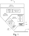
- the image processing module 104 is coupled to the imaging module 102 and works simultaneously to process the images received from the imaging module 102.
- the image processor 116 is to perform spatial and temporal resolution of the emission and reflectance or transmission spectra from the sample 112.
- the spatial resolution of the spectra is useful in extracting the spatial distribution of the pathogens in the sample 112.
- the temporal resolution of the spectra is useful for extracting the time-dependent fluorescence of the pathogens.
- the spatial resolution is determined by the imaging module 102 and can be tuned as per the application.
- temporal resolution is determined by the optical switch 110 and can be tuned depending on the application. In an example, the resolution of the device 100 is in a range of 1-2nm based on the application.
- the device 100 of the present subject matter is capable of classifying most of clinically relevant pathogens within a level of accuracy acceptable for various applications.
- the device 100 is capable of classifying the pathogen based on their family, genus, species and strain level.
- the device 100 can be used to distinguish a gram-positive bacterium from a gram-negative bacterium.
- the device 100 can be used to distinguish a drug resistant species of bacteria from its other variants. The detection and identification of pathogens happens within a few minutes and the device 100 can be operated easily without much technical training.
- the device 100 may be configured to distinguish between bacteria and fungus, or distinguish within bacteria based on class types into: Actinobacteria, Bacteroidia, Chlamydiae, Bacilli, Fusobacteria, Betaproteobacteria, Gammaproteobacteria, Mollicutes.
- Fungus may be classified based on the class types into: Ascomycota, Basidiomycota.
- the classification can also be based on gram types of bacteria: Gram negative type comprising but not limiting to Pseudomonas, Escherichia, Klebsiella, Proteus, Gram positive: Staphylococcus, Enterococcus, Streptococcus, Corynebacterium.
- the classification may also be based on genus: Corynebacterium, Micrococcus, Mycobacterium, Staphylococcus, Streptococcus, Clostridium, Neisseria, Helicobacter, Enterobacter, Escherichia, Klebsiella, Pseudomonas, Proteus, Salmonella, Mycoplasma.
- the device 100 of the present invention is suitable for studying the pathogens present in various kinds of samples 112.
- the sample may comprise one or more of the following: a body part, a wound, a fluid, a surface, a consumable commodity, a laboratory equipment, a sanitary device, a sanitary equipment, ambient air, a biochemical assay chip, a microfluidic chip, pus, blood, urine, saliva, sweat, semen, mucus, plasma, or any combination thereof for applications ranging from diabetic foot ulcers, surgical site infections, burns, hospital acquired skin and soft tissue infections, dermatology, cosmetology, plastic surgery, infection management in the hospitals, skin diseases, photodynamic therapy monitoring, anti-microbial susceptibility testing.
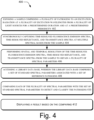
- Fig. 4 illustrates an example method 400 for detecting and classifying pathogens, in accordance with principles of the present subject matter.
- the order in which the method 400 is described is not intended to be construed as a limitation, and any number of the described method blocks can be combined in any order to implement method 400 or an alternative method. Additionally, individual blocks may be deleted from the method 400 without departing from the spirit and scope of the subject matter described herein.
- the method 400 may be implemented in any suitable hardware, computer readable instructions, firmware, or combination thereof. For discussion, the method 400 is described with reference to the implementations illustrated in Fig(s). 1-2.
- steps of the method 400 can be performed by programmed computers.
- program storage devices and non-transitory computer readable medium for example, digital data storage media, which are computer readable and encode computer-executable instructions, where said instructions perform some or all of the steps of the described method 400.
- the program storage devices may be, for example, digital memories, magnetic storage media, such as magnetic disks and magnetic tapes, hard drives, or optically readable digital data storage media.
- a library database is accessed.
- the library database comprises a set of standard spectral parameters associated with a set of reference pathogens.
- the library database is the library database 118 of the image processing module 104.
- the device 100 comprised high power light emitting diodes that are configured to emit light in the following wavelengths: 340 nm, 365 nm, 395 nm.
- the device comprised of an ARM processor, a 16MP cellphone camera with Sony IMX 398 sensor controlled with an electronic switch that is configured to capture white light and fluorescence images.
- the device 100 comprised an optical filter change mechanism controlled by a servo motor, and placed in front of the camera that was configured to measure spectral intensity of fluorescence at the following wavelengths: 415 nm, 470 nm, 515 nm, 560 nm, and 620 nm.
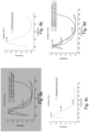
- Fig. 7 shows another statistical method employed to understand the variations in biomarker activity in different pathogens.
- Self-Organizing Map (SOM), is a neural network model that is based on unsupervised learning. SOM is useful for performing feature dependency extraction from a dataset with unknown dependencies. SOM shows the spectral regions of interest corresponding to auto fluorescence of various biomolecules present in pathogens. The weights obtained from SOM can be used to understand the relative distribution of auto fluorescence biomarkers which can then be used to distinguish pathogens.
- SOM Self-Organizing Map
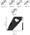
- Fig. 7a-e show relative distribution of auto fluorescence biomarkers in Candida sp., Candida albicans, Escherichia coli, Pseudomonas aeruginosa and Staphylococcus aureus respectively in a color-coded biomolecule classes.
- Fig. 7f shows the auto fluorescence biomolecules from all pathogen. Further information on individual spectral parameters can be obtained at the excitation wavelength obtained from SOM graph.
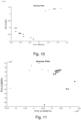
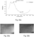
- Fig. 10 shows a chemometric statistical analysis method, namely the Principle Component Analysis (PCA) to understand maximum separation of bacteria in different excitation wavelengths.
- PCA Principle Component Analysis
- PCA at 410 nm excitation on E. coli, Pseudomonas aeruginosa, Staphylococcus aureus respectively have been shown, where Pseudomonas shows maximum separation at 410 nm excitation.
- PCA at 335 nm shows maximum separation of Candida species ( Fig. 11 ).
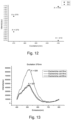
- Fig. 12 shows the ratio of amplitude of fluorescence emission peaks at 370 nm and 410 nm excitation, for Staphylococcus aureus and Escherichia coli. This example shows that ratio of fluorescence amplitudes in various spectral bands can be effectively used to classify certain pathogens.
- Fig. 13 shows 2D fluorescence spectra of Escherichia coli, collected at different growth stages- 2 hours, 4 hours and 6 hours of growth.
- the bacteria isolated from clinical samples was sub cultured and washed with 9% NaCl for 2D fluorescence spectra measurements using SpectraMax i3x Multi-Mode Reader. It was observed that spectral parameters obtained from steady state fluorescence are not enough for exact classification, mainly because the expression of fluorescence biomolecules depends on various intrinsic factors such as biochemical pathway during an infection and extrinsic factors such as environment, growth conditions, temperature. Therefore, it is clear that additional extraction of time dependent fluorescence parameters was needed.
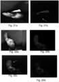
- Fig. 14a shows the time resolved fluorescence data contributed due to photo bleaching in Enterococcus kept at 29.5 degree.
- the excitation was at 415nm (at 90 flashes), and fluorescence recorded at every 49 seconds using SpectraMax i3x Multi-Mode Reader, in the kinetic mode.
- Fig. 14b shows Staphylococcus aureus at 340 nm excitation collected at 40 flashes, and recorded every 30 seconds.
- These graphs show a decrease in fluorescence with time due to photobleaching effect. This is a key spectral parameter that need to be considered for detection and classification of pathogens at genus and species level.
- Fig. 15 shows time-dependent fluorescence characteristics of Staphylococcus aureus and Escherichia coli excited at 370 nm and emission at 520 nm, obtained using Horiba TCSPC instrument (decay recorder was set to 100ns, the peak count was set to 10000 photons, and each channel duration of 0.027ns).
- steady state fluorescence spectra are similar at 520 nm corresponding to flavin molecule as shown in Fig. 8a and 8b , fluorescence lifetimes corresponding to flavins, at 520 nm emission are significantly different for Staphylococcus aureus and Escherichia coli. This result clearly demonstrates that time-dependent fluorescence can be used to distinguish between certain pathogens that are not possible using steady state fluorescence.
- EXAMPLE 3 USE OF HANDHELD DEVICE OF THE PRESENT INVENTION FOR VARIOUS CLINICAL DIAGNOSTIC APPLICATIONS
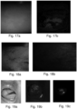
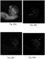
- Fig. 17 shows white light imaging and auto fluorescence imaging of infected hand to identify Malassezia furfur.
- the images were captured using the device 100 of Example 1, at 365 nm and 395 nm excitation light sources and at 450 nm and 520 nm emission respectively.
- the auto fluorescence images captured were processed using k-means clustering to clearly elucidate the region of infection and to understand the spatial distribution of fungal infection.
- Fig. 17a depicts white light image of fungus infected region
- Fig. 17b depicts processed auto fluorescence image using k-means clustering to identify the spatial distribution of Malassezia furfur.
Landscapes
- Health & Medical Sciences (AREA)
- Life Sciences & Earth Sciences (AREA)
- Physics & Mathematics (AREA)
- Engineering & Computer Science (AREA)
- General Health & Medical Sciences (AREA)
- Pathology (AREA)
- Public Health (AREA)
- Medical Informatics (AREA)
- Biomedical Technology (AREA)
- Heart & Thoracic Surgery (AREA)
- Veterinary Medicine (AREA)
- Animal Behavior & Ethology (AREA)
- Surgery (AREA)
- Molecular Biology (AREA)
- Biophysics (AREA)
- General Physics & Mathematics (AREA)
- Artificial Intelligence (AREA)
- Analytical Chemistry (AREA)
- Chemical & Material Sciences (AREA)
- Immunology (AREA)
- Biochemistry (AREA)
- Nuclear Medicine, Radiotherapy & Molecular Imaging (AREA)
- Spectroscopy & Molecular Physics (AREA)
- Computer Vision & Pattern Recognition (AREA)
- Physiology (AREA)
- Psychiatry (AREA)
- Signal Processing (AREA)
- Data Mining & Analysis (AREA)
- Primary Health Care (AREA)
- Epidemiology (AREA)
- Dermatology (AREA)
- Evolutionary Computation (AREA)
- Theoretical Computer Science (AREA)
- Radiology & Medical Imaging (AREA)
- Databases & Information Systems (AREA)
- Mathematical Physics (AREA)
- Fuzzy Systems (AREA)
- Hematology (AREA)
- Cardiology (AREA)
- Bioinformatics & Cheminformatics (AREA)
Claims (14)
- Vorrichtung (100) zur nicht-invasiven, automatischen und in-situ Detektion und Klassifizierung von Krankheitserregern, die Vorrichtung (100) umfassend:
ein Bildgebungsmodul (102), umfassend:eine Vielzahl von Lichtquellen (108), wobei jede Lichtquelle Erregungsstrahlung in einem vorbestimmten Wellenlängenbereich emittieren soll;einen optischen Schalter (110), um einen räumlichen Bereich einer Probe (112), umfassend Krankheitserreger, über eine vorbestimmte Dauer mit einer vorbestimmten Periodizität der Erregungsstrahlung auszusetzen; undeinen Detektor (114) zum Erfassen von zeitaufgelösten Fluoreszenzemissionsspektren und mindestens einem von: zeitaufgelösten Reflexionsspektren und zeitaufgelösten Transmissionsspektren in mehreren Spektralbändern über den räumlichen Bereich der Probe (112), wobei das Erfassen von jedem der zeitaufgelösten Fluoreszenzemissionsspektren, der zeitaufgelösten Reflexionsspektren und der zeitaufgelösten Transmissionsspektren basierend auf der Emission der Erregungsstrahlung durch die Vielzahl von Lichtquellen (108) synchronisiert ist;ein Bildverarbeitungsmodul (104), das mit dem Bildgebungsmodul (102) gekoppelt ist, um Informationen zu empfangen, die den zeitaufgelösten Fluoreszenzemissionsspektren, dem zeitaufgelösten Reflexionsgrad und den zeitaufgelösten Transmissionsspektren über den räumlichen Bereich der Probe (112) entsprechen, das Bildverarbeitungsmodul (104) umfassend:einen Bildprozessor (116), um eine räumliche und zeitliche Auflösung der Informationen auszuführen, um eine Vielzahl von Spektralparametern zu erlangen, wobei die räumliche Auflösung dazu dient, eine räumliche Verteilung von Krankheitserregern in dem räumlichen Bereich der Probe (112) zu extrahieren, und die zeitliche Auflösung dazu dient, zeitabhängige Spektralparameter der Krankheitserreger in der Probe (112) zu extrahieren; undeine Bibliotheksdatenbank (118), umfassend einen Satz von Standardspektralparametern, die mit Referenzkrankheitserregern identifizierbar sind,wobei das Bildverarbeitungsmodul (104) dazu dient, jeden der Vielzahl von Spektralparametern basierend auf einem ersten Bildverarbeitungsmodell mit dem Satz von Standardspektralparametern zu vergleichen, um den Krankheitserreger zu erkennen und zu klassifizieren, wobei die Bibliotheksdatenbank (118) dazu dient, einen neuen Satz von Standardspektralparametern zu empfangen, die mit einem neuen Referenzkrankheitserreger identifizierbar sind;eine Modellaufbaueinheit (304), um basierend auf dem neuen Satz von Standard-Spektralparametern, die mit dem neuen Referenz-Pathogen identifizierbar sind, und dem Satz von Standard-Spektralparametern, die mit dem Referenz-Pathogen identifizierbar sind, eine Vielzahl von Bildverarbeitungsmodellen bereitzustellen; undeinen Kreuzvalidator (306), um ein zweites Bildverarbeitungsmodell aus der Vielzahl der Bildverarbeitungsmodelle festzulegen, wobei das zweite Bildverarbeitungsmodell das erste Bildverarbeitungsmodell ersetzen soll; undein Anzeigemodul (106), um basierend auf dem Vergleich ein Resultat anzuzeigen. - Vorrichtung (100) nach Anspruch 1, wobei die Vielzahl der Lichtquellen mindestens eine der Folgenden umfasst: eine Leuchtdiode, einen Laser, eine farbige Lichtquelle, eine konfigurierbare Lichtquelle, Umgebungslicht oder eine beliebige Kombination davon.
- Vorrichtung (100) nach Anspruch 1, wobei das zeitaufgelöste Fluoreszenzemissionsspektrum ein Fluoreszenzlebensdauerspektrum oder ein Photobleichspektrum ist.
- Vorrichtung (100) nach Anspruch 3, wobei der Detektor (114) dazu dient, ein stationäres Fluoreszenzspektrum in Kombination mit mindestens einem von Folgenden zu erfassen: dem Fluoreszenzlebensdauerspektrum, dem Photobleichspektrum, dem zeitaufgelösten Reflexionsspektrum und dem zeitaufgelösten Transmissionsspektrum.
- Vorrichtung (100) nach Anspruch 3, wobei:wenn der Detektor (114) Fluoreszenzlebensdauerspektren erfassen soll, die Erregungsstrahlung die vorbestimmte Dauer in dem Bereich von etwa 1 ps bis etwa 1 s und die vorbestimmte Periodizität im Bereich von etwa 0,01 ns bis etwa 1 s aufweist; undwenn der Detektor (114) Photobleichspektren erfassen soll, die Erregungsstrahlung die vorbestimmte Dauer im Bereich von etwa 1 ms bis etwa 10 s und die vorbestimmte Periodizität in dem Bereich von etwa 0,01 s bis etwa 1 min aufweist.
- Vorrichtung (100) nach Anspruch 3, wobei das Bildverarbeitungsmodul (104) dazu dient, den Krankheitserreger basierend auf dem Vergleich nach Familie, Gattung, Art und Stamm zu klassifizieren.
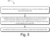
- Vorrichtung (100) nach Anspruch 1, wobei das Bildverarbeitungsmodul (104) dazu dient, in der Probe (112) vorhandene Krankheitserreger zu quantifizieren, wobei das Bildverarbeitungsmodul (104) dazu dient, eine Intensität der zeitabhängigen Fluoreszenz der Probe (112) zu empfangen und die Intensität mit in der Bibliotheksdatenbank (118) bereitgestellten Fluoreszenzintensitätsdaten zu vergleichen, um Krankheitserreger zu quantifizieren.
- Vorrichtung (100) nach Anspruch 1, wobei die Vorrichtung basierend auf der Detektion und der Klassifizierung von Pathogenen Wundheilung und Wundverschluss überwachen soll, wobei die Vorrichtung mindestens eines von Folgenden berechnen soll: Wundgröße, Wundtiefe, Wundtemperaturverteilung, Gewebeklassifizierung, Biofilminformationen, Kontaminationsgrad, Gewebeoxygenierung und Blutfluss.
- Vorrichtung (100) nach Anspruch 1, wobei die Vielzahl von Spektralparametern Amplitudenverhältnisse von Fluoreszenzemissionsspektren, Amplitudenverhältnisse von Reflexionsspektren, Amplitudenverhältnisse von Transmissionsspektren, Bandbreite der Fluoreszenzemissionsspektren, Halbwertsbreitenmaxima der Fluoreszenzemissionsspektren und Kombinationen davon umfasst.
- Vorrichtung (100) nach Anspruch 1, wobei die Bibliotheksdatenbank (118) mindestens eines von Erregungsemissionsmatrix-Spektren, Erregungsemissionsmatrix-Fluoreszenzspektren, Reflexionsspektren, Transmissionsspektren, Fluoreszenzlebensdauern, Photobleichzeiten, Absorptionskoeffizienten, Reflexionskoeffizienten, Transmissionskoeffizienten, Streukoeffizienten, normalisierte Intensitätsdaten, Intensitätsverhältnisse oder beliebige Kombinationen davon, die mit verschiedenen Referenzkrankheitserregern identifizierbar sind.
- Vorrichtung (100) nach Anspruch 1, wobei der Bildprozessor (116) dazu dient, mindestens eine der Folgenden zu erlangen: Fluoreszenzabklingzeiten, Amplitude des Fluoreszenzemissionssignals, Photobleichzeiten oder eine beliebige Kombination davon.
- Vorrichtung (100) nach Anspruch 1, wobei das Ergebnis mindestens eines der Folgenden umfasst: Detektion des Vorhandenseins verschiedener Krankheitserreger auf der Probe (112), Daten zur räumlichen Verteilung von Krankheitserregern, Daten zum Wachstumszustand von Krankheitserregern, Co-Kolonisationsdaten, Biofilm-Informationen, Biomarker-Informationen, Krankheitserreger-Quantifizierungsdaten, ein Behandlungsprotokoll oder eine beliebige Kombination davon.
- Verfahren zur nicht-invasiven, automatischen und in-situ Detektion und Klassifizierung von Krankheitserregern, umfassend:Aussetzen eines räumlichen Bereichs einer Probe (112), umfassend eine Vielzahl von Krankheitserregern, einer Erregungsstrahlung in einem vorbestimmten Bereich von Wellenlängen, die von einer Vielzahl von Lichtquellen (108) für eine vorbestimmte Dauer mit einer vorbestimmten Periodizität emittiert wird;Erfassen, durch einen Detektor (114), eines zeitaufgelösten Fluoreszenz-Emissionsspektrums und mindestens eines von Folgenden: einem zeitaufgelösten Reflexions- und einem Transmissionsspektrum in mehreren Spektralbändern über den räumlichen Bereich der Probe (112), wobei das Erfassen von jedem der zeitaufgelösten Fluoreszenzemissionsspektren, der zeitaufgelösten Reflexionsspektren und der zeitaufgelösten Transmissionsspektren basierend auf der Emission der Erregungsstrahlung durch die Vielzahl von Lichtquellen (108) synchronisiert ist;Ausführen, durch ein Bildverarbeitungsmodul (104), einer räumlichen und zeitlichen Auflösung der zeitaufgelösten Fluoreszenzemissionsspektren, zeitaufgelösten Reflexionsspektren und zeitaufgelösten Transmissionsspektren, die über den räumlichen Bereich der Probe (112) erfasst werden, um eine Vielzahl von Spektralparametern zu erlangen, wobei die räumliche Auflösung dazu dient, eine räumliche Verteilung von Krankheitserregern in dem räumlichen Bereich der Probe (112) zu extrahieren, und die zeitliche Auflösung dazu dient, zeitabhängige Spektralparameter der Krankheitserreger in der Probe (112) zu extrahieren;Zugreifen, durch einen Bildprozessor (116), auf eine Bibliotheksdatenbank (118), wobei die Bibliotheksdatenbank (118) einen Satz von Standardspektralparametern umfasst, die mit Referenzkrankheitserregern assoziiert sind;Vergleichen, durch das Bildverarbeitungsmodul (104), von jedem der Vielzahl von Spektralparametern mit dem Satz von Standardspektralparametern, um die Krankheitserreger zu erkennen und zu klassifizieren;
Anzeigen, durch ein Anzeigemodul (106), eines auf dem Vergleich basierenden Resultats;Empfangen, durch die Bibliotheksdatenbank (118), eines neuen Satzes von Standard-Spektralparametern, die mit einem neuen Referenzkrankheitserreger identifizierbar sind;Bereitstellen, durch eine Modellaufbaueinheit (304), einer Vielzahl von Bildverarbeitungsmodellen basierend auf dem neuen Satz von Standard-Spektralparametern, die mit dem neuen Referenzkrankheitserreger identifizierbar sind, und des Satzes von Standard-Spektralparametern, die mit dem Referenzerreger identifizierbar sind; undFestlegen, durch einen Kreuzvalidator (306), eines zweiten Bildverarbeitungsmodells aus der Vielzahl von Bildverarbeitungsmodellen, wobei das zweite Bildverarbeitungsmodell ein erstes Bildverarbeitungsmodell ersetzen soll, wobei das erste Bildverarbeitungsmodell mit dem Satz von Standard-Spektralparametern assoziiert ist und das zweite Bildverarbeitungsmodell mit dem Satz von Standard-Spektralparametern und dem neuen Satz von Standard-Spektralparametern assoziiert ist. - Verfahren nach Anspruch 13, wobei das Verfahren Folgendes umfasst:Berechnen, durch das Bildverarbeitungsmodul (104), von räumlichen Verteilungsdaten der Krankheitserreger über einen Wundbereich in Echtzeit;Kombinieren, durch das Bildverarbeitungsmodul (104), der räumlichen Verteilungsdaten in Echtzeit der Krankheitserreger über den Wundbereich mit Fluoreszenzintensitätsdaten bei einer Vielzahl von Spektralbändern der entsprechenden Krankheitserreger, die in der Bibliotheksdatenbank (118) enthalten sind; undQuantifizieren, durch das Bildverarbeitungsmodul (104), der Menge von jedem Krankheitserreger in dem gesamten Wundbereich.
Priority Applications (1)
| Application Number | Priority Date | Filing Date | Title |
|---|---|---|---|
| EP23196593.0A EP4306042A3 (de) | 2017-03-22 | 2018-03-22 | Vorrichtung und verfahren zum nachweis und zur klassifizierung von krankheitserregern |
Applications Claiming Priority (2)
| Application Number | Priority Date | Filing Date | Title |
|---|---|---|---|
| IN201741010111 | 2017-03-22 | ||
| PCT/IN2018/050161 WO2018173073A1 (en) | 2017-03-22 | 2018-03-22 | Device and method for detection and classification of pathogens |
Related Child Applications (2)
| Application Number | Title | Priority Date | Filing Date |
|---|---|---|---|
| EP23196593.0A Division-Into EP4306042A3 (de) | 2017-03-22 | 2018-03-22 | Vorrichtung und verfahren zum nachweis und zur klassifizierung von krankheitserregern |
| EP23196593.0A Division EP4306042A3 (de) | 2017-03-22 | 2018-03-22 | Vorrichtung und verfahren zum nachweis und zur klassifizierung von krankheitserregern |
Publications (3)
| Publication Number | Publication Date |
|---|---|
| EP3602007A1 EP3602007A1 (de) | 2020-02-05 |
| EP3602007A4 EP3602007A4 (de) | 2020-12-30 |
| EP3602007B1 true EP3602007B1 (de) | 2023-10-18 |
Family
ID=63584391
Family Applications (2)
| Application Number | Title | Priority Date | Filing Date |
|---|---|---|---|
| EP18772393.7A Active EP3602007B1 (de) | 2017-03-22 | 2018-03-22 | Vorrichtung und verfahren zum nachweis und zur klassifizierung von pathogenen |
| EP23196593.0A Pending EP4306042A3 (de) | 2017-03-22 | 2018-03-22 | Vorrichtung und verfahren zum nachweis und zur klassifizierung von krankheitserregern |
Family Applications After (1)
| Application Number | Title | Priority Date | Filing Date |
|---|---|---|---|
| EP23196593.0A Pending EP4306042A3 (de) | 2017-03-22 | 2018-03-22 | Vorrichtung und verfahren zum nachweis und zur klassifizierung von krankheitserregern |
Country Status (8)
| Country | Link |
|---|---|
| US (1) | US11523738B2 (de) |
| EP (2) | EP3602007B1 (de) |
| CA (1) | CA3057409A1 (de) |
| DK (1) | DK3602007T3 (de) |
| ES (1) | ES2968298T3 (de) |
| FI (1) | FI3602007T3 (de) |
| SG (1) | SG11201908815SA (de) |
| WO (1) | WO2018173073A1 (de) |
Families Citing this family (24)
| Publication number | Priority date | Publication date | Assignee | Title |
|---|---|---|---|---|
| JP7256207B2 (ja) | 2018-04-20 | 2023-04-11 | ザ プロクター アンド ギャンブル カンパニー | 頭皮ケア剤の空間的イメージング |
| WO2019222340A1 (en) | 2018-05-17 | 2019-11-21 | The Procter & Gamble Company | Systems and methods for hair coverage analysis |
| US11172873B2 (en) | 2018-05-17 | 2021-11-16 | The Procter & Gamble Company | Systems and methods for hair analysis |
| JP7134421B2 (ja) * | 2019-01-28 | 2022-09-12 | 日本たばこ産業株式会社 | 蛍光画像を用いた試料の品質判定方法、プログラム、及び、装置 |
| US11471389B2 (en) | 2019-06-28 | 2022-10-18 | The Procter & Gamble Company | Enhanced stability of zinc pyrithione in oxidative environments, such as scalp sebaceous fluid |
| EP4045898A4 (de) * | 2019-10-17 | 2024-01-10 | C2Sense, Inc. | Lumineszenzbilgebung zur erfassung |
| FR3103900B1 (fr) * | 2019-11-29 | 2024-07-19 | Univ Du Mans | Méthode d'identification rapide de microorganismes par analyse de matrices excitation-émission |
| GB2600719B (en) * | 2020-11-05 | 2023-10-11 | Oxford Nanoimaging Ltd | Fluorescence assay |
| BR112023015956A2 (pt) | 2021-02-09 | 2023-10-24 | Adiuvo Diagnostics Private Ltd | Dispositivo para examinar um alvo, sistema para examinar um alvo, dispositivo para treinar um modelo de análise para analisar imagens de alvos com base em fluorescência, método para examinar um alvo e método para treinar um modelo de análise para analisar imagens com base em fluorescência de alvos |
| AU2022249851A1 (en) * | 2021-03-31 | 2023-10-12 | University Of Lancaster | Detection of micro-organisms |
| US12039732B2 (en) | 2021-04-14 | 2024-07-16 | The Procter & Gamble Company | Digital imaging and learning systems and methods for analyzing pixel data of a scalp region of a users scalp to generate one or more user-specific scalp classifications |
| BE1029446B1 (nl) | 2021-06-01 | 2023-01-09 | Microtechnix Bvba | Inrichting en werkwijze voor telling en identificatie van bacteriekolonies met behulp van hyperspectrale beeldvorming |
| JP7190534B1 (ja) * | 2021-06-14 | 2022-12-15 | アサヒ飲料株式会社 | 微生物の検出方法 |
| EP4437499A4 (de) * | 2021-11-23 | 2025-08-27 | Tufts College | Erkennung und identifizierung von defekten mittels analyse von mehrdimensionalen informationsdaten mit künstlicher intelligenz |
| US20250049383A1 (en) * | 2021-12-20 | 2025-02-13 | Koninklijke Philips N.V. | Contactless wound monitoring |
| CN114414505A (zh) * | 2022-01-22 | 2022-04-29 | 山东润一智能科技有限公司 | 量子光谱微生物智能监测终端 |
| PL441490A1 (pl) * | 2022-06-15 | 2023-12-18 | Gdański Uniwersytet Medyczny | Układ do pomiaru absorbancji próbki materiału biologicznego w postaci płynnej i sposób dyskryminacji zakażeń układu moczowego realizowany za pomocą tego układu |
| WO2024092247A1 (en) * | 2022-10-27 | 2024-05-02 | Hyperspectral Corp. | Systems and methods for particle of interest detection |
| US12613177B2 (en) * | 2022-10-27 | 2026-04-28 | Hyperspectral Corp. | Systems and methods for detecting particles of interest using multi-model spectral analysis |
| US12546721B2 (en) | 2023-01-23 | 2026-02-10 | Lightsense Technology, Inc. | Multispectral detection and classification of bacteria utilizing scattering and/or absorbance, excitation, emissions utilizing machine learning |
| US20250090055A1 (en) * | 2023-09-20 | 2025-03-20 | Alexandre Ivachtchenko | Method and device for determining ischemic injuries of mammalian organs and tissues |
| CN117848962B (zh) * | 2023-10-26 | 2025-07-08 | 江苏大学 | 一种作物气传病害孢子检测装置与方法 |
| CN117825344A (zh) * | 2023-12-26 | 2024-04-05 | 中国科学院上海微系统与信息技术研究所 | 一种大肠杆菌检测系统及检测方法 |
| WO2026017793A1 (en) | 2024-07-18 | 2026-01-22 | Inwound Aps | An imaging device for monitoring wounds and a system and method for managing a treatment of a wound |
Family Cites Families (13)
| Publication number | Priority date | Publication date | Assignee | Title |
|---|---|---|---|---|
| WO2002069784A2 (en) | 2001-03-01 | 2002-09-12 | Trustees Of Dartmouth College | Fluorescence lifetime spectrometer (fls) and methods of detecting diseased tissues |
| EP1481356A1 (de) * | 2001-11-21 | 2004-12-01 | Paradigm Genetics Inc. | Verfahren und systeme zur analyse komplexer biologischer systeme |
| WO2003060444A1 (en) | 2002-01-10 | 2003-07-24 | Chemimage Corporation | Method for detection of pathogenic microorganisms |
| US7518710B2 (en) * | 2005-07-14 | 2009-04-14 | Battelle Memorial Institute | Optical devices for biological and chemical detection |
| CA3162577C (en) | 2008-05-20 | 2023-09-26 | University Health Network | Device and method for fluorescence-based imaging and monitoring |
| US8406859B2 (en) | 2008-08-10 | 2013-03-26 | Board Of Regents, The University Of Texas System | Digital light processing hyperspectral imaging apparatus |
| EA033790B1 (ru) * | 2011-12-19 | 2019-11-26 | Opticul Diagnostics Ltd | Способ спектрального обнаружения и идентификации микроорганизмов в культуре |
| WO2013116316A1 (en) | 2012-01-30 | 2013-08-08 | Scanadu Incorporated | Hyperspectral imaging systems, units, and methods |
| WO2014040168A1 (en) | 2012-09-14 | 2014-03-20 | University Of Windsor | Method of using laser-induced breakdown spectroscopy for the identification and classification of bacteria |
| WO2014168734A1 (en) * | 2013-03-15 | 2014-10-16 | Cedars-Sinai Medical Center | Time-resolved laser-induced fluorescence spectroscopy systems and uses thereof |
| ES2877351T3 (es) | 2013-08-07 | 2021-11-16 | Univ Wayne State | Instrumento de detección portátil basado en micro-raman y método de detección |
| JP6893877B2 (ja) | 2014-10-29 | 2021-06-23 | スペクトラル エムディー, インコーポレイテッドSpectral Md, Inc. | 組織を分類するための反射モードマルチスペクトル時間分解型光学イメージングの方法および装置 |
| WO2020148726A1 (en) | 2019-01-17 | 2020-07-23 | Moleculight Inc. | Modular system for multi-modal imaging and analysis |
-
2018
- 2018-03-22 EP EP18772393.7A patent/EP3602007B1/de active Active
- 2018-03-22 US US16/496,390 patent/US11523738B2/en active Active
- 2018-03-22 SG SG11201908815S patent/SG11201908815SA/en unknown
- 2018-03-22 EP EP23196593.0A patent/EP4306042A3/de active Pending
- 2018-03-22 FI FIEP18772393.7T patent/FI3602007T3/fi active
- 2018-03-22 ES ES18772393T patent/ES2968298T3/es active Active
- 2018-03-22 WO PCT/IN2018/050161 patent/WO2018173073A1/en not_active Ceased
- 2018-03-22 CA CA3057409A patent/CA3057409A1/en active Pending
- 2018-03-22 DK DK18772393.7T patent/DK3602007T3/da active
Also Published As
| Publication number | Publication date |
|---|---|
| WO2018173073A1 (en) | 2018-09-27 |
| US20210106231A1 (en) | 2021-04-15 |
| DK3602007T3 (da) | 2024-01-15 |
| SG11201908815SA (en) | 2019-10-30 |
| FI3602007T3 (fi) | 2024-01-16 |
| ES2968298T3 (es) | 2024-05-08 |
| EP3602007A1 (de) | 2020-02-05 |
| EP4306042A2 (de) | 2024-01-17 |
| CA3057409A1 (en) | 2018-09-27 |
| EP4306042A3 (de) | 2024-03-27 |
| EP3602007A4 (de) | 2020-12-30 |
| US11523738B2 (en) | 2022-12-13 |
Similar Documents
| Publication | Publication Date | Title |
|---|---|---|
| EP3602007B1 (de) | Vorrichtung und verfahren zum nachweis und zur klassifizierung von pathogenen | |
| US12387335B2 (en) | Systems, devices, and methods for visualization of tissue and collection and analysis of data regarding same | |
| AU2025238084A1 (en) | Modular system for multi-modal imaging and analysis | |
| JP2024508046A (ja) | 問題のある細胞実体の蛍光ベースの検出 | |
| US20250322524A1 (en) | Systems and methods for detection of cellular entities | |
| WO2024006808A2 (en) | Systems and methods for fluorescence-based imaging | |
| Radhakrishnan et al. | Rapid handheld screening device to detect skin and soft tissue infections | |
| HK40069119A (en) | Collection and analysis of data for diagnostic purposes | |
| HK1238515B (en) | Collection and analysis of data for diagnostic purposes | |
| HK1238515A1 (en) | Collection and analysis of data for diagnostic purposes |
Legal Events
| Date | Code | Title | Description |
|---|---|---|---|
| STAA | Information on the status of an ep patent application or granted ep patent |
Free format text: STATUS: THE INTERNATIONAL PUBLICATION HAS BEEN MADE |
|
| PUAI | Public reference made under article 153(3) epc to a published international application that has entered the european phase |
Free format text: ORIGINAL CODE: 0009012 |
|
| STAA | Information on the status of an ep patent application or granted ep patent |
Free format text: STATUS: REQUEST FOR EXAMINATION WAS MADE |
|
| 17P | Request for examination filed |
Effective date: 20191021 |
|
| AK | Designated contracting states |
Kind code of ref document: A1 Designated state(s): AL AT BE BG CH CY CZ DE DK EE ES FI FR GB GR HR HU IE IS IT LI LT LU LV MC MK MT NL NO PL PT RO RS SE SI SK SM TR |
|
| AX | Request for extension of the european patent |
Extension state: BA ME |
|
| DAV | Request for validation of the european patent (deleted) | ||
| DAX | Request for extension of the european patent (deleted) | ||
| TPAC | Observations filed by third parties |
Free format text: ORIGINAL CODE: EPIDOSNTIPA |
|
| A4 | Supplementary search report drawn up and despatched |
Effective date: 20201126 |
|
| RIC1 | Information provided on ipc code assigned before grant |
Ipc: A61B 5/00 20060101ALI20201120BHEP Ipc: G01N 21/31 20060101AFI20201120BHEP Ipc: G01N 33/50 20060101ALI20201120BHEP Ipc: G01N 21/64 20060101ALI20201120BHEP Ipc: G01N 21/65 20060101ALI20201120BHEP |
|
| STAA | Information on the status of an ep patent application or granted ep patent |
Free format text: STATUS: EXAMINATION IS IN PROGRESS |
|
| 17Q | First examination report despatched |
Effective date: 20210826 |
|
| GRAP | Despatch of communication of intention to grant a patent |
Free format text: ORIGINAL CODE: EPIDOSNIGR1 |
|
| STAA | Information on the status of an ep patent application or granted ep patent |
Free format text: STATUS: GRANT OF PATENT IS INTENDED |
|
| INTG | Intention to grant announced |
Effective date: 20230502 |
|
| GRAS | Grant fee paid |
Free format text: ORIGINAL CODE: EPIDOSNIGR3 |
|
| GRAA | (expected) grant |
Free format text: ORIGINAL CODE: 0009210 |
|
| STAA | Information on the status of an ep patent application or granted ep patent |
Free format text: STATUS: THE PATENT HAS BEEN GRANTED |
|
| AK | Designated contracting states |
Kind code of ref document: B1 Designated state(s): AL AT BE BG CH CY CZ DE DK EE ES FI FR GB GR HR HU IE IS IT LI LT LU LV MC MK MT NL NO PL PT RO RS SE SI SK SM TR |
|
| REG | Reference to a national code |
Ref country code: GB Ref legal event code: FG4D |
|
| REG | Reference to a national code |
Ref country code: CH Ref legal event code: EP |
|
| REG | Reference to a national code |
Ref country code: IE Ref legal event code: FG4D |
|
| REG | Reference to a national code |
Ref country code: DE Ref legal event code: R096 Ref document number: 602018059619 Country of ref document: DE |
|
| REG | Reference to a national code |
Ref country code: DK Ref legal event code: T3 Effective date: 20240111 |
|
| REG | Reference to a national code |
Ref country code: FI Ref legal event code: FGE |
|
| REG | Reference to a national code |
Ref country code: NO Ref legal event code: T2 Effective date: 20231018 |
|
| REG | Reference to a national code |
Ref country code: NL Ref legal event code: FP |
|
| REG | Reference to a national code |
Ref country code: SE Ref legal event code: TRGR |
|
| REG | Reference to a national code |
Ref country code: LT Ref legal event code: MG9D |
|
| P01 | Opt-out of the competence of the unified patent court (upc) registered |
Effective date: 20240104 |
|
| REG | Reference to a national code |
Ref country code: AT Ref legal event code: MK05 Ref document number: 1622861 Country of ref document: AT Kind code of ref document: T Effective date: 20231018 |
|
| PG25 | Lapsed in a contracting state [announced via postgrant information from national office to epo] |
Ref country code: GR Free format text: LAPSE BECAUSE OF FAILURE TO SUBMIT A TRANSLATION OF THE DESCRIPTION OR TO PAY THE FEE WITHIN THE PRESCRIBED TIME-LIMIT Effective date: 20240119 |
|
| PG25 | Lapsed in a contracting state [announced via postgrant information from national office to epo] |
Ref country code: IS Free format text: LAPSE BECAUSE OF FAILURE TO SUBMIT A TRANSLATION OF THE DESCRIPTION OR TO PAY THE FEE WITHIN THE PRESCRIBED TIME-LIMIT Effective date: 20240218 |
|
| PG25 | Lapsed in a contracting state [announced via postgrant information from national office to epo] |
Ref country code: LT Free format text: LAPSE BECAUSE OF FAILURE TO SUBMIT A TRANSLATION OF THE DESCRIPTION OR TO PAY THE FEE WITHIN THE PRESCRIBED TIME-LIMIT Effective date: 20231018 |
|
| PG25 | Lapsed in a contracting state [announced via postgrant information from national office to epo] |
Ref country code: AT Free format text: LAPSE BECAUSE OF FAILURE TO SUBMIT A TRANSLATION OF THE DESCRIPTION OR TO PAY THE FEE WITHIN THE PRESCRIBED TIME-LIMIT Effective date: 20231018 |
|
| PG25 | Lapsed in a contracting state [announced via postgrant information from national office to epo] |
Ref country code: LT Free format text: LAPSE BECAUSE OF FAILURE TO SUBMIT A TRANSLATION OF THE DESCRIPTION OR TO PAY THE FEE WITHIN THE PRESCRIBED TIME-LIMIT Effective date: 20231018 Ref country code: IS Free format text: LAPSE BECAUSE OF FAILURE TO SUBMIT A TRANSLATION OF THE DESCRIPTION OR TO PAY THE FEE WITHIN THE PRESCRIBED TIME-LIMIT Effective date: 20240218 Ref country code: GR Free format text: LAPSE BECAUSE OF FAILURE TO SUBMIT A TRANSLATION OF THE DESCRIPTION OR TO PAY THE FEE WITHIN THE PRESCRIBED TIME-LIMIT Effective date: 20240119 Ref country code: BG Free format text: LAPSE BECAUSE OF FAILURE TO SUBMIT A TRANSLATION OF THE DESCRIPTION OR TO PAY THE FEE WITHIN THE PRESCRIBED TIME-LIMIT Effective date: 20240118 Ref country code: AT Free format text: LAPSE BECAUSE OF FAILURE TO SUBMIT A TRANSLATION OF THE DESCRIPTION OR TO PAY THE FEE WITHIN THE PRESCRIBED TIME-LIMIT Effective date: 20231018 Ref country code: PT Free format text: LAPSE BECAUSE OF FAILURE TO SUBMIT A TRANSLATION OF THE DESCRIPTION OR TO PAY THE FEE WITHIN THE PRESCRIBED TIME-LIMIT Effective date: 20240219 |
|
| REG | Reference to a national code |
Ref country code: ES Ref legal event code: FG2A Ref document number: 2968298 Country of ref document: ES Kind code of ref document: T3 Effective date: 20240508 |
|
| PG25 | Lapsed in a contracting state [announced via postgrant information from national office to epo] |
Ref country code: RS Free format text: LAPSE BECAUSE OF FAILURE TO SUBMIT A TRANSLATION OF THE DESCRIPTION OR TO PAY THE FEE WITHIN THE PRESCRIBED TIME-LIMIT Effective date: 20231018 Ref country code: PL Free format text: LAPSE BECAUSE OF FAILURE TO SUBMIT A TRANSLATION OF THE DESCRIPTION OR TO PAY THE FEE WITHIN THE PRESCRIBED TIME-LIMIT Effective date: 20231018 Ref country code: LV Free format text: LAPSE BECAUSE OF FAILURE TO SUBMIT A TRANSLATION OF THE DESCRIPTION OR TO PAY THE FEE WITHIN THE PRESCRIBED TIME-LIMIT Effective date: 20231018 Ref country code: HR Free format text: LAPSE BECAUSE OF FAILURE TO SUBMIT A TRANSLATION OF THE DESCRIPTION OR TO PAY THE FEE WITHIN THE PRESCRIBED TIME-LIMIT Effective date: 20231018 |
|
| REG | Reference to a national code |
Ref country code: DE Ref legal event code: R097 Ref document number: 602018059619 Country of ref document: DE |
|
| PG25 | Lapsed in a contracting state [announced via postgrant information from national office to epo] |
Ref country code: CZ Free format text: LAPSE BECAUSE OF FAILURE TO SUBMIT A TRANSLATION OF THE DESCRIPTION OR TO PAY THE FEE WITHIN THE PRESCRIBED TIME-LIMIT Effective date: 20231018 |
|
| PG25 | Lapsed in a contracting state [announced via postgrant information from national office to epo] |
Ref country code: SK Free format text: LAPSE BECAUSE OF FAILURE TO SUBMIT A TRANSLATION OF THE DESCRIPTION OR TO PAY THE FEE WITHIN THE PRESCRIBED TIME-LIMIT Effective date: 20231018 |
|
| PG25 | Lapsed in a contracting state [announced via postgrant information from national office to epo] |
Ref country code: SM Free format text: LAPSE BECAUSE OF FAILURE TO SUBMIT A TRANSLATION OF THE DESCRIPTION OR TO PAY THE FEE WITHIN THE PRESCRIBED TIME-LIMIT Effective date: 20231018 Ref country code: SK Free format text: LAPSE BECAUSE OF FAILURE TO SUBMIT A TRANSLATION OF THE DESCRIPTION OR TO PAY THE FEE WITHIN THE PRESCRIBED TIME-LIMIT Effective date: 20231018 Ref country code: RO Free format text: LAPSE BECAUSE OF FAILURE TO SUBMIT A TRANSLATION OF THE DESCRIPTION OR TO PAY THE FEE WITHIN THE PRESCRIBED TIME-LIMIT Effective date: 20231018 Ref country code: EE Free format text: LAPSE BECAUSE OF FAILURE TO SUBMIT A TRANSLATION OF THE DESCRIPTION OR TO PAY THE FEE WITHIN THE PRESCRIBED TIME-LIMIT Effective date: 20231018 Ref country code: CZ Free format text: LAPSE BECAUSE OF FAILURE TO SUBMIT A TRANSLATION OF THE DESCRIPTION OR TO PAY THE FEE WITHIN THE PRESCRIBED TIME-LIMIT Effective date: 20231018 |
|
| PLBE | No opposition filed within time limit |
Free format text: ORIGINAL CODE: 0009261 |
|
| STAA | Information on the status of an ep patent application or granted ep patent |
Free format text: STATUS: NO OPPOSITION FILED WITHIN TIME LIMIT |
|
| 26N | No opposition filed |
Effective date: 20240719 |
|
| PG25 | Lapsed in a contracting state [announced via postgrant information from national office to epo] |
Ref country code: SI Free format text: LAPSE BECAUSE OF FAILURE TO SUBMIT A TRANSLATION OF THE DESCRIPTION OR TO PAY THE FEE WITHIN THE PRESCRIBED TIME-LIMIT Effective date: 20231018 |
|
| PG25 | Lapsed in a contracting state [announced via postgrant information from national office to epo] |
Ref country code: SI Free format text: LAPSE BECAUSE OF FAILURE TO SUBMIT A TRANSLATION OF THE DESCRIPTION OR TO PAY THE FEE WITHIN THE PRESCRIBED TIME-LIMIT Effective date: 20231018 |
|
| PG25 | Lapsed in a contracting state [announced via postgrant information from national office to epo] |
Ref country code: LU Free format text: LAPSE BECAUSE OF NON-PAYMENT OF DUE FEES Effective date: 20240322 |
|
| PG25 | Lapsed in a contracting state [announced via postgrant information from national office to epo] |
Ref country code: MC Free format text: LAPSE BECAUSE OF FAILURE TO SUBMIT A TRANSLATION OF THE DESCRIPTION OR TO PAY THE FEE WITHIN THE PRESCRIBED TIME-LIMIT Effective date: 20231018 |
|
| PG25 | Lapsed in a contracting state [announced via postgrant information from national office to epo] |
Ref country code: MC Free format text: LAPSE BECAUSE OF FAILURE TO SUBMIT A TRANSLATION OF THE DESCRIPTION OR TO PAY THE FEE WITHIN THE PRESCRIBED TIME-LIMIT Effective date: 20231018 Ref country code: LU Free format text: LAPSE BECAUSE OF NON-PAYMENT OF DUE FEES Effective date: 20240322 |
|
| PG25 | Lapsed in a contracting state [announced via postgrant information from national office to epo] |
Ref country code: IE Free format text: LAPSE BECAUSE OF NON-PAYMENT OF DUE FEES Effective date: 20240322 |
|
| PG25 | Lapsed in a contracting state [announced via postgrant information from national office to epo] |
Ref country code: IE Free format text: LAPSE BECAUSE OF NON-PAYMENT OF DUE FEES Effective date: 20240322 |
|
| PGFP | Annual fee paid to national office [announced via postgrant information from national office to epo] |
Ref country code: SE Payment date: 20250321 Year of fee payment: 8 |
|
| PGFP | Annual fee paid to national office [announced via postgrant information from national office to epo] |
Ref country code: DE Payment date: 20250319 Year of fee payment: 8 |
|
| PGFP | Annual fee paid to national office [announced via postgrant information from national office to epo] |
Ref country code: DK Payment date: 20250326 Year of fee payment: 8 Ref country code: FI Payment date: 20250324 Year of fee payment: 8 Ref country code: NL Payment date: 20250319 Year of fee payment: 8 |
|
| PGFP | Annual fee paid to national office [announced via postgrant information from national office to epo] |
Ref country code: NO Payment date: 20250321 Year of fee payment: 8 |
|
| PGFP | Annual fee paid to national office [announced via postgrant information from national office to epo] |
Ref country code: BE Payment date: 20250319 Year of fee payment: 8 |
|
| PGFP | Annual fee paid to national office [announced via postgrant information from national office to epo] |
Ref country code: FR Payment date: 20250326 Year of fee payment: 8 |
|
| PGFP | Annual fee paid to national office [announced via postgrant information from national office to epo] |
Ref country code: IT Payment date: 20250325 Year of fee payment: 8 Ref country code: GB Payment date: 20250324 Year of fee payment: 8 |
|
| PGFP | Annual fee paid to national office [announced via postgrant information from national office to epo] |
Ref country code: ES Payment date: 20250429 Year of fee payment: 8 |
|
| PGFP | Annual fee paid to national office [announced via postgrant information from national office to epo] |
Ref country code: CH Payment date: 20250401 Year of fee payment: 8 |
|
| PG25 | Lapsed in a contracting state [announced via postgrant information from national office to epo] |
Ref country code: CY Free format text: LAPSE BECAUSE OF FAILURE TO SUBMIT A TRANSLATION OF THE DESCRIPTION OR TO PAY THE FEE WITHIN THE PRESCRIBED TIME-LIMIT; INVALID AB INITIO Effective date: 20180322 |
|
| PG25 | Lapsed in a contracting state [announced via postgrant information from national office to epo] |
Ref country code: HU Free format text: LAPSE BECAUSE OF FAILURE TO SUBMIT A TRANSLATION OF THE DESCRIPTION OR TO PAY THE FEE WITHIN THE PRESCRIBED TIME-LIMIT; INVALID AB INITIO Effective date: 20180322 |
|
| PG25 | Lapsed in a contracting state [announced via postgrant information from national office to epo] |
Ref country code: TR Free format text: LAPSE BECAUSE OF FAILURE TO SUBMIT A TRANSLATION OF THE DESCRIPTION OR TO PAY THE FEE WITHIN THE PRESCRIBED TIME-LIMIT Effective date: 20231018 |
|
| REG | Reference to a national code |
Ref country code: GB Ref legal event code: 732E Free format text: REGISTERED BETWEEN 20251204 AND 20251210 |
|
| REG | Reference to a national code |
Ref country code: DE Ref legal event code: R081 Ref document number: 602018059619 Country of ref document: DE Owner name: ADIUVO DIAGNOSTIC PVT. LTD., IN Free format text: FORMER OWNER: ADIUVO DIAGNOSTICS PVT LTD, NAWABPET, NELLORE, IN Ref country code: DE Ref legal event code: R081 Ref document number: 602018059619 Country of ref document: DE Owner name: KENT IMAGING INC., CA Free format text: FORMER OWNER: ADIUVO DIAGNOSTICS PVT LTD, NAWABPET, NELLORE, IN Ref country code: DE Ref legal event code: R082 Ref document number: 602018059619 Country of ref document: DE Representative=s name: HERNANDEZ, YORCK, DIPL.-ING., DE |
|
| REG | Reference to a national code |
Ref country code: CH Ref legal event code: R14 Free format text: ST27 STATUS EVENT CODE: U-0-0-R10-R14 (AS PROVIDED BY THE NATIONAL OFFICE) Effective date: 20260130 |
|
| REG | Reference to a national code |
Ref country code: CH Ref legal event code: R17 Free format text: ST27 STATUS EVENT CODE: U-0-0-R10-R17 (AS PROVIDED BY THE NATIONAL OFFICE) Effective date: 20260211 Ref country code: FI Ref legal event code: PCE Owner name: ADIUVO DIAGNOSTIC PRIVATE LIMITED , IN; KENT IMAGING INC., CA |
|
| REG | Reference to a national code |
Ref country code: BE Ref legal event code: PD Owner name: KENT IMAGING INC.; CA Free format text: DETAILS ASSIGNMENT: CHANGE OF OWNER(S), ASSIGNMENT; FORMER OWNER NAME: ADIUVO DIAGNOSTICS PVT LTD Effective date: 20260125 |
|
| REG | Reference to a national code |
Ref country code: NL Ref legal event code: PD Owner name: ADIUVO DIAGNOSTICS PRIVATE LIMITED; IN Free format text: DETAILS ASSIGNMENT: CHANGE OF OWNER(S), ASSIGNMENT; FORMER OWNER NAME: ADIUVO DIAGNOSTICS PVT LTD Effective date: 20260420 |