EP3601604B1 - Multiplex-nachweis von isotypenspezifischem antikörper - Google Patents
Multiplex-nachweis von isotypenspezifischem antikörper Download PDFInfo
- Publication number
- EP3601604B1 EP3601604B1 EP18777364.3A EP18777364A EP3601604B1 EP 3601604 B1 EP3601604 B1 EP 3601604B1 EP 18777364 A EP18777364 A EP 18777364A EP 3601604 B1 EP3601604 B1 EP 3601604B1
- Authority
- EP
- European Patent Office
- Prior art keywords
- dna
- antibody
- seq
- sequence
- sample
- Prior art date
- Legal status (The legal status is an assumption and is not a legal conclusion. Google has not performed a legal analysis and makes no representation as to the accuracy of the status listed.)
- Active
Links
Images


Classifications
-
- C—CHEMISTRY; METALLURGY
- C12—BIOCHEMISTRY; BEER; SPIRITS; WINE; VINEGAR; MICROBIOLOGY; ENZYMOLOGY; MUTATION OR GENETIC ENGINEERING
- C12N—MICROORGANISMS OR ENZYMES; COMPOSITIONS THEREOF; PROPAGATING, PRESERVING, OR MAINTAINING MICROORGANISMS; MUTATION OR GENETIC ENGINEERING; CULTURE MEDIA
- C12N15/00—Mutation or genetic engineering; DNA or RNA concerning genetic engineering, vectors, e.g. plasmids, or their isolation, preparation or purification; Use of hosts therefor
- C12N15/09—Recombinant DNA-technology
- C12N15/11—DNA or RNA fragments; Modified forms thereof; Non-coding nucleic acids having a biological activity
-
- C—CHEMISTRY; METALLURGY
- C12—BIOCHEMISTRY; BEER; SPIRITS; WINE; VINEGAR; MICROBIOLOGY; ENZYMOLOGY; MUTATION OR GENETIC ENGINEERING
- C12Q—MEASURING OR TESTING PROCESSES INVOLVING ENZYMES, NUCLEIC ACIDS OR MICROORGANISMS; COMPOSITIONS OR TEST PAPERS THEREFOR; PROCESSES OF PREPARING SUCH COMPOSITIONS; CONDITION-RESPONSIVE CONTROL IN MICROBIOLOGICAL OR ENZYMOLOGICAL PROCESSES
- C12Q1/00—Measuring or testing processes involving enzymes, nucleic acids or microorganisms; Compositions therefor; Processes of preparing such compositions
- C12Q1/68—Measuring or testing processes involving enzymes, nucleic acids or microorganisms; Compositions therefor; Processes of preparing such compositions involving nucleic acids
- C12Q1/6804—Nucleic acid analysis using immunogens
-
- C—CHEMISTRY; METALLURGY
- C12—BIOCHEMISTRY; BEER; SPIRITS; WINE; VINEGAR; MICROBIOLOGY; ENZYMOLOGY; MUTATION OR GENETIC ENGINEERING
- C12Q—MEASURING OR TESTING PROCESSES INVOLVING ENZYMES, NUCLEIC ACIDS OR MICROORGANISMS; COMPOSITIONS OR TEST PAPERS THEREFOR; PROCESSES OF PREPARING SUCH COMPOSITIONS; CONDITION-RESPONSIVE CONTROL IN MICROBIOLOGICAL OR ENZYMOLOGICAL PROCESSES
- C12Q1/00—Measuring or testing processes involving enzymes, nucleic acids or microorganisms; Compositions therefor; Processes of preparing such compositions
- C12Q1/68—Measuring or testing processes involving enzymes, nucleic acids or microorganisms; Compositions therefor; Processes of preparing such compositions involving nucleic acids
- C12Q1/6806—Preparing nucleic acids for analysis, e.g. for polymerase chain reaction [PCR] assay
-
- C—CHEMISTRY; METALLURGY
- C12—BIOCHEMISTRY; BEER; SPIRITS; WINE; VINEGAR; MICROBIOLOGY; ENZYMOLOGY; MUTATION OR GENETIC ENGINEERING
- C12Q—MEASURING OR TESTING PROCESSES INVOLVING ENZYMES, NUCLEIC ACIDS OR MICROORGANISMS; COMPOSITIONS OR TEST PAPERS THEREFOR; PROCESSES OF PREPARING SUCH COMPOSITIONS; CONDITION-RESPONSIVE CONTROL IN MICROBIOLOGICAL OR ENZYMOLOGICAL PROCESSES
- C12Q1/00—Measuring or testing processes involving enzymes, nucleic acids or microorganisms; Compositions therefor; Processes of preparing such compositions
- C12Q1/68—Measuring or testing processes involving enzymes, nucleic acids or microorganisms; Compositions therefor; Processes of preparing such compositions involving nucleic acids
- C12Q1/6844—Nucleic acid amplification reactions
- C12Q1/686—Polymerase chain reaction [PCR]
-
- G—PHYSICS
- G01—MEASURING; TESTING
- G01N—INVESTIGATING OR ANALYSING MATERIALS BY DETERMINING THEIR CHEMICAL OR PHYSICAL PROPERTIES
- G01N33/00—Investigating or analysing materials by specific methods not covered by groups G01N1/00 - G01N31/00
- G01N33/48—Biological material, e.g. blood, urine; Haemocytometers
- G01N33/50—Chemical analysis of biological material, e.g. blood, urine; Testing involving biospecific ligand binding methods; Immunological testing
- G01N33/53—Immunoassay; Biospecific binding assay; Materials therefor
- G01N33/564—Immunoassay; Biospecific binding assay; Materials therefor for pre-existing immune complex or autoimmune disease, i.e. systemic lupus erythematosus, rheumatoid arthritis, multiple sclerosis, rheumatoid factors or complement components C1-C9
-
- G—PHYSICS
- G01—MEASURING; TESTING
- G01N—INVESTIGATING OR ANALYSING MATERIALS BY DETERMINING THEIR CHEMICAL OR PHYSICAL PROPERTIES
- G01N33/00—Investigating or analysing materials by specific methods not covered by groups G01N1/00 - G01N31/00
- G01N33/48—Biological material, e.g. blood, urine; Haemocytometers
- G01N33/50—Chemical analysis of biological material, e.g. blood, urine; Testing involving biospecific ligand binding methods; Immunological testing
- G01N33/68—Chemical analysis of biological material, e.g. blood, urine; Testing involving biospecific ligand binding methods; Immunological testing involving proteins, peptides or amino acids
- G01N33/6854—Immunoglobulins
- G01N33/686—Anti-idiotype
-
- C—CHEMISTRY; METALLURGY
- C12—BIOCHEMISTRY; BEER; SPIRITS; WINE; VINEGAR; MICROBIOLOGY; ENZYMOLOGY; MUTATION OR GENETIC ENGINEERING
- C12Q—MEASURING OR TESTING PROCESSES INVOLVING ENZYMES, NUCLEIC ACIDS OR MICROORGANISMS; COMPOSITIONS OR TEST PAPERS THEREFOR; PROCESSES OF PREPARING SUCH COMPOSITIONS; CONDITION-RESPONSIVE CONTROL IN MICROBIOLOGICAL OR ENZYMOLOGICAL PROCESSES
- C12Q2521/00—Reaction characterised by the enzymatic activity
- C12Q2521/10—Nucleotidyl transfering
- C12Q2521/101—DNA polymerase
-
- C—CHEMISTRY; METALLURGY
- C12—BIOCHEMISTRY; BEER; SPIRITS; WINE; VINEGAR; MICROBIOLOGY; ENZYMOLOGY; MUTATION OR GENETIC ENGINEERING
- C12Q—MEASURING OR TESTING PROCESSES INVOLVING ENZYMES, NUCLEIC ACIDS OR MICROORGANISMS; COMPOSITIONS OR TEST PAPERS THEREFOR; PROCESSES OF PREPARING SUCH COMPOSITIONS; CONDITION-RESPONSIVE CONTROL IN MICROBIOLOGICAL OR ENZYMOLOGICAL PROCESSES
- C12Q2521/00—Reaction characterised by the enzymatic activity
- C12Q2521/50—Other enzymatic activities
- C12Q2521/501—Ligase
-
- C—CHEMISTRY; METALLURGY
- C12—BIOCHEMISTRY; BEER; SPIRITS; WINE; VINEGAR; MICROBIOLOGY; ENZYMOLOGY; MUTATION OR GENETIC ENGINEERING
- C12Q—MEASURING OR TESTING PROCESSES INVOLVING ENZYMES, NUCLEIC ACIDS OR MICROORGANISMS; COMPOSITIONS OR TEST PAPERS THEREFOR; PROCESSES OF PREPARING SUCH COMPOSITIONS; CONDITION-RESPONSIVE CONTROL IN MICROBIOLOGICAL OR ENZYMOLOGICAL PROCESSES
- C12Q2525/00—Reactions involving modified oligonucleotides, nucleic acids, or nucleotides
- C12Q2525/10—Modifications characterised by
- C12Q2525/205—Aptamer
-
- C—CHEMISTRY; METALLURGY
- C12—BIOCHEMISTRY; BEER; SPIRITS; WINE; VINEGAR; MICROBIOLOGY; ENZYMOLOGY; MUTATION OR GENETIC ENGINEERING
- C12Q—MEASURING OR TESTING PROCESSES INVOLVING ENZYMES, NUCLEIC ACIDS OR MICROORGANISMS; COMPOSITIONS OR TEST PAPERS THEREFOR; PROCESSES OF PREPARING SUCH COMPOSITIONS; CONDITION-RESPONSIVE CONTROL IN MICROBIOLOGICAL OR ENZYMOLOGICAL PROCESSES
- C12Q2537/00—Reactions characterised by the reaction format or use of a specific feature
- C12Q2537/10—Reactions characterised by the reaction format or use of a specific feature the purpose or use of
- C12Q2537/143—Multiplexing, i.e. use of multiple primers or probes in a single reaction, usually for simultaneously analyse of multiple analysis
-
- C—CHEMISTRY; METALLURGY
- C12—BIOCHEMISTRY; BEER; SPIRITS; WINE; VINEGAR; MICROBIOLOGY; ENZYMOLOGY; MUTATION OR GENETIC ENGINEERING
- C12Q—MEASURING OR TESTING PROCESSES INVOLVING ENZYMES, NUCLEIC ACIDS OR MICROORGANISMS; COMPOSITIONS OR TEST PAPERS THEREFOR; PROCESSES OF PREPARING SUCH COMPOSITIONS; CONDITION-RESPONSIVE CONTROL IN MICROBIOLOGICAL OR ENZYMOLOGICAL PROCESSES
- C12Q2563/00—Nucleic acid detection characterized by the use of physical, structural and functional properties
- C12Q2563/179—Nucleic acid detection characterized by the use of physical, structural and functional properties the label being a nucleic acid
-
- G—PHYSICS
- G01—MEASURING; TESTING
- G01N—INVESTIGATING OR ANALYSING MATERIALS BY DETERMINING THEIR CHEMICAL OR PHYSICAL PROPERTIES
- G01N2458/00—Labels used in chemical analysis of biological material
- G01N2458/10—Oligonucleotides as tagging agents for labelling antibodies
Definitions
- the present invention pertains generally to the field of immunology and methods of detecting specific antibody isotypes.
- the invention relates to multiplex detection of antibodies using antigen-DNA and antibody-binding agent-DNA conjugates carrying DNA barcodes for identifying and quantitating disease-relevant antibody isotypes, such as those involved in allergic responses, autoimmune diseases, infections, and inflammation.
- Allergy is a prevalent immune hypersensitivity disease that affects more than 20% of the U.S. population ( Gupta et al. (2011) Pediatrics 128:e9-e17 , Akinbami et al. (2012) NCHS Data Brief 94:1-8 ). Exposure to allergens can lead to life-threatening disorders such as anaphylaxis ( Chinthrajah et al. (2016) J. Allergy Clin. Immunol. 137:984-997 ) and allergic asthma ( Milgrom et al. (1999) N. Engl. J. Med. 341:1966-1973 ). Novel anti-allergy therapies directly modify the immunological actors within the allergic response (Milgrom et al., supra ) .
- SPT skin-prick test
- This test measures allergic responses by subcutaneously injecting allergen extracts and observing the allergic lesion that develops.
- SPT test is inexpensive and can be performed quickly, its invasiveness and potential system complications limit widespread acceptance, especially in pediatric populations ( Liccardi et al. (2006) J. Investig. Allergol. Clin. Immunol. 16:75-78 ).
- results from the SPT fluctuate widely, as physicians rely on poorly-standardized parameters for quantification ( Fatteh et al. (2014) Allergy Asthma Clin. Immunol. 10(1):44 ).
- the IgE concentration in serum is very low (100-500 ng/mL) in comparison to other highly abundant and potentially interfering proteins such as IgG (40-50 mg/mL) ( Amarasekera et al. (2011) Asia Pac Allergy 1:12-15 ).
- the 5-6 orders of magnitude difference in concentration frustrates the development of assays to detect the minute amounts of IgE within a sea of irrelevant serum proteins.
- the ImmunoCAP platform (Phadia, Thermo Fischer) dominates the diagnostic landscape for component-resolved allergy IgE testing ( Chapman et al. (2015) Curr. Allergy Asthma Rep. 15:36 ).
- a key component of ImmunoCAP is a dense, allergen-impregnated polymer (Chapman et al., supra ) .
- the high allergen loading onto the polymer efficiently captures a large portion of allergen-binding antibodies from the sample (Chapman et al., supra ) .
- This approach renders ImmunoCAP much more sensitive than the traditional ELISA assay format, which does not employ polymer-based capture methods.
- the increased allergen consumption increases the cost of ImmunoCAP testing ($50 per sample).
- a 5-component ImmunoCAP test for peanut allergy can cost as much as $250 and require 200 ⁇ L of plasma.
- its relatively high cost and sample consumption signals an opportunity for novel diagnostic technologies with improved qualities.
- WO2016/168711 A1 describes methods for detecting antigen binding agents in samples by agglutination-polymerase chain reaction (ADAP).
- ADAP agglutination-polymerase chain reaction
- the present invention is based on the development of sensitive, reliable diagnostic assays for the detection of antibodies of specific isotypes.
- Antigen-DNA conjugates and antibody-binding agent-DNA conjugates carrying DNA barcodes are used to detect the presence of specific antibodies.
- the use of a DNA barcode allows antibodies to be identified by nucleic acid-based detection methods, such as polymerase chain reaction (PCR), isothermal amplification, or microarray analysis.
- PCR polymerase chain reaction
- the methods of the invention will allow monitoring of disease-relevant antibodies associated with immune disorders, such as allergies, autoimmune diseases, infection, or inflammation and thereby enable better disease management.
- the invention is defined in the claims and provides a method of detecting a target antibody isotype in a sample, the method comprising: a) contacting the sample with i) an antibody-binding agent comprising an antibody, an antibody mimetic, or an aptamer that specifically binds to the target antibody isotype, wherein the antibody-binding agent is conjugated to a first DNA molecule comprising a first portion of a barcode and ii) an antigen conjugated to a second DNA molecule comprising a second portion of a barcode, wherein the antigen binds to the target antibody isotype in the sample, if present, and the antibody-binding agent specifically binds to the target antibody isotype resulting in formation of a complex; b) connecting the first DNA molecule to the second DNA molecule in the complex, wherein the first portion of the barcode and the second portion of the barcode are joined to form a complete barcode; and c) detecting the complete barcode as an indication of the presence of the target antibody is
- the method comprises: a) contacting the complex with a bridge oligonucleotide, wherein the bridge oligonucleotide comprises a first portion sufficiently complementary to and capable of hybridizing with the first DNA molecule, and a second portion sufficiently complementary to and capable of hybridizing with the second DNA molecule, wherein the first DNA molecule and the second DNA molecule are in sufficient proximity to each other in the complex to simultaneously hybridize to the bridge oligonucleotide; and b) ligating the first DNA molecule to the second DNA molecule in the complex to produce a ligation product comprising the complete barcode.
- the method comprises hybridization of a nucleotide sequence in the first DNA molecule to a complementary nucleotide sequence in the second DNA molecule, and using a polymerase to extend the hybridized first and second DNA molecules to produce a nucleic acid comprising the complete barcode.
- the polymerase reaction can be carried out, for example, under isothermal conditions.
- the complete barcode is detected using PCR, isothermal amplification, or microarray analysis.
- the method further comprises quantitating the amount of the target antibody isotype, for example, using quantitative PCR (qPCR).
- the sample is obtained from a subject having an immune disorder such as an allergy, an infection, an autoimmune disorder, an inflammatory disorder.
- the sample is typically blood, plasma, or serum, but can be any sample comprising antibodies.
- the methods of the invention are used for detecting antihuman immunodeficiency virus (HIV) antibodies for diagnosing an HIV infection.
- the antigen conjugated to the second DNA molecule is an HIV antigen.
- HIV antigens include HIV-1 antigens, HIV-2 antigens, HIV-1/2 antigens, p16, p14, p24, p55, gpl20, gpl60, gp41, and gp36.
- the target antibody analyte is selected from the group consisting of an immunoglobulin E (IgE), an immunoglobulin M (IgM), an immunoglobulin G (IgG), an immunoglobulin A (IgA) and an immunoglobulin D (IgD).
- the antibody is an IgG of a IgG1, IgG2, IgG3, or IgG4 subtype.
- the antibody-binding agent comprises an antibody that specifically binds to the target antibody isotype.
- the antibody can be, for example, a monoclonal antibody, a polyclonal antibody, a chimeric antibody, a nanobody, a recombinant fragment of an antibody, an Fab fragment, an Fab' fragment, an F(ab') 2 fragment, an F v fragment, or an scF v fragment.
- the antibody that specifically binds to the target antibody isotype is selected from the group consisting of an anti-IgE antibody, an anti-IgM antibody, an anti-IgG antibody, an anti-IgA antibody, and an anti-IgD antibody.
- the antibody-binding agent comprises an aptamer that specifically binds to the target antibody isotype.
- an aptamer that specifically binds to the target antibody isotype For example, a DNA, RNA, xeno-nucleic acid (XNA), or peptide aptamer that specifically binds to the target antibody isotype may be used.
- the antibody-binding agent comprises an antibody mimetic that specifically binds to the target antibody isotype.
- Exemplary antibody mimetics include affibody molecules, affilins, affimers, affitins, alphabodies, anticalins, avimers, darpins, fynomers, and monobodies.
- the method further comprises adding a plurality of antibody-binding agent-DNA conjugates to the sample, wherein each antibody-binding agent is conjugated to a DNA molecule comprising a different barcode sequence and each antibody-binding agent is capable of binding to a different target antibody isotype to allow multiplex detection of a plurality of target antibody isotypes in the sample.
- the antibody-binding agent-DNA conjugates are selected from the group consisting of an anti-IgE secondary antibody-DNA conjugate for detection of IgE, an anti-IgM secondary antibody-DNA conjugate for detection of IgM, an anti-IgG secondary antibody-DNA conjugate for detection of IgG, an anti-IgA secondary antibody-DNA conjugate for detection of IgA, and an anti-IgD secondary antibody DNA conjugate for detection of IgD.
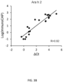
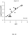
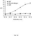
- the method is used for detecting allergen-specific antibodies, wherein the method is capable of detecting IgE at concentrations greater than or equal to 0.01 ng/mL.
- the antigen-DNA conjugate comprises an antigen selected from the group consisting of an allergen, an autoimmune disease antigen, a cancer antigen, and a pathogen antigen.
- the invention includes a method of detecting a target antibody isotype in a sample, the method comprising: a) adding an antibody-binding agent comprising an antibody, an antibody mimetic, or an aptamer that specifically binds to the target antibody isotype, wherein the antibody-binding agent is conjugated to a first DNA molecule and an antigen conjugated to a second DNA molecule to the sample, wherein the antigen binds to the target antibody isotype in the sample, if present, and the antibody-binding agent specifically binds to the target antibody isotype resulting in formation of a complex; b) contacting the complex with a bridge oligonucleotide, wherein the bridge oligonucleotide comprises a first portion sufficiently complementary to and capable of hybridizing with the first DNA molecule, and a second portion sufficiently complementary to and capable of hybridizing with the second DNA molecule, wherein the first DNA molecule and the second DNA molecule are in sufficient proximity to each other in the complex to simultaneously hybridize to
- a method of detecting a target antibody isotype in a sample comprising: a) adding an antibody-binding agent conjugated to a first DNA molecule and an antigen conjugated to a second DNA molecule to the sample, wherein the antigen binds to the target antibody isotype in the sample, if present, and the antibody-binding agent specifically binds to the target antibody isotype resulting in formation of a complex, and wherein a portion of the first DNA molecule is sufficiently complementary to hybridize with a portion of the second DNA molecule; b) extending the hybridized first and second DNA molecules with a DNA polymerase to produce an extended DNA product; and d) detecting the extended DNA product as an indication of the presence of the target antibody isotype in the sample.
- an antigen-DNA conjugate comprises a DNA sequence selected from the group consisting of SEQ ID NOS:1, 2, 5, 6, 9, 10, 13, 14, 17, an 18 or a DNA sequence having at least 95% identity to a DNA sequence selected from the group consisting of SEQ ID NOS:1, 2, 5, 6, 9, 10, 13, 14, 17, an 18.
- an antibody-binding agent-DNA conjugate comprises a DNA sequence selected from the group consisting of SEQ ID NOS:1, 2, 5, 6, 9, 10, 13, 14, 17, an 18 or a DNA sequence having at least 95% identity to a DNA sequence selected from the group consisting of SEQ ID NOS:1, 2, 5, 6, 9, 10, 13, 14, 17, an 18.
- the bridge oligonucleotide comprises the nucleotide sequence of SEQ ID NO:21 or a nucleotide sequence having at least 95% identity to the sequence of SEQ ID NO:21, wherein the bridge oligonucleotide is capable of hybridizing to the DNA of the secondary antibody-binding agent-DNA conjugate and the DNA of the antigen-DNA conjugate.
- the method is performed with at least one set of reagents selected from the group consisting of: a) an antigen-DNA conjugate comprising the DNA sequence of SEQ ID NO:1, an antibody-binding agent-DNA conjugate comprising the DNA sequence of SEQ ID NO:2, a bridge oligonucleotide comprising the nucleotide sequence of SEQ ID NO:21, a reverse primer comprising the nucleotide sequence of SEQ ID NO:3 and a forward primer comprising the sequence of SEQ ID NO:4; b) an antigen-DNA conjugate comprising the DNA sequence of SEQ ID NO:5, an antibody-binding agent-DNA conjugate comprising the DNA sequence of SEQ ID NO:6, a bridge oligonucleotide comprising the nucleotide sequence of SEQ ID NO:21, a reverse primer comprising the nucleotide sequence of SEQ ID NO:7 and a forward primer comprising the sequence of SEQ ID NO:8; c) an antigen-DNA conjugate comprising the DNA
- compositions useful for practicing the methods of the invention, for detecting antibodies in a biological sample comprising at least one set of reagents selected from the group consisting of: a) an antigen-DNA conjugate comprising the DNA sequence of SEQ ID NO:1, an antibody-binding agent-DNA conjugate comprising the DNA sequence of SEQ ID NO:2, a bridge oligonucleotide comprising the nucleotide sequence of SEQ ID NO:21, a reverse primer comprising the nucleotide sequence of SEQ ID NO:3 and a forward primer comprising the sequence of SEQ ID NO:4; b) an antigen-DNA conjugate comprising the DNA sequence of SEQ ID NO:5, an antibody-binding agent-DNA conjugate comprising the DNA sequence of SEQ ID NO:6, a bridge oligonucleotide comprising the nucleotide sequence of SEQ ID NO:21, a reverse primer comprising the nucleotide sequence of SEQ ID NO:7 and a forward primer comprising
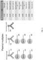
- kits useful for practicing the methods of the invention, for performing isotype-specific agglutination-polymerase chain reaction (ISAP) comprising at least one antigen-DNA conjugate, at least one antibody-binding agent-DNA conjugate (e.g., secondary antibody or aptamer specific for an antibody isotype), at least one bridge oligonucleotide, and at least one pair of PCR primers for detecting antibodies.
- the kit may further comprise instructions for performing ISAP to detect antibodies.
- the kit may further comprise a ligase, polymerase, and/or reagents for performing PCR.
- ISAP can be combined with other methods of antibody detection, particularly other methods that utilize DNA barcoding to allow detection of multiple antibody isotypes by multiplex PCR.
- ISAP is combined with proximity ligation assay (PLA) and/or agglutination-polymerase chain reaction (ADAP).
- PLA proximity ligation assay
- ADAP agglutination-polymerase chain reaction
- the invention includes a method for detecting allergen antibodies in a sample, the method comprising performing isotype-specific agglutination-polymerase chain reaction (ISAP) using at least one allergen-DNA conjugate in combination with at least one anti-IgE antibody-DNA conjugate to detect allergen-specific IgE levels in the sample.
- IgE isotype-specific agglutination-polymerase chain reaction
- the invention further comprises performing ISAP with at least one allergen-DNA conjugate in combination with at least one anti-immunoglobulin G4 (IgG4) antibody-DNA conjugate to detect allergen-specific IgG4 levels.
- IgG4 anti-immunoglobulin G4
- the invention further comprises performing a proximity ligation assay (PLA) using at least one pair of anti-IgE antibody-DNA conjugates to detect total immunoglobulin E (IgE) levels in the sample.
- PLA proximity ligation assay
- the invention further comprises performing agglutination-polymerase chain reaction (ADAP) using at least one pair of allergen-DNA conjugates to detect total anti-allergen antibody levels in the sample.
- ADAP agglutination-polymerase chain reaction
- the invention includes a method for detecting allergen antibodies in a sample, the method comprising: a) performing a proximity ligation assay (PLA) using at least one pair of anti-IgE antibody-DNA conjugates to detect total immunoglobulin E (IgE) levels in the sample; b) performing agglutination-polymerase chain reaction (ADAP) using at least one pair of allergen-DNA conjugates to detect total anti-allergen antibody levels in the sample; and c) performing isotype-specific agglutination-polymerase chain reaction (ISAP) using at least one allergen-DNA conjugate in combination with at least one anti-IgE antibody-DNA conjugate to detect allergen-specific IgE levels in the sample.
- the method may further comprise performing ISAP with at least one allergen-DNA conjugate in combination with at least one anti-immunoglobulin G4 (IgG4) antibody-DNA conjugate to detect allergen-specific IgG4 levels.
- ADAP is used to detect the total anti-allergen antibody levels of IgG, IgM, IgE, IgA, and IgD.
- total anti-allergen levels of an IgG of subtype IgG1, IgG2, IgG3, or IgG4 subtype, or a combination thereof are detected.
- performing PLA comprises: a) adding said at least one pair of anti-IgE antibody-DNA conjugates to the sample, wherein said at least one pair of anti-IgE antibody-DNA conjugates comprises a first anti-IgE antibody-DNA conjugate that binds to an IgE in the sample at a first site and a second anti-IgE antibody-DNA conjugate that binds to the same IgE at a second site; b) contacting the sample with a PLA bridge oligonucleotide, wherein the PLA bridge oligonucleotide comprises: (i) a first portion sufficiently complementary to and capable of hybridizing with the DNA of the first anti-IgE antibody-DNA conjugate, and (ii) a second portion sufficiently complementary to and capable of hybridizing with the DNA of the second anti-IgE antibody-DNA conjugate, wherein the DNA of the first anti-IgE antibody-DNA conjugate and the DNA of the second anti-IgE antibody-DNA conjugate are in sufficient proximity to each other to simultaneously
- performing ADAP comprises: a) adding said at least one pair of allergen-DNA conjugates to the sample, wherein said at least one pair of allergen-DNA conjugates comprises a first allergen-DNA conjugate that binds to an anti-allergen antibody in the sample at a first site and a second allergen-DNA conjugate that binds to the same anti-allergen antibody at a second site; b) contacting the sample with an ADAP bridge oligonucleotide, wherein the ADAP bridge oligonucleotide comprises: (i) a first portion sufficiently complementary to and capable of hybridizing with the DNA of the first allergen-DNA conjugate, and (ii) a second portion sufficiently complementary to and capable of hybridizing with the DNA of the second allergen-DNA conjugate, wherein the DNA of the first allergen-DNA conjugate and the DNA of the second allergen-DNA conjugate are in sufficient proximity to each other to simultaneously hybridize to the ADAP bridge oligonucleotide; c) ligating the first allergen
- performing ISAP comprises: a) adding at least one allergen-DNA conjugate in combination with at least one anti-IgE antibody-DNA conjugate to the sample, wherein the allergen-DNA conjugate binds to the allergen-specific IgE in the sample, and the anti-IgE antibody-DNA conjugate binds to the same allergen-specific IgE resulting in formation of a first complex; b) contacting the first complex with an ISAP bridge oligonucleotide, wherein the ISAP bridge oligonucleotide comprises: (i) a first portion sufficiently complementary to and capable of hybridizing with the DNA of the anti-IgE antibody-DNA conjugate, and (ii) a second portion sufficiently complementary to and capable of hybridizing with the DNA of the allergen-DNA conjugate, wherein the DNA of the anti-IgE antibody-DNA conjugate and the DNA of the allergen-DNA conjugate are in sufficient proximity to each other in the first complex to simultaneously hybridize to the ISAP bridge oligonucleotide
- detecting the PLA ligation product, the ADAP ligation product, the first ISAP ligation product, and the second ISAP ligation product is performed using multiplex polymerase chain reaction (PCR), isothermal amplification, or microarray analysis.
- the method may further comprise quantitating the amount of the PLA ligation product, the ADAP ligation product, the first ISAP ligation product, and the second ISAP ligation product, for example, by performing qPCR.
- PLA is performed with at least one set of reagents selected from the group consisting of: a) a first anti-IgE antibody-DNA conjugate comprising the DNA sequence of SEQ ID NO:1, a second anti-IgE antibody-DNA conjugate comprising the DNA sequence of SEQ ID NO:2, a bridge oligonucleotide comprising the nucleotide sequence of SEQ ID NO:21, a reverse primer comprising the nucleotide sequence of SEQ ID NO:3 and a forward primer comprising the sequence of SEQ ID NO:4; b) a first anti-IgE antibody-DNA conjugate comprising the DNA sequence of SEQ ID NO:5, a second anti-IgE antibody-DNA conjugate comprising the DNA sequence of SEQ ID NO:6, a bridge oligonucleotide comprising the nucleotide sequence of SEQ ID NO:21, a reverse primer comprising the nucleotide sequence of SEQ ID NO:7 and a forward primer comprising the sequence of S
- ADAP is performed with at least one set of reagents selected from the group consisting of: a) a first allergen-DNA conjugate comprising the DNA sequence of SEQ ID NO:1, a second allergen-DNA conjugate comprising the DNA sequence of SEQ ID NO:2, a bridge oligonucleotide comprising the nucleotide sequence of SEQ ID NO:21, a reverse primer comprising the nucleotide sequence of SEQ ID NO:3 and a forward primer comprising the sequence of SEQ ID NO:4; b) a first allergen-DNA conjugate comprising the DNA sequence of SEQ ID NO:5, a second allergen-DNA conjugate comprising the DNA sequence of SEQ ID NO:6, a bridge oligonucleotide comprising the nucleotide sequence of SEQ ID NO:21, a reverse primer comprising the nucleotide sequence of SEQ ID NO:7 and a forward primer comprising the sequence of SEQ ID NO:8; c) a first allergen-DNA conjugate
- ISAP is performed with at least one set of reagents selected from the group consisting of: a) an allergen-DNA conjugate comprising the DNA sequence of SEQ ID NO:1, an anti-IgE antibody-DNA conjugate comprising the DNA sequence of SEQ ID NO:2, a bridge oligonucleotide comprising the nucleotide sequence of SEQ ID NO:21, a reverse primer comprising the nucleotide sequence of SEQ ID NO:3 and a forward primer comprising the sequence of SEQ ID NO:4; b) an allergen-DNA conjugate comprising the DNA sequence of SEQ ID NO:5, an anti-IgE antibody-DNA conjugate comprising the DNA sequence of SEQ ID NO:6, a bridge oligonucleotide comprising the nucleotide sequence of SEQ ID NO:21, a reverse primer comprising the nucleotide sequence of SEQ ID NO:7 and a forward primer comprising the sequence of SEQ ID NO:8; c) an allergen-DNA conjugate comprising
- ISAP is performed with at least one set of reagents selected from the group consisting of: a) an allergen-DNA conjugate comprising the DNA sequence of SEQ ID NO:1, an anti-IgG4 antibody-DNA conjugate comprising the DNA sequence of SEQ ID NO:2, a bridge oligonucleotide comprising the nucleotide sequence of SEQ ID NO:21, a reverse primer comprising the nucleotide sequence of SEQ ID NO:3 and a forward primer comprising the sequence of SEQ ID NO:4; b) an allergen-DNA conjugate comprising the DNA sequence of SEQ ID NO:5, an anti-IgG4 antibody-DNA conjugate comprising the DNA sequence of SEQ ID NO:6, a bridge oligonucleotide comprising the nucleotide sequence of SEQ ID NO:21, a reverse primer comprising the nucleotide sequence of SEQ ID NO:7 and a forward primer comprising the sequence of SEQ ID NO:8; c) an allergen-DNA conjugate
- the invention includes a method for detecting peanut allergen antibodies in a sample, wherein a) PLA is performed using a pair of anti-IgE antibody-DNA conjugates to detect total IgE levels in the sample; b) ADAP is performed using a pair of Ara h1-DNA conjugates to detect total anti-Ara h1 antibody levels in the sample, a pair of Ara h2-DNA conjugates to detect total anti-Ara h2 antibody levels in the sample, and a pair of Ara h3-DNA conjugates to detect total anti-Ara h3 antibody levels in the sample; and c) ISAP is performed using an Ara h1-DNA conjugate in combination with at least one anti-IgE antibody-DNA conjugate to detect Ara h1-specific IgE levels in the sample, an Ara h2-DNA conjugate in combination with at least one anti-IgE antibody-DNA conjugate to detect Ara h2-specific IgE levels in the sample, and an Ara h3-
- performing ISAP further comprises using an Ara h1-DNA conjugate in combination with at least one anti-IgG4 antibody-DNA conjugate to detect Ara h1-specific IgG4 levels in the sample, an Ara h2-DNA conjugate in combination with at least one anti-IgG4 antibody-DNA conjugate to detect Ara h2-specific IgG4 levels in the sample, and an Ara h3-DNA conjugate in combination with at least one anti-IgG4 antibody-DNA conjugate to detect Ara h3-specific IgG4 levels in the sample.
- the method comprises: a) performing PLA with a first anti-IgE antibody-DNA conjugate comprising the DNA sequence of SEQ ID NO:1, a second anti-IgE antibody-DNA conjugate comprising the DNA sequence of SEQ ID NO:2, a bridge oligonucleotide comprising the nucleotide sequence of SEQ ID NO:21, a reverse primer comprising the nucleotide sequence of SEQ ID NO:3, and a forward primer comprising the sequence of SEQ ID NO:4 to detect the total IgE levels in the sample; b) performing ADAP with i) a first Ara h1-DNA conjugate comprising the DNA sequence of SEQ ID NO:5, a second Ara h1-DNA conjugate comprising the DNA sequence of SEQ ID NO:6, a bridge oligonucleotide comprising the nucleotide sequence of SEQ ID NO:21, a reverse primer comprising the nucleotide sequence of SEQ ID NO:7, and a forward primer comprising the
- compositions useful for practicing the methods of the invention, comprising: a) reagents for performing PLA comprising: a first anti-IgE antibody-DNA conjugate comprising the DNA sequence of SEQ ID NO:1, a second anti-IgE antibody-DNA conjugate comprising the DNA sequence of SEQ ID NO:2, a bridge oligonucleotide comprising the nucleotide sequence of SEQ ID NO:21, a reverse primer comprising the nucleotide sequence of SEQ ID NO:3, and a forward primer comprising the sequence of SEQ ID NO:4 for detecting total IgE levels; b) reagents for performing ADAP comprising: i) a first Ara h1-DNA conjugate comprising the DNA sequence of SEQ ID NO:5, a second Ara h1-DNA conjugate comprising the DNA sequence of SEQ ID NO:6, a bridge oligonucleotide comprising the nucleotide sequence of SEQ ID NO:21, a reverse primer
- kits comprising a composition described herein and instructions for detecting allergen antibodies.
- the kit may further comprise a ligase and reagents for performing PCR.
- polynucleotide oligonucleotide
- nucleic acid nucleic acid molecule
- polynucleotide oligonucleotide
- nucleic acid molecule a polymeric form of nucleotides of any length, either ribonucleotides or deoxyribonucleotides. This term refers only to the primary structure of the molecule. Thus, the term includes triple-, double- and single-stranded DNA, as well as triple-, double- and single-stranded RNA. It also includes modifications, such as by methylation and/or by capping, and unmodified forms of the polynucleotide.
- polynucleotide examples include polydeoxyribonucleotides (containing 2-deoxy-D-ribose), polyribonucleotides (containing D-ribose), any other type of polynucleotide which is an N- or C-glycoside of a purine or pyrimidine base, and other polymers containing nonnucleotidic backbones, for example, polyamide (e.g., peptide nucleic acids (PNAs)) and polymorpholino (commercially available from the Anti-Virals, Inc., Corvallis, Oregon, as Neugene) polymers, and other synthetic sequence-specific nucleic acid polymers providing that the polymers contain nucleobases in a configuration which allows for base pairing and base stacking, such as is found in DNA and RNA.
- PNAs peptide nucleic acids
- polynucleotide oligonucleotide
- nucleic acid nucleic acid molecule
- these terms include, for example, 3'-deoxy-2',5'-DNA, oligodeoxyribonucleotide N3' P5' phosphoramidates, 2'-O-alkyl-substituted RNA, double- and single-stranded DNA, as well as double- and single-stranded RNA, DNA:RNA hybrids, and hybrids between PNAs and DNA or RNA, and also include known types of modifications, for example, labels which are known in the art, methylation, "caps," substitution of one or more of the naturally occurring nucleotides with an analog, internucleotide modifications such as, for example, those with uncharged linkages (e.g., methyl phosphonates, phosphotriesters, phosphoramidates, carbamates
- Recombinant as used herein to describe a nucleic acid molecule means a polynucleotide of genomic, cDNA, viral, semisynthetic, or synthetic origin which, by virtue of its origin or manipulation is not associated with all or a portion of the polynucleotide with which it is associated in nature.
- the term "recombinant” as used with respect to a protein or polypeptide means a polypeptide produced by expression of a recombinant polynucleotide.
- the gene of interest is cloned and then expressed in transformed organisms, as described further below. The host organism expresses the foreign gene to produce the protein under expression conditions.
- solid support refers to a solid surface such as a magnetic bead, latex bead, microtiter plate well, glass plate, nylon, agarose, acrylamide, and the like.
- substantially purified generally refers to isolation of a substance (compound, polynucleotide, protein, polypeptide, peptide composition) such that the substance comprises the majority percent of the sample in which it resides.
- a substantially purified component comprises 50%, preferably 80%-85%, more preferably 90-95% of the sample.
- Techniques for purifying polynucleotides and polypeptides of interest are well-known in the art and include, for example, ionexchange chromatography, affinity chromatography and sedimentation according to density.
- isolated is meant, when referring to a protein, polypeptide or peptide, that the indicated molecule is separate and discrete from the whole organism with which the molecule is found in nature or is present in the substantial absence of other biological macro molecules of the same type.
- isolated with respect to a nucleic acid is a nucleic acid molecule devoid, in whole or part, of sequences normally associated with it in nature; or a sequence, as it exists in nature, but having heterologous sequences in association therewith; or a molecule disassociated from the chromosome.
- target nucleic acid region or “target nucleic acid” denotes a nucleic acid molecule with a “target sequence” to be amplified.
- the target nucleic acid may be either single-stranded or double-stranded and may include other sequences besides the target sequence, which may not be amplified.
- target sequence refers to the particular nucleotide sequence of the target nucleic acid which is to be amplified.
- the target sequence may include a probe-hybridizing region contained within the target molecule with which a probe will form a stable hybrid under desired conditions.
- target sequence may also include the complexing sequences to which the oligonucleotide primers complex and extended using the target sequence as a template.
- target sequence also refers to the sequence complementary to the "target sequence” as present in the target nucleic acid. If the "target nucleic acid” is originally double-stranded, the term “target sequence” refers to both the plus (+) and minus (-) strands (or sense and anti-sense strands).
- primer refers to an oligonucleotide that hybridizes to the template strand of a nucleic acid and initiates synthesis of a nucleic acid strand complementary to the template strand when placed under conditions in which synthesis of a primer extension product is induced, i.e., in the presence of nucleotides and a polymerization-inducing agent such as a DNA or RNA polymerase and at suitable temperature, pH, metal concentration, and salt concentration.
- the primer is preferably single-stranded for maximum efficiency in amplification, but may alternatively be double-stranded.
- the primer can first be treated to separate its strands before being used to prepare extension products. This denaturation step is typically effected by heat, but may alternatively be carried out using alkali, followed by neutralization.
- a "primer" is complementary to a template, and complexes by hydrogen bonding or hybridization with the template to give a primer/template complex for initiation of synthesis by a polymerase, which is extended by the addition of covalently bonded bases linked at its 3' end complementary to the template in the process of DNA or RNA synthesis.
- nucleic acids are amplified using at least one set of oligonucleotide primers comprising at least one forward primer and at least one reverse primer capable of hybridizing to regions of a nucleic acid flanking the portion of the nucleic acid to be amplified.
- amplicon refers to the amplified nucleic acid product of a PCR reaction or other nucleic acid amplification process (e.g ., isothermal amplification, rolling circle amplification, ligase chain reaction (LCR)).
- LCR ligase chain reaction
- probe or "oligonucleotide probe” refers to a polynucleotide, as defined above, that contains a nucleic acid sequence complementary to a nucleic acid sequence present in the target nucleic acid analyte.
- the polynucleotide regions of probes may be composed of DNA, and/or RNA, and/or synthetic nucleotide analogs.
- Probes may be labeled in order to detect the target sequence. Such a label may be present at the 5' end, at the 3' end, at both the 5' and 3' ends, and/or internally.
- the "oligonucleotide probe” may contain at least one fluorescer and at least one quencher.
- Quenching of fluorophore fluorescence may be eliminated by exonuclease cleavage of the fluorophore from the oligonucleotide (e.g., TaqMan assay) or by hybridization of the oligonucleotide probe to the nucleic acid target sequence (e.g., molecular beacons).
- the oligonucleotide probe will typically be derived from a sequence that lies between the sense and the antisense primers when used in a nucleic acid amplification assay.
- hybridize and “hybridization” refer to the formation of complexes between nucleotide sequences which are sufficiently complementary to form complexes via Watson-Crick base pairing.
- target template
- such complexes (or hybrids) are sufficiently stable to serve the priming function required by, e.g., the DNA polymerase to initiate DNA synthesis.
- hybridizing sequences need not have perfect complementarity to provide stable hybrids. In many situations, stable hybrids will form where fewer than about 10% of the bases are mismatches, ignoring loops of four or more nucleotides. Accordingly, as used herein the term “complementary” refers to an oligonucleotide that forms a stable duplex with its "complement” under assay conditions, generally where there is about 90% or greater homology.
- oligonucleotides e.g ., primers and/or probes that are capable of detecting a particular DNA barcode, for example, by amplifying and/or binding to a DNA barcode of a particular antigen-DNA or antibody-binding agent-DNA conjugate (e.g., secondary antibody-DNA, antibody mimetic-DNA, or aptamer-DNA conjugate), or ligation product or extension product thereof, but do not amplify and/or bind to other DNA sequences under appropriate hybridization conditions.
- a particular antigen-DNA or antibody-binding agent-DNA conjugate e.g., secondary antibody-DNA, antibody mimetic-DNA, or aptamer-DNA conjugate
- ligation product or extension product thereof do not amplify and/or bind to other DNA sequences under appropriate hybridization conditions.
- antibody encompasses polyclonal and monoclonal antibody preparations, as well as preparations including hybrid antibodies, altered antibodies, chimeric antibodies and, humanized antibodies, as well as: hybrid (chimeric) antibody molecules (see, for example, Winter et al. (1991) Nature 349:293-299 ; and U.S. Pat. No. 4,816,567 ); F(ab') 2 and F(ab) fragments; F v molecules (noncovalent heterodimers, see, for example, Inbar et al. (1972) Proc Natl Acad Sci USA 69:2659-2662 ; and Ehrlich et al.
- immunogenic substances include those that are foreign and those that are naturally occurring within the body of an organism. As such, the introduction of a foreign immunogenic substance may induce an organism to generate a general or specific immune response to the foreign immunogenic substance. In other instances, the production of an immunogenic substance within the body of an organism may induce the organism to generate a specific or general autoimmune response to the native immunogenic substance.
- Antigens encompass but are not limited to chemicals, small molecules, biomolecules (e.g., nucleic acids), macromolecules, peptides, polypeptides, cell fragments, cells, unicellular organisms, multicellular organisms, fragments thereof, and combinations thereof.
- antigens may be antigens for which an agent that binds the antigen is known, e.g., a polypeptide for which an antibody that binds the polypeptide is known.
- antigens may be antigens for which an agent that binds the antigen is unknown, e.g., a polypeptide for which an antibody that binds the polypeptide is unknown.
- polypeptides and peptides, both naturally occurring and synthetic, as antigens to which antibodies may be raised has been described in, e.g., Methods in Molecular Biology: Immunochemical Protocols. Ed. Burns, R., Humana Press, 2005 .
- the specified antibodies bind to a particular antigen at least two times the background and do not substantially bind in a significant amount to other antigens present in the sample.
- Specific binding to an antibody under such conditions may require an antibody that is selected for its specificity for a particular antigen.
- polyclonal antibodies raised to an antigen from specific species can be selected to obtain only those polyclonal antibodies that are specifically immunoreactive with the antigen and not with other proteins, except for polymorphic variants and alleles. This selection may be achieved by subtracting out antibodies that cross-react with molecules from other species.
- a variety of immunoassay formats may be used to select antibodies specifically immunoreactive with a particular antigen.
- solid-phase ELISA immunoassays are routinely used to select antibodies specifically immunoreactive with a protein (see, e.g., Harlow & Lane. Antibodies, A Laboratory Manual (1988 ), for a description of immunoassay formats and conditions that can be used to determine specific immunoreactivity).
- a specific or selective reaction will be at least twice background signal or noise and more typically more than 10 to 100 times background.
- a "biological sample” refers to a sample of cells, tissue, or fluid isolated from a subject, including but not limited to, for example, blood, plasma, serum, fecal matter, urine, bone marrow, bile, spinal fluid, lymph fluid, samples of the skin, external secretions of the skin, respiratory, intestinal, and genitourinary tracts, tears, saliva, milk, cells (e.g., epithelial and endothelial cells, fibroblasts, and macrophages), muscles, joints, organs (e.g., liver, lung, spleen, thymus, kidney, brain, or lymph node), abnormal collections of fluid such as inflammatory transudates or exudates, pus, contents of cysts or areas of tissue necrosis, natural or induced sputum, fluid obtained by lavage performed for diagnostic or therapeutic purposes, such as nasal, pharyngeal or bronchoalveolar lavage, or biopsies and also samples of in vitro cell culture constituents
- label and “detectable label” refer to a molecule capable of detection, including, but not limited to, radioactive isotopes, fluorescers, chemiluminescers, chromophores, enzymes, enzyme substrates, enzyme cofactors, enzyme inhibitors, semiconductor nanoparticles, dyes, metal ions, metal sols, ligands (e.g., biotin, strepavidin or haptens) and the like.
- fluorescer refers to a substance or a portion thereof which is capable of exhibiting fluorescence in the detectable range.
- subject any member of the subphylum chordata, including, without limitation, humans and other primates, including non-human primates such as chimpanzees and other apes and monkey species; farm animals such as cattle, sheep, pigs, goats and horses; domestic mammals such as dogs and cats; birds; and laboratory animals, including rodents such as mice, rats and guinea pigs, and the like.
- the term does not denote a particular age. Thus, both adult and newborn individuals are intended to be covered.
- the present invention is based on the discovery of reagents and methods for detection of antibodies of specific isotypes.
- antigen-DNA conjugates and antibody-binding agent-DNA conjugates carrying DNA barcodes are used to detect the presence of specific antibodies.
- the use of a DNA barcode allows antibodies to be identified by nucleic acid-based detection methods, such as PCR, isothermal amplification, or microarray analysis.
- the methods of the invention can be used to detect and/or quantitate multiple antibodies in a single assay.
- the assays described herein can be readily combined with any other assays for detection of antibodies. Multiplex assays can be used, for example, to detect total levels of disease-relevant antibodies as well as individual levels of multiple disease-relevant antibody isotypes.
- the methods of the invention will allow monitoring of disease-relevant antibodies associated with immune disorders, such as allergies, autoimmune diseases, infections, or inflammation and better disease management.
- the methods use antigen-DNA conjugates and antibody-binding agent-DNA conjugates as well as oligonucleotide reagents (e.g ., oligonucleotide primers and/or probes) or a combination of reagents capable of detecting one or more antibody isotypes in a single assay.
- oligonucleotide reagents e.g ., oligonucleotide primers and/or probes
- the assay methods described herein can be used for detecting antibody isotypes specific for any type of antigen, including, but not limited to an allergen, an autoimmune disease antigen, a cancer antigen, or a pathogen antigen.
- There are a number of assay designs that can be used to detect antibody isotypes which can be used alone or in combination with each other.
- the invention includes a method of detecting a target antibody isotype in a sample, the method comprising: a) contacting the sample with an antibody-binding agent comprising an antibody, an antibody mimetic, or an aptamer that specifically binds to the target antibody isotype, wherein the antibody-binding agent is conjugated to a first DNA molecule comprising a first portion of a barcode, and an antigen conjugated to a second DNA molecule comprising a second portion of a barcode, wherein the antigen binds to the target antibody isotype in the sample, if present, and the antibody-binding agent specifically binds to the target antibody isotype resulting in formation of a complex; b) connecting the first DNA molecule to the second DNA molecule in the complex, wherein the first portion of the barcode and the second portion of the barcode are joined to form a complete barcode; and c) detecting the complete barcode as an indication of the presence of the target antibody isotype in the sample.
- Connecting the first DNA molecule to the second DNA molecule can be accomplished in various ways.
- the first DNA molecule and the second DNA molecule are ligated together using a ligase to produce a ligation product comprising the complete barcode.
- a bridge oligonucleotide may be used to facilitate ligation, wherein the bridge oligonucleotide comprises a first portion sufficiently complementary to and capable of hybridizing with the first DNA molecule, and a second portion sufficiently complementary to and capable of hybridizing with the second DNA molecule.
- the first DNA molecule and the second DNA molecule are in sufficient proximity to each other to simultaneously hybridize to the bridge oligonucleotide and undergo ligation.
- the ligation product can be detected and/or quantitated, for example, using polymerase chain reaction (PCR), isothermal amplification, or microarray analysis.
- the first DNA molecule and the second DNA molecule comprise complementary nucleotide sequences, wherein hybridization of a nucleotide sequence in the first DNA molecule to a complementary nucleotide sequence in the second DNA molecule allows a polymerase to extend the hybridized first and second DNA molecules to produce a nucleic acid comprising the complete barcode.
- the polymerase reaction can be carried out, for example, under isothermal conditions.
- the "antibody binding agent” comprises an antibody, an antibody mimetic, or an aptamer that specifically binds to the target antibody isotype .
- the antibody binding agent binds to a target antibody isotype with high affinity.
- the antibody-binding agent comprises an antibody that specifically binds to the target antibody isotype.
- Any type of antibody may be used, including polyclonal and monoclonal antibodies, hybrid antibodies, altered antibodies, chimeric antibodies and, humanized antibodies, as well as: hybrid (chimeric) antibody molecules (see, for example, Winter et al. (1991) Nature 349:293-299 ; and U.S. Pat. No. 4,816,567 ); F(ab') 2 and F(ab) fragments; F v molecules (noncovalent heterodimers, see, for example, Inbar et al. (1972) Proc Natl Acad Sci USA 69:2659-2662 ; and Ehrlich et al.
- the antibody-binding agent comprises an aptamer that specifically binds to the target antibody isotype.
- aptamer Any type of aptamer may be used, including a DNA, RNA, xeno-nucleic acid (XNA), or peptide aptamer that specifically binds to the target antibody isotype.
- XNA xeno-nucleic acid
- Such aptamers can be identified, for example, by screening a combinatorial library.
- Nucleic acid aptamers e.g., DNA or RNA aptamers
- SELEX exponential enrichment
- Peptide aptamers that bind to a target antibody isotype may be isolated from a combinatorial library and improved by directed mutation or repeated rounds of mutagenesis and selection.
- Aptamers Tools for Nanotherapy and Molecular Imaging (R.N. Veedu ed., Pan Stanford, 2016 )
- Nucleic Acid and Peptide Aptamers Methods and Protocols (Methods in Molecular Biology, G. Mayer ed., Humana Press, 2009 )
- Nucleic Acid Aptamers Selection, Characterization, and Application (Methods in Molecular Biology, G.
- the antibody-binding agent comprises an antibody mimetic.
- Any type of antibody mimetic may be used, including, but not limited to, affibody molecules ( Nygren (2008) FEBS J. 275 (11):2668-2676 ), affilins ( Ebersbach et al. (2007) J. Mol. Biol. 372 (1):172-185 ), affimers ( Johnson et al. (2012) Anal. Chem. 84 (15):6553-6560 ), affitins ( Krehenbrink et al. (2008) J. Mol. Biol. 383 (5):1058-1068 ), alphabodies ( Desmet et al.
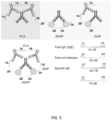
- Antigen-DNA and antibody-binding agent-DNA conjugates of the subject methods include at least one DNA molecule attached to the antigen or antibody-binding agent, respectively, wherein the DNA molecule comprises a unique barcode sequence that identifies a target antigen-specific antibody isotype (i.e., the complete barcode produced by joining the DNA sequences of the antigen-DNA and antibody-binding agent-DNA conjugates).
- DNA molecules attached to an antigen or antibody-binding agent may vary depending, in part, on the detection method employed, the method of attachment, the specific antibodies to be detected, etc.
- the length of the attached DNA molecules will be at least 15 nucleotides, but may range from 15 nucleotides to 200 nucleotides or more including but not limited to e.g., 20 or more nucleotides, 25 or more nucleotides, 30 or more nucleotides, 35 or more nucleotides, 40 or more nucleotides, 45 or more nucleotides, 50 or more nucleotides, 55 or more nucleotides, 60 or more nucleotides, 65 or more nucleotides, 70 or more nucleotides, 75 or more nucleotides, 80 or more nucleotides, 90 or more nucleotides, 95 or more nucleotides, 100 or more nucleotides, 15 to 200 nucleotides, 20 to 200 nucleotides, 25 to 200 nucleotides, 30 to 200 nucleotides, 35 to 200 nucleotides, 40 to 200 nucleotides, 45 to 200 nucleo
- the DNA may be attached to an antigen or antibody-binding agent by any convenient method, as described in more detail below.
- the DNA may be attached to an antigen or antibody-binding agent at any convenient point along the length of the DNA, including at the 3' or 5' termini.
- DNA is attached to the antigen or antibody at its 3' end or 5' end.
- both the antigen and the antibody-binding agent have DNA molecules attached at their 3' ends.
- both the antigen and the antibody-binding agent have DNA molecules attached at their 5' ends.
- bridging oligonucleotide or “bridge oligonucleotide” refers to any oligonucleotide that joins two or more separate DNA molecules or two termini of a single DNA molecule by simultaneously hybridizing with complementary regions on each DNA molecule or complementary regions of the DNA termini.
- a bridging oligonucleotide joins an antigen-DNA conjugate to an antibody-binding agent-DNA conjugate by simultaneously hybridizing with a first complementary region in the DNA of the antigen-DNA conjugate and a second complementary region in the DNA of the antibody-binding agent-DNA conjugate.
- Bridging oligonucleotides may be partially or completely single stranded, including partially single stranded and partially double stranded.
- the length of bridging oligonucleotides of the subject disclosure will vary and may be 10 or more nucleotides and range from 10 to 100 or more nucleotides, including e.g., 10 to 100 nucleotides, 12 to 100 nucleotides, 14 to 100 nucleotides, 16 to 100 nucleotides, 18 to 100 nucleotides, 20 to 100 nucleotides, 22 to 100 nucleotides, 24 to 100 nucleotides, 26 to 100 nucleotides, 28 to 100 nucleotides, 30 to 100 nucleotides, 10 to 50 nucleotides, 12 to 50 nucleotides, 14 to 50 nucleotides, 16 to 50 nucleotides, 18 to 50 nucleotides, 20 to 50 nucleotides, 22 to 50 nucleotides, 24 to
- Bridging oligonucleotides may "bridge" two or more DNA molecules to form a complex.
- a bridging polynucleotide may hybridize with two DNA termini, including termini of the same or different nucleic acids, such that the termini are adjacent within the complex, e.g., allowing for the ligation of the adjacent termini.
- a bridging polynucleotide may hybridize with two DNA termini, including termini of the same or different nucleic acids, such that the termini are not adjacent in the resulting polynucleotide complex, e.g., are not adjacent such that they cannot be directly ligated together.
- a splint polynucleotide may be hybridized in the space between the two termini such that the ends of the splint polynucleotide are located adjacent to one or more of the termini.
- the term "splint polynucleotide” as used herein refers to a polynucleotide, which may generally be single stranded or partially single stranded and partially double stranded, which may be used to fill one or more gaps between two DNA termini in a complex, e.g., those complexes formed by use of a bridging polynucleotide.
- a splint polynucleotide may have complementarity to one or more portions of a bridging oligonucleotide.
- DNA termini adjacent to a splint polynucleotide may be ligated to the splint polynucleotide.
- a bridging oligonucleotide of the subject disclosure may include one or more nucleoside analogs.
- a bridging oligonucleotide of the instant disclosure may include one or more deoxyribouracil (i.e., deoxyribose uracil, deoxyuridine, etc.) nucleosides/nucleotides.
- a bridging polynucleotide may include 2 or more nucleoside analogs including but not limited to e.g., 3 or more, 4 or more, 5 or more, 6 or more, etc.
- the number of nucleoside analogs as a percentage of the total bases of the bridging polynucleotide is 1% or more, including but not limited to e.g., 2% or more, 3% or more, 4% or more, 5% or more, 6% or more, 7% or more, 8% or more, 9% or more, 10% or more, 11% or more, 12% or more, 13% or more, 14% or more, 15% or more, 16% or more, 17% or more, 18% or more, 19% or more, 20% or more, 21% or more, 22% or more, 23% or more, 24% or more, 25% or more, 26% or more, 27% or more, 28% or more, 29% or more, 30% or more, etc.
- joining of DNA molecules from an antigen-DNA conjugate and an antibody-binding agent-DNA conjugate produces a complete barcode sequence identifying the target antibody isotype, which may be amplified to generate an amplification product (i.e., amplicon) that can be detected.
- amplification product i.e., amplicon
- Any convenient method of amplification may be utilized in generating the amplification product, as described in more detail below, and may depend upon the particular complex formed and/or particular requirements of the overall detection assay.
- the presence of an amplification product is indicative of the presence of target antibodies in the sample that are specific for the antigen and also recognized by the antibody-binding agent (e.g., a secondary antibody, antibody mimetic, or aptamer specific for a particular target antibody isotype).
- the antibody-binding agent e.g., a secondary antibody, antibody mimetic, or aptamer specific for a particular target antibody isotype.
- amplification may be performed by polymerase chain reaction (PCR).
- the reaction mixture generally includes a template nucleic acid which is combined with one or more primers that are employed in the primer extension reaction, e.g., the PCR primers (such as forward and reverse primers employed in geometric (or exponential) amplification or a single primer employed in a linear amplification).
- the hybridized portions of the above described nucleic acid complexes may serve as "primer" for the amplification reaction.
- a single free 3 '-terminus of hybridized nucleic acid of an above described nucleic acid complex may serve as a primer for amplification.
- one or more additional nucleic acids may be added to serve as primer in a formed nucleic acid complex.
- two antigen-bound polynucleotides may be joined in a ligation reaction and two additional primers may be added to facilitate amplification of the newly ligated nucleic acid segment or template.
- a single free 3 '-terminus of hybridized nucleic acid of an above described nucleic acid complex may serve as a first primer and a second primer may be added to facilitate amplification.
- any oligonucleotide primers with which the template nucleic acid (hereinafter referred to as template DNA for convenience) is contacted will be of sufficient length to provide for hybridization to complementary template DNA under annealing conditions.
- the primers will generally be at least 6 bp in length, including but not limited to e.g., at least 10 bp in length, at least 15 bp in length, at least 16 bp in length, at least 17 bp in length, at least 18 bp in length, at least 19 bp in length, at least 20 bp in length, at least 21 bp in length, at least 22 bp in length, at least 23 bp in length, at least 24 bp in length, at least 25 bp in length, at least 26 bp in length, at least 27 bp in length, at least 28 bp in length, at least 29 bp in length, at least 30 bp in length, and may be as long as 60 bp in length or longer, where the
- the template DNA may be contacted with a single primer or a set of two primers (forward and reverse primers), depending on whether primer extension, linear or exponential amplification of the template DNA is desired.
- Methods of PCR that may be employed in the subject methods include but are not limited to those described in U.S. Pat. Nos.: 4,683,202 ; 4,683,195 ; 4,800,159 ; 4,965,188 and 5,512,462 .
- a PCR reaction mixture produced in the subject methods may include a polymerase and deoxyribonucleoside triphosphates (dNTPs).
- the desired polymerase activity may be provided by one or more distinct polymerase enzymes.
- the reaction mixture includes at least a Family A polymerase, where representative Family A polymerases of interest include, but are not limited to: Thermus aquaticus polymerases, including the naturally occurring polymerase (Taq) and derivatives and homologues thereof, such as Klentaq (as described in Proc. Natl. Acad.
- the reaction mixture may further include a polymerase enzyme having 3'-5' exonuclease activity, e.g., as may be provided by a Family B polymerase, where Family B polymerases of interest include, but are not limited to: Thermococcus litoralis DNA polymerase (Vent) (e.g., as described in Perler et al., Proc. Natl. Acad. Sci.
- Vent Thermococcus litoralis DNA polymerase
- the reaction mixture will include four different types of dNTPs corresponding to the four naturally occurring bases are present, i.e. dATP, dTTP, dCTP and dGTP and in some instances, may include one or more modified nucleotide dNTPs.
- a PCR reaction will generally be carried out by cycling the reaction mixture between appropriate temperatures for annealing, elongation/extension, and denaturation for specific times. Such temperature and times will vary and will depend on the particular components of the reaction including, e.g., the polymerase and the primers as well as the expected length of the resulting PCR product. In some instances, e.g., where nested or two-step PCR are employed the cycling-reaction may be carried out in stages, e.g., cycling according to a first stage having a particular cycling program or using particular temperature(s) and subsequently cycling according to a second stage having a particular cycling program or using particular temperature(s).
- Multistep PCR processes may or may not include that addition of one or more reagents following the initiation of amplification.
- amplification may be initiated by elongation with the use of a polymerase and, following an initial phase of the reaction, additional reagent(s) (e.g., one or more additional primers, additional enzymes, etc.) may be added to the reaction to facilitate a second phase of the reaction.
- additional reagent(s) e.g., one or more additional primers, additional enzymes, etc.
- the initial phase of amplification may be referred to as "preamplification".
- amplification may be carried out under isothermal conditions, e.g., by means of isothermal amplification.
- Methods of isothermal amplification generally make use of enzymatic means of separating DNA strands to facilitate amplification at constant temperature, such as, e.g., strand-displacing polymerase or a helicase, thus negating the need for thermocycling to denature DNA.
- Any convenient and appropriate means of isothermal amplification may be employed in the subject methods including but are not limited to: loop-mediated isothermal amplification (LAMP), strand displacement amplification (SDA), helicase-dependent amplification (HDA), nicking enzyme amplification reaction (NEAR), and the like.
- LAMP generally utilizes a plurality of primers, e.g., 4-6 primers, which may recognize a plurality of distinct regions, e.g., 6-8 distinct regions, of target DNA. Synthesis is generally initiated by a strand-displacing DNA polymerase with two of the primers forming loop structures to facilitate subsequent rounds of amplification. LAMP is rapid and sensitive. In addition, the magnesium pyrophosphate produced during the LAMP amplification reaction may, in some instances be visualized without the use of specialized equipment, e.g., by eye.
- SDA generally involves the use of a strand-displacing DNA polymerase (e.g., Bst DNA polymerase, Large (Klenow) Fragment polymerase, Klenow Fragment (3'-5' exo-), and the like) to initiate at nicks created by a strand-limited restriction endonuclease or nicking enzyme at a site contained in a primer.
- a strand-displacing DNA polymerase e.g., Bst DNA polymerase, Large (Klenow) Fragment polymerase, Klenow Fragment (3'-5' exo-), and the like
- HDA generally employs: a helicase which unwinds double-stranded DNA unwinding to separate strands; primers, e.g., two primers, that may anneal to the unwound DNA; and a strand-displacing DNA polymerase for extension.
- NEAR generally involves a strand-displacing DNA polymerase that initiates elongation at nicks, e.g., created by a nicking enzyme. NEAR is rapid and sensitive, quickly producing many short nucleic acids from a target sequence.
- entire amplification methods may be combined or aspects of various amplification methods may be recombined to generate a hybrid amplification method.
- aspects of PCR may be used, e.g., to generate the initial template or amplicon or first round or rounds of amplification, and an isothermal amplification method may be subsequently employed for further amplification.
- an isothermal amplification method or aspects of an isothermal amplification method may be employed, followed by PCR for further amplification of the product of the isothermal amplification reaction.
- a sample may be preamplified using a first method of amplification and may be further processed, including e.g., further amplified or analyzed, using a second method of amplification.
- a sample may be preamplified by PCR and further analyzed by qPCR.
- the amplification step and the detection step may be combined, with or without the use of a preamplifcation step.
- the particular amplification method employed allows for the qualitative detection of amplification product, e.g., by visual inspection of the amplification reaction with or without a detection reagent.
- the ligation products are amplified by isothermal amplification, e.g., LAMP, and the amplification generates a visual change in the amplification reaction indicative of efficient amplification and thus presence of the antibody isotype in the sample.
- the amplification and detection steps are combined by monitoring the amplification reaction during amplification such as is performed in, e.g., real-time PCR, also referred to herein as quantitative PCR (qPCR).
- the methods described herein may make use of those methods, e.g., amplification methods, and components thereof, employed in proximity ligation assays (PLA) and proximity elongation assays (PEA) including but not limited to, e.g., rolling circle amplification (RCA), binding-induced DNA assembly (BINDA), nicking enzyme assisted fluorescence signal amplification (NEFSA), and, e.g., those described in Janssen et al. (2013) Sensors, 13, 1353-1384 .
- PLA proximity ligation assays
- PEA proximity elongation assays
- RCA rolling circle amplification
- BINDA binding-induced DNA assembly
- NEFSA nicking enzyme assisted fluorescence signal amplification
- the methods of the invention can be adapted to multiplexing.
- a plurality of antibody-binding agent-DNA conjugates can be added to a sample, wherein each antibody-binding agent is conjugated to a DNA molecule comprising a different barcode sequence and each antibody-binding agent is capable of binding to a different target antibody isotype to allow multiplex detection of a plurality of target antibody isotypes in a sample.
- the antibody-binding agent-DNA conjugates are selected from the group consisting of an anti-IgE secondary antibody-DNA conjugate for detection of IgE, an anti-IgM secondary antibody-DNA conjugate for detection of IgM, an anti-IgG secondary antibody-DNA conjugate for detection of IgG, an anti-IgA secondary antibody-DNA conjugate for detection of IgA, and an anti-IgD secondary antibody DNA conjugate for detection of IgD, wherein the DNA molecules in the conjugates comprise isotype-specific DNA barcodes that can be amplified and detected simultaneously by using a suitable combination of primers and/or probes in a multiplex-type assay format.
- an antigen-DNA conjugate comprises a DNA sequence selected from the group consisting of SEQ ID NOS:1, 2, 5, 6, 9, 10, 13, 14, 17, an 18 or a DNA sequence having at least 95% identity to a DNA sequence selected from the group consisting of SEQ ID NOS:1, 2, 5, 6, 9, 10, 13, 14, 17, an 18.
- a secondary antibody-binding agent-DNA conjugate comprises a DNA sequence selected from the group consisting of SEQ ID NOS:1, 2, 5, 6, 9, 10, 13, 14, 17, an 18 or a DNA sequence having at least 95% identity to a DNA sequence selected from the group consisting of SEQ ID NOS:1, 2, 5, 6, 9, 10, 13, 14, 17, an 18.
- the bridge oligonucleotide comprises the nucleotide sequence of SEQ ID NO:21 or a nucleotide sequence having at least 95% identity to the sequence of SEQ ID NO:21, wherein the bridge oligonucleotide is capable of hybridizing to the DNA of the
- the method is performed with at least one set of reagents selected from the group consisting of: a) an antigen-DNA conjugate comprising the DNA sequence of SEQ ID NO:1, an antibody-binding agent-DNA conjugate comprising the DNA sequence of SEQ ID NO:2, a bridge oligonucleotide comprising the nucleotide sequence of SEQ ID NO:21, a reverse primer comprising the nucleotide sequence of SEQ ID NO:3 and a forward primer comprising the sequence of SEQ ID NO:4; b) an antigen-DNA conjugate comprising the DNA sequence of SEQ ID NO:5, an antibody-binding agent-DNA conjugate comprising the DNA sequence of SEQ ID NO:
- ISAP can be combined with other methods of antibody detection, particularly other methods that utilize DNA barcoding to allow detection of multiple antibody isotypes by multiplex PCR.
- ISAP is combined with proximity ligation assay (PLA) and/or agglutination-polymerase chain reaction (ADAP).
- PLA proximity ligation assay
- ADAP agglutination-polymerase chain reaction
- ISAP can be combined with PLA and/or ADAP for detecting allergen antibodies in a sample.
- the method comprises: a) performing PLA using at least one pair of anti-IgE antibody-DNA conjugates to detect total IgE levels in the sample; b) performing ADAP using at least one pair of allergen-DNA conjugates to detect total anti-allergen antibody levels in the sample; and c) performing ISAP using at least one allergen-DNA conjugate in combination with at least one anti-IgE antibody-DNA conjugate to detect allergen-specific IgE levels in the sample.
- the method may further comprise performing ISAP with at least one allergen-DNA conjugate in combination with at least one anti-immunoglobulin G4 (IgG4) antibody-DNA conjugate to detect allergen-specific IgG4 levels.
- IgG4 anti-immunoglobulin G4
- ADAP is used to detect the total anti-allergen antibody levels of IgG, IgM, and IgE.
- the method comprises: a) adding at least one pair of anti-IgE antibody-DNA conjugates to the sample, wherein at least one pair of anti-IgE antibody-DNA conjugates comprises a first anti-IgE antibody-DNA conjugate that binds to an IgE in the sample at a first site and a second anti-IgE antibody-DNA conjugate that binds to the same IgE at a second site; b) contacting the sample with a PLA bridge oligonucleotide, wherein the PLA bridge oligonucleotide comprises: (i) a first portion sufficiently complementary to and capable of hybridizing with the DNA of the first anti-IgE antibody-DNA conjugate, and (ii) a second portion sufficiently complementary to and capable of hybridizing with the DNA of the second anti-IgE antibody-DNA conjugate, wherein the DNA of the first anti-IgE antibody-DNA conjugate and the DNA of the second anti-IgE antibody-DNA conjugate are in sufficient proximity to each other to simultaneously hybridize
- the method comprises: a) adding said at least one pair of allergen-DNA conjugates to the sample, wherein at least one pair of allergen-DNA conjugates comprises a first allergen-DNA conjugate that binds to an anti-allergen antibody in the sample at a first site and a second allergen-DNA conjugate that binds to the same anti-allergen antibody at a second site; b) contacting the sample with an ADAP bridge oligonucleotide, wherein the ADAP bridge oligonucleotide comprises: (i) a first portion sufficiently complementary to and capable of hybridizing with the DNA of the first allergen-DNA conjugate, and (ii) a second portion sufficiently complementary to and capable of hybridizing with the DNA of the second allergen-DNA conjugate, wherein the DNA of the first allergen-DNA conjugate and the DNA of the second allergen-DNA conjugate are in sufficient proximity to each other to simultaneously hybridize to the ADAP bridge oligonucleotide; c) ligating the ADAP bridge
- the method comprises: a) adding the at least one allergen-DNA conjugate in combination with at least one anti-IgE antibody-DNA conjugate to the sample, wherein the allergen-DNA conjugate binds to the allergen-specific IgE in the sample, and the anti-IgE antibody-DNA conjugate binds to the same allergen-specific IgE resulting in formation of a first complex; b) contacting the first complex with an ISAP bridge oligonucleotide, wherein the ISAP bridge oligonucleotide comprises: (i) a first portion sufficiently complementary to and capable of hybridizing with the DNA of the anti-IgE antibody-DNA conjugate, and (ii) a second portion sufficiently complementary to and capable of hybridizing with the DNA of the allergen-DNA conjugate, wherein the DNA of the anti-IgE antibody-DNA conjugate and the DNA of the allergen-DNA conjugate are in sufficient proximity to each other in the first complex to simultaneously hybridize to the ISAP bridge oligonucleo
- detecting the PLA ligation product, the ADAP ligation product, the first ISAP ligation product, and the second ISAP ligation product comprises using multiplex polymerase chain reaction (PCR), isothermal amplification, or microarray analysis.
- the method may further comprise quantitating the amount of the PLA ligation product, the ADAP ligation product, the first ISAP ligation product, and the second ISAP ligation product, for example, by performing qPCR.
- PLA is performed with at least one set of reagents selected from the group consisting of: a) a first anti-IgE antibody-DNA conjugate comprising the DNA sequence of SEQ ID NO: 1, a second anti-IgE antibody-DNA conjugate comprising the DNA sequence of SEQ ID NO:2, a bridge oligonucleotide comprising the nucleotide sequence of SEQ ID NO:21, a reverse primer comprising the nucleotide sequence of SEQ ID NO:3 and a forward primer comprising the sequence of SEQ ID NO:4; b) a first anti-IgE antibody-DNA conjugate comprising the DNA sequence of SEQ ID NO:5, a second anti-IgE antibody-DNA conjugate comprising the DNA sequence of SEQ ID NO:6, a bridge oligonucleotide comprising the nucleotide sequence of SEQ ID NO:21, a reverse primer comprising the nucleotide sequence of SEQ ID NO:7 and a forward primer comprising the sequence of S
- ADAP is performed with at least one set of reagents selected from the group consisting of: a) a first allergen-DNA conjugate comprising the DNA sequence of SEQ ID NO: 1, a second allergen-DNA conjugate comprising the DNA sequence of SEQ ID NO:2, a bridge oligonucleotide comprising the nucleotide sequence of SEQ ID NO:21, a reverse primer comprising the nucleotide sequence of SEQ ID NO:3 and a forward primer comprising the sequence of SEQ ID NO:4; b) a first allergen-DNA conjugate comprising the DNA sequence of SEQ ID NO:5, a second allergen-DNA conjugate comprising the DNA sequence of SEQ ID NO:6, a bridge oligonucleotide comprising the nucleotide sequence of SEQ ID NO:21, a reverse primer comprising the nucleotide sequence of SEQ ID NO:7 and a forward primer comprising the sequence of SEQ ID NO:8; c) a first allergen-DNA conjugate
- ISAP is performed with at least one set of reagents selected from the group consisting of: a) an allergen-DNA conjugate comprising the DNA sequence of SEQ ID NO:1, an anti-IgE antibody-DNA conjugate comprising the DNA sequence of SEQ ID NO:2, a bridge oligonucleotide comprising the nucleotide sequence of SEQ ID NO:21, a reverse primer comprising the nucleotide sequence of SEQ ID NO:3 and a forward primer comprising the sequence of SEQ ID NO:4; b) an allergen-DNA conjugate comprising the DNA sequence of SEQ ID NO:5, an anti-IgE antibody-DNA conjugate comprising the DNA sequence of SEQ ID NO:6, a bridge oligonucleotide comprising the nucleotide sequence of SEQ ID NO:21, a reverse primer comprising the nucleotide sequence of SEQ ID NO:7 and a forward primer comprising the sequence of SEQ ID NO:8; c) an allergen-DNA conjugate comprising
- ISAP is performed with at least one set of reagents selected from the group consisting of: a) an allergen-DNA conjugate comprising the DNA sequence of SEQ ID NO:1, an anti-IgG4 antibody-DNA conjugate comprising the DNA sequence of SEQ ID NO:2, a bridge oligonucleotide comprising the nucleotide sequence of SEQ ID NO:21, a reverse primer comprising the nucleotide sequence of SEQ ID NO:3 and a forward primer comprising the sequence of SEQ ID NO:4; b) an allergen-DNA conjugate comprising the DNA sequence of SEQ ID NO:5, an anti-IgG4 antibody-DNA conjugate comprising the DNA sequence of SEQ ID NO:6, a bridge oligonucleotide comprising the nucleotide sequence of SEQ ID NO:21, a reverse primer comprising the nucleotide sequence of SEQ ID NO:7 and a forward primer comprising the sequence of SEQ ID NO:8; c) an allergen-DNA conjugate
- the invention includes a method for detecting peanut allergen antibodies in a sample, wherein a) PLA is performed using a pair of anti-IgE antibody-DNA conjugates to detect total IgE levels in the sample; b) ADAP is performed using a pair of Ara h1-DNA conjugates to detect total anti-Ara h1 antibody levels in the sample, a pair of Ara h2-DNA conjugates to detect total anti-Ara h2 antibody levels in the sample, and a pair of Ara h3-DNA conjugates to detect total anti-Ara h3 antibody levels in the sample; and c) ISAP is performed using an Ara h1-DNA conjugate in combination with at least one anti-IgE antibody-DNA conjugate to detect Ara h1-specific IgE levels in the sample, an Ara h2-DNA conjugate in combination with at least one anti-IgE antibody-DNA conjugate to detect Ara h2-specific IgE levels in the sample, and an Ara h3-
- performing ISAP further comprises using an Ara h1-DNA conjugate in combination with at least one anti-IgG4 antibody-DNA conjugate to detect Ara h1-specific IgG4 levels in the sample, an Ara h2-DNA conjugate in combination with at least one anti-IgG4 antibody-DNA conjugate to detect Ara h2-specific IgG4 levels in the sample, and an Ara h3-DNA conjugate in combination with at least one anti-IgG4 antibody-DNA conjugate to detect Ara h3-specific IgG4 levels in the sample.
- the method comprises: a) performing PLA with a first anti-IgE antibody-DNA conjugate comprising the DNA sequence of SEQ ID NO: 1, a second anti-IgE antibody-DNA conjugate comprising the DNA sequence of SEQ ID NO:2, a bridge oligonucleotide comprising the nucleotide sequence of SEQ ID NO:21, a reverse primer comprising the nucleotide sequence of SEQ ID NO:3, and a forward primer comprising the sequence of SEQ ID NO:4 to detect the total IgE levels in the sample; b) performing ADAP with i) a first Ara h1-DNA conjugate comprising the DNA sequence of SEQ ID NO:5, a second Ara h1-DNA conjugate comprising the DNA sequence of SEQ ID NO:6, a bridge oligonucleotide comprising the nucleotide sequence of SEQ ID NO:21, a reverse primer comprising the nucleotide sequence of SEQ ID NO:7, and a forward primer comprising the
- the presence of the amplification product may be determined, including qualitatively determined or quantitatively determined, by any convenient method. In some instances, the presence of the amplification product may be qualitatively determined, e.g., through a physical change in the amplification reaction that is indicative of efficient amplification of the ligation product.
- the amplification product is detected and/or the amount of amplification product is measured by a detection protocol for non-specific detection of the amplified nucleic acid or a protocol for specific detection of the amplified nucleic acid.
- Representative non-specific detection protocols of interest include protocols that employ signal producing systems that selectively detect double stranded nucleic acid products, e.g., via intercalation.
- Representative detectable molecules that find use in such embodiments include fluorescent nucleic acid stains, such as phenanthridinium dyes, including monomers or homo- or heterodimers thereof, that provide enhanced fluorescence when complexed with nucleic acids.
- fluorescent nucleic acid stains such as phenanthridinium dyes, including monomers or homo- or heterodimers thereof, that provide enhanced fluorescence when complexed with nucleic acids.
- phenanthridinium dyes include ethidium homodimer, ethidium bromide, propidium iodide, and other alkyl-substituted phenanthridinium dyes.
- a nucleic acid stain in another embodiment, includes an acridine dye, or a homo- or heterodimer thereof, such as acridine orange, acridine homodimer, ethidium-acridine heterodimer, or 9-amino-6-chloro-2-methoxyacridine.
- the nucleic acid stain is an indole or imidazole dye, such as Hoechst 33258, Hoechst 33342, Hoechst 34580, DAPI (4',6-diamidino-2-phenylindole) or DIPI (4',6-(diimidazolin-2-yl)-2-phenylindole).
- nucleic acid stain is a cyanine dye or a homo- or heterodimer of a cyanine dye that gives an enhanced fluorescence when associated with nucleic acids.
- U.S. Pat. No. 5,410,030 may be used, including nucleic acid stains commercially available under the trademarks TOTO, BOBO, POPO, YOYO, TO-PRO, BO-PRO, PO-PRO and YO-PRO (Life Technologies, Inc. Grand Island, NY).
- dyes described in U.S. Pat. No. 5,436,134 , U.S. Pat. No. 5,658,751 and U.S. Pat. No. 5,863,753 may be used, including nucleic acid stains commercially available under the trademarks SYBR, SYTO, SYTOX, PICOGREEN, OLIGREEN, and RIBOGREEN (Life Technologies, Inc. Grand Island, NY).
- the nucleic acid stain is a monomeric, homodimeric or heterodimeric cyanine dye that incorporates an aza- or polyazabenzazolium heterocycle, such as an azabenzoxazole, azabenzimidazole, or azabenzothiazole, that gives enhanced fluorescence when associated with nucleic acids, including nucleic acid stains commercially available under the trademarks SYTO, SYTOX, JOJO, JO-PRO, LOLO, LO-PRO (Life Technologies, Inc. Grand Island, NY).
- an aza- or polyazabenzazolium heterocycle such as an azabenzoxazole, azabenzimidazole, or azabenzothiazole
- a signal producing system that is specific for the amplification product, as opposed to double stranded molecules in general, may be employed to detect the amplification.
- the signal producing system may include a probe nucleic acid that specifically binds to a sequence found in the amplification product, where the probe nucleic acid may be labeled with a directly or indirectly detectable label.
- a directly detectable label is one that can be directly detected without the use of additional reagents
- an indirectly detectable label is one that is detectable by employing one or more additional reagent, e.g., where the label is a member of a signal producing system made up of two or more components.
- the label is a directly detectable label, where directly detectable labels of interest include, but are not limited to: fluorescent labels, radioisotopic labels, chemiluminescent labels, and the like.
- the label is a fluorescent label, where the labeling reagent employed in such embodiments is a fluorescently tagged nucleotide(s), e.g. fluorescently tagged CTP (such as Cy3-CTP, Cy5-CTP) etc.
- fluorescent moieties which may be used to tag nucleotides for producing labeled probe nucleic acids include, but are not limited to: fluorescein, the cyanine dyes, such as Cy3, Cy5, Alexa 555, Bodipy 630/650, and the like. Other labels, such as those described above, may also be employed.
- signal detection typically includes detecting a change in a fluorescent signal from the reaction mixture to obtain an assay result.
- any modulation in the fluorescent signal generated by the reaction mixture is assessed.
- the change may be an increase or decrease in fluorescence, depending on the nature of the label employed, and in certain embodiments is an increase in fluorescence.
- the sample may be screened for an increase in fluorescence using any convenient means, e.g., a suitable fluorimeter, such as a thermostable cuvette or plate-reader fluorimeter. Fluorescence is suitably monitored using a known fluorimeter.
- the signals from these devices are sent to a data processor board and converted into a spectrum associated with each sample tube.
- Multiple reaction vessels e.g., multiple tubes, multi-well plates, etc., can be assessed at the same time.
- the elongation and/or amplification of a particular DNA sequence results in the duplication of one or more specific nucleic acid sequences resulting in one or more strands containing repeats of the one or more specific nucleic acid sequences.
- Such repetitive sequences may be detected, e.g., through hybridization of a probe nucleic acid specific for the repeated specific sequence.
- a tagged probe nucleic acid e.g., a fluorescently tagged probe nucleic acid, an enzymatically tagged probe nucleic acid, a radiolabel tagged probe nucleic acid, etc., specific for the repeated specific sequence may be utilized to detect an elongated polynucleotide or amplification product that contains the repeated specific sequence.
- hybridization of a tagged probe nucleic acid to a repeating sequence of an elongated polynucleotide or amplification product allows for the detection of the elongated polynucleotide or amplification product due to the high number of tagged probe nucleic acids hybridized to the elongated polynucleotide or amplification product, which results in a high local concentration of detectable tag.
- repeats of one or more sequences of DNA from an antigen-DNA conjugate or antibody-binding agent-DNA conjugate are contained in an amplification product or elongation product produced according to the methods described herein and the repeats are detected through the use of a tagged probe nucleic acid specific for the repeating sequence units.
- repeats of one or more sequences of a bridging polynucleotide are contained in an amplification product or elongation product produced according to the methods described herein and the repeats are detected through the use of a tagged probe nucleic acid specific for the repeating sequence units.
- repeats of one or more sequences of a circularizing oligonucleotide are contained in an amplification product or elongation product produced according to the methods described herein and the repeats are detected through the use of a tagged probe nucleic acid specific for the repeating sequence units.
- a repeating nucleic acid sequence may be produced by one or more of the elongation and/or amplification methods described herein, e.g., PCR amplification, isothermal amplification (e.g., RCA), etc., and the elongation and/or amplification product may be made detectable through hybridization of one or more fluorescently labeled probe nucleic acid to the elongation and/or amplification product.
- detectable elongation and/or amplification product may be identified through any convenient means for detecting fluorescence, including but not limited to, e.g., fluorescent microscopy, flow cytometry, imaging flow cytometry, etc.
- identification of a detectable elongation and/or amplification product may allow for detection or identification of a molecule, particle, cell, tissue, organism, etc., associated with the antibody of the complex from which the elongation and/or amplification product was derived.
- fluorescent probe-bound elongation and/or amplification product may remain associated with a cell that produced the antibody allowing identification of the cell, e.g., by fluorescent microscopy, and/or isolation of the cell, e.g., by fluorescent activated cell sorting (FACS).
- FACS fluorescent activated cell sorting
- amplification may be monitored in real time to provide detection and/or quantitation.
- the detection protocol is a real-time protocol, e.g., as employed in qPCR reaction protocols
- data may be collected at frequent intervals, for example once every 10 ms, or more or less frequently than once every 10 ms, throughout the reaction.
- the progress of the amplification reaction can be monitored in various ways. For example, the data provided by melting peaks can be analyzed, for example by calculating the area under the melting peaks and these data plotted against the number of cycles.
- the spectra generated in this way can be resolved, for example, using "fits" of preselected fluorescent moieties such as dyes, to form peaks representative of each signaling moiety (i.e. fluorophore).
- the areas under the peaks can be determined which represents the intensity value for each signal, and if required, expressed as quotients of each other.
- the differential of signal intensities and/or ratios will allow changes in labeled probes to be recorded through the reaction or at different reaction conditions, such as temperatures. The changes are related to the binding phenomenon between the oligonucleotide probe and the target sequence or degradation of the oligonucleotide probe bound to the target sequence.
- the integral of the area under the differential peaks will allow intensity values for the label effects to be calculated.
- Screening the mixture for a change in fluorescence provides one or more assay results, depending on whether the sample is screened once at the end of the amplification reaction, or multiple times during the reaction, e.g., after each cycle (e.g., as is done in real-time PCR monitoring).
- the presence of an antibody isotype may be detected, e.g., as above or below a particular detection threshold, or may be measured, e.g., the actual amount or concentration of the antibody isotype in the sample may be measured when present above a particular detection threshold.
- the actual detection threshold for a subject antibody isotype detection reaction will vary and will depend on, e.g., the antibody isotype to be detected the particular amplification method employed, the detection method employed, and the like.
- the detection threshold for the subject detection methods may range from 15 ng/ml to 1 pg/ml and may include less than 15 ng/ml, less than 14 ng/ml, less than 13 ng/ml, less than 12 ng/ml, less than 11 ng/ml, less than 10 ng/ml, less than 9 ng/ml, less than 8 ng/ml, less than 7 ng/ml, less than 6 ng/ml, less than 5 ng/ml, less than 4 ng/ml, less than 3 ng/ml, less than 2 ng/ml, less than 1 ng/ml, less than 500 pg/ml, less than 400 pg/ml, less than 300 pg/ml, less than 200 pg/ml, less than 100 pg/ml, less than 90 pg/ml, less than 80 pg/ml, less than 70 pg/ml, less than 60 pg/m
- the detection threshold for a particular detection method described herein may be expressed in the minimum moles of an antibody isotype that may be detected in a sample and, such detection thresholds may range from 200 attomoles to 100 zeptomoles, including but not limited to e.g., 200 attomoles, 190 attomoles, 180 attomoles, 170 attomoles, 160 attomoles, 150 attomoles, 140 attomoles, 130 attomoles, 120 attomoles, 110 attomoles, 100 attomoles, 90 attomoles, 80 attomoles, 70 attomoles, 60 attomoles, 50 attomoles, 40 attomoles, 30 attomoles, 20 attomoles, 10 attomoles, 1 attomole, 900 zeptomoles, 800 zeptomoles, 700 zeptomoles, 600 zepto
- control reactions of the subject method include positive controls, e.g., a reaction known to contain the antibody of interest and/or known to contain a known amount of antigen of interest. Control reactions may also include negative controls, e.g., reactions known to not contain a critical reagent, e.g., the antigen, the polymerase, a critical polynucleotide, etc.
- Reference values to which results of a detection reaction may be compared include but are not limited to a reference measurement from any control reaction performed previously, a standard curve gathered from a control reaction, a set of measured fluorescent values from positive or negative controls, user-defined reference values, manufacturer supplied reference values, etc.
- assessment of a subject reaction may include comparison to a scale, e.g., a scale of reference values, which can be used to estimate the amount of antibody present in the sample.
- a sample is readily screened for the presence of target antibody isotypes.
- the methods are suitable for detection of a single target antibody isotype as well as multiplex analyses, in which two or more different target antibody isotypes are assayed in the sample.
- the number of different sets of antigen-DNA and antibody-binding agent-DNA conjugates that may be employed typically ranges from about 2 to about 20 or higher, e.g., as up to 100 or higher, 1000 or higher, etc., including but not limited to e.g., 2 to 50, 2 to 100, 10 to 100, 50 to 100, 50 to 200, 50 to 300, 50 to 400, 50 to 500, etc.
- a multiplexed assay may make use of various different antigens conjugated to uniquely tagged DNA molecules such that amplification of a particularly uniquely tagged DNA molecule is indicative of the presence of an antibody specific for a particular antigen.
- a multiplexed assay may make use of various different antibody-binding agents specific for different target antibody isotypes conjugated to uniquely tagged DNA molecules such that amplification of a particularly uniquely tagged DNA molecule is indicative of the presence of an antibody isotype.
- the subject assays may make use of nucleic acid tagging and/or "barcoding" strategies to allow for the detection and/or quantification of a plurality of antibody isotypes specific for particular antigens in a sample.
- the number of different antigens and antibody-binding agents uniquely tagged with nucleic acid barcodes, that may be included in a multiplexed assay as described herein may vary and may be limited only by, e.g., the available length of the DNA in the antigen-DNA and antibody-binding agent-DNA conjugates for the barcodes and the physical limits of antigen or antibody concentrations that may be present in the sample.
- a panel of antigen-DNA and antibody-binding agent-DNA conjugates may be screened in a single reaction and the presence or quantities of each target antibody isotype of the panel may be assessed.
- the detection methods described above may be utilized in parallel for the detection and measurement of amplification products in a duplexed assay.
- nucleic acid sequencing methods may be utilized for detection and/or measurement of amplification products.
- quantitative sequencing may be utilized, e.g., in a multiplexed assay having produced a plurality of amplification products, to determine the relative amounts or presence of each amplification product allowing for a highly sensitive and highly multiplexed assessment of many different antibody isotypes in a single sample.
- a multiplexed assay of the instant disclosure may be performed in a pooled reaction to form a plurality of amplicons and the formed amplicons may be subsequently quantified to provide the quantity of the individual antibody isotypes of the multiplexed assay.
- a plurality of different antigen-DNA and antibody-binding agent-DNA conjugates may be added to a sample containing or suspected to contain one or more target antibody isotypes.
- amplicons are formed comprising a complete barcode corresponding to the target antibody isotypes present in the sample. Accordingly, the relative amounts of each amplicon formed will correspond to the relative amounts of each target antibody isotype in the sample. Thus, each antibody isotype may be quantified through quantification of the formed amplicons.
- Quantification of the formed amplicons may be performed by any convenient method where the particular method utilized may depend in part on the number of different antibody isotypes to be detected, the sensitivity of detection desired, the sensitivity of quantification desired, the dynamic range of quantification desired, etc. Quantification may be performed in the pooled reaction or the reaction forming the amplicons may be aliquoted for quantification. For example, in some instances, the amplicons may be formed and quantification may be performed on the pooled sample, e.g., through quantitative sequencing of the amplicons. In other instances, the amplicons may be formed and quantification may be performed by aliquoting the sample and individually quantifying each amplicon, e.g., by qPCR using primers that hybridize to the amplicon.
- each antigen and/or antibody-binding agent is conjugated to a DNA molecule that contains a sequence unique to the conjugated antigen and/or antibody-binding agent and a universal sequence for bridging polynucleotides.
- the unique sequence may be or may include a primer binding site.
- the universal sequence may be complementary to a portion of, including e.g., half of, a bridging polynucleotide such that upon complex formation between an antibody-binding agent-DNA conjugate and an antigen-DNA conjugate, the attached DNA molecules are brought into such proximity that a bridging oligonucleotide may simultaneously bind the universal sequences of the DNA molecules of the antigen-DNA and antibody-binding agent-DNA conjugates, allowing ligation of the two conjugated DNA molecules.
- the sample, containing a plurality of amplicons formed by the ligation reaction may then be aliquoted into individual reactions, each containing primer sets specific for the primer binding sites of a particular antigen or antibody and allowing for qPCR to be performed for the specific amplicon corresponding to a particular antibody isotype. Accordingly, through amplification of each particular amplicon of the pool the amount of each antibody isotype originally present in the sample may be determined.
- libraries of the instant disclosure may be categorized by the type of antigens contained in the library, including e.g., allergen libraries which contain various allergen antigens for detection of antibodies produced by a host allergic response to an allergen or otherwise serve as a biomarker for a type of allergy; pathogen libraries which contain various pathogen antigens for detection of antibodies produced by a host response to an infection by the pathogen or otherwise serve as a biomarker for an infection; autoimmune libraries which contain various self- or auto-antigens for detection of antibodies produced by a subject as part of an autoimmune disease or otherwise serve as a biomarker for an autoimmune disease; cancer libraries which contain various antigens for detection of antibodies produced by a subject in response to the presence of a cancer or tumor or otherwise serve as
- the number of different antigen-DNA conjugates in a library will vary and may range from 10 or less to 1000 or more, including but not limited to e.g., 10 to 1000, 20 to 1000, 30 to 1000, 40 to 1000, 50 to 1000, 60 to 1000, 70 to 1000, 80 to 1000, 90 to 1000, 100 to 1000, 100 to 900, 100 to 800, 100 to 700, 100 to 600, 100 to 500, 100 to 400, 100 to 300, 100 to 200, 10 to 900, 10 to 800, 10 to 700, 10 to 600, 10 to 500, 10 to 400, 10 to 300, 10 to 200, 10 to 100, 20 to 100, 30 to 100, 40 to 100, 50 to 100, 60 to 100, 70 to 100, 80 to 100, 90 to 100, 12, 24, 36, 48, 96, 384, etc.
- the different polynucleotide antigens of a library may be physically separated, e.g., in separate containers or separate wells of a multi-well plate, or may not be physically separated, i.e., may be pooled, in a single solution, in a single container, etc.
- a library may include one or more antibody-binding agent-DNA conjugates (e.g., secondary antibody, antibody mimetic, or aptamer DNA conjugates) capable of binding to antibodies of various isotypes.
- a library may comprise an anti-IgE secondary antibody-DNA conjugate for detection of IgE, an anti-IgM secondary antibody-DNA conjugate for detection of IgM, an anti-IgG secondary antibody-DNA conjugate for detection of IgG, an anti-IgA secondary antibody-DNA conjugate for detection of IgA, and an anti-IgD secondary antibody DNA conjugate for detection of IgD.
- a library of antigen-DNA conjugates and antibody-binding agent-DNA conjugates may include a corresponding library of primers, e.g., primer pairs, for quantification of antigen-specific antibody isotypes.
- a pooled library of antigen-DNA and antibody-binding agent-DNA conjugates will have a corresponding library of primer pairs for specifically amplifying and quantifying the unique amplicon derived from amplification of the complete barcode sequence produced by joining of the DNA molecules from the antigen-DNA and antibody-binding agent-DNA conjugates (i.e., the barcode produced by complexation of an antigen-DNA conjugate with a target antibody specific for the antigen, which, in turn, is bound by an antibody-binding agent-DNA conjugate, wherein the DNA molecules of the antigen-DNA conjugate and the antibody-binding agent-DNA conjugate in the complex are in sufficient proximity to allow joining by formation of a ligation product or hybridization and polymerase-catalyzed extension, wherein the barcode
- such a library of primers may contain primer pairs each in individual wells of a multi-well plate such that each well is configured for the amplification and quantification of a particular amplicon specific for a particular antigen-specific antibody isotype upon addition of an aliquot of the ligation reaction to each well.
- the quantification of each amplicon/antigen-specific antibody isotype of the library thus allows for the determination of the amount of each antigen-specific antibody isotype the library is configured to detect that is present in the initial sample.
- a library having 12 different pairs of antigen-DNA and antibody-binding agent-DNA conjugates will have a corresponding 12-well primer library where each well contains a primer pair configured to amplify an amplicon specific to one of the 12 pairs of antigen-DNA and antibody-binding agent-DNA conjugates for detection of an antigen-specific antibody isotype.
- a library having 24 different pairs of antigen-DNA and antibody-binding agent-DNA conjugates will have a corresponding 24-well primer library where each well contains a primer pair configured to amplify an amplicon specific to one of the 24 pairs of antigen-DNA and antibody-binding agent-DNA conjugates for detection of an antigen-specific antibody isotype.
- a library having 48 different pairs of antigen-DNA and antibody-binding agent-DNA conjugates will have a corresponding 48-well primer library where each well contains a primer pair configured to amplify an amplicon specific to one of the 48 pairs of antigen-DNA and antibody-binding agent-DNA conjugates for detection of an antigen-specific antibody isotype.
- a library having 96 different pairs of antigen-DNA and antibody-binding agent-DNA conjugates will have a corresponding 96-well primer library where each well contains a primer pair configured to amplify an amplicon specific to one of the 96 pairs of antigen-DNA and antibody-binding agent-DNA conjugates for detection of an antigen-specific antibody isotype.
- a library having 384 different pairs of antigen-DNA and antibody-binding agent-DNA conjugates will have a corresponding 384-well primer library where each well contains a primer pair configured to amplify an amplicon specific to one of the 384 pairs of antigen-DNA and antibody-binding agent-DNA conjugates for detection of an antigen-specific antibody isotype.
- a library will have more antigen-DNA conjugates and/or antibody-binding agent-DNA conjugates than corresponding primer pairs provided on a multi-well primer pair plate, including e.g., where the primer library includes multiple plates of primer pairs in order to allow amplification of all of the amplicons of the library.
- Libraries of the present disclosure may also include one or more additional reagents for performing all or part of a method as described herein, including e.g., additional reagents for ligation, amplification, detection, etc.
- additional reagents may be included in a pooled library.
- reagents for ligation e.g., a ligase
- a pooled library of antigen-DNA and antibody-binding agent-DNA conjugates may be included in the individual wells of a multi-well plate.
- reagents for amplification e.g., a polymerase, dNTPs, etc.
- a polymerase e.g., a reverse transcriptase
- dNTPs e.g., a reverse transcriptase
- libraries and/or components thereof e.g., a primer library, may be provided in a lyophilized form and may be rehydrated upon use.
- the methods and compositions described herein have particular utility in the detection and/or quantification of antibody isotypes present in a sample. Such detection may find various applications in a variety of technological fields including but not limited to e.g., basic scientific research (e.g., biomedical research, biochemistry research, immunological research, molecular biology research, microbiological research, cellular biology research, genetics, and the like), medical and/or pharmaceutical research (e.g., drug discovery research, drug design research, drug development research, pharmacology, toxicology, medicinal chemistry, preclinical research, clinical research, personalized or "precision” medicine, and the like), medicine, epidemiology, public health, biotechnology, veterinary science, veterinary medicine, agriculture, material science, molecular detection, molecular diagnostics, and the like.
- basic scientific research e.g., biomedical research, biochemistry research, immunological research, molecular biology research, microbiological research, cellular biology research, genetics, and the like
- medical and/or pharmaceutical research e.g.
- methods described herein find use in detection of an antibody in a biological sample from a subject.
- subject refers to an animal, including humans, livestock, pets, laboratory animals, bioproduction animals (e.g., animals used to generate a bioproduct, e.g., an antibody), and the like.
- a sample is derived from a mammalian subject, including e.g., mammalian tissue, mammalian cells, mammalian bodily fluid, mammalian excreted bodily fluids, mammalian semi-solid secretions, and the like.
- Mammals of interest from which such samples may be derived include but are not limited to e.g., humans, ungulates (e.g., any species or subspecies of porcine (pig), bovine (cattle), ovine (sheep) and caprine (goats), equine (horses), camelids (camels) or, generally, hooved domestic or farm animals, etc.), rodents (e.g., mice, rats, gerbils, hamsters, guinea pigs, and the like), rabbits, cats, dogs, primates, and the like.
- ungulates e.g., any species or subspecies of porcine (pig), bovine (cattle), ovine (sheep) and caprine (goats), equine (horses), camelids (camels) or, generally, hooved domestic or farm animals, etc.
- rodents e.g., mice, rats, gerbils, hamster
- samples may be derived from non-human animals including but not limited to non-human mammals.
- Non-human mammals from which samples may be derived include but are not limited to those listed above.
- Non-human animals from which samples may be derived include but are not limited to those listed above and, in addition, e.g., avians (i.e., birds, such as, e.g., chicken, duck, etc.), amphibians (e.g., frogs), fish, etc.
- antibody isotypes derived from the subject will generally be monospecific antibody isotypes, e.g., monospecific antibodies, including e.g., monospecific polyclonal antibodies.
- Monospecific antibodies measured in a human or non-human subject may be antibodies that are monospecific for a disease antigen where the disease antigen may be endogenous to the host (i.e., a host derived antigen or autoantigen) or may be exogenous to the host (i.e., a non-host derived antigen or infections pathogen derived antigen).
- the disease antigen may be endogenous to the host (i.e., a host derived antigen or autoantigen) or may be exogenous to the host (i.e., a non-host derived antigen or infections pathogen derived antigen).
- the methods described herein are utilized for providing an assessment, e.g., in the form of a judgment or appraisal of the presence of, and in some instances a diagnosis of, a subject's condition, determining a therapy for a subject having a condition, monitoring a subject having a condition, etc.
- an assessment of a subject's condition using the methods as described herein includes generating a written report that includes an artisan's assessment of the subject's current state of health i.e., a "diagnosis assessment”, of the subject's prognosis, i.e., a “prognosis assessment”, of possible treatment regimens, i.e., a "treatment assessment” and/or of responsiveness to therapy, i.e., a "prognosis assessment”.
- a subject method may further include a step of generating or outputting a report providing the results of a diagnosis assessment, a prognosis assessment, treatment assessment, or a monitoring assessment, and combinations thereof, which report can be provided in the form of an electronic medium (e.g., an electronic display on a computer monitor), or in the form of a tangible medium (e.g., a report printed on paper or other tangible medium).
- an electronic medium e.g., an electronic display on a computer monitor
- a tangible medium e.g., a report printed on paper or other tangible medium.
- assessments as described herein are performed as part of a treatment regimen, e.g., to assess the effectiveness of treatment or to determine the best timing of treatment or to determine whether modulation of treatment is necessary.
- a pretreatment sample may be collected and assessed according to the methods described herein and from the assessment a treatment protocol is selected.
- a post-treatment sample is collected and compared, according to the assessments described herein, to a pre-treatment sample in order to evaluate treatment effectiveness.
- one or more post treatment assessments are performed to best determine the timing of further therapy.
- Conditions including human and non-human animal conditions, for which the detection methods described herein include but are not limited to those conditions involving a subject's immune system and/or immune response.
- a subject's condition may be pathogen derived (e.g., an infection) and in other instances a subject's condition may be subject derived (e.g., an autoimmune disease or allergy) and in some instances the derivation of the condition may be unknown.
- the instant methods may find use in detecting allergen-specific antibodies in a subject by detecting the presence of one or more antibodies to an allergen in a sample derived from the subject.
- Allergens include any type of antigen that produces an abnormally strong immune response, (i.e. allergic response) in a subject having an allergy, wherein the antigen does not normally produce a strong immune response (i.e., associated with antigen-induced clinical signs and symptoms) in subjects who do not have the allergy.
- An allergen may trigger hypersensitivity mediated in part through an IgE response. In some subjects, hypersensitivity will be diminished by the effects of a concomitant sIgG4 response.
- the immune response in subjects with allergic responses may also involve other anti-allergen antibodies of various immunoglobulin isotypes (IgG, IgM, IgA, IgD, etc.).
- An allergic reaction or allergen-induced inflammation may be caused by any ingested or inhaled allergen, which can induce or 'trigger' a harmful IgE-mediated immune reaction.
- Allergenic source materials may include pollens, animal dander, fungal spores, house dust mite fecal particles, arthropod or reptile venoms, foods, latex, and therapeutic agents such as drugs, anesthetic agents and therapeutic antibodies or other proteins.
- food-induced allergic reactions are commonly triggered by peanuts, tree nuts, eggs, milk, shellfish, fish, wheat, soy, sesame seeds, mustard, and celery.
- Allergens may be associated with triggering and/or exacerbating IgE-mediated disorders or allergic diseases, allergen-induced inflammation, and asthma, including conditions such as, but not limited to, allergic and atopic asthma, atopic dermatitis and eczema, allergic rhinitis, allergic conjunctivitis and rhinoconjunctivitis, allergic encephalomyelitis, allergic vasculitis, anaphylactic shock, allergies, such as, but not limited to, an animal allergy (e.g., cat), a cockroach allergy, a tick allergy, a dust mite allergy, an insect sting allergy (e.g.
- a food allergy e.g., strawberries and other fruits and vegetables, peanuts, soy, and other legumes, walnuts and other treenuts, shellfish and other seafood, milk and other dairy products, wheat and other grains, and eggs
- a latex allergy e.g., a medication allergy (e.g., penicillin, carboplatin), mold and fungi allergies (e.g., Alternaria alternata, Aspergillus and others), a pollen allergy (e.g., ragweed, Bermuda grass, Russian thistle, oak, rye, and others), and a metal allergy.
- Infection conditions may vary and include any condition in which a foreign antigen is present in a host organism including but not limited to common infectious diseases, emerging infectious diseases, symptomatic infections, asymptomatic infections, and the like.
- infection conditions include but are not limited to those listed here, which are provided with exemplary condition-causing pathogens, e.g., Acinetobacter infections ( Acinetobacter baumannii ) , Actinomycosis ( Actinomyces israelii, Actinomyces gerencseriae and Propionibacterium propionicus ) , African sleeping sickness (African trypanosomiasis) ( Trypanosoma brucei ) , AIDS (Acquired immunodeficiency syndrome) (HIV (Human immunodeficiency virus)), Amebiasis ( Entamoeba histolytica ) , Anaplasmosis ( Anaplasma genus), Anthrax ( Bacillus anthracis
- detection of an infection condition includes detecting a host immune response to the infection by detecting one or more antigen-specific antibody isotypes, e.g., a host derived antibody to a pathogen derived antigen, present in a sample derived from the host.
- one or more antigen-specific antibody isotypes e.g., a host derived antibody to a pathogen derived antigen
- the instant methods may find use in detecting the presence of a pathogen in a subject derived or other type of sample by detecting the presence of one or more antibodies to the pathogen or a component thereof in the sample.
- Pathogens that may be detected according to the instant methods include but are not limited to e.g., viral pathogens, bacterial pathogens, fungal pathogens, protozoa pathogens, and the like.
- the presence of a newly discovered pathogen within a sample may be assayed for by isolating an antigenic component from the pathogen for use as a polynucleotide-conjugated antigen in according to one or more embodiments of the instant disclosure.
- the method described herein will detect and/or measure the presence of an antibody to an HIV antigen including but not limited to e.g., HIV-1 antigens, HIV-2 antigens, HIV-1/2 antigens, p16, p14, p24, p55, gp120, gpl60, gp41, gp36, and the like.
- Autoimmune conditions may vary and include any condition in which a subject's own immune cells attack healthy tissue and/or a subject develops an immune response to a subject-derived antigen including but not limited to symptomatic autoimmune diseases, asymptomatic autoimmune diseases, acute autoimmune diseases, chronic autoimmune diseases, transplant induced autoimmune diseases, and the like. Without being bound by theory, in some instances an autoimmune disease may be triggered by the presence of a foreign substance but the activated immune response may not be specifically directed to the foreign substance.
- Areas of the body generally affected by autoimmune conditions include but are not limited to, e.g., blood vessels, connective tissue, endocrine tissues (e.g., thyroid tissues, pancreas tissues, etc.), joint tissues, muscle tissues, hematopoietic tissues (e.g., including red blood cells and the like), epithelial tissues (e.g., including the skin and gut).
- endocrine tissues e.g., thyroid tissues, pancreas tissues, etc.
- joint tissues e.g., muscle tissues, hematopoietic tissues (e.g., including red blood cells and the like)
- epithelial tissues e.g., including the skin and gut.
- Non-limiting examples of autoimmune conditions and autoimmune-related conditions include but are not limited to, e.g., Acute Disseminated Encephalomyelitis (ADEM), Acute necrotizing hemorrhagic leukoencephalitis, Addison's disease, Adrenalitis, Agammaglobulinemia, Alopecia areata, Amyloidosis, Ankylosing spondylitis, Anti-GBM/Anti-TBM nephritis, Antiphospholipid syndrome (APS), Autoimmune angioedema, Autoimmune aplastic anemia, Autoimmune dysautonomia, Autoimmune hepatitis, Autoimmune hyperlipidemia, Autoimmune immunodeficiency, Autoimmune inner ear disease (AIED), Autoimmune myocarditis, Autoimmune oophoritis, Autoimmune pancreatitis, Autoimmune retinopathy, Autoimmune thrombocytopenic purpura (
- detection of an autoimmune condition includes detecting a subject autoimmune response by detecting one or more antigen-specific antibody isotypes, e.g., a subject derived autoimmune antibody to a subject derived antigen, present in a sample derived from the subject.
- a particular autoimmune disease may be characterized by the presence of multiple different autoantibodies of various isotypes and, thus, multiplexed methods for detection, as described herein, may be utilized to detect or measure the levels of a panel of autoimmune-related autoantibodies.
- Methods of the instant disclosure may find use in detecting one or more clinically relevant autoantibodies including but not limited to e.g., one or more autoantibodies generated by a subject in response to a neoplasm including but not limited to e.g., where the neoplasm is one or more of prostate cancer, breast cancer, lung cancer, colon cancer, stomach cancer, liver cancer and thyroid cancer.
- detection may be performed before the presence of disease symptoms.
- detection may be performed following the presence of disease symptoms including e.g., after the appearance of one or more disease symptoms but prior to treatment, during treatment, following treatment, or a combination thereof.
- Useful autoantibodies may include cancer autoantibodies (i.e., antibodies that are indicative of the presence of a cancer).
- Cancer autoantibodies that may identify or predict the presence of a cancer may include but are not limited to e.g., prostate cancer autoantibodies (including but not limited to e.g., those biomarker autoantibodies that specifically bind the gene product of alpha-methylacyl-CoA racemase (AMACR), Bromodomain Containing 2 (BRD2), Caldesmon 1 (CALD1), Eukaryotic Translation Initiation Factor 4 Gamma, 1 (EIF4G1), kallikrein-3 (KLK3), New York Esophageal Squamous Cell Carcinoma 1 (NY-ESO-1), Parkinson Protein 7 (PARK7), PC4 And SFRS 1 Interacting Protein 1 (PSIP1), Ribosomal Protein L13a (RPL13A), Ribosomal Protein L22 (RPL22), Synovial Sarcoma,
- Carcinoembryonic antigen-related cell adhesion molecules CEACAM genes, Eukaryotic elongation factor-2 kinase (EEF2K), erb-b2 receptor tyrosine kinase 2 (ERBB2), heat-shock protein 60 (HSP60), mucin 1 (MUC1), Myc, NY-ESO-1, cyclin-dependent kinase inhibitor 2A (pl6), PARK7, RELT tumor necrosis factor receptor, serine active site containing 1 (SERAC1), tumor protein p53 (TP53), etc.), lung cancer autoantibodies (including but not limited to e.g., those biomarker autoantibodies that specifically bind the gene product of annexin Al (ANXAl), cancer antigen 1 (CAGE1), CEACAM genes, enolase 1 (ENOl), ERBB2, GBU4-5, gastrin releasing peptide (GRP), MUC1, Myc, NY-ESO-1, phosphogly
- a subject of the instant disclosure may include a subject having cancer, a subject suspected of having cancer and/or a subject having been or being treated for cancer including but not limited to e.g., prostate cancer, breast cancer, lung cancer, colon cancer, stomach cancer, liver cancer, and the like.
- a subject of the instant disclosure may have or be suspected of having one or more paraneoplastic syndrome, i.e., a syndrome that is the consequence of cancer in the body but that is not due to the local presence of cancer cells.
- Paraneoplastic syndromes include but are not limited to e.g., endocrine paraneoplastic syndromes (including e.g., Cushing syndrome, syndrome of inappropriate antidiuretic hormone (ADH) secretion (SIADH), hypercalcemia, hypoglycemia, carcinoid syndrome, polycythemia, hyperaldosteronism, etc.), neurological paraneoplastic syndromes (including e.g., Lambert-Eaton myasthenic syndrome (LEMS), Paraneoplastic cerebellar degeneration, Encephalomyelitis, Limbic encephalitis, Brainstem encephalitis, Opsoclonus myoclonus ataxia syndrome, anti-NMDA receptor encephalitis, Polymyositis, etc.),
- the instant methods may identify the presence of or predict the presence of one or more paraneoplastic syndromes or a neoplasm associated with the paraneoplastic syndrome in a subject e.g., by the detection of an autoantibody in the subject.
- the methods of the instant disclosure include identifying or predicting the presence of autoantibodies associated with dermatomyositis, including but not limited to e.g., autoantibodies to one or more gene products of MORC family CW-type zinc finger 3 (NXP2), tripartite motif containing 33 (TIFly), small ubiquitin like modifier activating enzyme (SAE), and the like.
- autoantibodies associated with dermatomyositis including but not limited to e.g., autoantibodies to one or more gene products of MORC family CW-type zinc finger 3 (NXP2), tripartite motif containing 33 (TIFly), small ubiquitin like modifier activating enzyme (SAE), and the like.
- the methods of the instant disclosure include identifying or predicting the presence of autoantibodies associated with systemic sclerosis, including but not limited to e.g., autoantibodies to RNA polymerase III.
- the methods of the instant disclosure include identifying or predicting the presence of autoantibodies associated with Lambert-Eaton myasthenic syndrome, including but not limited to autoantibodies to one or more gene products of voltage-gated calcium channel genes.
- the methods of the instant disclosure include identifying or predicting the presence of autoantibodies associated with Myasthenia gravis, including but not limited to autoantibodies to one or more gene products of Titin, ryanodine receptor, and the like.
- the methods of the instant disclosure include identifying or predicting the presence of autoantibodies associated with Paraneoplastic pemphigus, including but not limited to autoantibodies to one or more gene products of Desmoplakins I, esmoplakins II, envoplakin, plectin, periplakin, and the like.
- the methods of the instant disclosure include identifying or predicting the presence of autoantibodies associated with Paraneoplastic neurological disease including but not limited to autoantibodies to one or more gene products/antigens of Hu (Anti-Neuronal Autoantibody 1 (ANNA1), Yo (Purkinje cell cytoplasmic antibody type 1 (PCA-1)), Ri (Anti-Neuronal Autoantibody 2 (ANNA2), Mal/2 (Paraneoplastic antigen Mal/2 PNMA1/2), CV2 (CV2/CRMP5-Ab), amphiphysin, SRY (sex determining region Y)-box 1 (SOX1), Zic family member 4 (Zic4), Tr (Delta/Notch-Like Epidermal Growth Factor-Related Receptor (DNER)), protein kinase C, gamma (PKCy), CARPVII, Ca/ARHGAP26, and the like.
- Hu Anti-Neuronal Autoantibody 1
- Yo Purkinje cell cytoplasmic antibody type 1 (PC
- subjects of the instant disclosure may include subjects having or suspected of having or being treated for a neurological disorder, including e.g., neurological disorders with an autoimmune component (e.g., neuroinflammatory diseases, inflammatory neuromuscular diseases, etc.) including but not limited to e.g., Myasthenia gravis, multiple sclerosis, and the like.
- an autoimmune component e.g., neuroinflammatory diseases, inflammatory neuromuscular diseases, etc.
- Myasthenia gravis e.g., Myasthenia gravis, multiple sclerosis, and the like.
- the methods of the instant disclosure may include identifying or predicting the presence of autoantibodies associated with a neurological disorder including but not limited to e.g., autoantibodies that bind a component of the voltage-gated potassium channel complex (e.g., VGKC, LG11, CASPR2, etc.), autoantibodies that bind a NMDA receptor (e.g., NR2), autoantibodies that bind an AMPA receptor, autoantibodies that bind a GABAA/B receptor, autoantibodies that bind a dipeptidyl-peptidase-like protein-6 (DPPX), antibodies that bind to IgLON5, autoantibodies that bind to a pathogenic component of Myasthenia gravis or autoantibodies generally associated with Myasthenia gravis (including but not limited to e.g., anti-acetylcholine receptor (AChR) antibodies, anti-muscle specific kinase (MuSK) antibodies, anti-lipofibase
- subjects of the instant disclosure may be subjects having or suspected of having, or being treated for biliary cirrhosis.
- the methods of the instant disclosure may include identifying or predicting the presence of biliary cirrhosis through the detection or measurement of one or more biliary cirrhosis associated antibodies including but not limited to e.g., anti-M2 mitochondrial antibodies.
- subjects of the instant disclosure may be subjects having or suspected of having, or being treated for autoimmune rheumatic diseases.
- the methods of the instant disclosure may include identifying or predicting the presence of autoimmune rheumatic diseases through the detection or measurement of one or more autoimmune rheumatic diseases associated antibodies including but not limited to e.g., anti-nuclear antibodies, anti-SSA autoantibodies (Anti-Sjogren's-syndrome-related antigen A) anti-Sjogren's syndrome type B (SSB) antibodies, anti-Smith antibodies, anti-UlRNP antibody, anti-double stranded DNA antibody, anti-phospholipid antibodies, anti-citrullinated protein antibodies, and the like.
- anti-nuclear antibodies anti-SSA autoantibodies (Anti-Sjogren's-syndrome-related antigen A) anti-Sjogren's syndrome type B (SSB) antibodies, anti-Smith antibodies, anti-UlRNP antibody, anti-double stranded DNA antibody, anti-phospholipid
- subjects of the instant disclosure may be subjects having or suspected of having, or being treated for idiopathic inflammatorymyopathies (IIMs), also separately referred to as polymyositis (PM) and dermatomyositis (DM).
- IIMs idiopathic inflammatorymyopathies
- PM polymyositis
- DM dermatomyositis
- the methods of the instant disclosure may include identifying or predicting the presence of IIMs through the detection or measurement of one or more IIM associated antibodies including but not limited to e.g., anti-Jo-1 antibodies, aminoacyl-tRNA synthetase autoantibodies (e.g., Jo-1 (histidyl) antibodies, PL-7 (threonyl) antibodies, PL-12 (alanyl) antibodies, OJ (isoleucyl) antibodies, EJ (glycyl) antibodies, KS (asparaginyl) antibodies, Zo (phenylalanyl) antibodies and Ha (tyrosyl) antibodies, etc.), anti-Mi-2 antibodies, anti-MDA5 antibodies, anti-NXP2 antibodies, anti-SAE antibodies, and anti-TIFly (p 155/140) antibodies, and the like.
- IIM associated antibodies including but not limited to e.g., anti-Jo-1 antibodies, aminoacyl-tRNA synthetase autoantibodies (
- subjects of the instant disclosure may be subjects having or suspected of having, or being treated for non-inflammatory muscle necrosis.
- the methods of the instant disclosure may include identifying or predicting the presence of noninflammatory muscle necrosis through the detection or measurement of one or more non-inflammatory muscle necrosis associated antibodies including but not limited to e.g., anti-3-hydroxy-3-methylglutaryl-coenzyme A reductase (HMGCR) antibodies.
- HMGCR anti-3-hydroxy-3-methylglutaryl-coenzyme A reductase
- subjects of the instant disclosure may be subjects having or suspected of having, or being treated for systemic sclerosis and mixed-connective tissue disease.
- the methods of the instant disclosure may include identifying or predicting the presence of systemic sclerosis and/or mixed-connective tissue disease through the detection or measurement of one or more systemic sclerosis or mixed-connective tissue disease associated antibodies including but not limited to e.g., anti-PM-Scl antibodies, anti-Ku antibodies, and anti-U1RNP antibodies, and the like.
- subjects of the instant disclosure may be subjects having or suspected of having, or being treated for a metabolic disease (e.g., diabetes including e.g., type 1 diabetes).
- the methods of the instant disclosure may include identifying or predicting the presence of a metabolic disease through the detection or measurement of one or more metabolic disease associated antibodies including but not limited to e.g., anti-Glutamic acid decarboxylase (GAD) antibodies, anti-tyrosine phosphatase-like molecule antibodies, anti-IA-2 antibodies and anti-insulin antibodies, e.g., as useful in detecting and/or monitoring type 1 diabetes.
- GAD anti-Glutamic acid decarboxylase
- anti-tyrosine phosphatase-like molecule antibodies anti-IA-2 antibodies and anti-insulin antibodies
- disorders of the instant disclosure and autoantibodies that may be detected using the methods as described herein for the identification of a condition or as part of a prognosis for a condition in a subject include but are not limited to e.g., those described in Zaenker & Ziman. Cancer Epidemiol Biomarkers Prev. (2013) 22(12):2161-81 ; Leslie et al. J Clin Invest (2001) 108(10): 1417-22 ; Damerson et al. Autoimmunity Reviews (2015) 14:555-563 .
- the methods described herein may find use in monitoring a patient following a treatment, e.g., following surgery, including tissue transplantation, following cancer therapy, following treatment for an infectious condition, following treatment for an autoimmune condition, and the like.
- the methods described herein may be used to monitor a subject following surgical treatment for cancer, e.g., to monitor autoantibody levels associated with the presence or absence of cancer following surgical cancer removal.
- the methods described herein may be used to monitor one or more thyroglobulin autoantibodies following thyroidectomy.
- methods of the instant disclosure find use in detecting one or more anti-thryo globulin antibodies, one or more anti-Clq antibodies, one or more anti-MPO antibodies, one or more anti-transglutaminase antibodies, one or more anti-Sm/RNP antibodies, one or more anti-GAD65 antibodies, one or more anti-Ro/SSA antibodies, one or more anti-JO-1 antibodies, one or more anti-IA-2 antibodies, one or more anti-La/SSB antibodies, one or more anti-PR3 antibodies, one or more anti-Sm B/B' antibodies, one or more anti-CENP-A antibodies, one or more anti-Ul-snRNP-C antibodies, one or more anti-Gliadin antibodies, one or more anti-Histone H3 antibodies, one or more anti-H2B antibodies, one or more anti-SmD antibodies, one or more anti-Histone H4 antibodies or one or more anti-insulin H antibodies.
- an antigen of the instant method may be one or more thryoglobulin antigens, one or more Clq antigens, one or more MPO antigens, one or more transglutaminase antigens, one or more Sm/RNP antigens, one or more GAD65 antigens, one or more Ro/SSA antigens, one or more JO-1 antigens, one or more IA-2 antigens, one or more La/SSB antigens, one or more PR3 antigens, one or more Sm B/B' antigens, one or more CENP-A antigens, one or more Ul-snRNP-C antigens, one or more Gliadin antigens, one or more Histone H3 antigens, one or more H2B antigens, one or more SmD antigens, one or more Histone H4 antigens or one or more insulin H antigens.
- a particular assay may include a combination of multiple antigens, individually conjugated to a polynucleotide as described herein, where the antigens may be each of or be selected from a thryoglobulin antigen, a Clq antigen, a MPO antigen, a transglutaminase antigen, a Sm/RNP antigen, a GAD65 antigen, a Ro/SSA antigen, a JO-1 antigen, a IA-2 antigen, a La/SSB antigen, a PR3 antigen, a Sm B/B' antigen, a CENP-A antigen, a Ul-snRNP-C antigen, a Gliadin antigen, a Histone H3 antigen, a H2B antigen, a SmD antigen, a Histone H4 antigen and/or an insulin H antigen.
- the antigens may be each of or be selected from a thryoglobulin antigen,
- the subject methods find use in normalizing measured values for one or more antibody isotypes present in a sample.
- the level of a particular antibody isotype may be normalized according to the level of a second antibody present in the sample.
- the level of an antibody may be normalized according to the level of one or more immunoglobulins present in the sample.
- the level of immunoglobulin in a sample may be indicative of an immunoglobulin deficiency.
- samples obtained from a subject may include but are not limited to, e.g., tissues samples (e.g., biopsy samples).
- tissue samples e.g., biopsy samples
- Tissue samples generally refers to samples that contain cells and other components and may vary but generally include skin tissue samples, muscle tissue samples, tumor tissue samples, blood samples, bone samples, bone marrow samples, brain tissue samples, connective tissue samples, and the like.
- a tissue sample may be liquefied or a cellular sample may be dissociated and/or homogenized prior to use in the methods as described herein.
- tissue is a liquid tissue sample, e.g., blood.
- the methods described herein may be performed on solid or semi-solid tissue samples without pre-processing, e.g., on tissue sections or cytological samples of cells obtained from a solid or semi-solid tissue, e.g., as performed in histological or cytological methods. Accordingly, in some instances, the subject method may find use in staining, e.g., for the identification of an antibody, a histological or cytological sample.
- the specificity of the antibody detection method of the instant disclosure is independent of the presence of anti-polynucleotide antibodies in the sample.
- the described assay may be performed regardless of whether an anti-polynucleotide antibody is or is not present in the sample.
- a sample of the instant disclosure may be a sample known to contain anti-polynucleotide antibodies.
- a sample of the instant disclosure may be a sample suspected to contain anti-polynucleotide antibodies.
- anti-polynucleotide antibody includes those antibodies produced by a subject's immune system that specifically bind one or more polynucleotides including but not limited to e.g., anti-DNA autoantibodies, anti-double-stranded DNA (dsDNA) antibodies, anti-single-stranded DNA (ssDNA) antibodies, etc.
- dsDNA double-stranded DNA
- ssDNA anti-single-stranded DNA
- Anti-DNA antibodies have some prevalence in the general population and certain subjects, including those predisposed to of having autoimmune disease, have an increased likelihood of displaying anti-DNA antibodies in their blood.
- certain conditions are or may be correlated with the presence and/or increased levels of anti-DNA antibodies. Such conditions include but are not limited to e.g., sytemic lupus erythrumatosus (SLE),
- Rheumatological diseases e.g., Antiphospholipid antibody syndrome, Rheumatoid arthritis, CREST (calcinosis, Raynaud's disease, esophageal dysmotility, sclerodactyly, and telangiectasia), Scleroderma, Vasculitis, Juvenile rheumatoid arthritis, Mixed connective tissue disease, etc.), Malignancy diseases (e.g., Lymphoma and other cancers), Infectious diseases (e.g., Tuberculosis and other infections), Endocrine disorders, Hepatitis (e.g., Autoimmune hepatitis, Chronic hepatitis B, etc.), Sarcoidosis, Familial Mediterranean fever, Idiopathic thrombocytopenic purpura, Rheumatic heart disease, Myasthenia Graves' disease, End stage renal disease, Ulcerative colitis, Epilepsy, Fibromyalgia, Osteochondritis, Osteo
- Subjects and conditions associated with the presence or increased levels of anti-DNA antibodies include but are not limited to e.g., those described in e.g., Isenberg et al. Rheumatology (Oxford). (2007) 46(7): 1052-6 ; Attar et al. Saudi Med J. (2010) 31(7):781-7 and Williamson et al. Proc Natl Acad Sci U S A. (2001) 98(4): 1793-8 .
- patient samples may contain or may be more likely to contain or suspected of containing anti-polynucleotide antibodies, including e.g., those anti-DNA antibody associated conditions described above and in the human patient population in general.
- anti-polynucleotide antibodies can interfere with agglutination assays by, without being bound by theory, interfering with agglutination of desired antigens by target antibodies and/or generating false-positive agglutination of polynucleotide-bound antigens with anti-polynucleotide antibodies.
- the deleterious effects of anti-polynucleotide antibodies on an agglutination assay as described herein may be mitigated by the addition of unbound (i.e., free) polynucleotide to the agglutination reaction.
- methods of the instant disclosure include the addition of free DNA, e.g., free ssDNA) to the agglutination reaction, including where such free DNA is added to samples known or expected to contain anti-DNA antibodies and where free DNA is added prophylactically to sample where the presence of anti-DNA antibodies is unknown or unexpected.
- free DNA e.g., free ssDNA
- Useful amounts of free DNA in an agglutination reaction will vary and useful concentrations may range from 0.1 ⁇ M or less to 1 mM or more, including but not limited to e.g., from 0.1 ⁇ M to 1 mM, 1 ⁇ M to 1 mM, 2 ⁇ M to 1 mM, 3 ⁇ M to 1 mM, 4 ⁇ M to 1 mM, 5 ⁇ M to 1 mM, 6 ⁇ M to 1 mM, 7 ⁇ M to 1 mM, 8 ⁇ M to 1 mM, 9 ⁇ M to 1 mM, 10 ⁇ M to 1 mM, 0.1 ⁇ M to 100 ⁇ M, 1 ⁇ M to 100 ⁇ M, 2 ⁇ M to 100 ⁇ M, 3 ⁇ M to 100 ⁇ M, 4 ⁇ M to 100 ⁇ M, 5 ⁇ M to 100 ⁇ M, 6 ⁇ M to 100 ⁇ M, 7 ⁇ M to 100 ⁇ M, 8 ⁇ M to 100 ⁇ M, 9
- Useful free DNA in agglutination reactions will generally not have significant homology to the antigen-bound polynucleotides or other polynucleotides (e.g., bridge polynucleotides, splint polynucleotides, etc.), where "significant homology is considered homology sufficient for hybridization under normal reaction conditions. Accordingly, the structure (e.g., length, nucleotide content, sequence, etc.) of such free DNA will vary widely.
- the free DNA may range from 50 or less nucleotides to 100 or more, including but not limited to e.g., 50 to 100 nucleotides, 50 to 95 nucleotides, 50 to 90 nucleotides, 50 to 85 nucleotides, 50 to 80 nucleotides, 55 to 100 nucleotides, 60 to 100 nucleotides, 60 to 90 nucleotides, 60 to 80 nucleotides, 60 nucleotides, 65 nucleotides, 70 nucleotides, 75 nucleotides, 80 nucleotides, 85 nucleotides, etc.
- the G/C content of the free DNA will be 50% or less including but not limited to e.g., from 30% to 50%, 35% to 50%, 40% to 50%, 45% to 50%, etc.
- a sample is a blood sample.
- Blood samples may be analyzed as whole blood samples or may be partially or totally fractionated.
- a fractionated blood sample my produce a serum sample upon which the detection methods described herein may be performed or a plasma sample upon which the detection methods described herein may be performed.
- a sample may be an excreted bodily fluid or semi-solid such that obtaining the sample is performed non-invasively and/or without any injury to the subject.
- Excreted bodily fluids and/or semi-solids of interest include but are not limited to, e.g., urine, saliva, tears, sweat, pus and stool.
- the high sensitivity of the subject methods allows for detection of an antibody isotype in an excreted bodily fluid or semi-solid where traditional agglutination methods and/or ELISA does not.
- a sample may be assayed for the presence of a particular antibody isotype and/or measured (e.g., titered) for the amount of a particular antibody isotype as a step in the process of producing a particular antibody isotype and/or screening the activity of an agent targeting a particular antibody isotype.
- samples in which the methods described herein find use are cellular samples generated in a laboratory. Such cellular laboratory samples may be in vitro or in vivo generated.
- a cellular sample is an in vitro derived hybridoma and the subject antibody is an antibody produced by the hybridoma.
- a cellular sample is an in vivo derived hybridoma and the subject antibody is an antibody produced by the hybridoma.
- the subject methods described herein, and the multiplexed methods described herein find use in screening hybridomas.
- Hybridoma screening may be performed for the detection of a desired natural or synthetically produced antibody including but not limited to e.g., a monoclonal antibody, a polyclonal antibody, a multi-specific antibody (e.g., a bi-specific antibody), and the like.
- a cell expressing an antibody e.g., a B-cell, a T-cell, a hybridoma cell, etc.
- a cell expressing an antibody may be identified as expressing the antibody, e.g., a B-cell receptor, a T-cell receptor, an antibody, etc., through detection of an associated elongated polynucleotide or amplification product generated according to the methods described herein.
- an elongation and/or amplification product generated based on aggregation of polynucleotide-bound antigen and antibody may be detected using detectable probe nucleic acid, e.g., a fluorescently tagged probe nucleic acid, allowing identification of a cell associated with the elongation and/or amplification product.
- detectable probe nucleic acid e.g., a fluorescently tagged probe nucleic acid
- an elongation and/or amplification product generated based on aggregation of polynucleotide-bound antigen and antibody may be detected using detectable probe nucleic acid, e.g., a fluorescently tagged probe nucleic acid, allowing identification of the cell that produced the antibody.
- such identification allows for the quantification of the relative binding of the antibody of the identified cell to the antigen (e.g., allowing identification of a cell producing an antibody with antigen-antibody binding or with superior antigen-antibody binding).
- identification allows for the sorting of cells (e.g., by FACS) based on their production of antibody and/or based on their production of relatively superior antibody, e.g., where multiple different cells are assayed in parallel or in multiplexed fashion.
- the method described herein find use in screening a host animal which has been immunized to generate antibodies.
- Any convenient host animal antibody production system may find use in combination with the methods described herein and may include but is not limited to, e.g., those subject animals described above.
- the subject methods as described herein may generally be configured for the detection of monospecific or multispecific antibodies, e.g., monospecific or multispecific antibodies (e.g., bispecific antibodies).
- compositions e.g., reagents, kit, and devices, useful in practicing the methods described herein. Any of the reagents described herein may find use individually in a method or kit for detecting antibodies.
- the instant disclosure provides antigen-DNA conjugates and antibody-binding agent-DNA conjugates useful in the described assays.
- the antigen-DNA conjugates and antibody-binding agent-DNA conjugates may be generated by any convenient method.
- the polynucleotide and the antigen or antibody-binding agent may be directly linked, e.g., via a single bond, or indirectly linked e.g., through the use of a suitable linker, e.g., a polymer linker, a chemical linker, or one or more linking molecules or moieties.
- attachment of the polynucleotide to the antigen or antibody-binding agent may be by way of one or more covalent interactions.
- the antigen may be functionalized, e.g., by addition or creation of a reactive functional group, for binding to the antigen.
- Functionalized antigens, antibody-binding agents, and/or polynucleotides may be modified to contain any convenient reactive functional group for conjugation.
- the polynucleotide is functionalized to comprise one or more functional groups including an amine functional group, e.g., a terminal amine functional group, a carboxylic functional group, e.g., a terminal carboxylic functional group or a sulfhydryl group, a thiol functional group, e.g., as in thiolated or thiol-modified oligonucleotides, and the like.
- a polynucleotide is functionalized with an amine functional group and/or a carboxylic functional group and/or a sulfhydryl group and a polypeptide antigen or antibody
- the functionalized polynucleotide and the polypeptide antigen or antibody may be conjugated by any convenient method of protein conjugation including but not limited to protein crosslinking including but not limited to, e.g., glutaraldehyde crosslinking, carbodiimide crosslinking, succinimide ester crosslinking, imidoester, crosslinking, maleimide crosslinking, iodoacetamide crosslinking, benzidine crosslinking, periodate crosslinking, isothiocyanate crosslinking, and the like.
- protein crosslinking including but not limited to, e.g., glutaraldehyde crosslinking, carbodiimide crosslinking, succinimide ester crosslinking, imidoester, crosslinking, maleimide crosslink
- Such conjugation methods may optionally use a reactive sidechain group of an amino acid residue of the polypeptide antigen (e.g., a reactive side-chain group of a Lys, Cys, Ser, Thr, Tyr, His or Arg amino acid residue of the protein, i.e., a polypeptide linking group may be amino-reactive, thiol-reactive, hydroxyl-reactive, imidazolyl-reactive or guanidinyl-reactive).
- a chemoselective reactive functional group may be utilized that conjugates to a compatible function group on the polynucleotide.
- Chemoselective reactive functional groups for inclusion in the subject polypeptide antigen include, but are not limited to: an azido group, an alkynyl group, a phosphine group, a cysteine residue, a C-terminal thioester, aryl azides, maleimides, carbodiimides, N-hydroxysuccinimide (NHS)-esters, hydrazides, PFP-esters, hydroxymethyl phosphines, psoralens, imidoesters, pyridyl disulfides, isocyanates, aminooxy-, aldehyde, keto, chloroacetyl, bromoacetyl, and vinyl sulfones.
- Further exemplary functional groups and crosslinking methods and methods of conjugation using such functional groups are described in, e.g., Hermanson, "Bioconjugate Techniques" 2nd Edition, Academic Press, 2008 .
- useful conjugation reagents may include but are not limited to e.g., homobifunctional conjugation reagents (e.g., (Bis(2-[Succinimidooxycarbonyloxy]ethyl) sulfone, 1,4-Di-(3'-[2'pyridyldithio]-propionamido) butane, Disuccinimidyl suberate, Disuccinimidyl tartrate, Sulfodisuccinimidyl tartrate, Dithiobis(succinimidyl propionate), 3,3'-Dithiobis(sulfosuccinimidyl propionate), Ethylene glycol bis(succinimidyl succinate), and the like), heterobifunctional conjugation reagents (e.g., (Bis(2-[Succinimidooxycarbonyloxy]ethyl) sulfone, 1,4-Di-(3'-[
- conjugation to an antigen or antibody-binding agent of interest may be achieved through the use of sulfo-sulfosuccinimidyl 4-(N-maleimidomethyl)cyclohexane-l-carboxylate (sulfo-SMCC) conjugation as described herein.
- a thiol functional group e.g., a thiolated oligonucleotide
- conjugation to an antigen or antibody-binding agent of interest may be achieved through the use of sulfo-sulfosuccinimidyl 4-(N-maleimidomethyl)cyclohexane-l-carboxylate (sulfo-SMCC) conjugation as described herein.
- any convenient method of conjugation may find use in covalently attaching the polynucleotide to the small molecule depending on various factors including e.g., available reactive groups on the small molecule and the presence or absence of particular modifications on the polynucleotide.
- amine functionalized polynucleotide may be conjugated to a desired small molecule via an amine-reactive crosslinker including but not limited to e.g., NHS ester including e.g., as described herein.
- attachment of the polynucleotide to the antigen or antibody-binding agent utilizes existing functional moieties already present on the polynucleotide.
- a functional moiety utilized in conjugating the polynucleotide to the antigen or antibody is added to a polynucleotide to generate a functionalized polynucleotide.
- Functionalized polynucleotides may be generated by modifying one or more nucleotides of the polynucleotide or by adding a modified nucleotide to the polynucleotide. Such modified nucleotides may, in some instances, be referred to as functionalized nucleotides.
- Modified nucleotides may be introduced into the polynucleotide by any convenient method including but not limited to, e.g., synthetic/chemical synthesis (e.g., solid-phase oligonucleotide synthesis, phosphor amidite synthesis, etc.), recombinant synthesis, enzymatic incorporation, and the like.
- Modified nucleotides and nucleotide modifications useful in forming an attachment to an antigen of interest include but are not limited to, e.g., those useful in Click-Chemistry functionalization (e.g., azide-functionalized, alkyne-functionalized, dibenzocyclooctyne (DBCO) functionalized, etc.), those useful in nucleic acid labeling, those useful in photocrosslinking, those useful in acrylic phosphor amidite linking, those useful in pyrophosphate linking/ligation, and the like, such as, e.g., 3'-Azido-2',3'-dideoxyadenosine-5'-triphosphate, 5-(3-Azidopropyl)-uridine-5'-triphosphate, 5-Ethynyl-2'-uridine 5'-triphosphate, 8-Azido-adenosine-5'-triphosphate, N 6 -(6-Azido)hexyl-3'-de
- a functional linker refers to any suitable linker that has one or more functional groups for the attachment of one molecule to another.
- a nucleotide of a polynucleotide of the subject disclosure may be attached to a biomolecule linker that comprises a functional group (e.g., an amino functional group, a thiol functional group, a hydroxyl functional group, an imidazolyl functional group, a guanidinyl functional group, an alkyne functional group, an azide functional group, a strained alkyne functional group, etc.).
- a nucleotide of a polynucleotide of the subject disclosure may biotinylated with functional biotin that comprises a functional group.
- those modified nucleotides useful in attachment of a polynucleotide to a desired antigen or antibody-binding agent may include those available from commercial suppliers, including but not limited to, e.g., Integrated DNA Technologies, Inc. (Coralville, IA), TriLink BioTechnologies, Inc. (San Diego, CA), Jena Bioscience GmbH (Jena, Germany), Life Technologies, Inc. (Grand Island, NY), New England Biolabs, Inc. (Ipswich, MA), Zymo Research Corporation, (Irvine, CA), Enzo Life Sciences, Inc. (Farmingdale, NY), and the like.
- Generation of the antigen-DNA conjugates and antibody-binding agent-DNA conjugates of the instant disclosure may take into account the efficiency of the conjugation reaction, which influences the molar ratio of antigen to polynucleotide, e.g., antigen:DNA or antibody-binding agent to polynucleotide, e.g., antibody, antibody mimetic, or aptamer:DNA molar ratio, following conjugation.
- the inventors of the instant disclosure have discovered that the molar ratio of antigen to polynucleotide impacts agglutination in the described assay.
- low antigen or antibody to high polynucleotide ratios appear, in some instances, to inhibit agglutination (e.g., by inhibiting access of the binding surfaces of the antigen and the antibody).
- the antigen-to-polynucleotide or antibody-to-polynucleotide molar ratio following conjugation will be greater than 1:5, including but not limited to e.g., greater than 1:4, greater than 1:3, greater than 1:2, etc.
- the antigen-to-polynucleotide molar ratio following conjugation will range from 1: 1 to 1:5 including but not limited to e.g., 1: 1 to 1:4, 1: 1 to 1:3, 1: 1 to 1:2, etc.
- the molar ratio of antigen-to-polynucleotide or antibody-to-polynucleotide following conjugation, for use in an assay as described is essentially 1:1, essentially 1:2, essentially 1:3, and the like.
- the instant disclosure also provides devices related to the subject assays and detection of the described antibodies.
- Such devices may include, but are not limited to "field-use” devices, e.g., dipstick assay devices, lateral-flow assay devices, slide-based devices, and the like, that may allow performing the herein described agglutination assays with minimal or no laboratory amenities, such as, e.g., electricity, chemical reagents, temperature control, refrigeration, etc.
- devices for use in the laboratory setting e.g., those devices utilizing precise quantification of the produced amplification product, including, e.g., PCR devices, qPCR devices, fluorimeters, scintillation counters, microscopes, plate-readers, nucleic acid sequencing devices, etc.
- isothermal amplification devices such as those described in Cheng et al. (2012) Sensors 12, 8319-8337 may be modified for use as devices for practicing the methods as described herein.
- kits for practicing the subject methods may include any combination of the herein described reagents, devices, or compositions useful in practicing the methods as described above including but not limited to, e.g., one or more of the described antigen-DNA conjugates, antibody-binding agent-DNA conjugates, bridging polynucleotides, splint polynucleotides, enzymatic reagents (e.g., ligases), primers, and the like.
- enzymatic reagents e.g., ligases
- Subject kits may further include one or more reagent preparation reagents including but not limited to, e.g., reagents for functionalizing an antigen or antibody-binding agent (including e.g., functionalized polynucleotides for readily conjugating the polynucleotide to an antigen or antibody-binding agent of interest), reagents for functionalizing a polynucleotide (e.g., a functionalized nucleotide (i.e., a nucleotide that includes one or more reactive groups), reagents for conjugation of a polynucleotide and/or an antigen and/or antibody-binding agent (including e.g., one or more conjugation and/or crosslinking reagents or linkers as described herein).
- reagent preparation reagents including but not limited to, e.g., reagents for functionalizing an antigen or antibody-binding agent (including e.g., functionalized polynucleot
- subject kits may further include assay reagents or reagents useful in performing an assay of a sample, e.g., a patient sample, to allow for an assessment, e.g., of whether one or more antibody isotypes are present in a sample from the subject.
- Such assay reagents may include but are not limited to, e.g., detection reagents, sample preparation reagents, amplification reagents (e.g., PCR reagents and/or isothermal amplification reagents and/or qPCR reagents, etc.) and agglutination reagents (e.g., antigen-DNA conjugates, antibody-binding agent-DNA conjugates, and the like), buffers, diluents, etc.
- Such assay kits may further include sample collection components, e.g., sample collection containers and/or sample collection devices, etc. The above components may be present in separate containers or one or more components may be combined into a single container, e.g., a glass or plastic vial or tube.
- Kits may further include control reagents and samples including but not limited to, e.g., control samples (e.g., positive control samples, negative control samples, etc.) calibration reagents (e.g., fluorescent calibration reagents, etc.).
- control samples e.g., positive control samples, negative control samples, etc.
- calibration reagents e.g., fluorescent calibration reagents, etc.
- the subject kits may further include instructions for practicing the subject methods. These instructions may be present in the subject kits in a variety of forms, one or more of which may be present in the kit.
- One form in which these instructions may be present is as printed information on a suitable medium or substrate, e.g., a piece or pieces of paper on which the information is printed, in the packaging of the kit, in a package insert, etc.
- Yet another means would be a computer readable medium, e.g., diskette, CD, DVD, Blu-ray, removable drive (e.g., flash memory device), etc., on which the information has been recorded.
- Yet another means that may be present is a website address which may be used via the internet to access the information at a removed site. Any convenient means may be present in the kits.
- FIG. 1 illustrates the ISAP method for detection of a target antibody.
- An antibody-binding agent-DNA conjugate and an antigen-DNA conjugate are incubated with a sample comprising the target antibody analyte.
- the secondary antibody-DNA conjugate binds to the target antibody, which, in turn, binds to the antigen-DNA conjugate to form a complex in which the secondary antibody-DNA and antigen-DNA are brought in proximity to each other. Because of their proximity, the secondary antibody-DNA and antigen-DNA can be ligated in the presence of a bridge oligonucleotide.
- the ligation product can be detected and quantified by qPCR or any other DNA detection methodology (e.g., DNA microarray).
- the ISAP method can be used in principle to detect antibodies of any isotype including IgE, IgM, IgG, IgA and IgD. Moreover, the ISAP method can be used to detect two or more different isotypes in a single assay by adding corresponding secondary antibody-DNA conjugates into the system. For example, one or more of the following secondary antibody-DNA conjugates may be used: an anti-IgE secondary antibody-DNA conjugate for detection of IgE, an anti-IgM secondary antibody-DNA conjugate for detection of IgM, an anti-IgG secondary antibody-DNA conjugate for detection of IgG, an anti-IgA secondary antibody-DNA conjugate for detection of IgA, and an anti-IgD secondary antibody DNA conjugate for detection of IgD.
- secondary antibody-DNA conjugates may be used: an anti-IgE secondary antibody-DNA conjugate for detection of IgE, an anti-IgM secondary antibody-DNA conjugate for detection of IgM, an anti-IgG secondary antibody-DNA conjugate
- Plasma samples from patients with peanut allergy were obtained as part of their enrollment into an institutional review board-approved clinical trial of oral immunotherapy (OIT) in children and adults with peanut allergy (POISED; ClinicalTrials.gov Identifier: NCT02103270).
- Peanut allergy was defined as having a reaction to a double-blind, placebo-controlled food challenge to peanut (with reactions elicited with ⁇ 500 mg of peanut protein) and a positive skin prick test response to peanut ( ⁇ 5 mm).
- mice Sensitization of mice was achieved by administration of ovalbumin (OVA) with aluminum hydroxide (alum) followed by intranasal challenge with OVA.
- OVA ovalbumin
- alum aluminum hydroxide
- OVA ovalbumin
- alumImuject Pierce, Rockford, Ill
- mice then were challenged intranasally on day 21 with a 20 ⁇ L solution containing 200 ⁇ g OVA in PBS or PBS alone. Body temperature changes were measured continuously for 2 hours after challenge. Successful sensitization is defined by the detection of a marked drop in body temperature (>3 °C) after OVA challenge. Serum samples were collected via retro-orbital bleeding on days 0, 7, and 14. On day 21, mice were sacrificed by inhalation of CO 2 and serum samples were collected from the heart. All serum samples were stored at -80 °C until used. Whole blood samples were collected alongside serum samples. Whole blood samples were diluted 1:1 in 10 mM EDTA and 1X PBS right after collection to prevent clotting. Whole blood samples were stored at -80 °C until used.
- BALB/c and C57BL/6 mice were then challenged intraperitoneally with 5 mg of peanut protein (extracted from defatted peanut flour as previously described (Byrd Mill, Ashland, Virginia). Body temperature was measured as described above to confirm sensitization. Serum samples were collected on day 0 via retro-orbital bleeding and on day 45 via heart bleeding after sacrificing the mice by inhalation of CO 2 . Serum samples were stored at -80 °C until used.
- IgE Specific IgE ELISA was performed as described (Bernstein et al., supra ) Briefly, OVA was deposited on ELISA plates to capture and detect anti-OVA IgE from purified IgE samples and serum samples from OVA-sensitized mice. The quantity of surface-bound IgE was quantified by detecting absorbance after treatment with secondary anti-mouse IgE and SA-HRP. For the detection of peanut-specific IgE, peanut extract was coated on ELISA plates and anti-peanut IgE was detected as described above.
- DNA sequences and primers were provided from Enable Biosciences. The sequences were optimized to minimize the formation of secondary structure and primer dimers while maximizing amplification efficiency.
- FIG. 2 illustrates how these sequences were used to detect IgG4 and IgE anti-peanut antibodies against Ara-h1, Ara-h2, and Ara-h3 antigens in a single assay using ISAP.
- OVA OVA was obtained from Life Technologies (#77120).
- Ara-h1 (#LTN-AH1-1), Ara-h2 (#RP-AH2-1) and Ara-h3 (#NA-AH3-1) were purchased from Indoor Technologies.
- Allergen (OVA, Ara-h1, Ara-h2 and Ara-h3)-DNA conjugates were synthesized by resuspending recombinant protein Reaction Buffer (1 mg/mL protein in 55 mM sodium phosphate, 150 mM sodium chloride, 20 mM EDTA, pH 7.2).
- SMCC succinimidyl 4-(N-maleimidomethyl)cyclohexane-1-carboxylate, Pierce Biotechnologies
- IDT Thiolated-DNA
- Antibody-DNA conjugates were synthesized following a similar protocol, but with minor modifications. Briefly, anti-IgE and anti-IgG4 polyclonal antibodies were purchased from Sigma and Thermo Fischer. Instead of 30K MWCO filters, 100K MWCO filter columns were used to purify the conjugates from unreacted DNA.
- allergen-specific IgE 1 fmol of allergen-DNA conjugate and anti-IgE conjugates were resuspended in 2 ⁇ L of Incubation Buffer C (2% BSA, 0.2% Triton X-100, 8 mM EDTA in PBS). To this solution, 1 ⁇ L of analyte was added and then incubated at 37 °C for 30 minutes.
- Incubation Buffer C 2% BSA, 0.2% Triton X-100, 8 mM EDTA in PBS.
- PCR reaction was then diluted 1:20 in ddH 2 O and 8.5 ⁇ L of the diluted PCR samples were added to 10 ⁇ L 2x qPCR Master Mix (Life Technologies) with 1.5 ⁇ L primers (final concentration 690 nM). Analysis by qPCR was performed on Bio-Rad CFX96 real-time PCR detection system.
- PCR assays were run alongside a Buffer C-only blank (2% BSA, 0.2% Triton X-100, 8 mM EDTA in PBS) to correct run-to-run variations.
- the Ct value for each sample was determined by a single-threshold fluorescence value automatically chosen by the Bio-Rad software.
- the PCR cycle number with a fluorescence value corresponding to the threshold value was defined as the cycle threshold (Ct) value.
- Ct cycle threshold
- ⁇ Ct is defined as the Ct value of the blank minus the Ct value of the samples.
- the value of ⁇ Ct is proportional to the initial amplicon concentration in the PCR plate well. This amplicon concentration is then also proportional to the amount of target antibody.
- a non-linear four-parameter logistic fit for an antibody dilution series is determined using custom software.
- the limit of detection for PCR-based assay is defined as the average ⁇ Ct value of the buffer C-only blank plus 3 standard deviations of the blank.
- the value of limit of detection is calculated relative to the blank.
- a similar process was performed for dilution series of antibodies measured by ELISA to obtain the corresponding detection limit.
- mice undergoing OVA or peanut sensitization we normalized the PCR-based signal by the signal observed at day 0. We empirically observed that such normalization helps to correct for heterogeneity between mice.
- This multiplexed assay simultaneously detects four immunological features: total IgE, specific IgE, specific IgG4 and total anti-allergen immunoglobulin (IgG, IgM, IgE, etc.) by encoding their protein levels into unique DNA sequences, which are then detected with qPCR.
- PLA proximity ligation assay
- PLA employs a pair of antibody-DNA conjugates to detect target antigens.
- a pair of polyclonal anti-IgE antibody-DNA conjugates to detect IgE.
- the antibody-DNA conjugates bind to multiple sites on the same IgE molecule, positioning single-stranded DNA (ssDNA) on the conjugates within close proximity.
- ssDNA single-stranded DNA
- the addition of a short bridging oligonucleotide and DNA ligase links the two nearby strands into a full-length amplicon. Analysis with qPCR determines the amount of reconstituted amplicon, which reflects the abundance of IgE molecules in the sample.
- ADAP antibody detection by agglutination-PCR
- Detection of anti-allergen antibodies helps establish a complete view of the allergic response, where other tests in isolation might provide a misleading or incomplete picture. For example, some patients may be negative for sIgE and sIgG4, but still have high levels of total anti-allergen antibodies due to the presence of other antibody isotypes. These subgroups might display different clinical behavior than those who are negative for all isotypes. Thus, the inclusion of total anti-allergen antibodies helps classify allergic reactions, especially for patients undergoing immunotherapy.
- ISAP isotype-specific agglutination-PCR
- Each allergy feature can then be interrogated by using appropriate primer pairs in an integrated PCR-based assay.
- 1F1R reports total IgE
- 2F2R shows the levels of total anti-allergen antibodies
- 2F1R quantifies sIgE levels.
- the core reagents for our PCR-based assays are high quality allergen-DNA and antibody-DNA conjugates (Tsai et al., supra ) .
- allergen-DNA conjugates we use recombinantly expressed or purified allergen molecules.
- the allergen is cross-linked with ssDNA using a small molecule cross-linker called sulfo-SMCC, which attaches thiol-functionalized ssDNA to lysine residues on the surface of allergens.
- sulfo-SMCC small molecule cross-linker
- These conjugates typically bear 2.5 strands of ssDNA per allergen molecule as determined by gel analysis and UV-VIS spectrometry. This amount of ssDNA loading is sufficient to generate strong assay signal while not blocking antigenic epitopes on the allergens (Tsai et al., supra).
- the antibody-DNA are synthesized using the same strategy. Specifically, we use purified polyclonal anti-IgE and anti-IgG4 antibodies in this study. The resulting conjugates are also verified by gel and UV-VIS analysis.
- the detection of allergy-related antibodies with our integrated PCR-based approach consists of three steps. (1) First, 1 ⁇ L of sample is mixed with 2 ⁇ L of DNA conjugate probes and incubated at 37° C for 30 minutes. During this incubation step, probes bind to analytes in the sample, bringing ssDNA on probes into close proximity. (2) Next, a ligation master mix containing DNA ligase and a short bridge DNA is added. If probes are appropriately clustered by target analytes, the bridge DNA hybridizes to two nearby ssDNA and triggered the ligation by DNA ligase. Notably, since each ssDNA on probes only has one primer binding site, the probe itself cannot be amplified by PCR.
- ligated DNA will have two primer sites competent for PCR amplification.
- the ligated DNA are pre-amplified by PCR and then analyzed by real-time qPCR.
- the pre-amplification step has been shown to enhance the assay reproducibility by increasing copy numbers of DNA before qPCR step (Tsai et al., supra ) .
- the ⁇ Ct value shows the difference between Ct value of the analyte-containing and buffer-only samples.
- the buffer-only sample serves as a point-of-reference to correct for variations between assays (Tsai et al., supra ) .
- the ⁇ Ct value is proportional to the amount of target analyte in the samples.
- the workflow for our assay only requires serial addition of key reagents.
- the assay is performed with readily available real-time qPCR thermocyclers. We foresee these simple requirements will help promote the adoption of the technology in research and clinical labs.
- IgE and IgG antibodies against ovalbumin (OVA) protein were obtained highly purified IgE and IgG antibodies against ovalbumin (OVA) protein.
- IgE and IgG antibodies against ovalbumin (OVA) protein were prepared serial dilutions of both antibodies in buffer and subject them to ISAP assay using anti-IgE-1B and OVA-2A as probes.
- the multiplex probe panels included anti-IgE-1A, anti-IgE-1B, OVA-2A and OVA-2B.
- the presence of any IgE molecules would trigger the generation of 1A-1B DNA
- any anti-OVA antibodies would lead to 2A-2B DNA and only IgE anti-OVA would form either 1A-2B or 2A-1B DNA.
- 1A-1B only shows concentration-dependent signal when assaying either IgE anti-OVA or non-specific IgE.
- 2A-2B only shows concentration-dependent signal when assaying either IgE anti-OVA or IgG anti-OVA.
- 2A-1B only shows signals when testing IgE anti-OVA.
- anti-IgE-1A anti-IgE-1B
- OVA-2A OVA-2B
- OVA-2B OVA-2B
- This panel allowed us to detect tIgE, sIgE for OVA and total immunoglobulin against OVA. Briefly, we incubated 1 ⁇ L samples with the probes, added the ligation mix, pre-amplified by PCR and then analyzed by qPCR.
- PCR-based approach can analyze allergy information for multiple peanut components in a single assay.
- the enhanced sensitivity of PCR-based approach observes relevant signals that are not detectable by traditional ELISA. Also, the signal observed is indeed specific because no signal is observed in the immune-compromised mouse strains.
- ISAP a newly developed PCR-based method to detect antigen-binding antibodies of a specific isotype.
- PCR-based approach for allergy detection enjoys several merits. First, it leverages the high specificity of component-resolved allergy diagnostics by using individual allergen protein-DNA conjugate as probes. Second, it is highly multiplexable via DNA barcoding, allowing simultaneous detection of responses against multiple allergen components. Third, it exhibits greatly improved analytical sensitivity due to the exponential amplification of PCR. Fourth, our PCR-based approach consumes a minute amount of sample (1 ⁇ L), which aids in testing pediatric patients. Fifth, the material cost for PCR-based assay is very low ($0.5/per sample/per feature) in comparison to gold-standard allergen assays. Finally, the workflow for PCR-based approach is operationally simple and does not require specialized equipment.
- ImmunoCAP assays are accepted as the gold standard for sIgE measurement.
- our PCR-based antibody tests correlated well with ImmunoCAP in analyzing clinical patient samples.
- our PCR-based approach costs less than $5 per test.
- a comparable ImmunoCAP assay costs more than $500.
- the PCR-based approach only uses 1 ⁇ L of plasma, in comparison to the 400 ⁇ L required by ImmunoCAP. The significant cost reduction may promote widespread adoption of component-resolved allergy diagnostics.
- ISAC ImmunoCAP
- PCR-based approach is not without limitations. Serum samples can contain high levels of IgG antibodies against allergen (sIgG) and compete with sIgE for probe binding. We experimentally validated that our PCR-based approach can accommodate up to 1000-10000X excess sIgG. This resilience to sIgG interference compares favorably with the clinically-used ISAC format ( Lupinek et al. (2014) Methods 66:106-119 ).
- PCR-based allergy assays feature enhanced sensitivity, specificity, low sample consumption and assay costs and reliance on standard qPCR instruments.
- Our assay will show particular benefit for pediatric patients due to the reduction of sample needed to link these patients to clinical trials and other emerging therapies for better disease management.
- ISAP and its allied PCR-based approaches will form the foundation for better allergy testing in the labs and clinics.
Landscapes
- Health & Medical Sciences (AREA)
- Life Sciences & Earth Sciences (AREA)
- Chemical & Material Sciences (AREA)
- Engineering & Computer Science (AREA)
- Immunology (AREA)
- Organic Chemistry (AREA)
- Molecular Biology (AREA)
- Analytical Chemistry (AREA)
- Proteomics, Peptides & Aminoacids (AREA)
- Zoology (AREA)
- Wood Science & Technology (AREA)
- Biotechnology (AREA)
- Biomedical Technology (AREA)
- Microbiology (AREA)
- Physics & Mathematics (AREA)
- Biochemistry (AREA)
- General Health & Medical Sciences (AREA)
- Genetics & Genomics (AREA)
- Hematology (AREA)
- Urology & Nephrology (AREA)
- General Engineering & Computer Science (AREA)
- Bioinformatics & Cheminformatics (AREA)
- Pathology (AREA)
- Biophysics (AREA)
- General Physics & Mathematics (AREA)
- Food Science & Technology (AREA)
- Medicinal Chemistry (AREA)
- Cell Biology (AREA)
- Chemical Kinetics & Catalysis (AREA)
- Rehabilitation Therapy (AREA)
- Rheumatology (AREA)
- Plant Pathology (AREA)
- Peptides Or Proteins (AREA)
- Measuring Or Testing Involving Enzymes Or Micro-Organisms (AREA)
- Investigating Or Analysing Biological Materials (AREA)
- Medicines That Contain Protein Lipid Enzymes And Other Medicines (AREA)
Claims (15)
- Verfahren zum Nachweis eines Ziel-Antikörperisotyps in einer Probe, wobei das Verfahren umfasst:a) Inkontaktbringen der Probe mit i) einem antikörperbindenden Mittel, umfassend einen Antikörper, ein Antikörpermimetikum oder ein Aptamer, der/das spezifisch an den Ziel-Antikörperisotyp bindet, wobei das antikörperbindende Mittel an ein einen ersten Teil eines Barcodes umfassendes erstes DNA-Molekül konjugiert ist, und ii) einem Antigen, das an ein einen zweiten Teil eines Barcodes umfassendes zweites DNA-Molekül konjugiert ist, wobei das Antigen an den Ziel-Antikörperisotyp in der Probe bindet, falls vorhanden, und das antikörperbindende Mittel spezifisch an den Ziel-Antikörperisotyp bindet, was zur Bildung eines Komplexes führt;b) Verbinden des ersten DNA-Moleküls mit dem zweiten DNA-Molekül in dem Komplex, wodurch der erste Teil des Barcodes und der zweite Teil des Barcodes verbunden werden, um einen vollständigen Barcode zu bilden; undc) Nachweisen des vollständigen Barcodes als Hinweis auf das Vorhandensein des Ziel-Antikörperisotyps in der Probe.
- Verfahren nach Anspruch 1, bei dem das Verbinden umfasst:a) Inkontaktbringen des Komplexes mit einem Brücken-Oligonukleotid, wobei das Brücken-Oligonukleotid einen ersten Teil, der zum Hybridisieren mit dem ersten DNA-Molekül ausreichend komplementär und in der Lage ist, und einen zweiten Teil umfasst, der zum Hybridisieren mit dem zweiten DNA-Molekül ausreichend komplementär und in der Lage ist, wobei das erste DNA-Molekül und das zweite DNA-Molekül in dem Komplex nahe genug beieinander liegen, um gleichzeitig an das Brücken-Oligonukleotid zu hybridisieren; undb) Ligieren des ersten DNA-Moleküls an das zweite DNA-Molekül in dem Komplex, um ein Ligationsprodukt herzustellen, das den vollständigen Barcode umfasst.
- Verfahren nach Anspruch 1, bei dem das Verbinden ein Hybridisieren einer Nukleotidsequenz in dem ersten DNA-Molekül an eine komplementäre Nukleotidsequenz in dem zweiten DNA-Molekül umfasst.
- Verfahren nach Anspruch 3, das ferner die Verwendung einer Polymerase umfasst, um die hybridisierten ersten und zweiten DNA-Moleküle zu verlängern, um eine Nukleinsäure herzustellen, die den vollständigen Barcode umfasst, wobei optional die Polymerase unter isothermen Bedingungen verwendet wird.
- Verfahren nach Anspruch 1, bei dem der Ziel-Antikörperisotyp aus der Gruppe ausgewählt wird, die aus einem Immunglobulin E (IgE), einem Immunglobulin M (IgM), einem Immunglobulin G (IgG), einem Immunglobulin A (IgA) und einem Immunglobulin D (IgD) besteht.
- Verfahren nach Anspruch 1, bei dem der Antikörper, der spezifisch an den Ziel-Antikörperisotyp bindet, aus der Gruppe ausgewählt wird, die aus einem Anti-IgE-Antikörper, einem Anti-IgM-Antikörper, einem Anti-IgG-Antikörper, einem Anti-IgA-Antikörper und einem Anti-IgD-Antikörper besteht.
- Verfahren nach Anspruch 1, bei dem das Nachweisen unter Verwendung von Polymerasekettenreaktion (PCR), isothermer Amplifikation oder Microarray-Analyse durchgeführt wird.
- Verfahren nach Anspruch 1, bei dem die Probe von einem Patienten mit einer Immunstörung gewonnen wurde, wobei die Immunstörung optional aus der Gruppe ausgewählt ist, die aus einer Allergie, einer Infektion, einer Autoimmunerkrankung und einer entzündlichen Erkrankung besteht.
- Verfahren nach Anspruch 1, ferner umfassend das Hinzufügen einer Mehrzahl von Konjugaten aus antikörperbindendem Mittel und DNA zu der Probe, wobei jedes antikörperbindende Mittel an ein DNA-Molekül konjugiert ist, das eine unterschiedliche Barcode-Sequenz umfasst, und jedes antikörperbindende Mittel in der Lage ist, an einen unterschiedlichen Ziel-Antikörperisotyp zu binden, um eine Multiplex-Detektion einer Mehrzahl von Ziel-Antikörperisotypen in der Probe zu ermöglichen, wobei optional die Konjugate aus antikörperbindendem Mittel und DNA aus der Gruppe ausgewählt sind, die aus einem Konjugat aus sekundärem Anti-IgE-Antikörper und DNA zum Nachweis von IgE, einem Konjugat aus sekundärem Anti-IgM-Antikörper und DNA zum Nachweis von IgM, einem Konjugat aus sekundärem Anti-IgG-Antikörper und DNA zum Nachweis von IgG, einem Konjugat aus sekundärem Anti-IgA-Antikörper und DNA zum Nachweis von IgA und einem Konjugat aus sekundärem Anti-IgD-Antikörper und DNA zum Nachweis von IgD besteht.
- Verfahren nach Anspruch 1, bei dem das Antigen aus der Gruppe ausgewählt ist, die aus einem Allergen, einem Antigen für eine Autoimmunerkrankung, einem Krebsantigen und einem Pathogenantigen besteht.
- Verfahren nach Anspruch 1, bei dem das antikörperbindende Mittel einen Anti-IgE-Antikörper umfasst und das Antigen ein Allergen ist, wobei allergenspezifische IgE-Spiegel in der Probe nachgewiesen werden.
- Verfahren nach Anspruch 11, ferner umfassend:(a) Durchführen des Verfahrens nach Anspruch 1 mit mindestens einem Allergen-DNA-Konjugat in Kombination mit mindestens einem Anti-Immunglobulin-G4 (IgG4)-Antikörper-DNA-Konjugat, um allergenspezifische IgG4-Spiegel in der Probe nachzuweisen;(b) Durchführen eines Proximity-Ligation-Assays (PLA) unter Verwendung von mindestens einem Paar von Anti-IgE-Antikörper-DNA-Konjugaten zum Nachweis von Gesamt-Immunglobulin-E (IgE)-Spiegeln in der Probe;(c) Durchführen einer Agglutinations-PolymeraseKettenreaktion (ADAP) unter Verwendung von mindestens einem Paar von Allergen-DNA-Konjugaten zum Nachweis von Gesamt-Anti-Allergen-Antikörperspiegeln in der Probe;
oder(d) Durchführen einer beliebigen Kombination von (a) bis (c). - Verfahren zum Nachweis von Erdnussallergen-Antikörpern in einer Probe, umfassend das Durchführen des Verfahrens nach Anspruch 12 (a) bis (c), wobei das Durchführen der ADAP umfasst: Verwenden eines Paars von Ara-h1-DNA-Konjugaten zum Nachweis der Gesamtmenge an Anti-Ara-h1-Antikörpern in der Probe, Verwenden eines Paars von Ara-h2-DNA-Konjugaten zum Nachweis der Gesamtmenge an Anti-Ara-h2-Antikörpern in der Probe und Verwenden eines Paars von Ara-h3-DNA-Konjugaten zum Nachweis der Gesamtmenge an Anti-Ara-h3-Antikörpern in der Probe.
- Verfahren nach Anspruch 13, wobei das Verfahren nach Anspruch 12(a) das Verwenden eines Ara-h1-DNA-Konjugats in Kombination mit mindestens einem Anti-IgE-Antikörper-DNA-Konjugat zum Nachweis von Ara-h1-spezifischen IgE-Spiegeln in der Probe, eines Ara-h2-DNA-Konjugats in Kombination mit mindestens einem Anti-IgE-Antikörper-DNA-Konjugat zum Nachweis von Ara-h2-spezifischen IgE-Konzentrationen in der Probe und eines Ara-h3-DNA-Konjugats in Kombination mit mindestens einem Anti-IgE-Antikörper-DNA-Konjugat zum Nachweis von Ara-h3-spezifischen IgE-Konzentrationen in der Probe umfasst.
- Verfahren nach Anspruch 14, wobei das Verfahren ferner das Verwenden eines Ara-h1-DNA-Konjugats in Kombination mit mindestens einem Anti-IgG4-Antikörper-DNA-Konjugat zum Nachweis von Ara-h1-spezifischen IgG4-Spiegeln in der Probe, eines Ara-h2-DNA-Konjugats in Kombination mit mindestens einem Anti-IgG4-Antikörper-DNA-Konjugat zum Nachweis von Ara-h2-spezifischen IgG4-Konzentrationen in der Probe und eines Ara-h3-DNA-Konjugats in Kombination mit mindestens einem Anti-IgG4-Antikörper-DNA-Konjugat zum Nachweis von Ara h3-spezifischen IgG4-Konzentrationen in der Probe umfasst.
Applications Claiming Priority (2)
| Application Number | Priority Date | Filing Date | Title |
|---|---|---|---|
| US201762479278P | 2017-03-30 | 2017-03-30 | |
| PCT/US2018/025299 WO2018183779A1 (en) | 2017-03-30 | 2018-03-29 | Multiplex isotype-specific antibody detection |
Publications (4)
| Publication Number | Publication Date |
|---|---|
| EP3601604A1 EP3601604A1 (de) | 2020-02-05 |
| EP3601604A4 EP3601604A4 (de) | 2021-01-06 |
| EP3601604C0 EP3601604C0 (de) | 2025-02-19 |
| EP3601604B1 true EP3601604B1 (de) | 2025-02-19 |
Family
ID=63678188
Family Applications (1)
| Application Number | Title | Priority Date | Filing Date |
|---|---|---|---|
| EP18777364.3A Active EP3601604B1 (de) | 2017-03-30 | 2018-03-29 | Multiplex-nachweis von isotypenspezifischem antikörper |
Country Status (8)
| Country | Link |
|---|---|
| US (2) | US11656233B2 (de) |
| EP (1) | EP3601604B1 (de) |
| JP (1) | JP2020515267A (de) |
| KR (1) | KR20190141679A (de) |
| CN (1) | CN110869514A (de) |
| AU (1) | AU2018243461A1 (de) |
| CA (1) | CA3058451A1 (de) |
| WO (1) | WO2018183779A1 (de) |
Families Citing this family (15)
| Publication number | Priority date | Publication date | Assignee | Title |
|---|---|---|---|---|
| AU2020212577A1 (en) * | 2019-01-25 | 2021-09-09 | Psomagen Inc. | Antibody-DNA conjugates and HPV detection and treatment |
| US20220155300A1 (en) * | 2019-06-11 | 2022-05-19 | Enable Biosciences Inc. | Multiplex competition assay for profiling binding epitopes of affinity agents for clinical diagnostics use |
| US20220308055A1 (en) * | 2019-08-27 | 2022-09-29 | University Of Central Florida Research Foundation, Inc. | Method and device for pathogen or antibody detection using unmodified signaling substances with or without unlabeled detection reagents |
| EP4248447A4 (de) * | 2020-11-23 | 2024-08-07 | Pleno, Inc. | Kodierte assays |
| US12467081B2 (en) | 2020-11-23 | 2025-11-11 | Pleno, Inc. | Encoded endonuclease assays |
| KR102482695B1 (ko) * | 2020-12-04 | 2023-01-02 | 주식회사 엠디헬스케어 | 만성 두드러기의 진단 방법 |
| WO2022140655A1 (en) * | 2020-12-23 | 2022-06-30 | The Trustees Of Columbia University In The City Of New York | Single-molecule electronic multiplex nanopore immunoassays for biomarker detection |
| WO2022221706A1 (en) * | 2021-04-15 | 2022-10-20 | Seqtech Diagnostics Llc | Methods for antibody identification and quantification |
| US12043862B2 (en) | 2021-12-28 | 2024-07-23 | Encodia, Inc. | High-throughput serotyping and antibody profiling assays |
| JPWO2023190098A1 (de) * | 2022-03-31 | 2023-10-05 | ||
| WO2025049674A1 (en) * | 2023-08-29 | 2025-03-06 | Yale University | Methods and compositions for identification of diagnostically and therapeutically relevant proteins |
| WO2025111396A1 (en) * | 2023-11-20 | 2025-05-30 | Alamar Biosciences, Inc. | Molecular barcode set and use thereof in multiplex proximity detection assays |
| WO2025111397A1 (en) * | 2023-11-20 | 2025-05-30 | Alamar Biosciences, Inc. | Use of autoinhibition standard curves in proximity assays |
| CN118033148B (zh) * | 2024-04-12 | 2024-07-05 | 迪亚莱博(张家港)生物科技有限公司 | 一种用于检测抗mda5抗体的荧光免疫层析试纸条 |
| CN119199117B (zh) * | 2024-08-29 | 2025-04-15 | 南方医科大学南方医院 | 神经系统自身抗体在制备胃癌预后评估产品中的应用 |
Family Cites Families (22)
| Publication number | Priority date | Publication date | Assignee | Title |
|---|---|---|---|---|
| US4816567A (en) | 1983-04-08 | 1989-03-28 | Genentech, Inc. | Recombinant immunoglobin preparations |
| US4683195A (en) | 1986-01-30 | 1987-07-28 | Cetus Corporation | Process for amplifying, detecting, and/or-cloning nucleic acid sequences |
| US4965188A (en) | 1986-08-22 | 1990-10-23 | Cetus Corporation | Process for amplifying, detecting, and/or cloning nucleic acid sequences using a thermostable enzyme |
| US4683202A (en) | 1985-03-28 | 1987-07-28 | Cetus Corporation | Process for amplifying nucleic acid sequences |
| US4883867A (en) | 1985-11-01 | 1989-11-28 | Becton, Dickinson And Company | Detection of reticulocytes, RNA or DNA |
| US4800159A (en) | 1986-02-07 | 1989-01-24 | Cetus Corporation | Process for amplifying, detecting, and/or cloning nucleic acid sequences |
| GB2276169A (en) | 1990-07-05 | 1994-09-21 | Celltech Ltd | Antibodies specific for carcinoembryonic antigen |
| ATE152831T1 (de) | 1991-09-16 | 1997-05-15 | Molecular Probes Inc | Dimere unsymmetrische cyaninfarbstoffe |
| US5321130A (en) | 1992-02-10 | 1994-06-14 | Molecular Probes, Inc. | Unsymmetrical cyanine dyes with a cationic side chain |
| US5410030A (en) | 1993-04-05 | 1995-04-25 | Molecular Probes, Inc. | Dimers of unsymmetrical cyanine dyes containing pyridinium moieties |
| US5436134A (en) | 1993-04-13 | 1995-07-25 | Molecular Probes, Inc. | Cyclic-substituted unsymmetrical cyanine dyes |
| US5658751A (en) | 1993-04-13 | 1997-08-19 | Molecular Probes, Inc. | Substituted unsymmetrical cyanine dyes with selected permeability |
| US5635602A (en) | 1993-08-13 | 1997-06-03 | The Regents Of The University Of California | Design and synthesis of bispecific DNA-antibody conjugates |
| US5512462A (en) | 1994-02-25 | 1996-04-30 | Hoffmann-La Roche Inc. | Methods and reagents for the polymerase chain reaction amplification of long DNA sequences |
| WO2007123771A2 (en) * | 2006-03-30 | 2007-11-01 | The Regents Of The University Of California | Ige directed dna vaccination |
| EP2022507A1 (de) * | 2007-08-07 | 2009-02-11 | Universität Hamburg | IgE-, IgG4- und IgA-epitopspezifische Antikörperzusammensetzungen als Hilfsmittel beim Design hypoallergener Moleküle für spezifische Immuntherapien |
| US20130165335A1 (en) * | 2008-12-29 | 2013-06-27 | Peter Lea | Multiplex measure of isotype antigen response |
| WO2011103172A1 (en) | 2010-02-17 | 2011-08-25 | One Lambda, Inc. | Compositions and methods for the detection of antibodies to native human leukocyte antigen |
| EP2818867A1 (de) | 2013-06-27 | 2014-12-31 | INSERM (Institut National de la Santé et de la Recherche Médicale) | An mindestens ein Nucleinsäuremolekül konjugierte Antikörper und deren Verwendung in Multiplex-Immunnachweis-Assays |
| CN105765065A (zh) | 2013-11-22 | 2016-07-13 | 日本国成育医疗研究中心 | 急性成淋巴细胞性白血病的新型嵌合基因atf7ip-pdgfrb |
| JP6822973B2 (ja) | 2015-04-17 | 2021-01-27 | ザ リージェンツ オブ ザ ユニバーシティ オブ カリフォルニア | 凝集の検出方法及びその方法を実施する際に使用する組成物 |
| GB201518655D0 (en) * | 2015-10-21 | 2015-12-02 | Olink Ab | Method for generating proximity probes |
-
2018
- 2018-03-29 US US16/497,668 patent/US11656233B2/en active Active
- 2018-03-29 EP EP18777364.3A patent/EP3601604B1/de active Active
- 2018-03-29 WO PCT/US2018/025299 patent/WO2018183779A1/en not_active Ceased
- 2018-03-29 AU AU2018243461A patent/AU2018243461A1/en not_active Abandoned
- 2018-03-29 CA CA3058451A patent/CA3058451A1/en not_active Abandoned
- 2018-03-29 CN CN201880030887.5A patent/CN110869514A/zh active Pending
- 2018-03-29 JP JP2019553563A patent/JP2020515267A/ja active Pending
- 2018-03-29 KR KR1020197031427A patent/KR20190141679A/ko not_active Withdrawn
-
2023
- 2023-03-23 US US18/188,927 patent/US20230333116A1/en active Pending
Also Published As
| Publication number | Publication date |
|---|---|
| US20230333116A1 (en) | 2023-10-19 |
| CA3058451A1 (en) | 2018-10-04 |
| AU2018243461A1 (en) | 2019-10-24 |
| US20200309784A1 (en) | 2020-10-01 |
| KR20190141679A (ko) | 2019-12-24 |
| JP2020515267A (ja) | 2020-05-28 |
| EP3601604A4 (de) | 2021-01-06 |
| WO2018183779A1 (en) | 2018-10-04 |
| CN110869514A (zh) | 2020-03-06 |
| EP3601604C0 (de) | 2025-02-19 |
| WO2018183779A8 (en) | 2020-01-02 |
| EP3601604A1 (de) | 2020-02-05 |
| US11656233B2 (en) | 2023-05-23 |
Similar Documents
| Publication | Publication Date | Title |
|---|---|---|
| US20230333116A1 (en) | Multiplex Isotype-Specific Antibody Detection | |
| US12054768B2 (en) | Methods for detecting agglutination and compositions for use in practicing the same | |
| US20210180118A1 (en) | Methods of nucleic acid sample preparation for immune repertoire sequencing | |
| JP7539367B2 (ja) | 無細胞dnaにおけるマイクロサテライト不安定性の検出 | |
| AU2020272037B2 (en) | Compositions and methods for analyte detection using bioluminescence | |
| US20240018577A1 (en) | Method for detection of analytes via polymer complexes | |
| US20170212101A1 (en) | Methods and compositions to identify, quantify, and characterize target analytes and binding moieties | |
| US11414700B2 (en) | TCR/BCR profiling using enrichment with pools of capture probes | |
| WO2017127556A1 (en) | Methods and compositions to identify, quantify, and characterize target analytes and binding moieties | |
| US20220282305A1 (en) | Methods of nucleic acid sample preparation | |
| Joda et al. | Low–medium resolution HLA-DQ2/DQ8 typing for coeliac disease predisposition analysis by colorimetric assay | |
| Linsley et al. | Self-reactive germline-like TCR alpha chains shared between blood and pancreas | |
| Vaghi et al. | Bio-functional surfaces for the immunocapture of AGO2-bound microRNAs | |
| WO2014095092A1 (en) | Double-stranded dna binding to a solid support and related methods and kits | |
| WO2025090771A1 (en) | Immune cell microvesicle profiling for surveillance and treatment of donor transplant rejection | |
| WO2025061853A1 (en) | Lateral flow test device | |
| UA138660U (uk) | Спосіб прогнозування розвитку остеоартрозу |
Legal Events
| Date | Code | Title | Description |
|---|---|---|---|
| STAA | Information on the status of an ep patent application or granted ep patent |
Free format text: STATUS: THE INTERNATIONAL PUBLICATION HAS BEEN MADE |
|
| PUAI | Public reference made under article 153(3) epc to a published international application that has entered the european phase |
Free format text: ORIGINAL CODE: 0009012 |
|
| STAA | Information on the status of an ep patent application or granted ep patent |
Free format text: STATUS: REQUEST FOR EXAMINATION WAS MADE |
|
| 17P | Request for examination filed |
Effective date: 20190926 |
|
| AK | Designated contracting states |
Kind code of ref document: A1 Designated state(s): AL AT BE BG CH CY CZ DE DK EE ES FI FR GB GR HR HU IE IS IT LI LT LU LV MC MK MT NL NO PL PT RO RS SE SI SK SM TR |
|
| AX | Request for extension of the european patent |
Extension state: BA ME |
|
| RIN1 | Information on inventor provided before grant (corrected) |
Inventor name: TSAI, MINDY Inventor name: GALLI, STEPHEN, J. Inventor name: ROBINSON, PETER Inventor name: TSAI, CHENG-TING Inventor name: BERTOZZI, CAROLYN Inventor name: MUKAI, KAORI |
|
| RIN1 | Information on inventor provided before grant (corrected) |
Inventor name: GALLI, STEPHEN, J. Inventor name: TSAI, MINDY Inventor name: MUKAL, KAORI Inventor name: BERTOZZI, CAROLYN Inventor name: TSAI, CHENG-TING Inventor name: ROBINSON, PETER |
|
| RIN1 | Information on inventor provided before grant (corrected) |
Inventor name: TSAI, CHENG-TING Inventor name: BERTOZZI, CAROLYN Inventor name: ROBINSON, PETER Inventor name: MUKAI, KAORI Inventor name: TSAI, MINDY Inventor name: GALLI, STEPHEN, J. |
|
| DAV | Request for validation of the european patent (deleted) | ||
| DAX | Request for extension of the european patent (deleted) | ||
| A4 | Supplementary search report drawn up and despatched |
Effective date: 20201204 |
|
| RIC1 | Information provided on ipc code assigned before grant |
Ipc: G01N 33/53 20060101ALI20201130BHEP Ipc: C12Q 1/6806 20180101ALI20201130BHEP Ipc: C12Q 1/686 20180101ALI20201130BHEP Ipc: C12Q 1/6804 20180101ALI20201130BHEP Ipc: G01N 33/52 20060101ALI20201130BHEP Ipc: C12Q 1/68 20180101AFI20201130BHEP Ipc: G01N 33/532 20060101ALI20201130BHEP Ipc: G01N 33/543 20060101ALI20201130BHEP Ipc: G01N 33/48 20060101ALI20201130BHEP |
|
| GRAP | Despatch of communication of intention to grant a patent |
Free format text: ORIGINAL CODE: EPIDOSNIGR1 |
|
| STAA | Information on the status of an ep patent application or granted ep patent |
Free format text: STATUS: GRANT OF PATENT IS INTENDED |
|
| INTG | Intention to grant announced |
Effective date: 20240919 |
|
| GRAS | Grant fee paid |
Free format text: ORIGINAL CODE: EPIDOSNIGR3 |
|
| GRAA | (expected) grant |
Free format text: ORIGINAL CODE: 0009210 |
|
| STAA | Information on the status of an ep patent application or granted ep patent |
Free format text: STATUS: THE PATENT HAS BEEN GRANTED |
|
| AK | Designated contracting states |
Kind code of ref document: B1 Designated state(s): AL AT BE BG CH CY CZ DE DK EE ES FI FR GB GR HR HU IE IS IT LI LT LU LV MC MK MT NL NO PL PT RO RS SE SI SK SM TR |
|
| REG | Reference to a national code |
Ref country code: GB Ref legal event code: FG4D |
|
| REG | Reference to a national code |
Ref country code: CH Ref legal event code: EP |
|
| REG | Reference to a national code |
Ref country code: DE Ref legal event code: R096 Ref document number: 602018079327 Country of ref document: DE |
|
| REG | Reference to a national code |
Ref country code: IE Ref legal event code: FG4D |
|
| U01 | Request for unitary effect filed |
Effective date: 20250219 |
|
| U07 | Unitary effect registered |
Designated state(s): AT BE BG DE DK EE FI FR IT LT LU LV MT NL PT RO SE SI Effective date: 20250226 |
|
| PGFP | Annual fee paid to national office [announced via postgrant information from national office to epo] |
Ref country code: GB Payment date: 20250305 Year of fee payment: 8 |
|
| U20 | Renewal fee for the european patent with unitary effect paid |
Year of fee payment: 8 Effective date: 20250324 |
|
| PG25 | Lapsed in a contracting state [announced via postgrant information from national office to epo] |
Ref country code: RS Free format text: LAPSE BECAUSE OF FAILURE TO SUBMIT A TRANSLATION OF THE DESCRIPTION OR TO PAY THE FEE WITHIN THE PRESCRIBED TIME-LIMIT Effective date: 20250519 |
|
| PG25 | Lapsed in a contracting state [announced via postgrant information from national office to epo] |
Ref country code: PL Free format text: LAPSE BECAUSE OF FAILURE TO SUBMIT A TRANSLATION OF THE DESCRIPTION OR TO PAY THE FEE WITHIN THE PRESCRIBED TIME-LIMIT Effective date: 20250219 |
|
| PG25 | Lapsed in a contracting state [announced via postgrant information from national office to epo] |
Ref country code: ES Free format text: LAPSE BECAUSE OF FAILURE TO SUBMIT A TRANSLATION OF THE DESCRIPTION OR TO PAY THE FEE WITHIN THE PRESCRIBED TIME-LIMIT Effective date: 20250219 |
|
| PG25 | Lapsed in a contracting state [announced via postgrant information from national office to epo] |
Ref country code: IS Free format text: LAPSE BECAUSE OF FAILURE TO SUBMIT A TRANSLATION OF THE DESCRIPTION OR TO PAY THE FEE WITHIN THE PRESCRIBED TIME-LIMIT Effective date: 20250619 Ref country code: NO Free format text: LAPSE BECAUSE OF FAILURE TO SUBMIT A TRANSLATION OF THE DESCRIPTION OR TO PAY THE FEE WITHIN THE PRESCRIBED TIME-LIMIT Effective date: 20250519 |
|
| PG25 | Lapsed in a contracting state [announced via postgrant information from national office to epo] |
Ref country code: HR Free format text: LAPSE BECAUSE OF FAILURE TO SUBMIT A TRANSLATION OF THE DESCRIPTION OR TO PAY THE FEE WITHIN THE PRESCRIBED TIME-LIMIT Effective date: 20250219 |
|
| PG25 | Lapsed in a contracting state [announced via postgrant information from national office to epo] |
Ref country code: GR Free format text: LAPSE BECAUSE OF FAILURE TO SUBMIT A TRANSLATION OF THE DESCRIPTION OR TO PAY THE FEE WITHIN THE PRESCRIBED TIME-LIMIT Effective date: 20250520 |
|
| PG25 | Lapsed in a contracting state [announced via postgrant information from national office to epo] |
Ref country code: SM Free format text: LAPSE BECAUSE OF FAILURE TO SUBMIT A TRANSLATION OF THE DESCRIPTION OR TO PAY THE FEE WITHIN THE PRESCRIBED TIME-LIMIT Effective date: 20250219 |
|
| PG25 | Lapsed in a contracting state [announced via postgrant information from national office to epo] |
Ref country code: CZ Free format text: LAPSE BECAUSE OF FAILURE TO SUBMIT A TRANSLATION OF THE DESCRIPTION OR TO PAY THE FEE WITHIN THE PRESCRIBED TIME-LIMIT Effective date: 20250219 |
|
| REG | Reference to a national code |
Ref country code: CH Ref legal event code: H13 Free format text: ST27 STATUS EVENT CODE: U-0-0-H10-H13 (AS PROVIDED BY THE NATIONAL OFFICE) Effective date: 20251024 |
|
| PG25 | Lapsed in a contracting state [announced via postgrant information from national office to epo] |
Ref country code: SK Free format text: LAPSE BECAUSE OF FAILURE TO SUBMIT A TRANSLATION OF THE DESCRIPTION OR TO PAY THE FEE WITHIN THE PRESCRIBED TIME-LIMIT Effective date: 20250219 |