EP3600156B1 - Herzklappen-implantat und herzklappen-implantat-system - Google Patents
Herzklappen-implantat und herzklappen-implantat-system Download PDFInfo
- Publication number
- EP3600156B1 EP3600156B1 EP18717859.5A EP18717859A EP3600156B1 EP 3600156 B1 EP3600156 B1 EP 3600156B1 EP 18717859 A EP18717859 A EP 18717859A EP 3600156 B1 EP3600156 B1 EP 3600156B1
- Authority
- EP
- European Patent Office
- Prior art keywords
- connecting element
- fastening means
- heart
- spring
- heart valve
- Prior art date
- Legal status (The legal status is an assumption and is not a legal conclusion. Google has not performed a legal analysis and makes no representation as to the accuracy of the status listed.)
- Active
Links
Images


Classifications
-
- A—HUMAN NECESSITIES
- A61—MEDICAL OR VETERINARY SCIENCE; HYGIENE
- A61F—FILTERS IMPLANTABLE INTO BLOOD VESSELS; PROSTHESES; DEVICES PROVIDING PATENCY TO, OR PREVENTING COLLAPSING OF, TUBULAR STRUCTURES OF THE BODY, e.g. STENTS; ORTHOPAEDIC, NURSING OR CONTRACEPTIVE DEVICES; FOMENTATION; TREATMENT OR PROTECTION OF EYES OR EARS; BANDAGES, DRESSINGS OR ABSORBENT PADS; FIRST-AID KITS
- A61F2/00—Filters implantable into blood vessels; Prostheses, i.e. artificial substitutes or replacements for parts of the body; Appliances for connecting them with the body; Devices providing patency to, or preventing collapsing of, tubular structures of the body, e.g. stents
- A61F2/02—Prostheses implantable into the body
- A61F2/24—Heart valves ; Vascular valves, e.g. venous valves; Heart implants, e.g. passive devices for improving the function of the native valve or the heart muscle; Transmyocardial revascularisation [TMR] devices; Valves implantable in the body
- A61F2/2442—Annuloplasty rings or inserts for correcting the valve shape; Implants for improving the function of a native heart valve
- A61F2/246—Devices for obstructing a leak through a native valve in a closed condition
-
- A—HUMAN NECESSITIES
- A61—MEDICAL OR VETERINARY SCIENCE; HYGIENE
- A61F—FILTERS IMPLANTABLE INTO BLOOD VESSELS; PROSTHESES; DEVICES PROVIDING PATENCY TO, OR PREVENTING COLLAPSING OF, TUBULAR STRUCTURES OF THE BODY, e.g. STENTS; ORTHOPAEDIC, NURSING OR CONTRACEPTIVE DEVICES; FOMENTATION; TREATMENT OR PROTECTION OF EYES OR EARS; BANDAGES, DRESSINGS OR ABSORBENT PADS; FIRST-AID KITS
- A61F2/00—Filters implantable into blood vessels; Prostheses, i.e. artificial substitutes or replacements for parts of the body; Appliances for connecting them with the body; Devices providing patency to, or preventing collapsing of, tubular structures of the body, e.g. stents
- A61F2/02—Prostheses implantable into the body
- A61F2/24—Heart valves ; Vascular valves, e.g. venous valves; Heart implants, e.g. passive devices for improving the function of the native valve or the heart muscle; Transmyocardial revascularisation [TMR] devices; Valves implantable in the body
- A61F2/2442—Annuloplasty rings or inserts for correcting the valve shape; Implants for improving the function of a native heart valve
- A61F2/2454—Means for preventing inversion of the valve leaflets, e.g. chordae tendineae prostheses
-
- A—HUMAN NECESSITIES
- A61—MEDICAL OR VETERINARY SCIENCE; HYGIENE
- A61F—FILTERS IMPLANTABLE INTO BLOOD VESSELS; PROSTHESES; DEVICES PROVIDING PATENCY TO, OR PREVENTING COLLAPSING OF, TUBULAR STRUCTURES OF THE BODY, e.g. STENTS; ORTHOPAEDIC, NURSING OR CONTRACEPTIVE DEVICES; FOMENTATION; TREATMENT OR PROTECTION OF EYES OR EARS; BANDAGES, DRESSINGS OR ABSORBENT PADS; FIRST-AID KITS
- A61F2/00—Filters implantable into blood vessels; Prostheses, i.e. artificial substitutes or replacements for parts of the body; Appliances for connecting them with the body; Devices providing patency to, or preventing collapsing of, tubular structures of the body, e.g. stents
- A61F2/02—Prostheses implantable into the body
- A61F2/24—Heart valves ; Vascular valves, e.g. venous valves; Heart implants, e.g. passive devices for improving the function of the native valve or the heart muscle; Transmyocardial revascularisation [TMR] devices; Valves implantable in the body
- A61F2/2442—Annuloplasty rings or inserts for correcting the valve shape; Implants for improving the function of a native heart valve
- A61F2/2466—Delivery devices therefor
Definitions
- the invention relates to a heart valve implant and a heart valve implant, in particular for minimally invasive cardiac surgery.
- instruments, devices or procedures are used to examine the interior of living organisms, for example the interior of the heart, and/or to use it for surgical interventions, for example the minimally invasive repair of heart valves, using surgical instruments that allow various repairs and the insertion of implants on the beating heart by means of access to the heart.
- Heart valve surgery is catheter-supported or surgical intervention on the heart valves or heart valve leaflets, with the aim of restoring the functionality of a heart valve.
- Various technical procedures and surgical instruments are available to restore functionality. Such techniques include the repair and replacement of heart valves.
- the heart operation is carried out using catheters.
- Some heart valve defects can be repaired gently using modern catheter procedures and can sometimes avoid a major operation.
- defects in the heart valves of the left i.e. the aortic and mitral valves, is treated with a catheter.
- a plastic catheter is pushed through a blood vessel in the groin or arm to the heart. This method of accessing the heart (transcatheter technology) will not be discussed in detail here.
- Mitral valve surgery previously required opening the patient's chest and using a heart-lung machine.
- Mitral valve reconstruction involves restoring the valve function while preserving the mitral valve (bicuspid valve).
- mitral valve reconstruction involves restoring the valve function while preserving the mitral valve (bicuspid valve).
- the various components of the mitral valve must be examined and their possible defects verified. The examination is carried out using diagnostics before the operation, e.g. with a cardiac catheter and echocardiography.
- the mitral valve consists of four functional components: the two leaflets (mitral valve leaflets) consisting of an anterior leaflet (cupis anterior) and a posterior leaflet (cupis pastterior), the suspension of the leaflets in the mitral valve annulus, the chordae tendineae with which the leaflets are attached to the papillary muscles and the papillary muscles themselves, which end in the myocardium.
- mitral valve leaflets consisting of an anterior leaflet (cupis anterior) and a posterior leaflet (cupis pastterior)
- the suspension of the leaflets in the mitral valve annulus the suspension of the leaflets in the mitral valve annulus
- chordae tendineae with which the leaflets are attached to the papillary muscles and the papillary muscles themselves, which end in the myocardium.
- a different surgical instrument and/or implant is available to repair each individual component.
- Mitral valve reconstruction also includes the repair of the tendon threads, e.g. by implanting artificial threads as replacements.
- the US 8,758,393 B2 and the US 9,192,374 B2 is a device for minimally invasive repair of tendinous threads of a (prolapsed) mitral valve leaflet.
- Tendon rupture is the tearing of one or more chordae tendineae inserted into the leaflets of the mitral valve.
- a torn tendinous thread (severed chordae), which has a
- the artificial thread is attached on the one hand to the leaflet of the mitral valve in the left atrium and on the other hand to the epicardium at the apex of the left ventricle in order to prevent the valve leaflets from folding back into the left atrium during systole.
- the instrument for inserting an implant made of artificial chordae is accessed via an incision (lateral LV incision by the true apex) through the myocardium at the apex of the heart into the left ventricle.
- the instrument In order to reach the site of a torn severe chordae in the heart using minimally invasive mitral valve surgery, it is necessary to make a left anterolateral minithoracotomy.
- the instrument is inserted into the left ventricle through the opening in the apex, thereby grasping the valve leaflet damaged by insufficiency.
- the instrument guides a double artificial tendon thread through the valve leaflet and fastens it with the help of a loop, thereby grasping the valve leaflet.
- the two ends of the tendon thread are knotted outside the epicardium at the apex after the necessary length of the tendon thread has been determined using various measuring methods, e.g. echocardiography using the TEE method.
- the opening at the apex of the heart was previously sutured.
- this instrument is not suitable for use with a torn tendon thread (severed chordae) in the left ventricle of the heart if access is via the left atrium.
- the repair system consists of various components that are assembled into a device.
- access between the ribs in the left thorax is required in order to be able to open the tip of the heart wall and provide access.
- the access is able to accommodate various components of the device, similar to a trocar.
- the assembled and locked arrangement of the device is then advanced as a unit through the access into the left ventricle. The advancement of the device is monitored using imaging techniques.
- the device can be used to attach an artificial thread to the heart valve leaflet with a knot (girth hitch knot) on the one hand and to sew it to a papillary muscle on the other hand, using a sewing cassette, in order to reduce valve regurgitation. It is also possible to use a knot pusher and to knot the threads on the epicardium on the outside of the heart in the area of the apex. This instrument and implant is also not intended for use in a torn severed chordae in the left ventricle of the heart when access to the left atrium is via the right thoracic region.
- the repair and/or correction of the dysfunctional heart valves can also be achieved by using a heart valve implant, as in US 8,480,730 B2 , the US 8,888,844 B2 , the US 8,894,705 B2 and the US 9,232,999 B2
- a mitral spacer is a valve implant that can be inserted into an opening and closing opening of a mitral valve in order to prevent a backflow of blood from the ventricle into the left atrium when the left ventricle contracts.
- the valve implant consists of a shaft that extends along a longitudinal axis of the heart implant and has a spacer at the upper end that is made up of a large number of segments.
- the segments can have different sizes and shapes.
- the outer surfaces of the segments serve to support the valve leaflets when the mitral valve closes.
- the shaft has an anchor section at the lower end.
- the anchor section consists of a helical screw (helical tissue anchor) that is brought into engagement with the muscle tissue of the heart by rotating about its axis. How and by what means a spiral screw is attached to the muscle tissue is not disclosed.
- the insertion of such a valve implant is carried out via the median longitudinal sternotomy access, which allows the heart to be brought into the appropriate position, or via the right thoracotomy access.
- Both procedures allow access to the left atrium of the heart with a view of the mitral valve.
- a catheter familiar to the surgeon is used to introduce the valve implant into the left atrium, to attach it there and to place the valve body between the two mitral valve leaflets.
- the valve leaflets are not attached to the valve body or spacer.
- a delivery catheter which is introduced percutaneously into the heart and through which the mitral valve implant is advanced.
- the mitral valve implant is secured in the left ventricle by an anchoring mechanism containing a spiral screw.
- the spiral screw is inserted into the native heart tissue, the muscle wall of the left ventricle near the apex of the heart.
- the spiral screw is inserted by rotating the implant, whereby the spiral screw penetrates the muscle tissue.
- the spiral screw can also be inserted as described in Fig. 7 and Fig. 8.
- a locking mechanism consisting of two locking pins which are in a coupled position in a sleeve can be rotated and moved with the help of a delivery wire which is guided through the centering sets in the sleeve.
- the locking mechanism acts on an anchoring wire and a stop mechanism to control the spiral screw being inserted into the tissue. To avoid the complicated mechanism, it is necessary to develop a new screwing system.
- the insertion of a spiral screw into a tissue with the help of a device is also known from the US 2007/0150000 A1 known.
- the device is used to connect two spaced tissue flaps in a heart with a spiral screw.
- a sliding device screw catheter
- the tissue flaps that are now in contact are glued together by applying a high-frequency voltage.
- open surgery is usually performed, in which access to the heart is created by opening the thorax. Access is usually gained by means of a median sternotomy, whereby a longitudinal incision of around 25 cm is made through the breastbone to open the chest.
- a thoracotomy the surgical opening of the thorax is made through an intercostal incision, i.e. through a small incision in the space between the ribs.
- the opening created by the sternotomy or thoracotomy is kept free by a rib spreader, which is used to expand and keep the chest open. The opening serves as an access point for the surgeons for surgical interventions.
- the interventions on the organic parts of the body are then carried out with the help of a variety of different surgical instruments through the opening created in the chest.
- various catheters, cannulas and clamps are placed directly on the heart and the large blood vessels.
- the aorta is occluded with a vascular clamp around the ascending aorta to isolate the coronary arteries from the rest of the arterial system, where occlusion is understood to mean grasping, squeezing, clamping and holding a vessel.
- the surgical instruments that are necessarily used reduce the size of the opening and thus hinder the surgeon's work in his field of vision.
- due to the size of the opening the resulting tissue damage and the surgical trauma, a rapid healing process in the patient is not to be expected.
- the disadvantages of a median sternotomy should be avoided.
- a medical heart valve implant with surgical instruments for use in minimally invasive surgery which avoids the aforementioned disadvantages and deficiencies of the known arrangements, in particular a surgical heart valve implant which is on the one hand simple and inexpensive to manufacture, and on the other hand makes it possible to produce a heart valve implant for the increased demands with a simple functional geometry in terms of ergonomics and handling.
- This surgical heart valve implant is not only intended to reconstruct organic body parts, but it is also intended to give the surgeon the opportunity to respond to different conditions in the patient's heart, e.g. to different length distances between the myocardium and a mitral valve leaflet and to be able to set a minimized backflow in the mitral valve valve.
- the differently adjustable backflow is intended to correspond to various medical applications.
- a heart valve implant for minimally invasive repair of a valve flap in the beating heart of a patient are known from the prior art, as previously shown.
- a heart valve implant in particular for mitral valve reconstruction, consists for example of a connecting element, such as a thread, shaft or wire, which extends generally linearly along a longitudinal axis of the heart valve implant, the connecting element being arranged with a first and a second end that are opposite one another, an anchor, preferably designed as a spiral screw, which has a proximal and a distal end, the proximal end being arranged at the first end of the connecting element.
- a fastening means is located at the second end of the connecting element.
- Such a heart valve implant is inserted from the left thorax area into the left ventricle.
- a heart implant which is attached to the heart tissue using a tissue anchor.
- a spiral screw is connected to a shaft component.
- a connecting element and an anchoring element are provided.
- the spiral screw is connected to a valve body of the implant via the connecting element.
- An implant should therefore only be as large as can be guided to the surgical site through a trocar and/or catheter.
- the implant should be designed with a fastening device.
- the fastening device should be able to grasp a mitral valve or mitral valve leaflet.
- the mitral valve or mitral valve leaflet must remain mobile with the fastening device, but the range of motion must be adjustable and limited.
- a connection of the fastening device to the myocardial tissue of the heart can be provided.
- a hybrid OR scenario can be used on an anesthetized patient for heart valve repair. Then, with the right lung collapsed, several small lateral access openings are made in the right chest between the 3rd or 4th intercostal space. This procedure is performed using minimally invasive technology (also known as keyhole surgery) and includes, for example, trocars, wound spreaders, optics, an atrial roof retractor and other instruments.
- minimally invasive technology also known as keyhole surgery
- accesses e.g. for an aortic clamp and for a heart-lung machine, are no longer required when using the minimally invasive surgical procedure to implant a heart valve implant, which reduces the invasiveness and thus relieves the patient.
- a trocar serves, for example, to accommodate and provide access for one or more catheters and for an implant in the left atrium and then further through the opening in the valve.
- a mitral valve or between the mitral valve leaflets in order to penetrate into the left ventricle of the mitral valve.
- the heart valve implant has a mitral valve implant, which can also be referred to by the product name "MitraPeg".
- the "MitraPeg” can be formed with three elements.
- a first element is a spiral-shaped anchoring element, which is designed as a spiral screw.
- the second element is a connecting element, consisting of an artificial thread or wire, which is equipped with a clamping means in the form of a sliding ring.
- the sliding ring establishes the connection between a thread and a fastening means.
- the third element forms the basis of the mitral valve implant, it concerns a fastening means in order to be able to limit or position the movement of a mitral valve. All three elements are connected to one another after assembly to form a heart valve implant.
- the fastening means can, on the one hand, grasp a leaflet of a mitral valve and, on the other hand, establish a connection to the artificial thread or wire, at the end of which the anchoring element is arranged.
- the fastening means can in turn have three elements.
- the three elements can comprise a tubular element, a connecting element and a gripping element.
- the tubular element can have a cylindrical sleeve with a connecting element arranged thereon.
- the connecting element can be formed with a wire-like bracket that is connected to the cylindrical sleeve in a hinge-like manner.
- a hinge is a rotatable connection between two parts with one degree of freedom.
- connection can be designed in such a way that the bracket can be pivoted around the sleeve by 360 degrees.
- the bracket can be designed in an approximately U-shaped manner, with one open transverse end of the bracket engaging with a pin in an opening in the sleeve wall in a rotatable manner and can thus be arranged pivotably on the outside of the sleeve.
- the two openings in the sleeve wall run transversely through the sleeve or perpendicular to the longitudinal axis of the sleeve and are arranged approximately centrally when viewed in the longitudinal direction of the sleeve.
- the other transverse end of the wire-like bracket which is remote from the sleeve, can be free of pins and have a continuous transverse rod which is arranged as a connecting rod between the two longitudinal legs and connects them.
- the connecting rod is the carrier of a gripping element.
- the gripping element can have a leg spring and two spring arms.
- the leg spring can be a leaf spring made of stainless steel, for example spring steel or Nitinol and may have two eyelets or a groove to accommodate the connecting rod of the bracket.
- the connecting rod of the bracket forms a transverse axis at the apex of the leg spring, into which the transverse end of the bracket, which is remote from the sleeve, engages.
- the connection between the connecting element and the leg spring can be designed in such a way that the leg spring can rotate around the connecting rod of the bracket.
- the leg spring thus forms a joint-like connection with the bracket that runs perpendicular to the longitudinal axis of the sleeve.
- the leg spring can be arranged so that it can pivot on a fixed circular path in a certain angular range around the transverse axis in the sleeve, and on the other hand, the leg spring can rotate itself around its own transverse axis. Due to this construction, the elements and means that are firmly connected to one another are arranged so that they can move via the joint-like connections.
- the leg spring can in turn be the carrier of two leaf-shaped spring arms that are firmly connected to the leg spring.
- the two spring arms can be spaced parallel by the leg spring and have a jaw part with gripping sections.
- the task of the spring arms and the leg spring is to be able to open and close the jaw part of the gripping element.
- the leg spring is therefore particularly important because it has to exert a force on the spring arms in order to be able to permanently clamp and hold a flap leaflet between the spring arms or the gripping arms of the jaw part.
- the slightly oval-shaped spring arms have the task of forming elastic gripping arms.
- the spring arms are open at one end and have a distance that is determined by the size of the leg spring.
- the other end of the spring arms forms the movable gripping arms with the closed jaw part.
- Such a heart valve implant suitable for implementation was disclosed in accordance with the foregoing in connection with a mitral valve implant.
- the implant can also be used in other applications, for example as an implant associated with another heart valve.
- the aspects of the present disclosure are therefore not limited to a mitral valve implant, but the implant can be designed for use with various heart valve reconstructions.
- such a heart valve implant can be manufactured and used in surgical interventions on the beating heart.
- the first task was to minimize the size of the heart valve implant in order to enable access to the heart from the right side of the thorax. Dismantling and minimization of the heart valve implant before insertion into a heart are possible.
- the heart valve implant can be constructed in several parts. The elements of the heart valve implant are inserted into the heart individually and assembled there.
- the elements can be an anchoring element, a connecting element with clamping means and a fastening means.
- the three elements together can form the heart valve implant.
- Minimizing the heart valve implant means that the individual elements can only have a maximum size that will fit through a surgical instrument inserted in the trocar.
- the size of the anchoring element and the connecting element is not the problem, but rather the surgical instruments required to insert and secure the elements. Therefore, an internal surgical instrument in the form of a tube slider II can be provided for inserting and securing the anchoring element arranged on the connecting element.
- the dimensions of the inner tube slider II can be suitable for being guided through a trocar inserted in the atrium of the heart and through the surgical instrument in the form of an outer tube slider inserted in the trocar. Furthermore, the inner tube slider II must be able to be pushed through a fastening device.
- the tube slider is referred to here as the inner tube slider II.
- the inner tube slide II has a clamping device at the introductory end with which the anchoring element can be screwed into the myocardium.
- the inner tube slide II can be easily detached from the anchoring element and removed from the outer tube slide.
- a fastening means was developed to connect the connecting element arranged on the anchoring element to a mitral valve or a mitral valve leaflet.
- the fastening means can have a size of only a few millimeters and is designed in such a way that it can be guided through a trocar with the aid of a surgical instrument in the form of an external tube slide.
- the fastening means has a gripping means for grasping and clamping a mitral valve leaflet.
- the fastening means can be connected to the connecting element, thereby establishing a defined, spaced connection between the mitral valve leaflet and the myocardium. can.
- the spaced connection is fixed in length, i.e.
- a mitral valve leaflet can only move in the ventricle, but cannot swing back into the atrium.
- the connection is movable, i.e. if the mitral valve swings in the ventricle towards the myocardium, the connecting element gives way.
- a fixed in length but movable connection between a mitral valve leaflet and the myocardium is created by inserting a clamping device into the fastening device, with which the connecting element is clamped in the fastening device.
- the clamping device consists of a sliding ring.
- Another surgical instrument in the form of another tube slide III is available for inserting the sliding ring into the fastening device.
- a special surgical instrument in the form of an external tube slide is also required for inserting the fastening device into the heart.
- the external tube slide can be inserted through a trocar.
- the outer pipe slide is able to accommodate the other inner pipe slides I, II, III.
- an inner pipe slide I which is guided through the outer pipe slide, grasps the gripping element of the fastening device for opening and closing a jaw part. Meanwhile, the outer pipe slide holds the fastening device in place.
- a heart valve implant in particular a mitral valve implant
- the surgical instruments required for inserting a heart valve implant are designed in such a way that the surgeon has ergonomically designed feeding and removal devices with various tube slides that facilitate handling.
- a system with devices and heart valve implants, according to claim 10, is available, with which a mitral valve reconstruction is made possible.
- a heart valve implant system for minimally invasive repair of a valve flap in the beating heart of a patient can comprise the following: an outer tube slide with a lumen for guiding and holding a fastening means and a first inner tube slide I with a lumen for opening and closing a gripping element. Furthermore, a second inner tube slide II with a lumen for guiding and screwing in an anchoring element, and a third inner tube slide III for introducing and positioning a clamping means.
- a heart valve implant is assembled using these surgical instruments.
- the heart valve implant can have a connecting element such as thread or wire that extends generally linearly along a longitudinal axis of the heart valve implant, the connecting element being equipped with a first and a second end, generally opposite one another.
- An anchoring element can be provided, which can be designed as a spiral screw with a proximal and a distal end, the proximal end being arranged at the first end of the connecting element and a fastening means being arranged at the second end of the connecting element.
- the heart valve implant system can have a fastening means, designed as a tubular element in the form of a cylindrical sleeve, and a connecting element and a gripping element, wherein the connecting element can have a bracket which has a free end which is pivotably arranged in the tubular element.
- a gripping element can be pivotably arranged, wherein the gripping element can consist of a leg spring, on which, firmly connected to this, two spring arms are arranged which are spaced parallel by the leg spring in order to at least partially minimize a backflow of blood through the valve of the heart valve which is in a closed position.
- the gripping element of the heart valve implant system has a jaw part formed by the spring arms on the fastening side of the gripping element and on the side facing away from the opened leg spring as well as on the longitudinal axis of the tubular element, wherein the jaw part has at least one spacer arranged in the jaw part, which creates a predetermined and precisely defined gap between the gripping jaws of the jaw part, whereby the gripping jaws equipped with a toothing do not lie directly on top of each other, but clamp the tissue atraumatically.
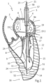
- the Fig. 1 The heart 1 shown in schematic and basic representation lies, rotated about its longitudinal axis, in the left chest cavity, so that the right half of the heart rests more against the front chest wall, while the left half of the heart points more towards the back.
- the object of the invention is to develop a heart valve implant 11 which, when minimally invasive surgery is used on the beating heart 1 of a patient, can be introduced via the right thorax area and the left atrium 3 of the heart 1 and from there into the left ventricle 7 with the aid of known surgical instruments and a trocar 5 and anchored there.
- the left ventricle 2 with the left atrium 3 and an access 4 into the left atrium 3 to the mitral valve 6 and the left ventricle 7 is therefore shown.
- the access 4 is via the indicated trocar 5, an outer tube slide 57 and an inner tube slide I 58.
- the outer tube slider 57 is guided through a trocar 5 and the inner tube slider I 58 through the outer tube slider 57.
- the inner tube slider I 58 is replaced during the operation by another tube slider II 59, see Fig. 4c , exchanged.
- the left ventricle 7 is divided into an inflow and outflow tract. It is separated from the atrium 3 by the mitral valve 6.
- the mitral valve 6 is connected to the papillary muscles 9 by tendon threads (chordae tendineae) 8, which originate from the ventricular wall 10 and ensure that the mitral valve 6 does not snap back too violently into the left atrium 3 when the valve closes and during the contraction phase (systole) of the left chamber 7.
- the inserted mitral valve implant 11 can be seen in the left ventricle 7.
- the mitral valve implant 11 has an anchoring element 13 at the distal end 12, whereby the anchoring element 13 consists of a corkscrew-like spiral screw 14.
- the screwed-in spiral screw 14 is located in the heart muscle tissue 15 in the area of the apex, the so-called pointed heart area 16. Furthermore, the mitral valve implant 11 has a fastening means 18 at the proximal end 17, which is attached to a mitral valve leaflet 19.
- a mitral valve 6 consists of two leaflets 19.1, 19.2, the anterior leaflet (cuspis anterior) 19.1 and the posterior leaflet (cuspis posterior) 19.2. According to the Fig. 1 , the mitral valve implant 11 is attached to the front damaged leaflet 19.1, for example.
- a connecting element 20 is arranged between the anchoring element 13 and the fastening means 18.
- the connecting element 20 consists of an artificial thread 21 which extends generally linearly along a longitudinal axis 23 of the heart valve implant 11 and replaces, for example, the lack of function of one or more torn tendon threads 22, the connecting element 20 being arranged with a first 24 and a second end 24' generally opposite one another and a fastening means 18 connecting to the anchoring element 13.
- the fastening means 18 is in Fig. 2a and Fig. 2b described in more detail. Analogous reference symbols from the Fig. 1 are in the Fig. 2a and 2b taken over.
- Fig. 2a and Fig. 2b show, in schematic representation and side view, the fastening means 18.
- Fig. 2a A closed jaw part 56 is provided, and after Fig. 2b an open jaw part 56 is provided on the gripping element 60 of the spring arms 50, 50'.
- the jaw part 56 forms the active side of the heart valve implant 11 or the fastening side 63 of the heart valve implant 11 on the mitral valve 6 and is in the Fig. 2c described in more detail.
- the flange side 64 on the cylindrical tube element 25 opposite the fastening side 63 represents the passive side, it is used for handling or introducing the fastening means 18 into the left atrium 3 and subsequently, after grasping and clamping a mitral valve leaflet 19 and inserting it into the left ventricle 7.
- the cylindrical tube element 25 of the fastening means 18 is detachably connected to an outer tube slide 57 using a known fastening method.
- the detachable connecting element 68 (not shown in more detail) between the tube element 25 is located on the flange side 64 of the tube element 25 and the docking side 65 of the outer tube slide 57.
- the outer tube slide 57 is a surgical instrument (not shown in more detail here) which is operated by the surgeon outside the patient's chest.
- the outer diameter of the outer tube slide 57 is largely adapted to the outer diameter 26 of the cylindrical tube element 25.
- one free end 61 of the gripping element 60 of the fastening means 18 is gripped with an inner tube slide I 58, which is guided through the outer tube slide 57 and the cylindrical tube element 25.
- the outer diameter of the inner tube slide I 58 is largely adapted to the inner diameter 66 of the thin-walled tube element 25.
- the gripping element 60 is gripped by the opening 62 of the inner tube slide I 58 receiving the spring arms 50, 50' at the free end 61 of the gripping element 60.
- the inner diameter 69 of the opening 62 of the inner tube slide I 58 is slightly smaller than the outer circumference of the gripping element 60 and thus also of the spring arms 50, 50'.
- the spring arms 50, 50' of the gripping element 60 are received in the opening 62 of the inner pipe slide I 58 by moving the inner pipe slide I 58 over the spring arms 50, 50' of the gripping element 60.
- the movement takes place according to the Fig. 2a , only until the spring arms 50, 50' are guided somewhat in the opening 62.
- the parallel spaced spring arms 50, 50' are pressed against one another slightly, but only so far that on the one hand the spring arms 50, 50' can just be received in the opening 62 of the inner pipe slide I 58 and on the other hand the jaw part 56 of the gripping element 60 does not yet open.
- the compression of the spring arms 50, 50' is made possible because the spring arms 50, 50' are arranged on a leg spring 38.
- the leg spring 38 can be compressed when pressure is exerted on the spring arms 50, 50'. This pressure is exerted on the spring arms 50, 50' with the aid of the inner tube slide I 58.
- the cylindrical tube element 25 on the fastening side 63 of the fastening means 18 is approximately flush with the inner tube slide I 58.
- the fastening means 18 can also be assembled on the two tube slides 57, 58 in the reverse order, in which the spring arms 50, 50' are first grasped with the inner tube slide I 58.
- the spring arms 50, 50' are grasped by pushing the inner tube slide I 58 through the inner diameter 66 of the cylindrical tube element 25 from the flange side 64 in the longitudinal direction 53 to the gripping arm side 67.
- the outer tube slide 57 is attached to the cylindrical tube element 25 by pushing it with its docking side 65 first, over the inner tube element I 58 to the flange side 64 of the cylindrical tube element 25 and connecting it to the connecting element 68.
- the fastening means 18 can be introduced through the trocar 5 into the left atrium 3.
- the fastening means 18 for gripping a mitral valve leaflet 19 is then prepared with the aid of the two tube slides 57, 58.
- the fastening means 18 of both tube slides 57, 58 is introduced into the left atrium 3, this can be used in a first application to open and close the jaw part 56, i.e. to grasp and clamp a mitral valve leaflet 19, as shown in the following sequence.
- the jaw part 56 of the gripping element 60 can be opened.
- the jaw part 56 is opened as follows: the outer tube slide 57, which is docked on the cylindrical tube element 25, pulls the spring arms 50, 50' of the gripping element 60, which are connected to the tube element 25 via a bracket 28, further into the opening 62 of the inner tube slide I 58.
- Each spring arm 50, 50' corresponds to a two-sided lever arm, so when assessing the functionality only one spring arm 50 needs to be considered.
- Such a spring arm 50 has two free ends. One end 61 is located on the open gripping element 60 and the other end on the jaw part 56.
- a spring arm 50 is attached approximately centrally to a leg spring 38, so that to the right and left of the attachment there is approximately one lever arm of equal length.
- the jaw part 56 of the gripping element 60 can also be opened and closed using a second method of using the two tube slides 57, 58 in order to grasp and clamp a mitral valve leaflet 19.
- the outer tube slide 57 is now held stationary and thus also the cylindrical tube element 25 docked to it.
- the inner tube slide I 58 located inside the outer tube slide 57 and inside the cylindrical tube element 25 can extend further over the spring arms 50, 50' located in the opening 62.
- the jaw part 56 of the gripping element 60 can be opened.
- the opening of the jaw part 56 takes place by the inner tube slide I 58 sliding over the spring arms 50, 50' of the gripping element 60, which are connected to the tube element 25 via a bracket 28.
- the spring arms 50, 50' cannot move because they are connected to the bracket 28 via the leg spring 38 and this in turn is connected to the tube element 25.
- the inner pipe slide I 58 is pushed further forward over the spring arms 50, 50', these slide further into the opening 62 of the inner pipe slide I 58.
- the two parallel spaced spring arms 50, 50' are further pressed together, whereby the jaw part 56 of the gripping element 60 opens, see Fig. 2b .
- the mitral valve implant 11 can be equipped with a fastening means 18.
- the fastening means 18 should be a mitral valve leaflet 19, cf. Fig. 1 , whereby the mitral valve 6 must still remain mobile, but its range of motion is limited in direction.
- Directional limitation means the folding back of the mitral valve leaflet 19 into the atrium 3.
- the heart valve implant 11 comprises, for example, three elements.
- a first element is the anchoring element 13, which is designed as a spiral screw 14 and provides a fastening of the heart valve implant 11 in the heart muscle tissue 15, see Fig. 1
- the second element is a connecting element 20, consisting of an artificial thread 21 or wire, which establishes the connection between the anchoring element 13 and the fastening means 18 with the assistance of a clamping means 74.
- These two elements have already been described in the Fig. 1
- Another element is the fastening means 18, which is shown here in the Fig. 2a and in the Fig. 2b is presented in more detail.
- the fastening means 18 itself can in turn be formed with three elements, for example a tubular element 25, a connecting element 27 and a gripping element 60.
- the tubular element 25 is preferably designed as a cylindrical sleeve 25', wherein the sleeve 25' can have geometric shapes in cross-section, such as square tubes, etc., and is thus not bound to the circular shape.
- Two openings 31, 31' are arranged in the sleeve wall 32 on the outer diameter 26 of the sleeve 25'.
- the openings 31, 31' are located on a transverse axis 35 which is perpendicular to the longitudinal axis 33 of the sleeve 25', wherein the openings 31, 31' are arranged approximately centrally when viewed in the longitudinal direction 53 of the sleeve 25'.
- a connecting element 27 is arranged on the openings 31, 31' and the outside of the sleeve 25'.
- the connecting element 27 is formed from a wire-like, approximately U-shaped bracket 28, which is connected in a joint-like manner 29 to the cylindrical sleeve 25'.
- the free end 30 of the U-shaped bracket 28 has two pins (not shown) which are bent inwards by at least 90 degrees and in each case one pin engages in an opening 31, 31' in the sleeve wall 32 in a rotationally movable manner.
- the pins can also be bent inwards by up to 180 degrees, so that a pin runs parallel to the longitudinal leg 34 in order to form a type of eyelet for receiving the sleeve wall 32. This creates a non-detachable but joint-like connection 29 between the sleeve 25' and the connecting element 27.
- the longitudinal legs 34 of the bracket 28 have a length which is considerably longer than half the sleeve length, i.e. approximately three times half the sleeve length.
- the length of the bracket 28 therefore allows the bracket 28 to perform a 360 degree rotation around the sleeve 25' because the cross leg 36 of the bracket 28, which connects the two longitudinal legs 34, 34' to one another, is relatively far away from the sleeve 25'.
- the cross leg 36 forms a continuous connecting rod 37 between the longitudinal legs 34, 34', which is the carrier of a gripping element 60.
- the gripping element 60 can be formed with a leg spring 38, which in turn is the carrier of two spring arms 50, 50'.
- the approximately U-shaped leg spring 38 is formed from a leaf spring 39, which has a corresponding connecting element 41 in the center of the leg region 40 or approximately in the apex 48 of the leg spring 38, for receiving a connecting rod 37 of the bracket 28.
- the connecting element 41 can consist of two spaced ring eyelets 42, 42' (see Fig. 3b ) which are an integral part of the leg spring 38.
- the eyelets 42, 42' are located on the side of the edge of the leg spring 38 and extend inwards, see Fig. 3b
- the connecting element 41 can also consist of a cylindrical channel 43 or a longitudinal groove 43 (see Fig. 3a ).
- the channel 43 or the longitudinal groove 43 run from one side to the other side of the leg spring 38 or transversely to the extending leaf spring legs 45, 49, see the explanations in the Fig. 3a .
- the axis 44 of the eyelets 42, 42' of the leg spring 38, according to the embodiment of the Fig. 3b , or the axis 44 of the cylindrical channel 43 or the longitudinal groove 43 of the leg spring 38, according to the embodiment of the Fig. 3a runs parallel to the transverse axis 35 of the openings 31, 31' in the sleeve 25' and also perpendicular to the longitudinal axis 33 of the sleeve 25'.
- the connection between the connecting rod 37 of the bracket 28 and the corresponding connecting element 41 of the leg spring 38 is designed such that the leg spring 38 can pivot about the connecting rod 37.
- the U-shaped leg spring 38 points with its open leaf spring legs 45, 49, starting from the apex 48, in the direction of the cylindrical sleeve 25' and the longitudinal legs 34, 34' of the bracket 28.
- the leaf spring legs 45, 45' are thus approximately parallel to the longitudinal axis 33 of the sleeve 25' and pivotally articulated on the bracket 28 and have an opening 47.
- leg spring 38 arranged on the bracket 28 performs a rotation with the bracket 28 on the one hand about the transverse axis 35, which leads through the sleeve 25', and on the other hand a rotation about the transverse axis 44, which leads through the leg spring 38.
- the rotation of the leg spring 38 about the two transverse axes 35, 44 can take place simultaneously, similar to the principle of a gondola on a Ferris wheel.
- the leg spring 38 in turn supports two spring arms 50, 50'.
- the two spring arms 50, 50' are firmly connected to the leg spring 38.
- the connection can be made by technically known methods, such as lasering, welding, riveting, screwing, etc.
- One spring arm 50 rests on a leaf spring leg 45 and the other spring arm 50' on an opposite leaf spring leg 49, provided that the leg spring 38 is only made up of two leaf spring legs 45, 49.
- the leg spring 38 is formed from four leaf spring legs 45, 45', 49, 49'.
- the two Spring arms 50, 50' are attached to the leaf spring legs 45, 49 or 45, 45' and 49, 49' of the leg spring 38 in such a way that, when the jaw part 56 is closed, they are spaced approximately parallel on the fastening side 63 and with their opposite opening at the free end 61 of the gripping means side 67 and extend parallel to the longitudinal axis 33 of the sleeve 25' and thus to the longitudinal axis 33 of the fastening means 18.
- the two spring arms 50, 50' which are spaced parallel to one another, have an approximately oval or convex shape, similar to a converging lens. This means that the two spring arms 50, 50' are each curved outwards.
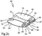
- the Fig. 2c shows a schematic representation of the jaw part 56 of a gripping element 60 of the fastening means 18.
- the jaw part 56 is formed from the spring arms 50, 50', which are part of the gripping elements 60.
- the jaw part 56 is arranged on the fastening side 63 of the gripping element 60 and on the side facing away from the opened leg spring 38 and on the longitudinal axis 33 of the tubular element 25, wherein the jaw part 56 has at least one spacer 88 arranged in the jaw part 56, which creates a predetermined gap 89 between the gripping jaws 86, 86' of the jaw part 56, whereby the teeth 87 of the gripping jaws 86, 86' do not lie directly on one another.
- a spacer 88 can be produced, for example, by embossing a bead 90, 90', preferably two beads per spring arm 50, 50'.
- the embossing of a bead 90, 90' takes place on the outside 91, 91' of the spring arms 50, 50', which creates an elevation 93, 93' on the inside 92, 92' of the spring arms 50, 50'.
- the beads 90, 90' are embossed in the spring arms 50, 50' opposite one another, so that the elevations 93, 93' created on the inside 92, 92' come to rest on one another when the jaw part 56 is closed and correspond with one another, thus forming a spacer 88.
- An elevation 93, 93' on the inner side 92, 92' between the spring arms 50, 50' to form a spacer 88 can also be achieved by using other means.
- the Fig. 3a, 3b and 3c show in perspective view an embodiment of a leg spring 38, which is part of the gripping element 60.
- the embodiment of the gripping element 60 consists of two identical, elongated spring arms 50, 50' made of rust-proof, alloyed metal which has certain elastic properties and can preferably consist of Nitinol.
- the spring arms 50, 50' are attached in the middle to a leg spring 38 made of rust-proof metal and are thus held together.
- the leg spring 38 also serves as a joint.
- the one-piece gripping element 60 consists of a leg spring 38 with spring arms 50, 50' fixedly arranged thereon, as can be seen from the Fig. 2a and 2b
- the representation of the spring arms 50, 50' in the Fig. 3a, 3b, 3c although these are of course the subject of a leg spring 38 and thus of the gripping element 60.
- the Fig. 2a and 2b The reference numerals shown identically with respect to the gripping element 60 and the leg spring 38 are adopted analogously here.
- leg springs 38 have in common that the base consists of a leaf spring 39.
- These leaf springs 39 are made of elastic spring steel and are U-shaped and therefore have an opening 47 between their bent legs.
- the U-shaped leaf springs 39 therefore each form two leaf spring legs 45, 49.
- the surfaces of the leaf spring legs 45, 49 can have recesses 71, 71' in their surface 70, 70', whereby the recesses 71, 71' in the surface 70, 70' can be open or closed. Due to the recesses 71, 71', several leaf spring legs 45, 45', 49, 49' can be created.
- a leg spring 38 has a corresponding connecting element 41 in its apex 48 for receiving a connecting rod 37 of a bracket 28 (see Fig. 2a and 2b ).
- the connecting element 41 can consist of two spaced ring eyelets 42, 42' (see Fig. 3b ) which are an integral part of the leg spring 38.
- the ring eyelets 42, 42' are located on the side of the edge of the leg spring 38 and extend inwards in the transverse direction of the leg spring 38 and have a transverse axis 44.
- the ring eyelets 42, 42' of the U-shaped leg spring 38 are located on the inside of the opening 47 on the side facing the leaf spring legs 45, 49.
- the ring eyelets 42, 42' can be arranged in the apex region 48 of the leg spring 38 on the outside of the leaf spring legs 45, 49, not shown, i.e. on the side facing away from the opening 47.
- the ring eyelets 42, 42' of the leg spring 38 are designed in such a way that they form an articulated connection 29' with the connecting rod 37 of a bracket 28.
- the connecting element 41 of a leg spring 38 can also consist of a cylindrical channel 43 or a longitudinal groove 43 (see Fig. 3a ).
- the channel 43 or the longitudinal groove 43 run from one transverse side to the other transverse side of the leg spring 38 or transversely to the extending leaf spring legs 45, 49.
- the cylindrical channel or the longitudinal groove 43 is designed such that the cylindrical channel or the longitudinal groove 43 has a slot 72 which is open in the direction of the leaf spring legs 45, 49 facing each other.
- the slot 72 is open all the way through in order to be able to accommodate the connecting rod 37 of a bracket 28.
- the cylindrical channel or the longitudinal groove 43 of the leg spring 38 is designed such that it forms a firm but articulated connection 29' with the connecting rod 37 of a bracket 28.
- the longitudinal groove 43 has a transverse axis 44 which is identical to the transverse axis 44 of the eyelets 42, 42'.
- This connecting element 41 is also located with its cylindrical channel or longitudinal groove 43 on the inside of the opening 47 on the side facing the leaf spring legs 45, 49.
- the leaf spring legs 45, 49 form a one-piece leg spring 38 with the cylindrical channel or longitudinal groove 43.
- the connecting element 41 of a cylindrical channel or a longitudinal groove 43 is not located in the opening area 47 of the leaf spring legs 45, 49 or not on the inside between the leaf spring legs 45, 49, but on the outside of the leaf spring legs 45, 49, but nevertheless in the area of the apex 48.
- the transverse axis 44 of this embodiment of leg spring 38 is identical to the transverse axes of the leg springs 38 from the Fig. 3a and 3b .
- This leg spring 38 is also designed in one piece and has a slot 72 for receiving the connecting rod 37 of a bracket 28.
- the cylindrical channel or the longitudinal groove 43 of the leg spring 38 is designed in such a way that it forms a firm but articulated connection 29' with the connecting rod 37 of a bracket 28.
- the Fig. 4a and 4b show a schematic representation of a fastening means 18 with the mitral valve leaflet 19 clamped in the gripping element 60.
- the process of grasping and clamping a mitral valve leaflet 19 in the left atrium 3 of the heart 1 takes place, whereby the process of opening and closing the jaw part 56 on the gripping element 60 already takes place in the Fig. 2a and 2b
- the functioning of the gripping element 60 is described in the Fig. 3a-3c Basically, before grasping a mitral valve leaflet 19, the gripping element 60 is opened and a mitral valve leaflet 19 is grasped with the open jaw part 56.
- both tube slides 57, 58, which transport and operate the fastening means 18, are pushed forward in the direction of the mitral valve 6.
- the displacement of the fastening means 18 is observed using imaging methods based on known devices. If the open jaw part 56 of the gripping element 60 is in the correct position in relation to the mitral valve leaflet 19, the jaw part 56 is closed. The closing of the jaw part 56 takes place by pulling back of the inner tube slide I 58 while simultaneously holding the outer tube slide 57, whereby the cylindrical tube element 25 remains stationary. The inner tube slide I 58 can now be removed.
- the leg spring 38 relaxes and the two leaf spring legs 45, 49 push the two spring arms 50, 50' apart at the free ends 61 by a certain distance.
- the maximum force of the leg spring 38 now acts on the jaw part 56, thereby clamping the mitral valve leaflet 19 located in the jaw part 56. The clamping takes place as in the Fig. 2c described.
- the gripping element 60 is pivoted about the transverse axis 35 of the cylindrical tube element 25 and the fastening means 18 is introduced into the left ventricle 7 in order to position it there on the one hand and to fasten it in the heart muscle tissue 15 on the other hand.
- This process is described in the Fig. 4b and 4c shown.
- a fastening means 18 with a clamped mitral valve leaflet 19 is visible during the pivoting process of the gripping element 60.
- the fastening means 18 which has already grasped and clamped a mitral valve leaflet 19, from the left atrium 3 (ie, the fastening means 18 is still located above the mitral valve 6) into the left ventricle 7 below the mitral valve 6, it is necessary to carry out further handling or pushing forward of the outer tube slide 57 with the outer tube slide 57 (see also in the Fig. 1 ).
- the outer tube slide 57 first introduces the cylindrical tube element 25 detachably attached to it and then the gripping element 60 arranged on the tube element 25 through the valve opening 94 in the mitral valve 6 into the left ventricle 7.
- the pivoting process of the gripping element 60 around the tube element 25 is completed when the gripping element 60 comes to rest on the outside of the outer tube slide 57.
- the gripping means 60 which is pivotably arranged on the fastening means 18, is located with the mitral valve leaflet 19 clamped below the mitral valve 6 in the left ventricle 7, as can be seen from the Fig. 1 and 5
- This handling, the displacement of the fastening means 18 with the aid of the outer tube slider 57 into the left ventricle 7, is also controlled using known imaging methods. The control is crucial when guiding the outer tube slider 57.
- the fastening of the fastening means 18 in the heart muscle tissue 15 of the left ventricle 7 is shown schematically in the Fig. 4c and positioning the heart valve implant 11 in the Fig. 5 shown.
- FIG. 4c A schematic representation of a surgical instrument for guiding and securing the anchoring element 13 arranged on the connecting element 20 in the heart muscle tissue 15 is shown.
- the surgical instrument is referred to here as an inner tube slide II 59.
- the inner tube slide II 59 is equipped on the inside with a connecting element 20 and an anchoring element 13, whereby the connecting element 20 is firmly connected to the anchoring element 13.
- the connecting element 20 consists of a thread 21 made of polytetrafluoroethylene (PTFE).
- One (second) end of the thread 21 of the connecting element 20 is located outside the patient's thorax, while the other (first) end 24 of the thread 21 is firmly connected to the proximal end 51 of the anchoring element 13.
- the anchoring element 13 consists of a spiral screw 14, which can be made of a nickel-titanium alloy, preferably of nitinol.
- the inner tube slide II 59 has a clamping means 74 at the insertion end 73.
- the clamping means 74 can be designed as a spiral groove 75 in the tube wall 76, wherein the spiral groove 75 forms a curve that is arranged with a constant pitch in the wall of the inner tube slide II 59.
- the pitch and running of the spiral groove 75 corresponds to the pitch of the right-hand spiral screw 14.
- the spiral groove 75 is relatively short at the insertion end 73 of the inner tube slide II 59 and grips the spiral screw 14 at the proximal end 51 when the inner tube slide II 59 is rotated clockwise.
- the connecting element 20 and the anchoring element 13 were introduced into the interior of the tube slide II 59.
- the anchoring element 13 is located at the insertion end 73 of the tube slide II 59, with the proximal end 51 of the spiral screw 14 in the spiral groove 75.
- the size of the spiral screw 14 to be used was selected before the operation in order to react to the different thicknesses of the heart muscle tissue 15 in the area of the apex 16. Different lengths of spiral screws 14 are therefore available.
- the distal end 52 of the spiral screw 14 is now introduced into the heart muscle tissue 15 by turning the tube slide II 59 clockwise.
- FIG. 5 As from the Fig. 5 , as can be seen in a schematic representation, there is now a heart valve implant 11 in the left ventricle 7 of the heart 1, whereby the fastening means 18 has grasped a mitral valve leaflet 19 and the spiral-shaped anchoring element 13 is anchored in the heart muscle tissue 15.
- the thread 21 of the connecting element 20 currently runs from the anchoring element 13 through the cylindrical tube element 25 of a fastening means 18, through an outer tube slide 57 docked to the fastening means 18 and from there through the trocar 5 inserted in the left atrium 3 to outside the thorax.
- the two inner tube slides I, II 58, 59 have already been removed. ZZ only the outer tube slide 57 is still in place, as can be seen from the Fig.
- the Fig. 6 shows a schematic representation of the connection between a fastening means 18 and an anchoring element 13 with the aid of the connecting element 20 and a clamping means 74.
- the connecting element 20 consists of a thread 21 which extends from the anchoring element 13 to outside the thorax and leads through the inner opening 66 of the fastening means 18.
- a clamping means 74 is pushed onto the outer end 83 of the thread 21.
- An inner tube slide III 77 is again used to slide the clamping means 74 onto the thread 21.
- the inner tube slide III 77 receives a thread 21 and a clamping means 74 in its bore 84.
- the outer diameter 85 of the inner pipe slide III 77 fits into the outer pipe slide 57.
- the clamping means 74 With the front side 78 of the pipe slide III 77, the clamping means 74 is pushed on the thread 21 along the longitudinal axis 23 to the fastening point 79.
- the fastening point 79 is located on the flange side 64, at the entrance to the inner opening 66 of the cylindrical pipe element 25.
- the material of the clamping means 74 can preferably consist of PTFE, but other materials are also conceivable.
- the bore 80 in the clamping means 74 is approximately adapted to the diameter 81 of the thread 21, with the proviso that the thread 21 in the clamping means 74 is stiff and the clamping means 74 can only be moved on the thread 21 with a certain force.
- the bore 80 and the diameter 81 form a press fit.
- the clamping means 74 can comprise different embodiments. In one embodiment of the clamping means 74, this is cylindrically designed as a sliding ring 76. In another embodiment, the clamping means 74 is designed as a truncated cone, similar to a bottle cork (not shown).
- the clamping effect occurs on the outer surface of the truncated cone.
- the positioning of the fastening means 18 in the left ventricle 7 takes place, for example, with the aid of the sliding ring 76.
- the sliding ring 76 is pushed into the cylindrical tubular element 25 in the optimal position of the fastening means 18 to the mitral valve 6.
- the optimal position of the fastening means 18 is again determined using the known imaging measurement methods, whereby the blood backflow during the contraction of the left ventricle into the left atrium 3 is also determined. The optimal position is reached when the blood backflow is at its minimum.
- the sliding ring 76 on the thread 21 is moved further along the longitudinal axis 23 in the direction of the anchoring element 13 with the tube slider III 77. While the sliding ring 76 is moved on the thread 21, the thread 21 is fixed outside the thorax and the fastening element 18 is held in place with the outer tube slider 57. After the sliding ring 76 has been successfully placed in the fastening means 18, the heart valve implant 11 is completely assembled and the inner tube slider III 77 can be withdrawn and removed. The tube slider III 77 is now replaced by a known surgical instrument (not shown). The surgical instrument can be used to knot and cut the thread 21 on the sliding ring 76, although other types of fastening are also conceivable.
- the outer tube slide 57 is also separated from the fastening means 18. To do this, the outer tube slide 57 is released from the connection point 68, e.g. by turning, depending on the design of the connection point 68.
- the gripping element 60 pivots onto the longitudinal axis 23 of the heart valve implant 11.
- the longitudinal axis 33 of the gripping element 60 and the fastening means 18 is now identical to the longitudinal axis 23 of the connecting element 20 and the anchoring element 13. Both longitudinal axes 23, 33 now form a common axis. The insertion of an inventive heart valve implant 11 into the left ventricle 7 of a heart 1 is thus completed.
- the previous embodiment shows a mitral valve implant that is assembled in the left ventricle and inserted relative to a mitral valve leaflet.
- regurgitation is eliminated.
- Regurgitation is a process in which the contents of the hollow organ of the heart do not only take the usual path, but flow back partly or predominantly in the other direction. This process is pathological in humans and many animal species.
- additional embodiments of the heart valve implant, in particular the fastening means of the heart valve implant are possible and are therefore not limited to the embodiment. This also applies to the connection technology between the connecting element and the fastening means.
Landscapes
- Health & Medical Sciences (AREA)
- Cardiology (AREA)
- Oral & Maxillofacial Surgery (AREA)
- Transplantation (AREA)
- Engineering & Computer Science (AREA)
- Biomedical Technology (AREA)
- Heart & Thoracic Surgery (AREA)
- Vascular Medicine (AREA)
- Life Sciences & Earth Sciences (AREA)
- Animal Behavior & Ethology (AREA)
- General Health & Medical Sciences (AREA)
- Public Health (AREA)
- Veterinary Medicine (AREA)
- Prostheses (AREA)
Priority Applications (1)
| Application Number | Priority Date | Filing Date | Title |
|---|---|---|---|
| EP24212188.7A EP4509095A3 (de) | 2017-03-28 | 2018-03-28 | Herzklappen-implantat und herzklappen-implantat-system |
Applications Claiming Priority (2)
| Application Number | Priority Date | Filing Date | Title |
|---|---|---|---|
| DE102017002974.1A DE102017002974B4 (de) | 2017-03-28 | 2017-03-28 | Herzklappen-Implantat, geeignet für den Einsatz in der minimal-invasiven Chirurgie zur Reparatur einer Herzklappe und/oder eines Herzklappensegels am schlagenden Herzen und Herzklappen-lmplantat-System |
| PCT/EP2018/058028 WO2018178210A1 (de) | 2017-03-28 | 2018-03-28 | Herzklappen-implantat und herzklappen-implantat-system |
Related Child Applications (1)
| Application Number | Title | Priority Date | Filing Date |
|---|---|---|---|
| EP24212188.7A Division EP4509095A3 (de) | 2017-03-28 | 2018-03-28 | Herzklappen-implantat und herzklappen-implantat-system |
Publications (3)
| Publication Number | Publication Date |
|---|---|
| EP3600156A1 EP3600156A1 (de) | 2020-02-05 |
| EP3600156C0 EP3600156C0 (de) | 2024-11-13 |
| EP3600156B1 true EP3600156B1 (de) | 2024-11-13 |
Family
ID=61972498
Family Applications (2)
| Application Number | Title | Priority Date | Filing Date |
|---|---|---|---|
| EP18717859.5A Active EP3600156B1 (de) | 2017-03-28 | 2018-03-28 | Herzklappen-implantat und herzklappen-implantat-system |
| EP24212188.7A Pending EP4509095A3 (de) | 2017-03-28 | 2018-03-28 | Herzklappen-implantat und herzklappen-implantat-system |
Family Applications After (1)
| Application Number | Title | Priority Date | Filing Date |
|---|---|---|---|
| EP24212188.7A Pending EP4509095A3 (de) | 2017-03-28 | 2018-03-28 | Herzklappen-implantat und herzklappen-implantat-system |
Country Status (6)
| Country | Link |
|---|---|
| US (2) | US11369472B2 (https=) |
| EP (2) | EP3600156B1 (https=) |
| JP (3) | JP7125784B2 (https=) |
| CN (1) | CN110996851B (https=) |
| DE (1) | DE102017002974B4 (https=) |
| WO (1) | WO2018178210A1 (https=) |
Families Citing this family (19)
| Publication number | Priority date | Publication date | Assignee | Title |
|---|---|---|---|---|
| US9308087B2 (en) | 2011-04-28 | 2016-04-12 | Neovasc Tiara Inc. | Sequentially deployed transcatheter mitral valve prosthesis |
| US9554897B2 (en) | 2011-04-28 | 2017-01-31 | Neovasc Tiara Inc. | Methods and apparatus for engaging a valve prosthesis with tissue |
| GB2536538B (en) | 2014-09-17 | 2018-07-18 | Cardiomech As | Anchor for implantation in body tissue |
| US10631984B2 (en) | 2015-12-15 | 2020-04-28 | Neovasc Tiara Inc. | Transseptal delivery system |
| EP4183372A1 (en) | 2016-01-29 | 2023-05-24 | Neovasc Tiara Inc. | Prosthetic valve for avoiding obstruction of outflow |
| EP3541462A4 (en) | 2016-11-21 | 2020-06-17 | Neovasc Tiara Inc. | METHODS AND SYSTEMS FOR THE QUICK RETURN OF A TRANSCATHETER HEART VALVE RELEASE SYSTEM |
| CN111263622A (zh) | 2017-08-25 | 2020-06-09 | 内奥瓦斯克迪亚拉公司 | 顺序展开的经导管二尖瓣假体 |
| US11173030B2 (en) | 2018-05-09 | 2021-11-16 | Neochord, Inc. | Suture length adjustment for minimally invasive heart valve repair |
| EP3876870B1 (en) | 2018-11-08 | 2023-12-20 | Neovasc Tiara Inc. | Ventricular deployment of a transcatheter mitral valve prosthesis |
| US20220117734A1 (en) | 2018-11-29 | 2022-04-21 | Cardiomech As | Device for Heart Repair |
| AU2020233892A1 (en) | 2019-03-08 | 2021-11-04 | Neovasc Tiara Inc. | Retrievable prosthesis delivery system |
| WO2020210652A1 (en) | 2019-04-10 | 2020-10-15 | Neovasc Tiara Inc. | Prosthetic valve with natural blood flow |
| US11376126B2 (en) | 2019-04-16 | 2022-07-05 | Neochord, Inc. | Transverse helical cardiac anchor for minimally invasive heart valve repair |
| US11779742B2 (en) | 2019-05-20 | 2023-10-10 | Neovasc Tiara Inc. | Introducer with hemostasis mechanism |
| US11311376B2 (en) | 2019-06-20 | 2022-04-26 | Neovase Tiara Inc. | Low profile prosthetic mitral valve |
| US12208007B2 (en) | 2020-01-16 | 2025-01-28 | Neochord, Inc. | Helical cardiac anchors for minimally invasive heart valve repair |
| CN117243654B (zh) * | 2023-10-10 | 2024-05-28 | 上海声拓医疗科技有限公司 | 用于修复装置的装载输送组件 |
| CN118299050B (zh) * | 2024-03-20 | 2026-02-06 | 四川大学华西医院 | 经导管主动脉瓣置换术后左室质量回归不佳风险预测方法 |
| CN118766517B (zh) * | 2024-08-16 | 2025-12-26 | 上海大学 | 一种心脏二尖瓣柔性捕获装置 |
Family Cites Families (24)
| Publication number | Priority date | Publication date | Assignee | Title |
|---|---|---|---|---|
| US3608554A (en) | 1969-12-22 | 1971-09-28 | Pilling Co | Surgical clamping means |
| US8216256B2 (en) * | 1999-04-09 | 2012-07-10 | Evalve, Inc. | Detachment mechanism for implantable fixation devices |
| CA2595459C (en) | 2005-01-21 | 2013-11-26 | Mayo Foundation For Medical Education And Research | Thorascopic heart valve repair method and apparatus |
| US7785366B2 (en) | 2005-10-26 | 2010-08-31 | Maurer Christopher W | Mitral spacer |
| US8778017B2 (en) * | 2005-10-26 | 2014-07-15 | Cardiosolutions, Inc. | Safety for mitral valve implant |
| US8092525B2 (en) | 2005-10-26 | 2012-01-10 | Cardiosolutions, Inc. | Heart valve implant |
| US8216302B2 (en) | 2005-10-26 | 2012-07-10 | Cardiosolutions, Inc. | Implant delivery and deployment system and method |
| US8449606B2 (en) | 2005-10-26 | 2013-05-28 | Cardiosolutions, Inc. | Balloon mitral spacer |
| US7632308B2 (en) * | 2005-11-23 | 2009-12-15 | Didier Loulmet | Methods, devices, and kits for treating mitral valve prolapse |
| DE102005062658B3 (de) | 2005-12-28 | 2007-05-31 | Osypka, Peter, Dr.-Ing. | Vorrichtung zum Verschliessen einer durch zwei sich überlappende Gewebelappen im Herzen gebildeten Öffnung |
| US8480730B2 (en) | 2007-05-14 | 2013-07-09 | Cardiosolutions, Inc. | Solid construct mitral spacer |
| CN101902975B (zh) | 2007-10-18 | 2014-06-04 | 尼奥绰德有限公司 | 搏动心脏中瓣膜小叶的微创修复 |
| US8147542B2 (en) | 2008-12-22 | 2012-04-03 | Valtech Cardio, Ltd. | Adjustable repair chords and spool mechanism therefor |
| US20110011917A1 (en) * | 2008-12-31 | 2011-01-20 | Hansen Medical, Inc. | Methods, devices, and kits for treating valve prolapse |
| US9204965B2 (en) * | 2009-01-14 | 2015-12-08 | Lc Therapeutics, Inc. | Synthetic chord |
| US8940042B2 (en) * | 2009-10-29 | 2015-01-27 | Valtech Cardio, Ltd. | Apparatus for guide-wire based advancement of a rotation assembly |
| US9044221B2 (en) | 2010-12-29 | 2015-06-02 | Neochord, Inc. | Exchangeable system for minimally invasive beating heart repair of heart valve leaflets |
| WO2012167120A2 (en) * | 2011-06-01 | 2012-12-06 | Neochord, Inc. | Minimally invasive repair of heart valve leaflets |
| US10799360B2 (en) * | 2011-07-27 | 2020-10-13 | The Cleveland Clinic Foundation | Systems and methods for treating a regurgitant heart valve |
| WO2015020971A1 (en) * | 2013-08-04 | 2015-02-12 | Mehr Medical Llc | Devices, systems and methods for repairing lumenal systems |
| US9549817B2 (en) * | 2011-09-22 | 2017-01-24 | Transmural Systems Llc | Devices, systems and methods for repairing lumenal systems |
| US9510946B2 (en) | 2012-09-06 | 2016-12-06 | Edwards Lifesciences Corporation | Heart valve sealing devices |
| WO2017066890A1 (en) * | 2015-10-21 | 2017-04-27 | Coremedic Ag | Medical instrument and method for heart valve repair |
| CA3178029A1 (en) * | 2017-04-05 | 2018-10-11 | Opus Medical Therapies, LLC | Transcatheter atrial sealing skirt, anchor, and tether and methods of implantation |
-
2017
- 2017-03-28 DE DE102017002974.1A patent/DE102017002974B4/de active Active
-
2018
- 2018-03-28 WO PCT/EP2018/058028 patent/WO2018178210A1/de not_active Ceased
- 2018-03-28 US US16/498,988 patent/US11369472B2/en active Active
- 2018-03-28 EP EP18717859.5A patent/EP3600156B1/de active Active
- 2018-03-28 EP EP24212188.7A patent/EP4509095A3/de active Pending
- 2018-03-28 JP JP2020503099A patent/JP7125784B2/ja active Active
- 2018-03-28 CN CN201880035160.6A patent/CN110996851B/zh active Active
-
2022
- 2022-06-27 US US17/850,313 patent/US20220323220A1/en active Pending
- 2022-08-05 JP JP2022125576A patent/JP7483273B2/ja active Active
-
2024
- 2024-04-23 JP JP2024069537A patent/JP2024087000A/ja not_active Ceased
Also Published As
| Publication number | Publication date |
|---|---|
| WO2018178210A1 (de) | 2018-10-04 |
| JP7125784B2 (ja) | 2022-08-25 |
| DE102017002974A1 (de) | 2018-10-04 |
| US20220323220A1 (en) | 2022-10-13 |
| JP2022140796A (ja) | 2022-09-27 |
| CA3095229A1 (en) | 2018-10-04 |
| EP3600156C0 (de) | 2024-11-13 |
| JP7483273B2 (ja) | 2024-05-15 |
| CN110996851A (zh) | 2020-04-10 |
| US11369472B2 (en) | 2022-06-28 |
| CN110996851B (zh) | 2021-12-17 |
| JP2024087000A (ja) | 2024-06-28 |
| US20200383784A1 (en) | 2020-12-10 |
| DE102017002974B4 (de) | 2024-08-08 |
| EP3600156A1 (de) | 2020-02-05 |
| EP4509095A2 (de) | 2025-02-19 |
| JP2020512173A (ja) | 2020-04-23 |
| EP4509095A3 (de) | 2025-05-07 |
Similar Documents
| Publication | Publication Date | Title |
|---|---|---|
| EP3600156B1 (de) | Herzklappen-implantat und herzklappen-implantat-system | |
| DE102017002976B4 (de) | Minimal-invasiv implantierbare Vorrichtung zur Beseitigung einer Mitralklappeninsuffizienz am schlagenden Herzen und Mitralklappen-Implantat-System | |
| EP2194925B1 (de) | Herzklappenstent | |
| DE60116786T2 (de) | Minimalinvasives system mit schablone für ein annuloplastisches reparatursegment | |
| DE60132005T2 (de) | Automatische ringförmige faltung zur mitralklappenreparatur | |
| DE102006052564B3 (de) | Mitralklappenstent | |
| DE202011111107U1 (de) | Transkatheter-Mitralklappenprothese | |
| DE102018100321B9 (de) | Herzklappenreparatursystem | |
| DE102018010279B9 (de) | Klappenreparaturvorrichtung | |
| DE212021000533U1 (de) | Herzklappenklemme zum Verhindern von Klappensegelverletzungen, Herzklappenklemmsystem | |
| DE102018100324B4 (de) | Klappenreparaturvorrichtung | |
| DE102018100319B9 (de) | Reparaturvorrichtungen und -verfahren für körpereigene Klappen | |
| DE102018100322B4 (de) | Reparaturvorrichtungen und -systeme für körpereigene Klappen | |
| DE102018100323B9 (de) | Klappenreparatursystem | |
| DE102018100350B4 (de) | Reparaturvorrichtungen und -verfahren für körpereigene klappen | |
| DE102018100352B9 (de) | Klappenreparatursystem | |
| DE102018100325B9 (de) | Reparaturvorrichtungen und -verfahren für körpereigene Klappen | |
| DE102006021975A1 (de) | Neochordae Sizer |
Legal Events
| Date | Code | Title | Description |
|---|---|---|---|
| STAA | Information on the status of an ep patent application or granted ep patent |
Free format text: STATUS: UNKNOWN |
|
| STAA | Information on the status of an ep patent application or granted ep patent |
Free format text: STATUS: THE INTERNATIONAL PUBLICATION HAS BEEN MADE |
|
| PUAI | Public reference made under article 153(3) epc to a published international application that has entered the european phase |
Free format text: ORIGINAL CODE: 0009012 |
|
| STAA | Information on the status of an ep patent application or granted ep patent |
Free format text: STATUS: REQUEST FOR EXAMINATION WAS MADE |
|
| 17P | Request for examination filed |
Effective date: 20191028 |
|
| AK | Designated contracting states |
Kind code of ref document: A1 Designated state(s): AL AT BE BG CH CY CZ DE DK EE ES FI FR GB GR HR HU IE IS IT LI LT LU LV MC MK MT NL NO PL PT RO RS SE SI SK SM TR |
|
| AX | Request for extension of the european patent |
Extension state: BA ME |
|
| RIN1 | Information on inventor provided before grant (corrected) |
Inventor name: ALBES, JOHANNES, PROF. DR. MED. |
|
| RAP1 | Party data changed (applicant data changed or rights of an application transferred) |
Owner name: IMMANUEL ALBERTINEN DIAKONIE GGMBH |
|
| GRAP | Despatch of communication of intention to grant a patent |
Free format text: ORIGINAL CODE: EPIDOSNIGR1 |
|
| STAA | Information on the status of an ep patent application or granted ep patent |
Free format text: STATUS: GRANT OF PATENT IS INTENDED |
|
| INTG | Intention to grant announced |
Effective date: 20220905 |
|
| GRAJ | Information related to disapproval of communication of intention to grant by the applicant or resumption of examination proceedings by the epo deleted |
Free format text: ORIGINAL CODE: EPIDOSDIGR1 |
|
| STAA | Information on the status of an ep patent application or granted ep patent |
Free format text: STATUS: REQUEST FOR EXAMINATION WAS MADE |
|
| STAA | Information on the status of an ep patent application or granted ep patent |
Free format text: STATUS: EXAMINATION IS IN PROGRESS |
|
| INTC | Intention to grant announced (deleted) | ||
| 17Q | First examination report despatched |
Effective date: 20230130 |
|
| P01 | Opt-out of the competence of the unified patent court (upc) registered |
Effective date: 20230605 |
|
| GRAP | Despatch of communication of intention to grant a patent |
Free format text: ORIGINAL CODE: EPIDOSNIGR1 |
|
| STAA | Information on the status of an ep patent application or granted ep patent |
Free format text: STATUS: GRANT OF PATENT IS INTENDED |
|
| INTG | Intention to grant announced |
Effective date: 20240705 |
|
| GRAS | Grant fee paid |
Free format text: ORIGINAL CODE: EPIDOSNIGR3 |
|
| GRAA | (expected) grant |
Free format text: ORIGINAL CODE: 0009210 |
|
| STAA | Information on the status of an ep patent application or granted ep patent |
Free format text: STATUS: THE PATENT HAS BEEN GRANTED |
|
| AK | Designated contracting states |
Kind code of ref document: B1 Designated state(s): AL AT BE BG CH CY CZ DE DK EE ES FI FR GB GR HR HU IE IS IT LI LT LU LV MC MK MT NL NO PL PT RO RS SE SI SK SM TR |
|
| REG | Reference to a national code |
Ref country code: GB Ref legal event code: FG4D Free format text: NOT ENGLISH |
|
| REG | Reference to a national code |
Ref country code: CH Ref legal event code: EP |
|
| REG | Reference to a national code |
Ref country code: DE Ref legal event code: R096 Ref document number: 502018015320 Country of ref document: DE |
|
| REG | Reference to a national code |
Ref country code: IE Ref legal event code: FG4D Free format text: LANGUAGE OF EP DOCUMENT: GERMAN |
|
| U01 | Request for unitary effect filed |
Effective date: 20241212 |
|
| P04 | Withdrawal of opt-out of the competence of the unified patent court (upc) registered |
Free format text: CASE NUMBER: APP_66855/2024 Effective date: 20241218 |
|
| U07 | Unitary effect registered |
Designated state(s): AT BE BG DE DK EE FI FR IT LT LU LV MT NL PT RO SE SI Effective date: 20241220 |
|
| PG25 | Lapsed in a contracting state [announced via postgrant information from national office to epo] |
Ref country code: HR Free format text: LAPSE BECAUSE OF FAILURE TO SUBMIT A TRANSLATION OF THE DESCRIPTION OR TO PAY THE FEE WITHIN THE PRESCRIBED TIME-LIMIT Effective date: 20241113 Ref country code: IS Free format text: LAPSE BECAUSE OF FAILURE TO SUBMIT A TRANSLATION OF THE DESCRIPTION OR TO PAY THE FEE WITHIN THE PRESCRIBED TIME-LIMIT Effective date: 20250313 |
|
| PG25 | Lapsed in a contracting state [announced via postgrant information from national office to epo] |
Ref country code: ES Free format text: LAPSE BECAUSE OF FAILURE TO SUBMIT A TRANSLATION OF THE DESCRIPTION OR TO PAY THE FEE WITHIN THE PRESCRIBED TIME-LIMIT Effective date: 20241113 |
|
| PG25 | Lapsed in a contracting state [announced via postgrant information from national office to epo] |
Ref country code: NO Free format text: LAPSE BECAUSE OF FAILURE TO SUBMIT A TRANSLATION OF THE DESCRIPTION OR TO PAY THE FEE WITHIN THE PRESCRIBED TIME-LIMIT Effective date: 20250213 |
|
| PG25 | Lapsed in a contracting state [announced via postgrant information from national office to epo] |
Ref country code: GR Free format text: LAPSE BECAUSE OF FAILURE TO SUBMIT A TRANSLATION OF THE DESCRIPTION OR TO PAY THE FEE WITHIN THE PRESCRIBED TIME-LIMIT Effective date: 20250214 |
|
| PG25 | Lapsed in a contracting state [announced via postgrant information from national office to epo] |
Ref country code: PL Free format text: LAPSE BECAUSE OF FAILURE TO SUBMIT A TRANSLATION OF THE DESCRIPTION OR TO PAY THE FEE WITHIN THE PRESCRIBED TIME-LIMIT Effective date: 20241113 |
|
| PG25 | Lapsed in a contracting state [announced via postgrant information from national office to epo] |
Ref country code: RS Free format text: LAPSE BECAUSE OF FAILURE TO SUBMIT A TRANSLATION OF THE DESCRIPTION OR TO PAY THE FEE WITHIN THE PRESCRIBED TIME-LIMIT Effective date: 20250213 |
|
| U20 | Renewal fee for the european patent with unitary effect paid |
Year of fee payment: 8 Effective date: 20250325 |
|
| PG25 | Lapsed in a contracting state [announced via postgrant information from national office to epo] |
Ref country code: SM Free format text: LAPSE BECAUSE OF FAILURE TO SUBMIT A TRANSLATION OF THE DESCRIPTION OR TO PAY THE FEE WITHIN THE PRESCRIBED TIME-LIMIT Effective date: 20241113 |
|
| VS25 | Lapsed in a validation state [announced via postgrant information from nat. office to epo] |
Ref country code: MD Free format text: LAPSE BECAUSE OF FAILURE TO SUBMIT A TRANSLATION OF THE DESCRIPTION OR TO PAY THE FEE WITHIN THE PRESCRIBED TIME-LIMIT Effective date: 20241113 |
|
| PGFP | Annual fee paid to national office [announced via postgrant information from national office to epo] |
Ref country code: CH Payment date: 20250401 Year of fee payment: 8 |
|
| PG25 | Lapsed in a contracting state [announced via postgrant information from national office to epo] |
Ref country code: SK Free format text: LAPSE BECAUSE OF FAILURE TO SUBMIT A TRANSLATION OF THE DESCRIPTION OR TO PAY THE FEE WITHIN THE PRESCRIBED TIME-LIMIT Effective date: 20241113 |
|
| PG25 | Lapsed in a contracting state [announced via postgrant information from national office to epo] |
Ref country code: CZ Free format text: LAPSE BECAUSE OF FAILURE TO SUBMIT A TRANSLATION OF THE DESCRIPTION OR TO PAY THE FEE WITHIN THE PRESCRIBED TIME-LIMIT Effective date: 20241113 |
|
| PLBE | No opposition filed within time limit |
Free format text: ORIGINAL CODE: 0009261 |
|
| STAA | Information on the status of an ep patent application or granted ep patent |
Free format text: STATUS: NO OPPOSITION FILED WITHIN TIME LIMIT |
|
| 26N | No opposition filed |
Effective date: 20250814 |
|
| REG | Reference to a national code |
Ref country code: CH Ref legal event code: U11 Free format text: ST27 STATUS EVENT CODE: U-0-0-U10-U11 (AS PROVIDED BY THE NATIONAL OFFICE) Effective date: 20260401 |
|
| PGFP | Annual fee paid to national office [announced via postgrant information from national office to epo] |
Ref country code: GB Payment date: 20260319 Year of fee payment: 9 |
|
| PGFP | Annual fee paid to national office [announced via postgrant information from national office to epo] |
Ref country code: MC Payment date: 20260320 Year of fee payment: 9 |
|
| PGFP | Annual fee paid to national office [announced via postgrant information from national office to epo] |
Ref country code: IE Payment date: 20260318 Year of fee payment: 9 |
|
| VS25 | Lapsed in a validation state [announced via postgrant information from nat. office to epo] |
Ref country code: MA Free format text: LAPSE BECAUSE OF FAILURE TO SUBMIT A TRANSLATION OF THE DESCRIPTION OR TO PAY THE FEE WITHIN THE PRESCRIBED TIME-LIMIT; INVALID AB INITIO Effective date: 20180328 |