EP3578042A1 - Fumarylacetoacetate hydrolase (fah)-deficient and immunodeficient rats and uses thereof - Google Patents
Fumarylacetoacetate hydrolase (fah)-deficient and immunodeficient rats and uses thereof Download PDFInfo
- Publication number
- EP3578042A1 EP3578042A1 EP19172883.1A EP19172883A EP3578042A1 EP 3578042 A1 EP3578042 A1 EP 3578042A1 EP 19172883 A EP19172883 A EP 19172883A EP 3578042 A1 EP3578042 A1 EP 3578042A1
- Authority
- EP
- European Patent Office
- Prior art keywords
- rat
- hepatocytes
- fah
- human
- liver
- Prior art date
- Legal status (The legal status is an assumption and is not a legal conclusion. Google has not performed a legal analysis and makes no representation as to the accuracy of the status listed.)
- Withdrawn
Links
- 230000002950 deficient Effects 0.000 title claims description 98
- 241000700159 Rattus Species 0.000 title abstract description 376
- 108010022687 fumarylacetoacetase Proteins 0.000 title abstract description 64
- 102100029115 Fumarylacetoacetase Human genes 0.000 title abstract description 54
- 210000003494 hepatocyte Anatomy 0.000 claims abstract description 416
- 238000000034 method Methods 0.000 claims abstract description 155
- 208000019423 liver disease Diseases 0.000 claims abstract description 63
- 230000014509 gene expression Effects 0.000 claims abstract description 39
- 230000000694 effects Effects 0.000 claims abstract description 38
- 230000002440 hepatic effect Effects 0.000 claims abstract description 35
- 230000007812 deficiency Effects 0.000 claims abstract description 34
- 238000001727 in vivo Methods 0.000 claims abstract description 25
- 102000004190 Enzymes Human genes 0.000 claims abstract description 22
- 108090000790 Enzymes Proteins 0.000 claims abstract description 22
- 230000003247 decreasing effect Effects 0.000 claims abstract description 12
- 239000002676 xenobiotic agent Substances 0.000 claims abstract description 9
- 101150094145 FAH gene Proteins 0.000 claims description 139
- 101100334119 Emericella nidulans (strain FGSC A4 / ATCC 38163 / CBS 112.46 / NRRL 194 / M139) fahA gene Proteins 0.000 claims description 110
- 239000003795 chemical substances by application Substances 0.000 claims description 70
- 238000002054 transplantation Methods 0.000 claims description 43
- 244000052769 pathogen Species 0.000 claims description 42
- 230000001717 pathogenic effect Effects 0.000 claims description 39
- 208000015181 infectious disease Diseases 0.000 claims description 32
- 150000001875 compounds Chemical class 0.000 claims description 31
- 210000000130 stem cell Anatomy 0.000 claims description 30
- 210000001519 tissue Anatomy 0.000 claims description 30
- 230000004077 genetic alteration Effects 0.000 claims description 28
- 231100000118 genetic alteration Toxicity 0.000 claims description 28
- 238000011282 treatment Methods 0.000 claims description 26
- 238000002347 injection Methods 0.000 claims description 25
- 239000007924 injection Substances 0.000 claims description 25
- 206010073071 hepatocellular carcinoma Diseases 0.000 claims description 24
- 101100193633 Danio rerio rag2 gene Proteins 0.000 claims description 22
- 101100193635 Mus musculus Rag2 gene Proteins 0.000 claims description 22
- 231100000844 hepatocellular carcinoma Toxicity 0.000 claims description 21
- 230000003908 liver function Effects 0.000 claims description 19
- BPYKTIZUTYGOLE-IFADSCNNSA-N Bilirubin Chemical compound N1C(=O)C(C)=C(C=C)\C1=C\C1=C(C)C(CCC(O)=O)=C(CC2=C(C(C)=C(\C=C/3C(=C(C=C)C(=O)N\3)C)N2)CCC(O)=O)N1 BPYKTIZUTYGOLE-IFADSCNNSA-N 0.000 claims description 18
- 206010016654 Fibrosis Diseases 0.000 claims description 18
- 238000011161 development Methods 0.000 claims description 18
- 230000007423 decrease Effects 0.000 claims description 17
- 208000019425 cirrhosis of liver Diseases 0.000 claims description 15
- 230000007882 cirrhosis Effects 0.000 claims description 14
- 102000009027 Albumins Human genes 0.000 claims description 13
- 108010088751 Albumins Proteins 0.000 claims description 13
- 239000003550 marker Substances 0.000 claims description 12
- 210000001671 embryonic stem cell Anatomy 0.000 claims description 11
- 230000003211 malignant effect Effects 0.000 claims description 10
- 102000002260 Alkaline Phosphatase Human genes 0.000 claims description 9
- 108020004774 Alkaline Phosphatase Proteins 0.000 claims description 9
- 230000004060 metabolic process Effects 0.000 claims description 9
- 210000003240 portal vein Anatomy 0.000 claims description 9
- 230000006698 induction Effects 0.000 claims description 8
- 210000000952 spleen Anatomy 0.000 claims description 8
- 230000002034 xenobiotic effect Effects 0.000 claims description 8
- 208000024891 symptom Diseases 0.000 claims description 6
- 210000002767 hepatic artery Anatomy 0.000 claims description 5
- 210000002865 immune cell Anatomy 0.000 claims description 5
- 210000004263 induced pluripotent stem cell Anatomy 0.000 claims description 4
- 108010078791 Carrier Proteins Proteins 0.000 claims description 3
- 108010015742 Cytochrome P-450 Enzyme System Proteins 0.000 claims description 2
- 210000003663 amniotic stem cell Anatomy 0.000 claims description 2
- 210000004271 bone marrow stromal cell Anatomy 0.000 claims description 2
- 230000006872 improvement Effects 0.000 claims description 2
- 210000004324 lymphatic system Anatomy 0.000 claims description 2
- 210000003200 peritoneal cavity Anatomy 0.000 claims description 2
- 102000002004 Cytochrome P-450 Enzyme System Human genes 0.000 claims 1
- 102100027280 Fanconi anemia group A protein Human genes 0.000 claims 1
- 230000005740 tumor formation Effects 0.000 claims 1
- OUYCCCASQSFEME-QMMMGPOBSA-N L-tyrosine Chemical compound OC(=O)[C@@H](N)CC1=CC=C(O)C=C1 OUYCCCASQSFEME-QMMMGPOBSA-N 0.000 abstract description 11
- OUYCCCASQSFEME-UHFFFAOYSA-N tyrosine Natural products OC(=O)C(N)CC1=CC=C(O)C=C1 OUYCCCASQSFEME-UHFFFAOYSA-N 0.000 abstract description 11
- 238000004458 analytical method Methods 0.000 abstract description 10
- 230000006652 catabolic pathway Effects 0.000 abstract description 6
- 210000004027 cell Anatomy 0.000 description 144
- 210000004185 liver Anatomy 0.000 description 108
- 108090000623 proteins and genes Proteins 0.000 description 79
- 239000013598 vector Substances 0.000 description 67
- 108090000435 Urokinase-type plasminogen activator Proteins 0.000 description 52
- 102000003990 Urokinase-type plasminogen activator Human genes 0.000 description 48
- 241001465754 Metazoa Species 0.000 description 47
- 229960005356 urokinase Drugs 0.000 description 44
- 239000003018 immunosuppressive agent Substances 0.000 description 30
- 150000007523 nucleic acids Chemical class 0.000 description 25
- 230000035772 mutation Effects 0.000 description 24
- 235000018102 proteins Nutrition 0.000 description 23
- 102000004169 proteins and genes Human genes 0.000 description 23
- QJJXYPPXXYFBGM-LFZNUXCKSA-N Tacrolimus Chemical compound C1C[C@@H](O)[C@H](OC)C[C@@H]1\C=C(/C)[C@@H]1[C@H](C)[C@@H](O)CC(=O)[C@H](CC=C)/C=C(C)/C[C@H](C)C[C@H](OC)[C@H]([C@H](C[C@H]2C)OC)O[C@@]2(O)C(=O)C(=O)N2CCCC[C@H]2C(=O)O1 QJJXYPPXXYFBGM-LFZNUXCKSA-N 0.000 description 22
- 239000003814 drug Substances 0.000 description 21
- 230000005976 liver dysfunction Effects 0.000 description 21
- QJJXYPPXXYFBGM-SHYZHZOCSA-N tacrolimus Natural products CO[C@H]1C[C@H](CC[C@@H]1O)C=C(C)[C@H]2OC(=O)[C@H]3CCCCN3C(=O)C(=O)[C@@]4(O)O[C@@H]([C@H](C[C@H]4C)OC)[C@@H](C[C@H](C)CC(=C[C@@H](CC=C)C(=O)C[C@H](O)[C@H]2C)C)OC QJJXYPPXXYFBGM-SHYZHZOCSA-N 0.000 description 21
- 229940088598 enzyme Drugs 0.000 description 20
- 238000012217 deletion Methods 0.000 description 19
- 230000037430 deletion Effects 0.000 description 19
- 239000000523 sample Substances 0.000 description 19
- 241000701161 unidentified adenovirus Species 0.000 description 19
- 229940125721 immunosuppressive agent Drugs 0.000 description 18
- 238000003780 insertion Methods 0.000 description 18
- 230000037431 insertion Effects 0.000 description 18
- 102000039446 nucleic acids Human genes 0.000 description 18
- 108020004707 nucleic acids Proteins 0.000 description 18
- 230000008685 targeting Effects 0.000 description 18
- HMLGSIZOMSVISS-ONJSNURVSA-N (7r)-7-[[(2z)-2-(2-amino-1,3-thiazol-4-yl)-2-(2,2-dimethylpropanoyloxymethoxyimino)acetyl]amino]-3-ethenyl-8-oxo-5-thia-1-azabicyclo[4.2.0]oct-2-ene-2-carboxylic acid Chemical compound N([C@@H]1C(N2C(=C(C=C)CSC21)C(O)=O)=O)C(=O)\C(=N/OCOC(=O)C(C)(C)C)C1=CSC(N)=N1 HMLGSIZOMSVISS-ONJSNURVSA-N 0.000 description 17
- 150000001413 amino acids Chemical group 0.000 description 17
- 241000699666 Mus <mouse, genus> Species 0.000 description 16
- 229940079593 drug Drugs 0.000 description 16
- 210000000987 immune system Anatomy 0.000 description 16
- 230000010412 perfusion Effects 0.000 description 16
- 230000018109 developmental process Effects 0.000 description 15
- 208000037265 diseases, disorders, signs and symptoms Diseases 0.000 description 15
- 230000001506 immunosuppresive effect Effects 0.000 description 15
- 241000894007 species Species 0.000 description 15
- 230000003612 virological effect Effects 0.000 description 15
- 206010062016 Immunosuppression Diseases 0.000 description 14
- 239000003112 inhibitor Substances 0.000 description 14
- 210000000056 organ Anatomy 0.000 description 14
- 231100000765 toxin Toxicity 0.000 description 14
- 108020004414 DNA Proteins 0.000 description 13
- 241000282412 Homo Species 0.000 description 13
- 206010067125 Liver injury Diseases 0.000 description 13
- 230000006870 function Effects 0.000 description 13
- 102000051628 Interleukin-1 receptor antagonist Human genes 0.000 description 12
- 108700021006 Interleukin-1 receptor antagonist Proteins 0.000 description 12
- 241000700605 Viruses Species 0.000 description 12
- 210000004369 blood Anatomy 0.000 description 12
- 239000008280 blood Substances 0.000 description 12
- 201000010099 disease Diseases 0.000 description 12
- 230000002458 infectious effect Effects 0.000 description 12
- 108700028369 Alleles Proteins 0.000 description 11
- 241000711549 Hepacivirus C Species 0.000 description 11
- 210000001744 T-lymphocyte Anatomy 0.000 description 11
- 229960004238 anakinra Drugs 0.000 description 11
- 239000000427 antigen Substances 0.000 description 11
- 108091007433 antigens Proteins 0.000 description 11
- 102000036639 antigens Human genes 0.000 description 11
- 229960003444 immunosuppressant agent Drugs 0.000 description 11
- 210000005228 liver tissue Anatomy 0.000 description 11
- 210000002966 serum Anatomy 0.000 description 11
- 239000003053 toxin Substances 0.000 description 11
- 108700012359 toxins Proteins 0.000 description 11
- YYSFXUWWPNHNAZ-PKJQJFMNSA-N umirolimus Chemical compound C1[C@@H](OC)[C@H](OCCOCC)CC[C@H]1C[C@@H](C)[C@H]1OC(=O)[C@@H]2CCCCN2C(=O)C(=O)[C@](O)(O2)[C@H](C)CC[C@H]2C[C@H](OC)/C(C)=C/C=C/C=C/[C@@H](C)C[C@@H](C)C(=O)[C@H](OC)[C@H](O)/C(C)=C/[C@@H](C)C(=O)C1 YYSFXUWWPNHNAZ-PKJQJFMNSA-N 0.000 description 11
- 241000700721 Hepatitis B virus Species 0.000 description 10
- 241000699670 Mus sp. Species 0.000 description 10
- 102000013530 TOR Serine-Threonine Kinases Human genes 0.000 description 10
- 108010065917 TOR Serine-Threonine Kinases Proteins 0.000 description 10
- 210000003719 b-lymphocyte Anatomy 0.000 description 10
- 210000003754 fetus Anatomy 0.000 description 10
- 210000002540 macrophage Anatomy 0.000 description 10
- 239000000047 product Substances 0.000 description 10
- 239000000243 solution Substances 0.000 description 10
- 229960005486 vaccine Drugs 0.000 description 10
- 239000013603 viral vector Substances 0.000 description 10
- 102100036475 Alanine aminotransferase 1 Human genes 0.000 description 9
- 108010082126 Alanine transaminase Proteins 0.000 description 9
- 108010003415 Aspartate Aminotransferases Proteins 0.000 description 9
- 102000004625 Aspartate Aminotransferases Human genes 0.000 description 9
- PMATZTZNYRCHOR-CGLBZJNRSA-N Cyclosporin A Chemical compound CC[C@@H]1NC(=O)[C@H]([C@H](O)[C@H](C)C\C=C\C)N(C)C(=O)[C@H](C(C)C)N(C)C(=O)[C@H](CC(C)C)N(C)C(=O)[C@H](CC(C)C)N(C)C(=O)[C@@H](C)NC(=O)[C@H](C)NC(=O)[C@H](CC(C)C)N(C)C(=O)[C@H](C(C)C)NC(=O)[C@H](CC(C)C)N(C)C(=O)CN(C)C1=O PMATZTZNYRCHOR-CGLBZJNRSA-N 0.000 description 9
- 108010036949 Cyclosporine Proteins 0.000 description 9
- 238000002965 ELISA Methods 0.000 description 9
- 108091006905 Human Serum Albumin Proteins 0.000 description 9
- 102000008100 Human Serum Albumin Human genes 0.000 description 9
- 206010028980 Neoplasm Diseases 0.000 description 9
- 238000010171 animal model Methods 0.000 description 9
- 210000002257 embryonic structure Anatomy 0.000 description 9
- HPNSFSBZBAHARI-RUDMXATFSA-N mycophenolic acid Chemical compound OC1=C(C\C=C(/C)CCC(O)=O)C(OC)=C(C)C2=C1C(=O)OC2 HPNSFSBZBAHARI-RUDMXATFSA-N 0.000 description 9
- 230000008569 process Effects 0.000 description 9
- QFJCIRLUMZQUOT-HPLJOQBZSA-N sirolimus Chemical compound C1C[C@@H](O)[C@H](OC)C[C@@H]1C[C@@H](C)[C@H]1OC(=O)[C@@H]2CCCCN2C(=O)C(=O)[C@](O)(O2)[C@H](C)CC[C@H]2C[C@H](OC)/C(C)=C/C=C/C=C/[C@@H](C)C[C@@H](C)C(=O)[C@H](OC)[C@H](O)/C(C)=C/[C@@H](C)C(=O)C1 QFJCIRLUMZQUOT-HPLJOQBZSA-N 0.000 description 9
- 108010068327 4-hydroxyphenylpyruvate dioxygenase Proteins 0.000 description 8
- 102100028626 4-hydroxyphenylpyruvate dioxygenase Human genes 0.000 description 8
- 102000029816 Collagenase Human genes 0.000 description 8
- 108060005980 Collagenase Proteins 0.000 description 8
- 206010019663 Hepatic failure Diseases 0.000 description 8
- 206010061598 Immunodeficiency Diseases 0.000 description 8
- 208000029462 Immunodeficiency disease Diseases 0.000 description 8
- 230000015572 biosynthetic process Effects 0.000 description 8
- 210000002459 blastocyst Anatomy 0.000 description 8
- 230000001413 cellular effect Effects 0.000 description 8
- 229960001265 ciclosporin Drugs 0.000 description 8
- 229960002424 collagenase Drugs 0.000 description 8
- 238000001415 gene therapy Methods 0.000 description 8
- 230000002068 genetic effect Effects 0.000 description 8
- 230000001553 hepatotropic effect Effects 0.000 description 8
- 210000005260 human cell Anatomy 0.000 description 8
- 230000007813 immunodeficiency Effects 0.000 description 8
- 230000001861 immunosuppressant effect Effects 0.000 description 8
- 208000007903 liver failure Diseases 0.000 description 8
- HPNSFSBZBAHARI-UHFFFAOYSA-N micophenolic acid Natural products OC1=C(CC=C(C)CCC(O)=O)C(OC)=C(C)C2=C1C(=O)OC2 HPNSFSBZBAHARI-UHFFFAOYSA-N 0.000 description 8
- 239000000203 mixture Substances 0.000 description 8
- 210000000822 natural killer cell Anatomy 0.000 description 8
- 239000002243 precursor Substances 0.000 description 8
- 238000011552 rat model Methods 0.000 description 8
- 108020004705 Codon Proteins 0.000 description 7
- 229930105110 Cyclosporin A Natural products 0.000 description 7
- 101100334122 Rattus norvegicus Fah gene Proteins 0.000 description 7
- 238000010459 TALEN Methods 0.000 description 7
- 108010043645 Transcription Activator-Like Effector Nucleases Proteins 0.000 description 7
- 229940024606 amino acid Drugs 0.000 description 7
- 235000001014 amino acid Nutrition 0.000 description 7
- 230000008901 benefit Effects 0.000 description 7
- 201000011510 cancer Diseases 0.000 description 7
- 239000006285 cell suspension Substances 0.000 description 7
- 238000001943 fluorescence-activated cell sorting Methods 0.000 description 7
- 230000010354 integration Effects 0.000 description 7
- 238000004519 manufacturing process Methods 0.000 description 7
- 239000008188 pellet Substances 0.000 description 7
- ZAHRKKWIAAJSAO-UHFFFAOYSA-N rapamycin Natural products COCC(O)C(=C/C(C)C(=O)CC(OC(=O)C1CCCCN1C(=O)C(=O)C2(O)OC(CC(OC)C(=CC=CC=CC(C)CC(C)C(=O)C)C)CCC2C)C(C)CC3CCC(O)C(C3)OC)C ZAHRKKWIAAJSAO-UHFFFAOYSA-N 0.000 description 7
- 229960002930 sirolimus Drugs 0.000 description 7
- 238000010186 staining Methods 0.000 description 7
- 239000013607 AAV vector Substances 0.000 description 6
- LFQSCWFLJHTTHZ-UHFFFAOYSA-N Ethanol Chemical compound CCO LFQSCWFLJHTTHZ-UHFFFAOYSA-N 0.000 description 6
- 238000010240 RT-PCR analysis Methods 0.000 description 6
- 208000032001 Tyrosinemia type 1 Diseases 0.000 description 6
- 108010032099 V(D)J recombination activating protein 2 Proteins 0.000 description 6
- LMEKQMALGUDUQG-UHFFFAOYSA-N azathioprine Chemical compound CN1C=NC([N+]([O-])=O)=C1SC1=NC=NC2=C1NC=N2 LMEKQMALGUDUQG-UHFFFAOYSA-N 0.000 description 6
- 229960002170 azathioprine Drugs 0.000 description 6
- 239000000872 buffer Substances 0.000 description 6
- 238000005119 centrifugation Methods 0.000 description 6
- 238000001514 detection method Methods 0.000 description 6
- 231100000234 hepatic damage Toxicity 0.000 description 6
- 230000001965 increasing effect Effects 0.000 description 6
- 230000001939 inductive effect Effects 0.000 description 6
- 230000008818 liver damage Effects 0.000 description 6
- 231100000835 liver failure Toxicity 0.000 description 6
- 230000036210 malignancy Effects 0.000 description 6
- 108020004999 messenger RNA Proteins 0.000 description 6
- 230000002503 metabolic effect Effects 0.000 description 6
- 210000004940 nucleus Anatomy 0.000 description 6
- 230000000144 pharmacologic effect Effects 0.000 description 6
- 229960004618 prednisone Drugs 0.000 description 6
- XOFYZVNMUHMLCC-ZPOLXVRWSA-N prednisone Chemical compound O=C1C=C[C@]2(C)[C@H]3C(=O)C[C@](C)([C@@](CC4)(O)C(=O)CO)[C@@H]4[C@@H]3CCC2=C1 XOFYZVNMUHMLCC-ZPOLXVRWSA-N 0.000 description 6
- 238000012360 testing method Methods 0.000 description 6
- 231100000419 toxicity Toxicity 0.000 description 6
- 230000001988 toxicity Effects 0.000 description 6
- 238000012546 transfer Methods 0.000 description 6
- JKMHFZQWWAIEOD-UHFFFAOYSA-N 2-[4-(2-hydroxyethyl)piperazin-1-yl]ethanesulfonic acid Chemical compound OCC[NH+]1CCN(CCS([O-])(=O)=O)CC1 JKMHFZQWWAIEOD-UHFFFAOYSA-N 0.000 description 5
- 241000282472 Canis lupus familiaris Species 0.000 description 5
- 102000016680 Dioxygenases Human genes 0.000 description 5
- 108010028143 Dioxygenases Proteins 0.000 description 5
- 102000030513 Homogentisate 1,2-Dioxygenase Human genes 0.000 description 5
- 108700023439 Homogentisate 1,2-dioxygenases Proteins 0.000 description 5
- 102000000589 Interleukin-1 Human genes 0.000 description 5
- 108010002352 Interleukin-1 Proteins 0.000 description 5
- 102100038560 Maleylacetoacetate isomerase Human genes 0.000 description 5
- 108091028043 Nucleic acid sequence Proteins 0.000 description 5
- 239000004677 Nylon Substances 0.000 description 5
- 241000282887 Suidae Species 0.000 description 5
- 102000016540 Tyrosine aminotransferases Human genes 0.000 description 5
- 108010042606 Tyrosine transaminase Proteins 0.000 description 5
- 102100029591 V(D)J recombination-activating protein 2 Human genes 0.000 description 5
- 239000011324 bead Substances 0.000 description 5
- 239000003651 drinking water Substances 0.000 description 5
- 235000020188 drinking water Nutrition 0.000 description 5
- 239000013604 expression vector Substances 0.000 description 5
- 230000001605 fetal effect Effects 0.000 description 5
- 229960000390 fludarabine Drugs 0.000 description 5
- GIUYCYHIANZCFB-FJFJXFQQSA-N fludarabine phosphate Chemical compound C1=NC=2C(N)=NC(F)=NC=2N1[C@@H]1O[C@H](COP(O)(O)=O)[C@@H](O)[C@@H]1O GIUYCYHIANZCFB-FJFJXFQQSA-N 0.000 description 5
- 238000002744 homologous recombination Methods 0.000 description 5
- 230000006801 homologous recombination Effects 0.000 description 5
- 238000002955 isolation Methods 0.000 description 5
- 230000000670 limiting effect Effects 0.000 description 5
- 108010035293 maleylacetoacetate isomerase Proteins 0.000 description 5
- 239000000463 material Substances 0.000 description 5
- 239000012528 membrane Substances 0.000 description 5
- 229940014456 mycophenolate Drugs 0.000 description 5
- 229920001778 nylon Polymers 0.000 description 5
- 150000003384 small molecules Chemical class 0.000 description 5
- 210000001082 somatic cell Anatomy 0.000 description 5
- 239000000126 substance Substances 0.000 description 5
- 230000001131 transforming effect Effects 0.000 description 5
- 201000011296 tyrosinemia Diseases 0.000 description 5
- IJGRMHOSHXDMSA-UHFFFAOYSA-N Atomic nitrogen Chemical compound N#N IJGRMHOSHXDMSA-UHFFFAOYSA-N 0.000 description 4
- 239000012594 Earle’s Balanced Salt Solution Substances 0.000 description 4
- 238000012413 Fluorescence activated cell sorting analysis Methods 0.000 description 4
- 206010056522 Hepatic infection Diseases 0.000 description 4
- 101150047285 Il1r1 gene Proteins 0.000 description 4
- 102000018682 Interleukin Receptor Common gamma Subunit Human genes 0.000 description 4
- 108010066719 Interleukin Receptor Common gamma Subunit Proteins 0.000 description 4
- 241000124008 Mammalia Species 0.000 description 4
- 108091005461 Nucleic proteins Proteins 0.000 description 4
- 238000002105 Southern blotting Methods 0.000 description 4
- 208000036142 Viral infection Diseases 0.000 description 4
- -1 adjacent sequences Proteins 0.000 description 4
- 230000004075 alteration Effects 0.000 description 4
- 239000012472 biological sample Substances 0.000 description 4
- 210000001185 bone marrow Anatomy 0.000 description 4
- 238000009395 breeding Methods 0.000 description 4
- 230000001488 breeding effect Effects 0.000 description 4
- 230000015556 catabolic process Effects 0.000 description 4
- 238000006243 chemical reaction Methods 0.000 description 4
- 230000034994 death Effects 0.000 description 4
- 231100000517 death Toxicity 0.000 description 4
- 230000036267 drug metabolism Effects 0.000 description 4
- 208000002854 epidermolysis bullosa simplex superficialis Diseases 0.000 description 4
- 230000004761 fibrosis Effects 0.000 description 4
- 230000028993 immune response Effects 0.000 description 4
- 239000002207 metabolite Substances 0.000 description 4
- 229960000951 mycophenolic acid Drugs 0.000 description 4
- OUBCNLGXQFSTLU-UHFFFAOYSA-N nitisinone Chemical compound [O-][N+](=O)C1=CC(C(F)(F)F)=CC=C1C(=O)C1C(=O)CCCC1=O OUBCNLGXQFSTLU-UHFFFAOYSA-N 0.000 description 4
- 229960001721 nitisinone Drugs 0.000 description 4
- 239000002773 nucleotide Substances 0.000 description 4
- 125000003729 nucleotide group Chemical group 0.000 description 4
- 238000004091 panning Methods 0.000 description 4
- 230000009467 reduction Effects 0.000 description 4
- 230000002829 reductive effect Effects 0.000 description 4
- 238000002271 resection Methods 0.000 description 4
- 230000004044 response Effects 0.000 description 4
- 230000002441 reversible effect Effects 0.000 description 4
- 239000002002 slurry Substances 0.000 description 4
- 238000010374 somatic cell nuclear transfer Methods 0.000 description 4
- 238000003860 storage Methods 0.000 description 4
- 239000006228 supernatant Substances 0.000 description 4
- 238000003786 synthesis reaction Methods 0.000 description 4
- VZGDMQKNWNREIO-UHFFFAOYSA-N tetrachloromethane Chemical compound ClC(Cl)(Cl)Cl VZGDMQKNWNREIO-UHFFFAOYSA-N 0.000 description 4
- 229940124597 therapeutic agent Drugs 0.000 description 4
- 230000001225 therapeutic effect Effects 0.000 description 4
- 231100000167 toxic agent Toxicity 0.000 description 4
- 239000003440 toxic substance Substances 0.000 description 4
- 210000002700 urine Anatomy 0.000 description 4
- 230000009385 viral infection Effects 0.000 description 4
- 108091032973 (ribonucleotides)n+m Proteins 0.000 description 3
- WYEPBHZLDUPIOD-UHFFFAOYSA-N 4,6-dioxoheptanoic acid Chemical compound CC(=O)CC(=O)CCC(O)=O WYEPBHZLDUPIOD-UHFFFAOYSA-N 0.000 description 3
- 208000007788 Acute Liver Failure Diseases 0.000 description 3
- 206010000804 Acute hepatic failure Diseases 0.000 description 3
- OYPRJOBELJOOCE-UHFFFAOYSA-N Calcium Chemical compound [Ca] OYPRJOBELJOOCE-UHFFFAOYSA-N 0.000 description 3
- 239000006144 Dulbecco’s modified Eagle's medium Substances 0.000 description 3
- 241000283086 Equidae Species 0.000 description 3
- 239000007995 HEPES buffer Substances 0.000 description 3
- 206010023126 Jaundice Diseases 0.000 description 3
- 241000283973 Oryctolagus cuniculus Species 0.000 description 3
- 206010052779 Transplant rejections Diseases 0.000 description 3
- 230000004913 activation Effects 0.000 description 3
- 231100000836 acute liver failure Toxicity 0.000 description 3
- 210000000577 adipose tissue Anatomy 0.000 description 3
- 230000003115 biocidal effect Effects 0.000 description 3
- 239000003131 biological toxin Substances 0.000 description 3
- 229960000074 biopharmaceutical Drugs 0.000 description 3
- 239000011575 calcium Substances 0.000 description 3
- 229910052791 calcium Inorganic materials 0.000 description 3
- 210000000349 chromosome Anatomy 0.000 description 3
- 230000000295 complement effect Effects 0.000 description 3
- 230000000875 corresponding effect Effects 0.000 description 3
- 230000004069 differentiation Effects 0.000 description 3
- 208000035475 disorder Diseases 0.000 description 3
- 239000012091 fetal bovine serum Substances 0.000 description 3
- 210000002950 fibroblast Anatomy 0.000 description 3
- 238000001914 filtration Methods 0.000 description 3
- 239000012634 fragment Substances 0.000 description 3
- 238000001476 gene delivery Methods 0.000 description 3
- 230000009395 genetic defect Effects 0.000 description 3
- 210000004602 germ cell Anatomy 0.000 description 3
- 230000036541 health Effects 0.000 description 3
- 231100000753 hepatic injury Toxicity 0.000 description 3
- 208000006454 hepatitis Diseases 0.000 description 3
- 231100000283 hepatitis Toxicity 0.000 description 3
- 238000011577 humanized mouse model Methods 0.000 description 3
- 230000002757 inflammatory effect Effects 0.000 description 3
- 238000007912 intraperitoneal administration Methods 0.000 description 3
- 231100000225 lethality Toxicity 0.000 description 3
- 201000007270 liver cancer Diseases 0.000 description 3
- 210000005229 liver cell Anatomy 0.000 description 3
- 208000014018 liver neoplasm Diseases 0.000 description 3
- 229940124302 mTOR inhibitor Drugs 0.000 description 3
- 239000003120 macrolide antibiotic agent Substances 0.000 description 3
- 230000005291 magnetic effect Effects 0.000 description 3
- 239000003628 mammalian target of rapamycin inhibitor Substances 0.000 description 3
- 241001515942 marmosets Species 0.000 description 3
- 239000002609 medium Substances 0.000 description 3
- 210000004379 membrane Anatomy 0.000 description 3
- GLVAUDGFNGKCSF-UHFFFAOYSA-N mercaptopurine Chemical compound S=C1NC=NC2=C1NC=N2 GLVAUDGFNGKCSF-UHFFFAOYSA-N 0.000 description 3
- 210000002901 mesenchymal stem cell Anatomy 0.000 description 3
- 210000001616 monocyte Anatomy 0.000 description 3
- 239000000178 monomer Substances 0.000 description 3
- 230000003287 optical effect Effects 0.000 description 3
- 239000002245 particle Substances 0.000 description 3
- 210000002381 plasma Anatomy 0.000 description 3
- 230000035935 pregnancy Effects 0.000 description 3
- 108090000765 processed proteins & peptides Proteins 0.000 description 3
- 229940002612 prodrug Drugs 0.000 description 3
- 239000000651 prodrug Substances 0.000 description 3
- 230000002062 proliferating effect Effects 0.000 description 3
- 230000035755 proliferation Effects 0.000 description 3
- 239000011251 protective drug Substances 0.000 description 3
- 230000001681 protective effect Effects 0.000 description 3
- 230000006825 purine synthesis Effects 0.000 description 3
- 230000006798 recombination Effects 0.000 description 3
- 238000005215 recombination Methods 0.000 description 3
- 230000010076 replication Effects 0.000 description 3
- 230000001177 retroviral effect Effects 0.000 description 3
- 238000012216 screening Methods 0.000 description 3
- 238000000926 separation method Methods 0.000 description 3
- 239000007787 solid Substances 0.000 description 3
- 150000003431 steroids Chemical class 0.000 description 3
- 229960001967 tacrolimus Drugs 0.000 description 3
- 231100000027 toxicology Toxicity 0.000 description 3
- 238000013518 transcription Methods 0.000 description 3
- 230000035897 transcription Effects 0.000 description 3
- 238000001890 transfection Methods 0.000 description 3
- 230000014616 translation Effects 0.000 description 3
- RZVAJINKPMORJF-UHFFFAOYSA-N Acetaminophen Chemical compound CC(=O)NC1=CC=C(O)C=C1 RZVAJINKPMORJF-UHFFFAOYSA-N 0.000 description 2
- 241000702423 Adeno-associated virus - 2 Species 0.000 description 2
- 229920000936 Agarose Polymers 0.000 description 2
- 208000023275 Autoimmune disease Diseases 0.000 description 2
- 102100032412 Basigin Human genes 0.000 description 2
- 102000015081 Blood Coagulation Factors Human genes 0.000 description 2
- 108010039209 Blood Coagulation Factors Proteins 0.000 description 2
- 241000283690 Bos taurus Species 0.000 description 2
- 108091003079 Bovine Serum Albumin Proteins 0.000 description 2
- 229940045513 CTLA4 antagonist Drugs 0.000 description 2
- UXVMQQNJUSDDNG-UHFFFAOYSA-L Calcium chloride Chemical compound [Cl-].[Cl-].[Ca+2] UXVMQQNJUSDDNG-UHFFFAOYSA-L 0.000 description 2
- 102100023321 Ceruloplasmin Human genes 0.000 description 2
- 241000701022 Cytomegalovirus Species 0.000 description 2
- 241000702421 Dependoparvovirus Species 0.000 description 2
- 206010058314 Dysplasia Diseases 0.000 description 2
- 102100039793 E3 ubiquitin-protein ligase RAG1 Human genes 0.000 description 2
- KCXVZYZYPLLWCC-UHFFFAOYSA-N EDTA Chemical compound OC(=O)CN(CC(O)=O)CCN(CC(O)=O)CC(O)=O KCXVZYZYPLLWCC-UHFFFAOYSA-N 0.000 description 2
- 102000010834 Extracellular Matrix Proteins Human genes 0.000 description 2
- 108010037362 Extracellular Matrix Proteins Proteins 0.000 description 2
- WSFSSNUMVMOOMR-UHFFFAOYSA-N Formaldehyde Chemical compound O=C WSFSSNUMVMOOMR-UHFFFAOYSA-N 0.000 description 2
- 102000006395 Globulins Human genes 0.000 description 2
- 108010044091 Globulins Proteins 0.000 description 2
- DHMQDGOQFOQNFH-UHFFFAOYSA-N Glycine Chemical compound NCC(O)=O DHMQDGOQFOQNFH-UHFFFAOYSA-N 0.000 description 2
- 102000011786 HLA-A Antigens Human genes 0.000 description 2
- 108010075704 HLA-A Antigens Proteins 0.000 description 2
- 206010073069 Hepatic cancer Diseases 0.000 description 2
- 101000744443 Homo sapiens E3 ubiquitin-protein ligase RAG1 Proteins 0.000 description 2
- 241000282620 Hylobates sp. Species 0.000 description 2
- 206010061218 Inflammation Diseases 0.000 description 2
- 102100025390 Integrin beta-2 Human genes 0.000 description 2
- 102000019223 Interleukin-1 receptor Human genes 0.000 description 2
- 108050006617 Interleukin-1 receptor Proteins 0.000 description 2
- 108091092195 Intron Proteins 0.000 description 2
- KFZMGEQAYNKOFK-UHFFFAOYSA-N Isopropanol Chemical compound CC(C)O KFZMGEQAYNKOFK-UHFFFAOYSA-N 0.000 description 2
- FFEARJCKVFRZRR-BYPYZUCNSA-N L-methionine Chemical compound CSCC[C@H](N)C(O)=O FFEARJCKVFRZRR-BYPYZUCNSA-N 0.000 description 2
- FBOZXECLQNJBKD-ZDUSSCGKSA-N L-methotrexate Chemical compound C=1N=C2N=C(N)N=C(N)C2=NC=1CN(C)C1=CC=C(C(=O)N[C@@H](CCC(O)=O)C(O)=O)C=C1 FBOZXECLQNJBKD-ZDUSSCGKSA-N 0.000 description 2
- 102100033467 L-selectin Human genes 0.000 description 2
- 208000004554 Leishmaniasis Diseases 0.000 description 2
- FYYHWMGAXLPEAU-UHFFFAOYSA-N Magnesium Chemical compound [Mg] FYYHWMGAXLPEAU-UHFFFAOYSA-N 0.000 description 2
- 241000283923 Marmota monax Species 0.000 description 2
- 206010028851 Necrosis Diseases 0.000 description 2
- 101710163270 Nuclease Proteins 0.000 description 2
- KHGNFPUMBJSZSM-UHFFFAOYSA-N Perforine Natural products COC1=C2CCC(O)C(CCC(C)(C)O)(OC)C2=NC2=C1C=CO2 KHGNFPUMBJSZSM-UHFFFAOYSA-N 0.000 description 2
- KPKZJLCSROULON-QKGLWVMZSA-N Phalloidin Chemical compound N1C(=O)[C@@H]([C@@H](O)C)NC(=O)[C@H](C)NC(=O)[C@H](C[C@@](C)(O)CO)NC(=O)[C@H](C2)NC(=O)[C@H](C)NC(=O)[C@@H]3C[C@H](O)CN3C(=O)[C@@H]1CSC1=C2C2=CC=CC=C2N1 KPKZJLCSROULON-QKGLWVMZSA-N 0.000 description 2
- 101100247004 Rattus norvegicus Qsox1 gene Proteins 0.000 description 2
- 101150036449 SIRPA gene Proteins 0.000 description 2
- FAPWRFPIFSIZLT-UHFFFAOYSA-M Sodium chloride Chemical class [Na+].[Cl-] FAPWRFPIFSIZLT-UHFFFAOYSA-M 0.000 description 2
- 108060008682 Tumor Necrosis Factor Proteins 0.000 description 2
- 206010047505 Visceral leishmaniasis Diseases 0.000 description 2
- 208000027418 Wounds and injury Diseases 0.000 description 2
- 108010017070 Zinc Finger Nucleases Proteins 0.000 description 2
- 238000009825 accumulation Methods 0.000 description 2
- 239000002253 acid Substances 0.000 description 2
- 230000000735 allogeneic effect Effects 0.000 description 2
- 210000001776 amniocyte Anatomy 0.000 description 2
- 239000003242 anti bacterial agent Substances 0.000 description 2
- 238000003556 assay Methods 0.000 description 2
- 230000001580 bacterial effect Effects 0.000 description 2
- 210000000941 bile Anatomy 0.000 description 2
- 230000004071 biological effect Effects 0.000 description 2
- 239000003114 blood coagulation factor Substances 0.000 description 2
- 210000001124 body fluid Anatomy 0.000 description 2
- 229940087828 buprenex Drugs 0.000 description 2
- UAIXRPCCYXNJMQ-RZIPZOSSSA-N buprenorphine hydrochlorie Chemical compound [Cl-].C([C@]12[C@H]3OC=4C(O)=CC=C(C2=4)C[C@@H]2[C@]11CC[C@]3([C@H](C1)[C@](C)(O)C(C)(C)C)OC)C[NH+]2CC1CC1 UAIXRPCCYXNJMQ-RZIPZOSSSA-N 0.000 description 2
- 150000001720 carbohydrates Chemical class 0.000 description 2
- 238000012754 cardiac puncture Methods 0.000 description 2
- 230000030833 cell death Effects 0.000 description 2
- 230000011712 cell development Effects 0.000 description 2
- 230000032823 cell division Effects 0.000 description 2
- 229940044683 chemotherapy drug Drugs 0.000 description 2
- HVYWMOMLDIMFJA-DPAQBDIFSA-N cholesterol Chemical compound C1C=C2C[C@@H](O)CC[C@]2(C)[C@@H]2[C@@H]1[C@@H]1CC[C@H]([C@H](C)CCCC(C)C)[C@@]1(C)CC2 HVYWMOMLDIMFJA-DPAQBDIFSA-N 0.000 description 2
- 230000002759 chromosomal effect Effects 0.000 description 2
- 238000003776 cleavage reaction Methods 0.000 description 2
- 230000002596 correlated effect Effects 0.000 description 2
- 239000003246 corticosteroid Substances 0.000 description 2
- 238000012258 culturing Methods 0.000 description 2
- 230000001351 cycling effect Effects 0.000 description 2
- 230000006378 damage Effects 0.000 description 2
- 238000011257 definitive treatment Methods 0.000 description 2
- 230000001934 delay Effects 0.000 description 2
- 230000027832 depurination Effects 0.000 description 2
- 238000001784 detoxification Methods 0.000 description 2
- 238000010586 diagram Methods 0.000 description 2
- 231100000676 disease causative agent Toxicity 0.000 description 2
- 238000009509 drug development Methods 0.000 description 2
- 231100001129 embryonic lethality Toxicity 0.000 description 2
- 230000007613 environmental effect Effects 0.000 description 2
- 231100000317 environmental toxin Toxicity 0.000 description 2
- 230000006862 enzymatic digestion Effects 0.000 description 2
- 230000029142 excretion Effects 0.000 description 2
- 210000002744 extracellular matrix Anatomy 0.000 description 2
- 230000008175 fetal development Effects 0.000 description 2
- 235000013305 food Nutrition 0.000 description 2
- 238000009472 formulation Methods 0.000 description 2
- 230000037433 frameshift Effects 0.000 description 2
- IGMNYECMUMZDDF-UHFFFAOYSA-N homogentisic acid Chemical compound OC(=O)CC1=CC(O)=CC=C1O IGMNYECMUMZDDF-UHFFFAOYSA-N 0.000 description 2
- 238000009396 hybridization Methods 0.000 description 2
- 230000001900 immune effect Effects 0.000 description 2
- 230000008629 immune suppression Effects 0.000 description 2
- 230000006058 immune tolerance Effects 0.000 description 2
- 238000003364 immunohistochemistry Methods 0.000 description 2
- 238000000338 in vitro Methods 0.000 description 2
- 238000011065 in-situ storage Methods 0.000 description 2
- 230000004054 inflammatory process Effects 0.000 description 2
- 230000002401 inhibitory effect Effects 0.000 description 2
- 208000014674 injury Diseases 0.000 description 2
- 102000002467 interleukin receptors Human genes 0.000 description 2
- 108010093036 interleukin receptors Proteins 0.000 description 2
- 238000010255 intramuscular injection Methods 0.000 description 2
- 239000007927 intramuscular injection Substances 0.000 description 2
- 238000010253 intravenous injection Methods 0.000 description 2
- 210000003734 kidney Anatomy 0.000 description 2
- GOTYRUGSSMKFNF-UHFFFAOYSA-N lenalidomide Chemical compound C1C=2C(N)=CC=CC=2C(=O)N1C1CCC(=O)NC1=O GOTYRUGSSMKFNF-UHFFFAOYSA-N 0.000 description 2
- 230000001665 lethal effect Effects 0.000 description 2
- 239000002502 liposome Substances 0.000 description 2
- 239000007788 liquid Substances 0.000 description 2
- 238000000464 low-speed centrifugation Methods 0.000 description 2
- 239000011777 magnesium Substances 0.000 description 2
- 229910052749 magnesium Inorganic materials 0.000 description 2
- 230000014759 maintenance of location Effects 0.000 description 2
- 201000004792 malaria Diseases 0.000 description 2
- 210000001161 mammalian embryo Anatomy 0.000 description 2
- 239000011159 matrix material Substances 0.000 description 2
- 229960001428 mercaptopurine Drugs 0.000 description 2
- 230000007102 metabolic function Effects 0.000 description 2
- 229930182817 methionine Natural products 0.000 description 2
- 230000002906 microbiologic effect Effects 0.000 description 2
- 230000003228 microsomal effect Effects 0.000 description 2
- 230000004048 modification Effects 0.000 description 2
- 238000012986 modification Methods 0.000 description 2
- RTGDFNSFWBGLEC-SYZQJQIISA-N mycophenolate mofetil Chemical compound COC1=C(C)C=2COC(=O)C=2C(O)=C1C\C=C(/C)CCC(=O)OCCN1CCOCC1 RTGDFNSFWBGLEC-SYZQJQIISA-N 0.000 description 2
- 229960004866 mycophenolate mofetil Drugs 0.000 description 2
- UMFJAHHVKNCGLG-UHFFFAOYSA-N n-Nitrosodimethylamine Chemical compound CN(C)N=O UMFJAHHVKNCGLG-UHFFFAOYSA-N 0.000 description 2
- 229930014626 natural product Natural products 0.000 description 2
- 230000017074 necrotic cell death Effects 0.000 description 2
- 230000001338 necrotic effect Effects 0.000 description 2
- 229960004927 neomycin Drugs 0.000 description 2
- 229910052757 nitrogen Inorganic materials 0.000 description 2
- 210000003463 organelle Anatomy 0.000 description 2
- 150000002894 organic compounds Chemical class 0.000 description 2
- 244000045947 parasite Species 0.000 description 2
- 230000003071 parasitic effect Effects 0.000 description 2
- 238000012753 partial hepatectomy Methods 0.000 description 2
- 230000037361 pathway Effects 0.000 description 2
- 229930192851 perforin Natural products 0.000 description 2
- 210000005259 peripheral blood Anatomy 0.000 description 2
- 239000011886 peripheral blood Substances 0.000 description 2
- 150000003904 phospholipids Chemical class 0.000 description 2
- KASDHRXLYQOAKZ-ZPSXYTITSA-N pimecrolimus Chemical compound C/C([C@H]1OC(=O)[C@@H]2CCCCN2C(=O)C(=O)[C@]2(O)O[C@@H]([C@H](C[C@H]2C)OC)[C@@H](OC)C[C@@H](C)C/C(C)=C/[C@H](C(C[C@H](O)[C@H]1C)=O)CC)=C\[C@@H]1CC[C@@H](Cl)[C@H](OC)C1 KASDHRXLYQOAKZ-ZPSXYTITSA-N 0.000 description 2
- 239000013600 plasmid vector Substances 0.000 description 2
- 210000001778 pluripotent stem cell Anatomy 0.000 description 2
- 238000002360 preparation method Methods 0.000 description 2
- 102000004196 processed proteins & peptides Human genes 0.000 description 2
- 230000000750 progressive effect Effects 0.000 description 2
- 230000001902 propagating effect Effects 0.000 description 2
- 150000003212 purines Chemical class 0.000 description 2
- 229940044551 receptor antagonist Drugs 0.000 description 2
- 239000002464 receptor antagonist Substances 0.000 description 2
- 102000005962 receptors Human genes 0.000 description 2
- 108020003175 receptors Proteins 0.000 description 2
- 230000008929 regeneration Effects 0.000 description 2
- 238000011069 regeneration method Methods 0.000 description 2
- 230000008672 reprogramming Effects 0.000 description 2
- 238000011160 research Methods 0.000 description 2
- 108091008146 restriction endonucleases Proteins 0.000 description 2
- 230000007017 scission Effects 0.000 description 2
- 210000003491 skin Anatomy 0.000 description 2
- 210000001626 skin fibroblast Anatomy 0.000 description 2
- 239000002689 soil Substances 0.000 description 2
- 210000001988 somatic stem cell Anatomy 0.000 description 2
- 238000007920 subcutaneous administration Methods 0.000 description 2
- 239000000758 substrate Substances 0.000 description 2
- 239000000725 suspension Substances 0.000 description 2
- 238000002560 therapeutic procedure Methods 0.000 description 2
- 231100000331 toxic Toxicity 0.000 description 2
- 230000002588 toxic effect Effects 0.000 description 2
- 230000009466 transformation Effects 0.000 description 2
- 238000013519 translation Methods 0.000 description 2
- 102000003390 tumor necrosis factor Human genes 0.000 description 2
- 229940046728 tumor necrosis factor alpha inhibitor Drugs 0.000 description 2
- 238000011144 upstream manufacturing Methods 0.000 description 2
- 210000005166 vasculature Anatomy 0.000 description 2
- 230000035899 viability Effects 0.000 description 2
- 230000029812 viral genome replication Effects 0.000 description 2
- YKFCISHFRZHKHY-NGQGLHOPSA-N (2s)-2-amino-3-(3,4-dihydroxyphenyl)-2-methylpropanoic acid;trihydrate Chemical compound O.O.O.OC(=O)[C@](N)(C)CC1=CC=C(O)C(O)=C1.OC(=O)[C@](N)(C)CC1=CC=C(O)C(O)=C1 YKFCISHFRZHKHY-NGQGLHOPSA-N 0.000 description 1
- HSINOMROUCMIEA-FGVHQWLLSA-N (2s,4r)-4-[(3r,5s,6r,7r,8s,9s,10s,13r,14s,17r)-6-ethyl-3,7-dihydroxy-10,13-dimethyl-2,3,4,5,6,7,8,9,11,12,14,15,16,17-tetradecahydro-1h-cyclopenta[a]phenanthren-17-yl]-2-methylpentanoic acid Chemical compound C([C@@]12C)C[C@@H](O)C[C@H]1[C@@H](CC)[C@@H](O)[C@@H]1[C@@H]2CC[C@]2(C)[C@@H]([C@H](C)C[C@H](C)C(O)=O)CC[C@H]21 HSINOMROUCMIEA-FGVHQWLLSA-N 0.000 description 1
- VVJYUAYZJAKGRQ-UHFFFAOYSA-N 1-[4,5-dihydroxy-6-(hydroxymethyl)oxan-2-yl]-5-methylpyrimidine-2,4-dione Chemical compound O=C1NC(=O)C(C)=CN1C1OC(CO)C(O)C(O)C1 VVJYUAYZJAKGRQ-UHFFFAOYSA-N 0.000 description 1
- UEJJHQNACJXSKW-UHFFFAOYSA-N 2-(2,6-dioxopiperidin-3-yl)-1H-isoindole-1,3(2H)-dione Chemical compound O=C1C2=CC=CC=C2C(=O)N1C1CCC(=O)NC1=O UEJJHQNACJXSKW-UHFFFAOYSA-N 0.000 description 1
- KDELTXNPUXUBMU-UHFFFAOYSA-N 2-[2-[bis(carboxymethyl)amino]ethyl-(carboxymethyl)amino]acetic acid boric acid Chemical compound OB(O)O.OB(O)O.OB(O)O.OC(=O)CN(CC(O)=O)CCN(CC(O)=O)CC(O)=O KDELTXNPUXUBMU-UHFFFAOYSA-N 0.000 description 1
- BALXSYQWXWVVJJ-UHFFFAOYSA-N 2-amino-3,7-dihydropurin-6-one;phosphoric acid Chemical compound OP(O)(O)=O.O=C1NC(N)=NC2=C1NC=N2 BALXSYQWXWVVJJ-UHFFFAOYSA-N 0.000 description 1
- GACSIVHAIFQKTC-OWOJBTEDSA-N 4-fumarylacetoacetic acid Chemical compound OC(=O)CC(=O)CC(=O)\C=C\C(O)=O GACSIVHAIFQKTC-OWOJBTEDSA-N 0.000 description 1
- KKADPXVIOXHVKN-UHFFFAOYSA-N 4-hydroxyphenylpyruvic acid Chemical compound OC(=O)C(=O)CC1=CC=C(O)C=C1 KKADPXVIOXHVKN-UHFFFAOYSA-N 0.000 description 1
- MJZJYWCQPMNPRM-UHFFFAOYSA-N 6,6-dimethyl-1-[3-(2,4,5-trichlorophenoxy)propoxy]-1,6-dihydro-1,3,5-triazine-2,4-diamine Chemical compound CC1(C)N=C(N)N=C(N)N1OCCCOC1=CC(Cl)=C(Cl)C=C1Cl MJZJYWCQPMNPRM-UHFFFAOYSA-N 0.000 description 1
- WEEJFVGJRBAOSV-LNYQSQCFSA-N 6-Thioinosinic acid Chemical compound OC[C@H]1O[C@H]([C@H](O)[C@@H]1O)N1CN=C2C1=NC=NC2(S)C(O)=O WEEJFVGJRBAOSV-LNYQSQCFSA-N 0.000 description 1
- ZGXJTSGNIOSYLO-UHFFFAOYSA-N 88755TAZ87 Chemical compound NCC(=O)CCC(O)=O ZGXJTSGNIOSYLO-UHFFFAOYSA-N 0.000 description 1
- BUROJSBIWGDYCN-GAUTUEMISA-N AP 23573 Chemical compound C1C[C@@H](OP(C)(C)=O)[C@H](OC)C[C@@H]1C[C@@H](C)[C@H]1OC(=O)[C@@H]2CCCCN2C(=O)C(=O)[C@](O)(O2)[C@H](C)CC[C@H]2C[C@H](OC)/C(C)=C/C=C/C=C/[C@@H](C)C[C@@H](C)C(=O)[C@H](OC)[C@H](O)/C(C)=C/[C@@H](C)C(=O)C1 BUROJSBIWGDYCN-GAUTUEMISA-N 0.000 description 1
- 208000004998 Abdominal Pain Diseases 0.000 description 1
- 102100022900 Actin, cytoplasmic 1 Human genes 0.000 description 1
- 108010085238 Actins Proteins 0.000 description 1
- 241001634120 Adeno-associated virus - 5 Species 0.000 description 1
- 241001164825 Adeno-associated virus - 8 Species 0.000 description 1
- 229930195730 Aflatoxin Natural products 0.000 description 1
- XWIYFDMXXLINPU-UHFFFAOYSA-N Aflatoxin G Chemical compound O=C1OCCC2=C1C(=O)OC1=C2C(OC)=CC2=C1C1C=COC1O2 XWIYFDMXXLINPU-UHFFFAOYSA-N 0.000 description 1
- 208000022309 Alcoholic Liver disease Diseases 0.000 description 1
- 208000007848 Alcoholism Diseases 0.000 description 1
- 108020000948 Antisense Oligonucleotides Proteins 0.000 description 1
- 240000005528 Arctium lappa Species 0.000 description 1
- 206010003445 Ascites Diseases 0.000 description 1
- 201000001320 Atherosclerosis Diseases 0.000 description 1
- 108090001008 Avidin Proteins 0.000 description 1
- 102000016605 B-Cell Activating Factor Human genes 0.000 description 1
- 108010028006 B-Cell Activating Factor Proteins 0.000 description 1
- 102100022005 B-lymphocyte antigen CD20 Human genes 0.000 description 1
- 241000894006 Bacteria Species 0.000 description 1
- 108010064528 Basigin Proteins 0.000 description 1
- 101150013553 CD40 gene Proteins 0.000 description 1
- 102000008203 CTLA-4 Antigen Human genes 0.000 description 1
- 108010021064 CTLA-4 Antigen Proteins 0.000 description 1
- 239000012275 CTLA-4 inhibitor Substances 0.000 description 1
- 241000191796 Calyptosphaeria tropica Species 0.000 description 1
- 241000283707 Capra Species 0.000 description 1
- 108090000565 Capsid Proteins Proteins 0.000 description 1
- 101710132601 Capsid protein Proteins 0.000 description 1
- 208000005623 Carcinogenesis Diseases 0.000 description 1
- 102000053642 Catalytic RNA Human genes 0.000 description 1
- 108090000994 Catalytic RNA Proteins 0.000 description 1
- 241000700198 Cavia Species 0.000 description 1
- 108010075016 Ceruloplasmin Proteins 0.000 description 1
- 208000018380 Chemical injury Diseases 0.000 description 1
- 102000019034 Chemokines Human genes 0.000 description 1
- 108010012236 Chemokines Proteins 0.000 description 1
- 206010068051 Chimerism Diseases 0.000 description 1
- 108010077544 Chromatin Proteins 0.000 description 1
- 108091026890 Coding region Proteins 0.000 description 1
- 206010010075 Coma hepatic Diseases 0.000 description 1
- 208000035473 Communicable disease Diseases 0.000 description 1
- 108010028773 Complement C5 Proteins 0.000 description 1
- 102100031506 Complement C5 Human genes 0.000 description 1
- 102000000989 Complement System Proteins Human genes 0.000 description 1
- 108010069112 Complement System Proteins Proteins 0.000 description 1
- 108010069514 Cyclic Peptides Proteins 0.000 description 1
- 102000001189 Cyclic Peptides Human genes 0.000 description 1
- 102000003849 Cytochrome P450 Human genes 0.000 description 1
- 102000004127 Cytokines Human genes 0.000 description 1
- 108090000695 Cytokines Proteins 0.000 description 1
- 238000007399 DNA isolation Methods 0.000 description 1
- 230000006820 DNA synthesis Effects 0.000 description 1
- 101710088194 Dehydrogenase Proteins 0.000 description 1
- 201000004624 Dermatitis Diseases 0.000 description 1
- 206010012735 Diarrhoea Diseases 0.000 description 1
- 102000016607 Diphtheria Toxin Human genes 0.000 description 1
- 108010053187 Diphtheria Toxin Proteins 0.000 description 1
- 206010013710 Drug interaction Diseases 0.000 description 1
- 108010000912 Egg Proteins Proteins 0.000 description 1
- 102000002322 Egg Proteins Human genes 0.000 description 1
- 208000005189 Embolism Diseases 0.000 description 1
- 241000196324 Embryophyta Species 0.000 description 1
- 102000010911 Enzyme Precursors Human genes 0.000 description 1
- 108010062466 Enzyme Precursors Proteins 0.000 description 1
- 241000588724 Escherichia coli Species 0.000 description 1
- 108010008165 Etanercept Proteins 0.000 description 1
- HKVAMNSJSFKALM-GKUWKFKPSA-N Everolimus Chemical compound C1C[C@@H](OCCO)[C@H](OC)C[C@@H]1C[C@@H](C)[C@H]1OC(=O)[C@@H]2CCCCN2C(=O)C(=O)[C@](O)(O2)[C@H](C)CC[C@H]2C[C@H](OC)/C(C)=C/C=C/C=C/[C@@H](C)C[C@@H](C)C(=O)[C@H](OC)[C@H](O)/C(C)=C/[C@@H](C)C(=O)C1 HKVAMNSJSFKALM-GKUWKFKPSA-N 0.000 description 1
- 206010073306 Exposure to radiation Diseases 0.000 description 1
- 206010015719 Exsanguination Diseases 0.000 description 1
- 108010049003 Fibrinogen Proteins 0.000 description 1
- 102000008946 Fibrinogen Human genes 0.000 description 1
- YPINZEGNLULHHT-UHFFFAOYSA-N Fujimycin Natural products COC1CC(CCC1O)C=C(/C)C2OC(=O)C3CCCCCN3C(=O)C(=O)C4(O)OC(C(CC4C)OC)C(OC)C(C)CC(=CC(CC=C)C(=O)CC(O)C2C)C YPINZEGNLULHHT-UHFFFAOYSA-N 0.000 description 1
- 241000287828 Gallus gallus Species 0.000 description 1
- 208000015872 Gaucher disease Diseases 0.000 description 1
- 206010064571 Gene mutation Diseases 0.000 description 1
- 108700028146 Genetic Enhancer Elements Proteins 0.000 description 1
- 241000447437 Gerreidae Species 0.000 description 1
- WQZGKKKJIJFFOK-GASJEMHNSA-N Glucose Natural products OC[C@H]1OC(O)[C@H](O)[C@@H](O)[C@@H]1O WQZGKKKJIJFFOK-GASJEMHNSA-N 0.000 description 1
- 239000004471 Glycine Substances 0.000 description 1
- 229920002527 Glycogen Polymers 0.000 description 1
- 229930186217 Glycolipid Natural products 0.000 description 1
- 102000003886 Glycoproteins Human genes 0.000 description 1
- 108090000288 Glycoproteins Proteins 0.000 description 1
- 206010018713 Gravitational oedema Diseases 0.000 description 1
- 241000941423 Grom virus Species 0.000 description 1
- 208000031886 HIV Infections Diseases 0.000 description 1
- 208000037357 HIV infectious disease Diseases 0.000 description 1
- 208000034507 Haematemesis Diseases 0.000 description 1
- 108700004948 Hawkinsinuria Proteins 0.000 description 1
- 208000002250 Hematologic Neoplasms Diseases 0.000 description 1
- 206010019799 Hepatitis viral Diseases 0.000 description 1
- 208000002972 Hepatolenticular Degeneration Diseases 0.000 description 1
- 241000709721 Hepatovirus A Species 0.000 description 1
- 102000008949 Histocompatibility Antigens Class I Human genes 0.000 description 1
- 108010088652 Histocompatibility Antigens Class I Proteins 0.000 description 1
- 101000897405 Homo sapiens B-lymphocyte antigen CD20 Proteins 0.000 description 1
- 101000935040 Homo sapiens Integrin beta-2 Proteins 0.000 description 1
- 101001057504 Homo sapiens Interferon-stimulated gene 20 kDa protein Proteins 0.000 description 1
- 101001055144 Homo sapiens Interleukin-2 receptor subunit alpha Proteins 0.000 description 1
- 101001018097 Homo sapiens L-selectin Proteins 0.000 description 1
- 101000878605 Homo sapiens Low affinity immunoglobulin epsilon Fc receptor Proteins 0.000 description 1
- 101000961414 Homo sapiens Membrane cofactor protein Proteins 0.000 description 1
- 101000595925 Homo sapiens Plasminogen-like protein B Proteins 0.000 description 1
- 101000984042 Homo sapiens Protein lin-28 homolog A Proteins 0.000 description 1
- 101000914484 Homo sapiens T-lymphocyte activation antigen CD80 Proteins 0.000 description 1
- 101000687905 Homo sapiens Transcription factor SOX-2 Proteins 0.000 description 1
- 101000638886 Homo sapiens Urokinase-type plasminogen activator Proteins 0.000 description 1
- 101001061851 Homo sapiens V(D)J recombination-activating protein 2 Proteins 0.000 description 1
- 208000035150 Hypercholesterolemia Diseases 0.000 description 1
- 208000031226 Hyperlipidaemia Diseases 0.000 description 1
- 208000000563 Hyperlipoproteinemia Type II Diseases 0.000 description 1
- 208000008852 Hyperoxaluria Diseases 0.000 description 1
- 208000013016 Hypoglycemia Diseases 0.000 description 1
- GRSZFWQUAKGDAV-KQYNXXCUSA-N IMP Chemical compound O[C@@H]1[C@H](O)[C@@H](COP(O)(O)=O)O[C@H]1N1C(NC=NC2=O)=C2N=C1 GRSZFWQUAKGDAV-KQYNXXCUSA-N 0.000 description 1
- 108060003951 Immunoglobulin Proteins 0.000 description 1
- 102000017727 Immunoglobulin Variable Region Human genes 0.000 description 1
- 108010067060 Immunoglobulin Variable Region Proteins 0.000 description 1
- 206010062018 Inborn error of metabolism Diseases 0.000 description 1
- 102100027268 Interferon-stimulated gene 20 kDa protein Human genes 0.000 description 1
- 102000014150 Interferons Human genes 0.000 description 1
- 108010050904 Interferons Proteins 0.000 description 1
- 102100026018 Interleukin-1 receptor antagonist protein Human genes 0.000 description 1
- 101710144554 Interleukin-1 receptor antagonist protein Proteins 0.000 description 1
- 108010002350 Interleukin-2 Proteins 0.000 description 1
- 102000000588 Interleukin-2 Human genes 0.000 description 1
- 108010038453 Interleukin-2 Receptors Proteins 0.000 description 1
- 102000010789 Interleukin-2 Receptors Human genes 0.000 description 1
- 108010002616 Interleukin-5 Proteins 0.000 description 1
- 102000010781 Interleukin-6 Receptors Human genes 0.000 description 1
- 108010038501 Interleukin-6 Receptors Proteins 0.000 description 1
- 108010002586 Interleukin-7 Proteins 0.000 description 1
- 102000015696 Interleukins Human genes 0.000 description 1
- 108010063738 Interleukins Proteins 0.000 description 1
- YQEZLKZALYSWHR-UHFFFAOYSA-N Ketamine Chemical compound C=1C=CC=C(Cl)C=1C1(NC)CCCCC1=O YQEZLKZALYSWHR-UHFFFAOYSA-N 0.000 description 1
- 108700021430 Kruppel-Like Factor 4 Proteins 0.000 description 1
- 108010092694 L-Selectin Proteins 0.000 description 1
- COLNVLDHVKWLRT-QMMMGPOBSA-N L-phenylalanine Chemical compound OC(=O)[C@@H](N)CC1=CC=CC=C1 COLNVLDHVKWLRT-QMMMGPOBSA-N 0.000 description 1
- 241000222722 Leishmania <genus> Species 0.000 description 1
- 241000222738 Leishmania aethiopica Species 0.000 description 1
- 241000222724 Leishmania amazonensis Species 0.000 description 1
- 241000178949 Leishmania chagasi Species 0.000 description 1
- 241000222727 Leishmania donovani Species 0.000 description 1
- 241000222697 Leishmania infantum Species 0.000 description 1
- 241000222734 Leishmania mexicana Species 0.000 description 1
- 241000012072 Leishmania mexicana venezuelensis Species 0.000 description 1
- 241000713666 Lentivirus Species 0.000 description 1
- 108090001030 Lipoproteins Proteins 0.000 description 1
- 102000004895 Lipoproteins Human genes 0.000 description 1
- 102100038007 Low affinity immunoglobulin epsilon Fc receptor Human genes 0.000 description 1
- 102100024640 Low-density lipoprotein receptor Human genes 0.000 description 1
- 108010064548 Lymphocyte Function-Associated Antigen-1 Proteins 0.000 description 1
- KDXKERNSBIXSRK-UHFFFAOYSA-N Lysine Natural products NCCCCC(N)C(O)=O KDXKERNSBIXSRK-UHFFFAOYSA-N 0.000 description 1
- 239000004472 Lysine Substances 0.000 description 1
- 208000015439 Lysosomal storage disease Diseases 0.000 description 1
- 241000282560 Macaca mulatta Species 0.000 description 1
- 208000030162 Maple syrup disease Diseases 0.000 description 1
- 102100039373 Membrane cofactor protein Human genes 0.000 description 1
- 102000003792 Metallothionein Human genes 0.000 description 1
- 108090000157 Metallothionein Proteins 0.000 description 1
- BAQCROVBDNBEEB-UBYUBLNFSA-N Metrizamide Chemical compound CC(=O)N(C)C1=C(I)C(NC(C)=O)=C(I)C(C(=O)N[C@@H]2[C@H]([C@H](O)[C@@H](CO)OC2O)O)=C1I BAQCROVBDNBEEB-UBYUBLNFSA-N 0.000 description 1
- 108700011259 MicroRNAs Proteins 0.000 description 1
- 241001529936 Murinae Species 0.000 description 1
- 101710135898 Myc proto-oncogene protein Proteins 0.000 description 1
- 102100038895 Myc proto-oncogene protein Human genes 0.000 description 1
- 229930193140 Neomycin Natural products 0.000 description 1
- 108091034117 Oligonucleotide Proteins 0.000 description 1
- 108010038807 Oligopeptides Proteins 0.000 description 1
- 102000015636 Oligopeptides Human genes 0.000 description 1
- 108700026244 Open Reading Frames Proteins 0.000 description 1
- 101710160107 Outer membrane protein A Proteins 0.000 description 1
- 241000282577 Pan troglodytes Species 0.000 description 1
- 241000228145 Penicillium brevicompactum Species 0.000 description 1
- 108010009711 Phalloidine Proteins 0.000 description 1
- 201000011252 Phenylketonuria Diseases 0.000 description 1
- CXOFVDLJLONNDW-UHFFFAOYSA-N Phenytoin Chemical compound N1C(=O)NC(=O)C1(C=1C=CC=CC=1)C1=CC=CC=C1 CXOFVDLJLONNDW-UHFFFAOYSA-N 0.000 description 1
- 102000012288 Phosphopyruvate Hydratase Human genes 0.000 description 1
- 108010022181 Phosphopyruvate Hydratase Proteins 0.000 description 1
- 108091000080 Phosphotransferase Proteins 0.000 description 1
- 102000013566 Plasminogen Human genes 0.000 description 1
- 108010051456 Plasminogen Proteins 0.000 description 1
- 102100035195 Plasminogen-like protein B Human genes 0.000 description 1
- 241000224016 Plasmodium Species 0.000 description 1
- 241000223960 Plasmodium falciparum Species 0.000 description 1
- 241000223801 Plasmodium knowlesi Species 0.000 description 1
- 241000223821 Plasmodium malariae Species 0.000 description 1
- 241001505293 Plasmodium ovale Species 0.000 description 1
- 241000223810 Plasmodium vivax Species 0.000 description 1
- 239000004793 Polystyrene Substances 0.000 description 1
- 241000288906 Primates Species 0.000 description 1
- 229940124158 Protease/peptidase inhibitor Drugs 0.000 description 1
- 108010076504 Protein Sorting Signals Proteins 0.000 description 1
- 102100025460 Protein lin-28 homolog A Human genes 0.000 description 1
- 108010094028 Prothrombin Proteins 0.000 description 1
- 102100027378 Prothrombin Human genes 0.000 description 1
- LCTONWCANYUPML-UHFFFAOYSA-M Pyruvate Chemical compound CC(=O)C([O-])=O LCTONWCANYUPML-UHFFFAOYSA-M 0.000 description 1
- 102000001183 RAG-1 Human genes 0.000 description 1
- 108060006897 RAG1 Proteins 0.000 description 1
- 101100477533 Rattus norvegicus Sirpa gene Proteins 0.000 description 1
- 241000700162 Rattus sp. Species 0.000 description 1
- 208000001647 Renal Insufficiency Diseases 0.000 description 1
- 206010061481 Renal injury Diseases 0.000 description 1
- 108091081062 Repeated sequence (DNA) Proteins 0.000 description 1
- 108700008625 Reporter Genes Proteins 0.000 description 1
- JRZNNZYZXQFKCF-UHFFFAOYSA-N Retrorsine Natural products CC=C/1CC(C)C(C)(CO)C(=O)OCC2CCN3CCC(OC1=O)C23 JRZNNZYZXQFKCF-UHFFFAOYSA-N 0.000 description 1
- 241000283984 Rodentia Species 0.000 description 1
- GGLZPLKKBSSKCX-UHFFFAOYSA-N S-ethylhomocysteine Chemical compound CCSCCC(N)C(O)=O GGLZPLKKBSSKCX-UHFFFAOYSA-N 0.000 description 1
- 101150086694 SLC22A3 gene Proteins 0.000 description 1
- 241000124033 Salix Species 0.000 description 1
- MTCFGRXMJLQNBG-UHFFFAOYSA-N Serine Natural products OCC(N)C(O)=O MTCFGRXMJLQNBG-UHFFFAOYSA-N 0.000 description 1
- 102000012479 Serine Proteases Human genes 0.000 description 1
- 108010022999 Serine Proteases Proteins 0.000 description 1
- 102000007562 Serum Albumin Human genes 0.000 description 1
- 108010071390 Serum Albumin Proteins 0.000 description 1
- 108091027967 Small hairpin RNA Proteins 0.000 description 1
- 108020004459 Small interfering RNA Proteins 0.000 description 1
- 101800001707 Spacer peptide Proteins 0.000 description 1
- 108091081024 Start codon Proteins 0.000 description 1
- 241000187391 Streptomyces hygroscopicus Species 0.000 description 1
- 241001647839 Streptomyces tsukubensis Species 0.000 description 1
- 230000024932 T cell mediated immunity Effects 0.000 description 1
- 108091008874 T cell receptors Proteins 0.000 description 1
- 230000005867 T cell response Effects 0.000 description 1
- 102000016266 T-Cell Antigen Receptors Human genes 0.000 description 1
- 102100027222 T-lymphocyte activation antigen CD80 Human genes 0.000 description 1
- CBPNZQVSJQDFBE-FUXHJELOSA-N Temsirolimus Chemical compound C1C[C@@H](OC(=O)C(C)(CO)CO)[C@H](OC)C[C@@H]1C[C@@H](C)[C@H]1OC(=O)[C@@H]2CCCCN2C(=O)C(=O)[C@](O)(O2)[C@H](C)CC[C@H]2C[C@H](OC)/C(C)=C/C=C/C=C/[C@@H](C)C[C@@H](C)C(=O)[C@H](OC)[C@H](O)/C(C)=C/[C@@H](C)C(=O)C1 CBPNZQVSJQDFBE-FUXHJELOSA-N 0.000 description 1
- 206010043276 Teratoma Diseases 0.000 description 1
- 102000006601 Thymidine Kinase Human genes 0.000 description 1
- 108020004440 Thymidine kinase Proteins 0.000 description 1
- 241001149960 Tolypocladium inflatum Species 0.000 description 1
- 102100024270 Transcription factor SOX-2 Human genes 0.000 description 1
- 101710150448 Transcriptional regulator Myc Proteins 0.000 description 1
- 102000004338 Transferrin Human genes 0.000 description 1
- 108090000901 Transferrin Proteins 0.000 description 1
- 108700019146 Transgenes Proteins 0.000 description 1
- GLNADSQYFUSGOU-GPTZEZBUSA-J Trypan blue Chemical compound [Na+].[Na+].[Na+].[Na+].C1=C(S([O-])(=O)=O)C=C2C=C(S([O-])(=O)=O)C(/N=N/C3=CC=C(C=C3C)C=3C=C(C(=CC=3)\N=N\C=3C(=CC4=CC(=CC(N)=C4C=3O)S([O-])(=O)=O)S([O-])(=O)=O)C)=C(O)C2=C1N GLNADSQYFUSGOU-GPTZEZBUSA-J 0.000 description 1
- 102100040245 Tumor necrosis factor receptor superfamily member 5 Human genes 0.000 description 1
- 206010045261 Type IIa hyperlipidaemia Diseases 0.000 description 1
- 206010046851 Uveitis Diseases 0.000 description 1
- 206010046996 Varicose vein Diseases 0.000 description 1
- 108020000999 Viral RNA Proteins 0.000 description 1
- 206010047642 Vitiligo Diseases 0.000 description 1
- 206010047700 Vomiting Diseases 0.000 description 1
- 208000018839 Wilson disease Diseases 0.000 description 1
- 108700029631 X-Linked Genes Proteins 0.000 description 1
- JLCPHMBAVCMARE-UHFFFAOYSA-N [3-[[3-[[3-[[3-[[3-[[3-[[3-[[3-[[3-[[3-[[3-[[5-(2-amino-6-oxo-1H-purin-9-yl)-3-[[3-[[3-[[3-[[3-[[3-[[5-(2-amino-6-oxo-1H-purin-9-yl)-3-[[5-(2-amino-6-oxo-1H-purin-9-yl)-3-hydroxyoxolan-2-yl]methoxy-hydroxyphosphoryl]oxyoxolan-2-yl]methoxy-hydroxyphosphoryl]oxy-5-(5-methyl-2,4-dioxopyrimidin-1-yl)oxolan-2-yl]methoxy-hydroxyphosphoryl]oxy-5-(6-aminopurin-9-yl)oxolan-2-yl]methoxy-hydroxyphosphoryl]oxy-5-(6-aminopurin-9-yl)oxolan-2-yl]methoxy-hydroxyphosphoryl]oxy-5-(6-aminopurin-9-yl)oxolan-2-yl]methoxy-hydroxyphosphoryl]oxy-5-(6-aminopurin-9-yl)oxolan-2-yl]methoxy-hydroxyphosphoryl]oxyoxolan-2-yl]methoxy-hydroxyphosphoryl]oxy-5-(5-methyl-2,4-dioxopyrimidin-1-yl)oxolan-2-yl]methoxy-hydroxyphosphoryl]oxy-5-(4-amino-2-oxopyrimidin-1-yl)oxolan-2-yl]methoxy-hydroxyphosphoryl]oxy-5-(5-methyl-2,4-dioxopyrimidin-1-yl)oxolan-2-yl]methoxy-hydroxyphosphoryl]oxy-5-(5-methyl-2,4-dioxopyrimidin-1-yl)oxolan-2-yl]methoxy-hydroxyphosphoryl]oxy-5-(6-aminopurin-9-yl)oxolan-2-yl]methoxy-hydroxyphosphoryl]oxy-5-(6-aminopurin-9-yl)oxolan-2-yl]methoxy-hydroxyphosphoryl]oxy-5-(4-amino-2-oxopyrimidin-1-yl)oxolan-2-yl]methoxy-hydroxyphosphoryl]oxy-5-(4-amino-2-oxopyrimidin-1-yl)oxolan-2-yl]methoxy-hydroxyphosphoryl]oxy-5-(4-amino-2-oxopyrimidin-1-yl)oxolan-2-yl]methoxy-hydroxyphosphoryl]oxy-5-(6-aminopurin-9-yl)oxolan-2-yl]methoxy-hydroxyphosphoryl]oxy-5-(4-amino-2-oxopyrimidin-1-yl)oxolan-2-yl]methyl [5-(6-aminopurin-9-yl)-2-(hydroxymethyl)oxolan-3-yl] hydrogen phosphate Polymers Cc1cn(C2CC(OP(O)(=O)OCC3OC(CC3OP(O)(=O)OCC3OC(CC3O)n3cnc4c3nc(N)[nH]c4=O)n3cnc4c3nc(N)[nH]c4=O)C(COP(O)(=O)OC3CC(OC3COP(O)(=O)OC3CC(OC3COP(O)(=O)OC3CC(OC3COP(O)(=O)OC3CC(OC3COP(O)(=O)OC3CC(OC3COP(O)(=O)OC3CC(OC3COP(O)(=O)OC3CC(OC3COP(O)(=O)OC3CC(OC3COP(O)(=O)OC3CC(OC3COP(O)(=O)OC3CC(OC3COP(O)(=O)OC3CC(OC3COP(O)(=O)OC3CC(OC3COP(O)(=O)OC3CC(OC3COP(O)(=O)OC3CC(OC3COP(O)(=O)OC3CC(OC3COP(O)(=O)OC3CC(OC3COP(O)(=O)OC3CC(OC3CO)n3cnc4c(N)ncnc34)n3ccc(N)nc3=O)n3cnc4c(N)ncnc34)n3ccc(N)nc3=O)n3ccc(N)nc3=O)n3ccc(N)nc3=O)n3cnc4c(N)ncnc34)n3cnc4c(N)ncnc34)n3cc(C)c(=O)[nH]c3=O)n3cc(C)c(=O)[nH]c3=O)n3ccc(N)nc3=O)n3cc(C)c(=O)[nH]c3=O)n3cnc4c3nc(N)[nH]c4=O)n3cnc4c(N)ncnc34)n3cnc4c(N)ncnc34)n3cnc4c(N)ncnc34)n3cnc4c(N)ncnc34)O2)c(=O)[nH]c1=O JLCPHMBAVCMARE-UHFFFAOYSA-N 0.000 description 1
- 229960003697 abatacept Drugs 0.000 description 1
- 210000001015 abdomen Anatomy 0.000 description 1
- 238000012084 abdominal surgery Methods 0.000 description 1
- 208000020560 abdominal swelling Diseases 0.000 description 1
- 230000002159 abnormal effect Effects 0.000 description 1
- 208000037919 acquired disease Diseases 0.000 description 1
- 230000010933 acylation Effects 0.000 description 1
- 238000005917 acylation reaction Methods 0.000 description 1
- 229960002964 adalimumab Drugs 0.000 description 1
- 229960003227 afelimomab Drugs 0.000 description 1
- 239000005409 aflatoxin Substances 0.000 description 1
- 229960002833 aflibercept Drugs 0.000 description 1
- 108010081667 aflibercept Proteins 0.000 description 1
- 201000007930 alcohol dependence Diseases 0.000 description 1
- 229960002459 alefacept Drugs 0.000 description 1
- 206010001689 alkaptonuria Diseases 0.000 description 1
- 230000029936 alkylation Effects 0.000 description 1
- 238000005804 alkylation reaction Methods 0.000 description 1
- 229960002749 aminolevulinic acid Drugs 0.000 description 1
- 210000004102 animal cell Anatomy 0.000 description 1
- 125000000129 anionic group Chemical group 0.000 description 1
- 238000000137 annealing Methods 0.000 description 1
- 230000003466 anti-cipated effect Effects 0.000 description 1
- 230000003432 anti-folate effect Effects 0.000 description 1
- 230000000781 anti-lymphocytic effect Effects 0.000 description 1
- 230000000340 anti-metabolite Effects 0.000 description 1
- 230000001028 anti-proliverative effect Effects 0.000 description 1
- 230000000692 anti-sense effect Effects 0.000 description 1
- 230000001494 anti-thymocyte effect Effects 0.000 description 1
- 230000000840 anti-viral effect Effects 0.000 description 1
- 229940127074 antifolate Drugs 0.000 description 1
- 229940100197 antimetabolite Drugs 0.000 description 1
- 239000002256 antimetabolite Substances 0.000 description 1
- 239000002246 antineoplastic agent Substances 0.000 description 1
- 239000000074 antisense oligonucleotide Substances 0.000 description 1
- 238000012230 antisense oligonucleotides Methods 0.000 description 1
- 230000006907 apoptotic process Effects 0.000 description 1
- 238000013459 approach Methods 0.000 description 1
- 238000000149 argon plasma sintering Methods 0.000 description 1
- 208000010668 atopic eczema Diseases 0.000 description 1
- 229950000103 atorolimumab Drugs 0.000 description 1
- 238000011888 autopsy Methods 0.000 description 1
- 229960004669 basiliximab Drugs 0.000 description 1
- 229960005347 belatacept Drugs 0.000 description 1
- 229960003270 belimumab Drugs 0.000 description 1
- 230000009286 beneficial effect Effects 0.000 description 1
- 229950010015 bertilimumab Drugs 0.000 description 1
- BCJMNZRQJAVDLD-PDOBJIDASA-N beta-longilobine Natural products CC=C1C[C@H](C)[C@@](O)(CO)C(=O)OCC2=CCN3CC[C@@H](OC1=O)[C@H]23 BCJMNZRQJAVDLD-PDOBJIDASA-N 0.000 description 1
- 239000003613 bile acid Substances 0.000 description 1
- 239000003833 bile salt Substances 0.000 description 1
- 229940093761 bile salts Drugs 0.000 description 1
- 238000005842 biochemical reaction Methods 0.000 description 1
- 230000005540 biological transmission Effects 0.000 description 1
- 238000001574 biopsy Methods 0.000 description 1
- 230000023555 blood coagulation Effects 0.000 description 1
- 230000037396 body weight Effects 0.000 description 1
- 238000009835 boiling Methods 0.000 description 1
- 239000001110 calcium chloride Substances 0.000 description 1
- 229910001628 calcium chloride Inorganic materials 0.000 description 1
- 230000036952 cancer formation Effects 0.000 description 1
- 210000000234 capsid Anatomy 0.000 description 1
- 235000014633 carbohydrates Nutrition 0.000 description 1
- 229950005499 carbon tetrachloride Drugs 0.000 description 1
- 125000003178 carboxy group Chemical group [H]OC(*)=O 0.000 description 1
- 231100000504 carcinogenesis Toxicity 0.000 description 1
- 239000000969 carrier Substances 0.000 description 1
- 125000002091 cationic group Chemical group 0.000 description 1
- 229950006754 cedelizumab Drugs 0.000 description 1
- 229960005229 ceftiofur Drugs 0.000 description 1
- ZBHXIWJRIFEVQY-IHMPYVIRSA-N ceftiofur Chemical compound S([C@@H]1[C@@H](C(N1C=1C(O)=O)=O)NC(=O)\C(=N/OC)C=2N=C(N)SC=2)CC=1CSC(=O)C1=CC=CO1 ZBHXIWJRIFEVQY-IHMPYVIRSA-N 0.000 description 1
- 230000022131 cell cycle Effects 0.000 description 1
- 208000015114 central nervous system disease Diseases 0.000 description 1
- 229940083181 centrally acting adntiadrenergic agent methyldopa Drugs 0.000 description 1
- 210000001175 cerebrospinal fluid Anatomy 0.000 description 1
- 229960003115 certolizumab pegol Drugs 0.000 description 1
- 230000008859 change Effects 0.000 description 1
- YRQNKMKHABXEJZ-UVQQGXFZSA-N chembl176323 Chemical compound C1C[C@]2(C)[C@@]3(C)CC(N=C4C[C@]5(C)CCC6[C@]7(C)CC[C@@H]([C@]7(CC[C@]6(C)[C@@]5(C)CC4=N4)C)CCCCCCCC)=C4C[C@]3(C)CCC2[C@]2(C)CC[C@H](CCCCCCCC)[C@]21C YRQNKMKHABXEJZ-UVQQGXFZSA-N 0.000 description 1
- 150000005829 chemical entities Chemical class 0.000 description 1
- 238000007385 chemical modification Methods 0.000 description 1
- 239000003153 chemical reaction reagent Substances 0.000 description 1
- 235000012000 cholesterol Nutrition 0.000 description 1
- 210000003483 chromatin Anatomy 0.000 description 1
- 230000001684 chronic effect Effects 0.000 description 1
- 238000003501 co-culture Methods 0.000 description 1
- 230000002860 competitive effect Effects 0.000 description 1
- 230000004154 complement system Effects 0.000 description 1
- 210000002808 connective tissue Anatomy 0.000 description 1
- 239000000356 contaminant Substances 0.000 description 1
- 238000001816 cooling Methods 0.000 description 1
- 229960001334 corticosteroids Drugs 0.000 description 1
- 229930182912 cyclosporin Natural products 0.000 description 1
- 230000001086 cytosolic effect Effects 0.000 description 1
- 229960002806 daclizumab Drugs 0.000 description 1
- 230000007547 defect Effects 0.000 description 1
- 230000007850 degeneration Effects 0.000 description 1
- 238000006731 degradation reaction Methods 0.000 description 1
- 238000000432 density-gradient centrifugation Methods 0.000 description 1
- 230000000779 depleting effect Effects 0.000 description 1
- 235000005911 diet Nutrition 0.000 description 1
- 230000037213 diet Effects 0.000 description 1
- 235000014113 dietary fatty acids Nutrition 0.000 description 1
- 239000003085 diluting agent Substances 0.000 description 1
- 238000010790 dilution Methods 0.000 description 1
- 239000012895 dilution Substances 0.000 description 1
- 230000003292 diminished effect Effects 0.000 description 1
- 208000016097 disease of metabolism Diseases 0.000 description 1
- 229940000406 drug candidate Drugs 0.000 description 1
- 238000003255 drug test Methods 0.000 description 1
- 230000005014 ectopic expression Effects 0.000 description 1
- 229960002224 eculizumab Drugs 0.000 description 1
- 229960000284 efalizumab Drugs 0.000 description 1
- 239000012636 effector Substances 0.000 description 1
- 230000008030 elimination Effects 0.000 description 1
- 238000003379 elimination reaction Methods 0.000 description 1
- 229950002507 elsilimomab Drugs 0.000 description 1
- 210000002242 embryoid body Anatomy 0.000 description 1
- 210000002472 endoplasmic reticulum Anatomy 0.000 description 1
- 238000005516 engineering process Methods 0.000 description 1
- 239000003623 enhancer Substances 0.000 description 1
- 230000002708 enhancing effect Effects 0.000 description 1
- 230000002255 enzymatic effect Effects 0.000 description 1
- 208000001780 epistaxis Diseases 0.000 description 1
- 210000003999 epithelial cell of bile duct Anatomy 0.000 description 1
- 229950004292 erlizumab Drugs 0.000 description 1
- 239000003797 essential amino acid Substances 0.000 description 1
- 235000020776 essential amino acid Nutrition 0.000 description 1
- 230000032050 esterification Effects 0.000 description 1
- 238000005886 esterification reaction Methods 0.000 description 1
- 229960000403 etanercept Drugs 0.000 description 1
- 229960004756 ethanol Drugs 0.000 description 1
- DEFVIWRASFVYLL-UHFFFAOYSA-N ethylene glycol bis(2-aminoethyl)tetraacetic acid Chemical compound OC(=O)CN(CC(O)=O)CCOCCOCCN(CC(O)=O)CC(O)=O DEFVIWRASFVYLL-UHFFFAOYSA-N 0.000 description 1
- 238000011156 evaluation Methods 0.000 description 1
- 229960005167 everolimus Drugs 0.000 description 1
- 238000002474 experimental method Methods 0.000 description 1
- 238000010195 expression analysis Methods 0.000 description 1
- 239000000284 extract Substances 0.000 description 1
- 206010016165 failure to thrive Diseases 0.000 description 1
- 201000001386 familial hypercholesterolemia Diseases 0.000 description 1
- 229950001488 faralimomab Drugs 0.000 description 1
- 229930195729 fatty acid Natural products 0.000 description 1
- 239000000194 fatty acid Substances 0.000 description 1
- 150000004665 fatty acids Chemical class 0.000 description 1
- 208000010706 fatty liver disease Diseases 0.000 description 1
- 210000003608 fece Anatomy 0.000 description 1
- 238000000855 fermentation Methods 0.000 description 1
- 230000004151 fermentation Effects 0.000 description 1
- 229940012952 fibrinogen Drugs 0.000 description 1
- 239000004052 folic acid antagonist Substances 0.000 description 1
- 229950004923 fontolizumab Drugs 0.000 description 1
- 230000002538 fungal effect Effects 0.000 description 1
- ZZUFCTLCJUWOSV-UHFFFAOYSA-N furosemide Chemical compound C1=C(Cl)C(S(=O)(=O)N)=CC(C(O)=O)=C1NCC1=CC=CO1 ZZUFCTLCJUWOSV-UHFFFAOYSA-N 0.000 description 1
- 229950001109 galiximab Drugs 0.000 description 1
- 210000000232 gallbladder Anatomy 0.000 description 1
- 229950002508 gantenerumab Drugs 0.000 description 1
- 210000001035 gastrointestinal tract Anatomy 0.000 description 1
- 229950004792 gavilimomab Drugs 0.000 description 1
- 238000003197 gene knockdown Methods 0.000 description 1
- 238000012239 gene modification Methods 0.000 description 1
- 238000010363 gene targeting Methods 0.000 description 1
- 230000005017 genetic modification Effects 0.000 description 1
- 235000013617 genetically modified food Nutrition 0.000 description 1
- 210000001368 germline stem cell Anatomy 0.000 description 1
- 230000001434 glomerular Effects 0.000 description 1
- 239000008103 glucose Substances 0.000 description 1
- 229940096919 glycogen Drugs 0.000 description 1
- 229960001743 golimumab Drugs 0.000 description 1
- 229940126613 gomiliximab Drugs 0.000 description 1
- 230000012010 growth Effects 0.000 description 1
- 208000006882 hawkinsinuria Diseases 0.000 description 1
- 201000001059 hepatic coma Diseases 0.000 description 1
- 208000007386 hepatic encephalopathy Diseases 0.000 description 1
- 230000002962 histologic effect Effects 0.000 description 1
- 239000012510 hollow fiber Substances 0.000 description 1
- 229940088597 hormone Drugs 0.000 description 1
- 239000005556 hormone Substances 0.000 description 1
- 210000003917 human chromosome Anatomy 0.000 description 1
- 208000033519 human immunodeficiency virus infectious disease Diseases 0.000 description 1
- 230000028996 humoral immune response Effects 0.000 description 1
- BHEPBYXIRTUNPN-UHFFFAOYSA-N hydridophosphorus(.) (triplet) Chemical compound [PH] BHEPBYXIRTUNPN-UHFFFAOYSA-N 0.000 description 1
- 208000006575 hypertriglyceridemia Diseases 0.000 description 1
- 230000002218 hypoglycaemic effect Effects 0.000 description 1
- 230000008105 immune reaction Effects 0.000 description 1
- 230000003053 immunization Effects 0.000 description 1
- 238000002649 immunization Methods 0.000 description 1
- 102000018358 immunoglobulin Human genes 0.000 description 1
- 239000007943 implant Substances 0.000 description 1
- 230000001976 improved effect Effects 0.000 description 1
- 238000012750 in vivo screening Methods 0.000 description 1
- 208000016245 inborn errors of metabolism Diseases 0.000 description 1
- 208000027866 inflammatory disease Diseases 0.000 description 1
- 229960000598 infliximab Drugs 0.000 description 1
- 238000001802 infusion Methods 0.000 description 1
- 230000005764 inhibitory process Effects 0.000 description 1
- 230000000977 initiatory effect Effects 0.000 description 1
- 238000011081 inoculation Methods 0.000 description 1
- 229950007937 inolimomab Drugs 0.000 description 1
- 235000013902 inosinic acid Nutrition 0.000 description 1
- 239000002917 insecticide Substances 0.000 description 1
- 238000002743 insertional mutagenesis Methods 0.000 description 1
- 102000006495 integrins Human genes 0.000 description 1
- 108010044426 integrins Proteins 0.000 description 1
- 229940079322 interferon Drugs 0.000 description 1
- 229940047122 interleukins Drugs 0.000 description 1
- 210000000936 intestine Anatomy 0.000 description 1
- 238000007918 intramuscular administration Methods 0.000 description 1
- 239000007928 intraperitoneal injection Substances 0.000 description 1
- 238000001990 intravenous administration Methods 0.000 description 1
- 229960005386 ipilimumab Drugs 0.000 description 1
- 230000001788 irregular Effects 0.000 description 1
- 208000028867 ischemia Diseases 0.000 description 1
- 125000000741 isoleucyl group Chemical group [H]N([H])C(C(C([H])([H])[H])C([H])([H])C([H])([H])[H])C(=O)O* 0.000 description 1
- 229960003350 isoniazid Drugs 0.000 description 1
- QRXWMOHMRWLFEY-UHFFFAOYSA-N isoniazide Chemical compound NNC(=O)C1=CC=NC=C1 QRXWMOHMRWLFEY-UHFFFAOYSA-N 0.000 description 1
- 229950010828 keliximab Drugs 0.000 description 1
- 210000002510 keratinocyte Anatomy 0.000 description 1
- 229960003299 ketamine Drugs 0.000 description 1
- 201000006370 kidney failure Diseases 0.000 description 1
- 230000002147 killing effect Effects 0.000 description 1
- 238000011813 knockout mouse model Methods 0.000 description 1
- 238000011005 laboratory method Methods 0.000 description 1
- 150000002596 lactones Chemical class 0.000 description 1
- 229950002183 lebrikizumab Drugs 0.000 description 1
- 229960000681 leflunomide Drugs 0.000 description 1
- VHOGYURTWQBHIL-UHFFFAOYSA-N leflunomide Chemical compound O1N=CC(C(=O)NC=2C=CC(=CC=2)C(F)(F)F)=C1C VHOGYURTWQBHIL-UHFFFAOYSA-N 0.000 description 1
- 229960004942 lenalidomide Drugs 0.000 description 1
- 229950010470 lerdelimumab Drugs 0.000 description 1
- 231100000518 lethal Toxicity 0.000 description 1
- 208000032839 leukemia Diseases 0.000 description 1
- 210000000265 leukocyte Anatomy 0.000 description 1
- 239000003446 ligand Substances 0.000 description 1
- 238000012317 liver biopsy Methods 0.000 description 1
- 238000007449 liver function test Methods 0.000 description 1
- 229950000128 lumiliximab Drugs 0.000 description 1
- 210000004698 lymphocyte Anatomy 0.000 description 1
- 230000002934 lysing effect Effects 0.000 description 1
- 229940041033 macrolides Drugs 0.000 description 1
- 238000007885 magnetic separation Methods 0.000 description 1
- 238000012423 maintenance Methods 0.000 description 1
- 208000024393 maple syrup urine disease Diseases 0.000 description 1
- 229950008083 maslimomab Drugs 0.000 description 1
- 210000003519 mature b lymphocyte Anatomy 0.000 description 1
- 238000005259 measurement Methods 0.000 description 1
- 230000001404 mediated effect Effects 0.000 description 1
- 229960005108 mepolizumab Drugs 0.000 description 1
- 208000030159 metabolic disease Diseases 0.000 description 1
- 229950005555 metelimumab Drugs 0.000 description 1
- MYWUZJCMWCOHBA-VIFPVBQESA-N methamphetamine Chemical compound CN[C@@H](C)CC1=CC=CC=C1 MYWUZJCMWCOHBA-VIFPVBQESA-N 0.000 description 1
- 125000001360 methionine group Chemical group N[C@@H](CCSC)C(=O)* 0.000 description 1
- 229960000485 methotrexate Drugs 0.000 description 1
- 230000011987 methylation Effects 0.000 description 1
- 238000007069 methylation reaction Methods 0.000 description 1
- 229960000554 metrizamide Drugs 0.000 description 1
- 230000000813 microbial effect Effects 0.000 description 1
- 238000000520 microinjection Methods 0.000 description 1
- 230000000394 mitotic effect Effects 0.000 description 1
- 238000002156 mixing Methods 0.000 description 1
- 229950008897 morolimumab Drugs 0.000 description 1
- 230000000877 morphologic effect Effects 0.000 description 1
- 238000010172 mouse model Methods 0.000 description 1
- 201000000626 mucocutaneous leishmaniasis Diseases 0.000 description 1
- BCJMNZRQJAVDLD-HVFZGPLGSA-N mucronatinine Natural products CC=C1C[C@H](C)[C@@](O)(CO)C(=O)OCC2=CCN3CC[C@@H](OC1=O)[C@@H]23 BCJMNZRQJAVDLD-HVFZGPLGSA-N 0.000 description 1
- 229960003816 muromonab-cd3 Drugs 0.000 description 1
- 229960005027 natalizumab Drugs 0.000 description 1
- 239000013642 negative control Substances 0.000 description 1
- 229950009675 nerelimomab Drugs 0.000 description 1
- 210000000653 nervous system Anatomy 0.000 description 1
- 229960003347 obinutuzumab Drugs 0.000 description 1
- 229950010465 odulimomab Drugs 0.000 description 1
- 229960000470 omalizumab Drugs 0.000 description 1
- 229950002610 otelixizumab Drugs 0.000 description 1
- 210000004681 ovum Anatomy 0.000 description 1
- 239000007800 oxidant agent Substances 0.000 description 1
- 230000004792 oxidative damage Effects 0.000 description 1
- KXFJZKUFXHWWAJ-UHFFFAOYSA-N p-hydroxybenzoylformic acid Natural products OC(=O)C(=O)C1=CC=C(O)C=C1 KXFJZKUFXHWWAJ-UHFFFAOYSA-N 0.000 description 1
- 238000004806 packaging method and process Methods 0.000 description 1
- 210000000496 pancreas Anatomy 0.000 description 1
- 229960005489 paracetamol Drugs 0.000 description 1
- 239000012188 paraffin wax Substances 0.000 description 1
- 230000005298 paramagnetic effect Effects 0.000 description 1
- 230000036961 partial effect Effects 0.000 description 1
- 238000005192 partition Methods 0.000 description 1
- 229950011485 pascolizumab Drugs 0.000 description 1
- 230000007170 pathology Effects 0.000 description 1
- 239000000137 peptide hydrolase inhibitor Substances 0.000 description 1
- 229950003203 pexelizumab Drugs 0.000 description 1
- 239000002831 pharmacologic agent Substances 0.000 description 1
- COLNVLDHVKWLRT-UHFFFAOYSA-N phenylalanine Natural products OC(=O)C(N)CC1=CC=CC=C1 COLNVLDHVKWLRT-UHFFFAOYSA-N 0.000 description 1
- 229960002036 phenytoin Drugs 0.000 description 1
- 102000020233 phosphotransferase Human genes 0.000 description 1
- 229960005330 pimecrolimus Drugs 0.000 description 1
- 239000000902 placebo Substances 0.000 description 1
- 229940068196 placebo Drugs 0.000 description 1
- 239000013612 plasmid Substances 0.000 description 1
- 229940012957 plasmin Drugs 0.000 description 1
- 229940118768 plasmodium malariae Drugs 0.000 description 1
- 239000004033 plastic Substances 0.000 description 1
- 238000007747 plating Methods 0.000 description 1
- 231100000614 poison Toxicity 0.000 description 1
- 230000007096 poisonous effect Effects 0.000 description 1
- 229920001184 polypeptide Polymers 0.000 description 1
- 229920002223 polystyrene Polymers 0.000 description 1
- 208000007232 portal hypertension Diseases 0.000 description 1
- 230000002980 postoperative effect Effects 0.000 description 1
- 230000003389 potentiating effect Effects 0.000 description 1
- 230000001376 precipitating effect Effects 0.000 description 1
- 229960005205 prednisolone Drugs 0.000 description 1
- OIGNJSKKLXVSLS-VWUMJDOOSA-N prednisolone Chemical compound O=C1C=C[C@]2(C)[C@H]3[C@@H](O)C[C@](C)([C@@](CC4)(O)C(=O)CO)[C@@H]4[C@@H]3CCC2=C1 OIGNJSKKLXVSLS-VWUMJDOOSA-N 0.000 description 1
- 238000004321 preservation Methods 0.000 description 1
- 125000002924 primary amino group Chemical group [H]N([H])* 0.000 description 1
- 230000002035 prolonged effect Effects 0.000 description 1
- 230000000644 propagated effect Effects 0.000 description 1
- 230000000069 prophylactic effect Effects 0.000 description 1
- 238000011321 prophylaxis Methods 0.000 description 1
- 238000001243 protein synthesis Methods 0.000 description 1
- 230000002797 proteolythic effect Effects 0.000 description 1
- 229940039716 prothrombin Drugs 0.000 description 1
- 238000000746 purification Methods 0.000 description 1
- 230000006824 pyrimidine synthesis Effects 0.000 description 1
- 150000003230 pyrimidines Chemical class 0.000 description 1
- 238000004445 quantitative analysis Methods 0.000 description 1
- 230000036647 reaction Effects 0.000 description 1
- 238000003259 recombinant expression Methods 0.000 description 1
- 230000001172 regenerating effect Effects 0.000 description 1
- 230000001105 regulatory effect Effects 0.000 description 1
- 230000008085 renal dysfunction Effects 0.000 description 1
- 230000003362 replicative effect Effects 0.000 description 1
- 229960003254 reslizumab Drugs 0.000 description 1
- 238000012552 review Methods 0.000 description 1
- 210000003705 ribosome Anatomy 0.000 description 1
- 108091092562 ribozyme Proteins 0.000 description 1
- 229960001302 ridaforolimus Drugs 0.000 description 1
- 229960001886 rilonacept Drugs 0.000 description 1
- 108010046141 rilonacept Proteins 0.000 description 1
- 229950009092 rovelizumab Drugs 0.000 description 1
- 229950005374 ruplizumab Drugs 0.000 description 1
- 210000003296 saliva Anatomy 0.000 description 1
- 210000003079 salivary gland Anatomy 0.000 description 1
- 239000012723 sample buffer Substances 0.000 description 1
- 238000007480 sanger sequencing Methods 0.000 description 1
- 210000003752 saphenous vein Anatomy 0.000 description 1
- 230000003248 secreting effect Effects 0.000 description 1
- 230000028327 secretion Effects 0.000 description 1
- 238000012163 sequencing technique Methods 0.000 description 1
- 208000037851 severe atopic dermatitis Diseases 0.000 description 1
- 230000035939 shock Effects 0.000 description 1
- 239000013605 shuttle vector Substances 0.000 description 1
- 229950003804 siplizumab Drugs 0.000 description 1
- 239000011780 sodium chloride Substances 0.000 description 1
- 238000012453 sprague-dawley rat model Methods 0.000 description 1
- 229910001220 stainless steel Inorganic materials 0.000 description 1
- 239000010935 stainless steel Substances 0.000 description 1
- 238000003756 stirring Methods 0.000 description 1
- 238000001356 surgical procedure Methods 0.000 description 1
- 230000008961 swelling Effects 0.000 description 1
- 230000002195 synergetic effect Effects 0.000 description 1
- 229950004218 talizumab Drugs 0.000 description 1
- 229950008300 telimomab aritox Drugs 0.000 description 1
- 229960000235 temsirolimus Drugs 0.000 description 1
- QFJCIRLUMZQUOT-UHFFFAOYSA-N temsirolimus Natural products C1CC(O)C(OC)CC1CC(C)C1OC(=O)C2CCCCN2C(=O)C(=O)C(O)(O2)C(C)CCC2CC(OC)C(C)=CC=CC=CC(C)CC(C)C(=O)C(OC)C(O)C(C)=CC(C)C(=O)C1 QFJCIRLUMZQUOT-UHFFFAOYSA-N 0.000 description 1
- 229950000301 teneliximab Drugs 0.000 description 1
- 229950010127 teplizumab Drugs 0.000 description 1
- 229960000331 teriflunomide Drugs 0.000 description 1
- UTNUDOFZCWSZMS-YFHOEESVSA-N teriflunomide Chemical compound C\C(O)=C(/C#N)C(=O)NC1=CC=C(C(F)(F)F)C=C1 UTNUDOFZCWSZMS-YFHOEESVSA-N 0.000 description 1
- 229960003433 thalidomide Drugs 0.000 description 1
- 229940124598 therapeutic candidate Drugs 0.000 description 1
- 230000002537 thrombolytic effect Effects 0.000 description 1
- 210000001541 thymus gland Anatomy 0.000 description 1
- 238000004448 titration Methods 0.000 description 1
- 229960003989 tocilizumab Drugs 0.000 description 1
- 238000011200 topical administration Methods 0.000 description 1
- 230000000699 topical effect Effects 0.000 description 1
- 229950001802 toralizumab Drugs 0.000 description 1
- 230000002103 transcriptional effect Effects 0.000 description 1
- 238000010361 transduction Methods 0.000 description 1
- 230000026683 transduction Effects 0.000 description 1
- 239000012581 transferrin Substances 0.000 description 1
- 230000001052 transient effect Effects 0.000 description 1
- 210000005239 tubule Anatomy 0.000 description 1
- 230000004614 tumor growth Effects 0.000 description 1
- 239000002452 tumor necrosis factor alpha inhibitor Substances 0.000 description 1
- 239000002451 tumor necrosis factor inhibitor Substances 0.000 description 1
- 210000003606 umbilical vein Anatomy 0.000 description 1
- 241001430294 unidentified retrovirus Species 0.000 description 1
- 230000002485 urinary effect Effects 0.000 description 1
- 229960003824 ustekinumab Drugs 0.000 description 1
- 229940125575 vaccine candidate Drugs 0.000 description 1
- 229950000386 vapaliximab Drugs 0.000 description 1
- 208000027185 varicose disease Diseases 0.000 description 1
- 239000003981 vehicle Substances 0.000 description 1
- 210000001631 vena cava inferior Anatomy 0.000 description 1
- 229950005208 vepalimomab Drugs 0.000 description 1
- 201000001862 viral hepatitis Diseases 0.000 description 1
- 244000052613 viral pathogen Species 0.000 description 1
- 229950004393 visilizumab Drugs 0.000 description 1
- 230000008673 vomiting Effects 0.000 description 1
- 238000005406 washing Methods 0.000 description 1
- XLYOFNOQVPJJNP-UHFFFAOYSA-N water Substances O XLYOFNOQVPJJNP-UHFFFAOYSA-N 0.000 description 1
- 230000003442 weekly effect Effects 0.000 description 1
- 238000004383 yellowing Methods 0.000 description 1
- 229950009002 zanolimumab Drugs 0.000 description 1
- 229950001346 zolimomab aritox Drugs 0.000 description 1
- CGTADGCBEXYWNE-JUKNQOCSSA-N zotarolimus Chemical compound N1([C@H]2CC[C@@H](C[C@@H](C)[C@H]3OC(=O)[C@@H]4CCCCN4C(=O)C(=O)[C@@]4(O)[C@H](C)CC[C@H](O4)C[C@@H](/C(C)=C/C=C/C=C/[C@@H](C)C[C@@H](C)C(=O)[C@H](OC)[C@H](O)/C(C)=C/[C@@H](C)C(=O)C3)OC)C[C@H]2OC)C=NN=N1 CGTADGCBEXYWNE-JUKNQOCSSA-N 0.000 description 1
- 229950009819 zotarolimus Drugs 0.000 description 1
- BCJMNZRQJAVDLD-XKLVTHTNSA-N β-Longilobine Chemical compound O1C(=O)C(=CC)C[C@@H](C)[C@](O)(CO)C(=O)OCC2=CCN3[C@H]2[C@H]1CC3 BCJMNZRQJAVDLD-XKLVTHTNSA-N 0.000 description 1
- DGVVWUTYPXICAM-UHFFFAOYSA-N β‐Mercaptoethanol Chemical compound OCCS DGVVWUTYPXICAM-UHFFFAOYSA-N 0.000 description 1
Images
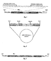
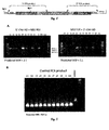


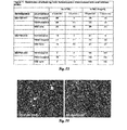
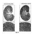
Classifications
-
- A—HUMAN NECESSITIES
- A01—AGRICULTURE; FORESTRY; ANIMAL HUSBANDRY; HUNTING; TRAPPING; FISHING
- A01K—ANIMAL HUSBANDRY; AVICULTURE; APICULTURE; PISCICULTURE; FISHING; REARING OR BREEDING ANIMALS, NOT OTHERWISE PROVIDED FOR; NEW BREEDS OF ANIMALS
- A01K67/00—Rearing or breeding animals, not otherwise provided for; New or modified breeds of animals
- A01K67/027—New or modified breeds of vertebrates
- A01K67/0275—Genetically modified vertebrates, e.g. transgenic
- A01K67/0276—Knock-out vertebrates
-
- A—HUMAN NECESSITIES
- A01—AGRICULTURE; FORESTRY; ANIMAL HUSBANDRY; HUNTING; TRAPPING; FISHING
- A01K—ANIMAL HUSBANDRY; AVICULTURE; APICULTURE; PISCICULTURE; FISHING; REARING OR BREEDING ANIMALS, NOT OTHERWISE PROVIDED FOR; NEW BREEDS OF ANIMALS
- A01K67/00—Rearing or breeding animals, not otherwise provided for; New or modified breeds of animals
- A01K67/027—New or modified breeds of vertebrates
- A01K67/0275—Genetically modified vertebrates, e.g. transgenic
- A01K67/0278—Knock-in vertebrates, e.g. humanised vertebrates
-
- C—CHEMISTRY; METALLURGY
- C07—ORGANIC CHEMISTRY
- C07K—PEPTIDES
- C07K14/00—Peptides having more than 20 amino acids; Gastrins; Somatostatins; Melanotropins; Derivatives thereof
- C07K14/435—Peptides having more than 20 amino acids; Gastrins; Somatostatins; Melanotropins; Derivatives thereof from animals; from humans
- C07K14/705—Receptors; Cell surface antigens; Cell surface determinants
- C07K14/715—Receptors; Cell surface antigens; Cell surface determinants for cytokines; for lymphokines; for interferons
- C07K14/7155—Receptors; Cell surface antigens; Cell surface determinants for cytokines; for lymphokines; for interferons for interleukins [IL]
-
- C—CHEMISTRY; METALLURGY
- C12—BIOCHEMISTRY; BEER; SPIRITS; WINE; VINEGAR; MICROBIOLOGY; ENZYMOLOGY; MUTATION OR GENETIC ENGINEERING
- C12N—MICROORGANISMS OR ENZYMES; COMPOSITIONS THEREOF; PROPAGATING, PRESERVING, OR MAINTAINING MICROORGANISMS; MUTATION OR GENETIC ENGINEERING; CULTURE MEDIA
- C12N9/00—Enzymes; Proenzymes; Compositions thereof; Processes for preparing, activating, inhibiting, separating or purifying enzymes
- C12N9/14—Hydrolases (3)
- C12N9/48—Hydrolases (3) acting on peptide bonds (3.4)
- C12N9/50—Proteinases, e.g. Endopeptidases (3.4.21-3.4.25)
- C12N9/64—Proteinases, e.g. Endopeptidases (3.4.21-3.4.25) derived from animal tissue
- C12N9/6421—Proteinases, e.g. Endopeptidases (3.4.21-3.4.25) derived from animal tissue from mammals
- C12N9/6424—Serine endopeptidases (3.4.21)
- C12N9/6456—Plasminogen activators
- C12N9/6462—Plasminogen activators u-Plasminogen activator (3.4.21.73), i.e. urokinase
-
- C—CHEMISTRY; METALLURGY
- C12—BIOCHEMISTRY; BEER; SPIRITS; WINE; VINEGAR; MICROBIOLOGY; ENZYMOLOGY; MUTATION OR GENETIC ENGINEERING
- C12Y—ENZYMES
- C12Y304/00—Hydrolases acting on peptide bonds, i.e. peptidases (3.4)
- C12Y304/21—Serine endopeptidases (3.4.21)
- C12Y304/21073—Serine endopeptidases (3.4.21) u-Plasminogen activator (3.4.21.73), i.e. urokinase
-
- A—HUMAN NECESSITIES
- A01—AGRICULTURE; FORESTRY; ANIMAL HUSBANDRY; HUNTING; TRAPPING; FISHING
- A01K—ANIMAL HUSBANDRY; AVICULTURE; APICULTURE; PISCICULTURE; FISHING; REARING OR BREEDING ANIMALS, NOT OTHERWISE PROVIDED FOR; NEW BREEDS OF ANIMALS
- A01K2207/00—Modified animals
- A01K2207/12—Animals modified by administration of exogenous cells
-
- A—HUMAN NECESSITIES
- A01—AGRICULTURE; FORESTRY; ANIMAL HUSBANDRY; HUNTING; TRAPPING; FISHING
- A01K—ANIMAL HUSBANDRY; AVICULTURE; APICULTURE; PISCICULTURE; FISHING; REARING OR BREEDING ANIMALS, NOT OTHERWISE PROVIDED FOR; NEW BREEDS OF ANIMALS
- A01K2207/00—Modified animals
- A01K2207/15—Humanized animals
-
- A—HUMAN NECESSITIES
- A01—AGRICULTURE; FORESTRY; ANIMAL HUSBANDRY; HUNTING; TRAPPING; FISHING
- A01K—ANIMAL HUSBANDRY; AVICULTURE; APICULTURE; PISCICULTURE; FISHING; REARING OR BREEDING ANIMALS, NOT OTHERWISE PROVIDED FOR; NEW BREEDS OF ANIMALS
- A01K2217/00—Genetically modified animals
-
- A—HUMAN NECESSITIES
- A01—AGRICULTURE; FORESTRY; ANIMAL HUSBANDRY; HUNTING; TRAPPING; FISHING
- A01K—ANIMAL HUSBANDRY; AVICULTURE; APICULTURE; PISCICULTURE; FISHING; REARING OR BREEDING ANIMALS, NOT OTHERWISE PROVIDED FOR; NEW BREEDS OF ANIMALS
- A01K2217/00—Genetically modified animals
- A01K2217/07—Animals genetically altered by homologous recombination
- A01K2217/075—Animals genetically altered by homologous recombination inducing loss of function, i.e. knock out
-
- A—HUMAN NECESSITIES
- A01—AGRICULTURE; FORESTRY; ANIMAL HUSBANDRY; HUNTING; TRAPPING; FISHING
- A01K—ANIMAL HUSBANDRY; AVICULTURE; APICULTURE; PISCICULTURE; FISHING; REARING OR BREEDING ANIMALS, NOT OTHERWISE PROVIDED FOR; NEW BREEDS OF ANIMALS
- A01K2227/00—Animals characterised by species
- A01K2227/10—Mammal
- A01K2227/105—Murine
-
- A—HUMAN NECESSITIES
- A01—AGRICULTURE; FORESTRY; ANIMAL HUSBANDRY; HUNTING; TRAPPING; FISHING
- A01K—ANIMAL HUSBANDRY; AVICULTURE; APICULTURE; PISCICULTURE; FISHING; REARING OR BREEDING ANIMALS, NOT OTHERWISE PROVIDED FOR; NEW BREEDS OF ANIMALS
- A01K2267/00—Animals characterised by purpose
- A01K2267/03—Animal model, e.g. for test or diseases
- A01K2267/035—Animal model for multifactorial diseases
- A01K2267/0387—Animal model for diseases of the immune system
-
- C—CHEMISTRY; METALLURGY
- C12—BIOCHEMISTRY; BEER; SPIRITS; WINE; VINEGAR; MICROBIOLOGY; ENZYMOLOGY; MUTATION OR GENETIC ENGINEERING
- C12N—MICROORGANISMS OR ENZYMES; COMPOSITIONS THEREOF; PROPAGATING, PRESERVING, OR MAINTAINING MICROORGANISMS; MUTATION OR GENETIC ENGINEERING; CULTURE MEDIA
- C12N2750/00—MICROORGANISMS OR ENZYMES; COMPOSITIONS THEREOF; PROPAGATING, PRESERVING, OR MAINTAINING MICROORGANISMS; MUTATION OR GENETIC ENGINEERING; CULTURE MEDIA ssDNA viruses
- C12N2750/00011—Details
- C12N2750/14011—Parvoviridae
- C12N2750/14111—Dependovirus, e.g. adenoassociated viruses
- C12N2750/14141—Use of virus, viral particle or viral elements as a vector
- C12N2750/14143—Use of virus, viral particle or viral elements as a vector viral genome or elements thereof as genetic vector
-
- C—CHEMISTRY; METALLURGY
- C12—BIOCHEMISTRY; BEER; SPIRITS; WINE; VINEGAR; MICROBIOLOGY; ENZYMOLOGY; MUTATION OR GENETIC ENGINEERING
- C12Y—ENZYMES
- C12Y307/00—Hydrolases acting on carbon-carbon bonds (3.7)
- C12Y307/01—Hydrolases acting on carbon-carbon bonds (3.7) in ketonic substances (3.7.1)
- C12Y307/01002—Fumarylacetoacetase (3.7.1.2)
Definitions
- SEQ ID NO:1 is the sequence of a portion of the rat FAH protein
- SEQ ID NO:2 is the nucleotide sequence of the rat Fah gene targeting cassette
- SEQ ID NOs:3-20 are nucleotide sequences of primers
- SEQ ID NO:21 is the nucleotide sequence of a portion of the wild type Fah rat gene
- SEQ ID NO:22 is the amino acid sequence of the wild type FAH protein
- SEQ ID NOs:23-25 are amino acid sequences of mutants m1-m3
- SEQ ID NO:26 is the amino acid sequence of a portion of the wild type Il2rg gene
- SEQ ID NO:27 is the amino acid sequence of a portion of the wild type Rag2 gene
- SEQ ID NO:29 is the amino acid sequence of anakinra.
- the present disclosure concerns rat models having a hepatic deficiency and/or an immunodeficiency and their use.
- This disclosure also relates to methods of using such animal models, including expanding heterologous hepatocytes, such as human hepatocytes, in such animal models.
- the liver is the principal site for the metabolism of xenobiotic compounds including medical drugs. Because many hepatic enzymes are species-specific, it is necessary to evaluate the metabolism of candidate pharmaceuticals using cultured primary human hepatocytes or their microsomal fraction. While microsomal hepatocyte fractions can be used to elucidate some metabolic functions, other tests depend on living hepatocytes. Some compounds, for example, induce hepatic enzymes and thus their metabolism changes with time. To analyze enzyme induction, hepatocytes must be not only viable, but fully differentiated and functional.
- Human hepatocytes are widely used by the pharmaceutical industry during preclinical drug development. Their use is mandated by the FDA as part of drug development. For drug metabolism and other studies, hepatocytes are typically isolated from cadaveric organ donors and shipped to the location where testing will be performed. The condition (viability and state of differentiation) of hepatocytes from cadaveric sources is highly variable and many cell preparations are of marginal quality. The availability of high quality human hepatocytes is further hampered by the fact that they cannot be significantly expanded in tissue culture. After plating, the cells survive but do not divide. Hepatocytes from readily available mammalian species, such as the mouse, are not suitable for drug testing because they have a different complement of metabolic enzymes and respond differently in induction studies.
- Immortal human liver cells hepatomas
- fetal hepatoblasts are also not an adequate replacement for fully differentiated adult cells.
- Human hepatocytes are also necessary for studies in the field of microbiology.
- Many human viruses, such as viruses that cause hepatitis, cannot replicate in any other cell type.
- bioartificial liver assist devices which use hepatocytes ex vivo, have been used to support patients in acute liver failure.
- several clinical trials of hepatocyte transplantation have been carried out, which provided proof-of-principle that hepatocyte transplantation can be beneficial.
- human hepatocytes cannot be expanded significantly in culture. Hepatocytes derived from stem cells in culture are immature and generally lack full functionality. Therefore, all hepatocytes in use today are derived from human donors, either cadaveric or surgical specimens, which significantly limits hepatocyte availability. If enough human hepatocytes were available, bioartificial liver assist devices would become a viable technology and human hepatocyte transplantation could find wide-spread use. Given these limitations, methods of expanding primary human hepatocytes are highly desirable. There is also a need in the animal health industry for processes to expand hepatocytes from other species, such as dogs, horses, etc. for research and study.
- Described herein are rats that have utility for a variety of purposes, including for the expansion of hepatocytes from other species (particularly humans), and as animal models of liver diseases, including cirrhosis, hepatocellular carcinoma and hepatic infection.
- the rats have dysfunctional livers ("hepatic deficiency"), induced liver damage, and/or an immunodeficiency, as described in more detail herein.
- the method includes transplanting heterologous hepatocytes (or hepatocyte progenitors) into an Fah-deficient rat and allowing the heterologous hepatocytes to expand.
- the Fah-deficient rat is immunosuppressed or immunodeficient.
- the Fah-deficient rat is administered a vector encoding urokinase prior to transplantation of the heterologous hepatocytes.
- heterologous hepatocytes are also provided.
- methods of expanding heterologous hepatocytes in vivo by transplanting heterologous hepatocytes (or hepatocyte progenitors) into an immunodeficient rat and allowing the heterologous hepatocytes to expand.
- the heterologous hepatocytes are allowed to expand for at least about 2, 4, 5, 6, 7, 8, weeks, or several months.
- Isolated hepatocytes (such as human hepatocytes) expanded in and collected from Fah-deficient and/or immunodeficient rats are also provided by the present disclosure.
- Fah-deficient rats whose genomes are homozygous for a disruption in the Fah gene such that the disruption results in loss of expression of functional FAH protein, wherein the rats exhibit decreased liver function.
- the Fah-deficient rats further include transplanted heterologous hepatocytes.
- the Fah-deficient rat is immunosuppressed.
- isolated rat embryonic stem cells comprising a disruption in the Fah gene.
- AAV-DJ vector An adeno-associated virus (AAV) packaging helper that expresses a hybrid capsid containing AAV2, AAV5 and AAV8 capsid proteins and the AAV2 rep protein.
- AAV adeno-associated virus
- exemplary routes of administration include, but are not limited to, injection (such as subcutaneous, intramuscular, intradermal, intraperitoneal, and intravenous), oral, intraductal, sublingual, rectal, transdermal, intranasal, vaginal and inhalation routes.
- injection such as subcutaneous, intramuscular, intradermal, intraperitoneal, and intravenous
- oral intraductal, sublingual, rectal, transdermal, intranasal, vaginal and inhalation routes.
- liver disease A compound or composition that when administered to an Fah-deficient rat, prevents, avoids, delays or inhibits the development of liver disease in the animal.
- Liver disease or liver dysfunction is characterized by any one of a number of signs or symptoms, including, but not limited to an alteration in liver histology (such as necrosis, inflammation, fibrosis, dysplasia or hepatic cancer), an alteration in levels of liver-specific enzymes and other proteins (such as aspartate aminotransferase, alanine aminotransferase, bilirubin, alkaline phosphatase and albumin), plasma or urinary succinylacetone (SA), or generalized liver failure.
- liver histology such as necrosis, inflammation, fibrosis, dysplasia or hepatic cancer
- SA urinary succinylacetone
- the agent that inhibits liver disease is a pharmacologic inhibitor of 4-OH-phenylpyruvate dioxygenase, such as 2-(2-nitro-4-trifluoro-methyl-benzoyl)-1,3 cyclohexanedione (NTBC) or methyl-NTBC.
- NTBC 2-(2-nitro-4-trifluoro-methyl-benzoyl)-1,3 cyclohexanedione
- methyl-NTBC methyl-NTBC.
- the agent that inhibits liver disease is NTBC.
- Anakinra An interleukin-1 (IL-1) receptor antagonist. Anakinra blocks the biologic activity of naturally occurring IL-1 by competitively inhibiting the binding of IL-1 to the IL-1 receptor, which is expressed in many tissues and organs. IL-1 is produced in response to inflammatory stimuli and mediates various physiologic responses, including inflammatory and immunologic reactions. Anakinra is a recombinant, non-glycosylated version of human 1L-1RA (IL-1 receptor antagonist) prepared from cultures of genetically modified Escherichia coli.
- IL-1 receptor antagonist a recombinant, non-glycosylated version of human 1L-1RA (IL-1 receptor antagonist) prepared from cultures of genetically modified Escherichia coli.
- the anakinra protein is 153 amino acids and has a molecular weight of approximately 17.3 kD and differs from native human 1L-1RA in that it has a single methionine residue on its amino terminus (the amino acid sequence of anakinra is set forth herein as SEQ ID NO:29). Anakinra is also known as KINERETTM.
- Azathioprine An immunosuppressant that is a purine synthesis inhibitor, inhibiting the proliferation of cells, especially leukocytes. This immunosuppressant is often used in the treatment of autoimmune diseases or organ transplant rejection. It is a pro-drug, converted in the body to the active metabolites 6-mercaptopurine (6-MP) and 6-thioinosinic acid. Azathioprine is produced by a number of generic manufacturers and as branded names (AzasanTM by Salix; ImuranTM by GlaxoSmithKline; AzamunTM; and ImurelTM).
- Biological sample A sample obtained from cells, tissue or bodily fluid of a subject, such as peripheral blood, serum, plasma, cerebrospinal fluid, bone marrow, urine, saliva, tissue biopsy, surgical specimen, and autopsy material. Also referred to herein as a "sample.”
- Cirrhosis refers to a group of chronic liver diseases characterized by loss of the normal microscopic lobular architecture and regenerative replacement of necrotic parenchymal tissue with fibrous bands of connective tissue that eventually constrict and partition the organ into irregular nodules. Cirrhosis has a lengthy latent period, usually followed by sudden abdominal pain and swelling with hematemesis, dependent edema, or jaundice. In advanced stages there may be ascites, pronounced jaundice, portal hypertension, varicose veins and central nervous system disorders that may end in hepatic coma.
- Collecting refers to the process of removing the expanded hepatocytes from a rat that has been injected or transplanted with isolated heterologous hepatocytes (also referred to as a recipient rat). Collecting optionally includes separating the hepatocytes from other cell types. In one embodiment, the expanded heterologous hepatocytes are collected from the liver of a rat.
- Common- ⁇ chain of the interleukin receptor ( Il2rg ): A gene encoding the common gamma chain of intcrlcukin receptors. Il2rg is a component of the receptors lor a number of interleukins, including 1L-2, 1L-4, IL-7 and IL-15 ( Di Santo et al. Proc. Natl. Acad. Sci. U.S.A. 92:377-381, 1995 ). Animals deficient in Il2rg exhibit a reduction in B cells and T cells and lack natural killer cells. Also known as interleukin-2 receptor gamma chain.
- Cryopreserved refers to a cell (such as a hepatocyte) or tissue that has been preserved or maintained by cooling to low sub-zero temperatures, such as 77 K or -196 °C (the boiling point of liquid nitrogen). At these low temperatures, any biological activity, including the biochemical reactions that would lead to cell death, is effectively stopped.
- Cyclosporin A An immunosuppressant compound that is a non-ribosomal cyclic peptide of 11 amino acids produced by the soil fungus Beauveria nivea. Cyclosporin A is used for the prophylaxis of graft rejection in organ and tissue transplantation. Cyclosporin A is also known as cyclosporine and ciclosporin.
- Decreased liver function An abnormal change in any one of a number of parameters that measure the health or function of the liver. Decreased liver function is also referred to herein as "liver dysfunction.” Liver function can be evaluated by any one of a number of means well known in the art, such as, but not limited to, examination of liver histology and measurement of liver enzymes or other proteins. For example, liver dysfunction can be indicated by necrosis, inflammation, fibrosis, oxidative damage or dysplasia of the liver. In some instances, liver dysfunction is indicated by hepatic cancer, such as hepatocellular carcinoma.
- liver enzymes and proteins that can be tested to evaluate liver dysfunction include, but are not limited to, alanine aminotransferase (ALT), aspartate aminotransferase (AST), bilirubin, alkaline phosphatase and albumin. Liver dysfunction also can result in generalized liver failure. Procedures for testing liver function are well known in the art, such as those taught by Grompe et al. (Genes Dev. 7:2298-2307, 1993 ) and Manning et al. (Proc. Natl. Acad. Sci. U.S.A. 96:11928-11933, 1999 ).
- Fah-deficient or “deficient in Fah” refers to an animal, such as a rat, having a substantial decrease in, or the absence of, FAH enzyme production or activity, for example an animal having a disruption in the Fah gene (such as an insertion, deletion or one or more point mutations), which results in a substantial decrease in, or the absence of, Fah mRNA expression and/or functional FAH enzyme activity.
- the term “loss of expression” of functional FAH protein does not refer to only a complete loss of expression, but also includes a substantial decrease in expression of functional FAH protein, such as a decrease of about 80%, about 90%, about 95% or about 99%.
- the Fah-deficient rat comprises heterozygous or homozygous insertions in the Fah gene (such as an insertion that includes an in-frame stop codon), with homozygous insertions being particularly preferred.
- the insertion is in exon 3 of Fah.
- the Fah-deficient rat comprises heterozygous or homozygous deletions in the Fah gene, with homozygous deletions being particularly preferred.
- the deletion is in exon 3 of Fah.
- the Fah-deficient rat comprises one or more point mutations in the Fah gene. Examples of suitable Fah point mutations are known in the art (see, e.g., Aponte et al. Proc. Natl. Acad. Sci. U.S.A. 98(2):641-645, 2001 ).
- macrophage depletion refers to the process of eliminating, removing, reducing or killing macrophages in an animal.
- An animal that has been depleted of macrophages is not necessarily completely devoid of macrophages but at least exhibits a reduction in the number or activity of macrophages.
- macrophage depletion results in at least a 10%, at least a 25%, at least a 50%, at least a 75%, at least a 90% or a 100% reduction in functional macrophages.
- Disruption in a gene refers to any insertion, deletion or point mutation, or any combination thereof. In some embodiments, the disruption leads to a partial or complete loss of expression of mRNA and/or functional protein.
- Engraft To implant cells or tissues in an animal.
- engraftment of heterologous hepatocytes in a recipient rat refers to the process of heterologous hepatocytes becoming implanted in the recipient rat following injection.
- Engrafted heterologous hepatocytes are capable of expansion in the recipient rat.
- Expand To increase in quantity.
- "expanding" heterologous hepatocytes refers to the process of allowing cell division to occur such that the hepatocytes actively proliferate in vivo and the number of heterologous hepatocytes increases as compared to the original number of heterologous hepatocytes transplanted into the recipient rat.
- Fetus The unborn offspring of an animal in the postembryonic period.
- FK506 also known as tacrolimus or fujimycin, is an immunosuppressant drug.
- FK506 a 23-membered macrolide lactone first discovered in the fermentation broth of a Japanese soil sample that contained the bacteria Streptomyces tsukubaensis. This compound is often used after allogeneic organ transplant to reduce the activity of the patient's immune system and lower the risk of organ rejection.
- FK506 reduces T-cell and interleukin-2 activity. It is also used in a topical preparation in the treatment of severe atopic dermatitis (eczema), severe refractory uveitis after bone marrow transplants, and the skin condition vitiligo.
- Fludarabine A purine analog that inhibits DNA synthesis. Fludarabine is often used as a chemotherapeutic drug for the treatment of various hematologic malignancies.
- FRG KO mouse A mutant mouse having homozygous deletions in the fumarylacetoacetate hydrolase ( Fah ), recombination activating gene 2 ( Rag2 ) and hemizygous or homozygous deletions of common-y chain of the interleukin receptor ( Il2rg ) x-linked gene. Also referred to as Fah - / - / Rag2 - / - / Il2rg - /- or Fah - / - / Rag2 - / - / Il2rg - / y .
- homozygous deletions in the Fah, Rag2 and Il2rg genes indicates no functional FAH, RAG-2 and IL-2R ⁇ protein is expressed in mice comprising the mutations.
- Fumarylacetoacetate hydrolase A metabolic enzyme that catalyzes the last step of tyrosine catabolism.
- Mice having a homozygous deletion of the Fah gene exhibit altered liver mRNA expression and severe liver dysfunction ( Grompe et al. Genes Dev. 7:2298-2307, 1993 ).
- Point mutations in the Fah gene have also been shown to cause hepatic failure and postnatal lethality ( Aponte et al. Proc. Natl. Acad. Sci. U.S.A. 98(2):641-645, 2001 ).
- Humans deficient for Fah develop the liver disease hereditary tyrosinemia type 1 (HT1) and develop liver failure.
- HT1 hereditary tyrosinemia type 1
- Fah deficiency leads to accumulation of fumarylacetoacetate, a potent oxidizing agent and this ultimately leads to cell death of hepatocytes deficient for Fah.
- Fah-deficient rats can be repopulated with hepatocytes from other species, including humans.
- Fah genomic, mRNA and protein sequences for a number of different species arc publically available, such as in the GenBank database (sec, for example, Gene ID 29383 (rat Fah); Gene ID 14085 (mouse Fah); Gene ID 610140 (dog FAH); Gene ID 415482 (chicken FAH); Gene ID 100049804 (horse FAH); Gene ID 712716 (rhesus macaque FAH); Gene ID 100408895 (marmoset FAH); Gene ID 100589446 (gibbon FAH); Gene ID 467738 (chimpanzee FAH); and Gene ID 508721 (cow FAH)).
- GenBank database sec, for example, Gene ID 29383 (rat Fah); Gene ID 14085 (mouse Fah); Gene ID 610140 (dog FAH); Gene ID 415482 (chicken FAH); Gene ID 100049804 (horse FAH); Gene ID 712716 (rhesus macaque FAH); Gene ID 100408895 (marmos
- Hepatic pathogen refers to any pathogen, such as a bacterial, viral or parasitic pathogen, that infects cells of the liver.
- the hepatic pathogen is a "hepatotropic virus” (a virus that targets the liver), such as HBV or HCV.
- Hepatocellular carcinoma is a primary malignancy of the liver typically occurring in patients with inflammatory livers resulting from viral hepatitis, liver toxins or hepatic cirrhosis.
- Hepatocyte A type of cell that makes up 70-80% of the cytoplasmic mass of the liver. Hepatocytes are involved in protein synthesis, protein storage and transformation of carbohydrates, synthesis of cholesterol, bile salts and phospholipids, and detoxification, modification and excretion of exogenous and endogenous substances. The hepatocyte also initiates the formation and secretion of bile. Hepatocytes manufacture serum albumin, fibrinogen and the prothrombin group of clotting factors and are the main site for the synthesis of lipoproteins, ceruloplasmin, transferrin, complement and glycoproteins. In addition, hepatocytes have the ability to metabolize, detoxify, and inactivate exogenous compounds such as drugs and insecticides, and endogenous compounds such as steroids.
- Hepatocyte progenitor Any cell type capable of giving rise to a hepatocyte.
- hepatocyte progenitors include, but are not limited to, embryonic stem cells (ESCs), induced pluripotent stem cells (IPSC), adult intrahepatic stem cells, mesenchymal stem cells and amniotic stem cells.
- methods of expanding heterologous hepatocytes in vivo include transplanting heterologous hepatocyte or hepatocyte progenitors.
- HT1 Hereditary tyrosinemia type 1
- Tyrosinemia is an error of metabolism, usually inborn, in which the body cannot effectively break down the amino acid tyrosine.
- HT1 is the most severe form of this disorder and is caused by a shortage of the enzyme fumarylacetoacetate hydrolase (FAH) encoded by the gene Fah found on human chromosome number 15.
- FAH fumarylacetoacetate hydrolase
- FAH fumarylacetoacetate hydrolase
- Symptoms of HT1 usually appear in the first few months of life and include failure to gain weight and grow at the expected rate (failure to thrive), diarrhea, vomiting, yellowing of the skin and whites of the eyes (jaundice), cabbage-like odor, and increased tendency to bleed (particularly nosebleeds).
- FAH fumarylacetoacetate hydrolase
- Heterologous Derived from a source other than (i.e., foreign to) the referenced species in contrast to material derived from, naturally associated with, or native to, that species.
- the rats can be transplanted and engrafted with hepatocytes from species other than the recipient rat, such as humans, dogs, pigs, etc.
- Heterozygous Having dissimilar alleles at corresponding chromosomal loci.
- a rat heterozygous for a particular gene mutation has the mutation in one allele of the gene but not the other.
- homozygous Generally means having identical alleles at one or more loci.
- "homozygous” for disruptions or “homozygous” for a deficiency refers to an organism having disruptions (such as a deletion, insertion or point mutation) of both alleles of a gene, as well as “compound heterozygosity" in which the organism may have disruptions on two unrelated alleles, which nevertheless behave like a homozygous disruption, resulting in loss of functional gene products.
- references to "humanized" animals (rats) or livers refers to animals or livers transplanted with human hepatocytes in which the human hepatocytes have expanded in vivo to substantially repopulate the liver of the animal with human hepatocytes.
- an immunodeficient rat Lacking in at least one essential function of the immune system.
- an immunodeficient rat is one lacking specific components of the immune system or lacking function of specific components of the immune system (such as, for example, B cells, T cells or NK cells).
- an immunodeficient rat lacks macrophages.
- an immunodeficient rat comprises one or more genetic alterations that prevent or inhibit the development of functional immune cells (such as B cells, T cells or NK cells).
- the genetic alteration is selected from the group consisting of recombination activating gene 1 ( Rag1 ) deficiency, recombination activating gene 2 ( Rag2 ) deficiency, interleukin-2 receptor gamma chain ( Il2rg ) deficiency, the Dnapk -/- (SCID) mutation, the humanized/humanized SIRP-alpha genotype, the nude rat mutation, perforin knockouts, and combinations thereof.
- recombination activating gene 1 ( Rag1 ) deficiency
- recombination activating gene 2 ( Rag2 ) deficiency
- interleukin-2 receptor gamma chain ( Il2rg ) deficiency the Dnapk -/- (SCID) mutation
- the humanized/humanized SIRP-alpha genotype the nude rat mutation
- perforin knockouts and combinations thereof.
- an "immunodeficient rat” comprises one or more of the following genetic alterations: Rag1 - / - , Rag2 - / - , Il2rg - / - , Il2rg - / y , SCID, SIRP-alpha genotype, perforin -/- , and/or nude.
- Immunodeficient animal strains are well known in the art and are commercially available, such as from The Jackson Laboratory (Bar Harbor, ME) or Taconic (Hudson, NY).
- the rat is a Fah - / - / Rag2 - / - / Il2rg - / - / Il1r1 - / - rat, a Fah - / - / Rag1 - / - / Il2rg - / - / Il1r1 - / - rat, a Fah - / - / Rag2 - / - / Il2rg - / - / Il1r1 - - rat, or a Fah - / - / Rag1 - / - / Il2rg - / - / Il1r1 - / - rat.
- an immunodeficient rat is an animal that has been administered one or more immunosuppressants.
- Immunosuppressant Any compound that decreases the function or activity of one or more aspects of the immune system, such as a component of the humoral or cellular immune system or the complement system. Immunosuppressants are also referred to as "immunosuppressive agents.” Exemplary immunosuppressants include, but arc not limited to: (1) antimetabolites, such as purine synthesis inhibitors (e.g., azathioprine and mycophenolic acid), pyrimidine synthesis inhibitors (e.g., leflunomide and teriflunomide) and antifolates ( e.g., methotrexate); (2) macrolides, such as FK506, cyclosporine A and pimecrolimus; (3) TNF- ⁇ inhibitors, such as thalidomide and lenalidomide; (4) IL-1 receptor antagonists, such as anakinra; (5) mammalian target of rapamycin (mTOR) inhibitors, such as rapamycin (siroli
- Exemplary cellular targets and their respective inhibitor compounds include, but are not limited to complement component 5 (e.g., eculizumab); tumor necrosis factors (TNFs) (e.g., infliximab, adalimumab, certolizumab pegol, afelimomab and golimumab); IL-5 (e.g., mepolizumab); IgE ( e.g., omalizumab); BAYX ( e.g.
- complement component 5 e.g., eculizumab
- TNFs tumor necrosis factors
- IgE e.g., omalizumab
- BAYX e.g.
- nerelimomab interferon (e.g., faralimomab); 1L-6 (e.g., elsilimomab); IL-12 and IL-13 ( e.g., lebrikizumab and ustekinumab); CD3 ( e.g., muromonab-CD3, otelixizumab, teplizumab, visilizumab); CD4 ( e.g., clcnoliximab, keliximab and zanolimumab); CD11a ( e.g., efalizumab); CD18 ( e.g., erlizumab); CD20 ( e.g., afutuzumab, ocrelizumah, pascolizumab); CD23 (e.g., lumiliximab); CD40 ( e.g., teneliximab, toralizumab);
- ipilimumab, trcmclimumab CAT ( e.g., bertilimumab, lerdelimumab, metelimumab); integrin ( e.g., natalizumab); IL-6 receptor ( e.g ., Tocilizumab); LFA-1 ( e.g., odulimomab); and IL-2 receptor/CD25 ( e.g ., basiliximab, daclizumab, inolimomab).
- CAT e.g., bertilimumab, lerdelimumab, metelimumab
- integrin e.g., natalizumab
- IL-6 receptor e.g ., Tocilizumab
- LFA-1 e.g., odulimomab
- IL-2 receptor/CD25 e.g ., basiliximab, daclizumab
- immunosuppressive agents include zolimomab aritox, atorolimumab, cedelizumab, dorlixizumab, fontolizumab, gantenerumab, gomiliximab, maslimomab, morolimumab, pexelizumab, reslizumab, rovelizumab, siplizumab, talizumab, telimomab aritox, vapaliximab, vepalimomab, anti-thymocyte globulin, anti-lymphocyte globulin; CTLA-4 inhibitors ( e.g., abatacept, belatacept); aflibercept; alefacept; rilonacept; and TNF inhibitors (e.g., etanercept).
- CTLA-4 inhibitors e.g., abatacept, belatacept
- aflibercept alefa
- Immunosuppression refers to the act of reducing the activity or function of the immune system. Immunosuppression can be achieved by administration of an immunosuppressant compound or can be the effect of a disease or disorder (for example, immunosuppression that results from HIV infection or due to a genetic defect). In some cases, immunosuppression occurs as the result of a genetic mutation that prevents or inhibits the development of functional immune cells.
- iPSC Induced pluripotency stem cells
- iPSCs A type of pluripotent stem cell artificially derived from a non-pluripotent cell, typically an adult somatic cell, by inducing expression of certain genes.
- iPSCs can be derived from any organism, such as a mammal. In some embodiments, iPSCs are produced from mice, rats, rabbits, guinea pigs, goats, pigs, cows, non-human primates or humans. Human derived iPSCs are exemplary.
- iPSCs are similar to ES cells in many respects, such as the expression of certain stem cell genes and proteins, chromatin methylation patterns, doubling time, embryoid body formation, teratoma formation, viable chimera formation, and potency and differentiability.
- Methods for producing iPSCs are known in the art.
- iPSCs are typically derived by transfection of certain stem cell-associated genes (such as Oct-3/4 (Pouf51) and Sox2) into non-pluripotent cells, such as adult fibroblasts. Transfection can be achieved through viral vectors, such as retroviruses, lentiviruses, or adenoviruses.
- cells can be transfected with Oct3/4, Sox2, Klf4, and c-Myc using a retroviral system or with OCT4, SOX2, NANOG, and LIN28 using a lentiviral system.
- small numbers of transfected cells begin to become morphologically and biochemically similar to pluripotent stem cells, and are typically isolated through morphological selection, doubling time, or through a reporter gene and antibiotic selection.
- iPSCs from adult human cells arc generated by the method of Yu et al. (Science 318(5854):1224, 2007 ) or Takahashi et al. (Cell 131(5):861-72, 2007 ).
- iPSCs arc also known as iPS cells.
- Infectious load refers to the quantity of a particular pathogen in a subject or in a sample from the subject. Infectious load can be measured using any one of a number of methods known in the art. The selected method will vary depending on the type of pathogen to be detected and the reagents available to detect the pathogen. Infectious load can also be measured, for example, by determining the titer of the pathogen, the method for which will vary depending on the pathogen to be detected. For example, the titer of some viruses can be quantified by performing a plaque assay. In some examples, infectious load is measured by quantifying the amount of a pathogen-specific antigen in a sample. In other examples, infectious load is measured by quantifying the amount of a pathogen-specific nucleic acid molecule in a sample. Quantifying encompasses determining a numerical value or can be a relative value.
- Isolated An "isolated" biological component, such as a nucleic acid, protein (including antibodies) or organelle, has been substantially separated or extracted away from other biological components in the environment (such as a cell) in which the component naturally occurs, i.e., other chromosomal and extra-chromosomal nucleic acids, adjacent sequences, proteins and organelles.
- Nucleic acids and proteins that have been “isolated” include nucleic acids and proteins purified by standard purification methods. The term also embraces nucleic acids and proteins prepared by recombinant expression in a host cell as well as chemically synthesized nucleic acids. In either case, "isolated" nucleic acids and/or proteins result in new chemical entities not found in nature.
- Mammalian target of rapamycin (mTOR) inhibitor A molecule that inhibits expression or activity of mTOR.
- mTOR inhibitors include, but are not limited to small molecule, antibody, peptide and nucleic acid inhibitors.
- an mTOR inhibitor can be a molecule that inhibits the kinase activity of mTOR or inhibits binding of mTOR to a ligand.
- Inhibitors of mTOR also include molecules that down-regulate expression of mTOR.
- a number of mTOR inhibitors are known in the art, including rapamycin (sirolimus).
- Mycophenolate An immunosuppressant typically used to prevent rejection of allogeneic transplants. This drug is generally administered orally or intravenously.
- Mycophenolate is derived from the fungus Penicillium stoloniferum.
- Mycophenolate mofetil the pro-drug form, is metabolized in the liver to the active moiety mycophenolic acid. It inhibits inosine monophosphate dehydrogenase, the enzyme that controls the rate of synthesis of guanine monophosphate in the de novo pathway of purine synthesis used in the proliferation of B and T lymphocytes.
- Mycophenolic acid is commonly marketed under the trade names CellCeptTM (mycophenolate mofetil; Roche) and MyforticTM (mycophenolate sodium; Novartis).
- NTBC (2-nitro-4-trifluoro-methyl-benzoyl)-1,3 cyclohexanedione, also known as nitisinone): An inhibitor of 4-hydroxy-phenylpyruvate dioxygenase (HPPD). HPPD catalyzes the conversion of 4-hydroxyphenylpyruvate to homogentisate, the second step in tyrosine catabolism. Treatment with NTBC blocks the tyrosine catabolism pathway at this step and prevents the accumulation of succinylacetone, a pathognomonic metabolite that accumulates in Fah-deficient humans and animals.
- HPPD 4-hydroxy-phenylpyruvate dioxygenase
- Nude rat Refers to a rat strain with a genetic mutation that causes a deteriorated or absent thymus, resulting in an inhibited immune system due to a greatly reduced number of T cells.
- Prednisone A synthetic corticosteroid that is an effective immunosuppressant. It is often used to treat certain inflammatory diseases, autoimmune diseases and cancers as well as treat or prevent organ transplant rejection. Prednisone is usually taken orally but can be delivered by intramuscular injection or intravenous injection. It is a prodrug that is converted by the liver into prednisolone, which is the active drug and also a steroid.
- Rapamycin A compound with known immunosuppressive and anti-proliferative properties. Rapamycin, also known as sirolimus, is a macrolide that was first discovered as a product of the bacterium Streptomyces hygroscopicus. Rapamycin binds and inhibits the activity of mTOR.
- a "recipient rat” is a rat that has been injected with heterologous hepatocytes as described herein. Typically, a portion (the percentage can vary) of the heterologous hepatocytes engraft in the recipient rat.
- the recipient rat is immunosuppressed or immunodeficient. In some embodiments, the recipient rat is Fah-deficient.
- Recombination activating gene 1 ( Rag1 ): A gene involved in activation of immunoglobulin V(D)J recombination.
- the RAG1 protein is involved in recognition of the DNA substrate, but stable binding and cleavage activity also requires RAG2.
- Rag-1-deficient rats have no mature B and T lymphocytes.
- Recombination activating gene 2 ( Rag2 ): A gene involved in recombination of immunoglobulin and T cell receptor loci. Rats deficient in the Rag2 gene are unable to undergo V(D)J recombination, resulting in a complete loss of functional T cells and B cells ( Shinkai et al. Cell 68:855-867, 1992 ).
- Serial transplantation The process for expanding heterologous hepatocytes in vivo in which hepatocytes expanded in a first animal are collected and transplanted, such as by injection, into a secondary animal for further expansion.
- Serial transplantation can further include tertiary, quaternary or additional animals.
- Somatic cell nuclear transfer A laboratory technique for creating a clonal embryo, using an ovum with a donor nucleus.
- SCNT Somatic cell nuclear transfer
- the nucleus of a somatic cell is removed and the rest of the cell discarded.
- the nucleus of an egg cell is removed.
- the nucleus of the somatic cell is then inserted into the enucleated egg cell.
- the somatic cell nucleus is reprogrammed by the host cell.
- the egg, now containing the nucleus of a somatic cell is stimulated with a shock and will begin to divide. After many mitotic divisions in culture, this single cell forms a blastocyst with almost identical DNA to the original organism.
- Stem cell A cell having the unique capacity to produce unaltered daughter cells (self-renewal; cell division produces at least one daughter cell that is identical to the parent cell) and to give rise to specialized cell types (potency).
- Stem cells include, but are not limited to, embryonic stem (ES) cells, embryonic germ (EG) cells, germline stem (GS) cells, human mesenchymal stem cells (hMSCs), adipose tissue-derived stem cells (ADSCs), multipotent adult progenitor cells (MAPCs), multipotent adult germline stem cells (maGSCs) and unrestricted somatic stem cell (USSCs).
- ES embryonic stem
- EG embryonic germ
- GS germline stem
- ADSCs adipose tissue-derived stem cells
- APCs multipotent adult progenitor cells
- maGSCs multipotent adult germline stem cells
- USSCs unrestricted somatic stem cell
- stem cells can divide without limit. After division, the stem cell may remain as a stem cell, become a precursor cell, or proceed to terminal differentiation.
- a precursor cell is a cell that can generate a fully differentiated functional cell of at least one given cell type.
- precursor cells can divide. After division, a precursor cell can remain a precursor cell, or may proceed to terminal differentiation. In one embodiment, the stem cells give rise to hepatocytes.
- Therapeutic agent A chemical compound, small molecule, or other composition, such as an antisense compound, antibody, protease inhibitor, hormone, chemokine or cytokine, capable of inducing a desired therapeutic or prophylactic effect when properly administered to a subject.
- a “candidate agent” is a compound selected for screening to determine if it can function as a therapeutic agent for a particular disease or disorder.
- titer refers to the amount of a particular pathogen in a sample.
- Immune tolerance A state of unresponsiveness to a specific antigen or group of antigens to which a subject (such as a human or animal) is normally responsive. Immune tolerance is achieved under conditions that suppress the immune reaction and is not just the absence of an immune response. Immune tolerance can result from a number of causes including prior contact with the same antigen in fetal life or in the newborn period when the immune system is not yet mature; prior contact with the antigen in extremely high or low doses; exposure to radiation, chemotherapy drugs, or other agents that impair the immune system; heritable diseases of the immune system; and acquired diseases of the immune system.
- toxin refers to any poisonous substance, including any chemical toxin or biological toxin.
- Transgene An exogenous nucleic acid sequence introduced into a cell or the genome of an organism.
- Transplant or transplanting refers to the process of grafting an organ, tissue or cells from one subject (e.g., human or animal) to another subject, or to another region of the same subject.
- Urokinase also called urokinase-type Plasminogen Activator (uPA), urokinase is a serine protease. Urokinase was originally isolated from human urine, but is present in several physiological locations, such as the blood stream and the extracellular matrix. The primary physiological substrate is plasminogen, which is an inactive zymogen form of the serine protease plasmin. Activation of plasmin triggers a proteolytic cascade which, depending on the physiological environment, participates in thrombolysis or extracellular matrix degradation. In one embodiment of the methods provided herein, urokinase is administered to a recipient rat prior to hepatocyte injection.
- urokinase is human urokinase. In some embodiments, the human urokinase is the secreted form of urokinase. In some embodiments, the human urokinase is a modified, non-secreted form of urokinase (see U.S. Patent No. 5,980,886 ).
- a nucleic acid molecule allowing insertion of foreign nucleic acid without disrupting the ability of the vector to replicate and/or integrate in a host cell.
- a vector can include nucleic acid sequences that permit it to replicate in a host cell, such as an origin of replication.
- a vector can also include one or more selectable marker genes and other genetic elements.
- An integrating vector is capable of integrating itself into a host nucleic acid.
- An expression vector is a vector that contains the necessary regulatory sequences to allow transcription and translation of inserted gene or genes.
- compositions comprises or excludes components A, B, and/or C, the composition can contain or exclude A alone; B alone; C alone; A and B in combination; A and C in combination; B and C in combination; or A, B, and C in combination.
- Rattus sp . genetically modified rats
- Rattus sp . which have utility for a variety of purposes, including for the expansion of hepatocytes from other species (particularly humans), and as animal models of liver diseases and normal liver pathology, including intermediate metabolism, bile acid production, small molecule transporters, assessment of biologics, drug metabolism, pharmacokinetics, as well as cirrhosis, hepatocellular carcinoma, hepatic infection, hepatic toxicity, gene therapy, liver regeneration, alcoholic liver disease, fatty liver disease, metabolic liver disease, and the like.
- the term "rat,” as used herein, is specific to the genus Rattus, and expressly does not encompass mice species ( Mus sp .).
- the rats are Fah-deficient, and preferably comprise a genome that is homozygous for a disruption in the Fah gene, such that the disruption results in loss of expression of functional FAH protein resulting in decreased liver function.
- the Fah gene disruption need not result in a complete loss of expression of functional FAH protein.
- loss or decrease of expression, activity, or function, of functional FAH protein is loss of expression of about 80%, about 90%, about 95% or about 99% of functional protein.
- the Fah gene disruption can be any modification that results in a significant diminishment (reduction) or complete loss of expression of functional FAH protein.
- the disruption is an insertion, a deletion, or one or more point mutations in the Fah gene, or any combination thereof.
- the disruption can be an insertion that includes an in-frame stop codon.
- the insertion can also include additional nucleic acid sequences, such as nucleic acid encoding a selectable marker.
- the Fah-deficient rat is homozygous for disruptions in exon 3 of the Fah gene.
- isolated rat embryonic stem (ES) cells comprising a disruption in the Fah gene.
- an Fah-deficient rat cell such as an embryonic stem cell
- the targeting vector comprises a targeting cassette comprising 5' and 3' homology arms that target a portion of the rat Fah gene (such as exon 3 of the Fah gene) and which flank a nucleic acid sequence (such as nucleic acid sequence encoding an antibiotic resistance gene), such that following transfection of the cell with the targeting vector, homologous recombination occurs, replacing the targeted portion of the rat Fah gene with the targeting cassette, thereby producing an Fah-deficient rat cell.
- an immunodeficient rat is provided.
- the immunodeficiency of the mouse can be due to a genetic alteration, immunosuppression, or a combination thereof.
- the rat lacks functional T cells, B cells, and/or NK cells.
- the rat is immunosuppressed.
- immunosuppression is the result of administration of one or more immunosuppressive agents.
- Any suitable immunosuppressive agent or agents effective for achieving immunosuppression in the rat can be used.
- immunosuppressive agents include, but are not limited to, FK506, cyclosporin A, fludarabine, mycophenolate, prednisone, rapamycin and azathioprine. Combinations of immunosuppressive agents can also be administered.
- the immunodeficiency is the result of one or more genetic alterations that result in a lack of a specific component of the immune system, or a lack of functionality of a specific component of the immune system (such as a lack of functional B, T and/or NK cells).
- the one or more genetic alterations comprise a genetic alteration of the Rag2 gene or a genetic alteration of the Il2rg gene such that the genetic alteration results in loss of expression of functional RAG-2 protein or IL-2R ⁇ protein.
- the one or more genetic alterations comprise a genetic alteration of the Rag2 gene and a genetic alteration of the Il2rg gene.
- the genetic alteration comprises homozygous deletions in the Rag2 gene or the Il2rg gene. Such deletions, in combination with the Fah-deficiency result in an FRG KO rat.
- the genetic alteration is SCID, NOD, or nude. Specific cells of the immune system (such as macrophages or NK cells) can also be depleted. Methods of depleting particular cell types arc known in the art.
- the rat comprises heterologous hepatocytes, such as human hepatocytes.
- heterologous hepatocytes such as human hepatocytes.
- the heterologous hepatocytes have not just been transplanted/engrafted in the rat, but expanded (i.e., actively proliferated) in the rat.
- various embodiments of the invention are concerned with rats having humanized livers.
- the method includes transplanting heterologous hepatocytes (or hepatocyte progenitors) into an Fah-deficient rat and allowing the heterologous hepatocytes to expand.
- the method includes transplanting heterologous hepatocytes into an immunodeficient rat and allowing the heterologous hepatocytes to expand.
- the method includes transplanting heterologous hepatocytes into an Fah-deficient and immunodeficient rat and allowing the heterologous hepatocytes to expand.
- the method includes transplanting heterologous hepatocytes into a rat with a hepatic deficiency, such as decreased expression, activity, or function of an enzyme in the tyrosine catabolic pathway, and allowing the heterologous hepatocytes to expand.
- a hepatic deficiency such as decreased expression, activity, or function of an enzyme in the tyrosine catabolic pathway
- the heterologous hepatocytes are transplanted by injection into the hepatic artery, spleen, portal vein, peritoneal cavity, hepatic tissue mass, or lymphatic system of the rat.
- the heterologous hepatocytes (or hepatocyte progenitors) transplanted into the rat are isolated human hepatocytes (or hepatocyte progenitors).
- the heterologous hepatocytes are transplanted as part of a liver tissue graft.
- the isolated heterologous hepatocytes can be obtained from any one of a number of different mammals as well as different sources.
- Mammalian hepatocytes can be obtained from dogs, pigs, horses, rabbits, mice, marmoset, woodchuck, non-human primates, and humans.
- the heterologous hepatocytes are isolated from the liver of a human or non-human organ donor.
- the heterologous hepatocytes are isolated from a surgical resection.
- the heterologous hepatocytes are derived from a stem cell, such as an embryonic stem cell, an induced pluripotency stem cell, a mesenchymal-derived stem cell, an adipose tissue-derived stem cell, a multipotent adult progenitor cell, an unrestricted somatic stem cell or tissue-specific liver stem cell, which can be found in the liver itself, the gall bladder, the intestine, the pancreas, or salivary glands.
- the heterologous hepatocytes are derived from monocytes or amniocytes, thus a stem cell or progenitor cell is obtained in vitro to produce hepatocytes.
- the heterologous hepatocytes are derived by reprogramming a distinct cell lineage such as a skin fibroblast, keratinocyte or lymphocyte.
- the heterologous hepatocytes have been cryopreserved prior to injection (i.e., are from cryopreserved lots which are thawed).
- human hepatocytes can be isolated from humanized mice, pigs, or other rats, as discussed in more detail herein.
- the one or more immunosuppressive agents are administered to the rat at least about 2 days prior to heterologous hepatocyte transplantation. Immunosuppression may be continued after transplantation for a period of time, for example for a portion of or the entire life of the rat.
- liver injury may be used in the rat to facilitate hepatocyte engraftment and expansion, including, without limitation, inducible injury and/or genetic modifications (e.g., Fah disruption, uPA, TK-NOG ( Washburn et al., Gastroenterology, 140(4):1334-44, 2011 ), albumin AFC8, albumin diphtheria toxin, Wilson's Disease, and the like). Combinations of liver injury techniques may also be used.
- the rat is administered a vector encoding a urokinase gene prior to injection of the heterologous hepatocytes.
- the urokinase gene is human urokinase.
- Wild-type urokinase is a secreted protein.
- the human urokinase is a secreted form of urokinase ( Nagai et al., Gene 36:183-188, 1985 ).
- the human urokinase is a modified, non-secreted form of urokinase. For example, Lieber et al. (Proc. Natl. Acad. Sci.
- Non-secreted forms of urokinase are also described in U.S. Patent No. 5,980,886 .
- the vector encoding urokinase can be any type of vector suitable for delivery to the rat and capable of expressing the urokinase gene. Such vectors include viral vectors or plasmid vectors. In one embodiment, the vector is an adenovirus vector. In another embodiment, the vector is an AAV vector.
- the vector encoding urokinase can be administered by any suitable means known in the art. In one embodiment, the vector is administered intravenously. In one aspect, the vector is administered by retroorbital injection. The vector encoding urokinase can be administered any time prior to injection of the heterologous hepatocytes. Typically, the vector is administered to allow sufficient time for urokinase to be expressed. In one embodiment, the vector is administered 24 to 48 hours prior to hepatocyte injection.
- the length of time for hepatocyte expansion can vary and will depend on a variety of factors, including the number of hepatocytes originally transplanted, the number of heterologous hepatocytes desired following expansion and/or the desired degree of liver repopulation with the heterologous hepatocytes. In some cases, these factors will be dictated by the desired use of the hepatocytes or the desired use of the rat engrafted with the heterologous hepatocytes.
- the heterologous hepatocytes are expanded in the rat for at least about 3 days, at least about 5 days, at least about 7 days, at least about 2 weeks, or at least about 4 weeks, at least about 5 weeks, at least about 6 weeks, at least about 2 months, at least about 3 months, at least about 4 months, at least about 5 months, at least about 6 months, at least about 7 months, at least about 8 months, at least about 9 months, at least about 10 months or at least about 11 months.
- the heterologous hepatocytes are expanded in the rat for at least 7 days.
- the heterologous hepatocytes are expanded in the rat for at least 6 months.
- the heterologous hepatocytes are expanded in the rat no more than 12 months.
- the rats with a hepatic deficiency can be administered an agent that inhibits, delays, avoids or prevents the development of liver disease in the rat.
- Administration of such an agent avoids liver dysfunction and/or death of the rat prior to repopulation of the rat with healthy (e.g., FAH-expressing) hepatocytes.
- the agent can be any compound or composition known in the art to inhibit liver disease.
- One such agent is 2-(2-nitro-4-trifluoro-methyl-benzoyl)-1,3 cyclohexanedione (NTBC), but other pharmacologic inhibitors of phenylpyruvate dioxygenase, such as methyl-NTBC can be used.
- NTBC (or another compound with a liver protective effect) is administered to regulate the development of liver disease in the Fah-deficient rat.
- the dose, dosing schedule and method of administration can be adjusted as needed to avoid liver dysfunction in the Fah-deficient rat.
- the Fah -deficient rat is further administered NTBC for at least two days, at least three days, at least four days, at least five days or at least six days following hepatocyte transplantation.
- the Fah -deficient rat is further administered NTBC for at least about one week, at least about two weeks, at least about three weeks, at least about four weeks, at least about one month, at least about two months, at least about three months, at least about four months, at least about live months, or at least about six months.
- the NTBC (or another compound with a liver protective effect) is withdrawn at about two days, about three days, about four days, about live days, about six days or about seven days following hepatocyte transplantation.
- the dose of NTBC administered to the Fah-deficient rat can vary.
- the dose is about 0.5 mg/kg to about 30 mg/kg per day, preferably from about 1 mg/kg to about 25 mg/kg, more preferably from about 10 mg/kg per day to about 20 mg/kg per day, and even more preferably about 20 mg/kg per day.
- NTBC can be administered by any suitable means, such as, but limited to, in the drinking water, in the food or by injection.
- the concentration of NTBC administered in the drinking water is about 1 to about 30 mg/L, preferably from about 10 to about 25 mg/L, more preferably from about 15 to about 20 mg/L, and even more preferably about 20 mg/L.
- the method further includes collecting the expanded heterologous hepatocytes from the rat. Further provided is an expanded heterologous hepatocyte isolated from the liver of the rat.
- the method comprises collecting the expanded heterologous hepatocytes from a first recipient rat and further expanding the hepatocytes in a second, third, fourth or additional recipient rat.
- the expanded hepatocytes can be collected from the rat using any one of a number of techniques. For example, the hepatocytes can be collected by enzymatic digestion of the rat liver, followed by gentle mincing, filtration, and centrifugation.
- the hepatocytes can be separated from other cell types, tissue and/or debris using well-known methods, such as by using an antibody that specifically recognizes the cell type of the engrafted hepatocyte species.
- antibodies include, but are not limited to an antibody that specifically binds to a class I major histocompatibility antigen, such as anti-human HLA-A,B,C ( Markus et al. Cell Transplantation 6:455-462, 1997 ).
- Antibody bound hepatocytes can then be separated by panning (which utilizes a monoclonal antibody attached to a solid matrix), fluorescence activated cell sorting (FACS), magnetic bead separation or the like.
- FACS fluorescence activated cell sorting
- Alternative methods of collecting hepatocytes are well known in the art.
- the method includes administering the exogenous agent to a rat model described herein which has been transplanted with heterologous hepatocytes and measuring at least one marker of liver function in the rat.
- the at least one marker of liver function is selected from AST, ALT, bilirubin, alkaline phosphatase and albumin, and wherein an increase in AST, ALT, bilirubin or alkaline phosphatase, or a decrease in albumin in the rat relative to the rat prior to administration of the exogenous agent, indicates the exogenous agent is toxic.
- the exogenous agent is a known or suspected toxin.
- ES rat embryonic stem
- the examples describe the generation of an AAV targeting construct containing a neomycin-resistance cassette and 5' and 3' homology arms that target the construct for insertion in exon 3 of the rat Fah gene.
- Rat ES cells were infected with the AAV targeting vector and selected for neomycin resistance to isolate individual cell clones with integrated vector.
- the FAH knockout (KO) ES cells can be injected into rat blastocysts and transferred to E3.5 pseudo-pregnant SD rats.
- the progeny from these rats can be genotyped and chimeric rats can be bred to generate heterozygote males and females that can be bred to generate homozygote FAH KO rats.
- This disclosure is not limited to one particular method of generating an Fah-deficient rat, but encompasses any method known in the art to produce an Fah-deficient rat, including, without limitation, TALEN, zinc finger nucleases, meganucleases, SCNT, and the like.
- additional metabolic enzymes in the tyrosine catabolic pathway can be targeted in addition to or in lieu of Fah to generate animal models with severe liver dysfunction ("hepatic deficiency").
- genes encoding for tyrosine aminotransferase, 4-hydroxy-phenylpyruvate dioxygenase, homogentisate 1,2-dioxygenase, and/or maleylacetoacetate isomerase can be targeted for disruption using methods similar to those described herein.
- rats deficient i.e., have decreased expression, activity, or function
- a liver dysfunction such as tyrosinemia (I, II, III), hawkinsinuria, alkaptonuria, and the like, which ultimately results in death of endogenous hepatocytes.
- tyrosinemia I, II, III
- hawkinsinuria avian-derived hepatocytes
- heterologous hepatocytes in livers of immunodeficient rats. Accordingly, the use of immunodeficient rats (in the absence of Fah-deficiency) is contemplated herein for expansion of heterologous hepatocytes.
- Humanized rats i.e. rats having livers reconstituted with human hepatocytes
- liver damage prior to transplantation with heterologous hepatocytes, acute liver damage will be induced in the rats using any suitable model of liver injury including uPa, TK, selective embolism, transient ischemia, retrorsine, monocrotoline, irradiation with gamma rays, carbon tetrachloride, and the like.
- liver damage is induced by administering a recombinant adenovirus expressing the urokinase plasminogen-like enzyme.
- the heterologous hepatocytes can be delivered via intra-splenic injection where the hepatocytes will travel through the vasculature to reach the liver.
- immune suppression drugs can optionally be given to the rats before, during and after the transplant to eliminate the host versus graft response in the rat from the xenografted heterologous hepatocytes.
- the rat cells By cycling the rats off NTBC for defined periods of time, the rat cells become quiescent and the engrafted cells will have a proliferative advantage leading to replacement of endogenous rat hepatocytes with heterologous hepatocytes.
- this generates rats with high levels of humanized livers.
- Heterologous hepatocyte repopulation levels can be determined through quantitation of human serum albumin levels correlated with immunohistochemistry of liver sections from transplanted rats.
- the present disclosure contemplates the use of heterologous hepatocytes expanded in and collected from a recipient rat as a source of hepatocytes for liver reconstitution in a subject in need of such therapy.
- Reconstitution of liver tissue in a patient by the introduction of hepatocytes is a potential therapeutic option for patients with acute liver failure, either as a temporary treatment in anticipation of liver transplant or as a definitive treatment for patients with isolated metabolic deficiencies ( Bumgardner et al. Transplantation 65: 53-61, 1998 ).
- Hepatocyte reconstitution may be used, for example, to introduce genetically modified hepatocytes for gene therapy or to replace hepatocytes lost as a result of disease, physical or chemical injury, or malignancy ( U.S. Patent No.
- One method disclosed herein for expanding heterologous hepatocytes in the rats includes transplanting heterologous hepatocytes and/or hepatocyte progenitors into fetuses.
- the disclosed method for pre-immune hepatocyte transplantation in rats includes breeding Fah-deficient rats to each other to generate only homozygous Fah-knockout offspring.
- the Fah-deficient rat fetus is surgically externalized and injected with heterologous hepatocytes via the umbilical vein or directly into the fetal liver. This method would also work with genetically modified immunodeficient rat fetuses, or rat fetuses having another hepatic deficiency.
- the number of heterologous hepatocytes injected into the rat can vary and will depend on the desired use, route of delivery and other factors.
- the fetus is injected with from about 50,000 to about 1 x 10 8 , such as about 500,000 to about 1 x 10 7 , heterologous hepatocytes.
- the fetus is injected with about 1 x 10 6 to about 1 x 10 8 , such as about 1 x 10 7 , heterologous hepatocytes.
- the fetus is injected with approximately 1 x 10 7 heterologous hepatocytes directly into the fetal liver.
- the number of heterologous hepatocytes injected is about 100,000 to about 500,000, such as about 300,000. Because of the immature nature of the immune system during fetal development, the fetus will develop immunological tolerance to heterologous hepatocytes. Thus, it is not necessary to treat a rat that has been transplanted with heterologous hepatocytes during fetal development with immunosuppressive agents.
- the pregnant rat is maintained on an effective amount of a pharmacologic inhibitor of phenylpyruvate dioxygenase, such as NTBC or another compound, to avoid liver dysfunction or failure in the Fah-deficient fetus throughout pregnancy.
- a pharmacologic inhibitor of phenylpyruvate dioxygenase can vary. Exemplary dosages are discussed above.
- the dose of NTBC can be modified as needed to avoid liver dysfunction in the Fah-deficient rats. After birth, the Fah-deficient rat engrafted with heterologous hepatocytes will no longer be administered NTBC to permit expansion of the heterologous hepatocytes. In some examples, the Fah-deficient rat will no longer be administered NTBC from immediately after birth. In other examples, the dose of NTBC is gradually reduced over time, such as over about 1 to 6 days, such as 1 to 6 days from the date of birth.
- a second method of expanding heterologous hepatocytes in rat models described herein includes post-natal transplantation of the heterologous hepatocytes and/or hepatocyte progenitors into the rats.
- the rats are transplanted shortly after birth, such as within 2 days or within a week of birth.
- older rats including adult rats, are transplanted.
- the heterologous hepatocytes are generally transplanted via the hepatic artery, intrasplenic injection or portal vein.
- the Fah-deficient rats are maintained on a pharmacologic inhibitor of phenylpyruvate dioxygenase, such as NTBC or methyl-NTBC, to inhibit or avoid liver dysfunction.
- the rats Prior to transplantation of the heterologous hepatocytes, the rats can be treated with one or more immunosuppressive agents to prevent rejection of the heterologous hepatocytes.
- the one or more immunosuppressive agents are administered about two days prior to heterologous hepatocyte transplantation; however, the timing for initiating treatment with the immunosuppressive agents can vary if necessary in order to achieve optimal results.
- Administration of the immunosuppressive agents will typically continue indefinitely in an amount effective to avoid rejection of the heterologous hepatocytes.
- the Fah-deficient rats are no longer administered NTBC to allow for expansion of the heterologous hepatocytes.
- the presence of the heterologous hepatocytes (which are not deficient for Fah) allows the rats to remain healthy in the absence of NTBC.
- treatment with NTBC is stopped immediately after hepatocyte transplantation.
- NTBC is gradually reduced over time, such as over about one to about six days.
- the dose of NTBC when administered to the Fah-deficient rats is about 0.2 mg/kg to about 2.0 mg/kg per day.
- the dose of NTBC is about 1.0 mg/kg per day.
- the dose of NTBC can be modified as needed to avoid liver dysfunction in the Fah-deficient rats.
- heterologous hepatocytes or hepatocyte precursors/progenitors can be used in the disclosed methods for transplantation in rats.
- human hepatocytes can be derived from cadaveric donors or liver resections from various species of mammals, or can be obtained from commercial sources.
- Human hepatocytes can also be obtained from previously engrafted animals (i.e., humanized animals whose livers have been previously repopulated with human hepatocytes, such as the humanized FRG KO mice disclosed in WO 2008/151283 and WO 2010/127275 ). Methods of isolating human hepatocytes from such animals are described herein.
- rat systems described herein can serve as a means of expanding human hepatocytes even when the number of hepatocytes is limited in number.
- Hepatocytes can be obtained from a liver biopsy taken percutaneously or via abdominal surgery.
- Heterologous hepatocytes for transplantation into a recipient rat can be isolated from mammalian liver tissue by any convenient method known in the art. Liver tissue can be dissociated mechanically or enzymatically to provide a suspension of single cells, or fragments of intact hepatic tissue may be used.
- the hepatocytes can be isolated from donor tissue by routine collagenase perfusion ( Ryan et al. Meth. Cell Biol. 13:29, 1976 ) followed by low-speed centrifugation. Hepatocytes can be further purified by filtering through a stainless steel mesh, followed by density-gradient centrifugation. Alternatively, other methods for enriching for hepatocytes can be used, such as, for example, fluorescence activated cell sorting, panning, magnetic bead separation, elutriation within a centrifugal field, or any other method well known in the art. Similar hepatocyte isolation methods can be used to collect expanded heterologous hepatocytes from a recipient rat liver.
- heterologous hepatocytes can be prepared using the technique described by Guguen-Guillouzo et al. (Cell Biol. Int. Rep. 6:625-628, 1982 ). Briefly, a liver or portion thereof is isolated and a cannula is introduced into the portal vein or a portal branch. The liver tissue is then perfused, via the cannula, with a calcium-free buffer followed by an enzymatic solution containing collagenase (such as about 0.025% collagenase) in calcium chloride solution (such as about 0.075% calcium chloride) in HEPES buffer at a flow rate of between 30 and 70 milliliters per minute at 37°C.
- collagenase such as about 0.025% collagenase
- calcium chloride solution such as about 0.075% calcium chloride
- the perfused liver tissue is minced into small (such as about 1 cubic millimeter) pieces.
- the enzymatic digestion is continued in the same buffer as described above for about 10-20 minutes with gentle stirring at 37°C to produce a cell suspension, and the released hepatocytes are collected by filtering the cell suspension through a 60-80 micrometer nylon mesh.
- the collected hepatocytes can then be washed in cold HEPES buffer at pH 7.0 using slow centrifugation to remove collagenase and cell debris.
- Non-parenchymal cells may be removed by metrizamide gradient centrifugation (see U.S. Patent No. 6,995,299 ).
- Hepatocytes can be obtained from fresh tissue (such as tissue obtained within hours of death) or freshly frozen tissue (such as fresh tissue frozen and maintained at or below about 0°C). For some applications, it is preferred that the heterologous tissue has no detectable pathogens, is normal in morphology and histology, and is essentially disease-free.
- the hepatocytes used for engraftment can be recently isolated, such as within a few hours, or can be transplanted after longer periods of time if the cells are maintained in appropriate storage media.
- VIASPANTM a universal aortic flush and cold storage solution for the preservation of intra-abdominal organs; also referred to as University of Wisconsin solution, or UW).
- Hepatocytes also can be cryopreserved prior to transplantation. Methods of cryopreserving hepatocytes are well known in the art and are described, for example, in U.S. Patent No. 6,136,525 .
- the cells used for engraftment can be mammalian stem cells, such as human stem cells, or hepatocyte precursor cells that, following transplantation into the recipient rat, develop or differentiate into hepatocytes capable of expansion.
- mammalian stem cells such as human stem cells, or hepatocyte precursor cells that, following transplantation into the recipient rat, develop or differentiate into hepatocytes capable of expansion.
- Human cells with ES cell properties have been isolated from the inner blastocyst cell mass ( Thomson et al., Science 282:1145-1147, 1998 ) and developing germ cells ( Shamblott et al., Proc. Natl. Acad. Sci. USA 95:13726-13731, 1998 ), and human embryonic stem cells have been produced (see U.S. Patent No. 6,200,806 ). As disclosed in U.S.
- ES cells can be produced from human and non-human primates. Induced pluripotent stem (iPS) cells induced from human and non-human primate cells can also be obtained (sec, for example, Yu et al., Science 318(5858):1917-1920, 2007 ; Takahashi et al., Cell 131(5):861-872, 2007 ; Liu et al., Cell Stem Cell 3(6):587-590, 2008 ). Generally, primate ES cells are isolated on a confluent layer of murine embryonic fibroblast in the presence of ES cell medium.
- iPS induced pluripotent stem
- ES cell medium generally consists of 80% Dulbecco's modified Eagle's medium (DMEM; no pyruvate, high glucose formulation, Gibeo BRL), with 20% fetal bovine serum (FBS; Hyclone), 0.1 mM ⁇ -mercaptoethanol (Sigma), and 1% non-essential amino acid stock (Gibco BRL).
- DMEM Dulbecco's modified Eagle's medium
- FBS Hyclone
- FBS fetal bovine serum
- Sigma 0.1 mM ⁇ -mercaptoethanol
- Gabco BRL non-essential amino acid stock
- hepatocytes can also be from a source other than ESCs.
- Human hepatocytes derived from human mesenchymal stem cells can also be used in the methods described herein. Sequential exposure of bone marrow-derived hMSCs to hepatogenic factors results in differentiation of the stem cells to cells with hepatocyte properties (see Snykers et al. BMC Dev Biol. 7:24, 2007 ; Aurich et al. Gut. 56(3):405-15, 2007 ). Hepatogenic differentiation of bone marrow-derived mesenchymal stem cells and adipose tissue-derived stem cells (ADSCs) has also been described (see Talens-Visconti et al. World J Gastroenterol. 12(36):5834-45, 2006 ).
- ADSCs adipose tissue-derived stem cells
- Human hepatocytes can also be generated from monocytes. Ruhnke et al. (Transplantation 79(9):1097-103, 2005 ) describe the generation of hepatocyte-like (NeoHcp) cells from terminally differentiated peripheral blood monocytes. The NeoHcp cells resemble primary human hepatocytes with respect to morphology, expression of hepatocyte markers, various secretory and metabolic functions and drug detoxification activities. In addition, human hepatocytes derived from amniocytes, also can be used in the methods described herein.
- Hepatocytes can also be derived by reprogramming of distinct cell types such as skin fibroblasts ( Huang et al., Nature 475(73560:386-389, 2011 ).
- Human ES cell lines exist and can be used in the methods disclosed herein.
- Human ES cells can also be derived from preimplantation embryos from in vitro fertilized (IVF) embryos. Experiments on unused human IVF-produced embryos are allowed in many countries, such as Singapore and the United Kingdom, if the embryos are less than 14 days old. Only high quality embryos are suitable for ES cell isolation. Culture conditions for culturing the one cell human embryo to the expanded blastocyst have been described (see, for example, Bongso et al., Hum Reprod. 4:706-713, 1989 ). Co-culturing of human embryos with human oviductal cclls results in the production of high blastocyst quality. IVF-derived expanded human blastocysts grown in cellular co-culture, or in improved defined medium, allows isolation of human ES cells (see U.S. Patent No. 6,200,806 ).
- Expanded hepatocytes can be collected from recipient rats using any of a number of techniques known in the art. For example, rats can be anesthetized and the portal vein or inferior vena cava cannulated with a catheter. The liver can then be perfused with an appropriate buffer (such as a calcium- and magnesium-free EBSS supplemented with 0.5 mM EGTA and 10mM HEPES), followed by collagenase treatment (for example, using a solution of EBSS with calcium and magnesium supplemented with 1 mg/ml collagenase II). The digested liver is removed from the animals, minced to dissociate cellular aggregates and generate a homogenous slurry. To enrich for hepatocytes the cell slurry is passed through nylon mesh (such as sequentially through 100 ⁇ m, 70 ⁇ m and 40 ⁇ m nylon mesh), followed by low speed centrifugation and washing of the cells.
- nylon mesh such as sequentially through 100 ⁇ m, 70 ⁇ m and 40 ⁇ m nylon mesh
- Heterologous hepatocytes collected from recipient rats can be separated from rat cells or other contaminants (such as tissue or cellular debris) using any technique well known in the art.
- such methods include using an antibody that selectively binds to the heterologous hepatocyte species.
- human hepatocytes such antibodies include, but are not limited to an antibody that specifically binds to a class 1 major histocompatibility antigen, such as anti-human HLA-A,B,C ( Markus et al. Cell Transplantation 6:455-462, 1997 ) or CD46.
- Antibodies specific for human cells or human hepatocytes can be used in a variety of different techniques, including fluorescence activated cell sorting (FACS), panning or magnetic bead separation.
- FACS fluorescence activated cell sorting
- antibodies which bind selectively to the rat cells can be used to remove contaminating animal cells and thereby enrich human hepatocytes.
- FACS employs a plurality of color channels, low angle and obtuse light-scattering detection channels, and impedance channels, among other more sophisticated levels of detection, to separate or sort cells (see U.S. Patent No. 5, 061,620 ) bound by the antibody.
- Magnetic separation involves the use of paramagnetic particles which are: 1) conjugated to the human-specific antibodies; 2) conjugated to detection antibodies which arc able to bind to the human-specific antibodies; or 3) conjugated to avidin which can bind to biotinylated antibodies.
- Panning involves a monoclonal antibody attached to a solid matrix, such as agarose beads, polystyrene beads, hollow fiber membranes or plastic petri dishes. Cells that are bound by the antibody can be isolated from a sample by simply physically separating the solid support from the sample.
- a solid matrix such as agarose beads, polystyrene beads, hollow fiber membranes or plastic petri dishes.
- Hepatocytes collected from rats can be, for example, cryopreserved for later use, or plated in tissue culture for shipping and future use.
- the rats described herein provide a system for propagating hepatocytes that can be used to reconstitute a liver, as an alternative or adjunct to a liver transplant in a human or non-human mammal.
- a liver transplant in a human or non-human mammal.
- patients suffering from liver disease may have to wait for long periods of time before a suitable organ for transplant becomes available. After transplant, patients need to be treated with immunosuppressive agents for the duration of their lives in order to avoid rejection of the donor's liver.
- a method for propagating the patient's own remaining healthy liver cells could provide a source of functional liver tissue which would not require immunosuppression to remain viable.
- the rats disclosed herein can be used to reconstitute the liver cells of both humans and non-human mammals with liver disease and/or liver failure using their own hepatocytes, including those produced from patient specific stem cells, that have been expanded in the rats, or hepatocytes from a donor.
- hepatocyte transplantation Reconstitution of liver tissue in a patient by the introduction of hepatocytes (also referred to as "hepatocyte transplantation") is a potential therapeutic option for patients with acute liver failure, either as a temporary treatment in anticipation of liver transplant or as a definitive treatment for patients with isolated metabolic deficiencies ( Bumgardner et al., Transplantation 65: 53-61, 1998 ).
- a major obstacle to achieving therapeutic liver reconstitution is immune rejection of transplanted hepatocytes by the host, a phenomenon referred to (where the host and donor cells are genetically and phenotypically different) as "allograft rejection.” Immunosuppressive agents have been only partially successful in preventing allograft rejection ( Javregui et al., Cell Transplantation 5: 353-367, 1996 ; Makowka et al., Transplantation 42: 537-541, 1986 ).
- Heterologous hepatocytes expanded in rats may also be used for gene therapy applications. In the broadest sense, such hepatocytes are transplanted into a host to correct a genetic defect.
- the passaged hepatocytes need not, but can be derived originally from the same individual or subject who is to be the recipient of the transplant.
- heterologous hepatocytes expanded in rats may be used to reconstitute liver tissue in a subject as a prelude or an alternative to liver transplant.
- a subject suffering from progressive degeneration of the liver for example, as a result of alcoholism, may serve as a donor of hepatocytes that are then expanded in a rat.
- the number of hepatocytes is expanded relative to the number originally obtained from the subject and transplanted into the rat.
- the expanded hepatocytes can be isolated from the rat, and can be used to reconstitute the subject's liver function.
- Expanding hepatocytes in rats may be used not only to increase the number of hepatocytes, but also to selectively remove hepatocytes that are afflicted with infectious or malignant disease.
- a subject may be suffering from hepatitis, where some but not all of the hepatocytes are infected and infected hepatocytes can be identified by the presence of viral antigens in or on the cell surface.
- hepatocytes can be collected from the subject, and non-infected cells can be selected for expanding in one or more rats, for example by FACS. Meanwhile, aggressive steps could be taken to eliminate infection in the patient.
- the subject's liver tissue may be reconstituted by hepatocytes expanded in the one or more rats.
- An analogous method could be used to selectively passage non-malignant cells from a patient suffering from a liver malignancy, such as HCC.
- HT1 Hereditary tyrosinemia type 1
- Fah familial tyrosinemia type 1
- HT1 results from defects in the Fah gene, located in q23-q25 of chromosome 15 in humans and in chromosome 7 in mice.
- HT1 patients display a variety of clinical symptoms, such as liver damage from infancy that advances to cirrhosis; reduced coagulation factors; hypoglycemia; high concentrations of methionine, phenylalanine, and aminolevulinic acid in serum plasma; high risk of hepatocellular carcinoma; and tubular and glomerular renal dysfunction.
- liver damage from infancy that advances to cirrhosis
- hypoglycemia high concentrations of methionine, phenylalanine, and aminolevulinic acid in serum plasma
- tubular and glomerular renal dysfunction In its severe form, a pattern of progressive liver damage begins from early infancy. In its mild form, chronic liver damage with a high incidence of hepatom
- Fah-deficient mice Animals homozygous for Fah gene disruption have a neonatal lethal phenotype caused by liver dysfunction. It has previously been demonstrated that treatment of Fah-deficient mice with NTBC restores liver function and abolishes neonatal lethality ( Grompe et al., Nat Genet 10:453-460, 1995 ). The prolonged lifespan of these animals resulted in a phenotype analogous to HT1 in humans, including the development of hepatocellular carcinoma and fibrosis. Accordingly, the Fah-deficient rats disclosed herein also represent animal models of the human disease HT1.
- Fah deficiency in rats leads to a disease phenotype similar to the human disease HT1.
- Fah-deficient rats are maintained on NTBC or another compound that avoids liver dysfunction, however, titration of the dose of NTBC can be used promote the development of HT1-type phenotypes, including HCC, fibrosis and cirrhosis.
- the rats with various hepatic deficiencies disclosed herein can be used to study a variety of liver diseases, including HCC and cirrhosis.
- the rats with humanized livers are used as animal models of human liver disease.
- the rats may be used as models of liver disease resulting from, for example, exposure to a toxin, infectious disease or malignancy or a genetic defect (i.e., Fah-deficiency leading to HT1).
- human genetic liver diseases for which the rats may serve as a model include, but are not limited to, hypercholesterolemia, hypertriglyceridemia, hyperoxaluria, phenylketonuria, maple syrup urine disease, glycogen storages diseases, lysosomal storage diseases (such as Gaucher's disease), and any inborn error of metabolism.
- the disclosed model systems can be used to gain a better understanding of particular liver diseases and to identify agents which may prevent, retard or reverse the disease processes.
- the Fall-deficient rat is maintained on a dosage of NTBC (or a drug having a liver protective effect similar to NTBC) effective to inhibit liver dysfunction or liver failure.
- the amount of toxic agent required to produce results most closely mimicking the corresponding human condition may be determined by using a number of rats exposed to incremental doses of the toxic agent.
- toxic agents include, but are not limited to, ethanol, acetaminophen, phenytoin, methyldopa, isoniazid, carbon tetrachloride, yellow phosphorous and phalloidin.
- the rats in the absence of human hepatocytes, are used as the model for evaluating the effect of a toxin.
- the rat is transplanted with human hepatocytes to evaluate the effect of the toxin on human hepatocytes.
- expansion of human hepatocytes is allowed to proceed to the point where the size of the human hepatocyte population is substantial ( e.g. has approached a maximum), before the rat is exposed to the toxic agent.
- the Fah-deficient rat is administered a high enough dose of a liver protective drug such as NTBC to prevent fatality due to liver dysfunction, but low enough to allow the development of HCC or other liver malignancy.
- a liver protective drug such as NTBC
- the Fah-deficient rat can be maintained on a dose of the drug that preserves liver function and the malignancy can be produced by exposure to a transforming agent or by the introduction of malignant cells.
- the rat in the absence of heterologous hepatocytes, is used as the model for malignant liver disease.
- the rat is transplanted with human hepatocytes to evaluate malignant liver disease of the human cells.
- the transforming agent or malignant cells may be introduced with the initial colonizing introduction of human hepatocytes or after the human hepatocytes have begun to proliferate in the host rat. In the case of a transforming agent, it may be preferable to administer the agent at a time when heterologous hepatocytes are actively proliferating.
- transforming agents include aflatoxin, dimethylnitrosamine, and a choline-deficicnt diet containing 0.05-0.1% w/w DL-ethionine ( Farber and Sarma, 1987, in Concepts and Grafs in Carcinogenesis, Maskens et al., eds, Elsevier, Amsterdam, pp. 185-220 ).
- Such transforming agents may be administered either systemically to the rat or locally into the liver itself. Malignant cells may be inoculated directly into the liver.
- Hepatocytes expanded in and collected from recipient rats can also be used for a variety of microbiological studies.
- a number of pathogens e.g., bacteria, viruses and parasites
- the expanded human hepatocytes can be used for studies of viral infection and replication or for studies to identify compounds that modulate infection of hepatic viruses.
- Methods for using primary human hepatocytes in studies of hepatic viruses are described in, for example, European Patent No. 1552740 , U.S. Patent No. 6,509,514 and PCT Publication No. WO 00/17338 .
- hepatic viruses examples include hepatitis A virus, hepatitis B virus (HBV), hepatitis C virus (HCV) and cytomegalovirus (CMV).
- parasites that infect the liver include, for example, the causative agents of malaria ( Plasmodium species, including Plasmodium falciparum, Plasmodium vivax, Plasmodium ovale, Plasmodium malariae and Plasmodium knowlesi ) and the causative agents of leishmaniasis ( Leishmania species, including L. donovani, L. infantum, L. chagasi, L. mexicana, L. amazonensis, L. venezuelensis, L. tropica; L. major; L. aethiopica, L. (V.) braziliensis, L. (V.) guyanensis, L. (V.) panamensis, and L. (V.) peruviana ) .
- rats themselves can serve as animal models of hepatic pathogen infection.
- rats repopulated with human hepatocytes can be infected with a hepatic pathogen and used to screen candidate agents for treatment of the infection.
- Candidate agents include any compound from any one of a number of chemical classes, such as small organic compounds.
- Candidate agents also include biomolecules, such as, for example, nucleic acid molecules (including antisense oligonucleotides, small interfering RNAs, microRNAs, ribozymes, short hairpin RNAs, expression vectors and the like), peptides and antibodies, saccharides, fatty acids, steroids, purines, pyrimidines, derivatives, structural analogs or combinations thereof.
- nucleic acid molecules including antisense oligonucleotides, small interfering RNAs, microRNAs, ribozymes, short hairpin RNAs, expression vectors and the like
- peptides and antibodies include saccharides, fatty acids, steroids, purines, pyrimidines, derivatives, structural analogs or combinations thereof.
- Candidate agents can be obtained from a wide variety of sources including libraries of synthetic or natural compounds. For example, numerous means are available for random and directed synthesis of a wide variety of organic compounds and biomolecules, including expression of randomized oligonucleotides and oligopeptides. Alternatively, libraries of natural compounds in the form of bacterial, fungal, plant and animal extracts are available or readily produced. Additionally, natural or synthetically produced libraries and compounds are readily modified through conventional chemical, physical and biochemical means, and may be used to produce combinatorial libraries. Known pharmacological agents may be subjected to directed or random chemical modifications, such as acylation, alkylation, esterification, amidification, etc. to produce structural analogs.
- a rat is used to identify agents that inhibit viral infection, decrease viral replication, and/or ameliorate one or more symptoms caused by HBV or HCV infection.
- the candidate agent is administered to the rat, and the effects of the candidate agent assessed relative to a control.
- the candidate agent can be administered to an HCV-infected rat model, and the viral titer of the treated rat (e.g ., as measured by RT-PCR of serum samples) can be compared to the viral titer of the rat prior to treatment and/or to an untreated HCV-infected rat.
- a detectable decrease in viral titer of an infected rat following treatment with a candidate agent is indicative of antiviral activity of the agent.
- the candidate agent can be administered in any suitable manner appropriate for delivery of the agent.
- the candidate agent can be administered by injection (such as by injection intravenously, intramuscularly, subcutaneously, or directly into the target tissue), orally, or by any other desirable means.
- the in vivo screen will involve a number of rats receiving varying amounts and concentrations of the candidate agent (from no agent to an amount of agent that approaches an upper limit of the amount that can be safely delivered to the animal), and may include delivery of the agent in different formulations and routes.
- Candidate agents can be administered singly or in combinations of two or more, especially where administration of a combination of agents may result in a synergistic effect.
- the activity of the candidate agent can be assessed using any one of a variety of means known in the art.
- a hepatotropic pathogen e.g ., HBV or HCV
- the effect of the agent can be assessed by examining serum samples for the presence of the pathogen (e.g ., measuring viral titer) or markers associated with the presence of the pathogen (e.g., a pathogen-specific protein or encoding nucleic acid).
- Qualitative and quantitative methods for detecting and assessing the presence and severity of viral infection arc well known in the art.
- the activity of an agent against HBV infection can be assessed by examining serum samples and/or tissue sections for the presence of a viral antigen (such as HBV surface antigen (HBsAg) or HBV core antigen (HbcAg)).
- a viral antigen such as HBV surface antigen (HBsAg) or HBV core antigen (HbcAg)
- the activity of an agent against viral infection can be assessed by examining serum samples for the presence of viral nucleic acid (such as HCV RNA).
- HCV RNA can be detected using, for example, reverse transcriptase polymerase chain reaction (RT-PCR), competitive RT-PCR or branched-DNA (bDNA) assay, detection of negative-strand RNA (the replicative intermediate of HCV) by RT-PCR, or sequencing of viral RNA to detect mutation/shift in the viral genome ("quasispecies evolution") with therapy.
- RT-PCR reverse transcriptase polymerase chain reaction
- bDNA branched-DNA
- detection of negative-strand RNA the replicative intermediate of HCV
- sequencing of viral RNA to detect mutation/shift in the viral genome
- the host liver may be biopsied and in situ RT-PCR hybridization performed to demonstrate directly any qualitative or quantitative alterations in the amount of viral particles within tissue sections.
- the host can be euthanized and the liver examined histologically for signs of infection and/or toxicity caused by the agent.
- a vaccine is an agent that, following administration, facilitates the host in mounting an immune response against the target pathogen.
- the humoral, cellular, or humoral/cellular immune response elicited can facilitate inhibition of infection by the pathogen against which the vaccine is developed.
- vaccines that elicit an immune response that inhibits infection by and/or intrahepatic replication of a hepatotropic pathogen (e.g ., a microbial, viral, or parasitic pathogen), particularly a viral pathogen, such as HBV and/or HCV.
- the rats are transplanted with human hepatocytes to repopulate the rat liver with human hepatocytes. Screening for an effective vaccine is similar to the screening methods described above.
- the candidate vaccine is administered to the rat prior to inoculation with the hepatotropic pathogen.
- the candidate vaccine is administered by providing a single bolus (e.g., intraperitoneal or intramuscular injection, topical administration, or oral administration), which is optionally followed by one or more booster immunizations.
- the induction of an immune response can be assessed by examining B and T cell responses that are specific for the antigen/vaccine according to methods well known in the art.
- the immunized rat is then challenged with the hepatotropic pathogen.
- immunized rats are challenged with increasing titers of the pathogen.
- the rats are then observed for development of infection, and the severity of infection is assessed (such as by assessing the titer of the pathogen present, or examining human hepatocyte function parameters).
- Vaccine candidates that provide for a significant decrease in infection by the pathogen and/or a significant decrease in the severity of disease that results post-challenge are identified as viable vaccines.
- the described rat models and/or heterologous hepatocytes expanded in and collected from the rats can be used to evaluate alterations in gene expression in such hepatocytes by any pharmacologic compound, such as small molecules, biologicals, environmental or biological toxins or gene delivery systems.
- human hepatocytes expanded in and collected from rats can be used to evaluate toxicity of particular compounds in human cells. Methods of testing toxicity of compounds in isolated hepatocytes are well known in the art and are described, for example, in PCT Publication No. WO 2007/022419 .
- rats transplanted with human hepatocytes can be used to evaluate the toxicity of exogenous agents.
- the exogenous agent is a known or suspected toxin.
- rats transplanted with human hepatocytes are used to evaluate any one of a number of parameters of drug metabolism and pharmacokinetics. For example, studies can be carried out to evaluate drug metabolism, drug/drug interactions in vivo , drug half-life, routes of excretion/elimination, metabolites in the urine, feces, bile, blood or other bodily fluid, cytochrome p450 induction, enterohepatic recirculation, and enzyme/transporter induction.
- rats transplanted with heterologous hepatocytes are used to evaluate toxicology and safety of a compound, including therapeutic agents or candidate agents (such as small molecules or biologicals), environmental or biological toxins, or gene delivery systems.
- a compound including therapeutic agents or candidate agents (such as small molecules or biologicals), environmental or biological toxins, or gene delivery systems.
- cell cycle proliferation in expanded human hepatocytes can be evaluated, such as to determine the risk of cancer following exposure to the compound.
- Toxicity to hepatocytes can also be assessed, such as by histology, apoptosis index, liver function tests, gene expression analysis, metabolism analysis and the like. Analysis of hepatocyte metabolism can also be performed, such as analysis of metabolites after infection of stable isotope precursors.
- drugs can also be evaluated in rats transplanted with human hepatocytes.
- Such drugs include, for example, drugs to treat hyperlipidemia/atherosclerosis, hepatitis and malaria.
- rats transplanted with human hepatocytes are used to study gene therapy protocols and vectors.
- the following parameters can be evaluated: transduction efficiency of gene delivery vehicles including viral and non-viral vectors; integration frequency and location of genetic payloads (integration site analysis); functionality of genetic payloads (gene expression levels, gene knockdown efficiency); and side effects of genetic payloads (analysis of gene expression or protcomics in human hepatocytes in vivo ).
- the rats are administered a vector encoding urokinase prior to transplantation of heterologous hepatocytes.
- Ectopic expression of urokinase induces hepatocellular death and subsequently promotes liver regeneration, and can thus aid in the efficiency of hepatocyte engraftment ( Lieber et al., Proc Natl Acad Sci USA 92:6210-6214, 1995 ).
- the urokinase (also known as urokinase plasminogen activator (uPA)) is the secreted form of human urokinase.
- the urokinase is a modified, non-secreted form of urokinase (see U.S. Patent No. 5,980,886 ).
- Any type of suitable vector for expression of urokinase in animals is contemplated. Such vectors include plasmid vectors or viral vectors.
- Suitable vectors include, but are not limited to, DNA vectors, adenovirus vectors, retroviral vectors, pseudotyped retroviral vectors, AAV vectors, gibbon ape leukemia vector, VSV-G, VL30 vectors, liposome mediated vectors, and the like.
- the viral vector is an adenovirus vector.
- the adenovirus vector can be derived from any suitable adenovirus, including any adenovirus serotype (such as, but not limited to Ad2 and Ad5).
- Adenovirus vectors can be first, second, third and/or fourth generation adenoviral vectors or gutless adenoviral vectors.
- the non-viral vectors can be constituted by plasmids, phospholipids or liposomes (cationic and anionic) of different structures.
- the viral vector is an AAV vector.
- the AAV vector can be any suitable AAV vector known in the art.
- Viral and non-viral vectors encoding urokinase are well known in the art.
- an adenovirus vector encoding human urokinase is described in U.S. Patent No. 5,980,886 and by Lieber et al. (Proc. Natl. Acad. Sci. U.S.A. 92(13):6210-4, 1995 ).
- U.S. Patent Application Publication No. 2005-176129 and U.S. Patent No. 5,585,362 describe recombinant adenovirus vectors and U.S. Patent No. 6,025,195 discloses an adenovirus vector for liver-specific expression.
- 2003-0166284 describes adeno-associated virus (AAV) vectors for liver-specific expression of a gene of interest, including urokinase.
- AAV adeno-associated virus
- U.S. Patent Nos. 6,521,225 and 5,589,377 describe recombinant AAV vectors.
- PCT Publication No. WO 0244393 describes viral and non-viral vectors comprising the human urokinase plasminogen activator gene.
- An expression vector capable of high level of expression of the human urokinase gene is disclosed in PCT Publication No. WO 03087393 .
- Vectors encoding urokinase can optionally include expression control sequences, including appropriate promoters, enhancers, transcription terminators, a start codon (i.e., ATG) in front of a protein-encoding gene, splicing signal for introns and maintenance of the correct reading frame of that gene to permit proper translation of mRNA, and stop codons.
- expression control sequences include a promoter, a minimal sequence sufficient to direct transcription.
- the expression vector can contain an origin of replication, a promoter, as well as specific genes which allow phenotypic selection of the transformed cells (such as an antibiotic resistance cassette).
- the expression vector will include a promoter.
- the promoter can be inducible or constitutive.
- the promoter can be tissue specific. Suitable promoters include the thymidine kinase promoter (TK), metallothionein I, polyhedron, neuron specific enolase, thyrosine hyroxylase, beta-actin, or other promoters.
- the promoter is a heterologous promoter.
- the sequence encoding urokinase is located downstream of the desired promoter.
- an enhancer element is also included, and can generally be located anywhere on the vector and still have an enhancing effect. However, the amount of increased activity will generally diminish with distance.
- the vector encoding urokinase can be administered by a variety of routes, including, but not limited to, intravenously, intraperitoneally or by intravascular infusion via portal vein.
- the amount of vector administered varies and can be determined using routine experimentation.
- rats are administered an adenovirus vector encoding urokinase at a dose of about 1 x 10 8 to about 1 x 10 10 plaque forming units, such as about 5 x 10 9 plaque forming units.
- the rat is administered an adenovirus vector encoding human urokinase.
- Adenovirus vectors have several advantages over other types of viral vectors, such as they can be generated to very high titers of infectious particles; they infect a great variety of cells; they efficiently transfer genes to cells that are not dividing; and they are seldom integrated in the guest genome, which avoids the risk of cellular transformation by insertional mutagenesis ( Douglas and Curiel, Science and Medicine, March/April 1997, pages 44-53 ; Zern and Kresinam, Hepatology:25(2), 484-491, 1997 ).
- adenoviral vectors which can be used to encode urokinase are described by Stratford-Perricaudet et al. (J. Clin. Invest. 90: 626-630, 1992 ); Graham and Prevec (In Methods in Molecular Biology: Gene Transfer and Expression Protocols 7: 109-128, 1991 ); and Barr et al. (Gene Therapy, 2:151-155, 1995 ).
- the rat FAH KO construct targets exon 3 of the FAH gene.
- Rat ES cell genomic DNA was used to generate 5' and 3' FAH homology regions for the targeting construct.
- a 5' FAH homology region was generated using a forward primer 1.5 kb upstream of exon 3 (intron 1-2; Table 1), and the reverse primer containing a stop codon ending 54bp into exon 3 of the FAH gene (SEQ ID NO:1; FIG. 1 ).
- a 3' FAH homology region was generated using a forward primer starting 90bp into exon3 and ending in intron 4-5 (1.5kb; Table 1).
- the two 1.5 kb PCR products were cloned, sequenced and ligated to a PGK:NEO selection cassette totaling 4.7kb ( FIG. 2 ).
- the nucleotide sequence of the targeting cassette is set forth herein as SEQ ID NO:2.
- the 4.7kb fragment was ligated into the pcDNA 3.1 vector, which was linearized and transfected into rat 208D fibroblast cells to examine NEO resistance.
- the cclls were selected with 500 ⁇ g/mL G418 for 1 week. Resistant 208D clones were obtained.
- the 4.7kb cassette was then ligated into the pAAV2 shuttle vector, and clones containing the entire FAH KO cassette were selected and the recombinant AAV was generated.
- the AAV-rFAH:PGK-NEO construct was checked for intact internal repeat sequences and sequenced before being used for AAV production.
- Recombinant FAH KO clones were selected using 100 ⁇ g/mL of G418. The ES cell clones that were able to grow under the selection were further amplified and genomic DNA was isolated. ES cell clones from the 95 G418 selected clones integrated into the correct locus of the rat genome as determined by PCR using FAH specific primers for the 5' end or 3' end of the integration site. Ten ES clones were further validated by Southern blot analysis to confirm correct genomic integration of the FAH KO cassette ( FIG. 3 ). First the ES clones were thawed and expanded in duplicate wells of a 24 well plate.
- Genomic DNA was isolated from the sample by lysing the cell pellet with SDS, digesting the protein and precipitating the genomic DNA (gDNA) with saturated sodium chloride and isopropanol. Approximately 20 ⁇ g of gDNA from each clone was digested with the restriction endonuclease Bgl II, and the samples were electrophoresed on a 0.7% Agarosc:Tris-borate-EDTA gel for 6 hours at 45 volts. The DNA was exposed to acid depurination prior to capillary transfer with alkaline transfer buffer.
- FIG. 4 is a schematic of the rFah gene with the integrated targeting cassette. Knock-out recombinant clones were identified by a unique 7.9 Kb fragment. The results are shown in FIGS 5-6 .
- FIG. 5(a) confirms integration by homologous recombination.
- 5' FAH + NEO (reverse) and 3' FAH + NEO (forward) primers it was confirmed that both flanks were intact as predicted for integration into exon 3 by homologous recombination.
- FIG. 5(b) is the control gel confirming the presence of DNA in the samples.
- FIG. 6 shows a schematic of the rFah gene wild type and KO genome of the targeted region, along with the Southern Blot using an Fah probe. Both the untargeted and targeted alleles are visualized and have the expected band sizes. This confirms proper targeting of the rat Fah locus in clones 11, 15, and 18.
- the targeted FAH knock-out ES cells can be used to generate an Fah KO rat using procedures reported by Tong et al. (Nature 467(7312):211-213, 2010 ).
- FAH KO rats were generated by injecting FAH KO ES cells into rat blastocysts, which are then transferred to E3.5 pseudo-pregnant SD rats.
- the chimeric progeny from these rats are bred to generate heterozygous FAH KO rats by germline transmission.
- Male and female heterozygotes are then further bred to generate FAH knockout rats.
- the deletion ⁇ f the FAH gene allows for the induction of liver damage to aid in high levels of engraftment and repopulation of the liver with transplanted human hepatocytes.
- liver damage Prior to transplantation with human hepatocytes, acute liver damage is induced in the rats by dosing with a recombinant adenovirus expressing the plasminogen-like urokinase enzyme. Twenty-four hours later the human hepatocytes are delivered via intra-splenic injection where the hepatocytes will travel through the vasculature to reach the liver. In addition, immune suppression drugs are given to the rats before, during and after the transplant to eliminate the host versus graft response in the rat from the xenografted human hepatocytes.
- rat cells By cycling the rats off NTBC for defined periods of time, the rat cells become quiescent and the human cells have a proliferative advantage, leading to replacement of rat hepatocytes with human hepatocytes, and generating rats with high levels of human chimerism in the liver.
- Human hepatocyte repopulation levels are determined through the quantitation of human serum albumin levels and other human specific markers and correlated with immunohistochemistry of liver sections from transplanted rats.
- the rat Il2rg construct targets exon 3 of the Il2rg gene.
- Rat ES cell genomic DNA was used to generate 5' and 3' Il2rg homology regions for the targeting construct.
- a 5' Il2rg homology region was generated using a forward primer 1.46 kb upstream of exon 3 (Table 2), and a reverse primer containing a stop codon ending 40bp inside exon 3 of the Il2rg gene.
- a 3' Il2rg homology region was generated using a forward primer containing 40bp of exon3 and ending in exon 6 (1.5 kb; Table 2).
- the two 1.5 kb PCR products were cloned, sequenced and ligated to a PGK:NEO selection cassette totaling 4.7 kb, similar to that of the FAH construct described in Example 1.
- the construct was sequenced and used for viral production.
- human hepatocytes were isolated from a genetically modified mouse (the FRG KO mouse) repopulated with human hepatocytes.
- the human hepatocytes isolated from the mouse were used in the studies described in Example 5.
- the mouse was first anaesthetized and then immobilized on a moisture-absorbing surface.
- the abdomen was opened and a cannula was inserted into the portal vein.
- the liver was then perfused with EBSS without calcium or magnesium, 10mM Hepes pH 7.4 and 0.5 mM EDTA to blanch the liver and eliminate any blood clotting.
- liver was perfused using a 1mg/mL collagenase Type II solution in EBSS with calcium and magnesiusm and 10mM Hepes pH 7.4 for 8 minutes until the liver was completely digested. The liver was then removed to a petri dish and dissociated using forceps and scissors to a homogenous slurry. Next, 5 mL of perfusion media (DMEM + 10% fetal bovine serum) was used to deactivate the collagenase.
- perfusion media DMEM + 10% fetal bovine serum
- the liver slurry was passed through a 100-micron filter into a 50 mL tube.
- the petri dish was then washed with perfusion media, passing it through the filter into the 50 mL tube.
- the volume in the tube was adjusted to 45 mL with perfusion media, and inverted two-three times to ensure a homogenous solution.
- the cell suspension was then passed through a 70-micron filter into a fresh 50 mL tube.
- the cells were centrifuged for 5 minutes at 140 x g and 4°C. The supernatant was carefully aspirated and the cell pellet was resuspended in 10 mL of perfusion media.
- the cell suspension was passed through the 10 mL pipet for a total of five times. The suspension volume was adjusted to 45 mL with perfusion media. The cells were pelleted by centrifugation for 5 minutes at 140 x g and 4°C. This is considered the first wash of the cell pellet. This step is repeated one more time. After the last centrifugation the cells were resupsended in 10 mL of perfusion media, passed through the pipet five times, and the volume adjusted to 45 mL with perfusion media. The cell suspension was then diluted 1:2 in perfusion media and counted with Trypan Blue in a hemocytometer chamber. The results are listed below:
- Cells (2.25x10 6 ) used for FACs analysis were transferred to a sterile 15 mL tube and the volume was adjusted to 10 mL with perfusion media.
- the volumes were equalized between the two tubes.
- the cells were pelleted at 140 xg for 5 minutes at 4°C.
- the supernatant was aspirated and the pellets were resuspended in the indicated volume of specified media:
- the solution was mixed by flicking the tubes, followed by incubating on ice for 30-60 minutes, with flicking every 5-10 minutes to keep cells suspended.
- the primary antibody was washed out by adding 1 mL of perfusion media to each tube and mixed by inversion several times.
- the cells were pelleted by centrifugation at 100 x g for 3 minutes at 4°C.
- the supernatant was aspirated, followed by resuspending each cell pellet in 200 ⁇ L of a secondary antibody solution consisting of: 850 ⁇ L perfusion media; 8.5 ⁇ L Strept-APC; and 8.5 ⁇ L anti-rat PE.
- the solution was mixed by flicking the tubes, followed by incubating on ice for 30-60 minutes, with flicking every 5-10 minutes to keep cells suspended.
- the secondary antibody was washed out by adding 1 mL of perfusion media to each tube, followed by mixing by inverting. The tubes were then centrifuged at 100 x g for 3 minutes at 4°C, to pellet the cells, followed by aspirating the supernatant and resuspending each cell pellet in 200 ⁇ L of PI solution: 5 ⁇ L PI; 4 ⁇ L 0.5M EDTA; and 1mL perfusion media. FACs analysis determined the percentage of repopulation by human hepatocytes in the mouse liver was 91 %.
- Nude (RNU) rates were repopulated with human hepatocytes by injecting with Ad:uPa transplanted with human hepatocytes that contained Anakinra and treated with FK506, without a partial hepatectomy.
- Rats assigned to the +immunosuppressive group were treated with 2mg/kg/day subcutaneous doses of FK506 (Tacrolimus) throughout the study, with the initial dosage being 20 ⁇ l of 5 mg/mL FK506 (i.p. injection) per day, based upon an average initial animal weight of 50 g at the start of the study.
- the -immunosuppression rats were not treated with FK506, but were injected subcutaneously with the same volume of saline throughout the study. All of the rats were then given an intravenous injection (retroorbital) of uPA adenovirus (2.5 x 10 9 plaque forming units (PFU) per 50 g rat) 48-96 hours before hepatocyte injection.
- the rats were then injected intrasplenically with placebo (PBS) or cell suspensions of human hepatocytes with Anakinra, according to Table 3 below, followed by post-operative administration of Buprenex stock at 1 ⁇ g/mL (5 ⁇ L/g) and Cetiofur at 1.25 mg/mL (200 ⁇ L).
- PBS placebo
- Buprenex stock 1 ⁇ g/mL
- Cetiofur 1.25 mg/mL (200 ⁇ L).
- the human hepatocytes were perfused from humanized mouse livers as explained in Example 4.
- FK506 Two weeks after study commencement, the dose of FK506 was increased to 30 ⁇ l of 5 mg/mL due to an increase in animal weight. Four weeks after study commencement, the dose of FK506 was again increased to 25 ⁇ l of 10 mg/mL to account for growth of the rats.
- FIG. 7 Sections were generated from a control liver and spleen as well as the liver and spleen from rat # 29.
- the images show that the liver from rat # 29 contained positive human hepatocytes by staining with human specific FAII antibody. These sections are similar to the engranment observed in the confirmed FAH mouse model with the same antibody.
- the initial ELISA data demonstrated the presence of human serum albumin in the engrafted nude rats.
- the data obtained from subsequent ELISA analyses, as well as the positive IHC indicate that a population of human cells were able to engraft into nude rats treated with uPA, Anakinra, and FK506.
- the levels of human albumin ( ⁇ 1.5 ⁇ g/mL) relate to 10,000 hepatocytes in the liver being human hepatocytes.
- Rats Sprague Dawley strain with mutations in the highly conserved Fah gene were generated using Transcriptional Activator-Like Effector Nucleases (TALENs) designed to target the endogenous Rattus norvegicus Fah gene exon 3 sequence (SEQ ID NO:21; FIG. 8 ), where the underlined portions of the sequence are the target binding sequences of two TALEN monomers, on opposite DNA strands, separated by the lowercase sequence where the TALEN heterodimer-mediatcd cleavage occurs.
- TALENs Transcriptional Activator-Like Effector Nucleases
- Fah-ml Disruption of the rat Fah target gene was confirmed using Sanger sequencing and all three mutant animal alleles were evidenced by micro deletions (1-20 base pairs) of coding sequence within exon 3 ( FIG. 8(b) ). Three mutant alleles were identified (Fah-ml, Fah-m2 and Fah-m3). Fah-m1 (6-bp deletion) and Fah-m2 (20-bp deletion) were generated in the SS while Fah-m3 (1-bp deletion) was identified in the SD rat strain genetic backgrounds.
- Fah-m2 and Fah-m3 Two mutations (Fah-m2 and Fah-m3) cause a frameshift in the nascent gene transcript shifting the natural open reading frame after Serine 69 or Glycine 73, respectively, and truncation of the protein product via the insertion of stop codons into the reading frame after amino acids 91 and 120, respectively.
- the Fah-m1 mutation results in a loss of three amino acids and insertion of one amino acid (Methionine 71- Glycinc 72- Lysine 73) and insertion of an isoleucine residue ( FIG.
Landscapes
- Life Sciences & Earth Sciences (AREA)
- Health & Medical Sciences (AREA)
- Chemical & Material Sciences (AREA)
- Organic Chemistry (AREA)
- Engineering & Computer Science (AREA)
- Zoology (AREA)
- Genetics & Genomics (AREA)
- General Health & Medical Sciences (AREA)
- Wood Science & Technology (AREA)
- Bioinformatics & Cheminformatics (AREA)
- Biochemistry (AREA)
- Biotechnology (AREA)
- Environmental Sciences (AREA)
- Biomedical Technology (AREA)
- General Engineering & Computer Science (AREA)
- Molecular Biology (AREA)
- Medicinal Chemistry (AREA)
- Microbiology (AREA)
- Veterinary Medicine (AREA)
- Biodiversity & Conservation Biology (AREA)
- Animal Husbandry (AREA)
- Animal Behavior & Ethology (AREA)
- Cell Biology (AREA)
- Proteomics, Peptides & Aminoacids (AREA)
- Biophysics (AREA)
- Gastroenterology & Hepatology (AREA)
- Toxicology (AREA)
- Immunology (AREA)
- Micro-Organisms Or Cultivation Processes Thereof (AREA)
- Investigating Or Analysing Biological Materials (AREA)
- Measuring Or Testing Involving Enzymes Or Micro-Organisms (AREA)
- Medicines That Contain Protein Lipid Enzymes And Other Medicines (AREA)
Abstract
Description
- The present application claims the priority benefit of
U.S. Provisional Patent Application Serial No. 61/527,865, filed August 26, 2011 - This invention was made in part with government support under grant number 1DP2OD008396-01, awarded by the National Institutes of Health. The U.S. government has certain rights in the invention.
- The nucleic and amino acid sequences listed in the accompanying sequence are presented in accordance with 37 C.F.R. 1.822. Only one strand of each nucleic acid sequence is shown, but the complementary strand is understood as included by any reference to the displayed strand. The Sequence Listing is submitted as an ASCII text file, created on August 24, 2012, 22 KB, which is incorporated by reference herein. In the accompanying sequence listing: SEQ ID NO:1 is the sequence of a portion of the rat FAH protein; SEQ ID NO:2 is the nucleotide sequence of the rat Fah gene targeting cassette; SEQ ID NOs:3-20 are nucleotide sequences of primers; SEQ ID NO:21 is the nucleotide sequence of a portion of the wild type Fah rat gene; SEQ ID NO:22 is the amino acid sequence of the wild type FAH protein; SEQ ID NOs:23-25 are amino acid sequences of mutants m1-m3; SEQ ID NO:26 is the amino acid sequence of a portion of the wild type Il2rg gene; SEQ ID NO:27 is the amino acid sequence of a portion of the wild type Rag2 gene; SEQ ID NO: 28 in the amino acid sequence of SEQ ID NO:1; and SEQ ID NO:29 is the amino acid sequence of anakinra.
- The present disclosure concerns rat models having a hepatic deficiency and/or an immunodeficiency and their use. This disclosure also relates to methods of using such animal models, including expanding heterologous hepatocytes, such as human hepatocytes, in such animal models.
- The liver is the principal site for the metabolism of xenobiotic compounds including medical drugs. Because many hepatic enzymes are species-specific, it is necessary to evaluate the metabolism of candidate pharmaceuticals using cultured primary human hepatocytes or their microsomal fraction. While microsomal hepatocyte fractions can be used to elucidate some metabolic functions, other tests depend on living hepatocytes. Some compounds, for example, induce hepatic enzymes and thus their metabolism changes with time. To analyze enzyme induction, hepatocytes must be not only viable, but fully differentiated and functional.
- Human hepatocytes are widely used by the pharmaceutical industry during preclinical drug development. Their use is mandated by the FDA as part of drug development. For drug metabolism and other studies, hepatocytes are typically isolated from cadaveric organ donors and shipped to the location where testing will be performed. The condition (viability and state of differentiation) of hepatocytes from cadaveric sources is highly variable and many cell preparations are of marginal quality. The availability of high quality human hepatocytes is further hampered by the fact that they cannot be significantly expanded in tissue culture. After plating, the cells survive but do not divide. Hepatocytes from readily available mammalian species, such as the mouse, are not suitable for drug testing because they have a different complement of metabolic enzymes and respond differently in induction studies. Immortal human liver cells (hepatomas) or fetal hepatoblasts are also not an adequate replacement for fully differentiated adult cells. Human hepatocytes are also necessary for studies in the field of microbiology. Many human viruses, such as viruses that cause hepatitis, cannot replicate in any other cell type.
- Moreover, bioartificial liver assist devices, which use hepatocytes ex vivo, have been used to support patients in acute liver failure. In addition, several clinical trials of hepatocyte transplantation have been carried out, which provided proof-of-principle that hepatocyte transplantation can be beneficial. Currently, human hepatocytes cannot be expanded significantly in culture. Hepatocytes derived from stem cells in culture are immature and generally lack full functionality. Therefore, all hepatocytes in use today are derived from human donors, either cadaveric or surgical specimens, which significantly limits hepatocyte availability. If enough human hepatocytes were available, bioartificial liver assist devices would become a viable technology and human hepatocyte transplantation could find wide-spread use. Given these limitations, methods of expanding primary human hepatocytes are highly desirable. There is also a need in the animal health industry for processes to expand hepatocytes from other species, such as dogs, horses, etc. for research and study.
- Described herein are rats that have utility for a variety of purposes, including for the expansion of hepatocytes from other species (particularly humans), and as animal models of liver diseases, including cirrhosis, hepatocellular carcinoma and hepatic infection. In general, the rats have dysfunctional livers ("hepatic deficiency"), induced liver damage, and/or an immunodeficiency, as described in more detail herein.
- Provided herein are methods of expanding heterologous hepatocytes, and particularly human hepatocytes in vivo. In some embodiments, the method includes transplanting heterologous hepatocytes (or hepatocyte progenitors) into an Fah-deficient rat and allowing the heterologous hepatocytes to expand. In some cases, the Fah-deficient rat is immunosuppressed or immunodeficient. In some embodiments, the Fah-deficient rat is administered a vector encoding urokinase prior to transplantation of the heterologous hepatocytes.
- Also provided are methods of expanding heterologous hepatocytes in vivo by transplanting heterologous hepatocytes (or hepatocyte progenitors) into an immunodeficient rat and allowing the heterologous hepatocytes to expand. In some embodiments, the heterologous hepatocytes are allowed to expand for at least about 2, 4, 5, 6, 7, 8, weeks, or several months.
- Isolated hepatocytes (such as human hepatocytes) expanded in and collected from Fah-deficient and/or immunodeficient rats are also provided by the present disclosure.
- Also provided are genetically modified rats whose genomes are homozygous for a disruption in the Fah gene such that the disruption results in loss of expression of functional FAH protein, wherein the rats exhibit decreased liver function. In some examples, the Fah-deficient rats further include transplanted heterologous hepatocytes. In some cases, the Fah-deficient rat is immunosuppressed.
- Further provided are isolated rat embryonic stem cells comprising a disruption in the Fah gene.
- Also provided arc methods of producing an Fah-deficient rat cell by various methods including homologous recombination using the AAV-DJ vector, as well as TALENs.
- Methods of using the rat models for in vivo analysis of human liver disease, drug candidates, and xcnobiotics are also provided.
- The foregoing and other features and advantages will become more apparent from the following detailed description of several embodiments, which proceeds with reference to the accompanying figures.
-
-
FIG. 1 is a schematic of the wild type rFAH gene including intron 2-3 and intron 3-4, along with the nucleotide sequence (SEQ ID NO:1) with corresponding amino acid sequence (SEQ ID NO:28) and PCR primers. The sequences adjacent the introns in the schematic indicate the sequence included in the rFah KO targeting cassette. The outside sequences indicate sequences that lie outside of the target sequence, but arc part of the rFah gene. -
FIG. 2 is a schematic diagram showing the rat Fah targeting construct containing the 1.5 kb 5' homology region and the 1.5 kb 3' homology region flanked by the 1.7 kb selection cassette PGK:NEO; -
FIG. 3 is a schematic diagram of the rat Fah gene with the integrated targeting KO cassette containing the PGK:NEO sequence; -
FIG. 4 is a schematic of the rFah gene with the integrated targeted KO cassette containing the PGK:NEO sequence. The arrows indicate the primer binding sites and the brackets indicate the entire PCR product. The 5' and 3' PCR primer pairs contain one primer that annealed to the rFah gene outside of the targeted sequence and a second primer that annealed to the PGK:NEO sequence. Each PCR reaction contained 0.1 ng of gDNA and 2µM of each PCR primer. Denaturing, annealing and extension temperatures along with cycle numbers were optimized for each primer set; -
FIG. 5 shows (A) a photograph of the 5' and 3' PCR reactions for each potential rFah KO ES clone 11-19. The predicted molecular weights of each primer pair are indicated.Clones -
FIG. 6 shows (A) a schematic of the rFah gene WT and KO genome of the targeted region. The arrows and bracket indicate the 32P-labeled PCR product used for hybridizing the southern blot; and (B) a southern blot image. Fifteen µg of genomic DNA from each ES clones was digested with Bgl II restriction endonuclease overnight at 37°C. The digested products were electrophoresed on a 0.7% Agarose:TBE gel, the DNA was exposed to acid depurination prior to capillary transfer to nylon membrane with alkaline transfer buffer. The membrane was hybridized with 32P labeled probe from the indicated PCR product in A and B; -
FIG. 7 shows images of (A) FAH positive staining (red) of a cluster of human hepatocytes inside the liver of rat 29; and (B) FAH positive staining (red) of human hepatocytes inside the liver of FRG KO mouse control; -
FIG. 8 is (A) a schematic of a pair of TALENs designed to target and disrupt sequences inexon 3 of the rat Fah gene (GenBank Accession NM_017181); (B) the wild-type TALEN target sequence with specific TALEN binding sites underlined and in capital letters (SEQ ID NO:21); -
FIG. 9 is the amino acid sequence of the wild type and mutations identified in Example 6 SEQ ID NOs:22-25); -
FIG. 10 is the TALENs targeting coding sequences of the rat Rag2 and the chromosome X-linked Il2rg genes injected into SS and SD embryos to disrupt both genes in both strains in Example 6 (SEQ ID NO:26). The wild type sequence of each target site is shown with the TALEN monomer binding sites underlined; -
FIGs. 11 and12 show immune phenotyping of null animals (SS-Il2rg-ml-/y and SS-Rag2-ml-/- males) revealing deficiencies of CD3+ T-cells (green circles), CD45RA+ B-cells (blue circles) in both strains, and CD3-/CD161aHIGH NK-cells (red circlcs) specifically in the Il2rg null animals; -
FIG. 13 is a table of genotypes of live born rat pups from Fah heterozygous breedings; -
FIG. 14 is Trichrome staining of fixed tissue from a 9-week old SS-Fah-ml-/- after 8 days of NTBC withdrawal showing evidence of early stages of liver dysfunction evident as (arrows) compared to a control SS inbred animal of a similar age on NTBC; and -
FIG. 15 is Trichrome staining of fixed tissue from a 9-week old SS-Fah-ml-/- after 8 days of NTBC withdrawal showing evidence of early stages of renal injury compared to a control SS inbred animal of a similar age on NTBC. - Unless otherwise noted, technical terms are used according to conventional usage. Definitions of common terms in molecular biology may be found in Benjamin Lewin, Genes V, published by Oxford University Press, 1994 (ISBN 0-19-854287-9); Kendrew et al. (eds.), The Encyclopedia of Molecular Biology, published by Blackwell Science Ltd., 1994 (ISBN 0-632-02182-9); and Robert A. Meyers (ed.), Molecular Biology and Biotechnology: a Comprehensive Desk Reference, published by VCH Publishers, Inc., 1995 (ISBN 1-56081-569-8). In order to facilitate review of the various embodiments of the disclosure, the following explanations of specific terms are provided:
AAV-DJ vector: An adeno-associated virus (AAV) packaging helper that expresses a hybrid capsid containing AAV2, AAV5 and AAV8 capsid proteins and the AAV2 rep protein. (Grimm et al., J Virol 82:5887, 2008;U.S. Patent No. 7,588,772 ;U.S. Patent Application Publication No. 2010/0047174 ). - Administration: To provide or give a subject an agent, such as a therapeutic agent, by any effective route. Exemplary routes of administration include, but are not limited to, injection (such as subcutaneous, intramuscular, intradermal, intraperitoneal, and intravenous), oral, intraductal, sublingual, rectal, transdermal, intranasal, vaginal and inhalation routes.
- Agent that inhibits or prevents or avoids the development of liver disease: A compound or composition that when administered to an Fah-deficient rat, prevents, avoids, delays or inhibits the development of liver disease in the animal. Liver disease or liver dysfunction is characterized by any one of a number of signs or symptoms, including, but not limited to an alteration in liver histology (such as necrosis, inflammation, fibrosis, dysplasia or hepatic cancer), an alteration in levels of liver-specific enzymes and other proteins (such as aspartate aminotransferase, alanine aminotransferase, bilirubin, alkaline phosphatase and albumin), plasma or urinary succinylacetone (SA), or generalized liver failure. In some embodiments, the agent that inhibits liver disease is a pharmacologic inhibitor of 4-OH-phenylpyruvate dioxygenase, such as 2-(2-nitro-4-trifluoro-methyl-benzoyl)-1,3 cyclohexanedione (NTBC) or methyl-NTBC. In one non-limiting example, the agent that inhibits liver disease is NTBC.
- Anakinra: An interleukin-1 (IL-1) receptor antagonist. Anakinra blocks the biologic activity of naturally occurring IL-1 by competitively inhibiting the binding of IL-1 to the IL-1 receptor, which is expressed in many tissues and organs. IL-1 is produced in response to inflammatory stimuli and mediates various physiologic responses, including inflammatory and immunologic reactions. Anakinra is a recombinant, non-glycosylated version of human 1L-1RA (IL-1 receptor antagonist) prepared from cultures of genetically modified Escherichia coli. The anakinra protein is 153 amino acids and has a molecular weight of approximately 17.3 kD and differs from native human 1L-1RA in that it has a single methionine residue on its amino terminus (the amino acid sequence of anakinra is set forth herein as SEQ ID NO:29). Anakinra is also known as KINERET™.
- Azathioprine: An immunosuppressant that is a purine synthesis inhibitor, inhibiting the proliferation of cells, especially leukocytes. This immunosuppressant is often used in the treatment of autoimmune diseases or organ transplant rejection. It is a pro-drug, converted in the body to the active metabolites 6-mercaptopurine (6-MP) and 6-thioinosinic acid. Azathioprine is produced by a number of generic manufacturers and as branded names (Azasan™ by Salix; Imuran™ by GlaxoSmithKline; Azamun™; and Imurel™).
- Biological sample: A sample obtained from cells, tissue or bodily fluid of a subject, such as peripheral blood, serum, plasma, cerebrospinal fluid, bone marrow, urine, saliva, tissue biopsy, surgical specimen, and autopsy material. Also referred to herein as a "sample."
- Cirrhosis: Refers to a group of chronic liver diseases characterized by loss of the normal microscopic lobular architecture and regenerative replacement of necrotic parenchymal tissue with fibrous bands of connective tissue that eventually constrict and partition the organ into irregular nodules. Cirrhosis has a lengthy latent period, usually followed by sudden abdominal pain and swelling with hematemesis, dependent edema, or jaundice. In advanced stages there may be ascites, pronounced jaundice, portal hypertension, varicose veins and central nervous system disorders that may end in hepatic coma.
- Collecting: As used herein, "collecting" expanded heterologous hepatocytes refers to the process of removing the expanded hepatocytes from a rat that has been injected or transplanted with isolated heterologous hepatocytes (also referred to as a recipient rat). Collecting optionally includes separating the hepatocytes from other cell types. In one embodiment, the expanded heterologous hepatocytes are collected from the liver of a rat.
- Common-γ chain of the interleukin receptor (Il2rg): A gene encoding the common gamma chain of intcrlcukin receptors. Il2rg is a component of the receptors lor a number of interleukins, including 1L-2, 1L-4, IL-7 and IL-15 (Di Santo et al. Proc. Natl. Acad. Sci. U.S.A. 92:377-381, 1995). Animals deficient in Il2rg exhibit a reduction in B cells and T cells and lack natural killer cells. Also known as interleukin-2 receptor gamma chain.
- Cryopreserved: As used herein, "cryopreserved" refers to a cell (such as a hepatocyte) or tissue that has been preserved or maintained by cooling to low sub-zero temperatures, such as 77 K or -196 °C (the boiling point of liquid nitrogen). At these low temperatures, any biological activity, including the biochemical reactions that would lead to cell death, is effectively stopped.
- Cyclosporin A: An immunosuppressant compound that is a non-ribosomal cyclic peptide of 11 amino acids produced by the soil fungus Beauveria nivea. Cyclosporin A is used for the prophylaxis of graft rejection in organ and tissue transplantation. Cyclosporin A is also known as cyclosporine and ciclosporin.
- Decreased liver function: An abnormal change in any one of a number of parameters that measure the health or function of the liver. Decreased liver function is also referred to herein as "liver dysfunction." Liver function can be evaluated by any one of a number of means well known in the art, such as, but not limited to, examination of liver histology and measurement of liver enzymes or other proteins. For example, liver dysfunction can be indicated by necrosis, inflammation, fibrosis, oxidative damage or dysplasia of the liver. In some instances, liver dysfunction is indicated by hepatic cancer, such as hepatocellular carcinoma. Examples of liver enzymes and proteins that can be tested to evaluate liver dysfunction include, but are not limited to, alanine aminotransferase (ALT), aspartate aminotransferase (AST), bilirubin, alkaline phosphatase and albumin. Liver dysfunction also can result in generalized liver failure. Procedures for testing liver function are well known in the art, such as those taught by Grompe et al. (Genes Dev. 7:2298-2307, 1993) and Manning et al. (Proc. Natl. Acad. Sci. U.S.A. 96:11928-11933, 1999).
- Deficient: As used herein, "Fah-deficient" or "deficient in Fah" refers to an animal, such as a rat, having a substantial decrease in, or the absence of, FAH enzyme production or activity, for example an animal having a disruption in the Fah gene (such as an insertion, deletion or one or more point mutations), which results in a substantial decrease in, or the absence of, Fah mRNA expression and/or functional FAH enzyme activity. As used herein, the term "loss of expression" of functional FAH protein does not refer to only a complete loss of expression, but also includes a substantial decrease in expression of functional FAH protein, such as a decrease of about 80%, about 90%, about 95% or about 99%. In some embodiments, the Fah-deficient rat comprises heterozygous or homozygous insertions in the Fah gene (such as an insertion that includes an in-frame stop codon), with homozygous insertions being particularly preferred. In some embodiments, the insertion is in
exon 3 of Fah. In some embodiments, the Fah-deficient rat comprises heterozygous or homozygous deletions in the Fah gene, with homozygous deletions being particularly preferred. As one example, the deletion is inexon 3 of Fah. In another embodiment, the Fah-deficient rat comprises one or more point mutations in the Fah gene. Examples of suitable Fah point mutations are known in the art (see, e.g., Aponte et al. Proc. Natl. Acad. Sci. U.S.A. 98(2):641-645, 2001). - Deplete: To reduce or remove. For example, "macrophage depletion" refers to the process of eliminating, removing, reducing or killing macrophages in an animal. An animal that has been depleted of macrophages is not necessarily completely devoid of macrophages but at least exhibits a reduction in the number or activity of macrophages. In one embodiment, macrophage depletion results in at least a 10%, at least a 25%, at least a 50%, at least a 75%, at least a 90% or a 100% reduction in functional macrophages.
- Disruption: As used herein, a "disruption" in a gene refers to any insertion, deletion or point mutation, or any combination thereof. In some embodiments, the disruption leads to a partial or complete loss of expression of mRNA and/or functional protein.
- Engraft: To implant cells or tissues in an animal. As used herein, engraftment of heterologous hepatocytes in a recipient rat refers to the process of heterologous hepatocytes becoming implanted in the recipient rat following injection. Engrafted heterologous hepatocytes are capable of expansion in the recipient rat.
- Expand: To increase in quantity. As used herein, "expanding" heterologous hepatocytes refers to the process of allowing cell division to occur such that the hepatocytes actively proliferate in vivo and the number of heterologous hepatocytes increases as compared to the original number of heterologous hepatocytes transplanted into the recipient rat.
- Fetus: The unborn offspring of an animal in the postembryonic period.
- FK506: FK506, also known as tacrolimus or fujimycin, is an immunosuppressant drug. FK506 a 23-membered macrolide lactone first discovered in the fermentation broth of a Japanese soil sample that contained the bacteria Streptomyces tsukubaensis. This compound is often used after allogeneic organ transplant to reduce the activity of the patient's immune system and lower the risk of organ rejection. FK506 reduces T-cell and interleukin-2 activity. It is also used in a topical preparation in the treatment of severe atopic dermatitis (eczema), severe refractory uveitis after bone marrow transplants, and the skin condition vitiligo.
- Fludarabine: A purine analog that inhibits DNA synthesis. Fludarabine is often used as a chemotherapeutic drug for the treatment of various hematologic malignancies.
- FRG KO mouse: A mutant mouse having homozygous deletions in the fumarylacetoacetate hydrolase (Fah), recombination activating gene 2 (Rag2) and hemizygous or homozygous deletions of common-y chain of the interleukin receptor (Il2rg) x-linked gene. Also referred to as Fah -/- /Rag2 -/- /Il2rg -/- or Fah -/- /Rag2 -/- /Il2rg -/ y. As used herein, homozygous deletions in the Fah, Rag2 and Il2rg genes indicates no functional FAH, RAG-2 and IL-2Rγ protein is expressed in mice comprising the mutations.
- Fumarylacetoacetate hydrolase (FAH): A metabolic enzyme that catalyzes the last step of tyrosine catabolism. Mice having a homozygous deletion of the Fah gene exhibit altered liver mRNA expression and severe liver dysfunction (Grompe et al. Genes Dev. 7:2298-2307, 1993). Point mutations in the Fah gene have also been shown to cause hepatic failure and postnatal lethality (Aponte et al. Proc. Natl. Acad. Sci. U.S.A. 98(2):641-645, 2001). Humans deficient for Fah develop the liver disease hereditary tyrosinemia type 1 (HT1) and develop liver failure. Fah deficiency leads to accumulation of fumarylacetoacetate, a potent oxidizing agent and this ultimately leads to cell death of hepatocytes deficient for Fah. Thus, Fah-deficient rats can be repopulated with hepatocytes from other species, including humans. Fah genomic, mRNA and protein sequences for a number of different species arc publically available, such as in the GenBank database (sec, for example, Gene ID 29383 (rat Fah); Gene ID 14085 (mouse Fah); Gene ID 610140 (dog FAH); Gene ID 415482 (chicken FAH); Gene ID 100049804 (horse FAH); Gene ID 712716 (rhesus macaque FAH); Gene ID 100408895 (marmoset FAH); Gene ID 100589446 (gibbon FAH); Gene ID 467738 (chimpanzee FAH); and Gene ID 508721 (cow FAH)).
- Hepatic pathogen: Refers to any pathogen, such as a bacterial, viral or parasitic pathogen, that infects cells of the liver. In some embodiments, the hepatic pathogen is a "hepatotropic virus" (a virus that targets the liver), such as HBV or HCV.
- Hepatocellular carcinoma (HCC): HCC is a primary malignancy of the liver typically occurring in patients with inflammatory livers resulting from viral hepatitis, liver toxins or hepatic cirrhosis.
- Hepatocyte: A type of cell that makes up 70-80% of the cytoplasmic mass of the liver. Hepatocytes are involved in protein synthesis, protein storage and transformation of carbohydrates, synthesis of cholesterol, bile salts and phospholipids, and detoxification, modification and excretion of exogenous and endogenous substances. The hepatocyte also initiates the formation and secretion of bile. Hepatocytes manufacture serum albumin, fibrinogen and the prothrombin group of clotting factors and are the main site for the synthesis of lipoproteins, ceruloplasmin, transferrin, complement and glycoproteins. In addition, hepatocytes have the ability to metabolize, detoxify, and inactivate exogenous compounds such as drugs and insecticides, and endogenous compounds such as steroids.
- Hepatocyte progenitor: Any cell type capable of giving rise to a hepatocyte. Examples of hepatocyte progenitors include, but are not limited to, embryonic stem cells (ESCs), induced pluripotent stem cells (IPSC), adult intrahepatic stem cells, mesenchymal stem cells and amniotic stem cells. In the context of the present disclosure, methods of expanding heterologous hepatocytes in vivo include transplanting heterologous hepatocyte or hepatocyte progenitors.
- Hereditary tyrosinemia type 1 (HT1): Tyrosinemia is an error of metabolism, usually inborn, in which the body cannot effectively break down the amino acid tyrosine. HT1 is the most severe form of this disorder and is caused by a shortage of the enzyme fumarylacetoacetate hydrolase (FAH) encoded by the gene Fah found on
human chromosome number 15. FAH is the last in a series of five enzymes needed to break down tyrosine. Symptoms of HT1 usually appear in the first few months of life and include failure to gain weight and grow at the expected rate (failure to thrive), diarrhea, vomiting, yellowing of the skin and whites of the eyes (jaundice), cabbage-like odor, and increased tendency to bleed (particularly nosebleeds). HT1 can lead to liver and kidney failure, problems affecting the nervous system, and an increased risk of liver cancer. - Heterologous: Derived from a source other than (i.e., foreign to) the referenced species in contrast to material derived from, naturally associated with, or native to, that species. In the case of the rat models herein, the rats can be transplanted and engrafted with hepatocytes from species other than the recipient rat, such as humans, dogs, pigs, etc.
- Heterozygous: Having dissimilar alleles at corresponding chromosomal loci. For example, a rat heterozygous for a particular gene mutation has the mutation in one allele of the gene but not the other.
- Homozygous: Generally means having identical alleles at one or more loci. As used herein, "homozygous" for disruptions or "homozygous" for a deficiency refers to an organism having disruptions (such as a deletion, insertion or point mutation) of both alleles of a gene, as well as "compound heterozygosity" in which the organism may have disruptions on two unrelated alleles, which nevertheless behave like a homozygous disruption, resulting in loss of functional gene products.
- Humanized: As used herein, references to "humanized" animals (rats) or livers refers to animals or livers transplanted with human hepatocytes in which the human hepatocytes have expanded in vivo to substantially repopulate the liver of the animal with human hepatocytes.
- Immunodeficient: Lacking in at least one essential function of the immune system. As used herein, an "immunodeficient" rat is one lacking specific components of the immune system or lacking function of specific components of the immune system (such as, for example, B cells, T cells or NK cells). In some cases, an immunodeficient rat lacks macrophages. In some embodiments, an immunodeficient rat comprises one or more genetic alterations that prevent or inhibit the development of functional immune cells (such as B cells, T cells or NK cells). In some embodiments, the genetic alteration is selected from the group consisting of recombination activating gene 1 (Rag1) deficiency, recombination activating gene 2 (Rag2) deficiency, interleukin-2 receptor gamma chain (Il2rg) deficiency, the Dnapk-/- (SCID) mutation, the humanized/humanized SIRP-alpha genotype, the nude rat mutation, perforin knockouts, and combinations thereof. For example, replacement of the rat Sirp-α gene with human sequence, i.e. humanization (hum) Sirp-αhum/hum will block activation of rat macrophages by human cells. In some embodiments, an "immunodeficient rat" comprises one or more of the following genetic alterations: Rag1 -/ -, Rag2 -/ -, Il2rg -/ -, Il2rg -/ y, SCID, SIRP-alpha genotype, perforin-/-, and/or nude. Immunodeficient animal strains are well known in the art and are commercially available, such as from The Jackson Laboratory (Bar Harbor, ME) or Taconic (Hudson, NY). In some embodiments, the rat is a Fah -/- /Rag2 -/- /Il2rg -/- /Il1r1 -/- rat, a Fah -/- /Rag1 -/- /Il2rg -/- /Il1r1 -/- rat, a Fah -/- /Rag2 -/- /Il2rg -/- /Il1r1 -/- rat, or a Fah -/- /Rag1 -/- /Il2rg -/- /Il1r1 -/- rat. In some embodiments, an immunodeficient rat is an animal that has been administered one or more immunosuppressants.
- Immunosuppressant: Any compound that decreases the function or activity of one or more aspects of the immune system, such as a component of the humoral or cellular immune system or the complement system. Immunosuppressants are also referred to as "immunosuppressive agents." Exemplary immunosuppressants include, but arc not limited to: (1) antimetabolites, such as purine synthesis inhibitors (e.g., azathioprine and mycophenolic acid), pyrimidine synthesis inhibitors (e.g., leflunomide and teriflunomide) and antifolates (e.g., methotrexate); (2) macrolides, such as FK506, cyclosporine A and pimecrolimus; (3) TNF-α inhibitors, such as thalidomide and lenalidomide; (4) IL-1 receptor antagonists, such as anakinra; (5) mammalian target of rapamycin (mTOR) inhibitors, such as rapamycin (sirolimus), deforolimus, everolimus, temsirolimus, zotarolimus and biolimus A9; (6) corticosteroids, such as prednisone; and (7) antibodies to any one of a number of cellular or serum targets. In particular embodiments of the disclosure, the immunosuppressant is FK506, cyclosporin A, fludarabine, mycophenolate, prednisone, rapamycin or azathioprine, or combinations thereof.
- Exemplary cellular targets and their respective inhibitor compounds include, but are not limited to complement component 5 (e.g., eculizumab); tumor necrosis factors (TNFs) (e.g., infliximab, adalimumab, certolizumab pegol, afelimomab and golimumab); IL-5 (e.g., mepolizumab); IgE (e.g., omalizumab); BAYX (e.g., nerelimomab); interferon (e.g., faralimomab); 1L-6 (e.g., elsilimomab); IL-12 and IL-13 (e.g., lebrikizumab and ustekinumab); CD3 (e.g., muromonab-CD3, otelixizumab, teplizumab, visilizumab); CD4 (e.g., clcnoliximab, keliximab and zanolimumab); CD11a (e.g., efalizumab); CD18 (e.g., erlizumab); CD20 (e.g., afutuzumab, ocrelizumah, pascolizumab); CD23 (e.g., lumiliximab); CD40 (e.g., teneliximab, toralizumab); CD62L/L-selectin (e.g., asclizumab); CD80 (e.g., galiximab); CD147/basigin (e.g., gavilimomab); CD154 (e.g., ruplizumab); BLyS (e.g., Belimumab); CTLA-4 (e.g., ipilimumab, trcmclimumab); CAT (e.g., bertilimumab, lerdelimumab, metelimumab); integrin (e.g., natalizumab); IL-6 receptor (e.g., Tocilizumab); LFA-1 (e.g., odulimomab); and IL-2 receptor/CD25 (e.g., basiliximab, daclizumab, inolimomab).
- Other immunosuppressive agents include zolimomab aritox, atorolimumab, cedelizumab, dorlixizumab, fontolizumab, gantenerumab, gomiliximab, maslimomab, morolimumab, pexelizumab, reslizumab, rovelizumab, siplizumab, talizumab, telimomab aritox, vapaliximab, vepalimomab, anti-thymocyte globulin, anti-lymphocyte globulin; CTLA-4 inhibitors (e.g., abatacept, belatacept); aflibercept; alefacept; rilonacept; and TNF inhibitors (e.g., etanercept).
- Immunosuppression: Refers to the act of reducing the activity or function of the immune system. Immunosuppression can be achieved by administration of an immunosuppressant compound or can be the effect of a disease or disorder (for example, immunosuppression that results from HIV infection or due to a genetic defect). In some cases, immunosuppression occurs as the result of a genetic mutation that prevents or inhibits the development of functional immune cells.
- Induced pluripotency stem cells (iPSC): A type of pluripotent stem cell artificially derived from a non-pluripotent cell, typically an adult somatic cell, by inducing expression of certain genes. iPSCs can be derived from any organism, such as a mammal. In some embodiments, iPSCs are produced from mice, rats, rabbits, guinea pigs, goats, pigs, cows, non-human primates or humans. Human derived iPSCs are exemplary. iPSCs are similar to ES cells in many respects, such as the expression of certain stem cell genes and proteins, chromatin methylation patterns, doubling time, embryoid body formation, teratoma formation, viable chimera formation, and potency and differentiability. Methods for producing iPSCs are known in the art. For example, iPSCs are typically derived by transfection of certain stem cell-associated genes (such as Oct-3/4 (Pouf51) and Sox2) into non-pluripotent cells, such as adult fibroblasts. Transfection can be achieved through viral vectors, such as retroviruses, lentiviruses, or adenoviruses. For example, cells can be transfected with Oct3/4, Sox2, Klf4, and c-Myc using a retroviral system or with OCT4, SOX2, NANOG, and LIN28 using a lentiviral system. After 3-4 weeks, small numbers of transfected cells begin to become morphologically and biochemically similar to pluripotent stem cells, and are typically isolated through morphological selection, doubling time, or through a reporter gene and antibiotic selection. In one example, iPSCs from adult human cells arc generated by the method of Yu et al. (Science 318(5854):1224, 2007) or Takahashi et al. (Cell 131(5):861-72, 2007). iPSCs arc also known as iPS cells.
- Infectious load: Refers to the quantity of a particular pathogen in a subject or in a sample from the subject. Infectious load can be measured using any one of a number of methods known in the art. The selected method will vary depending on the type of pathogen to be detected and the reagents available to detect the pathogen. Infectious load can also be measured, for example, by determining the titer of the pathogen, the method for which will vary depending on the pathogen to be detected. For example, the titer of some viruses can be quantified by performing a plaque assay. In some examples, infectious load is measured by quantifying the amount of a pathogen-specific antigen in a sample. In other examples, infectious load is measured by quantifying the amount of a pathogen-specific nucleic acid molecule in a sample. Quantifying encompasses determining a numerical value or can be a relative value.
- Isolated: An "isolated" biological component, such as a nucleic acid, protein (including antibodies) or organelle, has been substantially separated or extracted away from other biological components in the environment (such as a cell) in which the component naturally occurs, i.e., other chromosomal and extra-chromosomal nucleic acids, adjacent sequences, proteins and organelles. Nucleic acids and proteins that have been "isolated" include nucleic acids and proteins purified by standard purification methods. The term also embraces nucleic acids and proteins prepared by recombinant expression in a host cell as well as chemically synthesized nucleic acids. In either case, "isolated" nucleic acids and/or proteins result in new chemical entities not found in nature.
- Mammalian target of rapamycin (mTOR) inhibitor: A molecule that inhibits expression or activity of mTOR. mTOR inhibitors include, but are not limited to small molecule, antibody, peptide and nucleic acid inhibitors. For example, an mTOR inhibitor can be a molecule that inhibits the kinase activity of mTOR or inhibits binding of mTOR to a ligand. Inhibitors of mTOR also include molecules that down-regulate expression of mTOR. A number of mTOR inhibitors are known in the art, including rapamycin (sirolimus).
- Mycophenolate: An immunosuppressant typically used to prevent rejection of allogeneic transplants. This drug is generally administered orally or intravenously. Mycophenolate is derived from the fungus Penicillium stoloniferum. Mycophenolate mofetil, the pro-drug form, is metabolized in the liver to the active moiety mycophenolic acid. It inhibits inosine monophosphate dehydrogenase, the enzyme that controls the rate of synthesis of guanine monophosphate in the de novo pathway of purine synthesis used in the proliferation of B and T lymphocytes. Mycophenolic acid is commonly marketed under the trade names CellCept™ (mycophenolate mofetil; Roche) and Myfortic™ (mycophenolate sodium; Novartis).
- NTBC (2-nitro-4-trifluoro-methyl-benzoyl)-1,3 cyclohexanedione, also known as nitisinone): An inhibitor of 4-hydroxy-phenylpyruvate dioxygenase (HPPD). HPPD catalyzes the conversion of 4-hydroxyphenylpyruvate to homogentisate, the second step in tyrosine catabolism. Treatment with NTBC blocks the tyrosine catabolism pathway at this step and prevents the accumulation of succinylacetone, a pathognomonic metabolite that accumulates in Fah-deficient humans and animals.
- Nude rat: Refers to a rat strain with a genetic mutation that causes a deteriorated or absent thymus, resulting in an inhibited immune system due to a greatly reduced number of T cells.
- Prednisone: A synthetic corticosteroid that is an effective immunosuppressant. It is often used to treat certain inflammatory diseases, autoimmune diseases and cancers as well as treat or prevent organ transplant rejection. Prednisone is usually taken orally but can be delivered by intramuscular injection or intravenous injection. It is a prodrug that is converted by the liver into prednisolone, which is the active drug and also a steroid.
- Rapamycin: A compound with known immunosuppressive and anti-proliferative properties. Rapamycin, also known as sirolimus, is a macrolide that was first discovered as a product of the bacterium Streptomyces hygroscopicus. Rapamycin binds and inhibits the activity of mTOR.
- Recipient: As used herein, a "recipient rat" is a rat that has been injected with heterologous hepatocytes as described herein. Typically, a portion (the percentage can vary) of the heterologous hepatocytes engraft in the recipient rat. In some embodiments, the recipient rat is immunosuppressed or immunodeficient. In some embodiments, the recipient rat is Fah-deficient.
- Recombination activating gene 1 (Rag1): A gene involved in activation of immunoglobulin V(D)J recombination. The RAG1 protein is involved in recognition of the DNA substrate, but stable binding and cleavage activity also requires RAG2. Rag-1-deficient rats have no mature B and T lymphocytes.
- Recombination activating gene 2 (Rag2): A gene involved in recombination of immunoglobulin and T cell receptor loci. Rats deficient in the Rag2 gene are unable to undergo V(D)J recombination, resulting in a complete loss of functional T cells and B cells (Shinkai et al. Cell 68:855-867, 1992).
- Serial transplantation: The process for expanding heterologous hepatocytes in vivo in which hepatocytes expanded in a first animal are collected and transplanted, such as by injection, into a secondary animal for further expansion. Serial transplantation can further include tertiary, quaternary or additional animals.
- Somatic cell nuclear transfer (SCNT): A laboratory technique for creating a clonal embryo, using an ovum with a donor nucleus. In SCNT, the nucleus of a somatic cell is removed and the rest of the cell discarded. At the same time, the nucleus of an egg cell is removed. The nucleus of the somatic cell is then inserted into the enucleated egg cell. After being inserted into the egg, the somatic cell nucleus is reprogrammed by the host cell. The egg, now containing the nucleus of a somatic cell, is stimulated with a shock and will begin to divide. After many mitotic divisions in culture, this single cell forms a blastocyst with almost identical DNA to the original organism.
- Stem cell: A cell having the unique capacity to produce unaltered daughter cells (self-renewal; cell division produces at least one daughter cell that is identical to the parent cell) and to give rise to specialized cell types (potency). Stem cells include, but are not limited to, embryonic stem (ES) cells, embryonic germ (EG) cells, germline stem (GS) cells, human mesenchymal stem cells (hMSCs), adipose tissue-derived stem cells (ADSCs), multipotent adult progenitor cells (MAPCs), multipotent adult germline stem cells (maGSCs) and unrestricted somatic stem cell (USSCs). The role of stem cells in vivo is to replace cells that are destroyed during the normal life of an animal. Generally, stem cells can divide without limit. After division, the stem cell may remain as a stem cell, become a precursor cell, or proceed to terminal differentiation. A precursor cell is a cell that can generate a fully differentiated functional cell of at least one given cell type. Generally, precursor cells can divide. After division, a precursor cell can remain a precursor cell, or may proceed to terminal differentiation. In one embodiment, the stem cells give rise to hepatocytes.
- Therapeutic agent: A chemical compound, small molecule, or other composition, such as an antisense compound, antibody, protease inhibitor, hormone, chemokine or cytokine, capable of inducing a desired therapeutic or prophylactic effect when properly administered to a subject. As used herein, a "candidate agent" is a compound selected for screening to determine if it can function as a therapeutic agent for a particular disease or disorder.
- Titer: In the context of the present disclosure, titer refers to the amount of a particular pathogen in a sample.
- Tolerance: A state of unresponsiveness to a specific antigen or group of antigens to which a subject (such as a human or animal) is normally responsive. Immune tolerance is achieved under conditions that suppress the immune reaction and is not just the absence of an immune response. Immune tolerance can result from a number of causes including prior contact with the same antigen in fetal life or in the newborn period when the immune system is not yet mature; prior contact with the antigen in extremely high or low doses; exposure to radiation, chemotherapy drugs, or other agents that impair the immune system; heritable diseases of the immune system; and acquired diseases of the immune system.
- Toxin: In the context of the present disclosure, "toxin" refers to any poisonous substance, including any chemical toxin or biological toxin.
- Transgene: An exogenous nucleic acid sequence introduced into a cell or the genome of an organism.
- Transplant or transplanting: Refers to the process of grafting an organ, tissue or cells from one subject (e.g., human or animal) to another subject, or to another region of the same subject.
- Urokinase: Also called urokinase-type Plasminogen Activator (uPA), urokinase is a serine protease. Urokinase was originally isolated from human urine, but is present in several physiological locations, such as the blood stream and the extracellular matrix. The primary physiological substrate is plasminogen, which is an inactive zymogen form of the serine protease plasmin. Activation of plasmin triggers a proteolytic cascade which, depending on the physiological environment, participates in thrombolysis or extracellular matrix degradation. In one embodiment of the methods provided herein, urokinase is administered to a recipient rat prior to hepatocyte injection. In some embodiments, urokinase is human urokinase. In some embodiments, the human urokinase is the secreted form of urokinase. In some embodiments, the human urokinase is a modified, non-secreted form of urokinase (see
U.S. Patent No. 5,980,886 ). - Vector: A nucleic acid molecule allowing insertion of foreign nucleic acid without disrupting the ability of the vector to replicate and/or integrate in a host cell. A vector can include nucleic acid sequences that permit it to replicate in a host cell, such as an origin of replication. A vector can also include one or more selectable marker genes and other genetic elements. An integrating vector is capable of integrating itself into a host nucleic acid. An expression vector is a vector that contains the necessary regulatory sequences to allow transcription and translation of inserted gene or genes.
- Unless otherwise explained, all technical and scientific terms used herein have the same meaning as commonly understood by one of ordinary skill in the art to which this disclosure belongs. The singular terms "a," "an," and "the" include plural referents unless context clearly indicates otherwise. Similarly, the term "and/or" when used in a list of two or more items, means that any one of the listed items can be employed by itself or any combination of two or more of the listed items can be employed. For example, if a composition comprises or excludes components A, B, and/or C, the composition can contain or exclude A alone; B alone; C alone; A and B in combination; A and C in combination; B and C in combination; or A, B, and C in combination. It is further to be understood that all base sizes or amino acid sizes, and all molecular weight or molecular mass values, given for nucleic acids or polypeptides arc approximate, and are provided for description. Although methods and materials similar or equivalent to those described herein can be used in the practice or testing of the present disclosure, suitable methods and materials are described below. All publications, patent applications, patents, and other references mentioned herein are incorporated by reference in their entirety. In case of conflict, the present specification, including explanations of terms, will control. In addition, the materials, methods, and examples are illustrative only and not intended to be limiting.
- Described herein are genetically modified rats (Rattus sp.), which have utility for a variety of purposes, including for the expansion of hepatocytes from other species (particularly humans), and as animal models of liver diseases and normal liver pathology, including intermediate metabolism, bile acid production, small molecule transporters, assessment of biologics, drug metabolism, pharmacokinetics, as well as cirrhosis, hepatocellular carcinoma, hepatic infection, hepatic toxicity, gene therapy, liver regeneration, alcoholic liver disease, fatty liver disease, metabolic liver disease, and the like. The term "rat," as used herein, is specific to the genus Rattus, and expressly does not encompass mice species (Mus sp.).
- In one or more embodiments, the rats are Fah-deficient, and preferably comprise a genome that is homozygous for a disruption in the Fah gene, such that the disruption results in loss of expression of functional FAH protein resulting in decreased liver function. The Fah gene disruption need not result in a complete loss of expression of functional FAH protein. In some examples, loss or decrease of expression, activity, or function, of functional FAH protein is loss of expression of about 80%, about 90%, about 95% or about 99% of functional protein. The Fah gene disruption can be any modification that results in a significant diminishment (reduction) or complete loss of expression of functional FAH protein. In some embodiments, the disruption is an insertion, a deletion, or one or more point mutations in the Fah gene, or any combination thereof. For example, the disruption can be an insertion that includes an in-frame stop codon. The insertion can also include additional nucleic acid sequences, such as nucleic acid encoding a selectable marker. In particular examples, the Fah-deficient rat is homozygous for disruptions in
exon 3 of the Fah gene. Further provided are isolated rat embryonic stem (ES) cells comprising a disruption in the Fah gene. - Also provided herein is a method of producing an Fah-deficient rat cell (such as an embryonic stem cell) by transfecting the rat cell with an AAV-DJ targeting vector, wherein the targeting vector comprises a targeting cassette comprising 5' and 3' homology arms that target a portion of the rat Fah gene (such as
exon 3 of the Fah gene) and which flank a nucleic acid sequence (such as nucleic acid sequence encoding an antibiotic resistance gene), such that following transfection of the cell with the targeting vector, homologous recombination occurs, replacing the targeted portion of the rat Fah gene with the targeting cassette, thereby producing an Fah-deficient rat cell. - In some embodiments, an immunodeficient rat is provided. The immunodeficiency of the mouse can be due to a genetic alteration, immunosuppression, or a combination thereof. In one or more embodiments, the rat lacks functional T cells, B cells, and/or NK cells.
- In some embodiments, the rat is immunosuppressed. In some examples, immunosuppression is the result of administration of one or more immunosuppressive agents. Any suitable immunosuppressive agent or agents effective for achieving immunosuppression in the rat can be used. Examples of immunosuppressive agents include, but are not limited to, FK506, cyclosporin A, fludarabine, mycophenolate, prednisone, rapamycin and azathioprine. Combinations of immunosuppressive agents can also be administered.
- In other embodiments, the immunodeficiency is the result of one or more genetic alterations that result in a lack of a specific component of the immune system, or a lack of functionality of a specific component of the immune system (such as a lack of functional B, T and/or NK cells). In some examples, the one or more genetic alterations comprise a genetic alteration of the Rag2 gene or a genetic alteration of the Il2rg gene such that the genetic alteration results in loss of expression of functional RAG-2 protein or IL-2Rγ protein. In one example, the one or more genetic alterations comprise a genetic alteration of the Rag2 gene and a genetic alteration of the Il2rg gene. In some cases, the genetic alteration comprises homozygous deletions in the Rag2 gene or the Il2rg gene. Such deletions, in combination with the Fah-deficiency result in an FRG KO rat. In other examples, the genetic alteration is SCID, NOD, or nude. Specific cells of the immune system (such as macrophages or NK cells) can also be depleted. Methods of depleting particular cell types arc known in the art.
- In one or more embodiments, the rat comprises heterologous hepatocytes, such as human hepatocytes. It will be appreciated that a significant advantage of the invention is that the heterologous hepatocytes have not just been transplanted/engrafted in the rat, but expanded (i.e., actively proliferated) in the rat. In other words, various embodiments of the invention are concerned with rats having humanized livers.
- Methods of expanding heterologous hepatocytes in vivo are also provided. In some embodiments, the method includes transplanting heterologous hepatocytes (or hepatocyte progenitors) into an Fah-deficient rat and allowing the heterologous hepatocytes to expand. In other embodiments, the method includes transplanting heterologous hepatocytes into an immunodeficient rat and allowing the heterologous hepatocytes to expand. In yet other embodiments, the method includes transplanting heterologous hepatocytes into an Fah-deficient and immunodeficient rat and allowing the heterologous hepatocytes to expand. In other embodiments, the method includes transplanting heterologous hepatocytes into a rat with a hepatic deficiency, such as decreased expression, activity, or function of an enzyme in the tyrosine catabolic pathway, and allowing the heterologous hepatocytes to expand.
- In some embodiments, the heterologous hepatocytes are transplanted by injection into the hepatic artery, spleen, portal vein, peritoneal cavity, hepatic tissue mass, or lymphatic system of the rat. In some embodiments, the heterologous hepatocytes (or hepatocyte progenitors) transplanted into the rat are isolated human hepatocytes (or hepatocyte progenitors). In some embodiments, the heterologous hepatocytes are transplanted as part of a liver tissue graft. The isolated heterologous hepatocytes can be obtained from any one of a number of different mammals as well as different sources. Mammalian hepatocytes can be obtained from dogs, pigs, horses, rabbits, mice, marmoset, woodchuck, non-human primates, and humans. In one embodiment, the heterologous hepatocytes are isolated from the liver of a human or non-human organ donor. In another embodiment, the heterologous hepatocytes are isolated from a surgical resection. In another embodiment, the heterologous hepatocytes are derived from a stem cell, such as an embryonic stem cell, an induced pluripotency stem cell, a mesenchymal-derived stem cell, an adipose tissue-derived stem cell, a multipotent adult progenitor cell, an unrestricted somatic stem cell or tissue-specific liver stem cell, which can be found in the liver itself, the gall bladder, the intestine, the pancreas, or salivary glands. In another embodiment, the heterologous hepatocytes are derived from monocytes or amniocytes, thus a stem cell or progenitor cell is obtained in vitro to produce hepatocytes. In another embodiment, the heterologous hepatocytes are derived by reprogramming a distinct cell lineage such as a skin fibroblast, keratinocyte or lymphocyte. In another embodiment, the heterologous hepatocytes have been cryopreserved prior to injection (i.e., are from cryopreserved lots which are thawed). In some embodiments, human hepatocytes can be isolated from humanized mice, pigs, or other rats, as discussed in more detail herein.
- In some embodiments, the one or more immunosuppressive agents (discussed above) are administered to the rat at least about 2 days prior to heterologous hepatocyte transplantation. Immunosuppression may be continued after transplantation for a period of time, for example for a portion of or the entire life of the rat.
- It will be appreciated that various models of liver injury may be used in the rat to facilitate hepatocyte engraftment and expansion, including, without limitation, inducible injury and/or genetic modifications (e.g., Fah disruption, uPA, TK-NOG (Washburn et al., Gastroenterology, 140(4):1334-44, 2011), albumin AFC8, albumin diphtheria toxin, Wilson's Disease, and the like). Combinations of liver injury techniques may also be used. In some embodiments, the rat is administered a vector encoding a urokinase gene prior to injection of the heterologous hepatocytes. In one embodiment, the urokinase gene is human urokinase. Wild-type urokinase is a secreted protein. Thus, in some embodiments, the human urokinase is a secreted form of urokinase (Nagai et al., Gene 36:183-188, 1985). In some embodiments, the human urokinase is a modified, non-secreted form of urokinase. For example, Lieber et al. (Proc. Natl. Acad. Sci. 92:6210-6214, 1995) describe non-secreted forms of urokinase generated by inserting a sequence encoding an endoplasmic reticulum retention signal at the carboxyl terminus of urokinase, or by replacing the pre-uPA signal peptide with the amino-terminal RR-retention signal (Strubin et al., Cell 47:619-625, 1986; Schutze et al., EMBO J. 13:1696-1705, 1994) and the transmembrane anchor separated by a spacer peptide from the membrane II protein lip33 (Strubin et al., Cell 47:619-625, 1986). Non-secreted forms of urokinase are also described in
U.S. Patent No. 5,980,886 . - The vector encoding urokinase can be any type of vector suitable for delivery to the rat and capable of expressing the urokinase gene. Such vectors include viral vectors or plasmid vectors. In one embodiment, the vector is an adenovirus vector. In another embodiment, the vector is an AAV vector. The vector encoding urokinase can be administered by any suitable means known in the art. In one embodiment, the vector is administered intravenously. In one aspect, the vector is administered by retroorbital injection. The vector encoding urokinase can be administered any time prior to injection of the heterologous hepatocytes. Typically, the vector is administered to allow sufficient time for urokinase to be expressed. In one embodiment, the vector is administered 24 to 48 hours prior to hepatocyte injection.
- The length of time for hepatocyte expansion can vary and will depend on a variety of factors, including the number of hepatocytes originally transplanted, the number of heterologous hepatocytes desired following expansion and/or the desired degree of liver repopulation with the heterologous hepatocytes. In some cases, these factors will be dictated by the desired use of the hepatocytes or the desired use of the rat engrafted with the heterologous hepatocytes. In some embodiments, the heterologous hepatocytes are expanded in the rat for at least about 3 days, at least about 5 days, at least about 7 days, at least about 2 weeks, or at least about 4 weeks, at least about 5 weeks, at least about 6 weeks, at least about 2 months, at least about 3 months, at least about 4 months, at least about 5 months, at least about 6 months, at least about 7 months, at least about 8 months, at least about 9 months, at least about 10 months or at least about 11 months. In particular examples, the heterologous hepatocytes are expanded in the rat for at least 7 days. In other examples, the heterologous hepatocytes are expanded in the rat for at least 6 months. In some examples, the heterologous hepatocytes are expanded in the rat no more than 12 months.
- In some embodiments, the rats with a hepatic deficiency can be administered an agent that inhibits, delays, avoids or prevents the development of liver disease in the rat. Administration of such an agent avoids liver dysfunction and/or death of the rat prior to repopulation of the rat with healthy (e.g., FAH-expressing) hepatocytes. The agent can be any compound or composition known in the art to inhibit liver disease. One such agent is 2-(2-nitro-4-trifluoro-methyl-benzoyl)-1,3 cyclohexanedione (NTBC), but other pharmacologic inhibitors of phenylpyruvate dioxygenase, such as methyl-NTBC can be used. NTBC (or another compound with a liver protective effect) is administered to regulate the development of liver disease in the Fah-deficient rat. The dose, dosing schedule and method of administration can be adjusted as needed to avoid liver dysfunction in the Fah-deficient rat. In some embodiments, the Fah-deficient rat is further administered NTBC for at least two days, at least three days, at least four days, at least five days or at least six days following hepatocyte transplantation. In some embodiments, the Fah-deficient rat is further administered NTBC for at least about one week, at least about two weeks, at least about three weeks, at least about four weeks, at least about one month, at least about two months, at least about three months, at least about four months, at least about live months, or at least about six months. In some embodiments, the NTBC (or another compound with a liver protective effect) is withdrawn at about two days, about three days, about four days, about live days, about six days or about seven days following hepatocyte transplantation.
- The dose of NTBC administered to the Fah-deficient rat can vary. In some embodiments, the dose is about 0.5 mg/kg to about 30 mg/kg per day, preferably from about 1 mg/kg to about 25 mg/kg, more preferably from about 10 mg/kg per day to about 20 mg/kg per day, and even more preferably about 20 mg/kg per day. NTBC can be administered by any suitable means, such as, but limited to, in the drinking water, in the food or by injection. In one embodiment, the concentration of NTBC administered in the drinking water is about 1 to about 30 mg/L, preferably from about 10 to about 25 mg/L, more preferably from about 15 to about 20 mg/L, and even more preferably about 20 mg/L.
- In some embodiments, the method further includes collecting the expanded heterologous hepatocytes from the rat. Further provided is an expanded heterologous hepatocyte isolated from the liver of the rat.
- Further provided herein is a method of serial transplantation of heterologous hepatocytes (or hepatocytes from another species) in the recipient rat. The method comprises collecting the expanded heterologous hepatocytes from a first recipient rat and further expanding the hepatocytes in a second, third, fourth or additional recipient rat. The expanded hepatocytes can be collected from the rat using any one of a number of techniques. For example, the hepatocytes can be collected by enzymatic digestion of the rat liver, followed by gentle mincing, filtration, and centrifugation. Furthermore, the hepatocytes can be separated from other cell types, tissue and/or debris using well-known methods, such as by using an antibody that specifically recognizes the cell type of the engrafted hepatocyte species. Such antibodies include, but are not limited to an antibody that specifically binds to a class I major histocompatibility antigen, such as anti-human HLA-A,B,C (Markus et al. Cell Transplantation 6:455-462, 1997). Antibody bound hepatocytes can then be separated by panning (which utilizes a monoclonal antibody attached to a solid matrix), fluorescence activated cell sorting (FACS), magnetic bead separation or the like. Alternative methods of collecting hepatocytes are well known in the art.
- Further provided is a method of assessing the effect of an exogenous agent (xenobiotic) on hepatocytes, such as human hepatocytes, in vivo. In some embodiments, the method includes administering the exogenous agent to a rat model described herein which has been transplanted with heterologous hepatocytes and measuring at least one marker of liver function in the rat. In some embodiments, the at least one marker of liver function is selected from AST, ALT, bilirubin, alkaline phosphatase and albumin, and wherein an increase in AST, ALT, bilirubin or alkaline phosphatase, or a decrease in albumin in the rat relative to the rat prior to administration of the exogenous agent, indicates the exogenous agent is toxic. In some embodiments, the exogenous agent is a known or suspected toxin.
- According to one embodiment described herein is a method for generation of Fah-deficient rats by homologous recombination using an AAV targeting construct in rat embryonic stem (ES) cells. The examples describe the generation of an AAV targeting construct containing a neomycin-resistance cassette and 5' and 3' homology arms that target the construct for insertion in
exon 3 of the rat Fah gene. Rat ES cells were infected with the AAV targeting vector and selected for neomycin resistance to isolate individual cell clones with integrated vector. The FAH knockout (KO) ES cells can be injected into rat blastocysts and transferred to E3.5 pseudo-pregnant SD rats. The progeny from these rats can be genotyped and chimeric rats can be bred to generate heterozygote males and females that can be bred to generate homozygote FAH KO rats. This disclosure is not limited to one particular method of generating an Fah-deficient rat, but encompasses any method known in the art to produce an Fah-deficient rat, including, without limitation, TALEN, zinc finger nucleases, meganucleases, SCNT, and the like. - In one or more embodiments, additional metabolic enzymes in the tyrosine catabolic pathway can be targeted in addition to or in lieu of Fah to generate animal models with severe liver dysfunction ("hepatic deficiency"). For example, genes encoding for tyrosine aminotransferase, 4-hydroxy-phenylpyruvate dioxygenase,
homogentisate 1,2-dioxygenase, and/or maleylacetoacetate isomerase can be targeted for disruption using methods similar to those described herein. In the absence of treatment, rats deficient (i.e., have decreased expression, activity, or function) in one or more of the above enzymes will develop a liver dysfunction such as tyrosinemia (I, II, III), hawkinsinuria, alkaptonuria, and the like, which ultimately results in death of endogenous hepatocytes. Thus, as with Fah-deficient models, such rats can also be repopulated with hepatocytes from other species, including humans. - Also disclosed herein is the engraftment of heterologous hepatocytes in livers of immunodeficient rats. Accordingly, the use of immunodeficient rats (in the absence of Fah-deficiency) is contemplated herein for expansion of heterologous hepatocytes. Humanized rats (i.e. rats having livers reconstituted with human hepatocytes) can also be used, for example, as a source of hepatocytes for human liver reconstitution as well as for pharmacology, toxicology, and gene therapy studies, as discussed below.
- In some cases, prior to transplantation with heterologous hepatocytes, acute liver damage will be induced in the rats using any suitable model of liver injury including uPa, TK, selective embolism, transient ischemia, retrorsine, monocrotoline, irradiation with gamma rays, carbon tetrachloride, and the like. In some embodiments liver damage is induced by administering a recombinant adenovirus expressing the urokinase plasminogen-like enzyme. After administration of urokinase (such as 24 hours later), the heterologous hepatocytes can be delivered via intra-splenic injection where the hepatocytes will travel through the vasculature to reach the liver. In addition, immune suppression drugs can optionally be given to the rats before, during and after the transplant to eliminate the host versus graft response in the rat from the xenografted heterologous hepatocytes. By cycling the rats off NTBC for defined periods of time, the rat cells become quiescent and the engrafted cells will have a proliferative advantage leading to replacement of endogenous rat hepatocytes with heterologous hepatocytes. In the case of human hepatocytes, this generates rats with high levels of humanized livers. Heterologous hepatocyte repopulation levels can be determined through quantitation of human serum albumin levels correlated with immunohistochemistry of liver sections from transplanted rats.
- The present disclosure contemplates the use of heterologous hepatocytes expanded in and collected from a recipient rat as a source of hepatocytes for liver reconstitution in a subject in need of such therapy. Reconstitution of liver tissue in a patient by the introduction of hepatocytes is a potential therapeutic option for patients with acute liver failure, either as a temporary treatment in anticipation of liver transplant or as a definitive treatment for patients with isolated metabolic deficiencies (Bumgardner et al. Transplantation 65: 53-61, 1998). Hepatocyte reconstitution may be used, for example, to introduce genetically modified hepatocytes for gene therapy or to replace hepatocytes lost as a result of disease, physical or chemical injury, or malignancy (
U.S. Patent No. 6,995,299 ). For example, use of transfected hepatocytes in gene therapy of a patient suffering from familial hypercholesterolemia has been reported (Grossman et al. Nat. Genet. 6: 335, 1994). In addition, expanded human hepatocytes can be used to populate artificial liver assist devices. Particular methods of transplanting and expanding heterologous hepatocytes in rats, as well medical uses of the expanded heterologous hepatocytes, are described in greater detail below. - One method disclosed herein for expanding heterologous hepatocytes in the rats includes transplanting heterologous hepatocytes and/or hepatocyte progenitors into fetuses. In one or more embodiments, the disclosed method for pre-immune hepatocyte transplantation in rats includes breeding Fah-deficient rats to each other to generate only homozygous Fah-knockout offspring. At approximately day 9-15 of gestation (e.g., day 12), the Fah-deficient rat fetus is surgically externalized and injected with heterologous hepatocytes via the umbilical vein or directly into the fetal liver. This method would also work with genetically modified immunodeficient rat fetuses, or rat fetuses having another hepatic deficiency.
- The number of heterologous hepatocytes injected into the rat can vary and will depend on the desired use, route of delivery and other factors. In some embodiments, the fetus is injected with from about 50,000 to about 1 x 10⁸, such as about 500,000 to about 1 x 10⁷, heterologous hepatocytes. In some examples, the fetus is injected with about 1 x 10⁶ to about 1 x 10⁸, such as about 1 x 10⁷, heterologous hepatocytes. In one non-limiting example, the fetus is injected with approximately 1 x 10⁷ heterologous hepatocytes directly into the fetal liver. In other examples, the number of heterologous hepatocytes injected is about 100,000 to about 500,000, such as about 300,000. Because of the immature nature of the immune system during fetal development, the fetus will develop immunological tolerance to heterologous hepatocytes. Thus, it is not necessary to treat a rat that has been transplanted with heterologous hepatocytes during fetal development with immunosuppressive agents.
- In one or more embodiments, the pregnant rat is maintained on an effective amount of a pharmacologic inhibitor of phenylpyruvate dioxygenase, such as NTBC or another compound, to avoid liver dysfunction or failure in the Fah-deficient fetus throughout pregnancy. The dose of pharmacologic inhibitor of phenylpyruvate dioxygenase can vary. Exemplary dosages are discussed above. The dose of NTBC can be modified as needed to avoid liver dysfunction in the Fah-deficient rats. After birth, the Fah-deficient rat engrafted with heterologous hepatocytes will no longer be administered NTBC to permit expansion of the heterologous hepatocytes. In some examples, the Fah-deficient rat will no longer be administered NTBC from immediately after birth. In other examples, the dose of NTBC is gradually reduced over time, such as over about 1 to 6 days, such as 1 to 6 days from the date of birth.
- A second method of expanding heterologous hepatocytes in rat models described herein includes post-natal transplantation of the heterologous hepatocytes and/or hepatocyte progenitors into the rats. In some embodiments, the rats are transplanted shortly after birth, such as within 2 days or within a week of birth. In other embodiments, older rats, including adult rats, are transplanted. The heterologous hepatocytes are generally transplanted via the hepatic artery, intrasplenic injection or portal vein. In some embodiments, the Fah-deficient rats are maintained on a pharmacologic inhibitor of phenylpyruvate dioxygenase, such as NTBC or methyl-NTBC, to inhibit or avoid liver dysfunction. Prior to transplantation of the heterologous hepatocytes, the rats can be treated with one or more immunosuppressive agents to prevent rejection of the heterologous hepatocytes, Typically, the one or more immunosuppressive agents are administered about two days prior to heterologous hepatocyte transplantation; however, the timing for initiating treatment with the immunosuppressive agents can vary if necessary in order to achieve optimal results. Administration of the immunosuppressive agents will typically continue indefinitely in an amount effective to avoid rejection of the heterologous hepatocytes.
- Once transplantation of the heterologous hepatocytes has been completed, the Fah-deficient rats are no longer administered NTBC to allow for expansion of the heterologous hepatocytes. The presence of the heterologous hepatocytes (which are not deficient for Fah) allows the rats to remain healthy in the absence of NTBC. In some cases, treatment with NTBC is stopped immediately after hepatocyte transplantation. In other cases, NTBC is gradually reduced over time, such as over about one to about six days. In some embodiments, the dose of NTBC when administered to the Fah-deficient rats, is about 0.2 mg/kg to about 2.0 mg/kg per day. In some examples, the dose of NTBC is about 1.0 mg/kg per day. The dose of NTBC can be modified as needed to avoid liver dysfunction in the Fah-deficient rats.
- Any suitable source of heterologous hepatocytes or hepatocyte precursors/progenitors can be used in the disclosed methods for transplantation in rats. For example, human hepatocytes can be derived from cadaveric donors or liver resections from various species of mammals, or can be obtained from commercial sources. Human hepatocytes can also be obtained from previously engrafted animals (i.e., humanized animals whose livers have been previously repopulated with human hepatocytes, such as the humanized FRG KO mice disclosed in
WO 2008/151283 andWO 2010/127275 ). Methods of isolating human hepatocytes from such animals are described herein. It is anticipated that it will be possible to transplant rats with heterologous hepatocytes from donors of all ages or with cryopreserved hepatocytes. There is often a delay (typically 1 to 2 days) between isolation of human hepatocytes and transplantation, which can result in poor viability of the hepatocytes. However, the rat systems described herein can serve as a means of expanding human hepatocytes even when the number of hepatocytes is limited in number. - Methods of isolating hepatocytes are well known in the art. For example, methods of isolating hepatocytes from organ donors or liver resections arc described in
WO 2004/009766 andWO 2005/028640 andU.S. Patent Nos. 6,995,299 and6,509,514 . Hepatocytes can be obtained from a liver biopsy taken percutaneously or via abdominal surgery. Heterologous hepatocytes for transplantation into a recipient rat can be isolated from mammalian liver tissue by any convenient method known in the art. Liver tissue can be dissociated mechanically or enzymatically to provide a suspension of single cells, or fragments of intact hepatic tissue may be used. For example, the hepatocytes can be isolated from donor tissue by routine collagenase perfusion (Ryan et al. Meth. Cell Biol. 13:29, 1976) followed by low-speed centrifugation. Hepatocytes can be further purified by filtering through a stainless steel mesh, followed by density-gradient centrifugation. Alternatively, other methods for enriching for hepatocytes can be used, such as, for example, fluorescence activated cell sorting, panning, magnetic bead separation, elutriation within a centrifugal field, or any other method well known in the art. Similar hepatocyte isolation methods can be used to collect expanded heterologous hepatocytes from a recipient rat liver. - Alternatively, heterologous hepatocytes can be prepared using the technique described by Guguen-Guillouzo et al. (Cell Biol. Int. Rep. 6:625-628, 1982). Briefly, a liver or portion thereof is isolated and a cannula is introduced into the portal vein or a portal branch. The liver tissue is then perfused, via the cannula, with a calcium-free buffer followed by an enzymatic solution containing collagenase (such as about 0.025% collagenase) in calcium chloride solution (such as about 0.075% calcium chloride) in HEPES buffer at a flow rate of between 30 and 70 milliliters per minute at 37°C. The perfused liver tissue is minced into small (such as about 1 cubic millimeter) pieces. The enzymatic digestion is continued in the same buffer as described above for about 10-20 minutes with gentle stirring at 37°C to produce a cell suspension, and the released hepatocytes are collected by filtering the cell suspension through a 60-80 micrometer nylon mesh. The collected hepatocytes can then be washed in cold HEPES buffer at pH 7.0 using slow centrifugation to remove collagenase and cell debris. Non-parenchymal cells may be removed by metrizamide gradient centrifugation (see
U.S. Patent No. 6,995,299 ). - Hepatocytes can be obtained from fresh tissue (such as tissue obtained within hours of death) or freshly frozen tissue (such as fresh tissue frozen and maintained at or below about 0°C). For some applications, it is preferred that the heterologous tissue has no detectable pathogens, is normal in morphology and histology, and is essentially disease-free. The hepatocytes used for engraftment can be recently isolated, such as within a few hours, or can be transplanted after longer periods of time if the cells are maintained in appropriate storage media. One such media is VIASPAN™ (a universal aortic flush and cold storage solution for the preservation of intra-abdominal organs; also referred to as University of Wisconsin solution, or UW). Hepatocytes also can be cryopreserved prior to transplantation. Methods of cryopreserving hepatocytes are well known in the art and are described, for example, in
U.S. Patent No. 6,136,525 . - In addition to obtaining heterologous hepatocytes from organ donors or liver resections, the cells used for engraftment can be mammalian stem cells, such as human stem cells, or hepatocyte precursor cells that, following transplantation into the recipient rat, develop or differentiate into hepatocytes capable of expansion. Human cells with ES cell properties have been isolated from the inner blastocyst cell mass (Thomson et al., Science 282:1145-1147, 1998) and developing germ cells (Shamblott et al., Proc. Natl. Acad. Sci. USA 95:13726-13731, 1998), and human embryonic stem cells have been produced (see
U.S. Patent No. 6,200,806 ). As disclosed inU.S. Patent No. 6,200,806 , ES cells can be produced from human and non-human primates. Induced pluripotent stem (iPS) cells induced from human and non-human primate cells can also be obtained (sec, for example, Yu et al., Science 318(5858):1917-1920, 2007; Takahashi et al., Cell 131(5):861-872, 2007; Liu et al., Cell Stem Cell 3(6):587-590, 2008). Generally, primate ES cells are isolated on a confluent layer of murine embryonic fibroblast in the presence of ES cell medium. ES cell medium generally consists of 80% Dulbecco's modified Eagle's medium (DMEM; no pyruvate, high glucose formulation, Gibeo BRL), with 20% fetal bovine serum (FBS; Hyclone), 0.1 mM β-mercaptoethanol (Sigma), and 1% non-essential amino acid stock (Gibco BRL). Distinguishing features of ES cells, as compared to the committed "multipotential" stem cells present in adults, include the capacity of ES cells to maintain an undifferentiated state indefinitely in culture, and the potential that ES cells have to develop into every different cell type. Human ES (hES) cells express SSEA-4, a glycolipid cell surface antigen recognized by a specific monoclonal antibody (see, for example, Amit et al., Devel. Biol. 227:271-278, 2000). In one or more embodiments, the hepatocytes can also be from a source other than ESCs. - Human hepatocytes derived from human mesenchymal stem cells (hMSCs) can also be used in the methods described herein. Sequential exposure of bone marrow-derived hMSCs to hepatogenic factors results in differentiation of the stem cells to cells with hepatocyte properties (see Snykers et al. BMC Dev Biol. 7:24, 2007; Aurich et al. Gut. 56(3):405-15, 2007). Hepatogenic differentiation of bone marrow-derived mesenchymal stem cells and adipose tissue-derived stem cells (ADSCs) has also been described (see Talens-Visconti et al. World J Gastroenterol. 12(36):5834-45, 2006). Human hepatocytes can also be generated from monocytes. Ruhnke et al. (Transplantation 79(9):1097-103, 2005) describe the generation of hepatocyte-like (NeoHcp) cells from terminally differentiated peripheral blood monocytes. The NeoHcp cells resemble primary human hepatocytes with respect to morphology, expression of hepatocyte markers, various secretory and metabolic functions and drug detoxification activities. In addition, human hepatocytes derived from amniocytes, also can be used in the methods described herein.
- Hepatocytes can also be derived by reprogramming of distinct cell types such as skin fibroblasts (Huang et al., Nature 475(73560:386-389, 2011).
- Human ES cell lines exist and can be used in the methods disclosed herein. Human ES cells can also be derived from preimplantation embryos from in vitro fertilized (IVF) embryos. Experiments on unused human IVF-produced embryos are allowed in many countries, such as Singapore and the United Kingdom, if the embryos are less than 14 days old. Only high quality embryos are suitable for ES cell isolation. Culture conditions for culturing the one cell human embryo to the expanded blastocyst have been described (see, for example, Bongso et al., Hum Reprod. 4:706-713, 1989). Co-culturing of human embryos with human oviductal cclls results in the production of high blastocyst quality. IVF-derived expanded human blastocysts grown in cellular co-culture, or in improved defined medium, allows isolation of human ES cells (see
U.S. Patent No. 6,200,806 ). - Expanded hepatocytes can be collected from recipient rats using any of a number of techniques known in the art. For example, rats can be anesthetized and the portal vein or inferior vena cava cannulated with a catheter. The liver can then be perfused with an appropriate buffer (such as a calcium- and magnesium-free EBSS supplemented with 0.5 mM EGTA and 10mM HEPES), followed by collagenase treatment (for example, using a solution of EBSS with calcium and magnesium supplemented with 1 mg/ml collagenase II). The digested liver is removed from the animals, minced to dissociate cellular aggregates and generate a homogenous slurry. To enrich for hepatocytes the cell slurry is passed through nylon mesh (such as sequentially through 100µm, 70 µm and 40 µm nylon mesh), followed by low speed centrifugation and washing of the cells.
- Heterologous hepatocytes collected from recipient rats can be separated from rat cells or other contaminants (such as tissue or cellular debris) using any technique well known in the art. For example, such methods include using an antibody that selectively binds to the heterologous hepatocyte species. For human hepatocytes, such antibodies include, but are not limited to an antibody that specifically binds to a
class 1 major histocompatibility antigen, such as anti-human HLA-A,B,C (Markus et al. Cell Transplantation 6:455-462, 1997) or CD46. Antibodies specific for human cells or human hepatocytes can be used in a variety of different techniques, including fluorescence activated cell sorting (FACS), panning or magnetic bead separation. Alternatively, antibodies which bind selectively to the rat cells can be used to remove contaminating animal cells and thereby enrich human hepatocytes. FACS employs a plurality of color channels, low angle and obtuse light-scattering detection channels, and impedance channels, among other more sophisticated levels of detection, to separate or sort cells (seeU.S. Patent No. 5, 061,620 ) bound by the antibody. Magnetic separation involves the use of paramagnetic particles which are: 1) conjugated to the human-specific antibodies; 2) conjugated to detection antibodies which arc able to bind to the human-specific antibodies; or 3) conjugated to avidin which can bind to biotinylated antibodies. Panning involves a monoclonal antibody attached to a solid matrix, such as agarose beads, polystyrene beads, hollow fiber membranes or plastic petri dishes. Cells that are bound by the antibody can be isolated from a sample by simply physically separating the solid support from the sample. - Hepatocytes collected from rats can be, for example, cryopreserved for later use, or plated in tissue culture for shipping and future use.
- The rats described herein provide a system for propagating hepatocytes that can be used to reconstitute a liver, as an alternative or adjunct to a liver transplant in a human or non-human mammal. Currently, patients suffering from liver disease may have to wait for long periods of time before a suitable organ for transplant becomes available. After transplant, patients need to be treated with immunosuppressive agents for the duration of their lives in order to avoid rejection of the donor's liver. A method for propagating the patient's own remaining healthy liver cells could provide a source of functional liver tissue which would not require immunosuppression to remain viable. Accordingly, the rats disclosed herein can be used to reconstitute the liver cells of both humans and non-human mammals with liver disease and/or liver failure using their own hepatocytes, including those produced from patient specific stem cells, that have been expanded in the rats, or hepatocytes from a donor.
- Reconstitution of liver tissue in a patient by the introduction of hepatocytes (also referred to as "hepatocyte transplantation") is a potential therapeutic option for patients with acute liver failure, either as a temporary treatment in anticipation of liver transplant or as a definitive treatment for patients with isolated metabolic deficiencies (Bumgardner et al., Transplantation 65: 53-61, 1998). A major obstacle to achieving therapeutic liver reconstitution is immune rejection of transplanted hepatocytes by the host, a phenomenon referred to (where the host and donor cells are genetically and phenotypically different) as "allograft rejection." Immunosuppressive agents have been only partially successful in preventing allograft rejection (Javregui et al., Cell Transplantation 5: 353-367, 1996; Makowka et al., Transplantation 42: 537-541, 1986).
- Heterologous hepatocytes expanded in rats may also be used for gene therapy applications. In the broadest sense, such hepatocytes are transplanted into a host to correct a genetic defect. The passaged hepatocytes need not, but can be derived originally from the same individual or subject who is to be the recipient of the transplant.
- In some embodiments, heterologous hepatocytes expanded in rats may be used to reconstitute liver tissue in a subject as a prelude or an alternative to liver transplant. As one non-limiting example, a subject suffering from progressive degeneration of the liver, for example, as a result of alcoholism, may serve as a donor of hepatocytes that are then expanded in a rat. The number of hepatocytes is expanded relative to the number originally obtained from the subject and transplanted into the rat. Following expansion, the expanded hepatocytes can be isolated from the rat, and can be used to reconstitute the subject's liver function. Expanding hepatocytes in rats may be used not only to increase the number of hepatocytes, but also to selectively remove hepatocytes that are afflicted with infectious or malignant disease. Specifically, a subject may be suffering from hepatitis, where some but not all of the hepatocytes are infected and infected hepatocytes can be identified by the presence of viral antigens in or on the cell surface. In such an instance, hepatocytes can be collected from the subject, and non-infected cells can be selected for expanding in one or more rats, for example by FACS. Meanwhile, aggressive steps could be taken to eliminate infection in the patient. Following treatment, the subject's liver tissue may be reconstituted by hepatocytes expanded in the one or more rats. An analogous method could be used to selectively passage non-malignant cells from a patient suffering from a liver malignancy, such as HCC.
- Hereditary tyrosinemia type 1 (HT1) is a severe autosomal recessive metabolic disease that affects the liver and kidneys. HT1 results from defects in the Fah gene, located in q23-q25 of
chromosome 15 in humans and in chromosome 7 in mice. HT1 patients display a variety of clinical symptoms, such as liver damage from infancy that advances to cirrhosis; reduced coagulation factors; hypoglycemia; high concentrations of methionine, phenylalanine, and aminolevulinic acid in serum plasma; high risk of hepatocellular carcinoma; and tubular and glomerular renal dysfunction. In its severe form, a pattern of progressive liver damage begins from early infancy. In its mild form, chronic liver damage with a high incidence of hepatoma is characteristic. - Animals homozygous for Fah gene disruption have a neonatal lethal phenotype caused by liver dysfunction. It has previously been demonstrated that treatment of Fah-deficient mice with NTBC restores liver function and abolishes neonatal lethality (Grompe et al., Nat Genet 10:453-460, 1995). The prolonged lifespan of these animals resulted in a phenotype analogous to HT1 in humans, including the development of hepatocellular carcinoma and fibrosis. Accordingly, the Fah-deficient rats disclosed herein also represent animal models of the human disease HT1.
- As discussed above, Fah deficiency in rats leads to a disease phenotype similar to the human disease HT1. To prevent lethality, Fah-deficient rats are maintained on NTBC or another compound that avoids liver dysfunction, however, titration of the dose of NTBC can be used promote the development of HT1-type phenotypes, including HCC, fibrosis and cirrhosis. Accordingly, the rats with various hepatic deficiencies disclosed herein can be used to study a variety of liver diseases, including HCC and cirrhosis.
- In some embodiments, the rats with humanized livers are used as animal models of human liver disease. The rats may be used as models of liver disease resulting from, for example, exposure to a toxin, infectious disease or malignancy or a genetic defect (i.e., Fah-deficiency leading to HT1). Examples of human genetic liver diseases for which the rats may serve as a model include, but are not limited to, hypercholesterolemia, hypertriglyceridemia, hyperoxaluria, phenylketonuria, maple syrup urine disease, glycogen storages diseases, lysosomal storage diseases (such as Gaucher's disease), and any inborn error of metabolism. The disclosed model systems can be used to gain a better understanding of particular liver diseases and to identify agents which may prevent, retard or reverse the disease processes.
- Where the rat is to be used as a model for liver disease caused by a toxin, the Fall-deficient rat is maintained on a dosage of NTBC (or a drug having a liver protective effect similar to NTBC) effective to inhibit liver dysfunction or liver failure. The amount of toxic agent required to produce results most closely mimicking the corresponding human condition may be determined by using a number of rats exposed to incremental doses of the toxic agent. Examples of toxic agents include, but are not limited to, ethanol, acetaminophen, phenytoin, methyldopa, isoniazid, carbon tetrachloride, yellow phosphorous and phalloidin. In some cases, the rats, in the absence of human hepatocytes, are used as the model for evaluating the effect of a toxin. In other examples, the rat is transplanted with human hepatocytes to evaluate the effect of the toxin on human hepatocytes. In these examples, it is not necessary to maintain the Fah-deficient rat on a dosage of a liver protective drug such as NTBC to preserve liver function. Typically, expansion of human hepatocytes is allowed to proceed to the point where the size of the human hepatocyte population is substantial (e.g. has approached a maximum), before the rat is exposed to the toxic agent.
- In embodiments where a rat is to be used as a model for malignant liver disease (such as HCC or hepatoma), the Fah-deficient rat is administered a high enough dose of a liver protective drug such as NTBC to prevent fatality due to liver dysfunction, but low enough to allow the development of HCC or other liver malignancy. Alternatively, the Fah-deficient rat can be maintained on a dose of the drug that preserves liver function and the malignancy can be produced by exposure to a transforming agent or by the introduction of malignant cells. In some examples, the rat, in the absence of heterologous hepatocytes, is used as the model for malignant liver disease. In other examples, the rat is transplanted with human hepatocytes to evaluate malignant liver disease of the human cells. In these examples, it is not necessary to maintain the Fah-deficient rats on the liver protective drug, such as NTBC. The transforming agent or malignant cells may be introduced with the initial colonizing introduction of human hepatocytes or after the human hepatocytes have begun to proliferate in the host rat. In the case of a transforming agent, it may be preferable to administer the agent at a time when heterologous hepatocytes are actively proliferating. Examples of transforming agents include aflatoxin, dimethylnitrosamine, and a choline-deficicnt diet containing 0.05-0.1% w/w DL-ethionine (Farber and Sarma, 1987, in Concepts and Theories in Carcinogenesis, Maskens et al., eds, Elsevier, Amsterdam, pp. 185-220). Such transforming agents may be administered either systemically to the rat or locally into the liver itself. Malignant cells may be inoculated directly into the liver.
- Hepatocytes expanded in and collected from recipient rats can also be used for a variety of microbiological studies. A number of pathogens (e.g., bacteria, viruses and parasites) will only replicate in a human host or in primary human hepatocytes. Thus, having a sufficient source of primary human hepatocytes is critical for studies of these pathogens. The expanded human hepatocytes can be used for studies of viral infection and replication or for studies to identify compounds that modulate infection of hepatic viruses. Methods for using primary human hepatocytes in studies of hepatic viruses are described in, for example, European Patent No.
1552740 ,U.S. Patent No. 6,509,514 andPCT Publication No. WO 00/17338 - In addition to using the human hepatocytes expanded in rats for microbiological studies, the rats themselves can serve as animal models of hepatic pathogen infection. For example, rats repopulated with human hepatocytes can be infected with a hepatic pathogen and used to screen candidate agents for treatment of the infection. Candidate agents include any compound from any one of a number of chemical classes, such as small organic compounds. Candidate agents also include biomolecules, such as, for example, nucleic acid molecules (including antisense oligonucleotides, small interfering RNAs, microRNAs, ribozymes, short hairpin RNAs, expression vectors and the like), peptides and antibodies, saccharides, fatty acids, steroids, purines, pyrimidines, derivatives, structural analogs or combinations thereof.
- Candidate agents can be obtained from a wide variety of sources including libraries of synthetic or natural compounds. For example, numerous means are available for random and directed synthesis of a wide variety of organic compounds and biomolecules, including expression of randomized oligonucleotides and oligopeptides. Alternatively, libraries of natural compounds in the form of bacterial, fungal, plant and animal extracts are available or readily produced. Additionally, natural or synthetically produced libraries and compounds are readily modified through conventional chemical, physical and biochemical means, and may be used to produce combinatorial libraries. Known pharmacological agents may be subjected to directed or random chemical modifications, such as acylation, alkylation, esterification, amidification, etc. to produce structural analogs.
- Using rats to study HCV and HBV infection, as well as evaluate candidate agents for the treatment of these infections, is discussed below. However, the methods can be applied to any hepatic pathogen of interest. In one embodiment, a rat is used to identify agents that inhibit viral infection, decrease viral replication, and/or ameliorate one or more symptoms caused by HBV or HCV infection. In general, the candidate agent is administered to the rat, and the effects of the candidate agent assessed relative to a control. For example, the candidate agent can be administered to an HCV-infected rat model, and the viral titer of the treated rat (e.g., as measured by RT-PCR of serum samples) can be compared to the viral titer of the rat prior to treatment and/or to an untreated HCV-infected rat. A detectable decrease in viral titer of an infected rat following treatment with a candidate agent is indicative of antiviral activity of the agent.
- The candidate agent can be administered in any suitable manner appropriate for delivery of the agent. For example, the candidate agent can be administered by injection (such as by injection intravenously, intramuscularly, subcutaneously, or directly into the target tissue), orally, or by any other desirable means. In some cases, the in vivo screen will involve a number of rats receiving varying amounts and concentrations of the candidate agent (from no agent to an amount of agent that approaches an upper limit of the amount that can be safely delivered to the animal), and may include delivery of the agent in different formulations and routes. Candidate agents can be administered singly or in combinations of two or more, especially where administration of a combination of agents may result in a synergistic effect.
- The activity of the candidate agent can be assessed using any one of a variety of means known in the art. For example, where the rat is infected with a hepatotropic pathogen (e.g., HBV or HCV), the effect of the agent can be assessed by examining serum samples for the presence of the pathogen (e.g., measuring viral titer) or markers associated with the presence of the pathogen (e.g., a pathogen-specific protein or encoding nucleic acid). Qualitative and quantitative methods for detecting and assessing the presence and severity of viral infection arc well known in the art. In one embodiment, the activity of an agent against HBV infection can be assessed by examining serum samples and/or tissue sections for the presence of a viral antigen (such as HBV surface antigen (HBsAg) or HBV core antigen (HbcAg)). In another embodiment, the activity of an agent against viral infection can be assessed by examining serum samples for the presence of viral nucleic acid (such as HCV RNA). For example, HCV RNA can be detected using, for example, reverse transcriptase polymerase chain reaction (RT-PCR), competitive RT-PCR or branched-DNA (bDNA) assay, detection of negative-strand RNA (the replicative intermediate of HCV) by RT-PCR, or sequencing of viral RNA to detect mutation/shift in the viral genome ("quasispecies evolution") with therapy. Alternatively or in addition, the host liver may be biopsied and in situ RT-PCR hybridization performed to demonstrate directly any qualitative or quantitative alterations in the amount of viral particles within tissue sections. Alternatively or in addition, the host can be euthanized and the liver examined histologically for signs of infection and/or toxicity caused by the agent.
- The rat models described herein can also be used to screen candidate vaccines for their ability to prevent or ameliorate infection by a hepatotropic pathogen. In general, a vaccine is an agent that, following administration, facilitates the host in mounting an immune response against the target pathogen. The humoral, cellular, or humoral/cellular immune response elicited can facilitate inhibition of infection by the pathogen against which the vaccine is developed. Of particular interest in the present disclosure are vaccines that elicit an immune response that inhibits infection by and/or intrahepatic replication of a hepatotropic pathogen (e.g., a microbial, viral, or parasitic pathogen), particularly a viral pathogen, such as HBV and/or HCV.
- To evaluate candidate vaccines, the rats are transplanted with human hepatocytes to repopulate the rat liver with human hepatocytes. Screening for an effective vaccine is similar to the screening methods described above. In some embodiments, the candidate vaccine is administered to the rat prior to inoculation with the hepatotropic pathogen. In some cases, the candidate vaccine is administered by providing a single bolus (e.g., intraperitoneal or intramuscular injection, topical administration, or oral administration), which is optionally followed by one or more booster immunizations. The induction of an immune response can be assessed by examining B and T cell responses that are specific for the antigen/vaccine according to methods well known in the art. The immunized rat is then challenged with the hepatotropic pathogen. Typically, several immunized rats are challenged with increasing titers of the pathogen. The rats are then observed for development of infection, and the severity of infection is assessed (such as by assessing the titer of the pathogen present, or examining human hepatocyte function parameters). Vaccine candidates that provide for a significant decrease in infection by the pathogen and/or a significant decrease in the severity of disease that results post-challenge are identified as viable vaccines.
- The described rat models and/or heterologous hepatocytes expanded in and collected from the rats can be used to evaluate alterations in gene expression in such hepatocytes by any pharmacologic compound, such as small molecules, biologicals, environmental or biological toxins or gene delivery systems.
- For example, human hepatocytes expanded in and collected from rats can be used to evaluate toxicity of particular compounds in human cells. Methods of testing toxicity of compounds in isolated hepatocytes are well known in the art and are described, for example, in
PCT Publication No. WO 2007/022419 . Similarly, rats transplanted with human hepatocytes can be used to evaluate the toxicity of exogenous agents. In some embodiments, the exogenous agent is a known or suspected toxin. - In some embodiments, rats transplanted with human hepatocytes (or human hepatocytes expanded in and collected from rats) are used to evaluate any one of a number of parameters of drug metabolism and pharmacokinetics. For example, studies can be carried out to evaluate drug metabolism, drug/drug interactions in vivo, drug half-life, routes of excretion/elimination, metabolites in the urine, feces, bile, blood or other bodily fluid, cytochrome p450 induction, enterohepatic recirculation, and enzyme/transporter induction.
- In some embodiments, rats transplanted with heterologous hepatocytes (or heterologous hepatocytes expanded in and collected from rats) are used to evaluate toxicology and safety of a compound, including therapeutic agents or candidate agents (such as small molecules or biologicals), environmental or biological toxins, or gene delivery systems. For example, cell cycle proliferation in expanded human hepatocytes can be evaluated, such as to determine the risk of cancer following exposure to the compound. Toxicity to hepatocytes can also be assessed, such as by histology, apoptosis index, liver function tests, gene expression analysis, metabolism analysis and the like. Analysis of hepatocyte metabolism can also be performed, such as analysis of metabolites after infection of stable isotope precursors.
- The efficacy of particular drugs can also be evaluated in rats transplanted with human hepatocytes. Such drugs include, for example, drugs to treat hyperlipidemia/atherosclerosis, hepatitis and malaria.
- In some embodiments, rats transplanted with human hepatocytes (or human hepatocytes expanded in and collected from rats) are used to study gene therapy protocols and vectors. For example, the following parameters can be evaluated: transduction efficiency of gene delivery vehicles including viral and non-viral vectors; integration frequency and location of genetic payloads (integration site analysis); functionality of genetic payloads (gene expression levels, gene knockdown efficiency); and side effects of genetic payloads (analysis of gene expression or protcomics in human hepatocytes in vivo).
- In some embodiments of the methods described herein, the rats are administered a vector encoding urokinase prior to transplantation of heterologous hepatocytes. Ectopic expression of urokinase induces hepatocellular death and subsequently promotes liver regeneration, and can thus aid in the efficiency of hepatocyte engraftment (Lieber et al., Proc Natl Acad Sci USA 92:6210-6214, 1995).
- In one embodiment, the urokinase (also known as urokinase plasminogen activator (uPA)) is the secreted form of human urokinase. In another embodiment, the urokinase is a modified, non-secreted form of urokinase (see
U.S. Patent No. 5,980,886 ). Any type of suitable vector for expression of urokinase in animals is contemplated. Such vectors include plasmid vectors or viral vectors. Suitable vectors include, but are not limited to, DNA vectors, adenovirus vectors, retroviral vectors, pseudotyped retroviral vectors, AAV vectors, gibbon ape leukemia vector, VSV-G, VL30 vectors, liposome mediated vectors, and the like. In one embodiment, the viral vector is an adenovirus vector. The adenovirus vector can be derived from any suitable adenovirus, including any adenovirus serotype (such as, but not limited to Ad2 and Ad5). Adenovirus vectors can be first, second, third and/or fourth generation adenoviral vectors or gutless adenoviral vectors. The non-viral vectors can be constituted by plasmids, phospholipids or liposomes (cationic and anionic) of different structures. In another embodiment, the viral vector is an AAV vector. The AAV vector can be any suitable AAV vector known in the art. - Viral and non-viral vectors encoding urokinase are well known in the art. For example, an adenovirus vector encoding human urokinase is described in
U.S. Patent No. 5,980,886 and by Lieber et al. (Proc. Natl. Acad. Sci. U.S.A. 92(13):6210-4, 1995).U.S. Patent Application Publication No. 2005-176129 andU.S. Patent No. 5,585,362 describe recombinant adenovirus vectors andU.S. Patent No. 6,025,195 discloses an adenovirus vector for liver-specific expression.U.S. Patent Application Publication No. 2003-0166284 describes adeno-associated virus (AAV) vectors for liver-specific expression of a gene of interest, including urokinase.U.S. Patent Nos. 6,521,225 and5,589,377 describe recombinant AAV vectors.PCT Publication No. WO 0244393 PCT Publication No. WO 03087393 - Vectors encoding urokinase can optionally include expression control sequences, including appropriate promoters, enhancers, transcription terminators, a start codon (i.e., ATG) in front of a protein-encoding gene, splicing signal for introns and maintenance of the correct reading frame of that gene to permit proper translation of mRNA, and stop codons. Generally expression control sequences include a promoter, a minimal sequence sufficient to direct transcription.
- The expression vector can contain an origin of replication, a promoter, as well as specific genes which allow phenotypic selection of the transformed cells (such as an antibiotic resistance cassette). Generally, the expression vector will include a promoter. The promoter can be inducible or constitutive. The promoter can be tissue specific. Suitable promoters include the thymidine kinase promoter (TK), metallothionein I, polyhedron, neuron specific enolase, thyrosine hyroxylase, beta-actin, or other promoters. In one embodiment, the promoter is a heterologous promoter.
- In one example, the sequence encoding urokinase is located downstream of the desired promoter. Optionally, an enhancer element is also included, and can generally be located anywhere on the vector and still have an enhancing effect. However, the amount of increased activity will generally diminish with distance.
- The vector encoding urokinase can be administered by a variety of routes, including, but not limited to, intravenously, intraperitoneally or by intravascular infusion via portal vein. The amount of vector administered varies and can be determined using routine experimentation. In one embodiment, rats are administered an adenovirus vector encoding urokinase at a dose of about 1 x 10⁸ to about 1 x 10¹⁰ plaque forming units, such as about 5 x 10⁹ plaque forming units.
- In one embodiment, the rat is administered an adenovirus vector encoding human urokinase. Adenovirus vectors have several advantages over other types of viral vectors, such as they can be generated to very high titers of infectious particles; they infect a great variety of cells; they efficiently transfer genes to cells that are not dividing; and they are seldom integrated in the guest genome, which avoids the risk of cellular transformation by insertional mutagenesis (Douglas and Curiel, Science and Medicine, March/April 1997, pages 44-53; Zern and Kresinam, Hepatology:25(2), 484-491, 1997). Representative adenoviral vectors which can be used to encode urokinase are described by Stratford-Perricaudet et al. (J. Clin. Invest. 90: 626-630, 1992); Graham and Prevec (In Methods in Molecular Biology: Gene Transfer and Expression Protocols 7: 109-128, 1991); and Barr et al. (Gene Therapy, 2:151-155, 1995).
- Additional advantages of the described embodiments will be apparent to those skilled in the art based upon the examples.
- The following examples are provided to illustrate certain particular features and/or embodiments. These examples should not be construed to limit the disclosure to the particular features or embodiments described.
- The rat FAH KO construct
targets exon 3 of the FAH gene. Rat ES cell genomic DNA was used to generate 5' and 3' FAH homology regions for the targeting construct. A 5' FAH homology region was generated using a forward primer 1.5 kb upstream of exon 3 (intron 1-2; Table 1), and the reverse primer containing a stop codon ending 54bp intoexon 3 of the FAH gene (SEQ ID NO:1;FIG. 1 ). A 3' FAH homology region was generated using a forward primer starting 90bp into exon3 and ending in intron 4-5 (1.5kb; Table 1). The two 1.5 kb PCR products were cloned, sequenced and ligated to a PGK:NEO selection cassette totaling 4.7kb (FIG. 2 ). The nucleotide sequence of the targeting cassette is set forth herein as SEQ ID NO:2. - The 4.7kb fragment was ligated into the pcDNA 3.1 vector, which was linearized and transfected into rat 208D fibroblast cells to examine NEO resistance. The cclls were selected with 500 µg/mL G418 for 1 week. Resistant 208D clones were obtained. The 4.7kb cassette was then ligated into the pAAV2 shuttle vector, and clones containing the entire FAH KO cassette were selected and the recombinant AAV was generated. The AAV-rFAH:PGK-NEO construct was checked for intact internal repeat sequences and sequenced before being used for AAV production.

- Purified, high titer virus was propagated and used to transduce rat embryonic stem cells. Recombinant FAH KO clones were selected using 100 µg/mL of G418. The ES cell clones that were able to grow under the selection were further amplified and genomic DNA was isolated. ES cell clones from the 95 G418 selected clones integrated into the correct locus of the rat genome as determined by PCR using FAH specific primers for the 5' end or 3' end of the integration site. Ten ES clones were further validated by Southern blot analysis to confirm correct genomic integration of the FAH KO cassette (
FIG. 3 ). First the ES clones were thawed and expanded in duplicate wells of a 24 well plate. One well for each clone was harvested, pelleted and frozen in liquid nitrogen, and then stored at -80°C until ready to use. Genomic DNA was isolated from the sample by lysing the cell pellet with SDS, digesting the protein and precipitating the genomic DNA (gDNA) with saturated sodium chloride and isopropanol. Approximately 20µg of gDNA from each clone was digested with the restriction endonuclease Bgl II, and the samples were electrophoresed on a 0.7% Agarosc:Tris-borate-EDTA gel for 6 hours at 45 volts. The DNA was exposed to acid depurination prior to capillary transfer with alkaline transfer buffer. The recombined FRG KO clones were identified by hybridization of the nylon membrane using a 32P labeled probe that mapped immediately to the 3' end of the FRG KO:PGK-Neomycin cassette.FIG. 4 is a schematic of the rFah gene with the integrated targeting cassette. Knock-out recombinant clones were identified by a unique 7.9 Kb fragment. The results are shown inFIGS 5-6 . -
FIG. 5(a) confirms integration by homologous recombination. By using the 5' FAH + NEO (reverse) and 3' FAH + NEO (forward) primers, it was confirmed that both flanks were intact as predicted for integration intoexon 3 by homologous recombination.FIG. 5(b) is the control gel confirming the presence of DNA in the samples.FIG. 6 shows a schematic of the rFah gene wild type and KO genome of the targeted region, along with the Southern Blot using an Fah probe. Both the untargeted and targeted alleles are visualized and have the expected band sizes. This confirms proper targeting of the rat Fah locus inclones - The targeted FAH knock-out ES cells can be used to generate an Fah KO rat using procedures reported by Tong et al. (Nature 467(7312):211-213, 2010).
- FAH KO rats were generated by injecting FAH KO ES cells into rat blastocysts, which are then transferred to E3.5 pseudo-pregnant SD rats. The chimeric progeny from these rats are bred to generate heterozygous FAH KO rats by germline transmission. Male and female heterozygotes are then further bred to generate FAH knockout rats. The deletion οf the FAH gene allows for the induction of liver damage to aid in high levels of engraftment and repopulation of the liver with transplanted human hepatocytes.
- Prior to transplantation with human hepatocytes, acute liver damage is induced in the rats by dosing with a recombinant adenovirus expressing the plasminogen-like urokinase enzyme. Twenty-four hours later the human hepatocytes are delivered via intra-splenic injection where the hepatocytes will travel through the vasculature to reach the liver. In addition, immune suppression drugs are given to the rats before, during and after the transplant to eliminate the host versus graft response in the rat from the xenografted human hepatocytes. By cycling the rats off NTBC for defined periods of time, the rat cells become quiescent and the human cells have a proliferative advantage, leading to replacement of rat hepatocytes with human hepatocytes, and generating rats with high levels of human chimerism in the liver. Human hepatocyte repopulation levels are determined through the quantitation of human serum albumin levels and other human specific markers and correlated with immunohistochemistry of liver sections from transplanted rats.
- The rat Il2rg
construct targets exon 3 of the Il2rg gene. Rat ES cell genomic DNA was used to generate 5' and 3' Il2rg homology regions for the targeting construct. A 5' Il2rg homology region was generated using a forward primer 1.46 kb upstream of exon 3 (Table 2), and a reverse primer containing a stop codon ending 40bp insideexon 3 of the Il2rg gene. A 3' Il2rg homology region was generated using a forward primer containing 40bp of exon3 and ending in exon 6 (1.5 kb; Table 2). The two 1.5 kb PCR products were cloned, sequenced and ligated to a PGK:NEO selection cassette totaling 4.7 kb, similar to that of the FAH construct described in Example 1. The construct was sequenced and used for viral production.
- In this Example, human hepatocytes were isolated from a genetically modified mouse (the FRG KO mouse) repopulated with human hepatocytes. The human hepatocytes isolated from the mouse were used in the studies described in Example 5.
- Hepatocytes were isolated from an in situ liver of a highly repopulated mouse (FRG knockout mouse model, Yecuris Corporation; see
WO 2008/151283 andWO 2010/127275 ): albumin level = 4.37 mg/mL, NTBC status: 0 mg/L) using a collagenase perfusion method. The mouse was first anaesthetized and then immobilized on a moisture-absorbing surface. The abdomen was opened and a cannula was inserted into the portal vein. The liver was then perfused with EBSS without calcium or magnesium, 10mM Hepes pH 7.4 and 0.5 mM EDTA to blanch the liver and eliminate any blood clotting. Next the liver was perfused using a 1mg/mL collagenase Type II solution in EBSS with calcium and magnesiusm and 10mM Hepes pH 7.4 for 8 minutes until the liver was completely digested. The liver was then removed to a petri dish and dissociated using forceps and scissors to a homogenous slurry. Next, 5 mL of perfusion media (DMEM + 10% fetal bovine serum) was used to deactivate the collagenase. - Using a sterile 25 mL pipet, the liver slurry was passed through a 100-micron filter into a 50 mL tube. The petri dish was then washed with perfusion media, passing it through the filter into the 50 mL tube. The volume in the tube was adjusted to 45 mL with perfusion media, and inverted two-three times to ensure a homogenous solution. The cell suspension was then passed through a 70-micron filter into a fresh 50 mL tube. The cells were centrifuged for 5 minutes at 140 x g and 4°C. The supernatant was carefully aspirated and the cell pellet was resuspended in 10 mL of perfusion media. To dissociate any cell aggregates the cell suspension was passed through the 10 mL pipet for a total of five times. The suspension volume was adjusted to 45 mL with perfusion media. The cells were pelleted by centrifugation for 5 minutes at 140 x g and 4°C. This is considered the first wash of the cell pellet. This step is repeated one more time. After the last centrifugation the cells were resupsended in 10 mL of perfusion media, passed through the pipet five times, and the volume adjusted to 45 mL with perfusion media. The cell suspension was then diluted 1:2 in perfusion media and counted with Trypan Blue in a hemocytometer chamber. The results are listed below:

- Cells (2.25x10⁶) used for FACs analysis were transferred to a sterile 15 mL tube and the volume was adjusted to 10 mL with perfusion media.

- The following cell numbers were removed for transplant into rats:

- The volumes were equalized between the two tubes. The cells were pelleted at 140 xg for 5 minutes at 4°C. The supernatant was aspirated and the pellets were resuspended in the indicated volume of specified media:

- 4
X 200 µL of the cell suspension to be used for FACs analysis was dispensed into 1.5mL microfuge tubes (each tube contained -400,000 cells). The following primary antibodies were then added to each tube: #1 = nothing, negative control; #2 = 2 µL of HLA ABC; #3 = 2 µL of OC2-2F8 and 2 µL of OC2-2G9; and #4 = 2 µL of HLA ABC, 2 µL of OC2-2F8 and 2 µL of OC2-2G9. - The solution was mixed by flicking the tubes, followed by incubating on ice for 30-60 minutes, with flicking every 5-10 minutes to keep cells suspended. The primary antibody was washed out by adding 1 mL of perfusion media to each tube and mixed by inversion several times. The cells were pelleted by centrifugation at 100 x g for 3 minutes at 4°C. The supernatant was aspirated, followed by resuspending each cell pellet in 200 µL of a secondary antibody solution consisting of: 850 µL perfusion media; 8.5 µL Strept-APC; and 8.5 µL anti-rat PE. The solution was mixed by flicking the tubes, followed by incubating on ice for 30-60 minutes, with flicking every 5-10 minutes to keep cells suspended.
- The secondary antibody was washed out by adding 1 mL of perfusion media to each tube, followed by mixing by inverting. The tubes were then centrifuged at 100 x g for 3 minutes at 4°C, to pellet the cells, followed by aspirating the supernatant and resuspending each cell pellet in 200 µL of PI solution: 5 µL PI; 4 µL 0.5M EDTA; and 1mL perfusion media. FACs analysis determined the percentage of repopulation by human hepatocytes in the mouse liver was 91 %.
- Liver humanization has not been successfully attempted in rats without the use of partial hepatectomy. In this example, Nude (RNU) rates were repopulated with human hepatocytes by injecting with Ad:uPa transplanted with human hepatocytes that contained Anakinra and treated with FK506, without a partial hepatectomy.
- Forty nude rat pups (2 weeks old) were obtained from Charles River and acclimated for a week before the study began. The rats were assigned to + or -immunosuppressive groups, with controls in each group (see Table 3 below). Rats assigned to the +immunosuppressive group were treated with 2mg/kg/day subcutaneous doses of FK506 (Tacrolimus) throughout the study, with the initial dosage being 20 µl of 5 mg/mL FK506 (i.p. injection) per day, based upon an average initial animal weight of 50 g at the start of the study. The -immunosuppression rats were not treated with FK506, but were injected subcutaneously with the same volume of saline throughout the study. All of the rats were then given an intravenous injection (retroorbital) of uPA adenovirus (2.5 x 10⁹ plaque forming units (PFU) per 50 g rat) 48-96 hours before hepatocyte injection.
- Twenty-one days after the first dose of FK506 was administered the rats were then injected intrasplenically with placebo (PBS) or cell suspensions of human hepatocytes with Anakinra, according to Table 3 below, followed by post-operative administration of Buprenex stock at 1 µg/mL (5 µL/g) and Cetiofur at 1.25 mg/mL (200 µL). The human hepatocytes were perfused from humanized mouse livers as explained in Example 4.

- Twenty-four hours after hepatocyte injection, all of the rats were injected with Anakinra (10µL/2510µL/25g), Buprenex (5 µL/g) and Ceftiofur at 1.25 mg/mL (200 µL), and then again at 48 hours post-op. Three days after surgery, 2 µL of blood was collected (saphenous-vein bleed) from each rat. The blood was diluted into 1mL of ELISA buffer sample diluent. Samples were then tested for human scrum albumin (hSA) by Quantitative human Albumin ELISA, This process was repeated weekly throughout the study. Analysis from the first ELISA showed that the average optical density reading (450nm) of the experimental samples was twice that of the PBS injected controls. Both the -FK506 injected with 1x10⁶ cells and +FK-506 injected with 3x10⁶ cells had the highest optical density readings at 0.027. Though the range of human albumin levels fell below the linear range of the ELISA standard curve, the samples still exhibited detectable optical density readings for human albumin.
- Two weeks after study commencement, the dose of FK506 was increased to 30 µl of 5 mg/mL due to an increase in animal weight. Four weeks after study commencement, the dose of FK506 was again increased to 25 µl of 10 mg/mL to account for growth of the rats.
- Four weeks after hepatocyte injection (∼ 5 weeks after study commencement), at least 50 µl of whole blood was collected from the rats and allowed to clot for 1 hour at room temperature. Serum was collected by centrifuging the whole blood at 1,500 rpm x 15 minutes. The serum was then diluted 1:25 and 1:50 and used for hSA testing via ELISA. Five µl of whole blood was also taken from 6 random rats in the +FK506 treatment group. Five animals tested positive for human albumin between the ranges of 12 and 70 ng/mL with serum. One whole blood sample (#33) tested positive for human albumin at 220 ng/mL. The rats were also reweighed and it was found that the +immunosuppressed rats were significantly smaller than their non-treated counterparts, therefore the administration of FK506 was changed to every other day.
- Eight weeks after hepatocyte injection, 5 µl of whole blood was taken from each animal for ELISAs. The blood was immediately diluted in 1 mL of sample buffer for a dilution of 1:200. Rat # 29 (3x10⁶; +FK506 group) tested positive for hSA in all replicates. Results were confirmed by an independent blind repeat.
- Almost nine weeks after hepatocyte injection, all animals were sacrificed. Cardiac punctures and exsanguinations were performed to obtain whole blood and serum for ELISAs. Animals were first dosed with 500 µL of a ketamine cocktail before cardiac puncture. Portions of the spleen and liver (lobe 6) were flash frozen for genomic DNA isolation and detection of human genes. The remaining liver and spleen portions were then fixed in 10% normal buffered formalin for 48 hours, at which time they were transferred to 70% ethanol prior to paraffin embedding. Liver sections were stained with FAH antibody to identify the human hepatocytes. A humanized FRG KO mouse liver was used as a control. Tissues were analyzed for histologic evaluation and staining. The results are shown in
FIG. 7 . Sections were generated from a control liver and spleen as well as the liver and spleen from rat # 29. The images show that the liver from rat # 29 contained positive human hepatocytes by staining with human specific FAII antibody. These sections are similar to the engranment observed in the confirmed FAH mouse model with the same antibody. - The initial ELISA data demonstrated the presence of human serum albumin in the engrafted nude rats. The data obtained from subsequent ELISA analyses, as well as the positive IHC indicate that a population of human cells were able to engraft into nude rats treated with uPA, Anakinra, and FK506. The levels of human albumin (∼1.5 µg/mL) relate to 10,000 hepatocytes in the liver being human hepatocytes.
- Rats (Sprague Dawley strain) with mutations in the highly conserved Fah gene were generated using Transcriptional Activator-Like Effector Nucleases (TALENs) designed to target the endogenous Rattus norvegicus
Fah gene exon 3 sequence (SEQ ID NO:21;FIG. 8 ), where the underlined portions of the sequence are the target binding sequences of two TALEN monomers, on opposite DNA strands, separated by the lowercase sequence where the TALEN heterodimer-mediatcd cleavage occurs. Standard microinjection of the male pronucleus of rat embryos from the Dahl SS (SS; SS/JrHsdMcwi) and Spraguc Dawley (SD; SD/Crl) with an equimolar mixture of two in vitro-transcribed mRNAs encoding each TALEN monomer at a final concentration of 10|ng/µL led to the identification of three founders with putative disruptive mutations in the Fah gene using the Surveyor Nuclease Mutation Detection kit (Transgenomic, Inc) as previously described (Geurts, A.M. et al. Generation of gene-specific mutated rats using zinc-finger nucleases. Methods Mol Biol 597, 211-25 (2010)). - Disruption of the rat Fah target gene was confirmed using Sanger sequencing and all three mutant animal alleles were evidenced by micro deletions (1-20 base pairs) of coding sequence within exon 3 (
FIG. 8(b) ). Three mutant alleles were identified (Fah-ml, Fah-m2 and Fah-m3). Fah-m1 (6-bp deletion) and Fah-m2 (20-bp deletion) were generated in the SS while Fah-m3 (1-bp deletion) was identified in the SD rat strain genetic backgrounds. Two mutations (Fah-m2 and Fah-m3) cause a frameshift in the nascent gene transcript shifting the natural open reading frame after Serine 69 or Glycine 73, respectively, and truncation of the protein product via the insertion of stop codons into the reading frame after amino acids 91 and 120, respectively. The Fah-m1 mutation results in a loss of three amino acids and insertion of one amino acid (Methionine 71- Glycinc 72- Lysine 73) and insertion of an isoleucine residue (FIG. 9 ; SEQ ID NOS:22-25) from these founders, three colonies of mutant animals (SS-Fah-ml, SS-Fah-m2 and SD-Fah-m3) were established by backcross and intercross of heterozygous carriers. - Mutations in the Il2rg and Rag2 genes were generated in similar fashion in both genetic backgrounds. Target sequences and alleles are shown in
FIG. 10 (SEQ ID NOs:26-27). All mutations were predicted to be frame-shift truncations of the target gene. Knockout of both genes on the SS genetic background leads to severe immune deficiency of T- and B-cell populations in both strains and NK-cell populations are diminished in the SS-Il2rg-m1 strain (FIGs. 11-12 ). - Disruption of Fah in the rat for all three alleles leads to embryonic lethality as no homozygous offspring are observed in intererosses of heterozygous animals in the absence of any treatment. Administering NTBC to the drinking water of breeding heterozygotes (8 mg/mL), however, can rescue the lethal effects of these mutations at Mendelian or near-Mendelian ratios, depending on the allele (
FIG. 13 ), demonstrating that this drug can protect mutant rat embryos from the embryonic lethality. SS-Fah-ml null animals maintained on NTBC in their drinking water are healthy and are reproductively fit. The Table inFIG. 13 shows the genotypes of live born rat pups from heterozygous breedings. The number of pups with each genotype expected from Mendellian ratios is shown, along with the actually observed numbers in red. Without NTBC treatment during pregnancy, no homozygous mutant pups were born. Addition of NTBC to the drinking water of the mothers resulted in the birth of mutant embryos with both Fah-m1 and Fah-m2 breeders. - Withdrawal of NTBC from an adult animal SS-Fah-ml animal, but otherwise ad libitum access to food and water, leads to a rapid decline in body weight (>20% loss in 7-8 days). Examination of fixed liver tissue by sections and trichrome staining (
FIG. 14 ) revealed marked vacuolization and enlargement of the hepatocytes. In addition, necrotic hepatocytes are seen in high numbers, whereas none were seen in the control, NTBC-treated littennate. Similarly, the kidneys showed extensive injury, especially cystic dilatation of the tubules, after NTBC withdrawal (FIG. 15 ). These histological findings arc typical for Fah deficiency in rodents. - In view of the many possible embodiments to which the principles of the disclosed invention may be applied, it should be recognized that the illustrated embodiments are only preferred examples of the invention and should not be taken as limiting the scope of the invention. Rather, the scope of the invention is defined by the following claims. We therefore claim as our invention all that comes within the scope and spirit of these claims.
- The following represent further embodiments of the present invention:
- 1. A genetically modified Fah-deficient rat whose genome is homozygous for a disruption in the Fah gene such that the disruption results in loss of expression of functional FAH protein and decreased liver function.
- 2. The Fah-deficient rat of
embodiment 1, wherein the disruption is an insertion, a deletion, or one or more point mutations in the Fah gene. - 3. The Fah-deficient rat of
embodiments 1 or 2, wherein the disruption is an insertion, wherein the insertion comprises a stop codon, a selectable marker, or both. - 4. The Fah-deficient rat of any one of embodiments 1-3, wherein the disruption is in
exon 3 of the Fah gene. - 5. The Fah-deficient rat of any one of embodiments 1-4, wherein the rat is immunosuppressed.
- 6. The Fah-deficient rat of
embodiment 5, wherein the immunosuppression is the result of administration of one or more immunosuppressive agents. - 7. The Fah-deficient rat of embodiment 6, wherein the one or more immunosuppressive agents are selected from the group consisting of FK506, cyclosporin A, fludarabine, mycophenolate, prednisone, rapamycin, azathioprine, and combinations thereof.
- 8. The Fah-deficient rat of
embodiment 5, wherein the immunosuppression is the result of one or more genetic alterations that inhibit the development of functional immune cells. - 9. The Fah-deficient rat of embodiment 8, wherein the one or more genetic alterations is selected from the group consisting of Rag1 deficiency, Rag2 deficiency, Il2rg deficiency, SCID, Sirp-αhum/hum, nude, perforin knockouts, and combinations thereof.
- 10. The Fall-deficient rat of embodiment 8, wherein the one or more genetic alterations comprises a genetic alteration of the Rag2 gene or a genetic alteration of the Il2rg gene such that the genetic alteration results in loss of expression of functional RAG-2 protein or IL-2RY protein.
- 11. The Fah-deficient rat of any one of embodiments 1-10, wherein the rat comprises transplanted heterologous hepatocytes.
- 12. The Fah-deficient rat of
embodiment 11, wherein the heterologous hepatocytes are mammalian hepatocytes from a mammal selected from the group consisting of dogs, pigs, horses, rabbits, mice, marmoset, woodchuck, non-human primates, and humans. - 13. The Fah-deficient rat of any one of embodiments 1-12, wherein the rat comprises transplanted human hepatocytes.
- 14. The Fah-deficient rat of any one of embodiments 1-13, wherein the rat comprises expanded human hepatocytes, wherein liver function is restored in the rat.
- 15. A method of expanding human hepatocytes in vivo, comprising transplanting heterologous hepatocytes into a first Fah-deficient rat according to any one of embodiments 1-10, and allowing the heterologous hepatocytes to expand, thereby expanding heterologous hepatocytes in vivo.
- 16. The method of
embodiment 15, wherein the heterologous hepatocytes are transplanted by injection into the hepatic artery, spleen or portal vein of the first Fah-deficient rat. - 17. The method of any one of embodiments 15-16, wherein the heterologous hepatocytes transplanted into the first Fah-deficient rat are isolated human hepatocytes.
- 18. The method of
embodiment 17, wherein the human hepatocytes are isolated from the liver of a humanized non-human mammal prior to transplanting into the first Fah-deficient rat. - 19. The method of
embodiment 18, wherein the non-human mammal is an FRG KO mouse. - 20. The method of any one of embodiments 15-19, wherein the heterologous hepatocytes are allowed to expand in the first Fah-deficient rat for at least about 2 weeks.
- 21. The method of any one of embodiments 15-20, further comprising inducing acute liver damage in the first Fah-deficient rat prior to transplanting the heterologous hepatocytes.
- 22. The method of any one of embodiments 15-20, further comprising administering a vector encoding urokinase to the first Fah-deficient rat prior to transplanting the heterologous hepatocytes.
- 23. The method of embodiment 22, wherein the vector is an adenovirus vector.
- 24. The method of any one of embodiments 22-23, wherein the vector is administered about 24 to 48 hours prior to transplanting the heterologous hepatocytes.
- 25. The method of any one of embodiments 15-24, further comprising administering to the first Fah-deficient rat 2-(2-nitro-4-trifluoro-methyl-benzoyl)-1,3 cyclohexanedione (NTBC) at a dose sufficient to prevent liver dysfunction prior to transplanting the heterologous hepatocytes.
- 26. The method of embodiment 25, further comprising administering NTBC to the first Fah-deficient rat for at least about two days following transplanting the heterologous hepatocytes.
- 27. The method of
embodiment 25 or 26, wherein the NTBC is withdrawn at about two days following hepatocyte transplantation. - 28. The method of
embodiment 25 or 26, wherein NTBC is administered to the first Fah-deficient rat at a dose of about 0.5 mg/kg to about 35 mg/kg per day. - 29. The method of embodiment 28, wherein NTBC is administered to the first Fah-deficient rat at a dose of about 20 mg/kg per day.
- 30. The method of any one of embodiments 15-29, further comprising administering one or more immunosuppressive agents to the first Fah-deficient rat at least about 2 days prior to transplanting the heterologous hepatocytes.
- 31. The method of any one of embodiments 15-30, further comprising collecting the expanded heterologous hepatocytes from the first Fah-deficient rat.
- 32. The method of
embodiment 31, further comprising expanding the collected heterologous hepatocytes by serial transplantation. - 33. The method of embodiment 32, wherein said serial transplantation comprises:
- collecting expanded heterologous hepatocytes from the first Fah-deficient rat;
- transplanting the collected expanded heterologous hepatocytes from the first Fah-deficient rat into a second Fah-deficient rat according to any one of embodiments 1-10; and
- allowing the heterologous hepatocytes to expand, thereby expanding heterologous hepatocytes in vivo.
- 34. The method of any one of embodiments 15-31, further comprising collecting a biological sample from the first Fah-deficient rat.
- 35. The method of
embodiment 34, wherein the biological sample is a blood, urine, cell, and/or tissue sample. - 36. An isolated human hepatocyte, said human hepatocyte being isolated from the liver of a humanized Fah-deficient rat.
- 37. An isolated rat embryonic stem cell comprising a disruption in the Fah gene.
- 38. A method of expanding heterologous hepatocytes in vivo, comprising transplanting heterologous hepatocytes into a first immunodeficient rat and allowing the heterologous hepatocytes to expand, thereby expanding heterologous hepatocytes in vivo.
- 39. The method of embodiment 38, wherein the immunodeficiency of the rat is due to a genetic alteration, immunosuppression, or a combination thereof.
- 40. The method of embodiment 38, wherein the immunodeficiency is due to one or more genetic alterations that inhibit the development of functional immune cells.
- 41. The method of embodiment 40, wherein the one or more genetic alterations is selected from the group consisting of selected from the group consisting of Rag1 deficiency, Rag2 deficiency, Il2rg deficiency, SCID, Sirp-αhum/hum, nude, perforin-/-, and combinations thereof.
- 42. The method of any one of embodiments 38-41, wherein the heterologous hepatocytes are transplanted by injection into the hepatic artery, spleen or portal vein of the first immunodeficient rat.
- 43. The method of any one of embodiments 38-41, wherein the heterologous hepatocytes transplanted into the first immunodeficient rat are isolated human hepatocytes.
- 44. The method of embodiment 43, wherein the human hepatocytes are isolated from the liver of a humanized non-human mammal prior to transplanting into the first immunodeficient rat.
- 45. The method of embodiment 44, wherein the non-human mammal is an FRG KO mouse.
- 46. The method of any one of embodiments 38-45, wherein the heterologous hepatocytes are allowed to expand in the first immunodeficient rat for at least about 2 weeks.
- 47. The method of any one of embodiments 38-46, further comprising inducing acute liver damage in the first immunodeficient rat prior to transplanting the heterologous hepatocytes.
- 48. The method of any one of embodiments 38-46, further comprising administering a vector encoding urokinase to the first immunodeficient rat prior to transplanting the heterologous hepatocytes.
- 49. The method of embodiment 48, wherein the vector is an adenovirus vector.
- 50. The method of any one of embodiments 48-49, wherein the vector is administered about 24 to 48 hours prior to transplanting the heterologous hepatocytes.
- 51. The method of any one of embodiments 38-50, further comprising collecting the expanded heterologous hepatocytes from the first immunodeficient rat.
- 52. The method of embodiment 51, further comprising expanding the collected heterologous hepatocytes by serial transplantation.
- 53. The method of embodiment 52, wherein said serial transplantation comprises:
- collecting expanded heterologous hepatocytes from the first immunodeficient rat;
- transplanting the collected expanded heterologous hepatocytes from the first immunodeficient rat into a second immunodeficient rat; and
- allowing the heterologous hepatocytes to expand, thereby expanding heterologous hepatocytes in vivo.
- 54. The method of any one of embodiments 38-51, further comprising collecting a biological sample from the first immunodeficient rat.
- 55. An immunodeficient rat having a liver comprising transplanted and expanded human hepatocytes.
- 56. The immunodeficient rat of embodiment 55, wherein the immunodeficiency of the rat is due to a genetic alteration, immunosuppression, or a combination thereof.
- 57. The immunodeficient rat of embodiment 56, wherein the immunodeficiency is due to one or more genetic alterations selected from the group consisting of selected from the group consisting of Rag1 deficiency, Rag2 deficiency, Il2rg deficiency, SCID, Sirp-αhum/hum, nude, perforin-/-, and combinations thereof.
- 58. The immunodeficient rat of embodiment 55, wherein the rat is a nude rat.
- 59. A genetically modified rat whose genome comprises a disruption in the Fah gene.
- 60. A method of expanding heterologous hepatocytes in vivo, comprising:
- transplanting heterologous hepatocytes into a rat comprising a hepatic deficiency, said hepatic deficiency comprising decreased function, activity, or expression of an enzyme in the tyrosine catabolic pathway; and
- allowing the heterologous hepatocytes to expand, thereby expanding heterologous hepatocytes in vivo.
- 61. The method of embodiment 60, wherein the heterologous hepatocytes are isolated human hepatocytes.
- 62. The method of any one of embodiments 60-61, wherein the enzyme is selected from the group consisting of fumarylacetoacetate hydrolase, tyrosine aminotransferase, 4-hydroxyphenylpyruvate dioxygenase,
homogentisate 1,2-dioxygenase,maleylacetoacetate isomerase, and combinations thereof. - 63. The method of embodiment 60, wherein the rat is a genetically modified rat whose genome comprises a homozygous disruption in a gene encoding for fumarylacetoacetate hydrolase, tyrosine aminotransferase, 4-hydroxyphenylpyruvate dioxygenase,
homogentisate 1,2-dioxygenase, and/or maleylacetoacetate isomerase. - 64. The method of any one of embodiments 60-63, wherein the rat is immunosuppressed.
- 65. A method for selecting an agent effective for the treatment of a human liver disease, comprising:
- (i) administering a candidate agent to a genetically modified rat comprising a hepatic deficiency, said hepatic deficiency comprising decreased function, activity, or expression of an enzyme in the tyrosine catabolic pathway, wherein said rat comprises expanded human hepatocytes; and
- (ii) assessing the effect of the candidate agent on the liver disease, wherein an improvement in one or more signs or symptoms of the liver disease, indicates the candidate agent is effective for the treatment of the liver disease.
- 66. The method of embodiment 65, wherein the enzyme is selected from the group consisting of fumarylacetoacetate hydrolase, tyrosine aminotransferase, 4-hydroxyphenylpyruvate dioxygenase,
homogentisate 1,2-dioxygenase, maleylacetoacetate isomerase, and combinations thereof. - 67. The method of embodiment 65, wherein the human liver disease is infection by a human hepatic pathogen, wherein the rat is infected with the hepatic pathogen, said method further comprising:
assessing the effect of the candidate agent on the hepatic infection, wherein a decrease in infectious load of the hepatic pathogen relative to infectious load in the rat prior to administration of the candidate agent, indicates the candidate agent is effective for the treatment of infection by the hepatic pathogen. - 68. The method of embodiment 67, wherein the infectious load is determined by measuring titer of the pathogen in a sample obtained from the rat.
- 69. The method of embodiment 67, wherein the infectious load is determined by measuring a pathogen-specific antigen in a sample obtained from the rat.
- 70. The method of embodiment 67, wherein the infectious load is determined by measuring a pathogen-specific nucleic acid molecule in a sample obtained from the rat.
- 71. The method of any one of embodiments 67-70, wherein the hepatic pathogen is a hepatotropic virus.
- 72. The method of embodiment 71, wherein the hepatotropic virus is HBV or HCV.
- 73. The method of embodiment 65, wherein the human liver disease is cirrhosis, wherein the rat has been administered a compound that induces the development of cirrhosis in the mouse, the method further comprising:
assessing the effect of the candidate agent on at least one diagnostic marker of cirrhosis in the rat, wherein the at least one diagnostic marker of cirrhosis is selected from AST, ALT, bilirubin, alkaline phosphatase and albumin, and wherein a decrease in AST, ALT, bilirubin or alkaline phosphatase, or an increase in albumin in the rat relative to the rat prior to administration of the candidate agent, indicates the candidate agent is effective for the treatment of cirrhosis. - 74. The method of embodiment 65, wherein the human liver disease is hepatocellular carcinoma (HCC), wherein the rat has been administered a compound that induces the development of HCC in the rat or has been transplanted with malignant human hepatocytes, the method further comprising;
assessing the effect of the candidate agent on HCC in the rat, wherein a decrease in tumor growth or tumor volume in the rat relative to the mouse prior to administration of the candidate agent, indicates the candidate agent is effective for the treatment of HCC. - 75. A method of assessing the effect of an xenobiotic on human hepatocytes in vivo comprising:
- (i) administering the xenobiotic to a genetically modified rat comprising a hepatic deficiency, said hepatic deficiency comprising decreased function, activity, or expression of an enzyme in the tyrosine catabolic pathway, wherein the rat comprises expanded human hepatocytes; and
- (ii) measuring at least one marker of liver function in the rat, wherein the at least one marker of liver function is selected from AST, ALT, bilirubin, alkaline phosphatase and albumin, and wherein an increase in AST, ALT, bilirubin or alkaline phosphatase, or a decrease in albumin in the rat, relative to the rat prior to administration of the exogenous agent, indicates the exogenous agent is toxic.
- 76. The method of embodiment 75, wherein the enzyme is selected from the group consisting of fumarylacetoacetate hydrolase, tyrosine aminotransferase, 4-hydroxyphenylpyruvate dioxygenase,
homogentisate 1,2-dioxygenase, maleylacetoacetate isomerase, and combinations thereof - 77. The method of any one of embodiments 75-76, wherein the xenobiotic is a known or suspected toxin.


Claims (15)
- A genetically modified Fah-deficient rat whose genome is homozygous for a disruption in the Fah gene such that the disruption results in loss of expression of functional FAH protein and decreased liver function in the rat, wherein the rat is immunodeficient via one or more genetic alterations that inhibit the development of functional immune cells, wherein the one or more genetic alterations is selected from the group consisting of Rag1 deficiency, Rag2 deficiency, Il2rg deficiency, SCID, Sirp-αhum/hum, nude, perforin-/-, and combinations thereof.
- The Fah-deficient rat of claim 1, wherein the one or more genetic alterations is a homozygous disruption at the IL2rg (ILrg-/-) locus, and at either the Rag1 (Rag1-/-) or Rag2 (Rag2-/-) loci.
- The Fah-deficient rat of claim 1 or 2, wherein the disruption is in exon 3 of the Fah gene.
- The Fah-deficient rat of any one of claims 1-3, wherein the Fah-deficient rat comprises transplanted hepatocyte progenitor cells.
- The Fah-deficient rat of claim 4, wherein the transplanted hepatocyte progenitor cells have developed or differentiated into functional hepatocytes in the rat.
- A method of expanding heterologous hepatocytes in vivo, comprising:transplanting heterologous hepatocytes and/or hepatocyte progenitors into a first Fah-deficient rat according to any one of claims 1-3; andallowing the hepatocytes to expand, thereby expanding heterologous hepatocytes in vivo.
- The method of claim 6, wherein said hepatocyte progenitors are embryonic stem cells (ESCs), induced pluripotent stem cells (IPSC), adult intrahepatic stem cells, mesenchymal stem cells, and/or amniotic stem cells.
- The method of claim 6 or 7, wherein said hepatocyte progenitors, after transplantation, develop or differentiate into functional hepatocytes capable of expansion in the rat.
- The method of claim 8, further comprising collecting said functional hepatocytes from the rat.
- The method of any one of claims 6-9, wherein said heterologous hepatocytes and/or hepatocyte progenitors are transplanted by injection into the hepatic artery, spleen, portal vein, peritoneal cavity, hepatic tissue mass, or lymphatic system of the rat.
- The method of any one of claims 6-10, further comprising collecting said expanded heterologous hepatocytes from the rat.
- The method of any one of claims 6-11, wherein said heterologous hepatocytes and/or hepatocyte progenitors are human heterologous hepatocytes and/or hepatocyte progenitors.
- The method of claim 12, further comprising measuring the effect of a xenobiotic on human hepatocytes in vivo by:(i) administering the xenobiotic to said rat; and(ii) measuring at least one marker of liver function or hepatocyte metabolism in the rat,wherein an increase or decrease in said marker in the rat indicates the effect of said xenobiotic on said human hepatocytes.
- The method of claim 13, wherein the at least one marker of liver function or hepatocyte metabolism is selected from AST, ALT, bilirubin, alkaline phosphatase albumin, cytochrome p450 induction, enzyme/transporter induction, and combinations thereof
- The method of claim 12, wherein the rat exhibits a human liver disease, said method further comprising selecting an agent effective for the treatment of a human liver disease by:(i) administering a candidate agent to said rat; and(ii) assessing the effect of the candidate agent on the liver disease, wherein an improvement in one or more signs or symptoms of the liver disease, indicates the candidate agent is effective for the treatment of the liver disease,wherein the human liver disease is infection by a human hepatic pathogen, wherein the rat is infected with the hepatic pathogen; or
wherein the human liver disease is cirrhosis, wherein the rat has been administered a compound that induces the development of cirrhosis in the rat; or
wherein the human liver disease is hepatocellular carcinoma (HCC), wherein the rat has been administered a compound that induces the development of HCC in the rat or has been transplanted with malignant human hepatocytes for tumor formation in the rat.
Applications Claiming Priority (3)
| Application Number | Priority Date | Filing Date | Title |
|---|---|---|---|
| US201161527865P | 2011-08-26 | 2011-08-26 | |
| EP12828793.5A EP2747551B1 (en) | 2011-08-26 | 2012-08-24 | Fumarylacetoacetate hydrolase (fah)-deficient and immunodeficient rats and uses thereof |
| PCT/US2012/052306 WO2013032918A1 (en) | 2011-08-26 | 2012-08-24 | Fumarylacetoacetate hydrolase (fah)-deficient and immunodeficient rats and uses thereof |
Related Parent Applications (2)
| Application Number | Title | Priority Date | Filing Date |
|---|---|---|---|
| EP12828793.5A Division EP2747551B1 (en) | 2011-08-26 | 2012-08-24 | Fumarylacetoacetate hydrolase (fah)-deficient and immunodeficient rats and uses thereof |
| EP12828793.5A Division-Into EP2747551B1 (en) | 2011-08-26 | 2012-08-24 | Fumarylacetoacetate hydrolase (fah)-deficient and immunodeficient rats and uses thereof |
Publications (1)
| Publication Number | Publication Date |
|---|---|
| EP3578042A1 true EP3578042A1 (en) | 2019-12-11 |
Family
ID=47756758
Family Applications (2)
| Application Number | Title | Priority Date | Filing Date |
|---|---|---|---|
| EP12828793.5A Active EP2747551B1 (en) | 2011-08-26 | 2012-08-24 | Fumarylacetoacetate hydrolase (fah)-deficient and immunodeficient rats and uses thereof |
| EP19172883.1A Withdrawn EP3578042A1 (en) | 2011-08-26 | 2012-08-24 | Fumarylacetoacetate hydrolase (fah)-deficient and immunodeficient rats and uses thereof |
Family Applications Before (1)
| Application Number | Title | Priority Date | Filing Date |
|---|---|---|---|
| EP12828793.5A Active EP2747551B1 (en) | 2011-08-26 | 2012-08-24 | Fumarylacetoacetate hydrolase (fah)-deficient and immunodeficient rats and uses thereof |
Country Status (8)
| Country | Link |
|---|---|
| US (3) | US20140373187A1 (en) |
| EP (2) | EP2747551B1 (en) |
| JP (2) | JP6484444B2 (en) |
| CY (1) | CY1122857T1 (en) |
| DK (1) | DK2747551T3 (en) |
| ES (1) | ES2791829T3 (en) |
| PL (1) | PL2747551T3 (en) |
| WO (1) | WO2013032918A1 (en) |
Cited By (1)
| Publication number | Priority date | Publication date | Assignee | Title |
|---|---|---|---|---|
| KR20190022799A (en) * | 2016-06-27 | 2019-03-06 | 베일러 칼리지 오브 메디신 | Human chimeric non-human animal with deficient P450 oxidoreductase and method of using same |
Families Citing this family (13)
| Publication number | Priority date | Publication date | Assignee | Title |
|---|---|---|---|---|
| US9314005B2 (en) | 2009-07-01 | 2016-04-19 | Transposagen Biopharmaceuticals, Inc. | Genetically modified rat models for severe combined immunodeficiency (SCID) |
| US20140373187A1 (en) * | 2011-08-26 | 2014-12-18 | Yecuris Corporation | Fumarylacetoacetate hydrolase (fah)-deficient and immunodeficient rats and uses thereof |
| US20140359799A1 (en) * | 2011-12-23 | 2014-12-04 | Case Western Reserve University | Targeted gene modification using hybrid recombinant adeno-associated virus |
| BR112015019950B1 (en) | 2013-02-20 | 2023-02-14 | Regeneron Pharmaceuticals, Inc | METHOD TO GENERATE MOUSE EMBRYONIC STEM CELL (ES) LINEAGE AND IN VITRO CULTURE |
| RU2676708C2 (en) * | 2013-04-16 | 2019-01-10 | Регенерон Фармасьютикалс, Инк. | Targeted modification of rat genome |
| SG10202103445QA (en) * | 2015-04-13 | 2021-05-28 | Regeneron Pharma | Humanized sirpa-il15 knockin mice and methods of use thereof |
| WO2018033542A1 (en) * | 2016-08-15 | 2018-02-22 | Danmarks Tekniske Universitet | Method for reducing ammonium and lactate production in cho cells |
| CA3051442A1 (en) | 2017-02-14 | 2018-08-23 | Alejandro Soto-Gutierrez | Methods of engineering human induced pluripotent stem cells to produce liver tissue |
| CN109136273B (en) * | 2017-06-16 | 2022-09-27 | 中国科学院分子细胞科学卓越创新中心 | Method for preparing immunodeficient rat and application thereof |
| AU2020320861A1 (en) * | 2019-07-26 | 2022-03-03 | Cytotheryx, Inc. | Methods and compositions for producing hepatocytes |
| CA3238170A1 (en) * | 2021-11-12 | 2023-05-19 | Cytotheryx, Inc. | Methods of producing and using human hepatocytes and related compositions |
| US20240102045A1 (en) | 2022-07-19 | 2024-03-28 | Regeneron Pharmaceuticals, Inc. | Vectors, genetically modified cells, and genetically modified non-human animals comprising the same |
| WO2024073679A1 (en) | 2022-09-29 | 2024-04-04 | Regeneron Pharmaceuticals, Inc. | Correction of hepatosteatosis in humanized liver animals through restoration of il6/il6r/gp130 signaling in human hepatocytes |
Citations (21)
| Publication number | Priority date | Publication date | Assignee | Title |
|---|---|---|---|---|
| US5061620A (en) | 1990-03-30 | 1991-10-29 | Systemix, Inc. | Human hematopoietic stem cell |
| US5585362A (en) | 1989-08-22 | 1996-12-17 | The Regents Of The University Of Michigan | Adenovirus vectors for gene therapy |
| US5589377A (en) | 1990-10-30 | 1996-12-31 | Rhone-Poulenc Rorer Pharmaceuticals, Inc. | Recombinant adeno-associated virus vectors |
| US5980886A (en) | 1994-12-14 | 1999-11-09 | University Of Washington | Recombinant vectors for reconstitution of liver |
| US6025195A (en) | 1995-07-15 | 2000-02-15 | Max-Planck-Gesellschaft Zur Forderung Der Wissenschaften E.V. Berlin | Liver-specific adenovirus expression vector |
| WO2000017338A1 (en) | 1998-09-18 | 2000-03-30 | Albert Einstein College Of Medicine Of Yeshiva University | Upa/rag-2 mouse with mammalian repopulating hepatocytes for infection by hepadnaviruses |
| US6136525A (en) | 1996-04-04 | 2000-10-24 | Circe Biomedical, Inc. | Method of cryopreserving hepatocytes |
| US6200806B1 (en) | 1995-01-20 | 2001-03-13 | Wisconsin Alumni Research Foundation | Primate embryonic stem cells |
| WO2002044393A1 (en) | 2000-11-28 | 2002-06-06 | Tgt Laboratories, S.A. De C.V. | Recombinant viral and non-viral vectors containing the human urokinase plasminogen activator gene and its utilization in the treatment of various types of hepatic, renal, pulmonary, pancreatic and cardiac fibrosis and hypertrophic scars |
| US6509514B1 (en) | 2000-03-17 | 2003-01-21 | Kmt Hepatech, Inc. | Chimeric animal model susceptible to human hepatitis C virus infection |
| US6521225B1 (en) | 1996-09-06 | 2003-02-18 | Chiron Corporation | AAV vectors |
| WO2003087393A2 (en) | 2002-04-09 | 2003-10-23 | Global Biotech Inc. | Human tissue urokinase type plasminogen activator production |
| WO2004009766A2 (en) | 2002-07-19 | 2004-01-29 | Vesta Therapeutics, Inc. | Method of obtaining viable human liver cells, including hepatic stem/progenitor cells |
| WO2005028640A1 (en) | 2003-09-19 | 2005-03-31 | Newsouth Innovations Pty Limited | Method for isolating hepatocytes |
| EP1552740A1 (en) | 2004-01-09 | 2005-07-13 | Institut Pasteur | Chimeric murine model comprising human hepatocytes - use for screening compounds |
| US20050176129A1 (en) | 2002-06-05 | 2005-08-11 | Fuso Pharmaceutical Industries Ltd. | Adenovirus vector |
| US6995299B2 (en) | 1999-11-02 | 2006-02-07 | University Of Connecticut | Propagation of human hepatocytes in non-human animals |
| WO2007022419A2 (en) | 2005-08-17 | 2007-02-22 | Gene Logic, Inc. | Molecular toxicity models from isolated hepatocytes |
| WO2008151283A1 (en) | 2007-06-05 | 2008-12-11 | Oregon Health & Science University | Method of expanding human hepatocytes in vivo |
| US7588772B2 (en) | 2006-03-30 | 2009-09-15 | Board Of Trustees Of The Leland Stamford Junior University | AAV capsid library and AAV capsid proteins |
| WO2010127275A1 (en) | 2009-05-01 | 2010-11-04 | Oregon Health & Scince University | Method of expanding human hepatocytes in vivo |
Family Cites Families (8)
| Publication number | Priority date | Publication date | Assignee | Title |
|---|---|---|---|---|
| JPS6484444A (en) | 1987-09-25 | 1989-03-29 | Sony Corp | Two-shaft driving device for optical pickup |
| US20080313748A1 (en) | 2004-09-03 | 2008-12-18 | Regents Of The University Of California | Transgenic Non-Human Animals Comprising the Human Udp-Glucuronosyltransferase 1A (Ugt1a) Gene Locus and Methods of Using Them |
| JP5088786B2 (en) * | 2005-02-23 | 2012-12-05 | 塩野義製薬株式会社 | Mutant TRPV3 gene-introduced transgenic mouse or transgenic rat |
| WO2006135899A2 (en) | 2005-06-13 | 2006-12-21 | The Johns Hopkins University | Survival, differentiation and structural intergration of human neural stem cells grafted into the adult spinal cord |
| US20110030072A1 (en) * | 2008-12-04 | 2011-02-03 | Sigma-Aldrich Co. | Genome editing of immunodeficiency genes in animals |
| US9314005B2 (en) | 2009-07-01 | 2016-04-19 | Transposagen Biopharmaceuticals, Inc. | Genetically modified rat models for severe combined immunodeficiency (SCID) |
| JP5841322B2 (en) * | 2010-04-22 | 2016-01-13 | オレゴン ヘルス アンド サイエンス ユニバーシティ | Fumaryl acetoacetate hydrolase (FAH) deficient pig and use thereof |
| US20140373187A1 (en) | 2011-08-26 | 2014-12-18 | Yecuris Corporation | Fumarylacetoacetate hydrolase (fah)-deficient and immunodeficient rats and uses thereof |
-
2012
- 2012-08-24 US US14/241,316 patent/US20140373187A1/en not_active Abandoned
- 2012-08-24 JP JP2014527330A patent/JP6484444B2/en active Active
- 2012-08-24 ES ES12828793T patent/ES2791829T3/en active Active
- 2012-08-24 PL PL12828793T patent/PL2747551T3/en unknown
- 2012-08-24 DK DK12828793.5T patent/DK2747551T3/en active
- 2012-08-24 EP EP12828793.5A patent/EP2747551B1/en active Active
- 2012-08-24 EP EP19172883.1A patent/EP3578042A1/en not_active Withdrawn
- 2012-08-24 WO PCT/US2012/052306 patent/WO2013032918A1/en active Application Filing
-
2016
- 2016-05-19 US US15/159,200 patent/US10470445B2/en active Active
-
2017
- 2017-02-13 JP JP2017024322A patent/JP6573924B2/en active Active
-
2019
- 2019-08-30 US US16/557,155 patent/US11793178B2/en active Active
-
2020
- 2020-05-12 CY CY20201100435T patent/CY1122857T1/en unknown
Patent Citations (23)
| Publication number | Priority date | Publication date | Assignee | Title |
|---|---|---|---|---|
| US5585362A (en) | 1989-08-22 | 1996-12-17 | The Regents Of The University Of Michigan | Adenovirus vectors for gene therapy |
| US5061620A (en) | 1990-03-30 | 1991-10-29 | Systemix, Inc. | Human hematopoietic stem cell |
| US5589377A (en) | 1990-10-30 | 1996-12-31 | Rhone-Poulenc Rorer Pharmaceuticals, Inc. | Recombinant adeno-associated virus vectors |
| US5980886A (en) | 1994-12-14 | 1999-11-09 | University Of Washington | Recombinant vectors for reconstitution of liver |
| US6200806B1 (en) | 1995-01-20 | 2001-03-13 | Wisconsin Alumni Research Foundation | Primate embryonic stem cells |
| US6025195A (en) | 1995-07-15 | 2000-02-15 | Max-Planck-Gesellschaft Zur Forderung Der Wissenschaften E.V. Berlin | Liver-specific adenovirus expression vector |
| US6136525A (en) | 1996-04-04 | 2000-10-24 | Circe Biomedical, Inc. | Method of cryopreserving hepatocytes |
| US6521225B1 (en) | 1996-09-06 | 2003-02-18 | Chiron Corporation | AAV vectors |
| US20030166284A1 (en) | 1996-09-06 | 2003-09-04 | Chiron Corporation | Methods and compositions for liver specific delivery of therapeutic molecules using recombinant AAV vectors |
| WO2000017338A1 (en) | 1998-09-18 | 2000-03-30 | Albert Einstein College Of Medicine Of Yeshiva University | Upa/rag-2 mouse with mammalian repopulating hepatocytes for infection by hepadnaviruses |
| US6995299B2 (en) | 1999-11-02 | 2006-02-07 | University Of Connecticut | Propagation of human hepatocytes in non-human animals |
| US6509514B1 (en) | 2000-03-17 | 2003-01-21 | Kmt Hepatech, Inc. | Chimeric animal model susceptible to human hepatitis C virus infection |
| WO2002044393A1 (en) | 2000-11-28 | 2002-06-06 | Tgt Laboratories, S.A. De C.V. | Recombinant viral and non-viral vectors containing the human urokinase plasminogen activator gene and its utilization in the treatment of various types of hepatic, renal, pulmonary, pancreatic and cardiac fibrosis and hypertrophic scars |
| WO2003087393A2 (en) | 2002-04-09 | 2003-10-23 | Global Biotech Inc. | Human tissue urokinase type plasminogen activator production |
| US20050176129A1 (en) | 2002-06-05 | 2005-08-11 | Fuso Pharmaceutical Industries Ltd. | Adenovirus vector |
| WO2004009766A2 (en) | 2002-07-19 | 2004-01-29 | Vesta Therapeutics, Inc. | Method of obtaining viable human liver cells, including hepatic stem/progenitor cells |
| WO2005028640A1 (en) | 2003-09-19 | 2005-03-31 | Newsouth Innovations Pty Limited | Method for isolating hepatocytes |
| EP1552740A1 (en) | 2004-01-09 | 2005-07-13 | Institut Pasteur | Chimeric murine model comprising human hepatocytes - use for screening compounds |
| WO2007022419A2 (en) | 2005-08-17 | 2007-02-22 | Gene Logic, Inc. | Molecular toxicity models from isolated hepatocytes |
| US7588772B2 (en) | 2006-03-30 | 2009-09-15 | Board Of Trustees Of The Leland Stamford Junior University | AAV capsid library and AAV capsid proteins |
| US20100047174A1 (en) | 2006-03-30 | 2010-02-25 | Mark Kay | Aav capsid library and aav capsid proteins |
| WO2008151283A1 (en) | 2007-06-05 | 2008-12-11 | Oregon Health & Science University | Method of expanding human hepatocytes in vivo |
| WO2010127275A1 (en) | 2009-05-01 | 2010-11-04 | Oregon Health & Scince University | Method of expanding human hepatocytes in vivo |
Non-Patent Citations (53)
| Title |
|---|
| "GenBank", Database accession no. NM_01 7181 |
| "Molecular Biology and Biotechnology: a Comprehensive Desk Reference", 1995, VCH PUBLISHERS, INC. |
| "The Encyclopedia of Molecular Biology", 1994, BLACKWELL SCIENCE LTD. |
| AMIT ET AL., DEVEL. BIOL., vol. 227, 2000, pages 271 - 278 |
| APONTC, PROC. NAT/. ACAD. SET. U.S.A., vol. 98, no. 2, 2001, pages 641 - 645 |
| APONTE ET AL., PROC. NATL. ACAD. SCI. U.S.A., vol. 98, no. 2, 2001, pages 641 - 645 |
| AURICH ET AL., GUT., vol. 56, no. 3, 2007, pages 405 - 15 |
| AZUMA H ET AL: "Robust expansion of human hepatocytes in Fah-/-/Rag2-/-/Il2rg-/- mice", NATURE BIOTECHNOLOGY, NATURE PUBLISHING GROUP, US, vol. 25, no. 8, 1 August 2007 (2007-08-01), pages 903 - 910, XP002493009, ISSN: 1087-0156, [retrieved on 20070729], DOI: 10.1038/NBT1326 * |
| BARR ET AL., GENE THERAPY, vol. 2, 1995, pages 151 - 155 |
| BENJAMIN LCWIN: "Genes V", 1994, OXFORD UNIVERSITY PRESS |
| BONGSO ET AL., HUM REPROD., vol. 4, 1989, pages 706 - 713 |
| BUMGARDNER ET AL., TRANSPLANTATION, vol. 65, 1998, pages 53 - 61 |
| BUMGARDNER, TRANSPLANTATION, vol. 65, 1998, pages 53 - 61 |
| DAVID A SHAFRITZ: "A human hepatocyte factory", NATURE BIOTECHNOLOGY, NATURE PUBLISHING GROUP, US, vol. 25, no. 8, 1 August 2007 (2007-08-01), pages 871 - 872, XP002493010, ISSN: 1087-0156, DOI: 10.1038/NBT0807-871 * |
| DI SANTO ET AL., PRO . NATL. ACAD. SET. U.S.A., vol. 92, 1995, pages 377 - 381 |
| DOUGLAS; CURIEL, SCIENCE AND MEDICINE, March 1997 (1997-03-01), pages 44 - 53 |
| FARBER; SARMA ET AL.: "Concepts and Theories in Carcinogenesis", 1987, ELSEVIER, pages: 185 - 220 |
| GEURTS, A.M. ET AL.: "Generation of gene-specific mutated rats using zinc-finger nucleases", METHODS MOL BIOL, vol. 597, 2010, pages 21 1 - 25 |
| GRAHAM; PREVEC, METHODS IN MOLECULAR BIOLOGY: GENE TRANSFER AND EXPRESSION PROTOCOLS, vol. 7, 1991, pages 109 - 128 |
| GRIMM ET AL., J VIROL, vol. 82, 2008, pages 5887 |
| GROMPE ET AL., GENES DEV., vol. 7, 1993, pages 2298 - 2307 |
| GROMPE, NAT GENET, vol. 10, 1995, pages 453 - 460 |
| GROSSMAN, NAL. GENET., vol. 6, 1994, pages 335 |
| GUGUEN-GUILLOUZO ET AL., CELL BIOL, INT. REP., vol. 6, 1982, pages 625 - 628 |
| HUANG ET AL., NATURE, vol. 475, no. 73560, 2011, pages 386 - 389 |
| JAVRCGUI ET AL., CELL TRANSPLANTATION, vol. 5, 1996, pages 353 - 367 |
| LICBCR ET AL., PROC NATL ACAD SET USA, vol. 92, 1995, pages 6210 - 6214 |
| LIEBER, PROC, NAIL. A CAD. SCI, U.S.A., vol. 92, no. 13, 1995, pages 6210 - 4 |
| LIEBER, PROC, NATL. ACAD. SCI., vol. 92, 1995, pages 6210 - 6214 |
| LIU ET AL., CELL STEM CDL, vol. 3, no. 6, 2008, pages 587 - 590 |
| MAKOWKA, TRANSPLANTATION, vol. 42, 1986, pages 537 - 541 |
| MANNING, PROC. NUTL. ACAD. SET U.S.A., vol. 96, 1999, pages 11928 - 11933 |
| MARKUS, CELL TRANSPLANTATION, vol. 6, 1997, pages 455 - 462 |
| MICHAEL OERTEL: "Fetal liver cell transplantation as a potential alternative to whole liver transplantation?", JOURNAL OF GASTROENTEROLOGY, SPRINGER-VERLAG, TO, vol. 46, no. 8, 24 June 2011 (2011-06-24), pages 953 - 965, XP019940189, ISSN: 1435-5922, DOI: 10.1007/S00535-011-0427-5 * |
| NAGAI, GENE, vol. 36, 1985, pages 183 - 188 |
| RUHNKE ET AL., TRANSPLANTATION, vol. 79, no. 9, 2005, pages 1097 - 103 |
| RYAN ET AL., METH. CELL BIOI., vol. 13, 1976, pages 29 |
| SCHUTZE ET AL., EMBO J., vol. 13, 1994, pages 1696 - 1705 |
| SHAMBLOTT ET AL., PROC. NATL. ACAD, SEI. USA, vol. 95, 1998, pages 13726 - 13731 |
| SHINKAI, CELL, vol. 68, 1992, pages 855 - 867 |
| SNYKERS ET AL., BMC DEV BIOL., vol. 7, 2007, pages 24 |
| STRATFORD-PERRICAUDET ET AL., J. CLIN, INVEST., vol. 90, 1992, pages 626 - 630 |
| STRUBIN ET AL., CELL, vol. 47, 1986, pages 619 - 625 |
| STRUBIN, CELL, vol. 47, 1986, pages 619 - 625 |
| TAKAHASHI ET AL., CELL, vol. 131, no. 5, 2007, pages 861 - 72 |
| TAKAHASHI, CELL, vol. 131, no. 5, 2007, pages 861 - 872 |
| TALENS-VISCONTI ET AL., WORLD J GASTROENTEROL., vol. 12, no. 36, 2006, pages 5834 - 45 |
| THOMSON ET AL., SCIENCE, vol. 282, 1998, pages 1145 - 1147 |
| TONG ET AL., NATURE, vol. 467, no. 7312, 2010, pages 211 - 213 |
| WASHBURN ET AL., GASTROENTEROLOGY, vol. 140, no. 4, 2011, pages 1334 - 44 |
| YU ET AL., SCIENCE, vol. 318, no. 5854, 2007, pages 1224 |
| YU ET AL., SCIENCE, vol. 318, no. 5858, 2007, pages 1917 - 1920 |
| ZERN; KRESINAM, HEPALOLOGY, vol. 25, no. 2, 1997, pages 484 - 49J |
Cited By (1)
| Publication number | Priority date | Publication date | Assignee | Title |
|---|---|---|---|---|
| KR20190022799A (en) * | 2016-06-27 | 2019-03-06 | 베일러 칼리지 오브 메디신 | Human chimeric non-human animal with deficient P450 oxidoreductase and method of using same |
Also Published As
| Publication number | Publication date |
|---|---|
| JP2017131227A (en) | 2017-08-03 |
| JP6573924B2 (en) | 2019-09-11 |
| PL2747551T3 (en) | 2020-09-21 |
| JP6484444B2 (en) | 2019-03-13 |
| US10470445B2 (en) | 2019-11-12 |
| US11793178B2 (en) | 2023-10-24 |
| DK2747551T3 (en) | 2020-05-18 |
| WO2013032918A1 (en) | 2013-03-07 |
| JP2014525248A (en) | 2014-09-29 |
| EP2747551A1 (en) | 2014-07-02 |
| ES2791829T3 (en) | 2020-11-06 |
| CY1122857T1 (en) | 2021-05-05 |
| EP2747551A4 (en) | 2015-07-15 |
| US20140373187A1 (en) | 2014-12-18 |
| US20200008406A1 (en) | 2020-01-09 |
| EP2747551B1 (en) | 2020-02-12 |
| US20160249591A1 (en) | 2016-09-01 |
Similar Documents
| Publication | Publication Date | Title |
|---|---|---|
| US11793178B2 (en) | Fumarylacetoacetate hydrolase (FAH)-deficient and immunodeficient rats and uses thereof | |
| US9485971B2 (en) | Fumarylacetoacetate hydrolase (Fah)-deficient pigs and uses thereof | |
| JP4436389B2 (en) | Chimeric animal model susceptible to human hepatitis C virus infection | |
| US20120045764A1 (en) | Method of expanding human hepatocytes in vivo | |
| He et al. | A novel immunodeficient rat model supports human lung cancer xenografts | |
| EP2842417B1 (en) | Urokinase-type plasminogen activator transgenic mouse | |
| US9420769B2 (en) | Chimeric non-human animal carrying human hepatocyte | |
| JP2019524077A (en) | Human liver chimeric non-human animal having P450 oxidoreductase deficiency and method of using the same |
Legal Events
| Date | Code | Title | Description |
|---|---|---|---|
| PUAI | Public reference made under article 153(3) epc to a published international application that has entered the european phase |
Free format text: ORIGINAL CODE: 0009012 |
|
| STAA | Information on the status of an ep patent application or granted ep patent |
Free format text: STATUS: THE APPLICATION HAS BEEN PUBLISHED |
|
| AC | Divisional application: reference to earlier application |
Ref document number: 2747551 Country of ref document: EP Kind code of ref document: P |
|
| AK | Designated contracting states |
Kind code of ref document: A1 Designated state(s): AL AT BE BG CH CY CZ DE DK EE ES FI FR GB GR HR HU IE IS IT LI LT LU LV MC MK MT NL NO PL PT RO RS SE SI SK SM TR |
|
| STAA | Information on the status of an ep patent application or granted ep patent |
Free format text: STATUS: REQUEST FOR EXAMINATION WAS MADE |
|
| 17P | Request for examination filed |
Effective date: 20200611 |
|
| RBV | Designated contracting states (corrected) |
Designated state(s): AL AT BE BG CH CY CZ DE DK EE ES FI FR GB GR HR HU IE IS IT LI LT LU LV MC MK MT NL NO PL PT RO RS SE SI SK SM TR |
|
| STAA | Information on the status of an ep patent application or granted ep patent |
Free format text: STATUS: EXAMINATION IS IN PROGRESS |
|
| 17Q | First examination report despatched |
Effective date: 20220920 |
|
| STAA | Information on the status of an ep patent application or granted ep patent |
Free format text: STATUS: THE APPLICATION IS DEEMED TO BE WITHDRAWN |
|
| 18D | Application deemed to be withdrawn |
Effective date: 20230331 |