FIELD OF THE INVENTION
-
The present invention provides a method of making a
chimeric murine model comprising human hepatocytes. The invention also
discloses a chimeric murine model. The invention also relates to
applications in pathogen studies having recourse to said model, including
screening compounds or assessing efficacy of compounds in the treatment
of pathogen infections or detrimental effects resulting from said infection.
The invention also concerns the use of said model to evaluate the interest
of compounds in treatment of patients.
-
The chimeric murine model comprising human hepatocytes
can further be useful for the study of metabolism of said human
hepatocytes, when said cells are submitted to contact with various agents
including drug compounds or drug candidates.
BACKGROUND OF THE INVENTION
-
Disease-causing pathogens include microorganisms
encompassing viruses, bacteria, fungi or parasites. Other pathogens can be
substances inducing or favouring toxic or detrimental reactions to emerge
or to spread in hosts, said substances including components derived from
microorganisms or produced by the same or can be molecules having a
different origin. Pathogen infections in humans, sometimes leading to
premature death, have been controlled to some extent in industrialized
countries in the last decades due to a better comprehension of pathogen
life cycle and to the design and availability of new drugs including vaccines.
However, known pathogens keep on infecting people in various regions,
whereas in other situations, resistance strains to existing drugs have
occurred or new pathogens emerge. The needs therefore remain for the
design or the identification of efficient drugs against pathogens or against
their detrimental consequences in hosts and for the study of mechanisms of
infection of such pathogens.
-
The study of pathogens can be performed both on in vitro or
in vivo models. In vitro models, such as cell cultures, are easy to maintain
at a reasonable price. However, culture cells are not always receptive to
pathogens, and if they are, they do not sustain the infection for a sufficient
time period enabling the study of the pathogen life cycle. Moreover, primary
cultures are not differentiated enough to express markers and to secrete
molecules. Finally, they are not integrated in an environment comparable to
the environment offered by a live organism and consequently lack
interactions with other biological systems operating in vivo and particularly
with the immune system. As such, cellular cultures do not represent a
sufficient model to study the various interactions between the pathogen and
the cell in a manner, which would mimic in vivo interactions.
-
In vivo models often represent more relevant models than
cultured cells; experiments generally are carried on mammals and
particularly on mice, but also on primates. Mouse models have a lot of
advantages such as being cost efficient, easy to reproduce and to
manipulate. However, many pathogens cannot develop in such a host
because of their restricted tropism. Besides, biological mechanisms in mice
are different in many respects from those observed in human and results
obtained in mice can sometimes hardly be transposed to human. To
overcome such problems, experiments are performed on primates where
the mechanisms of infection are more or less the same as in human, at
least in higher primates such as chimpanzees. But the limited availability of
these primates, the economical and ethical considerations underlying their
use and the difficulty to handle them in most laboratories severely restrict
their use for such purposes.
-
A particular group of diseases, concerned by these
restrictions, are human liver diseases, such as hepatitis and malaria for
which yearly cumulative mortality is close to 10 millions people. The
infection caused by 3 majors pathogens, HBV (hepatitis B virus), HCV
(hepatitis C virus) and Plasmodium falciparum for malaria, can be fought by
different treatments: preventive vaccine or antiviral therapy for HBV and
antiviral therapy for HCV and malaria. However, some patients do not
respond to treatment and resistant strains of said pathogens are increasing
both in prevalence and degree of resistance. The development of pathogen
studies and new drugs is hampered by the difficulty to establish in vitro and
in vivo relevant models.
-
The narrow host range of these pathogens prevents their
efficient study in most in vitro models. For example, only fully functional
hepatocytes of primary cultures are susceptible to Plasmodium falciparum,
but after 1 to 3 weeks of cultivation, these cultures become refractory when
phenotypic changes, i.e., de-differentiation, occur (Fraslin JM. et al. 1985.
Embo J. 4, 2487-2491 and Guguen-guillouzou C. et al. 1993.
Cytotechnology. 11 (Suppl1 ), S3-S5). Even the most differentiated
hepatoma, such as HepG2-A16 or BC2 which share 99% homology with
primary hepatocytes in terms of secreted protein do not sustain
Plasmodium falciparum maturation (Hollingdale MR. et al. 1985. Am J Trop
Med Hyg 34, 261-222 and Druilhe P. et al. 1998. in Malaria: Parasite
Biology, Pathogenesis and Protection, ed I.W. Sherman, 513-543).
-
Whereas mammal models comprising tumour cells have been
described, few mammal models comprising non-infected, non-tumoral
human cells are available. The need in such models or in improved in vivo
models, reproducing to a certain extent human cell conditions is important
not only for the study of infectious diseases as explained above, but also for
the study of non infectious diseases, such as genetic or environmental
diseases, or more generally for the study of the metabolism of compounds
of human cells embedded in the animal model.
-
Moreover, in vivo animal models would be useful to perform
screening activity of compounds on organisms, especially for testing effects
of new drugs on live organisms. In this respect, a chimeric murine
according to the invention, which comprises functional human hepatocytes,
allows the study of metabolic pathways following administration of
compounds. Due to differences existing in metabolic pathways between
human patients and animal models usually used for screening it appears
that the effects of a compound on a human biological system can
sometimes be ascertained in clinical trials only. Thus, the availability of
such models would enable to increase screening efficiency and thus select
compounds of interest for clinical trials, in a more appropriate manner.
-
In vivo models, which have been prepared over a ten-year
period, offer the potential to store human healthy and infected cells in vivo
but still present drawbacks which harm their effective use.
-
For example, in order to study the stage known as late stage
of Plasmodium falciparum cycle, i.e. the hepatic or exoerythrocytic (EE)
stage, human hepatocytes have been transplanted in a severe combined
immunodeficient (SCID) mouse, lacking both functional T and B cells (Sacci
J.B. et al. 1992. Proc. Natl. Acad Sci. 89, 3701-3705). This SCID model did
not reject the xenograft of human tissue, enabling the transplanted cells to
maintain in their host. Subsequent intravenous injections of P. falciparum
sporozoites led to the infection of transplanted hepatocytes as controlled by
immunohistochemical staining at days 1 and 7 after the injection. The first
occurrence of liver stage of P. falciparum in a mouse transplanted with
human hepatocyte was obtained. However, rapidly, these results were
found to be disappointing and questionable since two independent research
teams had not been able, with the conditions reported in the article, to
reproduce the infection, therefore contesting the maturity and functionality
of the transplanted hepatocytes (Butcher GA. et al. 1993. Exp. Parasitol 77,
257-260 and Badell E. et al. 1995. Parasitology Today 11 (5), 169-171).
-
According to another example, in order to evaluate anti-HBV
therapeutic agents, a mouse model termed "trimera" was developed (llan E.
et al. 1999. Hepatology 29(2), 553-562). A normal mouse, preconditioned
by lethal total body irradiation and radioprotected with SCID mouse bone
marrow, was deemed to be permissive for engraftment of human tissues.
The resulting model comprised three genetically disparate sources of
tissues. The transplantation of ex vivo HBV-infected human liver fragment
in such a mouse enabled HBV to replicate for a period of one month, and to
generate viremia in the recipient mouse. This model enabled the infected
transfected cells to maintain in the recipient and sustained the replication of
the pathogen. Such a model also showed the survival of non-infected
hepatocytes up to 1 month after transplantation, but not the growth of these
latter.
-
Another strategy was adopted by the team of Ohashi et al.
(Ohashi K. et al. 2000. Nat. Med. 6(3), 327-331) to create a xenotransplant
model for study of human hepatitis viral infection. NOD/SCID (non obese
diabetic/severe combined immunodeficiency) mice were transplanted, in
the kidney capsule, with hepatocytes mixed with Matrigel. The loss of the
human transplanted hepatocytes was however observed and the
hypothesis was made of the absence of an essential growth factor i.e., the
hepatocyte growth factor (HGF). The phosphorylation of this growth factor
by the addition of a specific antibody against c-met did however stabilize
hepatocytes, as shown by measurement of a hepatocyte specific marker
concentration i.e., human alpha-1 antitrypsin (hα1AT). The authors showed
that these hepatocytes had become susceptible to HBV and HDV infection
and were able to support the replication of these viruses. However, though
this model seemed to be appropriate to study viral infection, only viability
and maintenance of transplanted hepatocytes, but no growth, could be
observed. Moreover, after about 5 months following transplantation, a 35-40%
decrease in hα1AT levels was observed, suggesting the persistence of
probably less functional hepatocytes.
-
A mouse model for studying the transplantation of circulating
red blood cells (RBC) and their infection by P. falciparum was obtained
(Badell E. et al. 2000. J. Exp. Med. 192(11), 1653-1659 and Moreno A. et
al. 2001. Antimicrob Agents Chemother. 45(6), 1847-1853). Mice bearing
mutations affecting T and B cell functions (BXN mice) were treated with
intraperitoneal injection of dichloromethylenediphosphonate (Cl2MDP)
encapsulated in liposomes and with anti-polymorphonuclear neutrophils
(PMN) antibodies. This treatment enabled survival of P. falciparum-infected
RBC and enabled the study of drugs in a chronic, stable and long-lasting
parasitaemia. This model seemed to be efficient for the survival of without a
nucleus and circulating cells, like RBC and nucleated protozoa such as
Plasmodium.
-
Another, suitable model for HCV infection, was obtained by
Mercer et al. (Mercer D.F. et al. 2001. Nat. Med. 7(8), 927-933). SCID mice
(i.e., mice having no functional T and B cells) were crossed with Alb-uPA
transgenic mice. These latter express a transgene, the urokinase-type
plasminogen activator (uPA) under the control of the albumin promoter,
leading to the death of transgene-carrying hepatocytes and resulting in a
growth advantage for transplanted cells devoid of said gene. The
effectiveness of human hepatocyte transplantation in these crossed mice
was controlled by hα1AT signal measurement. The results showed that
some recipient mice had an extinction of signal around 14 weeks after
transplantation, whereas a second subset maintained a strong signal
beyond 30 weeks. DNA analysis confirmed that animals with sustained
engraftment were homozygous for the transgene, and that the subset with
unsuccessful graft was hemizygous for said transgene. This model also
demonstrated that murine liver could be repopulated with human
hepatocytes, but in the Alb-uPA homozygous mice only. Consequently, the
homozygosity of Alb-uPA in this model was deemed to be critical to
successful grafting and establishment of viral infection.
-
Another model showing human hepatocyte partial
repopulation of murine liver was that of Dandri et al. (Dandri M. et al. 2001.
Hepatology 33(4), 981-988). uPA transgenic mice were crossed with RAG-2
mice (lacking mature T and B lymphocytes), and hemizygous uPA mice
were transplanted with primary human hepatocytes. A successful
transplantation and partial repopulation (highest degree estimated up to
15% of mouse liver) were obtained with hepatocytes from perfused donor
liver specimen. The other experiments with hepatocytes from tissues
surrounding tumours or from cell solution failed to produce successful
transplantation. Injection of HBV-infectious human serum in uPA/RAG-2
mice resulted in human hepatocyte infection and in presence of viral
envelope protein in transplanted mouse serum. Accordingly, the
transplanted hepatocytes were permissive for HBV indicating that they are
functional. This model proved to be useful in the study of HBV infection
when a repopulation could be obtained, i.e., with hepatocytes from healthy
livers that underwent a very short ischemia time before perfusion. No
transplantation using human hepatocytes obtained from a partial
hepatectomy succeeded. This restriction considerably limits the human liver
specimens that can be used for transplantation and accordingly the number
of efficient models obtained.
-
The models, presented above, all face important restrictions
or drawbacks limiting their use in pathogen and drug studies. Especially,
the first above models were easy to produce but only enabled the survival
of the implanted human cells and not their growth. The two last models
allowing repopulation of hepatocytes were limited by extensive conditions:
the requirement for homozygosity of the Alb-uPA transgene or the very high
quality of implanted hepatocytes.
-
In order to allow study of pathogens having a specific tropism
in human host, models have to fill in conditions that mimic to a large extent
those encountered in human. Hence, it would be highly desirable to obtain
a model with a degree of repopulation, which would allow cell interactions,
and sufficient cell differentiation enabling regular expression of receptors
and molecules. This model would be suitable for studying not only
pathogen life cycle, but also for the screening of drugs or the design of
compounds.
SUMMARY OF THE INVENTION
-
In one aspect, the invention provides a method of making a
chimeric murine model comprising:
- a. obtaining an immunocompromised murine host, which
carries a transgene or a genetic defect responsible for murine hepatocyte
death,
- b. implanting human hepatocytes therein,
- c. controlling non-adaptive defences,
- d. recovering a chimeric murine model harbouring settled
human hepatocytes capable of maintaining, differentiating and growing.
-
-
Alternatively in such process the above steps of implanting
human hepatocytes and controlling non-adaptive defences can be inverted.
-
The invention also provides a chimeric murine model which is
an immunocompromised murine host harbouring at least one transgene or
genetic defect responsible for murine hepatocyte death, which non-adaptive
defences are controlled and which is implanted with human hepatocytes,
said control of non-adaptive defences enable said hepatocytes to maintain,
differentiate and grow.
-
In a particular embodiment, the chimeric murine model of the
invention is also implanted with human red blood cells, in addition to human
hepatocytes.
-
In yet another aspect, the invention provides a method for
studying a hepatotropic pathogen, in a chimeric murine model according to
the invention comprising:
- a. infecting said chimeric murine model with a hepatotropic
pathogen, in conditions enabling said pathogen to penetrate the settled
human cells of the chimeric murine model,
- b. observing the pathogen-generated infection in said settled
cells.
-
-
The invention also relates to the use of a chimeric murine
model of the invention for the testing of a compound for a potential
therapeutic interest.
-
Another aspect of the invention is a method for screening
active compounds against the infection by a hepatotropic pathogen or
against its detrimental effects in a chimeric murine model according to the
invention comprising:
- a. infecting said chimeric murine model with a hepatotropic
pathogen, in conditions enabling said pathogen to penetrate the settled
human cells of the chimeric murine model;
- b. administering the tested compound in conditions allowing
its activity;
- c. observing the effects of said compound on the pathogen-generated
infection or on its detrimental effects.
-
-
A further aspect of the invention provides a method for
screening the
in vivo metabolism of xenobiotic compounds, in a chimeric
murine model of the invention comprising:
- a. administrating the xenobiotic compound to be tested to said
chimeric murine model in conditions allowing the compound to interact with
settled human hepatocytes,
- b. observing its biotransformation by said settled hepatocytes.
-
-
In a particular embodiment of the invention, the human
hepatocytes to be implanted can be obtained from donor liver specimens,
from partial hepatectomy, from tissues surrounding tumour or can be
human hepatocytes isolated from another mammal, especially murine
model.
-
A particular murine host is an immunocompromised mouse
homozygous for the SCID defect, lacking T and B cells, and homozygous or
hemizygous for the Alb-uPA gene and enabling the implantation of human
hepatocytes.
-
Particular hepatotropic pathogens for life cycle studies or drug
screening are Plasmodium strains (P. falciparum or P. vivax), HBV or HCV.
BRIEF DESCRIPTION OF THE DRAWINGS
-
- Fig 1: FACS profiles
- a) FACS profile for macrophage depletion in untreated and treated mice. In
black, cells incubated without F4/80 antibody; in red, cells incubated with
F4/80 antibody.
- b) FACS profile for NK depletion in untreated and treated mice.
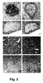
- Fig 2: Immunohistological analysis of human hepatocytes in chimeric
mouse livers.
Six weeks after human hepatocytes implantation in untreated animals,
serial liver sections were immunostained with a monoclonal antibody
against human hepatocytes (a) or with an anti-mouse macrophage
monoclonal antibody Mac2 (b)Twelve weeks after transplantation in untreated animals, serial liver
sections were immunostained with an anti-human α1-antitrypsin antibody
(c) or with an anti-mouse macrophage monoclonal antibody Mac2 (d).Untreated (e) and treated (f) chimeric mouse livers, one month after
implantation, immunostained with rabbit anti-human α1-antitrypsin
antibodies.Untreated (g) and treated (h) chimeric mouse livers immunostained with
rabbit anti-human α1-antitrypsin antibodies, 3 months after implantation.
- Fig 3: Proportion of mice positive for human albumin (HA) and α1-antitrypsin
(α1AT) secretion in treated as compared to untreated implanted
animals. The serum levels of HA (a) and human α1AT (b) in uPA/SCID
mice were determined by a standard sandwich ELISA, and the results
expressed in % positive animals.
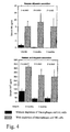
- Fig. 4: Mean concentrations in mice sera of Human Albumin and α1
antitrypsin among treated (hatched bars) and non-treated animals (plains
bars).
- Fig 5: The detection of HA transcripts in RNA samples from the chimeric
livers of treated ( uPA 131, 144 and 146) and untreated ( uPA 137, 138 and
149) animals 3 months after implantation. Human liver (Hu liver) and H2O
were used as positive and negative controls, respectively.
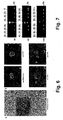
- Fig 6: Plasmodium falciparum liver stage development in chimeric human
liver. Bar represents 50 µm in A and 10 µm in B.
- a) Liver sections stained with hematoxylin-eosin. The arrows show two
developing P. falciparum liver schizonts day 5 post infection; the insert
shows a third schizont from the same animal.
- b) Liver stages of P. falciparum were labeled by various antibodies in IFAT:
an anti-HSP70, an anti-CS protein, an anti-LSA1 and a control anti-MSP3
antibodies.
- Fig 7: RT-PCR using primers of LSA1 (a) and HSP70 (b) on liver biopsies
from implanted mice (uPA1-6) and not implanted (uPA9) and on genomic
DNA from blood stages of P. falciparum as positive control. RT-PCR using
HA primers on liver biopsies results for human albumin from implanted mice
(uPA1-6) and not implanted (uPA9) and on human liver (Hu liver) as
positive control. The symbol "+" and "-" refer to test (RT-PCR) and control
respectively.
-
DETAILED DESCRIPTION
-
The present invention provides a method of making a
chimeric murine model comprising:
- a. obtaining an immunocompromised murine host, which
carries a transgene or a genetic defect responsible for murine hepatocyte
death,
- b. implanting human hepatocytes therein,
- c. controlling non-adaptive defences,
- d. recovering a chimeric murine model harbouring settled
human hepatocytes capable of maintaining, differentiating and growing.
-
-
A murine host useful for the preparation of the chimeric
murine model of the invention can be obtained by carrying out the following
steps. First, an immunocompromised murine animal is crossed with a
murine animal carrying a transgene or a genetic defect responsible for
murine hepatocyte death and resulting in a survival disadvantage of the
murine hepatocytes. By selective backcrosses performed according to
techniques well known to those of skill in the art, a murine host,
homozygous for the immunocompromised phenotype, and carrying at least
one copy of the transgene or the genetic defect responsible for murine
hepatocyte death is obtained.
-
These two features i.e., immunocompromised and the
transgene or the genetic defect responsible for murine hepatocyte death,
are required in the same murine host, to enable implantation of human
hepatocytes.
-
In order to prepare the chimeric murine model starting from
said murine host, a step is performed to control non-adaptive defences of
the host, to recover in a last step a chimeric murine model enabling the
settled human hepatocytes to maintain, differentiate and grow.
-
An alternative in the method of generating a chimeric murine
host of the invention encompasses the possibility to control non-adaptive
defences before implanting human hepatocytes.
-
"Chimeric" as used herein relates to a murine host, which
comprises cells from a xenogenic origin i.e., originating from a different
organism, in particular originating from a different animal species. In the
invention, said cells encompass human hepatocytes. Said human
hepatocytes can be provided to the murine host i.e., implanted by known
appropriate methods such as grafting, injection... When implanted in the
host, the human hepatocytes maintain, differentiate and grow.
-
In a particular embodiment said implanted human
hepatocytes are capable of substituting to the resident hepatocytes and/or
of repopulating the model.
-
"Host" as used herein is a murine animal exhibiting two
important features enabling xenogenic cells to settle:
- the capacity to tolerate the xenogenic human hepatic cells
because of altered immunologic mechanisms resulting from genetic
mutations, treatments or surgery to provide an "immunocompromised
state";
- the transgene or genetic defect responsible for murine
hepatocyte death leading to the survival advantage for the xenogenic
human hepatocytes and facilitating their expansion or colonisation.
-
The genetic defect responsible for murine hepatocyte death
can be located in a genetic region naturally present in the host. In such a
case, the term genetic defect encompasses any genetic modification
changing gene expression such as nucleotide substitution, deletion,
insertion that alter the gene transcription, the mRNA stability, the protein
translation or the protein stability or activity. These modifications can occur
in coding or in non-coding sequences such as promoter, 5'UTR, introns or
3'UTR (untranslated region). This defect can be the result of a naturally
occurring mutation or can be generated by techniques well known in the art,
such as homologous recombination.
-
The genetic defect can also result from the insertion of a
genetic construct in mammal cells. The genetic construct, such as a
transgene, can be integrated in the host genome (genomic or mitochondria
DNA) or can remain unintegrated, e.g., as a vector. The genetic construct
can carry promoter and regulation elements and a coding sequence DNA,
including cDNA, originally present or absent from the host genome.
-
For the invention, this genetic defect is harboured in at least
one copy; it can be present in all cells or in some cell types only provided it
is expressed in such a way that only resident hepatocytes of the murine
host are altered, because of the different gene expression patterns. It is an
object of the genetic defect to reduce the population of autologous
hepatocytes in the murine host prior to implanting human hepatocytes and
to enable the selective advantage in favour of these implanted human
hepatocytes to take place.
-
As a particular transgene or genetic defect responsible for
murine hepatocyte death is the Alb-uPA transgene. The Alb-uPA transgenic
mouse carries urokinase gene controlled by an albumin promoter which
targets urokinase overproduction to the liver resulting in a severe
hypofibrinogenemic state and accelerated hepatocyte death. Randomly,
individual hepatocytes spontaneously delete portion of the transgene,
providing a significant replicative advantage over surrounding cells, and
resulting in repopulation of the liver with nontransgenic cells (Heckel JL. et
al. 1990. Cell 62(3), 447-56 and Sandgren EP. et al. 2002 Journal of
hepatology 37, 422-424).
-
Genetic defects are all mutations, as described above, that
affect the murine hepatocyte survival. As examples, other genetic defects
leading to the survival advantage of implanted human hepatocytes can be
envisaged:
- a FAH mutation. Mice FAH-/-, because of the mutation,
present a deficit in fumaryl acetoacetate hydrolase. This deficiency causes
the accumulation of a hepatotoxic compound leading to a continuous
process of hepatocytes lyses.
- a caspase inducible gene under an inducible promoter that
can selectively destroy murine hepatocytes by activation of the promoter.
-
"Implanting" as used herein is the process of incorporating
xenogenic cells, e.g. human hepatocytes, into a recipient murine host. The
implantation can take place in various locations, e.g., intrahepatic,
intrasplenic, intraperitoneal or intraorbital and the implanted cells when
settled in the chimeric murine model can circulate or to the contrary can
remain at a determined location.
-
In a particular embodiment, implantation is realized with adult
or foetal primary hepatocytes, bone marrow cells that can differentiate in
hepatocytes or hepatocyte cell lines.
-
Before implantation, implanted hepatocytes can also have
previously undergone various treatments, including genetic modifications.
For example, hepatocytes can be prepared as described (Dandri M. et al.
2001. Hepatology 33, 981-988) by collagenase treatment. They can also be
kept under in vitro conditions in cultures (e.g., one to 3 weeks) before being
implanted into the murine host.
-
Dissociated hepatocyte suspension following collagenase
treatment will be implanted in the liver, the spleen or the kidney capsule,
and human liver slices of various thicknesses will be for example implanted
in the muscle. Implantation can also occur in the peritoneal cavity where
isolated human cells are attached to collagen and other matrix sponges.
-
The chimeric murine model can be used to implant, human
hepatocytes, and besides said human hepatocytes, enucleated cells such
as red blood cells (RBC). In such a murine model where combined
implantation of human hepatocytes and RBCs is provided, the present
description, especially definitions disclosed with respect to human
hepatocytes, can be transposed to RBCs where appropriate in
consideration of the nature of the cells.
-
In a particular embodiment, the implanted hepatocytes are
healthy cells, encompassing non-infected and non-tumoral hepatocytes.
-
"Non-infected" refers to hepatocytes that have not undergone,
previously to the implantation, interactions with the hepatotropic pathogen
which effect on said cells might be tested later in the obtained chimeric
murine model. Such hepatocytes are for example obtained from a liver of a
patient who has been tested negative for said pathogen or whose
background enables to support that he was free from infection with said
pathogen for the period of concern (e.g. histological and/or biological
signs).
-
Therefore, the current invention providing a chimeric murine
model with human hepatocytes, the implanted hepatocytes should not be
infected by Plasmodium strains and/or by HVB and/or by HCV.
-
"Non-tumoral" refer to hepatocytes having controlled cellular
proliferation and spread and having a stable karyotype, i.e. hepatocytes
containing the same number of chromosomes after multiple divisions.
-
In the invention, the implanted human hepatocytes, although
they are non-infected, and non-tumoral cells can nevertheless carry a
mutation which effect may be studied when these hepatocytes are brought
in contact with a determined compound administered to the chimeric murine
model.
-
"Non adaptive defences" refer to cells involved in the nonspecific
immunity, such as macrophages, monocytes or NK cells, in
contrast to specific immunity directed by T and B lymphocytes.
-
"Settled" as used herein refers to human hepatocytes that are
not lost after the implanting step, and that succeed in surviving and
repopulating in the chimeric murine model.
-
The capacity of implanted hepatocytes "to maintain" as used
herein refers to the capacity for these hepatocytes to survive in the host.
-
The capacity of hepatocytes "to differentiate" as used herein
refers to hepatocytes having the capacity to reach, after the implanting
step, characteristics as similar as possible to those in their original host,
e.g., in humans. This capacity can be determined in terms of secreted
molecules (such as hα1AT for the hepatocytes), expressed surface
receptors, pathogen infection, cell size or any other appropriate methods.
The presence of human cell type-specific molecules expressed by the
human cells can be measured, on murine sera, by well known techniques
such as ELISA (enzyme-linked immunosorbent assay), Western Blot, dot
blot, immunoprecipitation, direct or indirect immunostaining on histological
sections using specific antibodies of implanted cell markers. The cell type-specific
molecule transcripts can be detected by RT-PCR (reverse
transcriptase-polymerase chain reaction) or real-time RT-PCR by using
specific primers of implanted cell markers. Specific receptors can be
detected by various techniques such as FACS analysis Finally, pathogen
infection is controlled by detection of settled cell stage specific proteins by
techniques including light microscopy, immunohistological staining on
biopsies, by ELISA, PCR or RT-PCR on sera and by Western Blot, PCR or
RT-PCR on cellular extracts.
-
The capacity of implanted hepatocytes "to grow" as used
herein refers to the capacity of settled hepatocytes, not only to survive in
the recipient but also to multiply in the obtained chimeric model. Growth can
be measured by quantitative imaging and evaluating the percentage of cells
expressing a specific cell type marker. Measurements can be made at
different time points to follow the repopulation of the settled hepatocytes.
The growth can also be followed by the analysis of the DNA synthesis by
the incorporation of a labelled nucleotide such as BrdU.
-
A first advantage of the invention lies in the fact that the
success of human hepatocyte implantation does not depend on the strict
homozygosity of the transgene or genetic defect responsible for murine
hepatocyte death, in the murine model of the invention as described in
previous models. Consequently, the possibility to use murine host carrying
only one copy of said transgene or genetic defect, such as hemizygous or
heterozygous host for this trait, widens the scope of this model.
Consequently, the number of crosses to obtain a murine host, in which an
implanting step is possible, is reduced, leading to a faster and cheaper host
generation.
-
In the invention, homozygous is defined as presenting with
two copies of the same allele of a determined gene, heterozygous is
defined as having two different alleles (e.g., A and a) at the corresponding
gene loci on homologous chromosomes for said gene and hemizygous is
defined as presenting with a gene present in only one copy, not two, in an
otherwise diploid cell or individual.
-
A second advantage of the chimeric murine model according
to the invention is the possibility to implant non-infected, non-tumoral
hepatocytes from various origins. The hepatocytes needed for the
implanting step can come directly from donor liver, partial liver resection or
tumour-surrounding tissues. The model is less sensitive than previous ones
to cell conservation conditions. An attractive and outstanding feature of the
present model is the possibility to perform serial implantation from chimeric
murine model to chimeric murine model, allowing faster and more efficient
subsequent implantations.
-
The inventors have determined that controlling non-adaptive
defences of the host is one of the parameters enabling the implanted
hepatocytes to settle, differentiate and grow in the chimeric murine model.
-
The efficiency of the control of non-adaptive defences can be
checked by various techniques, such as FACS analysis. The main actors of
the non-adaptive defences are macrophages and NK cells for which a
strong reduction or depletion is expected after immunomodulation treatment
according to the invention.
-
"Macrophage depletion" as used herein is the process of
reducing in a large amount the circulating and tissue macrophages. A
convenient range of remaining macrophages after treatment is 0% to 50%.
A preferred range of remaining macrophages is 0% to 20%.
-
Macrophage number can be reduced by administrating in the
host, antagonists of macrophages, such as toxic substances, like Cl2MDP,
or antibodies altering macrophage development or function and finally
killing them. The administration of antagonists is performed by well-known
techniques, including the use of liposomes.
-
"NK depletion" as used herein is the process of reducing NK
cells after treatment. A convenient range of remaining NK after treatment is
0% to 50%. A preferred range of remaining NK is 0% to 20%.
-
The administration of NK antagonists or substances altering
their function and development is made by well-known techniques using
vectors including liposomes.
-
In a particular embodiment, the macrophage depletion is
realized by injecting liposomes containing Cl2MDP according to the
technique of Van Rooijen et al. (Van Rooijen N. 1989. J. Immunol. Methods
124, 1-6). The liposome size can range from 0.5 to 7 µm to be ingested by
macrophages, resulting in their killing.
-
Possible alternatives for reducing the macrophage population
are either the administration of another toxic, the cis-platinium or the use of
the irradiation. However, these techniques are less efficient than the
administration of liposomes containing Cl2MDP.
-
The NK cells depletion is preferably realized by injecting an
anti-NK antibody, such as the anti-Tim β1 mouse monoclonal antibody,
binding to the IL-2R β chain.
-
A particular protocol of macrophage depletion is the injection
of Cl2MDP embedded in liposomes, at 4-day interval, starting two days after
implantation. Such liposomes fully clear macrophages from peritoneum,
liver, spleen and kidney. Monocytes from the bone marrow colonise the
liver after the clearance of all Kupfer cells and transform into very active
large macrophages and new Kupfer cells. These are, again, destroyed by
the next injection of liposomes.
-
In a particular embodiment for NK depletion, anti-NK
antibodies are injected at monthly interval, starting two days after
implantation. The interval between injection depend on the half-life of the
antibody directed to NK cells, and can be from 2 to 4 weeks in order to
keep NK cells continuously low.
-
Particular immunocompromised models used to generate a
host are SCID mouse (severe combined immunodeficiency), SCID/Nod
mouse (severe combined immunodeficiency/non obese diabetic) or mouse
with altered lymphocyte lineages such as BXN (NIHIII or Beige Xid Nude),
RAG, RAG2 and RAG-γC.
-
In a particular embodiment of the invention, the mouse host is
obtained by:
- a. crossing a homozygous SCID mouse with a homozygous
Alb-uPA transgenic mouse;
- b. making selective backcrosses;
- c. recovering a homozygous SCID, hemizygous Alb-uPA
mouse host.
-
-
The present invention provides also a chimeric murine model
which is an immunocompromised murine host harbouring at least one
transgene or genetic defect responsible for murine hepatocyte death, which
non-adaptive defences are controlled and which is implanted with human
hepatocytes, said control of non-adaptive defences enable said
hepatocytes to maintain, differentiate and grow.
-
An advantage of the chimeric murine model of the invention is
not only the maintenance of the settled human hepatocytes, but also their
growth and differentiation.
-
The growth and differentiation of the settled human
hepatocytes of the chimeric murine model of the invention can be checked
by various features, such as the presence of cell surface receptors, the
secretion of proteins specific of the implanted hepatocytes, the cell size or
the receptivity to pathogens.
-
In a particular embodiment of the invention, the model
enables the settled human hepatocytes to secrete specific proteins
characterizing their differentiation and growth, for several months especially
for at least 3 months. Specific proteins of the implanted hepatocytes, such
as albumin, can be used as markers to follow the differentiation state as
well as the repopulation.
-
The hypothesis that implanted human hepatocytes acquired a
differentiation state similar to that observed in in vivo human conditions, can
be tested by the stringent requirements of some pathogens to infect
differentiated hepatocytes to realize their development.
-
Another advantage of the murine model of the invention is the
receptivity of settled hepatocytes for pathogens having a restricted
hepatotropism.
-
"Receptive" as used herein refers to the capacity of settled
hepatocytes to sustain an infection similar to that observed in humans.
Therefore, pathogens penetrate settled hepatocytes and realize their
replication and maturation.
-
According to another embodiment of the invention the model
is suitable for in vivo study of metabolism of administered compounds and
especially drugs, in said settled hepatocytes.
-
As a particular embodiment, the murine host is a homozygous
SCID, hemizygous Alb-uPA mouse, comprising implanted human
hepatocytes. In this chimeric murine model, settled cells are receptive to
hepatotropic pathogens, such as HBV, HCV or Plasmodium strains, e.g.
P. falciparum or P. vivax.
-
Another particular chimeric murine model is a chimeric murine
animal, comprising implanted human hepatocytes and human red blood
cells (RBC). In this particular model, it might be possible to follow-up the life
cycle of Plasmodium falciparum in the same animal model. The pathogen,
following its replication in human liver cells can invade implanted RBCs and
develop to schizont stage in those RBCs.
-
The chimeric murine model of the present invention allows the
study of restricted-tropism pathogens, for which no permissive line exists or
for which permissive models are not fully satisfactory. The chimeric murine
model of the present invention contains at least settled hepatocytes that are
differentiated and are able to sustain a pathogen infection for several
weeks. Because of the duration of the settled hepatocyte survival, their
repopulation and their high number in the chimeric murine model, pathogen
life cycle can be intensively studied including when said infected
hepatocytes are put in contact with candidate drug compounds.
-
The invention also provides a method for studying a
hepatotropic pathogen, in a chimeric murine model according to the
invention comprising:
- a. infecting said chimeric murine model with a hepatotropic
pathogen, in conditions enabling said pathogen to penetrate the settled
human cells of the chimeric murine model,
- b. observing the pathogen-generated infection in said settled
cells.
-
-
The first step is the introduction in the chimeric murine model
of a pathogen in conditions in which it can interact with settled hepatocytes
and especially can penetrate them. The introduction of the pathogen can be
achieved by various ways, including intravenous or intracutaneous
injections.
-
In a second step, the infection of said settled hepatocytes is
monitored by well known methods including, but not limited to, light
microscopy, immunofluorescence antibody test (IFAT) using pathogen
specific antibodies, PCR (qualitative or quantitative) or RT-PCR using
primers for a pathogen specific gene, ELISA or immunoprecipitation. When
a pathogen has a life cycle with different forms infecting various organs or
various species, antibodies and primers are chosen to be specific to
proteins of each form, and in the invention, specific of the proteins of the
settled cell form. All the results obtained to identify the pathogen infection,
for example the number of pathogens calculated by light microscopy, the
staining observed by IFAT and the mRNA transcripts detected in the settled
cells of the chimeric murine model, are compared to a control model
infected by the same pathogen but without cell implantation.
-
In a particular embodiment, the chimeric murine model tested
is a mouse generated from a homozygous SCID, hemizygous Alb-uPA
mouse host, in which hepatocytes are implanted.
-
Another particular chimeric murine model is a murine host
according to the invention implanted with both human hepatocytes and
human red blood cells (RBC).
-
Particular hepatotropic pathogens are HBV (hepatitis B virus),
HCV (hepatitis C virus) or Plasmodium strains including P. falciparum and
P vivax.
-
The monitoring of the hepatocyte infection by P. falciparum
can be performed by testing pathogen proteins, specific for hepatocytes
and/or erythrocytes, such as the circumsporozoite protein (sporozoite), the
MSP3 protein (erythrocytes), the HSP70 and MSP1 proteins (hepatocytes
and erythrocytes) and the LSA1 protein (hepatocytes).
-
The monitoring of the hepatocyte infection by HCV can be
performed by measuring the presence or absence of the viral RNA
sequence (qualitative PCR) or the viral load (quantitative PCR).
-
The monitoring of the hepatocyte infection by HBV can be
performed by quantifying a viral envelop protein HbsAg with the ELISA
technique.
-
The invention provides also the use of of a chimeric murine
model of the invention for the testing of a compound for a potential
therapeutic interest, towards the infection or its consequences.
-
The chimeric murine model can also be useful for the
screening of drugs capable of altering the life cycle of pathogens. Owing to
the capacity of the chimeric murine model to sustain a pathogen infection
for several weeks, the effects of a drug administration on the infection can
be observed and its efficacy can be evaluated. The invention provides a
method for screening active compounds against the infection by
hepatotropic pathogen or against its detrimental effects in a chimeric
murine model of the invention comprising:
- a. infecting said chimeric murine model with a hepatotropic
pathogen, in conditions enabling said pathogen to penetrate the settled
human cells of the chimeric murine model;
- b. administering the tested compound in conditions allowing
its activity;
- c. observing the effects of said compound on the pathogen-generated
infection or on its detrimental effects.
-
-
In a first step, settled hepatocytes are infected according to
the method described above.
-
Then, the drug is administered to the chimeric murine in
conditions in which the drug keeps, modifies or acquires its activity. The
interactions with the settled hepatocytes as well as the host environment
could be determined with respect to the activity of the drug. "A drug" as
defined herein is any substance presented for treating, curing or preventing
disease in human beings or in animals, including prodrugs.
-
The drug can be administered under any appropriate forms
including systemic or local routes.
-
Several drugs (at least two) can be administrated together,
alternatively or with specific protocols to show a possible synergy,
redundancy or antagonism.
-
The drug can be administrated by a lot of well known routes,
including taken by mouth (orally), given by injection into a vein
(intravenously), into a muscle (intramuscularly), beneath the skin
(subcutaneously) or placed under the tongue (sublingually), inserted in the
rectum (rectally) or vagina (vaginally), instilled in the eye (by the ocular
route); sprayed into the nose and absorbed through the nasal membranes
(nasally); breathed into the lungs (by inhalation) or applied to the skin
(cutaneously).
-
Finally, the effects of the drug on the pathogen infection or on
its detrimental effects can be monitored. Various techniques can be used to
observe the effects of the administrated drug, including light microscopy,
immunofluorescence antibody test (IFAT), RT-PCR, PCR (qualitative or
quantitative), or ELISA. The infection are followed at different time points
and various factors can be calculated: drug half-life, minimal effective
dosage for infection elimination or reduction, drug dosage based on age, on
body weight or on body surface area, the most efficient place or route for
drug administration, the combination therapy efficiency. Other factors that
can be observed could be the biological activity of the drug metabolites
produced by settled human hepatocytes as well as the toxicity on the
various organs of the murine model of these human specific metabolites.
-
The invention also provides a method for screening the
in vivo
metabolism of xenobiotic compounds, in a chimeric murine model of the
invention comprising:
- a. administrating the xenobiotic compound to be tested to said
chimeric murine model in conditions allowing the compound to interact with
settled human hepatocytes,
- b. observing its biotransformation by said settled hepatocytes.
-
-
This subset of human hepatocytes can be exploited to follow
the biotransformation of a xenobiotic, after its injection into the chimeric
murine model. "Xenobiotic" as used herein refers to a chemical substance
(or more generally, a chemical mix) that is not a normal component of the
organism in which it is exposed to. Xenobiotics include most drugs (others
than those compounds which naturally occur in the organism) and
prodrugs, as well as other foreign substances.
-
The xenobiotic is administrated into the chimeric murine
model according to all routes and all forms cited above for the drug. One
condition in the administration is that the xenobiotic can interact with the
settled hepatocytes.
-
The measurement of the level of the metabolites (degradation
products), including intermediates and final products, enables to track the
xenobiotic metabolism kinetics, including its half-life. The model enables
also to observe the effects of the compound on the settled cells, to evaluate
the doses at which the effects appear. Finally, it is possible to study the
potential interactions between reactive metabolites and cellular
macromolecules.
-
This model is important for prodrugs, i.e., any compound that
undergoes biotransformation prior to exhibiting its pharmacologic effects.
Indeed, the human hepatocytes can produce metabolites, especially the
active form of the drug, from the prodrugs, whereas the murine hepatocytes
cannot do it.
-
A particular murine model tested is a chimeric mouse model
generated from a homozygous SCID, hemizygous Alb-uPA mouse host, in
which hepatocytes are implanted. In this chimeric model, one could monitor
the cytotoxic effects of the drug on the liver, e.g., by measuring the
circulating hepatic transaminases and by analysing the liver histology with
optical microscopy techniques.
-
The invention also provides technical platforms comprising at
least the chimeric murine model of the invention such as:
- a technical platform, useful to identify new compounds
useful to treat mammal infections provoked by a hepatotropic pathogen,
characterized in that it comprises at least a chimeric murine model as
defined above and appropriate means to detect or to observe the effects of
said compounds on a pathogen-generated infection of said murine model.
- a technical platform, useful for screening the in vivo
metabolism of xenobiotic compounds characterized in that it comprises at
least a chimeric murine model according to the invention and appropriate
means to observe the biotransformation of said compounds by implanted
human hepatocytes in said murine model.
EXAMPLES
-
Examples are given to illustrate but not to limit the invention.
Example 1: generation of a chimeric mouse model
Animals
-
Homozygous Alb-uPA transgenic mice and homozygous
SCID mice were purchased from Jackson Laboratories (Maine, USA) and
IFFA-CREDO, respectively. Animal were housed and maintained under
specific pathogen-free conditions. To generate Alb-uPA/SCID mice, we
crossed Alb-uPA and SCID mouse lines, and through selective
backcrossed bred trait to homozygosis. uPA transgenic mice were identified
by PCR of mouse-tail DNA with the following primers: p1 :5'-ATTCTGGAGGACCGCTTATCTGT-3';
p2: 5'-CTTGAACCCAGGAGGCGGAGATT-3'.
Only hemizygous uPA mice were
used as host for implantation and infection experiments.
-
Homozygosity of the SCID trait was confirmed by
quantification of total serum IgG and IgM using a sandwich ELISA. Only
mouse with total Ig < 50 µg/ml were used for experiments.
Isolation of human hepatocytes
-
Primary human hepatocytes were isolated as described
elsewhere (Guguen-Guillouzo C. et al. 1986. Prog Liver Dis 8, 33-50) from
the healthy liver tissue of surgical liver biopsies specimens (approx. 20-25
cm3) obtained with informed consent from patients who underwent
therapeutic partial hepatectomy for liver metastasis and benign hepatic
tumor. Subjects with viral infections (HCV, HBV, HIV), cirrhosis and primary
hepatic carcinoma were excluded.
-
Resected liver lobes were cut at a distance of at least 3 cm
from the metastasis. Human hepatocytes were isolated by a two-step
perfusion technique. Hepatocyte viability obtained by this method depends
on 3 important factors which are the temperature: 37°C, pH: 7.6 and
perfusate flow rate: 16-20ml according to the rejection size. Perfusion
began with a HEPES buffer, without calcium, at a temperature, a pH and a
perfusate flow rate as indicated above. Hepatocytes were then isolated with
a perfusion of 200 ml of HEPES buffer supplemented in Collagenase H
0.05% (Roche Molecular Biochemicals) and CaCl 2 5 mM, and separated
from non-parenchymatous cells by Percoll fractionation, as previously
described (Giannini C. et al. 2003. Hepatology 38, 114-122; Guguen-Guillouzo
C. et al. 1993. Cytotechnology 11 Suppl, S3-5; Guguen-Guillouzo
C. et al. 1982. Cell Biol Int Rep 6(6), 625-8). Viable cells were determined
by trypan blue exclusion.
Implantation of human hepatocytes
-
For hepatocyte implantation, mouse hosts were anesthetized and a
laparotomy realized. A number of 5-6 x 10⁵ viable human hepatocytes were
injected into the splenic parenchyma of 10-14 days old recipients, as
previously described (Giannini C. et al. 2003. Hepatology 38, 114-122).
Macrophage and NK depletion treatment
-
Mice were injected intraperitonealy at 4-day interval starting 2
days after implantation with 50µl of a solution at 50% hematocrit of
encapsulated-dichloro-methylene-diphosphonate (Cl2MDP) liposomes. For
NK cells depletion, these mice received at monthly interval starting at day 2
post-implantation 1 mg of purified anti-Tim-β1 (anti-IL-2 Rβ chain) mouse
monoclonal antibody.
Example 2: macrophage and NK detection
FACS analysis
-
The depletion of macrophages and NK, following treatment,
were investigated by FACS analysis.
-
For macrophage analysis, 50 to 150 µl of blood (collected by
retro-orbital sampling) from treated and untreated mice were diluted in
phosphate buffer. Cells are washed in this buffer twice by centrifugation at
1500 rpm for 10 minutes at 37 °C. The pellet is resuspended in a solution
containing a F4/80 antibody and incubated for 30 minutes at room
temperature. Cells are then washed in a Facs buffer (Becton Dickinson).
The features of the F4/80 are the following: rat anti-mouse F4/80 antigen,
conjugated to Fluorescein Isothiocyanate Isomer I (FITC) from Serotec.
-
For NK cell analysis, spleens from treated and untreated mice were
taken and splenocytes were obtained after dilaceration (Cell strainer,
Becton Dickinson). Cell suspension is then washed twice by centrifugation
at 1500 rpm in RPMI 1650 medium (Life Sciences). Cells are then
incubated with DX5 and anti-CD3 (clone17A2) antibodies, respectively
coupled to FITC and Phycoerythrin for 30 minutes at room temperature.
Cells are then washed and resuspended in Facs buffer (Becton Dickinson).
The used antibodies have the following features:
- DX5 antibody: rat antibody, specific of CD49b/Pan-NK cells (Becton
Dickinson),
- 17A2 antibody: a rat anti-mouse CD3 (T-cell receptor-associated
CD3 complex), conjugated with FITC (Becton Dickinson).
Immunostaining
-
Mouse liver biopsies fixed in 10% formalin were embedded in
paraffin. To detect macrophages, sections were immunostained with a rat
anti-mouse macrophage monoclonal antibody Mac2 (culture supernatant
diluted to1:4; TIB 166, ATCC Manhasset, VA), 1 hour at room temperature.
Horseradish peroxidase labelled second anti-rat antibodies (1:500 dilution)
were incubated for 30 min followed by reaction with
diaminobenzidine/hydrogen peroxidase as chromogen-substrate.
Endoperoxydase activity was blocked by first treating the slides with 0.3%
H2O2 in PBS for 5 min at room temperature. After immunochemistry,
sections were counterstained with Meyer's hematoxylin (DAKO).
Example 3: serum human albumin and α1 antitrypsin detection
-
The levels of human albumin and human α1-antitrypsin
(hα1AT) in uPA/SCID mouse sera were quantified by a standard sandwich
ELISA. Mouse anti human albumin (0.1 µg/ml, clone HSA-9, Sigma, St
Louis, MO) and goat anti-hα1AT (11µg/ml; Rockland, Gilbertsville, PA)
were used as antigen-specific capture antibodies. Following overnight
coating, non specific binding was blocked by incubation with 1 % bovine
serum albumin for 1 hour at 37°C. After washing, 75 µl/well of 1:10 diluted
mouse sera in PBS, were added and incubated overnight at 4°C. HRP-conjugated
rabbit anti-human albumin (0.16 µg/ml; Sigma Chemical Co.)
and HRP-conjugated rabbit anti-hα1AT (2µg/ml) were used as antigen-specific
indicator antibodies. The chromogen and the substrate were used
according to the manufacturer indications (Sigma Chemical Co.).
Absorbance values (405 nm) were converted to concentrations in µg/ml by
comparison with a standard curve performed by using serial dilutions of
defined amounts of purified human albumin and hα1AT (Sigma Chemical
Co.). The ELISA result was considered as positive for the detection of
human proteins (HA, hα1AT) when the OD value was higher than the OD
means + 2 fold the standard deviation of 14 non-implanted Alb-uPA/SCID
mice. Qualitative comparisons were done using the chi 2 test and Fischer
exact test. P values of less than 0.05 were considered as significant.
Example 4: albumin transcript detection
-
RT-PCR was used for human albumin transcript detection.
Total RNAs were isolated from human-mouse chimeric hepatocytes, murine
and human liver tissues (and human cells) using RNeasy Protect Midi kit
(Qiagen S.A., France) according to the manufacturer indication. Samples
were quantitated using a spectrophotometer, and equal amounts of RNA
from all samples were subjected to cDNA synthesis using random primers.
Human specific albumin primers, 5'-CATTAGCTGCTGATTTTGTTGAAAG-3'
and 5'-TGTGCAGCATTTTGTGACTCTG-3' were used to detect human
albumin transcripts. PCR Conditions were 95°C for 5 min; 94°C for 30s,
60°C for 1 min, and 72°C for 1 min for 40 cycles, with a final extension at 72°
C for 5 min. Twenty µl of final PCR product were analyzed by
electrophoresis (2% agarose gel with Ethidium Bromide) and PCR product
bands (523 bp) were visualized under UV trans-illumination.
Example 5: immunohistological staining alpha-1 antitrypsin protein
detection
-
Mouse liver biopsies fixed in 10% formalin were embedded in
paraffin. To detect human hepatocytes, 5µm sections pre-treated with Dako
Biotin Blocking System (Dako, Glostrup, Denmark) were immunostained
with a monoclonal antibody against human hepatocytes (Clone OCH1E5,
1:20 dilution; Dako, Glostrup, Denmark), using the Dako ARK system.
Similarly, 5µm sections were immunostained with rabbit anti-human α1-antitrypsin
antibodies (Dako, Glostrup, Denmark) diluted (1:2000) for 1 hour
at room temperature. Slides were developed using a secondary anti-rabbit
antibody conjugated with horseradish peroxidase (EN-VISION-kit; Dako)
following the manufacturer's instructions.
Example 6: liver repopulation estimation
-
Quantitative imaging was performed on the liver biopsies of
three untreated and treated animals obtained at one and three months after
implantation. For each animal three different sections were immunostained
with rabbit anti-halAT and counterstained with Meyer's hematoxylin as
described above. Adobe PhotoShop 6.02 software was then use to select
and count the pixels corresponding to the total liver section (Meyer's
hematoxylin staining) or to human hepatocyte clusters (anti-hα1AT
reactivity). The percentage of liver repopulation by human hepatocytes was
expressed as the cell surface stained for hα1AT divided by the total cells
surface of the tissue section. Qualitative comparisons were done using
Fischer exact test. P values of less than 0.05 were considered significant.
Example 7: effective depletion of NK cells and macrophages
-
FACS analysis demonstrated, using F4/80 marker, that
macrophage depletion in mice was effective since two days after the first
liposome injection, macrophages represent 0.5% of lymphocyte cells in
treated mice versus 8% in untreated mice (Fig1a). In addition, a successful
depletion of NK cells was reached, since after Timβ-1 injection, DX5+CD3-
NK represent 0.5% of spleen lymphocyte cells in treated mice versus 8% in
untreated mice (Fig 1b).
-
These depletion results were confirmed by histochemical
analysis of human/mouse chimeric liver sections from animals implanted 6
weeks earlier. These investigations showed that the intrasplenically
injected-hepatocytes had indeed migrated to the liver parenchyma and
formed clusters as revealed with positive label of a human hepatocyte
marker (Fig 2a). In untreated mice, these clusters were surrounded by
numerous mononuclear cells (labeling with Mac2), which consisted mainly
of macrophages (Fig 2b).
Example 8: human albumin and α1 antitrypsin secretion
-
The ELISA analyses performed on sera of treated and
untreated implanted hosts were shown on Figure 3a (human albumin) and
3b (human α1AT). Over a 3-month period after implantation, the number of
positive mice for human albumin and α1 antitrypsin secretion was
dramatically improved by the treatment. In untreated animals, the number
of positive mice for HA and hα1AT secretion, decreased over time during
the follow-up (from month 1 to month 3: 13.6 to 0%, and 45.5 to 21.7%,
respectively). In contrast, in treated animals, the number of mice positive for
HA and hα1AT secretion was markedly higher at one month and, above all,
remained high over time during the follow-up. At three months, 60.7±1.6%
and 86.3±5.8% of mice secreted HA and hα1AT respectively. This first
results showed a clear correlation between the treatment and the human
albumin and α1 antitrypsin secretion.
Example 9: histochemical hα1AT protein expression
-
Immunohistochemical investigations at 3 months post-implantation
in untreated animals, revealed that human hepatocyte clusters
surrounded by macrophage became negative for hα1AT (Fig. 2c-d),
suggesting that human hepatocytes were no longer functional, in
agreement with the lack of detectable human serum albumin at that time
(Figure 4). These data confirmed the strong relationship between the
number of surrounding macrophages and the hepatocyte functionality.
-
Further albumin and α1 antitrypsin analyses were performed
on untreated and treated animals at 1, 2 and 3 months (Figure 4). A strong
reduction in macrophage numbers in the treated animals was associated
with both an increase in the number of human hepatocyte clusters and the
number of hepatocytes per cluster according to untreated animals, both at
one month (Fig. 2e-f) and at 3 months (Fig. 2g-h).
-
These results taken together confirmed that the macrophage
and NK depletion is necessary to the implanted hepatocytes to become
fully functional, i.e. to secrete both human albumin and α1 antirypsin in
mouse serum (Figure 4).
Example 10: repopulation of the chimeric liver
-
Quantitative imaging on liver sections from implanted animals
revealed that the percentage of human hepatocyte liver repopulation
(expressed as the cell surface percentage, stained for hα1AT) was higher in
treated as compared to untreated animals. The repopulation was of
17.7±2.1% in treated animals versus 3.1±1.5 % in untreated animals at one
month (
p=0.0006), and 26.9±4.2% versus 6.5±2.4% at 3 months (
p=0.002).
Individual data about 12 mice (6 treated and 6 untreated) are given in Table
- 1. This experiment showed that the implanted cells not only survive in their
host, but also can multiply and repopulate the chimeric liver.
-
Example 11: HA transcript expression
-
HA transcripts were detectable by RT-PCR in RNA samples
from the chimeric livers of treated animals but not from untreated animals, 3
months after implantation (Figure 5).
-
Taken together, these results indicate that innate immune
mechanisms deeply influence the survival and function of implanted human
hepatocytes in the chimeric mouse model. This confirms the highly
differentiated status of the settled cells obtained, following treatment. To
demonstrate unequivocally that the implanted human hepatocytes were
indeed functional, we took advantage of the stringent requirement of
P.falciparum liver stages for fully differentiated human hepatocytes.
Example 12: P.falciparum infection challenges
-
Plasmodium falciparum sporozoites (strain NF54) were
obtained from infected Anopheles stephensi mosquitoes salivary glands on
day 14-16 after membrane feeding on gametocytes from NF54 cultured
gametocytes as previously described (Ponnudurai T. et al. 1986.
Parasitology 93(Pt2), 263-74; Sinden RE. et al. 1979. Parasitology 79(2),
277-96; Sinden RE. et al. 1984. Parasitology 88(Pt2), 239-47; Ozaki I.S. et
al. 1984. J Parasitol 70, 831-833).
-
A number of 100,000 to 1,000,000 P. falciparum sporozoites,
the invasive forms injected by the mosquito, were intravenously injected in
the retro-orbital sinus 3 to 6 weeks after human hepatocytes implantation. A
total of 14 mice implanted with hepatocytes from 4 distinct donors were
challenged by parasites from four mosquito batches and liver biopsies
taken 5 days later. P. falciparum liver forms were detected by light
microscopy of hematoxylin-eosin stained sections. Non implanted mice, or
implanted mice negative for human albumin served as controls.
Example 13: detection of the hepatocyte infection by P. falciparum Immunohistological staining
-
For protein detection, several antibodies against pathogen
specific-stage proteins were used.
- an anti-circumsporozoite protein mouse monoclonal
antibody, Mab2A10, specific of the circumsporozoite (CS) protein, a
molecule expressed in sporozoites and carried over in liver stages
(Atkinson C.T. et al. 1989. Am J Trop Med Hyg 40, 131-140; , Suhrbier A.
et al. 1988. Eur J Cell Biol 46, 25-30; Wirtz R.A. et al. 1987. Bull World
Health Organ 65, 39-45).
- a polyclonal antibody (anti-PfHSP70-1), obtained from
immunized mice a heat shock protein expressed in both liver and blood
stages; these antibodies recognize parasite-specific epitopes of the HSP70
of Plasmodium and do not cross-react with HSP70 from mouse or human
(Renia L. et al. 1990. Eur J Immunol 20, 1445-1449; Motard A. et al. 1995.
Int Immunol 7, 147-150).
- an anti-MSP1 human affinity purified antibodies, specific of
MSP1, expressed in both liver and blood stages. Anti MSP1 antibodies
have been affinity purified on the MSP1-19 recombinant protein covering
the C-terminus part of the MSP1 molecule (Renia L. et al. 1990. Eur J
Immunol 20, 1445-1449; Motard A. et al. 1995. Int Immunol 7, 147-150).
- an antibody, obtained from an LSA1 immunized
chimpanzee, specific of LSA1, the only antigen known to be solely
expressed in liver stages. The LSA1 antibodies are directed to several
regions of the LSA1 molecule and particularly the peptides LSA1-R and
LSA1-J (Guerin-Marchand C. et al. 1987. Nature 329, 164-167; Fidock D.A.
et al. 1994. Nature 161, 126).
- an antibody specific of MSP3, an antigen expressed only in
blood stages. Anti-MSP3 affinity purified antibodies are directed to peptide
MSP3-b, peptide MSP3-c and peptide MSP3-d (Brahimi K. et al. 1993. J
Immunol Methods 162, 69-75).
-
All sera were used at a 1:100 dilution, except for Mab2A10,
used at 1:500. Moreover, a serum from a malaria non-exposed individual
and a naïve mouse serum served as negative controls. Secondary,
fluorescent Alexa 488-labelled anti mouse or human antibodies (Molecular
Probes®) were diluted 1:600.
-
Mouse liver biopsies fixed in 10% formalin were embedded in
paraffin. Immunohistological detection of parasites was performed on day 5
post infection with P. falciparum sporozoites. Liver biopsies were taken and
immediately processed for histological analysis. Hematoxylin/eosin (HE)
staining and IFAT were performed on serial 5 µm sections.
RT-PCR
-
For parasite transcript detection, two transcript genes were
tested: LSA1, a gene expressed only in liver stages and HSP70, a heat
shock protein gene, expressed in both liver and blood stages.
-
Total RNAs were extracted, as described above, from a)
implanted and non implanted infected mouse liver tissue taken 5 days
following sporozoites inoculation, b) from human non-infected liver tissue
and c) from
P. falciparum erythrocytic stages, and treated with RNase free
DNase. Reverse transcription was carried out under a final volume of 20 µl
using 4 µg of total RNA and 200 units of M-MLV Reverse Transcriptase
(Invitrogen) in the presence of specific LSA1 and HSP70-1 primers (from
either LSA1 or HSP70-1
P. falciparum genes). Nested RT-PCR was carried
out using 2µl of the RT reaction, in the presence of 125 µM dNTPs, 125 nM
of primers (LSA1-N and HSP70-N), and 2 units of Taq polymerase
(Qiagen). Direct PCR on RNA extracts was performed as a control to rule
out genomic DNA contamination in RNA preparations. The parasite specific
primers used for the reverse transcription and the nested RT-PCR analysis
are as follows:
Example 14: Plasmodium falciparum infection in hepatocytes
-
By light microscopy analysis, P. falciparum liver forms were
found to be unexpectedly abundant (Fig. 6a) and this high number
apparently correlated with serum albumin levels (ie. the largest number of
parasites was seen in mice having a serum albumin level of about 100
µg/ml).
-
Morphological indications were confirmed by
immunofluorescence labeling with parasite-specific reagents. The Mab2A10
antibody, specific of the circumsporozoite (CS) protein, specifically labeled
the same liver form (in serial sections), as others in sections from different
animals (Fig. 6b). Antibodies directed against either HSP70 or MSP1, both
expressed in liver and blood stages, also reacted specifically with P.
falciparum liver forms produced by sporozoite inoculation (Fig. 6b). Finally,
antibody specific of LSA1, the only antigen known to be solely expressed in
liver stages, equally labelled the liver forms (Fig. 6b).
-
In contrast, several control antibodies (anti-MSP3 or unrelated
antibodies) yielded negative results on serial cuts from positive animals.
Conversely, liver form-specific antibodies were negative on liver sections
from non-implanted though infected control mice (over 100 sections
screened).
-
The results obtained both by light microscopy and
immunofluorescence staining, were further confirmed by RT-PCR, using
primers specific for parasite or human genes. Human albumin transcripts
were detected in 7 of 7 albumin secreting mice, in 3 of 7 implanted mice
without detectable HA in the serum and not in 3 non implanted control mice
5 days after sporozoite inoculation (Figure 7).
-
Nested RT-PCR, performed using parasite-specific primers
derived from LSA1 and HSP70 sequences, yielded homogeneous results in
agreement with the above findings. Both corresponding messenger RNAs
were detected in 7/10 mice in which albumin transcripts were detected (Fig.
7).
-
These experiments showed the high degree of maturation of
the liver settled cells, as confirmed by their size, reaching 50-60 µm at
day
5,
i.e. as large as that obtained in humans (Shortt, HE. et al. 1948. Nature
161, 126 and Shortt HE. et al. 1951. Trans R Soc Trop Med Hyg 44, 405-419)
or higher primates such as chimpanzees (Bray RS. 1962. Am J Trop
Med Hyg 7, 20-24 and Meis JF. et al. 1990. Exp Parasitol 70, 1-11), and
nearly twice as large as that obtained under
in vitro conditions in primary
human hepatocyte cultures (Meis JF. et al. 1986. Cell Tissue Res 224, 345-350;
Mazier D. et al. 1985. Science 277, 440-442; Smith JE. et al. 1984.
Lancet 2, 757-758). These settled cells are receptive to
P. falciparum, in
which the pathogen can develop at densities similar to those obtained with
rodent parasites (Wery M. 1968. Ann Soc Belg Med Trop 48, 11-137),
despite the small number of human hepatocytes implanted and the fact that
parasites were inoculated in the peripheral blood.
| Mouse repopulation in treated and untreated mice, 1 month and 3 months after implantation. For each mouse, 3 values corresponding to 3 independent measures and a mean of these 3 measures are given. |
| | Non treated | Treated |
| | Mouse number | Measure | Repopulation % | Mouse number | Measure | Repopulation % | |
| 1 month after implantation | 166 | 1 | 2.264 | 162 | 1 | 17.035 |
| 2 | 2.962 | 2 | 21.855 |
| 3 | 0.312 | 3 | 11.274 |
| mean | 1.846 | mean | 16.721 |
| |
| 163 | 1 | 2.373 | 176 | 1 | 17.699 |
| 2 | 3.301 | 2 | 10.178 |
| 3 | 8.495 | 3 | 20.640 |
| mean | 4.723 | mean | 16.172 |
| |
| 160 | 1 | 1.269 | 161 | 1 | 26.399 |
| 2 | 4.924 | 2 | 14.861 |
| 3 | 1.558 | 3 | 16.021 |
| mean | 2.58 | mean | 20.09 |
| |
| mean | 13.1 ± 1.5 | | mean | 17.7 ± 2.1 |
| |
| | Non treated | Treated |
| | Mouse number | Measure | Repopulation % | Mouse number | Measure | Repopulation % | |
| 3 months after implantation | 153 | 1 | 3.009 | 144 | 1 | 29.186 |
| 2 | 10.620 | 2 | 28.154 |
| 3 | 3.349 | 3 | 29.740 |
| mean | 5.659 | mean | 29.026 |
| |
| 136 | 1 | 3.393 | 131 | 1 | 26.979 |
| 2 | 10.653 | 2 | 24.793 |
| 3 | 0 | 3 | 14.161 |
| mean | 4.682 | mean | 21.977 |
| |
| 155 | 1 | 27.495 | 143 | 1 | 32.229 |
| 2 | 0 | 2 | 26.799 |
| 3 | 0 | 3 | 29.920 |
| mean | 9.165 | mean | 29.884 |
| |
| mean | 6.5 ± 2.4 | mean | 26.9 ± 4.2 |