EP3569253A1 - Procédés permettant d'induire une apoptose partielle en utilisant des polypeptides de caspase - Google Patents
Procédés permettant d'induire une apoptose partielle en utilisant des polypeptides de caspase Download PDFInfo
- Publication number
- EP3569253A1 EP3569253A1 EP19173088.6A EP19173088A EP3569253A1 EP 3569253 A1 EP3569253 A1 EP 3569253A1 EP 19173088 A EP19173088 A EP 19173088A EP 3569253 A1 EP3569253 A1 EP 3569253A1
- Authority
- EP
- European Patent Office
- Prior art keywords
- cells
- cell
- caspase
- polypeptide
- multimeric ligand
- Prior art date
- Legal status (The legal status is an assumption and is not a legal conclusion. Google has not performed a legal analysis and makes no representation as to the accuracy of the status listed.)
- Pending
Links
- 238000000034 method Methods 0.000 title claims abstract description 143
- 230000001939 inductive effect Effects 0.000 title abstract description 62
- 102000011727 Caspases Human genes 0.000 title abstract description 44
- 108010076667 Caspases Proteins 0.000 title abstract description 44
- 230000006907 apoptotic process Effects 0.000 title abstract description 35
- 230000036961 partial effect Effects 0.000 title abstract description 13
- 239000003446 ligand Substances 0.000 claims abstract description 229
- 210000004027 cell Anatomy 0.000 claims description 497
- 210000001744 T-lymphocyte Anatomy 0.000 claims description 235
- 108090000566 Caspase-9 Proteins 0.000 claims description 191
- 102000004039 Caspase-9 Human genes 0.000 claims description 190
- 150000007523 nucleic acids Chemical class 0.000 claims description 94
- 208000024908 graft versus host disease Diseases 0.000 claims description 93
- 208000009329 Graft vs Host Disease Diseases 0.000 claims description 90
- 102000039446 nucleic acids Human genes 0.000 claims description 86
- 108020004707 nucleic acids Proteins 0.000 claims description 86
- 230000001225 therapeutic effect Effects 0.000 claims description 80
- 125000003275 alpha amino acid group Chemical group 0.000 claims description 78
- 108090000765 processed proteins & peptides Proteins 0.000 claims description 74
- 102000004196 processed proteins & peptides Human genes 0.000 claims description 67
- 229920001184 polypeptide Polymers 0.000 claims description 62
- 241000282414 Homo sapiens Species 0.000 claims description 49
- 206010028980 Neoplasm Diseases 0.000 claims description 45
- 238000011282 treatment Methods 0.000 claims description 40
- 108010019670 Chimeric Antigen Receptors Proteins 0.000 claims description 24
- 201000011510 cancer Diseases 0.000 claims description 23
- 210000003958 hematopoietic stem cell Anatomy 0.000 claims description 18
- 230000004083 survival effect Effects 0.000 claims description 7
- 210000003171 tumor-infiltrating lymphocyte Anatomy 0.000 claims description 7
- 210000002901 mesenchymal stem cell Anatomy 0.000 claims description 6
- 210000000107 myocyte Anatomy 0.000 claims description 4
- 210000002540 macrophage Anatomy 0.000 claims description 3
- 102100027913 Peptidyl-prolyl cis-trans isomerase FKBP1A Human genes 0.000 claims 2
- 108010006877 Tacrolimus Binding Protein 1A Proteins 0.000 claims 2
- 238000002659 cell therapy Methods 0.000 abstract description 27
- 231100000673 dose–response relationship Toxicity 0.000 abstract description 18
- 239000000411 inducer Substances 0.000 abstract description 13
- 238000005516 engineering process Methods 0.000 abstract description 11
- 108090000623 proteins and genes Proteins 0.000 description 170
- 230000014509 gene expression Effects 0.000 description 145
- 230000027455 binding Effects 0.000 description 73
- -1 CD3 Proteins 0.000 description 71
- 235000001014 amino acid Nutrition 0.000 description 69
- 239000013598 vector Substances 0.000 description 66
- 230000000694 effects Effects 0.000 description 61
- 102000004169 proteins and genes Human genes 0.000 description 58
- 238000006467 substitution reaction Methods 0.000 description 58
- 102000005962 receptors Human genes 0.000 description 54
- 108020003175 receptors Proteins 0.000 description 54
- 235000018102 proteins Nutrition 0.000 description 52
- 239000005090 green fluorescent protein Substances 0.000 description 50
- 102000040430 polynucleotide Human genes 0.000 description 48
- 108091033319 polynucleotide Proteins 0.000 description 48
- 239000002157 polynucleotide Substances 0.000 description 48
- 108010043121 Green Fluorescent Proteins Proteins 0.000 description 47
- 239000000203 mixture Substances 0.000 description 47
- 108020004414 DNA Proteins 0.000 description 45
- 102000004144 Green Fluorescent Proteins Human genes 0.000 description 43
- 241000700605 Viruses Species 0.000 description 40
- 208000037265 diseases, disorders, signs and symptoms Diseases 0.000 description 40
- 210000001519 tissue Anatomy 0.000 description 40
- 108700019146 Transgenes Proteins 0.000 description 38
- 210000004369 blood Anatomy 0.000 description 35
- 239000008280 blood Substances 0.000 description 35
- 239000003550 marker Substances 0.000 description 34
- 230000006870 function Effects 0.000 description 33
- 210000000130 stem cell Anatomy 0.000 description 33
- 230000003612 virological effect Effects 0.000 description 31
- 201000010099 disease Diseases 0.000 description 30
- 102000018679 Tacrolimus Binding Proteins Human genes 0.000 description 29
- 125000003729 nucleotide group Chemical group 0.000 description 29
- 238000010361 transduction Methods 0.000 description 29
- 230000026683 transduction Effects 0.000 description 29
- 108010027179 Tacrolimus Binding Proteins Proteins 0.000 description 27
- 239000000427 antigen Substances 0.000 description 27
- 108091007433 antigens Proteins 0.000 description 27
- 102000036639 antigens Human genes 0.000 description 27
- 239000013604 expression vector Substances 0.000 description 27
- 239000002773 nucleotide Substances 0.000 description 26
- 241001430294 unidentified retrovirus Species 0.000 description 25
- 238000002474 experimental method Methods 0.000 description 24
- 208000024891 symptom Diseases 0.000 description 24
- 210000001185 bone marrow Anatomy 0.000 description 23
- 239000003814 drug Substances 0.000 description 23
- 238000013518 transcription Methods 0.000 description 23
- 230000035897 transcription Effects 0.000 description 23
- 210000001151 cytotoxic T lymphocyte Anatomy 0.000 description 22
- 239000012634 fragment Substances 0.000 description 22
- 230000001177 retroviral effect Effects 0.000 description 21
- 238000001890 transfection Methods 0.000 description 21
- 238000012546 transfer Methods 0.000 description 21
- 229940079593 drug Drugs 0.000 description 20
- 239000000047 product Substances 0.000 description 20
- 230000002829 reductive effect Effects 0.000 description 20
- 230000001105 regulatory effect Effects 0.000 description 20
- 238000001727 in vivo Methods 0.000 description 19
- 239000007924 injection Substances 0.000 description 19
- 238000002347 injection Methods 0.000 description 19
- 238000011476 stem cell transplantation Methods 0.000 description 19
- 238000006471 dimerization reaction Methods 0.000 description 18
- 238000004519 manufacturing process Methods 0.000 description 18
- 239000000243 solution Substances 0.000 description 18
- 239000013603 viral vector Substances 0.000 description 18
- 241000702421 Dependoparvovirus Species 0.000 description 17
- 239000004098 Tetracycline Substances 0.000 description 17
- 150000001413 amino acids Chemical class 0.000 description 17
- 238000001802 infusion Methods 0.000 description 17
- 230000004044 response Effects 0.000 description 17
- 229960002180 tetracycline Drugs 0.000 description 17
- 229930101283 tetracycline Natural products 0.000 description 17
- 235000019364 tetracycline Nutrition 0.000 description 17
- 238000000338 in vitro Methods 0.000 description 16
- 230000001965 increasing effect Effects 0.000 description 16
- 238000004806 packaging method and process Methods 0.000 description 16
- 230000010076 replication Effects 0.000 description 16
- 229960005486 vaccine Drugs 0.000 description 16
- 241000701044 Human gammaherpesvirus 4 Species 0.000 description 15
- 230000004913 activation Effects 0.000 description 15
- 238000003556 assay Methods 0.000 description 15
- 239000003795 chemical substances by application Substances 0.000 description 15
- 239000003623 enhancer Substances 0.000 description 15
- 108020001507 fusion proteins Proteins 0.000 description 15
- 102000037865 fusion proteins Human genes 0.000 description 15
- 239000013612 plasmid Substances 0.000 description 15
- 108090000672 Annexin A5 Proteins 0.000 description 14
- 102000004121 Annexin A5 Human genes 0.000 description 14
- LFQSCWFLJHTTHZ-UHFFFAOYSA-N Ethanol Chemical compound CCO LFQSCWFLJHTTHZ-UHFFFAOYSA-N 0.000 description 14
- 241000699670 Mus sp. Species 0.000 description 14
- 230000008030 elimination Effects 0.000 description 14
- 238000003379 elimination reaction Methods 0.000 description 14
- 230000014616 translation Effects 0.000 description 14
- 108091032973 (ribonucleotides)n+m Proteins 0.000 description 13
- 241000701022 Cytomegalovirus Species 0.000 description 13
- 108020004684 Internal Ribosome Entry Sites Proteins 0.000 description 13
- 230000004069 differentiation Effects 0.000 description 13
- 230000001404 mediated effect Effects 0.000 description 13
- 239000000126 substance Substances 0.000 description 13
- FAPWRFPIFSIZLT-UHFFFAOYSA-M Sodium chloride Chemical compound [Na+].[Cl-] FAPWRFPIFSIZLT-UHFFFAOYSA-M 0.000 description 12
- 125000000539 amino acid group Chemical group 0.000 description 12
- 238000004458 analytical method Methods 0.000 description 12
- 108020004999 messenger RNA Proteins 0.000 description 12
- 230000020837 signal transduction in absence of ligand Effects 0.000 description 12
- 238000002560 therapeutic procedure Methods 0.000 description 12
- 238000013519 translation Methods 0.000 description 12
- 210000004881 tumor cell Anatomy 0.000 description 12
- 241000701161 unidentified adenovirus Species 0.000 description 12
- 102000002164 CARD domains Human genes 0.000 description 11
- 108050009503 CARD domains Proteins 0.000 description 11
- 230000035772 mutation Effects 0.000 description 11
- 239000002245 particle Substances 0.000 description 11
- 239000008194 pharmaceutical composition Substances 0.000 description 11
- 210000002536 stromal cell Anatomy 0.000 description 11
- 150000003522 tetracyclines Chemical class 0.000 description 11
- 238000001262 western blot Methods 0.000 description 11
- YXHLJMWYDTXDHS-IRFLANFNSA-N 7-aminoactinomycin D Chemical compound C[C@H]1OC(=O)[C@H](C(C)C)N(C)C(=O)CN(C)C(=O)[C@@H]2CCCN2C(=O)[C@@H](C(C)C)NC(=O)[C@H]1NC(=O)C1=C(N)C(=O)C(C)=C2OC(C(C)=C(N)C=C3C(=O)N[C@@H]4C(=O)N[C@@H](C(N5CCC[C@H]5C(=O)N(C)CC(=O)N(C)[C@@H](C(C)C)C(=O)O[C@@H]4C)=O)C(C)C)=C3N=C21 YXHLJMWYDTXDHS-IRFLANFNSA-N 0.000 description 10
- 108700012813 7-aminoactinomycin D Proteins 0.000 description 10
- 108020004705 Codon Proteins 0.000 description 10
- 108090000695 Cytokines Proteins 0.000 description 10
- 230000037396 body weight Effects 0.000 description 10
- 208000035475 disorder Diseases 0.000 description 10
- 238000004520 electroporation Methods 0.000 description 10
- 238000000684 flow cytometry Methods 0.000 description 10
- 238000009396 hybridization Methods 0.000 description 10
- 239000002502 liposome Substances 0.000 description 10
- 210000004185 liver Anatomy 0.000 description 10
- 230000004048 modification Effects 0.000 description 10
- 238000012986 modification Methods 0.000 description 10
- 239000011886 peripheral blood Substances 0.000 description 10
- 210000005259 peripheral blood Anatomy 0.000 description 10
- 210000001778 pluripotent stem cell Anatomy 0.000 description 10
- 238000002054 transplantation Methods 0.000 description 10
- 102100026549 Caspase-10 Human genes 0.000 description 9
- 102000004127 Cytokines Human genes 0.000 description 9
- 102100031573 Hematopoietic progenitor cell antigen CD34 Human genes 0.000 description 9
- 101000777663 Homo sapiens Hematopoietic progenitor cell antigen CD34 Proteins 0.000 description 9
- 241001465754 Metazoa Species 0.000 description 9
- 108091028043 Nucleic acid sequence Proteins 0.000 description 9
- QJJXYPPXXYFBGM-LFZNUXCKSA-N Tacrolimus Chemical compound C1C[C@@H](O)[C@H](OC)C[C@@H]1\C=C(/C)[C@@H]1[C@H](C)[C@@H](O)CC(=O)[C@H](CC=C)/C=C(C)/C[C@H](C)C[C@H](OC)[C@H]([C@H](C[C@H]2C)OC)O[C@@]2(O)C(=O)C(=O)N2CCCC[C@H]2C(=O)O1 QJJXYPPXXYFBGM-LFZNUXCKSA-N 0.000 description 9
- 230000001154 acute effect Effects 0.000 description 9
- 238000004113 cell culture Methods 0.000 description 9
- 230000002950 deficient Effects 0.000 description 9
- 230000001419 dependent effect Effects 0.000 description 9
- 210000001671 embryonic stem cell Anatomy 0.000 description 9
- 210000004700 fetal blood Anatomy 0.000 description 9
- 230000002147 killing effect Effects 0.000 description 9
- 108020001756 ligand binding domains Proteins 0.000 description 9
- 210000004698 lymphocyte Anatomy 0.000 description 9
- 230000008569 process Effects 0.000 description 9
- 230000035945 sensitivity Effects 0.000 description 9
- 230000011664 signaling Effects 0.000 description 9
- 210000003491 skin Anatomy 0.000 description 9
- 230000002463 transducing effect Effects 0.000 description 9
- 108091026890 Coding region Proteins 0.000 description 8
- 102000001493 Cyclophilins Human genes 0.000 description 8
- 108010068682 Cyclophilins Proteins 0.000 description 8
- PEDCQBHIVMGVHV-UHFFFAOYSA-N Glycerine Chemical compound OCC(O)CO PEDCQBHIVMGVHV-UHFFFAOYSA-N 0.000 description 8
- 101000958041 Homo sapiens Musculin Proteins 0.000 description 8
- 230000001640 apoptogenic effect Effects 0.000 description 8
- 230000028993 immune response Effects 0.000 description 8
- 230000000670 limiting effect Effects 0.000 description 8
- 239000006228 supernatant Substances 0.000 description 8
- RWQNBRDOKXIBIV-UHFFFAOYSA-N thymine Chemical compound CC1=CNC(=O)NC1=O RWQNBRDOKXIBIV-UHFFFAOYSA-N 0.000 description 8
- 230000001988 toxicity Effects 0.000 description 8
- 231100000419 toxicity Toxicity 0.000 description 8
- 239000003981 vehicle Substances 0.000 description 8
- 102000021350 Caspase recruitment domains Human genes 0.000 description 7
- 108091011189 Caspase recruitment domains Proteins 0.000 description 7
- 102000004190 Enzymes Human genes 0.000 description 7
- 108090000790 Enzymes Proteins 0.000 description 7
- 208000032672 Histiocytosis haematophagic Diseases 0.000 description 7
- 241000699666 Mus <mouse, genus> Species 0.000 description 7
- 102100036011 T-cell surface glycoprotein CD4 Human genes 0.000 description 7
- 210000001789 adipocyte Anatomy 0.000 description 7
- 230000002411 adverse Effects 0.000 description 7
- 208000015322 bone marrow disease Diseases 0.000 description 7
- 230000001413 cellular effect Effects 0.000 description 7
- 239000002299 complementary DNA Substances 0.000 description 7
- 206010052015 cytokine release syndrome Diseases 0.000 description 7
- 230000007812 deficiency Effects 0.000 description 7
- 238000012217 deletion Methods 0.000 description 7
- 230000037430 deletion Effects 0.000 description 7
- 229940088598 enzyme Drugs 0.000 description 7
- 238000009472 formulation Methods 0.000 description 7
- 238000001415 gene therapy Methods 0.000 description 7
- 208000014951 hematologic disease Diseases 0.000 description 7
- 230000003834 intracellular effect Effects 0.000 description 7
- 150000002632 lipids Chemical class 0.000 description 7
- 239000012071 phase Substances 0.000 description 7
- 230000008488 polyadenylation Effects 0.000 description 7
- 230000009467 reduction Effects 0.000 description 7
- 150000003384 small molecules Chemical class 0.000 description 7
- 230000000638 stimulation Effects 0.000 description 7
- SGKRLCUYIXIAHR-AKNGSSGZSA-N (4s,4ar,5s,5ar,6r,12ar)-4-(dimethylamino)-1,5,10,11,12a-pentahydroxy-6-methyl-3,12-dioxo-4a,5,5a,6-tetrahydro-4h-tetracene-2-carboxamide Chemical compound C1=CC=C2[C@H](C)[C@@H]([C@H](O)[C@@H]3[C@](C(O)=C(C(N)=O)C(=O)[C@H]3N(C)C)(O)C3=O)C3=C(O)C2=C1O SGKRLCUYIXIAHR-AKNGSSGZSA-N 0.000 description 6
- 208000019838 Blood disease Diseases 0.000 description 6
- 206010010144 Completed suicide Diseases 0.000 description 6
- 101150082208 DIABLO gene Proteins 0.000 description 6
- 101001057504 Homo sapiens Interferon-stimulated gene 20 kDa protein Proteins 0.000 description 6
- 101001055144 Homo sapiens Interleukin-2 receptor subunit alpha Proteins 0.000 description 6
- 206010061598 Immunodeficiency Diseases 0.000 description 6
- 102100027268 Interferon-stimulated gene 20 kDa protein Human genes 0.000 description 6
- 108010002350 Interleukin-2 Proteins 0.000 description 6
- 102000000588 Interleukin-2 Human genes 0.000 description 6
- 241000829100 Macaca mulatta polyomavirus 1 Species 0.000 description 6
- 208000004987 Macrophage activation syndrome Diseases 0.000 description 6
- 108091034117 Oligonucleotide Proteins 0.000 description 6
- 108091008874 T cell receptors Proteins 0.000 description 6
- 102000016266 T-Cell Antigen Receptors Human genes 0.000 description 6
- 206010045170 Tumour lysis syndrome Diseases 0.000 description 6
- ISAKRJDGNUQOIC-UHFFFAOYSA-N Uracil Chemical compound O=C1C=CNC(=O)N1 ISAKRJDGNUQOIC-UHFFFAOYSA-N 0.000 description 6
- 239000004480 active ingredient Substances 0.000 description 6
- 230000000735 allogeneic effect Effects 0.000 description 6
- 230000000840 anti-viral effect Effects 0.000 description 6
- 210000003719 b-lymphocyte Anatomy 0.000 description 6
- 230000001580 bacterial effect Effects 0.000 description 6
- 238000006243 chemical reaction Methods 0.000 description 6
- OPTASPLRGRRNAP-UHFFFAOYSA-N cytosine Chemical compound NC=1C=CNC(=O)N=1 OPTASPLRGRRNAP-UHFFFAOYSA-N 0.000 description 6
- 231100000135 cytotoxicity Toxicity 0.000 description 6
- 230000003013 cytotoxicity Effects 0.000 description 6
- 230000007423 decrease Effects 0.000 description 6
- 229960003722 doxycycline Drugs 0.000 description 6
- UYTPUPDQBNUYGX-UHFFFAOYSA-N guanine Chemical compound O=C1NC(N)=NC2=C1N=CN2 UYTPUPDQBNUYGX-UHFFFAOYSA-N 0.000 description 6
- 208000018706 hematopoietic system disease Diseases 0.000 description 6
- 230000003463 hyperproliferative effect Effects 0.000 description 6
- 210000000987 immune system Anatomy 0.000 description 6
- 230000002163 immunogen Effects 0.000 description 6
- 230000006882 induction of apoptosis Effects 0.000 description 6
- 208000015181 infectious disease Diseases 0.000 description 6
- 230000010354 integration Effects 0.000 description 6
- 239000000543 intermediate Substances 0.000 description 6
- 208000032839 leukemia Diseases 0.000 description 6
- 125000005647 linker group Chemical group 0.000 description 6
- 230000005291 magnetic effect Effects 0.000 description 6
- 230000003211 malignant effect Effects 0.000 description 6
- 210000004962 mammalian cell Anatomy 0.000 description 6
- 210000003463 organelle Anatomy 0.000 description 6
- 238000002360 preparation method Methods 0.000 description 6
- 102200015453 rs121912293 Human genes 0.000 description 6
- 150000003839 salts Chemical class 0.000 description 6
- 239000011780 sodium chloride Substances 0.000 description 6
- 239000002904 solvent Substances 0.000 description 6
- 235000000346 sugar Nutrition 0.000 description 6
- 238000003786 synthesis reaction Methods 0.000 description 6
- QJJXYPPXXYFBGM-SHYZHZOCSA-N tacrolimus Natural products CO[C@H]1C[C@H](CC[C@@H]1O)C=C(C)[C@H]2OC(=O)[C@H]3CCCCN3C(=O)C(=O)[C@@]4(O)O[C@@H]([C@H](C[C@H]4C)OC)[C@@H](C[C@H](C)CC(=C[C@@H](CC=C)C(=O)C[C@H](O)[C@H]2C)C)OC QJJXYPPXXYFBGM-SHYZHZOCSA-N 0.000 description 6
- 208000010380 tumor lysis syndrome Diseases 0.000 description 6
- 108091003079 Bovine Serum Albumin Proteins 0.000 description 5
- 108090000572 Caspase-10 Proteins 0.000 description 5
- 102000008198 Coxsackie and Adenovirus Receptor Like Membrane Protein Human genes 0.000 description 5
- 108010035601 Coxsackie and Adenovirus Receptor Like Membrane Protein Proteins 0.000 description 5
- 230000004543 DNA replication Effects 0.000 description 5
- 102100033189 Diablo IAP-binding mitochondrial protein Human genes 0.000 description 5
- 238000012413 Fluorescence activated cell sorting analysis Methods 0.000 description 5
- 241000124008 Mammalia Species 0.000 description 5
- 108010076504 Protein Sorting Signals Proteins 0.000 description 5
- 241000700159 Rattus Species 0.000 description 5
- 108060008682 Tumor Necrosis Factor Proteins 0.000 description 5
- 102000000852 Tumor Necrosis Factor-alpha Human genes 0.000 description 5
- 108020005202 Viral DNA Proteins 0.000 description 5
- 239000003242 anti bacterial agent Substances 0.000 description 5
- 230000002424 anti-apoptotic effect Effects 0.000 description 5
- 230000007503 antigenic stimulation Effects 0.000 description 5
- 239000007864 aqueous solution Substances 0.000 description 5
- 230000009286 beneficial effect Effects 0.000 description 5
- 230000015572 biosynthetic process Effects 0.000 description 5
- 210000000601 blood cell Anatomy 0.000 description 5
- 238000010367 cloning Methods 0.000 description 5
- 150000001875 compounds Chemical class 0.000 description 5
- 230000003292 diminished effect Effects 0.000 description 5
- 210000002889 endothelial cell Anatomy 0.000 description 5
- 239000012091 fetal bovine serum Substances 0.000 description 5
- 230000002068 genetic effect Effects 0.000 description 5
- 102000046949 human MSC Human genes 0.000 description 5
- 210000005260 human cell Anatomy 0.000 description 5
- 230000002637 immunotoxin Effects 0.000 description 5
- 229940051026 immunotoxin Drugs 0.000 description 5
- 239000002596 immunotoxin Substances 0.000 description 5
- 231100000608 immunotoxin Toxicity 0.000 description 5
- 230000001976 improved effect Effects 0.000 description 5
- 230000003993 interaction Effects 0.000 description 5
- 238000007912 intraperitoneal administration Methods 0.000 description 5
- 238000011068 loading method Methods 0.000 description 5
- 210000004072 lung Anatomy 0.000 description 5
- 239000006166 lysate Substances 0.000 description 5
- 239000012528 membrane Substances 0.000 description 5
- 210000004379 membrane Anatomy 0.000 description 5
- 230000002688 persistence Effects 0.000 description 5
- 230000000861 pro-apoptotic effect Effects 0.000 description 5
- 238000011084 recovery Methods 0.000 description 5
- 230000008685 targeting Effects 0.000 description 5
- 231100000331 toxic Toxicity 0.000 description 5
- 230000002588 toxic effect Effects 0.000 description 5
- 102000007469 Actins Human genes 0.000 description 4
- 108010085238 Actins Proteins 0.000 description 4
- 208000023275 Autoimmune disease Diseases 0.000 description 4
- 241000894006 Bacteria Species 0.000 description 4
- 102000014914 Carrier Proteins Human genes 0.000 description 4
- UPEZCKBFRMILAV-JNEQICEOSA-N Ecdysone Natural products O=C1[C@H]2[C@@](C)([C@@H]3C([C@@]4(O)[C@@](C)([C@H]([C@H]([C@@H](O)CCC(O)(C)C)C)CC4)CC3)=C1)C[C@H](O)[C@H](O)C2 UPEZCKBFRMILAV-JNEQICEOSA-N 0.000 description 4
- 102100037241 Endoglin Human genes 0.000 description 4
- 241000233866 Fungi Species 0.000 description 4
- 101000983518 Homo sapiens Caspase-10 Proteins 0.000 description 4
- 241001135569 Human adenovirus 5 Species 0.000 description 4
- 108010052285 Membrane Proteins Proteins 0.000 description 4
- 241000283973 Oryctolagus cuniculus Species 0.000 description 4
- ISWSIDIOOBJBQZ-UHFFFAOYSA-N Phenol Chemical compound OC1=CC=CC=C1 ISWSIDIOOBJBQZ-UHFFFAOYSA-N 0.000 description 4
- UPEZCKBFRMILAV-UHFFFAOYSA-N alpha-Ecdysone Natural products C1C(O)C(O)CC2(C)C(CCC3(C(C(C(O)CCC(C)(C)O)C)CCC33O)C)C3=CC(=O)C21 UPEZCKBFRMILAV-UHFFFAOYSA-N 0.000 description 4
- 210000000612 antigen-presenting cell Anatomy 0.000 description 4
- 238000013459 approach Methods 0.000 description 4
- 239000012736 aqueous medium Substances 0.000 description 4
- 108091008324 binding proteins Proteins 0.000 description 4
- 210000001772 blood platelet Anatomy 0.000 description 4
- 210000004556 brain Anatomy 0.000 description 4
- 210000000170 cell membrane Anatomy 0.000 description 4
- 239000002458 cell surface marker Substances 0.000 description 4
- 230000003833 cell viability Effects 0.000 description 4
- 230000000875 corresponding effect Effects 0.000 description 4
- 230000007547 defect Effects 0.000 description 4
- 230000018109 developmental process Effects 0.000 description 4
- 238000010790 dilution Methods 0.000 description 4
- 239000012895 dilution Substances 0.000 description 4
- 230000000447 dimerizing effect Effects 0.000 description 4
- UPEZCKBFRMILAV-JMZLNJERSA-N ecdysone Chemical compound C1[C@@H](O)[C@@H](O)C[C@]2(C)[C@@H](CC[C@@]3([C@@H]([C@@H]([C@H](O)CCC(C)(C)O)C)CC[C@]33O)C)C3=CC(=O)[C@@H]21 UPEZCKBFRMILAV-JMZLNJERSA-N 0.000 description 4
- 210000003527 eukaryotic cell Anatomy 0.000 description 4
- 230000001747 exhibiting effect Effects 0.000 description 4
- 239000000835 fiber Substances 0.000 description 4
- 210000001035 gastrointestinal tract Anatomy 0.000 description 4
- 238000001476 gene delivery Methods 0.000 description 4
- 210000003494 hepatocyte Anatomy 0.000 description 4
- 230000001900 immune effect Effects 0.000 description 4
- 230000001506 immunosuppresive effect Effects 0.000 description 4
- 230000006698 induction Effects 0.000 description 4
- 230000000977 initiatory effect Effects 0.000 description 4
- 238000002955 isolation Methods 0.000 description 4
- 210000000265 leukocyte Anatomy 0.000 description 4
- 230000007774 longterm Effects 0.000 description 4
- 230000002934 lysing effect Effects 0.000 description 4
- 230000036210 malignancy Effects 0.000 description 4
- 230000007246 mechanism Effects 0.000 description 4
- 239000011325 microbead Substances 0.000 description 4
- 239000002777 nucleoside Substances 0.000 description 4
- 238000006384 oligomerization reaction Methods 0.000 description 4
- 210000000056 organ Anatomy 0.000 description 4
- 230000037361 pathway Effects 0.000 description 4
- 210000003819 peripheral blood mononuclear cell Anatomy 0.000 description 4
- 238000001556 precipitation Methods 0.000 description 4
- 210000004986 primary T-cell Anatomy 0.000 description 4
- 239000000523 sample Substances 0.000 description 4
- 208000007056 sickle cell anemia Diseases 0.000 description 4
- 210000000952 spleen Anatomy 0.000 description 4
- 230000002269 spontaneous effect Effects 0.000 description 4
- 238000007920 subcutaneous administration Methods 0.000 description 4
- 229940113082 thymine Drugs 0.000 description 4
- 230000009466 transformation Effects 0.000 description 4
- 230000001131 transforming effect Effects 0.000 description 4
- XLYOFNOQVPJJNP-UHFFFAOYSA-N water Substances O XLYOFNOQVPJJNP-UHFFFAOYSA-N 0.000 description 4
- BCHIXGBGRHLSBE-UHFFFAOYSA-N (4-methyl-2-oxochromen-7-yl) dihydrogen phosphate Chemical compound C1=C(OP(O)(O)=O)C=CC2=C1OC(=O)C=C2C BCHIXGBGRHLSBE-UHFFFAOYSA-N 0.000 description 3
- PHAFOFIVSNSAPQ-UHFFFAOYSA-N 4-fluoro-6-methyl-1h-benzimidazole Chemical compound CC1=CC(F)=C2NC=NC2=C1 PHAFOFIVSNSAPQ-UHFFFAOYSA-N 0.000 description 3
- QCXGJTGMGJOYDP-UHFFFAOYSA-N 4-methyl-1h-benzimidazole Chemical compound CC1=CC=CC2=C1N=CN2 QCXGJTGMGJOYDP-UHFFFAOYSA-N 0.000 description 3
- 102100022464 5'-nucleotidase Human genes 0.000 description 3
- 229930024421 Adenine Natural products 0.000 description 3
- GFFGJBXGBJISGV-UHFFFAOYSA-N Adenine Chemical compound NC1=NC=NC2=C1N=CN2 GFFGJBXGBJISGV-UHFFFAOYSA-N 0.000 description 3
- 108090000397 Caspase 3 Proteins 0.000 description 3
- 102100029855 Caspase-3 Human genes 0.000 description 3
- 102100026548 Caspase-8 Human genes 0.000 description 3
- 108090000538 Caspase-8 Proteins 0.000 description 3
- 108010035563 Chloramphenicol O-acetyltransferase Proteins 0.000 description 3
- 101710094648 Coat protein Proteins 0.000 description 3
- 108010080611 Cytosine Deaminase Proteins 0.000 description 3
- 102000053602 DNA Human genes 0.000 description 3
- 102100038132 Endogenous retrovirus group K member 6 Pro protein Human genes 0.000 description 3
- 208000010201 Exanthema Diseases 0.000 description 3
- 108700028146 Genetic Enhancer Elements Proteins 0.000 description 3
- 108700039691 Genetic Promoter Regions Proteins 0.000 description 3
- WQZGKKKJIJFFOK-GASJEMHNSA-N Glucose Natural products OC[C@H]1OC(O)[C@H](O)[C@@H](O)[C@@H]1O WQZGKKKJIJFFOK-GASJEMHNSA-N 0.000 description 3
- 108010054147 Hemoglobins Proteins 0.000 description 3
- 102000001554 Hemoglobins Human genes 0.000 description 3
- 101000678236 Homo sapiens 5'-nucleotidase Proteins 0.000 description 3
- 101000881679 Homo sapiens Endoglin Proteins 0.000 description 3
- 101000800116 Homo sapiens Thy-1 membrane glycoprotein Proteins 0.000 description 3
- 241000725303 Human immunodeficiency virus Species 0.000 description 3
- 102100037850 Interferon gamma Human genes 0.000 description 3
- 108010074328 Interferon-gamma Proteins 0.000 description 3
- 102000003814 Interleukin-10 Human genes 0.000 description 3
- 108090000174 Interleukin-10 Proteins 0.000 description 3
- 102000018697 Membrane Proteins Human genes 0.000 description 3
- 108010032605 Nerve Growth Factor Receptors Proteins 0.000 description 3
- 108700026244 Open Reading Frames Proteins 0.000 description 3
- DNIAPMSPPWPWGF-UHFFFAOYSA-N Propylene glycol Chemical compound CC(O)CO DNIAPMSPPWPWGF-UHFFFAOYSA-N 0.000 description 3
- KDCGOANMDULRCW-UHFFFAOYSA-N Purine Natural products N1=CNC2=NC=NC2=C1 KDCGOANMDULRCW-UHFFFAOYSA-N 0.000 description 3
- 240000004808 Saccharomyces cerevisiae Species 0.000 description 3
- 241000700584 Simplexvirus Species 0.000 description 3
- 108091081024 Start codon Proteins 0.000 description 3
- 102000007451 Steroid Receptors Human genes 0.000 description 3
- 108010085012 Steroid Receptors Proteins 0.000 description 3
- 102100033523 Thy-1 membrane glycoprotein Human genes 0.000 description 3
- 108020004440 Thymidine kinase Proteins 0.000 description 3
- YXFVVABEGXRONW-UHFFFAOYSA-N Toluene Chemical compound CC1=CC=CC=C1 YXFVVABEGXRONW-UHFFFAOYSA-N 0.000 description 3
- 102100033725 Tumor necrosis factor receptor superfamily member 16 Human genes 0.000 description 3
- 238000010521 absorption reaction Methods 0.000 description 3
- 230000009471 action Effects 0.000 description 3
- 229960000643 adenine Drugs 0.000 description 3
- WQZGKKKJIJFFOK-PHYPRBDBSA-N alpha-D-galactose Chemical compound OC[C@H]1O[C@H](O)[C@H](O)[C@@H](O)[C@H]1O WQZGKKKJIJFFOK-PHYPRBDBSA-N 0.000 description 3
- 230000000844 anti-bacterial effect Effects 0.000 description 3
- 230000005809 anti-tumor immunity Effects 0.000 description 3
- 229940121375 antifungal agent Drugs 0.000 description 3
- 239000003429 antifungal agent Substances 0.000 description 3
- 239000011324 bead Substances 0.000 description 3
- 230000008901 benefit Effects 0.000 description 3
- WQZGKKKJIJFFOK-VFUOTHLCSA-N beta-D-glucose Chemical compound OC[C@H]1O[C@@H](O)[C@H](O)[C@@H](O)[C@@H]1O WQZGKKKJIJFFOK-VFUOTHLCSA-N 0.000 description 3
- 210000002798 bone marrow cell Anatomy 0.000 description 3
- 210000004271 bone marrow stromal cell Anatomy 0.000 description 3
- 239000000872 buffer Substances 0.000 description 3
- 239000001506 calcium phosphate Substances 0.000 description 3
- 229910000389 calcium phosphate Inorganic materials 0.000 description 3
- 235000011010 calcium phosphates Nutrition 0.000 description 3
- 230000008859 change Effects 0.000 description 3
- 238000003776 cleavage reaction Methods 0.000 description 3
- 238000000576 coating method Methods 0.000 description 3
- 230000001276 controlling effect Effects 0.000 description 3
- 229940104302 cytosine Drugs 0.000 description 3
- 230000001086 cytosolic effect Effects 0.000 description 3
- 239000000551 dentifrice Substances 0.000 description 3
- 238000001514 detection method Methods 0.000 description 3
- 238000011161 development Methods 0.000 description 3
- 239000000539 dimer Substances 0.000 description 3
- 239000006185 dispersion Substances 0.000 description 3
- 239000002612 dispersion medium Substances 0.000 description 3
- 239000003937 drug carrier Substances 0.000 description 3
- 150000002148 esters Chemical class 0.000 description 3
- 238000011156 evaluation Methods 0.000 description 3
- 201000005884 exanthem Diseases 0.000 description 3
- 239000013613 expression plasmid Substances 0.000 description 3
- 239000000499 gel Substances 0.000 description 3
- 239000008103 glucose Substances 0.000 description 3
- 235000011187 glycerol Nutrition 0.000 description 3
- PCHJSUWPFVWCPO-UHFFFAOYSA-N gold Chemical compound [Au] PCHJSUWPFVWCPO-UHFFFAOYSA-N 0.000 description 3
- 229910052737 gold Inorganic materials 0.000 description 3
- 239000010931 gold Substances 0.000 description 3
- 230000012010 growth Effects 0.000 description 3
- ZRALSGWEFCBTJO-UHFFFAOYSA-O guanidinium Chemical compound NC(N)=[NH2+] ZRALSGWEFCBTJO-UHFFFAOYSA-O 0.000 description 3
- 230000003394 haemopoietic effect Effects 0.000 description 3
- 150000002402 hexoses Chemical group 0.000 description 3
- 230000036039 immunity Effects 0.000 description 3
- 238000009169 immunotherapy Methods 0.000 description 3
- 239000003112 inhibitor Substances 0.000 description 3
- 238000007918 intramuscular administration Methods 0.000 description 3
- 239000007951 isotonicity adjuster Substances 0.000 description 3
- 239000007788 liquid Substances 0.000 description 3
- 210000001165 lymph node Anatomy 0.000 description 3
- 210000004324 lymphatic system Anatomy 0.000 description 3
- 238000012423 maintenance Methods 0.000 description 3
- 239000002609 medium Substances 0.000 description 3
- 210000002894 multi-fate stem cell Anatomy 0.000 description 3
- 238000002703 mutagenesis Methods 0.000 description 3
- 231100000350 mutagenesis Toxicity 0.000 description 3
- 239000013642 negative control Substances 0.000 description 3
- 125000001997 phenyl group Chemical group [H]C1=C([H])C([H])=C(*)C([H])=C1[H] 0.000 description 3
- 239000002644 phorbol ester Substances 0.000 description 3
- 210000004180 plasmocyte Anatomy 0.000 description 3
- 229920001223 polyethylene glycol Polymers 0.000 description 3
- 230000002265 prevention Effects 0.000 description 3
- 206010037844 rash Diseases 0.000 description 3
- 210000003705 ribosome Anatomy 0.000 description 3
- 230000007017 scission Effects 0.000 description 3
- 230000028327 secretion Effects 0.000 description 3
- 208000002491 severe combined immunodeficiency Diseases 0.000 description 3
- 239000007787 solid Substances 0.000 description 3
- 241000894007 species Species 0.000 description 3
- 230000009870 specific binding Effects 0.000 description 3
- 238000010186 staining Methods 0.000 description 3
- ZRKFYGHZFMAOKI-QMGMOQQFSA-N tgfbeta Chemical class C([C@H](NC(=O)[C@H](C(C)C)NC(=O)CNC(=O)[C@H](CCC(O)=O)NC(=O)[C@H](CCCNC(N)=N)NC(=O)[C@H](CC(N)=O)NC(=O)[C@H](CC(C)C)NC(=O)[C@H]([C@@H](C)O)NC(=O)[C@H](CCC(O)=O)NC(=O)[C@H]([C@@H](C)O)NC(=O)[C@H](CC(C)C)NC(=O)CNC(=O)[C@H](C)NC(=O)[C@H](CO)NC(=O)[C@H](CCC(N)=O)NC(=O)[C@@H](NC(=O)[C@H](C)NC(=O)[C@H](C)NC(=O)[C@@H](NC(=O)[C@H](CC(C)C)NC(=O)[C@@H](N)CCSC)C(C)C)[C@@H](C)CC)C(=O)N[C@@H]([C@@H](C)O)C(=O)N[C@@H](C(C)C)C(=O)N[C@@H](CC=1C=CC=CC=1)C(=O)N[C@@H](C)C(=O)N1[C@@H](CCC1)C(=O)N[C@@H]([C@@H](C)O)C(=O)N[C@@H](CC(N)=O)C(=O)N[C@@H](CCC(O)=O)C(=O)N[C@@H](C)C(=O)N[C@@H](CC=1C=CC=CC=1)C(=O)N[C@@H](CCCNC(N)=N)C(=O)N[C@@H](C)C(=O)N[C@@H](CC(C)C)C(=O)N1[C@@H](CCC1)C(=O)N1[C@@H](CCC1)C(=O)N[C@@H](CCCNC(N)=N)C(=O)N[C@@H](CCC(O)=O)C(=O)N[C@@H](CCCNC(N)=N)C(=O)N[C@@H](CO)C(=O)N[C@@H](CCCNC(N)=N)C(=O)N[C@@H](CC(C)C)C(=O)N[C@@H](CC(C)C)C(O)=O)C1=CC=C(O)C=C1 ZRKFYGHZFMAOKI-QMGMOQQFSA-N 0.000 description 3
- 230000002103 transcriptional effect Effects 0.000 description 3
- QORWJWZARLRLPR-UHFFFAOYSA-H tricalcium bis(phosphate) Chemical compound [Ca+2].[Ca+2].[Ca+2].[O-]P([O-])([O-])=O.[O-]P([O-])([O-])=O QORWJWZARLRLPR-UHFFFAOYSA-H 0.000 description 3
- 201000008827 tuberculosis Diseases 0.000 description 3
- 238000011144 upstream manufacturing Methods 0.000 description 3
- 229940035893 uracil Drugs 0.000 description 3
- 230000009385 viral infection Effects 0.000 description 3
- YBJHBAHKTGYVGT-ZKWXMUAHSA-N (+)-Biotin Chemical compound N1C(=O)N[C@@H]2[C@H](CCCCC(=O)O)SC[C@@H]21 YBJHBAHKTGYVGT-ZKWXMUAHSA-N 0.000 description 2
- MZOFCQQQCNRIBI-VMXHOPILSA-N (3s)-4-[[(2s)-1-[[(2s)-1-[[(1s)-1-carboxy-2-hydroxyethyl]amino]-4-methyl-1-oxopentan-2-yl]amino]-5-(diaminomethylideneamino)-1-oxopentan-2-yl]amino]-3-[[2-[[(2s)-2,6-diaminohexanoyl]amino]acetyl]amino]-4-oxobutanoic acid Chemical compound OC[C@@H](C(O)=O)NC(=O)[C@H](CC(C)C)NC(=O)[C@H](CCCN=C(N)N)NC(=O)[C@H](CC(O)=O)NC(=O)CNC(=O)[C@@H](N)CCCCN MZOFCQQQCNRIBI-VMXHOPILSA-N 0.000 description 2
- HSHNITRMYYLLCV-UHFFFAOYSA-N 4-methylumbelliferone Chemical compound C1=C(O)C=CC2=C1OC(=O)C=C2C HSHNITRMYYLLCV-UHFFFAOYSA-N 0.000 description 2
- ZKHQWZAMYRWXGA-KQYNXXCUSA-J ATP(4-) Chemical compound C1=NC=2C(N)=NC=NC=2N1[C@@H]1O[C@H](COP([O-])(=O)OP([O-])(=O)OP([O-])([O-])=O)[C@@H](O)[C@H]1O ZKHQWZAMYRWXGA-KQYNXXCUSA-J 0.000 description 2
- 208000031261 Acute myeloid leukaemia Diseases 0.000 description 2
- ZKHQWZAMYRWXGA-UHFFFAOYSA-N Adenosine triphosphate Natural products C1=NC=2C(N)=NC=NC=2N1C1OC(COP(O)(=O)OP(O)(=O)OP(O)(O)=O)C(O)C1O ZKHQWZAMYRWXGA-UHFFFAOYSA-N 0.000 description 2
- 102000002260 Alkaline Phosphatase Human genes 0.000 description 2
- 108020004774 Alkaline Phosphatase Proteins 0.000 description 2
- BPYKTIZUTYGOLE-IFADSCNNSA-N Bilirubin Chemical compound N1C(=O)C(C)=C(C=C)\C1=C\C1=C(C)C(CCC(O)=O)=C(CC2=C(C(C)=C(\C=C/3C(=C(C=C)C(=O)N\3)C)N2)CCC(O)=O)N1 BPYKTIZUTYGOLE-IFADSCNNSA-N 0.000 description 2
- 102000004506 Blood Proteins Human genes 0.000 description 2
- 108010017384 Blood Proteins Proteins 0.000 description 2
- 241000283690 Bos taurus Species 0.000 description 2
- 241000282465 Canis Species 0.000 description 2
- 241000283707 Capra Species 0.000 description 2
- 108090000565 Capsid Proteins Proteins 0.000 description 2
- 102100035904 Caspase-1 Human genes 0.000 description 2
- 108090000426 Caspase-1 Proteins 0.000 description 2
- 241000282693 Cercopithecidae Species 0.000 description 2
- 102100023321 Ceruloplasmin Human genes 0.000 description 2
- 241001227713 Chiron Species 0.000 description 2
- 102100022641 Coagulation factor IX Human genes 0.000 description 2
- 206010053138 Congenital aplastic anaemia Diseases 0.000 description 2
- 102000000311 Cytosine Deaminase Human genes 0.000 description 2
- WQZGKKKJIJFFOK-QTVWNMPRSA-N D-mannopyranose Chemical compound OC[C@H]1OC(O)[C@@H](O)[C@@H](O)[C@@H]1O WQZGKKKJIJFFOK-QTVWNMPRSA-N 0.000 description 2
- 102000004163 DNA-directed RNA polymerases Human genes 0.000 description 2
- 108090000626 DNA-directed RNA polymerases Proteins 0.000 description 2
- 102000036292 Death effector domains Human genes 0.000 description 2
- 108091010866 Death effector domains Proteins 0.000 description 2
- 208000008334 Dermatofibrosarcoma Diseases 0.000 description 2
- 229920002307 Dextran Polymers 0.000 description 2
- 239000004803 Di-2ethylhexylphthalate Substances 0.000 description 2
- 238000002965 ELISA Methods 0.000 description 2
- 241000283073 Equus caballus Species 0.000 description 2
- 241000588724 Escherichia coli Species 0.000 description 2
- 101150064015 FAS gene Proteins 0.000 description 2
- 108091011114 FK506 binding proteins Proteins 0.000 description 2
- 108010077716 Fas-Associated Death Domain Protein Proteins 0.000 description 2
- 108010067306 Fibronectins Proteins 0.000 description 2
- 102000016359 Fibronectins Human genes 0.000 description 2
- ZHNUHDYFZUAESO-UHFFFAOYSA-N Formamide Chemical compound NC=O ZHNUHDYFZUAESO-UHFFFAOYSA-N 0.000 description 2
- DHMQDGOQFOQNFH-UHFFFAOYSA-N Glycine Chemical compound NCC(O)=O DHMQDGOQFOQNFH-UHFFFAOYSA-N 0.000 description 2
- 206010062506 Heparin-induced thrombocytopenia Diseases 0.000 description 2
- 229920000209 Hexadimethrine bromide Polymers 0.000 description 2
- 241000282412 Homo Species 0.000 description 2
- 101000983523 Homo sapiens Caspase-9 Proteins 0.000 description 2
- 101000581981 Homo sapiens Neural cell adhesion molecule 1 Proteins 0.000 description 2
- 101000914514 Homo sapiens T-cell-specific surface glycoprotein CD28 Proteins 0.000 description 2
- 101000851370 Homo sapiens Tumor necrosis factor receptor superfamily member 9 Proteins 0.000 description 2
- 241000713772 Human immunodeficiency virus 1 Species 0.000 description 2
- MHAJPDPJQMAIIY-UHFFFAOYSA-N Hydrogen peroxide Chemical compound OO MHAJPDPJQMAIIY-UHFFFAOYSA-N 0.000 description 2
- 206010062016 Immunosuppression Diseases 0.000 description 2
- 108090001061 Insulin Proteins 0.000 description 2
- 102000000589 Interleukin-1 Human genes 0.000 description 2
- 108010002352 Interleukin-1 Proteins 0.000 description 2
- 108090000978 Interleukin-4 Proteins 0.000 description 2
- 102000004388 Interleukin-4 Human genes 0.000 description 2
- 108010002616 Interleukin-5 Proteins 0.000 description 2
- 102000000743 Interleukin-5 Human genes 0.000 description 2
- 108091092195 Intron Proteins 0.000 description 2
- 239000007760 Iscove's Modified Dulbecco's Medium Substances 0.000 description 2
- 206010025323 Lymphomas Diseases 0.000 description 2
- 208000030289 Lymphoproliferative disease Diseases 0.000 description 2
- 108010059343 MM Form Creatine Kinase Proteins 0.000 description 2
- 241000713333 Mouse mammary tumor virus Species 0.000 description 2
- 241001529936 Murinae Species 0.000 description 2
- 241000714177 Murine leukemia virus Species 0.000 description 2
- LRJUYAVTHIEHAI-UHFFFAOYSA-N Muristeron A Natural products C1C(O)C(O)CC2(C)C(C(O)CC3(C(C(C)(O)C(O)CCC(C)C)CCC33O)C)C3=CC(=O)C21O LRJUYAVTHIEHAI-UHFFFAOYSA-N 0.000 description 2
- 208000033776 Myeloid Acute Leukemia Diseases 0.000 description 2
- 102000003505 Myosin Human genes 0.000 description 2
- 108060008487 Myosin Proteins 0.000 description 2
- 229930193140 Neomycin Natural products 0.000 description 2
- 102100027347 Neural cell adhesion molecule 1 Human genes 0.000 description 2
- 108700001237 Nucleic Acid-Based Vaccines Proteins 0.000 description 2
- 241000709664 Picornaviridae Species 0.000 description 2
- 206010035226 Plasma cell myeloma Diseases 0.000 description 2
- 239000002202 Polyethylene glycol Substances 0.000 description 2
- 102100039641 Protein MFI Human genes 0.000 description 2
- 241000714474 Rous sarcoma virus Species 0.000 description 2
- 201000004283 Shwachman-Diamond syndrome Diseases 0.000 description 2
- DBMJMQXJHONAFJ-UHFFFAOYSA-M Sodium laurylsulphate Chemical compound [Na+].CCCCCCCCCCCCOS([O-])(=O)=O DBMJMQXJHONAFJ-UHFFFAOYSA-M 0.000 description 2
- 101710172711 Structural protein Proteins 0.000 description 2
- 230000006052 T cell proliferation Effects 0.000 description 2
- 206010042971 T-cell lymphoma Diseases 0.000 description 2
- 208000027585 T-cell non-Hodgkin lymphoma Diseases 0.000 description 2
- 102100027213 T-cell-specific surface glycoprotein CD28 Human genes 0.000 description 2
- 108091005735 TGF-beta receptors Proteins 0.000 description 2
- 241001420369 Thosea Species 0.000 description 2
- 102000006601 Thymidine Kinase Human genes 0.000 description 2
- 102000040945 Transcription factor Human genes 0.000 description 2
- 108091023040 Transcription factor Proteins 0.000 description 2
- 102000004887 Transforming Growth Factor beta Human genes 0.000 description 2
- 108090001012 Transforming Growth Factor beta Proteins 0.000 description 2
- 102000016715 Transforming Growth Factor beta Receptors Human genes 0.000 description 2
- 206010052779 Transplant rejections Diseases 0.000 description 2
- 102100022153 Tumor necrosis factor receptor superfamily member 4 Human genes 0.000 description 2
- 101710165473 Tumor necrosis factor receptor superfamily member 4 Proteins 0.000 description 2
- 102100036856 Tumor necrosis factor receptor superfamily member 9 Human genes 0.000 description 2
- 208000033779 X-linked lymphoproliferative disease Diseases 0.000 description 2
- WTIJXIZOODAMJT-WBACWINTSA-N [(3r,4s,5r,6s)-5-hydroxy-6-[4-hydroxy-3-[[5-[[4-hydroxy-7-[(2s,3r,4s,5r)-3-hydroxy-5-methoxy-6,6-dimethyl-4-(5-methyl-1h-pyrrole-2-carbonyl)oxyoxan-2-yl]oxy-8-methyl-2-oxochromen-3-yl]carbamoyl]-4-methyl-1h-pyrrole-3-carbonyl]amino]-8-methyl-2-oxochromen- Chemical compound O([C@@H]1[C@H](C(O[C@H](OC=2C(=C3OC(=O)C(NC(=O)C=4C(=C(C(=O)NC=5C(OC6=C(C)C(O[C@@H]7[C@@H]([C@H](OC(=O)C=8NC(C)=CC=8)[C@@H](OC)C(C)(C)O7)O)=CC=C6C=5O)=O)NC=4)C)=C(O)C3=CC=2)C)[C@@H]1O)(C)C)OC)C(=O)C1=CC=C(C)N1 WTIJXIZOODAMJT-WBACWINTSA-N 0.000 description 2
- 239000003070 absorption delaying agent Substances 0.000 description 2
- 239000002253 acid Substances 0.000 description 2
- 239000013543 active substance Substances 0.000 description 2
- 230000001464 adherent effect Effects 0.000 description 2
- 108700025316 aldesleukin Proteins 0.000 description 2
- SHGAZHPCJJPHSC-YCNIQYBTSA-N all-trans-retinoic acid Chemical compound OC(=O)\C=C(/C)\C=C\C=C(/C)\C=C\C1=C(C)CCCC1(C)C SHGAZHPCJJPHSC-YCNIQYBTSA-N 0.000 description 2
- 208000007502 anemia Diseases 0.000 description 2
- 125000002178 anthracenyl group Chemical group C1(=CC=CC2=CC3=CC=CC=C3C=C12)* 0.000 description 2
- 230000000692 anti-sense effect Effects 0.000 description 2
- 230000000259 anti-tumor effect Effects 0.000 description 2
- 229940088710 antibiotic agent Drugs 0.000 description 2
- 230000000890 antigenic effect Effects 0.000 description 2
- 230000003416 augmentation Effects 0.000 description 2
- 230000001588 bifunctional effect Effects 0.000 description 2
- 239000011230 binding agent Substances 0.000 description 2
- 239000003124 biologic agent Substances 0.000 description 2
- 239000012472 biological sample Substances 0.000 description 2
- BJQHLKABXJIVAM-UHFFFAOYSA-N bis(2-ethylhexyl) phthalate Chemical compound CCCCC(CC)COC(=O)C1=CC=CC=C1C(=O)OCC(CC)CCCC BJQHLKABXJIVAM-UHFFFAOYSA-N 0.000 description 2
- 210000000988 bone and bone Anatomy 0.000 description 2
- 210000002449 bone cell Anatomy 0.000 description 2
- 229910021538 borax Inorganic materials 0.000 description 2
- 230000015556 catabolic process Effects 0.000 description 2
- 230000024245 cell differentiation Effects 0.000 description 2
- 230000032823 cell division Effects 0.000 description 2
- 230000010261 cell growth Effects 0.000 description 2
- 230000022534 cell killing Effects 0.000 description 2
- 230000036755 cellular response Effects 0.000 description 2
- 108700010039 chimeric receptor Proteins 0.000 description 2
- OSASVXMJTNOKOY-UHFFFAOYSA-N chlorobutanol Chemical compound CC(C)(O)C(Cl)(Cl)Cl OSASVXMJTNOKOY-UHFFFAOYSA-N 0.000 description 2
- 210000001612 chondrocyte Anatomy 0.000 description 2
- 238000004587 chromatography analysis Methods 0.000 description 2
- 210000000349 chromosome Anatomy 0.000 description 2
- 230000001684 chronic effect Effects 0.000 description 2
- 230000004087 circulation Effects 0.000 description 2
- 230000000295 complement effect Effects 0.000 description 2
- 238000010276 construction Methods 0.000 description 2
- CVSVTCORWBXHQV-UHFFFAOYSA-N creatine Chemical compound NC(=[NH2+])N(C)CC([O-])=O CVSVTCORWBXHQV-UHFFFAOYSA-N 0.000 description 2
- 238000004132 cross linking Methods 0.000 description 2
- 239000003431 cross linking reagent Substances 0.000 description 2
- 239000012228 culture supernatant Substances 0.000 description 2
- 230000001472 cytotoxic effect Effects 0.000 description 2
- 238000002784 cytotoxicity assay Methods 0.000 description 2
- 231100000263 cytotoxicity test Toxicity 0.000 description 2
- 230000006378 damage Effects 0.000 description 2
- 230000034994 death Effects 0.000 description 2
- 231100000517 death Toxicity 0.000 description 2
- 239000007857 degradation product Substances 0.000 description 2
- 238000006731 degradation reaction Methods 0.000 description 2
- 238000002716 delivery method Methods 0.000 description 2
- 210000004443 dendritic cell Anatomy 0.000 description 2
- 210000004207 dermis Anatomy 0.000 description 2
- 238000013461 design Methods 0.000 description 2
- 239000003599 detergent Substances 0.000 description 2
- 239000010432 diamond Substances 0.000 description 2
- 239000003085 diluting agent Substances 0.000 description 2
- 238000009826 distribution Methods 0.000 description 2
- 239000012636 effector Substances 0.000 description 2
- 238000001378 electrochemiluminescence detection Methods 0.000 description 2
- 210000002472 endoplasmic reticulum Anatomy 0.000 description 2
- 108010048367 enhanced green fluorescent protein Proteins 0.000 description 2
- 230000002255 enzymatic effect Effects 0.000 description 2
- 239000012530 fluid Substances 0.000 description 2
- 238000013467 fragmentation Methods 0.000 description 2
- 238000006062 fragmentation reaction Methods 0.000 description 2
- 108700004026 gag Genes Proteins 0.000 description 2
- IRSCQMHQWWYFCW-UHFFFAOYSA-N ganciclovir Chemical compound O=C1NC(N)=NC2=C1N=CN2COC(CO)CO IRSCQMHQWWYFCW-UHFFFAOYSA-N 0.000 description 2
- 229960002963 ganciclovir Drugs 0.000 description 2
- 239000011521 glass Substances 0.000 description 2
- 239000003862 glucocorticoid Substances 0.000 description 2
- 125000004404 heteroalkyl group Chemical group 0.000 description 2
- 238000002744 homologous recombination Methods 0.000 description 2
- 230000006801 homologous recombination Effects 0.000 description 2
- 230000002209 hydrophobic effect Effects 0.000 description 2
- 208000026278 immune system disease Diseases 0.000 description 2
- 230000005847 immunogenicity Effects 0.000 description 2
- 239000002054 inoculum Substances 0.000 description 2
- 238000003780 insertion Methods 0.000 description 2
- 230000037431 insertion Effects 0.000 description 2
- NOESYZHRGYRDHS-UHFFFAOYSA-N insulin Chemical compound N1C(=O)C(NC(=O)C(CCC(N)=O)NC(=O)C(CCC(O)=O)NC(=O)C(C(C)C)NC(=O)C(NC(=O)CN)C(C)CC)CSSCC(C(NC(CO)C(=O)NC(CC(C)C)C(=O)NC(CC=2C=CC(O)=CC=2)C(=O)NC(CCC(N)=O)C(=O)NC(CC(C)C)C(=O)NC(CCC(O)=O)C(=O)NC(CC(N)=O)C(=O)NC(CC=2C=CC(O)=CC=2)C(=O)NC(CSSCC(NC(=O)C(C(C)C)NC(=O)C(CC(C)C)NC(=O)C(CC=2C=CC(O)=CC=2)NC(=O)C(CC(C)C)NC(=O)C(C)NC(=O)C(CCC(O)=O)NC(=O)C(C(C)C)NC(=O)C(CC(C)C)NC(=O)C(CC=2NC=NC=2)NC(=O)C(CO)NC(=O)CNC2=O)C(=O)NCC(=O)NC(CCC(O)=O)C(=O)NC(CCCNC(N)=N)C(=O)NCC(=O)NC(CC=3C=CC=CC=3)C(=O)NC(CC=3C=CC=CC=3)C(=O)NC(CC=3C=CC(O)=CC=3)C(=O)NC(C(C)O)C(=O)N3C(CCC3)C(=O)NC(CCCCN)C(=O)NC(C)C(O)=O)C(=O)NC(CC(N)=O)C(O)=O)=O)NC(=O)C(C(C)CC)NC(=O)C(CO)NC(=O)C(C(C)O)NC(=O)C1CSSCC2NC(=O)C(CC(C)C)NC(=O)C(NC(=O)C(CCC(N)=O)NC(=O)C(CC(N)=O)NC(=O)C(NC(=O)C(N)CC=1C=CC=CC=1)C(C)C)CC1=CN=CN1 NOESYZHRGYRDHS-UHFFFAOYSA-N 0.000 description 2
- 238000001990 intravenous administration Methods 0.000 description 2
- 238000002372 labelling Methods 0.000 description 2
- 208000036546 leukodystrophy Diseases 0.000 description 2
- 210000003810 lymphokine-activated killer cell Anatomy 0.000 description 2
- 238000005259 measurement Methods 0.000 description 2
- 239000002207 metabolite Substances 0.000 description 2
- 230000001394 metastastic effect Effects 0.000 description 2
- 206010061289 metastatic neoplasm Diseases 0.000 description 2
- 244000005700 microbiome Species 0.000 description 2
- 238000000520 microinjection Methods 0.000 description 2
- 238000010369 molecular cloning Methods 0.000 description 2
- 238000009126 molecular therapy Methods 0.000 description 2
- 238000012544 monitoring process Methods 0.000 description 2
- 239000000178 monomer Substances 0.000 description 2
- 239000002324 mouth wash Substances 0.000 description 2
- 208000010125 myocardial infarction Diseases 0.000 description 2
- 230000001338 necrotic effect Effects 0.000 description 2
- 229960004927 neomycin Drugs 0.000 description 2
- 229940023146 nucleic acid vaccine Drugs 0.000 description 2
- 150000003833 nucleoside derivatives Chemical class 0.000 description 2
- 125000003835 nucleoside group Chemical group 0.000 description 2
- 238000005457 optimization Methods 0.000 description 2
- 230000008520 organization Effects 0.000 description 2
- 210000000963 osteoblast Anatomy 0.000 description 2
- 239000006072 paste Substances 0.000 description 2
- 125000003933 pentacenyl group Chemical group C1(=CC=CC2=CC3=CC4=CC5=CC=CC=C5C=C4C=C3C=C12)* 0.000 description 2
- 239000000825 pharmaceutical preparation Substances 0.000 description 2
- YBYRMVIVWMBXKQ-UHFFFAOYSA-N phenylmethanesulfonyl fluoride Chemical compound FS(=O)(=O)CC1=CC=CC=C1 YBYRMVIVWMBXKQ-UHFFFAOYSA-N 0.000 description 2
- 150000004633 phorbol derivatives Chemical class 0.000 description 2
- 150000003904 phospholipids Chemical class 0.000 description 2
- 230000036470 plasma concentration Effects 0.000 description 2
- 239000004033 plastic Substances 0.000 description 2
- 229920003023 plastic Polymers 0.000 description 2
- BASFCYQUMIYNBI-UHFFFAOYSA-N platinum Chemical compound [Pt] BASFCYQUMIYNBI-UHFFFAOYSA-N 0.000 description 2
- 125000003367 polycyclic group Chemical group 0.000 description 2
- 229920000642 polymer Polymers 0.000 description 2
- 230000003389 potentiating effect Effects 0.000 description 2
- 239000000843 powder Substances 0.000 description 2
- 229940087463 proleukin Drugs 0.000 description 2
- 210000002307 prostate Anatomy 0.000 description 2
- 230000017854 proteolysis Effects 0.000 description 2
- RXWNCPJZOCPEPQ-NVWDDTSBSA-N puromycin Chemical compound C1=CC(OC)=CC=C1C[C@H](N)C(=O)N[C@H]1[C@@H](O)[C@H](N2C3=NC=NC(=C3N=C2)N(C)C)O[C@@H]1CO RXWNCPJZOCPEPQ-NVWDDTSBSA-N 0.000 description 2
- 125000001725 pyrenyl group Chemical group 0.000 description 2
- 239000002510 pyrogen Substances 0.000 description 2
- 230000005855 radiation Effects 0.000 description 2
- 230000007420 reactivation Effects 0.000 description 2
- 239000012713 reactive precursor Substances 0.000 description 2
- 230000007115 recruitment Effects 0.000 description 2
- 230000000306 recurrent effect Effects 0.000 description 2
- 238000009877 rendering Methods 0.000 description 2
- 101150066583 rep gene Proteins 0.000 description 2
- 230000003362 replicative effect Effects 0.000 description 2
- 229930002330 retinoic acid Natural products 0.000 description 2
- 230000002441 reversible effect Effects 0.000 description 2
- 238000002864 sequence alignment Methods 0.000 description 2
- 210000002966 serum Anatomy 0.000 description 2
- 210000004927 skin cell Anatomy 0.000 description 2
- 235000010339 sodium tetraborate Nutrition 0.000 description 2
- 238000000527 sonication Methods 0.000 description 2
- 230000010473 stable expression Effects 0.000 description 2
- 239000000758 substrate Substances 0.000 description 2
- 150000008163 sugars Chemical class 0.000 description 2
- 125000001935 tetracenyl group Chemical group C1(=CC=CC2=CC3=CC4=CC=CC=C4C=C3C=C12)* 0.000 description 2
- 210000001541 thymus gland Anatomy 0.000 description 2
- 230000001052 transient effect Effects 0.000 description 2
- 230000007704 transition Effects 0.000 description 2
- 230000032258 transport Effects 0.000 description 2
- 229960001727 tretinoin Drugs 0.000 description 2
- GETQZCLCWQTVFV-UHFFFAOYSA-N trimethylamine Chemical compound CN(C)C GETQZCLCWQTVFV-UHFFFAOYSA-N 0.000 description 2
- BSVBQGMMJUBVOD-UHFFFAOYSA-N trisodium borate Chemical compound [Na+].[Na+].[Na+].[O-]B([O-])[O-] BSVBQGMMJUBVOD-UHFFFAOYSA-N 0.000 description 2
- WFKWXMTUELFFGS-UHFFFAOYSA-N tungsten Chemical compound [W] WFKWXMTUELFFGS-UHFFFAOYSA-N 0.000 description 2
- 229910052721 tungsten Inorganic materials 0.000 description 2
- 239000010937 tungsten Substances 0.000 description 2
- 210000003954 umbilical cord Anatomy 0.000 description 2
- VBEQCZHXXJYVRD-GACYYNSASA-N uroanthelone Chemical compound C([C@@H](C(=O)N[C@H](C(=O)N[C@@H](CS)C(=O)N[C@@H](CC(N)=O)C(=O)N[C@@H](CS)C(=O)N[C@H](C(=O)N[C@@H]([C@@H](C)CC)C(=O)NCC(=O)N[C@@H](CC=1C=CC(O)=CC=1)C(=O)N[C@@H](CO)C(=O)NCC(=O)N[C@@H](CC(O)=O)C(=O)N[C@@H](CCCNC(N)=N)C(=O)N[C@@H](CS)C(=O)N[C@@H](CCC(N)=O)C(=O)N[C@@H]([C@@H](C)O)C(=O)N[C@@H](CCCNC(N)=N)C(=O)N[C@@H](CC(O)=O)C(=O)N[C@@H](CC(C)C)C(=O)N[C@@H](CCCNC(N)=N)C(=O)N[C@@H](CC=1C2=CC=CC=C2NC=1)C(=O)N[C@@H](CC=1C2=CC=CC=C2NC=1)C(=O)N[C@@H](CCC(O)=O)C(=O)N[C@@H](CC(C)C)C(=O)N[C@@H](CCCNC(N)=N)C(O)=O)C(C)C)[C@@H](C)O)NC(=O)[C@H](CO)NC(=O)[C@H](CC(O)=O)NC(=O)[C@H](CC(C)C)NC(=O)[C@H](CO)NC(=O)[C@H](CCC(O)=O)NC(=O)[C@@H](NC(=O)[C@H](CC=1NC=NC=1)NC(=O)[C@H](CCSC)NC(=O)[C@H](CS)NC(=O)[C@@H](NC(=O)CNC(=O)CNC(=O)[C@H](CC(N)=O)NC(=O)[C@H](CC(C)C)NC(=O)[C@H](CS)NC(=O)[C@H](CC=1C=CC(O)=CC=1)NC(=O)CNC(=O)[C@H](CC(O)=O)NC(=O)[C@H](CC=1C=CC(O)=CC=1)NC(=O)[C@H](CO)NC(=O)[C@H](CO)NC(=O)[C@H]1N(CCC1)C(=O)[C@H](CS)NC(=O)CNC(=O)[C@H]1N(CCC1)C(=O)[C@H](CC=1C=CC(O)=CC=1)NC(=O)[C@H](CO)NC(=O)[C@@H](N)CC(N)=O)C(C)C)[C@@H](C)CC)C1=CC=C(O)C=C1 VBEQCZHXXJYVRD-GACYYNSASA-N 0.000 description 2
- 210000002845 virion Anatomy 0.000 description 2
- 230000003442 weekly effect Effects 0.000 description 2
- FXYPGCIGRDZWNR-UHFFFAOYSA-N (2,5-dioxopyrrolidin-1-yl) 3-[[3-(2,5-dioxopyrrolidin-1-yl)oxy-3-oxopropyl]disulfanyl]propanoate Chemical compound O=C1CCC(=O)N1OC(=O)CCSSCCC(=O)ON1C(=O)CCC1=O FXYPGCIGRDZWNR-UHFFFAOYSA-N 0.000 description 1
- DIGQNXIGRZPYDK-WKSCXVIASA-N (2R)-6-amino-2-[[2-[[(2S)-2-[[2-[[(2R)-2-[[(2S)-2-[[(2R,3S)-2-[[2-[[(2S)-2-[[2-[[(2S)-2-[[(2S)-2-[[(2R)-2-[[(2S,3S)-2-[[(2R)-2-[[(2S)-2-[[(2S)-2-[[(2S)-2-[[2-[[(2S)-2-[[(2R)-2-[[2-[[2-[[2-[(2-amino-1-hydroxyethylidene)amino]-3-carboxy-1-hydroxypropylidene]amino]-1-hydroxy-3-sulfanylpropylidene]amino]-1-hydroxyethylidene]amino]-1-hydroxy-3-sulfanylpropylidene]amino]-1,3-dihydroxypropylidene]amino]-1-hydroxyethylidene]amino]-1-hydroxypropylidene]amino]-1,3-dihydroxypropylidene]amino]-1,3-dihydroxypropylidene]amino]-1-hydroxy-3-sulfanylpropylidene]amino]-1,3-dihydroxybutylidene]amino]-1-hydroxy-3-sulfanylpropylidene]amino]-1-hydroxypropylidene]amino]-1,3-dihydroxypropylidene]amino]-1-hydroxyethylidene]amino]-1,5-dihydroxy-5-iminopentylidene]amino]-1-hydroxy-3-sulfanylpropylidene]amino]-1,3-dihydroxybutylidene]amino]-1-hydroxy-3-sulfanylpropylidene]amino]-1,3-dihydroxypropylidene]amino]-1-hydroxyethylidene]amino]-1-hydroxy-3-sulfanylpropylidene]amino]-1-hydroxyethylidene]amino]hexanoic acid Chemical compound C[C@@H]([C@@H](C(=N[C@@H](CS)C(=N[C@@H](C)C(=N[C@@H](CO)C(=NCC(=N[C@@H](CCC(=N)O)C(=NC(CS)C(=N[C@H]([C@H](C)O)C(=N[C@H](CS)C(=N[C@H](CO)C(=NCC(=N[C@H](CS)C(=NCC(=N[C@H](CCCCN)C(=O)O)O)O)O)O)O)O)O)O)O)O)O)O)O)N=C([C@H](CS)N=C([C@H](CO)N=C([C@H](CO)N=C([C@H](C)N=C(CN=C([C@H](CO)N=C([C@H](CS)N=C(CN=C(C(CS)N=C(C(CC(=O)O)N=C(CN)O)O)O)O)O)O)O)O)O)O)O)O DIGQNXIGRZPYDK-WKSCXVIASA-N 0.000 description 1
- FYADHXFMURLYQI-UHFFFAOYSA-N 1,2,4-triazine Chemical class C1=CN=NC=N1 FYADHXFMURLYQI-UHFFFAOYSA-N 0.000 description 1
- LKUDPHPHKOZXCD-UHFFFAOYSA-N 1,3,5-trimethoxybenzene Chemical compound COC1=CC(OC)=CC(OC)=C1 LKUDPHPHKOZXCD-UHFFFAOYSA-N 0.000 description 1
- SGKGZYGMLGVQHP-ZOQUXTDFSA-N 1-[(2r,3r,4s,5r)-3,4-dihydroxy-5-(hydroxymethyl)oxolan-2-yl]-6-methylpyrimidine-2,4-dione Chemical compound CC1=CC(=O)NC(=O)N1[C@H]1[C@H](O)[C@H](O)[C@@H](CO)O1 SGKGZYGMLGVQHP-ZOQUXTDFSA-N 0.000 description 1
- VVJYUAYZJAKGRQ-BGZDPUMWSA-N 1-[(2r,4r,5s,6r)-4,5-dihydroxy-6-(hydroxymethyl)oxan-2-yl]-5-methylpyrimidine-2,4-dione Chemical compound O=C1NC(=O)C(C)=CN1[C@@H]1O[C@H](CO)[C@@H](O)[C@H](O)C1 VVJYUAYZJAKGRQ-BGZDPUMWSA-N 0.000 description 1
- IIZPXYDJLKNOIY-JXPKJXOSSA-N 1-palmitoyl-2-arachidonoyl-sn-glycero-3-phosphocholine Chemical compound CCCCCCCCCCCCCCCC(=O)OC[C@H](COP([O-])(=O)OCC[N+](C)(C)C)OC(=O)CCC\C=C/C\C=C/C\C=C/C\C=C/CCCCC IIZPXYDJLKNOIY-JXPKJXOSSA-N 0.000 description 1
- 150000003923 2,5-pyrrolediones Chemical class 0.000 description 1
- JVKUCNQGESRUCL-UHFFFAOYSA-N 2-Hydroxyethyl 12-hydroxyoctadecanoate Chemical compound CCCCCCC(O)CCCCCCCCCCC(=O)OCCO JVKUCNQGESRUCL-UHFFFAOYSA-N 0.000 description 1
- LQGNCUXDDPRDJH-UHFFFAOYSA-N 3'-GMP Natural products C1C(O)C(O)CC2(C)C(C(O)CC3(C(C(C)(O)C(O)CCC(C)C)CCC33O)C)C3=CC(=O)C21 LQGNCUXDDPRDJH-UHFFFAOYSA-N 0.000 description 1
- BIGBDMFRWJRLGJ-UHFFFAOYSA-N 3-benzyl-1,5-didiazoniopenta-1,4-diene-2,4-diolate Chemical compound [N-]=[N+]=CC(=O)C(C(=O)C=[N+]=[N-])CC1=CC=CC=C1 BIGBDMFRWJRLGJ-UHFFFAOYSA-N 0.000 description 1
- VPLZGVOSFFCKFC-UHFFFAOYSA-N 3-methyluracil Chemical compound CN1C(=O)C=CNC1=O VPLZGVOSFFCKFC-UHFFFAOYSA-N 0.000 description 1
- NLPWSMKACWGINL-UHFFFAOYSA-N 4-azido-2-hydroxybenzoic acid Chemical compound OC(=O)C1=CC=C(N=[N+]=[N-])C=C1O NLPWSMKACWGINL-UHFFFAOYSA-N 0.000 description 1
- GCNTZFIIOFTKIY-UHFFFAOYSA-N 4-hydroxypyridine Chemical compound OC1=CC=NC=C1 GCNTZFIIOFTKIY-UHFFFAOYSA-N 0.000 description 1
- 102100033051 40S ribosomal protein S19 Human genes 0.000 description 1
- ZAYHVCMSTBRABG-UHFFFAOYSA-N 5-Methylcytidine Natural products O=C1N=C(N)C(C)=CN1C1C(O)C(O)C(CO)O1 ZAYHVCMSTBRABG-UHFFFAOYSA-N 0.000 description 1
- AGFIRQJZCNVMCW-UAKXSSHOSA-N 5-bromouridine Chemical compound O[C@@H]1[C@H](O)[C@@H](CO)O[C@H]1N1C(=O)NC(=O)C(Br)=C1 AGFIRQJZCNVMCW-UAKXSSHOSA-N 0.000 description 1
- ZAYHVCMSTBRABG-JXOAFFINSA-N 5-methylcytidine Chemical compound O=C1N=C(N)C(C)=CN1[C@H]1[C@H](O)[C@H](O)[C@@H](CO)O1 ZAYHVCMSTBRABG-JXOAFFINSA-N 0.000 description 1
- 239000013607 AAV vector Substances 0.000 description 1
- 208000030507 AIDS Diseases 0.000 description 1
- 208000004998 Abdominal Pain Diseases 0.000 description 1
- DLFVBJFMPXGRIB-UHFFFAOYSA-N Acetamide Chemical compound CC(N)=O DLFVBJFMPXGRIB-UHFFFAOYSA-N 0.000 description 1
- 241000251468 Actinopterygii Species 0.000 description 1
- 108700028369 Alleles Proteins 0.000 description 1
- 102100022524 Alpha-1-antichymotrypsin Human genes 0.000 description 1
- 102100033312 Alpha-2-macroglobulin Human genes 0.000 description 1
- 208000037540 Alveolar soft tissue sarcoma Diseases 0.000 description 1
- QGZKDVFQNNGYKY-UHFFFAOYSA-O Ammonium Chemical compound [NH4+] QGZKDVFQNNGYKY-UHFFFAOYSA-O 0.000 description 1
- 241000272525 Anas platyrhynchos Species 0.000 description 1
- 102000004881 Angiotensinogen Human genes 0.000 description 1
- 108090001067 Angiotensinogen Proteins 0.000 description 1
- 102000000412 Annexin Human genes 0.000 description 1
- 108050008874 Annexin Proteins 0.000 description 1
- 229940088872 Apoptosis inhibitor Drugs 0.000 description 1
- 108010062544 Apoptotic Protease-Activating Factor 1 Proteins 0.000 description 1
- 102100034524 Apoptotic protease-activating factor 1 Human genes 0.000 description 1
- 108010039627 Aprotinin Proteins 0.000 description 1
- 108010031480 Artificial Receptors Proteins 0.000 description 1
- 102000005427 Asialoglycoprotein Receptor Human genes 0.000 description 1
- 241000228212 Aspergillus Species 0.000 description 1
- 206010003571 Astrocytoma Diseases 0.000 description 1
- 201000001320 Atherosclerosis Diseases 0.000 description 1
- 241000713826 Avian leukosis virus Species 0.000 description 1
- 241000713842 Avian sarcoma virus Species 0.000 description 1
- 108090001008 Avidin Proteins 0.000 description 1
- 208000025324 B-cell acute lymphoblastic leukemia Diseases 0.000 description 1
- 101150009389 BZLF1 gene Proteins 0.000 description 1
- 208000035143 Bacterial infection Diseases 0.000 description 1
- 102100026596 Bcl-2-like protein 1 Human genes 0.000 description 1
- DWRXFEITVBNRMK-UHFFFAOYSA-N Beta-D-1-Arabinofuranosylthymine Natural products O=C1NC(=O)C(C)=CN1C1C(O)C(O)C(CO)O1 DWRXFEITVBNRMK-UHFFFAOYSA-N 0.000 description 1
- 208000033932 Blackfan-Diamond anemia Diseases 0.000 description 1
- 241001598984 Bromius obscurus Species 0.000 description 1
- 101710149858 C-C chemokine receptor type 7 Proteins 0.000 description 1
- 102100036301 C-C chemokine receptor type 7 Human genes 0.000 description 1
- 108010074051 C-Reactive Protein Proteins 0.000 description 1
- 102100032752 C-reactive protein Human genes 0.000 description 1
- 101710100501 CASP8 and FADD-like apoptosis regulator Proteins 0.000 description 1
- 102100025752 CASP8 and FADD-like apoptosis regulator Human genes 0.000 description 1
- 102100034808 CCAAT/enhancer-binding protein alpha Human genes 0.000 description 1
- 101710168309 CCAAT/enhancer-binding protein alpha Proteins 0.000 description 1
- 201000007155 CD40 ligand deficiency Diseases 0.000 description 1
- 101100381481 Caenorhabditis elegans baz-2 gene Proteins 0.000 description 1
- 101100506090 Caenorhabditis elegans hil-2 gene Proteins 0.000 description 1
- OYPRJOBELJOOCE-UHFFFAOYSA-N Calcium Chemical compound [Ca] OYPRJOBELJOOCE-UHFFFAOYSA-N 0.000 description 1
- 241000178270 Canarypox virus Species 0.000 description 1
- 101150044789 Cap gene Proteins 0.000 description 1
- KXDHJXZQYSOELW-UHFFFAOYSA-M Carbamate Chemical compound NC([O-])=O KXDHJXZQYSOELW-UHFFFAOYSA-M 0.000 description 1
- OKTJSMMVPCPJKN-UHFFFAOYSA-N Carbon Chemical compound [C] OKTJSMMVPCPJKN-UHFFFAOYSA-N 0.000 description 1
- 108010022366 Carcinoembryonic Antigen Proteins 0.000 description 1
- 102100025475 Carcinoembryonic antigen-related cell adhesion molecule 5 Human genes 0.000 description 1
- 208000004918 Cartilage-hair hypoplasia Diseases 0.000 description 1
- 102100032616 Caspase-2 Human genes 0.000 description 1
- 108090000552 Caspase-2 Proteins 0.000 description 1
- 102000053642 Catalytic RNA Human genes 0.000 description 1
- 108090000994 Catalytic RNA Proteins 0.000 description 1
- 241000700199 Cavia porcellus Species 0.000 description 1
- 102000000844 Cell Surface Receptors Human genes 0.000 description 1
- 108010001857 Cell Surface Receptors Proteins 0.000 description 1
- 206010068051 Chimerism Diseases 0.000 description 1
- 206010008583 Chloroma Diseases 0.000 description 1
- 108010077544 Chromatin Proteins 0.000 description 1
- 102100035371 Chymotrypsin-like elastase family member 1 Human genes 0.000 description 1
- 101710138848 Chymotrypsin-like elastase family member 1 Proteins 0.000 description 1
- 108091062157 Cis-regulatory element Proteins 0.000 description 1
- 108060005980 Collagenase Proteins 0.000 description 1
- 102000029816 Collagenase Human genes 0.000 description 1
- 206010010099 Combined immunodeficiency Diseases 0.000 description 1
- 108010028780 Complement C3 Proteins 0.000 description 1
- 102000016918 Complement C3 Human genes 0.000 description 1
- 206010010356 Congenital anomaly Diseases 0.000 description 1
- 206010062759 Congenital dyskeratosis Diseases 0.000 description 1
- 208000025212 Constitutional neutropenia Diseases 0.000 description 1
- 241000557626 Corvus corax Species 0.000 description 1
- 241000699800 Cricetinae Species 0.000 description 1
- 239000004971 Cross linker Substances 0.000 description 1
- 229930105110 Cyclosporin A Natural products 0.000 description 1
- PMATZTZNYRCHOR-CGLBZJNRSA-N Cyclosporin A Chemical compound CC[C@@H]1NC(=O)[C@H]([C@H](O)[C@H](C)C\C=C\C)N(C)C(=O)[C@H](C(C)C)N(C)C(=O)[C@H](CC(C)C)N(C)C(=O)[C@H](CC(C)C)N(C)C(=O)[C@@H](C)NC(=O)[C@H](C)NC(=O)[C@H](CC(C)C)N(C)C(=O)[C@H](C(C)C)NC(=O)[C@H](CC(C)C)N(C)C(=O)CN(C)C1=O PMATZTZNYRCHOR-CGLBZJNRSA-N 0.000 description 1
- 108010036949 Cyclosporine Proteins 0.000 description 1
- 201000003883 Cystic fibrosis Diseases 0.000 description 1
- 102100025621 Cytochrome b-245 heavy chain Human genes 0.000 description 1
- 108010052832 Cytochromes Proteins 0.000 description 1
- 102000018832 Cytochromes Human genes 0.000 description 1
- 206010011831 Cytomegalovirus infection Diseases 0.000 description 1
- WQZGKKKJIJFFOK-CBPJZXOFSA-N D-Gulose Chemical compound OC[C@H]1OC(O)[C@H](O)[C@H](O)[C@H]1O WQZGKKKJIJFFOK-CBPJZXOFSA-N 0.000 description 1
- WQZGKKKJIJFFOK-WHZQZERISA-N D-aldose Chemical compound OC[C@H]1OC(O)[C@@H](O)[C@@H](O)[C@H]1O WQZGKKKJIJFFOK-WHZQZERISA-N 0.000 description 1
- HMFHBZSHGGEWLO-SOOFDHNKSA-N D-ribofuranose Chemical compound OC[C@H]1OC(O)[C@H](O)[C@@H]1O HMFHBZSHGGEWLO-SOOFDHNKSA-N 0.000 description 1
- 108010054814 DNA Gyrase Proteins 0.000 description 1
- 108010041986 DNA Vaccines Proteins 0.000 description 1
- 230000006820 DNA synthesis Effects 0.000 description 1
- 229940021995 DNA vaccine Drugs 0.000 description 1
- 206010012455 Dermatitis exfoliative Diseases 0.000 description 1
- 206010057070 Dermatofibrosarcoma protuberans Diseases 0.000 description 1
- 102100036912 Desmin Human genes 0.000 description 1
- 108010044052 Desmin Proteins 0.000 description 1
- 101710101225 Diablo IAP-binding mitochondrial protein Proteins 0.000 description 1
- 201000004449 Diamond-Blackfan anemia Diseases 0.000 description 1
- 206010012735 Diarrhoea Diseases 0.000 description 1
- 102100024746 Dihydrofolate reductase Human genes 0.000 description 1
- 241000255581 Drosophila <fruit fly, genus> Species 0.000 description 1
- 108010069091 Dystrophin Proteins 0.000 description 1
- 102100037024 E3 ubiquitin-protein ligase XIAP Human genes 0.000 description 1
- 102000001301 EGF receptor Human genes 0.000 description 1
- 108060006698 EGF receptor Proteins 0.000 description 1
- 101710099240 Elastase-1 Proteins 0.000 description 1
- 241000196324 Embryophyta Species 0.000 description 1
- 108010036395 Endoglin Proteins 0.000 description 1
- 101710121417 Envelope glycoprotein Proteins 0.000 description 1
- 101710091045 Envelope protein Proteins 0.000 description 1
- 101800003838 Epidermal growth factor Proteins 0.000 description 1
- YQYJSBFKSSDGFO-UHFFFAOYSA-N Epihygromycin Natural products OC1C(O)C(C(=O)C)OC1OC(C(=C1)O)=CC=C1C=C(C)C(=O)NC1C(O)C(O)C2OCOC2C1O YQYJSBFKSSDGFO-UHFFFAOYSA-N 0.000 description 1
- 208000031637 Erythroblastic Acute Leukemia Diseases 0.000 description 1
- 208000036566 Erythroleukaemia Diseases 0.000 description 1
- 241000701959 Escherichia virus Lambda Species 0.000 description 1
- 102100038595 Estrogen receptor Human genes 0.000 description 1
- IAYPIBMASNFSPL-UHFFFAOYSA-N Ethylene oxide Chemical compound C1CO1 IAYPIBMASNFSPL-UHFFFAOYSA-N 0.000 description 1
- 241000206602 Eukaryota Species 0.000 description 1
- 108091029865 Exogenous DNA Proteins 0.000 description 1
- 102000010834 Extracellular Matrix Proteins Human genes 0.000 description 1
- 108010037362 Extracellular Matrix Proteins Proteins 0.000 description 1
- 206010049811 Extraskeletal ossification Diseases 0.000 description 1
- 102100026693 FAS-associated death domain protein Human genes 0.000 description 1
- 108010076282 Factor IX Proteins 0.000 description 1
- 201000004939 Fanconi anemia Diseases 0.000 description 1
- 102000010579 Fas-Associated Death Domain Protein Human genes 0.000 description 1
- 241000282326 Felis catus Species 0.000 description 1
- 108010049003 Fibrinogen Proteins 0.000 description 1
- 102000008946 Fibrinogen Human genes 0.000 description 1
- 208000000259 GATA2 Deficiency Diseases 0.000 description 1
- 208000022140 GATA2 deficiency with susceptibility to MDS/AML Diseases 0.000 description 1
- 241000287828 Gallus gallus Species 0.000 description 1
- 206010051066 Gastrointestinal stromal tumour Diseases 0.000 description 1
- 108010010803 Gelatin Proteins 0.000 description 1
- 108700007698 Genetic Terminator Regions Proteins 0.000 description 1
- 101710103262 Glandular kallikrein Proteins 0.000 description 1
- 102100039289 Glial fibrillary acidic protein Human genes 0.000 description 1
- 101710193519 Glial fibrillary acidic protein Proteins 0.000 description 1
- 108090000079 Glucocorticoid Receptors Proteins 0.000 description 1
- 102100033417 Glucocorticoid receptor Human genes 0.000 description 1
- SXRSQZLOMIGNAQ-UHFFFAOYSA-N Glutaraldehyde Chemical compound O=CCCCC=O SXRSQZLOMIGNAQ-UHFFFAOYSA-N 0.000 description 1
- 102000003886 Glycoproteins Human genes 0.000 description 1
- 108090000288 Glycoproteins Proteins 0.000 description 1
- 208000031886 HIV Infections Diseases 0.000 description 1
- 108050005077 Haptoglobin Proteins 0.000 description 1
- 102000014702 Haptoglobin Human genes 0.000 description 1
- 208000006050 Hemangiopericytoma Diseases 0.000 description 1
- 208000002250 Hematologic Neoplasms Diseases 0.000 description 1
- 208000031220 Hemophilia Diseases 0.000 description 1
- 208000009292 Hemophilia A Diseases 0.000 description 1
- 208000009889 Herpes Simplex Diseases 0.000 description 1
- 108010068250 Herpes Simplex Virus Protein Vmw65 Proteins 0.000 description 1
- 208000034970 Heterotopic Ossification Diseases 0.000 description 1
- 241000238631 Hexapoda Species 0.000 description 1
- 108010027412 Histocompatibility Antigens Class II Proteins 0.000 description 1
- 102000018713 Histocompatibility Antigens Class II Human genes 0.000 description 1
- 101000871228 Homo sapiens Diablo IAP-binding mitochondrial protein Proteins 0.000 description 1
- 101001040800 Homo sapiens Integral membrane protein GPR180 Proteins 0.000 description 1
- 101001002657 Homo sapiens Interleukin-2 Proteins 0.000 description 1
- 101000608935 Homo sapiens Leukosialin Proteins 0.000 description 1
- 101100495232 Homo sapiens MS4A1 gene Proteins 0.000 description 1
- 101000934372 Homo sapiens Macrosialin Proteins 0.000 description 1
- 101000961414 Homo sapiens Membrane cofactor protein Proteins 0.000 description 1
- 101000946889 Homo sapiens Monocyte differentiation antigen CD14 Proteins 0.000 description 1
- 101001060744 Homo sapiens Peptidyl-prolyl cis-trans isomerase FKBP1A Proteins 0.000 description 1
- 101000738771 Homo sapiens Receptor-type tyrosine-protein phosphatase C Proteins 0.000 description 1
- 101000637821 Homo sapiens Serum amyloid A-2 protein Proteins 0.000 description 1
- 101001050288 Homo sapiens Transcription factor Jun Proteins 0.000 description 1
- 101100264173 Homo sapiens XIAP gene Proteins 0.000 description 1
- 108010000521 Human Growth Hormone Proteins 0.000 description 1
- 241000701024 Human betaherpesvirus 5 Species 0.000 description 1
- 241000713340 Human immunodeficiency virus 2 Species 0.000 description 1
- DGAQECJNVWCQMB-PUAWFVPOSA-M Ilexoside XXIX Chemical compound C[C@@H]1CC[C@@]2(CC[C@@]3(C(=CC[C@H]4[C@]3(CC[C@@H]5[C@@]4(CC[C@@H](C5(C)C)OS(=O)(=O)[O-])C)C)[C@@H]2[C@]1(C)O)C)C(=O)O[C@H]6[C@@H]([C@H]([C@@H]([C@H](O6)CO)O)O)O.[Na+] DGAQECJNVWCQMB-PUAWFVPOSA-M 0.000 description 1
- 108700002232 Immediate-Early Genes Proteins 0.000 description 1
- 108060003951 Immunoglobulin Proteins 0.000 description 1
- 206010052210 Infantile genetic agranulocytosis Diseases 0.000 description 1
- 206010061218 Inflammation Diseases 0.000 description 1
- 208000022559 Inflammatory bowel disease Diseases 0.000 description 1
- 108020005350 Initiator Codon Proteins 0.000 description 1
- UGQMRVRMYYASKQ-KQYNXXCUSA-N Inosine Chemical compound O[C@@H]1[C@H](O)[C@@H](CO)O[C@H]1N1C2=NC=NC(O)=C2N=C1 UGQMRVRMYYASKQ-KQYNXXCUSA-N 0.000 description 1
- 229930010555 Inosine Natural products 0.000 description 1
- 102100023915 Insulin Human genes 0.000 description 1
- 102100021244 Integral membrane protein GPR180 Human genes 0.000 description 1
- 102000004551 Interleukin-10 Receptors Human genes 0.000 description 1
- 108010017550 Interleukin-10 Receptors Proteins 0.000 description 1
- 108010038453 Interleukin-2 Receptors Proteins 0.000 description 1
- 102000010789 Interleukin-2 Receptors Human genes 0.000 description 1
- 108090001005 Interleukin-6 Proteins 0.000 description 1
- 102000004889 Interleukin-6 Human genes 0.000 description 1
- 108090001007 Interleukin-8 Proteins 0.000 description 1
- 208000000816 Intravenous Substance Abuse Diseases 0.000 description 1
- 208000007766 Kaposi sarcoma Diseases 0.000 description 1
- 108010077861 Kininogens Proteins 0.000 description 1
- 102000010631 Kininogens Human genes 0.000 description 1
- ZQISRDCJNBUVMM-UHFFFAOYSA-N L-Histidinol Natural products OCC(N)CC1=CN=CN1 ZQISRDCJNBUVMM-UHFFFAOYSA-N 0.000 description 1
- WQZGKKKJIJFFOK-VSOAQEOCSA-N L-altropyranose Chemical compound OC[C@@H]1OC(O)[C@H](O)[C@@H](O)[C@H]1O WQZGKKKJIJFFOK-VSOAQEOCSA-N 0.000 description 1
- HNDVDQJCIGZPNO-YFKPBYRVSA-N L-histidine Chemical compound OC(=O)[C@@H](N)CC1=CN=CN1 HNDVDQJCIGZPNO-YFKPBYRVSA-N 0.000 description 1
- ZQISRDCJNBUVMM-YFKPBYRVSA-N L-histidinol Chemical compound OC[C@@H](N)CC1=CNC=N1 ZQISRDCJNBUVMM-YFKPBYRVSA-N 0.000 description 1
- HXEACLLIILLPRG-YFKPBYRVSA-N L-pipecolic acid Chemical compound [O-]C(=O)[C@@H]1CCCC[NH2+]1 HXEACLLIILLPRG-YFKPBYRVSA-N 0.000 description 1
- 201000001779 Leukocyte adhesion deficiency Diseases 0.000 description 1
- 102100039564 Leukosialin Human genes 0.000 description 1
- GDBQQVLCIARPGH-UHFFFAOYSA-N Leupeptin Natural products CC(C)CC(NC(C)=O)C(=O)NC(CC(C)C)C(=O)NC(C=O)CCCN=C(N)N GDBQQVLCIARPGH-UHFFFAOYSA-N 0.000 description 1
- 239000000232 Lipid Bilayer Substances 0.000 description 1
- 108010013563 Lipoprotein Lipase Proteins 0.000 description 1
- 102100022119 Lipoprotein lipase Human genes 0.000 description 1
- 208000035809 Lymphohistiocytosis Diseases 0.000 description 1
- 102000008072 Lymphokines Human genes 0.000 description 1
- 108010074338 Lymphokines Proteins 0.000 description 1
- 102000043136 MAP kinase family Human genes 0.000 description 1
- 108091054455 MAP kinase family Proteins 0.000 description 1
- 102000043129 MHC class I family Human genes 0.000 description 1
- 108091054437 MHC class I family Proteins 0.000 description 1
- 102100025136 Macrosialin Human genes 0.000 description 1
- 208000006644 Malignant Fibrous Histiocytoma Diseases 0.000 description 1
- 208000002720 Malnutrition Diseases 0.000 description 1
- 102000000422 Matrix Metalloproteinase 3 Human genes 0.000 description 1
- 102000012750 Membrane Glycoproteins Human genes 0.000 description 1
- 108010090054 Membrane Glycoproteins Proteins 0.000 description 1
- 102100039373 Membrane cofactor protein Human genes 0.000 description 1
- 206010027406 Mesothelioma Diseases 0.000 description 1
- 201000011442 Metachromatic leukodystrophy Diseases 0.000 description 1
- 102000003792 Metallothionein Human genes 0.000 description 1
- 108090000157 Metallothionein Proteins 0.000 description 1
- 206010027476 Metastases Diseases 0.000 description 1
- 102100035877 Monocyte differentiation antigen CD14 Human genes 0.000 description 1
- 208000002678 Mucopolysaccharidoses Diseases 0.000 description 1
- 208000034578 Multiple myelomas Diseases 0.000 description 1
- LRJUYAVTHIEHAI-LHBNDURVSA-N Muristerone Chemical compound C1[C@@H](O)[C@@H](O)C[C@]2(C)[C@@H]([C@H](O)C[C@@]3([C@@H]([C@@](C)(O)[C@H](O)CCC(C)C)CC[C@]33O)C)C3=CC(=O)[C@@]21O LRJUYAVTHIEHAI-LHBNDURVSA-N 0.000 description 1
- 101500027988 Mus musculus ADGRV1 subunit beta Proteins 0.000 description 1
- OKIZCWYLBDKLSU-UHFFFAOYSA-M N,N,N-Trimethylmethanaminium chloride Chemical compound [Cl-].C[N+](C)(C)C OKIZCWYLBDKLSU-UHFFFAOYSA-M 0.000 description 1
- NQTADLQHYWFPDB-UHFFFAOYSA-N N-Hydroxysuccinimide Chemical class ON1C(=O)CCC1=O NQTADLQHYWFPDB-UHFFFAOYSA-N 0.000 description 1
- BACYUWVYYTXETD-UHFFFAOYSA-N N-Lauroylsarcosine Chemical compound CCCCCCCCCCCC(=O)N(C)CC(O)=O BACYUWVYYTXETD-UHFFFAOYSA-N 0.000 description 1
- 206010028817 Nausea and vomiting symptoms Diseases 0.000 description 1
- 108700019961 Neoplasm Genes Proteins 0.000 description 1
- 102000048850 Neoplasm Genes Human genes 0.000 description 1
- 206010029260 Neuroblastoma Diseases 0.000 description 1
- 201000004404 Neurofibroma Diseases 0.000 description 1
- 241001028048 Nicola Species 0.000 description 1
- 206010067482 No adverse event Diseases 0.000 description 1
- 108010061952 Orosomucoid Proteins 0.000 description 1
- 102000012404 Orosomucoid Human genes 0.000 description 1
- 206010033701 Papillary thyroid cancer Diseases 0.000 description 1
- 208000018737 Parkinson disease Diseases 0.000 description 1
- 241001494479 Pecora Species 0.000 description 1
- 108091005804 Peptidases Proteins 0.000 description 1
- 241000009328 Perro Species 0.000 description 1
- 108010004729 Phycoerythrin Proteins 0.000 description 1
- 208000000474 Poliomyelitis Diseases 0.000 description 1
- 239000004952 Polyamide Substances 0.000 description 1
- 208000025237 Polyendocrinopathy Diseases 0.000 description 1
- 239000004698 Polyethylene Substances 0.000 description 1
- 229920002565 Polyethylene Glycol 400 Polymers 0.000 description 1
- ZLMJMSJWJFRBEC-UHFFFAOYSA-N Potassium Chemical compound [K] ZLMJMSJWJFRBEC-UHFFFAOYSA-N 0.000 description 1
- 102100033237 Pro-epidermal growth factor Human genes 0.000 description 1
- 239000004365 Protease Substances 0.000 description 1
- 101710188315 Protein X Proteins 0.000 description 1
- 102100027584 Protein c-Fos Human genes 0.000 description 1
- 108010071563 Proto-Oncogene Proteins c-fos Proteins 0.000 description 1
- 206010070310 Pulmonary ossification Diseases 0.000 description 1
- CZPWVGJYEJSRLH-UHFFFAOYSA-N Pyrimidine Chemical compound C1=CN=CN=C1 CZPWVGJYEJSRLH-UHFFFAOYSA-N 0.000 description 1
- 108091034057 RNA (poly(A)) Proteins 0.000 description 1
- 230000006819 RNA synthesis Effects 0.000 description 1
- 239000012980 RPMI-1640 medium Substances 0.000 description 1
- 206010037868 Rash maculo-papular Diseases 0.000 description 1
- 101100372762 Rattus norvegicus Flt1 gene Proteins 0.000 description 1
- 102100037422 Receptor-type tyrosine-protein phosphatase C Human genes 0.000 description 1
- 108700005075 Regulator Genes Proteins 0.000 description 1
- 108010034634 Repressor Proteins Proteins 0.000 description 1
- 102000009661 Repressor Proteins Human genes 0.000 description 1
- 201000000582 Retinoblastoma Diseases 0.000 description 1
- 206010038997 Retroviral infections Diseases 0.000 description 1
- PYMYPHUHKUWMLA-LMVFSUKVSA-N Ribose Natural products OC[C@@H](O)[C@@H](O)[C@@H](O)C=O PYMYPHUHKUWMLA-LMVFSUKVSA-N 0.000 description 1
- 241000724205 Rice stripe tenuivirus Species 0.000 description 1
- 108010039491 Ricin Proteins 0.000 description 1
- 239000006146 Roswell Park Memorial Institute medium Substances 0.000 description 1
- 206010039491 Sarcoma Diseases 0.000 description 1
- MTCFGRXMJLQNBG-UHFFFAOYSA-N Serine Natural products OCC(N)C(O)=O MTCFGRXMJLQNBG-UHFFFAOYSA-N 0.000 description 1
- 102100032007 Serum amyloid A-2 protein Human genes 0.000 description 1
- 208000019802 Sexually transmitted disease Diseases 0.000 description 1
- 108010061228 Sialomucins Proteins 0.000 description 1
- 102000012010 Sialomucins Human genes 0.000 description 1
- 108020004682 Single-Stranded DNA Proteins 0.000 description 1
- 206010040844 Skin exfoliation Diseases 0.000 description 1
- 206010068771 Soft tissue neoplasm Diseases 0.000 description 1
- 229920001304 Solutol HS 15 Polymers 0.000 description 1
- 208000010346 Sphingolipidoses Diseases 0.000 description 1
- 201000001307 Sphingolipidosis Diseases 0.000 description 1
- 108010090804 Streptavidin Proteins 0.000 description 1
- 241000701093 Suid alphaherpesvirus 1 Species 0.000 description 1
- 241000282898 Sus scrofa Species 0.000 description 1
- 230000006044 T cell activation Effects 0.000 description 1
- 230000024932 T cell mediated immunity Effects 0.000 description 1
- 230000005867 T cell response Effects 0.000 description 1
- 208000000389 T-cell leukemia Diseases 0.000 description 1
- 208000028530 T-cell lymphoblastic leukemia/lymphoma Diseases 0.000 description 1
- 206010043395 Thalassaemia sickle cell Diseases 0.000 description 1
- 102000057032 Tissue Kallikreins Human genes 0.000 description 1
- 102000002689 Toll-like receptor Human genes 0.000 description 1
- 108020000411 Toll-like receptor Proteins 0.000 description 1
- 102100023132 Transcription factor Jun Human genes 0.000 description 1
- 239000007983 Tris buffer Substances 0.000 description 1
- 239000013504 Triton X-100 Substances 0.000 description 1
- 229920004890 Triton X-100 Polymers 0.000 description 1
- 102100033254 Tumor suppressor ARF Human genes 0.000 description 1
- 108091000117 Tyrosine 3-Monooxygenase Proteins 0.000 description 1
- 102000048218 Tyrosine 3-monooxygenases Human genes 0.000 description 1
- 208000015778 Undifferentiated pleomorphic sarcoma Diseases 0.000 description 1
- 241000700618 Vaccinia virus Species 0.000 description 1
- 108010073929 Vascular Endothelial Growth Factor A Proteins 0.000 description 1
- 108010019530 Vascular Endothelial Growth Factors Proteins 0.000 description 1
- 206010053648 Vascular occlusion Diseases 0.000 description 1
- 241000251539 Vertebrata <Metazoa> Species 0.000 description 1
- 108010003533 Viral Envelope Proteins Proteins 0.000 description 1
- 108700005077 Viral Genes Proteins 0.000 description 1
- 108010067390 Viral Proteins Proteins 0.000 description 1
- 208000036142 Viral infection Diseases 0.000 description 1
- 229930003316 Vitamin D Natural products 0.000 description 1
- QYSXJUFSXHHAJI-XFEUOLMDSA-N Vitamin D3 Natural products C1(/[C@@H]2CC[C@@H]([C@]2(CCC1)C)[C@H](C)CCCC(C)C)=C/C=C1\C[C@@H](O)CCC1=C QYSXJUFSXHHAJI-XFEUOLMDSA-N 0.000 description 1
- 208000006110 Wiskott-Aldrich syndrome Diseases 0.000 description 1
- 108700031544 X-Linked Inhibitor of Apoptosis Proteins 0.000 description 1
- 108010084455 Zeocin Proteins 0.000 description 1
- 238000002679 ablation Methods 0.000 description 1
- 239000003082 abrasive agent Substances 0.000 description 1
- 150000007513 acids Chemical class 0.000 description 1
- 230000003213 activating effect Effects 0.000 description 1
- 239000012190 activator Substances 0.000 description 1
- 208000021841 acute erythroid leukemia Diseases 0.000 description 1
- 208000013593 acute megakaryoblastic leukemia Diseases 0.000 description 1
- 239000002671 adjuvant Substances 0.000 description 1
- 210000004504 adult stem cell Anatomy 0.000 description 1
- 230000002776 aggregation Effects 0.000 description 1
- 238000004220 aggregation Methods 0.000 description 1
- 150000001298 alcohols Chemical class 0.000 description 1
- 125000005103 alkyl silyl group Chemical group 0.000 description 1
- 230000000172 allergic effect Effects 0.000 description 1
- 230000003281 allosteric effect Effects 0.000 description 1
- 125000003525 allosyl group Chemical group 0.000 description 1
- 108010091628 alpha 1-Antichymotrypsin Proteins 0.000 description 1
- 108010050122 alpha 1-Antitrypsin Proteins 0.000 description 1
- 102000015395 alpha 1-Antitrypsin Human genes 0.000 description 1
- 229940024142 alpha 1-antitrypsin Drugs 0.000 description 1
- HMFHBZSHGGEWLO-UHFFFAOYSA-N alpha-D-Furanose-Ribose Natural products OCC1OC(O)C(O)C1O HMFHBZSHGGEWLO-UHFFFAOYSA-N 0.000 description 1
- 108010044736 alpha2-macroglobulin-chymotrypsin Proteins 0.000 description 1
- 230000004075 alteration Effects 0.000 description 1
- 208000008524 alveolar soft part sarcoma Diseases 0.000 description 1
- 229940059260 amidate Drugs 0.000 description 1
- 125000003277 amino group Chemical group 0.000 description 1
- 238000004873 anchoring Methods 0.000 description 1
- 238000010171 animal model Methods 0.000 description 1
- 230000002421 anti-septic effect Effects 0.000 description 1
- 239000003443 antiviral agent Substances 0.000 description 1
- 239000000158 apoptosis inhibitor Substances 0.000 description 1
- 230000005735 apoptotic response Effects 0.000 description 1
- 229960004405 aprotinin Drugs 0.000 description 1
- 150000001491 aromatic compounds Chemical class 0.000 description 1
- 210000001367 artery Anatomy 0.000 description 1
- 210000004507 artificial chromosome Anatomy 0.000 description 1
- 210000001106 artificial yeast chromosome Anatomy 0.000 description 1
- 108010006523 asialoglycoprotein receptor Proteins 0.000 description 1
- 210000001130 astrocyte Anatomy 0.000 description 1
- 208000010668 atopic eczema Diseases 0.000 description 1
- 230000002238 attenuated effect Effects 0.000 description 1
- 208000022362 bacterial infectious disease Diseases 0.000 description 1
- 230000006399 behavior Effects 0.000 description 1
- WQZGKKKJIJFFOK-FPRJBGLDSA-N beta-D-galactose Chemical compound OC[C@H]1O[C@@H](O)[C@H](O)[C@@H](O)[C@H]1O WQZGKKKJIJFFOK-FPRJBGLDSA-N 0.000 description 1
- WPIHMWBQRSAMDE-YCZTVTEBSA-N beta-D-galactosyl-(1->4)-beta-D-galactosyl-N-(pentacosanoyl)sphingosine Chemical compound CCCCCCCCCCCCCCCCCCCCCCCCC(=O)N[C@@H](CO[C@@H]1O[C@H](CO)[C@H](O[C@@H]2O[C@H](CO)[C@H](O)[C@H](O)[C@H]2O)[C@H](O)[C@H]1O)[C@H](O)\C=C\CCCCCCCCCCCCC WPIHMWBQRSAMDE-YCZTVTEBSA-N 0.000 description 1
- HMFHBZSHGGEWLO-TXICZTDVSA-N beta-D-ribose Chemical compound OC[C@H]1O[C@@H](O)[C@H](O)[C@@H]1O HMFHBZSHGGEWLO-TXICZTDVSA-N 0.000 description 1
- 108010005774 beta-Galactosidase Proteins 0.000 description 1
- 125000002619 bicyclic group Chemical group 0.000 description 1
- LPCWKMYWISGVSK-UHFFFAOYSA-N bicyclo[3.2.1]octane Chemical compound C1C2CCC1CCC2 LPCWKMYWISGVSK-UHFFFAOYSA-N 0.000 description 1
- WNTGVOIBBXFMLR-UHFFFAOYSA-N bicyclo[3.3.1]nonane Chemical compound C1CCC2CCCC1C2 WNTGVOIBBXFMLR-UHFFFAOYSA-N 0.000 description 1
- 230000002146 bilateral effect Effects 0.000 description 1
- 239000003012 bilayer membrane Substances 0.000 description 1
- 239000012148 binding buffer Substances 0.000 description 1
- 238000002306 biochemical method Methods 0.000 description 1
- 239000000090 biomarker Substances 0.000 description 1
- 229960000074 biopharmaceutical Drugs 0.000 description 1
- 229960002685 biotin Drugs 0.000 description 1
- 235000020958 biotin Nutrition 0.000 description 1
- 239000011616 biotin Substances 0.000 description 1
- 230000006287 biotinylation Effects 0.000 description 1
- 238000007413 biotinylation Methods 0.000 description 1
- 210000002459 blastocyst Anatomy 0.000 description 1
- 208000002352 blister Diseases 0.000 description 1
- 210000004204 blood vessel Anatomy 0.000 description 1
- 238000010322 bone marrow transplantation Methods 0.000 description 1
- 108010006025 bovine growth hormone Proteins 0.000 description 1
- 210000000481 breast Anatomy 0.000 description 1
- UDSAIICHUKSCKT-UHFFFAOYSA-N bromophenol blue Chemical compound C1=C(Br)C(O)=C(Br)C=C1C1(C=2C=C(Br)C(O)=C(Br)C=2)C2=CC=CC=C2S(=O)(=O)O1 UDSAIICHUKSCKT-UHFFFAOYSA-N 0.000 description 1
- 125000000484 butyl group Chemical group [H]C([*])([H])C([H])([H])C([H])([H])C([H])([H])[H] 0.000 description 1
- 210000004899 c-terminal region Anatomy 0.000 description 1
- 230000002308 calcification Effects 0.000 description 1
- 239000011575 calcium Substances 0.000 description 1
- 229910052791 calcium Inorganic materials 0.000 description 1
- 238000004422 calculation algorithm Methods 0.000 description 1
- 239000002775 capsule Substances 0.000 description 1
- 229910052799 carbon Inorganic materials 0.000 description 1
- 125000003178 carboxy group Chemical group [H]OC(*)=O 0.000 description 1
- 125000002057 carboxymethyl group Chemical group [H]OC(=O)C([H])([H])[*] 0.000 description 1
- 210000000845 cartilage Anatomy 0.000 description 1
- 229920006317 cationic polymer Polymers 0.000 description 1
- 230000022131 cell cycle Effects 0.000 description 1
- 230000030833 cell death Effects 0.000 description 1
- 239000013592 cell lysate Substances 0.000 description 1
- 230000004663 cell proliferation Effects 0.000 description 1
- 230000017455 cell-cell adhesion Effects 0.000 description 1
- 210000003169 central nervous system Anatomy 0.000 description 1
- 238000005119 centrifugation Methods 0.000 description 1
- 230000003196 chaotropic effect Effects 0.000 description 1
- 238000007385 chemical modification Methods 0.000 description 1
- 239000007795 chemical reaction product Substances 0.000 description 1
- 230000000973 chemotherapeutic effect Effects 0.000 description 1
- 210000000038 chest Anatomy 0.000 description 1
- 230000035606 childbirth Effects 0.000 description 1
- 229960004926 chlorobutanol Drugs 0.000 description 1
- 210000003483 chromatin Anatomy 0.000 description 1
- 208000017760 chronic graft versus host disease Diseases 0.000 description 1
- 208000016532 chronic granulomatous disease Diseases 0.000 description 1
- 229960001265 ciclosporin Drugs 0.000 description 1
- 238000003501 co-culture Methods 0.000 description 1
- 238000005345 coagulation Methods 0.000 description 1
- 230000015271 coagulation Effects 0.000 description 1
- 239000011248 coating agent Substances 0.000 description 1
- 229960002424 collagenase Drugs 0.000 description 1
- 238000001360 collision-induced dissociation Methods 0.000 description 1
- 210000001072 colon Anatomy 0.000 description 1
- 230000002301 combined effect Effects 0.000 description 1
- 230000002860 competitive effect Effects 0.000 description 1
- 238000004590 computer program Methods 0.000 description 1
- 230000003750 conditioning effect Effects 0.000 description 1
- 238000012790 confirmation Methods 0.000 description 1
- 210000002808 connective tissue Anatomy 0.000 description 1
- 108091036078 conserved sequence Proteins 0.000 description 1
- 230000002596 correlated effect Effects 0.000 description 1
- 229960003624 creatine Drugs 0.000 description 1
- 239000006046 creatine Substances 0.000 description 1
- WZHCOOQXZCIUNC-UHFFFAOYSA-N cyclandelate Chemical compound C1C(C)(C)CC(C)CC1OC(=O)C(O)C1=CC=CC=C1 WZHCOOQXZCIUNC-UHFFFAOYSA-N 0.000 description 1
- 125000000596 cyclohexenyl group Chemical group C1(=CCCCC1)* 0.000 description 1
- 235000018417 cysteine Nutrition 0.000 description 1
- XUJNEKJLAYXESH-UHFFFAOYSA-N cysteine Natural products SCC(N)C(O)=O XUJNEKJLAYXESH-UHFFFAOYSA-N 0.000 description 1
- 230000016396 cytokine production Effects 0.000 description 1
- 230000009089 cytolysis Effects 0.000 description 1
- 230000001461 cytolytic effect Effects 0.000 description 1
- 230000003247 decreasing effect Effects 0.000 description 1
- 230000008260 defense mechanism Effects 0.000 description 1
- 230000006735 deficit Effects 0.000 description 1
- 230000001934 delay Effects 0.000 description 1
- 230000002939 deleterious effect Effects 0.000 description 1
- 210000003595 dermal dendritic cell Anatomy 0.000 description 1
- 210000005045 desmin Anatomy 0.000 description 1
- 230000035618 desquamation Effects 0.000 description 1
- 230000001687 destabilization Effects 0.000 description 1
- 206010012601 diabetes mellitus Diseases 0.000 description 1
- 238000002050 diffraction method Methods 0.000 description 1
- 230000029087 digestion Effects 0.000 description 1
- 108020001096 dihydrofolate reductase Proteins 0.000 description 1
- ZPTBLXKRQACLCR-XVFCMESISA-N dihydrouridine Chemical compound O[C@@H]1[C@H](O)[C@@H](CO)O[C@H]1N1C(=O)NC(=O)CC1 ZPTBLXKRQACLCR-XVFCMESISA-N 0.000 description 1
- UGMCXQCYOVCMTB-UHFFFAOYSA-K dihydroxy(stearato)aluminium Chemical compound CCCCCCCCCCCCCCCCCC(=O)O[Al](O)O UGMCXQCYOVCMTB-UHFFFAOYSA-K 0.000 description 1
- NAGJZTKCGNOGPW-UHFFFAOYSA-K dioxido-sulfanylidene-sulfido-$l^{5}-phosphane Chemical compound [O-]P([O-])([S-])=S NAGJZTKCGNOGPW-UHFFFAOYSA-K 0.000 description 1
- LOKCTEFSRHRXRJ-UHFFFAOYSA-I dipotassium trisodium dihydrogen phosphate hydrogen phosphate dichloride Chemical compound P(=O)(O)(O)[O-].[K+].P(=O)(O)([O-])[O-].[Na+].[Na+].[Cl-].[K+].[Cl-].[Na+] LOKCTEFSRHRXRJ-UHFFFAOYSA-I 0.000 description 1
- 230000008034 disappearance Effects 0.000 description 1
- 238000006073 displacement reaction Methods 0.000 description 1
- 239000002552 dosage form Substances 0.000 description 1
- 230000007783 downstream signaling Effects 0.000 description 1
- 239000003107 drug analog Substances 0.000 description 1
- 238000012377 drug delivery Methods 0.000 description 1
- 238000009510 drug design Methods 0.000 description 1
- 229940126534 drug product Drugs 0.000 description 1
- 241001493065 dsRNA viruses Species 0.000 description 1
- 230000009977 dual effect Effects 0.000 description 1
- 230000004064 dysfunction Effects 0.000 description 1
- 208000009356 dyskeratosis congenita Diseases 0.000 description 1
- 108010057988 ecdysone receptor Proteins 0.000 description 1
- 210000003981 ectoderm Anatomy 0.000 description 1
- 238000001962 electrophoresis Methods 0.000 description 1
- 238000005538 encapsulation Methods 0.000 description 1
- 210000001900 endoderm Anatomy 0.000 description 1
- 230000003511 endothelial effect Effects 0.000 description 1
- 208000037902 enteropathy Diseases 0.000 description 1
- 108700004025 env Genes Proteins 0.000 description 1
- 238000003114 enzyme-linked immunosorbent spot assay Methods 0.000 description 1
- 229940116977 epidermal growth factor Drugs 0.000 description 1
- 230000008029 eradication Effects 0.000 description 1
- 210000003743 erythrocyte Anatomy 0.000 description 1
- 229940011871 estrogen Drugs 0.000 description 1
- 239000000262 estrogen Substances 0.000 description 1
- 108010038795 estrogen receptors Proteins 0.000 description 1
- BEFDCLMNVWHSGT-UHFFFAOYSA-N ethenylcyclopentane Chemical compound C=CC1CCCC1 BEFDCLMNVWHSGT-UHFFFAOYSA-N 0.000 description 1
- NPUKDXXFDDZOKR-LLVKDONJSA-N etomidate Chemical compound CCOC(=O)C1=CN=CN1[C@H](C)C1=CC=CC=C1 NPUKDXXFDDZOKR-LLVKDONJSA-N 0.000 description 1
- 230000017188 evasion or tolerance of host immune response Effects 0.000 description 1
- 230000005284 excitation Effects 0.000 description 1
- 210000003499 exocrine gland Anatomy 0.000 description 1
- 210000002744 extracellular matrix Anatomy 0.000 description 1
- 238000000605 extraction Methods 0.000 description 1
- 229960004222 factor ix Drugs 0.000 description 1
- 230000002349 favourable effect Effects 0.000 description 1
- 229940012952 fibrinogen Drugs 0.000 description 1
- 210000002950 fibroblast Anatomy 0.000 description 1
- 239000000796 flavoring agent Substances 0.000 description 1
- 239000004088 foaming agent Substances 0.000 description 1
- 235000013355 food flavoring agent Nutrition 0.000 description 1
- 238000002825 functional assay Methods 0.000 description 1
- 230000004927 fusion Effects 0.000 description 1
- 101150098622 gag gene Proteins 0.000 description 1
- 108010027225 gag-pol Fusion Proteins Proteins 0.000 description 1
- 229930182830 galactose Natural products 0.000 description 1
- 230000002496 gastric effect Effects 0.000 description 1
- 201000011243 gastrointestinal stromal tumor Diseases 0.000 description 1
- 239000008273 gelatin Substances 0.000 description 1
- 229920000159 gelatin Polymers 0.000 description 1
- 235000019322 gelatine Nutrition 0.000 description 1
- 235000011852 gelatine desserts Nutrition 0.000 description 1
- 238000012239 gene modification Methods 0.000 description 1
- 238000010363 gene targeting Methods 0.000 description 1
- 102000054766 genetic haplotypes Human genes 0.000 description 1
- 230000005017 genetic modification Effects 0.000 description 1
- 235000013617 genetically modified food Nutrition 0.000 description 1
- 210000001654 germ layer Anatomy 0.000 description 1
- 239000003365 glass fiber Substances 0.000 description 1
- 210000005046 glial fibrillary acidic protein Anatomy 0.000 description 1
- 208000005017 glioblastoma Diseases 0.000 description 1
- 108020004445 glyceraldehyde-3-phosphate dehydrogenase Proteins 0.000 description 1
- 102000006602 glyceraldehyde-3-phosphate dehydrogenase Human genes 0.000 description 1
- 208000017750 granulocytic sarcoma Diseases 0.000 description 1
- 239000001963 growth medium Substances 0.000 description 1
- ZJYYHGLJYGJLLN-UHFFFAOYSA-N guanidinium thiocyanate Chemical compound SC#N.NC(N)=N ZJYYHGLJYGJLLN-UHFFFAOYSA-N 0.000 description 1
- 230000009931 harmful effect Effects 0.000 description 1
- 230000035876 healing Effects 0.000 description 1
- 230000036541 health Effects 0.000 description 1
- 229910001385 heavy metal Inorganic materials 0.000 description 1
- 210000002443 helper t lymphocyte Anatomy 0.000 description 1
- 238000011134 hematopoietic stem cell transplantation Methods 0.000 description 1
- 210000000777 hematopoietic system Anatomy 0.000 description 1
- 208000034737 hemoglobinopathy Diseases 0.000 description 1
- 230000003258 hemophagocytic effect Effects 0.000 description 1
- 208000009429 hemophilia B Diseases 0.000 description 1
- 230000011132 hemopoiesis Effects 0.000 description 1
- 208000006454 hepatitis Diseases 0.000 description 1
- 231100000283 hepatitis Toxicity 0.000 description 1
- 229940022353 herceptin Drugs 0.000 description 1
- 238000004128 high performance liquid chromatography Methods 0.000 description 1
- 238000013537 high throughput screening Methods 0.000 description 1
- 210000003630 histaminocyte Anatomy 0.000 description 1
- HNDVDQJCIGZPNO-UHFFFAOYSA-N histidine Natural products OC(=O)C(N)CC1=CN=CN1 HNDVDQJCIGZPNO-UHFFFAOYSA-N 0.000 description 1
- 229940088597 hormone Drugs 0.000 description 1
- 239000005556 hormone Substances 0.000 description 1
- 102000055277 human IL2 Human genes 0.000 description 1
- 239000003906 humectant Substances 0.000 description 1
- 235000011167 hydrochloric acid Nutrition 0.000 description 1
- 150000004679 hydroxides Chemical class 0.000 description 1
- 125000002951 idosyl group Chemical class C1([C@@H](O)[C@H](O)[C@@H](O)[C@H](O1)CO)* 0.000 description 1
- 208000008384 ileus Diseases 0.000 description 1
- 238000003384 imaging method Methods 0.000 description 1
- 150000002463 imidates Chemical class 0.000 description 1
- 125000001841 imino group Chemical group [H]N=* 0.000 description 1
- 230000002519 immonomodulatory effect Effects 0.000 description 1
- 210000002865 immune cell Anatomy 0.000 description 1
- 208000014165 immunodeficiency 21 Diseases 0.000 description 1
- 102000018358 immunoglobulin Human genes 0.000 description 1
- 229940027941 immunoglobulin g Drugs 0.000 description 1
- 230000016784 immunoglobulin production Effects 0.000 description 1
- 239000003018 immunosuppressive agent Substances 0.000 description 1
- 230000001771 impaired effect Effects 0.000 description 1
- 239000012535 impurity Substances 0.000 description 1
- 238000011534 incubation Methods 0.000 description 1
- 210000004263 induced pluripotent stem cell Anatomy 0.000 description 1
- 239000012678 infectious agent Substances 0.000 description 1
- 230000002458 infectious effect Effects 0.000 description 1
- 208000000509 infertility Diseases 0.000 description 1
- 230000036512 infertility Effects 0.000 description 1
- 208000021267 infertility disease Diseases 0.000 description 1
- 230000002757 inflammatory effect Effects 0.000 description 1
- 230000004054 inflammatory process Effects 0.000 description 1
- 208000018337 inherited hemoglobinopathy Diseases 0.000 description 1
- 230000005764 inhibitory process Effects 0.000 description 1
- ZPNFWUPYTFPOJU-LPYSRVMUSA-N iniprol Chemical compound C([C@H]1C(=O)NCC(=O)NCC(=O)N[C@H]2CSSC[C@H]3C(=O)N[C@@H](CCCCN)C(=O)N[C@@H](C)C(=O)N[C@@H](CCCNC(N)=N)C(=O)N[C@H](C(N[C@H](C(=O)N[C@@H](CCCNC(N)=N)C(=O)N[C@@H](CC=4C=CC(O)=CC=4)C(=O)N[C@@H](CC=4C=CC=CC=4)C(=O)N[C@@H](CC=4C=CC(O)=CC=4)C(=O)N[C@@H](CC(N)=O)C(=O)N[C@@H](C)C(=O)N[C@@H](CCCCN)C(=O)N[C@@H](C)C(=O)NCC(=O)N[C@@H](CC(C)C)C(=O)N[C@@H](CSSC[C@H](NC(=O)[C@H](CC(O)=O)NC(=O)[C@H](CCC(O)=O)NC(=O)[C@H](C)NC(=O)[C@H](CO)NC(=O)[C@H](CCCCN)NC(=O)[C@H](CC=4C=CC=CC=4)NC(=O)[C@H](CC(N)=O)NC(=O)[C@H](CC(N)=O)NC(=O)[C@H](CCCNC(N)=N)NC(=O)[C@H](CCCCN)NC(=O)[C@H](C)NC(=O)[C@H](CCCNC(N)=N)NC2=O)C(=O)N[C@@H](CCSC)C(=O)N[C@@H](CCCNC(N)=N)C(=O)N[C@@H]([C@@H](C)O)C(=O)N[C@@H](CSSC[C@H](NC(=O)[C@H](CC=2C=CC=CC=2)NC(=O)[C@H](CC(O)=O)NC(=O)[C@H]2N(CCC2)C(=O)[C@@H](N)CCCNC(N)=N)C(=O)N[C@@H](CC(C)C)C(=O)N[C@@H](CCC(O)=O)C(=O)N2[C@@H](CCC2)C(=O)N2[C@@H](CCC2)C(=O)N[C@@H](CC=2C=CC(O)=CC=2)C(=O)N[C@@H]([C@@H](C)O)C(=O)NCC(=O)N2[C@@H](CCC2)C(=O)N3)C(=O)NCC(=O)NCC(=O)N[C@@H](C)C(O)=O)C(=O)N[C@@H](CCC(N)=O)C(=O)N[C@H](C(=O)N[C@@H](CC=2C=CC=CC=2)C(=O)N[C@H](C(=O)N1)C(C)C)[C@@H](C)O)[C@@H](C)CC)=O)[C@@H](C)CC)C1=CC=C(O)C=C1 ZPNFWUPYTFPOJU-LPYSRVMUSA-N 0.000 description 1
- 239000007972 injectable composition Substances 0.000 description 1
- 238000011081 inoculation Methods 0.000 description 1
- 150000007529 inorganic bases Chemical class 0.000 description 1
- 229910052500 inorganic mineral Inorganic materials 0.000 description 1
- 229960003786 inosine Drugs 0.000 description 1
- 239000012212 insulator Substances 0.000 description 1
- 229940125396 insulin Drugs 0.000 description 1
- 208000028774 intestinal disease Diseases 0.000 description 1
- 102000027411 intracellular receptors Human genes 0.000 description 1
- 108091008582 intracellular receptors Proteins 0.000 description 1
- 238000010253 intravenous injection Methods 0.000 description 1
- 230000009545 invasion Effects 0.000 description 1
- JJWLVOIRVHMVIS-UHFFFAOYSA-N isopropylamine Chemical compound CC(C)N JJWLVOIRVHMVIS-UHFFFAOYSA-N 0.000 description 1
- HXEACLLIILLPRG-RXMQYKEDSA-N l-pipecolic acid Natural products OC(=O)[C@H]1CCCCN1 HXEACLLIILLPRG-RXMQYKEDSA-N 0.000 description 1
- 229940067606 lecithin Drugs 0.000 description 1
- 235000010445 lecithin Nutrition 0.000 description 1
- 239000000787 lecithin Substances 0.000 description 1
- GDBQQVLCIARPGH-ULQDDVLXSA-N leupeptin Chemical compound CC(C)C[C@H](NC(C)=O)C(=O)N[C@@H](CC(C)C)C(=O)N[C@H](C=O)CCCN=C(N)N GDBQQVLCIARPGH-ULQDDVLXSA-N 0.000 description 1
- 108010052968 leupeptin Proteins 0.000 description 1
- 238000012417 linear regression Methods 0.000 description 1
- 238000001638 lipofection Methods 0.000 description 1
- 206010024627 liposarcoma Diseases 0.000 description 1
- 210000005229 liver cell Anatomy 0.000 description 1
- 208000019423 liver disease Diseases 0.000 description 1
- 210000005265 lung cell Anatomy 0.000 description 1
- 210000003563 lymphoid tissue Anatomy 0.000 description 1
- 239000012139 lysis buffer Substances 0.000 description 1
- 230000002101 lytic effect Effects 0.000 description 1
- 229920002521 macromolecule Polymers 0.000 description 1
- 208000012965 maculopapular rash Diseases 0.000 description 1
- 230000014759 maintenance of location Effects 0.000 description 1
- 206010025482 malaise Diseases 0.000 description 1
- 230000001071 malnutrition Effects 0.000 description 1
- 235000000824 malnutrition Nutrition 0.000 description 1
- 210000001161 mammalian embryo Anatomy 0.000 description 1
- 108010082117 matrigel Proteins 0.000 description 1
- 239000011159 matrix material Substances 0.000 description 1
- 230000035800 maturation Effects 0.000 description 1
- 230000010534 mechanism of action Effects 0.000 description 1
- 201000001441 melanoma Diseases 0.000 description 1
- 238000002844 melting Methods 0.000 description 1
- 230000008018 melting Effects 0.000 description 1
- 210000004779 membrane envelope Anatomy 0.000 description 1
- 210000003071 memory t lymphocyte Anatomy 0.000 description 1
- 206010027191 meningioma Diseases 0.000 description 1
- 210000003716 mesoderm Anatomy 0.000 description 1
- 208000030159 metabolic disease Diseases 0.000 description 1
- 230000002503 metabolic effect Effects 0.000 description 1
- 239000002923 metal particle Substances 0.000 description 1
- 230000009401 metastasis Effects 0.000 description 1
- YCXSYMVGMXQYNT-UHFFFAOYSA-N methyl 3-[(4-azidophenyl)disulfanyl]propanimidate Chemical compound COC(=N)CCSSC1=CC=C(N=[N+]=[N-])C=C1 YCXSYMVGMXQYNT-UHFFFAOYSA-N 0.000 description 1
- YACKEPLHDIMKIO-UHFFFAOYSA-N methylphosphonic acid Chemical compound CP(O)(O)=O YACKEPLHDIMKIO-UHFFFAOYSA-N 0.000 description 1
- 230000004089 microcirculation Effects 0.000 description 1
- 238000007431 microscopic evaluation Methods 0.000 description 1
- 235000010755 mineral Nutrition 0.000 description 1
- 239000011707 mineral Substances 0.000 description 1
- 150000007522 mineralic acids Chemical class 0.000 description 1
- 244000309715 mini pig Species 0.000 description 1
- 125000004573 morpholin-4-yl group Chemical group N1(CCOCC1)* 0.000 description 1
- 238000010172 mouse model Methods 0.000 description 1
- 229940051866 mouthwash Drugs 0.000 description 1
- 206010028093 mucopolysaccharidosis Diseases 0.000 description 1
- 210000004877 mucosa Anatomy 0.000 description 1
- 210000004400 mucous membrane Anatomy 0.000 description 1
- 210000003205 muscle Anatomy 0.000 description 1
- 201000006938 muscular dystrophy Diseases 0.000 description 1
- 230000000869 mutational effect Effects 0.000 description 1
- 201000000050 myeloid neoplasm Diseases 0.000 description 1
- 201000005987 myeloid sarcoma Diseases 0.000 description 1
- 239000002105 nanoparticle Substances 0.000 description 1
- 125000001624 naphthyl group Chemical group 0.000 description 1
- 210000000822 natural killer cell Anatomy 0.000 description 1
- 230000001613 neoplastic effect Effects 0.000 description 1
- 208000007538 neurilemmoma Diseases 0.000 description 1
- 208000029974 neurofibrosarcoma Diseases 0.000 description 1
- 210000002569 neuron Anatomy 0.000 description 1
- 230000007935 neutral effect Effects 0.000 description 1
- 208000004235 neutropenia Diseases 0.000 description 1
- 231100000252 nontoxic Toxicity 0.000 description 1
- 230000003000 nontoxic effect Effects 0.000 description 1
- UMRZSTCPUPJPOJ-KNVOCYPGSA-N norbornane Chemical group C1C[C@H]2CC[C@@H]1C2 UMRZSTCPUPJPOJ-KNVOCYPGSA-N 0.000 description 1
- 238000010606 normalization Methods 0.000 description 1
- 230000030147 nuclear export Effects 0.000 description 1
- 238000001668 nucleic acid synthesis Methods 0.000 description 1
- 208000015380 nutritional deficiency disease Diseases 0.000 description 1
- 238000002515 oligonucleotide synthesis Methods 0.000 description 1
- 238000011275 oncology therapy Methods 0.000 description 1
- 230000000174 oncolytic effect Effects 0.000 description 1
- 244000309459 oncolytic virus Species 0.000 description 1
- 150000007524 organic acids Chemical class 0.000 description 1
- 235000005985 organic acids Nutrition 0.000 description 1
- 150000007530 organic bases Chemical class 0.000 description 1
- 239000013110 organic ligand Substances 0.000 description 1
- 239000003960 organic solvent Substances 0.000 description 1
- 210000002997 osteoclast Anatomy 0.000 description 1
- 208000002865 osteopetrosis Diseases 0.000 description 1
- 201000008968 osteosarcoma Diseases 0.000 description 1
- 230000002611 ovarian Effects 0.000 description 1
- 125000004430 oxygen atom Chemical group O* 0.000 description 1
- 229940127255 pan-caspase inhibitor Drugs 0.000 description 1
- 244000045947 parasite Species 0.000 description 1
- 238000007911 parenteral administration Methods 0.000 description 1
- 244000052769 pathogen Species 0.000 description 1
- 230000001575 pathological effect Effects 0.000 description 1
- 108010089193 pattern recognition receptors Proteins 0.000 description 1
- 102000007863 pattern recognition receptors Human genes 0.000 description 1
- JLFNLZLINWHATN-UHFFFAOYSA-N pentaethylene glycol Chemical compound OCCOCCOCCOCCOCCO JLFNLZLINWHATN-UHFFFAOYSA-N 0.000 description 1
- 210000004976 peripheral blood cell Anatomy 0.000 description 1
- 102000013415 peroxidase activity proteins Human genes 0.000 description 1
- 108040007629 peroxidase activity proteins Proteins 0.000 description 1
- 239000008177 pharmaceutical agent Substances 0.000 description 1
- 239000000546 pharmaceutical excipient Substances 0.000 description 1
- 229960003742 phenol Drugs 0.000 description 1
- CWCMIVBLVUHDHK-ZSNHEYEWSA-N phleomycin D1 Chemical compound N([C@H](C(=O)N[C@H](C)[C@@H](O)[C@H](C)C(=O)N[C@@H]([C@H](O)C)C(=O)NCCC=1SC[C@@H](N=1)C=1SC=C(N=1)C(=O)NCCCCNC(N)=N)[C@@H](O[C@H]1[C@H]([C@@H](O)[C@H](O)[C@H](CO)O1)O[C@@H]1[C@H]([C@@H](OC(N)=O)[C@H](O)[C@@H](CO)O1)O)C=1N=CNC=1)C(=O)C1=NC([C@H](CC(N)=O)NC[C@H](N)C(N)=O)=NC(N)=C1C CWCMIVBLVUHDHK-ZSNHEYEWSA-N 0.000 description 1
- PHEDXBVPIONUQT-RGYGYFBISA-N phorbol 13-acetate 12-myristate Chemical compound C([C@]1(O)C(=O)C(C)=C[C@H]1[C@@]1(O)[C@H](C)[C@H]2OC(=O)CCCCCCCCCCCCC)C(CO)=C[C@H]1[C@H]1[C@]2(OC(C)=O)C1(C)C PHEDXBVPIONUQT-RGYGYFBISA-N 0.000 description 1
- 239000002953 phosphate buffered saline Substances 0.000 description 1
- 125000002467 phosphate group Chemical group [H]OP(=O)(O[H])O[*] 0.000 description 1
- 150000008300 phosphoramidites Chemical class 0.000 description 1
- 235000011007 phosphoric acid Nutrition 0.000 description 1
- 150000003016 phosphoric acids Chemical class 0.000 description 1
- OJMIONKXNSYLSR-UHFFFAOYSA-N phosphorous acid Chemical compound OP(O)O OJMIONKXNSYLSR-UHFFFAOYSA-N 0.000 description 1
- 230000026731 phosphorylation Effects 0.000 description 1
- 238000006366 phosphorylation reaction Methods 0.000 description 1
- 230000000704 physical effect Effects 0.000 description 1
- 230000035790 physiological processes and functions Effects 0.000 description 1
- 210000002826 placenta Anatomy 0.000 description 1
- 229910052697 platinum Inorganic materials 0.000 description 1
- 231100000572 poisoning Toxicity 0.000 description 1
- 230000000607 poisoning effect Effects 0.000 description 1
- 108700004029 pol Genes Proteins 0.000 description 1
- 229920002401 polyacrylamide Polymers 0.000 description 1
- 229920002647 polyamide Polymers 0.000 description 1
- 229920000573 polyethylene Polymers 0.000 description 1
- 229920005862 polyol Polymers 0.000 description 1
- 150000003077 polyols Chemical class 0.000 description 1
- 235000010482 polyoxyethylene sorbitan monooleate Nutrition 0.000 description 1
- 229920000053 polysorbate 80 Polymers 0.000 description 1
- 239000013641 positive control Substances 0.000 description 1
- 230000029279 positive regulation of transcription, DNA-dependent Effects 0.000 description 1
- 231100000683 possible toxicity Toxicity 0.000 description 1
- 229910052700 potassium Inorganic materials 0.000 description 1
- 235000007686 potassium Nutrition 0.000 description 1
- 239000011591 potassium Substances 0.000 description 1
- 235000015497 potassium bicarbonate Nutrition 0.000 description 1
- 229910000028 potassium bicarbonate Inorganic materials 0.000 description 1
- 239000011736 potassium bicarbonate Substances 0.000 description 1
- TYJJADVDDVDEDZ-UHFFFAOYSA-M potassium hydrogencarbonate Chemical compound [K+].OC([O-])=O TYJJADVDDVDEDZ-UHFFFAOYSA-M 0.000 description 1
- 239000002243 precursor Substances 0.000 description 1
- 238000004321 preservation Methods 0.000 description 1
- 125000002924 primary amino group Chemical group [H]N([H])* 0.000 description 1
- 208000028529 primary immunodeficiency disease Diseases 0.000 description 1
- 230000001536 pro-arrhythmogenic effect Effects 0.000 description 1
- 210000001948 pro-b lymphocyte Anatomy 0.000 description 1
- MFDFERRIHVXMIY-UHFFFAOYSA-N procaine Chemical compound CCN(CC)CCOC(=O)C1=CC=C(N)C=C1 MFDFERRIHVXMIY-UHFFFAOYSA-N 0.000 description 1
- 229960004919 procaine Drugs 0.000 description 1
- 238000012545 processing Methods 0.000 description 1
- 239000000651 prodrug Substances 0.000 description 1
- 229940002612 prodrug Drugs 0.000 description 1
- 230000002250 progressing effect Effects 0.000 description 1
- 230000002062 proliferating effect Effects 0.000 description 1
- 230000035755 proliferation Effects 0.000 description 1
- 230000002035 prolonged effect Effects 0.000 description 1
- ULWHHBHJGPPBCO-UHFFFAOYSA-N propane-1,1-diol Chemical compound CCC(O)O ULWHHBHJGPPBCO-UHFFFAOYSA-N 0.000 description 1
- 230000000069 prophylactic effect Effects 0.000 description 1
- 238000011321 prophylaxis Methods 0.000 description 1
- OSFBJERFMQCEQY-UHFFFAOYSA-N propylidene Chemical group [CH]CC OSFBJERFMQCEQY-UHFFFAOYSA-N 0.000 description 1
- MWWATHDPGQKSAR-UHFFFAOYSA-N propyne Chemical compound CC#C MWWATHDPGQKSAR-UHFFFAOYSA-N 0.000 description 1
- 230000001681 protective effect Effects 0.000 description 1
- 239000003531 protein hydrolysate Substances 0.000 description 1
- 238000000746 purification Methods 0.000 description 1
- IGFXRKMLLMBKSA-UHFFFAOYSA-N purine Chemical compound N1=C[N]C2=NC=NC2=C1 IGFXRKMLLMBKSA-UHFFFAOYSA-N 0.000 description 1
- 229950010131 puromycin Drugs 0.000 description 1
- UBQKCCHYAOITMY-UHFFFAOYSA-N pyridin-2-ol Chemical compound OC1=CC=CC=N1 UBQKCCHYAOITMY-UHFFFAOYSA-N 0.000 description 1
- 230000003439 radiotherapeutic effect Effects 0.000 description 1
- 238000002708 random mutagenesis Methods 0.000 description 1
- 238000011552 rat model Methods 0.000 description 1
- 230000036647 reaction Effects 0.000 description 1
- 238000003753 real-time PCR Methods 0.000 description 1
- 230000010837 receptor-mediated endocytosis Effects 0.000 description 1
- 230000006798 recombination Effects 0.000 description 1
- 238000005215 recombination Methods 0.000 description 1
- 238000005057 refrigeration Methods 0.000 description 1
- 230000001172 regenerating effect Effects 0.000 description 1
- 230000008929 regeneration Effects 0.000 description 1
- 238000011069 regeneration method Methods 0.000 description 1
- 210000001567 regular cardiac muscle cell of ventricle Anatomy 0.000 description 1
- 230000037425 regulation of transcription Effects 0.000 description 1
- 230000022532 regulation of transcription, DNA-dependent Effects 0.000 description 1
- 210000003289 regulatory T cell Anatomy 0.000 description 1
- 238000007634 remodeling Methods 0.000 description 1
- 238000011160 research Methods 0.000 description 1
- 208000037803 restenosis Diseases 0.000 description 1
- 108091008146 restriction endonucleases Proteins 0.000 description 1
- 230000000717 retained effect Effects 0.000 description 1
- 108010056030 retronectin Proteins 0.000 description 1
- 238000010839 reverse transcription Methods 0.000 description 1
- 238000012552 review Methods 0.000 description 1
- DWRXFEITVBNRMK-JXOAFFINSA-N ribothymidine Chemical compound O=C1NC(=O)C(C)=CN1[C@H]1[C@H](O)[C@H](O)[C@@H](CO)O1 DWRXFEITVBNRMK-JXOAFFINSA-N 0.000 description 1
- 108091092562 ribozyme Proteins 0.000 description 1
- 108700004121 sarkosyl Proteins 0.000 description 1
- 238000012216 screening Methods 0.000 description 1
- 208000011571 secondary malignant neoplasm Diseases 0.000 description 1
- 230000019491 signal transduction Effects 0.000 description 1
- 230000037432 silent mutation Effects 0.000 description 1
- 238000002741 site-directed mutagenesis Methods 0.000 description 1
- 210000002027 skeletal muscle Anatomy 0.000 description 1
- 231100000046 skin rash Toxicity 0.000 description 1
- 239000002002 slurry Substances 0.000 description 1
- 208000000649 small cell carcinoma Diseases 0.000 description 1
- 229910052708 sodium Inorganic materials 0.000 description 1
- 239000011734 sodium Substances 0.000 description 1
- 239000007790 solid phase Substances 0.000 description 1
- 235000010199 sorbic acid Nutrition 0.000 description 1
- 239000004334 sorbic acid Substances 0.000 description 1
- 229940075582 sorbic acid Drugs 0.000 description 1
- 230000000087 stabilizing effect Effects 0.000 description 1
- 238000010561 standard procedure Methods 0.000 description 1
- 230000004936 stimulating effect Effects 0.000 description 1
- 238000003860 storage Methods 0.000 description 1
- 108091007196 stromelysin Proteins 0.000 description 1
- 125000001424 substituent group Chemical group 0.000 description 1
- IIACRCGMVDHOTQ-UHFFFAOYSA-M sulfamate Chemical compound NS([O-])(=O)=O IIACRCGMVDHOTQ-UHFFFAOYSA-M 0.000 description 1
- 229940124530 sulfonamide Drugs 0.000 description 1
- 150000003456 sulfonamides Chemical class 0.000 description 1
- BDHFUVZGWQCTTF-UHFFFAOYSA-M sulfonate Chemical compound [O-]S(=O)=O BDHFUVZGWQCTTF-UHFFFAOYSA-M 0.000 description 1
- 230000008093 supporting effect Effects 0.000 description 1
- 239000004094 surface-active agent Substances 0.000 description 1
- 239000000725 suspension Substances 0.000 description 1
- 208000006379 syphilis Diseases 0.000 description 1
- 229940037128 systemic glucocorticoids Drugs 0.000 description 1
- 238000010863 targeted diagnosis Methods 0.000 description 1
- 238000002626 targeted therapy Methods 0.000 description 1
- 230000002123 temporal effect Effects 0.000 description 1
- 230000002381 testicular Effects 0.000 description 1
- 238000012360 testing method Methods 0.000 description 1
- 229940124597 therapeutic agent Drugs 0.000 description 1
- RTKIYNMVFMVABJ-UHFFFAOYSA-L thimerosal Chemical compound [Na+].CC[Hg]SC1=CC=CC=C1C([O-])=O RTKIYNMVFMVABJ-UHFFFAOYSA-L 0.000 description 1
- 229940033663 thimerosal Drugs 0.000 description 1
- RYYWUUFWQRZTIU-UHFFFAOYSA-K thiophosphate Chemical compound [O-]P([O-])([O-])=S RYYWUUFWQRZTIU-UHFFFAOYSA-K 0.000 description 1
- 208000030045 thyroid gland papillary carcinoma Diseases 0.000 description 1
- 238000004448 titration Methods 0.000 description 1
- 230000000699 topical effect Effects 0.000 description 1
- 239000012096 transfection reagent Substances 0.000 description 1
- 230000009261 transgenic effect Effects 0.000 description 1
- 229960000575 trastuzumab Drugs 0.000 description 1
- LENZDBCJOHFCAS-UHFFFAOYSA-N tris Chemical compound OCC(N)(CO)CO LENZDBCJOHFCAS-UHFFFAOYSA-N 0.000 description 1
- 230000010415 tropism Effects 0.000 description 1
- 230000005909 tumor killing Effects 0.000 description 1
- 241001529453 unidentified herpesvirus Species 0.000 description 1
- 230000003827 upregulation Effects 0.000 description 1
- 210000005167 vascular cell Anatomy 0.000 description 1
- 208000021331 vascular occlusion disease Diseases 0.000 description 1
- 235000015112 vegetable and seed oil Nutrition 0.000 description 1
- 239000008158 vegetable oil Substances 0.000 description 1
- 210000003462 vein Anatomy 0.000 description 1
- 235000019166 vitamin D Nutrition 0.000 description 1
- 239000011710 vitamin D Substances 0.000 description 1
- 150000003710 vitamin D derivatives Chemical class 0.000 description 1
- 102000009310 vitamin D receptors Human genes 0.000 description 1
- 108050000156 vitamin D receptors Proteins 0.000 description 1
- 229940046008 vitamin d Drugs 0.000 description 1
- 238000010792 warming Methods 0.000 description 1
- DGVVWUTYPXICAM-UHFFFAOYSA-N β‐Mercaptoethanol Chemical compound OCCS DGVVWUTYPXICAM-UHFFFAOYSA-N 0.000 description 1
Images
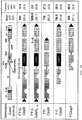
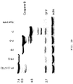

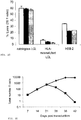
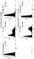


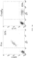


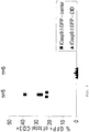

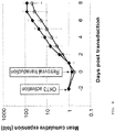

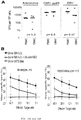
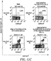


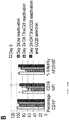
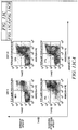
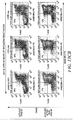
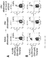
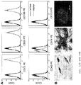

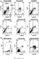
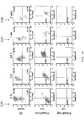
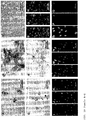


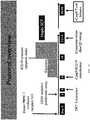


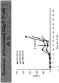


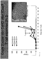
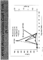
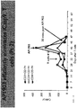
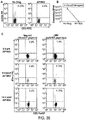


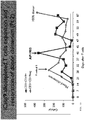

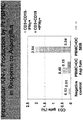
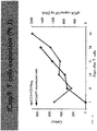
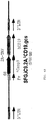

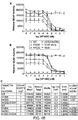

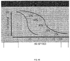


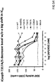
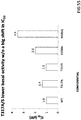
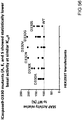
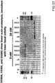
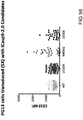
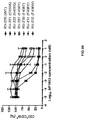

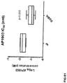
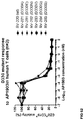
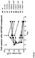
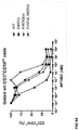
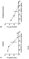
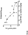
Classifications
-
- A—HUMAN NECESSITIES
- A61—MEDICAL OR VETERINARY SCIENCE; HYGIENE
- A61K—PREPARATIONS FOR MEDICAL, DENTAL OR TOILETRY PURPOSES
- A61K38/00—Medicinal preparations containing peptides
- A61K38/16—Peptides having more than 20 amino acids; Gastrins; Somatostatins; Melanotropins; Derivatives thereof
- A61K38/43—Enzymes; Proenzymes; Derivatives thereof
- A61K38/46—Hydrolases (3)
- A61K38/48—Hydrolases (3) acting on peptide bonds (3.4)
- A61K38/4873—Cysteine endopeptidases (3.4.22), e.g. stem bromelain, papain, ficin, cathepsin H
-
- A—HUMAN NECESSITIES
- A61—MEDICAL OR VETERINARY SCIENCE; HYGIENE
- A61K—PREPARATIONS FOR MEDICAL, DENTAL OR TOILETRY PURPOSES
- A61K35/00—Medicinal preparations containing materials or reaction products thereof with undetermined constitution
- A61K35/12—Materials from mammals; Compositions comprising non-specified tissues or cells; Compositions comprising non-embryonic stem cells; Genetically modified cells
- A61K35/14—Blood; Artificial blood
- A61K35/17—Lymphocytes; B-cells; T-cells; Natural killer cells; Interferon-activated or cytokine-activated lymphocytes
-
- A—HUMAN NECESSITIES
- A61—MEDICAL OR VETERINARY SCIENCE; HYGIENE
- A61K—PREPARATIONS FOR MEDICAL, DENTAL OR TOILETRY PURPOSES
- A61K35/00—Medicinal preparations containing materials or reaction products thereof with undetermined constitution
- A61K35/12—Materials from mammals; Compositions comprising non-specified tissues or cells; Compositions comprising non-embryonic stem cells; Genetically modified cells
- A61K35/28—Bone marrow; Haematopoietic stem cells; Mesenchymal stem cells of any origin, e.g. adipose-derived stem cells
-
- A—HUMAN NECESSITIES
- A61—MEDICAL OR VETERINARY SCIENCE; HYGIENE
- A61K—PREPARATIONS FOR MEDICAL, DENTAL OR TOILETRY PURPOSES
- A61K35/00—Medicinal preparations containing materials or reaction products thereof with undetermined constitution
- A61K35/12—Materials from mammals; Compositions comprising non-specified tissues or cells; Compositions comprising non-embryonic stem cells; Genetically modified cells
- A61K35/30—Nerves; Brain; Eyes; Corneal cells; Cerebrospinal fluid; Neuronal stem cells; Neuronal precursor cells; Glial cells; Oligodendrocytes; Schwann cells; Astroglia; Astrocytes; Choroid plexus; Spinal cord tissue
-
- A—HUMAN NECESSITIES
- A61—MEDICAL OR VETERINARY SCIENCE; HYGIENE
- A61K—PREPARATIONS FOR MEDICAL, DENTAL OR TOILETRY PURPOSES
- A61K35/00—Medicinal preparations containing materials or reaction products thereof with undetermined constitution
- A61K35/12—Materials from mammals; Compositions comprising non-specified tissues or cells; Compositions comprising non-embryonic stem cells; Genetically modified cells
- A61K35/48—Reproductive organs
- A61K35/54—Ovaries; Ova; Ovules; Embryos; Foetal cells; Germ cells
- A61K35/545—Embryonic stem cells; Pluripotent stem cells; Induced pluripotent stem cells; Uncharacterised stem cells
-
- A—HUMAN NECESSITIES
- A61—MEDICAL OR VETERINARY SCIENCE; HYGIENE
- A61K—PREPARATIONS FOR MEDICAL, DENTAL OR TOILETRY PURPOSES
- A61K38/00—Medicinal preparations containing peptides
- A61K38/16—Peptides having more than 20 amino acids; Gastrins; Somatostatins; Melanotropins; Derivatives thereof
-
- A—HUMAN NECESSITIES
- A61—MEDICAL OR VETERINARY SCIENCE; HYGIENE
- A61K—PREPARATIONS FOR MEDICAL, DENTAL OR TOILETRY PURPOSES
- A61K38/00—Medicinal preparations containing peptides
- A61K38/16—Peptides having more than 20 amino acids; Gastrins; Somatostatins; Melanotropins; Derivatives thereof
- A61K38/17—Peptides having more than 20 amino acids; Gastrins; Somatostatins; Melanotropins; Derivatives thereof from animals; from humans
- A61K38/177—Receptors; Cell surface antigens; Cell surface determinants
-
- A—HUMAN NECESSITIES
- A61—MEDICAL OR VETERINARY SCIENCE; HYGIENE
- A61K—PREPARATIONS FOR MEDICAL, DENTAL OR TOILETRY PURPOSES
- A61K39/00—Medicinal preparations containing antigens or antibodies
- A61K39/0005—Vertebrate antigens
-
- A—HUMAN NECESSITIES
- A61—MEDICAL OR VETERINARY SCIENCE; HYGIENE
- A61K—PREPARATIONS FOR MEDICAL, DENTAL OR TOILETRY PURPOSES
- A61K39/00—Medicinal preparations containing antigens or antibodies
- A61K39/46—Cellular immunotherapy
- A61K39/461—Cellular immunotherapy characterised by the cell type used
- A61K39/4611—T-cells, e.g. tumor infiltrating lymphocytes [TIL], lymphokine-activated killer cells [LAK] or regulatory T cells [Treg]
-
- A—HUMAN NECESSITIES
- A61—MEDICAL OR VETERINARY SCIENCE; HYGIENE
- A61K—PREPARATIONS FOR MEDICAL, DENTAL OR TOILETRY PURPOSES
- A61K39/00—Medicinal preparations containing antigens or antibodies
- A61K39/46—Cellular immunotherapy
- A61K39/462—Cellular immunotherapy characterized by the effect or the function of the cells
- A61K39/4621—Cellular immunotherapy characterized by the effect or the function of the cells immunosuppressive or immunotolerising
-
- A—HUMAN NECESSITIES
- A61—MEDICAL OR VETERINARY SCIENCE; HYGIENE
- A61K—PREPARATIONS FOR MEDICAL, DENTAL OR TOILETRY PURPOSES
- A61K39/00—Medicinal preparations containing antigens or antibodies
- A61K39/46—Cellular immunotherapy
- A61K39/463—Cellular immunotherapy characterised by recombinant expression
- A61K39/4631—Chimeric Antigen Receptors [CAR]
-
- A—HUMAN NECESSITIES
- A61—MEDICAL OR VETERINARY SCIENCE; HYGIENE
- A61K—PREPARATIONS FOR MEDICAL, DENTAL OR TOILETRY PURPOSES
- A61K39/00—Medicinal preparations containing antigens or antibodies
- A61K39/46—Cellular immunotherapy
- A61K39/464—Cellular immunotherapy characterised by the antigen targeted or presented
- A61K39/4643—Vertebrate antigens
- A61K39/46434—Antigens related to induction of tolerance to non-self
-
- A—HUMAN NECESSITIES
- A61—MEDICAL OR VETERINARY SCIENCE; HYGIENE
- A61K—PREPARATIONS FOR MEDICAL, DENTAL OR TOILETRY PURPOSES
- A61K39/00—Medicinal preparations containing antigens or antibodies
- A61K39/46—Cellular immunotherapy
- A61K39/464—Cellular immunotherapy characterised by the antigen targeted or presented
- A61K39/4643—Vertebrate antigens
- A61K39/4644—Cancer antigens
- A61K39/464402—Receptors, cell surface antigens or cell surface determinants
- A61K39/464403—Receptors for growth factors
- A61K39/464404—Epidermal growth factor receptors [EGFR]
-
- A—HUMAN NECESSITIES
- A61—MEDICAL OR VETERINARY SCIENCE; HYGIENE
- A61K—PREPARATIONS FOR MEDICAL, DENTAL OR TOILETRY PURPOSES
- A61K39/00—Medicinal preparations containing antigens or antibodies
- A61K39/46—Cellular immunotherapy
- A61K39/464—Cellular immunotherapy characterised by the antigen targeted or presented
- A61K39/4643—Vertebrate antigens
- A61K39/4644—Cancer antigens
- A61K39/464402—Receptors, cell surface antigens or cell surface determinants
- A61K39/464403—Receptors for growth factors
- A61K39/464406—Her-2/neu/ErbB2, Her-3/ErbB3 or Her 4/ ErbB4
-
- A—HUMAN NECESSITIES
- A61—MEDICAL OR VETERINARY SCIENCE; HYGIENE
- A61K—PREPARATIONS FOR MEDICAL, DENTAL OR TOILETRY PURPOSES
- A61K39/00—Medicinal preparations containing antigens or antibodies
- A61K39/46—Cellular immunotherapy
- A61K39/464—Cellular immunotherapy characterised by the antigen targeted or presented
- A61K39/4643—Vertebrate antigens
- A61K39/4644—Cancer antigens
- A61K39/464402—Receptors, cell surface antigens or cell surface determinants
- A61K39/464403—Receptors for growth factors
- A61K39/464409—Vascular endothelial growth factor receptors [VEGFR]
-
- A—HUMAN NECESSITIES
- A61—MEDICAL OR VETERINARY SCIENCE; HYGIENE
- A61K—PREPARATIONS FOR MEDICAL, DENTAL OR TOILETRY PURPOSES
- A61K39/00—Medicinal preparations containing antigens or antibodies
- A61K39/46—Cellular immunotherapy
- A61K39/464—Cellular immunotherapy characterised by the antigen targeted or presented
- A61K39/4643—Vertebrate antigens
- A61K39/4644—Cancer antigens
- A61K39/464402—Receptors, cell surface antigens or cell surface determinants
- A61K39/464411—Immunoglobulin superfamily
- A61K39/464412—CD19 or B4
-
- A—HUMAN NECESSITIES
- A61—MEDICAL OR VETERINARY SCIENCE; HYGIENE
- A61K—PREPARATIONS FOR MEDICAL, DENTAL OR TOILETRY PURPOSES
- A61K39/00—Medicinal preparations containing antigens or antibodies
- A61K39/46—Cellular immunotherapy
- A61K39/464—Cellular immunotherapy characterised by the antigen targeted or presented
- A61K39/4643—Vertebrate antigens
- A61K39/4644—Cancer antigens
- A61K39/464469—Tumor associated carbohydrates
- A61K39/464471—Gangliosides, e.g. GM2, GD2 or GD3
-
- A—HUMAN NECESSITIES
- A61—MEDICAL OR VETERINARY SCIENCE; HYGIENE
- A61K—PREPARATIONS FOR MEDICAL, DENTAL OR TOILETRY PURPOSES
- A61K39/00—Medicinal preparations containing antigens or antibodies
- A61K39/46—Cellular immunotherapy
- A61K39/464—Cellular immunotherapy characterised by the antigen targeted or presented
- A61K39/4643—Vertebrate antigens
- A61K39/4644—Cancer antigens
- A61K39/46448—Cancer antigens from embryonic or fetal origin
- A61K39/464482—Carcinoembryonic antigen [CEA]
-
- A—HUMAN NECESSITIES
- A61—MEDICAL OR VETERINARY SCIENCE; HYGIENE
- A61K—PREPARATIONS FOR MEDICAL, DENTAL OR TOILETRY PURPOSES
- A61K39/00—Medicinal preparations containing antigens or antibodies
- A61K39/46—Cellular immunotherapy
- A61K39/464—Cellular immunotherapy characterised by the antigen targeted or presented
- A61K39/4643—Vertebrate antigens
- A61K39/4644—Cancer antigens
- A61K39/464484—Cancer testis antigens, e.g. SSX, BAGE, GAGE or SAGE
- A61K39/464486—MAGE
-
- A—HUMAN NECESSITIES
- A61—MEDICAL OR VETERINARY SCIENCE; HYGIENE
- A61K—PREPARATIONS FOR MEDICAL, DENTAL OR TOILETRY PURPOSES
- A61K39/00—Medicinal preparations containing antigens or antibodies
- A61K39/46—Cellular immunotherapy
- A61K39/464—Cellular immunotherapy characterised by the antigen targeted or presented
- A61K39/4643—Vertebrate antigens
- A61K39/4644—Cancer antigens
- A61K39/464484—Cancer testis antigens, e.g. SSX, BAGE, GAGE or SAGE
- A61K39/464488—NY-ESO
-
- A—HUMAN NECESSITIES
- A61—MEDICAL OR VETERINARY SCIENCE; HYGIENE
- A61K—PREPARATIONS FOR MEDICAL, DENTAL OR TOILETRY PURPOSES
- A61K39/00—Medicinal preparations containing antigens or antibodies
- A61K39/46—Cellular immunotherapy
- A61K39/464—Cellular immunotherapy characterised by the antigen targeted or presented
- A61K39/4643—Vertebrate antigens
- A61K39/4644—Cancer antigens
- A61K39/46449—Melanoma antigens
- A61K39/464491—Melan-A/MART
-
- A—HUMAN NECESSITIES
- A61—MEDICAL OR VETERINARY SCIENCE; HYGIENE
- A61K—PREPARATIONS FOR MEDICAL, DENTAL OR TOILETRY PURPOSES
- A61K39/00—Medicinal preparations containing antigens or antibodies
- A61K39/46—Cellular immunotherapy
- A61K39/464—Cellular immunotherapy characterised by the antigen targeted or presented
- A61K39/4643—Vertebrate antigens
- A61K39/4644—Cancer antigens
- A61K39/46449—Melanoma antigens
- A61K39/464492—Glycoprotein 100 [Gp100]
-
- A—HUMAN NECESSITIES
- A61—MEDICAL OR VETERINARY SCIENCE; HYGIENE
- A61K—PREPARATIONS FOR MEDICAL, DENTAL OR TOILETRY PURPOSES
- A61K39/00—Medicinal preparations containing antigens or antibodies
- A61K39/46—Cellular immunotherapy
- A61K39/464—Cellular immunotherapy characterised by the antigen targeted or presented
- A61K39/4643—Vertebrate antigens
- A61K39/4644—Cancer antigens
- A61K39/464493—Prostate associated antigens e.g. Prostate stem cell antigen [PSCA]; Prostate carcinoma tumor antigen [PCTA]; Prostatic acid phosphatase [PAP]; Prostate-specific G-protein-coupled receptor [PSGR]
- A61K39/464495—Prostate specific membrane antigen [PSMA]
-
- A—HUMAN NECESSITIES
- A61—MEDICAL OR VETERINARY SCIENCE; HYGIENE
- A61K—PREPARATIONS FOR MEDICAL, DENTAL OR TOILETRY PURPOSES
- A61K48/00—Medicinal preparations containing genetic material which is inserted into cells of the living body to treat genetic diseases; Gene therapy
- A61K48/005—Medicinal preparations containing genetic material which is inserted into cells of the living body to treat genetic diseases; Gene therapy characterised by an aspect of the 'active' part of the composition delivered, i.e. the nucleic acid delivered
-
- A—HUMAN NECESSITIES
- A61—MEDICAL OR VETERINARY SCIENCE; HYGIENE
- A61P—SPECIFIC THERAPEUTIC ACTIVITY OF CHEMICAL COMPOUNDS OR MEDICINAL PREPARATIONS
- A61P1/00—Drugs for disorders of the alimentary tract or the digestive system
- A61P1/04—Drugs for disorders of the alimentary tract or the digestive system for ulcers, gastritis or reflux esophagitis, e.g. antacids, inhibitors of acid secretion, mucosal protectants
-
- A—HUMAN NECESSITIES
- A61—MEDICAL OR VETERINARY SCIENCE; HYGIENE
- A61P—SPECIFIC THERAPEUTIC ACTIVITY OF CHEMICAL COMPOUNDS OR MEDICINAL PREPARATIONS
- A61P17/00—Drugs for dermatological disorders
-
- A—HUMAN NECESSITIES
- A61—MEDICAL OR VETERINARY SCIENCE; HYGIENE
- A61P—SPECIFIC THERAPEUTIC ACTIVITY OF CHEMICAL COMPOUNDS OR MEDICINAL PREPARATIONS
- A61P17/00—Drugs for dermatological disorders
- A61P17/14—Drugs for dermatological disorders for baldness or alopecia
-
- A—HUMAN NECESSITIES
- A61—MEDICAL OR VETERINARY SCIENCE; HYGIENE
- A61P—SPECIFIC THERAPEUTIC ACTIVITY OF CHEMICAL COMPOUNDS OR MEDICINAL PREPARATIONS
- A61P19/00—Drugs for skeletal disorders
-
- A—HUMAN NECESSITIES
- A61—MEDICAL OR VETERINARY SCIENCE; HYGIENE
- A61P—SPECIFIC THERAPEUTIC ACTIVITY OF CHEMICAL COMPOUNDS OR MEDICINAL PREPARATIONS
- A61P19/00—Drugs for skeletal disorders
- A61P19/08—Drugs for skeletal disorders for bone diseases, e.g. rachitism, Paget's disease
-
- A—HUMAN NECESSITIES
- A61—MEDICAL OR VETERINARY SCIENCE; HYGIENE
- A61P—SPECIFIC THERAPEUTIC ACTIVITY OF CHEMICAL COMPOUNDS OR MEDICINAL PREPARATIONS
- A61P21/00—Drugs for disorders of the muscular or neuromuscular system
- A61P21/04—Drugs for disorders of the muscular or neuromuscular system for myasthenia gravis
-
- A—HUMAN NECESSITIES
- A61—MEDICAL OR VETERINARY SCIENCE; HYGIENE
- A61P—SPECIFIC THERAPEUTIC ACTIVITY OF CHEMICAL COMPOUNDS OR MEDICINAL PREPARATIONS
- A61P25/00—Drugs for disorders of the nervous system
-
- A—HUMAN NECESSITIES
- A61—MEDICAL OR VETERINARY SCIENCE; HYGIENE
- A61P—SPECIFIC THERAPEUTIC ACTIVITY OF CHEMICAL COMPOUNDS OR MEDICINAL PREPARATIONS
- A61P27/00—Drugs for disorders of the senses
- A61P27/02—Ophthalmic agents
-
- A—HUMAN NECESSITIES
- A61—MEDICAL OR VETERINARY SCIENCE; HYGIENE
- A61P—SPECIFIC THERAPEUTIC ACTIVITY OF CHEMICAL COMPOUNDS OR MEDICINAL PREPARATIONS
- A61P3/00—Drugs for disorders of the metabolism
-
- A—HUMAN NECESSITIES
- A61—MEDICAL OR VETERINARY SCIENCE; HYGIENE
- A61P—SPECIFIC THERAPEUTIC ACTIVITY OF CHEMICAL COMPOUNDS OR MEDICINAL PREPARATIONS
- A61P35/00—Antineoplastic agents
-
- A—HUMAN NECESSITIES
- A61—MEDICAL OR VETERINARY SCIENCE; HYGIENE
- A61P—SPECIFIC THERAPEUTIC ACTIVITY OF CHEMICAL COMPOUNDS OR MEDICINAL PREPARATIONS
- A61P37/00—Drugs for immunological or allergic disorders
- A61P37/02—Immunomodulators
-
- A—HUMAN NECESSITIES
- A61—MEDICAL OR VETERINARY SCIENCE; HYGIENE
- A61P—SPECIFIC THERAPEUTIC ACTIVITY OF CHEMICAL COMPOUNDS OR MEDICINAL PREPARATIONS
- A61P37/00—Drugs for immunological or allergic disorders
- A61P37/02—Immunomodulators
- A61P37/04—Immunostimulants
-
- A—HUMAN NECESSITIES
- A61—MEDICAL OR VETERINARY SCIENCE; HYGIENE
- A61P—SPECIFIC THERAPEUTIC ACTIVITY OF CHEMICAL COMPOUNDS OR MEDICINAL PREPARATIONS
- A61P37/00—Drugs for immunological or allergic disorders
- A61P37/02—Immunomodulators
- A61P37/06—Immunosuppressants, e.g. drugs for graft rejection
-
- A—HUMAN NECESSITIES
- A61—MEDICAL OR VETERINARY SCIENCE; HYGIENE
- A61P—SPECIFIC THERAPEUTIC ACTIVITY OF CHEMICAL COMPOUNDS OR MEDICINAL PREPARATIONS
- A61P43/00—Drugs for specific purposes, not provided for in groups A61P1/00-A61P41/00
-
- A—HUMAN NECESSITIES
- A61—MEDICAL OR VETERINARY SCIENCE; HYGIENE
- A61P—SPECIFIC THERAPEUTIC ACTIVITY OF CHEMICAL COMPOUNDS OR MEDICINAL PREPARATIONS
- A61P5/00—Drugs for disorders of the endocrine system
-
- A—HUMAN NECESSITIES
- A61—MEDICAL OR VETERINARY SCIENCE; HYGIENE
- A61P—SPECIFIC THERAPEUTIC ACTIVITY OF CHEMICAL COMPOUNDS OR MEDICINAL PREPARATIONS
- A61P7/00—Drugs for disorders of the blood or the extracellular fluid
-
- A—HUMAN NECESSITIES
- A61—MEDICAL OR VETERINARY SCIENCE; HYGIENE
- A61P—SPECIFIC THERAPEUTIC ACTIVITY OF CHEMICAL COMPOUNDS OR MEDICINAL PREPARATIONS
- A61P7/00—Drugs for disorders of the blood or the extracellular fluid
- A61P7/06—Antianaemics
-
- C—CHEMISTRY; METALLURGY
- C12—BIOCHEMISTRY; BEER; SPIRITS; WINE; VINEGAR; MICROBIOLOGY; ENZYMOLOGY; MUTATION OR GENETIC ENGINEERING
- C12N—MICROORGANISMS OR ENZYMES; COMPOSITIONS THEREOF; PROPAGATING, PRESERVING, OR MAINTAINING MICROORGANISMS; MUTATION OR GENETIC ENGINEERING; CULTURE MEDIA
- C12N5/00—Undifferentiated human, animal or plant cells, e.g. cell lines; Tissues; Cultivation or maintenance thereof; Culture media therefor
- C12N5/0081—Purging biological preparations of unwanted cells
- C12N5/0087—Purging against subsets of blood cells, e.g. purging alloreactive T cells
-
- C—CHEMISTRY; METALLURGY
- C12—BIOCHEMISTRY; BEER; SPIRITS; WINE; VINEGAR; MICROBIOLOGY; ENZYMOLOGY; MUTATION OR GENETIC ENGINEERING
- C12N—MICROORGANISMS OR ENZYMES; COMPOSITIONS THEREOF; PROPAGATING, PRESERVING, OR MAINTAINING MICROORGANISMS; MUTATION OR GENETIC ENGINEERING; CULTURE MEDIA
- C12N9/00—Enzymes; Proenzymes; Compositions thereof; Processes for preparing, activating, inhibiting, separating or purifying enzymes
- C12N9/14—Hydrolases (3)
- C12N9/48—Hydrolases (3) acting on peptide bonds (3.4)
- C12N9/50—Proteinases, e.g. Endopeptidases (3.4.21-3.4.25)
- C12N9/64—Proteinases, e.g. Endopeptidases (3.4.21-3.4.25) derived from animal tissue
- C12N9/6421—Proteinases, e.g. Endopeptidases (3.4.21-3.4.25) derived from animal tissue from mammals
- C12N9/6472—Cysteine endopeptidases (3.4.22)
-
- A—HUMAN NECESSITIES
- A61—MEDICAL OR VETERINARY SCIENCE; HYGIENE
- A61K—PREPARATIONS FOR MEDICAL, DENTAL OR TOILETRY PURPOSES
- A61K2239/00—Indexing codes associated with cellular immunotherapy of group A61K39/46
- A61K2239/31—Indexing codes associated with cellular immunotherapy of group A61K39/46 characterized by the route of administration
-
- A—HUMAN NECESSITIES
- A61—MEDICAL OR VETERINARY SCIENCE; HYGIENE
- A61K—PREPARATIONS FOR MEDICAL, DENTAL OR TOILETRY PURPOSES
- A61K2239/00—Indexing codes associated with cellular immunotherapy of group A61K39/46
- A61K2239/38—Indexing codes associated with cellular immunotherapy of group A61K39/46 characterised by the dose, timing or administration schedule
-
- A—HUMAN NECESSITIES
- A61—MEDICAL OR VETERINARY SCIENCE; HYGIENE
- A61K—PREPARATIONS FOR MEDICAL, DENTAL OR TOILETRY PURPOSES
- A61K2239/00—Indexing codes associated with cellular immunotherapy of group A61K39/46
- A61K2239/46—Indexing codes associated with cellular immunotherapy of group A61K39/46 characterised by the cancer treated
- A61K2239/47—Brain; Nervous system
-
- A—HUMAN NECESSITIES
- A61—MEDICAL OR VETERINARY SCIENCE; HYGIENE
- A61K—PREPARATIONS FOR MEDICAL, DENTAL OR TOILETRY PURPOSES
- A61K2239/00—Indexing codes associated with cellular immunotherapy of group A61K39/46
- A61K2239/46—Indexing codes associated with cellular immunotherapy of group A61K39/46 characterised by the cancer treated
- A61K2239/48—Blood cells, e.g. leukemia or lymphoma
-
- A—HUMAN NECESSITIES
- A61—MEDICAL OR VETERINARY SCIENCE; HYGIENE
- A61K—PREPARATIONS FOR MEDICAL, DENTAL OR TOILETRY PURPOSES
- A61K2239/00—Indexing codes associated with cellular immunotherapy of group A61K39/46
- A61K2239/46—Indexing codes associated with cellular immunotherapy of group A61K39/46 characterised by the cancer treated
- A61K2239/50—Colon
-
- C—CHEMISTRY; METALLURGY
- C12—BIOCHEMISTRY; BEER; SPIRITS; WINE; VINEGAR; MICROBIOLOGY; ENZYMOLOGY; MUTATION OR GENETIC ENGINEERING
- C12N—MICROORGANISMS OR ENZYMES; COMPOSITIONS THEREOF; PROPAGATING, PRESERVING, OR MAINTAINING MICROORGANISMS; MUTATION OR GENETIC ENGINEERING; CULTURE MEDIA
- C12N2501/00—Active agents used in cell culture processes, e.g. differentation
- C12N2501/40—Regulators of development
- C12N2501/48—Regulators of apoptosis
-
- C—CHEMISTRY; METALLURGY
- C12—BIOCHEMISTRY; BEER; SPIRITS; WINE; VINEGAR; MICROBIOLOGY; ENZYMOLOGY; MUTATION OR GENETIC ENGINEERING
- C12N—MICROORGANISMS OR ENZYMES; COMPOSITIONS THEREOF; PROPAGATING, PRESERVING, OR MAINTAINING MICROORGANISMS; MUTATION OR GENETIC ENGINEERING; CULTURE MEDIA
- C12N2510/00—Genetically modified cells
Definitions
- the technology relates in part to methods for inducing partial apoptosis of cells that express an inducible caspase polypeptide.
- the technology further relates in part to methods for inducing partial apoptosis of cells that express an inducible modified caspase polypeptide, having a modified dose response curve to the multimeric ligand inducer.
- the technology also relates in part to methods for cell therapy using cells that express the inducible caspase polypeptide or the inducible modified caspase polypeptide, where the proportion of caspase polypeptide-expressing cells eliminated by apoptosis is related to the administered amount of the multimeric ligand inducer.
- T cell therapies such as therapies using chimeric antigen receptor-expressing T cells (CAR), donor lymphocyte infusions (DLIs), or T cell add-back following hematopoietic stem cell transplants (HSCTs)
- CAR chimeric antigen receptor-expressing T cells
- DLIs donor lymphocyte infusions
- HSCTs T cell add-back following hematopoietic stem cell transplants
- TLS tumor lysis syndrome
- CRS cytokine release syndrome
- MAS macrophage activation syndrome
- a discrete fraction of cells may be eliminated by partial apoptosis, allowing beneficial effects of the cellular therapy to remain.
- An example of a cellular therapy is adoptive T cell transfer after CD34 + stem cell transplantation. Administering T cells after stem cell transfer helps to accelerate the reconstitution of an immune system in the patient recipient.
- the T cells may be obtained from, for example, a matched related or unrelated donor. When a matched related or unrelated donor is not available, or the disease is too aggressive for an extensive donor search, the use of an HLA haploidentical family donor may be effective. Such donors may be parents, siblings, or second-degree relatives. Such infusions may enhance immune recovery and thereby reduce virus infections and eliminate relapsing leukemia cells.
- graft-versus-host disease GvHD
- An inducible caspase-9 system may be applied to human T cells, which are then administered to stem cell transplantation patients.
- caspase-9 is activated after the administration of a multimeric ligand, which causes dimerization of the protein and induced apoptosis of the allogeneic activated T cells.
- a caspase-9 based apoptotic safety switch may also be applied to therapeutic chimeric antigen receptor-expressing cells, which express artificial receptors designed to convey antigen specificity to T cells. They include an antigen-specific component, a transmembrane component, and an intracellular component selected to activate the T cell and provide specific immunity. Chimeric antigen receptor-expressing T cells may be used in various therapies, including cancer therapies. While effective against tumors, in some cases these therapies have led to side effects due, in part to non-specific attacks on healthy tissue.
- An inducible caspase-9 system may be provided in these therapeutic cells before administering them to a patient, to provide the ability to selectively kill the therapeutic cells if the patient experiences negative side effects, such as, for example, on-target but off-organ toxicity, where the wrong organ is targeted by the chimeric antigen receptor.
- caspase-9 based apoptotic safety switch may be used for the elimination of tissue stem cells and their progeny, and for augmentation of oncolytic virus-mediated tumor killing where too complete killing might limit the oncolytic effect.
- Methods featured in some embodiments include methods of inducing apoptosis in discrete fractions of caspase-9-expressing cells.
- a percentage of therapeutic cells causing the graft vs. host disease may be eliminated, while leaving a sufficient number of therapeutic cells to assist in the reconstitution of the patient's immune system.
- a percentage of chimeric antigen receptor-expressing therapeutic cells may be eliminated, while leaving a sufficient number of the cells to continue their therapeutic effect.
- one of the populations of therapeutic cells may be eliminated upon the occurrence of an adverse event, without significantly affecting the proportion of the other population of therapeutic T cells.
- featured in some embodiments are methods for controlling the survival of transplanted therapeutic cells in a subject, comprising preparing or obtaining therapeutic cells; transfecting or transducing the therapeutic cells with a nucleic acid that encodes a chimeric protein comprising a multimeric ligand binding region and a caspase-9 polypeptide or a modified caspase-9 polypeptide, wherein the caspase-9 polypeptide or the modified caspase-9 polypeptide comprises an amino acid sequence having at least 90% sequence identity to SEQ ID NO: 9; transplanting the transduced or transfected therapeutic cells into the subject; and after transplantation, administering an effective amount of a multimeric ligand that binds to the multimeric ligand binding region to the subject, wherein less than 80% of transplanted therapeutic cells that express the caspase-9 polypeptide or the modified caspase-9 polypeptide are killed following administration of the multimeric ligand; wherein the modified caspase-9 polypeptide has a reduced IC 50 and/or an elongated dose response
- the therapeutic cells are selected from the group consisting of hematopoietic stem cells, inducible progenitor cells (iPS), embryonic stem (ES) cells, mesenchymal stem cells, plasma (B) cells, myocytes, tumor infiltrating lymphocytes, and T cells.
- iPS inducible progenitor cells
- ES embryonic stem
- B plasma
- myocytes tumor infiltrating lymphocytes
- T cells tumor infiltrating lymphocytes
- the subject has undergone a stem cell transplant, for example, a transplant that is haplo-identical, matched unrelated, or matched related.
- the subject has been diagnosed with a hyperproliferative disease. In other embodiments, the subject has been diagnosed with an immune disease.
- the modified caspase-9 polypeptide comprises an amino acid substitution selected from the group consisting of the caspase variants in Table 3. In some embodiments, the modified caspase-9 polypeptide comprises an amino acid substitution selected from the group consisting of N405Q, F404Y, F406L, F406T, F404W, and the caspase-9 polypeptide containing the substitution of dimerization domain amino acid residues 402-406 (GCFNF)-from caspase-9 with the equivalent position residues of Caspase10(GCFNF 402 ISAQT). In some embodiments, the modified caspase-9 polypeptide comprises an amino acid substitution selected from the group consisting of D330A and T317A.
- the modified caspase-9 polypeptide comprises an amino acid substitution selected from the group consisting of T317S, S144A, S144D, S196A, S183A, and S195A
- the modified caspase-9 polypeptide comprises an amino acid substitution selected from the group consisting of D330A-N405Q, D330A-S144A, D330A-S144D, D330AS196A, D330A-T317A, and D330A-S183A
- the modified caspase-9 polypeptide comprises an amino acid substitution selected from the group consisting of F404T, F404W, N405F, and F406T
- the modified caspase-9 polypeptide comprises an amino acid substitution selected from the group consisting of D315A, A316G, T317S, F319W, and S307A
- the modified caspase-9 polypeptide comprises an amino acid substitution selected from the group consisting of Y153A and Y153F
- the modified caspase-9 polypeptide comprises an amino acid substitution selected
- Also provided are methods for controlling the survival of transplanted therapeutic cells in a subject comprising preparing or obtaining therapeutic cells; transfecting or transducing a first subset of the therapeutic cells with a nucleic acid that encodes a chimeric protein comprising a multimeric ligand binding region and a first caspase-9 polypeptide, wherein the first caspase-9 polypeptide comprises an amino acid sequence having at least 90% sequence identity to SEQ ID NO: 9; transfecting or transducing a second subset of the therapeutic cells with a nucleic acid that encodes a chimeric protein comprising a multimeric ligand binding region and a second caspase-9 polypeptide, wherein the second caspase-9 polypeptide comprises an amino acid sequence having at least 90% sequence identity to SEQ ID NO: 9; transplanting the transduced or transfected first and second therapeutic cells into the subject; and after transplantation, administering an effective amount of a multimeric ligand that binds to the multimeric ligand binding region to the subject, wherein
- the first caspase-9 polypeptide has a reduced IC 50 and an elongated dose response curve in response to the multimeric ligand, when compared to the second caspase-9 polypeptide.
- the therapeutic cells are selected from the group consisting of hematopoietic stem cells, inducible progenitor cells (iPS), embryonic stem (ES) cells, mesenchymal stem cells, plasma (B) cells, myocytes and T cells.
- the subject has been diagnosed with a hyperproliferative disease. In some embodiments, the subject has been diagnosed with an immune disease.
- the first or second caspase-9 polypeptide comprises an amino acid substitution selected from the group consisting of F319W, F404Y, A316G, Y153A, F406L, C403A, N405Q, C285A, F406T, or the caspase-9 polypeptide comprises ISAQT, the corresponding amino acid sequence of the dimerization domain in Caspase 10, or the first or second caspase-9 polypeptide comprises an amino acid substitution selected from the group consisting of N405Q, C285A, and F406T.
- the second caspase-9 polypeptide has an IC 50 for the multimeric ligand greater than 0.01 pM, 0.05 pM, 0.1 pM, 0.5 pM, 0.01 nM, 0.05 nM, 0.1 nM, 0.5 nM, or 1 nM.
- a therapeutically effective level of the first therapeutic cells comprising the chimeric antigen receptor remains active in the subject following administration of the multimeric ligand.
- the second therapeutic cells are T cells, for example, T cells administered to a subject following stem cell transplantation.
- the T cells are allodepleted before administration to the subject.
- the T cells are not allodepleted before administration to the subject.
- the second therapeutic cell comprises a chimeric antigen receptor.
- the first therapeutic cells are T cells.
- the second therapeutic cells are T cells administered to a subject following stem cell transplantation.
- the T cells are allodepleted before administration to the subject.
- the T cells are not allodepleted before administration to the subject.
- the modified caspase-9 polypeptide has an IC 50 for the multimeric ligand greater than 0.01 pM, 0.05 pM, 0.1 pM, 0.5 pM, 0.01 nM, 0.05 nM, 0.1 nM, 0.5 nM, or 1 nM.
- the multimeric ligand binding region is selected from the group consisting of FKBP ligand-binding region, cyclophilin receptor ligand-binding region, steroid receptor ligand-binding region, cyclophilin receptor ligand-binding region, and tetracycline receptor ligand-binding region.
- the ligand-binding region comprises a F v ⁇ F vls amino acid sequence.
- the ligand is a small molecule.
- the ligand is dimeric.
- the ligand is dimeric FK506, or a dimeric FK506-like analog.
- the multimeric ligand is AP1903.
- the multimeric ligand is AP20187.
- the cells are T cells.
- the chimeric protein further comprises a marker polypeptide.
- the methods further comprise a selection step, wherein cells that express the marker are selected for administration to the subject.
- the methods further comprise administering a second dose of the multimeric ligand to the subject, wherein the second dose comprises more multimeric ligand than the first dose.
- multiple doses of multimeric ligand are administered to the subject, with an escalation of dosage levels among the multiple doses.
- the escalation of dosage levels increases the number of therapeutic cells that are killed.
- the dose is escalated from 0.01 to 1 mg/kg.
- the doses are administered in increments of about 15 to 30 minutes.
- the multimeric ligand is administered using a continuous infusion pump, and the concentration of multimeric ligand is increased during the infusion.
- the concentration of multimeric ligand is increased until the desired percentage of therapeutic cells is killed.
- the subject has graft vs. host disease and the administration of the multimeric ligand alleviates the disease.
- the subject is human.
- the therapeutic cell comprises a chimeric antigen receptor.
- the subject exhibits symptoms of off-target toxicity before administration of the multimeric ligand.
- the subject exhibits symptoms of tumor lysis syndrome (TLS), cytokine release syndrome (CRS) or macrophage activation syndrome (MAS) before administration of the multimeric ligand.
- TLS tumor lysis syndrome
- CRS cytokine release syndrome
- MAS macrophage activation syndrome
- the administration of the multimeric ligand alleviates the off-target or off-organ toxicity.
- off-target toxicity is provided in, for example, Heslop, H.E., Blood 122:853-854 (2013 ).
- the administration of the multimeric ligand alleviates the tumor lysis syndrome (TLS), cytokine release syndrome (CRS) or macrophage activation syndrome (MAS).
- TLS tumor lysis syndrome
- CRS cytokine release syndrome
- MAS macrophage activation syndrome
- a therapeutically effective level of therapeutic cells comprising the chimeric antigen receptor remains active in the subject following administration of the multimeric ligand.
- the patient has cancer. In some embodiments, the patient has a solid tumor. In some embodiments, the cancer is present in the blood or bone marrow of the patient. In some embodiments, the patient has a blood or bone marrow disease. In some embodiments, the patient has been diagnosed with any condition or disorder that can be alleviated by stem cell transplantation. In some embodiments, the patient has been diagnosed with sickle cell anemia or metachromatic leukodystrophy. In some embodiments, the promoter is activated in activated T cells. In certain embodiments, the caspase-9 polypeptide is a truncated caspase-9 polypeptide, or the caspase-9 polypeptide lacks the Caspase recruitment domain. In some embodiments, the patient exhibits one or more Stage 1, 2, 3, or 4 graft versus host disease symptoms.
- the number of alloreactive T cells is reduced.
- the alloreactive T cells express a marker and CD3.
- the number of alloreactive T cells is reduced by about 90% or more after administration of the multimeric ligand.
- donor T cells survive in the patient that are able to expand and are reactive to viruses and fungi.
- donor T cells survive in the patient that are able to expand and are reactive to tumor cells in the patient.
- the patients have received haplo-CD34 + stem cell transplants before or at the same time as administration of the donor T cells.
- the inducible chimeric caspase-9 polypeptide has been modified to have a different sensitivity to the ligand inducer, or to have a different basal activity in the transduced or transfected cell, when compared to wild type caspase-9 polypeptide, or wild type caspase-9 polypeptide that has been truncated to remove the CARD domain.
- the methods of the present application use chimeric polypeptides comprising modified caspase-9 polypeptides, including, for example, iCasp9 D330A, iCasp9 N405Q, and iCasp9 D330A N405Q, demonstrated low to undetectable basal activity, respectively, with a minimum deleterious effect on their AP1903 IC 50 in a SEAP reporter-based, surrogate killing assay.
- a cell which comprises a polynucleotide that encodes a chimeric protein comprising a multimerization region and a modified caspase-9 polypeptide, wherein the modified caspase-9 polypeptide comprises an amino acid sequence having at least 90% sequence identity to SEQ ID NO: 9, and comprises at least one amino acid substitution selected from the group consisting of S144A, S144D, Y153A, Y153F, S183A, S195A, S196A, S196D, S307A, D315A, A316G, T317A, T317C, T317E, T317S, P318A, F319A, F319W, F326K, D327G, D327K, D327R, Q328K, Q328R, L329E, L329G, L329K, D330A, D330E, D330G, D330N, D330S, D330V, A331K, C403A, C
- the at least one amino acid substitution is selected from the group consisting of S144A, S144D, Y153A, Y153F, S183A, S195A, S196A, S196D, S307A, D315A, A316G, T317A, T317C, T317E, T317S, P318A, F319A, F319W, F326K, D327G, D327K, D327R, Q328K, Q328R, L329E, L329G, L329K, D330A, D330E, D330G, D330N, D330S, D330V, A331K, C403A, C403S, C403T, F404T, F404W, F404Y, N405A, N405F, N405Q, N405T, F406A, F406T, F406W, and F406Y.
- the at least one amino acid substitution is selected from the group consisting of S144A, S144D, Y153A, Y153F, S183A, S195A, S196A, S196D, S307A, D315A, A316G, T317A, T317C, T317S, P318A, F319A, F319W, L329E, D330A, D330E, D330G, D330N, D330S, D330V, C403A, C403S, C403T, F404T, F404W, F404Y, N405A, N405F, N405Q, F406A, F406T, F406W, and F406Y.
- the at least one amino acid substitution is S144A, S144D, Y153A, Y153F, S183A, S195A, S196A, S196D, S307A, D315A, A316G, T317A, T317S, F319W, L329E, D330A, D330E, D330G, D330N, D330S, D330V, F404T, F404W, F404Y, N405F, N405Q, F406A, F406T, F406W, and F406Y.
- the at least one amino acid substitution is S144A, S144D, S183A, S195A, S196A, S196D, T317A, T317S, L329E, D330A, D330E, D330G, D330N, D330S, D330V, F404Y, N405Q, F406A, F406W, and F406Y.
- the at least one amino acid substitution is selected from the group consisting of T317S, S144A, S133, and S196D.
- the at least one amino acid substitution is selected from the group consisting of S183A, S195A, S196A, S196D, T317A, L329E, D330A, D330E, D330G, D330N, D330S, D330V, F404Y, N405Q, F406A, F406W, and F406Y.
- the at least one amino acid substitution is D330A.
- the at least one amino acid substitution is D330E.
- the at least one amino acid substitution is N405Q.
- the modified caspase-9 polypeptide comprises at least two amino acid substitutions selected from the group consisting of D330A-N405Q, D330A-S144A, D330A-S144D, D330A-S183A, D330A-S196A, N405Q-S144A, N405Q-S144D, N405Q-S196D, N405Q-T317S, N405Q-S144Aco, N405Q-T317Sco, 402 GCFNF 406 ISAQT (CASP-10), 316 ATPF 319 AVPI (SMAC/Diablo), D330A-N405T, D315A-D330A, D330A-Y153A, D330A-Y153F, D330A-T317E, 402 GCFNF 406 CIVSM (CASP-3), 402 GCFNF 406 AAAAA, 402 GCFNF 406 YCSTL (CASP-2), and 402 GFNF 406 QPTFT (
- the modified caspase-9 polypeptide comprises at least two amino acid substitutions selected from the group consisting of D330A-N405Q, D330A-S144A, D330A-S144D, D330AS183A, D330A-S196A, N405Q-S144A, N405Q-S144D, N405Q-S196D, N405Q-T317S, N405Q-S144Aco, N405Q-T317Sco, 402 GCFNF 406 ISAQT (CASP-10), 316 ATPF 319 AVPI (SMAC/Diablo), and D330A-N405T.
- the modified caspase-9 polypeptide comprises at least two amino acid substitutions selected from the group consisting of D330A-N405Q, D330A-S144A, D330A-S144D, D330A-S183A, D330A-S196A, N405Q-S144A, N405Q-S144D, N405Q-S196D, N405Q-T317S, N405Q-S144Aco, N405Q-T317Sco, 402 GCFNF 406 ISAQT (CASP-10), and 316 ATPF 319 AVPI (SMAC/Diablo).
- the modified caspase-9 polypeptide comprises at least two amino acid substitutions selected from the group consisting of D330AN405Q, D330A-S144A, D330A-S144D, D330A-S183A, D330A-S196A, N405Q-S144A, N405Q-S144D, N405Q-S196D, N405Q-T317S, N405Q-S144Aco, N405Q-T317Sco, and 402 GCFNF 406 ISAQT (CASP-10).
- the modified caspase-9 polypeptide comprises at least two amino acid substitutions selected from the group consisting of N405Q-S144Aco and N405Q-T317Sco.
- the at least one amino acid substitution is selected from the group consisting of S144A, S144D, Y153A, Y153F, S183A, S195A, S196A, S307A, D315A, A316G, T317A, T317C, T317E, T317S, P318A, F319A, F319W, F326K, D327G, D327K, D327R, Q328K, Q328R, L329E, L329G, L329K, D330A, D330E, D330G, D330N, D330S, D330V, A331K, F404T, F404W, F404YN405F, N405Q, and F406T.
- the at least one amino acid substitution is selected from the group consisting of S144A, S144D, Y153A, Y153F, S183A, S195A, S196A, S307A, D315A, A316G, T317A, T317C, T317E, T317S, P318A, F319A, F319W, F326K, D327G, D327K, D327R, Q328K, Q328R, L329E, L329G, L329K, D330A, D330E, D330G, D330N, D330S, D330V, A331K, F404T, F404W, F404Y, N405F, N405Q, and F406T.
- the at least one amino acid substitution is selected from the group consisting of S144A, S144D, Y153A, Y153F, S183A, S195A, S196A, S307A, D315A, A316G, T317A, T317C, T317S, P318A, F319A, F319W, L329E, D330A, D330E, D330G, D330N, D330S, D330V, F404T, F404W, F404Y, N405F, N405Q, and F406T.
- the at least one amino acid substitution is selected from the group consisting of S144A, S144D, Y153A, Y153F, S183A, S195A, S196A, S307A, D315A, A316G, T317A, T317S, F319W, D330A, F404T, F404W, F404Y, N405F, N405Q, and F406T.
- the at least one amino acid substitution is selected from the group consisting of S144A, S144D, S183A, S195A, S196A, T317A, T317S, D330A, F404Y, and N405Q.
- the at least one amino acid substitution is selected from the group consisting of S196D, T317C, T317E, P318A, F319A, F326K, D327G, D327K, D327R, Q328K, Q328R, L329E, L329G, L329K, D330E, D330G, D330N, D330S, D330V, A331K, C403A, C403S, C403T, N405A, N405T, F406A, F406W, F406Y, G402A, G402I, G402Q, G402Y, C403P, F404A, F404S, and F406L.
- the at least one amino acid substitution is selected from the group consisting of S196D, T317C, T317E, P318A, F319A, F326K, D327G, D327K, D327R, Q328K, Q328R, L329E, L329G, L329K, D330E, D330G, D330N, D330S, D330V, A331K, C403A, C403S, C403T, N405A, N405T, F406A, F406W, and F406Y.
- the at least one amino acid substitution is selected from the group consisting of S196D, T317C, P318A, F319A, L329E, D330E, D330G, D330N, D330S, D330V, C403A, C403S, C403T, N405A, F406A, F406T, F406W, and F406Y.
- the at least one amino acid substitution is selected from the group consisting of S196D, L329E, D330E, D330G, D330N, D330S, D330V, F406A, F406T, F406W, and F406Y.
- the at least one amino acid substitution is selected from the group consisting of S196D, L329E, D330E, D330G, D330N, D330S, D330V, F406A, F406W, and F406Y. 47.
- the at least one amino acid substitution is selected from the group consisting of N405Q, F404Y, F406A, F406W, F406Y, F404T, F404W, N405F, F406T, C403A, C403S, C403T, N405A, and N405T.
- the at least one amino acid substitution is selected from the group consisting of N405Q, F404Y, F406A, F406W, and F406Y. In some embodiments, the at least one amino acid substitution is selected from the group consisting of T317S, D330A, D330E, D330G, D330N, D330S, D330V, L329E, T317A, D315A, A316G, T317C, P318A, F319A, T317E, F326K, D327G, D327K, D327R, Q328K, Q328R, L329G, L329K, and A331K.
- the at least one amino acid substitution is selected from the group consisting of T317S, D330A, D330E, D330G, D330N, D330S, D330V, L329E, and T317A. In some embodiments, the at least one amino acid substitution is selected from the group consisting of S144A, S144D, S196D, S183A, S195A, S196A, Y153A, Y153F, and S307A.
- the polynucleotide comprises optimized codons encoding the caspase-9 polypeptide
- the polynucleotide coding for the modified caspase-9 polypeptide comprises an amino acid substitution of N405Q, and comprises optimized codons.
- the polynucleotide coding for the modified caspase-9 polypeptide comprises the nucleic acid sequence of SEQ ID NO: 39.
- modified caspase polypeptides comprising the amino acid sequences discussed herein as part of the cells which comprise polynucleotides coding for the chimeric modified caspase-9 polypeptides.
- nucleic acids comprising polynucleotides coding for the modified caspase-9polypeptides and the chimeric modified caspase-9 polypeptides.
- vectors comprising the polynucleotides coding for the modified caspase polypeptides and the chimeric modified caspase polypeptides.
- the cell is a human cell.
- the cell of the present application may be any type of eukaryotic cell, for example a mammalian cell, for example a horse, dog, cat, cow, or human cell.
- the cell is a progenitor cell.
- the cell is a hematopoietic progenitor cell.
- the cell is selected from the group consisting of mesenchymal stromal cells, embryonic stem cells, and inducible pluripotent stem cells.
- the cell is a T cell.
- the cell is obtained or prepared from bone marrow.
- the cell is obtained or prepared from umbilical cord blood.
- the cell is obtained or prepared from peripheral blood.
- the cell is obtained or prepared from peripheral blood mononuclear cells.
- the polynucleotide coding for the chimeric polypeptide or modified caspase-9 polypeptide is operably linked to a promoter.
- the promoter is developmentally regulated and the caspase-9 polypeptide is expressed in developmentally differentiated cells.
- the promoter is tissue-specific and the caspase-9 polypeptide is expressed in the specific tissue.
- the promoter is activated in activated T cells.
- the promoter comprises a 5'LTR sequence.
- the chimeric protein further comprises a marker polypeptide, for example, but not limited to, a ⁇ CD19 polypeptide.
- the caspase-9 polypeptide is a truncated caspase-9 polypeptide.
- the caspase-9 polypeptide lacks the Caspase recruitment domain.
- the multimerization region is selected from the group consisting of FKBP, cyclophilin receptor, steroid receptor, tetracycline receptor, heavy chain antibody subunit, light chain antibody subunit, single chain antibodies comprised of heavy and light chain variable regions in tandem separated by a flexible linker domain, and mutated sequences thereof.
- the multimerization region is an FKBP12 region.
- the FKB12 region is an FKB12v 36 region.
- the multimerization region is Fv'Fvls.
- the multimerization region binds a ligand selected from the group consisting of an FK506 dimer and a dimeric FK506 analog ligand.
- the ligand is AP1903, in other embodiments, the ligand is AP20187.
- the multimerization region has an amino acid sequence of SEQ ID NO: 29 or a functional fragment thereof.
- the multimerization region is encoded by a nucleotide sequence in SEQ ID NO: 30, or a functional fragment thereof.
- the multimerization region further comprises a polypeptide having an amino acid sequence of SEQ ID NO: 32, or a functional fragment thereof.
- the multimerization region further comprises a polypeptide encoded by a nucleotide sequence in SEQ ID NO: 31, or a functional fragment thereof.
- the multimerization region further comprises a polypeptide having an amino acid sequence of SEQ ID NO: 32, or a functional fragment thereof. In some embodiments, the multimerization region further comprises a polypeptide encoded by a nucleotide sequence in SEQ ID NO: 31, or a functional fragment thereof. In some embodiments, the multimerization region further comprises a polypeptide having an amino acid sequence of SEQ ID NO: 29 or SEQ ID NO: 32, or a functional fragment thereof. In some embodiments, the multimerization region further comprises a polypeptide encoded by a nucleotide sequence in SEQ ID NO: 30 or SEQ ID NO: 31, or a functional fragment thereof.
- the cells are transduced or transfected with a viral vector.
- the viral vector may be, for example, but not limited to, a retroviral vector, such as, for example, but not limited to, a murine leukemia virus vector; an SFG vector; and adenoviral vector, or a lentiviral vector.
- the cells are further transfected or transduced with a gene expression vector.
- the cells further comprise a polynucleotide that encodes the modified caspase-9 polypeptide and further comprise a second polynucleotide that encodes a heterologous polypeptide.
- the heterologous polypeptide is a chimeric antigen receptor.
- the cells further comprises a polynucleotide that encodes a chimeric protein comprising a multimerization region and a caspase-9 polypeptide or a second modified caspase-9 polypeptide., wherein the first and second caspase-9 polypeptides comprise different amino acid sequences and have different basal activities or different IC 50 s.
- modified caspase-9 polypeptide and the caspase-9 polypeptide; or the modified caspase-9 polypeptide and the second modified caspase-9 polypeptide have different IC 50 s or different elongated dose response curves, or different IC 50 s and different elongated dose response curves to the multimeric ligand.
- the cell is isolated. In some embodiments, the cell is in a human subject. In some embodiments, the cell is transplanted in a human subject.
- the cells are transduced or transfected in a donor cell culture.
- the method further comprises detecting the presence of graft versus host disease in the patient after administering the cells to the patient; and administering a multimeric ligand that binds to the multimerization region to the patient for whom the presence of graft versus host disease is detected.
- the effect of the graft versus host disease is reduced following administration of the multimeric ligand.
- stem cell transplantation comprising administering a stem cell transplant to a human patient; and administering cells of the present application to the patient, wherein the cells are donor T cells, to the patient.
- the stem cell transplant is selected from the group consisting of a matched transplant, a partially-matched transplant, a haploidentical transplant, and a CD34 + haploidentical stem cell transplant.
- the human donor T cells are matched, partially matched, or haploidentical to the patient's T cells.
- Also provided in certain aspects are methods for controlling the survival of transplanted therapeutic cells in a patient, comprising administering cells of the present application to a human patient, and administering a multimeric ligand to the patient, wherein the multimeric ligand binds to the multimerization region, wherein the administered cells that express the caspase-9 polypeptide are killed following administration of the multimeric ligand.
- the method comprises preparing cells of the present application for transplantation, and transplanting the therapeutic cells into the human patient.
- the patient has cancer. In some embodiments, the patient has a solid tumor. In some embodiments, the cancer is present in the blood or bone marrow of the patient. In some embodiments, the patient has a blood or bone marrow disease. In some embodiments, the patient has been diagnosed with any condition or disorder that can be alleviated by stem cell transplantation.
- the methods of the present application may further comprise a selection step, wherein cells that express a marker are selected for administration to the patient.
- the marker may be, for example, but not limited to, ⁇ CD19.
- the cells are transfected in a donor cell culture that is prepared from a bone marrow sample.
- the cells are transfected in a donor cell culture that is prepared from peripheral blood.
- the donor cell culture is prepared from donor peripheral blood mononuclear cells.
- the donor T cells are allodepleted from the donor cell culture before transfection or transduction.
- the donor T cells are not allodepleted from the donor cell culture before transfection or transduction.
- the transduced or transfected T cells are cultured in the presence of IL-2 before administration to the patient.
- the methods of the present application further comprise administering a multimeric ligand that binds to the multimerization region, such as, for example, AP1903 or AP20187.
- the multimeric ligand is administered to treat graft versus host disease.
- the patient exhibits graft versus host disease symptoms before the multimeric ligand is administered.
- the patient exhibits one or more Stage 0, Stage 1, Stage 2, Stage 3, or Stage 4 graft versus host disease symptoms.
- more than one dose of the multimeric ligand is administered.
- the number of alloreactive T cells is reduced.
- the alloreactive T cells express the marker and CD3.
- the number of alloreactive T cells is reduced by about 90% or more after administration of the multimeric ligand.
- donor T cells survive in the patient that are able to expand and are reactive to viruses and fungi.
- donor T cells survive in the patient that are able to expand and are reactive to tumor cells in the patient.
- the patient has received stem cell transplants before or at the same time as administration of the donor T cells.
- the stem cell transplant is haploidentical.
- the donor T cells are haploidentical and are not allodepleted before administration to the patient.
- At least 1 x 10 6 transduced or transfected donor T cells are administered to the patient.
- at least 1 x 10 7 transduced or transfected donor T cells are administered to the patient.
- at least 1 x 10 8 transduced or transfected donor T cells are administered to the patient.
- personalized treatment wherein the stage or level of the disease or condition is determined before administration of the multimeric ligand, before the administration of an additional dose of the multimeric ligand, or in determining method and dosage involved in the administration of the multimeric ligand.
- These methods may be used in any of the methods of any of the diseases or conditions of the present application. Where these methods of assessing the patient before administering the ligand are discussed in the context of graft versus host disease, it is understood that these methods may be similarly applied to the treatment of other conditions and diseases.
- the method comprises administering therapeutic cells to a patient, and further comprises identifying a presence or absence of a condition in the patient that requires the removal of transfected or transduced therapeutic cells from the patient; and administering a multimeric ligand that binds to the multimerization region, maintaining a subsequent dosage of the multimeric ligand, or adjusting a subsequent dosage of the multimeric ligand to the patient based on the presence or absence of the condition identified in the patient.
- the method further comprises determining whether to administer an additional dose or additional doses of the multimeric ligand to the patient based upon the appearance of graft versus host disease symptoms in the patient.
- the method further comprises identifying the presence, absence or stage of graft versus host disease in the patient, and administering a multimeric ligand that binds to the multimerization region, maintaining a subsequent dosage of the multimeric ligand, or adjusting a subsequent dosage of the multimeric ligand to the patient based on the presence, absence or stage of the graft versus host disease identified in the patient.
- the method further comprises identifying the presence, absence or stage of graft versus host disease in the patient, and determining whether a multimeric ligand that binds to the multimerization region should be administered to the patient, or the dosage of the multimeric ligand subsequently administered to the patient is adjusted based on the presence, absence or stage of the graft versus host disease identified in the patient.
- the method further comprises receiving information comprising the presence, absence or stage of graft versus host disease in the patient; and administering a multimeric ligand that binds to the multimerization region, maintaining a subsequent dosage of the multimeric ligand, or adjusting a subsequent dosage of the multimeric ligand to the patient based on the presence, absence or stage of the graft versus host disease identified in the patient.
- the method further comprises identifying the presence, absence or stage of graft versus host disease in the patient, and transmitting the presence, absence or stage of the graft versus host disease to a decision maker who administers a multimeric ligand that binds to the multimerization region, maintains a subsequent dosage of the multimeric ligand, or adjusts a subsequent dosage of the multimeric ligand administered to the patient based on the presence, absence or stage of the graft versus host disease identified in the subject.
- the method further comprises identifying the presence, absence or stage of graft versus host disease in the patient, and transmitting an indication to administer a multimeric ligand that binds to the multimeric binding region, maintain a subsequent dosage of the multimeric ligand or adjust a subsequent dosage of the multimeric ligand administered to the patient based on the presence, absence or stage of the graft versus host disease identified in the subject.
- methods for treating graft versus host disease in a patient who has undergone cell therapy, wherein one or more of the cells introduced for the therapy is a cell of the present application, comprising administering a multimeric ligand that binds to the multimerization region to the patient.
- the number of alloreactive T cells is reduced.
- alloreactive T cells that are not undergoing cell division are ablated.
- within 2 hours of administration of the multimeric ligand at least 90% of CD3 + ⁇ CD19 + cells are ablated.
- the method further comprises a resolution of skin and liver GvHD within 24 hours after administration of the multimeric ligand.
- the cells are therapeutic cells and are transduced or transfected with a second nucleic acid that encodes a second heterologous protein.
- the therapeutic cells are transduced with a heterologous gene that expresses a chimeric antigen receptor.
- the therapeutic cells are transduced with a heterologous gene that expresses a modified TGF-beta receptor.
- the therapeutic cells are transduced with the heterologous gene before, at the same time as, or after being transduced with the nucleic acid encoding the chimeric protein comprising a multimerization region and a caspase-9 polypeptide.
- Also provided is a method for administering donor T cells to a human patient comprising administering a transduced or transfected T cell of the present application to a human patient, wherein the cells are non-allodepleted human donor T cells.
- the therapeutic cells are administered to a subject having a non-malignant disorder, or where the subject has been diagnosed with a non-malignant disorder, such as, for example, a primary immune deficiency disorder (for example, but not limited to, Severe Combined Immune Deficiency (SCID), Combined Immune Deficiency (CID), Congenital T-cell Defect/Deficiency, Common Variable Immune Deficiency (CVID), Chronic Granulomatous Disease, IPEX (Immune deficiency, polyendocrinopathy, enteropathy, X-linked) or IPEX-like, Wiskott-Aldrich Syndrome, CD40 Ligand Deficiency, Leukocyte Adhesion Deficiency, DOCK 8 Deficiency, IL-10 Deficiency/IL-10 Receptor Deficiency, GATA 2 deficiency, X-linked lymphoproliferative disease (XLP), Cartilage Hair Hypoplasia, and the like), Hemophago
- SCID
- the therapeutic cells may be, for example, any cell administered to a patient for a desired therapeutic result.
- the cells may be, for example, T cells, natural killer cells, B cells, macrophages, peripheral blood cells, hematopoietic progenitor cells, bone marrow cells, or tumor cells.
- the modified caspase-9 polypeptide can also be used to directly kill tumor cells.
- vectors comprising polynucleotides coding for the inducible modified caspase-9 polypeptide would be injected into a tumor and after 10-24 hours (to permit protein expression), the ligand inducer, such as, for example, AP1903, would be administered to trigger apoptosis, causing the release of tumor antigens to the microenvironment.
- the treatment may be combined with one or more adjuvants (e.g., IL-12, TLRs, IDO inhibitors, etc.).
- the cells may be delivered to treat a solid tumor, such as, for example, delivery of the cells to a tumor bed.
- a polynucleotide encoding the chimeric caspase-9 polypeptide may be administered as part of a vaccine, or by direct delivery to a tumor bed, resulting in expression of the chimeric caspase-9 polypeptide in the tumor cells, followed by apoptosis of tumor cells following administration of the ligand inducer.
- nucleic acid vaccines such as DNA vaccines, wherein the vaccine comprises a nucleic acid comprising a polynucleotide that encodes an inducible, or modified inducible caspase-9 polypeptide of the present application.
- the vaccine may be administered to a subject, thereby transforming or transducing target cells in vivo.
- the ligand inducer is then administered following the methods of the present application.
- the modified caspase-9 polypeptide is a truncated modified caspase-9 polypeptide. In some embodiments, the modified caspase-9 polypeptide lacks the Caspase recruitment domain. In some embodiments, the caspase-9 polypeptide comprises the amino acid sequence of SEQ ID NO: 9, or a fragment thereof, or is encoded by the nucleotide sequence of SEQ ID NO: 8, or a fragment thereof.
- the methods further comprise administering a multimeric ligand that binds to the multimeric ligand binding region.
- the multimeric ligand binding region is selected from the group consisting of FKBP, cyclophilin receptor, steroid receptor, tetracycline receptor, heavy chain antibody subunit, light chain antibody subunit, single chain antibodies comprised of heavy and light chain variable regions in tandem separated by a flexible linker domain, and mutated sequences thereof.
- the multimeric ligand binding region is an FKBP12 region.
- the multimeric ligand is an FK506 dimer or a dimeric FK506-like analog ligand.
- the multimeric ligand is AP1903.
- the multimeric ligand is administered to treat graft versus host disease.
- the patient exhibits graft versus host disease symptoms before the multimeric ligand is administered.
- the patient exhibits one or more Stage 0 graft versus host disease symptoms.
- the patient exhibits one or more Stage 1 graft versus host disease symptoms.
- the patient exhibits one or more Stage 2 graft versus host disease symptoms.
- the patient exhibits one or more Stage 3 graft versus host disease symptoms.
- the patient exhibits one or more Stage 4 graft versus host disease symptoms.
- more than one dose of the multimeric ligand is administered.
- the number of alloreactive T cells is reduced. In some embodiments, the number of alloreactive T cells is reduced by from about 60% to 99%, about 70% to 95%, from 80% to 90% or about 90% or more after administration of the multimeric ligand. In some embodiments, after administration of the multimeric ligand, donor T cells survive in the patient that are able to expand and are reactive to viruses and fungi. In some embodiments, after administration of the multimeric ligand, donor T cells survive in the patient that are able to expand and are reactive to tumor cells in the patient.
- allogeneic refers to HLA or MHC loci that are antigenically distinct.
- syngeneic mice can differ at one or more loci (congenics) and allogeneic mice can have the same background.
- antigen as used herein is defined as a molecule that provokes an immune response. This immune response may involve either antibody production, or the activation of specific immunologically-competent cells, or both.
- cancer as used herein is defined as a hyperproliferation of cells whose unique trait-loss of normal controls-results in unregulated growth, lack of differentiation, local tissue invasion, and metastasis. Examples include but are not limited to, melanoma, non-small cell lung, small-cell lung, lung, hepatocarcinoma, leukemia, retinoblastoma, astrocytoma, glioblastoma, gum, tongue, neuroblastoma, head, neck, breast, pancreatic, prostate, renal, bone, testicular, ovarian, mesothelioma, cervical, gastrointestinal, lymphoma, brain, colon, sarcoma or bladder.
- Donor refers to a mammal, for example, a human, that is not the patient recipient.
- the donor may, for example, have HLA identity with the recipient, or may have partial or greater HLA disparity with the recipient.
- Haploidentical refers to cells sharing a haplotype or cells having substantially the same alleles at a set of closely linked genes on one chromosome. A haploidentical donor does not have complete HLA identity with the recipient, there is a partial HLA disparity.
- Blood disease refers to conditions that affect the production of blood and its components, including but not limited to, blood cells, hemoglobin, blood proteins, the mechanism of coagulation, production of blood, production of blood proteins, the like and combinations thereof.
- blood diseases include anemias, leukemias, lymphomas, hematological neoplasms, albuminemias, haemophilias and the like.
- Bone marrow disease refers to conditions leading to a decrease in the production of blood cells and blood platelets.
- normal bone marrow architecture can be displaced by infections (e.g., tuberculosis) or malignancies, which in turn can lead to the decrease in production of blood cells and blood platelets.
- infections e.g., tuberculosis
- malignancies e.g., malignancies
- Non-limiting examples of bone marrow diseases include leukemias, bacterial infections (e.g., tuberculosis), radiation sickness or poisoning, apnocytopenia, anemia, multiple myeloma and the like.
- T cells and Activated T cells include that this means CD3 + cells: T cells (also referred to as T lymphocytes) belong to a group of white blood cells referred to as lymphocytes. Lymphocytes generally are involved in cell-mediated immunity.
- the "T” in “T cells” refers to cells derived from or whose maturation is influenced by the thymus. T cells can be distinguished from other lymphocytes types such as B cells and Natural Killer (NK) cells by the presence of cell surface proteins known as T cell receptors.
- NK Natural Killer
- activated T cells refers to T cells that have been stimulated to produce an immune response (e.g., clonal expansion of activated T cells) by recognition of an antigenic determinant presented in the context of a Class II major histo-compatibility (MHC) marker.
- T-cells are activated by the presence of an antigenic determinant, cytokines and/or lymphokines and cluster of differentiation cell surface proteins (e.g., CD3, CD4, CD8, the like and combinations thereof).
- Cells that express a cluster of differential protein often are said to be "positive" for expression of that protein on the surface of T-cells (e.g., cells positive for CD3 or CD 4 expression are referred to as CD3 + or CD4 + ).
- CD3 and CD4 proteins are cell surface receptors or co-receptors that may be directly and/or indirectly involved in signal transduction in T cells.
- peripheral blood refers to cellular components of blood (e.g., red blood cells, white blood cells and platelets), which are obtained or prepared from the circulating pool of blood and not sequestered within the lymphatic system, spleen, liver or bone marrow.
- red blood cells e.g., red blood cells, white blood cells and platelets
- platelets e.g., red blood cells, white blood cells and platelets
- Umbilical cord blood is distinct from peripheral blood and blood sequestered within the lymphatic system, spleen, liver or bone marrow.
- Cord blood often contains stem cells including hematopoietic cells.
- the cells or cell culture are isolated, purified, or partially purified from the source, where the source may be, for example, umbilical cord blood, bone marrow, or peripheral blood.
- the terms may also apply to the case where the original source, or a cell culture, has been cultured and the cells have replicated, and where the progeny cells are now derived from the original source.
- kill or “killing” as in a percent of cells killed, is meant the death of a cell through apoptosis, as measured using any method known for measuring apoptosis, and, for example, using the assays discussed herein, such as, for example the SEAP assays or T cell assays discussed herein.
- the term may also refer to cell ablation.
- Allodepletion refers to the selective depletion of alloreactive T cells.
- organoactive T cells refers to T cells activated to produce an immune response in reaction to exposure to foreign cells, such as, for example, in a transplanted allograft.
- the selective depletion generally involves targeting various cell surface expressed markers or proteins, (e.g., sometimes cluster of differentiation proteins (CD proteins), CD19, or the like, for removal using immunomagnets, immunotoxins, flow sorting, induction of apoptosis, photodepletion techniques, the like or combinations thereof.
- markers or proteins e.g., sometimes cluster of differentiation proteins (CD proteins), CD19, or the like, for removal using immunomagnets, immunotoxins, flow sorting, induction of apoptosis, photodepletion techniques, the like or combinations thereof.
- the cells may be transduced or transfected with the chimeric protein-encoding vector before or after allodepletion. Also, the cells may be transduced or transfected with the chimeric protein-encoding vector without an allodepletion step, and the non-allodepleted cells may be administered to the patient. Because of the added "safety switch" it is, for example, possible to administer the non allo-depleted T cells because an adverse event such as, for example, graft versus host disease, may be alleviated upon the administration of the multimeric ligand.
- Graft versus host disease refers to a complication often associated with allogeneic bone marrow transplantation and sometimes associated with transfusions of un-irradiated blood to immunocompromised patients. Graft versus host disease sometimes can occur when functional immune cells in the transplanted marrow recognize the recipient as "foreign" and mount an immunologic response. GvHD can be divided into an acute form and a chronic form.
- Acute GVHD often is observed within the first 100 days following transplant or transfusion and can affect the liver, skin, mucosa, immune system (e.g., the hematopoietic system, bone marrow, thymus, and the like), lungs and gastrointestinal tract.
- Chronic GVHD cGVHD
- Acute GvHD of the skin can result in a diffuse maculopapular rash, sometimes in a lacy pattern.
- Graft versus host disease may be diagnosed as having a particular stage. Alleviating the disease may include, for example, reducing the stage of the disease. For example, after treatment, a patient exhibiting Stage 4 symptoms may exhibit Stage 3, 2, or 1 symptoms, or no symptoms of GvHD.
- Donor T cell refers to T cells that often are administered to a recipient to confer anti-viral and/or anti-tumor immunity following allogeneic stem cell transplantation. Donor T cells often are utilized to inhibit marrow graft rejection and increase the success of alloengraftment, however the same donor T cells can cause an alloaggressive response against host antigens, which in turn can result in graft versus host disease (GVHD). Certain activated donor T cells can cause a higher or lower GvHD response than other activated T cells. Donor T cells may also be reactive against recipient tumor cells, causing a beneficial graft vs. tumor effect.
- GVHD graft versus host disease
- Mesenchymal stromal cell refers to multipotent stem cells that can differentiate ex vivo, in vitro and in vivo into adipocytes, osteoblasts and chondroblasts, and may be further defined as a fraction of mononuclear bone marrow cells that adhere to plastic culture dishes in standard culture conditions, are negative for hematopoietic lineage markers and are positive for CD73, CD90 and CD105.
- Embryonic stem cell refers to pluripotent stem cells derived from the inner cell mass of the blastocyst, an early-stage embryo of between 50 to 150 cells. Embryonic stem cells are characterized by their ability to renew themselves indefinitely and by their ability to differentiate into derivatives of all three primary germ layers, ectoderm, endoderm and mesoderm. Pluripotent is distinguished from mutipotent in that pluripotent cells can generate all cell types, while multipotent cells (e.g., adult stem cells) can only produce a limited number of cell types.
- inducible pluripotent stem cell refers to adult, or differentiated cells, that are “reprogrammed” or induced by genetic (e.g., expression of genes that in turn activates pluripotency), biological (e.g., treatment viruses or retroviruses) and/or chemical (e.g., small molecules, peptides and the like) manipulation to generate cells that are capable of differentiating into many if not all cell types, like embryonic stem cells.
- Inducible pluripotent stem cells are distinguished from embryonic stem cells in that they achieve an intermediate or terminally differentiated state (e.g., skin cells, bone cells, fibroblasts, and the like) and then are induced to dedifferentiate, thereby regaining some or all of the ability to generate multipotent or pluripotent cells.
- an intermediate or terminally differentiated state e.g., skin cells, bone cells, fibroblasts, and the like
- CD34 + cell refers to a cell expressing the CD34 protein on its cell surface.
- CD34 refers to a cell surface glycoprotein (e.g., sialomucin protein) that often acts as a cell-cell adhesion factor and is involved in T cell entrance into lymph nodes, and is a member of the "cluster of differentiation" gene family. CD34 also may mediate the attachment of stem cells to bone marrow, extracellular matrix or directly to stromal cells.
- a cell surface glycoprotein e.g., sialomucin protein
- CD34 + cells often are found in the umbilical cord and bone marrow as hematopoietic cells, a subset of mesenchymal stem cells, endothelial progenitor cells, endothelial cells of blood vessels but not lymphatics (except pleural lymphatics), mast cells, a sub-population of dendritic cells (which are factor Xllla negative) in the interstitium and around the adnexa of dermis of skin, as well as cells in certain soft tissue tumors (e.g., alveolar soft part sarcoma, pre-B acute lymphoblastic leukemia (Pre-B-ALL), acute myelogenous leukemia (AML), AML-M7, dermatofibrosarcoma protuberans, gastrointestinal stromal tumors, giant cell fibroblastoma, granulocytic sarcoma, Kaposi's sarcoma, liposarcoma, malignant fibrous histiocyto
- Tumor infiltrating lymphocytes refer to T cells having various receptors which infiltrate tumors and kill tumor cells in a targeted manor. Regulating the activity of the TILs using the methods of the present application would allow for more direct control of the elimination of tumor cells.
- Gene expression vector The terms "gene expression vector”, “nucleic acid expression vector”, or “expression vector” as used herein, which can be used interchangeably throughout the document, generally refers to a nucleic acid molecule (e.g., a plasmid, phage, autonomously replicating sequence (ARS), artificial chromosome, yeast artificial chromosome (e.g., YAC)) that can be replicated in a host cell and be utilized to introduce a gene or genes into a host cell.
- ARS autonomously replicating sequence
- ARS autonomously replicating sequence
- YAC yeast artificial chromosome
- the genes introduced on the expression vector can be endogenous genes (e.g., a gene normally found in the host cell or organism) or heterologous genes (e.g., genes not normally found in the genome or on extra-chromosomal nucleic acids of the host cell or organism).
- the genes introduced into a cell by an expression vector can be native genes or genes that have been modified or engineered.
- the gene expression vector also can be engineered to contain 5' and 3' untranslated regulatory sequences that sometimes can function as enhancer sequences, promoter regions and/or terminator sequences that can facilitate or enhance efficient transcription of the gene or genes carried on the expression vector.
- a gene expression vector sometimes also is engineered for replication and/or expression functionality (e.g., transcription and translation) in a particular cell type, cell location, or tissue type. Expression vectors sometimes include a selectable marker for maintenance of the vector in the host or recipient cell.
- Developmentally regulated promoter refers to a promoter that acts as the initial binding site for RNA polymerase to transcribe a gene which is expressed under certain conditions that are controlled, initiated by or influenced by a developmental program or pathway.
- Developmentally regulated promoters often have additional control regions at or near the promoter region for binding activators or repressors of transcription that can influence transcription of a gene that is part of a development program or pathway.
- Developmentally regulated promoters sometimes are involved in transcribing genes whose gene products influence the developmental differentiation of cells.
- developmentally differentiated cells refers to cells that have undergone a process, often involving expression of specific developmentally regulated genes, by which the cell evolves from a less specialized form to a more specialized form in order to perform a specific function.
- Non-limiting examples of developmentally differentiated cells are liver cells, lung cells, skin cells, nerve cells, blood cells, and the like.
- Changes in developmental differentiation generally involve changes in gene expression (e.g., changes in patterns of gene expression), genetic re-organization (e.g., remodeling or chromatin to hide or expose genes that will be silenced or expressed, respectively), and occasionally involve changes in DNA sequences (e.g., immune diversity differentiation).
- a regulatory gene and its cis-regulatory modules are nodes in a gene regulatory network that receive input (e.g., protein expressed upstream in a development pathway or program) and create output elsewhere in the network (e.g., the expressed gene product acts on other genes downstream in the developmental pathway or program).
- cell may be used interchangeably. All of these terms also include their progeny, which are any and all subsequent generations. It is understood that all progeny may not be identical due to deliberate or inadvertent mutations.
- icaspase-9 molecule polypeptide, or protein
- icaspase-9 embraces icaspase-9 nucleic acids, icaspase-9 polypeptides and/or icaspase-9 expression vectors.
- the term also encompasses either the natural icaspase-9 nucleotide or amino acid sequence, or a truncated sequence that is lacking the CARD domain.
- caspase-9 polypeptide is considered to by "wild type.”
- wild type caspase-9 polypeptide in the context of the experimental details provided herein, is meant the caspase-9 polypeptide lacking the CARD domain.
- iCaspase 1 molecule As used herein, the term “iCaspase 1 molecule”, “iCaspase 3 molecule”, or “iCaspase 8 molecule” is defined as an inducible Caspase 1, 3, or 8, respectively.
- the term iCaspase 1, iCaspase 3, or iCaspase 8 embraces iCaspase 1, 3, or 8 nucleic acids, iCaspase 1, 3, or 8 polypeptides and/or iCaspase 1, 3, or 8 expression vectors, respectively.
- the term also encompasses either the natural CaspaseiCaspase-1, -3, or -8 nucleotide or amino acid sequence, respectively, or a truncated sequence that is lacking the CARD domain.
- Modified caspase-9 polypeptides comprise at least one amino acid substitution that affects basal activity or IC 50 , in a chimeric polypeptide comprising the modified caspase-9 polypeptide. Methods for testing basal activity and IC 50 are discussed herein. Caspase-9 polypeptides that are not modified do not comprise this type of amino acid substitution. Both modified caspase-9 polypeptides and caspase-9 polypeptides that are not modified may be truncated, for example, to remove the CARD domain.
- “Function-conservative variants” are proteins or enzymes in which a given amino acid residue has been changed without altering overall conformation and function of the protein or enzyme, including, but not limited to, replacement of an amino acid with one having similar properties, including polar or non-polar character, size, shape and charge.
- Conservative amino acid substitutions for many of the commonly known non-genetically encoded amino acids are well known in the art.
- Conservative substitutions for other non-encoded amino acids can be determined based on their physical properties as compared to the properties of the genetically encoded amino acids.
- amino acids other than those indicated as conserved may differ in a protein or enzyme so that the percent protein or amino acid sequence similarity between any two proteins of similar function may vary and can be, for example, at least 70%, preferably at least 80%, more preferably at least 90%, and most preferably at least 95%, as determined according to an alignment scheme.
- sequence similarity means the extent to which nucleotide or protein sequences are related. The extent of similarity between two sequences can be based on percent sequence identity and/or conservation.
- Sequence identity herein means the extent to which two nucleotide or amino acid sequences are invariant.
- Sequence alignment means the process of lining up two or more sequences to achieve maximal levels of identity (and, in the case of amino acid sequences, conservation) for the purpose of assessing the degree of similarity.
- Numerous methods for aligning sequences and assessing similarity/identity are known in the art such as, for example, the Cluster Method, wherein similarity is based on the MEGALIGN algorithm, as well as BLASTN, BLASTP, and FASTA. When using any of these programs, the preferred settings are those that results in the highest sequence similarity.
- amino acid residue numbers referred to herein reflect the amino acid position in the non-truncated and non-modified caspase-9 polypeptide, for example, that of SEQ ID NO: 9.
- SEQ ID NO: 9 provides an amino acid sequence for the truncated caspase-9 polypeptide, which does not include the CARD domain. Thus SEQ ID NO: 9 commences at amino acid residue number 135, and ends at amino acid residue number 416, with reference to the full length caspase-9 amino acid sequence.
- Those of ordinary skill in the art may align the sequence with other sequences of caspase-9 polypeptides to, if desired, correlate the amino acid residue number, for example, using the sequence alignment methods discussed herein.
- cDNA is intended to refer to DNA prepared using messenger RNA (mRNA) as template.
- mRNA messenger RNA
- expression construct or "transgene” is defined as any type of genetic construct containing a nucleic acid coding for gene products in which part or all of the nucleic acid encoding sequence is capable of being transcribed can be inserted into the vector.
- the transcript is translated into a protein, but it need not be.
- expression includes both transcription of a gene and translation of mRNA into a gene product.
- expression only includes transcription of the nucleic acid encoding genes of interest.
- therapeutic construct may also be used to refer to the expression construct or transgene.
- the expression construct or transgene may be used, for example, as a therapy to treat hyperproliferative diseases or disorders, such as cancer, thus the expression construct or transgene is a therapeutic construct or a prophylactic construct.
- expression vector refers to a vector containing a nucleic acid sequence coding for at least part of a gene product capable of being transcribed. In some cases, RNA molecules are then translated into a protein, polypeptide, or peptide. In other cases, these sequences are not translated, for example, in the production of antisense molecules or ribozymes.
- Expression vectors can contain a variety of control sequences, which refer to nucleic acid sequences necessary for the transcription and possibly translation of an operatively linked coding sequence in a particular host organism. In addition to control sequences that govern transcription and translation, vectors and expression vectors may contain nucleic acid sequences that serve other functions as well and are discussed infra.
- ex vivo refers to "outside” the body.
- in vitro can be used interchangeably herein.
- “Functionally equivalent” refers, for example, to a caspase-9 polypeptide that is lacking the CARD domain, but is capable of inducing an apoptotic cell response.
- nucleic acids or polypeptides such as, for example, CD19, the 5'LTR, the multimeric ligand binding region, or CD3, it refers to fragments, variants, and the like that have the same or similar activity as the reference polypeptides of the methods herein.
- the term "gene” is defined as a functional protein, polypeptide, or peptide-encoding unit. As will be understood, this functional term includes genomic sequences, cDNA sequences, and smaller engineered gene segments that express, or are adapted to express, proteins, polypeptides, domains, peptides, fusion proteins, and mutants.
- hyperproliferative disease is defined as a disease that results from a hyperproliferation of cells.
- exemplary hyperproliferative diseases include, but are not limited to cancer or autoimmune diseases.
- Other hyperproliferative diseases may include vascular occlusion, restenosis, atherosclerosis, or inflammatory bowel disease.
- immunogen refers to a substance that is capable of provoking an immune response.
- immunogens include, e.g., antigens, autoantigens that play a role in induction of autoimmune diseases, and tumor-associated antigens expressed on cancer cells.
- immunocompromised as used herein is defined as a subject that has reduced or weakened immune system.
- the immunocompromised condition may be due to a defect or dysfunction of the immune system or to other factors that heighten susceptibility to infection and/or disease.
- a defect or dysfunction of the immune system or to other factors that heighten susceptibility to infection and/or disease.
- immunocompromised individuals often do not fit completely into one group or the other. More than one defect in the body's defense mechanisms may be affected. For example, individuals with a specific T-lymphocyte defect caused by HIV may also have neutropenia caused by drugs used for antiviral therapy or be immunocompromised because of a breach of the integrity of the skin and mucous membranes.
- An immunocompromised state can result from indwelling central lines or other types of impairment due to intravenous drug abuse; or be caused by secondary malignancy, malnutrition, or having been infected with other infectious agents such as tuberculosis or sexually transmitted diseases, e.g., syphilis or hepatitis.
- the term "pharmaceutically or pharmacologically acceptable” refers to molecular entities and compositions that do not produce adverse, allergic, or other untoward reactions when administered to an animal or a human.
- pharmaceutically acceptable carrier includes any and all solvents, dispersion media, coatings, antibacterial and antifungal agents, isotonic and absorption delaying agents and the like.
- the use of such media and agents for pharmaceutically active substances is well known in the art. Except insofar as any conventional media or agent is incompatible with the vectors or cells presented herein, its use in therapeutic compositions is contemplated. Supplementary active ingredients also can be incorporated into the compositions.
- nucleotide is defined as a chain of nucleotides.
- nucleic acids are polymers of nucleotides.
- nucleic acids and polynucleotides as used herein are interchangeable.
- Nucleic acids are polynucleotides, which can be hydrolyzed into the monomeric “nucleotides.” The monomeric nucleotides can be hydrolyzed into nucleosides.
- polynucleotides include, but are not limited to, all nucleic acid sequences which are obtained by any means available in the art, including, without limitation, recombinant means, i.e., the cloning of nucleic acid sequences from a recombinant library or a cell genome, using ordinary cloning technology and PCRTM, and the like, and by synthetic means.
- polynucleotides include mutations of the polynucleotides, include but are not limited to, mutation of the nucleotides, or nucleosides by methods well known in the art.
- a nucleic acid may comprise one or more polynucleotides.
- polypeptide is defined as a chain of amino acid residues, usually having a defined sequence.
- polypeptide is interchangeable with the terms “peptides” and “proteins”.
- promoter is defined as a DNA sequence recognized by the synthetic machinery of the cell, or introduced synthetic machinery, required to initiate the specific transcription of a gene.
- transfection and “transduction” are interchangeable and refer to the process by which an exogenous DNA sequence is introduced into a eukaryotic host cell.
- Transfection can be achieved by any one of a number of means including electroporation, microinjection, gene gun delivery, retroviral infection, lipofection, superfection and the like.
- syngeneic refers to cells, tissues or animals that have genotypes that are identical or closely related enough to allow tissue transplant, or are immunologically compatible. For example, identical twins or animals of the same inbred strain. Syngeneic and isogeneic can be used interchangeably.
- patient or “subject” are interchangeable, and, as used herein include, but are not limited to, an organism or animal; a mammal, including, e.g., a human, non-human primate (e.g., monkey), mouse, pig, cow, goat, rabbit, rat, guinea pig, hamster, horse, monkey, sheep, or other non-human mammal; a non-mammal, including, e.g., a non-mammalian vertebrate, such as a bird (e.g., a chicken or duck) or a fish, and a non-mammalian invertebrate.
- a mammal including, e.g., a human, non-human primate (e.g., monkey), mouse, pig, cow, goat, rabbit, rat, guinea pig, hamster, horse, monkey, sheep, or other non-human mammal
- a non-mammal including, e.g.
- under transcriptional control or "operatively linked” is defined as the promoter is in the correct location and orientation in relation to the nucleic acid to control RNA polymerase initiation and expression of the gene.
- treatment refers to prophylaxis and/or therapy.
- the term "vaccine” refers to a formulation that contains a composition presented herein which is in a form that is capable of being administered to an animal.
- the vaccine comprises a conventional saline or buffered aqueous solution medium in which the composition is suspended or dissolved.
- the composition can be used conveniently to prevent, ameliorate, or otherwise treat a condition.
- the vaccine Upon introduction into a subject, the vaccine is able to provoke an immune response including, but not limited to, the production of antibodies, cytokines and/or other cellular responses.
- the nucleic acid is contained within a viral vector.
- the viral vector is a retroviral vector.
- the viral vector is an adenoviral vector or a lentiviral vector. It is understood that in some embodiments, the antigen-presenting cell is contacted with the viral vector ex vivo, and in some embodiments, the antigen-presenting cell is contacted with the viral vector in vivo.
- Hematopoietic stem cells include hematopoietic progenitor cells, immature, multipotent cells that can differentiate into mature blood cell types. These stem cells and progenitor cells may be isolated from bone marrow and umbilical cord blood, and, in some cases, from peripheral blood. Other stem and progenitor cells include, for example, mesenchymal stromal cells, embryonic stem cells, and inducible pluripotent stem cells.
- Bone marrow derived mesenchymal stromal cells have been defined as a fraction of mononuclear bone marrow cells that adhere to plastic culture dishes in standard culture conditions, are negative for hematopoietic lineage markers and positive for CD73, CD90 and CD105, and able to differentiate in vitro into adipocytes, osteoblasts, and chondroblasts. While one physiologic role is presumed to be the support of hematopoiesis, several reports have also established that MSCs are able to incorporate and possibly proliferate in areas of active growth, such as cicatricial and neoplastic tissues, and to home to their native microenvironment and replace the function of diseased cells.
- MSCs Their differentiation potential and homing ability make MSCs attractive vehicles for cellular therapy, either in their native form for regenerative applications, or through their genetic modification for delivery of active biological agents to specific microenvironments such as diseased bone marrow or metastatic deposits.
- MSCs possess potent intrinsic immunosuppressive activity, and to date have found their most frequent application in the experimental treatment of graft-versus-host disease and autoimmune disorders ( Pittenger, M. F., et al. (1999). Science 284: 143-147 ; Dominici, M., et al. (2006). Cytotherapy 8: 315-317 ; Prockop, D. J. (1997). Science 276: 71-74 ; Lee, R. H., et al. (2006).
- MSCs have been infused in hundreds of patients with minimal reported side effects.
- follow-up is limited, long term side effects are unknown, and little is known of the consequences that will be associated with future efforts to induce their in vivo differentiation, for example to cartilage or bone, or to genetically modify them to enhance their functionality.
- Several animal models have raised safety concerns. For instance, spontaneous osteosarcoma formation in culture has been observed in murine derived MSCs.
- ectopic ossification and calcification foci have been described in mouse and rat models of myocardial infarction after local injection of MSC, and their proarrhythmic potential has also been apparent in co-culture experiments with neonatal rat ventricular myocytes.
- T cells transduced with a nucleic acid encoding a chimeric antigen receptor have been administered to patients to treat cancer ( Zhong, X.-S., (2010) Molecular Therapy 18:413-420 ).
- T cells expressing a chimeric antigen receptor based on the humanized monoclonal antibody Trastuzumab (Herceptin) has been used to treat cancer patients.
- Adverse events are possible, however, and in at least one reported case, the therapy had fatal consequences to the patient ( Morgan, R.A., et al., (2010) Molecular Therapy 18:843-851 ).
- chimeric antigen receptor or “CAR” is meant, for example, a chimeric polypeptide which comprises a polypeptide sequence that recognizes a target antigen (an antigen-recognition domain) linked to a transmembrane polypeptide and intracellular domain polypeptide selected to activate the T cell and provide specific immunity.
- the antigen-recognition domain may be a single-chain variable fragment (ScFv), or may, for example, be derived from other molecules such as, for example, a T cell receptor or Pattern Recognition Receptor.
- the intracellular domain comprises at least one polypeptide which causes activation of the T cell, such as, for example, but not limited to, CD3 zeta, and, for example, co-stimulatory molecules, for example, but not limited to, CD28, OX40 and 4-1BB.
- the term "chimeric antigen receptor" may also refer to chimeric receptors that are not derived from antibodies, but are chimeric T cell receptors. These chimeric T cell receptors may comprise a polypeptide sequence that recognizes a target antigen, where the recognition sequence may be, for example, but not limited to, the recognition sequence derived from a T cell receptor or an scFv.
- the intracellular domain polypeptides are those that act to activate the T cell.
- Chimeric T cell receptors are discussed in, for example, Gross, G., and Eshar, Z., FASEB Journal 6:3370-3378 (1992 ), and Zhang, Y., et al., PLOS Pathogens 6:1- 13 (2010 ).
- the intracellular domain comprises at least one polypeptide which causes activation of the T cell, such as, for example, but not limited to, CD3 zeta, and, for example, co-stimulatory molecules, for example, but not limited to, CD28, OX40 and 4-1BB.
- T cells are modified so that express a non-functional TGF-beta receptor, rendering them resistant to TGF-beta.
- This allows the modified T cells to avoid the cytotoxicity caused by TGF-beta, and allows the cells to be used in cellular therapy ( Bollard, C.J., et al., (2002) Blood 99:3179-3187 ; Bollard, C.M., et al., (2004) J. Exptl. Med. 200:1623-1633 ).
- it also could result in a T cell lymphoma, or other adverse effect, as the modified T cells now lack part of the normal cellular control; these therapeutic T cells could themselves become malignant. Transducing these modified T cells with a chimeric caspase-9-based safety switch as presented herein, would provide a safety switch that could avoid this result.
- Cells used in cellular therapy that express a heterologous gene, such as a modified receptor, or a chimeric receptor, may be transduced with nucleic acid that encodes a chimeric caspase-9-based safety switch before, after, or at the same time, as the cells are transduced with the heterologous gene.
- a heterologous gene such as a modified receptor, or a chimeric receptor
- graft rejection may be overcome by a combination of appropriate conditioning and large doses of stem cells, while graft versus host disease (GvHD) may be prevented by extensive T cell-depletion of the donor graft.
- GvHD graft versus host disease
- the immediate outcomes of such procedures have been gratifying, with engraftment rate > 90% and a severe GvHD rate of ⁇ 10% for both adults and children even in the absence of post transplant immunosuppression.
- Unfortunately the profound immunosuppression of the grafting procedure, coupled with the extensive T cell-depletion and HLA mismatching between donor and recipient lead to an extremely high rate of post-transplant infectious complications, and contributed to high incidence of disease relapse.
- Donor T cell infusion is an effective strategy for conferring anti-viral and anti-tumor immunity following allogeneic stem cell transplantation.
- Methods are being developed to accelerate immune reconstitution by administrating donor T cells that have first been depleted of alloreactive ceils.
- One method of achieving this is stimulating donor T cells with recipient EBV-transformed B lymphoblastoid cell lines (LCLs). Alloreactive T cells upregulate CD25 expression, and are eliminated by a CD25 Mab immunotoxin conjugate, RFT5-SMPT-dgA.
- This compound consists of a murine IgG1 anti-CD25 (IL-2 receptor alpha chain) conjugated via a hetero-bifunctional crosslinker [N-succinimidyloxycarbonyl-alphamethyl-d- (2-pyridylthio) toluene] to chemically deglycosylated ricin A chain (dgA).
- CD25 immunotoxin Treatment with CD25 immunotoxin after LCL stimulation depletes >90% of alloreactive cells.
- T-cell-receptor signal joint excision circles were not detected in reconstituting T cells in dose-level 2 patients, indicating they are likely to be derived from the infused allodepleted cells. Spectratyping of the T cells at 4 months demonstrated a polyclonal Vbeta repertoire.
- cytomegalovirus (CMV)- and Epstein-Barr virus (EBV)-specific responses in 4 of 6 evaluable patients at dose level 2 as early as 2 to 4 months after transplantation, whereas such responses were not observed until 6 to 12 months in dose-level 1 patients.
- the incidence of significant acute (2 of 16) and chronic graft-versus-host disease (GvHD; 2 of 15) was low.
- Graft versus Host Disease is a condition that sometimes occurs after the transplantation of donor immunocompetent cells, for example, T cells, into a recipient.
- the transplanted cells recognize the recipient's cells as foreign, and attack and destroy them.
- This condition can be a dangerous effect of T cell transplantation, especially when associated with haploidentical stem cell transplantation.
- Sufficient T cells should be infused to provide the beneficial effects, such as, for example, the reconstitution of an immune system and the graft anti-tumor effect. But, the number of T cells that can be transplanted can be limited by the concern that the transplant will result in severe graft versus host disease.
- Graft versus Host Disease may be staged as indicated in the following tables: Table 1 Staging Stage 0 Stage 1 Stage 2 Stage 3 Stage 4 Skin No rash Rash ⁇ 25% BSA 25-50% >50% Generalized erythroderma Plus bullae and desquamation Gut (for pediatric patients) ⁇ 500 mL diarrhea/day 501-1000 mL/day 5cc/kg-10 cc/kg/day 1001-1500 mL/day 10cc/kg-15cc/kg/day >1500 mL/day >15 cc/kg/day Severe abdominal pain and ileus UGI Severe nausea/vomiting Liver Bilirubins 2mg/di 2.1-3 mg/di 3.1-6mg/di 6.1-15 mg/di >15 mg/di
- Acute GvHD grading may be performed by the consensus conference criteria ( Przepiorka D et al., 1994 Consensus Conference on Acute GVHD Grading. Bone Marrow Transplant 1995;15:825-828 ). Grading Index of Acute GvHD Skin Liver Gut Upper GI 0 None and None and None and None I Stage 1-2 and None and None II Stage 3 and/or Stage 1 and/or Stage 1 and/or Stage 1 III None-Stage 3 with Stage 2-3 or Stage 2-4 N/A IV Stage 4 or Stage 4 N/A N/A
- reducing the effect of graft versus host disease is meant, for example, a decrease in the GvHD symptoms so that the patient may be assigned a lower level stage, or, for example, a reduction of a symptom of graft versus host disease by at least 20%, 30%, 40%, 50%, 60%, 70%, 80%, 90%, 95%, or 99%.
- a reduction in the effect of graft versus host disease may also be measured by detection of a reduction in activated T cells involved in the GvHD reaction, such as, for example, a reduction of cells that express the marker protein, for example CD19, and express CD3 (CD3 + CD19 + cells, for example) by at least 30%, 40%, 50%, 60%, 70%, 75%, 80%, 85%, 90%, 95%, or 99%.
- a reduction in activated T cells involved in the GvHD reaction such as, for example, a reduction of cells that express the marker protein, for example CD19, and express CD3 (CD3 + CD19 + cells, for example) by at least 30%, 40%, 50%, 60%, 70%, 75%, 80%, 85%, 90%, 95%, or 99%.
- an alternative suicide gene strategy that is based on human proapoptotic molecules fused with an FKBP variant that is optimized to bind a chemical inducer of dimerization (CID) ( Clackson T, et al., Proc Natl Acad Sci U S A. 1998, 95:10437-10442 ), m AP1903, a synthetic drug that has proven safe in healthy volunteers ( luliucci JD, et al., J Clin Pharmacol. 2001, 41:870-879 ). Administration of this small molecule results in cross-linking and activation of the proapoptotic target molecules.
- CID chemical inducer of dimerization
- Fas or the death effector domain (DED) of the Fas-associated death domain-containing protein (FADD) as proapoptotic molecules.
- DED death effector domain
- FADD Fas-associated death domain-containing protein
- This suicide gene strategy may be used in any appropriate cell used for cell therapy including, for example, hematopoietic stem cells, and other progenitor cells, including, for example, mesenchymal stromal cells, embryonic stem cells, and inducible pluripotent stem cells.
- this safety switch catalyzed by caspase-9, may be used where there is a condition in the cell therapy patient that requires the removal of the transfected or transduced therapeutic cells.
- Therapeutic cells include, for example, any cell used for therapeutic treatment of a disease or condition, and include, for example, therapeutic cells selected from the group consisting of hematopoietic stem cells, inducible progenitor cells (iPS), embryonic stem (ES) cells, mesenchymal stem cells, plasma (B) cells, myocytes and T cells.
- Conditions where the cells may need to be removed include, for example, GvHD, inappropriate differentiation of the cells into more mature cells of the wrong tissue or cell type, and other toxicities.
- tissue specific promoters For example, where a progenitor cell differentiates into bone and fat cells, and the fat cells are not desired, the vector used to transfect or transduce the progenitor cell may have a fat cell specific promoter that is operably linked to the caspase-9 nucleotide sequence. In this way, should the cells differentiate into fat cells, upon administration of the multimer ligand, apoptosis of the inappropriately differentiated fat cells should result.
- the methods may be used, for example, for any disorder that can be alleviated by cell therapy, including cancer, cancer in the blood or bone marrow, other blood or bone marrow borne diseases such as sickle cell anemia and metachromic leukodystrophy, and any disorder that can be alleviated by a stem cell transplantation, for example blood or bone marrow disorders such as sickle cell anemia or metachromal leukodystrophy.
- cancer cancer in the blood or bone marrow
- other blood or bone marrow borne diseases such as sickle cell anemia and metachromic leukodystrophy
- any disorder that can be alleviated by a stem cell transplantation for example blood or bone marrow disorders such as sickle cell anemia or metachromal leukodystrophy.
- the efficacy of adoptive immunotherapy may be enhanced by rendering the therapeutic T cells resistant to immune evasion strategies employed by tumor cells.
- immune evasion strategies employed by tumor cells In vitro studies have shown that this can be achieved by transduction with a dominant-negative receptor or an immunomodulatory cytokine ( Bollard CM, et al., Blood. 2002, 99:3179-3187 : Wagner HJ, et al., Cancer Gene Ther. 2004, 11:81-91 ).
- transfer of antigen-specific T-cell receptors allows for the application of T-cell therapy to a broader range of tumors ( Pule M, et al., Cytotherapy. 2003, 5:211-226 ; Schumacher TN, Nat Rev Immunol. 2002, 2:512-519 ).
- caspase-9 has been modified and shown to be stably expressed in human T lymphocytes without compromising their functional and phenotypic characteristics while demonstrating sensitivity to CID, even in T cells that have upregulated antiapoptotic molecules. ( Straathof, K.C., et al., 2005, Blood 105:4248-54 ).
- the gene may be a heterologous polynucleotide sequence derived from a source other than the cell that is being used to express the gene.
- the gene is derived from a prokaryotic or eukaryotic source such as a bacterium, a virus, yeast, a parasite, a plant, or even an animal.
- the heterologous DNA also is derived from more than one source, i.e., a multigene construct or a fusion protein.
- the heterologous DNA also may include a regulatory sequence, which is derived from one source and the gene from a different source. Or, the heterologous DNA may include regulatory sequences that are used to change the normal expression of a cellular endogenous gene.
- Caspase polypeptides other than caspase-9 that may be encoded by the chimeric polypeotides of the current technology include, for example, Caspase-1, Caspase-3, and Caspase-8. Discussions of these Caspase polypeptides may be found in, for example, MacCorkle, R.A., et al., Proc. Natl. Acad. Sci. U.S.A. (1998) 95:3655-3660 ; and Fan, L., et al. (1999) Human Gene Therapy 10:2273-2285 ).
- Expression constructs encode a multimeric ligand binding region and a caspase-9 polypeptide, or, in certain embodiments a multimeric ligand binding region and a caspase-9 polypeptide linked to a marker polypeptide, all operatively linked.
- caspase-9 polypeptide is meant to include general references to modified caspase-9 polypeptides.
- the term "operably linked” is meant to indicate that the promoter sequence is functionally linked to a second sequence, wherein, for example, the promoter sequence initiates and mediates transcription of the DNA corresponding to the second sequence.
- the caspase-9 polypeptide may be full length or truncated.
- the marker polypeptide is linked to the caspase-9 polypeptide.
- the marker polypeptide may be linked to the caspase-9 polypeptide via a polypeptide sequence, such as, for example, a cleavable 2A-like sequence.
- the marker polypeptide may be, for example, CD19, ⁇ CD19, or may be, for example, a heterologous protein, selected to not affect the activity of the chimeric caspase polypeptide.
- the polynucleotide may encode the caspase-9 polypeptide and a heterologous protein, which may be, for example a marker polypeptide and may be, for example, a chimeric antigen receptor.
- the heterologous polypeptide for example, the chimeric antigen receptor, may be linked to the caspase-9 polypeptide via a polypeptide sequence, such as, for example, a cleavable 2A-like sequence.
- 2A-like sequences are derived from, for example, many different viruses, including, for example, from Thosea asigna. These sequences are sometimes also known as “peptide skipping sequences.”
- peptide skipping sequences When this type of sequence is placed within a cistron, between two peptides that are intended to be separated, the ribosome appears to skip a peptide bond, in the case of Thosea asigna sequence, the bond between the Gly and Pro amino acids is omitted. This leaves two polypeptides, in this case the caspase-9 polypeptide and the marker polypeptide.
- the peptide that is encoded 5' of the 2A sequence may end up with additional amino acids at the carboxy terminus, including the Gly residue and any upstream in the 2A sequence.
- the peptide that is encoded 3' of the 2A sequence may end up with additional amino acids at the amino terminus, including the Pro residue and any downstream in the 2A sequence.
- “2A” or “2A-like” sequences are part of a large family of peptides that can cause peptide bond-skipping.
- Various 2A sequences have been characterized (e.g., F2A, P2A, T2A), and are examples of 2A-like sequences that may be used in the polypeptides of the present application..
- the expression construct may be inserted into a vector, for example a viral vector or plasmid.
- the steps of the methods provided may be performed using any suitable method, these methods include, without limitation, methods of transducing, transforming, or otherwise providing nucleic acid to the antigen-presenting cell, presented herein.
- the truncated caspase-9 polypeptide is encoded by the nucleotide sequence of SEQ ID NO 8, SEQ ID NO: 23, SEQ ID NO: 25, SEQ ID NO: 27, or a functionally equivalent fragment thereof, with or without DNA linkers, or has the amino acid sequence of SEQ ID NO: 9, SEQ ID NO: 24, SEQ ID NO: 26, or SEQ ID NO: 28or a functionally equivalent fragment thereof.
- the CD19 polypeptide is encoded by the nucleotide sequence of SEQ ID NO 14, or a functionally equivalent fragment thereof, with or without DNA linkers, or has the amino acid sequence of SEQ ID NO: 15, or a functionally equivalent fragment thereof.
- a functionally equivalent fragment of the caspase-9 polypeptide has substantially the same ability to induce apoptosis as the polypeptide of SEQ ID NO: 9, with at least 50%, 60%, 70%, 80%, 90%, or 95% of the activity of the polypeptide of SEQ ID NO: 9.
- a functionally equivalent fragment of the CD19 polypeptide has substantially the same ability as the polypeptide of SEQ ID No: 15, to act as a marker to be used to identify and select transduced or transfected cells, with at least 50%, 60%, 70%, 80%, 90%, or 95% of the marker polypeptide being detected when compared to the polypeptide of SEQ ID NO: 15, using standard detection techniques.
- ligand-binding domain or multimerization region may be used in the expression construct.
- the expression construct contains a membrane-targeting sequence.
- Appropriate expression constructs may include a co-stimulatory polypeptide element on either side of the above FKBP ligand-binding elements.
- the ligand-binding ("dimerization") domain, or multimerization region, of the expression construct can be any convenient domain that will allow for induction using a natural or unnatural ligand, for example, an unnatural synthetic ligand.
- the multimerization region can be internal or external to the cellular membrane, depending upon the nature of the construct and the choice of ligand.
- ligand-binding proteins including receptors, are known, including ligand-binding proteins associated with the cytoplasmic regions indicated above.
- the term "ligand-binding domain" can be interchangeable with the term "receptor".
- ligand-binding proteins for which ligands for example, small organic ligands
- ligand-binding domains or receptors include the FKBPs and cyclophilin receptors, the steroid receptors, the tetracycline receptor, the other receptors indicated above, and the like, as well as "unnatural" receptors, which can be obtained from antibodies, particularly the heavy or light chain subunit, mutated sequences thereof, random amino acid sequences obtained by stochastic procedures, combinatorial syntheses, and the like.
- the ligand-binding region is selected from the group consisting of FKBP ligand-binding region, cyclophilin receptor ligand-binding region, steroid receptor ligand-binding region, cyclophilin receptors ligand-binding region, and tetracycline receptor ligand-binding region.
- the ligand-binding region comprises a F v' F vls sequence.
- the F v' F vls sequence further comprises an additional F v' sequence.
- Examples include, for example, those discussed in Kopytek, S.J., et al., Chemistry & Biology 7:313-321 (2000 ) and in Gestwicki, J.E., et al., Combinatorial Chem. & High Throughput Screening 10:667-675 (2007 ); Clackson T (2006) Chem Biol Drug Des 67:440-2 ; Clackson, T., in Chemical Biology: From Small Molecules to Systems Biology and Drug Design (Schreiber, s., et al., eds., Wiley, 2007 )).
- the ligand-binding domains or receptor domains will be at least about 50 amino acids, and fewer than about 350 amino acids, usually fewer than 200 amino acids, either as the natural domain or truncated active portion thereof.
- the binding domain may, for example, be small ( ⁇ 25 kDa, to allow efficient transfection in viral vectors), monomeric, nonimmunogenic, have synthetically accessible, cell permeable, nontoxic ligands that can be configured for dimerization.
- the receptor domain can be intracellular or extracellular depending upon the design of the expression construct and the availability of an appropriate ligand.
- the binding domain can be on either side of the membrane, but for hydrophilic ligands, particularly protein ligands, the binding domain will usually be external to the cell membrane, unless there is a transport system for internalizing the ligand in a form in which it is available for binding.
- the construct can encode a signal peptide and transmembrane domain 5' or 3' of the receptor domain sequence or may have a lipid attachment signal sequence 5' of the receptor domain sequence. Where the receptor domain is between the signal peptide and the transmembrane domain, the receptor domain will be extracellular.
- the portion of the expression construct encoding the receptor can be subjected to mutagenesis for a variety of reasons.
- the mutagenized protein can provide for higher binding affinity, allow for discrimination by the ligand of the naturally occurring receptor and the mutagenized receptor, provide opportunities to design a receptor-ligand pair, or the like.
- the change in the receptor can involve changes in amino acids known to be at the binding site, random mutagenesis using combinatorial techniques, where the codons for the amino acids associated with the binding site or other amino acids associated with conformational changes can be subject to mutagenesis by changing the codon(s) for the particular amino acid, either with known changes or randomly, expressing the resulting proteins in an appropriate prokaryotic host and then screening the resulting proteins for binding.
- Antibodies and antibody subunits e.g., heavy or light chain, particularly fragments, more particularly all or part of the variable region, or fusions of heavy and light chain to create high-affinity binding, can be used as the binding domain.
- Antibodies that are contemplated include ones that are an ectopically expressed human product, such as an extracellular domain that would not trigger an immune response and generally not expressed in the periphery (i.e., outside the CNS/brain area). Such examples, include, but are not limited to low affinity nerve growth factor receptor (LNGFR), and embryonic surface proteins (i.e., carcinoembryonic antigen).
- LNGFR low affinity nerve growth factor receptor
- embryonic surface proteins i.e., carcinoembryonic antigen
- antibodies can be prepared against haptenic molecules, which are physiologically acceptable, and the individual antibody subunits screened for binding affinity.
- the cDNA encoding the subunits can be isolated and modified by deletion of the constant region, portions of the variable region, mutagenesis of the variable region, or the like, to obtain a binding protein domain that has the appropriate affinity for the ligand.
- almost any physiologically acceptable haptenic compound can be employed as the ligand or to provide an epitope for the ligand.
- natural receptors can be employed, where the binding domain is known and there is a useful ligand for binding.
- the transduced signal will normally result from ligand-mediated oligomerization of the chimeric protein molecules, i.e., as a result of oligomerization following ligand-binding, although other binding events, for example allosteric activation, can be employed to initiate a signal.
- the construct of the chimeric protein will vary as to the order of the various domains and the number of repeats of an individual domain.
- the ligand for the ligand-binding domains/receptor domains of the chimeric surface membrane proteins will usually be multimeric in the sense that it will have at least two binding sites, with each of the binding sites capable of binding to the ligand receptor domain.
- multimeric ligand binding region is meant a ligand binding region that binds to a multimeric ligand.
- multimeric ligands include dimeric ligands. A dimeric ligand will have two binding sites capable of binding to the ligand receptor domain.
- the subject ligands will be a dimer or higher order oligomer, usually not greater than about tetrameric, of small synthetic organic molecules, the individual molecules typically being at least about 150 Da and less than about 5 kDa, usually less than about 3 kDa.
- a variety of pairs of synthetic ligands and receptors can be employed.
- dimeric FK506 can be used with an FKBP12 receptor
- dimerized cyclosporin A can be used with the cyclophilin receptor
- dimerized estrogen with an estrogen receptor
- dimerized glucocorticoids with a glucocorticoid receptor
- dimerized tetracycline with the tetracycline receptor
- dimerized vitamin D with the vitamin D receptor
- higher orders of the ligands e.g., trimeric can be used.
- any of a large variety of compounds can be used.
- a significant characteristic of these ligand units is that each binding site is able to bind the receptor with high affinity and they are able to be dimerized chemically. Also, methods are available to balance the hydrophobicity/hydrophilicity of the ligands so that they are able to dissolve in serum at functional levels, yet diffuse across plasma membranes for most applications.
- the present methods utilize the technique of chemically induced dimerization (CID) to produce a conditionally controlled protein or polypeptide.
- CID chemically induced dimerization
- this technique is inducible, it also is reversible, due to the degradation of the labile dimerizing agent or administration of a monomeric competitive inhibitor.
- the CID system uses synthetic bivalent ligands to rapidly crosslink signaling molecules that are fused to ligand-binding domains. This system has been used to trigger the oligomerization and activation of cell surface ( Spencer, D. M., et al., Science, 1993. 262: p. 1019-1024 ; Spencer D. M. et al., Curr Biol 1996, 6:839-847 ; Blau, C. A. et al., Proc Natl Acad.Sci. USA 1997, 94:3076-3081 ), or cytosolic proteins ( Luo, Z. et al., Nature 1996,383:181-185 ; MacCorkle, R. A.
- the CID system is based upon the notion that surface receptor aggregation effectively activates downstream signaling cascades.
- the CID system uses a dimeric analog of the lipid permeable immunosuppressant drug, FK506, which loses its normal bioactivity while gaining the ability to crosslink molecules genetically fused to the FK506-binding protein, FKBP12.
- FK506 lipid permeable immunosuppressant drug
- FKBP12 lipid permeable immunosuppressant drug
- FKBP12 third-generation AP20187/AP1903 CIDs for their binding domain, FKBP12, permits specific activation of the recombinant receptor in vivo without the induction of non-specific side effects through endogenous FKBP12.
- FKBP12 variants having amino acid substitutions and deletions, such as FKBP12v36, that bind to a dimerizer drug, may also be used.
- the synthetic ligands are resistant to protease degradation, making them more efficient at activating receptors in vivo than most delivered protein agents.
- the ligands used are capable of binding to two or more of the ligand-binding domains.
- the chimeric proteins may be able to bind to more than one ligand when they contain more than one ligand-binding domain.
- the ligand is typically a non-protein or a chemical.
- Exemplary ligands include, but are not limited to FK506 (e.g., FK1012).
- ligand binding regions may be, for example, dimeric regions, or modified ligand binding regions with a wobble substitution, such as, for example, FKBP12(V36):
- Two tandem copies of the protein may also be used in the construct so that higher-order oligomers are induced upon cross-linking by AP1903.
- F36V'-FKBP is a codon-wobbled version of F36V-FKBP. It encodes the identical polypeptide sequence as F36V-FKPB but has only 62% homology at the nucleotide level.
- F36V'-FKBP was designed to reduce recombination in retroviral vectors ( Schellhammer, P.F. et al., J. Urol. 157, 1731-5 (1997 )).
- F36V'-FKBP was constructed by a PCR assembly procedure. The transgene contains one copy of F36V'-FKBP linked directly to one copy of F36V-FKBP.
- the ligand is a small molecule.
- the appropriate ligand for the selected ligand-binding region may be selected.
- the ligand is dimeric, sometimes the ligand is a dimeric FK506 or a dimeric FK506-like analog.
- the ligand is AP1903 (CAS Index Name: 2-Piperidinecarboxylic acid, 1-[(2S)-1-oxo-2-(3,4,5-trimethoxyphenyl)butyl]-, 1,2-ethanediylbis[imino(2-oxo-2,1-ethanediyl)oxy-3,1-phenylene[(1R)-3-(3,4-dimethoxyphenyl)propylidene]]] ester, [2S-[1(R*),2R*[S*[S*[1(R*),2R*]]]]]]-(9Cl) CAS Registry Number: 195514-63-7; Molecular Formula: C78H98N4O20 Molecular Weight: 1411.65).
- the ligand is AP20187. In certain embodiments, the ligand is an AP20187 analog, such as, for example, AP1510. In some embodiments, certain analogs will be appropriate for the FKBP12, and certain analogs appropriate for the wobbled version of FKBP12. In certain embodiments, one ligand binding region is included in the chimeric protein. In other embodiments, two or more ligand binding regions are included. Where, for example, the ligand binding region is FKBP12, where two of these regions are included, one may, for example, be the wobbled version.
- dimerization systems contemplated include the coumermycin/DNA gyrase B system.
- Coumermycin-induced dimerization activates a modified Raf protein and stimulates the MAP kinase cascade. See Farrar et al., 1996.
- AP1903 is manufactured by Alphora Research Inc. and AP1903 Drug Product for Injection is made by Formatech Inc. It is formulated as a 5 mg/mL solution of AP1903 in a 25% solution of the nonionic solubilizer Solutol HS 15 (250 mg/mL, BASF). At room temperature, this formulation is a clear, slightly yellow solution. Upon refrigeration, this formulation undergoes a reversible phase transition, resulting in a milky solution. This phase transition is reversed upon re-warming to room temperature. The fill is 2.33 mL in a 3 mL glass vial (-10 mg AP1903 for Injection total per vial).
- AP1903 is removed from the refrigerator the night before the patient is dosed and stored at a temperature of approximately 21 °C overnight, so that the solution is clear prior to dilution.
- the solution is prepared within 30 minutes of the start of the infusion in glass or polyethylene bottles or non-DEHP bags and stored at approximately 21 °C prior to dosing.
- All study medication is maintained at a temperature between 2 degrees C and 8 degrees C, protected from excessive light and heat, and stored in a locked area with restricted access.
- patients may be, for example, administered a single fixed dose of AP1903 for Injection (0.4 mg/kg) via IV infusion over 2 hours, using a non-DEHP, non-ethylene oxide sterilized infusion set.
- the dose of AP1903 is calculated individually for all patients, and is not be recalculated unless body weight fluctuates by ⁇ 10%.
- the calculated dose is diluted in 100 mL in 0.9% normal saline before infusion.
- AP1903 plasma levels were directly proportional to dose, with mean C max values ranging from approximately 10 - 1275 ng/mL over the 0.01 - 1.0 mg/kg dose range.
- blood concentrations demonstrated a rapid distribution phase, with plasma levels reduced to approximately 18, 7, and 1% of maximal concentration at 0.5, 2 and 10 hours post-dose, respectively.
- AP1903 for Injection was shown to be safe and well tolerated at all dose levels and demonstrated a favorable pharmacokinetic profile. luliucci JD, et al., J Clin Pharmacol. 41: 870-9, 2001.
- the fixed dose of AP1903 for injection used may be 0.4 mg/kg intravenously infused over 2 hours.
- the amount of AP1903 needed in vitro for effective signaling of cells is 10 - 100 nM (1600 Da MW). This equates to 16 - 160 ⁇ g/L or ⁇ 0.016 - 1.6 mg/kg (1.6 - 160 ⁇ g/kg). Doses up to 1 mg/kg were well-tolerated in the Phase I study of AP1903 described above.
- the expression constructs contain nucleic acid constructs whose expression is identified in vitro or in vivo by including a marker in the expression construct.
- markers would confer an identifiable change to the cell permitting easy identification of cells containing the expression construct.
- a drug selection marker aids in cloning and in the selection of transformants.
- genes that confer resistance to neomycin, puromycin, hygromycin, DHFR, GPT, zeocin and histidinol are useful selectable markers.
- enzymes such as Herpes Simplex Virus-I thymidine kinase (tk) are employed.
- Immunologic surface markers containing the extracellular, non-signaling domains or various proteins (e.g.
- CD34, CD19, LNGFR also can be employed, permitting a straightforward method for magnetic or fluorescence antibody-mediated sorting.
- the selectable marker employed is not believed to be important, so long as it is capable of being expressed simultaneously with the nucleic acid encoding a gene product.
- Further examples of selectable markers include, for example, reporters such as GFP, EGFP, beta-gal or chloramphenicol acetyltransferase (CAT).
- the marker protein, such as, for example, CD19 is used for selection of the cells for transfusion, such as, for example, in immunomagnetic selection.
- the particular promoter employed to control the expression of a polynucleotide sequence of interest is not believed to be important, so long as it is capable of directing the expression of the polynucleotide in the targeted cell.
- the polynucleotide sequence-coding region may, for example, be placed adjacent to and under the control of a promoter that is capable of being expressed in a human cell.
- a promoter might include either a human or viral promoter.
- the human cytomegalovirus (CMV) immediate early gene promoter can be used to obtain high-level expression of the coding sequence of interest.
- CMV cytomegalovirus
- the use of other viral or mammalian cellular or bacterial phage promoters which are well known in the art to achieve expression of a coding sequence of interest is contemplated as well, provided that the levels of expression are sufficient for a given purpose.
- a promoter with well-known properties, the level and pattern of expression of the protein of interest following transfection or transformation can be optimized.
- Selection of a promoter that is regulated in response to specific physiologic or synthetic signals can permit inducible expression of the gene product.
- a transgene or transgenes when a multicistronic vector is utilized, is toxic to the cells in which the vector is produced in, it is desirable to prohibit or reduce expression of one or more of the transgenes.
- transgenes that are toxic to the producer cell line are pro-apoptotic and cytokine genes.
- inducible promoter systems are available for production of viral vectors where the transgene products are toxic (add in more inducible promoters).
- the ecdysone system (Invitrogen, Carlsbad, CA) is one such system. This system is designed to allow regulated expression of a gene of interest in mammalian cells. It consists of a tightly regulated expression mechanism that allows virtually no basal level expression of the transgene, but over 200-fold inducibility.
- the system is based on the heterodimeric ecdysone receptor of Drosophila, and when ecdysone or an analog such as muristerone A binds to the receptor, the receptor activates a promoter to turn on expression of the downstream transgene high levels of mRNA transcripts are attained.
- both monomers of the heterodimeric receptor are constitutively expressed from one vector, whereas the ecdysone-responsive promoter, which drives expression of the gene of interest, is on another plasmid.
- Engineering of this type of system into the gene transfer vector of interest would therefore be useful.
- Cotransfection of plasmids containing the gene of interest and the receptor monomers in the producer cell line would then allow for the production of the gene transfer vector without expression of a potentially toxic transgene.
- expression of the transgene could be activated with ecdysone or muristeron A.
- Tet-OffTM or Tet-OnTM system (Clontech, Palo Alto, CA) originally developed by Gossen and Bujard ( Gossen and Bujard, Proc. Natl. Acad. Sci. USA, 89:5547-5551, 1992 ; Gossen et al., Science, 268:1766-1769, 1995 ).
- This system also allows high levels of gene expression to be regulated in response to tetracycline or tetracycline derivatives such as doxycycline.
- Tet-OnTM system gene expression is turned on in the presence of doxycycline
- Tet-OffTM system gene expression is turned on in the absence of doxycycline.
- tetracycline-controlled transactivator which is composed, in the Tet-OffTM system, of the VP16 domain from the herpes simplex virus and the wild-type tertracycline repressor.
- the tetracycline repressor is not wildtype and in the presence of doxycycline activates transcription.
- the Tet-OffTM system may be used so that the producer cells could be grown in the presence of tetracycline or doxycycline and prevent expression of a potentially toxic transgene, but when the vector is introduced to the patient, the gene expression would be constitutively on.
- a transgene in a gene therapy vector.
- different viral promoters with varying strengths of activity are utilized depending on the level of expression desired.
- the CMV immediate early promoter is often used to provide strong transcriptional activation.
- the CMV promoter is reviewed in Donnelly, J.J., et al., 1997. Annu. Rev. Immunol. 15:617-48 . Modified versions of the CMV promoter that are less potent have also been used when reduced levels of expression of the transgene are desired.
- retroviral promoters such as the LTRs from MLV or MMTV are often used.
- viral promoters that are used depending on the desired effect include SV40, RSV LTR, HIV-1 and HIV-2 LTR, adenovirus promoters such as from the E1A, E2A, or MLP region, AAV LTR, HSV-TK, and avian sarcoma virus.
- promoters may be selected that are developmentally regulated and are active in particular differentiated cells.
- a promoter may not be active in a pluripotent stem cell, but, for example, where the pluripotent stem cell differentiates into a more mature cell, the promoter may then be activated.
- tissue specific promoters are used to effect transcription in specific tissues or cells so as to reduce potential toxicity or undesirable effects to non-targeted tissues. These promoters may result in reduced expression compared to a stronger promoter such as the CMV promoter, but may also result in more limited expression, and immunogenicity ( Bojak, A., et al.,2002. Vaccine. 20:1975-79 ; Cazeaux., N., et al., 2002. Vaccine 20:3322-31 ).
- tissue specific promoters such as the PSA associated promoter or prostate-specific glandular kallikrein, or the muscle creatine kinase gene may be used where appropriate.
- tissue specific or differentiation specific promoters include, but are not limited to, the following: B29 (B cells); CD14 (monocytic cells); CD43 (leukocytes and platelets); CD45 (hematopoietic cells); CD68 (macrophages); desmin (muscle); elastase-1 (pancreatic acinar cells); endoglin (endothelial cells); fibronectin (differentiating cells, healing tissues); and Flt-1 (endothelial cells); GFAP (astrocytes).
- telomeres are hormone or cytokine regulatable.
- Cytokine and inflammatory protein responsive promoters that can be used include K and T kininogen ( Kageyama et al., (1987) J. Biol. Chem., 262,2345-2351 ), c-fos, TNF-alpha, C-reactive protein ( Arcone, et al., (1988) Nucl.
- angiotensinogen Ron, et al., (1991) Mol. Cell. Biol., 2887-2895
- fibrinogen inducible by phorbol esters, TNF-alpha, UV radiation, retinoic acid, and hydrogen peroxide
- collagenase induced by phorbol esters and retinoic acid
- metallothionein heavy metal and glucocorticoid inducible
- Stromelysin inducible by phorbol ester, interleukin-1 and EGF
- alpha-2 macroglobulin and alpha-1 anti-chymotrypsin alpha-2 macroglobulin and alpha-1 anti-chymotrypsin.
- promoters include, for example, SV40, MMTV, Human Immunodeficiency Virus (MV), Moloney virus, ALV, Epstein Barr virus, Rous Sarcoma virus, human actin, myosin, hemoglobin, and creatine.
- MV Human Immunodeficiency Virus
- Moloney virus Moloney virus
- ALV Epstein Barr virus
- Rous Sarcoma virus human actin
- myosin myosin
- hemoglobin and creatine.
- promoters alone or in combination with another can be useful depending on the action desired. Promoters, and other regulatory elements, are selected such that they are functional in the desired cells or tissue. In addition, this list of promoters should not be construed to be exhaustive or limiting; other promoters that are used in conjunction with the promoters and methods disclosed herein.
- Enhancers are genetic elements that increase transcription from a promoter located at a distant position on the same molecule of DNA. Early examples include the enhancers associated with immunoglobulin and T cell receptors that both flank the coding sequence and occur within several introns. Many viral promoters, such as CMV, SV40, and retroviral LTRs are closely associated with enhancer activity and are often treated like single elements. Enhancers are organized much like promoters. That is, they are composed of many individual elements, each of which binds to one or more transcriptional proteins. The basic distinction between enhancers and promoters is operational. An enhancer region as a whole stimulates transcription at a distance and often independent of orientation; this need not be true of a promoter region or its component elements.
- a promoter has one or more elements that direct initiation of RNA synthesis at a particular site and in a particular orientation, whereas enhancers lack these specificities. Promoters and enhancers are often overlapping and contiguous, often seeming to have a very similar modular organization. A subset of enhancers is locus-control regions (LCRs) that can not only increase transcriptional activity, but (along with insulator elements) can also help to insulate the transcriptional element from adjacent sequences when integrated into the genome.
- LCRs locus-control regions
- Any promoter/enhancer combination (as per the Eukaryotic Promoter Data Base EPDB) can be used to drive expression of the gene, although many will restrict expression to a particular tissue type or subset of tissues (reviewed in, for example, Kutzler, M.A., and Weiner, D.B., 2008. Nature Reviews Genetics 9:776-88 ). Examples include, but are not limited to, enhancers from the human actin, myosin, hemoglobin, muscle creatine kinase, sequences, and from viruses CMV, RSV, and EBV. Appropriate enhancers may be selected for particular applications. Eukaryotic cells can support cytoplasmic transcription from certain bacterial promoters if the appropriate bacterial polymerase is provided, either as part of the delivery complex or as an additional genetic expression construct.
- a polyadenylation signal to effect proper polyadenylation of the gene transcript.
- the nature of the polyadenylation signal is not believed to be crucial to the successful practice of the present methods, and any such sequence is employed such as human or bovine growth hormone and SV40 polyadenylation signals and LTR polyadenylation signals.
- SV40 polyadenylation signal present in the pCEP3 plasmid (Invitrogen, Carlsbad, California).
- a terminator contemplated as an element of the expression cassette. These elements can serve to enhance message levels and to minimize read through from the cassette into other sequences.
- Termination or poly(A) signal sequences may be, for example, positioned about 11-30 nucleotides downstream from a conserved sequence (AAUAAA) at the 3' end of the mRNA ( Montgomery, D.L., et al., 1993. DNA Cell Biol. 12:777-83 ; Kutzler, M.A., and Weiner, D.B., 2008. Nature Rev. Gen. 9:776-88 ).
- a specific initiation signal also may be required for efficient translation of coding sequences. These signals include the ATG initiation codon or adjacent sequences. Exogenous translational control signals, including the ATG initiation codon, may need to be provided. The initiation codon is placed in-frame with the reading frame of the desired coding sequence to ensure translation of the entire insert. The exogenous translational control signals and initiation codons can be either natural or synthetic. The efficiency of expression may be enhanced by the inclusion of appropriate transcription enhancer elements.
- IRES internal ribosome entry sites
- IRES elements are able to bypass the ribosome-scanning model of 5' methylated cap-dependent translation and begin translation at internal sites ( Pelletier and Sonenberg, Nature, 334:320-325, 1988 ).
- IRES elements from two members of the picornavirus family polio and encephalomyocarditis have been discussed (Pelletier and Sonenberg, 1988), as well an IRES from a mammalian message ( Macejak and Sarnow, Nature, 353:90-94, 1991 ).
- IRES elements can be linked to heterologous open reading frames.
- each open reading frame can be transcribed together, each separated by an IRES, creating polycistronic messages.
- IRES element By virtue of the IRES element, each open reading frame is accessible to ribosomes for efficient translation.
- Multiple genes can be efficiently expressed using a single promoter/enhancer to transcribe a single message (see U.S. Patent Nos. 5,925,565 and 5,935,819 , each herein incorporated by reference).
- Protein production may also be increased by optimizing the codons in the transgene. Species specific codon changes may be used to increase protein production. Also, codons may be optimized to produce an optimized RNA, which may result in more efficient translation. By optimizing the codons to be incorporated in the RNA, elements such as those that result in a secondary structure that causes instability, secondary mRNA structures that can, for example, inhibit ribosomal binding, or cryptic sequences that can inhibit nuclear export of mRNA can be removed ( Kutzler, M.A., and Weiner, D.B., 2008. Nature Rev. Gen. 9:776-88 ; Yan, J. et al., 2007. Mol. Ther.
- Leader sequences may be added to enhance the stability of mRNA and result in more efficient translation.
- the leader sequence is usually involved in targeting the mRNA to the endoplasmic reticulum. Examples include the signal sequence for the HIV-1 envelope glycoprotein (Env), which delays its own cleavage, and the IgE gene leader sequence ( Kutzler, M.A., and Weiner, D.B., 2008. Nature Rev. Gen. 9:776-88 ; Li, V., et al., 2000. Virology 272:417-28 ; Xu, Z.L., et al. 2001. Gene 272:149-56 ; Malin, A.S., et al., 2000. Microbes Infect.
- the IgE leader may be used to enhance insertion into the endoplasmic reticulum ( Tepler, I, et al. (1989) J. Biol. Chem. 264:5912 ).
- Expression of the transgenes may be optimized and/or controlled by the selection of appropriate methods for optimizing expression. These methods include, for example, optimizing promoters, delivery methods, and gene sequences, (for example, as presented in Laddy, D.J., et al., 2008. PLoS.ONE 3 e2517 ; Kutzler, M.A., and Weiner, D.B., 2008. Nature Rev. Gen. 9:776-88 ).
- nucleic acid generally refers to a molecule (one, two or more strands) of DNA, RNA or a derivative or analog thereof, comprising a nucleobase.
- a nucleobase includes, for example, a naturally occurring purine or pyrimidine base found in DNA (e.g., an adenine "A,” a guanine “G,” a thymine “T” or a cytosine “C”) or RNA (e.g., an A, a G, an uracil "U” or a C).
- nucleic acid encompasses the terms “oligonucleotide” and “polynucleotide,” each as a subgenus of the term “nucleic acid.” Nucleic acids may be, be at least, be at most, or be about 3, 4, 5, 6, 7, 8, 9, 10, 11, 12, 13, 14, 15, 16, 17, 18, 19, 20, 21, 22, 23, 24, 25, 26, 27, 28, 29, 30, 31, 32, 33, 34, 35, 36, 37, 38, 39, 40, 41, 42, 43, 44, 45, 46, 47, 48, 49, 50, 51, 52, 53, 54, 55, 56, 57, 58, 59, 60, 61, 62, 63, 64, 65, 66, 67, 68, 69, 70, 71, 72, 73, 74, 75, 76, 77, 78, 79, 80, 81, 82, 83, 84, 85, 86, 87, 88, 89, 90, 91, 92, 93, 94, 95,
- Nucleic acids herein provided may have regions of identity or complementarity to another nucleic acid. It is contemplated that the region of complementarity or identity can be at least 5 contiguous residues, though it is specifically contemplated that the region is, is at least, is at most, or is about 6, 7, 8, 9, 10, 11, 12, 13, 14, 15, 16, 17, 18, 19, 20, 21, 22, 23, 24, 25, 26, 27, 28, 29, 30, 31, 32, 33, 34, 35, 36, 37, 38, 39, 40, 41, 42, 43, 44, 45, 46, 47, 48, 49, 50, 51, 52, 53, 54, 55, 56, 57, 58, 59, 60, 61, 62, 63, 64, 65, 66, 67, 68, 69, 70, 71, 72, 73, 74, 75, 76, 77, 78, 79, 80, 81, 82, 83, 84, 85, 86, 87, 88, 89, 90, 91, 92, 93, 94, 95,
- hybridization As used herein, “hybridization”, “hybridizes” or “capable of hybridizing” is understood to mean forming a double or triple stranded molecule or a molecule with partial double or triple stranded nature.
- anneal as used herein is synonymous with “hybridize.”
- hybridization “hybridize(s)” or “capable of hybridizing” encompasses the terms “stringent condition(s)” or “high stringency” and the terms “low stringency” or “low stringency condition(s).”
- stringent condition(s) or “high stringency” are those conditions that allow hybridization between or within one or more nucleic acid strand(s) containing complementary sequence(s), but preclude hybridization of random sequences. Stringent conditions tolerate little, if any, mismatch between a nucleic acid and a target strand. Such conditions are known, and are often used for applications requiring high selectivity. Non-limiting applications include isolating a nucleic acid, such as a gene or a nucleic acid segment thereof, or detecting at least one specific mRNA transcript or a nucleic acid segment thereof, and the like.
- Stringent conditions may comprise low salt and/or high temperature conditions, such as provided by about 0.02 M to about 0.5 M NaCl at temperatures of about 42 degrees C to about 70 degrees C. It is understood that the temperature and ionic strength of a desired stringency are determined in part by the length of the particular nucleic acid(s), the length and nucleobase content of the target sequence(s), the charge composition of the nucleic acid(s), and the presence or concentration of formamide, tetramethylammonium chloride or other solvent(s) in a hybridization mixture.
- low stringency or “low stringency conditions”
- non-limiting examples of low stringency include hybridization performed at about 0.15 M to about 0.9 M NaCl at a temperature range of about 20 degrees C. to about 50 degrees C.
- the low or high stringency conditions may be further modified to suit a particular application.
- any of the modifications discussed below may be applied to a nucleic acid.
- modifications include alterations to the RNA or DNA backbone, sugar or base, and various combinations thereof. Any suitable number of backbone linkages, sugars and/or bases in a nucleic acid can be modified (e.g., independently about 5%, 10%, 15%, 20%, 25%, 30%, 35%, 40%, 45%, 50%, 55%, 60%, 65%, 70%, 75%, 80%, 85%, 90%, 95%, up to 100%).
- An unmodified nucleoside is any one of the bases adenine, cytosine, guanine, thymine, or uracil joined to the 1' carbon of beta-D-ribo-furanose.
- a modified base is a nucleotide base other than adenine, guanine, cytosine and uracil at a 1' position.
- modified bases include inosine, purine, pyridin-4-one, pyridin-2-one, phenyl, pseudouracil, 2, 4, 6-trimethoxy benzene, 3-methyl uracil, dihydrouridine, naphthyl, aminophenyl, 5-alkylcytidines (e. g., 5-methylcytidine), 5-alkyluridines (e. g., ribothymidine), 5-halouridine (e.
- modified bases include nitropyrrolyl (e.g., 3-nitropyrrolyl), nitroindolyl (e.g., 4-, 5-, 6-nitroindolyl), hypoxanthinyl, isoinosinyl, 2-aza-inosinyl, 7-deaza-inosinyl, nitroimidazolyl, nitropyrazolyl, nitrobenzimidazolyl, nitroindazolyl, aminoindolyl, pyrrolopyrimidinyl, difluorotolyl, 4-fluoro-6-methylbenzimidazole, 4-methylbenzimidazole, 3-methyl isocarbostyrilyl, 5-methyl isocarbostyrilyl, 3-methyl-7-propynyl isocarbostyrilyl,
- a nucleic acid may comprise modified nucleic acid molecules, with phosphate backbone modifications.
- backbone modifications include phosphorothioate, phosphorodithioate, methylphosphonate, phosphotriester, morpholino, amidate carbamate, carboxymethyl, acetamidate, polyamide, sulfonate, sulfonamide, sulfamate, formacetal, thioformacetal, and/or alkylsilyl modifications.
- a ribose sugar moiety that naturally occurs in a nucleoside is replaced with a hexose sugar, polycyclic heteroalkyl ring, or cyclohexenyl group.
- the hexose sugar is an allose, altrose, glucose, mannose, gulose, idose, galactose, talose, or a derivative thereof.
- the hexose may be a D-hexose, glucose, or mannose.
- the polycyclic heteroalkyl group may be a bicyclic ring containing one oxygen atom in the ring.
- the polycyclic heteroalkyl group is a bicyclo[2.2.1]heptane, a bicyclo[3.2.1]octane, or a bicyclo[3.3.1]nonane.
- Nitropyrrolyl and nitroindolyl nucleobases are members of a class of compounds known as universal bases. Universal bases are those compounds that can replace any of the four naturally occurring bases without substantially affecting the melting behavior or activity of the oligonucleotide duplex. In contrast to the stabilizing, hydrogen-bonding interactions associated with naturally occurring nucleobases, oligonucleotide duplexes containing 3-nitropyrrolyl nucleobases may be stabilized solely by stacking interactions. The absence of significant hydrogen-bonding interactions with nitropyrrolyl nucleobases obviates the specificity for a specific complementary base. In addition, 4-, 5- and 6-nitroindolyl display very little specificity for the four natural bases.
- Difluorotolyl is a non-natural nucleobase that functions as a universal base.
- Difluorotolyl is an isostere of the natural nucleobase thymine. But unlike thymine, difluorotolyl shows no appreciable selectivity for any of the natural bases.
- Other aromatic compounds that function as universal bases are 4-fluoro-6-methylbenzimidazole and 4-methylbenzimidazole.
- the relatively hydrophobic isocarbostyrilyl derivatives 3-methyl isocarbostyrilyl, 5-methyl isocarbostyrilyl, and 3-methyl-7-propynyl isocarbostyrilyl are universal bases which cause only slight destabilization of oligonucleotide duplexes compared to the oligonucleotide sequence containing only natural bases.
- nucleobases include 7-azaindolyl, 6-methyl-7-azaindolyl, imidizopyridinyl, 9-methyl-imidizopyridinyl, pyrrolopyrizinyl, isocarbostyrilyl, 7-propynyl isocarbostyrilyl, propynyl-7-azaindolyl, 2,4,5-trimethylphenyl, 4-methylindolyl, 4,6-dimethylindolyl, phenyl, napthalenyl, anthracenyl, phenanthracenyl, pyrenyl, stilbenyl, tetracenyl, pentacenyl, and structural derivates thereof.
- cross-linking agents may be used to add further stability or irreversibility to the reaction.
- cross-linking agents include, for example, 1,1-bis(diazoacetyl)-2-phenylethane, glutaraldehyde, N-hydroxysuccinimide esters, for example, esters with 4-azidosalicylic acid, homobifunctional imidoesters, including disuccinimidyl esters such as 3,3'-dithiobis(succinimidylpropionate), bifunctional maleimides such as bis-N-maleimido-1,8-octane and agents such as methyl-3-[(p-azidophenyl) dithio]propioimidate.
- a nucleotide analog may also include a "locked" nucleic acid.
- Certain compositions can be used to essentially “anchor” or “lock” an endogenous nucleic acid into a particular structure.
- Anchoring sequences serve to prevent disassociation of a nucleic acid complex, and thus not only can prevent copying but may also enable labeling, modification, and/or cloning of the endogenous sequence.
- the locked structure may regulate gene expression (i.e. inhibit or enhance transcription or replication), or can be used as a stable structure that can be used to label or otherwise modify the endogenous nucleic acid sequence, or can be used to isolate the endogenous sequence, i.e. for cloning.
- Nucleic acid molecules need not be limited to those molecules containing only RNA or DNA, but further encompass chemically-modified nucleotides and non-nucleotides.
- the percent of non-nucleotides or modified nucleotides may be from 1% to 100% (e.g., about 5, 10, 15, 20, 25, 30, 35, 40, 45, 50, 55, 60, 65, 70, 75, 80, 85, 90 or 95%).
- a nucleic acid is provided for use as a control or standard in an assay, or therapeutic, for example.
- a nucleic acid may be made by any technique known in the art, such as for example, chemical synthesis, enzymatic production or biological production.
- Nucleic acids may be recovered or isolated from a biological sample. The nucleic acid may be recombinant or it may be natural or endogenous to the cell (produced from the cell's genome). It is contemplated that a biological sample may be treated in a way so as to enhance the recovery of small nucleic acid molecules. Generally, methods may involve lysing cells with a solution having guanidinium and a detergent.
- Nucleic acid synthesis may also be performed according to standard methods.
- Non-limiting examples of a synthetic nucleic acid include a nucleic acid made by in vitro chemical synthesis using phosphotriester, phosphite, or phosphoramidite chemistry and solid phase techniques or via deoxynucleoside H-phosphonate intermediates.
- a synthetic nucleic acid e.g., a synthetic oligonucleotide
- Various different mechanisms of oligonucleotide synthesis have been disclosed elsewhere.
- Nucleic acids may be isolated using known techniques. In particular embodiments, methods for isolating small nucleic acid molecules, and/or isolating RNA molecules can be employed. Chromatography is a process used to separate or isolate nucleic acids from protein or from other nucleic acids. Such methods can involve electrophoresis with a gel matrix, filter columns, alcohol precipitation, and/or other chromatography. If a nucleic acid from cells is to be used or evaluated, methods generally involve lysing the cells with a chaotropic (e.g., guanidinium isothiocyanate) and/or detergent (e.g., N-lauroyl sarcosine) prior to implementing processes for isolating particular populations of RNA.
- a chaotropic e.g., guanidinium isothiocyanate
- detergent e.g., N-lauroyl sarcosine
- Methods may involve the use of organic solvents and/or alcohol to isolate nucleic acids.
- the amount of alcohol added to a cell lysate achieves an alcohol concentration of about 55% to 60%. While different alcohols can be employed, ethanol works well.
- a solid support may be any structure, and it includes beads, filters, and columns, which may include a mineral or polymer support with electronegative groups. A glass fiber filter or column is effective for such isolation procedures.
- a nucleic acid isolation processes may sometimes include: a) lysing cells in the sample with a lysing solution comprising guanidinium, where a lysate with a concentration of at least about 1 M guanidinium is produced; b) extracting nucleic acid molecules from the lysate with an extraction solution comprising phenol; c) adding to the lysate an alcohol solution to form a lysate/alcohol mixture, wherein the concentration of alcohol in the mixture is between about 35% to about 70%; d) applying the lysate/alcohol mixture to a solid support; e) eluting the nucleic acid molecules from the solid support with an ionic solution; and, f) capturing the nucleic acid molecules.
- the sample may be dried down and resuspended in a liquid and volume appropriate for subsequent manipulation.
- a transformed cell comprising an expression vector is generated by introducing into the cell the expression vector.
- Suitable methods for polynucleotide delivery for transformation of an organelle, a cell, a tissue or an organism for use with the current methods include virtually any method by which a polynucleotide (e.g., DNA) can be introduced into an organelle, a cell, a tissue or an organism.
- a host cell can, and has been, used as a recipient for vectors.
- Host cells may be derived from prokaryotes or eukaryotes, depending upon whether the desired result is replication of the vector or expression of part or all of the vector-encoded polynucleotide sequences.
- Numerous cell lines and cultures are available for use as a host cell, and they can be obtained through the American Type Culture Collection (ATCC), which is an organization that serves as an archive for living cultures and genetic materials.
- ATCC American Type Culture Collection
- An appropriate host may be determined. Generally this is based on the vector backbone and the desired result.
- a plasmid or cosmid for example, can be introduced into a prokaryote host cell for replication of many vectors.
- Bacterial cells used as host cells for vector replication and/or expression include DH5alpha, JM109, and KC8, as well as a number of commercially available bacterial hosts such as SURE® Competent Cells and SOLOPACK Gold Cells (STRATAGENE®, La Jolla, CA).
- bacterial cells such as E. coli LE392 could be used as host cells for phage viruses.
- Eukaryotic cells that can be used as host cells include, but are not limited to yeast, insects and mammals.
- mammalian eukaryotic host cells for replication and/or expression of a vector include, but are not limited to, HeLa, NIH3T3, Jurkat, 293, COS, CHO, Saos, and PC12.
- yeast strains include, but are not limited to, YPH499, YPH500 and YPH501.
- Nucleic acid vaccines may include, for example, non-viral DNA vectors, "naked” DNA and RNA, and viral vectors. Methods of transforming cells with these vaccines, and for optimizing the expression of genes included in these vaccines are known and are also discussed herein.
- Any appropriate method may be used to transfect or transform the cells, or to administer the nucleotide sequences or compositions of the present methods.
- Certain examples are presented herein, and further include methods such as delivery using cationic polymers, lipid like molecules, and certain commercial products such as, for example, IN-VIVO-JET PEI.
- vascular cells and tissues removed from an organism in an ex vivo setting Various methods are available for transfecting vascular cells and tissues removed from an organism in an ex vivo setting.
- canine endothelial cells have been genetically altered by retroviral gene transfer in vitro and transplanted into a canine ( Wilson et al., Science, 244:1344-1346, 1989 ).
- Yucatan minipig endothelial cells were transfected by retrovirus in vitro and transplanted into an artery using a double-balloon catheter ( Nabel et al., Science, 244(4910):1342-1344, 1989 ).
- cells or tissues may be removed and transfected ex vivo using the polynucleotides presented herein.
- the transplanted cells or tissues may be placed into an organism.
- an antigen presenting cell or a nucleic acid or viral vector may be delivered to an organelle, a cell, a tissue or an organism via one or more injections (i.e., a needle injection), such as, for example, subcutaneous, intradermal, intramuscular, intravenous, intraprotatic, intratumor, intraperitoneal, etc.
- injections i.e., a needle injection
- Methods of injection include, for example, injection of a composition comprising a saline solution.
- Further embodiments include the introduction of a polynucleotide by direct microinjection.
- the amount of the expression vector used may vary upon the nature of the antigen as well as the organelle, cell, tissue or organism used.
- Intradermal, intranodal, or intralymphatic injections are some of the more commonly used methods of DC administration. Intradermal injection is characterized by a low rate of absorption into the bloodstream but rapid uptake into the lymphatic system. The presence of large numbers of Langerhans dendritic cells in the dermis will transport intact as well as processed antigen to draining lymph nodes. Proper site preparation is necessary to perform this correctly (i.e., hair is clipped in order to observe proper needle placement). Intranodal injection allows for direct delivery of antigen to lymphoid tissues. Intralymphatic injection allows direct administration of DCs.
- a polynucleotide is introduced into an organelle, a cell, a tissue or an organism via electroporation.
- Electroporation involves the exposure of a suspension of cells and DNA to a high-voltage electric discharge.
- certain cell wall-degrading enzymes such as pectin-degrading enzymes, are employed to render the target recipient cells more susceptible to transformation by electroporation than untreated cells ( U.S. Patent No. 5,384,253 , incorporated herein by reference).
- a DNA vector encoding a polypeptide is injected intradermally in a patient. Then electrodes apply electrical pulses to the intradermal space causing the cells localized there, especially resident dermal dendritic cells, to take up the DNA vector and express the encoded polypeptide. These polypeptide-expressing cells activated by local inflammation can then migrate to lymph-nodes, presenting antigens, for example.
- a nucleic acid is electroporetically administered when it is administered using electroporation, following, for example, but not limited to, injection of the nucleic acid or any other means of administration where the nucleic acid may be delivered to the cells by electroporation
- a polynucleotide is introduced to the cells using calcium phosphate precipitation.
- Human KB cells have been transfected with adenovirus 5 DNA ( Graham and van der Eb, (1973) Virology, 52,456-467 ) using this technique.
- mouse L(A9), mouse C127, CHO, CV-1, BHK, NIH3T3 and HeLa cells were transfected with a neomycin marker gene ( Chen and Okayama, Mol. Cell Biol., 7(8):2745-2752, 1987 ), and rat hepatocytes were transfected with a variety of marker genes ( Rippe et al., Mol. Cell Biol., 10:689-695, 1990 ).
- a polynucleotide is delivered into a cell using DEAE-dextran followed by polyethylene glycol.
- reporter plasmids were introduced into mouse myeloma and erythroleukemia cells ( Gopal, T.V., Mol Cell Biol. 1985 May;5(5):1188-90 ).
- Additional embodiments include the introduction of a polynucleotide by direct sonic loading.
- LTK-fibroblasts have been transfected with the thymidine kinase gene by sonication loading ( Fechheimer et al., (1987) Proc. Nat'l Acad. Sci. USA, 84,8463-8467 ).
- a polynucleotide may be entrapped in a lipid complex such as, for example, a liposome.
- Liposomes are vesicular structures characterized by a phospholipid bilayer membrane and an inner aqueous medium. Multilamellar liposomes have multiple lipid layers separated by aqueous medium. They form spontaneously when phospholipids are suspended in an excess of aqueous solution. The lipid components undergo self-rearrangement before the formation of closed structures and entrap water and dissolved solutes between the lipid bilayers ( Ghosh and Bachhawat, (1991) In: Liver Diseases, Targeted Diagnosis and Therapy Using Specific Receptors and Ligands. pp. 87-104 ). Also contemplated is a polynucleotide complexed with Lipofectamine (Gibco BRL) or Superfect (Qiagen).
- a polynucleotide may be delivered to a target cell via receptor-mediated delivery vehicles.
- receptor-mediated delivery vehicles comprise a cell receptor-specific ligand and a polynucleotide-binding agent.
- Others comprise a cell receptor-specific ligand to which the polynucleotide to be delivered has been operatively attached.
- ligands have been used for receptor-mediated gene transfer ( Wu and Wu, (1987) J. Biol.
- a ligand is chosen to correspond to a receptor specifically expressed on the target cell population.
- a polynucleotide delivery vehicle component of a cell-specific polynucleotide-targeting vehicle may comprise a specific binding ligand in combination with a liposome.
- the polynucleotide(s) to be delivered are housed within the liposome and the specific binding ligand is functionally incorporated into the liposome membrane.
- the liposome will thus specifically bind to the receptor(s) of a target cell and deliver the contents to a cell.
- Such systems have been shown to be functional using systems in which, for example, epidermal growth factor (EGF) is used in the receptor-mediated delivery of a polynucleotide to cells that exhibit upregulation of the EGF receptor.
- EGF epidermal growth factor
- the polynucleotide delivery vehicle component of a targeted delivery vehicle may be a liposome itself, which may, for example, comprise one or more lipids or glycoproteins that direct cell-specific binding.
- lipids or glycoproteins that direct cell-specific binding.
- lactosyl-ceramide, a galactose-terminal asialoganglioside have been incorporated into liposomes and observed an increase in the uptake of the insulin gene by hepatocytes ( Nicolau et al., (1987) Methods Enzymol., 149,157-176 ). It is contemplated that the tissue-specific transforming constructs may be specifically delivered into a target cell in a similar manner.
- Microprojectile bombardment techniques can be used to introduce a polynucleotide into at least one, organelle, cell, tissue or organism ( U.S. Patent No. 5,550,318 ; U.S. Patent No. 5,538,880 ; U.S. Patent No. 5,610,042 ; and PCT Application WO 94/09699 ; each of which is incorporated herein by reference).
- This method depends on the ability to accelerate DNA-coated microprojectiles to a high velocity allowing them to pierce cell membranes and enter cells without killing them ( Klein et al., (1987) Nature, 327, 70-73 ).
- microprojectile bombardment techniques There are a wide variety of microprojectile bombardment techniques known in the art, many of which are applicable to the present methods.
- one or more particles may be coated with at least one polynucleotide and delivered into cells by a propelling force.
- Several devices for accelerating small particles have been developed.
- One such device relies on a high voltage discharge to generate an electrical current, which in turn provides the motive force ( Yang et al., (1990) Proc. Nat'l Acad. Sci. USA, 87, 9568-9572 ).
- the microprojectiles used have consisted of biologically inert substances such as tungsten or gold particles or beads.
- Exemplary particles include those comprised of tungsten, platinum, and, in certain examples, gold, including, for example, nanoparticles.
- DNA precipitation onto metal particles would not be necessary for DNA delivery to a recipient cell using microprojectile bombardment.
- particles may contain DNA rather than be coated with DNA.
- DNA-coated particles may increase the level of DNA delivery via particle bombardment but are not, in and of themselves, necessary.
- any viral vector suitable for administering nucleotide sequences, or compositions comprising nucleotide sequences, to a cell or to a subject, such that the cell or cells in the subject may express the genes encoded by the nucleotide sequences may be employed in the present methods.
- a transgene is incorporated into a viral particle to mediate gene transfer to a cell.
- the virus simply will be exposed to the appropriate host cell under physiologic conditions, permitting uptake of the virus.
- the present methods are advantageously employed using a variety of viral vectors, as discussed below.
- Adenovirus is particularly suitable for use as a gene transfer vector because of its mid-sized DNA genome, ease of manipulation, high titer, wide target-cell range, and high infectivity.
- the roughly 36 kb viral genome is bounded by 100-200 base pair (bp) inverted terminal repeats (ITR), in which are contained cis-acting elements necessary for viral DNA replication and packaging.
- ITR inverted terminal repeats
- the early (E) and late (L) regions of the genome that contain different transcription units are divided by the onset of viral DNA replication.
- the E1 region encodes proteins responsible for the regulation of transcription of the viral genome and a few cellular genes.
- the expression of the E2 region (E2A and E2B) results in the synthesis of the proteins for viral DNA replication. These proteins are involved in DNA replication, late gene expression, and host cell shut off ( Renan, M. J. (1990) Radiother Oncol., 19, 197-218 ).
- the products of the late genes (L1, L2, L3, L4 and L5), including the majority of the viral capsid proteins, are expressed only after significant processing of a single primary transcript issued by the major late promoter (MLP).
- MLP located at 16.8 map units
- TL tripartite leader
- adenovirus In order for adenovirus to be optimized for gene therapy, it is necessary to maximize the carrying capacity so that large segments of DNA can be included. It also is very desirable to reduce the toxicity and immunologic reaction associated with certain adenoviral products.
- the two goals are, to an extent, coterminous in that elimination of adenoviral genes serves both ends. By practice of the present methods, it is possible to achieve both these goals while retaining the ability to manipulate the therapeutic constructs with relative ease.
- ITR inverted terminal repeats
- the packaging signal for viral encapsulation is localized between 194-385 bp (0.5-1.1 map units) at the left end of the viral genome ( Hearing et al., J. (1987) Virol., 67,2555-2558 ).
- This signal mimics the protein recognition site in bacteriophage lambda DNA where a specific sequence close to the left end, but outside the cohesive end sequence, mediates the binding to proteins that are required for insertion of the DNA into the head structure.
- E1 substitution vectors of Ad have demonstrated that a 450 bp (0-1.25 map units) fragment at the left end of the viral genome could direct packaging in 293 cells ( Levrero et al., Gene, 101:195-202, 1991 ).
- adenoviral genome can be incorporated into the genome of mammalian cells and the genes encoded thereby expressed. These cell lines are capable of supporting the replication of an adenoviral vector that is deficient in the adenoviral function encoded by the cell line. There also have been reports of complementation of replication deficient adenoviral vectors by "helping" vectors, e.g., wild-type virus or conditionally defective mutants.
- Replication-deficient adenoviral vectors can be complemented, in trans, by helper virus. This observation alone does not permit isolation of the replication-deficient vectors, however, since the presence of helper virus, needed to provide replicative functions, would contaminate any preparation. Thus, an additional element was needed that would add specificity to the replication and/or packaging of the replication-deficient vector. That element derives from the packaging function of adenovirus.
- helper viruses that are packaged with varying efficiencies.
- the mutations are point mutations or deletions.
- helper viruses with low efficiency packaging are grown in helper cells, the virus is packaged, albeit at reduced rates compared to wild-type virus, thereby permitting propagation of the helper.
- helper viruses are grown in cells along with virus that contains wild-type packaging signals, however, the wild-type packaging signals are recognized preferentially over the mutated versions.
- the virus containing the wild-type signals is packaged selectively when compared to the helpers. If the preference is great enough, stocks approaching homogeneity may be achieved.
- the receptor-binding fiber sequences can often be substituted between adenoviral isolates.
- the Coxsackie-adenovirus receptor (CAR) ligand found in adenovirus 5 can be substituted for the CD46-binding fiber sequence from adenovirus 35, making a virus with greatly improved binding affinity for human hematopoietic cells.
- CAR Coxsackie-adenovirus receptor
- Ad5f35 has been the basis for several clinically developed viral isolates.
- various biochemical methods exist to modify the fiber to allow re-targeting of the virus to target cells.
- Methods include use of bifunctional antibodies (with one end binding the CAR ligand and one end binding the target sequence), and metabolic biotinylation of the fiber to permit association with customized avidin-based chimeric ligands.
- ligands e.g. anti-CD205 by heterobifunctional linkers (e.g. PEG-containing), to the adenovirus particle.
- the retroviruses are a group of single-stranded RNA viruses characterized by an ability to convert their RNA to double-stranded DNA in infected cells by a process of reverse-transcription ( Coffin, (1990) In: Virology, ed., New York: Raven Press, pp. 1437-1500 ).
- the resulting DNA then stably integrates into cellular chromosomes as a provirus and directs synthesis of viral proteins.
- the integration results in the retention of the viral gene sequences in the recipient cell and its descendants.
- the retroviral genome contains three genes - gag, pol and env - that code for capsid proteins, polymerase enzyme, and envelope components, respectively.
- a sequence found upstream from the gag gene functions as a signal for packaging of the genome into virions.
- Two long terminal repeat (LTR) sequences are present at the 5' and 3' ends of the viral genome. These contain strong promoter and enhancer sequences and also are required for integration in the host cell genome (Coffin, 1990).
- the present technology includes, for example, cells whereby the polynucleotide used to transduce the cell is integrated into the genome of the cell.
- a nucleic acid encoding a promoter is inserted into the viral genome in the place of certain viral sequences to produce a virus that is replication-defective.
- a packaging cell line containing the gag, pol and env genes but without the LTR and psi components is constructed ( Mann et al., (1983) Cell, 33,153-159 ).
- RNA transcript of the recombinant plasmid When a recombinant plasmid containing a human cDNA, together with the retroviral LTR and psi sequences is introduced into this cell line (by calcium phosphate precipitation for example), the psi sequence allows the RNA transcript of the recombinant plasmid to be packaged into viral particles, which are then secreted into the culture media ( Nicolas, J.F., and Rubenstein, J.L.R., (1988) In: Vectors: a Survey of Molecular Cloning Vectors and Their Uses, Rodriquez and Denhardt, Eds .). Nicolas and Rubenstein; Temin et al., (1986) In: Gene Transfer, Kucherlapati (ed.), New York: Plenum Press, pp.
- Retroviral vectors are able to infect a broad variety of cell types. However, integration and stable expression of many types of retroviruses require the division of host cells ( Paskind et al., (1975) Virology, 67,242-248 ).
- An approach designed to allow specific targeting of retrovirus vectors recently was developed based on the chemical modification of a retrovirus by the chemical addition of galactose residues to the viral envelope. This modification could permit the specific infection of cells such as hepatocytes via asialoglycoprotein receptors, may be desired.
- AAV utilizes a linear, single-stranded DNA of about 4700 base pairs. Inverted terminal repeats flank the genome. Two genes are present within the genome, giving rise to a number of distinct gene products. The first, the cap gene, produces three different virion proteins (VP), designated VP-1, VP-2 and VP-3. The second, the rep gene, encodes four non-structural proteins (NS). One or more of these rep gene products is responsible for transactivating AAV transcription.
- the three promoters in AAV are designated by their location, in map units, in the genome. These are, from left to right, p5, p19 and p40. Transcription gives rise to six transcripts, two initiated at each of three promoters, with one of each pair being spliced.
- the splice site derived from map units 42-46, is the same for each transcript.
- the four non-structural proteins apparently are derived from the longer of the transcripts, and three virion proteins all arise from the smallest transcript.
- AAV is not associated with any pathologic state in humans.
- AAV requires "helping" functions from viruses such as herpes simplex virus I and II, cytomegalovirus, pseudorabies virus and, of course, adenovirus.
- the best characterized of the helpers is adenovirus, and many "early" functions for this virus have been shown to assist with AAV replication.
- Low-level expression of AAV rep proteins believed to hold AAV structural expression in check, and helper virus infection is thought to remove this block.
- the terminal repeats of the AAV vector can be obtained by restriction endonuclease digestion of AAV or a plasmid such as p201, which contains a modified AAV genome ( Samulski et al., J. Virol., 61:3096-3101 (1987 )), or by other methods, including but not limited to chemical or enzymatic synthesis of the terminal repeats based upon the published sequence of AAV. It can be determined, for example, by deletion analysis, the minimum sequence or part of the AAV ITRs which is required to allow function, i.e., stable and site-specific integration. It can also be determined which minor modifications of the sequence can be tolerated while maintaining the ability of the terminal repeats to direct stable, site-specific integration.
- AAV-based vectors have proven to be safe and effective vehicles for gene delivery in vitro, and these vectors are being developed and tested in pre-clinical and clinical stages for a wide range of applications in potential gene therapy, both ex vivo and in vivo ( Carter and Flotte, (1995) Ann. N.Y. Acad. Sci., 770; 79-90 ; Chatteijee, et al., (1995) Ann. N.Y. Acad. Sci., 770,79-90 ; Ferrari et al., (1996) J. Virol., 70,3227-3234 ; Fisher et al., (1996) J. Virol., 70,520-532 ; Flotte et al., Proc. Nat'l Acad.
- AAV-mediated efficient gene transfer and expression in the lung has led to clinical trials for the treatment of cystic fibrosis (Carter and Flotte, 1995; Flotte et al., Proc. Nat'l Acad. Sci. USA, 90, 10613-10617, (1993 )).
- viral vectors are employed as expression constructs in the present methods and compositions.
- Vectors derived from viruses such as vaccinia virus ( Ridgeway, (1988) In: Vectors: A survey of molecular cloning vectors and their uses, pp. 467-492 ; Baichwal and Sugden, (1986) In, Gene Transfer, pp. 117-148 ; Coupar et al., Gene, 68:1-10, 1988 ) canary poxvirus, and herpes viruses are employed. These viruses offer several features for use in gene transfer into various mammalian cells.
- the nucleic acid encoding the transgene are positioned and expressed at different sites.
- the nucleic acid encoding the transgene is stably integrated into the genome of the cell. This integration is in the cognate location and orientation via homologous recombination (gene replacement) or it is integrated in a random, non-specific location (gene augmentation).
- the nucleic acid is stably maintained in the cell as a separate, episomal segment of DNA. Such nucleic acid segments or "episomes" encode sequences sufficient to permit maintenance and replication independent of or in synchronization with the host cell cycle. How the expression construct is delivered to a cell and where in the cell the nucleic acid remains is dependent on the type of expression construct employed.
- the present methods also encompass methods of treatment or prevention of a disease where administration of cells by, for example, infusion, may be beneficial.
- Cells such as, for example, progenitor cells, such as, for example, hematopoietic stem cells, mesenchymal stromal cells, stem cells, pluripotent stem cells, and embryonic stem cells may be used for cell therapy.
- the cells may be from a donor, or may be cells obtained from the patient.
- the cells may, for example, be used in regeneration, for example, to replace the function of diseased cells.
- the cells may also be modified to express a heterologous gene so that biological agents may be delivered to specific microenvironments such as, for example, diseased bone marrow or metastatic deposits.
- Mesenchymal stromal cells have also, for example, been used to provide immunosuppressive activity, and may be used in the treatment of graft versus host disease and autoimmune disorders.
- the cells provided in the present application contain a safety switch that may be valuable in a situation where following cell therapy, the cells need to be removed.
- a safety switch that may be valuable in a situation where following cell therapy, the cells need to be removed.
- an adverse event such as inappropriate differentiation of the cell into a more mature cell type, or an undesired invitation into another tissue, for example, where it is necessary to remove the therapeutic cells.
- therapeutic cell is meant a cell used for cell therapy, that is, a cell administered to a subject to treat or prevent a condition or disease. In such cases, where the cells have a negative effect, the present methods may be used to remove the therapeutic cells through partial apoptosis.
- T cells are used to treat various diseases and conditions, and as a part of stem cell transplantation.
- An adverse event that may occur after haploidentical T cell transplantation is graft versus host disease (GvHD).
- GvHD graft versus host disease
- the likelihood of GvHD occurring increases with the increased number of T cells that are transplanted. This limits the number of T cells that may be infused.
- a greater number of T cells may be infused, increasing the number to greater than 10 6 , greater than 10 7 , greater than 10 8 , or greater than 10 9 cells.
- the number of T cells/kg body weight that may be administered may be, for example, from about 1 x 10 4 T cells/kg body weight to about 9 x 10 7 T cells/kg body weight, for example about 1, 2, 3, 4, 5, 6, 7, 8, or 9 x 10 4 ; about 1, 2, 3, 4, 5, 6, 7, 8, or 9 x 10 5 ; about 1, 2, 3, 4, 5, 6, 7, 8, or 9 x 10 6 ; or about 1, 2, 3, 4, 5, 6, 7, 8, or 9 x 10 7 T cells/kg body weight.
- therapeutic cells other than T cells may be used.
- the number of therapeutic cells/kg body weight that may be administered may be, for example, from about 1 x 10 4 T cells/kg body weight to about 9 x 10 7 T cells/kg body weight, for example about 1, 2, 3, 4, 5, 6, 7, 8, or 9 x 10 4 ; about 1, 2, 3, 4, 5, 6, 7, 8, or 9 x 10 5 ; about 1, 2, 3, 4, 5, 6, 7, 8, or 9 x 10 6 ; or about 1, 2, 3, 4, 5, 6, 7, 8, or 9 x 10 7 therapeutic cells/kg body weight.
- unit dose refers to physically discrete units suitable as unitary dosages for mammals, each unit containing a predetermined quantity of pharmaceutical composition calculated to produce the desired immunogenic effect in association with the required diluent.
- the specifications for the unit dose of an inoculum are dictated by and are dependent upon the unique characteristics of the pharmaceutical composition and the particular immunologic effect to be achieved.
- an effective amount of the pharmaceutical composition such as the multimeric ligand presented herein, would be the amount that achieves this selected result of selectively removing the desired number or concentration of cells that include the caspase-9 vector, such that over 60%, 70%, 80%, 85%, 90%, 95%, or 97%, or that under 80%, 70%, 60%, 50%, 40%, 30%, 20%, or 10% of the caspase-9 expressing cells are killed.
- the term is also synonymous with "sufficient amount.”
- the effective amount for any particular application can vary depending on such factors as the disease or condition being treated, the particular composition being administered, the size of the subject, and/or the severity of the disease or condition.
- contacted and “exposed,” when applied to a cell, tissue or organism are used herein to describe the process by which the pharmaceutical composition and/or another agent, such as for example a chemotherapeutic or radiotherapeutic agent, are delivered to a target cell, tissue or organism or are placed in direct juxtaposition with the target cell, tissue or organism.
- the pharmaceutical composition and/or additional agent(s) are delivered to one or more cells in a combined amount effective to kill the cell(s) or prevent them from dividing.
- the administration of the pharmaceutical composition may precede, be co-current with and/or follow the other agent(s) by intervals ranging from minutes to weeks.
- the pharmaceutical composition and other agent(s) are applied separately to a cell, tissue or organism, one would generally ensure that a significant period of time did not expire between the times of each delivery, such that the pharmaceutical composition and agent(s) would still be able to exert an advantageously combined effect on the cell, tissue or organism.
- one or more agents may be administered within of from substantially simultaneously, about 1 minute, to about 24 hours to about 7 days to about 1 to about 8 weeks or more, and any range derivable therein, prior to and/or after administering the expression vector. Yet further, various combination regimens of the pharmaceutical composition presented herein and one or more agents may be employed.
- the induction of apoptosis after administration of the dimer may be optimized by determining the stage of graft versus host disease, or the number of undesired therapeutic cells that remain in the patient.
- determining that a patient has GvHD, and the stage of the GvHD provides an indication to a clinician that it may be necessary to induce caspase-9 associated apoptosis by administering the multimeric ligand.
- determining that a patient has a reduced level of GvHD after treatment with the multimeric ligand may indicate to the clinician that no additional dose of the multimeric ligand is needed.
- determining that the patient continues to exhibit GvHD symptoms, or suffers a relapse of GvHD may indicate to the clinician that it may be necessary to administer at least one additional dose of multimeric ligand.
- the term “dosage” is meant to include both the amount of the dose and the frequency of administration, such as, for example, the timing of the next dose.
- the term “dosage level” refers to the amount of the multimeric ligand administered in relation to the body weight of the subject. Thus increasing the dosage level would mean increasing the amount of the ligand administered relative to the subject's weight.
- increasing the concentration of the dose administered such as, for example, when the multimeric ligand is administered using a continuous infusion pump would mean that the concentration administered (and thus the amount administered) per minute, or second, is increased.
- An indication of adjusting or maintaining a subsequent drug dose can be provided in any convenient manner.
- An indication may be provided in tabular form (e.g., in a physical or electronic medium) in some embodiments.
- the graft versus host disease observed symptoms may be provided in a table, and a clinician may compare the symptoms with a list or table of stages of the disease. The clinician then can identify from the table an indication for subsequent drug dose.
- an indication can be presented (e.g., displayed) by a computer, after the symptoms or the GvHD stage is provided to the computer (e.g., entered into memory on the computer).
- this information can be provided to a computer (e.g., entered into computer memory by a user or transmitted to a computer via a remote device in a computer network), and software in the computer can generate an indication for adjusting or maintaining a subsequent drug dose, and/or provide the subsequent drug dose amount.
- a computer e.g., entered into computer memory by a user or transmitted to a computer via a remote device in a computer network
- software in the computer can generate an indication for adjusting or maintaining a subsequent drug dose, and/or provide the subsequent drug dose amount.
- a clinician may administer the subsequent dose or provide instructions to adjust the dose to another person or entity.
- the term "clinician" as used herein refers to a decision maker, and a clinician is a medical professional in certain embodiments.
- a decision maker can be a computer or a displayed computer program output in some embodiments, and a health service provider may act on the indication or subsequent drug dose displayed by the computer.
- a decision maker may administer the subsequent dose directly (e.g., infuse the subsequent dose into the subject) or remotely (e.g., pump parameters may be changed remotely by a decision maker).
- an effective amount of an activated cell is defined as that amount sufficient to detectably and repeatedly to achieve the stated desired result, for example, to ameliorate, reduce, minimize or limit the extent of the disease or its symptoms. Other more rigorous definitions may apply, including elimination, eradication or cure of disease. In some embodiments there may be a step of monitoring the biomarkers to evaluate the effectiveness of treatment and to control toxicity.
- compositions-expression constructs expression vectors, fused proteins, transfected or transduced cells, in a form appropriate for the intended application.
- this will entail preparing compositions that are essentially free of pyrogens, as well as other impurities that could be harmful to humans or animals.
- the multimeric ligand such as, for example, AP1903, may be delivered, for example at doses of about 0.1 to 10 mg/kg subject weight, of about 0.1 to 5 mg/kg subject weight, of about 0.2 to 4 mg/kg subject weight, of about 0.3 to 3 mg/kg subject weight, of about 0.3 to 2 mg/kg subject weight, or about 0.3 to 1 mg/kg subject weight, for example, about 0.1, 0.2, 0.3, 0.4, 0.5, 0.6, 0.7, 0.8, 0.9, 1.0, 1.5, 2, 2.5, 3, 3.5, 4, 4.5, 5, 6, 7, 8, 9, or 10 mg/kg subject weight.
- the ligand is provided at 0.4mg/kg per dose, for example at a concentration of 5mg/mL.
- Vials or other containers may be provided containing the ligand at, for example, a volume per vial of about 0.25 ml to about 10 ml, for example, about 0.25, 0.5, 1, 1.5, 2, 2.5, 3, 3.5, 4, 4.5, 5, 5.5, 6, 6.5, 7, 7.5, 8, 8.5, 9, 9.5, or 10 ml, for example, about 2 ml.
- Aqueous compositions comprise an effective amount of the vector to cells, dissolved or dispersed in a pharmaceutically acceptable carrier or aqueous medium. Such compositions also are referred to as inocula.
- a pharmaceutically acceptable carrier includes any and all solvents, dispersion media, coatings, antibacterial and antifungal agents, isotonic and absorption delaying agents and the like. The use of such media and agents for pharmaceutically active substances is known. Except insofar as any conventional media or agent is incompatible with the vectors or cells, its use in therapeutic compositions is contemplated. Supplementary active ingredients also can be incorporated into the compositions.
- the active compositions may include classic pharmaceutical preparations. Administration of these compositions will be via any common route so long as the target tissue is available via that route. This includes, for example, oral, nasal, buccal, rectal, vaginal or topical. Alternatively, administration may be by orthotopic, intradermal, subcutaneous, intramuscular, intraperitoneal or intravenous injection. Such compositions would normally be administered as pharmaceutically acceptable compositions, discussed herein.
- the pharmaceutical forms suitable for injectable use include sterile aqueous solutions or dispersions and sterile powders for the extemporaneous preparation of sterile injectable solutions or dispersions.
- the form is sterile and is fluid to the extent that easy syringability exists. It is stable under the conditions of manufacture and storage and is preserved against the contaminating action of microorganisms, such as bacteria and fungi.
- the carrier can be a solvent or dispersion medium containing, for example, water, ethanol, polyol (for example, glycerol, propylene glycol, and liquid polyethylene glycol, and the like), suitable mixtures thereof, and vegetable oils.
- the proper fluidity can be maintained, for example, by the use of a coating, such as lecithin, by the maintenance of the required particle size in the case of dispersion and by the use of surfactants.
- a coating such as lecithin
- surfactants for example, sodium sulfate, sodium sulfate, sodium sulfate, sodium sulfate, sodium sulfate, sodium sulfate, sodium sulfate, sodium stearate, and gelatin.
- the compositions may be incorporated with excipients and used in the form of non-ingestible mouthwashes and dentifrices.
- a mouthwash may be prepared incorporating the active ingredient in the required amount in an appropriate solvent, such as a sodium borate solution (Dobell's Solution).
- the active ingredient may be incorporated into an antiseptic wash containing sodium borate, glycerin and potassium bicarbonate.
- the active ingredient also may be dispersed in dentifrices, including, for example: gels, pastes, powders and slurries.
- the active ingredient may be added in a therapeutically effective amount to a paste dentifrice that may include, for example, water, binders, abrasives, flavoring agents, foaming agents, and humectants.
- compositions may be formulated in a neutral or salt form.
- Pharmaceutically-acceptable salts include, for example, the acid addition salts (formed with the free amino groups of the protein) and which are formed with inorganic acids such as, for example, hydrochloric or phosphoric acids, or such organic acids as acetic, oxalic, tartaric, mandelic, and the like. Salts formed with the free carboxyl groups can also be derived from inorganic bases such as, for example, sodium, potassium, ammonium, calcium, or ferric hydroxides, and such organic bases as isopropylamine, trimethylamine, histidine, procaine and the like.
- solutions Upon formulation, solutions will be administered in a manner compatible with the dosage formulation and in such amount as is therapeutically effective.
- the formulations are easily administered in a variety of dosage forms such as injectable solutions, drug release capsules and the like.
- the solution may be suitably buffered if necessary and the liquid diluent first rendered isotonic with sufficient saline or glucose.
- aqueous solutions are especially suitable for intravenous, intramuscular, subcutaneous and intraperitoneal administration.
- sterile aqueous media can be employed.
- one dosage could be dissolved in 1 ml of isotonic NaCl solution and either added to 1000 ml of hypodermoclysis fluid or injected at the proposed site of infusion, (see for example, "Remington's Pharmaceutical Sciences” 15th Edition, pages 1035-1038 and 1570-1580). Some variation in dosage will necessarily occur depending on the condition of the subject being treated. The person responsible for administration will, in any event, determine the appropriate dose for the individual subject. Moreover, for human administration, preparations may meet sterility, pyrogenicity, and general safety and purity standards as required by FDA Office of Biologics standards.
- the herpes simplex virus I--derived thymidine kinase (HSVTK) gene has been used as an in vivo suicide switch in donor T-cell infusions to treat recurrent malignancy and Epstein Barr virus (EBV) lymphoproliferation after hematopoietic stem cell transplantation ( Bonini C, et al., Science. 1997, 276:1719-1724 ; Tiberghien P, et al., Blood. 2001, 97:63-72 ).
- HSV-TK-directed immune responses have resulted in elimination of HSV-TK-transduced cells, even in immunosuppressed human immunodeficiency virus and bone marrow transplant patients, compromising the persistence and hence efficacy of the infused T cells.
- HSV-TK is also virus-derived, and therefore potentially immunogenic ( Bonini C, et al., Science. 1997, 276:1719-1724 ; Riddell SR, et al., Nat Med. 1996, 2:216- 23 ).
- the E coli-derived cytosine deaminase gene has also been used clinically ( Freytag SO, et al., Cancer Res.
- Transgenic human CD20 which can be activated by a monoclonal chimeric anti-CD20 antibody, has been proposed as a nonimmunogenic safety system ( Introna M, et al., Hum Gene Ther. 2000, 11: 611-620 ).
- the following section provides examples of method of providing a safety switch in cells used for cellular therapy, using a caspase-9 chimeric protein.
- a safety switch that can be stably and efficiently expressed in human T cells is presented herein.
- the system includes human gene products with low potential immunogenicity that have been modified to interact with a small molecule dimerizer drug that is capable of causing the selective elimination of transduced T cells expressing the modified gene. Additionally the inducible caspase-9 maintains function in T cells overexpressing antiapoptotic molecules.
- FIG. 1A illustrates the full length, truncated and modified caspase-9 expression vectors constructed and evaluated as a suicide switch for induction of apoptosis.
- the full-length inducible caspase-9 molecule includes 2, 3, or more FK506 binding proteins (FKBPs-for example, FKBP12v36 variants) linked with a Gly-Ser-Gly-Gly-Gly-Ser linker to the small and large subunit of the Caspase molecule (see FIG. 1A ).
- FKBPs for example, FKBP12v36 variants
- Full-length inducible caspase-9 has a full-length caspase-9, also includes a Caspase recruitment domain (CARD; GenBank NM001 229) linked to 2 12-kDa human FK506 binding proteins (FKBP12; GenBank AH002 818) that contain an F36V mutation ( FIG. 1A ).
- the amino acid sequence of one or more of the FKBPs (F') was codon-wobbled (e.g., the 3 rd nucleotide of each amino acid codon was altered by a silent mutation that maintained the originally encoded amino acid) to prevent homologous recombination when expressed in a retrovirus.
- F'F-C-Casp9C3S includes a cysteine to serine mutation at position 287 that disrupts its activation site.
- F'F-Casp9, F-C-Casp9, and F'-Casp9 either the Caspase activation domain (CARD), one FKBP, or both, were deleted, respectively. All constructs were cloned into MSCV.IRES.GFP as EcoRI-Xhol fragments.
- 293T cells were transfected with each of these constructs and 48 hours after transduction expression of the marker gene GFP was analyzed by flow cytometry. In addition, 24 hours after transfection, 293T cells were incubated overnight with 100 nM CID and subsequently stained with the apoptosis marker annexin V.
- the mean and standard deviation of transgene expression level (mean GFP) and number of apoptotic cells before and after exposure to the chemical inducer of dimerization (CID) (% annexin V within GFP ⁇ cells) from 4 separate experiments are shown in the second through fifth columns of the table in FIG. 1A .
- Coexpression of the inducible caspase-9 constructs of the expected size with the marker gene GFP in transfected 293T cells was demonstrated by Western blot using a caspase-9 antibody specific for amino acid residues 299-318, present both in the full-length and truncated Caspase molecules as well as a GFP-specific antibody. Western blots were performed as presented herein.
- Transfected 293T cells were resuspended in lysis buffer (50% Tris/Gly, 10% sodium dodecyl sulfate [SDS], 4% beta-mercaptoethanol, 10% glycerol, 12% water, 4% bromophenol blue at 0.5%) containing aprotinin, leupeptin, and phenylmethylsulfonyl fluoride (Boehringer, Ingelheim, Germany) and incubated for 30 minutes on ice. After a 30-minute centrifugation, supernatant was harvested, mixed 1:2 with Laemmli buffer (Bio-Rad, Hercules, CA), boiled and loaded on a 10% SDS-polyacrylamide gel.
- lysis buffer 50% Tris/Gly, 10% sodium dodecyl sulfate [SDS], 4% beta-mercaptoethanol, 10% glycerol, 12% water, 4% bromophenol blue at 0.5%) containing aprotinin, leupeptin
- the membrane was probed with rabbit anti-caspase-9 (amino acid residues 299-3 18) immunoglobulin G (IgG; Affinity BioReagents, Golden, CO; 1:500 dilution) and with mouse anti-GFP IgG (Covance, Berkeley, CA; 1:25,000 dilution). Blots were then exposed to appropriate peroxidase-coupled secondary antibodies and protein expression was detected with enhanced chemiluminescence (ECL; Amersham, Arlington Heights, IL). The membrane was then stripped and reprobed with goat polyclonal antiactin (Santa Cruz Biotechnology; 1:500 dilution) to check equality of loading.
- IgG immunoglobulin G
- mouse anti-GFP IgG Covance, Berkeley, CA; 1:25,000 dilution
- Additional smaller size bands seem in FIG. 1B , likely represent degradation products.
- Degradation products for the F'F-C-Casp9 and F'F-Casp9 constructs may not be detected due to a lower expression level of these constructs as a result of their basal activity.
- Equal loading of each sample was confirmed by the substantially equal amounts of actin shown at the bottom of each lane of the western blot, indicating substantially similar amounts of protein were loaded in each lane.
- EBV transformed B-cell lines (LCLs), Jurkat, and MT-2 cells (kindly provided by Dr S. Marriott, Baylor College of Medicine, Houston, TX) were cultured in RPMI 1640 (Hyclone, Logan, UT) containing 10% fetal bovine serum (FBS; Hyclone). Polyclonal EBV-specific T-cell lines were cultured in 45% RPMI/45% Clicks (Irvine Scientific, Santa Ana, CA)/10% FBS and generated as previously reported.
- peripheral blood mononuclear cells (2 x 10 6 per well of a 24-well plate) were stimulated with autologous LCLs irradiated at 4000 rads at a responder-to-stimulator (R/S) ratio of 40:1. After 9 to 12 days, viable cells were restimulated with irradiated LCLs at an R/S ratio of 4:1. Subsequently, cytotoxic T cells (CTLs) were expanded by weekly restimulation with LCLs in the presence of 40 U/mL to 100 U/mL recombinant human interleukin-2 (rhIL-2; Proleukin; Chiron, Emeryville, CA).
- rhIL-2 human interleukin-2
- Virus was harvested 48 to 72 hours after transfection, snap frozen, and stored at ⁇ 80°C until use.
- a stable FLYRD 18-derived retroviral producer line was generated by multiple transductions with VSV-G pseudotyped transient retroviral supernatant.
- FLYRD18 cells with highest transgene expression were single-cell sorted, and the clone that produced the highest virus titer was expanded and used to produce virus for lymphocyte transduction.
- transgene expression, function, and retroviral titer of this clone were maintained during continuous culture for more than 8 weeks.
- a non-tissue-culture-treated 24-well plate (Becton Dickinson, San Jose, CA) was coated with recombinant fibronectin fragment (FN CH-296; Retronectin; Takara Shuzo, Otsu, Japan; 4 ⁇ g/mL in PBS, overnight at 4°C) and incubated twice with 0.5 mL retrovirus per well for 30 minutes at 37°C.
- CID (AP20187; ARIAD Pharmaceuticals) at indicated concentrations was added to transfected 293T cells or transduced CTLs.
- Adherent and nonadherent cells were harvested and washed with annexin binding buffer (BD Pharmingen, San Jose, CA). Cells were stained with annexin-V and 7-amino-actinomycin D (7-AAD) for 15 minutes according to the manufacturer's instructions (BD Pharmingen). Within 1 hour after staining, cells were analyzed by flow cytometry using CellQuest software (Becton Dickinson).
- Target cells included autologous LCLs, human leukocyte antigen (HLA) class I-mismatched LCLs and the lymphokine-activated killer cell-sensitive T-cell lymphoma line HSB-2.
- Target cells incubated in complete medium or 1% Triton X-100 (Sigma, St Louis, MO) were used to determine spontaneous and maximum 51 Cr release, respectively.
- Triton X-100 Sigma, St Louis, MO
- Cell-surface phenotype was investigated using the following monoclonal antibodies: CD3, CD4, CD8 (Becton Dickinson) and CD56 and TCR- ⁇ / ⁇ (Immunotech, Miami, FL).
- ⁇ NGFR-iFas was detected using anti-NGFR antibody (Chromaprobe, Aptos, CA).
- Appropriate matched isotype controls (Becton Dickinson) were used in each experiment. Cells were analyzed with a FACSscan flow cytometer (Becton Dickinson).
- IFN- ⁇ interferon-y
- IL-2, IL-4, IL-5, IL-10, and tumor necrosis factor- ⁇ (TNF ⁇ ) in CTL culture supernatants was measured using the Human Th1/Th2 cytokine cytometric Bead Array (BD Pharmingen) and the concentration of IL-12 in the culture supernatants was measured by enzyme-linked immunosorbent assay (ELISA; R&D Systems, Minneapolis, MN) according to the instructions of the manufacturer.
- ELISA enzyme-linked immunosorbent assay
- Non-obese diabetic severe combined immunodeficient mice 6 to 8 weeks of age, were irradiated (250 rad) and injected subcutaneously in the right flank with 10 x 10 6 to 15 x 10 6 LCLs resuspended in Matrigel (BD Bioscience). Two weeks later mice bearing tumors that were approximately 0.5 cm in diameter were injected into the tail vein with a 1:1 mixture of nontransduced and iCasp9.I.GFPhigh-transduced EBV CTLs (total 15 x 10 6 ). At 4 to 6 hours prior and 3 days after CTL infusion, mice were injected intraperitoneally with recombinant hIL-2 (2000 U; Proleukin; Chiron).
- mice were randomly segregated in 2 groups: 1 group received CID (50 ⁇ g AP20187, intraperitoneally) and 1 group received carrier only (16.7% propanediol, 22.5% PEG400, and 1.25% Tween 80, intraperitoneally).
- group received CID 50 ⁇ g AP20187, intraperitoneally
- group received carrier only (16.7% propanediol, 22.5% PEG400, and 1.25% Tween 80, intraperitoneally.
- all mice were killed. Tumors were homogenized and stained with antihuman CD3 (BD Pharmingen). By FACS analysis, the number of GFP+ cells within the gated CD3 + population was evaluated. Tumors from a control group of mice that received only nontransduced CTLs (total 15 x 10 6 ) were used as a negative control in the analysis of CD3 + /GFP + cells.
- Caspases 3, 7, and 9 were screened for their suitability as inducible safety-switch molecules both in transfected 293T cells and in transduced human T cells. Only inducible caspase-9 (iCasp9) was expressed at levels sufficient to confer sensitivity to the chosen CID (e.g., chemical inducer of dimerization). An initial screen indicated that the full length iCasp9 could not be maintained stably at high levels in T cells, possibly due to transduced cells being eliminated by the basal activity of the transgene.
- the CARD domain is involved in physiologic dimerization of caspase-9 molecules, by a cytochrome C and adenosine triphosphate (ATP)-driven interaction with apoptotic protease-activating factor 1 (Apaf-1). Because of the use of a CID to induce dimerization and activation of the suicide switch, the function of the CARD domain is superfluous in this context and removal of the CARD domain was investigated as a method of reducing basal activity. Given that only dimerization rather than multimerization is required for activation of caspase-9, a single FKBP12v36 domain also was investigated as a method to effect activation.
- the activity of the resultant truncated and/or modified forms of caspase-9 (e.g., the CARD domain, or one of the 2 FKBP domains, or both, are removed) were compared.
- a construct with a disrupted activation site, F'F-C-Casp9 C ⁇ S provided a nonfunctional control (see FIG. 1A ). All constructs were cloned into the retroviral vector MSCV 26 in which retroviral long terminal repeats (LTRs) direct transgene expression and enhanced GFP is coexpressed from the same mRNA by use of an internal ribosomal entry site (IRES).
- LTRs retroviral long terminal repeats
- EBV-CTLs Consistent transduction efficiencies of EBV-CTLs of more than 70% (mean, 75.3%; range, 71.4%-83.0% in 5 different donors) were obtained after a single transduction with retrovirus.
- the expression of iCasp9 M in EBV-CTLs was stable for at least 4 weeks after transduction without selection or loss of transgene function.
- transduced and nontransduced CTLs consisted of equal numbers of CD4 + , CD8 + , CD56 + , and TCR ⁇ / ⁇ + cells (see FIG. 2A ).
- iCasp9 M expression was unaltered by iCasp9 M expression (see FIG. 2B ).
- iCasp9 M -transduced EBVCTLs specifically lysed autologous LCLs comparable to nontransduced and control-transduced CTLs (see FIG. 2C ).
- Expression of iCasp9M did not affect the growth characteristics of exponentially growing CTLs, and importantly, dependence on antigen and IL-2 for proliferation was preserved (see FIG. 2D ).
- FIG. 2C graphically illustrates the level of cytotoxic activity against autologous EBV-transformed lymphoblastoid B-cell line (LCL), HLA-mismatched LCL, and HSB-2 (a LAK cell target) were compared in nontransduced (white bars), F-Casp9 M -transduced (black bars), and F'F-C-Casp9 C ⁇ S -transduced (stippled bars) EBV-specific CTLs (EBV-CTLs) on day 15 to day 18 after transduction (2 antigenic stimulations after transduction).
- LCL lymphoblastoid B-cell line
- HLA-mismatched LCL HLA-mismatched LCL
- HSB-2 a LAK cell target
- FIG. 2D graphically illustrates the antigen dependence of iCasp9 M -transduced CTLs.
- the normal weekly antigenic stimulation with autologous LCLs and IL-2 was continued (black diamonds) or discontinued (black squares).
- Discontinuation of antigen stimulation resulted in a steady decline of T cells.
- Inducible iCasp9 M proficiency in CTLs was tested by monitoring loss of GFP-expressing cells after administration of CID; 91.3% (range, 89.5%-92.6% in 5 different donors) of GFP + cells were eliminated after a single 10-nM dose of CID (see FIG. 3A ). Similar results were obtained regardless of exposure time to CID (range, 1 hour-continuous). In all experiments, CTLs that survived CID treatment had low transgene expression with a 70% (range, 55%-82%) reduction in mean fluorescence intensity of GFP after CID. No further elimination of the surviving GFP + T cells could be obtained by an antigenic stimulation followed by a second 10-nM dose of CID.
- CTLs were sorted for low, intermediate, and high expression of the linked marker gene GFP and mixed 1:1 with nontransduced CTLs from the same donor to allow for an accurate quantitation of the number of transduced T cells responding to CID-induced apoptosis.
- iCasp9 M IRES.GFP-transduced EBV-CTL were selected for low (mean 21), intermediate (mean 80) and high (mean 189) GFP expression (see FIG. 4A ).
- Selected T-cells were incubated overnight with 10 nM CID and subsequently stained with annexin V and 7-AAD. Indicated are the percentages of annexin V + +/7-AAD- and annexin V + /7-AAD + T-cells (see FIG. 4B ).
- Selected T-cells were mixed 1:1 with non-transduced T-cells and incubated with 10 nM CID following antigenic stimulation. Indicated is the percentage of residual GFP-positive T-cells on day 7 (see FIG. 4C ).
- a dose-response curve using the indicated amounts of CID shows the sensitivity of F-Casp9 M .I.GFP high to CID. Survival of GFP + cells is measured on day 7 after administration of the indicated amount of CID. The mean and standard deviation for each point are given. Similar results were obtained using another chemical inducer of dimerization (CID), AP1903, which was clinically shown to have substantially no adverse effects when administered to healthy volunteers. The dose response remained unchanged for at least 4 weeks after transduction.
- iCasp9 M is functional in malignant cells that express antiapoptotic molecules
- caspase-9 was selected as an inducible proapoptotic molecule for clinical use rather than previously presented iFas and iFADD, because caspase-9 acts relatively late in apoptosis signaling and therefore is expected to be less susceptible to inhibition by apoptosis inhibitors.
- suicide function should be preserved not only in malignant, transformed T-cell lines that express antiapoptotic molecules, but also in subpopulations of normal T cells that express elevated antiapoptotic molecules as part of the process to ensure long-term preservation of memory cells.
- the function of iCasp9 M and iFas was first compared in EBV-CTLs.
- inducible Fas also was expressed in the MSCV.IRES.GFP vector, like iCasp9.
- ⁇ NGFR.iFas.I.GFP and iCasp9 M .I.GFP-transduced CTLs were sorted for GFP high expression and mixed with nontransduced CTLs at a 1:1 ratio to obtain cell populations that expressed either iFas or iCasp9 M at equal proportions and at similar levels (see FIG. 5A ).
- EBV-CTLs transduced with ⁇ NGFR-iFas.I.GFP are shown in the left panel of FIG. 5A .
- EBV-CTLs transduced with iCasp9 M .I.GFP are shown in the right panel of FIG. 5A .
- the EBV-CTLs were sorted for high GFP expression and mixed 1:1 with nontransduced CTLs as presented.
- the percentages of ⁇ NGFR+/GFP + and GFP + T cells are indicated.
- Elimination of GFP + cells after administration of 10 nM CID was more rapid and more efficient in iCasp9 M than in iFas-transduced CTLs (99.2% +/- 0.14% of iCasp9 M -transduced cells compared with 89.3% +/- 4.9% of iFas-transduced cells at day 7 after CID; P ⁇ .05; see FIG. 5B ).
- 10 nM CID was administered, and GFP was measured at the time points indicated to determine the response to CID.
- Black diamonds represent data for ⁇ NGFR-iFas.I.GFP; black squares represent data for iCasp9 M .I.GFP. Mean and standard deviation of 3 experiments are shown.
- iCasp9M and iFas were also compared in 2 malignant T-cell lines: Jurkat, an apoptosis-sensitive T-cell leukemia line, and MT-2, an apoptosis-resistant T-cell line, due to c-FLIP and bcl-xL expression.
- Jurkat cells and MT-2 cells were transduced with iFas and iCasp9 M with similar efficiencies (92% vs 84% in Jurkat, 76% vs 70% in MT-2) and were cultured in the presence of 10 nM CID for 8 hours.
- annexin-V staining showed that although iFas and iCasp9 M induced apoptosis in an equivalent number of Jurkat cells (56.4% +/- 15.6% and 57.2% +/-18.9%, respectively), only activation of iCasp9 M resulted in apoptosis of MT-2 cells (19.3% +/- 8.4% and 57.9% +/- 11.9% for iFas and iCasp9 M , respectively; see Figure 5C ).
- the human T-cell lines Jurkat (left) and MT-2 (right) were transduced with ⁇ NGFR-iFas.I.GFP (top row of FIG. 5C ) or iCasp9 M .I.GFP (bottom row of FIG. 5C ).
- An equal percentage of T cells were transduced with each of the suicide genes: 92% for ⁇ NGFR-iFas.I.GFP versus 84% for iCasp9 M .I.GFP in Jurkat, and 76% for ⁇ NGFR-iFas.I.GFP versus 70% for iCasp9 M .I.GFP in MT-2.
- T cells were either nontreated or incubated with 10 nM CID.
- iCasp9M could effectively destroy cells genetically modified to express an active transgene product.
- the ability of iCasp9 M to eliminate EBV-CTLs stably expressing IL-12 was measured. While IL- 12 was undetectable in the supernatant of nontransduced and iCasp9 M .IRES.GFP-transduced CTLs, the supernatant of iCasp9 M .IRES.IL-12-transduced cells contained 324 pg/mL to 762 pg/mL IL-12. After administration of 10 nM CID, however, the IL-12 in the supernatant fell to undetectable levels ( ⁇ 8 pg/mL).
- iCasp9 M activation of iCasp9 M is sufficient to completely eliminate all T cells producing biologically relevant levels of IL-12 ( Figure 6 ).
- the function of iCasp9M when coexpressed with IL-12 is graphically represented by bar graphs in FIG. 6 .
- the marker gene GFP in the iCasp9 M .I.GFP constructs was replaced by flexi IL-12, encoding the p40 and p35 subunits of human IL-12.
- iCasp9 M .I.GFP- and iCasp9 M .I.IL-12-transduced EBV-CTLs were stimulated with LCLs, and then left untreated or exposed to 10 nM CID.
- the levels of IL-12 in the culture supernatant were measured by IL-12 ELISA (detection limit of this assay is 8 pg/mL). The mean and standard deviation of triplicate wells are indicated. Results of 1 of 2 experiments with CTLs from 2 different donors are shown.
- iCasp9 M The function of iCasp9 M also was evaluated in transduced EBV-CTLs in vivo.
- a SCID mouse-human xenograft model was used for adoptive immunotherapy. After intravenous infusion of a 1:1 mixture of nontransduced and iCasp9 M .IRES.GFP high -transduced CTLs into SCID mice bearing an autologous LCL xenograft, mice were treated either with a single dose of CID or carrier only. Three days after CID/carrier administration, tumors were analyzed for human CD3 + /GFP + cells.
- mice treated with CID there was more than a 99% reduction in the number of human CD3 + /GFP + T cells, compared with infused mice treated with carrier alone, demonstrating equally high sensitivity of iCasp9 M -transduced T cells in vivo and in vitro (see FIG. 7 ).
- mice were irradiated and injected subcutaneously with 10 x 10 6 to 15 x 10 6 LCLs. After 14 days, mice bearing tumors of 0.5cm in diameter received a total of 15 x10 6 EBV-CTLs (50% of these cells were nontransduced and 50% were transduced with iCasp9 M .I.GFP and sorted for high GFP expression).
- Human CD3 + T cells isolated from the tumors of a control group of mice that received only nontransduced CTLs (15 x10 6 CTLs; n 4) were used as a negative control for the analysis of CD3 + /GFP + T cells within the tumors.
- Suicide gene expression vectors presented herein have certain non-limiting advantageous features including stable coexpression in all cells carrying the modifying gene, expression at levels high enough to elicit cell death, low basal activity, high specific activity, and minimal susceptibility to endogenous antiapoptotic molecules.
- an inducible caspase-9, iCasp9 M which has low basal activity allowing stable expression for more than 4 weeks in human T cells.
- a single 10-nM dose of a small molecule chemical inducer of dimerization (CID) is sufficient to kill more than 99% of iCasp9 M -transduced cells selected for high transgene expression both in vitro and in vivo.
- CID chemical inducer of dimerization
- caspase-9 acts downstream of most antiapoptotic molecules, therefore a high sensitivity to CID is preserved regardless of the presence of increased levels of antiapoptotic molecules of the bcl-2 family.
- iCasp9 M also may prove useful for inducing destruction even of transformed T cells and memory T cells that are relatively resistant to apoptosis.
- proteolysis does not appear sufficient for activation of caspase-9.
- Crystallographic and functional data indicate that dimerization of inactive caspase-9 monomers leads to conformational change-induced activation.
- the concentration of pro-caspase-9, in a physiologic setting, is in the range of about 20 nM, well below the threshold needed for dimerization.
- the energetic barrier to dimerization can be overcome by homophilic interactions between the CARD domains of Apaf-1 and caspase-9, driven by cytochrome C and ATP.
- Overexpression of caspase-9 joined to 2 FKBPs may allow spontaneous dimerization to occur and can account for the observed toxicity of the initial full length caspase-9 construct.
- a decrease in toxicity and an increase in gene expression was observed following removal of one FKBP, most likely due to a reduction in toxicity associated with spontaneous dimerization. While multimerization often is involved in activation of surface death receptors, dimerization of caspase-9 should be sufficient to mediate activation.
- virus- or bacteria-derived lethal genes such as HSV-TK and cytosine deaminase
- the persistence and function of virus- or bacteria-derived lethal genes can be impaired by unwanted immune responses against cells expressing the virus or bacteria derived lethal genes.
- the FKBPs and proapoptotic molecules that form the components of iCasp9 M are human-derived molecules and are therefore less likely to induce an immune response.
- the linker between FKBP and caspase-9 and the single point mutation in the FKBP domain introduce novel amino acid sequences, the sequences were not immunologically recognized by macaque recipients of iFas-transduced T cells.
- iCasp9 M are human-derived molecules, no memory T cells specific for the junction sequences should be present in a recipient, unlike virus-derived proteins such as HSV-TK, thereby reducing the risk of immune response-mediated elimination of iCasp9 M -transduced T cells.
- a very small number of resistant residual cells may cause a resurgence of toxicity, a deletion efficiency of up to 2 logs will significantly decrease this possibility.
- coexpression with a nonimmunogenic selectable marker such as truncated human NGFR, CD20, or CD34 (e.g., instead of GFP) will allow for selection of high transgene-expressing T cells.
- Coexpression of the suicide switch e.g., iCASP9 M
- a suitable selectable marker e.g., truncated human NGFR, CD20, CD34, the like and combinations thereof
- IVS internal ribosome entry site
- 2A posttranslational modification of a fusion protein containing a self-cleaving sequence
- this selection step may be unnecessary, as tight linkage between iCasp9 M and transgene expression enables elimination of substantially all cells expressing biologically relevant levels of the therapeutic transgene.
- apoptosis inhibitors include c-FLIP, bcl-2 family members and inhibitors of apoptosis proteins (IAPs), which normally regulate the balance between apoptosis and survival.
- IAPs apoptosis proteins
- upregulation of c-FLIP and bcl-2 render a subpopulation of T cells, destined to establish the memory pool, resistant to activation-induced cell death in response to cognate target or antigen-presenting cells.
- a suicide gene should delete substantially all transduced T cells including memory and malignantly transformed cells. Therefore, the chosen inducible suicide gene should retain a significant portion if not substantially all of its activity in the presence of increased levels of antiapoptotic molecules.
- iFas or iFADD
- Caspase 3 or 7 would seem well suited as terminal effector molecules, however neither could be expressed at functional levels in primary human T cells. Therefore caspase-9, was chosen as the suicide gene, because capsase 9 functions late enough in the apoptosis pathway that it bypasses the inhibitory effects of c-FLIP and antiapoptotic bcl-2 family members, and caspase-9 also could be expressed stably at functional levels.
- X-linked inhibitor of apoptosis could in theory reduce spontaneous caspase-9 activation, the high affinity of AP20187 (or AP1903) for FKBP V36 may displace this noncovalently associated XIAP.
- iCasp9 M remained functional in a transformed T-cell line that overexpresses antiapoptotic molecules, including bcl-xL.
- iCasp9 M can be activated by AP1903 (or analogs), a small chemical inducer of dimerization that has proven safe at the required dose for optimum deletional effect, and unlike ganciclovir or rituximab has no other biologic effects in vivo. Therefore, expression of this suicide gene in T cells for adoptive transfer can increase safety and also may broaden the scope of clinical applications.
- Example 2 Using the iCasp9 Suicide Gene to Improve the Safety of Allodepleted T Cells after Haploidentical Stem Cell Transplantation
- a retroviral vector encoding iCasp9 and a selectable marker was generated as a safety switch for donor T cells. Even after allodepletion (using anti-CD25 immunotoxin), donor T cells could be efficiently transduced, expanded, and subsequently enriched by CD19 immunomagnetic selection to >90% purity.
- the engineered cells retained anti-viral specificity and functionality, and contained a subset with regulatory phenotype and function.
- Activating iCasp9 with a small-molecule dimerizer rapidly produced >90% apoptosis.
- transgene expression was downregulated in quiescent T cells, iCasp9 remained an efficient suicide gene, as expression was rapidly upregulated in activated (alloreactive) T cells.
- PBMCs peripheral blood mononuclear cells
- EBV Epstein Barr virus
- LCL lymphoblastoid cell lines
- AIM V Invitrogen, Carlsbad, CA
- activated T cells that expressed CD25 were depleted from the co-culture by overnight incubation in RFT5-SMPT-dgA immunotoxin. Allodepletion was considered adequate if the residual CD3 + CD25 + population was ⁇ 1% and residual proliferation by 3H-thymidine incorporation was ⁇ 10%.
- SFG.iCasp9.2A.CD19 consists of inducible caspase-9 (iCasp9) linked, via a cleavable 2A-like sequence, to truncated human CD19 (C D 1 9; see FIG. 8A ).
- iCasp9 consists of a human FK5 06-binding protein (FKBP12; GenBank AH002 818) with an F36V mutation, connected via a Ser-Gly-Gly-Gly-Ser linker to human caspase-9 (CASP9; GenBank NM 001229).
- the F36V mutation increases the binding affinity of FKBP12 to the synthetic homodimerizer, AP20187 or AP1903.
- the Caspase recruitment domain (CARD) has been deleted from the human caspase-9 sequence because its physiological function has been replaced by FKBP12, and its removal increases transgene expression and function.
- the 2A-like sequence encodes an 20 amino acid peptide from Thosea asigna insect virus, which mediates >99% cleavage between a glycine and terminal proline residue, resulting in 19 extra amino acids in the C terminus of iCasp9, and one extra proline residue in the N terminus of CD19.
- CD19 consists of full-length CD19 (GenBank NM 001770) truncated at amino acid 333 (TDPTRRF), which shortens the intracytoplasmic domain from 242 to 19 amino acids, and removes all conserved tyrosine residues that are potential sites for phosphorylation.
- TPTRRF amino acid 333
- a stable PG13 clone producing Gibbon ape leukemia virus (Gal-V) pseudotyped retrovirus was made by transiently transfecting Phoenix Eco cell line (ATCC product #SD3444; ATCC, Manassas, VA) with SFG.iCasp9.2A.CD19. This produced Eco-pseudotyped retrovirus.
- the PG13 packaging cell line (ATCC) was transduced three times with Eco-pseudotyped retrovirus to generate a producer line that contained multiple SFG.iCasp9.2A.CD19 proviral integrants per cell. Single cell cloning was performed, and the PG13 clone that produced the highest titer was expanded and used for vector production.
- FIG. 8B presents an overview of the process for production of the "final cell product" that expresses the transduced transgene.
- non-tissue culture-treated 24-well plates (Becton Dickinson, San Jose, CA) were coated with OKT3 1 g/ml for 2 to 4 hours at 37°C. Allodepleted cells were added at 1 ⁇ 10 6 cells per well. At 24 hours, 100U/ml of recombinant human interleukin-2 (IL-2) (Proleukin; Chiron, Emeryville, CA) was added. Retroviral transduction was performed 48 hours after activation.
- IL-2 human interleukin-2
- Non-tissue culture-treated 24-well plates were coated with 3.5 ⁇ g/cm 2 recombinant fibronectin fragment (CH-296; Retronectin; Takara Mirus Bio, Madison, WI) and the wells loaded twice with retroviral vector-containing supernatant at 0.5ml per well for 30 minutes at 37°C, following which OKT3 -activated cells were plated at 5 ⁇ 10 5 cells per well in fresh retroviral vector-containing supernatant and T cell culture medium at a ratio of 3:1, supplemented with 100U/ml IL-2. Cells were harvested after 2 to 3 days and expanded in the presence of 50U/ml IL-2.
- CH-296 Retronectin
- Takara Mirus Bio Madison, WI
- retronectin-coated flasks or bags were loaded once with 10ml of retrovirus-containing supernatant for 2 to 3 hours.
- OKT3-activated T cells were seeded at 1 ⁇ 10 6 cells/ml in fresh retroviral vector-containing medium and T cell culture medium at a ratio of 3:1, supplemented with 100U/ml IL-2.
- Cells were harvested the following morning and expanded in tissue-culture treated T75 or T175 flasks in culture medium supplemented with between about 50 to 100U/ml IL-2 at a seeding density of between about 5 ⁇ 10 5 cells/ ml to 8 ⁇ 10 5 cells/ ml.
- Immunomagnetic selection for CD19 was performed 4 days after transduction.
- Cells were labeled with paramagnetic microbeads conjugated to monoclonal mouse anti-human CD19 antibodies (Miltenyi Biotech, Auburn, CA) and selected on MS or LS columns in small scale experiments and on a CliniMacs Plus automated selection device in large scale experiments.
- CD19-selected cells were expanded for a further 4 days and cryopreserved on day 8 post transduction. These cells were referred to as "gene-modified allodepleted cells".
- Flow cytometric analysis was performed using the following antibodies: CD3, CD4, CD8, CD19, CD25, CD27, CD28, CD45RA, CD45RO, CD56 and CD62L.
- CD19-PE Clone 4G7; Becton Dickinson
- a Non-transduced control was used to set the negative gate for CD19.
- An HLA-pentamer, HLA-B8-RAKFKQLL (Proimmune, Springfield, VA) was used to detect T cells recognizing an epitope from EBV lytic antigen (BZLF1).
- HLA-A2-NLVPMVATV pentamer was used to detect T cells recognizing an epitope from CMV-pp65 antigen.
- Interferon- ELISpot for assessment of responses to EBV, CMV and adenovirus antigens was performed using known methods. Gene-modified allodepleted cells cryopreserved at 8 days post transduction were thawed and rested overnight in complete medium without IL-2 prior to use as responder cells. Cryopreserved PBMCs from the same donor were used as comparators. Responder cells were plated in duplicate or triplicate in serial dilutions of 2 ⁇ 10 5 , 1 ⁇ 10 5 , 5 ⁇ 10 4 and 2.5 ⁇ 10 4 cells per well. Stimulator cells were plated at 1 ⁇ 10 5 per well. For response to EBV, donor-derived EBV-LCLs irradiated at 40Gy were used as stimulators. For response to adenovirus, donor-derived activated monocytes infected with Ad5f35 adenovirus were used.
- donor PBMCs were plated in X-Vivo 15 (Cambrex, Walkersville, MD) in 24-well plates overnight, harvested the next morning, infected with Ad5f35 at a multiplicity of infection (MOI) of 200 for 2 hours, washed, irradiated at 30Gy, and used as stimulators.
- MOI multiplicity of infection
- Ad5f35-pp65 adenovirus encoding the CMV pp65 transgene
- SFU spot-forming units
- Suicide gene functionality was assessed by adding a small molecule synthetic homodimerizer, AP20187 (Ariad Pharmaceuticals; Cambridge, MA), at 10nM final concentration the day following CD19 immunomagnetic selection.
- Cells were stained with annexin V and 7-amino-actinomycin (7-AAD)(BD Pharmingen) at 24 hours and analyzed by flow cytometry.
- Cells negative for both annexin V and 7-AAD were considered viable, cells that were annexin V positive were apoptotic, and cells that were both annexin V and 7-AAD positive were necrotic.
- Cells were maintained in T cell medium containing 50U/ml IL-2 until 22 days after transduction. A portion of cells was reactivated on 24-well plates coated with 1 g/ml OKT3 and 1 ⁇ g/ml anti-CD28 (Clone CD28.2, BD Pharmingen, San Jose, CA) for 48 to 72 hours. CD19 expression and suicide gene function in both reactivated and non-reactivated cells were measured on day 24 or 25 post transduction.
- cells also were cultured for 3 weeks post transduction and stimulated with 30G-irradiated allogeneic PBMC at a responder:stimulator ratio of 1:1. After 4 days of co-culture, a portion of cells was treated with 10nM AP20187. Killing was measured by annexin V/7-AAD staining at 24 hours, and the effect of dimerizer on bystander virus-specific T cells was assessed by pentamer analysis on AP20187-treated and untreated cells.
- CD4 CD25 and Foxp3 expression was analyzed in gene-modified allodepleted cells using flow cytometry.
- human Foxp3 staining the eBioscience (San Diego, CA) staining set was used with an appropriate rat IgG2a isotype control. These cells were co-stained with surface CD25-FITC and CD4-PE. Functional analysis was performed by co-culturing CD4 + 25 + cells selected after allodepletion and gene modification with carboxyfluorescein diacetate N-succinimidyl ester (CFSE)-labeled autologous PBMC.
- CFSE carboxyfluorescein diacetate N-succinimidyl ester
- CD4 + 25 + selection was performed by first depleting CD8 + cells using anti-CD 8 microbeads (Miltenyi Biotec, Auburn, CA), followed by positive selection using anti-CD25 microbeads (Miltenyi Biotec, Auburn, CA).
- CFSE-labeling was performed by incubating autologous PBMC at 2 ⁇ 10 7 /ml in phosphate buffered saline containing 1.5 ⁇ M CFSE for 10 minutes. The reaction was stopped by adding an equivalent volume of FBS and incubating for 10 minutes at 37°C. Cells were washed twice before use.
- CFSE-labeled PBMCs were stimulated with OKT3 500ng/ml and 40G-irradiated allogeneic PBMC feeders at a PBMC:allogeneic feeder ratio of 5:1.
- the cells were then cultured with or without an equal number of autologous CD4 + 25 + gene-modified allodepleted cells. After 5 days of culture, cell division was analyzed by flow cytometry; CD19 was used to gate out non-CFSE-labeled CD4 + CD25 + gene-modified T cells.
- Allodepleted cells activated on immobilized OKT3 for 48 hours could be efficiently transduced with Gal-V pseudotyped retrovirus vector encoding SFG.iCasp9.2A.CD19.
- Transduction efficiency assessed by FACS analysis for CD19 expression 2 to 4 days after transduction was about 53% ⁇ 8%, with comparable results for small-scale (24-well plates) and large-scale (T75 flasks) transduction (about 55 ⁇ 8% versus about 50% ⁇ 10% in 6 and 4 experiments, respectively).
- Cell numbers contracted in the first 2 days following OKT3 activation such that only about 61% ⁇ 12% (range of about 45% to 80%) of allodepleted cells were recovered on the day of transduction (see FIG. 9 ). Illustrated in FIG.
- ⁇ CD19 enables efficient and selective enrichment of transduced cells on immunomagnetic columns
- the efficiency of suicide gene activation sometimes depends on the functionality of the suicide gene itself, and sometimes on the selection system used to enrich for gene-modified cells.
- the use of CD19 as a selectable marker was investigated to determine if CD19 selection enabled the selection of gene-modified cells with sufficient purity and yield, and whether selection had any deleterious effects on subsequent cell growth. Small-scale selection was performed according to manufacturer's instruction; however, it was determined that large-scale selection was optimum when 10l of CD19 microbeads was used per 1.3 ⁇ 10 7 cells. FACS analysis was performed at 24 hours after immunomagnetic selection to minimize interference from anti-CD19 microbeads.
- the absolute yield of small- and large-scale selections were about 31% ⁇ 11 % and about 28% ⁇ 6%, respectively; after correction for transduction efficiency.
- the mean recovery of transduced cells was about 54% ⁇ 14 % in small-scale and about 72% ⁇ 18 % in large-scale selections. The selection process did not have any discernable deleterious effect on subsequent cell expansion.
- the final cell product (gene-modified allodepleted cells that had been cryopreserved 8 days after transduction) was immunophenotyped and was found to contain both CD4 and CD8 cells, with CD8 cells predominant, at 62% ⁇ 11% CD8 + versus 23% ⁇ 8% CD4 + , as shown in the table below.
- CD45RO + The majority of cells were CD45RO + and had the surface immunophenotype of effector memory T cells. Expression of memory markers, including CD62L, CD27 and CD28, was heterogeneous. Approximately 24% of cells expressed CD62L, a lymph node-homing molecule predominantly expressed on central memory cells.
- the ability of end-product cells to mediate antiviral immunity was assessed by interferon-ELISpot, cytotoxicity assay, and pentamer analysis.
- the cryopreserved gene-modified allodepleted cells were used in all analyses, since they were representative of the product currently being evaluated for use in a clinical study.
- Interferon-y secretion in response to adenovirus, CMV or EBV antigens presented by donor cells was preserved although there was a trend towards reduced anti-EBV response in gene-modified allodepleted cells versus unmanipulated PBMC (see FIG. 11A ). Illustrated in FIG. 11A are the results of the interferon secretion studies.
- the response to viral antigens was assessed by ELISpot in 4 pairs of unmanipulated PBMC and gene-modified allodepleted cells (GMAC).
- Adenovirus and CMV antigens were presented by donor-derived activated monocytes through infection with Ad5f35 null vector and Ad5f35-pp65 vector, respectively.
- EBV antigens were presented by donor EBV-LCL.
- SFU spot-forming units
- Cytotoxicity was assessed using donor-derived EBV-LCL as targets.
- Gene-modified allodepleted cells that had undergone 2 or 3 rounds of stimulation with donor-derived EBV-LCL could efficiently lyse virus-infected autologous target cells (see FIG. 11B ).
- FIG. 11B Presented in FIG. 11B are the results of the cytotoxicity assay.
- Gene-modified allodepleted cells were stimulated with donor EBV-LCL for 2 or 3 cycles.
- 51 Cr release assay was performed using donor-derived EBV-LCL and donor OKT3 blasts as targets.
- NK activity was blocked with 30-fold excess cold K562.
- the left panel shows results from 5 independent experiments using totally or partially mismatched donor-recipient pairs.
- the right panel shows results from 3 experiments using unrelated HLA haploidentical donor-recipient pairs. Error bars indicate standard deviation.
- EBV-LCLs were used as antigen-presenting cells during selective allodepletion, therefore it was possible that EBV-specific T cells could be significantly depleted when the donor and recipient were haploidentical.
- three experiments using unrelated HLA-haploidentical donor-recipient pairs were included, and the results showed that cytotoxicity against donor-derived EBV-LCL was retained.
- the results were corroborated by pentamer analysis for T cells recognizing HLA-B8-RAKFKQLL, an EBV lytic antigen (BZLF1) epitope, in two informative donors following allodepletion against HLA-B8 negative haploidentical recipients (see FIG. 11C).
- BZLF1 EBV lytic antigen
- FIG. 11C illustrates the frequency of T cells specific for the BZLF1 epitope.
- Unmanipulated PBMC were used as comparators.
- the RAK-pentamer positive population was retained in gene-modified allodepleted cells and could be expanded following several rounds of in vitro stimulation with donor-derived EBV-LCL.
- the percentages shown in graph presented in FIG. 11C indicate percentage of pentamer positive cells within the CD8 population. Together, these results indicate that gene-modified allodepleted cells retained significant anti-viral functionality.
- FIG. 12A illustrates the results of a CD4 + CD25 + functional assay.
- Donor-derived PBMC was labeled with CFSE and stimulated with OKT3 and allogeneic feeders.
- CD4 + CD25 + cells were immunomagnetically selected from the gene-modified cell population and added at 1:1 ratio to test wells. Flow cytometry was performed after 5 days. Gene-modified T cells were gated out by CD19 expression. The addition of CD4 + CD25 + gene-modified cells (bottom panel) significantly reduced cell proliferation. Thus, allodepleted T cells may reacquire regulatory phenotype even after exposure to a CD25 depleting immunotoxin.
- killing efficiency was completely restored if the cells were immunomagnetically sorted for the activation marker CD25: killing efficiency of CD25 positive cells was about 93%.2 ⁇ 1.2%, which was the same as killing efficiency on day 5 post transduction (93.1 ⁇ 3.5%) (see FIG. 13C ). Killing of the CD25 negative fraction was 78.6 ⁇ 9.1%. Illustrated in FIG. 13C are representative FACS plots showing the effect of extended culture and T cell activation on suicide gene function.
- EBV-specific T cells were stimulated with allogeneic PBMC for 4 days, following which a portion was treated with 10nM AP20187.
- the frequency of EBV-specific T cells were quantified by pentamer analysis before allostimulation, after allostimulation, and after treatment of allostimulated cells with dimerizer.
- the percentage of virus-specific T cells decreased after allostimulation. Following treatment with dimerizer, virus-specific T cells were partially and preferentially retained.
- Neomycin phosphotransferase encodes a potentially immunogenic foreign protein and requires a 7-day culture in selection medium, which not only increases the complexity of the system, but is also potentially damaging to virus-specific T cells.
- a widely used surface selection marker, LNGFR has recently had concerns raised, regarding its oncogenic potential and potential correlation with leukemia, in a mouse model, despite its apparent clinical safety. Furthermore, LNGFR selection is not widely available, because it is used almost exclusively in gene therapy. A number of alternative selectable markers have been suggested.
- CD34 has been well-studied in vitro, but the steps required to optimize a system configured primarily for selection of rare hematopoietic progenitors, and more critically, the potential for altered in vivo T cell homing, make CD34 sub-optimal for use as a selectable marker for a suicide switch expression construct.
- CD19 was chosen as an alternative selectable marker, since clinical grade CD19 selection is readily available as a method for B-cell depletion of stem cell autografts. The results presented herein demonstrated that CD19 enrichment could be performed with high purity and yield and, furthermore, the selection process had no discernable effect on subsequent cell growth and functionality.
- Optimal culture conditions for maintaining the immunological competence of suicide gene-modified T cells must be determined and defined for each combination of safety switch, selectable marker and cell type, since phenotype, repertoire and functionality can all be affected by the stimulation used for polyclonal T cell activation, the method for selection of transduced cells, and duration of culture.
- the addition of CD28 co-stimulation and the use of cell-sized paramagnetic beads to generate gene modified-cells that more closely resemble unmanipulated PBMC in terms of CD4:CD8 ratio, and expression of memory subset markers including lymph node homing molecules CD62L and CCR7, may improve the in vivo functionality of gene-modified T cells.
- CD28 co-stimulation also may increase the efficiency of retroviral transduction and expansion.
- CD28 co-stimulation was found to have no impact on transduction of allodepleted cells, and the degree of cell expansion demonstrated was higher when compared to the anti-CD3 alone arm in other studies.
- iCasp9-modified allodepleted cells retained significant anti-viral functionality, and approximately one fourth retained CD62L expression. Regeneration of CD4 + CD25 + Foxp3 + regulatory T cells was also seen.
- the allodepleted cells used as the starting material for T cell activation and transduction may have been less sensitive to the addition of anti-CD28 antibody as co-stimulation.
- CD25-depleted PBMC / EBV-LCL co-cultures contained T cells and B cells that already express CD86 at significantly higher level than unmanipulated PBMCs and may themselves provide co-stimulation. Depletion of CD25 + regulatory T cells prior to polyclonal T cell activation with anti-CD3 has been reported to enhance the immunological competence of the final T cell product. In order to minimize the effect of in vitro culture and expansion on functional competence, a relatively brief culture period was used in some experiments presented herein, whereby cells were expanded for a total of 8 days post-transduction with CD19-selection being performed on day 4.
- the allodepletion and iCasp9-modification presented herein may significantly improve the safety of adding back T cells, particularly after haploidentical stem cell allografts. This should in turn enable greater dose-escalation, with a higher chance of producing an anti-leukemia effect.
- Example 3 CASPALLO - Phase I Clinical Trial of Allodepleted T Cells Transduced with Inducible caspase-9 Suicide Gene after Haploidentical Stem Cell Transplantation
- This example presents results of a phase 1 clinical trial using the alternative suicide gene strategy illustrated in FIG. 22 .
- donor peripheral blood mononuclear cells were co-cultured with recipient irradiated EBV-transformed lymphoblastoid cells (40:1) for 72 hrs, allodepleted with a CD25 immunotoxin and then transduced with a retroviral supernatant carrying the iCasp9 suicide gene and a selection marker ( ⁇ CD19); ⁇ CD19 allowed enrichment to >90% purity via immunomagnetic selection., as illustrated in FIG. 23 .
- peripheral blood Up to 240 ml (in 2 collections) of peripheral blood was obtained from the transplant donor according to established protocols. In some cases, dependent on the size of donor and recipient, a leukopheresis was performed to isolate sufficient T cells. 10cc-30cc of blood also was drawn from the recipient and was used to generate the Epstein Barr virus (EBV)-transformed lymphoblastoid cell line used as stimulator cells. In some cases, dependent on the medical history and/or indication of a low B cell count, the LCLs were generated using appropriate 1st degree relative (e.g., parent, sibling, or offspring) peripheral blood mononuclear cells.
- EBV Epstein Barr virus
- Allodepleted cells were generated from the transplant donors as presented herein.
- Peripheral blood mononuclear cells (PBMCs) from healthy donors were co-cultured with irradiated recipient Epstein Barr virus (EBV)-transformed lymphoblastoid cell lines (LCL) at responder-to-stimulator ratio of 40:1 in serum-free medium (AIM V; Invitrogen, Carlsbad, CA).
- EBV Epstein Barr virus
- LCD lymphoblastoid cell lines
- AIM V Invitrogen, Carlsbad, CA
- activated T cells that express CD25 were depleted from the co-culture by overnight incubation in RFT5-SMPT-dgA immunotoxin. Allodepletion is considered adequate if the residual CD3 + CD25 + population was ⁇ 1% and residual proliferation by 3 H-thymidine incorporation was ⁇ 10%.
- a retroviral producer line clone was generated for the iCasp9-CD19 construct.
- a master cell-bank of the producer also was generated. Testing of the master-cell bank was performed to exclude generation of replication competent retrovirus and infection by Mycoplasma, HIV, HBV, HCV and the like.
- the producer line was grown to confluency, supernatant harvested, filtered, aliquoted and rapidly frozen and stored at -80°C. Additional testing was performed on all batches of retroviral supernatant to exclude Replication Competent Retrovirus (RCR) and issued with a certificate of analysis, as per protocol.
- RCR Replication Competent Retrovirus
- Allodepleted T-lymphocytes were transduced using Fibronectin. Plates or bags were coated with recombinant Fibronectin fragment CH-296 (RetronectinTM, Takara Shuzo, Otsu, Japan). Virus was attached to retronectin by incubating producer supernatant in coated plates or bags. Cells were then transferred to virus coated plates or bags. After transduction allodepleted T cells were expanded, feeding them with IL-2 twice a week to reach the sufficient number of cells as per protocol.
- Immunomagnetic selection for CD19 was performed 4 days after transduction.
- Cells are labeled with paramagnetic microbeads conjugated to monoclonal mouse anti-human CD19 antibodies (Miltenyi Biotech, Auburn, CA) and selected on a CliniMacs Plus automated selection device (see FIG. 24 ).
- monoclonal mouse anti-human CD19 antibodies Miltenyi Biotech, Auburn, CA
- CliniMacs Plus automated selection device see FIG. 24 .
- RFT5-SMPT-dgA is a murine IgG1 anti-CD25 (IL-2 receptor alpha chain) conjugated via a heterobifunctional crosslinker [N-succinimidyloxycarbonyl-alpha-methyl-d- (2-pyridylthio) toluene] (SMPT) to chemically deglycosylated ricin A chain (dgA).
- RFT5-SMPT-dgA is formulated as a sterile solution at 0.5 mg/ml.
- AP1903-inducible cell death is achieved by expressing a chimeric protein comprising the intracellular portion of the human (caspase-9 protein) receptor, which signals apoptotic cell death, fused to a drug-binding domain derived from human FK506-binding protein (FKBP).
- FKBP human FK506-binding protein
- AP1903 has been evaluated as an Investigational New Drug (IND) by the FDA and has successfully completed a phase I clinical safety study. No significant adverse effects were noted when AP1903 was administered over a 0.01 mg/kg to 1.0mglkg dose range.
- IND Investigational New Drug
- Pharmacology/Pharmacokinetics Patients received 0.4 mg/kg of AP1903 as a 2 h infusion - based on published Pk data which show plasma concentrations of 10 ng/mL - 1275 ng/mL over the 0.01 mg/kg to 1.0 mg/kg dose range with plasma levels falling to 18% and 7% of maximum at 0.5 and 2 hrs post dose.
- GVHD patients developing grade 1 GVHD were treated with 0.4mglkg AP1903 as a 2-hour infusion. Protocols for administration of AP1903 to patients grade 1 GVHD were established as follows. Patients developing GvHD after infusion of allodepleted T cells are biopsied to confirm the diagnosis and receive 0.4 mg/kg of AP1903 as a 2 h infusion. Patients with Grade I GVHD received no other therapy initially, however if they showed progression of GvHD conventional GvHD therapy was administered as per institutional guidelines. Patients developing grades 2-4 GVHD were administered standard systemic immunosuppressive therapy per institutional guidelines, in addition to the AP1903 dimerizer drug.
- AP1903 for injection is obtained as a concentrated solution of 2.33 ml in a 3 ml vial, at a concentration of 5 mg/mi, (i.e., 10.66 mg per vial). Prior to administration, the calculated dose was diluted to 100 mL in 0.9% normal saline for infusion. AP1903 for injection (0.4 mg/kg) in a volume of 100 ml was administered via IV infusion over 2 hours, using a non-DEHP, non-ethylene oxide sterilized infusion set and infusion pump.
- the iCasp9 suicide gene expression construct (e.g., SFG.iCasp9.2A.ACD19), shown in FIG. 25 , consists of inducible caspase-9 (iCasp9) linked, via a cleavable 2A-like sequence, to truncated human CD19 (ACD19).
- iCasp9 includes a human FK506-binding protein (FKBP12; GenBank AH002 818) with an F36V mutation, connected via a Ser-Gly-Gly-Gly-Ser-Gly linker to human caspase-9 (CASP9; GenBank NM 001229).
- the F36V mutation may increase the binding affinity of FKBP12 to the synthetic homodimerizer, AP20187 or AP1903.
- the Caspase recruitment domain (CARD) has been deleted from the human caspase-9 sequence and its physiological function has been replaced by FKBP12. The replacement of CARD with FKBP12 increases transgene expression and function.
- the 2A-like sequence encodes an 18 amino acid peptide from Thosea Asigna insect virus, which mediates >99% cleavage between a glycine and terminal proline residue, resulting in 17 extra amino acids in the C terminus of iCasp9, and one extra proline residue in the N terminus of CD19.
- ⁇ CD19 consists of full length CD19 (GenBank NM 001770) truncated at amino acid 333 (TDPTRRF), which shortens the intracytoplasmic domain from 242 to 19 amino acids, and removes all conserved tyrosine residues that are potential sites for phosphorylation. Illustrated in FIG. 26 is the result of iCasp9 and AP1903 in eliminating gene modified T cells carrying the iCasp9 suicide switch.
- Infused T cells were detected in vivo by flow cytometry (CD3 + ⁇ CD19 + ) or qPCR as early as day 7 after infusion, with a maximum fold expansion of 170 ⁇ 5 (day 29 ⁇ 9 after infusion), as illustrated in FIGS. 27 , 28 , and 29 .
- Two patients developed grade I/II aGVHD (see FIGS. 31-32 ) and AP1903 administration caused >90% ablation of CD3 + ⁇ CD19 + cells, within 30 minutes of infusion (see FIGS. 30 , 33 , and 34 ), with a further log reduction within 24 hours, and resolution of skin and liver aGvHD within 24hrs (see FIG. 35 ), showing that iCasp9 transgene was functional in vivo.
- Table 3 Patients with GvHD (dose level 1) Patient SCT to GvHD (days) T cells to GvHD (days) GvHD (grade/site) 1 77 14 2 (liver, skin) 2 124 45/13 2
- immune reconstitution studies may be obtained at serial intervals after transplant.
- Several parameters measuring immune reconstitution resulting from iCaspase transduced allodepleted T cells will be analyzed. The analysis includes repeated measurements of total lymphocyte counts, T and CD19 B cell numbers, and FACS analysis of T cell subsets (CD3, CD4, CD8, CD16, CD19, CD27, CD28, CD44, CD62L, CCR7, CD56, CD45RA, CD45RO, alpha/beta and gamma/delta T cell receptors).
- T regulatory cell markers such as CD41CD251FoxP3 also are analyzed. Approximately 10-60 ml of patient blood is taken, when possible, 4 hours after infusion, weekly for 1 month, monthly x 9 months, and then at 1 and 2 years. The amount of blood taken is dependent on the size of the recipient and does not exceed 1-2 cc/kg in total (allowing for blood taken for clinical care and study evaluation) at any one blood draw.
- the MTD is defined to be the dose which causes grade III/IV acute GVHD in at most 25% of eligible cases.
- the determination is based on a modified continual reassessment method (CRM) using a logistic model with a cohort of size 2.
- CCM continual reassessment method
- Three dose groups are being evaluated namely, 1x10 6 , 3x10 6 , 1x10 7 with prior probabilities of toxicity estimated at 10%, 15%, and 30%, respectively.
- the proposed CRM design employs modifications to the original CRM by accruing more than one subject in each cohort, limiting dose escalation to no more than one dose level, and starting patient enrollment at the lowest dose level shown to be safe for non-transduced cells. Toxicity outcome in the lowest dose cohort is used to update the dose-toxicity curve.
- the next patient cohort is assigned to the dose level with an associated probability of toxicity closest to the target probability of 25%. This process continues until at least 10 patients have been accrued into this dose-escalation study. Depending on patient availability, at most 18 patients may be enrolled into the Phase I trial or until 6 patients have been treated at the current MTD. The final MTD will be the dose with probability closest to the target toxicity rate at these termination points.
- Simulations were performed to determine the operating characteristics of the proposed design and compared this with a standard 3+3 dose-escalation design.
- the proposed design delivers better estimates of the MTD based on a higher probability of declaring the appropriate dose level as the MTD, afforded smaller number of patients accrued at lower and likely ineffective dose levels, and maintained a lower average total number of patients required for the trial.
- a shallow dose-toxicity curve is expected over the range of doses proposed herein and therefore accelerated dose-escalations can be conducted without comprising patient safety.
- the simulations performed indicate that the modified CRM design does not incur a larger average number of total toxicities when compared to the standard design (total toxicities equal to 1.9 and 2.1, respectively.).
- Grade III/IV GVHD that occurs within 45 days after initial infusion of allodepleted T cells will be factored into the CRM calculations to determine the recommended dose for the subsequent cohort.
- Real-time monitoring of patient toxicity outcome is performed during the study in order to implement estimation of the dose-toxicity curve and determine dose level for the next patient cohort using one of the pre-specified dose levels.
- Treatment limiting toxicities will include grade 4 reactions related to infusion, graft failure (defined as a subsequent decline in the ANC to ⁇ 5001mm 3 for three consecutive measurements on different days, unresponsive to growth factor therapy that persists for at least 14 days.) occurring within 30 days after infusion of TC-T grade 4 nonhematologic and noninfectious adverse events, occurring within 30 days after infusion grades 3-4 acute GVHD by 45 days after infusion of TC-T treatment-related death occurring within 30 days after infusion
- GVHD rates are summarized using descriptive statistics along with other measures of safety and toxicity. Likewise, descriptive statistics will be calculated to summarize the clinical and biologic response in patients who receive AP1903 due to great than Grade 1 GVHD.
- T cell subsets CD3, CD4, CDS, CD16, CD19, CD27, CD44, CD62L, CCR7, CD56, CD45RA, CD45RO, alpha/beta and gamma/delta T cell receptors. If sufficient T cells remain for analysis, T regulatory cell markers such as CD4/CD25/FoxP3 will also be analyzed. Each subject will be measured pre-infusion and at multiple time points post-infusion as presented above.
- Longitudinal analysis of each repeatedly-measured immune reconstitution parameter using the random coefficients model will be performed. Longitudinal analysis allows construction of model patterns of immune reconstitution per patient while allowing for varying intercepts and slopes within a patient. Dose level as an independent variable in the model to account for the different dose levels received by the patients will also be used. Testing whether there is a significant improvement in immune function over time and estimates of the magnitude of these improvements based on estimates of slopes and its standard error will be possible using the model presented herein. Evaluation of any indication of differences in rates of immune reconstitution across different dose levels of CTLs will also be performed. The normal distribution with an identity link will be utilized in these models and implemented using SAS MIXED procedure. The normality assumption of the immune reconstitution parameters will be assessed and transformations (e.g. log, square root) can be performed, if necessary to achieve normality.
- transformations e.g. log, square root
- Virus-specific immunity of the iCasp9 T cells will be evaluated by analysis of the number of T cells releasing IFN gamma based on ex-vivo stimulation virus-specific CTLs using longitudinal models. Separate models will be generated for analysis of EBV, CMV and adenovirus evaluations of immunity.
- the protocols provided in Examples 1-3 may also be modified to provide for in vivo T cell allodepletion.
- the protocol may be simplified, by providing for an in vivo method of T cell depletion.
- EBV-transformed lymphoblastoid cell lines are first prepared from the recipient, which then act as alloantigen presenting cells. This procedure can take up to 8 weeks, and may fail in extensively pre-treated subjects with malignancy, particularly if they have received rituximab as a component of their initial therapy.
- the donor T cells are co-cultured with recipient EBV-LCL, and the alloreactive T cells (which express the activation antigen CD25) are then treated with CD25-ricin conjugated monoclonal antibody. This procedure may take many additional days of laboratory work for each subject.
- the process may be simplified by using an in vivo method of allodepletion, building on the observed rapid in vivo depletion of alloreactive T cells by dimerizer drug and the sparing of unstimulated but virus /fungus reactive T cells.
- a single dose of dimerizer drug is administered, for example at a dose of 0.4 mg/kg of AP1903 as a 2 hour intravenous infusion. Up to 3 additional doses of dimerizer drug may be administered at 48 hour intervals if acute GvHD persists. In subjects with Grade II or greater acute GvHD, these additional doses of dimerizer drug may be combined with steroids. For patients with persistent GVHD who cannot receive additional doses of the dimerizer due to a Grade III or IV reaction to the dimerizer, the patient may be treated with steroids alone, after either 0 or 1 doses of the dimerizer.
- peripheral blood Up to 240 ml (in 2 collections) of peripheral blood is obtained from the transplant donor according to the procurement consent. If necessary, a leukapheresis is used to obtain sufficient T cells; (either prior to stem cell mobilization or seven days after the last dose of G-CSF). An extra 10-30 mls of blood may also be collected to test for infectious diseases such as hepatitis and HIV.
- Peripheral blood mononuclear cells are be activated using anti-human CD3 antibody (e.g. from Orthotech or Miltenyi) on day 0 and expanded in the presence of recombinant human interleukin-2 (rhIL-2) on day 2.
- CD3 antibody-activated T cells are transduced by the icaspase-9 retroviral vector on flasks or plates coated with recombinant Fibronectin fragment CH-296 (RetronectinTM, Takara Shuzo, Otsu, Japan). Virus is attached to retronectin by incubating producer supernatant in retronectin coated plates or flasks. Cells are then transferred to virus coated tissue culture devices. After transduction T cells are expanded by feeding them with rhlL-2 twice a week to reach the sufficient number of cells as per protocol.
- a selectable marker truncated human CD19 ( ⁇ CD19) and a commercial selection device, may be used to select the transduced cells to >90% purity.
- Immunomagnetic selection for CD19 may be performed 4 days after transduction. Cells are labeled with paramagnetic microbeads conjugated to monoclonal mouse anti-human CD19 antibodies (Miltenyi Biotech, Auburn, CA) and selected on a CliniMacs Plus automated selection device. Depending upon the number of cells required for clinical infusion cells might either be cryopreserved after the CliniMacs selection or further expanded with IL-2 and cryopreserved as soon as sufficient cells have expanded (up to day 14 from product initiation).
- Aliquots of cells may be removed for testing of transduction efficiency, identity, phenotype, autonomous growth and microbiological examination as required for final release testing by the FDA.
- the cells are be cryopreserved prior to administration.
- the transduced T cells are administered to patients from, for example, between 30 and 120 days following stem cell transplantation.
- the cryopreserved T cells are thawed and infused through a catheter line with normal saline.
- premedications are dosed by weight.
- Doses of cells may range from, for example, from about 1 x 10 4 cells/kg to 1 x 10 8 cells/kg, for example from about 1 x 10 5 cells/kg to 1 x 10 7 cells/kg, from about 1 x 10 6 cells/kg to 5 x 10 6 cells/kg, from about 1 x 10 4 cells/kg to 5 x 10 6 cells/kg, for example, about 1 x 10 4 , about 1 x 10 5 , about 2 x 10 5 , about 3 x 10 5 , about 5 x 10 5 , 6 x 10 5 , about 7 x 10 5 , about 8 x 10 5 , about 9 x 10 5 , about 1 x 10 6 , about 2 x 10 6 , about 3 x 10 6 , about 4 x 10 6 , or about 5 x 10 6 cells/kg.
- AP1903 for injection may be provided, for example, as a concentrated solution of 2.33 ml in a 3 ml vial, at a concentration of 5 mg/ml, (i.e 10.66 mg per vial). Prior to administration, the calculated dose will be diluted to 100 mL in 0.9% normal saline for infusion.
- AP1903 for Injection (0.4 mg/kg) in a volume of 100 ml may be administered via IV infusion over 2 hours, using a non-DEHP, non-ethylene oxide sterilized infusion set and an infusion pump.
- Table 4 Sample treatment schedule Time Donor Recipient Pre-transplant Obtain up to 240 ml of blood or unstimulated leukapheresis from bone marrow transplant donor. Prepare T cells and donor LCLs for later immune reconstitution studies. Day 0 Anti-CD3 activation of PBMC Day 2 IL-2 feed Day 3 Transduction Day 4 Expansion Day 6 CD19 selection. Cryopreservation (*if required dose is met) Day 8 Assess transduction efficiency and iCaspase9 transgene functionality by phenotype. Cryopreservation (*if not yet performed) Day 10 or Day 12 to Day 14 Cryopreservation (if not yet performed) From 30 to 120 days post transplant Thaw and infuse T cells 30 to 120 days post stem cell infusion.
- Example 5 Using the iCasp9 Suicide Gene to Improve the Safety of Mesenchymal Stromal Cell Therapies
- MSCs Mesenchymal stromal cells
- the long term side effects are not known due to limited follow-up and a relatively short time since MSCs have been used in treatment of disease.
- Several animal models have indicated that there exists the potential for side effects, and therefore a system allowing control over the growth and survival of MSCs used therapeutically is desirable.
- the inducible caspase-9 suicide switch expression vector construct presented herein was investigated as a method of eliminating MSC's in vivo and in vitro.
- MSCs were isolated from healthy donors. Briefly, post-infusion discarded healthy donor bone marrow collection bags and filters were washed with RPMI 1640 (HyClone, Logan, UT) and plated on tissue culture flasks in DMEM (Invitrogen, Carlsbad, CA) with 10% fetal bovine serum (FBS), 2 mM alanyl-glutamine (Glutamax, Invitrogen), 100 units/mL penicillin and 100 ⁇ g/mL streptomycin (Invitrogen).
- RPMI 1640 HyClone, Logan, UT
- FBS fetal bovine serum
- Glutamax Glutamax
- Invitrogen 100 units/mL penicillin and 100 ⁇ g/mL streptomycin
- CCM complete culture medium
- Phycoerythrin PE
- fluorescein isothiocyanate FITC
- peridinin chlorophyll protein PerCP
- allophycocyanin APC-conjugated CD14, CD34, CD45, CD73, CD90, CD105 and CD133 monoclonal antibodies were used to stain MSCs. All antibodies were from Becton Dickinson-Pharmingen (San Diego, CA), except where indicated. Control samples labeled with an appropriate isotype-matched antibody were included in each experiment. Cells were analyzed by fluorescence-activated cell sorting FACScan (Becton Dickinson) equipped with a filter set for 4 fluorescence signals.
- MSCs 7.5 ⁇ 10 4 cells
- NH AdipoDiff Medium (Miltenyi Biotech, Auburn, CA). Medium was changed every third day for 21 days.
- Cells were stained with Oil Red O solution (obtained by diluting 0.5% w/v Oil Red O in isopropanol with water at a 3:2 ratio), after fixation with 4% formaldehyde in phosphate buffered saline (PBS).
- Oil Red O solution obtained by diluting 0.5% w/v Oil Red O in isopropanol with water at a 3:2 ratio
- PBS phosphate buffered saline
- MSCs 4.5 ⁇ 10 4 cells
- NH OsteoDiff Medium (Miltenyi Biotech). Medium was changed every third day for 10 days.
- Cells were stained for alkaline phosphatase activity using Sigma Fast BCIP/NBT substrate (Sigma-Aldrich, St. Louis, MO) as per manufacturer instructions, after fixation with cold methanol.
- MSC pellets containing 2.5 ⁇ 10 5 to 5 ⁇ 10 5 cells were obtained by centrifugation in 15 mL or 1.5 mL polypropylene conical tubes and cultured in NH ChondroDiff Medium (Miltenyi Biotech). Medium was changed every third day for a total of 24 days.
- Cell pellets were fixed in 4% formalin in PBS and processed for routine paraffin sectioning. Sections were stained with alcian blue or using indirect immunofluorescence for type II collagen (mouse anti-collagen type II monoclonal antibody MAB8887, Millipore, Billerica, MA) after antigen retrieval with pepsin (Thermo Scientific, Fremont, CA).
- the SFG.iCasp9.2A. ⁇ CD19 (iCasp- ⁇ CD19) retrovirus consists of iCasp9 linked, via a cleavable 2A-like sequence, to truncated human CD19 ( ⁇ CD19).
- iCasp9 is a human FK506-binding protein (FKBP12) with an F36V mutation, which increases the binding affinity of the protein to a synthetic homodimerizer (AP20187 or AP1903), connected via a Ser-Gly-Gly-Gly-Ser-Gly linker to human caspase-9, whose recruitment domain (CARD) has been deleted, its function replaced by FKBP12.
- the 2A-like sequence encodes a 20 amino acid peptide from Thosea Asigna insect virus, which mediates more than 99% cleavage between a glycine and terminal proline residue, to ensure separation of iCasp9 and ⁇ CD19 upon translation.
- ⁇ CD19 consists of human CD19 truncated at amino acid 333, which removes all conserved intracytoplasmic tyrosine residues that are potential sites for phosphorylation.
- a stable PG13 clone producing Gibbon ape leukemia virus (Gal-V) pseudotyped retrovirus was made by transiently transfecting Phoenix Eco cell line (ATCC product #SD3444; ATCC, Manassas, VA) with SFG.iCasp9.2A. ⁇ CD19, which yielded Eco-pseudotyped retrovirus.
- the PG13 packaging cell line (ATCC) was transduced 3 times with Eco-pseudotyped retrovirus to generate a producer line that contained multiple SFG.iCasp9.2A. ⁇ CD19 proviral integrants per cell.
- Retroviral supernatant was obtained via culture of the producer cell lines in IMDM (Invitrogen) with 10% FBS, 2 mM alanyl-glutamine, 100 units/mL penicillin and 100 ⁇ g/mL streptomycin. Supernatant containing the retrovirus was collected 48 and 72 hours after initial culture. For transduction, approximately 2 ⁇ 10 4 MSCs/cm 2 were plated in CM in 6-well plates, T75 or T175 flasks. After 24 hours, medium was replaced by viral supernatant diluted 10-fold together with polybrene (final concentration 5 ⁇ g/mL) and the cells were incubated at 37°C in 5% CO 2 for 48 hours, after which cells were maintained in complete medium.
- retrovirally transduced MSC were enriched for CD19-positive cells using magnetic beads (Miltenyi Biotec) conjugated with anti-CD19 (clone 4G7), per manufacturer instructions.
- Cell samples were stained with PE- or APC- conjugated CD19 (clone SJ25C1) antibody to assess the purity of the cellular fractions.
- CID chemical inducer of dimerization
- Differentiated MSCs Transduced MSCs were differentiated as presented above. At the end of the differentiation period, CID was added to the differentiation media at 50 nM. Cells were stained appropriately for the tissue being studied, as presented above, and a contrast stain (methylene azur or methylene blue) was used to evaluate the nuclear and cytoplasmic morphology. In parallel, tissues were processed for terminal deoxynucleotidyl-transferase dUTP nick end labeling (TUNEL) assay as per manufacturer instructions (In Situ Cell Death Detection Kit, Roche Diagnostics, Mannheim, Germany).
- TUNEL terminal deoxynucleotidyl-transferase dUTP nick end labeling
- MSCs were transduced with retroviruses coding for the enhanced green fluorescent protein-firefly luciferase (eGFP-FFLuc) gene alone or together with the iCasp9- ⁇ CD19 gene.
- eGFP-FFLuc enhanced green fluorescent protein-firefly luciferase
- Cells were sorted for eGFP positivity by fluorescence activated cell sorting using a MoFlo flow cytometer (Beckman Coulter, Fullerton, CA). Doubly transduced cells were also stained with PE-conjugated anti-CD19 and sorted for PE-positivity.
- mice (8-10 weeks old) were injected subcutaneously with 5 ⁇ 10 5 MSCs with and without iCasp9- ⁇ CD19 in opposite flanks. Mice received two intraperitoneal injections of 50 ⁇ g of CID 24 hours apart starting a week later.
- mice were injected intraperitoneally with D-luciferin (150 mg/kg) and analyzed using the Xenogen-IVIS Imaging System.
- Total luminescence (a measurement proportional to the total labeled MSCs deposited) at each time point was calculated by automatically defining regions-of-interest (ROIs) over the MSC implantation sites. These ROIs included all areas with luminescence signals at least 5% above background. Total photon counts were integrated for each ROI and an average value calculated. Results were normalized so that time zero would correspond to 100% signal.
- ROIs regions-of-interest
- a mixture of 2.5 ⁇ 10 6 eGFP-FFLuc-labeled MSCs and 2.5 ⁇ 10 6 eGFP-FFLuc-labeled, iCasp9- ⁇ CD19-transduced MSCs was injected subcutaneously in the right flank, and the mice received two intraperitoneal injections of 50 ⁇ g of CID 24 h apart starting 7 days later.
- the subcutaneous pellet of MSCs was harvested using tissue luminescence to identify and collect the whole human specimen and to minimize mouse tissue contamination. Genomic DNA was then isolated using QIAmp® DNA Mini (Qiagen, Valencia, CA).
- N iCasp9 /N eGFP (k/g) ⁇ [C/(C+G)], i.e., the ratio between iCasp9 copy number and eGFP copy number is proportional to the fraction of doubly transduced (iCasp9-positive) cells among all eGFP positive cells.
- the absolute values of N iCasp9 and N eGFP will decrease with increasing contamination by murine cells in each MSC explant, for each time point the ratio will be constant regardless of the amount of murine tissue included, since both types of human cells are physically mixed.
- MSCs are readily transduced with iCasp9- ⁇ CD19 and maintain their basic phenotype
- FIG. 15A Flow cytometric analysis of MSCs from 3 healthy donors showed they were uniformly positive for CD73, CD90 and CD105 and negative for the hematopoietic markers CD45, CD14, CD133 ( FIG. 15A ) and CD34.
- the mononuclear adherent fraction isolated from bone marrow was homogenously positive for CD73, CD90 and CD105 and negative for hematopoietic markers.
- the differentiation potential, of isolated MSCs, into adipocytes, osteoblasts and chondroblasts was confirmed in specific assays (see FIG. 15B ), demonstrating that these cells are bona fide MSCs.
- FIG. 15B Flow cytometric analysis of MSCs from 3 healthy donors showed they were uniformly positive for CD73, CD90 and CD105 and negative for the hematopoietic markers CD45, CD14, CD133 ( FIG. 15A ) and CD34.
- the mononuclear adherent fraction isolated from bone marrow was homogenously positive
- 15B illustrates the results of differentiation studies, the isolated MSCs were able to differentiate into adipocytes (left, oil red and methylene blue), osteoblasts (center, alkaline phosphatase-bromochloroindolyl phosphate/nitroblue tetrazolium and methylene blue) and chondroblasts (right, anti-type II collagen antibody-Texas red and DAPI) when cultured in appropriate media.
- adipocytes left, oil red and methylene blue
- osteoblasts center, alkaline phosphatase-bromochloroindolyl phosphate/nitroblue tetrazolium and methylene blue
- chondroblasts right, anti-type II collagen antibody-Texas red and DAPI
- FIG. 9A illustrates the results of MSCs that underwent a single round of transduction with iCasp9- ⁇ CD19 retrovirus.
- the phenotype of the iCasp9-CD19-positive cells was otherwise substantially identical to that of untransduced cells, with virtually all cells positive for CD73, CD90 and CD105 and negative for hematopoietic markers, as illustrated in FIG. 16B ), confirming that the genetic manipulation of MSCs did not modify their basic characteristics.
- iCasp9- ⁇ CD19 transduced MSCs undergo selective apoptosis after exposure to CID in vitro
- the proapoptotic gene product iCasp9 can activated by a small chemical inducer of dimerization (CID), AP20187, an analogue of tacrolimus that binds the FK506-binding domain present in the iCasp9 product.
- CID chemical inducer of dimerization
- AP20187 an analogue of tacrolimus that binds the FK506-binding domain present in the iCasp9 product.
- a population of iCasp9-CD19-negative MSCs is expected since achieving a population with 100% purity is unrealistic and because the MSCs are being cultured in conditions that favor their rapid expansion in vitro.
- a fraction of iCasp9-CD19-positive population persists, as predicted by the fact that killing is not 100% efficient (assuming, for example, 99% killing of a 99% pure population, the resulting population would have 49.7% iCasp9-positive and 50.3% iCasp9-negative cells).
- the surviving cells can be killed at later time points by re-exposure to CID.
- iCasp9- ⁇ CD19 transduced MSCs maintain the differentiation potential of unmodified MSCs and their progeny is killed by exposure to CID
- immunomagnetic selection for CD19 was used to increase the purity of the modified population (>90% after one round of selection, see FIG. 16B ).
- the iCasp9-positive cells thus selected were able to differentiate in vivo into all connective tissue lineages studied (see FIGS. 19A-19Q ).
- Human MSCs were immunomagnetically selected for CD19 (thus iCasp9) expression, with a purity greater than 91%.
- iCasp9-positive cells After culture in specific differentiation media, iCasp9-positive cells were able to give rise to adipocytic (A, oil red and methylene azur), osteoblastic (B, alkaline phosphatase-BCIP/NBT and methylene blue) and chondroblastic lineages (C, alcian blue and nuclear red) lineages. These differentiated tissues are driven to apoptosis by exposure to 50 nM CID (D-N).
- iCasp9 remained functional even after MSC differentiation, and its activation results in the death of the differentiated progeny.
- iCasp9- ⁇ CD19 transduced MSCs undergo selective apoptosis after in vivo exposure to CID
- MSCs intravenously injected MSC
- eGFP-FFLuc previously presented
- iCasp9- ⁇ CD19 genes previously presented
- MSCs were also singly transduced with eGFP-FFLuc.
- the eGFP-positive (and CD19-positive, where applicable) fractions were isolated by fluorescence activated cell sorting, with a purity > 95%.
- Each animal was injected subcutaneously with iCasp9-positive and control MSCs (both eGFP-FFLuc-positive) in opposite flanks. Localization of the MSCs was evaluated using the Xenogen-IVIS Imaging System.
- a 1:1 mixture of singly and doubly transduced MSCs was injected subcutaneously in the right flank and the mice received CID as above.
- the subcutaneous pellet of MSCs was harvested at different time points, genomic DNA was isolated and qPCR was used to determine copy numbers of the eGFP-FFLuc and iCasp9- ⁇ CD19 genes.
- the ratio of the iCasp9 to eGFP gene copy numbers is proportional to the fraction of iCasp9-positive cells among total human cells (see Methods above for details). The ratios were normalized so that time zero corresponds to 100% of iCasp9-positive cells.
- Serial examination of animals after subcutaneous inoculation of MSCs (prior to CID injection) shows evidence of spontaneous apoptosis in both cell populations (as demonstrated by a fall in the overall luminescence signal to ⁇ 20% of the baseline). This has been previously observed after systemic and local delivery of MSCs in xenogeneic models.
- the luminescence data showed a substantial loss of human MSCs over the first 96 h (see FIG. 21C ) after local delivery of MSCs, even before administration of CID, with only approximately 20% cells surviving after one week. From that time point onward, however, there were significant differences between the survival of icasp9-positive MSCs with and without dimerizer drug. Seven days after MSC implantation, animals were given two injections of 50 ⁇ g of CID, 24 hours apart. As illustrated in FIG. 21A , the MSCs transduced with iCasp9 were quickly killed by the drug, as demonstrated by the disappearance of their luminescence signal. Cells negative for iCasp9 were not affected by the drug.
- mice Animals not injected with the drug showed persistence of signal in both populations up to a month after MSC implantation.
- qPCR assays were developed to measure copy numbers of the eGFP-FFLuc and iCasp9- ⁇ CD19 genes. Mice were injected subcutaneously with a 1:1 mixture of doubly and singly transduced MSCs and administered CID as above, one week after MSC implantation. MSCs explants were collected at several time points, genomic DNA isolated from the samples and qPCR assays performed on substantially identical amounts of DNA.
- the ratio of iCasp9- ⁇ CD19 to eGFP-FFLuc copy numbers is proportional to the fraction of viable iCasp9-positive cells. Progressive killing of iCasp9-positive cells was observed (>99%) so that the proportion of surviving iCasp9-positive cells was reduced to 0.7% of the original population after one week (see FIG. 21B ). Therefore, MSCs transduced with iCasp9 can be selectively killed in vivo after exposure to CID, but otherwise persist.
- MSC can be readily transduced with the suicide gene iCasp9 coupled to the selectable surface maker CD19. Expression of the co-transduced genes is stable both in MSCs and their differentiated progeny, and does not evidently alter their phenotype or potential for differentiation. These transduced cells can be killed in vitro and in vivo when exposed to the appropriate small molecule chemical inducer of dimerization that binds to the iCasp9.
- a safe cell based therapy also should include the ability to control the unwanted growth and activity of successfully transplanted cells.
- MSCs have been administered to many patients without notable side effects, recent reports indicate additional protections, such as the safety switch presented herein, may offer additional methods of control over cell based therapies as the potential of transplanted MSC to be genetically and epigenetically modified to enhance their functionality, and to differentiate into lineages including bone and cartilage is further investigated and exploited. Subjects receiving MSCs that have been genetically modified to release biologically active proteins might particularly benefit from the added safety provided by a suicide gene.
- nucleoside analogues such as those combining Herpes Simplex Virus thymidine kinase (HSV-tk) with gancyclovir (GCV) and bacterial or yeast cytosine deaminase (CD) with 5-fluoro-cytosine (5-FC), are cell-cycle dependent and are unlikely to be effective in the post-mitotic tissues that may be formed during the application of MSCs to regenerative medicine. Moreover, even in proliferating tissues the mitotic fraction does not comprise all cells, and a significant portion of the graft may survive and remain dysfunctional.
- HSV-tk Herpes Simplex Virus thymidine kinase
- GCV gancyclovir
- CD bacterial or yeast cytosine deaminase
- 5-FC 5-fluoro-cytosine
- the prodrugs required for suicide may themselves have therapeutic uses that are therefore excluded (e.g., GCV), or may be toxic (e.g., 5-FC), either as a result of their metabolism by non-target organs (e.g., many cytochrome P450 substrates), or due to diffusion to neighboring tissues after activation by target cells (e.g., CB1954, a substrate for bacterial nitroreductase).
- GCV GCV
- 5-FC cytochrome P450 substrates
- target cells e.g., CB1954, a substrate for bacterial nitroreductase
- the small molecule chemical inducers of dimerization presented herein have shown no evidence of toxicities even at doses ten fold higher than those required to activate the iCasp9.
- nonhuman enzymatic systems such as HSV-tk and DC, carry a high risk of destructive immune responses against transduced cells.
- Both the iCasp9 suicide gene and the selection marker CD19 are of human origin, and thus should be less likely to induce unwanted immune responses.
- linkage of expression of the selectable marker to the suicide gene by a 2A-like cleavable peptide of nonhuman origin could pose problems, the 2A-like linker is 20 amino acids long, and is likely less immunogenic than a nonhuman protein.
- the effectiveness of suicide gene activation in iCasp9-positive cells compares favorably to killing of cells expressing other suicide systems, with 90% or more of iCasp9-modified T cells eliminated after a single dose of dimerizer, a level that is likely to be clinically efficacious.
- the iCasp9 system presented herein also may avoid additional limitations seen with other cell based and/or suicide switch based therapies. Loss of expression due to silencing of the transduced construct is frequently observed after retroviral transduction of mammalian cells. The expression constructs presented herein showed no evidence of such an effect. No decrease in expression or induced death was evident, even after one month in culture.
- a potential limitation specific to the system presented herein may be spontaneous dimerization of iCasp9, which in turn could cause unwanted cell death and poor persistence. This effect has been observed in certain other inducible systems that utilize Fas. The observation of low spontaneous death rate in transduced cells and long term persistence of transgenic cells in vivo indicate this possibility is not a significant consideration when using iCasp9 based expression constructs.
- Integration events deriving from retroviral transduction of MSCs may potentially drive deleterious mutagenesis, especially when there are multiple insertions of the retroviral vector, causing unwanted copy number effects and/or other undesirable effects. These unwanted effects could offset the benefit of a retrovirally transduced suicide system. These effects often can be minimized using clinical grade retroviral supernatant obtained from stable producer cell lines and similar culture conditions to transduce T lymphocytes.
- the T cells transduced and evaluated herein contain in the range of about 1 to 3 integrants (the supernatant containing in the range of about 1 ⁇ 10 6 viral particles/mL).
- the substitution of lentiviral for retroviral vectors could further reduce the risk of genotoxicity, especially in cells with high self-renewal and differentiation potential.
- the CD19 molecule which is physiologically expressed by B lymphocytes, was chosen as the selectable marker for transduced cells, because of its potential advantages over other available selection systems, such as neomycin phosphotransferase (neo) and truncated low affinity nerve growth factor receptor ( ⁇ LNGFR).
- neo neomycin phosphotransferase
- ⁇ LNGFR truncated low affinity nerve growth factor receptor
- ⁇ LNGFR expression should allow for isolation strategies similar to other surface markers, but these are not widely available for clinical use and a lingering concern remains about the oncogenic potential of ⁇ LNGFR.
- magnetic selection of iCasp9-positive cells by CD19 expression using a clinical grade device is readily available and has shown no notable effects on subsequent cell growth or differentiation.
- the procedure used for preparation and administration of mesenchymal stromal cells comprising the caspase-9 safety switch may also be used for the preparation of embryonic stem cells and inducible pluripotent stem cells.
- embryonic stem cells or inducible pluripotent stem cells may be substituted for the mesenchymal stromal cells provided in the example.
- retroviral and lentiviral vectors may be used, with, for example, CMV promoters, or the ronin promoter.
- Example 6 Modified caspase-9 Polypeptides with Lower Basal Activity and Minimal Loss of Ligand IC 50
- Basal signaling signaling in the absence of agonist or activating agent, is prevalent in a multitude of biomolecules. For example, it has been observed in more than 60 wild-type G protein coupled receptors (GPCRs) from multiple subfamilies [1], kinases, such as ERK and abl [2], surface immunoglobulins [3], and proteases. Basal signaling has been hypothesized to contribute to a vast variety of biological events, from maintenance of embryonic stem cell pluripotency, B cell development and differentiation [4-6], T cell differentiation [2, 7], thymocyte development [8], endocytosis and drug tolerance [9], autoimmunity [10], to plant growth and development [11].
- GPCRs G protein coupled receptors
- PCR-based site directed mutagenesis [31] was done with mutation-containing oligos and Kapa (Kapa Biosystems, Woburn, MA). After 18 cycles of amplification, parental plasmid was removed with methylation-dependent Dpnl restriction enzyme that leaves the PCR products intact. 2 ⁇ l of resulting reaction was used to chemically transform XL1-blue or DH5 ⁇ . Positive mutants were subsequently identified via sequencing (SeqWright, Houston, TX).
- HEK293T/16 cells ATCC, Manassas, VA
- IMDM IMDM
- GlutaMAXTM Life Technologies, Carlsbad, CA
- FBS fetal bovine serum
- penicillin 100 U/mL
- streptomycin 100 U/mL
- Cells in logarithmic-phase growth were transiently transfected with 800 ng to 2 ⁇ g of expression plasmid encoding iCasp9 mutants and 500 ng of an expression plasmid encoding SRa promoter driven SEAP per million cells in 15-mL conical tubes.
- Catalytically inactive caspase-9 (C285A) (without the FKBP domain) or "empty" expression plasmid ("pSH1-null”) were used to keep the total plasmid levels constant between transfections.
- GeneJammer® Transfection Reagent at a ratio of 3 ⁇ l per ug of plasmid DNA was used to transiently transfect HEK293T/16 cells in the absence of antibiotics. 100 ⁇ l or 2 mL of the transfection mixture was added to each well in 96-well or 6-well plate, respectively.
- SEAP assays log dilutions of AP1903 were added after a minimum 3-hour incubation post-transfection.
- western blots cells were incubated for 20 minutes with AP1903 (10 nM) before harvesting.
- SEAP Secreted alkaline phosphatase
- HEK293T/16 cells transiently transfected with 2 ⁇ g of plasmid for 48-72 hours were treated with AP1903 for 7.5 to 20 minutes (as indicated) at 37°C and subsequently lysed in 500 ⁇ l of RIPA buffer (0.01 M Tris ⁇ HCl, pH 8.0/140 mM NaCl/1% Triton X-100/1 mM phenylmethylsulfonyl fluoride/1% sodium deoxycholate/0.1% SDS) with HaltTM Protease Inhibitor Cocktail.
- the lysates were collected and lysed on ice for 30 min.
- Protein concentrations from overlying supernatants were measured in 96-well plates with BCATM Protein Assay as recommended by the manufacturer. 30 ⁇ g of proteins were boiled in Laemmli sample buffer (Bio-Rad, Hercules, CA) with 2.5% 2-mercaptoethanol for 5 min at 95°C before being separated by Criterion TGX 10% Tris/glylcine protein gel. Membranes were probed with 1/1000 rabbit anti-human caspase-9 polyclonal antibody followed by 1/10,000 HRP-conjugated goat anti-rabbit IgG F(ab')2 secondary antibody (Bio-Rad). Protein bands were detected using Supersignal West Femto chemiluminescent substrate.
- blots were stripped at 65°C for 1 hour with Restore PLUS Western Blot Stripping Buffer before labeling with 1/10,000 rabbit anti-actin polyclonal antibody. Unless otherwise stated, all the reagents were purchased from Thermo Scientific.
- iCasp9 D330A, N405Q, and D330A-N405Q showed significantly less basal activity than unmodified iCasp9 for cells transfected with either 1 ⁇ g iCasp9 per million cells (relative SEAP activity Units of 148928, 179081, 205772 vs. 114518) or 2 ⁇ g iCasp9 per million cells (136863, 175529, 174366 vs.
- Inducible caspase-9 provides for rapid, cell-cycle-independent, cell autonomous killing in an AP1903-dependent fashion. Improving the characteristics of this inducible caspase-9 polypeptide would allow for even broader applicability. It is desirable to decrease the protein's ligand-independent cytotoxicity, and increase its killing at low levels of expression. Although ligand-independent cytotoxicity is not a concern at relatively low levels of expression, it can have a material impact where levels of expression can reach one or more orders of magnitude higher than in primary target cells, such as during vector production. Also, cells can be differentially sensitive to low levels of caspase expression due to the level of apoptosis inhibitors, like XIAP and Bcl-2, which cells express. Therefore, to re-engineer the caspase polypeptide to have a lower basal activity and possibly higher sensitivity to AP1903 ligand, four mutagenesis strategies were devised.
- Dimerization Domain Although caspase-9 is a monomer in solution at physiological levels, at high levels of expression, such as occurs in the pro-apoptotic, Apaf-driven "apoptosome", caspase-9 can dimerize, leading to auto-proteolysis at D315 and a large increase in catalytic activity. Since C285 is part of the active site, mutation C285A is catalytically inactive and is used as a negative control construct. Dimerization involves very close interaction of five residues in particular, namely G402, C403, F404, N405, and F406. For each residue, a variety of amino acid substitutions, representing different classes of amino acids (e.g., hydrophobic, polar, etc.) were constructed.
- Mutations at N405 either had no effect, as with N405A, increased basal activity, as in N405T, or lowered basal activity concomitant with either a small ( ⁇ 5-fold) or larger deleterious effect on IC 50 , as with N405Q and N405F, respectively.
- mutations at F406 all lowered basal activity, and reflected reduced sensitivity to IC 50 , from ⁇ 1 log to unmeasurable. In order of efficacy, they are: F406A F406W, F406Y > F406T >> F406L.
- caspase-9 polypeptides containing the 5-residue change from Caspase-2, -3, and -8, along with an AAAAA alanine substitution were all catalytically inactive, while the equivalent residues from Caspase-10 (ISAQT), led to reduced basal activity but higher IC 50 .
- N405Q was selected for further experiments.
- a codon-optimized version of the modified caspase-9 polypeptide, having the N405Q substitution, called N405Qco was tested. This polypeptide appeared marginally more sensitive to AP1903 than the wildtype N405Q-substituted caspase-9 polypeptide.
- Cleavage site mutants Following aggregation of caspase-9 within the apoptosome or via AP1903-enforced homodimerization, auto-proteolysis at D315 occurs. This creates a new amino-terminus at A316, at least transiently. Interestingly, the newly revealed tetra-peptide, 316 ATPF 319 , binds to the caspase-9 inhibitor, XIAP, which competes for dimerization with caspase-9 itself at the dimerization motif, GCFNF, described above. Therefore, the initial outcome of D315 cleavage is XIAP binding, attenuating further caspase-9 activation.
- D330 which is the target of downstream effector caspase, caspase-3.
- D330A mutant was constructed, which lowered basal activity, but not as low as in N405Q.
- D330 mutants were created, including D330E, D330G, D330N, D330S, and D330V. Mutation at D327, also prevented cleavage at D330, as the consensus Caspase-3 cleavage site is DxxD, but several D327 mutations (i.e., D327G, D327K, and D327R) along with F326K, Q328K, Q328R, L329K, L329G, and A331K, unlike D330 mutations, did not lower basal activity and were not pursued further.
- D327G, D327K, and D327R D327 mutations along with F326K, Q328K, Q328R, L329K, L329G, and A331K, unlike D330 mutations, did not lower basal activity and were not pursued further.
- XIAP-binding mutants As described above, autoproteolysis at D315 reveals an XIAP-binding tetrapeptide, 316 ATPF 319 , which "lures" XIAP into the caspase-9 complex. Substitution of ATPF with the analogous XIAP-binding tetrapeptide, AVPI, from mitochondria-derived anti-XIAP inhibitor, SMAC/DIABLO, might bind more tightly to XIAP and lower basal activity. However, this 4-residue substitution had no effect.
- Phosphorylation mutants A small number of caspase-9 residues were reported to be the targets of either inhibitory (e.g., S144, S183, S195, S196, S307, T317) or activating (i.e., Y153) phosphorylations. Therefore, mutations that either mimic the phosphorylation ("phosphomimetics") by substitution with an acidic residue (e.g., Asp) or eliminate phosphorylation were tested. In general, most mutations, regardless of whether a phosphomimetic or not was tried, lowered basal activity.
- mutants with lower basal activity mutations at S144 (i.e., S144A and S144D) and S1496D had no discernable effect on IC 50 , mutants S183A, S195A, and S196A increased the IC 50 mildly, and mutants Y153A, Y153A, and S307A had a big deleterious effect on IC 50 . Due to the combination of lower basal activity and minimal, if any effect on IC 50 , S144A was chosen for double mutations (described below).
- Double mutants In order to combine the slightly improved efficacy of D330A variant with possible residues that could further lower basal activity, numerous D330A double mutants were constructed and tested. Typically, they maintained lower basal activity with only a slight increase in IC 50 , including 2nd mutations at N405Q, S144A, S144D, S183A, and S196A. Double mutant D330AN405T had higher basal activity and double mutants at D330A with Y153A, Y153F, and T317E were catalytically inactive. A series of double mutants with low basal activity N405Q, intended to improve efficacy or decrease the IC 50 was tested. These all appeared similar to N405Q in terms of low basal activity and slightly increased IC 50 relative to CaspaCIDe-1.0, and included N405Q with S144A, S144D, S196D, and T317S.
- Figure 52A represents a SEAP assay to study the basal activity and CID sensitivity of some of the dimerization domain mutants. This shows that N405Q (black circles) was the most AP1903-sensitive of the mutants tested with lower basal activity than the WT caspase-9, as determined by a shift upwards of AP1903-independent signaling. F406T was the least CID-sensitive from this group. 52B shows a table of Maximal SEAP activity (reflecting basal activity) and IC 50 s.
- Figure 53A represents the dimer-independent SEAP activity of mutant caspase polypeptides D330A and N405Q, along with double mutant D330A-N405Q.
- the results show that D330A is somewhat more sensitive to AP1903 than N405Q mutants but about 2-fold less sensitive than WT caspase-9 in a transient transfection assay.
- Figure 54 represents a SEAP assay reflecting WT caspase-9, N405Q, inactive C285A, and several T317 mutants within the XIAP-binding domain.
- the results show that T317S and T317A can reduce basal activity without a large shift in the IC 50 to APf1903. Therefore, T317S was chosen to make double mutants with N405Q.
- Figure 55 represents the IC 50 s from experiment 50B, showing that T317A and T317S have similar IC 50 s to wildtype caspase-9 polypeptide despite having lower basal activity.
- Figure 56 represents the dimer-independent SEAP activity from several D330 mutants showing that all members of this class tested, including D330A, D330E, D330N, D330V, D330G, and D330S, have less basal activity than wildtype caspase-9.
- Figure 57 shows the result of a western blot illustrating that the D330 mutations block cleavage at D330, leading to a slightly largely (slower migrating) small band ( ⁇ 20 kDa marker). Other blots show that D327 mutation also blocks cleavage.
- Figure 58 shows the mean fluorescence intensities of multiple clones of PG13 transduced 5X with retroviruses encoding the indicated caspase-9 polypeptides.
- Lower basal activity typically translates to higher levels of expression of the caspase-9 gene along with the genetically linked reporter, CD19.
- the results show that on the average, clones expressing the N405Q mutant express higher levels of CD19, reflecting the lower basal activity of N405Q over D330 mutants or WT caspase-9.
- Figure 59 shows the effects of various caspase polypeptides at mostly single copy in primary T cells. This may reflect more accurately how these suicide genes will be used therapeutically. Surprisingly, the data show that the D330A mutant is actually more sensitive to AP1903 at low titers and kills at least as well as WT caspase-9 when tested in a 24-hour assay. The N405Q mutant is less sensitive to AP1903 and cannot kill target cells as efficiently within 24 hours.
- Figure 60 shows the results of transducing 6 independent T cell samples from separate healthy donors. These results confirm that the D330A mutant (mut) is more sensitive to AP1903 than the wildtype caspase-9 polypeptide.
- Figure 61 shows the average IC 50 , range and standard deviation from the 6 healthy donors shown in Fig 56 . This data shows that the improvement is statistically significant.
- Figure 62 shows the results of several D330 mutants, revealing that all six D330 mutants tested (D330A, E, N, V, G, and S) are more sensitive to AP1903 than wildtype caspase-9 polypeptide.
- Figure 63 shows that the N405Q mutant along with other dimerization domain mutants, including N404Y and N406Y, can kill target T cells indistinguishable from wildtype caspase-9 polypeptide or D330A within 10 days.
- Cells that received AP1903 at Day 0 received a second dose of AP1903 at day 4.
- This data supports the use of reduced sensitivity caspase-9 mutants, like N405Q as part of a regulated efficacy switch.
- Figure 64 shows the results of codon optimization of N405Q caspase polypeptide, called "N405Qco", revealing that codon optimization, likely leading to an increase in expression only has a very subtle effect on inducible caspase function. This likely reflects the use of common codons in the original caspase-9 gene.
- Figure 65 shows that the caspase-9 polypeptide has a dose-response curve in vivo, which could be used to eliminate a variable fraction of T cells expressing the caspase-9 polypeptide.
- the data also shows that a dose of 0.5 mg/kg AP1903 is sufficient to eliminate most modified T cells in vivo.
- Figure 66 shows the dose-response curve of the D330E mutant in vivo. This study also shows that elimination of T cells in titratable in vivo.
- N405Q mutants are also attractive since they have very low basal activity with only slightly decreased efficacy, reflected by a 4-5-fold increase in IC 50 .
- Experiments in primary T cells have shown that N405Q mutants can effectively kill target cells, but with somewhat slower kinetics than D330 mutants, making this potentially very useful for a graduated suicide switch that kills partially after an initial dose of AP1903, and up to full killing can be achieved upon a second dose of AP1903.
- the following table provides a summary of basal activity and IC 50 for various chimeric modified caspase-9 polypeptides prepared and assayed according to the methods discussed herein. The results are based on a minimum of two independent SEAP assays, except for a subset (i.e., A316G, T317E, F326K, D327G, D327K, D327R, Q328K, Q328R, L329G, L329K, A331K, S196A, S196D, and the following double mutants: D330A with S144A, S144D, or S183A; and N405Q with S144A, S144D, S196D, or T317S) that were tested once.
- a subset i.e., A316G, T317E, F326K, D327G, D327K, D327R, Q328K, Q328R, L329G, L329K, A331K, S196A,
- Double mutants are indicated by a hyphen, for example, D330A-N405Q denotes a modified caspase-9 polypeptide having a substitution at position 330 and a substitution at position 405.
- Table 5 Caspase Mutant Classes Basal Activity Homodimerization domain Cleavage sites & XIAP Interaction Phosphor ylation Double mutants, Misc.
- Example 10 Inducing Controlled Levels of Apoptosis by Administration of Varying Dosages of Ligand Inducer
- CAR T cells chimeric antigen receptor T cell target
- AEs adverse events
- CAR/T cell targets that have different profiles with respect to these properties that govern efficacy and safety that may be associated with clinical scenarios that might benefit from delivery of more controlled amounts of the multimeric ligand in order to induce apoptosis in a more discrete number of therapeutic cells.
- T cell therapy for example T cell add-back after stem cell transplantation, may result in adverse events such as those discussed herein, relating to graft vs. host disease.
- a controlled level of T cell removal such as a controlled induction of apoptosis in a discrete number of the transplanted T cells, may alleviate the GvHD symptoms, while still allowing for sufficient reconstitution of the patient's immune system.
- gradual escalating doses of the inducer may be administered to patients, for example, those patients undergoing a CD34-selected stem cell haplotransplantation.
- the desired dosage of inducer will be the level that can facilitate engraftment, enhance immune reconstitution and potentially improve the graft versus leukemia (GvL) effect while reducing the severity and duration of severe acute GvHD.
- Subjects who present with severe acute GvHD may, for example receive a single vial of 40 mg AP1903 (5 mg/mL; 8 mL) over a 2 hour infusion. For patients weighing up to 100 kg, this equates to a dosage of at least 0.4 mg/kg or greater.
- the IC 50 for iCasp9 is in the 0.001-0.01 nM range, and the dose response curve appears steep across ⁇ 1-2 log concentration. Because the C max for a 0.4 mg/kg 2-hr infusion reaches 100 to 1000 nM within 15-30 min, AP1903 levels in the body rapidly exceed by > 3 logs than IC 50 for iCasp9, allowing iCasp9 to function effectively as an "on/off switch", with >90% killing of cells within the first time point measured (i.e., 30 minutes) and an additional log killing within the first 24 hours.
- Therapeutic cells expressing the "wildtype" inducible caspase-9 may be partially removed by administering the appropriate amount or concentration of the ligand inducer, leaving some therapeutic cells in the patient.
- the therapeutic cells may express an inducible caspase-9 variant, which has a different IC 50 , that may respond to a different amount or concentration of the ligand inducer.
- a colon cancer patient with metastatic disease to the lungs and liver, and refractory to multiple standard treatments is treated with ErbB2 CAR-modified T cells.
- the patient develops respiratory distress and pulmonary infiltrate. Cytokine storm ensues and despite heroic measures, the patient expires within 5 days.
- the CAR T cell toxicity was due to off-organ targeting (lung) and was rapid and life threatening.
- the treating physician would likely want to terminate the ErbB2 CAR-modified T cells as quickly and completely as possible. In this type of scenario, a safety "on/off" switch, killing as many of the therapeutic cells as possible would seem the most appropriate option.
- a leukemia patient treated with chemotherapy fails to achieve CR, which is necessary for eligibility for potentially curative HSCT.
- CD19-targeted CAR T cell therapy is given. Patient responds rapidly but because of the large burden of disease, develops tumor lysis syndrome and becomes critically ill requiring ICU admission, systemic steroids, and supportive care. The patient responds to this therapy but later develops relapse of leukemia presumably due to the global immunosuppression from steroids.
- the CAR T cell toxicity was not related to off-molecular or off-organ specificity, but due to the over-effectiveness of the response leading to life threatening tumor lysis syndrome and cytokine storm.
- the treating physician may have been reluctant to completely terminate an effective treatment for life threatening leukemia by inducing apoptosis and killing all of the therapeutic cells.
- the physician might be more inclined to simply reduce the number of CAR T cells in order to modulate the anti-cancer activity at a safer, more sustainable level.
- a method for selectively killing only a fraction of the therapeutic cells may be preferable.
- a two-year-old with Stage 4 neuroblastoma receives infusion of GD2-targeted CAR T cells.
- Patient responds slowly to therapy but develops bothersome side effects including fever, cough, rash, pain, and motor neuropathy, all seen previously with anti-GD2 monoclonal antibody therapy.
- the patient is treated with anti-inflammatory drugs, steroids and pain medications with marginal relief.
- the CAR T cell toxicity is related to an on-molecular target/off-organ target scenario, and is subacute and non-life threatening.
- the treating physician may have been reluctant to completely terminate an effective treatment for life threatening neuroblastoma by inducing apoptosis and killing all of the therapeutic cells.
- the physician might be more inclined to simply reduce the number of CAR T cells in order to modulate the anti-cancer activity at a safer, more sustainable level.
- a method for selectively killing only a fraction of the therapeutic cells may be preferable.
- a 52-year-old with AML and in second remission after chemotherapy is evaluated for an HSCT but no matching donor is found.
- a CD34-selected haplo-identical HSCT is performed with BPX-501 T cell addback given at the time of HSCT.
- the patient On Day 50 post-HSCT, the patient develops a symptomatic rash and slightly elevated bilirubin indicative of Gr II to III GvHD, but has remained in leukemic remission.
- the toxicity is on-molecular target/off-organ target, but is subacute and non-life threatening.
- AP1903 is given at a 40-mg dose (0.4 mg/kg), and GvHD is resolved, the accompanying GvL effect may be lost as well, causing physician reluctance in using the technology. In this case, the treating physician may be more inclined to simply partially eliminate the T cells to alleviate the GvHD symptoms.
- Using the caspase technology as a rheostat would maintain the ability to achieve >90% rapid killing at a full dose of 0.5-1 mg/kg, while allowing clinically titratable reduced killing at lower doses ( Figures 48 and 49 ).
- a dose escalation from 0.01 to 1 mg/kg is given in as little as 15-30 minute increments while the patient's adverse event(s) is monitored for response.
- a continuous infusion pump is used to initiate an AP1903 infusion at a very low dose and is slowly titrated higher in as little as 15-30 minute increments and the patient's adverse event is monitored.
- a slow release formulation oral, IM, SQ, SL
- a slow release formulation oral, IM, SQ, SL
- a modified caspase polypeptide comprising various point mutations within the protein-protein dimerization interface (i.e., GCFNF 402-406 in the ⁇ 6 strand of caspase-9 within the small subunit) ((1, 2)) resulted in modified IC 50 levels along with reduced basal activity ( Figure 50 ).
- PCR-based site directed mutagenesis (3) was done with Kapa high fidelity polymerase with a known error rate of 2.8 x 10 7 errors/nucleotide incorporated, 100-fold higher fidelity than Taq polymerase (Kapa Biosystems, Woburn, MA).
- parental plasmid was removed with methylation-dependent Dpn I restriction enzyme that leaves the PCR products intact. 2 ⁇ l of resulting reaction was used to chemically transform XL1-blue or DH5 ⁇ . Positive mutants were subsequently identified via sequencing (SeqWright, Houston, TX).
- transfections were done in early passage HEK293T/16 cells (ATCC, Manassas, VA) maintained in IMDM, GlutaMAXTM (Life Technologies, Carlsbad, CA) supplemented with 10% FBS, 100 U/mL penicillin, and 100 U/mL streptomycin until transfection in a humidified, 37°C, 5% CO 2 /95% air atmosphere.
- Cells in logarithmic-phase growth were transiently transfected with 800 ng to 2 ⁇ g of expression plasmid encoding iCasp9 mutants and 500 ng of an expression plasmid encoding SRa promoter-driven SEAP per million cells in 15-mL conical tubes.
- Catalytically inactive caspase-9 (C285A) (without the FKBP domain) or "empty" expression plasmid ("pSH1-null”) were used to keep the total plasmid level constant between transfections.
- GeneJammer® Transfection Reagent at a ratio of 3 ⁇ l per ⁇ g of plasmid DNA was used to transiently transfect HEK293T/16 cells in the absence of antibiotics.
- SEAP secreted alkaline phosphatase
- cell therapy would include cells expressing a high sensitivity modified caspase, for example, N405Q, along with cells expressing a low sensitivity caspase, for example, F406T, permitting selective elimination of the most ligand-sensitive subset while preserving the less sensitive cells under CID control.
- a high sensitivity modified caspase for example, N405Q
- a low sensitivity caspase for example, F406T
- the patient may undergo cell therapy using two types of cells, for example two types of chimeric antigen receptors, or, for example, T cell addback following stem cell transplantation and CAR cell therapy.
- one set of cells may express a high sensitivity modified caspase
- the other set of cells may express a low sensitivity caspase, allowing for selective removal of the cells upon an adverse event.
- the T cells that are added back following stem cell transplantation may express the high sensitivity modified caspase
- the CAR-modified cells may express the low sensitivity modified caspase.
- the T cells may be eliminated by administration of a low dose of the multimeric ligand, while CAR-modified therapeutic cells are retained.
- the CAR-modified cells may express the high sensitivity modified caspase, and the T cells that are added back following stem cell transplantation may express the low sensitivity modified caspase.
- TLS tumor lysis syndrome
- CRS cytokine release syndrome
- MAS macrophage activation syndrome
- an adverse event or graft vs. host disease may not be present in the patient before it is desired to eliminate one of the populations of cells. A limited duration of therapy may be needed.
- the CAR-modified therapeutic cells would express the high sensitivity-modified caspase.
- the T cells would express the high sensitivity-modified caspase.
- Example 12 Titrating Ligand Inducer to Avoid Graft vs. Host Disease
- the partial elimination of therapeutic cells following therapy may be performed prophylactically to avoid the occurrence of an adverse event, such as, for example, GvHD.
- Methods used to determine the schedule and dosage of the therapeutic cells and ligand inducer may also be used to determine the schedule and dosage of therapeutic cells and ligand inducer to alleviate an adverse event. These methods may be used to identify the lowest dose of the ligand inducer, for example, AP1903, that pre-emptively eliminates alloreactive T cells and avoids GvHD, while maintaining the greatest number of beneficial T cells, including GvL-mediating T cells.
- AP1903 pre-emptively eliminates alloreactive T cells and avoids GvHD
- beneficial T cells including GvL-mediating T cells.
- by targeting alloreactive T cells prophylactically higher doses of T cells may be administered to the patient so that engraftment is facilitated and immune function against infection is preserved, while reducing the occurrence of GvHD.
- graft vs. host disease is following myeloablative haplotransplantation in adults and children with hematologic malignancies.
- the measurement of therapeutic outcomes includes, for example, engraftment, immune function and relapse at, for example, 3, 6, 12, and 24 months following therapy.
- Adult or child subjects having hematologic malignancies undergo myeloablative haplotransplantation at day 0.
- a fixed dose T cells that express the inducible caspase-9, or an inducible caspase-9 variant are administered to the patient.
- the range of concentrations of T cells added back may be, for example, 1 x 10 5 to 1 x 10 8 cells/kg.
- the ligand inducer for example, AP1903 is administered prophylactically.
- the ligand inducer may be administered from days 3-15.
- the initial dose of inducer is reduced by, for example, a half-log in each cohort of 3 patients until GvHD emerges by day 100, which would be alleviated by a second, full dose of AP1903.
- the initial dose of inducer is a low dose, and is increased by, for example, a half-log in each cohort until GvHD occurrence is abolished.
- the dose range for the inducer may be, for example, from 0.01 to 0.1 micrograms/kg.
- Table 7 provides a summary of an example of a protocol to determine the appropriate ligand inducer dosage to avod GvHD.
- Table 7 Day Event -7 to -1 Conditioning regimen 0 Partially matched T cell depleted PBSCT 0-2
- Administer inducible caspase-9 or caspase-9 variant-expressing T cells 7 Administer low dose AP1903 2-100-if acute or chronic GvHD presents Administer full dose (0.4 mg/kg) AP1903 infusion
- Example 13 Examples of Particular Nucleic Acid and Amino Acid Sequences
- a or “an” can refer to one of or a plurality of the elements it modifies (e.g., "a reagent” can mean one or more reagents) unless it is contextually clear either one of the elements or more than one of the elements is described.
- the term “about” as used herein refers to a value within 10% of the underlying parameter (i.e., plus or minus 10%), and use of the term “about” at the beginning of a string of values modifies each of the values (i.e., "about 1, 2 and 3” refers to about 1, about 2 and about 3).
- a weight of "about 100 grams” can include weights between 90 grams and 110 grams.
- the invention furthermore comprises the following items:
Applications Claiming Priority (4)
| Application Number | Priority Date | Filing Date | Title |
|---|---|---|---|
| US201361831428P | 2013-06-05 | 2013-06-05 | |
| US201461949847P | 2014-03-07 | 2014-03-07 | |
| EP14807405.7A EP3004329B1 (fr) | 2013-06-05 | 2014-06-04 | Procédé d'induction d'une apoptose partielle au moyen de polypeptides de caspase |
| PCT/US2014/040964 WO2014197638A2 (fr) | 2013-06-05 | 2014-06-04 | Procédé d'induction d'une apoptose partielle au moyen de polypeptides de caspase |
Related Parent Applications (2)
| Application Number | Title | Priority Date | Filing Date |
|---|---|---|---|
| EP14807405.7A Division-Into EP3004329B1 (fr) | 2013-06-05 | 2014-06-04 | Procédé d'induction d'une apoptose partielle au moyen de polypeptides de caspase |
| EP14807405.7A Division EP3004329B1 (fr) | 2013-06-05 | 2014-06-04 | Procédé d'induction d'une apoptose partielle au moyen de polypeptides de caspase |
Publications (1)
| Publication Number | Publication Date |
|---|---|
| EP3569253A1 true EP3569253A1 (fr) | 2019-11-20 |
Family
ID=52008744
Family Applications (2)
| Application Number | Title | Priority Date | Filing Date |
|---|---|---|---|
| EP19173088.6A Pending EP3569253A1 (fr) | 2013-06-05 | 2014-06-04 | Procédés permettant d'induire une apoptose partielle en utilisant des polypeptides de caspase |
| EP14807405.7A Active EP3004329B1 (fr) | 2013-06-05 | 2014-06-04 | Procédé d'induction d'une apoptose partielle au moyen de polypeptides de caspase |
Family Applications After (1)
| Application Number | Title | Priority Date | Filing Date |
|---|---|---|---|
| EP14807405.7A Active EP3004329B1 (fr) | 2013-06-05 | 2014-06-04 | Procédé d'induction d'une apoptose partielle au moyen de polypeptides de caspase |
Country Status (9)
| Country | Link |
|---|---|
| US (3) | US9913882B2 (fr) |
| EP (2) | EP3569253A1 (fr) |
| JP (4) | JP6467406B2 (fr) |
| AU (1) | AU2014274916B2 (fr) |
| CA (1) | CA2912172A1 (fr) |
| DK (1) | DK3004329T3 (fr) |
| ES (1) | ES2791598T3 (fr) |
| HK (1) | HK1223647A1 (fr) |
| WO (1) | WO2014197638A2 (fr) |
Families Citing this family (70)
| Publication number | Priority date | Publication date | Assignee | Title |
|---|---|---|---|---|
| US9089520B2 (en) | 2010-05-21 | 2015-07-28 | Baylor College Of Medicine | Methods for inducing selective apoptosis |
| AU2013204922B2 (en) | 2012-12-20 | 2015-05-14 | Celgene Corporation | Chimeric antigen receptors |
| US9434935B2 (en) | 2013-03-10 | 2016-09-06 | Bellicum Pharmaceuticals, Inc. | Modified caspase polypeptides and uses thereof |
| AU2014236726A1 (en) | 2013-03-14 | 2015-09-17 | Bellicum Pharmaceuticals, Inc. | Methods for controlling T cell proliferation |
| AU2014240083C1 (en) | 2013-03-15 | 2019-10-24 | Celgene Corporation | Modified T lymphocytes |
| JP6467406B2 (ja) | 2013-06-05 | 2019-02-13 | ベリカム ファーマシューティカルズ, インコーポレイテッド | カスパーゼポリペプチドを使用して部分的なアポトーシスを誘導するための方法 |
| EP3104866A4 (fr) | 2014-02-14 | 2017-08-02 | Bellicum Pharmaceuticals, Inc. | Méthodes d'activation des lymphocytes t à l'aide d'un polypeptide chimère inductible |
| IL293603B2 (en) | 2014-04-07 | 2024-03-01 | Novartis Ag | Cancer treatment using chimeric antigen receptor (CAR) against CD19 |
| US10888608B2 (en) | 2014-09-02 | 2021-01-12 | Bellicum Pharmaceuticals, Inc. | Costimulation of chimeric antigen receptors by MyD88 and CD40 polypeptides |
| RU2745705C2 (ru) * | 2014-09-15 | 2021-03-30 | МОЛМЕД СпА | Химерные рецепторы антигенов |
| SG11201702895SA (en) | 2014-10-08 | 2017-05-30 | Novartis Ag | Biomarkers predictive of therapeutic responsiveness to chimeric antigen receptor therapy and uses thereof |
| JP6718444B2 (ja) | 2014-11-03 | 2020-07-08 | アカデミッシュ ザイケンホイス レイデン (エイチ.オー.ディー.エヌ. エルユーエムシー) | Bob1に対して指向されるT細胞レセプターおよびその使用 |
| US20160166613A1 (en) * | 2014-12-15 | 2016-06-16 | Bellicum Pharmaceuticals, Inc. | Methods for controlled elimination of therapeutic cells |
| WO2016100241A2 (fr) * | 2014-12-15 | 2016-06-23 | Bellicum Pharmaceuticals, Inc. | Procédés d'activation ou d'élimination régulée de cellules thérapeutiques |
| KR20170093254A (ko) | 2014-12-29 | 2017-08-14 | 노파르티스 아게 | 키메라 항원 수용체-발현 세포를 제조하는 방법 |
| US11497767B2 (en) | 2015-02-18 | 2022-11-15 | Enlivex Therapeutics R&D Ltd | Combination immune therapy and cytokine control therapy for cancer treatment |
| IL297418B2 (en) | 2015-02-18 | 2023-11-01 | Enlivex Therapeutics Rdo Ltd | Combined immunotherapy and cytokine control therapy for cancer treatment |
| US11318163B2 (en) | 2015-02-18 | 2022-05-03 | Enlivex Therapeutics Ltd | Combination immune therapy and cytokine control therapy for cancer treatment |
| US11000548B2 (en) | 2015-02-18 | 2021-05-11 | Enlivex Therapeutics Ltd | Combination immune therapy and cytokine control therapy for cancer treatment |
| US11596652B2 (en) | 2015-02-18 | 2023-03-07 | Enlivex Therapeutics R&D Ltd | Early apoptotic cells for use in treating sepsis |
| US11304976B2 (en) | 2015-02-18 | 2022-04-19 | Enlivex Therapeutics Ltd | Combination immune therapy and cytokine control therapy for cancer treatment |
| GB201503133D0 (en) * | 2015-02-24 | 2015-04-08 | Ucl Business Plc And Syncona Partners Llp | Chimeric protein |
| EP4219544A1 (fr) * | 2015-03-10 | 2023-08-02 | Academisch Ziekenhuis Leiden H.O.D.N. Leids Universitair Medisch Centrum | Récepteurs de lymphocytes t dirigés contre l'antigène exprimé de préférence dans le mélanome et leurs utilisations |
| SI3280729T1 (sl) | 2015-04-08 | 2022-09-30 | Novartis Ag | Terapije CD20, terapije CD22 in kombinacija terapij s celico, ki izraža himerni antigenski receptor CD19 (CAR) |
| US11091546B2 (en) * | 2015-04-15 | 2021-08-17 | The Scripps Research Institute | Optimized PNE-based chimeric receptor T cell switches and uses thereof |
| JP7114457B2 (ja) | 2015-04-17 | 2022-08-08 | ザ トラスティーズ オブ ザ ユニバーシティ オブ ペンシルバニア | キメラ抗原受容体発現細胞の有効性および増殖を改善するための方法 |
| IL287500B2 (en) | 2015-04-21 | 2023-12-01 | Enlivex Therapeutics Rdo Ltd | Therapeutic preparations from pooled apoptotic blood cells and their use |
| AR105433A1 (es) | 2015-07-21 | 2017-10-04 | Novartis Ag | Métodos para mejorar la eficacia y expansión de las células inmunes |
| JP6905163B2 (ja) | 2015-09-03 | 2021-07-21 | ザ トラスティーズ オブ ザ ユニバーシティ オブ ペンシルバニア | サイトカイン放出症候群を予測するバイオマーカー |
| US11730761B2 (en) | 2016-02-18 | 2023-08-22 | Enlivex Therapeutics Rdo Ltd | Combination immune therapy and cytokine control therapy for cancer treatment |
| US11499168B2 (en) | 2016-04-25 | 2022-11-15 | Universitat Basel | Allele editing and applications thereof |
| US20210177896A1 (en) | 2016-06-02 | 2021-06-17 | Novartis Ag | Therapeutic regimens for chimeric antigen receptor (car)- expressing cells |
| CN111110867A (zh) * | 2016-06-03 | 2020-05-08 | 国家农艺研究院 | 编码促凋亡蛋白的核酸的膳食受控表达 |
| MX2019003983A (es) | 2016-10-06 | 2019-06-10 | Poseida Therapeutics Inc | Caspasas inducibles y metodos de uso. |
| AR110676A1 (es) | 2016-10-07 | 2019-04-24 | Novartis Ag | Tratamiento del cáncer utilizando receptores de antígenos quiméricos |
| CA3041284A1 (fr) * | 2016-10-20 | 2018-04-26 | Celgene Corporation | Recepteurs antigeniques chimeriques heterodimerisables a base de cereblon |
| JP7305539B2 (ja) * | 2016-11-02 | 2023-07-10 | ウニベルシテート バーゼル | 細胞療法に使用するための、免疫学的に識別できる細胞表面変異体 |
| EP3579870A4 (fr) | 2017-02-07 | 2020-12-30 | Seattle Children's Hospital (DBA Seattle Children's Research Institute) | Agents de ciblage de tumeur à lymphocytes t car (ctct) d'éther de phospholipide (ple) |
| CA3051442A1 (fr) * | 2017-02-14 | 2018-08-23 | Alejandro Soto-Gutierrez | Procedes d'ingenierie de cellules souches pluripotentes induites humaines pour produire un tissu hepatique |
| US11850262B2 (en) | 2017-02-28 | 2023-12-26 | Purdue Research Foundation | Compositions and methods for CAR T cell therapy |
| EP3612210A4 (fr) | 2017-04-19 | 2021-01-27 | Board Of Regents, The University Of Texas System | Cellules immunitaires exprimant des récepteurs d'antigènes modifiés |
| WO2018208849A1 (fr) | 2017-05-09 | 2018-11-15 | Bellicum Pharmaceuticals, Inc. | Procédés pour augmenter ou modifier la transduction de signal |
| CN107365798B (zh) * | 2017-07-13 | 2020-07-14 | 山东省齐鲁细胞治疗工程技术有限公司 | 一种携带iCasp9自杀基因的CD19-CAR-T细胞及其应用 |
| CN107488636A (zh) * | 2017-09-30 | 2017-12-19 | 山东兴瑞生物科技有限公司 | 一种携带分子开关的抗her2嵌合抗原受体修饰的免疫细胞及其应用 |
| AU2018355427A1 (en) | 2017-10-25 | 2020-04-30 | Novartis Ag | Methods of making chimeric antigen receptor-expressing cells |
| CA3079096A1 (fr) | 2017-11-01 | 2019-05-09 | Allogene Therapeutics, Inc. | Polypeptides de caspase-9 modifies et leurs procedes d'utilisation |
| JP2021505139A (ja) | 2017-12-08 | 2021-02-18 | ベリカム ファーマシューティカルズ, インコーポレイテッド | Car−t細胞の効力を増強及び維持するための方法 |
| US20200308144A1 (en) | 2017-12-20 | 2020-10-01 | Bellicum Pharmaceuticals, Inc. | Multimeric piperidine derivatives |
| TW201940182A (zh) | 2018-01-22 | 2019-10-16 | 美商安德賽特公司 | Car t 細胞之使用方法 |
| WO2019213282A1 (fr) | 2018-05-01 | 2019-11-07 | Novartis Ag | Biomarqueurs pour évaluer des cellules car-t pour prédire un résultat clinique |
| WO2019227003A1 (fr) | 2018-05-25 | 2019-11-28 | Novartis Ag | Polythérapie comprenant des thérapies par récepteur antigénique chimérique (car) |
| WO2020047449A2 (fr) | 2018-08-31 | 2020-03-05 | Novartis Ag | Procédés de fabrication de cellules exprimant un récepteur d'antigène chimère |
| EP3844267A2 (fr) | 2018-08-31 | 2021-07-07 | Novartis AG | Procédés de fabrication de cellules exprimant un récepteur d'antigène chimère |
| US20210347851A1 (en) | 2018-09-28 | 2021-11-11 | Novartis Ag | Cd19 chimeric antigen receptor (car) and cd22 car combination therapies |
| US20220047633A1 (en) | 2018-09-28 | 2022-02-17 | Novartis Ag | Cd22 chimeric antigen receptor (car) therapies |
| WO2020092440A1 (fr) | 2018-10-31 | 2020-05-07 | Bellicum Pharmaceuticals, Inc. | Lymphocytes t à commutateur suicide |
| JP2022522654A (ja) | 2019-03-01 | 2022-04-20 | アロジーン セラピューティクス,インコーポレイテッド | Pd-1外部ドメインを担持するキメラサイトカイン受容体 |
| EP3953455A1 (fr) | 2019-04-12 | 2022-02-16 | Novartis AG | Procédés de fabrication de cellules exprimant un récepteur antigénique chimérique |
| JP7403241B2 (ja) | 2019-06-07 | 2023-12-22 | 小林製薬株式会社 | 内臓脂肪の視認性向上方法 |
| US11160832B2 (en) | 2019-07-09 | 2021-11-02 | The Children's Mercy Hospital | Engineered regulatory T cells |
| EP3769816A1 (fr) * | 2019-07-25 | 2021-01-27 | Ospedale Pediatrico Bambino Gesù | Vecteur car-cd123 et ses utilisations |
| WO2021163618A1 (fr) | 2020-02-14 | 2021-08-19 | Novartis Ag | Procédé de prédiction de réponse à une thérapie de récepteur antigénique chimérique |
| CA3173394A1 (fr) | 2020-02-27 | 2021-09-02 | Novartis Ag | Methodes de production de cellules exprimant un recepteur antigenique chimerique |
| MX2023005609A (es) | 2020-11-13 | 2023-05-29 | Novartis Ag | Terapias de combinacion con celulas que expresan receptores quimericos para el antigeno (car). |
| US11661459B2 (en) | 2020-12-03 | 2023-05-30 | Century Therapeutics, Inc. | Artificial cell death polypeptide for chimeric antigen receptor and uses thereof |
| US11883432B2 (en) | 2020-12-18 | 2024-01-30 | Century Therapeutics, Inc. | Chimeric antigen receptor system with adaptable receptor specificity |
| CN114042030B (zh) * | 2021-11-29 | 2022-06-24 | 上海天安智谷干细胞科技集团有限公司 | 一种含有脂肪间充质干细胞冻干粉的化妆品及抗炎药物 |
| WO2023200745A1 (fr) * | 2022-04-11 | 2023-10-19 | The Regents Of The University Of Michigan | Regulation chimiogénétique de la fonction peptidique |
| WO2024011250A1 (fr) | 2022-07-08 | 2024-01-11 | Viromissile, Inc. | Virus de la vaccine oncolytiques et virus recombinants et leurs procédés d'utilisation |
| WO2024056809A1 (fr) | 2022-09-15 | 2024-03-21 | Novartis Ag | Traitement de troubles auto-immuns à l'aide d'une thérapie par récepteur antigénique chimérique |
Citations (9)
| Publication number | Priority date | Publication date | Assignee | Title |
|---|---|---|---|---|
| EP0273085A1 (fr) | 1986-12-29 | 1988-07-06 | IntraCel Corporation | Procédé pour faire entrer des acides nucléiques dans des cellules eucaryotes |
| WO1994009699A1 (fr) | 1992-10-30 | 1994-05-11 | British Technology Group Limited | Methode d'examen corporel |
| US5384253A (en) | 1990-12-28 | 1995-01-24 | Dekalb Genetics Corporation | Genetic transformation of maize cells by electroporation of cells pretreated with pectin degrading enzymes |
| US5538880A (en) | 1990-01-22 | 1996-07-23 | Dekalb Genetics Corporation | Method for preparing fertile transgenic corn plants |
| US5550318A (en) | 1990-04-17 | 1996-08-27 | Dekalb Genetics Corporation | Methods and compositions for the production of stably transformed, fertile monocot plants and cells thereof |
| US5610042A (en) | 1991-10-07 | 1997-03-11 | Ciba-Geigy Corporation | Methods for stable transformation of wheat |
| US5925565A (en) | 1994-07-05 | 1999-07-20 | Institut National De La Sante Et De La Recherche Medicale | Internal ribosome entry site, vector containing it and therapeutic use |
| US5935819A (en) | 1992-08-27 | 1999-08-10 | Eichner; Wolfram | Process for producing a pharmaceutical preparation of PDGF-AB |
| WO2011146862A1 (fr) * | 2010-05-21 | 2011-11-24 | Bellicum Pharmaceuticals, Inc. | Méthodes d'induction d'une apoptose sélective |
Family Cites Families (68)
| Publication number | Priority date | Publication date | Assignee | Title |
|---|---|---|---|---|
| US4514506A (en) | 1982-02-23 | 1985-04-30 | The Government Of The United States As Represented By The Secretary Of The Department Of Health And Human Services | Method for the identification and purification of human lung tumor-associated antigens (hLTAA) and clinical detection and determination of these antigens |
| FR2610631B1 (fr) | 1987-02-09 | 1989-11-24 | Pasteur Institut | Molecules comportant au moins une sequence peptidique porteuse d'un ou plusieurs, epitope caracteristique d'une proteine produite par p. falciparum dans les hepatocytes, et leurs utilisations, notamment pour le diagnostic du paludisme ou dans des compositions de vaccins contre le paludisme |
| US5869608A (en) | 1989-03-17 | 1999-02-09 | The United States Of America As Represented By The Department Of Health And Human Services | Nucleotide and amino acid sequences of the four variable domains of the major outer membrane proteins of Chlamydia trachomatis |
| GB9105383D0 (en) | 1991-03-14 | 1991-05-01 | Immunology Ltd | An immunotherapeutic for cervical cancer |
| DE69233441T2 (de) | 1991-04-26 | 2005-10-13 | Osaka Bioscience Institute, Suita | Menschliches Zelloberflächen-Antigen codierende DNA |
| US5780036A (en) | 1991-08-26 | 1998-07-14 | The Scripps Research Institute | Peptides for inducing cytotoxic T lymphocyte responses to hepattis B virus |
| US5834266A (en) | 1993-02-12 | 1998-11-10 | President & Fellows Of Harvard College | Regulated apoptosis |
| US5869337A (en) | 1993-02-12 | 1999-02-09 | President And Fellows Of Harvard College | Regulated transcription of targeted genes and other biological events |
| WO1994018317A1 (fr) | 1993-02-12 | 1994-08-18 | The Board Of Trustees Of The Leland Stanford Junior University | Transcription regulee de genes cibles et d'autres evenements biologiques |
| US5830462A (en) | 1993-02-12 | 1998-11-03 | President & Fellows Of Harvard College | Regulated transcription of targeted genes and other biological events |
| US5426027A (en) | 1993-05-20 | 1995-06-20 | The Government Of The United States Of America As Represented By The Secretary | Nucleic acid probes and methods for detecting Candida DNA cells in blood |
| US5648226A (en) | 1993-07-22 | 1997-07-15 | Ludwig Institute For Cancer Research | Isolated peptides derived from tumor rejection antigens, and their use |
| NZ271774A (en) | 1993-08-06 | 1998-02-26 | Cytel Corp | Immunogenic peptides from the c-terminus of the mage-1 (melanoma) antigen |
| US5550214A (en) | 1994-02-10 | 1996-08-27 | Brigham And Women's Hospital | Isolated antigenic oncogene peptide fragments and uses |
| US5709995A (en) | 1994-03-17 | 1998-01-20 | The Scripps Research Institute | Hepatitis C virus-derived peptides capable of inducing cytotoxic T lymphocyte responses |
| JPH11511643A (ja) | 1994-10-25 | 1999-10-12 | ザ・ボード・オブ・トラスティーズ・オブ・ザ・リランド・スタンフォード・ジュニア・ユニバーシティ | 遺伝子工学処理された細胞の条件的形質転換 |
| US5741899A (en) | 1995-02-02 | 1998-04-21 | Cell Genesys, Inc. | Chimeric receptors comprising janus kinase for regulating cellular pro liferation |
| US6103521A (en) | 1995-02-06 | 2000-08-15 | Cell Genesys, Inc. | Multispecific chimeric receptors |
| US6187757B1 (en) | 1995-06-07 | 2001-02-13 | Ariad Pharmaceuticals, Inc. | Regulation of biological events using novel compounds |
| US5840839A (en) | 1996-02-09 | 1998-11-24 | The United States Of America As Represented By The Secretary Of The Department Of Health And Human Services | Alternative open reading frame DNA of a normal gene and a novel human cancer antigen encoded therein |
| US5853719A (en) | 1996-04-30 | 1998-12-29 | Duke University | Methods for treating cancers and pathogen infections using antigen-presenting cells loaded with RNA |
| US6010878A (en) | 1996-05-20 | 2000-01-04 | Smithkline Beecham Corporation | Interleukin-1 β converting enzyme like apoptotic protease-6 |
| EP0961824A4 (fr) | 1996-07-10 | 2003-01-29 | Immunex Corp | Procede d'activation de cellules dendritiques |
| US5955596A (en) | 1996-07-26 | 1999-09-21 | American Cyanamid Company | NucA protein of Haemophilus influenzae and the gene encoding that protein |
| US6046158A (en) | 1996-12-20 | 2000-04-04 | Board Of Regents The University Of Texas Systems | Unique dendritic cell-associated C-type lectins, dectin-1 and dectin-2; compositions and uses thereof |
| US5965242A (en) | 1997-02-19 | 1999-10-12 | Eastman Kodak Company | Glow-in-the-dark medium and method of making |
| US6969609B1 (en) | 1998-12-09 | 2005-11-29 | The United States Of America As Represented By The Department Of Health And Human Serivces | Recombinant vector expressing multiple costimulatory molecules and uses thereof |
| US6403765B1 (en) | 1998-06-16 | 2002-06-11 | Thomas Jefferson University | Truncated Apaf-1 and methods of use thereof |
| US6558951B1 (en) | 1999-02-11 | 2003-05-06 | 3M Innovative Properties Company | Maturation of dendritic cells with immune response modifying compounds |
| GB9930616D0 (en) | 1999-12-24 | 2000-02-16 | Mathilda & Terence Kennedy Ins | Activation and inhibition of the immune system |
| WO2001048154A1 (fr) | 1999-12-28 | 2001-07-05 | Toyoshima, Kumao | Agent de maturation pour cellules dendritiques immatures |
| US7435585B2 (en) | 2000-01-03 | 2008-10-14 | University Of Pennsylvania | Auto-stimulating cells and methods for making and using the same |
| SE0001642D0 (sv) | 2000-05-04 | 2000-05-04 | Sahltech Ab | Reagent for detection of biomolecules, and use thereof |
| US20030232055A1 (en) | 2000-07-31 | 2003-12-18 | Ruslan Medzhitov | Innate immune system-directed vaccines |
| US6943245B2 (en) | 2000-08-10 | 2005-09-13 | Board Of Regents, The University Of Texas System | Tumor suppressor CAR-1 |
| AU2002221780A1 (en) | 2000-10-31 | 2002-05-15 | F.Hoffmann-La Roche Ag | Nucleic acids encoding cd40/cd40l chimeric polypeptides, methods for their production and uses thereof |
| ATE444488T1 (de) | 2000-12-14 | 2009-10-15 | Burnham Inst | Non-apoptotische formen des zelltods und verfahren zur modulation |
| US20020160975A1 (en) * | 2001-02-08 | 2002-10-31 | Thomas Jefferson University | Conserved XIAP-interaction motif in caspase-9 and Smac/DIABLO for mediating apoptosis |
| WO2003006606A2 (fr) | 2001-07-09 | 2003-01-23 | Oklahoma Medical Research Foundation | Proteines hybrides a cibles definies et procedes de caracterisation de domaines membranaires de cellules |
| WO2003024480A2 (fr) | 2001-09-14 | 2003-03-27 | Cytos Biotechnology Ag | Activation in vivo de cellules presentant un antigene en vue d'augmenter les reponses immunes induites par des particules de type virus |
| US20050215472A1 (en) | 2001-10-23 | 2005-09-29 | Psma Development Company, Llc | PSMA formulations and uses thereof |
| AU2002353890A1 (en) | 2001-10-26 | 2003-05-06 | Cleveland Clinic Foundation | Fused cells, methods of forming same, and therapie utilizing same |
| EP2557164A1 (fr) | 2003-02-18 | 2013-02-13 | Kevin M. Slawin | Activation induite dans des cellules dendritiques |
| US20080274140A1 (en) | 2004-11-19 | 2008-11-06 | David B Weiner | Vaccines and Methods for Using the Same |
| JP2008526889A (ja) * | 2005-01-10 | 2008-07-24 | リサーチ ディベロップメント ファウンデーション | 癌治療のための標的化キメラ分子 |
| WO2006133398A2 (fr) | 2005-06-08 | 2006-12-14 | Invitrogen Corporation | Lymphocytes t donneurs favorisant la prise de greffe |
| KR100705981B1 (ko) | 2005-10-12 | 2007-04-10 | 주식회사 리제론 | 인간 성장호르몬을 포함하는 탈모방지 또는 발모촉진용조성물 |
| US20080300202A1 (en) * | 2006-05-18 | 2008-12-04 | The State of Oregon acting by and through the State Board of Higher Education on behalf of the | Subtractive transgenics |
| US20100196336A1 (en) | 2006-05-23 | 2010-08-05 | Dongsu Park | Modified dendritic cells having enhanced survival and immunogenicity and related compositions and methods |
| US20120328628A1 (en) | 2006-07-07 | 2012-12-27 | The Regents Of The University Of California | Antibodies to conformationally trapped proteins |
| WO2008049113A2 (fr) | 2006-10-19 | 2008-04-24 | Baylor College Of Medicine | Procédés et compositions pour générer une réponse immune en induisant des récepteurs cd40 et des récepteurs de reconnaissance de structures et des adaptateurs de ceux-ci |
| US9382515B2 (en) | 2007-04-07 | 2016-07-05 | Whitehead Institute For Biomedical Research | Reprogramming of somatic cells |
| JP2008307007A (ja) | 2007-06-15 | 2008-12-25 | Bayer Schering Pharma Ag | 出生後のヒト組織由来未分化幹細胞から誘導したヒト多能性幹細胞 |
| US9213999B2 (en) | 2007-06-15 | 2015-12-15 | Kyoto University | Providing iPSCs to a customer |
| EP2006376A1 (fr) | 2007-06-21 | 2008-12-24 | Helmholtz Zentrum München Deutsches Forschungszentrum für Gesundheit und Umwelt GmbH | Protéine de fusion comportant un domaine de caspase et un domaine associé au récepteur d'hormone nucléaire et procédés et utilisations associés |
| NZ585556A (en) | 2007-11-07 | 2012-07-27 | Celldex Therapeutics Inc | Antibodies that bind human dendritic and epithelial cell 205 (dec-205) |
| EP2262530A4 (fr) | 2008-03-03 | 2012-12-05 | Dyax Corp | Protéines de liaison à métalloprotéinase 12 |
| ES2840750T3 (es) | 2008-09-22 | 2021-07-07 | Baylor College Medicine | Métodos y composiciones para generar una respuesta inmune induciendo CD40 y adaptadores del receptor de reconocimiento de patrones |
| WO2011035018A2 (fr) | 2009-09-18 | 2011-03-24 | Fate Therapeutics, Inc. | Cellules cytotoxiques |
| US20110287038A1 (en) | 2010-04-16 | 2011-11-24 | Kevin Slawin | Method for treating solid tumors |
| US20130071414A1 (en) | 2011-04-27 | 2013-03-21 | Gianpietro Dotti | Engineered cd19-specific t lymphocytes that coexpress il-15 and an inducible caspase-9 based suicide gene for the treatment of b-cell malignancies |
| EP3692794A1 (fr) | 2011-09-16 | 2020-08-12 | Baylor College of Medicine | Ciblage du microenvironnement tumoral au moyen de cellules nkt modifiées |
| US9434935B2 (en) | 2013-03-10 | 2016-09-06 | Bellicum Pharmaceuticals, Inc. | Modified caspase polypeptides and uses thereof |
| JP6467406B2 (ja) | 2013-06-05 | 2019-02-13 | ベリカム ファーマシューティカルズ, インコーポレイテッド | カスパーゼポリペプチドを使用して部分的なアポトーシスを誘導するための方法 |
| US20150328292A1 (en) | 2014-03-07 | 2015-11-19 | Bellicum Pharmaceuticals, Inc. | Caspase polypeptides having modified activity and uses thereof |
| US10888608B2 (en) | 2014-09-02 | 2021-01-12 | Bellicum Pharmaceuticals, Inc. | Costimulation of chimeric antigen receptors by MyD88 and CD40 polypeptides |
| US20160166613A1 (en) * | 2014-12-15 | 2016-06-16 | Bellicum Pharmaceuticals, Inc. | Methods for controlled elimination of therapeutic cells |
| WO2016100241A2 (fr) | 2014-12-15 | 2016-06-23 | Bellicum Pharmaceuticals, Inc. | Procédés d'activation ou d'élimination régulée de cellules thérapeutiques |
-
2014
- 2014-06-04 JP JP2016517976A patent/JP6467406B2/ja active Active
- 2014-06-04 EP EP19173088.6A patent/EP3569253A1/fr active Pending
- 2014-06-04 AU AU2014274916A patent/AU2014274916B2/en active Active
- 2014-06-04 DK DK14807405.7T patent/DK3004329T3/da active
- 2014-06-04 WO PCT/US2014/040964 patent/WO2014197638A2/fr active Application Filing
- 2014-06-04 EP EP14807405.7A patent/EP3004329B1/fr active Active
- 2014-06-04 ES ES14807405T patent/ES2791598T3/es active Active
- 2014-06-04 CA CA2912172A patent/CA2912172A1/fr active Pending
- 2014-06-04 US US14/296,404 patent/US9913882B2/en active Active
-
2016
- 2016-10-12 HK HK16111775.0A patent/HK1223647A1/zh unknown
-
2018
- 2018-02-05 US US15/888,948 patent/US10525110B2/en active Active
- 2018-08-14 JP JP2018152650A patent/JP6818720B6/ja active Active
-
2019
- 2019-11-20 JP JP2019209416A patent/JP2020040969A/ja active Pending
-
2020
- 2020-01-03 US US16/734,104 patent/US11839647B2/en active Active
-
2021
- 2021-12-01 JP JP2021195285A patent/JP2022024158A/ja active Pending
Patent Citations (9)
| Publication number | Priority date | Publication date | Assignee | Title |
|---|---|---|---|---|
| EP0273085A1 (fr) | 1986-12-29 | 1988-07-06 | IntraCel Corporation | Procédé pour faire entrer des acides nucléiques dans des cellules eucaryotes |
| US5538880A (en) | 1990-01-22 | 1996-07-23 | Dekalb Genetics Corporation | Method for preparing fertile transgenic corn plants |
| US5550318A (en) | 1990-04-17 | 1996-08-27 | Dekalb Genetics Corporation | Methods and compositions for the production of stably transformed, fertile monocot plants and cells thereof |
| US5384253A (en) | 1990-12-28 | 1995-01-24 | Dekalb Genetics Corporation | Genetic transformation of maize cells by electroporation of cells pretreated with pectin degrading enzymes |
| US5610042A (en) | 1991-10-07 | 1997-03-11 | Ciba-Geigy Corporation | Methods for stable transformation of wheat |
| US5935819A (en) | 1992-08-27 | 1999-08-10 | Eichner; Wolfram | Process for producing a pharmaceutical preparation of PDGF-AB |
| WO1994009699A1 (fr) | 1992-10-30 | 1994-05-11 | British Technology Group Limited | Methode d'examen corporel |
| US5925565A (en) | 1994-07-05 | 1999-07-20 | Institut National De La Sante Et De La Recherche Medicale | Internal ribosome entry site, vector containing it and therapeutic use |
| WO2011146862A1 (fr) * | 2010-05-21 | 2011-11-24 | Bellicum Pharmaceuticals, Inc. | Méthodes d'induction d'une apoptose sélective |
Non-Patent Citations (222)
| Title |
|---|
| "GenBank", Database accession no. AH002 818 |
| "GenBank", Database accession no. NM 001229 |
| "GenBank", Database accession no. NM 001770 |
| "Remington's Pharmaceutical Sciences", pages: 1035 - 1038,1570-1580 |
| AMROLIA PJ ET AL., BLOOD, 2003 |
| AMROLIA PJ ET AL., BLOOD, vol. 108, 2006, pages 1797 - 1808 |
| ANDRE-SCHMUTZ I ET AL., LANCET, vol. 360, 2002, pages 130 - 137 |
| ARCONE ET AL., NUCL. ACIDS RES., vol. 16, no. 8, 1988, pages 3195 - 3207 |
| AVERSA F ET AL., J C LIN.ON COL., vol. 23, 2005, pages 3447 - 3454 |
| AVERSA F ET AL., N ENGL J MED, vol. 339, 1998, pages 1186 - 1193 |
| BAICHWALSUGDEN, GENE TRANSFER, 1986, pages 117 - 148 |
| BERGER C ET AL., BLOOD, vol. 103, 2004, pages 1261 - 1269 |
| BLAU, C. A. ET AL., PROC NATL ACAD.SCI. USA, vol. 94, 1997, pages 3076 - 3081 |
| BLEAKLEY M ET AL., NAT.REV.CANCER, vol. 4, 2004, pages 371 - 380 |
| BLOOM, J.D.F.H. ARNOLD: "In the light of directed evolution: pathways of adaptive protein evolution", PROC NATL ACAD SCI USA, vol. 106, no. 1, 2009, pages 9995 - 10000 |
| BOATRIGHT, K.M. ET AL.: "A unified model for apical Caspase activation", MOL CELL, vol. 11, no. 2, 2003, pages 529 - 41, XP002365111, DOI: doi:10.1016/S1097-2765(03)00051-0 |
| BOATRIGHT, K.M.G.S. SALVESEN: "Mechanisms of Caspase activation", CURR OPIN CELL BIOL, vol. 15, no. 6, 2003, pages 725 - 31 |
| BOJAK, A. ET AL., VACCINE, vol. 20, 2002, pages 1975 - 79 |
| BOLLARD CM ET AL., BLOOD, vol. 99, 2002, pages 3179 - 3187 |
| BOLLARD, C.J. ET AL., BLOOD, vol. 99, 2002, pages 3179 - 3187 |
| BOLLARD, C.M. ET AL., J. EXPTL. MED., vol. 200, 2004, pages 1623 - 1633 |
| BONINI C ET AL., SCIENCE, vol. 276, 1997, pages 1719 - 1724 |
| BOSS, W.F. ET AL.: "Basal signaling regulates plant growth and development", PLANT PHYSIOL, vol. 154, no. 2, 2010, pages 439 - 43 |
| BRADY, S.C.L.A. ALLANP.R. CLARKE: "Regulation of caspase-9 through phosphorylation by protein kinase C zeta in response to hyperosmotic stress", MOL CELL BIOL, vol. 25, no. 23, 2005, pages 10543 - 55, XP055293374, DOI: doi:10.1128/MCB.25.23.10543-10555.2005 |
| BREITBACH, M. ET AL., BLOOD, vol. 110, 2007, pages 1362 - 1369 |
| CARDONE, M.H. ET AL.: "Regulation of cell death protease caspase-9 by phosphorylation", SCIENCE, vol. 282, no. 5392, 1998, pages 1318 - 21 |
| CARTERFLOTTE, ANN. N.Y. ACAD. SCI., vol. 770, 1995, pages 79 - 90 |
| CAZEAUX., N. ET AL., VACCINE, vol. 20, 2002, pages 3322 - 31 |
| CHAMBERLAIN, G. ET AL., STEM CELLS, vol. 25, 2007, pages 2739 - 2749 |
| CHANG, M. G. ET AL., CIRCULATION, vol. 113, 2006, pages 1832 - 1841 |
| CHANG, W.C. ET AL.: "Modifying ligand-induced and constitutive signaling of the human 5-HT4 receptor", PLOS ONE, vol. 2, no. 12, 2007, pages e1317 |
| CHAO, Y. ET AL.: "Engineering a dimeric caspase-9: a re-evaluation of the induced proximity model for Caspase activation", PLOS BIOL, vol. 3, no. 6, 2005, pages e183, XP055570289, DOI: doi:10.1371/journal.pbio.0030183 |
| CHAO, Y.SHIOZAKI, E.N.SRINIVASULA, S.M.RIGOTTI, D.J.FAIRMAN, R.SHI, Y.: "Engineering a dimeric caspase-9: a re-evaluation of the induced proximity model for caspase activation", PLOS BIOL, vol. 3, 2005, pages e183 |
| CHATTEIJEE ET AL., ANN. N.Y. ACAD. SCI., vol. 770, 1995, pages 79 - 90 |
| CHEMICAL ABSTRACTS, Columbus, Ohio, US; abstract no. 195514-63-7 |
| CHENOKAYAMA, MOL. CELL BIOL., vol. 7, no. 8, 1987, pages 2745 - 2752 |
| CHEUNG, Y.K. ET AL., VACCINE, vol. 23, 2004, pages 629 - 38 |
| CICERI, F. ET AL., LANCET ONCOL., vol. 262, 2009, pages 1019 - 24 |
| CLACKSON T ET AL., PROC NATL ACAD SCI USA., vol. 95, 1998, pages 10437 - 10442 |
| CLACKSON T, CHEM BIOL DRUG DES, vol. 67, 2006, pages 440 - 2 |
| CLACKSON, T. ET AL.: "Chemical Biology: From Small Molecules to Systems Biology and Drug Design", 2007, WILEY |
| COFFIN: "Virology", 1990, RAVEN PRESS, pages: 1437 - 1500 |
| COUPAR ET AL., GENE, vol. 68, 1988, pages 1 - 10 |
| DATABASE UniProt [online] 4 December 2007 (2007-12-04), "SubName: Full=cDNA FLJ75893, highly similar to Homo sapiens caspase 9, apoptosis-related cysteine peptidase (CASP9), transcript variant alpha, mRNA {ECO:0000313|EMBL:BAF84800.1};", XP002793850, retrieved from EBI accession no. UNIPROT:A8K7U6 Database accession no. A8K7U6 * |
| DEMI, L.A. ET AL., J. VIROL., vol. 75, 2001, pages 1099 - 11001 |
| DEY BR ET AL., BR.J HAEMATOL., vol. 135, 2006, pages 423 - 437 |
| DI STASI, A. ET AL.: "Inducible apoptosis as a safety switch for adoptive cell therapy", N ENGL J MED, vol. 365, no. 18, 2011, pages 1673 - 83, XP055181696, DOI: doi:10.1056/NEJMoa1106152 |
| DOMINICI, M. ET AL., CYTOTHERAPY, vol. 8, 2006, pages 315 - 317 |
| DONNELLY, J.J. ET AL., ANNU. REV. IMMUNOL., vol. 15, 1997, pages 617 - 48 |
| DUDLEY, M.E. ET AL., SCIENCE, vol. 298, 2002, pages 850 - 54 |
| FAN L ET AL., HUM GENE THER., vol. 10, 1999, pages 2273 - 2285 |
| FAN, L. ET AL., HUMAN GENE THERAPY, vol. 10, 1999, pages 2273 - 2285 |
| FECHHEIMER ET AL., PROC. NAT'L ACAD. SCI. USA, vol. 84, 1987, pages 8463 - 8467 |
| FERRARI ET AL., J. VIROL., vol. 70, 1996, pages 3227 - 3234 |
| FERRARO, B. ET AL., HUMAN VACCINES, vol. 7, 2011, pages 120 - 127 |
| FISHER ET AL., J. VIROL., vol. 70, 1996, pages 520 - 532 |
| FLOTTE ET AL., PROC. NAT'L ACAD. SCI. USA, vol. 90, 1993, pages 10613 - 10617 |
| FREYTAG SO ET AL., CANCER RES., vol. 62, 2002, pages 4968 - 4976 |
| GAUBERT, G.WENGEL, J., TETRAHEDRON LETTERS, vol. 45, 2004, pages 5629 |
| GESTWICKI, J.E. ET AL., COMBINATORIAL CHEM. & HIGH THROUGHPUT SCREENING, vol. 10, 2007, pages 667 - 675 |
| GHETIE V ET AL., J IMMUNOL METHODS, vol. 142, 1991, pages 223 - 230 |
| GHOSHBACHHAWAT, LIVER DISEASES, TARGETED DIAGNOSIS AND THERAPY USING SPECIFIC RECEPTORS AND LIGANDS, 1991, pages 87 - 104 |
| GOODMAN ET AL., BLOOD, vol. 84, 1994, pages 1492 - 1500 |
| GOPAL, T.V., MOL CELL BIOL., vol. 5, no. 5, May 1985 (1985-05-01), pages 1188 - 90 |
| GOSSEN ET AL., SCIENCE, vol. 268, 1995, pages 1766 - 1769 |
| GOSSENBUJARD, PROC. NATL. ACAD. SCI. USA, vol. 89, 1992, pages 5547 - 5551 |
| GOTTSCHALK S ET AL., ANNU.REV.MED, vol. 56, 2005, pages 29 - 44 |
| GRAHAMVAN DER EB, VIROLOGY, vol. 52, 1973, pages 456 - 467 |
| GROSS, G.ESHAR, Z., FASEB JOURNAL, vol. 6, 1992, pages 3370 - 3378 |
| HALL, B. ET AL., INT J HEMATOL, vol. 86, 2007, pages 8 - 16 |
| HAY, R.T. ET AL., J MOL BIOL., vol. 175, no. 4, 5 June 1984 (1984-06-05), pages 493 - 510 |
| HEARING ET AL., VIROL., vol. 67, 1987, pages 2555 - 2558 |
| HEARINGSHENK, J. MOL. BIOL., vol. 167, 1983, pages 809 - 822 |
| HESLOP, H.E., BLOOD, vol. 122, 2013, pages 853 - 854 |
| HO, S. N. ET AL., NATURE, vol. 382, 1996, pages 822 - 826 |
| HOLSINGER, L. J. ET AL., PROC.NATL.ACAD.SCI. USA, vol. 95, 1995, pages 9810 - 9814 |
| HONG, T. ET AL.: "A simple theoretical framework for understanding heterogeneous differentiation of CD4+ T cells", BMC SYST BIOL, vol. 6, 2012, pages 66, XP021116947, DOI: doi:10.1186/1752-0509-6-66 |
| HORWITZ, E. M. ET AL., BIOL BLOOD MARROW TRANSPLANT, vol. 13, 2007, pages 53 - 57 |
| HORWITZ, E. M. ET AL., NAT MED, vol. 5, 1999, pages 309 - 313 |
| HORWITZ, E. M. ET AL., PROC NATL ACAD SCI U S A, vol. 99, 2002, pages 8932 - 8937 |
| HSIAO, E.C. ET AL.: "Constitutive Gs activation using a single-construct tetracycline-inducible expression system in embryonic stem cells and mice", STEM CELL RES THER, vol. 2, no. 2, 2011, pages 11, XP021102331, DOI: doi:10.1186/scrt52 |
| HURLEY CK ET AL., BIOL BLOOD MARROW TRANSPLANT, vol. 9, 2003, pages 610 - 615 |
| INTRONA M ET AL., HUM GENE THER., vol. 11, 2000, pages 611 - 620 |
| JEMAL, A. ET AL., CA CANCER J. CLINIC., vol. 58, 2008, pages 71 - 96 |
| JUNKER K ET AL., GENE THER., vol. 10, 2003, pages 1189 - 197 |
| KAGEYAMA ET AL., J. BIOL. CHEM., vol. 262, 1987, pages 2345 - 2351 |
| KAPLITT ET AL., NAT'L GENET., vol. 8, 1994, pages 148 - 153 |
| KAPLITT, M.G. ET AL., ANN THORAC SURG., vol. 62, no. 6, December 1996 (1996-12-01), pages 1669 - 76 |
| KESSLER ET AL., PROC. NAT'L ACAD. SCI. USA, vol. 93, 1996, pages 14082 - 14087 |
| KLEIN ET AL., NATURE, vol. 327, 1987, pages 70 - 73 |
| KOEBERL ET AL., PROC. NAT'L ACAD. SCI. USA, vol. 94, 1997, pages 1426 - 1431 |
| KOLB HJ ET AL., BLOOD, vol. 103, 2004, pages 767 - 776 |
| KOPYTEK, S.J. ET AL., CHEMISTRY & BIOLOGY, vol. 7, 2000, pages 313 - 321 |
| KOUSKOFF, V. ET AL.: "B cell receptor expression level determines the fate of developing B lymphocytes: receptor editing versus selection", PROC NATL ACAD SCI USA, vol. 97, no. 13, 2000, pages 7435 - 9 |
| KUMAR, S. ET AL., DNA CELL BIOL., vol. 25, 2006, pages 383 - 92 |
| KUTZLER, M.A. ET AL., J. IMMUNOL., vol. 175, 2005, pages 112 - 125 |
| KUTZLER, M.A.WEINER, D.B., NATURE REV. GEN., vol. 9, 2008, pages 776 - 88 |
| KUTZLER, M.A.WEINER, D.B., NATURE REVIEWS GENETICS, vol. 9, 2008, pages 776 - 88 |
| LADDY, D.J. ET AL., PLOS.ONE, vol. 3, 2008, pages e2517 |
| LANG P, MOL.DIS., vol. 33, 2004, pages 281 - 287 |
| LE BLANC, K. ET AL., LANCET, vol. 371, 2008, pages 1579 - 1586 |
| LEE, R. H. ET AL., PROC NATL ACAD SCI U S A, vol. 103, 2006, pages 17438 - 17443 |
| LEVRERO ET AL., GENE, vol. 101, 1991, pages 195 - 202 |
| LI, V. ET AL., VIROLOGY, vol. 272, 2000, pages 417 - 28 |
| LULIUCCI JD ET AL., J CLIN PHARMACOL., vol. 41, 2001, pages 870 - 879 |
| LULIUCCI JD ET AL., J CLIN PHARMACOL., vol. 41, 2001, pages 870 - 9 |
| LUNING PRAK, E.T.M. MONESTIERR.A. EISENBERG: "B cell receptor editing in tolerance and autoimmunity", ANN N Y ACAD SCI, vol. 1217, 2011, pages 96 - 121 |
| LUO, Z. ET AL., NATURE, vol. 383, 1996, pages 181 - 185 |
| M. SADELAIN ET AL: "The Basic Principles of Chimeric Antigen Receptor Design", CANCER DISCOVERY, vol. 3, no. 4, April 2013 (2013-04-01), US, pages 388 - 398, XP055287277, ISSN: 2159-8274, DOI: 10.1158/2159-8290.CD-12-0548 * |
| MACCORKLE, R. A. ET AL., PROC NATL ACAD SCI USA, vol. 95, 1998, pages 3655 - 3660 |
| MACCORKLE, R.A. ET AL., PROC. NATL. ACAD. SCI. U.S.A., vol. 95, 1998, pages 3655 - 3660 |
| MACCORKLE, R.A.FREEMAN, K.W.SPENCER, D.M.: "Synthetic activation of caspases: artificial death switches", PROC NATL ACAD SCI U S A, vol. 95, 1998, pages 3655 - 3660, XP055545497, DOI: doi:10.1073/pnas.95.7.3655 |
| MACCORKLE, R.A.K.W. FREEMAND.M. SPENCER: "Synthetic activation of Caspases: artificial death switches", PROC NATL ACAD SCI USA, vol. 95, no. 7, 1998, pages 3655 - 60, XP055545497, DOI: doi:10.1073/pnas.95.7.3655 |
| MACEJAKSARNOW, NATURE, vol. 353, 1991, pages 90 - 94 |
| MALIN, A.S. ET AL., MICROBES INFECT., vol. 2, 2000, pages 1677 - 85 |
| MANN ET AL., CELL, vol. 33, 1983, pages 153 - 159 |
| MARJIT, W.A. ET AL., PROC. NATL. ACAD. SCI. USA, vol. 100, 2003, pages 2742 - 47 |
| MARTIN, M.C. ET AL.: "Protein kinase A regulates caspase-9 activation by Apaf-1 downstream of cytochrome c", J BIOL CHEM, vol. 280, no. 15, 2005, pages 15449 - 55 |
| MCCOWN ET AL., BRAIN RES., vol. 713, 1996, pages 99 - 107 |
| MEGEDE, J. ET AL., J. VIROL., vol. 74, 2000, pages 2628 - 2635 |
| MIZUKAMI ET AL., VIROLOGY, vol. 217, 1996, pages 124 - 130 |
| MOLLDREM JJ ET AL., CANCER RES, vol. 59, 1999, pages 2675 - 2681 |
| MONTGOMERY, D.L. ET AL., DNA CELL BIOL., vol. 12, 1993, pages 777 - 83 |
| MORGAN, R.A. ET AL., MOLECULAR THERAPY, vol. 18, 2010, pages 843 - 851 |
| NABEL ET AL., SCIENCE, vol. 244, no. 4910, 1989, pages 1342 - 1344 |
| NAUTA, A. J.FIBBE, W. E., BLOOD, vol. 110, 2007, pages 3499 - 3506 |
| NICOLAS, J.F.RUBENSTEIN, J.L.R.: "Vectors: a Survey of Molecular Cloning Vectors and Their Uses", 1988, NICOLAS AND RUBENSTEIN |
| NICOLAU ET AL., METHODS ENZYMOL., vol. 149, 1987, pages 157 - 176 |
| NIKITINA, E. ET AL., CANCER RES, vol. 65, 2005, pages 4309 - 4319 |
| OLIVIERO ET AL., EMBO J., vol. 6, 1987, pages 1905 - 1912 |
| PAPWORTH, C.BAUER, J. C.BRAMAN, J.WRIGHT, D. A.: "Site-directed mutagenesis in one day with >80% efficiency", STRATEGIES, vol. 9, 1996, pages 3 - 4 |
| PAPWORTH, C.BAUER, J. C.BRAMAN, J.WRIGHT, D. A.: "Site-directed mutagenesis in one day with >80% efficiency", STRATEGIES, vol. 9, no. 3, 1996, pages 3 - 4 |
| PASKIND ET AL., VIROLOGY, vol. 67, 1975, pages 242 - 248 |
| PELLETIERSONENBERG, NATURE, vol. 334, 1988, pages 320 - 325 |
| PERALES ET AL., PROC. NATL. ACAD. SCI. USA, vol. 91, 1994, pages 4086 - 4090 |
| PHINNEY, D. G.PROCKOP, D. J., STEM CELLS, vol. 25, 2007, pages 2896 - 2902 |
| PING ET AL., MICROCIRCULATION, vol. 3, 1996, pages 225 - 228 |
| PITTENGER, M. F. ET AL., SCIENCE, vol. 284, 1999, pages 143 - 147 |
| POLICORTESE, PROC. NAT'L ACAD. SCI. USA, vol. 86, 1989, pages 8202 - 8206 |
| POTTER ET AL., PROC. NAT'L ACAD. SCI. USA, vol. 81, 1984, pages 7161 - 7165 |
| PROCKOP, D. J., SCIENCE, vol. 276, 1997, pages 71 - 74 |
| PROWSEBAUMANN, MOL CELL BIOL, vol. 8, 1988, pages 42 - 51 |
| PRZEPIORKA D ET AL.: "Consensus Conference on Acute GVHD Grading", BONE MARROW TRANSPLANT, vol. 15, 1994, pages 825 - 828, XP009166128 |
| PULE M ET AL., CYTOTHERAPY, vol. 5, 2003, pages 211 - 226 |
| RAINA, D. ET AL.: "c-Abl tyrosine kinase regulates caspase-9 autocleavage in the apoptotic response to DNA damage", J BIOL CHEM, vol. 280, no. 12, 2005, pages 11147 - 51 |
| RANDALL, K.L. ET AL.: "Dock8 mutations cripple B cell immunological synapses, germinal centers and long-lived antibody production", NAT IMMUNOL, vol. 10, no. 12, 2009, pages 1283 - 91 |
| RENAN, M. J., RADIOTHER ONCOL., vol. 19, 1990, pages 197 - 218 |
| RENATUS, M. ET AL.: "Dimer formation drives the activation of the cell death protease caspase-9", PROC NATL ACAD SCI USA, vol. 98, no. 25, 2001, pages 14250 - 5 |
| REZVANI K ET AL., BLOOD, vol. 102, 2003, pages 2892 - 2900 |
| REZVANI K ET AL., CLIN.CANCER RES., vol. 1, no. 1, 2005, pages 8799 - 8807 |
| RIDDELL SR ET AL., NAT MED., vol. 2, 1996, pages 216 - 23 |
| RIDGEWAY, A SURVEY OF MOLECULAR CLONING VECTORS AND THEIR USES, 1988, pages 467 - 492 |
| RIPPE ET AL., MOL. CELL BIOL., vol. 10, 1990, pages 689 - 695 |
| RIVERA, V. M. ET AL., NAT.MED., vol. 2, 1996, pages 1028 - 1032 |
| RON ET AL., MOL. CELL. BIOL., 1991, pages 2887 - 2895 |
| ROONEY, C.M. ET AL., BLOOD, vol. 92, 1998, pages 1549 - 55 |
| ROOSE, J.P. ET AL.: "T cell receptor-independent basal signaling via Erk and Abl kinases suppresses RAG gene expression", PLOS BIOL, vol. 1, no. 2, 2003, pages E53 |
| ROUX ET AL., PROC. NAT'L ACAD. SCI. USA, vol. 86, 1989, pages 9079 - 9083 |
| RUDD, M.L.A. TUA-SMITHD.B. STRAUS: "Lck SH3 domain function is required for T-cell receptor signals regulating thymocyte development", MOL CELL BIOL, vol. 26, no. 21, 2006, pages 7892 - 900 |
| SALE, G. E.STORB, R., EXP HEMATOL, vol. 11, 1983, pages 961 - 966 |
| SAMULSKI ET AL., J. VIROL., vol. 61, 1987, pages 3096 - 3101 |
| SARDESAI, N.Y.WEINER, D.B., CURRENT OPINION IN IMMUNOTHERAPY, vol. 23, 2011, pages 421 - 9 |
| SCHELLHAMMER, P.F. ET AL., J. UROL., vol. 157, 1997, pages 1731 - 5 |
| SCHER, H.I.KELLY, W.K., JOURNAL OF CLINICAL ONCOLOGY, vol. 11, 1993, pages 1566 - 72 |
| SCHNEIDER, R. M. ET AL., J. VIROL., vol. 71, 1997, pages 4892 - 4903 |
| SCHRAM, B.R. ET AL.: "B cell receptor basal signaling regulates antigen-induced Ig light chain rearrangements", J IMMUNOL, vol. 180, no. 7, 2008, pages 4728 - 41 |
| SCHUMACHER TN, NAT REV IMMUNOL., vol. 2, 2002, pages 512 - 519 |
| SCHWEITZER ET AL., J. ORG. CHEM., vol. 59, 1994, pages 7238 - 7242 |
| SEIFERT, R.K. WENZEL-SEIFERT: "Constitutive activity of G-protein-coupled receptors: cause of disease and common property of wild-type receptors", NAUNYN SCHMIEDEBERGS ARCH PHARMACOL, vol. 366, no. 5, 2002, pages 381 - 416 |
| SHI, Y.: "Mechanisms of Caspase activation and inhibition during apoptosis", MOL CELL, vol. 9, no. 3, 2002, pages 459 - 70 |
| SHIOZAKI, E.N. ET AL.: "Mechanism of XIAP-mediated inhibition of caspase-9", MOL CELL, vol. 11, no. 2, 2003, pages 519 - 27 |
| SHIOZAKI, E.N.CHAI, J.RIGOTTI, D.J.RIEDL, S.J.LI, P.SRINIVASULA, S.M.ALNEMRI, E.S.FAIRMAN, R.SHI, Y.: "Mechanism of XIAP-mediated inhibition of caspase-9", MOL CELL, vol. 11, 2003, pages 519 - 527 |
| SHIOZAKI, E.N.J. CHAIY. SHI: "Oligomerization and activation of caspase-9, induced by Apaf-1 CARD", PROC NATL ACAD SCI USA, vol. 99, no. 7, 2002, pages 4197 - 202, XP002978103 |
| SMITH, J.M. ET AL., AIDS RES. HUM. RETROVIRUSES, vol. 20, 2004, pages 1335 - 47 |
| SOLOMON SR ET AL., BLOOD, vol. 106, 2005, pages 1123 - 1129 |
| SORKIN, A.M. VON ZASTROW: "Endocytosis and signalling: intertwining molecular networks", NAT REV MOL CELL BIOL, vol. 10, no. 9, 2009, pages 609 - 22 |
| SPENCER D. M. ET AL., CURR BIOL, vol. 6, 1996, pages 839 - 847 |
| SPENCER D. M. ET AL., PROC.NATL.ACAD.SCI. USA, vol. 92, 1995, pages 9805 - 9809 |
| SPENCER DM ET AL., CURR BIOL., vol. 6, 1996, pages 839 - 847 |
| SPENCER, D. M. ET AL., SCIENCE, vol. 262, 1993, pages 1019 - 1024 |
| SPENCER, D.M. ET AL.: "Functional analysis of Fas signaling in vivo using synthetic inducers of dimerization", CURR BIOL, vol. 6, no. 7, 1996, pages 839 - 47, XP001093782, DOI: doi:10.1016/S0960-9822(02)00607-3 |
| SPENCER, D.M.BELSHAW, P.J.CHEN, L.HO, S.N.RANDAZZO, F.CRABTREE, G.R.SCHREIBER, S.L.: "Functional analysis of Fas signaling in vivo using synthetic inducers of dimerization", CURR BIOL, vol. 6, 1996, pages 839 - 847, XP001093782, DOI: doi:10.1016/S0960-9822(02)00607-3 |
| SPIEGEL, A.M.: "Defects in G protein-coupled signal transduction in human disease", ANNU REV PHYSIOL, vol. 58, 1996, pages 143 - 70, XP009059612, DOI: doi:10.1146/annurev.ph.58.030196.001043 |
| STENNICKE, H.R. ET AL.: "caspase-9 can be activated without proteolytic processing", J BIOL CHEM, vol. 274, no. 13, 1999, pages 8359 - 62, XP055310291, DOI: doi:10.1074/jbc.274.13.8359 |
| STRAATHOF, K.C. ET AL., BLOOD, vol. 105, 2005, pages 4248 - 54 |
| STRAATHOF, K.C. ET AL.: "An inducible caspase-9 safety switch for T-cell therapy", BLOOD, vol. 105, no. 11, 2005, pages 4247 - 54, XP008126232, DOI: doi:10.1182/blood-2004-11-4564 |
| STUDENY, M. ET AL., CANCER RES, vol. 62, 2002, pages 3603 - 3608 |
| STUDENY, M. ET AL., J NATL CANCER INST, vol. 96, 2004, pages 1593 - 1603 |
| TAO, Y.X.: "Constitutive activation of G protein-coupled receptors and diseases: insights into mechanisms of activation and therapeutics", PHARMACOL THER, vol. 120, no. 2, 2008, pages 129 - 48, XP025545708, DOI: doi:10.1016/j.pharmthera.2008.07.005 |
| TEMIN ET AL.: "Gene Transfer, Kucherlapati", 1986, PLENUM PRESS, pages: 149 - 188 |
| TEPLER, I ET AL., J. BIOL. CHEM., vol. 264, 1989, pages 5912 |
| THOMIS DC ET AL., BLOOD, vol. 97, 2001, pages 1249 - 1257 |
| TIBBETTS, CELL, vol. 12, 1977, pages 243 - 249 |
| TIBERGHIEN P ET AL., BLOOD, vol. 97, 2001, pages 63 - 72 |
| TOLAR, J. ET AL., STEM CELLS, vol. 25, 2007, pages 371 - 379 |
| TUR-KASPA ET AL., MOL. CELL BIOL., vol. 6, 1986, pages 716 - 718 |
| TYNDALL, A.UCCELLI, A., BONE MARROW TRANSPLANT, 2009 |
| TZE, L.E. ET AL.: "Basal immunoglobulin signaling actively maintains developmental stage in immature B cells", PLOS BIOL, vol. 3, no. 3, 2005, pages e82 |
| WAGNER ET AL., PROC. NATL. ACAD. SCI. USA, vol. 87, no. 9, 1990, pages 3410 - 3414 |
| WAGNER HJ ET AL., CANCER GENE THER., vol. 11, 2004, pages 81 - 91 |
| WALDNER, C. ET AL.: "Double conditional human embryonic kidney cell line based on FLP and PhiC31 mediated transgene integration", BMC RES NOTES, vol. 4, 2011, pages 420, XP021114651, DOI: doi:10.1186/1756-0500-4-420 |
| WALTER, E.A. ET AL., N. ENGL. J. MED., vol. 333, 1995, pages 1038 - 44 |
| WANG, S. ET AL., VACCINE, vol. 24, 2006, pages 4531 - 40 |
| WANG, S.D. ET AL., VACCINE, vol. 24, 2006, pages 4531 - 40 |
| WILSON ET AL., MOL. CELL. BIOL., 1990, pages 6181 - 6191 |
| WILSON ET AL., SCIENCE, vol. 244, 1989, pages 1344 - 1346 |
| WU, X. ET AL., BIOCHEM. BIOPHYS. RES. COMMUN., vol. 313, 2004, pages 89 - 96 |
| WUWU, ADV. DRUG DELIVERY REV., vol. 12, 1993, pages 159 - 167 |
| WUWU, J. BIOL. CHEM., vol. 262, 1987, pages 4429 - 4432 |
| XIAO ET AL., J. VIROL., vol. 70, 1996, pages 8098 - 8108 |
| XIE X. ET AL., CANCER RES, vol. 61, 2001, pages 6795 - 6804 |
| XU, Z.L. ET AL., GENE, vol. 272, 2001, pages 149 - 56 |
| YADAVA, A.OCKENHOUSE, C.F., INFECT. IMMUN., vol. 71, 2003, pages 4962 - 69 |
| YAN, J. ET AL., MOL. THER., vol. 15, 2007, pages 411 - 21 |
| YANG ET AL., PROC. NAT'L ACAD. SCI. USA, vol. 87, 1990, pages 9568 - 9572 |
| YANG, J.S. ET AL., EMERG. INFECT. DIS., vol. 8, 2002, pages 1379 - 84 |
| YOON, Y.-S. ET AL., CIRCULATION, vol. 109, 2004, pages 3154 - 3157 |
| ZECHNER ET AL., MOL. CELL. BIOL., 1988, pages 2394 - 2401 |
| ZHANG, W. ET AL., BIOCHEM. BIOPHYS. RES. COMMUN., vol. 349, 2006, pages 69 - 78 |
| ZHANG, Y. ET AL., PLOS PATHOGENS, vol. 6, 2010, pages 1 - 13 |
| ZHONG, X.-S., MOLECULAR THERAPY, vol. 18, 2010, pages 413 - 420 |
| ZHOU, W. ET AL., VET. MICROBIOL., vol. 88, 2002, pages 127 - 51 |
Also Published As
| Publication number | Publication date |
|---|---|
| JP6818720B6 (ja) | 2021-02-24 |
| AU2014274916B2 (en) | 2019-10-31 |
| EP3004329B1 (fr) | 2020-03-04 |
| CA2912172A1 (fr) | 2014-12-11 |
| US20200230216A1 (en) | 2020-07-23 |
| JP2020040969A (ja) | 2020-03-19 |
| US20180243384A1 (en) | 2018-08-30 |
| AU2014274916A1 (en) | 2015-12-24 |
| JP2018188475A (ja) | 2018-11-29 |
| EP3004329A4 (fr) | 2016-11-23 |
| US11839647B2 (en) | 2023-12-12 |
| JP6818720B2 (ja) | 2021-01-20 |
| US10525110B2 (en) | 2020-01-07 |
| US20160151465A1 (en) | 2016-06-02 |
| JP2022024158A (ja) | 2022-02-08 |
| WO2014197638A3 (fr) | 2015-02-05 |
| US9913882B2 (en) | 2018-03-13 |
| HK1223647A1 (zh) | 2017-08-04 |
| ES2791598T3 (es) | 2020-11-05 |
| EP3004329A2 (fr) | 2016-04-13 |
| JP6467406B2 (ja) | 2019-02-13 |
| WO2014197638A2 (fr) | 2014-12-11 |
| JP2016521709A (ja) | 2016-07-25 |
| DK3004329T3 (da) | 2020-05-18 |
Similar Documents
| Publication | Publication Date | Title |
|---|---|---|
| US11839647B2 (en) | Methods for inducing partial apoptosis using caspase polypeptides | |
| US20230270831A1 (en) | Methods for inducing selective apoptosis | |
| US20220125837A1 (en) | Methods for controlled elimination of therapeutic cells | |
| US9932572B2 (en) | Modified Caspase polypeptides and uses thereof | |
| EP3234145B1 (fr) | Procédé pour l'activation ou l'elimination de cellules thérapeutiques | |
| US20150328292A1 (en) | Caspase polypeptides having modified activity and uses thereof |
Legal Events
| Date | Code | Title | Description |
|---|---|---|---|
| PUAI | Public reference made under article 153(3) epc to a published international application that has entered the european phase |
Free format text: ORIGINAL CODE: 0009012 |
|
| STAA | Information on the status of an ep patent application or granted ep patent |
Free format text: STATUS: REQUEST FOR EXAMINATION WAS MADE |
|
| 17P | Request for examination filed |
Effective date: 20190607 |
|
| AC | Divisional application: reference to earlier application |
Ref document number: 3004329 Country of ref document: EP Kind code of ref document: P |
|
| AK | Designated contracting states |
Kind code of ref document: A1 Designated state(s): AL AT BE BG CH CY CZ DE DK EE ES FI FR GB GR HR HU IE IS IT LI LT LU LV MC MK MT NL NO PL PT RO RS SE SI SK SM TR |
|
| STAA | Information on the status of an ep patent application or granted ep patent |
Free format text: STATUS: EXAMINATION IS IN PROGRESS |
|
| REG | Reference to a national code |
Ref country code: HK Ref legal event code: DE Ref document number: 40016843 Country of ref document: HK |
|
| STAA | Information on the status of an ep patent application or granted ep patent |
Free format text: STATUS: EXAMINATION IS IN PROGRESS |
|
| 17Q | First examination report despatched |
Effective date: 20200910 |
|
| GRAP | Despatch of communication of intention to grant a patent |
Free format text: ORIGINAL CODE: EPIDOSNIGR1 |
|
| STAA | Information on the status of an ep patent application or granted ep patent |
Free format text: STATUS: GRANT OF PATENT IS INTENDED |
|
| RIC1 | Information provided on ipc code assigned before grant |
Ipc: A61K 38/16 20060101ALI20210202BHEP Ipc: A61K 35/545 20150101ALI20210202BHEP Ipc: A61K 48/00 20060101AFI20210202BHEP Ipc: A61K 38/17 20060101ALI20210202BHEP Ipc: A61K 38/48 20060101ALI20210202BHEP Ipc: C12N 5/00 20060101ALI20210202BHEP Ipc: C12N 9/64 20060101ALI20210202BHEP Ipc: A61P 1/04 20060101ALI20210202BHEP Ipc: A61K 35/30 20150101ALI20210202BHEP Ipc: A61K 35/28 20150101ALI20210202BHEP Ipc: A61K 35/17 20150101ALI20210202BHEP |
|
| INTG | Intention to grant announced |
Effective date: 20210301 |
|
| GRAJ | Information related to disapproval of communication of intention to grant by the applicant or resumption of examination proceedings by the epo deleted |
Free format text: ORIGINAL CODE: EPIDOSDIGR1 |
|
| STAA | Information on the status of an ep patent application or granted ep patent |
Free format text: STATUS: EXAMINATION IS IN PROGRESS |
|
| GRAP | Despatch of communication of intention to grant a patent |
Free format text: ORIGINAL CODE: EPIDOSNIGR1 |
|
| STAA | Information on the status of an ep patent application or granted ep patent |
Free format text: STATUS: GRANT OF PATENT IS INTENDED |
|
| INTC | Intention to grant announced (deleted) | ||
| RIC1 | Information provided on ipc code assigned before grant |
Ipc: A61K 48/00 20060101AFI20210629BHEP Ipc: A61K 38/17 20060101ALI20210629BHEP Ipc: A61P 5/00 20060101ALI20210629BHEP Ipc: A61P 37/02 20060101ALI20210629BHEP Ipc: A61P 19/08 20060101ALI20210629BHEP Ipc: A61P 19/00 20060101ALI20210629BHEP Ipc: A61P 17/00 20060101ALI20210629BHEP Ipc: A61P 17/14 20060101ALI20210629BHEP Ipc: A61P 7/06 20060101ALI20210629BHEP Ipc: A61P 7/00 20060101ALI20210629BHEP Ipc: A61P 3/00 20060101ALI20210629BHEP Ipc: A61P 1/04 20060101ALI20210629BHEP Ipc: C12N 5/00 20060101ALI20210629BHEP Ipc: A61K 38/48 20060101ALI20210629BHEP Ipc: C12N 9/64 20060101ALI20210629BHEP Ipc: A61K 35/17 20150101ALI20210629BHEP Ipc: A61K 35/545 20150101ALI20210629BHEP Ipc: A61K 35/28 20150101ALI20210629BHEP Ipc: A61K 35/30 20150101ALI20210629BHEP Ipc: A61K 38/16 20060101ALI20210629BHEP |
|
| INTG | Intention to grant announced |
Effective date: 20210722 |
|
| GRAJ | Information related to disapproval of communication of intention to grant by the applicant or resumption of examination proceedings by the epo deleted |
Free format text: ORIGINAL CODE: EPIDOSDIGR1 |
|
| STAA | Information on the status of an ep patent application or granted ep patent |
Free format text: STATUS: EXAMINATION IS IN PROGRESS |
|
| INTC | Intention to grant announced (deleted) | ||
| GRAP | Despatch of communication of intention to grant a patent |
Free format text: ORIGINAL CODE: EPIDOSNIGR1 |
|
| STAA | Information on the status of an ep patent application or granted ep patent |
Free format text: STATUS: GRANT OF PATENT IS INTENDED |
|
| INTG | Intention to grant announced |
Effective date: 20220121 |
|
| GRAJ | Information related to disapproval of communication of intention to grant by the applicant or resumption of examination proceedings by the epo deleted |
Free format text: ORIGINAL CODE: EPIDOSDIGR1 |
|
| STAA | Information on the status of an ep patent application or granted ep patent |
Free format text: STATUS: EXAMINATION IS IN PROGRESS |
|
| GRAP | Despatch of communication of intention to grant a patent |
Free format text: ORIGINAL CODE: EPIDOSNIGR1 |
|
| STAA | Information on the status of an ep patent application or granted ep patent |
Free format text: STATUS: GRANT OF PATENT IS INTENDED |
|
| INTC | Intention to grant announced (deleted) | ||
| INTG | Intention to grant announced |
Effective date: 20220620 |
|
| GRAJ | Information related to disapproval of communication of intention to grant by the applicant or resumption of examination proceedings by the epo deleted |
Free format text: ORIGINAL CODE: EPIDOSDIGR1 |
|
| STAA | Information on the status of an ep patent application or granted ep patent |
Free format text: STATUS: EXAMINATION IS IN PROGRESS |
|
| INTC | Intention to grant announced (deleted) | ||
| GRAP | Despatch of communication of intention to grant a patent |
Free format text: ORIGINAL CODE: EPIDOSNIGR1 |
|
| STAA | Information on the status of an ep patent application or granted ep patent |
Free format text: STATUS: GRANT OF PATENT IS INTENDED |
|
| INTG | Intention to grant announced |
Effective date: 20230117 |
|
| GRAJ | Information related to disapproval of communication of intention to grant by the applicant or resumption of examination proceedings by the epo deleted |
Free format text: ORIGINAL CODE: EPIDOSDIGR1 |
|
| STAA | Information on the status of an ep patent application or granted ep patent |
Free format text: STATUS: EXAMINATION IS IN PROGRESS |
|
| GRAP | Despatch of communication of intention to grant a patent |
Free format text: ORIGINAL CODE: EPIDOSNIGR1 |
|
| STAA | Information on the status of an ep patent application or granted ep patent |
Free format text: STATUS: GRANT OF PATENT IS INTENDED |
|
| INTC | Intention to grant announced (deleted) | ||
| INTG | Intention to grant announced |
Effective date: 20230616 |
|
| GRAJ | Information related to disapproval of communication of intention to grant by the applicant or resumption of examination proceedings by the epo deleted |
Free format text: ORIGINAL CODE: EPIDOSDIGR1 |
|
| STAA | Information on the status of an ep patent application or granted ep patent |
Free format text: STATUS: EXAMINATION IS IN PROGRESS |
|
| GRAP | Despatch of communication of intention to grant a patent |
Free format text: ORIGINAL CODE: EPIDOSNIGR1 |
|
| STAA | Information on the status of an ep patent application or granted ep patent |
Free format text: STATUS: GRANT OF PATENT IS INTENDED |
|
| INTC | Intention to grant announced (deleted) | ||
| INTG | Intention to grant announced |
Effective date: 20231110 |
|
| GRAJ | Information related to disapproval of communication of intention to grant by the applicant or resumption of examination proceedings by the epo deleted |
Free format text: ORIGINAL CODE: EPIDOSDIGR1 |
|
| STAA | Information on the status of an ep patent application or granted ep patent |
Free format text: STATUS: EXAMINATION IS IN PROGRESS |
|
| GRAP | Despatch of communication of intention to grant a patent |
Free format text: ORIGINAL CODE: EPIDOSNIGR1 |
|
| STAA | Information on the status of an ep patent application or granted ep patent |
Free format text: STATUS: GRANT OF PATENT IS INTENDED |
|
| INTC | Intention to grant announced (deleted) | ||
| INTG | Intention to grant announced |
Effective date: 20240318 |