EP3562576B1 - Combined lysis protocol for comprehensive cell lysis - Google Patents
Combined lysis protocol for comprehensive cell lysis Download PDFInfo
- Publication number
- EP3562576B1 EP3562576B1 EP17887015.0A EP17887015A EP3562576B1 EP 3562576 B1 EP3562576 B1 EP 3562576B1 EP 17887015 A EP17887015 A EP 17887015A EP 3562576 B1 EP3562576 B1 EP 3562576B1
- Authority
- EP
- European Patent Office
- Prior art keywords
- conducted
- ionic detergent
- lysis
- concentration
- base
- Prior art date
- Legal status (The legal status is an assumption and is not a legal conclusion. Google has not performed a legal analysis and makes no representation as to the accuracy of the status listed.)
- Active
Links
- 230000009089 cytolysis Effects 0.000 title description 39
- 230000006037 cell lysis Effects 0.000 title 1
- 238000000034 method Methods 0.000 claims description 61
- KWYUFKZDYYNOTN-UHFFFAOYSA-M Potassium hydroxide Chemical group [OH-].[K+] KWYUFKZDYYNOTN-UHFFFAOYSA-M 0.000 claims description 47
- 239000003599 detergent Substances 0.000 claims description 43
- DBMJMQXJHONAFJ-UHFFFAOYSA-M Sodium laurylsulphate Chemical group [Na+].CCCCCCCCCCCCOS([O-])(=O)=O DBMJMQXJHONAFJ-UHFFFAOYSA-M 0.000 claims description 34
- 244000005700 microbiome Species 0.000 claims description 30
- 241000894006 Bacteria Species 0.000 claims description 15
- 239000007864 aqueous solution Substances 0.000 claims description 12
- 238000010438 heat treatment Methods 0.000 claims description 11
- 241000203069 Archaea Species 0.000 claims description 9
- 239000002244 precipitate Substances 0.000 claims description 9
- 238000001816 cooling Methods 0.000 claims description 8
- 230000002934 lysing effect Effects 0.000 claims description 5
- 241000233866 Fungi Species 0.000 claims description 4
- 241000282414 Homo sapiens Species 0.000 claims description 4
- 241000206602 Eukaryota Species 0.000 claims description 3
- 239000013592 cell lysate Substances 0.000 claims description 3
- 230000007613 environmental effect Effects 0.000 claims description 3
- 241000195493 Cryptophyta Species 0.000 claims description 2
- 206010062717 Increased upper airway secretion Diseases 0.000 claims description 2
- 241001465754 Metazoa Species 0.000 claims description 2
- 206010028980 Neoplasm Diseases 0.000 claims description 2
- 239000013060 biological fluid Substances 0.000 claims description 2
- 239000012620 biological material Substances 0.000 claims description 2
- 239000008280 blood Substances 0.000 claims description 2
- 210000004369 blood Anatomy 0.000 claims description 2
- 238000005119 centrifugation Methods 0.000 claims description 2
- 210000003608 fece Anatomy 0.000 claims description 2
- 238000001914 filtration Methods 0.000 claims description 2
- 230000005484 gravity Effects 0.000 claims description 2
- 238000002156 mixing Methods 0.000 claims description 2
- 239000000203 mixture Substances 0.000 claims description 2
- 208000026435 phlegm Diseases 0.000 claims description 2
- 238000000926 separation method Methods 0.000 claims description 2
- -1 tissue Substances 0.000 claims description 2
- 241000700605 Viruses Species 0.000 claims 1
- 238000001556 precipitation Methods 0.000 claims 1
- 108020004414 DNA Proteins 0.000 description 36
- 210000004027 cell Anatomy 0.000 description 21
- 108090000623 proteins and genes Proteins 0.000 description 13
- 108020004465 16S ribosomal RNA Proteins 0.000 description 12
- 238000012163 sequencing technique Methods 0.000 description 10
- HEMHJVSKTPXQMS-UHFFFAOYSA-M Sodium hydroxide Chemical compound [OH-].[Na+] HEMHJVSKTPXQMS-UHFFFAOYSA-M 0.000 description 9
- 241000192125 Firmicutes Species 0.000 description 8
- 239000011324 bead Substances 0.000 description 8
- 238000010009 beating Methods 0.000 description 8
- 210000002421 cell wall Anatomy 0.000 description 8
- 230000000813 microbial effect Effects 0.000 description 5
- 210000000170 cell membrane Anatomy 0.000 description 4
- 238000010561 standard procedure Methods 0.000 description 4
- 238000001712 DNA sequencing Methods 0.000 description 3
- 102000016943 Muramidase Human genes 0.000 description 3
- 108010014251 Muramidase Proteins 0.000 description 3
- 108010062010 N-Acetylmuramoyl-L-alanine Amidase Proteins 0.000 description 3
- 108091028043 Nucleic acid sequence Proteins 0.000 description 3
- 238000004458 analytical method Methods 0.000 description 3
- 238000000605 extraction Methods 0.000 description 3
- 229960000274 lysozyme Drugs 0.000 description 3
- 235000010335 lysozyme Nutrition 0.000 description 3
- 239000004325 lysozyme Substances 0.000 description 3
- 108020004707 nucleic acids Proteins 0.000 description 3
- 150000007523 nucleic acids Chemical class 0.000 description 3
- 102000039446 nucleic acids Human genes 0.000 description 3
- 238000002360 preparation method Methods 0.000 description 3
- 238000012216 screening Methods 0.000 description 3
- 241000894007 species Species 0.000 description 3
- 230000004544 DNA amplification Effects 0.000 description 2
- 238000007400 DNA extraction Methods 0.000 description 2
- MSFSPUZXLOGKHJ-UHFFFAOYSA-N Muraminsaeure Natural products OC(=O)C(C)OC1C(N)C(O)OC(CO)C1O MSFSPUZXLOGKHJ-UHFFFAOYSA-N 0.000 description 2
- 238000012408 PCR amplification Methods 0.000 description 2
- 108010013639 Peptidoglycan Proteins 0.000 description 2
- 238000013459 approach Methods 0.000 description 2
- 230000001580 bacterial effect Effects 0.000 description 2
- 230000001413 cellular effect Effects 0.000 description 2
- 239000012528 membrane Substances 0.000 description 2
- 239000013612 plasmid Substances 0.000 description 2
- 230000001376 precipitating effect Effects 0.000 description 2
- 230000008569 process Effects 0.000 description 2
- 239000000243 solution Substances 0.000 description 2
- 239000006228 supernatant Substances 0.000 description 2
- 241000702460 Akkermansia Species 0.000 description 1
- 108091093088 Amplicon Proteins 0.000 description 1
- 241000606125 Bacteroides Species 0.000 description 1
- 241001202853 Blautia Species 0.000 description 1
- 241000196324 Embryophyta Species 0.000 description 1
- 241000186781 Listeria Species 0.000 description 1
- 241000425347 Phyla <beetle> Species 0.000 description 1
- 241000192031 Ruminococcus Species 0.000 description 1
- 239000007983 Tris buffer Substances 0.000 description 1
- 241001261005 Verrucomicrobia Species 0.000 description 1
- 108020005202 Viral DNA Proteins 0.000 description 1
- 238000007792 addition Methods 0.000 description 1
- 239000003945 anionic surfactant Substances 0.000 description 1
- 238000002306 biochemical method Methods 0.000 description 1
- 230000008859 change Effects 0.000 description 1
- 239000000470 constituent Substances 0.000 description 1
- 230000000254 damaging effect Effects 0.000 description 1
- 230000003247 decreasing effect Effects 0.000 description 1
- 230000002255 enzymatic effect Effects 0.000 description 1
- 238000011156 evaluation Methods 0.000 description 1
- 238000013401 experimental design Methods 0.000 description 1
- 230000002550 fecal effect Effects 0.000 description 1
- 230000002538 fungal effect Effects 0.000 description 1
- 230000002068 genetic effect Effects 0.000 description 1
- 244000005709 gut microbiome Species 0.000 description 1
- 244000005702 human microbiome Species 0.000 description 1
- 238000011221 initial treatment Methods 0.000 description 1
- 150000002500 ions Chemical class 0.000 description 1
- 150000002632 lipids Chemical class 0.000 description 1
- 239000007788 liquid Substances 0.000 description 1
- 238000013507 mapping Methods 0.000 description 1
- 238000012986 modification Methods 0.000 description 1
- 230000004048 modification Effects 0.000 description 1
- 238000000746 purification Methods 0.000 description 1
- 238000011160 research Methods 0.000 description 1
- 239000000126 substance Substances 0.000 description 1
- 239000004094 surface-active agent Substances 0.000 description 1
- 230000002195 synergetic effect Effects 0.000 description 1
- 230000008685 targeting Effects 0.000 description 1
- 238000011282 treatment Methods 0.000 description 1
- LENZDBCJOHFCAS-UHFFFAOYSA-N tris Chemical compound OCC(N)(CO)CO LENZDBCJOHFCAS-UHFFFAOYSA-N 0.000 description 1
- 241001624918 unidentified bacterium Species 0.000 description 1
Images


Classifications
-
- C—CHEMISTRY; METALLURGY
- C12—BIOCHEMISTRY; BEER; SPIRITS; WINE; VINEGAR; MICROBIOLOGY; ENZYMOLOGY; MUTATION OR GENETIC ENGINEERING
- C12N—MICROORGANISMS OR ENZYMES; COMPOSITIONS THEREOF; PROPAGATING, PRESERVING, OR MAINTAINING MICROORGANISMS; MUTATION OR GENETIC ENGINEERING; CULTURE MEDIA
- C12N1/00—Microorganisms, e.g. protozoa; Compositions thereof; Processes of propagating, maintaining or preserving microorganisms or compositions thereof; Processes of preparing or isolating a composition containing a microorganism; Culture media therefor
- C12N1/06—Lysis of microorganisms
-
- C—CHEMISTRY; METALLURGY
- C12—BIOCHEMISTRY; BEER; SPIRITS; WINE; VINEGAR; MICROBIOLOGY; ENZYMOLOGY; MUTATION OR GENETIC ENGINEERING
- C12N—MICROORGANISMS OR ENZYMES; COMPOSITIONS THEREOF; PROPAGATING, PRESERVING, OR MAINTAINING MICROORGANISMS; MUTATION OR GENETIC ENGINEERING; CULTURE MEDIA
- C12N15/00—Mutation or genetic engineering; DNA or RNA concerning genetic engineering, vectors, e.g. plasmids, or their isolation, preparation or purification; Use of hosts therefor
- C12N15/09—Recombinant DNA-technology
- C12N15/10—Processes for the isolation, preparation or purification of DNA or RNA
- C12N15/1003—Extracting or separating nucleic acids from biological samples, e.g. pure separation or isolation methods; Conditions, buffers or apparatuses therefor
-
- C—CHEMISTRY; METALLURGY
- C12—BIOCHEMISTRY; BEER; SPIRITS; WINE; VINEGAR; MICROBIOLOGY; ENZYMOLOGY; MUTATION OR GENETIC ENGINEERING
- C12Q—MEASURING OR TESTING PROCESSES INVOLVING ENZYMES, NUCLEIC ACIDS OR MICROORGANISMS; COMPOSITIONS OR TEST PAPERS THEREFOR; PROCESSES OF PREPARING SUCH COMPOSITIONS; CONDITION-RESPONSIVE CONTROL IN MICROBIOLOGICAL OR ENZYMOLOGICAL PROCESSES
- C12Q1/00—Measuring or testing processes involving enzymes, nucleic acids or microorganisms; Compositions therefor; Processes of preparing such compositions
- C12Q1/68—Measuring or testing processes involving enzymes, nucleic acids or microorganisms; Compositions therefor; Processes of preparing such compositions involving nucleic acids
- C12Q1/6806—Preparing nucleic acids for analysis, e.g. for polymerase chain reaction [PCR] assay
-
- C—CHEMISTRY; METALLURGY
- C12—BIOCHEMISTRY; BEER; SPIRITS; WINE; VINEGAR; MICROBIOLOGY; ENZYMOLOGY; MUTATION OR GENETIC ENGINEERING
- C12Q—MEASURING OR TESTING PROCESSES INVOLVING ENZYMES, NUCLEIC ACIDS OR MICROORGANISMS; COMPOSITIONS OR TEST PAPERS THEREFOR; PROCESSES OF PREPARING SUCH COMPOSITIONS; CONDITION-RESPONSIVE CONTROL IN MICROBIOLOGICAL OR ENZYMOLOGICAL PROCESSES
- C12Q1/00—Measuring or testing processes involving enzymes, nucleic acids or microorganisms; Compositions therefor; Processes of preparing such compositions
- C12Q1/68—Measuring or testing processes involving enzymes, nucleic acids or microorganisms; Compositions therefor; Processes of preparing such compositions involving nucleic acids
- C12Q1/6869—Methods for sequencing
- C12Q1/6874—Methods for sequencing involving nucleic acid arrays, e.g. sequencing by hybridisation
-
- G—PHYSICS
- G01—MEASURING; TESTING
- G01N—INVESTIGATING OR ANALYSING MATERIALS BY DETERMINING THEIR CHEMICAL OR PHYSICAL PROPERTIES
- G01N33/00—Investigating or analysing materials by specific methods not covered by groups G01N1/00 - G01N31/00
- G01N33/48—Biological material, e.g. blood, urine; Haemocytometers
- G01N33/50—Chemical analysis of biological material, e.g. blood, urine; Testing involving biospecific ligand binding methods; Immunological testing
- G01N33/53—Immunoassay; Biospecific binding assay; Materials therefor
- G01N33/569—Immunoassay; Biospecific binding assay; Materials therefor for microorganisms, e.g. protozoa, bacteria, viruses
- G01N33/56911—Bacteria
- G01N33/56916—Enterobacteria, e.g. shigella, salmonella, klebsiella, serratia
Definitions
- the disclosed methods combine heat, detergent and base in a single tube and can be completed in a few minutes.
- the methods combine a normally incompatible detergent and base that facilitate post-lysis removal of detergent without extra steps, and the combination creates unexpected synergies lacking in sequential treatment protocols, that greatly reduces the number of steps and hands-on time, while yielding improved representation of gDNA, for example, from difficult to lyse bacteria in microbiome samples.
- lysis techniques There are multiple lysis techniques known in the art that attack cellular integrity based on different biochemical methods, including lysozyme (enzymatic attack on the peptidoglycan cell wall), strong base (chemical attack), detergent (solubilizes cell membranes), bead beating or shaking (mechanical disruption), and heat (Comparison of lysis techniques for microbiome- Sanqing Yuan, Dora B. Cohen, Jacques Ravel, Zaid Abdo, Larry J. Forney. Evaluation of Methods for the Extraction and Purification of DNA from the Human Microbiome. PLoS ONE 7(3): e33865. doi: 10.1371 /journal. pone.
- DNA extraction methods affect microbiome profiling results: Wagner Mackenzie B, Waite DW, Taylor MW. Evaluating variation in human gut microbiota profiles due to DNA extraction method and inter-subject differences. Frontiers in Microbiology. 2015 ;6: 130. doi:10.3389/fmicb.2015.00130 ).
- Most published or commercially available DNA preparation methods use one or more of these methods to lyse cells, usually in sequential steps that can take a significant amount of time, especially when handling many samples at once. While individual lysis methods are usually sufficient for applications where incomplete or partial lysis yields sufficient DNA for the protocol being performed, they often do not yield DNA from microbiome samples in proportion to the original community, and may fail to lyse certain microbes altogether.
- a detergent-based lysis may disrupt a subset of cells with weak cell walls and strong cell membranes, but not open detergent-resistant microbes with strong cell walls, leading to under-representation or absence of DNA from detergent resistant cells in the resulting DNA preparation.
- bead beating of microbes sufficient to lyse cells with strong cell membranes may shear or destroy DNA released early in the process from easily lysed cells.
- the various methods of lysis tend to be incompatible with each other, and need to be performed sequentially if used in combination. For example, lysozyme will not work in the presence of detergents or strong base. Certain detergents precipitate in the presence of strong base. Bead beating is difficult to combine with a heating process.
- WO 2011/124708 A1 methods and kits for precipitating anionic surfactants from a liquid sample containing the surfactant ions and nucleic acids are described. It is disclosed that when 0.25 M KOH (0.45 ⁇ L) is added to a cell lysate (500 ⁇ L) in an attempt to precipitate the SDS, that the DNA in the supernatant isolate was completely degraded.
- WO 2016/024263 A1 alkaline lysis procedures are disclosed, utilizing combinations of SDS and NaOH. Further nucleic acid extraction methods are disclosed in WO 2011/070507 A1 and in EP 2 481 791 A1 .
- the methods disclosed herein streamline lysis for applications and techniques where proportional lysis is desired or necessary, such as microbiome research, by combining multiple lysis methods into a simple, rapid protocol that yields a more representative DNA profile across a sample containing different cellular constituents, such as the microbiome.
- the methods combine a normally incompatible detergent and base lysis, allows for simplified removal of detergent after lysis, and importantly, yields improved quantities of genomic DNA (gDNA) from difficult to lyse bacteria.
- a method for lysing cells in a sample to release genomic DNA from the cells comprising: (a) mixing an aqueous solution containing biologic cells with (i) an ionic detergent and (ii) a base capable of precipitating the ionic detergent wherein the ionic detergent is sodium dodecyl sulfate and the base is potassium hydroxide; (b) heating the aqueous solution to at least 50°C for a time effective to dissolve the ionic detergent; (c) cooling the aqueous solution to 40°C or less for a time effective to precipitate the ionic detergent; and (d) separating the precipitate from the aqueous solution, wherein the genomic DNA released from the biologic cells is present in the aqueous solution after separation of the precipitate.
- the ionic detergent is sodium dodecyl sulfate (SDS). In some embodiments, the concentration of the ionic detergent is from about 0.1% to about 10%. In some embodiments, the ionic detergent is sodium dodecyl sulfate (SDS) at a concentration of about 1%.
- the base is potassium hydroxide (KOH).
- the concentration of the base is from about 0.05 molar to about 1 molar.
- the base is potassium hydroxide (KOH) at a concentration of about 0.2 molar.
- the detergent is combined with a base that precipitates the detergent at low temperature, but permits the detergent to dissolve at high temperature.
- the ionic detergent is sodium dodecyl sulfate (SDS) at a concentration of 1% by weight and the base is and aqueous solution containing potassium hydroxide (KOH) at a concentration of 0.2 molar.
- the heating is conducted at a temperature range of from about 50°C to about 100°C. In some embodiments, the heating is conducted at a temperature of about 65°C. In some embodiments, the heating is conducted at a temperature of about 95°C. In some embodiments, the heating is conducted for at least 1 minute.
- the cooling is conducted at a temperature range of from about 4°C to about 40°C. In some embodiments, the cooling is conducted at a temperature of about 20°C to about 25°C. In some embodiments, the cooling is conducted for at least 30 seconds.
- the separating is conducted by a method selected from the group consisting of: centrifugation, filtration, gravity settling.
- the biologic cells originate from a sample selected from the group consisting of: feces, cell lysate, tissue, blood, tumor, tongue, tooth, buccal swab, phlegm, mucous, wound swab, skin swab, vaginal swab, or any other biological material or biological fluid originally obtained from a human, animal, plant, or environmental sample, including raw samples, complex samples, mixtures, and microbiome samples.
- the biologic cells originate from an organism selected from the group consisting of: multicellular organisms, unicellular organisms, prokaryotes, eukaryotes, microbes, bacteria, archaea, protozoa, algae and fungi.
- DNA located inside cells, such as bacteria and archaea in a microbiome can be released by lysing the cells.
- cells in the target microbiome are lysed, after which the resulting DNA in this description can either be sequenced directly ('shotgun' sequencing, not shown), or used as template in PCR amplification targeting a genetic region such as the 16S rRNA gene, present in all bacteria and archaea.
- the 16S gene is used as an example herein because it can be used as a 'fingerprint' identification method for microbes, requiring ⁇ 1000x less sequencing than the shotgun method.
- Microbes can be identified using their 16S rRNA gene sequence, which varies slightly in most, if not all, bacteria and archaea.
- the variation in 16S gene sequence means that individual species of bacteria and archaea have characteristic DNA variations ('fingerprints') in the 16S rRNA gene that serve as identifiers for those species or strains. Kits, protocols and software enable comprehensive fingerprinting of the microbes in a sample, and permits simultaneous 16S rRNA fingerprinting of many samples at once, at high resolution, using the full length 16S rRNA gene (see, for example, U.S. Patent Application No. US2017/0166956 titled "Methods for DNA Preparation for Multiplex High Throughput Targeted Screening" by Mark Driscoll and Thomas Jarvie ).
- Known microbes can be identified after sequencing by mapping the DNA sequence of the 16S gene to a database of known reads.
- Unknown microbes will contain 16S DNA sequences that are different from any of the microbes in the database, but can be tracked using their unique 16S sequence.
- the number of reads obtained for each microbe in a sample can reveal the relative abundances of each microbe in a sample.
- the relative abundance of specific microbes can be an important indicator of the state of each individual microbiome. Lysis techniques that change relative abundances of microbes, or leave out DNA from certain microbes altogether, can lead to sequencing results that incorrectly characterize the state of the microbiomes being studied. The methods described herein can be used to achieve the correct relative abundances of microbes from a sample.
- the lysis process can be used for 'shotgun' microbiome sequencing as well, where the DNA is subjected to sequencing after lysis without 16S rRNA gene amplification.
- the shotgun method is used when investigators want to read all DNA sequences in a sample, not just the 16S gene from bacteria and archaea.
- high depth shotgun microbiome DNA sequencing may reveal the full DNA genomic sequence from unknown bacteria/archaea, as well as fungi, or multicellular eukaryotes, viral DNA, or any other DNA containing organisms.
- a shotgun microbiome profile can require thousands of times more sequencing than a 16S rRNA gene microbiome profile, with correspondingly greater time and costs.
- Each DNA sample was subjected to PCR amplification a method which assigns unique DNA barcodes to each sample.
- An example PCR reaction for 8 different microbiomes is shown in Fig. 2 where human fecal samples 1-8 were lysed according to the protocol described above in Steps 1-6, or by a standard protocol with sequential detergent/bead beating steps.
- Each sample was PCR amplified using primers to the 1500bp 16S rRNA gene, with a different DNA barcode for each sample.
- Samples were pooled for DNA sequencing after PCR. Since the reads from each sample contained a unique identifying DNA barcode, they can be sorted by sample after sequencing. Reads output by the sequencer are sorted by sample using barcodes and mapped to a database to identify known microbes, unknown microbes, and their relative proportion in each sample.
- the reads for each microbiome were compared.
- the quantity of each microbe in a microbiome is included in a 100% stacked bar plot for two samples. This method allows for simple, direct comparison of microbiomes. Other useful comparisons include phylum level differences, species or strain level differences, or other taxonomic levels.
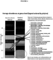
- Fig. 4 depicts average abundances at the genus level for the same samples shown in Fig. 3 .
- Fig. 4 is a higher-resolution view of the Firmicutes that are under-represented using the standard method using sequential lysis steps of detergent and bead beating.
- the bar plot of genus level differences in abundance show that there are five Firmicutes genuses under-represented using the standard sequential lysis method (Listeria, Blautia, Lachnospiracea Incertae Sedis, Butyrococcus, Ruminococcus ), and the relative representation of the Bacteroides and Akkermansia is artificially high using the standard method.
- This parallels the phylum level differences in Fig. 3 while showing that individual Firmicutes genus levels can be significantly under-represented using the standard method.
Description
- Disclosed are methods for lysing cells to release or extract genomic DNA (gDNA) from inside of the cells. The disclosed methods combine heat, detergent and base in a single tube and can be completed in a few minutes. The methods combine a normally incompatible detergent and base that facilitate post-lysis removal of detergent without extra steps, and the combination creates unexpected synergies lacking in sequential treatment protocols, that greatly reduces the number of steps and hands-on time, while yielding improved representation of gDNA, for example, from difficult to lyse bacteria in microbiome samples.
- Many cell-based and DNA-based analytical methods require releasing DNA from inside the cell to facilitate analysis. Opening the cells to release the DNA is called 'lysis.' For example, methods used to investigate the microbiome using DNA sequencing techniques first require lysis of microbes so the DNA can be extracted. Most microbiomes are communities of bacteria, archaea and fungi that vary tremendously in their susceptibility to lysis techniques. Differential susceptibility presents a significant problem to researchers, who want to ensure that the toughest (usually Gram-positive) and the easiest (usually Gram-negative) to lyse bacteria are represented in proportion to their population in the original sample. Unfortunately, most microbial lysis protocols work well for some microbes, but poorly for others. Additionally, rapid and simple alkaline lysis techniques used to recover plasmid DNA typically also remove the microbial genomic DNA, which is the target for microbiome screening (Alkaline Lysis opens cells but removes gDNA - Birnboim, H. C. and Doly, J., A rapid alkaline extraction procedure for screening recombinant plasmid DNA, Nucleic Acids Res. 7(6), 1979, 1513-1524 ; KOH lysis recovers bacterial genomic DNA-Raghunathan, Arumugham et al. "Genomic DNA Amplification from a Single Bacterium. " Applied and Environmental Microbiology 71.6 (2005): 3342-3347. PMC. Web. 29 Sept. 2016). There are multiple lysis techniques known in the art that attack cellular integrity based on different biochemical methods, including lysozyme (enzymatic attack on the peptidoglycan cell wall), strong base (chemical attack), detergent (solubilizes cell membranes), bead beating or shaking (mechanical disruption), and heat (Comparison of lysis techniques for microbiome- Sanqing Yuan, Dora B. Cohen, Jacques Ravel, Zaid Abdo, Larry J. Forney. Evaluation of Methods for the Extraction and Purification of DNA from the Human Microbiome. PLoS ONE 7(3): e33865. doi: 10.1371 /journal. pone. 0033865 ; DNA extraction methods affect microbiome profiling results: Wagner Mackenzie B, Waite DW, Taylor MW. Evaluating variation in human gut microbiota profiles due to DNA extraction method and inter-subject differences. Frontiers in Microbiology. 2015 ;6: 130. doi:10.3389/fmicb.2015.00130). Most published or commercially available DNA preparation methods use one or more of these methods to lyse cells, usually in sequential steps that can take a significant amount of time, especially when handling many samples at once. While individual lysis methods are usually sufficient for applications where incomplete or partial lysis yields sufficient DNA for the protocol being performed, they often do not yield DNA from microbiome samples in proportion to the original community, and may fail to lyse certain microbes altogether. For example, a detergent-based lysis may disrupt a subset of cells with weak cell walls and strong cell membranes, but not open detergent-resistant microbes with strong cell walls, leading to under-representation or absence of DNA from detergent resistant cells in the resulting DNA preparation. In another example, bead beating of microbes sufficient to lyse cells with strong cell membranes may shear or destroy DNA released early in the process from easily lysed cells. Additionally, the various methods of lysis tend to be incompatible with each other, and need to be performed sequentially if used in combination. For example, lysozyme will not work in the presence of detergents or strong base. Certain detergents precipitate in the presence of strong base. Bead beating is difficult to combine with a heating process. While individual shortcomings may be overcome by running separate lysis protocols in series, this increases the complexity, time, and cost involved. Importantly, detergents such as sodium dodecyl sulfate (SDS) must be removed after lysis, because SDS interferes with downstream DNA manipulation. Additionally, certain microbes may be resistant to lysis protocols run sequentially, depending on protocol order. For example, certain microbes with tough peptidoglycan cell walls may have an outer envelope of lipid bi-layer that protects from an initial treatment with strong base or lysozyme. Only a simultaneous combination of multiple methods may be effective, or a long sequence of multiple steps, to yield DNA from all microbes in a sample.
InWO 2011/124708 A1 methods and kits for precipitating anionic surfactants from a liquid sample containing the surfactant ions and nucleic acids are described. It is disclosed that when 0.25 M KOH (0.45 µL) is added to a cell lysate (500 µL) in an attempt to precipitate the SDS, that the DNA in the supernatant isolate was completely degraded.
InWO 2016/024263 A1 alkaline lysis procedures are disclosed, utilizing combinations of SDS and NaOH.
Further nucleic acid extraction methods are disclosed inWO 2011/070507 A1 and inEP 2 481 791 A1 - The methods disclosed herein streamline lysis for applications and techniques where proportional lysis is desired or necessary, such as microbiome research, by combining multiple lysis methods into a simple, rapid protocol that yields a more representative DNA profile across a sample containing different cellular constituents, such as the microbiome.
- Disclosed are methods for lysis of cells, such as bacteria present in microbiomes, that combine three lysis steps -- (1) heat, (2) detergent and (3) base -- into a single step and that can be completed in a short period of time, e.g., a few minutes. The methods combine a normally incompatible detergent and base lysis, allows for simplified removal of detergent after lysis, and importantly, yields improved quantities of genomic DNA (gDNA) from difficult to lyse bacteria.
- Disclosed herein is a method for lysing cells in a sample to release genomic DNA from the cells, comprising: (a) mixing an aqueous solution containing biologic cells with (i) an ionic detergent and (ii) a base capable of precipitating the ionic detergent wherein the ionic detergent is sodium dodecyl sulfate and the base is potassium hydroxide; (b) heating the aqueous solution to at least 50°C for a time effective to dissolve the ionic detergent; (c) cooling the aqueous solution to 40°C or less for a time effective to precipitate the ionic detergent; and (d) separating the precipitate from the aqueous solution, wherein the genomic DNA released from the biologic cells is present in the aqueous solution after separation of the precipitate.
- The ionic detergent is sodium dodecyl sulfate (SDS). In some embodiments, the concentration of the ionic detergent is from about 0.1% to about 10%. In some embodiments, the ionic detergent is sodium dodecyl sulfate (SDS) at a concentration of about 1%.
- The base is potassium hydroxide (KOH). In some embodiments, the concentration of the base is from about 0.05 molar to about 1 molar. In some embodiments, the base is potassium hydroxide (KOH) at a concentration of about 0.2 molar.
- In some embodiments, the detergent is combined with a base that precipitates the detergent at low temperature, but permits the detergent to dissolve at high temperature.
- In some embodiments, the ionic detergent is sodium dodecyl sulfate (SDS) at a concentration of 1% by weight and the base is and aqueous solution containing potassium hydroxide (KOH) at a concentration of 0.2 molar.
- In some embodiments, the heating is conducted at a temperature range of from about 50°C to about 100°C. In some embodiments, the heating is conducted at a temperature of about 65°C. In some embodiments, the heating is conducted at a temperature of about 95°C. In some embodiments, the heating is conducted for at least 1 minute.
- In some embodiments, the cooling is conducted at a temperature range of from about 4°C to about 40°C. In some embodiments, the cooling is conducted at a temperature of about 20°C to about 25°C. In some embodiments, the cooling is conducted for at least 30 seconds.
- In some embodiments, the separating is conducted by a method selected from the group consisting of: centrifugation, filtration, gravity settling.
- In some embodiments, the biologic cells originate from a sample selected from the group consisting of: feces, cell lysate, tissue, blood, tumor, tongue, tooth, buccal swab, phlegm, mucous, wound swab, skin swab, vaginal swab, or any other biological material or biological fluid originally obtained from a human, animal, plant, or environmental sample, including raw samples, complex samples, mixtures, and microbiome samples.
- In some embodiments, the biologic cells originate from an organism selected from the group consisting of: multicellular organisms, unicellular organisms, prokaryotes, eukaryotes, microbes, bacteria, archaea, protozoa, algae and fungi.
-
- Fig. la shows the white SDS precipitate in the potassium hydroxide (KOH+SDS) tube (right), at room temperature. The tube at left shows 1% SDS in the absence of KOH as a clear solution.
- Fig. lb shows the white SDS precipitate in the potassium hydroxide (KOH+SDS) tube (left), at room temperature. The tube at
right shows 1% SDS in the presence of NaOH as a clear solution, demonstrating that NaOH does not precipitate SDS. -
Fig. 2 depicts PCR barcoding results of 16S rRNA genes for 8 microbiome samples. The 1500 base amplicon in each of the 8 lanes is tagged with a different DNA barcode. -
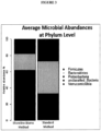
Fig. 3 is a graph showing a comparison of average microbial abundances at phylum level in multiple samples lysed using sequential lysis steps of detergent and bead beating or the combined lysis method described herein. There is a higher abundance of the more difficult to lyse Firmicutes using the Shoreline Biome method. -
Fig. 4 is a graph showing a comparison of average microbial abundances at genus level in multiple samples lysed using sequential lysis steps of detergent and bead beating or the combined lysis method described herein. This demonstrates that the phylum level abundances inFig. 3 correspond to the appearance of an increased quantity and diversity of Firmicutes at the genus level. - The subject matter that is regarded as the invention is particularly pointed out and distinctly claimed in the claims at the conclusion of the specification. The foregoing and other objects, features, and advantages of the invention will be apparent from the following detailed description taken in conjunction with the accompanying drawings.
- DNA located inside cells, such as bacteria and archaea in a microbiome, can be released by lysing the cells. To investigate a microbiome, cells in the target microbiome are lysed, after which the resulting DNA in this description can either be sequenced directly ('shotgun' sequencing, not shown), or used as template in PCR amplification targeting a genetic region such as the 16S rRNA gene, present in all bacteria and archaea. The 16S gene is used as an example herein because it can be used as a 'fingerprint' identification method for microbes, requiring ~1000x less sequencing than the shotgun method. Microbes can be identified using their 16S rRNA gene sequence, which varies slightly in most, if not all, bacteria and archaea. The variation in 16S gene sequence means that individual species of bacteria and archaea have characteristic DNA variations ('fingerprints') in the 16S rRNA gene that serve as identifiers for those species or strains. Kits, protocols and software enable comprehensive fingerprinting of the microbes in a sample, and permits simultaneous 16S rRNA fingerprinting of many samples at once, at high resolution, using the full length 16S rRNA gene (see, for example, U.S. Patent Application No.
US2017/0166956 titled "Methods for DNA Preparation for Multiplex High Throughput Targeted Screening" by Mark Driscoll and Thomas Jarvie ). Known microbes can be identified after sequencing by mapping the DNA sequence of the 16S gene to a database of known reads. Unknown microbes will contain 16S DNA sequences that are different from any of the microbes in the database, but can be tracked using their unique 16S sequence. In addition, the number of reads obtained for each microbe in a sample can reveal the relative abundances of each microbe in a sample. The relative abundance of specific microbes can be an important indicator of the state of each individual microbiome. Lysis techniques that change relative abundances of microbes, or leave out DNA from certain microbes altogether, can lead to sequencing results that incorrectly characterize the state of the microbiomes being studied. The methods described herein can be used to achieve the correct relative abundances of microbes from a sample. - The lysis process can be used for 'shotgun' microbiome sequencing as well, where the DNA is subjected to sequencing after lysis without 16S rRNA gene amplification. The shotgun method is used when investigators want to read all DNA sequences in a sample, not just the 16S gene from bacteria and archaea. For example, high depth shotgun microbiome DNA sequencing may reveal the full DNA genomic sequence from unknown bacteria/archaea, as well as fungi, or multicellular eukaryotes, viral DNA, or any other DNA containing organisms. Since a full bacterial genome can be millions of bases long (thousands of times larger than the 16S gene), fungal genomes can be more than a hundred million bases, and eukaryotic genomes can be billions of bases long, a shotgun microbiome profile can require thousands of times more sequencing than a 16S rRNA gene microbiome profile, with correspondingly greater time and costs. Although only the 16S profiling method is discussed in this example, the lysis protocol described herein provides the same advantages to both shotgun and 16S rRNA microbiome sequencing approaches.
- The following is an example of the disclosed methods for the 16S rRNA gene microbiome sequencing approach:
-
Step 1. A microbiome sample was dispersed into an aqueous solution containing 2% by weight of sodium dodecyl sulfate (SDS). -
Step 2. 0.4M KOH was added and SDS detergent precipitated as white flocculent. In this example, the detergent (1% SDS) is precipitated by the base (0.2M KOH). -
Step 3. The tube was capped and heated (temperature can range from about 50°C to about 100°C). SDS dissolves at temperatures above 50°C. Heat and KOH attack the peptidogycan cell wall, and SDS solubilizes membranes that protect microorganisms from the damaging effects of the KOH and heat. This combination of steps is synergistic, because sequential exposure to KOH, SDS, and heat, in contrast to the combined exposure described here, may not yield the same results because of the way that microbial cell walls and membranes are structured. Heat actually allows SDS to work in the presence of strong base, resulting in a unique simultaneous combination of three different lysis techniques. -
Step 4. After heating, the sample was brought back to room temperature (e.g., below 40°C) to precipitate the SDS detergent. -
Step 5. The sample was centrifuged briefly to pelletize the SDS detergent (no additions necessary, rapid removal of detergent). -
Step 6. The supernatant was moved to tube containing 500mM Tris buffer or equivalent, pH 8.5. The released DNA is now ready for analysis by 16S rRNA PCR (as described below), or can be stored or purified further for other uses. - Each DNA sample was subjected to PCR amplification a method which assigns unique DNA barcodes to each sample. An example PCR reaction for 8 different microbiomes is shown in
Fig. 2 where human fecal samples 1-8 were lysed according to the protocol described above in Steps 1-6, or by a standard protocol with sequential detergent/bead beating steps. Each sample was PCR amplified using primers to the 1500bp 16S rRNA gene, with a different DNA barcode for each sample. Samples were pooled for DNA sequencing after PCR. Since the reads from each sample contained a unique identifying DNA barcode, they can be sorted by sample after sequencing. Reads output by the sequencer are sorted by sample using barcodes and mapped to a database to identify known microbes, unknown microbes, and their relative proportion in each sample. - After sorting by barcode into sample of origin, identification by genus, and quantitation of the number of reads for each genus by software analysis of the reads, the reads for each microbiome were compared. Depending on the experimental design, there are a number of ways the output could be compared. In
Fig. 3 , the quantity of each microbe in a microbiome is included in a 100% stacked bar plot for two samples. This method allows for simple, direct comparison of microbiomes. Other useful comparisons include phylum level differences, species or strain level differences, or other taxonomic levels. - For multiple samples, a standard method using sequential lysis steps of detergent and bead beating was compared to the combined lysis method described herein. As shown in
Fig. 3 , Gram-positive Firmicutes increased in abundance from ~30% to over 60% of the microbiome. Firmicutes are Gram positive bacteria with strong cell walls that tend to be difficult to lyse. This demonstrates that the lysis method described herein is better at lysing microbes with strong cell walls. Easy to lyse Bacteriodetes and Verrucomicrobia phyla decreased proportionally, as would be expected when using a 100% stacked bar plot. -
Fig. 4 depicts average abundances at the genus level for the same samples shown inFig. 3 .Fig. 4 is a higher-resolution view of the Firmicutes that are under-represented using the standard method using sequential lysis steps of detergent and bead beating. The bar plot of genus level differences in abundance show that there are five Firmicutes genuses under-represented using the standard sequential lysis method (Listeria, Blautia, Lachnospiracea Incertae Sedis, Butyrococcus, Ruminococcus), and the relative representation of the Bacteroides and Akkermansia is artificially high using the standard method. This parallels the phylum level differences inFig. 3 , while showing that individual Firmicutes genus levels can be significantly under-represented using the standard method. - One or more embodiments of the present invention have been described. Nevertheless, it will be understood that various modifications may be made without departing from the scope of the invention. Accordingly, other embodiments are within the scope of the following claims.
Claims (15)
- A method for lysing biologic cells in a sample to release genomic DNA from the cells, comprising the sequential steps of:a. mixing an aqueous solution containing the biologic cells with (i) an ionic detergent, and (ii) a base effective to facilitate precipitation of the ionic detergent, wherein the ionic detergent is sodium dodecyl sulfate and the base is potassium hydroxide;b. heating the aqueous solution to at least 50°C for a time effective for the ionic detergent to dissolve;c. cooling the aqueous solution to 40°C or less for a time effective to precipitate the ionic detergent; andd. separating the precipitate from the aqueous solution,wherein the genomic DNA released from the biologic cells is present in the aqueous solution following separation of the precipitate.
- The method of claim 1, wherein the concentration of the ionic detergent is from about 0.1% by weight to about 10% by weight.
- The method of claim 1, wherein the ionic detergent is sodium dodecyl sulfate (SDS) at a concentration of about 1% by weight.
- The method of claim 1, wherein the concentration of the base is from about 0.05 molar to about 1 molar.
- The method of claim 1, wherein the base is potassium hydroxide (KOH) at a concentration of about 0.2 molar.
- The method of claim 1, wherein the ionic detergent is sodium dodecyl sulfate (SDS) at a concentration of 1% by weight and the base is potassium hydroxide (KOH) at a concentration of 0.2 molar.
- The method of claim 1, wherein the heating is conducted at a temperature range of from about 50°C to about 100°C.
- The method of claim 7, wherein the heating is conducted at a temperature of about 50°C.
- The method of claim 8, wherein the heating is conducted for at least 0.25 minutes.
- The method of claim 1, wherein the cooling is conducted at a temperature range of from about 4°C to about 40°C.
- The method of claim 10, wherein the cooling is conducted at a temperature of about 20°C to about 25°C.
- The method of claim 11, wherein the cooling is conducted for at least 0.25 minutes.
- The method of claim 1, wherein the separating is conducted by a method selected from the group consisting of: centrifugation, filtration and gravity settling.
- The method of claim 1, wherein the biologic cells originate from a sample selected from the group consisting of: feces, cell lysate, tissue, blood, tumor, tongue, tooth, buccal swab, phlegm, mucous, wound swab, skin swab, vaginal swab, or any other biological material or biological fluid originally obtained from a human, animal, plant, or environmental sample, including raw samples, complex samples, mixtures, and microbiome samples.
- The method of claim 1, wherein the biologic cells originate from an organism selected from the group consisting of: multicellular organisms, unicellular organisms, prokaryotes, eukaryotes, microbes, bacteria, archaea, protozoa, algae, fungi and viruses.
Applications Claiming Priority (2)
| Application Number | Priority Date | Filing Date | Title |
|---|---|---|---|
| US201662440171P | 2016-12-29 | 2016-12-29 | |
| PCT/US2017/068415 WO2018125865A1 (en) | 2016-12-29 | 2017-12-26 | Combined lysis protocol for comprehensive cell lysis |
Publications (3)
| Publication Number | Publication Date |
|---|---|
| EP3562576A1 EP3562576A1 (en) | 2019-11-06 |
| EP3562576A4 EP3562576A4 (en) | 2020-07-22 |
| EP3562576B1 true EP3562576B1 (en) | 2023-09-06 |
Family
ID=62709305
Family Applications (1)
| Application Number | Title | Priority Date | Filing Date |
|---|---|---|---|
| EP17887015.0A Active EP3562576B1 (en) | 2016-12-29 | 2017-12-26 | Combined lysis protocol for comprehensive cell lysis |
Country Status (8)
| Country | Link |
|---|---|
| US (2) | US10774322B2 (en) |
| EP (1) | EP3562576B1 (en) |
| JP (1) | JP6935945B2 (en) |
| KR (1) | KR102281213B1 (en) |
| CN (1) | CN110312570B (en) |
| AU (1) | AU2017388204B2 (en) |
| CA (1) | CA3048844C (en) |
| WO (1) | WO2018125865A1 (en) |
Families Citing this family (7)
| Publication number | Priority date | Publication date | Assignee | Title |
|---|---|---|---|---|
| US10428370B2 (en) * | 2016-09-15 | 2019-10-01 | Sun Genomics, Inc. | Universal method for extracting nucleic acid molecules from a diverse population of one or more types of microbes in a sample |
| US11959125B2 (en) | 2016-09-15 | 2024-04-16 | Sun Genomics, Inc. | Universal method for extracting nucleic acid molecules from a diverse population of one or more types of microbes in a sample |
| US11149246B2 (en) * | 2016-12-29 | 2021-10-19 | Shoreline Biome, Llc | Methods for cell lysis and preparation of high molecular weight DNA from modified cells |
| WO2020181082A1 (en) * | 2019-03-07 | 2020-09-10 | Shoreline Biome, Llc | Methods for cell lysis and preparation of high molecular weight dna from modified cells |
| KR102370468B1 (en) * | 2020-03-18 | 2022-03-03 | 재단법인대구경북과학기술원 | A composition and a method for removal of residual anionic surfactant, protein-detergent complexes, and micelles, in biological tissues |
| EP4153606A2 (en) | 2020-07-13 | 2023-03-29 | Singular Genomics Systems, Inc. | Methods of sequencing complementary polynucleotides |
| CN112626174B (en) * | 2020-12-16 | 2024-05-07 | 广州源井生物科技有限公司 | Cell lysate and application thereof |
Citations (2)
| Publication number | Priority date | Publication date | Assignee | Title |
|---|---|---|---|---|
| WO2004108260A2 (en) * | 2003-05-30 | 2004-12-16 | Advisys, Inc. | Devices and methods for biomaterial production |
| EP2481791B1 (en) * | 2011-01-31 | 2015-05-20 | Tokyo University of Agriculture & Technology | Nucleic acid extraction method |
Family Cites Families (14)
| Publication number | Priority date | Publication date | Assignee | Title |
|---|---|---|---|---|
| CA2319775C (en) * | 1998-02-02 | 2009-05-05 | Gentra Systems, Inc. | Compositions and methods for using a lysing matrix for isolating dna |
| CN1057530C (en) * | 1998-02-12 | 2000-10-18 | 复旦大学 | Fast extracting method of nucleic acid |
| CN1308130A (en) * | 2000-12-26 | 2001-08-15 | 李永武 | Method of extracting nucleic and from microbe cell |
| WO2003070898A2 (en) * | 2002-02-19 | 2003-08-28 | Choicepoint Asset Company | Selective extraction of dna from groups of cells |
| US20040157219A1 (en) | 2003-02-06 | 2004-08-12 | Jianrong Lou | Chemical treatment of biological samples for nucleic acid extraction and kits therefor |
| EP1608969A4 (en) * | 2003-03-19 | 2007-06-06 | Alfa Wassermann Inc | Separation and accumulation of subcellular components, and proteins derived therefrom |
| US8501402B2 (en) * | 2003-03-24 | 2013-08-06 | Boehringer Ingelheim Rcv Gmbh & Co Kg | Methods and devices for producing biomolecules |
| WO2005073377A1 (en) | 2004-01-28 | 2005-08-11 | Toudai Tlo, Ltd. | Method of collecting dna from environmental sample |
| EP2333105A1 (en) | 2009-12-08 | 2011-06-15 | Koninklijke Philips Electronics N.V. | Selective lysis of cells |
| CN102822339A (en) * | 2010-04-08 | 2012-12-12 | 恰根有限公司 | Method for precipitating anionic surfactant ions in presence of nucleic acids |
| EP2576801B1 (en) * | 2010-06-01 | 2019-10-02 | DSM IP Assets B.V. | Extraction of lipid from cells and products therefrom |
| AU2012266754B2 (en) * | 2011-06-06 | 2016-04-21 | Biocartis Nv | Selective lysis of cells by ionic surfactants |
| WO2016024263A1 (en) | 2014-08-14 | 2016-02-18 | Molecular Detection Israel Ltd. | Methods for isolating microbial dna from a blood sample |
| US20170166956A1 (en) | 2015-12-11 | 2017-06-15 | Shoreline Biome, Llc | Methods for DNA Preparation for Multiplex High Throughput Targeted Sequencing |
-
2017
- 2017-12-26 CA CA3048844A patent/CA3048844C/en active Active
- 2017-12-26 US US15/854,157 patent/US10774322B2/en active Active
- 2017-12-26 KR KR1020197021748A patent/KR102281213B1/en active IP Right Grant
- 2017-12-26 WO PCT/US2017/068415 patent/WO2018125865A1/en unknown
- 2017-12-26 JP JP2019556545A patent/JP6935945B2/en active Active
- 2017-12-26 EP EP17887015.0A patent/EP3562576B1/en active Active
- 2017-12-26 AU AU2017388204A patent/AU2017388204B2/en active Active
- 2017-12-26 CN CN201780085878.1A patent/CN110312570B/en active Active
-
2020
- 2020-08-19 US US16/996,996 patent/US20210032618A1/en not_active Abandoned
Patent Citations (2)
| Publication number | Priority date | Publication date | Assignee | Title |
|---|---|---|---|---|
| WO2004108260A2 (en) * | 2003-05-30 | 2004-12-16 | Advisys, Inc. | Devices and methods for biomaterial production |
| EP2481791B1 (en) * | 2011-01-31 | 2015-05-20 | Tokyo University of Agriculture & Technology | Nucleic acid extraction method |
Also Published As
| Publication number | Publication date |
|---|---|
| CA3048844C (en) | 2023-12-12 |
| JP2020503068A (en) | 2020-01-30 |
| CN110312570B (en) | 2022-04-05 |
| AU2017388204A1 (en) | 2019-08-01 |
| EP3562576A4 (en) | 2020-07-22 |
| US20180187181A1 (en) | 2018-07-05 |
| US20210032618A1 (en) | 2021-02-04 |
| WO2018125865A8 (en) | 2019-08-22 |
| JP6935945B2 (en) | 2021-09-15 |
| KR20190100315A (en) | 2019-08-28 |
| CA3048844A1 (en) | 2018-07-05 |
| KR102281213B1 (en) | 2021-07-22 |
| CN110312570A (en) | 2019-10-08 |
| WO2018125865A1 (en) | 2018-07-05 |
| AU2017388204B2 (en) | 2023-10-12 |
| US10774322B2 (en) | 2020-09-15 |
| EP3562576A1 (en) | 2019-11-06 |
Similar Documents
| Publication | Publication Date | Title |
|---|---|---|
| EP3562576B1 (en) | Combined lysis protocol for comprehensive cell lysis | |
| JP5112064B2 (en) | Kits and methods for removing contaminants from nucleic acids in environmental and biological samples | |
| US20230183673A1 (en) | Method for nucleic acid depletion | |
| US5731150A (en) | IS6110 based molecular detection of mycobacterium tuberculosis | |
| Amita et al. | Qualitative evaluation of mycobacterial DNA extraction protocols for polymerase chain reaction | |
| CN101111606A (en) | Methods for simplifying microbial nucleic acids by chemical modification of cytosines | |
| AU2006209416A1 (en) | Method of quantitatively analysing microorganism targeting rRNA | |
| AU2011227452B2 (en) | Use of achromopeptidase for lysis at room temperature | |
| US11149246B2 (en) | Methods for cell lysis and preparation of high molecular weight DNA from modified cells | |
| US20090142798A1 (en) | Method of selectively lysing non-viable cells in cell population in sample | |
| WO2020181082A1 (en) | Methods for cell lysis and preparation of high molecular weight dna from modified cells | |
| CN114144531A (en) | Method for detecting rare DNA sequences in fecal samples | |
| WO2020218553A1 (en) | Digital analysis of microbial flora | |
| US20190024076A1 (en) | Semi-dry bead beating method for microbial lysis and device for performing same | |
| JP4083860B2 (en) | Use of alcohol-salt buffer wash for efficient recovery of mycobacteria and mycobacterial DNA from respiratory sediments | |
| Gifvars | Optimization of a sampling and analysis process to study the effects of skin care products on the microbial skin flora. | |
| Yatbantoong et al. | Direct DNA extraction to detect Mycobacterium bovis from the lungs of buffaloes positive to intradermal tuberculin testing | |
| Zidan et al. | Extraction of microbial community DNA from soil for polymerase chain reaction | |
| Booysen | Molecular methods for the detection of food-borne pathogens an overview | |
| WO2016153355A2 (en) | Novel isolation and amplification process control |
Legal Events
| Date | Code | Title | Description |
|---|---|---|---|
| STAA | Information on the status of an ep patent application or granted ep patent |
Free format text: STATUS: THE INTERNATIONAL PUBLICATION HAS BEEN MADE |
|
| PUAI | Public reference made under article 153(3) epc to a published international application that has entered the european phase |
Free format text: ORIGINAL CODE: 0009012 |
|
| STAA | Information on the status of an ep patent application or granted ep patent |
Free format text: STATUS: REQUEST FOR EXAMINATION WAS MADE |
|
| 17P | Request for examination filed |
Effective date: 20190726 |
|
| AK | Designated contracting states |
Kind code of ref document: A1 Designated state(s): AL AT BE BG CH CY CZ DE DK EE ES FI FR GB GR HR HU IE IS IT LI LT LU LV MC MK MT NL NO PL PT RO RS SE SI SK SM TR |
|
| AX | Request for extension of the european patent |
Extension state: BA ME |
|
| DAV | Request for validation of the european patent (deleted) | ||
| DAX | Request for extension of the european patent (deleted) | ||
| A4 | Supplementary search report drawn up and despatched |
Effective date: 20200622 |
|
| RIC1 | Information provided on ipc code assigned before grant |
Ipc: C12M 1/04 20060101ALI20200616BHEP Ipc: C12Q 1/6806 20180101AFI20200616BHEP Ipc: C12N 1/06 20060101ALI20200616BHEP Ipc: B01F 13/02 20060101ALI20200616BHEP |
|
| STAA | Information on the status of an ep patent application or granted ep patent |
Free format text: STATUS: EXAMINATION IS IN PROGRESS |
|
| STAA | Information on the status of an ep patent application or granted ep patent |
Free format text: STATUS: EXAMINATION IS IN PROGRESS |
|
| 17Q | First examination report despatched |
Effective date: 20210806 |
|
| REG | Reference to a national code |
Ref country code: DE Ref legal event code: R079 Ref document number: 602017073980 Country of ref document: DE Free format text: PREVIOUS MAIN CLASS: B01F0013020000 Ipc: C12Q0001680600 Free format text: PREVIOUS MAIN CLASS: B01F0013020000 Ipc: C12Q0001680600 |
|
| GRAP | Despatch of communication of intention to grant a patent |
Free format text: ORIGINAL CODE: EPIDOSNIGR1 |
|
| STAA | Information on the status of an ep patent application or granted ep patent |
Free format text: STATUS: GRANT OF PATENT IS INTENDED |
|
| RIC1 | Information provided on ipc code assigned before grant |
Ipc: C12N 15/10 20060101ALI20230124BHEP Ipc: C12N 1/06 20060101ALI20230124BHEP Ipc: C12Q 1/6806 20180101AFI20230124BHEP |
|
| INTG | Intention to grant announced |
Effective date: 20230215 |
|
| GRAS | Grant fee paid |
Free format text: ORIGINAL CODE: EPIDOSNIGR3 |
|
| P01 | Opt-out of the competence of the unified patent court (upc) registered |
Effective date: 20230530 |
|
| P02 | Opt-out of the competence of the unified patent court (upc) changed |
Effective date: 20230606 |
|
| GRAA | (expected) grant |
Free format text: ORIGINAL CODE: 0009210 |
|
| STAA | Information on the status of an ep patent application or granted ep patent |
Free format text: STATUS: THE PATENT HAS BEEN GRANTED |
|
| RAP3 | Party data changed (applicant data changed or rights of an application transferred) |
Owner name: INTUS BIOSCIENCES, LLC |
|
| AK | Designated contracting states |
Kind code of ref document: B1 Designated state(s): AL AT BE BG CH CY CZ DE DK EE ES FI FR GB GR HR HU IE IS IT LI LT LU LV MC MK MT NL NO PL PT RO RS SE SI SK SM TR |
|
| REG | Reference to a national code |
Ref country code: GB Ref legal event code: FG4D |
|
| REG | Reference to a national code |
Ref country code: CH Ref legal event code: EP |
|
| REG | Reference to a national code |
Ref country code: IE Ref legal event code: FG4D |
|
| REG | Reference to a national code |
Ref country code: DE Ref legal event code: R096 Ref document number: 602017073980 Country of ref document: DE |
|
| REG | Reference to a national code |
Ref country code: LT Ref legal event code: MG9D |
|
| REG | Reference to a national code |
Ref country code: NL Ref legal event code: MP Effective date: 20230906 |
|
| PG25 | Lapsed in a contracting state [announced via postgrant information from national office to epo] |
Ref country code: GR Free format text: LAPSE BECAUSE OF FAILURE TO SUBMIT A TRANSLATION OF THE DESCRIPTION OR TO PAY THE FEE WITHIN THE PRESCRIBED TIME-LIMIT Effective date: 20231207 |
|
| PGFP | Annual fee paid to national office [announced via postgrant information from national office to epo] |
Ref country code: GB Payment date: 20231227 Year of fee payment: 7 |
|
| PG25 | Lapsed in a contracting state [announced via postgrant information from national office to epo] |
Ref country code: SE Free format text: LAPSE BECAUSE OF FAILURE TO SUBMIT A TRANSLATION OF THE DESCRIPTION OR TO PAY THE FEE WITHIN THE PRESCRIBED TIME-LIMIT Effective date: 20230906 Ref country code: RS Free format text: LAPSE BECAUSE OF FAILURE TO SUBMIT A TRANSLATION OF THE DESCRIPTION OR TO PAY THE FEE WITHIN THE PRESCRIBED TIME-LIMIT Effective date: 20230906 Ref country code: NO Free format text: LAPSE BECAUSE OF FAILURE TO SUBMIT A TRANSLATION OF THE DESCRIPTION OR TO PAY THE FEE WITHIN THE PRESCRIBED TIME-LIMIT Effective date: 20231206 Ref country code: LV Free format text: LAPSE BECAUSE OF FAILURE TO SUBMIT A TRANSLATION OF THE DESCRIPTION OR TO PAY THE FEE WITHIN THE PRESCRIBED TIME-LIMIT Effective date: 20230906 Ref country code: LT Free format text: LAPSE BECAUSE OF FAILURE TO SUBMIT A TRANSLATION OF THE DESCRIPTION OR TO PAY THE FEE WITHIN THE PRESCRIBED TIME-LIMIT Effective date: 20230906 Ref country code: HR Free format text: LAPSE BECAUSE OF FAILURE TO SUBMIT A TRANSLATION OF THE DESCRIPTION OR TO PAY THE FEE WITHIN THE PRESCRIBED TIME-LIMIT Effective date: 20230906 Ref country code: GR Free format text: LAPSE BECAUSE OF FAILURE TO SUBMIT A TRANSLATION OF THE DESCRIPTION OR TO PAY THE FEE WITHIN THE PRESCRIBED TIME-LIMIT Effective date: 20231207 Ref country code: FI Free format text: LAPSE BECAUSE OF FAILURE TO SUBMIT A TRANSLATION OF THE DESCRIPTION OR TO PAY THE FEE WITHIN THE PRESCRIBED TIME-LIMIT Effective date: 20230906 |
|
| PGFP | Annual fee paid to national office [announced via postgrant information from national office to epo] |
Ref country code: FR Payment date: 20231227 Year of fee payment: 7 |
|
| REG | Reference to a national code |
Ref country code: AT Ref legal event code: MK05 Ref document number: 1608628 Country of ref document: AT Kind code of ref document: T Effective date: 20230906 |
|
| PG25 | Lapsed in a contracting state [announced via postgrant information from national office to epo] |
Ref country code: NL Free format text: LAPSE BECAUSE OF FAILURE TO SUBMIT A TRANSLATION OF THE DESCRIPTION OR TO PAY THE FEE WITHIN THE PRESCRIBED TIME-LIMIT Effective date: 20230906 |
|
| PG25 | Lapsed in a contracting state [announced via postgrant information from national office to epo] |
Ref country code: IS Free format text: LAPSE BECAUSE OF FAILURE TO SUBMIT A TRANSLATION OF THE DESCRIPTION OR TO PAY THE FEE WITHIN THE PRESCRIBED TIME-LIMIT Effective date: 20240106 |
|
| PG25 | Lapsed in a contracting state [announced via postgrant information from national office to epo] |
Ref country code: AT Free format text: LAPSE BECAUSE OF FAILURE TO SUBMIT A TRANSLATION OF THE DESCRIPTION OR TO PAY THE FEE WITHIN THE PRESCRIBED TIME-LIMIT Effective date: 20230906 |
|
| PG25 | Lapsed in a contracting state [announced via postgrant information from national office to epo] |
Ref country code: ES Free format text: LAPSE BECAUSE OF FAILURE TO SUBMIT A TRANSLATION OF THE DESCRIPTION OR TO PAY THE FEE WITHIN THE PRESCRIBED TIME-LIMIT Effective date: 20230906 |
|
| PG25 | Lapsed in a contracting state [announced via postgrant information from national office to epo] |
Ref country code: SM Free format text: LAPSE BECAUSE OF FAILURE TO SUBMIT A TRANSLATION OF THE DESCRIPTION OR TO PAY THE FEE WITHIN THE PRESCRIBED TIME-LIMIT Effective date: 20230906 Ref country code: RO Free format text: LAPSE BECAUSE OF FAILURE TO SUBMIT A TRANSLATION OF THE DESCRIPTION OR TO PAY THE FEE WITHIN THE PRESCRIBED TIME-LIMIT Effective date: 20230906 Ref country code: IS Free format text: LAPSE BECAUSE OF FAILURE TO SUBMIT A TRANSLATION OF THE DESCRIPTION OR TO PAY THE FEE WITHIN THE PRESCRIBED TIME-LIMIT Effective date: 20240106 Ref country code: ES Free format text: LAPSE BECAUSE OF FAILURE TO SUBMIT A TRANSLATION OF THE DESCRIPTION OR TO PAY THE FEE WITHIN THE PRESCRIBED TIME-LIMIT Effective date: 20230906 Ref country code: EE Free format text: LAPSE BECAUSE OF FAILURE TO SUBMIT A TRANSLATION OF THE DESCRIPTION OR TO PAY THE FEE WITHIN THE PRESCRIBED TIME-LIMIT Effective date: 20230906 Ref country code: CZ Free format text: LAPSE BECAUSE OF FAILURE TO SUBMIT A TRANSLATION OF THE DESCRIPTION OR TO PAY THE FEE WITHIN THE PRESCRIBED TIME-LIMIT Effective date: 20230906 Ref country code: AT Free format text: LAPSE BECAUSE OF FAILURE TO SUBMIT A TRANSLATION OF THE DESCRIPTION OR TO PAY THE FEE WITHIN THE PRESCRIBED TIME-LIMIT Effective date: 20230906 Ref country code: PT Free format text: LAPSE BECAUSE OF FAILURE TO SUBMIT A TRANSLATION OF THE DESCRIPTION OR TO PAY THE FEE WITHIN THE PRESCRIBED TIME-LIMIT Effective date: 20240108 Ref country code: SK Free format text: LAPSE BECAUSE OF FAILURE TO SUBMIT A TRANSLATION OF THE DESCRIPTION OR TO PAY THE FEE WITHIN THE PRESCRIBED TIME-LIMIT Effective date: 20230906 |
|
| PGFP | Annual fee paid to national office [announced via postgrant information from national office to epo] |
Ref country code: DE Payment date: 20231229 Year of fee payment: 7 |