EP3467497B1 - Procédé d'analyse d'expression de corps nucléaire de la protéine smn - Google Patents
Procédé d'analyse d'expression de corps nucléaire de la protéine smn Download PDFInfo
- Publication number
- EP3467497B1 EP3467497B1 EP17802794.2A EP17802794A EP3467497B1 EP 3467497 B1 EP3467497 B1 EP 3467497B1 EP 17802794 A EP17802794 A EP 17802794A EP 3467497 B1 EP3467497 B1 EP 3467497B1
- Authority
- EP
- European Patent Office
- Prior art keywords
- smn protein
- blood
- nuclear body
- sma
- nucleated cells
- Prior art date
- Legal status (The legal status is an assumption and is not a legal conclusion. Google has not performed a legal analysis and makes no representation as to the accuracy of the status listed.)
- Active
Links
- 238000000034 method Methods 0.000 title claims description 102
- 230000014509 gene expression Effects 0.000 title claims description 44
- 101150081851 SMN1 gene Proteins 0.000 title description 19
- 210000004027 cell Anatomy 0.000 claims description 177
- 108090000623 proteins and genes Proteins 0.000 claims description 151
- 102000004169 proteins and genes Human genes 0.000 claims description 149
- 210000004369 blood Anatomy 0.000 claims description 104
- 239000008280 blood Substances 0.000 claims description 104
- 208000002320 spinal muscular atrophy Diseases 0.000 claims description 89
- 238000002372 labelling Methods 0.000 claims description 43
- 239000000427 antigen Substances 0.000 claims description 26
- 102000036639 antigens Human genes 0.000 claims description 26
- 108091007433 antigens Proteins 0.000 claims description 26
- 239000007850 fluorescent dye Substances 0.000 claims description 26
- 238000003384 imaging method Methods 0.000 claims description 20
- 238000000684 flow cytometry Methods 0.000 claims description 19
- 210000001616 monocyte Anatomy 0.000 claims description 17
- 101000934338 Homo sapiens Myeloid cell surface antigen CD33 Proteins 0.000 claims description 12
- 102100025243 Myeloid cell surface antigen CD33 Human genes 0.000 claims description 12
- 229940079593 drug Drugs 0.000 claims description 12
- 239000003814 drug Substances 0.000 claims description 12
- 229940000406 drug candidate Drugs 0.000 claims description 12
- 210000002161 motor neuron Anatomy 0.000 claims description 7
- 238000011002 quantification Methods 0.000 claims description 6
- 210000003743 erythrocyte Anatomy 0.000 claims description 5
- 206010018910 Haemolysis Diseases 0.000 claims description 4
- 230000008588 hemolysis Effects 0.000 claims description 4
- 230000004083 survival effect Effects 0.000 claims description 3
- 239000000523 sample Substances 0.000 description 40
- 210000004940 nucleus Anatomy 0.000 description 30
- 101150015954 SMN2 gene Proteins 0.000 description 15
- 210000000601 blood cell Anatomy 0.000 description 15
- 102100021947 Survival motor neuron protein Human genes 0.000 description 14
- 239000003153 chemical reaction reagent Substances 0.000 description 14
- 101000617738 Homo sapiens Survival motor neuron protein Proteins 0.000 description 13
- 108020004999 messenger RNA Proteins 0.000 description 13
- 238000005119 centrifugation Methods 0.000 description 11
- 210000003819 peripheral blood mononuclear cell Anatomy 0.000 description 10
- PRDFBSVERLRRMY-UHFFFAOYSA-N 2'-(4-ethoxyphenyl)-5-(4-methylpiperazin-1-yl)-2,5'-bibenzimidazole Chemical compound C1=CC(OCC)=CC=C1C1=NC2=CC=C(C=3NC4=CC(=CC=C4N=3)N3CCN(C)CC3)C=C2N1 PRDFBSVERLRRMY-UHFFFAOYSA-N 0.000 description 9
- 239000000090 biomarker Substances 0.000 description 9
- 102100024222 B-lymphocyte antigen CD19 Human genes 0.000 description 8
- 101000980825 Homo sapiens B-lymphocyte antigen CD19 Proteins 0.000 description 8
- 239000000047 product Substances 0.000 description 8
- 238000004458 analytical method Methods 0.000 description 7
- 230000009089 cytolysis Effects 0.000 description 7
- 238000010195 expression analysis Methods 0.000 description 7
- 238000011534 incubation Methods 0.000 description 7
- 208000024891 symptom Diseases 0.000 description 7
- 239000000969 carrier Substances 0.000 description 6
- 238000011161 development Methods 0.000 description 6
- 201000010099 disease Diseases 0.000 description 6
- 208000037265 diseases, disorders, signs and symptoms Diseases 0.000 description 6
- 238000011156 evaluation Methods 0.000 description 6
- 210000005259 peripheral blood Anatomy 0.000 description 6
- 239000011886 peripheral blood Substances 0.000 description 6
- 238000013518 transcription Methods 0.000 description 6
- 230000035897 transcription Effects 0.000 description 6
- 238000002965 ELISA Methods 0.000 description 5
- 230000037430 deletion Effects 0.000 description 5
- 238000012217 deletion Methods 0.000 description 5
- 230000008823 permeabilization Effects 0.000 description 5
- 230000035945 sensitivity Effects 0.000 description 5
- 238000010186 staining Methods 0.000 description 5
- -1 CD66 Proteins 0.000 description 4
- 210000001744 T-lymphocyte Anatomy 0.000 description 4
- 210000003719 b-lymphocyte Anatomy 0.000 description 4
- 210000000349 chromosome Anatomy 0.000 description 4
- 238000001514 detection method Methods 0.000 description 4
- 239000003550 marker Substances 0.000 description 4
- 201000000585 muscular atrophy Diseases 0.000 description 4
- 102000017420 CD3 protein, epsilon/gamma/delta subunit Human genes 0.000 description 3
- 108050005493 CD3 protein, epsilon/gamma/delta subunit Proteins 0.000 description 3
- 208000010428 Muscle Weakness Diseases 0.000 description 3
- 206010028289 Muscle atrophy Diseases 0.000 description 3
- 206010028372 Muscular weakness Diseases 0.000 description 3
- 208000032225 Proximal spinal muscular atrophy type 1 Diseases 0.000 description 3
- 208000033526 Proximal spinal muscular atrophy type 3 Diseases 0.000 description 3
- 239000006285 cell suspension Substances 0.000 description 3
- 230000020763 muscle atrophy Effects 0.000 description 3
- 230000035772 mutation Effects 0.000 description 3
- 210000000440 neutrophil Anatomy 0.000 description 3
- 238000004445 quantitative analysis Methods 0.000 description 3
- 238000012216 screening Methods 0.000 description 3
- 238000000926 separation method Methods 0.000 description 3
- 230000001225 therapeutic effect Effects 0.000 description 3
- 208000032471 type 1 spinal muscular atrophy Diseases 0.000 description 3
- 208000032527 type III spinal muscular atrophy Diseases 0.000 description 3
- YBJHBAHKTGYVGT-ZKWXMUAHSA-N (+)-Biotin Chemical compound N1C(=O)N[C@@H]2[C@H](CCCCC(=O)O)SC[C@@H]21 YBJHBAHKTGYVGT-ZKWXMUAHSA-N 0.000 description 2
- 108091026890 Coding region Proteins 0.000 description 2
- 101000946889 Homo sapiens Monocyte differentiation antigen CD14 Proteins 0.000 description 2
- 101000738771 Homo sapiens Receptor-type tyrosine-protein phosphatase C Proteins 0.000 description 2
- 208000026350 Inborn Genetic disease Diseases 0.000 description 2
- 102100035877 Monocyte differentiation antigen CD14 Human genes 0.000 description 2
- 208000033522 Proximal spinal muscular atrophy type 2 Diseases 0.000 description 2
- 102100037422 Receptor-type tyrosine-protein phosphatase C Human genes 0.000 description 2
- 101150113275 Smn gene Proteins 0.000 description 2
- 208000003954 Spinal Muscular Atrophies of Childhood Diseases 0.000 description 2
- 238000004364 calculation method Methods 0.000 description 2
- 239000002771 cell marker Substances 0.000 description 2
- 238000003745 diagnosis Methods 0.000 description 2
- 230000001747 exhibiting effect Effects 0.000 description 2
- 238000010230 functional analysis Methods 0.000 description 2
- 208000016361 genetic disease Diseases 0.000 description 2
- 239000003219 hemolytic agent Substances 0.000 description 2
- 230000002949 hemolytic effect Effects 0.000 description 2
- 230000004807 localization Effects 0.000 description 2
- 210000004698 lymphocyte Anatomy 0.000 description 2
- 238000005259 measurement Methods 0.000 description 2
- XJMOSONTPMZWPB-UHFFFAOYSA-M propidium iodide Chemical compound [I-].[I-].C12=CC(N)=CC=C2C2=CC=C(N)C=C2[N+](CCC[N+](C)(CC)CC)=C1C1=CC=CC=C1 XJMOSONTPMZWPB-UHFFFAOYSA-M 0.000 description 2
- 239000000243 solution Substances 0.000 description 2
- 239000006228 supernatant Substances 0.000 description 2
- 238000012360 testing method Methods 0.000 description 2
- 208000032521 type II spinal muscular atrophy Diseases 0.000 description 2
- 238000005406 washing Methods 0.000 description 2
- XLYOFNOQVPJJNP-UHFFFAOYSA-N water Substances O XLYOFNOQVPJJNP-UHFFFAOYSA-N 0.000 description 2
- UBOKASXZHPZFRZ-UHFFFAOYSA-N 2-phenyl-1h-indole-4,6-diamine Chemical compound N1C2=CC(N)=CC(N)=C2C=C1C1=CC=CC=C1 UBOKASXZHPZFRZ-UHFFFAOYSA-N 0.000 description 1
- FWBHETKCLVMNFS-UHFFFAOYSA-N 4',6-Diamino-2-phenylindol Chemical compound C1=CC(C(=N)N)=CC=C1C1=CC2=CC=C(C(N)=N)C=C2N1 FWBHETKCLVMNFS-UHFFFAOYSA-N 0.000 description 1
- 239000012103 Alexa Fluor 488 Substances 0.000 description 1
- 239000012099 Alexa Fluor family Substances 0.000 description 1
- 241001121515 Celes Species 0.000 description 1
- 102000008167 DEAD Box Protein 20 Human genes 0.000 description 1
- 108010060424 DEAD Box Protein 20 Proteins 0.000 description 1
- 102000004190 Enzymes Human genes 0.000 description 1
- 108090000790 Enzymes Proteins 0.000 description 1
- 102000009109 Fc receptors Human genes 0.000 description 1
- 108010087819 Fc receptors Proteins 0.000 description 1
- 229920001917 Ficoll Polymers 0.000 description 1
- 102000006354 HLA-DR Antigens Human genes 0.000 description 1
- 108010058597 HLA-DR Antigens Proteins 0.000 description 1
- 102100031573 Hematopoietic progenitor cell antigen CD34 Human genes 0.000 description 1
- HTTJABKRGRZYRN-UHFFFAOYSA-N Heparin Chemical compound OC1C(NC(=O)C)C(O)OC(COS(O)(=O)=O)C1OC1C(OS(O)(=O)=O)C(O)C(OC2C(C(OS(O)(=O)=O)C(OC3C(C(O)C(O)C(O3)C(O)=O)OS(O)(=O)=O)C(CO)O2)NS(O)(=O)=O)C(C(O)=O)O1 HTTJABKRGRZYRN-UHFFFAOYSA-N 0.000 description 1
- 101000777663 Homo sapiens Hematopoietic progenitor cell antigen CD34 Proteins 0.000 description 1
- 101001057504 Homo sapiens Interferon-stimulated gene 20 kDa protein Proteins 0.000 description 1
- 101001055144 Homo sapiens Interleukin-2 receptor subunit alpha Proteins 0.000 description 1
- 101000998120 Homo sapiens Interleukin-3 receptor subunit alpha Proteins 0.000 description 1
- 101000915742 Homo sapiens Zinc finger protein ZPR1 Proteins 0.000 description 1
- 102100022297 Integrin alpha-X Human genes 0.000 description 1
- 102100026878 Interleukin-2 receptor subunit alpha Human genes 0.000 description 1
- 102100033493 Interleukin-3 receptor subunit alpha Human genes 0.000 description 1
- 108091093105 Nuclear DNA Proteins 0.000 description 1
- 101710160107 Outer membrane protein A Proteins 0.000 description 1
- 101710171779 Survival motor neuron protein Proteins 0.000 description 1
- 208000026481 Werdnig-Hoffmann disease Diseases 0.000 description 1
- 102100028959 Zinc finger protein ZPR1 Human genes 0.000 description 1
- 210000002226 anterior horn cell Anatomy 0.000 description 1
- 230000002583 anti-histone Effects 0.000 description 1
- 229940054051 antipsychotic indole derivative Drugs 0.000 description 1
- 230000015572 biosynthetic process Effects 0.000 description 1
- 239000011616 biotin Substances 0.000 description 1
- 229960002685 biotin Drugs 0.000 description 1
- 235000020958 biotin Nutrition 0.000 description 1
- 230000000903 blocking effect Effects 0.000 description 1
- 238000011088 calibration curve Methods 0.000 description 1
- 210000003855 cell nucleus Anatomy 0.000 description 1
- 239000003795 chemical substances by application Substances 0.000 description 1
- 230000006378 damage Effects 0.000 description 1
- 230000007423 decrease Effects 0.000 description 1
- 238000007865 diluting Methods 0.000 description 1
- 238000010790 dilution Methods 0.000 description 1
- 239000012895 dilution Substances 0.000 description 1
- 239000000428 dust Substances 0.000 description 1
- 239000000975 dye Substances 0.000 description 1
- 230000000694 effects Effects 0.000 description 1
- 230000007717 exclusion Effects 0.000 description 1
- 230000002349 favourable effect Effects 0.000 description 1
- GNBHRKFJIUUOQI-UHFFFAOYSA-N fluorescein Chemical compound O1C(=O)C2=CC=CC=C2C21C1=CC=C(O)C=C1OC1=CC(O)=CC=C21 GNBHRKFJIUUOQI-UHFFFAOYSA-N 0.000 description 1
- 238000005194 fractionation Methods 0.000 description 1
- 229960002897 heparin Drugs 0.000 description 1
- 229920000669 heparin Polymers 0.000 description 1
- 150000002475 indoles Chemical class 0.000 description 1
- 229940079865 intestinal antiinfectives imidazole derivative Drugs 0.000 description 1
- 201000004815 juvenile spinal muscular atrophy Diseases 0.000 description 1
- 230000003902 lesion Effects 0.000 description 1
- 230000002934 lysing effect Effects 0.000 description 1
- 239000003068 molecular probe Substances 0.000 description 1
- 210000005087 mononuclear cell Anatomy 0.000 description 1
- 239000008177 pharmaceutical agent Substances 0.000 description 1
- 239000008055 phosphate buffer solution Substances 0.000 description 1
- INAAIJLSXJJHOZ-UHFFFAOYSA-N pibenzimol Chemical compound C1CN(C)CCN1C1=CC=C(N=C(N2)C=3C=C4NC(=NC4=CC=3)C=3C=CC(O)=CC=3)C2=C1 INAAIJLSXJJHOZ-UHFFFAOYSA-N 0.000 description 1
- 238000002360 preparation method Methods 0.000 description 1
- 102000004196 processed proteins & peptides Human genes 0.000 description 1
- 108090000765 processed proteins & peptides Proteins 0.000 description 1
- 238000012545 processing Methods 0.000 description 1
- 239000001397 quillaja saponaria molina bark Substances 0.000 description 1
- 239000000941 radioactive substance Substances 0.000 description 1
- 230000029058 respiratory gaseous exchange Effects 0.000 description 1
- PYWVYCXTNDRMGF-UHFFFAOYSA-N rhodamine B Chemical compound [Cl-].C=12C=CC(=[N+](CC)CC)C=C2OC2=CC(N(CC)CC)=CC=C2C=1C1=CC=CC=C1C(O)=O PYWVYCXTNDRMGF-UHFFFAOYSA-N 0.000 description 1
- 229930182490 saponin Natural products 0.000 description 1
- 150000007949 saponins Chemical class 0.000 description 1
- 210000000278 spinal cord Anatomy 0.000 description 1
- 230000004960 subcellular localization Effects 0.000 description 1
- 239000011534 wash buffer Substances 0.000 description 1
Images


Classifications
-
- G—PHYSICS
- G01—MEASURING; TESTING
- G01N—INVESTIGATING OR ANALYSING MATERIALS BY DETERMINING THEIR CHEMICAL OR PHYSICAL PROPERTIES
- G01N15/00—Investigating characteristics of particles; Investigating permeability, pore-volume or surface-area of porous materials
- G01N15/10—Investigating individual particles
- G01N15/14—Optical investigation techniques, e.g. flow cytometry
-
- G—PHYSICS
- G01—MEASURING; TESTING
- G01N—INVESTIGATING OR ANALYSING MATERIALS BY DETERMINING THEIR CHEMICAL OR PHYSICAL PROPERTIES
- G01N33/00—Investigating or analysing materials by specific methods not covered by groups G01N1/00 - G01N31/00
- G01N33/48—Biological material, e.g. blood, urine; Haemocytometers
- G01N33/50—Chemical analysis of biological material, e.g. blood, urine; Testing involving biospecific ligand binding methods; Immunological testing
- G01N33/68—Chemical analysis of biological material, e.g. blood, urine; Testing involving biospecific ligand binding methods; Immunological testing involving proteins, peptides or amino acids
- G01N33/6893—Chemical analysis of biological material, e.g. blood, urine; Testing involving biospecific ligand binding methods; Immunological testing involving proteins, peptides or amino acids related to diseases not provided for elsewhere
- G01N33/6896—Neurological disorders, e.g. Alzheimer's disease
-
- G—PHYSICS
- G01—MEASURING; TESTING
- G01N—INVESTIGATING OR ANALYSING MATERIALS BY DETERMINING THEIR CHEMICAL OR PHYSICAL PROPERTIES
- G01N21/00—Investigating or analysing materials by the use of optical means, i.e. using sub-millimetre waves, infrared, visible or ultraviolet light
- G01N21/62—Systems in which the material investigated is excited whereby it emits light or causes a change in wavelength of the incident light
- G01N21/63—Systems in which the material investigated is excited whereby it emits light or causes a change in wavelength of the incident light optically excited
- G01N21/64—Fluorescence; Phosphorescence
- G01N21/6428—Measuring fluorescence of fluorescent products of reactions or of fluorochrome labelled reactive substances, e.g. measuring quenching effects, using measuring "optrodes"
-
- G—PHYSICS
- G01—MEASURING; TESTING
- G01N—INVESTIGATING OR ANALYSING MATERIALS BY DETERMINING THEIR CHEMICAL OR PHYSICAL PROPERTIES
- G01N33/00—Investigating or analysing materials by specific methods not covered by groups G01N1/00 - G01N31/00
- G01N33/48—Biological material, e.g. blood, urine; Haemocytometers
- G01N33/483—Physical analysis of biological material
- G01N33/487—Physical analysis of biological material of liquid biological material
- G01N33/49—Blood
- G01N33/4915—Blood using flow cells
-
- G—PHYSICS
- G01—MEASURING; TESTING
- G01N—INVESTIGATING OR ANALYSING MATERIALS BY DETERMINING THEIR CHEMICAL OR PHYSICAL PROPERTIES
- G01N33/00—Investigating or analysing materials by specific methods not covered by groups G01N1/00 - G01N31/00
- G01N33/48—Biological material, e.g. blood, urine; Haemocytometers
- G01N33/50—Chemical analysis of biological material, e.g. blood, urine; Testing involving biospecific ligand binding methods; Immunological testing
- G01N33/53—Immunoassay; Biospecific binding assay; Materials therefor
- G01N33/569—Immunoassay; Biospecific binding assay; Materials therefor for microorganisms, e.g. protozoa, bacteria, viruses
- G01N33/56966—Animal cells
- G01N33/56972—White blood cells
-
- G—PHYSICS
- G01—MEASURING; TESTING
- G01N—INVESTIGATING OR ANALYSING MATERIALS BY DETERMINING THEIR CHEMICAL OR PHYSICAL PROPERTIES
- G01N33/00—Investigating or analysing materials by specific methods not covered by groups G01N1/00 - G01N31/00
- G01N33/48—Biological material, e.g. blood, urine; Haemocytometers
- G01N33/50—Chemical analysis of biological material, e.g. blood, urine; Testing involving biospecific ligand binding methods; Immunological testing
- G01N33/58—Chemical analysis of biological material, e.g. blood, urine; Testing involving biospecific ligand binding methods; Immunological testing involving labelled substances
- G01N33/582—Chemical analysis of biological material, e.g. blood, urine; Testing involving biospecific ligand binding methods; Immunological testing involving labelled substances with fluorescent label
-
- G—PHYSICS
- G01—MEASURING; TESTING
- G01N—INVESTIGATING OR ANALYSING MATERIALS BY DETERMINING THEIR CHEMICAL OR PHYSICAL PROPERTIES
- G01N33/00—Investigating or analysing materials by specific methods not covered by groups G01N1/00 - G01N31/00
- G01N33/48—Biological material, e.g. blood, urine; Haemocytometers
- G01N33/50—Chemical analysis of biological material, e.g. blood, urine; Testing involving biospecific ligand binding methods; Immunological testing
- G01N33/68—Chemical analysis of biological material, e.g. blood, urine; Testing involving biospecific ligand binding methods; Immunological testing involving proteins, peptides or amino acids
- G01N33/6875—Nucleoproteins
-
- G—PHYSICS
- G01—MEASURING; TESTING
- G01N—INVESTIGATING OR ANALYSING MATERIALS BY DETERMINING THEIR CHEMICAL OR PHYSICAL PROPERTIES
- G01N21/00—Investigating or analysing materials by the use of optical means, i.e. using sub-millimetre waves, infrared, visible or ultraviolet light
- G01N21/62—Systems in which the material investigated is excited whereby it emits light or causes a change in wavelength of the incident light
- G01N21/63—Systems in which the material investigated is excited whereby it emits light or causes a change in wavelength of the incident light optically excited
- G01N21/64—Fluorescence; Phosphorescence
- G01N21/6428—Measuring fluorescence of fluorescent products of reactions or of fluorochrome labelled reactive substances, e.g. measuring quenching effects, using measuring "optrodes"
- G01N2021/6439—Measuring fluorescence of fluorescent products of reactions or of fluorochrome labelled reactive substances, e.g. measuring quenching effects, using measuring "optrodes" with indicators, stains, dyes, tags, labels, marks
-
- G—PHYSICS
- G01—MEASURING; TESTING
- G01N—INVESTIGATING OR ANALYSING MATERIALS BY DETERMINING THEIR CHEMICAL OR PHYSICAL PROPERTIES
- G01N2800/00—Detection or diagnosis of diseases
- G01N2800/28—Neurological disorders
- G01N2800/2878—Muscular dystrophy
Definitions
- the present invention relates to a method for analyzing the expression of a survival motor neuron (SMN) protein nuclear body.
- STN survival motor neuron
- SMA Spinal muscular atrophy
- type I which is severe type (also referred to as Werdnig ⁇ Hoffmann disease) and occurs by the age of six months after birth
- type II which is intermediate type (also referred to as Dubowitz disease) and occurs by the age of one year and six months
- type III which is mild type (also referred to as Kugelberg-Welander disease) and occurs after the age of one year and six months.
- SMA is classified into type I to type IV according to the age of onset and severity: type I which is severe type (also referred to as Werdnig ⁇ Hoffmann disease) and occurs by the age of six months after birth; type II which is intermediate type (also referred to as Dubowitz disease) and occurs by the age of one year and six months; and type III which is mild type (also referred to as Kugelberg-Welander disease) and occurs after the age of one year and six months. These are childhood SMA.
- type IV which occurs at the age of 20 years or older is adult SMA.
- SMA type I accounts for approximately 30% of SMA and occurs at the age of six months or younger. The symptom is quite serious. The affected individual cannot keep sitting throughout life, and can rarely survive for two years or longer without artificial respiration. SMA type II makes it impossible for the affected individual to stand up and walk throughout life. The person affected with SMA type III can walk independently, but a symptom gradually appears such that the person tends to fall over, cannot walk, or cannot stand up. Despite these facts, the method for completely curing SMA has not been established yet, and SMA is one of diseases designated as intractable diseases by the country.
- the responsible gene in many childhood SMA cases is the SMN1 gene located in 5q13 on a long arm of chromosome 5. In many childhood SMA cases, a deletion or mutation of the SMN1 gene has been observed, so that childhood SMA is recognized as an autosomal recessive genetic disorder.
- the SMN1 gene expresses SMN protein, and the SMN protein is conceivably involved in the development of spinal motor neuron and so forth.
- the SMN2 gene exists which is different from the SMN1 gene by only one base in the coding region.
- the SMN1 gene is deleted or mutated, and the SMN protein derived from the SMN1 gene decreases, while only the SMN2 gene functions.
- the SMN protein is expressed from the SMN2 gene in childhood SMA patients, and it is believed that the severity of the symptom varies depending on the amount of the SMN2 gene-derived SMN protein expressed.
- the transcription product of the SMN1 gene is one type of full-length SMN1 mRNA.
- full-length SMN2 mRNA accounts for approximately 10% of the transcription products of the SMN2 gene
- truncated SMN2 mRNA in which deletion of exon 7 is observed accounts for approximately 90% of the transcription products of the SMN2 gene.
- the truncated SMN2 mRNA is translated into a non-functional protein, and only the full-length SMN2 mRNA is normally translated into the SMN protein.
- the amount of the SMN protein expressed is low in comparison with healthy persons and is only about 10% to 20% of that in healthy persons. Hence, the deletion or mutation of the SMN1 gene causes muscle weakness and muscle atrophy.
- the SMN2 gene may be present in place of the SMN1 gene. In this case, two copies of the SMN2 gene exist on one chromosome. The amount of the SMN2 gene-derived SMN protein expressed is also increased depending on the number of copies. It has been known that, in SMA patients, the larger the amount of the SMN2 gene-derived SMN protein expressed, the milder the symptom.
- the amount of the SMN protein expressed is closely related to childhood SMA. It has been believed that the SMN protein can be a biomarker useful for diagnosing SMA, in particular, childhood SMA and for accurately determining the effect of an agent. However, in SMA patients also, the expression of the SMN protein per se is observed, and utilizing the SMN protein as a biomarker useful for SMA requires some sensitivity enough to detect a difference in the level of the SMN protein expressed when a patient is compared with a healthy person.
- SMA is an autosomal recessive genetic disorder
- the SMN1 gene is deleted or mutated in only one of a pair of autosomes, the symptoms of muscle weakness and muscle atrophy do not appear at all, but the person is a carrier.
- the amount of the SMN protein expressed is larger than that in the patient.
- the difference between a carrier and a patient is presumably not always as large as the difference between a healthy person and a patient in the amount of the SMN protein expressed.
- utilizing the SMN protein as a biomarker useful for SMA requires further sensitivity enough to detect a difference in the level of the SMN protein expressed when a carrier is compared with a patient.
- Non-Patent Document 1 A quantitative analysis was actually attempted on the SMN protein by an ELISA method using peripheral blood mononuclear cells (Non-Patent Document 1).
- Kobayashi et al. reported that the result obtained by the ELISA method showed no significant difference in the level of the SMN protein expressed between childhood SMA patients and carriers. Further, Kobayashi et al. even disclosed that the level of the SMN protein expressed does not serve as a reliable indicator in the SMA diagnosis.
- peripheral blood mononuclear cells PBMCs
- operations such as centrifuging blood samples obtained from patients and others are burdensome, and also reduce the cell yield.
- PBMCs peripheral blood mononuclear cells
- the SMN protein quantification by the ELISA method using PBMCs also requires a large amount of blood to be collected.
- Patent Document 1 clarified neither any SMN protein subcellular localization nor any SMN protein nuclear body.
- Patent Literature 1 WO2015/152410
- Non Patent Literature 1 Dione T. Kobayashi et al., Plos one, November 2012, Vol. 7, Issue 11, e50763, Evaluation of Peripheral Blood Mononuclear Cell Processing and Analysis for Survival Motor Neuron Protein
- An object of the present invention is to provide a method capable of analyzing an SMN protein nuclear body, which is a more reliable biomarker, using blood cell samples.
- the present inventors have conducted earnest studies and as a result, the inventors have newly found out that analysis of an SMN protein nuclear body with imaging flow cytometry as well as enhancement of sensitivity of detecting the level of the SMN protein expressed can be achieved by selecting a particular cell population among blood cells in which SMN protein expression is to be detected and detecting the SMN protein expression in the cell population.
- One aspect of the present invention provides a method for analyzing the expression of a survival motor neuron (SMN) protein nuclear body, comprising the steps of:
- the step of labeling SMN protein in the nucleated cells comprises labeling SMN protein with a first fluorescent dye
- the step of labeling nuclei of the nucleated cells comprises labeling nuclei with a second fluorescent dye.
- the method for analyzing the expression of an SMN protein nuclear body further comprises the step of subjecting the sample containing the blood-derived nucleated cells to erythrocyte hemolysis treatment.
- SMN protein nuclear body it is possible to analyze an SMN protein nuclear body using a blood cell sample, the SMN protein nuclear body being a more highly reliable biomarker.
- an SMN protein nuclear body with about 500 ⁇ L of blood, and it is possible to provide a useful method in case of using an SMN protein nuclear body as a biomarker in a test of a drug or a drug candidate against SMA, in particular when collecting blood from a child patient.
- a method for analyzing the expression of an SMN protein nuclear body of the present invention uses samples derived from blood obtained from subjects.
- the blood sample is less invasive to patients and is most suitable as a sample.
- the subject is, but is not limited to, a childhood SMA patient, an adult SMA patient, a carrier, or a person not an SMA patient nor a carrier (a healthy person or a healthy subject).
- Childhood SMA patients include SMA type I patients, SMA type II patients and SMA type III patients.
- a sample derived from blood obtained from a subject, used in the method of the present invention may be any sample containing blood-derived nucleated cells.
- blood obtained from a subject, collected in a blood collection tube may be used without any treatment, or a sample obtained from a subject may be treated in any way as long as the sample is treated to contain blood-derived nucleated cells.
- a sample containing blood-derived nucleated cells used in the method of the present invention can include, but is not limited to, one obtained by centrifuging blood obtained from a subject and separating it as peripheral blood mononuclear cells (PBMC) and one containing nucleated cells obtained by hemolyzing red blood cells in blood and then centrifuging the resultant to precipitate it.
- PBMC peripheral blood mononuclear cells
- technique conventionally known in the technical field of the present invention can be used to treat blood obtained from a subject so as to obtain a sample containing nucleated cells derived from the blood.
- the method for treating blood obtained from a subject to obtain a sample containing nucleated cells derived from the blood is preferably a method which results in a favorable cell yield with the least possible damage.
- a method comprising hemolyzing red blood cells in blood, and then separating nucleated cells precipitated by centrifugation requires only a small amount of blood to be collected from a subject, and also imposes only a small stress to the blood cells.
- the lysis method is preferably used to treat blood obtained from a subject so as to obtain a sample containing nucleated cells derived from the blood.
- SMN protein is utilized as a biomarker in testing a drug or a drug candidate against SMA
- the lysis method is advantageously used because it requires only a small amount of blood to be collected from a child patient or the like.
- blood obtained from a subject may be treated by using the lysis method or the lysis method may be used after the labeling step.
- a sample containing blood-derived nucleated cells it is possible to use a sample containing cells subjected to lymphoblast genesis after separating blood-derived nucleated cells by conventionally known technique as described above.
- lymphoblast genesis it is also possible to use technique conventionally known in the technical field of the present invention.
- One embodiment of the method of the present invention comprises the step of preparing a sample containing blood-derived nucleated cells by centrifuging blood.
- Blood centrifugation can be performed at, for example, about 1400 to about 3200 rpm (190 to 1000 x g) for about 5 to about 20 minutes.
- centrifugation after hemolysis with a hemolytic agent is preferably performed at about 1400 to about 2300 rpm for about 5 to about 8 minutes.
- centrifugation after hemolysis with a hemolytic agent is preferably performed at about 1400 to about 2300 rpm (190 to 510 x g) for about 5 to about 8 minutes.
- centrifugation is preferably performed at about 2000 to about 3200 rpm (390 to 1000 x g) for about 15 to about 20 minutes after collecting blood to a heparin tube and overlaying it with Ficoll solution or after placing blood in BD Vacutainer (registered trademark) CPTTM mononuclear cell separation blood collection tube.
- the amount of blood derived from a subject used in the method of the present invention is not particularly limited as long as the amount enables expression analysis of an SMN protein nuclear body.
- the amount is, for example, about 0.5 mL or more and preferably about 1 mL or more, and about 3 mL or less and preferably about 2 mL or less.
- PBMC peripheral blood mononuclear cells
- one embodiment of the present invention is advantageous in that the amount of blood required is small.
- a sample used in the method of the present invention shall contain nucleated cells derived from blood.
- whole blood peripheral blood
- whole blood contains all sorts of nucleated cells
- whole blood contains a cell population rich in diversity and thus heterogeneous.
- human blood cell fractions constantly vary, it is preferable to minimize the influence of the variation in blood cell fractions on the amount of SMN protein expressed in blood.
- the method for analyzing the expression of an SMN protein nuclear body of the present invention comprises the step of labeling one or more surface antigen markers of blood-derived nucleated cells in a sample containing the nucleated cells with one or more label antibodies, wherein the one or more surface antigen markers comprise CD33.
- labeling surface antigen markers using label antigens as described above nucleated cells in blood can be classified into several clusters (cell populations), for example, 2 to 4 clusters, and the SMN protein expression level can be measured for each cluster.
- the cell populations can be classified based on side scattering (SSC) together with the surface antigen markers. This makes it possible to reduce the influence of the variation in blood cell fractions on the amount of SMN protein expressed in blood.
- SSC side scattering
- the method for analyzing the expression of an SMN protein nuclear body of the present invention comprises the step of labeling SMN protein in nucleated cells and the step of labeling nuclei of nucleated cells, it is possible to perform in any order the three steps, namely the above two steps together with the step of labeling one or more surface antigen markers of blood-derived nucleated cells in a sample containing the nucleated cells with one or more label antibodies.
- the surface antigen marker which is used in the method of the present invention is CD33.
- surface antigen markers may be used alone, or together with two or more of other surface antigen markers including CD45, CD66, CD14, CD3, CD19, CD123, CD34, CD11c, CD25, HLA-DR.
- CD45, CD66, CD3, CD19, and CD14 are generally known as a panhemocyte marker, a neutrophil marker, a T cell marker, a B cell marker, and a monocyte marker, respectively.
- the method of the present invention comprises selection of cell populations containing monocytes.
- Cell populations containing monocytes can contain 95% or more, 90% or more, 85% or more, 80% or more, 75% or more, 70% or more, 65% or more, 60% or more, 55% or more, or 50% or more of monocytes. Since the method of the present invention comprises selecting cell populations containing monocytes, and performing, for those cell populations, expression analysis of an SMN protein nuclear body based on a label on SMN protein, significant difference can be observed in the fluorescence intensity of SMN protein nuclear body (spot) between healthy persons and SMA patients.
- the method for analyzing the expression of an SMN protein nuclear body of the present invention comprises a step of labeling SMN protein in nucleated cells in a sample with a first fluorescent dye.
- the SMN protein is a protein expressed by SMN gene which is a SMA responsible gene, and conceivably involved in motor neuron formation and so forth.
- the SMN gene includes the SMN1 gene and the SMN2 gene. Both are located in 5q13 on a long arm of chromosome 5.
- the SMN2 gene is different from the SMN1 gene by only one base in the coding region.
- full-length SMN1 mRNA transcribed from the SMN1 gene and to be translated into normal SMN protein is 100%
- full-length SMN2 mRNA accounts for approximately 10% of the transcription products of the SMN2 gene
- truncated SMN2 mRNA in which deletion of exon 7 is observed accounts for approximately 90% of the transcription products of the SMN2 gene.
- the truncated SMN2 mRNA produces a non-functional protein, and has no use for the development of spinal motor neuron.
- labeling the SMN protein should be able to label normal SMN protein translated from full-length SMN1 mRNA and full-length SMN2 mRNA, but may also label a non-functional protein translated from the truncated SMN2 mRNA.
- Examples of the method for labeling the SMN protein include the use of an anti-SMN antibody conjugated to a labeling reagent; and the use of an anti-SMN antibody as a primary antibody not conjugated to a labeling reagent followed by the use of a secondary antibody conjugated to a labeling reagent.
- Examples of the labeling reagent include enzymes, dyes, fluorescent dyes, biotin, radioactive substances, various peptides, and amino acid linkers, but are not limited thereto.
- the labeling reagent is a fluorescent dye, and examples thereof include fluorescein, rhodamine, coumarin, imidazole derivatives, indole derivatives, Cy3, Cy5, Cy5.5, Cy7, APC, PE, DyLight, AlexaFluor, and the like.
- the anti-SMN antibody conjugated to a fluorescent dyes or the anti-SMN antibody used as a primary antibody, which is used in the method of the present invention may be a monoclonal antibody or a polyclonal antibody, and commercially available products can be utilized.
- commercially available products of the anti-SMN antibodies include Milli-MarkTM of Millipore Corporation, antibodies provided by BD, Abcam plc., and Sigma-Aldrich Co., and other antibodies.
- the method for analyzing the expression of an SMN protein nuclear body of the present invention comprises the step of labeling nuclei of nucleated cells in a sample.
- a method for labeling nuclei technique conventionally known in the technical field of the present invention can be used.
- the method for labeling nuclei includes labeling with a fluorescent dye, labeling with an antibody recognizing cell nucleus-specific protein, and the like.
- examples of the reagent include Hoechst 33342, Hoechst 33258, 4,6-diamino-2-phenylindole (DAPI), Propidium iodide (PI), fluorescence labeled anti-histone antibodies, fluorescence labeled anti-lamin antibodies, and the like.
- a fluorescent dye for labeling SMN protein is referred to as a first fluorescent dye and a fluorescent dye for labeling nuclei is referred to as a second fluorescent dye in some cases.
- the step of labeling SMN protein in nucleated cells comprises labeling SMN protein with a first fluorescent dye, for example AlexaFluor 488, and the step of labeling nuclei of nucleated cells preferably comprises labeling nuclei with a second fluorescent dye, for example Hoechst 33342.
- a first fluorescent dye for example AlexaFluor 488
- a second fluorescent dye for example Hoechst 33342
- the method for analyzing the expression of an SMN protein nuclear body of the present invention comprises the step of selecting one cell population containing monocytes from a plurality of cell populations in which nuclei and SMN protein in nucleated cells have been labeled and which have been classified based on one or more surface antigen markers comprising CD33 labeled with one or more label antibodies.
- the selecting step comprises selecting one cell population containing monocytes from a plurality of cell population in which the nuclei and the SMN protein in the nucleated cells have been labeled and which have been classified based on the one or more surface antigen markers comprising CD33 labeled with the one or more label antibodies and on side scattering (SSC).
- SSC side scattering
- the cell population containing monocytes in which nuclei and SMN protein have been labeled includes intact cells (viable cells) by 100%, 95% or more, 90% or more, 85% or more, 80% or more, 75% or more, 70% or more, 65% or more, 60% or more, 55% or more, or 50% or more of the population.
- the cell population of nucleated cells containing monocytes in which nuclei and SMN protein have been labeled is a population including cells having a nuclear fluorescence intensity of approximately 1 ⁇ 10 5 or more and a SMN protein fluorescence intensity of approximately 1 ⁇ 10 3 or more when the nuclei and the SMN protein are labeled with a fluorescent dye as described later in Example 1 of the present description and the fluorescence intensities are detected as described later in Example 1.
- Such cells are determined as intact cells. Additionally, cells keeping the cell form and also keeping the aspect ratio (width-to-height ratio) and surface area under microscope observation are determined as intact cells.
- the criteria of determining intact cells or determining a cell population including intact cells vary depending on the method for labeling nuclei and SMN protein in nucleated cells. However, the determinations can be made as appropriate based on the results of detecting nuclei and SMN protein in nucleated cells on the basis of a labeling reagent to be used.
- the method for analyzing the expression of an SMN protein nuclear body of the present invention comprises the step of analyzing the expression of an SMN protein nuclear body of the cell population selected as described above based on a protein by measuring a fluorescence intensity emitted by the first fluorescent dye.
- the step of selecting a cell population containing monocytes and the step of analyzing the expression of an SMN protein nuclear body are performed by imaging flow cytometry which uses an objective lens with a magnification of 40 times or more.
- imaging flow cytometry makes it possible to perform expression analysis of an SMN protein nuclear body by focusing on the cell population mainly containing desired blood cell components (desired blood cell fractions) based on one or more surface antigen markers labeled with one or more label antibodies or on such surface antigen markers and side scattering (SSC). Therefore, the method of the present invention can be widely used in functional analysis of SMN protein, and can contribute greatly to diagnostic and therapeutic technique of SMA.
- the magnification of an objective lens used is preferably 50 times or more and more preferably 60 times or more.
- the upper limit may be any value as long as it is within a range which enables expression analysis of an SMN protein nuclear body, the upper limit can be 200 times or less, 150 times or less, and 80 times or less.
- Imaging flow cytometry can be carried out as appropriate according to the manufacturer's instructions using commercially available equipment.
- an SMN protein nuclear body was observed in the shape of a spot by the present invention, the present specification may simply refer to an SMN protein nuclear body observed in the shape of a spot as a "spot.”
- the definition of the spot includes, for example, ones 1) existing in a nucleus, 2) having a size of 0.34 to 3.07 ⁇ m 2 , 3) showing fluorescence intensity 5 times that of the background or greater, and 4) having a width-to-height ratio of 0.5 to 1.0.
- another definition of the spot includes, for example, ones 1) existing in a nucleus, 2) having a size of 0.34 to 8.54 ⁇ m 2 , 3) showing fluorescence intensity 5.5 times that of the background or greater, and 4) having a width-to-height ratio of 0.4 to 1.0.
- the SMN protein nuclear body of the selected cell population is observed in the shape of a spot, the spot being defined as existing in a nucleus, having a size of 0.34 to 8.54 ⁇ m 2 , showing fluorescence intensity 5.5 times that of the background or greater, and having a width-to-height ratio of 0.4 to 1.0.
- the step of analyzing the expression of an SMN protein nuclear body by measuring a fluorescence intensity emitted by the first fluorescent dye may comprise quantifying the SMN protein nuclear body.
- the method of the present invention may comprise the step of comparing results of quantifying the SMN protein nuclear body between, for example, spinal muscular atrophy (SMA) patients, carriers, and healthy persons, specifically between SMA patients and carriers or between SMA patients and healthy persons. When comparison reveals a difference in results of quantifying the SMN protein (expression level or localization) between SMA patients and carriers or between SMA patients and healthy persons, it is possible to classify the SMA patients, the carriers, and the healthy persons.
- SMA spinal muscular atrophy
- the method for analyzing the expression of an SMN protein nuclear body of the present invention can be used in a method for screening a pharmaceutical agent for treating SMA.
- the method of the present invention it is possible to appropriately perform an operation usually performed in detecting protein expression in cells, for example the step of washing cells with a reagent such as phosphate buffer solution.
- a sample containing blood-derived nucleated cells is obtained from a subject
- the step of analyzing the expression of an SMN protein nuclear body may comprise quantifying an SMN protein nuclear body
- the method may comprise the step of comparing results obtained by the quantification with control.
- the control can be selected from the following (a) to (c):
- measured in the same manner as is construed such that, when comparing results of quantifying an SMN protein nuclear body (expression level or localization), changes to the extent, not substantially affecting the quantification of an SMN protein nuclear body, are permitted.
- the meaning of the phrase does not require that steps such as washing cells be also the same.
- Quantification of an SMN protein nuclear body can be carried out as appropriate by using technique conventionally known in the technical field of the present invention, such as a calibration curve method.
- one embodiment of the present invention relates to a method for diagnosing SMA if it comprises the step of comparison as described above and the step of examining the subject based on the results of comparison. Moreover, it can also be said that one embodiment of the present invention relates to a method for screening a drug or a drug candidate against SMA if it comprises the step of comparison as described above and a step of selecting a drug or a drug candidate against SMA based on results of comparison.
- Example 1 Expression Analysis of SMN Protein Nuclear Body
- Samples were prepared according to the following staining protocol. Note that all procedures were performed at room temperature unless otherwise noted.
- Healthy persons (healthy subjects): 2 adult women (38 to 51 years old) and 8 children (1 to 7 years old) including men and women.
- SMA patients 1 adult man (21 years old) and 19 infants to children including men and women (2 months of age to 8 years of age)
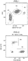
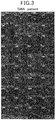
- Figs. 3 and 4 each illustrate a cell photograph taken by imaging flow cytometry in the same manner as described above.
- Figs. 3 and 4 are cell photographs of the cell groups mainly containing monocytes in the respective blood samples obtained from an SMA patient and a healthy subject (control).
- the SMN protein is indicated by green fluorescence.
- a healthy subject control was greater than an SMA patient in all of the fluorescence intensity in the whole cell, the fluorescence intensity in the nuclei, the number of spots, and the fluorescence intensity in the spots.
- spot means an SMN protein nuclear body observed in the shape of a spot
- spot employed the definition of the "spot” as one 1) existing in a nucleus, 2) having a size of 0.34 to 3.07 ⁇ m 2 , 3) showing fluorescence intensity 5 times that of the background or greater, and 4) having a width-to-height ratio of 0.5 to 1.0.
- the average value of the number of spots in a cell group exhibiting a predetermined fluorescence intensity was calculated as follows.
- Evaluation was carried out using an analysis value which was obtained by subtracting the numerical value of a sample stained with a non-specific antibody (isotype control) from the numerical value of a sample stained with a specific antibody (anti-SMN antibody).
- the Mean was calculated using the number of spots/the number of aiming adjusted cells, and the average value of the number of spots was calculated with the following formula.
- Fig. 5 shows the results.
- the numerical values of 0.426 and 0.295 are average values (arithmetic means) of the number of spots calculated for all tested blood samples derived from healthy persons and blood samples derived from SMA patients, respectively.
- ⁇ Mean Mean fluorescence label-anti SMN monoclonal antibody ⁇ Mean fluorescence label-isotype control
- the proportion of spot positive cells in a cell group exhibiting a predetermined fluorescence intensity was calculated as follows.
- Evaluation was carried out using an analysis value which was obtained by subtracting the numerical value of a sample stained with a non-specific antibody (isotype control) from the numerical value of a sample stained with a specific antibody (anti-SMN antibody).
- the proportion (%) was calculated using the number of spots/the number of aiming adjusted cells ⁇ 100, and the proportion of spot positive cells was calculated with the following formula.
- Fig. 6 shows the results.
- the numerical values of 35.6 and 26.1 are average values (arithmetic means) of the proportion of spot positive cells calculated for all tested blood samples derived from healthy persons and blood samples derived from SMA patients, respectively.
- ⁇ % % fluorescence label-anti SMN monoclonal antibody
- Fig. 7 shows the results.
- the numerical values of 7255 and 5364 are average values (arithmetic means) of the fluorescence intensity of spot positive cells calculated for all tested blood samples derived from healthy persons and blood samples derived from SMA patients, respectively.
- Fig. 8 shows the results.
- the numerical values of 332.7 and 162.7 are average values (arithmetic means) of the fluorescence intensity of spots calculated for all tested blood samples derived from healthy persons and blood samples derived from SMA patients, respectively.
- the method for analyzing the expression of an SMN protein nuclear body of the present invention not only can simply detect the expression of SMN protein nuclear body, which is a more reliable biomarker, but also can measure the difference between healthy persons and SMA patients with higher detection sensitivity.
- SMA is one of the intractable diseases designated by the country, and the method for completely curing SMA has not been established yet.
- the present invention is extremely useful for the evaluation of methods which can be a treatment for SMA. For example, it is possible to use the method of the present invention in evaluating a drug or a drug candidate against SMA and in screening a drug or a drug candidate against SMA.
- the present invention is also useful for use in the diagnosis of SMA. Furthermore, the present invention can be widely used in functional analysis of SMN protein, and can contribute greatly to diagnostic and therapeutic technique of SMA.
- the present invention can be applied not only to SMA but also to technique for diagnosing or ameliorating diseases and symptoms caused by reduction in the expression level of SMN protein.
Landscapes
- Health & Medical Sciences (AREA)
- Life Sciences & Earth Sciences (AREA)
- Engineering & Computer Science (AREA)
- Immunology (AREA)
- Chemical & Material Sciences (AREA)
- Biomedical Technology (AREA)
- Hematology (AREA)
- Urology & Nephrology (AREA)
- Molecular Biology (AREA)
- Physics & Mathematics (AREA)
- Cell Biology (AREA)
- Pathology (AREA)
- General Physics & Mathematics (AREA)
- General Health & Medical Sciences (AREA)
- Biochemistry (AREA)
- Analytical Chemistry (AREA)
- Medicinal Chemistry (AREA)
- Food Science & Technology (AREA)
- Biotechnology (AREA)
- Microbiology (AREA)
- Proteomics, Peptides & Aminoacids (AREA)
- Neurology (AREA)
- Nuclear Medicine, Radiotherapy & Molecular Imaging (AREA)
- Zoology (AREA)
- Virology (AREA)
- Neurosurgery (AREA)
- Tropical Medicine & Parasitology (AREA)
- Chemical Kinetics & Catalysis (AREA)
- Optics & Photonics (AREA)
- Dispersion Chemistry (AREA)
- Ecology (AREA)
- Biophysics (AREA)
- Investigating Or Analysing Biological Materials (AREA)
- Measuring Or Testing Involving Enzymes Or Micro-Organisms (AREA)
Claims (6)
- Procédé pour l'analyse de l'expression d'un corps nucléaire de la protéine neurone moteur de survie (SMN), comprenant les étapes de :marquage d'un ou plusieurs marqueurs d'antigènes de surface de cellules nucléées dérivées du sang dans un échantillon contenant les cellules nucléées avec un ou plusieurs anticorps de marqueurs, dans lequel les un ou plusieurs marqueurs d'antigènes de surface comprennent CD33 ;marquage de la protéine SMN dans les cellules nucléées avec un premier colorant fluorescent ;marquage de noyaux des cellules nucléées ;sélection d'une population de cellules contenant des monocytes à partir de plusieurs populations de cellules dans lesquelles les noyaux et la protéine SMN dans les cellules nucléées ont été marqués et qui ont été classés sur la base des un ou plusieurs marqueurs d'antigènes de surface comprenant CD33 marqués avec les un ou plusieurs anticorps de marqueurs ; etanalyse de l'expression d'un corps nucléaire de la protéine SMN de la population de cellules sélectionnée en mesurant une intensité de fluorescence émise par le premier colorant fluorescent, dans lequell'étape de sélection d'une population de cellules et l'étape d'analyse de l'expression d'un corps nucléaire de la protéine SMN sont réalisées par imagerie en cytométrie de flux qui utilise une lentille d'objectif avec un agrandissement de 40 fois ou plus.
- Procédé selon la revendication 1, dans lequel
l'étape de sélection comprend la sélection d'une population de cellules contenant des monocytes à partir de plusieurs populations de cellules dans lesquelles les noyaux et la protéine SMN dans les cellules nucléées ont été marqués et qui ont été classés sur la base des un ou plusieurs marqueurs d'antigènes de surface comprenant CD33 marqués avec les un ou plusieurs anticorps de marqueurs et sur une dispersion latérale (SSC). - Procédé selon la revendication 1 ou 2, dans lequell'étape de marquage de la protéine SMN comprend le marquage de la protéine SMN avec un premier colorant fluorescent, etl'étape de marquage de noyaux comprend le marquage de noyaux avec un second colorant fluorescent.
- Procédé selon l'une quelconque des revendications 1 à 3, comprenant de plus l'étape soumettant l'échantillon contenant les cellules nucléées dérivées du sang à un traitement d'hémolyse d'érythrocyte.
- Procédé selon l'une quelconque des revendications 1 à 4, dans lequell'échantillon contenant les cellules nucléées dérivées du sang est un obtenu chez un sujet,l'étape d'analyse de l'expression d'un corps nucléaire de la protéine SMN comprend la quantification d'un corps nucléaire de la protéine SMN, etle procédé comprend l'étape de comparaison d'un résultat obtenu par la quantification avec un témoin choisi parmi les (a) à (c) suivants :(a) si le sujet est un patient ayant une amyotrophie spinale (SMA), le témoin est un résultat obtenu de la même manière que dans le procédé pour la quantification du corps nucléaire de la protéine SMN utilisant du sang obtenu chez le sujet à l'exception que l'on utilise du sang obtenu chez une personne saine ou un porteur ;(b) si le sujet est une personne saine ou un porteur, le témoin est un résultat obtenu de la même manière que dans le procédé pour la quantification du corps nucléaire de la protéine SMN utilisant du sang obtenu chez le sujet à l'exception que l'on utilise du sang obtenu chez un patient de SMA ; et(c) si le sujet est une personne à laquelle un médicament ou un médicament candidat contre SMA a été administré, le témoin est un résultat obtenu de la même manière que dans le procédé pour la quantification du corps nucléaire de la protéine SMN utilisant du sang obtenu chez le sujet après l'administration du médicament ou du médicament candidat contre SMA à l'exception que l'on utilise du sang obtenu chez le sujet avant ladite administration.
- Procédé selon l'une quelconque des revendications 1 à 5, dans lequel le corps nucléaire de la protéine SMN de la population de cellules sélectionnée est observé dans la forme d'une tache, la tache étant définie comme existant dans un noyau, présentant une dimension de 0,34 à 8,54 µm2, présentant une intensité de fluorescence 5,5 fois celle du bruit de fond ou supérieure, et présentant un rapport largeur- hauteur de 0,4 à 1,0.
Applications Claiming Priority (2)
| Application Number | Priority Date | Filing Date | Title |
|---|---|---|---|
| JP2016103482 | 2016-05-24 | ||
| PCT/JP2017/019165 WO2017204208A1 (fr) | 2016-05-24 | 2017-05-23 | Procédé d'analyse d'expression de corps nucléaire de la protéine smn |
Publications (3)
| Publication Number | Publication Date |
|---|---|
| EP3467497A1 EP3467497A1 (fr) | 2019-04-10 |
| EP3467497A4 EP3467497A4 (fr) | 2020-02-05 |
| EP3467497B1 true EP3467497B1 (fr) | 2021-09-29 |
Family
ID=60411292
Family Applications (1)
| Application Number | Title | Priority Date | Filing Date |
|---|---|---|---|
| EP17802794.2A Active EP3467497B1 (fr) | 2016-05-24 | 2017-05-23 | Procédé d'analyse d'expression de corps nucléaire de la protéine smn |
Country Status (4)
| Country | Link |
|---|---|
| US (1) | US20210270844A1 (fr) |
| EP (1) | EP3467497B1 (fr) |
| JP (1) | JP6671664B2 (fr) |
| WO (1) | WO2017204208A1 (fr) |
Family Cites Families (5)
| Publication number | Priority date | Publication date | Assignee | Title |
|---|---|---|---|---|
| WO2006050451A2 (fr) * | 2004-11-02 | 2006-05-11 | Whitehead Institute For Biomedical Research | Methodes et compositions pour traiter des maladies des motoneurones |
| SI3421603T1 (sl) * | 2009-05-02 | 2022-02-28 | Genzyme Corporation | Genska terapija za nevrodegenerativne motnje |
| US20140349938A1 (en) * | 2011-06-03 | 2014-11-27 | President And Fellows Of Harvard College | Methods of diagnosing and treating amyotrophic lateral sclerosis |
| CN106255886B (zh) | 2014-04-03 | 2018-06-08 | 学校法人东京女子医科大学 | 检测活运动神经元蛋白质的表达的方法 |
| JP6143233B2 (ja) * | 2015-08-05 | 2017-06-07 | 国立大学法人京都大学 | 運動ニューロン疾患の検査方法及び治療剤のスクリーニング方法 |
-
2017
- 2017-05-23 JP JP2018519557A patent/JP6671664B2/ja active Active
- 2017-05-23 WO PCT/JP2017/019165 patent/WO2017204208A1/fr active Search and Examination
- 2017-05-23 US US16/304,219 patent/US20210270844A1/en not_active Abandoned
- 2017-05-23 EP EP17802794.2A patent/EP3467497B1/fr active Active
Also Published As
| Publication number | Publication date |
|---|---|
| JP6671664B2 (ja) | 2020-03-25 |
| WO2017204208A1 (fr) | 2017-11-30 |
| EP3467497A4 (fr) | 2020-02-05 |
| US20210270844A1 (en) | 2021-09-02 |
| JPWO2017204208A1 (ja) | 2019-08-08 |
| EP3467497A1 (fr) | 2019-04-10 |
Similar Documents
| Publication | Publication Date | Title |
|---|---|---|
| US7901950B2 (en) | Method for assessing disease states by profile analysis of isolated circulating endothelial cells | |
| Ohkawa et al. | Autoantibodies to epilepsy-related LGI1 in limbic encephalitis neutralize LGI1-ADAM22 interaction and reduce synaptic AMPA receptors | |
| JP6095578B2 (ja) | 全身における組織恒常性の撹乱をモニターする方法及び手段 | |
| US10914748B2 (en) | Erythrocyte-derived extracellular vesicles as a biomarker for clinically assessing Parkinson's disease | |
| CN108474798A (zh) | 表征细胞特异性微囊泡的方法 | |
| EP3128327B1 (fr) | Procédé de détection de l'expression de la protéine smn | |
| Lamarthée et al. | CRISPR/Cas9-engineered HLA-deleted glomerular endothelial cells as a tool to predict pathogenic non-HLA antibodies in kidney transplant recipients | |
| EP2453243A1 (fr) | Procédé pour le diagnostic et/ou le suivi de l'évolution d'une tumeur | |
| JP5791095B2 (ja) | Pnh型白血球の検出方法 | |
| WO2006020936A2 (fr) | Procede d'evaluation d'etats pathologiques par l'analyse de profils de cellules endotheliales circulantes isolees | |
| EP3467497B1 (fr) | Procédé d'analyse d'expression de corps nucléaire de la protéine smn | |
| CA2941460C (fr) | Vesicules extracellulaires derivees d'erythrocyte comme biomarqueur d'evaluation clinique de maladie de parkinson | |
| EP4081800A1 (fr) | Méthode de diagnostic de maladies cutanées liées au lymphome t | |
| EP4057004A1 (fr) | Procédé d'acquisition d'informations sur l'amyotrophie spinale | |
| WO2000052472A1 (fr) | Test de detection rapide d'une infection chez les enfants en bas age | |
| JP6956402B2 (ja) | 解析方法 | |
| WO2014065323A1 (fr) | Méthode de test et kit de test de certains troubles psychiatriques | |
| CN114878814A (zh) | 循环滤泡调节性t细胞作为过敏性哮喘诊断标记物的应用 | |
| CN111458515A (zh) | 一种外周血中的肺小细胞肿瘤细胞数量的检测方法 | |
| Klimova et al. | PROGNOSTIC MARKERS IN PATIENTS WITH THYMUS-INDEPENDENT AND THYMUS-DEPENDENT MYASTHENIA GRAVIS | |
| CN116679067A (zh) | 一种nAChRs亚基蛋白的检测方法及其应用 | |
| US20040166540A1 (en) | Methods of detecting CD34 positive and negative hematopoietic stem cells in human samples | |
| CN117092343A (zh) | 一种用于检测人体免疫年龄的试剂盒以及人体免疫年龄确定方法、装置、系统和存储介质 | |
| CN108414479A (zh) | 一种免疫组合物在制备IgA肾病无创检测用试剂中的用途 | |
| CN115197322A (zh) | 用于慢性淋巴细胞白血病微小残留病灶检测的抗体组合物及其应用 |
Legal Events
| Date | Code | Title | Description |
|---|---|---|---|
| STAA | Information on the status of an ep patent application or granted ep patent |
Free format text: STATUS: THE INTERNATIONAL PUBLICATION HAS BEEN MADE |
|
| PUAI | Public reference made under article 153(3) epc to a published international application that has entered the european phase |
Free format text: ORIGINAL CODE: 0009012 |
|
| STAA | Information on the status of an ep patent application or granted ep patent |
Free format text: STATUS: REQUEST FOR EXAMINATION WAS MADE |
|
| 17P | Request for examination filed |
Effective date: 20181221 |
|
| AK | Designated contracting states |
Kind code of ref document: A1 Designated state(s): AL AT BE BG CH CY CZ DE DK EE ES FI FR GB GR HR HU IE IS IT LI LT LU LV MC MK MT NL NO PL PT RO RS SE SI SK SM TR |
|
| AX | Request for extension of the european patent |
Extension state: BA ME |
|
| DAV | Request for validation of the european patent (deleted) | ||
| DAX | Request for extension of the european patent (deleted) | ||
| A4 | Supplementary search report drawn up and despatched |
Effective date: 20200109 |
|
| RIC1 | Information provided on ipc code assigned before grant |
Ipc: G01N 33/543 20060101ALI20200102BHEP Ipc: G01N 33/49 20060101ALI20200102BHEP Ipc: G01N 21/64 20060101ALI20200102BHEP Ipc: G01N 15/14 20060101ALI20200102BHEP Ipc: G01N 33/569 20060101ALI20200102BHEP Ipc: G01N 33/536 20060101ALI20200102BHEP Ipc: G01N 33/68 20060101ALI20200102BHEP Ipc: G01N 33/53 20060101AFI20200102BHEP Ipc: G01N 33/48 20060101ALI20200102BHEP |
|
| STAA | Information on the status of an ep patent application or granted ep patent |
Free format text: STATUS: EXAMINATION IS IN PROGRESS |
|
| STAA | Information on the status of an ep patent application or granted ep patent |
Free format text: STATUS: EXAMINATION IS IN PROGRESS |
|
| 17Q | First examination report despatched |
Effective date: 20201223 |
|
| GRAP | Despatch of communication of intention to grant a patent |
Free format text: ORIGINAL CODE: EPIDOSNIGR1 |
|
| STAA | Information on the status of an ep patent application or granted ep patent |
Free format text: STATUS: GRANT OF PATENT IS INTENDED |
|
| INTG | Intention to grant announced |
Effective date: 20210419 |
|
| GRAS | Grant fee paid |
Free format text: ORIGINAL CODE: EPIDOSNIGR3 |
|
| GRAA | (expected) grant |
Free format text: ORIGINAL CODE: 0009210 |
|
| STAA | Information on the status of an ep patent application or granted ep patent |
Free format text: STATUS: THE PATENT HAS BEEN GRANTED |
|
| AK | Designated contracting states |
Kind code of ref document: B1 Designated state(s): AL AT BE BG CH CY CZ DE DK EE ES FI FR GB GR HR HU IE IS IT LI LT LU LV MC MK MT NL NO PL PT RO RS SE SI SK SM TR |
|
| REG | Reference to a national code |
Ref country code: GB Ref legal event code: FG4D |
|
| REG | Reference to a national code |
Ref country code: CH Ref legal event code: EP Ref country code: AT Ref legal event code: REF Ref document number: 1434626 Country of ref document: AT Kind code of ref document: T Effective date: 20211015 |
|
| REG | Reference to a national code |
Ref country code: DE Ref legal event code: R096 Ref document number: 602017046849 Country of ref document: DE |
|
| REG | Reference to a national code |
Ref country code: IE Ref legal event code: FG4D |
|
| REG | Reference to a national code |
Ref country code: LT Ref legal event code: MG9D |
|
| PG25 | Lapsed in a contracting state [announced via postgrant information from national office to epo] |
Ref country code: LT Free format text: LAPSE BECAUSE OF FAILURE TO SUBMIT A TRANSLATION OF THE DESCRIPTION OR TO PAY THE FEE WITHIN THE PRESCRIBED TIME-LIMIT Effective date: 20210929 Ref country code: BG Free format text: LAPSE BECAUSE OF FAILURE TO SUBMIT A TRANSLATION OF THE DESCRIPTION OR TO PAY THE FEE WITHIN THE PRESCRIBED TIME-LIMIT Effective date: 20211229 Ref country code: NO Free format text: LAPSE BECAUSE OF FAILURE TO SUBMIT A TRANSLATION OF THE DESCRIPTION OR TO PAY THE FEE WITHIN THE PRESCRIBED TIME-LIMIT Effective date: 20211229 Ref country code: FI Free format text: LAPSE BECAUSE OF FAILURE TO SUBMIT A TRANSLATION OF THE DESCRIPTION OR TO PAY THE FEE WITHIN THE PRESCRIBED TIME-LIMIT Effective date: 20210929 Ref country code: HR Free format text: LAPSE BECAUSE OF FAILURE TO SUBMIT A TRANSLATION OF THE DESCRIPTION OR TO PAY THE FEE WITHIN THE PRESCRIBED TIME-LIMIT Effective date: 20210929 Ref country code: SE Free format text: LAPSE BECAUSE OF FAILURE TO SUBMIT A TRANSLATION OF THE DESCRIPTION OR TO PAY THE FEE WITHIN THE PRESCRIBED TIME-LIMIT Effective date: 20210929 Ref country code: RS Free format text: LAPSE BECAUSE OF FAILURE TO SUBMIT A TRANSLATION OF THE DESCRIPTION OR TO PAY THE FEE WITHIN THE PRESCRIBED TIME-LIMIT Effective date: 20210929 |
|
| REG | Reference to a national code |
Ref country code: NL Ref legal event code: MP Effective date: 20210929 |
|
| REG | Reference to a national code |
Ref country code: AT Ref legal event code: MK05 Ref document number: 1434626 Country of ref document: AT Kind code of ref document: T Effective date: 20210929 |
|
| PG25 | Lapsed in a contracting state [announced via postgrant information from national office to epo] |
Ref country code: LV Free format text: LAPSE BECAUSE OF FAILURE TO SUBMIT A TRANSLATION OF THE DESCRIPTION OR TO PAY THE FEE WITHIN THE PRESCRIBED TIME-LIMIT Effective date: 20210929 Ref country code: GR Free format text: LAPSE BECAUSE OF FAILURE TO SUBMIT A TRANSLATION OF THE DESCRIPTION OR TO PAY THE FEE WITHIN THE PRESCRIBED TIME-LIMIT Effective date: 20211230 |
|
| PG25 | Lapsed in a contracting state [announced via postgrant information from national office to epo] |
Ref country code: AT Free format text: LAPSE BECAUSE OF FAILURE TO SUBMIT A TRANSLATION OF THE DESCRIPTION OR TO PAY THE FEE WITHIN THE PRESCRIBED TIME-LIMIT Effective date: 20210929 |
|
| PG25 | Lapsed in a contracting state [announced via postgrant information from national office to epo] |
Ref country code: IS Free format text: LAPSE BECAUSE OF FAILURE TO SUBMIT A TRANSLATION OF THE DESCRIPTION OR TO PAY THE FEE WITHIN THE PRESCRIBED TIME-LIMIT Effective date: 20220129 Ref country code: SK Free format text: LAPSE BECAUSE OF FAILURE TO SUBMIT A TRANSLATION OF THE DESCRIPTION OR TO PAY THE FEE WITHIN THE PRESCRIBED TIME-LIMIT Effective date: 20210929 Ref country code: RO Free format text: LAPSE BECAUSE OF FAILURE TO SUBMIT A TRANSLATION OF THE DESCRIPTION OR TO PAY THE FEE WITHIN THE PRESCRIBED TIME-LIMIT Effective date: 20210929 Ref country code: PT Free format text: LAPSE BECAUSE OF FAILURE TO SUBMIT A TRANSLATION OF THE DESCRIPTION OR TO PAY THE FEE WITHIN THE PRESCRIBED TIME-LIMIT Effective date: 20220131 Ref country code: PL Free format text: LAPSE BECAUSE OF FAILURE TO SUBMIT A TRANSLATION OF THE DESCRIPTION OR TO PAY THE FEE WITHIN THE PRESCRIBED TIME-LIMIT Effective date: 20210929 Ref country code: NL Free format text: LAPSE BECAUSE OF FAILURE TO SUBMIT A TRANSLATION OF THE DESCRIPTION OR TO PAY THE FEE WITHIN THE PRESCRIBED TIME-LIMIT Effective date: 20210929 Ref country code: ES Free format text: LAPSE BECAUSE OF FAILURE TO SUBMIT A TRANSLATION OF THE DESCRIPTION OR TO PAY THE FEE WITHIN THE PRESCRIBED TIME-LIMIT Effective date: 20210929 Ref country code: EE Free format text: LAPSE BECAUSE OF FAILURE TO SUBMIT A TRANSLATION OF THE DESCRIPTION OR TO PAY THE FEE WITHIN THE PRESCRIBED TIME-LIMIT Effective date: 20210929 Ref country code: CZ Free format text: LAPSE BECAUSE OF FAILURE TO SUBMIT A TRANSLATION OF THE DESCRIPTION OR TO PAY THE FEE WITHIN THE PRESCRIBED TIME-LIMIT Effective date: 20210929 Ref country code: AL Free format text: LAPSE BECAUSE OF FAILURE TO SUBMIT A TRANSLATION OF THE DESCRIPTION OR TO PAY THE FEE WITHIN THE PRESCRIBED TIME-LIMIT Effective date: 20210929 |
|
| REG | Reference to a national code |
Ref country code: DE Ref legal event code: R097 Ref document number: 602017046849 Country of ref document: DE |
|
| PG25 | Lapsed in a contracting state [announced via postgrant information from national office to epo] |
Ref country code: DK Free format text: LAPSE BECAUSE OF FAILURE TO SUBMIT A TRANSLATION OF THE DESCRIPTION OR TO PAY THE FEE WITHIN THE PRESCRIBED TIME-LIMIT Effective date: 20210929 |
|
| PLBE | No opposition filed within time limit |
Free format text: ORIGINAL CODE: 0009261 |
|
| STAA | Information on the status of an ep patent application or granted ep patent |
Free format text: STATUS: NO OPPOSITION FILED WITHIN TIME LIMIT |
|
| 26N | No opposition filed |
Effective date: 20220630 |
|
| PG25 | Lapsed in a contracting state [announced via postgrant information from national office to epo] |
Ref country code: SI Free format text: LAPSE BECAUSE OF FAILURE TO SUBMIT A TRANSLATION OF THE DESCRIPTION OR TO PAY THE FEE WITHIN THE PRESCRIBED TIME-LIMIT Effective date: 20210929 |
|
| REG | Reference to a national code |
Ref country code: CH Ref legal event code: PL |
|
| REG | Reference to a national code |
Ref country code: BE Ref legal event code: MM Effective date: 20220531 |
|
| PG25 | Lapsed in a contracting state [announced via postgrant information from national office to epo] |
Ref country code: MC Free format text: LAPSE BECAUSE OF FAILURE TO SUBMIT A TRANSLATION OF THE DESCRIPTION OR TO PAY THE FEE WITHIN THE PRESCRIBED TIME-LIMIT Effective date: 20210929 Ref country code: LU Free format text: LAPSE BECAUSE OF NON-PAYMENT OF DUE FEES Effective date: 20220523 Ref country code: LI Free format text: LAPSE BECAUSE OF NON-PAYMENT OF DUE FEES Effective date: 20220531 Ref country code: IT Free format text: LAPSE BECAUSE OF FAILURE TO SUBMIT A TRANSLATION OF THE DESCRIPTION OR TO PAY THE FEE WITHIN THE PRESCRIBED TIME-LIMIT Effective date: 20210929 Ref country code: CH Free format text: LAPSE BECAUSE OF NON-PAYMENT OF DUE FEES Effective date: 20220531 |
|
| PG25 | Lapsed in a contracting state [announced via postgrant information from national office to epo] |
Ref country code: IE Free format text: LAPSE BECAUSE OF NON-PAYMENT OF DUE FEES Effective date: 20220523 |
|
| PG25 | Lapsed in a contracting state [announced via postgrant information from national office to epo] |
Ref country code: BE Free format text: LAPSE BECAUSE OF NON-PAYMENT OF DUE FEES Effective date: 20220531 |
|
| PGFP | Annual fee paid to national office [announced via postgrant information from national office to epo] |
Ref country code: FR Payment date: 20230526 Year of fee payment: 7 Ref country code: DE Payment date: 20230519 Year of fee payment: 7 |
|
| PGFP | Annual fee paid to national office [announced via postgrant information from national office to epo] |
Ref country code: GB Payment date: 20230524 Year of fee payment: 7 |
|
| PG25 | Lapsed in a contracting state [announced via postgrant information from national office to epo] |
Ref country code: HU Free format text: LAPSE BECAUSE OF FAILURE TO SUBMIT A TRANSLATION OF THE DESCRIPTION OR TO PAY THE FEE WITHIN THE PRESCRIBED TIME-LIMIT; INVALID AB INITIO Effective date: 20170523 |
|
| PG25 | Lapsed in a contracting state [announced via postgrant information from national office to epo] |
Ref country code: SM Free format text: LAPSE BECAUSE OF FAILURE TO SUBMIT A TRANSLATION OF THE DESCRIPTION OR TO PAY THE FEE WITHIN THE PRESCRIBED TIME-LIMIT Effective date: 20210929 Ref country code: MK Free format text: LAPSE BECAUSE OF FAILURE TO SUBMIT A TRANSLATION OF THE DESCRIPTION OR TO PAY THE FEE WITHIN THE PRESCRIBED TIME-LIMIT Effective date: 20210929 Ref country code: CY Free format text: LAPSE BECAUSE OF FAILURE TO SUBMIT A TRANSLATION OF THE DESCRIPTION OR TO PAY THE FEE WITHIN THE PRESCRIBED TIME-LIMIT Effective date: 20210929 |