EP3461419A1 - Procédé de commande pour un appareil robotisé pourvu d'un dispositif à ultrasons et appareil robotisé - Google Patents
Procédé de commande pour un appareil robotisé pourvu d'un dispositif à ultrasons et appareil robotisé Download PDFInfo
- Publication number
- EP3461419A1 EP3461419A1 EP17194033.1A EP17194033A EP3461419A1 EP 3461419 A1 EP3461419 A1 EP 3461419A1 EP 17194033 A EP17194033 A EP 17194033A EP 3461419 A1 EP3461419 A1 EP 3461419A1
- Authority
- EP
- European Patent Office
- Prior art keywords
- robotic device
- movement
- end effector
- ultrasound
- image data
- Prior art date
- Legal status (The legal status is an assumption and is not a legal conclusion. Google has not performed a legal analysis and makes no representation as to the accuracy of the status listed.)
- Withdrawn
Links
- GDOPTJXRTPNYNR-UHFFFAOYSA-N CC1CCCC1 Chemical compound CC1CCCC1 GDOPTJXRTPNYNR-UHFFFAOYSA-N 0.000 description 1
Images


Classifications
-
- A—HUMAN NECESSITIES
- A61—MEDICAL OR VETERINARY SCIENCE; HYGIENE
- A61B—DIAGNOSIS; SURGERY; IDENTIFICATION
- A61B34/00—Computer-aided surgery; Manipulators or robots specially adapted for use in surgery
- A61B34/20—Surgical navigation systems; Devices for tracking or guiding surgical instruments, e.g. for frameless stereotaxis
-
- A—HUMAN NECESSITIES
- A61—MEDICAL OR VETERINARY SCIENCE; HYGIENE
- A61B—DIAGNOSIS; SURGERY; IDENTIFICATION
- A61B34/00—Computer-aided surgery; Manipulators or robots specially adapted for use in surgery
- A61B34/30—Surgical robots
-
- A—HUMAN NECESSITIES
- A61—MEDICAL OR VETERINARY SCIENCE; HYGIENE
- A61B—DIAGNOSIS; SURGERY; IDENTIFICATION
- A61B17/00—Surgical instruments, devices or methods, e.g. tourniquets
- A61B17/16—Bone cutting, breaking or removal means other than saws, e.g. Osteoclasts; Drills or chisels for bones; Trepans
- A61B17/17—Guides or aligning means for drills, mills, pins or wires
- A61B17/1703—Guides or aligning means for drills, mills, pins or wires using imaging means, e.g. by X-rays
-
- B—PERFORMING OPERATIONS; TRANSPORTING
- B25—HAND TOOLS; PORTABLE POWER-DRIVEN TOOLS; MANIPULATORS
- B25J—MANIPULATORS; CHAMBERS PROVIDED WITH MANIPULATION DEVICES
- B25J11/00—Manipulators not otherwise provided for
- B25J11/005—Manipulators for mechanical processing tasks
- B25J11/0055—Cutting
-
- B—PERFORMING OPERATIONS; TRANSPORTING
- B25—HAND TOOLS; PORTABLE POWER-DRIVEN TOOLS; MANIPULATORS
- B25J—MANIPULATORS; CHAMBERS PROVIDED WITH MANIPULATION DEVICES
- B25J9/00—Programme-controlled manipulators
- B25J9/16—Programme controls
- B25J9/1694—Programme controls characterised by use of sensors other than normal servo-feedback from position, speed or acceleration sensors, perception control, multi-sensor controlled systems, sensor fusion
- B25J9/1697—Vision controlled systems
-
- A—HUMAN NECESSITIES
- A61—MEDICAL OR VETERINARY SCIENCE; HYGIENE
- A61B—DIAGNOSIS; SURGERY; IDENTIFICATION
- A61B34/00—Computer-aided surgery; Manipulators or robots specially adapted for use in surgery
- A61B34/20—Surgical navigation systems; Devices for tracking or guiding surgical instruments, e.g. for frameless stereotaxis
- A61B2034/2046—Tracking techniques
- A61B2034/2063—Acoustic tracking systems, e.g. using ultrasound
-
- A—HUMAN NECESSITIES
- A61—MEDICAL OR VETERINARY SCIENCE; HYGIENE
- A61B—DIAGNOSIS; SURGERY; IDENTIFICATION
- A61B90/00—Instruments, implements or accessories specially adapted for surgery or diagnosis and not covered by any of the groups A61B1/00 - A61B50/00, e.g. for luxation treatment or for protecting wound edges
- A61B90/36—Image-producing devices or illumination devices not otherwise provided for
- A61B90/37—Surgical systems with images on a monitor during operation
- A61B2090/378—Surgical systems with images on a monitor during operation using ultrasound
Definitions
- the invention relates to a driving method for a robotic device with an ultrasonic device according to claim 1 and a robotic device for carrying out such a method according to claim 8.
- robotic-surgical interventions which can be both diagnostic and therapeutic in nature, and in which an object is controlled by a robotic device or an interaction with the object is performed, it is important to always achieve the greatest possible precision.
- An example of this is e.g. in robot-assisted spine surgery, in which a robotic device holds a drill sleeve through which a screw or other instrument is inserted into a bone of the patient.
- Known robotic systems and methods in this area therefore often work with external optical tracking systems, which, for example, have a camera and markers in order to accurately record movements of the object (eg respiratory movement or heart movement in a patient).
- the markers are physically attached to the object and then tracked directly with the camera, requiring direct line of sight. In this way, the movements of the object can be visually tracked.
- attaching the markers to the object and requiring an unobstructed line of sight for the camera are often limiting and undesirable.
- Other systems for tracking patient movements such as EMT tracking or live fluoroscopy, also have disadvantages, hinder workflow, are invasive or pose risks from unnecessary X-rays.
- a method for controlling a robotic device whose end effector arranged on a kinematic chain interacts with an object and with an ultrasound device registered for the robotic device for detecting the movement of the object is provided with the following steps: detection of ultrasound image data of the object, determination of the relative Movement of the object on the basis of the ultrasonic image data using a mathematical method, determination of the absolute movement by registration to a planning data set, and compensation of movements of the object in the context of the interaction of the end effector of the robotic device with the object, wherein on Based on the current motion determination future movements of the object and an adapted positioning of the end effector of the robotic device are calculated in advance and the robotic device is driven accordingly, wherein in the calculation of the adjusted positioning of the end effector of the robotic device with respect to the movement of the object time delay is included ,
- the method according to the invention enables a precise compensation of live movements of the object in such a way that dangers, for example due to injuries to the object or the patient, can be minimized.
- the movements of the object are a periodic movement, e.g. global for respiration or heartbeat of the subject (patient) or locally for a movement that accelerates or decelerates.
- At least the duration of the detection of the ultrasound image data, the duration of the determination of the absolute movement and the reaction time of the robotic device are taken into account in the determination or estimation of the time delay. These are three major factors contributing to the time delay. Other factors may also be considered to determine the actual delay, eg, the duration of the pre-computations of the object and robotic device positions.
- features are extracted from image data; these can then be e.g. spatially transformed to features of other images.
- the mathematical method for determining the relative motion of the object using the image processing methods feature extraction or optical flow. These are two possible known methods for reading information from image data. Other image processing methods may be used.
- the ultrasound device is registered to a patient coordinate system.
- the predicted positions of the end effector of the robotic device are filtered and / or synchronized. These two methods are primarily used to avoid measurement noise and measurement errors as well as sudden movements of the object that deviate from the normal motion flow, such as, for example, Twitches, to avoid with the robotic device or not to follow such movements if possible.
- the Kalman filter is used as an example of a suitable known filter.
- the robotic device In order to compensate for the time delay, in general, the robotic device generally has to accelerate significantly more to synchronize with the movement of the object than does the object itself, while at the same time the robotic device must decelerate in time so as not to overshoot the target.
- a robotic device with an ultrasound device registered to the robotic device, the robotic device having a kinematic chain of movable components, an end effector arranged at the end of the kinematic chain, which is designed to interact with an object, a control device for Control of the kinematic chain and the ultrasound device, an image processing device and a calculation unit, wherein the ultrasound device is designed to acquire ultrasound image data of the object, the image processing device and the computation unit are designed to cooperate, from the ultrasound image data and other data, the relative movement and the absolute movement of the object to determine on the basis of the current motion determination future movements of the object as well as an adapted positioning of the robotic device ts with a time delay calculated with respect to the movement of the object and the control device is designed to control the positioning of the robotic device.
- the robotic device has an end effector in the form of a drill sleeve. This is particularly important in the context of procedures for inserting or drilling in screws or other implants in bone structures.
- FIG. 1 a known robotic device 4 is shown with an ultrasonic device 5 for motion detection, as described for example in the non-prepublished European patent application with the application file 17159352.8.
- the robotic device 4 has a robot arm 3, which has several (at least 3) segments 7 and joints 8 and at the free end an end effector 9.
- the end effector 9 is designed to interact with an object, in particular a patient.
- An example of the use of such a robotic device can be found in robot-assisted spine surgery.
- the end effector is formed, for example, as a drill sleeve, which is held by the robot arm.
- the sleeve is placed on the bone of a patient, and a drill or screw introduces a screw or other instrument through the sleeve into the patient's bone.
- the ultrasound device 5 is arranged, for example, on the robotic device 4 or otherwise mechanically connected thereto and has an ultrasound head 10 which is designed to record (3D) ultrasound image data.
- the ultrasonic head 10 is brought into contact with the object.
- a control device 6 is provided, in addition, an image processing device 11 for processing the ultrasound image data may be present.
- the ultrasound head can also be integrated in it, for example.
- FIG. 2 is shown by means of a time axis t, as usually a movement delay comes to conditions.
- a movement delay comes to conditions.
- a cyclic movement a of the object eg respiratory movement or heart movement of a patient.
- the ultrasound image data are recorded and evaluated b and after a second time period t 2 (eg, robotic device response time), only the movement c of the robotic device takes place.
- t 0 between the movement a of the object and the movement of the robotic device c, so that any motion compensation based thereon is not of high quality.
- a high-quality motion compensation is necessary.
- FIG. 4 1 shows a sequence of a method according to the invention for controlling a robotic device interacting with an object with an ultrasound device registered for the robotic device for detecting the movement of the object.
- the inventive method causes a precise and high quality motion compensation and can thus be used in the surgical environment.
- the corresponding time required is included as a time delay in the planned movement of the robotic device and with adjusted for future predicted movements of the object.
- the ultrasound device is preferably registered to an object coordinate system (eg patient coordinate system).
- object coordinate system eg patient coordinate system
- a movement of the robotic device here is generally to be understood as meaning a movement of the robotic arm for positioning the end effector.
- ultrasound device 5 acquires ultrasound image data of a moving object (e.g., a patient).
- the ultrasound head must be in contact with the object.
- These are preferably at least two or a plurality of ultrasound images. These can be 2D or 3D ultrasound image data.
- the movement of the object it can be e.g. to treat the cyclical respiratory movement or heart movement of a patient.
- the relative movement of the object is determined or calculated on the basis of the recorded ultrasound image data using a mathematical method.
- a mathematical method e.g. to determine the relative motion of the object extracted from the ultrasound image data features of the object.
- methods that can be used for this purpose may be e.g. the so-called feature extraction or the so-called optical flow.
- a planning data record is understood to mean a previously existing initial data record, for example a 2D or 3D data record. It may be an ultrasound data set, but alternatively other data sets may be used, eg x-ray image data.
- vectors or pixels generated from the second step 22 may be obtained, suitably filtered, and registered in the third step to a previous image data set or to an existing initial 3D or 2D data set.
- the result of the calculations may be eg a new pose in space or a velocity vector reflecting the movement of the object (patient) at a defined point in the volume of the object (patient).
- a time delay is subsequently included in the planned movement of the robotic device and synchronized with predicted future movements of the object.
- the necessary time delay may e.g. previously estimated, calculated or determined empirically in an additional step 26, generally a constant value is used here.
- the duration of the recording of the ultrasound image data, the determinations of the movement of the object and the activation time of the robotic device are preferably taken into account in the time delay.
- the duration for the prediction of the object movement and the planned movement of the robotic device may also be included.
- dynamic limitations of robotic device motion can be taken into account in motion compensation planning.
- the robotic device may e.g.
- the planned movements of the robotic device are generally filtered (e.g., using the so-called Kalman filter) to reduce measurement errors.
- a position for the robotic device This is based on the determination of the movement of the object from the motion detection. It is usually a periodic movement, e.g. global for respiration or heartbeat of the subject (patient) or locally for a movement that accelerates or decelerates.
- a fifth step 25 the movement of the object is compensated by the robotic device. It may be necessary for the robotic device to accelerate significantly more than the object being compensated for in order to be in sync and slow down more so as not to exceed the intended target.
- FIG. 3 it is shown how the movements along the time axis t look when using the method according to the invention.
- the movement c of the robotic device is now in the context of an error tolerance synchronous with the movement of the object a, the preprocessing V for the robotic device takes into account the time delay t 0 .
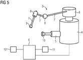
- FIG. 5 a robotic device 4 according to the invention with an ultrasound device 5 registered to the robotic device is shown.
- the robotic device 4 has a robot arm 3, which has a plurality of (at least three) segments 7 and joints 8 and at the free end an end effector 9, for example a drill sleeve.
- the end effector 9 is designed to interact with an object, eg a patient or an organ of the patient.
- the robotic device has a control device 6 for controlling the robot arm and the ultrasound device 5.
- the robotic device has an image processing device 11 and a calculation unit 12.
- the ultrasound device is designed to record ultrasound image data of the object.
- the image processing device 11 and the calculation unit 12 are designed to cooperatively determine from the ultrasound image data and further data (previously recorded image data or eg planning data) the relative movement and the absolute movement of the object, to calculate future movements of the object on the basis of the current motion determination as well as an adapted positioning of the robotic device with a time delay calculated with respect to the movement of the object.
- the control device 6 is designed to control the positioning of the end effector of the robotic device.
- a for a particular object particularly safe medical intervention with a robotic device is a method for controlling a robotic device whose an end effector disposed on a kinematic chain interacts with an object, and with an ultrasound device for motion detection of the object registered with the robotic device, comprising the steps of: acquiring ultrasound image data of the object, determining the relative motion of the object based on the ultrasound image data using a mathematical method Determining the absolute movement by registration to a planning data set, and compensation of movements of the object in the context of the interaction of the end effector of the robotic device with the object, wherein on the basis of the current motion determination future movements of the object and an adapted positioning of the end effector of the robotic Device are calculated in advance and the robotic device is driven accordingly, wherein in the calculation of the adjusted positioning of the end effector of the robotic device with respect to the movement de s object time delay is included.
Landscapes
- Health & Medical Sciences (AREA)
- Engineering & Computer Science (AREA)
- Surgery (AREA)
- Life Sciences & Earth Sciences (AREA)
- Nuclear Medicine, Radiotherapy & Molecular Imaging (AREA)
- Robotics (AREA)
- Animal Behavior & Ethology (AREA)
- Veterinary Medicine (AREA)
- Medical Informatics (AREA)
- Molecular Biology (AREA)
- Biomedical Technology (AREA)
- General Health & Medical Sciences (AREA)
- Public Health (AREA)
- Heart & Thoracic Surgery (AREA)
- Mechanical Engineering (AREA)
- Pathology (AREA)
- Radiology & Medical Imaging (AREA)
- Dentistry (AREA)
- Oral & Maxillofacial Surgery (AREA)
- Orthopedic Medicine & Surgery (AREA)
- Ultra Sonic Daignosis Equipment (AREA)
- Manipulator (AREA)
Priority Applications (2)
| Application Number | Priority Date | Filing Date | Title |
|---|---|---|---|
| EP17194033.1A EP3461419A1 (fr) | 2017-09-29 | 2017-09-29 | Procédé de commande pour un appareil robotisé pourvu d'un dispositif à ultrasons et appareil robotisé |
| US16/145,520 US10893909B2 (en) | 2017-09-29 | 2018-09-28 | Control method for a robotic device with an ultrasound apparatus and robotic device |
Applications Claiming Priority (1)
| Application Number | Priority Date | Filing Date | Title |
|---|---|---|---|
| EP17194033.1A EP3461419A1 (fr) | 2017-09-29 | 2017-09-29 | Procédé de commande pour un appareil robotisé pourvu d'un dispositif à ultrasons et appareil robotisé |
Publications (1)
| Publication Number | Publication Date |
|---|---|
| EP3461419A1 true EP3461419A1 (fr) | 2019-04-03 |
Family
ID=60037379
Family Applications (1)
| Application Number | Title | Priority Date | Filing Date |
|---|---|---|---|
| EP17194033.1A Withdrawn EP3461419A1 (fr) | 2017-09-29 | 2017-09-29 | Procédé de commande pour un appareil robotisé pourvu d'un dispositif à ultrasons et appareil robotisé |
Country Status (2)
| Country | Link |
|---|---|
| US (1) | US10893909B2 (fr) |
| EP (1) | EP3461419A1 (fr) |
Cited By (1)
| Publication number | Priority date | Publication date | Assignee | Title |
|---|---|---|---|---|
| CN114191099A (zh) * | 2022-01-14 | 2022-03-18 | 山东威高手术机器人有限公司 | 微创手术机器人主从跟踪延时测试方法 |
Families Citing this family (1)
| Publication number | Priority date | Publication date | Assignee | Title |
|---|---|---|---|---|
| CN113925529A (zh) * | 2021-10-14 | 2022-01-14 | 武汉库柏特科技有限公司 | 超声扫描控制方法、装置、设备及存储介质 |
Citations (2)
| Publication number | Priority date | Publication date | Assignee | Title |
|---|---|---|---|---|
| WO2000033723A2 (fr) * | 1998-11-20 | 2000-06-15 | Intuitive Surgical, Inc. | Chirurgie cardiaque sans cardioplegie |
| US20170151025A1 (en) * | 2015-12-01 | 2017-06-01 | Siemens Healthcare Gmbh | Medical robotic device and method for the operation thereof |
Family Cites Families (1)
| Publication number | Priority date | Publication date | Assignee | Title |
|---|---|---|---|---|
| KR102438357B1 (ko) * | 2016-07-01 | 2022-09-01 | 인튜어티브 서지컬 오퍼레이션즈 인코포레이티드 | 컴퓨터 보조 의료 시스템 및 방법 |
-
2017
- 2017-09-29 EP EP17194033.1A patent/EP3461419A1/fr not_active Withdrawn
-
2018
- 2018-09-28 US US16/145,520 patent/US10893909B2/en active Active
Patent Citations (2)
| Publication number | Priority date | Publication date | Assignee | Title |
|---|---|---|---|---|
| WO2000033723A2 (fr) * | 1998-11-20 | 2000-06-15 | Intuitive Surgical, Inc. | Chirurgie cardiaque sans cardioplegie |
| US20170151025A1 (en) * | 2015-12-01 | 2017-06-01 | Siemens Healthcare Gmbh | Medical robotic device and method for the operation thereof |
Non-Patent Citations (2)
| Title |
|---|
| GANGLOFF J ET AL: "Active Filtering of Physiological Motion in Robotized Surgery Using Predictive Control", IEEE TRANSACTIONS ON ROBOTICS, IEEE SERVICE CENTER, PISCATAWAY, NJ, US, vol. 21, no. 1, 1 February 2005 (2005-02-01), pages 67 - 79, XP011126460, ISSN: 1552-3098, DOI: 10.1109/TRO.2004.833812 * |
| SHELTEN G YUEN ET AL: "3D Ultrasound-Guided Motion Compensation System for Beating Heart Mitral Valve Repair", 6 September 2008, ECCV 2016 CONFERENCE; [LECTURE NOTES IN COMPUTER SCIENCE; LECT.NOTES COMPUTER], SPRINGER INTERNATIONAL PUBLISHING, CHAM, PAGE(S) 711 - 719, ISBN: 978-3-642-33485-6, ISSN: 0302-9743, XP019105094 * |
Cited By (2)
| Publication number | Priority date | Publication date | Assignee | Title |
|---|---|---|---|---|
| CN114191099A (zh) * | 2022-01-14 | 2022-03-18 | 山东威高手术机器人有限公司 | 微创手术机器人主从跟踪延时测试方法 |
| CN114191099B (zh) * | 2022-01-14 | 2023-12-01 | 山东威高手术机器人有限公司 | 微创手术机器人主从跟踪延时测试方法 |
Also Published As
| Publication number | Publication date |
|---|---|
| US20190099223A1 (en) | 2019-04-04 |
| US10893909B2 (en) | 2021-01-19 |
Similar Documents
| Publication | Publication Date | Title |
|---|---|---|
| EP3409230B1 (fr) | Mouvement d'un bras de robot | |
| DE102011079561B4 (de) | Verfahren und Röntgengerät zum zeitlich aktuellen Darstellen eines bewegten Abschnitts eines Körpers, Computerprogramm und Datenträger | |
| DE102009014154B4 (de) | Verfahren zur Kalibrierung der Position von einem Laserfächerstrahl zur Projektionsgeometrie eines Röntgengerätes und Röntgengerät | |
| DE10240727A1 (de) | Bildgebendes System und Verfahren zur Optimierung einer Röntgenabbildung | |
| DE102011083876A1 (de) | Verfahren zur Bewegungssteuerung einer Röntgenvorrichtung und Röntgensystem | |
| DE102014214935A1 (de) | Verfahren zum Betreiben eines medizinisch-robotischen Geräts | |
| DE102005059804A1 (de) | Verfahren und Vorrichtung zur Bewegungskorrektur bei der Bildgebung während einer medizinischen Intervention | |
| EP2259725A1 (fr) | Dispositif de radiographie et poste de travail médical | |
| EP1260179A1 (fr) | Appareil pour la régistration d'images radiographiques avec une système de navigation pour applications médicales | |
| DE102005028226A1 (de) | Vorrichtung zur Steuerung eines magnetischen Elements im Körper eines Patienten | |
| DE102004004620A1 (de) | Verfahren zur Registrierung und Überlagerung von Bilddaten bei Serienaufnahmen in der medizinischen Bildgebung | |
| EP2135575A1 (fr) | Procédé d'alignement d'instruments à fréquence libre | |
| DE102010040634A1 (de) | Verfahren zur 2D/3D-Registrierung | |
| DE102008033137A1 (de) | Verfahren und Vorrichtung zur Einstellung einer dynamisch anpassbaren Position eines bildgebenden Systems | |
| DE10219594A1 (de) | Verfahren zur transkutanen Katheterführung | |
| WO2019149400A1 (fr) | Procédé de planification de la position d'un système d'enregistrement d'un appareil d'imagerie médicale, et appareil d'imagerie médicale | |
| DE102019200803A1 (de) | Medizintechnischer Roboter, medizinisches System, Verfahren zu deren Betrieb, Computerprogramm und Speichermedium | |
| EP3461419A1 (fr) | Procédé de commande pour un appareil robotisé pourvu d'un dispositif à ultrasons et appareil robotisé | |
| EP3628225B1 (fr) | Procédé d'enregistrement de données d'image et système d'imagerie médicale | |
| EP3626173A1 (fr) | Procédé de correction de déplacement d'un ensemble de données d'image tridimensionnel reconstruit, dispositif à rayons x biplan, programme informatique et support de données lisible électroniquement | |
| DE102008023918A1 (de) | Automatisierte Registrierung von Live-Durchleuchtungsbildern | |
| DE102011006122A1 (de) | Medizinisches Röntgengerät und Biplan-Röntgengerät | |
| EP1629774B1 (fr) | Enregistrement de balayages peropératoires | |
| DE102013210185A1 (de) | Verfahren zur visuellen Unterstützung beim Fixieren eines Implantats | |
| DE102010040945B3 (de) | Bildgebendes Verfahren zur Berechnung von Flussgeschwindigkeiten |
Legal Events
| Date | Code | Title | Description |
|---|---|---|---|
| PUAI | Public reference made under article 153(3) epc to a published international application that has entered the european phase |
Free format text: ORIGINAL CODE: 0009012 |
|
| AK | Designated contracting states |
Kind code of ref document: A1 Designated state(s): AL AT BE BG CH CY CZ DE DK EE ES FI FR GB GR HR HU IE IS IT LI LT LU LV MC MK MT NL NO PL PT RO RS SE SI SK SM TR |
|
| AX | Request for extension of the european patent |
Extension state: BA ME |
|
| STAA | Information on the status of an ep patent application or granted ep patent |
Free format text: STATUS: THE APPLICATION IS DEEMED TO BE WITHDRAWN |
|
| 18D | Application deemed to be withdrawn |
Effective date: 20191005 |