EP3454737B1 - Tumor bed implant for multimodality treatment of at risk tissue surrounding a resection cavity - Google Patents
Tumor bed implant for multimodality treatment of at risk tissue surrounding a resection cavity Download PDFInfo
- Publication number
- EP3454737B1 EP3454737B1 EP17776835.5A EP17776835A EP3454737B1 EP 3454737 B1 EP3454737 B1 EP 3454737B1 EP 17776835 A EP17776835 A EP 17776835A EP 3454737 B1 EP3454737 B1 EP 3454737B1
- Authority
- EP
- European Patent Office
- Prior art keywords
- polymer
- radiation
- seeds
- implant
- tissue
- Prior art date
- Legal status (The legal status is an assumption and is not a legal conclusion. Google has not performed a legal analysis and makes no representation as to the accuracy of the status listed.)
- Active
Links
Images


Classifications
-
- A—HUMAN NECESSITIES
- A61—MEDICAL OR VETERINARY SCIENCE; HYGIENE
- A61N—ELECTROTHERAPY; MAGNETOTHERAPY; RADIATION THERAPY; ULTRASOUND THERAPY
- A61N5/00—Radiation therapy
- A61N5/10—X-ray therapy; Gamma-ray therapy; Particle-irradiation therapy
- A61N5/1001—X-ray therapy; Gamma-ray therapy; Particle-irradiation therapy using radiation sources introduced into or applied onto the body; brachytherapy
- A61N5/1014—Intracavitary radiation therapy
- A61N5/1015—Treatment of resected cavities created by surgery, e.g. lumpectomy
-
- A—HUMAN NECESSITIES
- A61—MEDICAL OR VETERINARY SCIENCE; HYGIENE
- A61B—DIAGNOSIS; SURGERY; IDENTIFICATION
- A61B5/00—Measuring for diagnostic purposes; Identification of persons
- A61B5/01—Measuring temperature of body parts ; Diagnostic temperature sensing, e.g. for malignant or inflamed tissue
-
- A—HUMAN NECESSITIES
- A61—MEDICAL OR VETERINARY SCIENCE; HYGIENE
- A61B—DIAGNOSIS; SURGERY; IDENTIFICATION
- A61B5/00—Measuring for diagnostic purposes; Identification of persons
- A61B5/06—Devices, other than using radiation, for detecting or locating foreign bodies ; Determining position of diagnostic devices within or on the body of the patient
-
- A—HUMAN NECESSITIES
- A61—MEDICAL OR VETERINARY SCIENCE; HYGIENE
- A61B—DIAGNOSIS; SURGERY; IDENTIFICATION
- A61B5/00—Measuring for diagnostic purposes; Identification of persons
- A61B5/48—Other medical applications
- A61B5/4848—Monitoring or testing the effects of treatment, e.g. of medication
-
- A—HUMAN NECESSITIES
- A61—MEDICAL OR VETERINARY SCIENCE; HYGIENE
- A61B—DIAGNOSIS; SURGERY; IDENTIFICATION
- A61B5/00—Measuring for diagnostic purposes; Identification of persons
- A61B5/68—Arrangements of detecting, measuring or recording means, e.g. sensors, in relation to patient
- A61B5/6846—Arrangements of detecting, measuring or recording means, e.g. sensors, in relation to patient specially adapted to be brought in contact with an internal body part, i.e. invasive
- A61B5/6847—Arrangements of detecting, measuring or recording means, e.g. sensors, in relation to patient specially adapted to be brought in contact with an internal body part, i.e. invasive mounted on an invasive device
- A61B5/6852—Catheters
-
- A—HUMAN NECESSITIES
- A61—MEDICAL OR VETERINARY SCIENCE; HYGIENE
- A61K—PREPARATIONS FOR MEDICAL, DENTAL OR TOILETRY PURPOSES
- A61K41/00—Medicinal preparations obtained by treating materials with wave energy or particle radiation ; Therapies using these preparations
- A61K41/0052—Thermotherapy; Hyperthermia; Magnetic induction; Induction heating therapy
-
- A—HUMAN NECESSITIES
- A61—MEDICAL OR VETERINARY SCIENCE; HYGIENE
- A61K—PREPARATIONS FOR MEDICAL, DENTAL OR TOILETRY PURPOSES
- A61K51/00—Preparations containing radioactive substances for use in therapy or testing in vivo
- A61K51/12—Preparations containing radioactive substances for use in therapy or testing in vivo characterised by a special physical form, e.g. emulsion, microcapsules, liposomes, characterized by a special physical form, e.g. emulsions, dispersions, microcapsules
- A61K51/1213—Semi-solid forms, gels, hydrogels, ointments, fats and waxes that are solid at room temperature
-
- A—HUMAN NECESSITIES
- A61—MEDICAL OR VETERINARY SCIENCE; HYGIENE
- A61N—ELECTROTHERAPY; MAGNETOTHERAPY; RADIATION THERAPY; ULTRASOUND THERAPY
- A61N2/00—Magnetotherapy
- A61N2/002—Magnetotherapy in combination with another treatment
-
- A—HUMAN NECESSITIES
- A61—MEDICAL OR VETERINARY SCIENCE; HYGIENE
- A61N—ELECTROTHERAPY; MAGNETOTHERAPY; RADIATION THERAPY; ULTRASOUND THERAPY
- A61N2/00—Magnetotherapy
- A61N2/004—Magnetotherapy specially adapted for a specific therapy
-
- A—HUMAN NECESSITIES
- A61—MEDICAL OR VETERINARY SCIENCE; HYGIENE
- A61N—ELECTROTHERAPY; MAGNETOTHERAPY; RADIATION THERAPY; ULTRASOUND THERAPY
- A61N5/00—Radiation therapy
- A61N5/02—Radiation therapy using microwaves
- A61N5/022—Apparatus adapted for a specific treatment
- A61N5/025—Warming the body, e.g. hyperthermia treatment
-
- A—HUMAN NECESSITIES
- A61—MEDICAL OR VETERINARY SCIENCE; HYGIENE
- A61N—ELECTROTHERAPY; MAGNETOTHERAPY; RADIATION THERAPY; ULTRASOUND THERAPY
- A61N5/00—Radiation therapy
- A61N5/10—X-ray therapy; Gamma-ray therapy; Particle-irradiation therapy
- A61N5/1001—X-ray therapy; Gamma-ray therapy; Particle-irradiation therapy using radiation sources introduced into or applied onto the body; brachytherapy
- A61N5/1027—Interstitial radiation therapy
-
- A—HUMAN NECESSITIES
- A61—MEDICAL OR VETERINARY SCIENCE; HYGIENE
- A61M—DEVICES FOR INTRODUCING MEDIA INTO, OR ONTO, THE BODY; DEVICES FOR TRANSDUCING BODY MEDIA OR FOR TAKING MEDIA FROM THE BODY; DEVICES FOR PRODUCING OR ENDING SLEEP OR STUPOR
- A61M25/00—Catheters; Hollow probes
- A61M25/10—Balloon catheters
- A61M25/1011—Multiple balloon catheters
- A61M2025/1013—Multiple balloon catheters with concentrically mounted balloons, e.g. being independently inflatable
-
- A—HUMAN NECESSITIES
- A61—MEDICAL OR VETERINARY SCIENCE; HYGIENE
- A61N—ELECTROTHERAPY; MAGNETOTHERAPY; RADIATION THERAPY; ULTRASOUND THERAPY
- A61N5/00—Radiation therapy
- A61N5/10—X-ray therapy; Gamma-ray therapy; Particle-irradiation therapy
- A61N5/1001—X-ray therapy; Gamma-ray therapy; Particle-irradiation therapy using radiation sources introduced into or applied onto the body; brachytherapy
- A61N2005/1019—Sources therefor
- A61N2005/1024—Seeds
Definitions
- the disclosure is related to biocompatible implant devices suitable for surgical placement within a resection cavity for treatment of at-risk tumor margin tissue with local hyperthermia in combination with radiation and/or chemotherapy and/or immunotherapy.
- RT radiation therapy
- RT radiation therapy
- tissue immediately surrounding the excised tumor volume are at risk for tumor regrowth from remaining microscopic disease.
- One method of concentrating radiation in tissues at risk and minimizing dose to surrounding normal tissues is to place radioactive sources directly inside the resection cavity to radiate tissue from the inside out.
- this procedure delivers the appropriate dose of radiation slowly over a period of time, and is thus called Brachytherapy.
- This procedure may be used to deliver radiation to tumor or at-risk tissue in close vicinity to small radiation sources that are generally implanted through one or more interstitial needles or plastic catheters, or through an endoluminal catheter or instrument inserted into a balloon or natural body orifice.
- Brachytherapy sources may be distributed within the target tissue in the form of one or more parallel preconfigured strings of radioactive small diameter cylindrical seeds placed in the resection cavity or inserted into afterloading catheters that are placed percutaneously into the tumor region in a planar or volumetric implant array.
- a single radiation source of very high activity located at the end of a wire may be pulled in computer controlled incremental steps through each implanted catheter, where the pause duration at each step is calculated to deliver the proper radiation dose before moving to the next point.
- US 2004/242953 discussed particles that can be introduced into a body cavity using a balloon catheter.
- the particles are adapted to provide brachytherapy treatment as well as hyperthermia treatment induced by a magnetic field.
- US2013053619 discloses a balloon catheter wherein magnetic material is provided in the balloon; an external magnetic field having the frequency of 30-100 kHz is applied to heat up said material and provide hyperthermia therapy.
- the hyperthermia therapy can be combined with chemotherapy or radiation therapy with an external X-ray beam.
- the peak radiation dose in tissue adjacent to the seeds is extremely high, which risks complications when seeds are placed near critical normal tissues. Indeed, doses of up to 500% above the minimum prescribed target tissue dose are delivered at the seed surface, but quickly reduce towards non-therapeutic levels with increasing distance from the seed. Accordingly, the prior art treatments risk both radiation necrosis from overdosing as well as under dosing of other tissue within the same treatment target, which reduces efficacy of the treatment.
- transplanted tissue flaps i.e. fat or muscle tissue excised from elsewhere in patient
- the transplanted tissue acts as a spacer, but the thickness of the transplant often varies significantly within a single slab. This has a direct effect on the radiation dose distribution delivered to surrounding at-risk tumor margin.
- the transplanted tissue may have weak structure that does not always control the separation distance from seeds to tumor margin or the migration of seeds during the brachytherapy delivery adequately. When seeds move during the implant period, which can last anywhere from 1 hour to >50 days, this causes distortion of the expected dose distribution with unanticipated excessive dose in regions where seeds bunch together and underdosing of locations where seeds drift away from initial positions.
- Surgical resection may be the treatment of choice for tumors located almost anywhere in the body, including head and neck, brain, lung, breast, torso and extremities.
- One typical location for tumors amenable to treatment with the proposed invention are tumors of the head and neck.
- Integration of new treatment approaches for head and neck cancers such as organ preservation surgery (1, 2), high-dose-rate brachytherapy (3), chemotherapy (4, 5), immunotherapy (6-8), and hyperthermia (9-11) have been proposed to enhance clinical outcomes.
- Local tumor hyperthermia at temperatures of 41-45°C for 30-60 min is one of the most promising adjuvant treatment methods in cancer therapy, contributing to a significant improvement of therapeutic efficacy in sites where adequate heating is possible. It is normally applied as an adjuvant to established cancer treatment modalities such as radiotherapy and/or chemotherapy (12-14), and is currently being investigated for potential synergism and activation of immunotherapy. (15-17).
- Chemotherapy treatments are well known and there are numerous methods of delivering chemotherapeutics to human tumors; the optimum method depends on site and tumor size among other factors. Drug delivery is perhaps most dependent on tumor vasculature which is widely variable across a tumor volume and often compromised in the center of large or rapidly growing tumor masses. While chemotherapy is generally administered systemically with good effect, in many solid tumors the local concentration is inadequate at levels considered toxic to critical normal tissues. In some tumors, local concentration of therapeutic may be increased using a liposomal formulation where the active drug is carried inside a lipid outer layer that reduces accumulation of therapeutic in critical normal tissues like heart, liver and spleen.
- This local drug delivery scheme can be further enhanced using a temperature sensitive liposome formulation that breaks down the lipid capsule at a moderate hyperthermic temperature (e.g. 40°C) and releases the cytotoxic drug into the heated tumor tissue. (18-21).
- drug may be instilled directly into a body cavity for more concentrated effect on adjacent tissue.
- This approach is the treatment of non-muscle invasive bladder cancer and the local effect can be increased further by the addition of local hyperthermia. (22-24).
- One approach for brain tumors that can be surgically removed is to attach biodegradable thin polymer "wafers" that are impregnated with the desired therapeutic (e.g. carmustine, BCNU, Gliadel wafer) all around the inside of the resection cavity to release drug directly into surrounding tissue, thereby bypassing blood vessels and the blood brain barrier. (25, 26).
- microwave radiation is often used to produce local hyperthermia of tumors located close to the skin surface, but penetration of effective heating is limited to about 3-4 cm.
- Treatment of tumors deep in the neck with external microwave waveguide applicators has been reported, but significant heating of overlying tissue is unavoidable (27, 28).
- Penetration deeper into tissue using a phased array of microwave antennas has been proposed (29, 30) and prototype arrays are currently under development (10, 31), but it remains a challenge to precisely focus heat within the human head without overheating surrounding critical normal tissues.
- radiofrequency annular phased array systems are available (32-34) that can penetrate deeper in the body, but the long wavelength produces a large heat focus and significant sidelobe heating in surrounding normal tissues. In either case, significant heating of critical normal tissues outside the intended tumor target is usually unavoidable.
- Ultrasound from external transducer arrays may be focused precisely into deep tissue targets (35-39), but this approach is problematic for head and neck tumors such as the larynx due to the heterogeneous anatomy and proximity to complex shaped air and bone regions that reflect or absorb ultrasound preferentially.
- the complex anatomy and sensitivity of surrounding critical normal tissues severely restrict the use of external heating technology for small head and neck tumors like the larynx.
- improved localization of heat within a small volume at depth may be obtained with a variety of internal heat source techniques, including interstitial radiofrequency electrodes, microwave antennas, ultrasound transducers, or one of several thermal conduction based hot source techniques (40).
- interstitial heating modalities require percutaneous insertion of an array of needles or catheters to insert heat sources, power connections, and temperature monitoring/control sensors into the tumor.
- heat sources power connections, and temperature monitoring/control sensors into the tumor.
- temperature monitoring/control sensors For many head and neck tumors including nasopharynx and larynx, maintaining an externalized array of percutaneous catheters following surgery is painful and undesirable for the patient.
- one hot source technique stands out as potentially most appropriate for minimally invasive local hyperthermia.
- Tissue directly adjacent to the magnetically coupled heated material is generally overheated, while tissue >5 mm from the hot sources may not be heated sufficiently (45, 54).
- One method of improving the control of temperature within an array of ferromagnetic implants is to use materials that undergo their Curie point transition from magnetic to non-magnetic state at the desired implant temperature (42, 44, 55). These ferroseeds self-regulate their temperature at approximately the Curie point of the alloy regardless of applied power level, leading to more uniform tumor heating without the need to measure internal source temperatures or adjust power of the external magnetic field.
- MRgFUS MR guided focused ultrasound
- Currently approved clinical indications for MRgFUS in the US are limited to treatment of essential tremor, neuropathic pain, and Parkinson's disease as these diseases tend to involve small volumes of tissue located closer to the center of the skull where better focal gain can be obtained from the surface conforming transducer array.
- Techniques to treat brain tumors that tend to be larger and located off center closer to the skull are currently under investigation.
- the present application describes new therapeutic devices and methods for treatment of certain cancers, specifically those in the head and neck, brain, and other sites that produce a resection cavity, including but not limited to cancers of the chest and abdomen, pelvis, arms and legs - sites where resection of a mass can be treated.
- the invention is defined in the following claims. Other embodiments, examples as well as methods are not a part of the invention and are provided for illustrative purposes only.
- the objective of the present invention is directed to a multimodality therapeutic device for treating cancerous cells that remain after tumor surgery, with lower toxicity to surrounding normal tissues than existing clinical approaches.
- the specific purpose is to create a product that will improve dose uniformity - by increasing minimum dose to the target while reducing treatment complications in normal tissues surrounding a tumor resection cavity, and thereby improve clinical outcomes and overall experience for the cancer patient.
- the device is directed to treating tissue in the tumor margin of surgical resection cavities with brachytherapy, combined with local hyperthermia.
- An embodiment of the invention is directed
- the balloon is elastic in order to stretch to fill a tumor resection cavity when filled with a radioactive fluid mixed with magnetic nanoparticles. This strategy provides highly uniform radiation and thermal dose distributions around the surface of the polymer balloon implant.
- a further embodiment is directed to a biocompatible polymer implant that consists of two concentric thin elastic layers that can be filled with two different fluids - the inner balloon to be filled with inexpensive saline or liquid polymer while the outer balloon in contact with the resection cavity wall contains the radioactive fluid mixed with magnetic nanoparticles to provide highly uniform radiation and thermal dose distributions in tissue around the surface of the polymer implant.
- the balloon implant contains one or more internal catheters that extend from the tip of the balloon to outside the skin surface in order to afterload a radioactive source(s) into the balloon in combination with magnetic nanoparticles dispersed uniformly within the balloon.
- a multimodality treatment device comprising a biocompatible expandable polymer shell balloon, wherein the polymer shell is capable of expanding to fill a tumor resection cavity, a radioactive material, a magnetic material, a hollow central space containing one or more catheters, and a flexible shaft connecting the balloon implant to the tissue surface.
- the device comprises at least one catheter extending from outside the skin surface to the tip of polymer shell and allows remote afterloading insertion of a High-Dose-Rate (HDR) brachytherapy source into the central space inside the polymer shell and preferable one or more catheters extend from outside the skin surface to various locations inside the hollow space of the polymer shell to allow insertion of temperature monitoring probes and for filling the interior space with fluids, such as a magnetic nanoparticle solution.
- HDR High-Dose-Rate
- a multimodality treatment device comprising a first biocompatible expandable polymer shell balloon, and a second polymer shell; wherein the first and second polymer shells are capable of expanding to fill a tumor resection cavity, a radioactive material, a magnetic material, a hollow central space containing one or more catheters within the second polymer shell, and a void between the first and second polymer shells, and a flexible shaft connecting the balloon implant to the tissue surface.
- a first fluid is injected into the hollow central space and a second fluid is injected into the void between the first and second polymer shells.
- magnetic nanoparticles is used interchangeably with magnetic fluid, or MNP fluid, are particles that can be manipulated using magnetic fields. These particles preferably are capable of being heated by application of an external force.
- ferromagnetic seeds or “ferroseeds” or “heat seeds” are meant to be spherical or cylindrical metallic materials. These may be individual seeds or combined on a string of ferromagnetic seeds, or combined on a string with ferromagnetic and radiation seeds.
- the embodiments disclosed herein define an implant device that capitalizes on the effects of mild hyperthermia (heating for 30-60 min at 40-45°C) combined with radiation and/or chemotherapy and/or immunotherapy and focused on the annular rim of at-risk tissue around a tumor resection cavity. Randomized trials of combined thermoradiotherapy and thermochemotherapy have demonstrated significant improvement of complete response rates with the addition of local hyperthermia, as well as a survival advantage in many tumor sites. (56). Hyperthermia is particularly efficacious in the re-treatment setting where radiation dose is limited. (14, 15). The typical problem restricting use of hyperthermia is poor control of power deposition pattern from the heat source which produces unacceptable heterogeneity of temperatures across the tissue target and unacceptable heating of surrounding normal tissues.
- a major issue in radiation therapy is unacceptable toxicity from excessive dose in normal tissue.
- the radiation dose adjacent to the source is over five times the minimum (100%) target dose prescribed for a 5 mm rim of tissue around the seed.
- the minimum (100%) target dose prescribed for a 5 mm rim of tissue around the seed As seen in the shaded region between 0 and 0.5 cm from the source, radiation dose drops off very rapidly from supratherapeutic levels near the seed. Conversely, the drop off in the second shaded region from 0.5 - 1 cm is much more gradual.
- a typical goal of therapy is to maintain all critical normal tissues less than about 140% of the prescribed target dose. Clearly that goal is not attained in the first region close to the radiation source where dose falls over 80% (from over 500 to 100%), whereas that goal is easily attained in the second region further from the source where dose falls only 20% (from 100 to 80%).
- the embodiments herein are directed towards new devices for treatment of at-risk tissue surrounding a tumor resection cavity - tissue that contains a mixture of cancerous and normal tissues.
- Resection cavities are necessary, for example, where the bulk of tumor can be removed from the body.
- the surgeon is not always able to remove all tumor cells and therefore there is often a rim of about 5 mm (sometimes more) of "at-risk" tissue in this tumor margin.
- These removable cancers are found in the head, neck, torso, back, arms, and legs, literally anywhere a cancer can grow.
- a patient suffering from brain cancer may have the cancerous tissue removed, leaving a resection cavity inside the skull. These removals, of course, are common in other cancer forms.
- an embodiment of the present disclosure is directed towards a device to be implanted in the resection cavity after tumor reduction surgery that will facilitate the delivery of highly localized brachytherapy.
- the brachytherapy is provided simultaneously with mild heating of the surrounding at-risk tissue in order to enhance radiation response locally and reduce surrounding normal tissue complications.
- the product will consist of a biocompatible tissue implant (e.g., a polymer slab, shell, or balloon, with approximately spherical, planar, or custom shape), which fills the resection cavity.
- Optimal materials for the tumor bed implant will depend on surgical site.
- the implant may be formed of a soft biocompatible polymer implant.
- the shape may be pre-formed by the manufacturer to fit a preplanned shape, or it may be custom fit to the resection cavity in the operating room by injecting a polymer that is a thick viscous liquid initially at room temperature and solidifies into the precise shape of the tumor bed upon exposure to elevated body temperature (34-37°C) and/or cure time, or a combination thereof.
- radiation seed strings When radiation seed strings are used, they often consist of high activity long half-life sources that are afterloaded into indwelling catheters for a calculated period of time to deliver the required radiation dose to tissue around the implant, and then removed after being in place for several minutes, hours or days. In some protocols, the catheter array may be left in place for multiple fractionated brachytherapy treatments, or the catheters replaced multiple times to deliver the total radiation dose. Alternatively, lower activity radiation seeds with short half-life (e.g. Cs-131 with half-life ⁇ 10 days) may be implanted surgically and left permanently in the tissue without catheters to deliver the desired total radiation dose over a longer time period.
- short half-life e.g. Cs-131 with half-life ⁇ 10 days
- tissues close to the small diameter seeds get extremely high radiation dose in order to obtain sufficiently high minimum dose throughout the tissue target, as demonstrated in FIG.1 . If seeds are placed or migrate to locations nearby critical normal tissue structures (e.g., blood vessels or nerves), the resulting dose heterogeneity can lead to severe complications.
- critical normal tissue structures e.g., blood vessels or nerves
- FIG. 2 depicts a prior art method of producing localized heat within a deep tissue volume.
- the figure depicts a cross section through the center of an implant array of 12 stainless steel needles (or alternatively 12 strings of ferromagnetic seeds), each 10 cm long x 1.5 mm diameter and implanted 1.5 cm apart in muscle tissue-equivalent phantom. Temperatures recorded at points a (seed surface), b (midway between two seeds), and c (midway between 4 seeds) during a 6 min application of external magnetic field at 100 kHz are depicted in the accompanying graph.
- the temperature rise at point a near the heated seed produces a very hot temperature in a short amount of time, that may burn or be supratherapeutic.
- the temperature rise at points b, b, and c far from the seeds is reduced nearly 6°C from the temperature at point a, and may not reach therapeutic levels between seeds when the limit of temperature is achieved near the seeds.
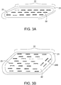
- FIG. 3A depicts a biocompatible polymer slab brachytherapy tumor bed implant 20 having a polymer coating (spacer) 21 coating a polymer core 22. Within the polymer core 22 are depicted three parallel lines (strands) of evenly spaced radiation therapy seeds 23 embedded just under the surface or lying on top of the surface of the polymer core 22.
- the radiation therapy seeds are preferably spaced 3-10 mm from the at-risk tissue target surrounding a resection cavity wall by a polymer coating 21 layer.
- the polymer coating 21 is comprised of either a resorbable or a non-resorbable biocompatible polymer material.
- a non-resorbable polymer would be utilized in instances where the resection cavity should be filled and maintained equal volume after surgery, so as to prevent a cavity in the skin for example.
- a resorbable polymer can be advantageously utilized where the resection cavity should collapse back to its size before the growth of the tumor cells that are excised.
- the polymer core 22 is similarly made of either non-resorbable or resorbable polymer. In certain embodiments, the same polymer can be utilized for both the polymer coating 21 and polymer core 22, though different polymers or densities can be utilized as necessary for the particular application.
- FIG. 3B depicts a variation of FIG. 3A , wherein a polymer slab implant 24 comprises radiation therapy seeds 23 placed in multiple mostly parallel seed strands, as well as seeds 23B oriented at angles to the parallel seed strands, as necessary to provide the most evenly spaced location of seeds all around the periphery of an irregularly shaped resection cavity wall.
- Appropriate numbers of parallel seeds 23 and angled seeds 23B, in multiple strands, are embedded in the polymer core 22 to ensure uniform spacing all around the polymer core 22.
- the polymer coating layer 21 provides a uniform 3-10 mm thick separation of radiation and ferroseeds from the resection cavity wall.
- this tumor bed shaped implant will add ferromagnetic seeds interspersed between the radiation seeds and distributed uniformly around or under the surface of the polymer implant.
- these ferroseeds 26 will be similar in size and interspersed alternately with radiation seeds 23 along each seed string (e.g. FIG. 4A ).
- the ferroseeds 26 may be heated to the desired treatment temperature one or more times after surgery by placing the patient inside a non-contacting induction coil and applying an external magnetic field that couples electromagnetic energy into the ferroseeds to make them hot.
- the seeds may be fabricated from an alloy having an appropriate Curie point temperature such that the seeds effectively self-regulate at the desired treatment temperature, normally around 45-50°C, though that temperature could range as high as 100°C for thermal ablation applications.(44, 74-77)
- the tumor bed implant will be fabricated from biocompatible resorbable polymer material that resorbs into tissue only after the delivery of radiation dose is complete. As the polymer material is resorbed, the implant region is debulked and the strings of now inactivated radiation seeds 23 and/or interspersed ferroseeds 26 are left in a fibrosed tissue, thereby limiting seed movement in the years thereafter. For some clinical applications, a formulation of polymer that does not resorb over time is preferred. This permanent implant would maintain the structure of the resection cavity long after surgery and ensure no migration of the seeds.
- Advantages from use of the proposed multimodality polymer implant include the ability to close the surgical wound at the time of tumor resection leaving a permanently implanted tumor bed shaped implant that will deliver appropriate radiation and heat doses (and potentially chemotherapy and/or immunotherapy drug infusions) with no externalized connections.
- the patient can go home soon after surgery with no needles or catheters penetrating the skin.
- Low energy sources, such as Cs-131 are safe for the family with minimal precautions.
- the radiation dose distribution is significantly more uniform throughout the at-risk tissue surrounding the resection cavity due to the 3-10 mm separation of radiation seeds from tissue provided by the intervening polymer. In the case of radiation fluid uniformly mixed into the polymer in place of individual radiation seeds, the radiation dosimetry is even more uniform around the surface of the implant.
- the thermal dose from embedded ferromagnetic seeds is significantly more uniform than from standard interstitial heating technology due to the 3-10 mm spacing of seeds from the target tissue.
- the uniformity of thermal dose may be maximized using magnetic nanoparticles distributed uniformly within the polymer.
- Heat treatments can be accomplished with no external connections to the power source as heat is coupled inductively to the seeds from the external magnetic field; no invasive connections are required.
- the radiation dose is delivered slowly over approximately 50 days post implant, allowing higher overall dose with less normal tissue complications than a single radiation dose at the time of surgery or several short radiation doses closely spaced in time soon after surgery.
- the improved homogeneity and extended duration of radiation dose delivery should reduce the risk of severe radiation-related side effects, which include: carotid artery rupture, osteo- or condro-radionecrosis, wound healing complications, fistula formation, cranial nerve damage, or skin and subcutaneous tissue necrosis among others.
- severe radiation-related side effects include: carotid artery rupture, osteo- or condro-radionecrosis, wound healing complications, fistula formation, cranial nerve damage, or skin and subcutaneous tissue necrosis among others.
- the synergistic effect of combining heat treatment with radiation should significantly improve overall response with minimal impact on normal tissue toxicity (57, 58).
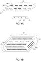
- FIG. 4A depicts a further embodiment of a thermobrachytherapy slab 25, comprising a polymer core 22, regularly spaced radiotherapy seeds 23 and heat therapy seeds 26.
- a side view is provided in FIG.4A so as to provide for appropriate visualization of the required spacing between the seeds and edge of overlying polymer.
- a further polymer coating may be included, as is depicted in FIG. 4B .
- the seeds are spaced at a regular and consistent spacing to ensure consistent and even therapeutic doses to adjacent cells.
- FIG. 4B provides a further embodiment of a thermobrachytherapy implant 28, having multiple mostly parallel seed strands 23 embedded in the polymer core 22 as well as seeds 23B at angles to the parallel seed strands 23, as necessary to provide the most evenly spaced location of seeds all around the periphery of the polymer core even in irregularly shaped resection cavity.
- Appropriate numbers of parallel seeds 23 and angled seeds 23B, in multiple strands, are embedded in the polymer core 22 to ensure uniform spacing all around the polymer core 22.
- the implant further comprises a polymer coating layer 21 that provides a uniform 3-10 mm thick separation of radiation and ferroseeds from the resection cavity wall.
- One preferred embodiment of the invention involves formation of a biocompatible resorbable implant inside the surgical cavity that can be fitted with a mesh of equally spaced radioactive seeds that deliver 95% of their radiation dose over a period of about 40 days, with dose rate continuing to fall off slowly thereafter.
- this implant would incorporate an array of equally spaced ferromagnetic seeds that can be coupled to an external magnetic field for delivery of heat treatments to enhance the radiation effect, for example FIGS. 4A and 4B .
- Heat treatments can be performed 1-7 times a week, for a duration of between 1 minute and 24 hours, inclusive of all times within.
- treatment is 1-2 times a week for a treatment time between 30 minutes and 1 hour.
- each of the polymer core 22 and coating 21 may absorb or be non-absorbable as necessary for the clinical indication.
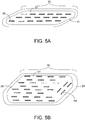
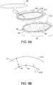
- FIG. 5A provides a further embodiment, comprising a biocompatible polymer thermo/chemo/immunotherapy implant 30 that comprises ferroseeds 26 within the polymer core 31.
- This embodiment provides the opportunity to include chemotherapeutic and immunotherapy agents mixed within the polymer structure which will be delivered slowly into the target tissue around the implant as the polymer material is resorbed.
- the chemo or immunotherapeutic materials can be impregnated within either the polymer coating 21 or polymer core 31 material, or in both the spacer 21 and core 31.
- the implant 30 may optionally comprise radiation therapy seeds, as provided in prior embodiments, to accomplish multimodality treatment with thermobrachy/chemo/immunotherapy.
- this slow release of drug will normally occur after a delay from the end of thermobrachytherapy treatment, and, thus, will provide appropriate separation of toxicities from the radiation and chemotherapy doses while adding therapeutic benefit from both treatments from the same multimodality tumor bed implant.
- FIG. 5B depicts a variation of FIG.5A wherein the polymer slab implant 35 comprises ferroseeds 26 placed in multiple mostly parallel seed strands, as well as seeds 26B oriented at angles to the parallel seed strands, as necessary to provide the most uniformly spaced location of thermal seeds all around the periphery of the polymer core 31, even in an irregularly shaped resection cavity.
- the polymer coating layer 21 provides a uniform 3-10 mm thick separation of ferroseeds from the resection cavity wall. This embodiment provides the opportunity to include chemotherapeutic and immunotherapy agents mixed within the polymer structure which will be delivered slowly into the target tissue around the implant as the polymer material is resorbed.
- the implant 35 may optionally comprise radiation therapy seeds, as provided in prior embodiments, to accomplish multimodality treatment with thermobrachy/chemo/immunotherapy.
- radiation therapy seeds are utilized, this slow release of drug occurring after a delay from the end of thermobrachytherapy treatment should provide appropriate separation of toxicities from the radiation and chemotherapy doses while adding therapeutic benefit from both treatments from the same multimodality tumor bed implant.
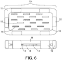
- FIG. 6 depicts a further embodiment of a polymer slab implant 10, comprising an inner preformed polymer core 13, spacers 12 positioned around the preformed polymer core 13, and a resorbable polymer 11 injected into the cavity to fill the space between the inner preformed core 13 and the edge of the resection cavity.
- the seed strings include ferroseed strings 14, which are uniformly spaced around the periphery of the preformed core 13 as well as radiation therapy seed strings 15, also uniformly spaced around the periphery of the preformed core 13.
- individual ferroseeds 14 and radiation seeds 15 may be interspersed uniformly around the periphery of the core 13.
- Cs-131 is the preferred radiation source, due to its half-life properties, allowing for the material to stay in the body while delivering the radiation dose over about 50 days.
- the resorbable polymer 11 also fills around the spacers 12 to make a uniform thickness layer of polymer overlying the radiation seeds 15 and ferroseeds 14 in the inner core 13.
- FIG.7 depicts a variation of the thermochemotherapy implant, wherein the polymer implant 40 contains no heat or radiation therapy seeds. Instead, the slab 40 contains a polymer 41 that is uniformly impregnated with magnetic nanoparticles. These nanoparticles, like the heat seeds, can be heated with an external power source. This configuration allows combination with external beam radiation therapy provided to the region of the resection cavity, in combination with heating of surrounding tissue by thermal conduction.
- the polymer 41 can be optionally further impregnated with therapeutic materials, such as chemotherapeutic or immunotherapeutic materials which will be delivered slowly to the target tissue as the polymer material is resorbed.
- FIG. 7 depicts a further polymer coating 42 surrounding the polymer 41 that can be optionally added.
- the implant may contain only the polymer 41 or both the polymer coating 42 and the polymer 41, each of which can be modified as described herein.
- FIG. 8 depicts a biocompatible non-resorbable multilayer polymer implant 45 with radiation seeds 23 uniformly spaced around the periphery of the polymer core 41.
- This configuration allows heating of resection cavity wall, e.g., 1-10 times with magnetic coupling to an external magnetic field during the 40-50 day delivery of brachytherapy.
- the outer layer of resorbable polymer 42 resorbs slowly in time releasing the chemotherapeutics.
- Heat treatments may continue 1-2 times per week during the period of radiation treatments and again during the period of release of chermotherapeutics as optimum for multimodality treatment of the tissue.
- This configuration debulks the region around the resection cavity after the end of multimodality treatments and eliminates all magnetic material from the body, while leaving only the biocompatible polymer core 41 with embedded radiation seeds (RT) 23 when it is non-resorbable.
- the polymer core 41 with embedded RT seeds 23 may be resorbable into the body after the end of radiation treatment leaving the decayed radiation seeds in the fibrosed scar of the resection cavity.
- Methods of treatment using the any of the above described devices comprises inserting the device into a resection cavity.
- the device can be pre-molded, or can be molded with a suitable fluid which will harden in the cavity as a polymer core.
- Radiation seeds and a magnetic material are added, either in or around the polymer core.
- a liquid polymer can be further injected around the polymer core to create appropriate space between the cells at the edge of the resection cavity and the radiation seeds.
- a magnetic force can then be applied externally to the patient to heat the magnetic material (ferroseeds or a magnetic nanoparticle).
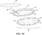
- FIG. 9A depicts a resection cavity 55.
- the multimodality treatment device 50 comprises a biocompatible expandable hollow polymer shell 51 with uniformly spaced radiation seeds 53 and ferromagnetic seeds 54 embedded in the inner surface 51B of the thick wall polymer shell 51 for thermobrachytherapy over approximately fifty (50) days interspersed with hyperthermia treatments 1-2 times weekly.
- the hollow polymer shell 51 is expandable, like a balloon, and thus can be filled with a liquid polymer, saline, or other suitable material.
- the liquid polymer core 52 can also be filled and itself would expand the polymer shell 51 outwards. This forces the outer wall 51A against the resection cavity 55.
- FIG. 9B A detail cross-section of the polymer shell 51 is depicted in FIG. 9B .
- the hollow core 52 is filled with liquid polymer to expand the polymer shell 51 to fill the resection cavity 55 with the outer surface 51B contacting the cells at the resection cavity wall.
- the 3-8 mm thick wall of polymer shell 51 provides the correct separation distance of radiation seeds 53 and ferroseeds 54 from tissue around the resection cavity wall.
- the device 60 would consist of a biocompatible expandable hollow polymer shell 51 with uniformly spaced radiation seeds 53 embedded in the inner surface 51B of the thick wall polymer shell 51, as in FIG. 9B .
- the interior would be filled with magnetic heating material 57 that may consist of tightly packed small ferromagnetic spheres, particles, or magnetic nanoparticle fluid.
- the thick wall polymer shell 51 may be filled or partially filled prior to surgical placement, and the volume expanded to completely fill the resection cavity by injection of additional fluid via needle (or catheter) 59 and syringe 58.
- the polymer shell 51 may be formulated from non-resorbable or resorbable material to fit the clinical application.
- the magnetic heating material may be mixed and distributed within the polymer shell 51, and the inner core 57 filled with a non-magnetic fluid or polymer.
- the small nanoparticles may dissipate from the region and be excreted from the body as the polymer shell is resorbed in tissue whereas the radiation seeds would remain permanently fixed in the scar tissue of the collapsing resection cavity wall. Elimination of all magnetic nanoparticles from the region following treatment has the advantage of avoiding image artifacts in any future Magnetic Resonance imaging of the region.
- the polymer shell 51 can be filled with a magnetic material, such as a magnetic nanoparticle, and the cores 52 or 57 can be filled with a polymer. This allows the core to inexpensively expand to press outwards on the polymer shell 51 while the expensive magnetic material fills the smaller volume of the polymer shell in direct contact with the tissue target.
- a magnetic material such as a magnetic nanoparticle
- methods of treatment of cancerous cells using the device according to FIGS. 9 or 10 includes resection of a tumor, application of the polymer shell into the resection cavity.
- the surgeon can then inject a fluid into either the core or into the polymer coating 51.
- the fluid, or the device itself comprises ferroseeds or a magnetic nanoparticle which can be heated. This fluid injection will allow the polymer coating 51 to expand to contact the edge of the resection cavity.
- the method further comprises applying an energy source to the patient to heat the ferroseeds or magnetic nanoparticles to increase the temperature to the cells at the edge of the resection cavity.
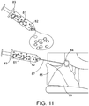
- FIG. 11 depicts an alternative configuration of polymer implant consisting of viscous colloidal solution containing radioactive material 81 mixed homogeneously with magnetic particles 82 that can be injected through, for example a syringe 83, and into the body through a needle or catheter 87 directly into a tumor resection cavity 84 or delivered into the interior of an expandable tumor cavity fitting polymer shell 51 as in Fig. 10 .
- the viscous liquid polymer may be formulated as one or two component mixture that becomes solid shortly after delivery into the tumor resection cavity due to higher body temperature or time cure.
- this colloidal solution of intermixed radiation and/or magnetic particles filling the tumor bed should provide the most uniform heat and radiation dosimetry around the resection cavity wall.
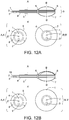
- FIGS. 12A-B depict the embodiment according to the invention, where the radiation and heat sources are delivered into the tumor resection cavity via a biocompatible flexible sheath 4.
- a high activity radiation source may be inserted into one or more central catheters 2 that extend from outside the patient through an incision in the skin to the tip of balloon which is implanted in the resection cavity.
- a High Dose Rate (HDR) afterloading device would be used to move the radiation source in precalculated steps along the one or more internal catheters 2, to deliver a computer planned radiation dose distribution to all tissue in contact with the balloon.
- HDR High Dose Rate
- FIG. 12B provides similar heat and radiation to the resection cavity wall with the addition of a temperature sensor that can be pulled along a catheter 1B that extends just inside the outer balloon wall and along the flexible sheath. Both configurations include a biocompatible polymer collar 5 with suture holes 8 to anchor the collar to the tissue surface and thereby secure the flexible shaft 4 to the skin.
- FIG. 13 depicts an alternative configuration of the biocompatible balloon 7 of Fig. 12 with two concentric thin wall balloons connecting to surface ports through a biocompatible flexible sheath 4. Both expandable thin wall polymer balloons are sealed to the flexible sheath 4 at the tip and distal ends with fixation collars 6.
- the outer balloon 9 may be filled through an external port and catheter 100 with homogeneous mixture of magnetic nanoparticles and radiation fluid 110 while the inner balloon 7 is filled through an external port and catheter 3 with sterile saline 120 to expand the balloon to fill the tumor resection cavity.
- a High Dose Rate (HDR) afterloading device would be used to move the radiation source in precalculated steps along the one or more internal catheters 2 to deliver most uniform radiation dose to all tissue in contact with the balloon.
- the magnetic nanoparticles can be heated 1-10 times during the delivery of radiation by coupling to an external magnetic field while temperatures of the outer balloon surface are measured with a temperature probe pulled in a catheter 1 contacting the outer balloon wall.
- a biocompatible polymer collar 5 anchors the flexible shaft 4 to the skin using sutures through suture holes 8.
- a method of treatment of cancerous cells in a patient comprises inserting a therapeutic device as described above into a resection cavity; applying an external magnetic field to increase the temperature of magnetic materials in the device for a predetermined amount of time to reach a therapeutic heat at the cancerous cells to be treated.
- the external magnetic field can be applied 1-20 times over the course of a treatment period of 1-100 days, to increase efficacy of the treatment.
- the therapeutic will be formulated with a release profile to allow for appropriately timed release into the surrounding tissues.
- Components of the above therapeutic devices consist of biocompatible polymer materials meeting the specifications above, the mixing container (if delivered in two components), insertion device (syringe or polymer delivery applicator), and sterile packaging for use in the operating room.
- the product incorporates a regularly spaced array of radioactive seeds such as the Cs-131 seed strings available from IsoRay Medical (Richland WA) with interspersed ferroseeds (such as those available from Best Medical International (Springfield VA) that are embedded within or on the surface of the polymer product.
- Other suitable arrangements include a configuration with each Cs-131 seed spaced equidistant around the inside of a hollow polymer shell.
- Cs-131 seeds can be replaced with other suitable radioactive isotopes for brachytherapy (e.g. 1-125, Pd-103, etc.).
- Accessories might include separately purchased software to replace or adapt current radiation treatment planning software to calculate radiation dose distributions from tumor bed implants.
- pre-formed pre-gelled resorbable polymer cores and flat tissue spacers may be manufactured in advance in various sizes and thicknesses to fill different size surgical cavities.
- Matching ungelled resorbable polymer material may be injected into the cavity around the pre-formed solid cores and radiation seeds at the time of surgery to cover the seeds with appropriate spacing to the resection cavity wall.
- chemotherapeutics may be mixed within the polymer that are slowly released as the polymer is absorbed into tissue after the end of thermobrachytherapy, to spread out and separate toxicities from the radiation and drug treatments.
- the tumor bed implant will consist of a biocompatible non-resorbable polymer that will permanently contain and stabilize radiation and ferroseeds left in tissue after treatment.
- Alternative product configurations include flexible expandable thick wall polymer shells that have radiation seeds and interspersed ferroseeds embedded on the inner surface of the shell.
- Other products include thin wall inflatable balloon polymer shells with internal catheters that extend out to the tissue surface to introduce a remotely afterloaded brachytherapy source and sensors to monitor the temperature of magnetic nanoparticle solution contained within the balloon that fills the resection cavity.
- Another alternative configuration consists of an inflatable balloon with catheter extending outside the tissue surface to inject both radioactive fluid (e.g. Iotrex or Cesitrex) and magnetic nanoparticle solution into the interior of a thin wall balloon that fills the resection cavity.
- radioactive fluid e.g. Iotrex or Cesitrex
- a further alternative is a polymer implant with two separate inflatable thin wall balloons with an inner balloon to contain saline or liquid polymer and a concentric outer balloon that contains magnetic material and/or radiation fluid and/or chemotherapeutics.
- biocompatible soft polymer implant that fits snugly inside the resection cavity after tumor reduction surgery.
- the implant may start as a high viscosity liquid at room temperature that solidifies at elevated (body) temperature or it may be supplied as a two component compound that solidifies quickly after mixing.
- a pre-manufactured solid polymer core implant may be available "in stock" that is close to the required size so can be shipped to the operating room (OR) in sterile packaging for insertion into the cavity.
- that standard size implant may require customization by injecting additional liquid or highly viscous polymer into the cavity around the core where it solidifies in the exact shape of the cavity due to temperature and/or time cure. Some cases may benefit from injecting the sterilized biocompatible viscous liquid polymer directly into the resection cavity without a core.
- radioactive seeds e.g. Cs-131, Pd-103, 1-125
- Cs-131, Pd-103, 1-125 short half-life radioactive seeds
- Curie point ferromagnetic seeds or particles that thermoregulate at the desired treatment temperature will be embedded under the surface of the polymer uniformly spaced and interspersed between the radiation seeds for controlled heating of tissue around the implant via coupling to an external magnetic field 1-7 times per week during the irradiation period, and again during the slow release of chemotherapeutics from a resorbing multimodality polymer implant.
- the implant may consist of a hollow polymer shell with the interior filled with radioactive fluid and/or magnetic nanoparticle solution for most uniform distribution of radiation and thermal doses in the at-risk tissue around the implant.
- radioactive fluid and/or magnetic nanoparticle solution for most uniform distribution of radiation and thermal doses in the at-risk tissue around the implant.
- Variations to the embodiments of the device include, but are not limited to the following options:
- a polymer core that is resorbable or non-resorbable This core can be performed or injected as a material that hardens via time or temperature cure.
- the core containing a plurality of evenly spaced radiation seeds and magnetic seeds.
- the radiation seeds and/or ferromagnetic seeds are replaced with nanoparticle solutions that include radioactive and/or magnetic properties.
- the magnetic materials have a Curie point transition that provides thermoregulation at an appropriate treatment temperature.
- the polymer may further comprise a therapeutic or chemotherapeutic material that, especially in the resorbable polymers, is then passed into the adjacent tissues as the polymer resorbs.
- the polymer core may further comprise a polymer coating.
- This polymer coating can be pre-formed or injected to the particular resection site, which may require certain spacers to be placed around the polymer core prior to injection of the coating layer.
- the polymer coating may be resorbable or non-resorbable and contain any or none of the following radiation therapy materials, heat therapy materials, therapeutic and chemotherapeutic materials.
- the polymer core and the polymer coating may be elastic and expand to fill a cavity, or can be flexible, but not expand, as appropriate.
- a flexible material When a flexible material is utilized, it can be envisioned as a single flexible shell that surrounds a core.
- the flexible shell can then be filled with a polymer material, radiation and heat materials, therapeutics and chemotherapeutics as in the prior examples, and then be filled to expand to fit the resection cavity.
- the flexible shell itself is hollow, having an inner wall and an outer wall, and creating a core inside of the inner wall.
- the space between the inner and outer wall itself can be filled with a material, as is described in detail above.
- the inner core of a prefabricated resorbable (or non-resorbable) polymer implant may be awkward to force the inner core of a prefabricated resorbable (or non-resorbable) polymer implant to conform to the shape of a resection cavity. This may be addressed for many tumor sites by having on hand several preformed inner cores that will fit many different size resection cavities. After selecting the appropriate size core from a number of preformed cores already sterile on the OR tray, the Cs-131 seed and ferroseed strings can be wrapped rapidly around the surface. Then the core can be positioned in the center of the cavity while liquid gel polymer is injected around the core, filling the cavity completely and providing the required distance from seeds to resection cavity wall.
- various size inner cores could be pre-fabricated with the seeds already embedded uniformly around and/or underlying the surface of the implant. This would allow the radiation dosimetry to be planned in advance of surgery. If the core does not entirely fill the cavity, a thicker outer coating layer of polymer material would be added to the seed embedded inner core to complete the tumor bed fit prior to insertion into the patient and the radiation dosimetry adjusted as necessary.
- the exact size of resection cavity in advance may be difficult or impossible in some cases. But due to advance imaging studies the approximate size of resection cavity will be known in advance for most cases. With appropriate preplanning, it should be possible to order the approximate size tumor bed implant in advance and customize its size in the operating room either by trimming the core or adding a thin overcoating of polymer to fit the actual resection cavity.
- the expandable polymer shell option may be used to provide a custom fit of the implant to the actual cavity by appropriate filling of the polymer shell interior with liquid polymer, magnetic fluid, and/or radioactive fluid.
- Biocompatible resorbable polymer implant to be combined with permanent radiation seeds (Cs-131, 1-125, or Pd-103) interspersed with ferromagnetic seeds (i.e. cylindrical or spherical).
- Configurations include: Made by manufacturer in advance - slab of resorbable polymer material to use as the implant core - available for order in several standard sizes. Cover this with matching size slab of resorbable polymer of desired thickness (e.g. 3-10 mm) with permanent radiation seeds and ferroseeds uniformly spaced in a mesh on one surface. Surgeon will assemble appropriate tumor bed implant in the operating room by trimming a standard size core to fit the surgical cavity and mating one seed-containing layer on each side of the core (for large volume implants); see Figure 3 .
- surgeon will adhere one 3-10 mm thick polymer-only slab to one matching size 3-10 mm thick slab having a radiation seed and interspersed ferroseed mesh on the mating surface, and implant into the cavity, forming one plane of seeds buried in the middle of a 6-20 mm thick polymer sandwich, as shown in Figure 5A and Figure 6 .
- Configurations include each of the prior options listed above, but wherein the material may exchange a resorbable material for a non-resorbable material in each of the prior embodiments. Indeed, some cases may benefit from combining a resorbable material as the outer layer of the implant to decompress the tissue region after completion of thermobrachytherapy, and a non-resorbable inner core material that will remain in the body to eliminate migration of seeds. Further, the outer resorbable polymer layer might include a homogenized mixture of magnetic nanoparticles such that the particles dissipate and excrete from the body in weeks following completion of thermobrachytherapy - to enable post treatment MR imaging of the region without artifact from concentrated magnetic particles.
- Biocompatible non-resorbable polymer implant with expandable polymer shell surrounding a hollow center that is made by the manufacturer in advance and available for order in several standard sizes.
- This polymer mixture is flexible and can expand in size as additional liquid is injected (via needle) into the shell interior prior to closing the surgical wound. This allows minor expansion of the polymer shell to fill the resection cavity, like inflating a balloon till it fills the cavity.
- Configurations include: a) uniformly spaced permanent radiation seeds (Cs-131, Pd-103, 1-125) interspersed with ferromagnetic seeds embedded in the inner wall of a 3-7 mm thick expandable polymer shell around a hollow center filled with saline or liquid polymer; b) uniformly spaced permanent radiation seeds embedded in the interior wall of a 3-7 mm thick polymer shell around a hollow center filled with magnetic fluid core; and c) 3-7 mm thick expandable polymer shell around a hollow center filled with magnetic fluid intermixed with radioactive fluid. See Figures 9 and 10 .
- Biocompatible polymer supplied by manufacturer in sterilized ungelled form inside syringe for injection by the surgeon into the tumor resection cavity.
- This polymer would come in one of several alternative formulations: a) resorbable or non-resorbable formulation; b) polymer to include magnetic nanoparticles homogeneously mixed throughout and be injected around one or more radiation seeds or seed strings positioned uniformly within the resection cavity; c) polymer to include magnetic nanoparticles and radiation fluid homogeneously mixed for injection directly into the resection cavity; d) chemotherapeutic agent uniformly mixed with magnetic nanoparticles and/or radioactive material within resorbable polymer; e) immunotherapy agent uniformly mixed with magnetic nanoparticles and/or radioactive material and/or chemotherapeutic within a resorbable polymer; or f) any of the above formulations injected around the outside of an undersized polymer core (filler) already placed in the center of the resection cavity.
- a further embodiment is directed towards a microscopic colloidal solution or nanoparticle containing radioactive and/or ferromagnetic particles for 3D printing or injection to the desired site of action around or within the tumor or resection cavity.
- the benefit of the injectable material is the small and uniform distribution of RT and HT materials in said material. This ensures a highly uniform distribution of materials to the ultimate site.
- the material can be directly injected into a resection cavity of any size or shape, or adjacent to a partial resection and existing tumor.
- the balloon catheter comprises a temperature probe catheter 1, a RT source catheter 2, a port 3 with a valve for inserting a fluid into the balloon; a flexible biocompatible sheath (or shaft) 4, a collar 5, to fix the sheath 4 to the skin, a balloon collar 6, to seal a balloon to the flexible sheath 4, a balloon 7, and suture holes 8 in the collar 5, to secure to the skin surface.
- the Flexible shaft 4 containing multiple catheters extending from outside the skin to a thin wall expandable polymer "balloon" implanted surgically in a tumor resection cavity.
- a practitioner would utilize one catheter 3 to inflate the balloon with radiation fluid homogeneously mixed with magnetic nanoparticles to fill the resection cavity for simultaneous or sequentially applied heat and brachytherapy treatment of the resection cavity wall.
- Another catheter 1 would be utilized to monitor temperature in the interior of the balloon.
- the central catheter 2 would allow connection of remote afterloader for insertion of high activity radiation source into the interior of the balloon and time sequenced radiation treatment of the resection cavity wall. Accordingly, a computer controlled program would control the timing of the catheter along a path to provide radiation to the interior wall cells of the resection cavity. This can be combined with inflation of the balloon 7 with homogeneously mixed magnetic nanoparticle solution to fill the resection cavity for simultaneous or sequentially applied heat with the brachytherapy treatment.
- the position of the temperature probe is different in FIGS. 12A and 12B , to measure the temperature of the fluid in the balloon, (12A) or the temperature at the interface of balloon and target tissue (12B).
- the device that provide for a suitable cancer treatment device, that can be modified based on the particular circumstances of the cancer to be treated, the location, and other factors as known to one of ordinary skill in the art. Accordingly, these can then be further utilized in methods of treatment of cancer in a patient, wherein the device is implanted into the patient in order to provide therapy with multiple synergistic treatment modalities.
- Laryngeal cancer comprises about 3% of all human cancers.
- the most common treatment for such patients is total laryngectomy which inevitably produces traumatic disability without ruling out tumor recurrence.
- five-year survival rates above 90% may be achieved reliably only for earlier stage T1 and T2 disease, whereas five-year survival is less than 60% for patients with intermediate or advanced stage cancers (80).
- One of the promising new approaches is to apply high-dose-rate brachytherapy from radiation sources placed within a patient-specific biocompatible implant that is itself inserted in the tumor resection cavity at the time of surgery.
- afterloading catheters were placed into a custom formed tumor bed shaped silicone implant to provide precise computable locations for a high dose rate (HDR) radiation source that was scanned through the catheters to deliver a conformal radiation dose to the tumor bed while the patient is still in the operating room (3, 81).
- HDR high dose rate
- Feasibility of this method of delivering HDR brachytherapy from within afterloading catheters inserted inside a tumor bed shaped silicone implant for intraoperative radiation treatment of laryngeal cancer has been described in the literature (81, 82).
- there are several inherent limitations of afterloading catheters that limit their use in many instances, and require the development of new therapies that can deliver more uniform doses of radiation to the target tissue and whenever possible without catheters exiting through the skin.
- results of a 48 patient pilot study performed in Russia of laryngeal cancers demonstrate the clinical promise of a tumor bed implant method that combines partial larynx resection with sequential high-dose-rate brachytherapy delivered to the resection cavity wall from within an intraoperatively-formed tumor bed fitting silicone gel implant (81, 82).
- This procedure allowed reduction in the volume of tissue resected and thus higher probability of preserving organ function.
- contact hyperthermia heating tissue adjacent to a hot surface
- Gliomas are the most commonly occurring brain tumors. Approximately 60% of primary brain tumors are high grade gliomas including Grade IV glioblastoma multiforme (GBM), of which there are approximately 10,000 and 74,000 new cases annually in the US and World respectively.
- GBM Grade IV glioblastoma multiforme
- One treatment approach is to implant an array of catheters percutaneously into the GBM and insert radioactive seeds (e.g. 1-125) into the catheters for brachytherapy treatment with or without adjuvant hyperthermia applied via miniature coaxial microwave antennas inserted into the same catheters after withdrawal of the brachytherapy seeds.
- Cs-131 Another short half-life radioactive material (Cs-131) was investigated for its safety and efficacy as a permanently implantable therapeutic.
- Interstitial hyperthermia may be delivered to a tissue volume via implanted microwave antennas, ultrasound tubular radiators, radiofrequency electrodes, or thermal conduction redistribution of heat within the interior of an array of small diameter thermally hot needles or catheters.(40) All of the interstitial heating approaches except for one require wires, needles, catheters or other connections from outside the patient to apply power and monitor/control the heat treatment.
- the preferred embodiments build off these previous concepts. Rather than restrict the delivery of radiation to tumor bed from radiation seed positions in a limited number of afterloading catheters embedded in the silicone implant, the preferred embodiments will provide significantly improved radiation dosimetry by dispersing the radioactive sources uniformly around and underlying the surface of the device. This separation of tissue from the radioactive seeds by a calculated distance of polymer significantly lowers the dose gradient in the surrounding tumor bed. The preferred embodiments further improve on brachytherapy treatment by adding contact hyperthermia from the large diameter heated cavity implant surface. In one configuration that avoids all undesirable percutaneous connections to the cavity implant, this invention combines the following features:
- thermal conduction based hyperthermia is akin to ferromagnetic thermoseed hyperthermia treatments reported in the literature (27, 45, 48, 72, 78), except its aim is not to heat the entire tumor volume but rather just a thin rim of at-risk tissue surrounding a tumor resection cavity. Precise localization of treatment to just the at-risk tumor bed tissue can be achieved using an intraoperatively formed implant to deliver both the brachytherapy and contact hyperthermia.
Landscapes
- Health & Medical Sciences (AREA)
- Life Sciences & Earth Sciences (AREA)
- Engineering & Computer Science (AREA)
- Biomedical Technology (AREA)
- Animal Behavior & Ethology (AREA)
- General Health & Medical Sciences (AREA)
- Public Health (AREA)
- Veterinary Medicine (AREA)
- Pathology (AREA)
- Nuclear Medicine, Radiotherapy & Molecular Imaging (AREA)
- Radiology & Medical Imaging (AREA)
- Surgery (AREA)
- Heart & Thoracic Surgery (AREA)
- Physics & Mathematics (AREA)
- Chemical & Material Sciences (AREA)
- Medical Informatics (AREA)
- Biophysics (AREA)
- Molecular Biology (AREA)
- Medicinal Chemistry (AREA)
- Pharmacology & Pharmacy (AREA)
- Epidemiology (AREA)
- Dispersion Chemistry (AREA)
- Optics & Photonics (AREA)
- Human Computer Interaction (AREA)
- Radiation-Therapy Devices (AREA)
- Thermotherapy And Cooling Therapy Devices (AREA)
- Magnetic Treatment Devices (AREA)
- Medicines That Contain Protein Lipid Enzymes And Other Medicines (AREA)
- Medicinal Preparation (AREA)
- Prostheses (AREA)
- Surgical Instruments (AREA)
- Materials For Medical Uses (AREA)
Priority Applications (2)
| Application Number | Priority Date | Filing Date | Title |
|---|---|---|---|
| PL17776835T PL3454737T3 (pl) | 2016-03-31 | 2017-03-31 | Implant do loży po guzie do leczenia multimodalnego zagrożonych tkanek otaczających jamę po resekcji |
| EP21183933.7A EP3954434A3 (en) | 2016-03-31 | 2017-03-31 | Tumor bed implant for multimodality treatment of at risk tissue surrounding a resection cavity |
Applications Claiming Priority (2)
| Application Number | Priority Date | Filing Date | Title |
|---|---|---|---|
| US201662315839P | 2016-03-31 | 2016-03-31 | |
| PCT/US2017/025523 WO2017173352A1 (en) | 2016-03-31 | 2017-03-31 | Tumor bed implant for multimodality treatment of at risk tissue surrounding a resection cavity |
Related Child Applications (1)
| Application Number | Title | Priority Date | Filing Date |
|---|---|---|---|
| EP21183933.7A Division EP3954434A3 (en) | 2016-03-31 | 2017-03-31 | Tumor bed implant for multimodality treatment of at risk tissue surrounding a resection cavity |
Publications (3)
| Publication Number | Publication Date |
|---|---|
| EP3454737A1 EP3454737A1 (en) | 2019-03-20 |
| EP3454737A4 EP3454737A4 (en) | 2020-01-22 |
| EP3454737B1 true EP3454737B1 (en) | 2021-07-14 |
Family
ID=59965301
Family Applications (2)
| Application Number | Title | Priority Date | Filing Date |
|---|---|---|---|
| EP17776835.5A Active EP3454737B1 (en) | 2016-03-31 | 2017-03-31 | Tumor bed implant for multimodality treatment of at risk tissue surrounding a resection cavity |
| EP21183933.7A Pending EP3954434A3 (en) | 2016-03-31 | 2017-03-31 | Tumor bed implant for multimodality treatment of at risk tissue surrounding a resection cavity |
Family Applications After (1)
| Application Number | Title | Priority Date | Filing Date |
|---|---|---|---|
| EP21183933.7A Pending EP3954434A3 (en) | 2016-03-31 | 2017-03-31 | Tumor bed implant for multimodality treatment of at risk tissue surrounding a resection cavity |
Country Status (8)
| Country | Link |
|---|---|
| US (2) | US11911631B2 (enExample) |
| EP (2) | EP3454737B1 (enExample) |
| JP (2) | JP7181549B2 (enExample) |
| BR (1) | BR112018070259A2 (enExample) |
| DK (1) | DK3454737T3 (enExample) |
| IL (2) | IL297355B2 (enExample) |
| PL (1) | PL3454737T3 (enExample) |
| WO (1) | WO2017173352A1 (enExample) |
Families Citing this family (12)
| Publication number | Priority date | Publication date | Assignee | Title |
|---|---|---|---|---|
| CA3029899A1 (en) * | 2016-07-07 | 2018-01-11 | The Regents Of The University Of California | Implants using ultrasonic backscatter for detecting electrophysiological signals |
| EP3634288A4 (en) * | 2017-06-08 | 2021-06-30 | Neuronoff, Inc. | ELECTRODE HARDENED AND MANUFACTURED IN THE BODY, AND RELATED METHODS AND DEVICES |
| WO2019099813A1 (en) * | 2017-11-17 | 2019-05-23 | Musara Mubayiwa Cornelious | Multilayered biologic mesh and methods of use thereof |
| AU2019200986A1 (en) | 2018-02-22 | 2019-09-05 | Robert E. Sandstrom | Magnetic Field Enhancement of Chemotherapy for Tumor Treatment |
| WO2019169445A1 (en) * | 2018-03-07 | 2019-09-12 | Margin-Clear Pty Ltd | A topical brachytherapy device and a method of treatment of malignant cancer cells |
| CA3091560A1 (en) * | 2018-03-08 | 2019-09-12 | Alpha Tau Medical Ltd. | Radiotherapy seeds and applicators |
| CN108714276B (zh) * | 2018-06-15 | 2021-06-01 | 李大鹏 | 一种妇科肿瘤腔内放射治疗防护装置 |
| WO2020205739A1 (en) * | 2019-03-31 | 2020-10-08 | The Trustees Of The University Of Pennsylvania | 'smart' hydrogel for the radiosensitization and sustained delivery of therapeutics triggered by irradiation |
| WO2021005610A1 (en) | 2019-07-10 | 2021-01-14 | David Cohen | Apparatus for treatment of tumors using a treatment object and powering of the treatment object |
| GR1010153B (el) * | 2020-09-21 | 2022-01-19 | Κυριακη Ιωαννη Θεοδωρου | Τρισδιαστατος συμμορφος εφαρμοστης για διεγχειρητικη ακτινοθεραπεια |
| JP2024511074A (ja) * | 2021-03-19 | 2024-03-12 | ジーティー メディカル テクノロジーズ、インコーポレイテッド | カスタム小線源治療キャリアを作成するシステム及び方法 |
| CA3227749A1 (en) * | 2021-07-28 | 2023-02-02 | Neuronoff, Inc. | Extension of a previously implanted lead or electrode |
Family Cites Families (47)
| Publication number | Priority date | Publication date | Assignee | Title |
|---|---|---|---|---|
| US6099457A (en) * | 1990-08-13 | 2000-08-08 | Endotech, Inc. | Endocurietherapy |
| JPH04116146A (ja) | 1990-09-04 | 1992-04-16 | Riken Corp | ポリマー被覆した感温性アモルファス合金 |
| US5429582A (en) | 1991-06-14 | 1995-07-04 | Williams; Jeffery A. | Tumor treatment |
| JPH09122260A (ja) * | 1995-11-02 | 1997-05-13 | Olympus Optical Co Ltd | 治療装置 |
| ATE297787T1 (de) | 1994-12-27 | 2005-07-15 | Olympus Optical Co | Medizinische vorrichtung |
| US6589502B1 (en) * | 1995-11-27 | 2003-07-08 | International Brachytherapy S.A. | Radioisotope dispersed in a matrix for brachytherapy |
| AU742942B2 (en) | 1997-04-07 | 2002-01-17 | Cook Urological Inc. | Back-up retention member drainage catheter |
| US5976067A (en) | 1997-05-28 | 1999-11-02 | Ablation Technologies, Inc. | Combination radioactive and temperature self-regulating thermal seed implant for treating tumors |
| JP2002524108A (ja) * | 1998-07-28 | 2002-08-06 | インナーダイン, インコーポレイテッド | 吸収性近接照射療法および化学療法送達デバイスならびに方法 |
| US8068897B1 (en) * | 1999-03-01 | 2011-11-29 | Gazdzinski Robert F | Endoscopic smart probe and method |
| US6641519B1 (en) | 1999-07-23 | 2003-11-04 | Nucletron B.V. | Wire drive in a medical device |
| EP1129990A1 (en) | 2000-02-25 | 2001-09-05 | Lucent Technologies Inc. | Process for controlled growth of carbon nanotubes |
| WO2004026111A2 (en) * | 2000-11-16 | 2004-04-01 | Microspherix Llc | Flexible and/or elastic brachytherapy seed or strand |
| US6527693B2 (en) | 2001-01-30 | 2003-03-04 | Implant Sciences Corporation | Methods and implants for providing radiation to a patient |
| US20030088145A1 (en) | 2001-07-18 | 2003-05-08 | Scott William Tate | Methods and devices for staged thermal and radiation therapy |
| EP1450711A1 (en) * | 2001-10-29 | 2004-09-01 | Triton Biosystems Inc. | Systems containing temperature regulated medical devices, and methods related thereto |
| US6761680B2 (en) | 2001-11-02 | 2004-07-13 | Richard A. Terwilliger | Delivery system and method for interstitial radiation therapy using seed strands constructed with preformed strand housing |
| US20040167506A1 (en) * | 2003-02-25 | 2004-08-26 | Scimed Life Systems, Inc. | Medical devices employing ferromagnetic heating |
| US20050090732A1 (en) * | 2003-10-28 | 2005-04-28 | Triton Biosystems, Inc. | Therapy via targeted delivery of nanoscale particles |
| US20060010475A1 (en) * | 2004-07-07 | 2006-01-12 | Sun Ningjun | High-spectrum radio frequency wireless transmission and control system for audio/video equipment |
| US7662082B2 (en) * | 2004-11-05 | 2010-02-16 | Theragenics Corporation | Expandable brachytherapy device |
| US8617152B2 (en) * | 2004-11-15 | 2013-12-31 | Medtronic Ablation Frontiers Llc | Ablation system with feedback |
| US20070173680A1 (en) * | 2005-12-29 | 2007-07-26 | Boston Scientific Scimed, Inc | Apparatus and method for performing therapeutic tissue ablation and brachytherapy |
| WO2007106531A1 (en) * | 2006-03-14 | 2007-09-20 | C.R. Bard, Inc. | Implant comprising radioactive seeds |
| WO2008058089A2 (en) * | 2006-11-03 | 2008-05-15 | North American Scientific, Inc. | Brachytherapy device having seed tubes with individually-settable tissue spacings |
| RU2322199C1 (ru) | 2006-11-20 | 2008-04-20 | Илья Леонидович Васильченко | Способ лечения злокачественных опухолей гортани и гортаноглотки |
| US20080146861A1 (en) | 2006-12-18 | 2008-06-19 | Goshen Health System, Inc. | Brachytherapy Shielding System |
| US8287442B2 (en) | 2007-03-12 | 2012-10-16 | Senorx, Inc. | Radiation catheter with multilayered balloon |
| US8303477B2 (en) | 2007-08-24 | 2012-11-06 | Cytyc Corporation | Fluid radiation shield for brachytherapy |
| US9561068B2 (en) | 2008-10-06 | 2017-02-07 | Virender K. Sharma | Method and apparatus for tissue ablation |
| JP2010233682A (ja) | 2009-03-30 | 2010-10-21 | Olympus Corp | カテーテル |
| US20110118600A1 (en) * | 2009-11-16 | 2011-05-19 | Michael Gertner | External Autonomic Modulation |
| EP2496165B1 (en) * | 2009-11-04 | 2017-01-11 | Emcision Limited | Lumenal remodelling device |
| US8262619B2 (en) | 2010-09-30 | 2012-09-11 | Tyco Healthcare Group Lp | Introducer sheath for catheters |
| US9060761B2 (en) * | 2010-11-18 | 2015-06-23 | Boston Scientific Scime, Inc. | Catheter-focused magnetic field induced renal nerve ablation |
| US20130261368A1 (en) | 2011-09-23 | 2013-10-03 | Alan N. Schwartz | Non-invasive and minimally invasive and tightly targeted minimally invasive therapy methods and devices for parathyroid treatment |
| CA2832586C (en) | 2011-04-08 | 2016-08-16 | Covidien Lp | Flexible microwave catheters for natural or artificial lumens |
| CA2835065C (en) | 2011-04-28 | 2018-02-13 | Peter Nakaji | Apparatus for loading dosimetrically customizable brachytherapy carriers |
| KR102069907B1 (ko) * | 2011-08-26 | 2020-01-23 | 엔도마그네틱스 엘티디 | 체강 및 공동형 부위에 있는 암을 치료하기 위한 에너지 필드 발생 장치 |
| NL2009699C2 (en) | 2012-10-25 | 2014-04-29 | Nucletron Operations Bv | A modular applicator for brachytherapy. |
| EP2914340B1 (en) | 2012-11-05 | 2020-10-14 | Nucletron Operations B.V. | Methods of making a medical applicator |
| US9149653B2 (en) | 2013-03-06 | 2015-10-06 | Mark A. D'Andrea | Brachytherapy devices and methods for therapeutic radiation procedures |
| WO2014205076A1 (en) | 2013-06-20 | 2014-12-24 | Source Production & Equipment Co., Inc. | Radioactive therapeutic device with fixation |
| WO2015181632A1 (en) | 2014-05-28 | 2015-12-03 | Nucletron Operations B.V. | Methods and systems for brachytherapy planning based on imaging data |
| EP3280494B1 (en) | 2015-04-10 | 2019-08-14 | Nucletron Operations B.V. | Brachytherapy applicators having ultrasound elements |
| US20170065324A1 (en) * | 2015-09-09 | 2017-03-09 | Rowan University | Wireless ablation catheter assembly |
| WO2017071965A1 (en) | 2015-10-30 | 2017-05-04 | Koninklijke Philips N.V. | Adaptive treatment planning for hyperthermia-enhanced radiation therapy |
-
2017
- 2017-03-31 EP EP17776835.5A patent/EP3454737B1/en active Active
- 2017-03-31 DK DK17776835.5T patent/DK3454737T3/da active
- 2017-03-31 PL PL17776835T patent/PL3454737T3/pl unknown
- 2017-03-31 WO PCT/US2017/025523 patent/WO2017173352A1/en not_active Ceased
- 2017-03-31 EP EP21183933.7A patent/EP3954434A3/en active Pending
- 2017-03-31 US US16/086,733 patent/US11911631B2/en active Active
- 2017-03-31 JP JP2018551372A patent/JP7181549B2/ja active Active
- 2017-03-31 BR BR112018070259A patent/BR112018070259A2/pt not_active Application Discontinuation
- 2017-03-31 IL IL297355A patent/IL297355B2/en unknown
-
2018
- 2018-09-26 IL IL261962A patent/IL261962B2/en unknown
-
2022
- 2022-11-10 JP JP2022179986A patent/JP7445232B2/ja active Active
-
2024
- 2024-01-18 US US18/416,340 patent/US20240181274A1/en active Pending
Also Published As
| Publication number | Publication date |
|---|---|
| US20240181274A1 (en) | 2024-06-06 |
| IL261962B2 (en) | 2023-03-01 |
| IL261962A (en) | 2018-10-31 |
| EP3454737A4 (en) | 2020-01-22 |
| PL3454737T3 (pl) | 2022-02-07 |
| JP7181549B2 (ja) | 2022-12-01 |
| JP2019511308A (ja) | 2019-04-25 |
| EP3954434A3 (en) | 2022-05-18 |
| IL297355B2 (en) | 2023-11-01 |
| US11911631B2 (en) | 2024-02-27 |
| JP7445232B2 (ja) | 2024-03-07 |
| US20190099618A1 (en) | 2019-04-04 |
| BR112018070259A2 (pt) | 2019-01-29 |
| EP3454737A1 (en) | 2019-03-20 |
| EP3954434A2 (en) | 2022-02-16 |
| IL297355B1 (en) | 2023-07-01 |
| IL297355A (en) | 2022-12-01 |
| DK3454737T3 (da) | 2021-10-11 |
| JP2023001313A (ja) | 2023-01-04 |
| WO2017173352A1 (en) | 2017-10-05 |
| IL261962B (en) | 2022-11-01 |
Similar Documents
| Publication | Publication Date | Title |
|---|---|---|
| US20240181274A1 (en) | Tumor bed implant for multimodality treatment of at risk tissue surrounding a resection cavity | |
| US20220347489A1 (en) | Dosimetrically customizable brachytherapy carriers and methods thereof in the treatment of tumors | |
| JP3505622B2 (ja) | 腫瘍治療装置 | |
| CN102470184B (zh) | 采用放射性种子的肿瘤治疗 | |
| Stewart et al. | Hyperthermia in the treatment of cancer: Perspectives on its promise and its problems | |
| US10238891B2 (en) | Device and method for improving brachytherapy | |
| CN105407955B (zh) | 输送药物的装置和方法 | |
| JP2019511308A5 (enExample) | ||
| US20250235255A1 (en) | Damaging cancerous cells utilizing radio frequency waves in heating with heating enhanced by infusion or injection of glucose | |
| Grady et al. | Magnetic stereotaxis: a technique to deliver stereotactic hyperthermia | |
| US12053644B2 (en) | Radiation shielding apparatus for implantable radioactive seeds | |
| De La Puente et al. | Delivery systems for brachytherapy | |
| WO2015142316A1 (en) | Dosimetrically customizable brachytherapy carriers and methods thereof in the treatment of tumors | |
| Stubbs et al. | Preclinical evaluation of a novel device for delivering brachytherapy to the margins of resected brain tumor cavities | |
| US11771919B2 (en) | Hyperthermia brachytherapy device and method for treating tumors | |
| Dahiya | Brachytherapy: A review | |
| Stauffer et al. | Dual modality implant for simultaneous magnetic nanoparticle heating and brachytherapy treatment of tumor resection cavities in brain | |
| CN100435763C (zh) | 结合热籽及血管支架实现血管内加热的肿瘤全身热疗装置 | |
| US20250058145A1 (en) | Pulsating brachytherapy method and system | |
| Yang et al. | Advances in hyperthermia technology | |
| Seegenschmiedt et al. | Clinical practice of interstitial thermoradiotherapy | |
| US20240082600A1 (en) | Hyperthermia brachytherapy device and method for treating tumors | |
| Luk | Clinical application of interstitial hyperthermia | |
| Vasilchenko et al. | Preliminary tests of local hyperthermia based on inductively heated tumor bed implant | |
| Prada | High-Dose-Rate Interstitial Brachytherapy as Monotherapy in |
Legal Events
| Date | Code | Title | Description |
|---|---|---|---|
| STAA | Information on the status of an ep patent application or granted ep patent |
Free format text: STATUS: THE INTERNATIONAL PUBLICATION HAS BEEN MADE |
|
| PUAI | Public reference made under article 153(3) epc to a published international application that has entered the european phase |
Free format text: ORIGINAL CODE: 0009012 |
|
| STAA | Information on the status of an ep patent application or granted ep patent |
Free format text: STATUS: REQUEST FOR EXAMINATION WAS MADE |
|
| 17P | Request for examination filed |
Effective date: 20181030 |
|
| AK | Designated contracting states |
Kind code of ref document: A1 Designated state(s): AL AT BE BG CH CY CZ DE DK EE ES FI FR GB GR HR HU IE IS IT LI LT LU LV MC MK MT NL NO PL PT RO RS SE SI SK SM TR |
|
| AX | Request for extension of the european patent |
Extension state: BA ME |
|
| DAV | Request for validation of the european patent (deleted) | ||
| DAX | Request for extension of the european patent (deleted) | ||
| A4 | Supplementary search report drawn up and despatched |
Effective date: 20200102 |
|
| RIC1 | Information provided on ipc code assigned before grant |
Ipc: A61B 5/06 20060101ALN20191217BHEP Ipc: A61M 25/10 20130101ALI20191217BHEP Ipc: A61K 51/12 20060101ALI20191217BHEP Ipc: A61N 5/10 20060101AFI20191217BHEP Ipc: A61B 5/01 20060101ALN20191217BHEP Ipc: A61K 49/04 20060101ALI20191217BHEP Ipc: A61B 5/00 20060101ALN20191217BHEP Ipc: A61K 41/00 20060101ALI20191217BHEP Ipc: A61N 5/02 20060101ALI20191217BHEP Ipc: A61N 2/00 20060101ALI20191217BHEP |
|
| REG | Reference to a national code |
Ref country code: DE Ref legal event code: R079 Ref document number: 602017042164 Country of ref document: DE Free format text: PREVIOUS MAIN CLASS: A61B0005060000 Ipc: A61N0005100000 |
|
| RIC1 | Information provided on ipc code assigned before grant |
Ipc: A61K 41/00 20200101ALI20201217BHEP Ipc: A61K 51/12 20060101ALI20201217BHEP Ipc: A61B 5/00 20060101ALN20201217BHEP Ipc: A61K 49/04 20060101ALI20201217BHEP Ipc: A61N 5/10 20060101AFI20201217BHEP Ipc: A61B 5/01 20060101ALN20201217BHEP Ipc: A61N 2/00 20060101ALI20201217BHEP Ipc: A61M 25/10 20130101ALI20201217BHEP Ipc: A61B 5/06 20060101ALN20201217BHEP Ipc: A61N 5/02 20060101ALI20201217BHEP |
|
| GRAP | Despatch of communication of intention to grant a patent |
Free format text: ORIGINAL CODE: EPIDOSNIGR1 |
|
| STAA | Information on the status of an ep patent application or granted ep patent |
Free format text: STATUS: GRANT OF PATENT IS INTENDED |
|
| INTG | Intention to grant announced |
Effective date: 20210128 |
|
| GRAS | Grant fee paid |
Free format text: ORIGINAL CODE: EPIDOSNIGR3 |
|
| GRAA | (expected) grant |
Free format text: ORIGINAL CODE: 0009210 |
|
| STAA | Information on the status of an ep patent application or granted ep patent |
Free format text: STATUS: THE PATENT HAS BEEN GRANTED |
|
| AK | Designated contracting states |
Kind code of ref document: B1 Designated state(s): AL AT BE BG CH CY CZ DE DK EE ES FI FR GB GR HR HU IE IS IT LI LT LU LV MC MK MT NL NO PL PT RO RS SE SI SK SM TR |
|
| REG | Reference to a national code |
Ref country code: GB Ref legal event code: FG4D |
|
| REG | Reference to a national code |
Ref country code: IE Ref legal event code: FG4D |
|
| REG | Reference to a national code |
Ref country code: DE Ref legal event code: R096 Ref document number: 602017042164 Country of ref document: DE |
|
| REG | Reference to a national code |
Ref country code: AT Ref legal event code: REF Ref document number: 1410193 Country of ref document: AT Kind code of ref document: T Effective date: 20210815 |
|
| REG | Reference to a national code |
Ref country code: FI Ref legal event code: FGE |
|
| REG | Reference to a national code |
Ref country code: DK Ref legal event code: T3 Effective date: 20211005 |
|
| REG | Reference to a national code |
Ref country code: SE Ref legal event code: TRGR |
|
| REG | Reference to a national code |
Ref country code: NL Ref legal event code: FP |
|
| REG | Reference to a national code |
Ref country code: LT Ref legal event code: MG9D |
|
| REG | Reference to a national code |
Ref country code: AT Ref legal event code: MK05 Ref document number: 1410193 Country of ref document: AT Kind code of ref document: T Effective date: 20210714 |
|
| PG25 | Lapsed in a contracting state [announced via postgrant information from national office to epo] |
Ref country code: RS Free format text: LAPSE BECAUSE OF FAILURE TO SUBMIT A TRANSLATION OF THE DESCRIPTION OR TO PAY THE FEE WITHIN THE PRESCRIBED TIME-LIMIT Effective date: 20210714 Ref country code: HR Free format text: LAPSE BECAUSE OF FAILURE TO SUBMIT A TRANSLATION OF THE DESCRIPTION OR TO PAY THE FEE WITHIN THE PRESCRIBED TIME-LIMIT Effective date: 20210714 Ref country code: LT Free format text: LAPSE BECAUSE OF FAILURE TO SUBMIT A TRANSLATION OF THE DESCRIPTION OR TO PAY THE FEE WITHIN THE PRESCRIBED TIME-LIMIT Effective date: 20210714 Ref country code: AT Free format text: LAPSE BECAUSE OF FAILURE TO SUBMIT A TRANSLATION OF THE DESCRIPTION OR TO PAY THE FEE WITHIN THE PRESCRIBED TIME-LIMIT Effective date: 20210714 Ref country code: BG Free format text: LAPSE BECAUSE OF FAILURE TO SUBMIT A TRANSLATION OF THE DESCRIPTION OR TO PAY THE FEE WITHIN THE PRESCRIBED TIME-LIMIT Effective date: 20211014 Ref country code: NO Free format text: LAPSE BECAUSE OF FAILURE TO SUBMIT A TRANSLATION OF THE DESCRIPTION OR TO PAY THE FEE WITHIN THE PRESCRIBED TIME-LIMIT Effective date: 20211014 Ref country code: PT Free format text: LAPSE BECAUSE OF FAILURE TO SUBMIT A TRANSLATION OF THE DESCRIPTION OR TO PAY THE FEE WITHIN THE PRESCRIBED TIME-LIMIT Effective date: 20211115 Ref country code: ES Free format text: LAPSE BECAUSE OF FAILURE TO SUBMIT A TRANSLATION OF THE DESCRIPTION OR TO PAY THE FEE WITHIN THE PRESCRIBED TIME-LIMIT Effective date: 20210714 |
|
| PG25 | Lapsed in a contracting state [announced via postgrant information from national office to epo] |
Ref country code: LV Free format text: LAPSE BECAUSE OF FAILURE TO SUBMIT A TRANSLATION OF THE DESCRIPTION OR TO PAY THE FEE WITHIN THE PRESCRIBED TIME-LIMIT Effective date: 20210714 Ref country code: GR Free format text: LAPSE BECAUSE OF FAILURE TO SUBMIT A TRANSLATION OF THE DESCRIPTION OR TO PAY THE FEE WITHIN THE PRESCRIBED TIME-LIMIT Effective date: 20211015 |
|
| REG | Reference to a national code |
Ref country code: DE Ref legal event code: R097 Ref document number: 602017042164 Country of ref document: DE |
|
| PLBE | No opposition filed within time limit |
Free format text: ORIGINAL CODE: 0009261 |
|
| STAA | Information on the status of an ep patent application or granted ep patent |
Free format text: STATUS: NO OPPOSITION FILED WITHIN TIME LIMIT |
|
| PG25 | Lapsed in a contracting state [announced via postgrant information from national office to epo] |
Ref country code: SM Free format text: LAPSE BECAUSE OF FAILURE TO SUBMIT A TRANSLATION OF THE DESCRIPTION OR TO PAY THE FEE WITHIN THE PRESCRIBED TIME-LIMIT Effective date: 20210714 Ref country code: SK Free format text: LAPSE BECAUSE OF FAILURE TO SUBMIT A TRANSLATION OF THE DESCRIPTION OR TO PAY THE FEE WITHIN THE PRESCRIBED TIME-LIMIT Effective date: 20210714 Ref country code: RO Free format text: LAPSE BECAUSE OF FAILURE TO SUBMIT A TRANSLATION OF THE DESCRIPTION OR TO PAY THE FEE WITHIN THE PRESCRIBED TIME-LIMIT Effective date: 20210714 Ref country code: EE Free format text: LAPSE BECAUSE OF FAILURE TO SUBMIT A TRANSLATION OF THE DESCRIPTION OR TO PAY THE FEE WITHIN THE PRESCRIBED TIME-LIMIT Effective date: 20210714 Ref country code: CZ Free format text: LAPSE BECAUSE OF FAILURE TO SUBMIT A TRANSLATION OF THE DESCRIPTION OR TO PAY THE FEE WITHIN THE PRESCRIBED TIME-LIMIT Effective date: 20210714 Ref country code: AL Free format text: LAPSE BECAUSE OF FAILURE TO SUBMIT A TRANSLATION OF THE DESCRIPTION OR TO PAY THE FEE WITHIN THE PRESCRIBED TIME-LIMIT Effective date: 20210714 |
|
| 26N | No opposition filed |
Effective date: 20220419 |
|
| PG25 | Lapsed in a contracting state [announced via postgrant information from national office to epo] |
Ref country code: IT Free format text: LAPSE BECAUSE OF FAILURE TO SUBMIT A TRANSLATION OF THE DESCRIPTION OR TO PAY THE FEE WITHIN THE PRESCRIBED TIME-LIMIT Effective date: 20210714 |
|
| PG25 | Lapsed in a contracting state [announced via postgrant information from national office to epo] |
Ref country code: MC Free format text: LAPSE BECAUSE OF FAILURE TO SUBMIT A TRANSLATION OF THE DESCRIPTION OR TO PAY THE FEE WITHIN THE PRESCRIBED TIME-LIMIT Effective date: 20210714 |
|
| PG25 | Lapsed in a contracting state [announced via postgrant information from national office to epo] |
Ref country code: LU Free format text: LAPSE BECAUSE OF NON-PAYMENT OF DUE FEES Effective date: 20220331 Ref country code: IE Free format text: LAPSE BECAUSE OF NON-PAYMENT OF DUE FEES Effective date: 20220331 |
|
| P01 | Opt-out of the competence of the unified patent court (upc) registered |
Effective date: 20230520 |
|
| PG25 | Lapsed in a contracting state [announced via postgrant information from national office to epo] |
Ref country code: HU Free format text: LAPSE BECAUSE OF FAILURE TO SUBMIT A TRANSLATION OF THE DESCRIPTION OR TO PAY THE FEE WITHIN THE PRESCRIBED TIME-LIMIT; INVALID AB INITIO Effective date: 20170331 |
|
| PG25 | Lapsed in a contracting state [announced via postgrant information from national office to epo] |
Ref country code: MK Free format text: LAPSE BECAUSE OF FAILURE TO SUBMIT A TRANSLATION OF THE DESCRIPTION OR TO PAY THE FEE WITHIN THE PRESCRIBED TIME-LIMIT Effective date: 20210714 Ref country code: CY Free format text: LAPSE BECAUSE OF FAILURE TO SUBMIT A TRANSLATION OF THE DESCRIPTION OR TO PAY THE FEE WITHIN THE PRESCRIBED TIME-LIMIT Effective date: 20210714 |
|
| PG25 | Lapsed in a contracting state [announced via postgrant information from national office to epo] |
Ref country code: MT Free format text: LAPSE BECAUSE OF FAILURE TO SUBMIT A TRANSLATION OF THE DESCRIPTION OR TO PAY THE FEE WITHIN THE PRESCRIBED TIME-LIMIT Effective date: 20210714 |
|
| PGFP | Annual fee paid to national office [announced via postgrant information from national office to epo] |
Ref country code: SE Payment date: 20250310 Year of fee payment: 9 |
|
| PGFP | Annual fee paid to national office [announced via postgrant information from national office to epo] |
Ref country code: DE Payment date: 20250305 Year of fee payment: 9 |
|
| PGFP | Annual fee paid to national office [announced via postgrant information from national office to epo] |
Ref country code: DK Payment date: 20250317 Year of fee payment: 9 Ref country code: NL Payment date: 20250317 Year of fee payment: 9 Ref country code: FI Payment date: 20250314 Year of fee payment: 9 |
|
| PGFP | Annual fee paid to national office [announced via postgrant information from national office to epo] |
Ref country code: BE Payment date: 20250318 Year of fee payment: 9 |
|
| PGFP | Annual fee paid to national office [announced via postgrant information from national office to epo] |
Ref country code: FR Payment date: 20250310 Year of fee payment: 9 |
|
| PGFP | Annual fee paid to national office [announced via postgrant information from national office to epo] |
Ref country code: GB Payment date: 20250227 Year of fee payment: 9 |
|
| PGFP | Annual fee paid to national office [announced via postgrant information from national office to epo] |
Ref country code: PL Payment date: 20250428 Year of fee payment: 9 |
|
| PGFP | Annual fee paid to national office [announced via postgrant information from national office to epo] |
Ref country code: CH Payment date: 20250401 Year of fee payment: 9 |