EP3448258B1 - Multiplexed transdermal extraction and detection system for non-invasive monitoring of substances and method of use - Google Patents
Multiplexed transdermal extraction and detection system for non-invasive monitoring of substances and method of use Download PDFInfo
- Publication number
- EP3448258B1 EP3448258B1 EP17719274.7A EP17719274A EP3448258B1 EP 3448258 B1 EP3448258 B1 EP 3448258B1 EP 17719274 A EP17719274 A EP 17719274A EP 3448258 B1 EP3448258 B1 EP 3448258B1
- Authority
- EP
- European Patent Office
- Prior art keywords
- array
- substances
- glucose
- gel
- sensor pixels
- Prior art date
- Legal status (The legal status is an assumption and is not a legal conclusion. Google has not performed a legal analysis and makes no representation as to the accuracy of the status listed.)
- Active
Links
Images


Classifications
-
- A—HUMAN NECESSITIES
- A61—MEDICAL OR VETERINARY SCIENCE; HYGIENE
- A61B—DIAGNOSIS; SURGERY; IDENTIFICATION
- A61B5/00—Measuring for diagnostic purposes; Identification of persons
- A61B5/145—Measuring characteristics of blood in vivo, e.g. gas concentration or pH-value ; Measuring characteristics of body fluids or tissues, e.g. interstitial fluid or cerebral tissue
- A61B5/14507—Measuring characteristics of blood in vivo, e.g. gas concentration or pH-value ; Measuring characteristics of body fluids or tissues, e.g. interstitial fluid or cerebral tissue specially adapted for measuring characteristics of body fluids other than blood
- A61B5/1451—Measuring characteristics of blood in vivo, e.g. gas concentration or pH-value ; Measuring characteristics of body fluids or tissues, e.g. interstitial fluid or cerebral tissue specially adapted for measuring characteristics of body fluids other than blood for interstitial fluid
- A61B5/14514—Measuring characteristics of blood in vivo, e.g. gas concentration or pH-value ; Measuring characteristics of body fluids or tissues, e.g. interstitial fluid or cerebral tissue specially adapted for measuring characteristics of body fluids other than blood for interstitial fluid using means for aiding extraction of interstitial fluid, e.g. microneedles or suction
-
- A—HUMAN NECESSITIES
- A61—MEDICAL OR VETERINARY SCIENCE; HYGIENE
- A61B—DIAGNOSIS; SURGERY; IDENTIFICATION
- A61B5/00—Measuring for diagnostic purposes; Identification of persons
- A61B5/145—Measuring characteristics of blood in vivo, e.g. gas concentration or pH-value ; Measuring characteristics of body fluids or tissues, e.g. interstitial fluid or cerebral tissue
- A61B5/14532—Measuring characteristics of blood in vivo, e.g. gas concentration or pH-value ; Measuring characteristics of body fluids or tissues, e.g. interstitial fluid or cerebral tissue for measuring glucose, e.g. by tissue impedance measurement
-
- A—HUMAN NECESSITIES
- A61—MEDICAL OR VETERINARY SCIENCE; HYGIENE
- A61B—DIAGNOSIS; SURGERY; IDENTIFICATION
- A61B5/00—Measuring for diagnostic purposes; Identification of persons
- A61B5/145—Measuring characteristics of blood in vivo, e.g. gas concentration or pH-value ; Measuring characteristics of body fluids or tissues, e.g. interstitial fluid or cerebral tissue
- A61B5/1468—Measuring characteristics of blood in vivo, e.g. gas concentration or pH-value ; Measuring characteristics of body fluids or tissues, e.g. interstitial fluid or cerebral tissue using chemical or electrochemical methods, e.g. by polarographic means
- A61B5/1477—Measuring characteristics of blood in vivo, e.g. gas concentration or pH-value ; Measuring characteristics of body fluids or tissues, e.g. interstitial fluid or cerebral tissue using chemical or electrochemical methods, e.g. by polarographic means non-invasive
-
- A—HUMAN NECESSITIES
- A61—MEDICAL OR VETERINARY SCIENCE; HYGIENE
- A61B—DIAGNOSIS; SURGERY; IDENTIFICATION
- A61B5/00—Measuring for diagnostic purposes; Identification of persons
- A61B5/145—Measuring characteristics of blood in vivo, e.g. gas concentration or pH-value ; Measuring characteristics of body fluids or tissues, e.g. interstitial fluid or cerebral tissue
- A61B5/1468—Measuring characteristics of blood in vivo, e.g. gas concentration or pH-value ; Measuring characteristics of body fluids or tissues, e.g. interstitial fluid or cerebral tissue using chemical or electrochemical methods, e.g. by polarographic means
- A61B5/1486—Measuring characteristics of blood in vivo, e.g. gas concentration or pH-value ; Measuring characteristics of body fluids or tissues, e.g. interstitial fluid or cerebral tissue using chemical or electrochemical methods, e.g. by polarographic means using enzyme electrodes, e.g. with immobilised oxidase
-
- A—HUMAN NECESSITIES
- A61—MEDICAL OR VETERINARY SCIENCE; HYGIENE
- A61B—DIAGNOSIS; SURGERY; IDENTIFICATION
- A61B10/00—Instruments for taking body samples for diagnostic purposes; Other methods or instruments for diagnosis, e.g. for vaccination diagnosis, sex determination or ovulation-period determination; Throat striking implements
- A61B2010/0009—Testing for drug or alcohol abuse
-
- A—HUMAN NECESSITIES
- A61—MEDICAL OR VETERINARY SCIENCE; HYGIENE
- A61B—DIAGNOSIS; SURGERY; IDENTIFICATION
- A61B10/00—Instruments for taking body samples for diagnostic purposes; Other methods or instruments for diagnosis, e.g. for vaccination diagnosis, sex determination or ovulation-period determination; Throat striking implements
- A61B10/0045—Devices for taking samples of body liquids
- A61B2010/008—Interstitial fluid
-
- A—HUMAN NECESSITIES
- A61—MEDICAL OR VETERINARY SCIENCE; HYGIENE
- A61B—DIAGNOSIS; SURGERY; IDENTIFICATION
- A61B2562/00—Details of sensors; Constructional details of sensor housings or probes; Accessories for sensors
- A61B2562/02—Details of sensors specially adapted for in-vivo measurements
- A61B2562/0209—Special features of electrodes classified in A61B5/24, A61B5/25, A61B5/283, A61B5/291, A61B5/296, A61B5/053
- A61B2562/0215—Silver or silver chloride containing
-
- A—HUMAN NECESSITIES
- A61—MEDICAL OR VETERINARY SCIENCE; HYGIENE
- A61B—DIAGNOSIS; SURGERY; IDENTIFICATION
- A61B2562/00—Details of sensors; Constructional details of sensor housings or probes; Accessories for sensors
- A61B2562/02—Details of sensors specially adapted for in-vivo measurements
- A61B2562/028—Microscale sensors, e.g. electromechanical sensors [MEMS]
-
- A—HUMAN NECESSITIES
- A61—MEDICAL OR VETERINARY SCIENCE; HYGIENE
- A61B—DIAGNOSIS; SURGERY; IDENTIFICATION
- A61B2562/00—Details of sensors; Constructional details of sensor housings or probes; Accessories for sensors
- A61B2562/04—Arrangements of multiple sensors of the same type
- A61B2562/046—Arrangements of multiple sensors of the same type in a matrix array
-
- A—HUMAN NECESSITIES
- A61—MEDICAL OR VETERINARY SCIENCE; HYGIENE
- A61B—DIAGNOSIS; SURGERY; IDENTIFICATION
- A61B2562/00—Details of sensors; Constructional details of sensor housings or probes; Accessories for sensors
- A61B2562/12—Manufacturing methods specially adapted for producing sensors for in-vivo measurements
- A61B2562/125—Manufacturing methods specially adapted for producing sensors for in-vivo measurements characterised by the manufacture of electrodes
-
- A—HUMAN NECESSITIES
- A61—MEDICAL OR VETERINARY SCIENCE; HYGIENE
- A61B—DIAGNOSIS; SURGERY; IDENTIFICATION
- A61B2562/00—Details of sensors; Constructional details of sensor housings or probes; Accessories for sensors
- A61B2562/16—Details of sensor housings or probes; Details of structural supports for sensors
- A61B2562/164—Details of sensor housings or probes; Details of structural supports for sensors the sensor is mounted in or on a conformable substrate or carrier
-
- A—HUMAN NECESSITIES
- A61—MEDICAL OR VETERINARY SCIENCE; HYGIENE
- A61B—DIAGNOSIS; SURGERY; IDENTIFICATION
- A61B5/00—Measuring for diagnostic purposes; Identification of persons
- A61B5/145—Measuring characteristics of blood in vivo, e.g. gas concentration or pH-value ; Measuring characteristics of body fluids or tissues, e.g. interstitial fluid or cerebral tissue
- A61B5/14546—Measuring characteristics of blood in vivo, e.g. gas concentration or pH-value ; Measuring characteristics of body fluids or tissues, e.g. interstitial fluid or cerebral tissue for measuring analytes not otherwise provided for, e.g. ions, cytochromes
-
- A—HUMAN NECESSITIES
- A61—MEDICAL OR VETERINARY SCIENCE; HYGIENE
- A61B—DIAGNOSIS; SURGERY; IDENTIFICATION
- A61B5/00—Measuring for diagnostic purposes; Identification of persons
- A61B5/48—Other medical applications
- A61B5/4845—Toxicology, e.g. by detection of alcohol, drug or toxic products
Definitions
- the present invention relates to multiplexed transdermal extraction and detection devices and systems for non-invasive monitoring of substances, such as glucose, and to methods of using these devices for substance monitoring in subjects.
- the GlucoWatch Biographer ® remains the only non-invasive, glucose-monitoring device to have been approved for use in diabetic subjects by the US Food & Drug Administration (FDA).
- FDA Food & Drug Administration
- the technology uses iontophoresis (i.e., the application of a small direct current across two electrodes positioned on the skin surface) to induce the electro-osmotic extraction of a very small volume of interstitial fluid in which glucose is present at a concentration essentially identical to that in the blood (see U.S. Patent Nos: 5279543 , 5362307 , 5730714 , 5911223 , 6542765 , 6714815 , 7693573 and 7555337 ).
- This tiny volume of fluid is collected into and diluted within an aqueous, receiving gel ( Leboulanger et al., Reverse iontophoresis for non-invasive transdermal monitoring. Physiological Measurement, 25(3): p. R35, 2004 ; Tierney, et al., Electroanalysis of Glucose in Transcutaneously Extracted Samples. Electroanalysis, 12(9): 666-671, 2000 ) and the glucose is then detected electrochemically via a glucose oxidase-mediated reaction. The area over which extraction is performed is about 3 cm 2 and the levels of glucose being measured in the collecting gel are on the order of micromolar ( U.S. Publication No: 2002/019604 ).
- the GlucoWatch operates very close to its limit of detection, particularly when the diabetic subject is hypoglycaemic ( Accuracy of the GlucoWatch G2 Biographer and the Continuous Glucose Monitoring System During Hypoglycemia: Experience of the Diabetes Research in Children Network. Diabetes Care, 27(3): 722-726, 2004 ).
- the factor of dilution varies between subjects, and even within different skin sites on a single individual, it was essential to calibrate the device before each sampling period via a conventional ⁇ finger-stick' measurement. For these, and other reasons, the GlucoWatch was not a commercial success and is no longer available.
- Document US 2009/308742 A1 relates to a system and method for selective sampling of interstitial and biological fluids, for selective measurement of bio-molecules present in the sample.
- Document CN 102 119 860 A relates to an electrode patch for non-invasive blood glucose measurement, wherein the electrode patch adopts an array electrode design. The provision of effective non-invasive glucose monitoring devices that avoid some of these drawbacks therefore remains an unresolved problem in the art.
- the present invention concerns devices, systems and methods for transdermal extraction and detection of substances, such as glucose via reverse iontophoresis, that enable the noninvasive monitoring of their levels in subjects.
- the devices, systems and methods of the present invention preferably allow the semi-continuous or continuous monitoring of their levels in subjects.
- the devices, systems and methods operate through transdermal extraction of the substances via preferential pathways in the skin, typically through skin appendages such as skin pores, hair follicles and sweat glands.
- the present invention differs from prior art approaches for the transdermal extraction and detection monitoring of substances in its ability to access and sample the preferential pathways individually via a multiplexed array of sensor pixels, each sensor pixel performing the dual roles of substance (e.g., glucose) extraction and detection.
- the ability of the present invention to interrogate single preferential pathways with a single sensor pixel in an array has the advantage that it enables clinically relevant transdermal monitoring to be implemented, typically without the need for finger-stick (or an equivalent method of) calibration.
- the present invention achieves these aims through the use of a miniaturised iontophoretic sampling device designed with an array of sensor pixels dimensioned so that one or more of the sensor pixels samples analyte extracted via a preferential pathway.
- transdermally extractable substances such as diagnostic markers, drugs, substances of abuse and toxins.
- transdermally extractable analytes include glucose; markers of oxidative stress such as glutathione, reactive oxygen and nitrogen species or peroxynitrites; metal ions such as Na + and K + ; markers of kidney disease, such as urea or iohexol in paediatric patients; markers of skin health, including the constituents of so-called 'natural moisturising factor' (NMF), which is intimately involved in skin barrier function and skin hydration; drugs including therapeutic drugs, e.g.
- NMF moisturising factor'
- lithium lithium, chemotherapeutic agents such as fluorouracil and methotrexate, theophylline for asthma treatment, antidepressants such as amitriptylene HCl; hormones such as insulin, prostaglandin or steroids, and other analytes such as lactate, alcohol, sucrose, galactose, uric acid, alpha amylase, choline and L-lysine, acetylcholine, pilocarpine (e.g. for cystic fibrosis diagnosis).
- chemotherapeutic agents such as fluorouracil and methotrexate
- theophylline for asthma treatment antidepressants such as amitriptylene HCl
- hormones such as insulin, prostaglandin or steroids, and other analytes
- lactate lactate
- alcohol sucrose
- galactose uric acid
- alpha amylase alpha amylase
- choline and L-lysine alpha amylase
- a preferred list of substances includes glucose, lithium, lactate, ammonium, urea, uric acid, potassium, ethanol, valproate, glutathione, phenylalanine, amino acids, constituents of the skin's natural moisturising factor (NMF), iohexol, therapeutic monitoring of various compounds representing anti-depressive and anti-cancer drugs, prostaglandins, steroids and other drug classes and drugs that will be evident to those skilled in the art.
- NMF moisturising factor
- An extensive list of substances that may be monitored using non-invasive sampling techniques of the present invention is provided in U.S. Patent No: 5,279,543 which is expressly incorporated by reference in its entirety, see especially Table 4.
- the devices, systems and methods of the present invention may be used for monitoring markers of oxidative stress, for example for the non-invasive monitoring and indirect detection of the highly-damaging reactive oxygen and nitrogen species arising from environmental stressors such as ultraviolet radiation (UV) and pollution.
- Molecules such as glutathione or stabilised derivatives of peroxynitrite may be extracted and electrochemically detected.
- Glutathione is present in physiological conditions in two forms: as GSH, the reduced form, and GSSG, the oxidised form.
- GSH is oxidised to GSSG.
- the ratio of GSH/GSSG in tissue is therefore highly correlated with oxidative stress.
- Peroxynitrite is produced in vivo by the reaction of superoxide with nitric oxide and contributes to cell damage during oxidative stress.
- the present invention provides a multiplexed, transdermal extraction and detection system for noninvasive monitoring of one or more substances in a subject according to claim 1.
- the present invention provides a method for non-invasive monitoring of one or more substances in a subject as defined in claim 2.
- a preferred substance that can be monitored is glucose, in particular non-invasive and preferably semi-continuous or continuous glucose monitoring in the management of diabetes.
- the extraction and detection electrodes at each sensor pixel are laid down on a flexible, and optionally transparent, substrate.
- the flexible substrate may be formed from a polymer, such as polyethylene terephthalate (PET).
- PET polyethylene terephthalate
- the set of extraction electrodes comprises two electrodes, for example a Ag and AgCl electrode pair.
- the set of detection electrodes comprises two or three electrodes for example a set of electrodes comprising AgCl and graphene electrodes, and optionally a Pt electrode.
- the use of graphene as an electrode material has the advantage that it can be readily patterned into sensor pixels of a suitable size (e.g. about 2 ⁇ 2 mm 2 ) via techniques such as plasma etching using standard optical lithography or directly by shadow-masking, made by controlled vapour deposition.
- a graphene-based nanoflake ink can be printed using printing technologies.
- graphene can be used also to form electrical interconnects to the sensor pixels.
- platinum nanoparticles Pt NPs
- analyte e.g., glucose
- the platinum nanoparticles may be immobilised on the sensor pixels by techniques such as electrochemical deposition or formed by sputtering. These platinum nanoparticles are immobilised on the graphene electrode to amplify, for example, the signal from the hydrogen peroxide produced from the enzymatic reaction of glucose in the extracted samples and glucose oxidase.
- sets of electrodes for both substance extraction and electrochemical detection are then provided at each sensor pixel in a way that means that the sensor pixels are individually addressable so that the device is capable of distinguishing a sample of a transdermally extracted substance obtained via a preferential pathway measured at one or more sensor pixels from that extracted via other pathways that is measured at other sensor pixels.
- the device may comprise a patterned supporting membrane, generally in the form of a flexible membrane formed from an elastomer, such as polydimethylsiloxane (PDMS).
- PDMS polydimethylsiloxane
- the supporting membrane is overlaid on top of the substrate.
- the supporting membrane has a pattern of holes formed to match the pattern of the sensor pixels, and provides definition and mechanical support for an array of gel reservoirs that fill the pattern of holes.
- This gel reservoircontaining membrane provides the interface between the device and the skin of the user.
- the gel reservoirs fill the holes of the membrane so that they are in contact with the substrate.
- the gel is also flush with the outer surface of the membrane so that it is capable of coming into contact with the skin for receiving the one or more substances extracted by the extraction electrodes.
- the thickness of the supporting elastomer membrane is less than 0.5 mm, more preferably less than 0.4 mm, more preferably less than 0.3 mm, more preferably less than 0.2 mm, and most preferably on the order of 0.1 mm.
- a range of preferred thickness of gel forming the sensor pixels is between 0.05 mm and 0.2 mm.
- the elastomer membrane with the encased hydrogel is then positioned on top of the array of sensor pixels so that the gel pixels align with the sensor pixels.
- the volume of gel in a sensor pixel is generally less than about 30 ⁇ L, more preferably less than about 20 ⁇ L, and still more preferably less than 10 ⁇ L.
- volume of gel in a sensor pixel is generally between 0.1 ⁇ L and 30 ⁇ L, more preferably between 0.1 ⁇ L and 10 ⁇ L, and still more preferably between for example 0.2 ⁇ L and 2 ⁇ L.
- the gel is a hydrogel, such as agarose.
- the reservoirs comprise an enzyme-containing gel for detecting substances extracted using the device.
- the enzyme glucose oxidase is entrapped in the hydrogel reservoirs to provide the sensor pixels with specificity of response to glucose by reacting with glucose in the sample to produce hydrogen peroxide for detection by the detection electrodes. In this way, the sensor will not respond to interfering species that can be present in the iontophoretically extracted fluid.
- the enzyme is mixed with the hydrogel while in the liquefied state.
- the supporting membrane When the supporting membrane is fabricated, enzyme and liquefied hydrogel are injected (sequentially, or in a single step, using a mixture of the two, depending on the thermal characteristics of both enzyme and hydrogel) using a micro-dispenser into each of the holes of the supporting membrane and allowed to solidify.
- the hydrogel is allowed to set to a semi-solid state, which typically corresponds to the set volume being about 2/3 of the initial volume. This state of the hydrogel facilitates both glucose diffusion through the gel and effective electron transfer during electrochemical sensing.
- the supporting membrane and gel reservoirs are designed to be a replaceable part that mates with the electrode substrate, thereby enabling the electrodes to be reused.
- the device of the present invention can also be made using screen printing technologies to produce a defined array of sensor pixels and the means for interconnecting them to the outside world.
- the sets of electrodes and their interconnects are printed onto the flexible substrate, for example using a graphene flake-based ink, a Ag-based ink and a Ag/AgCl-based ink, respectively.
- miniaturisation enables the spacing between the electrodes in a sensor pixel to be chosen so that the working and counter electrodes are close enough to the reference and iontophoresis electrodes in order to minimise the ohmic potential drop in solution, as well as to allow the extracted substances (e.g., glucose) to reach rapidly and efficiently the detection electrodes.
- extracted substances e.g., glucose
- the devices of the present invention include an array of sensor pixels that has sufficient pixels to ensure that at least one sample of the substance is extracted via a preferential pathway, and more preferably so that a plurality of samples are so extracted.
- This may be achieved using an array of sensor pixels that comprises at least 16 sensor pixels, and more preferably an array of sensor pixels that comprises at least 64 sensor pixels.
- the array of sensor pixels comprises between 10 and 100 sensor pixels, for example the array of sensor pixels comprises 16 or 64 sensor pixels.
- the sensor pixels have an area between 1.0 mm 2 and 100.0 mm 2 , for example an area between 2.0 mm 2 and 50.0 mm 2 or an area between 3.0 mm 2 and 10.0 mm 2 .
- the acquisition, control and processing of the data of the device array may be implemented via bespoke software using a System on Chip (SoC).
- SoC System on Chip
- the devices, systems and methods of the present invention can output the results of monitoring the one or more substances wirelessly to any convenient output device known in the art, such as a personal "smart" device (e.g. smart phone, wrist-band or smart watch), tablet or other computer. This will result in the display of the results, or allow more sophisticated scenarios, such as the setting of alarms warning of low-blood sugar.
- the devices, systems and methods of the present invention employ a single pathway sampling concept that circumvents the need for finger-stick calibration, as the dilution factor of the extracted substance(s) is fixed by the geometric characteristics of the miniaturised single pixel device of an array of sensor pixels ( Figure 2b ), so that the density of the skin appendages, such as skin hair follicles, through which substances are extracted has no influence on the determination of substance concentration in the transdermally extracted fluid.
- this capability based upon specific technical achievements of device size/glucose operation range and sensitivity/material implementation, is a unique aspect of our technology. Transdermal glucose monitoring hence becomes truly non-invasive, promising to satisfy an important unmet medical need.
- the devices, systems and methods of the present invention can use a data acquisition and processing system (e.g., via software-control implemented, for example, using System on Chip technology) allowing analysis of the data acquired by each sensor pixel in the multiplexed array, identifying the sensor pixels that are sampling the preferential glucose pathways, and retaining and processing the data produced from these sensor pixels, as distinct from other sensor pixels in the array that either do not produce a useful signal or else produce a signal that arises from samples extracted via other pathways or mechanisms. In this way, data that does not reflect the glucose levels in the interstitial fluid can be discarded.
- a further advantage of the approach used on the present invention is that it enables the identification of the sensor pixels producing meaningful data in the early stages of an acquisition/read-out cycle, allowing one to reduce the overall processing time for the determination of the level of the one or more substances.
- the array contains an optimised number (see below) of miniaturised, graphene sensor pixels.
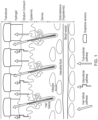
- Each pixel ( Figure 3 ) performs the critical functions of glucose extraction and detection, and comprises (a) an individual enzyme-bearing gel reservoir, into which glucose is extracted transdermally, (b) an extraction circuit that allows the glucose to be extracted into the gel reservoir, and (c) an electrochemical, enzyme-based glucose detector based on a platinum nanoparticle (NP)-decorated graphene material.
- the array is integrated into a flexible patch with, potentially, a disposable element (see below), and, ultimately, has a wireless readout.
- the number of its pixels and their geometrical dimensions need to be carefully selected, according to the following criteria.
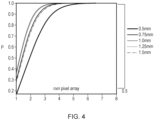
- Criterion 1 The number of pixels in the array and their number per unit area is dictated by the probability P of at least one hair follicle "hit” using the chosen geometry, and that no more than one hair follicle is probed by an individual pixel.
- the overall area of the device patch was set to 2 ⁇ 2 cm 2 (for practical reasons), and a human hair distribution centred about a peak value of 24 follicles per cm 2 (which is encompassed by the average hair distribution of 18 to 32 follicles per cm 2 on the human forearm).
- Figure 4(a) shows that a 4 ⁇ 4 pixel array of 2 to 3 mm diameter cylinders of enzyme-containing gels guarantees at least one follicular "hit”.
- an 8x8 array provides useful redundancy for probing the privileged glucose pathways.
- the multiplexed iontophoretic sampling devices of the present invention preferably comprise an array spanning about 2x2 cm 2 , and comprising between 4 and 100 sensor pixels, and more preferably between 10 and 80 sensor pixels.
- the array of sensor pixels comprises 4, 9, 16, 25, 36, 49 or 64 sensor pixels, for example in arrays 2 ⁇ 2, 3 ⁇ 3, 4 ⁇ 4, 5 ⁇ 5, 6 ⁇ 6, 7 ⁇ 7 or 8 ⁇ 8 sensor pixels. While in some embodiments, the sensor pixels are disposed in a square array, other arrangements of sensor pixels may be used.
- Criterion 2 If the diameter/area of the enzyme-encasing gel within a pixel is as estimated above, its volume is determined by the requirement that the glucose concentration range achieved in the pixel reservoir falls well within the full available range of the sensor. Taking the hypoglycaemic and hyperglycaemic blood concentrations to be 3.5 and 12 mM, respectively, 11 ⁇ M and 36 ⁇ M are obtained after their dilution in 24 ⁇ l of gel.
- the volume of the gel reservoir and the extraction conditions set the value of the fixed conversion factor between the interstitial fluid glucose concentration and the one that is achieved in the pixels of the array.
- a reduction in the volume of the gel reservoir by a factor of ⁇ 60 allows the hypo- to hyper-glycaemic blood glucose concentration range to be mapped onto the 10 to 40 ⁇ M glucose concentration within the gel reservoir, to be achieved using extraction current and period of 0.02 mA and 10 minutes, respectively (a follicular glucose flux value of 3.5 nmol.mA -1 .hr -1 at 10 mM subdermal glucose concentration was used for this estimation).
- Criterion 3 The gel dimensions also have an impact on the overall duration of the glucose extraction/read-out cycle.
- the thickness of the gel has to be minimised to decrease the time needed for the extracted glucose to diffuse across the gel, from the side facing the skin to the side facing the graphene sensor.
- Targeted thickness range is on the order of 0.1 mm ( Tierney, et al., Electroanalysis of Glucose in Transcutaneously Extracted Samples. Electroanalysis, 12(9): 666-671, 2000 ), which is thereby the most preferred thickness value of the gel reservoir.
- a volume of gel reservoir of 2 mm diameter and 0.1 mm thickness would allow the extraction current and period to be decreased, for example, to 0.02 mA and 10 minutes, respectively, while achieving the same glucose concentration range, of 10 to 40 ⁇ M, in the gel reservoir, as mapped in red on Figure 6 .
- the active areas of extraction and detection electrodes fit within the pixel area.
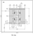
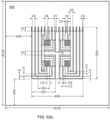
- An example of typical dimensions within a pixel area is given in Figure 5 : the unit cell of the array (pixel area) was chosen to be 5x5 mm 2 , with the active regions of the electrodes occupying a 4x4 mm 2 area, and the footprint area of the gel reservoir within which glucose is extracted (delineated with a dotted line in Figure 5 ) occupying a disk region of 3 mm in diameter.
- the main materials used to construct the glucose monitor in this embodiment are: (i) a graphene film decorated with platinum nanoparticles, together forming the sensing material, (ii) an enzyme, glucose oxidase, which in an electrochemical reaction with glucose produces hydrogen peroxide, the reaction product detected by the electrochemical graphene sensor, (iii) a hydrogel (based on a polymers such as agarose, chitosan, ethyl cellulose, or methyl cellulose) used to encase the enzyme, and (iv) a bio-compatible elastomer (e.g.
- silicone rubbers such as polydimethylsiloxane (PDMS) or PlatSil 7315, yielding thicknesses in the hundred micron range; or parylene, for designs where thicknesses below 100 ⁇ m are sought) for creating a perforated membrane, used to provide mechanical support and definition for the gel reservoirs of each pixel.
- Graphene is the material of choice for flexible electronics. Here it was chosen due to its mechanical resilience to bending and flexing, its ease towards patterning and device integration through standard microfabrication techniques (characteristics that are necessary to create the pixelized array), its compatibility with green electronics, and not least of all its potential to reduce the cost in a commercial product compared with noble metal electrochemical electrodes.
- graphene can be used not only to provide the active area of the electrochemical pixel sensors, but also the electrical interconnects that link these sensing regions to the outside world ( Figure 5 ).
- the types of graphene to be used can be either atomically thin layers produced by CVD, or a graphene nano-flake ink used to create the printed regions.
- the realization of the pixel array is not restricted to the sensing materials mentioned above.
- Other sensing materials could be used, such graphene/Pt NPs (or other catalytic particles) further functionalized with Prussian Blue (or an equivalent, with the role to further decrease the working potential), carbon-based electrodes (including carbon nanotubes), Prussian Blue (or an equivalent) alone, metal electrodes traditionally used in electrochemistry, or a combination of them.
- the hydrogel is allowed to become semi-solid, at which point its volume is about 2/3 of the initial value; the semi-solid nature of the hydrogel facilitates both glucose diffusion through the gel and effective electron transfer during electrochemical sensing.
- the elastomer unit with the encased gel may represent the replaceable part of the device.
- the electrical interconnects and the working electrode are defined by printing a graphene flake-based ink (early designs of the array have interconnects based on a Ag-based ink);
- the Ag/AgCl electrodes, to be used as the pseudo-reference electrodes for glucose detection, and for reverse iontophoresis during glucose extraction are defined by subsequent stages of printing of Ag- and AgCl-based inks, respectively.
- Figure 5 illustrates the relative positioning of the various components of a 2x2 array: the spacing between the electrodes in a pixel is chosen so that the working and counter electrodes are close enough to the reference and iontophoresis electrodes in order to minimise the ohmic potential drop in solution, as well as to allow the extracted glucose to reach rapidly and efficiently the detection electrodes. Because the use of a single Ag/AgCl electrode for both extraction and detection (as proposed in the layout in Figure 5(b) ) may, in time, affect its performance, a second layout was designed in which the sensing and the reverse-iontophoresis circuits are entirely decoupled (i.e., they do not share any of the electrodes) ( Figure 5(a) ).
- an insulating layer can be printed using an appropriate ink.
- inks include bio-compatible variants.
- the printed array is then coupled to the elastomer-hydrogel membrane, created using the same steps 6 to 8, as described above (Strategy no.1).
- glucose is extracted in the next cycle of operation by the other half of the pixels of the array; the sequential recycling of Ag and AgCl between the respective pairs of pixel devices is thereby ensured.
- This sequential change in the polarity of the electrodes may also limit any polarization of the skin that has been suggested to be associated with stinging and erythema.
- miniaturised pixel devices of the present invention for non-invasive monitoring of transdermal glucose were tested to determine their detection range, limit of detection, specificity of response for glucose, and their ability to perform dual glucose extraction/detection through single follicular pathways. Additionally, the cross-talk between two adjacent pixel devices was also evaluated.
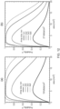
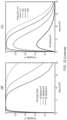
- Figure 6(a) displays a typical electrochemical current versus glucose concentration calibration curve of a pixel device in an embodiment as realized via strategy no.1.
- the pixel device was about 3 mm in diameter, and comprised an enzyme-encased gel reservoir of 24 ⁇ l containing 8 mg/ml glucose oxidase, and external (wire) Ag/AgCl and Pt electrodes in contact with the gel reservoir.
- the resulting calibration curve shows a single-law dependence over a concentration range from micromolar to more than millimolar, and displays a low limit of detection (LoD) of 4micromolar.
- LiD low limit of detection
- hypo- to hyper-glycaemic range in diabetics i.e., 3.5 to 12 mM in the blood, and of a quite similar range in the interstitial fluid
- the measured glucose concentration range is 10-40 micromolar, already well above the LoD of the sensor.
- Figure 6(b) displays the electrochemical current versus glucose concentration calibration curve of a full on-chip pixel device, where all the electrodes are planar and integrated with a PET substrate.
- the pixel device had a 4x2 mm 2 area, a gel reservoir of 10 ⁇ l containing 16 mg/ml glucose, and two planar electrochemistry electrodes made of platinum nanoparticle-decorated graphene and an Ag/AgCl film (see section 4, "Supporting Methods”), respectively.
- the volume of the gel reservoir was decreased to about 1 ⁇ l, resulting in a thickness of about 0.1 mm, a most preferred value which greatly reduces the glucose diffusion time across the gel. This improvement allows one to decrease both the extraction time and extraction current, bringing these operation parameters of the device into the most preferred range.
- the glucose extraction function of the platform was shown by performing reverse iontophoresis (RI) ex vivo in simple diffusion cells using porcine skin (see section 4, "Supporting Methods"), which is an excellent model for the human counterpart ( Schmook, F.P., J.G. Meingassner, and A. Billich, Comparison of human skin or epidermis models with human and animal skin in in-vitro percutaneous absorption. International Journal of Pharmaceutics, 2001. 215(1-2): p. 51-56 ).
- RI reverse iontophoresis
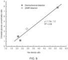
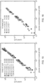
- Figure 9 collects the results of several experiments in which the efficiency of glucose extraction was correlated with hair density, and the analyte was detected either electrochemically or by 1 H-qNMR.
- the data which show a clear correlation between the ratios of concentrations extracted through follicle-rich and follicle-poor skin samples and the respective hair density ratios thereof, demonstrate that glucose extraction by RI into the miniature "pixels" indeed occurs primarily through preferred follicular pathways.
- the excellent agreement between the amperometric and NMR analytical techniques provides further confidence in the dual extraction-detection functions of the pixel device.
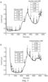
- Figure 8(b) demonstrates reverse iontophoresis employing planar electrodes fully integrated with a PET substrate. The trend of the data is very similar to the ones described in Figure 8(a) .
- the chronoamperometric current in the second "pixel” was then measured before and after additions of glucose at various concentrations from 10 micromolar to 1 millimolar. Lastly, the chronoamperometric response in the first "pixel” was re-determined to assess any cross-talk between the two devices. It was found that the baseline response in the first "pixel” increased by no more than 3% of that corresponding to the total amount of glucose added to the second "pixel". In other words, even with a graphene electrode common to both devices, the use of individual hydrogel reservoirs effectively decouples the response of the individual "pixels". Achieving complete decoupling is anticipated in a practical embodiment of the array for which individual graphene detectors are envisaged.
- Graphene-based sensor fabrication Chemical vapour deposition (CVD) graphene squares, of 3 ⁇ 3 or 2 ⁇ 2 mm 2 , originally synthesized on Cu foils, were transferred onto SiO 2 /Si (in early experiments) or flexible PET substrates by standard procedures ( Bae, S., et al., Roll-to-roll production of 30-inch graphene films for transparent electrodes. Nat Nano, 2010. 5(8): p. 574-578 ).
- CVD chemical vapour deposition
- the graphene area used in electrochemistry was then insulated from the rest of the electrical circuit with a polydimethylsiloxane (PDMS) or silicone rubber frame with a central cylindrical hole, into which the hydrogel reservoir was cast on top of the graphene.
- the electrochemistry circuit was completed (i) with external Ag, Ag/AgCl and Pt wires in the early experiments, and (ii) with chip-integrated Ag/AgCl (and Pt, in some variants) electrodes in later embodiments.
- An Ag/AgCl micro-electrode was fabricated by coating a 99.95% pure, silver wire with AgCl by chronoamperometry in a 3.5 M KCl solution, with Pt as reference and counter electrodes, for 1 hour at 1 V.
- the wire was then encased in a 1% w/v agarose gel containing 0.1 M KCl.
- the electrode held only a low (0.1 M) KCl concentration to limit the amount of glucose oxidase inhibitor present.
- the electrode was stored in 0.1 M KCl at 4 °C when not in use, and its performance and stability over time were confirmed periodically using cyclic voltammetry.
- Chip-integrated electrochemistry electrodes To fabricate a fully integrated sensor, all electrodes involved in electrochemistry were defined directly on the substrate. As indicated in Figure 5 , this necessitated creation of Ag/AgCl electrodes.
- Thermal/e-beam evaporation Firstly, Ag patterned regions of 850 nm thickness were deposited directly on PET using stencil masks. Note that on other substrates, such as SiO 2 , which were used for proof-of-principle studies, a layer of 5-10 nm of Ti was first deposited in order to ensure adhesion of the Ag layer. Then, an additional AgCl layer of about 300 nm in thickness was deposited on top of the Ag regions to create a stable AgCl/Ag reference electrode. Such thick layers of Ag and AgCl are needed to ensure a long lifetime of the reference electrode ( B.J. Polk et al., Sensors and Actuators B 114 (2006) 239-247 ).
- Chemical and electrochemical methods (i) chemically, a 50 mM FeCl 3 solution is applied to the Ag surface for 20 seconds at room temperature, followed by rinsing with de-ionized water; (ii) electrochemically, AgCl was produced by chrono-amperometry in a 1M KCl solution with an on-chip Ag electrode as the working electrode, and Pt wires as reference and counter electrodes, followed by rinsing with de-ionized water.
- Ag/AgCl electrodes can also be created using direct printing of stacked layers of Ag- and AgCl-based inks.
- the volume of gel In order to reduce the extraction current and the time period, the volume of gel needs to be decreased (see section entitled “Geometry considerations”).
- 2 ⁇ l enzyme-containing gel was cast into the holes (1.5-2 mm diameter) of a 0.1 mm thick PDMS membrane.
- the volume of enzyme-containing gel scales down with decreasing volume defined by the thickness of the supporting elastomer membrane and the dimensions of the reservoir holes within.
- hydrogel with a gelling temperature below the denaturation point of the enzyme, may allow direct mixing of the enzyme with the hydrogel, and then direct injection of the mixture into the holes of the elastomer membrane.
- Electrochemical method A cyclic voltammogram acquired in 10 ⁇ L of 0.1M H 2 SO 4 , 1.7 mM hydrogen hexachloroplatinate, at 20 mV/sec scan rate, shows a typical chloride reduction peak at about -0.35V against a micro Ag/AgCl reference electrode.
- Transdermal RI glucose extraction A piece of skin separated the two halves of a vertical Franz diffusion cell, with the epidermal side facing the upper compartment. The lower, sub-dermal chamber of the cell was filled with 7.5 mL of either 10 or 100 mM glucose solution, and magnetically stirred for 1 hour. RI extraction was performed in two experimental configurations: (i) first, with external wire extraction electrodes, and then (ii) with chip-integrated extraction electrodes.
- the enzyme-containing gel reservoir was positioned on the skin surface with the Ag/AgCl porous cathode contacting the "pixel". A silver anode was inserted into the sub-dermal compartment.
- the electrical resistance of the iontophoresis circuit was about one-half of that expected in vivo, where both electrodes would be located on the skin surface and the iontophoretic current must, as a consequence, cross the skin twice.
- RI extraction is undertaken at constant current, the only difference between the in vitro and in vivo situations is the approximately two-fold higher voltage required to drive the current used in the latter case ( Potts, R.O., Mechanisms of Transdermal Drug Delivery.
- RI was performed by passing a constant current of 0.2 mA for 1 hour between the anode and cathode from a power supply; the potential across the skin was monitored regularly during current passage.
- the RI current application time employed permitted the extracted glucose to distribute essentially homogenously across the entire thickness of the gel reservoir.
- Chip-integrated RI electrodes An on-chip Ag and Ag/AgCl pair of electrodes was created via identical methods to those described above for the fabrication of on-chip electrochemistry electrodes.
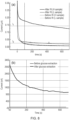
- Output data of the device The chronoamperometric current ( Figure 9 ) was recorded, typically, for 700 seconds in each measurement, then averaged over the last 600 seconds of the total measurement period (i.e., corresponding to the plateau region), and the corresponding background value (i.e., before RI) subtracted.
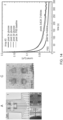
- Figures 14 and 15 contain a compilation of representative ex-vivo (porcine skin) extraction-detection experiments involving four different 2x2 graphene-based arrays realized by the strategy no.1 described in section "Detailed discussion/Choice of materials and device realization strategies”.
- the data demonstrate all the expected functional aspects: (i) targeted extraction ( Figure 14 B and C ), (ii) correlation and proportionality with the number of hair follicles probed by the respective pixels ( Figures 14 B and C ), (iii) capability to detect glucose extracted through a single hair follicle ( Figure 14 C) , (iv) proportionality with the concentration of subdermal glucose ( Figure 14 B , panel 3), and (v) close operational characteristics of pixels within an array ( Figure 14 B , panel 1), as well as between different arrays ( Figure 15 ).
- Electrodes labelled with 4 in Figure 14 A play no role in this configuration.
- Ag/AgCl has been chosen as the material for the extraction electrode couple due to the ability of AgCl (an ionic solid) to recover its surface chemical composition after electrochemical stress, hence ensuring its stability after repeated cycles of extraction/recovery that require the electrode polarity to alternate.
- the surface of pure Ag electrodes is subjected to reactions (e.g. oxidation) that can change its chemistry. It was found that after four cycles of extraction/detection performed with the array, the potential across the Ag/AgCl electrodes changed negligibly (by only ⁇ 30 mV), establishing their recovery.
- Figure 14 B panel 2 shows the set of current-time detection curves obtained after extraction via each of the pixels of an array (for which the sensitivity calibration curves are shown in Figure 14 B , panel 1); extraction has occurred through various number of hair follicles, as probed by the respective pixel devices.
- the inset of Figure 14 B , panel 2 shows, for each of current-time detection curves, the detected current averaged along the plateau of the curve and then plotted on the sensitivity calibration curve: this allows one to determine, by interpolation, the concentration of the extracted glucose within the gel of each of the pixels.
- the graph in the inset is the arithmetic average of the four current-concentration calibration curves shown in Figure 14B , panel 1.
- the concentration of glucose thus determined is proportional to the number of follicles targeted by the respective pixel, and consistent with estimations based on the glucose follicular extraction flux determined previously. Additionally, extraction via non-follicular skin in similar conditions leads to a detected current that decays much faster than in the case of follicular extraction ( Figure 14 B , panel 2), due to the very low glucose content within the pixel gel. Altogether, these experiments unequivocally demonstrate that the array operates as designed, by exploiting the hair follicles as the preferential transdermal extraction paths for glucose. Further, Figure 14 B , panel 3 shows the proportionality, after extraction through the same pixel device, of the detected current with the concentration of subdermal glucose.
- Figure 14 C shows detection current vs time curves correlated with images of the hair follicles targeted by the respective pixels of an array, as an example of the way the extraction-detection is practically performed with the array. In this case, detection after extraction through a single follicle could also be probed.
- graphene patches For a 2x2 array, four such graphene patches (larger than the final, desired size) were placed on the PET sheet roughly in the desired locations using a stencil mask (designed for subsequent electrode and track definition) to guide alignment.
- the graphene patches provide the working electrodes for each of the pixels of the array in the electrochemical detection of glucose.
- a second graphene layer is subsequently transferred on top of each of the previously transferred patches.
- Electrode and track deposition through physical vapour deposition thermal evaporation
- sets of custom-made or polyimide industrialtape (Kapton ® ) laser-machined stencil masks were placed successively, and aligned on top of, the PET-supported graphene patches.
- the stencil mask sets are tailored to the array layout, examples of such layouts being given in Figure 4 ; in this specific case, the design from Figure 4(a) has been used.
- a 500 nm silver film was deposited on top of a 30 nm palladium layer previously deposited to promote adhesion of the silver layer.
- a 500 nm thick AgCl layer was subsequently deposited on top of the silver films, to complete the reference/counter electrodes.
- the graphene patches were then patterned in the pre-defined geometry (e.g., according to the layouts from Figure 5 ). Though low energy oxygen plasma can be used to etch graphene supported by plastic substrates, in the current realization mechanical cutting (using a scalpel) was successfully employed to remove the excess graphene from the pixel patches.
- PDMS elastomer membrane designed to support the enzyme-encasing gel
- PDMS mixed with a curing agent was spin-coated on a PET support sheet and cured, leading to a 100 um thick membrane. Circular holes (1.5-3 mm diameter) were then drilled to create sockets for the reservoir gel. After careful underwater peeling in a de-ionized water bath, the PDMS membranes were transferred onto the array with defined electrodes and tracks, ensuring alignment of the sockets to the electrochemical cell region of each pixel. The assembly was then left to dry in air.
- Platinum nanoparticle deposition onto the graphene pixel electrodes Platinum nanoparticles were formed and deposited on the graphene regions of the pixels through appropriate stencil masks by DC sputtering under argon. By tuning the argon gas pressure and sputtering time (of, typically, 20 s), particles of 3 to 5 nm in diameter were achieved.
Landscapes
- Health & Medical Sciences (AREA)
- Life Sciences & Earth Sciences (AREA)
- Physics & Mathematics (AREA)
- Medical Informatics (AREA)
- Surgery (AREA)
- Biophysics (AREA)
- Pathology (AREA)
- Engineering & Computer Science (AREA)
- Biomedical Technology (AREA)
- Heart & Thoracic Surgery (AREA)
- Veterinary Medicine (AREA)
- Molecular Biology (AREA)
- Optics & Photonics (AREA)
- Animal Behavior & Ethology (AREA)
- General Health & Medical Sciences (AREA)
- Public Health (AREA)
- Chemical & Material Sciences (AREA)
- Chemical Kinetics & Catalysis (AREA)
- General Chemical & Material Sciences (AREA)
- Emergency Medicine (AREA)
- Measurement Of The Respiration, Hearing Ability, Form, And Blood Characteristics Of Living Organisms (AREA)
- Investigating Or Analysing Biological Materials (AREA)
Applications Claiming Priority (3)
| Application Number | Priority Date | Filing Date | Title |
|---|---|---|---|
| GB201607265 | 2016-04-26 | ||
| GBGB1703300.2A GB201703300D0 (en) | 2017-03-01 | 2017-03-01 | Multiplexed transdermal extraction and detection devices for non-invasive monitoring of substances and methods of use |
| PCT/EP2017/059909 WO2017186783A1 (en) | 2016-04-26 | 2017-04-26 | Multiplexed transdermal extraction and detection devices for non-invasive monitoring of substances and methods of use |
Publications (3)
| Publication Number | Publication Date |
|---|---|
| EP3448258A1 EP3448258A1 (en) | 2019-03-06 |
| EP3448258C0 EP3448258C0 (en) | 2024-06-26 |
| EP3448258B1 true EP3448258B1 (en) | 2024-06-26 |
Family
ID=58632416
Family Applications (1)
| Application Number | Title | Priority Date | Filing Date |
|---|---|---|---|
| EP17719274.7A Active EP3448258B1 (en) | 2016-04-26 | 2017-04-26 | Multiplexed transdermal extraction and detection system for non-invasive monitoring of substances and method of use |
Country Status (5)
| Country | Link |
|---|---|
| US (1) | US11278218B2 (enExample) |
| EP (1) | EP3448258B1 (enExample) |
| JP (1) | JP6953431B2 (enExample) |
| CN (1) | CN109414227A (enExample) |
| WO (1) | WO2017186783A1 (enExample) |
Families Citing this family (13)
| Publication number | Priority date | Publication date | Assignee | Title |
|---|---|---|---|---|
| CN110389163A (zh) * | 2018-04-16 | 2019-10-29 | 潘新宇 | 基于多传感器像素阵列的无创血糖检测装置 |
| CN110501403B (zh) * | 2018-05-18 | 2025-01-03 | 潘新宇 | 单像素无创血糖检测装置 |
| JP7220598B2 (ja) * | 2019-03-15 | 2023-02-10 | 本田技研工業株式会社 | 生体情報測定センサ、生体情報測定装置及び生体情報測定方法 |
| WO2020232405A1 (en) * | 2019-05-16 | 2020-11-19 | California Institute Of Technology | Laser-enabled lab on skin |
| WO2021231948A1 (en) * | 2020-05-15 | 2021-11-18 | Hememics Biotechnologies, Inc. | Multiplex biosensor for rapid point-of-care diagnostics |
| CN111803087B (zh) * | 2020-06-12 | 2021-11-09 | 同济大学 | 一种生物体无损血糖检测器件及其制备方法 |
| WO2022109008A1 (en) * | 2020-11-18 | 2022-05-27 | Cercacor Laboratories, Inc. | Glucose sensors and methods of manufacturing |
| CN113017995B (zh) * | 2021-03-03 | 2022-01-04 | 杭州可靠护理用品股份有限公司 | 具有尿酸提示功能的成人纸尿裤 |
| CN113647952A (zh) * | 2021-08-18 | 2021-11-16 | 北京航空航天大学 | 基于银/氯化银纳米线制成的柔性干电极及其制备方法 |
| CN114557694B (zh) * | 2022-04-28 | 2022-09-23 | 中国科学院大学 | 一种无创皮下组织液提取-检测装置和提取-检测方法 |
| CN116858914A (zh) * | 2023-07-14 | 2023-10-10 | 苏州大学 | 一种基于疏水基底的无酶葡萄糖传感器及其制备方法和应用 |
| GB2630155B (en) * | 2023-10-12 | 2025-06-04 | Transdermal Diagnostics Ltd | Method and sensor |
| CN118058741B (zh) * | 2024-02-01 | 2024-11-26 | 丽新(浙江)科技有限公司 | 一种用于汗液维生素b监测的柔性贴片装置 |
Family Cites Families (27)
| Publication number | Priority date | Publication date | Assignee | Title |
|---|---|---|---|---|
| US5362307A (en) | 1989-01-24 | 1994-11-08 | The Regents Of The University Of California | Method for the iontophoretic non-invasive-determination of the in vivo concentration level of an inorganic or organic substance |
| KR970011449B1 (ko) | 1988-01-29 | 1997-07-11 | 더 리전트 오브 디 유니버시티 오브 캘리포니아 | 이온전기 영동형 비침입 검체 채취 또는 이송 장치 및 방법 |
| DE69519023T2 (de) | 1994-06-24 | 2001-06-13 | Cygnus, Inc. | Einrichtung zur iontophoretischen probennahme |
| US5954685A (en) | 1996-05-24 | 1999-09-21 | Cygnus, Inc. | Electrochemical sensor with dual purpose electrode |
| US5911223A (en) | 1996-08-09 | 1999-06-15 | Massachusetts Institute Of Technology | Introduction of modifying agents into skin by electroporation |
| ES2213369T3 (es) | 1998-05-13 | 2004-08-16 | Cygnus, Inc. | Procesamiento de señal para medicion de analitos fisiologicos. |
| EP1270041A1 (en) | 2001-06-22 | 2003-01-02 | Universite De Geneve | Device for non-invasively determining the relative levels of two substances present in a biological system |
| JP2004016489A (ja) * | 2002-06-17 | 2004-01-22 | Polytronics Ltd | 皮下含有物質の検査装置 |
| JP2005137416A (ja) * | 2003-11-04 | 2005-06-02 | Sysmex Corp | 経皮的分析物抽出システム及び経皮的分析物分析システム |
| WO2007070093A2 (en) * | 2005-12-09 | 2007-06-21 | Flexible Medical Systems, Llc. | Flexible apparatus and method for monitoring and delivery |
| US8333874B2 (en) * | 2005-12-09 | 2012-12-18 | Flexible Medical Systems, Llc | Flexible apparatus and method for monitoring and delivery |
| CN101365381A (zh) * | 2005-12-09 | 2009-02-11 | 弹性医疗系统有限责任公司 | 用于监测和递送的柔性设备和方法 |
| JP5470509B2 (ja) | 2008-11-27 | 2014-04-16 | 独立行政法人産業技術総合研究所 | 電極用白金クラスター及びその製造方法 |
| CN102119860B (zh) * | 2010-01-12 | 2013-07-17 | 长庚医学科技股份有限公司 | 非侵入式血糖量测系统和测量方法 |
| US9451913B2 (en) * | 2010-12-10 | 2016-09-27 | Touchtek Labs, Llc | Transdermal sampling and analysis device |
| US9380965B2 (en) * | 2011-05-20 | 2016-07-05 | Abbott Diabetes Care Inc. | Analyte sensors having a membrane with low temperature sensitivity |
| JP5903806B2 (ja) | 2011-09-05 | 2016-04-13 | 船井電機株式会社 | 検出装置 |
| US9700245B2 (en) * | 2011-09-23 | 2017-07-11 | Itrace Biomedical Inc. | Transdermal analyte extraction and detection system and the method thereof |
| JP6219212B2 (ja) | 2014-03-27 | 2017-10-25 | 浜松ホトニクス株式会社 | 生体計測用プローブ及び生体計測装置 |
| WO2016025468A2 (en) * | 2014-08-11 | 2016-02-18 | The Board Of Trustees Of The University Of Illinois | Devices and related methods for epidermal characterization of biofluids |
| US10736551B2 (en) * | 2014-08-11 | 2020-08-11 | The Board Of Trustees Of The University Of Illinois | Epidermal photonic systems and methods |
| WO2016033204A2 (en) * | 2014-08-26 | 2016-03-03 | Echo Therapeutics, Inc. | Differential biosensor system |
| US10722160B2 (en) * | 2014-12-03 | 2020-07-28 | The Regents Of The University Of California | Non-invasive and wearable chemical sensors and biosensors |
| CA2973261A1 (en) * | 2015-01-09 | 2016-07-14 | Exhalix Llc | Transdermal sampling strip and method for analyzing transdermally emitted gases |
| US10105100B2 (en) * | 2015-07-28 | 2018-10-23 | Verily Life Sciences Llc | Display on a bandage-type monitoring device |
| US10925543B2 (en) * | 2015-11-11 | 2021-02-23 | The Board Of Trustees Of The University Of Illinois | Bioresorbable silicon electronics for transient implants |
| US10653342B2 (en) * | 2016-06-17 | 2020-05-19 | The Board Of Trustees Of The University Of Illinois | Soft, wearable microfluidic systems capable of capture, storage, and sensing of biofluids |
-
2017
- 2017-04-26 EP EP17719274.7A patent/EP3448258B1/en active Active
- 2017-04-26 CN CN201780039263.5A patent/CN109414227A/zh active Pending
- 2017-04-26 WO PCT/EP2017/059909 patent/WO2017186783A1/en not_active Ceased
- 2017-04-26 JP JP2018555939A patent/JP6953431B2/ja active Active
- 2017-04-26 US US16/096,668 patent/US11278218B2/en active Active
Also Published As
| Publication number | Publication date |
|---|---|
| JP2019516452A (ja) | 2019-06-20 |
| EP3448258C0 (en) | 2024-06-26 |
| EP3448258A1 (en) | 2019-03-06 |
| CN109414227A (zh) | 2019-03-01 |
| WO2017186783A1 (en) | 2017-11-02 |
| JP6953431B2 (ja) | 2021-10-27 |
| CA3021886A1 (en) | 2017-11-02 |
| US11278218B2 (en) | 2022-03-22 |
| US20200008717A1 (en) | 2020-01-09 |
Similar Documents
| Publication | Publication Date | Title |
|---|---|---|
| EP3448258B1 (en) | Multiplexed transdermal extraction and detection system for non-invasive monitoring of substances and method of use | |
| Lipani et al. | Non-invasive, transdermal, path-selective and specific glucose monitoring via a graphene-based platform | |
| US12023154B2 (en) | Non-invasive and wearable chemical sensors and biosensors | |
| Yu et al. | Flexible electrochemical bioelectronics: the rise of in situ bioanalysis | |
| Tasca et al. | Microneedle-based electrochemical devices for transdermal biosensing: a review | |
| Chinnadayyala et al. | Nonenzymatic determination of glucose at near neutral pH values based on the use of nafion and platinum black coated microneedle electrode array | |
| US20240049994A1 (en) | One-touch fingertip sweat sensor and personalized data processing for reliable prediction of blood biomarker concentrations | |
| US5954685A (en) | Electrochemical sensor with dual purpose electrode | |
| JP4108610B2 (ja) | 高触媒作用のスクリーン印刷インク | |
| JP3155523B2 (ja) | バイオセンサー、イオン浸透サンプリングシステムおよびその使用方法 | |
| Zhu et al. | Effect of interstitial fluid pH on transdermal glucose extraction by reverse iontophoresis | |
| JP2006167428A (ja) | 分析物抽出装置、分析装置、分析物抽出方法および分析方法 | |
| Du Toit et al. | Generating power from transdermal extracts using a multi-electrode miniature enzymatic fuel cell | |
| Zhan et al. | A 3D-printed microneedle extraction system integrated with patterned electrodes for minimally invasive transdermal detection | |
| Lyu et al. | Soft, disruptive and wearable electrochemical biosensors | |
| Li et al. | Boosting the performance of an iontophoretic biosensing system with a graphene aerogel and Prussian blue for highly sensitive and noninvasive glucose monitoring | |
| US12004876B2 (en) | Auto-powered synthetic skin | |
| CA3021886C (en) | Multiplexed transdermal extraction and detection devices for non-invasive monitoring of substances and methods of use | |
| WO2024010976A1 (en) | Flexible biosensor for the detection and/or 2d/3d mapping of biomarker concentration | |
| El-Desoky | Voltammetric sensors for biological sample analysis | |
| Mohan et al. | Wearable biosensor platform: design and healthcare commercial values | |
| US12507954B2 (en) | Auto-powered synthetic skin | |
| Ng et al. | Skin biosensing and bioanalysis: what the future holds | |
| Mohan et al. | Nanomaterials-based flexible electrochemical sensors for health care monitoring | |
| Liu et al. | 26‐1: A Wearable Glucose Sensor Integrated with Hollow Microneedles and Reverse Iontophoresis Extraction |
Legal Events
| Date | Code | Title | Description |
|---|---|---|---|
| STAA | Information on the status of an ep patent application or granted ep patent |
Free format text: STATUS: UNKNOWN |
|
| STAA | Information on the status of an ep patent application or granted ep patent |
Free format text: STATUS: THE INTERNATIONAL PUBLICATION HAS BEEN MADE |
|
| PUAI | Public reference made under article 153(3) epc to a published international application that has entered the european phase |
Free format text: ORIGINAL CODE: 0009012 |
|
| STAA | Information on the status of an ep patent application or granted ep patent |
Free format text: STATUS: REQUEST FOR EXAMINATION WAS MADE |
|
| 17P | Request for examination filed |
Effective date: 20181112 |
|
| AK | Designated contracting states |
Kind code of ref document: A1 Designated state(s): AL AT BE BG CH CY CZ DE DK EE ES FI FR GB GR HR HU IE IS IT LI LT LU LV MC MK MT NL NO PL PT RO RS SE SI SK SM TR |
|
| AX | Request for extension of the european patent |
Extension state: BA ME |
|
| DAV | Request for validation of the european patent (deleted) | ||
| DAX | Request for extension of the european patent (deleted) | ||
| STAA | Information on the status of an ep patent application or granted ep patent |
Free format text: STATUS: EXAMINATION IS IN PROGRESS |
|
| 17Q | First examination report despatched |
Effective date: 20220819 |
|
| GRAP | Despatch of communication of intention to grant a patent |
Free format text: ORIGINAL CODE: EPIDOSNIGR1 |
|
| STAA | Information on the status of an ep patent application or granted ep patent |
Free format text: STATUS: GRANT OF PATENT IS INTENDED |
|
| INTG | Intention to grant announced |
Effective date: 20240105 |
|
| GRAS | Grant fee paid |
Free format text: ORIGINAL CODE: EPIDOSNIGR3 |
|
| GRAA | (expected) grant |
Free format text: ORIGINAL CODE: 0009210 |
|
| STAA | Information on the status of an ep patent application or granted ep patent |
Free format text: STATUS: THE PATENT HAS BEEN GRANTED |
|
| AK | Designated contracting states |
Kind code of ref document: B1 Designated state(s): AL AT BE BG CH CY CZ DE DK EE ES FI FR GB GR HR HU IE IS IT LI LT LU LV MC MK MT NL NO PL PT RO RS SE SI SK SM TR |
|
| REG | Reference to a national code |
Ref country code: GB Ref legal event code: FG4D |
|
| REG | Reference to a national code |
Ref country code: CH Ref legal event code: EP |
|
| REG | Reference to a national code |
Ref country code: DE Ref legal event code: R096 Ref document number: 602017082801 Country of ref document: DE |
|
| U01 | Request for unitary effect filed |
Effective date: 20240710 |
|
| U07 | Unitary effect registered |
Designated state(s): AT BE BG DE DK EE FI FR IT LT LU LV MT NL PT RO SE SI Effective date: 20240902 |
|
| PG25 | Lapsed in a contracting state [announced via postgrant information from national office to epo] |
Ref country code: HR Free format text: LAPSE BECAUSE OF FAILURE TO SUBMIT A TRANSLATION OF THE DESCRIPTION OR TO PAY THE FEE WITHIN THE PRESCRIBED TIME-LIMIT Effective date: 20240626 |
|
| PG25 | Lapsed in a contracting state [announced via postgrant information from national office to epo] |
Ref country code: GR Free format text: LAPSE BECAUSE OF FAILURE TO SUBMIT A TRANSLATION OF THE DESCRIPTION OR TO PAY THE FEE WITHIN THE PRESCRIBED TIME-LIMIT Effective date: 20240927 |
|
| PG25 | Lapsed in a contracting state [announced via postgrant information from national office to epo] |
Ref country code: NO Free format text: LAPSE BECAUSE OF FAILURE TO SUBMIT A TRANSLATION OF THE DESCRIPTION OR TO PAY THE FEE WITHIN THE PRESCRIBED TIME-LIMIT Effective date: 20240926 Ref country code: HR Free format text: LAPSE BECAUSE OF FAILURE TO SUBMIT A TRANSLATION OF THE DESCRIPTION OR TO PAY THE FEE WITHIN THE PRESCRIBED TIME-LIMIT Effective date: 20240626 Ref country code: GR Free format text: LAPSE BECAUSE OF FAILURE TO SUBMIT A TRANSLATION OF THE DESCRIPTION OR TO PAY THE FEE WITHIN THE PRESCRIBED TIME-LIMIT Effective date: 20240927 Ref country code: RS Free format text: LAPSE BECAUSE OF FAILURE TO SUBMIT A TRANSLATION OF THE DESCRIPTION OR TO PAY THE FEE WITHIN THE PRESCRIBED TIME-LIMIT Effective date: 20240926 |
|
| PG25 | Lapsed in a contracting state [announced via postgrant information from national office to epo] |
Ref country code: PL Free format text: LAPSE BECAUSE OF FAILURE TO SUBMIT A TRANSLATION OF THE DESCRIPTION OR TO PAY THE FEE WITHIN THE PRESCRIBED TIME-LIMIT Effective date: 20240626 |
|
| PG25 | Lapsed in a contracting state [announced via postgrant information from national office to epo] |
Ref country code: IS Free format text: LAPSE BECAUSE OF FAILURE TO SUBMIT A TRANSLATION OF THE DESCRIPTION OR TO PAY THE FEE WITHIN THE PRESCRIBED TIME-LIMIT Effective date: 20241026 |
|
| PG25 | Lapsed in a contracting state [announced via postgrant information from national office to epo] |
Ref country code: CZ Free format text: LAPSE BECAUSE OF FAILURE TO SUBMIT A TRANSLATION OF THE DESCRIPTION OR TO PAY THE FEE WITHIN THE PRESCRIBED TIME-LIMIT Effective date: 20240626 |
|
| PG25 | Lapsed in a contracting state [announced via postgrant information from national office to epo] |
Ref country code: SK Free format text: LAPSE BECAUSE OF FAILURE TO SUBMIT A TRANSLATION OF THE DESCRIPTION OR TO PAY THE FEE WITHIN THE PRESCRIBED TIME-LIMIT Effective date: 20240626 |
|
| PG25 | Lapsed in a contracting state [announced via postgrant information from national office to epo] |
Ref country code: ES Free format text: LAPSE BECAUSE OF FAILURE TO SUBMIT A TRANSLATION OF THE DESCRIPTION OR TO PAY THE FEE WITHIN THE PRESCRIBED TIME-LIMIT Effective date: 20240626 Ref country code: SM Free format text: LAPSE BECAUSE OF FAILURE TO SUBMIT A TRANSLATION OF THE DESCRIPTION OR TO PAY THE FEE WITHIN THE PRESCRIBED TIME-LIMIT Effective date: 20240626 |
|
| PG25 | Lapsed in a contracting state [announced via postgrant information from national office to epo] |
Ref country code: SM Free format text: LAPSE BECAUSE OF FAILURE TO SUBMIT A TRANSLATION OF THE DESCRIPTION OR TO PAY THE FEE WITHIN THE PRESCRIBED TIME-LIMIT Effective date: 20240626 Ref country code: SK Free format text: LAPSE BECAUSE OF FAILURE TO SUBMIT A TRANSLATION OF THE DESCRIPTION OR TO PAY THE FEE WITHIN THE PRESCRIBED TIME-LIMIT Effective date: 20240626 Ref country code: PL Free format text: LAPSE BECAUSE OF FAILURE TO SUBMIT A TRANSLATION OF THE DESCRIPTION OR TO PAY THE FEE WITHIN THE PRESCRIBED TIME-LIMIT Effective date: 20240626 Ref country code: IS Free format text: LAPSE BECAUSE OF FAILURE TO SUBMIT A TRANSLATION OF THE DESCRIPTION OR TO PAY THE FEE WITHIN THE PRESCRIBED TIME-LIMIT Effective date: 20241026 Ref country code: ES Free format text: LAPSE BECAUSE OF FAILURE TO SUBMIT A TRANSLATION OF THE DESCRIPTION OR TO PAY THE FEE WITHIN THE PRESCRIBED TIME-LIMIT Effective date: 20240626 Ref country code: CZ Free format text: LAPSE BECAUSE OF FAILURE TO SUBMIT A TRANSLATION OF THE DESCRIPTION OR TO PAY THE FEE WITHIN THE PRESCRIBED TIME-LIMIT Effective date: 20240626 |
|
| PLBE | No opposition filed within time limit |
Free format text: ORIGINAL CODE: 0009261 |
|
| STAA | Information on the status of an ep patent application or granted ep patent |
Free format text: STATUS: NO OPPOSITION FILED WITHIN TIME LIMIT |
|
| U20 | Renewal fee for the european patent with unitary effect paid |
Year of fee payment: 9 Effective date: 20250417 |
|
| 26N | No opposition filed |
Effective date: 20250327 |
|
| PGFP | Annual fee paid to national office [announced via postgrant information from national office to epo] |
Ref country code: GB Payment date: 20250423 Year of fee payment: 9 |
|
| REG | Reference to a national code |
Ref country code: CH Ref legal event code: H13 Free format text: ST27 STATUS EVENT CODE: U-0-0-H10-H13 (AS PROVIDED BY THE NATIONAL OFFICE) Effective date: 20251125 |
|
| PG25 | Lapsed in a contracting state [announced via postgrant information from national office to epo] |
Ref country code: MC Free format text: LAPSE BECAUSE OF FAILURE TO SUBMIT A TRANSLATION OF THE DESCRIPTION OR TO PAY THE FEE WITHIN THE PRESCRIBED TIME-LIMIT Effective date: 20240626 |