EP3445397B1 - Integral membrane protein display on poxvirus extracellular enveloped virions - Google Patents
Integral membrane protein display on poxvirus extracellular enveloped virions Download PDFInfo
- Publication number
- EP3445397B1 EP3445397B1 EP17786694.4A EP17786694A EP3445397B1 EP 3445397 B1 EP3445397 B1 EP 3445397B1 EP 17786694 A EP17786694 A EP 17786694A EP 3445397 B1 EP3445397 B1 EP 3445397B1
- Authority
- EP
- European Patent Office
- Prior art keywords
- eev
- imp
- protein
- fragment
- membrane
- Prior art date
- Legal status (The legal status is an assumption and is not a legal conclusion. Google has not performed a legal analysis and makes no representation as to the accuracy of the status listed.)
- Active
Links
Images


Classifications
-
- A—HUMAN NECESSITIES
- A61—MEDICAL OR VETERINARY SCIENCE; HYGIENE
- A61K—PREPARATIONS FOR MEDICAL, DENTAL OR TOILETRY PURPOSES
- A61K39/00—Medicinal preparations containing antigens or antibodies
- A61K39/12—Viral antigens
-
- C—CHEMISTRY; METALLURGY
- C12—BIOCHEMISTRY; BEER; SPIRITS; WINE; VINEGAR; MICROBIOLOGY; ENZYMOLOGY; MUTATION OR GENETIC ENGINEERING
- C12N—MICROORGANISMS OR ENZYMES; COMPOSITIONS THEREOF; PROPAGATING, PRESERVING, OR MAINTAINING MICROORGANISMS; MUTATION OR GENETIC ENGINEERING; CULTURE MEDIA
- C12N15/00—Mutation or genetic engineering; DNA or RNA concerning genetic engineering, vectors, e.g. plasmids, or their isolation, preparation or purification; Use of hosts therefor
- C12N15/09—Recombinant DNA-technology
- C12N15/63—Introduction of foreign genetic material using vectors; Vectors; Use of hosts therefor; Regulation of expression
- C12N15/79—Vectors or expression systems specially adapted for eukaryotic hosts
- C12N15/85—Vectors or expression systems specially adapted for eukaryotic hosts for animal cells
- C12N15/86—Viral vectors
- C12N15/863—Poxviral vectors, e.g. entomopoxvirus
-
- C—CHEMISTRY; METALLURGY
- C07—ORGANIC CHEMISTRY
- C07K—PEPTIDES
- C07K14/00—Peptides having more than 20 amino acids; Gastrins; Somatostatins; Melanotropins; Derivatives thereof
- C07K14/005—Peptides having more than 20 amino acids; Gastrins; Somatostatins; Melanotropins; Derivatives thereof from viruses
-
- C—CHEMISTRY; METALLURGY
- C07—ORGANIC CHEMISTRY
- C07K—PEPTIDES
- C07K14/00—Peptides having more than 20 amino acids; Gastrins; Somatostatins; Melanotropins; Derivatives thereof
- C07K14/435—Peptides having more than 20 amino acids; Gastrins; Somatostatins; Melanotropins; Derivatives thereof from animals; from humans
- C07K14/705—Receptors; Cell surface antigens; Cell surface determinants
- C07K14/70503—Immunoglobulin superfamily
-
- C—CHEMISTRY; METALLURGY
- C07—ORGANIC CHEMISTRY
- C07K—PEPTIDES
- C07K14/00—Peptides having more than 20 amino acids; Gastrins; Somatostatins; Melanotropins; Derivatives thereof
- C07K14/435—Peptides having more than 20 amino acids; Gastrins; Somatostatins; Melanotropins; Derivatives thereof from animals; from humans
- C07K14/705—Receptors; Cell surface antigens; Cell surface determinants
- C07K14/70596—Molecules with a "CD"-designation not provided for elsewhere
-
- C—CHEMISTRY; METALLURGY
- C07—ORGANIC CHEMISTRY
- C07K—PEPTIDES
- C07K14/00—Peptides having more than 20 amino acids; Gastrins; Somatostatins; Melanotropins; Derivatives thereof
- C07K14/435—Peptides having more than 20 amino acids; Gastrins; Somatostatins; Melanotropins; Derivatives thereof from animals; from humans
- C07K14/705—Receptors; Cell surface antigens; Cell surface determinants
- C07K14/71—Receptors; Cell surface antigens; Cell surface determinants for growth factors; for growth regulators
-
- C—CHEMISTRY; METALLURGY
- C07—ORGANIC CHEMISTRY
- C07K—PEPTIDES
- C07K14/00—Peptides having more than 20 amino acids; Gastrins; Somatostatins; Melanotropins; Derivatives thereof
- C07K14/435—Peptides having more than 20 amino acids; Gastrins; Somatostatins; Melanotropins; Derivatives thereof from animals; from humans
- C07K14/705—Receptors; Cell surface antigens; Cell surface determinants
- C07K14/715—Receptors; Cell surface antigens; Cell surface determinants for cytokines; for lymphokines; for interferons
- C07K14/7158—Receptors; Cell surface antigens; Cell surface determinants for cytokines; for lymphokines; for interferons for chemokines
-
- C—CHEMISTRY; METALLURGY
- C07—ORGANIC CHEMISTRY
- C07K—PEPTIDES
- C07K14/00—Peptides having more than 20 amino acids; Gastrins; Somatostatins; Melanotropins; Derivatives thereof
- C07K14/435—Peptides having more than 20 amino acids; Gastrins; Somatostatins; Melanotropins; Derivatives thereof from animals; from humans
- C07K14/705—Receptors; Cell surface antigens; Cell surface determinants
- C07K14/72—Receptors; Cell surface antigens; Cell surface determinants for hormones
- C07K14/723—G protein coupled receptor, e.g. TSHR-thyrotropin-receptor, LH/hCG receptor, FSH receptor
-
- C—CHEMISTRY; METALLURGY
- C07—ORGANIC CHEMISTRY
- C07K—PEPTIDES
- C07K16/00—Immunoglobulins [IG], e.g. monoclonal or polyclonal antibodies
- C07K16/08—Immunoglobulins [IG], e.g. monoclonal or polyclonal antibodies against material from viruses
- C07K16/081—DNA viruses
-
- C—CHEMISTRY; METALLURGY
- C07—ORGANIC CHEMISTRY
- C07K—PEPTIDES
- C07K16/00—Immunoglobulins [IG], e.g. monoclonal or polyclonal antibodies
- C07K16/18—Immunoglobulins [IG], e.g. monoclonal or polyclonal antibodies against material from animals or humans
- C07K16/28—Immunoglobulins [IG], e.g. monoclonal or polyclonal antibodies against material from animals or humans against receptors, cell surface antigens or cell surface determinants
- C07K16/286—Immunoglobulins [IG], e.g. monoclonal or polyclonal antibodies against material from animals or humans against receptors, cell surface antigens or cell surface determinants against neuromediator receptors, e.g. serotonin receptor, dopamine receptor
-
- C—CHEMISTRY; METALLURGY
- C07—ORGANIC CHEMISTRY
- C07K—PEPTIDES
- C07K16/00—Immunoglobulins [IG], e.g. monoclonal or polyclonal antibodies
- C07K16/18—Immunoglobulins [IG], e.g. monoclonal or polyclonal antibodies against material from animals or humans
- C07K16/28—Immunoglobulins [IG], e.g. monoclonal or polyclonal antibodies against material from animals or humans against receptors, cell surface antigens or cell surface determinants
- C07K16/2866—Immunoglobulins [IG], e.g. monoclonal or polyclonal antibodies against material from animals or humans against receptors, cell surface antigens or cell surface determinants against receptors for cytokines, lymphokines, interferons
-
- C—CHEMISTRY; METALLURGY
- C07—ORGANIC CHEMISTRY
- C07K—PEPTIDES
- C07K16/00—Immunoglobulins [IG], e.g. monoclonal or polyclonal antibodies
- C07K16/18—Immunoglobulins [IG], e.g. monoclonal or polyclonal antibodies against material from animals or humans
- C07K16/28—Immunoglobulins [IG], e.g. monoclonal or polyclonal antibodies against material from animals or humans against receptors, cell surface antigens or cell surface determinants
- C07K16/2869—Immunoglobulins [IG], e.g. monoclonal or polyclonal antibodies against material from animals or humans against receptors, cell surface antigens or cell surface determinants against hormone receptors
-
- C—CHEMISTRY; METALLURGY
- C07—ORGANIC CHEMISTRY
- C07K—PEPTIDES
- C07K16/00—Immunoglobulins [IG], e.g. monoclonal or polyclonal antibodies
- C07K16/18—Immunoglobulins [IG], e.g. monoclonal or polyclonal antibodies against material from animals or humans
- C07K16/28—Immunoglobulins [IG], e.g. monoclonal or polyclonal antibodies against material from animals or humans against receptors, cell surface antigens or cell surface determinants
- C07K16/2887—Immunoglobulins [IG], e.g. monoclonal or polyclonal antibodies against material from animals or humans against receptors, cell surface antigens or cell surface determinants against CD20
-
- C—CHEMISTRY; METALLURGY
- C07—ORGANIC CHEMISTRY
- C07K—PEPTIDES
- C07K16/00—Immunoglobulins [IG], e.g. monoclonal or polyclonal antibodies
- C07K16/18—Immunoglobulins [IG], e.g. monoclonal or polyclonal antibodies against material from animals or humans
- C07K16/28—Immunoglobulins [IG], e.g. monoclonal or polyclonal antibodies against material from animals or humans against receptors, cell surface antigens or cell surface determinants
- C07K16/2896—Immunoglobulins [IG], e.g. monoclonal or polyclonal antibodies against material from animals or humans against receptors, cell surface antigens or cell surface determinants against molecules with a "CD"-designation, not provided for elsewhere
-
- C—CHEMISTRY; METALLURGY
- C07—ORGANIC CHEMISTRY
- C07K—PEPTIDES
- C07K16/00—Immunoglobulins [IG], e.g. monoclonal or polyclonal antibodies
- C07K16/18—Immunoglobulins [IG], e.g. monoclonal or polyclonal antibodies against material from animals or humans
- C07K16/32—Immunoglobulins [IG], e.g. monoclonal or polyclonal antibodies against material from animals or humans against translation products of oncogenes
-
- C—CHEMISTRY; METALLURGY
- C12—BIOCHEMISTRY; BEER; SPIRITS; WINE; VINEGAR; MICROBIOLOGY; ENZYMOLOGY; MUTATION OR GENETIC ENGINEERING
- C12N—MICROORGANISMS OR ENZYMES; COMPOSITIONS THEREOF; PROPAGATING, PRESERVING, OR MAINTAINING MICROORGANISMS; MUTATION OR GENETIC ENGINEERING; CULTURE MEDIA
- C12N15/00—Mutation or genetic engineering; DNA or RNA concerning genetic engineering, vectors, e.g. plasmids, or their isolation, preparation or purification; Use of hosts therefor
- C12N15/09—Recombinant DNA-technology
- C12N15/11—DNA or RNA fragments; Modified forms thereof; Non-coding nucleic acids having a biological activity
- C12N15/62—DNA sequences coding for fusion proteins
-
- C—CHEMISTRY; METALLURGY
- C12—BIOCHEMISTRY; BEER; SPIRITS; WINE; VINEGAR; MICROBIOLOGY; ENZYMOLOGY; MUTATION OR GENETIC ENGINEERING
- C12N—MICROORGANISMS OR ENZYMES; COMPOSITIONS THEREOF; PROPAGATING, PRESERVING, OR MAINTAINING MICROORGANISMS; MUTATION OR GENETIC ENGINEERING; CULTURE MEDIA
- C12N15/00—Mutation or genetic engineering; DNA or RNA concerning genetic engineering, vectors, e.g. plasmids, or their isolation, preparation or purification; Use of hosts therefor
- C12N15/09—Recombinant DNA-technology
- C12N15/63—Introduction of foreign genetic material using vectors; Vectors; Use of hosts therefor; Regulation of expression
- C12N15/79—Vectors or expression systems specially adapted for eukaryotic hosts
- C12N15/85—Vectors or expression systems specially adapted for eukaryotic hosts for animal cells
- C12N15/86—Viral vectors
-
- C—CHEMISTRY; METALLURGY
- C12—BIOCHEMISTRY; BEER; SPIRITS; WINE; VINEGAR; MICROBIOLOGY; ENZYMOLOGY; MUTATION OR GENETIC ENGINEERING
- C12N—MICROORGANISMS OR ENZYMES; COMPOSITIONS THEREOF; PROPAGATING, PRESERVING, OR MAINTAINING MICROORGANISMS; MUTATION OR GENETIC ENGINEERING; CULTURE MEDIA
- C12N7/00—Viruses; Bacteriophages; Compositions thereof; Preparation or purification thereof
-
- C—CHEMISTRY; METALLURGY
- C12—BIOCHEMISTRY; BEER; SPIRITS; WINE; VINEGAR; MICROBIOLOGY; ENZYMOLOGY; MUTATION OR GENETIC ENGINEERING
- C12Q—MEASURING OR TESTING PROCESSES INVOLVING ENZYMES, NUCLEIC ACIDS OR MICROORGANISMS; COMPOSITIONS OR TEST PAPERS THEREFOR; PROCESSES OF PREPARING SUCH COMPOSITIONS; CONDITION-RESPONSIVE CONTROL IN MICROBIOLOGICAL OR ENZYMOLOGICAL PROCESSES
- C12Q1/00—Measuring or testing processes involving enzymes, nucleic acids or microorganisms; Compositions therefor; Processes of preparing such compositions
- C12Q1/68—Measuring or testing processes involving enzymes, nucleic acids or microorganisms; Compositions therefor; Processes of preparing such compositions involving nucleic acids
- C12Q1/6806—Preparing nucleic acids for analysis, e.g. for polymerase chain reaction [PCR] assay
-
- G—PHYSICS
- G01—MEASURING; TESTING
- G01N—INVESTIGATING OR ANALYSING MATERIALS BY DETERMINING THEIR CHEMICAL OR PHYSICAL PROPERTIES
- G01N33/00—Investigating or analysing materials by specific methods not covered by groups G01N1/00 - G01N31/00
- G01N33/48—Biological material, e.g. blood, urine; Haemocytometers
- G01N33/50—Chemical analysis of biological material, e.g. blood, urine; Testing involving biospecific ligand binding methods; Immunological testing
- G01N33/68—Chemical analysis of biological material, e.g. blood, urine; Testing involving biospecific ligand binding methods; Immunological testing involving proteins, peptides or amino acids
- G01N33/6854—Immunoglobulins
-
- G—PHYSICS
- G01—MEASURING; TESTING
- G01N—INVESTIGATING OR ANALYSING MATERIALS BY DETERMINING THEIR CHEMICAL OR PHYSICAL PROPERTIES
- G01N33/00—Investigating or analysing materials by specific methods not covered by groups G01N1/00 - G01N31/00
- G01N33/48—Biological material, e.g. blood, urine; Haemocytometers
- G01N33/50—Chemical analysis of biological material, e.g. blood, urine; Testing involving biospecific ligand binding methods; Immunological testing
- G01N33/68—Chemical analysis of biological material, e.g. blood, urine; Testing involving biospecific ligand binding methods; Immunological testing involving proteins, peptides or amino acids
- G01N33/6872—Intracellular protein regulatory factors and their receptors, e.g. including ion channels
-
- C—CHEMISTRY; METALLURGY
- C07—ORGANIC CHEMISTRY
- C07K—PEPTIDES
- C07K2319/00—Fusion polypeptide
-
- C—CHEMISTRY; METALLURGY
- C07—ORGANIC CHEMISTRY
- C07K—PEPTIDES
- C07K2319/00—Fusion polypeptide
- C07K2319/01—Fusion polypeptide containing a localisation/targetting motif
-
- C—CHEMISTRY; METALLURGY
- C07—ORGANIC CHEMISTRY
- C07K—PEPTIDES
- C07K2319/00—Fusion polypeptide
- C07K2319/01—Fusion polypeptide containing a localisation/targetting motif
- C07K2319/02—Fusion polypeptide containing a localisation/targetting motif containing a signal sequence
-
- C—CHEMISTRY; METALLURGY
- C07—ORGANIC CHEMISTRY
- C07K—PEPTIDES
- C07K2319/00—Fusion polypeptide
- C07K2319/01—Fusion polypeptide containing a localisation/targetting motif
- C07K2319/03—Fusion polypeptide containing a localisation/targetting motif containing a transmembrane segment
-
- C—CHEMISTRY; METALLURGY
- C07—ORGANIC CHEMISTRY
- C07K—PEPTIDES
- C07K2319/00—Fusion polypeptide
- C07K2319/20—Fusion polypeptide containing a tag with affinity for a non-protein ligand
- C07K2319/21—Fusion polypeptide containing a tag with affinity for a non-protein ligand containing a His-tag
-
- C—CHEMISTRY; METALLURGY
- C12—BIOCHEMISTRY; BEER; SPIRITS; WINE; VINEGAR; MICROBIOLOGY; ENZYMOLOGY; MUTATION OR GENETIC ENGINEERING
- C12N—MICROORGANISMS OR ENZYMES; COMPOSITIONS THEREOF; PROPAGATING, PRESERVING, OR MAINTAINING MICROORGANISMS; MUTATION OR GENETIC ENGINEERING; CULTURE MEDIA
- C12N2710/00—MICROORGANISMS OR ENZYMES; COMPOSITIONS THEREOF; PROPAGATING, PRESERVING, OR MAINTAINING MICROORGANISMS; MUTATION OR GENETIC ENGINEERING; CULTURE MEDIA dsDNA viruses
- C12N2710/00011—Details
- C12N2710/24011—Poxviridae
- C12N2710/24111—Orthopoxvirus, e.g. vaccinia virus, variola
- C12N2710/24121—Viruses as such, e.g. new isolates, mutants or their genomic sequences
-
- C—CHEMISTRY; METALLURGY
- C12—BIOCHEMISTRY; BEER; SPIRITS; WINE; VINEGAR; MICROBIOLOGY; ENZYMOLOGY; MUTATION OR GENETIC ENGINEERING
- C12N—MICROORGANISMS OR ENZYMES; COMPOSITIONS THEREOF; PROPAGATING, PRESERVING, OR MAINTAINING MICROORGANISMS; MUTATION OR GENETIC ENGINEERING; CULTURE MEDIA
- C12N2710/00—MICROORGANISMS OR ENZYMES; COMPOSITIONS THEREOF; PROPAGATING, PRESERVING, OR MAINTAINING MICROORGANISMS; MUTATION OR GENETIC ENGINEERING; CULTURE MEDIA dsDNA viruses
- C12N2710/00011—Details
- C12N2710/24011—Poxviridae
- C12N2710/24111—Orthopoxvirus, e.g. vaccinia virus, variola
- C12N2710/24122—New viral proteins or individual genes, new structural or functional aspects of known viral proteins or genes
-
- C—CHEMISTRY; METALLURGY
- C12—BIOCHEMISTRY; BEER; SPIRITS; WINE; VINEGAR; MICROBIOLOGY; ENZYMOLOGY; MUTATION OR GENETIC ENGINEERING
- C12N—MICROORGANISMS OR ENZYMES; COMPOSITIONS THEREOF; PROPAGATING, PRESERVING, OR MAINTAINING MICROORGANISMS; MUTATION OR GENETIC ENGINEERING; CULTURE MEDIA
- C12N2710/00—MICROORGANISMS OR ENZYMES; COMPOSITIONS THEREOF; PROPAGATING, PRESERVING, OR MAINTAINING MICROORGANISMS; MUTATION OR GENETIC ENGINEERING; CULTURE MEDIA dsDNA viruses
- C12N2710/00011—Details
- C12N2710/24011—Poxviridae
- C12N2710/24111—Orthopoxvirus, e.g. vaccinia virus, variola
- C12N2710/24123—Virus like particles [VLP]
-
- C—CHEMISTRY; METALLURGY
- C12—BIOCHEMISTRY; BEER; SPIRITS; WINE; VINEGAR; MICROBIOLOGY; ENZYMOLOGY; MUTATION OR GENETIC ENGINEERING
- C12N—MICROORGANISMS OR ENZYMES; COMPOSITIONS THEREOF; PROPAGATING, PRESERVING, OR MAINTAINING MICROORGANISMS; MUTATION OR GENETIC ENGINEERING; CULTURE MEDIA
- C12N2710/00—MICROORGANISMS OR ENZYMES; COMPOSITIONS THEREOF; PROPAGATING, PRESERVING, OR MAINTAINING MICROORGANISMS; MUTATION OR GENETIC ENGINEERING; CULTURE MEDIA dsDNA viruses
- C12N2710/00011—Details
- C12N2710/24011—Poxviridae
- C12N2710/24111—Orthopoxvirus, e.g. vaccinia virus, variola
- C12N2710/24131—Uses of virus other than therapeutic or vaccine, e.g. disinfectant
-
- C—CHEMISTRY; METALLURGY
- C12—BIOCHEMISTRY; BEER; SPIRITS; WINE; VINEGAR; MICROBIOLOGY; ENZYMOLOGY; MUTATION OR GENETIC ENGINEERING
- C12N—MICROORGANISMS OR ENZYMES; COMPOSITIONS THEREOF; PROPAGATING, PRESERVING, OR MAINTAINING MICROORGANISMS; MUTATION OR GENETIC ENGINEERING; CULTURE MEDIA
- C12N2710/00—MICROORGANISMS OR ENZYMES; COMPOSITIONS THEREOF; PROPAGATING, PRESERVING, OR MAINTAINING MICROORGANISMS; MUTATION OR GENETIC ENGINEERING; CULTURE MEDIA dsDNA viruses
- C12N2710/00011—Details
- C12N2710/24011—Poxviridae
- C12N2710/24111—Orthopoxvirus, e.g. vaccinia virus, variola
- C12N2710/24134—Use of virus or viral component as vaccine, e.g. live-attenuated or inactivated virus, VLP, viral protein
-
- C—CHEMISTRY; METALLURGY
- C12—BIOCHEMISTRY; BEER; SPIRITS; WINE; VINEGAR; MICROBIOLOGY; ENZYMOLOGY; MUTATION OR GENETIC ENGINEERING
- C12N—MICROORGANISMS OR ENZYMES; COMPOSITIONS THEREOF; PROPAGATING, PRESERVING, OR MAINTAINING MICROORGANISMS; MUTATION OR GENETIC ENGINEERING; CULTURE MEDIA
- C12N2710/00—MICROORGANISMS OR ENZYMES; COMPOSITIONS THEREOF; PROPAGATING, PRESERVING, OR MAINTAINING MICROORGANISMS; MUTATION OR GENETIC ENGINEERING; CULTURE MEDIA dsDNA viruses
- C12N2710/00011—Details
- C12N2710/24011—Poxviridae
- C12N2710/24111—Orthopoxvirus, e.g. vaccinia virus, variola
- C12N2710/24141—Use of virus, viral particle or viral elements as a vector
- C12N2710/24142—Use of virus, viral particle or viral elements as a vector virus or viral particle as vehicle, e.g. encapsulating small organic molecule
-
- C—CHEMISTRY; METALLURGY
- C12—BIOCHEMISTRY; BEER; SPIRITS; WINE; VINEGAR; MICROBIOLOGY; ENZYMOLOGY; MUTATION OR GENETIC ENGINEERING
- C12N—MICROORGANISMS OR ENZYMES; COMPOSITIONS THEREOF; PROPAGATING, PRESERVING, OR MAINTAINING MICROORGANISMS; MUTATION OR GENETIC ENGINEERING; CULTURE MEDIA
- C12N2710/00—MICROORGANISMS OR ENZYMES; COMPOSITIONS THEREOF; PROPAGATING, PRESERVING, OR MAINTAINING MICROORGANISMS; MUTATION OR GENETIC ENGINEERING; CULTURE MEDIA dsDNA viruses
- C12N2710/00011—Details
- C12N2710/24011—Poxviridae
- C12N2710/24111—Orthopoxvirus, e.g. vaccinia virus, variola
- C12N2710/24141—Use of virus, viral particle or viral elements as a vector
- C12N2710/24143—Use of virus, viral particle or viral elements as a vector viral genome or elements thereof as genetic vector
-
- G—PHYSICS
- G01—MEASURING; TESTING
- G01N—INVESTIGATING OR ANALYSING MATERIALS BY DETERMINING THEIR CHEMICAL OR PHYSICAL PROPERTIES
- G01N2333/00—Assays involving biological materials from specific organisms or of a specific nature
- G01N2333/005—Assays involving biological materials from specific organisms or of a specific nature from viruses
- G01N2333/01—DNA viruses
- G01N2333/065—Poxviridae, e.g. avipoxvirus
- G01N2333/07—Vaccinia virus; Variola virus
-
- G—PHYSICS
- G01—MEASURING; TESTING
- G01N—INVESTIGATING OR ANALYSING MATERIALS BY DETERMINING THEIR CHEMICAL OR PHYSICAL PROPERTIES
- G01N2500/00—Screening for compounds of potential therapeutic value
- G01N2500/04—Screening involving studying the effect of compounds C directly on molecule A (e.g. C are potential ligands for a receptor A, or potential substrates for an enzyme A)
Definitions
- Antibodies of defined specificity are being employed in an increasing number of diverse therapeutic applications.
- a number of methods have been used to obtain useful antibodies for human therapeutic use. These include chimeric and humanized antibodies, and fully human antibodies selected from libraries, e.g., phage display libraries, or from transgenic animals.
- Immunoglobulin libraries constructed in bacteriophage can derive from antibody producing cells of naive or specifically immunized individuals and could, in principle, include new and diverse pairings of human immunoglobulin heavy and light chains. Although this strategy does not suffer from an intrinsic repertoire limitation, it requires that complementarity determining regions (CDRs) of the expressed immunoglobulin fragment be synthesized and fold properly in bacterial cells.
- CDRs complementarity determining regions
- Fully human antibodies can be isolated from libraries in eukaryotic systems, e.g ., yeast display, retroviral display, or expression in DNA viruses such as poxviruses. See, e.g., U.S. Patent No. 7,858,559 , and U.S. Patent Appl. Publication No. 2013-028892 .
- IMPs integral membrane proteins
- GPCRs multi-pass membrane proteins
- Ion Channels Ion Channels
- IMPs multi-pass membrane proteins
- the absence of properly folded target proteins in an isolated state makes the identification and selection of antibodies to these targets challenging.
- IMPs can be expressed on the surface of cells, e.g., mammalian cells
- whole cells are problematic for use in antibody discovery because they are complex antigen mixtures, target expression can be low, and because certain display packages used to construct antibody libraries (e.g ., vaccinia virus antibody libraries) can bind to whole cells non-specifically.
- VLPs virus-like particles
- EEV extracellular enveloped virion
- compositions and methods for expressing and displaying isolated integral membrane proteins (IMPs) or fragments thereof in a native conformation for use in the screening, selecting, and identifying of antibodies or antibody-like molecules that bind to a target IMP of interest are provided.
- the disclosure provides an isolated polynucleotide comprising:
- the IMP of the first aspect of the disclosure is a multi-pass membrane protein comprising at least two, at least three, at least four, at least five, at least six or at least seven transmembrane domains.
- the IMP of some embodiments of the first aspect of the disclosure has an odd number of transmembrane domains, the 5' end of the first nucleic acid fragment of the first aspect of the disclosure encodes an extramembrane region, the 3' end of said first nucleic acid fragment encodes an intramembrane region, and the 5' end of the second polynucleotide of the first aspect of the disclosure is fused to the 3' end of the first nucleic acid fragment.
- the IMP of embodiments of the first aspect of the disclosure comprises
- the IMP of some embodiments of the first aspect of the disclosure has an even number of transmembrane domains, both the 5' and 3' ends of the first nucleic acid fragment of the first aspect of the disclosure encode intra-membrane regions, and the second nucleic acid fragment of the first aspect of the invention is fused to 3' end of the first nucleic acid fragment.
- the IMP of the first aspect of the invention is human CD20 protein, or a fragment thereof.
- the polynucleotide of embodiments of the first aspect of the disclosure is operably associated with a poxvirus promoter.
- the disclosure provides the F13L fusion protein encoded by the polynucleotide of embodiments of the first aspect of the disclosure.
- the disclosure provides a poxvirus genome comprising the polynucleotide of embodiments of the first aspect of the disclosure.
- the disclosure provides a method of producing a recombinant vaccinia virus EEV, comprising:
- the disclosure provides a method to display an integral membrane protein (IMP) or fragment thereof in a native conformation comprising:
- the IMP of the fifth aspect of the disclosure is a IMP of the fifth aspect of the disclosure.
- the F13L fusion protein of the second aspect of the disclosure comprises:
- the disclosure provides a recombinant poxvirus EEV comprising an IMP-F13L fusion protein encoded by the polynucleotide of embodiments of the first aspect of the disclosure, wherein the IMP is a heterologous multi-pass membrane protein comprising at least two, at least three, at least four, at least five, at least six or at least seven transmembrane domains, and wherein the fusion protein is situated in the EEV outer envelope membrane, and wherein the IMP or fragment thereof displays on the surface of the EEV in its native conformation.
- the recombinant poxvirus EEV of the sixth aspect of the disclosure is a recombinant vaccinia virus EEV.
- the disclosure provides a method to select antibodies that bind to a multi-pass membrane protein comprising:
- This disclosure provides methods and compositions for expressing and displaying integral membrane proteins (IMPs), e.g ., multi-pass (IMPs), in a conformationally intact or native state on the surface of extracellular enveloped virion particles (EEV) of poxviruses, e.g ., vaccinia virus, as a fusion with a polypeptide segment an EEV-specific membrane-associated protein, e.g., F13L.
- IMPs integral membrane proteins
- EEV extracellular enveloped virion particles
- a or “an” entity refers to one or more of that entity; for example, “a binding molecule,” is understood to represent one or more binding molecules.
- a binding molecule is understood to represent one or more binding molecules.
- the terms “a” (or “an”), “one or more,” and “at least one” can be used interchangeably herein.
- non-naturally occurring substance, composition, entity, and/or any combination of substances, compositions, or entities, or any grammatical variants thereof is a conditional term that explicitly excludes, but only excludes, those forms of the substance, composition, entity, and/or any combination of substances, compositions, or entities that are well-understood by persons of ordinary skill in the art as being “naturally-occurring,” or that are, or might be at any time, determined or interpreted by a judge or an administrative or judicial body to be, "naturally-occurring.”
- polypeptide is intended to encompass a singular “polypeptide” as well as plural “polypeptides,” and refers to a molecule composed of monomers (amino acids) linearly linked by amide bonds (also known as peptide bonds).
- polypeptide refers to any chain or chains of two or more amino acids, and does not refer to a specific length of the product.
- polypeptides dipeptides, tripeptides, oligopeptides, "protein,” “amino acid chain,” or any other term used to refer to a chain or chains of two or more amino acids are included within the definition of "polypeptide,” and the term “polypeptide” can be used instead of, or interchangeably with any of these terms.
- polypeptide is also intended to refer to the products of post-expression modifications of the polypeptide, including without limitation glycosylation, acetylation, phosphorylation, amidation, and derivatization by known protecting/blocking groups, proteolytic cleavage, or modification by non-naturally occurring amino acids.
- a polypeptide can be derived from a biological source or produced by recombinant technology, but is not necessarily translated from a designated nucleic acid sequence. It can be generated in any manner, including by chemical synthesis.
- a polypeptide as disclosed herein can be of a size of about 3 or more, 5 or more, 10 or more, 20 or more, 25 or more, 50 or more, 75 or more, 100 or more, 200 or more, 500 or more, 1,000 or more, or 2,000 or more amino acids.
- Polypeptides can have a defined three-dimensional structure, although they do not necessarily have such structure. Polypeptides with a defined three-dimensional structure are referred to as folded, and polypeptides that do not possess a defined three-dimensional structure, but rather can adopt a large number of different conformations, and are referred to as unfolded.
- glycoprotein refers to a protein coupled to at least one carbohydrate moiety that is attached to the protein via an oxygen-containing or a nitrogen-containing side chain of an amino acid, e.g., a serine or an asparagine.
- an “isolated” polypeptide or a fragment, variant, or derivative thereof is intended a polypeptide that is not in its natural milieu. No particular level of purification is required.
- an isolated polypeptide can be removed from its native or natural environment.
- Recombinantly produced polypeptides and proteins expressed in host cells are considered isolated as disclosed herein, as are native or recombinant polypeptides that have been separated, fractionated, or partially or substantially purified by any suitable technique.
- non-naturally occurring polypeptide is a conditional term that explicitly excludes, but only excludes, those forms of the polypeptide that are well-understood by persons of ordinary skill in the art as being “naturally-occurring,” or that are, or might be at any time, determined or interpreted by a judge or an administrative or judicial body to be, "naturally-occurring.”
- polypeptides disclosed herein are fragments, derivatives, analogs, or variants of the foregoing polypeptides, and any combination thereof.
- fragment include any polypeptides that retain at least some of the properties of the corresponding native antibody or polypeptide, for example, specifically binding to an antigen. Fragments of polypeptides include, for example, proteolytic fragments, as well as deletion fragments, in addition to specific antibody fragments discussed elsewhere herein.
- Variants of, e.g., a polypeptide include fragments as described above, and also polypeptides with altered amino acid sequences due to amino acid substitutions, deletions, or insertions.
- variants can be non-naturally occurring.
- Non-naturally occurring variants can be produced using art-known mutagenesis techniques.
- Variant polypeptides can comprise conservative or non-conservative amino acid substitutions, deletions or additions.
- Derivatives are polypeptides that have been altered so as to exhibit additional features not found on the original polypeptide. Examples include fusion proteins.
- Variant polypeptides can also be referred to herein as "polypeptide analogs.”
- a "derivative" of a polypeptide can also refer to a subject polypeptide having one or more amino acids chemically derivatized by reaction of a functional side group.
- derivatives are those peptides that contain one or more derivatives of the twenty standard amino acids.
- 4-hydroxyproline can be substituted for proline
- 5-hydroxylysine can be substituted for lysine
- 3-methylhistidine can be substituted for histidine
- homoserine can be substituted for serine
- ornithine can be substituted for lysine.
- a “conservative amino acid substitution” is one in which one amino acid is replaced with another amino acid having a similar side chain.
- Families of amino acids having similar side chains have been defined in the art, including basic side chains (e.g., lysine, arginine, histidine), acidic side chains (e.g ., aspartic acid, glutamic acid), uncharged polar side chains ( e.g ., asparagine, glutamine, serine, threonine, tyrosine, cysteine), nonpolar side chains ( e.g ., glycine, alanine, valine, leucine, isoleucine, proline, phenylalanine, methionine, tryptophan), beta-branched side chains ( e.g ., threonine, valine, isoleucine) and aromatic side chains ( e.g ., tyrosine, phenylalanine, tryptophan, histidine).
- IMP integrated membrane protein
- transmembrane protein which spans the lipid bilayer of the biological membrane one or more times.
- Single-pass membrane proteins cross the membrane only once, while multi-pass membrane proteins weave in and out, crossing several times.
- Type I single-pass proteins are positioned with their amino terminus on the outer side of the membrane or "extra-membrane” and their carboxyl-terminus on the interior side of the membrane, or "intra-membrane.”
- Type II single-pass proteins have their amino-terminus on the intra-membrane side.
- Multi-pass transmembrane proteins pass through the membrane two or more times and can have a variety of different topologies. Those proteins with an even number of transmembrane domains will have both their amino terminus and carboxy terminus on the same side of the membrane.
- One example of such a protein is CD20, which is expressed on B cells.
- Those with an odd number of transmembrane domains will have their amino-and carboxy termini on opposite sides of the membrane. Examples include G-protein coupled receptors, which typically have 7 transmembrane domains, with the amino terminus on the extra-membrane side and the carboxy terminus on the intra-membrane side.
- IMPs do not have transmembrane domains and are instead anchored to the membrane, e.g ., via a lipid such as glycosylphosphatidylinositol or palmitoyl group.
- IMPs have myriad biological functions including, but not limited to transporters, linkers, channels, receptors, enzymes, energy transduction or cell adhesion.
- polynucleotide is intended to encompass a singular nucleic acid as well as plural nucleic acids, and refers to an isolated nucleic acid molecule or construct, e.g., messenger RNA (mRNA), cDNA, or plasmid DNA (pDNA).
- a polynucleotide can comprise a conventional phosphodiester bond or a non-conventional bond (e.g., an amide bond, such as found in peptide nucleic acids (PNA)).
- PNA peptide nucleic acids
- nucleic acid or nucleic acid sequence refer to any one or more nucleic acid segments, e.g. , DNA or RNA fragments, present in a polynucleotide.
- an “isolated” nucleic acid or polynucleotide is intended any form of the nucleic acid or polynucleotide that is separated from its native environment.
- gel-purified polynucleotide, or a recombinant polynucleotide encoding a polypeptide contained in a vector would be considered to be “isolated.”
- a polynucleotide segment e.g., a PCR product, that has been engineered to have restriction sites for cloning is considered to be “isolated.”
- Further examples of an isolated polynucleotide include recombinant polynucleotides maintained in heterologous host cells or purified (partially or substantially) polynucleotides in a non-native solution such as a buffer or saline.
- Isolated RNA molecules include in vivo or in vitro RNA transcripts of polynucleotides, where the transcript is not one that would be found in nature. Isolated polynucleotides or nucleic acids further include such molecules produced synthetically.
- polynucleotide or a nucleic acid can be or can include a regulatory element such as a promoter, ribosome binding site, or a transcription terminator.
- a "non-naturally occurring" polynucleotide is a conditional definition that explicitly excludes, but only excludes, those forms of the polynucleotide that are well-understood by persons of ordinary skill in the art as being “naturally-occurring,” or that are, or that might be at any time, determined or interpreted by a judge or an administrative or judicial body to be, "naturally-occurring.”
- a "coding region” is a portion of nucleic acid that consists of codons translated into amino acids. Although a "stop codon" (TAG, TGA, or TAA) is not translated into an amino acid, it can be considered to be part of a coding region, but any flanking sequences, for example promoters, ribosome binding sites, transcriptional terminators, introns, and the like, are not part of a coding region. Two or more coding regions can be present in a single polynucleotide construct, e.g., on a single vector, or in separate polynucleotide constructs, e.g ., on separate (different) vectors.
- any vector can contain a single coding region, or can comprise two or more coding regions, e.g., a single vector can separately encode an immunoglobulin heavy chain variable region and an immunoglobulin light chain variable region.
- a vector, polynucleotide, or nucleic acid can include heterologous coding regions, either fused or unfused to another coding region.
- Heterologous coding regions include without limitation, those encoding specialized elements or motifs, such as a secretory signal peptide or a heterologous functional domain.
- the polynucleotide or nucleic acid is DNA.
- a polynucleotide comprising a nucleic acid that encodes a polypeptide normally can include a promoter and/or other transcription or translation control elements operably associated with one or more coding regions.
- An operable association is when a coding region for a gene product, e.g., a polypeptide, is associated with one or more regulatory sequences in such a way as to place expression of the gene product under the influence or control of the regulatory sequence(s).
- Two DNA fragments are "operably associated" if induction of promoter function results in the transcription of mRNA encoding the desired gene product and if the nature of the linkage between the two DNA fragments does not interfere with the ability of the expression regulatory sequences to direct the expression of the gene product or interfere with the ability of the DNA template to be transcribed.
- a promoter region would be operably associated with a nucleic acid encoding a polypeptide if the promoter was capable of effecting transcription of that nucleic acid.
- the promoter can be a cell-specific promoter that directs substantial transcription of the DNA in predetermined cells.
- Other transcription control elements besides a promoter, for example enhancers, operators, repressors, and transcription termination signals, can be operably associated with the polynucleotide to direct cell-specific transcription.
- transcription control regions are known to those skilled in the art. These include, without limitation, transcription control regions that function in vertebrate cells, such as, but not limited to, promoter and enhancer segments from cytomegaloviruses (the immediate early promoter, in conjunction with intron-A), simian virus 40 (the early promoter), and retroviruses (such as Rous sarcoma virus).
- Other transcription control regions include those derived from vertebrate genes such as actin, heat shock protein, bovine growth hormone and rabbit ⁇ -globin, as well as other sequences capable of controlling gene expression in eukaryotic cells. Additional suitable transcription control regions include tissue-specific promoters and enhancers as well as lymphokine-inducible promoters ( e.g ., promoters inducible by interferons or interleukins).
- translation control elements include, but are not limited to ribosome binding sites, translation initiation and termination codons, and elements derived from picornaviruses (particularly an internal ribosome entry site, or IRES, also referred to as a CITE sequence).
- Polynucleotide and nucleic acid coding regions can be associated with additional coding regions that encode secretory or signal peptides, which direct the secretion of a polypeptide encoded by a polynucleotide as disclosed herein.
- proteins secreted by mammalian cells have a signal peptide or secretory leader sequence that is cleaved from the mature protein once export of the growing protein chain across the rough endoplasmic reticulum has been initiated.
- polypeptides secreted by vertebrate cells can have a signal peptide fused to the N-terminus of the polypeptide, which is cleaved from the complete or "full length" polypeptide to produce a secreted or "mature” form of the polypeptide.
- the native signal peptide e.g., an immunoglobulin heavy chain or light chain signal peptide is used, or a functional derivative of that sequence that retains the ability to direct the secretion of the polypeptide that is operably associated with it.
- a heterologous mammalian signal peptide, or a functional derivative thereof can be used.
- the wild-type leader sequence can be substituted with the leader sequence of human tissue plasminogen activator (TPA) or mouse ⁇ -glucuronidase.
- a "library” is a representative genus of polynucleotides, e.g., a group of polynucleotides related through, for example, their origin from a single animal species, tissue type, organ, or cell type, where the library collectively comprises at least two different species within a given genus of polynucleotides.
- a library of polynucleotides can include, e.g., at least two, at least 5, at least 10, 100, 10 3 , 10 4 , 10 5 , 10 6 , 10 7 , 10 8 , or 10 9 different species within a given genus of polynucleotides.
- a library of polynucleotides as provided herein can encode a plurality of polypeptides that contains a polypeptide of interest.
- a library of polynucleotides as provided herein can encode a plurality of immunoglobulin subunit polypeptides, e.g., heavy chain subunit polypeptides or light chain subunit polypeptides.
- a "library” as provided herein comprises polynucleotides of a common genus, the genus being polynucleotides encoding immunoglobulin subunit polypeptides of a certain type and class e.g., a library might encode a human ⁇ , ⁇ -1, ⁇ -2, ⁇ -3, ⁇ -4, ⁇ -1, ⁇ -2, ⁇ , or ⁇ heavy chain, or a human ⁇ or ⁇ light chain.
- each member of any one library constructed according to the methods provided herein can encode the same heavy or light chain constant region and/or a membrane anchoring domain
- the library can collectively comprise at least two, at least 5, or at least 10, 100, 10 3 , 10 4 , 10 5 , 10 6 , 10 7 , 10 8 , or 10 9 different variable region associated with the common constant region.
- the library can a plurality of immunoglobulin single-chain fragments that comprise a variable region, such as a light chain variable region or a heavy chain variable region, and/or both a light chain variable region and a heavy chain variable region, e.g., an ScFv fragment.
- a variable region such as a light chain variable region or a heavy chain variable region
- a heavy chain variable region e.g., an ScFv fragment
- a "display library” is a library of polynucleotides each carried in a “display package” that expresses the polypeptide encoded by the library polynucleotide on its surface.
- An antibody display library for example, can include plurality of display packages, each displaying an antigen binding domain of an antibody on its surface. When the display library is permitted to interact with an antigen of interest, e.g ., immobilized on a solid surface, those display packages that bind the antigen can be isolated from the rest of the library and recovered. The polynucleotide encoding the antigen binding domain displayed on the surface of the display package can then be isolated.
- Display libraries include, without limitation, phage display libraries in bacteria or libraries in eukaryotic systems, e.g ., yeast display, retroviral display, or expression in DNA viruses such as poxviruses. See, e.g., U.S. Patent No. 7,858,559 , and U.S. Patent Appl. Publication No. 2013-028892 .
- an antibody display library can be prepared in a poxvirus, e.g. , vaccinia virus vector, as fusion proteins with an EEV-specific protein, such that the "display packages" are EEV particles. See U.S. Patent Appl. Publication No. 2013-028892 .
- Such display libraries can be screened against the IMP fusion proteins displayed on the surface of EEV as provided herein.
- recipient cell or "host cell” or “cell” is meant a cell or population of cells in which a recombinant protein can be expressed, a virus can be propagated, or polynucleotide libraries as provided herein can be constructed and/or propagated.
- a host cell as provided herein is typically a eukaryotic cell or cell line, e.g., a vertebrate, mammalian, rodent, mouse, primate, or human cell or cell line.
- a population of host cells is meant a group of cultured cells which a "library” as provided herein can be constructed, propagated, and/or expressed. Any host cell which is permissive for vaccinia virus infectivity is suitable for the methods provided by this disclosure.
- Host cells for use in the methods provided herein can be adherent, e.g ., host cells that grow attached to a solid substrate, or, alternatively, the host cells can be in suspension.
- Host cells as provided herein can comprise a constitutive secretory pathway, where proteins, e.g ., proteins of interest expressed by the cell or by a library, are secreted from the interior of the cell either to be expressed on a cell or viral membrane surface or to be fully secreted as soluble polypeptides.
- proteins of interest expressed on or in a biological membrane e.g ., an IMP
- proteins of interest expressed on or in a biological membrane e.g ., an IMP
- an enveloped virus produced by the host cell e.g., an extracellular enveloped vaccinia virus, or EEV.
- Transmembrane domains are hydrophobic stretches of about 20 amino acids that adopt an alpha-helical conformation as they transverse the membrane.
- Membrane embedded proteins are anchored in the phospholipid bilayer of the plasma membrane.
- Transmembrane forms of polypeptides of interest e.g ., membrane-anchored immunoglobulin heavy chain polypeptides typically utilize amino terminal signal peptides as do fully secreted forms.
- transmembrane domains and cytosolic or "intra-membrane” domains are known for a wide variety of membrane bound and/or fully secreted proteins.
- Suitable transmembrane domains can include, but are not limited to the TM domain of the vaccinia virus EEV-specific HA protein A56R, or the EEV-specific vaccinia virus transmembrane proteins A33R, A34R, A36R, or B5R. See, e.g., U.S. Patent Appl. Publ. No. 2013/0288927, published October 31, 2013 .
- the EEV specific protein can be anchored to the inner surface of the viral envelope via a palmitoyl group, e.g., the vaccinia virus protein F13L, discussed in more detail elsewhere herein.
- binding molecule refers in its broadest sense to a molecule that specifically binds to a receptor, e.g., an epitope or an antigenic determinant.
- a binding molecule can comprise one or more "antigen binding domains" described herein.
- a non-limiting example of a binding molecule is an antibody or fragment thereof that retains antigen-specific binding.
- binding domain and "antigen binding domain” are used interchangeably herein and refer to a region of a binding molecule that is necessary and sufficient to specifically bind to an epitope.
- an "Fv” e.g., a variable heavy chain and variable light chain of an antibody, either as two separate polypeptide subunits or as a single chain, is considered to be a "binding domain.”
- antigen binding domains include, without limitation, the variable heavy chain (VHH) of an antibody derived from a camelid species, or six immunoglobulin complementarity determining regions (CDRs) expressed in a fibronectin scaffold.
- VHH variable heavy chain
- CDRs immunoglobulin complementarity determining regions
- an antibody and "immunoglobulin” can be used interchangeably herein.
- An antibody (or a fragment, variant, or derivative thereof as disclosed herein) includes at least the variable region of a heavy chain (e.g., for camelid species) or at least the variable regions of a heavy chain and a light chain.
- Basic immunoglobulin structures in vertebrate systems are relatively well understood. See, e.g., Harlow et al., Antibodies: A Laboratory Manual, (Cold Spring Harbor Laboratory Press, 2nd ed. 1988 ).
- the term “antibody” encompasses anything ranging from a small antigen binding fragment of an antibody to a full sized antibody, e.g., an IgG antibody that includes two complete heavy chains and two complete light chains.
- immunoglobulin comprises various broad classes of polypeptides that can be distinguished biochemically. Those skilled in the art will appreciate that heavy chains are classified as gamma, mu, alpha, delta, or epsilon, ( ⁇ , ⁇ , ⁇ , ⁇ , ⁇ ) with some subclasses among them (e.g., ⁇ 1- ⁇ 4 or ⁇ 1- ⁇ 2)). It is the nature of this chain that determines the "class" of the antibody as IgG, IgM, IgA IgG, or IgE, respectively.
- immunoglobulin subclasses e.g., IgG 1 , IgG 2 , IgG 3 , IgG 4 , IgA 1 , IgA 2 , etc. are well characterized and are known to confer functional specialization.
- Light chains are classified as either kappa or lambda ( ⁇ , ⁇ ). Each heavy chain class can be bound with either a kappa or lambda light chain.
- the light and heavy chains are covalently bonded to each other, and the "tail" portions of the two heavy chains are bonded to each other by covalent disulfide linkages or non-covalent linkages when the immunoglobulins are generated either by hybridomas, B cells or genetically engineered host cells.
- the amino acid sequences run from an N-terminus at the forked ends of the Y configuration to the C-terminus at the bottom of each chain.
- the basic structure of certain antibodies includes two heavy chain subunits and two light chain subunits covalently connected via disulfide bonds to form a "Y" structure, also referred to herein as an "H2L2" structure.
- epitope includes any molecular determinant capable of specific binding to an antibody.
- an epitope can include chemically active surface groupings of molecules such as amino acids, sugar side chains, phosphoryl, or sulfonyl, and, in certain aspects, can have three dimensional structural characteristics, and or specific charge characteristics.
- An epitope is a region of a target that is bound by an antibody.
- target is used in the broadest sense to include substances that can be bound by a binding molecule.
- a target can be, e.g., a polypeptide, a nucleic acid, a carbohydrate, a lipid, or other molecule.
- a target can, for example, be a cell, an organ, or an organism that comprises an epitope bound that can be bound by a binding molecule.
- variable regions which can be called “variable domains” interchangeably herein
- variable heavy chain portions determine antigen recognition and specificity.
- constant domains of the light chain (CL) and the heavy chain e.g., CH1, CH2 or CH3
- CH1, CH2 or CH3 confer biological properties such as secretion, transplacental mobility, Fc receptor binding, complement binding, and the like.
- the N-terminal portion is a variable region and at the C-terminal portion is a constant region; the CH3 (or CH4 in the case of IgM) and CL domains are at the carboxy-terminus of the heavy and light chain, respectively.
- the six “complementarity determining regions" or “CDRs” present in an antibody antigen binding domain are short, non-contiguous sequences of amino acids that are specifically positioned to form the antigen binding domain as the antibody assumes its three dimensional configuration in an aqueous environment.
- the remainder of the amino acids in the antigen binding domain referred to as “framework” regions, show less intermolecular variability.
- the framework regions largely adopt a ⁇ -sheet conformation and the CDRs form loops that connect, and in some cases form part of, the ⁇ -sheet structure. Thus, framework regions act to form a scaffold that provides for positioning the CDRs in correct orientation by inter-chain, non-covalent interactions.
- the antigen binding domain formed by the positioned CDRs defines a surface complementary to the epitope on the immunoreactive antigen. This complementary surface promotes the non-covalent binding of the antibody to its cognate epitope.
- the amino acids that make up the CDRs and the framework regions, respectively, can be readily identified for any given heavy or light chain variable region by one of ordinary skill in the art, since they have been defined in various different ways ( see, " Sequences of Proteins of Immunological Interest," Kabat, E., et al., U.S. Department of Health and Human Services, (1983 ); and Chothia and Lesk, J. Mol. Biol., 196:901-917 (1987 )).
- CDR complementarity determining region
- Immunoglobulin variable domains can also be analyzed, e.g., using the IMGT information system (www://imgt.cines.fr/) (IMGT ® /V-Quest) to identify variable region segments, including CDRs. ( See, e.g., Brochet et al., Nucl. Acids Res., 36:W503-508, 2008 ).
- IMGT information system www://imgt.cines.fr/
- V-Quest variable region segments, including CDRs.
- Kabat et al. also defined a numbering system for variable domain sequences that is applicable to any antibody.
- One of ordinary skill in the art can unambiguously assign this system of "Kabat numbering" to any variable domain sequence, without reliance on any experimental data beyond the sequence itself.
- Kabat numbering refers to the numbering system set forth by Kabat et al., U.S. Dept. of Health and Human Services, "Sequence of Proteins of Immunological Interest" (1983 ). Unless use of the Kabat numbering system is explicitly noted, however, consecutive numbering is used for all amino acid sequences in this disclosure.
- Binding molecules e.g ., antibodies or antigen binding fragments, variants, or derivatives thereof include, but are not limited to, polyclonal, monoclonal, human, humanized, or chimeric antibodies, single chain antibodies, epitope-binding fragments, e.g ., Fab, Fab' and F(ab') 2 , Fd, Fvs, single-chain Fvs (scFv), single-chain antibodies, disulfide-linked Fvs (sdFv), single domain antibodies such as camelid VHH antibodies, fragments comprising either a VL or VH domain, fragments produced by a Fab expression library.
- Immunoglobulin or antibody molecules encompassed by this disclosure can be of any type (e.g., IgG, IgE, IgM, IgD, IgA, and IgY), class (e.g., IgG1, IgG2, IgG3, IgG4, IgA1 and IgA2) or subclass of immunoglobulin molecule.
- immunoglobulin new antigen receptor (IgNAR) isotypes that are bivalent and comprise a single chain that includes an IgNAR variable domain (VNAR). ( See, Walsh et al., Virology 411:132-141, 2011 ).
- a binding molecule e.g., an antibody or fragment, variant, or derivative thereof binds to an epitope via its antigen binding domain, and that the binding entails some complementarity between the antigen binding domain and the epitope.
- a binding molecule is said to "specifically bind” to an epitope when it binds to that epitope, via its antigen binding domain more readily than it would bind to a random, unrelated epitope.
- the term "specificity" is used herein to qualify the relative affinity by which a certain binding molecule binds to a certain epitope.
- binding molecule "A” can be deemed to have a higher specificity for a given epitope than binding molecule "B,” or binding molecule “A” can be said to bind to epitope “C” with a higher specificity than it has for related epitope "D.”
- the term "affinity” refers to a measure of the strength of the binding of an individual epitope with one or more antigen binding domains, e.g ., of an immunoglobulin molecule. See, e.g., Harlow et al., Antibodies: A Laboratory Manual, (Cold Spring Harbor Laboratory Press, 2nd ed. 1988) at pages 27-28 .
- the term “avidity” refers to the overall stability of the complex between a population of antigen binding domains and an antigen. See, e.g., Harlow at pages 29-34.
- Avidity is related to both the affinity of individual antigen binding domains in the population with specific epitopes, and also the valencies of the immunoglobulins and the antigen. For example, the interaction between a bivalent monoclonal antibody and an antigen with a highly repeating epitope structure, such as a polymer, would be one of high avidity. An interaction between a between a bivalent monoclonal antibody with a receptor present at a high density on a cell surface would also be of high avidity.
- the term “heavy chain subunit” or “heavy chain domain” includes amino acid sequences derived from an immunoglobulin heavy chain, a binding molecule, e.g., an antibody comprising a heavy chain subunit can include at least one of: a VH domain, a CH1 domain, a hinge (e.g., upper, middle, and/or lower hinge region) domain, a CH2 domain, a CH3 domain, a CH4 domain, or a variant or fragment thereof.
- light chain subunit or “light chain domain” includes amino acid sequences derived from an immunoglobulin light chain.
- the light chain subunit includes at least one of a VL or CL ( e.g., C ⁇ or C ⁇ ) domain.
- Binding molecules e.g., antibodies or antigen binding fragments, variants, or derivatives thereof can be described or specified in terms of the epitope(s) or portion(s) of an antigen that they recognize or specifically bind.
- the portion of a target antigen that specifically interacts with the antigen binding domain of an antibody is an "epitope," or an "antigenic determinant.”
- a target antigen can comprise a single epitope or at least two epitopes, and can include any number of epitopes, depending on the size, conformation, and type of antigen.
- a recombinant fusion protein is a single protein containing two or more segments that correspond to polypeptides encoded by the original ORFs (which segments are not normally so joined in nature).
- the reading frame is thus made continuous throughout the fused segments, the segments can be physically or spatially separated by, for example, in-frame linker sequence.
- polynucleotides encoding an IMP and a vaccinia virus EEV-specific protein can be fused, in-frame, but be separated by a polynucleotide encoding a linker or spacer, as long as the "fused" open reading frames are co-translated as part of a continuous polypeptide.
- hemagglutinin tag or "HA tag” is a protein derived from a human influenza hemagglutinin surface glycoprotein (HA) corresponding to amino acids 98-106.
- the HA tag is extensively used as a general epitope tag in expression vectors.
- Recombinant proteins can be engineered to express the HA tag, which does not appear to interfere with the bioactivity or the biodistribution of the recombinant protein. This tag facilitates the detection, isolation, and purification of the protein of interest.
- a “linear sequence” or a “sequence” is an order of amino acids in a polypeptide from the amino or N-terminus to the carboxyl or C-terminus, in which amino acids that neighbor each other in the sequence are contiguous in the primary structure of the polypeptide.
- a portion of a polypeptide that is "ammo-terminal” or “N-terminal” to another portion of a polypeptide is that portion that comes earlier in the sequential polypeptide chain.
- a portion of a polypeptide that is “carboxy-terminal” or “C-terminal” to another portion of a polypeptide is that portion that comes later in the sequential polypeptide chain.
- expression refers to a process by which a gene produces a biochemical, for example, a polypeptide.
- the process includes any manifestation of the functional presence of the gene within the cell including, without limitation, gene knockdown as well as both transient expression and stable expression. It includes without limitation transcription of the gene into messenger RNA (mRNA), and the translation of such mRNA into polypeptide(s). If the final desired product is a biochemical, expression includes the creation of that biochemical and any precursors.
- mRNA messenger RNA
- expression includes the creation of that biochemical and any precursors.
- Expression of a gene produces a "gene product.”
- a gene product can be either a nucleic acid, e.g., a messenger RNA produced by transcription of a gene, or a polypeptide that is translated from a transcript.
- Gene products described herein further include nucleic acids with post transcriptional modifications, e.g ., polyadenylation, or polypeptides with post translational modifications, e.g ., methylation, glycosylation, the addition of lipids, association with other protein subunits, proteolytic cleavage, and the like.
- post transcriptional modifications e.g ., polyadenylation
- polypeptides with post translational modifications e.g methylation, glycosylation, the addition of lipids, association with other protein subunits, proteolytic cleavage, and the like.
- eukaryote or "eukaryotic organism” is intended to encompass all organisms in the animal, plant, and protist kingdoms, including protozoa, fungi, yeasts, green algae, single celled plants, multi celled plants, and all animals, both vertebrates and invertebrates. The term does not encompass bacteria or viruses.
- a "eukaryotic cell” is intended to encompass a singular “eukaryotic cell” as well as plural “eukaryotic cells,” and comprises cells derived from a eukaryote.
- verbrate is intended to encompass a singular “vertebrate” as well as plural “vertebrates,” and comprises mammals and birds, as well as fish, reptiles, and amphibians.
- mammal is intended to encompass a singular "mammal” and plural “mammals,” and includes, but is not limited to humans; primates such as apes, monkeys, orangutans, and chimpanzees; canids such as dogs and wolves; felids such as cats, lions, and tigers; equids such as horses, donkeys, and zebras, food animals such as cows, pigs, and sheep; ungulates such as deer and giraffes; rodents such as mice, rats, hamsters and guinea pigs; and bears.
- the mammal is a human subject.
- tissue culture or “cell culture” or “culture” or “culturing” refer to the maintenance or growth of plant or animal tissue or cells in vitro under conditions that allow preservation of cell architecture, preservation of cell function, further differentiation, or all three.
- Primary tissue cells are those taken directly from tissue, i.e., a population of cells of the same kind performing the same function in an organism. Treating such tissue cells with the proteolytic enzyme trypsin, for example, dissociates them into individual primary tissue cells that grow or maintain cell architecture when seeded onto culture plates. Cell cultures arising from multiplication of primary cells in tissue culture are called “secondary cell cultures.” Most secondary cells divide a finite number of times and then die.
- culture medium or “culture media.”
- Culture medium into which desired molecules, e.g ., viruses or proteins, e.g ., immunoglobulin molecules, have been secreted during culture of the cells therein can be referred to as "conditioned medium.”
- the term "identify” refers to methods in which a desired molecule, e.g., a polynucleotide encoding a protein of interest with a desired characteristics or function, is differentiated from a plurality or library of such molecules. Identification methods include “selection” and “screening” or “panning.” As used herein, “selection” methods are those in which the desired molecules can be directly separated from the library, e.g ., via drug resistance. As used herein, “screening” or “panning” methods are those in which pools comprising the desired molecules are subjected to an assay in which the desired molecule can be detected. Aliquots of the pools in which the molecule is detected are then divided into successively smaller pools which are likewise assayed, until a pool which is highly enriched from the desired molecule is achieved.
- Poxviruses e.g ., vaccinia virus EEV vectors
- IMP fusion proteins as provided herein are produced in poxvirus vectors, e.g ., vaccinia virus vectors.
- poxvirus includes any member of the family Poxviridae. See, for example, B. Moss in: Virology, 2d Edition, B. N. Fields, D. M. Knipe et al., Eds., Raven Press, p. 2080 (1990 ).
- the genus of orthopoxvirus includes, e.g ., vaccinia virus, variola virus (the virus that causes smallpox), and raccoon poxvirus.
- Vaccinia virus is the prototype orthopoxvirus, and has been developed and is well-characterized as a vector for the expression of heterologous proteins.
- any suitable poxvirus vector can be used.
- the location of a gene encoding an IMP fusion protein can be in a region of the vector that is non-essential for growth and replication of the virus so that infectious viruses are produced.
- the most widely used locus for insertion of foreign genes is the thymidine kinase locus, located in the HindIII J fragment in the genome.
- the tk locus has been engineered to contain one or two unique restriction enzyme sites, allowing for convenient use of the trimolecular recombination method recombinant virus production, as described elsewhere herein.
- Polynucleotides encoding IMP fusion proteins as provided herein can be inserted into poxvirus vectors, particularly vaccinia virus vectors, under operable association with a transcriptional control region which functions in the cytoplasm of a poxvirus-infected cell.
- Poxvirus transcriptional control regions comprise a promoter and a transcription termination signal. Gene expression in poxviruses is temporally regulated, and promoters for early, intermediate, and late genes possess varying structures. Certain poxvirus genes are expressed constitutively, and promoters for these "early-late" genes bear hybrid structures. Synthetic early-late promoters have also been developed.
- Suitable poxvirus promoters for expressing IMP fusion proteins as provided herein include, but are not limited to late promoters such as the 7.5-kD promoter, the MIL promoter, the 37-kD promoter, the 11-kD promoter, the 11L promoter, the 12L promoter, the 13L promoter, the 15L promoter, the 17L promoter, the 28-kD promoter, the H1L promoter, the H3L promoter, the H5L promoter, the H6L promoter, the H8L promoter, the D11L promoter, the D12L promoter, the D13L promoter, the A1L promoter, the A2L promoter, the A3L promoter, and the P4b promoter.
- late promoters such as the 7.5-kD promoter, the MIL promoter, the 37-kD promoter, the 11-kD promoter, the 11L promoter, the 12L promoter, the 13L promoter, the 15L promoter, the 17L promote
- Suitable poxvirus vectors include wild-type vaccinia virus, e.g ., strain Western Reserve or WR, or attenuated vaccinia virus, e.g., modified vaccinia Ankara (MVA) ( Mayr, A. et al., Infection 3:6-14 (1975 )).
- wild-type vaccinia virus e.g ., strain Western Reserve or WR
- attenuated vaccinia virus e.g., modified vaccinia Ankara (MVA) ( Mayr, A. et al., Infection 3:6-14 (1975 )).
- a poxvirus e.g ., a vaccinia virus
- IMV intracellular mature virion
- IEV intracellular enveloped virion
- CEV cell-associated enveloped virion
- EEV extracellular enveloped virion
- IMVs Intracellular Enveloped Virus
- the IEVs are then transported to the cell surface on microtubules.
- the outer IEV membrane fuses with the plasma membrane to expose a CEV (Cell Associated Enveloped Virus) at the cell surface.
- Actin polymerization from the host cell can drive the CEV to infect neighboring cells, or the virus can be released as an EEV. See, e.g., Kim L. Roberts and Geoffrey L. Smith. Trends in Microbiology 16(10):472-479 (2008 ); Geoffrey L. Smith, et al., Journal of General Virology 83:2915-2931 (2002 ).
- the F13L protein is associated with the interior surface of the outermost EEV membrane through palmitoylation of cysteines 185 and 186. Smith Trends in Microbiol. 16:472-479 (2008 ). Vaccinia viruses in which the gene encoding F13L is deleted form tiny plaques and the number of EEV produced is reduced significantly.
- the amino acid sequence of the F13L protein from vaccinia virus strain WR is presented as SEQ ID NO: 1.
- the two palmitoylated cysteine residues are underlined. Since F13L does not cross the membrane, it does not have a transmembrane domain or signal peptide. >F13L (SEQ ID NO: 1)
- the A56R protein is the vaccinia virus hemagglutinin, and is a standard type I integral membrane protein comprising an amino-terminal extracellular ("extra-membrane") domain, a single transmembrane domain, and a cytoplasmic ("intra-membrane”) domain.
- A56R comprises an N-terminal signal peptide of about 33 amino acids, an Ig-like domain extending from about amino acid 34 to about amino acid 103, a stalk region extending from about amino acid 121 to about amino acid 275, a transmembrane domain extending from about amino acid 276 to about amino acid 303, and an cytoplasmic ("inter-membrane”) domain extending from about amino acid 304 to amino acid 314.
- A56R is presented as SEQ ID NO: 5. >A56R (SEQ ID NO: 5)
- IMP fusion proteins as provided herein can be expressed in any suitable vaccinia virus.
- the DNA encoding an EEV fusion protein can be inserted into a region of the vaccinia virus genome which is non-essential for growth and replication of the vector so that infectious viruses are produced.
- the most widely used locus for insertion of foreign genes is the thymidine kinase locus, located in the HindIII J fragment in the genome.
- IMP fusion proteins as provided herein can be inserted into vaccinia virus vectors under operable association with a transcriptional control region which functions in the cytoplasm of a poxvirus-infected cell.
- Suitable promoters for use in the methods described herein include, without limitation, the early/late 7.5-kD promoter, or the early/late H5 promoter (or variants thereof).
- Tri-molecular recombination as disclosed in Zauderer, PCT Publication No. WO 00/028016 and in US Patent No. 7,858,559 , is a high efficiency, high titer-producing method for expressing proteins of interest and or producing libraries in vaccinia virus.
- the tri-molecular recombination method allows the generation of recombinant viruses at efficiencies of at least 90%, and titers at least at least 2 orders of magnitude higher than those obtained by direct ligation.
- IMP fusion proteins for expression in vaccinia virus and display on EEV as described herein can be constructed in poxvirus vectors, e.g., vaccinia virus vectors, by tri-molecular recombination.
- a transfer plasmid for IMP fusion proteins for expression in EEV which comprises a polynucleotide flanking regions in the vaccinia virus Tk gene, the vaccinia virus H5 promoter, and NcoI and BsiWI restriction sites for inserting coding regions for desired fusion proteins.
- the disclosure provides a method for expressing integral membrane proteins (IMPs) in a conformationally intact state that approaches the native conformation of the protein as it would appear in a cell in which the protein is naturally expressed.
- IMPs are expressed as fusion proteins with poxvirus proteins that are expressed on poxvirus, e.g ., vaccinia virus EEVs.
- poxvirus proteins e.g ., vaccinia virus EEVs.
- IMP fusion proteins as provided herein when expressed and displayed on the surface of EEVs, are useful as target antigens for screening libraries of binding molecules, e.g ., antibody display libraries.
- any IMP can be constructed as a fusion protein according to the methods provided herein.
- the IMP is a target for immunotherapy.
- the IMP is a multi-pass IMP such as CD20 or a G-protein coupled receptor (GPCR).
- GPCR G-protein coupled receptor
- Suitable multi-pass human IMPs for use in the construction of IMP fusion proteins as provided herein include, without limitation, the proteins listed in Table 1.
- the multi-pass IMP is a GPCR, e.g., FZD4 or CXCR4. In certain aspects the multi-pass IMP is CD20.
- This disclosure provides an isolated polynucleotide for expression of an integral membrane protein or fragment thereof in a conformationally-intact form in the context of a biological membrane, as a fusion with a protein or fragment thereof specific for vaccinia virus EEV.
- conformationally intact is meant that the protein appears, or is displayed, in a native or close to native conformation in the context of a biological lipid bilayer membrane, much as the protein would appear in its native state.
- the disclosure provides an isolated polynucleotide that includes a first nucleic acid fragment that encodes an integral membrane protein (IMP) or fragment thereof, e.g., a multi-pass IMP, where the IMP or fragment thereof comprises at least one extra-membrane region, at least one transmembrane domain and at least one intra-membrane region, and where a portion of the first nucleic acid fragment encoding at least one intra-membrane region is situated at the 5' or 3' end of the first nucleic acid fragment; and a second nucleic acid fragment that encodes a vaccinia virus F13L protein (SEQ ID NO: 1) or functional fragment thereof, where the second nucleic acid fragment is fused in frame to a portion of the first nucleic acid fragment that encodes an intra-membrane region of the IMP.
- IMP integral membrane protein
- SEQ ID NO: 1 vaccinia virus F13L protein
- the first nucleic acid fragment and the second nucleic acid fragment can, in some instances, we separated by a nucleic acid encoding a linker or other spacer.
- the polynucleotide can further include a poxvirus promoter operably associated with the first and second nucleic acid fragments, allowing expression of the polynucleotide in the cytoplasm of a poxvirus-infected cell.
- a poxvirus-infected cell that contains the polynucleotide can express an IMP-F13L fusion protein as part of the outer envelope membrane of an extracellular enveloped virion (EEV). Schematic diagrams showing expression of an IMP as a fusion with F13L are shown in FIG. 1B and FIG. 1C .
- the IMP or fragment thereof can be a multi-pass membrane protein comprising at least two, at least three, at least four, at least five, at least six, at least seven, at least eight, at least nine, at least ten, at least eleven, at least twelve, or even more transmembrane (TM) domains, such as those listed in Table 1.
- TM transmembrane
- one end of the IMP will be naturally situated on the extra-membrane side of the biological membrane and the other end of the IMP will be situated on the intra-membrane side of the IMP. Since the F13L protein is wholly-internal to the outer membrane of poxvirus EEVs, the end of the IMP, the N-terminus or the C-terminus that is situated internal to the membrane can be fused to F13L.
- the N-terminus of F13L can be fused to the C-terminus of the GPCR as shown in FIG. 1B .
- a polynucleotide as above where the first nucleic acid fragment encodes an IMP with an odd number of transmembrane domains, where the 5' end of the first nucleic acid fragment encodes the extra-membrane region, and the 3' end of the first nucleic acid fragment encodes the intra-membrane region of the IMP, the latter being fused to the 5' end of the nucleic acid fragment encoding F13L or a fragment thereof.
- the first polynucleotide can encode the human frizzled-4 protein (FZD4), or a fragment thereof, a target for immunotherapy of certain human cancers, fused to the N-terminus of F13L.
- FZD4 human frizzled-4 protein
- a polynucleotide which encodes an FZD4-F13L fusion protein is provided.
- An exemplary polynucleotide according to this aspect encodes the mature fusion protein, amino acids 20 to 892 of SEQ ID NO: 2, as shown below.
- the polynucleotide can further encode a signal peptide, e.g., the signal peptide of FZD4, amino acids 1 to 19 of SEQ ID NO: 2.
- FZD(FL)-F13L SEQ ID NO: 2
- the first polynucleotide can encode A CXC chemokine receptor, or a fragment thereof, fused to the N-terminus of F13L.
- CXC chemokine receptors are likewise targets for immunotherapy of certain human cancers.
- An exemplary CXC chemokine receptor is CXCR4, or a fragment thereof.
- a polynucleotide which encodes a CXC chemokine receptor-F13L fusion protein e.g., a CXCR4-F13L fusion protein is provided.
- An exemplary polynucleotide according to this aspect encodes SEQ ID NO: 3, as shown below.
- CXCR4-F13L SEQ ID NO: 3
- a multi-pass membrane protein having an even number of transmembrane domains will be inserted into a biological membrane such that its N-terminus and its C-terminus are on the same side of the membrane, either on the extra-membrane side of the membrane, or on the intra-membrane side of the membrane. Since the F13L protein is situated entirely on the intra-membrane side of poxvirus EEVs, formation of an IMP-F13L fusion protein properly embedded in the membrane would need at least one of the N-terminus or the C-terminus of the IMP or fragment thereof to be internal to the membrane.
- the F13L protein can be fused either to the N-terminus of the IMP or to the C-terminus of the IMP. If the full-length IMP is situated such that both the N- and C-terminus are extra-membrane, a fragment of the IMP having an odd number of TM domains can be fused to F13L.
- the disclosure provides a polynucleotide as described above that encodes an IMP with an even number of transmembrane domains, where both the 5' and 3' ends of the first nucleic acid fragment encode intra-membrane regions.
- the 3' end of the nucleic acid fragment encoding F13L can be fused to the 5' end of the nucleic acid fragment encoding the IMP, in certain aspects the 5' end of the nucleic acid fragment encoding F13L can be fused to the 3' end of the nucleic acid fragment encoding the IMP.
- An exemplary IMP of this type is human CD20, a 4-TM domain IMP expressed on human B cells, which is a target for immunotherapy of B cell leukemias, lymphomas, and myelomas.
- a diagram of a CD20-F13L fusion protein in which the C-terminus of CD20 is fused to the N-terminus of F13L is shown in FIG. 1C . Accordingly, a polynucleotide which encodes a CD20-F13L fusion protein is provided.
- An exemplary polynucleotide according to this aspect encodes SEQ ID NO: 4, as shown below.
- CD20-F13L (Seq ID NO: 4)
- the first and second nucleic acid fragments can be directly fused, or alternatively they can be separated by a nucleic acid fragment encoding a linker or spacer or other polypeptide fragment.
- a polynucleotide as provided above can further include a third nucleic acid fragment that encodes a heterologous peptide polypeptide, either between the first and second nucleic acid fragments, or on either side.
- the heterologous peptide can be, for example, a linker sequence, an amino acid tag or label, or a peptide or polypeptide sequence that facilitates purification.
- the heterologous peptide is a 6-histidine tag fused, e.g., to the C-terminus of the fusion protein.
- a polynucleotide as provided herein is operably associated with a poxvirus promoter. Suitable promoters are described elsewhere herein. In certain aspects the promoter is a poxvirus p7.5 promoter or a poxvirus H5 promoter.
- a polynucleotide as provided herein can be or can be part of, a poxvirus genome, where the poxvirus genome, upon introduction into a suitable permissive host cell, can produce infectious EEV that display the IMP-F13L fusion protein on their surface.
- the poxvirus genome is a vaccinia virus genome, e.g., a vaccinia virus WR genome or an MVA genome.
- a poxvirus genome comprising a polynucleotide as described can be produced by standard molecular biological and virology techniques, for example by using tri-molecular recombination as described herein.
- a poxvirus genome as provided herein can be introduced into permissive cells as part of a recombinant poxvirus, or as naked DNA accompanied by suitable helper viruses, e.g ., fowlpox virus.
- suitable helper viruses e.g ., fowlpox virus.
- the disclosure further provides a recombinant poxvirus, e.g ., a recombinant vaccinia virus comprising the provided poxvirus genome.
- This disclosure further provides an IMP-F13L fusion protein such as those encoded by the polynucleotides described above. Moreover, the IMP-F13L fusion protein can be expressed on the surface of a recombinant poxvirus EEV, e.g., a recombinant vaccinia virus EEV.
- a recombinant poxvirus EEV e.g., a recombinant vaccinia virus EEV comprising the provided fusion protein is provided by the disclosure.
- fusion proteins comprising an IMP or fragment thereof, which can be a multi-pass IMP, and single pass IMP, or even just the extracellular domain (ECD) of the IMP, fused to a poxvirus protein, e.g., a vaccinia virus protein, specific for EEV, such as F13L, A56R, B5R, 33R, A34R, or A36R, an "IMP-EEV fusion protein.”
- ECD fusion protein e.g., a vaccinia virus protein, specific for EEV, such as F13L, A56R, B5R, 33R, A34R, or A36R.
- EEV For use in screening antibody display libraries for antigen binding domains that specifically bind to a target IMP, display of IMPs on the surface of poxvirus EEV offers many advantages over displaying IMPs on the surface of recombinant cells, e.g., CHO cells, as is typical.
- the IMP can be expressed at higher density on EEV than on cells.
- EEV express only about six different poxvirus proteins on their surface (e.g ., F13L, A56R, B5R, 33R, A34R, and A36R) as opposed to hundreds that might be expressed on the surface of cells.
- inactivated EEV expressing IMP-F13L fusion proteins as provided herein can be attached to solid supports, offering convenience in library screening.
- this disclosure provides a method to display an integral membrane protein (IMP) or fragment thereof in a native conformation for use, e.g., in screening antibody display libraries for antigen binding domains specific for the IMP.
- the method includes: infecting host cells permissive for poxvirus infectivity with a recombinant poxvirus that expresses the IMP or fragment thereof as a fusion protein with poxvirus EEV-specific protein or membrane-associated fragment thereof, where EEV produced by the infected host cell comprise the IMP as part of the EEV outer envelope membrane; and recovering EEV released from the host cell.
- the EEV-specific protein or fragment thereof can be the vaccinia virus A33R protein, A34R protein, A56R protein, B5R protein, A36R protein, F13L protein, any membrane-associated fragment thereof, or any combination thereof.
- the EEV-specific protein is F13L (SEQ ID NO: 1) or a functional fragment thereof, and the fusion protein can be one expressed by a polynucleotide as provided above, e.g., where the IMP is a multi-pass membrane protein comprising at least two, at least three, at least four, at least five, at least six or at least seven transmembrane domains.
- the membrane-associated EEV specific protein fragment includes the stalk, transmembrane, and intra-membrane domains of the vaccinia virus A56R protein, a fragment comprising, consisting of, or consisting essentially of amino acids 108 to 314 of SEQ ID NO: 5.
- One of several exemplary fusion partners includes the ECD of human FZD4, shown in bold in SEQ ID NO: 6 below.
- the disclosure provides a method to display a conformationally intact fragment of human FZD4 on the surface of a poxvirus EEV comprising infecting host cells permissive for poxvirus infectivity with a recombinant poxvirus encoding a fusion protein comprising amino acids 20 to 370 of SEQ ID NO: 6.
- the fusion protein can further comprise a signal peptide, e.g., amino acids 1 to 19 of SEQ ID NO: 6.
- FZD-ECD-A56R (Seq ID NO: 6)
- Another exemplary fusion partner includes the ECD of human ErbB2 (Her2), shown in bold in SEQ ID NO: 7 below.
- the disclosure provides a method to display a conformationally intact fragment of human Her2 on the surface of a poxvirus EEV comprising infecting host cells permissive for poxvirus infectivity with a recombinant poxvirus encoding a fusion protein comprising amino acids 20 to 855 of SEQ ID NO: 7.
- the fusion protein can further comprise a signal peptide, e.g., amino acids 1 to 19 of SEQ ID NO: 7.
- Her2-A56R SEQ ID NO: 7
- the membrane-associated EEV specific protein fragment includes the transmembrane and intra-membrane domains of the vaccinia virus B5R protein, a fragment comprising, consisting of, or consisting essentially of amino acids 276 to 317 of SEQ ID NO: 9.
- the membrane-associated EEV specific protein fragment includes the stalk, transmembrane, and intra-membrane domains of the vaccinia virus B5R protein, a fragment comprising, consisting of, or consisting essentially of amino acids 238 to 317 of SEQ ID NO: 9.
- a recombinant poxvirus EEV comprising any EEV fusion protein as provided herein is also provided.
- This disclosure further provides a method to select binding molecules, e.g., antibodies, antigen-binding antibody fragments, or antibody like binding molecules that bind to a multi-pass membrane protein interest.
- the method comprises attaching a recombinant EEV as provided herein to a solid support, where the recombinant EEV can display a multi-pass protein on its surface; providing a display library, e.g., an antibody display library, where the library comprises display packages displaying a plurality of antigen binding domains; contacting the display library with the EEV such that display packages displaying antigen binding domains that specifically binds to the IMP expressed on the EEV can bind thereto; removing unbound display packages; and recovering display packages that display an antigen binding domain specific for the IMP expressed on the EEV.
- a display library e.g., an antibody display library
- any display library comprising a plurality of binding domains, e.g., antibodies, antibody like molecules or other binding molecules is suitable for this method.
- the display library can be a phage display library, a yeast display library or a library constructed in a vaccinia virus vector as described elsewhere herein.
- the recombinant EEV can be inactivated prior to attachment to the solid support.
- the EEV can be inactivated by incubation with Psoralen (Trioxsalen, 4'-aminomethyl-, hydrochloride) in the presence of UV irradiation.
- Psoralen Trioxsalen, 4'-aminomethyl-, hydrochloride
- solid support is any support capable of binding an EEV, which can be in any of various forms, as is known in the art.
- Well-known supports include tissue culture plastic, glass, polystyrene, polypropylene, polyethylene, dextran, nylon, amylases, natural and modified celluloses, polyacrylamides, gabbros, and magnetite.
- the nature of the carrier can be either soluble to some extent or insoluble for the purposes of this disclosure.
- the support material can have virtually any structural configuration as long as the coupled EEV is capable of binding to a displayed binding molecule such as an antibody.
- the support configuration can be spherical, as in a bead, or cylindrical, as in the inside surface of a test tube, or the external surface of a rod.
- the surface can be flat such as a sheet, test strip, etc.
- Typical supports include beads, e.g., magnetic polystyrene beads such as DYNABEADS ® that can be pulled out of suspension by a magnet.
- the support configuration can include a tube, bead, microbead, well, plate, tissue culture plate, petri plate, microplate, microtiter plate, flask, stick, strip, vial, paddle, etc., etc.
- a solid support can be magnetic or non-magnetic.
- EEV as provided herein can be attached to the solid support via reaction with, e.g ., tosyl groups, epoxy groups, carboxylic acid groups, or amino groups attached to the surface.
- EEV can be attached to the surface of tosyl-activated magnetic beads, e.g., MYONE TM tosylactivated beads.
- the EEV can be biotinylated and attached to a streptavidin solid surface, e.g ., streptavidin coated magnetic beads.
- the disclosure provides an isolated polynucleotide that includes: a first nucleic acid fragment that encodes an integral membrane protein (IMP) or fragment thereof, where the IMP or fragment thereof includes at least one extra-membrane region, at least one transmembrane domain and at least one intra-membrane region, and where a portion of the first nucleic acid fragment encoding at least one intra-membrane region is situated at the 5' or 3' end of the first nucleic acid fragment; and a second nucleic acid fragment that encodes a vaccinia virus F13L protein or functional fragment thereof, where the second nucleic acid fragment is fused in frame to a portion of the first nucleic acid fragment that encodes an intra-membrane region of the IMP.
- IMP integral membrane protein
- a poxvirus infected cell containing the polynucleotide can express an IMP-F13L fusion protein as part of the outer envelope membrane of an extracellular enveloped virion (EEV).
- the F13L protein or functional fragment thereof can include the amino acid sequence SEQ ID NO: 1 or a functional fragment thereof.
- the IMP may be a multi-pass membrane protein comprising at least two, at least three, at least four, at least five, at least six or at least seven transmembrane domains.
- the IMP may be a multi-pass membrane protein listed in Table 1.
- the multi-pass IMP can have an odd number of transmembrane domains, the 5' end of the first nucleic acid fragment can encode an extra-membrane region, and the 3' end of the first nucleic acid fragment can encode an intra-membrane region fused to the 5' end of the second nucleic acid fragment.
- the first nucleic acid fragment of this type can encode, e.g., a G-protein coupled receptor (GPCR).
- GPCR G-protein coupled receptor
- the GPCR can be the human frizzled-4 protein (FZD4), or a fragment thereof, and the polynucleotide can encode a polypeptide that includes amino acids 20 to 892 of SEQ ID NO: 2.
- the polypeptide can further include a signal peptide, e.g., amino acids 1 to 19 of SEQ ID NO: 2.
- the GPCR can be a CXC chemokine receptor, e.g., CXCR4, or a fragment thereof, and the polynucleotide can encode a polypeptide that includes the amino acid sequence SEQ ID NO: 3.
- the multi-pass IMP can have an even number of transmembrane domains, and both the 5' and 3' ends of the first nucleic acid fragment can encode intra-membrane regions.
- the second nucleic acid fragment can be fused to 3' end of the first nucleic acid fragment.
- the IMP can be, e.g., human CD20 protein, or a fragment thereof, and the polynucleotide can encode a polypeptide that includes the amino acid sequence SEQ ID NO: 4.
- the first and second nucleic acid fragments of a polynucleotide provided herein can be directly fused.
- the polynucleotide as provided herein can include a third nucleic acid fragment encoding a heterologous peptide, e.g., a linker sequence, an amino acid tag or label, or a peptide or polypeptide sequence that facilitates purification, such as a histidine tag.
- a polynucleotide as provided here can be operably associated with a poxvirus promoter, e.g., a p7.5, a T7, or H5 promoter.
- the disclosure further provides an F13L fusion protein encoded by a polynucleotide as provided herein.
- the disclosure further provides a poxvirus genome, e.g., a vaccinia virus genome, that includes a polynucleotide as provided herein.
- the disclosure further provides a recombinant vaccinia virus EEV that includes a poxvirus genome as provided herein.
- the disclosure further provides a method to display an integral membrane protein (IMP) or fragment thereof in a native conformation where the method includes infecting host cells permissive for poxvirus infectivity with a recombinant poxvirus that expresses an IMP or fragment thereof as a fusion protein with poxvirus EEV-specific protein or membrane-associated fragment thereof, where EEV produced by the infected host cell comprise the IMP fusion protein as part of the EEV outer envelope membrane and recovering EEV released from the host cell.
- the IMP or fragment thereof may be displayed on the surface of the EEV in a native conformation.
- the EEV-specific protein can be the vaccinia virus A33R protein, A34R protein, A56R protein, B5R protein, A36R protein, F13L protein, any membrane-associated fragment thereof, or any combination thereof.
- the EEV-specific protein may be F13L (SEQ ID NO: 1) or a functional fragment thereof.
- the IMP may be a multi-pass membrane protein that includes at least two, at least three, at least four, at least five, at least six or at least seven transmembrane domains.
- the IMP can be a G-protein coupled receptor (GPCR), e.g., human FZD4 or CXCR4 as described above, that includes seven transmembrane domains, and the F13L protein can be fused to the C-terminus of the IMP.
- GPCR G-protein coupled receptor
- the IMP or fragment thereof can have an even number of transmembrane domains, e.g., human CD20 as described above, where both the N-terminus and the C-terminus of the IMP or fragment thereof are intra-membrane, and the F13L can be fused to the N-terminus or the C-terminus of the IMP
- the membrane-associated EEV specific protein fragment can include or consist of the stalk, transmembrane, and intra-membrane domains of the vaccinia virus A56R protein, e.g., amino acids 108 to 314 of SEQ ID NO: 5.
- the IMP portion of the A56R fusion protein can include the extracellular domain of human FZD4, e.g., the fusion protein can include amino acids 20 to 370 of SEQ ID NO: 6, the extracellular domain of human ErbB2 (Her2), e.g., the fusion protein can include amino acids 20 to 855 of SEQ ID NO: 7, or the extracellular domain of human CD100 (Semaphorin 4D), e.g., the fusion protein can include amino acids 20 to 935 of SEQ ID NO: 8.
- the membrane-associated EEV specific protein fragment can include or consist of the transmembrane and intra-membrane domains, or the stalk, transmembrane, and intra-membrane domains of the vaccinia virus B5R protein, e.g., amino acids 276 to 317 of SEQ ID NO: 9 or amino acids 238 to 317 of SEQ ID NO: 9, respectively.
- the IMP portion of the B5R fusion protein can include the extracellular domain of human FZD4, e.g., the fusion protein can include amino acids 20 to 243 of SEQ ID NO: 10 or amino acids 20 to 281 of SEQ ID NO: 11.
- the disclosure further provides a fusion protein comprising: amino acids 20 to 892 of SEQ ID NO: 2; SEQ ID NO: 3; SEQ ID NO: 4; amino acids 20 to 370 of SEQ ID NO: 6; amino acids 20 to 855 of SEQ ID NO: 7; amino acids 20 to 935 of SEQ ID NO: 8; amino acids 20 to 243 of SEQ ID NO: 10; amino acids 20 to 281 of SEQ ID NO: 11, amino acids 20 to 506 of SEQ ID NO: 13, or amino acids 20 to 235 of SEQ ID NO: 14.
- a fusion protein as provided when expressed by a recombinant poxvirus, e.g., a vaccinia virus, can appear on the surface of a poxvirus extracellular enveloped virion (EEV) in a native conformation.
- a recombinant poxvirus EEV comprising the fusion protein is also provided.
- the disclosure further provides a recombinant poxvirus EEV that includes a heterologous IMP or fragment thereof fused to a poxvirus EEV-specific protein or membrane-associated fragment thereof, where the fusion protein is situated in the EEV outer envelope membrane, and where the IMP or fragment thereof displays on the surface of the EEV in its native conformation.
- the recombinant poxvirus EEV may be a vaccinia virus EEV.
- the disclosure further provides a method to select antibodies that bind to a multi-pass membrane protein where the method includes attaching recombinant EEV as provided herein to a solid support; providing an antibody display library, where the library comprises display packages displaying a plurality of antigen binding domains; contacting the display library with the EEV such that display packages displaying antigen binding domains that specifically binds to the IMP expressed on the EEV can bind thereto; removing unbound display packages; and recovering display packages that display an antigen binding domain specific for the IMP expressed on the EEV.
- the recombinant EEV may be inactivated prior to attachment to the solid support, e.g., by incubation with Psoralen (Trioxsalen, 4'-aminomethyl-, hydrochloride) in the presence of UV irradiation.
- the recombinant EEV may be attached to the solid surface via reaction with tosyl groups attached to the surface.
- the solid surface can be tosyl-activated magnetic beads.
- the recombinant EEV may be biotinylated and attached to a streptavidin coated solid surface, e.g., streptavidin-coated magnetic beads.
- pJEM1 is a derivative of p7.5/tk described in U.S. Patent Appl. Publ. No. 2013/0288927 , and when digested with NcoI or BssHII and BsiWI, contains flanking regions capable of homologous recombination with the vaccinia virus TK gene and the vaccinia virus H5 promoter.
- the resulting CD20-F13L fusion protein encoded by the polynucleotide comprises the amino acid sequence SEQ ID NO: 4.
- the open reading frame of human membrane protein FZD4 (NM_012193.3) was cloned in frame with the vaccinia virus F13L using SOE (Splicing by Overlap Extension) PCR as per standard protocols whereby restriction endonuclease sites BssHII and BsiWI were added to the PCR product by encoding them into the 5' and 3'-most flanking primers, respectively.
- This strategy provides for the use of the leader peptide contained within pJEM1.
- the final PCR product and pJEM1 were digested with BssHII and BsiWI and the two species were joined via ligation according to the standard protocols.
- the circularized plasmid was then introduced into competent E.
- the resulting mature FZD4-F13L fusion protein encoded by the polynucleotide comprises amino acids 20-892 of SEQ ID NO: 2.
- the open reading frame of human membrane protein CXCR4 (NM_001008540.1) was cloned in frame with the vaccinia virus F13L using SOE (Splicing by Overlap Extension) PCR as per standard protocols whereby restriction endonuclease sites NcoI and BsiWI were added to the PCR product by encoding them into the 5' and 3'-most flanking primers respectively. This strategy avoids the introduction of a leader peptide.
- the final PCR product and pJEM1 were digested with NcoI and BsiWI and the two species were joined via ligation according to the standard protocol.
- the circularized plasmid was then introduced into competent E. coli via chemical transformation and colonies were selected on ampicillin-containing agar plates.
- the resulting CXCR4-F13L fusion protein encoded by the polynucleotide comprises the amino acid sequence SEQ ID NO: 3.
- BHK cells were infected with either IMV encoding the CD20-F13L fusion protein (SEQ ID NO: 4) or Control Western Reserve (WR) virus at a multiplicity of infection (MOI) of 1 virus per cell for two days after which the supernatant containing EEV was harvested and debris removed by low speed centrifugation.
- IMV IMV encoding the CD20-F13L fusion protein
- WR Control Western Reserve virus
- MOI multiplicity of infection
- Protein G DYNABEADS ® 110 ⁇ L were pulled down with a magnet and 1 mL of PBS + 20 ⁇ g of purified anti-CD20 antibody was added to the beads. The solution was incubated at room temperature with gentle rotation for 30-60 minutes to allow the antibody to couple to the Protein G beads.
- the % EEV bound to the anti-CD20 coated beads was significantly higher for CD20-F13L EEV fusion protein than it is for the Western Reserve indicating that CD20 is being expressed on the EEV membrane surface.
- Streptavidin DYNABEADS ® 200 ⁇ L were pulled down with a magnet and 0.2 mL of PBS + 20 ⁇ g of purified Biotinylated anti-CD20 antibody or Biotinylated anti-HER2 was added to the beads.
- CD20-A56R (SEQ ID NO: 13)
- Plaques were allowed to form for two days and then the cells were fixed and stained with 0.1% Crystal Violet solution. Plaques were counted to determine the number of plaque forming units (pfu) in the "Unbound" and "Bound” from which the % of EEV bound to the beads could be calculated. Results are shown in FIG. 2 .
- the % EEV bound to the anti-CD20 coated beads for CD20-F13L was greater than the % EEV bound for untagged (unfused) or A56R-fused CD20 indicating higher expression of CD20-F13L on the EEV membrane.
- the lack of binding to the anti-HER2 coated beads confirmed specificity of the assay.
- Example 4 Vaccinia Virus can be engineered to express various antigen-EEV constructs
- Streptavidin DYNABEADS ® were pulled down with a magnet and for each sample, 50 ⁇ L of beads were resuspended in 0.1 mL of PBS + 5 ⁇ g of purified Biotinylated anti-CD20 antibody, Biotinylated anti-CXCR4 (12G5), Biotinylated anti-CD100 (2503), or Biotinylated anti-HER2.
- the solutions were incubated at room temperature with gentle rotation for 30 minutes to allow the antibody to couple to the Streptavidin beads. Beads were pulled down with the magnet, washed once with 1 mL of PBS and resuspended in 100 ⁇ L of PBS per sample.
- Plaques were allowed to form for two days and then the cells were fixed and stained with 0.1% Crystal Violet solution. Plaques were counted to determine the number of plaque forming units (pfu) in the "Unbound" and "Bound” from which the % of EEV bound to the beads could be calculated. Results are shown in FIG. 4 .
- Example 5 Antigen-EEV can be directly coupled to magnetic beads for antibody selection
- the supernatant was aspirated and the pellet resuspended in 1.5 mL of IX PBS.
- the various viruses were transferred to fresh tubes and Psoralen (Trioxsalen, 4'-aminomethyl-, hydrochloride; Sigma) was added to 20 ⁇ g/ml final concentration.
- Psoralen Trioxsalen, 4'-aminomethyl-, hydrochloride; Sigma
- the EEV and Psoralen were incubated at room temperature for 10 minutes before being irradiated in the STRATALINKER ® UV Crosslinker (Stratagene) for 99,999 microjoules.
- the Psoralen/UV procedure ensures that the antigen-EEV is inactivated and therefore unable to form plaques or multiply in any downstream testing.
- Tosylactivated MyOne DYNABEADS ® (100 ⁇ L) were pulled down with a magnet and washed with 1 mL of PBS. The beads were pulled down with the magnet, the PBS removed and 1 mL of each Psoralen/UV inactivated antigen-EEV was added to a separate aliquot of beads. The beads and antigen-EEV were allowed to rotate at 37 °C for 16-20 hours. The beads were pelleted and the supernatant was removed. The beads were blocked with 1 mL of IX PBS, 10% FBS and 0.5% BSA at 37 °C for 1 hour. The beads were pelleted and washed with 1 mL IX PBS before being resuspended in 200 ⁇ L of IX PBS.
- Antibody-EEV expressing Anti-HER2 was specifically pulled down by beads coupled with HER2-ECD-A56R antigen EEV
- Antibody-EEV expressing Anti-FZD was specifically pulled down by beads coupled with FZD-F13L antigen EEV
- Antibody-EEV expressing Anti-CXCR4 was specifically pulled down by beads coupled with CXCR4-F13L antigen EEV
- Antibody-EEV expressing Anti-SEMA was specifically pulled down by beads coupled with Sema-ECD-A56R antigen EEV.
- the antibody library contained a diverse population of heavy chain variable domains in full length IgG format, fused in frame to A56R (see US Patent Appl. Publication No. 2013-0288927 ). The diversity of this library was approximately 400 million independent clones. After two days, the supernatant containing EEV was harvested and debris removed by low speed centrifugation. The clarified supernatant was then spun at 13,000 rpm (28,000 ⁇ g) for 1 hour to pellet the antibody-EEV.
- the antibody-EEV was resuspended in 1ml EMEM with 10% FBS and stored at 4 degrees until ready for use.
- the one mL of the FZD4-ECD-A56R EEV was transferred to a fresh tube and Psoralen (Trioxsalen, 4'-aminomethyl-, hydrochloride; Sigma) was added to 40 ⁇ g/ml final concentration.
- Psoralen Trioxsalen, 4'-aminomethyl-, hydrochloride; Sigma
- the EEV and Psoralen were incubated at room temperature for 10 minutes before being irradiated in the STRATAL ® UV Crosslinker (Stratagene) for 99,999 microjoules.
- STRATAL ® UV Crosslinker Stratagene
- Tosylactivated MyOne DYNABEADS ® (150 ⁇ L) were pulled down with a magnet and washed with 1 mL of PBS, two times. The beads were pulled down with the magnet, the PBS removed and the 1 mL of Psoralen/UV inactivated FZD4-ECD-A56R was added to the beads. The beads and antigen-EEV were allowed to rotate at 37 °C for 18-20 hours. The beads were pelleted and the supernatant was removed. The beads were blocked with 1 mL of 1 ⁇ PBS, 10% FBS and 0.5% BSA at 37 °C for 2 hours. The beads were pelleted and washed with 1 mL 1 ⁇ PBS before being resuspended in 150 ⁇ L of 1 ⁇ PBS.
- FZD4-ECD-A56R-coupled beads were added to 1 mL of the antibody-EEV library.
- the FZD4-ECD-A56R coupled beads and antibody EEV were allowed to rotate at room temperature for 2 hours. Beads were pelleted using the magnet and unbound supernatant removed. The beads were then washed five times with 1 mL of DMEM media supplemented with 10% FBS and 1 mM HEPES (10% DMEM). All washes were pooled with the unbound supernatant ("Unbound"). The beads ("Bound") were then resuspended in 1 mL of 10% DMEM.
- the amplified Heavy chains from round 1 were co-infected along with fresh L48 into one cellSTACK of BHK Antibody EEV was harvested as described above.
- fresh FZD-ECD-A56R antigen virus was produced, concentrated, inactivated and coupled to beads as described above.
- Fifty microliters of FZD4-ECD-A56R coupled beads was added to 1 mL antibody-EEV Rd2.
- the FZD4-ECD-A56R coupled beads and antibody EEV were allowed to rotate at room temperature for 2 hours. Beads were pelleted using the magnet and unbound supernatant removed.
- the beads were then washed five times with 1 mL of DMEM media supplemented with 10% FBS and 1 mM HEPES (10% DMEM). All washes were pooled with the unbound supernatant ("Unbound”). The beads ("Bound”) were then resuspended in 1 mL of 10% DMEM. "Unbound” and “Bound” were titered on BSC-1 cells and overlaid with growth medium containing methylcellulose. Plaques were allowed to form for two days and then the cells were fixed and stained with 0.1% Crystal Violet solution.
- the remaining bound virus (990ul) was divided among 5 T175 flasks containing confluent BSC1 cells and allowed to amplify in DMEM2.5% containing 1mg/ml G418 for 3 days. The cells were then harvested, and the virus released by three cycles of freeze/thaw, and the virus tittered.
- Antibodies from round 5 were sub-cloned into a mammalian expression vector to be expressed as full length soluble IgG and transfected (along with L48 in a mammalian expression vector). The resulting antibodies present in the supernatant were tested by flow cytometry for binding to FZD4 transfected CHO cells and the absence of binding to CXCR4 transfected CHO cells. A number of antibodies that bound specifically to FZD were identified.
- Example 7 Dual Tag/Antigen EEV can be Coupled to Magnetic Beads
- BHK cells were infected at two virions per cell where one virion was Hemagglutinin tag (HA)-A56R (SEQ ID NO: 14) and the second was FZD4-F13L (SEQ ID NO: 2) in order to yield EEV expressing both the HA tag and FZD4 antigen on its surface or infected at one virion per cell with each individual virus.
- the supernatant containing EEV was harvested and debris removed by low speed centrifugation.
- Protein G magnetic beads 150 ⁇ L were pulled down with a magnet and 1 mL of PBS + 30 ⁇ g of purified anti-FZD4 antibody (C6073) was added to the beads.
- Example 8 Dual Tag/Antigen EEV can be Coupled to Magnetic Beads and used to Capture mAb EEV
- BHK cells (2 ⁇ 10 8 cells) were infected at two virions per cell where one virion was HA-A56R (SEQ ID NO: 14) and the second was CXCR4-F13L (SEQ ID NO: 3) in order to yield EEV expressing both the HA tag and CXCR4 antigen on its surface.
- the supernatant containing EEV was harvested and debris removed by low speed centrifugation. The clarified supernatant was then spun at 13,000 rpm (28,000xg) for 1 hour to pellet the tag/antigen-EEV. The supernatant was aspirated and the pellet resuspended in 1 mL of 1 ⁇ PBS.
- Psoralen Trioxsalen, 4'-aminomethyl-, hydrochloride; Sigma was added to 40 ug/ml final concentration.
- the EEV and Psoralen were incubated at room temperature for 10 minutes before being irradiated in the Stratalinker UV Crosslinker (Stratagene) for 99,999 microjoules twice.
- the Psoralen/UV procedure ensures that the antigen-EEV is inactivated and therefore unable to form plaques or multiply in any downstream testing.
- Anti-CXCR4 EEV and anti-HER2 mAb EEV were produced by infecting BHK cells at two virions per cell where one virion was specific heavy chain and the second was specific light chain. After two days, supernatants containing the anti-CXCR4 EEV and the anti-HER2 EEV were harvested and debris removed by low speed centrifugation.
- HA/CXCR4 EEV coupled to the anti-HA beads was added to 1 mL of each mAb EEV supernatant and incubated at room temperature with gentle rotation for 1-1.5 hours. Beads were pelleted using the magnet and unbound supernatant removed. The beads were then washed five times with 1 mL of DMEM media supplemented with 10% FBS and 1 mM HEPES (10% DMEM). All washes were pooled with the unbound supernatant ("Unbound"). The beads ("Bound”) were then resuspended in 1 mL of 10% DMEM.
- Antigen-EEV can be biotinylated for coupling to magnetic beads
- Biotin-EEV To couple the Biotin-EEV to beads, 150 ⁇ L of Streptavidin DYNABEADS ® were pelleted and washed once with 1 mL of 1 ⁇ PBS. The beads were resuspended in 150 ⁇ L and 50 ⁇ L was added to 1 mL of Eagle's Minimum Essential Medium (EMEM) media containing 10% FBS and 1 mM HEPES (10% EMEM). Fifty ⁇ L of Biotin-EEV was added to the beads and media and allowed to rotate at room temperature for 1 hour. Beads were pelleted using the magnet and unbound supernatant removed.
- EMEM Eagle's Minimum Essential Medium
- the beads were then washed five times with 1 mL of DMEM media supplemented with 10% FBS and 1 mM HEPES (10% DMEM). All washes were pooled with the unbound supernatant ("Unbound”). The beads ("Bound") were then resuspended in 1 mL of 10% DMEM. "Unbound” and “Bound” were titered on BSC-1 cells and overlaid with growth medium containing methylcellulose. Plaques were allowed to form for two days and then the cells were fixed and stained with 0.1% Crystal Violet solution. Plaques were counted to determine the number of plaque forming units (pfu) in the "Unbound” and "Bound” from which the % of EEV bound to the beads could be calculated. Results are shown in FIG. 10 .
- Antigen-EEV was able to be biotinylated and coupled to magnetic Streptavidin beads.
Landscapes
- Health & Medical Sciences (AREA)
- Chemical & Material Sciences (AREA)
- Life Sciences & Earth Sciences (AREA)
- Organic Chemistry (AREA)
- Genetics & Genomics (AREA)
- Immunology (AREA)
- Molecular Biology (AREA)
- General Health & Medical Sciences (AREA)
- Biochemistry (AREA)
- Engineering & Computer Science (AREA)
- Medicinal Chemistry (AREA)
- Biophysics (AREA)
- Proteomics, Peptides & Aminoacids (AREA)
- Zoology (AREA)
- Biomedical Technology (AREA)
- Cell Biology (AREA)
- Biotechnology (AREA)
- Wood Science & Technology (AREA)
- Bioinformatics & Cheminformatics (AREA)
- Gastroenterology & Hepatology (AREA)
- General Engineering & Computer Science (AREA)
- Microbiology (AREA)
- Toxicology (AREA)
- Virology (AREA)
- Physics & Mathematics (AREA)
- Hematology (AREA)
- Urology & Nephrology (AREA)
- Analytical Chemistry (AREA)
- Plant Pathology (AREA)
- Food Science & Technology (AREA)
- General Physics & Mathematics (AREA)
- Pathology (AREA)
- Endocrinology (AREA)
- Neurology (AREA)
- Oncology (AREA)
- Public Health (AREA)
- Mycology (AREA)
- Pharmacology & Pharmacy (AREA)
- Epidemiology (AREA)
- Animal Behavior & Ethology (AREA)
Description
- Antibodies of defined specificity are being employed in an increasing number of diverse therapeutic applications. A number of methods have been used to obtain useful antibodies for human therapeutic use. These include chimeric and humanized antibodies, and fully human antibodies selected from libraries, e.g., phage display libraries, or from transgenic animals. Immunoglobulin libraries constructed in bacteriophage can derive from antibody producing cells of naive or specifically immunized individuals and could, in principle, include new and diverse pairings of human immunoglobulin heavy and light chains. Although this strategy does not suffer from an intrinsic repertoire limitation, it requires that complementarity determining regions (CDRs) of the expressed immunoglobulin fragment be synthesized and fold properly in bacterial cells. Many antigen binding regions, however, are difficult to assemble correctly as a fusion protein in bacterial cells. In addition, the protein will not undergo normal eukaryotic post-translational modifications. As a result, this method imposes a different selective filter on the antibody specificities that can be obtained. Alternatively, fully human antibodies can be isolated from libraries in eukaryotic systems, e.g., yeast display, retroviral display, or expression in DNA viruses such as poxviruses. See, e.g.,
U.S. Patent No. 7,858,559 , andU.S. Patent Appl. Publication No. 2013-028892 . - Many important targets for therapeutic antibodies are integral membrane proteins (IMPs), e.g., multi-pass membrane proteins (GPCRs, Ion Channels, etc.) that are difficult to express and purify in a conformationally-intact state. The absence of properly folded target proteins in an isolated state makes the identification and selection of antibodies to these targets challenging. While certain IMPs can be expressed on the surface of cells, e.g., mammalian cells, whole cells are problematic for use in antibody discovery because they are complex antigen mixtures, target expression can be low, and because certain display packages used to construct antibody libraries (e.g., vaccinia virus antibody libraries) can bind to whole cells non-specifically. It has been reported that IMPs (including IMPs with multiple transmembrane loops) may be displayed on virus-like particles (VLPs) by fusing the IMPs to a retroviral gag, which is an extracellular enveloped virion (EEV)-specific protein. Such VLPs can be used to screen for antibodies against the IMP (
WO2015/193143 andUS2004/014033 ). The display of an antibody fragment on the surface of an EEV, by producing a chimera between the antibody fragment and the vaccinia protein A56R, has also been disclosed (WO2013/163602 ). There remains a need for new methods to express and display target IMPs of interest in their native conformation at a sufficient concentration and with minimal competition from other cell proteins to allow for identification and selection of therapeutic antibodies and antibody-like molecules from display libraries. - This disclosure provides compositions and methods for expressing and displaying isolated integral membrane proteins (IMPs) or fragments thereof in a native conformation for use in the screening, selecting, and identifying of antibodies or antibody-like molecules that bind to a target IMP of interest. The invention, summarised here, is defined in the claims.
- In a first aspect, the disclosure provides an isolated polynucleotide comprising:
- (a) a first nucleic acid fragment that encodes an integral membrane protein (IMP) or fragment thereof, wherein the IMP or fragment thereof comprises at least one extra-membrane region, at least one transmembrane domain and at least one intramembrane region, and wherein a portion of the first nucleic acid fragment encoding at least one intra-membrane region is situated at the 5' or 3' end of the first nucleic acid fragment; and
- (b) a second nucleic acid fragment that encodes a vaccinia virus F13L protein or functional fragment thereof, wherein the second nucleic acid fragment is fused in frame to a portion of the first nucleic acid fragment that encodes an intra-membrane region of the IMP;
- In some embodiments, the IMP of the first aspect of the disclosure is a multi-pass membrane protein comprising at least two, at least three, at least four, at least five, at least six or at least seven transmembrane domains.
- In some embodiments, the IMP of some embodiments of the first aspect of the disclosure has an odd number of transmembrane domains, the 5' end of the first nucleic acid fragment of the first aspect of the disclosure encodes an extramembrane region, the 3' end of said first nucleic acid fragment encodes an intramembrane region, and the 5' end of the second polynucleotide of the first aspect of the disclosure is fused to the 3' end of the first nucleic acid fragment.
- In some embodiments, the IMP of embodiments of the first aspect of the disclosure comprises
- (a) a G-protein coupled receptor (GPCR);
- (b) the human frizzled-4 protein (FZD4), or a fragment thereof;
- (c) a CXC chemokine receptor; or
- (d) the CXC chemokine receptor CXCR4, or a fragment thereof.
- In some embodiments, the IMP of some embodiments of the first aspect of the disclosure has an even number of transmembrane domains, both the 5' and 3' ends of the first nucleic acid fragment of the first aspect of the disclosure encode intra-membrane regions, and the second nucleic acid fragment of the first aspect of the invention is fused to 3' end of the first nucleic acid fragment.
- In some embodiments, the IMP of the first aspect of the invention is human CD20 protein, or a fragment thereof.
- In some embodiments, the polynucleotide of embodiments of the first aspect of the disclosure is operably associated with a poxvirus promoter.
- In a second aspect, the disclosure provides the F13L fusion protein encoded by the polynucleotide of embodiments of the first aspect of the disclosure.
- In a third aspect, the disclosure provides a poxvirus genome comprising the polynucleotide of embodiments of the first aspect of the disclosure.
- In some embodiments, the poxvirus genome of the third aspect of the disclosure is a vaccinia virus genome.
- In a fourth aspect, the disclosure provides a method of producing a recombinant vaccinia virus EEV, comprising:
- (a) infecting a host cell permissive for vaccinia virus infectivity with a vaccinia virus comprising the Vaccinia virus genome of embodiments of the second aspect of the disclosure, and
- (b) recovering EEV released from the host cell.
- In a fifth aspect, the disclosure provides a method to display an integral membrane protein (IMP) or fragment thereof in a native conformation comprising:
- (a) infecting host cells permissive for poxvirus infectivity with a recombinant Vaccinia virus that expresses the IMP or fragment thereof as a fusion protein with Vaccinia virus F13L protein or functional fragment thereof, wherein the IMP is a multipass membrane protein comprising at least two, at least three, at least four, at least five, at least six or at least seven transmembrane domains, and wherein EEV produced by the infected host cell comprise the IMP-F13L fusion protein as part of the EEV outer envelope membrane;
- (b) recovering EEV released from the host cell;
- In some embodiments, the IMP of the fifth aspect of the disclosure
- (a) comprises a G-protein coupled receptor (GPCR)
- comprising seven transmembrane domains selected from the group consisting of:
- (i) the human frizzled-4 protein (FZD4), or a fragment thereof;
- (ii) a CXC chemokine receptor; or
- (iii) the CXC chemokine receptor CXCR4, or a fragment thereof;
- wherein F13L is fused to the C-terminus of the IMP; or
- comprising seven transmembrane domains selected from the group consisting of:
- (b) is human CD20, or a fragment thereof.
- In some embodiments, the F13L fusion protein of the second aspect of the disclosure comprises:
- (a)
amino acids 20 to 892 of SEQ ID NO: 2; - (b) SEQ ID NO: 3; or
- (c) SEQ ID NO: 4;
- In a sixth aspect, the disclosure provides a recombinant poxvirus EEV comprising an IMP-F13L fusion protein encoded by the polynucleotide of embodiments of the first aspect of the disclosure, wherein the IMP is a heterologous multi-pass membrane protein comprising at least two, at least three, at least four, at least five, at least six or at least seven transmembrane domains, and wherein the fusion protein is situated in the EEV outer envelope membrane, and wherein the IMP or fragment thereof displays on the surface of the EEV in its native conformation.
- In some embodiments, the recombinant poxvirus EEV of the sixth aspect of the disclosure is a recombinant vaccinia virus EEV.
- In a seventh aspect, the disclosure provides a method to select antibodies that bind to a multi-pass membrane protein comprising:
- (a) attaching the recombinant EEV of any one of
claims 15 or 16 to a solid support; - (b) providing an antibody display library, wherein the library comprises display packages displaying a plurality of antigen binding domains;
- (c) contacting the display library with the EEV such that display packages displaying antigen binding domains that specifically bind to the IMP expressed on the EEV can bind thereto;
- (d) removing unbound display packages; and
- (e) recovering display packages that display an antigen binding domain specific for the IMP expressed on the EEV.
-
-
FIGURE 1A -C: Diagrammatic depiction of integral membrane proteins (IMPs) or fragment thereof fused to vaccinia virus extracellular enveloped virion (EEV)-specific proteins or fragments thereof. The parallel horizontal lines are a diagram of the EEV outer membrane.FIG. 1A diagrams the extracellular domain (ECD) of an IMP fused to a fragment of the vaccinia A56R protein that includes the transmembrane domain and the intra-membrane domain.FIG. 1B diagrams the topology of a typical G protein-coupled receptor fused to the vaccinia virus EEV-specific F13L protein. F13L is associated with the inner side of the EEV outer membrane via palmitoylation.FIG. 1C diagrams the topology of an IMP with an even number of transmembrane domains, e.g., CD20), fused to F13L. -
FIGURE 2 : Demonstration of incorporation of CD20-F13L and CD20 ECD-A56R fusion proteins into vaccinia virus EEV particles. -
FIGURE 3A : Demonstration of preferential incorporation of CD20-F13L fusion protein over untagged CD20 into vaccinia virus EEV particles. -
FIGURE 3B : Demonstration of preferential incorporation of FZD4-F13L fusion protein over untagged (unfused) FZD4 into vaccinia virus EEV particles. -
FIGURE 4 : Incorporation of additional IMP-EEV protein fusions into vaccinia virus EEV. "CD20" is a CD20-F13L fusion protein, "CXCR4" is a CXCR4-F13L fusion protein, "Her2" is a Her2 ECD-A56R fusion protein; and "CD100" is a CD100 ECD-A56R fusion protein. -
FIGURE 5 : Outline of assay for screening an antibody display library for display packages that bind to an IMP of interest expressed on vaccinia virus EEV. -
FIGURE 6A : Binding of vaccinia virus EEV expressing an anti-HER-2 antibody to vaccinia virus EEV expressing the HER2 ECD as a fusion with the vaccinia virus A56R protein, bound by tosyl-groups to magnetic beads. -
FIGURE 6B : Binding of vaccinia virus EEV expressing an anti-FZD antibody to vaccinia virus EEV expressing FZD4 as a fusion with the vaccinia virus F13L protein, bound by tosyl-groups to magnetic beads. -
FIGURE 6C : Binding of vaccinia virus EEV expressing an anti-CXCR4 antibody to vaccinia virus EEV expressing the CXCR4 as a fusion with the vaccinia virus F13L protein, bound by tosyl-groups to magnetic beads. -
FIGURE 6D : Binding of vaccinia virus EEV expressing an anti-CD100 ("sema") antibody to vaccinia virus EEV expressing the CD100 ECD as a fusion with the vaccinia virus A56R protein, bound by tosyl-groups to magnetic beads. -
FIGURE 7 : FACS scans showing enrichment for anti FZD4 antibodies following panning on inactivated FZD-ECD-A45R-expressing EEV bound by tosyl-groups to magnetic beads after 3 (Rd3), 4 (Rd4), and 5 (Rd5) rounds of panning. The top row shows antibody-expressing virus-infected cells stained with 10 µg/ml FZD-His, followed by anti-His-Dyelight650 and anti-Fab-FITC. The bottom row shows antibody-expressing virus-infected cells stained with 10 µg/ml CD100-His (negative control), followed by anti-His-Dyelight650 and anti-Fab-FITC. -
FIGURE 8 : Incorporation two different protein fusions (HA-A56R fusion and FZD4-F13L fusion) into vaccinia virus EEV. EEV expressing the HA-A56R fusion alone, the FZD4-F13L fusion alone, or both fusion proteins, were tested for binding to either anti-FZD4-coated beads or anti-HA coated beads. -
FIGURE 9 : Specific recovery of anti-CXCR4-expressing EEV by magnetic beads coated with EEV expressing both an HA-A56R fusion and CXCR4-F13L fusion. The antigen-EEV were coupled to anti-HA coated beads. -
FIGURE 10 : Binding of biotinylated vaccinia virus EEV expressing the designated fusion proteins to streptavidin coated magnetic beads. - This disclosure provides methods and compositions for expressing and displaying integral membrane proteins (IMPs), e.g., multi-pass (IMPs), in a conformationally intact or native state on the surface of extracellular enveloped virion particles (EEV) of poxviruses, e.g., vaccinia virus, as a fusion with a polypeptide segment an EEV-specific membrane-associated protein, e.g., F13L.
- The detailed technical disclosure set out below, and the summary of teachings that follow it, may in some respects go beyond the disclosure of the invention per se, and may also provide technical background for related technical developments. It will be appreciated that the additional technical background provided is not intended to define the invention as such (which is defined exclusively by the appended claims), but rather to place it in a broader technical context. Accordingly, it will be appreciated that the terms "embodiments", "aspects", "subject-matter" reflect specific details of the disclosure, but insofar as they refer to a part of the additional technical background, are not intended to define as part of the invention subject-matter that does not fall within the scope of the appended claims.
- The term "a" or "an" entity refers to one or more of that entity; for example, "a binding molecule," is understood to represent one or more binding molecules. As such, the terms "a" (or "an"), "one or more," and "at least one" can be used interchangeably herein.
- Furthermore, "and/or" where used herein is to be taken as specific disclosure of each of the two specified features or components with or without the other. Thus, the term and/or" as used in a phrase such as "A and/or B" herein is intended to include "A and B," "A or B," "A" (alone), and "B" (alone). Likewise, the term "and/or" as used in a phrase such as "A, B, and/or C" is intended to encompass each of the following embodiments: A, B, and C; A, B, or C; A or C; A or B; B or C; A and C; A and B; B and C; A (alone); B (alone); and C (alone).
- Unless defined otherwise, technical and scientific terms used herein have the same meaning as commonly understood by one of ordinary skill in the art to which this disclosure is related. For example, the Concise Dictionary of Biomedicine and Molecular Biology, Juo, Pei-Show, 2nd ed., 2002, CRC Press; The Dictionary of Cell and Molecular Biology, 3rd ed., 1999, Academic Press; and the Oxford Dictionary Of Biochemistry And Molecular Biology, Revised, 2000, Oxford University Press, provide one of skill with a general dictionary of many of the terms used in this disclosure.
- Units, prefixes, and symbols are denoted in their Système International de Unites (SI) accepted form. Numeric ranges are inclusive of the numbers defining the range. Unless otherwise indicated, amino acid sequences are written left to right in amino to carboxy orientation. The headings provided herein are not limitations of the various aspects or aspects of the disclosure, which can be had by reference to the specification as a whole. Accordingly, the terms defined immediately below are more fully defined by reference to the specification in its entirety.
- As used herein, the term "non-naturally occurring" substance, composition, entity, and/or any combination of substances, compositions, or entities, or any grammatical variants thereof, is a conditional term that explicitly excludes, but only excludes, those forms of the substance, composition, entity, and/or any combination of substances, compositions, or entities that are well-understood by persons of ordinary skill in the art as being "naturally-occurring," or that are, or might be at any time, determined or interpreted by a judge or an administrative or judicial body to be, "naturally-occurring."
- As used herein, the term "polypeptide" is intended to encompass a singular "polypeptide" as well as plural "polypeptides," and refers to a molecule composed of monomers (amino acids) linearly linked by amide bonds (also known as peptide bonds). The term "polypeptide" refers to any chain or chains of two or more amino acids, and does not refer to a specific length of the product. Thus, peptides, dipeptides, tripeptides, oligopeptides, "protein," "amino acid chain," or any other term used to refer to a chain or chains of two or more amino acids are included within the definition of "polypeptide," and the term "polypeptide" can be used instead of, or interchangeably with any of these terms. The term "polypeptide" is also intended to refer to the products of post-expression modifications of the polypeptide, including without limitation glycosylation, acetylation, phosphorylation, amidation, and derivatization by known protecting/blocking groups, proteolytic cleavage, or modification by non-naturally occurring amino acids. A polypeptide can be derived from a biological source or produced by recombinant technology, but is not necessarily translated from a designated nucleic acid sequence. It can be generated in any manner, including by chemical synthesis.
- A polypeptide as disclosed herein can be of a size of about 3 or more, 5 or more, 10 or more, 20 or more, 25 or more, 50 or more, 75 or more, 100 or more, 200 or more, 500 or more, 1,000 or more, or 2,000 or more amino acids. Polypeptides can have a defined three-dimensional structure, although they do not necessarily have such structure. Polypeptides with a defined three-dimensional structure are referred to as folded, and polypeptides that do not possess a defined three-dimensional structure, but rather can adopt a large number of different conformations, and are referred to as unfolded. As used herein, the term glycoprotein refers to a protein coupled to at least one carbohydrate moiety that is attached to the protein via an oxygen-containing or a nitrogen-containing side chain of an amino acid, e.g., a serine or an asparagine.
- By an "isolated" polypeptide or a fragment, variant, or derivative thereof is intended a polypeptide that is not in its natural milieu. No particular level of purification is required. For example, an isolated polypeptide can be removed from its native or natural environment. Recombinantly produced polypeptides and proteins expressed in host cells are considered isolated as disclosed herein, as are native or recombinant polypeptides that have been separated, fractionated, or partially or substantially purified by any suitable technique.
- As used herein, the term "non-naturally occurring" polypeptide, or any grammatical variants thereof, is a conditional term that explicitly excludes, but only excludes, those forms of the polypeptide that are well-understood by persons of ordinary skill in the art as being "naturally-occurring," or that are, or might be at any time, determined or interpreted by a judge or an administrative or judicial body to be, "naturally-occurring."
- Other polypeptides disclosed herein are fragments, derivatives, analogs, or variants of the foregoing polypeptides, and any combination thereof. The terms "fragment," "variant," "derivative" and "analog" as disclosed herein include any polypeptides that retain at least some of the properties of the corresponding native antibody or polypeptide, for example, specifically binding to an antigen. Fragments of polypeptides include, for example, proteolytic fragments, as well as deletion fragments, in addition to specific antibody fragments discussed elsewhere herein. Variants of, e.g., a polypeptide include fragments as described above, and also polypeptides with altered amino acid sequences due to amino acid substitutions, deletions, or insertions. In certain aspects, variants can be non-naturally occurring. Non-naturally occurring variants can be produced using art-known mutagenesis techniques. Variant polypeptides can comprise conservative or non-conservative amino acid substitutions, deletions or additions. Derivatives are polypeptides that have been altered so as to exhibit additional features not found on the original polypeptide. Examples include fusion proteins. Variant polypeptides can also be referred to herein as "polypeptide analogs." As used herein a "derivative" of a polypeptide can also refer to a subject polypeptide having one or more amino acids chemically derivatized by reaction of a functional side group. Also included as "derivatives" are those peptides that contain one or more derivatives of the twenty standard amino acids. For example, 4-hydroxyproline can be substituted for proline; 5-hydroxylysine can be substituted for lysine; 3-methylhistidine can be substituted for histidine; homoserine can be substituted for serine; and ornithine can be substituted for lysine.
- A "conservative amino acid substitution" is one in which one amino acid is replaced with another amino acid having a similar side chain. Families of amino acids having similar side chains have been defined in the art, including basic side chains (e.g., lysine, arginine, histidine), acidic side chains (e.g., aspartic acid, glutamic acid), uncharged polar side chains (e.g., asparagine, glutamine, serine, threonine, tyrosine, cysteine), nonpolar side chains (e.g., glycine, alanine, valine, leucine, isoleucine, proline, phenylalanine, methionine, tryptophan), beta-branched side chains (e.g., threonine, valine, isoleucine) and aromatic side chains (e.g., tyrosine, phenylalanine, tryptophan, histidine). For example, substitution of a phenylalanine for a tyrosine is a conservative substitution. In certain embodiments, conservative substitutions in the sequences of the polypeptides and antibodies of the present disclosure do not abrogate the binding of the polypeptide or antibody containing the amino acid sequence, to the antigen to which the binding molecule binds. Methods of identifying nucleotide and amino acid conservative substitutions that do not eliminate antigen binding are well-known in the art (see, e.g., Brummell et al., Biochem. 32:1180-1 187 (1993); Kobayashi et al., Protein Eng. 12(10):879-884 (1999); and Burks et al., Proc. Natl. Acad. Sci. USA 94:.412-417 (1997)).
- As used herein the term "integral membrane protein" or "IMP" refers to a protein or polypeptide that is attached to a biological membrane. One example of an IMP is a transmembrane protein, which spans the lipid bilayer of the biological membrane one or more times. Single-pass membrane proteins cross the membrane only once, while multi-pass membrane proteins weave in and out, crossing several times. Type I single-pass proteins are positioned with their amino terminus on the outer side of the membrane or "extra-membrane" and their carboxyl-terminus on the interior side of the membrane, or "intra-membrane." Type II single-pass proteins have their amino-terminus on the intra-membrane side. Multi-pass transmembrane proteins pass through the membrane two or more times and can have a variety of different topologies. Those proteins with an even number of transmembrane domains will have both their amino terminus and carboxy terminus on the same side of the membrane. One example of such a protein is CD20, which is expressed on B cells. Those with an odd number of transmembrane domains will have their amino-and carboxy termini on opposite sides of the membrane. Examples include G-protein coupled receptors, which typically have 7 transmembrane domains, with the amino terminus on the extra-membrane side and the carboxy terminus on the intra-membrane side. Certain IMPs do not have transmembrane domains and are instead anchored to the membrane, e.g., via a lipid such as glycosylphosphatidylinositol or palmitoyl group. IMPs have myriad biological functions including, but not limited to transporters, linkers, channels, receptors, enzymes, energy transduction or cell adhesion.
- The term "polynucleotide" is intended to encompass a singular nucleic acid as well as plural nucleic acids, and refers to an isolated nucleic acid molecule or construct, e.g., messenger RNA (mRNA), cDNA, or plasmid DNA (pDNA). A polynucleotide can comprise a conventional phosphodiester bond or a non-conventional bond (e.g., an amide bond, such as found in peptide nucleic acids (PNA)). The terms "nucleic acid" or "nucleic acid sequence" refer to any one or more nucleic acid segments, e.g., DNA or RNA fragments, present in a polynucleotide.
- By an "isolated" nucleic acid or polynucleotide is intended any form of the nucleic acid or polynucleotide that is separated from its native environment. For example, gel-purified polynucleotide, or a recombinant polynucleotide encoding a polypeptide contained in a vector would be considered to be "isolated." Also, a polynucleotide segment, e.g., a PCR product, that has been engineered to have restriction sites for cloning is considered to be "isolated." Further examples of an isolated polynucleotide include recombinant polynucleotides maintained in heterologous host cells or purified (partially or substantially) polynucleotides in a non-native solution such as a buffer or saline. Isolated RNA molecules include in vivo or in vitro RNA transcripts of polynucleotides, where the transcript is not one that would be found in nature. Isolated polynucleotides or nucleic acids further include such molecules produced synthetically. In addition, polynucleotide or a nucleic acid can be or can include a regulatory element such as a promoter, ribosome binding site, or a transcription terminator.
- As used herein, a "non-naturally occurring" polynucleotide, or any grammatical variants thereof, is a conditional definition that explicitly excludes, but only excludes, those forms of the polynucleotide that are well-understood by persons of ordinary skill in the art as being "naturally-occurring," or that are, or that might be at any time, determined or interpreted by a judge or an administrative or judicial body to be, "naturally-occurring."
- As used herein, a "coding region" is a portion of nucleic acid that consists of codons translated into amino acids. Although a "stop codon" (TAG, TGA, or TAA) is not translated into an amino acid, it can be considered to be part of a coding region, but any flanking sequences, for example promoters, ribosome binding sites, transcriptional terminators, introns, and the like, are not part of a coding region. Two or more coding regions can be present in a single polynucleotide construct, e.g., on a single vector, or in separate polynucleotide constructs, e.g., on separate (different) vectors. Furthermore, any vector can contain a single coding region, or can comprise two or more coding regions, e.g., a single vector can separately encode an immunoglobulin heavy chain variable region and an immunoglobulin light chain variable region. In addition, a vector, polynucleotide, or nucleic acid can include heterologous coding regions, either fused or unfused to another coding region. Heterologous coding regions include without limitation, those encoding specialized elements or motifs, such as a secretory signal peptide or a heterologous functional domain.
- In certain embodiments, the polynucleotide or nucleic acid is DNA. In the case of DNA, a polynucleotide comprising a nucleic acid that encodes a polypeptide normally can include a promoter and/or other transcription or translation control elements operably associated with one or more coding regions. An operable association is when a coding region for a gene product, e.g., a polypeptide, is associated with one or more regulatory sequences in such a way as to place expression of the gene product under the influence or control of the regulatory sequence(s). Two DNA fragments (such as a polypeptide coding region and a promoter associated therewith) are "operably associated" if induction of promoter function results in the transcription of mRNA encoding the desired gene product and if the nature of the linkage between the two DNA fragments does not interfere with the ability of the expression regulatory sequences to direct the expression of the gene product or interfere with the ability of the DNA template to be transcribed. Thus, a promoter region would be operably associated with a nucleic acid encoding a polypeptide if the promoter was capable of effecting transcription of that nucleic acid. The promoter can be a cell-specific promoter that directs substantial transcription of the DNA in predetermined cells. Other transcription control elements, besides a promoter, for example enhancers, operators, repressors, and transcription termination signals, can be operably associated with the polynucleotide to direct cell-specific transcription.
- A variety of transcription control regions are known to those skilled in the art. These include, without limitation, transcription control regions that function in vertebrate cells, such as, but not limited to, promoter and enhancer segments from cytomegaloviruses (the immediate early promoter, in conjunction with intron-A), simian virus 40 (the early promoter), and retroviruses (such as Rous sarcoma virus). Other transcription control regions include those derived from vertebrate genes such as actin, heat shock protein, bovine growth hormone and rabbit β-globin, as well as other sequences capable of controlling gene expression in eukaryotic cells. Additional suitable transcription control regions include tissue-specific promoters and enhancers as well as lymphokine-inducible promoters (e.g., promoters inducible by interferons or interleukins).
- Poxvirus promoters (e.g. p7.5 or H5) or the bacteriophage T7 promoter can also be used as transcription control regions. When employing a T7 promoter, an inducible vaccinia expression system can be utilized. The vaccinia expression system can include, but is not limited, to a first recombinant vaccinia virus that encodes the entire
bacteriophage T7 gene 1 coding region for T7 RNA polymerase, and a second recombinant vaccinia virus that encodes a gene of interest flanked by a T7 promoter and termination regulatory elements. Dual infection of eukaryotic cells with both recombinant vaccinia viruses results in synthesis of the T7 RNA polymerase and expression of the gene of interest controlled by the T7 promoter. - Similarly, a variety of translation control elements are known to those of ordinary skill in the art. These include, but are not limited to ribosome binding sites, translation initiation and termination codons, and elements derived from picornaviruses (particularly an internal ribosome entry site, or IRES, also referred to as a CITE sequence).
- In other embodiments, a polynucleotide can be RNA, for example, in the form of messenger RNA (mRNA), transfer RNA, or ribosomal RNA.
- Polynucleotide and nucleic acid coding regions can be associated with additional coding regions that encode secretory or signal peptides, which direct the secretion of a polypeptide encoded by a polynucleotide as disclosed herein. According to the signal hypothesis, proteins secreted by mammalian cells have a signal peptide or secretory leader sequence that is cleaved from the mature protein once export of the growing protein chain across the rough endoplasmic reticulum has been initiated. Those of ordinary skill in the art are aware that polypeptides secreted by vertebrate cells can have a signal peptide fused to the N-terminus of the polypeptide, which is cleaved from the complete or "full length" polypeptide to produce a secreted or "mature" form of the polypeptide. In certain embodiments, the native signal peptide, e.g., an immunoglobulin heavy chain or light chain signal peptide is used, or a functional derivative of that sequence that retains the ability to direct the secretion of the polypeptide that is operably associated with it. Alternatively, a heterologous mammalian signal peptide, or a functional derivative thereof, can be used. For example, the wild-type leader sequence can be substituted with the leader sequence of human tissue plasminogen activator (TPA) or mouse β-glucuronidase.
- As used herein, a "library" is a representative genus of polynucleotides, e.g., a group of polynucleotides related through, for example, their origin from a single animal species, tissue type, organ, or cell type, where the library collectively comprises at least two different species within a given genus of polynucleotides. A library of polynucleotides can include, e.g., at least two, at least 5, at least 10, 100, 103, 104, 105, 106, 107, 108, or 109 different species within a given genus of polynucleotides. In certain aspects, a library of polynucleotides as provided herein can encode a plurality of polypeptides that contains a polypeptide of interest. In certain aspects, a library of polynucleotides as provided herein can encode a plurality of immunoglobulin subunit polypeptides, e.g., heavy chain subunit polypeptides or light chain subunit polypeptides. In this context, a "library" as provided herein comprises polynucleotides of a common genus, the genus being polynucleotides encoding immunoglobulin subunit polypeptides of a certain type and class e.g., a library might encode a human µ, γ-1, γ-2, γ-3, γ-4, α-1, α-2, ε, or δ heavy chain, or a human κ or λ light chain. Although each member of any one library constructed according to the methods provided herein can encode the same heavy or light chain constant region and/or a membrane anchoring domain, the library can collectively comprise at least two, at least 5, or at least 10, 100, 103, 104, 105, 106, 107, 108, or 109 different variable region associated with the common constant region.
- In other embodiments, the library can a plurality of immunoglobulin single-chain fragments that comprise a variable region, such as a light chain variable region or a heavy chain variable region, and/or both a light chain variable region and a heavy chain variable region, e.g., an ScFv fragment.
- As used herein, a "display library" is a library of polynucleotides each carried in a "display package" that expresses the polypeptide encoded by the library polynucleotide on its surface. An antibody display library, for example, can include plurality of display packages, each displaying an antigen binding domain of an antibody on its surface. When the display library is permitted to interact with an antigen of interest, e.g., immobilized on a solid surface, those display packages that bind the antigen can be isolated from the rest of the library and recovered. The polynucleotide encoding the antigen binding domain displayed on the surface of the display package can then be isolated. Display libraries include, without limitation, phage display libraries in bacteria or libraries in eukaryotic systems, e.g., yeast display, retroviral display, or expression in DNA viruses such as poxviruses. See, e.g.,
U.S. Patent No. 7,858,559 , andU.S. Patent Appl. Publication No. 2013-028892 . In certain aspects, an antibody display library can be prepared in a poxvirus, e.g., vaccinia virus vector, as fusion proteins with an EEV-specific protein, such that the "display packages" are EEV particles. SeeU.S. Patent Appl. Publication No. 2013-028892 . - Such display libraries can be screened against the IMP fusion proteins displayed on the surface of EEV as provided herein.
- By "recipient cell" or "host cell" or "cell" is meant a cell or population of cells in which a recombinant protein can be expressed, a virus can be propagated, or polynucleotide libraries as provided herein can be constructed and/or propagated. A host cell as provided herein is typically a eukaryotic cell or cell line, e.g., a vertebrate, mammalian, rodent, mouse, primate, or human cell or cell line. By "a population of host cells" is meant a group of cultured cells which a "library" as provided herein can be constructed, propagated, and/or expressed. Any host cell which is permissive for vaccinia virus infectivity is suitable for the methods provided by this disclosure. Host cells for use in the methods provided herein can be adherent, e.g., host cells that grow attached to a solid substrate, or, alternatively, the host cells can be in suspension.
- Host cells as provided herein can comprise a constitutive secretory pathway, where proteins, e.g., proteins of interest expressed by the cell or by a library, are secreted from the interior of the cell either to be expressed on a cell or viral membrane surface or to be fully secreted as soluble polypeptides. In certain aspects, proteins of interest expressed on or in a biological membrane, e.g., an IMP, are expressed on the surface of an enveloped virus produced by the host cell, e.g., an extracellular enveloped vaccinia virus, or EEV. IMPs can follow the same pathway as fully secreted forms or proteins, passing through to the ER lumen, except that they can be retained in the ER membrane by the presence of one or more stop-transfer signals, or "transmembrane domains." Transmembrane domains are hydrophobic stretches of about 20 amino acids that adopt an alpha-helical conformation as they transverse the membrane. Membrane embedded proteins are anchored in the phospholipid bilayer of the plasma membrane. Transmembrane forms of polypeptides of interest, e.g., membrane-anchored immunoglobulin heavy chain polypeptides typically utilize amino terminal signal peptides as do fully secreted forms.
- Signal peptides, transmembrane domains, and cytosolic or "intra-membrane" domains are known for a wide variety of membrane bound and/or fully secreted proteins.
- Suitable transmembrane domains can include, but are not limited to the TM domain of the vaccinia virus EEV-specific HA protein A56R, or the EEV-specific vaccinia virus transmembrane proteins A33R, A34R, A36R, or B5R. See, e.g.,
U.S. Patent Appl. Publ. No. 2013/0288927, published October 31, 2013 . In certain aspects the EEV specific protein can be anchored to the inner surface of the viral envelope via a palmitoyl group, e.g., the vaccinia virus protein F13L, discussed in more detail elsewhere herein. - As used herein, the term "binding molecule" refers in its broadest sense to a molecule that specifically binds to a receptor, e.g., an epitope or an antigenic determinant. As described further herein, a binding molecule can comprise one or more "antigen binding domains" described herein. A non-limiting example of a binding molecule is an antibody or fragment thereof that retains antigen-specific binding.
- The terms "binding domain" and "antigen binding domain" are used interchangeably herein and refer to a region of a binding molecule that is necessary and sufficient to specifically bind to an epitope. For example, an "Fv," e.g., a variable heavy chain and variable light chain of an antibody, either as two separate polypeptide subunits or as a single chain, is considered to be a "binding domain."
- Other antigen binding domains include, without limitation, the variable heavy chain (VHH) of an antibody derived from a camelid species, or six immunoglobulin complementarity determining regions (CDRs) expressed in a fibronectin scaffold.
- The terms "antibody" and "immunoglobulin" can be used interchangeably herein. An antibody (or a fragment, variant, or derivative thereof as disclosed herein) includes at least the variable region of a heavy chain (e.g., for camelid species) or at least the variable regions of a heavy chain and a light chain. Basic immunoglobulin structures in vertebrate systems are relatively well understood. See, e.g., Harlow et al., Antibodies: A Laboratory Manual, (Cold Spring Harbor Laboratory Press, 2nd ed. 1988). Unless otherwise stated, the term "antibody" encompasses anything ranging from a small antigen binding fragment of an antibody to a full sized antibody, e.g., an IgG antibody that includes two complete heavy chains and two complete light chains.
- The term "immunoglobulin" comprises various broad classes of polypeptides that can be distinguished biochemically. Those skilled in the art will appreciate that heavy chains are classified as gamma, mu, alpha, delta, or epsilon, (γ, µ, α, δ, ε) with some subclasses among them (e.g., γ1-γ4 or α1-α2)). It is the nature of this chain that determines the "class" of the antibody as IgG, IgM, IgA IgG, or IgE, respectively. The immunoglobulin subclasses (isotypes) e.g., IgG1, IgG2, IgG3, IgG4, IgA1, IgA2, etc. are well characterized and are known to confer functional specialization.
- Light chains are classified as either kappa or lambda (κ, λ). Each heavy chain class can be bound with either a kappa or lambda light chain. In general, the light and heavy chains are covalently bonded to each other, and the "tail" portions of the two heavy chains are bonded to each other by covalent disulfide linkages or non-covalent linkages when the immunoglobulins are generated either by hybridomas, B cells or genetically engineered host cells. In the heavy chain, the amino acid sequences run from an N-terminus at the forked ends of the Y configuration to the C-terminus at the bottom of each chain. The basic structure of certain antibodies, e.g., IgG antibodies, includes two heavy chain subunits and two light chain subunits covalently connected via disulfide bonds to form a "Y" structure, also referred to herein as an "H2L2" structure.
- The term "epitope" includes any molecular determinant capable of specific binding to an antibody. In certain aspects, an epitope can include chemically active surface groupings of molecules such as amino acids, sugar side chains, phosphoryl, or sulfonyl, and, in certain aspects, can have three dimensional structural characteristics, and or specific charge characteristics. An epitope is a region of a target that is bound by an antibody.
- The term "target" is used in the broadest sense to include substances that can be bound by a binding molecule. A target can be, e.g., a polypeptide, a nucleic acid, a carbohydrate, a lipid, or other molecule. Moreover, a "target" can, for example, be a cell, an organ, or an organism that comprises an epitope bound that can be bound by a binding molecule.
- Both the light and heavy chains are divided into regions of structural and functional homology. The terms "constant" and "variable" are used functionally. In this regard, it will be appreciated that the variable regions (which can be called "variable domains" interchangeably herein) of both the variable light (VL) and variable heavy (VH) chain portions determine antigen recognition and specificity. Conversely, the constant domains of the light chain (CL) and the heavy chain (e.g., CH1, CH2 or CH3) confer biological properties such as secretion, transplacental mobility, Fc receptor binding, complement binding, and the like. By convention the numbering of the constant region domains increases as they become more distal from the antigen binding site or amino-terminus of the antibody. The N-terminal portion is a variable region and at the C-terminal portion is a constant region; the CH3 (or CH4 in the case of IgM) and CL domains are at the carboxy-terminus of the heavy and light chain, respectively.
- The six "complementarity determining regions" or "CDRs" present in an antibody antigen binding domain are short, non-contiguous sequences of amino acids that are specifically positioned to form the antigen binding domain as the antibody assumes its three dimensional configuration in an aqueous environment. The remainder of the amino acids in the antigen binding domain, referred to as "framework" regions, show less intermolecular variability. The framework regions largely adopt a β-sheet conformation and the CDRs form loops that connect, and in some cases form part of, the β-sheet structure. Thus, framework regions act to form a scaffold that provides for positioning the CDRs in correct orientation by inter-chain, non-covalent interactions. The antigen binding domain formed by the positioned CDRs defines a surface complementary to the epitope on the immunoreactive antigen. This complementary surface promotes the non-covalent binding of the antibody to its cognate epitope. The amino acids that make up the CDRs and the framework regions, respectively, can be readily identified for any given heavy or light chain variable region by one of ordinary skill in the art, since they have been defined in various different ways (see, "Sequences of Proteins of Immunological Interest," Kabat, E., et al., U.S. Department of Health and Human Services, (1983); and Chothia and Lesk, J. Mol. Biol., 196:901-917 (1987)).
- In the case where there are two or more definitions of a term that is used and/or accepted within the art, the definition of the term as used herein is intended to include all such meanings unless explicitly stated to the contrary. A specific example is the use of the term "complementarity determining region" ("CDR") to describe the non-contiguous antigen combining sites found within the variable region of both heavy and light chain polypeptides. These particular regions have been described, for example, by Kabat et al., U.S. Dept. of Health and Human Services, "Sequences of Proteins of Immunological Interest" (1983) and by Chothia et al., J. Mol. Biol. 196:901-917 (1987). Immunoglobulin variable domains can also be analyzed, e.g., using the IMGT information system (www://imgt.cines.fr/) (IMGT®/V-Quest) to identify variable region segments, including CDRs. (See, e.g., Brochet et al., Nucl. Acids Res., 36:W503-508, 2008).
- Kabat et al. also defined a numbering system for variable domain sequences that is applicable to any antibody. One of ordinary skill in the art can unambiguously assign this system of "Kabat numbering" to any variable domain sequence, without reliance on any experimental data beyond the sequence itself. As used herein, "Kabat numbering" refers to the numbering system set forth by Kabat et al., U.S. Dept. of Health and Human Services, "Sequence of Proteins of Immunological Interest" (1983). Unless use of the Kabat numbering system is explicitly noted, however, consecutive numbering is used for all amino acid sequences in this disclosure.
- Binding molecules, e.g., antibodies or antigen binding fragments, variants, or derivatives thereof include, but are not limited to, polyclonal, monoclonal, human, humanized, or chimeric antibodies, single chain antibodies, epitope-binding fragments, e.g., Fab, Fab' and F(ab')2, Fd, Fvs, single-chain Fvs (scFv), single-chain antibodies, disulfide-linked Fvs (sdFv), single domain antibodies such as camelid VHH antibodies, fragments comprising either a VL or VH domain, fragments produced by a Fab expression library. ScFv molecules are known in the art and are described, e.g., in
US patent 5,892,019 . Immunoglobulin or antibody molecules encompassed by this disclosure can be of any type (e.g., IgG, IgE, IgM, IgD, IgA, and IgY), class (e.g., IgG1, IgG2, IgG3, IgG4, IgA1 and IgA2) or subclass of immunoglobulin molecule. Also contemplated are immunoglobulin new antigen receptor (IgNAR) isotypes that are bivalent and comprise a single chain that includes an IgNAR variable domain (VNAR). (See, Walsh et al., Virology 411:132-141, 2011). - By "specifically binds," it is generally meant that a binding molecule, e.g., an antibody or fragment, variant, or derivative thereof binds to an epitope via its antigen binding domain, and that the binding entails some complementarity between the antigen binding domain and the epitope. According to this definition, a binding molecule is said to "specifically bind" to an epitope when it binds to that epitope, via its antigen binding domain more readily than it would bind to a random, unrelated epitope. The term "specificity" is used herein to qualify the relative affinity by which a certain binding molecule binds to a certain epitope. For example, binding molecule "A" can be deemed to have a higher specificity for a given epitope than binding molecule "B," or binding molecule "A" can be said to bind to epitope "C" with a higher specificity than it has for related epitope "D."
- As used herein, the term "affinity" refers to a measure of the strength of the binding of an individual epitope with one or more antigen binding domains, e.g., of an immunoglobulin molecule. See, e.g., Harlow et al., Antibodies: A Laboratory Manual, (Cold Spring Harbor Laboratory Press, 2nd ed. 1988) at pages 27-28. As used herein, the term "avidity" refers to the overall stability of the complex between a population of antigen binding domains and an antigen. See, e.g., Harlow at pages 29-34. Avidity is related to both the affinity of individual antigen binding domains in the population with specific epitopes, and also the valencies of the immunoglobulins and the antigen. For example, the interaction between a bivalent monoclonal antibody and an antigen with a highly repeating epitope structure, such as a polymer, would be one of high avidity. An interaction between a between a bivalent monoclonal antibody with a receptor present at a high density on a cell surface would also be of high avidity.
- As used herein, the term "heavy chain subunit" or "heavy chain domain" includes amino acid sequences derived from an immunoglobulin heavy chain, a binding molecule, e.g., an antibody comprising a heavy chain subunit can include at least one of: a VH domain, a CH1 domain, a hinge (e.g., upper, middle, and/or lower hinge region) domain, a CH2 domain, a CH3 domain, a CH4 domain, or a variant or fragment thereof.
- As used herein, the term "light chain subunit" or "light chain domain" includes amino acid sequences derived from an immunoglobulin light chain. The light chain subunit includes at least one of a VL or CL (e.g., Cκ or Cλ) domain.
- Binding molecules, e.g., antibodies or antigen binding fragments, variants, or derivatives thereof can be described or specified in terms of the epitope(s) or portion(s) of an antigen that they recognize or specifically bind. The portion of a target antigen that specifically interacts with the antigen binding domain of an antibody is an "epitope," or an "antigenic determinant." A target antigen can comprise a single epitope or at least two epitopes, and can include any number of epitopes, depending on the size, conformation, and type of antigen.
- As used herein, the terms "linked," "fused" or "fusion" or other grammatical equivalents can be used interchangeably. These terms refer to the joining together of two more elements or components, by whatever means including chemical conjugation or recombinant means. An "in-frame fusion" refers to the joining of two or more polynucleotide open reading frames (ORFs) to form a continuous longer ORF, in a manner that maintains the translational reading frame of the original ORFs. Thus, a recombinant fusion protein is a single protein containing two or more segments that correspond to polypeptides encoded by the original ORFs (which segments are not normally so joined in nature). Although the reading frame is thus made continuous throughout the fused segments, the segments can be physically or spatially separated by, for example, in-frame linker sequence. For example, polynucleotides encoding an IMP and a vaccinia virus EEV-specific protein can be fused, in-frame, but be separated by a polynucleotide encoding a linker or spacer, as long as the "fused" open reading frames are co-translated as part of a continuous polypeptide.
- As used herein, the term "hemagglutinin tag" or "HA tag" is a protein derived from a human influenza hemagglutinin surface glycoprotein (HA) corresponding to amino acids 98-106. The HA tag is extensively used as a general epitope tag in expression vectors. Recombinant proteins can be engineered to express the HA tag, which does not appear to interfere with the bioactivity or the biodistribution of the recombinant protein. This tag facilitates the detection, isolation, and purification of the protein of interest.
- In the context of polypeptides, a "linear sequence" or a "sequence" is an order of amino acids in a polypeptide from the amino or N-terminus to the carboxyl or C-terminus, in which amino acids that neighbor each other in the sequence are contiguous in the primary structure of the polypeptide.
- A portion of a polypeptide that is "ammo-terminal" or "N-terminal" to another portion of a polypeptide is that portion that comes earlier in the sequential polypeptide chain. Similarly a portion of a polypeptide that is "carboxy-terminal" or "C-terminal" to another portion of a polypeptide is that portion that comes later in the sequential polypeptide chain.
- The term "expression" as used herein refers to a process by which a gene produces a biochemical, for example, a polypeptide. The process includes any manifestation of the functional presence of the gene within the cell including, without limitation, gene knockdown as well as both transient expression and stable expression. It includes without limitation transcription of the gene into messenger RNA (mRNA), and the translation of such mRNA into polypeptide(s). If the final desired product is a biochemical, expression includes the creation of that biochemical and any precursors. Expression of a gene produces a "gene product." As used herein, a gene product can be either a nucleic acid, e.g., a messenger RNA produced by transcription of a gene, or a polypeptide that is translated from a transcript. Gene products described herein further include nucleic acids with post transcriptional modifications, e.g., polyadenylation, or polypeptides with post translational modifications, e.g., methylation, glycosylation, the addition of lipids, association with other protein subunits, proteolytic cleavage, and the like.
- The term "eukaryote" or "eukaryotic organism" is intended to encompass all organisms in the animal, plant, and protist kingdoms, including protozoa, fungi, yeasts, green algae, single celled plants, multi celled plants, and all animals, both vertebrates and invertebrates. The term does not encompass bacteria or viruses. A "eukaryotic cell" is intended to encompass a singular "eukaryotic cell" as well as plural "eukaryotic cells," and comprises cells derived from a eukaryote.
- The term "vertebrate" is intended to encompass a singular "vertebrate" as well as plural "vertebrates," and comprises mammals and birds, as well as fish, reptiles, and amphibians.
- The term "mammal" is intended to encompass a singular "mammal" and plural "mammals," and includes, but is not limited to humans; primates such as apes, monkeys, orangutans, and chimpanzees; canids such as dogs and wolves; felids such as cats, lions, and tigers; equids such as horses, donkeys, and zebras, food animals such as cows, pigs, and sheep; ungulates such as deer and giraffes; rodents such as mice, rats, hamsters and guinea pigs; and bears. In certain aspects, the mammal is a human subject.
- The terms "tissue culture" or "cell culture" or "culture" or "culturing" refer to the maintenance or growth of plant or animal tissue or cells in vitro under conditions that allow preservation of cell architecture, preservation of cell function, further differentiation, or all three. "Primary tissue cells" are those taken directly from tissue, i.e., a population of cells of the same kind performing the same function in an organism. Treating such tissue cells with the proteolytic enzyme trypsin, for example, dissociates them into individual primary tissue cells that grow or maintain cell architecture when seeded onto culture plates. Cell cultures arising from multiplication of primary cells in tissue culture are called "secondary cell cultures." Most secondary cells divide a finite number of times and then die. A few secondary cells, however, can pass through this "crisis period," after which they are able to multiply indefinitely to form a continuous "cell line." The liquid medium in which cells are cultured is referred to herein as "culture medium" or "culture media." Culture medium into which desired molecules, e.g., viruses or proteins, e.g., immunoglobulin molecules, have been secreted during culture of the cells therein can be referred to as "conditioned medium."
- As used herein, the term "identify" refers to methods in which a desired molecule, e.g., a polynucleotide encoding a protein of interest with a desired characteristics or function, is differentiated from a plurality or library of such molecules. Identification methods include "selection" and "screening" or "panning." As used herein, "selection" methods are those in which the desired molecules can be directly separated from the library, e.g., via drug resistance. As used herein, "screening" or "panning" methods are those in which pools comprising the desired molecules are subjected to an assay in which the desired molecule can be detected. Aliquots of the pools in which the molecule is detected are then divided into successively smaller pools which are likewise assayed, until a pool which is highly enriched from the desired molecule is achieved.
- IMP fusion proteins as provided herein are produced in poxvirus vectors, e.g., vaccinia virus vectors. The term "poxvirus" includes any member of the family Poxviridae. See, for example, B. Moss in: Virology, 2d Edition, B. N. Fields, D. M. Knipe et al., Eds., Raven Press, p. 2080 (1990). The genus of orthopoxvirus includes, e.g., vaccinia virus, variola virus (the virus that causes smallpox), and raccoon poxvirus. Vaccinia virus is the prototype orthopoxvirus, and has been developed and is well-characterized as a vector for the expression of heterologous proteins.
- In those embodiments where poxvirus vectors, in particular vaccinia virus vectors, are used to express IMP fusion proteins as provided herein, any suitable poxvirus vector can be used. In certain aspects, the location of a gene encoding an IMP fusion protein can be in a region of the vector that is non-essential for growth and replication of the virus so that infectious viruses are produced. Although a variety of non-essential regions of the vaccinia virus genome have been characterized, the most widely used locus for insertion of foreign genes is the thymidine kinase locus, located in the HindIII J fragment in the genome. In certain vaccinia virus vectors, the tk locus has been engineered to contain one or two unique restriction enzyme sites, allowing for convenient use of the trimolecular recombination method recombinant virus production, as described elsewhere herein.
- Polynucleotides encoding IMP fusion proteins as provided herein can be inserted into poxvirus vectors, particularly vaccinia virus vectors, under operable association with a transcriptional control region which functions in the cytoplasm of a poxvirus-infected cell.
- Poxvirus transcriptional control regions comprise a promoter and a transcription termination signal. Gene expression in poxviruses is temporally regulated, and promoters for early, intermediate, and late genes possess varying structures. Certain poxvirus genes are expressed constitutively, and promoters for these "early-late" genes bear hybrid structures. Synthetic early-late promoters have also been developed. Suitable poxvirus promoters for expressing IMP fusion proteins as provided herein include, but are not limited to late promoters such as the 7.5-kD promoter, the MIL promoter, the 37-kD promoter, the 11-kD promoter, the 11L promoter, the 12L promoter, the 13L promoter, the 15L promoter, the 17L promoter, the 28-kD promoter, the H1L promoter, the H3L promoter, the H5L promoter, the H6L promoter, the H8L promoter, the D11L promoter, the D12L promoter, the D13L promoter, the A1L promoter, the A2L promoter, the A3L promoter, and the P4b promoter. See, e.g., Moss, B., "Poxviridae and their Replication" IN Virology, 2d Edition, B. N. Fields, D. M. Knipe et al., Eds., Raven Press, p. 2090 (1990).
- Suitable poxvirus vectors include wild-type vaccinia virus, e.g., strain Western Reserve or WR, or attenuated vaccinia virus, e.g., modified vaccinia Ankara (MVA) (Mayr, A. et al., Infection 3:6-14 (1975)).
- During its replication cycle, a poxvirus, e.g., a vaccinia virus, produces four infectious forms which differ in their membrane structure: intracellular mature virion (IMV), the intracellular enveloped virion (IEV), the cell-associated enveloped virion (CEV) and the extracellular enveloped virion (EEV). The prevailing view is that the IMV have a single lipoprotein membrane, while the CEV and EEV are both surrounded by two membrane layers and the IEV has three envelopes. EEV is shed from the plasma membrane of the host cell and the EEV membrane is derived from the trans-Golgi.
- After infection, the virus loses its membrane(s) and the DNA/protein core is transported along microtubules into the cell. The proteins encoded by early vaccinia mRNAs ("early" is defined as pre-DNA replication) lead to uncoating of the vaccinia core and subsequent DNA replication. This replication occurs in what are termed "viral factories" which are located essentially on top of the ER. Within the viral factory, immature virions (IV) assemble and are processed to form IMV (Intracellular Mature Virus). IMVs contain a membrane that is derived from the ER. The majority of IMVs are released from the cell by cell lysis. Some IMVs are transported on microtubules to sites of wrapping by membranes of the trans-Golgi network or early endosomes. The wrapping of the IMV particles by a double membrane creates a form of vaccinia called IEVs (Intracellular Enveloped Virus). The IEVs are then transported to the cell surface on microtubules. The outer IEV membrane fuses with the plasma membrane to expose a CEV (Cell Associated Enveloped Virus) at the cell surface. Actin polymerization from the host cell can drive the CEV to infect neighboring cells, or the virus can be released as an EEV. See, e.g., Kim L. Roberts and Geoffrey L. Smith. Trends in Microbiology 16(10):472-479 (2008); Geoffrey L. Smith, et al., Journal of General Virology 83:2915-2931 (2002).
- At least six virus-encoded proteins have been reported as components of the EEV envelope membrane. Of these, four proteins (A33R, A34R, A56R, and B5R) are glycoproteins, one (A36R) is a nonglycosylated transmembrane protein, and one (F13L) is a palmitoylated peripheral membrane protein. See, e.g., Lorenzo et al., Journal of Virology 74(22):10535 (2000). During infection, these proteins localize to the Golgi complex, where they are incorporated into infectious virus that is then transported and released into the extracellular medium. As provided herein, IMP fusion proteins are directed to and expressed on the EEV membrane as a fusion protein with an EEV-specific protein, e.g., F13L or A56R.
- The F13L protein is associated with the interior surface of the outermost EEV membrane through palmitoylation of cysteines 185 and 186. Smith Trends in Microbiol. 16:472-479 (2008). Vaccinia viruses in which the gene encoding F13L is deleted form tiny plaques and the number of EEV produced is reduced significantly.
- The amino acid sequence of the F13L protein from vaccinia virus strain WR is presented as SEQ ID NO: 1. The two palmitoylated cysteine residues (amino acids 85 and 86 of SEQ ID NO: 1) are underlined. Since F13L does not cross the membrane, it does not have a transmembrane domain or signal peptide.
>F13L (SEQ ID NO: 1)
- The A56R protein is the vaccinia virus hemagglutinin, and is a standard type I integral membrane protein comprising an amino-terminal extracellular ("extra-membrane") domain, a single transmembrane domain, and a cytoplasmic ("intra-membrane") domain. A56R comprises an N-terminal signal peptide of about 33 amino acids, an Ig-like domain extending from about amino acid 34 to about
amino acid 103, a stalk region extending from about amino acid 121 to about amino acid 275, a transmembrane domain extending from about amino acid 276 to about amino acid 303, and an cytoplasmic ("inter-membrane") domain extending from about amino acid 304 to amino acid 314. See DeHaven et al., J. Gen Virol. 92:1971-1980 (2011). A56R is presented as SEQ ID NO: 5.
>A56R (SEQ ID NO: 5)
- IMP fusion proteins as provided herein can be expressed in any suitable vaccinia virus. In certain embodiments, the DNA encoding an EEV fusion protein can be inserted into a region of the vaccinia virus genome which is non-essential for growth and replication of the vector so that infectious viruses are produced. Although a variety of non-essential regions of the vaccinia virus genome have been characterized, the most widely used locus for insertion of foreign genes is the thymidine kinase locus, located in the HindIII J fragment in the genome. IMP fusion proteins as provided herein can be inserted into vaccinia virus vectors under operable association with a transcriptional control region which functions in the cytoplasm of a poxvirus-infected cell.
- Suitable promoters for use in the methods described herein include, without limitation, the early/late 7.5-kD promoter, or the early/late H5 promoter (or variants thereof).
- Tri-molecular recombination, as disclosed in Zauderer,
PCT Publication No. WO 00/028016 US Patent No. 7,858,559 , is a high efficiency, high titer-producing method for expressing proteins of interest and or producing libraries in vaccinia virus. The tri-molecular recombination method allows the generation of recombinant viruses at efficiencies of at least 90%, and titers at least at least 2 orders of magnitude higher than those obtained by direct ligation. - In certain aspects, IMP fusion proteins for expression in vaccinia virus and display on EEV as described herein can be constructed in poxvirus vectors, e.g., vaccinia virus vectors, by tri-molecular recombination.
- In certain embodiments, a transfer plasmid for IMP fusion proteins for expression in EEV is provided, which comprises a polynucleotide flanking regions in the vaccinia virus Tk gene, the vaccinia virus H5 promoter, and NcoI and BsiWI restriction sites for inserting coding regions for desired fusion proteins.
- The disclosure provides a method for expressing integral membrane proteins (IMPs) in a conformationally intact state that approaches the native conformation of the protein as it would appear in a cell in which the protein is naturally expressed. According to the disclosure, IMPs are expressed as fusion proteins with poxvirus proteins that are expressed on poxvirus, e.g., vaccinia virus EEVs. IMP fusion proteins as provided herein, when expressed and displayed on the surface of EEVs, are useful as target antigens for screening libraries of binding molecules, e.g., antibody display libraries.
- Any IMP can be constructed as a fusion protein according to the methods provided herein. In certain aspects the IMP is a target for immunotherapy. In certain aspects the IMP is a multi-pass IMP such as CD20 or a G-protein coupled receptor (GPCR). Suitable multi-pass human IMPs for use in the construction of IMP fusion proteins as provided herein include, without limitation, the proteins listed in Table 1.
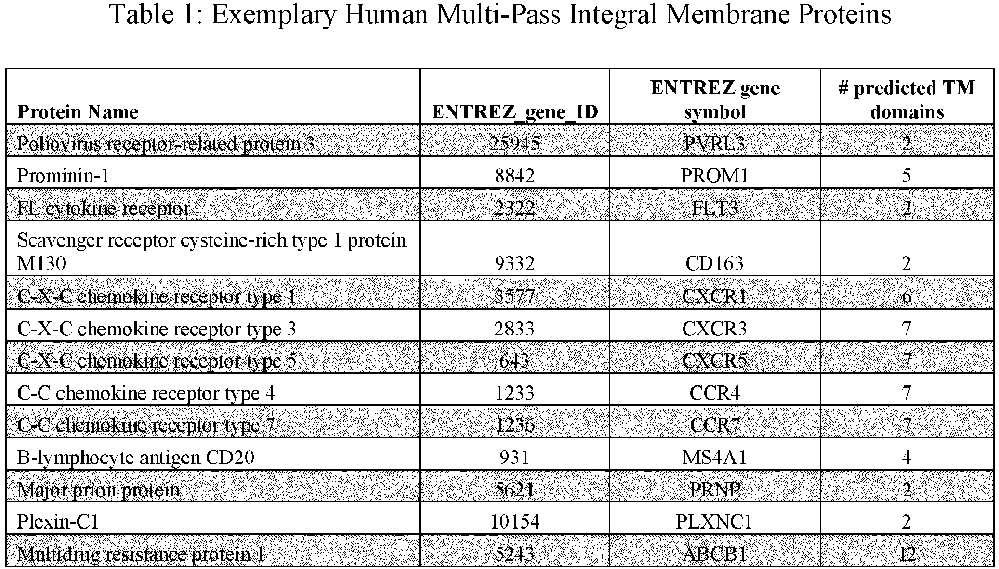


- In certain aspects, the multi-pass IMP is a GPCR, e.g., FZD4 or CXCR4. In certain aspects the multi-pass IMP is CD20.
- This disclosure provides an isolated polynucleotide for expression of an integral membrane protein or fragment thereof in a conformationally-intact form in the context of a biological membrane, as a fusion with a protein or fragment thereof specific for vaccinia virus EEV. By "conformationally intact" is meant that the protein appears, or is displayed, in a native or close to native conformation in the context of a biological lipid bilayer membrane, much as the protein would appear in its native state.
- In one aspect, the disclosure provides an isolated polynucleotide that includes a first nucleic acid fragment that encodes an integral membrane protein (IMP) or fragment thereof, e.g., a multi-pass IMP, where the IMP or fragment thereof comprises at least one extra-membrane region, at least one transmembrane domain and at least one intra-membrane region, and where a portion of the first nucleic acid fragment encoding at least one intra-membrane region is situated at the 5' or 3' end of the first nucleic acid fragment; and a second nucleic acid fragment that encodes a vaccinia virus F13L protein (SEQ ID NO: 1) or functional fragment thereof, where the second nucleic acid fragment is fused in frame to a portion of the first nucleic acid fragment that encodes an intra-membrane region of the IMP. The first nucleic acid fragment and the second nucleic acid fragment can, in some instances, we separated by a nucleic acid encoding a linker or other spacer. The polynucleotide can further include a poxvirus promoter operably associated with the first and second nucleic acid fragments, allowing expression of the polynucleotide in the cytoplasm of a poxvirus-infected cell. According to this aspect, a poxvirus-infected cell that contains the polynucleotide can express an IMP-F13L fusion protein as part of the outer envelope membrane of an extracellular enveloped virion (EEV). Schematic diagrams showing expression of an IMP as a fusion with F13L are shown in
FIG. 1B andFIG. 1C . - In certain aspects, the IMP or fragment thereof can be a multi-pass membrane protein comprising at least two, at least three, at least four, at least five, at least six, at least seven, at least eight, at least nine, at least ten, at least eleven, at least twelve, or even more transmembrane (TM) domains, such as those listed in Table 1.
- Where the IMP has an odd number of TM domains, one end of the IMP, either the N-terminus or the C-terminus, will be naturally situated on the extra-membrane side of the biological membrane and the other end of the IMP will be situated on the intra-membrane side of the IMP. Since the F13L protein is wholly-internal to the outer membrane of poxvirus EEVs, the end of the IMP, the N-terminus or the C-terminus that is situated internal to the membrane can be fused to F13L. Thus for an IMP such as a typical 7-TM domain GPCR in which the N-terminus of the protein is extra-membrane and the C-terminus is intra-membrane, the N-terminus of F13L can be fused to the C-terminus of the GPCR as shown in
FIG. 1B . Accordingly, a polynucleotide as above is provided where the first nucleic acid fragment encodes an IMP with an odd number of transmembrane domains, where the 5' end of the first nucleic acid fragment encodes the extra-membrane region, and the 3' end of the first nucleic acid fragment encodes the intra-membrane region of the IMP, the latter being fused to the 5' end of the nucleic acid fragment encoding F13L or a fragment thereof. - In an exemplary polynucleotide of this type, the first polynucleotide can encode the human frizzled-4 protein (FZD4), or a fragment thereof, a target for immunotherapy of certain human cancers, fused to the N-terminus of F13L. Accordingly, a polynucleotide which encodes an FZD4-F13L fusion protein is provided. An exemplary polynucleotide according to this aspect encodes the mature fusion protein,
amino acids 20 to 892 of SEQ ID NO: 2, as shown below. The polynucleotide can further encode a signal peptide, e.g., the signal peptide of FZD4,amino acids 1 to 19 of SEQ ID NO: 2.
FZD(FL)-F13L (SEQ ID NO: 2)
- Single Underline - leader peptide (amino acids 1-19)
- Bold - human Fzd4 (amino acids 20-520)
- Italics - F13L (amino acids 521-892)
- In another exemplary polynucleotide of this type, the first polynucleotide can encode A CXC chemokine receptor, or a fragment thereof, fused to the N-terminus of F13L. CXC chemokine receptors are likewise targets for immunotherapy of certain human cancers. An exemplary CXC chemokine receptor is CXCR4, or a fragment thereof. Accordingly, a polynucleotide which encodes a CXC chemokine receptor-F13L fusion protein, e.g., a CXCR4-F13L fusion protein is provided. An exemplary polynucleotide according to this aspect encodes SEQ ID NO: 3, as shown below.
CXCR4-F13L (SEQ ID NO: 3)
- Bold - human CXCR4 (amino acids 1-356)
- Italics - F13L (amino acids 357-728)
- As will be evident to a person of ordinary skill in the art, a multi-pass membrane protein having an even number of transmembrane domains will be inserted into a biological membrane such that its N-terminus and its C-terminus are on the same side of the membrane, either on the extra-membrane side of the membrane, or on the intra-membrane side of the membrane. Since the F13L protein is situated entirely on the intra-membrane side of poxvirus EEVs, formation of an IMP-F13L fusion protein properly embedded in the membrane would need at least one of the N-terminus or the C-terminus of the IMP or fragment thereof to be internal to the membrane. Where the IMP has an even number of TM domains and both are situated internally, the F13L protein can be fused either to the N-terminus of the IMP or to the C-terminus of the IMP. If the full-length IMP is situated such that both the N- and C-terminus are extra-membrane, a fragment of the IMP having an odd number of TM domains can be fused to F13L.
- Accordingly, the disclosure provides a polynucleotide as described above that encodes an IMP with an even number of transmembrane domains, where both the 5' and 3' ends of the first nucleic acid fragment encode intra-membrane regions. In certain aspects the 3' end of the nucleic acid fragment encoding F13L can be fused to the 5' end of the nucleic acid fragment encoding the IMP, in certain aspects the 5' end of the nucleic acid fragment encoding F13L can be fused to the 3' end of the nucleic acid fragment encoding the IMP.
- An exemplary IMP of this type is human CD20, a 4-TM domain IMP expressed on human B cells, which is a target for immunotherapy of B cell leukemias, lymphomas, and myelomas. A diagram of a CD20-F13L fusion protein in which the C-terminus of CD20 is fused to the N-terminus of F13L is shown in
FIG. 1C . Accordingly, a polynucleotide which encodes a CD20-F13L fusion protein is provided. An exemplary polynucleotide according to this aspect encodes SEQ ID NO: 4, as shown below.
CD20-F13L (Seq ID NO: 4)
- Bold - human CD20 (MS4A1)(amino acids 1-298)
- Italics - F13L (amino acids 299-669)
- In polynucleotides as provided above, the first and second nucleic acid fragments can be directly fused, or alternatively they can be separated by a nucleic acid fragment encoding a linker or spacer or other polypeptide fragment. In certain aspects, a polynucleotide as provided above can further include a third nucleic acid fragment that encodes a heterologous peptide polypeptide, either between the first and second nucleic acid fragments, or on either side. The heterologous peptide can be, for example, a linker sequence, an amino acid tag or label, or a peptide or polypeptide sequence that facilitates purification. In certain aspects the heterologous peptide is a 6-histidine tag fused, e.g., to the C-terminus of the fusion protein.
- In certain aspects, a polynucleotide as provided herein is operably associated with a poxvirus promoter. Suitable promoters are described elsewhere herein. In certain aspects the promoter is a poxvirus p7.5 promoter or a poxvirus H5 promoter.
- A polynucleotide as provided herein can be or can be part of, a poxvirus genome, where the poxvirus genome, upon introduction into a suitable permissive host cell, can produce infectious EEV that display the IMP-F13L fusion protein on their surface. In certain aspects the poxvirus genome is a vaccinia virus genome, e.g., a vaccinia virus WR genome or an MVA genome. A poxvirus genome comprising a polynucleotide as described can be produced by standard molecular biological and virology techniques, for example by using tri-molecular recombination as described herein. A poxvirus genome as provided herein can be introduced into permissive cells as part of a recombinant poxvirus, or as naked DNA accompanied by suitable helper viruses, e.g., fowlpox virus. The disclosure further provides a recombinant poxvirus, e.g., a recombinant vaccinia virus comprising the provided poxvirus genome.
- This disclosure further provides an IMP-F13L fusion protein such as those encoded by the polynucleotides described above. Moreover, the IMP-F13L fusion protein can be expressed on the surface of a recombinant poxvirus EEV, e.g., a recombinant vaccinia virus EEV. A recombinant poxvirus EEV, e.g., a recombinant vaccinia virus EEV comprising the provided fusion protein is provided by the disclosure. A recombinant poxvirus EEV can be produced by a method that includes infecting a host cell permissive for vaccinia virus infectivity with a vaccinia virus comprising a poxvirus genome as provided above, and recovering EEV released from the infected host cell. Accordingly, an IMP-F13L fusion protein encoded by a polynucleotide as described above, is provided.
- Moreover the disclosure provides fusion proteins comprising an IMP or fragment thereof, which can be a multi-pass IMP, and single pass IMP, or even just the extracellular domain (ECD) of the IMP, fused to a poxvirus protein, e.g., a vaccinia virus protein, specific for EEV, such as F13L, A56R, B5R, 33R, A34R, or A36R, an "IMP-EEV fusion protein." Exemplary ECD fusion proteins are described below. An IMP-EEV fusion protein as provided herein can display the IMP, e.g., a multi-pass IMP, single-pass IMP or ECD of an IMP, in a conformationally intact form on the surface of poxvirus EEV. For use in screening antibody display libraries for antigen binding domains that specifically bind to a target IMP, display of IMPs on the surface of poxvirus EEV offers many advantages over displaying IMPs on the surface of recombinant cells, e.g., CHO cells, as is typical. For example the IMP can be expressed at higher density on EEV than on cells. Moreover, EEV express only about six different poxvirus proteins on their surface (e.g., F13L, A56R, B5R, 33R, A34R, and A36R) as opposed to hundreds that might be expressed on the surface of cells. Finally, inactivated EEV expressing IMP-F13L fusion proteins as provided herein can be attached to solid supports, offering convenience in library screening.
- Accordingly, this disclosure provides a method to display an integral membrane protein (IMP) or fragment thereof in a native conformation for use, e.g., in screening antibody display libraries for antigen binding domains specific for the IMP. The method includes: infecting host cells permissive for poxvirus infectivity with a recombinant poxvirus that expresses the IMP or fragment thereof as a fusion protein with poxvirus EEV-specific protein or membrane-associated fragment thereof, where EEV produced by the infected host cell comprise the IMP as part of the EEV outer envelope membrane; and recovering EEV released from the host cell. IMP. In certain aspects, the EEV-specific protein or fragment thereof can be the vaccinia virus A33R protein, A34R protein, A56R protein, B5R protein, A36R protein, F13L protein, any membrane-associated fragment thereof, or any combination thereof.
- In certain aspects, the EEV-specific protein is F13L (SEQ ID NO: 1) or a functional fragment thereof, and the fusion protein can be one expressed by a polynucleotide as provided above, e.g., where the IMP is a multi-pass membrane protein comprising at least two, at least three, at least four, at least five, at least six or at least seven transmembrane domains.
- In certain aspects, the membrane-associated EEV specific protein fragment includes the stalk, transmembrane, and intra-membrane domains of the vaccinia virus A56R protein, a fragment comprising, consisting of, or consisting essentially of amino acids 108 to 314 of SEQ ID NO: 5. One of several exemplary fusion partners includes the ECD of human FZD4, shown in bold in SEQ ID NO: 6 below. According to this exemplary aspect the disclosure provides a method to display a conformationally intact fragment of human FZD4 on the surface of a poxvirus EEV comprising infecting host cells permissive for poxvirus infectivity with a recombinant poxvirus encoding a fusion protein comprising
amino acids 20 to 370 of SEQ ID NO: 6. In certain aspects the fusion protein can further comprise a signal peptide, e.g.,amino acids 1 to 19 of SEQ ID NO: 6.
FZD-ECD-A56R (Seq ID NO: 6)
- Single Underline - leader peptide (amino acids 1-19)
- Bold - human FZD4 extracellular domain (amino acids 20-163)
- Italics - A56R stalk, transmembrane, and intra-membrane (amino acids 164 to 370)
- Another exemplary fusion partner includes the ECD of human ErbB2 (Her2), shown in bold in SEQ ID NO: 7 below. According to this exemplary aspect the disclosure provides a method to display a conformationally intact fragment of human Her2 on the surface of a poxvirus EEV comprising infecting host cells permissive for poxvirus infectivity with a recombinant poxvirus encoding a fusion protein comprising
amino acids 20 to 855 of SEQ ID NO: 7. In certain aspects the fusion protein can further comprise a signal peptide, e.g.,amino acids 1 to 19 of SEQ ID NO: 7.
Her2-A56R (SEQ ID NO: 7)
- Single Underline - leader peptide (amino acids 1-19)
- Bold - human ERBB2 (HER2) extracellular domain (amino acids 20-648)
- Italics - A56R stalk, transmembrane, and intra-membrane (
amino acids 649 to 855) - Another exemplary fusion partner includes the ECD of human CD100 (Semaphorin 4D), shown in bold in SEQ ID NO: 8 below. According to this exemplary aspect the disclosure provides a method to display a conformationally intact fragment of human CD100 on the surface of a poxvirus EEV comprising infecting host cells permissive for poxvirus infectivity with a recombinant poxvirus encoding a fusion protein comprising
amino acids 20 to 935 of SEQ ID NO: 8. In certain aspects the fusion protein can further comprise a signal peptide, e.g.,amino acids 1 to 19 of SEQ ID NO: 8.
CD100-A56R (SEQ ID NO: 8)
- Single Underline - leader peptide (amino acids 1-19)
- Bold - human CD100 extracellular domain (amino acids 20-728)
- Italics - A56R stalk, transmembrane, and intra-membrane (amino acids 729 to 935)
- In certain aspects, the membrane-associated EEV specific protein fragment includes the transmembrane and intra-membrane domains of the vaccinia virus B5R protein, a fragment comprising, consisting of, or consisting essentially of amino acids 276 to 317 of SEQ ID NO: 9. In certain aspects, the membrane-associated EEV specific protein fragment includes the stalk, transmembrane, and intra-membrane domains of the vaccinia virus B5R protein, a fragment comprising, consisting of, or consisting essentially of amino acids 238 to 317 of SEQ ID NO: 9.
SEQ ID NO: 9: WR B5R protein
- In certain exemplary aspects the IMP fusion partner for the B5R fragment comprises the extracellular domain of human FZD4, shown in bold in SEQ ID NO: 10 and SEQ ID NO: 11 below. According to this exemplary aspect the disclosure provides a method to display a conformationally intact fragment of human FZD4 on the surface of a poxvirus EEV comprising infecting host cells permissive for poxvirus infectivity with a recombinant poxvirus encoding a fusion protein comprising
amino acids 20 to 243 of SEQ ID NO: 10 oramino acids 20 to 281 of SEQ ID NO: 11. In certain aspects the fusion protein can further comprise a signal peptide, e.g.,amino acids 1 to 19 of SEQ ID NO: 10. - FZD-B5R (short) (SEQ ID NO: 10)

- Single Underline - leader peptide (amino acids 1-19)
- Bold - human FZD4 extracellular domain (amino acids 20-200)
- Italics - B5R TM and cytoplasmic tail (amino acids 201-243)
- FZD-B5R (long) (SEQ ID NO: 11)

- Single Underline - leader peptide (amino acids 1-19)
- Bold - human FZD4 extracellular domain (amino acids 20-200)
- Italics - B5R stalk, TM and cytoplasmic tail (amino acids 201-281)
- The disclosure further provides a fusion protein comprising:
amino acids 20 to 892 of SEQ ID NO: 2; SEQ ID NO: 3; SEQ ID NO: 4;amino acids 20 to 370 of SEQ ID NO: 6;amino acids 20 to 855 of SEQ ID NO: 7;amino acids 20 to 935 of SEQ ID NO: 8;amino acids 20 to 243 of SEQ ID NO: 10; oramino acids 20 to 281 of SEQ ID NO: 11, any combination thereof, any fragment thereof, or any variant thereof, where the fusion protein, when expressed by a recombinant poxvirus, appears on the surface of a poxvirus extracellular enveloped virion (EEV) in a native conformation. - A recombinant poxvirus EEV comprising any EEV fusion protein as provided herein is also provided.
- This disclosure further provides a method to select binding molecules, e.g., antibodies, antigen-binding antibody fragments, or antibody like binding molecules that bind to a multi-pass membrane protein interest. The method comprises attaching a recombinant EEV as provided herein to a solid support, where the recombinant EEV can display a multi-pass protein on its surface; providing a display library, e.g., an antibody display library, where the library comprises display packages displaying a plurality of antigen binding domains; contacting the display library with the EEV such that display packages displaying antigen binding domains that specifically binds to the IMP expressed on the EEV can bind thereto; removing unbound display packages; and recovering display packages that display an antigen binding domain specific for the IMP expressed on the EEV.
- Any display library comprising a plurality of binding domains, e.g., antibodies, antibody like molecules or other binding molecules is suitable for this method. For example the display library can be a phage display library, a yeast display library or a library constructed in a vaccinia virus vector as described elsewhere herein.
- In certain aspects, the recombinant EEV can be inactivated prior to attachment to the solid support. For example, the EEV can be inactivated by incubation with Psoralen (Trioxsalen, 4'-aminomethyl-, hydrochloride) in the presence of UV irradiation.
- Any suitable solid support can be used. As used herein, a "solid support" is any support capable of binding an EEV, which can be in any of various forms, as is known in the art. Well-known supports include tissue culture plastic, glass, polystyrene, polypropylene, polyethylene, dextran, nylon, amylases, natural and modified celluloses, polyacrylamides, gabbros, and magnetite. The nature of the carrier can be either soluble to some extent or insoluble for the purposes of this disclosure. The support material can have virtually any structural configuration as long as the coupled EEV is capable of binding to a displayed binding molecule such as an antibody. Thus, the support configuration can be spherical, as in a bead, or cylindrical, as in the inside surface of a test tube, or the external surface of a rod. Alternatively, the surface can be flat such as a sheet, test strip, etc. Typical supports include beads, e.g., magnetic polystyrene beads such as DYNABEADS® that can be pulled out of suspension by a magnet. The support configuration can include a tube, bead, microbead, well, plate, tissue culture plate, petri plate, microplate, microtiter plate, flask, stick, strip, vial, paddle, etc., etc. A solid support can be magnetic or non-magnetic. Those skilled in the art will know many other suitable carriers for binding EEV as provided herein, or will be able to readily ascertain the same. In certain aspects, EEV as provided herein can be attached to the solid support via reaction with, e.g., tosyl groups, epoxy groups, carboxylic acid groups, or amino groups attached to the surface. For example, EEV can be attached to the surface of tosyl-activated magnetic beads, e.g., MYONE™ tosylactivated beads. Alternatively, the EEV can be biotinylated and attached to a streptavidin solid surface, e.g., streptavidin coated magnetic beads.
- This disclosure employs, unless otherwise indicated, conventional techniques of cell biology, cell culture, molecular biology, transgenic biology, microbiology, recombinant DNA, and immunology, which are within the skill of the art. Such techniques are explained fully in the literature. (See, for example, Sambrook et al., ed. (1989) Molecular Cloning A Laboratory Manual (2nd ed.; Cold Spring Harbor Laboratory Press); Sambrook et al., ed. (1992) Molecular Cloning: A Laboratory Manual, (Cold Springs Harbor Laboratory, NY); D. N. Glover ed., (1985) DNA Cloning, Volumes I and II; Gait, ed. (1984) Oligonucleotide Synthesis;
Mullis et al. U.S. Pat. No. 4,683,195 ; Hames and Higgins, eds. (1984) Nucleic Acid Hybridization; Hames and Higgins, eds. (1984) Transcription And Translation; Freshney (1987) Culture Of Animal Cells (Alan R. Liss, Inc.); Immobilized Cells And Enzymes (IRL Press) (1986); Perbal (1984) A Practical Guide To Molecular Cloning; the treatise, Methods In Enzymology (Academic Press, Inc., N.Y.); Miller and Calos eds. (1987) Gene Transfer Vectors For Mammalian Cells, (Cold Spring Harbor Laboratory); Wu et al., eds., Methods In Enzymology, Vols. 154 and 155; Mayer and Walker, eds. (1987) Immunochemical Methods In Cell And Molecular Biology (Academic Press, London); Weir and Blackwell, eds., (1986) Handbook Of Experimental Immunology, Volumes I-IV; Manipulating the Mouse Embryo, Cold Spring Harbor Laboratory Press, Cold Spring Harbor, N.Y., (1986); and in Ausubel et al. (1989) Current Protocols in Molecular Biology (John Wiley and Sons, Baltimore, Md.). - General principles of antibody engineering are set forth in Borrebaeck, ed. (1995) Antibody Engineering (2nd ed.; Oxford Univ. Press). General principles of protein engineering are set forth in Rickwood et al., eds. (1995) Protein Engineering, A Practical Approach (IRL Press at Oxford Univ. Press, Oxford, Eng.). General principles of antibodies and antibody-hapten binding are set forth in: Nisonoff (1984) Molecular Immunology (2nd ed.; Sinauer Associates, Sunderland, Mass.); and Steward (1984) Antibodies, Their Structure and Function (Chapman and Hall, New York, N.Y.). Additionally, standard methods in immunology known in the art and not specifically described can be followed as in Current Protocols in Immunology, John Wiley & Sons, New York; Stites et al., eds. (1994) Basic and Clinical Immunology (8th ed; Appleton & Lange, Norwalk, Conn.) and Mishell and Shiigi (eds) (1980) Selected Methods in Cellular Immunology (W.H. Freeman and Co., NY).
- Standard reference works setting forth general principles of immunology include Current Protocols in Immunology, John Wiley & Sons, New York; Klein (1982) J., Immunology: The Science of Self-Nonself Discrimination (John Wiley & Sons, NY); Kennett et al., eds. (1980) Monoclonal Antibodies, Hybridoma: A New Dimension in Biological Analyses (Plenum Press, NY); Campbell (1984) "Monoclonal Antibody Technology" in Laboratory Techniques in Biochemistry and Molecular Biology, ed. Burden et al., (Elsevier, Amsterdam); Goldsby et al., eds. (2000) Kuby Immunology (4th ed.; H. Freeman & Co.); Roitt et al. (2001) Immunology (6th ed.; London: Mosby); Abbas et al. (2005) Cellular and Molecular Immunology (5th ed.; Elsevier Health Sciences Division); Kontermann and Dubel (2001) Antibody Engineering (Springer Verlag); Sambrook and Russell (2001) Molecular Cloning: A Laboratory Manual (Cold Spring Harbor Press); Lewin (2003) Genes VIII (Prentice Hall, 2003); Harlow and Lane (1988) Antibodies: A Laboratory Manual (Cold Spring Harbor Press); Dieffenbach and Dveksler (2003) PCR Primer (Cold Spring Harbor Press).
- The following examples are offered by way of illustration and not by way of limitation.
- The disclosure provides an isolated polynucleotide that includes: a first nucleic acid fragment that encodes an integral membrane protein (IMP) or fragment thereof, where the IMP or fragment thereof includes at least one extra-membrane region, at least one transmembrane domain and at least one intra-membrane region, and where a portion of the first nucleic acid fragment encoding at least one intra-membrane region is situated at the 5' or 3' end of the first nucleic acid fragment; and a second nucleic acid fragment that encodes a vaccinia virus F13L protein or functional fragment thereof, where the second nucleic acid fragment is fused in frame to a portion of the first nucleic acid fragment that encodes an intra-membrane region of the IMP. According to this, a poxvirus infected cell containing the polynucleotide can express an IMP-F13L fusion protein as part of the outer envelope membrane of an extracellular enveloped virion (EEV). The F13L protein or functional fragment thereof can include the amino acid sequence SEQ ID NO: 1 or a functional fragment thereof. The IMP may be a multi-pass membrane protein comprising at least two, at least three, at least four, at least five, at least six or at least seven transmembrane domains. The IMP may be a multi-pass membrane protein listed in Table 1.
- The multi-pass IMP can have an odd number of transmembrane domains, the 5' end of the first nucleic acid fragment can encode an extra-membrane region, and the 3' end of the first nucleic acid fragment can encode an intra-membrane region fused to the 5' end of the second nucleic acid fragment. The first nucleic acid fragment of this type can encode, e.g., a G-protein coupled receptor (GPCR). The GPCR can be the human frizzled-4 protein (FZD4), or a fragment thereof, and the polynucleotide can encode a polypeptide that includes
amino acids 20 to 892 of SEQ ID NO: 2. The polypeptide can further include a signal peptide, e.g.,amino acids 1 to 19 of SEQ ID NO: 2. The GPCR can be a CXC chemokine receptor, e.g., CXCR4, or a fragment thereof, and the polynucleotide can encode a polypeptide that includes the amino acid sequence SEQ ID NO: 3. - The multi-pass IMP can have an even number of transmembrane domains, and both the 5' and 3' ends of the first nucleic acid fragment can encode intra-membrane regions. The second nucleic acid fragment can be fused to 3' end of the first nucleic acid fragment. The IMP can be, e.g., human CD20 protein, or a fragment thereof, and the polynucleotide can encode a polypeptide that includes the amino acid sequence SEQ ID NO: 4.
- The first and second nucleic acid fragments of a polynucleotide provided herein can be directly fused. The polynucleotide as provided herein can include a third nucleic acid fragment encoding a heterologous peptide, e.g., a linker sequence, an amino acid tag or label, or a peptide or polypeptide sequence that facilitates purification, such as a histidine tag. A polynucleotide as provided here can be operably associated with a poxvirus promoter, e.g., a p7.5, a T7, or H5 promoter.
- The disclosure further provides an F13L fusion protein encoded by a polynucleotide as provided herein. The disclosure further provides a poxvirus genome, e.g., a vaccinia virus genome, that includes a polynucleotide as provided herein. The disclosure further provides a recombinant vaccinia virus EEV that includes a poxvirus genome as provided herein.
- The disclosure further provides a method of producing a recombinant vaccinia virus EEV as provided herein where the method includes infecting a host cell permissive for vaccinia virus infectivity with a vaccinia virus comprising a poxvirus genome as provided herein, and recovering EEV released from the host cell.
- The disclosure further provides a method to display an integral membrane protein (IMP) or fragment thereof in a native conformation where the method includes infecting host cells permissive for poxvirus infectivity with a recombinant poxvirus that expresses an IMP or fragment thereof as a fusion protein with poxvirus EEV-specific protein or membrane-associated fragment thereof, where EEV produced by the infected host cell comprise the IMP fusion protein as part of the EEV outer envelope membrane and recovering EEV released from the host cell. The IMP or fragment thereof may be displayed on the surface of the EEV in a native conformation. The EEV-specific protein can be the vaccinia virus A33R protein, A34R protein, A56R protein, B5R protein, A36R protein, F13L protein, any membrane-associated fragment thereof, or any combination thereof.
- The EEV-specific protein may be F13L (SEQ ID NO: 1) or a functional fragment thereof. The IMP may be a multi-pass membrane protein that includes at least two, at least three, at least four, at least five, at least six or at least seven transmembrane domains. The IMP can be a G-protein coupled receptor (GPCR), e.g., human FZD4 or CXCR4 as described above, that includes seven transmembrane domains, and the F13L protein can be fused to the C-terminus of the IMP. The IMP or fragment thereof can have an even number of transmembrane domains, e.g., human CD20 as described above, where both the N-terminus and the C-terminus of the IMP or fragment thereof are intra-membrane, and the F13L can be fused to the N-terminus or the C-terminus of the IMP
- The membrane-associated EEV specific protein fragment can include or consist of the stalk, transmembrane, and intra-membrane domains of the vaccinia virus A56R protein, e.g., amino acids 108 to 314 of SEQ ID NO: 5. The IMP portion of the A56R fusion protein can include the extracellular domain of human FZD4, e.g., the fusion protein can include
amino acids 20 to 370 of SEQ ID NO: 6, the extracellular domain of human ErbB2 (Her2), e.g., the fusion protein can includeamino acids 20 to 855 of SEQ ID NO: 7, or the extracellular domain of human CD100 (Semaphorin 4D), e.g., the fusion protein can includeamino acids 20 to 935 of SEQ ID NO: 8. - The membrane-associated EEV specific protein fragment can include or consist of the transmembrane and intra-membrane domains, or the stalk, transmembrane, and intra-membrane domains of the vaccinia virus B5R protein, e.g., amino acids 276 to 317 of SEQ ID NO: 9 or amino acids 238 to 317 of SEQ ID NO: 9, respectively. The IMP portion of the B5R fusion protein can include the extracellular domain of human FZD4, e.g., the fusion protein can include
amino acids 20 to 243 of SEQ ID NO: 10 oramino acids 20 to 281 of SEQ ID NO: 11. - The disclosure further provides a fusion protein comprising:
amino acids 20 to 892 of SEQ ID NO: 2; SEQ ID NO: 3; SEQ ID NO: 4;amino acids 20 to 370 of SEQ ID NO: 6;amino acids 20 to 855 of SEQ ID NO: 7;amino acids 20 to 935 of SEQ ID NO: 8;amino acids 20 to 243 of SEQ ID NO: 10;amino acids 20 to 281 of SEQ ID NO: 11,amino acids 20 to 506 of SEQ ID NO: 13, oramino acids 20 to 235 of SEQ ID NO: 14. A fusion protein as provided, when expressed by a recombinant poxvirus, e.g., a vaccinia virus, can appear on the surface of a poxvirus extracellular enveloped virion (EEV) in a native conformation. A recombinant poxvirus EEV comprising the fusion protein is also provided. The disclosure further provides a recombinant poxvirus EEV that includes a heterologous IMP or fragment thereof fused to a poxvirus EEV-specific protein or membrane-associated fragment thereof, where the fusion protein is situated in the EEV outer envelope membrane, and where the IMP or fragment thereof displays on the surface of the EEV in its native conformation. The recombinant poxvirus EEV may be a vaccinia virus EEV. - The disclosure further provides a method to select antibodies that bind to a multi-pass membrane protein where the method includes attaching recombinant EEV as provided herein to a solid support; providing an antibody display library, where the library comprises display packages displaying a plurality of antigen binding domains; contacting the display library with the EEV such that display packages displaying antigen binding domains that specifically binds to the IMP expressed on the EEV can bind thereto; removing unbound display packages; and recovering display packages that display an antigen binding domain specific for the IMP expressed on the EEV. The recombinant EEV may be inactivated prior to attachment to the solid support, e.g., by incubation with Psoralen (Trioxsalen, 4'-aminomethyl-, hydrochloride) in the presence of UV irradiation. The recombinant EEV may be attached to the solid surface via reaction with tosyl groups attached to the surface. The solid surface can be tosyl-activated magnetic beads. The recombinant EEV may be biotinylated and attached to a streptavidin coated solid surface, e.g., streptavidin-coated magnetic beads.
- IMPs were incorporated into vaccinia virus EEVs using the EEV-specific proteins F13L, A56R, and B5R, by the following methods. Generally, the extracellular domains of HER2, CD100 (semaphorin 4D), and FZD4 were incorporated as fusions with the single-pass EEV-specific membrane proteins A56R and B5R as diagrammed in
FIG. 1A . The mature FZD4-ECD-A56R fusion protein comprisesamino acids 20 to 370 of SEQ ID NO: 6, the mature HER2-ECD-A56R fusion protein comprisesamino acids 20 to 855 of SEQ ID NO: 7, and the mature CD100-ECD-A56R fusion protein comprisesamino acids 20 to 935 SEQ ID NO: 8.FIG. 1B andFIG. 1C show diagrammatically how the multi-pass proteins such as GPCRs and CD20 can be incorporated into EEVs as multi-pass membrane proteins as a fusion with the EEV membrane-associated protein F13L. - cDNAs encoding the IMPs were cloned in-frame to the vaccinia virus F13L gene encoding the palmitoylated EEV membrane glycoprotein (SEQ ID NO: 1) into the pJEM1 plasmid previously described for the purpose of introduction into vaccinia virus. pJEM1 is a derivative of p7.5/tk described in
U.S. Patent Appl. Publ. No. 2013/0288927 , and when digested with NcoI or BssHII and BsiWI, contains flanking regions capable of homologous recombination with the vaccinia virus TK gene and the vaccinia virus H5 promoter. - The open reading frame of human membrane protein MS4A1 gene (CD20)(NM_021950.3) was cloned in frame with the vaccinia virus F13L using SOE (Splicing by Overlap Extension) PCR as per standard protocols whereby restriction endonuclease sites NcoI and BsiWI were added to the PCR product by encoding them into the 5' and 3'-most flanking primers respectively. This strategy avoids the introduction of a leader peptide. The final PCR product and pJEM1 were digested with NcoI and BsiWI and the two species were joined via ligation according to standard protocols. The circularized plasmid was then introduced into competent E. coli via chemical transformation and colonies were selected on ampicillin-containing agar plates.
MS4A1S tataCCATGgCAACACCCAGAAATTCAGTAAATG (SEQ ID NO: 12) MS4A1AS 
F13S 
F13AS 
- The resulting CD20-F13L fusion protein encoded by the polynucleotide comprises the amino acid sequence SEQ ID NO: 4.
- The open reading frame of human membrane protein FZD4 (NM_012193.3) was cloned in frame with the vaccinia virus F13L using SOE (Splicing by Overlap Extension) PCR as per standard protocols whereby restriction endonuclease sites BssHII and BsiWI were added to the PCR product by encoding them into the 5' and 3'-most flanking primers, respectively. This strategy provides for the use of the leader peptide contained within pJEM1. The final PCR product and pJEM1 were digested with BssHII and BsiWI and the two species were joined via ligation according to the standard protocols. The circularized plasmid was then introduced into competent E. coli via chemical transformation and colonies were selected on ampicillin-containing agar plates. PCR primers were specific for FZD4 and F13L and conform to the same general strategy as described for MS4A1. The resulting mature FZD4-F13L fusion protein encoded by the polynucleotide comprises amino acids 20-892 of SEQ ID NO: 2.
- The open reading frame of human membrane protein CXCR4 (NM_001008540.1) was cloned in frame with the vaccinia virus F13L using SOE (Splicing by Overlap Extension) PCR as per standard protocols whereby restriction endonuclease sites NcoI and BsiWI were added to the PCR product by encoding them into the 5' and 3'-most flanking primers respectively. This strategy avoids the introduction of a leader peptide. The final PCR product and pJEM1 were digested with NcoI and BsiWI and the two species were joined via ligation according to the standard protocol. The circularized plasmid was then introduced into competent E. coli via chemical transformation and colonies were selected on ampicillin-containing agar plates. PCR primers were specific for CXCR4 and F13L and conform to the same general strategy as described for MS4A1. The resulting CXCR4-F13L fusion protein encoded by the polynucleotide comprises the amino acid sequence SEQ ID NO: 3.
- The plasmids produced as described above, as well as similar plasmids encoding non-fused ("untagged") versions of CD20, FZD4, CXCR4, CD100, and HER2, were linearized and introduced into vaccinia virus via tri-molecular recombination.
- BHK cells were infected with either IMV encoding the CD20-F13L fusion protein (SEQ ID NO: 4) or Control Western Reserve (WR) virus at a multiplicity of infection (MOI) of 1 virus per cell for two days after which the supernatant containing EEV was harvested and debris removed by low speed centrifugation. Protein G DYNABEADS® (110 µL) were pulled down with a magnet and 1 mL of PBS + 20 µg of purified anti-CD20 antibody was added to the beads. The solution was incubated at room temperature with gentle rotation for 30-60 minutes to allow the antibody to couple to the Protein G beads. Ten µg of purified mIgG1 isotype control was added to the solution to ensure complete blocking, and the solution was incubated at room temperature with gentle rotation for 10-30 additional minutes. Beads were pulled down with the magnet, washed once with 1 mL of PBS and resuspended in 110 µL of PBS.
- Fifty µL of Anti-CD20-Pro G DYNABEADS® was added to 1 mL of CD20-F13L or WR EEV supernatant and was incubated at room temperature with gentle rotation for 1 hour. Beads were pelleted using the magnet and unbound supernatant removed. The beads were then washed five times with 1 mL of Dulbecco's Modified Eagle Medium (DMEM) media supplemented with 10% FBS and 1 mM HEPES (10% DMEM). All washes were pooled with the unbound supernatant ("Unbound"). The beads ("Bound") were then resuspended in 1 mL of 10% DMEM. "Unbound" and "Bound" were titered on BSC-1 cells and overlaid with growth medium containing methyl cellulose. Plaques were allowed to form for two days and then the cells were fixed and stained with 0.1% Crystal Violet solution. Plaques were counted to determine the number of plaque forming units (pfu) in the "Unbound" and "Bound" from which the % of EEV bound to the beads could be calculated. Results are shown on Table 2.
Table 2: CD20-F13L EEV Binding EEV Supernatant % Bound Western Reserve 10.8 % CD20-F13L 50.5% - The % EEV bound to the anti-CD20 coated beads was significantly higher for CD20-F13L EEV fusion protein than it is for the Western Reserve indicating that CD20 is being expressed on the EEV membrane surface.
- BHK cells were infected with either IMV encoding the CD20-F13L fusion protein (SEQ ID NO: 4), CD20-A56R fusion protein (SEQ ID NO: 13), CD20 untagged (unfused) or control HER2-A56R Extracellular Domain (ECD) (SEQ ID NO: 7) virus at an MOI=1 for two days after which the supernatant containing EEV was harvested and debris removed by low speed centrifugation. Streptavidin DYNABEADS® (200 µL) were pulled down with a magnet and 0.2 mL of PBS + 20 µg of purified Biotinylated anti-CD20 antibody or Biotinylated anti-HER2 was added to the beads. The solution was incubated at room temperature with gentle rotation for 30 minutes to allow the antibody to couple to the Streptavidin beads. Beads were pulled down with the magnet, washed once with 1 mL of PBS and resuspended in 200 µL of PBS.
CD20-A56R: (SEQ ID NO: 13)
- Single Underline - Signal sequence (amino acids 1-19)
- Italics - Truncated A56R (amino acids 20-190)
- Bold - CD20 Sequence (amino acids 191-506)
- Fifty µL of prepared streptavidin beads were added to 1 mL of each EEV supernatant and allowed to rotate at room temperature for 45 minutes. Beads were pelleted using the magnet and unbound supernatant removed. The beads were then washed five times with 1 mL of DMEM media supplemented with 10% FBS and 1 mM HEPES (10% DMEM). All washes were pooled with the unbound supernatant ("Unbound"). The beads ("Bound") were then resuspended in 1 mL of 10% DMEM. "Unbound" and "Bound" were titered on BSC-1 cells and overlaid with growth medium containing methylcellulose. Plaques were allowed to form for two days and then the cells were fixed and stained with 0.1% Crystal Violet solution. Plaques were counted to determine the number of plaque forming units (pfu) in the "Unbound" and "Bound" from which the % of EEV bound to the beads could be calculated. Results are shown in
FIG. 2 . - The % EEV bound to the anti-CD20 coated beads for CD20-F13L was greater than the % EEV bound for untagged (unfused) or A56R-fused CD20 indicating higher expression of CD20-F13L on the EEV membrane. The lack of binding to the anti-HER2 coated beads confirmed specificity of the assay.
- The experiment above was repeated using CD20-F13L fusion protein (SEQ ID NO: 4), CD20 untagged (unfused), FZD-F13L fusion protein (SEQ ID NO: 2), and FZD untagged (unfused). Virus was pulled down using anti-CD20 or anti-FZD coated beads as described above. The data in
FIG. 3A (anti-CD20-coated beads) andFIG. 3B (anti-FZD-coated beads) shows that F13L fusion proteins were specifically pulled down by their respective antibodies, and were more efficiently incorporated into vaccinia virus than untagged (unfused) proteins. - BHK cells were infected at an MOI=1 with virus expressing the following antigen constructs: CD20-F13L (SEQ ID NO: 4), CXCR4-F13L (SEQ ID NO: 3), HER2-ECD-A56R (SEQ ID NO: 7), and CD100-ECD-A56R (SEQ ID NO: 8). After two days, the supernatant containing EEV was harvested and debris removed by low speed centrifugation. Streptavidin DYNABEADS® were pulled down with a magnet and for each sample, 50 µL of beads were resuspended in 0.1 mL of PBS + 5 µg of purified Biotinylated anti-CD20 antibody, Biotinylated anti-CXCR4 (12G5), Biotinylated anti-CD100 (2503), or Biotinylated anti-HER2. The solutions were incubated at room temperature with gentle rotation for 30 minutes to allow the antibody to couple to the Streptavidin beads. Beads were pulled down with the magnet, washed once with 1 mL of PBS and resuspended in 100 µL of PBS per sample.
- One hundred µL of prepared streptavidin beads were added to 1 mL of each EEV supernatant and allowed to rotate at room temperature for 45 minutes. Beads were pelleted using the magnet and unbound supernatant removed. The beads were then washed five times with 1 mL of DMEM media supplemented with 10% FBS and 1 mM HEPES (10% DMEM). All washes were pooled with the unbound supernatant ("Unbound"). The beads ("Bound") were then resuspended in 1 mL of 10% DMEM. "Unbound" and "Bound" were titered on BSC-1 cells and overlaid with growth medium containing methylcellulose. Plaques were allowed to form for two days and then the cells were fixed and stained with 0.1% Crystal Violet solution. Plaques were counted to determine the number of plaque forming units (pfu) in the "Unbound" and "Bound" from which the % of EEV bound to the beads could be calculated. Results are shown in
FIG. 4 . - All of the antigen-EEV bound specifically to their corresponding antibody-coupled beads indicating efficient expression of the antigen on the EEV membrane.
- BHK cells (2 × 10⁸ cells) were infected at an MOI=1 with virus expressing HER2-ECD-A56R (SEQ ID NO: 7), FZD-F13L (SEQ ID NO: 2), CXCR4-F13L (SEQ ID NO: 3) or CD100 (semaphorin 4D)-ECD-A56R (SEQ ID NO: 8) in one cellSTACK cell culture chamber each (Corning). After two days, the supernatant containing EEV was harvested and debris removed by low speed centrifugation. The clarified supernatant was then spun at 13,000 rpm (28,000 × g) for 1 hour to pellet the antigen-EEV. The supernatant was aspirated and the pellet resuspended in 1.5 mL of IX PBS. The various viruses were transferred to fresh tubes and Psoralen (Trioxsalen, 4'-aminomethyl-, hydrochloride; Sigma) was added to 20 µg/ml final concentration. The EEV and Psoralen were incubated at room temperature for 10 minutes before being irradiated in the STRATALINKER® UV Crosslinker (Stratagene) for 99,999 microjoules. The Psoralen/UV procedure ensures that the antigen-EEV is inactivated and therefore unable to form plaques or multiply in any downstream testing.
- Tosylactivated MyOne DYNABEADS® (100 µL) were pulled down with a magnet and washed with 1 mL of PBS. The beads were pulled down with the magnet, the PBS removed and 1 mL of each Psoralen/UV inactivated antigen-EEV was added to a separate aliquot of beads. The beads and antigen-EEV were allowed to rotate at 37 °C for 16-20 hours. The beads were pelleted and the supernatant was removed. The beads were blocked with 1 mL of IX PBS, 10% FBS and 0.5% BSA at 37 °C for 1 hour. The beads were pelleted and washed with 1 mL IX PBS before being resuspended in 200 µL of IX PBS.
- One hundred microliters of antigen-EEV-coupled beads was added to 1 mL of each respective antibody-EEV supernatant expressing anti-FZD4, anti-CXCR4,
anti-CD 100, or anti-HER2, as well as to control antibody-EEV. Antibody EEV were produced by infecting BHK cells at an MOI=1 each for 2 days with vaccinia virus encoding both the heavy and light chains of the respective antibodies, and harvesting the supernatants followed by a low speed spin to remove any cells. The virus coupled beads and antibody EEV were allowed to rotate at room temperature for 2 hours. Beads were pelleted using the magnet and unbound supernatants removed. The beads were then washed five times with 1 mL of DMEM media supplemented with 10% FBS and 1 mM HEPES (10% DMEM). All washes were pooled with the unbound supernatant ("Unbound"). The beads ("Bound") were then resuspended in 1 mL of 10% DMEM. "Unbound" and "Bound" were titered on BSC-1 cells and overlaid with growth medium containing methylcellulose. Plaques were allowed to form for two days and then the cells were fixed and stained with 0.1% Crystal Violet solution. Plaques were counted to determine the number of plaque forming units (pfu) in the "Unbound" and "Bound" from which the % of EEV bound to the beads could be calculated. A diagram of the method is shown inFIG 5 , and results are shown inFIG. 6A (HER2),FIG. 6B (FZD4),FIG. 6C (CXCR4), andFIG. 6D (CD100 ("Sema")). - Antibody-EEV expressing Anti-HER2 was specifically pulled down by beads coupled with HER2-ECD-A56R antigen EEV, Antibody-EEV expressing Anti-FZD was specifically pulled down by beads coupled with FZD-F13L antigen EEV, Antibody-EEV expressing Anti-CXCR4 was specifically pulled down by beads coupled with CXCR4-F13L antigen EEV, and Antibody-EEV expressing Anti-SEMA was specifically pulled down by beads coupled with Sema-ECD-A56R antigen EEV.
- BHK cells were infected at an MOI=1 each with an antibody library (H-IgG-A56R) and L48 (derivative of germline VK1-39) in four cell STACK cell culture chamber (Corning) (2 × 10⁸ cells per stacker). The antibody library contained a diverse population of heavy chain variable domains in full length IgG format, fused in frame to A56R (see
US Patent Appl. Publication No. 2013-0288927 ). The diversity of this library was approximately 400 million independent clones. After two days, the supernatant containing EEV was harvested and debris removed by low speed centrifugation. The clarified supernatant was then spun at 13,000 rpm (28,000 × g) for 1 hour to pellet the antibody-EEV. The antibody-EEV was resuspended in 1ml EMEM with 10% FBS and stored at 4 degrees until ready for use. In order to make the antigen virus for panning, BHK cells (2 × 10⁸ cells) were infected at an MOI=1.5 with virus expressing FZD4-ECD-A56R (SEQ ID NO: 6) in two cellSTACK cell culture chamber (Corning). After two days, the supernatant containing EEV was harvested and debris removed by low speed centrifugation. The clarified supernatant was then spun at 13,000 rpm (28,000 × g) for 1 hour to pellet the antigen-EEV. The supernatant was aspirated and the pellet resuspended in 1.0 mL of 1× PBS. The one mL of the FZD4-ECD-A56R EEV was transferred to a fresh tube and Psoralen (Trioxsalen, 4'-aminomethyl-, hydrochloride; Sigma) was added to 40 µg/ml final concentration. The EEV and Psoralen were incubated at room temperature for 10 minutes before being irradiated in the STRATAL® UV Crosslinker (Stratagene) for 99,999 microjoules. The Psoralen/UV procedure ensures that the antigen-EEV is inactivated and therefore unable to form plaques or multiply in any downstream testing. - Tosylactivated MyOne DYNABEADS® (150 µL) were pulled down with a magnet and washed with 1 mL of PBS, two times. The beads were pulled down with the magnet, the PBS removed and the 1 mL of Psoralen/UV inactivated FZD4-ECD-A56R was added to the beads. The beads and antigen-EEV were allowed to rotate at 37 °C for 18-20 hours. The beads were pelleted and the supernatant was removed. The beads were blocked with 1 mL of 1× PBS, 10% FBS and 0.5% BSA at 37 °C for 2 hours. The beads were pelleted and washed with 1
mL 1× PBS before being resuspended in 150 µL of 1× PBS. - Fifty microliters of FZD4-ECD-A56R-coupled beads were added to 1 mL of the antibody-EEV library. The FZD4-ECD-A56R coupled beads and antibody EEV were allowed to rotate at room temperature for 2 hours. Beads were pelleted using the magnet and unbound supernatant removed. The beads were then washed five times with 1 mL of DMEM media supplemented with 10% FBS and 1 mM HEPES (10% DMEM). All washes were pooled with the unbound supernatant ("Unbound"). The beads ("Bound") were then resuspended in 1 mL of 10% DMEM. "Unbound" and "Bound" were titered on BSC-1 cells and overlaid with growth medium containing methylcellulose. Plaques were allowed to form for two days and then the cells were fixed and stained with 0.1% Crystal Violet solution. The remaining bound virus (990 µl) was divided among 5 T175 flasks containing confluent BSC1 cells and allowed to amplify in DMEM2.5% containing 1mg/ml G418 for 3 days. The cells were then harvested, and the virus released by three cycles of freeze/thaw, and the virus titered.
- For the second round of selection (Rd2), the amplified Heavy chains from
round 1 were co-infected along with fresh L48 into one cellSTACK of BHK Antibody EEV was harvested as described above. For each round of panning, fresh FZD-ECD-A56R antigen virus was produced, concentrated, inactivated and coupled to beads as described above. Fifty microliters of FZD4-ECD-A56R coupled beads was added to 1 mL antibody-EEV Rd2. The FZD4-ECD-A56R coupled beads and antibody EEV were allowed to rotate at room temperature for 2 hours. Beads were pelleted using the magnet and unbound supernatant removed. The beads were then washed five times with 1 mL of DMEM media supplemented with 10% FBS and 1 mM HEPES (10% DMEM). All washes were pooled with the unbound supernatant ("Unbound"). The beads ("Bound") were then resuspended in 1 mL of 10% DMEM. "Unbound" and "Bound" were titered on BSC-1 cells and overlaid with growth medium containing methylcellulose. Plaques were allowed to form for two days and then the cells were fixed and stained with 0.1% Crystal Violet solution. The remaining bound virus (990ul) was divided among 5 T175 flasks containing confluent BSC1 cells and allowed to amplify in DMEM2.5% containing 1mg/ml G418 for 3 days. The cells were then harvested, and the virus released by three cycles of freeze/thaw, and the virus tittered. - Three additional cycles of panning (Rd3, Rd4, and Rd5) were performed as described above.
-
Rounds FIG 7 shows increasing enrichment per round of selection. Antibodies fromround 5 were sub-cloned into a mammalian expression vector to be expressed as full length soluble IgG and transfected (along with L48 in a mammalian expression vector). The resulting antibodies present in the supernatant were tested by flow cytometry for binding to FZD4 transfected CHO cells and the absence of binding to CXCR4 transfected CHO cells. A number of antibodies that bound specifically to FZD were identified. - BHK cells were infected at two virions per cell where one virion was Hemagglutinin tag (HA)-A56R (SEQ ID NO: 14) and the second was FZD4-F13L (SEQ ID NO: 2) in order to yield EEV expressing both the HA tag and FZD4 antigen on its surface or infected at one virion per cell with each individual virus. After two days, the supernatant containing EEV was harvested and debris removed by low speed centrifugation. Protein G magnetic beads (150 µL) were pulled down with a magnet and 1 mL of PBS + 30 µg of purified anti-FZD4 antibody (C6073) was added to the beads. The solution was incubated at room temperature with gentle rotation for 25 minutes to allow the antibody to couple to the Protein G beads. Beads were pulled down with the magnet, washed once with 1 mL of PBS and resuspended in 300 µL of DMEM + 10% FBS. Anti-HA-tag magnetic beads (ThermoFisher, 150 µl) pulled down with a magnet and washed once with 1 mL of PBS before resuspending in 150 µl of PBS.
HA-A56R (SEQ ID NO: 14)
- Single Underline - Signal sequence (amino acids 1-19)
- Bold - HA Tag (amino acids 20-29)
- Italics - Truncated A56R (amino acids 30-235)
- Fifty µL of prepared anti-HA-tag beads or 100 µl of prepared anti-FZD4 Protein G were added to 1 mL of each EEV supernatant and allowed to rotate at room temperature for 60 minutes. Beads were pelleted using the magnet and unbound supernatant removed. The beads were then washed five times with 1 mL of DMEM media supplemented with 10% FBS and 1 mM HEPES (10% DMEM). All washes were pooled with the unbound supernatant ("Unbound"). The beads ("Bound") were then resuspended in 1 mL of 10% DMEM. "Unbound" and "Bound" were titered on BSC-1 cells and overlaid with growth medium containing methylcellulose. Plaques were allowed to form for two days and then the cells were fixed and stained with 0.1% Crystal Violet solution. Plaques were counted to determine the number of plaque forming units (pfu) in the "Unbound" and "Bound" from which the % of EEV bound to the beads could be calculated. Results are shown in
FIG 8 . EEV expressing both fusion proteins were pulled down by either antibody. - BHK cells (2×10⁸ cells) were infected at two virions per cell where one virion was HA-A56R (SEQ ID NO: 14) and the second was CXCR4-F13L (SEQ ID NO: 3) in order to yield EEV expressing both the HA tag and CXCR4 antigen on its surface. After two days, the supernatant containing EEV was harvested and debris removed by low speed centrifugation. The clarified supernatant was then spun at 13,000 rpm (28,000xg) for 1 hour to pellet the tag/antigen-EEV. The supernatant was aspirated and the pellet resuspended in 1 mL of 1× PBS. Psoralen (Trioxsalen, 4'-aminomethyl-, hydrochloride; Sigma) was added to 40 ug/ml final concentration. The EEV and Psoralen were incubated at room temperature for 10 minutes before being irradiated in the Stratalinker UV Crosslinker (Stratagene) for 99,999 microjoules twice. The Psoralen/UV procedure ensures that the antigen-EEV is inactivated and therefore unable to form plaques or multiply in any downstream testing.
- Anti-CXCR4 EEV and anti-HER2 mAb EEV were produced by infecting BHK cells at two virions per cell where one virion was specific heavy chain and the second was specific light chain. After two days, supernatants containing the anti-CXCR4 EEV and the anti-HER2 EEV were harvested and debris removed by low speed centrifugation.
- Three hundred microliters of anti-HA magnetic beads were washed with 1 mL of PBS and then resuspended in one milliliter of the Psoralen/UV inactivated HA/CXCR4 EEV. The beads and EEV were incubated at room temperature with gentle rotation for 90 minutes to allow the EEV to couple with the anti-HA beads. Beads were pulled down with the magnet, washed once with 1 mL of PBS and resuspended in 300 µL of PBS.
- One hundred µL of HA/CXCR4 EEV coupled to the anti-HA beads was added to 1 mL of each mAb EEV supernatant and incubated at room temperature with gentle rotation for 1-1.5 hours. Beads were pelleted using the magnet and unbound supernatant removed. The beads were then washed five times with 1 mL of DMEM media supplemented with 10% FBS and 1 mM HEPES (10% DMEM). All washes were pooled with the unbound supernatant ("Unbound"). The beads ("Bound") were then resuspended in 1 mL of 10% DMEM. "Unbound" and "Bound" were titered on BSC-1 cells and overlaid with growth medium containing methylcellulose. Plaques were allowed to form for two days and then the cells were fixed and stained with 0.1% Crystal Violet solution. Plaques were counted to determine the number of plaque forming units (pfu) in the "Unbound" and "Bound" from which the % of EEV bound to the beads could be calculated. Results are shown in
FIG 9 . Anti-CXCR4 EEV were specifically captured by the beads coated with EEV co-expressing HA-A45R and CXCR4-F13L. - BHK cells were infected at an MOI=1 with virus expressing FZD4-F13L, FZD4-ECD-A56R or CD20-F13L. After two days, the supernatant containing EEV was harvested and debris removed by low speed centrifugation. The clarified supernatant was then spun at 13,000 rpm for 1 hour to pellet the antigen-EEV. The supernatant was aspirated and the pellet resuspended in 1-2 mL of 1× PBS. To biotinylate the EEV, 2.5 µL of Biotin-XX SSE stock solution in 1× PBS (FLUOREPORTER® Cell Surface Biotinylation Kit, Molecular Probes) was added to the 1 mL of each EEV in PBS and incubated on ice for 30 minutes. Fifty µL of 1M Tris,
pH 8 was added to quench each reaction. - To couple the Biotin-EEV to beads, 150 µL of Streptavidin DYNABEADS® were pelleted and washed once with 1 mL of 1× PBS. The beads were resuspended in 150 µL and 50 µL was added to 1 mL of Eagle's Minimum Essential Medium (EMEM) media containing 10% FBS and 1 mM HEPES (10% EMEM). Fifty µL of Biotin-EEV was added to the beads and media and allowed to rotate at room temperature for 1 hour. Beads were pelleted using the magnet and unbound supernatant removed. The beads were then washed five times with 1 mL of DMEM media supplemented with 10% FBS and 1 mM HEPES (10% DMEM). All washes were pooled with the unbound supernatant ("Unbound"). The beads ("Bound") were then resuspended in 1 mL of 10% DMEM. "Unbound" and "Bound" were titered on BSC-1 cells and overlaid with growth medium containing methylcellulose. Plaques were allowed to form for two days and then the cells were fixed and stained with 0.1% Crystal Violet solution. Plaques were counted to determine the number of plaque forming units (pfu) in the "Unbound" and "Bound" from which the % of EEV bound to the beads could be calculated. Results are shown in
FIG. 10 . - Antigen-EEV was able to be biotinylated and coupled to magnetic Streptavidin beads.
Claims (17)
- An isolated polynucleotide comprising:(a) a first nucleic acid fragment that encodes an integral membrane protein (IMP) or fragment thereof, wherein the IMP or fragment thereof comprises at least one extra-membrane region, at least one transmembrane domain and at least one intra-membrane region, and wherein a portion of the first nucleic acid fragment encoding at least one intra-membrane region is situated at the 5' or 3' end of the first nucleic acid fragment; and(b) a second nucleic acid fragment that encodes a vaccinia virus F13L protein or functional fragment thereof, wherein the second nucleic acid fragment is fused in frame to a portion of the first nucleic acid fragment that encodes an intra-membrane region of the IMP; wherein a Vaccinia virus infected cell comprising the polynucleotide can express an IMP-F13L fusion protein as part of the outer envelope membrane of an extracellular enveloped virion (EEV).
- The polynucleotide of claim 1, wherein the IMP is a multi-pass membrane protein comprising at least two, at least three, at least four, at least five, at least six or at least seven transmembrane domains.
- The polynucleotide of claim 2, wherein the IMP has an odd number of transmembrane domains, wherein the 5' end of the first nucleic acid fragment encodes an extra-membrane region, wherein the 3' end of the first nucleic acid fragment encodes an intra-membrane region, and wherein the 5' end of the second polynucleotide is fused to the 3' end of the first nucleic acid fragment.
- The polynucleotide of claim 3, wherein the IMP comprises(a) a G-protein coupled receptor (GPCR);(b) the human frizzled-4 protein (FZD4), or a fragment thereof;(c) a CXC chemokine receptor; or(d) the CXC chemokine receptor CXCR4, or a fragment thereof.
- The polynucleotide of claim 2, wherein the IMP has an even number of transmembrane domains, and wherein both the 5' and 3' ends of the first nucleic acid fragment encode intra-membrane regions, and wherein the second nucleic acid fragment is fused to 3' end of the first nucleic acid fragment.
- The polynucleotide of claim 1, wherein the IMP is human CD20 protein, or a fragment thereof.
- The polynucleotide of any one of claims 1 to 6, which is operably associated with a poxvirus promoter.
- The F13L fusion protein encoded by the polynucleotide of any one of claims 1 to 7.
- A poxvirus genome comprising the polynucleotide of any one of claims 1 to 7.
- The poxvirus genome of claim 9, which is a vaccinia virus genome.
- A method of producing a recombinant vaccinia virus EEV, comprising:(a) infecting a host cell permissive for vaccinia virus infectivity with a vaccinia virus comprising the Vaccinia virus genome of claim 10, and(b) recovering EEV released from the host cell.
- A method to display an integral membrane protein (IMP) or fragment thereof in a native conformation comprising:(a) infecting host cells permissive for poxvirus infectivity with a recombinant Vaccinia virus that expresses the IMP or fragment thereof as a fusion protein with Vaccinia virus F13L protein or functional fragment thereof, wherein the IMP is a multi-pass membrane protein comprising at least two, at least three, at least four, at least five, at least six or at least seven transmembrane domains, and wherein EEV produced by the infected host cell comprise the IMP-F13L fusion protein as part of the EEV outer envelope membrane;(b) recovering EEV released from the host cell;wherein the IMP or fragment thereof displays on the surface of the EEV in a native conformation.
- The method of claim 12,(a) wherein the IMP comprises a G-protein coupled receptor (GPCR) comprising seven transmembrane domains selected from the group consisting of:(i) the human frizzled-4 protein (FZD4), or a fragment thereof;(ii) a CXC chemokine receptor; or(iii) the CXC chemokine receptor CXCR4, or a fragment thereof;wherein F13L is fused to the C-terminus of the IMP; or(b) wherein the IMP is human CD20, or a fragment thereof.
- The F13L fusion protein of claim 8 comprising:(a) amino acids 20 to 892 of SEQ ID NO: 2;(b) SEQ ID NO: 3; or(c) SEQ ID NO: 4;wherein the fusion protein, when expressed by a recombinant poxvirus, appears on the surface of a poxvirus extracellular enveloped virion (EEV) in a native conformation.
- A recombinant poxvirus EEV comprising an IMP-F13L fusion protein encoded by the polynucleotide of any one of claims 1 to 7, wherein the IMP is a heterologous multi-pass membrane protein comprising at least two, at least three, at least four, at least five, at least six or at least seven transmembrane domains, and wherein the fusion protein is situated in the EEV outer envelope membrane, and wherein the IMP or fragment thereof displays on the surface of the EEV in its native conformation.
- The recombinant poxvirus EEV of claim 15, wherein the recombinant poxvirus EEV is a recombinant vaccinia virus EEV.
- A method to select antibodies that bind to a multi-pass membrane protein comprising:(a) attaching the recombinant EEV of any one of claims 15 or 16 to a solid support;(b) providing an antibody display library, wherein the library comprises display packages displaying a plurality of antigen binding domains;(c) contacting the display library with the EEV such that display packages displaying antigen binding domains that specifically bind to the IMP expressed on the EEV can bind thereto;(d) removing unbound display packages; and(e) recovering display packages that display an antigen binding domain specific for the IMP expressed on the EEV.
Priority Applications (1)
| Application Number | Priority Date | Filing Date | Title |
|---|---|---|---|
| EP22165288.6A EP4059516A1 (en) | 2016-04-22 | 2017-04-21 | Integral membrane protein display on poxvirus extracellular enveloped virions |
Applications Claiming Priority (2)
| Application Number | Priority Date | Filing Date | Title |
|---|---|---|---|
| US201662326501P | 2016-04-22 | 2016-04-22 | |
| PCT/US2017/028787 WO2017184951A1 (en) | 2016-04-22 | 2017-04-21 | Integral membrane protein display on poxvirus extracellular enveloped virions |
Related Child Applications (2)
| Application Number | Title | Priority Date | Filing Date |
|---|---|---|---|
| EP22165288.6A Division-Into EP4059516A1 (en) | 2016-04-22 | 2017-04-21 | Integral membrane protein display on poxvirus extracellular enveloped virions |
| EP22165288.6A Division EP4059516A1 (en) | 2016-04-22 | 2017-04-21 | Integral membrane protein display on poxvirus extracellular enveloped virions |
Publications (3)
| Publication Number | Publication Date |
|---|---|
| EP3445397A1 EP3445397A1 (en) | 2019-02-27 |
| EP3445397A4 EP3445397A4 (en) | 2019-12-25 |
| EP3445397B1 true EP3445397B1 (en) | 2022-11-09 |
Family
ID=60116403
Family Applications (2)
| Application Number | Title | Priority Date | Filing Date |
|---|---|---|---|
| EP22165288.6A Pending EP4059516A1 (en) | 2016-04-22 | 2017-04-21 | Integral membrane protein display on poxvirus extracellular enveloped virions |
| EP17786694.4A Active EP3445397B1 (en) | 2016-04-22 | 2017-04-21 | Integral membrane protein display on poxvirus extracellular enveloped virions |
Family Applications Before (1)
| Application Number | Title | Priority Date | Filing Date |
|---|---|---|---|
| EP22165288.6A Pending EP4059516A1 (en) | 2016-04-22 | 2017-04-21 | Integral membrane protein display on poxvirus extracellular enveloped virions |
Country Status (19)
| Country | Link |
|---|---|
| US (3) | US10577427B2 (en) |
| EP (2) | EP4059516A1 (en) |
| JP (2) | JP7072521B2 (en) |
| KR (1) | KR102558637B1 (en) |
| CN (2) | CN116286909A (en) |
| AU (1) | AU2017252427B2 (en) |
| BR (1) | BR112018071686A2 (en) |
| CA (1) | CA3018990A1 (en) |
| DK (1) | DK3445397T3 (en) |
| ES (1) | ES2935585T3 (en) |
| FI (1) | FI3445397T3 (en) |
| IL (1) | IL262232B2 (en) |
| MX (2) | MX388030B (en) |
| PL (1) | PL3445397T3 (en) |
| PT (1) | PT3445397T (en) |
| RU (1) | RU2759846C2 (en) |
| SG (1) | SG11201808554TA (en) |
| WO (1) | WO2017184951A1 (en) |
| ZA (1) | ZA201806449B (en) |
Families Citing this family (17)
| Publication number | Priority date | Publication date | Assignee | Title |
|---|---|---|---|---|
| ES2647823T3 (en) | 2009-05-08 | 2017-12-26 | Vaccinex, Inc. | Anti-CD100 antibodies and methods of use thereof |
| BR112014008885B1 (en) | 2011-10-11 | 2021-02-17 | Vaccinex, Inc | use of an isolated antibody or antigen-binding fragment that specifically binds to semaphorin-4d to decrease blood-brain barrier (bbb) permeability in an individual with increased bbb permeability and a neuroinflammatory disorder |
| US9708601B2 (en) | 2012-04-26 | 2017-07-18 | Vaccinex, Inc. | Fusion proteins to facilitate selection of cells infected with specific immunoglobulin gene recombinant vaccinia virus |
| MX377858B (en) | 2013-06-25 | 2025-03-11 | Vaccinex Inc | COMBINATION OF SEMAPHORIN-4D INHIBITOR MOLECULES WITH IMMUNOMODULATORY AGENTS AND THEIR USES. |
| NZ630892A (en) | 2013-10-21 | 2016-03-31 | Vaccinex Inc | Use of semaphorin-4d binding molecules for treating neurodegenerative disorders |
| KR102449406B1 (en) | 2016-08-02 | 2022-09-29 | 백시넥스 인코포레이티드 | Improved Methods for Producing Polynucleotide Libraries in Vaccinia Virus/Eukaryotic Cells |
| CN109706177A (en) * | 2019-01-24 | 2019-05-03 | 浙江省医学科学院 | A recombinant vaccinia virus surface display system vector plasmid and application |
| AU2020361337B2 (en) * | 2019-10-02 | 2025-02-06 | Bionoxx Inc. | Tumor-targeting protein or fragment thereof, antibody binding thereto and use thereof |
| CA3177517A1 (en) * | 2020-05-06 | 2021-11-11 | Ernest S. Smith | Integral membrane protein display on poxvirus extracellular enveloped virions |
| MX2023000622A (en) * | 2020-07-14 | 2023-02-22 | Genentech Inc | TESTS FOR FIXED DOSE COMBINATIONS. |
| CN116710476A (en) * | 2020-07-14 | 2023-09-05 | 豪夫迈·罗氏有限公司 | Assays for fixed dose combinations |
| KR102419397B1 (en) * | 2021-04-02 | 2022-07-14 | 주식회사 바이오녹스 | Chimeric antigen receptor targeting oncolytic virus-derived protein, cell expressing the same, and use thereof |
| KR102739298B1 (en) * | 2021-04-02 | 2024-12-06 | 주식회사 바이오녹스 | Virus vector-derived target protein for cancer therapy and binding molecule or fragment thereof specifically binding thereto |
| US20240093177A1 (en) | 2022-06-10 | 2024-03-21 | Vaccinex, Inc. | Methods to select antibodies specific to complex membrane antigens |
| AU2023400596A1 (en) * | 2022-11-29 | 2025-06-19 | Malcorp Biodiscoveries Limited | Novel cd20 protein |
| CN116218879B (en) * | 2022-12-28 | 2025-09-09 | 中国人民解放军军事科学院军事医学研究院 | Monovalent broad-spectrum orthopoxvirus mRNA vaccine and application thereof |
| WO2025184411A1 (en) | 2024-02-27 | 2025-09-04 | Calidi Biotherapeutics (Nevada), Inc. | Serum-resistant eev viruses and uses thereof |
Family Cites Families (50)
| Publication number | Priority date | Publication date | Assignee | Title |
|---|---|---|---|---|
| US4683195A (en) | 1986-01-30 | 1987-07-28 | Cetus Corporation | Process for amplifying, detecting, and/or-cloning nucleic acid sequences |
| US5892019A (en) | 1987-07-15 | 1999-04-06 | The United States Of America, As Represented By The Department Of Health And Human Services | Production of a single-gene-encoded immunoglobulin |
| US6218525B1 (en) * | 1988-02-25 | 2001-04-17 | The General Hospital Corporation | Nucleic acid encoding CD28 |
| US6576754B2 (en) | 1995-11-09 | 2003-06-10 | Dana-Farber Cancer Institute | CD100 antigen and uses therefor |
| EP0897980A3 (en) * | 1997-08-20 | 2002-04-17 | Smithkline Beecham Corporation | CXCR4B: A human splice variant of CXCR4 chemokine receptor |
| AU776865B2 (en) | 1998-11-10 | 2004-09-23 | University Of Rochester | T cells specific for target antigens and methods and vaccines based thereon |
| US7198920B1 (en) * | 1999-01-29 | 2007-04-03 | Corika Corporation | HER-2/neu fusion proteins |
| AU6267000A (en) * | 1999-06-30 | 2001-01-22 | Evotec Oai Ag | Virus like particles, their preparation and their use preferably in pharmaceutical screening and functional genomics |
| US20040014033A1 (en) * | 1999-06-30 | 2004-01-22 | Evotec Biosystems Ag, A Corporation Of Germany | Virus like particles, their preparation and their use preferably in pharmaceutical screening and functional genomics |
| EP1340088B1 (en) | 2000-11-17 | 2007-01-17 | University Of Rochester | In vitro methods of producing and identifying immunoglobulin molecules in eukaryotic cells |
| US7645456B2 (en) * | 2001-04-23 | 2010-01-12 | Sanofi Pasteur Biologics Co. | Vaccinia virus strains |
| US20030044409A1 (en) * | 2001-05-01 | 2003-03-06 | Carson Dennis A. | Immunologic compositions and methods for studying and treating cancers expressing frizzled antigens |
| US7244592B2 (en) * | 2002-03-07 | 2007-07-17 | Dyax Corp. | Ligand screening and discovery |
| CA2431349A1 (en) | 2002-06-06 | 2003-12-06 | Xiao-Dan Yao | Method of producing a recombinant virus |
| US20060003316A1 (en) | 2002-07-15 | 2006-01-05 | John Simard | Immunogenic compositions derived from poxviruses and methods of using same |
| US9809654B2 (en) | 2002-09-27 | 2017-11-07 | Vaccinex, Inc. | Targeted CD1d molecules |
| CA2503390A1 (en) * | 2002-11-01 | 2004-05-21 | Genentech, Inc. | Compositions and methods for the treatment of immune related diseases |
| EP1442749A1 (en) | 2003-01-31 | 2004-08-04 | Institut National De La Sante Et De La Recherche Medicale (Inserm) | Use of anti-CD100 antibodies for the treatment and the diagnosis of inflammatory disorder affecting the central nervous system |
| CA2548180C (en) | 2003-12-04 | 2014-02-04 | Vaccinex, Inc. | Methods of killing tumor cells by targeting internal antigens exposed on apoptotic tumor cells |
| US8022043B2 (en) | 2004-08-27 | 2011-09-20 | Albert Einstein College Of Medicine Of Yeshiva University | Ceramide derivatives as modulators of immunity and autoimmunity |
| CA2577009C (en) | 2004-08-27 | 2017-05-02 | Albert Einstein College Of Medicine, Inc. | Ceramide derivatives as modulators of immunity and autoimmunity |
| EP1833970A2 (en) * | 2004-12-22 | 2007-09-19 | Genentech, Inc. | Methods for producing soluble multi-membrane-spanning proteins |
| US9382327B2 (en) | 2006-10-10 | 2016-07-05 | Vaccinex, Inc. | Anti-CD20 antibodies and methods of use |
| US7919594B2 (en) | 2007-02-14 | 2011-04-05 | Vaccinex, Inc. | Human anti-CD100 antibodies |
| JP5357782B2 (en) | 2007-02-21 | 2013-12-04 | バクシネックス インコーポレーティッド | Regulation of NKT cell activity by antigen-loaded CD1d molecules |
| US8309088B2 (en) | 2007-08-10 | 2012-11-13 | Regeneron Pharmaceuticals, Inc. | Method of treating osteoarthritis with an antibody to NGF |
| NZ586544A (en) | 2007-12-26 | 2012-07-27 | Vaccinex Inc | Anti-c35 antibody in combination with an ani-her2 antibody in cancer therapies and methods |
| WO2010044919A2 (en) * | 2008-05-28 | 2010-04-22 | Vgx Pharmaceuticals, Inc. | Smallpox dna vaccine and the antigens therein that elicit an immune response |
| DK2385980T3 (en) | 2009-01-08 | 2018-07-30 | Albert Einstein College Medicine Inc | BACTERIA VACCINES WITH CELL-WALL-ASSOCIATED CERAMIDE-LIKE GLYCOLIPIDS AND APPLICATIONS THEREOF |
| ES2647823T3 (en) | 2009-05-08 | 2017-12-26 | Vaccinex, Inc. | Anti-CD100 antibodies and methods of use thereof |
| NZ604464A (en) | 2010-06-14 | 2014-03-28 | Vaccinex Inc | Anti-vegf antibodies and uses thereof |
| CA2804029A1 (en) | 2010-07-07 | 2012-01-12 | Steven A. Porcelli | Ceramide-like glycolipid-associated bacterial vaccines and uses thereof |
| AU2011295902B2 (en) | 2010-09-02 | 2014-12-04 | Vaccinex, Inc. | Anti-CXCL13 antibodies and methods of using the same |
| KR102003571B1 (en) | 2011-05-13 | 2019-07-24 | 고쿠리츠 다이가쿠호우징 도쿄이카시카다이가쿠 | Osteogenesis promoter |
| BR112014008885B1 (en) | 2011-10-11 | 2021-02-17 | Vaccinex, Inc | use of an isolated antibody or antigen-binding fragment that specifically binds to semaphorin-4d to decrease blood-brain barrier (bbb) permeability in an individual with increased bbb permeability and a neuroinflammatory disorder |
| US8790652B2 (en) | 2011-12-06 | 2014-07-29 | Vaccinex, Inc. | Use of the combination of semaphorin-4D inhibitory molecules and VEGF inhibitory molecules to inhibit angiogenesis |
| NZ629828A (en) | 2012-03-02 | 2017-05-26 | Vaccinex Inc | Methods for the treatment of b cell-mediated inflammatory diseases |
| US9090709B2 (en) | 2012-03-28 | 2015-07-28 | Vaccinex, Inc. | Anti-SEMA4D antibodies and epitopes |
| US9708601B2 (en) | 2012-04-26 | 2017-07-18 | Vaccinex, Inc. | Fusion proteins to facilitate selection of cells infected with specific immunoglobulin gene recombinant vaccinia virus |
| US10494440B2 (en) | 2012-05-11 | 2019-12-03 | Vaccinex, Inc. | Use of semaphorin-4D binding molecules to promote neurogenesis following stroke |
| EP2951204B1 (en) | 2013-01-31 | 2019-05-22 | Vaccinex, Inc. | Methods for increasing immunoglobulin a levels |
| US9371352B2 (en) | 2013-02-08 | 2016-06-21 | Vaccinex, Inc. | Modified glycolipids and methods of making and using the same |
| MX377858B (en) | 2013-06-25 | 2025-03-11 | Vaccinex Inc | COMBINATION OF SEMAPHORIN-4D INHIBITOR MOLECULES WITH IMMUNOMODULATORY AGENTS AND THEIR USES. |
| NZ630881A (en) | 2013-10-10 | 2016-03-31 | Vaccinex Inc | Use of semaphorin-4d binding molecules for treatment of atherosclerosis |
| NZ630892A (en) | 2013-10-21 | 2016-03-31 | Vaccinex Inc | Use of semaphorin-4d binding molecules for treating neurodegenerative disorders |
| DK3158058T3 (en) * | 2014-06-18 | 2019-07-01 | Morphosys Ag | Fusion proteins and use thereof |
| KR102449406B1 (en) | 2016-08-02 | 2022-09-29 | 백시넥스 인코포레이티드 | Improved Methods for Producing Polynucleotide Libraries in Vaccinia Virus/Eukaryotic Cells |
| CN110325213A (en) | 2017-02-22 | 2019-10-11 | 瓦西尼斯公司 | The early detection of Glial Activation in nervus retrogression or neuroinflammatory disorder |
| BR112019019597A2 (en) | 2017-03-20 | 2020-04-14 | Vaccinex Inc | cancer treatment with an anti-semaphorin-4d antibody in combination with an epigenetic modulating agent |
| WO2018204895A1 (en) | 2017-05-05 | 2018-11-08 | Vaccinex, Inc. | Human anti-semaphorin 4d antibody |
-
2017
- 2017-04-21 IL IL262232A patent/IL262232B2/en unknown
- 2017-04-21 ES ES17786694T patent/ES2935585T3/en active Active
- 2017-04-21 RU RU2018140971A patent/RU2759846C2/en active
- 2017-04-21 FI FIEP17786694.4T patent/FI3445397T3/en active
- 2017-04-21 PT PT177866944T patent/PT3445397T/en unknown
- 2017-04-21 EP EP22165288.6A patent/EP4059516A1/en active Pending
- 2017-04-21 EP EP17786694.4A patent/EP3445397B1/en active Active
- 2017-04-21 KR KR1020187033790A patent/KR102558637B1/en active Active
- 2017-04-21 MX MX2018012818A patent/MX388030B/en unknown
- 2017-04-21 SG SG11201808554TA patent/SG11201808554TA/en unknown
- 2017-04-21 CA CA3018990A patent/CA3018990A1/en active Pending
- 2017-04-21 AU AU2017252427A patent/AU2017252427B2/en active Active
- 2017-04-21 BR BR112018071686-3A patent/BR112018071686A2/en not_active Application Discontinuation
- 2017-04-21 WO PCT/US2017/028787 patent/WO2017184951A1/en not_active Ceased
- 2017-04-21 JP JP2018555114A patent/JP7072521B2/en active Active
- 2017-04-21 CN CN202211138346.9A patent/CN116286909A/en active Pending
- 2017-04-21 US US16/091,077 patent/US10577427B2/en active Active
- 2017-04-21 CN CN201780025118.1A patent/CN109069613B/en not_active Expired - Fee Related
- 2017-04-21 PL PL17786694.4T patent/PL3445397T3/en unknown
- 2017-04-21 DK DK17786694.4T patent/DK3445397T3/en active
-
2018
- 2018-09-27 ZA ZA2018/06449A patent/ZA201806449B/en unknown
- 2018-10-19 MX MX2021013783A patent/MX2021013783A/en unknown
-
2019
- 2019-04-15 US US16/384,087 patent/US10550199B2/en active Active
- 2019-12-26 US US16/727,151 patent/US10870707B2/en active Active
-
2022
- 2022-05-10 JP JP2022077485A patent/JP2022116013A/en active Pending
Non-Patent Citations (1)
| Title |
|---|
| CARROLL M.W. & MOSS B.: "Poxviruses as expression vectors", CURRENT OP. IN BIOTECHNOLOGY, vol. 8, 1997, pages 573 - 577 * |
Also Published As
Similar Documents
| Publication | Publication Date | Title |
|---|---|---|
| EP3445397B1 (en) | Integral membrane protein display on poxvirus extracellular enveloped virions | |
| CN104520444B (en) | Fusion protein that facilitates selection of cells infected with specific immunoglobulin gene recombinant vaccinia viruses | |
| JP7470817B2 (en) | Display of integral membrane proteins on poxvirus extracellular enveloped virions | |
| JP2019524126A (en) | Improved method for generating polynucleotide libraries in vaccinia virus / eukaryotic cells | |
| RU2847466C1 (en) | Display of integral membrane proteins on poksvirus extracellular virionen in the envelope | |
| US20240093177A1 (en) | Methods to select antibodies specific to complex membrane antigens |
Legal Events
| Date | Code | Title | Description |
|---|---|---|---|
| STAA | Information on the status of an ep patent application or granted ep patent |
Free format text: STATUS: THE INTERNATIONAL PUBLICATION HAS BEEN MADE |
|
| PUAI | Public reference made under article 153(3) epc to a published international application that has entered the european phase |
Free format text: ORIGINAL CODE: 0009012 |
|
| STAA | Information on the status of an ep patent application or granted ep patent |
Free format text: STATUS: REQUEST FOR EXAMINATION WAS MADE |
|
| 17P | Request for examination filed |
Effective date: 20181122 |
|
| AK | Designated contracting states |
Kind code of ref document: A1 Designated state(s): AL AT BE BG CH CY CZ DE DK EE ES FI FR GB GR HR HU IE IS IT LI LT LU LV MC MK MT NL NO PL PT RO RS SE SI SK SM TR |
|
| AX | Request for extension of the european patent |
Extension state: BA ME |
|
| DAV | Request for validation of the european patent (deleted) | ||
| DAX | Request for extension of the european patent (deleted) | ||
| RIC1 | Information provided on ipc code assigned before grant |
Ipc: A61K 39/12 20060101AFI20191114BHEP Ipc: C12N 15/62 20060101ALI20191114BHEP Ipc: C07K 16/28 20060101ALI20191114BHEP Ipc: C12N 15/39 20060101ALI20191114BHEP Ipc: G01N 33/68 20060101ALI20191114BHEP Ipc: A61K 39/285 20060101ALI20191114BHEP Ipc: C12N 15/863 20060101ALI20191114BHEP |
|
| A4 | Supplementary search report drawn up and despatched |
Effective date: 20191122 |
|
| STAA | Information on the status of an ep patent application or granted ep patent |
Free format text: STATUS: EXAMINATION IS IN PROGRESS |
|
| 17Q | First examination report despatched |
Effective date: 20200706 |
|
| GRAP | Despatch of communication of intention to grant a patent |
Free format text: ORIGINAL CODE: EPIDOSNIGR1 |
|
| STAA | Information on the status of an ep patent application or granted ep patent |
Free format text: STATUS: GRANT OF PATENT IS INTENDED |
|
| INTG | Intention to grant announced |
Effective date: 20210302 |
|
| GRAJ | Information related to disapproval of communication of intention to grant by the applicant or resumption of examination proceedings by the epo deleted |
Free format text: ORIGINAL CODE: EPIDOSDIGR1 |
|
| STAA | Information on the status of an ep patent application or granted ep patent |
Free format text: STATUS: EXAMINATION IS IN PROGRESS |
|
| INTC | Intention to grant announced (deleted) | ||
| GRAP | Despatch of communication of intention to grant a patent |
Free format text: ORIGINAL CODE: EPIDOSNIGR1 |
|
| STAA | Information on the status of an ep patent application or granted ep patent |
Free format text: STATUS: GRANT OF PATENT IS INTENDED |
|
| INTG | Intention to grant announced |
Effective date: 20210909 |
|
| GRAJ | Information related to disapproval of communication of intention to grant by the applicant or resumption of examination proceedings by the epo deleted |
Free format text: ORIGINAL CODE: EPIDOSDIGR1 |
|
| STAA | Information on the status of an ep patent application or granted ep patent |
Free format text: STATUS: EXAMINATION IS IN PROGRESS |
|
| INTC | Intention to grant announced (deleted) | ||
| GRAP | Despatch of communication of intention to grant a patent |
Free format text: ORIGINAL CODE: EPIDOSNIGR1 |
|
| STAA | Information on the status of an ep patent application or granted ep patent |
Free format text: STATUS: GRANT OF PATENT IS INTENDED |
|
| INTG | Intention to grant announced |
Effective date: 20211122 |
|
| GRAJ | Information related to disapproval of communication of intention to grant by the applicant or resumption of examination proceedings by the epo deleted |
Free format text: ORIGINAL CODE: EPIDOSDIGR1 |
|
| STAA | Information on the status of an ep patent application or granted ep patent |
Free format text: STATUS: EXAMINATION IS IN PROGRESS |
|
| INTC | Intention to grant announced (deleted) | ||
| GRAP | Despatch of communication of intention to grant a patent |
Free format text: ORIGINAL CODE: EPIDOSNIGR1 |
|
| STAA | Information on the status of an ep patent application or granted ep patent |
Free format text: STATUS: GRANT OF PATENT IS INTENDED |
|
| INTG | Intention to grant announced |
Effective date: 20220527 |
|
| GRAS | Grant fee paid |
Free format text: ORIGINAL CODE: EPIDOSNIGR3 |
|
| GRAA | (expected) grant |
Free format text: ORIGINAL CODE: 0009210 |
|
| STAA | Information on the status of an ep patent application or granted ep patent |
Free format text: STATUS: THE PATENT HAS BEEN GRANTED |
|
| AK | Designated contracting states |
Kind code of ref document: B1 Designated state(s): AL AT BE BG CH CY CZ DE DK EE ES FI FR GB GR HR HU IE IS IT LI LT LU LV MC MK MT NL NO PL PT RO RS SE SI SK SM TR |
|
| REG | Reference to a national code |
Ref country code: GB Ref legal event code: FG4D |
|
| REG | Reference to a national code |
Ref country code: CH Ref legal event code: EP Ref country code: AT Ref legal event code: REF Ref document number: 1529917 Country of ref document: AT Kind code of ref document: T Effective date: 20221115 |
|
| REG | Reference to a national code |
Ref country code: DE Ref legal event code: R096 Ref document number: 602017063566 Country of ref document: DE |
|
| REG | Reference to a national code |
Ref country code: IE Ref legal event code: FG4D |
|
| REG | Reference to a national code |
Ref country code: NO Ref legal event code: T2 Effective date: 20221109 |
|
| REG | Reference to a national code |
Ref country code: NL Ref legal event code: FP |
|
| REG | Reference to a national code |
Ref country code: PT Ref legal event code: SC4A Ref document number: 3445397 Country of ref document: PT Date of ref document: 20230113 Kind code of ref document: T Free format text: AVAILABILITY OF NATIONAL TRANSLATION Effective date: 20230109 |
|
| REG | Reference to a national code |
Ref country code: DK Ref legal event code: T3 Effective date: 20230131 |
|
| REG | Reference to a national code |
Ref country code: SE Ref legal event code: TRGR |
|
| REG | Reference to a national code |
Ref country code: NO Ref legal event code: TB2 Ref country code: LT Ref legal event code: MG9D |
|
| REG | Reference to a national code |
Ref country code: ES Ref legal event code: FG2A Ref document number: 2935585 Country of ref document: ES Kind code of ref document: T3 Effective date: 20230308 |
|
| REG | Reference to a national code |
Ref country code: GR Ref legal event code: EP Ref document number: 20230400132 Country of ref document: GR Effective date: 20230307 |
|
| PG25 | Lapsed in a contracting state [announced via postgrant information from national office to epo] |
Ref country code: LT Free format text: LAPSE BECAUSE OF FAILURE TO SUBMIT A TRANSLATION OF THE DESCRIPTION OR TO PAY THE FEE WITHIN THE PRESCRIBED TIME-LIMIT Effective date: 20221109 |
|
| PG25 | Lapsed in a contracting state [announced via postgrant information from national office to epo] |
Ref country code: RS Free format text: LAPSE BECAUSE OF FAILURE TO SUBMIT A TRANSLATION OF THE DESCRIPTION OR TO PAY THE FEE WITHIN THE PRESCRIBED TIME-LIMIT Effective date: 20221109 Ref country code: LV Free format text: LAPSE BECAUSE OF FAILURE TO SUBMIT A TRANSLATION OF THE DESCRIPTION OR TO PAY THE FEE WITHIN THE PRESCRIBED TIME-LIMIT Effective date: 20221109 Ref country code: IS Free format text: LAPSE BECAUSE OF FAILURE TO SUBMIT A TRANSLATION OF THE DESCRIPTION OR TO PAY THE FEE WITHIN THE PRESCRIBED TIME-LIMIT Effective date: 20230309 Ref country code: HR Free format text: LAPSE BECAUSE OF FAILURE TO SUBMIT A TRANSLATION OF THE DESCRIPTION OR TO PAY THE FEE WITHIN THE PRESCRIBED TIME-LIMIT Effective date: 20221109 |
|
| P01 | Opt-out of the competence of the unified patent court (upc) registered |
Effective date: 20230503 |
|
| PG25 | Lapsed in a contracting state [announced via postgrant information from national office to epo] |
Ref country code: SM Free format text: LAPSE BECAUSE OF FAILURE TO SUBMIT A TRANSLATION OF THE DESCRIPTION OR TO PAY THE FEE WITHIN THE PRESCRIBED TIME-LIMIT Effective date: 20221109 Ref country code: RO Free format text: LAPSE BECAUSE OF FAILURE TO SUBMIT A TRANSLATION OF THE DESCRIPTION OR TO PAY THE FEE WITHIN THE PRESCRIBED TIME-LIMIT Effective date: 20221109 Ref country code: EE Free format text: LAPSE BECAUSE OF FAILURE TO SUBMIT A TRANSLATION OF THE DESCRIPTION OR TO PAY THE FEE WITHIN THE PRESCRIBED TIME-LIMIT Effective date: 20221109 Ref country code: CZ Free format text: LAPSE BECAUSE OF FAILURE TO SUBMIT A TRANSLATION OF THE DESCRIPTION OR TO PAY THE FEE WITHIN THE PRESCRIBED TIME-LIMIT Effective date: 20221109 |
|
| REG | Reference to a national code |
Ref country code: DE Ref legal event code: R097 Ref document number: 602017063566 Country of ref document: DE |
|
| PG25 | Lapsed in a contracting state [announced via postgrant information from national office to epo] |
Ref country code: SK Free format text: LAPSE BECAUSE OF FAILURE TO SUBMIT A TRANSLATION OF THE DESCRIPTION OR TO PAY THE FEE WITHIN THE PRESCRIBED TIME-LIMIT Effective date: 20221109 Ref country code: AL Free format text: LAPSE BECAUSE OF FAILURE TO SUBMIT A TRANSLATION OF THE DESCRIPTION OR TO PAY THE FEE WITHIN THE PRESCRIBED TIME-LIMIT Effective date: 20221109 |
|
| PLBE | No opposition filed within time limit |
Free format text: ORIGINAL CODE: 0009261 |
|
| STAA | Information on the status of an ep patent application or granted ep patent |
Free format text: STATUS: NO OPPOSITION FILED WITHIN TIME LIMIT |
|
| 26N | No opposition filed |
Effective date: 20230810 |
|
| PG25 | Lapsed in a contracting state [announced via postgrant information from national office to epo] |
Ref country code: SI Free format text: LAPSE BECAUSE OF FAILURE TO SUBMIT A TRANSLATION OF THE DESCRIPTION OR TO PAY THE FEE WITHIN THE PRESCRIBED TIME-LIMIT Effective date: 20221109 |
|
| PG25 | Lapsed in a contracting state [announced via postgrant information from national office to epo] |
Ref country code: MC Free format text: LAPSE BECAUSE OF FAILURE TO SUBMIT A TRANSLATION OF THE DESCRIPTION OR TO PAY THE FEE WITHIN THE PRESCRIBED TIME-LIMIT Effective date: 20221109 |
|
| PG25 | Lapsed in a contracting state [announced via postgrant information from national office to epo] |
Ref country code: MC Free format text: LAPSE BECAUSE OF FAILURE TO SUBMIT A TRANSLATION OF THE DESCRIPTION OR TO PAY THE FEE WITHIN THE PRESCRIBED TIME-LIMIT Effective date: 20221109 |
|
| PG25 | Lapsed in a contracting state [announced via postgrant information from national office to epo] |
Ref country code: BG Free format text: LAPSE BECAUSE OF FAILURE TO SUBMIT A TRANSLATION OF THE DESCRIPTION OR TO PAY THE FEE WITHIN THE PRESCRIBED TIME-LIMIT Effective date: 20221109 |
|
| PG25 | Lapsed in a contracting state [announced via postgrant information from national office to epo] |
Ref country code: BG Free format text: LAPSE BECAUSE OF FAILURE TO SUBMIT A TRANSLATION OF THE DESCRIPTION OR TO PAY THE FEE WITHIN THE PRESCRIBED TIME-LIMIT Effective date: 20221109 |
|
| REG | Reference to a national code |
Ref country code: AT Ref legal event code: UEP Ref document number: 1529917 Country of ref document: AT Kind code of ref document: T Effective date: 20221109 |
|
| PGFP | Annual fee paid to national office [announced via postgrant information from national office to epo] |
Ref country code: NL Payment date: 20250427 Year of fee payment: 9 |
|
| PGFP | Annual fee paid to national office [announced via postgrant information from national office to epo] |
Ref country code: LU Payment date: 20250428 Year of fee payment: 9 |
|
| PGFP | Annual fee paid to national office [announced via postgrant information from national office to epo] |
Ref country code: FI Payment date: 20250425 Year of fee payment: 9 |
|
| PGFP | Annual fee paid to national office [announced via postgrant information from national office to epo] |
Ref country code: PL Payment date: 20250407 Year of fee payment: 9 Ref country code: DE Payment date: 20250429 Year of fee payment: 9 |
|
| PGFP | Annual fee paid to national office [announced via postgrant information from national office to epo] |
Ref country code: GB Payment date: 20250428 Year of fee payment: 9 Ref country code: DK Payment date: 20250425 Year of fee payment: 9 Ref country code: ES Payment date: 20250505 Year of fee payment: 9 |
|
| PGFP | Annual fee paid to national office [announced via postgrant information from national office to epo] |
Ref country code: NO Payment date: 20250429 Year of fee payment: 9 |
|
| PGFP | Annual fee paid to national office [announced via postgrant information from national office to epo] |
Ref country code: BE Payment date: 20250428 Year of fee payment: 9 Ref country code: IT Payment date: 20250422 Year of fee payment: 9 |
|
| PGFP | Annual fee paid to national office [announced via postgrant information from national office to epo] |
Ref country code: PT Payment date: 20250402 Year of fee payment: 9 |
|
| PGFP | Annual fee paid to national office [announced via postgrant information from national office to epo] |
Ref country code: FR Payment date: 20250425 Year of fee payment: 9 |
|
| PGFP | Annual fee paid to national office [announced via postgrant information from national office to epo] |
Ref country code: GR Payment date: 20250430 Year of fee payment: 9 |
|
| PGFP | Annual fee paid to national office [announced via postgrant information from national office to epo] |
Ref country code: CH Payment date: 20250501 Year of fee payment: 9 |
|
| PGFP | Annual fee paid to national office [announced via postgrant information from national office to epo] |
Ref country code: AT Payment date: 20250402 Year of fee payment: 9 |
|
| PG25 | Lapsed in a contracting state [announced via postgrant information from national office to epo] |
Ref country code: CY Free format text: LAPSE BECAUSE OF FAILURE TO SUBMIT A TRANSLATION OF THE DESCRIPTION OR TO PAY THE FEE WITHIN THE PRESCRIBED TIME-LIMIT; INVALID AB INITIO Effective date: 20170421 |
|
| PGFP | Annual fee paid to national office [announced via postgrant information from national office to epo] |
Ref country code: IE Payment date: 20250428 Year of fee payment: 9 |
|
| PGFP | Annual fee paid to national office [announced via postgrant information from national office to epo] |
Ref country code: SE Payment date: 20250507 Year of fee payment: 9 |
|
| PG25 | Lapsed in a contracting state [announced via postgrant information from national office to epo] |
Ref country code: HU Free format text: LAPSE BECAUSE OF FAILURE TO SUBMIT A TRANSLATION OF THE DESCRIPTION OR TO PAY THE FEE WITHIN THE PRESCRIBED TIME-LIMIT; INVALID AB INITIO Effective date: 20170421 |
|
| PG25 | Lapsed in a contracting state [announced via postgrant information from national office to epo] |
Ref country code: TR Free format text: LAPSE BECAUSE OF FAILURE TO SUBMIT A TRANSLATION OF THE DESCRIPTION OR TO PAY THE FEE WITHIN THE PRESCRIBED TIME-LIMIT Effective date: 20221109 |