EP3432018B1 - Mrt-verfahren zur berechnung von t2*-korrigiertem protonendichte-fettanteil - Google Patents
Mrt-verfahren zur berechnung von t2*-korrigiertem protonendichte-fettanteil Download PDFInfo
- Publication number
- EP3432018B1 EP3432018B1 EP17181641.6A EP17181641A EP3432018B1 EP 3432018 B1 EP3432018 B1 EP 3432018B1 EP 17181641 A EP17181641 A EP 17181641A EP 3432018 B1 EP3432018 B1 EP 3432018B1
- Authority
- EP
- European Patent Office
- Prior art keywords
- fat
- pdff
- ref
- signal
- roi
- Prior art date
- Legal status (The legal status is an assumption and is not a legal conclusion. Google has not performed a legal analysis and makes no representation as to the accuracy of the status listed.)
- Active
Links
Images
Classifications
-
- A—HUMAN NECESSITIES
- A61—MEDICAL OR VETERINARY SCIENCE; HYGIENE
- A61B—DIAGNOSIS; SURGERY; IDENTIFICATION
- A61B5/00—Measuring for diagnostic purposes; Identification of persons
- A61B5/05—Detecting, measuring or recording for diagnosis by means of electric currents or magnetic fields; Measuring using microwaves or radio waves
- A61B5/055—Detecting, measuring or recording for diagnosis by means of electric currents or magnetic fields; Measuring using microwaves or radio waves involving electronic [EMR] or nuclear [NMR] magnetic resonance, e.g. magnetic resonance imaging
-
- A—HUMAN NECESSITIES
- A61—MEDICAL OR VETERINARY SCIENCE; HYGIENE
- A61B—DIAGNOSIS; SURGERY; IDENTIFICATION
- A61B5/00—Measuring for diagnostic purposes; Identification of persons
- A61B5/48—Other medical applications
- A61B5/4869—Determining body composition
- A61B5/4872—Body fat
-
- G—PHYSICS
- G01—MEASURING; TESTING
- G01R—MEASURING ELECTRIC VARIABLES; MEASURING MAGNETIC VARIABLES
- G01R33/00—Arrangements or instruments for measuring magnetic variables
- G01R33/20—Arrangements or instruments for measuring magnetic variables involving magnetic resonance
- G01R33/44—Arrangements or instruments for measuring magnetic variables involving magnetic resonance using nuclear magnetic resonance [NMR]
- G01R33/48—NMR imaging systems
- G01R33/4828—Resolving the MR signals of different chemical species, e.g. water-fat imaging
-
- G—PHYSICS
- G01—MEASURING; TESTING
- G01R—MEASURING ELECTRIC VARIABLES; MEASURING MAGNETIC VARIABLES
- G01R33/00—Arrangements or instruments for measuring magnetic variables
- G01R33/20—Arrangements or instruments for measuring magnetic variables involving magnetic resonance
- G01R33/44—Arrangements or instruments for measuring magnetic variables involving magnetic resonance using nuclear magnetic resonance [NMR]
- G01R33/48—NMR imaging systems
- G01R33/483—NMR imaging systems with selection of signals or spectra from particular regions of the volume, e.g. in vivo spectroscopy
- G01R33/485—NMR imaging systems with selection of signals or spectra from particular regions of the volume, e.g. in vivo spectroscopy based on chemical shift information [CSI] or spectroscopic imaging, e.g. to acquire the spatial distributions of metabolites
-
- G—PHYSICS
- G01—MEASURING; TESTING
- G01R—MEASURING ELECTRIC VARIABLES; MEASURING MAGNETIC VARIABLES
- G01R33/00—Arrangements or instruments for measuring magnetic variables
- G01R33/20—Arrangements or instruments for measuring magnetic variables involving magnetic resonance
- G01R33/44—Arrangements or instruments for measuring magnetic variables involving magnetic resonance using nuclear magnetic resonance [NMR]
- G01R33/48—NMR imaging systems
- G01R33/50—NMR imaging systems based on the determination of relaxation times, e.g. T1 measurement by IR sequences; T2 measurement by multiple-echo sequences
-
- G—PHYSICS
- G01—MEASURING; TESTING
- G01R—MEASURING ELECTRIC VARIABLES; MEASURING MAGNETIC VARIABLES
- G01R33/00—Arrangements or instruments for measuring magnetic variables
- G01R33/20—Arrangements or instruments for measuring magnetic variables involving magnetic resonance
- G01R33/44—Arrangements or instruments for measuring magnetic variables involving magnetic resonance using nuclear magnetic resonance [NMR]
- G01R33/48—NMR imaging systems
- G01R33/54—Signal processing systems, e.g. using pulse sequences ; Generation or control of pulse sequences; Operator console
- G01R33/56—Image enhancement or correction, e.g. subtraction or averaging techniques, e.g. improvement of signal-to-noise ratio and resolution
- G01R33/5602—Image enhancement or correction, e.g. subtraction or averaging techniques, e.g. improvement of signal-to-noise ratio and resolution by filtering or weighting based on different relaxation times within the sample, e.g. T1 weighting using an inversion pulse
Definitions
- Non-alcoholic fatty liver disease a range of diseases characterized by steatosis, is associated with metabolic syndrome, diabetes, and obesity (Ekstedt et al., 2006; Ertle et al., 2011) and can lead to advanced fibrosis, cirrhosis, and hepatocellular carcinoma (Ekstedt et al., 2006; Wattacheril et al., 2012).
- NAFLD Non-alcoholic steatohepatitis, a more serious form of NAFLD, is now the single most common cause of liver disease in developed countries (Sanyal, 2011; Misra et al., 2009) and is associated with high rates of morbidity and mortality.
- Multi-echo MRI-determined PDFF imaging provides non-local, quantitative, standardized measurements of hepatic fat that is reproducible and correlates closely with MRS (Noureddin 2013; Kamg 2011), liver biopsy (Tang et al., 2013) and ex vivo measurements (Bannas et al., 2015).
- Fat-referenced lipid quantification allows fat quantification in T 1 -weighted Dixon imaging, and was originally introduced by Hu and colleagues and Dahlqvist Leinhard and colleagues (Hu and Nayak, 2008; Dahlqvist Leinhard et al., 2008).
- This quantification method calibrates the observed signal intensities of the water and fat images using the lipid signal in pure adipose tissue. This transforms the Dixon images into a common intensity scale where a value of 1 in the fat image corresponds to an adipose tissue concentration of 100 %.
- the invariability to the T1 weighting has been shown by Peterson et al. (Peterson et al., 2016).
- the PDFF calculation apparatus configured to perform the PDFF calculations may be constituted by a computer comprising the necessary computer executable program and provided with the necessary input for the calculations.
- the present invention and its embodiments provide that accurate proton density fat fraction (PDFF) estimation may be achieved in T 1 -weighted fat- and water-separated imaging using the presented framework based on fat-referenced fat quantification.
- PDFF proton density fat fraction
- the present invention provides that two-point Dixon (2PD) magnetic resonance imaging (MRI) using simplistic reconstruction without a multispectral lipid model may be used for accurate liver PDFF estimation using fixed T2* correction. This may further be improved by taking the individual T2* values of the liver water signal into account. But this is also applicable to other organs in the human body.
- the fat-referenced quantification technique shows much lower sensitivity to T2* effects in 2PD PDFF calculations compared to the 2PD fat fraction technique. This lowered sensitivity to T2* relaxation is achieved because the fat referenced calculations do not include the water signal in the denominator.
- the present invention provides that PDFF may be accurately estimated using T1 saturation corrected 10PD acquisitions using the suggested approach. Limits of agreement of ⁇ 1.41 % for liver PDFF acquired with different sequences, in different breath holds, and with different acquisition coils in the datasets fulfilling strict quality control and ⁇ 1.44% in the analysis including all datasets may be achieved with the present invention, which are lower than what is commonly observed using other state of the art implementations.
- liver PDFF was estimated using unenhanced 3.0T MRI, using right liver lobe magnetic resonance spectroscopy (MRS) as a reference (Heba et al. 2016).
- MRS right liver lobe magnetic resonance spectroscopy
- PDFF MRI findings were in close agreement with magnetic resonance spectroscopy (MRS), with the two-echo method based on fat fraction measurement with spectral correction but without T2* correction being least accurate.
- the present invention provides an alternative way to compensate for effects caused by the hepatic lipid spectrum. Correction of the lipid spectrum based on the acquired data, is normally a complex process, especially as the analysis also involves estimation of lipid T 2 * and water T 2 * relaxation (Qayyum et al. 2005; Reeder et al. 2011; Hu et al. 2011).
- no assumptions have been made about the details of the lipid spectrum model. Using the methods described herein, the only basic assumptions made are that in-phase and opposite-phase imaging creates a highly specific contrast for fat and water, and that the effects on the observed lipid signal caused by the lipid spectrum are similar in both the reference adipose tissue and in the liver tissue.
- a computer-implemented method of calculating a proton density fat fraction, PDFF, from awater and fat separated magnetic resonance imaging, MRI, data obtained using T1-weighted multipoint Dixon imaging based on fat-referenced lipid quantification in a region of interest (ROI) and using determination of a reference tissue is provided, as defined in claim 1.
- the method comprises the step of determining: F R wherein
- the T 2 * relaxation effect value may be determined in a separate experiment.
- the T 2 * relaxation effect value may be set as a constant based on a population mean.
- a proton density fat fraction, PDFF, calculation apparatus configured to perform calculation of a PDFF according to any of the embodiments above.
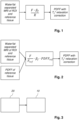
- the PDFF calculation apparatus 10 may receive input from a MRI source 20.
- the MRI source 20 may provide water and fat separated MR data for the ROI and the reference tissue.
- the PDFF calculation apparatus 10 may be a computer configured to perform the calculations according to any of the embodiments above.
- PDFF Proton density fat fraction
- PDFF is the fraction of MRI visible fat protons in relation to the sum of MRI visible fat and water protons. Furthermore, as PDFF is based on the unsaturated MRI signals, the acquisition parameters must be set such that s w « s f , e.g. by choosing a low flip angle. Alternatively, additional images need to be collected to determine the ratio between and s w and s f .
- a signal reference is acquired from regions of pure adipose tissue within the subject and interpolated over the complete image volume (Romu et al., 2011; Dahlqvist Leinhard et al., 2008).
- F ref represent the fat signal of the reference tissue
- R F ref ⁇ PDFF ref ⁇ 1
- eq. 5 describing PDFF in the measurement point can therefore be reformulated as (see figs.
- t w ⁇ describes the crosstalk caused by the water signal as a function of T 2 ,w ⁇ and the echo times T op and T ip .
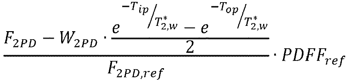
- PDFF 2 PD F 2 PD ⁇ W 2 PD ⁇ t w ⁇ F 2 PD
- ref ⁇ PDFF ref F 2 PD ⁇ 0.5 ⁇ W 2 PD ⁇ e ⁇ T ip T 2 , w * ⁇ e ⁇ T op T 2 , w * F 2 PD , ref ⁇ PDFF ref , where T 2 ,w ⁇ and PDFF ref are the only unknowns.
- the saturation ratio ⁇ w can then be determined based on a separate PDFF experiment such as the fat referenced PDFF 2PD , by minimizing the following expression with respect to ⁇ w , min ⁇ w ⁇ All subjects F F + W ⁇ ⁇ w ⁇ PDFF 2 PD 2 . which minimizes the observed differences between PDFF in the water-referenced acquisition and PDFF 2PD from the fat-referenced T 2 *-corrected 2PD acquisition.
Landscapes
- Physics & Mathematics (AREA)
- High Energy & Nuclear Physics (AREA)
- General Physics & Mathematics (AREA)
- Condensed Matter Physics & Semiconductors (AREA)
- Health & Medical Sciences (AREA)
- Spectroscopy & Molecular Physics (AREA)
- Life Sciences & Earth Sciences (AREA)
- Engineering & Computer Science (AREA)
- Nuclear Medicine, Radiotherapy & Molecular Imaging (AREA)
- General Health & Medical Sciences (AREA)
- Radiology & Medical Imaging (AREA)
- Optics & Photonics (AREA)
- Signal Processing (AREA)
- Biomedical Technology (AREA)
- Pathology (AREA)
- Biophysics (AREA)
- Heart & Thoracic Surgery (AREA)
- Medical Informatics (AREA)
- Molecular Biology (AREA)
- Surgery (AREA)
- Animal Behavior & Ethology (AREA)
- Public Health (AREA)
- Veterinary Medicine (AREA)
- Magnetic Resonance Imaging Apparatus (AREA)
Claims (5)
- Computer-implementiertes Verfahren zum Berechnen einer Protonendichte-Fettfraktion, PDFF, aus getrennter Magnetresonanzbildgebung von Wasser, W, und Fett, F, wobei MRI-Daten unter Verwendung von T1-gewichteter Mehrpunkt-Dixon-Bildgebung auf Basis von auf Fett bezogener Lipid-Quantifizierung in einem Bereich von Interesse (ROI) und unter Verwendung der Bestimmung eines Referenzgewebes erhalten werden, dadurch gekennzeichnet, dass das Verfahren den Schritt des Bestimmens von PDFF als:

wobeiF das Fettsignal in dem ROI ist, erhalten aus den MRI-Daten, und R als ein Anteil zwischen Fref und PDFFref definiert ist, so dass das Verfahren den Schritt des Bestimmens von PDFF als: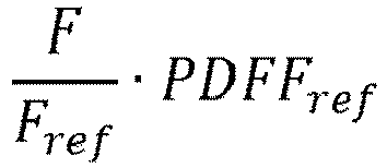 umfasst,wobeiFref das Fettsignal in dem Referenzgewebe ist; wobei die T1-Relaxation des Fettsignals in dem Referenzgewebe und in dem ROI als gleich angenommen wird; undPDFFref die PDFF des Referenzgewebes ist, erhalten durch ein getrenntes Experiment am Referenzgewebe oder durch eine vorbestimmte Konstante.
 - Verfahren nach Anspruch 1, wobei die PDFF aus einer auf Fett bezogenen Zweipunkt-Dixon-Analyse ohne vorherige Korrektur auf T2*-Relaxationseffekte bestimmt wird, und wobei das Wassersignal in dem Referenzgewebe, Wref, niedrig ist, so dass ein resultierender Wert, wenn Wref mit einem resultierenden T2*-Relaxationseffekt multipliziert wird, eine Annäherung geliefert wird, dass das Wassersignal in dem ROI gleich einem beobachteten Wassersignal in dem ROI, W2PD, ist, das eine Rekonstruktion des Wassersignals aus den MRI-Daten in dem ROI unter Verwendung der auf Fett bezogenen Zweipunkt-Dixon-Analyse ist, was ergibt, dass die PDFF berechnet wird als
 F2PD das beobachtete Fettsignal in dem ROI ist, das eine Rekonstruktion des Fettsignals aus den MRI-Daten in dem ROI unter Verwendung der auf Fett bezogenen Zweipunkt-Dixon-Analyse ist;Tip eine Konstante der Echozeit der In-Phase (IP)-Komponente ist, umfassend Wasser plus Fettsignal des Wassers und Fettsignale aus den MRI-Daten in dem ROI;T2*w der T2*-Relaxationseffekt von Wasser in dem ROI ist, der sich aus der auf Fett bezogenen Zweipunkt-Dixon-Analyse ergibt;Top eine Konstante der Echozeit der Out-of-Phase (OP)-Komponente ist, umfassend die Differenz zwischen dem Wasser- und dem Fettsignal aus den MRI-Daten in dem ROI; undF2PD,ref das beobachtete Fettsignal des Referenzgewebes ist, das eine Rekonstruktion des Fettsignals aus den MRI-Daten in dem Referenzgewebe unter Verwendung der auf Fett bezogenen Zweipunkt-Dixon-Analyse ist.
 - Verfahren nach Anspruch 2, wobei der Wert des T2*-Relaxationseffekts in einem getrennten Experiment bestimmt wird.
- Verfahren nach Anspruch 2, wobei der Wert des T2*-Relaxationseffekts als eine Konstante auf Basis eines Populationsmittelwerts festgelegt wird.
- Protonendichte-Fettfraktion, PDFF, Berechnungseinrichtung (10) konfiguriert zum Durchführen einer Berechnung einer PDFF nach einem der Verfahren in den Ansprüchen 1 bis 4.
Priority Applications (9)
| Application Number | Priority Date | Filing Date | Title |
|---|---|---|---|
| EP17181641.6A EP3432018B1 (de) | 2017-07-17 | 2017-07-17 | Mrt-verfahren zur berechnung von t2*-korrigiertem protonendichte-fettanteil |
| ES17181641T ES3013572T3 (en) | 2017-07-17 | 2017-07-17 | Mri method for calculating a t2*-corrected proton density fat fraction |
| US16/631,741 US11486948B2 (en) | 2017-07-17 | 2018-02-23 | MRI method for calculating a proton density fat fraction |
| KR1020207003680A KR102348974B1 (ko) | 2017-07-17 | 2018-02-23 | 양성자 밀도 지방 분율 계산을 위한 mri 방법 |
| PCT/EP2018/054488 WO2019015810A1 (en) | 2017-07-17 | 2018-02-23 | MRI PROCESS FOR CALCULATING A PROTON-DENSITY GREASE FRACTION |
| AU2018304409A AU2018304409B2 (en) | 2017-07-17 | 2018-02-23 | MRI method for calculating a proton density fat fraction |
| CA3069611A CA3069611A1 (en) | 2017-07-17 | 2018-02-23 | Mri method for calculating a proton density fat fraction |
| JP2020502163A JP7047057B2 (ja) | 2017-07-17 | 2018-02-23 | プロトン密度脂肪画分を計算するためのmri方法 |
| CN201880047712.5A CN110998350B (zh) | 2017-07-17 | 2018-02-23 | 用于以mri计算质子密度脂肪分数的方法和装置 |
Applications Claiming Priority (1)
| Application Number | Priority Date | Filing Date | Title |
|---|---|---|---|
| EP17181641.6A EP3432018B1 (de) | 2017-07-17 | 2017-07-17 | Mrt-verfahren zur berechnung von t2*-korrigiertem protonendichte-fettanteil |
Publications (3)
| Publication Number | Publication Date |
|---|---|
| EP3432018A1 EP3432018A1 (de) | 2019-01-23 |
| EP3432018C0 EP3432018C0 (de) | 2024-11-27 |
| EP3432018B1 true EP3432018B1 (de) | 2024-11-27 |
Family
ID=59363018
Family Applications (1)
| Application Number | Title | Priority Date | Filing Date |
|---|---|---|---|
| EP17181641.6A Active EP3432018B1 (de) | 2017-07-17 | 2017-07-17 | Mrt-verfahren zur berechnung von t2*-korrigiertem protonendichte-fettanteil |
Country Status (9)
| Country | Link |
|---|---|
| US (1) | US11486948B2 (de) |
| EP (1) | EP3432018B1 (de) |
| JP (1) | JP7047057B2 (de) |
| KR (1) | KR102348974B1 (de) |
| CN (1) | CN110998350B (de) |
| AU (1) | AU2018304409B2 (de) |
| CA (1) | CA3069611A1 (de) |
| ES (1) | ES3013572T3 (de) |
| WO (1) | WO2019015810A1 (de) |
Cited By (1)
| Publication number | Priority date | Publication date | Assignee | Title |
|---|---|---|---|---|
| US20240366147A1 (en) * | 2021-05-11 | 2024-11-07 | Amra Medical Ab | Method of quantifying an effective volume of a muscle |
Families Citing this family (4)
| Publication number | Priority date | Publication date | Assignee | Title |
|---|---|---|---|---|
| CN110780247B (zh) * | 2019-11-12 | 2021-02-12 | 无锡鸣石峻致医疗科技有限公司 | 一种基于磁共振原理的器官脂肪无创定量检测方法 |
| CN112834969A (zh) * | 2020-12-23 | 2021-05-25 | 江苏珂玛麒生物科技有限公司 | 兔、猴肝脏水/脂肪分离mri成像的方法、计算方法 |
| WO2023108325A1 (zh) * | 2021-12-13 | 2023-06-22 | 中国科学院深圳先进技术研究院 | 脂肪定量成像方法、装置、设备及其存储介质 |
| EP4207061A1 (de) | 2021-12-29 | 2023-07-05 | Shanghai United Imaging Healthcare Co., Ltd. | Systeme und verfahren zur merkmalsinformationsbestimmung |
Family Cites Families (12)
| Publication number | Priority date | Publication date | Assignee | Title |
|---|---|---|---|---|
| JP5508697B2 (ja) * | 2007-10-04 | 2014-06-04 | 株式会社東芝 | Mri装置 |
| JP5701476B2 (ja) | 2008-08-26 | 2015-04-15 | ジーイー・メディカル・システムズ・グローバル・テクノロジー・カンパニー・エルエルシー | 脂肪定量装置、磁気共鳴イメージングシステム、および脂肪定量方法 |
| CN101708123A (zh) * | 2009-10-28 | 2010-05-19 | 上海理工大学 | 肝纤维化分级研究的磁共振弹性成像检测系统及其方法 |
| US8488859B2 (en) | 2009-10-28 | 2013-07-16 | Siemens Aktiengesellschaft | Method for fat fraction quantification in magnetic resonance imaging |
| CN102727201B (zh) * | 2011-04-13 | 2014-02-12 | 深圳迈瑞生物医疗电子股份有限公司 | 磁共振系统及其水脂分离成像方法、装置 |
| US9194925B2 (en) | 2012-11-05 | 2015-11-24 | Siemens Medical Solutions Usa, Inc. | Fat and iron quantification using a multi-step adaptive fitting approach with multi-echo magnetic resonance imaging |
| DE102013215703B3 (de) * | 2013-08-08 | 2015-02-05 | Siemens Aktiengesellschaft | Bestimmung einer T1-Zeit von Wasser und einer T1-Zeit von Fett |
| RU2544387C1 (ru) * | 2013-12-24 | 2015-03-20 | Общество с ограниченной ответственностью "С.П. ГЕЛПИК" | Способ разделения изображений воды и жира в магнитно-резонансной томографии |
| EP2937039B1 (de) * | 2014-04-25 | 2022-04-20 | AMRA Medical AB | Magergewebewasserkonzentrationsquantifizierung |
| CN105796065B (zh) * | 2014-12-29 | 2019-02-15 | 中国科学院深圳先进技术研究院 | 基于水脂分离的无参考温度测量方法及系统 |
| US10162035B2 (en) * | 2015-02-19 | 2018-12-25 | Wisconsin Alumni Research Foundation | System and method for controlling motion effects in magnetic resonance imaging (MRI) images |
| US10743791B2 (en) | 2015-12-28 | 2020-08-18 | Wisconsin Alumni Research Foundation | System and method for assessing tissue properties using magnetic resonance imaging |
-
2017
- 2017-07-17 EP EP17181641.6A patent/EP3432018B1/de active Active
- 2017-07-17 ES ES17181641T patent/ES3013572T3/es active Active
-
2018
- 2018-02-23 AU AU2018304409A patent/AU2018304409B2/en active Active
- 2018-02-23 CN CN201880047712.5A patent/CN110998350B/zh active Active
- 2018-02-23 US US16/631,741 patent/US11486948B2/en active Active
- 2018-02-23 CA CA3069611A patent/CA3069611A1/en active Pending
- 2018-02-23 KR KR1020207003680A patent/KR102348974B1/ko active Active
- 2018-02-23 JP JP2020502163A patent/JP7047057B2/ja active Active
- 2018-02-23 WO PCT/EP2018/054488 patent/WO2019015810A1/en not_active Ceased
Non-Patent Citations (1)
| Title |
|---|
| PERNILLA PETERSON ET AL: "Fat quantification in skeletal muscle using multigradient-echo imaging: Comparison of fat and water references", JOURNAL OF MAGNETIC RESONANCE IMAGING, vol. 43, no. 1, 10 June 2015 (2015-06-10), US, pages 203 - 212, XP055463233, ISSN: 1053-1807, DOI: 10.1002/jmri.24972 * |
Cited By (1)
| Publication number | Priority date | Publication date | Assignee | Title |
|---|---|---|---|---|
| US20240366147A1 (en) * | 2021-05-11 | 2024-11-07 | Amra Medical Ab | Method of quantifying an effective volume of a muscle |
Also Published As
| Publication number | Publication date |
|---|---|
| JP2020527409A (ja) | 2020-09-10 |
| EP3432018A1 (de) | 2019-01-23 |
| AU2018304409B2 (en) | 2023-11-16 |
| EP3432018C0 (de) | 2024-11-27 |
| CA3069611A1 (en) | 2019-01-24 |
| US20200174090A1 (en) | 2020-06-04 |
| KR20200026977A (ko) | 2020-03-11 |
| KR102348974B1 (ko) | 2022-01-07 |
| US11486948B2 (en) | 2022-11-01 |
| AU2018304409A1 (en) | 2020-02-27 |
| CN110998350A (zh) | 2020-04-10 |
| WO2019015810A1 (en) | 2019-01-24 |
| ES3013572T3 (en) | 2025-04-14 |
| CN110998350B (zh) | 2022-11-08 |
| JP7047057B2 (ja) | 2022-04-04 |
Similar Documents
| Publication | Publication Date | Title |
|---|---|---|
| Jaubert et al. | Multi‐parametric liver tissue characterization using MR fingerprinting: Simultaneous T1, T2, T2*, and fat fraction mapping | |
| EP3432018B1 (de) | Mrt-verfahren zur berechnung von t2*-korrigiertem protonendichte-fettanteil | |
| Lv et al. | Noninvasive quantitative detection methods of liver fat content in nonalcoholic fatty liver disease | |
| Sharma et al. | Measurement of liver fat fraction and iron with MRI and MR spectroscopy techniques | |
| Wang et al. | Free‐breathing multitasking multi‐echo MRI for whole‐liver water‐specific T1, proton density fat fraction, and quantification | |
| Kiliçkesmez et al. | Non-breath-hold high b-value diffusion-weighted MRI with parallel imaging technique: apparent diffusion coefficient determination in normal abdominal organs | |
| Leporq et al. | Liver fat volume fraction quantification with fat and water T1 and T2* estimation and accounting for NMR multiple components in patients with chronic liver disease at 1.5 and 3.0 T | |
| US10557906B2 (en) | Quantitative magnetic resonance imaging relaxometry with suppression of blood signal | |
| Satkunasingham et al. | Liver fat quantification: comparison of dual-echo and triple-echo chemical shift MRI to MR spectroscopy | |
| Liu et al. | Comparative analysis of hepatic fat quantification across 5 T, 3 T and 1.5 T: A study on consistency and feasibility | |
| Stelter et al. | Simultaneous whole‐liver water T 1 and T 2 mapping with isotropic resolution during free‐breathing | |
| Tripp et al. | Simultaneous 3D T 1 T _1, T 2 T _2, and fat‐signal‐fraction mapping with respiratory‐motion correction for comprehensive liver tissue characterization at 0.55 T | |
| Muslu et al. | Free‐breathing, fat‐corrected T1 mapping of the liver with stack‐of‐stars MRI, and joint estimation of T1, PDFF, R 2*, and B 1+ | |
| Lee et al. | Advanced MRI techniques in abdominal imaging | |
| Faller et al. | Radial MP2RAGE sequence for rapid 3D T1 mapping of mouse abdomen: application to hepatic metastases | |
| Leitão et al. | MR fat fraction mapping: a simple biomarker for liver steatosis quantification in nonalcoholic fatty liver disease patients | |
| Takahara et al. | Diffusion-weighted magnetic resonance imaging of the liver using tracking only navigator echo: feasibility study | |
| Ishizaka et al. | Comparison of 1H MR spectroscopy, 3-point DIXON, and multi-echo gradient echo for measuring hepatic fat fraction | |
| Shimizu et al. | Hepatic fat quantification using automated six-point Dixon: Comparison with conventional chemical shift based sequences and computed tomography | |
| Belsley et al. | Accurate and precise in vivo liver 3D T1 mapping at 3T | |
| Stabinska et al. | Two point Dixon-based chemical exchange saturation transfer (CEST) MRI in renal transplant patients on 3 T | |
| Tamada et al. | Whole liver phase‐based R2 mapping in liver iron overload within a breath‐hold | |
| Lavdas et al. | How reliable is MRCP with an SS-FSE sequence at 3.0 T: comparison between SS-FSE BH and 3D-FSE BH ASSET sequences | |
| Korinek et al. | Fast triple-spin-echo Dixon (FTSED) sequence for water and fat imaging | |
| Shin et al. | Spectrally selective and interleaved water imaging and fat imaging (siWIFI) |
Legal Events
| Date | Code | Title | Description |
|---|---|---|---|
| PUAI | Public reference made under article 153(3) epc to a published international application that has entered the european phase |
Free format text: ORIGINAL CODE: 0009012 |
|
| STAA | Information on the status of an ep patent application or granted ep patent |
Free format text: STATUS: THE APPLICATION HAS BEEN PUBLISHED |
|
| AK | Designated contracting states |
Kind code of ref document: A1 Designated state(s): AL AT BE BG CH CY CZ DE DK EE ES FI FR GB GR HR HU IE IS IT LI LT LU LV MC MK MT NL NO PL PT RO RS SE SI SK SM TR |
|
| AX | Request for extension of the european patent |
Extension state: BA ME |
|
| STAA | Information on the status of an ep patent application or granted ep patent |
Free format text: STATUS: REQUEST FOR EXAMINATION WAS MADE |
|
| 17P | Request for examination filed |
Effective date: 20190722 |
|
| RBV | Designated contracting states (corrected) |
Designated state(s): AL AT BE BG CH CY CZ DE DK EE ES FI FR GB GR HR HU IE IS IT LI LT LU LV MC MK MT NL NO PL PT RO RS SE SI SK SM TR |
|
| STAA | Information on the status of an ep patent application or granted ep patent |
Free format text: STATUS: EXAMINATION IS IN PROGRESS |
|
| 17Q | First examination report despatched |
Effective date: 20211115 |
|
| GRAP | Despatch of communication of intention to grant a patent |
Free format text: ORIGINAL CODE: EPIDOSNIGR1 |
|
| STAA | Information on the status of an ep patent application or granted ep patent |
Free format text: STATUS: GRANT OF PATENT IS INTENDED |
|
| RIC1 | Information provided on ipc code assigned before grant |
Ipc: G01R 33/56 20060101ALN20240618BHEP Ipc: A61B 5/00 20060101ALI20240618BHEP Ipc: G01R 33/50 20060101ALI20240618BHEP Ipc: G01R 33/485 20060101ALI20240618BHEP Ipc: G01R 33/48 20060101AFI20240618BHEP |
|
| INTG | Intention to grant announced |
Effective date: 20240705 |
|
| GRAS | Grant fee paid |
Free format text: ORIGINAL CODE: EPIDOSNIGR3 |
|
| GRAA | (expected) grant |
Free format text: ORIGINAL CODE: 0009210 |
|
| STAA | Information on the status of an ep patent application or granted ep patent |
Free format text: STATUS: THE PATENT HAS BEEN GRANTED |
|
| AK | Designated contracting states |
Kind code of ref document: B1 Designated state(s): AL AT BE BG CH CY CZ DE DK EE ES FI FR GB GR HR HU IE IS IT LI LT LU LV MC MK MT NL NO PL PT RO RS SE SI SK SM TR |
|
| REG | Reference to a national code |
Ref country code: GB Ref legal event code: FG4D |
|
| REG | Reference to a national code |
Ref country code: CH Ref legal event code: EP |
|
| REG | Reference to a national code |
Ref country code: IE Ref legal event code: FG4D |
|
| REG | Reference to a national code |
Ref country code: DE Ref legal event code: R096 Ref document number: 602017086346 Country of ref document: DE |
|
| U01 | Request for unitary effect filed |
Effective date: 20241211 |
|
| U07 | Unitary effect registered |
Designated state(s): AT BE BG DE DK EE FI FR IT LT LU LV MT NL PT RO SE SI Effective date: 20241220 |
|
| PG25 | Lapsed in a contracting state [announced via postgrant information from national office to epo] |
Ref country code: HR Free format text: LAPSE BECAUSE OF FAILURE TO SUBMIT A TRANSLATION OF THE DESCRIPTION OR TO PAY THE FEE WITHIN THE PRESCRIBED TIME-LIMIT Effective date: 20241127 Ref country code: IS Free format text: LAPSE BECAUSE OF FAILURE TO SUBMIT A TRANSLATION OF THE DESCRIPTION OR TO PAY THE FEE WITHIN THE PRESCRIBED TIME-LIMIT Effective date: 20250327 |
|
| REG | Reference to a national code |
Ref country code: ES Ref legal event code: FG2A Ref document number: 3013572 Country of ref document: ES Kind code of ref document: T3 Effective date: 20250414 |
|
| PG25 | Lapsed in a contracting state [announced via postgrant information from national office to epo] |
Ref country code: NO Free format text: LAPSE BECAUSE OF FAILURE TO SUBMIT A TRANSLATION OF THE DESCRIPTION OR TO PAY THE FEE WITHIN THE PRESCRIBED TIME-LIMIT Effective date: 20250227 |
|
| PG25 | Lapsed in a contracting state [announced via postgrant information from national office to epo] |
Ref country code: GR Free format text: LAPSE BECAUSE OF FAILURE TO SUBMIT A TRANSLATION OF THE DESCRIPTION OR TO PAY THE FEE WITHIN THE PRESCRIBED TIME-LIMIT Effective date: 20250228 |
|
| PG25 | Lapsed in a contracting state [announced via postgrant information from national office to epo] |
Ref country code: PL Free format text: LAPSE BECAUSE OF FAILURE TO SUBMIT A TRANSLATION OF THE DESCRIPTION OR TO PAY THE FEE WITHIN THE PRESCRIBED TIME-LIMIT Effective date: 20241127 |
|
| PG25 | Lapsed in a contracting state [announced via postgrant information from national office to epo] |
Ref country code: RS Free format text: LAPSE BECAUSE OF FAILURE TO SUBMIT A TRANSLATION OF THE DESCRIPTION OR TO PAY THE FEE WITHIN THE PRESCRIBED TIME-LIMIT Effective date: 20250227 |
|
| PG25 | Lapsed in a contracting state [announced via postgrant information from national office to epo] |
Ref country code: SM Free format text: LAPSE BECAUSE OF FAILURE TO SUBMIT A TRANSLATION OF THE DESCRIPTION OR TO PAY THE FEE WITHIN THE PRESCRIBED TIME-LIMIT Effective date: 20241127 |
|
| PGFP | Annual fee paid to national office [announced via postgrant information from national office to epo] |
Ref country code: GB Payment date: 20250613 Year of fee payment: 9 |
|
| U20 | Renewal fee for the european patent with unitary effect paid |
Year of fee payment: 9 Effective date: 20250613 |
|
| PG25 | Lapsed in a contracting state [announced via postgrant information from national office to epo] |
Ref country code: SK Free format text: LAPSE BECAUSE OF FAILURE TO SUBMIT A TRANSLATION OF THE DESCRIPTION OR TO PAY THE FEE WITHIN THE PRESCRIBED TIME-LIMIT Effective date: 20241127 |
|
| PG25 | Lapsed in a contracting state [announced via postgrant information from national office to epo] |
Ref country code: CZ Free format text: LAPSE BECAUSE OF FAILURE TO SUBMIT A TRANSLATION OF THE DESCRIPTION OR TO PAY THE FEE WITHIN THE PRESCRIBED TIME-LIMIT Effective date: 20241127 |
|
| PLBE | No opposition filed within time limit |
Free format text: ORIGINAL CODE: 0009261 |
|
| STAA | Information on the status of an ep patent application or granted ep patent |
Free format text: STATUS: NO OPPOSITION FILED WITHIN TIME LIMIT |
|
| PGFP | Annual fee paid to national office [announced via postgrant information from national office to epo] |
Ref country code: ES Payment date: 20250801 Year of fee payment: 9 |
|
| PGFP | Annual fee paid to national office [announced via postgrant information from national office to epo] |
Ref country code: CH Payment date: 20250801 Year of fee payment: 9 |
|
| 26N | No opposition filed |
Effective date: 20250828 |