EP3380121B1 - Actrii antagonist for use in treating eye disorders - Google Patents
Actrii antagonist for use in treating eye disorders Download PDFInfo
- Publication number
- EP3380121B1 EP3380121B1 EP16869255.6A EP16869255A EP3380121B1 EP 3380121 B1 EP3380121 B1 EP 3380121B1 EP 16869255 A EP16869255 A EP 16869255A EP 3380121 B1 EP3380121 B1 EP 3380121B1
- Authority
- EP
- European Patent Office
- Prior art keywords
- amino acid
- acid sequence
- seq
- actrii
- polypeptide
- Prior art date
- Legal status (The legal status is an assumption and is not a legal conclusion. Google has not performed a legal analysis and makes no representation as to the accuracy of the status listed.)
- Active
Links
- 239000005557 antagonist Substances 0.000 title claims description 210
- 208000030533 eye disease Diseases 0.000 title claims description 5
- 108090000765 processed proteins & peptides Proteins 0.000 claims description 275
- 102000004196 processed proteins & peptides Human genes 0.000 claims description 270
- 229920001184 polypeptide Polymers 0.000 claims description 269
- 239000000488 activin Substances 0.000 claims description 218
- 125000003275 alpha amino acid group Chemical group 0.000 claims description 217
- 108010059616 Activins Proteins 0.000 claims description 206
- 102100026818 Inhibin beta E chain Human genes 0.000 claims description 198
- 108010023082 activin A Proteins 0.000 claims description 103
- 150000001413 amino acids Chemical class 0.000 claims description 101
- 238000000034 method Methods 0.000 claims description 100
- 108010023079 activin B Proteins 0.000 claims description 73
- 108020001507 fusion proteins Proteins 0.000 claims description 44
- 102000037865 fusion proteins Human genes 0.000 claims description 44
- 208000019553 vascular disease Diseases 0.000 claims description 40
- 206010059245 Angiopathy Diseases 0.000 claims description 36
- 239000003112 inhibitor Substances 0.000 claims description 28
- 239000000203 mixture Substances 0.000 claims description 26
- 239000003795 chemical substances by application Substances 0.000 claims description 21
- 201000003793 Myelodysplastic syndrome Diseases 0.000 claims description 17
- 230000002378 acidificating effect Effects 0.000 claims description 17
- 108060003951 Immunoglobulin Proteins 0.000 claims description 15
- 102000018358 immunoglobulin Human genes 0.000 claims description 15
- 238000009120 supportive therapy Methods 0.000 claims description 11
- 108010073929 Vascular Endothelial Growth Factor A Proteins 0.000 claims description 10
- GVJHHUAWPYXKBD-UHFFFAOYSA-N (±)-α-Tocopherol Chemical compound OC1=C(C)C(C)=C2OC(CCCC(C)CCCC(C)CCCC(C)C)(C)CCC2=C1C GVJHHUAWPYXKBD-UHFFFAOYSA-N 0.000 claims description 8
- FWMNVWWHGCHHJJ-SKKKGAJSSA-N 4-amino-1-[(2r)-6-amino-2-[[(2r)-2-[[(2r)-2-[[(2r)-2-amino-3-phenylpropanoyl]amino]-3-phenylpropanoyl]amino]-4-methylpentanoyl]amino]hexanoyl]piperidine-4-carboxylic acid Chemical compound C([C@H](C(=O)N[C@H](CC(C)C)C(=O)N[C@H](CCCCN)C(=O)N1CCC(N)(CC1)C(O)=O)NC(=O)[C@H](N)CC=1C=CC=CC=1)C1=CC=CC=C1 FWMNVWWHGCHHJJ-SKKKGAJSSA-N 0.000 claims description 8
- 102000005789 Vascular Endothelial Growth Factors Human genes 0.000 claims description 8
- 108010019530 Vascular Endothelial Growth Factors Proteins 0.000 claims description 8
- 208000028867 ischemia Diseases 0.000 claims description 8
- 238000001356 surgical procedure Methods 0.000 claims description 7
- 238000002560 therapeutic procedure Methods 0.000 claims description 7
- 101000595923 Homo sapiens Placenta growth factor Proteins 0.000 claims description 6
- 102100035194 Placenta growth factor Human genes 0.000 claims description 6
- KBPHJBAIARWVSC-XQIHNALSSA-N trans-lutein Natural products CC(=C/C=C/C=C(C)/C=C/C=C(C)/C=C/C1=C(C)CC(O)CC1(C)C)C=CC=C(/C)C=CC2C(=CC(O)CC2(C)C)C KBPHJBAIARWVSC-XQIHNALSSA-N 0.000 claims description 6
- 239000013543 active substance Substances 0.000 claims description 5
- JKQXZKUSFCKOGQ-JLGXGRJMSA-N (3R,3'R)-beta,beta-carotene-3,3'-diol Chemical compound C([C@H](O)CC=1C)C(C)(C)C=1/C=C/C(/C)=C/C=C/C(/C)=C/C=C/C=C(C)C=CC=C(C)C=CC1=C(C)C[C@@H](O)CC1(C)C JKQXZKUSFCKOGQ-JLGXGRJMSA-N 0.000 claims description 4
- CIWBSHSKHKDKBQ-JLAZNSOCSA-N Ascorbic acid Chemical compound OC[C@H](O)[C@H]1OC(=O)C(O)=C1O CIWBSHSKHKDKBQ-JLAZNSOCSA-N 0.000 claims description 4
- 102100040898 Growth/differentiation factor 11 Human genes 0.000 claims description 4
- 102100039939 Growth/differentiation factor 8 Human genes 0.000 claims description 4
- 101000893545 Homo sapiens Growth/differentiation factor 11 Proteins 0.000 claims description 4
- 101001001487 Homo sapiens Phosphatidylinositol-glycan biosynthesis class F protein Proteins 0.000 claims description 4
- JUUFBMODXQKSTD-UHFFFAOYSA-N N-[2-amino-6-[(4-fluorophenyl)methylamino]-3-pyridinyl]carbamic acid ethyl ester Chemical compound N1=C(N)C(NC(=O)OCC)=CC=C1NCC1=CC=C(F)C=C1 JUUFBMODXQKSTD-UHFFFAOYSA-N 0.000 claims description 4
- 229930003427 Vitamin E Natural products 0.000 claims description 4
- JKQXZKUSFCKOGQ-LQFQNGICSA-N Z-zeaxanthin Natural products C([C@H](O)CC=1C)C(C)(C)C=1C=CC(C)=CC=CC(C)=CC=CC=C(C)C=CC=C(C)C=CC1=C(C)C[C@@H](O)CC1(C)C JKQXZKUSFCKOGQ-LQFQNGICSA-N 0.000 claims description 4
- QOPRSMDTRDMBNK-RNUUUQFGSA-N Zeaxanthin Natural products CC(=C/C=C/C=C(C)/C=C/C=C(C)/C=C/C1=C(C)CCC(O)C1(C)C)C=CC=C(/C)C=CC2=C(C)CC(O)CC2(C)C QOPRSMDTRDMBNK-RNUUUQFGSA-N 0.000 claims description 4
- HCHKCACWOHOZIP-UHFFFAOYSA-N Zinc Chemical compound [Zn] HCHKCACWOHOZIP-UHFFFAOYSA-N 0.000 claims description 4
- 108010081667 aflibercept Proteins 0.000 claims description 4
- 229960002833 aflibercept Drugs 0.000 claims description 4
- JKQXZKUSFCKOGQ-LOFNIBRQSA-N all-trans-Zeaxanthin Natural products CC(=C/C=C/C=C(C)/C=C/C=C(C)/C=C/C1=C(C)CC(O)CC1(C)C)C=CC=C(/C)C=CC2=C(C)CC(O)CC2(C)C JKQXZKUSFCKOGQ-LOFNIBRQSA-N 0.000 claims description 4
- 229960000397 bevacizumab Drugs 0.000 claims description 4
- FEBLZLNTKCEFIT-VSXGLTOVSA-N fluocinolone acetonide Chemical compound C1([C@@H](F)C2)=CC(=O)C=C[C@]1(C)[C@]1(F)[C@@H]2[C@@H]2C[C@H]3OC(C)(C)O[C@@]3(C(=O)CO)[C@@]2(C)C[C@@H]1O FEBLZLNTKCEFIT-VSXGLTOVSA-N 0.000 claims description 4
- 229960003667 flupirtine Drugs 0.000 claims description 4
- OVBPIULPVIDEAO-LBPRGKRZSA-N folic acid Chemical compound C=1N=C2NC(N)=NC(=O)C2=NC=1CNC1=CC=C(C(=O)N[C@@H](CCC(O)=O)C(O)=O)C=C1 OVBPIULPVIDEAO-LBPRGKRZSA-N 0.000 claims description 4
- WIGCFUFOHFEKBI-UHFFFAOYSA-N gamma-tocopherol Natural products CC(C)CCCC(C)CCCC(C)CCCC1CCC2C(C)C(O)C(C)C(C)C2O1 WIGCFUFOHFEKBI-UHFFFAOYSA-N 0.000 claims description 4
- 230000004377 improving vision Effects 0.000 claims description 4
- 229960003876 ranibizumab Drugs 0.000 claims description 4
- 235000019165 vitamin E Nutrition 0.000 claims description 4
- 229940046009 vitamin E Drugs 0.000 claims description 4
- 239000011709 vitamin E Substances 0.000 claims description 4
- 235000010930 zeaxanthin Nutrition 0.000 claims description 4
- 239000001775 zeaxanthin Substances 0.000 claims description 4
- 229940043269 zeaxanthin Drugs 0.000 claims description 4
- 238000002647 laser therapy Methods 0.000 claims description 3
- 230000000649 photocoagulation Effects 0.000 claims description 3
- GGUSQTSTQSHJAH-UHFFFAOYSA-N 1-(4-chlorophenyl)-2-[4-(4-fluorobenzyl)piperidin-1-yl]ethanol Chemical compound C=1C=C(Cl)C=CC=1C(O)CN(CC1)CCC1CC1=CC=C(F)C=C1 GGUSQTSTQSHJAH-UHFFFAOYSA-N 0.000 claims description 2
- WNWVKZTYMQWFHE-UHFFFAOYSA-N 4-ethylmorpholine Chemical compound [CH2]CN1CCOCC1 WNWVKZTYMQWFHE-UHFFFAOYSA-N 0.000 claims description 2
- BSYNRYMUTXBXSQ-UHFFFAOYSA-N Aspirin Chemical compound CC(=O)OC1=CC=CC=C1C(O)=O BSYNRYMUTXBXSQ-UHFFFAOYSA-N 0.000 claims description 2
- 239000005552 B01AC04 - Clopidogrel Substances 0.000 claims description 2
- 239000005528 B01AC05 - Ticlopidine Substances 0.000 claims description 2
- 108010053835 Catalase Proteins 0.000 claims description 2
- CMSMOCZEIVJLDB-UHFFFAOYSA-N Cyclophosphamide Chemical compound ClCCN(CCCl)P1(=O)NCCCO1 CMSMOCZEIVJLDB-UHFFFAOYSA-N 0.000 claims description 2
- PMATZTZNYRCHOR-CGLBZJNRSA-N Cyclosporin A Chemical compound CC[C@@H]1NC(=O)[C@H]([C@H](O)[C@H](C)C\C=C\C)N(C)C(=O)[C@H](C(C)C)N(C)C(=O)[C@H](CC(C)C)N(C)C(=O)[C@H](CC(C)C)N(C)C(=O)[C@@H](C)NC(=O)[C@H](C)NC(=O)[C@H](CC(C)C)N(C)C(=O)[C@H](C(C)C)NC(=O)[C@H](CC(C)C)N(C)C(=O)CN(C)C1=O PMATZTZNYRCHOR-CGLBZJNRSA-N 0.000 claims description 2
- 108010036949 Cyclosporine Proteins 0.000 claims description 2
- FBPFZTCFMRRESA-KVTDHHQDSA-N D-Mannitol Chemical compound OC[C@@H](O)[C@@H](O)[C@H](O)[C@H](O)CO FBPFZTCFMRRESA-KVTDHHQDSA-N 0.000 claims description 2
- ZZZCUOFIHGPKAK-UHFFFAOYSA-N D-erythro-ascorbic acid Natural products OCC1OC(=O)C(O)=C1O ZZZCUOFIHGPKAK-UHFFFAOYSA-N 0.000 claims description 2
- 108010008165 Etanercept Proteins 0.000 claims description 2
- HTTJABKRGRZYRN-UHFFFAOYSA-N Heparin Chemical compound OC1C(NC(=O)C)C(O)OC(COS(O)(=O)=O)C1OC1C(OS(O)(=O)=O)C(O)C(OC2C(C(OS(O)(=O)=O)C(OC3C(C(O)C(O)C(O3)C(O)=O)OS(O)(=O)=O)C(CO)O2)NS(O)(=O)=O)C(C(O)=O)O1 HTTJABKRGRZYRN-UHFFFAOYSA-N 0.000 claims description 2
- MKXZASYAUGDDCJ-SZMVWBNQSA-N LSM-2525 Chemical compound C1CCC[C@H]2[C@@]3([H])N(C)CC[C@]21C1=CC(OC)=CC=C1C3 MKXZASYAUGDDCJ-SZMVWBNQSA-N 0.000 claims description 2
- 229930195725 Mannitol Natural products 0.000 claims description 2
- VLCDUOXHFNUCKK-UHFFFAOYSA-N N,N'-Dimethylthiourea Chemical compound CNC(=S)NC VLCDUOXHFNUCKK-UHFFFAOYSA-N 0.000 claims description 2
- OVBPIULPVIDEAO-UHFFFAOYSA-N N-Pteroyl-L-glutaminsaeure Natural products C=1N=C2NC(N)=NC(=O)C2=NC=1CNC1=CC=C(C(=O)NC(CCC(O)=O)C(O)=O)C=C1 OVBPIULPVIDEAO-UHFFFAOYSA-N 0.000 claims description 2
- UBQYURCVBFRUQT-UHFFFAOYSA-N N-benzoyl-Ferrioxamine B Chemical compound CC(=O)N(O)CCCCCNC(=O)CCC(=O)N(O)CCCCCNC(=O)CCC(=O)N(O)CCCCCN UBQYURCVBFRUQT-UHFFFAOYSA-N 0.000 claims description 2
- 102000019197 Superoxide Dismutase Human genes 0.000 claims description 2
- 108010012715 Superoxide dismutase Proteins 0.000 claims description 2
- KJADKKWYZYXHBB-XBWDGYHZSA-N Topiramic acid Chemical compound C1O[C@@]2(COS(N)(=O)=O)OC(C)(C)O[C@H]2[C@@H]2OC(C)(C)O[C@@H]21 KJADKKWYZYXHBB-XBWDGYHZSA-N 0.000 claims description 2
- UHWVSEOVJBQKBE-UHFFFAOYSA-N Trimetazidine Chemical compound COC1=C(OC)C(OC)=CC=C1CN1CCNCC1 UHWVSEOVJBQKBE-UHFFFAOYSA-N 0.000 claims description 2
- 229930003779 Vitamin B12 Natural products 0.000 claims description 2
- 229930003268 Vitamin C Natural products 0.000 claims description 2
- 229960001138 acetylsalicylic acid Drugs 0.000 claims description 2
- 229960003459 allopurinol Drugs 0.000 claims description 2
- OFCNXPDARWKPPY-UHFFFAOYSA-N allopurinol Chemical compound OC1=NC=NC2=C1C=NN2 OFCNXPDARWKPPY-UHFFFAOYSA-N 0.000 claims description 2
- 238000011122 anti-angiogenic therapy Methods 0.000 claims description 2
- 229940121363 anti-inflammatory agent Drugs 0.000 claims description 2
- 239000002260 anti-inflammatory agent Substances 0.000 claims description 2
- 239000003146 anticoagulant agent Substances 0.000 claims description 2
- 229940127219 anticoagulant drug Drugs 0.000 claims description 2
- 239000003963 antioxidant agent Substances 0.000 claims description 2
- 235000006708 antioxidants Nutrition 0.000 claims description 2
- 238000013176 antiplatelet therapy Methods 0.000 claims description 2
- LMEKQMALGUDUQG-UHFFFAOYSA-N azathioprine Chemical compound CN1C=NC([N+]([O-])=O)=C1SC1=NC=NC2=C1NC=N2 LMEKQMALGUDUQG-UHFFFAOYSA-N 0.000 claims description 2
- 229960002170 azathioprine Drugs 0.000 claims description 2
- 229960005438 calcium dobesilate Drugs 0.000 claims description 2
- QGNBTYAQAPLTMX-UHFFFAOYSA-L calcium dobesilate Chemical compound [Ca+2].OC1=CC=C(O)C(S([O-])(=O)=O)=C1.OC1=CC=C(O)C(S([O-])(=O)=O)=C1 QGNBTYAQAPLTMX-UHFFFAOYSA-L 0.000 claims description 2
- 229940105657 catalase Drugs 0.000 claims description 2
- 229960001265 ciclosporin Drugs 0.000 claims description 2
- GKTWGGQPFAXNFI-HNNXBMFYSA-N clopidogrel Chemical compound C1([C@H](N2CC=3C=CSC=3CC2)C(=O)OC)=CC=CC=C1Cl GKTWGGQPFAXNFI-HNNXBMFYSA-N 0.000 claims description 2
- 229960003009 clopidogrel Drugs 0.000 claims description 2
- FDJOLVPMNUYSCM-WZHZPDAFSA-L cobalt(3+);[(2r,3s,4r,5s)-5-(5,6-dimethylbenzimidazol-1-yl)-4-hydroxy-2-(hydroxymethyl)oxolan-3-yl] [(2r)-1-[3-[(1r,2r,3r,4z,7s,9z,12s,13s,14z,17s,18s,19r)-2,13,18-tris(2-amino-2-oxoethyl)-7,12,17-tris(3-amino-3-oxopropyl)-3,5,8,8,13,15,18,19-octamethyl-2 Chemical compound [Co+3].N#[C-].N([C@@H]([C@]1(C)[N-]\C([C@H]([C@@]1(CC(N)=O)C)CCC(N)=O)=C(\C)/C1=N/C([C@H]([C@@]1(CC(N)=O)C)CCC(N)=O)=C\C1=N\C([C@H](C1(C)C)CCC(N)=O)=C/1C)[C@@H]2CC(N)=O)=C\1[C@]2(C)CCC(=O)NC[C@@H](C)OP([O-])(=O)O[C@H]1[C@@H](O)[C@@H](N2C3=CC(C)=C(C)C=C3N=C2)O[C@@H]1CO FDJOLVPMNUYSCM-WZHZPDAFSA-L 0.000 claims description 2
- 239000003246 corticosteroid Substances 0.000 claims description 2
- 229960001334 corticosteroids Drugs 0.000 claims description 2
- 238000000315 cryotherapy Methods 0.000 claims description 2
- 229960004397 cyclophosphamide Drugs 0.000 claims description 2
- 229930182912 cyclosporin Natural products 0.000 claims description 2
- 229960000958 deferoxamine Drugs 0.000 claims description 2
- 229960003957 dexamethasone Drugs 0.000 claims description 2
- UREBDLICKHMUKA-CXSFZGCWSA-N dexamethasone Chemical compound C1CC2=CC(=O)C=C[C@]2(C)[C@]2(F)[C@@H]1[C@@H]1C[C@@H](C)[C@@](C(=O)CO)(O)[C@@]1(C)C[C@@H]2O UREBDLICKHMUKA-CXSFZGCWSA-N 0.000 claims description 2
- 229960001985 dextromethorphan Drugs 0.000 claims description 2
- 235000015872 dietary supplement Nutrition 0.000 claims description 2
- QLTXKCWMEZIHBJ-PJGJYSAQSA-N dizocilpine maleate Chemical compound OC(=O)\C=C/C(O)=O.C12=CC=CC=C2[C@]2(C)C3=CC=CC=C3C[C@H]1N2 QLTXKCWMEZIHBJ-PJGJYSAQSA-N 0.000 claims description 2
- 229950005455 eliprodil Drugs 0.000 claims description 2
- 229960000403 etanercept Drugs 0.000 claims description 2
- 238000001914 filtration Methods 0.000 claims description 2
- SMANXXCATUTDDT-QPJJXVBHSA-N flunarizine Chemical compound C1=CC(F)=CC=C1C(C=1C=CC(F)=CC=1)N1CCN(C\C=C\C=2C=CC=CC=2)CC1 SMANXXCATUTDDT-QPJJXVBHSA-N 0.000 claims description 2
- 229960000326 flunarizine Drugs 0.000 claims description 2
- 229960001347 fluocinolone acetonide Drugs 0.000 claims description 2
- 235000019152 folic acid Nutrition 0.000 claims description 2
- 239000011724 folic acid Substances 0.000 claims description 2
- 229960000304 folic acid Drugs 0.000 claims description 2
- 229960002897 heparin Drugs 0.000 claims description 2
- 229920000669 heparin Polymers 0.000 claims description 2
- 229940029200 iluvien Drugs 0.000 claims description 2
- 229960003444 immunosuppressant agent Drugs 0.000 claims description 2
- 239000003018 immunosuppressive agent Substances 0.000 claims description 2
- 238000002513 implantation Methods 0.000 claims description 2
- 229960000598 infliximab Drugs 0.000 claims description 2
- 235000012680 lutein Nutrition 0.000 claims description 2
- 229960005375 lutein Drugs 0.000 claims description 2
- 239000001656 lutein Substances 0.000 claims description 2
- KBPHJBAIARWVSC-RGZFRNHPSA-N lutein Chemical compound C([C@H](O)CC=1C)C(C)(C)C=1\C=C\C(\C)=C\C=C\C(\C)=C\C=C\C=C(/C)\C=C\C=C(/C)\C=C\[C@H]1C(C)=C[C@H](O)CC1(C)C KBPHJBAIARWVSC-RGZFRNHPSA-N 0.000 claims description 2
- ORAKUVXRZWMARG-WZLJTJAWSA-N lutein Natural products CC(=C/C=C/C=C(C)/C=C/C=C(C)/C=C/C1=C(C)CCCC1(C)C)C=CC=C(/C)C=CC2C(=CC(O)CC2(C)C)C ORAKUVXRZWMARG-WZLJTJAWSA-N 0.000 claims description 2
- 239000000594 mannitol Substances 0.000 claims description 2
- 235000010355 mannitol Nutrition 0.000 claims description 2
- 229960001855 mannitol Drugs 0.000 claims description 2
- HPNSFSBZBAHARI-UHFFFAOYSA-N micophenolic acid Natural products OC1=C(CC=C(C)CCC(O)=O)C(OC)=C(C)C2=C1C(=O)OC2 HPNSFSBZBAHARI-UHFFFAOYSA-N 0.000 claims description 2
- 229950007856 mofetil Drugs 0.000 claims description 2
- 229940014456 mycophenolate Drugs 0.000 claims description 2
- HPNSFSBZBAHARI-RUDMXATFSA-N mycophenolic acid Chemical compound OC1=C(C\C=C(/C)CCC(O)=O)C(OC)=C(C)C2=C1C(=O)OC2 HPNSFSBZBAHARI-RUDMXATFSA-N 0.000 claims description 2
- HYIMSNHJOBLJNT-UHFFFAOYSA-N nifedipine Chemical compound COC(=O)C1=C(C)NC(C)=C(C(=O)OC)C1C1=CC=CC=C1[N+]([O-])=O HYIMSNHJOBLJNT-UHFFFAOYSA-N 0.000 claims description 2
- 229960001597 nifedipine Drugs 0.000 claims description 2
- 238000006213 oxygenation reaction Methods 0.000 claims description 2
- XOFYZVNMUHMLCC-ZPOLXVRWSA-N prednisone Chemical compound O=C1C=C[C@]2(C)[C@H]3C(=O)C[C@](C)([C@@](CC4)(O)C(=O)CO)[C@@H]4[C@@H]3CCC2=C1 XOFYZVNMUHMLCC-ZPOLXVRWSA-N 0.000 claims description 2
- 229960004618 prednisone Drugs 0.000 claims description 2
- 150000003431 steroids Chemical class 0.000 claims description 2
- 230000009885 systemic effect Effects 0.000 claims description 2
- 229960002663 thioctic acid Drugs 0.000 claims description 2
- PHWBOXQYWZNQIN-UHFFFAOYSA-N ticlopidine Chemical compound ClC1=CC=CC=C1CN1CC(C=CS2)=C2CC1 PHWBOXQYWZNQIN-UHFFFAOYSA-N 0.000 claims description 2
- 229960005001 ticlopidine Drugs 0.000 claims description 2
- 229960004394 topiramate Drugs 0.000 claims description 2
- GFNANZIMVAIWHM-OBYCQNJPSA-N triamcinolone Chemical compound O=C1C=C[C@]2(C)[C@@]3(F)[C@@H](O)C[C@](C)([C@@]([C@H](O)C4)(O)C(=O)CO)[C@@H]4[C@@H]3CCC2=C1 GFNANZIMVAIWHM-OBYCQNJPSA-N 0.000 claims description 2
- 229960002117 triamcinolone acetonide Drugs 0.000 claims description 2
- 229960001177 trimetazidine Drugs 0.000 claims description 2
- 239000002525 vasculotropin inhibitor Substances 0.000 claims description 2
- 235000019163 vitamin B12 Nutrition 0.000 claims description 2
- 239000011715 vitamin B12 Substances 0.000 claims description 2
- 235000019158 vitamin B6 Nutrition 0.000 claims description 2
- 239000011726 vitamin B6 Substances 0.000 claims description 2
- 235000019154 vitamin C Nutrition 0.000 claims description 2
- 239000011718 vitamin C Substances 0.000 claims description 2
- PJVWKTKQMONHTI-UHFFFAOYSA-N warfarin Chemical compound OC=1C2=CC=CC=C2OC(=O)C=1C(CC(=O)C)C1=CC=CC=C1 PJVWKTKQMONHTI-UHFFFAOYSA-N 0.000 claims description 2
- 229960005080 warfarin Drugs 0.000 claims description 2
- FJHBOVDFOQMZRV-XQIHNALSSA-N xanthophyll Natural products CC(=C/C=C/C=C(C)/C=C/C=C(C)/C=C/C1=C(C)CC(O)CC1(C)C)C=CC=C(/C)C=CC2C=C(C)C(O)CC2(C)C FJHBOVDFOQMZRV-XQIHNALSSA-N 0.000 claims description 2
- 239000011701 zinc Substances 0.000 claims description 2
- 229910052725 zinc Inorganic materials 0.000 claims description 2
- 235000016804 zinc Nutrition 0.000 claims description 2
- 102000016938 Catalase Human genes 0.000 claims 1
- 101000886562 Homo sapiens Growth/differentiation factor 8 Proteins 0.000 claims 1
- 102100039037 Vascular endothelial growth factor A Human genes 0.000 claims 1
- MWUXSHHQAYIFBG-UHFFFAOYSA-N Nitric oxide Chemical compound O=[N] MWUXSHHQAYIFBG-UHFFFAOYSA-N 0.000 description 352
- 230000027455 binding Effects 0.000 description 219
- 230000000694 effects Effects 0.000 description 165
- 102000005962 receptors Human genes 0.000 description 137
- 108020003175 receptors Proteins 0.000 description 137
- 108010057453 activin receptor type II-B Proteins 0.000 description 132
- 235000001014 amino acid Nutrition 0.000 description 112
- 229940024606 amino acid Drugs 0.000 description 99
- 108090000623 proteins and genes Proteins 0.000 description 99
- 239000003446 ligand Substances 0.000 description 88
- 102100027647 Activin receptor type-2B Human genes 0.000 description 86
- 102100028726 Bone morphogenetic protein 10 Human genes 0.000 description 84
- 101000695367 Homo sapiens Bone morphogenetic protein 10 Proteins 0.000 description 84
- 150000003384 small molecules Chemical class 0.000 description 84
- 102100022525 Bone morphogenetic protein 6 Human genes 0.000 description 83
- 101000899390 Homo sapiens Bone morphogenetic protein 6 Proteins 0.000 description 83
- 108010057429 activin receptor type II-A Proteins 0.000 description 82
- 230000014509 gene expression Effects 0.000 description 75
- 241000282414 Homo sapiens Species 0.000 description 72
- 102100021886 Activin receptor type-2A Human genes 0.000 description 67
- 102100040892 Growth/differentiation factor 2 Human genes 0.000 description 66
- 101000893585 Homo sapiens Growth/differentiation factor 2 Proteins 0.000 description 66
- 102100035364 Growth/differentiation factor 3 Human genes 0.000 description 64
- 101001023986 Homo sapiens Growth/differentiation factor 3 Proteins 0.000 description 64
- 102000040430 polynucleotide Human genes 0.000 description 62
- 102000004169 proteins and genes Human genes 0.000 description 62
- 108091033319 polynucleotide Proteins 0.000 description 61
- 239000002157 polynucleotide Substances 0.000 description 61
- 235000018102 proteins Nutrition 0.000 description 59
- 210000004027 cell Anatomy 0.000 description 54
- 210000001508 eye Anatomy 0.000 description 46
- 150000007523 nucleic acids Chemical class 0.000 description 45
- 102000039446 nucleic acids Human genes 0.000 description 37
- 108020004707 nucleic acids Proteins 0.000 description 37
- 208000004644 retinal vein occlusion Diseases 0.000 description 35
- 239000002773 nucleotide Substances 0.000 description 33
- 125000003729 nucleotide group Chemical group 0.000 description 33
- 230000003213 activating effect Effects 0.000 description 31
- 201000007527 Retinal artery occlusion Diseases 0.000 description 30
- 230000004913 activation Effects 0.000 description 30
- 239000000427 antigen Substances 0.000 description 30
- 238000006467 substitution reaction Methods 0.000 description 29
- 108091007433 antigens Proteins 0.000 description 28
- 102000036639 antigens Human genes 0.000 description 28
- -1 for example Proteins 0.000 description 27
- 230000035772 mutation Effects 0.000 description 27
- 230000000875 corresponding effect Effects 0.000 description 25
- 230000011664 signaling Effects 0.000 description 25
- 102000004887 Transforming Growth Factor beta Human genes 0.000 description 21
- 108090001012 Transforming Growth Factor beta Proteins 0.000 description 21
- ZRKFYGHZFMAOKI-QMGMOQQFSA-N tgfbeta Chemical compound C([C@H](NC(=O)[C@H](C(C)C)NC(=O)CNC(=O)[C@H](CCC(O)=O)NC(=O)[C@H](CCCNC(N)=N)NC(=O)[C@H](CC(N)=O)NC(=O)[C@H](CC(C)C)NC(=O)[C@H]([C@@H](C)O)NC(=O)[C@H](CCC(O)=O)NC(=O)[C@H]([C@@H](C)O)NC(=O)[C@H](CC(C)C)NC(=O)CNC(=O)[C@H](C)NC(=O)[C@H](CO)NC(=O)[C@H](CCC(N)=O)NC(=O)[C@@H](NC(=O)[C@H](C)NC(=O)[C@H](C)NC(=O)[C@@H](NC(=O)[C@H](CC(C)C)NC(=O)[C@@H](N)CCSC)C(C)C)[C@@H](C)CC)C(=O)N[C@@H]([C@@H](C)O)C(=O)N[C@@H](C(C)C)C(=O)N[C@@H](CC=1C=CC=CC=1)C(=O)N[C@@H](C)C(=O)N1[C@@H](CCC1)C(=O)N[C@@H]([C@@H](C)O)C(=O)N[C@@H](CC(N)=O)C(=O)N[C@@H](CCC(O)=O)C(=O)N[C@@H](C)C(=O)N[C@@H](CC=1C=CC=CC=1)C(=O)N[C@@H](CCCNC(N)=N)C(=O)N[C@@H](C)C(=O)N[C@@H](CC(C)C)C(=O)N1[C@@H](CCC1)C(=O)N1[C@@H](CCC1)C(=O)N[C@@H](CCCNC(N)=N)C(=O)N[C@@H](CCC(O)=O)C(=O)N[C@@H](CCCNC(N)=N)C(=O)N[C@@H](CO)C(=O)N[C@@H](CCCNC(N)=N)C(=O)N[C@@H](CC(C)C)C(=O)N[C@@H](CC(C)C)C(O)=O)C1=CC=C(O)C=C1 ZRKFYGHZFMAOKI-QMGMOQQFSA-N 0.000 description 21
- 230000004438 eyesight Effects 0.000 description 20
- 239000012634 fragment Substances 0.000 description 20
- 230000004927 fusion Effects 0.000 description 19
- 239000013598 vector Substances 0.000 description 19
- 208000010038 Ischemic Optic Neuropathy Diseases 0.000 description 18
- 206010064930 age-related macular degeneration Diseases 0.000 description 18
- 230000001404 mediated effect Effects 0.000 description 18
- 208000032253 retinal ischemia Diseases 0.000 description 18
- 238000003127 radioimmunoassay Methods 0.000 description 17
- 238000003556 assay Methods 0.000 description 16
- 230000001965 increasing effect Effects 0.000 description 15
- 230000004048 modification Effects 0.000 description 15
- 238000012986 modification Methods 0.000 description 15
- 241000894007 species Species 0.000 description 15
- 108091028043 Nucleic acid sequence Proteins 0.000 description 14
- 125000000539 amino acid group Chemical group 0.000 description 14
- 208000002780 macular degeneration Diseases 0.000 description 14
- 101000970954 Homo sapiens Activin receptor type-2A Proteins 0.000 description 13
- 230000004075 alteration Effects 0.000 description 13
- 230000013595 glycosylation Effects 0.000 description 13
- 238000006206 glycosylation reaction Methods 0.000 description 13
- 230000000302 ischemic effect Effects 0.000 description 13
- 239000002243 precursor Substances 0.000 description 13
- CJLHTKGWEUGORV-UHFFFAOYSA-N Artemin Chemical compound C1CC2(C)C(O)CCC(=C)C2(O)C2C1C(C)C(=O)O2 CJLHTKGWEUGORV-UHFFFAOYSA-N 0.000 description 12
- 102100024506 Bone morphogenetic protein 2 Human genes 0.000 description 12
- 102100024505 Bone morphogenetic protein 4 Human genes 0.000 description 12
- 102220479051 CD59 glycoprotein_L79D_mutation Human genes 0.000 description 12
- 102000034615 Glial cell line-derived neurotrophic factor Human genes 0.000 description 12
- 108091010837 Glial cell line-derived neurotrophic factor Proteins 0.000 description 12
- 102100040896 Growth/differentiation factor 15 Human genes 0.000 description 12
- 102100035368 Growth/differentiation factor 6 Human genes 0.000 description 12
- 101000762366 Homo sapiens Bone morphogenetic protein 2 Proteins 0.000 description 12
- 101000762379 Homo sapiens Bone morphogenetic protein 4 Proteins 0.000 description 12
- 101000893549 Homo sapiens Growth/differentiation factor 15 Proteins 0.000 description 12
- 101001023964 Homo sapiens Growth/differentiation factor 6 Proteins 0.000 description 12
- 206010030924 Optic ischaemic neuropathy Diseases 0.000 description 12
- 208000027073 Stargardt disease Diseases 0.000 description 12
- 201000007058 anterior ischemic optic neuropathy Diseases 0.000 description 12
- 230000000692 anti-sense effect Effects 0.000 description 12
- 230000000295 complement effect Effects 0.000 description 12
- 235000018417 cysteine Nutrition 0.000 description 12
- 210000004408 hybridoma Anatomy 0.000 description 12
- 108020001756 ligand binding domains Proteins 0.000 description 12
- 101000937269 Homo sapiens Activin receptor type-2B Proteins 0.000 description 11
- 238000012217 deletion Methods 0.000 description 11
- 230000037430 deletion Effects 0.000 description 11
- 239000003814 drug Substances 0.000 description 11
- 102000045412 human ACVR2B Human genes 0.000 description 11
- 238000001727 in vivo Methods 0.000 description 11
- 238000004519 manufacturing process Methods 0.000 description 11
- 231100000350 mutagenesis Toxicity 0.000 description 11
- 238000000746 purification Methods 0.000 description 11
- 238000012216 screening Methods 0.000 description 11
- 230000004304 visual acuity Effects 0.000 description 11
- 241001465754 Metazoa Species 0.000 description 10
- 210000004369 blood Anatomy 0.000 description 10
- 239000008280 blood Substances 0.000 description 10
- XUJNEKJLAYXESH-UHFFFAOYSA-N cysteine Natural products SCC(N)C(O)=O XUJNEKJLAYXESH-UHFFFAOYSA-N 0.000 description 10
- 210000003743 erythrocyte Anatomy 0.000 description 10
- 230000006870 function Effects 0.000 description 10
- 238000002703 mutagenesis Methods 0.000 description 10
- 239000013612 plasmid Substances 0.000 description 10
- 230000001105 regulatory effect Effects 0.000 description 10
- 102000003978 Tissue Plasminogen Activator Human genes 0.000 description 9
- 108090000373 Tissue Plasminogen Activator Proteins 0.000 description 9
- 241000251539 Vertebrata <Metazoa> Species 0.000 description 9
- 230000004071 biological effect Effects 0.000 description 9
- 208000037265 diseases, disorders, signs and symptoms Diseases 0.000 description 9
- 238000005516 engineering process Methods 0.000 description 9
- 230000005764 inhibitory process Effects 0.000 description 9
- 229960000187 tissue plasminogen activator Drugs 0.000 description 9
- 230000000007 visual effect Effects 0.000 description 9
- UBWXUGDQUBIEIZ-UHFFFAOYSA-N (13-methyl-3-oxo-2,6,7,8,9,10,11,12,14,15,16,17-dodecahydro-1h-cyclopenta[a]phenanthren-17-yl) 3-phenylpropanoate Chemical compound CC12CCC(C3CCC(=O)C=C3CC3)C3C1CCC2OC(=O)CCC1=CC=CC=C1 UBWXUGDQUBIEIZ-UHFFFAOYSA-N 0.000 description 8
- 102000005606 Activins Human genes 0.000 description 8
- 102100022544 Bone morphogenetic protein 7 Human genes 0.000 description 8
- 108020004414 DNA Proteins 0.000 description 8
- 206010012689 Diabetic retinopathy Diseases 0.000 description 8
- 101000899361 Homo sapiens Bone morphogenetic protein 7 Proteins 0.000 description 8
- 108010021625 Immunoglobulin Fragments Proteins 0.000 description 8
- 102000008394 Immunoglobulin Fragments Human genes 0.000 description 8
- WHUUTDBJXJRKMK-VKHMYHEASA-N L-glutamic acid Chemical compound OC(=O)[C@@H](N)CCC(O)=O WHUUTDBJXJRKMK-VKHMYHEASA-N 0.000 description 8
- 241000699670 Mus sp. Species 0.000 description 8
- 210000004204 blood vessel Anatomy 0.000 description 8
- 239000000032 diagnostic agent Substances 0.000 description 8
- 229940039227 diagnostic agent Drugs 0.000 description 8
- 210000003527 eukaryotic cell Anatomy 0.000 description 8
- 239000013604 expression vector Substances 0.000 description 8
- 230000002829 reductive effect Effects 0.000 description 8
- 230000019491 signal transduction Effects 0.000 description 8
- 230000008685 targeting Effects 0.000 description 8
- 229940124597 therapeutic agent Drugs 0.000 description 8
- 102000004190 Enzymes Human genes 0.000 description 7
- 108090000790 Enzymes Proteins 0.000 description 7
- 230000004988 N-glycosylation Effects 0.000 description 7
- 239000004473 Threonine Substances 0.000 description 7
- 241000269370 Xenopus <genus> Species 0.000 description 7
- 238000007792 addition Methods 0.000 description 7
- 230000010056 antibody-dependent cellular cytotoxicity Effects 0.000 description 7
- 230000015572 biosynthetic process Effects 0.000 description 7
- 201000010099 disease Diseases 0.000 description 7
- 229940088598 enzyme Drugs 0.000 description 7
- 230000002401 inhibitory effect Effects 0.000 description 7
- 125000005647 linker group Chemical group 0.000 description 7
- 238000013519 translation Methods 0.000 description 7
- MTCFGRXMJLQNBG-REOHCLBHSA-N (2S)-2-Amino-3-hydroxypropansäure Chemical compound OC[C@H](N)C(O)=O MTCFGRXMJLQNBG-REOHCLBHSA-N 0.000 description 6
- 108091032973 (ribonucleotides)n+m Proteins 0.000 description 6
- 102100026376 Artemin Human genes 0.000 description 6
- 101710205806 Artemin Proteins 0.000 description 6
- 201000004569 Blindness Diseases 0.000 description 6
- 102100024504 Bone morphogenetic protein 3 Human genes 0.000 description 6
- 102100022526 Bone morphogenetic protein 5 Human genes 0.000 description 6
- 102000016970 Follistatin Human genes 0.000 description 6
- 108010014612 Follistatin Proteins 0.000 description 6
- DHMQDGOQFOQNFH-UHFFFAOYSA-N Glycine Chemical compound NCC(O)=O DHMQDGOQFOQNFH-UHFFFAOYSA-N 0.000 description 6
- 102100035379 Growth/differentiation factor 5 Human genes 0.000 description 6
- 102100035363 Growth/differentiation factor 7 Human genes 0.000 description 6
- 101000762375 Homo sapiens Bone morphogenetic protein 3 Proteins 0.000 description 6
- 101000899388 Homo sapiens Bone morphogenetic protein 5 Proteins 0.000 description 6
- 101001023988 Homo sapiens Growth/differentiation factor 5 Proteins 0.000 description 6
- 101001023968 Homo sapiens Growth/differentiation factor 7 Proteins 0.000 description 6
- 101000692878 Homo sapiens Regulator of MON1-CCZ1 complex Proteins 0.000 description 6
- AYFVYJQAPQTCCC-GBXIJSLDSA-N L-threonine Chemical compound C[C@@H](O)[C@H](N)C(O)=O AYFVYJQAPQTCCC-GBXIJSLDSA-N 0.000 description 6
- 208000001344 Macular Edema Diseases 0.000 description 6
- 206010025412 Macular dystrophy congenital Diseases 0.000 description 6
- 206010025415 Macular oedema Diseases 0.000 description 6
- 102100030173 Muellerian-inhibiting factor Human genes 0.000 description 6
- 101710122877 Muellerian-inhibiting factor Proteins 0.000 description 6
- 101100288960 Mus musculus Lefty1 gene Proteins 0.000 description 6
- 102100021584 Neurturin Human genes 0.000 description 6
- 108010015406 Neurturin Proteins 0.000 description 6
- 206010069385 Ocular ischaemic syndrome Diseases 0.000 description 6
- 102100036660 Persephin Human genes 0.000 description 6
- 108010076504 Protein Sorting Signals Proteins 0.000 description 6
- 201000001949 Retinal Vasculitis Diseases 0.000 description 6
- 206010038933 Retinopathy of prematurity Diseases 0.000 description 6
- MTCFGRXMJLQNBG-UHFFFAOYSA-N Serine Natural products OCC(N)C(O)=O MTCFGRXMJLQNBG-UHFFFAOYSA-N 0.000 description 6
- 206010043189 Telangiectasia Diseases 0.000 description 6
- AYFVYJQAPQTCCC-UHFFFAOYSA-N Threonine Natural products CC(O)C(N)C(O)=O AYFVYJQAPQTCCC-UHFFFAOYSA-N 0.000 description 6
- 102000046299 Transforming Growth Factor beta1 Human genes 0.000 description 6
- 102000011117 Transforming Growth Factor beta2 Human genes 0.000 description 6
- 101800002279 Transforming growth factor beta-1 Proteins 0.000 description 6
- 101800000304 Transforming growth factor beta-2 Proteins 0.000 description 6
- 102000056172 Transforming growth factor beta-3 Human genes 0.000 description 6
- 108090000097 Transforming growth factor beta-3 Proteins 0.000 description 6
- 208000000208 Wet Macular Degeneration Diseases 0.000 description 6
- 230000001154 acute effect Effects 0.000 description 6
- 235000004279 alanine Nutrition 0.000 description 6
- 201000005845 branch retinal artery occlusion Diseases 0.000 description 6
- 210000004899 c-terminal region Anatomy 0.000 description 6
- 150000001720 carbohydrates Chemical group 0.000 description 6
- 201000005849 central retinal artery occlusion Diseases 0.000 description 6
- 201000005667 central retinal vein occlusion Diseases 0.000 description 6
- 230000001684 chronic effect Effects 0.000 description 6
- 238000003776 cleavage reaction Methods 0.000 description 6
- 125000000151 cysteine group Chemical group N[C@@H](CS)C(=O)* 0.000 description 6
- 230000002163 immunogen Effects 0.000 description 6
- 238000003780 insertion Methods 0.000 description 6
- 230000037431 insertion Effects 0.000 description 6
- 201000010230 macular retinal edema Diseases 0.000 description 6
- 108010070453 persephin Proteins 0.000 description 6
- 229920001223 polyethylene glycol Polymers 0.000 description 6
- 210000001236 prokaryotic cell Anatomy 0.000 description 6
- 238000000159 protein binding assay Methods 0.000 description 6
- 210000001525 retina Anatomy 0.000 description 6
- 230000007017 scission Effects 0.000 description 6
- 208000027653 severe early-childhood-onset retinal dystrophy Diseases 0.000 description 6
- 238000003786 synthesis reaction Methods 0.000 description 6
- 208000009056 telangiectasis Diseases 0.000 description 6
- 201000007790 vitelliform macular dystrophy Diseases 0.000 description 6
- 102100028727 Bone morphogenetic protein 15 Human genes 0.000 description 5
- 206010012688 Diabetic retinal oedema Diseases 0.000 description 5
- LYCAIKOWRPUZTN-UHFFFAOYSA-N Ethylene glycol Chemical compound OCCO LYCAIKOWRPUZTN-UHFFFAOYSA-N 0.000 description 5
- 208000008069 Geographic Atrophy Diseases 0.000 description 5
- 241000282412 Homo Species 0.000 description 5
- 101000695360 Homo sapiens Bone morphogenetic protein 15 Proteins 0.000 description 5
- CKLJMWTZIZZHCS-REOHCLBHSA-N L-aspartic acid Chemical compound OC(=O)[C@@H](N)CC(O)=O CKLJMWTZIZZHCS-REOHCLBHSA-N 0.000 description 5
- 241000699666 Mus <mouse, genus> Species 0.000 description 5
- 239000002202 Polyethylene glycol Substances 0.000 description 5
- 238000013459 approach Methods 0.000 description 5
- 229940009098 aspartate Drugs 0.000 description 5
- 230000004540 complement-dependent cytotoxicity Effects 0.000 description 5
- 230000006378 damage Effects 0.000 description 5
- 201000011190 diabetic macular edema Diseases 0.000 description 5
- 229930195712 glutamate Natural products 0.000 description 5
- 239000000833 heterodimer Substances 0.000 description 5
- 229910052739 hydrogen Inorganic materials 0.000 description 5
- 239000012528 membrane Substances 0.000 description 5
- 210000004379 membrane Anatomy 0.000 description 5
- 238000002823 phage display Methods 0.000 description 5
- 238000003752 polymerase chain reaction Methods 0.000 description 5
- 238000002360 preparation method Methods 0.000 description 5
- 230000008569 process Effects 0.000 description 5
- 230000028327 secretion Effects 0.000 description 5
- 238000012163 sequencing technique Methods 0.000 description 5
- 210000001519 tissue Anatomy 0.000 description 5
- 238000013518 transcription Methods 0.000 description 5
- 230000035897 transcription Effects 0.000 description 5
- 238000010361 transduction Methods 0.000 description 5
- 230000026683 transduction Effects 0.000 description 5
- 238000011282 treatment Methods 0.000 description 5
- 241000701447 unidentified baculovirus Species 0.000 description 5
- 230000004393 visual impairment Effects 0.000 description 5
- 239000004475 Arginine Substances 0.000 description 4
- 241000588724 Escherichia coli Species 0.000 description 4
- 108010087819 Fc receptors Proteins 0.000 description 4
- 102000009109 Fc receptors Human genes 0.000 description 4
- 102000012673 Follicle Stimulating Hormone Human genes 0.000 description 4
- 108010079345 Follicle Stimulating Hormone Proteins 0.000 description 4
- QNAYBMKLOCPYGJ-REOHCLBHSA-N L-alanine Chemical compound C[C@H](N)C(O)=O QNAYBMKLOCPYGJ-REOHCLBHSA-N 0.000 description 4
- 125000000729 N-terminal amino-acid group Chemical group 0.000 description 4
- 108091034117 Oligonucleotide Proteins 0.000 description 4
- 240000004808 Saccharomyces cerevisiae Species 0.000 description 4
- 208000007502 anemia Diseases 0.000 description 4
- ODKSFYDXXFIFQN-UHFFFAOYSA-N arginine Natural products OC(=O)C(N)CCCNC(N)=N ODKSFYDXXFIFQN-UHFFFAOYSA-N 0.000 description 4
- CKLJMWTZIZZHCS-REOHCLBHSA-L aspartate group Chemical group N[C@@H](CC(=O)[O-])C(=O)[O-] CKLJMWTZIZZHCS-REOHCLBHSA-L 0.000 description 4
- 230000001413 cellular effect Effects 0.000 description 4
- 230000008859 change Effects 0.000 description 4
- 238000004587 chromatography analysis Methods 0.000 description 4
- 230000003247 decreasing effect Effects 0.000 description 4
- 238000001514 detection method Methods 0.000 description 4
- 230000004069 differentiation Effects 0.000 description 4
- 230000003292 diminished effect Effects 0.000 description 4
- 239000012636 effector Substances 0.000 description 4
- 239000012530 fluid Substances 0.000 description 4
- 229940028334 follicle stimulating hormone Drugs 0.000 description 4
- 230000012010 growth Effects 0.000 description 4
- 230000002209 hydrophobic effect Effects 0.000 description 4
- 125000001165 hydrophobic group Chemical group 0.000 description 4
- 238000000338 in vitro Methods 0.000 description 4
- 230000001939 inductive effect Effects 0.000 description 4
- 230000003993 interaction Effects 0.000 description 4
- 230000007246 mechanism Effects 0.000 description 4
- 108020004999 messenger RNA Proteins 0.000 description 4
- 210000003205 muscle Anatomy 0.000 description 4
- 229920000642 polymer Polymers 0.000 description 4
- 230000004044 response Effects 0.000 description 4
- 210000002966 serum Anatomy 0.000 description 4
- 210000002027 skeletal muscle Anatomy 0.000 description 4
- 239000004055 small Interfering RNA Substances 0.000 description 4
- 239000000126 substance Substances 0.000 description 4
- 238000002198 surface plasmon resonance spectroscopy Methods 0.000 description 4
- 230000009261 transgenic effect Effects 0.000 description 4
- 230000014616 translation Effects 0.000 description 4
- 241000283690 Bos taurus Species 0.000 description 3
- 241000699802 Cricetulus griseus Species 0.000 description 3
- 238000002965 ELISA Methods 0.000 description 3
- 102100029379 Follistatin-related protein 3 Human genes 0.000 description 3
- WHUUTDBJXJRKMK-UHFFFAOYSA-N Glutamic acid Natural products OC(=O)C(N)CCC(O)=O WHUUTDBJXJRKMK-UHFFFAOYSA-N 0.000 description 3
- 108010070675 Glutathione transferase Proteins 0.000 description 3
- PEDCQBHIVMGVHV-UHFFFAOYSA-N Glycerine Chemical compound OCC(O)CO PEDCQBHIVMGVHV-UHFFFAOYSA-N 0.000 description 3
- 239000004471 Glycine Substances 0.000 description 3
- 102100029100 Hematopoietic prostaglandin D synthase Human genes 0.000 description 3
- 108010054147 Hemoglobins Proteins 0.000 description 3
- 102000001554 Hemoglobins Human genes 0.000 description 3
- 241000238631 Hexapoda Species 0.000 description 3
- 101001059454 Homo sapiens Serine/threonine-protein kinase MARK2 Proteins 0.000 description 3
- ONIBWKKTOPOVIA-BYPYZUCNSA-N L-Proline Chemical compound OC(=O)[C@@H]1CCCN1 ONIBWKKTOPOVIA-BYPYZUCNSA-N 0.000 description 3
- 108010056852 Myostatin Proteins 0.000 description 3
- 102000035195 Peptidases Human genes 0.000 description 3
- 108091005804 Peptidases Proteins 0.000 description 3
- 229920001213 Polysorbate 20 Polymers 0.000 description 3
- ONIBWKKTOPOVIA-UHFFFAOYSA-N Proline Natural products OC(=O)C1CCCN1 ONIBWKKTOPOVIA-UHFFFAOYSA-N 0.000 description 3
- DNIAPMSPPWPWGF-UHFFFAOYSA-N Propylene glycol Chemical compound CC(O)CO DNIAPMSPPWPWGF-UHFFFAOYSA-N 0.000 description 3
- 239000004365 Protease Substances 0.000 description 3
- 108020004511 Recombinant DNA Proteins 0.000 description 3
- 108091081021 Sense strand Proteins 0.000 description 3
- 102100028904 Serine/threonine-protein kinase MARK2 Human genes 0.000 description 3
- 108010003723 Single-Domain Antibodies Proteins 0.000 description 3
- FAPWRFPIFSIZLT-UHFFFAOYSA-M Sodium chloride Chemical compound [Na+].[Cl-] FAPWRFPIFSIZLT-UHFFFAOYSA-M 0.000 description 3
- JLCPHMBAVCMARE-UHFFFAOYSA-N [3-[[3-[[3-[[3-[[3-[[3-[[3-[[3-[[3-[[3-[[3-[[5-(2-amino-6-oxo-1H-purin-9-yl)-3-[[3-[[3-[[3-[[3-[[3-[[5-(2-amino-6-oxo-1H-purin-9-yl)-3-[[5-(2-amino-6-oxo-1H-purin-9-yl)-3-hydroxyoxolan-2-yl]methoxy-hydroxyphosphoryl]oxyoxolan-2-yl]methoxy-hydroxyphosphoryl]oxy-5-(5-methyl-2,4-dioxopyrimidin-1-yl)oxolan-2-yl]methoxy-hydroxyphosphoryl]oxy-5-(6-aminopurin-9-yl)oxolan-2-yl]methoxy-hydroxyphosphoryl]oxy-5-(6-aminopurin-9-yl)oxolan-2-yl]methoxy-hydroxyphosphoryl]oxy-5-(6-aminopurin-9-yl)oxolan-2-yl]methoxy-hydroxyphosphoryl]oxy-5-(6-aminopurin-9-yl)oxolan-2-yl]methoxy-hydroxyphosphoryl]oxyoxolan-2-yl]methoxy-hydroxyphosphoryl]oxy-5-(5-methyl-2,4-dioxopyrimidin-1-yl)oxolan-2-yl]methoxy-hydroxyphosphoryl]oxy-5-(4-amino-2-oxopyrimidin-1-yl)oxolan-2-yl]methoxy-hydroxyphosphoryl]oxy-5-(5-methyl-2,4-dioxopyrimidin-1-yl)oxolan-2-yl]methoxy-hydroxyphosphoryl]oxy-5-(5-methyl-2,4-dioxopyrimidin-1-yl)oxolan-2-yl]methoxy-hydroxyphosphoryl]oxy-5-(6-aminopurin-9-yl)oxolan-2-yl]methoxy-hydroxyphosphoryl]oxy-5-(6-aminopurin-9-yl)oxolan-2-yl]methoxy-hydroxyphosphoryl]oxy-5-(4-amino-2-oxopyrimidin-1-yl)oxolan-2-yl]methoxy-hydroxyphosphoryl]oxy-5-(4-amino-2-oxopyrimidin-1-yl)oxolan-2-yl]methoxy-hydroxyphosphoryl]oxy-5-(4-amino-2-oxopyrimidin-1-yl)oxolan-2-yl]methoxy-hydroxyphosphoryl]oxy-5-(6-aminopurin-9-yl)oxolan-2-yl]methoxy-hydroxyphosphoryl]oxy-5-(4-amino-2-oxopyrimidin-1-yl)oxolan-2-yl]methyl [5-(6-aminopurin-9-yl)-2-(hydroxymethyl)oxolan-3-yl] hydrogen phosphate Polymers Cc1cn(C2CC(OP(O)(=O)OCC3OC(CC3OP(O)(=O)OCC3OC(CC3O)n3cnc4c3nc(N)[nH]c4=O)n3cnc4c3nc(N)[nH]c4=O)C(COP(O)(=O)OC3CC(OC3COP(O)(=O)OC3CC(OC3COP(O)(=O)OC3CC(OC3COP(O)(=O)OC3CC(OC3COP(O)(=O)OC3CC(OC3COP(O)(=O)OC3CC(OC3COP(O)(=O)OC3CC(OC3COP(O)(=O)OC3CC(OC3COP(O)(=O)OC3CC(OC3COP(O)(=O)OC3CC(OC3COP(O)(=O)OC3CC(OC3COP(O)(=O)OC3CC(OC3COP(O)(=O)OC3CC(OC3COP(O)(=O)OC3CC(OC3COP(O)(=O)OC3CC(OC3COP(O)(=O)OC3CC(OC3COP(O)(=O)OC3CC(OC3CO)n3cnc4c(N)ncnc34)n3ccc(N)nc3=O)n3cnc4c(N)ncnc34)n3ccc(N)nc3=O)n3ccc(N)nc3=O)n3ccc(N)nc3=O)n3cnc4c(N)ncnc34)n3cnc4c(N)ncnc34)n3cc(C)c(=O)[nH]c3=O)n3cc(C)c(=O)[nH]c3=O)n3ccc(N)nc3=O)n3cc(C)c(=O)[nH]c3=O)n3cnc4c3nc(N)[nH]c4=O)n3cnc4c(N)ncnc34)n3cnc4c(N)ncnc34)n3cnc4c(N)ncnc34)n3cnc4c(N)ncnc34)O2)c(=O)[nH]c1=O JLCPHMBAVCMARE-UHFFFAOYSA-N 0.000 description 3
- 238000001042 affinity chromatography Methods 0.000 description 3
- 238000012867 alanine scanning Methods 0.000 description 3
- 230000003042 antagnostic effect Effects 0.000 description 3
- 230000000890 antigenic effect Effects 0.000 description 3
- 125000000637 arginyl group Chemical group N[C@@H](CCCNC(N)=N)C(=O)* 0.000 description 3
- QVGXLLKOCUKJST-UHFFFAOYSA-N atomic oxygen Chemical compound [O] QVGXLLKOCUKJST-UHFFFAOYSA-N 0.000 description 3
- 230000001580 bacterial effect Effects 0.000 description 3
- 229920001577 copolymer Polymers 0.000 description 3
- 230000001086 cytosolic effect Effects 0.000 description 3
- 238000002784 cytotoxicity assay Methods 0.000 description 3
- 231100000263 cytotoxicity test Toxicity 0.000 description 3
- 238000010494 dissociation reaction Methods 0.000 description 3
- 230000005593 dissociations Effects 0.000 description 3
- 229940079593 drug Drugs 0.000 description 3
- 230000002255 enzymatic effect Effects 0.000 description 3
- 239000003102 growth factor Substances 0.000 description 3
- 230000011132 hemopoiesis Effects 0.000 description 3
- 238000009396 hybridization Methods 0.000 description 3
- 238000002649 immunization Methods 0.000 description 3
- 230000003053 immunization Effects 0.000 description 3
- 210000004962 mammalian cell Anatomy 0.000 description 3
- 238000005259 measurement Methods 0.000 description 3
- 238000002887 multiple sequence alignment Methods 0.000 description 3
- 235000015097 nutrients Nutrition 0.000 description 3
- 239000001301 oxygen Substances 0.000 description 3
- 229910052760 oxygen Inorganic materials 0.000 description 3
- 239000000256 polyoxyethylene sorbitan monolaurate Substances 0.000 description 3
- 235000010486 polyoxyethylene sorbitan monolaurate Nutrition 0.000 description 3
- 239000000047 product Substances 0.000 description 3
- 230000017854 proteolysis Effects 0.000 description 3
- 150000003839 salts Chemical class 0.000 description 3
- 238000002415 sodium dodecyl sulfate polyacrylamide gel electrophoresis Methods 0.000 description 3
- 239000000243 solution Substances 0.000 description 3
- 239000003381 stabilizer Substances 0.000 description 3
- 230000000087 stabilizing effect Effects 0.000 description 3
- 235000000346 sugar Nutrition 0.000 description 3
- 102000035160 transmembrane proteins Human genes 0.000 description 3
- 108091005703 transmembrane proteins Proteins 0.000 description 3
- 230000002792 vascular Effects 0.000 description 3
- HZAXFHJVJLSVMW-UHFFFAOYSA-N 2-Aminoethan-1-ol Chemical compound NCCO HZAXFHJVJLSVMW-UHFFFAOYSA-N 0.000 description 2
- FPQQSJJWHUJYPU-UHFFFAOYSA-N 3-(dimethylamino)propyliminomethylidene-ethylazanium;chloride Chemical compound Cl.CCN=C=NCCCN(C)C FPQQSJJWHUJYPU-UHFFFAOYSA-N 0.000 description 2
- 102100034134 Activin receptor type-1B Human genes 0.000 description 2
- 102220638160 Arylamine N-acetyltransferase 1_R64K_mutation Human genes 0.000 description 2
- 241000701822 Bovine papillomavirus Species 0.000 description 2
- 101000724917 Calliophis bivirgatus Delta-elapitoxin-Cb1a Proteins 0.000 description 2
- 101000724912 Calliophis bivirgatus Maticotoxin A Proteins 0.000 description 2
- 102220578645 Chorion-specific transcription factor GCMb_N65A_mutation Human genes 0.000 description 2
- 108091026890 Coding region Proteins 0.000 description 2
- 108091033380 Coding strand Proteins 0.000 description 2
- 108020004705 Codon Proteins 0.000 description 2
- 102220525561 Coiled-coil domain-containing protein 200_A24N_mutation Human genes 0.000 description 2
- 108091035707 Consensus sequence Proteins 0.000 description 2
- 241000699800 Cricetinae Species 0.000 description 2
- 101000724921 Dendroaspis polylepis polylepis Dendroaspis polylepis MT9 Proteins 0.000 description 2
- 229920002307 Dextran Polymers 0.000 description 2
- 108010013369 Enteropeptidase Proteins 0.000 description 2
- 102100029727 Enteropeptidase Human genes 0.000 description 2
- 108091006020 Fc-tagged proteins Proteins 0.000 description 2
- 241000287828 Gallus gallus Species 0.000 description 2
- 208000010412 Glaucoma Diseases 0.000 description 2
- 102000005744 Glycoside Hydrolases Human genes 0.000 description 2
- 108010031186 Glycoside Hydrolases Proteins 0.000 description 2
- 101000799189 Homo sapiens Activin receptor type-1B Proteins 0.000 description 2
- 101001062529 Homo sapiens Follistatin-related protein 3 Proteins 0.000 description 2
- 108091006905 Human Serum Albumin Proteins 0.000 description 2
- 102000008100 Human Serum Albumin Human genes 0.000 description 2
- 102100026120 IgG receptor FcRn large subunit p51 Human genes 0.000 description 2
- 101710177940 IgG receptor FcRn large subunit p51 Proteins 0.000 description 2
- XEEYBQQBJWHFJM-UHFFFAOYSA-N Iron Chemical compound [Fe] XEEYBQQBJWHFJM-UHFFFAOYSA-N 0.000 description 2
- HNDVDQJCIGZPNO-YFKPBYRVSA-N L-histidine Chemical compound OC(=O)[C@@H](N)CC1=CN=CN1 HNDVDQJCIGZPNO-YFKPBYRVSA-N 0.000 description 2
- COLNVLDHVKWLRT-QMMMGPOBSA-N L-phenylalanine Chemical compound OC(=O)[C@@H](N)CC1=CC=CC=C1 COLNVLDHVKWLRT-QMMMGPOBSA-N 0.000 description 2
- QIVBCDIJIAJPQS-VIFPVBQESA-N L-tryptophane Chemical compound C1=CC=C2C(C[C@H](N)C(O)=O)=CNC2=C1 QIVBCDIJIAJPQS-VIFPVBQESA-N 0.000 description 2
- OUYCCCASQSFEME-QMMMGPOBSA-N L-tyrosine Chemical compound OC(=O)[C@@H](N)CC1=CC=C(O)C=C1 OUYCCCASQSFEME-QMMMGPOBSA-N 0.000 description 2
- KZSNJWFQEVHDMF-BYPYZUCNSA-N L-valine Chemical compound CC(C)[C@H](N)C(O)=O KZSNJWFQEVHDMF-BYPYZUCNSA-N 0.000 description 2
- KDXKERNSBIXSRK-UHFFFAOYSA-N Lysine Natural products NCCCCC(N)C(O)=O KDXKERNSBIXSRK-UHFFFAOYSA-N 0.000 description 2
- 206010025421 Macule Diseases 0.000 description 2
- 101710175625 Maltose/maltodextrin-binding periplasmic protein Proteins 0.000 description 2
- 108700011259 MicroRNAs Proteins 0.000 description 2
- 101000783591 Micrurus clarki Clarkitoxin-1 Proteins 0.000 description 2
- 101000963932 Micrurus frontalis Frontoxin II Proteins 0.000 description 2
- 101000783588 Micrurus mipartitus Mipartoxin-1 Proteins 0.000 description 2
- 101000963935 Micrurus nigrocinctus Nicotinic acetylcholine receptor-binding protein Mnn-1A Proteins 0.000 description 2
- 101000964147 Micrurus nigrocinctus Nicotinic acetylcholine receptor-binding protein Mnn-3C Proteins 0.000 description 2
- 101000964140 Micrurus nigrocinctus Nicotinic acetylcholine receptor-binding protein Mnn-4 Proteins 0.000 description 2
- 101000724922 Micrurus pyrrhocryptus Venom protein E2 Proteins 0.000 description 2
- 101000724923 Micrurus surinamensis Short neurotoxin MS11 Proteins 0.000 description 2
- 241000287143 Myotis davidii Species 0.000 description 2
- 101000724924 Naja kaouthia Nakoroxin Proteins 0.000 description 2
- 206010028980 Neoplasm Diseases 0.000 description 2
- 206010029113 Neovascularisation Diseases 0.000 description 2
- 208000028389 Nerve injury Diseases 0.000 description 2
- 230000004989 O-glycosylation Effects 0.000 description 2
- 208000022873 Ocular disease Diseases 0.000 description 2
- 101000963934 Ophiophagus hannah Neurotoxin Oh9-1 Proteins 0.000 description 2
- 101000724920 Ophiophagus hannah Short neurotoxin OH-26 Proteins 0.000 description 2
- 101000963927 Ophiophagus hannah Short neurotoxin OH-32 Proteins 0.000 description 2
- 101000724910 Ophiophagus hannah Short neurotoxin OH-46 Proteins 0.000 description 2
- 101000964138 Ophiophagus hannah Short neurotoxin OH-5 Proteins 0.000 description 2
- 101000724915 Ophiophagus hannah Short neurotoxin SNTX11 Proteins 0.000 description 2
- 101000724916 Ophiophagus hannah Short neurotoxin SNTX14 Proteins 0.000 description 2
- 101000724918 Ophiophagus hannah Short neurotoxin SNTX26 Proteins 0.000 description 2
- 101000724908 Ophiophagus hannah Short neurotoxin SNTX6 Proteins 0.000 description 2
- 101000964146 Ophiophagus hannah Weak neurotoxin WNTX33 Proteins 0.000 description 2
- 241000283973 Oryctolagus cuniculus Species 0.000 description 2
- 101000937265 Ovis aries Activin receptor type-2A Proteins 0.000 description 2
- 101000964133 Oxyuranus microlepidotus Toxin 3FTx-Oxy5 Proteins 0.000 description 2
- 101000724919 Oxyuranus scutellatus scutellatus Scutelatoxin Proteins 0.000 description 2
- 101000964145 Oxyuranus scutellatus scutellatus Short neurotoxin 3 Proteins 0.000 description 2
- 206010035226 Plasma cell myeloma Diseases 0.000 description 2
- 239000004372 Polyvinyl alcohol Substances 0.000 description 2
- NBBJYMSMWIIQGU-UHFFFAOYSA-N Propionic aldehyde Chemical compound CCC=O NBBJYMSMWIIQGU-UHFFFAOYSA-N 0.000 description 2
- 206010038934 Retinopathy proliferative Diseases 0.000 description 2
- 241000283984 Rodentia Species 0.000 description 2
- 102000007374 Smad Proteins Human genes 0.000 description 2
- 108010007945 Smad Proteins Proteins 0.000 description 2
- 108091027967 Small hairpin RNA Proteins 0.000 description 2
- 108020004459 Small interfering RNA Proteins 0.000 description 2
- 108091081024 Start codon Proteins 0.000 description 2
- 102000043168 TGF-beta family Human genes 0.000 description 2
- 108091085018 TGF-beta family Proteins 0.000 description 2
- QIVBCDIJIAJPQS-UHFFFAOYSA-N Tryptophan Natural products C1=CC=C2C(CC(N)C(O)=O)=CNC2=C1 QIVBCDIJIAJPQS-UHFFFAOYSA-N 0.000 description 2
- 241000566589 Tyto alba Species 0.000 description 2
- KZSNJWFQEVHDMF-UHFFFAOYSA-N Valine Natural products CC(C)C(N)C(O)=O KZSNJWFQEVHDMF-UHFFFAOYSA-N 0.000 description 2
- 206010054880 Vascular insufficiency Diseases 0.000 description 2
- 241000700605 Viruses Species 0.000 description 2
- 208000027418 Wounds and injury Diseases 0.000 description 2
- 230000009824 affinity maturation Effects 0.000 description 2
- 238000001261 affinity purification Methods 0.000 description 2
- 125000003295 alanine group Chemical group N[C@@H](C)C(=O)* 0.000 description 2
- 238000004458 analytical method Methods 0.000 description 2
- 125000003118 aryl group Chemical group 0.000 description 2
- 229960001230 asparagine Drugs 0.000 description 2
- 235000003704 aspartic acid Nutrition 0.000 description 2
- 210000003719 b-lymphocyte Anatomy 0.000 description 2
- 230000009286 beneficial effect Effects 0.000 description 2
- OQFSQFPPLPISGP-UHFFFAOYSA-N beta-carboxyaspartic acid Natural products OC(=O)C(N)C(C(O)=O)C(O)=O OQFSQFPPLPISGP-UHFFFAOYSA-N 0.000 description 2
- 210000001185 bone marrow Anatomy 0.000 description 2
- 201000011510 cancer Diseases 0.000 description 2
- 125000003178 carboxy group Chemical group [H]OC(*)=O 0.000 description 2
- 230000022159 cartilage development Effects 0.000 description 2
- 238000004113 cell culture Methods 0.000 description 2
- 210000004978 chinese hamster ovary cell Anatomy 0.000 description 2
- 230000002759 chromosomal effect Effects 0.000 description 2
- 210000000349 chromosome Anatomy 0.000 description 2
- 230000004087 circulation Effects 0.000 description 2
- 238000010367 cloning Methods 0.000 description 2
- 239000002299 complementary DNA Substances 0.000 description 2
- 239000003184 complementary RNA Substances 0.000 description 2
- 239000002131 composite material Substances 0.000 description 2
- 150000001875 compounds Chemical class 0.000 description 2
- 238000004590 computer program Methods 0.000 description 2
- 238000007796 conventional method Methods 0.000 description 2
- 230000008878 coupling Effects 0.000 description 2
- 238000010168 coupling process Methods 0.000 description 2
- 238000005859 coupling reaction Methods 0.000 description 2
- 239000013078 crystal Substances 0.000 description 2
- 238000001212 derivatisation Methods 0.000 description 2
- 238000011161 development Methods 0.000 description 2
- 230000018109 developmental process Effects 0.000 description 2
- 230000029087 digestion Effects 0.000 description 2
- 208000035475 disorder Diseases 0.000 description 2
- 239000003623 enhancer Substances 0.000 description 2
- 230000010437 erythropoiesis Effects 0.000 description 2
- 229910052731 fluorine Inorganic materials 0.000 description 2
- 238000001415 gene therapy Methods 0.000 description 2
- 235000013922 glutamic acid Nutrition 0.000 description 2
- 239000004220 glutamic acid Substances 0.000 description 2
- 150000002333 glycines Chemical class 0.000 description 2
- HNDVDQJCIGZPNO-UHFFFAOYSA-N histidine Natural products OC(=O)C(N)CC1=CN=CN1 HNDVDQJCIGZPNO-UHFFFAOYSA-N 0.000 description 2
- 239000000710 homodimer Substances 0.000 description 2
- 229920001519 homopolymer Polymers 0.000 description 2
- 230000016784 immunoglobulin production Effects 0.000 description 2
- 229940072221 immunoglobulins Drugs 0.000 description 2
- 238000011534 incubation Methods 0.000 description 2
- 230000006698 induction Effects 0.000 description 2
- 239000000893 inhibin Substances 0.000 description 2
- ZPNFWUPYTFPOJU-LPYSRVMUSA-N iniprol Chemical compound C([C@H]1C(=O)NCC(=O)NCC(=O)N[C@H]2CSSC[C@H]3C(=O)N[C@@H](CCCCN)C(=O)N[C@@H](C)C(=O)N[C@@H](CCCNC(N)=N)C(=O)N[C@H](C(N[C@H](C(=O)N[C@@H](CCCNC(N)=N)C(=O)N[C@@H](CC=4C=CC(O)=CC=4)C(=O)N[C@@H](CC=4C=CC=CC=4)C(=O)N[C@@H](CC=4C=CC(O)=CC=4)C(=O)N[C@@H](CC(N)=O)C(=O)N[C@@H](C)C(=O)N[C@@H](CCCCN)C(=O)N[C@@H](C)C(=O)NCC(=O)N[C@@H](CC(C)C)C(=O)N[C@@H](CSSC[C@H](NC(=O)[C@H](CC(O)=O)NC(=O)[C@H](CCC(O)=O)NC(=O)[C@H](C)NC(=O)[C@H](CO)NC(=O)[C@H](CCCCN)NC(=O)[C@H](CC=4C=CC=CC=4)NC(=O)[C@H](CC(N)=O)NC(=O)[C@H](CC(N)=O)NC(=O)[C@H](CCCNC(N)=N)NC(=O)[C@H](CCCCN)NC(=O)[C@H](C)NC(=O)[C@H](CCCNC(N)=N)NC2=O)C(=O)N[C@@H](CCSC)C(=O)N[C@@H](CCCNC(N)=N)C(=O)N[C@@H]([C@@H](C)O)C(=O)N[C@@H](CSSC[C@H](NC(=O)[C@H](CC=2C=CC=CC=2)NC(=O)[C@H](CC(O)=O)NC(=O)[C@H]2N(CCC2)C(=O)[C@@H](N)CCCNC(N)=N)C(=O)N[C@@H](CC(C)C)C(=O)N[C@@H](CCC(O)=O)C(=O)N2[C@@H](CCC2)C(=O)N2[C@@H](CCC2)C(=O)N[C@@H](CC=2C=CC(O)=CC=2)C(=O)N[C@@H]([C@@H](C)O)C(=O)NCC(=O)N2[C@@H](CCC2)C(=O)N3)C(=O)NCC(=O)NCC(=O)N[C@@H](C)C(O)=O)C(=O)N[C@@H](CCC(N)=O)C(=O)N[C@H](C(=O)N[C@@H](CC=2C=CC=CC=2)C(=O)N[C@H](C(=O)N1)C(C)C)[C@@H](C)O)[C@@H](C)CC)=O)[C@@H](C)CC)C1=CC=C(O)C=C1 ZPNFWUPYTFPOJU-LPYSRVMUSA-N 0.000 description 2
- 239000007924 injection Substances 0.000 description 2
- 238000002347 injection Methods 0.000 description 2
- 208000014674 injury Diseases 0.000 description 2
- 238000004255 ion exchange chromatography Methods 0.000 description 2
- 238000001155 isoelectric focusing Methods 0.000 description 2
- 238000002955 isolation Methods 0.000 description 2
- 238000005304 joining Methods 0.000 description 2
- 239000003550 marker Substances 0.000 description 2
- 230000035800 maturation Effects 0.000 description 2
- 239000002679 microRNA Substances 0.000 description 2
- 201000000050 myeloid neoplasm Diseases 0.000 description 2
- 230000008764 nerve damage Effects 0.000 description 2
- 230000004766 neurogenesis Effects 0.000 description 2
- 210000002569 neuron Anatomy 0.000 description 2
- 230000007935 neutral effect Effects 0.000 description 2
- 210000001672 ovary Anatomy 0.000 description 2
- 238000010647 peptide synthesis reaction Methods 0.000 description 2
- 239000000816 peptidomimetic Substances 0.000 description 2
- 210000003819 peripheral blood mononuclear cell Anatomy 0.000 description 2
- COLNVLDHVKWLRT-UHFFFAOYSA-N phenylalanine Natural products OC(=O)C(N)CC1=CC=CC=C1 COLNVLDHVKWLRT-UHFFFAOYSA-N 0.000 description 2
- 230000026731 phosphorylation Effects 0.000 description 2
- 238000006366 phosphorylation reaction Methods 0.000 description 2
- 102000054765 polymorphisms of proteins Human genes 0.000 description 2
- 229940068977 polysorbate 20 Drugs 0.000 description 2
- 229920002451 polyvinyl alcohol Polymers 0.000 description 2
- 230000008092 positive effect Effects 0.000 description 2
- 230000004481 post-translational protein modification Effects 0.000 description 2
- 230000001323 posttranslational effect Effects 0.000 description 2
- 238000012545 processing Methods 0.000 description 2
- 230000002285 radioactive effect Effects 0.000 description 2
- 239000011347 resin Substances 0.000 description 2
- 229920005989 resin Polymers 0.000 description 2
- 238000002864 sequence alignment Methods 0.000 description 2
- 238000013207 serial dilution Methods 0.000 description 2
- 208000031162 sideroblastic anemia Diseases 0.000 description 2
- 238000001542 size-exclusion chromatography Methods 0.000 description 2
- 230000022379 skeletal muscle tissue development Effects 0.000 description 2
- 239000011780 sodium chloride Substances 0.000 description 2
- 239000007787 solid Substances 0.000 description 2
- 125000003396 thiol group Chemical group [H]S* 0.000 description 2
- OUYCCCASQSFEME-UHFFFAOYSA-N tyrosine Natural products OC(=O)C(N)CC1=CC=C(O)C=C1 OUYCCCASQSFEME-UHFFFAOYSA-N 0.000 description 2
- 239000004474 valine Substances 0.000 description 2
- 229910052720 vanadium Inorganic materials 0.000 description 2
- 208000023577 vascular insufficiency disease Diseases 0.000 description 2
- 210000005166 vasculature Anatomy 0.000 description 2
- 239000003981 vehicle Substances 0.000 description 2
- 210000004127 vitreous body Anatomy 0.000 description 2
- 238000005406 washing Methods 0.000 description 2
- 229920003169 water-soluble polymer Polymers 0.000 description 2
- 230000003442 weekly effect Effects 0.000 description 2
- WNXJIVFYUVYPPR-UHFFFAOYSA-N 1,3-dioxolane Chemical compound C1COCO1 WNXJIVFYUVYPPR-UHFFFAOYSA-N 0.000 description 1
- NFGXHKASABOEEW-UHFFFAOYSA-N 1-methylethyl 11-methoxy-3,7,11-trimethyl-2,4-dodecadienoate Chemical compound COC(C)(C)CCCC(C)CC=CC(C)=CC(=O)OC(C)C NFGXHKASABOEEW-UHFFFAOYSA-N 0.000 description 1
- GZCWLCBFPRFLKL-UHFFFAOYSA-N 1-prop-2-ynoxypropan-2-ol Chemical compound CC(O)COCC#C GZCWLCBFPRFLKL-UHFFFAOYSA-N 0.000 description 1
- OSJPPGNTCRNQQC-UWTATZPHSA-N 3-phospho-D-glyceric acid Chemical compound OC(=O)[C@H](O)COP(O)(O)=O OSJPPGNTCRNQQC-UWTATZPHSA-N 0.000 description 1
- 102000013563 Acid Phosphatase Human genes 0.000 description 1
- 108010051457 Acid Phosphatase Proteins 0.000 description 1
- 102000018918 Activin Receptors Human genes 0.000 description 1
- 108010052946 Activin Receptors Proteins 0.000 description 1
- 102100034135 Activin receptor type-1C Human genes 0.000 description 1
- 101710173005 Activin receptor type-1C Proteins 0.000 description 1
- 208000031261 Acute myeloid leukaemia Diseases 0.000 description 1
- 102000002260 Alkaline Phosphatase Human genes 0.000 description 1
- 108020004774 Alkaline Phosphatase Proteins 0.000 description 1
- 108700028369 Alleles Proteins 0.000 description 1
- 206010002329 Aneurysm Diseases 0.000 description 1
- 108020004491 Antisense DNA Proteins 0.000 description 1
- 108020005544 Antisense RNA Proteins 0.000 description 1
- 108091023037 Aptamer Proteins 0.000 description 1
- DCXYFEDJOCDNAF-UHFFFAOYSA-N Asparagine Natural products OC(=O)C(N)CC(N)=O DCXYFEDJOCDNAF-UHFFFAOYSA-N 0.000 description 1
- CKLJMWTZIZZHCS-UHFFFAOYSA-N Aspartic acid Chemical compound OC(=O)C(N)CC(O)=O CKLJMWTZIZZHCS-UHFFFAOYSA-N 0.000 description 1
- 244000186140 Asperula odorata Species 0.000 description 1
- 241000271566 Aves Species 0.000 description 1
- 241000894006 Bacteria Species 0.000 description 1
- 101000970957 Bos taurus Activin receptor type-2A Proteins 0.000 description 1
- 108091003079 Bovine Serum Albumin Proteins 0.000 description 1
- 101710132601 Capsid protein Proteins 0.000 description 1
- 229920002134 Carboxymethyl cellulose Polymers 0.000 description 1
- 102100035882 Catalase Human genes 0.000 description 1
- 108090000994 Catalytic RNA Proteins 0.000 description 1
- 102000053642 Catalytic RNA Human genes 0.000 description 1
- 208000002177 Cataract Diseases 0.000 description 1
- 241000282693 Cercopithecidae Species 0.000 description 1
- 102220601920 Cerebral dopamine neurotrophic factor_S169N_mutation Human genes 0.000 description 1
- 108091006146 Channels Proteins 0.000 description 1
- 108090000317 Chymotrypsin Proteins 0.000 description 1
- 101710094648 Coat protein Proteins 0.000 description 1
- 108020004635 Complementary DNA Proteins 0.000 description 1
- 108020004394 Complementary RNA Proteins 0.000 description 1
- 102000004420 Creatine Kinase Human genes 0.000 description 1
- 108010042126 Creatine kinase Proteins 0.000 description 1
- 241000701022 Cytomegalovirus Species 0.000 description 1
- 241000630627 Diodella Species 0.000 description 1
- 206010059866 Drug resistance Diseases 0.000 description 1
- 241000196324 Embryophyta Species 0.000 description 1
- 102000003951 Erythropoietin Human genes 0.000 description 1
- 108090000394 Erythropoietin Proteins 0.000 description 1
- 241000701959 Escherichia virus Lambda Species 0.000 description 1
- 206010015866 Extravasation Diseases 0.000 description 1
- 108010054218 Factor VIII Proteins 0.000 description 1
- 102000001690 Factor VIII Human genes 0.000 description 1
- 108010074864 Factor XI Proteins 0.000 description 1
- 108010074860 Factor Xa Proteins 0.000 description 1
- 101150050927 Fcgrt gene Proteins 0.000 description 1
- 108010012820 Follistatin-Related Proteins Proteins 0.000 description 1
- 102000019203 Follistatin-Related Proteins Human genes 0.000 description 1
- 101710115769 Follistatin-related protein 3 Proteins 0.000 description 1
- 235000008526 Galium odoratum Nutrition 0.000 description 1
- 108700039691 Genetic Promoter Regions Proteins 0.000 description 1
- KOSRFJWDECSPRO-WDSKDSINSA-N Glu-Glu Chemical compound OC(=O)CC[C@H](N)C(=O)N[C@@H](CCC(O)=O)C(O)=O KOSRFJWDECSPRO-WDSKDSINSA-N 0.000 description 1
- 102100021181 Golgi phosphoprotein 3 Human genes 0.000 description 1
- 108010017080 Granulocyte Colony-Stimulating Factor Proteins 0.000 description 1
- 102100039619 Granulocyte colony-stimulating factor Human genes 0.000 description 1
- 101710194452 Growth/differentiation factor 11 Proteins 0.000 description 1
- 208000031886 HIV Infections Diseases 0.000 description 1
- 208000037357 HIV infectious disease Diseases 0.000 description 1
- 101000961145 Homo sapiens Immunoglobulin heavy constant gamma 3 Proteins 0.000 description 1
- 101000878605 Homo sapiens Low affinity immunoglobulin epsilon Fc receptor Proteins 0.000 description 1
- 101100095662 Homo sapiens SF3B1 gene Proteins 0.000 description 1
- 241000701044 Human gammaherpesvirus 4 Species 0.000 description 1
- AVXURJPOCDRRFD-UHFFFAOYSA-N Hydroxylamine Chemical compound ON AVXURJPOCDRRFD-UHFFFAOYSA-N 0.000 description 1
- PMMYEEVYMWASQN-DMTCNVIQSA-N Hydroxyproline Chemical compound O[C@H]1CN[C@H](C(O)=O)C1 PMMYEEVYMWASQN-DMTCNVIQSA-N 0.000 description 1
- 206010020880 Hypertrophy Diseases 0.000 description 1
- 206010021143 Hypoxia Diseases 0.000 description 1
- 108010054477 Immunoglobulin Fab Fragments Proteins 0.000 description 1
- 102000001706 Immunoglobulin Fab Fragments Human genes 0.000 description 1
- 102000006496 Immunoglobulin Heavy Chains Human genes 0.000 description 1
- 108010019476 Immunoglobulin Heavy Chains Proteins 0.000 description 1
- 102100039348 Immunoglobulin heavy constant gamma 3 Human genes 0.000 description 1
- 206010065973 Iron Overload Diseases 0.000 description 1
- ODKSFYDXXFIFQN-BYPYZUCNSA-P L-argininium(2+) Chemical compound NC(=[NH2+])NCCC[C@H]([NH3+])C(O)=O ODKSFYDXXFIFQN-BYPYZUCNSA-P 0.000 description 1
- DCXYFEDJOCDNAF-REOHCLBHSA-N L-asparagine Chemical compound OC(=O)[C@@H](N)CC(N)=O DCXYFEDJOCDNAF-REOHCLBHSA-N 0.000 description 1
- AGPKZVBTJJNPAG-WHFBIAKZSA-N L-isoleucine Chemical compound CC[C@H](C)[C@H](N)C(O)=O AGPKZVBTJJNPAG-WHFBIAKZSA-N 0.000 description 1
- ROHFNLRQFUQHCH-YFKPBYRVSA-N L-leucine Chemical compound CC(C)C[C@H](N)C(O)=O ROHFNLRQFUQHCH-YFKPBYRVSA-N 0.000 description 1
- 108091026898 Leader sequence (mRNA) Proteins 0.000 description 1
- ROHFNLRQFUQHCH-UHFFFAOYSA-N Leucine Natural products CC(C)CC(N)C(O)=O ROHFNLRQFUQHCH-UHFFFAOYSA-N 0.000 description 1
- 102100038007 Low affinity immunoglobulin epsilon Fc receptor Human genes 0.000 description 1
- 239000004472 Lysine Substances 0.000 description 1
- 102000043129 MHC class I family Human genes 0.000 description 1
- 108091054437 MHC class I family Proteins 0.000 description 1
- 101710125418 Major capsid protein Proteins 0.000 description 1
- 241000124008 Mammalia Species 0.000 description 1
- 208000009857 Microaneurysm Diseases 0.000 description 1
- 102000007474 Multiprotein Complexes Human genes 0.000 description 1
- 108010085220 Multiprotein Complexes Proteins 0.000 description 1
- 241000699660 Mus musculus Species 0.000 description 1
- 206010028289 Muscle atrophy Diseases 0.000 description 1
- 102100038895 Myc proto-oncogene protein Human genes 0.000 description 1
- 101710135898 Myc proto-oncogene protein Proteins 0.000 description 1
- 208000033776 Myeloid Acute Leukemia Diseases 0.000 description 1
- OVRNDRQMDRJTHS-CBQIKETKSA-N N-Acetyl-D-Galactosamine Chemical compound CC(=O)N[C@H]1[C@@H](O)O[C@H](CO)[C@H](O)[C@@H]1O OVRNDRQMDRJTHS-CBQIKETKSA-N 0.000 description 1
- NQTADLQHYWFPDB-UHFFFAOYSA-N N-Hydroxysuccinimide Chemical compound ON1C(=O)CCC1=O NQTADLQHYWFPDB-UHFFFAOYSA-N 0.000 description 1
- OVRNDRQMDRJTHS-UHFFFAOYSA-N N-acelyl-D-glucosamine Natural products CC(=O)NC1C(O)OC(CO)C(O)C1O OVRNDRQMDRJTHS-UHFFFAOYSA-N 0.000 description 1
- MBLBDJOUHNCFQT-UHFFFAOYSA-N N-acetyl-D-galactosamine Natural products CC(=O)NC(C=O)C(O)C(O)C(O)CO MBLBDJOUHNCFQT-UHFFFAOYSA-N 0.000 description 1
- OVRNDRQMDRJTHS-FMDGEEDCSA-N N-acetyl-beta-D-glucosamine Chemical compound CC(=O)N[C@H]1[C@H](O)O[C@H](CO)[C@@H](O)[C@@H]1O OVRNDRQMDRJTHS-FMDGEEDCSA-N 0.000 description 1
- MBLBDJOUHNCFQT-LXGUWJNJSA-N N-acetylglucosamine Natural products CC(=O)N[C@@H](C=O)[C@@H](O)[C@H](O)[C@H](O)CO MBLBDJOUHNCFQT-LXGUWJNJSA-N 0.000 description 1
- 125000001429 N-terminal alpha-amino-acid group Chemical group 0.000 description 1
- 101710141454 Nucleoprotein Proteins 0.000 description 1
- 241000238413 Octopus Species 0.000 description 1
- 239000004235 Orange GGN Substances 0.000 description 1
- 238000012408 PCR amplification Methods 0.000 description 1
- 101150012394 PHO5 gene Proteins 0.000 description 1
- 229910019142 PO4 Inorganic materials 0.000 description 1
- 241000609499 Palicourea Species 0.000 description 1
- 241001494479 Pecora Species 0.000 description 1
- 108090000284 Pepsin A Proteins 0.000 description 1
- 102000057297 Pepsin A Human genes 0.000 description 1
- 108091000080 Phosphotransferase Proteins 0.000 description 1
- 241000276498 Pollachius virens Species 0.000 description 1
- 102220542870 Presenilins-associated rhomboid-like protein, mitochondrial_L79E_mutation Human genes 0.000 description 1
- 101710083689 Probable capsid protein Proteins 0.000 description 1
- 108091030071 RNAI Proteins 0.000 description 1
- 241000700159 Rattus Species 0.000 description 1
- 108010008281 Recombinant Fusion Proteins Proteins 0.000 description 1
- 102000007056 Recombinant Fusion Proteins Human genes 0.000 description 1
- 108700008625 Reporter Genes Proteins 0.000 description 1
- 206010038848 Retinal detachment Diseases 0.000 description 1
- 206010057430 Retinal injury Diseases 0.000 description 1
- 241000714474 Rous sarcoma virus Species 0.000 description 1
- 229920002684 Sepharose Polymers 0.000 description 1
- VMHLLURERBWHNL-UHFFFAOYSA-M Sodium acetate Chemical compound [Na+].CC([O-])=O VMHLLURERBWHNL-UHFFFAOYSA-M 0.000 description 1
- 206010041660 Splenomegaly Diseases 0.000 description 1
- 108700005078 Synthetic Genes Proteins 0.000 description 1
- 101710137500 T7 RNA polymerase Proteins 0.000 description 1
- 108090001109 Thermolysin Proteins 0.000 description 1
- 102100036407 Thioredoxin Human genes 0.000 description 1
- 108090000190 Thrombin Proteins 0.000 description 1
- 101710120037 Toxin CcdB Proteins 0.000 description 1
- 101710150448 Transcriptional regulator Myc Proteins 0.000 description 1
- 108090000631 Trypsin Proteins 0.000 description 1
- 102000004142 Trypsin Human genes 0.000 description 1
- 206010067584 Type 1 diabetes mellitus Diseases 0.000 description 1
- 102000000523 Type II Activin Receptors Human genes 0.000 description 1
- 108010041546 Type II Activin Receptors Proteins 0.000 description 1
- 101150117115 V gene Proteins 0.000 description 1
- 102000009524 Vascular Endothelial Growth Factor A Human genes 0.000 description 1
- 206010047571 Visual impairment Diseases 0.000 description 1
- 208000034698 Vitreous haemorrhage Diseases 0.000 description 1
- 238000012452 Xenomouse strains Methods 0.000 description 1
- 150000001241 acetals Chemical class 0.000 description 1
- 230000021736 acetylation Effects 0.000 description 1
- 238000006640 acetylation reaction Methods 0.000 description 1
- 239000002253 acid Substances 0.000 description 1
- 239000012190 activator Substances 0.000 description 1
- 230000010933 acylation Effects 0.000 description 1
- 238000005917 acylation reaction Methods 0.000 description 1
- 230000011759 adipose tissue development Effects 0.000 description 1
- 239000002671 adjuvant Substances 0.000 description 1
- 239000003463 adsorbent Substances 0.000 description 1
- 230000002411 adverse Effects 0.000 description 1
- 150000001298 alcohols Chemical class 0.000 description 1
- 150000001299 aldehydes Chemical class 0.000 description 1
- 150000001336 alkenes Chemical class 0.000 description 1
- 150000001345 alkine derivatives Chemical class 0.000 description 1
- 150000001350 alkyl halides Chemical class 0.000 description 1
- 229940045714 alkyl sulfonate alkylating agent Drugs 0.000 description 1
- 150000008052 alkyl sulfonates Chemical class 0.000 description 1
- KOSRFJWDECSPRO-UHFFFAOYSA-N alpha-L-glutamyl-L-glutamic acid Natural products OC(=O)CCC(N)C(=O)NC(CCC(O)=O)C(O)=O KOSRFJWDECSPRO-UHFFFAOYSA-N 0.000 description 1
- 125000003368 amide group Chemical group 0.000 description 1
- 150000001408 amides Chemical class 0.000 description 1
- 230000006229 amino acid addition Effects 0.000 description 1
- 150000001414 amino alcohols Chemical class 0.000 description 1
- 210000003484 anatomy Anatomy 0.000 description 1
- 150000001448 anilines Chemical class 0.000 description 1
- 238000010171 animal model Methods 0.000 description 1
- 230000002788 anti-peptide Effects 0.000 description 1
- 230000005875 antibody response Effects 0.000 description 1
- 210000000628 antibody-producing cell Anatomy 0.000 description 1
- 238000010913 antigen-directed enzyme pro-drug therapy Methods 0.000 description 1
- 239000003816 antisense DNA Substances 0.000 description 1
- 150000001491 aromatic compounds Chemical class 0.000 description 1
- 150000001502 aryl halides Chemical class 0.000 description 1
- 125000005228 aryl sulfonate group Chemical group 0.000 description 1
- 235000009582 asparagine Nutrition 0.000 description 1
- 150000001541 aziridines Chemical class 0.000 description 1
- 239000011324 bead Substances 0.000 description 1
- 230000008901 benefit Effects 0.000 description 1
- WQZGKKKJIJFFOK-FPRJBGLDSA-N beta-D-galactose Chemical compound OC[C@H]1O[C@@H](O)[C@H](O)[C@@H](O)[C@H]1O WQZGKKKJIJFFOK-FPRJBGLDSA-N 0.000 description 1
- 108010005774 beta-Galactosidase Proteins 0.000 description 1
- 238000013357 binding ELISA Methods 0.000 description 1
- 230000000975 bioactive effect Effects 0.000 description 1
- 230000003115 biocidal effect Effects 0.000 description 1
- 230000008827 biological function Effects 0.000 description 1
- 210000000601 blood cell Anatomy 0.000 description 1
- 238000004820 blood count Methods 0.000 description 1
- 210000002798 bone marrow cell Anatomy 0.000 description 1
- 229940098773 bovine serum albumin Drugs 0.000 description 1
- 210000004556 brain Anatomy 0.000 description 1
- 239000000872 buffer Substances 0.000 description 1
- ZTQSAGDEMFDKMZ-UHFFFAOYSA-N butyric aldehyde Natural products CCCC=O ZTQSAGDEMFDKMZ-UHFFFAOYSA-N 0.000 description 1
- 239000006227 byproduct Substances 0.000 description 1
- 102220370152 c.131G>C Human genes 0.000 description 1
- 102220384347 c.298A>G Human genes 0.000 description 1
- 238000010804 cDNA synthesis Methods 0.000 description 1
- 238000004422 calculation algorithm Methods 0.000 description 1
- 238000005251 capillar electrophoresis Methods 0.000 description 1
- 150000004657 carbamic acid derivatives Chemical class 0.000 description 1
- 235000013877 carbamide Nutrition 0.000 description 1
- 150000004649 carbonic acid derivatives Chemical class 0.000 description 1
- 239000001768 carboxy methyl cellulose Substances 0.000 description 1
- 235000010948 carboxy methyl cellulose Nutrition 0.000 description 1
- 230000021523 carboxylation Effects 0.000 description 1
- 238000006473 carboxylation reaction Methods 0.000 description 1
- 150000001735 carboxylic acids Chemical class 0.000 description 1
- 239000008112 carboxymethyl-cellulose Substances 0.000 description 1
- 239000000969 carrier Substances 0.000 description 1
- 238000005277 cation exchange chromatography Methods 0.000 description 1
- 238000000423 cell based assay Methods 0.000 description 1
- 239000006143 cell culture medium Substances 0.000 description 1
- 230000022131 cell cycle Effects 0.000 description 1
- 230000007910 cell fusion Effects 0.000 description 1
- 238000011965 cell line development Methods 0.000 description 1
- 230000004663 cell proliferation Effects 0.000 description 1
- 230000033077 cellular process Effects 0.000 description 1
- 230000005754 cellular signaling Effects 0.000 description 1
- 238000001311 chemical methods and process Methods 0.000 description 1
- 150000001805 chlorine compounds Chemical class 0.000 description 1
- 238000013375 chromatographic separation Methods 0.000 description 1
- 229960002376 chymotrypsin Drugs 0.000 description 1
- 238000004440 column chromatography Methods 0.000 description 1
- 238000002742 combinatorial mutagenesis Methods 0.000 description 1
- 230000024203 complement activation Effects 0.000 description 1
- 230000021615 conjugation Effects 0.000 description 1
- 239000000470 constituent Substances 0.000 description 1
- 210000004087 cornea Anatomy 0.000 description 1
- ATDGTVJJHBUTRL-UHFFFAOYSA-N cyanogen bromide Chemical compound BrC#N ATDGTVJJHBUTRL-UHFFFAOYSA-N 0.000 description 1
- 230000000254 damaging effect Effects 0.000 description 1
- 230000022811 deglycosylation Effects 0.000 description 1
- 230000002939 deleterious effect Effects 0.000 description 1
- 230000001419 dependent effect Effects 0.000 description 1
- 238000013461 design Methods 0.000 description 1
- 230000001627 detrimental effect Effects 0.000 description 1
- 206010012601 diabetes mellitus Diseases 0.000 description 1
- 150000008049 diazo compounds Chemical class 0.000 description 1
- 239000000539 dimer Substances 0.000 description 1
- 230000000447 dimerizing effect Effects 0.000 description 1
- 150000002009 diols Chemical class 0.000 description 1
- 230000009429 distress Effects 0.000 description 1
- 238000009826 distribution Methods 0.000 description 1
- 150000002019 disulfides Chemical class 0.000 description 1
- PMMYEEVYMWASQN-UHFFFAOYSA-N dl-hydroxyproline Natural products OC1C[NH2+]C(C([O-])=O)C1 PMMYEEVYMWASQN-UHFFFAOYSA-N 0.000 description 1
- 230000007783 downstream signaling Effects 0.000 description 1
- 230000000459 effect on growth Effects 0.000 description 1
- 238000001962 electrophoresis Methods 0.000 description 1
- 230000013020 embryo development Effects 0.000 description 1
- 210000002257 embryonic structure Anatomy 0.000 description 1
- 150000002081 enamines Chemical class 0.000 description 1
- 230000002708 enhancing effect Effects 0.000 description 1
- 238000001976 enzyme digestion Methods 0.000 description 1
- 230000009786 epithelial differentiation Effects 0.000 description 1
- 229940105423 erythropoietin Drugs 0.000 description 1
- 150000002148 esters Chemical class 0.000 description 1
- 235000019441 ethanol Nutrition 0.000 description 1
- 150000002170 ethers Chemical class 0.000 description 1
- 230000005284 excitation Effects 0.000 description 1
- 230000007717 exclusion Effects 0.000 description 1
- 230000036251 extravasation Effects 0.000 description 1
- 238000000684 flow cytometry Methods 0.000 description 1
- 239000007850 fluorescent dye Substances 0.000 description 1
- 108091006047 fluorescent proteins Proteins 0.000 description 1
- 102000034287 fluorescent proteins Human genes 0.000 description 1
- 239000000499 gel Substances 0.000 description 1
- 238000001641 gel filtration chromatography Methods 0.000 description 1
- 230000009368 gene silencing by RNA Effects 0.000 description 1
- 230000002068 genetic effect Effects 0.000 description 1
- 210000004602 germ cell Anatomy 0.000 description 1
- ZDXPYRJPNDTMRX-UHFFFAOYSA-N glutamine Natural products OC(=O)C(N)CCC(N)=O ZDXPYRJPNDTMRX-UHFFFAOYSA-N 0.000 description 1
- 108010055341 glutamyl-glutamic acid Proteins 0.000 description 1
- RWSXRVCMGQZWBV-WDSKDSINSA-N glutathione Chemical compound OC(=O)[C@@H](N)CCC(=O)N[C@@H](CS)C(=O)NCC(O)=O RWSXRVCMGQZWBV-WDSKDSINSA-N 0.000 description 1
- 235000011187 glycerol Nutrition 0.000 description 1
- 230000002414 glycolytic effect Effects 0.000 description 1
- 229930182470 glycoside Natural products 0.000 description 1
- 150000002338 glycosides Chemical class 0.000 description 1
- 125000003630 glycyl group Chemical group [H]N([H])C([H])([H])C(*)=O 0.000 description 1
- 239000005090 green fluorescent protein Substances 0.000 description 1
- 230000003394 haemopoietic effect Effects 0.000 description 1
- 238000005534 hematocrit Methods 0.000 description 1
- 210000003958 hematopoietic stem cell Anatomy 0.000 description 1
- 150000002391 heterocyclic compounds Chemical class 0.000 description 1
- 239000004312 hexamethylene tetramine Substances 0.000 description 1
- 230000013632 homeostatic process Effects 0.000 description 1
- 229940088597 hormone Drugs 0.000 description 1
- 239000005556 hormone Substances 0.000 description 1
- 102000057639 human GDF11 Human genes 0.000 description 1
- 208000033519 human immunodeficiency virus infectious disease Diseases 0.000 description 1
- 229940042795 hydrazides for tuberculosis treatment Drugs 0.000 description 1
- 150000007857 hydrazones Chemical class 0.000 description 1
- 125000002887 hydroxy group Chemical group [H]O* 0.000 description 1
- WGCNASOHLSPBMP-UHFFFAOYSA-N hydroxyacetaldehyde Natural products OCC=O WGCNASOHLSPBMP-UHFFFAOYSA-N 0.000 description 1
- 229960002591 hydroxyproline Drugs 0.000 description 1
- 206010020718 hyperplasia Diseases 0.000 description 1
- 230000007954 hypoxia Effects 0.000 description 1
- 230000001900 immune effect Effects 0.000 description 1
- 238000003018 immunoassay Methods 0.000 description 1
- 229940127121 immunoconjugate Drugs 0.000 description 1
- 230000005847 immunogenicity Effects 0.000 description 1
- 238000003364 immunohistochemistry Methods 0.000 description 1
- 238000001114 immunoprecipitation Methods 0.000 description 1
- 230000001976 improved effect Effects 0.000 description 1
- 238000000099 in vitro assay Methods 0.000 description 1
- 238000005462 in vivo assay Methods 0.000 description 1
- 230000000415 inactivating effect Effects 0.000 description 1
- 230000002779 inactivation Effects 0.000 description 1
- 230000003834 intracellular effect Effects 0.000 description 1
- 229910052742 iron Inorganic materials 0.000 description 1
- 239000012948 isocyanate Substances 0.000 description 1
- 150000002513 isocyanates Chemical class 0.000 description 1
- 229960000310 isoleucine Drugs 0.000 description 1
- AGPKZVBTJJNPAG-UHFFFAOYSA-N isoleucine Natural products CCC(C)C(N)C(O)=O AGPKZVBTJJNPAG-UHFFFAOYSA-N 0.000 description 1
- 150000002576 ketones Chemical class 0.000 description 1
- 210000003734 kidney Anatomy 0.000 description 1
- 238000013532 laser treatment Methods 0.000 description 1
- 230000002045 lasting effect Effects 0.000 description 1
- 210000000982 limb bud Anatomy 0.000 description 1
- 230000029226 lipidation Effects 0.000 description 1
- 150000002632 lipids Chemical class 0.000 description 1
- 210000004185 liver Anatomy 0.000 description 1
- 230000004807 localization Effects 0.000 description 1
- 230000004777 loss-of-function mutation Effects 0.000 description 1
- 210000004698 lymphocyte Anatomy 0.000 description 1
- 229920002521 macromolecule Polymers 0.000 description 1
- 238000012423 maintenance Methods 0.000 description 1
- 230000010534 mechanism of action Effects 0.000 description 1
- 239000002609 medium Substances 0.000 description 1
- 230000008018 melting Effects 0.000 description 1
- 238000002844 melting Methods 0.000 description 1
- 230000002503 metabolic effect Effects 0.000 description 1
- 229910052751 metal Inorganic materials 0.000 description 1
- 239000002184 metal Substances 0.000 description 1
- TWXDDNPPQUTEOV-FVGYRXGTSA-N methamphetamine hydrochloride Chemical compound Cl.CN[C@@H](C)CC1=CC=CC=C1 TWXDDNPPQUTEOV-FVGYRXGTSA-N 0.000 description 1
- 125000001360 methionine group Chemical group N[C@@H](CCSC)C(=O)* 0.000 description 1
- 230000003278 mimic effect Effects 0.000 description 1
- 239000003607 modifier Substances 0.000 description 1
- 238000010369 molecular cloning Methods 0.000 description 1
- 238000012900 molecular simulation Methods 0.000 description 1
- 210000001616 monocyte Anatomy 0.000 description 1
- 150000002772 monosaccharides Chemical class 0.000 description 1
- 230000000877 morphologic effect Effects 0.000 description 1
- 238000010172 mouse model Methods 0.000 description 1
- 230000037257 muscle growth Effects 0.000 description 1
- 201000000585 muscular atrophy Diseases 0.000 description 1
- 210000003098 myoblast Anatomy 0.000 description 1
- 229950006780 n-acetylglucosamine Drugs 0.000 description 1
- 210000000822 natural killer cell Anatomy 0.000 description 1
- 210000005036 nerve Anatomy 0.000 description 1
- 210000000653 nervous system Anatomy 0.000 description 1
- 230000001537 neural effect Effects 0.000 description 1
- 208000004235 neutropenia Diseases 0.000 description 1
- 230000009437 off-target effect Effects 0.000 description 1
- 210000001706 olfactory mucosa Anatomy 0.000 description 1
- 229940046166 oligodeoxynucleotide Drugs 0.000 description 1
- 238000011275 oncology therapy Methods 0.000 description 1
- 210000001328 optic nerve Anatomy 0.000 description 1
- 230000002611 ovarian Effects 0.000 description 1
- 150000002917 oxazolidines Chemical class 0.000 description 1
- 150000002918 oxazolines Chemical class 0.000 description 1
- 230000001590 oxidative effect Effects 0.000 description 1
- 230000036542 oxidative stress Effects 0.000 description 1
- 150000002923 oximes Chemical class 0.000 description 1
- 150000002924 oxiranes Chemical class 0.000 description 1
- 230000005298 paramagnetic effect Effects 0.000 description 1
- 230000007170 pathology Effects 0.000 description 1
- 238000000059 patterning Methods 0.000 description 1
- 229940111202 pepsin Drugs 0.000 description 1
- 125000001151 peptidyl group Chemical group 0.000 description 1
- 235000021317 phosphate Nutrition 0.000 description 1
- 150000003013 phosphoric acid derivatives Chemical class 0.000 description 1
- 102000020233 phosphotransferase Human genes 0.000 description 1
- 230000001817 pituitary effect Effects 0.000 description 1
- 230000003169 placental effect Effects 0.000 description 1
- 229920000191 poly(N-vinyl pyrrolidone) Polymers 0.000 description 1
- 229920001308 poly(aminoacid) Polymers 0.000 description 1
- 229920001583 poly(oxyethylated polyols) Polymers 0.000 description 1
- 108010054442 polyalanine Proteins 0.000 description 1
- 229920002704 polyhistidine Polymers 0.000 description 1
- 229920001282 polysaccharide Polymers 0.000 description 1
- 239000005017 polysaccharide Substances 0.000 description 1
- 150000004804 polysaccharides Chemical class 0.000 description 1
- 239000001267 polyvinylpyrrolidone Substances 0.000 description 1
- 229920000036 polyvinylpyrrolidone Polymers 0.000 description 1
- 235000013855 polyvinylpyrrolidone Nutrition 0.000 description 1
- 238000011176 pooling Methods 0.000 description 1
- OXCMYAYHXIHQOA-UHFFFAOYSA-N potassium;[2-butyl-5-chloro-3-[[4-[2-(1,2,4-triaza-3-azanidacyclopenta-1,4-dien-5-yl)phenyl]phenyl]methyl]imidazol-4-yl]methanol Chemical compound [K+].CCCCC1=NC(Cl)=C(CO)N1CC1=CC=C(C=2C(=CC=CC=2)C2=N[N-]N=N2)C=C1 OXCMYAYHXIHQOA-UHFFFAOYSA-N 0.000 description 1
- 150000003141 primary amines Chemical class 0.000 description 1
- 125000002924 primary amino group Chemical group [H]N([H])* 0.000 description 1
- 125000001500 prolyl group Chemical group [H]N1C([H])(C(=O)[*])C([H])([H])C([H])([H])C1([H])[H] 0.000 description 1
- 230000000644 propagated effect Effects 0.000 description 1
- 230000004224 protection Effects 0.000 description 1
- 230000002797 proteolythic effect Effects 0.000 description 1
- 230000006337 proteolytic cleavage Effects 0.000 description 1
- 238000010791 quenching Methods 0.000 description 1
- 230000000171 quenching effect Effects 0.000 description 1
- 230000005855 radiation Effects 0.000 description 1
- 238000007420 radioactive assay Methods 0.000 description 1
- 238000001959 radiotherapy Methods 0.000 description 1
- 229920005604 random copolymer Polymers 0.000 description 1
- 238000002708 random mutagenesis Methods 0.000 description 1
- 239000003642 reactive oxygen metabolite Substances 0.000 description 1
- 230000008707 rearrangement Effects 0.000 description 1
- 108091006082 receptor inhibitors Proteins 0.000 description 1
- 230000000306 recurrent effect Effects 0.000 description 1
- 230000009467 reduction Effects 0.000 description 1
- 230000007261 regionalization Effects 0.000 description 1
- 230000010076 replication Effects 0.000 description 1
- 108091008146 restriction endonucleases Proteins 0.000 description 1
- 230000000717 retained effect Effects 0.000 description 1
- 230000004264 retinal detachment Effects 0.000 description 1
- 230000002207 retinal effect Effects 0.000 description 1
- 230000004233 retinal vasculature Effects 0.000 description 1
- 230000001177 retroviral effect Effects 0.000 description 1
- 238000004007 reversed phase HPLC Methods 0.000 description 1
- 238000012552 review Methods 0.000 description 1
- 210000003705 ribosome Anatomy 0.000 description 1
- 108091092562 ribozyme Proteins 0.000 description 1
- 102220220520 rs1060503090 Human genes 0.000 description 1
- 102220278863 rs1395998044 Human genes 0.000 description 1
- 102220125997 rs141863326 Human genes 0.000 description 1
- 102220288227 rs141863326 Human genes 0.000 description 1
- 102200145357 rs1555341957 Human genes 0.000 description 1
- 102220039284 rs199473359 Human genes 0.000 description 1
- 102220036189 rs273585616 Human genes 0.000 description 1
- 102220058274 rs45617634 Human genes 0.000 description 1
- 102200012687 rs73846070 Human genes 0.000 description 1
- 102200108223 rs869312782 Human genes 0.000 description 1
- 239000000523 sample Substances 0.000 description 1
- 238000007423 screening assay Methods 0.000 description 1
- 150000003335 secondary amines Chemical class 0.000 description 1
- 230000018448 secretion by cell Effects 0.000 description 1
- 150000007659 semicarbazones Chemical class 0.000 description 1
- 230000035945 sensitivity Effects 0.000 description 1
- 208000007056 sickle cell anemia Diseases 0.000 description 1
- 238000002741 site-directed mutagenesis Methods 0.000 description 1
- 238000004513 sizing Methods 0.000 description 1
- 239000001632 sodium acetate Substances 0.000 description 1
- 235000017281 sodium acetate Nutrition 0.000 description 1
- 239000001601 sodium adipate Substances 0.000 description 1
- 239000001509 sodium citrate Substances 0.000 description 1
- NLJMYIDDQXHKNR-UHFFFAOYSA-K sodium citrate Chemical compound O.O.[Na+].[Na+].[Na+].[O-]C(=O)CC(O)(CC([O-])=O)C([O-])=O NLJMYIDDQXHKNR-UHFFFAOYSA-K 0.000 description 1
- 239000007790 solid phase Substances 0.000 description 1
- 210000001082 somatic cell Anatomy 0.000 description 1
- 230000000392 somatic effect Effects 0.000 description 1
- 238000001228 spectrum Methods 0.000 description 1
- 210000003594 spinal ganglia Anatomy 0.000 description 1
- 238000010561 standard procedure Methods 0.000 description 1
- 210000000130 stem cell Anatomy 0.000 description 1
- 230000000638 stimulation Effects 0.000 description 1
- 150000008163 sugars Chemical class 0.000 description 1
- 229940124530 sulfonamide Drugs 0.000 description 1
- 150000003456 sulfonamides Chemical class 0.000 description 1
- YBBRCQOCSYXUOC-UHFFFAOYSA-N sulfuryl dichloride Chemical class ClS(Cl)(=O)=O YBBRCQOCSYXUOC-UHFFFAOYSA-N 0.000 description 1
- 239000004094 surface-active agent Substances 0.000 description 1
- 230000004083 survival effect Effects 0.000 description 1
- 125000005931 tert-butyloxycarbonyl group Chemical group [H]C([H])([H])C(OC(*)=O)(C([H])([H])[H])C([H])([H])[H] 0.000 description 1
- 150000003512 tertiary amines Chemical class 0.000 description 1
- 239000012085 test solution Substances 0.000 description 1
- 238000012360 testing method Methods 0.000 description 1
- 230000001225 therapeutic effect Effects 0.000 description 1
- 150000003548 thiazolidines Chemical class 0.000 description 1
- 150000003549 thiazolines Chemical class 0.000 description 1
- 150000003555 thioacetals Chemical class 0.000 description 1
- 150000003568 thioethers Chemical class 0.000 description 1
- 150000003573 thiols Chemical class 0.000 description 1
- 108060008226 thioredoxin Proteins 0.000 description 1
- 229940094937 thioredoxin Drugs 0.000 description 1
- 125000000341 threoninyl group Chemical group [H]OC([H])(C([H])([H])[H])C([H])(N([H])[H])C(*)=O 0.000 description 1
- 229960004072 thrombin Drugs 0.000 description 1
- 238000004448 titration Methods 0.000 description 1
- FGMPLJWBKKVCDB-UHFFFAOYSA-N trans-L-hydroxy-proline Natural products ON1CCCC1C(O)=O FGMPLJWBKKVCDB-UHFFFAOYSA-N 0.000 description 1
- 230000002103 transcriptional effect Effects 0.000 description 1
- 238000001890 transfection Methods 0.000 description 1
- 230000009466 transformation Effects 0.000 description 1
- 230000001131 transforming effect Effects 0.000 description 1
- 238000011830 transgenic mouse model Methods 0.000 description 1
- 230000001052 transient effect Effects 0.000 description 1
- 230000010474 transient expression Effects 0.000 description 1
- ITMCEJHCFYSIIV-UHFFFAOYSA-N triflic acid Chemical compound OS(=O)(=O)C(F)(F)F ITMCEJHCFYSIIV-UHFFFAOYSA-N 0.000 description 1
- 239000012588 trypsin Substances 0.000 description 1
- 229910052721 tungsten Inorganic materials 0.000 description 1
- 208000001072 type 2 diabetes mellitus Diseases 0.000 description 1
- 238000000108 ultra-filtration Methods 0.000 description 1
- 241000701161 unidentified adenovirus Species 0.000 description 1
- 241000712461 unidentified influenza virus Species 0.000 description 1
- 150000003672 ureas Chemical class 0.000 description 1
- 230000003966 vascular damage Effects 0.000 description 1
- 238000011100 viral filtration Methods 0.000 description 1
- 230000003612 virological effect Effects 0.000 description 1
- 208000029257 vision disease Diseases 0.000 description 1
- XLYOFNOQVPJJNP-UHFFFAOYSA-N water Substances O XLYOFNOQVPJJNP-UHFFFAOYSA-N 0.000 description 1
- 238000001262 western blot Methods 0.000 description 1
- 210000005253 yeast cell Anatomy 0.000 description 1
- 229910052727 yttrium Inorganic materials 0.000 description 1
Images


Classifications
-
- A—HUMAN NECESSITIES
- A61—MEDICAL OR VETERINARY SCIENCE; HYGIENE
- A61K—PREPARATIONS FOR MEDICAL, DENTAL OR TOILETRY PURPOSES
- A61K38/00—Medicinal preparations containing peptides
- A61K38/16—Peptides having more than 20 amino acids; Gastrins; Somatostatins; Melanotropins; Derivatives thereof
- A61K38/17—Peptides having more than 20 amino acids; Gastrins; Somatostatins; Melanotropins; Derivatives thereof from animals; from humans
- A61K38/18—Growth factors; Growth regulators
- A61K38/1841—Transforming growth factor [TGF]
-
- A—HUMAN NECESSITIES
- A61—MEDICAL OR VETERINARY SCIENCE; HYGIENE
- A61K—PREPARATIONS FOR MEDICAL, DENTAL OR TOILETRY PURPOSES
- A61K38/00—Medicinal preparations containing peptides
- A61K38/16—Peptides having more than 20 amino acids; Gastrins; Somatostatins; Melanotropins; Derivatives thereof
- A61K38/17—Peptides having more than 20 amino acids; Gastrins; Somatostatins; Melanotropins; Derivatives thereof from animals; from humans
- A61K38/177—Receptors; Cell surface antigens; Cell surface determinants
- A61K38/179—Receptors; Cell surface antigens; Cell surface determinants for growth factors; for growth regulators
-
- A—HUMAN NECESSITIES
- A61—MEDICAL OR VETERINARY SCIENCE; HYGIENE
- A61K—PREPARATIONS FOR MEDICAL, DENTAL OR TOILETRY PURPOSES
- A61K38/00—Medicinal preparations containing peptides
- A61K38/16—Peptides having more than 20 amino acids; Gastrins; Somatostatins; Melanotropins; Derivatives thereof
- A61K38/17—Peptides having more than 20 amino acids; Gastrins; Somatostatins; Melanotropins; Derivatives thereof from animals; from humans
- A61K38/177—Receptors; Cell surface antigens; Cell surface determinants
- A61K38/1796—Receptors; Cell surface antigens; Cell surface determinants for hormones
-
- A—HUMAN NECESSITIES
- A61—MEDICAL OR VETERINARY SCIENCE; HYGIENE
- A61P—SPECIFIC THERAPEUTIC ACTIVITY OF CHEMICAL COMPOUNDS OR MEDICINAL PREPARATIONS
- A61P27/00—Drugs for disorders of the senses
- A61P27/02—Ophthalmic agents
-
- C—CHEMISTRY; METALLURGY
- C07—ORGANIC CHEMISTRY
- C07K—PEPTIDES
- C07K14/00—Peptides having more than 20 amino acids; Gastrins; Somatostatins; Melanotropins; Derivatives thereof
- C07K14/435—Peptides having more than 20 amino acids; Gastrins; Somatostatins; Melanotropins; Derivatives thereof from animals; from humans
- C07K14/705—Receptors; Cell surface antigens; Cell surface determinants
- C07K14/71—Receptors; Cell surface antigens; Cell surface determinants for growth factors; for growth regulators
-
- C—CHEMISTRY; METALLURGY
- C07—ORGANIC CHEMISTRY
- C07K—PEPTIDES
- C07K16/00—Immunoglobulins [IGs], e.g. monoclonal or polyclonal antibodies
- C07K16/18—Immunoglobulins [IGs], e.g. monoclonal or polyclonal antibodies against material from animals or humans
- C07K16/22—Immunoglobulins [IGs], e.g. monoclonal or polyclonal antibodies against material from animals or humans against growth factors ; against growth regulators
-
- G—PHYSICS
- G01—MEASURING; TESTING
- G01N—INVESTIGATING OR ANALYSING MATERIALS BY DETERMINING THEIR CHEMICAL OR PHYSICAL PROPERTIES
- G01N33/00—Investigating or analysing materials by specific methods not covered by groups G01N1/00 - G01N31/00
- G01N33/48—Biological material, e.g. blood, urine; Haemocytometers
- G01N33/50—Chemical analysis of biological material, e.g. blood, urine; Testing involving biospecific ligand binding methods; Immunological testing
- G01N33/5005—Chemical analysis of biological material, e.g. blood, urine; Testing involving biospecific ligand binding methods; Immunological testing involving human or animal cells
- G01N33/5008—Chemical analysis of biological material, e.g. blood, urine; Testing involving biospecific ligand binding methods; Immunological testing involving human or animal cells for testing or evaluating the effect of chemical or biological compounds, e.g. drugs, cosmetics
- G01N33/5082—Supracellular entities, e.g. tissue, organisms
- G01N33/5088—Supracellular entities, e.g. tissue, organisms of vertebrates
-
- G—PHYSICS
- G01—MEASURING; TESTING
- G01N—INVESTIGATING OR ANALYSING MATERIALS BY DETERMINING THEIR CHEMICAL OR PHYSICAL PROPERTIES
- G01N33/00—Investigating or analysing materials by specific methods not covered by groups G01N1/00 - G01N31/00
- G01N33/48—Biological material, e.g. blood, urine; Haemocytometers
- G01N33/50—Chemical analysis of biological material, e.g. blood, urine; Testing involving biospecific ligand binding methods; Immunological testing
- G01N33/80—Chemical analysis of biological material, e.g. blood, urine; Testing involving biospecific ligand binding methods; Immunological testing involving blood groups or blood types or red blood cells
-
- A—HUMAN NECESSITIES
- A61—MEDICAL OR VETERINARY SCIENCE; HYGIENE
- A61K—PREPARATIONS FOR MEDICAL, DENTAL OR TOILETRY PURPOSES
- A61K38/00—Medicinal preparations containing peptides
-
- A—HUMAN NECESSITIES
- A61—MEDICAL OR VETERINARY SCIENCE; HYGIENE
- A61K—PREPARATIONS FOR MEDICAL, DENTAL OR TOILETRY PURPOSES
- A61K38/00—Medicinal preparations containing peptides
- A61K38/16—Peptides having more than 20 amino acids; Gastrins; Somatostatins; Melanotropins; Derivatives thereof
- A61K38/17—Peptides having more than 20 amino acids; Gastrins; Somatostatins; Melanotropins; Derivatives thereof from animals; from humans
-
- A—HUMAN NECESSITIES
- A61—MEDICAL OR VETERINARY SCIENCE; HYGIENE
- A61K—PREPARATIONS FOR MEDICAL, DENTAL OR TOILETRY PURPOSES
- A61K39/00—Medicinal preparations containing antigens or antibodies
-
- C—CHEMISTRY; METALLURGY
- C07—ORGANIC CHEMISTRY
- C07K—PEPTIDES
- C07K2319/00—Fusion polypeptide
- C07K2319/30—Non-immunoglobulin-derived peptide or protein having an immunoglobulin constant or Fc region, or a fragment thereof, attached thereto
-
- C—CHEMISTRY; METALLURGY
- C07—ORGANIC CHEMISTRY
- C07K—PEPTIDES
- C07K2319/00—Fusion polypeptide
- C07K2319/31—Fusion polypeptide fusions, other than Fc, for prolonged plasma life, e.g. albumin
-
- G—PHYSICS
- G01—MEASURING; TESTING
- G01N—INVESTIGATING OR ANALYSING MATERIALS BY DETERMINING THEIR CHEMICAL OR PHYSICAL PROPERTIES
- G01N2500/00—Screening for compounds of potential therapeutic value
- G01N2500/04—Screening involving studying the effect of compounds C directly on molecule A (e.g. C are potential ligands for a receptor A, or potential substrates for an enzyme A)
Definitions
- the vascular disorder of the eye is associated with ischemia and/or microvascular insufficiency.
- the polypeptide is a fusion protein comprising in addition to an ActRII polypeptide domain an immunoglobulin Fc domain.
- the polypeptide comprises a D or E at position 79 with respect to SEQ ID NO: 1.
- the method further comprises administering the ActRII antagonist in combination with one or more additional active agents or supportive therapies for treating, preventing, or reducing the severity of an eye disorder.
- the one or more additional active agents is selected from the group consisting of: a vascular endothelial growth factor (VEGF) inhibitor, a VEGF-A inhibitor, a placental growth factor (PIGF) inhibitor, an agent that inhibits VEGF and PIGF, aflibercept, ranibizumab, and bevacizumab.
- VEGF vascular endothelial growth factor
- PIGF placental growth factor
- the ActRII antagonist is an ActRII-Fc fusion protein
- the ActRII-Fc fusion protein comprises an amino acid sequence that is at least 90% identical to SEQ ID NO:64.
- the ActRII-Fc fusion protein comprises an amino acid sequence that is at least 95% identical to SEQ ID NO: 64.
- the ActRII-Fc fusion protein comprises an amino acid sequence that is identical to SEQ ID NO: 64.
- TGF- ⁇ signals are mediated by heteromeric complexes of type I and type II serine/threonine kinase receptors, which phosphorylate and activate downstream SMAD proteins (e.g., SMAD proteins 1, 2, 3, 5, and 8) upon ligand stimulation [see, e.g., Massagué (2000) Nat. Rev. Mol. Cell Biol. 1: 169-178 ].
- SMAD proteins e.g., SMAD proteins 1, 2, 3, 5, and 8
- SMAD proteins e.g., SMAD proteins 1, 2, 3, 5, and 8
- Type I and type II receptors are transmembrane proteins, composed of a ligand-binding extracellular domain with cysteine-rich region, a transmembrane domain, and a cytoplasmic domain with predicted serine/threonine specificity.
- Type I receptors are essential for signaling.
- Type II receptors are required for binding ligands and for activation of type I receptors.
- ALK4 is the primary type I receptor for activins, particularly for activin A, and ALK-7 may serve as a receptor for other activins as well, particularly for activin B.
- the present disclosure relates to antagonizing a ligand of an ActRII receptor (also referred to as an ActRII ligand) with one or more inhibitor agents disclosed herein, particularly inhibitor agents that can antagonize one or more of activin A, activin B, activin C, activin E, BMP9, BMP10, BMP6, GDF3, GDF11 and/or GDF8.
- ActRII receptor also referred to as an ActRII ligand
- inhibitor agents disclosed herein particularly inhibitor agents that can antagonize one or more of activin A, activin B, activin C, activin E, BMP9, BMP10, BMP6, GDF3, GDF11 and/or GDF8.
- Activins are dimeric polypeptide growth factors that belong to the TGF-beta superfamily. There are three principal activin forms (A, B, and AB) that are homo/heterodimers of two closely related ⁇ subunits ( ⁇ A ⁇ A , ⁇ B ⁇ B , and ⁇ A ⁇ B , respectively).
- the human genome also encodes an activin C and an activin E, which are primarily expressed in the liver, and heterodimeric forms containing ⁇ C or ⁇ E are also known.
- activins are unique and multifunctional factors that can stimulate hormone production in ovarian and placental cells, support neuronal cell survival, influence cell-cycle progress positively or negatively depending on cell type, and induce mesodermal differentiation at least in amphibian embryos [ DePaolo et al. (1991) Proc Soc Ep Biol Med. 198:500-512 ; Dyson et al. (1997) Curr Biol. 7:81-84 ; and Woodruff (1998) Biochem Pharmacol. 55:953-963 ]. Moreover, erythroid differentiation factor (EDF) isolated from the stimulated human monocytic leukemic cells was found to be identical to activin A [ Murata et al.
- EDF erythroid differentiation factor
- activin A promotes erythropoiesis in the bone marrow.
- activin signaling is antagonized by its related heterodimer, inhibin.
- FSH follicle-stimulating hormone
- Other proteins that may regulate activin bioactivity and/or bind to activin include follistatin (FS), follistatin-related protein (FSRP, also known as FLRG or FSTL3), and ⁇ 2 -macroglobulin.
- agents that bind to "activin A” are agents that specifically bind to the ⁇ A subunit, whether in the context of an isolated ⁇ A subunit or as a dimeric complex (e.g., a ⁇ A ⁇ A homodimer or a ⁇ A ⁇ B heterodimer).
- agents that bind to "activin A” are specific for epitopes present within the ⁇ A subunit, but do not bind to epitopes present within the non- ⁇ A subunit of the complex (e.g., the ⁇ B subunit of the complex).
- GDF8 Growth and differentiation factor-8
- GDF8 is a negative regulator of skeletal muscle mass. GDF8 is highly expressed in the developing and adult skeletal muscle. The GDF8 null mutation in transgenic mice is characterized by a marked hypertrophy and hyperplasia of the skeletal muscle [ McPherron et al., Nature (1997) 387:83-90 ]. Similar increases in skeletal muscle mass are evident in naturally occurring mutations of GDF8 in cattle [see, e.g., Ashmore et al. (1974) Growth, 38:501-507 ; Swatland and Kieffer (1994) J. Anim. Sci. 38:752-757 ; McPherron and Lee (1997) Proc. Natl.
- GDF8 can modulate the production of muscle-specific enzymes (e.g., creatine kinase) and modulate myoblast cell proliferation [see, e.g. international patent application publication no.
- the GDF8 propeptide can noncovalently bind to the mature GDF8 domain dimer, inactivating its biological activity [see, e.g., Miyazono et al. (1988) J. Biol. Chem., 263: 6407-6415 ; Wakefield et al. (1988) J. Biol. Chem., 263: 7646-7654 ; and Brown et al. (1990) Growth Factors, 3: 35-43 ].
- Other proteins which bind to GDF8 or structurally related proteins and inhibit their biological activity include follistatin, and potentially, follistatin-related proteins [see, e.g., Gamer et al. (1999) Dev. Biol., 208: 222-232 ].
- GDF11 Growth and differentiation factor-11
- BMP11 is a secreted protein [ McPherron et al. (1999) Nat. Genet. 22: 260-264 ]. GDF11 is expressed in the tail bud, limb bud, maxillary and mandibular arches, and dorsal root ganglia during mouse development [see, e.g., Nakashima et al. (1999) Mech. Dev. 80: 185-189 ]. GDF11 plays a unique role in patterning both mesodermal and neural tissues [see, e.g., Gamer et al. (1999) Dev Biol., 208:222-32 ].
- GDF11 was shown to be a negative regulator of chondrogenesis and myogenesis in developing chick limb [see, e.g., Gamer et al. (2001) Dev Biol. 229:407-20 ].
- the expression of GDF11 in muscle also suggests its role in regulating muscle growth in a similar way to GDF8.
- the expression of GDF11 in brain suggests that GDF11 may also possess activities that relate to the function of the nervous system.
- GDF 11 was found to inhibit neurogenesis in the olfactory epithelium [see, e.g., Wu et al. (2003) Neuron. 37:197-207 ].
- ActRII polypeptides e.g., ActRIIA and ActRIIB polypeptides as well as variants thereof such as GDF traps
- GDF traps can be used to increase red blood cell levels in vivo (see, e.g., WO 2008/046437 and WO 2010/019261 ).
- a GDF trap polypeptide is characterized by unique biological properties in comparison to a corresponding sample of an unmodified ActRII polypeptide. This GDF trap is characterized, in part, by substantial loss of binding affinity for activin A, and therefore significantly diminished capacity to antagonize activin A activity, but retains near wild-type levels of binding and inhibition of GDF11.
- hematopoiesis is a complex process, regulated by a variety of factors, including erythropoietin, G-CSF, and iron homeostasis.
- the terms “increase red blood cell levels” and “promote red blood cell formation” refer to clinically observable metrics, such as hematocrit, red blood cell counts, and hemoglobin measurements, and are intended to be neutral as to the mechanism by which such changes occur.
- an ActRII antagonist (inhibitor) can be used to increase hemoglobin levels and reduce blood transfusion burden in MDS patients. Accordingly, these data indicate that ActRII inhibitors, optionally in combination with one or more supportive therapies, can be used to treat myelodysplastic syndrome, treat sideroblastic anemia in a subject in need thereof, and to treat or prevent one or more complications of sideroblastic anemia or myelodysplastic syndrome (e.g., anemia, blood, transfusion requirement, iron overload, neutropenia, splenomegaly, and progression to acute myeloid leukemia), and, optionally, in a subgroup of patients with ring sideroblasts and/or one or more mutations in the SF3B1 gene in bone marrow cells.
- sideroblastic anemia or myelodysplastic syndrome e.g., anemia, blood, transfusion requirement, iron overload, neutropenia, splenomegaly, and progression to acute myeloid leukemia
- the methods of the present disclosure are directed to the use of one or more ActRII antagonists (inhibitors), optionally in combination with one or more supportive therapies, to treat or prevent a vascular disorder of the eye in a subject in need thereof, improve vision (e.g., increase visual acuity and/or visual field) in a patient that has a vascular disorder of the eye, and/or to treat or prevent one or more complications of a vascular disorder of the eye.
- ActRII antagonists inhibitors
- supportive therapies to treat or prevent a vascular disorder of the eye in a subject in need thereof, improve vision (e.g., increase visual acuity and/or visual field) in a patient that has a vascular disorder of the eye, and/or to treat or prevent one or more complications of a vascular disorder of the eye.
- sequence similarity in all its grammatical forms, refers to the degree of identity or correspondence between nucleic acid or amino acid sequences that may or may not share a common evolutionary origin.
- the term “homologous,” when modified with an adverb such as "highly,” may refer to sequence similarity and may or may not relate to a common evolutionary origin.
- Percent (%) sequence identity with respect to a reference polypeptide (or nucleotide) sequence is defined as the percentage of amino acid residues (or nucleic acids) in a candidate sequence that are identical to the amino acid residues (or nucleic acids) in the reference polypeptide (nucleotide) sequence, after aligning the sequences and introducing gaps, if necessary, to achieve the maximum percent sequence identity, and not considering any conservative substitutions as part of the sequence identity. Alignment for purposes of determining percent amino acid sequence identity can be achieved in various ways that are within the skill in the art, for instance, using publicly available computer software such as BLAST, BLAST-2, ALIGN or Megalign (DNASTAR) software.
- ALIGN-2 sequence comparison computer program
- the ALIGN-2 sequence comparison computer program was authored by Genentech, Inc., and the source code has been filed with user documentation in the U.S. Copyright Office, Washington D.C., 20559, where it is registered under U.S. Copyright Registration No. TXU510087.
- the ALIGN-2 program is publicly available from Genentech, Inc., South San Francisco, Calif., or may be compiled from the source code.
- the ALIGN-2 program should be compiled for use on a UNIX operating system, including digital UNIX V4.0D. All sequence comparison parameters are set by the ALIGN-2 program and do not vary.
- Alternize in all its grammatical forms, refers to the process of activating a protein and/or gene (e.g., by activating or amplifying that protein's gene expression or by inducing an inactive protein to enter an active state) or increasing a protein's and/or gene's activity.
- “Antagonize”, in all its grammatical forms, refers to the process of inhibiting a protein and/or gene (e.g., by inhibiting or decreasing that protein's gene expression or by inducing an active protein to enter an inactive state) or decreasing a protein's and/or gene's activity.
- the present disclosure provides, in part, various ActRII antagonists that can be used, alone or in combination with one or more additional supportive therapies, to treat or prevent vascular disorders of the eye [ e.g ., macular degeneration (e.g., age-related macular degeneration, juvenile macular degeneration, wet macular degeneration, dry macular degeneration, Stargardt's disease, and Best's disease), retinal vein occlusion (e.g, central retinal vein occlusion, hemi-retinal vein occlusion, branch retinal vein occlusion, macular edema following retinal vein occlusion, and ischemic retinal vein occlusion), retinal artery occlusion (e.g, central retinal artery occlusion, hemi-retinal artery occlusion, branch retinal artery occlusion, and ischemic retinal artery occlusion), diabetic retinopathy, diabetic macular edema, ischemic optics, vascular
- ActRII antagonists to be used in accordance with the unclaimed methods disclosed herein may be ActRII polypeptides (ActRIIA or ActRIIB polypeptides) including truncations and variants thereof.
- Variant ActRII polypeptides that retain strong to intermediate binding affinity to GDF11 and/or GDF8 but have reduced binding to one or more ActRII ligands (e.g., activin A) compared to a corresponding, non-variant ActRII polypeptide are generally referred to herein as "GDF traps" or "GDF trap polypeptides".
- soluble ActRII polypeptides and variants thereof may affect vision or other complication of vascular disorders of the eye through a mechanism other than inhibition of ActRII ligands
- a mechanism other than inhibition of ActRII ligands e.g., inhibition of one or more of GDF11, GDF8, activin, BMP6, GDF3, BMP10, and/or BMP9 may be an indicator of the tendency of an agent to inhibit the activities of a spectrum of additional agents, including, perhaps, other members of the TGF-beta superfamily and such collective inhibition may lead to the desired effect on, for example, vision
- other types of ActRII ligand and receptor inhibitors, or combination of inhibitors are expected to be useful in accordance with the methods of disclosure including, for example, anti-GDF11 antibodies; anti-GDF8 antibodies; anti-ActRIIA antibodies; anti-ActRIIB antibodies; anti-ActRIIA/IIB antibodies; anti-activin antibodies; anti-BMP6 antibodies; anti-GDF3 antibodies; andt
- vascular disorder of the eye e.g., macular degeneration (e.g., age-related macular degeneration, juvenile macular degeneration, wet macular degeneration, dry macular degeneration, Stargardt's disease, and Best's disease), retinal vein occlusion (e.g., central retinal vein occlusion, hemi-retinal vein occlusion, branch retinal vein occlusion, macular edema following retinal vein occlusion, and ischemic retinal vein occlusion), retinal artery occlusion (e.g., central retinal artery occlusion, hemi-retinal artery occlusion, branch retinal artery occlusion, and ischemic retinal artery occlusion), diabetic retinopathy, diabetic macular edem
- macular degeneration e.g., age-related macular degeneration, juvenile macular degeneration, wet macular degeneration, dry macular degeneration, Stargardt's disease,
- ActRIIB refers to a family of activin receptor type IIB (ActRIIB) proteins from any species and variants derived from such ActRIIB proteins by mutagenesis or other modification. Reference to ActRIIB herein is understood to be a reference to any one of the currently identified forms. Members of the ActRIIB family are generally transmembrane proteins, composed of a ligand-binding extracellular domain comprising a cysteine-rich region, a transmembrane domain, and a cytoplasmic domain with predicted serine/threonine kinase activity.
- ActRIIB polypeptide includes polypeptides comprising any naturally occurring polypeptide of an ActRIIB family member as well as any variants thereof (including mutants, fragments, fusions, and peptidomimetic forms) that retain a useful activity. Examples of such variant ActRIIA polypeptides are provided throughout the present disclosure as well as in International Patent Application Publication No. WO 2006/012627 . Numbering of amino acids for all ActRIIB-related polypeptides described herein is based on the numbering of the human ActRIIB precursor protein sequence provided below (SEQ ID NO: 1), unless specifically designated otherwise.
- the signal peptide is indicated with a single underline ; the extracellular domain is indicated in bold font; and the potential, endogenous N-linked glycosylation sites are indicated with a double underline .
- a processed extracellular ActRIIB polypeptide sequence is as follows:
- the protein may be produced with an "SGR" sequence at the N-terminus.
- the C-terminal "tail" of the extracellular domain is indicated by a single underline .
- the sequence with the "tail” deleted is as follows:
- a form of ActRIIB with an alanine at position 64 of SEQ ID NO: 1 (A64) is also reported in the literature [ Hilden et al. (1994) Blood, 83(8): 2163-2170 ].
- Applicants have ascertained that an ActRIIB-Fc fusion protein comprising an extracellular domain of ActRIIB with the A64 substitution has a relatively low affinity for activin and GDF11.
- the same ActRIIB-Fc fusion protein with an arginine at position 64 has an affinity for activin and GDF 11 in the low nanomolar to high picomolar range. Therefore, sequences with an R64 are used as the "wild-type" reference sequence for human ActRIIB in this disclosure.
- a form of ActRIIB with an alanine at position 64 is as follows:
- the signal peptide is indicated by a single underline and the extracellular domain is indicated by bold font.
- the protein may be produced with an "SGR" sequence at the N-terminus.
- the C-terminal "tail” of the extracellular domain is indicated by single underline .
- the sequence with the "tail” deleted is as follows:
- a nucleic acid sequence encoding the processed extracellular human ActRIIB polypeptide is as follows (SEQ ID NO: 8): The sequence as shown provides an arginine at position 64, and may be modified to provide an alanine instead.
- ActRIIB is generally well-conserved among vertebrates, with large stretches of the extracellular domain completely conserved.
- Figure 2 depicts a multi-sequence alignment of a human ActRIIB extracellular domain compared to various ActRIIB orthologs. Many of the ligands that bind to ActRIIB are also highly conserved. Accordingly, from these alignments, it is possible to predict key amino acid positions within the ligand-binding domain that are important for normal ActRIIB-ligand binding activities as well as to predict amino acid positions that are likely to be tolerant to substitution without significantly altering normal ActRIIB-ligand binding activities.
- an active, human ActRIIB variant polypeptide useful in accordance with the presently disclosed methods may include one or more amino acids at corresponding positions from the sequence of another vertebrate ActRIIB, or may include a residue that is similar to that in the human or other vertebrate sequences.
- T93 in the human extracellular domain is a K in Xenopus, indicating that a wide structural variation is tolerated at this position, with polar residues favored, such as S, K, R, E, D, H, G, P, G and Y.
- F108 in the human extracellular domain is a Y in Xenopus, and therefore Y or other hydrophobic group, such as I, V or L should be tolerated.
- E111 in the human extracellular domain is K in Xenopus, indicating that charged residues will be tolerated at this position, including D, R, K and H, as well as Q and N.
- R112 in the human extracellular domain is K in Xenopus, indicating that basic residues are tolerated at this position, including R and H.
- a at position 119 in the human extracellular domain is relatively poorly conserved, and appears as P in rodents (SEQ ID NOs: 52 and 54) and V in Xenopus, thus essentially any amino acid should be tolerated
- ActRII proteins have been characterized in the art in terms of structural/functional characteristics, particularly with respect to ligand binding [ Attisano et al. (1992) Cell 68(1):97-108 ; Greenwald et al. (1999) Nature Structural Biology 6(1): 18-22 ; Allendorph et al. (2006) PNAS 103(20: 7643-7648 ; Thompson et al. (2003) The EMBO Journal 22(7): 1555-1566 ; as well as U.S. Patent Nos: 7,709,605 , 7,612,041 , and 7,842,663 ].
- these references provide amply guidance for how to generate ActRII variants that retain one or more desired activities (e.g., ligand-binding activity).
- a defining structural motif known as a three-finger toxin fold is important for ligand binding by type I and type II receptors and is formed by conserved cysteine residues located at varying positions within the extracellular domain of each monomeric receptor [ Greenwald et al. (1999) Nat Struct Biol 6:18-22 ; and Hinck (2012) FEBS Lett 586:1860-1870 ]. Accordingly, the core ligand-binding domains of human ActRIIB, as demarcated by the outermost of these conserved cysteines, corresponds to positions 29-109 of SEQ ID NO: 1 (ActRIIB precursor).
- Attisano et al. showed that a deletion of the proline knot at the C-terminus of the extracellular domain of ActRIIB reduced the affinity of the receptor for activin.
- An ActRIIB-Fc fusion protein containing amino acids 20-119 of present SEQ ID NO: 1, "ActRIIB(20-119)-Fc”, has reduced binding to GDF11 and activin relative to an ActRIIB(20-134)-Fc, which includes the proline knot region and the complete juxtamembrane domain (see, e.g., U.S. Patent No. 7,842,663 ).
- an ActRIIB(20-129)-Fc protein retains similar, but somewhat reduced activity, relative to the wild-type, even though the proline knot region is disrupted.
- ActRIIB extracellular domains that stop at amino acid 134, 133, 132, 131, 130 and 129 are all expected to be active, but constructs stopping at 134 or 133 may be most active.
- mutations at any of residues 129-134 are not expected to alter ligand-binding affinity by large margins. In support of this, it is known in the art that mutations of P129 and P130 (with respect to SEQ ID NO: 1) do not substantially decrease ligand binding.
- ActRIIB polypeptides ending at or between 119 and 127 (e.g., 119, 120, 121, 122, 123, 124, 125, 126, or 127), with respect to SEQ ID NO: 1, will have an intermediate binding ability. Any of these forms may be desirable to use, depending on the clinical or experimental setting.
- ActRIIB polypeptides beginning at position 20, 21, 22, 23, and 24 should retain general ligand-biding activity
- ActRIIB polypeptides beginning at positions 25, 26, 27, 28, and 29 are also expected to retain ligand-biding activity. It has been demonstrated, e.g., U.S. Patent No. 7,842,663 , that, surprisingly, an ActRIIB construct beginning at 22, 23, 24, or 25 will have the most activity.
- ActRIIB polypeptides may, for example, comprise, consists essentially of, or consists of an amino acid sequence that is at least 70%, 75%, 80%, 85%, 86%, 87%, 88%, 89%, 90%, 91%, 92%, 93%, 94%, 95%, 96%, 97%, 98%, 99%, or 100% identical to a portion of ActRIIB beginning at a residue corresponding to any one of amino acids 20-29 (e.g., beginning at any one of amino acids 20, 21, 22, 23, 24, 25, 26, 27, 28, or 29) of SEQ ID NO: 1 and ending at a position corresponding to any one amino acids 109-134 (e.g., ending at any one of amino acids 109, 110, 111, 112, 113, 114, 115, 116, 117, 118, 119, 120,
- polypeptides that begin at a position from 20-29 ( e.g., any one of positions 20, 21, 22, 23, 24, 25, 26, 27, 28, or 29) or 21-29 ( e.g., any one of positions 21, 22, 23, 24, 25, 26, 27, 28, or 29) and end at a position from 119-134 ( e.g., any one of positions 119, 120, 121, 122, 123, 124, 125, 126, 127, 128, 129, 130, 131, 132, 133, or 134), 119-133 ( e.g., any one of positions 119, 120, 121, 122, 123, 124, 125, 126, 127, 128, 129, 130, 131, 132, or 133), 129-134 ( e.g., any one of positions 129, 130, 131, 132, 133, or 134), or 129-133 ( e.g., any one of positions 129, 130, 131, 132, 133, or
- constructs that begin at a position from 20-24 (e.g., any one of positions 20, 21, 22, 23, or 24), 21-24 ( e.g., any one of positions 21, 22, 23, or 24), or 22-25 ( e.g., any one of positions 22, 22, 23, or 25) and end at a position from 109-134 (e.g., any one of positions 109, 110, 111, 112, 113, 114, 115, 116, 117, 118, 119, 120, 121, 122, 123, 124, 125, 126, 127, 128, 129, 130, 131, 132, 133, or 134), 119-134 ( e.g., any one of positions 119, 120, 121, 122, 123, 124, 125, 126, 127, 128, 129, 130, 131, 132, 133, or 134) or 129-134 ( e.g., any one of positions 129, 130, 131, 132, 133, or 13
- Variants within these ranges are also contemplated, particularly those having at least 70%, 75%, 80%, 85%, 86%, 87%, 88%, 89%, 90%, 91%, 92%, 93%, 94%, 95%, 96%, 97%, 98%, 99%, or 100% identity to the corresponding portion of SEQ ID NO: 1.
- ActRIIB variants may comprise no more than 1, 2, 5, 6, 7, 8, 9, 10 or 15 conservative amino acid changes in the ligand-binding pocket, and zero, one or more non-conservative alterations at positions 40, 53, 55, 74, 79 and/or 82 in the ligand-binding pocket.
- Sites outside the binding pocket, at which variability may be particularly well tolerated, include the amino and carboxy termini of the extracellular domain (as noted above), and positions 42-46 and 65-73 (with respect to SEQ ID NO: 1).
- N65A Asparagine-to-alanine alteration at position 65 (N65A) actually improves ligand binding in the A64 background, and is thus expected to have no detrimental effect on ligand binding in the R64 background [ U.S. Patent No. 7,842,663 ].
- This change probably eliminates glycosylation at N65 in the A64 background, thus demonstrating that a significant change in this region is likely to be tolerated.
- R64A change is poorly tolerated, R64K is well-tolerated, and thus another basic residue, such as H may be tolerated at position 64 [ U.S. Patent No. 7,842,663 ].
- the results of the mutagenesis program described in the art indicate that there are amino acid positions in ActRIIB that are often beneficial to conserve.
- SEQ ID NO: 1 these include position 80 (acidic or hydrophobic amino acid), position 78 (hydrophobic, and particularly tryptophan), position 37 (acidic, and particularly aspartic or glutamic acid), position 56 (basic amino acid), position 60 (hydrophobic amino acid, particularly phenylalanine or tyrosine).
- positions that may be desirable to conserve are as follows: position 52 (acidic amino acid), position 55 (basic amino acid), position 81 (acidic), 98 (polar or charged, particularly E, D, R or K), all with respect to SEQ ID NO: 1.
- N-X-S/T sequences may be generally introduced at positions outside the ligand binding pocket defined in Figure 1 in ActRIIB polypeptide of the present disclosure.
- Particularly suitable sites for the introduction of non-endogenous N-X-S/T sequences include amino acids 20-29, 20-24, 22-25, 109-134, 120-134 or 129-134 (with respect to SEQ ID NO: 1).
- N-X-S/T sequences may also be introduced into the linker between the ActRIIB sequence and an Fc domain or other fusion component.
- Such a site may be introduced with minimal effort by introducing an N in the correct position with respect to a pre-existing S or T, or by introducing an S or T at a position corresponding to a pre-existing N.
- desirable alterations that would create an N-linked glycosylation site are: A24N, R64N, S67N (possibly combined with an N65A alteration), E105N, R112N, G120N, E123N, P129N, A132N, R112S and R112T (with respect to SEQ ID NO: 1).
- an S that is predicted to be glycosylated may be altered to a T without creating an immunogenic site, because of the protection afforded by the glycosylation.
- any T that is predicted to be glycosylated may be altered to an S.
- S67T and S44T are contemplated.
- S26T alteration may be used.
- an ActRIIB polypeptide may be a variant having one or more additional, non-endogenous N-linked glycosylation consensus sequences as described above.
- ActRII inhibitors may comprise at least one ActRIIB polypeptide, which includes fragments, functional variants, and modified forms thereof.
- ActRIIB polypeptides may be soluble ( e.g., an extracellular domain of ActRIIB).
- ActRIIB polypeptides may inhibit (antagonize) activity (e.g., induction of Smad 1, 2, 3, 5, or 8 signaling) of one or more TGF-beta superfamily ligands [ e.g., GDF11, GDF8, activin (activin A, activin B, activin AB, activin C, activin E) BMP6, GDF3, BMP10, and/or BMP9].
- TGF-beta superfamily ligands e.g., GDF11, GDF8, activin (activin A, activin B, activin AB, activin C, activin E) BMP6, GDF3, BMP10, and/or BMP9].
- ActRIIB polypeptides may bind to one or more TGF-beta superfamily ligands [ e.g., GDF11, GDF8, activin (activin A, activin B, activin AB, activin C, activin E) BMP6, GDF3, BMP10, and/or BMP9].
- ActRIIB polypeptides as defined in accordance with the claimed invention comprise an amino acid sequence that is at least 90% identical to the sequence of amino acids 29-109 of SEQ ID NO: 1, wherein the polypeptide comprises an acidic amino acid at position 79 with respect to SEQ ID NO: 1.
- ActRIIB polypeptides of the disclosure comprise an amino acid sequence that is at least 91%, 92%, 93%, 94%, 95%, 96%, 97%, 98%, 99%, or 100% identical amino acids 29-109 of SEQ ID NO: 1, wherein the position corresponding to L79 of SEQ ID NO: 1 is an acidic amino acid (naturally occurring acidic amino acids D and E or an artificial acidic amino acid).
- ActRIIB polypeptides of the disclosure comprise an amino acid sequence that is at least 91%, 92%, 93%, 94%, 95%, 96%, 97%, 98%, 99%, or 100% identical amino acids 25-131 of SEQ ID NO: 1, wherein the position corresponding to L79 of SEQ ID NO: 1 is an acidic amino acid.
- ActRIIB polypeptides of comprise an amino acid sequence that is at least 91%, 92%, 93%, 94%, 95%, 97%, 98%, 99%, or 100% identical to the amino acid sequence of any one of SEQ ID NOs: 1, 2, 3, 5, 6, 40, 41, 44, 46, 48, 49, 50, 61, 64, 65, 78, and 79, wherein the position corresponding to L79 of SEQ ID NO: 1 is an acidic amino acid.
- ActRIIA refers to a family of activin receptor type IIA (ActRIIA) proteins from any species and variants derived from such ActRIIA proteins by mutagenesis or other modification.
- ActRIIA activin receptor type IIA
- Reference to ActRIIA herein is understood to be a reference to any one of the currently identified forms.
- Members of the ActRIIA family are generally transmembrane proteins, composed of a ligand-binding extracellular domain comprising a cysteine-rich region, a transmembrane domain, and a cytoplasmic domain with predicted serine/threonine kinase activity.
- ActRIIA polypeptide includes polypeptides comprising any naturally occurring polypeptide of an ActRIIA family member as well as any variants thereof (including mutants, fragments, fusions, and peptidomimetic forms) that retain a useful activity. Examples of such variant ActRIIA polypeptides are provided throughout the present disclosure as well as in International Patent Application Publication No. WO 2006/012627 . Numbering of amino acids for all ActRIIA-related polypeptides described herein is based on the numbering of the human ActRIIA precursor protein sequence provided below (SEQ ID NO: 9), unless specifically designated otherwise.
- the canonical human ActRIIA precursor protein sequence is as follows:
- the signal peptide is indicated by a single underline ; the extracellular domain is indicated in bold font; and the potential, endogenous N-linked glycosylation sites are indicated by a double underline .
- a processed extracellular human ActRIIA polypeptide sequence is as follows:
- the C-terminal "tail" of the extracellular domain is indicated by single underline .
- the sequence with the "tail” deleted is as follows:
- the nucleic acid sequence encoding human ActRIIA precursor protein is shown below (SEQ ID NO: 12), as follows nucleotides 159-1700 of Genbank Reference Sequence NM_001616.4. The signal sequence is underlined.
- nucleic acid sequence encoding processed soluble (extracellular) human ActRIIA polypeptide is as follows:
- ActRIIA is well-conserved among vertebrates, with large stretches of the extracellular domain completely conserved.
- Figure 14 depicts a multi-sequence alignment of a human ActRIIA extracellular domain compared to various ActRIIA orthologs. Many of the ligands that bind to ActRIIA are also highly conserved. Accordingly, from these alignments, it is possible to predict key amino acid positions within the ligand-binding domain that are important for normal ActRIIA-ligand binding activities as well as to predict amino acid positions that are likely to be tolerant to substitution without significantly altering normal ActRIIA-ligand binding activities.
- an active, human ActRIIA variant polypeptide useful in accordance with the presently disclosed methods may include one or more amino acids at corresponding positions from the sequence of another vertebrate ActRIIA, or may include a residue that is similar to that in the human or other vertebrate sequences.
- F13 in the human extracellular domain is Y in Ovis aries (SEQ ID NO: 70), Gallus gallus (SEQ ID NO: 73), Bos Taurus (SEQ ID NO: 74), Tyto alba (SEQ ID NO: 75), and Myotis davidii (SEQ ID NO: 76) ActRIIA, indicating that aromatic residues are tolerated at this position, including F, W, and Y.
- Q24 in the human extracellular domain is R in Bos Taurus ActRIIA, indicating that charged residues will be tolerated at this position, including D, R, K, H, and E.
- S95 in the human extracellular domain is F in Gallus gallus and Tyto alba ActRIIA, indicating that this site may be tolerant of a wide variety of changes, including polar residues, such as E, D, K, R, H, S, T, P, G, Y, and probably hydrophobic residue such as L, I, or F.
- E52 in the human extracellular domain is D in Ovis aries ActRIIA, indicating that acidic residues are tolerated at this position, including D and E. P29 in the human extracellular domain is relatively poorly conserved, appearing as S in Ovis aries ActRIIA and L in Myotis davidii ActRIIA, thus essentially any amino acid should be tolerated at this position.
- ActRII proteins have been characterized in the art in terms of structural/functional characteristics, particularly with respect to ligand binding [ Attisano et al. (1992) Cell 68(1):97-108 ; Greenwald et al. (1999) Nature Structural Biology 6(1): 18-22 ; Allendorph et al. (2006) PNAS 103(20: 7643-7648 ; Thompson et al. (2003) The EMBO Journal 22(7): 1555-1566 ; as well as U.S. Patent Nos: 7,709,605 , 7,612,041 , and 7,842,663 ].
- these references provide amply guidance for how to generate ActRII variants that retain one or more desired activities (e.g., ligand-binding activity).
- a defining structural motif known as a three-finger toxin fold is important for ligand binding by type I and type II receptors and is formed by conserved cysteine residues located at varying positions within the extracellular domain of each monomeric receptor [ Greenwald et al. (1999) Nat Struct Biol 6:18-22 ; and Hinck (2012) FEBS Lett 586:1860-1870 ]. Accordingly, the core ligand-binding domains of human ActRIIA, as demarcated by the outermost of these conserved cysteines, corresponds to positions 30-110 of SEQ ID NO: 9 (ActRIIA precursor).
- a general formula for an active portion (e.g., ligand binding) of ActRIIA is a polypeptide that comprises, consists essentially of, or consists of amino acids 30-110 of SEQ ID NO: 9. Therefore ActRIIA polypeptides may, for example, comprise, consists essentially of, or consists of an amino acid sequence that is at least 70%, 75%, 80%, 85%, 86%, 87%, 88%, 89%, 90%, 91%, 92%, 93%, 94%, 95%, 96%, 97%, 98%, 99%, or 100% identical to a portion of ActRIIA beginning at a residue corresponding to any one of amino acids 21-30 ( e.g., beginning at any one of amino acids 21, 22, 23, 24, 25, 26, 27, 28, 29, or 30) of SEQ ID NO: 9 and ending at a position corresponding to any one amino acids 110-135 ( e.g., ending at any one of amino acids 110, 111, 112, 113, 114, 115, 116, 117,
- ActRIIA of the present disclosure may comprise, consists essentially of, or consist of a polypeptide that is at least 70%, 75%, 80%, 85%, 86%, 87%, 88%, 89%, 90%, 91%, 92%, 93%, 94%, 95%, 96%, 97%, 98%, 99%, or 100% identical to amino acids 30-110 of SEQ ID NO: 9.
- ActRIIA polypeptides of the disclosure comprise, consist, or consist essentially of an amino acid sequence that is at least 70%, 75%, 80%, 85%, 86%, 87%, 88%, 89%, 90%, 91%, 92%, 93%, 94%, 95%, 96%, 97%, 98%, 99%, or 100% identical amino acids 21-135 of SEQ ID NO: 9.
- ActRIIA polypeptide of disclosure comprise, consist, or consist essentially of an amino acid sequence that is at least 70%, 75%, 80%, 85%, 86%, 87%, 88%, 89%, 90%, 91%, 92%, 93%, 94%, 95%, 97%, 98%, 99%, or 100% identical to the amino acid sequence of any one of SEQ ID NOs: 9, 10, 11, 32, 36, and 39.
- one or more mutations may be selected that increase the selectivity of the altered ligand-binding domain for GDF11 and/or GDF8 over one or more ActRII-binding ligands such as activins (activin A, activin B, activin AB, activin C, and/or activin E), particularly activin A.
- the altered ligand-binding domain has a ratio of K d for activin binding to K d for GDF11 and/or GDF8 binding that is at least 2-, 5-, 10-, 20-, 50-, 100- or even 1000-fold greater relative to the ratio for the wild-type ligand-binding domain.
- the altered ligand-binding domain has a ratio of IC 50 for inhibiting activin to IC 50 for inhibiting GDF11 and/or GDF8 that is at least 2-, 5-, 10-, 20-, 50-, 100- or even 1000-fold greater relative to the wild-type ligand-binding domain.
- the altered ligand-binding domain inhibits GDF11 and/or GDF8 with an IC 50 at least 2-, 5-, 10-, 20-, 50-, 100- or even 1000-times less than the IC 50 for inhibiting activin.
- GDF traps may have diminished binding affinity for activins (e.g., activin A, activin A/B, activin B, activin C, activin E ), e.g., in comparison to a wild-type ActRII polypeptide.
- a GDF trap polypeptide may have diminished binding affinity for activin A.
- a variety of amino acid substitutions or deletions at one or both of the first or third amino acid positions of a glycosylation recognition site (and/or amino acid deletion at the second position) results in non-glycosylation at the modified tripeptide sequence.
- Another means of increasing the number of carbohydrate moieties on a polypeptide is by chemical or enzymatic coupling of glycosides to the polypeptide.
- Chemical deglycosylation may involve, for example, exposure of a polypeptide to the compound trifluoromethanesulfonic acid, or an equivalent compound. This treatment results in the cleavage of most or all sugars except the linking sugar (N-acetylglucosamine or N-acetylgalactosamine), while leaving the amino acid sequence intact.
- Enzymatic cleavage of carbohydrate moieties on polypeptides can be achieved by the use of a variety of endo- and exo-glycosidases as described by Thotakura et al. [Meth. Enzymol. (1987) 138:350 ].
- the present disclosure further contemplates a method, which is not covered by the claimed invention, of generating mutants, particularly sets of combinatorial mutants of an ActRII polypeptide as well as truncation mutants. Pools of combinatorial mutants are especially useful for identifying functionally active (e.g., TGF-beta superfamily ligand binding) ActRII sequences.
- the purpose of screening such combinatorial libraries may be to generate, for example, polypeptides variants which have altered properties, such as altered pharmacokinetic or altered ligand binding.
- a variety of screening assays are provided below, and such assays may be used to evaluate variants.
- an ActRII polypeptides may also be tested in a cell-based or in vivo assay. For example, the effect of an ActRII polypeptide on the expression of genes involved in visual acuity may be assessed. This may, as needed, be performed in the presence of one or more recombinant ActRII ligand proteins (e.g., BMP2, BMP2/7, BMP3, BMP4, BMP4/7, BMP5, BMP6, BMP7, BMP8a, BMP8b, BMP9, BMP10, GDF3, GDF5, GDF6/BMP13, GDF7, GDF8, GDF9b/BMP15, GDF11/BMP11, GDF15/MIC1, TGF- ⁇ 1, TGF- ⁇ 2, TGF- ⁇ 3, activin A, activin B, activin C, activin E, activin AB, activin AC, nodal, glial cell-derived neurotrophic factor (GDNF), neurturin, artemin, persephin
- an ActRII polypeptide may be administered to a mouse or other animal and visual acuity may be assessed using art-recognized methods.
- the activity of an ActRII polypeptide or variant thereof may be tested in blood cell precursor cells for any effect on growth of these cells, for example, by the assays as described herein and those of common knowledge in the art.
- a SMAD-responsive reporter gene may be used in such cell lines to monitor effects on downstream signaling.
- Combinatorial-derived variants can be generated which have increased selectivity or generally increased potency relative to a reference ActRII polypeptide.
- Such variants when expressed from recombinant DNA constructs, can be used in gene therapy protocols.
- mutagenesis can give rise to variants which have intracellular half-lives dramatically different than the corresponding unmodified ActRII polypeptide.
- the altered protein can be rendered either more stable or less stable to proteolytic degradation or other cellular processes which result in destruction, or otherwise inactivation, of an unmodified polypeptide.
- Such variants, and the genes which encode them can be utilized to alter polypeptide complex levels by modulating the half-life of the polypeptide.
- a short half-life can give rise to more transient biological effects and, when part of an inducible expression system, can allow tighter control of recombinant polypeptide complex levels within the cell.
- mutations may be made in the linker (if any) and/or the Fc portion to alter the half-life of the ActRII polypeptide.
- a combinatorial library may be produced by way of a degenerate library of genes encoding a library of polypeptides which each include at least a portion of potential ActRII sequences.
- a mixture of synthetic oligonucleotides can be enzymatically ligated into gene sequences such that the degenerate set of potential ActRII encoding nucleotide sequences are expressible as individual polypeptides, or alternatively, as a set of larger fusion proteins ( e.g., for phage display).
- the library of potential homologs can be generated from a degenerate oligonucleotide sequence.
- Chemical synthesis of a degenerate gene sequence can be carried out in an automatic DNA synthesizer, and the synthetic genes can then be ligated into an appropriate vector for expression.
- the synthesis of degenerate oligonucleotides is well known in the art [ Narang, SA (1983) Tetrahedron 39:3 ; Itakura et al. (1981) Recombinant DNA, Proc. 3rd Cleveland Sympos. Macromolecules, ed. AG Walton, Amsterdam: Elsevier pp273-289 ; Itakura et al. (1984) Annu. Rev. Biochem.
- ActRII polypeptides of the disclosure can be generated and isolated from a library by screening using, for example, alanine scanning mutagenesis [ Ruf et al. (1994) Biochemistry 33:1565-1572 ; Wang et al. (1994) J. Biol. Chem. 269:3095-3099 ; Balint et al. (1993) Gene 137:109-118 ; Grodberg et al. (1993) Eur. J. Biochem. 218:597-601 ; Nagashima et al. (1993) J. Biol. Chem. 268:2888-2892 ; Lowman et al.
- a wide range of techniques are known in the art for screening gene products of combinatorial libraries made by point mutations and truncations, and, for that matter, for screening cDNA libraries for gene products having a certain property. Such techniques will be generally adaptable for rapid screening of the gene libraries generated by the combinatorial mutagenesis of ActRII polypeptides.
- the most widely used techniques for screening large gene libraries typically comprise cloning the gene library into replicable expression vectors, transforming appropriate cells with the resulting library of vectors, and expressing the combinatorial genes under conditions in which detection of a desired activity facilitates relatively easy isolation of the vector encoding the gene whose product was detected.
- Preferred assays include TGF-beta ligand (e.g., BMP2, BMP2/7, BMP3, BMP4, BMP4/7, BMP5, BMP6, BMP7, BMP8a, BMP8b, BMP9, BMP10, GDF3, GDF5, GDF6/BMP13, GDF7, GDF8, GDF9b/BMP15, GDF11BMP11, GDF15/MIC1, TGF- ⁇ 1, TGF- ⁇ 2, TGF- ⁇ 3, activin A, activin B, activin C, activin E, activin AB, activin AC, nodal, glial cell-derived neurotrophic factor (GDNF), neurturin, artemin, persephin, MIS, and Lefty) binding assays and/or TGF-beta ligand-mediated cell signaling assays.
- TGF-beta ligand e.g., BMP2, BMP2/7, BMP3, BMP4, BMP4/7, BMP
- the present disclosure identifies functionally active portions (fragments) and variants of ActRII polypeptides that can be used as guidance for generating and using other variant ActRII polypeptides described herein, whereby only such variants or fragments of ActRII polypeptides are covered by the claimed invention for which the condition applies to comprise an amino acid sequence that is at least 90% identical to the sequence of SEQ ID NO: 1, wherein the polypeptide comprises an acidic amino acid at position 79 with respect to SEQ ID NO: 1.
- the fragments can be produced (recombinantly or by chemical synthesis) and tested to identify those peptidyl fragments that can function as antagonists (inhibitors) of ActRII receptors and/or one or more ligands (e.g ., BMP2, BMP2/7, BMP3, BMP4, BMP4/7, BMP5, BMP6, BMP7, BMP8a, BMP8b, BMP9, BMP10, GDF3, GDF5, GDF6/BMP13, GDF7, GDF8, GDF9b/BMP15, GDF11BMP11, GDF15/MIC1, TGF- ⁇ 1, TGF- ⁇ 2, TGF- ⁇ 3, activin A, activin B, activin C, activin E, activin AB, activin AC, nodal, glial cell-derived neurotrophic factor (GDNF), neurturin, artemin, persephin, MIS, and Lefty).
- GDNF glial cell-derived neurotrophic factor
- ActRII polypeptides may further comprise post-translational modifications in addition to any that are naturally present in the ActRII (e.g. , an ActRIIA or ActRIIB polypeptide). Such modifications include, but are not limited to, acetylation, carboxylation, glycosylation, phosphorylation, lipidation, and acylation.
- the ActRII polypeptide may contain non-amino acid elements, such as polyethylene glycols, lipids, polysaccharide or monosaccharide, and phosphates. Effects of such non-amino acid elements on the functionality of a ligand trap polypeptide may be tested as described herein for other ActRII variants.
- ActRII polypeptides may include fusion proteins having at least a portion (domain) of an ActRII polypeptide (e.g., an ActRIIA or ActRIIB polypeptide) and one or more heterologous portions (domains), whereby only such fusion proteins are covered by the claimed invention for which the condition applies to comprise an ActRII polypeptide selected from the group consisting of: a) a polypeptide comprising an amino acid sequence that is identical to amino acids 29-109 of SEQ ID NO: 1 or comprising an amino acid sequence that is at least 91%, 92%, 93%, 94%, 95%, 96%, 97%, 98%, or 99% identical to the sequence of amino acids 29-109 of SEQ ID NO: 1; b) a polypeptide comprising an amino acid sequence that is identical to amino acids 25-131 of SEQ ID NO: 1 or comprising an amino acid sequence that is at least 90%, 91%, 92%, 93%, 94%, 95%, 96%, 97%, 98%, or 99% identical to
- fusion domains include, but are not limited to, polyhistidine, Glu-Glu, glutathione S-transferase (GST), thioredoxin, protein A, protein G, an immunoglobulin heavy-chain constant region (Fc), maltose binding protein (MBP), or human serum albumin.
- a fusion domain may be selected so as to confer a desired property.
- some fusion domains are particularly useful for isolation of the fusion proteins by affinity chromatography.
- relevant matrices for affinity chromatography such as glutathione-, amylase-, and nickel- or cobalt- conjugated resins are used.
- fusion domain may be selected so as to facilitate detection of the ActRII polypeptide.
- detection domains include the various fluorescent proteins (e.g., GFP) as well as "epitope tags," which are usually short peptide sequences for which a specific antibody is available.
- epitope tags for which specific monoclonal antibodies are readily available include FLAG, influenza virus haemagglutinin (HA), and c-myc tags.
- the fusion domains may have a protease cleavage site, such as for Factor Xa or thrombin, which allows the relevant protease to partially digest the fusion proteins and thereby liberate the recombinant proteins therefrom. The liberated proteins can then be isolated from the fusion domain by subsequent chromatographic separation.
- Other types of fusion domains that may be selected include multimerizing (e.g., dimerizing, tetramerizing) domains and functional domains (that confer an additional biological function) including, for example constant domains from immunoglobulins (e.g., Fc domains).
- ActRII polypeptides may contain one or more modifications that are capable of "stabilizing” the polypeptides.
- stabilizing is meant anything that increases the in vitro half-life, serum half-life, regardless of whether this is because of decreased destruction, decreased clearance by the kidney, or other pharmacokinetic effect of the agent.
- such modifications enhance the shelf-life of the polypeptides, enhance circulatory half-life of the polypeptides, and/or reduce proteolytic degradation of the polypeptides.
- the IgG1 Fc domain has one or more mutations at residues such as Asp-265, lysine 322, and Asn-434.
- the mutant IgG1 Fc domain having one or more of these mutations e.g., Asp-265 mutation
- the mutant Fc domain having one or more of these mutations has increased ability of binding to the MHC class I-related Fc-receptor (FcRN) relative to a wild-type IgG1 Fc domain.
- polypeptides comprising, consisting essential of, or consisting of amino acid sequences with 70%, 75%, 80%, 85%, 86%, 87%, 88%, 89%, 90%, 91%, 92%, 93%, 94%, 95%, 96%, 97%, 98%, 99%, or 100% identity to SEQ ID NOs: 16 and 17.
- Naturally occurring variants in G3Fc include E68Q, P76L, E79Q, Y81F, D97N, N100D, T124A, S169N, S169del, F221Y when converted to the numbering system used in SEQ ID NO: 16, and the present disclosure provides fusion proteins comprising G3Fc domains containing one or more of these variations.
- the human immunoglobulin IgG3 gene ( IGHG3 ) shows a structural polymorphism characterized by different hinge lengths [see Uniprot P01859]. Specifically, variant WIS is lacking most of the V region and all of the CH1 region.
- a given amino acid position in an immunoglobulin sequence consisting of hinge, C H 2, and C H 3 regions will be identified by a different number than the same position when numbering encompasses the entire IgG1 heavy-chain constant domain (consisting of the C H 1, hinge, C H 2, and C H 3 regions) as in the Uniprot database.
- correspondence between selected C H 3 positions in a human G1Fc sequence (SEQ ID NO: 14), the human IgG1 heavy chain constant domain (Uniprot P01857), and the human IgG1 heavy chain is as follows.
- IgG1 heavy chain constant domain (Numbering begins at first threonine in hinge region) IgG1 heavy chain constant domain (Numbering begins at C H 1) IgG1 heavy chain (EU numbering scheme of Kabat et al., 1991*) Y127 Y232 Y349 S132 S237 S354 E134 E239 E356 T144 T249 T366 L146 L251 L368 K170 K275 K392 D177 D282 D399 Y185 Y290 Y407 K187 K292 K409 * Kabat et al. (eds) 1991; pp. 688-696 in Sequences of Proteins of Immunological Interest, 5th ed., Vol. 1, NIH, Bethesda, MD .
- An ActRII fusion protein may comprise an amino acid sequence as set forth in the formula A-B-C, wherein A is a leader (signal) sequence, B consists of an ActRII polypeptide domain, and C is a polypeptide portion that enhances one or more of in vivo stability, in vivo half-life, uptake/administration, tissue localization or distribution, formation of protein complexes, and/or purification.
- An ActRII fusion protein may comprise an amino acid sequence as set forth in the formula A-B-C, wherein A is a TPA leader sequence, B consists of an ActRII receptor polypeptide domain, and C is an immunoglobulin Fc domain.
- Preferred fusion proteins not covered by the claimed invention comprise the amino acid sequence set forth in any one of SEQ ID NOs: 32, 36 and39.
- Preferred fusion proteins covered by the claimed invention comprise the amino acid sequence as set forth in any one of SEQ ID NOs: 40, 41, 44, 46, 50, 61, 64, 78, and 79.
- ActRII polypeptides may be isolated polypeptides.
- an isolated protein or polypeptide is one which has been separated from a component of its natural environment.
- a polypeptide of the disclosure may be purified to greater than 95%, 96%, 97%, 98%, or 99% purity as determined by, for example, electrophoretic (e.g., SDS-PAGE, isoelectric focusing (IEF), capillary electrophoresis) or chromatographic (e.g., ion exchange or reverse phase HPLC). Methods for assessment of antibody purity are well known in the art [see, e.g., Flatman et al., (2007) J. Chromatogr. B 848:79-87 ].
- ActRII polypeptides may be recombinant polypeptides.
- ActRII polypeptides of the disclosure can be produced by a variety of art-known techniques.
- polypeptides of the disclosure can be synthesized using standard protein chemistry techniques such as those described in Bodansky, M. Principles of Peptide Synthesis, Springer Verlag, Berlin (1993 ) and Grant G. A. (ed.), Synthetic Peptides: A User's Guide, W. H. Freeman and Company, New York (1992 ).
- automated peptide synthesizers are commercially available (e.g., Advanced ChemTech Model 396; Milligen/Biosearch 9600).
- ActRII polypeptides described herein can be combined with one or more additional ActRII antagonists to achieve the desired effect (e.g., treat or prevent a vascular disorder of the eye in a patient in need thereof, increase vision in patient in need thereof that has a vascular disorder of the eye, and/or treat or prevent one or more complications of a vascular disorder of the eye).
- additional ActRII antagonists e.g., treat or prevent a vascular disorder of the eye in a patient in need thereof, increase vision in patient in need thereof that has a vascular disorder of the eye, and/or treat or prevent one or more complications of a vascular disorder of the eye).
- an ActRII polypeptide can be used in combination with: i) one or more additional ActRII polypeptides, ii) one or more ActRII antagonist antibodies; iii) one or more small molecule ActRII antagonists; iv) one or more polynucleotide ActRII antagonists; v) one or more follistatin polypeptides; and/or vi) one or more FLRG polypeptides.
- the present disclosure provides isolated and/or recombinant nucleic acids encoding the ActRII polypeptides (including fragments, functional variants (e.g., GDF traps), and fusion proteins thereof).
- SEQ ID NO: 12 encodes the naturally occurring human ActRIIA precursor polypeptide
- SEQ ID NO: 13 encodes the processed extracellular domain of ActRIIA
- SEQ ID NO: 7 encodes a naturally occurring human ActRIIB precursor polypeptide (the R64 variant described above)
- SEQ ID NO: 8 encodes the processed extracellular domain of ActRIIB (the R64 variant described above).
- the subject nucleic acids may be single-stranded or double stranded. Such nucleic acids may be DNA or RNA molecules. These nucleic acids may be used, for example, in methods for making ActRII-based ligand trap polypeptides as described herein.
- isolated nucleic acid(s) refers to a nucleic acid molecule that has been separated from a component of its natural environment.
- An isolated nucleic acid includes a nucleic acid molecule contained in cells that ordinarily contain the nucleic acid molecule, but the nucleic acid molecule is present extrachromosomally or at a chromosomal location that is different from its natural chromosomal location.
- nucleic acids encoding ActRII polypeptides of the disclosure are understood to include nucleic acids that are variants of any one of SEQ ID NOs: 7, 8, 12, 13, 37, 42, 47, 60, 62, 63, 66, 67, 68, 80, 81, 82, and 83.
- Variant nucleotide sequences include sequences that differ by one or more nucleotide substitutions, additions, or deletions including allelic variants, and therefore, will include coding sequence that differ from the nucleotide sequence designated in any one of SEQ ID NOs: 7, 8, 12, 13, 37, 42, 47, 60, 62, 63, 66, 67, 68, 80, 81, 82, and 83.
- ActRII polypeptides of the disclosure are encoded by isolated and/or recombinant nucleic acid sequences that are at least 70%, 75%, 80%, 85%, 90%, 91%, 92%, 93%, 94% 95%, 96%, 97%, 98%, 99%, or 100% identical to any one of SEQ ID NOs: 7, 8, 12, 13, 37, 42, 47, 60, 62, 63, 66, 67, 68, 80, 81, 82, and 83.
- nucleic acid sequences that are at least 70%, 75%, 80%, 85%, 90%, 91%, 92%, 93%, 94% 95%, 96%, 97%, 98%, 99%, or 100% identical to the sequences complementary to SEQ ID NOs: 7, 8, 12, 13, 37, 42, 47, 60, 62, 63, 66, 67, 68, 80, 81, 82, and 83, and variants thereof, are also within the scope of the present disclosure.
- the nucleic acid sequences of the disclosure can be isolated, recombinant, and/or fused with a heterologous nucleotide sequence, or in a DNA library.
- nucleic acids of the present disclosure also include nucleotide sequences that hybridize under highly stringent conditions to the nucleotide sequence designated in SEQ ID NOs: 7, 8, 12, 13, 37, 42, 47, 60, 62, 63, 66, 67, 68, 80, 81, 82, and 83, complement sequences of SEQ ID NOs: 7, 8, 12, 13, 37, 42, 47, 60, 62, 63, 66, 67, 68, 80, 81, 82, and 83, or fragments thereof.
- appropriate stringency conditions which promote DNA hybridization can be varied.
- the disclosure provides nucleic acids which hybridize under low stringency conditions of 6 x SSC at room temperature followed by a wash at 2 x SSC at room temperature.
- Isolated nucleic acids which differ from the nucleic acids as set forth in SEQ ID NOs: 7, 8, 12, 13, 37, 42, 47, 60, 62, 63, 66, 67, 68, 80, 81, 82, and 83 to degeneracy in the genetic code are also within the scope of the disclosure.
- a number of amino acids are designated by more than one triplet. Codons that specify the same amino acid, or synonyms (for example, CAU and CAC are synonyms for histidine) may result in "silent" mutations which do not affect the amino acid sequence of the protein.
- CAU and CAC are synonyms for histidine
- nucleotides up to about 3-5% of the nucleotides
- nucleic acids encoding a particular protein may exist among individuals of a given species due to natural allelic variation. Any and all such nucleotide variations and resulting amino acid polymorphisms are within the scope of this disclosure.
- the subject nucleic acid disclosed herein is provided in an expression vector comprising a nucleotide sequence encoding an ActRII polypeptide and operably linked to at least one regulatory sequence.
- Regulatory sequences are art-recognized and are selected to direct expression of the ActRII polypeptide. Accordingly, the term regulatory sequence includes promoters, enhancers, and other expression control elements. Exemplary regulatory sequences are described in Goeddel; Gene Expression Technology: Methods in Enzymology, Academic Press, San Diego, CA (1990 ).
- any of a wide variety of expression control sequences that control the expression of a DNA sequence when operatively linked to it may be used in these vectors to express DNA sequences encoding an ActRII polypeptide.
- useful expression control sequences include, for example, the early and late promoters of SV40, tet promoter, adenovirus or cytomegalovirus immediate early promoter, RSV promoters, the lac system, the trp system, the TAC or TRC system, T7 promoter whose expression is directed by T7 RNA polymerase, the major operator and promoter regions of phage lambda, the control regions for fd coat protein, the promoter for 3-phosphoglycerate kinase or other glycolytic enzymes, the promoters of acid phosphatase, e.g., Pho5, the promoters of the yeast ⁇ -mating factors, the polyhedron promoter of the baculovirus system and other sequences known to control the expression of genes of prokary
- the design of the expression vector may depend on such factors as the choice of the host cell to be transformed and/or the type of protein desired to be expressed. Moreover, the vector's copy number, the ability to control that copy number and the expression of any other protein encoded by the vector, such as antibiotic markers, should also be considered.
- a recombinant nucleic acid can be produced by ligating the cloned gene, or a portion thereof, into a vector suitable for expression in either prokaryotic cells, eukaryotic cells (yeast, avian, insect or mammalian), or both.
- Expression vehicles for production of a recombinant ActRII polypeptide include plasmids and other vectors.
- suitable vectors include plasmids of the following types: pBR322-derived plasmids, pEMBL-derived plasmids, pEX-derived plasmids, pBTac-derived plasmids and pUC-derived plasmids for expression in prokaryotic cells, such as E. coli.
- Some mammalian expression vectors contain both prokaryotic sequences to facilitate the propagation of the vector in bacteria, and one or more eukaryotic transcription units that are expressed in eukaryotic cells.
- the pcDNAI/amp, pcDNAI/neo, pRc/CMV, pSV2gpt, pSV2neo, pSV2-dhfr, pTk2, pRSVneo, pMSG, pSVT7, pko-neo and pHyg derived vectors are examples of mammalian expression vectors suitable for transfection of eukaryotic cells.
- baculovirus expression systems include pVL-derived vectors (such as pVL1392, pVL1393 and pVL941), pAcUW-derived vectors (such as pAcUW1), and pBlueBac-derived vectors (such as the ⁇ -gal containing pBlueBac III).
- pVL-derived vectors such as pVL1392, pVL1393 and pVL941
- pAcUW-derived vectors such as pAcUW1
- pBlueBac-derived vectors such as the ⁇ -gal containing pBlueBac III.
- a vector will be designed for production of the subject ActRII polypeptides in CHO cells, such as a Pcmv-Script vector (Stratagene, La Jolla, Calif.), pcDNA4 vectors (Invitrogen, Carlsbad, Calif.) and pCI-neo vectors (Promega, Madison, Wisc.).
- a vector will be designed for production of the subject ActRII polypeptides in CHO cells, such as a Pcmv-Script vector (Stratagene, La Jolla, Calif.), pcDNA4 vectors (Invitrogen, Carlsbad, Calif.) and pCI-neo vectors (Promega, Madison, Wisc.).
- the subject gene constructs can be used to cause expression of the subject ActRII polypeptides in cells propagated in culture, e.g., to produce proteins, including fusion proteins or variant proteins, for purification.
- a host cell transfected with a recombinant gene including a coding sequence for one or more of the subject ActRII polypeptides may be any prokaryotic or eukaryotic cell.
- an ActRII polypeptide of the disclosure may be expressed in bacterial cells such as E. coli, insect cells ( e.g., using a baculovirus expression system), yeast, or mammalian cells [ e.g. a Chinese hamster ovary (CHO) cell line].
- Other suitable host cells are known to those skilled in the art.
- a host cell transfected with an expression vector encoding an ActRII polypeptide can be cultured under appropriate conditions to allow expression of the ActRII polypeptide to occur.
- the polypeptide may be secreted and isolated from a mixture of cells and medium containing the polypeptide.
- the ActRII polypeptide may be retained cytoplasmically or in a membrane fraction and the cells harvested, lysed and the protein isolated.
- a cell culture includes host cells, media and other byproducts. Suitable media for cell culture are well known in the art.
- Purification may be achieved by a series of column chromatography steps, including, for example, three or more of the following, in any order: protein A chromatography, Q sepharose chromatography, phenylsepharose chromatography, size exclusion chromatography, and cation exchange chromatography.
- the purification could be completed with viral filtration and buffer exchange.
- An ActRII protein may be purified to a purity of >90%, >95%, >96%, >98%, or >99% as determined by size exclusion chromatography and >90%, >95%, >96%, >98%, or >99% as determined by SDS PAGE.
- the target level of purity should be one that is sufficient to achieve desirable results in mammalian systems, particularly non-human primates, rodents (mice), and humans.
- fusion genes are well known. Essentially, the joining of various DNA fragments coding for different polypeptide sequences is performed in accordance with conventional techniques, employing blunt-ended or stagger-ended termini for ligation, restriction enzyme digestion to provide for appropriate termini, filling-in of cohesive ends as appropriate, alkaline phosphatase treatment to avoid undesirable joining, and enzymatic ligation.
- the fusion gene may be synthesized by conventional techniques including automated DNA synthesizers.
- PCR amplification of gene fragments can be carried out using anchor primers which give rise to complementary overhangs between two consecutive gene fragments which can subsequently be annealed to generate a chimeric gene sequence. See, e.g., Current Protocols in Molecular Biology, eds. Ausubel et al., John Wiley & Sons: 1992 .
- the present disclosure relates to an antibody, or combination of antibodies, that antagonize ActRII activity (e.g., inhibition of ActRII signaling transduction via Smads 1, 2, 3, 5, and 8).
- Such antibodies may bind to one or more TGF- ⁇ ligands (e.g., BMP2, BMP2/7, BMP3, BMP4, BMP4/7, BMP5, BMP6, BMP7, BMP8a, BMP8b, BMP9, BMP10, GDF3, GDF5, GDF6/BMP13, GDF7, GDF8, GDF9bBMP15, GDF11BMP11, GDF15/MIC1, TGF- ⁇ 1, TGF- ⁇ 2, TGF- ⁇ 3, activin A, activin B, activin C, activin E, activin AB, activin AC, nodal, glial cell-derived neurotrophic factor (GDNF), neurturin, artemin, persephin, MIS, and Lefty) or one TGF- ⁇ ligands (
- macular degeneration e.g., age-related macular degeneration, juvenile macular degeneration, wet macular degeneration, dry macular degeneration, Stargardt's disease, and Best's disease
- retinal vein occlusion e.g, central retinal vein occlusion, hemi-retinal vein occlusion, branch retinal vein occlusion, macular edema following retinal vein occlusion, and ischemic retinal vein occlusion
- retinal artery occlusion e.g, central retinal artery occlusion, hemi-retinal artery occlusion, branch retinal artery occlusion, and ischemic retinal artery occlusion
- diabetic retinopathy diabetic macular edema
- ischemic optic neuropathy e.g., anterior ischemic optic neuropathy (arteritic and non-arteritic) and posterior ischemic optic neuropathy]
- a preferred antibody ActRII antagonist of the disclosure is an antibody, or combination of antibodies, that binds to and/or inhibits activity of at least BMP9.
- a preferred antibody ActRII antagonist of the disclosure is an antibody, or combination of antibodies, that does not bind to and/or inhibits activity of BMP9.
- a preferred antibody ActRII antagonist of the disclosure is an antibody, or combination of antibodies, that binds to and/or inhibits activity of at least BMP10.
- a preferred antibody ActRII antagonist of the disclosure is an antibody, or combination of antibodies, that does not bind to and/or inhibits activity of BMP10.
- a preferred antibody ActRII antagonist of the disclosure is an antibody, or combination of antibodies, that binds to and/or inhibits activity of at least activin (e.g., activin A, activin B, activin AB, activin C, and activin D).
- a preferred antibody ActRII antagonist of the disclosure is an antibody, or combination of antibodies, that binds to and/or inhibits activity of at least activin B.
- a preferred antibody ActRII antagonist of the disclosure is an antibody, or combination of antibodies, that binds to and/or inhibits activity of at least activin A and activin B.
- a preferred antibody ActRII antagonist of the disclosure is an antibody, or combination of antibodies, that binds to and/or inhibits activity of at least activin B, but does not bind to and/or inhibit activin A.
- a preferred antibody ActRII antagonist of the disclosure is an antibody, or combination of antibodies, that does not bind to and/or inhibits activity one or more of BMP9, BMP10, and activin A.
- a preferred antibody ActRII antagonist of the disclosure is an antibody, or combination of antibodies, that binds to and/or inhibits activity of at least GDF11 and GDF8, particularly in the case of a multispecific antibody that has binding affinity for both GDF11 and GDF8 or in the context of a combination of one or more anti-GDF11 antibodies and one or more anti-GDF8 antibodies.
- an antibody, or combination of antibodies, of the disclosure that binds to and/or inhibits the activity of GDF11 and/or GDF8 further binds to and/or inhibits activity of activin (e.g., activin A, activin B, activin AB, activin C, and/or activin E).
- an antibody, or combination of antibodies, of the disclosure that binds to and/or inhibits the activity of GDF11, GDF8, activin, BMP6, and/or GDF3 does not bind to and/or inhibit BMP9 (e.g., an antibody that has a K D of greater than 1 ⁇ 10 -7 for BMP9).
- an antibody, or combination of antibodies, of the disclosure that binds to and/or inhibits the activity of GDF11, GDF8, activin, BMP6, and/or GDF3 does not bind to and/or inhibit BMP10 (e.g., an antibody that has a K D of greater than 1 ⁇ 10 -7 for BMP10).
- an antibody, or combination of antibodies, of the disclosure that binds to and/or inhibits the activity of GDF11, GDF8, activin, BMP6, and/or GDF3 does not bind to and/or inhibit BMP9 or BMP10 (e.g., an antibody that has a K D of greater than 1 ⁇ 10 -7 for BMP9 and BMP10).
- an antibody, or combination of antibodies, of the disclosure that binds to and/or inhibits the activity of GDF11, GDF8, activin B, BMP6, and/or GDF3 does not bind to and/or inhibit BMP9 or BMP10 (e.g., an antibody that has a K D of greater than 1 ⁇ 10 -7 for BMP9 and BMP10).
- an antibody, or combination of antibodies, of the disclosure that binds to and/or inhibits the activity of GDF11, GDF8, activin B, BMP6, and/or GDF3 does not bind to and/or inhibit activin A or BMP10 (e.g., an antibody that has a K D of greater than 1 ⁇ 10 -7 for activin A and BMP10).
- an antibody, or combination of antibodies, of the disclosure that binds to and/or inhibits the activity of GDF11, GDF8, activin B, BMP6, and/or GDF3 does not bind to and/or inhibit activin A or BMP9 (e.g., an antibody that has a K D of greater than 1 ⁇ 10 -7 for activin A and BMP9).
- an antibody, or combination of antibodies, of the disclosure that binds to and/or inhibits the activity of GDF11, GDF8, activin B, BMP6, and/or GDF3 does not bind to and/or inhibit activin A, BMP9, or BMP10 (e.g., an antibody that has a K D of greater than 1 ⁇ 10 -7 for activin A, BMP9, and BMP10).
- an ActRII antagonist of the present disclosure is an antibody, or combination of antibodies, that binds to and/or inhibits activity of an ActRII receptor (e.g. an ActRIIA and/or ActRIIB receptor).
- an anti-ActRII receptor antibody, or combination of antibodies, of the disclosure binds to an ActRII receptor and prevents binding and/or activation of the ActRII receptor by at least GDF11.
- an anti-ActRII receptor antibody, or combination of antibodies, of the disclosure binds to an ActRII receptor and prevents binding and/or activation of the ActRII receptor by at least GDF8.
- an anti-ActRII receptor antibody, or combination of antibodies, of the disclosure binds to an ActRII receptor and prevents binding and/or activation of the ActRII receptor by at least GDF3. In some embodiments not covered by the claimed invention, an anti-ActRII receptor antibody, or combination of antibodies, of the disclosure binds to an ActRII receptor and prevents binding and/or activation of the ActRII receptor by at least BMP6. In some embodiments not covered by the claimed invention, an anti-ActRII receptor antibody, or combination of antibodies, of the disclosure binds to an ActRII receptor and prevents binding and/or activation of the ActRII receptor by at least BMP 10.
- an anti-ActRII receptor antibody, or combination of antibodies, of the disclosure does not prevent binding and/or activation of the ActRII receptor by BMP10.
- an anti-ActRII receptor antibody, or combination of antibodies, of the disclosure binds to an ActRII receptor and prevents binding and/or activation of the ActRII receptor by at least BMP9.
- an anti-ActRII receptor antibody, or combination of antibodies, of the disclosure does not prevent binding and/or activation of the ActRII receptor by BMP9.
- an anti-ActRII receptor antibody, or combination of antibodies, of the disclosure binds to an ActRII receptor and prevents binding and/or activation of the ActRII receptor by at least activin (e.g., activin A, activin B, activin AB, activin C, and activin E).
- an anti-ActRII receptor antibody, or combination of antibodies, of the disclosure binds to an ActRII receptor and prevents binding and/or activation of the ActRII receptor by at least activin B.
- an anti-ActRII receptor antibody, or combination of antibodies, of the disclosure does not prevent binding and/or activation of the ActRII receptor by activin A.
- antibody is used herein in the broadest sense and encompasses various antibody structures, including but not limited to monoclonal antibodies, polyclonal antibodies, multispecific antibodies (e.g., bispecific antibodies), and antibody fragments so long as they exhibit the desired antigen-binding activity.
- An antibody fragment refers to a molecule other than an intact antibody that comprises a portion of an intact antibody that binds the antigen to which the intact antibody binds. Examples of antibody fragments include but are not limited to Fv, Fab, Fab', Fab'-SH, F(ab') 2 ; diabodies; linear antibodies; single-chain antibody molecules ( e.g., scFv); and multispecific antibodies formed from antibody fragments.
- Single-domain antibodies are antibody fragments comprising all or a portion of the heavy-chain variable domain or all or a portion of the light-chain variable domain of an antibody.
- a single-domain antibody is a human single-domain antibody. See, e.g., U.S. Pat. No. 6,248,516 .
- Antibody fragments can be made by various techniques, including but not limited to proteolytic digestion of an intact antibody as well as production by recombinant host cells (e.g., E. coli or phage), as described herein.
- recombinant host cells e.g., E. coli or phage
- the antibodies herein may be of any class.
- the class of an antibody refers to the type of constant domain or constant region possessed by its heavy chain.
- the heavy-chain constant domains that correspond to the different classes of immunoglobulins are called alpha, delta, epsilon, gamma, and mu.
- an antibody for use in the herein described unclaimed methods specifically binds to its target antigen, preferably with high binding affinity.
- Affinity may be expressed as a K D value and reflects the intrinsic binding affinity (e.g., with minimized avidity effects).
- binding affinity is measured in vitro, whether in a cell-free or cell-associated setting. Any of a number of assays known in the art, including those disclosed herein, can be used to obtain binding affinity measurements including, for example, surface plasmon resonance (Biacore TM assay), radiolabeled antigen binding assay (RIA), and ELISA.
- Antibodies may bind to their target antigens (e.g.
- GDF11, GDF8, ActRIIA, ActRIIB, etc . with at least a K D of 1x 10 -7 or stronger, 1 ⁇ 10 -8 or stronger, 1 ⁇ 10 -9 or stronger, 1 ⁇ 10 -10 or stronger, 1 ⁇ 10 -11 or stronger, 1 ⁇ 10 -12 or stronger, 1 ⁇ 10 -13 or stronger, or 1 ⁇ 10 -14 or stronger.
- K D may be measured by RIA performed with the Fab version of an antibody of interest and its target antigen as described by the following assay.
- Solution binding affinity of Fabs for the antigen is measured by equilibrating Fab with a minimal concentration of radiolabeled antigen (e.g., 125 I-labeled) in the presence of a titration series of unlabeled antigen, then capturing bound antigen with an anti-Fab antibody-coated plate [see, e.g., Chen et al. (1999) J. Mol. Biol. 293:865-881 ].
- multi-well plates e.g., MICROTITER ® from Thermo Scientific
- a capturing anti-Fab antibody e.g., from Cappel Labs
- bovine serum albumin preferably at room temperature (e.g., approximately 23°C).
- radiolabeled antigen are mixed with serial dilutions of a Fab of interest [e.g., consistent with assessment of the anti-VEGF antibody, Fab-12, in Presta et al., (1997) Cancer Res. 57:4593-4599 ].
- the Fab of interest is then incubated, preferably overnight but the incubation may continue for a longer period (e.g., about 65 hours) to ensure that equilibrium is reached. Thereafter, the mixtures are transferred to the capture plate for incubation, preferably at room temperature for about one hour. The solution is then removed and the plate is washed times several times, preferably with polysorbate 20 and PBS mixture. When the plates have dried, scintillant (e.g., MICROSCINT ® from Packard) is added, and the plates are counted on a gamma counter (e.g., TOPCOUNT ® from Packard).
- a gamma counter e.g., TOPCOUNT ® from Packard
- an antigen can be diluted with 10 mM sodium acetate, pH 4.8, to 5 ⁇ g/ml (about 0.2 ⁇ M) before injection at a flow rate of 5 ⁇ l/minute to achieve approximately 10 response units (RU) of coupled protein.
- 1 M ethanolamine is injected to block unreacted groups.
- twofold serial dilutions of Fab (0.78 nM to 500 nM) are injected in PBS with 0.05% polysorbate 20 (TWEEN-20 ® ) surfactant (PBST) at at a flow rate of approximately 25 ⁇ l/min.
- association rates (k on ) and dissociation rates (k off ) are calculated using, for example, a simple one-to-one Langmuir binding model (BIACORE ® Evaluation Software version 3.2) by simultaneously fitting the association and dissociation sensorgrams.
- the equilibrium dissociation constant (K D ) is calculated as the ratio k off / k on [see, e.g., Chen et al., (1999) J. Mol. Biol. 293:865-881 ].
- anti-GDF11 antibody generally refers to an antibody that is capable of binding to GDF11 with sufficient affinity such that the antibody is useful as a diagnostic and/or therapeutic agent in targeting GDF11.
- the extent of binding of an anti-GDF11 antibody to an unrelated, non-GDF11 protein may be less than about 10%, 9%, 8%, 7%, 6%, 5%, 4%, 3%, 2%, or less than 1% of the binding of the antibody to GDF11 as measured, for example, by a radioimmunoassay (RIA).
- RIA radioimmunoassay
- An anti-GDF11 antibody may bind to an epitope of GDF11 that is conserved among GDF11 from different species.
- An anti-GDF11 antibody may be an antagonist antibody that can inhibit GDF11 activity.
- an anti-GDF11 antibody may inhibit GDF11 from binding to a cognate receptor (e.g., ActRIIA or ActRIIB receptor) and/or inhibit GDF11-mediated signal transduction (activation) of a cognate receptor, such as Smad signaling by an ActRII receptor.
- a cognate receptor e.g., ActRIIA or ActRIIB receptor
- activation of a cognate receptor, such as Smad signaling by an ActRII receptor.
- Anti-GDF11 antibodies may not substantially bind to and/or inhibit activity of activin A (e.g., bind to activin A with a K D of greater than 1 ⁇ 10 -7 M or has relatively modest binding, e.g., about 1 ⁇ 10 -8 M or about 1 ⁇ 10 -9 M It should be noted that GDF11 has high sequence homology to GDF8 and therefore antibodies that bind and/or to GDF11, in some cases, may also bind to and/or inhibit GDF8.
- anti-GDF8 antibody generally refers to an antibody that is capable of binding to GDF8 with sufficient affinity such that the antibody is useful as a diagnostic and/or therapeutic agent in targeting GDF8.
- the extent of binding of an anti-GDF8 antibody to an unrelated, non-GDF8 protein may be less than about 10%, 9%, 8%, 7%, 6%, 5%, 4%, 3%, 2%, or less than 1% of the binding of the antibody to GDF8 as measured, for example, by a radioimmunoassay (RIA).
- RIA radioimmunoassay
- An anti-GDF8 antibody may bind to an epitope of GDF8 that is conserved among GDF8 from different species.
- An anti-GDF8 antibody may be an antagonist antibody that can inhibit GDF8 activity.
- an anti-GDF8 antibody may inhibit GDF8 from binding to a cognate receptor (e.g., ActRIIA or ActRIIB receptor) and/or inhibit GDF8-mediated signal transduction (activation) of a cognate receptor, such as Smad signaling by an ActRII receptor.
- Anti-GDF8 antibodies particularly in the case of multispecific antibodies, may not substantially bind to and/or inhibit activity of activin A (e.g., bind to activin A with a K D of greater than 1 ⁇ 10 -7 M or has relatively modest binding, e.g., about 1 ⁇ 10 -8 M or about 1 ⁇ 10 -9 M).
- GDF8 has high sequence homology to GDF11 and therefore antibodies that bind and/or to GDF8, in some cases, may also bind to and/or inhibit GDF 11.
- An anti-activin antibody may bind to an epitope of activin that is conserved among activin from different species.
- An anti-activin antibody may be an antagonist antibody that can inhibit activin activity.
- an anti-activin antibody of the disclosure may inhibit activin from binding to a cognate receptor (e.g., ActRIIA or ActRIIB receptor) and/or inhibit activin-mediated signal transduction (activation) of a cognate receptor, such as Smad signaling by an ActRII receptor.
- Anti-activin antibodies may bind to and/or inhibit activity of activin B.
- an anti-BMP6 antibody may inhibit BMP6 from binding to a cognate receptor (e.g., ActRIIA or ActRIIB receptor) and/or inhibit BMP6-mediated signal transduction (activation) of a cognate receptor, such as Smad signaling by an ActRII receptor.
- a cognate receptor e.g., ActRIIA or ActRIIB receptor
- BMP6-mediated signal transduction (activation) of a cognate receptor such as Smad signaling by an ActRII receptor.
- Anti-BMP6 antibodies particularly in the case of multispecific antibodies, may not substantially bind to and/or inhibit activity of activin A (e.g., bind to activin A with a K D of greater than 1 ⁇ 10 -7 M or has relatively modest binding, e.g., about 1 ⁇ 10 -8 M or about 1 ⁇ 10 -9 M).
- anti-GDF3 antibody generally refers to an antibody that is capable of binding to GDF3 with sufficient affinity such that the antibody is useful as a diagnostic and/or therapeutic agent in targeting GDF3.
- the extent of binding of an anti- GDF3 antibody to an unrelated, non-GDF3 protein may be less than about 10%, 9%, 8%, 7%, 6%, 5%, 4%, 3%, 2%, or less than 1% of the binding of the antibody to GDF3 as measured, for example, by a radioimmunoassay (RIA).
- RIA radioimmunoassay
- An anti-GDF3 antibody may bind to an epitope of GDF3 that is conserved among GDF3 from different species.
- An anti-GDF3 antibody may be an antagonist antibody that can inhibit GDF3 activity.
- an anti-GDF3 antibody may inhibit GDF3 from binding to a cognate receptor (e.g., ActRIIA or ActRIIB receptor) and/or inhibit GDF3-mediated signal transduction (activation) of a cognate receptor, such as Smad signaling by an ActRII receptor.
- a cognate receptor e.g., ActRIIA or ActRIIB receptor
- GDF3-mediated signal transduction (activation) of a cognate receptor such as Smad signaling by an ActRII receptor.
- Anti-GDF3 antibodies particularly in the case of multispecific antibodies, may not substantially bind to and/or inhibit activity of activin A (e.g., bind to activin A with a K D of greater than 1 ⁇ 10 -7 M or has relatively modest binding, e.g., about 1 ⁇ 10 -8 M or about 1 ⁇ 10 -9 M).
- anti-BMP10 antibody generally refers to an antibody that is capable of binding to BMP10 with sufficient affinity such that the antibody is useful as a diagnostic and/or therapeutic agent in targeting BMP10.
- the extent of binding of an anti- BMP10 antibody to an unrelated, non-BMP10 protein may be less than about 10%, 9%, 8%, 7%, 6%, 5%, 4%, 3%, 2%, or less than 1% of the binding of the antibody to BMP10 as measured, for example, by a radioimmunoassay (RIA).
- An anti-BMP10 antibody may bind to an epitope of BMP10 that is conserved among BMP10 from different species.
- An anti-BMP10 antibody may be an antagonist antibody that can inhibit BMP10 activity.
- an anti-BMP10 antibody may inhibit BMP10 from binding to a cognate receptor (e.g., ActRIIA or ActRIIB receptor) and/or inhibit BMP10-mediated signal transduction (activation) of a cognate receptor, such as Smad signaling by an ActRII receptor.
- a cognate receptor e.g., ActRIIA or ActRIIB receptor
- BMP10-mediated signal transduction (activation) of a cognate receptor such as Smad signaling by an ActRII receptor.
- Anti-BMP10 antibodies particularly in the case of multispecific antibodies, may not substantially bind to and/or inhibit activity of activin A (e.g., bind to activin A with a K D of greater than 1 ⁇ 10 -7 M or has relatively modest binding, e.g., about 1 ⁇ 10 -8 M or about 1 ⁇ 10 -9 M
- an anti-BMP9 antibody may inhibit BMP9 from binding to a cognate receptor (e.g., ActRIIA or ActRIIB receptor) and/or inhibit BMP9-mediated signal transduction (activation) of a cognate receptor, such as Smad signaling by an ActRII receptor.
- a cognate receptor e.g., ActRIIA or ActRIIB receptor
- BMP9-mediated signal transduction (activation) of a cognate receptor such as Smad signaling by an ActRII receptor.
- Anti-BMP9 antibodies particularly in the case of multispecific antibodies, may not substantially bind to and/or inhibit activity of activin A (e.g., bind to activin A with a K D of greater than 1 x 10 -7 M or has relatively modest binding, e.g., about 1 ⁇ 10 -8 M or about 1 ⁇ 10 -9 M).
- An anti-ActRII antibody refers to an antibody that is capable of binding to ActRII (ActRIIA and/or ActRIIB) with sufficient affinity such that the antibody is useful as a diagnostic and/or therapeutic agent in targeting ActRII.
- the extent of binding of an anti-ActRII antibody to an unrelated, non-ActRII protein may be less than about 10%, 9%, 8%, 7%, 6%, 5%, 4%, 3%, 2%, or less than 1% of the binding of the antibody to ActRII as measured, for example, by a radioimmunoassay (RIA).
- An anti-ActRII antibody may bind to an epitope of ActRII that is conserved among ActRII from different species.
- An anti-ActRII antibody may be an antagonist antibody that can inhibit ActRII activity.
- an anti-ActRII antibody may inhibit one or more ActRII ligands selected from activin A, activin B, activin AB, activin C, activin E, GDF11, GDF8, activin A, BMP6, and BMP7 from binding to the ActRII receptor and/or inhibit one of these ligands from activating ActRII signaling (e.g., Smad 1, 2, 3, 5, and 8 signaling).
- Anti-ActRII antibodies of the present disclosure may inhibit GDF11 from binding to the ActRII receptor and/or inhibit GDF11 from activating ActRII signaling.
- Anti-ActRII antibodies may inhibit GDF8 from binding to the ActRII receptor and/or inhibit GDF8 from activating ActRII signaling. Anti-ActRII antibodies may inhibit GDF8 and GDF11 from binding to the ActRII receptor and/or inhibit GDF8 and GDF11 from activating ActRII signaling. An anti-ActRII antibody that inhibits GDF11 and/or GDF8 from binding to and/or activating an ActRII receptor may further inhibit activin (e.g., activin A, activin B, activin AB, activin C, activin E) from binding to and/or activating the ActRII receptor.
- activin e.g., activin A, activin B, activin AB, activin C, activin E
- An anti-ActRII antibody that inhibits GDF11 and/or GDF8 from binding to and/or activating an ActRII receptor may further inhibit BMP6 from binding to and/or activating the ActRII receptor.
- An anti-ActRII antibody that inhibits GDF11 and/or GDF8 from binding to and/or activating an ActRII receptor may further inhibit BMP10 from binding to and/or activating the ActRII receptor.
- An anti-ActRII antibody that inhibits GDF11 and/or GDF8 from binding to and/or activating an ActRII receptor may further inhibit BMP6 and BMP10 from binding to and/or activating the ActRII receptor.
- An anti-ActRII antibody that inhibits GDF11 and/or GDF8 from binding to and/or activating an ActRII receptor may further inhibit activin (e.g., activin B) and BMP6 from binding to and/or activating the ActRII receptor.
- An anti-ActRII antibody that inhibits GDF11 and/or GDF8 from binding to and/or activating an ActRII receptor may further inhibit activin (e.g., activin B) and BMP10 from binding to and/or activating the ActRII receptor.
- An anti-ActRII antibody that inhibits GDF11 and/or GDF8 from binding to and/or activating an ActRII receptor may further inhibit activin (e.g., activin B), BMP6, and BMP10 from binding to and/or activating the ActRII receptor.
- Anti-ActRIIA antibodies may not substantially inhibit activin A from binding to the ActRII receptor and/or may not substantially inhibit activin A-mediated activation of ActRII signaling.
- nucleic acid and amino acid sequences of human ActRII receptors and ligands e.g., GDF11, GDF8, activin A, activin B, activin C, activin E, BMP6, BMP10, ActRIIB, and ActRIIA
- GDF11, GDF8 activin A, activin B, activin C, activin E, BMP6, BMP10, ActRIIB, and ActRIIA are well known in the art and thus antibody antagonists for use in accordance with this disclosure may be routinely made by the skilled artisan based on the knowledge in the art and teachings provided herein.
- an antibody is a chimeric antibody.
- a chimeric antibody refers to an antibody in which a portion of the heavy and/or light chain is derived from a particular source or species, while the remainder of the heavy and/or light chain is derived from a different source or species. Certain chimeric antibodies are described, for example, in U.S. Pat. No. 4,816,567 ; and Morrison et al., (1984) Proc. Natl. Acad. Sci. USA, 81:6851-6855 .
- a chimeric antibody may comprise a non-human variable region (e.g., a variable region derived from a mouse, rat, hamster, rabbit, or non-human primate, such as a monkey) and a human constant region.
- a chimeric antibody may be a "class switched" antibody in which the class or subclass has been changed from that of the parent antibody.
- chimeric antibodies include antigen-binding fragments thereof.
- a chimeric antibody may be a humanized antibody.
- a humanized antibody refers to a chimeric antibody comprising amino acid residues from non-human hypervariable regions (HVRs) and amino acid residues from human framework regions (FRs).
- a humanized antibody may comprise substantially all of at least one, and typically two, variable domains, in which all or substantially all of the HVRs (e.g., CDRs) correspond to those of a non-human antibody, and all or substantially all of the FRs correspond to those of a human antibody.
- a humanized antibody optionally may comprise at least a portion of an antibody constant region derived from a human antibody.
- a "humanized form" of an antibody, e.g., a non-human antibody refers to an antibody that has undergone humanization.
- Human framework regions that may be used for humanization include but are not limited to: framework regions selected using the "best-fit" method [see, e.g., Sims et al. (1993) J. Immunol. 151:2296 ]; framework regions derived from the consensus sequence of human antibodies of a particular subgroup of light-chain or heavy-chain variable regions [see, e.g., Carter et al. (1992) Proc. Natl. Acad. Sci. USA, 89:4285 ; and Presta et al. (1993) J. Immunol., 151:2623 ]; human mature (somatically mutated) framework regions or human germline framework regions [see, e.g., Almagro and Fransson (2008) Front.
- An antibody may be a human antibody.
- Human antibodies can be produced using various techniques known in the art. Human antibodies are described generally in van Dijk and van de Winkel (2001) Curr. Opin. Pharmacol. 5: 368-74 and Lonberg (2008) Curr. Opin. Immunol. 20:450-459 .
- Human antibodies may be prepared by administering an immunogen (e.g., a GDF11 polypeptide, GDF8 polypeptide, an ActRIIA polypeptide, or an ActRIIB polypeptide) to a transgenic animal that has been modified to produce intact human antibodies or intact antibodies with human variable regions in response to antigenic challenge.
- an immunogen e.g., a GDF11 polypeptide, GDF8 polypeptide, an ActRIIA polypeptide, or an ActRIIB polypeptide
- Such animals typically contain all or a portion of the human immunoglobulin loci, which replace the endogenous immunoglobulin loci, or which are present extrachromosomally or integrated randomly into the animal's chromosomes. In such transgenic animals, the endogenous immunoglobulin loci have generally been inactivated.
- Human antibodies provided herein can also be made by hybridoma-based methods.
- Human myeloma and mouse-human heteromyeloma cell lines for the production of human monoclonal antibodies have been described [see, e.g., Kozbor J. Immunol., (1984) 133: 3001 ; Brodeur et al. (1987) Monoclonal Antibody Production Techniques and Applications, pp. 51-63, Marcel Dekker, Inc., New York ; and Boerner et al. (1991) J. Immunol., 147: 86 ].
- Human antibodies generated via human B-cell hybridoma technology are also described in Li etal., (2006) Proc. Natl. Acad. Sci.
- Human antibodies provided herein may also be generated by isolating Fv clone variable-domain sequences selected from human-derived phage display libraries. Such variable-domain sequences may then be combined with a desired human constant domain. Techniques for selecting human antibodies from antibody libraries are described herein.
- antibodies of the present disclosure may be isolated by screening combinatorial libraries for antibodies with the desired activity or activities.
- a variety of methods are known in the art for generating phage-display libraries and screening such libraries for antibodies possessing the desired binding characteristics. Such methods are reviewed, for example, in Hoogenboom et al. (2001) in Methods in Molecular Biology 178:1-37, O'Brien et al., ed., Human Press, Totowa, N.J . and further described, for example, in the McCafferty et al. (1991) Nature 348:552-554 ; Clackson et al., (1991) Nature 352: 624-628 ; Marks et al. (1992) J. Mol. Biol.
- repertoires of VH and VL genes are separately cloned by polymerase chain reaction (PCR) and recombined randomly in phage libraries, which can then be screened for antigen-binding phage as described in Winter et al. (1994) Ann. Rev. Immunol., 12: 433-455 .
- Phage typically display antibody fragments, either as single-chain Fv (scFv) fragments or as Fab fragments.
- Libraries from immunized sources provide high-affinity antibodies to the immunogen (e.g., GDF11, activin B, ActRIIA, or ActRIIB) without the requirement of constructing hybridomas.
- a multispecific antibody that has binding affinity, in part, for a GDF11 epitope may be used to inhibit a GDF11 activity (e.g., the ability to bind to and/or activate an ActRII receptor), and optionally inhibit the activity of one or more different ActRII ligands (e.g., GDF8, activin, BMP6, and/or BMP10) and/or an ActRII receptor (e.g., an ActRIIA or ActRIIB receptor).
- Multispecific antibodies that bind to and/or inhibit GDF11 may further bind to and/or inhibit at least GDF8.
- multispecific antibodies that bind to and/or inhibit GDF11 may not substantially bind to and/or substantially inhibit activin A.
- a multispecific antibody may comprise two or more binding specificities, with at least one of the binding specificities being for a GDF8 epitope, and optionally one or more additional binding specificities being for an epitope on a different ActRII ligand (e.g., GDF11, activin, BMP6, BMP10, BMP9, and/or GDF3) and/or an ActRII receptor (e.g., an ActRIIA and/or ActRIIB receptor).
- Multispecific antibodies may bind to two or more different epitopes of GDF8.
- a multispecific antibody that has binding affinity, in part, for an GDF8 epitope may be used to inhibit an GDF8 activity (e.g., the ability to bind to and/or activate an ActRII receptor), and optionally inhibit the activity of one or more different ActRII ligands (e.g., GDF11, activin, BMP6, BMP10, BMP9, and/or GDF3) and/or an ActRII receptor (e.g., an ActRIIA or ActRIIB receptor).
- Multispecific antibodies that bind to and/or inhibit GDF8 may further bind to and/or inhibit at least GDF11.
- multispecific antibodies that bind to and/or inhibit GDF8 may further bind to and/or inhibit at least activin (e.g., activin A, activin B, activin AB, activin C, and activin E).
- Multispecific antibodies that bind to and/or inhibit GDF8 may further bind to and/or inhibit at least activin B.
- Multispecific antibodies that bind to and/or inhibit GDF8 may not substantially bind to and/or substantially inhibit activin A.
- multispecific antibodies that bind to and/or inhibit GDF8 may further bind to and/or inhibit at least BMP6.
- multispecific antibodies that bind to and/or inhibit GDF8 may further bind to and/or inhibit at least BMP9.
- a multispecific antibody that has binding affinity, in part, for an GDF11 epitope may be used to inhibit an GDF11 activity (e.g., the ability to bind to and/or activate an ActRII receptor), and optionally inhibit the activity of one or more different ActRII ligands (e.g., GDF8, activin, BMP6, BMP10, BMP9, and/or GDF3) and/or an ActRII receptor (e.g., an ActRIIA or ActRIIB receptor).
- Multispecific antibodies that bind to and/or inhibit GDF11 may further bind to and/or inhibit at least GDF8.
- Multispecific antibodies that bind to and/or inhibit GDF11 may not substantially bind to and/or substantially inhibit BMP9.
- multispecific antibodies that bind to and/or inhibit GDF11 may further bind to and/or inhibit at least BMP10.
- Multispecific antibodies that bind to and/or inhibit GDF11 may not substantially bind to and/or substantially inhibit BMP10.
- multispecific antibodies that bind to and/or inhibit GDF11 may further bind to and/or inhibit at least GDF3.
- a multispecific antibody of the present disclosure may comprise two or more binding specificities, with at least one of the binding specificities being for activin, and optionally one or more additional binding specificities being for an epitope on a different ActRII ligand (e.g., GDF11, GDF8, BMP6, BMP10, BMP9, and/or GDF3) and/or an ActRII receptor (e.g., an ActRIIA and/or ActRIIB receptor).
- Multispecific antibodies may bind to two or more different epitopes of activin or may bind to two or more different epitopes on different types of activin (e.g., binds an activin A epitope and binds an activin B epitope).
- a multispecific antibody that has binding affinity, in part, for an activin epitope may be used to inhibit an activin activity (e.g., the ability to bind to and/or activate an ActRII receptor), and optionally inhibit the activity of one or more different ActRII ligands (e.g., GDF11, GDF8, BMP6, BMP10, BMP9, and/or GDF3) and/or an ActRII receptor (e.g., an ActRIIA or ActRIIB receptor).
- Multispecific antibodies that bind to and/or inhibit activin may further bind to and/or inhibit at least GDF11.
- multispecific antibodies that bind to and/or inhibit activin may further bind to and/or inhibit at least GDF8.
- the antibodies may be monoclonal antibodies.
- Monoclonal antibody refers to an antibody obtained from a population of substantially homogeneous antibodies, i.e., the individual antibodies comprising the population are identical and/or bind the same epitope, except for possible variant antibodies, e.g., containing naturally occurring mutations or arising during production of a monoclonal antibody preparation, such variants generally being present in minor amounts.
- polyclonal antibody preparations which typically include different antibodies directed against different epitopes
- each monoclonal antibody of a monoclonal antibody preparation is directed against a single epitope on an antigen.
- the modifier "monoclonal” indicates the character of the antibody as being obtained from a substantially homogeneous population of antibodies and is not to be construed as requiring production of the antibody by any particular method.
- the monoclonal antibodies to be used in accordance with the present methods may be made by a variety of techniques, including but not limited to the hybridoma method, recombinant DNA methods, phage-display methods, and methods utilizing transgenic animals containing all or part of the human immunoglobulin loci, such methods and other exemplary methods for making monoclonal antibodies being described herein.
- anti-protein/anti-peptide antisera or monoclonal antibodies can be made by standard protocols [see, e.g., Antibodies: A Laboratory Manual (1988) ed. by Harlow and Lane, Cold Spring Harbor Press ].
- a mammal such as a mouse, hamster, or rabbit can be immunized with an immunogenic form of the GDF 11 polypeptide, an antigenic fragment which is capable of eliciting an antibody response, or a fusion protein.
- Techniques for conferring immunogenicity on a protein or peptide include conjugation to carriers or other techniques well known in the art.
- An immunogenic portion of a GDF11 polypeptide can be administered in the presence of adjuvant. The progress of immunization can be monitored by detection of antibody titers in plasma or serum. Standard ELISA or other immunoassays can be used with the immunogen as antigen to assess the levels of antibody production and/or level of binding affinity.
- Hybridoma cells can be screened immunochemically for production of antibodies specifically reactive with a GDF11 polypeptide, and monoclonal antibodies isolated from a culture comprising such hybridoma cells.
- the Fc-region variant may comprise a human Fc-region sequence (e.g., a human IgG1, IgG2, IgG3 or IgG4 Fc region) comprising an amino acid modification (e.g., a substitution, deletion, and/or addition) at one or more amino acid positions.
- a human Fc-region sequence e.g., a human IgG1, IgG2, IgG3 or IgG4 Fc region
- an amino acid modification e.g., a substitution, deletion, and/or addition
- the present disclosure contemplates an antibody variant not covered by the claimed invention that possesses some but not all effector functions, which make it a desirable candidate for applications in which the half-life of the antibody in vivo is important yet for which certain effector functions [ e.g., complement-dependent cytotoxicity (CDC) and antibody-dependent cellular cytotoxicity (ADCC)] are unnecessary or deleterious.
- CDC complement-dependent cytotoxicity
- ADCC antibody-dependent cellular cytotoxicity
- In vitro and/or in vivo cytotoxicity assays can be conducted to confirm the reduction/depletion of CDC and/or ADCC activities.
- Fc receptor (FcR) binding assays can be conducted to ensure that the antibody lacks Fc ⁇ R binding (hence likely lacking ADCC activity), but retains FcRn binding ability.
- NK cells express FcyRIII only, whereas monocytes express FcyRI, FcyRII and FcyRIII.
- FcR expression on hematopoietic cells is summarized in, for example, Ravetch and Kinet (1991) Annu. Rev. Immunol. 9:457-492 .
- Non-limiting examples of in vitro assays to assess ADCC activity of a molecule of interest are described in U.S. Pat. No. 5,500,362 ; Hellstrom, I. et al. (1986) Proc. Nat'l Acad. Sci. USA 83:7059-7063 ; Hellstrom, I et al. (1985) Proc. Nat'l Acad. Sci.
- non-radioactive assay methods may be employed (e.g., ACTI TM , non-radioactive cytotoxicity assay for flow cytometry; CellTechnology, Inc. Mountain View, Calif.; and CytoTox 96 ® non-radioactive cytotoxicity assay, Promega, Madison, Wis.).
- Useful effector cells for such assays include peripheral blood mononuclear cells (PBMC) and natural killer (NK) cells.
- ADCC activity of the molecule of interest may be assessed in vivo, for example, in an animal model such as that disclosed in Clynes et al. (1998) Proc. Nat'l Acad. Sci. USA 95:652-656 .
- C1q binding assays may also be carried out to confirm that the antibody is unable to bind C1q and hence lacks CDC activity [see, e.g., C1q and C3c binding ELISA in WO 2006/029879 and WO 2005/100402 ].
- a CDC assay may be performed [see, e.g., Gazzano-Santoro et al. (1996) J. Immunol.
- cysteine-engineered antibodies e.g., "thioMAbs”
- one or more residues of an antibody are substituted with cysteine residues.
- the substituted residues may occur at accessible sites of the antibody.
- reactive thiol groups are thereby positioned at accessible sites of the antibody and may be used to conjugate the antibody to other moieties, such as drug moieties or linker-drug moieties, to create an immunoconjugate, as described further herein.
- Cysteine engineered antibodies may be generated as described, for example., in U.S. Pat. No. 7,521,541 .
- the techniques used to screen antibodies in order to identify a desirable antibody may influence the properties of the antibody obtained. For example, if an antibody is to be used for binding an antigen in solution, it may be desirable to test solution binding.
- a variety of different techniques are available for testing interaction between antibodies and antigens to identify particularly desirable antibodies. Such techniques include ELISAs, surface plasmon resonance binding assays (e.g., the Biacore TM binding assay, Biacore AB, Uppsala, Sweden), sandwich assays (e.g., the paramagnetic bead system of IGEN International, Inc., Gaithersburg, Maryland), western blots, immunoprecipitation assays, and immunohistochemistry.
- amino acid sequence variants of the binding polypeptides provided herein are contemplated. For example, it may be desirable to improve the binding affinity and/or other biological properties of the antibody and/or binding polypeptide.
- Amino acid sequence variants of an antibody and/or binding polypeptides may be prepared by introducing appropriate modifications into the nucleotide sequence encoding the antibody and/or binding polypeptide, or by peptide synthesis. Such modifications include, for example, deletions from, and/or insertions into, and/or substitutions of residues within, the amino acid sequences of the antibody and/or binding polypeptide. Any combination of deletion, insertion, and substitution can be made to arrive at the final construct, provided that the final construct possesses the desired characteristics, e.g., target-binding (GDF11, GDF8, ActRIIA, and/or ActRIIB binding).
- target-binding GDF11, GDF8, ActRIIA, and/or ActRIIB binding
- Alterations may be made in HVRs, for example, to improve antibody affinity.
- Such alterations may be made in HVR "hotspots," i.e., residues encoded by codons that undergo mutation at high frequency during the somatic maturation process (see, e.g., Chowdhury (2008) Methods Mol. Biol. 207:179-196 (2008 )), and/or SDRs (a-CDRs), with the resulting variant VH or VL being tested for binding affinity.
- Affinity maturation by constructing and reselecting from secondary libraries has been described in the art [see, e.g., Hoogenboom et al., in Methods in Molecular Biology 178:1-37, O'Brien et al., ed., Human Press, Totowa, N.J., (2001 )].
- affinity maturation diversity may be introduced into the variable genes chosen for maturation by any of a variety of methods (e.g., error-prone PCR, chain shuffling, or oligonucleotide-directed mutagenesis).
- a secondary library is then created. The library is then screened to identify any antibody variants with the desired affinity.
- a crystal structure of an antigen-antibody complex can be used to identify contact points between the antibody and antigen. Such contact residues and neighboring residues may be targeted or eliminated as candidates for substitution. Variants may be screened to determine whether they contain the desired properties.
- the disclosure provides methods not covered by the claimed invention of using a small molecule antagonist (inhibitors), or combination of small molecule antagonists, of ActRII, alone or in combination with one or more additional supportive therapies, to treat or prevent a vascular disorder of the eye [ e.g., macular degeneration (e.g., age-related macular degeneration, juvenile macular degeneration, wet macular degeneration, dry macular degeneration, Stargardt's disease, and Best's disease), retinal vein occlusion (e.g, central retinal vein occlusion, hemi-retinal vein occlusion, branch retinal vein occlusion, macular edema following retinal vein occlusion, and ischemic retinal vein occlusion), retinal artery occlusion (e.g, central retinal artery occlusion, hemi-retinal artery occlusion, branch retinal artery occlusion, and ischemic retinal artery occlusion), diabetiosis,
- An ActRII antagonist may be a small molecule antagonist, or combination of small molecule antagonists, that inhibits at least GDF11 activity.
- An ActRII antagonist may be a small molecule antagonist, or combination of small molecule antagonists, that inhibits at least GDF8 activity.
- An ActRII antagonist may be a small molecule antagonist, or combination of small molecule antagonists, that inhibits at least GDF3 activity.
- An ActRII antagonist may be a small molecule antagonist, or combination of small molecule antagonists, that inhibits at least BMP6 activity.
- An ActRII antagonist may be a small molecule antagonist, or combination of small molecule antagonists, that inhibits at least BMP9 activity.
- An small molecule ActRII antagonist may not inhibit BMP9 activity.
- An ActRII antagonist may be a small molecule antagonist, or combination of small molecule antagonists, that inhibits at least BMP10 activity. Alternatively, a small molecule ActRII antagonist may not inhibit BMP10 activity.
- An ActRII antagonist may be a small molecule antagonist, or combination of small molecule antagonists, that inhibits at least activin (e.g., activin A, activin B, activin AB, activin C, and activin E) activity.
- An ActRII antagonist may be a small molecule antagonist, or combination of small molecule antagonists, that inhibits at least activin (e.g., activin A, activin B, activin AB, activin C, and activin E) activity.
- An ActRII antagonist may be a small molecule antagonist, or combination of small molecule antagonists, that inhibits at least activin B activity. A small molecule ActRII antagonist may not inhibit activin B activity.
- a small molecule antagonist, or combination of small molecule antagonists, that inhibits GDF11 and/or GDF8 from binding to and/or activating an ActRII receptor may further inhibit activin (e.g., activin B), BMP6, and BMP10 from binding to and/or activation an ActRII receptor.
- activin e.g., activin B
- BMP6 e.g., BMP6
- BMP10 e.g., activin B
- a small molecule antagonist, or combination of small molecule antagonists, that inhibits GDF11 and/or GDF8 from binding to and/or activating an ActRII receptor may not inhibit activin A from binding to and/or activation an ActRII receptor.
- Small molecule ActRII antagonists can be direct or indirect inhibitors.
- an indirect small molecule ActRII antagonist, or combination of small molecule ActRII antagonists may inhibit the expression (e.g., transcription, translation, cellular secretion, or combinations thereof) of at least one or more of GDF11, GDF8, activin (e.g., activin A, activin B, activin AB, activin C, activin E) BMP6, GDF3, BMP10, ActRIIA and/or ActRIIB.
- a direct small molecule ActRII antagonist may directly bind to, for example, one or more ligand [e.g., GDF11, GDF8,GDF3, activin (e.g., activin A, activin B, activin AB, activin C, activin E) BMP6, and/or BMP10], receptor (ActRIIA and/or ActRIIB), or signaling component (e.g., one or more of Smad 1, 2, 3, 5, and 8) of an ActRII-ligand signaling pathway.
- ligand e.g., GDF11, GDF8,GDF3, activin (e.g., activin A, activin B, activin AB, activin C, activin E) BMP6, and/or BMP10]
- receptor ActRIIA and/or ActRIIB
- signaling component e.g., one or more of Smad 1, 2, 3, 5, and 8
- Binding organic small molecule antagonists may be identified and chemically synthesized using known methodology (see, e.g., PCT Publication Nos. WO 00/00823 and WO 00/39585 ).
- small molecule antagonists are usually less than about 2000 daltons in size, alternatively less than about 1500, 750, 500, 250 or 200 daltons in size, wherein such organic small molecules that are capable of binding, preferably specifically, to a polypeptide as described herein ( e.g., GDF11, GDF8, ActRIIA, and ActRIIB).
- Such small molecule antagonists may be identified without undue experimentation using well-known techniques.
- Binding organic small molecules may be, for example, aldehydes, ketones, oximes, hydrazones, semicarbazones, carbazides, primary amines, secondary amines, tertiary amines, N-substituted hydrazines, hydrazides, alcohols, ethers, thiols, thioethers, disulfides, carboxylic acids, esters, amides, ureas, carbamates, carbonates, ketals, thioketals, acetals, thioacetals, aryl halides, aryl sulfonates, alkyl halides, alkyl sulfonates, aromatic compounds, heterocyclic compounds, anilines, alkenes, alkynes, diols, amino alcohols, oxazolidines, oxazolines, thiazolidines, thiazolines, enamines, sulfonamides, e
- any of the small molecule ActRII antagonists disclosed, however not claimed herein can be combined with one or more additional ActRII antagonist agents to achieve the desired effect [e.g., to treat or prevent a vascular disorder of the eye in a patient in need thereof; increase vision (e.g., increase visual acuity and/or increase visual field) in patient in need thereof that has a vascular disorder of the eye; and/or treat or prevent one or more complications of a vascular disorder of the eye].
- additional ActRII antagonist agents e.g., to treat or prevent a vascular disorder of the eye in a patient in need thereof; increase vision (e.g., increase visual acuity and/or increase visual field) in patient in need thereof that has a vascular disorder of the eye; and/or treat or prevent one or more complications of a vascular disorder of the eye].
- a small molecule ActRII antagonist can be used in combination with i) one or more additional small molecule ActRII antagonists, ii) one or more ActRII antagonist antibodies disclosed herein; iii) one or more ActRII polypeptides; iv) one or more polynucleotide ActRII antagonists; v) one or more follistatin polypeptides; and/or vi) one or more FLRG polypeptides.
- the present disclosure relates to a polynucleotide, or combination of polynucleotides, that antagonizes ActRII activity (e.g., inhibition of ActRII signaling transduction via Smads 1, 2, 3, 5, and 8).
- the disclosure provides methods not covered by the claimed invention of using a polynucleotide ActRII antagonist, or combination of polynucleotide ActRII antagonists, alone or in combination with one or more additional supportive therapies, to treat or prevent a vascular disorder of the eye [ e.g., macular degeneration (e.g., age-related macular degeneration, juvenile macular degeneration, wet macular degeneration, dry macular degeneration, Stargardt's disease, and Best's disease), retinal vein occlusion (e.g, central retinal vein occlusion, hemi-retinal vein occlusion, branch retinal vein occlusion, macular edema following retinal vein occlusion, and ischemic retinal vein occlusion), retinal artery occlusion (e.g, central retinal artery occlusion, hemi-retinal artery occlusion, branch retinal artery occlusion, and ischemic retinal artery occlusion), retina
- a polynucleotide ActRII antagonist, or combination of polynucleotide ActRII antagonists may be used to inhibit the activity and/or expression (e.g., transcription, translation, secretion, or combinations thereof) of GDF 11.
- a polynucleotide ActRII antagonist, or combination of polynucleotide ActRII antagonists may be used to inhibit the activity and/or expression of GDF8.
- a polynucleotide ActRII antagonist, or combination of polynucleotide ActRII antagonists may be used to inhibit the activity and/or expression of GDF3.
- a polynucleotide ActRII antagonist, or combination of polynucleotide ActRII antagonists may be used to inhibit the activity and/or expression of BMP6.
- a polynucleotide ActRII antagonist, or combination of polynucleotide ActRII antagonists may be used to inhibit the activity and/or expression of BMP9.
- a polynucleotide ActRII antagonist, or combination of polynucleotide ActRII antagonists may not inhibit the activity and/or expression of BMP9.
- a polynucleotide ActRII antagonist, or combination of polynucleotide ActRII antagonists may be used to inhibit the activity and/or expression of BMP10.
- a polynucleotide ActRII antagonist, or combination of polynucleotide ActRII antagonists may not inhibit the activity and/or expression of BMP10.
- a polynucleotide ActRII antagonist, or combination of polynucleotide ActRII antagonists may be used to inhibit the activity and/or expression of activin (e.g., activin A, activin B, activin AB, activin C, and activin E).
- a polynucleotide ActRII antagonist, or combination of polynucleotide ActRII antagonists may be used to inhibit the activity and/or expression of activin B.
- a polynucleotide ActRII antagonist, or combination of polynucleotide ActRII antagonists may not inhibit the activity and/or expression of activin A.
- a polynucleotide ActRII antagonist, or combination of polynucleotide ActRII antagonists may be used to inhibit the activity and/or expression of activin B, but may not inhibit the activity and/or expression of activin A.
- a polynucleotide ActRII antagonist, or combination of polynucleotide ActRII antagonists, mayn be used to inhibit the activity and/or expression of GDF11 and GDF8.
- a polynucleotide ActRII antagonist, or combination of polynucleotide ActRII antagonists, that inhibits the activity and/or expression of GDF11 and/or GDF8 may further inhibit the activity and/or expression of activin (e.g., activin A, activin B, activin AB, activin C, and/or activin E).
- a polynucleotide ActRII antagonist, or combination of polynucleotide ActRII antagonists, that inhibits the activity and/or expression of GDF11, GDF8, and/or activin may further inhibit the activity and/or expression of BMP6.
- a polynucleotide ActRII antagonist, or combination of polynucleotide ActRII antagonists, that inhibits the activity and/or expression of GDF11, GDF8, activin, and/or BMP6 may further inhibit the activity and/or expression of GDF3.
- a polynucleotide ActRII antagonist, or combination of polynucleotide ActRII antagonists, that inhibits the activity and/or expression of GDF11, GDF8, activin B, BMP6, GDF3, BMP9, and/or BMP10 may not inhibit the activity and/or expression of activin A.
- a polynucleotide ActRII antagonist, or combination of polynucleotide ActRII antagonists, that inhibits the activity and/or expression of GDF11, GDF8, activin B, BMP6, GDF3, and/or BMP10 may not inhibit the activity and/or expression of activin A or BMP9.
- a polynucleotide ActRII antagonist, or combination of polynucleotide ActRII antagonists, that inhibits the activity and/or expression of GDF11, GDF8, activin B, BMP6, and/or GDF3 may not inhibit the activity and/or expression of activin A, BMP9, or BMP10.
- a polynucleotide antagonist, or combination of polynucleotide antagonists may inhibit the activity and/or expression (e.g., transcription, translation, secretion, or combinations thereof) of at ActRII (ActRIIA and/or ActRIIB).
- a polynucleotide antagonist, or combination of polynucleotide antagonists may inhibit the activity and/or expression of ActRII may further inhibit the activity and or expression of one or more of ligands (e.g., activin A, activin B, activin AB, activin C, activin E, BMP6, BMP9, GDF11, GDF8, and BMP10).
- a polynucleotide antagonist, or combination of polynucleotide antagonists, that inhibits the activity and/or expression of ActRII may not inhibit the activity and or expression of activin A.
- the herein disclosed polynucleotide antagonists may be an antisense nucleic acid, an RNAi molecule [e.g., small interfering RNA (siRNA), small-hairpin RNA (shRNA), microRNA (miRNA)], an aptamer and/or a ribozyme.
- RNAi molecule e.g., small interfering RNA (siRNA), small-hairpin RNA (shRNA), microRNA (miRNA)
- siRNA small interfering RNA
- shRNA small-hairpin RNA
- miRNA microRNA
- antisense technology can be used to control gene expression through antisense DNA or RNA, or through triple-helix formation.
- Antisense techniques are discussed, for example, in Okano (1991) J. Neurochem. 56:560 ; Oligodeoxynucleotides as Antisense Inhibitors of Gene Expression, CRC Press, Boca Raton, Fla. (1988 ).
- Triple helix formation is discussed in, for instance, Cooney et al. (1988) Science 241:456 ; and Dervan et al., (1991)Science 251:1300 .
- the methods are based on binding of a polynucleotide to a complementary DNA or RNA.
- the antisense nucleic acids may comprise a single-stranded RNA or DNA sequence that is complementary to at least a portion of an RNA transcript of a desired gene. However, absolute complementarity, although preferred, is not required.
- a sequence "complementary to at least a portion of an RNA,” referred to herein, means a sequence having sufficient complementarity to be able to hybridize with the RNA, forming a stable duplex; in the case of double-stranded antisense nucleic acids of a gene disclosed herein, a single strand of the duplex DNA may thus be tested, or triplex formation may be assayed.
- the ability to hybridize will depend on both the degree of complementarity and the length of the antisense nucleic acid. Generally, the larger the hybridizing nucleic acid, the more base mismatches with an RNA it may contain and still form a stable duplex (or triplex as the case may be).
- One skilled in the art can ascertain a tolerable degree of mismatch by use of standard procedures to determine the melting point of the hybridized complex.
- Polynucleotides that are complementary to the 5' end of the message should work most efficiently at inhibiting translation.
- sequences complementary to the 3'-untranslated sequences of mRNAs have been shown to be effective at inhibiting translation of mRNAs as well [see, e.g., Wagner, R., (1994) Nature 372:333-335 ].
- oligonucleotides complementary to either the 5'- or 3'-untranslated, noncoding regions of a gene of the disclosure could be used in an antisense approach to inhibit translation of an endogenous mRNA.
- Expression of the sequence encoding desired genes of the instant disclosure, or fragments thereof, can be by any promoter known in the art to act in vertebrate, preferably human cells.
- Such promoters can be inducible or constitutive.
- Such promoters include, but are not limited to, the SV40 early promoter region [see, e.g., Benoist and Chambon (1981) Nature 29:304-310 ], the promoter contained in the 3' long terminal repeat of Rous sarcoma virus [see, e.g., Yamamoto et al. (1980) Cell 22:787-797 ], the herpes thymidine promoter [see, e.g., Wagner et al. (1981) Proc. Natl. Acad. Sci. U.S.A. 78:1441-1445 ], and the regulatory sequences of the metallothionein gene [see, e.g., Brinster, et al. (1982) Nature 296:39-42 ].
- the polynucleotide antagonists are interfering RNA or RNAi molecules that target the expression of one or more genes.
- RNAi refers to the expression of an RNA which interferes with the expression of the targeted mRNA.
- siRNA small interfering RNA
- the ds RNA complex is then targeted for degradation by the cell.
- An siRNA molecule is a double-stranded RNA duplex of 10 to 50 nucleotides in length, which interferes with the expression of a target gene which is sufficiently complementary ( e.g. at least 80% identity to the gene).
- the siRNA molecule comprises a nucleotide sequence that is at least 85, 90, 95, 96, 97, 98, 99, or 100% identical to the nucleotide sequence of the target gene.
- stem lengths can range anywhere from about 25 to about 30 nt, and loop size can range between 4 to about 25 nt without affecting silencing activity. While not wishing to be bound by any particular theory, it is believed that these shRNAs resemble the double-stranded RNA (dsRNA) products of the DICER RNase and, in any event, have the same capacity for inhibiting expression of a specific gene.
- the shRNA can be expressed from a lentiviral vector.
- An miRNA is a single-stranded RNA of about 10 to 70 nucleotides in length that are initially transcribed as pre-miRNA characterized by a "stem-loop" structure and which are subsequently processed into mature miRNA after further processing through the RISC.
- RNAi molecules that mediate RNAi, including without limitation siRNA
- chemical synthesis Hohjoh, FEBS Lett 521:195-199, 2002
- hydrolysis of dsRNA Yang et al., Proc Natl Acad Sci USA 99:9942-9947, 2002
- T7 RNA polymerase Donzeet et al., Nucleic Acids Res 30:e46, 2002 ; Yu et al., Proc Natl Acad Sci USA 99:6047-6052, 2002
- hydrolysis of double-stranded RNA using a nuclease such as E. coli RNase III ( Yang et al., Proc Natl Acad Sci USA 99:9942-9947, 2002 ).
- polynucleotide antagonists which are not covered by the claimed invention, including but not limited to, a decoy DNA, a double-stranded DNA, a single-stranded DNA, a complexed DNA, an encapsulated DNA, a viral DNA, a plasmid DNA, a naked RNA, an encapsulated RNA, a viral RNA, a double-stranded RNA, a molecule capable of generating RNA interference, or combinations thereof.
- the polynucleotide antagonists may be aptamers.
- Aptamers are nucleic acid molecules, including double-stranded DNA and single-stranded RNA molecules, which bind to and form tertiary structures that specifically bind to a target molecule, such as a GDF11, GDF8, activin A, activin B, activin C, activin E, BMP6, BMP7, Nodal, ActRIIA, and ActRIIB polypeptide.
- a target molecule such as a GDF11, GDF8, activin A, activin B, activin C, activin E, BMP6, BMP7, Nodal, ActRIIA, and ActRIIB polypeptide.
- the generation and therapeutic use of aptamers are well established in the art. See, e.g., U.S. Pat. No. 5,475,096 . Additional information on aptamers can be found in U.S. Patent Application Publication No. 20060148748 .
- the SELEX process is based on the capacity of nucleic acids for forming a variety of two- and three-dimensional structures, as well as the chemical versatility available within the nucleotide monomers to act as ligands (form specific binding pairs) with virtually any chemical compound, whether monomeric or polymeric, including other nucleic acid molecules and polypeptides. Molecules of any size or composition can serve as targets.
- the SELEX method involves selection from a mixture of candidate oligonucleotides and stepwise iterations of binding, partitioning and amplification, using the same general selection scheme, to achieve desired binding affinity and selectivity.
- the SELEX method includes steps of contacting the mixture with the target under conditions favorable for binding; partitioning unbound nucleic acids from those nucleic acids which have bound specifically to target molecules; dissociating the nucleic acid-target complexes; amplifying the nucleic acids dissociated from the nucleic acid-target complexes to yield a ligand enriched mixture of nucleic acids.
- the steps of binding, partitioning, dissociating and amplifying are repeated through as many cycles as desired to yield highly specific high affinity nucleic acid ligands to the target molecule.
- any of the polynucleotide ActRII antagonists disclosed herein can be combined with one or more additional ActRII antagonists to achieve the desired effect [ e.g., treat or prevent a vascular disorder of the eye in a patient in need thereof; increase vision (e.g., increase visual acuity and/or field) in patient in need thereof that has a vascular disorder of the eye, and/or treat or prevent one or more complications of a vascular disorder of the eye].
- a polynucleotide antagonist of one or more of GDF11, GDF8, GDF3, activin A, activin B, activin AB, activin C, activin E, BMP6, BMP10, ActRIIA, and/or ActRIIB can be combined with one or more additional ActRII antagonists to achieve the desired effect [ e.g., treat or prevent a vascular disorder of the eye in a patient in need thereof; increase vision (e.g., increase visual acuity and/or field) in patient in need thereof that has
- an ActRII antagonist for use in accordance with the methods disclosed herein is a follistatin or FLRG polypeptide, which may be used alone or in combination with one or more additional supportive therapies to treat or prevent a vascular disorder of the eye ( e .
- macular degeneration e.g., age-related macular degeneration, juvenile macular degeneration, wet macular degeneration, dry macular degeneration, Stargardt's disease, and Best's disease
- retinal vein occlusion e.g, central retinal vein occlusion, hemi-retinal vein occlusion, branch retinal vein occlusion, macular edema following retinal vein occlusion, and ischemic retinal vein occlusion
- retinal artery occlusion e.g, central retinal artery occlusion, hemi-retinal artery occlusion, branch retinal artery occlusion, and ischemic retinal artery occlusion
- diabetic retinopathy diabetic macular edema
- ischemic optic neuropathy e.g., anterior ischemic optic neuropathy (arteritic and non-arteritic) and posterior ischemic optic neuropathy]
- follistatin polypeptide includes polypeptides comprising any naturally occurring polypeptide of follistatin as well as any variants thereof (including mutants, fragments, fusions, and peptidomimetic forms) that retain a useful activity, and further includes any functional monomer or multimer of follistatin.
- follistatin polypeptides of the disclosure bind to and/or inhibit activin activity, particularly activin A. Variants of follistatin polypeptides that retain activin binding properties can be identified based on previous studies involving follistatin and activin interactions.
- WO2008/030367 discloses specific follistatin domains ("FSDs") that are shown to be important for activin binding.
- FSDs follistatin domains
- SEQ ID NOs: 28-30 the follistatin N-terminal domain
- FSD2 SEQ ID NO: 30
- FSD1 SEQ ID NO: 29
- methods for making and testing libraries of polypeptides are described above in the context of ActRII polypeptides, and such methods also pertain to making and testing variants of follistatin.
- Follistatin polypeptides include polypeptides derived from the sequence of any known follistatin having a sequence at least about 80% identical to the sequence of a follistatin polypeptide, and optionally at least 85%, 90%, 95%, 96%, 97%, 98%, 99% or greater identity.
- follistatin polypeptides include the mature follistatin polypeptide or shorter isoforms or other variants of the human follistatin precursor polypeptide (SEQ ID NO: 26) as described, for example, in WO2005/025601 .
- the human follistatin precursor polypeptide isoform FST344 is as follows:
- the signal peptide is underlined; also underlined above are the last 27 residues which represent the C-terminal extension distinguishing this follistatin isoform from the shorter follistatin isoform FST317 shown below.
- FSND follistatin N-terminal domain
- an agent for use in accordance with the methods disclosed herein is a follistatin-like related gene (FLRG), also known as follistatin-related protein 3 (FSTL3).
- FLRG polypeptide includes polypeptides comprising any naturally occurring polypeptide of FLRG as well as any variants thereof (including mutants, fragments, fusions, and peptidomimetic forms) that retain a useful activity.
- FLRG polypeptides of the disclosure bind to and/or inhibit activin activity, particularly activin A.
- FLRG polypeptides that retain activin binding properties can be identified using routine methods to assay FLRG and activin interactions (see, e.g., US 6,537,966 ).
- methods for making and testing libraries of polypeptides are described above in the context of ActRII polypeptides and such methods also pertain to making and testing variants of FLRG.
- FLRG polypeptides include polypeptides derived from the sequence of any known FLRG having a sequence at least about 80% identical to the sequence of an FLRG polypeptide, and optionally at least 85%, 90%, 95%, 97%, 99% or greater identity.
- the human FLRG precursor (follistatin-related protein 3 precursor) polypeptide is as follows: The signal peptide is underlined.
- follistatin polypeptides and FLRG polypeptides include fusion proteins having at least a portion of the follistatin polypeptide or FLRG polypeptide and one or more fusion domains, such as, for example, domains that facilitate isolation, detection, stabilization or multimerization of the polypeptide. Suitable fusion domains are discussed in detail above with reference to the ActRII polypeptides.
- an antagonist agent of the disclosure is a fusion protein comprising an activin-binding portion of a follistatin polypeptide fused to an Fc domain.
- an antagonist agent of the disclosure is a fusion protein comprising an activin binding portion of an FLRG polypeptide fused to an Fc domain.
- any of the follistatin polypeptides disclosed herein may be combined with one or more additional ActRII antagonists agents of the disclosure to achieve the desired effect (e.g., treat or prevent a vascular disorder of the eye in a patient in need thereof, increase vision in patient in need thereof that has a vascular disorder of the eye, and/or treat or prevent one or more complications of a vascular disorder of the eye).
- additional ActRII antagonists agents of the disclosure e.g., treat or prevent a vascular disorder of the eye in a patient in need thereof, increase vision in patient in need thereof that has a vascular disorder of the eye, and/or treat or prevent one or more complications of a vascular disorder of the eye).
- a follistatin polypeptide can be used in combination with i) one or more additional follistatin polypeptides, ii) one or more ActRII polypeptides disclosed herein, iii) one or more ActRII antagonist antibodies; iv) one or more small molecule ActRII antagonists; v) one or more polynucleotide ActRII antagonists; and/or vi) one or more FLRG polypeptides.
- any of the FLRG polypeptides disclosed herein may be combined with one or more additional ActRII antagonists agents of the disclosure to achieve the desired effect (e.g., treat or prevent a vascular disorder of the eye in a patient in need thereof, increase vision in patient in need thereof that has a vascular disorder of the eye, and/or treat or prevent one or more complications of a vascular disorder of the eye).
- additional ActRII antagonists agents of the disclosure e.g., treat or prevent a vascular disorder of the eye in a patient in need thereof, increase vision in patient in need thereof that has a vascular disorder of the eye, and/or treat or prevent one or more complications of a vascular disorder of the eye).
- a FLRG polypeptide can be used in combination with i) one or more additional FLRG polypeptides, ii) one or more ActRII polypeptides disclosed herein, iii) one or more ActRII antagonist antibodies; iv) one or more small molecule ActRII antagonists; v) one or more polynucleotide ActRII antagonists; and/or vi) one or more follistatin polypeptides.
- the present disclosure relates to the use of the subject ActRII polypeptides (e.g., ActRIIA and ActRIIB polypeptides and variants thereof) to identify compounds (agents) which are agonist or antagonists of ActRII polypeptides.
- Compounds identified through this screening can be tested to assess their ability to improve visual acuity, for example, in animal models.
- the assay may be carried out to screen and identify compounds that specifically inhibit or reduce binding of an ActRII polypeptide to its binding partner, such as an ActRII ligand (e.g., activin A, activin B, activin AB, activin C, GDF8, GDF11 or BMP10).
- an ActRII ligand e.g., activin A, activin B, activin AB, activin C, GDF8, GDF11 or BMP10
- the assay can be used to identify compounds that enhance binding of an ActRII polypeptide to its binding partner such as an ActRII ligand.
- the compounds can be identified by their ability to interact with an ActRII polypeptide.
- Test compounds contemplated by the present disclosure but not covered by the claimed invention include non-peptidyl organic molecules, peptides, polypeptides, peptidomimetics, sugars, hormones, and nucleic acid molecules.
- the test agent is a small organic molecule having a molecular weight of less than about 2,000 Daltons.
- test compounds of the disclosure may be provided as single, discrete entities, or provided in libraries of greater complexity, such as made by combinatorial chemistry.
- libraries can comprise, for example, alcohols, alkyl halides, amines, amides, esters, aldehydes, ethers and other classes of organic compounds.
- Presentation of test compounds to the test system can be in either an isolated form or as mixtures of compounds, especially in initial screening steps.
- the compounds may be optionally derivatized with other compounds and have derivatizing groups that facilitate isolation of the compounds.
- Non-limiting examples of derivatizing groups include biotin, fluorescein, digoxygenin, green fluorescent protein, isotopes, polyhistidine, magnetic beads, glutathione S-transferase (GST), photoactivatible crosslinkers or any combinations thereof.
- the effects of cellular toxicity or bioavailability of the test compound can be generally ignored in the in vitro system, the assay instead being focused primarily on the effect of the drug on the molecular target as may be manifest in an alteration of binding affinity between an ActRII polypeptide and its binding partner (e.g., an ActRII ligand).
- an ActRII polypeptide and its binding partner e.g., an ActRII ligand
- the compound of interest is contacted with an isolated and purified ActRIIB polypeptide which is ordinarily capable of binding to an ActRIIB ligand, as appropriate for the intention of the assay.
- an isolated and purified ActRIIB polypeptide which is ordinarily capable of binding to an ActRIIB ligand, as appropriate for the intention of the assay.
- an ActRIIB ligand e.g., GDF11
- Detection and quantification of ActRIIB/ActRIIB-ligand complexes provides a means for determining the compound's efficacy at inhibiting (or potentiating) complex formation between the ActRIIB polypeptide and its binding protein.
- the efficacy of the compound can be assessed by generating dose-response curves from data obtained using various concentrations of the test compound.
- a control assay can also be performed to provide a baseline for comparison.
- isolated and purified ActRIIB ligand is added to a composition containing the ActRIIB polypeptide, and the formation of ActRIIB/ActRIIB ligand complex is quantitated in the absence of the test compound.
- the order in which the reactants may be admixed can be varied, and can be admixed simultaneously.
- cellular extracts and lysates may be used to render a suitable cell-free assay system.
- Complex formation between an ActRII polypeptide and its binding protein may be detected by a variety of techniques. For instance, modulation of the formation of complexes can be quantitated using, for example, detectably labeled proteins such as radiolabeled (e.g., 32 P, 35 S, 14 C or 3 H), fluorescently labeled (e.g., FITC), or enzymatically labeled ActRII polypeptide and/or its binding protein, by immunoassay, or by chromatographic detection.
- detectably labeled proteins such as radiolabeled (e.g., 32 P, 35 S, 14 C or 3 H), fluorescently labeled (e.g., FITC), or enzymatically labeled ActRII polypeptide and/or its binding protein, by immunoassay, or by chromatographic detection.
- the present disclosure contemplates the use of fluorescence polarization assays and fluorescence resonance energy transfer (FRET) assays in measuring, either directly or indirectly, the degree of interaction between an ActRII polypeptide and its binding protein.
- FRET fluorescence resonance energy transfer
- other modes of detection such as those based on optical waveguides (see, e.g., PCT Publication WO 96/26432 and U.S. Pat. No. 5,677,196 ), surface plasmon resonance (SPR), surface charge sensors, and surface force sensors, are compatible with many instances of the disclosure.
- an interaction trap assay also known as the "two-hybrid assay” for identifying agents that disrupt or potentiate interaction between an ActRII polypeptide and its binding partner. See, e.g., U.S. Pat. No. 5,283,317 ; Zervos et al. (1993) Cell 72:223-232 ; Madura et al. (1993) J Biol Chem 268:12046-12054 ; Bartel et al. (1993) Biotechniques 14:920-924 ; and Iwabuchi et al. (1993) Oncogene 8:1693-1696 ).
- the present disclosure contemplates the use of reverse two-hybrid systems to identify compounds (e.g., small molecules or peptides) that dissociate interactions between an ActRII polypeptide or GDF trap and its binding protein [see, e.g., Vidal and Legrain, (1999) Nucleic Acids Res 27:919-29 ; Vidal and Legrain, (1999) Trends Biotechnol 17:374-81 ; and U.S. Pat. Nos. 5,525,490 ; 5,955,280 ; and 5,965,368 ].
- compounds e.g., small molecules or peptides
- the subject compounds may be identified by their ability to interact with an ActRII polypeptide.
- the interaction between the compound and the ActRII polypeptide may be covalent or non-covalent.
- such interaction can be identified at the protein level using in vitro biochemical methods, including photo-crosslinking, radiolabeled ligand binding, and affinity chromatography [see, e.g., Jakoby WB et al. (1974) Methods in Enzymology 46:1 ].
- the compounds may be screened in a mechanism-based assay, such as an assay to detect compounds which bind to an ActRII polypeptide. This may include a solid-phase or fluid-phase binding event.
- the gene encoding an ActRII polypeptide can be transfected with a reporter system (e.g., ⁇ -galactosidase, luciferase, or green fluorescent protein) into a cell and screened against the library preferably by high-throughput screening or with individual members of the library.
- a reporter system e.g., ⁇ -galactosidase, luciferase, or green fluorescent protein
- Other mechanism-based binding assays may be used; for example, binding assays which detect changes in free energy. Binding assays can be performed with the target fixed to a well, bead or chip or captured by an immobilized antibody or resolved by capillary electrophoresis. The bound compounds may be detected usually using colorimetric endpoints or fluorescence or surface plasmon resonance.
- an ActRII antagonist (inhibitor) has a surprising effect on improving vision in an MDS patient.
- the data of the present disclosure suggests that ActRII inhibitors also may have positive effects in treating or preventing other types of eye (ocular) disorders, particularly vascular ocular disorders including, for example, those associated with ischemia and/or vascular insufficiency.
- the structural and functional integrity of the eye depends on a regular oxygen and nutrient supply. Being one of the most metabolically active tissues, the retina consumes oxygen more rapidly than other tissues in the body [ Cohen et al. (1965) Biochemistry of the Retina. Orlando, Fla: Academic Press Inc; pp. 36-50 ; Anderson et al. (1964) Arch Ophthalmol 72:792-795 ; and Ames A. (1992) Can J Physiol Pharmacol. 70(Suppl):S158-64 ].
- the presence of a dual circulation system makes retinal oxygenation unique [ Osborne et al. (2004) Prog Retin Eye Res. 23:91-147 ].
- the photoreceptors and the greater portion of the outer plexiform layer receive nourishment from the choriocapillaris indirectly whereas the inner retinal layers are supplied by the superficial and deep capillary plexuses formed by branches of the central artery of the retina.
- Inner layers of the retina are known to show highest sensitivity to hypoxic challenges [ Janáky et al. (2007) Doc Ophthalmol. 114:45-51 ], whereas the outer retina is more resistant to a hypoxic stress [ Tinjust et al. (2002) Aviat Space Environ Med. 73:1189-94 ].
- a number of systemic and cellular responses such as glycolysis, angiogenesis, vasodilation, and erythropoiesis enable an organism to respond to hypoxia [ Harris et al. (2002) Nat Rev Cancer. 2:38-47 ].
- Many tissues are capable of inducing protective mechanisms under hypoxic-ischemic conditions, which are typically induced within minutes of onset, and are of critical importance for limiting damage [ Kitagawa et al. (1990) Brain Res. 528:21-4 ].
- these protective mechanisms are generally diminished/lost within hours of the hypoxic-ischemic insult, leading to cell death and tissue damage [ Prass et al. (2003) Stroke. 34:1981-6 ].
- the present disclosure provides methods for treating or preventing an vascular disorder (disease) of the eye in a patient (subject) in need thereof (particularly mammals such as rodents, cats, dogs, primates, and humans) by administering to the patient a therapeutically effective amount of an ActRII antagonist (inhibitor), or combination of ActRII antagonists.
- an ActRII antagonist inhibitor
- the disclosure provides methods for using an ActRII antagonist, or combination of ActRII antagonists, to treat or prevent vascular disorders of the eye associated with ischemia.
- the disclosure provides methods for using an ActRII antagonist, or combination of ActRII antagonists, to treat or prevent an ischemic eye disease.
- the disclosure provides methods for using an ActRII antagonist, or combination of ActRII antagonists, to treat or prevent an ocular microvasculature insufficiency disease in a patient that has one or more of: anemia, myelodysplastic syndrome, sideroblastic anemia, a hemoglobinopathy, thalassemia, and sickle-cell disease.
- the disclosure provides methods for using an ActRII antagonist, or combination of ActRII antagonists, to treat or prevent vascular disorders of the eye associated with retinopathy in a patient that has one or more of: anemia, myelodysplastic syndrome, sideroblastic anemia, a hemoglobinopathy, thalassemia, and sickle-cell disease.
- the disclosure provides methods for using an ActRII antagonist, or combination of ActRII antagonists, to improve vision (e.g., increase visual acuity and or visual field) in a patient with ischemic optic neuropathy. In some embodiments not covered by the claimed invention, the disclosure provides methods for using an ActRII antagonist, or combination of ActRII antagonists, to improve vision (e.g., increase visual acuity and or visual field) in a patient with retinopathy associated with microvasculature insufficiency.
- the disclosure provides methods, not covered by the claimed invention, for using an ActRII antagonist, or combination of ActRII antagonists, to improve vision (e.g., increase visual acuity and or visual field) in a patient with one or more diseases selected from: macular degeneration (e.g., age-related macular degeneration, juvenile macular degeneration, wet macular degeneration, dry macular degeneration, Stargardt's disease, and Best's disease), retinal vein occlusion (e.g, central retinal vein occlusion, hemi-retinal vein occlusion, branch retinal vein occlusion, and ischemic retinal vein occlusion), retinal artery occlusion ( e.g, central retinal artery occlusion, hemi-retinal artery occlusion, branch retinal artery occlusion, and ischemic retinal artery occlusion), diabetic retinopathy, ischemic optic neuropathy [ e.g., anterior ische
- the disclosure provides methods for using an ActRII antagonist, or combination of ActRII antagonists, to improve vision (e.g., increase visual acuity and or visual field) in a patient with anemia. In some embodiments not covered by the claimed invention, the disclosure provides methods for using an ActRII antagonist, or combination of ActRII antagonists, to improve vision (e.g., increase visual acuity and or visual field) in a patient with myelodysplastic syndrome. In some embodiments not covered by the claimed invention, the disclosure provides methods for using an ActRII antagonist, or combination of ActRII antagonists, to improve vision (e.g., increase visual acuity and or visual field) in a patient with sideroblastic anemia.
- methods of the disclosure for improving vision may further comprise administration of one or more supportive therapies for treating or preventing the ocular disease in addition to administration of an ActRII antagonist, or combination of ActRII antagonists.
- supportive therapies for treating or preventing the ocular disease in addition to administration of an ActRII antagonist, or combination of ActRII antagonists.
- treating includes amelioration or elimination of the condition once it has been established. In either case, prevention or treatment may be discerned in the diagnosis provided by a physician or other health care provider and the intended result of administration of the therapeutic agent.
- treatment or prevention of a disease or condition as described in the present disclosure is achieved by administering one or more of ActRII antagonists (e.g., an ActRIIA and/or ActRIIB antagonist) in an effective amount.
- An effective amount of an agent refers to an amount effective, at dosages and for periods of time necessary, to achieve the desired therapeutic or prophylactic result.
- a therapeutically effective amount of an agent of the present disclosure may vary according to factors such as the disease state, age, sex, and weight of the individual, and the ability of the agent to elicit a desired response in the individual.
- a prophylactically effective amount refers to an amount effective, at dosages and for periods of time necessary, to achieve the desired prophylactic result.
- hemoglobinopathy diseases e.g., sickle cell disease and thalassemia
- the present disclosure provides methods for improving vision (e.g., visual acuity and/or visual field) in a patient with a hematological disorder by administering one or more ActRII antagonist (e.g., a GDF-ActRII antagonist, an ActRIIA polypeptide, an ActRIIB polypeptide, a GDF trap, etc.).
- an ActRII antagonist, or combination of ActRII antagonists can be used to improve vision (e.g., improve visual acuity and/or visual field) in a patient with anemia.
- an ActRII antagonist, or combination of ActRII antagonists can be used to improve vision (e.g., improve visual acuity and/or visual field) in a patient with MDS. In some embodiments not covered by the claimed invention, an ActRII antagonist, or combination of ActRII antagonists, can be used to improve vision (e.g., improve visual acuity and/or visual field) in a patient with a hemoglobinopathy. In some embodiments not covered by the claimed invention, an ActRII antagonist, or combination of ActRII antagonists, can be used to improve vision (e.g., improve visual acuity and/or visual field) in a patient with thalassemia.
- an ActRII antagonist, or combination of ActRII antagonists can be used to improve vision (e.g., improve visual acuity and/or visual field) in a patient with sickle cell disease.
- an ActRII antagonist, or combination of ActRII antagonists can be used to improve vision (e.g., improve visual acuity and/or visual field) in a patient with sideroblastic anemia.
- patients having a hematological disorder e.g., myelodysplastic syndrome, sideroblastic anemia, thalassemia, sickle cell disease, anemia, a hemoglobinopathy, or sideroblastic anemia
- a hematological disorder e.g., myelodysplastic syndrome, sideroblastic anemia, thalassemia, sickle cell disease, anemia, a hemoglobinopathy, or sideroblastic anemia
- improved vision improved visual acuity and/or visual field
- Retinal ischemia is a common disease and, due to relatively ineffective treatment, remains a common cause of visual impairment and blindness in the industrialized world [ Osborne et al. (2004) Progress in Retinal and Eye Research 23:91-147 ].
- Ischemia refers to a pathological situation involving an inadequacy (not necessarily a complete lack of) blood flow to a tissue, with failure to meet cellular energy demands.
- ischemia deprives a tissue of three requirements: oxygen, metabolic substrates, and removal of waste products. The loss of these requirements will initially lower homeostatic responses and with time will induce injury to the tissue. If withheld for a sufficiently long time, the tissue will die (an infarct).
- ischemic retinal injury consists of a self-reinforcing destructive cascade involving neuronal depolarization, calcium influx, and oxidative stress initiated by energy failure and increased glutamatergic stimulation.
- ischemic damage can lead to loss of cells in the retina including, for example, photoreceptors, ganglion cells and amacrine cells.
- Retinal ischemia can be caused by a variety of conditions including, for example, stroke, ocular injury, and diabetes. It is also commonly caused when the central retinal vein becomes occluded or detached from the eye. When the retina loses its oxygen supply, the body tries to compensate by producing various vascular modifying agents including, for example, Vascular Endothelial Growth Factor (VEGF). Unfortunately, this can lead to the growth of abnormal blood vessels on the surface of the retina, leading to blindness.
- VEGF Vascular Endothelial Growth Factor
- Retinal ischemia can manifest as a chronic or acute disease.
- retinal ischemia is first localized to one eye, but it often progresses to affect both eyes over time.
- a patient with retinal ischemia presents with a painless loss of visual acuity and visual field associated with optic disc swelling.
- the age range of patients with this condition is extensive, and depends in part on the cause of the ischemia.
- the degree of visual loss may be severe or the patient may notice only a vague sensation of blurred vision, often described as a shade or veil over a portion of the visual field.
- Vision loss varies and may cause severe impairment in the visual field and visual acuity. Once occurred, the loss of vision is usually permanent although some recovery is possible in the early stages with appropriate treatment.
- ocular and systemic treatments available for treating retinal ischemia, many with limited efficacy and/or potential adverse side effects. These treatments include, for example: surgery, laser therapy (e.g., photocoagulation), anticoagulants (e.g., aspirin and PAF inhibitors), anti-angiogenic therapy (e.g., VEGF inhibitors), Ca 2+ inhibitors (e.g., flunarizine and nifedipine), cryotherapy, hyperbaric oxygenation, Na + channel blockers (e.g., topiramate), iGluR antagonists (e.g., MK-801, dextromethorphan, eliprodil, and flupirtine), antioxidants (e.g., dimethylthiourea, vitamin E, alph-lipoic acid, superoxide dismutase, catalase, desferrioxamine, mannitol, allopurinol, calcium dobesilate, flupirtine, trimetazidine, and
- the present disclosure provides methods for treating or preventing retinal ischemia in a patient in need thereof by administering one or more ActRII antagonists [ e.g., an ActRII polypeptide or variant thereof (e.g., a GDF trap)].
- an ActRII antagonist, or combination of ActRII antagonists can be used to treat or prevent one or more of: acute retinal ischemia and chronic retinal ischemia.
- an ActRII antagonist or combination of ActRII antagonists, can be used to treat or prevent one or more complication of retinal ischemia selected from: cataracts, corneal edema, ocular hypotony, increased ocular pressure, anterior chamber inflammation, neovascular glaucoma, and iris neovascularization, narrowed retinal arteries, dilated retinal veins, retinal hemorrhages, cotton-wool spots, cherry-red spot, optic nerve neovascularization, retinal neovascularization, ischemic ocular pain, and amaurosis fugax.
- cataracts cataracts, corneal edema, ocular hypotony, increased ocular pressure, anterior chamber inflammation, neovascular glaucoma, and iris neovascularization, narrowed retinal arteries, dilated retinal veins, retinal hemorrhages, cotton-wool spots, cherry-red spot, optic nerve neovascularization
- an ActRII antagonist or combination of ActRII antagonists, can be used to improve vision (e.g., improve visual acuity and/or visual field) in a patient with retinal ischemia.
- vision e.g., improve visual acuity and/or visual field
- patients afflicted with retinal ischemia may be treated with one or more supportive therapies [ e .
- Ocular treatments include, for example: surgery or laser therapy (e.g., panretinal photocoagulation) to treat neovascularization of the iris, optic nerve, or retina; topical medication to lower intraocular pressure, cyclodiathermy and cyclocryotherapy to lower intraocular pressure; intravitreal steroids; and ocular filtering procedures and implantation of glaucoma drainage valves to treat neovascular glaucoma.
- System treatments include, for example: antiplatelet therapy (e.g., aspirin, ticlopidine, and clopidogrel), anticoagulant therapy (e.g., warfarin and heparin), and steroids.
- Ischemic optic neuropathy is a sudden loss of central vision, side vision, or both due to a decreased or interrupted blood flow to the eye's optic nerve.
- ION posterior ischemic optic neuropathy
- AION anterior ischemic optic neuropathy
- AION is generally categorized as either arteritic AION (AAION) or non-arteritic AION (NAION).
- AAION results from temporal arteritis (also called giant cell arteritis), which is an inflammatory disease of medium-sized blood vessels generally occurring in older adults. Most cases of AAION result in near complete vision loss in one eye. If left untreated, the second eye will likely suffer vision loss as well within 1-2 weeks. In contrast, NAION is more frequently observed in slightly younger groups and results from the coincidence of cardiovascular risk factors (e.g., diabetes, hypertension, and high cholesterol levels) in patients with a type of optic disk shape often referred to as "crowded disc” or "disk at risk”.
- cardiovascular risk factors e.g., diabetes, hypertension, and high cholesterol levels
- Retinitis may be transient or may be accompanied by retinal necrosis. Intraretinal infiltrates can be sight-threatening and can lead to retinal atrophy, breaks, and detachment. Retinal vasculitis may result in micro-infarcts of the retinal nerve fiber layer that manifests as diffuse, fluffy, cotton-wool like spots in the superficial retinal surface. Infectious forms of uveitis associated with retinal vasculitis can be associated with necrosis of retinal layers.
- VEGF vascular endothelial growth factor
- inhibitors including, for example, bevacizumab, ranibizumab, and aflibercept.
- Bevacizumab Avastin ®
- ranibizumab Ranibizumab
- Fab monoclonal VEGF-A antibody fragment
- Aflibercept Eylea ®
- Eylea ® is an immunoglobulin Fc fusion protein comprising portions from the extracellular domains of human VEGF receptors 1 and 2.
- Treatment of diabetic retinopathy is generally directed at maintaining visual acuity by monitoring the patient for and treating complications such as macular edema and neovascularization.
- Such complications of diabetic retinopathy may be treated, for example, by administering VEGF antagonists (e.g., bevacizumab, ranibizumab, and aflibercept) and/or corticosteroids (e.g., triamcinolone and dexamethasone).
- VEGF antagonists e.g., bevacizumab, ranibizumab, and aflibercept
- corticosteroids e.g., triamcinolone and dexamethasone
- diabetic retinopathy is treated with surgery, laser therapy (e.g., laser photocoagulation, modified grid laser photocoagulation, panretinal photocoagulation, and photodynamic therapy) and/or vitrectomy.
- laser therapy e.g., laser photocoagulation, modified grid laser photocoagulation, panre
- patients afflicted with diabetic retinopathy may be treated with one or more supportive therapies [e.g., a VEGF antagonist (e.g., bevacizumab, ranibizumab, and aflibercept), a corticosteroid (triamcinolone and dexamethasone), surgery, laser therapy (e.g., laser photocoagulation, modified grid laser photocoagulation, panretinal photocoagulation, and photodynamic therapy), and/or vitrectomy] for treating diabetic retinopathy in addition to an ActRII antagonist, or combination of ActRII antagonists.
- supportive therapies e.g., a VEGF antagonist (e.g., bevacizumab, ranibizumab, and aflibercept), a corticosteroid (triamcinolone and dexamethasone), surgery, laser therapy (e.g., laser photocoagulation, modified grid laser photocoagulation, panretinal photocoagulation, and photodynamic therapy), and/or vitrectomy] for treating diabet
- the present disclosure provides methods for treating or preventing retinal occlusion in a patient in need thereof by administering one or more ActRII antagonists [ e.g ., an ActRII polypeptide or variant thereof (e.g., a GDF trap)].
- ActRII antagonists e.g ., an ActRII polypeptide or variant thereof (e.g., a GDF trap)
- Type 1 macular telangiectasia In Type 1 macular telangiectasia, the blood vessels around the fovea become dilated forming tiny aneurysms, which can promote macular edema and neovascularization. Type 1 macular telangiectasia almost always occurs in one eye, which differentiates it from the Type 2 form of the disease
- Treatment of ROP is generally directed at maintaining visual acuity by monitoring the patient for and treating complications such as macular edema, retinal hemorrhages, neovascularization, vitreous hemorrhage, and retinal detachment.
- complications of ROP may be treated by administering VEGF antagonists (e.g., bevacizumab, ranibizumab, and aflibercept).
- VEGF antagonists e.g., bevacizumab, ranibizumab, and aflibercept.
- ROP is treated with surgery, laser therapy (e.g., laser photocoagulation, modified grid laser photocoagulation, panretinal photocoagulation, and photodynamic therapy) and/or vitrectomy. Scleral buckle surgery and pneumatic retinopexy are common ophthalmologic procedures for repair retinal detachment.
- beta-blockers e.g., propranolol
- ROP retinal angiogenesis
- the present disclosure provides methods for treating or preventing retinopathy of prematurity in a patient in need thereof by administering one or more ActRII antagonists [ e.g., an ActRII polypeptide or variant thereof (e.g., a GDF trap)].
- an ActRII antagonist, or combination of ActRII antagonists can be used to treat or prevent one or more complication of retinopathy of prematurity including, for example, retinal hemorrhages, macular edema, neovascularization, vitreous hemorrhage, and retinal detachment.
- One or more ActRII antagonists e.g., a GDF-ActRII antagonist, an ActRIIA polypeptide, an ActRIIB polypeptide, a GDF trap, antibody etc.
- each therapeutic agent will be administered at a dose and/or on a time schedule determined for that particular agent.
- the particular combination to employ in a regimen will take into account compatibility of the antagonist of the present disclosure with the therapy and/or the desired.
- VA Visual acuity
- VA is acuteness or clearness of vision, which is dependent, in part, on the sharpness of the retinal focus within the eye and the sensitivity of the interpretative faculty of the brain.
- Visual acuity is a measure of the spatial resolution of the visual processing system.
- VA is tested by requiring the person whose vision is being tested to identify characters, typically numbers or letters, on a chart from a set distance. In general, chart characters are represented as black symbols against a white background. The distance between the person's eyes and the testing chart is set at a sufficient distance to approximate infinity in the way the lens attempts to focus. In some embodiments not covered by the claimed invention, twenty feet, or six meters, essentially infinity from an optical perspective.
- One non-limiting means for measuring VA is the use of the ESV-3000 ETDRS testing device (see, US 5,078,486 ), a self-calibrated test lighting.
- the ESV-3000 device incorporates LED light source technology.
- the auto-calibration circuitry constantly monitors the LED light source and calibrates the test luminance to 85 cd/m2 or 3 cd/m2.
- the device can be used in a non-research setting, for example, a hospital or clinic where ocular disease monitoring is conducted.
- the test is conducted under standardized lighting conditions, for example, photopic test level of 85 cd/m2.
- Scoring of visual acuity may be accomplished in any manner chosen by the monitor.
- the increase or decrease in the number of letters that can be identified by the test subject provides a measure of sight increase or decrease during treatment.
- Other methods of measure VA include, for example, the Snellen test, the E chart test, and the Near test.
- the disclosure provides a method for increasing visual acuity in a subject having a vascular disorder of the eye as described herein.
- these methods comprise administering to a patient in need thereof an effective amount of one or more ActRII antagonists.
- the method provides a means for increasing the number of letters recognizable by a treated eye from about 1 to about 30 letters.
- the number of letters recognizable is increased from about 5 to about 25 letters.
- the number of letters recognizable is increased from about 5 to about 20 letters.
- the number of letters recognizable is increased from about 5 to about 15 letters.
- the number of letters recognizable is increased from about 5 to about 10 letters. In a yet another embodiment not covered by the claimed invention, the number of letters recognizable is increased from about 10 to about 25 letters. In a yet still further embodiment not covered by the claimed invention, the number of letters recognizable is increased from about 15 to about 25 letters. In yet still another embodiment not covered by the claimed invention, the number of letters recognizable is increased from about 20 to about 25 letters.
- visual field may be determined through visual field testing the full horizontal and vertical range of what a patient is able to see peripherally. This type of testing is usually performed with an automated perimetry test in which the patient stares at a source of light straight ahead and random lights of different densities are flashed in their peripheral field of vision. The patient presses a button or other means to indicate that they can see the light.
- Visual field tests that may be used in accordance with the methods described herein include for example, the Amsler grid test, the confrontation test, the perimetry test, and the tangent screen test.
- the disclosure provides a method for increasing visual field in a subject having a vascular disorder of the eye as described herein.
- these methods comprise administering to a patient in need thereof an effective amount of one or more ActRII antagonists.
- the method provides a means for increasing the visual field of a patient by at least 10% (e.g., at least 15%, at least 20%, at least 25%, at least 30%, at least 40%, at least 50%, at least 60%, at least 70%, at least 80%, at least 90%).
- erythropoiesis The most common causes of ineffective erythropoiesis are the thalassemia syndromes, hereditary hemoglobinopathies in which imbalances in the production of intact alpha- and beta-hemoglobin chains lead to increased apoptosis during erythroblast maturation ( Schrier, 2002, Curr Opin Hematol 9:123-126 ). Thalassemias are collectively among the most frequent genetic disorders worldwide, with changing epidemiologic patterns predicted to contribute to a growing public health problem in both the U.S. and globally ( Vichinsky, 2005, Ann NY Acad Sci 1054:18-24 ). Thalassemia syndromes are named according to their severity.
- ⁇ -thalassemias include ⁇ -thalassemia minor (also known as ⁇ -thalassemia trait; two affected ⁇ -globin genes), hemoglobin H disease (three affected ⁇ -globin genes), and ⁇ -thalassemia major (also known as hydrops fetalis; four affected ⁇ -globin genes).
- ⁇ -Thalassemias include ⁇ -thalassemia minor (also known as ⁇ -thalassemia trait; one affected ⁇ -globin gene), ⁇ -thalassemia intermedia (two affected ⁇ -globin genes), hemoglobin E thalassemia (two affected ⁇ -globin genes), and ⁇ -thalassemia major (also known as Cooley's anemia; two affected ⁇ -globin genes resulting in a complete absence of ⁇ -globin protein).
- ⁇ -Thalassemia impacts multiple organs, is associated with considerable morbidity and mortality, and currently requires life-long care.
- ActRII antagonists can be used for treating disorders of ineffective erythropoiesis besides thalassemia syndromes.
- disorders include siderblastic anemia (inherited or acquired); dyserythropoietic anemia (Types I and II); sickle cell anemia; hereditary spherocytosis; pyruvate kinase deficiency; megaloblastic anemias, potentially caused by conditions such as folate deficiency (due to congenital diseases, decreased intake, or increased requirements), cobalamin deficiency (due to congenital diseases, pernicious anemia, impaired absorption, pancreatic insufficiency, or decreased intake), certain drugs, or unexplained causes (congenital dyserythropoietic anema, refractory megaloblastic anemia, or erythroleukemia); myelophthisic anemias, including myelofibrosis (myeloid metaplasia) and
- MDS Myelodysplastic syndromes
- MDS disorders include, for example, refractory anemia, refractory cytopenia with unilineage dysplasia (RCUD), refractory anemia with ringed sideroblasts (RARS), refractory anemia with ringed sideroblasts associated with marked thrombocytosis (RARS-T), refractory anemia with excess blasts (RAEB-1), refractory anemia with excess blasts in transformation (RAEB-2), refractory cytopenia with multilineage dysplasia (RCMD), MDS unclassified (MDS-U), and myelodysplastic syndrome associated with an isolated 5q chromosome abnormality [MDS with del(5q)].
- RCUD unilineage dysplasia
- RARS refractory anemia with ringed sideroblasts
- RARS-T refractory anemia with marked thrombocytosis
- RAEB-1 refractory anemia with excess blasts
- RAEB-2
- MDS patients eventually require blood transfusions and/or treatment with erythropoietic growth factors (e.g., ESAs such as EPO) alone or in combination with a colony-stimulating factor [e.g., an analog of granulocyte colony-stimulating factor (G-CSF) such as filgrastim or an analog of granulocyte macrophage colony-stimulating factor (GM-GSF) such as sargramostim] to increase red blood cell levels.
- G-CSF granulocyte colony-stimulating factor
- GM-GSF granulocyte macrophage colony-stimulating factor
- sargramostim granulocyte macrophage colony-stimulating factor
- one or more ActRII antagonist agents of the disclosure e.g., a GDF-ActRII antagonist, an ActRIIA polypeptide, an ActRIIB polypeptide, a GDF trap, etc.
- an EPO receptor activator may be used to treat patients with MDS or sideroblastic anemias.
- patients suffering from MDS or a siderblastic anemia may be treated using one or more ActRII antagonist agents of the disclosure (e.g., a GDF-ActRII antagonist, an ActRIIA polypeptide, an ActRIIB polypeptide, a GDF trap, etc. ), optionally in combination with an EPO receptor activator.
- ActRII antagonist agents of the disclosure e.g., a GDF-ActRII antagonist, an ActRIIA polypeptide, an ActRIIB polypeptide, a GDF trap, etc.
- EPO receptor activator e.g., a GDF-ActRII antagonist, an ActRIIA polypeptide, an ActRIIB polypeptide, a GDF trap, etc.
- patients suffering from MDS or a sideroblastic anemia may be treated using a combination of one or more ActRII antagonist agents of the disclosure (e.g.
- a GDF-ActRII antagonist an ActRIIA polypeptide, an ActRIIB polypeptide, a GDF Trap, etc.
- additional therapeutic agents for treating MDS including, for example, ESAs; G-CSF analogs, including filgrastim; GM-CSF analogs, including sargramostim; lenalidomide; thalidomide; pomalidomide, hypomethylating agents, including azacitidine and decitabine; iron-chelating agents, including deferoxamine and deferasirox; thrombopoietin mimetics, including romiplostim and eltrombopag; chemotherapeutic agents, including cytarabine (ara-C) alone or in combination with idarubicin, topotecan, or fludarabine; immunosuppressants, including antithymocyte globulin, alemtuzumab, and cyclosporine; histone deacetylase inhibitor
- ring sideroblasts may be particularly suited to treatment with ActRII antagonists.
- Sideroblastic anemias can be classified broadly into congenital (inherited) and acquired forms, which can be further subdivided as shown in Table 1. Table 1.
- Novel sequencing techniques have led in the past few years to identification of dozens of genes that are recurrently mutated in MDS.
- a 2013 list of such genes classified by type is shown in Table 3.
- One or more such mutations can be found in almost all patients with MDS, and knowing the nature of the genes involved has improved understanding of how MDS develops and evolves, although it has not yet had an impact on treatment.
- Whole-genome sequencing applied to MDS patient samples has identified an entirely novel class of cancer-associated genes encoding mRNA splicing (spliceosome) factors.
- the first such gene identified in MDS was SF3B1, which is mutated particularly frequently in patients with RARS [ Papaemmanuil et al. (2011) NEngl J Med 365:1384-1395 ].
- mutated genes are epigenetic (DNA methylation) regulators, transcription factors, and signaling molecules [ Cazzola et al. (2013) Blood 122:4021-4034 ; Bejar et al. (2014) Blood 124:2793-2803 ].
- the extent to which these mutations co-occur in MDS patients seems to vary with gene type. For example, approximately 50% of MDS patients possess one often genes identified to date encoding mutant splicing factors, but these mutant genes rarely co-occur in the same patient [ Bejar et al. (2014) Blood 124:2793-2803 ]. Thus, these mutant genes are seldom redundant markers for the same individuals.
- mutant epigenetic regulators co-occur more frequently with each other and with mutant splicing factor genes in the same patient.
- the differential occurrence of mutant genes such as those listed in Table 3 provides a genetic signature that can assist in predicting which patients with MDS or sideroblastic anemia are likely to be either responsive or nonresponsive therapeutically to an ActRII antagonist. Table 3.
- SF3B1 splicing factor 3B1
- SF3B1 mutations identified in MDS include, for example, K182E, E491G, R590K, E592K, R625C, R625G, N626D, N626S, H662Y, T663A, K666M, K666Q, K666R, Q670E, G676D, V701I, I704N, I704V, G740R, A744P, D781G, and A1188V.
- SF3B1 mutations identified in cancer include, for example, N619K, N626H, N626Y, R630S, I704T, G740E, K741N, G742D, D894G, Q903R, R1041H, and I1241T.
- SF3B1 mutations found in both MDS and cancer include, for example, G347V, E622D, Y623C, R625H, R625L, H662D, H662Q, T663I, K666E, K666N, K666T, K700E, and V701F.
- ActRII antagonists are useful for treating a vascular disorder of the eye in patients, including MDS patients or patients with sideroblastic anemia, in whom more than 1%, 2%, 3%, 4%, 5%, 6%, 7%, 8%, 9%, 10%, 11%, 12%, 13%, 14%, 15%, 16%, 17%, 18%, 19%, 20%, 25%, 30%, 35%, 40%, 45%, 50%, 55%, 60%, 65%, 70%, 75%, 80%, 85%, 90%, or 95% of erythroid precursors are ring sideroblasts, e.g., in refractory anemia with ring sideroblasts (RARS), RARS associated with marked thrombocytosis (RARS-T), or refractory cytopenia with multilineage dysplasia (RCMD, also known as RCMD-RS in patients where ring sideroblasts are prominent).
- RARS refractory anemia with ring sideroblasts
- RARS-T R
- sickle-cell disease is an inherited disorder caused by a mutation in the ⁇ - globin gene (a mutation of a glutamate to a valine at codon 6). See, e.g., Kassim et al. (2013) Annu Rev Med, 64: 451-466 .
- Sickle-cell anemia refers to the most common form of sickle-cell disease, with a homozygous mutation in the ⁇ S allele (HbSS), affecting 60 to 70% of people with sickle-cell disease.
- abnormal hemoglobin molecules are produced with a hydrophobic motif that is exposed when it is in a deoxygenated state [see, e.g., Eaton et al. (1990) Adv Protein Chem, 40: 63-279 ; Steinberg, MH (1999) N Engl J Med 340(13): 1021-1030 ; and Ballas et al. (1992) Blood, 79(8): 2154-63 ].
- the chains of the separate hemoglobin molecules polymerize, which results in damage to the red blood cell membrane and cellular dehydration.
- the membrane damage is manifested, in part, by a redistribution of membrane lipids leading to the expression of phosphatidylserine on the outer leaflet of the erythrocyte membrane [see, e.g., ( 2002) Blood 99(5): 1564-1571 ].
- Externalized phosphatidylserine promotes adhesion to both macrophages and activated endothelial cells, which contributes to vascular (vaso) occlusion.
- the red cell's hemoglobin precipitates into long crystals that cause it to elongate, morphologically switching into a "sickled" red blood cell.
- Both genotype and the extent and degree of deoxygenation contribute to the severity of hemoglobin polymerization. It has been demonstrated that the presence of fetal hemoglobin proportionally reduces the amount of pathological hemoglobin polymers and is protective from vaso-occlusive crises.
- the mainstay of treatment for the majority of patients with sickle-cell disease is supportive.
- Current treatment options for patients with sickle-cell disease include antibiotics, pain management [ e.g ., treatment with one or more narcotics, non-steroid anti-inflammatory drugs, and/or corticosteroids), intravenous fluids, blood transfusion, surgery, iron chelation therapy (e.g., deferroxamine) and hydroxyurea ( e.g. Droxia ® )].
- the present disclosure provides methods for managing a patient that has been treated with, or is a candidate to be treated with, one or more one or more ActRII antagonists of the disclosure [e.g., an ActRIIA polypeptide as well as variants thereof (e.g. a GDF trap)] by measuring one or more hematologic parameters in the patient.
- the hematologic parameters may be used to evaluate appropriate dosing for a patient who is a candidate to be treated with the antagonist of the present disclosure, to monitor the hematologic parameters during treatment, to evaluate whether to adjust the dosage during treatment with one or more antagonist of the disclosure, and/or to evaluate an appropriate maintenance dose of one or more antagonists of the disclosure. If one or more of the hematologic parameters are outside the normal level, dosing with one or more ActRII antagonists may be reduced, delayed, or terminated.
- hematologic parameters are outside the normal range or on the high side of normal in a patient who is a candidate to be treated with one or more ActRII antagonists, then onset of administration of the one or more antagonists may be delayed until the hematologic parameters have returned to a normal or acceptable level either naturally or via therapeutic intervention. For example, if a candidate patient is hypertensive or pre-hypertensive, then the patient may be treated with a blood pressure lowering agent in order to reduce the patient's blood pressure.
- Any blood pressure lowering agent appropriate for the individual patient's condition may be used including, for example, diuretics, adrenergic inhibitors (including alpha blockers and beta blockers), vasodilators, calcium channel blockers, angiotensin-converting enzyme (ACE) inhibitors, or angiotensin II receptor blockers.
- Blood pressure may alternatively be treated using a diet and exercise regimen.
- the patient may be treated with an appropriate regimen of diet and/or iron supplements until the patient's iron stores have returned to a normal or acceptable level.
- administration of the one or more antagonists of the disclosure may be delayed until the levels have returned to a normal or acceptable level.
- the dosage amount or frequency of dosing of the one or more antagonists may be set at an amount that would reduce the risk of an unacceptable increase in the hematologic parameters arising upon administration of the one or more antagonists of the disclosure.
- a therapeutic regimen may be developed for the patient that combines one or more ActRII antagonist agents (e.g., a GDF-ActRII antagonist, an ActRIIA polypeptide, an ActRIIB polypeptide, a GDF trap, etc.) with a therapeutic agent that addresses the undesirable level of the hematologic parameter.
- ActRII antagonist agents e.g., a GDF-ActRII antagonist, an ActRIIA polypeptide, an ActRIIB polypeptide, a GDF trap, etc.
- a therapeutic regimen may be designed involving administration of one or more ActRII antagonists and a blood pressure lowering agent.
- a therapeutic regimen may be developed involving one or more ActRII antagonists and iron supplementation.
- Baseline parameter(s) for one or more hematologic parameters may be established for a patient who is a candidate to be treated with one or more ActRII antagonists agents and an appropriate dosing regimen established for that patient based on the baseline value(s).
- established baseline parameters based on a patient's medical history could be used to inform an appropriate antagonist dosing regimen for a patient. For example, if a healthy patient has an established baseline blood pressure reading that is above the defined normal range it may not be necessary to bring the patient's blood pressure into the range that is considered normal for the general population prior to treatment with the one or more antagonist of the disclosure.
- a patient's baseline values for one or more hematologic parameters prior to treatment with one or more ActRII antagonists may also be used as the relevant comparative values for monitoring any changes to the hematologic parameters during treatment with the one or more antagonists described herein.
- One or more hematologic parameters may be measured in patients who are being treated with a one or more ActRII antagonists.
- the hematologic parameters may be used to monitor the patient during treatment and permit adjustment or termination of the dosing with the one or more antagonists of the disclosure or additional dosing with another therapeutic agent. For example, if administration of one or more ActRII antagonists results in an increase in blood pressure, red blood cell level, or hemoglobin level, or a reduction in iron stores, then the dose of the one or more antagonists of the disclosure may be reduced in amount or frequency in order to decrease the effects of the one or more antagonists of the disclosure on the one or more hematologic parameters.
- the dosing of the one or more antagonists described herein may be terminated either temporarily, until the hematologic parameter(s) return to an acceptable level, or permanently. Similarly, if one or more hematologic parameters are not brought within an acceptable range after reducing the dose or frequency of administration of the one or more antagonists described herein, then the dosing may be terminated.
- the patient may be dosed with an additional therapeutic agent that addresses the undesirable level in the hematologic parameter(s), such as, for example, a blood pressure lowering agent or an iron supplement.
- an additional therapeutic agent that addresses the undesirable level in the hematologic parameter(s), such as, for example, a blood pressure lowering agent or an iron supplement.
- dosing with the one or more antagonists of the disclosure may continue at the same level and a blood-pressure-lowering agent is added to the treatment regimen, dosing with the one or more antagonist (e.g., in amount and/or frequency) and a blood-pressure-lowering agent is added to the treatment regimen, or dosing with the one or more antagonist may be terminated and the patient may be treated with a blood-pressure-lowering agent.
- a blood-pressure-lowering agent e.g., in amount and/or frequency
- dosing with the one or more antagonist may be terminated and the patient may be treated with a blood-pressure-lowering agent.
- One or more ActRII antagonists as described herein may be administered alone or as a component of a pharmaceutical formulation (also referred to as a therapeutic composition or pharmaceutical composition).
- a pharmaceutical formulation refers to a preparation which is in such form as to permit the biological activity of an active ingredient (e.g., an agent of the present disclosure) contained therein to be effective and which contains no additional components which are unacceptably toxic to a subject to which the formulation would be administered.
- the subject compounds may be formulated for administration in any convenient way for use in human or veterinary medicine.
- one or more agents may be formulated with a pharmaceutically acceptable carrier.
- a pharmaceutically acceptable carrier refers to an ingredient in a pharmaceutical formulation, other than an active ingredient, which is generally nontoxic to a subject.
- a pharmaceutically acceptable carrier includes, but is not limited to, a buffer, excipient, stabilizer, and/or preservative.
- pharmaceutical formulations for use in the present disclosure are in a pyrogen-free, physiologically-acceptable form when administered to a subject.
- Therapeutically useful agents other than those described herein, which may optionally be included in the formulation as described above, may be administered in combination with the subject agents in the unclaimed methods of the present disclosure.
- compounds will be administered to the eye including, e.g., by topical administration, intraocular (e.g., intravitreal) injection, or by implant or device.
- intraocular e.g., intravitreal
- An intravitreal injection can be injected, for example, through the pars plana, 3 mm to 4 mm posterior to the limbus.
- Pharmaceutical compositions for administration to the eye may formulated in a variety of ways including, for example, eye drops, ophthalmic solutions, ophthalmic suspensions, ophthalmic emulsions, intravitreal injections, sub-Tenon injections, ophthalmic biodrodible implant, and non-bioeordible ophthalmic inserts or depots.
- Compounds may be administered parenterally [e.g., by intravenous (I.V.) injection, intraarterial injection, intraosseous injection, intramuscular injection, intrathecal injection, subcutaneous injection, or intradermal injection].
- I.V. intravenous
- intraarterial injection intraarterial injection
- intraosseous injection intramuscular injection
- intrathecal injection subcutaneous injection, or intradermal injection.
- compositions suitable for ocular or parenteral administration may comprise one or more agents of the disclosure in combination with one or more pharmaceutically acceptable sterile isotonic aqueous or nonaqueous solutions, dispersions, suspensions or emulsions, or sterile powders which may be reconstituted into sterile solutions or dispersions just prior to use.
- Solutions or dispersions may contain antioxidants, buffers, bacteriostats, suspending agents, thickening agents, or solutes which render the formulation isotonic with the blood of the intended recipient.
- aqueous and nonaqueous carriers examples include water, ethanol, polyols (e.g., glycerol, propylene glycol, polyethylene glycol, etc.), vegetable oils (e.g., olive oil), injectable organic esters (e.g., ethyl oleate), and suitable mixtures thereof.
- polyols e.g., glycerol, propylene glycol, polyethylene glycol, etc.
- vegetable oils e.g., olive oil
- injectable organic esters e.g., ethyl oleate
- suitable mixtures thereof Proper fluidity can be maintained, for example, by the use of coating materials (e.g., lecithin), by the maintenance of the required particle size in the case of dispersions, and by the use of surfactants.
- a therapeutic method of the present disclosure includes administering the pharmaceutical composition systemically, or locally, from an implant or device.
- the pharmaceutical composition may be encapsulated or injected in a form for delivery to a target tissue site (e.g., bone marrow or muscle).
- Compositions of the present disclosure may include a matrix capable of delivering one or more of the agents of the present disclosure to a target tissue site (e.g., bone marrow or muscle), providing a structure for the developing tissue and optimally capable of being resorbed into the body.
- the matrix may provide slow release of one or more agents of the present disclosure.
- Such matrices may be formed of materials presently in use for other implanted medical applications.
- the choice of matrix material may be based on one or more of: biocompatibility, biodegradability, mechanical properties, cosmetic appearance, and interface properties.
- Potential matrices for the compositions may be biodegradable and chemically defined calcium sulfate, tricalciumphosphate, hydroxyapatite, polylactic acid, and polyanhydrides.
- Other potential materials are biodegradable and biologically well-defined, including, for example, bone or dermal collagen.
- Further matrices are comprised of pure proteins or extracellular matrix components.
- Other potential matrices are non-biodegradable and chemically defined, including, for example, sintered hydroxyapatite, bioglass, aluminates, or other ceramics.
- Matrices may be comprised of combinations of any of the above-mentioned types of material including, for example, polylactic acid and hydroxyapatite or collagen and tricalciumphosphate.
- the bioceramics may be altered in composition (e.g., calcium-aluminate-phosphate) and processing to alter one or more of pore size, particle size, particle shape, and biodegradability.
- compositions may be administered orally, for example, in the form of capsules, cachets, pills, tablets, lozenges (using a flavored basis such as sucrose and acacia or tragacanth), powders, granules, a solution or a suspension in an aqueous or non-aqueous liquid, an oil-in-water or water-in-oil liquid emulsion, or an elixir or syrup, or pastille (using an inert base, such as gelatin and glycerin, or sucrose and acacia), and/or a mouth wash, each containing a predetermined amount of a compound of the present disclosure and optionally one or more other active ingredients.
- a compound of the present disclosure and optionally one or more other active ingredients may also be administered as a bolus, electuary, or paste.
- one or more compounds of the present disclosure may be mixed with one or more pharmaceutically acceptable carriers including, for example, sodium citrate, dicalcium phosphate, a filler or extender (e.g., a starch, lactose, sucrose, glucose, mannitol, and silicic acid), a binder (e.g.
- carboxymethylcellulose an alginate, gelatin, polyvinyl pyrrolidone, sucrose, and acacia
- a humectant e.g., glycerol
- a disintegrating agent e.g., agar-agar, calcium carbonate, potato or tapioca starch, alginic acid, a silicate, and sodium carbonate
- a solution retarding agent e.g. paraffin
- an absorption accelerator e.g.
- the pharmaceutical formulation may also comprise a buffering agent.
- a wetting agent e.g., cetyl alcohol and glycerol monostearate
- an absorbent e.g., kaolin and bentonite clay
- a lubricant e.g., a talc, calcium stearate, magnesium stearate, solid polyethylene glycols, sodium lauryl sulfate
- a coloring agent e.g., a quaternary ammonium compound
- a wetting agent e.g., cetyl alcohol and glycerol monostearate
- an absorbent e.g., kaolin and bentonite clay
- a lubricant e.g., a talc, calcium stearate, magnesium stearate, solid polyethylene glycols, sodium lauryl sulfate
- a coloring agent e.g., a coloring agent, and mixtures thereof.
- the pharmaceutical formulation may also comprise
- Solid compositions of a similar type may also be employed as fillers in soft and hard-filled gelatin capsules using one or more excipients including, e.g., lactose or a milk sugar as well as a high molecular-weight polyethylene glycol.
- Liquid dosage forms for oral administration of the pharmaceutical composition may include pharmaceutically acceptable emulsions, microemulsions, solutions, suspensions, syrups, and elixirs.
- the liquid dosage form may contain an inert diluent commonly used in the art including, for example, water or other solvent, a solubilizing agent and/or emulsifier [ e.g., ethyl alcohol, isopropyl alcohol, ethyl carbonate, ethyl acetate, benzyl alcohol, benzyl benzoate, propylene glycol, or 1,3-butylene glycol, an oil ( e.g., cottonseed, groundnut, corn, germ, olive, castor, and sesame oil), glycerol, tetrahydrofuryl alcohol, a polyethylene glycol, a fatty acid ester of sorbitan, and mixtures thereof].
- an inert diluent commonly used in the art including, for example, water
- the oral formulation can also include an adjuvant including, for example, a wetting agent, an emulsifying and suspending agent, a sweetening agent, a flavoring agent, a coloring agent, a perfuming agent, a preservative agent, and combinations thereof.
- an adjuvant including, for example, a wetting agent, an emulsifying and suspending agent, a sweetening agent, a flavoring agent, a coloring agent, a perfuming agent, a preservative agent, and combinations thereof.
- Suspensions in addition to the active compounds, may contain suspending agents including, for example, an ethoxylated isostearyl alcohol, polyoxyethylene sorbitol, a sorbitan ester, microcrystalline cellulose, aluminum metahydroxide, bentonite, agar-agar, tragacanth, and combinations thereof.
- suspending agents including, for example, an ethoxylated isostearyl alcohol, polyoxyethylene sorbitol, a sorbitan ester, microcrystalline cellulose, aluminum metahydroxide, bentonite, agar-agar, tragacanth, and combinations thereof.
- antibacterial and antifungal agents including, for example, paraben, chlorobutanol, and phenol sorbic acid.
- an isotonic agent including, for example, a sugar or sodium chloride into the compositions.
- prolonged absorption of an injectable pharmaceutical form may be brought about by the inclusion of an agent that delays absorption, including, for example, aluminum monostearate and gelatin.
- the dosage regimen will be determined by the attending physician considering various factors which modify the action of the one or more of the agents of the present disclosure.
- the various factors include, but are not limited to, the patient's red blood cell count, hemoglobin level, the desired target red blood cell count, the patient's age, the patient's sex, the patient's diet, the severity of any disease that may be contributing to a depressed red blood cell level, the time of administration, and other clinical factors.
- the addition of other known active agents to the final composition may also affect the dosage. Progress can be monitored by periodic assessment of one or more of red blood cell levels, hemoglobin levels, reticulocyte levels, and other indicators of the hematopoietic process.
- the present disclosure also provides gene therapy for the in vivo production of one or more of the agents of the present disclosure.
- Such therapy would achieve its therapeutic effect by introduction of the agent sequences into cells or tissues having one or more of the disorders as listed above.
- Delivery of the agent sequences can be achieved, for example, by using a recombinant expression vector such as a chimeric virus or a colloidal dispersion system.
- Preferred therapeutic delivery of one or more of agent sequences of the disclosure is the use of targeted liposomes.
- RNA virus e.g., a retrovirus
- the retroviral vector may be a derivative of a murine or avian retrovirus.
- retroviral vectors in which a single foreign gene can be inserted include, but are not limited to: Moloney murine leukemia virus (MoMuLV), Harvey murine sarcoma virus (HaMuSV), murine mammary tumor virus (MuMTV), and Rous sarcoma virus (RSV).
- MoMuLV Moloney murine leukemia virus
- HaMuSV Harvey murine sarcoma virus
- MuMTV murine mammary tumor virus
- RSV Rous sarcoma virus
- a number of additional retroviral vectors can incorporate multiple genes.
- Retroviral vectors can transfer or incorporate a gene for a selectable marker so that transduced cells can be identified and generated.
- Retroviral vectors can be made target-specific by attaching, for example, a sugar, a glycolipid, or a protein. Preferred targeting is accomplished by using an antibody.
- specific polynucleotide sequences can be inserted into the retroviral genome or attached to a viral envelope to allow target specific delivery of the retroviral vector containing one or more of the agents of the present disclosure.
- tissue culture cells can be directly transfected with plasmids encoding the retroviral structural genes (gag, pol, and env), by conventional calcium phosphate transfection. These cells are then transfected with the vector plasmid containing the genes of interest. The resulting cells release the retroviral vector into the culture medium.
- colloidal dispersion systems include, for example, macromolecule complexes, nanocapsules, microspheres, beads, and lipid-based systems including oil-in-water emulsions, micelles, mixed micelles, and liposomes.
- the colloidal system may be a liposome.
- Liposomes are artificial membrane vesicles which are useful as delivery vehicles in vitro and in vivo. RNA, DNA, and intact virions can be encapsulated within the aqueous interior and be delivered to cells in a biologically active form [see, e.g., Fraley, et al. (1981) Trends Biochem. Sci., 6:77 ].
- Methods for efficient gene transfer using a liposome vehicle are known in the art [see, e.g., Mannino, et al. (1988) Biotechniques, 6:682, 1988 ].
- the composition of the liposome is usually a combination of phospholipids, which may include a steroid (e.g. cholesterol).
- a steroid e.g. cholesterol
- the physical characteristics of liposomes depend on pH, ionic strength, and the presence of divalent cations.
- Other phospholipids or other lipids may also be used, including, for example a phosphatidyl compound (e.g., phosphatidylglycerol, phosphatidylcholine, phosphatidylserine, phosphatidylethanolamine, sphingolipid, cerebroside, or a ganglioside), egg phosphatidylcholine, dipalmitoylphosphatidylcholine, and distearoylphosphatidylcholine.
- the targeting of liposomes is also possible based on, for example, organ specificity, cell specificity, and organelle specificity and is known in the art.
- Applicants constructed a soluble ActRIIA fusion protein that has the extracellular domain of human ActRIIa fused to a human or mouse Fc domain with a minimal linker in between.
- the constructs are referred to as ActRIIA-hFc and ActRIIA-mFc, respectively.
- ActRIIA-hFc is shown below as purified from CHO cell lines (SEQ ID NO: 32):
- the ActRIIA-hFc and ActRIIA-mFc proteins were expressed in CHO cell lines. Three different leader sequences were considered:
- the selected form employs the TPA leader and has the following unprocessed amino acid sequence:
- This polypeptide is encoded by the following nucleic acid sequence:
- ActRIIA-hFc and ActRIIA-mFc showed a high affinity for ligands.
- GDF-11 or activin A were immobilized on a Biacore TM CM5 chip using standard amine-coupling procedure.
- ActRIIA-hFc and ActRIIA-mFc proteins were loaded onto the system, and binding was measured.
- ActRIIA-hFc bound to activin with a dissociation constant (K D ) of 5 ⁇ 10 -12 and bound to GDF11 with a K D of 9.96 ⁇ 10 -9 . See Figure 4 .
- ActRIIA-mFc behaved similarly.
- the ActRIIA-hFc was very stable in pharmacokinetic studies. Rats were dosed with 1 mg/kg, 3 mg/kg, or 10 mg/kg of ActRIIA-hFc protein, and plasma levels of the protein were measured at 24, 48, 72, 144 and 168 hours. In a separate study, rats were dosed at 1 mg/kg, 10 mg/kg, or 30 mg/kg.
- ActRIIA-hFc had an 11-14 day serum half-life, and circulating levels of the drug were quite high after two weeks (11 ⁇ g/ml, 110 ⁇ g/ml, or 304 ⁇ g/ml for initial administrations of 1 mg/kg, 10 mg/kg, or 30 mg/kg, respectively.)
- the plasma half-life was substantially greater than 14 days, and circulating levels of the drug were 25 ⁇ g/ml, 304 ⁇ g/ml, or 1440 ⁇ g/ml for initial administrations of 1 mg/kg, 10 mg/kg, or 30 mg/kg, respectively.
- ActRIIA-hFc fusion protein was expressed in stably transfected CHO-DUKX B11 cells from a pAID4 vector (SV40 ori/enhancer, CMV promoter), using a tissue plasminogen leader sequence of SEQ ID NO: 34.
- the protein purified as described above in Example 1, had a sequence of SEQ ID NO: 32.
- the Fc portion is a human IgG1 Fc sequence, as shown in SEQ ID NO: 32. Protein analysis reveals that the ActRIIA-hFc fusion protein is formed as a homodimer with disulfide bonding.
- the CHO-cell-expressed material has a higher affinity for activin B ligand than that reported for an ActRIIa-hFc fusion protein expressed in human 293 cells [see, del Re et al. (2004) J Biol Chem. 279(51):53126-53135 ]. Additionally, the use of the TPA leader sequence provided greater production than other leader sequences and, unlike ActRIIA-Fc expressed with a native leader, provided a highly pure N-terminal sequence. Use of the native leader sequence resulted in two major species of ActRIIA-Fc, each having a different N-terminal sequence.
- ActRIIA variants that may be used according to the methods described herein are described in the International Patent Application published as WO2006/012627 (see e.g., pp. 55-58).
- An alternative construct may have a deletion of the C-terminal tail (the final 15 amino acids of the extracellular domain of ActRIIA. The sequence for such a construct is presented below (Fc portion underlined) (SEQ ID NO: 39):
- the constructs are referred to as ActRIIB-hFc and ActRIIB-mFc, respectively.
- ActRIIB-hFc is shown below as purified from CHO cell lines (SEQ ID NO: 40):
- the ActRIIB-hFc and ActRIIB-mFc proteins were expressed in CHO cell lines. Three different leader sequences were considered: (i) Honey bee mellitin (HBML), ii) Tissue plasminogen activator (TPA), and (iii) Native: MGAAAKLAFAVFLISCSSGA (SEQ ID NO: 77).
- the selected form employs the TPA leader and has the following unprocessed amino acid sequence (SEQ ID NO: 41):
- This polypeptide is encoded by the following nucleic acid sequence (SEQ ID NO: 42):
- N-terminal sequencing of the CHO-cell-produced material revealed a major sequence of -GRGEAE (SEQ ID NO: 43). Notably, other constructs reported in the literature begin with an -SGR... sequence.
- Purification could be achieved by a series of column chromatography steps, including, for example, three or more of the following, in any order: protein A chromatography, Q sepharose chromatography, phenylsepharose chromatography, size exclusion chromatography, and cation exchange chromatography.
- the purification could be completed with viral filtration and buffer exchange.
- ActRIIB-Fc fusion proteins were also expressed in HEK293 cells and COS cells. Although material from all cell lines and reasonable culture conditions provided protein with muscle-building activity in vivo, variability in potency was observed perhaps relating to cell line selection and/or culture conditions.
- the background ActRIIB-Fc fusion has the sequence of SEQ ID NO: 40.
- HEK293T cells were produced in HEK293T cells by transient transfection.
- HEK293T cells were set up at 6 ⁇ 10 5 cells/ml in Freestyle (Invitrogen) media in 250ml volume and grown overnight. Next day, these cells were treated with DNA:PEI (1:1) complex at 0.5 ug/ml final DNA concentration. After 4 hrs, 250 ml media was added and cells were grown for 7 days. Conditioned media was harvested by spinning down the cells and concentrated.
- Mutants were purified using a variety of techniques, including, for example, a protein A column, and eluted with low pH (3.0) glycine buffer. After neutralization, these were dialyzed against PBS.
- Mutants were also produced in CHO cells by similar methodology. Mutants were tested in binding assays and/or bioassays described in WO 2008/097541 and WO 2006/012627 . In some instances, assays were performed with conditioned medium rather than purified proteins. Additional variations of ActRIIB are described in U.S. Patent No. 7,842,663 .
- Applicant generated an ActRIIB(25-131)-hFc fusion protein, which comprises the human ActRIIB extracellular domain with N-terminal and C-terminal truncations (residues 25-131 of the native protein SEQ ID NO: 1) fused N-terminally with a TPA leader sequence substituted for the native ActRIIB leader and C-terminally with a human Fc domain via a minimal linker (three glycine residues) ( Figure 16 ).
- a nucleotide sequence encoding this fusion protein is shown in Figures 17A and 17B . Applicants modified the codons and found a variant nucleic acid encoding the ActRIIB(25-131)-hFc protein that provided substantial improvement in the expression levels of initial transformants ( Figure 18 ).
- the mature protein has an amino acid sequence as follows (N-terminus confirmed by N-terminal sequencing)(SEQ ID NO: 78): Amino acids 1-107 are derived from ActRIIB.
- the expressed molecule was purified using a series of column chromatography steps, including for example, three or more of the following, in any order: Protein A chromatography, Q sepharose chromatography, phenylsepharose chromatography, size exclusion chromatography and cation exchange chromatography.
- the purification could be completed with viral filtration and buffer exchange.
- a polypeptide having a modified extracellular domain of ActRIIB amino acids 20-134 of SEQ ID NO: 1 with an L79D substitution
- myostatin as a consequence of a leucine-to-aspartate substitution at position 79 in SEQ ID NO: 1
- the constructs are referred to as ActRIIB(L79D 20-134)-hFc and ActRIIB(L79D 20-134)-mFc, respectively.
- the GDF trap ActRIIB(L79D 20-134)-hFc is shown below as purified from CHO cell lines (SEQ ID NO: 44).
- the ActRIIB-derived portion of the GDF trap has an amino acid sequence set forth below (SEQ ID NO: 45), and that portion could be used as a monomer or as a non-Fc fusion protein as a monomer, dimer, or greater-order complex.
- the GDF trap protein was expressed in CHO cell lines. Three different leader sequences were considered: (i) Honey bee melittin (HBML), (ii) Tissue plasminogen activator (TPA), and (iii) Native.
- the selected form employs the TPA leader and has the following unprocessed amino acid sequence:
- This polypeptide is encoded by the following nucleic acid sequence (SEQ ID NO: 47):
- Purification could be achieved by a series of column chromatography steps, including, for example, three or more of the following, in any order: protein A chromatography, Q sepharose chromatography, phenylsepharose chromatography, size exclusion chromatography, and cation exchange chromatography.
- the purification could be completed with viral filtration and buffer exchange.
- the cell culture medium is passed over a protein A column, washed in 150 mM Tris/NaCl (pH 8.0), then washed in 50 mM Tris/NaCl (pH 8.0) and eluted with 0.1 M glycine, pH 3.0.
- the low pH eluate is kept at room temperature for 30 minutes as a viral clearance step.
- the eluate is then neutralized and passed over a Q-sepharose ion-exchange column and washed in 50 mM Tris pH 8.0, 50 mM NaCl, and eluted in 50 mM Tris pH 8.0, with an NaCl concentration between 150 mM and 300 mM.
- the eluate is then changed into 50 mM Tris pH 8.0, 1.1 M ammonium sulfate and passed over a phenyl sepharose column, washed, and eluted in 50 mM Tris pH 8.0 with ammonium sulfate between 150 and 300 mM.
- the eluate is dialyzed and filtered for use.
- A-204 reporter gene assay was used to evaluate the effects of ActRIIB-Fc proteins and GDF traps on signaling by GDF-11 and activin A.
- Cell line human rhabdomyosarcoma (derived from muscle).
- Reporter vector pGL3(CAGA)12 (described in Dennler et al, 1998, EMBO 17: 3091-3100 ).
- the CAGA12 motif is present in TGF-beta responsive genes (e.g., PAI-1 gene), so this vector is of general use for factors signaling through SMAD2 and 3.
- activin A showed 10-fold stimulation of reporter gene expression and an ED50 ⁇ 2 ng/ml.
- GDF-11 16 fold stimulation
- ED50 ⁇ 1.5 ng/ml.
- ActRIIB(20-134) is a potent inhibitor of activin A, GDF-8, and GDF-11 activity in this assay. As described below, ActRIIB variants were also tested in this assay.
- ActRIIB-Fc Variations Portion of ActRIIB (corresponds to amino acids of SEQ ID NO:1) GDF11 Inhibition Activity Activin Inhibition Activity R64 20-134 +++ (approx. 10 -8 M K I ) +++ (approx. 10 -8 M K I ) A64 20-134 + (approx. 10 -6 M K I ) + (approx.
- ActRIIB(20-134)-Fc has a serum half-life of approximately 70 hours.
- ActRIIB(A24N 20-134)-Fc has a serum half-life of approximately 100-150 hours.
- the A24N variant has activity in the cell-based assay (above) and that is equivalent to the wild-type molecule. Coupled with the longer half-life, this means that over time an A24N variant will give greater effect per unit of protein than the wild-type molecule.
- the A24N variant, and any of the other variants tested above, may be combined with the GDF trap molecules, such as the L79D or L79E variants.
- Example 8 GDF-11 and Activin A Binding.
- the ActRIIB-Fc variants or wild-type protein were captured onto the system using an anti-hFc antibody. Ligands were injected and flowed over the captured receptor proteins. Results are summarized in the tables below.
- a GDF trap referred to as ActRIIB(L79D 20-134)-hFc was generated by N-terminal fusion of TPA leader to the ActRIIB extracellular domain (residues 20-134 in SEQ ID NO: 1) containing a leucine-to-aspartate substitution (at residue 79 in SEQ ID NO:1) and C-terminal fusion of human Fc domain with minimal linker (three glycine residues) ( Figure 5 ).
- a nucleotide sequence corresponding to this fusion protein is shown in Figures 6A and 6B .
- a GDF trap with truncated ActRIIB extracellular domain referred to as ActRIIB(L79D 25-131)-hFc
- ActRIIB(L79D 25-131)-hFc was generated by N-terminal fusion of TPA leader to truncated extracellular domain (residues 25-131 in SEQ ID NO:1) containing a leucine-to-aspartate substitution (at residue 79 in SEQ ID NO:1) and C-terminal fusion of human Fc domain with minimal linker (three glycine residues) ( Figure 7 , SEQ ID NO: 61).
- the processed form of ActRIIB(L79D 25-131)-hFc (SEQ ID NO: 64) is shown in Figure 9 .
- ActRIIB5 an alternate, soluble form of ActRIIB (designated ActRIIB5), in which exon 4, including the ActRIIB transmembrane domain, has been replaced by a different C-terminal sequence (see, e.g., WO 2007/053775 ).
- An leucine-to-aspartate substitution, or other acidic substitutions, may be performed at native position 79 (underlined) as described to construct the variant ActRIIB5(L79D), which has the following sequence:
- This variant may be connected to human Fc (double underline) with a TGGG linker (single underline) to generate a human ActRIIB5(L79D)-hFc fusion protein with the following sequence:
- This construct may be expressed in CHO cells.
- disease severity increases with age, eventually progressing to acute myeloid leukemia in the majority of mice, and they have a mean life span of approximately 14 months. Starting at approximately 4 months of age, these mice exhibit anemia, leukopenia, ineffective erythropoiesis, and bone marrow that is normocellular to hypercellular [ Lin et al. (2005) Blood 106:287-295 ].
- MDS mice were treated with ActRIIB(L79D 25-131)-mFc (10 mg/kg, s.c.) or vehicle twice weekly beginning at 4 months of age and continuing for 7 months, while blood samples (50 ⁇ L) were collected at baseline and monthly thereafter for complete blood count analysis.
- ActRIIB(L79D 25-131)-mFc 10 mg/kg, s.c.
- blood samples 50 ⁇ L
- 6-month-old MDS mice developed severe anemia compared to wild-type mice ( Figure 13A ), and bone marrow analyses revealed increased numbers of erythroid precursors ( Figure 13A ) and a lower myeloid/erythroid (M/E) ratio [ Suragani et al.
- ActRIIB(L79D 25-131)-mFc treatment significantly increased RBC count (by 18.3%) and hemoglobin level (by 13.0%) ( Figure 13B ), reduced erythroid precursor cell count ( Figure 13B ), and improved the M:E ratio [ Suragani et al. (2014) Nat Med 20:408-414 ], as compared to vehicle. ActRIIB(L79D 25-131)-mFc treatment did not affect the absolute number of myeloid precursors. Flow cytometry confirmed that ActRIIB(L79D 25-131)-mFc treatment reduced erythroid hyperplasia in MDS mice at both ages.
- Example 13 Cytologic and Genetic Signatures in MDS Patients Therapeutically Responsive to a GDF Trap
- a recombinant fusion protein containing modified activin receptor type IIB and IgG Fc is being developed for the treatment of anemias due to ineffective erythropoiesis such as myelodysplastic syndromes (MDS).
- MDS myelodysplastic syndromes
- ESAs erythropoiesis-stimulating agents
- MDS patients have also been shown to have increased serum levels of GDF11 [ Suragani et al. (2014) Nat Med 20:408-414 ] and increased Smad 2/3 signaling in the bone marrow [ Zhou et al.
- ActRIIB(L79D 25-131)-hFc binds to ligands in the TGF ⁇ superfamily, including GDF11, inhibits Smad2/3 signaling, and promotes late-stage erythroid differentiation via a mechanism distinct from ESAs.
- ActRIIB(L79D 25-131)-hFc was well-tolerated and increased Hb levels [ Attie et al. (2014) Am J Hematol 89:766-770 ].
- Applicants are conducting an ongoing, phase 2, multicenter, open-label, dose-finding study to evaluate the effects of ActRIIB(L79D 25-131)-hFc on anemia in patients with Low or Int-1 risk MDS who have either high transfusion burden (HTB, defined as ⁇ 4 units RBC per 8 weeks prior to baseline) or low transfusion burden (LTB, defined as ⁇ 4 units RBC per 8 weeks prior to baseline).
- HTB high transfusion burden
- LTB low transfusion burden
- Study outcomes include erythroid response (either Hb increase in LTB patients or reduced transfusion burden in HTB patients), safety, tolerability, pharmacokinetics, and pharmacodynamic biomarkers.
- Inclusion criteria include: Low or Int-1 risk MDS, age ⁇ 18 yr, anemia (defined as either being HTB patient or having baseline Hb ⁇ 10.0 g/dL in LTB patient), EPO > 500 U/L or nonresponsive/refractory to ESAs, no prior azacitidine or decitabine, and no current treatment with ESA, granulocyte colony-stimulating factor (G-CSF), granulocyte-macrophage colony-stimulating factor (GM-CSF), or lenalidomide, thalidomide or pomalidomide.
- G-CSF granulocyte colony-stimulating factor
- GM-CSF granulocyte-macrophage colony-stimulating factor
- lenalidomide thalidomide or pomalidomide.
- Hgb increase ⁇ 1.5 g/dl for ⁇ 2 weeks.
- two patients (40%) achieved a HI-E response [International Working Group; Cheson et al. (2000) Blood 96:3671-3674 ; Cheson et al. (2006) Blood 108:419-425 ], defined as an Hgb increase of ⁇ 1.5 g/dl for ⁇ 8 weeks in LTB patients.
- the HI-E response is defined as a reduction in transfusion burden of at least four units of red blood cells transfused over an 8 week period as compared to the 8 weeks prior to study start.
- This analysis examined activated signaling genes ( KIT, JAK2, NRAS, CBL, and MPL ), transcription factors ( RUNX1, ETV6 ), epigenetic genes ( IDH1, IDH2, TET2, DNMT3A, EZH2, ASXL1, and SETBP1 ) , RNA splicing genes ( SF3B1, U2AF1, ZRSF2, and SRSF2 ), and tumor suppressors/others ( TP53, NPM1, PHF6 ). Analysis of SF3B1 specifically targeted exons 13-16. Of these 21 MDS-associated genes evaluated, mutations in SF3B1 were more frequently detected in bone marrow cells in the responsive patients than in the nonresponsive patients.
- Example 14 Improvement in Visual Acuity in a MDS Patient Therapeutically Responsive to a GDF Trap
- ActRIIB(L79D 25-131)-hFc treatment was also observed to have a surprising effect on vision.
- a male patient with poor vision e.g., requiring corrective lens to perform certain activities
- ActRIIB(L79D 25-131)-hFc therapy Prior to treatment the patient was afflicted with chronic anemia, requiring regular blood transfusions.
- ActRIIB(L79D 25-131)-hFc resulted in significant and sustained increases in hemoglobin levels the patient. Indeed, the patient achieved transfusion independence for ⁇ 8 months during the study.
- significant improvement in vision was observed in the patient. In fact, the patient no longer required corrective lens to perform certain activities.
- ActRII inhibitors may be used to improve vision in MDS patients.
- the data suggest that ActRII inhibitors also may have positive effects on treating other types of ocular disorders.
- retinal nerve fiber layer thickness is decreased in patients with various hematologic disorders, particularly those associated with anemia [Han et al. (2015) J Glaucoma (Epub ahead of print)]. Such changes in the retinal nerve fiber are associated with reduced visual acuity and other pathological changes in the eye. Studies indicate that retinal damage in these patients may be due to ischemic optic neuropathy. For example, loss of vision in MDS patients has been associated with the manifestation of NAION [ Brouzas et al. (2009) Clinical Ophthalmology 3:133-137 ]. In such MDS patients, it is believed that the initiation of NAION is through a mechanism of ischemia and/or microvascular insufficiency.
- ActRII antagonists may be used to treat other related ocular disorders, particularly those associated with ischemia and microvascular insufficiency.
- the rats are separated into one of 4 treatment groups: a) s.c. administration of vehicle (PBS) on days 1, 8, and 15; b) s.c. administration of ActRIIA-Fc (10 mg/kg) on days 1, 8, and 15; c) s.c. administration of ActRIIB-Fc (10 mg/kg) on days 1, 8, and 15; and d) s.c. administration of ActRIIB(L79D 25-13 1)-Fc (10 mg/kg) on days 1, 8, and 15.
- vehicle PBS
- s.c. administration of ActRIIA-Fc 10 mg/kg
- ActRIIB-Fc 10 mg/kg
- d s.c. administration of ActRIIB(L79D 25-13 1)-Fc (10 mg/kg) on days 1, 8, and 15.
- the rats are evaluated for reduction of lesion size and leakage by in vivo fluorescein angiographs.
- Animals are anesthetized and then receive an IP injection of 10% fluorescein sodium at 1 ⁇ l/gram of body weight.
- Fundus images are then captured as 8-bit TIFF files using the Micron III and exciter/barrier filters for a target wavelength of 488 nm. Standard color fundus photos are also captured for each eye. All TIFF images are quantified using computerized image-analysis software (e.g., Imaged, NIH, USA).
- the boundary of lesions are then individually traced free-hand, and the resultant image is subjected to multi-Otsu thresholding to eliminate background signal and quantify the area in pixels. Areas of hemorrhage or where two lesions are overlapping are excluded from analysis.
- STZ is a small molecule that causes depletion of the pancreatic islet cells following systemic administration.
- the islet cell death causes a loss of insulin production and subsequent dysregulation of blood glucose levels leading to hyperglycemia within days.
- This model has been used to study inflammation, vascular pathology, and signaling pathways in the pathogenesis of diabetic retinopathy and diabetic macular edema.
- At eight weeks following induction of diabetes with STZ there is a significant and progressive loss of visual acuity and contrast sensitivity. Since the primary endpoints accepted by regulatory agencies for human clinical trials are focused on quantitative measurements of visual acuity and contrast sensitivity, this is an excellent model for testing the activity of therapeutic agents to prevent diabetic vision loss.
- the rats are separated into one of 4 treatment groups: a) s.c. administration of vehicle (PBS) on days 1, 8, and 15; b) s.c. administration of ActRIIA-Fc (10 mg/kg) on days 1, 8, and 15; c) s.c. administration of ActRIIB-Fc (10 mg/kg) on days 1, 8, and 15; and d) s.c. administration of ActRIIB(L79D 25-13 1)-Fc (10 mg/kg) on days 1, 8, and 15.
- vehicle PBS
- s.c. administration of ActRIIA-Fc 10 mg/kg
- ActRIIB-Fc 10 mg/kg
- the rats are evaluated for reduction of lesion size and leakage by in vivo fluorescein angiographs as described above in Example 15.
Landscapes
- Health & Medical Sciences (AREA)
- Life Sciences & Earth Sciences (AREA)
- Chemical & Material Sciences (AREA)
- Engineering & Computer Science (AREA)
- Immunology (AREA)
- General Health & Medical Sciences (AREA)
- Medicinal Chemistry (AREA)
- Bioinformatics & Cheminformatics (AREA)
- Veterinary Medicine (AREA)
- Public Health (AREA)
- Pharmacology & Pharmacy (AREA)
- Animal Behavior & Ethology (AREA)
- Proteomics, Peptides & Aminoacids (AREA)
- Cell Biology (AREA)
- Organic Chemistry (AREA)
- Zoology (AREA)
- Gastroenterology & Hepatology (AREA)
- Biomedical Technology (AREA)
- Epidemiology (AREA)
- Molecular Biology (AREA)
- Hematology (AREA)
- Biochemistry (AREA)
- Urology & Nephrology (AREA)
- Toxicology (AREA)
- Microbiology (AREA)
- Biophysics (AREA)
- Genetics & Genomics (AREA)
- General Chemical & Material Sciences (AREA)
- Ophthalmology & Optometry (AREA)
- Chemical Kinetics & Catalysis (AREA)
- Nuclear Medicine, Radiotherapy & Molecular Imaging (AREA)
- Pathology (AREA)
- General Physics & Mathematics (AREA)
- Biotechnology (AREA)
- Analytical Chemistry (AREA)
- Food Science & Technology (AREA)
- Physics & Mathematics (AREA)
- Neurology (AREA)
- Endocrinology (AREA)
- Tropical Medicine & Parasitology (AREA)
Description
- As one of the most metabolically active tissues, the structural and functional integrity of the eye depends on a regular oxygen supply and nutrients from the blood [Suk-Yee et al. (2012) Oxidative Medicine and Cellular Longevity 2012:1-10]. In order to meet this high metabolic demand, the eye contains several structurally and functionally distinct vascular beds, which supply oxygen and nutrients to ocular components critical for the maintenance of vision [Kiel J.W. (2010) The Ocular Circulation. San Rafael (CA) Morgan & Claypool Life Sciences, ]. These include the retinal and choroidal vasculatures, which supply the inner and outer portions of the retina, respectively, and the limbal vasculature located at the periphery of the cornea. Injuries and diseases that impair the normal structure and/or function of blood vessels in the eye, particularly those associated with ischemia and vascular complications such as neovascularization, blood vessel leakage, and blood vessel occlusion, are among the leading causes of visual impairment and blindness [Kaur et al. (2008) Clinical Ophthalmology 2(4):879-999]. Such injuries and disease often result in hypoxia and/or increased oxidative stress (e.g., increased levels of reactive oxygen species) within the eye, which can be particularly damaging to the retina and ocular nerve. Accordingly, in many ischemic and microvascular insufficiency disorders, vision loss is due to one or more of retinal damage, optic nerve damage, and vitreous hemorrhage (extravasation, or leakage, of blood and fluid into the areas in and around the vitreous humor of the eye).
WO 2015 161220 describes methods for increasing red blood cell levels and treating sickle-cell disease with one or more ActRII antagonists. - For example, diabetic retinopathy is one of the most common diseases affecting the retinal vasculature, which can manifest in both
type 1 diabetes ortype 2 diabetes patients [Shin et al. (2014) J Opthalmic Vis Res. 9(3):362-373]. At first, diabetic retinopathy is generally asymptomatic or only results in mild vision problems. However, left untreated, diabetic retinopathy eventually can result in blindness. In the early stage of the disease, classified as non-proliferative retinopathy, microaneurysms develop in the retina's blood vessels. As the disease progresses, more blood vessels become damaged or blocked resulting in ischemia, which promotes growth of new blood vessels (neovascularization) in attempt to compensate for reduced oxygen and nutrient circulation. This stage of the disease is called proliferative retinopathy. New blood vessels form along the retina and the surface of the clear vitreous gel that fills the inside of the eye. These new blood vessels have thin, fragile walls which are prone to fluid leakage (whole blood and/or some constituents thereof) and rupture. Such leakage leads to blood and/or fluid pooling within the layers of the retina and in the vitreous humor, clouding vision. Also, blood and/or fluid can leak into the macula of the retina, the part of the eye responsible for sharp, straight-ahead vision. As the macula swells, the patient's central vision becomes distorted. This condition is referred to as macular edema and, left untreated, can result in macular degeneration in diabetic patients. - Ischemia and microvascular pathology are also associated with many other ocular disorders including, for example, macular degeneration (e.g., age-related macular degeneration, juvenile macular degeneration, wet macular degeneration, Stargardt's disease, and Best's disease), retinal vein occlusion (e.g., central retinal vein occlusion, hemi-retinal vein occlusion, branch retinal vein occlusion, and ischemic retinal vein occlusion), retinal artery occlusion (e.g., central retinal artery occlusion, hemi-retinal artery occlusion, branch retinal artery occlusion, and ischemic retinal artery occlusion), ischemic optic neuropathy [e.g., anterior ischemic optic neuropathy (arteritic and non-arteritic) and posterior ischemic optic neuropathy], macular telangiectasia (type I or type II), retinal ischemia (e.g., acute retinal ischemia or chronic retinal ischemia), ocular ischemic syndrome, retinal vasculitis, and retinopathy of prematurity.
- Most available treatments for vascular disorders of the eye are directed at ameliorating vascular and nerve damage and include, for example, laser photocoagulation therapy, low dose radiation, and surgery (e.g., removal of neovascular membranes and vitrectomy). Unfortunately, many of these therapies have limited or short lasting effects. For example, neovascular membranes, which initially respond to laser therapy, have high recurrent rates and there also is risk of vision loss due to damage during laser treatment. Similarly, there is a high rate of recurrence of neovacuolization in patients receiving low dose radiation therapy. Surgical removal of neovascular membranes and vitrectomy can result in retinal detachment and are frequently associated with cataract development following treatment [Benson et al. (1988) Ophthalmic Surgery 19(20):826-824]. Recently, various VEGF antagonists have been approved for use in age-related macular degeneration and trials are ongoing for other ocular indications. However, VEGF antagonist therapy also has been associated with various adverse complications [Falavarjani et al. (2013) Eye 27:787-794].
- Thus, there is high unmet need for effective therapies for treating ocular disorders, particularly those associated with ischemia and/or microvascular insufficiency. Accordingly, it is an object of the present disclosure to provide methods for improving vision in patients in need thereof and treating vascular disorders of the eye.
- The invention is defined by the claims. Accordingly, the present invention relates to a composition for use in a method of: a) treating or preventing a vascular disorder of the eye in a patient; and/or b) improving vision in a patient that has vascular disorder of the eye; wherein the patient has myelodysplastic syndrome, wherein the composition comprises an effective amount of an ActRII antagonist, and wherein the ActRII antagonist is a polypeptide comprising an amino acid sequence that is at least 90% identical to the sequence of amino acids 29-109 of SEQ ID NO: 1, wherein the polypeptide comprises an acidic amino acid at position 79 with respect to SEQ ID NO: 1.
- In accordance with a preferred embodiment of the invention, the vascular disorder of the eye is associated with ischemia and/or microvascular insufficiency.
- In accordance with a further preferred embodiment, the ActRII antagonist is an ActRII polypeptide, wherein the ActRII polypeptide is selected from the group concisting of: a) a polypeptide comprising an amino acid sequence that is identical to amino acids 29-109 of SEQ ID NO: 1 or comprising an amino acid sequence that is at least 91%, 92%, 93%, 94%, 95%, 96%, 97%, 98%, or 99% identical to the sequence of amino acids 29-109 of SEQ ID NO: 1; b) a polypeptide comprising an amino acid sequence that is identical to amino acids 25-131 of SEQ ID NO: 1 or comprising an amino acid sequence that is at least 90%, 91%, 92%, 93%, 94%, 95%, 96%, 97%, 98%, or 99% identical to the sequence of amino acids 25-131 of SEQ ID NO: 1; c) a polypeptide comprising the amino acid sequence of SEQ ID NO: 2 or comprising an amino acid sequence that is at least 90%, 91%, 92%, 93%, 94%, 95%, 96%, 97%, 98%, or 99% identical to the amino acid sequence of SEQ ID NO: 2; d) a polypeptide comprising the amino acid sequence of SEQ ID NO:3 or comprising an amino acid sequence that is at least 90%, 91%, 92%, 93%, 94%, 95%, 96%, 97%, 98%, or 99% identical to the amino acid sequence of SEQ ID NO: 3; e) a polypeptide comprising the amino acid sequence of SEQ ID NO: 5 or comprising an amino acid sequence that is at least 90%, 91%, 92%, 93%, 94%, 95%, 96%, 97%, 98%, or 99% identical to the amino acid sequence of SEQ ID NO: 5; f) a polypeptide comprising the amino acid sequence of SEQ ID NO: 6 or comprising an amino acid sequence that is at least 90%, 91%, 92%, 93%, 94%, 95%, 96%, 97%, 98%, or 99% identical to the amino acid sequence of SEQ ID NO: 6; g) a polypeptide comprising the amino acid sequence of SEQ ID NO: 45 or comprising an amino acid sequence that is at least 90%, 91%, 92%, 93%, 94%, 95%, 96%, 97%, 98%, or 99% identical to the amino acid sequence of SEQ ID NO: 45; h) a polypeptide comprising the amino acid sequence of SEQ ID NO: 48 or comprising an amino acid sequence that is at least 90%, 91%, 92%, 93%, 94%, 95%, 96%, 97%, 98%, or 99% identical to the amino acid sequence of SEQ ID NO: 48; i) a polypeptide comprising the amino acid sequence of SEQ ID NO: 49 or comprising an amino acid sequence that is at least 90%, 91%, 92%, 93%, 94%, 95%, 96%, 97%, 98%, or 99% identical to the amino acid sequence of SEQ ID NO: 49; and j) a polypeptide comprising the amino acid sequence of SEQ ID NO: 65 or comprising an amino acid sequence that is at least 90%, 91%, 92%, 93%, 94%, 95%, 96%, 97%, 98%, or 99% identical to the amino acid sequence of SEQ ID NO: 65.
- In accordance with a more preferred embodiment, the polypeptide is a fusion protein comprising in addition to an ActRII polypeptide domain an immunoglobulin Fc domain.
- In a particularly preferred embodiment, the polypeptide is an ActRII-Fc fusion protein comprising a polypeptide selected from the group consisting of: a) a polypeptide comprising the amino acid sequence of SEQ ID NO: 40 or comprising an amino acid sequence that is at least 90%, 91%, 92%, 93%, 94%, 95%, 96%, 97%, 98%, or 99% identical to the amino acid sequence of SEQ ID NO: 40; b) a polypeptide comprising the amino acid sequence of SEQ ID NO: 41 or comprising an amino acid sequence that is at least 90%, 91%, 92%, 93%, 94%, 95%, 96%, 97%, 98%, or 99% identical to the amino acid sequence of SEQ ID NO: 41; c) a polypeptide comprising the amino acid sequence of SEQ ID NO: 44 or comprising an amino acid sequence that is at least 90%, 91%, 92%, 93%, 94%, 95%, 96%, 97%, 98%, or 99% identical to the amino acid sequence of SEQ ID NO: 44; d) a polypeptide comprising the amino acid sequence of SEQ ID NO: 46 or comprising an amino acid sequence that is at least 90%, 91%, 92%, 93%, 94%, 95%, 96%, 97%, 98%, or 99% identical to the amino acid sequence of SEQ ID NO: 46; e) a polypeptide comprising the amino acid sequence of SEQ ID NO: 50 or comprising an amino acid sequence that is at least 90%, 91%, 92%, 93%, 94%, 95%, 96%, 97%, 98%, or 99% identical to the amino acid sequence of SEQ ID NO: 50; f) a polypeptide comprising the amino acid sequence of SEQ ID NO: 61 or comprising an amino acid sequence that is at least 90%, 91%, 92%, 93%, 94%, 95%, 96%, 97%, 98%, or 99% identical to the amino acid sequence of SEQ ID NO: 61; g) a polypeptide comprising the amino acid sequence of SEQ ID NO: 64 or comprising an amino acid sequence that is at least 90%, 91%, 92%, 93%, 94%, 95%, 96%, 97%, 98%, or 99% identical to the amino acid sequence of SEQ ID NO: 64; h) a polypeptide comprising the amino acid sequence of SEQ ID NO: 78 or comprising an amino acid sequence that is at least 90%, 91%, 92%, 93%, 94%, 95%, 96%, 97%, 98%, or 99% identical to the amino acid sequence of SEQ ID NO: 78; and i) a polypeptide comprising the amino acid sequence of SEQ ID NO: 50 or comprising an amino acid sequence that is at least 90%, 91%, 92%, 93%, 94%, 95%, 96%, 97%, 98%, or 99% identical to the amino acid sequence of SEQ ID NO: 79.
- In accordance with another preferred embodiment, the polypeptide comprises a D or E at position 79 with respect to SEQ ID NO: 1.
- In accordance with a further preferred embodiment, the polypeptide binds to GDF11, GDF8, activin, activin A and/or activin B.
- In accordance with yet another preferred embodiment, the method further comprises administering the ActRII antagonist in combination with one or more additional active agents or supportive therapies for treating, preventing, or reducing the severity of an eye disorder.
- In accordance with a more preferred embodiment, the one or more supportive therapies is selected from the group consisting of: surgery, laser therapy (e.g., photocoagulation), anti-angiogenic therapy [e.g., VEGF inhibitors such as bevacizumab, ranibizumab, and Aflibercept], Ca2+ inhibitors (e.g., flunarizine and nifedipine), cryotherapy, hyperbaric oxygenation, Na+ channel blockers (e.g., topiramate), iGluR antagonists (e.g., MK-801, dextromethorphan, eliprodil, and flupirtine), antioxidants (e.g., dimethylthiourea, vitamin E, alph-lipoic acid, superoxide dismutase, catalase, desferrioxamine, mannitol, allopurinol, calcium dobesilate, flupirtine, trimetazidine, and EGB-761), anti-inflammatory agents, cyclodiathermy, cyclocryotherapy, ocular filtering procedures, implantation of drainage valves, antiplatelet therapy (e.g., aspirin, ticlopidine, and clopidogrel), anticoagulant therapy (e.g., warfarin and heparin), steroids, systemic or local corticosteroids (e.g., prednisone triamcinolone and fluocinolone acetonide (Iluvien), and dexamethasone, steroid-sparing immunosuppressants (e.g., cyclosporine, azathioprine, cyclophosphamide, mycophenolate, mofetil, infliximab and etanercept), dietary supplements (e.g., vitamin C, vitamin E, lutein, zeaxanthin, zinc, folic acid, vitamins B6, vitamin B12, and zeaxanthin), vitrectomy, scleral buckle surgery, and pneumatic retinopexy.
- In accordance with a particularly preferred embodiment, the one or more additional active agents is selected from the group consisting of: a vascular endothelial growth factor (VEGF) inhibitor, a VEGF-A inhibitor, a placental growth factor (PIGF) inhibitor, an agent that inhibits VEGF and PIGF, aflibercept, ranibizumab, and bevacizumab.
- In accordance with another preferred embodiment, the ActRII antagonist is administered by ocular or intravitreal administration.
- In accordance with yet another preferred embodiment, the ActRII antagonist is a polypeptide comprising an amino acid sequence that is at least 95% identical to the sequence of amino acids 29-109 of SEQ ID NO:1.
- In accordance with a further preferred embodiment, the ActRII antagonist is an ActRII-Fc fusion protein, and the ActRII-Fc fusion protein comprises an amino acid sequence that is at least 90% identical to SEQ ID NO:64. In accordance with a more preferred embodiment, the ActRII-Fc fusion protein comprises an amino acid sequence that is at least 95% identical to SEQ ID NO: 64. In accordance with a further particularly preferred embodiment, the ActRII-Fc fusion protein comprises an amino acid sequence that is identical to SEQ ID NO: 64.
- Generally, ActRII polypeptides, as well as variants thereof, may bind various ligands of the TGF-β super family [e.g., GDF11, GDF8, activin (activin A, activin B, activin AB, activin C, activin E) BMP6, GDF3, BMP10, and/or BMP9]. Accordingly, ActRII polypeptides, particularly soluble polypeptides, may generally be used to inhibit ActRII-ligand interactions (e.g., naturally occurring ligand-receptor interactions occurring at cellular membranes) and thus may be used to inhibit ActRII-mediated Smad (e.g.,
Smads -
Figure 1 shows an alignment of extracellular domains of human ActRIIA (SEQ ID NO: 51) and human ActRIIB (SEQ ID NO: 2) with the residues that are deduced herein, based on composite analysis of multiple ActRIIB and ActRIIA crystal structures, to directly contact ligand indicated with boxes. -
Figure 2 shows a multiple sequence alignment of various vertebrate ActRIIB proteins and human ActRIIA (SEQ ID NOs: 52-58) as well as a consensus ActRII sequence derived from the alignment (SEQ ID NO: 59). -
Figure 3 shows the purification of ActRIIA-hFc expressed in CHO cells. The protein purifies as a single, well-defined peak as visualized by sizing column (top panel) and Coomassie stained SDS-PAGE (bottom panel) (left lane: molecular weight standards; right lane: ActRIIA-hFc). -
Figure 4 shows the binding of ActRIIA-hFc to activin (top panel) and GDF-11 (bottom panel), as measured by Biacore™ assay. -
Figure 5 shows the full amino acid sequence for the GDF trap ActRIIB(L79D 20-134)-hFc (SEQ ID NO: 46), including the TPA leader sequence (double underline), ActRIIB extracellular domain (residues 20-134 in SEQ ID NO: 1; single underline), and hFc domain. The aspartate substituted at position 79 in the native sequence is double underlined and highlighted, as is the glycine revealed by sequencing to be the N-terminal residue in the mature fusion protein. -
Figures 6A and6B show a nucleotide sequence encoding ActRIIB(L79D 20-134)-hFc. SEQ ID NO: 47 corresponds to the sense strand, and SEQ ID NO: 60 corresponds to the antisense strand. The TPA leader (nucleotides 1-66) is double underlined, and the ActRIIB extracellular domain (nucleotides 76-420) is single underlined. -
Figure 7 shows the full amino acid sequence for the truncated GDF trap ActRIIB(L79D 25-131)-hFc (SEQ ID NO: 61), including the TPA leader (double underline), truncated ActRIIB extracellular domain (residues 25-131 in SEQ ID NO:1; single underline), and hFc domain. The aspartate substituted at position 79 in the native sequence is double underlined and highlighted, as is the glutamate revealed by sequencing to be the N-terminal residue in the mature fusion protein. -
Figures 8A and8B show a nucleotide sequence encoding ActRIIB(L79D 25-131)-hFc. SEQ ID NO: 62 corresponds to the sense strand, and SEQ ID NO: 63 corresponds to the antisense strand. The TPA leader (nucleotides 1-66) is double underlined, and the truncated ActRIIB extracellular domain (nucleotides 76-396) is single underlined. The amino acid sequence for the ActRIIB extracellular domain (residues 25-131 in SEQ ID NO: 1) is also shown. -
Figure 9 shows the amino acid sequence for the truncated GDF trap ActRIIB(L79D 25-131)-hFc without a leader (SEQ ID NO: 64). The truncated ActRIIB extracellular domain (residues 25-131 in SEQ ID NO: 1) is underlined. The aspartate substituted at position 79 in the native sequence is double underlined and highlighted, as is the glutamate revealed by sequencing to be the N-terminal residue in the mature fusion protein. -
Figure 10 shows the amino acid sequence for the truncated GDF trap ActRIIB(L79D 25-131) without the leader, hFc domain, and linker (SEQ ID NO: 65). The aspartate substituted at position 79 in the native sequence is underlined and highlighted, as is the glutamate revealed by sequencing to be the N-terminal residue in the mature fusion protein. -
Figures 11A and11B shows an alternative nucleotide sequence encoding ActRIIB(L79D 25-131)-hFc. SEQ ID NO: 66 corresponds to the sense strand, and SEQ ID NO: 67 corresponds to the antisense strand. The TPA leader (nucleotides 1-66) is double underlined, the truncated ActRIIB extracellular domain (nucleotides 76-396) is underlined, and substitutions in the wild-type nucleotide sequence of the extracellular domain are double underlined and highlighted (compare with SEQ ID NO: 62,Figure 8 ). The amino acid sequence for the ActRIIB extracellular domain (residues 25-131 in SEQ ID NO: 1) is also shown. -
Figure 12 shows nucleotides 76-396 (SEQ ID NO: 68) of the alternative nucleotide sequence shown inFigure 11 (SEQ ID NO: 66). The same nucleotide substitutions indicated inFigure 11 are also underlined and highlighted here. SEQ ID NO: 68 encodes only the truncated ActRIIB extracellular domain (corresponding to residues 25-131 in SEQ ID NO: 1) with a L79D substitution, e.g., ActRIIB(L79D 25-131). -
Figure 13 shows that a GDF trap can mitigate ineffective erythropoiesis and ameliorate anemia at multiple stages of disease severity in a mouse model of MDS. (A) RBC numbers and hemoglobin concentrations (top) and morphological enumeration of hematopoietic precursors in bone marrow (bottom) in wild-type (Wt) mice treated with vehicle (Tris-bufered saline, TBS, n = 5), MDS mice treated with TBS (n = 5), and MDS mice treated with ActRIIB(L79D 25-131)-mFc (10 mg/kg, n = 6) twice weekly for 8 weeks ending at approximately 6 months of age (early stage). *P < 0.05, **P < 0.01, vs. TBS-treated MDS mice; ###P < 0.001 vs. wild-type mice. (B) Same endpoints as in panel A in MDS mice treated with ActRIIB(L79D 25-131)-mFc (10 mg/kg, twice weekly, n = 5) or TBS (n = 4) for 7 weeks ending at approximately 12 months of age (late stage). *P < 0.05 vs. TBS-treated MDS mice. Data are means ± SEM. -
Figure 14 shows a multiple sequence alignment of various vertebrate ActRIIA proteins and human ActRIIA (SEQ ID NOs: 69-76). -
Figure 15 shows multiple sequence alignment of Fc domains from human IgG isotypes using Clustal 2.1. Hinge regions are indicated by dotted underline. -
Figure 16 shows the full, unprocessed amino acid sequence for ActRIIB(25-131)-hFc (SEQ ID NO: 79). The TPA leader (residues 1-22) and double-truncated ActRIIB extracellular domain (residues 24-131, using numbering based on the native sequence in SEQ ID NO: 1) are each underlined. Highlighted is the glutamate revealed by sequencing to be the N-terminal amino acid of the mature fusion protein, which is at position 25 relative to SEQ ID NO: 1. -
Figures 17A and17B show a nucleotide sequence encoding ActRIIB(25-131)-hFc (the coding strand is shown at top, SEQ ID NO: 80, and the complement shown at bottom 3'-5', SEQ ID NO: 81). Sequences encoding the TPA leader (nucleotides 1-66) and ActRIIB extracellular domain (nucleotides 73-396) are underlined. The corresponding amino acid sequence for ActRIIB(25-131) is also shown. -
Figure 18 shows an alternative nucleotide sequence encoding ActRIIB(25-131)-hFc (the coding strand is shown at top, SEQ ID NO: 82, and the complement shown at bottom 3'-5', SEQ ID NO: 83). This sequence confers a greater level of protein expression in initial transformants, making cell line development a more rapid process. Sequences encoding the TPA leader (nucleotides 1-66) and ActRIIB extracellular domain (nucleotides 73-396) are underlined, and substitutions in the wild type nucleotide sequence of the ECD (seeFigure 17 ) are highlighted. The corresponding amino acid sequence for ActRIIB(25-131) is also shown. - The transforming growth factor-beta (TGF-beta) superfamily contains a variety of growth factors that share common sequence elements and structural motifs. These proteins are known to exert biological effects on a large variety of cell types in both vertebrates and invertebrates. Members of the superfamily perform important functions during embryonic development in pattern formation and tissue specification and can influence a variety of differentiation processes, including adipogenesis, myogenesis, chondrogenesis, cardiogenesis, hematopoiesis, neurogenesis, and epithelial cell differentiation. By manipulating the activity of a member of the TGF-beta family, it is often possible to cause significant physiological changes in an organism. For example, the Piedmontese and Belgian Blue cattle breeds carry a loss-of-function mutation in the GDF8 (also called myostatin) gene that causes a marked increase in muscle mass [see, e.g., Grobet et al. (1997) Nat Genet. 17(1):71-4]. Furthermore, in humans, inactive alleles of GDF8 are associated with increased muscle mass and, reportedly, exceptional strength [see, e.g., Schuelke et al. (2004) N Engl J Med, 350:2682-8].
- TGF-β signals are mediated by heteromeric complexes of type I and type II serine/threonine kinase receptors, which phosphorylate and activate downstream SMAD proteins (e.g.,
SMAD proteins - Two related type II receptors (ActRII), ActRIIA and ActRIIB, have been identified as the type II receptors for activins [see, e.g., Mathews and Vale (1991) Cell 65:973-982; and Attisano et al. (1992) Cell 68: 97-108]. Besides activins, ActRIIA and ActRIIB can biochemically interact with several other TGF-β family proteins including, for example, BMP6, BMP7, Nodal, GDF8, and GDF11 [see, e.g., Yamashita et al. (1995) J. Cell Biol. 130:217-226; Lee and McPherron (2001) Proc. Natl. Acad. Sci. USA 98:9306-9311; Yeo and Whitman (2001) Mol. Cell 7: 949-957; and Oh et al. (2002) Genes Dev. 16:2749-54]. ALK4 is the primary type I receptor for activins, particularly for activin A, and ALK-7 may serve as a receptor for other activins as well, particularly for activin B. In certain embodiments, the present disclosure relates to antagonizing a ligand of an ActRII receptor (also referred to as an ActRII ligand) with one or more inhibitor agents disclosed herein, particularly inhibitor agents that can antagonize one or more of activin A, activin B, activin C, activin E, BMP9, BMP10, BMP6, GDF3, GDF11 and/or GDF8.
- Activins are dimeric polypeptide growth factors that belong to the TGF-beta superfamily. There are three principal activin forms (A, B, and AB) that are homo/heterodimers of two closely related β subunits (βAβA, βBβB, and βAβB, respectively). The human genome also encodes an activin C and an activin E, which are primarily expressed in the liver, and heterodimeric forms containing βC or βE are also known.
- In the TGF-beta superfamily, activins are unique and multifunctional factors that can stimulate hormone production in ovarian and placental cells, support neuronal cell survival, influence cell-cycle progress positively or negatively depending on cell type, and induce mesodermal differentiation at least in amphibian embryos [DePaolo et al. (1991) Proc Soc Ep Biol Med. 198:500-512; Dyson et al. (1997) Curr Biol. 7:81-84; and Woodruff (1998) Biochem Pharmacol. 55:953-963]. Moreover, erythroid differentiation factor (EDF) isolated from the stimulated human monocytic leukemic cells was found to be identical to activin A [Murata et al. (1988) PNAS, 85:2434]. It has been suggested that activin A promotes erythropoiesis in the bone marrow. In several tissues, activin signaling is antagonized by its related heterodimer, inhibin. For example, during the release of follicle-stimulating hormone (FSH) from the pituitary, activin promotes FSH secretion and synthesis, while inhibin prevents FSH secretion and synthesis. Other proteins that may regulate activin bioactivity and/or bind to activin include follistatin (FS), follistatin-related protein (FSRP, also known as FLRG or FSTL3), and α2-macroglobulin.
- As described herein, agents that bind to "activin A" are agents that specifically bind to the βA subunit, whether in the context of an isolated βA subunit or as a dimeric complex (e.g., a βAβA homodimer or a βAβB heterodimer). In the case of a heterodimer complex (e.g., a βAβB heterodimer), agents that bind to "activin A" are specific for epitopes present within the βA subunit, but do not bind to epitopes present within the non-βA subunit of the complex (e.g., the βB subunit of the complex). Similarly, agents disclosed herein that antagonize (inhibit) "activin A" are agents that inhibit one or more activities as mediated by a βA subunit, whether in the context of an isolated βA subunit or as a dimeric complex (e.g., a βAβA homodimer or a βAβB heterodimer). In the case of βAβB heterodimers, agents that inhibit "activin A" are agents that specifically inhibit one or more activities of the βA subunit, but do not inhibit the activity of the non-βA subunit of the complex (e.g., the βB subunit of the complex). This principle applies also to agents that bind to and/or inhibit "activin B", "activin C", and "activin E". Agents disclosed herein that antagonize "activin AB" are agents that inhibit one or more activities as mediated by the βA subunit and one or more activities as mediated by the βB subunit.
- Growth and differentiation factor-8 (GDF8) is also known as myostatin. GDF8 is a negative regulator of skeletal muscle mass. GDF8 is highly expressed in the developing and adult skeletal muscle. The GDF8 null mutation in transgenic mice is characterized by a marked hypertrophy and hyperplasia of the skeletal muscle [McPherron et al., Nature (1997) 387:83-90]. Similar increases in skeletal muscle mass are evident in naturally occurring mutations of GDF8 in cattle [see, e.g., Ashmore et al. (1974) Growth, 38:501-507; Swatland and Kieffer (1994) J. Anim. Sci. 38:752-757; McPherron and Lee (1997) Proc. Natl. Acad. Sci. USA 94:12457-12461; and Kambadur et al. (1997) Genome Res. 7:910-915] and, strikingly, in humans [see, e.g., Schuelke et al. (2004) N Engl J Med 350:2682-8]. Studies have also shown that muscle wasting associated with HIV-infection in humans is accompanied by increases in GDF8 protein expression [see, e.g., Gonzalez-Cadavid et al. (1998) PNAS 95:14938-43]. In addition, GDF8 can modulate the production of muscle-specific enzymes (e.g., creatine kinase) and modulate myoblast cell proliferation [see, e.g. international patent application publication no.
WO 00/43781 - Growth and differentiation factor-11 (GDF11), also known as BMP11, is a secreted protein [McPherron et al. (1999) Nat. Genet. 22: 260-264]. GDF11 is expressed in the tail bud, limb bud, maxillary and mandibular arches, and dorsal root ganglia during mouse development [see, e.g., Nakashima et al. (1999) Mech. Dev. 80: 185-189]. GDF11 plays a unique role in patterning both mesodermal and neural tissues [see, e.g., Gamer et al. (1999) Dev Biol., 208:222-32]. GDF11 was shown to be a negative regulator of chondrogenesis and myogenesis in developing chick limb [see, e.g., Gamer et al. (2001) Dev Biol. 229:407-20]. The expression of GDF11 in muscle also suggests its role in regulating muscle growth in a similar way to GDF8. In addition, the expression of GDF11 in brain suggests that GDF11 may also possess activities that relate to the function of the nervous system. Interestingly,
GDF 11 was found to inhibit neurogenesis in the olfactory epithelium [see, e.g., Wu et al. (2003) Neuron. 37:197-207]. - It has been demonstrated that ActRII polypeptides (e.g., ActRIIA and ActRIIB polypeptides as well as variants thereof such as GDF traps) can be used to increase red blood cell levels in vivo (see, e.g.,
WO 2008/046437 andWO 2010/019261 ). In certain examples described herein, it is shown that a GDF trap polypeptide is characterized by unique biological properties in comparison to a corresponding sample of an unmodified ActRII polypeptide. This GDF trap is characterized, in part, by substantial loss of binding affinity for activin A, and therefore significantly diminished capacity to antagonize activin A activity, but retains near wild-type levels of binding and inhibition of GDF11. The GDF trap is more effective at increasing red blood cell levels compared to the corresponding unmodified ActRIIB polypeptide and has beneficial effects in a variety of models for anemia. The data therefore indicate that the observed biological activity of an ActRII polypeptide, with respect to red blood cell levels, is not dependent on activin A inhibition. However, it is to be noted that the unmodified ActRII polypeptide, which retains activin A binding, still demonstrates the capacity to increase red blood cells in vivo. Furthermore, an ActRII polypeptide that retains activin A inhibition may be more desirable in some applications, in comparison to a GDF trap having diminished binding affinity for activin A, where more modest gains in red blood cell levels are desirable and/or where some level of off-target activity is acceptable (or even desirable). It should be noted that hematopoiesis is a complex process, regulated by a variety of factors, including erythropoietin, G-CSF, and iron homeostasis. The terms "increase red blood cell levels" and "promote red blood cell formation" refer to clinically observable metrics, such as hematocrit, red blood cell counts, and hemoglobin measurements, and are intended to be neutral as to the mechanism by which such changes occur. - As described herein, it has been determined that an ActRII antagonist (inhibitor) can be used to increase hemoglobin levels and reduce blood transfusion burden in MDS patients. Accordingly, these data indicate that ActRII inhibitors, optionally in combination with one or more supportive therapies, can be used to treat myelodysplastic syndrome, treat sideroblastic anemia in a subject in need thereof, and to treat or prevent one or more complications of sideroblastic anemia or myelodysplastic syndrome (e.g., anemia, blood, transfusion requirement, iron overload, neutropenia, splenomegaly, and progression to acute myeloid leukemia), and, optionally, in a subgroup of patients with ring sideroblasts and/or one or more mutations in the SF3B1 gene in bone marrow cells.
- Surprisingly, the ActRII antagonist also was observed to improve vision in an MDS patient. Therefore, in addition to positive effects on treating anemia, ActRII inhibitors may result in increased vision (e.g., increased visual acuity and/or visual field) in MDS patients. Moreover, in view of the reported mechanism for MDS-associated vision loss [Han et al. (2015) J Glaucoma (Epub ahead of print); Brouzas et al. (2009) Clinical Ophthalmology 3: 133-137] the data suggest that ActRII inhibitors also may have positive effects on treating other types of ocular (eye) disorders, particularly those associated with ischemia and vascular insufficiency.
- Accordingly, the methods of the present disclosure, in part, are directed to the use of one or more ActRII antagonists (inhibitors), optionally in combination with one or more supportive therapies, to treat or prevent a vascular disorder of the eye in a subject in need thereof, improve vision (e.g., increase visual acuity and/or visual field) in a patient that has a vascular disorder of the eye, and/or to treat or prevent one or more complications of a vascular disorder of the eye.
- The terms used in this specification generally have their ordinary meanings in the art, within the context of this disclosure and in the specific context where each term is used. Certain terms are discussed below or elsewhere in the specification, to provide additional guidance to the practitioner in describing the compositions and methods of the disclosure and how to make and use them. The scope or meaning of any use of a term will be apparent from the specific context in which they are used.
- "Homologous," in all its grammatical forms and spelling variations, refers to the relationship between two proteins that possess a "common evolutionary origin," including proteins from superfamilies in the same species of organism, as well as homologous proteins from different species of organism. Such proteins (and their encoding nucleic acids) have sequence homology, as reflected by their sequence similarity, whether in terms of percent identity or by the presence of specific residues or motifs and conserved positions. The term "sequence similarity," in all its grammatical forms, refers to the degree of identity or correspondence between nucleic acid or amino acid sequences that may or may not share a common evolutionary origin. However, in common usage and in the instant application, the term "homologous," when modified with an adverb such as "highly," may refer to sequence similarity and may or may not relate to a common evolutionary origin.
- "Percent (%) sequence identity" with respect to a reference polypeptide (or nucleotide) sequence is defined as the percentage of amino acid residues (or nucleic acids) in a candidate sequence that are identical to the amino acid residues (or nucleic acids) in the reference polypeptide (nucleotide) sequence, after aligning the sequences and introducing gaps, if necessary, to achieve the maximum percent sequence identity, and not considering any conservative substitutions as part of the sequence identity. Alignment for purposes of determining percent amino acid sequence identity can be achieved in various ways that are within the skill in the art, for instance, using publicly available computer software such as BLAST, BLAST-2, ALIGN or Megalign (DNASTAR) software. Those skilled in the art can determine appropriate parameters for aligning sequences, including any algorithms needed to achieve maximal alignment over the full length of the sequences being compared. For purposes herein, however, % amino acid (nucleic acid) sequence identity values are generated using the sequence comparison computer program ALIGN-2. The ALIGN-2 sequence comparison computer program was authored by Genentech, Inc., and the source code has been filed with user documentation in the U.S. Copyright Office, Washington D.C., 20559, where it is registered under U.S. Copyright Registration No. TXU510087. The ALIGN-2 program is publicly available from Genentech, Inc., South San Francisco, Calif., or may be compiled from the source code. The ALIGN-2 program should be compiled for use on a UNIX operating system, including digital UNIX V4.0D. All sequence comparison parameters are set by the ALIGN-2 program and do not vary.
- "Agonize", in all its grammatical forms, refers to the process of activating a protein and/or gene (e.g., by activating or amplifying that protein's gene expression or by inducing an inactive protein to enter an active state) or increasing a protein's and/or gene's activity.
- "Antagonize", in all its grammatical forms, refers to the process of inhibiting a protein and/or gene (e.g., by inhibiting or decreasing that protein's gene expression or by inducing an active protein to enter an inactive state) or decreasing a protein's and/or gene's activity.
- As used herein, unless otherwise stated, "does not substantially bind to X" is intended to mean that an agent has a KD that is greater than about 10-7, 10-6, 10-5, 10-4, or greater (e.g., no detectable binding by the assay used to determine the KD for "X" or has relatively modest binding for "X", e.g., about 1 x 10⁻⁸ M or about 1 x 10⁻⁹ M).
- The terms "about" and "approximately" as used in connection with a numerical value throughout the specification and the claims denotes an interval of accuracy, familiar and acceptable to a person skilled in the art. In general, such interval of accuracy is ± 10%. Alternatively, and particularly in biological systems, the terms "about" and "approximately" may mean values that are within an order of magnitude, preferably ≤ 5 -fold and more preferably ≤ 2-fold of a given value.
- Numeric ranges disclosed herein are inclusive of the numbers defining the ranges.
- The terms "a" and "an" include plural referents unless the context in which the term is used clearly dictates otherwise. The terms "a" (or "an"), as well as the terms "one or more," and "at least one" can be used interchangeably herein. Furthermore, "and/or" where used herein is to be taken as specific disclosure of each of the two or more specified features or components with or without the other. Thus, the term "and/or" as used in a phrase such as "A and/or B" herein is intended to include "A and B," "A or B," "A" (alone), and "B" (alone). Likewise, the term "and/or" as used in a phrase such as "A, B, and/or C" is intended to encompass each of the following aspects: A, B, and C; A, B, or C; A or C; A or B; B or C; A and C; A and B; B and C; A (alone); B (alone); and C (alone).
- Throughout this specification, the word "comprise" or variations such as "comprises" or "comprising" will be understood to imply the inclusion of a stated integer or groups of integers but not the exclusion of any other integer or group of integers.
- The data presented herein demonstrates that ActRII antagonists (inhibitors) (e.g., inhibitors of ActRII-mediated
Smad - ActRII antagonists to be used in accordance with the unclaimed methods disclosed herein may be ActRII polypeptides (ActRIIA or ActRIIB polypeptides) including truncations and variants thereof. Variant ActRII polypeptides that retain strong to intermediate binding affinity to GDF11 and/or GDF8 but have reduced binding to one or more ActRII ligands (e.g., activin A) compared to a corresponding, non-variant ActRII polypeptide are generally referred to herein as "GDF traps" or "GDF trap polypeptides".
- Although soluble ActRII polypeptides and variants thereof (e.g., GDF traps) may affect vision or other complication of vascular disorders of the eye through a mechanism other than inhibition of ActRII ligands [e.g., inhibition of one or more of GDF11, GDF8, activin, BMP6, GDF3, BMP10, and/or BMP9 may be an indicator of the tendency of an agent to inhibit the activities of a spectrum of additional agents, including, perhaps, other members of the TGF-beta superfamily and such collective inhibition may lead to the desired effect on, for example, vision], other types of ActRII ligand and receptor inhibitors, or combination of inhibitors, are expected to be useful in accordance with the methods of disclosure including, for example, anti-GDF11 antibodies; anti-GDF8 antibodies; anti-ActRIIA antibodies; anti-ActRIIB antibodies; anti-ActRIIA/IIB antibodies; anti-activin antibodies; anti-BMP6 antibodies; anti-GDF3 antibodies; andti-BMP10 antibodies; anti-BMP9 antibodies; nucleic acids that inhibit the expression (e.g., transcription, translation, secretion from a cell, or combinations thereof) of one or more of GDF11, GDF8, ActRIIA, ActRIIB, activin, BMP6, GDF3, BMP10, and BMP9; as well as small molecule inhibitors of one or more of GDF11, GDF8, ActRIIA, ActRIIB, activin, BMP6, GDF3, BMP10, and BMP9.
- Described herein are generally unclaimed methods of using ActRII polypeptides, alone or in combination with one or more additional active agents or supportive therapies, to treat or prevent an eye disorder, particularly a vascular disorder of the eye [e.g., macular degeneration (e.g., age-related macular degeneration, juvenile macular degeneration, wet macular degeneration, dry macular degeneration, Stargardt's disease, and Best's disease), retinal vein occlusion (e.g., central retinal vein occlusion, hemi-retinal vein occlusion, branch retinal vein occlusion, macular edema following retinal vein occlusion, and ischemic retinal vein occlusion), retinal artery occlusion (e.g., central retinal artery occlusion, hemi-retinal artery occlusion, branch retinal artery occlusion, and ischemic retinal artery occlusion), diabetic retinopathy, diabetic macular edema, ischemic optic neuropathy [e.g., anterior ischemic optic neuropathy (arteritic and non-arteritic) and posterior ischemic optic neuropathy], macular telangiectasia (type I or type II), retinal ischemia (e.g., acute retinal ischemia or chronic retinal ischemia), ocular ischemic syndrome, retinal vasculitis, and retinopathy of prematurity] in a patient in need thereof, improve (increase) vision (e.g., increase visual acuity and/or increase visual field) in patient in need thereof that has a vascular disorder of the eye, and/or treat or prevent one or more complications of a vascular disorder of the eye. As used herein the term "ActRII" refers to the family of type II activin receptors. This family includes the activin receptor type IIA (ActRIIA) and the activin receptor type IIB (ActRIIB).
- As used herein, the term "ActRIIB" refers to a family of activin receptor type IIB (ActRIIB) proteins from any species and variants derived from such ActRIIB proteins by mutagenesis or other modification. Reference to ActRIIB herein is understood to be a reference to any one of the currently identified forms. Members of the ActRIIB family are generally transmembrane proteins, composed of a ligand-binding extracellular domain comprising a cysteine-rich region, a transmembrane domain, and a cytoplasmic domain with predicted serine/threonine kinase activity.
- The term "ActRIIB polypeptide" includes polypeptides comprising any naturally occurring polypeptide of an ActRIIB family member as well as any variants thereof (including mutants, fragments, fusions, and peptidomimetic forms) that retain a useful activity. Examples of such variant ActRIIA polypeptides are provided throughout the present disclosure as well as in International Patent Application Publication No.
WO 2006/012627 . Numbering of amino acids for all ActRIIB-related polypeptides described herein is based on the numbering of the human ActRIIB precursor protein sequence provided below (SEQ ID NO: 1), unless specifically designated otherwise. - The human ActRIIB precursor protein sequence is as follows:

- The signal peptide is indicated with a single underline; the extracellular domain is indicated in bold font; and the potential, endogenous N-linked glycosylation sites are indicated with a double underline.
- A processed extracellular ActRIIB polypeptide sequence is as follows:

- The protein may be produced with an "SGR..." sequence at the N-terminus. The C-terminal "tail" of the extracellular domain is indicated by a single underline. The sequence with the "tail" deleted (a Δ15 sequence) is as follows:

- A form of ActRIIB with an alanine at
position 64 of SEQ ID NO: 1 (A64) is also reported in the literature [Hilden et al. (1994) Blood, 83(8): 2163-2170]. Applicants have ascertained that an ActRIIB-Fc fusion protein comprising an extracellular domain of ActRIIB with the A64 substitution has a relatively low affinity for activin and GDF11. By contrast, the same ActRIIB-Fc fusion protein with an arginine at position 64 (R64) has an affinity for activin andGDF 11 in the low nanomolar to high picomolar range. Therefore, sequences with an R64 are used as the "wild-type" reference sequence for human ActRIIB in this disclosure. - A form of ActRIIB with an alanine at
position 64 is as follows: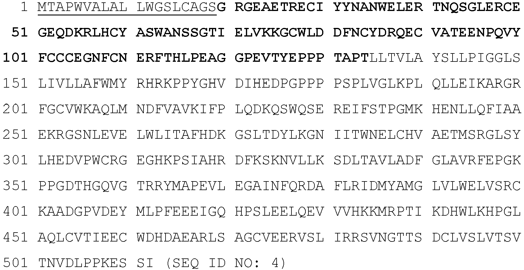
- The signal peptide is indicated by a single underline and the extracellular domain is indicated by bold font.
- A processed extracellular ActRIIB polypeptide sequence of the alternative A64 form is as follows:

- The protein may be produced with an "SGR..." sequence at the N-terminus. The C-terminal "tail" of the extracellular domain is indicated by single underline. The sequence with the "tail" deleted (a Δ15 sequence) is as follows:

- A nucleic acid sequence encoding human ActRIIB precursor protein is shown below (SEQ ID NO: 7), consisting of nucleotides 25-1560 of Genbank Reference Sequence NM_001106.3, which encode amino acids 1-513 of the ActRIIB precursor. The sequence as shown provides an arginine at
position 64 and may be modified to provide an alanine instead. The signal sequence is underlined.

- A nucleic acid sequence encoding the processed extracellular human ActRIIB polypeptide is as follows (SEQ ID NO: 8):The sequence as shown provides an arginine at

position 64, and may be modified to provide an alanine instead. - An alignment of the amino acid sequences of human ActRIIB extracellular domain and human ActRIIA extracellular domain are illustrated in
Figure 1 . This alignment indicates amino acid residues within both receptors that are believed to directly contact ActRII ligands. For example, the composite ActRII structures indicated that the ActRIIB-ligand binding pocket is defined, in part, by residues Y31, N33, N35, L38 through T41, E47, E50, Q53 through K55, L57, H58, Y60, S62, K74, W78 through N83, Y85, R87, A92, and E94 through F101. At these positions, it is expected that conservative mutations will be tolerated. - In addition, ActRIIB is generally well-conserved among vertebrates, with large stretches of the extracellular domain completely conserved. For example,
Figure 2 depicts a multi-sequence alignment of a human ActRIIB extracellular domain compared to various ActRIIB orthologs. Many of the ligands that bind to ActRIIB are also highly conserved. Accordingly, from these alignments, it is possible to predict key amino acid positions within the ligand-binding domain that are important for normal ActRIIB-ligand binding activities as well as to predict amino acid positions that are likely to be tolerant to substitution without significantly altering normal ActRIIB-ligand binding activities. Therefore, an active, human ActRIIB variant polypeptide useful in accordance with the presently disclosed methods may include one or more amino acids at corresponding positions from the sequence of another vertebrate ActRIIB, or may include a residue that is similar to that in the human or other vertebrate sequences. - The following examples illustrate this approach to defining an active ActRIIB variant. L46 in the human extracellular domain (SEQ ID NO: 2) is a valine in Xenopus ActRIIB (SEQ ID NO: 57), and so this position may be altered, and optionally may be altered to another hydrophobic residue, such as V, I or F, or a non-polar residue such as A. E52 in the human extracellular domain is a K in Xenopus, indicating that this site may be tolerant of a wide variety of changes, including polar residues, such as E, D, K, R, H, S, T, P, G, Y and probably A. T93 in the human extracellular domain is a K in Xenopus, indicating that a wide structural variation is tolerated at this position, with polar residues favored, such as S, K, R, E, D, H, G, P, G and Y. F108 in the human extracellular domain is a Y in Xenopus, and therefore Y or other hydrophobic group, such as I, V or L should be tolerated. E111 in the human extracellular domain is K in Xenopus, indicating that charged residues will be tolerated at this position, including D, R, K and H, as well as Q and N. R112 in the human extracellular domain is K in Xenopus, indicating that basic residues are tolerated at this position, including R and H. A at position 119 in the human extracellular domain is relatively poorly conserved, and appears as P in rodents (SEQ ID NOs: 52 and 54) and V in Xenopus, thus essentially any amino acid should be tolerated at this position.
- Moreover, ActRII proteins have been characterized in the art in terms of structural/functional characteristics, particularly with respect to ligand binding [Attisano et al. (1992) Cell 68(1):97-108; Greenwald et al. (1999) Nature Structural Biology 6(1): 18-22; Allendorph et al. (2006) PNAS 103(20: 7643-7648; Thompson et al. (2003) The EMBO Journal 22(7): 1555-1566; as well as
U.S. Patent Nos: 7,709,605 ,7,612,041 , and7,842,663 ]. In addition to the teachings herein, these references provide amply guidance for how to generate ActRII variants that retain one or more desired activities (e.g., ligand-binding activity). - For example, a defining structural motif known as a three-finger toxin fold is important for ligand binding by type I and type II receptors and is formed by conserved cysteine residues located at varying positions within the extracellular domain of each monomeric receptor [Greenwald et al. (1999) Nat Struct Biol 6:18-22; and Hinck (2012) FEBS Lett 586:1860-1870]. Accordingly, the core ligand-binding domains of human ActRIIB, as demarcated by the outermost of these conserved cysteines, corresponds to positions 29-109 of SEQ ID NO: 1 (ActRIIB precursor). The structurally less-ordered amino acids flanking these cysteine-demarcated core sequences can be truncated by 1, 2, 3, 4, 5, 6, 7, 8, 9, 10, 11, 12, 13, 14, 15, 16, 17, 18, 19, 20, 21, 22, 23, 24, 25, 26, 27, 28, 29, 30, 31, 32, 33, 34, 35, 36, or 37 residues without necessarily altering ligand binding. Exemplary ActRIIB extracellular domains for N-terminal and/or C-terminal truncation include SEQ ID NOs: 2, 3, 5, and 6.
- Attisano et al. showed that a deletion of the proline knot at the C-terminus of the extracellular domain of ActRIIB reduced the affinity of the receptor for activin. An ActRIIB-Fc fusion protein containing amino acids 20-119 of present SEQ ID NO: 1, "ActRIIB(20-119)-Fc", has reduced binding to GDF11 and activin relative to an ActRIIB(20-134)-Fc, which includes the proline knot region and the complete juxtamembrane domain (see, e.g.,
U.S. Patent No. 7,842,663 ). However, an ActRIIB(20-129)-Fc protein retains similar, but somewhat reduced activity, relative to the wild-type, even though the proline knot region is disrupted. Thus, ActRIIB extracellular domains that stop at amino acid 134, 133, 132, 131, 130 and 129 (with respect to SEQ ID NO: 1) are all expected to be active, but constructs stopping at 134 or 133 may be most active. Similarly, mutations at any of residues 129-134 (with respect to SEQ ID NO: 1) are not expected to alter ligand-binding affinity by large margins. In support of this, it is known in the art that mutations of P129 and P130 (with respect to SEQ ID NO: 1) do not substantially decrease ligand binding. Therefore, an ActRIIB polypeptide of the present disclosure may end as early as amino acid 109 (the final cysteine), however, forms ending at or between 109 and 119 (e.g., 109, 110, 111, 112, 113, 114, 115, 116, 117, 118, or 119) are expected to have reduced ligand binding. Amino acid 119 (with respect to present SEQ ID NO: 1) is poorly conserved and so is readily altered or truncated. ActRIIB polypeptides ending at 128 (with respect to SEQ ID NO: 1) or later should retain ligand-binding activity. ActRIIB polypeptides ending at or between 119 and 127 (e.g., 119, 120, 121, 122, 123, 124, 125, 126, or 127), with respect to SEQ ID NO: 1, will have an intermediate binding ability. Any of these forms may be desirable to use, depending on the clinical or experimental setting. - At the N-terminus of ActRIIB, it is expected that a protein beginning at amino acid 29 or before (with respect to SEQ ID NO: 1) will retain ligand-binding activity. Amino acid 29 represents the initial cysteine. An alanine-to-asparagine mutation at position 24 (with respect to SEQ ID NO: 1) introduces an N-linked glycosylation sequence without substantially affecting ligand binding [
U.S. Patent No. 7,842,663 ]. This confirms that mutations in the region between the signal cleavage peptide and the cysteine cross-linked region, corresponding to amino acids 20-29, are well tolerated. In particular, ActRIIB polypeptides beginning atposition 20, 21, 22, 23, and 24 (with respect to SEQ ID NO: 1) should retain general ligand-biding activity, and ActRIIB polypeptides beginning at positions 25, 26, 27, 28, and 29 (with respect to SEQ ID NO: 1) are also expected to retain ligand-biding activity. It has been demonstrated, e.g.,U.S. Patent No. 7,842,663 , that, surprisingly, an ActRIIB construct beginning at 22, 23, 24, or 25 will have the most activity. - Taken together, a general formula for an active portion (e.g., ligand-binding portion) of ActRIIB comprises amino acids 29-109 of SEQ ID NO: 1. Therefore ActRIIB polypeptides may, for example, comprise, consists essentially of, or consists of an amino acid sequence that is at least 70%, 75%, 80%, 85%, 86%, 87%, 88%, 89%, 90%, 91%, 92%, 93%, 94%, 95%, 96%, 97%, 98%, 99%, or 100% identical to a portion of ActRIIB beginning at a residue corresponding to any one of amino acids 20-29 (e.g., beginning at any one of
amino acids 20, 21, 22, 23, 24, 25, 26, 27, 28, or 29) of SEQ ID NO: 1 and ending at a position corresponding to any one amino acids 109-134 (e.g., ending at any one ofamino acids positions 20, 21, 22, 23, 24, 25, 26, 27, 28, or 29) or 21-29 (e.g., any one of positions 21, 22, 23, 24, 25, 26, 27, 28, or 29) and end at a position from 119-134 (e.g., any one ofpositions 119, 120, 121, 122, 123, 124, 125, 126, 127, 128, 129, 130, 131, 132, 133, or 134), 119-133 (e.g., any one ofpositions 119, 120, 121, 122, 123, 124, 125, 126, 127, 128, 129, 130, 131, 132, or 133), 129-134 (e.g., any one of positions 129, 130, 131, 132, 133, or 134), or 129-133 (e.g., any one of positions 129, 130, 131, 132, or 133) of SEQ ID NO: 1. Other examples include constructs that begin at a position from 20-24 (e.g., any one ofpositions 20, 21, 22, 23, or 24), 21-24 (e.g., any one of positions 21, 22, 23, or 24), or 22-25 (e.g., any one of positions 22, 22, 23, or 25) and end at a position from 109-134 (e.g., any one ofpositions positions 119, 120, 121, 122, 123, 124, 125, 126, 127, 128, 129, 130, 131, 132, 133, or 134) or 129-134 (e.g., any one of positions 129, 130, 131, 132, 133, or 134) of SEQ ID NO: 1. Variants within these ranges are also contemplated, particularly those having at least 70%, 75%, 80%, 85%, 86%, 87%, 88%, 89%, 90%, 91%, 92%, 93%, 94%, 95%, 96%, 97%, 98%, 99%, or 100% identity to the corresponding portion of SEQ ID NO: 1. - The variations described herein may be combined in various ways. ActRIIB variants may comprise no more than 1, 2, 5, 6, 7, 8, 9, 10 or 15 conservative amino acid changes in the ligand-binding pocket, and zero, one or more non-conservative alterations at
positions U.S. Patent No. 7,842,663 ]. This change probably eliminates glycosylation at N65 in the A64 background, thus demonstrating that a significant change in this region is likely to be tolerated. While an R64A change is poorly tolerated, R64K is well-tolerated, and thus another basic residue, such as H may be tolerated at position 64 [U.S. Patent No. 7,842,663 ]. Additionally, the results of the mutagenesis program described in the art indicate that there are amino acid positions in ActRIIB that are often beneficial to conserve. With respect to SEQ ID NO: 1, these include position 80 (acidic or hydrophobic amino acid), position 78 (hydrophobic, and particularly tryptophan), position 37 (acidic, and particularly aspartic or glutamic acid), position 56 (basic amino acid), position 60 (hydrophobic amino acid, particularly phenylalanine or tyrosine). Thus, the disclosure provides a framework of amino acids that may be conserved in ActRIIB polypeptides. Other positions that may be desirable to conserve are as follows: position 52 (acidic amino acid), position 55 (basic amino acid), position 81 (acidic), 98 (polar or charged, particularly E, D, R or K), all with respect to SEQ ID NO: 1. - It has been previously demonstrated that the addition of a further N-linked glycosylation site (N-X-S/T) into the ActRIIB extracellular domain is well-tolerated (see, e.g.,
U.S. Patent No. 7,842,663 ). Therefore, N-X-S/T sequences may be generally introduced at positions outside the ligand binding pocket defined inFigure 1 in ActRIIB polypeptide of the present disclosure. Particularly suitable sites for the introduction of non-endogenous N-X-S/T sequences include amino acids 20-29, 20-24, 22-25, 109-134, 120-134 or 129-134 (with respect to SEQ ID NO: 1). N-X-S/T sequences may also be introduced into the linker between the ActRIIB sequence and an Fc domain or other fusion component. Such a site may be introduced with minimal effort by introducing an N in the correct position with respect to a pre-existing S or T, or by introducing an S or T at a position corresponding to a pre-existing N. Thus, desirable alterations that would create an N-linked glycosylation site are: A24N, R64N, S67N (possibly combined with an N65A alteration), E105N, R112N, G120N, E123N, P129N, A132N, R112S and R112T (with respect to SEQ ID NO: 1). Any S that is predicted to be glycosylated may be altered to a T without creating an immunogenic site, because of the protection afforded by the glycosylation. Likewise, any T that is predicted to be glycosylated may be altered to an S. Thus the alterations S67T and S44T (with respect to SEQ ID NO: 1) are contemplated. Likewise, in an A24N variant, an S26T alteration may be used. Accordingly, an ActRIIB polypeptide may be a variant having one or more additional, non-endogenous N-linked glycosylation consensus sequences as described above. - ActRII inhibitors may comprise at least one ActRIIB polypeptide, which includes fragments, functional variants, and modified forms thereof. ActRIIB polypeptides may be soluble (e.g., an extracellular domain of ActRIIB). ActRIIB polypeptides may inhibit (antagonize) activity (e.g., induction of
Smad - In certain embodiments not covered by the claimed invention, the present disclosure relates to ActRIIA polypeptides. As used herein, the term "ActRIIA" refers to a family of activin receptor type IIA (ActRIIA) proteins from any species and variants derived from such ActRIIA proteins by mutagenesis or other modification. Reference to ActRIIA herein is understood to be a reference to any one of the currently identified forms. Members of the ActRIIA family are generally transmembrane proteins, composed of a ligand-binding extracellular domain comprising a cysteine-rich region, a transmembrane domain, and a cytoplasmic domain with predicted serine/threonine kinase activity.
- The term "ActRIIA polypeptide" includes polypeptides comprising any naturally occurring polypeptide of an ActRIIA family member as well as any variants thereof (including mutants, fragments, fusions, and peptidomimetic forms) that retain a useful activity. Examples of such variant ActRIIA polypeptides are provided throughout the present disclosure as well as in International Patent Application Publication No.
WO 2006/012627 . Numbering of amino acids for all ActRIIA-related polypeptides described herein is based on the numbering of the human ActRIIA precursor protein sequence provided below (SEQ ID NO: 9), unless specifically designated otherwise. - The canonical human ActRIIA precursor protein sequence is as follows:
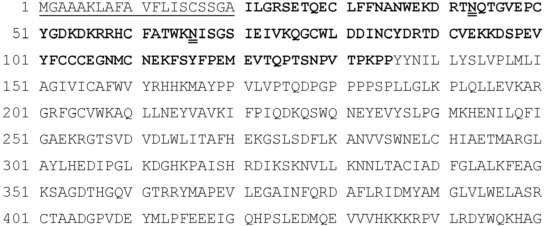

- The signal peptide is indicated by a single underline; the extracellular domain is indicated in bold font; and the potential, endogenous N-linked glycosylation sites are indicated by a double underline.
- A processed extracellular human ActRIIA polypeptide sequence is as follows:

- The C-terminal "tail" of the extracellular domain is indicated by single underline. The sequence with the "tail" deleted (a Δ15 sequence) is as follows:

- The nucleic acid sequence encoding human ActRIIA precursor protein is shown below (SEQ ID NO: 12), as follows nucleotides 159-1700 of Genbank Reference Sequence NM_001616.4. The signal sequence is underlined.


- The nucleic acid sequence encoding processed soluble (extracellular) human ActRIIA polypeptide is as follows:

- ActRIIA is well-conserved among vertebrates, with large stretches of the extracellular domain completely conserved. For example,
Figure 14 depicts a multi-sequence alignment of a human ActRIIA extracellular domain compared to various ActRIIA orthologs. Many of the ligands that bind to ActRIIA are also highly conserved. Accordingly, from these alignments, it is possible to predict key amino acid positions within the ligand-binding domain that are important for normal ActRIIA-ligand binding activities as well as to predict amino acid positions that are likely to be tolerant to substitution without significantly altering normal ActRIIA-ligand binding activities. Therefore, an active, human ActRIIA variant polypeptide useful in accordance with the presently disclosed methods may include one or more amino acids at corresponding positions from the sequence of another vertebrate ActRIIA, or may include a residue that is similar to that in the human or other vertebrate sequences. - The following examples illustrate this approach to defining an active ActRIIA variant. F13 in the human extracellular domain is Y in Ovis aries (SEQ ID NO: 70), Gallus gallus (SEQ ID NO: 73), Bos Taurus (SEQ ID NO: 74), Tyto alba (SEQ ID NO: 75), and Myotis davidii (SEQ ID NO: 76) ActRIIA, indicating that aromatic residues are tolerated at this position, including F, W, and Y. Q24 in the human extracellular domain is R in Bos Taurus ActRIIA, indicating that charged residues will be tolerated at this position, including D, R, K, H, and E. S95 in the human extracellular domain is F in Gallus gallus and Tyto alba ActRIIA, indicating that this site may be tolerant of a wide variety of changes, including polar residues, such as E, D, K, R, H, S, T, P, G, Y, and probably hydrophobic residue such as L, I, or F. E52 in the human extracellular domain is D in Ovis aries ActRIIA, indicating that acidic residues are tolerated at this position, including D and E. P29 in the human extracellular domain is relatively poorly conserved, appearing as S in Ovis aries ActRIIA and L in Myotis davidii ActRIIA, thus essentially any amino acid should be tolerated at this position.
- Moreover, as discussed above, ActRII proteins have been characterized in the art in terms of structural/functional characteristics, particularly with respect to ligand binding [Attisano et al. (1992) Cell 68(1):97-108; Greenwald et al. (1999) Nature Structural Biology 6(1): 18-22; Allendorph et al. (2006) PNAS 103(20: 7643-7648; Thompson et al. (2003) The EMBO Journal 22(7): 1555-1566; as well as
U.S. Patent Nos: 7,709,605 ,7,612,041 , and7,842,663 ]. In addition to the teachings herein, these references provide amply guidance for how to generate ActRII variants that retain one or more desired activities (e.g., ligand-binding activity). - For example, a defining structural motif known as a three-finger toxin fold is important for ligand binding by type I and type II receptors and is formed by conserved cysteine residues located at varying positions within the extracellular domain of each monomeric receptor [Greenwald et al. (1999) Nat Struct Biol 6:18-22; and Hinck (2012) FEBS Lett 586:1860-1870]. Accordingly, the core ligand-binding domains of human ActRIIA, as demarcated by the outermost of these conserved cysteines, corresponds to positions 30-110 of SEQ ID NO: 9 (ActRIIA precursor). Therefore, the structurally less-ordered amino acids flanking these cysteine-demarcated core sequences can be truncated by about 1, 2, 3, 4, 5, 6, 7, 8, 9, 10, 11, 12, 13, 14, 15, 16, 17, 18, 19, 20, 21, 22, 23, 24, 25, 26, 27, 28, or 29 residues at the N-terminus and by about 1, 2, 3, 4, 5, 6, 7, 8, 9, 10, 11, 12, 13, 14, 15, 16, 17, 18, 19, 20, 21, 22, 23, 24, or 25 residues at the C-terminus without necessarily altering ligand binding. Exemplary ActRIIA extracellular domains truncations include SEQ ID NOs: 10 and 11.
- Accordingly, a general formula for an active portion (e.g., ligand binding) of ActRIIA is a polypeptide that comprises, consists essentially of, or consists of amino acids 30-110 of SEQ ID NO: 9. Therefore ActRIIA polypeptides may, for example, comprise, consists essentially of, or consists of an amino acid sequence that is at least 70%, 75%, 80%, 85%, 86%, 87%, 88%, 89%, 90%, 91%, 92%, 93%, 94%, 95%, 96%, 97%, 98%, 99%, or 100% identical to a portion of ActRIIA beginning at a residue corresponding to any one of amino acids 21-30 (e.g., beginning at any one of amino acids 21, 22, 23, 24, 25, 26, 27, 28, 29, or 30) of SEQ ID NO: 9 and ending at a position corresponding to any one amino acids 110-135 (e.g., ending at any one of
amino acids positions - In certain embodiments not covered by the claimed invention, the disclosure relates to ActRII inhibitors that comprise at least one ActRIIA polypeptide, which includes fragments, functional variants, and modified forms thereof. ActRIIA polypeptides may be soluble (e.g., an extracellular domain of ActRIIA). ActRIIA polypeptides may inhibit (antagonize) activity (e.g., induction of
Smad amino acids - In certain aspects not covered by the claimed invention, the present disclosure relates to GDF trap polypeptides (also referred to as "GDF traps"). GDF traps may be variant ActRII polypeptides (e.g., ActRIIA and ActRIIB polypeptides) that comprise one or more mutations (e.g., amino acid additions, deletions, substitutions, and combinations thereof) in the extracellular domain (also referred to as the ligand-binding domain) of an ActRII polypeptide (e.g., a "wild-type" or unmodified ActRII polypeptide) such that the variant ActRII polypeptide has one or more altered ligand-binding activities than the corresponding wild-type ActRII polypeptide. In preferred instances, GDF trap polypeptides of the present disclosure retain at least one similar activity as a corresponding wild-type ActRII polypeptide. For example, preferable GDF traps bind to and inhibit (e.g. antagonize) the function of GDF11 and/or GDF8. GDF traps may further bind to and inhibit one or more of ligand of the TGF-beta superfamily. Accordingly, GDF trap polypeptides may have an altered binding specificity for one or more ActRII ligands.
- To illustrate, one or more mutations may be selected that increase the selectivity of the altered ligand-binding domain for GDF11 and/or GDF8 over one or more ActRII-binding ligands such as activins (activin A, activin B, activin AB, activin C, and/or activin E), particularly activin A. Optionally, the altered ligand-binding domain has a ratio of Kd for activin binding to Kd for GDF11 and/or GDF8 binding that is at least 2-, 5-, 10-, 20-, 50-, 100- or even 1000-fold greater relative to the ratio for the wild-type ligand-binding domain. Optionally, the altered ligand-binding domain has a ratio of IC50 for inhibiting activin to IC50 for inhibiting GDF11 and/or GDF8 that is at least 2-, 5-, 10-, 20-, 50-, 100- or even 1000-fold greater relative to the wild-type ligand-binding domain. Optionally, the altered ligand-binding domain inhibits GDF11 and/or GDF8 with an IC50 at least 2-, 5-, 10-, 20-, 50-, 100- or even 1000-times less than the IC50 for inhibiting activin.
- GDF traps may be designed to preferentially bind to GDF11 and/or GDF8 (also known as myostatin). Optionally, GDF11 and/or GDF8-binding traps may further bind to activin B. Optionally, GDF11 and/or GDF8-binding traps may further bind to BMP6. Optionally, GDF11 and/or GDF8-binding traps may further bind to BMP10. Optionally, GDF11 and/or GDF8-binding traps may further bind to activin B and BMP6. GDF traps may have diminished binding affinity for activins (e.g., activin A, activin A/B, activin B, activin C, activin E ), e.g., in comparison to a wild-type ActRII polypeptide. A GDF trap polypeptide may have diminished binding affinity for activin A.
- Amino acid residues of the ActRIIB proteins (e.g., E39, K55, Y60, K74, W78, L79, D80, and F101) are in the ActRIIB ligand-binding pocket and help mediated binding to its ligands including, for example, activin A, GDF11, and GDF8. Thus, the present disclosure provides GDF trap polypeptides, which are not covered by the claimed invention, comprising an altered-ligand binding domain (e.g., a GDF8/GDF11-binding domain) of an ActRIIB receptor which comprises one or more mutations at those amino acid residues.
- As a specific example, the positively-charged amino acid residue Asp (D80) of the ligand-binding domain of ActRIIB can be mutated to a different amino acid residue to produce a GDF trap polypeptide that preferentially binds to GDF8, but not activin. Preferably, the D80 residue with respect to SEQ ID NO: 1 is changed to an amino acid residue selected from the group consisting of: an uncharged amino acid residue, a negative amino acid residue, and a hydrophobic amino acid residue. As a further specific example, the hydrophobic residue L79 of SEQ ID NO: 1 can be altered to confer altered activin-GDF11/GDF8 binding properties. For example, an L79P substitution reduces GDF11 binding to a greater extent than activin binding. In contrast, replacement of L79 with an acidic amino acid [an aspartic acid or glutamic acid; an L79D or an L79E substitution] greatly reduces activin A binding affinity while retaining GDF11 binding affinity. In exemplary embodiments not covered by the claimed invention, the methods described herein utilize a GDF trap polypeptide which is a variant ActRIIB polypeptide comprising an acidic amino acid (e.g., D or E) at the position corresponding to position 79 of SEQ ID NO: 1, optionally in combination with one or more additional amino acid substitutions, additions, or deletions.
- The present disclosure also contemplates making functional variants by modifying the structure of an ActRII polypeptide for such purposes as enhancing therapeutic efficacy or stability (e.g., shelf-life and resistance to proteolytic degradation in vivo). Variants can be produced by amino acid substitution, deletion, addition, or combinations thereof. For instance, it is reasonable to expect that an isolated replacement of a leucine with an isoleucine or valine, an aspartate with a glutamate, a threonine with a serine, or a similar replacement of an amino acid with a structurally related amino acid (e.g., conservative mutations) will not have a major effect on the biological activity of the resulting molecule. Conservative replacements are those that take place within a family of amino acids that are related in their side chains. Whether a change in the amino acid sequence of a polypeptide of the disclosure results in a functional homolog can be readily determined by assessing the ability of the variant polypeptide to produce a response in cells in a fashion similar to the wild-type polypeptide, or to bind to one or more TGF-beta ligands including, for example, BMP2, BMP2/7, BMP3, BMP4, BMP4/7, BMP5, BMP6, BMP7, BMP8a, BMP8b, BMP9, BMP10, GDF3, GDF5, GDF6/BMP13, GDF7, GDF8, GDF9b/BMP15, GDF11BMP11, GDF15/MIC1, TGF-β1, TGF-β2, TGF-β3, activin A, activin B, activin C, activin E, activin AB, activin AC, nodal, glial cell-derived neurotrophic factor (GDNF), neurturin, artemin, persephin, MIS, and Lefty.
- The present disclosure also contemplates specific mutations of an ActRII polypeptide so as to alter the glycosylation of the polypeptide. Such mutations may be selected so as to introduce or eliminate one or more glycosylation sites, such as O-linked or N-linked glycosylation sites. Asparagine-linked glycosylation recognition sites generally comprise a tripeptide sequence, asparagine-X-threonine or asparagine-X-serine (where "X" is any amino acid) which is specifically recognized by appropriate cellular glycosylation enzymes. The alteration may also be made by the addition of, or substitution by, one or more serine or threonine residues to the sequence of the polypeptide (for O-linked glycosylation sites). A variety of amino acid substitutions or deletions at one or both of the first or third amino acid positions of a glycosylation recognition site (and/or amino acid deletion at the second position) results in non-glycosylation at the modified tripeptide sequence. Another means of increasing the number of carbohydrate moieties on a polypeptide is by chemical or enzymatic coupling of glycosides to the polypeptide. Depending on the coupling mode used, the sugar(s) may be attached to (a) arginine and histidine; (b) free carboxyl groups; (c) free sulfhydryl groups such as those of cysteine; (d) free hydroxyl groups such as those of serine, threonine, or hydroxyproline; (e) aromatic residues such as those of phenylalanine, tyrosine, or tryptophan; or (f) the amide group of glutamine. Removal of one or more carbohydrate moieties present on a polypeptide may be accomplished chemically and/or enzymatically. Chemical deglycosylation may involve, for example, exposure of a polypeptide to the compound trifluoromethanesulfonic acid, or an equivalent compound. This treatment results in the cleavage of most or all sugars except the linking sugar (N-acetylglucosamine or N-acetylgalactosamine), while leaving the amino acid sequence intact. Enzymatic cleavage of carbohydrate moieties on polypeptides can be achieved by the use of a variety of endo- and exo-glycosidases as described by Thotakura et al. [Meth. Enzymol. (1987) 138:350]. The sequence of a polypeptide may be adjusted, as appropriate, depending on the type of expression system used, as mammalian, yeast, insect, and plant cells may all introduce differing glycosylation patterns that can be affected by the amino acid sequence of the peptide. In general, polypeptides of the present disclosure for use in humans may be expressed in a mammalian cell line that provides proper glycosylation, such as HEK293 or CHO cell lines, although other mammalian expression cell lines are expected to be useful as well.
- The present disclosure further contemplates a method, which is not covered by the claimed invention, of generating mutants, particularly sets of combinatorial mutants of an ActRII polypeptide as well as truncation mutants. Pools of combinatorial mutants are especially useful for identifying functionally active (e.g., TGF-beta superfamily ligand binding) ActRII sequences. The purpose of screening such combinatorial libraries may be to generate, for example, polypeptides variants which have altered properties, such as altered pharmacokinetic or altered ligand binding. A variety of screening assays are provided below, and such assays may be used to evaluate variants. For example, ActRII variants may be screened for ability to bind to one or more TGF-beta superfamily ligands (e.g., BMP2, BMP2/7, BMP3, BMP4, BMP4/7, BMP5, BMP6, BMP7, BMP8a, BMP8b, BMP9, BMP10, GDF3, GDF5, GDF6/BMP13, GDF7, GDF8, GDF9b/BMP15, GDF11BMP11, GDF15/MIC1, TGF-β1, TGF-β2, TGF-β3, activin A, activin B, activin AB, activin AC, nodal, glial cell-derived neurotrophic factor (GDNF), neurturin, artemin, persephin, MIS, and Lefty), to prevent binding of a TGF-beta superfamily ligand to a TGF-beta superfamily receptor, and/or to interfere with signaling caused by an TGF-beta superfamily ligand.
- The activity of an ActRII polypeptides may also be tested in a cell-based or in vivo assay. For example, the effect of an ActRII polypeptide on the expression of genes involved in visual acuity may be assessed. This may, as needed, be performed in the presence of one or more recombinant ActRII ligand proteins (e.g., BMP2, BMP2/7, BMP3, BMP4, BMP4/7, BMP5, BMP6, BMP7, BMP8a, BMP8b, BMP9, BMP10, GDF3, GDF5, GDF6/BMP13, GDF7, GDF8, GDF9b/BMP15, GDF11/BMP11, GDF15/MIC1, TGF-β1, TGF-β2, TGF-β3, activin A, activin B, activin C, activin E, activin AB, activin AC, nodal, glial cell-derived neurotrophic factor (GDNF), neurturin, artemin, persephin, MIS, and Lefty), and cells may be transfected so as to produce an ActRII polypeptide, and optionally, an ActRII ligand. Likewise, an ActRII polypeptide may be administered to a mouse or other animal and visual acuity may be assessed using art-recognized methods. Similarly, the activity of an ActRII polypeptide or variant thereof may be tested in blood cell precursor cells for any effect on growth of these cells, for example, by the assays as described herein and those of common knowledge in the art. A SMAD-responsive reporter gene may be used in such cell lines to monitor effects on downstream signaling.
- Combinatorial-derived variants can be generated which have increased selectivity or generally increased potency relative to a reference ActRII polypeptide. Such variants, when expressed from recombinant DNA constructs, can be used in gene therapy protocols. Likewise, mutagenesis can give rise to variants which have intracellular half-lives dramatically different than the corresponding unmodified ActRII polypeptide. For example, the altered protein can be rendered either more stable or less stable to proteolytic degradation or other cellular processes which result in destruction, or otherwise inactivation, of an unmodified polypeptide. Such variants, and the genes which encode them, can be utilized to alter polypeptide complex levels by modulating the half-life of the polypeptide. For instance, a short half-life can give rise to more transient biological effects and, when part of an inducible expression system, can allow tighter control of recombinant polypeptide complex levels within the cell. In an Fc fusion protein, mutations may be made in the linker (if any) and/or the Fc portion to alter the half-life of the ActRII polypeptide.
- A combinatorial library may be produced by way of a degenerate library of genes encoding a library of polypeptides which each include at least a portion of potential ActRII sequences. For instance, a mixture of synthetic oligonucleotides can be enzymatically ligated into gene sequences such that the degenerate set of potential ActRII encoding nucleotide sequences are expressible as individual polypeptides, or alternatively, as a set of larger fusion proteins (e.g., for phage display).
- There are many ways by which the library of potential homologs can be generated from a degenerate oligonucleotide sequence. Chemical synthesis of a degenerate gene sequence can be carried out in an automatic DNA synthesizer, and the synthetic genes can then be ligated into an appropriate vector for expression. The synthesis of degenerate oligonucleotides is well known in the art [Narang, SA (1983) Tetrahedron 39:3; Itakura et al. (1981) Recombinant DNA, Proc. 3rd Cleveland Sympos. Macromolecules, ed. AG Walton, Amsterdam: Elsevier pp273-289; Itakura et al. (1984) Annu. Rev. Biochem. 53:323; Itakura et al. (1984) Science 198:1056; and Ike et al. (1983) Nucleic Acid Res. 11:477]. Such techniques have been employed in the directed evolution of other proteins [Scott et al., (1990) Science 249:386-390; Roberts et al. (1992) PNAS USA 89:2429-2433; Devlin et al. (1990) Science 249: 404-406; Cwirla et al., (1990) PNAS USA 87: 6378-6382; as well as
U.S. Patent Nos: 5,223,409 ,5,198,346 , and5,096,815 ]. - Alternatively, other forms of mutagenesis can be utilized to generate a combinatorial library. For example, ActRII polypeptides of the disclosure can be generated and isolated from a library by screening using, for example, alanine scanning mutagenesis [Ruf et al. (1994) Biochemistry 33:1565-1572; Wang et al. (1994) J. Biol. Chem. 269:3095-3099; Balint et al. (1993) Gene 137:109-118; Grodberg et al. (1993) Eur. J. Biochem. 218:597-601; Nagashima et al. (1993) J. Biol. Chem. 268:2888-2892; Lowman et al. (1991) Biochemistry 30:10832-10838; and Cunningham et al. (1989) Science 244:1081-1085], by linker scanning mutagenesis [Gustin et al. (1993) Virology 193:653-660; and Brown et al. (1992) Mol. Cell Biol. 12:2644-2652; McKnight et al. (1982) Science 232:316], by saturation mutagenesis [Meyers et al., (1986) Science 232:613]; by PCR mutagenesis [Leung et al. (1989) Method Cell Mol Biol 1:11-19]; or by random mutagenesis, including chemical mutagenesis [Miller et al. (1992) A Short Course in Bacterial Genetics, CSHL Press, Cold Spring Harbor, NY; and Greener et al. (1994) Strategies in Mol Biol 7:32-34]. Linker scanning mutagenesis, particularly in a combinatorial setting, is an attractive method for identifying truncated (bioactive) forms of ActRII polypeptides.
- A wide range of techniques are known in the art for screening gene products of combinatorial libraries made by point mutations and truncations, and, for that matter, for screening cDNA libraries for gene products having a certain property. Such techniques will be generally adaptable for rapid screening of the gene libraries generated by the combinatorial mutagenesis of ActRII polypeptides. The most widely used techniques for screening large gene libraries typically comprise cloning the gene library into replicable expression vectors, transforming appropriate cells with the resulting library of vectors, and expressing the combinatorial genes under conditions in which detection of a desired activity facilitates relatively easy isolation of the vector encoding the gene whose product was detected. Preferred assays include TGF-beta ligand (e.g., BMP2, BMP2/7, BMP3, BMP4, BMP4/7, BMP5, BMP6, BMP7, BMP8a, BMP8b, BMP9, BMP10, GDF3, GDF5, GDF6/BMP13, GDF7, GDF8, GDF9b/BMP15, GDF11BMP11, GDF15/MIC1, TGF-β1, TGF-β2, TGF-β3, activin A, activin B, activin C, activin E, activin AB, activin AC, nodal, glial cell-derived neurotrophic factor (GDNF), neurturin, artemin, persephin, MIS, and Lefty) binding assays and/or TGF-beta ligand-mediated cell signaling assays.
- As will be recognized by one of skill in the art, most of the described mutations, variants or modifications described herein may be made at the nucleic acid level or, in some cases, by post-translational modification or chemical synthesis. Such techniques are well known in the art and some of which are described herein. In part, the present disclosure identifies functionally active portions (fragments) and variants of ActRII polypeptides that can be used as guidance for generating and using other variant ActRII polypeptides described herein, whereby only such variants or fragments of ActRII polypeptides are covered by the claimed invention for which the condition applies to comprise an amino acid sequence that is at least 90% identical to the sequence of SEQ ID NO: 1, wherein the polypeptide comprises an acidic amino acid at position 79 with respect to SEQ ID NO: 1.
- Functionally active fragments of ActRII polypeptides of the present disclosure can be obtained by screening polypeptides recombinantly produced from the corresponding fragment of the nucleic acid encoding an ActRII polypeptide (e.g., SEQ ID NOs: 7, 8, 12, 13, 37, 42, 47, 60, 62, 63, 66, 67, 68, 80, 81, 82, and 83). In addition, fragments can be chemically synthesized using techniques known in the art such as conventional Merrifield solid phase f-Moc or t-Boc chemistry. The fragments can be produced (recombinantly or by chemical synthesis) and tested to identify those peptidyl fragments that can function as antagonists (inhibitors) of ActRII receptors and/or one or more ligands (e.g., BMP2, BMP2/7, BMP3, BMP4, BMP4/7, BMP5, BMP6, BMP7, BMP8a, BMP8b, BMP9, BMP10, GDF3, GDF5, GDF6/BMP13, GDF7, GDF8, GDF9b/BMP15, GDF11BMP11, GDF15/MIC1, TGF-β1, TGF-β2, TGF-β3, activin A, activin B, activin C, activin E, activin AB, activin AC, nodal, glial cell-derived neurotrophic factor (GDNF), neurturin, artemin, persephin, MIS, and Lefty).
- ActRII polypeptides may further comprise post-translational modifications in addition to any that are naturally present in the ActRII (e.g., an ActRIIA or ActRIIB polypeptide). Such modifications include, but are not limited to, acetylation, carboxylation, glycosylation, phosphorylation, lipidation, and acylation. As a result, the ActRII polypeptide may contain non-amino acid elements, such as polyethylene glycols, lipids, polysaccharide or monosaccharide, and phosphates. Effects of such non-amino acid elements on the functionality of a ligand trap polypeptide may be tested as described herein for other ActRII variants. When a polypeptide of the disclosure is produced in cells by cleaving a nascent form of the polypeptide, post-translational processing may also be important for correct folding and/or function of the protein. Different cells (e.g., CHO, HeLa, MDCK, 293, WI38, NIH-3T3 or HEK293) have specific cellular machinery and characteristic mechanisms for such post-translational activities and may be chosen to ensure the correct modification and processing of the ActRII polypeptides.
- ActRII polypeptides may include fusion proteins having at least a portion (domain) of an ActRII polypeptide (e.g., an ActRIIA or ActRIIB polypeptide) and one or more heterologous portions (domains), whereby only such fusion proteins are covered by the claimed invention for which the condition applies to comprise an ActRII polypeptide selected from the group consisting of: a) a polypeptide comprising an amino acid sequence that is identical to amino acids 29-109 of SEQ ID NO: 1 or comprising an amino acid sequence that is at least 91%, 92%, 93%, 94%, 95%, 96%, 97%, 98%, or 99% identical to the sequence of amino acids 29-109 of SEQ ID NO: 1; b) a polypeptide comprising an amino acid sequence that is identical to amino acids 25-131 of SEQ ID NO: 1 or comprising an amino acid sequence that is at least 90%, 91%, 92%, 93%, 94%, 95%, 96%, 97%, 98%, or 99% identical to the sequence of amino acids 25-131 of SEQ ID NO: 1; c) a polypeptide comprising the amino acid sequence of SEQ ID NO: 2 or comprising an amino acid sequence that is at least 90%, 91%, 92%, 93%, 94%, 95%, 96%, 97%, 98%, or 99% identical to the amino acid sequence of SEQ ID NO: 2; d) a polypeptide comprising the amino acid sequence of SEQ ID NO:3 or comprising an amino acid sequence that is at least 90%, 91%, 92%, 93%, 94%, 95%, 96%, 97%, 98%, or 99% identical to the amino acid sequence of SEQ ID NO: 3; e) a polypeptide comprising the amino acid sequence of SEQ ID NO: 5 or comprising an amino acid sequence that is at least 90%, 91%, 92%, 93%, 94%, 95%, 96%, 97%, 98%, or 99% identical to the amino acid sequence of SEQ ID NO: 5; f) a polypeptide comprising the amino acid sequence of SEQ ID NO: 6 or comprising an amino acid sequence that is at least 90%, 91%, 92%, 93%, 94%, 95%, 96%, 97%, 98%, or 99% identical to the amino acid sequence of SEQ ID NO: 6; g) a polypeptide comprising the amino acid sequence of SEQ ID NO: 45 or comprising an amino acid sequence that is at least 90%, 91%, 92%, 93%, 94%, 95%, 96%, 97%, 98%, or 99% identical to the amino acid sequence of SEQ ID NO: 45; h) a polypeptide comprising the amino acid sequence of SEQ ID NO: 48 or comprising an amino acid sequence that is at least 90%, 91%, 92%, 93%, 94%, 95%, 96%, 97%, 98%, or 99% identical to the amino acid sequence of SEQ ID NO: 48; i) a polypeptide comprising the amino acid sequence of SEQ ID NO: 49 or comprising an amino acid sequence that is at least 90%, 91%, 92%, 93%, 94%, 95%, 96%, 97%, 98%, or 99% identical to the amino acid sequence of SEQ ID NO: 49; and j) a polypeptide comprising the amino acid sequence of SEQ ID NO: 65 or comprising an amino acid sequence that is at least 90%, 91%, 92%, 93%, 94%, 95%, 96%, 97%, 98%, or 99% identical to the amino acid sequence of SEQ ID NO: 65; and wherein the fusion protein comprises, in addition to the ActRII polypeptide domain, an immunoglobulin Fc domain. Well-known examples of such fusion domains include, but are not limited to, polyhistidine, Glu-Glu, glutathione S-transferase (GST), thioredoxin, protein A, protein G, an immunoglobulin heavy-chain constant region (Fc), maltose binding protein (MBP), or human serum albumin. A fusion domain may be selected so as to confer a desired property. For example, some fusion domains are particularly useful for isolation of the fusion proteins by affinity chromatography. For the purpose of affinity purification, relevant matrices for affinity chromatography, such as glutathione-, amylase-, and nickel- or cobalt- conjugated resins are used. Many of such matrices are available in "kit" form, such as the Pharmacia GST purification system and the QIAexpress™ system (Qiagen) useful with (HIS6) fusion partners. As another example, a fusion domain may be selected so as to facilitate detection of the ActRII polypeptide. Examples of such detection domains include the various fluorescent proteins (e.g., GFP) as well as "epitope tags," which are usually short peptide sequences for which a specific antibody is available. Well-known epitope tags for which specific monoclonal antibodies are readily available include FLAG, influenza virus haemagglutinin (HA), and c-myc tags. The fusion domains may have a protease cleavage site, such as for Factor Xa or thrombin, which allows the relevant protease to partially digest the fusion proteins and thereby liberate the recombinant proteins therefrom. The liberated proteins can then be isolated from the fusion domain by subsequent chromatographic separation. Other types of fusion domains that may be selected include multimerizing (e.g., dimerizing, tetramerizing) domains and functional domains (that confer an additional biological function) including, for example constant domains from immunoglobulins (e.g., Fc domains).
- ActRII polypeptides may contain one or more modifications that are capable of "stabilizing" the polypeptides. By "stabilizing" is meant anything that increases the in vitro half-life, serum half-life, regardless of whether this is because of decreased destruction, decreased clearance by the kidney, or other pharmacokinetic effect of the agent. For example, such modifications enhance the shelf-life of the polypeptides, enhance circulatory half-life of the polypeptides, and/or reduce proteolytic degradation of the polypeptides. Such stabilizing modifications include, but are not limited to, fusion proteins (including, for example, fusion proteins comprising an ActRII polypeptide domain and a stabilizer domain), modifications of a glycosylation site (including, for example, addition of a glycosylation site to a polypeptide of the disclosure), and modifications of carbohydrate moiety (including, for example, removal of carbohydrate moieties from a polypeptide of the disclosure). As used herein, the term "stabilizer domain" not only refers to a fusion domain (e.g., an immunoglobulin Fc domain) as in the case of fusion proteins, but also includes nonproteinaceous modifications such as a carbohydrate moiety, or nonproteinaceous moiety, such as polyethylene glycol. An ActRII polypeptide may be fused with a heterologous domain that stabilizes the polypeptide (a "stabilizer" domain), preferably a heterologous domain that increases stability of the polypeptide in vivo. Fusions with a constant domain of an immunoglobulin (e.g., a Fc domain) are known to confer desirable pharmacokinetic properties on a wide range of proteins. Likewise, fusions to human serum albumin can confer desirable properties.
- An example of a native amino acid sequence that may be used for the Fc portion of human IgG1 (G1Fc) is shown below (SEQ ID NO: 14). Dotted underline indicates the hinge region, and solid underline indicates positions with naturally occurring variants. In part, the disclosure provides polypeptides comprising, consisting essential of, or consisting of amino acid sequences with 70%, 75%, 80%, 85%, 86%, 87%, 88%, 89%, 90%, 91%, 92%, 93%, 94%, 95%, 96%, 97%, 98%, 99%, or 100% identity to SEQ ID NO: 14. Naturally occurring variants in G1Fc would include E134D and M136L according to the numbering system used in SEQ ID NO: 14 (see Uniprot P01857).
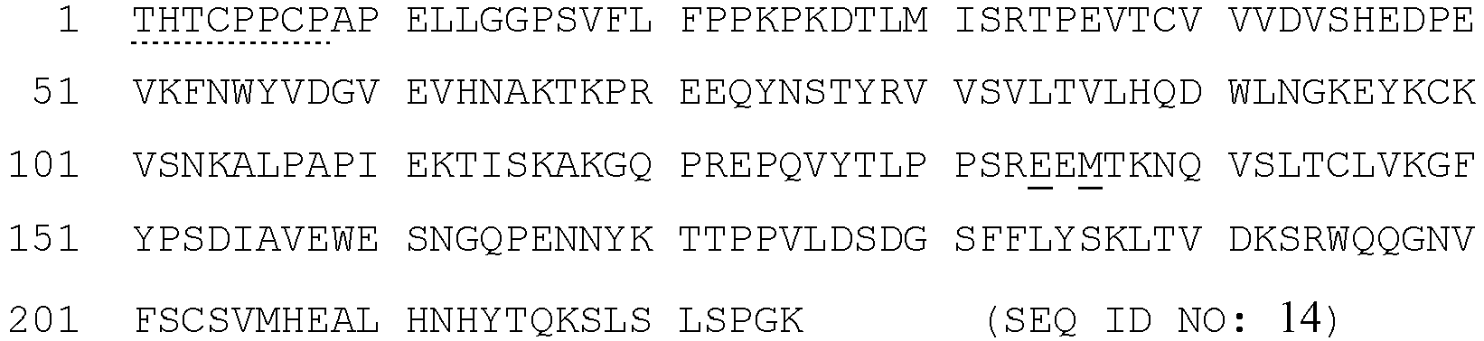
- Optionally, the IgG1 Fc domain has one or more mutations at residues such as Asp-265, lysine 322, and Asn-434. In certain cases, the mutant IgG1 Fc domain having one or more of these mutations (e.g., Asp-265 mutation) has reduced ability of binding to the Fcy receptor relative to a wild-type Fc domain. In other cases, the mutant Fc domain having one or more of these mutations (e.g., Asn-434 mutation) has increased ability of binding to the MHC class I-related Fc-receptor (FcRN) relative to a wild-type IgG1 Fc domain.
- An example of a native amino acid sequence that may be used for the Fc portion of human IgG2 (G2Fc) is shown below (SEQ ID NO: 15). Dotted underline indicates the hinge region and double underline indicates positions where there are data base conflicts in the sequence (according to UniProt P01859). In part, the disclosure provides polypeptides comprising, consisting essential of, or consisting of amino acid sequences with 70%, 75%, 80%, 85%, 86%, 87%, 88%, 89%, 90%, 91%, 92%, 93%, 94%, 95%, 96%, 97%, 98%, 99%, or 100% identity to SEQ ID NO: 15.

- Two examples of amino acid sequences that may be used for the Fc portion of human IgG3 (G3Fc) are shown below. The hinge region in G3Fc can be up to four times as long as in other Fc chains and contains three identical 15-residue segments preceded by a similar 17-residue segment. The first G3Fc sequence shown below (SEQ ID NO: 16) contains a short hinge region consisting of a single 15-residue segment, whereas the second G3Fc sequence (SEQ ID NO: 17) contains a full-length hinge region. In each case, dotted underline indicates the hinge region, and solid underline indicates positions with naturally occurring variants according to UniProt P01859. In part, the disclosure provides polypeptides comprising, consisting essential of, or consisting of amino acid sequences with 70%, 75%, 80%, 85%, 86%, 87%, 88%, 89%, 90%, 91%, 92%, 93%, 94%, 95%, 96%, 97%, 98%, 99%, or 100% identity to SEQ ID NOs: 16 and 17.


- Naturally occurring variants in G3Fc (for example, see Uniprot P01860) include E68Q, P76L, E79Q, Y81F, D97N, N100D, T124A, S169N, S169del, F221Y when converted to the numbering system used in SEQ ID NO: 16, and the present disclosure provides fusion proteins comprising G3Fc domains containing one or more of these variations. In addition, the human immunoglobulin IgG3 gene (IGHG3) shows a structural polymorphism characterized by different hinge lengths [see Uniprot P01859]. Specifically, variant WIS is lacking most of the V region and all of the CH1 region. It has an extra interchain disulfide bond at
position 7 in addition to the 11 normally present in the hinge region. Variant ZUC lacks most of the V region, all of the CH1 region, and part of the hinge. Variant OMM may represent an allelic form or another gamma chain subclass. The present disclosure provides additional fusion proteins comprising G3Fc domains containing one or more of these variants. - An example of a native amino acid sequence that may be used for the Fc portion of human IgG4 (G4Fc) is shown below (SEQ ID NO: 18). Dotted underline indicates the hinge region. In part, the disclosure provides polypeptides comprising, consisting essential of, or consisting of amino acid sequences with 70%, 75%, 80%, 85%, 86%, 87%, 88%, 89%, 90%, 91%, 92%, 93%, 94%, 95%, 96%, 97%, 98%, 99%, or 100% identity to SEQ ID NO: 18.
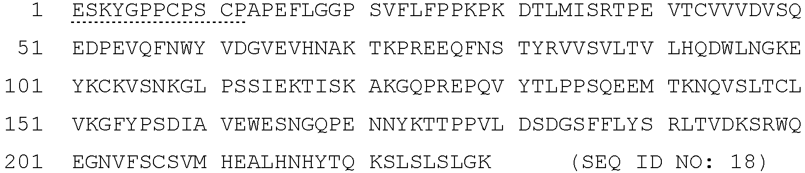
- A variety of engineered mutations in the Fc domain are presented herein with respect to the G1Fc sequence (SEQ ID NO: 14), and analogous mutations in G2Fc, G3Fc, and G4Fc can be derived from their alignment with G 1F c in
Figure 15 . Due to unequal hinge lengths, analogous Fc positions based on isotype alignment (Figure 15 ) possess different amino acid numbers in SEQ ID NOs: 14, 15, 16, 17, and 18. It can also be appreciated that a given amino acid position in an immunoglobulin sequence consisting of hinge,C H2, and CH3 regions (e.g., SEQ ID NOs: 14, 15, 16, 17, and 18) will be identified by a different number than the same position when numbering encompasses the entire IgG1 heavy-chain constant domain (consisting of theC H1, hinge,C H2, and CH3 regions) as in the Uniprot database. For example, correspondence between selected CH3 positions in a human G1Fc sequence (SEQ ID NO: 14), the human IgG1 heavy chain constant domain (Uniprot P01857), and the human IgG1 heavy chain is as follows.Correspondence of CH3 Positions in Different Numbering Systems GIFc (Numbering begins at first threonine in hinge region) IgG1 heavy chain constant domain (Numbering begins at CH1) IgG1 heavy chain (EU numbering scheme of Kabat et al., 1991*) Y127 Y232 Y349 S132 S237 S354 E134 E239 E356 T144 T249 T366 L146 L251 L368 K170 K275 K392 D177 D282 D399 Y185 Y290 Y407 K187 K292 K409 * Kabat et al. (eds) 1991; pp. 688-696 in Sequences of Proteins of Immunological Interest, 5th ed., Vol. 1, NIH, Bethesda, MD. - It is understood that different elements of the fusion proteins (e.g., immunoglobulin Fc fusion proteins) may be arranged in any manner that is consistent with desired functionality. For example, an ActRII polypeptide domain may be placed C-terminal to a heterologous domain, or alternatively, a heterologous domain may be placed C-terminal to an ActRII polypeptide domain. The ActRII polypeptide domain and the heterologous domain need not be adjacent in a fusion protein, and additional domains or amino acid sequences may be included C- or N-terminal to either domain or between the domains.
- For example, an ActRII receptor fusion protein may comprise an amino acid sequence as set forth in the formula A-B-C. The B portion corresponds to an ActRII polypeptide domain. The A and C portions may be independently zero, one, or more than one amino acid, and both the A and C portions when present are heterologous to B. The A and/or C portions may be attached to the B portion via a linker sequence. A linker may be rich in glycine (e.g., 2-10, 2-5, 2-4, 2-3 glycine residues) or glycine and proline residues and may, for example, contain a single sequence of threonine/serine and glycines or repeating sequences of threonine/serine and/or glycines, e.g., GGG (SEQ ID NO: 19), GGGG (SEQ ID NO: 20), TGGGG (SEQ ID NO: 21), SGGGG(SEQ ID NO: 22), TGGG (SEQ ID NO: 23), SGGG (SEQ ID NO: 24), or GGGGS (SEQ ID NO: 25) singlets, or repeats. An ActRII fusion protein may comprise an amino acid sequence as set forth in the formula A-B-C, wherein A is a leader (signal) sequence, B consists of an ActRII polypeptide domain, and C is a polypeptide portion that enhances one or more of in vivo stability, in vivo half-life, uptake/administration, tissue localization or distribution, formation of protein complexes, and/or purification. An ActRII fusion protein may comprise an amino acid sequence as set forth in the formula A-B-C, wherein A is a TPA leader sequence, B consists of an ActRII receptor polypeptide domain, and C is an immunoglobulin Fc domain. Preferred fusion proteins not covered by the claimed invention comprise the amino acid sequence set forth in any one of SEQ ID NOs: 32, 36 and39. Preferred fusion proteins covered by the claimed invention comprise the amino acid sequence as set forth in any one of SEQ ID NOs: 40, 41, 44, 46, 50, 61, 64, 78, and 79.
- ActRII polypeptides may be isolated polypeptides. As used herein, an isolated protein or polypeptide is one which has been separated from a component of its natural environment. A polypeptide of the disclosure may be purified to greater than 95%, 96%, 97%, 98%, or 99% purity as determined by, for example, electrophoretic (e.g., SDS-PAGE, isoelectric focusing (IEF), capillary electrophoresis) or chromatographic (e.g., ion exchange or reverse phase HPLC). Methods for assessment of antibody purity are well known in the art [see, e.g., Flatman et al., (2007) J. Chromatogr. B 848:79-87]. ActRII polypeptides may be recombinant polypeptides.
- ActRII polypeptides of the disclosure can be produced by a variety of art-known techniques. For example, polypeptides of the disclosure can be synthesized using standard protein chemistry techniques such as those described in Bodansky, M. Principles of Peptide Synthesis, Springer Verlag, Berlin (1993) and Grant G. A. (ed.), Synthetic Peptides: A User's Guide, W. H. Freeman and Company, New York (1992). In addition, automated peptide synthesizers are commercially available (e.g., Advanced ChemTech Model 396; Milligen/Biosearch 9600). Alternatively, the polypeptides of the disclosure, including fragments or variants thereof, may be recombinantly produced using various expression systems [e.g., E. coli, Chinese Hamster Ovary (CHO) cells, COS cells, baculovirus] as is well known in the art. Modified or unmodified polypeptides of the disclosure may be produced by digestion of recombinantly produced full-length ActRII polypeptides by using, for example, a protease, e.g., trypsin, thermolysin, chymotrypsin, pepsin, or paired basic amino acid converting enzyme (PACE). Computer analysis (using commercially available software, e.g., MacVector, Omega, PCGene, Molecular Simulation, Inc.) can be used to identify proteolytic cleavage sites. Alternatively, such polypeptides may be produced from recombinantly generated full-length ActRII polypeptides using chemical cleavage (e.g., cyanogen bromide, hydroxylamine, etc.).
- Any of the ActRII polypeptides described herein (e.g., ActRIIA or ActRIIB polypeptides as well as variants thereof such as GDF traps) can be combined with one or more additional ActRII antagonists to achieve the desired effect (e.g., treat or prevent a vascular disorder of the eye in a patient in need thereof, increase vision in patient in need thereof that has a vascular disorder of the eye, and/or treat or prevent one or more complications of a vascular disorder of the eye). For example, an ActRII polypeptide can be used in combination with: i) one or more additional ActRII polypeptides, ii) one or more ActRII antagonist antibodies; iii) one or more small molecule ActRII antagonists; iv) one or more polynucleotide ActRII antagonists; v) one or more follistatin polypeptides; and/or vi) one or more FLRG polypeptides.
- In certain embodiments not covered by the claimed invention, the present disclosure provides isolated and/or recombinant nucleic acids encoding the ActRII polypeptides (including fragments, functional variants (e.g., GDF traps), and fusion proteins thereof). For example, SEQ ID NO: 12 encodes the naturally occurring human ActRIIA precursor polypeptide, while SEQ ID NO: 13 encodes the processed extracellular domain of ActRIIA. In addition, SEQ ID NO: 7 encodes a naturally occurring human ActRIIB precursor polypeptide (the R64 variant described above), while SEQ ID NO: 8 encodes the processed extracellular domain of ActRIIB (the R64 variant described above). The subject nucleic acids may be single-stranded or double stranded. Such nucleic acids may be DNA or RNA molecules. These nucleic acids may be used, for example, in methods for making ActRII-based ligand trap polypeptides as described herein.
- As used herein, isolated nucleic acid(s) refers to a nucleic acid molecule that has been separated from a component of its natural environment. An isolated nucleic acid includes a nucleic acid molecule contained in cells that ordinarily contain the nucleic acid molecule, but the nucleic acid molecule is present extrachromosomally or at a chromosomal location that is different from its natural chromosomal location.
- In certain embodiments not covered by the claimed invention, nucleic acids encoding ActRII polypeptides of the disclosure are understood to include nucleic acids that are variants of any one of SEQ ID NOs: 7, 8, 12, 13, 37, 42, 47, 60, 62, 63, 66, 67, 68, 80, 81, 82, and 83. Variant nucleotide sequences include sequences that differ by one or more nucleotide substitutions, additions, or deletions including allelic variants, and therefore, will include coding sequence that differ from the nucleotide sequence designated in any one of SEQ ID NOs: 7, 8, 12, 13, 37, 42, 47, 60, 62, 63, 66, 67, 68, 80, 81, 82, and 83.
- In certain embodiments not covered by the claimed invention, ActRII polypeptides of the disclosure are encoded by isolated and/or recombinant nucleic acid sequences that are at least 70%, 75%, 80%, 85%, 90%, 91%, 92%, 93%, 94% 95%, 96%, 97%, 98%, 99%, or 100% identical to any one of SEQ ID NOs: 7, 8, 12, 13, 37, 42, 47, 60, 62, 63, 66, 67, 68, 80, 81, 82, and 83. One of ordinary skill in the art will appreciate that nucleic acid sequences that are at least 70%, 75%, 80%, 85%, 90%, 91%, 92%, 93%, 94% 95%, 96%, 97%, 98%, 99%, or 100% identical to the sequences complementary to SEQ ID NOs: 7, 8, 12, 13, 37, 42, 47, 60, 62, 63, 66, 67, 68, 80, 81, 82, and 83, and variants thereof, are also within the scope of the present disclosure. In further embodiments not covered by the claimed invention, the nucleic acid sequences of the disclosure can be isolated, recombinant, and/or fused with a heterologous nucleotide sequence, or in a DNA library.
- In other embodiments not covered by the claimed invention, nucleic acids of the present disclosure also include nucleotide sequences that hybridize under highly stringent conditions to the nucleotide sequence designated in SEQ ID NOs: 7, 8, 12, 13, 37, 42, 47, 60, 62, 63, 66, 67, 68, 80, 81, 82, and 83, complement sequences of SEQ ID NOs: 7, 8, 12, 13, 37, 42, 47, 60, 62, 63, 66, 67, 68, 80, 81, 82, and 83, or fragments thereof. As discussed above, one of ordinary skill in the art will understand readily that appropriate stringency conditions which promote DNA hybridization can be varied. One of ordinary skill in the art will understand readily that appropriate stringency conditions which promote DNA hybridization can be varied. For example, one could perform the hybridization at 6.0 x sodium chloride/sodium citrate (SSC) at about 45 °C, followed by a wash of 2.0 x SSC at 50 °C. For example, the salt concentration in the wash step can be selected from a low stringency of about 2.0 x SSC at 50 °C to a high stringency of about 0.2 x SSC at 50 °C. In addition, the temperature in the wash step can be increased from low stringency conditions at room temperature, about 22 °C, to high stringency conditions at about 65 °C. Both temperature and salt may be varied, or temperature or salt concentration may be held constant while the other variable is changed. In one embodiment not covered by the claimed invention, the disclosure provides nucleic acids which hybridize under low stringency conditions of 6 x SSC at room temperature followed by a wash at 2 x SSC at room temperature.
- Isolated nucleic acids which differ from the nucleic acids as set forth in SEQ ID NOs: 7, 8, 12, 13, 37, 42, 47, 60, 62, 63, 66, 67, 68, 80, 81, 82, and 83 to degeneracy in the genetic code are also within the scope of the disclosure. For example, a number of amino acids are designated by more than one triplet. Codons that specify the same amino acid, or synonyms (for example, CAU and CAC are synonyms for histidine) may result in "silent" mutations which do not affect the amino acid sequence of the protein. However, it is expected that DNA sequence polymorphisms that do lead to changes in the amino acid sequences of the subject proteins will exist among mammalian cells. One skilled in the art will appreciate that these variations in one or more nucleotides (up to about 3-5% of the nucleotides) of the nucleic acids encoding a particular protein may exist among individuals of a given species due to natural allelic variation. Any and all such nucleotide variations and resulting amino acid polymorphisms are within the scope of this disclosure.
- In certain embodiments not covered by the claimed invention, the recombinant nucleic acids of the present disclosure may be operably linked to one or more regulatory nucleotide sequences in an expression construct. Regulatory nucleotide sequences will generally be appropriate to the host cell used for expression. Numerous types of appropriate expression vectors and suitable regulatory sequences are known in the art and can be used in a variety of host cells. Typically, one or more regulatory nucleotide sequences may include, but are not limited to, promoter sequences, leader or signal sequences, ribosomal binding sites, transcriptional start and termination sequences, translational start and termination sequences, and enhancer or activator sequences. Constitutive or inducible promoters as known in the art are contemplated by the disclosure. The promoters may be either naturally occurring promoters, or hybrid promoters that combine elements of more than one promoter. An expression construct may be present in a cell on an episome, such as a plasmid, or the expression construct may be inserted in a chromosome. In some embodiments not covered by the claimed invention, the expression vector contains a selectable marker gene to allow the selection of transformed host cells. Selectable marker genes are well known in the art and can vary with the host cell used.
- In certain aspects not covered by the claimed invention, the subject nucleic acid disclosed herein is provided in an expression vector comprising a nucleotide sequence encoding an ActRII polypeptide and operably linked to at least one regulatory sequence. Regulatory sequences are art-recognized and are selected to direct expression of the ActRII polypeptide. Accordingly, the term regulatory sequence includes promoters, enhancers, and other expression control elements. Exemplary regulatory sequences are described in Goeddel; Gene Expression Technology: Methods in Enzymology, Academic Press, San Diego, CA (1990). For instance, any of a wide variety of expression control sequences that control the expression of a DNA sequence when operatively linked to it may be used in these vectors to express DNA sequences encoding an ActRII polypeptide. Such useful expression control sequences, include, for example, the early and late promoters of SV40, tet promoter, adenovirus or cytomegalovirus immediate early promoter, RSV promoters, the lac system, the trp system, the TAC or TRC system, T7 promoter whose expression is directed by T7 RNA polymerase, the major operator and promoter regions of phage lambda, the control regions for fd coat protein, the promoter for 3-phosphoglycerate kinase or other glycolytic enzymes, the promoters of acid phosphatase, e.g., Pho5, the promoters of the yeast α-mating factors, the polyhedron promoter of the baculovirus system and other sequences known to control the expression of genes of prokaryotic or eukaryotic cells or their viruses, and various combinations thereof. It should be understood that the design of the expression vector may depend on such factors as the choice of the host cell to be transformed and/or the type of protein desired to be expressed. Moreover, the vector's copy number, the ability to control that copy number and the expression of any other protein encoded by the vector, such as antibiotic markers, should also be considered.
- A recombinant nucleic acid can be produced by ligating the cloned gene, or a portion thereof, into a vector suitable for expression in either prokaryotic cells, eukaryotic cells (yeast, avian, insect or mammalian), or both. Expression vehicles for production of a recombinant ActRII polypeptide include plasmids and other vectors. For instance, suitable vectors include plasmids of the following types: pBR322-derived plasmids, pEMBL-derived plasmids, pEX-derived plasmids, pBTac-derived plasmids and pUC-derived plasmids for expression in prokaryotic cells, such as E. coli.
- Some mammalian expression vectors contain both prokaryotic sequences to facilitate the propagation of the vector in bacteria, and one or more eukaryotic transcription units that are expressed in eukaryotic cells. The pcDNAI/amp, pcDNAI/neo, pRc/CMV, pSV2gpt, pSV2neo, pSV2-dhfr, pTk2, pRSVneo, pMSG, pSVT7, pko-neo and pHyg derived vectors are examples of mammalian expression vectors suitable for transfection of eukaryotic cells. Some of these vectors are modified with sequences from bacterial plasmids, such as pBR322, to facilitate replication and drug resistance selection in both prokaryotic and eukaryotic cells. Alternatively, derivatives of viruses such as the bovine papilloma virus (BPV-1), or Epstein-Barr virus (pHEBo, pREP-derived and p205) can be used for transient expression of proteins in eukaryotic cells. Examples of other viral (including retroviral) expression systems can be found below in the description of gene therapy delivery systems. The various methods employed in the preparation of the plasmids and in transformation of host organisms are well known in the art. For other suitable expression systems for both prokaryotic and eukaryotic cells, as well as general recombinant procedures, e.g., Molecular Cloning A Laboratory Manual, 3rd Ed., ed. by Sambrook, Fritsch and Maniatis (Cold Spring Harbor Laboratory Press, 2001). In some instances, it may be desirable to express the recombinant polypeptides by the use of a baculovirus expression system. Examples of such baculovirus expression systems include pVL-derived vectors (such as pVL1392, pVL1393 and pVL941), pAcUW-derived vectors (such as pAcUW1), and pBlueBac-derived vectors (such as the β-gal containing pBlueBac III).
- In an embodiment not covered by the claimed invention, a vector will be designed for production of the subject ActRII polypeptides in CHO cells, such as a Pcmv-Script vector (Stratagene, La Jolla, Calif.), pcDNA4 vectors (Invitrogen, Carlsbad, Calif.) and pCI-neo vectors (Promega, Madison, Wisc.). As will be apparent, the subject gene constructs can be used to cause expression of the subject ActRII polypeptides in cells propagated in culture, e.g., to produce proteins, including fusion proteins or variant proteins, for purification.
- Also disclosed herein but not covered by the claimed invention is a host cell transfected with a recombinant gene including a coding sequence for one or more of the subject ActRII polypeptides. The host cell may be any prokaryotic or eukaryotic cell. For example, an ActRII polypeptide of the disclosure may be expressed in bacterial cells such as E. coli, insect cells (e.g., using a baculovirus expression system), yeast, or mammalian cells [e.g. a Chinese hamster ovary (CHO) cell line]. Other suitable host cells are known to those skilled in the art.
- Accordingly, the present disclosure further pertains to methods not covered by the claimed invention of producing the subject ActRII polypeptides. For example, a host cell transfected with an expression vector encoding an ActRII polypeptide can be cultured under appropriate conditions to allow expression of the ActRII polypeptide to occur. The polypeptide may be secreted and isolated from a mixture of cells and medium containing the polypeptide. Alternatively, the ActRII polypeptide may be retained cytoplasmically or in a membrane fraction and the cells harvested, lysed and the protein isolated. A cell culture includes host cells, media and other byproducts. Suitable media for cell culture are well known in the art. The subject polypeptides can be isolated from cell culture medium, host cells, or both, using techniques known in the art for purifying proteins, including ionexchange chromatography, gel filtration chromatography, ultrafiltration, electrophoresis, immunoaffinity purification with antibodies specific for particular epitopes of the ActRII polypeptides, and affinity purification with an agent that binds to a domain fused to the ActRII polypeptide (e.g., a protein A column may be used to purify an ActRII-Fc fusion protein). In some embodiments not covered by the claimed invention, the ActRII polypeptide is a fusion protein containing a domain which facilitates its purification.
- Purification may be achieved by a series of column chromatography steps, including, for example, three or more of the following, in any order: protein A chromatography, Q sepharose chromatography, phenylsepharose chromatography, size exclusion chromatography, and cation exchange chromatography. The purification could be completed with viral filtration and buffer exchange. An ActRII protein may be purified to a purity of >90%, >95%, >96%, >98%, or >99% as determined by size exclusion chromatography and >90%, >95%, >96%, >98%, or >99% as determined by SDS PAGE. The target level of purity should be one that is sufficient to achieve desirable results in mammalian systems, particularly non-human primates, rodents (mice), and humans.
- A fusion gene coding for a purification leader sequence, such as a poly-(His)/enterokinase cleavage site sequence at the N-terminus of the desired portion of the recombinant ActRII polypeptide, can allow purification of the expressed fusion protein by affinity chromatography using a Ni2+ metal resin. The purification leader sequence can then be subsequently removed by treatment with enterokinase to provide the purified ActRII polypeptide. See, e.g., Hochuli et al. (1987) J. Chromatography 411:177; and Janknecht et al. (1991)PNAS USA 88:8972.
- Techniques for making fusion genes are well known. Essentially, the joining of various DNA fragments coding for different polypeptide sequences is performed in accordance with conventional techniques, employing blunt-ended or stagger-ended termini for ligation, restriction enzyme digestion to provide for appropriate termini, filling-in of cohesive ends as appropriate, alkaline phosphatase treatment to avoid undesirable joining, and enzymatic ligation. The fusion gene may be synthesized by conventional techniques including automated DNA synthesizers. Alternatively, PCR amplification of gene fragments can be carried out using anchor primers which give rise to complementary overhangs between two consecutive gene fragments which can subsequently be annealed to generate a chimeric gene sequence. See, e.g., Current Protocols in Molecular Biology, eds. Ausubel et al., John Wiley & Sons: 1992.
- In other aspects not covered by the claimed invention, the present disclosure relates to an antibody, or combination of antibodies, that antagonize ActRII activity (e.g., inhibition of ActRII signaling transduction via
Smads - In certain aspects not covered by the claimed invention, a preferred antibody ActRII antagonist of the disclosure is an antibody, or combination of antibodies, that binds to and/or inhibits activity of at least
GDF 11. In other aspects not covered by the claimed invention, a preferred antibody ActRII antagonist of the disclosure is an antibody, or combination of antibodies, that binds to and/or inhibits activity of at least GDF8. In other aspects not covered by the claimed invention, a preferred antibody ActRII antagonist of the disclosure is an antibody, or combination of antibodies, that binds to and/or inhibits activity of at least GDF3. In even other aspects not covered by the claimed invention, a preferred antibody ActRII antagonist of the disclosure is an antibody, or combination of antibodies, that binds to and/or inhibits activity of at least BMP6. In still other aspects not covered by the claimed invention, a preferred antibody ActRII antagonist of the disclosure is an antibody, or combination of antibodies, that binds to and/or inhibits activity of at least BMP9. In alternative aspects not covered by the claimed invention, a preferred antibody ActRII antagonist of the disclosure is an antibody, or combination of antibodies, that does not bind to and/or inhibits activity of BMP9. In still even other aspects not covered by the claimed invention, a preferred antibody ActRII antagonist of the disclosure is an antibody, or combination of antibodies, that binds to and/or inhibits activity of at least BMP10. In alternative aspects not covered by the claimed invention, a preferred antibody ActRII antagonist of the disclosure is an antibody, or combination of antibodies, that does not bind to and/or inhibits activity of BMP10. In further aspects not covered by the claimed invention, a preferred antibody ActRII antagonist of the disclosure is an antibody, or combination of antibodies, that binds to and/or inhibits activity of at least activin (e.g., activin A, activin B, activin AB, activin C, and activin D). In some embodiments not covered by the claimed invention, a preferred antibody ActRII antagonist of the disclosure is an antibody, or combination of antibodies, that binds to and/or inhibits activity of at least activin B. In some embodiments not covered by the claimed invention, a preferred antibody ActRII antagonist of the disclosure is an antibody, or combination of antibodies, that binds to and/or inhibits activity of at least activin A and activin B. Alternatively, in some embodiments not covered by the claimed invention, a preferred antibody ActRII antagonist of the disclosure is an antibody, or combination of antibodies, that binds to and/or inhibits activity of at least activin B, but does not bind to and/or inhibit activin A. In even further aspects not covered by the claimed invention, a preferred antibody ActRII antagonist of the disclosure is an antibody, or combination of antibodies, that does not bind to and/or inhibits activity one or more of BMP9, BMP10, and activin A. - In some embodiments not covered by the claimed invention, a preferred antibody ActRII antagonist of the disclosure is an antibody, or combination of antibodies, that binds to and/or inhibits activity of at least GDF11 and GDF8, particularly in the case of a multispecific antibody that has binding affinity for both GDF11 and GDF8 or in the context of a combination of one or more anti-GDF11 antibodies and one or more anti-GDF8 antibodies. In some embodiments not covered by the claimed invention, an antibody, or combination of antibodies, of the disclosure that binds to and/or inhibits the activity of GDF11 and/or GDF8 further binds to and/or inhibits activity of activin (e.g., activin A, activin B, activin AB, activin C, and/or activin E). In some embodiments not covered by the claimed invention, an antibody, or combination of antibodies, of the disclosure that binds to and/or inhibits the activity of
GDF 11 and/or GDF8 further binds to and/or inhibits activity of activin B. In some embodiments not covered by the claimed invention, an antibody, or combination of antibodies, of the disclosure that binds to and/or inhibits the activity of GDF11 and/or GDF8 does not bind to and/or inhibit, or does not substantially bind to and/or inhibit, activity of activin A (e.g., an antibody that has a KD of greater than 1 × 10⁻⁷ for activin A). In some embodiments not covered by the claimed invention, an antibody, or combination of antibodies, of the disclosure that binds to and/or inhibits the activity of GDF11, GDF8, and/or activin further binds to and/or inhibits activity of BMP6. In some embodiments not covered by the claimed invention, an antibody, or combination of antibodies, of the disclosure that binds to and/or inhibits the activity of GDF11, GDF8, activin, and/or BMP6 further binds to and/or inhibits activity of GDF3. In some embodiments not covered by the claimed invention, an antibody, or combination of antibodies, of the disclosure that binds to and/or inhibits the activity of GDF11, GDF8, activin, BMP6, and/or GDF3 further binds to and/or inhibits activity of BMP10. In some embodiments not covered by the claimed invention, an antibody, or combination of antibodies, of the disclosure that binds to and/or inhibits the activity ofGDF 11, GDF8, activin, BMP6, GDF3, and/or BMP10 further binds to and/or inhibits activity of BMP9. In some embodiments not covered by the claimed invention, an antibody, or combination of antibodies, of the disclosure that binds to and/or inhibits the activity of GDF11, GDF8, activin, BMP6, and/or GDF3 does not bind to and/or inhibit BMP9 (e.g., an antibody that has a KD of greater than 1 × 10⁻⁷ for BMP9). In some embodiments not covered by the claimed invention, an antibody, or combination of antibodies, of the disclosure that binds to and/or inhibits the activity of GDF11, GDF8, activin, BMP6, and/or GDF3 does not bind to and/or inhibit BMP10 (e.g., an antibody that has a KD of greater than 1 × 10⁻⁷ for BMP10). In some embodiments not covered by the claimed invention, an antibody, or combination of antibodies, of the disclosure that binds to and/or inhibits the activity of GDF11, GDF8, activin, BMP6, and/or GDF3 does not bind to and/or inhibit BMP9 or BMP10 (e.g., an antibody that has a KD of greater than 1 × 10⁻⁷ for BMP9 and BMP10). In some embodiments not covered by the claimed invention, an antibody, or combination of antibodies, of the disclosure that binds to and/or inhibits the activity of GDF11, GDF8, activin B, BMP6, and/or GDF3 does not bind to and/or inhibit BMP9 (e.g., an antibody that has a KD of greater than 1 × 10⁻⁷ for BMP9). In some embodiments not covered by the claimed invention, an antibody, or combination of antibodies, of the disclosure that binds to and/or inhibits the activity of GDF11, GDF8, activin B, BMP6, and/or GDF3 does not bind to and/or inhibit BMP10 (e.g., an antibody that has a KD of greater than 1 × 10⁻⁷ for BMP10). In some embodiments not covered by the claimed invention, an antibody, or combination of antibodies, of the disclosure that binds to and/or inhibits the activity of GDF11, GDF8, activin B, BMP6, and/or GDF3 does not bind to and/or inhibit BMP9 or BMP10 (e.g., an antibody that has a KD of greater than 1 × 10⁻⁷ for BMP9 and BMP10). In some embodiments not covered by the claimed invention, an antibody, or combination of antibodies, of the disclosure that binds to and/or inhibits the activity of GDF11, GDF8, activin B, BMP6, and/or GDF3 does not bind to and/or inhibit activin A (e.g., an antibody that has a KD of greater than 1 × 10⁻⁷ for activin A). In some embodiments not covered by the claimed invention, an antibody, or combination of antibodies, of the disclosure that binds to and/or inhibits the activity of GDF11, GDF8, activin B, BMP6, and/or GDF3 does not bind to and/or inhibit activin A or BMP10 (e.g., an antibody that has a KD of greater than 1 × 10⁻⁷ for activin A and BMP10). In some embodiments not covered by the claimed invention, an antibody, or combination of antibodies, of the disclosure that binds to and/or inhibits the activity of GDF11, GDF8, activin B, BMP6, and/or GDF3 does not bind to and/or inhibit activin A or BMP9 (e.g., an antibody that has a KD of greater than 1 × 10⁻⁷ for activin A and BMP9). In some embodiments not covered by the claimed invention, an antibody, or combination of antibodies, of the disclosure that binds to and/or inhibits the activity of GDF11, GDF8, activin B, BMP6, and/or GDF3 does not bind to and/or inhibit activin A, BMP9, or BMP10 (e.g., an antibody that has a KD of greater than 1 × 10⁻⁷ for activin A, BMP9, and BMP10). - In another aspect not covered by the claimed invention, an ActRII antagonist of the present disclosure is an antibody, or combination of antibodies, that binds to and/or inhibits activity of an ActRII receptor (e.g. an ActRIIA and/or ActRIIB receptor). In some embodiments not covered by the claimed invention, an anti-ActRII receptor antibody, or combination of antibodies, of the disclosure binds to an ActRII receptor and prevents binding and/or activation of the ActRII receptor by at least GDF11. In some embodiments not covered by the claimed invention, an anti-ActRII receptor antibody, or combination of antibodies, of the disclosure binds to an ActRII receptor and prevents binding and/or activation of the ActRII receptor by at least GDF8. In some embodiments not covered by the claimed invention, an anti-ActRII receptor antibody, or combination of antibodies, of the disclosure binds to an ActRII receptor and prevents binding and/or activation of the ActRII receptor by at least GDF3. In some embodiments not covered by the claimed invention, an anti-ActRII receptor antibody, or combination of antibodies, of the disclosure binds to an ActRII receptor and prevents binding and/or activation of the ActRII receptor by at least BMP6. In some embodiments not covered by the claimed invention, an anti-ActRII receptor antibody, or combination of antibodies, of the disclosure binds to an ActRII receptor and prevents binding and/or activation of the ActRII receptor by at least
BMP 10. In alternative embodiments not covered by the claimed invention, an anti-ActRII receptor antibody, or combination of antibodies, of the disclosure does not prevent binding and/or activation of the ActRII receptor by BMP10. In some embodiments not covered by the claimed invention, an anti-ActRII receptor antibody, or combination of antibodies, of the disclosure binds to an ActRII receptor and prevents binding and/or activation of the ActRII receptor by at least BMP9. In alternative embodiments not covered by the claimed invention, an anti-ActRII receptor antibody, or combination of antibodies, of the disclosure does not prevent binding and/or activation of the ActRII receptor by BMP9. In some embodiments not covered by the claimed invention, an anti-ActRII receptor antibody, or combination of antibodies, of the disclosure binds to an ActRII receptor and prevents binding and/or activation of the ActRII receptor by at least activin (e.g., activin A, activin B, activin AB, activin C, and activin E). In some embodiments not covered by the claimed invention, an anti-ActRII receptor antibody, or combination of antibodies, of the disclosure binds to an ActRII receptor and prevents binding and/or activation of the ActRII receptor by at least activin B. In some embodiments not covered by the claimed invention, an anti-ActRII receptor antibody, or combination of antibodies, of the disclosure does not prevent binding and/or activation of the ActRII receptor by activin A. In some embodiments not covered by the claimed invention, an anti-ActRII receptor antibody, or combination of antibodies, of the disclosure binds to an ActRII receptor and prevents binding and/or activation of the ActRII receptor by at least activin B, but does not prevent binding and/or activation of the ActRII receptor by activin A. - The term antibody is used herein in the broadest sense and encompasses various antibody structures, including but not limited to monoclonal antibodies, polyclonal antibodies, multispecific antibodies (e.g., bispecific antibodies), and antibody fragments so long as they exhibit the desired antigen-binding activity. An antibody fragment refers to a molecule other than an intact antibody that comprises a portion of an intact antibody that binds the antigen to which the intact antibody binds. Examples of antibody fragments include but are not limited to Fv, Fab, Fab', Fab'-SH, F(ab')2; diabodies; linear antibodies; single-chain antibody molecules (e.g., scFv); and multispecific antibodies formed from antibody fragments. See, e.g., Hudson et al. (2003) Nat. Med. 9:129-134; Plückthun, in The Pharmacology of Monoclonal Antibodies, vol. 113, Rosenburg and Moore eds., (Springer-Verlag, New York), pp. 269-315 (1994);
WO 93/16185 U.S. Pat. Nos. 5,571,894 ,5,587,458 , and5,869,046 . Antibodies disclosed herein may be polyclonal antibodies or monoclonal antibodies. The antibodies may comprise a label attached thereto and able to be detected (e.g., the label can be a radioisotope, fluorescent compound, enzyme, or enzyme cofactor). he antibodies may be isolated antibodies. - Diabodies are antibody fragments with two antigen-binding sites that may be bivalent or bispecific. See, e.g.,
EP 404,097 WO 1993/01161 - Single-domain antibodies are antibody fragments comprising all or a portion of the heavy-chain variable domain or all or a portion of the light-chain variable domain of an antibody. A single-domain antibody is a human single-domain antibody. See, e.g.,
U.S. Pat. No. 6,248,516 . - Antibody fragments can be made by various techniques, including but not limited to proteolytic digestion of an intact antibody as well as production by recombinant host cells (e.g., E. coli or phage), as described herein.
- The antibodies herein may be of any class. The class of an antibody refers to the type of constant domain or constant region possessed by its heavy chain. There are five major classes of antibodies: IgA, IgD, IgE, IgG, and IgM, and several of these may be further divided into subclasses (isotypes), for example, IgG1, IgG2, IgG3, IgG4, IgA1, and IgA2. The heavy-chain constant domains that correspond to the different classes of immunoglobulins are called alpha, delta, epsilon, gamma, and mu.
- In general, an antibody for use in the herein described unclaimed methods specifically binds to its target antigen, preferably with high binding affinity. Affinity may be expressed as a KD value and reflects the intrinsic binding affinity (e.g., with minimized avidity effects). Typically, binding affinity is measured in vitro, whether in a cell-free or cell-associated setting. Any of a number of assays known in the art, including those disclosed herein, can be used to obtain binding affinity measurements including, for example, surface plasmon resonance (Biacore™ assay), radiolabeled antigen binding assay (RIA), and ELISA. Antibodies may bind to their target antigens (e.g. GDF11, GDF8, ActRIIA, ActRIIB, etc.) with at least a KD of
1x 10⁻⁷ or stronger, 1×10⁻⁸ or stronger, 1×10⁻⁹ or stronger, 1×10⁻¹⁰ or stronger, 1×10-11 or stronger, 1×10-12 or stronger, 1×10-13 or stronger, or 1×10-14 or stronger. - KD may be measured by RIA performed with the Fab version of an antibody of interest and its target antigen as described by the following assay. Solution binding affinity of Fabs for the antigen is measured by equilibrating Fab with a minimal concentration of radiolabeled antigen (e.g., 125I-labeled) in the presence of a titration series of unlabeled antigen, then capturing bound antigen with an anti-Fab antibody-coated plate [see, e.g., Chen et al. (1999) J. Mol. Biol. 293:865-881]. To establish conditions for the assay, multi-well plates (e.g., MICROTITER® from Thermo Scientific) are coated (e.g., overnight) with a capturing anti-Fab antibody (e.g., from Cappel Labs) and subsequently blocked with bovine serum albumin, preferably at room temperature (e.g., approximately 23°C). In a non-adsorbent plate, radiolabeled antigen are mixed with serial dilutions of a Fab of interest [e.g., consistent with assessment of the anti-VEGF antibody, Fab-12, in Presta et al., (1997) Cancer Res. 57:4593-4599]. The Fab of interest is then incubated, preferably overnight but the incubation may continue for a longer period (e.g., about 65 hours) to ensure that equilibrium is reached. Thereafter, the mixtures are transferred to the capture plate for incubation, preferably at room temperature for about one hour. The solution is then removed and the plate is washed times several times, preferably with
polysorbate 20 and PBS mixture. When the plates have dried, scintillant (e.g., MICROSCINT® from Packard) is added, and the plates are counted on a gamma counter (e.g., TOPCOUNT® from Packard). - KD may be measured using surface plasmon resonance assays using, for example a BIACORE® 2000 or a BIACORE® 3000 (Biacore, Inc., Piscataway, N.J.) with immobilized antigen CM5 chips at about 10 response units (RU). Briefly, carboxymethylated dextran biosensor chips (CM5, Biacore, Inc.) are activated with N-ethyl-N'-(3-dimethylaminopropyl)-carbodiimide hydrochloride (EDC) and N-hydroxysuccinimide (NHS) according to the supplier's instructions. For example, an antigen can be diluted with 10 mM sodium acetate, pH 4.8, to 5 µg/ml (about 0.2 µM) before injection at a flow rate of 5 µl/minute to achieve approximately 10 response units (RU) of coupled protein. Following the injection of antigen, 1 M ethanolamine is injected to block unreacted groups. For kinetics measurements, twofold serial dilutions of Fab (0.78 nM to 500 nM) are injected in PBS with 0.05% polysorbate 20 (TWEEN-20®) surfactant (PBST) at at a flow rate of approximately 25 µl/min. Association rates (kon) and dissociation rates (koff) are calculated using, for example, a simple one-to-one Langmuir binding model (BIACORE® Evaluation Software version 3.2) by simultaneously fitting the association and dissociation sensorgrams. The equilibrium dissociation constant (KD) is calculated as the ratio koff / kon [see, e.g., Chen et al., (1999) J. Mol. Biol. 293:865-881]. If the on-rate exceeds, for example, 106 M-1 s-1 by the surface plasmon resonance assay above, then the on-rate can be determined by using a fluorescent quenching technique that measures the increase or decrease in fluorescence emission intensity (e.g., excitation=295 nm; emission=340 nm, 16 nm band-pass) of a 20 nM anti-antigen antibody (Fab form) in PBS in the presence of increasing concentrations of antigen as measured in a spectrometer, such as a stop-flow equipped spectrophometer (Aviv Instruments) or a 8000-series SLM-AMINCO® spectrophotometer (ThermoSpectronic) with a stirred cuvette.
- As used herein, anti-GDF11 antibody generally refers to an antibody that is capable of binding to GDF11 with sufficient affinity such that the antibody is useful as a diagnostic and/or therapeutic agent in targeting GDF11. The extent of binding of an anti-GDF11 antibody to an unrelated, non-GDF11 protein may be less than about 10%, 9%, 8%, 7%, 6%, 5%, 4%, 3%, 2%, or less than 1% of the binding of the antibody to GDF11 as measured, for example, by a radioimmunoassay (RIA). An anti-GDF11 antibody may bind to an epitope of GDF11 that is conserved among GDF11 from different species. An anti-GDF11 antibody may be an antagonist antibody that can inhibit GDF11 activity. For example, an anti-GDF11 antibody may inhibit GDF11 from binding to a cognate receptor (e.g., ActRIIA or ActRIIB receptor) and/or inhibit GDF11-mediated signal transduction (activation) of a cognate receptor, such as Smad signaling by an ActRII receptor. Anti-GDF11 antibodies, particularly in the case of multispecific antibodies, may not substantially bind to and/or inhibit activity of activin A (e.g., bind to activin A with a KD of greater than 1 × 10⁻⁷ M or has relatively modest binding, e.g., about 1 × 10⁻⁸ M or about 1 × 10⁻⁹ M It should be noted that GDF11 has high sequence homology to GDF8 and therefore antibodies that bind and/or to GDF11, in some cases, may also bind to and/or inhibit GDF8.
- As used herein, anti-GDF8 antibody generally refers to an antibody that is capable of binding to GDF8 with sufficient affinity such that the antibody is useful as a diagnostic and/or therapeutic agent in targeting GDF8. The extent of binding of an anti-GDF8 antibody to an unrelated, non-GDF8 protein may be less than about 10%, 9%, 8%, 7%, 6%, 5%, 4%, 3%, 2%, or less than 1% of the binding of the antibody to GDF8 as measured, for example, by a radioimmunoassay (RIA). An anti-GDF8 antibody may bind to an epitope of GDF8 that is conserved among GDF8 from different species. An anti-GDF8 antibody may be an antagonist antibody that can inhibit GDF8 activity. For example, an anti-GDF8 antibody may inhibit GDF8 from binding to a cognate receptor (e.g., ActRIIA or ActRIIB receptor) and/or inhibit GDF8-mediated signal transduction (activation) of a cognate receptor, such as Smad signaling by an ActRII receptor. Anti-GDF8 antibodies, particularly in the case of multispecific antibodies, may not substantially bind to and/or inhibit activity of activin A (e.g., bind to activin A with a KD of greater than 1 × 10⁻⁷ M or has relatively modest binding, e.g., about 1 × 10⁻⁸ M or about 1 × 10⁻⁹ M). It should be noted that GDF8 has high sequence homology to GDF11 and therefore antibodies that bind and/or to GDF8, in some cases, may also bind to and/or inhibit
GDF 11. - As used herein, anti-activin antibody generally refers to an antibody that is capable of binding to activin (e.g., one or more of activin A, activin B, activin C, activin AB, and/ or activin E) with sufficient affinity such that the antibody is useful as a diagnostic and/or therapeutic agent in targeting activin. The extent of binding of an anti-activin antibody to an unrelated, non-activin protein may be less than about 10%, 9%, 8%, 7%, 6%, 5%, 4%, 3%, 2%, or less than 1% of the binding of the antibody to activin as measured, for example, by a radioimmunoassay (RIA). An anti-activin antibody may bind to an epitope of activin that is conserved among activin from different species. An anti-activin antibody may be an antagonist antibody that can inhibit activin activity. For example, an anti-activin antibody of the disclosure may inhibit activin from binding to a cognate receptor (e.g., ActRIIA or ActRIIB receptor) and/or inhibit activin-mediated signal transduction (activation) of a cognate receptor, such as Smad signaling by an ActRII receptor. Anti-activin antibodies may bind to and/or inhibit activity of activin B. Anti-activin antibodies may bind to and/or inhibit activity of activin A and activin BAnti-activin antibodies, particularly in the case of multispecific antibodies, may not substantially bind to and/or inhibit activity of activin A (e.g., bind to activin A with a KD of greater than 1 × 10⁻⁷ M or has relatively modest binding, e.g., about 1 × 10⁻⁸ M or about 1 × 10⁻⁹ M).
- As used herein, anti-BMP6 antibody generally refers to an antibody that is capable of binding to BMP6 with sufficient affinity such that the antibody is useful as a diagnostic and/or therapeutic agent in targeting BMP6. The extent of binding of an anti-BMP6 antibody to an unrelated, non-BMP6 protein may be less than about 10%, 9%, 8%, 7%, 6%, 5%, 4%, 3%, 2%, or less than 1% of the binding of the antibody to BMP6 as measured, for example, by a radioimmunoassay (RIAAn anti-BMP6 antibody may bind to an epitope of BMP6 that is conserved among BMP6 from different species. An anti-BMP6 antibody may be an antagonist antibody that can inhibit BMP6 activity. For example, an anti-BMP6 antibody may inhibit BMP6 from binding to a cognate receptor (e.g., ActRIIA or ActRIIB receptor) and/or inhibit BMP6-mediated signal transduction (activation) of a cognate receptor, such as Smad signaling by an ActRII receptor. Anti-BMP6 antibodies, particularly in the case of multispecific antibodies, may not substantially bind to and/or inhibit activity of activin A (e.g., bind to activin A with a KD of greater than 1 × 10⁻⁷ M or has relatively modest binding, e.g., about 1 × 10⁻⁸ M or about 1 × 10⁻⁹ M).
- As used herein, anti-GDF3 antibody generally refers to an antibody that is capable of binding to GDF3 with sufficient affinity such that the antibody is useful as a diagnostic and/or therapeutic agent in targeting GDF3. The extent of binding of an anti- GDF3 antibody to an unrelated, non-GDF3 protein may be less than about 10%, 9%, 8%, 7%, 6%, 5%, 4%, 3%, 2%, or less than 1% of the binding of the antibody to GDF3 as measured, for example, by a radioimmunoassay (RIA). An anti-GDF3 antibody may bind to an epitope of GDF3 that is conserved among GDF3 from different species. An anti-GDF3 antibody may be an antagonist antibody that can inhibit GDF3 activity. For example, an anti-GDF3 antibody may inhibit GDF3 from binding to a cognate receptor (e.g., ActRIIA or ActRIIB receptor) and/or inhibit GDF3-mediated signal transduction (activation) of a cognate receptor, such as Smad signaling by an ActRII receptor. Anti-GDF3 antibodies, particularly in the case of multispecific antibodies, may not substantially bind to and/or inhibit activity of activin A (e.g., bind to activin A with a KD of greater than 1 × 10⁻⁷ M or has relatively modest binding, e.g., about 1 × 10⁻⁸ M or about 1 × 10⁻⁹ M).
- As used herein, anti-BMP10 antibody generally refers to an antibody that is capable of binding to BMP10 with sufficient affinity such that the antibody is useful as a diagnostic and/or therapeutic agent in targeting BMP10. The extent of binding of an anti- BMP10 antibody to an unrelated, non-BMP10 protein may be less than about 10%, 9%, 8%, 7%, 6%, 5%, 4%, 3%, 2%, or less than 1% of the binding of the antibody to BMP10 as measured, for example, by a radioimmunoassay (RIA). An anti-BMP10 antibody may bind to an epitope of BMP10 that is conserved among BMP10 from different species. An anti-BMP10 antibody may be an antagonist antibody that can inhibit BMP10 activity. For example, an anti-BMP10 antibody may inhibit BMP10 from binding to a cognate receptor (e.g., ActRIIA or ActRIIB receptor) and/or inhibit BMP10-mediated signal transduction (activation) of a cognate receptor, such as Smad signaling by an ActRII receptor. Anti-BMP10 antibodies, particularly in the case of multispecific antibodies, may not substantially bind to and/or inhibit activity of activin A (e.g., bind to activin A with a KD of greater than 1 × 10⁻⁷ M or has relatively modest binding, e.g., about 1 × 10⁻⁸ M or about 1 × 10⁻⁹ M
- As used herein, anti-BMP9 antibody generally refers to an antibody that is capable of binding to BMP9 with sufficient affinity such that the antibody is useful as a diagnostic and/or therapeutic agent in targeting BMP9. The extent of binding of an anti- BMP9 antibody to an unrelated, non-BMP9 protein may be less than about 10%, 9%, 8%, 7%, 6%, 5%, 4%, 3%, 2%, or less than 1% of the binding of the antibody to BMP9 as measured, for example, by a radioimmunoassay (RIA). An anti-BMP9 antibody may bins to an epitope of BMP9 that is conserved among BMP9 from different species. An anti-BMP9 antibody may be an antagonist antibody that can inhibit BMP9 activity. For example, an anti-BMP9 antibody may inhibit BMP9 from binding to a cognate receptor (e.g., ActRIIA or ActRIIB receptor) and/or inhibit BMP9-mediated signal transduction (activation) of a cognate receptor, such as Smad signaling by an ActRII receptor. Anti-BMP9 antibodies, particularly in the case of multispecific antibodies, may not substantially bind to and/or inhibit activity of activin A (e.g., bind to activin A with a KD of greater than 1 x 10⁻⁷ M or has relatively modest binding, e.g., about 1 × 10⁻⁸ M or about 1 × 10⁻⁹ M).
- An anti-ActRII antibody refers to an antibody that is capable of binding to ActRII (ActRIIA and/or ActRIIB) with sufficient affinity such that the antibody is useful as a diagnostic and/or therapeutic agent in targeting ActRII. The extent of binding of an anti-ActRII antibody to an unrelated, non-ActRII protein may be less than about 10%, 9%, 8%, 7%, 6%, 5%, 4%, 3%, 2%, or less than 1% of the binding of the antibody to ActRII as measured, for example, by a radioimmunoassay (RIA). An anti-ActRII antibody may bind to an epitope of ActRII that is conserved among ActRII from different species. An anti-ActRII antibody may be an antagonist antibody that can inhibit ActRII activity. For example, an anti-ActRII antibody may inhibit one or more ActRII ligands selected from activin A, activin B, activin AB, activin C, activin E, GDF11, GDF8, activin A, BMP6, and BMP7 from binding to the ActRII receptor and/or inhibit one of these ligands from activating ActRII signaling (e.g.,
Smad - The nucleic acid and amino acid sequences of human ActRII receptors and ligands (e.g., GDF11, GDF8, activin A, activin B, activin C, activin E, BMP6, BMP10, ActRIIB, and ActRIIA) are well known in the art and thus antibody antagonists for use in accordance with this disclosure may be routinely made by the skilled artisan based on the knowledge in the art and teachings provided herein.
- In certain embodiments not covered by the claimed invention, an antibody is a chimeric antibody. A chimeric antibody refers to an antibody in which a portion of the heavy and/or light chain is derived from a particular source or species, while the remainder of the heavy and/or light chain is derived from a different source or species. Certain chimeric antibodies are described, for example, in
U.S. Pat. No. 4,816,567 ; and Morrison et al., (1984) Proc. Natl. Acad. Sci. USA, 81:6851-6855. A chimeric antibody may comprise a non-human variable region (e.g., a variable region derived from a mouse, rat, hamster, rabbit, or non-human primate, such as a monkey) and a human constant region. A chimeric antibody may be a "class switched" antibody in which the class or subclass has been changed from that of the parent antibody. In general, chimeric antibodies include antigen-binding fragments thereof. - A chimeric antibody may be a humanized antibody. A humanized antibody refers to a chimeric antibody comprising amino acid residues from non-human hypervariable regions (HVRs) and amino acid residues from human framework regions (FRs). A humanized antibody may comprise substantially all of at least one, and typically two, variable domains, in which all or substantially all of the HVRs (e.g., CDRs) correspond to those of a non-human antibody, and all or substantially all of the FRs correspond to those of a human antibody. A humanized antibody optionally may comprise at least a portion of an antibody constant region derived from a human antibody. A "humanized form" of an antibody, e.g., a non-human antibody, refers to an antibody that has undergone humanization.
- Humanized antibodies and methods of making them are reviewed, for example, in Almagro and Fransson (2008) Front. Biosci. 13:1619-1633 and are further described, for example, in Riechmann et al., (1988) Nature 332:323-329; Queen et al. (1989) Proc. Nat'l Acad. Sci. USA 86:10029-10033;
U.S. Pat. Nos. 5,821,337 ,7,527,791 ,6,982,321 , and7,087,409 ; Kashmiri et al., (2005) Methods 36:25-34 [describing SDR (a-CDR) grafting]; Padlan, Mol. Immunol. (1991) 28:489-498 (describing "resurfacing"); Dall'Acqua et al. (2005) Methods 36:43-60 (describing "FR shuffling"); Osbourn et al. (2005) Methods 36:61-68; and Klimka et al. Br. J. Cancer (2000) 83:252-260 (describing the "guided selection" approach to FR shuffling). - Human framework regions that may be used for humanization include but are not limited to: framework regions selected using the "best-fit" method [see, e.g., Sims et al. (1993) J. Immunol. 151:2296 ]; framework regions derived from the consensus sequence of human antibodies of a particular subgroup of light-chain or heavy-chain variable regions [see, e.g., Carter et al. (1992) Proc. Natl. Acad. Sci. USA, 89:4285; and Presta et al. (1993) J. Immunol., 151:2623]; human mature (somatically mutated) framework regions or human germline framework regions [see, e.g., Almagro and Fransson (2008) Front. Biosci. 13:1619-1633]; and framework regions derived from screening FR libraries [see, e.g., Baca et cd., (1997) J. Biol. Chem. 272:10678-10684; and Rosok et cd., (1996) J. Biol. Chem. 271:22611-22618].
- An antibody may be a human antibody. Human antibodies can be produced using various techniques known in the art. Human antibodies are described generally in van Dijk and van de Winkel (2001) Curr. Opin. Pharmacol. 5: 368-74 and Lonberg (2008) Curr. Opin. Immunol. 20:450-459.
- Human antibodies may be prepared by administering an immunogen (e.g., a GDF11 polypeptide, GDF8 polypeptide, an ActRIIA polypeptide, or an ActRIIB polypeptide) to a transgenic animal that has been modified to produce intact human antibodies or intact antibodies with human variable regions in response to antigenic challenge. Such animals typically contain all or a portion of the human immunoglobulin loci, which replace the endogenous immunoglobulin loci, or which are present extrachromosomally or integrated randomly into the animal's chromosomes. In such transgenic animals, the endogenous immunoglobulin loci have generally been inactivated. For a review of methods for obtaining human antibodies from transgenic animals, see, for example, Lonberg (2005) Nat. Biotechnol. 23:1117-1125;
U.S. Pat. Nos. 6,075,181 and6,150,584 (describing XENOMOUSE™ technology);U.S. Pat. No. 5,770,429 (describing HuMab® technology);U.S. Pat. No. 7,041,870 (describing K-M MOUSE® technology); andU.S. Patent Application Publication No. 2007/0061900 (describing VelociMouse® technology). Human variable regions from intact antibodies generated by such animals may be further modified, for example, by combining with a different human constant region. - Human antibodies provided herein can also be made by hybridoma-based methods. Human myeloma and mouse-human heteromyeloma cell lines for the production of human monoclonal antibodies have been described [see, e.g., Kozbor J. Immunol., (1984) 133: 3001; Brodeur et al. (1987) Monoclonal Antibody Production Techniques and Applications, pp. 51-63, Marcel Dekker, Inc., New York; and Boerner et al. (1991) J. Immunol., 147: 86]. Human antibodies generated via human B-cell hybridoma technology are also described in Li etal., (2006) Proc. Natl. Acad. Sci. USA, 103:3557-3562. Additional methods include those described, for example, in
U.S. Pat. No. 7,189,826 (describing production of monoclonal human IgM antibodies from hybridoma cell lines) and Ni, Xiandai Mianyixue (2006) 26(4):265-268 (2006) (describing human-human hybridomas). Human hybridoma technology (Trioma technology) is also described in Vollmers and Brandlein (2005) Histol. Histopathol., 20(3):927-937 (2005) and Vollmers and Brandlein (2005) Methods Find Exp. Clin. Pharmacol., 27(3):185-91. - Human antibodies provided herein may also be generated by isolating Fv clone variable-domain sequences selected from human-derived phage display libraries. Such variable-domain sequences may then be combined with a desired human constant domain. Techniques for selecting human antibodies from antibody libraries are described herein.
- For example, antibodies of the present disclosure may be isolated by screening combinatorial libraries for antibodies with the desired activity or activities. A variety of methods are known in the art for generating phage-display libraries and screening such libraries for antibodies possessing the desired binding characteristics. Such methods are reviewed, for example, in Hoogenboom et al. (2001) in Methods in Molecular Biology 178:1-37, O'Brien et al., ed., Human Press, Totowa, N.J. and further described, for example, in the McCafferty et al. (1991) Nature 348:552-554; Clackson et al., (1991) Nature 352: 624-628; Marks et al. (1992) J. Mol. Biol. 222:581-597; Marks and Bradbury (2003) in Methods in Molecular Biology 248:161-175, Lo, ed., Human Press, Totowa, N.J.; Sidhu et al. (2004) J. Mol. Biol. 338(2):299-310; Lee et al. (2004) J. Mol. Biol. 340(5):1073-1093; Fellouse (2004) Proc. Natl. Acad. Sci. USA 101(34):12467-12472; and Lee et al. (2004) J. Immunol. Methods 284(1-2): 119-132.
- In certain phage display methods, repertoires of VH and VL genes are separately cloned by polymerase chain reaction (PCR) and recombined randomly in phage libraries, which can then be screened for antigen-binding phage as described in Winter et al. (1994) Ann. Rev. Immunol., 12: 433-455. Phage typically display antibody fragments, either as single-chain Fv (scFv) fragments or as Fab fragments. Libraries from immunized sources provide high-affinity antibodies to the immunogen (e.g., GDF11, activin B, ActRIIA, or ActRIIB) without the requirement of constructing hybridomas. Alternatively, the naive repertoire can be cloned (e.g., from human) to provide a single source of antibodies directed against a wide range of non-self and also self-antigens without any immunization as described by Griffiths et al. (1993) EMBO J, 12: 725-734. Finally, naive libraries can also be made synthetically by cloning un-rearranged V-gene segments from stem cells and using PCR primers containing random sequence to encode the highly variable CDR3 regions and to accomplish rearrangement in vitro, as described by Hoogenboom and Winter (1992) J. Mol. Biol., 227: 381-388. Patent publications describing human antibody phage libraries include, for example:
U.S. Pat. No. 5,750,373 , andU.S. Patent Publication Nos. 2005/0079574 ,2005/0119455 ,2005/0266000 ,2007/0117126 ,2007/0160598 ,2007/0237764 ,2007/0292936 , and2009/0002360 . - An antibody may be a multispecific antibody, for example, a bispecific antibody. Multispecific antibodies (typically monoclonal antibodies) have binding specificities for at least two different epitopes (e.g., two, three, four, five, or six or more) on one or more (e.g., two, three, four, five, six or more) antigens.
- A multispecific antibody may comprise two or more binding specificities, with at least one of the binding specificities being for a
GDF 11 epitope, and optionally one or more additional binding specificities being for an epitope on a different ActRII ligand (e.g., GDF8, activin, BMP6, and/or BMP10) and/or an ActRII receptor (e.g., an ActRIIA and/or ActRIIB receptor). Multispecific antibodies may bind to two or more different epitopes ofGDF 11. A multispecific antibody that has binding affinity, in part, for a GDF11 epitope may be used to inhibit a GDF11 activity (e.g., the ability to bind to and/or activate an ActRII receptor), and optionally inhibit the activity of one or more different ActRII ligands (e.g., GDF8, activin, BMP6, and/or BMP10) and/or an ActRII receptor (e.g., an ActRIIA or ActRIIB receptor). Multispecific antibodies that bind to and/or inhibit GDF11 may further bind to and/or inhibit at least GDF8. Optionally, multispecific antibodies that bind to and/or inhibit GDF11 may not substantially bind to and/or substantially inhibit activin A. - A multispecific antibody may comprise two or more binding specificities, with at least one of the binding specificities being for a GDF8 epitope, and optionally one or more additional binding specificities being for an epitope on a different ActRII ligand (e.g., GDF11, activin, BMP6, BMP10, BMP9, and/or GDF3) and/or an ActRII receptor (e.g., an ActRIIA and/or ActRIIB receptor). Multispecific antibodies may bind to two or more different epitopes of GDF8. A multispecific antibody that has binding affinity, in part, for an GDF8 epitope may be used to inhibit an GDF8 activity (e.g., the ability to bind to and/or activate an ActRII receptor), and optionally inhibit the activity of one or more different ActRII ligands (e.g., GDF11, activin, BMP6, BMP10, BMP9, and/or GDF3) and/or an ActRII receptor (e.g., an ActRIIA or ActRIIB receptor). Multispecific antibodies that bind to and/or inhibit GDF8 may further bind to and/or inhibit at least GDF11. Optionally, multispecific antibodies that bind to and/or inhibit GDF8 may further bind to and/or inhibit at least activin (e.g., activin A, activin B, activin AB, activin C, and activin E). Multispecific antibodies that bind to and/or inhibit GDF8 may further bind to and/or inhibit at least activin B. Multispecific antibodies that bind to and/or inhibit GDF8 may not substantially bind to and/or substantially inhibit activin A. Optionally, multispecific antibodies that bind to and/or inhibit GDF8 may further bind to and/or inhibit at least BMP6. Optionally, multispecific antibodies that bind to and/or inhibit GDF8 may further bind to and/or inhibit at least BMP9. Multispecific antibodies that bind to and/or inhibit GDF8 may not substantially bind to and/or substantially inhibit BMP9. Optionally, multispecific antibodies that bind to and/or inhibit GDF8 may further bind to and/or inhibit at least BMP10. Multispecific antibodies that bind to and/or inhibit GDF8 may not substantially bind to and/or substantially inhibit BMP10. Optionally, multispecific antibodies that bind to and/or inhibit GDF8 may further bind to and/or inhibit at least GDF3.
- A multispecific antibody may comprise two or more binding specificities, with at least one of the binding specificities being for a
GDF 11 epitope, and optionally one or more additional binding specificities being for an epitope on a different ActRII ligand (e.g., GDF8, activin, BMP6, BMP10, BMP9, and/or GDF3) and/or an ActRII receptor (e.g., an ActRIIA and/or ActRIIB receptor). Multispecific antibodies may bind to two or more different epitopes of GDF11. A multispecific antibody that has binding affinity, in part, for an GDF11 epitope may be used to inhibit an GDF11 activity (e.g., the ability to bind to and/or activate an ActRII receptor), and optionally inhibit the activity of one or more different ActRII ligands (e.g., GDF8, activin, BMP6, BMP10, BMP9, and/or GDF3) and/or an ActRII receptor (e.g., an ActRIIA or ActRIIB receptor). Multispecific antibodies that bind to and/or inhibit GDF11 may further bind to and/or inhibit at least GDF8. Optionally, multispecific antibodies that bind to and/or inhibit GDF11 may further bind to and/or inhibit at least activin (e.g., activin A, activin B, activin AB, activin C, and activin E). Multispecific antibodies that bind to and/or inhibit GDF11 may further bind to and/or inhibit at least activin BMultispecific antibodies that bind to and/or inhibit GDF11 may not substantially bind to and/or substantially inhibit activin A. Optionally, multispecific antibodies that bind to and/or inhibit GDF11 may further bind to and/or inhibit at least BMP6. Optionally, multispecific antibodies that bind to and/or inhibit GDF11 may further bind to and/or inhibit at least BMP9. Multispecific antibodies that bind to and/or inhibit GDF11 may not substantially bind to and/or substantially inhibit BMP9. Optionally, multispecific antibodies that bind to and/or inhibit GDF11 may further bind to and/or inhibit at least BMP10. Multispecific antibodies that bind to and/or inhibit GDF11 may not substantially bind to and/or substantially inhibit BMP10. Optionally, multispecific antibodies that bind to and/or inhibit GDF11 may further bind to and/or inhibit at least GDF3. - A multispecific antibody of the present disclosure may comprise two or more binding specificities, with at least one of the binding specificities being for activin, and optionally one or more additional binding specificities being for an epitope on a different ActRII ligand (e.g., GDF11, GDF8, BMP6, BMP10, BMP9, and/or GDF3) and/or an ActRII receptor (e.g., an ActRIIA and/or ActRIIB receptor). Multispecific antibodies may bind to two or more different epitopes of activin or may bind to two or more different epitopes on different types of activin (e.g., binds an activin A epitope and binds an activin B epitope). A multispecific antibody that has binding affinity, in part, for an activin epitope may be used to inhibit an activin activity (e.g., the ability to bind to and/or activate an ActRII receptor), and optionally inhibit the activity of one or more different ActRII ligands (e.g., GDF11, GDF8, BMP6, BMP10, BMP9, and/or GDF3) and/or an ActRII receptor (e.g., an ActRIIA or ActRIIB receptor). Multispecific antibodies that bind to and/or inhibit activin may further bind to and/or inhibit at least GDF11. Optionally, multispecific antibodies that bind to and/or inhibit activin may further bind to and/or inhibit at least GDF8. Multispecific antibodies that bind to and/or inhibit activin B. Multispecific antibodies of the disclosure that bind to and/or inhibit activin B may not substantially bind to and/or substantially inhibit activin A. Optionally, multispecific antibodies that bind to and/or inhibit activin may further bind to and/or inhibit at least BMP6. Optionally, multispecific antibodies that bind to and/or inhibit activin may further bind to and/or inhibit at least BMP9. Multispecific antibodies that bind to and/or inhibit activin may not substantially bind to and/or substantially inhibit BMP9. Optionally, multispecific antibodies that bind to and/or inhibit activin may further bind to and/or inhibit at least BMP10. Multispecific antibodies that bind to and/or inhibit activin may not substantially bind to and/or substantially inhibit BMP10. Optionally, multispecific antibodies that bind to and/or inhibit activin may further bind to and/or inhibit at least GDF3.
- Engineered antibodies with three or more functional antigen binding sites, including "octopus antibodies," are also included herein (see, e.g.,
US 2006/0025576A1 ). - The antibodies may be monoclonal antibodies. Monoclonal antibody refers to an antibody obtained from a population of substantially homogeneous antibodies, i.e., the individual antibodies comprising the population are identical and/or bind the same epitope, except for possible variant antibodies, e.g., containing naturally occurring mutations or arising during production of a monoclonal antibody preparation, such variants generally being present in minor amounts. In contrast to polyclonal antibody preparations, which typically include different antibodies directed against different epitopes, each monoclonal antibody of a monoclonal antibody preparation is directed against a single epitope on an antigen. Thus, the modifier "monoclonal" indicates the character of the antibody as being obtained from a substantially homogeneous population of antibodies and is not to be construed as requiring production of the antibody by any particular method. For example, the monoclonal antibodies to be used in accordance with the present methods may be made by a variety of techniques, including but not limited to the hybridoma method, recombinant DNA methods, phage-display methods, and methods utilizing transgenic animals containing all or part of the human immunoglobulin loci, such methods and other exemplary methods for making monoclonal antibodies being described herein.
- For example, by using immunogens derived from GDF11, anti-protein/anti-peptide antisera or monoclonal antibodies can be made by standard protocols [see, e.g., Antibodies: A Laboratory Manual (1988) ed. by Harlow and Lane, Cold Spring Harbor Press]. A mammal, such as a mouse, hamster, or rabbit can be immunized with an immunogenic form of the
GDF 11 polypeptide, an antigenic fragment which is capable of eliciting an antibody response, or a fusion protein. Techniques for conferring immunogenicity on a protein or peptide include conjugation to carriers or other techniques well known in the art. An immunogenic portion of a GDF11 polypeptide can be administered in the presence of adjuvant. The progress of immunization can be monitored by detection of antibody titers in plasma or serum. Standard ELISA or other immunoassays can be used with the immunogen as antigen to assess the levels of antibody production and/or level of binding affinity. - Following immunization of an animal with an antigenic preparation of
GDF 11, antisera can be obtained and, if desired, polyclonal antibodies can be isolated from the serum. To produce monoclonal antibodies, antibody-producing cells (lymphocytes) can be harvested from an immunized animal and fused by standard somatic cell fusion procedures with immortalizing cells such as myeloma cells to yield hybridoma cells. Such techniques are well known in the art, and include, for example, the hybridoma technique [see, e.g., Kohler and Milstein (1975) Nature, 256: 495-497], the human B cell hybridoma technique [see, e.g., Kozbar et al. (1983) Immunology Today, 4:72], and the EBV-hybridoma technique to produce human monoclonal antibodies [Cole et al. (1985) Monoclonal Antibodies and Cancer Therapy, Alan R. Liss, Inc. pp. 77-96]. Hybridoma cells can be screened immunochemically for production of antibodies specifically reactive with a GDF11 polypeptide, and monoclonal antibodies isolated from a culture comprising such hybridoma cells. - One or more amino acid modifications may be introduced into the Fc region of an antibody provided herein thereby generating an Fc-region variant. The Fc-region variant may comprise a human Fc-region sequence (e.g., a human IgG1, IgG2, IgG3 or IgG4 Fc region) comprising an amino acid modification (e.g., a substitution, deletion, and/or addition) at one or more amino acid positions.
- For example, the present disclosure contemplates an antibody variant not covered by the claimed invention that possesses some but not all effector functions, which make it a desirable candidate for applications in which the half-life of the antibody in vivo is important yet for which certain effector functions [e.g., complement-dependent cytotoxicity (CDC) and antibody-dependent cellular cytotoxicity (ADCC)] are unnecessary or deleterious. In vitro and/or in vivo cytotoxicity assays can be conducted to confirm the reduction/depletion of CDC and/or ADCC activities. For example, Fc receptor (FcR) binding assays can be conducted to ensure that the antibody lacks FcγR binding (hence likely lacking ADCC activity), but retains FcRn binding ability. The primary cells for mediating ADCC, NK cells, express FcyRIII only, whereas monocytes express FcyRI, FcyRII and FcyRIII. FcR expression on hematopoietic cells is summarized in, for example, Ravetch and Kinet (1991) Annu. Rev. Immunol. 9:457-492. Non-limiting examples of in vitro assays to assess ADCC activity of a molecule of interest are described in
U.S. Pat. No. 5,500,362 ; Hellstrom, I. et al. (1986) Proc. Nat'l Acad. Sci. USA 83:7059-7063; Hellstrom, I et al. (1985) Proc. Nat'l Acad. Sci. USA 82:1499-1502;U.S. Pat. No. 5,821,337 ; and Bruggemann, M. et al. (1987) J. Exp. Med. 166:1351-1361. Alternatively, non-radioactive assay methods may be employed (e.g., ACTI™, non-radioactive cytotoxicity assay for flow cytometry; CellTechnology, Inc. Mountain View, Calif.; and CytoTox 96® non-radioactive cytotoxicity assay, Promega, Madison, Wis.). Useful effector cells for such assays include peripheral blood mononuclear cells (PBMC) and natural killer (NK) cells. Alternatively, or additionally, ADCC activity of the molecule of interest may be assessed in vivo, for example, in an animal model such as that disclosed in Clynes et al. (1998) Proc. Nat'l Acad. Sci. USA 95:652-656. C1q binding assays may also be carried out to confirm that the antibody is unable to bind C1q and hence lacks CDC activity [see, e.g., C1q and C3c binding ELISA inWO 2006/029879 andWO 2005/100402 ]. To assess complement activation, a CDC assay may be performed [see, e.g., Gazzano-Santoro et al. (1996) J. Immunol. Methods 202:163; Cragg, M. S. et al. (2003) Blood 101:1045-1052; and Cragg, M. S, and M. J. Glennie (2004) Blood 103:2738-2743]. FcRn binding and in vivo clearance/half-life determinations can also be performed using methods known in the art [see, e.g., Petkova, S. B. et al. (2006) Int. Immunol. 18(12):1759-1769]. - Antibodies with reduced effector function include those with substitution of one or more of Fc region residues 238, 265, 269, 270, 297, 327 and 329 (
U.S. Pat. No. 6,737,056 ). Such Fc mutants include Fc mutants with substitutions at two or more of amino acid positions 265, 269, 270, 297 and 327, including the so-called "DANA" Fc mutant with substitution of residues 265 and 297 to alanine (U.S. Pat. No. 7,332,581 ). - It may be desirable to create cysteine-engineered antibodies, e.g., "thioMAbs," in which one or more residues of an antibody are substituted with cysteine residues. The substituted residues may occur at accessible sites of the antibody. By substituting those residues with cysteine, reactive thiol groups are thereby positioned at accessible sites of the antibody and may be used to conjugate the antibody to other moieties, such as drug moieties or linker-drug moieties, to create an immunoconjugate, as described further herein. Any one or more of the following residues may be substituted with cysteine: V205 (Kabat numbering) of the light chain; A118 (EU numbering) of the heavy chain; and S400 (EU numbering) of the heavy-chain Fc region. Cysteine engineered antibodies may be generated as described, for example., in
U.S. Pat. No. 7,521,541 . - In addition, the techniques used to screen antibodies in order to identify a desirable antibody may influence the properties of the antibody obtained. For example, if an antibody is to be used for binding an antigen in solution, it may be desirable to test solution binding. A variety of different techniques are available for testing interaction between antibodies and antigens to identify particularly desirable antibodies. Such techniques include ELISAs, surface plasmon resonance binding assays (e.g., the Biacore™ binding assay, Biacore AB, Uppsala, Sweden), sandwich assays (e.g., the paramagnetic bead system of IGEN International, Inc., Gaithersburg, Maryland), western blots, immunoprecipitation assays, and immunohistochemistry.
- In certain embodiments, amino acid sequence variants of the binding polypeptides provided herein are contemplated. For example, it may be desirable to improve the binding affinity and/or other biological properties of the antibody and/or binding polypeptide. Amino acid sequence variants of an antibody and/or binding polypeptides may be prepared by introducing appropriate modifications into the nucleotide sequence encoding the antibody and/or binding polypeptide, or by peptide synthesis. Such modifications include, for example, deletions from, and/or insertions into, and/or substitutions of residues within, the amino acid sequences of the antibody and/or binding polypeptide. Any combination of deletion, insertion, and substitution can be made to arrive at the final construct, provided that the final construct possesses the desired characteristics, e.g., target-binding (GDF11, GDF8, ActRIIA, and/or ActRIIB binding).
- Alterations (e.g., substitutions) may be made in HVRs, for example, to improve antibody affinity. Such alterations may be made in HVR "hotspots," i.e., residues encoded by codons that undergo mutation at high frequency during the somatic maturation process (see, e.g., Chowdhury (2008) Methods Mol. Biol. 207:179-196 (2008)), and/or SDRs (a-CDRs), with the resulting variant VH or VL being tested for binding affinity. Affinity maturation by constructing and reselecting from secondary libraries has been described in the art [see, e.g., Hoogenboom et al., in Methods in Molecular Biology 178:1-37, O'Brien et al., ed., Human Press, Totowa, N.J., (2001)]. In affinity maturation, diversity may be introduced into the variable genes chosen for maturation by any of a variety of methods (e.g., error-prone PCR, chain shuffling, or oligonucleotide-directed mutagenesis). A secondary library is then created. The library is then screened to identify any antibody variants with the desired affinity. Another method to introduce diversity involves HVR-directed approaches, in which several HVR residues (e.g., 4-6 residues at a time) are randomized. HVR residues involved in antigen binding may be specifically identified, e.g., using alanine scanning mutagenesis or modeling. CDR-H3 and CDR-L3 in particular are often targeted.
- Substitutions, insertions, or deletions may occur within one or more HVRs so long as such alterations do not substantially reduce the ability of the antibody to bind to the antigen. For example, conservative alterations (e.g., conservative substitutions as provided herein) that do not substantially reduce binding affinity may be made in HVRs. Such alterations may be outside of HVR "hotspots" or SDRs. In certain the variant VH and VL sequences provided above, each HVR either may be unaltered, or contain no more than one, two, or three amino acid substitutions.
- A useful method for identification of residues or regions of the antibody and/or the binding polypeptide that may be targeted for mutagenesis is called "alanine scanning mutagenesis", as described by Cunningham and Wells (1989) Science, 244:1081-1085. In this method, a residue or group of target residues (e.g., charged residues such as arg, asp, his, lys, and glu) are identified and replaced by a neutral or negatively charged amino acid (e.g., alanine or polyalanine) to determine whether the interaction of the antibody or binding polypeptide with antigen is affected. Further substitutions may be introduced at the amino acid locations demonstrating functional sensitivity to the initial substitutions. Alternatively, or additionally, a crystal structure of an antigen-antibody complex can be used to identify contact points between the antibody and antigen. Such contact residues and neighboring residues may be targeted or eliminated as candidates for substitution. Variants may be screened to determine whether they contain the desired properties.
- Amino-acid sequence insertions include amino- and/or carboxyl-terminal fusions ranging in length from one residue to polypeptides containing a hundred or more residues, as well as intrasequence insertions of single or multiple amino acid residues. Examples of terminal insertions include an antibody with an N-terminal methionyl residue. Other insertional variants of the antibody molecule include fusion of the N- or C-terminus of the antibody to an enzyme (e.g., for ADEPT) or a polypeptide which increases the serum half-life of the antibody.
- An antibody and/or binding polypeptide provided herein may be further modified to contain additional non-proteinaceous moieties that are known in the art and readily available. The moieties suitable for derivatization of the antibody and/or binding polypeptide include but are not limited to water-soluble polymers. Non-limiting examples of water-soluble polymers include, but are not limited to, polyethylene glycol (PEG), copolymers of ethylene glycol/propylene glycol, carboxymethylcellulose, dextran, polyvinyl alcohol, polyvinyl pyrrolidone, poly-1,3-dioxolane, poly-1,3,6-trioxane, ethylene/maleic anhydride copolymer, polyaminoacids (either homopolymers or random copolymers), and dextran or poly(n-vinyl pyrrolidone)polyethylene glycol, propropylene glycol homopolymers, prolypropylene oxide/ethylene oxide co-polymers, polyoxyethylated polyols (e.g., glycerol), polyvinyl alcohol, and mixtures thereof. Polyethylene glycol propionaldehyde may have advantages in manufacturing due to its stability in water. The polymer may be of any molecular weight, and may be branched or unbranched. The number of polymers attached to the antibody and/or binding polypeptide may vary, and if more than one polymer are attached, they can be the same or different molecules. In general, the number and/or type of polymers used for derivatization can be determined based on considerations including, but not limited to, the particular properties or functions of the antibody and/or binding polypeptide to be improved, whether the antibody derivative and/or binding polypeptide derivative will be used in a therapy under defined conditions.
- Any of the ActRII antagonist antibodies disclosed, however not claimed herein (e.g., an anti-activin A antibody, an anti-activin B antibody, an anti-activin C antibody, an anti-activin E antibody, an anti-GDF 11 antibody, an anti-GDF8 antibody, an anti-BMP6 antibody, an anti-BMP10 antibody, an anti-ActRIIA antibody, an anti-GDF3 antibody, and/or or an anti-ActRIIB antibody) can be combined with one or more additional ActRII antagonist agents to achieve the desired effect [to treat or prevent a vascular disorder of the eye in a patient in need thereof; increase vision (e.g., increase visual acuity and/or visual field) in patient in need thereof that has a vascular disorder of the eye; and/or treat or prevent one or more complications of a vascular disorder of the eye]. For example, an ActRII antagonist antibody can be used in combination with i) one or more additional ActRII antagonist antibodies, ii) one or more ActRII polypeptides; iii) one or more small molecule ActRII antagonists; iv) one or more polynucleotide ActRII antagonists; v) one or more follistatin polypeptides; and/or vi) one or more FLRG polypeptides.
- In another aspect not covered by the claimed invention, the present disclosure relates to a small molecule, or combination of small molecules, that antagonizes ActRII activity (e.g., inhibition of ActRII signaling transduction via
Smads - An ActRII antagonist may be a small molecule antagonist, or combination of small molecule antagonists, that inhibits at least GDF11 activity. An ActRII antagonist may be a small molecule antagonist, or combination of small molecule antagonists, that inhibits at least GDF8 activity. An ActRII antagonist may be a small molecule antagonist, or combination of small molecule antagonists, that inhibits at least GDF3 activity. An ActRII antagonist may be a small molecule antagonist, or combination of small molecule antagonists, that inhibits at least BMP6 activity. An ActRII antagonist may be a small molecule antagonist, or combination of small molecule antagonists, that inhibits at least BMP9 activity. An small molecule ActRII antagonist may not inhibit BMP9 activity. An ActRII antagonist may be a small molecule antagonist, or combination of small molecule antagonists, that inhibits at least BMP10 activity. Alternatively, a small molecule ActRII antagonist may not inhibit BMP10 activity. An ActRII antagonist may be a small molecule antagonist, or combination of small molecule antagonists, that inhibits at least activin (e.g., activin A, activin B, activin AB, activin C, and activin E) activity. An ActRII antagonist may be a small molecule antagonist, or combination of small molecule antagonists, that inhibits at least activin (e.g., activin A, activin B, activin AB, activin C, and activin E) activity. An ActRII antagonist may be a small molecule antagonist, or combination of small molecule antagonists, that inhibits at least activin B activity. A small molecule ActRII antagonist may not inhibit activin B activity.
- An ActRII antagonist may be a small molecule antagonist, or combination of small molecule antagonists, that inhibits at least GDF11 and GDF8 activity. A small molecule antagonist, or combination of small molecule antagonists, that inhibits GDF11 and/or GDF8 activity may further inhibit activin (e.g., activin A, activin B, activin AB, activin C, activin E). A small molecule antagonist, or combination of small molecule antagonists, that inhibits GDF11, GDF8, and/or activin activity may further inhibit BMP6. A small molecule antagonist, or combination of small molecule antagonists, that inhibits GDF11, GDF8, activin, and/or BMP6 activity may further inhibits GDF3. A small molecule antagonist, or combination of small molecule antagonists, that inhibits GDF11, GDF8, activin, BMP6, and/or GDF3 activity may further inhibit BMP10. A small molecule antagonist, or combination of small molecule antagonists, that inhibits GDF11, GDF8, activin, BMP6, GDF3, and/or BMP10 activity may further inhibit BMP9. A small molecule antagonist, or combination of small molecule antagonists, that inhibits GDF11, GDF8, activin, BMP6, GDF3, BMP9, and/or BMP10 activity may not inhibit activin A.
- A small molecule antagonist, or combination of small molecule antagonists, may inhibit an ActRII receptor (e.g. ActRII-mediated
Smad GDF 11 and GDF8 from binding to and/or activating an ActRII receptor. A small molecule antagonist, or combination of small molecule antagonists, that inhibits GDF11 and/or GDF8 from binding to and/or activating an ActRII receptor may further inhibit activin (e.g., activin A, activin B, activin AB, activin C, and/or activin E) from binding to and/or activation an ActRII receptor. A small molecule antagonist, or combination of small molecule antagonists, that inhibits GDF11 and/or GDF8 from binding to and/or activating an ActRII receptor may further inhibit BMP6 from binding to and/or activation an ActRII receptor. A small molecule antagonist, or combination of small molecule antagonists, that inhibits GDF11 and/or GDF8 from binding to and/or activating an ActRII receptor may further inhibit BMP10 from binding to and/or activation an ActRII receptor. A small molecule antagonist, or combination of small molecule antagonists, that inhibits GDF11 and/or GDF8 from binding to and/or activating an ActRII receptor may further inhibit BMP6 and BMP10 from binding to and/or activation an ActRII receptor. A small molecule antagonist, or combination of small molecule antagonists, that inhibits GDF11 and/or GDF8 from binding to and/or activating an ActRII receptor may further inhibit activin (e.g., activin B) and BMP6 from binding to and/or activation an ActRII receptor. A small molecule antagonist, or combination of small molecule antagonists, that inhibits GDF11 and/or GDF8 from binding to and/or activating an ActRII receptor may further inhibit activin (e.g., activin B) and BMP10 from binding to and/or activation an ActRII receptor. A small molecule antagonist, or combination of small molecule antagonists, that inhibits GDF11 and/or GDF8 from binding to and/or activating an ActRII receptor may further inhibit activin (e.g., activin B), BMP6, and BMP10 from binding to and/or activation an ActRII receptor. A small molecule antagonist, or combination of small molecule antagonists, that inhibits GDF11 and/or GDF8 from binding to and/or activating an ActRII receptor may not inhibit activin A from binding to and/or activation an ActRII receptor. - Small molecule ActRII antagonists can be direct or indirect inhibitors. For example, an indirect small molecule ActRII antagonist, or combination of small molecule ActRII antagonists, may inhibit the expression (e.g., transcription, translation, cellular secretion, or combinations thereof) of at least one or more of GDF11, GDF8, activin (e.g., activin A, activin B, activin AB, activin C, activin E) BMP6, GDF3, BMP10, ActRIIA and/or ActRIIB. Alternatively, a direct small molecule ActRII antagonist, or combination of small molecule ActRII antagonists, may directly bind to, for example, one or more ligand [e.g., GDF11, GDF8,GDF3, activin (e.g., activin A, activin B, activin AB, activin C, activin E) BMP6, and/or BMP10], receptor (ActRIIA and/or ActRIIB), or signaling component (e.g., one or more of
Smad - Binding organic small molecule antagonists may be identified and chemically synthesized using known methodology (see, e.g.,
PCT Publication Nos. WO 00/00823 WO 00/39585 WO00/00823 WO00/39585 - Binding organic small molecules may be, for example, aldehydes, ketones, oximes, hydrazones, semicarbazones, carbazides, primary amines, secondary amines, tertiary amines, N-substituted hydrazines, hydrazides, alcohols, ethers, thiols, thioethers, disulfides, carboxylic acids, esters, amides, ureas, carbamates, carbonates, ketals, thioketals, acetals, thioacetals, aryl halides, aryl sulfonates, alkyl halides, alkyl sulfonates, aromatic compounds, heterocyclic compounds, anilines, alkenes, alkynes, diols, amino alcohols, oxazolidines, oxazolines, thiazolidines, thiazolines, enamines, sulfonamides, epoxides, aziridines, isocyanates, sulfonyl chlorides, diazo compounds, and acid chlorides.
- Any of the small molecule ActRII antagonists disclosed, however not claimed herein (e.g., a small molecule antagonist of one or more of GDF11, GDF8, GDF3, activin A, activin B, activin AB, activin C, activin E, BMP6, BMP10, ActRIIA, and/or ActRIIB) can be combined with one or more additional ActRII antagonist agents to achieve the desired effect [e.g., to treat or prevent a vascular disorder of the eye in a patient in need thereof; increase vision (e.g., increase visual acuity and/or increase visual field) in patient in need thereof that has a vascular disorder of the eye; and/or treat or prevent one or more complications of a vascular disorder of the eye]. For example, a small molecule ActRII antagonist can be used in combination with i) one or more additional small molecule ActRII antagonists, ii) one or more ActRII antagonist antibodies disclosed herein; iii) one or more ActRII polypeptides; iv) one or more polynucleotide ActRII antagonists; v) one or more follistatin polypeptides; and/or vi) one or more FLRG polypeptides.
- In another aspect not covered by the claimed invention, the present disclosure relates to a polynucleotide, or combination of polynucleotides, that antagonizes ActRII activity (e.g., inhibition of ActRII signaling transduction via
Smads - A polynucleotide ActRII antagonist, or combination of polynucleotide ActRII antagonists, may be used to inhibit the activity and/or expression (e.g., transcription, translation, secretion, or combinations thereof) of
GDF 11. A polynucleotide ActRII antagonist, or combination of polynucleotide ActRII antagonists, may be used to inhibit the activity and/or expression of GDF8. A polynucleotide ActRII antagonist, or combination of polynucleotide ActRII antagonists, may be used to inhibit the activity and/or expression of GDF3. A polynucleotide ActRII antagonist, or combination of polynucleotide ActRII antagonists, may be used to inhibit the activity and/or expression of BMP6. A polynucleotide ActRII antagonist, or combination of polynucleotide ActRII antagonists, may be used to inhibit the activity and/or expression of BMP9. Alternatively, a polynucleotide ActRII antagonist, or combination of polynucleotide ActRII antagonists, may not inhibit the activity and/or expression of BMP9. A polynucleotide ActRII antagonist, or combination of polynucleotide ActRII antagonists, may be used to inhibit the activity and/or expression of BMP10. Alternatively, a polynucleotide ActRII antagonist, or combination of polynucleotide ActRII antagonists, may not inhibit the activity and/or expression of BMP10. A polynucleotide ActRII antagonist, or combination of polynucleotide ActRII antagonists, may be used to inhibit the activity and/or expression of activin (e.g., activin A, activin B, activin AB, activin C, and activin E). A polynucleotide ActRII antagonist, or combination of polynucleotide ActRII antagonists, may be used to inhibit the activity and/or expression of activin B. A polynucleotide ActRII antagonist, or combination of polynucleotide ActRII antagonists, may not inhibit the activity and/or expression of activin A. A polynucleotide ActRII antagonist, or combination of polynucleotide ActRII antagonists, may be used to inhibit the activity and/or expression of activin B, but may not inhibit the activity and/or expression of activin A. - A polynucleotide ActRII antagonist, or combination of polynucleotide ActRII antagonists, mayn be used to inhibit the activity and/or expression of GDF11 and GDF8. A polynucleotide ActRII antagonist, or combination of polynucleotide ActRII antagonists, that inhibits the activity and/or expression of GDF11 and/or GDF8 may further inhibit the activity and/or expression of activin (e.g., activin A, activin B, activin AB, activin C, and/or activin E). A polynucleotide ActRII antagonist, or combination of polynucleotide ActRII antagonists, that inhibits the activity and/or expression of GDF11, GDF8, and/or activin may further inhibit the activity and/or expression of BMP6. A polynucleotide ActRII antagonist, or combination of polynucleotide ActRII antagonists, that inhibits the activity and/or expression of GDF11, GDF8, activin, and/or BMP6 may further inhibit the activity and/or expression of GDF3. A polynucleotide ActRII antagonist, or combination of polynucleotide ActRII antagonists, that inhibits the activity and/or expression of GDF11, GDF8, activin, BMP6, and/or GDF3 may further inhibit the activity and/or expression of BMP10. A polynucleotide ActRII antagonist, or combination of polynucleotide ActRII antagonists, that inhibits the activity and/or expression of GDF11, GDF8, activin, BMP6, GDF3, and/or BMP10 may further inhibit the activity and/or expression of BMP9. A polynucleotide ActRII antagonist, or combination of polynucleotide ActRII antagonists, that inhibits the activity and/or expression of GDF11, GDF8, activin B, BMP6, GDF3, BMP9, and/or BMP10 may not inhibit the activity and/or expression of activin A. A polynucleotide ActRII antagonist, or combination of polynucleotide ActRII antagonists, that inhibits the activity and/or expression of GDF11, GDF8, activin B, BMP6, GDF3, and/or BMP10 may not inhibit the activity and/or expression of activin A or BMP9. A polynucleotide ActRII antagonist, or combination of polynucleotide ActRII antagonists, that inhibits the activity and/or expression of GDF11, GDF8, activin B, BMP6, and/or GDF3 may not inhibit the activity and/or expression of activin A, BMP9, or BMP10.
- A polynucleotide antagonist, or combination of polynucleotide antagonists, may inhibit the activity and/or expression (e.g., transcription, translation, secretion, or combinations thereof) of at ActRII (ActRIIA and/or ActRIIB). A polynucleotide antagonist, or combination of polynucleotide antagonists, may inhibit the activity and/or expression of ActRII may further inhibit the activity and or expression of one or more of ligands (e.g., activin A, activin B, activin AB, activin C, activin E, BMP6, BMP9, GDF11, GDF8, and BMP10). A polynucleotide antagonist, or combination of polynucleotide antagonists, that inhibits the activity and/or expression of ActRII may not inhibit the activity and or expression of activin A.
- The herein disclosed polynucleotide antagonists (not covered by the claimed invention) may be an antisense nucleic acid, an RNAi molecule [e.g., small interfering RNA (siRNA), small-hairpin RNA (shRNA), microRNA (miRNA)], an aptamer and/or a ribozyme. The nucleic acid and amino acid sequences of human GDF11, GDF8, activin A, activin B, activin C, activin E, BMP6, BMP10, ActRIIA, and ActRIIB are known in the art and thus polynucleotide antagonists for use in accordance with methods of the present disclosure may be routinely made by the skilled artisan based on the knowledge in the art and teachings provided herein.
- For example, antisense technology can be used to control gene expression through antisense DNA or RNA, or through triple-helix formation. Antisense techniques are discussed, for example, in Okano (1991) J. Neurochem. 56:560; Oligodeoxynucleotides as Antisense Inhibitors of Gene Expression, CRC Press, Boca Raton, Fla. (1988). Triple helix formation is discussed in, for instance, Cooney et al. (1988) Science 241:456; and Dervan et al., (1991)Science 251:1300. The methods are based on binding of a polynucleotide to a complementary DNA or RNA. The antisense nucleic acids may comprise a single-stranded RNA or DNA sequence that is complementary to at least a portion of an RNA transcript of a desired gene. However, absolute complementarity, although preferred, is not required.
- A sequence "complementary to at least a portion of an RNA," referred to herein, means a sequence having sufficient complementarity to be able to hybridize with the RNA, forming a stable duplex; in the case of double-stranded antisense nucleic acids of a gene disclosed herein, a single strand of the duplex DNA may thus be tested, or triplex formation may be assayed. The ability to hybridize will depend on both the degree of complementarity and the length of the antisense nucleic acid. Generally, the larger the hybridizing nucleic acid, the more base mismatches with an RNA it may contain and still form a stable duplex (or triplex as the case may be). One skilled in the art can ascertain a tolerable degree of mismatch by use of standard procedures to determine the melting point of the hybridized complex.
- Polynucleotides that are complementary to the 5' end of the message, for example, the 5'-untranslated sequence up to and including the AUG initiation codon, should work most efficiently at inhibiting translation. However, sequences complementary to the 3'-untranslated sequences of mRNAs have been shown to be effective at inhibiting translation of mRNAs as well [see, e.g., Wagner, R., (1994) Nature 372:333-335]. Thus, oligonucleotides complementary to either the 5'- or 3'-untranslated, noncoding regions of a gene of the disclosure, could be used in an antisense approach to inhibit translation of an endogenous mRNA. Polynucleotides complementary to the 5'-untranslated region of the mRNA should include the complement of the AUG start codon. Antisense polynucleotides complementary to mRNA coding regions are less efficient inhibitors of translation but could be used in accordance with the herein disclosed, however unclaimed methods. Whether designed to hybridize to the 5'-untranslated, 3'-untranslated, or coding regions of an mRNA of the disclosure, antisense nucleic acids should be at least six nucleotides in length, and are preferably oligonucleotides ranging from 6 to about 50 nucleotides in length. The oligonucleotide may be at least 10 nucleotides, at least 17 nucleotides, at least 25 nucleotides, or at least 50 nucleotides.
- In one embodiment not covered by the claimed invention, the antisense nucleic acid of the present disclosure which is also not covered by the claimed invention is produced intracellularly by transcription from an exogenous sequence. For example, a vector or a portion thereof, is transcribed, producing an antisense nucleic acid (RNA) of a gene of the disclosure. Such a vector would contain a sequence encoding the desired antisense nucleic acid. Such a vector can remain episomal or become chromosomally integrated, as long as it can be transcribed to produce the desired antisense RNA. Such vectors can be constructed by recombinant DNA technology methods standard in the art. Vectors can be plasmid, viral, or others known in the art, used for replication and expression in vertebrate cells. Expression of the sequence encoding desired genes of the instant disclosure, or fragments thereof, can be by any promoter known in the art to act in vertebrate, preferably human cells. Such promoters can be inducible or constitutive. Such promoters include, but are not limited to, the SV40 early promoter region [see, e.g., Benoist and Chambon (1981) Nature 29:304-310], the promoter contained in the 3' long terminal repeat of Rous sarcoma virus [see, e.g., Yamamoto et al. (1980) Cell 22:787-797], the herpes thymidine promoter [see, e.g., Wagner et al. (1981) Proc. Natl. Acad. Sci. U.S.A. 78:1441-1445], and the regulatory sequences of the metallothionein gene [see, e.g., Brinster, et al. (1982) Nature 296:39-42].
- In some embodiments described herein but not covered by the claimed invention, the polynucleotide antagonists are interfering RNA or RNAi molecules that target the expression of one or more genes. RNAi refers to the expression of an RNA which interferes with the expression of the targeted mRNA. Specifically, RNAi silences a targeted gene via interacting with the specific mRNA through a siRNA (small interfering RNA). The ds RNA complex is then targeted for degradation by the cell. An siRNA molecule is a double-stranded RNA duplex of 10 to 50 nucleotides in length, which interferes with the expression of a target gene which is sufficiently complementary (e.g. at least 80% identity to the gene). In some embodiments not covered by the claimed invention, the siRNA molecule comprises a nucleotide sequence that is at least 85, 90, 95, 96, 97, 98, 99, or 100% identical to the nucleotide sequence of the target gene.
- Additional RNAi molecules include short-hairpin RNA (shRNA); also short-interfering hairpin and microRNA (miRNA). The shRNA molecule contains sense and antisense sequences from a target gene connected by a loop. The shRNA is transported from the nucleus into the cytoplasm, and it is degraded along with the mRNA. Pol III or U6 promoters can be used to express RNAs for RNAi. Paddison et al. [Genes & Dev. (2002) 16:948-958, 2002] have used small RNA molecules folded into hairpins as a means to effect RNAi. Accordingly, such short hairpin RNA (shRNA) molecules are also advantageously used in the methods described herein. The length of the stem and loop of functional shRNAs varies; stem lengths can range anywhere from about 25 to about 30 nt, and loop size can range between 4 to about 25 nt without affecting silencing activity. While not wishing to be bound by any particular theory, it is believed that these shRNAs resemble the double-stranded RNA (dsRNA) products of the DICER RNase and, in any event, have the same capacity for inhibiting expression of a specific gene. The shRNA can be expressed from a lentiviral vector. An miRNA is a single-stranded RNA of about 10 to 70 nucleotides in length that are initially transcribed as pre-miRNA characterized by a "stem-loop" structure and which are subsequently processed into mature miRNA after further processing through the RISC.
- Molecules that mediate RNAi, including without limitation siRNA, can be produced in vitro by chemical synthesis (Hohjoh, FEBS Lett 521:195-199, 2002), hydrolysis of dsRNA (Yang et al., Proc Natl Acad Sci USA 99:9942-9947, 2002), by in vitro transcription with T7 RNA polymerase (Donzeet et al., Nucleic Acids Res 30:e46, 2002; Yu et al., Proc Natl Acad Sci USA 99:6047-6052, 2002), and by hydrolysis of double-stranded RNA using a nuclease such as E. coli RNase III (Yang et al., Proc Natl Acad Sci USA 99:9942-9947, 2002).
- The disclosure provides polynucleotide antagonists, which are not covered by the claimed invention, including but not limited to, a decoy DNA, a double-stranded DNA, a single-stranded DNA, a complexed DNA, an encapsulated DNA, a viral DNA, a plasmid DNA, a naked RNA, an encapsulated RNA, a viral RNA, a double-stranded RNA, a molecule capable of generating RNA interference, or combinations thereof.
- The polynucleotide antagonists may be aptamers. Aptamers are nucleic acid molecules, including double-stranded DNA and single-stranded RNA molecules, which bind to and form tertiary structures that specifically bind to a target molecule, such as a GDF11, GDF8, activin A, activin B, activin C, activin E, BMP6, BMP7, Nodal, ActRIIA, and ActRIIB polypeptide. The generation and therapeutic use of aptamers are well established in the art. See, e.g.,
U.S. Pat. No. 5,475,096 . Additional information on aptamers can be found inU.S. Patent Application Publication No. 20060148748 . Nucleic acid aptamers are selected using methods known in the art, for example via the Systematic Evolution of Ligands by Exponential Enrichment (SELEX) process. SELEX is a method for the in vitro evolution of nucleic acid molecules with highly specific binding to target molecules as described in, e.g.,U.S. Pat. Nos. 5,475,096 ,5,580,737 ,5,567,588 ,5,707,796 ,5,763,177 ,6,011,577 , and6,699,843 . Another screening method to identify aptamers is described inU.S. Pat. No. 5,270,163 . The SELEX process is based on the capacity of nucleic acids for forming a variety of two- and three-dimensional structures, as well as the chemical versatility available within the nucleotide monomers to act as ligands (form specific binding pairs) with virtually any chemical compound, whether monomeric or polymeric, including other nucleic acid molecules and polypeptides. Molecules of any size or composition can serve as targets. The SELEX method involves selection from a mixture of candidate oligonucleotides and stepwise iterations of binding, partitioning and amplification, using the same general selection scheme, to achieve desired binding affinity and selectivity. Starting from a mixture of nucleic acids, which can comprise a segment of randomized sequence, the SELEX method includes steps of contacting the mixture with the target under conditions favorable for binding; partitioning unbound nucleic acids from those nucleic acids which have bound specifically to target molecules; dissociating the nucleic acid-target complexes; amplifying the nucleic acids dissociated from the nucleic acid-target complexes to yield a ligand enriched mixture of nucleic acids. The steps of binding, partitioning, dissociating and amplifying are repeated through as many cycles as desired to yield highly specific high affinity nucleic acid ligands to the target molecule. - Typically, such binding molecules are separately administered to the animal [see, e.g., O'Connor (1991) J. Neurochem. 56:560], but such binding molecules can also be expressed in vivo from polynucleotides taken up by a host cell and expressed in vivo [see, e.g., Oligodeoxynucleotides as Antisense Inhibitors of Gene Expression, CRC Press, Boca Raton, Fla. (1988)].
- Any of the polynucleotide ActRII antagonists disclosed herein (e.g., a polynucleotide antagonist of one or more of GDF11, GDF8, GDF3, activin A, activin B, activin AB, activin C, activin E, BMP6, BMP10, ActRIIA, and/or ActRIIB) can be combined with one or more additional ActRII antagonists to achieve the desired effect [e.g., treat or prevent a vascular disorder of the eye in a patient in need thereof; increase vision (e.g., increase visual acuity and/or field) in patient in need thereof that has a vascular disorder of the eye, and/or treat or prevent one or more complications of a vascular disorder of the eye]. For example, a polynucleotide ActRII antagonist disclosed herein can be used in combination with i) one or more additional polynucleotide ActRII antagonists, ii) one or more ActRII polypeptides; iii) one or more ActRII antagonist antibodies; iv) one or more small molecule ActRII antagonists; v) one or more follistatin polypeptides; and/or vi) one or more FLRG polypeptides.
- In other aspects not covered by the claimed invention, an ActRII antagonist (inhibitor) for use in accordance with the methods disclosed herein is a follistatin or FLRG polypeptide, which may be used alone or in combination with one or more additional supportive therapies to treat or prevent a vascular disorder of the eye (e.g., macular degeneration (e.g., age-related macular degeneration, juvenile macular degeneration, wet macular degeneration, dry macular degeneration, Stargardt's disease, and Best's disease), retinal vein occlusion (e.g, central retinal vein occlusion, hemi-retinal vein occlusion, branch retinal vein occlusion, macular edema following retinal vein occlusion, and ischemic retinal vein occlusion), retinal artery occlusion (e.g, central retinal artery occlusion, hemi-retinal artery occlusion, branch retinal artery occlusion, and ischemic retinal artery occlusion), diabetic retinopathy, diabetic macular edema, ischemic optic neuropathy [e.g., anterior ischemic optic neuropathy (arteritic and non-arteritic) and posterior ischemic optic neuropathy], macular telangiectasia (type I or type II), retinal ischemia (e.g., acute retinal ischemia or chronic retinal ischemia), ocular ischemic syndrome, retinal vasculitis, and retinopathy of prematurity) in a patient in need thereof; increase vision (e.g., increase visual acuity and/or visual field) in patient in need thereof that has a vascular disorder of the eye, and/or treat or prevent one or more complications of vascular disorder of the eye.
- The term "follistatin polypeptide" includes polypeptides comprising any naturally occurring polypeptide of follistatin as well as any variants thereof (including mutants, fragments, fusions, and peptidomimetic forms) that retain a useful activity, and further includes any functional monomer or multimer of follistatin. In certain embodiments not covered by the claimed invention, follistatin polypeptides of the disclosure bind to and/or inhibit activin activity, particularly activin A. Variants of follistatin polypeptides that retain activin binding properties can be identified based on previous studies involving follistatin and activin interactions. For example,
WO2008/030367 discloses specific follistatin domains ("FSDs") that are shown to be important for activin binding. As shown below in SEQ ID NOs: 28-30, the follistatin N-terminal domain ("FSND" SEQ ID NO: 28), FSD2 (SEQ ID NO: 30), and to a lesser extent FSD1 (SEQ ID NO: 29) represent exemplary domains within follistatin that are important for activin binding. In addition, methods for making and testing libraries of polypeptides are described above in the context of ActRII polypeptides, and such methods also pertain to making and testing variants of follistatin. Follistatin polypeptides include polypeptides derived from the sequence of any known follistatin having a sequence at least about 80% identical to the sequence of a follistatin polypeptide, and optionally at least 85%, 90%, 95%, 96%, 97%, 98%, 99% or greater identity. Examples of follistatin polypeptides include the mature follistatin polypeptide or shorter isoforms or other variants of the human follistatin precursor polypeptide (SEQ ID NO: 26) as described, for example, inWO2005/025601 . - The human follistatin precursor polypeptide isoform FST344 is as follows:

- The signal peptide is underlined; also underlined above are the last 27 residues which represent the C-terminal extension distinguishing this follistatin isoform from the shorter follistatin isoform FST317 shown below.
- The human follistatin precursor polypeptide isoform FST317 is as follows:The signal peptide is underlined.
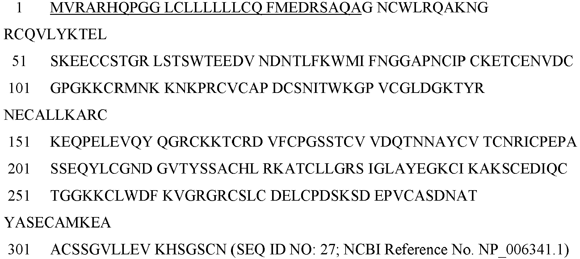
- The follistatin N-terminal domain (FSND) sequence is as follows:

- The FSD1 and FSD2 sequences are as follows:
- ETCENVDCGPGKKCRMNKKNKPRCV (SEQ ID NO: 29; FSD1)
- KTCRDVFCPGSSTCVVDQTNNAYCVT (SEQ ID NO: 30; FSD2)
- In other aspects not covered by the claimed invention, an agent for use in accordance with the methods disclosed herein is a follistatin-like related gene (FLRG), also known as follistatin-related protein 3 (FSTL3). The term "FLRG polypeptide" includes polypeptides comprising any naturally occurring polypeptide of FLRG as well as any variants thereof (including mutants, fragments, fusions, and peptidomimetic forms) that retain a useful activity. In certain embodiments not covered by the claimed invention, FLRG polypeptides of the disclosure bind to and/or inhibit activin activity, particularly activin A. Variants of FLRG polypeptides that retain activin binding properties can be identified using routine methods to assay FLRG and activin interactions (see, e.g.,
US 6,537,966 ). In addition, methods for making and testing libraries of polypeptides are described above in the context of ActRII polypeptides and such methods also pertain to making and testing variants of FLRG. FLRG polypeptides include polypeptides derived from the sequence of any known FLRG having a sequence at least about 80% identical to the sequence of an FLRG polypeptide, and optionally at least 85%, 90%, 95%, 97%, 99% or greater identity. - The human FLRG precursor (follistatin-related protein 3 precursor) polypeptide is as follows:The signal peptide is underlined.

- In certain embodiments not covered by the claimed invention, functional variants or modified forms of the follistatin polypeptides and FLRG polypeptides include fusion proteins having at least a portion of the follistatin polypeptide or FLRG polypeptide and one or more fusion domains, such as, for example, domains that facilitate isolation, detection, stabilization or multimerization of the polypeptide. Suitable fusion domains are discussed in detail above with reference to the ActRII polypeptides. In some embodiment, an antagonist agent of the disclosure is a fusion protein comprising an activin-binding portion of a follistatin polypeptide fused to an Fc domain. In another embodiment, an antagonist agent of the disclosure is a fusion protein comprising an activin binding portion of an FLRG polypeptide fused to an Fc domain.
- Any of the follistatin polypeptides disclosed herein may be combined with one or more additional ActRII antagonists agents of the disclosure to achieve the desired effect (e.g., treat or prevent a vascular disorder of the eye in a patient in need thereof, increase vision in patient in need thereof that has a vascular disorder of the eye, and/or treat or prevent one or more complications of a vascular disorder of the eye). For example, a follistatin polypeptide can be used in combination with i) one or more additional follistatin polypeptides, ii) one or more ActRII polypeptides disclosed herein, iii) one or more ActRII antagonist antibodies; iv) one or more small molecule ActRII antagonists; v) one or more polynucleotide ActRII antagonists; and/or vi) one or more FLRG polypeptides.
- Similarly, any of the FLRG polypeptides disclosed herein may be combined with one or more additional ActRII antagonists agents of the disclosure to achieve the desired effect (e.g., treat or prevent a vascular disorder of the eye in a patient in need thereof, increase vision in patient in need thereof that has a vascular disorder of the eye, and/or treat or prevent one or more complications of a vascular disorder of the eye). For example, a FLRG polypeptide can be used in combination with i) one or more additional FLRG polypeptides, ii) one or more ActRII polypeptides disclosed herein, iii) one or more ActRII antagonist antibodies; iv) one or more small molecule ActRII antagonists; v) one or more polynucleotide ActRII antagonists; and/or vi) one or more follistatin polypeptides.
- In certain aspects not covered by the claimed invention, the present disclosure relates to the use of the subject ActRII polypeptides (e.g., ActRIIA and ActRIIB polypeptides and variants thereof) to identify compounds (agents) which are agonist or antagonists of ActRII polypeptides. Compounds identified through this screening can be tested to assess their ability to improve visual acuity, for example, in animal models.
- There are numerous approaches to screening for therapeutic agents for improving vision (e.g., increasing visual acuity and/or visual field) by targeting ActRII signaling (e.g., ActRII signaling via
Smad - A variety of assay formats will suffice and, in light of the present disclosure, those not expressly described herein will nevertheless be comprehended by one of ordinary skill in the art. As described herein, the test compounds (agents) may be created by any combinatorial chemical method. Alternatively, the subject compounds may be naturally occurring biomolecules synthesized in vivo or in vitro. Compounds (agents) to be tested for their ability to act as modulators of tissue growth can be produced, for example, by bacteria, yeast, plants or other organisms (e.g., natural products), produced chemically (e.g., small molecules, including peptidomimetics), or produced recombinantly. Test compounds contemplated by the present disclosure but not covered by the claimed invention include non-peptidyl organic molecules, peptides, polypeptides, peptidomimetics, sugars, hormones, and nucleic acid molecules. The test agent is a small organic molecule having a molecular weight of less than about 2,000 Daltons.
- The test compounds of the disclosure may be provided as single, discrete entities, or provided in libraries of greater complexity, such as made by combinatorial chemistry. These libraries can comprise, for example, alcohols, alkyl halides, amines, amides, esters, aldehydes, ethers and other classes of organic compounds. Presentation of test compounds to the test system can be in either an isolated form or as mixtures of compounds, especially in initial screening steps. Optionally, the compounds may be optionally derivatized with other compounds and have derivatizing groups that facilitate isolation of the compounds. Non-limiting examples of derivatizing groups include biotin, fluorescein, digoxygenin, green fluorescent protein, isotopes, polyhistidine, magnetic beads, glutathione S-transferase (GST), photoactivatible crosslinkers or any combinations thereof.
- In many drug-screening programs which test libraries of compounds and natural extracts, high-throughput assays are desirable in order to maximize the number of compounds surveyed in a given period of time. Assays which are performed in cell-free systems, such as may be derived with purified or semi-purified proteins, are often preferred as "primary" screens in that they can be generated to permit rapid development and relatively easy detection of an alteration in a molecular target which is mediated by a test compound. Moreover, the effects of cellular toxicity or bioavailability of the test compound can be generally ignored in the in vitro system, the assay instead being focused primarily on the effect of the drug on the molecular target as may be manifest in an alteration of binding affinity between an ActRII polypeptide and its binding partner (e.g., an ActRII ligand).
- Merely to illustrate, in an exemplary screening assay of the present disclosure, the compound of interest is contacted with an isolated and purified ActRIIB polypeptide which is ordinarily capable of binding to an ActRIIB ligand, as appropriate for the intention of the assay. To the mixture of the compound and ActRIIB polypeptide is then added to a composition containing an ActRIIB ligand (e.g., GDF11). Detection and quantification of ActRIIB/ActRIIB-ligand complexes provides a means for determining the compound's efficacy at inhibiting (or potentiating) complex formation between the ActRIIB polypeptide and its binding protein. The efficacy of the compound can be assessed by generating dose-response curves from data obtained using various concentrations of the test compound. Moreover, a control assay can also be performed to provide a baseline for comparison. For example, in a control assay, isolated and purified ActRIIB ligand is added to a composition containing the ActRIIB polypeptide, and the formation of ActRIIB/ActRIIB ligand complex is quantitated in the absence of the test compound. It will be understood that, in general, the order in which the reactants may be admixed can be varied, and can be admixed simultaneously. Moreover, in place of purified proteins, cellular extracts and lysates may be used to render a suitable cell-free assay system.
- Complex formation between an ActRII polypeptide and its binding protein may be detected by a variety of techniques. For instance, modulation of the formation of complexes can be quantitated using, for example, detectably labeled proteins such as radiolabeled (e.g., 32P, 35S, 14C or 3H), fluorescently labeled (e.g., FITC), or enzymatically labeled ActRII polypeptide and/or its binding protein, by immunoassay, or by chromatographic detection.
- In certain embodiments not covered by the claimed invention, the present disclosure contemplates the use of fluorescence polarization assays and fluorescence resonance energy transfer (FRET) assays in measuring, either directly or indirectly, the degree of interaction between an ActRII polypeptide and its binding protein. Further, other modes of detection, such as those based on optical waveguides (see, e.g.,
PCT Publication WO 96/26432 U.S. Pat. No. 5,677,196 ), surface plasmon resonance (SPR), surface charge sensors, and surface force sensors, are compatible with many instances of the disclosure. - Moreover, the present disclosure contemplates, in embodiments not covered by the claimed invention, the use of an interaction trap assay, also known as the "two-hybrid assay," for identifying agents that disrupt or potentiate interaction between an ActRII polypeptide and its binding partner. See, e.g.,
U.S. Pat. No. 5,283,317 ; Zervos et al. (1993) Cell 72:223-232; Madura et al. (1993) J Biol Chem 268:12046-12054; Bartel et al. (1993) Biotechniques 14:920-924; and Iwabuchi et al. (1993) Oncogene 8:1693-1696). In a specific embodiment not covered by the claimed invention, the present disclosure contemplates the use of reverse two-hybrid systems to identify compounds (e.g., small molecules or peptides) that dissociate interactions between an ActRII polypeptide or GDF trap and its binding protein [see, e.g., Vidal and Legrain, (1999) Nucleic Acids Res 27:919-29; Vidal and Legrain, (1999) Trends Biotechnol 17:374-81; andU.S. Pat. Nos. 5,525,490 ;5,955,280 ; and5,965,368 ]. - The subject compounds may be identified by their ability to interact with an ActRII polypeptide. The interaction between the compound and the ActRII polypeptide may be covalent or non-covalent. For example, such interaction can be identified at the protein level using in vitro biochemical methods, including photo-crosslinking, radiolabeled ligand binding, and affinity chromatography [see, e.g., Jakoby WB et al. (1974) Methods in Enzymology 46:1]. The compounds may be screened in a mechanism-based assay, such as an assay to detect compounds which bind to an ActRII polypeptide. This may include a solid-phase or fluid-phase binding event. Alternatively, the gene encoding an ActRII polypeptide can be transfected with a reporter system (e.g., β-galactosidase, luciferase, or green fluorescent protein) into a cell and screened against the library preferably by high-throughput screening or with individual members of the library. Other mechanism-based binding assays may be used; for example, binding assays which detect changes in free energy. Binding assays can be performed with the target fixed to a well, bead or chip or captured by an immobilized antibody or resolved by capillary electrophoresis. The bound compounds may be detected usually using colorimetric endpoints or fluorescence or surface plasmon resonance.
- As described herein, applicants have discovered that an ActRII antagonist (inhibitor) has a surprising effect on improving vision in an MDS patient. Moreover, in view of the reported mechanism for MDS-associated vision loss [Han et al. (2015) J Glaucoma (Epub ahead of print); [Brouzas et al. (2009) Clinical Ophthalmology 3: 133-137], the data of the present disclosure suggests that ActRII inhibitors also may have positive effects in treating or preventing other types of eye (ocular) disorders, particularly vascular ocular disorders including, for example, those associated with ischemia and/or vascular insufficiency.
- The structural and functional integrity of the eye depends on a regular oxygen and nutrient supply. Being one of the most metabolically active tissues, the retina consumes oxygen more rapidly than other tissues in the body [Cohen et al. (1965) Biochemistry of the Retina. Orlando, Fla: Academic Press Inc; pp. 36-50; Anderson et al. (1964) Arch Ophthalmol 72:792-795; and Ames A. (1992) Can J Physiol Pharmacol. 70(Suppl):S158-64]. The presence of a dual circulation system makes retinal oxygenation unique [Osborne et al. (2004) Prog Retin Eye Res. 23:91-147]. The photoreceptors and the greater portion of the outer plexiform layer receive nourishment from the choriocapillaris indirectly whereas the inner retinal layers are supplied by the superficial and deep capillary plexuses formed by branches of the central artery of the retina. Inner layers of the retina are known to show highest sensitivity to hypoxic challenges [Janáky et al. (2007) Doc Ophthalmol. 114:45-51], whereas the outer retina is more resistant to a hypoxic stress [Tinjust et al. (2002) Aviat Space Environ Med. 73:1189-94].
- A number of systemic and cellular responses such as glycolysis, angiogenesis, vasodilation, and erythropoiesis enable an organism to respond to hypoxia [Harris et al. (2002) Nat Rev Cancer. 2:38-47]. Many tissues are capable of inducing protective mechanisms under hypoxic-ischemic conditions, which are typically induced within minutes of onset, and are of critical importance for limiting damage [Kitagawa et al. (1990) Brain Res. 528:21-4]. However, during prolonged hypoxic conditions, these protective mechanisms are generally diminished/lost within hours of the hypoxic-ischemic insult, leading to cell death and tissue damage [Prass et al. (2003) Stroke. 34:1981-6]. Transcriptional activator hypoxia-inducible factor-1α (HIF-1α) is a master regulator of cellular O2 homeostasis [Iyer et al. (1998) Genes Dev. 12:149-62]. Hypoxia is known to induce HIF-1α and its target genes such as vascular endothelial growth factor (VEGF) and nitric oxide synthase (NOS) in many tissues. Interestingly, overproduction of these factors, such as during prolonged hypoxia, has been implicated in cellular death in hypoxic-ischemic conditions. In addition, enhanced extracellular accumulation of glutamate and inflammatory cytokines, which occurs during prolonged hypoxia, can damage cells and tissues. Increased expression of HIF-1α, VEGF, and various isoforms of NOS has been reported in the retina following hypoxic injury [Kaur et al.(2006) Invest Ophthalmol Vis Sci. 47:1126-41; and Tezel et al. (2004) Curr Opin Ophthalmol. 15:80-4].
- Retinal ganglion cells (RGCs) are particularly sensitive to acute, transient, and mild systemic hypoxic stress [Kergoat et al. (2006) Invest Ophthalmol Vis Sci. 47:5423-7]. Loss of RGCs occurs in many ophthalmic conditions such as glaucoma and diabetes (Sucher et al. (1997) Vision Res. 37:3483-93; Abu-El-Asrar et al. (2004) Invest Ophthalmol Vis Sci. 45:2760-6], hypoxia being implicated in such loss [Wax et al. (2002) Mol Neurobiol. 26:45-55; Tezel et al. (2004). Curr Opin Ophthalmol. 15:80-4; and Chen et al. (2007) Stem Cells. 25:2291-301]. Neuronal degeneration resulting from retinal hypoxia-ischemia, caused by oxygen and substrate deprivation, is partially mediated by accumulation of free oxygen radicals [Block et al. (1997) Exp Eye Res. 64:559-64; Muller et al. (1997) Exp Eye Res. 64:637-43; and Szabo et al. (1997) Clin Neurosci. 4:240-5], glutamate excitotoxicity [Kuroiwa Tet al. (1985) Acta Neuropathol (Berl) 68:122-9; Osborne et al. (2004) Prog Retin Eye Res. 23:91-147; and Kaur et al. (2006) Invest Ophthalmol Vis Sci. 47:1126-41], inflammation, and disruption of the blood retinal barrier [Kuroiwa et al. (1985) Acta Neuropathol (Berl) 68:122-9; and Kaur et al. (2007) J Pathol. 212:429-39].
- Hypoxia-ischemia also results in retinal vascular damage which is associated with fluid accumulation in the extracellular spaces (vasogenic edema) or intracellulary spaces (cytotoxic edema) [Marmor et al. (1999) Doc Ophthalmol. 97:239-49]. The extracellular spaces in the inner retina consist of the narrow clefts between the tightly packed cellular elements. Fluid leaking out from damaged capillaries in the inner retina accumulates in the extracellular spaces displacing the retinal cellular elements and disrupting the normal anatomy of the neuronal connections, resulting in macular edema [Hamann et al. (2005) Acta Ophthalmol Scand. 83:523-5]. Macular edema can further exacerbate retinal ischemia and well as promote increased oxidative stress and inflammation (Guex-Crosier Y. (1999) Doc Ophthalmol. 97:297-309; van Dam PS. (2002) Diabetes Metab Res Rev. 18:176-84; and Miyake et al. (2002) Surv Ophthalmol. 47:S203-8.). Increased permeability of blood-retinal barrier (BRB) resulting in fluid accumulation has been reported to contribute to retinal neuronal degeneration by compression [Antcliff et al. (1999) Semin Ophthalmol. 14:223-32; Marumo T et al. (1999) J Vasc Res. 36:510-15; and Reichenbach et al. (2007) Graefes Arch Clin Exp Ophthalmol. 245:627-36). While initially protective, excess and/or chronic production of VEGF, nitric oxide (NO), and aquaporin-4 during hypoxic-ischemic insults can cause neovascularization and dysfunction of the BRB in the inner retina, resulting in serum leakage into the retinal tissues and retinal edema. In addition to an increase in vascular permeability, ocular hypoxia has also been correlated with endothelial cell death, leukocyte plugging of vessels, and microaneurysms [Linsenmeier et al. (1998) Invest Ophthalmol Vis Sci. 39:1647-57].
- Hypoxia-ischemia occurs in various ocular conditions including, for example, retinal artery/vein occlusion or thrombosis, ocular ischemic syndrome, ischemic optic neuropathy, and retinal ischemia. Hypoxia-ischemia also has been implicated in the development of glaucoma [Flammer J. (1994) 'Surv Ophthalmol. 38(Suppl):S3-6; Chung et al. (1999) Surv Ophthalmol. 43(Suppl 1):S43-50; and Tezel et al. (2004) Curr Opin Ophthalmol. 15:80-4], is thought to underlie many of the sight-threatening complications of diabetic eye disease including retinal and optic nerve head neovascularization [Linsenmeier et al. (1998) Invest Ophthalmol Vis Sci. 39:1647-57], and may play a role in age-related macular degeneration [Tso et al. (1982) Ophthalmology. 89:902-15; Yanoff et al. (1984) Surv Ophthalmol. 28 (Suppl):505-11; and Bressler et al. (2001) In: Schachat AP, editor. Retina. St. Louis, MO: Mosby]. Systemic causes of ocular hypoxia include the cardiovascular effects, chronic obstructive airways disease, arterial/venous obstructive conditions, [Brown et al. (1988) Int Ophthalmol. 11:239-51] Takayasu's arteritis [Shelhamer et al. (1985) Ann Intern Med. 103:121-6], hyperviscosity syndromes [Ashton et al. (1963) J Pathol Bacteriol. 86:453-61] as well as trauma (e.g., surgery or accidental damage) [Purtscher's retinopathy; Buckley et al. (1996) Postgrad Med J. 72:409-12]. Hypoxia associated with the above conditions is a common cause of visual impairment and blindness [Osborne, et al. (2004) Prog Retin Eye Res. 23:91-147].
- Therefore, in certain aspects not covered by the claimed invention, the present disclosure provides methods for treating or preventing an vascular disorder (disease) of the eye in a patient (subject) in need thereof (particularly mammals such as rodents, cats, dogs, primates, and humans) by administering to the patient a therapeutically effective amount of an ActRII antagonist (inhibitor), or combination of ActRII antagonists. In some embodiments not covered by the claimed invention, the disclosure provides methods for using an ActRII antagonist, or combination of ActRII antagonists, to treat or prevent vascular disorders of the eye associated with ischemia. In some embodiments not covered by the claimed invention, the disclosure provides methods for using an ActRII antagonist, or combination of ActRII antagonists, to treat or prevent an ischemic eye disease. In some embodiments not covered by the claimed invention, the disclosure provides methods for using an ActRII antagonist, or combination of ActRII antagonists, to treat or prevent vascular disorders of the eye associated with microvasculature insufficiency. In some embodiments not covered by the claimed invention, the disclosure provides methods for using an ActRII antagonist, or combination of ActRII antagonists, to treat or prevent an ocular microvasculature insufficiency disease. In some embodiments not covered by the claimed invention, the disclosure provides methods for using an ActRII antagonist, or combination of ActRII antagonists, to treat or prevent vascular disorders of the eye associated with retinopathy. In some embodiments not covered by the claimed invention, the disclosure provides methods for using an ActRII antagonist, or combination of ActRII antagonists, to treat or prevent vascular disorders of the eye associated with optic neuropathy. In some embodiments not covered by the claimed invention, the disclosure provides methods for using an ActRII antagonist, or combination of ActRII antagonists, to treat or prevent ischemic retinopathy. In some embodiments not covered by the claimed invention, the disclosure provides methods for using an ActRII antagonist, or combination of ActRII antagonists, to treat or prevent ischemic optic neuropathy. In some embodiments not covered by the claimed invention, the disclosure provides methods for using an ActRII antagonist, or combination of ActRII antagonists, to treat or prevent retinopathy associated with microvasculature insufficiency. In some embodiments not covered by the claimed invention, the disclosure provides methods for using an ActRII antagonist, or combination of ActRII antagonists, to treat or prevent optic neuropathy associated with microvasculature insufficiency. In particular, the disclosure provides methods, which are, however, not covered by the claimed invention, for using an ActRII antagonist, or combination of ActRII antagonists, to treat or prevent one or more diseases selected from: macular degeneration (e.g., age-related macular degeneration, juvenile macular degeneration, wet macular degeneration, dry macular degeneration, Stargardt's disease, and Best's disease), retinal vein occlusion (e.g, central retinal vein occlusion, hemi-retinal vein occlusion, branch retinal vein occlusion, and ischemic retinal vein occlusion), retinal artery occlusion (e.g, central retinal artery occlusion, hemi-retinal artery occlusion, branch retinal artery occlusion, and ischemic retinal artery occlusion), diabetic retinopathy, ischemic optic neuropathy [e.g., anterior ischemic optic neuropathy (arteritic and non-arteritic) and posterior ischemic optic neuropathy], macular telangiectasia (type I or type II), retinal ischemia (e.g., acute retinal ischemia or chronic retinal ischemia), ocular ischemic syndrome, retinal vasculitis, and retinopathy of prematurity. Methods and compositions disclosed herein for treating an ocular disease may result in improving vision in an eye of the patient. Methods and compositions disclosed herein for treating an ocular disease may result in increasing visual acuity in an eye of the patient. Methods and compositions disclosed herein for treating an ocular disease may result in increasing visual field in an eye of the patient. Optionally, methods of the disclosure, which are, however, not covered by the claimed invention for treating or preventing an vascular disorder of the eye may further comprise administration of one or more supportive therapies for treating or preventing the disorder in addition to administration of an ActRII antagonist, or combination of ActRII antagonists [e.g., surgery, laser therapy (e.g., photocoagulation), anti-angiogenic therapy [e.g., VEGF inhibitors such as bevacizumab (Avastin®),ranibizumab (Lucentis®), and Aflibercept (Eylea®)], Ca2+ inhibitors (e.g., flunarizine and nifedipine), cryotherapy, hyperbaric oxygenation, Na+ channel blockers (e.g., topiramate), iGluR antagonists (e.g., MK-801, dextromethorphan, eliprodil, and flupirtine), antioxidants (e.g., dimethylthiourea, vitamin E, alph-lipoic acid, superoxide dismutase, catalase, desferrioxamine, mannitol, allopurinol, calcium dobesilate, flupirtine, trimetazidine, and EGB-761), anti-inflammatory agents, cyclodiathermy, cyclocryotherapy, ocular filtering procedures, implantation of drainage valves, antiplatelet therapy (e.g., aspirin, ticlopidine, and clopidogrel), anticoagulant therapy (e.g., warfarin and heparin), steroids, systemic or local corticosteroids (e.g., prednisone triamcinolone (Triesence®), and dexamethasone (Ozurdex®), steroid-sparing immunosuppressants (e.g., cyclosporine, azathioprine, cyclophosphamide, mycophenolate, mofetil, infliximab and etanercept), dietary supplements (e.g., vitamin C, vitamin E, lutein, zeaxanthin, zinc, folic acid, vitamins B6, vitamin B 12, and zeaxanthin), vitrectomy, scleral buckle surgery, and pneumatic retinopexy].
- In certain aspects, the present disclosure provides methods, not covered by the claimed invention, for treating or preventing a vascular disorder of the eye in a patient that has one or more of: anemia, myelodysplastic syndrome, sideroblastic anemia, a hemoglobinopathy, thalassemia, and sickle-cell disease by administering to the patient a therapeutically effective amount of an ActRII antagonist, or combination of ActRII antagonists. In some embodiments not covered by the claimed invention, the disclosure provides methods for using an ActRII antagonist, or combination of ActRII antagonists, to treat or prevent vascular disorders of the eye associated with ocular ischemia in a patient that has one or more of: anemia, myelodysplastic syndrome, sideroblastic anemia, a hemoglobinopathy, thalassemia, and sickle-cell disease. In some embodiments not covered by the claimed invention, the disclosure provides methods for using an ActRII antagonist, or combination of ActRII antagonists, to treat or prevent an ischemic ocular disease in a patient that has one or more of: anemia, myelodysplastic syndrome, sideroblastic anemia, a hemoglobinopathy, thalassemia, and sickle-cell disease. In some embodiments not covered by the claimed invention, the disclosure provides methods for using an ActRII antagonist, or combination of ActRII antagonists, to treat or prevent vascular disorders of the eye associated with microvasculature insufficiency in a patient that has one or more of: anemia, myelodysplastic syndrome, sideroblastic anemia, a hemoglobinopathy, thalassemia, and sickle-cell disease. In some embodiments not covered by the claimed invention, the disclosure provides methods for using an ActRII antagonist, or combination of ActRII antagonists, to treat or prevent an ocular microvasculature insufficiency disease in a patient that has one or more of: anemia, myelodysplastic syndrome, sideroblastic anemia, a hemoglobinopathy, thalassemia, and sickle-cell disease. In some embodiments not covered by the claimed invention, the disclosure provides methods for using an ActRII antagonist, or combination of ActRII antagonists, to treat or prevent vascular disorders of the eye associated with retinopathy in a patient that has one or more of: anemia, myelodysplastic syndrome, sideroblastic anemia, a hemoglobinopathy, thalassemia, and sickle-cell disease. In some embodiments not covered by the claimed invention, the disclosure provides methods for using an ActRII antagonist, or combination of ActRII antagonists, to treat or prevent vascular disorders of the eye associated with optic neuropathy in a patient that has one or more of: anemia, myelodysplastic syndrome, sideroblastic anemia, a hemoglobinopathy, thalassemia, and sickle-cell disease. In some embodiments not covered by the claimed invention, the disclosure provides methods for using an ActRII antagonist, or combination of ActRII antagonists, to treat or prevent ischemic retinopathy in a patient that has one or more of: anemia, myelodysplastic syndrome, sideroblastic anemia, a hemoglobinopathy, thalassemia, and sickle-cell disease. In some embodiments not covered by the claimed invention, the disclosure provides methods for using an ActRII antagonist, or combination of ActRII antagonists, to treat or prevent ischemic optic neuropathy in a patient that has one or more of: anemia, myelodysplastic syndrome, sideroblastic anemia, a hemoglobinopathy, thalassemia, and sickle-cell disease. In some embodiments not covered by the claimed invention, the disclosure provides methods for using an ActRII antagonist, or combination of ActRII antagonists, to treat or prevent retinopathy associated with microvasculature insufficiency in a patient that has one or more of: anemia, myelodysplastic syndrome, sideroblastic anemia, a hemoglobinopathy, thalassemia, and sickle-cell disease. In some embodiments not covered by the claimed invention, the disclosure provides methods for using an ActRII antagonist, or combination of ActRII antagonists, to treat or prevent optic neuropathy associated with microvasculature insufficiency in a patient that has one or more of: anemia, myelodysplastic syndrome, sideroblastic anemia, a hemoglobinopathy, thalassemia, and sickle-cell disease. In some embodiments not covered by the claimed invention, methods disclosed herein for treating an ocular disease in a patient that has one or more of: anemia, myelodysplastic syndrome, sideroblastic anemia, a hemoglobinopathy, thalassemia, and sickle-cell disease result in improving vision in an eye of the patient. In some embodiments not covered by the claimed invention, methods disclosed herein for treating a vascular disorder of the eye in a patient that has one or more of: anemia, myelodysplastic syndrome, sideroblastic anemia, a hemoglobinopathy, thalassemia, and sickle-cell disease result in increasing visual acuity in an eye of the patient. In some embodiments not covered by the claimed invention, methods disclosed herein for treating an vascular disorder of the eye in a patient that has one or more of: anemia, myelodysplastic syndrome, sideroblastic anemia, a hemoglobinopathy, thalassemia, and sickle-cell disease result in increasing visual field in an eye of the patient. Optionally, methods of the disclosure, which, however, are not covered by the claimed invention for treating or preventing an ocular disease in a patient that has one or more of: anemia, myelodysplastic syndrome, sideroblastic anemia, a hemoglobinopathy, thalassemia, and sickle-cell disease may further comprise administration of one or more supportive therapies for treating or preventing the vascular disorder of the eye in addition to administration of an ActRII antagonist, or combination of ActRII antagonists.
- In certain aspects, the present disclosure provides methods not covered by the claimed invention for treating or preventing a vascular disorder of the eye in a patient (subject) having myelodysplastic syndrome (particularly mammals such as rodents, cats, dogs, primates, and humans) by administering to the patient a therapeutically effective amount of an ActRII antagonist, or combination of ActRII antagonists. In some embodiments not covered by the claimed invention, the disclosure provides methods for using an ActRII antagonist, or combination of ActRII antagonists, to treat or prevent a vascular disorder of the eye associated with ocular ischemia in a patient having myelodysplastic syndrome. In some embodiments not covered by the claimed invention, the disclosure provides methods for using an ActRII antagonist, or combination of ActRII antagonists, to treat or prevent an ischemic ocular disease in a patient having myelodysplastic syndrome. In some embodiments not covered by the claimed invention, the disclosure provides methods for using an ActRII antagonist, or combination of ActRII antagonists, to treat or prevent a vascular disorder of the eye associated with microvasculature insufficiency in a patient having myelodysplastic syndrome. In some embodiments not covered by the claimed invention, the disclosure provides methods for using an ActRII antagonist, or combination of ActRII antagonists, to treat or prevent an ocular microvasculature insufficiency disease in a patient having myelodysplastic syndrome. In some embodiments not covered by the claimed invention, the disclosure provides methods for using an ActRII antagonist, or combination of ActRII antagonists, to treat or prevent a vascular disorder of the eye associated with retinopathy in a patient having myelodysplastic syndrome. In some embodiments not covered by the claimed invention, the disclosure provides methods for using an ActRII antagonist, or combination of ActRII antagonists, to treat or prevent a vascular disorder of the eye associated with optic neuropathy in a patient having myelodysplastic syndrome. In some embodiments, the disclosure provides methods for using an ActRII antagonist, or combination of ActRII antagonists not covered by the claimed invention, to treat or prevent ischemic retinopathy in a patient having myelodysplastic syndrome. In some embodiments not covered by the claimed invention, the disclosure provides methods for using an ActRII antagonist, or combination of ActRII antagonists, to treat or prevent ischemic optic neuropathy in a patient having myelodysplastic syndrome. In some embodiments not covered by the claimed invention, the disclosure provides methods for using an ActRII antagonist, or combination of ActRII antagonists, to treat or prevent retinopathy associated with microvasculature insufficiency in a patient having myelodysplastic syndrome. In some embodiments not covered by the claimed invention, the disclosure provides methods for using an ActRII antagonist, or combination of ActRII antagonists, to treat or prevent optic neuropathy associated with microvasculature insufficiency in a patient having myelodysplastic syndrome. In some embodiments not covered by the claimed invention, methods disclosed herein for treating a vascular disorder of the eye in a patient having myelodysplastic syndrome result in improving vision in an eye of the patient. In some embodiments not covered by the claimed invention, methods disclosed herein for treating a vascular disorder of the eye in a patient having myelodysplastic syndrome result in increasing visual acuity in an eye of the patient. In some embodiments not covered by the claimed invention, methods disclosed herein for treating a vascular disorder of the eye in a patient having myelodysplastic syndrome result in increasing visual field in an eye of the patient. Optionally, methods of the disclosure not covered by the claimed invention for treating or preventing a vascular disorder of the eye in a patient with myelodysplastic syndrome may further comprise administration of one or more supportive therapies for treating or preventing the ocular disease in addition to administration of an ActRII antagonist, or combination of ActRII antagonists.
- In certain aspects not covered by the claimed invention, the present disclosure provides methods for improving vision (e.g., increasing visual acuity and or visual field) in a patient in need thereof by administering to the patient a therapeutically effective amount of an ActRII antagonist, or combination of ActRII antagonists. In some embodiments not covered by the claimed invention, the disclosure provides methods for using an ActRII antagonist, or combination of ActRII antagonists, to improve vision (e.g., increase visual acuity and or visual field) in a patient with a vascular disorder of the eye. In some embodiments not covered by the claimed invention, the disclosure provides methods for using an ActRII antagonist, or combination of ActRII antagonists, to improve vision (e.g., increase visual acuity and or visual field) in a patient with a vascular disorder of the eye associated with ischemic ocular disease. In some embodiments not covered by the claimed invention, the disclosure provides methods for using an ActRII antagonist, or combination of ActRII antagonists, to improve vision (e.g., increase visual acuity and or visual field) in a patient with a vascular disorder of the eye associated with microvasculature insufficiency. In some embodiments not covered by the claimed invention, the disclosure provides methods for using an ActRII antagonist, or combination of ActRII antagonists, to improve vision (e.g., increase visual acuity and or visual field) in a patient with ocular microvasculature insufficiency disease. In some embodiments not covered by the claimed invention, the disclosure provides methods for using an ActRII antagonist, or combination of ActRII antagonists, to improve vision (e.g., increase visual acuity and or visual field) in a patient with a vascular disorder of the eye associated with retinopathy. In some embodiments not covered by the claimed invention, the disclosure provides methods for using an ActRII antagonist, or combination of ActRII antagonists, to improve vision (e.g., increase visual acuity and or visual field) in a patient with a vascular disorder of the eye associated with optic neuropathy. In some embodiments not covered by the claimed invention, the disclosure provides methods for using an ActRII antagonist, or combination of ActRII antagonists, to improve vision (e.g., increase visual acuity and or visual field) in a patient with ischemic retinopathy. In some embodiments not covered by the claimed invention, the disclosure provides methods for using an ActRII antagonist, or combination of ActRII antagonists, to improve vision (e.g., increase visual acuity and or visual field) in a patient with ischemic optic neuropathy. In some embodiments not covered by the claimed invention, the disclosure provides methods for using an ActRII antagonist, or combination of ActRII antagonists, to improve vision (e.g., increase visual acuity and or visual field) in a patient with retinopathy associated with microvasculature insufficiency. In some embodiments not covered by the claimed invention, the disclosure provides methods for using an ActRII antagonist, or combination of ActRII antagonists, to improve vision (e.g., increase visual acuity and or visual field) in a patient with optic neuropathy associated with microvasculature insufficiency. In particular, the disclosure provides methods, not covered by the claimed invention, for using an ActRII antagonist, or combination of ActRII antagonists, to improve vision (e.g., increase visual acuity and or visual field) in a patient with one or more diseases selected from: macular degeneration (e.g., age-related macular degeneration, juvenile macular degeneration, wet macular degeneration, dry macular degeneration, Stargardt's disease, and Best's disease), retinal vein occlusion (e.g, central retinal vein occlusion, hemi-retinal vein occlusion, branch retinal vein occlusion, and ischemic retinal vein occlusion), retinal artery occlusion (e.g, central retinal artery occlusion, hemi-retinal artery occlusion, branch retinal artery occlusion, and ischemic retinal artery occlusion), diabetic retinopathy, ischemic optic neuropathy [e.g., anterior ischemic optic neuropathy (arteritic and non-arteritic) and posterior ischemic optic neuropathy], macular telangiectasia (type I or type II), retinal ischemia (e.g., acute retinal ischemia or chronic retinal ischemia), ocular ischemic syndrome, retinal vasculitis, and retinopathy of prematurity. In some embodiments not covered by the claimed invention, the disclosure provides methods for using an ActRII antagonist, or combination of ActRII antagonists, to improve vision (e.g., increase visual acuity and or visual field) in a patient with anemia. In some embodiments not covered by the claimed invention, the disclosure provides methods for using an ActRII antagonist, or combination of ActRII antagonists, to improve vision (e.g., increase visual acuity and or visual field) in a patient with myelodysplastic syndrome. In some embodiments not covered by the claimed invention, the disclosure provides methods for using an ActRII antagonist, or combination of ActRII antagonists, to improve vision (e.g., increase visual acuity and or visual field) in a patient with sideroblastic anemia. In some embodiments not covered by the claimed invention, the disclosure provides methods for using an ActRII antagonist, or combination of ActRII antagonists, to improve vision (e.g., increase visual acuity and or visual field) in a patient with a hemoglobinopathy. In some embodiments not covered by the claimed invention, the disclosure provides methods for using an ActRII antagonist, or combination of ActRII antagonists, to improve vision (e.g., increase visual acuity and or visual field) in a patient with thalassemia. In some embodiments not covered by the claimed invention, the disclosure provides methods for using an ActRII antagonist, or combination of ActRII antagonists, to improve vision (e.g., increase visual acuity and or visual field) in a patient with sickle-cell disease. Optionally, methods of the disclosure for improving vision (e.g., increasing visual acuity and or visual field) in a patient with an ocular disease may further comprise administration of one or more supportive therapies for treating or preventing the ocular disease in addition to administration of an ActRII antagonist, or combination of ActRII antagonists. It is, however, to be understood that any methods of treatment by therapy or surgery or in vivo diagnosis methods described or referred to in the present disclosure are not encompassed by the claimed invention but are to be interpreted as references to pharmaceutical compositions of the present invention for use in those methods. Specifically, the present invention relates to a composition for use in a method of a) treating or preventing a vascular disorder of the eye; b) improving vision in a patient that has vascular disorder of the eye; wherein the patient has myelodysplastic syndrome.
- As used herein, a therapeutic that "prevents" a disorder or condition refers to a compound that, in a statistical sample, reduces the occurrence of the disorder or condition in the treated sample relative to an untreated control sample, or delays the onset or reduces the severity of one or more symptoms of the disorder or condition relative to the untreated control sample.
- The term "treating" as used herein includes amelioration or elimination of the condition once it has been established. In either case, prevention or treatment may be discerned in the diagnosis provided by a physician or other health care provider and the intended result of administration of the therapeutic agent.
- In general, treatment or prevention of a disease or condition as described in the present disclosure is achieved by administering one or more of ActRII antagonists (e.g., an ActRIIA and/or ActRIIB antagonist) in an effective amount. An effective amount of an agent refers to an amount effective, at dosages and for periods of time necessary, to achieve the desired therapeutic or prophylactic result. A therapeutically effective amount of an agent of the present disclosure may vary according to factors such as the disease state, age, sex, and weight of the individual, and the ability of the agent to elicit a desired response in the individual. A prophylactically effective amount refers to an amount effective, at dosages and for periods of time necessary, to achieve the desired prophylactic result.
- Ocular damage is a complication/manifestation of myelodysplastic syndrome [Han et al. (2015) J Glaucoma (Epub ahead of print); [Brouzas et al. (2009) Clinical Ophthalmology 3:133-137]. Applicants have discovered that treatment with an ActRII inhibitor has a surprising effect on improving vision in an MDS patient. Insight into the mechanism for vision loss in MDS patients suggests that ActRII inhibitor therapy may also be useful in the treatment of other vascular disorders of the eye, particularly those associated with ischemia and/or microvasculature insufficiency. For example, in addition to MDS, other hematological disorders have been associated with ocular damage including, for example, hemoglobinopathy diseases (e.g., sickle cell disease and thalassemia) [de Melo M.B. (2014) Rev Hematol Hemoter 36(5):319-321; and Aksoy et al. (2013) Seminars in Ophthalmology 28(1): 22-26].
- Therefore, in certain aspects not covered by the claimed invention, the present disclosure provides methods for improving vision (e.g., visual acuity and/or visual field) in a patient with a hematological disorder by administering one or more ActRII antagonist (e.g., a GDF-ActRII antagonist, an ActRIIA polypeptide, an ActRIIB polypeptide, a GDF trap, etc.). In some embodiments not covered by the claimed invention, an ActRII antagonist, or combination of ActRII antagonists, can be used to improve vision (e.g., improve visual acuity and/or visual field) in a patient with anemia. In some embodiments not covered by the claimed invention, an ActRII antagonist, or combination of ActRII antagonists, can be used to improve vision (e.g., improve visual acuity and/or visual field) in a patient with MDS. In some embodiments not covered by the claimed invention, an ActRII antagonist, or combination of ActRII antagonists, can be used to improve vision (e.g., improve visual acuity and/or visual field) in a patient with a hemoglobinopathy. In some embodiments not covered by the claimed invention, an ActRII antagonist, or combination of ActRII antagonists, can be used to improve vision (e.g., improve visual acuity and/or visual field) in a patient with thalassemia. In some embodiments not covered by the claimed invention, an ActRII antagonist, or combination of ActRII antagonists, can be used to improve vision (e.g., improve visual acuity and/or visual field) in a patient with sickle cell disease. In some embodiments not covered by the claimed invention, an ActRII antagonist, or combination of ActRII antagonists, can be used to improve vision (e.g., improve visual acuity and/or visual field) in a patient with sideroblastic anemia. Optionally, patients having a hematological disorder (e.g., myelodysplastic syndrome, sideroblastic anemia, thalassemia, sickle cell disease, anemia, a hemoglobinopathy, or sideroblastic anemia) and in need of improved vision (improved visual acuity and/or visual field) may be treated with one or more supportive therapies for treating the hematological disorder in addition to an ActRII antagonist, or combination of ActRII antagonists.
- Retinal ischemia is a common disease and, due to relatively ineffective treatment, remains a common cause of visual impairment and blindness in the industrialized world [Osborne et al. (2004) Progress in Retinal and Eye Research 23:91-147]. Ischemia refers to a pathological situation involving an inadequacy (not necessarily a complete lack of) blood flow to a tissue, with failure to meet cellular energy demands. In general, ischemia deprives a tissue of three requirements: oxygen, metabolic substrates, and removal of waste products. The loss of these requirements will initially lower homeostatic responses and with time will induce injury to the tissue. If withheld for a sufficiently long time, the tissue will die (an infarct). At a cellular level, ischemic retinal injury consists of a self-reinforcing destructive cascade involving neuronal depolarization, calcium influx, and oxidative stress initiated by energy failure and increased glutamatergic stimulation. Ultimately ischemic damage can lead to loss of cells in the retina including, for example, photoreceptors, ganglion cells and amacrine cells.
- Retinal ischemia can be caused by a variety of conditions including, for example, stroke, ocular injury, and diabetes. It is also commonly caused when the central retinal vein becomes occluded or detached from the eye. When the retina loses its oxygen supply, the body tries to compensate by producing various vascular modifying agents including, for example, Vascular Endothelial Growth Factor (VEGF). Unfortunately, this can lead to the growth of abnormal blood vessels on the surface of the retina, leading to blindness. In fact, it has been suggested ischemia is responsible for retinal neovascularization in patients with retinal vein occlusion, diabetes, sickle-cell retinopathy, and retinopathy of prematurity, all of which can ultimately result in retinal vessel hemorrhage and/or retinal detachment [Osborne et al. (2004) Progress in Retinal and Eye Research 23:91-147].
- Retinal ischemia can manifest as a chronic or acute disease. Generally, retinal ischemia is first localized to one eye, but it often progresses to affect both eyes over time. In most cases, a patient with retinal ischemia presents with a painless loss of visual acuity and visual field associated with optic disc swelling. The age range of patients with this condition is extensive, and depends in part on the cause of the ischemia. Some patients, however, simply experience sudden visual loss. The degree of visual loss may be severe or the patient may notice only a vague sensation of blurred vision, often described as a shade or veil over a portion of the visual field. Vision loss varies and may cause severe impairment in the visual field and visual acuity. Once occurred, the loss of vision is usually permanent although some recovery is possible in the early stages with appropriate treatment.
- There are various ocular and systemic treatments available for treating retinal ischemia, many with limited efficacy and/or potential adverse side effects. These treatments include, for example: surgery, laser therapy (e.g., photocoagulation), anticoagulants (e.g., aspirin and PAF inhibitors), anti-angiogenic therapy (e.g., VEGF inhibitors), Ca2+ inhibitors (e.g., flunarizine and nifedipine), cryotherapy, hyperbaric oxygenation, Na+ channel blockers (e.g., topiramate), iGluR antagonists (e.g., MK-801, dextromethorphan, eliprodil, and flupirtine), antioxidants (e.g., dimethylthiourea, vitamin E, alph-lipoic acid, superoxide dismutase, catalase, desferrioxamine, mannitol, allopurinol, calcium dobesilate, flupirtine, trimetazidine, and EGB-761), and anti-inflammatory agents.
- In certain aspects not covered by the claimed invention, the present disclosure provides methods for treating or preventing retinal ischemia in a patient in need thereof by administering one or more ActRII antagonists [e.g., an ActRII polypeptide or variant thereof (e.g., a GDF trap)]. In some embodiments not covered by the claimed invention, an ActRII antagonist, or combination of ActRII antagonists, can be used to treat or prevent one or more of: acute retinal ischemia and chronic retinal ischemia. In some embodiments not covered by the claimed invention, an ActRII antagonist, or combination of ActRII antagonists, can be used to treat or prevent one or more complication of retinal ischemia selected from: cataracts, corneal edema, ocular hypotony, increased ocular pressure, anterior chamber inflammation, neovascular glaucoma, and iris neovascularization, narrowed retinal arteries, dilated retinal veins, retinal hemorrhages, cotton-wool spots, cherry-red spot, optic nerve neovascularization, retinal neovascularization, ischemic ocular pain, and amaurosis fugax. In some embodiments not covered by the claimed invention, an ActRII antagonist, or combination of ActRII antagonists, can be used to improve vision (e.g., improve visual acuity and/or visual field) in a patient with retinal ischemia. Optionally, patients afflicted with retinal ischemia may be treated with one or more supportive therapies [e.g., surgery, laser therapy (e.g., photocoagulation), topical medication to lower intraocular pressure, cyclodiathermy, cyclocryotherapy, intravitreal steroids, ocular filtering procedures, implantation of glaucoma drainage valves to treat neovascular glaucoma, antiplatelet therapy (e.g., aspirin, ticlopidine, and clopidogrel), anticoagulant therapy (e.g., warfarin and heparin), and systemic steroid therapy, anti-angiogenic therapy (e.g., VEGF inhibitors), Ca2+ inhibitors (e.g., flunarizine and nifedipine), cryotherapy, hyperbaric oxygenation, Na+ channel blockers (e.g., topiramate), iGluR antagonists (e.g., MK-801, dextromethorphan, eliprodil, and flupirtine), antioxidants (e.g., dimethylthiourea, vitamin E, alpha-lipoic acid, superoxide dismutase, catalase, desferrioxamine, mannitol, allopurinol, calcium dobesilate, flupirtine, trimetazidine, and EGB-761), and anti-inflammatory agents] for treating retinal ischemia in addition to an ActRII antagonist, or combination of ActRII antagonists.
- Ocular ischemic syndrome (OIS) is a rare disease in which gradual or sudden loss of vision results from chronic vascular insufficiency [Brown et al (1994) Ocular ischemic syndrome. In: Retina. 2nd ed. Mosby. 1515-27; and Chen et al. (2007) Compr Ophthalmol Update. 8(1): 17-28]. The most common etiology of OIS is sever unilateral or bilateral atherosclerotic disease of the internal carotid artery or marked stenosis at the bifurcation of the common carotid artery. OIS may also be caused by giant cell arteritis. It is postulated that the decrease in vascular perfusion results in tissue hypoxia and increased ocular ischemic, which typically results in neovascularization. The disease is found most often in patients with other risk factors for cardiovascular disease, such as diabetes mellitus, hyperlipidemia, and hypertension. Common anterior pathology includes cataracts, corneal edema, ocular hypotony, increased ocular pressure, anterior chamber inflammation, neovascular glaucoma, and iris neovascularization. Posterior segment signs include narrowed retinal arteries, dilated but nontortuour retinal veins, retinal hemorrhages, cotton-wool spots, cherry-red spot, and optic nerve/retinal neovascularization.
- Principal symptoms of OIS include visual loss, light-induced transient visual loss, amaurosis fugax, and ischemic ocular pain [Mizener et al. (1997) Ophthalmology. 104(5):859-64; and Chen et al. (2007) Compr Ophthalmol Update. 8(1): 17-28]. Loss of visual acuity is the most frequently encountered symptom, present in 70-90% of patients. While visual loss typically occurs gradually over a period of weeks to months, it can also occur abruptly. About 40% of patients with OIS will present with symptoms of ischemic pain. In general, the pain is characteristically described as a dull ache over the brow, which beings gradually over a prior of hours to days. Amaurosis fugax is a transient episode of complete or partial monocular blindness lasting for a period of less than about 10 minutes. A history of amaurosis fugax is found in about 9-15% of patients with OIS.
- There are various ocular and systemic treatments available for treating OIS, many with limited efficacy and/or potential adverse side effects. Ocular treatments include, for example: surgery or laser therapy (e.g., panretinal photocoagulation) to treat neovascularization of the iris, optic nerve, or retina; topical medication to lower intraocular pressure, cyclodiathermy and cyclocryotherapy to lower intraocular pressure; intravitreal steroids; and ocular filtering procedures and implantation of glaucoma drainage valves to treat neovascular glaucoma. System treatments include, for example: antiplatelet therapy (e.g., aspirin, ticlopidine, and clopidogrel), anticoagulant therapy (e.g., warfarin and heparin), and steroids.
- In certain aspects not covered by the claimed invention, the present disclosure provides methods for treating or preventing ocular ischemic syndrome in a patient in need thereof by administering one or more ActRII antagonists [e.g., an ActRII polypeptide or variant thereof (e.g., a GDF trap)]. In some embodiments not covered by the claimed invention, an ActRII antagonist, or combination of ActRII antagonists, can be used to treat or prevent one or more complication of ocular ischemic syndrome selected from: cataracts, corneal edema, ocular hypotony, increased ocular pressure, anterior chamber inflammation, neovascular glaucoma, and iris neovascularization, narrowed retinal arteries, dilated retinal veins, retinal hemorrhages, cotton-wool spots, cherry-red spot, optic nerve neovascularization, retinal neovascularization, ischemic ocular pain, and amaurosis fugax. In some embodiments not covered by the claimed invention, an ActRII antagonist, or combination of ActRII antagonists, can be used to improve vision (e.g., improve visual acuity and/or visual field) in a patient with ocular ischemic syndrome. Optionally, patients afflicted with ocular ischemic syndrome may be treated with one or more supportive therapies [e.g., panretinal photocoagulation, topical medication to lower intraocular pressure, cyclodiathermy, cyclocryotherapy, intravitreal steroids, ocular filtering procedures, implantation of glaucoma drainage valves to treat neovascular glaucoma, antiplatelet therapy (e.g., aspirin, ticlopidine, and clopidogrel), anticoagulant therapy (e.g., warfarin and heparin), and systemic steroid therapy] for treating ocular ischemic syndrome in addition to an ActRII antagonist, or combination of ActRII antagonists.
- Ischemic optic neuropathy (ION) is a sudden loss of central vision, side vision, or both due to a decreased or interrupted blood flow to the eye's optic nerve. There are two major categories of of ION: posterior ischemic optic neuropathy (PION) and anterior ischemic optic neuropathy (AION). AION is generally categorized as either arteritic AION (AAION) or non-arteritic AION (NAION).
- PION is generally characterized by damage to the retrobulbar portion of the optic nerve due to ischemia. Despite the term posterior, this pathophysiology may be applied to cases wherein the ischemic damage is anterior, as the condition describes a particular mechanism of visual loss as much as the location of damage in the optic nerve. AION is distinguished by the fact that it occurs spontaneously and unilaterally in patients with predisposing conditions and/or cardiovascular risk factors. PION typically occurs in two categories of patients: i) patients who have undergone a non-ocular surgery that is particularly prolonged or is associated with significant blood loss, and ii) patients who have experienced significant hemorrhaging from an accident or ruptured blood vessel. Patients with a history of high blood pressure, diabetes, and smoking are most susceptible to PION as they generally have a compromised blood vessel autoregulation.
- AAION results from temporal arteritis (also called giant cell arteritis), which is an inflammatory disease of medium-sized blood vessels generally occurring in older adults. Most cases of AAION result in near complete vision loss in one eye. If left untreated, the second eye will likely suffer vision loss as well within 1-2 weeks. In contrast, NAION is more frequently observed in slightly younger groups and results from the coincidence of cardiovascular risk factors (e.g., diabetes, hypertension, and high cholesterol levels) in patients with a type of optic disk shape often referred to as "crowded disc" or "disk at risk".
- It was once believed that ION damage could not be reversed. However, recent studies have shown improvement of visual acuity in patients who are treated with large doses of corticosteroids (e.g., prednisone) during the early stages of ION [Hayreh et al. (2008) Graefe's Archive for Clinical Experimental Opthalmology 246(7): 1029-1046].
- In certain aspects not covered by the claimed invention, the present disclosure provides methods for treating or preventing ischemic optic neuropathy in a patient in need thereof by administering one or more ActRII antagonists [e.g., an ActRII polypeptide or variant thereof (e.g., a GDF trap)]. In some embodiments not covered by the claimed invention, an ActRII antagonist, or combination of ActRII antagonists, can be used to treat or prevent one or more of: posterior ischemic optic neuropathy, anterior ischemic optic neuropathy, arteritic anterior ischemic optic neuropathy, and non-arteritic anterior ischemic optic neuropathy. In some embodiments not covered by the claimed invention, an ActRII antagonist, or combination of ActRII antagonists, can be used to improve vision (e.g., increase visual acuity and/or visual field) in a patient with one or more of: posterior ischemic optic neuropathy, anterior ischemic optic neuropathy, arteritic anterior ischemic optic neuropathy, and non-arteritic anterior ischemic optic neuropathy. Optionally, patients afflicted with ischemic optic neuropathy may be treated with one or more supportive therapies [e.g., a corticosteroid (prednisone)] for treating ischemic optic neuropathy in addition to an ActRII antagonist, or combination of ActRII antagonists.
- Retinal vasculitis can be an isolated condition or a complication of local or systemic disorders characterized by inflammation of the retinal vessels [Walton et al. (2003) Current opinion in ophthalmology. 14(6):413-419; and Ali et al. (2014) The British journal of ophthalmology. 98(6):785-789]. Retinal vasculitis is generally classified based on location: large vessel vasculitis, medium vessel vasculitis, small vessel vasculitis, variable vessel vasculitis, and single-organ vasculitis. The classic feature of retinal vasculitis is presence of sheathing around the vessel wall. The perivascular sheathing is a collection of exudation consisting of inflammatory cells around the affected vessels. This results in appearance of a white cuff around the blood vessels. Patches of retinitis may accompany retinal vasculitis. These are seen in individuals with Adamantiades-Behçet's disease and infectious uveitis. Retinitis may be transient or may be accompanied by retinal necrosis. Intraretinal infiltrates can be sight-threatening and can lead to retinal atrophy, breaks, and detachment. Retinal vasculitis may result in micro-infarcts of the retinal nerve fiber layer that manifests as diffuse, fluffy, cotton-wool like spots in the superficial retinal surface. Infectious forms of uveitis associated with retinal vasculitis can be associated with necrosis of retinal layers. Frosted branch angiitis is a descriptive term for retinal vasculitis characterized by severe infiltration of perivascular space with lymphoplasmacytic infiltrates. This gives an appearance of frosted branches of a tree. Occlusion of retinal vasculature secondary to inflammation may result in ischemia of the retina and development of capillary non-perfusion areas. These patients may be more predisposed to develop complications arising out of retinal non-perfusion, such as neovascularization and intraocular hemorrhage. This may result in development of a significant area of retinal non-perfusion. Various other complications that can result include rubeosis, tractional retinal detachment, neovascular glaucoma, and recurrent vitreous hemorrhage.
- Non-infectious retinal vasculitis is managed by systemic or local corticosteroids (e.g., prednisone and triamcinolone) and steroid-sparing immunosuppressants (e.g., cyclosporine, azathioprine, cyclophosphamide, mycophenolate, mofetil, infliximab and etanercept). The local delivery of therapeutic agents may be done via intravitreal injections or periocular therapy, although the latter may not be sufficiently adequate for cases of severe retinal vasculitis. The choice of immunosuppressive agents must be tailored based on ocular manifestations, etiology and systemic co-morbidities. Apart from immunosuppression, various therapeutic options such as surgery, cryotherapy, and laser therapy (e.g., pan-retinal photocoagulation) may be used to control retinal vasculitis.
- In certain aspects not covered by the claimed invention, the present disclosure provides methods for treating or preventing retinal vasculitis in a patient in need thereof by administering one or more ActRII antagonists [e.g., an ActRII polypeptide or variant thereof (e.g., a GDF trap)]. In some embodiments not covered by the claimed invention, an ActRII antagonist, or combination of ActRII antagonists, can be used to treat or prevent one or more of: large vessel vasculitis, medium vessel vasculitis, small vessel vasculitis, variable vessel vasculitis, and single-organ vasculitis. In some embodiments not covered by the claimed invention, an ActRII antagonist, or combination of ActRII antagonists, can be used to treat or prevent one or more complication of retinal vasculitis selected from: perivascular sheathing, retinitis, retinal necrosis, intraretinal infiltrates frosted branch capillary non-perfusion, neovascularization, intraocular hemorrhage, rubeosis, retinal detachment, neovascular glaucoma, and recurrent vitreous hemorrhage. In some embodiments not covered by the claimed invention, an ActRII antagonist, or combination of ActRII antagonists, can be used to improve vision (e.g., increase visual acuity and/or increase visual field) in a patient with retinal vasculitis (e.g., large vessel vasculitis, medium vessel vasculitis, small vessel vasculitis, variable vessel vasculitis, and single-organ vasculitis). Optionally, patients afflicted with retinal vasculitis may be treated with one or more supportive therapies [e.g., corticosteroids (e.g., prednisone and triamcinolone) and steroid-sparing immunosuppressants (e.g., cyclosporine, azathioprine, cyclophosphamide, mycophenolate, mofetil, infliximab and etanercept)] for treating retinal vasculitis in addition to an ActRII antagonist, or combination of ActRII antagonists.
- Macular degeneration results in loss of vision in the center of the visual field (the macula) and generally is caused by damage to the retina [de Jong PT (2006) N Engl J Med 255(14): 1474-1485]. It is a major cause of blindness and visual impairment and usually occurs in older adults, afflicting around 20-50 million people globally. As it predominantly manifests in older adults, macular degeneration is often referred to as age-related macular degeneration. In younger patients, macular degeneration is often referred to as juvenile macular degeneration, which is generally the result of an underlying genetic disorder (e.g., Stargardt's disease or Best's disease) [Dryja et al. (1998) Science 279(5354): 1107]. In general, macular degeneration manifest as either "dry" (non-exudative) or "wet" (exudative) disease. In dry macular degeneration, yellow deposits (drusen) accumulate in the macular, between the retinal pigment epithelium and the underlying choroid. Large and/or numerous drusen depositions disrupt the pigmented cell layer under the macula, which may cause vision loss due to damaged photoreceptors (cones and rods). In general, wet macular degeneration results from abnormal blood vessel growth (choroidal neovascularization) from the choriocapillaris through the Bruch's membrane. These new vessels are fragile, leading to blood and protein leakage below the macula. Bleeding and scarring from these blood vessels can damage the photoreceptors and thus promote vision loss.
- Unfortunately, there are limited treatments for dry macular degeneration. However, a large scientific study (The Age-Related Eye Disease Study 2) showed that, among people at high risk for developing late-stage macular degeneration, taking dietary supplements of vitamin C, vitamin E, lutein, and zeaxanthin in combination with zinc lowered progression to advance stages of the disease by at least 25% [Chew et al. (2013) Ophthalmology 120(8): 1604-1611]. Another large study in women showed benefits from taking folic acid and vitamins B6 and B12 [Christen et al. (2009) Arch Intern Med 169(4): 335-341]. Other studies have shown that lutein and zeaxanthin may reduce risk of developing dry macular degeneration [Chew et al. (2013) Ophthalmology 131(7): 843-850].
- The most common therapy for wet macular degeneration is administration of one or more vascular endothelial growth factor (VEGF) antagonists (inhibitors) including, for example, bevacizumab, ranibizumab, and aflibercept. Bevacizumab (Avastin®) is humanized, monoclonal VEGF-A antibody. Similarly, ranibizumab (Lucentis®) is a monoclonal VEGF-A antibody fragment (Fab). Aflibercept (Eylea®) is an immunoglobulin Fc fusion protein comprising portions from the extracellular domains of
human VEGF receptors - In certain aspects not covered by the claimed invention, the present disclosure provides methods for treating or preventing macular degeneration in a patient in need thereof by administering one or more ActRII antagonists [e.g., an ActRII polypeptide or variant thereof (e.g., a GDF trap)]. In some embodiments not covered by the claimed invention, an ActRII antagonist, or combination of ActRII antagonists, can be used to treat or prevent one or more of: age-related macular degeneration, juvenile macular degeneration, Stargardt's disease, Best's disease, dry macular degeneration, and wet macular degeneration. In some embodiments not covered by the claimed invention, an ActRII antagonist, or combination of ActRII antagonists, can be used to treat or prevent one or more complication of macular degeneration including, for example, druse deposition/accumulation, macular edema, and neovacuolization. In some embodiments not covered by the claimed invention, an ActRII antagonist, or combination of ActRII antagonists, can be used to improve vision (e.g., increase visual acuity and/or increase visual field) in a patient with macular degeneration. Optionally, patients afflicted with macular degeneration may be treated with one or more supportive therapies [e.g., a VEGF antagonist (e.g., bevacizumab, ranibizumab, and aflibercept), surgery, laser therapy, photodynamic therapy, and/or dietary supplements (e.g., vitamin C, vitamin E, lutein, zeaxanthin, zinc, folic acid, vitamins B6, vitamin B12, and zeaxanthin)] for treating macular degeneration in addition to an ActRII antagonist, or combination of ActRII antagonists.
- Diabetic retinopathy is an ocular manifestation of diabetes and is classified into two types: non-proliferative diabetic retinopathy (NPDR) and proliferative diabetic retinopathy (PDR) [Semeraro et al. (2015) Journal of Diabetes Research 2015(582060) 1-16; Arden et al. (2011) Current Diabetes Reviews 7:291-304; and Eshaq et al. (2014) Redox Biology 2: 661-666]. NPDR is the early stage of the disease with generally mild, or non-existent, symptoms. In NPDR, the blood vessels in the retina are weakened causing microaneurysms. These microanuerysms can leak fluid into the retina, which may lead to macular edema. Accordingly, NPDR complications often manifest as microaneurysms, retinal hemorrhages, macular edema, and macular ischemia. PDR is the more advanced form of the disease. At this stage, circulation problems cause the retina to become oxygen deprived, which promotes formation of new, fragile blood vessels in the retina that can extend into the vitreous. This neovascularization may result in vitreous hemorrhage, which can cloud vision. Other complications of PDR include detachment of the retina due to scar tissue formation and the development of glaucoma. In some cases, increased fluid pressure inside the eye results in optic nerve damage. If left untreated, diabetic retinopathy can cause severe vision loss and even blindness.
- Treatment of diabetic retinopathy is generally directed at maintaining visual acuity by monitoring the patient for and treating complications such as macular edema and neovascularization. Such complications of diabetic retinopathy may be treated, for example, by administering VEGF antagonists (e.g., bevacizumab, ranibizumab, and aflibercept) and/or corticosteroids (e.g., triamcinolone and dexamethasone). In some cases, diabetic retinopathy is treated with surgery, laser therapy (e.g., laser photocoagulation, modified grid laser photocoagulation, panretinal photocoagulation, and photodynamic therapy) and/or vitrectomy.
- In certain aspects not covered by the claimed invention, the present disclosure provides methods for treating or preventing diabetic retinopathy in a patient in need thereof by administering one or more ActRII antagonists [e.g., an ActRII polypeptide or variant thereof (e.g., a GDF trap)]. In some embodiments not covered by the claimed invention, an ActRII antagonist, or combination of ActRII antagonists, can be used to treat or prevent one or more of: non-proliferative diabetic retinopathy and proliferative diabetic retinopathy. In some embodiments not covered by the claimed invention, an ActRII antagonist, or combination of ActRII antagonists, can be used to treat or prevent one or more complications of diabetic retinopathy including, for example, microaneurysms, retinal hemorrhages, macular edema, macular ischemia, neovascularization, glaucoma, vitreous hemorrhage, optical nerve damage, and retinal detachment. In some embodiments not covered by the claimed invention, an ActRII antagonist, or combination of ActRII antagonists, can be used to improve vision (e.g., increase visual acuity and/or increase visual field) in a patient with diabetic retinopathy. Optionally, patients afflicted with diabetic retinopathy may be treated with one or more supportive therapies [e.g., a VEGF antagonist (e.g., bevacizumab, ranibizumab, and aflibercept), a corticosteroid (triamcinolone and dexamethasone), surgery, laser therapy (e.g., laser photocoagulation, modified grid laser photocoagulation, panretinal photocoagulation, and photodynamic therapy), and/or vitrectomy] for treating diabetic retinopathy in addition to an ActRII antagonist, or combination of ActRII antagonists.
- Retinal occlusion is a common vascular disorder of the retina and one of the most common causes of vision loss worldwide [Klein et al. (2000) Tran Am Opthalmol Soc. 98: 133-141]. Retinal occlusion may manifest as retinal arterial occlusion (RAC) or as retinal vein occlusion (RVO). Retinal occlusion is classified according to where the occlusion is located. Occlusion of the central vein at the level of the optic nerve is referenced to as central arterial/retinal vein occlusion (CRAO and CRVO). Occlusion at the primary superior branch or primary inferior branch involving approximately half of the retina is referred to as hemi-retinal arterial/retinal occlusion (HRAO and HRVO). Obstruction at any more distal branches of the retinal is referred to as branch retinal arterial/retinal occlusion (BRAO and BRVO). The location of the occlusion influences the pathogenesis, clinical presentation, and management of retinal occlusion. Retinal occlusion is further subdivided into non-ischemic and ischemic types, according to the amount of retinal capillary ischemia observed.
- In general, retinal occlusion is a blockage of a portion of the circulation that supplies blood (RAC) or drains blood (RVO) from the retina. With blockage, pressure builds up in the capillaries, leading to hemorrhage and leakage of fluid and blood. This can cause edema in the macula. Macular ischemia can also develop within these capillaries, which supply oxygen to the retina. Reduced oxygen and nutrient availability promotes neovascularization, which can lead to neovascular glaucoma, vitreous hemorrhage, retinal detachment. Visual morbidity and blindness generally results from a combination of these factors.
- Treatment of retinal occlusion is generally directed at maintaining visual acuity by monitoring the patient for and treating complications such as macular edema and neovascularization. Such complications may be treated with VEGF antagonists (e.g., bevacizumab, ranibizumab, and aflibercept) and/or corticosteroids including, for example, triamcinolone (Triesence®) and dexamethasone (Ozurdex®). In some cases, retinal occlusion treated with surgery or laser therapy, including certain types of photodynamic therapy techniques. In refractory cases, vitrectomy may be required, which involves removing the vitreous and replacing it with a saline solution.
- In certain aspects not covered by the claimed invention, the present disclosure provides methods for treating or preventing retinal occlusion in a patient in need thereof by administering one or more ActRII antagonists [e.g., an ActRII polypeptide or variant thereof (e.g., a GDF trap)]. In some embodiments not covered by the claimed invention, an ActRII antagonist, or combination of ActRII antagonists, can be used to treat or prevent one or more of: central retinal vein occlusion, hemi-retinal vein occlusion, branch retinal vein occlusion, ischemic retinal vein occlusion, non-ischemic retinal vein occlusion, central retinal artery occlusion, hemi-retinal artery occlusion, branch retinal artery occlusion, ischemic retinal artery occlusion, and non-ischemic retinal artery occlusion. In some embodiments not covered by the claimed invention, an ActRII antagonist, or combination of ActRII antagonists, can be used to treat or prevent one or more complication of retinal occlusion including, for example, macular edema, macular ischemia, neovascularization, glaucocma, and retinal detachment. In some embodiments not covered by the claimed invention, an ActRII antagonist, or combination of ActRII antagonists, can be used to improve vision (e.g., increase visual acuity and/or increase visual field) in a patient with retinal occlusion. Optionally, patients afflicted with retinal occlusion may be treated with one or more supportive therapies [e.g., a VEGF antagonist (e.g., bevacizumab, ranibizumab, and aflibercept), a corticosteroid (triamcinolone and dexamethasone), surgery, laser therapy, photodynamic therapy, and vitrectomy] for treating retinal occlusion in addition to an ActRII antagonist, or combination of ActRII antagonists.
- Macular telangiectasia is characterized by damage around the fovea, which is the center of the macula, and manifests in two forms.
Type 2 macular telangiectasia is the most common form of the disease and manifests as leakage of the blood vessels around the fovea. This leakage can lead to macular edema and neovascularization, affecting central vision due, in part, to vitreous hemorrhage. Also, scar tissue can form over the macular and the fovea, causing loss of detailed vision.Type 2 macular telangiectasia affects both eyes but not necessarily with the same severity. InType 1 macular telangiectasia, the blood vessels around the fovea become dilated forming tiny aneurysms, which can promote macular edema and neovascularization.Type 1 macular telangiectasia almost always occurs in one eye, which differentiates it from theType 2 form of the disease - Treatment of macular telangiectasia is generally directed at maintaining visual acuity by monitoring the patient for and treating complications such as macular edema and neovascularization. Such complications of macular telangiectasia may be treated by administering VEGF antagonists (e.g., bevacizumab, ranibizumab, and aflibercept). In some cases, macular telangiectasia treated with surgery, laser therapy (e.g., laser photocoagulation, modified grid laser photocoagulation, panretinal photocoagulation, and photodynamic therapy) and/or vitrectomy.
- In certain aspects not covered by the claimed invention, the present disclosure provides methods for treating or preventing macular telangiectasia in a patient in need thereof by administering one or more ActRII antagonists [e.g., an ActRII polypeptide or variant thereof (e.g., a GDF trap)]. In some embodiments not covered by the claimed invention, an ActRII antagonist, or combination of ActRII antagonists, can be used to treat or prevent one or more of:
Type 2 macular telangiectasia andType 1 macular telangiectasia. In some embodiments not covered by the claimed invention, an ActRII antagonist, or combination of ActRII antagonists, can be used to treat or prevent one or more complication of macular telangiectasia including, for example, microaneurysms, macular edema, neovascularization, and vitreous hemorrhage. In some embodiments not covered by the claimed invention, an ActRII antagonist, or combination of ActRII antagonists, can be used to improve vision (e.g., increase visual acuity and/or increase visual field) in a patient with macular telangiectasia. Optionally, patients afflicted with macular telangiectasia may be treated with one or more supportive therapies [e.g., a VEGF antagonist (e.g., bevacizumab, ranibizumab, and aflibercept), surgery, laser therapy (e.g., laser photocoagulation, modified grid laser photocoagulation, panretinal photocoagulation, and photodynamic therapy), and vitrectomy] for treating retinal macular telangiectasia in addition to an ActRII antagonist, or combination of ActRII antagonists. - Retinopathy of prematurity (ROP), also referred to as Terry syndrome or retrolental fibroplasia, is an ocular disease that occurs in premature babies having abnormal blood vessel growth around the retina [Phelps D.L. (2001) NeoReview 2(7): 153-166]. Neovascularization around the retina can result in macular edema and vitreous hemorrhage, impairing vision. In some cases, neovascularization leads to scar tissue formation around the retina, which can promote retinal detachment. Patients with ROP, particularly those who developed severe disease, are a greater risk for myopia (near-sightedness), amblyopia (lazy eye), strabismus (misaligned eyes), cataracts, and glaucoma later in life.
- Treatment of ROP is generally directed at maintaining visual acuity by monitoring the patient for and treating complications such as macular edema, retinal hemorrhages, neovascularization, vitreous hemorrhage, and retinal detachment. Such complications of ROP may be treated by administering VEGF antagonists (e.g., bevacizumab, ranibizumab, and aflibercept). In some cases, ROP is treated with surgery, laser therapy (e.g., laser photocoagulation, modified grid laser photocoagulation, panretinal photocoagulation, and photodynamic therapy) and/or vitrectomy. Scleral buckle surgery and pneumatic retinopexy are common ophthalmologic procedures for repair retinal detachment. Recently, beta-blockers (e.g., propranolol) were demonstrated slow the progression of ROP, particularly by inhibiting retinal angiogenesis and thus ameliorating blood-retinal barrier dysfunction [Ristori C. (2001) Invest Ophthalmol Vis Sci 52(1): 155-170].
- In certain aspects not covered by the claimed invention, the present disclosure provides methods for treating or preventing retinopathy of prematurity in a patient in need thereof by administering one or more ActRII antagonists [e.g., an ActRII polypeptide or variant thereof (e.g., a GDF trap)]. In some embodiments not covered by the claimed invention, an ActRII antagonist, or combination of ActRII antagonists, can be used to treat or prevent one or more complication of retinopathy of prematurity including, for example, retinal hemorrhages, macular edema, neovascularization, vitreous hemorrhage, and retinal detachment. In some embodiments not covered by the claimed invention, an ActRII antagonist, or combination of ActRII antagonists, can be used to improve vision (e.g., increase visual acuity and/or increase visual field) in a patient with retinopathy of prematurity. Optionally, patients afflicted with retinopathy of prematurity may be treated with one or more supportive therapies [e.g., a VEGF antagonist (e.g., bevacizumab, ranibizumab, and aflibercept), a beta blocker (propranolol), surgery, laser therapy (e.g., laser photocoagulation, modified grid laser photocoagulation, panretinal photocoagulation, and photodynamic therapy), vitrectomy, scleral buckle surgery, and/or pneumatic retinopexy] for treating retinopathy of prematurity in addition to an ActRII antagonist, or combination of ActRII antagonists.
- As used herein, "in combination with" or "conjoint administration" refers to any form of administration such that additional therapies (e.g., second, third, fourth, etc.) are still effective in the body (e.g., multiple compounds are simultaneously effective in the patient, which may include synergistic effects of those compounds). Effectiveness may not correlate to measurable concentration of the agent in blood, serum, or plasma. For example, the different therapeutic compounds can be administered either in the same formulation or in separate formulations, either concomitantly or sequentially, and on different schedules. Thus, an individual who receives such treatment can benefit from a combined effect of different therapies. One or more ActRII antagonists (e.g., a GDF-ActRII antagonist, an ActRIIA polypeptide, an ActRIIB polypeptide, a GDF trap, antibody etc.) of the disclosure can be administered concurrently with, prior to, or subsequent to, one or more other additional agents or supportive therapies. In general, each therapeutic agent will be administered at a dose and/or on a time schedule determined for that particular agent. The particular combination to employ in a regimen will take into account compatibility of the antagonist of the present disclosure with the therapy and/or the desired.
- Visual acuity (VA) is acuteness or clearness of vision, which is dependent, in part, on the sharpness of the retinal focus within the eye and the sensitivity of the interpretative faculty of the brain. Visual acuity is a measure of the spatial resolution of the visual processing system. In some embodiments not covered by the claimed invention, VA is tested by requiring the person whose vision is being tested to identify characters, typically numbers or letters, on a chart from a set distance. In general, chart characters are represented as black symbols against a white background. The distance between the person's eyes and the testing chart is set at a sufficient distance to approximate infinity in the way the lens attempts to focus. In some embodiments not covered by the claimed invention, twenty feet, or six meters, essentially infinity from an optical perspective.
- One non-limiting means for measuring VA is the use of the ESV-3000 ETDRS testing device (see,
US 5,078,486 ), a self-calibrated test lighting. The ESV-3000 device incorporates LED light source technology. The auto-calibration circuitry constantly monitors the LED light source and calibrates the test luminance to 85 cd/m2 or 3 cd/m2. Although designed for clinical trials where large-format ETDRS testing (up to 20/200) is performed at 4 meters, the device can be used in a non-research setting, for example, a hospital or clinic where ocular disease monitoring is conducted. In some embodiments not covered by the claimed invention, the test is conducted under standardized lighting conditions, for example, photopic test level of 85 cd/m2. This light level has been recommended by the National Academy of Sciences and by the American National Standards Institute for ETDRS and contrast sensitivity vision testing. Scoring of visual acuity may be accomplished in any manner chosen by the monitor. After providing a baseline evaluation, the increase or decrease in the number of letters that can be identified by the test subject provides a measure of sight increase or decrease during treatment. Other methods of measure VA include, for example, the Snellen test, the E chart test, and the Near test. - In one aspect not covered by the claimed invention, the disclosure provides a method for increasing visual acuity in a subject having a vascular disorder of the eye as described herein. In general, these methods comprise administering to a patient in need thereof an effective amount of one or more ActRII antagonists. In some embodiments not covered by the claimed invention, the method provides a means for increasing the number of letters recognizable by a treated eye from about 1 to about 30 letters. In another embodiment not covered by the claimed invention, the number of letters recognizable is increased from about 5 to about 25 letters. In a further embodiment not covered by the claimed invention, the number of letters recognizable is increased from about 5 to about 20 letters. In another further embodiment not covered by the claimed invention, the number of letters recognizable is increased from about 5 to about 15 letters. In a still further embodiment, the number of letters recognizable is increased from about 5 to about 10 letters. In a yet another embodiment not covered by the claimed invention, the number of letters recognizable is increased from about 10 to about 25 letters. In a yet still further embodiment not covered by the claimed invention, the number of letters recognizable is increased from about 15 to about 25 letters. In yet still another embodiment not covered by the claimed invention, the number of letters recognizable is increased from about 20 to about 25 letters.
- In general, visual field may be determined through visual field testing the full horizontal and vertical range of what a patient is able to see peripherally. This type of testing is usually performed with an automated perimetry test in which the patient stares at a source of light straight ahead and random lights of different densities are flashed in their peripheral field of vision. The patient presses a button or other means to indicate that they can see the light. Visual field tests that may be used in accordance with the methods described herein include for example, the Amsler grid test, the confrontation test, the perimetry test, and the tangent screen test.
- In one aspect not covered by the claimed invention, the disclosure provides a method for increasing visual field in a subject having a vascular disorder of the eye as described herein. In general, these methods comprise administering to a patient in need thereof an effective amount of one or more ActRII antagonists. In some embodiments not covered by the claimed invention, the method provides a means for increasing the visual field of a patient by at least 10% (e.g., at least 15%, at least 20%, at least 25%, at least 30%, at least 40%, at least 50%, at least 60%, at least 70%, at least 80%, at least 90%).
- The most common causes of ineffective erythropoiesis are the thalassemia syndromes, hereditary hemoglobinopathies in which imbalances in the production of intact alpha- and beta-hemoglobin chains lead to increased apoptosis during erythroblast maturation (Schrier, 2002, Curr Opin Hematol 9:123-126). Thalassemias are collectively among the most frequent genetic disorders worldwide, with changing epidemiologic patterns predicted to contribute to a growing public health problem in both the U.S. and globally (Vichinsky, 2005, Ann NY Acad Sci 1054:18-24). Thalassemia syndromes are named according to their severity. Thus, α-thalassemias include α-thalassemia minor (also known as α-thalassemia trait; two affected α-globin genes), hemoglobin H disease (three affected α-globin genes), and α-thalassemia major (also known as hydrops fetalis; four affected α-globin genes). β-Thalassemias include β-thalassemia minor (also known as β-thalassemia trait; one affected β-globin gene), β-thalassemia intermedia (two affected β-globin genes), hemoglobin E thalassemia (two affected β-globin genes), and β-thalassemia major (also known as Cooley's anemia; two affected β-globin genes resulting in a complete absence of β-globin protein). β-Thalassemia impacts multiple organs, is associated with considerable morbidity and mortality, and currently requires life-long care. Although life expectancy in patients with β-thalassemia has increased in recent years due to use of regular blood transfusions in combination with iron chelation, iron overload resulting both from transfusions and from excessive gastrointestinal absorption of iron can cause serious complications such as heart disease, thrombosis, hypogonadism, hypothyroidism, diabetes, osteoporosis, and osteopenia (Rund et al, 2005, N Engl J Med 353:1135-1146). ActRII antagonists, optionally in combination with one or more additional supportive therapies, can be used for treating a thalassemia syndromes.
- ActRII antagonists, optionally in combination with one or more additional supportive therapies, can be used for treating disorders of ineffective erythropoiesis besides thalassemia syndromes. Such disorders include siderblastic anemia (inherited or acquired); dyserythropoietic anemia (Types I and II); sickle cell anemia; hereditary spherocytosis; pyruvate kinase deficiency; megaloblastic anemias, potentially caused by conditions such as folate deficiency (due to congenital diseases, decreased intake, or increased requirements), cobalamin deficiency (due to congenital diseases, pernicious anemia, impaired absorption, pancreatic insufficiency, or decreased intake), certain drugs, or unexplained causes (congenital dyserythropoietic anema, refractory megaloblastic anemia, or erythroleukemia); myelophthisic anemias, including myelofibrosis (myeloid metaplasia) and myelophthisis; congenital erythropoietic porphyria; and lead poisoning.
- Myelodysplastic syndromes (MDS) are a diverse collection of hematological disorders characterized by ineffective production of myeloid blood cells and risk of transformation to acute myeloid leukemia. In MDS patients, hematopoietic stem cells do not mature into healthy red blood cells, white blood cells, or platelets. MDS disorders include, for example, refractory anemia, refractory cytopenia with unilineage dysplasia (RCUD), refractory anemia with ringed sideroblasts (RARS), refractory anemia with ringed sideroblasts associated with marked thrombocytosis (RARS-T), refractory anemia with excess blasts (RAEB-1), refractory anemia with excess blasts in transformation (RAEB-2), refractory cytopenia with multilineage dysplasia (RCMD), MDS unclassified (MDS-U), and myelodysplastic syndrome associated with an isolated 5q chromosome abnormality [MDS with del(5q)].
- MDS patients eventually require blood transfusions and/or treatment with erythropoietic growth factors (e.g., ESAs such as EPO) alone or in combination with a colony-stimulating factor [e.g., an analog of granulocyte colony-stimulating factor (G-CSF) such as filgrastim or an analog of granulocyte macrophage colony-stimulating factor (GM-GSF) such as sargramostim] to increase red blood cell levels. The frequency of transfusions depends on the extent of the disease and on the presence of comorbidities. Chronic transfusions are known to increase hemoglobin levels, which in turn improve brain and peripheral tissue oxygenation, thereby improving physical activity and mental alertness. However, many MDS patients develop side-effects from the use of such therapies. For example, patients who receive frequent red blood cell transfusions can develop tissue and organ damage from iron accumulation and generation of toxic reactive oxygen species. Accordingly, one or more ActRII antagonist agents of the disclosure (e.g., a GDF-ActRII antagonist, an ActRIIA polypeptide, an ActRIIB polypeptide, a GDF trap, etc.), optionally combined with an EPO receptor activator, may be used to treat patients with MDS or sideroblastic anemias. In certain embodiments not covered by the claimed invention, patients suffering from MDS or a siderblastic anemia may be treated using one or more ActRII antagonist agents of the disclosure (e.g., a GDF-ActRII antagonist, an ActRIIA polypeptide, an ActRIIB polypeptide, a GDF trap, etc.), optionally in combination with an EPO receptor activator. In other embodiments not covered by the claimed invention, patients suffering from MDS or a sideroblastic anemia may be treated using a combination of one or more ActRII antagonist agents of the disclosure (e.g., a GDF-ActRII antagonist, an ActRIIA polypeptide, an ActRIIB polypeptide, a GDF Trap, etc.) and one or more additional therapeutic agents for treating MDS including, for example, ESAs; G-CSF analogs, including filgrastim; GM-CSF analogs, including sargramostim; lenalidomide; thalidomide; pomalidomide, hypomethylating agents, including azacitidine and decitabine; iron-chelating agents, including deferoxamine and deferasirox; thrombopoietin mimetics, including romiplostim and eltrombopag; chemotherapeutic agents, including cytarabine (ara-C) alone or in combination with idarubicin, topotecan, or fludarabine; immunosuppressants, including antithymocyte globulin, alemtuzumab, and cyclosporine; histone deacetylase inhibitors (HDAC inhibitors), including vorinostat, valproic acid, phenylbutyrate, entinostat, MGCD0103, and other class I nuclear HDAC inhibitors, class II non-nuclear HDAC inhibitors, pan HDAC inhibitors, and isoform-specific HDAC inhibitors; farnesyltransferase inhibitors, including as tipifarnib and lonafarnib; tumor necrosis factor-alpha (TNF-α) inhibitors, including etanercept or infliximab; inhibitors of glutathione-S-transferase (GST) P1-1, including ezatiostat; and inhitors of CD33, including gemtuzumab ozogamicin.
- As described herein, patients that exhibit ring sideroblasts may be particularly suited to treatment with ActRII antagonists. Sideroblastic anemias can be classified broadly into congenital (inherited) and acquired forms, which can be further subdivided as shown in Table 1.
Table 1. Classification of Sideroblastic Anemias* Class Gene Anemia Severity Iron Homeostasis Congenital Nonsyndromic X-linked ALAS2 Mild to severe Iron overload SLC25A38 deficiency SLC25A38 Severe Iron overload Glutaredoxin 5 deficiency GLRX5 Mild to severe Iron overload Erythropoietic protoporphyria FECH Mild --- Syndromic X-linked with ataxia ABCB7 Mild to moderate --- SIFD Unknown Severe Iron overload Pearson marrow- pancreas Syndrome mtDNA Severe --- Myopathy, lactic acidosis, and sideroblastic anemia PUS1/YARS2 Mild to severe --- (MLASA) Thiamine-responsive megaloblastic anemia SLC19A2 Severe --- (TRMA) Syndromic/nonsyndromic of unknown cause Unknown Variable --- Acquired Clonal / Neoplastic MDS** Variable Mild to severe Iron overload Metabolic Alcoholism --- Variable --- Drug-induced --- Variable --- Copper deficiency (zinc toxicity) --- Variable --- Hypothermia --- Variable --- * See Bottomley et al., 2014, Hematol Oncol Clin N Am 28:653-670.
** See table below for MDS subclassifications according to the World Health Organization. - Novel sequencing techniques have led in the past few years to identification of dozens of genes that are recurrently mutated in MDS. A 2013 list of such genes classified by type is shown in Table 3. One or more such mutations can be found in almost all patients with MDS, and knowing the nature of the genes involved has improved understanding of how MDS develops and evolves, although it has not yet had an impact on treatment. Whole-genome sequencing applied to MDS patient samples has identified an entirely novel class of cancer-associated genes encoding mRNA splicing (spliceosome) factors. The first such gene identified in MDS was SF3B1, which is mutated particularly frequently in patients with RARS [Papaemmanuil et al. (2011) NEngl J Med 365:1384-1395]. Other major categories of mutated genes are epigenetic (DNA methylation) regulators, transcription factors, and signaling molecules [Cazzola et al. (2013) Blood 122:4021-4034; Bejar et al. (2014) Blood 124:2793-2803]. The extent to which these mutations co-occur in MDS patients seems to vary with gene type. For example, approximately 50% of MDS patients possess one often genes identified to date encoding mutant splicing factors, but these mutant genes rarely co-occur in the same patient [Bejar et al. (2014) Blood 124:2793-2803]. Thus, these mutant genes are seldom redundant markers for the same individuals. Genes encoding mutant epigenetic regulators co-occur more frequently with each other and with mutant splicing factor genes in the same patient. As disclosed herein, the differential occurrence of mutant genes such as those listed in Table 3 provides a genetic signature that can assist in predicting which patients with MDS or sideroblastic anemia are likely to be either responsive or nonresponsive therapeutically to an ActRII antagonist.
Table 3. MDS-Associated Somatic Mutations* Gene Frequency in MDS (% cases) RNA Splicing SF3B1 14-28 SRSF2 15 U2AF1 8 ZRSR2 6 PRPF40B 1 SF3A1 1 SF1 1 U2AF65 < 1 LUC7L2 Rare PRPF8 Rare Epigenetic Regulators TET2 19-26 ASXL1 10-20 DNMT3A 10 IDH1/IDH2 4-12 EZH2 6 UTX 1 ATRX < 1 Transcription Factors RUNX1 10-20 TP53 4-14 ETV6 1-3 PHF6 Rare WT1 Rare Signaling NRAS 10 CBL 3 JAK2 3 FLT3 2-3 KRAS 1-2 c- KIT 1 BRAF < 1 CDKN2A < 1 GNAS < 1 PTEN < 1 PTPN11 < 1 CBLB Rare MPL, CSF1R Rare Others NPM1 2-3 * From Tothova et al. (2013) Clin Cancer Res 19:1637-1643. - Among the genes listed in Table 3, the gene encoding splicing factor 3B1 (SF3B1) has been implicated recently as critical in MDS, particularly in the RARS, RARS-T, and RCMD-RS subtypes [Malcovati et al. (2011) Blood 118:6239-6246; Dolatshad et al. (2014) Leukemia doi: 10.1038/leu.2014.331 epub ahead of print]. Somatic mutations in SF3B1 also occur in hematologic cancers including chronic lymphocytic leukemia (CLL), and acute myeloid leukemia (AML) as well as in breast cancer, pancreatic cancer, gastric cancer, prostate cancer, and uveal melanoma [Malcovati et al. (2011) Blood 118:6239-6246; Wang et al. (2011) N Engl J Med 365:2497-2506; The Cancer Genome Atlas Network (2012) Nature 490:61-70; Biankin et al. (2012) Nature 491:399-405; Chesnais et al. (2012) Oncotarget 3:1284-1293; Furney et al. (2013) Cancer Discov 3:1122-1129; Je et al. (2013) Int J Cancer 133:260-266]. A spectrum of SF3B1 mutations, many clustered at a few locations in the protein, have been identified in clinical samples or in cell lines exposed to high concentrations of pladienolide [Webb et al. (2013) Drug Discov Today 18:43-49]. SF3B1 mutations identified in MDS include, for example, K182E, E491G, R590K, E592K, R625C, R625G, N626D, N626S, H662Y, T663A, K666M, K666Q, K666R, Q670E, G676D, V701I, I704N, I704V, G740R, A744P, D781G, and A1188V. SF3B1 mutations identified in cancer include, for example, N619K, N626H, N626Y, R630S, I704T, G740E, K741N, G742D, D894G, Q903R, R1041H, and I1241T. Finally, SF3B1 mutations found in both MDS and cancer include, for example, G347V, E622D, Y623C, R625H, R625L, H662D, H662Q, T663I, K666E, K666N, K666T, K700E, and V701F.
- In one embodiment of the disclosure but not covered by the claimed invention, ActRII antagonists are useful for treating a vascular disorder of the eye in patients, including MDS patients or patients with sideroblastic anemia, in whom more than 1%, 2%, 3%, 4%, 5%, 6%, 7%, 8%, 9%, 10%, 11%, 12%, 13%, 14%, 15%, 16%, 17%, 18%, 19%, 20%, 25%, 30%, 35%, 40%, 45%, 50%, 55%, 60%, 65%, 70%, 75%, 80%, 85%, 90%, or 95% of erythroid precursors are ring sideroblasts, e.g., in refractory anemia with ring sideroblasts (RARS), RARS associated with marked thrombocytosis (RARS-T), or refractory cytopenia with multilineage dysplasia (RCMD, also known as RCMD-RS in patients where ring sideroblasts are prominent).
- Numerous genes contribute to classical sickle-cell disease (SCD; drepanocytosis). Primarily, sickle-cell disease is an inherited disorder caused by a mutation in the β-globin gene (a mutation of a glutamate to a valine at codon 6). See, e.g., Kassim et al. (2013) Annu Rev Med, 64: 451-466. Sickle-cell anemia refers to the most common form of sickle-cell disease, with a homozygous mutation in the βS allele (HbSS), affecting 60 to 70% of people with sickle-cell disease. Because of the mutation in the β-globin gene, abnormal hemoglobin molecules are produced with a hydrophobic motif that is exposed when it is in a deoxygenated state [see, e.g., Eaton et al. (1990) Adv Protein Chem, 40: 63-279; Steinberg, MH (1999) N Engl J Med 340(13): 1021-1030; and Ballas et al. (1992) Blood, 79(8): 2154-63]. Once exposed, the chains of the separate hemoglobin molecules polymerize, which results in damage to the red blood cell membrane and cellular dehydration. The membrane damage is manifested, in part, by a redistribution of membrane lipids leading to the expression of phosphatidylserine on the outer leaflet of the erythrocyte membrane [see, e.g., (2002) Blood 99(5): 1564-1571]. Externalized phosphatidylserine promotes adhesion to both macrophages and activated endothelial cells, which contributes to vascular (vaso) occlusion. Thus, at low oxygen states, the red cell's hemoglobin precipitates into long crystals that cause it to elongate, morphologically switching into a "sickled" red blood cell. Both genotype and the extent and degree of deoxygenation contribute to the severity of hemoglobin polymerization. It has been demonstrated that the presence of fetal hemoglobin proportionally reduces the amount of pathological hemoglobin polymers and is protective from vaso-occlusive crises.
- The mainstay of treatment for the majority of patients with sickle-cell disease is supportive. Current treatment options for patients with sickle-cell disease include antibiotics, pain management [e.g., treatment with one or more narcotics, non-steroid anti-inflammatory drugs, and/or corticosteroids), intravenous fluids, blood transfusion, surgery, iron chelation therapy (e.g., deferroxamine) and hydroxyurea (e.g. Droxia®)]. In some embodiments not covered by the claimed invention, one or more ActRII antagonist agents of the disclosure (e.g., a GDF-ActRII antagonist, an ActRIIA polypeptide, an ActRIIB polypeptide, a GDF trap, etc.), may be used to treat sickle-cell disease in a patient in need thereof in combination with one or more additional agents and/or supportive therapies for treating sickle-cell disease (e.g., treatment with one or more narcotics, non-steroid anti-inflammatory drugs, and/or corticosteroids), intravenous fluids, blood transfusion, surgery, iron chelation therapy (e.g., deferroxamine) and hydroxyurea).
- In certain embodiments not covered by the claimed invention, the present disclosure provides methods for managing a patient that has been treated with, or is a candidate to be treated with, one or more one or more ActRII antagonists of the disclosure [e.g., an ActRIIA polypeptide as well as variants thereof (e.g. a GDF trap)] by measuring one or more hematologic parameters in the patient. The hematologic parameters may be used to evaluate appropriate dosing for a patient who is a candidate to be treated with the antagonist of the present disclosure, to monitor the hematologic parameters during treatment, to evaluate whether to adjust the dosage during treatment with one or more antagonist of the disclosure, and/or to evaluate an appropriate maintenance dose of one or more antagonists of the disclosure. If one or more of the hematologic parameters are outside the normal level, dosing with one or more ActRII antagonists may be reduced, delayed, or terminated.
- Hematologic parameters that may be measured in accordance with the methods provided herein include, for example, red blood cell levels, blood pressure, iron stores, and other agents found in bodily fluids that correlate with increased red blood cell levels, using art recognized methods. Such parameters may be determined using a blood sample from a patient. Increases in red blood cell levels, hemoglobin levels, and/or hematocrit levels may cause increases in blood pressure.
- If one or more hematologic parameters are outside the normal range or on the high side of normal in a patient who is a candidate to be treated with one or more ActRII antagonists, then onset of administration of the one or more antagonists may be delayed until the hematologic parameters have returned to a normal or acceptable level either naturally or via therapeutic intervention. For example, if a candidate patient is hypertensive or pre-hypertensive, then the patient may be treated with a blood pressure lowering agent in order to reduce the patient's blood pressure. Any blood pressure lowering agent appropriate for the individual patient's condition may be used including, for example, diuretics, adrenergic inhibitors (including alpha blockers and beta blockers), vasodilators, calcium channel blockers, angiotensin-converting enzyme (ACE) inhibitors, or angiotensin II receptor blockers. Blood pressure may alternatively be treated using a diet and exercise regimen. Similarly, if a candidate patient has iron stores that are lower than normal, or on the low side of normal, then the patient may be treated with an appropriate regimen of diet and/or iron supplements until the patient's iron stores have returned to a normal or acceptable level. For patients having higher than normal red blood cell levels and/or hemoglobin levels, then administration of the one or more antagonists of the disclosure may be delayed until the levels have returned to a normal or acceptable level.
- If one or more hematologic parameters are outside the normal range or on the high side of normal in a patient who is a candidate to be treated with one or more ActRII antagonists agents, then the onset of administration may not be delayed. However, the dosage amount or frequency of dosing of the one or more antagonists may be set at an amount that would reduce the risk of an unacceptable increase in the hematologic parameters arising upon administration of the one or more antagonists of the disclosure. Alternatively, a therapeutic regimen may be developed for the patient that combines one or more ActRII antagonist agents (e.g., a GDF-ActRII antagonist, an ActRIIA polypeptide, an ActRIIB polypeptide, a GDF trap, etc.) with a therapeutic agent that addresses the undesirable level of the hematologic parameter. For example, if the patient has elevated blood pressure, then a therapeutic regimen may be designed involving administration of one or more ActRII antagonists and a blood pressure lowering agent. For a patient having lower than desired iron stores, a therapeutic regimen may be developed involving one or more ActRII antagonists and iron supplementation.
- Baseline parameter(s) for one or more hematologic parameters may be established for a patient who is a candidate to be treated with one or more ActRII antagonists agents and an appropriate dosing regimen established for that patient based on the baseline value(s). Alternatively, established baseline parameters based on a patient's medical history could be used to inform an appropriate antagonist dosing regimen for a patient. For example, if a healthy patient has an established baseline blood pressure reading that is above the defined normal range it may not be necessary to bring the patient's blood pressure into the range that is considered normal for the general population prior to treatment with the one or more antagonist of the disclosure. A patient's baseline values for one or more hematologic parameters prior to treatment with one or more ActRII antagonists may also be used as the relevant comparative values for monitoring any changes to the hematologic parameters during treatment with the one or more antagonists described herein.
- One or more hematologic parameters may be measured in patients who are being treated with a one or more ActRII antagonists. The hematologic parameters may be used to monitor the patient during treatment and permit adjustment or termination of the dosing with the one or more antagonists of the disclosure or additional dosing with another therapeutic agent. For example, if administration of one or more ActRII antagonists results in an increase in blood pressure, red blood cell level, or hemoglobin level, or a reduction in iron stores, then the dose of the one or more antagonists of the disclosure may be reduced in amount or frequency in order to decrease the effects of the one or more antagonists of the disclosure on the one or more hematologic parameters. If administration of one or more ActRII antagonists results in a change in one or more hematologic parameters that is adverse to the patient, then the dosing of the one or more antagonists described herein may be terminated either temporarily, until the hematologic parameter(s) return to an acceptable level, or permanently. Similarly, if one or more hematologic parameters are not brought within an acceptable range after reducing the dose or frequency of administration of the one or more antagonists described herein, then the dosing may be terminated. As an alternative, or in addition to, reducing or terminating the dosing with the one or more antagonists described herein, the patient may be dosed with an additional therapeutic agent that addresses the undesirable level in the hematologic parameter(s), such as, for example, a blood pressure lowering agent or an iron supplement. For example, if a patient being treated with one or more ActRII antagonists has elevated blood pressure, then dosing with the one or more antagonists of the disclosure may continue at the same level and a blood-pressure-lowering agent is added to the treatment regimen, dosing with the one or more antagonist (e.g., in amount and/or frequency) and a blood-pressure-lowering agent is added to the treatment regimen, or dosing with the one or more antagonist may be terminated and the patient may be treated with a blood-pressure-lowering agent.
- One or more ActRII antagonists as described herein may be administered alone or as a component of a pharmaceutical formulation (also referred to as a therapeutic composition or pharmaceutical composition). A pharmaceutical formulation refers to a preparation which is in such form as to permit the biological activity of an active ingredient (e.g., an agent of the present disclosure) contained therein to be effective and which contains no additional components which are unacceptably toxic to a subject to which the formulation would be administered. The subject compounds may be formulated for administration in any convenient way for use in human or veterinary medicine. For example, one or more agents may be formulated with a pharmaceutically acceptable carrier. A pharmaceutically acceptable carrier refers to an ingredient in a pharmaceutical formulation, other than an active ingredient, which is generally nontoxic to a subject. A pharmaceutically acceptable carrier includes, but is not limited to, a buffer, excipient, stabilizer, and/or preservative. In general, pharmaceutical formulations for use in the present disclosure are in a pyrogen-free, physiologically-acceptable form when administered to a subject. Therapeutically useful agents other than those described herein, which may optionally be included in the formulation as described above, may be administered in combination with the subject agents in the unclaimed methods of the present disclosure.
- Typically, compounds will be administered to the eye including, e.g., by topical administration, intraocular (e.g., intravitreal) injection, or by implant or device. An intravitreal injection can be injected, for example, through the pars plana, 3 mm to 4 mm posterior to the limbus. Pharmaceutical compositions for administration to the eye may formulated in a variety of ways including, for example, eye drops, ophthalmic solutions, ophthalmic suspensions, ophthalmic emulsions, intravitreal injections, sub-Tenon injections, ophthalmic biodrodible implant, and non-bioeordible ophthalmic inserts or depots.
- Compounds may be administered parenterally [e.g., by intravenous (I.V.) injection, intraarterial injection, intraosseous injection, intramuscular injection, intrathecal injection, subcutaneous injection, or intradermal injection].
- Pharmaceutical compositions suitable for ocular or parenteral administration may comprise one or more agents of the disclosure in combination with one or more pharmaceutically acceptable sterile isotonic aqueous or nonaqueous solutions, dispersions, suspensions or emulsions, or sterile powders which may be reconstituted into sterile solutions or dispersions just prior to use. Solutions or dispersions may contain antioxidants, buffers, bacteriostats, suspending agents, thickening agents, or solutes which render the formulation isotonic with the blood of the intended recipient. Examples of suitable aqueous and nonaqueous carriers which may be employed in the pharmaceutical formulations of the present disclosure include water, ethanol, polyols (e.g., glycerol, propylene glycol, polyethylene glycol, etc.), vegetable oils (e.g., olive oil), injectable organic esters (e.g., ethyl oleate), and suitable mixtures thereof. Proper fluidity can be maintained, for example, by the use of coating materials (e.g., lecithin), by the maintenance of the required particle size in the case of dispersions, and by the use of surfactants.
- In some embodiments not covered by the claimed invention, a therapeutic method of the present disclosure includes administering the pharmaceutical composition systemically, or locally, from an implant or device. Further, the pharmaceutical composition may be encapsulated or injected in a form for delivery to a target tissue site (e.g., bone marrow or muscle). Compositions of the present disclosure may include a matrix capable of delivering one or more of the agents of the present disclosure to a target tissue site (e.g., bone marrow or muscle), providing a structure for the developing tissue and optimally capable of being resorbed into the body. For example, the matrix may provide slow release of one or more agents of the present disclosure. Such matrices may be formed of materials presently in use for other implanted medical applications.
- The choice of matrix material may be based on one or more of: biocompatibility, biodegradability, mechanical properties, cosmetic appearance, and interface properties. The particular application of the subject compositions will define the appropriate formulation. Potential matrices for the compositions may be biodegradable and chemically defined calcium sulfate, tricalciumphosphate, hydroxyapatite, polylactic acid, and polyanhydrides. Other potential materials are biodegradable and biologically well-defined, including, for example, bone or dermal collagen. Further matrices are comprised of pure proteins or extracellular matrix components. Other potential matrices are non-biodegradable and chemically defined, including, for example, sintered hydroxyapatite, bioglass, aluminates, or other ceramics. Matrices may be comprised of combinations of any of the above-mentioned types of material including, for example, polylactic acid and hydroxyapatite or collagen and tricalciumphosphate. The bioceramics may be altered in composition (e.g., calcium-aluminate-phosphate) and processing to alter one or more of pore size, particle size, particle shape, and biodegradability.
- Pharmaceutical compositions may be administered orally, for example, in the form of capsules, cachets, pills, tablets, lozenges (using a flavored basis such as sucrose and acacia or tragacanth), powders, granules, a solution or a suspension in an aqueous or non-aqueous liquid, an oil-in-water or water-in-oil liquid emulsion, or an elixir or syrup, or pastille (using an inert base, such as gelatin and glycerin, or sucrose and acacia), and/or a mouth wash, each containing a predetermined amount of a compound of the present disclosure and optionally one or more other active ingredients. A compound of the present disclosure and optionally one or more other active ingredients may also be administered as a bolus, electuary, or paste.
- In solid dosage forms for oral administration (e.g., capsules, tablets, pills, dragees, powders, and granules), one or more compounds of the present disclosure may be mixed with one or more pharmaceutically acceptable carriers including, for example, sodium citrate, dicalcium phosphate, a filler or extender (e.g., a starch, lactose, sucrose, glucose, mannitol, and silicic acid), a binder (e.g. carboxymethylcellulose, an alginate, gelatin, polyvinyl pyrrolidone, sucrose, and acacia), a humectant (e.g., glycerol), a disintegrating agent (e.g., agar-agar, calcium carbonate, potato or tapioca starch, alginic acid, a silicate, and sodium carbonate), a solution retarding agent (e.g. paraffin), an absorption accelerator (e.g. a quaternary ammonium compound), a wetting agent (e.g., cetyl alcohol and glycerol monostearate), an absorbent (e.g., kaolin and bentonite clay), a lubricant (e.g., a talc, calcium stearate, magnesium stearate, solid polyethylene glycols, sodium lauryl sulfate), a coloring agent, and mixtures thereof. In the case of capsules, tablets, and pills, the pharmaceutical formulation (composition) may also comprise a buffering agent. Solid compositions of a similar type may also be employed as fillers in soft and hard-filled gelatin capsules using one or more excipients including, e.g., lactose or a milk sugar as well as a high molecular-weight polyethylene glycol.
- Liquid dosage forms for oral administration of the pharmaceutical composition may include pharmaceutically acceptable emulsions, microemulsions, solutions, suspensions, syrups, and elixirs. In addition to the active ingredient(s), the liquid dosage form may contain an inert diluent commonly used in the art including, for example, water or other solvent, a solubilizing agent and/or emulsifier [e.g., ethyl alcohol, isopropyl alcohol, ethyl carbonate, ethyl acetate, benzyl alcohol, benzyl benzoate, propylene glycol, or 1,3-butylene glycol, an oil (e.g., cottonseed, groundnut, corn, germ, olive, castor, and sesame oil), glycerol, tetrahydrofuryl alcohol, a polyethylene glycol, a fatty acid ester of sorbitan, and mixtures thereof]. Besides inert diluents, the oral formulation can also include an adjuvant including, for example, a wetting agent, an emulsifying and suspending agent, a sweetening agent, a flavoring agent, a coloring agent, a perfuming agent, a preservative agent, and combinations thereof.
- Suspensions, in addition to the active compounds, may contain suspending agents including, for example, an ethoxylated isostearyl alcohol, polyoxyethylene sorbitol, a sorbitan ester, microcrystalline cellulose, aluminum metahydroxide, bentonite, agar-agar, tragacanth, and combinations thereof.
- Prevention of the action and/or growth of microorganisms may be ensured by the inclusion of various antibacterial and antifungal agents including, for example, paraben, chlorobutanol, and phenol sorbic acid.
- It may be desirable to include an isotonic agent including, for example, a sugar or sodium chloride into the compositions. In addition, prolonged absorption of an injectable pharmaceutical form may be brought about by the inclusion of an agent that delays absorption, including, for example, aluminum monostearate and gelatin.
- It is understood that the dosage regimen will be determined by the attending physician considering various factors which modify the action of the one or more of the agents of the present disclosure. The various factors include, but are not limited to, the patient's red blood cell count, hemoglobin level, the desired target red blood cell count, the patient's age, the patient's sex, the patient's diet, the severity of any disease that may be contributing to a depressed red blood cell level, the time of administration, and other clinical factors. The addition of other known active agents to the final composition may also affect the dosage. Progress can be monitored by periodic assessment of one or more of red blood cell levels, hemoglobin levels, reticulocyte levels, and other indicators of the hematopoietic process.
- In certain embodiments not covered by the claimed invention, the present disclosure also provides gene therapy for the in vivo production of one or more of the agents of the present disclosure. Such therapy would achieve its therapeutic effect by introduction of the agent sequences into cells or tissues having one or more of the disorders as listed above. Delivery of the agent sequences can be achieved, for example, by using a recombinant expression vector such as a chimeric virus or a colloidal dispersion system. Preferred therapeutic delivery of one or more of agent sequences of the disclosure is the use of targeted liposomes.
- Various viral vectors which can be utilized for gene therapy as taught herein include adenovirus, herpes virus, vaccinia, or an RNA virus (e.g., a retrovirus). The retroviral vector may be a derivative of a murine or avian retrovirus. Examples of retroviral vectors in which a single foreign gene can be inserted include, but are not limited to: Moloney murine leukemia virus (MoMuLV), Harvey murine sarcoma virus (HaMuSV), murine mammary tumor virus (MuMTV), and Rous sarcoma virus (RSV). A number of additional retroviral vectors can incorporate multiple genes. All of these vectors can transfer or incorporate a gene for a selectable marker so that transduced cells can be identified and generated. Retroviral vectors can be made target-specific by attaching, for example, a sugar, a glycolipid, or a protein. Preferred targeting is accomplished by using an antibody. Those of skill in the art will recognize that specific polynucleotide sequences can be inserted into the retroviral genome or attached to a viral envelope to allow target specific delivery of the retroviral vector containing one or more of the agents of the present disclosure.
- Alternatively, tissue culture cells can be directly transfected with plasmids encoding the retroviral structural genes (gag, pol, and env), by conventional calcium phosphate transfection. These cells are then transfected with the vector plasmid containing the genes of interest. The resulting cells release the retroviral vector into the culture medium.
- Another targeted delivery system for one or more of the agents of the present disclosure is a colloidal dispersion system. Colloidal dispersion systems include, for example, macromolecule complexes, nanocapsules, microspheres, beads, and lipid-based systems including oil-in-water emulsions, micelles, mixed micelles, and liposomes. The colloidal system may be a liposome. Liposomes are artificial membrane vesicles which are useful as delivery vehicles in vitro and in vivo. RNA, DNA, and intact virions can be encapsulated within the aqueous interior and be delivered to cells in a biologically active form [see, e.g., Fraley, et al. (1981) Trends Biochem. Sci., 6:77]. Methods for efficient gene transfer using a liposome vehicle are known in the art [see, e.g., Mannino, et al. (1988) Biotechniques, 6:682, 1988].
- The composition of the liposome is usually a combination of phospholipids, which may include a steroid (e.g. cholesterol). The physical characteristics of liposomes depend on pH, ionic strength, and the presence of divalent cations. Other phospholipids or other lipids may also be used, including, for example a phosphatidyl compound (e.g., phosphatidylglycerol, phosphatidylcholine, phosphatidylserine, phosphatidylethanolamine, sphingolipid, cerebroside, or a ganglioside), egg phosphatidylcholine, dipalmitoylphosphatidylcholine, and distearoylphosphatidylcholine. The targeting of liposomes is also possible based on, for example, organ specificity, cell specificity, and organelle specificity and is known in the art.
- The invention now being generally described, it will be more readily understood by reference to the following examples, which are included merely for purposes of illustration of certain embodiments and embodiments of the present invention, and are not intended to limit the invention.
- Applicants constructed a soluble ActRIIA fusion protein that has the extracellular domain of human ActRIIa fused to a human or mouse Fc domain with a minimal linker in between. The constructs are referred to as ActRIIA-hFc and ActRIIA-mFc, respectively.
- ActRIIA-hFc is shown below as purified from CHO cell lines (SEQ ID NO: 32):
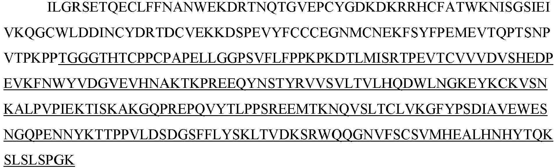
- The ActRIIA-hFc and ActRIIA-mFc proteins were expressed in CHO cell lines. Three different leader sequences were considered:
- (i) Honey bee mellitin (HBML): MKFLVNVALVFMVVYISYIYA (SEQ ID NO: 33)
- (ii) Tissue plasminogen activator (TPA): MDAMKRGLCCVLLLCGAVFVSP (SEQ ID NO: 34)
- (iii) Native: MGAAAKLAFAVFLISCSSGA (SEQ ID NO: 35).
- The selected form employs the TPA leader and has the following unprocessed amino acid sequence:

- This polypeptide is encoded by the following nucleic acid sequence:

- Both ActRIIA-hFc and ActRIIA-mFc were remarkably amenable to recombinant expression. As shown in
Figure 3 , the protein was purified as a single, well-defined peak of protein. N-terminal sequencing revealed a single sequence of -ILGRSETQE (SEQ ID NO: 38). Purification could be achieved by a series of column chromatography steps, including, for example, three or more of the following, in any order: protein A chromatography, Q sepharose chromatography, phenylsepharose chromatography, size exclusion chromatography, and cation exchange chromatography. The purification could be completed with viral filtration and buffer exchange. The ActRIIA-hFc protein was purified to a purity of >98% as determined by size exclusion chromatography and >95% as determined by SDS PAGE. - ActRIIA-hFc and ActRIIA-mFc showed a high affinity for ligands. GDF-11 or activin A were immobilized on a Biacore™ CM5 chip using standard amine-coupling procedure. ActRIIA-hFc and ActRIIA-mFc proteins were loaded onto the system, and binding was measured. ActRIIA-hFc bound to activin with a dissociation constant (KD) of 5 × 10-12 and bound to GDF11 with a KD of 9.96 × 10⁻⁹. See
Figure 4 . ActRIIA-mFc behaved similarly. - The ActRIIA-hFc was very stable in pharmacokinetic studies. Rats were dosed with 1 mg/kg, 3 mg/kg, or 10 mg/kg of ActRIIA-hFc protein, and plasma levels of the protein were measured at 24, 48, 72, 144 and 168 hours. In a separate study, rats were dosed at 1 mg/kg, 10 mg/kg, or 30 mg/kg. In rats, ActRIIA-hFc had an 11-14 day serum half-life, and circulating levels of the drug were quite high after two weeks (11 µg/ml, 110 µg/ml, or 304 µg/ml for initial administrations of 1 mg/kg, 10 mg/kg, or 30 mg/kg, respectively.) In cynomolgus monkeys, the plasma half-life was substantially greater than 14 days, and circulating levels of the drug were 25 µg/ml, 304 µg/ml, or 1440 µg/ml for initial administrations of 1 mg/kg, 10 mg/kg, or 30 mg/kg, respectively.
- ActRIIA-hFc fusion protein was expressed in stably transfected CHO-DUKX B11 cells from a pAID4 vector (SV40 ori/enhancer, CMV promoter), using a tissue plasminogen leader sequence of SEQ ID NO: 34. The protein, purified as described above in Example 1, had a sequence of SEQ ID NO: 32. The Fc portion is a human IgG1 Fc sequence, as shown in SEQ ID NO: 32. Protein analysis reveals that the ActRIIA-hFc fusion protein is formed as a homodimer with disulfide bonding.
- The CHO-cell-expressed material has a higher affinity for activin B ligand than that reported for an ActRIIa-hFc fusion protein expressed in human 293 cells [see, del Re et al. (2004) J Biol Chem. 279(51):53126-53135]. Additionally, the use of the TPA leader sequence provided greater production than other leader sequences and, unlike ActRIIA-Fc expressed with a native leader, provided a highly pure N-terminal sequence. Use of the native leader sequence resulted in two major species of ActRIIA-Fc, each having a different N-terminal sequence.
- A variety of ActRIIA variants that may be used according to the methods described herein are described in the International Patent Application published as
WO2006/012627 (see e.g., pp. 55-58). An alternative construct may have a deletion of the C-terminal tail (the final 15 amino acids of the extracellular domain of ActRIIA. The sequence for such a construct is presented below (Fc portion underlined) (SEQ ID NO: 39):
- Applicants constructed a soluble ActRIIB fusion protein that has the extracellular domain of human ActRIIB fused to a human or mouse Fc domain with a minimal linker (three glycine amino acids) in between. The constructs are referred to as ActRIIB-hFc and ActRIIB-mFc, respectively.
- ActRIIB-hFc is shown below as purified from CHO cell lines (SEQ ID NO: 40):
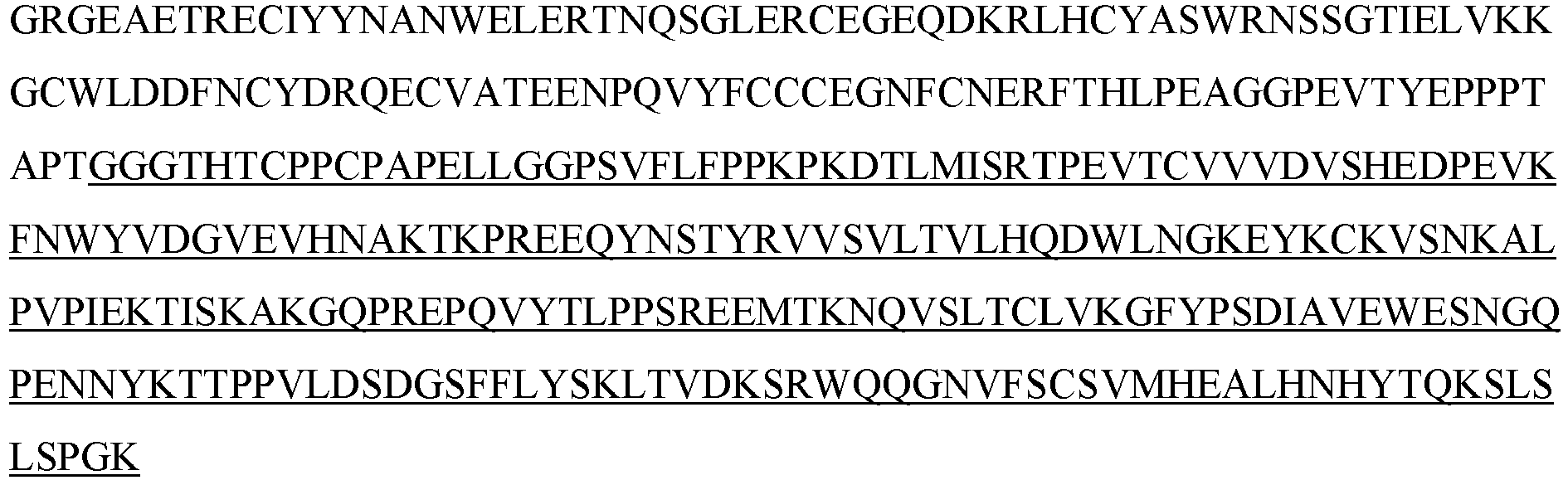
- The ActRIIB-hFc and ActRIIB-mFc proteins were expressed in CHO cell lines. Three different leader sequences were considered: (i) Honey bee mellitin (HBML), ii) Tissue plasminogen activator (TPA), and (iii) Native: MGAAAKLAFAVFLISCSSGA (SEQ ID NO: 77).
- The selected form employs the TPA leader and has the following unprocessed amino acid sequence (SEQ ID NO: 41):

- This polypeptide is encoded by the following nucleic acid sequence (SEQ ID NO: 42):


- N-terminal sequencing of the CHO-cell-produced material revealed a major sequence of -GRGEAE (SEQ ID NO: 43). Notably, other constructs reported in the literature begin with an -SGR... sequence.
- Purification could be achieved by a series of column chromatography steps, including, for example, three or more of the following, in any order: protein A chromatography, Q sepharose chromatography, phenylsepharose chromatography, size exclusion chromatography, and cation exchange chromatography. The purification could be completed with viral filtration and buffer exchange.
- ActRIIB-Fc fusion proteins were also expressed in HEK293 cells and COS cells. Although material from all cell lines and reasonable culture conditions provided protein with muscle-building activity in vivo, variability in potency was observed perhaps relating to cell line selection and/or culture conditions.
- Applicants generated a series of mutations in the extracellular domain of ActRIIB and produced these mutant proteins as soluble fusion proteins between extracellular ActRIIB and an Fc domain. The background ActRIIB-Fc fusion has the sequence of SEQ ID NO: 40.
- Various mutations, including N- and C-terminal truncations, were introduced into the background ActRIIB-Fc protein. Based on the data presented herien, it is expected that these constructs, if expressed with a TPA leader, will lack the N-terminal serine. Mutations were generated in ActRIIB extracellular domain by PCR mutagenesis. After PCR, fragments were purified through a Qiagen column, digested with SfoI and AgeI and gel purified. These fragments were ligated into expression vector pAID4 (see
WO2006/012627 ) such that upon ligation it created fusion chimera with human IgG1. Upon transformation into E. coli DH5 alpha, colonies were picked and DNAs were isolated. For murine constructs (mFc), a murine IgG2a was substituted for the human IgG1. Sequences of all mutants were verified. - All of the mutants were produced in HEK293T cells by transient transfection. In summary, in a 500ml spinner, HEK293T cells were set up at 6×10⁵ cells/ml in Freestyle (Invitrogen) media in 250ml volume and grown overnight. Next day, these cells were treated with DNA:PEI (1:1) complex at 0.5 ug/ml final DNA concentration. After 4 hrs, 250 ml media was added and cells were grown for 7 days. Conditioned media was harvested by spinning down the cells and concentrated.
- Mutants were purified using a variety of techniques, including, for example, a protein A column, and eluted with low pH (3.0) glycine buffer. After neutralization, these were dialyzed against PBS.
- Mutants were also produced in CHO cells by similar methodology. Mutants were tested in binding assays and/or bioassays described in
WO 2008/097541 andWO 2006/012627 . In some instances, assays were performed with conditioned medium rather than purified proteins. Additional variations of ActRIIB are described inU.S. Patent No. 7,842,663 . - Applicant generated an ActRIIB(25-131)-hFc fusion protein, which comprises the human ActRIIB extracellular domain with N-terminal and C-terminal truncations (residues 25-131 of the native protein SEQ ID NO: 1) fused N-terminally with a TPA leader sequence substituted for the native ActRIIB leader and C-terminally with a human Fc domain via a minimal linker (three glycine residues) (
Figure 16 ). A nucleotide sequence encoding this fusion protein is shown inFigures 17A and17B . Applicants modified the codons and found a variant nucleic acid encoding the ActRIIB(25-131)-hFc protein that provided substantial improvement in the expression levels of initial transformants (Figure 18 ). - The mature protein has an amino acid sequence as follows (N-terminus confirmed by N-terminal sequencing)(SEQ ID NO: 78):Amino acids 1-107 are derived from ActRIIB.

- The expressed molecule was purified using a series of column chromatography steps, including for example, three or more of the following, in any order: Protein A chromatography, Q sepharose chromatography, phenylsepharose chromatography, size exclusion chromatography and cation exchange chromatography. The purification could be completed with viral filtration and buffer exchange.
- Affinities of several ligands for ActRIIB(25-131)-hFc and its full-length counterpart ActRIIB(20-134)-hFc were evaluated in vitro with a Biacore™ instrument, and the results are summarized in the table below. Kd values were obtained by steady-state affinity fit due to very rapid association and dissociation of the complex, which prevented accurate determination of kon and koff. ActRIIB(25-131)-hFc bound activin A, activin B, and GDF11 with high affinity.
-
Fusion Construct Activin A (e-11) Activin B (e-11) GDF11 (e-11) ActRIIB(20-134)-hFc 1.6 1.2 3.6 ActRIIB(25-131)-hFc 1.8 1.2 3.1 - Applicants constructed a GDF trap as follows. A polypeptide having a modified extracellular domain of ActRIIB (amino acids 20-134 of SEQ ID NO: 1 with an L79D substitution) with greatly reduced activin A binding relative to GDF11 and/or myostatin (as a consequence of a leucine-to-aspartate substitution at position 79 in SEQ ID NO: 1) was fused to a human or mouse Fc domain with a minimal linker (three glycine amino acids) in between. The constructs are referred to as ActRIIB(L79D 20-134)-hFc and ActRIIB(L79D 20-134)-mFc, respectively. Alternative forms with a glutamate rather than an aspartate at position 79 performed similarly (L79E). Alternative forms with an alanine rather than a valine at position 226 with respect to SEQ ID NO: 44, below were also generated and performed equivalently in all respects tested. The aspartate at position 79 (relative to SEQ ID NO: 1, or
position 60 relative to SEQ ID NO: 44) is indicated with double underlining below. The valine at position 226 relative to SEQ ID NO: 44 is also indicated by double underlining below. - The GDF trap ActRIIB(L79D 20-134)-hFc is shown below as purified from CHO cell lines (SEQ ID NO: 44).

- The ActRIIB-derived portion of the GDF trap has an amino acid sequence set forth below (SEQ ID NO: 45), and that portion could be used as a monomer or as a non-Fc fusion protein as a monomer, dimer, or greater-order complex.

- The GDF trap protein was expressed in CHO cell lines. Three different leader sequences were considered:
(i) Honey bee melittin (HBML), (ii) Tissue plasminogen activator (TPA), and (iii) Native. - The selected form employs the TPA leader and has the following unprocessed amino acid sequence:

- This polypeptide is encoded by the following nucleic acid sequence (SEQ ID NO: 47):


- Purification could be achieved by a series of column chromatography steps, including, for example, three or more of the following, in any order: protein A chromatography, Q sepharose chromatography, phenylsepharose chromatography, size exclusion chromatography, and cation exchange chromatography. The purification could be completed with viral filtration and buffer exchange. In an example of a purification scheme, the cell culture medium is passed over a protein A column, washed in 150 mM Tris/NaCl (pH 8.0), then washed in 50 mM Tris/NaCl (pH 8.0) and eluted with 0.1 M glycine, pH 3.0. The low pH eluate is kept at room temperature for 30 minutes as a viral clearance step. The eluate is then neutralized and passed over a Q-sepharose ion-exchange column and washed in 50 mM Tris pH 8.0, 50 mM NaCl, and eluted in 50 mM Tris pH 8.0, with an NaCl concentration between 150 mM and 300 mM. The eluate is then changed into 50 mM Tris pH 8.0, 1.1 M ammonium sulfate and passed over a phenyl sepharose column, washed, and eluted in 50 mM Tris pH 8.0 with ammonium sulfate between 150 and 300 mM. The eluate is dialyzed and filtered for use.
- Additional GDF traps (ActRIIB-Fc fusion proteins modified so as to reduce the ratio of activin A binding relative to myostatin or
GDF 11 binding) are described inWO 2008/097541 andWO 2006/012627 . - An A-204 reporter gene assay was used to evaluate the effects of ActRIIB-Fc proteins and GDF traps on signaling by GDF-11 and activin A. Cell line: human rhabdomyosarcoma (derived from muscle). Reporter vector: pGL3(CAGA)12 (described in Dennler et al, 1998, EMBO 17: 3091-3100). The CAGA12 motif is present in TGF-beta responsive genes (e.g., PAI-1 gene), so this vector is of general use for factors signaling through SMAD2 and 3.
- Day 1: Split A-204 cells into 48-well plate.
- Day 2: A-204 cells transfected with 10 ug pGL3(CAGA)12 or pGL3(CAGA)12(10 ug) + pRLCMV (1 µg) and Fugene.
- Day 3: Add factors (diluted into medium + 0.1 % BSA). Inhibitors need to be preincubated with factors for 1 hr before adding to cells. Six hrs later, cells were rinsed with PBS and lysed.
- This is followed by a luciferase assay. In the absence of any inhibitors, activin A showed 10-fold stimulation of reporter gene expression and an ED50 ~ 2 ng/ml. GDF-11: 16 fold stimulation, ED50: ~1.5 ng/ml.
- ActRIIB(20-134) is a potent inhibitor of activin A, GDF-8, and GDF-11 activity in this assay. As described below, ActRIIB variants were also tested in this assay.
- Activity of ActRIIB-Fc proteins and GDF traps was tested in a cell-based assay as described above. Results are summarized in the table below. Some variants were tested in different C-terminal truncation constructs. As discussed above, truncations of five or fifteen amino acids caused reduction in activity. The GDF traps (L79D and L79E variants) showed substantial loss of activin A inhibition while retaining almost wild-type inhibition of GDF-11.
-
ActRIIB-Fc Variations Portion of ActRIIB (corresponds to amino acids of SEQ ID NO:1) GDF11 Inhibition Activity Activin Inhibition Activity R64 20-134 +++ (approx. 10-8 M KI) +++ (approx. 10-8 M KI) A64 20-134 + (approx. 10-6 M KI) + (approx. 10-6 M KI) R64 20-129 +++ +++ R64 K74A 20-134 ++++ ++++ R64 A24N 20-134 +++ +++ R64 A24N 20-119 ++ ++ R64 A24N K74A 20-119 + + R64 L79P 20-134 + + R64 L79P K74A 20-134 + + R64 L79D 20-134 +++ + R64 L79E 20-134 +++ + R64K 20-134 +++ +++ R64K 20-129 +++ +++ R64 P129S P130A 20-134 +++ +++ R64N 20-134 + + + Poor activity (roughly 1×10⁻⁶ KI)
++ Moderate activity (roughly 1×10⁻⁷ KI)
+++ Good (wild-type) activity (roughly 1×10⁻⁸ KI)
++++ Greater than wild-type activity - Several variants have been assessed for serum half-life in rats. ActRIIB(20-134)-Fc has a serum half-life of approximately 70 hours. ActRIIB(A24N 20-134)-Fc has a serum half-life of approximately 100-150 hours. The A24N variant has activity in the cell-based assay (above) and that is equivalent to the wild-type molecule. Coupled with the longer half-life, this means that over time an A24N variant will give greater effect per unit of protein than the wild-type molecule. The A24N variant, and any of the other variants tested above, may be combined with the GDF trap molecules, such as the L79D or L79E variants.
- Binding of certain ActRIIB-Fc proteins and GDF traps to ligands was tested in a Biacore™ assay.
- The ActRIIB-Fc variants or wild-type protein were captured onto the system using an anti-hFc antibody. Ligands were injected and flowed over the captured receptor proteins. Results are summarized in the tables below.
-
GDF11 Protein Kon (1/Ms) Koff (1/s) KD (M) ActRIIB(20-134)-hFc 1.34e-6 1.13e-4 8.42e-11 ActRIIB(A24N 20-134)-hFc 1.21e-6 6.35e-5 5.19e-11 ActRIIB(L79D 20-134)-hFc 6.7e-5 4.39e-4 6.55e-10 ActRIIB(L79E 20-134)-hFc 3.8e-5 2.74e-4 7.16e-10 ActRIIB(R64K 20-134)-hFc 6.77e-5 2.41e-5 3.56e-11 GDF8 Protein Kon (1/Ms) Koff (1/s) KD (M) ActRIIB(20-134)-hFc 3.69e-5 3.45e-5 9.35e-11 ActRIIB(A24N 20-134)-hFc ActRIIB(L79D 20-134)-hFc 3.85e-5 8.3e-4 2.15e-9 ActRIIB(L79E 20-134)-hFc 3.74e-5 9e-4 2.41e-9 ActRIIB(R64K 20-134)-hFc 2.25e-5 4.71e-5 2.1e-10 ActRIIB(R64K 20-129)-hFc 9.74e-4 2.09e-4 2.15e-9 ActRIIB(P129S, P130R 20-134)-hFc 1.08e-5 1.8e-4 1.67e-9 ActRIIB(K74A 20-134)-hFc 2.8e-5 2.03e-5 7.18e-11 Activin A Protein Kon (1/Ms) Koff (1/s) KD (M) ActRIIB(20-134)-hFc 5.94e6 1.59e-4 2.68e-11 ActRIIB(A24N 20-134)-hFc 3.34e6 3.46e-4 1.04e-10 ActRIIB(L79D 20-134)-hFc Low binding ActRIIB(L79E 20-134)-hFc Low binding ActRIIB(R64K 20-134)-hFc 6.82e6 3.25e-4 4.76e-11 ActRIIB(R64K 20-129)-hFc 7.46e6 6.28e-4 8.41e-11 ActRIIB(P129S, P130R 20-134)-hFc 5.02e6 4.17e-4 8.31e-11 - These data obtained in a cell-free assay confirm the cell-based assay data, demonstrating that the A24N variant retains ligand-binding activity that is similar to that of the ActRIIB(20-134)-hFc molecule and that the L79D or L79E molecule retains myostatin and GDF11 binding but shows markedly decreased (non-quantifiable) binding to activin A.
- Other variants have been generated and tested, as reported in
WO2006/012627 . See, e.g., pp. 59-60, using ligands coupled to the device and flowing receptor over the coupled ligands. Notably, K74Y, K74F, K74I (and presumably other hydrophobic substitutions at K74, such as K74L), and D80I, cause a decrease in the ratio of activin A (ActA) binding to GDF11 binding, relative to the wild-type K74 molecule. A table of data with respect to these variants is reproduced below: -
ActRIIB ActA GDF11 WT (64A) KD=1.8e-7M (+) KD= 2.6e-7M (+) WT (64R) na KD= 8.6e-8M (+++) +15tail KD ∼2.6 e-8M (+++) KD= 1.9e-8M (++++) E37A * * R40A - - D54A - * K55A ++ * R56A * * K74A KD=4.35e-9 M KD=5.3e-9M K74Y * -- K74F * -- K74I * -- W78A * * L79A + * D80K * * D80R * * D80A * * D80F * * D80G * * D80M * * D80N * * D80I * -- F82A ++ -- * No observed binding
-- < 1/5 WT binding
- ~ 1/2 WT binding
+ WT
++ < 2x increased binding
+++ ~5x increased binding
++++ ~10x increased binding
+++++ ~ 40x increased binding - A a GDF trap referred to as ActRIIB(L79D 20-134)-hFc was generated by N-terminal fusion of TPA leader to the ActRIIB extracellular domain (residues 20-134 in SEQ ID NO: 1) containing a leucine-to-aspartate substitution (at residue 79 in SEQ ID NO:1) and C-terminal fusion of human Fc domain with minimal linker (three glycine residues) (
Figure 5 ). A nucleotide sequence corresponding to this fusion protein is shown inFigures 6A and6B . - A GDF trap with truncated ActRIIB extracellular domain, referred to as ActRIIB(L79D 25-131)-hFc, was generated by N-terminal fusion of TPA leader to truncated extracellular domain (residues 25-131 in SEQ ID NO:1) containing a leucine-to-aspartate substitution (at residue 79 in SEQ ID NO:1) and C-terminal fusion of human Fc domain with minimal linker (three glycine residues) (
Figure 7 , SEQ ID NO: 61). The processed form of ActRIIB(L79D 25-131)-hFc (SEQ ID NO: 64) is shown inFigure 9 . One nucleotide sequence encoding this fusion protein is shown inFigures 8A and8B (SEQ ID NO: 62), and an alternative nucleotide sequence encoding exactly the same fusion protein is shown inFigures 11A and11B (SEQ ID NO: 66). - The affinity of GDF traps and other ActRIIB-hFc proteins for several ligands was evaluated in vitro with a Biacore™ instrument. Results are summarized in the table below. Kd values were obtained by steady-state affinity fit due to the very rapid association and dissociation of the complex, which prevented accurate determination of kon and koff.
-
Fusion Construct Activin A (Kd e-11) Activin B (Kd e-11) GDF11 (Kd e-11) ActRIIB(L79 20-134)-hFc 1.6 1.2 3.6 ActRIIB(L79D 20-134)-hFc 1350.0 78.8 12.3 ActRIIB(L79 25-131)-hFc 1.8 1.2 3.1 ActRIIB(L79D 25-13 1)-hFc 2290.0 62.1 7.4 - The GDF trap with a truncated extracellular domain, ActRIIB(L79D 25-131)-hFc, equaled or surpassed the ligand selectivity displayed by the longer variant, ActRIIB(L79D 20-134)-hFc, with pronounced loss of activin A binding, partial loss of activin B binding, and nearly full retention of GDF11 binding compared to ActRIIB-hFc counterparts lacking the L79D substitution. Note that truncation alone (without L79D substitution) did not alter selectivity among the ligands displayed here [compare ActRIIB(L79 25-131)-hFc with ActRIIB(L79 20-134)-hFc]. ActRIIB(L79D 25-131)-hFc also retains strong to intermediate binding to the
Smad 2/3 signaling ligand GDF8 and theSmad 1/5/8 ligands BMP6 and BMP10. - Others have reported an alternate, soluble form of ActRIIB (designated ActRIIB5), in which
exon 4, including the ActRIIB transmembrane domain, has been replaced by a different C-terminal sequence (see, e.g.,WO 2007/053775 ). - The sequence of native human ActRIIB5 without its leader is as follows:

- An leucine-to-aspartate substitution, or other acidic substitutions, may be performed at native position 79 (underlined) as described to construct the variant ActRIIB5(L79D), which has the following sequence:

- This variant may be connected to human Fc (double underline) with a TGGG linker (single underline) to generate a human ActRIIB5(L79D)-hFc fusion protein with the following sequence:

- This construct may be expressed in CHO cells.
- Applicants investigated effects of the GDF trap ActRIIB(L79D 25-131)-mFc in the NUP98-HOXD13 mouse model of MDS, which is characterized by abortive precursor maturation and ineffective hematopoiesis. In this model, disease severity increases with age, eventually progressing to acute myeloid leukemia in the majority of mice, and they have a mean life span of approximately 14 months. Starting at approximately 4 months of age, these mice exhibit anemia, leukopenia, ineffective erythropoiesis, and bone marrow that is normocellular to hypercellular [Lin et al. (2005) Blood 106:287-295]. To monitor the effects of chronic administration, MDS mice were treated with ActRIIB(L79D 25-131)-mFc (10 mg/kg, s.c.) or vehicle twice weekly beginning at 4 months of age and continuing for 7 months, while blood samples (50 µL) were collected at baseline and monthly thereafter for complete blood count analysis. As expected, 6-month-old MDS mice developed severe anemia compared to wild-type mice (
Figure 13A ), and bone marrow analyses revealed increased numbers of erythroid precursors (Figure 13A ) and a lower myeloid/erythroid (M/E) ratio [Suragani et al. (2014) Nat Med 20:408-414] in MDS mice compared to age-matched FVB wild-type mice, indicative of ineffective erythropoiesis. In 6-month-old MDS mice, treatment with ActRIIB(L79D 25-131)-mFc significantly increased RBC count (by 16.9%) and hemoglobin concentration (by 12.5%) (Figure 13A ), reduced erythroid precursor cell count in bone marrow (Figure 13A ), and normalized the M/E ratio to that of wild-type mice [Suragani et al. (2014) Nat Med 20:408-414]. - In MDS mice at 12 months of age, ActRIIB(L79D 25-131)-mFc treatment significantly increased RBC count (by 18.3%) and hemoglobin level (by 13.0%) (
Figure 13B ), reduced erythroid precursor cell count (Figure 13B ), and improved the M:E ratio [Suragani et al. (2014) Nat Med 20:408-414], as compared to vehicle. ActRIIB(L79D 25-131)-mFc treatment did not affect the absolute number of myeloid precursors. Flow cytometry confirmed that ActRIIB(L79D 25-131)-mFc treatment reduced erythroid hyperplasia in MDS mice at both ages. A time-course analysis in MDS mice treated with ActRIIB(L79D 25-131)-mFc for 7 months showed a sustained elevation in RBC numbers for the duration of the study [Suragani et al. (2014) Nat Med 20:408-414]. Together, these results indicate that treatment with a GDF trap ameliorates anemia, erythroid hyperplasia and ineffective erythropoiesis in MDS mice regardless of disease severity. - A recombinant fusion protein containing modified activin receptor type IIB and IgG Fc [ActRIIB(L79D 25-131)-hFc, also known as luspatercept or ACE-536] is being developed for the treatment of anemias due to ineffective erythropoiesis such as myelodysplastic syndromes (MDS). Patients with MDS often have elevated levels of EPO and may be nonresponsive or refractory to erythropoiesis-stimulating agents (ESAs). MDS patients have also been shown to have increased serum levels of GDF11 [Suragani et al. (2014) Nat Med 20:408-414] and increased
Smad 2/3 signaling in the bone marrow [Zhou et al. (2008) Blood 112:3434-3443]. ActRIIB(L79D 25-131)-hFc binds to ligands in the TGFβ superfamily, including GDF11, inhibits Smad2/3 signaling, and promotes late-stage erythroid differentiation via a mechanism distinct from ESAs. A murine version, ActRIIB(L79D 25-131)-mFc, reduced Smad2 signaling, increased hemoglobin (Hb) levels, and decreased bone marrow erythroid hyperplasia in a mouse model of MDS [Suragani et al. (2014) Nat Med 20:408-414]. In a healthy-volunteer study, ActRIIB(L79D 25-131)-hFc was well-tolerated and increased Hb levels [Attie et al. (2014) Am J Hematol 89:766-770]. - Applicants are conducting an ongoing,
phase 2, multicenter, open-label, dose-finding study to evaluate the effects of ActRIIB(L79D 25-131)-hFc on anemia in patients with Low or Int-1 risk MDS who have either high transfusion burden (HTB, defined as ≥ 4 units RBC per 8 weeks prior to baseline) or low transfusion burden (LTB, defined as < 4 units RBC per 8 weeks prior to baseline). Study outcomes include erythroid response (either Hb increase in LTB patients or reduced transfusion burden in HTB patients), safety, tolerability, pharmacokinetics, and pharmacodynamic biomarkers. Inclusion criteria include: Low or Int-1 risk MDS, age ≥ 18 yr, anemia (defined as either being HTB patient or having baseline Hb < 10.0 g/dL in LTB patient), EPO > 500 U/L or nonresponsive/refractory to ESAs, no prior azacitidine or decitabine, and no current treatment with ESA, granulocyte colony-stimulating factor (G-CSF), granulocyte-macrophage colony-stimulating factor (GM-CSF), or lenalidomide, thalidomide or pomalidomide. In the dose-escalation phase, ActRIIB(L79D 25-131)-hFc was administered by subcutaneous injection once every 3 weeks in seven sequential cohorts (n = 3-6) at dose levels of 0.125, 0.25, 0.5, 0.75, 1.0, 1.33 and 1.75 mg/kg for up to 5 doses with a 3-month follow-up. - Data were available for 26 patients (seven LTB/19 HTB). Median age was 71 yr (range: 27-88 yr), 50% were female, 54% had prior EPO therapy, and 15% had prior lenalidomide. Patient classification by WHO subtype was as follows: 15% RARS, 46% RCMD-RS, 15% RCMD, 15% RAEB-1 (including two patients with ≥15% ring sideroblasts) and 8% del (5q). Mean (SD) baseline Hgb for the LTB patients (n = 7) was 9.1 (0.4) g/dL. Mean (SD) units RBC transfused in the 8 weeks prior to treatment was 0.9 (1.1) units for the LTB patients and 6.3 (2.4) units for the HTB patients. Two of the seven LTB patients had an increase in mean Hb ≥ 1.5 g/dL over 8 weeks compared to baseline. Mean maximum Hb increase in the LTB patients was 0.8, 1.0, 2.2, and 3.5 g/dL in the 0.125 (n=1), 0.25 (n = 1), 0.75 (n = 3), and 1.75 (n = 2) mg/kg dose groups, respectively. Six of the seven LTB patients achieved RBC transfusion independence (RBC-TI) for ≥ 8 weeks during the study. The dose levels ranging from 0.75 mg/kg to 1.75 mg/kg were deemed to be active doses. Among the five patients in the active dose groups, four (80%) achieved the pre-specified endpoint of Hgb increase of ≥ 1.5 g/dl for ≥ 2 weeks. Two patients (40%) achieved a HI-E response [International Working Group; Cheson et al. (2000) Blood 96:3671-3674; Cheson et al. (2006) Blood 108:419-425], defined as an Hgb increase of ≥ 1.5 g/dl for ≥ 8 weeks in LTB patients. In HTB patients, the HI-E response is defined as a reduction in transfusion burden of at least four units of red blood cells transfused over an 8 week period as compared to the 8 weeks prior to study start. In the active dose groups, five of 12 (42%) HTB patients met the pre-specified endpoint of a reduction of ≥ 4 RBC units or ≥ 50% reduction in RBC units transfused over an 8-week interval during the treatment period compared to the 8 weeks prior to treatment, and the same patients (five of 12, 42%) achieved a HI-E response; three of 12 (25%) of HTB patients in the active dose groups achieved RBC-TI ≥ 8 weeks during the study. Increases in neutrophil count following study drug administration were observed in some patients. Finally, ActRIIB(L79D 25-131)-hFc was generally well tolerated. No related serious adverse events have been reported to date. The most frequent adverse events regardless of causality were: diarrhea (n = 4,
grade 1/2), bone pain, fatigue, muscle spasms, myalgia, and nasopharyngitis (n = 3 each,grade 1/2). - Assessment of bone marrow aspirates demonstrated an association between the presence of ring sideroblasts (considered positive if ≥ 15% of erythroid precursors exhibited ring sideroblast morphology) and responsiveness to ActRIIB(L79D 25-131)-hFc in the active dose groups (0.75 - 1.75 mg/kg). The overall response rate (using HI-E criteria, described above) across both LTB and HTB patients was seven of 17 (41%). Among patients positive for ring sideroblasts at baseline, seven of 13 (54%) patients achieved a HI-E response, and notably none of the four patients negative for ring sideroblasts at baseline achieved a HI-E response.
- Bone marrow aspirates from patients were also evaluated for the presence of mutations in 21 different genes that are known to harbor mutations (primarily somatic mutations) associated with MDS. Genomic DNA was isolated from bone marrow aspirates, selected coding regions of the 21 genes were amplified by polymerase chain reaction, and the DNA sequences of these regions were determined using next-generation sequencing (Myeloid Molecular Profile 21-gene panel, Genoptix, Inc., Carlsbad, CA). This analysis examined activated signaling genes (KIT, JAK2, NRAS, CBL, and MPL), transcription factors (RUNX1, ETV6), epigenetic genes (IDH1, IDH2, TET2, DNMT3A, EZH2, ASXL1, and SETBP1), RNA splicing genes (SF3B1, U2AF1, ZRSF2, and SRSF2), and tumor suppressors/others (TP53, NPM1, PHF6). Analysis of SF3B1 specifically targeted exons 13-16. Of these 21 MDS-associated genes evaluated, mutations in SF3B1 were more frequently detected in bone marrow cells in the responsive patients than in the nonresponsive patients. Individual SF3B1 mutations detected in these patients are shown in the following table. The same mutation sometimes occurred in multiple patients.
Nucleotide Nucleotide Substitution Amino Acid Substitution Exon 1873 C → T R625C 14 1874 G → T R625L 14 1986 C → G H662Q 14 2098 A → G K700E 15 2342 A → G D781G 16 - In patients with SF3B1 mutations in the active dose groups, six of nine (67%) achieved HI-E responses, including all three patients that achieved transfusion independence for greater than 8 weeks. In patients not having an SF3B1 mutation, only one of eight (13%) achieved a HI-E reponse. Mutations in SF3B1 are frequently observed in MDS patients with ring sideroblasts and are associated with ineffective erythropoiesis.
- These results indicate that patients with MDS exhibiting ≥ 15% ring sideroblasts (and patients with other forms of sideroblastic anemia), and/or at least one mutation in SF3B1 are more likely to respond therapeutically to ActRIIB(L79D 25-131)-hFc than MDS patients with < 15% ring sideroblasts and/or no mutation in SF3B1. Based on these data, selective treatment of any of these patient subgroups is expected to greatly increase the benefit/risk ratio of treatment with ActRII inhibitors.
- ActRIIB(L79D 25-131)-hFc treatment was also observed to have a surprising effect on vision. In the above-described
phase 2 MDS study, a male patient with poor vision (e.g., requiring corrective lens to perform certain activities) was shown to be responsive to ActRIIB(L79D 25-131)-hFc therapy. Prior to treatment the patient was afflicted with chronic anemia, requiring regular blood transfusions. ActRIIB(L79D 25-131)-hFc resulted in significant and sustained increases in hemoglobin levels the patient. Indeed, the patient achieved transfusion independence for ≥ 8 months during the study. In addition, significant improvement in vision was observed in the patient. In fact, the patient no longer required corrective lens to perform certain activities. These data therefore indicate that, in addition to positive effects on treating anemia, ActRII inhibitors may be used to improve vision in MDS patients. Moreover, in view of the reported mechanism for MDS-associated vision loss, as discussed below, the data suggest that ActRII inhibitors also may have positive effects on treating other types of ocular disorders. - It has been reported that retinal nerve fiber layer thickness is decreased in patients with various hematologic disorders, particularly those associated with anemia [Han et al. (2015) J Glaucoma (Epub ahead of print)]. Such changes in the retinal nerve fiber are associated with reduced visual acuity and other pathological changes in the eye. Studies indicate that retinal damage in these patients may be due to ischemic optic neuropathy. For example, loss of vision in MDS patients has been associated with the manifestation of NAION [Brouzas et al. (2009) Clinical Ophthalmology 3:133-137]. In such MDS patients, it is believed that the initiation of NAION is through a mechanism of ischemia and/or microvascular insufficiency. Indeed, it has been demonstrated that therapy for treating ischemia-hypoxia improves visual acuity in MDS patients. Accordingly, the data of the instant application suggests that ActRII antagonists may be used to treat other related ocular disorders, particularly those associated with ischemia and microvascular insufficiency.
- The effects of ActRIIA-Fc (see Example 1), ActRIIB-Fc (see Example 4), and ActRIIB(L79D 25-131)-Fc (see Example 9) on lesion size and leakage in a rat model of laser-induced choroidal neovascularization are to be evaluated.
- At
day 1 of the study, 20 Brown Norway rats (6-8 weeks of age) receive bilateral laser treatment to produce 3 lesions per eye. Animals are dilated with 1% Cyclogyl solution and protected from light. Following observable dilation, the animals are sedated. The fundus of sedated animals is observed and recorded using a Micron IV small animal funduscope (Phoenix Research). Laser treatments are performed using a thermal laser which is connected through the Micron IV custom laser attachment. A total of 3 lesions per eye are placed in the right eye using a wavelength of 532 nm. The resultant fundus images are recorded and evaluated to confirm the laser has successfully produced a bubble through the Bruch's membrane. - Following laser treatment, the rats are separated into one of 4 treatment groups: a) s.c. administration of vehicle (PBS) on
days 1, 8, and 15; b) s.c. administration of ActRIIA-Fc (10 mg/kg) ondays 1, 8, and 15; c) s.c. administration of ActRIIB-Fc (10 mg/kg) ondays 1, 8, and 15; and d) s.c. administration of ActRIIB(L79D 25-13 1)-Fc (10 mg/kg) ondays 1, 8, and 15. - At day 22, the rats are evaluated for reduction of lesion size and leakage by in vivo fluorescein angiographs. Animals are anesthetized and then receive an IP injection of 10% fluorescein sodium at 1 µl/gram of body weight. Fundus images are then captured as 8-bit TIFF files using the Micron III and exciter/barrier filters for a target wavelength of 488 nm. Standard color fundus photos are also captured for each eye. All TIFF images are quantified using computerized image-analysis software (e.g., Imaged, NIH, USA). The boundary of lesions are then individually traced free-hand, and the resultant image is subjected to multi-Otsu thresholding to eliminate background signal and quantify the area in pixels. Areas of hemorrhage or where two lesions are overlapping are excluded from analysis.
- The effects of ActRIIA-Fc (see Example 1), ActRIIB-Fc (see Example 4), and ActRIIB(L79D 25-131)-Fc (see Example 9) on lesion size and leakage in a rat model of streptozotocin (STZ) induced diabetes are to be evaluated.
- STZ is a small molecule that causes depletion of the pancreatic islet cells following systemic administration. The islet cell death causes a loss of insulin production and subsequent dysregulation of blood glucose levels leading to hyperglycemia within days. This model has been used to study inflammation, vascular pathology, and signaling pathways in the pathogenesis of diabetic retinopathy and diabetic macular edema. At eight weeks following induction of diabetes with STZ, there is a significant and progressive loss of visual acuity and contrast sensitivity. Since the primary endpoints accepted by regulatory agencies for human clinical trials are focused on quantitative measurements of visual acuity and contrast sensitivity, this is an excellent model for testing the activity of therapeutic agents to prevent diabetic vision loss.
- At
day 1 of the study, 20 Brown Norway rats (8-12 weeks of age) are given a single intraperitoneal injection of STZ (50mg/kg of body weight in 10 mmol/L of citrate buffer, pH 4.5). Serum glucose levels will be examined two days after the STZ injection and weekly thereafter. Only animals with blood glucose levels higher than 350 mg/dl will be used as diabetic rats. - Six to eight weeks following STZ treatment, the rats are separated into one of 4 treatment groups: a) s.c. administration of vehicle (PBS) on
days 1, 8, and 15; b) s.c. administration of ActRIIA-Fc (10 mg/kg) ondays 1, 8, and 15; c) s.c. administration of ActRIIB-Fc (10 mg/kg) ondays 1, 8, and 15; and d) s.c. administration of ActRIIB(L79D 25-13 1)-Fc (10 mg/kg) ondays 1, 8, and 15. - At day 22 following the start of therapy, the rats are evaluated for reduction of lesion size and leakage by in vivo fluorescein angiographs as described above in Example 15.
Claims (15)
- A composition for use in a method of:a) treating or preventing a vascular disorder of the eye in a patient; and/orb) improving vision in a patient that has a vascular disorder of the eye; wherein the patient has myelodysplastic syndrome, wherein the composition comprises an effective amount of an ActRII antagonist, and wherein the ActRII antagonist is a polypeptide comprising an amino acid sequence that is at least 90% identical to the sequence of amino acids 29-109 of SEQ ID NO:1, wherein the polypeptide comprises an acidic amino acid at position 79 with respect to SEQ ID NO: 1.
- The composition for use according to claim 1, wherein the vascular disorder of the eye is associated with ischemia and/or microvascular insufficiency.
- The composition for use according to any one of claims 1-2, wherein the ActRII antagonist is an ActRII polypeptide, and wherein the ActRII polypeptide is selected from the group consisting of:a) a polypeptide comprising an amino acid sequence that is identical to amino acids 29-109 of SEQ ID NO: 1 or comprising an amino acid sequence that is at least 91%, 92%, 93%, 94%, 95%, 96%, 97%, 98%, or 99% identical to the sequence of amino acids 29-109 of SEQ ID NO: 1;b) a polypeptide comprising an amino acid sequence that is identical to amino acids 25-131 of SEQ ID NO: 1 or comprising an amino acid sequence that is at least 90%, 91%, 92%, 93%, 94%, 95%, 96%, 97%, 98%, or 99% identical to the sequence of amino acids 25-131 of SEQ ID NO: 1;c) a polypeptide comprising the amino acid sequence of SEQ ID NO: 2 or comprising an amino acid sequence that is at least 90%, 91%, 92%, 93%, 94%, 95%, 96%, 97%, 98%, or 99% identical to the amino acid sequence of SEQ ID NO: 2;d) a polypeptide comprising the amino acid sequence of SEQ ID NO:3 or comprising an amino acid sequence that is at least 90%, 91%, 92%, 93%, 94%, 95%, 96%, 97%, 98%, or 99% identical to the amino acid sequence of SEQ ID NO: 3;e) a polypeptide comprising the amino acid sequence of SEQ ID NO: 5 or comprising an amino acid sequence that is at least 90%, 91%, 92%, 93%, 94%, 95%, 96%, 97%, 98%, or 99% identical to the amino acid sequence of SEQ ID NO: 5;f) a polypeptide comprising the amino acid sequence of SEQ ID NO: 6 or comprising an amino acid sequence that is at least 90%, 91%, 92%, 93%, 94%, 95%, 96%, 97%, 98%, or 99% identical to the amino acid sequence of SEQ ID NO: 6;g) a polypeptide comprising the amino acid sequence of SEQ ID NO: 45 or comprising an amino acid sequence that is at least 90%, 91%, 92%, 93%, 94%, 95%, 96%, 97%, 98%, or 99% identical to the amino acid sequence of SEQ ID NO: 45;h) a polypeptide comprising the amino acid sequence of SEQ ID NO: 48 or comprising an amino acid sequence that is at least 90%, 91%, 92%, 93%, 94%, 95%, 96%, 97%, 98%, or 99% identical to the amino acid sequence of SEQ ID NO: 48;i) a polypeptide comprising the amino acid sequence of SEQ ID NO: 49 or comprising an amino acid sequence that is at least 90%, 91%, 92%, 93%, 94%, 95%, 96%, 97%, 98%, or 99% identical to the amino acid sequence of SEQ ID NO: 49; andj) a polypeptide comprising the amino acid sequence of SEQ ID NO: 65 or comprising an amino acid sequence that is at least 90%, 91%, 92%, 93%, 94%, 95%, 96%, 97%, 98%, or 99% identical to the amino acid sequence of SEQ ID NO: 65.
- The composition for use according to claim 3, wherein the polypeptide is a fusion protein comprising, in addition to an ActRII polypeptide domain an immunoglobulin Fc domain.
- The composition for use according to claim 4, wherein the polypeptide is an ActRII-Fc fusion protein comprising a polypeptide selected from the group consisting of:a) a polypeptide comprising the amino acid sequence of SEQ ID NO: 40 or comprising an amino acid sequence that is at least 90%, 91%, 92%, 93%, 94%, 95%, 96%, 97%, 98%, or 99% identical to the amino acid sequence of SEQ ID NO: 40;b) a polypeptide comprising the amino acid sequence of SEQ ID NO: 41 or comprising an amino acid sequence that is at least 90%, 91%, 92%, 93%, 94%, 95%, 96%, 97%, 98%, or 99% identical to the amino acid sequence of SEQ ID NO: 41;c) a polypeptide comprising the amino acid sequence of SEQ ID NO: 44 or comprising an amino acid sequence that is at least 90%, 91%, 92%, 93%, 94%, 95%, 96%, 97%, 98%, or 99% identical to the amino acid sequence of SEQ ID NO: 44;d) a polypeptide comprising the amino acid sequence of SEQ ID NO: 46 or comprising an amino acid sequence that is at least 90%, 91%, 92%, 93%, 94%, 95%, 96%, 97%, 98%, or 99% identical to the amino acid sequence of SEQ ID NO: 46;e) a polypeptide comprising the amino acid sequence of SEQ ID NO: 50 or comprising an amino acid sequence that is at least 90%, 91%, 92%, 93%, 94%, 95%, 96%, 97%, 98%, or 99% identical to the amino acid sequence of SEQ ID NO: 50;f) a polypeptide comprising the amino acid sequence of SEQ ID NO: 61 or comprising an amino acid sequence that is at least 90%, 91%, 92%, 93%, 94%, 95%, 96%, 97%, 98%, or 99% identical to the amino acid sequence of SEQ ID NO: 61;g) a polypeptide comprising the amino acid sequence of SEQ ID NO: 64 or comprising an amino acid sequence that is at least 90%, 91%, 92%, 93%, 94%, 95%, 96%, 97%, 98%, or 99% identical to the amino acid sequence of SEQ ID NO: 64;h) a polypeptide comprising the amino acid sequence of SEQ ID NO: 78 or comprising an amino acid sequence that is at least 90%, 91%, 92%, 93%, 94%, 95%, 96%, 97%, 98%, or 99% identical to the amino acid sequence of SEQ ID NO: 78; andi) a polypeptide comprising the amino acid sequence of SEQ ID NO: 79 or comprising an amino acid sequence that is at least 90%, 91%, 92%, 93%, 94%, 95%, 96%, 97%, 98%, or 99% identical to the amino acid sequence of SEQ ID NO: 79.
- The composition for use according to any one of claims 1 to 5, wherein the polypeptide comprises a D or E at position 79 with respect to SEQ ID NO: 1.
- The composition for use according to any one of claims 1-6, wherein the polypeptide binds to GDF11, GDF8, activin, activin A and/or activin B.
- The composition for use according to any one of claims 1-7, wherein the method further comprises administering the ActRII antagonist in combination with one or more additional active agents or supportive therapies for treating, preventing, or reducing the severity of an eye disorder.
- The composition for use according to claim 8, wherein the one or more supportive therapies is selected from the group consisting of: surgery, laser therapy (e.g., photocoagulation), anti-angiogenic therapy [e.g., VEGF inhibitors such as bevacizumab, ranibizumab, and Aflibercept], Ca2+ inhibitors (e.g., flunarizine and nifedipine), cryotherapy, hyperbaric oxygenation, Na+ channel blockers (e.g., topiramate), iGluR antagonists (e.g., MK-801, dextromethorphan, eliprodil, and flupirtine), antioxidants (e.g., dimethylthiourea, vitamin E, alph-lipoic acid, superoxide dismutase, catalase, desferrioxamine, mannitol, allopurinol, calcium dobesilate, flupirtine, trimetazidine, and EGB-761), anti-inflammatory agents, cyclodiathermy, cyclocryotherapy, ocular filtering procedures, implantation of drainage valves, antiplatelet therapy (e.g., aspirin, ticlopidine, and clopidogrel), anticoagulant therapy (e.g., warfarin and heparin), steroids, systemic or local corticosteroids (e.g., prednisone triamcinolone and fluocinolone acetonide (Iluvien)), and dexamethasone, steroid-sparing immunosuppressants (e.g., cyclosporine, azathioprine, cyclophosphamide, mycophenolate, mofetil, infliximab and etanercept), dietary supplements (e.g., vitamin C, vitamin E, lutein, zeaxanthin, zinc, folic acid, vitamins B6, vitamin B12, and zeaxanthin), vitrectomy, scleral buckle surgery, and pneumatic retinopexy.
- The composition for use according to claim 8, wherein the one or more additional active agents is selected from the group consisting of: a vascular endothelial growth factor (VEGF) inhibitor, a VEGF-A inhibitor, a placental growth factor (PIGF) inhibitor, an agent that inhibits VEGF and PIGF, aflibercept, ranibizumab, and bevacizumab.
- The composition for use according to any one of claims 1-10, wherein the ActRII antagonist is administered by ocular or intravitreal administration.
- The composition for use according to any one of claims 1-11, wherein the ActRII antagonist is a polypeptide comprising an amino acid sequence that is at least 95% identical to the sequence of amino acids 29-109 of SEQ ID NO:1.
- The composition for use according to any one of claims 1-12, wherein the ActRII antagonist is an ActRII-Fc fusion protein, and wherein the ActRII-Fc fusion protein comprises an amino acid sequence that is at least 90% identical to SEQ ID NO:64.
- The composition for use according to claim 13, wherein the ActRII-Fc fusion protein comprises an amino acid sequence that is at least 95% identical to SEQ ID NO: 64.
- The composition for use according to claim 14, wherein the ActRII-Fc fusion protein comprises an amino acid sequence that is identical to SEQ ID NO: 64.
Applications Claiming Priority (2)
| Application Number | Priority Date | Filing Date | Title |
|---|---|---|---|
| US201562258934P | 2015-11-23 | 2015-11-23 | |
| PCT/US2016/063557 WO2017091706A1 (en) | 2015-11-23 | 2016-11-23 | Methods for treating eye disorders |
Publications (3)
| Publication Number | Publication Date |
|---|---|
| EP3380121A1 EP3380121A1 (en) | 2018-10-03 |
| EP3380121A4 EP3380121A4 (en) | 2019-06-12 |
| EP3380121B1 true EP3380121B1 (en) | 2023-12-20 |
Family
ID=58763623
Family Applications (1)
| Application Number | Title | Priority Date | Filing Date |
|---|---|---|---|
| EP16869255.6A Active EP3380121B1 (en) | 2015-11-23 | 2016-11-23 | Actrii antagonist for use in treating eye disorders |
Country Status (8)
| Country | Link |
|---|---|
| US (2) | US10550170B2 (en) |
| EP (1) | EP3380121B1 (en) |
| JP (1) | JP7072507B2 (en) |
| KR (1) | KR20180096645A (en) |
| CN (1) | CN108697793B (en) |
| AU (1) | AU2016359695A1 (en) |
| CA (1) | CA3005975A1 (en) |
| WO (1) | WO2017091706A1 (en) |
Families Citing this family (20)
| Publication number | Priority date | Publication date | Assignee | Title |
|---|---|---|---|---|
| EP2496247B1 (en) * | 2009-11-03 | 2017-08-23 | Acceleron Pharma, Inc. | Methods for treating fatty liver disease |
| ES2884095T3 (en) | 2012-11-02 | 2021-12-10 | Celgene Corp | Activin-actrii antagonists and uses for treating bone and other disorders |
| CN114699529A (en) * | 2014-06-13 | 2022-07-05 | 阿塞勒隆制药公司 | Methods and compositions for treating ulcers |
| MA41052A (en) | 2014-10-09 | 2017-08-15 | Celgene Corp | TREATMENT OF CARDIOVASCULAR DISEASE USING ACTRII LIGAND TRAPS |
| SI3227675T1 (en) | 2014-12-03 | 2023-07-31 | Celgene Corporation | Activin-actrii antagonists and uses for treating myelodysplastic syndrome |
| WO2016171948A1 (en) | 2015-04-22 | 2016-10-27 | Alivegen Usa Inc. | Novel hybrid actriib ligand trap proteins for treating muscle wasting diseases |
| CN108350057A (en) | 2015-05-20 | 2018-07-31 | 细胞基因公司 | Use the external cell culture method for β-thalassemia of II type activin acceptor ligand traps |
| EP3370754A4 (en) | 2015-11-04 | 2019-10-23 | Acceleron Pharma Inc. | Methods for increasing red blood cell levels and treating ineffective erythropoiesis |
| JP7072507B2 (en) | 2015-11-23 | 2022-05-20 | アクセルロン ファーマ インコーポレイテッド | Methods for treating eye disorders |
| JP7051846B2 (en) | 2016-11-10 | 2022-04-11 | ケロス セラピューティクス インコーポレイテッド | A pharmaceutical composition comprising an activin type IIa receptor variant and the variant. |
| CN111050770A (en) * | 2017-06-14 | 2020-04-21 | 细胞基因公司 | Methods of treating myeloproliferative tumor-associated myelofibrosis and anemia |
| US11484573B2 (en) | 2017-11-09 | 2022-11-01 | Keros Therapeutics, Inc. | Activin receptor type IIa variants and methods of use thereof |
| CA3087008A1 (en) | 2018-01-12 | 2019-07-18 | Keros Therapeutics, Inc. | Activin receptor type iib variants and methods of use thereof |
| JP7521752B2 (en) * | 2018-09-28 | 2024-07-24 | 株式会社ダイセル | Method for screening or evaluating therapeutic agents for prostate cancer |
| JP7473132B2 (en) * | 2018-09-28 | 2024-04-23 | 株式会社ダイセル | Method of acquiring data for diagnosing prostate cancer - Patents.com |
| CN111109198B (en) * | 2019-03-29 | 2021-08-20 | 成都华西海圻医药科技有限公司 | Method for constructing uveitis animal model |
| CN110496130B (en) * | 2019-09-23 | 2020-09-08 | 佳木斯大学 | Pharmaceutical composition for preventing and treating diabetic retinopathy |
| MX2023005093A (en) * | 2020-11-03 | 2023-05-18 | Ads Therapeutics Llc | Ocular antibody-drug conjugates. |
| IL310141A (en) * | 2021-07-19 | 2024-03-01 | Acceleron Pharma Inc | Actrii proteins and uses thereof |
| IL314280A (en) | 2022-01-28 | 2024-09-01 | 35Pharma Inc | Activin receptor type iib variants and uses thereof |
Family Cites Families (84)
| Publication number | Priority date | Publication date | Assignee | Title |
|---|---|---|---|---|
| US4816567A (en) | 1983-04-08 | 1989-03-28 | Genentech, Inc. | Recombinant immunoglobin preparations |
| US6548640B1 (en) | 1986-03-27 | 2003-04-15 | Btg International Limited | Altered antibodies |
| IL85035A0 (en) | 1987-01-08 | 1988-06-30 | Int Genetic Eng | Polynucleotide molecule,a chimeric antibody with specificity for human b cell surface antigen,a process for the preparation and methods utilizing the same |
| US5080891A (en) | 1987-08-03 | 1992-01-14 | Ddi Pharmaceuticals, Inc. | Conjugates of superoxide dismutase coupled to high molecular weight polyalkylene glycols |
| US5223409A (en) | 1988-09-02 | 1993-06-29 | Protein Engineering Corp. | Directed evolution of novel binding proteins |
| WO1990005144A1 (en) | 1988-11-11 | 1990-05-17 | Medical Research Council | Single domain ligands, receptors comprising said ligands, methods for their production, and use of said ligands and receptors |
| US5198346A (en) | 1989-01-06 | 1993-03-30 | Protein Engineering Corp. | Generation and selection of novel DNA-binding proteins and polypeptides |
| US5096815A (en) | 1989-01-06 | 1992-03-17 | Protein Engineering Corporation | Generation and selection of novel dna-binding proteins and polypeptides |
| DE3920358A1 (en) | 1989-06-22 | 1991-01-17 | Behringwerke Ag | BISPECIFIC AND OLIGO-SPECIFIC, MONO- AND OLIGOVALENT ANTI-BODY CONSTRUCTS, THEIR PRODUCTION AND USE |
| US5078486A (en) | 1989-10-13 | 1992-01-07 | Evans David W | Self-calibrating vision test apparatus |
| US6075181A (en) | 1990-01-12 | 2000-06-13 | Abgenix, Inc. | Human antibodies derived from immunized xenomice |
| US6150584A (en) | 1990-01-12 | 2000-11-21 | Abgenix, Inc. | Human antibodies derived from immunized xenomice |
| US5270163A (en) | 1990-06-11 | 1993-12-14 | University Research Corporation | Methods for identifying nucleic acid ligands |
| US5580737A (en) | 1990-06-11 | 1996-12-03 | Nexstar Pharmaceuticals, Inc. | High-affinity nucleic acid ligands that discriminate between theophylline and caffeine |
| US5475096A (en) | 1990-06-11 | 1995-12-12 | University Research Corporation | Nucleic acid ligands |
| US5567588A (en) | 1990-06-11 | 1996-10-22 | University Research Corporation | Systematic evolution of ligands by exponential enrichment: Solution SELEX |
| US5763177A (en) | 1990-06-11 | 1998-06-09 | Nexstar Pharmaceuticals, Inc. | Systematic evolution of ligands by exponential enrichment: photoselection of nucleic acid ligands and solution selex |
| US5707796A (en) | 1990-06-11 | 1998-01-13 | Nexstar Pharmaceuticals, Inc. | Method for selecting nucleic acids on the basis of structure |
| US5770429A (en) | 1990-08-29 | 1998-06-23 | Genpharm International, Inc. | Transgenic non-human animals capable of producing heterologous antibodies |
| DE69129154T2 (en) | 1990-12-03 | 1998-08-20 | Genentech, Inc., South San Francisco, Calif. | METHOD FOR ENRICHING PROTEIN VARIANTS WITH CHANGED BINDING PROPERTIES |
| US5571894A (en) | 1991-02-05 | 1996-11-05 | Ciba-Geigy Corporation | Recombinant antibodies specific for a growth factor receptor |
| AU675916B2 (en) | 1991-06-14 | 1997-02-27 | Genentech Inc. | Method for making humanized antibodies |
| GB9114948D0 (en) | 1991-07-11 | 1991-08-28 | Pfizer Ltd | Process for preparing sertraline intermediates |
| US5587458A (en) | 1991-10-07 | 1996-12-24 | Aronex Pharmaceuticals, Inc. | Anti-erbB-2 antibodies, combinations thereof, and therapeutic and diagnostic uses thereof |
| DE69333807T2 (en) | 1992-02-06 | 2006-02-02 | Chiron Corp., Emeryville | MARKERS FOR CANCER AND BIOSYNTHETIC BINDEPROTEIN THEREFOR |
| US5677196A (en) | 1993-05-18 | 1997-10-14 | University Of Utah Research Foundation | Apparatus and methods for multi-analyte homogeneous fluoro-immunoassays |
| US5525490A (en) | 1994-03-29 | 1996-06-11 | Onyx Pharmaceuticals, Inc. | Reverse two-hybrid method |
| US5814565A (en) | 1995-02-23 | 1998-09-29 | University Of Utah Research Foundation | Integrated optic waveguide immunosensor |
| NZ306767A (en) | 1995-04-11 | 2000-03-27 | Univ Johns Hopkins | Method of identifying molecular interactions employing counterselection and at least two hybrid molecules or two hybrid systems |
| US5869046A (en) | 1995-04-14 | 1999-02-09 | Genentech, Inc. | Altered polypeptides with increased half-life |
| US6699843B2 (en) | 1995-06-07 | 2004-03-02 | Gilead Sciences, Inc. | Method for treatment of tumors using nucleic acid ligands to PDGF |
| US6011577A (en) | 1997-06-30 | 2000-01-04 | Polaroid Corporation | Modular optical print head assembly |
| EP1015468A4 (en) | 1997-08-29 | 2000-09-06 | Human Genome Sciences Inc | Follistatin-3 |
| US6610833B1 (en) | 1997-11-24 | 2003-08-26 | The Institute For Human Genetics And Biochemistry | Monoclonal human natural antibodies |
| EP1034298B1 (en) | 1997-12-05 | 2011-11-02 | The Scripps Research Institute | Humanization of murine antibody |
| US6335155B1 (en) | 1998-06-26 | 2002-01-01 | Sunesis Pharmaceuticals, Inc. | Methods for rapidly identifying small organic molecule ligands for binding to biological target molecules |
| WO2000039585A1 (en) | 1998-12-28 | 2000-07-06 | Sunesis Pharmaceuticals, Inc. | Identifying small organic molecule ligands for binding |
| US6737056B1 (en) | 1999-01-15 | 2004-05-18 | Genentech, Inc. | Polypeptide variants with altered effector function |
| BR0008188A (en) | 1999-01-21 | 2002-02-13 | Metamorphix Inc | Growth differentiation factor inhibitors and uses for them |
| DE60042021D1 (en) | 1999-07-29 | 2009-05-28 | Gilead Sciences Inc | NUCLEAR ACID SURFACES FOR THE HEPATOCYTIC GROWTH FACTOR / DISPERSION FACTOR (HGF / SF) AND ITS C-MET RECEPTOR |
| IL149809A0 (en) | 1999-12-15 | 2002-11-10 | Genentech Inc | Shotgun scanning, a combinatorial method for mapping functional protein epitopes |
| KR20020093029A (en) | 2000-04-11 | 2002-12-12 | 제넨테크, 인크. | Multivalent Antibodies And Uses Therefor |
| US6596541B2 (en) | 2000-10-31 | 2003-07-22 | Regeneron Pharmaceuticals, Inc. | Methods of modifying eukaryotic cells |
| JP3523245B1 (en) | 2000-11-30 | 2004-04-26 | メダレックス,インコーポレーテッド | Transgenic chromosome-introduced rodents for the production of human antibodies |
| JP4753578B2 (en) | 2002-06-03 | 2011-08-24 | ジェネンテック, インコーポレイテッド | Synthetic antibody phage library |
| US20050079574A1 (en) | 2003-01-16 | 2005-04-14 | Genentech, Inc. | Synthetic antibody phage libraries |
| WO2004064770A2 (en) * | 2003-01-17 | 2004-08-05 | Government Of The United States Of America As Represented By The Secretary, Department Of Health Andhuman Services | Use of smad3 inhibitor in the treatment of fibrosis dependent on epithelial to mesenchymal transition as in the eye and kidney |
| WO2005025601A1 (en) | 2003-09-15 | 2005-03-24 | Monash University | Follistatin isoforms and uses thereof |
| RU2386638C2 (en) | 2004-03-31 | 2010-04-20 | Дженентек, Инк. | Humanised anti-tgf-beta-antibody |
| US7785903B2 (en) | 2004-04-09 | 2010-08-31 | Genentech, Inc. | Variable domain library and uses |
| RU2368622C2 (en) | 2004-04-13 | 2009-09-27 | Ф.Хоффманн-Ля Рош Аг | Antibodies to p-selectin |
| DK2332977T3 (en) | 2004-07-23 | 2016-02-29 | Acceleron Pharma Inc | ActRII receptor polypeptides |
| TWI309240B (en) | 2004-09-17 | 2009-05-01 | Hoffmann La Roche | Anti-ox40l antibodies |
| AU2005286607B2 (en) | 2004-09-23 | 2011-01-27 | Genentech, Inc. | Cysteine engineered antibodies and conjugates |
| US8067562B2 (en) | 2005-11-01 | 2011-11-29 | Amgen Inc. | Isolated nucleic acid molecule comprising the amino acid sequence of SEQ ID NO:1 |
| EP1957531B1 (en) | 2005-11-07 | 2016-04-13 | Genentech, Inc. | Binding polypeptides with diversified and consensus vh/vl hypervariable sequences |
| EP3811965A1 (en) | 2005-11-23 | 2021-04-28 | Acceleron Pharma, Inc. | Activin-actriia antagonists in use for promoting bone growth |
| WO2007064919A2 (en) | 2005-12-02 | 2007-06-07 | Genentech, Inc. | Binding polypeptides with restricted diversity sequences |
| AR060871A1 (en) | 2006-05-09 | 2008-07-16 | Genentech Inc | UNION OF POLYPEPTIDES WITH OPTIMIZED SUPERCONTIGES |
| WO2008030367A2 (en) | 2006-09-01 | 2008-03-13 | The General Hospital Corporation | Selective myostatin inhibitors |
| EP2082143B1 (en) | 2006-10-19 | 2010-08-11 | ElringKlinger AG | Combination of a screw, a washer, and a sleeve, and method for the production of such a combination |
| ME02335B (en) | 2006-12-18 | 2013-04-30 | Acceleron Pharma Inc | Activin-actrii antagonists and uses for treating anemia |
| US20100028332A1 (en) | 2006-12-18 | 2010-02-04 | Acceleron Pharma Inc. | Antagonists of actriib and uses for increasing red blood cell levels |
| TW202104248A (en) | 2007-02-02 | 2021-02-01 | 美商艾瑟勒朗法瑪公司 | Variants derived from actriib and uses therefor |
| TW201934141A (en) * | 2007-02-09 | 2019-09-01 | 美商艾瑟勒朗法瑪公司 | Pharmaceutical composition comprising an ActRIIa-Fc fusion protein; use of an actriia-Fc fusion protein for treatment or prevention of cancer-related bone loss; use of an ActRIIa-Fc fusion protein for the treatment or prevention of multiple myeloma |
| CN100592373C (en) | 2007-05-25 | 2010-02-24 | 群康科技(深圳)有限公司 | Liquid crystal panel drive device and its drive method |
| CN102083439A (en) * | 2008-05-30 | 2011-06-01 | 萨马保健系统有限责任公司 | Methods for using TGF-beta receptor inhibitors or activin-like kinase (ALK) 5 inhibitors A-83-01 and SB-431542 to treat eye disease and wound healing conditions |
| CA2729100C (en) | 2008-06-26 | 2018-01-02 | Acceleron Pharma Inc. | Methods for dosing an activin-actriia antagonist and monitoring of treated patients |
| CA2729054C (en) | 2008-06-26 | 2019-09-03 | Acceleron Pharma Inc. | Antagonists of actriib and uses for increasing red blood cell levels |
| PL2340031T4 (en) | 2008-08-14 | 2020-12-28 | Acceleron Pharma Inc. | Gdf traps for use to treat anemia |
| US8216997B2 (en) | 2008-08-14 | 2012-07-10 | Acceleron Pharma, Inc. | Methods for increasing red blood cell levels and treating anemia using a combination of GDF traps and erythropoietin receptor activators |
| WO2010121162A1 (en) * | 2009-04-17 | 2010-10-21 | Summa Health Systems Llc | Use of transforming growth factor-b receptor inhibitors to suppress ocular scarring |
| WO2010125416A1 (en) * | 2009-04-27 | 2010-11-04 | Raouf Rekik | Drug delivery to the anterior and posterior segments of the eye |
| AU2010282361B2 (en) | 2009-08-13 | 2015-03-19 | Acceleron Pharma Inc. | Combined use of GDF traps and erythropoietin receptor activators to increase red blood cell levels |
| EP3875104A1 (en) | 2011-10-17 | 2021-09-08 | Acceleron Pharma Inc. | Compositions for treating myelofibrosis |
| KR102520970B1 (en) | 2014-03-21 | 2023-04-12 | 악셀레론 파마 인코포레이티드 | Methods for increasing red blood cell levels and treating ineffective erythropoiesis by inhibiting activin b and/or gdf11 |
| JP6649895B2 (en) | 2014-04-18 | 2020-02-19 | アクセルロン ファーマ, インコーポレイテッド | Method for increasing red blood cell levels and treating sickle cell disease |
| CN114699529A (en) | 2014-06-13 | 2022-07-05 | 阿塞勒隆制药公司 | Methods and compositions for treating ulcers |
| MA41119A (en) | 2014-12-03 | 2017-10-10 | Acceleron Pharma Inc | METHODS OF TREATMENT OF MYELODYSPLASIC SYNDROMES AND SIDEROBLASTIC ANEMIA |
| SI3227675T1 (en) | 2014-12-03 | 2023-07-31 | Celgene Corporation | Activin-actrii antagonists and uses for treating myelodysplastic syndrome |
| CA2985777A1 (en) | 2015-05-13 | 2016-11-17 | Celgene Corporation | Treatment of beta-thalassemia using actrii ligand traps |
| EP3370754A4 (en) | 2015-11-04 | 2019-10-23 | Acceleron Pharma Inc. | Methods for increasing red blood cell levels and treating ineffective erythropoiesis |
| JP7072507B2 (en) * | 2015-11-23 | 2022-05-20 | アクセルロン ファーマ インコーポレイテッド | Methods for treating eye disorders |
| CA3031909A1 (en) | 2016-07-27 | 2018-02-01 | Acceleron Pharma Inc. | Methods and compositions for treating myelofibrosis |
-
2016
- 2016-11-23 JP JP2018526587A patent/JP7072507B2/en active Active
- 2016-11-23 CN CN201680079781.5A patent/CN108697793B/en active Active
- 2016-11-23 EP EP16869255.6A patent/EP3380121B1/en active Active
- 2016-11-23 CA CA3005975A patent/CA3005975A1/en active Pending
- 2016-11-23 KR KR1020187017837A patent/KR20180096645A/en active Search and Examination
- 2016-11-23 US US15/360,588 patent/US10550170B2/en active Active
- 2016-11-23 AU AU2016359695A patent/AU2016359695A1/en not_active Abandoned
- 2016-11-23 WO PCT/US2016/063557 patent/WO2017091706A1/en active Application Filing
-
2019
- 2019-04-10 US US16/380,196 patent/US11524990B2/en active Active
Also Published As
| Publication number | Publication date |
|---|---|
| KR20180096645A (en) | 2018-08-29 |
| WO2017091706A1 (en) | 2017-06-01 |
| JP7072507B2 (en) | 2022-05-20 |
| EP3380121A4 (en) | 2019-06-12 |
| US20170291935A1 (en) | 2017-10-12 |
| EP3380121A1 (en) | 2018-10-03 |
| US20200071383A1 (en) | 2020-03-05 |
| JP2018534335A (en) | 2018-11-22 |
| US11524990B2 (en) | 2022-12-13 |
| US10550170B2 (en) | 2020-02-04 |
| CN108697793A (en) | 2018-10-23 |
| CN108697793B (en) | 2023-08-01 |
| CA3005975A1 (en) | 2017-06-01 |
| AU2016359695A1 (en) | 2018-06-14 |
Similar Documents
| Publication | Publication Date | Title |
|---|---|---|
| EP3380121B1 (en) | Actrii antagonist for use in treating eye disorders | |
| AU2021202382B2 (en) | Methods for increasing red blood cell levels and treating sickle-cell disease | |
| CA3030859C (en) | Compositions and methods for treating pulmonary hypertension | |
| US10829531B2 (en) | Methods for treating myelodysplastic syndromes and sideroblastic anemias | |
| US20220233697A1 (en) | Methods for increasing red blood cell levels and treating ineffective erythropoiesis | |
| JP6669673B2 (en) | Methods and compositions for treating ulcers |
Legal Events
| Date | Code | Title | Description |
|---|---|---|---|
| STAA | Information on the status of an ep patent application or granted ep patent |
Free format text: STATUS: THE INTERNATIONAL PUBLICATION HAS BEEN MADE |
|
| PUAI | Public reference made under article 153(3) epc to a published international application that has entered the european phase |
Free format text: ORIGINAL CODE: 0009012 |
|
| STAA | Information on the status of an ep patent application or granted ep patent |
Free format text: STATUS: REQUEST FOR EXAMINATION WAS MADE |
|
| 17P | Request for examination filed |
Effective date: 20180622 |
|
| AK | Designated contracting states |
Kind code of ref document: A1 Designated state(s): AL AT BE BG CH CY CZ DE DK EE ES FI FR GB GR HR HU IE IS IT LI LT LU LV MC MK MT NL NO PL PT RO RS SE SI SK SM TR |
|
| AX | Request for extension of the european patent |
Extension state: BA ME |
|
| DAV | Request for validation of the european patent (deleted) | ||
| DAX | Request for extension of the european patent (deleted) | ||
| A4 | Supplementary search report drawn up and despatched |
Effective date: 20190515 |
|
| RIC1 | Information provided on ipc code assigned before grant |
Ipc: A61K 38/18 20060101ALI20190509BHEP Ipc: A61K 39/395 20060101AFI20190509BHEP Ipc: A61P 27/02 20060101ALI20190509BHEP |
|
| REG | Reference to a national code |
Ref country code: HK Ref legal event code: DE Ref document number: 1261774 Country of ref document: HK |
|
| STAA | Information on the status of an ep patent application or granted ep patent |
Free format text: STATUS: EXAMINATION IS IN PROGRESS |
|
| STAA | Information on the status of an ep patent application or granted ep patent |
Free format text: STATUS: EXAMINATION IS IN PROGRESS |
|
| 17Q | First examination report despatched |
Effective date: 20201104 |
|
| STAA | Information on the status of an ep patent application or granted ep patent |
Free format text: STATUS: EXAMINATION IS IN PROGRESS |
|
| GRAP | Despatch of communication of intention to grant a patent |
Free format text: ORIGINAL CODE: EPIDOSNIGR1 |
|
| STAA | Information on the status of an ep patent application or granted ep patent |
Free format text: STATUS: GRANT OF PATENT IS INTENDED |
|
| INTG | Intention to grant announced |
Effective date: 20230206 |
|
| P01 | Opt-out of the competence of the unified patent court (upc) registered |
Effective date: 20230418 |
|
| GRAJ | Information related to disapproval of communication of intention to grant by the applicant or resumption of examination proceedings by the epo deleted |
Free format text: ORIGINAL CODE: EPIDOSDIGR1 |
|
| STAA | Information on the status of an ep patent application or granted ep patent |
Free format text: STATUS: EXAMINATION IS IN PROGRESS |
|
| GRAP | Despatch of communication of intention to grant a patent |
Free format text: ORIGINAL CODE: EPIDOSNIGR1 |
|
| STAA | Information on the status of an ep patent application or granted ep patent |
Free format text: STATUS: GRANT OF PATENT IS INTENDED |
|
| INTC | Intention to grant announced (deleted) | ||
| INTG | Intention to grant announced |
Effective date: 20230714 |
|
| GRAS | Grant fee paid |
Free format text: ORIGINAL CODE: EPIDOSNIGR3 |
|
| GRAA | (expected) grant |
Free format text: ORIGINAL CODE: 0009210 |
|
| STAA | Information on the status of an ep patent application or granted ep patent |
Free format text: STATUS: THE PATENT HAS BEEN GRANTED |
|
| AK | Designated contracting states |
Kind code of ref document: B1 Designated state(s): AL AT BE BG CH CY CZ DE DK EE ES FI FR GB GR HR HU IE IS IT LI LT LU LV MC MK MT NL NO PL PT RO RS SE SI SK SM TR |
|
| REG | Reference to a national code |
Ref country code: GB Ref legal event code: FG4D |
|
| REG | Reference to a national code |
Ref country code: CH Ref legal event code: EP |
|
| REG | Reference to a national code |
Ref country code: DE Ref legal event code: R096 Ref document number: 602016084904 Country of ref document: DE |
|
| REG | Reference to a national code |
Ref country code: IE Ref legal event code: FG4D |
|
| PG25 | Lapsed in a contracting state [announced via postgrant information from national office to epo] |
Ref country code: GR Free format text: LAPSE BECAUSE OF FAILURE TO SUBMIT A TRANSLATION OF THE DESCRIPTION OR TO PAY THE FEE WITHIN THE PRESCRIBED TIME-LIMIT Effective date: 20240321 |
|
| REG | Reference to a national code |
Ref country code: LT Ref legal event code: MG9D |
|
| PG25 | Lapsed in a contracting state [announced via postgrant information from national office to epo] |
Ref country code: LT Free format text: LAPSE BECAUSE OF FAILURE TO SUBMIT A TRANSLATION OF THE DESCRIPTION OR TO PAY THE FEE WITHIN THE PRESCRIBED TIME-LIMIT Effective date: 20231220 |
|
| REG | Reference to a national code |
Ref country code: NL Ref legal event code: MP Effective date: 20231220 |
|
| PG25 | Lapsed in a contracting state [announced via postgrant information from national office to epo] |
Ref country code: ES Free format text: LAPSE BECAUSE OF FAILURE TO SUBMIT A TRANSLATION OF THE DESCRIPTION OR TO PAY THE FEE WITHIN THE PRESCRIBED TIME-LIMIT Effective date: 20231220 |
|
| PG25 | Lapsed in a contracting state [announced via postgrant information from national office to epo] |
Ref country code: LT Free format text: LAPSE BECAUSE OF FAILURE TO SUBMIT A TRANSLATION OF THE DESCRIPTION OR TO PAY THE FEE WITHIN THE PRESCRIBED TIME-LIMIT Effective date: 20231220 Ref country code: GR Free format text: LAPSE BECAUSE OF FAILURE TO SUBMIT A TRANSLATION OF THE DESCRIPTION OR TO PAY THE FEE WITHIN THE PRESCRIBED TIME-LIMIT Effective date: 20240321 Ref country code: FI Free format text: LAPSE BECAUSE OF FAILURE TO SUBMIT A TRANSLATION OF THE DESCRIPTION OR TO PAY THE FEE WITHIN THE PRESCRIBED TIME-LIMIT Effective date: 20231220 Ref country code: ES Free format text: LAPSE BECAUSE OF FAILURE TO SUBMIT A TRANSLATION OF THE DESCRIPTION OR TO PAY THE FEE WITHIN THE PRESCRIBED TIME-LIMIT Effective date: 20231220 Ref country code: BG Free format text: LAPSE BECAUSE OF FAILURE TO SUBMIT A TRANSLATION OF THE DESCRIPTION OR TO PAY THE FEE WITHIN THE PRESCRIBED TIME-LIMIT Effective date: 20240320 |
|
| REG | Reference to a national code |
Ref country code: AT Ref legal event code: MK05 Ref document number: 1641934 Country of ref document: AT Kind code of ref document: T Effective date: 20231220 |
|
| PG25 | Lapsed in a contracting state [announced via postgrant information from national office to epo] |
Ref country code: NL Free format text: LAPSE BECAUSE OF FAILURE TO SUBMIT A TRANSLATION OF THE DESCRIPTION OR TO PAY THE FEE WITHIN THE PRESCRIBED TIME-LIMIT Effective date: 20231220 |
|
| PG25 | Lapsed in a contracting state [announced via postgrant information from national office to epo] |
Ref country code: SE Free format text: LAPSE BECAUSE OF FAILURE TO SUBMIT A TRANSLATION OF THE DESCRIPTION OR TO PAY THE FEE WITHIN THE PRESCRIBED TIME-LIMIT Effective date: 20231220 Ref country code: RS Free format text: LAPSE BECAUSE OF FAILURE TO SUBMIT A TRANSLATION OF THE DESCRIPTION OR TO PAY THE FEE WITHIN THE PRESCRIBED TIME-LIMIT Effective date: 20231220 Ref country code: NO Free format text: LAPSE BECAUSE OF FAILURE TO SUBMIT A TRANSLATION OF THE DESCRIPTION OR TO PAY THE FEE WITHIN THE PRESCRIBED TIME-LIMIT Effective date: 20240320 Ref country code: NL Free format text: LAPSE BECAUSE OF FAILURE TO SUBMIT A TRANSLATION OF THE DESCRIPTION OR TO PAY THE FEE WITHIN THE PRESCRIBED TIME-LIMIT Effective date: 20231220 Ref country code: LV Free format text: LAPSE BECAUSE OF FAILURE TO SUBMIT A TRANSLATION OF THE DESCRIPTION OR TO PAY THE FEE WITHIN THE PRESCRIBED TIME-LIMIT Effective date: 20231220 Ref country code: HR Free format text: LAPSE BECAUSE OF FAILURE TO SUBMIT A TRANSLATION OF THE DESCRIPTION OR TO PAY THE FEE WITHIN THE PRESCRIBED TIME-LIMIT Effective date: 20231220 |
|
| PG25 | Lapsed in a contracting state [announced via postgrant information from national office to epo] |
Ref country code: IS Free format text: LAPSE BECAUSE OF FAILURE TO SUBMIT A TRANSLATION OF THE DESCRIPTION OR TO PAY THE FEE WITHIN THE PRESCRIBED TIME-LIMIT Effective date: 20240420 |
|
| PG25 | Lapsed in a contracting state [announced via postgrant information from national office to epo] |
Ref country code: AT Free format text: LAPSE BECAUSE OF FAILURE TO SUBMIT A TRANSLATION OF THE DESCRIPTION OR TO PAY THE FEE WITHIN THE PRESCRIBED TIME-LIMIT Effective date: 20231220 Ref country code: CZ Free format text: LAPSE BECAUSE OF FAILURE TO SUBMIT A TRANSLATION OF THE DESCRIPTION OR TO PAY THE FEE WITHIN THE PRESCRIBED TIME-LIMIT Effective date: 20231220 |
|
| PG25 | Lapsed in a contracting state [announced via postgrant information from national office to epo] |
Ref country code: SK Free format text: LAPSE BECAUSE OF FAILURE TO SUBMIT A TRANSLATION OF THE DESCRIPTION OR TO PAY THE FEE WITHIN THE PRESCRIBED TIME-LIMIT Effective date: 20231220 |
|
| PG25 | Lapsed in a contracting state [announced via postgrant information from national office to epo] |
Ref country code: SM Free format text: LAPSE BECAUSE OF FAILURE TO SUBMIT A TRANSLATION OF THE DESCRIPTION OR TO PAY THE FEE WITHIN THE PRESCRIBED TIME-LIMIT Effective date: 20231220 Ref country code: SK Free format text: LAPSE BECAUSE OF FAILURE TO SUBMIT A TRANSLATION OF THE DESCRIPTION OR TO PAY THE FEE WITHIN THE PRESCRIBED TIME-LIMIT Effective date: 20231220 Ref country code: RO Free format text: LAPSE BECAUSE OF FAILURE TO SUBMIT A TRANSLATION OF THE DESCRIPTION OR TO PAY THE FEE WITHIN THE PRESCRIBED TIME-LIMIT Effective date: 20231220 Ref country code: IT Free format text: LAPSE BECAUSE OF FAILURE TO SUBMIT A TRANSLATION OF THE DESCRIPTION OR TO PAY THE FEE WITHIN THE PRESCRIBED TIME-LIMIT Effective date: 20231220 Ref country code: IS Free format text: LAPSE BECAUSE OF FAILURE TO SUBMIT A TRANSLATION OF THE DESCRIPTION OR TO PAY THE FEE WITHIN THE PRESCRIBED TIME-LIMIT Effective date: 20240420 Ref country code: EE Free format text: LAPSE BECAUSE OF FAILURE TO SUBMIT A TRANSLATION OF THE DESCRIPTION OR TO PAY THE FEE WITHIN THE PRESCRIBED TIME-LIMIT Effective date: 20231220 Ref country code: CZ Free format text: LAPSE BECAUSE OF FAILURE TO SUBMIT A TRANSLATION OF THE DESCRIPTION OR TO PAY THE FEE WITHIN THE PRESCRIBED TIME-LIMIT Effective date: 20231220 Ref country code: AT Free format text: LAPSE BECAUSE OF FAILURE TO SUBMIT A TRANSLATION OF THE DESCRIPTION OR TO PAY THE FEE WITHIN THE PRESCRIBED TIME-LIMIT Effective date: 20231220 |
|
| PG25 | Lapsed in a contracting state [announced via postgrant information from national office to epo] |
Ref country code: PL Free format text: LAPSE BECAUSE OF FAILURE TO SUBMIT A TRANSLATION OF THE DESCRIPTION OR TO PAY THE FEE WITHIN THE PRESCRIBED TIME-LIMIT Effective date: 20231220 Ref country code: PT Free format text: LAPSE BECAUSE OF FAILURE TO SUBMIT A TRANSLATION OF THE DESCRIPTION OR TO PAY THE FEE WITHIN THE PRESCRIBED TIME-LIMIT Effective date: 20240422 |
|
| PG25 | Lapsed in a contracting state [announced via postgrant information from national office to epo] |
Ref country code: PT Free format text: LAPSE BECAUSE OF FAILURE TO SUBMIT A TRANSLATION OF THE DESCRIPTION OR TO PAY THE FEE WITHIN THE PRESCRIBED TIME-LIMIT Effective date: 20240422 Ref country code: PL Free format text: LAPSE BECAUSE OF FAILURE TO SUBMIT A TRANSLATION OF THE DESCRIPTION OR TO PAY THE FEE WITHIN THE PRESCRIBED TIME-LIMIT Effective date: 20231220 |