EP3374524B1 - Verfahren zum nachweis von 5-hydroxymethylcytosin und zur diagnose von krebs - Google Patents
Verfahren zum nachweis von 5-hydroxymethylcytosin und zur diagnose von krebs Download PDFInfo
- Publication number
- EP3374524B1 EP3374524B1 EP16863787.4A EP16863787A EP3374524B1 EP 3374524 B1 EP3374524 B1 EP 3374524B1 EP 16863787 A EP16863787 A EP 16863787A EP 3374524 B1 EP3374524 B1 EP 3374524B1
- Authority
- EP
- European Patent Office
- Prior art keywords
- dna
- hmc
- cancer
- cell
- level
- Prior art date
- Legal status (The legal status is an assumption and is not a legal conclusion. Google has not performed a legal analysis and makes no representation as to the accuracy of the status listed.)
- Active
Links
Images
Classifications
-
- C—CHEMISTRY; METALLURGY
- C12—BIOCHEMISTRY; BEER; SPIRITS; WINE; VINEGAR; MICROBIOLOGY; ENZYMOLOGY; MUTATION OR GENETIC ENGINEERING
- C12Q—MEASURING OR TESTING PROCESSES INVOLVING ENZYMES, NUCLEIC ACIDS OR MICROORGANISMS; COMPOSITIONS OR TEST PAPERS THEREFOR; PROCESSES OF PREPARING SUCH COMPOSITIONS; CONDITION-RESPONSIVE CONTROL IN MICROBIOLOGICAL OR ENZYMOLOGICAL PROCESSES
- C12Q1/00—Measuring or testing processes involving enzymes, nucleic acids or microorganisms; Compositions therefor; Processes of preparing such compositions
- C12Q1/68—Measuring or testing processes involving enzymes, nucleic acids or microorganisms; Compositions therefor; Processes of preparing such compositions involving nucleic acids
- C12Q1/6876—Nucleic acid products used in the analysis of nucleic acids, e.g. primers or probes
- C12Q1/6883—Nucleic acid products used in the analysis of nucleic acids, e.g. primers or probes for diseases caused by alterations of genetic material
- C12Q1/6886—Nucleic acid products used in the analysis of nucleic acids, e.g. primers or probes for diseases caused by alterations of genetic material for cancer
-
- C—CHEMISTRY; METALLURGY
- C12—BIOCHEMISTRY; BEER; SPIRITS; WINE; VINEGAR; MICROBIOLOGY; ENZYMOLOGY; MUTATION OR GENETIC ENGINEERING
- C12Q—MEASURING OR TESTING PROCESSES INVOLVING ENZYMES, NUCLEIC ACIDS OR MICROORGANISMS; COMPOSITIONS OR TEST PAPERS THEREFOR; PROCESSES OF PREPARING SUCH COMPOSITIONS; CONDITION-RESPONSIVE CONTROL IN MICROBIOLOGICAL OR ENZYMOLOGICAL PROCESSES
- C12Q1/00—Measuring or testing processes involving enzymes, nucleic acids or microorganisms; Compositions therefor; Processes of preparing such compositions
- C12Q1/68—Measuring or testing processes involving enzymes, nucleic acids or microorganisms; Compositions therefor; Processes of preparing such compositions involving nucleic acids
- C12Q1/6806—Preparing nucleic acids for analysis, e.g. for polymerase chain reaction [PCR] assay
-
- C—CHEMISTRY; METALLURGY
- C12—BIOCHEMISTRY; BEER; SPIRITS; WINE; VINEGAR; MICROBIOLOGY; ENZYMOLOGY; MUTATION OR GENETIC ENGINEERING
- C12Q—MEASURING OR TESTING PROCESSES INVOLVING ENZYMES, NUCLEIC ACIDS OR MICROORGANISMS; COMPOSITIONS OR TEST PAPERS THEREFOR; PROCESSES OF PREPARING SUCH COMPOSITIONS; CONDITION-RESPONSIVE CONTROL IN MICROBIOLOGICAL OR ENZYMOLOGICAL PROCESSES
- C12Q1/00—Measuring or testing processes involving enzymes, nucleic acids or microorganisms; Compositions therefor; Processes of preparing such compositions
- C12Q1/68—Measuring or testing processes involving enzymes, nucleic acids or microorganisms; Compositions therefor; Processes of preparing such compositions involving nucleic acids
- C12Q1/6809—Methods for determination or identification of nucleic acids involving differential detection
-
- C—CHEMISTRY; METALLURGY
- C12—BIOCHEMISTRY; BEER; SPIRITS; WINE; VINEGAR; MICROBIOLOGY; ENZYMOLOGY; MUTATION OR GENETIC ENGINEERING
- C12Q—MEASURING OR TESTING PROCESSES INVOLVING ENZYMES, NUCLEIC ACIDS OR MICROORGANISMS; COMPOSITIONS OR TEST PAPERS THEREFOR; PROCESSES OF PREPARING SUCH COMPOSITIONS; CONDITION-RESPONSIVE CONTROL IN MICROBIOLOGICAL OR ENZYMOLOGICAL PROCESSES
- C12Q1/00—Measuring or testing processes involving enzymes, nucleic acids or microorganisms; Compositions therefor; Processes of preparing such compositions
- C12Q1/68—Measuring or testing processes involving enzymes, nucleic acids or microorganisms; Compositions therefor; Processes of preparing such compositions involving nucleic acids
- C12Q1/6811—Selection methods for production or design of target specific oligonucleotides or binding molecules
-
- C—CHEMISTRY; METALLURGY
- C12—BIOCHEMISTRY; BEER; SPIRITS; WINE; VINEGAR; MICROBIOLOGY; ENZYMOLOGY; MUTATION OR GENETIC ENGINEERING
- C12Q—MEASURING OR TESTING PROCESSES INVOLVING ENZYMES, NUCLEIC ACIDS OR MICROORGANISMS; COMPOSITIONS OR TEST PAPERS THEREFOR; PROCESSES OF PREPARING SUCH COMPOSITIONS; CONDITION-RESPONSIVE CONTROL IN MICROBIOLOGICAL OR ENZYMOLOGICAL PROCESSES
- C12Q2525/00—Reactions involving modified oligonucleotides, nucleic acids, or nucleotides
- C12Q2525/10—Modifications characterised by
- C12Q2525/117—Modifications characterised by incorporating modified base
-
- C—CHEMISTRY; METALLURGY
- C12—BIOCHEMISTRY; BEER; SPIRITS; WINE; VINEGAR; MICROBIOLOGY; ENZYMOLOGY; MUTATION OR GENETIC ENGINEERING
- C12Q—MEASURING OR TESTING PROCESSES INVOLVING ENZYMES, NUCLEIC ACIDS OR MICROORGANISMS; COMPOSITIONS OR TEST PAPERS THEREFOR; PROCESSES OF PREPARING SUCH COMPOSITIONS; CONDITION-RESPONSIVE CONTROL IN MICROBIOLOGICAL OR ENZYMOLOGICAL PROCESSES
- C12Q2600/00—Oligonucleotides characterized by their use
- C12Q2600/154—Methylation markers
Definitions
- the present invention in some embodiments thereof, relates to methods of detecting and quantifying the level of 5-hydroxymethylcytosine.
- Cytosine methylation is an epigenetic modification which is catalyzed by DNA cytosine-5methyltransferases (DNMTs) and occurs at the 5-position (C5) of the cytosine ring, within CpG dinucleotides.
- DNMTs DNA cytosine-5methyltransferases
- 5-hmc plays a role in DNA demethylation, chromatin remodeling and gene expression regulation in a tissue-, cell- or organ-specific manner
- 5-hmc may also negatively regulate cancer formation and development.
- Numerous evidence showed that methylation-mediated silencing of tumor suppression and apoptosis genes is involved in cancer formation and progression.
- 5-hmc has been demonstrated to facilitate passive DNA demethylation by blocking the maintenance DNA methyltransferase DNMT1 to methylate DNA containing 5-hmc [ Valinluck V et al., Nucleic Acids Res 2004 ; Liutkeviciute Z et al., Nat Chem Biol 2009 ] and to participate in active DNA demethylation by enzymatic or spontaneous conversion of 5mC [ Tahiliani M et al. Science 2009 ; Ito S et al., Nature, 2010 ].
- the 5-hmc could affect the reactivation of these genes through 5-hmc-mediated demethylation and help to restore the tumor suppression and apoptosis function of these genes.
- 5-hmc levels in cancer tissues are significantly reduced compared to their normal counterparts, suggesting a complex and yet to be defined role of 5-hmc in tissue differentiation and neoplasia.
- 5-hmC is 10 - 1000 times less abundant than 5-mC and therefore is difficult to quantify in some tissues. Due to the general decrease in 5-hmc level in cancer, it is impossible to detect its levels in in-situ biopsies, let alone in the blond, using clinically relevant assays.
- a method for detecting and quantifying the level of 5-hydroxymethylcytosine (5-hmc) in a cell-free DNA molecule of a subject using an imaging method comprising: associating a label with the 5-hydroxylmethylcytosine, wherein the label comprises a
- the method is capable of detecting the level of 5-hmc in a DNA molecule of a cell having a 5-hmc prevalence lower than 0.002 % of total DNA.
- the cell is a cell line.
- the cell is a primary cell.
- a method of diagnosing cancer in a subject in need thereof comprising:
- the significant decrease is below 50 %.
- the cell has a 5-hmc prevalence lower than 0.01%.
- the cell has a 5-hmc prevalence lower than 0.005%.
- the cell has a 5-hmc prevalence lower than 0.004%.
- the cell has a 5-hmcprevalence lower than 0.003%.
- the cancer is a soft tissue cancer.
- the soft tissue cancer is selected from the group consisting of leukemia and multiple myeloma.
- the cancer is a solid tumor.
- the cancer is of the gastrointestinal system (GI).
- GI gastrointestinal system
- the cancer of the GI system is selected from the group consisting of colon cancer and rectal cancer.
- the cancer is a soft tissue tumor or a solid tumor and the cell is a PBMC.
- the cancer is a soft tissue tumor or a solid tumor and the cell is a lymphocyte.
- the cancer is a solid tumor and the cell is of the tumor (in situ or metastasis).
- attaching the labeling agent comprises: reacting a labeling agent derivatized by a second reactive group with a DNA molecule in the DNA sample in which the 5-hydroxymethylcytosines are glycosylated by a glucose molecule derivatized by a first reactive group, wherein the first and second reactive groups are chemically compatible to one another.
- glycosylating the 5hydroxymethylcytosines in the DNA molecule comprises incubating the DNA molecule with ⁇ -glucosyltransferase and a uridine diphosphoglucose (UDP-Glu) derivatized by the first reactive group.
- UDP-Glu uridine diphosphoglucose
- one of the first and second reactive groups is azide and the other is alkyne, such that attaching the labeling agent to the DNA molecule is effected by a dick chemistry.
- the reacting is free of a copper catalyst.
- the first reactive group is azide
- the uridine diphosphoglucose (UDP-G1u) derivatized by the first reactive group is a UDP-6-N 3 -Glucose.
- the UDP-6-N 3 -Glucose is synthesized chemically.
- the UDP-6-N 3 -Glucose is synthesized enzymatically.
- the method further comprises extending DNA molecule(s) of the DNA sample prior to imaging.
- the extending is linearly extending. According to some embodiments of the invention, the extending is effected following step (a).
- the method does not comprise subjecting the DNA molecule(s) to fragmentation.
- the extending is effected by depositing the DNA molecule(s) an a surface or extending the DNA molecule in a nanochannel.
- the method further comprises identifying a position of the 5-hydroxymethyl-cytosine (5-hmc) along the DNA molecule(s).
- the method comprises reacting the 5hmc-specific fluorescent agent under conditions which allow staining of the DNA molecule(s) with the 5hmc-specific labeling agent so as to obtain a 5hmC-labeled DNA sample; and measuring fluorescence intensity of the 5hmC-labeled DNA molecule(s) (X) and DNA stain intensity (Y), wherein a ratio between X to Y is indicative of presence or level of 5hmC in the DNA sample.
- the cell is a cancer cell having a 5-hmc prevalence above 0.002 %.
- the cancer cell is a solid tumor cancer.
- the ratio is compared to a ratiometric calibration curve.
- the present invention in some embodiments thereof, relates to methods of detecting and quantifying the level of 5-hydroxymethylcytosine.
- 5-hydroxymethylcytosine is a recently rediscovered epigenetic modification of DNA with tissue and cell type specific distribution in mammalian genomes.
- the level of 5-hmc in peripheral blood is estimated to be as low as 0.002 % from total DNA bases. This fact makes it impossible to detect via existing commercial methods due to limit of sensitivity.
- the present inventors were able to detect as low as 0.001 % of 5-hmc in peripheral blood and in various cell lines using optical mapping of DNA and detection of fluorescently labeled 5-hmC residues.
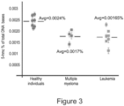
- the global level of 5-hmc in genomic DNA is reduced in cancer tissues, it is still not clear what the 5-hmc level is in peripheral blood of cancer patients. It was found that using the method of some embodiments of the invention, it is possible to quantify small differences (as low as 5 % difference) in 5-hmc content between different samples such as peripheral blood samples.
- the 5-hmc level in peripheral blood of multiple myeloma and leukemia patients is 25% lower than compared to healthy individuals.
- a method for detecting and quantifying the level of 5-hydroxymethylcytosine (5-hmc) in a cell-free DNA molecule of a subject using an imaging method comprising: associating a label with the 5-hydroxylmethylcytosine, wherein the label comprises a fluorescent label and quantifying the ratio between overall DNA fluorescence intensity and overall 5hmC fluorescence intensity, thereby determining the level of 5-hmc in the cell-free DNA molecule, wherein the method is capable of detecting the level of 5-hmc in a DNA molecule of a cell having a 5-hmc prevalence lower than 0.002 % of total DNA.
- cells having a 5-hmc prevalence lower than 0.0018 % of total DNA bases are selected according to the method.
- cells having a 5-hmc prevalence lower than 0.0017 % of total DNA bases are selected according to the method.
- cells having a 5-hmc prevalence lower than 0.0016 % of total DNA bases are selected according to the method.
- cells having a 5-hmc prevalence lower than 0.0015 % of total DNA bases are selected according to the method.
- cells having a 5-hmc prevalence lower than 0.0014 % of total DNA bases are selected according to the method.
- cells having a 5-hmc prevalence lower than 0.0013 % of total DNA bases are selected according to the method.
- cells having a 5-hmc prevalence lower than 0.0012 % of total DNA bases are selected according to the method.
- cells having a 5-hmc prevalence lower than 0.0011 % of total DNA bases are selected according to the method.
- cells having a 5-hmc prevalence lower than 0.0001 % of total DNA bases are selected according to the method (e.g., Figure 5 ).
- the cell is a cancer cell.
- the cell is a pathogenic immune cell.
- the cell is a cell line (e.g., cancer cell line).
- the cell is a primary cell, e.g., non-cultured cancer cell or pathogenic immune cell.
- the cell is a healthy cell e.g., used in screening populations, screening assays or as a control.
- the cell is a pre-malignant tissue or lesion.
- pre-malignant refers to a tissue that is not yet malignant but is poised to become malignant. Appropriate clinical and laboratory studies are designed to detect premalignant tissue while it is still in a premalignant stage. Examples of premalignant growths include polyps in the colon, actinic keratosis of the skin, dysplasia of the cervix, metaplasia of the lung, pre-malignant lesions of oral squamous cell carcinoma (OSCC) and leukoplakia (white patches in the mouth).
- OSCC oral squamous cell carcinoma
- the pre-malignant tissue is of colorectal cancer (see e.g., Figures 5A-B ).
- the pre-malignant lesion which is diagnosed by the method of this aspect of the present invention is an adenomatous polyp of the colon, an adenomatous polyp of the rectum, an adenomatous polyp of the small bowel and Barrett's esophagus.
- the pre-malignant lesion has a 5-hmc prevalence which is intermediate between that of a healthy tissue and that of a cancerous tissue (e.g., all data is available from the same subject).
- the cancer is a soft tissue tumor or a solid tumor and the cell is an immune cell e.g., PBMC e.g., a lymphocyte.
- PBMC e.g., a lymphocyte
- a method of diagnosing cancer in a subject in need thereof comprising:
- DNA sample of a cell of a subject refers to a tissue biopsy.
- the biopsy can be taken from a non-affected/suspected region (e.g., control), a region diagnosed with cancer and/or a region suspected of being cancerous or premalignant and that can be in the vicinity of a cancerous/affected region.
- a non-affected/suspected region e.g., control
- a region diagnosed with cancer and/or a region suspected of being cancerous or premalignant can be in the vicinity of a cancerous/affected region.
- significant decrease refers to a decrease that is statistically significant (e.g., P ⁇ 0.05).
- the decrease is subtle between the normal control and the pathogenic sample and therefore the sensitivity of the method of 5-hmc detection is crucial. This is further emphasized in pre-malignant stages, as shown in
- the significant decrease is below 50 %.
- the significant decrease is between 5-45%.
- the significant decrease is between 10-50 %.
- the significant decrease is between 10-45%.
- the significant decrease is between 20-50%.
- the significant decrease is between 20-45%.
- the significant decrease is between 30-50%.
- the significant decrease is between 30-45%.
- the significant decrease is between 10-30%
- the significant decrease is between 1-30 %.
- the significant decrease is between 5-30%.
- the significant decrease is between 1-50%.
- the significant decrease is between 1-20%.
- the significant decrease is between 1-10%.
- the cell has a 5-hmc prevalence lower than 0.01 % of total DNA bases.
- the cell has a 5-hmc prevalence lower than 0.005 % of total DNA bases.
- the cell has a 5-hmc prevalence lower than 0.004 % of total DNA bases.
- the cell has a 5-hmc prevalence lower than 0.003 % of total DNA bases.
- the cell has a 5-hmc prevalence lower than 0.002 % of total DNA bases.
- the cell has a 5-hmc prevalence lower than 0.0019.
- the cell has a 5-hmc prevalence lower than 0.0018 % of total DNA bases.
- the cell has a 5-hmc prevalence lower than 0.0017 % of total DNA bases.
- the cell has a 5-hmc prevalence lower than 0.0016 % of total DNA bases.
- the cell has a 5-hmc prevalence lower than 0.0014 % of total DNA bases.
- the cell has a 5-hmc prevalence lower than 0.0012 % of total DNA bases.
- the cell has a 5-hmc prevalence lower than 0.0011 % of total DNA bases.
- the cell has a 5-hmc prevalence lower than 0.0001 % of total DNA bases.
- the cancer is a soft tissue tumor or a solid tumor and the cell is an immune cell e.g., PBMC e.g., a lymphocyte.
- the assay may be performed by a simple blood test that is non-invasive.
- the cancer is a solid tumor and the cell is of the cancer (in situ metastasis).
- diagnosis refers to determining presence or absence of a pathology (e.g., a disease, disorder, condition or syndrome), classifying a pathology or a symptom, determining a severity of the pathology, monitoring pathology progression (i.e., repeating the determination 2 or more times throughout the subject's life time), monitoring treatment (i.e., 5-hmc level following and optionally prior-to treatment e.g., with an anti-cancer drug), forecasting an outcome of a pathology and/or prospects of recovery and screening of a subject for a specific disease.
- a pathology e.g., a disease, disorder, condition or syndrome
- monitoring pathology progression i.e., repeating the determination 2 or more times throughout the subject's life time
- monitoring treatment i.e., 5-hmc level following and optionally prior-to treatment e.g., with an anti-cancer drug
- the sample can be obtained using methods known in the art such as using a syringe with a needle, a scalpel, Amsterdam needle aspiration (FNA), catheter, gastrointestinal endoscopy (e.g., colorectal endoscopy, gastro-endoscopy) and the like.
- FNA needle aspiration
- DNA is extracted using methods which are well known in the art, involving tissue mincing, cell lysis, protein extraction and/or DNA precipitation using 2 to 3 volumes of 100% ethanol, rinsing in 70% ethanol, pelleting, drying and resuspension in water or any other suitable buffer (e.g., Tris-EDTA).
- Non-limiting examples of cancers which can be diagnosed by the method of this aspect of some embodiments of the invention can be any solid or non-solid cancer and/or cancer metastasis, including, but is not limiting to, tumors of the gastrointestinal tract (colon carcinoma, rectal carcinoma, colorectal carcinoma, colorectal cancer, colorectal adenoma, hereditary nonpolyposis type 1, hereditary nonpolyposis type 2, hereditary nonpolyposis type 3, hereditary nonpolyposis type 6; colorectal cancer, hereditary nonpolyposis type 7, small and/or large bowel carcinoma, esophageal carcinoma, tylosis with esophageal cancer, stomach carcinoma, pancreatic carcinoma, pancreatic endocrine tumors), endometrial carcinoma, dermatofibrosarcoma protuberans, gallbladder carcinoma, Biliary tract tumors, prostate cancer, prostate adenocarcinoma, renal cancer (e.g., Wilms' tumor type
- the cancer is of the gastrointestinal system (GI) e.g., colon cancer or rectal cancer e.g., colorectal cancer or a pre-malignant lesion.
- GI gastrointestinal system
- rectal cancer e.g., colorectal cancer or a pre-malignant lesion.
- the cancer is leukemia.
- the cell is selected from tissues which are characterized by particularly low 5-hmc prevalence.
- Total DNA bases are the number of bases of DNA measured in the sample.
- Measuring total DNA is done using fluorescence intensity measurement of a DNA stain (usually intercalating dye such as YOYO-1, Pico green, Eva green), this measurement is also referred to as (Y). See e.g., for further details describing the correlation between intensity and the number of bases Yuval Ebenstein, Dmitri Torchinsky, Sizing femtogram amounts of dsDNA by single-molecule counting. Nucl. Acids Res., (2016) doi: 10.1093/nar/gkv904 .
- the sample for DNA extraction can be from various tissues or body fluids including but are not limited to tissue biopsy, tissue section, formalin fixed paraffin embedded (FFPE) specimens, blood, plasma, serum, bone marrow, cerebro-spinal fluid, tears, sweat, lymph fluid, saliva, nasal swab or nasal aspirate, sputum, bronchoalveolar lavage, breast aspirate, pleural effusion, peritoneal fluid, glandular fluid, amniotic fluid, cervical swab or vaginal fluid, ejaculate, semen, prostate fluid, urine, conjunctival fluid, duodenal juice, pancreatic juice, bile, and stool.
- tissue biopsy tissue section
- FFPE formalin fixed paraffin embedded
- DNA could be isolated by lysis of cells with lysis buffer containing a sodium salt, tris-HC1, EDTA, and detergents such as sodium dodecyl sulphate (SDS) or cetyltrimethylammonium bromide (CATB).
- Tissue fragments should be homogenized before lysing. For example, disaggregating of tissue fragments can be performed by stroking 10-50 times, depending an tissue type, with a Dounce homogenizer.
- DNA can be further purified by mixing with a high concentration of sodium chloride and then adding into a column pre-inserted with a silica gel, a silica membrane, or a silica filter.
- DNA that binds to the silica matrix is washed by adding a washing buffer and eluted with TE buffer or water.
- DNA can also be isolated and purified by using commercially available DNA extraction kits such as QiaAmp tissue kits.
- Body fluid should be pre-treated under appropriate condition prior to DNA extraction.
- anti-coagulants contained in whole blood should be able to inhibit DNAse activity.
- a suitable anti-coagulant may be a chelating agent such as EDTA that prevents both DNAse-caused DNA degradation and clotting of the whole blood samples.
- EDTA a chelating agent
- Cells in these kinds of samples can be collected by the procedures described in prior art.
- collection of cells in a urine sample can simply be achieved by simply centrifugation, while collection of cells in a sputum sample requires DTT treatment of sputum followed by filtering through a nylon gauze mesh filter and then centrifugation.
- a stool stabilizing and homogenizing reagents should be added to stabilize DNA and remove stool particles.
- Human DNA fraction from total stool DNA then can be primarily isolated or purified using commercially available stool DNA isolation kits such as Qiagen DNA Stool Mini Kit (using the protocol for human DNA extraction) or be captured by methyl-binding domain (MBD)-based methylated DNA capture methods after total DNA isolation [ Zhou H et al., Clinical Chemistry, 2007 ].
- the DNA molecules are cell-free DNA molecules.
- 5-Methylcytosine or “5mC” is a methylated form of the DNA base cytosine. When cytosine is methylated, the DNA maintains the same sequence, but the expression of methylated genes can be altered (the study of this is part of the field of epigenetics). 5-Methylcytosine is incorporated in the nucleoside 5-methylcytidine. As used herein “5-Hydroxymethylcytosine” or “5hmC” is a DNA pyrimidine nitrogen base. It is formed from the DNA base cytosine by adding a methyl group and then a hydroxyl group.
- DNA refers to single stranded DNA or a double stranded DNA which is isolated.
- the DNA can be a eukaryotic DNA (e.g., rodent or primate e.g., human) in which 5hmC modifications typically occur or a synthetic DNA in which 5hmC modifications may be artificially added.
- the DNA molecule is a complementary polynucleotide sequence (cDNA) to which 5hmC modifications have been artificially added, a genomic polynucleotide sequence and/or a composite polynucleotide sequences (e.g., a combination of the above).
- cDNA complementary polynucleotide sequence
- genomic polynucleotide sequence e.g., a genomic polynucleotide sequence
- composite polynucleotide sequences e.g., a combination of the above.
- complementary polynucleotide sequence refers to a sequence, which results from reverse transcription of messenger RNA using a reverse transcriptase or any other RNA dependent DNA polymerase. Such a sequence can be subsequently amplified in vivo or in vitro using a DNA dependent DNA polymerase.
- genomic polynucleotide sequence refers to a sequence derived (isolated) from a chromosome and thus it represents a contiguous portion of a chromosome.
- composite polynucleotide sequence refers to a sequence, which is at least partially complementary and at least partially genomic.
- a composite sequence can include some exonal sequences required to encode the polypeptide of the present invention, as well as some intronic sequences interposing therebetween.
- the intronic sequences can be of any source, including of other genes, and typically will include conserved splicing signal sequences. Such intronic sequences may further include cis acting expression regulatory elements.
- the length of the DNA molecule may vary. Exemplary ranges include, but are not limited to 1-1 5,000 Kb, reflecting at the high range the size of a human chromosomes (or chromatin). According to some embodiments of the invention, the DNA molecule is longer than 20 Kb.
- the DNA molecule is longer than 30 Kb. According to some embodiments of the invention, the DNA molecule is longer than 40 Kb.
- Detection of the labeled DNA molecule can be done at the single molecule level using optical imaging as further described hereinbelow. Alternatively, detection of labeled DNA molecules can be done at the global level, analyzing the presence or level of 5hmC modification of a plurality of DNA molecules at the cell, tissue and organism level, as further described hereinbelow.
- the methods described herein rely on subjecting the labeled DNA molecule to an imaging method suitable for detecting the labeling agent. Such imaging methods are described herein below and typically do not rely on mass-spectrometry, radioactive assays, or immune assays (e.g., ELISA, DOT-BLOT, immunoprecipitation).
- a fluorescent dye is directly attached to the DNA (not mediated by antibody binding) and the fluorescent signal is detected and quantified.
- a method of labeling the epigenetic modification 5-hydroxymethyl-cytosine (5hmC) along a (single) DNA molecule comprising:
- attachment of a 5hmc specific labeling agent to the DNA molecule is effected when analysis is performed in the single molecule level or when a plurality of DNA molecules (global 5hmC analysis) are analyzed.
- a 5hmC specific labeling agent refers to a labeling agent that differentiates between 5hmC modification and non-modified cytosine or methylated cytosine (5mC), as described hereinabove.
- a 5hmC specific labeling agent labels selectively the position or positions where 5hmC modification is present in a DNA molecule, and does not label those positions in a DNA molecule where 5mC or any other epigenetic modification is present.
- the 5hmC labeling agent according to some embodiments of the present invention is fluorescently detectable. A list of suitable labeling agents is provided hereinafter.
- a 5hmC specific labeling agent labels at least 50 %, or at least 70 %, or at least 80 %, or at least 90 % of the a 5hmC modifications in a DNA molecule, including any intermediate within 50-100%.
- a 5hmC specific labeling agent is attached (e.g., covalently) selectively to 5hmC.
- selectively attaching a 5hmC specific labeling agent is effected by: reacting a labeling agent derivatized by a reactive group (herein referred to as a second reactive group) with a DNA molecule in which the 5-hydroxymethylcytosine bases are glycosylated by a glucose molecule derivatized by another reactive group (herein referred to as a first reactive group).
- a reactive group herein referred to as a second reactive group
- a DNA molecule in which the 5-hydroxymethylcytosine bases are glycosylated by a glucose molecule derivatized by another reactive group (herein referred to as a first reactive group).
- the first and second reactive groups are selected as being chemical compatible to one another.
- chemically compatible it is meant that the first and second reactive groups
- the phrase "reactive group” describes a chemical group that is capable of undergoing a chemical reaction that typically leads to a bond formation.
- the bond can involve one or more of a covalent bond, an electrostatic bond, a hydrogen bond, aromatic interactions, and any combination thereof.
- the bond is a covalent bond.
- Chemical reactions that lead to a bond formation include, for example, cycloaddition reactions (such as the Diels-Alder's reaction, the 1 ,3-dipolar cycloaddition Huisgen reaction, and the similar "dück reaction"), condensations, nucleophilic and electrophilic addition reactions, nucleophilic and electrophilic substitutions, addition and elimination reactions, alkylation reactions, rearrangement reactions and any other known organic reactions that involve a reactive group.
- reactive groups include, without limitation, acyl halide, aldehyde, alkoxy, alkyne, amide, amine, aryloxy, azide, aziridine, azo, carbamate, carbonyl, carboxyl, carboxylate, cyano, diene, dienophile, epoxy, guanidine, guanyl, halide, hydrazide, hydrazine, hydroxy, hydroxylamine, imino, isocyanate, nitro, phosphate, phosphonate, sulfinyl, sulfonamide, sulfonate, thioalkoxy, thioaryloxy, thiocarbamate, thiocarbonyl, thiohydroxy, thiourea and urea, as these terms are defined hereinafter.
- first and second reactive groups that are chemically compatible with one another as described herein include, but are not limited to, hydroxy and carboxylic acid, which form an ester bond; thiol and carboxylic acid, which form a thioester bond; amine and carboxylic acid, which form an amide bond; aldehyde and amine, hydrazine, hydrazide, hydroxylamine, phenylhydrazine, semicarbazide or thiosemicarbazide, which form a Schiff base (imine bond); alkene and diene, which react therebetween via cycloaddition reactions; and reactive groups that can participate in a Click reaction.
- pairs of reactive groups capable of reacting with one another include an azide and an alkyne, an unsaturated carbon-carbon bond (e.g., acrylate, methacrylate, maleimide) and a thiol, an unsaturated carbon-carbon bond and an amine, a carboxylic acid and an amine, a hydroxyl and an isocyanate, a carboxylic acid and an isocyanate, an amine and an isocyanate, a thiol and an isocyanate.
- Additional examples include an amine, a hydroxyl, a thiol or a carboxylic acid along with a nucleophilic leaving group (e.g., hydroxysuccinimide, a halogen).
- either reactive group can correspond to the "first reactive group” or to the "second reactive group”.
- the first and/or the second reactive groups can be latent groups, which are exposed during the chemical reaction, such that the reacting (e.g., covalent bond formation) is effected once a latent group is exposed.
- exemplary such groups include, but are not limited to, reactive groups as described hereinabove, which are protected with a protecting group that is labile under selected reaction conditions.
- labile protecting groups include, for example, carboxylate esters, which may hydrolyzed to form an alcohol and a carboxylic acid by exposure to acidic or basic conditions; silyl ethers such as trialkyl silyl ethers, which can be hydrolysed to an alcohol by acid or fluoride ion; p-methoxybenzyl ethers, which may be hydrolysed to an alcohol, for example, by oxidizing conditions or acidic conditions; t-butyloxycarbonyl and 9-fluorenylmethyloxycarbonyl, which may be hydrolysed to an amine by a exposure to basic conditions; sulfonamides, which may be hydrolysed to a sulfonate and amine by exposure to a suitable reagent such as samarium iodide or tributyltin hydride; acetals and ketals, which may be hydrolysed to form an aldehyde or ketone, respectively, along with an alcohol or dio
- a linking moiety is formed as a result of a bond-forming reaction between two (first and second) reactive groups.
- linking moieties which are formed between a first and a second reactive groups as described herein include without limitation, amide, lactone, lactam, carboxylate (ester), cycloalkene (e.g., cyclohexene), heteroalicyclic, heteroaryl, triazine, triazole, disulfide, imine, aldimine, ketimine, hydrazone, semicarbazone and the likes.
- Other linking moieties are defined hereinbelow.
- a reaction between a diene reactive group and a dienophile reactive group would form a cycloalkene linking moiety, and in most cases a cyclohexene linking moiety.
- an amine reactive group would form an amide linking moiety when reacted with a carboxyl reactive group.
- a hydroxyl reactive group would form an ester linking moiety when reacted with a carboxyl reactive group.
- a sulfhydryl reactive group would form a disulfide (-S-S-) linking moiety when reacted with another sulfhydryl reactive group under oxidation conditions, or a thioether (thioalkoxy) linking moiety when reacted with a halo reactive group or another leaving-reactive group.
- an alkynyl reactive group would form a triazole linking moiety by "dick reaction" when reacted with an azide reactive group.
- the “dick reaction”, also known as “dick chemistry” is a name often used to describe a stepwise variant of the Huisgen 1,3-dipolar cycloaddition of azides and alkynes to yield 1,2,3-triazole.
- This reaction is carried out under ambient conditions, or under mild microwave irradiation, typically in the presence of a Cu(I) catalyst, and with exclusive regioselectivity for the 1 ,4-disubstituted triazole product when mediated by catalytic amounts of Cu(I) salts [ V. Rostovtsev, L. G. Green, V. V. Fokin, K. B. Sharpless, Angew. Chem. Int. Ed. 2002, 41, 2596 ; H.C. Kolb, M. Finn, K.B. Sharpless, Angew Chem., Int. Ed. 2001, 40, 2004 ].
- the "dick reaction” is particularly suitable in the context of embodiments of the present invention since it can be carried out under conditions which are non-distructive to DNA molecules, and it affords attachment of a lebeling agent to 5hmC in a DNA molecule at high chemical yields using mild conditions in aqueous media.
- the selectivity of this reaction allows to perform the reaction with minimized or nullified use of protecting groups, which use often results in multistep cumbersome synthetic processes.
- the first and second reactive groups comprise (in no particular order) an azide and an alkyne. These two reactive groups may combine to form a triazole ring, as defined herein, as a linking moiety. These two reactive groups thus combine to attach a labeling agent to the 5hmC in the DNA molecule by a mechanism referred to as "dick" chemistry, as defined herein.
- a labeling agent derivatized by a second reactive group means that a labeling agent as described herein is modified so as to comprise a second reactive group as described herein, by substituting a position thereof with a chemical moiety that comprises the second reactive group.
- the second reactive group or a chemical moiety comprising the second reactive group already forms a part in a labeling agent as a substituent.
- a chemical moiety that comprises the second reactive group can be the second reactive group per se or, for example, a spacer moiety that includes, and preferably terminates with, the second reactive group.
- spacer moiety describes a chemical moiety that typically extends between two chemical moieties and is attached to each of the chemical moieties via covalent bonds.
- the spacer moiety may be linear or cyclic, be branched or unbranched, rigid or flexible.
- the spacer moieties are selected such that they allow and/or promote the one or both of attachment of a second reactive group to the labeling agent and attachment of the labeling agent to the 5hmC in a DNA molecule.
- Such traits can be selected for in terms of spacer's length, flexibility, structure and specific chemical reactivity or lack thereof.
- Exemplary spacer moieties include, but are not limited to, alkyl, alkenyl, alkynyl, cycloalkyl, heteroalicyclic, aryl, heteroaryl and/or a hydrocarbon chain having 1-20 carbon atoms and ending or interrupted by at least one heteroatom selected from the group consisting of O, S and N and/or containing from 0 to 19 unsaturated carbon-carbon or carbon-heteroatom bonds.
- a second reactive group as described herein is attached to a labeling agent via a spacer moiety, while exploiting functional groups present in the labeling agent for attaching thereto the spacer moiety which terminates with the second reactive group.
- a labeling agent derivatized by a second reactive group as described herein can be selected and prepared using conventional chemical reactions, or can be a commercially available derivatized labeling agent.
- the second reactive group is an alkyne and the labeling agent is derivatized by a chemical moiety that comprises an alkyne, as described herein.
- Such a chemical moiety can comprise, for example, benzocyclooctyne (DIBO), and can be attached to the labeling agent via a spacer as described herein.
- the second reactive group is a "strained alkyne".
- a "strained alkyne” is a cycloalkyne, preferably substituted by one or more groups that render it highly strained, for example, cyclopropyls, benzyls, and others. Examples of known strained alkynes include, but are not limited to, the following:
- a glucose derivatized by a first reactive group describes a glucose moiety that is substituted at one position thereof by a chemical moiety that comprises the first reactive group, as described herein.
- one of the hydroxy groups of a glucose can be substituted by a chemical moiety that comprises the first reactive group or can be used to attach to the glucose the chemical moiety that comprises the first reactive group, via chemical reactions that involve a hydroxy group, as described herein.
- a chemical moiety that comprises the first reactive group can be the first reactive group per se or, for example, a spacer moiety, as described herein, that includes, or terminates with, the first reactive group.
- one of the hydroxy groups of a glucose is substituted (replaced) by a chemical moiety that comprises the first reactive group.
- the first reactive group is azide and a hydroxy at position 6 of the glucose is substituted by an azide group.
- An exemplary synthetic pathway for preparing 6-azido-glucose is depicted in Figure 1 of WO2014/191981 .
- a DNA molecule in which the 5-hydroxymethylcytosine bases are glycosylated by a glucose molecule derivatized by the first reactive group is prepared, while utilizing a glucose derivatized by the first reactive group, as described herein.
- a selective introduction of a glucose derivatized by the first reactive group to 5-hydroxymethylcytosines in a DNA molecule comprises incubating the DNA molecule with f3-glucosyltransferase and a uridine diphosphoglucose (UDP-Glu) derivatized by the first reactive group.
- UDP-Glu uridine diphosphoglucose
- a DNA beta-glucosyltransferase (EC 2.4.1.27) is an enzyme that catalyzes the chemical reaction in which a beta-D-glucosyl residue is transferred from UDP-glucose to an hydroxymethylcytosine residue in DNA.
- This enzyme belongs to the family of glycosyltransferases, specifically the hexosyltransferases. The systematic name of this enzyme class is UDP-glucose:DNA beta-D-glucosyltransferase.
- T4-HMC-beta- glucosyl transferase T4-beta-glucosyl transferase
- phage beta-glucosyltransferase UDP glucose-DNA beta glucosyltransferase
- uridine diphosphoglucose-deoxyribonucleate beta-glucosyltransferase the a I3-glucosyltransferase is a His-tag fusion protein.
- the protein may be used without the His-tag (hexa-histidine tag shown above) portion.
- a uridine diphosphoglucose (UDP-Glu) derivatized by the first reactive group is meant to describe a uridine diphosphoglucose in which the glucose moiety is derivatized by a first reactive group, according to any one of the embodiments described herein.
- the uridine diphosphoglucose (UDP-G1u) derivatized by the first reactive group is a UDP-6-N 3 -Glucose.
- a UDP-6-N 3 -Glucose, or any other uridine diphosphoglucose (UDP-Glu) derivatized by the first reactive group can be prepared by chemical synthesis, while utilizing, for example, a 6-azido glucose or any other derivatized glucose, or can be a commercially available product.
- the UDP-6-N 3 -Glucose, or any other uridine diphosphoglucose (UDP-Glu) derivatized by the first reactive group is prepared by enzymatically-catalyzed reactions, as exemplified in further detail hereinafter.
- a glucose derivatized by a first reactive group is introduced to 5-hmCs in a DNA molecule, the DNA molecule is reacted with a labeling agent derivatized by a compatible second reactive group, as described herein.
- the reaction involves a dick chemistry reaction.
- the dick chemistry reaction is free of a copper catalyst, namely, is effected without the presence of a copper catalyst or an other catalyst that may adversely affect the DNA molecule.
- labeling agent refers to a detectable moiety or a probe.
- Exemplary labeling agents which are suitable for use in the context of these embodiments include, but are not limited to, a fluorescent agent, a radioactive agent, a magnetic agent, a chromophore, a bioluminescent agent, a chemiluminescent agent, a phosphorescent agent and a heavy metal cluster, as well as any other known detectable agents.
- the labeling agent is an agent that is detectable by spectrophotometric measurements, and/or which can be utilized to produce optical imaging.
- agents include, for example, chromophores, fluorescent agents, phosphorescent agents, and heavy metal clusters.
- chromophore refers to a chemical moiety that, when attached to another molecule, renders the latter colored and thus visible when various spectrophotometric measurements are applied.
- fluorescent agent refers to a compound that emits light at a specific wavelength during exposure to radiation from an external source.
- phosphorescent agent refers to a compound emitting light without appreciable heat or external excitation as by slow oxidation of phosphorous.
- a heavy metal cluster can be for example a cluster of gold atoms used, for example, for labeling in electron microscopy techniques (e.g., AFM).
- AFM electron microscopy techniques
- bioluminescent agent describes a substance which emits light by biochemical process.
- chemiluminescent agent describes a substance which emits light as the result of a chemical reaction.
- the labeling agent is a fluorescent labeling agent.
- a fluorescent agent can be a protein, quantum dots or small molecules.
- Common dye families include, but are not limited to Xanthene derivatives: fluorescein, rhodamine, Oregon green, eosin, Texas red etc.; Cyanine derivatives: cyanine, indocarbocyanine, oxacarbocyanine, thiacarbocyanine and merocyanine; Naphthalene derivatives (dansyl and prodan derivatives); Coumarin derivatives; oxadiazole derivatives: pyridyloxazole, nitrobenzoxadiazole and benzoxadiazole; Pyrene derivatives: cascade blue etc.; BODIPY (Invitrogen); Oxazine derivatives: Nile red, Nile blue, cresyl violet, oxazine 170 etc.; Acridine derivatives: proflavin, acridine orange, acridine yellow etc.; Arylmethine derivatives: auramine, crystal violet, malachite green; CF dye (Biotium); Alexa Flu
- fluorophores include: Hydroxycoumarin; Aminocoumarin; Methoxycoumarin; Cascade Blue; Pacific Blue; Pacific Orange; Lucifer yellow; NBD; R-Phycoerythrin (PE); PE-Cy5 conjugates; PE-Cy7 conjugates; Red 613; PerCP; TruRed; FluorX; Fluorescein; BODIPY-FL; TRITC; X-Rhodamine; Lissamine Rhodamine B; Texas Red; Aliaphycocyanin; APC-Cy7 conjugates.
- Alexa Fluor dyes include: Alexa Fluor 350, Alexa Fluor 405, Alexa Fluor 430, Alexa Fluor 488, Alexa Fluor 500, Alexa Fluor 514, Alexa Fluor 532, Alexa Fluor 546, Alexa Fluor 555, Alexa Fluor 568, Alexa Fluor 594, Alexa Fluor 610, Alexa Fluor 633, Alexa Fluor 647, Alexa Fluor 660, Alexa Fluor 680, Alexa Fluor 700, Alexa Fluor 750, and Alexa Fluor 790.
- Cy Dyes include Cyt, Cy3, Cy3B, Cy3.5, Cy5, Cy5.5 and Cy7.
- Nucleic acid probes include Hoechst 33342, DAPI, Hoechst 33258, SYTOX Blue, ChromomycinA3, Mithramycin, YOY0-1, Ethidium Bromide, Acridine Orange, SYTOX Green, TOTO-1, TO-PRO-1, TO-PRO: Cyanine Monomer, Thiazole Orange, Propidium Iodide (PI), LDS 751, 7-AAD, SYTOX Orange, TOTO-3, TO-PRO-3, and DRAQ5.
- Cell function probes include Indo-1, Fluo-3, DCFH, DHR, SNARF.
- Fluorescent proteins include Y66H, Y66F, EBFP, EBFP2, Azurite, GFPuv, T- Sapphire, Cerulean, mCFP, ECFP, CyPet, Y66W, mKeima-Red, TagCFP, AmCyanl, mTFP1, S65A, Midoriishi Cyan, Wild Type GFP, S65C, TurboGFP, TagGFP, S65L, Emerald, S65T (Invitrogen), EGFP (Ciontech), Azami Green (MBL), ZsGreenl (Clontech), TagYFP (Evrogen), EYFP (Clontech), Topaz, Venus, mCitrine, YPet, Turbo YFP, ZsYellow 1 (Clontech), Kusabira Orange (MBL), mOrange, mKO, TurboRFP (Evrogen), tdTomato, TagRFP (Evrogen), DsRed (Clontech
- each of the labeling agents e.g., fluorophores
- the reagents used for the reaction are derivatives of the labeling agent, which include a reactive group as described herein.
- Exemplary fluorescent agents include, but are not limited to, Alexa fluor dyes, Cy Dyes, Atto dyes, TAMRA dyes, etc., such as, for example, described in the Examples section that follows.
- analyzing 5hmC content is done without subjecting the DNA molecule to fragmentation.
- the DNA molecule is immobilized on a solid phase.
- the extending is linearly extending.
- the extending is effected by depositing the DNA molecule on a surface or extending the DNA molecule in a nanochannel.
- extended DNA molecule or “elongated DNA molecule” whichis interchangeably used herein refers to a single or plurality elongated and fixed (i.e., immobilized) DNA.
- the extended DNA molecules are elongated and fixed in a controllable manner directly onto a solid, planar surface.
- this solid, planar surface contains a positive charge density which has been controllably modified such that the single nucleic acid molecules will exhibit an optimal balance between the critical parameters of nucleic acid elongation state, degree of relaxation stability and biological activity. Further, methods, compositions and assays are described by which such an optimal balance can precisely and reproducibly be achieved.
- the single nucleic acid molecules are elongated via flow-based techniques.
- a single nucleic acid molecule is elongated, manipulated (via, for example, a regio-specific restriction digestion), and/or analyzed in a laminar flow elongation device.
- a laminar flow elongation device such as a laminar flow elongation devices and methods of elongating or extending DNA are described in U.S. Patent Application 20030124611 .
- the elongated, individual labeled DNA molecules can then be utilized in a variety of ways which have applications for the analysis of nucleic acid at the genome level.
- nucleic acid molecules may be used to generate ordered, high resolution single nucleic acid molecule restriction maps. This method is referred to herein as “optical mapping” or “optical restriction mapping”.
- optical mapping or “optical restriction mapping”.
- optical sequencing methods are presented whereby specific nucleotide sequences present within the elongated nucleic acid molecules can be identified. Such methods are referred to herein as "optical sequencing”.
- the optical mapping and optical sequencing techniques can be used independently or in combination on the same individual nucleic acid molecules.
- methods are also presented for the imaging and sizing of the elongated single nucleic acid molecules. These imaging techniques may, for example, include the use of fluorochromes, microscopy and/or image processing computer software and hardware.
- step (b, extending) is effected following step (a, attaching to the DNA molecule a 5hmc specific labeling agent).
- the method further comprises attaching to the DNA molecule an additional labeling agent distinct of the 5hmc specific labeling agent.
- the additional labeling agent is an epigenetic modification specific labeling agent. Examples of such modifications include but are not limited to 5-methylcytosine (5mC), histone acetylation and the like.
- the additional labeling agent is a non-epigenetic modification specific labeling agent.
- stains and dyes include DNA fluorescent dyes such as cyanine nucleic acid stains, which are essentially nonfluorescent in the absence of nucleic acids and exhibit significant fluorescence enhancements upon DNA binding.
- the stain may be cell permeant or impermeant.
- Such stains are available from Molecular Probes (e.g., YOYO-1. TOTO, S YT 0 X, POPO-1, BOB 0-1, LOLO- 1, J OJ 0-1 etc.).
- Molecular Probes e.g., YOYO-1. TOTO, S YT 0 X, POPO-1, BOB 0-1, LOLO- 1, J OJ 0-1 etc.
- non-fluorescent stains can be used as further described hereinbelow.
- high throughput methods for utilizing such single nucleic acid molecules in genome analysis are presented.
- rapid optical mapping approaches are described for the creation of high-resolution restriction maps.
- single nucleic acid molecules are elongated, fixed and gridded to high density onto a solid surface. These molecules can then be digested with appropriate restriction enzymes for the map construction.
- the single nucleic acid molecules can be elongated, fixed and gridded at high density onto a solid surface and utilized in a variety of optical sequencing-based diagnostic methods. In addition to speed, such diagnostic grids can be reused. Further, the high throughput and methods can be utilized to rapidly generate information derived from procedures which combine optical mapping and optical sequencing methods.
- a method of in-situ imaging a DNA molecule comprising:
- the method further comprises generating an optical image of the DNA molecule following the imaging.
- an extended DNA molecule comprising at least one 5hmc-specific labeling agent.
- a DNA molecule comprising at least two different labeling agents, wherein a first labeling agent of the at least two different labels is a 5hmc-specific labeling agent.
- the 5hmc-specific labeling agent is attached to the DNA molecule by reacting a labeling agent derivatized by a second reactive group with a DNA molecule in which the 5-hydroxymethylcytosines are glycosylated by a glucose molecule derivatized by a first reactive group, wherein the first and second reactive groups are chemically compatible to one another, as described in any one of the embodiments pertaining to attaching a 5hmc-specific labeling agent to a DNA molecule of the present invention.
- one the first and second reactive groups is azide and the other is alkyne, such that attaching the labeling agent to the DNA molecule is effected by a dick chemistry, as described herein.
- a second labeling agent of the at least two different labeling agents is a 5mc-specific labeling agent.
- a second labeling agent of the at least two different labeling agents is for an epigenetic modification.
- a second labeling agent of the at least two different labeling agents is for a non-epigenetically modified base.
- distinct or “different” labels refer to labels which can be distinguished upon visualization.
- one label may be red fluorescence while the other can be blue fluorescence.
- composition-of-matter comprising the DNA molecule.
- the DNA molecule is surface deposited or extended in a microchannel.
- the present invention also envisages detecting 5hmC in non-immobilized biological samples.
- a method of detecting 5-hydroxymethyl-cytosine (5hmC) in a DNA sample comprising:
- fluorescent intensity refers to the intensity of the fluorescent probe.
- absorbance refers to DNA light absorbance at 260 nm which is a measure for DNA quantity. At this wavelength, DNA typically exhibits absorbance maxima.
- DNA stain intensity refers to a non-specific DNA stain that labels the bases globally thus giving a measure of total nucleotides in the sample (such DNA labels are described hereinabove).
- the methodology described herein, according to some embodiments of the present invention can be used to detect global 5hmC modification.
- global 5hmC modification refers to the detection of 5hmC of a plurality of DNA molecules which are in a non-immobilized state.
- the sample may be a heterogeneous sample.
- the threshold of sensitivity or the limit of detection is about 0.0022 % 5hmC/dN.
- the concentration of the DNA in the test sample depends an the level (e.g., %) of hmC in the tissue.
- concentration of DNA that can be read is up to 350 ng/ ⁇ 1 DNA without having signal saturation (e.g., 1350 ng/ ⁇ 1).
- This concentration of DNA is high enough for detection % hmC at low-% hmC-containing tissues such as spleen and liver.
- concentrated DNA samples e.g., 100 ng/ ⁇ 1 to 100 ⁇ g/ ⁇ 1
- the volume of the sample is between 1-50 ⁇ 1 or 10-20 ul for the detection of hmC in genomic DNA in multi well plate.
- the sample can be subjected to optical imaging by extending the molecules on slides (immobilizing the DNA molecules) as described herein.
- the amount of DNA is measured by length or fluorescence intensity of the intercalating DNA stain, and the amount of 5-hmC is determined by counting the fluorescent spots generated along the DNA by the labeled 5-hmC or measuring their intensity.
- the position of the modification can also be analyzed using enzymes which are sensitive to bulky residues i.e., the modification of the 5hmC with N 3 -5-gmC.
- Presence of N 3 -5-g group on the DNA template strand will interfere with the synthesis of a nucleic acid strand by DNA polymerase or RNA polymerase, or the efficient cleavage of DNA by a restriction endonuclease (e.g., Msp 1) or inhibition of other enzymatic modifications of nucleic acid containing 5-hmC.
- a restriction endonuclease e.g., Msp 1
- primer extensions or other assays can be employed, for example, to evaluate a partially extended primer of certain length and the modification sites can be revealed by sequencing the partially extended primers.
- the sensitivity of the method may be even augmented by selecting a specific cell type (e.g., lymphocyte from PBMCs) such as by using cell sorting (e.g., FACS, magenic beads etc.).
- a specific cell type e.g., lymphocyte from PBMCs
- cell sorting e.g., FACS, magenic beads etc.
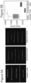
- Figure 4 shows DNA extracted from human PBMCs and stretched in nanochannel arrays (BioNano Genomics): DNA molecules in blue, red dots are genetic tags for mapping to the genome and green dots are 5hmC.
- compositions comprising, “comprising”, “includes”, “including”, “having” and their conjugates mean “including but not limited to”.
- Consisting of' means “including and limited to”.
- Consisting essentially of means that the composition, method or structure may include additional ingredients, steps and/or parts, but only if the additional ingredients, steps and/or parts do not materially alter the basic and novel characteristics of the claimed composition, method or structure.
- a compound or “at least one compound” may include a plurality of compounds, including mixtures thereof.
- range format is merely for convenience and brevity and should not be construed as an inflexible limitation an the scope of the invention. Accordingly, the description of a range should be considered to have specifically disclosed all the possible subranges as well as individual numerical values within that range. For example, description of a range such as from 1 to 6 should be considered to have specifically disclosed subranges such as from 1 to 3, from 1 to 4, from 1 to 5, from 2 to 4, from 2 to 6, from 3 to 6 etc., as well as individual numbers within that range, for example, 1, 2, 3, 4, 5, and 6. This applies regardless of the breadth of the range.
- method refers to manners, means, techniques and procedures for accomplishing a given task including, but not limited to, those manners, means, techniques and procedures either known to, or readily developed from known manners, means, techniques and procedures by practitioners of the chemical, pharmacological, biological, biochemical and medical arts.
- treating includes abrogating, substantially inhibiting, slowing or reversing the progression of a condition, substantially ameliorating clinical or aesthetical symptoms of a condition or substantially preventing the appearance of clinical or aesthetical symptoms of a condition.
- sequences that substantially correspond to its complementary sequence as including minor sequence variations, resulting from, e.g., sequencing errors, cloning errors, or other alterations resulting in base substitution, base deletion or base addition, provided that the frequency of such variations is less than 1 in 50 nucleotides, alternatively, less than 1 in 100 nucleotides, alternatively, less than 1 in 200 nucleotides, alternatively, less than 1 in 500 nucleotides, alternatively, less than 1 in 1000 nucleotides, alternatively, less than 1 in 5,000 nucleotides, alternatively, less than 1 in 10,000 nucleotides.
- the T4 ⁇ -glucosyltransferase (13-GT) was used to tag 5-hmc sites with a fluorescent reporter molecule.
- the enzyme was fed with a synthetic cofactor UDP-6-N3-Glu, resulting in covalent attachment of a functional azide at the 5-hmc site. This azide was further reacted with an Cy5 Fluor alkyne via a "dick" chemistry reaction to generate the fluorescently labeled 5-hmc.
- the resulting DNA product had fluorescence and absorbance proportional to the content of 5-hmc residues.
- the DNA was stretched on a cover slip glass and visualized by fluorescence microscopy, then all imaged data was analyzed using a proprietary software. The software is counting the number of 5-hmc per length or intensity of the DNA and calculates the percentage of 5-hmc from total DNA bases.
- DNA 500 ng was incubated with 2 ⁇ ! of ß- glycosyltransferase (NEB), 1X buffer 4 (NEB), 20 nM 6-N3-UDPG, and ultra-pure water at 37 °C for overnight.
- a dick copper-free reaction was used to label the 5-hmC sites with 60 nM DBCO-Cy5 and incubated overnight at 37 °C.
- DNA samples were cleaned using DNA biding magnetic beads that were purchased from Nvigem (cat# 61001-1500) according to company's protocol.
- ⁇ bactcriophage genomic DNA (NEB) was used as a negative control since it contains no 5-hmC. No labeling was observed for these samples.
- DNA was stained with the intercalating dye YOYO-1; 3 ⁇ 1 of the sample were diluted in 80 ⁇ 1 HEPES -DTT buffer (100uM HEPES, 100uM DTT) with 0.65 ⁇ 120 ⁇ M YoYo-1 (1:4 dye to nucleotide ratio) and incubated at 37 °C for 2 hours.
- DNA molecules were extended on silanized glass slides by placing 6 ⁇ 1_, of pre-labeled DNA in HEPES -DTT buffer (100uM HEPES, 100uM DTT) between a dry silanized glass slide and a non-treated microscope slide (the DNA final concentration 0.25 ng/ ⁇ L). Extended DNA molecules were imaged on a MORE imaging system (TILL photonics GmbH) with an Olympus UPlanApo 100X 1.3 NA oil immersion objective. A 150 W Xenon lamp with galvanometer driven filter switching was used as an excitation source. The filter sets used to image YOYO-1 stained DNA and the Cy5 labels were 485/20 and 650/13 bandpass excitation filters, 525/30 and 684/24 emission filters.
- Images were acquired by a DU888 EMCCD (Andor Technologies) with an EM gain setting of 300 and integration times of 200 ms and 3000 ms for YOYO-1 and Cy5, respectively. All imaged data was analyzed using a proprietary software. The software is counting the number of 5-hmc per length or intensity of the DNA and calculates the percentage of 5-hmc from total DNA bases.
- the 5-hmc level in peripheral blood is estimated to be 0.002 % of total DNA bases.
- Existing commercial methods rely on bulky antibodies and cannot detect lower than 0.02 % 5-hmc. More sensitive assays that rely on mass-spectra analysis or radioactive labeling are not suitable for clinical settings.
- Two commercially available examples are the MethylFlash Hydroxymethylated DNA 5-hmC Quantification Kit (Epigentek cat# p-1036-48) and Quest 5-hmCTM DNA ELISA Kit (Zymo cat# D5425) which are not able to detect 5-hmc in blood.
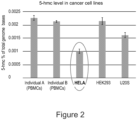
- the 5-hmc level of HELA, HEK293 and U2OS cell lines is considered to be extremely low and estimated between 0.001-0.003% of total bases.
- Figure 1 shows the wide dynamic range of the methods as described herein supporting its used for all biologically relevant levels of 5hC from healthy brain to blood cancer and cell lines.
- the 5-hmC level of healthy colon in estimated to be 0.1%-0.05% from total DNA nucleotides and can drop up to 5 time fold in colon cancer tissue.
- 5-hmC Several specific antibodies to 5-hmC are commercially available and can be used for dot blot, immunoprecipitation, and ELISA assays.
- the detection limit of these commercially available kits is limited to about 0.03 % 5-hmC.
- the detection and screening of colon cancer using those kits is very limited due to poor sensitivity and poor resolution.
- the drop of 5-hmC in colon cancer tissues is relatively small (less than 30% drop) and cannot be distinguished when comparing to healthy tissue, if using antibodies related methods.
- adjacent tissue healthy tissue next to the tumor
- pre-malignant tissue the drop of 5-hmC can be as lower than 1 5% compared to a healthy tissue and therefore is undetectable using existent methods.
- a chemo-enzymatic labelling scheme is used herein in order to develop a high throughput assay for quantifying the levels of 5-hmC for cancer diagnostics and prediction of treatment outcome.
- embodiments of the invention rely on fluorescent labelling of 5-hmC followed by fluorescent measurement in a multi-well slide format and in single-molecule assay. 5-hmC detection and quantification is based on specific attachment of a fluorescent reporter to individual 5-hmC sites along the DNA molecules.
Landscapes
- Chemical & Material Sciences (AREA)
- Life Sciences & Earth Sciences (AREA)
- Organic Chemistry (AREA)
- Health & Medical Sciences (AREA)
- Proteomics, Peptides & Aminoacids (AREA)
- Zoology (AREA)
- Engineering & Computer Science (AREA)
- Wood Science & Technology (AREA)
- Analytical Chemistry (AREA)
- Immunology (AREA)
- Genetics & Genomics (AREA)
- Biotechnology (AREA)
- General Health & Medical Sciences (AREA)
- Microbiology (AREA)
- Physics & Mathematics (AREA)
- Biochemistry (AREA)
- Bioinformatics & Cheminformatics (AREA)
- General Engineering & Computer Science (AREA)
- Biophysics (AREA)
- Molecular Biology (AREA)
- Pathology (AREA)
- Chemical Kinetics & Catalysis (AREA)
- Hospice & Palliative Care (AREA)
- Oncology (AREA)
- Measuring Or Testing Involving Enzymes Or Micro-Organisms (AREA)
Claims (11)
- Verfahren zum Nachweis und zur Quantifizierung des Gehalts an 5-Hydroxymethylcytosin (5-hmc) in einem zellfreien DNA-Molekül eines Subjekts unter Verwendung eines bildgebenden Verfahrens, wobei das Verfahren umfasst:Zuordnen einer Markierung zu dem 5-Hydroxymethylcytosin, wobei die Markierung eine fluoreszierende Markierung umfasst,Anheften eines zusätzlichen Markierungsmittels an das DNA-Molekül, das sich von der 5-hmc-spezifischen Markierung unterscheidet, wobei das zusätzliche Markierungsmittel eine unspezifische fluoreszierende Markierung zur Bestimmung der Gesamt-DNA-Fluoreszenzintensität ist, undQuantifizieren des Verhältnisses zwischen der Gesamt-DNA-Fluoreszenzintensität und der Gesamt-5hmC-Fluoreszenzintensität, wodurch der Gehalt an 5-hmc in dem zellfreien DNA-Molekül bestimmt wird,wobei das Verfahren in der Lage ist, den Gehalt an 5-hmc in einem DNA-Molekül einer Zelle mit einer 5-hmc-Prävalenz von weniger als 0,002 % der Gesamt-DNA nachzuweisen.
- Verfahren nach Anspruch 1, wobei der Nachweis das Identifizieren einer Position von 5-Hydroxymethylcytosin auf dem zellfreien DNA-Molekül umfasst.
- Verfahren nach Anspruch 1, das ferner das Extrahieren des zellfreien DNA-Moleküls aus einer Probe des Subjekts umfasst.
- Verfahren nach einem der vorhergehenden Ansprüche, das ferner das Vergleichen des 5-Hydroxymethylcytosingehalts mit einem 5-Hydroxymethylcytosingehalt in einer Kontrollprobe umfasst.
- Verfahren nach Anspruch 4, bei dem eine Abnahme des 5-Hydroxymethylcytosingehalts im Vergleich zum 5-Hydroxymethylcytosingehalt in der Kontrollprobe auf das Vorliegen von Krebs oder einer prämalignen Läsion hinweist.
- Verfahren nach Anspruch 4, bei dem die Kontrollprobe von einem gesunden Subjekt stammt.
- Verfahren nach Anspruch 5, wobei das Verfahren Krebs bei dem Subjekt diagnostiziert und wobei der Krebs einen Dickdarmkrebs, einen Bauchspeicheldrüsenkrebs, einen Eierstockkrebs, einen Gebärmutterkrebs, einen Lungenkrebs, einen Brustkrebs oder einen Prostatakrebs umfasst.
- Verfahren nach Anspruch 5, wobei das Verfahren eine prämaligne Läsion bei dem Subjekt diagnostiziert.
- Verfahren nach Anspruch 1, bei dem die Markierung eine Azidgruppe umfasst.
- Verfahren nach Anspruch 1, das ferner das Zugeben einer Glucose zu dem 5-Hydroxymethylcytosin umfasst.
- Verfahren nach Anspruch 10, bei dem die Glucose eine Uridindiphosphoglucose (UDP-Glu) ist.
Applications Claiming Priority (2)
| Application Number | Priority Date | Filing Date | Title |
|---|---|---|---|
| US201562253797P | 2015-11-11 | 2015-11-11 | |
| PCT/IL2016/051218 WO2017081689A1 (en) | 2015-11-11 | 2016-11-10 | Methods of detecting 5-hydroxymethylcytosine and diagnosing of cancer |
Publications (3)
| Publication Number | Publication Date |
|---|---|
| EP3374524A1 EP3374524A1 (de) | 2018-09-19 |
| EP3374524A4 EP3374524A4 (de) | 2019-06-26 |
| EP3374524B1 true EP3374524B1 (de) | 2025-01-29 |
Family
ID=58694862
Family Applications (1)
| Application Number | Title | Priority Date | Filing Date |
|---|---|---|---|
| EP16863787.4A Active EP3374524B1 (de) | 2015-11-11 | 2016-11-10 | Verfahren zum nachweis von 5-hydroxymethylcytosin und zur diagnose von krebs |
Country Status (3)
| Country | Link |
|---|---|
| US (2) | US20180327855A1 (de) |
| EP (1) | EP3374524B1 (de) |
| WO (1) | WO2017081689A1 (de) |
Families Citing this family (9)
| Publication number | Priority date | Publication date | Assignee | Title |
|---|---|---|---|---|
| US9115386B2 (en) | 2008-09-26 | 2015-08-25 | Children's Medical Center Corporation | Selective oxidation of 5-methylcytosine by TET-family proteins |
| ES2669214T3 (es) | 2011-12-13 | 2018-05-24 | Oslo Universitetssykehus Hf | Procedimientos y kits para la detección de estado de metilación |
| ES2786984T3 (es) | 2012-11-30 | 2020-10-14 | Cambridge Epigenetix Ltd | Agente oxidante para nucleótidos modificados |
| CA2912666A1 (en) | 2013-05-28 | 2014-12-04 | Ramot At Tel-Aviv University Ltd. | Detection of hydroxymethylcytosine bases |
| US11459573B2 (en) | 2015-09-30 | 2022-10-04 | Trustees Of Boston University | Deadman and passcode microbial kill switches |
| US12385161B2 (en) | 2018-06-06 | 2025-08-12 | Ramot At Tel-Aviv University Ltd. | Multi-sample array chip for DNA modification quantification |
| EP4521115A3 (de) | 2020-06-10 | 2025-06-11 | Ramot at Tel-Aviv University Ltd. | Verfahren zum nachweis von methyliertem cpg |
| EP4083231A1 (de) | 2020-07-30 | 2022-11-02 | Cambridge Epigenetix Limited | Zusammensetzungen und verfahren zur nukleinsäureanalyse |
| CN113061652A (zh) * | 2021-03-30 | 2021-07-02 | 成都泰莱医学检验实验室有限公司 | 一种基于葡萄糖基修饰的测定基因标志物中5hmC含量的方法 |
Citations (1)
| Publication number | Priority date | Publication date | Assignee | Title |
|---|---|---|---|---|
| US20140080715A1 (en) * | 2012-09-20 | 2014-03-20 | The Chinese University Of Hong Kong | Non-invasive determination of methylome of fetus or tumor from plasma |
Family Cites Families (29)
| Publication number | Priority date | Publication date | Assignee | Title |
|---|---|---|---|---|
| NL154600B (nl) | 1971-02-10 | 1977-09-15 | Organon Nv | Werkwijze voor het aantonen en bepalen van specifiek bindende eiwitten en hun corresponderende bindbare stoffen. |
| NL154598B (nl) | 1970-11-10 | 1977-09-15 | Organon Nv | Werkwijze voor het aantonen en bepalen van laagmoleculire verbindingen en van eiwitten die deze verbindingen specifiek kunnen binden, alsmede testverpakking. |
| NL154599B (nl) | 1970-12-28 | 1977-09-15 | Organon Nv | Werkwijze voor het aantonen en bepalen van specifiek bindende eiwitten en hun corresponderende bindbare stoffen, alsmede testverpakking. |
| US3901654A (en) | 1971-06-21 | 1975-08-26 | Biological Developments | Receptor assays of biologically active compounds employing biologically specific receptors |
| US3853987A (en) | 1971-09-01 | 1974-12-10 | W Dreyer | Immunological reagent and radioimmuno assay |
| US3867517A (en) | 1971-12-21 | 1975-02-18 | Abbott Lab | Direct radioimmunoassay for antigens and their antibodies |
| NL171930C (nl) | 1972-05-11 | 1983-06-01 | Akzo Nv | Werkwijze voor het aantonen en bepalen van haptenen, alsmede testverpakkingen. |
| US3850578A (en) | 1973-03-12 | 1974-11-26 | H Mcconnell | Process for assaying for biologically active molecules |
| US3935074A (en) | 1973-12-17 | 1976-01-27 | Syva Company | Antibody steric hindrance immunoassay with two antibodies |
| US3996345A (en) | 1974-08-12 | 1976-12-07 | Syva Company | Fluorescence quenching with immunological pairs in immunoassays |
| US4034074A (en) | 1974-09-19 | 1977-07-05 | The Board Of Trustees Of Leland Stanford Junior University | Universal reagent 2-site immunoradiometric assay using labelled anti (IgG) |
| US3984533A (en) | 1975-11-13 | 1976-10-05 | General Electric Company | Electrophoretic method of detecting antigen-antibody reaction |
| US4098876A (en) | 1976-10-26 | 1978-07-04 | Corning Glass Works | Reverse sandwich immunoassay |
| US4879219A (en) | 1980-09-19 | 1989-11-07 | General Hospital Corporation | Immunoassay utilizing monoclonal high affinity IgM antibodies |
| US5011771A (en) | 1984-04-12 | 1991-04-30 | The General Hospital Corporation | Multiepitopic immunometric assay |
| US4666828A (en) | 1984-08-15 | 1987-05-19 | The General Hospital Corporation | Test for Huntington's disease |
| US4683202A (en) | 1985-03-28 | 1987-07-28 | Cetus Corporation | Process for amplifying nucleic acid sequences |
| US4801531A (en) | 1985-04-17 | 1989-01-31 | Biotechnology Research Partners, Ltd. | Apo AI/CIII genomic polymorphisms predictive of atherosclerosis |
| US6147198A (en) | 1988-09-15 | 2000-11-14 | New York University | Methods and compositions for the manipulation and characterization of individual nucleic acid molecules |
| US5272057A (en) | 1988-10-14 | 1993-12-21 | Georgetown University | Method of detecting a predisposition to cancer by the use of restriction fragment length polymorphism of the gene for human poly (ADP-ribose) polymerase |
| US5192659A (en) | 1989-08-25 | 1993-03-09 | Genetype Ag | Intron sequence analysis method for detection of adjacent and remote locus alleles as haplotypes |
| US5281521A (en) | 1992-07-20 | 1994-01-25 | The Trustees Of The University Of Pennsylvania | Modified avidin-biotin technique |
| WO2011127136A1 (en) * | 2010-04-06 | 2011-10-13 | University Of Chicago | Composition and methods related to modification of 5-hydroxymethylcytosine (5-hmc) |
| US20120122087A1 (en) | 2010-11-17 | 2012-05-17 | Weiwei Li | 5-Hydroxymethylcytosine as a biomarker for early detection, treatment and prognostic monitoring of cancer |
| US20140030727A1 (en) | 2012-01-20 | 2014-01-30 | Gerd PFEIFER | Loss of 5-hydroxymethylcytosine as a biomarker for cancer |
| US20150285807A1 (en) * | 2012-06-11 | 2015-10-08 | The Brigham And Women's Hospital, Inc. | System and method for detecting cancer |
| US9297806B2 (en) * | 2012-08-01 | 2016-03-29 | The Johns Hopkins University | 5-hydroxymethylcytosine in human cancer |
| US20140272970A1 (en) * | 2013-03-15 | 2014-09-18 | Promega Corporation | Method for quantifying 5-hydroxymethylcytosine |
| CA2912666A1 (en) | 2013-05-28 | 2014-12-04 | Ramot At Tel-Aviv University Ltd. | Detection of hydroxymethylcytosine bases |
-
2016
- 2016-11-10 EP EP16863787.4A patent/EP3374524B1/de active Active
- 2016-11-10 US US15/775,015 patent/US20180327855A1/en not_active Abandoned
- 2016-11-10 WO PCT/IL2016/051218 patent/WO2017081689A1/en not_active Ceased
-
2024
- 2024-08-12 US US18/800,170 patent/US20250223650A1/en active Pending
Patent Citations (1)
| Publication number | Priority date | Publication date | Assignee | Title |
|---|---|---|---|---|
| US20140080715A1 (en) * | 2012-09-20 | 2014-03-20 | The Chinese University Of Hong Kong | Non-invasive determination of methylome of fetus or tumor from plasma |
Non-Patent Citations (1)
| Title |
|---|
| NOA GILAT ET AL: "Single-molecule quantification of 5-hydroxymethylcytosine for diagnosis of blood and colon cancers", CLINICAL EPIGENETICS, BIOMED CENTRAL LTD, LONDON, UK, vol. 9, no. 1, 14 July 2017 (2017-07-14), pages 1 - 8, XP021247144, ISSN: 1868-7075, DOI: 10.1186/S13148-017-0368-9 * |
Also Published As
| Publication number | Publication date |
|---|---|
| EP3374524A4 (de) | 2019-06-26 |
| US20250223650A1 (en) | 2025-07-10 |
| WO2017081689A1 (en) | 2017-05-18 |
| EP3374524A1 (de) | 2018-09-19 |
| US20180327855A1 (en) | 2018-11-15 |
Similar Documents
| Publication | Publication Date | Title |
|---|---|---|
| US20250223650A1 (en) | Methods of detecting 5-hydroxymethylcytosine and diagnosing of cancer | |
| US12043863B2 (en) | Detection of hydroxymethylcytosine bases | |
| Cai et al. | Single-molecule amplification-free multiplexed detection of circulating microRNA cancer biomarkers from serum | |
| Evangelou et al. | Robust, universal biomarker assay to detect senescent cells in biological specimens | |
| US11385179B2 (en) | Target molecule density determination in a fluorescence image | |
| Gilat et al. | Single-molecule quantification of 5-hydroxymethylcytosine for diagnosis of blood and colon cancers | |
| Chen et al. | Homogeneous two-dimensional visual and fluorescence analysis of circulating tumor cells in clinical samples via steric hindrance regulated enzymes recognition cleavage and elongation | |
| CN108977544A (zh) | 用于鉴定胃癌和/或胃息肉的试剂盒及其应用 | |
| Ren et al. | FRET imaging of glycoRNA on small extracellular vesicles enabling sensitive cancer diagnostics | |
| HRP20040488A2 (en) | Methods for detecting cells with numerical chromosomal abnormalities | |
| Wang et al. | Advancing small ligands targeting RNA for better binding affinity and specificity: A study of structural influence through molecular design approach | |
| Chen et al. | A multivalent activatable aptamer probe with ultralow background signal and high sensitivity for diagnosis of lung adenocarcinoma | |
| WO2021250677A1 (en) | METHODS OF DETECTING METHYLATED CpG | |
| Wu et al. | Single extracellular vesicle imaging via rolling circle amplification–expansion microscopy | |
| Liu et al. | Nanoplasmon-enhanced drop-screen for high throughput single-cell nucleocytoplasmic miRNA profiling | |
| CN113249479B (zh) | 胰腺癌相关的lncRNA标志物、探针及检测试剂盒在胰腺癌诊断中的应用 | |
| US20230047930A1 (en) | Methods and arrays for identifying the cell or tissue origin of dna | |
| Jang et al. | Multiplexed and streamlined DNA methylation detection of colorectal cancer-related genes using graphically encoded hydrogel microparticles and rolling circle amplification | |
| Ren et al. | Identifying Hepsin as a novel biomarker for human esophageal squamous cell carcinoma (ESCC) and its application in fluorescence imaging | |
| Avraham et al. | Simultaneous global labeling (SiGL) of 5-methylcytosine and 5-hydroxymethylcytosine by DNA alkylation with a synthetic cofactor and engineered methyltransferase | |
| Amato et al. | Multitarget Fluorescence In Situ Hybridization Diagnostic Applications in Tumors | |
| Goto et al. | Hybridization-based CpG methylation level detection using methyl-CpG-binding domain–fused luciferase | |
| Saadh et al. | Urinary Tumor Markers for Detection and Surveillance in Non-Muscle Invasive Bladder Cancer | |
| Solomon et al. | Emerging molecular approaches in the analysis of urine in bladder cancer diagnosis | |
| US20150086976A1 (en) | Self-catalytic reaction based assay |
Legal Events
| Date | Code | Title | Description |
|---|---|---|---|
| STAA | Information on the status of an ep patent application or granted ep patent |
Free format text: STATUS: THE INTERNATIONAL PUBLICATION HAS BEEN MADE |
|
| PUAI | Public reference made under article 153(3) epc to a published international application that has entered the european phase |
Free format text: ORIGINAL CODE: 0009012 |
|
| STAA | Information on the status of an ep patent application or granted ep patent |
Free format text: STATUS: REQUEST FOR EXAMINATION WAS MADE |
|
| 17P | Request for examination filed |
Effective date: 20180507 |
|
| AK | Designated contracting states |
Kind code of ref document: A1 Designated state(s): AL AT BE BG CH CY CZ DE DK EE ES FI FR GB GR HR HU IE IS IT LI LT LU LV MC MK MT NL NO PL PT RO RS SE SI SK SM TR |
|
| AX | Request for extension of the european patent |
Extension state: BA ME |
|
| DAV | Request for validation of the european patent (deleted) | ||
| DAX | Request for extension of the european patent (deleted) | ||
| A4 | Supplementary search report drawn up and despatched |
Effective date: 20190527 |
|
| RIC1 | Information provided on ipc code assigned before grant |
Ipc: C12Q 1/48 20060101ALI20190521BHEP Ipc: C12Q 1/6886 20180101AFI20190521BHEP Ipc: G01N 33/58 20060101ALI20190521BHEP Ipc: C12Q 1/6806 20180101ALI20190521BHEP Ipc: G01N 33/52 20060101ALI20190521BHEP |
|
| STAA | Information on the status of an ep patent application or granted ep patent |
Free format text: STATUS: EXAMINATION IS IN PROGRESS |
|
| 17Q | First examination report despatched |
Effective date: 20200724 |
|
| GRAP | Despatch of communication of intention to grant a patent |
Free format text: ORIGINAL CODE: EPIDOSNIGR1 |
|
| STAA | Information on the status of an ep patent application or granted ep patent |
Free format text: STATUS: GRANT OF PATENT IS INTENDED |
|
| INTG | Intention to grant announced |
Effective date: 20240531 |
|
| GRAS | Grant fee paid |
Free format text: ORIGINAL CODE: EPIDOSNIGR3 |
|
| GRAA | (expected) grant |
Free format text: ORIGINAL CODE: 0009210 |
|
| STAA | Information on the status of an ep patent application or granted ep patent |
Free format text: STATUS: THE PATENT HAS BEEN GRANTED |
|
| AK | Designated contracting states |
Kind code of ref document: B1 Designated state(s): AL AT BE BG CH CY CZ DE DK EE ES FI FR GB GR HR HU IE IS IT LI LT LU LV MC MK MT NL NO PL PT RO RS SE SI SK SM TR |
|
| REG | Reference to a national code |
Ref country code: GB Ref legal event code: FG4D |
|
| REG | Reference to a national code |
Ref country code: CH Ref legal event code: EP |
|
| REG | Reference to a national code |
Ref country code: DE Ref legal event code: R096 Ref document number: 602016091114 Country of ref document: DE |
|
| REG | Reference to a national code |
Ref country code: IE Ref legal event code: FG4D |
|
| REG | Reference to a national code |
Ref country code: NL Ref legal event code: MP Effective date: 20250129 |
|
| PG25 | Lapsed in a contracting state [announced via postgrant information from national office to epo] |
Ref country code: NL Free format text: LAPSE BECAUSE OF FAILURE TO SUBMIT A TRANSLATION OF THE DESCRIPTION OR TO PAY THE FEE WITHIN THE PRESCRIBED TIME-LIMIT Effective date: 20250129 |
|
| PG25 | Lapsed in a contracting state [announced via postgrant information from national office to epo] |
Ref country code: RS Free format text: LAPSE BECAUSE OF FAILURE TO SUBMIT A TRANSLATION OF THE DESCRIPTION OR TO PAY THE FEE WITHIN THE PRESCRIBED TIME-LIMIT Effective date: 20250429 |
|
| PG25 | Lapsed in a contracting state [announced via postgrant information from national office to epo] |
Ref country code: FI Free format text: LAPSE BECAUSE OF FAILURE TO SUBMIT A TRANSLATION OF THE DESCRIPTION OR TO PAY THE FEE WITHIN THE PRESCRIBED TIME-LIMIT Effective date: 20250129 |
|
| PG25 | Lapsed in a contracting state [announced via postgrant information from national office to epo] |
Ref country code: PL Free format text: LAPSE BECAUSE OF FAILURE TO SUBMIT A TRANSLATION OF THE DESCRIPTION OR TO PAY THE FEE WITHIN THE PRESCRIBED TIME-LIMIT Effective date: 20250129 |
|
| PG25 | Lapsed in a contracting state [announced via postgrant information from national office to epo] |
Ref country code: ES Free format text: LAPSE BECAUSE OF FAILURE TO SUBMIT A TRANSLATION OF THE DESCRIPTION OR TO PAY THE FEE WITHIN THE PRESCRIBED TIME-LIMIT Effective date: 20250129 |
|
| REG | Reference to a national code |
Ref country code: LT Ref legal event code: MG9D |
|
| PG25 | Lapsed in a contracting state [announced via postgrant information from national office to epo] |
Ref country code: IS Free format text: LAPSE BECAUSE OF FAILURE TO SUBMIT A TRANSLATION OF THE DESCRIPTION OR TO PAY THE FEE WITHIN THE PRESCRIBED TIME-LIMIT Effective date: 20250529 Ref country code: NO Free format text: LAPSE BECAUSE OF FAILURE TO SUBMIT A TRANSLATION OF THE DESCRIPTION OR TO PAY THE FEE WITHIN THE PRESCRIBED TIME-LIMIT Effective date: 20250429 |
|
| REG | Reference to a national code |
Ref country code: AT Ref legal event code: MK05 Ref document number: 1763483 Country of ref document: AT Kind code of ref document: T Effective date: 20250129 |
|
| PG25 | Lapsed in a contracting state [announced via postgrant information from national office to epo] |
Ref country code: HR Free format text: LAPSE BECAUSE OF FAILURE TO SUBMIT A TRANSLATION OF THE DESCRIPTION OR TO PAY THE FEE WITHIN THE PRESCRIBED TIME-LIMIT Effective date: 20250129 |
|
| PG25 | Lapsed in a contracting state [announced via postgrant information from national office to epo] |
Ref country code: PT Free format text: LAPSE BECAUSE OF FAILURE TO SUBMIT A TRANSLATION OF THE DESCRIPTION OR TO PAY THE FEE WITHIN THE PRESCRIBED TIME-LIMIT Effective date: 20250529 Ref country code: LV Free format text: LAPSE BECAUSE OF FAILURE TO SUBMIT A TRANSLATION OF THE DESCRIPTION OR TO PAY THE FEE WITHIN THE PRESCRIBED TIME-LIMIT Effective date: 20250129 |
|
| PG25 | Lapsed in a contracting state [announced via postgrant information from national office to epo] |
Ref country code: BG Free format text: LAPSE BECAUSE OF FAILURE TO SUBMIT A TRANSLATION OF THE DESCRIPTION OR TO PAY THE FEE WITHIN THE PRESCRIBED TIME-LIMIT Effective date: 20250129 Ref country code: GR Free format text: LAPSE BECAUSE OF FAILURE TO SUBMIT A TRANSLATION OF THE DESCRIPTION OR TO PAY THE FEE WITHIN THE PRESCRIBED TIME-LIMIT Effective date: 20250430 |
|
| PG25 | Lapsed in a contracting state [announced via postgrant information from national office to epo] |
Ref country code: AT Free format text: LAPSE BECAUSE OF FAILURE TO SUBMIT A TRANSLATION OF THE DESCRIPTION OR TO PAY THE FEE WITHIN THE PRESCRIBED TIME-LIMIT Effective date: 20250129 |
|
| PG25 | Lapsed in a contracting state [announced via postgrant information from national office to epo] |
Ref country code: SE Free format text: LAPSE BECAUSE OF FAILURE TO SUBMIT A TRANSLATION OF THE DESCRIPTION OR TO PAY THE FEE WITHIN THE PRESCRIBED TIME-LIMIT Effective date: 20250129 |
|
| PG25 | Lapsed in a contracting state [announced via postgrant information from national office to epo] |
Ref country code: SM Free format text: LAPSE BECAUSE OF FAILURE TO SUBMIT A TRANSLATION OF THE DESCRIPTION OR TO PAY THE FEE WITHIN THE PRESCRIBED TIME-LIMIT Effective date: 20250129 |
|
| PG25 | Lapsed in a contracting state [announced via postgrant information from national office to epo] |
Ref country code: DK Free format text: LAPSE BECAUSE OF FAILURE TO SUBMIT A TRANSLATION OF THE DESCRIPTION OR TO PAY THE FEE WITHIN THE PRESCRIBED TIME-LIMIT Effective date: 20250129 |
|
| PG25 | Lapsed in a contracting state [announced via postgrant information from national office to epo] |
Ref country code: IT Free format text: LAPSE BECAUSE OF FAILURE TO SUBMIT A TRANSLATION OF THE DESCRIPTION OR TO PAY THE FEE WITHIN THE PRESCRIBED TIME-LIMIT Effective date: 20250129 |
|
| PG25 | Lapsed in a contracting state [announced via postgrant information from national office to epo] |
Ref country code: CZ Free format text: LAPSE BECAUSE OF FAILURE TO SUBMIT A TRANSLATION OF THE DESCRIPTION OR TO PAY THE FEE WITHIN THE PRESCRIBED TIME-LIMIT Effective date: 20250129 Ref country code: EE Free format text: LAPSE BECAUSE OF FAILURE TO SUBMIT A TRANSLATION OF THE DESCRIPTION OR TO PAY THE FEE WITHIN THE PRESCRIBED TIME-LIMIT Effective date: 20250129 |
|
| PG25 | Lapsed in a contracting state [announced via postgrant information from national office to epo] |
Ref country code: RO Free format text: LAPSE BECAUSE OF FAILURE TO SUBMIT A TRANSLATION OF THE DESCRIPTION OR TO PAY THE FEE WITHIN THE PRESCRIBED TIME-LIMIT Effective date: 20250129 |
|
| PG25 | Lapsed in a contracting state [announced via postgrant information from national office to epo] |
Ref country code: SK Free format text: LAPSE BECAUSE OF FAILURE TO SUBMIT A TRANSLATION OF THE DESCRIPTION OR TO PAY THE FEE WITHIN THE PRESCRIBED TIME-LIMIT Effective date: 20250129 |
|
| REG | Reference to a national code |
Ref country code: DE Ref legal event code: R097 Ref document number: 602016091114 Country of ref document: DE |
|
| PLBE | No opposition filed within time limit |
Free format text: ORIGINAL CODE: 0009261 |
|
| STAA | Information on the status of an ep patent application or granted ep patent |
Free format text: STATUS: NO OPPOSITION FILED WITHIN TIME LIMIT |
|
| 26N | No opposition filed |
Effective date: 20251030 |
|
| PGFP | Annual fee paid to national office [announced via postgrant information from national office to epo] |
Ref country code: DE Payment date: 20251119 Year of fee payment: 10 |