EP3371603B1 - Verfahren zur charakterisierung von zellspezifischen mikrovesikeln - Google Patents
Verfahren zur charakterisierung von zellspezifischen mikrovesikeln Download PDFInfo
- Publication number
- EP3371603B1 EP3371603B1 EP16794273.9A EP16794273A EP3371603B1 EP 3371603 B1 EP3371603 B1 EP 3371603B1 EP 16794273 A EP16794273 A EP 16794273A EP 3371603 B1 EP3371603 B1 EP 3371603B1
- Authority
- EP
- European Patent Office
- Prior art keywords
- derived
- microvesicle
- cardiac
- cd172a
- microvesicles
- Prior art date
- Legal status (The legal status is an assumption and is not a legal conclusion. Google has not performed a legal analysis and makes no representation as to the accuracy of the status listed.)
- Active
Links
Images





Classifications
-
- G—PHYSICS
- G01—MEASURING; TESTING
- G01N—INVESTIGATING OR ANALYSING MATERIALS BY DETERMINING THEIR CHEMICAL OR PHYSICAL PROPERTIES
- G01N33/00—Investigating or analysing materials by specific methods not covered by groups G01N1/00 - G01N31/00
- G01N33/48—Biological material, e.g. blood, urine; Haemocytometers
- G01N33/50—Chemical analysis of biological material, e.g. blood, urine; Testing involving biospecific ligand binding methods; Immunological testing
- G01N33/68—Chemical analysis of biological material, e.g. blood, urine; Testing involving biospecific ligand binding methods; Immunological testing involving proteins, peptides or amino acids
- G01N33/6893—Chemical analysis of biological material, e.g. blood, urine; Testing involving biospecific ligand binding methods; Immunological testing involving proteins, peptides or amino acids related to diseases not provided for elsewhere
-
- G—PHYSICS
- G01—MEASURING; TESTING
- G01N—INVESTIGATING OR ANALYSING MATERIALS BY DETERMINING THEIR CHEMICAL OR PHYSICAL PROPERTIES
- G01N33/00—Investigating or analysing materials by specific methods not covered by groups G01N1/00 - G01N31/00
- G01N33/48—Biological material, e.g. blood, urine; Haemocytometers
- G01N33/50—Chemical analysis of biological material, e.g. blood, urine; Testing involving biospecific ligand binding methods; Immunological testing
- G01N33/53—Immunoassay; Biospecific binding assay; Materials therefor
- G01N33/543—Immunoassay; Biospecific binding assay; Materials therefor with an insoluble carrier for immobilising immunochemicals
- G01N33/54313—Immunoassay; Biospecific binding assay; Materials therefor with an insoluble carrier for immobilising immunochemicals the carrier being characterised by its particulate form
- G01N33/54326—Magnetic particles
-
- G—PHYSICS
- G01—MEASURING; TESTING
- G01N—INVESTIGATING OR ANALYSING MATERIALS BY DETERMINING THEIR CHEMICAL OR PHYSICAL PROPERTIES
- G01N33/00—Investigating or analysing materials by specific methods not covered by groups G01N1/00 - G01N31/00
- G01N33/48—Biological material, e.g. blood, urine; Haemocytometers
- G01N33/50—Chemical analysis of biological material, e.g. blood, urine; Testing involving biospecific ligand binding methods; Immunological testing
- G01N33/53—Immunoassay; Biospecific binding assay; Materials therefor
- G01N33/569—Immunoassay; Biospecific binding assay; Materials therefor for microorganisms, e.g. protozoa, bacteria, viruses
- G01N33/56966—Animal cells
-
- G—PHYSICS
- G01—MEASURING; TESTING
- G01N—INVESTIGATING OR ANALYSING MATERIALS BY DETERMINING THEIR CHEMICAL OR PHYSICAL PROPERTIES
- G01N33/00—Investigating or analysing materials by specific methods not covered by groups G01N1/00 - G01N31/00
- G01N33/48—Biological material, e.g. blood, urine; Haemocytometers
- G01N33/50—Chemical analysis of biological material, e.g. blood, urine; Testing involving biospecific ligand binding methods; Immunological testing
- G01N33/53—Immunoassay; Biospecific binding assay; Materials therefor
- G01N33/569—Immunoassay; Biospecific binding assay; Materials therefor for microorganisms, e.g. protozoa, bacteria, viruses
- G01N33/56966—Animal cells
- G01N33/56972—White blood cells
-
- G—PHYSICS
- G01—MEASURING; TESTING
- G01N—INVESTIGATING OR ANALYSING MATERIALS BY DETERMINING THEIR CHEMICAL OR PHYSICAL PROPERTIES
- G01N33/00—Investigating or analysing materials by specific methods not covered by groups G01N1/00 - G01N31/00
- G01N33/48—Biological material, e.g. blood, urine; Haemocytometers
- G01N33/50—Chemical analysis of biological material, e.g. blood, urine; Testing involving biospecific ligand binding methods; Immunological testing
- G01N33/68—Chemical analysis of biological material, e.g. blood, urine; Testing involving biospecific ligand binding methods; Immunological testing involving proteins, peptides or amino acids
- G01N33/6803—General methods of protein analysis not limited to specific proteins or families of proteins
- G01N33/6842—Proteomic analysis of subsets of protein mixtures with reduced complexity, e.g. membrane proteins, phosphoproteins, organelle proteins
-
- G—PHYSICS
- G01—MEASURING; TESTING
- G01N—INVESTIGATING OR ANALYSING MATERIALS BY DETERMINING THEIR CHEMICAL OR PHYSICAL PROPERTIES
- G01N33/00—Investigating or analysing materials by specific methods not covered by groups G01N1/00 - G01N31/00
- G01N33/48—Biological material, e.g. blood, urine; Haemocytometers
- G01N33/50—Chemical analysis of biological material, e.g. blood, urine; Testing involving biospecific ligand binding methods; Immunological testing
- G01N33/80—Chemical analysis of biological material, e.g. blood, urine; Testing involving biospecific ligand binding methods; Immunological testing involving blood groups or blood types or red blood cells
-
- G—PHYSICS
- G01—MEASURING; TESTING
- G01N—INVESTIGATING OR ANALYSING MATERIALS BY DETERMINING THEIR CHEMICAL OR PHYSICAL PROPERTIES
- G01N2333/00—Assays involving biological materials from specific organisms or of a specific nature
- G01N2333/435—Assays involving biological materials from specific organisms or of a specific nature from animals; from humans
- G01N2333/705—Assays involving receptors, cell surface antigens or cell surface determinants
- G01N2333/70589—CD45
-
- G—PHYSICS
- G01—MEASURING; TESTING
- G01N—INVESTIGATING OR ANALYSING MATERIALS BY DETERMINING THEIR CHEMICAL OR PHYSICAL PROPERTIES
- G01N2333/00—Assays involving biological materials from specific organisms or of a specific nature
- G01N2333/435—Assays involving biological materials from specific organisms or of a specific nature from animals; from humans
- G01N2333/705—Assays involving receptors, cell surface antigens or cell surface determinants
- G01N2333/70596—Molecules with a "CD"-designation not provided for elsewhere in G01N2333/705
Definitions
- the present invention relates to a method for characterizing, quantifying and sorting the circulating cell specific microvesicles (MV), in particular the cardiac-derived MV.
- MV circulating cell specific microvesicles
- the present invention also refers to methods for diagnosing and/or assessing the risk of developing and/or for prognosing and/or for monitoring the progression and/or for monitoring the efficacy of a therapeutic treatment and/or for the screening of a therapeutic treatment of a disease, in particular of cardiovascular diseases, by characterizing, quantifying and sorting the circulating cardiac-derived microvesicles (MV).
- MV circulating cardiac-derived microvesicles
- Cardiovascular diseases continue to be a leading cause of morbidity and mortality among adults in all economically advanced countries.
- the identification of clear biomarkers related to a high risk of cardiovascular diseases remains still largely elusive due to the heterogeneity of the pathological processes underlying these diseases. Therefore, the building of a novel technological approach able to improve the early detection of cardiovascular diseases (CVD) might lead to more accurate interventions and treatment of patients.
- CVD cardiovascular diseases
- Extracellular vesicles are the key players of the intercellular communications and are released under basal or stress conditions.
- EV include apoptotic bodies (ABs), microvesicles (MV) and exosomes, originating from different subcellular compartments [ Raposo, et al. J Cell Biol. 2013 Feb 18;200(4):373-83 ].
- ABs apoptotic bodies
- MV microvesicles
- exosomes originating from different subcellular compartments [ Raposo, et al. J Cell Biol. 2013 Feb 18;200(4):373-83 ].
- MV are released from their cell of origin upon fusion with the plasma membrane.
- the characterization of circulating MV could improve the pathophysiological information for pre-symptomatic disease, leading to a better assessment of patient risk stratification and following therapeutic approaches.
- WO 2012/024543 discloses reagents for detection of markers disclosed herein.
- WO 2012/115885 discloses reagents for detection of markers disclosed herein.
- WO 2013/022995 discloses reagents for detection of markers disclosed herein.
- MV circulating cell specific microvesicles
- TAVI Transcatheter Aortic Valve Implantation
- the present inventors developed a simple, accurate and powerful tool for characterizing, quantifying and sorting the circulating tissue-derived MV, focusing on cardiac-derived MVs, for disease prediction, staging and drug-responsiveness.
- the inventors based their method on a sequential stepwise fashion of gating strategy using the multicolor Flow Cytometry (FACS). Inventors optimized the FACS analysis to simultaneously characterize, quantify and sorting the circulating tissue-derived MV, including ABs and/or membrane fragments.
- FACS multicolor Flow Cytometry
- the method of the invention may be used for staging of patients with heart failure, cardiomyopathy and valvular diseases and prediction of disease severity in patients with acute coronary artery syndromes, including the drug-responsiveness profile with a prognostic/predictive role on the major adverse cardiac outcome.
- the present method can be set up not only in the strict meaning of CVD, but also in other pathologies as autoimmune diseases (rheumatoid arthritis, arthritis psoriasis, polymyalgia rheumatic, lupus, scleroderma autoimmune disorders, ..).
- autoimmune diseases rheumatoid arthritis, arthritis psoriasis, polymyalgia rheumatic, lupus, scleroderma autoimmune disorders, ..).
- Cardiovascular complications are the leading negative outcome of autoimmune diseases and could be due to inflammatory status or drug-responsiveness.
- cancer therapies i.e. breast cancer, Hodgkin lymphoma, ..
- breast cancer Hodgkin lymphoma, ..
- cardiotoxicity possibly leading to heart failure and other cardiovascular negative consequences.
- the method of the invention is highly adaptable to different aspects closely-linked to CVD directly (i.e. heart failure) and/or indirectly (i.e. individual drug-response), with main implications in clinical management and personalized medicine.
- the present method allows to accurately discriminate circulating cardiac-derived MV, using the anti CD172a antibody, the linking of which to myocardial-derived MV was not previously shown.
- Conventional biomarkers as Troponin T (cTnT)
- cTnT Troponin T
- NT-proBNP myocardial injury or heart failure
- the present method may be used also in research field for MV sorting.
- the purified MV subsets can be used for the analysis of their content and function (i.e. proteomics analysis, Next-Generation Sequencing, ..).
- the obtained findings will improve the knowledge of CVD pathogenesis and drug-responsiveness.
- the designed matrix 7x7 of the antibodies (Table 1) enables to accurately discriminate the cardiac (CD172a+) circulating tissue-derived MV subsets of interest, strategically placed as last parameter.
- the present matrix is highly adaptable to apply to different study designs and different combinations of antibodies selecting in the last two positions (R13 and R14) circulating tissue-derived MV subsets of major interest and can be designed using different numbers of antibodies ("n x n" matrix), relevant to further clinical applications.
- It is therefore an object of the invention an ex-vivo or in vitro method for characterizing and/or measuring the amount of subsets of circulating tissue-derived microvesicles (MV) comprising the step of detecting at least the CD172a marker on said microvescicles, in an isolated biological sample obtained from the subject, wherein if the microvescicle is positive for CD172a the microvesicle is characterized as a cardiac derived microvescicle.
- the method of the invention is an ex-vivo or in vitro, more preferably ex-vivo, method.
- the method of the invention preferably further comprises the step of further detecting at least one marker selected from the group consisting of: CD235a, CD61, CD144, CD14, CD45, CD73, CD3 or any combination thereof.
- the markers to be detected are CD172a, CD235a, CD61, CD144, CD14, CD45 and CD73.
- the markers may be detected in any order, preferably in the following sequence: CD235a, CD61, CD144, CD14, CD45, CD172a and CD73.
- markers CD45 and CD14 are detected at the same time.
- the microvesicle is positive for CD3 and/or CD45 the microvesicle is a leukocyte-derived MV.
- a further object of the invention is an ex vivo or in vitro method for diagnosing and/or assessing the risk of developing and/or prognosing and/or for monitoring the progression and/or for monitoring the efficacy of a therapeutic treatment and/or for the screening of a therapeutic treatment of cardiovascular diseases (CVD), in a subject comprising the steps of:
- the above method is an ex-vivo or in vitro method, more preferably an ex-vivo method.
- an amount of cardiac-derived MV, in the isolated biological sample obtained from the subject, higher than the control amount indicates that the subject is either affected by or is at increased risk for developing cardiovascular diseases (CVD), preferably heart failure, primary or secondary.
- CVD cardiovascular diseases
- an amount of cardiac-derived MV, in the isolated biological sample obtained from the subject, lower than the control amount indicates that the subject is going toward an amelioration of the CVD.
- TAVR percutaneous aortic valve replacement
- TAVI transcatheter aortic valve implantation
- the measured or characterized subset of circulating tissue-derived microvesicles is the cardiac-derived MV subset.
- Said cardiac-derived MV subset is preferably characterized by the presence of the marker CD172a.
- cardiac and/or endothelium- and/or stromal-, and/or leukocyte- and/or platelet- and/or monocyte- and/or erythroid -derived MV subset may be characterized by the presence of different markers (or detected with different antibodies) compared to the markers (or antibodies) used in the present method.
- Said cardiac-derived microvesicles are preferably negative for at least one of the following markers: CD235a, CD61, CD144, CD14, CD45, CD73, CD3. More preferably said cardiac-derived microvesicles are negative for the following markers: CD235a, CD61, CD144, CD14, CD45, CD73.
- the cardiovascular disease is preferably selected from the group consisting of: heart failure, preferably primary or secondary, aortic stenosis (AS), valvular disease, cardiomyopathy, acute coronary artery disease (CAD), atherosclerosis, myocardial ischemia, infarction, arrhythmias, hypertrophic cardiomyopathy (HCM) and drug-dependent cardiotoxicity connected to different pathologies (e.g. cancer, HIV-HAART therapies).
- AS aortic stenosis
- CAD acute coronary artery disease
- HCM hypertrophic cardiomyopathy
- drug-dependent cardiotoxicity connected to different pathologies (e.g. cancer, HIV-HAART therapies).
- the isolated biological sample of the above defined methods is preferably plasma, blood, serum, tissue obtained by surgical resection, tissue obtained by biopsy, cells culture, cell supernatants, cell lysates, tissue samples, organs, bone marrow.
- the marker is preferably detected by means of a specific ligand, preferably said ligand is an antibody, or functional fragments thereof.
- Said marker is preferably detected by magnetic beads coated with antibody capture (e.g. MACS® MicroBeads, Miltenyi) and/or customized dried antibody cocktails (e.g. BD Lyotubes) and/or columns with sized filter cartridges and/or combined with specific antibody filter (SAF).
- antibody capture e.g. MACS® MicroBeads, Miltenyi
- customized dried antibody cocktails e.g. BD Lyotubes
- SAF specific antibody filter
- the subsets of circulating tissue-derived microvesicles are preferably characterized and/or their amount measured by means of multicolor flow cytometry technology (FACS), immunogold electron microscopy, immunofluorescence, ELISA, immunoprecipitation, reverse colorimetric immunoassay (RCIA), radioimmune assay (RIA) Electrochemiluminescence, surface plasmon resonance (SPR)-based approach, nanoliter microfluidics (immunoassays) or spectometry.
- FACS multicolor flow cytometry technology
- immunogold electron microscopy immunofluorescence
- ELISA immunoprecipitation
- RCIA reverse colorimetric immunoassay
- RIA radioimmune assay
- a cell-of-origin specific microvesicle can be enriched or isolated using one or more binding agents using a magnetic capture method, fluorescence activated cell sorting (FACS) or laser cytometry as described above.
- Magnetic capture methods can include, but are not limited to, the use of magnetically activated cell sorter (MACS) microbeads or magnetic columns.
- any other appropriate method for isolating or otherwise enriching the cell-of-origin specific microvesicles with respect to a biological sample may also be used in combination with the present invention.
- size exclusion chromatography such as gel permeation columns, centrifugation or density gradient centrifugation, and filtration methods can be used in combination with the antigen selection methods described herein.
- microvesicles can be isolated from cells derived from a site of cardiovascular disease.
- the isolated microvesicles are derived from cells related to such diseases and disorders, whose analysis is informative for a diagnosis, prognosis, disease stratification, theranosis, prediction of responder / non-responder status, disease monitoring, treatment monitoring and the like as relates to such diseases and disorders.
- the microvesicles are further useful to discover biomarkers.
- Techniques useful for validation and characterization of a microvesicle includes, but is not limited to, western blot, electron microscopy, immunohistochemistry, immunoelectron microscopy, FACS (Fluorescent activated cell sorting), electrophoresis (1 dimension, 2 dimension), liquid chromatography, mass spectrometry, MALDI-TOF (matrix assisted laser desorption/ionization-time of flight), ELISA, LC-MS-MS, and nESI (nanoelectrospray ionization).
- a further object of the invention is the use of a ligand specific for CD172a for the detection and/or quantification of cardiac-derived MV in an isolated biological sample.
- Said ligand is preferably an anti-CD 172a antibody, or functional fragments thereof.
- kits comprising detecting means for the markers as above defined, preferably for carrying out the methods as above defined.
- the CD45 and/or CD14 markers are detected before the detection of the CD172a marker.
- sequence information of the marker CD172a is recorded in a NCBI-PUBMED database (Gene ID: 140885; Protein ID: NP_001035111.1).
- sequence information of the marker CD235a is recorded in a NCBI-PUBMED database (Gene ID: 2993; Protein ID: NP_002090.4).
- sequence information of the marker CD61 is recorded in a NCBI-PUBMED database (Gene ID: 3690; Protein ID: NP_000203.2).
- sequence information of the marker CD144 is recorded in a NCBI-PUBMED database (Gene ID: 1003; Protein ID: NP_001786.2).
- sequence information of the marker CD14 is recorded in a NCBI-PUBMED database (Gene ID: 929; Protein ID: NP_000582.1).
- sequence information of the marker CD45 is recorded in a NCBI-PUBMED database (Gene ID: 5788; Protein ID: NP_001254727.1).
- sequence information of the marker CD73 is recorded in a NCBI-PUBMED database (Gene ID: 4907; Protein ID: NP_001191742.1).
- sequence information of the marker CD3 is recorded in a NCBI-PUBMED database (Gene ID: 915/916 and 917; Protein ID: NP_000723.1/NP_000724.1 and NP_000064.1).
- the methods of the invention may also be used for identifying the severity of the disease and/or stratifying patients.
- the methods of the invention may also be used for sorting the circulating tissue-derived microvesicles (MV).
- MV circulating tissue-derived microvesicles
- Microvesicles are circular membrane fragments, ranging in size from 100nm to 1 ⁇ m in diameter of different subcellular origin released by most known eukaryotic cell types.
- extracellular vesicles comprises apoptotic bodies (ABs), microvesicles (MV) and exosomes.
- subsets of circulating tissue-derived microvesicles refers to different tissue-derived MV.
- the subsets are characterized by presenting at least one of the above markers and/or other know markers.
- the microvesicle subsets are e.g. erythroid derived MV, platelet derived MV, endothelium-derived MV, monocyte-derived MV, leukocyte derived MV, cardiac derived MV, or stromal/adipocyte derived MV.
- a preferred embodiment of the present methods comprises comparing the amount of each quantified marker to a predetermined cutoff for said marker and determining whether said subject has a risk or is affected, or determining the stage, or determining the prognosis or monitoring the efficacy of a therapeutic treatment, of cardiovascular disease based on the comparison.
- a cut-off of cardiac derived MV ⁇ 4.7 was selected to optimize and identify a potential "grey zone" of cardiac biomarker in patients affected by aortic stenosis.
- a cut-off of cardiac derived CD172a+ MV ⁇ 1.79/ml was selected to accurately discriminate patients which are good responder to TAVI treatment (thus presenting a strong reduction of mortality after treatment) from patients with CD172a+ MV ⁇ 1.79/ml (absolute count/mL) as "no-responder" (higher percentage of mortality).
- a "severity index" of cardiac-stress may be identified by the skilled man, in particular by applying the present method and cardiac-biomarker on an independent cohort of patients with heart failure and stratified according New York Heart Association (NYHA) class or other classifications.
- NYHA New York Heart Association
- control value or proper control may also be selected from a value measured in a healthy patient, a patient affected by a non-CVD, a patient affected by a CVD before a therapeutic treatment, a patient affected by a CVD during the time course of a therapeutic treatment, a patient affected by a CVD at various time point during the course of the disease.
- microvesicles are obtained from any convenient biological sample.
- Plasma and serum samples from an individual are preferred samples, which may be treated in various ways, including binding to affinity reagents for identification and sorting.
- samples may be stained with antibodies that selectively bind to the markers.
- the sample e.g. plasma sample
- a microvesicle can be isolated from an archived or stored sample.
- a microvesicle may be isolated from a biological sample and analyzed without storing or archiving of the sample.
- microvesicles can be obtained from a biological sample.
- microvesicles may be concentrated or isolated from a biological sample using size exclusion chromatography, density gradient centrifugation, differential centrifugation, nanomembrane ultrafiltration, immunoabsorbent capture, affinity purification, micro fluidic separation, or combinations thereof.
- Size exclusion chromatography such as gel permeation columns, centrifugation or density gradient centrifugation, and filtration methods can be used.
- a microvesicle can be isolated by differential centrifugation, anion exchange and/or gel permeation chromatography (for example, as described in US Patent Nos. 6,899,863 and 6,812,023 ), sucrose density gradients, organelle electrophoresis (for example, as described in U.S. Patent No. 7,198,923 ), magnetic activated cell sorting (MACS), or with a nanomembrane ultrafiltration concentrator.
- Various combinations of isolation or concentration methods can be used.
- microvesicles may also be sorted and analyzed for the presence of circulating biomarkers (as cardiac troponin T (cTnT)), proteins, lipids or nucleic acids of interest, such as RNA, including microRNA, as miR-1 and miR-133a.
- biomarkers as cardiac troponin T (cTnT)
- proteins as proteins
- lipids or nucleic acids of interest, such as RNA, including microRNA, as miR-1 and miR-133a.
- RNA including microRNA, as miR-1 and miR-133a.
- a treatment can be selected for the subject suffering from a CVD, based on the signature identified by the methods of the invention.
- the signature can comprise a presence or level of a circulating biomarker, including a microRNA, a vesicle, or any useful microvesicle associated biomarker.
- any combination e.g. of two, three, four, etc. may be detected or their amount or alteration measured. Said combinations are e.g. CD172a and CD235a and CD61, or CD172a and CD61 and CD144,...
- the markers may be detected in any sequence.
- the proper control may be a sample taken from a healthy patient or from a patient affected by another disorder or pathology, and the control amount may be the amount of the same marker or subset of MV measured in a sample taken from a healthy patient or from a patient affected by another disorder or pathology.
- the progress of the disease is monitored and the proper control may be a sample taken from the same subject at various times or from another patient, and the control amount may by the amount of the same marker or subset of MV measured in a sample taken from the same subject at various times or from another patient.
- the subject may present CVD, may be at risk of developing CVD or go towards an aggravation of said disease.
- the proper control may by a sample taken from the same subject before initiation of the therapy or taken at various times during the course of the therapy and the control amount may be the amount of the same marker or MV subset measured in a sample taken from the same subject before initiation of the therapy or taken at various times during the course of the therapy.
- the amount of cardiac-derived MV, in the isolated biological sample obtained from the subject is lower than the control amount, it may indicate that the therapeutic treatment is effective.
- the treatment may be a valve replacement such as TAVI.
- the proper control may be a sample taken from subjects without treatment, or from subjects treated with a substance that is to be assayed or from subjects treated with a reference treatment and the proper control amount may be the average of the amounts of the same marker or cardiac-derived MV, measured in samples taken from subjects without treatment, or from subjects treated with a substance that is to be assayed or from subjects treated with a reference treatment.
- the amount of cardiac-derived MV, in the isolated biological sample obtained from the subject is lower than the control amount, it may indicate that the tested substance is effective for the treatment of the CVD.
- characterizing may be intended also as “detecting” and the term “detecting” may be intended also as “measuring the amount” or “measuring the alteration”.
- aspects of the invention include analysis of the quantity and/or quality (for example the presence of protein or nucleic acid markers of interest) of microvesicles for monitoring cardiac function and therapeutic response in the clinical settings of acute and chronic heart failure.
- aspects of the invention include analysis of the quantity and/or quality (for example the presence of protein or nucleic acid markers of interest) of microvesicles for monitoring of cardiotoxicity responses to drugs, e.g. to cancer therapies (e.g. chemotherapy and radiation therapy).
- aspects of the invention include analysis of the quantity and/or quality (for example the presence of protein or nucleic acid markers of interest) of microvesicles for monitoring cardiovascular complications due to autoimmune diseases, inflammatory status, including analysis of the quality and/or quantity for prediction of disease in patients with heart failure and monitoring therapeutic responsiveness.
- aspects of the invention include analysis of the quantity and/or quality (for example the presence of protein or nucleic acid markers of interest) of microvesicles for aortic stenosis following percutaneous aortic valve replacement (TAVI) and other invasive valve replacement procedures and heart failure.
- TAVI percutaneous aortic valve replacement
- aspects of the invention include analysis of the quantity and/or quality (for example the presence of protein or nucleic acid markers of interest) of microvesicles for prediction of disease severity in patients with acute coronary artery syndrome or for monitor drug-responsiveness profile.
- Such analysis may include detecting the number of microvesicles relative to total serum protein levels, and may include determining the presence of CD172a on the microvesicles.
- aspects of the invention include analysis of the quantity and/or quality (for example the presence of protein or nucleic acid markers of interest) of microvesicles which is incorporated into a point of care device for purposes of identifying individuals with radiation exposure or specific infections.
- a patient sample e.g. a plasma or serum sample
- a patient sample is analyzed for the presence of microvesicles, which may be exosomes, comprising markers of interest.
- Analysis may include mass spectroscopy, but preferably utilizes flow cytometry with the methods of the invention.
- Markers of interest include CD172a, CD235a, CD61, CD144, CD14, CD45, CD73 or CD3 and any combination thereof.
- Assessment in a patient allows improved care, where patients classified according to responsiveness can be treated with an appropriate agent. Patients can be classified upon initial presentation of symptoms, and can be further monitored for status over the course of the disease to maintain appropriate therapy, or can be classified at any appropriate stage of disease progression.
- Treatment of particular interest includes pharmacological for heart failure due to primary cardiomyopathy or secondary to various diseases processes, and other invasive replacement procedures including those for cardiac valves, such as TAVI.
- a device or kit for the analysis of patient samples.
- the reagents can be provided as a kit comprising reagents in a suspension or suspendable form, e.g. reagents bound to beads suitable for flow cytometry, preferably magnetic beads coated with antibody capture, or customized dried antibody cocktails for Multicolor analysis and/or columns with sized filter cartridges, and/or combined with specific antibody filter (SAF) and the like.
- the instructions may comprise instructions for conducting an antibody-based flow cytometry assay.
- Detecting means are preferably means able to detect and/or measure the amount of the described markers, e.g. means able to detect the complex antigen-antibody, as enzyme conjugated secondary antibodies, luminescent substrates, magnetic beads coated with antibody capture, customized dried antibody cocktails and/or columns with size filter cartridges and/or combined with specific antibody filter (SAF).
- SAF specific antibody filter
- the method further comprises selecting a therapeutic regimen based on the analysis. In an embodiment, the method further comprises determining a treatment course for the subject based on the analysis.
- Marker signature pattern refers to the spectrum of marker on microvesicles. Once the marker levels and pattern for a particular sample are identified, the data can be used in selecting the most appropriate therapy for an individual. By analysis of marker levels on an individual basis, the specific subclass of disease is determined, and the patient can be classified based on the likelihood to respond to treatments of interest. Thus, the marker signature can provide prognostic information to guide clinical decision making, both in terms of institution of and escalation of therapy as well as in the selection of the therapeutic agent to which the patient is most likely to exhibit a robust response.
- the information obtained from the marker profile is used to (a) determine type and level of therapeutic intervention warranted (i.e. more versus less aggressive therapy, monotherapy versus combination therapy, type of combination therapy), and (b) to optimize the selection of therapeutic agents.
- therapeutic regimens can be individualized and tailored according to the specificity data obtained at different times over the course of treatment, thereby providing a regimen that is individually appropriate.
- patient samples can be obtained at any point during the treatment process for analysis.
- sample with respect to a patient encompasses blood and other liquid samples of biological origin, solid tissue samples such as a biopsy specimen or tissue cultures or cells derived therefrom and the progeny thereof.
- the definition also includes samples that have been manipulated in any way after their procurement, such as by treatment with reagents; washed; or enrichment for certain cell populations, such as cancer cells.
- the definition also includes sample that have been enriched for particular types of molecules, e.g., nucleic acids, polypeptides, etc, or for microvescicles.
- biological sample encompasses a clinical sample, and also includes tissue obtained by surgical resection, tissue obtained by biopsy, cells in culture, cell supernatants, cell lysates, tissue samples, organs, bone marrow, blood, plasma, serum, and the like.
- diagnosis is used herein to refer to the identification of a molecular or pathological state, disease or condition.
- the term also includes staging and/or predicting disease severity and/or predicting outcome of the disease.
- prognosis is used herein to refer to the prediction of the likelihood of disease-attributable death or progression, including recurrence and drug resistance of the disease.
- prediction is used herein to refer to the act of foretelling or estimating, based on observation, experience, or scientific reasoning. In one example, a physician may predict the likelihood that a patient will survive, following a therapeutic treatment or surgery.
- biomarker refers to, without limitation, to the mentioned proteins (and sequence information above mentioned), and to peptides, nucleic acids, oligonucleotides thereof together with their related metabolites, mutations, variants, orthologues, polymorphisms, modifications, fragments, subunits, isoforms, precursors, degradation products, elements, and other analytes or sample-derived measures. Markers can also include mutated proteins, mutated nucleic acids, variations in copy numbers and/or transcript variants.
- Markers also encompass non-blood borne factors and non-analyte physiological markers of health status, and/or other factors or markers not measured from samples (e.g., biological samples such as bodily fluids), such as clinical parameters and traditional factors for clinical assessments. Markers can also include any indices that are calculated and/or created mathematically. Markers can also include combinations of any one or more of the foregoing measurements, including temporal trends and differences.
- To “analyze” includes determining a set of values associated with a sample by measurement of a marker (such as, e.g., presence or absence of a marker or constituent expression levels) in the sample and comparing the measurement against measurement in a sample or set of samples from the same subject or other control subject(s).
- a marker such as, e.g., presence or absence of a marker or constituent expression levels
- the markers of the present teachings can be analyzed by any of various conventional methods known in the art.
- To “analyze” can include performing a statistical analysis to, e.g., determine whether a subject is a responder or a non-responder to a therapy.
- sample in the context of the present teachings refers to any biological sample that is isolated from a subject.
- a sample can include, without limitation an aliquot of body fluid, whole blood, serum, plasma, tissue biopsies, synovial fluid, lymphatic fluid, ascites fluid, and interstitial or extracellular fluid.
- sample also encompasses the fluid in spaces between cells, including gingival crevicular fluid, bone marrow, cerebrospinal fluid (CSF), saliva, mucous, sputum, semen, sweat, urine, or any other bodily fluids.
- Bood sample can refer to whole blood or any fraction thereof, including serum and plasma. Samples can be obtained from a subject by means including but not limited to venipuncture, excretion, ejaculation, massage, biopsy, needle aspirate, lavage, scraping, surgical incision, or intervention or other means known in the art.
- a "ligand” as described herein can also be linked directly or indirectly to a solid surface or substrate.
- an antibody used to isolate a microvesicle can be bound to a solid substrate such as a well, such as commercially available plates (e.g. from Nunc, Milan Italy). Each well can be coated with the antibody.
- the antibody used to isolate a microvesicle is bound to a solid substrate such as an array.
- the array can have a predetermined spatial arrangement of molecule interactions, binding islands, biomolecules, zones, domains or spatial arrangements of binding islands or binding agents deposited within discrete boundaries.
- the term array may be used herein to refer to multiple arrays arranged on a surface, such as would be the case where a surface bore multiple copies of an array. Such surfaces bearing multiple arrays may also be referred to as multiple arrays or repeating arrays.
- the ligand can also be bound to particles such as beads or microspheres.
- particles such as beads or microspheres.
- an antibody specific for a component of a microvesicle can be bound to a particle, and the antibody-bound particle is used to isolate a microvesicle from a biological sample.
- the microspheres may be magnetic or fluorescently labeled.
- a binding agent for isolating microvesicles can be a solid substrate itself.
- latex beads such as aldehyde/sulfate beads (Interfacial Dynamics, Portland, OR) can be used.
- a ligand bound to a magnetic bead can also be used to isolate a microvesicle.
- a ligand, such as an antibody, for isolating microvesicles is preferably contacted with the biological sample comprising the microvesicles of interest for at least a time sufficient for the ligand to bind to a component of the microvesicle.
- an antibody may be contacted with a biological sample for various intervals ranging from seconds days, including but not limited to, about 10 minutes, 30 minutes, 1 hour, 3 hours, 5 hours, 7 hours, 10 hours, 15 hours, 1 day, 3 days, 7 days or 10 days.
- a ligand such as an antibody specific to the above markers can be labeled with, including but not limited to, a magnetic label, a fluorescent moiety, an enzyme, a chemiluminescent probe, a metal particle, a non-metal colloidal particle, a polymeric dye particle, a pigment molecule, a pigment particle, an electrochemically active species, semiconductor nanocrystal or other nanoparticles including quantum dots or gold particles.
- the label can be, but not be limited to, fluorophores, quantum dots, or radioactive labels.
- the label can be a radioisotope (radionuclides), such as ⁇ 3>H, ⁇ 11>C, ⁇ 14>C, ⁇ 18>F, ⁇ 32>P, ⁇ 35>S, ⁇ 64>Cu, ⁇ 68>Ga, ⁇ 86>Y, ⁇ 99>Tc, ⁇ 111>In, ⁇ 123>I, ⁇ 124>I, ⁇ 125>I, ⁇ 131>I, ⁇ 133>Xe, ⁇ 177>Lu, ⁇ 211>At, or ⁇ 213>Bi.
- radioisotope radioisotope
- the label can be a fluorescent label, such as a rare earth chelate (europium chelate), fluorescein type, such as, but not limited to, FITC, 5-carboxyfluorescein, 6-carboxy fluorescein; a rhodamine type, such as, but not limited to, TAMRA; dansyl; Lissamine; cyanines; phycoerythrins; Texas Red; and analogs thereof.
- the fluorescent label can be one or more of FAM, dRHO, 5-FAM, 6FAM, dR6G, JOE, HEX, VIC, TET, dTAMRA, TAMRA, NED, dROX, PET, BHQ, Gold540 and LIZ.
- the ligand can be directly or indirectly labeled, e.g., the label is attached to the antibody through biotin-streptavidin.
- an antibody is not labeled, but is later contacted with a second antibody that is labeled after the first antibody is bound to an antigen of interest.
- Antibodies include, but are not limited to, polyclonal, monoclonal, bispecific, synthetic, humanized and chimeric antibodies, single chain antibodies,
- the term "antibody” also comprise also functional fragments which are able to bind their antigens, as well as molecules comprising such antigen-binding fragments, including engineered antibody fragments, antibody derivatives, bispecific antibodies and other multispecific molecules, scFv, Fv fragment, a Fab fragment, a F(ab)2 fragment, a multimeric antibody, a peptide or a proteolytic fragment containing the epitope binding region.
- An antibody or generally any molecule, "binds specifically" to an antigen (or other molecule) if the antibody binds preferentially to the antigen, and, e.g., has less than about 30%, 20%, 10%, 5% or 1% cross-reactivity with another molecule.
- the ligand can also be a polypeptide or peptide.
- the antibody used is at least one antibody selected from the group consisting of:
- Isolation or detection of a microvesicle using a particle such as a bead or microsphere can also be performed using flow cytometry.
- Flow cytometry can be used for sorting microscopic particles suspended in a stream of fluid. As particles pass through they can be selectively charged and on their exit can be deflected into separate paths of flow. It is therefore possible to separate populations from an original mix, such as a biological sample, with a high degree of accuracy and speed.
- Flow cytometry allows simultaneous multiparametric analysis of the physical and/or chemical characteristics of single cells flowing through an optical/electronic detection apparatus.
- a beam of light, usually laser light, of a single frequency (color) is directed onto a hydrodynamically focused stream of fluid.
- a number of detectors are aimed at the point where the stream passes through the light beam; one in line with the light beam (Forward Scatter or FSC) and several perpendicular to it (Side Scatter or SSC) and one or more fluorescent detectors.
- FSC Forward Sc
- Detecting or “detection”, “measuring” or “measurement” in the context of the present teachings refers to determining the presence, absence, quantity, amount, or effective amount of a substance in a clinical or subject-derived sample, including the presence, absence, or concentration levels of such substances, and/or evaluating the values or categorization of a subject's clinical parameters based on a control.
- Classification can be made according to predictive modeling methods that set a threshold for determining the probability that a sample belongs to a given class. The probability preferably is at least 50%, or at least 60% or at least 70% or at least 80% or higher. Classifications also can be made by determining whether a comparison between an obtained dataset and a reference dataset yields a statistically significant difference. If so, then the sample from which the dataset was obtained is classified as not belonging to the reference dataset class. Conversely, if such a comparison is not statistically significantly different from the reference dataset, then the sample from which the dataset was obtained is classified as belonging to the reference dataset class. The predictive ability of a model can be evaluated according to its ability to provide a quality metric, e.g.
- a desired quality threshold is a predictive model that will classify a sample with an accuracy of at least about 0.7, at least about 0.75, at least about 0.8, at least about 0.85, at least about 0.9, at least about 0.95, or higher.
- a desired quality threshold can refer to a predictive model that will classify a sample with an AUC (area under the curve) of at least about 0.7, at least about 0.75, at least about 0.8, at least about 0.85, at least about 0.9, or higher.
- the relative sensitivity and specificity of a predictive model can be "tuned" to favor either the selectivity metric or the sensitivity metric, where the two metrics have an inverse relationship.
- the limits in a model as described above can be adjusted to provide a selected sensitivity or specificity level, depending on the particular requirements of the test being performed.
- One or both of sensitivity and specificity can be at least about at least about 0.7, at least about 0.75, at least about 0.8, at least about 0.85, at least about 0.9, or higher.
- all elements, steps or features of the invention can be used in any combination with other elements, steps or features.
- a sample from an individual is analyzed for the presence of microvesicles, which are optionally detectable labeled for one or more markers of interest.
- Parameters of interest include microvesicle size, quantity, presence of RNA of interest, presence of proteins of interest, presence of lipids of interest.
- a 0.22 to 0.45 ⁇ m filtered sheath fluid can be used to limit background noise from dust and crystals.
- beads e.g. Megamix Plus FSC beads, or propylene non fluorescent different size beads, e.g. NIST Traceable Polystyrene-Melamine Size Standards Nanosphere 0.05 ⁇ m-1 ⁇ m, are preferably used.
- ABs and/or membrane fragments are detected.
- dyes detecting nucleus could be used to detect compromised plasma membranes, e.g. SYTOX, Phallotoxin, Propidium, amino reactive dyes, 7-Aminoactinomycin D etc.
- the flow cytometry technology is able to detect the particles size and the granularity by FSC (Forware SCatter) and SSC (Side Scatter) parameters.
- FSC is a measurement of the amount of the laser beam that passes around the cell. This gives a relative size for the particles.
- the SSC parameter is a measurement of the amount of the laser beam that bounces off of particulates inside of the particles.
- beads of known size one can determine the size of a population based on the FSC parameters.
- membrane fragments while detected are discarded from the MV analyzed population.
- the signature pattern can be generated from a biological sample using any convenient protocol.
- the readout can be a mean, average, median or the variance or other statistically or mathematically-derived value associated with the measurement.
- the marker readout information can be further refined by direct comparison with the corresponding reference or control pattern.
- a binding pattern can be evaluated on a number of points: to determine if there is a statistically significant change at any point in the data matrix; whether the change is an increase or decrease in the binding; whether the change is specific for one or more physiological states, and the like.
- the absolute values obtained for each marker under identical conditions will display a variability that is inherent in live biological systems and also reflects the variability inherent between individuals.
- a reference or control signature pattern can be a signature pattern that is obtained from a sample of a patient known to be responsive or non-responsive to the therapy of interest, and therefore can be a positive reference or control profile.
- the obtained signature pattern is compared to a single reference/control profile to obtain information regarding the phenotype of the patient being assayed. In yet other embodiments, the obtained signature pattern is compared to two or more different reference/control profiles to obtain more in depth information regarding the phenotype of the patient. For example, the obtained signature pattern can be compared to a positive and negative reference profile to obtain confirmed information regarding whether the patient has the phenotype of interest.
- the detection reagents can be provided as part of a kit.
- the disclosure further provides kits for detecting the presence of a panel of specific markers of interest in a biological sample. Procedures using these kits can be performed by clinical laboratories, experimental laboratories, medical practitioners, or private individuals.
- the kits for detecting markers comprise affinity reagents useful for generating a prognostic signature pattern, which can be provided in solution or bound to a substrate.
- the kit can optionally provide additional components that are useful in the procedure, including, but not limited to, buffers, developing reagents, labels, reacting surfaces, means for detection, control samples, standards, instructions, and interpretive information.
- Imaging-based criteria such as radiographic scores, clinical and laboratory criteria.
- Multiple different imaging, clinical and laboratory criteria and scoring systems have been and are being developed to assess disease activity and response to therapy in CVD, inflammatory diseases, etc.
- a pattern can be obtained as a dataset for an indication of interest.
- the dataset comprises quantitative data for the presence in serum or other biological sample of at least 1 microvesicle marker, etc.
- a statistical test will provide a confidence level for a change in the expression, titers or concentration of markers between the test and control profiles to be considered significant, where the control profile can be for selected as appropriate.
- the raw data can be initially analyzed by measuring the values for each marker, usually in duplicate, triplicate, quadruplicate or in 5-10 replicate features per marker.
- a test dataset is considered to be different than a control dataset if one or more of the parameter values of the profile exceed the limits that correspond to a predefined level of significance.
- sensitivity means the identifying true positives when biomarker levels are used clinically to identify disease/outcome/condition of interest.
- the term "specificity” means the identifying true-negatives when biomarker levels are used clinically to identify disease/outcome/condition of interest.
- AUC or “accuracy” means the better tradeoff between false positive and true positive rates to measure predictive accuracy when biomarker levels are used clinically to identify disease/outcome/condition of interest.
- AUC is determined from ROC curve, a plot of the sensitivity versus (1-specificity) of a biomarker.
- "Accuracy” in the "antibodies matrix” of the present invention means the cross-reactivity and/or co-expression between antibodies inversely linked with Accuracy of the system.
- IC95% 95% confidence intervals
- IC95% confidence intervals means an estimated range of values that covers 95% of the normal density curve of a population. On the contrary, the probability of observing a value outside of this area is less than 0.05 (level of significance). Usually all values are estimated at 90% and/or 95% and/or 99% of confidence level, preferable as 95%.
- the methods according to the invention are ex-vivo or in vitro methods, more preferably ex-vivo methods.
- the subject may be an animal or a human, preferably a human.
- a 5 ml sample of peripheral blood was collected in EDTA-containing Vacutainer tubes.
- the vials were processed within 2 h of collection by centrifugation at 1200 x g at room temperature in a bench top centrifuge for 20 minutes to eliminate all blood cells.
- the top third of the plasma was aspirated and placed in fresh tubes and frozen at -80°C.
- a 0.22 ⁇ m filtered sheath fluid was indifferently used preserving sterility, for sample acquisition.
- a morphological gate of microvesicles was performed using Megamix Plus FSC beads (0.1 ⁇ m to 1 ⁇ m beads Biocytex), according to literature [ Mobarrez F, et al., Thromb Res. 2010 Mar;125:e110-6 ].
- the SYTOX molecule is a high affinity nucleic acid dye while the Phallotoxin binds F-actin. Both dyes easily penetrates only compromised plasma membranes.
- stepwise gating strategy only events SYTOX/Phallotoxin double negatives were analyzed for the detection of tissue-derived MV using the appropriate saturating concentrations of highly specific surface antibodies (30 minutes at RT).
- the antibodies were selected according to literature, as following:
- the present method enables to discriminate monocyte- and leukocyte-MVs derived (R10 and R11) in the first steps of the matrix.
- PS Phosphatidylserine
- Circulating MV absolute count was performed using Trucount beads (BD Biosciences), following manufacturer's instructions.
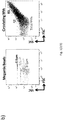
- the present inventors' "stepwise gating strategy", reported in Figure 1A is a potent and strategic tool able to accurately discriminate the uninvestigated cardiac (R13)-derived MV, minimizing the false positive events and using an anti-CD 172a antibody not known as marker for circulating cardiac-derived MV.
- the first assessed gate allows the identification of ABs (R3) and/or membrane fragments (R2).
- the sequential gating strategy is able to exclude the predominant MV erythroid (R5), platelet (R7), endothelium (R8) and leukocyte (R10 and R11) and to specifically focus on cardiac-(R13) and stromal/adipocyte (R14) -derived MV.
- the matrix 7x7 of the antibodies is representative of the applied "stepwise gating strategy". Noteworthy, the high specificity of the antibodies with a cross reaction ⁇ 10% and accuracy of system (100% for cardiac-derived MV) were successfully reached.
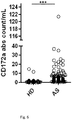
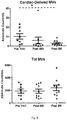
- MV characterized from plasma AB (R3), erythroid-(R5), endothelium- (R8) and cardiac- (R13) derived MV subsets were found at significantly higher levels in pre-TAVI AS patients as compared with healthy donors ( Fig.2a and 2b ). Strikingly, levels of circulating cardiac- and endothelium-derived MV subsets were significantly reduced after TAVI ( Fig.2a and 2b ).
- the interlinked MV-MV showed a rho ⁇ 0.80 a P ⁇ 0.0000, as reported in the correlation matrix of Table II.
- the identified inter-linking between MV-MV may characterize also other CVDs.
- Inventors selected a R13 cut-off to accurately identify the potential "Grey Zone” (MV absolute count ⁇ 4.7/mL) and the best performer cut-off of our cardiac-biomarker (MV absolute count ⁇ 4.7/mL).
- the threshold of the cardiac- biomarker able to discriminate patients with AS was optimized in the phase of the updated data using a larger cohort of AS patients and age-matched control group.
- the "best performer cut-off" of cardiac biomarkers ( ⁇ 4.7) enables to identify the "best performer cut-off” and the "grey zone” of the other correlated MV (endothelium-, monocitic- and stromal-derived MVs).
- Table.1 The matrix 7x7 of "Stepwise gating strategy" to characterize circulating tissue-derived MVs from Human Plasma.
- R11 0 0 0 0 100 8.7 ⁇ 3.2 7 ⁇ 2 R13 0 0 0 0 0 100 0 R14 0 0 0 0 0 0 100 0 R14 0 0 0 0 0 0 100
- R5 erythroid-derived
- R7 platelet-derived
- R8 endothelium-derived
- R10 monocyte-derived
- R11 leukocyte-derived
- R13 cardiac-derived
- R14 stromal/adipocyte-derived circulating MVs.
- Table. II Inter linking MV-MV CD172a CD144 CD73 CD14 CD172a 1
- CD144 Rho 0.81 P ⁇ 0.0000 1
- plasma samples and storage techniques were standardized.
- a 5 ml sample of peripheral blood was collected in EDTA-containing Vacutainer tubes.
- the vials were processed within 2 h of collection by centrifugation at 1,200g x 20 min. at room temperature (RT) to eliminate all blood cells.
- RT room temperature
- the top third of the plasma was aspirated and placed in a fresh tube.
- isolated plasma was subsequently diluted with filtered PBS and centrifuged at 500g x 30 min. at 4 °C.
- the obtained platelet-free plasma was centrifuged at 12,000g x 45 min. at 4 °C.
- a 0.22 ⁇ m filtered sheath fluid was used for sample acquisition.
- a morphological gate of microvesicles was performed according to Megamix-Plus FSC beads size (range: 0.1-0.9 ⁇ m) (Biocytex), according to literature ( Mobarrez F, et al., Thromb Res. 2010 Mar; 125:e110-6 ). Events included in a range from 0.1 ⁇ m to 5 ⁇ m were clearly discriminated from background noise.
- cTnT Monoclonal anti-cardiac troponin T (cTnT) (Innova Biosciences) was used to detect intracellular cTnT in CD172a+ MVs as well as in CD61+ MVs, as an internal negative control, with the Intrasure KiT (BD Biosciences), according to the manufacturer's instructions.
- Circulating MVs were sorted using a FACSAria III cell sorter (Biosciences) equipped with 4 lasers and able to discriminate up to 18 fluorescences. MV sorting was validated using the MegaMix bead (BioCytex). The cell sorter instrument was optimized to sort 0.3 ⁇ m, 0.5 ⁇ m and 0.9 ⁇ m beads with a purity higher than 95%.
- RNA from sorted cardiac-derived MVs was extracted with TRIzol reagent (Invitrogen, Carlsbad, CA, USA) following the standard protocol. Reverse transcription reactions were performed using the EXIQON miRNA Reverse Transcription Mercury Universal cDNA synthesis kit (Exiqon A/S, Vedbaek, Denmark).
- the Freedom Evo150 liquid handling system (Tecan Group, Mannedorf, Switzerland) was used for aliquoting reaction mixtures and samples in all protocol steps. qRT-PCR was performed with a 7900HT Fast Real-Time PCR (Applied Biosystems). miRNA content was evaluated in cardiac-derived MVs as well as in monocyte- and leukocyte-derived MVs, as negative controls, in experimental settings. The miRNA Ct cut off value was considered ⁇ 39.
- Aortic Stenosis (AS) cohort Aortic Stenosis (AS) cohort.
- ECG electrocardiogram
- CMR CMR
- AS aortic stenosis
- HCM Hypertrophic Cardiomyopathy
- HCM hypertrophied, non-dilated LV (wall thickness >15mm) in the absence of any other cardiac or systemic disorder producing a comparable grade of hypertrophy
- ACCF/AHA guideline for the diagnosis and treatment of hypertrophic cardiomyopathy executive summary: a report of the American College of Cardiology Foundation/American Heart Association Task Force on Practice Guidelines. Circulation 2011;124:27861-94 ).
- HCM status was those established by the American Heart Association guidelines ( Gersh BJ, Maron BJ, Bonow RO, et al. 2011 ACCF/AHA guideline for the diagnosis and treatment of hypertrophic cardiomyopathy: executive summary: a report of the American College of Cardiology Foundation/American Heart Association Task Force on Practice Guidelines. Circulation 2011;124:2761-96 ). None of the patients was in heart failure.
- Exclusion criteria were: either non-ST-segment or ST-segment elevation myocardial infarction; cardiogenic shock; allergy/intolerance to aspirin and/or clopidogrel; ongoing serious bleeding or bleeding diathesis; platelet count _75.000/mm3; planned or undelayable noncardiac surgery; previous percutaneous coronary intervention or coronary artery bypass grafting; severe liver disease (e.g., cirrhosis or portal hypertension); and life expectancy ⁇ 1 year due to other medical conditions.
- DM Diabetes mellitus
- CKD Chronic kidney disease
- CD172a as surface marker for the identification of cardiac derived MVs in plasma of Aortic Stenosis (AS) patients.
- AS Aortic Stenosis
- inventors analysed their internal content by investigating the expression of both cardiac specific Troponin T and micro RNAs.
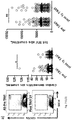
- the flow cytometric analysis performed on permeabilized circulating MVs revealed a selective expression of cardiac troponin T in the CD172a+ subpopulation ( Fig 4a ).
- this result was supported by the real time PCR analysis for Cardiac specific miRNAs performed on sorted CD172a+ MVs.
- the CD172a+ MVs expressed the cardiac specific miR1 and miR133a and the non-cardiac specific miR21 while the CD14+ MVs (internal negative control) expressed only the non-cardiac specific miR21. Altogether these results confirmed the cardiac origin of CD172a+ MVs.
- Inventors showed a significant decrease of cardiac MVs (CD172a+) in Aortic Stenosis (AS) patients upon TAVI surgery suggesting the release of these MVs from cardiac cells under stress conditions.
- AS Aortic Stenosis
- the control group included n.32 individuals aged ⁇ 65 years (n.8 females and n.24 males; range years: 31-64) and n.20 ⁇ 65 years (n.14 females and n.6 males; range years: 66-98).
- Circulating tissue-derived MVs of n. 10 patients with AS undergone percutaneous aortic valve replacement from "Federico II” University, Naples were evaluated at time of inclusion (pre-TAVI) up to 2-months follow-up, as summarized in Figure 8 .
- the significant reduction of circulating cardiac-derived MV subset was confirmed 2 months after surgery.
- HCM Hypertrophic CardioMyopathy
- CAD Coronary Artery Disease
Landscapes
- Health & Medical Sciences (AREA)
- Life Sciences & Earth Sciences (AREA)
- Engineering & Computer Science (AREA)
- Immunology (AREA)
- Molecular Biology (AREA)
- Hematology (AREA)
- Urology & Nephrology (AREA)
- Biomedical Technology (AREA)
- Chemical & Material Sciences (AREA)
- Physics & Mathematics (AREA)
- Cell Biology (AREA)
- Medicinal Chemistry (AREA)
- Pathology (AREA)
- Food Science & Technology (AREA)
- Biotechnology (AREA)
- Microbiology (AREA)
- Analytical Chemistry (AREA)
- Biochemistry (AREA)
- General Health & Medical Sciences (AREA)
- General Physics & Mathematics (AREA)
- Proteomics, Peptides & Aminoacids (AREA)
- Bioinformatics & Cheminformatics (AREA)
- Bioinformatics & Computational Biology (AREA)
- Biophysics (AREA)
- Zoology (AREA)
- Tropical Medicine & Parasitology (AREA)
- Virology (AREA)
- Investigating Or Analysing Biological Materials (AREA)
- Measuring Or Testing Involving Enzymes Or Micro-Organisms (AREA)
Claims (14)
- Ex-vivo- oder in-vitro-Verfahren zur Charakterisierung und/oder Messung der Menge an Untergruppen von zirkulierenden, von Gewebe abgeleiteten Mikrovesikeln (MV), umfassend den Schritt des Detektierens wenigstens des CD172a-Markers an den Mikrovesikeln, in einer isolierten biologischen Probe, die von dem Individuum gewonnen wurde, wobei, wenn das Mikrovesikel positiv für CD172a ist, das Mikrovesikel als ein vom Herzen abgeleitetes Mikrovesikel charakterisiert wird.
- Ex-vivo- oder in-vitro-Verfahren nach Anspruch 1, ferner umfassend den Schritt des weiteren Detektierens wenigstens eines Markers, ausgewählt aus der Gruppe bestehend aus: CD235a, CD61, CD144, CD14, CD45, CD73, CD3 oder jeder Kombination davon.
- Verfahren nach einem der vorhergehenden Ansprüche, wobei die zu detektierenden Marker CD172a, CD235a, CD61, CD144, CD14, CD45 und CD73 sind.
- Verfahren nach einem der Ansprüche 2 bis 3, wobei:- wenn das Mikrovesikel positiv für CD235a ist, das Mikrovesikel als ein von Erythroiden abgeleitetes MV charakterisiert wird;- wenn das Mikrovesikel positiv für CD61 ist, das Mikrovesikel als ein von Thrombozyten abgeleitetes MV charakterisiert wird;- wenn das Mikrovesikel positiv für CD144 ist, das Mikrovesikel als ein von Endothel abgeleitetes MV charakterisiert wird;- wenn das Mikrovesikel positiv für CD14 ist, das Mikrovesikel als ein von Monozyten abgeleitetes MV charakterisiert wird;- wenn das Mikrovesikel positiv für CD45 ist, das Mikrovesikel als ein von Leukozyten abgeleitetes MV charakterisiert wird;- wenn das Mikrovesikel positiv für CD73 ist, das Mikrovesikel als ein von Stromazellen/Adipozyten abgeleitetes MV charakterisiert wird.
- Ex-vivo- oder in-vitro-Verfahren zur Diagnostizierung und/oder Beurteilung des Risikos einer Entstehung und/oder zur Voraussage und/oder zur Überwachung des Fortschritts und/oder zur Überwachung der Wirksamkeit einer therapeutischen Behandlung und/oder zum Screening einer therapeutischen Behandlung von kardiovaskulären Erkrankungen (CVD) in einem Individuum, umfassend die folgenden Schritte:a) Charakterisieren und/oder Messen der Menge an Untergruppen von zirkulierenden, von Gewebe abgeleiteten Mikrovesikeln nach dem Verfahren von einem der Ansprüche 1 bis 4;b) Vergleichen in Bezug auf eine geeignete Kontrolle und/oder Referenz.
- Verfahren nach Anspruch 5, wobei eine Menge an vom Herzen abgeleiteten MV in der von dem Individuum gewonnenen, isolierten biologischen Probe, die höher als die Kontrollmenge ist, anzeigt, dass das Individuum entweder an kardiovaskulären Erkrankungen (CVD) leidet oder ein erhöhtes Risiko aufweist, solche zu entwickeln.
- Verfahren nach Anspruch 5, wobei eine Menge an vom Herzen abgeleiteten MV in der von dem Individuum gewonnenen, isolierten biologischen Probe, die niedriger ist als die Kontrollmenge, anzeigt, dass das Individuum auf eine Verbesserung der CVD zugeht, wobei sich das Individuum vorzugsweise einem Klappenersatz, vorzugsweise einer Transkatheter-Aortenklappenimplantation (TAVI) unterzogen hat.
- Verfahren nach einem der Ansprüche 5 bis 7, wobei die kardiovaskuläre Erkrankung (CVD) aus der Gruppe bestehend aus: Herzinsuffizienz, vorzugsweise primärer oder sekundärer Aortenstenose (AS), Klappenerkrankung, Kardiomyopathie, akuter Koronararterienerkrankung (CAD), Atherosklerose, Myokardischämie, Infarkt, Rhythmusstörungen, hypertropher Kardiomyopathie (HCM) und arzneimittelbedingter Kardiotoxizität, die mit verschiedenen Erkrankungen (z.B. Krebs, HIV-HAART-Therapien) verbunden ist, ausgewählt wird.
- Verfahren nach einem der Ansprüche 5 bis 8, wobei die gemessene oder charakterisierte Untergruppe von zirkulierenden, von Gewebe abgeleiteten Mikrovesikeln die vom Herzen abgeleitete MV-Untergruppe ist, wobei die vom Herzen abgeleitete MV-Untergruppe vorzugsweise durch das Vorhandensein des Markers CD172a gekennzeichnet ist, wobei die vom Herzen abgeleiteten Mikrovesikel stärker bevorzugt negativ für die folgenden Marker sind: CD235a, CD61, CD144, CD14, CD45, CD73.
- Verfahren nach einem der Ansprüche 5 bis 9, wobei es sich bei der isolierten biologischen Probe um Plasma, Blut, Serum, durch chirurgische Resektion gewonnenes Gewebe, durch Biopsie gewonnenes Gewebe, eine Zellkultur, Zellüberstände, Zelllysate, Gewebsproben, Organe, Knochenmark handelt.
- Verfahren nach einem der vorhergehenden Ansprüche, wobei der Marker mithilfe eines spezifischen Liganden detektiert wird, wobei der Ligand vorzugsweise ein Antikörper oder ein funktionelles Fragment davon ist.
- Verfahren nach Anspruch 11, wobei der Marker detektiert wird durch Magnetkügelchen, die mit einem Antikörpereinfang beschichtet sind, und/oder individualisierte getrocknete Antikörpercocktails und/oder Säulen mit angepassten Filterkartuschen und/oder kombiniert mit einem spezifischen Antikörperfilter (SAF).
- Verfahren nach einem der vorhergehenden Ansprüche, wobei die Untergruppen von zirkulierenden, von Gewebe abgeleiteten Mikrovesikeln wie folgt charakterisiert werden und/oder ihre Menge wie folgt gemessen wird: durch Mehrfarben-Durchflusszytometrietechnologie (FACS), Immungold-Elektronenmikroskopie, Immunfluoreszenz, ELISA, Immunpräzipitation, reversen kolorimetrischen Immunassay (RCIA), Radioimmunassay (RIA), Elektrochemilumineszenz, einen Oberflächenplasmonresonanz-(SPR)-basierten Ansatz, Nanoliter-Mikrofluidik (Immunoassays) oder Spektrometrie.
- Verwendung eines für CD172a spezifischen Liganden zur Detektion und/oder Quantifizierung von vom Herzen abgeleiteten MVs in einer isolierten biologischen Probe, wobei es sich bei dem Liganden vorzugsweise um einen Anti-CD172a-Antikörper oder funktionelle Fragmente davon handelt.
Applications Claiming Priority (2)
| Application Number | Priority Date | Filing Date | Title |
|---|---|---|---|
| EP15193500.4A EP3165926A1 (de) | 2015-11-06 | 2015-11-06 | Verfahren zur charakterisierung von zellspezifischen mikrovesikeln |
| PCT/EP2016/076810 WO2017077105A1 (en) | 2015-11-06 | 2016-11-07 | Method for characterization of cell specific microvesicles |
Publications (2)
| Publication Number | Publication Date |
|---|---|
| EP3371603A1 EP3371603A1 (de) | 2018-09-12 |
| EP3371603B1 true EP3371603B1 (de) | 2020-03-11 |
Family
ID=54539863
Family Applications (2)
| Application Number | Title | Priority Date | Filing Date |
|---|---|---|---|
| EP15193500.4A Withdrawn EP3165926A1 (de) | 2015-11-06 | 2015-11-06 | Verfahren zur charakterisierung von zellspezifischen mikrovesikeln |
| EP16794273.9A Active EP3371603B1 (de) | 2015-11-06 | 2016-11-07 | Verfahren zur charakterisierung von zellspezifischen mikrovesikeln |
Family Applications Before (1)
| Application Number | Title | Priority Date | Filing Date |
|---|---|---|---|
| EP15193500.4A Withdrawn EP3165926A1 (de) | 2015-11-06 | 2015-11-06 | Verfahren zur charakterisierung von zellspezifischen mikrovesikeln |
Country Status (7)
| Country | Link |
|---|---|
| US (1) | US20190079103A1 (de) |
| EP (2) | EP3165926A1 (de) |
| CN (1) | CN108474798A (de) |
| AU (1) | AU2016347731A1 (de) |
| CA (1) | CA3004378A1 (de) |
| IL (1) | IL259029B (de) |
| WO (1) | WO2017077105A1 (de) |
Families Citing this family (5)
| Publication number | Priority date | Publication date | Assignee | Title |
|---|---|---|---|---|
| EP3639032A1 (de) * | 2017-06-13 | 2020-04-22 | Beckman Coulter, Inc. | Nachweis von mikrovesikeln aus leukozyten durch fluosensitive durchflusszytometrie |
| IT201800003981A1 (it) * | 2018-03-26 | 2019-09-26 | Marco Marchisio | Metodo per identificare ed analizzare microvescicole in un campione di fluido biologico |
| EP3775897B1 (de) * | 2018-03-29 | 2023-12-13 | The United States of America, as represented by the Secretary, Department of Health and Human Services | Biomarkeranalyse für diagnostische multiplexdaten mit hohem durchsatz |
| CN112899224A (zh) * | 2021-02-08 | 2021-06-04 | 高连如 | 一种用于负载冠脉支架的细胞外纳米囊泡,制备方法和应用 |
| CN114081900B (zh) * | 2021-02-24 | 2024-11-22 | 济宁医学院附属医院 | 间充质干细胞来源的纳米微囊泡在制备治疗子痫前期的药物中的应用 |
Family Cites Families (8)
| Publication number | Priority date | Publication date | Assignee | Title |
|---|---|---|---|---|
| FR2788780B1 (fr) | 1999-01-27 | 2001-03-30 | Ap Cells Inc | Procede de preparation de vesicules membranaires |
| GB9927320D0 (en) | 1999-11-18 | 2000-01-12 | Chiron Spa | Exosome separation |
| US6812023B1 (en) | 2000-04-27 | 2004-11-02 | Anosys, Inc. | Methods of producing membrane vesicles |
| US20090311715A1 (en) * | 2008-05-09 | 2009-12-17 | Mayo Foundation For Medical Education And Research | Identifying coronary or soft tissue calcification |
| WO2012024543A1 (en) * | 2010-08-18 | 2012-02-23 | Caris Life Sciences Luxembourg Holdings | Circulating biomarkers for disease |
| WO2012115885A1 (en) * | 2011-02-22 | 2012-08-30 | Caris Life Sciences Luxembourg Holdings, S.A.R.L. | Circulating biomarkers |
| KR20140067001A (ko) * | 2011-08-08 | 2014-06-03 | 카리스 라이프 사이언스 룩셈부르크 홀딩스, 에스.에이.알.엘. | 생물지표 조성물 및 방법 |
| AU2013229762A1 (en) * | 2012-03-09 | 2014-09-25 | Caris Life Sciences Switzerland Holdings Gmbh | Biomarker compositions and methods |
-
2015
- 2015-11-06 EP EP15193500.4A patent/EP3165926A1/de not_active Withdrawn
-
2016
- 2016-11-07 EP EP16794273.9A patent/EP3371603B1/de active Active
- 2016-11-07 WO PCT/EP2016/076810 patent/WO2017077105A1/en not_active Ceased
- 2016-11-07 CN CN201680077239.6A patent/CN108474798A/zh active Pending
- 2016-11-07 CA CA3004378A patent/CA3004378A1/en not_active Abandoned
- 2016-11-07 AU AU2016347731A patent/AU2016347731A1/en not_active Abandoned
- 2016-11-07 US US15/773,615 patent/US20190079103A1/en not_active Abandoned
-
2018
- 2018-04-29 IL IL259029A patent/IL259029B/en not_active IP Right Cessation
Non-Patent Citations (1)
| Title |
|---|
| None * |
Also Published As
| Publication number | Publication date |
|---|---|
| WO2017077105A1 (en) | 2017-05-11 |
| EP3371603A1 (de) | 2018-09-12 |
| CA3004378A1 (en) | 2017-05-11 |
| IL259029A (en) | 2018-06-28 |
| AU2016347731A1 (en) | 2018-06-14 |
| CN108474798A (zh) | 2018-08-31 |
| US20190079103A1 (en) | 2019-03-14 |
| EP3165926A1 (de) | 2017-05-10 |
| IL259029B (en) | 2020-09-30 |
Similar Documents
| Publication | Publication Date | Title |
|---|---|---|
| US20240302376A1 (en) | Method for identifying subgroups of circulating tumor cells (ctcs) in the ctc population of a biological sample | |
| US9175346B2 (en) | Evaluation method for arteriosclerosis | |
| IL259029A (en) | A method for characterizing cell-specific tiny blisters | |
| US20130203061A1 (en) | Methods and systems for isolating, storing, and analyzing vesicles | |
| KR102337232B1 (ko) | 뇌 유래 소포체 특이적 마커 및 이를 이용한 뇌 질병 진단 방법 | |
| TWI784163B (zh) | 用於純化分離及分析腫瘤表面標記陰性且血球表面標記陰性之有核細胞的方法及腫瘤表面標記陰性且血球表面標記陰性之有核細胞之應用 | |
| EP2664922A1 (de) | Verfahren zur prognose von sepsis | |
| JP2020527711A (ja) | 子宮内膜癌のマーカとしてのctnb1 | |
| US20150377905A1 (en) | Methods for diagnosis of kawasaki disease | |
| JP7675441B2 (ja) | 移植拒絶反応の非hlaマーカー | |
| CN113358881B (zh) | 用于nmosd预测或复发监测的生物标志物及其应用 | |
| JP2022553420A (ja) | 敗血症管理 | |
| CN102221607B (zh) | 一种抗体组合物及其应用 | |
| Wang et al. | Different expression of S100A8 in malignant and benign gallbladder diseases | |
| US20150338412A1 (en) | Composition for diagnosis of lung cancer and diagnosis kit for lung cancer | |
| WO2011112749A2 (en) | Semi-invasive method for characterizing lung injury | |
| KR101297309B1 (ko) | 폐암 진단용 조성물 및 폐암 진단키트 | |
| EP2772759B1 (de) | Zusammensetzung für die Diagnose von Lungenkrebs | |
| KR101334123B1 (ko) | 소세포폐암 진단용 조성물 및 소세포폐암 진단키트 | |
| CN118443943A (zh) | 作为转移性去势抵抗性前列腺癌(mCRPC)患者的治疗选择的生物标志物的雄激素受体变体7 | |
| US20230028910A1 (en) | Method for diagnosing cutaneous t-cell lymphoma diseases | |
| WO2020201131A1 (en) | New markers for discriminating juvenile idiopathic arthritis (jia) and septic arthritis (sa) | |
| WO2010062705A1 (en) | Cancer diagnosis using ki-67 | |
| Salvadori et al. | WJT |
Legal Events
| Date | Code | Title | Description |
|---|---|---|---|
| STAA | Information on the status of an ep patent application or granted ep patent |
Free format text: STATUS: UNKNOWN |
|
| STAA | Information on the status of an ep patent application or granted ep patent |
Free format text: STATUS: THE INTERNATIONAL PUBLICATION HAS BEEN MADE |
|
| PUAI | Public reference made under article 153(3) epc to a published international application that has entered the european phase |
Free format text: ORIGINAL CODE: 0009012 |
|
| STAA | Information on the status of an ep patent application or granted ep patent |
Free format text: STATUS: REQUEST FOR EXAMINATION WAS MADE |
|
| 17P | Request for examination filed |
Effective date: 20180601 |
|
| AK | Designated contracting states |
Kind code of ref document: A1 Designated state(s): AL AT BE BG CH CY CZ DE DK EE ES FI FR GB GR HR HU IE IS IT LI LT LU LV MC MK MT NL NO PL PT RO RS SE SI SK SM TR |
|
| AX | Request for extension of the european patent |
Extension state: BA ME |
|
| DAV | Request for validation of the european patent (deleted) | ||
| DAX | Request for extension of the european patent (deleted) | ||
| STAA | Information on the status of an ep patent application or granted ep patent |
Free format text: STATUS: EXAMINATION IS IN PROGRESS |
|
| 17Q | First examination report despatched |
Effective date: 20190410 |
|
| GRAP | Despatch of communication of intention to grant a patent |
Free format text: ORIGINAL CODE: EPIDOSNIGR1 |
|
| STAA | Information on the status of an ep patent application or granted ep patent |
Free format text: STATUS: GRANT OF PATENT IS INTENDED |
|
| INTG | Intention to grant announced |
Effective date: 20190923 |
|
| GRAS | Grant fee paid |
Free format text: ORIGINAL CODE: EPIDOSNIGR3 |
|
| GRAA | (expected) grant |
Free format text: ORIGINAL CODE: 0009210 |
|
| STAA | Information on the status of an ep patent application or granted ep patent |
Free format text: STATUS: THE PATENT HAS BEEN GRANTED |
|
| AK | Designated contracting states |
Kind code of ref document: B1 Designated state(s): AL AT BE BG CH CY CZ DE DK EE ES FI FR GB GR HR HU IE IS IT LI LT LU LV MC MK MT NL NO PL PT RO RS SE SI SK SM TR |
|
| REG | Reference to a national code |
Ref country code: GB Ref legal event code: FG4D |
|
| REG | Reference to a national code |
Ref country code: CH Ref legal event code: EP |
|
| REG | Reference to a national code |
Ref country code: AT Ref legal event code: REF Ref document number: 1243793 Country of ref document: AT Kind code of ref document: T Effective date: 20200315 |
|
| REG | Reference to a national code |
Ref country code: IE Ref legal event code: FG4D |
|
| REG | Reference to a national code |
Ref country code: DE Ref legal event code: R096 Ref document number: 602016031654 Country of ref document: DE |
|
| PG25 | Lapsed in a contracting state [announced via postgrant information from national office to epo] |
Ref country code: FI Free format text: LAPSE BECAUSE OF FAILURE TO SUBMIT A TRANSLATION OF THE DESCRIPTION OR TO PAY THE FEE WITHIN THE PRESCRIBED TIME-LIMIT Effective date: 20200311 Ref country code: NO Free format text: LAPSE BECAUSE OF FAILURE TO SUBMIT A TRANSLATION OF THE DESCRIPTION OR TO PAY THE FEE WITHIN THE PRESCRIBED TIME-LIMIT Effective date: 20200611 Ref country code: RS Free format text: LAPSE BECAUSE OF FAILURE TO SUBMIT A TRANSLATION OF THE DESCRIPTION OR TO PAY THE FEE WITHIN THE PRESCRIBED TIME-LIMIT Effective date: 20200311 |
|
| REG | Reference to a national code |
Ref country code: NL Ref legal event code: MP Effective date: 20200311 |
|
| PG25 | Lapsed in a contracting state [announced via postgrant information from national office to epo] |
Ref country code: GR Free format text: LAPSE BECAUSE OF FAILURE TO SUBMIT A TRANSLATION OF THE DESCRIPTION OR TO PAY THE FEE WITHIN THE PRESCRIBED TIME-LIMIT Effective date: 20200612 Ref country code: BG Free format text: LAPSE BECAUSE OF FAILURE TO SUBMIT A TRANSLATION OF THE DESCRIPTION OR TO PAY THE FEE WITHIN THE PRESCRIBED TIME-LIMIT Effective date: 20200611 Ref country code: SE Free format text: LAPSE BECAUSE OF FAILURE TO SUBMIT A TRANSLATION OF THE DESCRIPTION OR TO PAY THE FEE WITHIN THE PRESCRIBED TIME-LIMIT Effective date: 20200311 Ref country code: LV Free format text: LAPSE BECAUSE OF FAILURE TO SUBMIT A TRANSLATION OF THE DESCRIPTION OR TO PAY THE FEE WITHIN THE PRESCRIBED TIME-LIMIT Effective date: 20200311 Ref country code: HR Free format text: LAPSE BECAUSE OF FAILURE TO SUBMIT A TRANSLATION OF THE DESCRIPTION OR TO PAY THE FEE WITHIN THE PRESCRIBED TIME-LIMIT Effective date: 20200311 |
|
| REG | Reference to a national code |
Ref country code: LT Ref legal event code: MG4D |
|
| PG25 | Lapsed in a contracting state [announced via postgrant information from national office to epo] |
Ref country code: NL Free format text: LAPSE BECAUSE OF FAILURE TO SUBMIT A TRANSLATION OF THE DESCRIPTION OR TO PAY THE FEE WITHIN THE PRESCRIBED TIME-LIMIT Effective date: 20200311 |
|
| PG25 | Lapsed in a contracting state [announced via postgrant information from national office to epo] |
Ref country code: PT Free format text: LAPSE BECAUSE OF FAILURE TO SUBMIT A TRANSLATION OF THE DESCRIPTION OR TO PAY THE FEE WITHIN THE PRESCRIBED TIME-LIMIT Effective date: 20200805 Ref country code: SK Free format text: LAPSE BECAUSE OF FAILURE TO SUBMIT A TRANSLATION OF THE DESCRIPTION OR TO PAY THE FEE WITHIN THE PRESCRIBED TIME-LIMIT Effective date: 20200311 Ref country code: IS Free format text: LAPSE BECAUSE OF FAILURE TO SUBMIT A TRANSLATION OF THE DESCRIPTION OR TO PAY THE FEE WITHIN THE PRESCRIBED TIME-LIMIT Effective date: 20200711 Ref country code: EE Free format text: LAPSE BECAUSE OF FAILURE TO SUBMIT A TRANSLATION OF THE DESCRIPTION OR TO PAY THE FEE WITHIN THE PRESCRIBED TIME-LIMIT Effective date: 20200311 Ref country code: LT Free format text: LAPSE BECAUSE OF FAILURE TO SUBMIT A TRANSLATION OF THE DESCRIPTION OR TO PAY THE FEE WITHIN THE PRESCRIBED TIME-LIMIT Effective date: 20200311 Ref country code: SM Free format text: LAPSE BECAUSE OF FAILURE TO SUBMIT A TRANSLATION OF THE DESCRIPTION OR TO PAY THE FEE WITHIN THE PRESCRIBED TIME-LIMIT Effective date: 20200311 Ref country code: RO Free format text: LAPSE BECAUSE OF FAILURE TO SUBMIT A TRANSLATION OF THE DESCRIPTION OR TO PAY THE FEE WITHIN THE PRESCRIBED TIME-LIMIT Effective date: 20200311 Ref country code: CZ Free format text: LAPSE BECAUSE OF FAILURE TO SUBMIT A TRANSLATION OF THE DESCRIPTION OR TO PAY THE FEE WITHIN THE PRESCRIBED TIME-LIMIT Effective date: 20200311 |
|
| REG | Reference to a national code |
Ref country code: AT Ref legal event code: MK05 Ref document number: 1243793 Country of ref document: AT Kind code of ref document: T Effective date: 20200311 |
|
| REG | Reference to a national code |
Ref country code: DE Ref legal event code: R097 Ref document number: 602016031654 Country of ref document: DE |
|
| PLBE | No opposition filed within time limit |
Free format text: ORIGINAL CODE: 0009261 |
|
| STAA | Information on the status of an ep patent application or granted ep patent |
Free format text: STATUS: NO OPPOSITION FILED WITHIN TIME LIMIT |
|
| PG25 | Lapsed in a contracting state [announced via postgrant information from national office to epo] |
Ref country code: AT Free format text: LAPSE BECAUSE OF FAILURE TO SUBMIT A TRANSLATION OF THE DESCRIPTION OR TO PAY THE FEE WITHIN THE PRESCRIBED TIME-LIMIT Effective date: 20200311 Ref country code: DK Free format text: LAPSE BECAUSE OF FAILURE TO SUBMIT A TRANSLATION OF THE DESCRIPTION OR TO PAY THE FEE WITHIN THE PRESCRIBED TIME-LIMIT Effective date: 20200311 Ref country code: ES Free format text: LAPSE BECAUSE OF FAILURE TO SUBMIT A TRANSLATION OF THE DESCRIPTION OR TO PAY THE FEE WITHIN THE PRESCRIBED TIME-LIMIT Effective date: 20200311 |
|
| 26N | No opposition filed |
Effective date: 20201214 |
|
| PG25 | Lapsed in a contracting state [announced via postgrant information from national office to epo] |
Ref country code: PL Free format text: LAPSE BECAUSE OF FAILURE TO SUBMIT A TRANSLATION OF THE DESCRIPTION OR TO PAY THE FEE WITHIN THE PRESCRIBED TIME-LIMIT Effective date: 20200311 Ref country code: SI Free format text: LAPSE BECAUSE OF FAILURE TO SUBMIT A TRANSLATION OF THE DESCRIPTION OR TO PAY THE FEE WITHIN THE PRESCRIBED TIME-LIMIT Effective date: 20200311 |
|
| REG | Reference to a national code |
Ref country code: DE Ref legal event code: R119 Ref document number: 602016031654 Country of ref document: DE |
|
| PG25 | Lapsed in a contracting state [announced via postgrant information from national office to epo] |
Ref country code: MC Free format text: LAPSE BECAUSE OF FAILURE TO SUBMIT A TRANSLATION OF THE DESCRIPTION OR TO PAY THE FEE WITHIN THE PRESCRIBED TIME-LIMIT Effective date: 20200311 |
|
| REG | Reference to a national code |
Ref country code: CH Ref legal event code: PL |
|
| GBPC | Gb: european patent ceased through non-payment of renewal fee |
Effective date: 20201107 |
|
| PG25 | Lapsed in a contracting state [announced via postgrant information from national office to epo] |
Ref country code: LU Free format text: LAPSE BECAUSE OF NON-PAYMENT OF DUE FEES Effective date: 20201107 |
|
| REG | Reference to a national code |
Ref country code: BE Ref legal event code: MM Effective date: 20201130 |
|
| PG25 | Lapsed in a contracting state [announced via postgrant information from national office to epo] |
Ref country code: CH Free format text: LAPSE BECAUSE OF NON-PAYMENT OF DUE FEES Effective date: 20201130 Ref country code: LI Free format text: LAPSE BECAUSE OF NON-PAYMENT OF DUE FEES Effective date: 20201130 |
|
| PG25 | Lapsed in a contracting state [announced via postgrant information from national office to epo] |
Ref country code: FR Free format text: LAPSE BECAUSE OF NON-PAYMENT OF DUE FEES Effective date: 20201130 Ref country code: IE Free format text: LAPSE BECAUSE OF NON-PAYMENT OF DUE FEES Effective date: 20201107 |
|
| PG25 | Lapsed in a contracting state [announced via postgrant information from national office to epo] |
Ref country code: GB Free format text: LAPSE BECAUSE OF NON-PAYMENT OF DUE FEES Effective date: 20201107 Ref country code: DE Free format text: LAPSE BECAUSE OF NON-PAYMENT OF DUE FEES Effective date: 20210601 |
|
| PG25 | Lapsed in a contracting state [announced via postgrant information from national office to epo] |
Ref country code: IT Free format text: LAPSE BECAUSE OF NON-PAYMENT OF DUE FEES Effective date: 20201107 |
|
| PG25 | Lapsed in a contracting state [announced via postgrant information from national office to epo] |
Ref country code: TR Free format text: LAPSE BECAUSE OF FAILURE TO SUBMIT A TRANSLATION OF THE DESCRIPTION OR TO PAY THE FEE WITHIN THE PRESCRIBED TIME-LIMIT Effective date: 20200311 Ref country code: MT Free format text: LAPSE BECAUSE OF FAILURE TO SUBMIT A TRANSLATION OF THE DESCRIPTION OR TO PAY THE FEE WITHIN THE PRESCRIBED TIME-LIMIT Effective date: 20200311 Ref country code: CY Free format text: LAPSE BECAUSE OF FAILURE TO SUBMIT A TRANSLATION OF THE DESCRIPTION OR TO PAY THE FEE WITHIN THE PRESCRIBED TIME-LIMIT Effective date: 20200311 |
|
| PG25 | Lapsed in a contracting state [announced via postgrant information from national office to epo] |
Ref country code: MK Free format text: LAPSE BECAUSE OF FAILURE TO SUBMIT A TRANSLATION OF THE DESCRIPTION OR TO PAY THE FEE WITHIN THE PRESCRIBED TIME-LIMIT Effective date: 20200311 Ref country code: AL Free format text: LAPSE BECAUSE OF FAILURE TO SUBMIT A TRANSLATION OF THE DESCRIPTION OR TO PAY THE FEE WITHIN THE PRESCRIBED TIME-LIMIT Effective date: 20200311 |
|
| PG25 | Lapsed in a contracting state [announced via postgrant information from national office to epo] |
Ref country code: BE Free format text: LAPSE BECAUSE OF NON-PAYMENT OF DUE FEES Effective date: 20201130 |
|
| PG25 | Lapsed in a contracting state [announced via postgrant information from national office to epo] |
Ref country code: IS Free format text: LAPSE BECAUSE OF NON-PAYMENT OF DUE FEES Effective date: 20200711 |