EP3371595B1 - Ex vivo-verfahren zur prüfung der zellularen antwort von primärzellenpopulationen auf eine droge oder kombinationen von drogen - Google Patents
Ex vivo-verfahren zur prüfung der zellularen antwort von primärzellenpopulationen auf eine droge oder kombinationen von drogen Download PDFInfo
- Publication number
- EP3371595B1 EP3371595B1 EP16801976.8A EP16801976A EP3371595B1 EP 3371595 B1 EP3371595 B1 EP 3371595B1 EP 16801976 A EP16801976 A EP 16801976A EP 3371595 B1 EP3371595 B1 EP 3371595B1
- Authority
- EP
- European Patent Office
- Prior art keywords
- cancer
- cells
- cell
- primary
- primary tumor
- Prior art date
- Legal status (The legal status is an assumption and is not a legal conclusion. Google has not performed a legal analysis and makes no representation as to the accuracy of the status listed.)
- Active
Links
- 210000004881 tumor cell Anatomy 0.000 title claims description 114
- 239000003814 drug Substances 0.000 title claims description 102
- 229940079593 drug Drugs 0.000 title claims description 99
- 238000000034 method Methods 0.000 title claims description 56
- 238000012360 testing method Methods 0.000 title claims description 33
- 230000001413 cellular effect Effects 0.000 title claims description 16
- 230000004043 responsiveness Effects 0.000 title claims description 14
- 210000004027 cell Anatomy 0.000 claims description 185
- 206010028980 Neoplasm Diseases 0.000 claims description 112
- 230000035899 viability Effects 0.000 claims description 89
- 230000035755 proliferation Effects 0.000 claims description 86
- 208000010839 B-cell chronic lymphocytic leukemia Diseases 0.000 claims description 77
- 208000032852 chronic lymphocytic leukemia Diseases 0.000 claims description 74
- 208000031422 Lymphocytic Chronic B-Cell Leukemia Diseases 0.000 claims description 73
- 210000002381 plasma Anatomy 0.000 claims description 67
- 102000004127 Cytokines Human genes 0.000 claims description 52
- 108090000695 Cytokines Proteins 0.000 claims description 52
- 210000001185 bone marrow Anatomy 0.000 claims description 52
- 210000003743 erythrocyte Anatomy 0.000 claims description 47
- 210000005259 peripheral blood Anatomy 0.000 claims description 42
- 239000011886 peripheral blood Substances 0.000 claims description 42
- 230000000694 effects Effects 0.000 claims description 39
- 206010035226 Plasma cell myeloma Diseases 0.000 claims description 38
- 201000011510 cancer Diseases 0.000 claims description 36
- 238000011534 incubation Methods 0.000 claims description 34
- 210000000265 leukocyte Anatomy 0.000 claims description 34
- 108010002350 Interleukin-2 Proteins 0.000 claims description 31
- 102000000588 Interleukin-2 Human genes 0.000 claims description 31
- 210000001165 lymph node Anatomy 0.000 claims description 21
- 210000002536 stromal cell Anatomy 0.000 claims description 20
- 238000011282 treatment Methods 0.000 claims description 18
- 239000000203 mixture Substances 0.000 claims description 16
- 208000031261 Acute myeloid leukaemia Diseases 0.000 claims description 12
- 201000003793 Myelodysplastic syndrome Diseases 0.000 claims description 11
- 208000033776 Myeloid Acute Leukemia Diseases 0.000 claims description 11
- 208000037265 diseases, disorders, signs and symptoms Diseases 0.000 claims description 11
- 201000010099 disease Diseases 0.000 claims description 10
- 238000001727 in vivo Methods 0.000 claims description 10
- 210000004882 non-tumor cell Anatomy 0.000 claims description 10
- 102000004889 Interleukin-6 Human genes 0.000 claims description 9
- 108090001005 Interleukin-6 Proteins 0.000 claims description 9
- 208000024893 Acute lymphoblastic leukemia Diseases 0.000 claims description 8
- 208000014697 Acute lymphocytic leukaemia Diseases 0.000 claims description 8
- 102100030704 Interleukin-21 Human genes 0.000 claims description 8
- 208000006664 Precursor Cell Lymphoblastic Leukemia-Lymphoma Diseases 0.000 claims description 8
- 108010074108 interleukin-21 Proteins 0.000 claims description 8
- 102000019034 Chemokines Human genes 0.000 claims description 7
- 108010012236 Chemokines Proteins 0.000 claims description 7
- 108010037362 Extracellular Matrix Proteins Proteins 0.000 claims description 7
- 102000010834 Extracellular Matrix Proteins Human genes 0.000 claims description 7
- 102100036922 Tumor necrosis factor ligand superfamily member 13B Human genes 0.000 claims description 7
- 210000002744 extracellular matrix Anatomy 0.000 claims description 7
- 208000015634 Rectal Neoplasms Diseases 0.000 claims description 6
- 208000002495 Uterine Neoplasms Diseases 0.000 claims description 6
- 239000002246 antineoplastic agent Substances 0.000 claims description 6
- 238000004113 cell culture Methods 0.000 claims description 6
- 208000014829 head and neck neoplasm Diseases 0.000 claims description 6
- 201000001441 melanoma Diseases 0.000 claims description 6
- 206010038038 rectal cancer Diseases 0.000 claims description 6
- 201000001275 rectum cancer Diseases 0.000 claims description 6
- 206010046766 uterine cancer Diseases 0.000 claims description 6
- 208000034578 Multiple myelomas Diseases 0.000 claims description 5
- 208000015914 Non-Hodgkin lymphomas Diseases 0.000 claims description 5
- 210000001808 exosome Anatomy 0.000 claims description 5
- 201000003444 follicular lymphoma Diseases 0.000 claims description 5
- 238000009169 immunotherapy Methods 0.000 claims description 5
- 239000003112 inhibitor Substances 0.000 claims description 5
- 230000011664 signaling Effects 0.000 claims description 5
- 108010028006 B-Cell Activating Factor Proteins 0.000 claims description 4
- 108090001007 Interleukin-8 Proteins 0.000 claims description 4
- 102000004890 Interleukin-8 Human genes 0.000 claims description 4
- 102000015696 Interleukins Human genes 0.000 claims description 4
- 108010063738 Interleukins Proteins 0.000 claims description 4
- 208000007660 Residual Neoplasm Diseases 0.000 claims description 4
- 230000006907 apoptotic process Effects 0.000 claims description 4
- 230000036755 cellular response Effects 0.000 claims description 4
- 239000003102 growth factor Substances 0.000 claims description 4
- 230000001965 increasing effect Effects 0.000 claims description 4
- 230000003902 lesion Effects 0.000 claims description 4
- 230000008685 targeting Effects 0.000 claims description 4
- 238000012604 3D cell culture Methods 0.000 claims description 3
- 208000036170 B-Cell Marginal Zone Lymphoma Diseases 0.000 claims description 3
- 208000003950 B-cell lymphoma Diseases 0.000 claims description 3
- 208000032791 BCR-ABL1 positive chronic myelogenous leukemia Diseases 0.000 claims description 3
- 206010005949 Bone cancer Diseases 0.000 claims description 3
- 208000018084 Bone neoplasm Diseases 0.000 claims description 3
- 206010006143 Brain stem glioma Diseases 0.000 claims description 3
- 208000011691 Burkitt lymphomas Diseases 0.000 claims description 3
- 208000017897 Carcinoma of esophagus Diseases 0.000 claims description 3
- 206010007953 Central nervous system lymphoma Diseases 0.000 claims description 3
- 208000010833 Chronic myeloid leukaemia Diseases 0.000 claims description 3
- 206010009944 Colon cancer Diseases 0.000 claims description 3
- 208000000461 Esophageal Neoplasms Diseases 0.000 claims description 3
- 208000017604 Hodgkin disease Diseases 0.000 claims description 3
- 208000021519 Hodgkin lymphoma Diseases 0.000 claims description 3
- 208000010747 Hodgkins lymphoma Diseases 0.000 claims description 3
- 102000003814 Interleukin-10 Human genes 0.000 claims description 3
- 108090000174 Interleukin-10 Proteins 0.000 claims description 3
- 102000003816 Interleukin-13 Human genes 0.000 claims description 3
- 108090000176 Interleukin-13 Proteins 0.000 claims description 3
- 102000003812 Interleukin-15 Human genes 0.000 claims description 3
- 108090000172 Interleukin-15 Proteins 0.000 claims description 3
- 108090000978 Interleukin-4 Proteins 0.000 claims description 3
- 102000004388 Interleukin-4 Human genes 0.000 claims description 3
- 208000007766 Kaposi sarcoma Diseases 0.000 claims description 3
- 208000008839 Kidney Neoplasms Diseases 0.000 claims description 3
- 208000031671 Large B-Cell Diffuse Lymphoma Diseases 0.000 claims description 3
- 208000025205 Mantle-Cell Lymphoma Diseases 0.000 claims description 3
- 208000033761 Myelogenous Chronic BCR-ABL Positive Leukemia Diseases 0.000 claims description 3
- 206010033128 Ovarian cancer Diseases 0.000 claims description 3
- 206010061535 Ovarian neoplasm Diseases 0.000 claims description 3
- 206010061902 Pancreatic neoplasm Diseases 0.000 claims description 3
- 208000000821 Parathyroid Neoplasms Diseases 0.000 claims description 3
- 208000002471 Penile Neoplasms Diseases 0.000 claims description 3
- 208000007913 Pituitary Neoplasms Diseases 0.000 claims description 3
- 201000005746 Pituitary adenoma Diseases 0.000 claims description 3
- 206010061538 Pituitary tumour benign Diseases 0.000 claims description 3
- 208000006265 Renal cell carcinoma Diseases 0.000 claims description 3
- 208000000453 Skin Neoplasms Diseases 0.000 claims description 3
- 208000021712 Soft tissue sarcoma Diseases 0.000 claims description 3
- 208000005718 Stomach Neoplasms Diseases 0.000 claims description 3
- 208000024313 Testicular Neoplasms Diseases 0.000 claims description 3
- 206010057644 Testis cancer Diseases 0.000 claims description 3
- 208000024770 Thyroid neoplasm Diseases 0.000 claims description 3
- 208000023915 Ureteral Neoplasms Diseases 0.000 claims description 3
- 206010046458 Urethral neoplasms Diseases 0.000 claims description 3
- 208000007097 Urinary Bladder Neoplasms Diseases 0.000 claims description 3
- 201000003761 Vaginal carcinoma Diseases 0.000 claims description 3
- 239000012190 activator Substances 0.000 claims description 3
- 238000004115 adherent culture Methods 0.000 claims description 3
- 208000024447 adrenal gland neoplasm Diseases 0.000 claims description 3
- 229940124675 anti-cancer drug Drugs 0.000 claims description 3
- 238000011319 anticancer therapy Methods 0.000 claims description 3
- 210000003169 central nervous system Anatomy 0.000 claims description 3
- 208000025997 central nervous system neoplasm Diseases 0.000 claims description 3
- 208000019065 cervical carcinoma Diseases 0.000 claims description 3
- 239000003795 chemical substances by application Substances 0.000 claims description 3
- 230000014564 chemokine production Effects 0.000 claims description 3
- 208000029742 colonic neoplasm Diseases 0.000 claims description 3
- 230000000139 costimulatory effect Effects 0.000 claims description 3
- 208000035250 cutaneous malignant susceptibility to 1 melanoma Diseases 0.000 claims description 3
- 230000016396 cytokine production Effects 0.000 claims description 3
- 229940127089 cytotoxic agent Drugs 0.000 claims description 3
- 239000002254 cytotoxic agent Substances 0.000 claims description 3
- 230000004069 differentiation Effects 0.000 claims description 3
- 206010012818 diffuse large B-cell lymphoma Diseases 0.000 claims description 3
- 210000000750 endocrine system Anatomy 0.000 claims description 3
- 201000003914 endometrial carcinoma Diseases 0.000 claims description 3
- 210000002889 endothelial cell Anatomy 0.000 claims description 3
- 201000001343 fallopian tube carcinoma Diseases 0.000 claims description 3
- 230000003328 fibroblastic effect Effects 0.000 claims description 3
- 210000000285 follicular dendritic cell Anatomy 0.000 claims description 3
- 206010017758 gastric cancer Diseases 0.000 claims description 3
- 239000000499 gel Substances 0.000 claims description 3
- 201000009277 hairy cell leukemia Diseases 0.000 claims description 3
- 208000014951 hematologic disease Diseases 0.000 claims description 3
- 230000002401 inhibitory effect Effects 0.000 claims description 3
- 239000003446 ligand Substances 0.000 claims description 3
- 201000007270 liver cancer Diseases 0.000 claims description 3
- 208000014018 liver neoplasm Diseases 0.000 claims description 3
- 201000007919 lymphoplasmacytic lymphoma Diseases 0.000 claims description 3
- 208000015486 malignant pancreatic neoplasm Diseases 0.000 claims description 3
- 208000026037 malignant tumor of neck Diseases 0.000 claims description 3
- 201000007924 marginal zone B-cell lymphoma Diseases 0.000 claims description 3
- 208000021937 marginal zone lymphoma Diseases 0.000 claims description 3
- 239000003550 marker Substances 0.000 claims description 3
- 210000002901 mesenchymal stem cell Anatomy 0.000 claims description 3
- 230000001394 metastastic effect Effects 0.000 claims description 3
- 206010061289 metastatic neoplasm Diseases 0.000 claims description 3
- 208000002154 non-small cell lung carcinoma Diseases 0.000 claims description 3
- 201000002528 pancreatic cancer Diseases 0.000 claims description 3
- 208000008443 pancreatic carcinoma Diseases 0.000 claims description 3
- 210000002990 parathyroid gland Anatomy 0.000 claims description 3
- 210000003668 pericyte Anatomy 0.000 claims description 3
- 208000021310 pituitary gland adenoma Diseases 0.000 claims description 3
- 239000004033 plastic Substances 0.000 claims description 3
- 229920003023 plastic Polymers 0.000 claims description 3
- 208000016800 primary central nervous system lymphoma Diseases 0.000 claims description 3
- 201000007444 renal pelvis carcinoma Diseases 0.000 claims description 3
- 201000000849 skin cancer Diseases 0.000 claims description 3
- 210000000813 small intestine Anatomy 0.000 claims description 3
- 206010041823 squamous cell carcinoma Diseases 0.000 claims description 3
- 208000017572 squamous cell neoplasm Diseases 0.000 claims description 3
- 201000011549 stomach cancer Diseases 0.000 claims description 3
- 201000003120 testicular cancer Diseases 0.000 claims description 3
- 229940124597 therapeutic agent Drugs 0.000 claims description 3
- 210000001685 thyroid gland Anatomy 0.000 claims description 3
- 230000005747 tumor angiogenesis Effects 0.000 claims description 3
- 210000000626 ureter Anatomy 0.000 claims description 3
- 208000013013 vulvar carcinoma Diseases 0.000 claims description 3
- 208000023275 Autoimmune disease Diseases 0.000 claims description 2
- 108020004414 DNA Proteins 0.000 claims description 2
- 102000053602 DNA Human genes 0.000 claims description 2
- 108060003951 Immunoglobulin Proteins 0.000 claims description 2
- 108020004682 Single-Stranded DNA Proteins 0.000 claims description 2
- 208000007502 anemia Diseases 0.000 claims description 2
- 208000015294 blood coagulation disease Diseases 0.000 claims description 2
- 229940044683 chemotherapy drug Drugs 0.000 claims description 2
- 231100000599 cytotoxic agent Toxicity 0.000 claims description 2
- 102000018358 immunoglobulin Human genes 0.000 claims description 2
- 230000001939 inductive effect Effects 0.000 claims description 2
- 229940047122 interleukins Drugs 0.000 claims description 2
- 230000003278 mimic effect Effects 0.000 claims description 2
- 241000894007 species Species 0.000 claims description 2
- 230000000052 comparative effect Effects 0.000 claims 1
- 238000000926 separation method Methods 0.000 claims 1
- 239000000523 sample Substances 0.000 description 78
- 229940046168 CpG oligodeoxynucleotide Drugs 0.000 description 36
- 230000002062 proliferating effect Effects 0.000 description 35
- 201000000050 myeloid neoplasm Diseases 0.000 description 33
- 108091003079 Bovine Serum Albumin Proteins 0.000 description 22
- 239000012091 fetal bovine serum Substances 0.000 description 22
- 238000003556 assay Methods 0.000 description 20
- 238000010228 ex vivo assay Methods 0.000 description 18
- 238000003501 co-culture Methods 0.000 description 16
- 238000004458 analytical method Methods 0.000 description 15
- GOTYRUGSSMKFNF-UHFFFAOYSA-N lenalidomide Chemical compound C1C=2C(N)=CC=CC=2C(=O)N1C1CCC(=O)NC1=O GOTYRUGSSMKFNF-UHFFFAOYSA-N 0.000 description 15
- 229960004942 lenalidomide Drugs 0.000 description 15
- 210000002966 serum Anatomy 0.000 description 15
- 230000008901 benefit Effects 0.000 description 14
- 229960003445 idelalisib Drugs 0.000 description 14
- YKLIKGKUANLGSB-HNNXBMFYSA-N idelalisib Chemical compound C1([C@@H](NC=2[C]3N=CN=C3N=CN=2)CC)=NC2=CC=CC(F)=C2C(=O)N1C1=CC=CC=C1 YKLIKGKUANLGSB-HNNXBMFYSA-N 0.000 description 14
- 239000002177 L01XE27 - Ibrutinib Substances 0.000 description 13
- 229960001507 ibrutinib Drugs 0.000 description 13
- XYFPWWZEPKGCCK-GOSISDBHSA-N ibrutinib Chemical compound C1=2C(N)=NC=NC=2N([C@H]2CN(CCC2)C(=O)C=C)N=C1C(C=C1)=CC=C1OC1=CC=CC=C1 XYFPWWZEPKGCCK-GOSISDBHSA-N 0.000 description 13
- 102400000432 CD40 ligand, soluble form Human genes 0.000 description 12
- 101800000267 CD40 ligand, soluble form Proteins 0.000 description 12
- 150000001875 compounds Chemical class 0.000 description 12
- 238000002474 experimental method Methods 0.000 description 12
- 102000004121 Annexin A5 Human genes 0.000 description 11
- 108090000672 Annexin A5 Proteins 0.000 description 11
- 230000004044 response Effects 0.000 description 11
- 208000002250 Hematologic Neoplasms Diseases 0.000 description 10
- 102100031573 Hematopoietic progenitor cell antigen CD34 Human genes 0.000 description 10
- 101000777663 Homo sapiens Hematopoietic progenitor cell antigen CD34 Proteins 0.000 description 10
- 239000001963 growth medium Substances 0.000 description 10
- 231100000673 dose–response relationship Toxicity 0.000 description 9
- 239000013610 patient sample Substances 0.000 description 9
- UZOVYGYOLBIAJR-UHFFFAOYSA-N 4-isocyanato-4'-methyldiphenylmethane Chemical compound C1=CC(C)=CC=C1CC1=CC=C(N=C=O)C=C1 UZOVYGYOLBIAJR-UHFFFAOYSA-N 0.000 description 8
- 229940100601 interleukin-6 Drugs 0.000 description 8
- 230000001028 anti-proliverative effect Effects 0.000 description 7
- 210000004369 blood Anatomy 0.000 description 7
- 239000008280 blood Substances 0.000 description 7
- 230000008569 process Effects 0.000 description 7
- 230000000638 stimulation Effects 0.000 description 7
- 108010029697 CD40 Ligand Proteins 0.000 description 6
- 102100032937 CD40 ligand Human genes 0.000 description 6
- 238000001574 biopsy Methods 0.000 description 6
- 238000000432 density-gradient centrifugation Methods 0.000 description 6
- 238000010186 staining Methods 0.000 description 6
- 210000001519 tissue Anatomy 0.000 description 6
- 239000006146 Roswell Park Memorial Institute medium Substances 0.000 description 5
- 230000004663 cell proliferation Effects 0.000 description 5
- 238000011161 development Methods 0.000 description 5
- 238000000338 in vitro Methods 0.000 description 5
- 238000002360 preparation method Methods 0.000 description 5
- 229920001917 Ficoll Polymers 0.000 description 4
- 206010066476 Haematological malignancy Diseases 0.000 description 4
- 229940041181 antineoplastic drug Drugs 0.000 description 4
- 230000001640 apoptogenic effect Effects 0.000 description 4
- 230000005754 cellular signaling Effects 0.000 description 4
- 238000007710 freezing Methods 0.000 description 4
- 230000008014 freezing Effects 0.000 description 4
- 230000012010 growth Effects 0.000 description 4
- 230000003211 malignant effect Effects 0.000 description 4
- 210000005087 mononuclear cell Anatomy 0.000 description 4
- 230000003389 potentiating effect Effects 0.000 description 4
- 230000004083 survival effect Effects 0.000 description 4
- 102100024222 B-lymphocyte antigen CD19 Human genes 0.000 description 3
- BZUNJUAMQZRJIP-UHFFFAOYSA-N CPDA Natural products OCCCCCCCCCCCCCCC(O)=O BZUNJUAMQZRJIP-UHFFFAOYSA-N 0.000 description 3
- 101000980825 Homo sapiens B-lymphocyte antigen CD19 Proteins 0.000 description 3
- 101000797623 Homo sapiens Protein AMBP Proteins 0.000 description 3
- 102100032859 Protein AMBP Human genes 0.000 description 3
- 108010065323 Tumor Necrosis Factor Ligand Superfamily Member 13 Proteins 0.000 description 3
- 210000003719 b-lymphocyte Anatomy 0.000 description 3
- 230000009286 beneficial effect Effects 0.000 description 3
- 230000003833 cell viability Effects 0.000 description 3
- 238000012258 culturing Methods 0.000 description 3
- 230000000857 drug effect Effects 0.000 description 3
- 238000011156 evaluation Methods 0.000 description 3
- 238000000684 flow cytometry Methods 0.000 description 3
- 239000012520 frozen sample Substances 0.000 description 3
- 230000028993 immune response Effects 0.000 description 3
- 108091070501 miRNA Proteins 0.000 description 3
- GISJHCLTIVIGLX-UHFFFAOYSA-N n-[4-[(4-chlorophenyl)methoxy]pyridin-2-yl]-2-(2,6-difluorophenyl)acetamide Chemical compound FC1=CC=CC(F)=C1CC(=O)NC1=CC(OCC=2C=CC(Cl)=CC=2)=CC=N1 GISJHCLTIVIGLX-UHFFFAOYSA-N 0.000 description 3
- -1 or bone marrow Substances 0.000 description 3
- 238000011084 recovery Methods 0.000 description 3
- 230000035945 sensitivity Effects 0.000 description 3
- 239000000243 solution Substances 0.000 description 3
- 210000000952 spleen Anatomy 0.000 description 3
- NLXLAEXVIDQMFP-UHFFFAOYSA-N Ammonia chloride Chemical compound [NH4+].[Cl-] NLXLAEXVIDQMFP-UHFFFAOYSA-N 0.000 description 2
- 108091008875 B cell receptors Proteins 0.000 description 2
- 208000005443 Circulating Neoplastic Cells Diseases 0.000 description 2
- AOJJSUZBOXZQNB-TZSSRYMLSA-N Doxorubicin Chemical compound O([C@H]1C[C@@](O)(CC=2C(O)=C3C(=O)C=4C=CC=C(C=4C(=O)C3=C(O)C=21)OC)C(=O)CO)[C@H]1C[C@H](N)[C@H](O)[C@H](C)O1 AOJJSUZBOXZQNB-TZSSRYMLSA-N 0.000 description 2
- 206010059866 Drug resistance Diseases 0.000 description 2
- 239000006144 Dulbecco’s modified Eagle's medium Substances 0.000 description 2
- 101001137987 Homo sapiens Lymphocyte activation gene 3 protein Proteins 0.000 description 2
- 101000738771 Homo sapiens Receptor-type tyrosine-protein phosphatase C Proteins 0.000 description 2
- WHUUTDBJXJRKMK-VKHMYHEASA-N L-glutamic acid Chemical compound OC(=O)[C@@H](N)CCC(O)=O WHUUTDBJXJRKMK-VKHMYHEASA-N 0.000 description 2
- ZDXPYRJPNDTMRX-VKHMYHEASA-N L-glutamine Chemical compound OC(=O)[C@@H](N)CCC(N)=O ZDXPYRJPNDTMRX-VKHMYHEASA-N 0.000 description 2
- 229930182816 L-glutamine Natural products 0.000 description 2
- 102100020862 Lymphocyte activation gene 3 protein Human genes 0.000 description 2
- 206010025323 Lymphomas Diseases 0.000 description 2
- 108010061593 Member 14 Tumor Necrosis Factor Receptors Proteins 0.000 description 2
- 102100040678 Programmed cell death protein 1 Human genes 0.000 description 2
- 101710089372 Programmed cell death protein 1 Proteins 0.000 description 2
- 239000012980 RPMI-1640 medium Substances 0.000 description 2
- 102100037422 Receptor-type tyrosine-protein phosphatase C Human genes 0.000 description 2
- 102100028785 Tumor necrosis factor receptor superfamily member 14 Human genes 0.000 description 2
- 230000004075 alteration Effects 0.000 description 2
- 230000003115 biocidal effect Effects 0.000 description 2
- 239000012472 biological sample Substances 0.000 description 2
- 230000015572 biosynthetic process Effects 0.000 description 2
- 239000012888 bovine serum Substances 0.000 description 2
- 239000003560 cancer drug Substances 0.000 description 2
- 238000002512 chemotherapy Methods 0.000 description 2
- 210000003040 circulating cell Anatomy 0.000 description 2
- 238000005138 cryopreservation Methods 0.000 description 2
- 230000001472 cytotoxic effect Effects 0.000 description 2
- 238000010790 dilution Methods 0.000 description 2
- 239000012895 dilution Substances 0.000 description 2
- 230000002708 enhancing effect Effects 0.000 description 2
- 210000003714 granulocyte Anatomy 0.000 description 2
- 230000002489 hematologic effect Effects 0.000 description 2
- 210000002865 immune cell Anatomy 0.000 description 2
- 230000003993 interaction Effects 0.000 description 2
- 210000004698 lymphocyte Anatomy 0.000 description 2
- 238000004519 manufacturing process Methods 0.000 description 2
- 238000005259 measurement Methods 0.000 description 2
- 230000007246 mechanism Effects 0.000 description 2
- 239000002609 medium Substances 0.000 description 2
- 239000002679 microRNA Substances 0.000 description 2
- 239000003068 molecular probe Substances 0.000 description 2
- KDWFDOFTPHDNJL-TUBOTVQJSA-N odn-2006 Chemical compound O=C1NC(=O)C(C)=CN1[C@@H]1O[C@H](COP(O)(=O)O[C@@H]2[C@H](O[C@H](C2)N2C(NC(=O)C(C)=C2)=O)COP(O)(=O)O[C@H]2[C@H]([C@@H](O[C@@H]2COP(O)(=S)O[C@H]2[C@H]([C@@H](O[C@@H]2COP(O)(=O)O[C@@H]2[C@H](O[C@H](C2)N2C(NC(=O)C(C)=C2)=O)COP(O)(=O)O[C@@H]2[C@H](O[C@H](C2)N2C(NC(=O)C(C)=C2)=O)COP(O)(=O)O[C@@H]2[C@H](O[C@H](C2)N2C(NC(=O)C(C)=C2)=O)COP(O)(=O)O[C@@H]2[C@H](O[C@H](C2)N2C(NC(=O)C(C)=C2)=O)COP(O)(=O)O[C@H]2[C@H]([C@@H](O[C@@H]2COP(O)(=O)O[C@@H]2[C@H](O[C@H](C2)N2C(NC(=O)C(C)=C2)=O)COP(O)(=O)O[C@H]2[C@H]([C@@H](O[C@@H]2COP(O)(=S)O[C@H]2[C@H]([C@@H](O[C@@H]2COP(O)(=O)OC[C@@H]2[C@H](C[C@@H](O2)N2C(NC(=O)C(C)=C2)=O)OP(O)(=O)OC[C@@H]2[C@H]([C@@H](O)[C@@H](O2)N2C3=C(C(NC(N)=N3)=O)N=C2)OP(O)(=O)OC[C@@H]2[C@H](C[C@@H](O2)N2C(NC(=O)C(C)=C2)=O)OP(O)(=O)OC[C@@H]2[C@H](C[C@@H](O2)N2C(NC(=O)C(C)=C2)=O)OP(O)(=O)OC[C@@H]2[C@H](C[C@@H](O2)N2C(NC(=O)C(C)=C2)=O)OP(O)(=O)OC[C@@H]2[C@H](C[C@@H](O2)N2C(NC(=O)C(C)=C2)=O)OP(O)(=O)OC[C@@H]2[C@H]([C@@H](O)[C@@H](O2)N2C3=C(C(NC(N)=N3)=O)N=C2)OP(S)(=O)OC[C@@H]2[C@H]([C@@H](O)[C@@H](O2)N2C(N=C(N)C=C2)=O)OP(O)(=O)OC[C@@H]2[C@H](C[C@@H](O2)N2C(NC(=O)C(C)=C2)=O)OP(O)(=O)OC[C@@H]2[C@H](C[C@@H](O2)N2C(NC(=O)C(C)=C2)=O)O)N2C3=C(C(NC(N)=N3)=O)N=C2)O)N2C(N=C(N)C=C2)=O)O)N2C3=C(C(NC(N)=N3)=O)N=C2)O)N2C3=C(C(NC(N)=N3)=O)N=C2)O)N2C(N=C(N)C=C2)=O)O)[C@@H](O)C1 KDWFDOFTPHDNJL-TUBOTVQJSA-N 0.000 description 2
- 230000001575 pathological effect Effects 0.000 description 2
- 230000037361 pathway Effects 0.000 description 2
- 230000000144 pharmacologic effect Effects 0.000 description 2
- 230000001737 promoting effect Effects 0.000 description 2
- 239000000700 radioactive tracer Substances 0.000 description 2
- 206010062113 splenic marginal zone lymphoma Diseases 0.000 description 2
- 210000000130 stem cell Anatomy 0.000 description 2
- 239000006228 supernatant Substances 0.000 description 2
- 238000010257 thawing Methods 0.000 description 2
- 230000004614 tumor growth Effects 0.000 description 2
- JKMHFZQWWAIEOD-UHFFFAOYSA-N 2-[4-(2-hydroxyethyl)piperazin-1-yl]ethanesulfonic acid Chemical compound OCC[NH+]1CCN(CCS([O-])(=O)=O)CC1 JKMHFZQWWAIEOD-UHFFFAOYSA-N 0.000 description 1
- RSGFPIWWSCWCFJ-VAXZQHAWSA-N 2-hydroxypropane-1,2,3-tricarboxylic acid;(2r,3s,4r,5r)-2,3,4,5,6-pentahydroxyhexanal;phosphoric acid Chemical compound OP(O)(O)=O.OC[C@@H](O)[C@@H](O)[C@H](O)[C@@H](O)C=O.OC(=O)CC(O)(C(O)=O)CC(O)=O RSGFPIWWSCWCFJ-VAXZQHAWSA-N 0.000 description 1
- STQGQHZAVUOBTE-UHFFFAOYSA-N 7-Cyan-hept-2t-en-4,6-diinsaeure Natural products C1=2C(O)=C3C(=O)C=4C(OC)=CC=CC=4C(=O)C3=C(O)C=2CC(O)(C(C)=O)CC1OC1CC(N)C(O)C(C)O1 STQGQHZAVUOBTE-UHFFFAOYSA-N 0.000 description 1
- 229930024421 Adenine Natural products 0.000 description 1
- 229920001817 Agar Polymers 0.000 description 1
- 102100022005 B-lymphocyte antigen CD20 Human genes 0.000 description 1
- 102000015081 Blood Coagulation Factors Human genes 0.000 description 1
- 108010039209 Blood Coagulation Factors Proteins 0.000 description 1
- COVZYZSDYWQREU-UHFFFAOYSA-N Busulfan Chemical compound CS(=O)(=O)OCCCCOS(C)(=O)=O COVZYZSDYWQREU-UHFFFAOYSA-N 0.000 description 1
- 102100027207 CD27 antigen Human genes 0.000 description 1
- CMSMOCZEIVJLDB-UHFFFAOYSA-N Cyclophosphamide Chemical compound ClCCN(CCCl)P1(=O)NCCCO1 CMSMOCZEIVJLDB-UHFFFAOYSA-N 0.000 description 1
- ZBNZXTGUTAYRHI-UHFFFAOYSA-N Dasatinib Chemical compound C=1C(N2CCN(CCO)CC2)=NC(C)=NC=1NC(S1)=NC=C1C(=O)NC1=C(C)C=CC=C1Cl ZBNZXTGUTAYRHI-UHFFFAOYSA-N 0.000 description 1
- IAZDPXIOMUYVGZ-UHFFFAOYSA-N Dimethylsulphoxide Chemical compound CS(C)=O IAZDPXIOMUYVGZ-UHFFFAOYSA-N 0.000 description 1
- 239000007995 HEPES buffer Substances 0.000 description 1
- 101000897405 Homo sapiens B-lymphocyte antigen CD20 Proteins 0.000 description 1
- 101000914511 Homo sapiens CD27 antigen Proteins 0.000 description 1
- 101000851370 Homo sapiens Tumor necrosis factor receptor superfamily member 9 Proteins 0.000 description 1
- 102000014150 Interferons Human genes 0.000 description 1
- 108010050904 Interferons Proteins 0.000 description 1
- 108010002386 Interleukin-3 Proteins 0.000 description 1
- 239000005517 L01XE01 - Imatinib Substances 0.000 description 1
- 239000002067 L01XE06 - Dasatinib Substances 0.000 description 1
- 239000005536 L01XE08 - Nilotinib Substances 0.000 description 1
- 108010053334 Peptichemio Proteins 0.000 description 1
- FAPWRFPIFSIZLT-UHFFFAOYSA-M Sodium chloride Chemical compound [Na+].[Cl-] FAPWRFPIFSIZLT-UHFFFAOYSA-M 0.000 description 1
- 210000001744 T-lymphocyte Anatomy 0.000 description 1
- 102100022153 Tumor necrosis factor receptor superfamily member 4 Human genes 0.000 description 1
- 101710165473 Tumor necrosis factor receptor superfamily member 4 Proteins 0.000 description 1
- 102100036856 Tumor necrosis factor receptor superfamily member 9 Human genes 0.000 description 1
- JXLYSJRDGCGARV-WWYNWVTFSA-N Vinblastine Natural products O=C(O[C@H]1[C@](O)(C(=O)OC)[C@@H]2N(C)c3c(cc(c(OC)c3)[C@]3(C(=O)OC)c4[nH]c5c(c4CCN4C[C@](O)(CC)C[C@H](C3)C4)cccc5)[C@@]32[C@H]2[C@@]1(CC)C=CCN2CC3)C JXLYSJRDGCGARV-WWYNWVTFSA-N 0.000 description 1
- 241000700605 Viruses Species 0.000 description 1
- 230000002159 abnormal effect Effects 0.000 description 1
- 229960000643 adenine Drugs 0.000 description 1
- 239000008272 agar Substances 0.000 description 1
- 235000019270 ammonium chloride Nutrition 0.000 description 1
- 230000002424 anti-apoptotic effect Effects 0.000 description 1
- 239000003146 anticoagulant agent Substances 0.000 description 1
- 229940127219 anticoagulant drug Drugs 0.000 description 1
- 238000003782 apoptosis assay Methods 0.000 description 1
- 210000003969 blast cell Anatomy 0.000 description 1
- 230000000903 blocking effect Effects 0.000 description 1
- 239000003114 blood coagulation factor Substances 0.000 description 1
- 210000003995 blood forming stem cell Anatomy 0.000 description 1
- 210000004204 blood vessel Anatomy 0.000 description 1
- 239000000872 buffer Substances 0.000 description 1
- 229960002092 busulfan Drugs 0.000 description 1
- 238000004364 calculation method Methods 0.000 description 1
- 244000309466 calf Species 0.000 description 1
- 230000030833 cell death Effects 0.000 description 1
- 230000008783 cell intrinsic mechanism Effects 0.000 description 1
- 239000006285 cell suspension Substances 0.000 description 1
- 238000005119 centrifugation Methods 0.000 description 1
- 230000008859 change Effects 0.000 description 1
- 238000012512 characterization method Methods 0.000 description 1
- 230000000973 chemotherapeutic effect Effects 0.000 description 1
- JCKYGMPEJWAADB-UHFFFAOYSA-N chlorambucil Chemical compound OC(=O)CCCC1=CC=C(N(CCCl)CCCl)C=C1 JCKYGMPEJWAADB-UHFFFAOYSA-N 0.000 description 1
- 229960004630 chlorambucil Drugs 0.000 description 1
- 238000011284 combination treatment Methods 0.000 description 1
- 101150008740 cpg-1 gene Proteins 0.000 description 1
- 229960004397 cyclophosphamide Drugs 0.000 description 1
- 231100000433 cytotoxic Toxicity 0.000 description 1
- 230000003013 cytotoxicity Effects 0.000 description 1
- 231100000135 cytotoxicity Toxicity 0.000 description 1
- 229960002448 dasatinib Drugs 0.000 description 1
- STQGQHZAVUOBTE-VGBVRHCVSA-N daunorubicin Chemical compound O([C@H]1C[C@@](O)(CC=2C(O)=C3C(=O)C=4C=CC=C(C=4C(=O)C3=C(O)C=21)OC)C(C)=O)[C@H]1C[C@H](N)[C@H](O)[C@H](C)O1 STQGQHZAVUOBTE-VGBVRHCVSA-N 0.000 description 1
- 229960000975 daunorubicin Drugs 0.000 description 1
- 229960001760 dimethyl sulfoxide Drugs 0.000 description 1
- 230000003292 diminished effect Effects 0.000 description 1
- 230000008034 disappearance Effects 0.000 description 1
- 208000035475 disorder Diseases 0.000 description 1
- 229960004679 doxorubicin Drugs 0.000 description 1
- 239000000890 drug combination Substances 0.000 description 1
- 230000000431 effect on proliferation Effects 0.000 description 1
- 238000005516 engineering process Methods 0.000 description 1
- 230000004049 epigenetic modification Effects 0.000 description 1
- 230000007717 exclusion Effects 0.000 description 1
- 210000002950 fibroblast Anatomy 0.000 description 1
- 230000030279 gene silencing Effects 0.000 description 1
- 208000035474 group of disease Diseases 0.000 description 1
- 230000036541 health Effects 0.000 description 1
- 201000005787 hematologic cancer Diseases 0.000 description 1
- 208000024200 hematopoietic and lymphoid system neoplasm Diseases 0.000 description 1
- 210000005260 human cell Anatomy 0.000 description 1
- 229960002411 imatinib Drugs 0.000 description 1
- KTUFNOKKBVMGRW-UHFFFAOYSA-N imatinib Chemical compound C1CN(C)CCN1CC1=CC=C(C(=O)NC=2C=C(NC=3N=C(C=CN=3)C=3C=NC=CC=3)C(C)=CC=2)C=C1 KTUFNOKKBVMGRW-UHFFFAOYSA-N 0.000 description 1
- 229940091204 imlygic Drugs 0.000 description 1
- 238000013394 immunophenotyping Methods 0.000 description 1
- 230000001976 improved effect Effects 0.000 description 1
- 230000006872 improvement Effects 0.000 description 1
- 230000005764 inhibitory process Effects 0.000 description 1
- 230000000977 initiatory effect Effects 0.000 description 1
- 230000002452 interceptive effect Effects 0.000 description 1
- 229940047124 interferons Drugs 0.000 description 1
- 238000002955 isolation Methods 0.000 description 1
- 230000007774 longterm Effects 0.000 description 1
- 230000001926 lymphatic effect Effects 0.000 description 1
- 210000002540 macrophage Anatomy 0.000 description 1
- 230000036210 malignancy Effects 0.000 description 1
- MIKKOBKEXMRYFQ-WZTVWXICSA-N meglumine amidotrizoate Chemical compound C[NH2+]C[C@H](O)[C@@H](O)[C@H](O)[C@H](O)CO.CC(=O)NC1=C(I)C(NC(C)=O)=C(I)C(C([O-])=O)=C1I MIKKOBKEXMRYFQ-WZTVWXICSA-N 0.000 description 1
- SGDBTWWWUNNDEQ-LBPRGKRZSA-N melphalan Chemical compound OC(=O)[C@@H](N)CC1=CC=C(N(CCCl)CCCl)C=C1 SGDBTWWWUNNDEQ-LBPRGKRZSA-N 0.000 description 1
- 229960001924 melphalan Drugs 0.000 description 1
- KKZJGLLVHKMTCM-UHFFFAOYSA-N mitoxantrone Chemical compound O=C1C2=C(O)C=CC(O)=C2C(=O)C2=C1C(NCCNCCO)=CC=C2NCCNCCO KKZJGLLVHKMTCM-UHFFFAOYSA-N 0.000 description 1
- 229960001156 mitoxantrone Drugs 0.000 description 1
- 238000012986 modification Methods 0.000 description 1
- 230000004048 modification Effects 0.000 description 1
- 230000004001 molecular interaction Effects 0.000 description 1
- 238000012544 monitoring process Methods 0.000 description 1
- 210000001616 monocyte Anatomy 0.000 description 1
- 210000000822 natural killer cell Anatomy 0.000 description 1
- 230000017074 necrotic cell death Effects 0.000 description 1
- 229960001346 nilotinib Drugs 0.000 description 1
- HHZIURLSWUIHRB-UHFFFAOYSA-N nilotinib Chemical compound C1=NC(C)=CN1C1=CC(NC(=O)C=2C=C(NC=3N=C(C=CN=3)C=3C=NC=CC=3)C(C)=CC=2)=CC(C(F)(F)F)=C1 HHZIURLSWUIHRB-UHFFFAOYSA-N 0.000 description 1
- 235000015097 nutrients Nutrition 0.000 description 1
- 229960002450 ofatumumab Drugs 0.000 description 1
- 229940124276 oligodeoxyribonucleotide Drugs 0.000 description 1
- 230000008520 organization Effects 0.000 description 1
- 239000008188 pellet Substances 0.000 description 1
- 230000002093 peripheral effect Effects 0.000 description 1
- 239000008363 phosphate buffer Substances 0.000 description 1
- 210000004180 plasmocyte Anatomy 0.000 description 1
- 238000007747 plating Methods 0.000 description 1
- 230000008092 positive effect Effects 0.000 description 1
- 230000000861 pro-apoptotic effect Effects 0.000 description 1
- 230000001686 pro-survival effect Effects 0.000 description 1
- 230000005522 programmed cell death Effects 0.000 description 1
- 108090000623 proteins and genes Proteins 0.000 description 1
- 102000004169 proteins and genes Human genes 0.000 description 1
- 238000011002 quantification Methods 0.000 description 1
- 108020003175 receptors Proteins 0.000 description 1
- 102000005962 receptors Human genes 0.000 description 1
- 230000010076 replication Effects 0.000 description 1
- 229960004641 rituximab Drugs 0.000 description 1
- 238000009738 saturating Methods 0.000 description 1
- 238000012216 screening Methods 0.000 description 1
- 238000004062 sedimentation Methods 0.000 description 1
- 239000011780 sodium chloride Substances 0.000 description 1
- 239000011550 stock solution Substances 0.000 description 1
- 239000000126 substance Substances 0.000 description 1
- 239000013589 supplement Substances 0.000 description 1
- 230000001502 supplementing effect Effects 0.000 description 1
- 230000008093 supporting effect Effects 0.000 description 1
- 230000001225 therapeutic effect Effects 0.000 description 1
- 238000002560 therapeutic procedure Methods 0.000 description 1
- 231100000331 toxic Toxicity 0.000 description 1
- 230000002588 toxic effect Effects 0.000 description 1
- 230000005751 tumor progression Effects 0.000 description 1
- 229960003048 vinblastine Drugs 0.000 description 1
- JXLYSJRDGCGARV-XQKSVPLYSA-N vincaleukoblastine Chemical compound C([C@@H](C[C@]1(C(=O)OC)C=2C(=CC3=C([C@]45[C@H]([C@@]([C@H](OC(C)=O)[C@]6(CC)C=CCN([C@H]56)CC4)(O)C(=O)OC)N3C)C=2)OC)C[C@@](C2)(O)CC)N2CCC2=C1NC1=CC=CC=C21 JXLYSJRDGCGARV-XQKSVPLYSA-N 0.000 description 1
- 229960004528 vincristine Drugs 0.000 description 1
- OGWKCGZFUXNPDA-XQKSVPLYSA-N vincristine Chemical compound C([N@]1C[C@@H](C[C@]2(C(=O)OC)C=3C(=CC4=C([C@]56[C@H]([C@@]([C@H](OC(C)=O)[C@]7(CC)C=CCN([C@H]67)CC5)(O)C(=O)OC)N4C=O)C=3)OC)C[C@@](C1)(O)CC)CC1=C2NC2=CC=CC=C12 OGWKCGZFUXNPDA-XQKSVPLYSA-N 0.000 description 1
- OGWKCGZFUXNPDA-UHFFFAOYSA-N vincristine Natural products C1C(CC)(O)CC(CC2(C(=O)OC)C=3C(=CC4=C(C56C(C(C(OC(C)=O)C7(CC)C=CCN(C67)CC5)(O)C(=O)OC)N4C=O)C=3)OC)CN1CCC1=C2NC2=CC=CC=C12 OGWKCGZFUXNPDA-UHFFFAOYSA-N 0.000 description 1
- 238000005406 washing Methods 0.000 description 1
Images









Classifications
-
- G—PHYSICS
- G01—MEASURING; TESTING
- G01N—INVESTIGATING OR ANALYSING MATERIALS BY DETERMINING THEIR CHEMICAL OR PHYSICAL PROPERTIES
- G01N33/00—Investigating or analysing materials by specific methods not covered by groups G01N1/00 - G01N31/00
- G01N33/48—Biological material, e.g. blood, urine; Haemocytometers
- G01N33/50—Chemical analysis of biological material, e.g. blood, urine; Testing involving biospecific ligand binding methods; Immunological testing
- G01N33/5005—Chemical analysis of biological material, e.g. blood, urine; Testing involving biospecific ligand binding methods; Immunological testing involving human or animal cells
- G01N33/5008—Chemical analysis of biological material, e.g. blood, urine; Testing involving biospecific ligand binding methods; Immunological testing involving human or animal cells for testing or evaluating the effect of chemical or biological compounds, e.g. drugs, cosmetics
- G01N33/5011—Chemical analysis of biological material, e.g. blood, urine; Testing involving biospecific ligand binding methods; Immunological testing involving human or animal cells for testing or evaluating the effect of chemical or biological compounds, e.g. drugs, cosmetics for testing antineoplastic activity
-
- C—CHEMISTRY; METALLURGY
- C12—BIOCHEMISTRY; BEER; SPIRITS; WINE; VINEGAR; MICROBIOLOGY; ENZYMOLOGY; MUTATION OR GENETIC ENGINEERING
- C12N—MICROORGANISMS OR ENZYMES; COMPOSITIONS THEREOF; PROPAGATING, PRESERVING, OR MAINTAINING MICROORGANISMS; MUTATION OR GENETIC ENGINEERING; CULTURE MEDIA
- C12N5/00—Undifferentiated human, animal or plant cells, e.g. cell lines; Tissues; Cultivation or maintenance thereof; Culture media therefor
- C12N5/06—Animal cells or tissues; Human cells or tissues
- C12N5/0602—Vertebrate cells
- C12N5/0693—Tumour cells; Cancer cells
- C12N5/0694—Cells of blood, e.g. leukemia cells, myeloma cells
-
- G—PHYSICS
- G01—MEASURING; TESTING
- G01N—INVESTIGATING OR ANALYSING MATERIALS BY DETERMINING THEIR CHEMICAL OR PHYSICAL PROPERTIES
- G01N33/00—Investigating or analysing materials by specific methods not covered by groups G01N1/00 - G01N31/00
- G01N33/48—Biological material, e.g. blood, urine; Haemocytometers
- G01N33/50—Chemical analysis of biological material, e.g. blood, urine; Testing involving biospecific ligand binding methods; Immunological testing
- G01N33/5005—Chemical analysis of biological material, e.g. blood, urine; Testing involving biospecific ligand binding methods; Immunological testing involving human or animal cells
- G01N33/5008—Chemical analysis of biological material, e.g. blood, urine; Testing involving biospecific ligand binding methods; Immunological testing involving human or animal cells for testing or evaluating the effect of chemical or biological compounds, e.g. drugs, cosmetics
- G01N33/5044—Chemical analysis of biological material, e.g. blood, urine; Testing involving biospecific ligand binding methods; Immunological testing involving human or animal cells for testing or evaluating the effect of chemical or biological compounds, e.g. drugs, cosmetics involving specific cell types
-
- G—PHYSICS
- G01—MEASURING; TESTING
- G01N—INVESTIGATING OR ANALYSING MATERIALS BY DETERMINING THEIR CHEMICAL OR PHYSICAL PROPERTIES
- G01N33/00—Investigating or analysing materials by specific methods not covered by groups G01N1/00 - G01N31/00
- G01N33/48—Biological material, e.g. blood, urine; Haemocytometers
- G01N33/50—Chemical analysis of biological material, e.g. blood, urine; Testing involving biospecific ligand binding methods; Immunological testing
- G01N33/5005—Chemical analysis of biological material, e.g. blood, urine; Testing involving biospecific ligand binding methods; Immunological testing involving human or animal cells
- G01N33/5008—Chemical analysis of biological material, e.g. blood, urine; Testing involving biospecific ligand binding methods; Immunological testing involving human or animal cells for testing or evaluating the effect of chemical or biological compounds, e.g. drugs, cosmetics
- G01N33/5082—Supracellular entities, e.g. tissue, organisms
- G01N33/5088—Supracellular entities, e.g. tissue, organisms of vertebrates
-
- C—CHEMISTRY; METALLURGY
- C12—BIOCHEMISTRY; BEER; SPIRITS; WINE; VINEGAR; MICROBIOLOGY; ENZYMOLOGY; MUTATION OR GENETIC ENGINEERING
- C12N—MICROORGANISMS OR ENZYMES; COMPOSITIONS THEREOF; PROPAGATING, PRESERVING, OR MAINTAINING MICROORGANISMS; MUTATION OR GENETIC ENGINEERING; CULTURE MEDIA
- C12N2500/00—Specific components of cell culture medium
- C12N2500/30—Organic components
- C12N2500/40—Nucleotides, nucleosides or bases
-
- C—CHEMISTRY; METALLURGY
- C12—BIOCHEMISTRY; BEER; SPIRITS; WINE; VINEGAR; MICROBIOLOGY; ENZYMOLOGY; MUTATION OR GENETIC ENGINEERING
- C12N—MICROORGANISMS OR ENZYMES; COMPOSITIONS THEREOF; PROPAGATING, PRESERVING, OR MAINTAINING MICROORGANISMS; MUTATION OR GENETIC ENGINEERING; CULTURE MEDIA
- C12N2501/00—Active agents used in cell culture processes, e.g. differentation
- C12N2501/20—Cytokines; Chemokines
- C12N2501/23—Interleukins [IL]
- C12N2501/2302—Interleukin-2 (IL-2)
-
- C—CHEMISTRY; METALLURGY
- C12—BIOCHEMISTRY; BEER; SPIRITS; WINE; VINEGAR; MICROBIOLOGY; ENZYMOLOGY; MUTATION OR GENETIC ENGINEERING
- C12N—MICROORGANISMS OR ENZYMES; COMPOSITIONS THEREOF; PROPAGATING, PRESERVING, OR MAINTAINING MICROORGANISMS; MUTATION OR GENETIC ENGINEERING; CULTURE MEDIA
- C12N2501/00—Active agents used in cell culture processes, e.g. differentation
- C12N2501/20—Cytokines; Chemokines
- C12N2501/23—Interleukins [IL]
- C12N2501/2304—Interleukin-4 (IL-4)
-
- C—CHEMISTRY; METALLURGY
- C12—BIOCHEMISTRY; BEER; SPIRITS; WINE; VINEGAR; MICROBIOLOGY; ENZYMOLOGY; MUTATION OR GENETIC ENGINEERING
- C12N—MICROORGANISMS OR ENZYMES; COMPOSITIONS THEREOF; PROPAGATING, PRESERVING, OR MAINTAINING MICROORGANISMS; MUTATION OR GENETIC ENGINEERING; CULTURE MEDIA
- C12N2501/00—Active agents used in cell culture processes, e.g. differentation
- C12N2501/20—Cytokines; Chemokines
- C12N2501/23—Interleukins [IL]
- C12N2501/2306—Interleukin-6 (IL-6)
-
- C—CHEMISTRY; METALLURGY
- C12—BIOCHEMISTRY; BEER; SPIRITS; WINE; VINEGAR; MICROBIOLOGY; ENZYMOLOGY; MUTATION OR GENETIC ENGINEERING
- C12N—MICROORGANISMS OR ENZYMES; COMPOSITIONS THEREOF; PROPAGATING, PRESERVING, OR MAINTAINING MICROORGANISMS; MUTATION OR GENETIC ENGINEERING; CULTURE MEDIA
- C12N2501/00—Active agents used in cell culture processes, e.g. differentation
- C12N2501/20—Cytokines; Chemokines
- C12N2501/23—Interleukins [IL]
- C12N2501/231—Interleukin-10 (IL-10)
-
- C—CHEMISTRY; METALLURGY
- C12—BIOCHEMISTRY; BEER; SPIRITS; WINE; VINEGAR; MICROBIOLOGY; ENZYMOLOGY; MUTATION OR GENETIC ENGINEERING
- C12N—MICROORGANISMS OR ENZYMES; COMPOSITIONS THEREOF; PROPAGATING, PRESERVING, OR MAINTAINING MICROORGANISMS; MUTATION OR GENETIC ENGINEERING; CULTURE MEDIA
- C12N2501/00—Active agents used in cell culture processes, e.g. differentation
- C12N2501/20—Cytokines; Chemokines
- C12N2501/23—Interleukins [IL]
- C12N2501/2313—Interleukin-13 (IL-13)
-
- C—CHEMISTRY; METALLURGY
- C12—BIOCHEMISTRY; BEER; SPIRITS; WINE; VINEGAR; MICROBIOLOGY; ENZYMOLOGY; MUTATION OR GENETIC ENGINEERING
- C12N—MICROORGANISMS OR ENZYMES; COMPOSITIONS THEREOF; PROPAGATING, PRESERVING, OR MAINTAINING MICROORGANISMS; MUTATION OR GENETIC ENGINEERING; CULTURE MEDIA
- C12N2501/00—Active agents used in cell culture processes, e.g. differentation
- C12N2501/20—Cytokines; Chemokines
- C12N2501/23—Interleukins [IL]
- C12N2501/2315—Interleukin-15 (IL-15)
-
- C—CHEMISTRY; METALLURGY
- C12—BIOCHEMISTRY; BEER; SPIRITS; WINE; VINEGAR; MICROBIOLOGY; ENZYMOLOGY; MUTATION OR GENETIC ENGINEERING
- C12N—MICROORGANISMS OR ENZYMES; COMPOSITIONS THEREOF; PROPAGATING, PRESERVING, OR MAINTAINING MICROORGANISMS; MUTATION OR GENETIC ENGINEERING; CULTURE MEDIA
- C12N2501/00—Active agents used in cell culture processes, e.g. differentation
- C12N2501/20—Cytokines; Chemokines
- C12N2501/23—Interleukins [IL]
- C12N2501/2321—Interleukin-21 (IL-21)
-
- C—CHEMISTRY; METALLURGY
- C12—BIOCHEMISTRY; BEER; SPIRITS; WINE; VINEGAR; MICROBIOLOGY; ENZYMOLOGY; MUTATION OR GENETIC ENGINEERING
- C12N—MICROORGANISMS OR ENZYMES; COMPOSITIONS THEREOF; PROPAGATING, PRESERVING, OR MAINTAINING MICROORGANISMS; MUTATION OR GENETIC ENGINEERING; CULTURE MEDIA
- C12N2501/00—Active agents used in cell culture processes, e.g. differentation
- C12N2501/998—Proteins not provided for elsewhere
-
- C—CHEMISTRY; METALLURGY
- C12—BIOCHEMISTRY; BEER; SPIRITS; WINE; VINEGAR; MICROBIOLOGY; ENZYMOLOGY; MUTATION OR GENETIC ENGINEERING
- C12N—MICROORGANISMS OR ENZYMES; COMPOSITIONS THEREOF; PROPAGATING, PRESERVING, OR MAINTAINING MICROORGANISMS; MUTATION OR GENETIC ENGINEERING; CULTURE MEDIA
- C12N2501/00—Active agents used in cell culture processes, e.g. differentation
- C12N2501/999—Small molecules not provided for elsewhere
-
- C—CHEMISTRY; METALLURGY
- C12—BIOCHEMISTRY; BEER; SPIRITS; WINE; VINEGAR; MICROBIOLOGY; ENZYMOLOGY; MUTATION OR GENETIC ENGINEERING
- C12N—MICROORGANISMS OR ENZYMES; COMPOSITIONS THEREOF; PROPAGATING, PRESERVING, OR MAINTAINING MICROORGANISMS; MUTATION OR GENETIC ENGINEERING; CULTURE MEDIA
- C12N2502/00—Coculture with; Conditioned medium produced by
- C12N2502/30—Coculture with; Conditioned medium produced by tumour cells
-
- C—CHEMISTRY; METALLURGY
- C12—BIOCHEMISTRY; BEER; SPIRITS; WINE; VINEGAR; MICROBIOLOGY; ENZYMOLOGY; MUTATION OR GENETIC ENGINEERING
- C12N—MICROORGANISMS OR ENZYMES; COMPOSITIONS THEREOF; PROPAGATING, PRESERVING, OR MAINTAINING MICROORGANISMS; MUTATION OR GENETIC ENGINEERING; CULTURE MEDIA
- C12N2503/00—Use of cells in diagnostics
- C12N2503/02—Drug screening
-
- C—CHEMISTRY; METALLURGY
- C12—BIOCHEMISTRY; BEER; SPIRITS; WINE; VINEGAR; MICROBIOLOGY; ENZYMOLOGY; MUTATION OR GENETIC ENGINEERING
- C12N—MICROORGANISMS OR ENZYMES; COMPOSITIONS THEREOF; PROPAGATING, PRESERVING, OR MAINTAINING MICROORGANISMS; MUTATION OR GENETIC ENGINEERING; CULTURE MEDIA
- C12N2513/00—3D culture
Definitions
- Present invention relates to the field of pharmacology and, in particular, to the assessment of anti-tumor drugs activity.
- the microenvironment (ME) critically affects tumor cell survival, proliferation and drug resistance (1).
- the role of the ME has become even more important with the discovery and development of cancer drugs that specifically target components of the ME (2). This has become an obstacle for the ex vivo testing of drugs that treat cancer. The absence of the factors that constitute the ME in these ex vivo assays will influence the perceived efficacy of many drugs. The result of this is a lack of correlation between the ex vivo results and the clinical effectiveness of these drugs.
- microenvironment-mimicking assays include adding cellular elements ( 3-5 ), adding components of the extracellular matrix (6, 7) and/or the addition of cocktails of soluble cytokines/chemokines or molecules derived from non-tumor cells present in the tumor microenvironment ( 8, 9 ).
- WO 2014/035668 A1 describes an assay for detecting malignant potential, involving in vitro co-culturing a population of cancer responder cells (e.g., primary cancer/tumor cells) with a population of non-tumor cells (e.g., bone marrow-derived circulating cells, buffy coat cells, peripheral circulating cells, and immune cells), contacting the co-culture with at least one drug, and measuring at least one malignant phenotype exhibited by the cancer responder cells, such as ability to proliferation on soft agar, ability to proliferate and form tumor in vitro, ability to proliferate and form tumor in vivo.
- the method may further comprise co-culturing the populations of cancer responder cells and non- tumor cells in the presence of plasma.
- the subject may have been diagnosed with cancer and the plasma may be obtained from said subject.
- the subject may have undergone chemotherapy.
- the method may be used for determining the likelihood of development of cancer drug resistance in a subject.
- the cancer responder cells may be human tumor cell lines, fresh or frozen human tumor surgical samples, fresh or frozen human biopsy samples, human tumor cells.
- WO 2015/042450 A1 discloses systems and methods for studying responses of patient target cells in the presence of their own stroma cells (cis-co-culture) under different drug conditions.
- WO 2010/120329 A1 discloses extracellular matrix bioscaffolds capable of supporting the formation and growth of tumors from tumor cells surrounded by tumor-associated cells such as macrophages and fibroblast-like cells and discloses the importance of tumor microenvironment in methods for testing compounds for treating cancers. While these attempts have had some success, the present invention introduces a new method for mimicking components of the microenvironment that have not previously been attempted.
- Chronic Lymphocytic Leukemia is a prototype of a disease with an extremely heterogeneous biology and clinical course; it is the most frequent adult hematologic malignancy ( 10 ). This pronounced clinical heterogeneity is linked to both cell-intrinsic and cell-extrinsic mechanisms of the disease. The best known amongst cell-intrinsic mechanisms concerns genomic aberrations, epigenetic modifications and micro-RNAs. CLL behavior and outcome, including chemorefractoriness, also depends on stimuli present in the leukemic ME ( 11 ).
- MM Multiple Myeloma
- BM bone marrow
- MM is still an incurable disease, being the second most frequent hematological malignancy in the USA and Europe.
- MM cells have a strong BM microenvironment dependence, which confers anti-apoptotic and pro-survival signals and resistance to drugs.
- MM tumor cells have an elevated proliferation rate promoted by soluble factors and molecular interactions within the particular BM microenvironment.
- Tumor samples extracted from cancer patients contain the tumor cells in the context of a highly heterogeneous environment. This environment may represent 99% of the sample extracted from the patient, for example in hematological tissue samples such as bone marrow, peripheral blood, or lymph node. To study these primary tumor cells, it is common practice to remove most of this heterogeneous environment, concentrating the remaining tumor cells that are being studied. In hematological samples this is commonly performed by a density gradient isolating the fraction containing the leukocytes, which corresponds roughly to only 1% of the entire sample. The other major fractions of the density gradient commonly discarded are the plasma fraction, and the erythrocyte fraction. We refer to both of these discarded fractions as the Artificial Environment (AE).
- AE Artificial Environment
- the disclosed method of the present invention adds Artificial Environment to primary tumor cells.
- the present invention has several advantages over that of the prior art.
- the activity of compounds on primary cells is artificially altered in assays which use isolated primary cells ex vivo.
- the method of the present invention in which the assay of potential anti-tumor drugs is also included, adds an Artificial Environment that compensates for the lack of a microenvironment in isolated cells.
- the whole sample has been used, but it is generally greatly diluted. Adding AE to whole sample assays would compensate for artifacts imposed by the initial dilution of the ME of the sample.
- Another advantage is that the methods of present invention enhance the viability of a primary tumor cell population.
- An additional advantage is that the method of the present invention promotes the proliferation of a primary tumor cell population, to levels that have not been previously reported ( 14 ).
- These advantages allow for the ex vivo analysis of the cellular response of the primary tumor cell population to a large number of variables, including different incubation times and testing of many drug compositions, including drugs that inhibit proliferation.
- another advantage of the present invention is the capacity to analyze the primary tumor cells in a manner that more closely mimics the in vivo microenvironment of a patient. This is particularly important in the case of drugs targeting the microenvironment e.g. ibrutinib and idelalisib, whose assessment will likely require microenvironment-mimicking assays.
- the invention presented here relates to a method for producing an artificial environment (AE) of primary cell populations, particularly an artificial tumor environment of primary tumor cell populations and its use in an ex vivo method to test the cellular responsiveness of primary tumor cell populations to a drug or combination of drugs.
- the test of the cellular responsiveness comprises incubation of the primary tumor cells with the artificial tumor environment and the drug or drugs to be tested and analyze the response of the primary tumor cell populations.
- the incubation of the primary tumor cells with the artificial tumor environment enhances the viability of said tumor cells and/or induces greater levels of tumor cell proliferation and, consequently, increases the sensitivity and accuracy of the test with regard to the drug/s assayed.
- Described herein is also the process for obtaining components of AE from peripheral blood (PB), or bone marrow (BM), or lymph nodes (LN) samples. Said components are used to stabilize primary tumor cell populations.
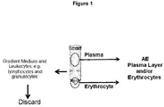
- These selected components which constitute the AE are isolated and/or mixed components from a sample selected from: peripheral blood, or bone marrow, or lymph nodes obtained from a donor ( Figure 1 and Example 1), but excluding the leukocytes fraction (AE leukocyte-free).
- the present invention relates to a method for producing an artificial tumor environment of primary tumor cells and its use in an ex vivo method to test the cellular responsiveness of primary tumor cell populations to a drug or combination of drugs.
- This method is characterized for enhancing and/or promoting the proliferation of a primary tumor cell population in ex vivo assays. Isolated primary tumor cells cannot be duly analyzed because they lack certain microenvironmental elements which are critical in promoting the survival and proliferation of a tumor in vivo.
- CLL Chronic Lymphocytic Leukemia
- AML Acute Myeloid Leukemia
- ALL Acute Lymphoid Leukemia
- MDS Myelodysplastic Syndrome
- NHL non-Hodgkin's Lymphoma
- MM Multiple Myeloma
- the present invention also discloses the use of the AE in ex vivo assays for determine the effect of a drug (analysis and quantification of the drug effect) on primary tumor cells isolated from patient samples.
- Microenvironment - is the environment in which the tumor exists, including surrounding blood vessels, immune cells, fibroblasts, stromal cells, the extracellular matrix (ECM), soluble factors (e.g. tumor derived exosomes, signaling molecules. growth factors, micro RNA, chemokines, cytokines and any soluble molecule derived from tumor or non-tumor cells), all of which affect tumor cell dynamics.
- ECM extracellular matrix
- AE Artificial Environment -
- AE leukocyte-free Fraction or mixture of fractions isolated from a peripheral blood, or bone marrow, or lymph node sample from a donor after density gradient centrifugation excluding leukocyte fraction (AE leukocyte-free). Residual leukocytes could still remain in the AE.
- AE can be only the plasma fraction, only the erythrocyte fraction, or a combination of the two fractions at any ratio (e.g. 1:1, 1:2, 2:1, etc.).
- AE Leukocyte-free Fraction or sample without leukocytes, or with a residual number of leukocytes, defined as less than 100 leukocytes per ⁇ l of AE.
- Donor - refers to a person who provides a biological sample.
- the person can be healthy or have been diagnosed with a disease (e.g. cancer).
- the biological sample can be, for example, blood, bone marrow, lymph node or tumor biopsy.
- Ex vivo - experimentation or measurements done in or on tissue or cell/s from an organism in an external environment with the minimum alteration of natural conditions refers to primary human cells in vitro, from patients suffering a disease or normal individuals, where the cells can be either recently extracted, cryopreserved, or frozen, to preserve their state. In some embodiments, these cells are thawed for the in vitro evaluation of drug effects.
- Ex vivo and “In vitro" are used indistinctly.
- Hematological Malignancies - (HM) Also called hematological neoplasms refers to a group of diseases, defined according to the World Health Organization classification, that includes Hodgkin's lymphoma, Non-Hodgkin's lymphoma (e.g., B cell lymphoma, diffuse large B cell lymphoma, follicular lymphoma, mantle cell lymphoma, marginal zone B-cell lymphoma, Burkitt lymphoma, lymphoplasmacytic lymphoma, hairy cell leukemia), chronic lymphocytic leukemia, acute myeloid leukemia, chronic myeloid leukemia, myelodysplastic syndrome, multiple myeloma, or acute lymphocytic leukemia.
- Hodgkin's lymphoma e.g., B cell lymphoma, diffuse large B cell lymphoma, follicular lymphoma, mantle cell lymphoma, marginal zone B-cell lymphom
- Solid Tumors - consists of neoplasms or lesions formed by abnormal growth of body tissue cells other than blood, bone marrow or lymphatic cells.
- Solid tumors include, but are not limited to, ovarian cancer, rectal cancer, cancer of the anal region, stomach cancer, testicular cancer, uterine cancer, colon cancer, rectal cancer, renal-cell carcinoma, liver cancer, non-small cell carcinoma of the lung, cancer of the small intestine, cancer of the esophagus, melanoma, cancer of the endocrine system, cancer of the thyroid gland, cancer of the parathyroid gland, cancer of the adrenal gland, bone cancer, pancreatic cancer, skin cancer, cancer of the head or neck, cutaneous or intraocular malignant melanoma, uterine cancer, carcinoma of the fallopian tubes, brain stem glioma, pituitary adenoma, Kaposi's sarcoma, epidermoid cancer, squamous cell cancer, carcinoma of the endometrium, carcinoma
- a sample can be primary cells from a healthy individual, a patient diagnosed with cancer, or a patient diagnosed with another hematological disorder.
- a sample may or may not have been cryopreserved. Cryopreserved samples have to be thawed before use.
- Primary tumor cells refers to tumor cells taken directly from living tissue (e.g. bone marrow, peripheral blood, lymph nodes, spleen, or tumor biopsy), isolated and established for ex vivo growth. Primary tumor cells may have been previously extracted and cryopreserved and thawed before use, or may be recently extracted and used without cryopreservation.
- living tissue e.g. bone marrow, peripheral blood, lymph nodes, spleen, or tumor biopsy
- Primary tumor cells may have been previously extracted and cryopreserved and thawed before use, or may be recently extracted and used without cryopreservation.
- Primary cell population refers to cells (non-diseased) taken directly from living tissue (e.g. bone marrow, peripheral blood, lymph nodes, spleen, or tumor biopsy) that are established for ex vivo growth.
- living tissue e.g. bone marrow, peripheral blood, lymph nodes, spleen, or tumor biopsy
- Stromal cell population representative of a cell type found in the in vivo tumor microenvironment is composed of several non-tumor cell types that can include, but is not limited to, fibroblastic reticular cells, nurse-like cells, mesenchymal stem cells, follicular dendritic cells, endothelial cells and pericytes.
- the stromal cells can be primary cells, or cells derived from primary stromal cells including, but not limited to, HS-5, HS27a, UE6E7T-2, NK-Tert, HMEC-1, KUSA-H1, M210B4, ST-2, and KUM-4.
- Density Gradient Centrifugation - refers to a process that separates a blood, or bone marrow, or sample into fractions of discrete components, e.g. plasma, erythrocytes, granulocytes and monocytes.
- Blood, or bone marrow, or lymph node samples are layered over a gradient medium (e.g. Ficoll-PaqueTM, LymphoprepTM, Histopaque-1077TM, or other similar mediums) and after centrifugation the isolated fractions of each component can be removed ( Fig. 1 ).
- a gradient medium e.g. Ficoll-PaqueTM, LymphoprepTM, Histopaque-1077TM, or other similar mediums
- Plasma fraction or Serum fraction - Plasma obtained from a blood, or bone marrow, or lymph node sample after density gradient centrifugation. This is the top most fraction, as seen in Figure 1 . Residual leukocytes could still remain in this fraction, but at a concentration of less than 100 leukocytes/ ⁇ l AE. Serum is the plasma, with the clotting factors removed.
- Erythrocyte fraction - AE comprising mainly erythrocytes.
- a sample of peripheral blood, bone marrow or lymph node is separated into various component parts by density gradient centrifugation, this is the bottom fraction as see in Fig. 1 . Residual leukocytes could still remain in this fraction, but at a concentration of less than 100 leukocytes/ ⁇ l AE.
- Soluble factors - refers non-cellular components of the tumor microenvironment that can include tumor derived exosomes, signaling molecules, growth factors, chemokines, cytokines, micro RNA, and any soluble molecule derived from tumor or non-tumor cells ( 9 , 16, 17 ).
- IL Interleukin
- IL-4 Interleukin
- IL-6 Interleukin-6
- IL-8 IL-10
- IL-13 IL-15
- sCD soluble cluster of differentiation
- Ig Immunoglobulin M
- CpG short synthetic single-stranded DNA molecules containing non-methylated CpG dinucleotides motifs
- BAFF B cell activating factor
- APRIL Proliferation Inducing Ligand
- Tumor derived exosomes tumor cell-derived vesicles that are present in the tumor microenvironment.
- Types of Drugs - chemotherapeutic or cytotoxic drug medicines that contain chemicals which are toxic to cells, preventing their replication or growth (e.g. busulfan, cyclophosphamide, mitoxantrone, daunorubicin, doxorubicin, melphalan, vincristine, vinblastine and chlorambucil); targeted anti-cancer therapy drug, drugs designed to interfere with specific molecules necessary for tumor growth and progression (e.g. imatinib, dasatinib, nilotinib); oncolytic drug, a virus that invades tumors and replicates itself, thereby killing cancer and spurring an immune response to multiply its effect (e.g.
- Imlygic and Peptichemio treats disease by inducing, enhancing, or suppressing an immune response
- an immune response e.g. Alentuzumab, Ofatumumab, Rituximab
- cytokine therapy a broad category of small proteins that are important in cell signalling that can be used to treat disease (e.g. interferons (IFN ⁇ , IFN ⁇ , IFN ⁇ and IFN ⁇ ) and interleukins (IL-2, IL-8, IL-21); tumor microenvironment drugs, drugs that target the microenvironment rather than the tumor (e.g.
- Ibrutinib and idelalisib activator of a costimulatory molecule, agents that activate co-stimulatory molecules that are crucial to the development of an effective immune response (e.g. Cluster of differentiation (CD)28, OX40, CD137, CD27, Herpesvirus-entry mediator (HVEM)); inhibitor of an inhibitory molecule, drugs that evokes a response by silencing inhibitors of a system (e.g. antibodies to programmed cell death 1 (PD1) and Lymphocyte-activation gene 3 (LAG-3)).
- CD Cluster of differentiation
- OX40 OX40
- CD137 Herpesvirus-entry mediator
- HVEM Herpesvirus-entry mediator
- inhibitor of an inhibitory molecule drugs that evokes a response by silencing inhibitors of a system (e.g. antibodies to programmed cell death 1 (PD1) and Lymphocyte-activation gene 3 (LAG-3)).
- PD1 programmed cell death 1
- LAG-3 Lymphocyte-
- 3D Cell Culture an artificially-created environment in which biological cells are permitted to grow or interact with their surroundings in all three dimensions (e.g. spheroid constructs, extracellular matrix gels, synthetic scaffolds, rotary cell culture systems, or on low/non-adherent culture plastics ( 15, 18, 19 ).
- Whole sample e.g. whole peripheral blood, whole bone marrow or whole lymph node
- lymphocytes isolated from a bone marrow sample are not considered whole sample.
- AE AE to test of the responsiveness of primary tumor cells populations to a drug or combination of drugs method disclosed in the present invention
- One advantage is that the method of present invention enhances the viability of a primary tumor cell population, thus prolonging the duration of the tests and the corresponding assessment of drug long term activity.
- Another advantage is that the method promotes the proliferation of a primary tumor cell population, to levels that have not been previously reported (14).
- Another advantage is the capacity to analyze the primary tumor cell population in a manner that more closely mimics the in vivo microenvironment of the primary tumor cell population in a patient. This is particularly important in the case of drugs targeting the microenvironment e.g. Ibrutinib and Idelalisib, whose assessment requires microenvironment-mimicking assays.
- the present invention also affords the advantage of improving the ability of an ex vivo test to determine if a drug or drugs would be beneficial to a patient (or patients) in the treatment of primary tumors. In a particular embodiment, this test can be used determine the effectiveness of a given treatment in a patient.
- AE e.g. plasma fraction only, erythrocyte fraction only or both, but always excluding leukocyte fraction, AE leukocyte-free
- AE is added to ex vivo assays that test the cellular responsiveness of primary cells to a drug or drugs.
- AE e.g. plasma fraction only, erythrocyte fraction only or both, but always excluding leukocyte fraction, AE leukocyte-free
- the AE contains a residual number of leukocytes less than 100 per ⁇ l of AE.
- the residual leukocytes present in an AE preparation are labeled to distinguish them from primary tumor cells to be tested.
- the primary tumor cells are incubated in an incubation mixture comprising primary tumor cells, AE and a drug or drugs.
- the primary tumor cells are incubated in an incubation mixture comprising AE, one or more soluble factors (e.g. cytokines/chemokines or molecules derived from non-tumor cells) and a drug or drugs.
- the primary tumor cells are incubated in an incubation mixture comprising AE, a co-culture of an additional cell population that is representative of a cell type found in the in vivo tumor microenvironment, and a drug or drugs.
- the primary tumor cells are incubated in an incubation mixture comprising AE, one or more soluble factors (e.g. cytokines/chemokines or molecules derived from non-tumor cells), and a co-culture of an additional cell population that is representative of a cell type found in the in vivo tumor microenvironment, and a drug or drugs.
- soluble factors e.g. cytokines/chemokines or molecules derived from non-tumor cells
- AE is added to ex vivo assay systems that contain components from other species. These components include, but are not limited to, fetal bovine serum, calf bovine serum, or components derived from these.
- AE is added to ex vivo assays that contain culture media supplemented with different nutrients. These media include, but are not limited to RPMI-1640, AIM-V, DMEM and IMDM.
- AE is added to assay systems comprised of ex vivo 3D cell culture constructs built to mimic the microenvironment architecture of solid tumors. This is achieved for example by culturing primary tissues or established cell lines within spheroids, extracellular matrix gels, synthetic scaffolds, rotary cell culture systems, or on low/non-adherent culture plastics ( 18-20 ).
- the incubation mixtures are incubated for a period of 2 hours to 14 days prior to the analysis of the effect of the drug or drugs on the primary tumor cells.
- the soluble factors added to the incubation mixture are comprised from a list including, but not limited to tumor derived exosomes, signaling molecules, growth factors, chemokines, cytokines and any soluble molecule derived from tumor or non-tumor cells ( 9 , 16, 17). Examples of these can include, but are not limited to, IL-2, IL-4, IL-6, IL-8, IL-10, IL-13, IL-15, IL-21, sCD40L, IgM, CpG, BAFF, or APRIL.
- the additional cell population that is representative of a cell type found in the in vivo tumor microenvironment added to the incubation mixture is comprised from a list including, but not limited to either primary cells or cell lines derived from: fibroblastic reticular cells, nurse-like cells, mesenchymal stem cells, follicular dendritic cells, endothelial cells, and pericytes ( 5 , 21, 22 ) .
- Cell lines derived from primary stromal cells can include, but are not limited to, HS-5, HS27a, UE6E7T-2, NK-Tert, HMEC-1, KUSA-H1, M210B4, ST-2, and KUM-4.
- the AE fractions are collected from several donors, all different from the patient providing the primary tumor cells, wherein all plasma fractions have been pooled together and erythrocytes from compatible blood types have been pooled, reducing the interpatient donor variability in the AE.
- the AE fractions are comprised of fractions from a single donor, different from the patient providing the primary tumor cells.
- the AE fractions are comprised of fractions obtained from the same patient that provides the primary tumor cells.
- the AE is comprised of fractions obtained from donors/patients with the same type of cancer as the patient from which the primary tumor cells to be tested have been extracted (e.g. to test a primary tumor sample from a patient diagnosed with CLL, the AE used in the assays would be comprised of plasma fractions and serum fractions obtained from one or more patients also diagnosed with CLL).
- the AE is comprised of fractions obtained from healthy donors.
- the AE is comprised of fractions obtained from donors diagnosed with a cancer different from the cancer of the primary tumor cells to be tested.
- the AE corresponding to a primary cell sample is preserved separately, whereby plasma fraction can be frozen at -80 °C and the erythrocyte fraction is preserved in CPDA bags for up to 45 days at 4°C.
- the primary cell sample has been cryopreserved prior the test. In another embodiment, the primary cell sample has been freshly extracted. In a particular embodiment, the cryopreserved primary cell sample is thawed together with the AE. In a preferred embodiment, the cryopreserved primary cell sample is cryopreserved together with the AE.
- Primary cell samples can have been cryopreserved for any length of time after freezing. In one embodiment, a primary cell sample is cryopreserved from 5 days to 1 year after freezing. In another embodiment, a primary cell sample is cryopreserved from 1 year to 20 years after freezing. The reconstitution of the cryopreserved primary cell sample with the AE provides a sample more similar to the native environment of said primary cells and increase viability and/or proliferation and/or maintaining of the pharmacological properties of the primary cells.
- the primary tumor sample has been extracted from an adult cancer patient. In another embodiment, the primary tumor sample has been extracted from a pediatric patient. In an embodiment, the primary tumor sample has been extracted from a cancer patient that has not received a prior treatment for the cancer. In another embodiment, the primary tumor sample has been extracted from a cancer patient that has received one or more previous treatments for the cancer. In a further embodiment, the primary tumor sample has been extracted from a patient with Minimal Residual Disease (MRD).
- MRD Minimal Residual Disease
- the primary tumor cells are obtained from a patient diagnosed with a hematological malignancy.
- Hematological malignancies include, but are not limited to, Hodgkin's lymphoma, Non-Hodgkin's lymphoma (e.g., B cell lymphoma, diffuse large B cell lymphoma, follicular lymphoma, mantle cell lymphoma, marginal zone B-cell lymphoma, Burkitt lymphoma, lymphoplasmacytic lymphoma, hairy cell leukemia), chronic lymphocytic leukemia, acute myeloid leukemia, chronic myeloid leukemia, myelodysplastic syndrome, multiple myeloma, or acute lymphocytic leukemia.
- the primary tumor cells from a patient diagnosed with a hematological malignancy is selected from a group consisting of peripheral blood, bone marrow, lymph node, and spleen.
- the primary tumor cells are extracted from a patient diagnosed with a solid tumor.
- Solid tumors include, but are not limited to, ovarian cancer, rectal cancer, cancer of the anal region, stomach cancer, testicular cancer, uterine cancer, colon cancer, rectal cancer, renal-cell carcinoma, liver cancer, non-small cell carcinoma of the lung, cancer of the small intestine, cancer of the esophagus, melanoma, cancer of the endocrine system, cancer of the thyroid gland, cancer of the parathyroid gland, cancer of the adrenal gland, bone cancer, pancreatic cancer, skin cancer, cancer of the head or neck, cutaneous or intraocular malignant melanoma, uterine cancer, carcinoma of the fallopian tubes, brain stem glioma, pituitary adenoma, Kaposi's sarcoma, epidermoid cancer, squamous cell cancer, carcinoma of the endometrium, carcinoma of the cervix, carcinoma of the vagina, carcinoma of the vulva,
- the primary tumor cells from a solid tumor are obtained via a biopsy of the solid tumor.
- the solid tumor cells are circulating tumor cells extracted via a peripheral blood sample.
- the drug or drugs tested in an incubation mixture containing AE consist of one or more therapeutic agent or agents chosen from, but not limited to, the following categories: chemotherapy, a targeted anti-cancer therapy, an oncolytic drug, a cytotoxic agent, an immune-based therapy, a cytokine, drugs targeting the microenvironment, an activator of a costimulatory molecule, an inhibitor of an inhibitory molecule, or a cellular immunotherapy.
- the ex vivo analysis of the cellular response of the primary tumor cell population includes, but is not limited to, measuring one or more of the following: viability, proliferation, apoptosis, cell depletion, changes in surface marker expression, changes in cytokine/chemokine production, and analysis of these parameters on one or more subpopulations of cells identified within the primary tumor cell population.
- AE e.g. plasma fraction only, erythrocyte fraction only or both
- the primary cells may be extracted from patients diagnosed with other hematological disorders, including but not limited to, autoimmune diseases, blood coagulation disorders, and anemia.
- primary tumor cells are extracted from a patient after treatment of the disorder has been started.
- the ex vivo assay that test the cellular responsiveness of primary tumor cells can be used to determine if a prescribed treatment is effective to said patient.
- the patient sample is obtained 5, 6, 7, 8, 9, or 10 days after initiation of treatment.
- the sample from the patient corresponds to a lymph node biopsy obtained after being treated with idelalisib 7 days after the start of treatment.
- AE e.g. plasma fraction only, erythrocyte fraction only or both
- AE is added to ex vivo assays that test the cellular responsiveness of primary cells, either healthy or pathological, to a drug or drugs that are in development.
- AE e.g. plasma fraction only, erythrocyte fraction only or both
- results could provide information on the whether the drug is potentially beneficial to a patient, and if the patient would be a good candidate for the clinical trial.
- PB, BM and LN samples used in this document were from adult patients over 18 years of age. All patients gave informed written consent for study participation using protocols approved by the ethical committees of each hospital. Samples were provided by Spanish Centers from the Programa Espa ⁇ ol de Tratamientos en Hematologia (PETHEMA) group and Hospital San Raffaelle, Milan, Italy.
- PETHEMA Espa ⁇ ol de Tratamientos en Hematologia
- Histopaque 1077 (Sigma) is added to a conical centrifuge tube.
- the peripheral blood (PB), or bone marrow (BM), or lymph nodes (LN) samples from donors are carefully layered onto the Histopaque-1077 solution and centrifuge at 1500 rpm for 40 minutes.
- the upper layer, containing the plasma fraction is removed first.
- the middle layers containing the mononuclear cells (MNC), Histopaque-1077 and granulocytes layers (leucocyte fraction) are removed and discarded.
- the bottom layer containing the red blood cells (RBC) or erythrocyte fraction is then removed ( Figure 1 ).
- the plasma fractions are stored at -80°C until use.
- the anticoagulant citrate phosphate dextrose adenine solution (CPDA-1, Terumo Corporation, Tokyo, Japan) is added to the RBC fraction (150 ⁇ l CPDA/ml RBCs), and this is stored at 4°C for a maximum of 35 days.
- the plasma and erythrocyte fraction are mixed in a 1:1 proportion for all experiments where AE is supplementing the cell media.
- the middle sections are excluded to remove the gradient media and the leukocyte population as well.
- the exclusion of the leukocytes allows for the use of an AE preparation in tests of primary cell samples from other donors without the worry of Major Histocompatability Complex (MHC) interactions.
- MHC receptors on some of the leukocyte populations e.g. T cells and NK cells
- T cells and NK cells may cause them to attack primary cells from non-autologous samples.
- the method described is in practice an enrichment process. All fractions may contain a residual number of leukocytes, but not enough to have a measurable MHC effect on the primary cells populations being tested.
- AE erythrocytes
- the AE is prepared as follows: The whole PB, BM or LN sample is centrifuged at 2500 rpm for 15 minutes at 4°C. The upper layer containing the plasma fraction is removed, aliquoted in small fractions and stored at -80°C until use.
- Fresh BM AML samples were divided into two parts; in one part, we evaluated the dose response curves of several compounds in its AE as fresh samples. In the other part, we performed a density gradient isolating the leukocyte fraction that was cryopreserved, the plasma fraction that was frozen at -20°C, and the erythrocyte-granulocyte fraction that was preserved in CPDA from blood bags at 4°C. After 2 weeks, the cryopreserved vials were thawed, and the plasma and erythrocyte fractions from each patient were mixed in a 1:1 ratio and added to the thawed primary cells. We repeated the same evaluation of the activity of these compounds and compared the results in fresh vs frozen samples of the same patients with reconstituted AE.
- Figure 2B shows the differences in the viability obtained when the same AML sample is processed with different assays. Comparison of the viability of leukemic cells in fresh whole bone marrow with frozen and then thawed isolated leukocytes from the same sample, was assayed in 5 AML samples (grey boxes). Comparison of the viability of leukemic cells in fresh whole bone marrow with frozen and then thawed isolated leukocytes from the same sample with reconstituted AE, was assayed in 10 AML samples (black boxes). Upper panel represent the initial viability of leukemic cells whereas lower panels show the viability of leukemic cells after 48 h incubation.
- FIG. 2C shows the Dose Response curves at 48h and 72h for a compound in an ALL sample. Solid line represents the fresh sample and the dotted line represents the thawed isolated lymphocytes from the same sample reconstituted with cryopreserved AE from the patient (plasma and erythrocyte fraction). As seen in Figure 2C , no significant differences were observed between both methods indicating that AE could be added later to thawed samples obtaining similar drug sensibility.
- Example 3 Assay to test Viability and Proliferation in Primary Tumor Cell Population
- the Vybrant® CFDA SE Cell Tracer Kit (Molecular Probes) is used to measure cell proliferation.
- the CFDA SE (component A) is dissolved in dimethylsufoxide (component B) at a concentration of 5 mM as stock solution and kept at -20°C until further use.
- component B dimethylsufoxide
- CFDA is added to a 1 ml cell suspension of primary CLL cells, to a final concentration of 5 ⁇ M.
- cells are vortexed and incubated at room temperature for 10 min with continuous shaking and light protected. At the end of the incubation period, the cells are resuspended in media and kept on ice for 5 min. The cells are washed twice with media and maintained at 4°C until use. This step is performed prior all other steps in the experimental preparation.
- the CFDA labeled primary CLL cells are then dispensed into 96-well plates.
- Cells are stimulated with the cytokine combination of 1 ⁇ g/ml Oligodeoxy-ribonucleotides containing CpG motifs (ODN2006; Miltenyi Biotec, Bergisch Gladbach, Germany) plus 50 ng/ml Interleukin-2 (IL-2, Peprotech, London, United Kingdon). No stimulation is also used as a control of both viability and proliferation. After 96 h of incubation the plates are washed.
- the antibodies CD19 and CD45 are used to identify the primary CLL cells, Annexin-V staining discerns between apoptotic and live cells to test viability, and CFDA allows for proliferation to be monitored.
- Figure 3A shows how primary CLL cells are identified using a gating strategy based on forward scatter (FSC) and/or side scatter (SSC) and the expression of different surface markers (CD19 and CD45).
- Figure 3B shows a plot displaying the primary CLL cells identified in A, but where Annexin-V staining discerns between apoptotic and viable cells.
- Figure 3C displays how CFDA staining identifies the proliferating population.
- the left panel shows an example of CLL cells that have not been exposed to cytokines, and still shows a single population.
- the right panel displays CLL cells exposed to cytokines in which part of the sample is proliferating and part of the sample is not.
- a flow cytometry based screening process is used to test for the optimal conditions for primary Tumor cells from patient diagnosed with CLL.
- the conditions tested are summarized in Table 1. The ratio of HS-5 stromal cells to B-cells in each assay is indicated.
- CLL primary tumor samples Three cryopreserved CLL primary tumor samples are tested with and without the presence of HS-5 stromal cells (none, 1:10 and 1:100). Plates that incorporated HS-5 (23) cells are plated at either 10 2 or 10 3 cells/60 ⁇ l/well in 96-well plates and incubated for 24 hours to allow cells to adhere.
- the CLL primary tumor cells are labeled with CFDA. Any residual leukocytes that are present in the AE will not be labeled with CFDA and in this manner can be differentiated from the primary tumor cell population being tested.
- the AE the plasma and erythrocyte fractions, are isolated from 6 healthy donors, three from PB and three from BM, and are tested separately (samples were not pooled).
- the AE is added to the appropriate wells at 1 ⁇ l/well.
- Three cytokine pairs are also tested: 1 ⁇ g/ml CD40 Ligand (sCD40L, Peprotech, London, United Kingdon) plus 25 ng/ml IL-2 (Peprotech, London, United Kingdon), 1 ⁇ g/ml sCD40L plus 1 ⁇ g/ml CpG (ODN2006 Miltenyi Biotec, Bergisch Gladbach, Germany) and 1 ⁇ g/ml CpG plus 50 ng/ml IL-2. No stimulation is also used as a control of both viability and proliferation. Replicate plates for each condition are incubated for 2 time points, 72 hours and 96 hours, each in triplicate.
- each graph shows viability on the Y-axis and proliferation on the X-axis measured as the median viability +/- SD (Standard deviation).
- the vertical columns display the absence of co-culture with HS-5 cells (0), co-culture with a 1:10 (1:10) or a 1:100 (1:100) ratio of HS-5 cells to tumor cells.
- the three horizontal rows show AE conditions, the top row does not contain AE, the middle row contains AE from normal BM and the bottom row is AE from normal PB.
- Figure 4 shows the median response for each cytokine combination.
- the highest levels of proliferation are samples that do not contain the AE (top row). This shows that testing the effect of a drug on isolated cells without the AE components may overestimate the effectiveness of a drug, as the AE may have components that also affect the tumor cells. If a drug primarily affect is on proliferating cells, it would have a larger ex vivo effect in the absence of AE. While many conditions improved viability, it is more difficult to induce proliferation. Of all the conditions tested, only the CpG + IL-2 cytokine pair had a positive effect on both viability and proliferation (See circles). This is seen at 96 hours in the presence of 1:100 co-culture with HS-5 cells, either without AE, or with AE derived from NPB.
- the present methods were used to evaluate AE derived from the PB of both healthy donors and CLL patients.
- Conditions for this experiment were set using 1 ⁇ g/ml CpG plus 50 ng/ml IL-2 to stimulate the samples both without HS-5 co-culture and with a 1:10 HS-5 co-culture, each at 72-hour and 96-hour incubation times.
- Cryopreserved primary CLL tumor cells from a representative patient sample is shown in Figure 5 . Open bars are the 72 h results and the closed bars are the 96 h results. Control samples refer to the CLL primary tumor sample without AE, plus or minus CpG+IL2.
- the primary tumor sample was tested with AE (plasma and erythrocytes fraction, 1 ⁇ l/well) from 5 healthy donors (NPB) and from 5 CLL donors (CLL).
- AE plasma and erythrocytes fraction, 1 ⁇ l/well
- NPB healthy donors
- CLL CLL donors
- viability levels were fairly close between the 2 types of AE, with the presence of the HS-5 co-culture somewhat lowering the levels for both.
- the lower graphs show levels of proliferation, where the biggest impact is with HS-5 co-culture and AE from CLL patients (lower right for). These conditions provided levels of tumor cell proliferation up to 60%. This is well above proliferation levels of only 15% in primary CLL cells that have been recently reported (14).
- One other point of interest is that within similar donor samples, the results still vary. This is significant in one particular AE that greatly inhibited the viability of the CLL tumor cells with HS-5 co-culture (see circle). This indicates that the AE obtained will need to be tested, and potentially pooled
- Example 6 Viability and Proliferation using pooled AE with plasma only or with plasma + erythrocyte
- the present methods were used to evaluate the effect of the different component of the AE from CLL or normal samples in CLL cryopreserved samples.
- Four CLL frozen samples were seeded with 1 ⁇ g/ml CpG plus 50 ng/ml IL-2 (grey circles) together with controls (black circles) and both proliferation (X-axis, Median proliferation +/- SD) and viability (Y-axis, median viability +/- SD) were tested ( Figure 6 ).
- Vertical and horizontal lines from the circles represent the error bar for both parameters.
- Six Normal PB (NPB) and six CLL PB media were used as source of AE supplement. To avoid high interpatient variability, the plasma and RBC for all the 6 samples adding all together in the same reservoir keeping the proportion (1:1) as previously described.
- the three horizontal rows show different HS5 stromal cell line density (1:10, 1:100 and none) at 96-hour incubation. Each column represents different cell culture conditions:
- the media that the primary CLL tumor cells are maintained in during the incubation process contains 10% Fetal Bovine Serum (FBS).
- FBS Fetal Bovine Serum
- the AE was derived from 6 PB samples from CLL patients. The plasma and erythrocyte fractions from the 6 samples were mixed in a 1:1 ratio, and added to the wells at 1 ⁇ l/well.
- Results show that HS significantly increases the viability of the tumor cells over FBS in all conditions ( Figure 7 ). The median value for each sample tested is shown. Increasing both HS and FBS to 20% leads to an increase in cell viability but the higher serum concentrations also inhibit proliferation. Co-culture with the HS-5 cell line reduces viability in the HS samples, but is necessary for proliferation. The best condition overall for viability and proliferation is 10% HS.
- the present experiment evaluates the effect of viability and proliferation of 24 cytokines combinations (Table 2) that cover different cell signaling.
- the cytokines used were: CpG (1 ⁇ g/ml), IL-2 (50 ng/ml), sCD40L (1 ⁇ g/ml), B cell activating factor (BAFF, 10 ng/ml), IgM+IgG (BCR, 10 ⁇ g/ml), IL-21 (Interleukin-21, 50 ng/ml).
- stromal cells, HS-5 were used at a dilution of 1:100.
- the AE for these experiments was plasma and erythrocytes, obtained from PB of 3 patients with CLL.
- the AE fractions from each sample were mixed in equal parts prior to adding to the culture media.
- the media used contained the AE (1.6 %)plus human serum (HS) 10%.
- 20 CLL cryopreserved primary tumor samples were processed, and tested with each of the cytokine combinations. Both proliferation (X-axis) and viability (Y-axis) were tested as previously described for each cytokine combination.
- the numbers indicated in Table 2 correspond to each combination represented in Figures 8 and 9 .
- Each data point of the dot plot in Figure 8 corresponds to one specific cytokine combination indicated by the number.
- Figure 8 reflects the median values for all the samples for each combination, showing that the cytokine combination of CpG (1 ⁇ g/ml) plus IL2 (50 ng/ml) plus HS5 (1:100) together with CLL PB AE and HS 10% is the best condition, providing up to 60% viability and 40% proliferation at 96 h of incubation. This is combination 22 in the table and on figure 8 .
- Figure 9 groups all the previous 24 cytokine combinations by three different backbone stimulations, previously reported to improve to different extent CLL proliferation (X-axis) and viability (Y-axis). These correspond to sCD40L + CpG, sCD40L + IL-21 and CpG + IL-2.
- Figure 9 using the same combination numbers from Table 2, represents the median value of all the samples for the cytokine combination for each backbone stimulation.
- Figure 9 indicates that the cell signaling combination CpG + IL-2 is the best pathway to induce higher proliferation (20%) leaving a good viability (70%) compared to control in comparison with the other cell signaling pathways.
- Cryopreserved Peripheral Blood (PB) mononuclear cells were provided from 15 CLL patients in need of treatment.
- the AE for these experiments was plasma and erythrocytes, obtained from PB of 3 patients with CLL.
- the AE fractions from each sample were mixed in equal parts prior to adding to the culture media.
- Cryopreserved CLL primary tumor cells to be tested were cultured at a ratio of 100:1 on confluent layers of HS-5 cell line in AIM-V (AIM-V®+ Albumax® (BSA 1X)) supplemented with 1.6% AE, 10% (vol/vol) Human Serum, 2% HEPES, 1% antibiotic, and 1% L-glutamine 200 mM.
- AIM-V AIM-V®+ Albumax® (BSA 1X)
- Cells were stimulated with the following pair cytokine combination: 1 ⁇ g/ml CpG ODN plus 50 ng/ml IL-2 for 96 hours at 37°C in humidified air containing 5% CO 2 .
- Figure 10A-C shows the Dose Response (DR) curves from the samples where each grey line represents a different sample.
- DR Dose Response
- the survival index of the B-Cells is displayed in the Y-axis starting at 100%, and the X-axis represents the Ibrutinib ( Figure 10A ) or Idelalisib ( Figure 10B ) drug concentration.
- the left panels in Figures 10A and B show the effect of each drug in Non-Proliferative (NP) Live Tumor Cells and the right panels show the effect of the drugs in Proliferative (PR) cell fraction.
- Figure 10A and B demonstrates that both Ibrutinib (A) and Idelalisib (B) have little effect on the NP fraction, suggesting a limited direct pro-apoptotic activity of the drugs.
- FIG. 10C shows the median values of both NP (grey line) and PR (dark line) cell subsets for Ibrutinib and Idelalisib and reflect the clear difference in the sensitivity of NP vs PR cells.
- AML and MDS are both associated with malignancies in multipotential hemopoietic stem cells that can be identified by their expression of CD34+. These cells can prove to be difficult to keep viable ex vivo, and have very low levels of proliferation.
- An appropriate source of serum, media or AE should be used to avoid CD34+ cell death and allow proliferation with specific cytokines.
- cryopreserved primary cells obtained from BM from a Myelodysplastic Syndrome (MDS) patient ( Figure 11 ).
- the Control was incubated in RPMI + 20% FBS. Viability was assessed by Annexin V and absolute CD34+ cell counts by our ExviTech® platform.
- the antibodies CD45-APC (Clon HI30, Immunostep) and CD34-PerCP (Clon581, Biolegend) are used to identify the primary CD34+ cells.
- the upper panel in Figures 11 shows viability and the lower panel is absolute CD34+ blast cell counts.
- the different conditions, including the addition of AE affects both parameters. Many of these conditions show a similar pattern with viability after cell culture being comparable to control, and the same for CD34+ numbers vs. baseline. However, interestingly for this particular MDS sample, the addition of the AE from only 1 NBM (number 3) and the co-culture with the HS5 stromal cell line (high and low confluence, numbers 6 and 7) decrease the CD34+ viability.
- B-NHL B-Non Hodkin's Lymphoma
- the AE used for this example consisted of plasma and erythrocytes fraction at a concentration of 1.6% (vol/vol).
- Table 3 CLL cells + AIM-V No AE AE (NMB) Cytokine combinations + 10% HS No 1 6 HS5 (1 :100) 4 HS5Sup 5 7 CpG+CD40L 8 9 CpG+CD40L+HS5 (1:100) 10 CpG+CD40L+HS5Sup 11 CpG+IL2 12 13 CpG+IL2+ HS5 (1:100) 14 CpG+IL2+ HS5Sup 15 + 20% HS 2 + 10% FBS 3
- the antibodies CD19-PE (clone HIB19, e-Bioscience), CD10-PE-Cya7(clone HI10a, Biolegend), CD45PO (clone HI30, Invitrogen) and CD20 APC (clone L27, Becton Dickinson) are used to identify the primary lymphoma cells.
- the median response for the 4 B-NHL samples is shown for each condition tested, where each data point of the dot plots represents one combination in Table 3. Results for the 72-hour incubation are shown in Figure 12A , and the 96-hour incubation is shown in Figure 12B , where the graphs show viability on the Y-axis and proliferation on the X-axis.
- Filled squares are combinations containing CD40L, open squares are combinations containing IL2, and circles do not contain cytokines.
- CLL different conditions retain viability but a CpG + sCD40L (filled squares) is necessary to induce the most proliferation.
- the best overall condition was 96h incubation with CpG+sCD40L+HS 10% with (or without) the AE of the NBM ( Figure 12B right two data points, 8 and 9). Both have viability and proliferation levels greater than 60%.
- Bone marrow samples obtained from MM patients were evaluated to determine the best conditions for preserving viability and proliferation rates. Different sample treatments were analyzed to determine how they affect the viability and/or proliferation of the tumor cells. Additionally, each sample was treated differently prior to its use in the assay. The conditions are as follows:
- RPMI 1640 RPMI 1640
- AIMV AIMV
- DMEM DMEM
- IMDM IMDM-derived stromal cell lines
- CFDA labeled plasmatic cells were dispensed into 96-well plates (60 ⁇ l/well) and incubated at 37°C with 5% CO 2 during 72 or 96 hours.
- MM cells assessed by monoclonal antibodies
- a pooled well assay was performed, obtaining sample from 4 wells that have been treated in an identical manner. Thus, a significantly higher number of cells can be evaluated, increasing the reliability of the data. According to this, and based on the initial cell count, calculation of the sample volume was done for plating between 1000 and 4000 live pathological MM cells per well. During acquisition, sample from four wells was obtained for each treatment, thus obtaining a minimum of 4000 and a maximum of 16000 live plasmatic cells.
- the sample type refers to the different sample treatments outlined at the beginning of this example. All samples were assayed in duplicate (except sample 5). The numbers correspond to the samples in Figure 13 . Results are shown for both the Non-Proliferative (NP) Live Tumor Cells (empty circles) and Proliferative (PR) cell fractions (grey circles).
- NP Non-Proliferative
- PR Proliferative
- cytokines did not provide any advantage in the viability of the cells nor in percentage of proliferation of the cells.
- Sample 1 even displayed higher levels of viability in the absence of IL-6 and CpG.
- cytokines did not have any effect on the outcome.
- IL-6 and CpG do not appear to affect viability and proliferation of MM cells in ex vivo assays.
- the presence of autologous plasma, minus stromal cells and IL-6 offers the best conditions so far for MM cells. Other cytokines might have a different outcome.
- Some drugs used to treat tumor cells preferentially target cells that are actively proliferating, acting as anti-proliferative agents.
- An example of this is the use of lenalidomide to treat patients diagnosed with MM.
- the efficacy of lenalidomide is difficult to assess in ex vivo assays against MM primary tumor cells, as these cells have very low levels of proliferation under the conditions that have traditionally been used.
- MM cell viability (Annexin V) and proliferation (CFDA) were tested against 4 concentrations of Lenalidomide.
- Figure 14 shows results from four representative samples. The number of viable cells in both the non-proliferative (NP, grey) the proliferative (PR, black) populations for samples 1, 3, 6 and 7 are shown in Figure 14A . The presence of the AE induces a proliferating population in all of the samples. Dose Response (DR) curves from these four samples in response to lenalidomide ( Figure 14B ) show results for the NP MM cells (grey squares) and the PR MM cells (black circles). The survival index of the MM cells is displayed on the Y-axis, and the X-axis represents the lenalidomide drug concentration.
- DR Dose Response
- lenalidomide While in sample 7 (lower right) it can be seen that lenalidomide is very potent and effective against the PR cells, and at a higher concentration also effects the NP cells. These results show that the effectiveness of lenalidomide can vary greatly between patient samples in ex vivo assays, as it does between patients clinically. Also, it appears to have both anti-proliferative and cytotoxic properties, for some examples the anti-proliferative effect is more potent, occurring at a lower concentration that the cytotoxicity.
- the addition of the AE to the culture medium can change how cells react to a drug in an ex vivo setting. This may help to eventually correlate the ex vivo effect of drugs like lenalidomide to clinical outcomes specifically for each particular patient.
Landscapes
- Health & Medical Sciences (AREA)
- Life Sciences & Earth Sciences (AREA)
- Engineering & Computer Science (AREA)
- Biomedical Technology (AREA)
- Immunology (AREA)
- Chemical & Material Sciences (AREA)
- Hematology (AREA)
- Urology & Nephrology (AREA)
- Molecular Biology (AREA)
- Cell Biology (AREA)
- Biotechnology (AREA)
- Bioinformatics & Cheminformatics (AREA)
- Microbiology (AREA)
- Biochemistry (AREA)
- General Health & Medical Sciences (AREA)
- Medicinal Chemistry (AREA)
- Food Science & Technology (AREA)
- Physics & Mathematics (AREA)
- Analytical Chemistry (AREA)
- Tropical Medicine & Parasitology (AREA)
- Toxicology (AREA)
- General Physics & Mathematics (AREA)
- Pathology (AREA)
- Genetics & Genomics (AREA)
- Oncology (AREA)
- Wood Science & Technology (AREA)
- Zoology (AREA)
- Organic Chemistry (AREA)
- General Engineering & Computer Science (AREA)
- Measuring Or Testing Involving Enzymes Or Micro-Organisms (AREA)
- Medicines That Contain Protein Lipid Enzymes And Other Medicines (AREA)
- Investigating Or Analysing Biological Materials (AREA)
Claims (18)
- Ex-vivo-Verfahren zur Untersuchung der zellulären Ansprechbarkeit von primären Tumorzellpopulationen auf ein Arzneimittel oder eine Kombination aus Arzneimitteln, das Folgendes umfasst:i) Unterziehen einer Gesamtprobe eines Subjekts, ausgewählt aus: peripherem Blut (PB) oder Knochenmark (BN) oder Lymphknoten (LN), einem Trennprozess, um eine künstliche Umgebung (AE) zu isolieren, die aus einer Plasmafraktion, einer Erythrozytenfraktion oder einer Kombination davon besteht und frei von Leukozyten ist,ii) Mischen der leukozytenfreien, im vorherigen Schritt erhaltenen AE mit einer primären Tumorzellpopulation,iii) Zugeben von mindestens einem zu untersuchenden Arzneimittel zu der Mischung aus Schritt ii),iv) Inkubieren des in Schritt iii) erhaltenen Gemischs für 2 Stunden bis 14 Tage, um dem Arzneimittel die Ausübung einer Aktivität, die es auf die primäre Tumorzellpopulation haben könnte, zu ermöglichen,v) Beurteilen der Lebensfähigkeit und/oder der Proliferation der primären Tumorzellpopulation in Anwesenheit oder Abwesenheit des untersuchten Arzneimittels,vi) Erzeugen von Vergleichsdaten zur Lebensfähigkeit und/oder Proliferation der primären Tumorzellpopulation zwischen der in Anwesenheit und in Abwesenheit des untersuchten Arzneimittels durchgeführten Beurteilung und Zuordnen der erhaltenen Daten zu Werten, die bezeichnend für eine Arzneimittelaktivität im Sinne einer Verringerung/Erhöhung der Lebensfähigkeit und/oder der Proliferation der primären Tumorzellpopulation sind.
- Ex-vivo-Verfahren nach Anspruch 1, wobei die Kombination der Plasmafraktion und der Erythrozytenfraktion im Verhältnis von 1:1 vorliegt.
- Ex-vivo-Verfahren nach einem der Ansprüche 1 bis 2, wobei die leukozytenfreie AE gewonnen wird aus einem gesunden Spender, aus einem Spender, der an Krebs gelitten hat oder leidet, aus mehreren Spendern, deren AE vereinigt werden, bevor sie mit der primären Tumorzellpopulation und/oder dem zu untersuchenden Arzneimittel gemischt werden, aus einem Spender oder aus Spendern, die an derselben Erkrankung leiden wie der Patient, aus dem die primären Tumorzellpopulation erhalten wird, aus einem Spender oder aus Spendern, die an einer anderen Erkrankung leiden als der Patient, der die primäre Tumorzellpopulation bereitstellt, aus gesunden Spendern oder aus demselben Subjekt, der die primäre Tumorzellpopulation bereitstellt.
- Ex-vivo-Verfahren nach einem der Ansprüche 1 bis 3, wobei das Inkubationsgemisch ebenfalls mindestens einen löslichen Faktor und/oder mindestens eine zusätzliche Zellpopulation eines Zelltyps beinhaltet, der in vivo in einer Zellpopulation der Tumormikroumgebung vorkommt.
- Ex-vivo-Verfahren nach Anspruch 4, wobei der mindestens eine lösliche Faktor eine nicht-zelluläre Komponente der Tumormikroumgebung ist, die ausgewählt ist aus: tumorabgeleiteten Exosomen, Signalmolekülen, Wachstumsfaktoren, Chemokinen, Zytokinen, Interleukinen und einem löslichen Molekül, das von Tumor- oder Nicht-Tumorzellen abgeleitet ist.
- Ex-vivo-Verfahren nach Anspruch 5, wobei der mindestens eine lösliche Faktor ausgewählt ist aus: Interleukin IL-2, IL-4, IL-6, IL-8, IL-10, IL-13, IL-15, IL-21, Ligand des löslichen Differenzierungsclusters (sCD) 40, Immunglobulin (Ig) M, CpG, kurzen synthetischen Einzelstrang-DNA-Molekülen, die nichtmethylierte CpG-Dinukleotidmotive enthalten, B-Zell-Aktivierungsfaktor (AFF) und proliferationinduzierendem Ligand (APRIL).
- Ex-vivo-Verfahren nach Anspruch 4, wobei die mindestens eine zusätzliche Zellpopulation ausgewählt ist aus: fibroblastischen Retikulumzellen, Ammenzellen, mesenchymalen Stammzellen, follikulären dendritischen Zellen, Endothelzellen, Perizyten und von primären Bindegewebszellen abgeleiteten Zelllinien.
- Ex-vivo-Verfahren nach Anspruch 7, wobei die von primären Bindegewebszellen abgeleiteten Zelllinien ausgewählt sind aus: HS-5, HS27a, UE6E7T-2, NK-Tert, HMEC-1, KUSA-H1, M210B4, ST-2 und KUM-4.
- Ex-vivo-Verfahren nach einem der Ansprüche 4 bis 8, wobei der mindestens eine lösliche Faktor und/oder die mindestens eine zusätzliche Zellpopulation aus einer vom Menschen verschiedenen Spezies stammt.
- Ex-vivo-Verfahren nach einem der Ansprüche 1 bis 9, wobei die primäre Tumorzellpopulation vor ihrer Verwendung frisch entnommen wurde oder zuvor kryokonserviert und vor Verwendung zusammen mit der AE aufgetaut wurde.
- Ex-vivo-Verfahren nach einem der Ansprüche 1 bis 9, wobei die primäre Tumorzellpopulation aus einem Patienten entnommen wurde, der keine frühere Behandlung für einen Krebs erhalten hatte, oder aus einem Krebspatienten entnommen wurde, der eine oder mehrere frühere Behandlungen für diesen Krebs erhalten hatte, oder die primäre Tumorzellpopulation aus einem Patienten mit einer minimalen Resterkrankung (MRD) entnommen wurde.
- Ex-vivo-Verfahren nach einem der Ansprüche 1 bis 11, wobei die primäre Tumorzellpopulation aus einem soliden Tumor gewonnen wird, der aus einer Liste ausgewählt ist, die Folgendes umfasst: Eierstockkrebs, Enddarmkrebs, Krebserkrankung der Afterregion, Magenkrebs, Hodenkrebs, Gebärmutterkrebs, Darmkrebs, Enddarmkrebs, Nierenzellkarzinom, Leberkrebs, nicht-kleinzelligem Lungenkarzinom, Dünndarmkrebs, Speiseröhrenkrebs, Melanom, Krebserkrankungen des Hormonsystems, Schilddrüsenkrebs, Nebenschilddrüsenkrebs, Nebennierenkrebs, Knochenkrebs, Bauchspeicheldrüsenkrebs, Hautkrebs, Kopf- oder Halskrebs, kutanem oder intraokulärem malignem Melanom, Gebärmutterkrebs, Eileiterkarzinom, Hirnstammgliom, Hypophysenadenom, Kaposi-Sarkom, Epidermoidkrebs, Plattenepithelkarzinom, Endometriumkarzinom, Zervixkarzinom, Vaginalkarzinom, Vulvakarzinom, Weichteilsarkom, Harnröhrenkrebs, Peniskrebs, Blasenkrebs, Nieren- oder Harnleiterkrebs, Nierenbeckenkarzinom, Neoplasma des zentralen Nervensystems (ZNS), primäres ZNS-Lymphom, Tumorangiogenese, Wribelsäulentumor, metastatischen Läsionen dieser Krebserkrankungen und Kombinationen davon.
- Ex-vivo-Verfahren nach einem der Ansprüche 11 oder 12, wobei das Verfahren 3D-Zellkulturkonstruktionen umfasst, die zur Nachahmung der Mikroumgebungsarchitektur solider Tumoren erzeugt werden und ausgewählt sind aus: Sphäroiden, extrazellulären Matrixgelen, synthetischen Gerüsten, rotierenden Zellkultursystemen oder auf wenig-/nicht-haftenden Kulturkunststoffen.
- Ex-vivo-Verfahren nach einem der Ansprüche 1 bis 13, wobei die primäre Tumorzellpopulation aus einer hämtologischen Störung gewonnen wird, ausgewählt aus Hodgkin-Lymphom, Non-Hodgkin-Lymphom, B-Zell-Lymphom, diffusem großzelligem B-Zell-Lymphom, follikulärem Lymphom, Mantelzelllymphom, Marginalzonen-B-Zell, Burkitt-Lymphom, lymphoplasmozytischem Lymphom, Haarzellenleukämie, chronischer lymphozytischer Leukämie, akuter myeloischer Leukämie, chronischer myeloischer Leukämie, myelodysplastischem Syndrom, multiplen Myelom oder akuter lymphozytischer Leukämie, Autoimmunkrankheiten, Blutgerinnungsstörungen oder Anämie.
- Ex-vivo-Verfahren nach einem der Ansprüche 1 bis 14, wobei das mindestens eine untersuchte Arzneimittel mindestens ein therapeutisches Mittel umfasst, das aus einer Liste ausgewählt ist, die Folgendes beinhaltet: ein Chemotherapeutikum, ein gezieltes Krebstherapeutikum, ein onkolytisches Arzneimittel, ein zytotoxisches Mittel, ein immunbasiertes Therapeutikum, ein Zytokin, Arzneimittel, die auf die Mikroumgebung gerichtet sind, ein Aktivator eines kostimulatorischen Moleküls, ein Inhibitor eines inhibitorischen Moleküls oder ein Immuntherapeutikum.
- Ex-vivo-Verfahren nach einem der Ansprüche 1 bis 15, wobei die Beurteilung des zellulären Ansprechens der primären Tumorzellpopulation das Messen mindestens eines der folgenden Parameter beinhaltet: Lebensfähigkeit, Proliferation, Apoptose, Zellabreicherung, Veränderung der Expression von Oberflächenmarkern, Veränderungen der Zytokin-/Chemokin-Produktion.
- Ex-vivo-Verfahren nach einem der Ansprüche 1 bis 16 zur Verwendung bei der Feststellung der Wirksamkeit einer verschriebenen Behandlung nach Einleiten der Behandlung bei einem Subjekt.
- Ex-vivo-Verfahren nach Anspruch 17, wobei die Bestimmung der Wirksamkeit mindestens sieben Tage nach der Einleitung der Behandlung erfolgt.
Applications Claiming Priority (2)
| Application Number | Priority Date | Filing Date | Title |
|---|---|---|---|
| US201562250687P | 2015-11-04 | 2015-11-04 | |
| PCT/EP2016/076690 WO2017077046A1 (en) | 2015-11-04 | 2016-11-04 | An ex vivo method for testing cellular responsiveness of primary cell populations to a drug or combination of drugs |
Publications (2)
| Publication Number | Publication Date |
|---|---|
| EP3371595A1 EP3371595A1 (de) | 2018-09-12 |
| EP3371595B1 true EP3371595B1 (de) | 2019-12-25 |
Family
ID=57421814
Family Applications (1)
| Application Number | Title | Priority Date | Filing Date |
|---|---|---|---|
| EP16801976.8A Active EP3371595B1 (de) | 2015-11-04 | 2016-11-04 | Ex vivo-verfahren zur prüfung der zellularen antwort von primärzellenpopulationen auf eine droge oder kombinationen von drogen |
Country Status (13)
| Country | Link |
|---|---|
| US (1) | US20190212325A1 (de) |
| EP (1) | EP3371595B1 (de) |
| JP (1) | JP6774685B2 (de) |
| KR (1) | KR20180070603A (de) |
| CN (1) | CN108351345B (de) |
| AU (1) | AU2016348722A1 (de) |
| BR (1) | BR112018008928A2 (de) |
| CA (1) | CA3003347A1 (de) |
| CL (1) | CL2018001138A1 (de) |
| IL (1) | IL259013A (de) |
| MX (1) | MX2018005228A (de) |
| SG (1) | SG11201803461RA (de) |
| WO (1) | WO2017077046A1 (de) |
Families Citing this family (4)
| Publication number | Priority date | Publication date | Assignee | Title |
|---|---|---|---|---|
| AU2016295728B2 (en) * | 2015-07-22 | 2020-07-23 | Phytopharma International Ltd. | Bee-ingestible compositions, methods of using same for producing honey and honey produced thereby |
| CN111089977A (zh) * | 2019-12-30 | 2020-05-01 | 浙江科途医学科技有限公司 | 一种利用类器官检测溶瘤病毒有效性的方法 |
| US11366101B1 (en) * | 2020-12-31 | 2022-06-21 | Elephas Biosciences Corporation | Ex vivo systems and methods for determining the effect of a drug or other agent on a tissue |
| CN117192115A (zh) * | 2023-11-03 | 2023-12-08 | 赛德特(北京)生物工程有限公司 | 检测t淋巴细胞免疫活性的方法 |
Family Cites Families (6)
| Publication number | Priority date | Publication date | Assignee | Title |
|---|---|---|---|---|
| EP2260097B1 (de) * | 2008-03-12 | 2017-04-26 | Sanofi Pasteur VaxDesign Corporation | Einbeziehung eines krankheitsmodells in ein artifizielles immunsystem (ais) |
| TW201024733A (en) * | 2008-09-26 | 2010-07-01 | Sanofi Aventis | Methods for determining sodium-proton-exchanger ligand efficiency |
| WO2010120329A1 (en) * | 2009-04-15 | 2010-10-21 | The University Of Medicine And Dentistry Of New Jersey | Reconstituted tumor microenvironment for anticancer drug development |
| DK2433124T3 (en) * | 2009-05-19 | 2017-06-19 | Vivia Biotech Sl | PROCEDURES FOR PROVIDING PERSONALIZED MEDICAL EX VIVO TESTS FOR HEMATOLOGICAL NEOPLASMS |
| WO2014035668A1 (en) * | 2012-08-28 | 2014-03-06 | The Brigham And Women's Hospital, Inc. | Functional assay for cancer recurrence and malignant potential |
| US9958432B2 (en) * | 2013-09-20 | 2018-05-01 | Lynx Biosciences, Inc. | Cellular cis-co-culture systems for analysis |
-
2016
- 2016-11-04 SG SG11201803461RA patent/SG11201803461RA/en unknown
- 2016-11-04 KR KR1020187012867A patent/KR20180070603A/ko not_active Withdrawn
- 2016-11-04 US US15/773,400 patent/US20190212325A1/en not_active Abandoned
- 2016-11-04 MX MX2018005228A patent/MX2018005228A/es unknown
- 2016-11-04 EP EP16801976.8A patent/EP3371595B1/de active Active
- 2016-11-04 AU AU2016348722A patent/AU2016348722A1/en not_active Abandoned
- 2016-11-04 BR BR112018008928-1A patent/BR112018008928A2/pt not_active Application Discontinuation
- 2016-11-04 WO PCT/EP2016/076690 patent/WO2017077046A1/en not_active Ceased
- 2016-11-04 CN CN201680064332.3A patent/CN108351345B/zh active Active
- 2016-11-04 JP JP2018518687A patent/JP6774685B2/ja active Active
- 2016-11-04 CA CA3003347A patent/CA3003347A1/en not_active Abandoned
-
2018
- 2018-04-27 CL CL2018001138A patent/CL2018001138A1/es unknown
- 2018-04-29 IL IL259013A patent/IL259013A/en unknown
Non-Patent Citations (1)
| Title |
|---|
| None * |
Also Published As
| Publication number | Publication date |
|---|---|
| JP2018533728A (ja) | 2018-11-15 |
| CL2018001138A1 (es) | 2018-10-12 |
| CA3003347A1 (en) | 2017-05-11 |
| BR112018008928A2 (pt) | 2019-04-30 |
| US20190212325A1 (en) | 2019-07-11 |
| MX2018005228A (es) | 2018-08-15 |
| SG11201803461RA (en) | 2018-05-30 |
| WO2017077046A1 (en) | 2017-05-11 |
| IL259013A (en) | 2018-06-28 |
| HK1258451A1 (zh) | 2019-11-15 |
| CN108351345B (zh) | 2021-11-30 |
| CN108351345A (zh) | 2018-07-31 |
| KR20180070603A (ko) | 2018-06-26 |
| JP6774685B2 (ja) | 2020-10-28 |
| EP3371595A1 (de) | 2018-09-12 |
| AU2016348722A1 (en) | 2018-05-17 |
Similar Documents
| Publication | Publication Date | Title |
|---|---|---|
| US9931384B2 (en) | Identification of CD8+ T cells that are CD161hi and/or IL18Rαhi and have rapid drug efflux capacity | |
| Müller-Edenborn et al. | Volatile anesthetics reduce invasion of colorectal cancer cells through down-regulation of matrix metalloproteinase-9 | |
| EP3034092A1 (de) | Adoptive Immuntherapie zur Behandlung von Krebs | |
| MD3532607T2 (ro) | Restimularea limfocitelor infiltrante în tumori crioconservate | |
| CN102781449A (zh) | 增强自然杀伤细胞增殖和活性的方法 | |
| CN113748202B (zh) | 由液体肿瘤扩增肿瘤浸润淋巴细胞及其治疗用途 | |
| US20190212325A1 (en) | An ex vivo method for testing cellular responseiveness of primary cell populations to a drug or combination of drugs | |
| EP3892722A1 (de) | Herstellungsverfahren für t-zellen oder nk-zellen, medium zur züchtung von t-zellen oder nk-zellen, verfahren zur züchtung von t-zellen oder nk-zellen, verfahren zur aufrechterhaltung des undifferenzierten zustandes undifferenzierter t-zellen und wachstumsbeschleunigendes mittel für t-zellen oder nk-zellen | |
| Pons et al. | Human platelet lysate as validated replacement for animal serum to assess chemosensitivity | |
| Thiel et al. | Plasmacytoid dendritic cell subpopulations in head and neck squamous cell carcinoma | |
| Faith et al. | Plasmacytoid dendritic cells from human lung cancer draining lymph nodes induce Tc1 responses | |
| Zhou et al. | Characterization of T-cell memory phenotype after in vitro expansion of tumor-infiltrating lymphocytes from melanoma patients | |
| HK1258451B (en) | An ex vivo method for testing cellular responsiveness of primary cell populations to a drug or combination of drugs | |
| CN113874490A (zh) | 一种抗癌活性增加的自然杀伤细胞以及其免疫治疗用途 | |
| López Pardo | Cytokine preactivation and ADCC: a potent strategy for enhancing NK cell effector functions against 3D tumor models | |
| Shergold | A novel screen to understand preDC migration and improve recruitment to the tumour microenvironment | |
| Yao | Reshape of CAR-T cells by overexpression of TCF-1 | |
| Chen | Research Progress of NK Cell-Based Immunotherapy for Tumors | |
| Toffoli et al. | J Immunother Cancer. 2023 Dec 6; 11 (12): e007554.* authors contributed equally | |
| Funck | Innate immune cell crosstalk induces melanoma cell senescence | |
| Charalambous | Tumour-Priming of Human Natural Killer Cells and the Impact of Tumour Microenvironment in Ovarian Cancer | |
| WO2025196287A1 (en) | T cell lineages with early-differentiated characteristics derived from solid tumors as next generation cancer therapy and diagnostics | |
| Javier | Increased TGF-β Signaling Drives Different Hematopoietic Disease Outcomes following Stress Hematopoiesis | |
| JP2025531809A (ja) | オルガノイドを用いて腫瘍反応性免疫集団を増強するシステムおよび方法 | |
| Tanaskovic | Lack of p21 expression in tumor-associated APCs triggers the activation of a potent anti-tumor immune response |
Legal Events
| Date | Code | Title | Description |
|---|---|---|---|
| STAA | Information on the status of an ep patent application or granted ep patent |
Free format text: STATUS: UNKNOWN |
|
| STAA | Information on the status of an ep patent application or granted ep patent |
Free format text: STATUS: THE INTERNATIONAL PUBLICATION HAS BEEN MADE |
|
| PUAI | Public reference made under article 153(3) epc to a published international application that has entered the european phase |
Free format text: ORIGINAL CODE: 0009012 |
|
| STAA | Information on the status of an ep patent application or granted ep patent |
Free format text: STATUS: REQUEST FOR EXAMINATION WAS MADE |
|
| 17P | Request for examination filed |
Effective date: 20180601 |
|
| AK | Designated contracting states |
Kind code of ref document: A1 Designated state(s): AL AT BE BG CH CY CZ DE DK EE ES FI FR GB GR HR HU IE IS IT LI LT LU LV MC MK MT NL NO PL PT RO RS SE SI SK SM TR |
|
| AX | Request for extension of the european patent |
Extension state: BA ME |
|
| DAV | Request for validation of the european patent (deleted) | ||
| DAX | Request for extension of the european patent (deleted) | ||
| GRAP | Despatch of communication of intention to grant a patent |
Free format text: ORIGINAL CODE: EPIDOSNIGR1 |
|
| STAA | Information on the status of an ep patent application or granted ep patent |
Free format text: STATUS: GRANT OF PATENT IS INTENDED |
|
| RIC1 | Information provided on ipc code assigned before grant |
Ipc: G01N 33/50 20060101AFI20190701BHEP |
|
| INTG | Intention to grant announced |
Effective date: 20190731 |
|
| GRAS | Grant fee paid |
Free format text: ORIGINAL CODE: EPIDOSNIGR3 |
|
| GRAA | (expected) grant |
Free format text: ORIGINAL CODE: 0009210 |
|
| STAA | Information on the status of an ep patent application or granted ep patent |
Free format text: STATUS: THE PATENT HAS BEEN GRANTED |
|
| AK | Designated contracting states |
Kind code of ref document: B1 Designated state(s): AL AT BE BG CH CY CZ DE DK EE ES FI FR GB GR HR HU IE IS IT LI LT LU LV MC MK MT NL NO PL PT RO RS SE SI SK SM TR |
|
| REG | Reference to a national code |
Ref country code: GB Ref legal event code: FG4D |
|
| REG | Reference to a national code |
Ref country code: CH Ref legal event code: EP |
|
| REG | Reference to a national code |
Ref country code: AT Ref legal event code: REF Ref document number: 1217683 Country of ref document: AT Kind code of ref document: T Effective date: 20200115 |
|
| REG | Reference to a national code |
Ref country code: DE Ref legal event code: R096 Ref document number: 602016027011 Country of ref document: DE |
|
| REG | Reference to a national code |
Ref country code: IE Ref legal event code: FG4D |
|
| REG | Reference to a national code |
Ref country code: NL Ref legal event code: MP Effective date: 20191225 |
|
| PG25 | Lapsed in a contracting state [announced via postgrant information from national office to epo] |
Ref country code: NO Free format text: LAPSE BECAUSE OF FAILURE TO SUBMIT A TRANSLATION OF THE DESCRIPTION OR TO PAY THE FEE WITHIN THE PRESCRIBED TIME-LIMIT Effective date: 20200325 Ref country code: GR Free format text: LAPSE BECAUSE OF FAILURE TO SUBMIT A TRANSLATION OF THE DESCRIPTION OR TO PAY THE FEE WITHIN THE PRESCRIBED TIME-LIMIT Effective date: 20200326 Ref country code: LT Free format text: LAPSE BECAUSE OF FAILURE TO SUBMIT A TRANSLATION OF THE DESCRIPTION OR TO PAY THE FEE WITHIN THE PRESCRIBED TIME-LIMIT Effective date: 20191225 Ref country code: LV Free format text: LAPSE BECAUSE OF FAILURE TO SUBMIT A TRANSLATION OF THE DESCRIPTION OR TO PAY THE FEE WITHIN THE PRESCRIBED TIME-LIMIT Effective date: 20191225 Ref country code: SE Free format text: LAPSE BECAUSE OF FAILURE TO SUBMIT A TRANSLATION OF THE DESCRIPTION OR TO PAY THE FEE WITHIN THE PRESCRIBED TIME-LIMIT Effective date: 20191225 Ref country code: BG Free format text: LAPSE BECAUSE OF FAILURE TO SUBMIT A TRANSLATION OF THE DESCRIPTION OR TO PAY THE FEE WITHIN THE PRESCRIBED TIME-LIMIT Effective date: 20200325 Ref country code: FI Free format text: LAPSE BECAUSE OF FAILURE TO SUBMIT A TRANSLATION OF THE DESCRIPTION OR TO PAY THE FEE WITHIN THE PRESCRIBED TIME-LIMIT Effective date: 20191225 |
|
| REG | Reference to a national code |
Ref country code: LT Ref legal event code: MG4D |
|
| PG25 | Lapsed in a contracting state [announced via postgrant information from national office to epo] |
Ref country code: RS Free format text: LAPSE BECAUSE OF FAILURE TO SUBMIT A TRANSLATION OF THE DESCRIPTION OR TO PAY THE FEE WITHIN THE PRESCRIBED TIME-LIMIT Effective date: 20191225 Ref country code: HR Free format text: LAPSE BECAUSE OF FAILURE TO SUBMIT A TRANSLATION OF THE DESCRIPTION OR TO PAY THE FEE WITHIN THE PRESCRIBED TIME-LIMIT Effective date: 20191225 |
|
| PG25 | Lapsed in a contracting state [announced via postgrant information from national office to epo] |
Ref country code: AL Free format text: LAPSE BECAUSE OF FAILURE TO SUBMIT A TRANSLATION OF THE DESCRIPTION OR TO PAY THE FEE WITHIN THE PRESCRIBED TIME-LIMIT Effective date: 20191225 |
|
| PG25 | Lapsed in a contracting state [announced via postgrant information from national office to epo] |
Ref country code: EE Free format text: LAPSE BECAUSE OF FAILURE TO SUBMIT A TRANSLATION OF THE DESCRIPTION OR TO PAY THE FEE WITHIN THE PRESCRIBED TIME-LIMIT Effective date: 20191225 Ref country code: CZ Free format text: LAPSE BECAUSE OF FAILURE TO SUBMIT A TRANSLATION OF THE DESCRIPTION OR TO PAY THE FEE WITHIN THE PRESCRIBED TIME-LIMIT Effective date: 20191225 Ref country code: PT Free format text: LAPSE BECAUSE OF FAILURE TO SUBMIT A TRANSLATION OF THE DESCRIPTION OR TO PAY THE FEE WITHIN THE PRESCRIBED TIME-LIMIT Effective date: 20200520 Ref country code: NL Free format text: LAPSE BECAUSE OF FAILURE TO SUBMIT A TRANSLATION OF THE DESCRIPTION OR TO PAY THE FEE WITHIN THE PRESCRIBED TIME-LIMIT Effective date: 20191225 Ref country code: RO Free format text: LAPSE BECAUSE OF FAILURE TO SUBMIT A TRANSLATION OF THE DESCRIPTION OR TO PAY THE FEE WITHIN THE PRESCRIBED TIME-LIMIT Effective date: 20191225 |
|
| PG25 | Lapsed in a contracting state [announced via postgrant information from national office to epo] |
Ref country code: SK Free format text: LAPSE BECAUSE OF FAILURE TO SUBMIT A TRANSLATION OF THE DESCRIPTION OR TO PAY THE FEE WITHIN THE PRESCRIBED TIME-LIMIT Effective date: 20191225 Ref country code: IS Free format text: LAPSE BECAUSE OF FAILURE TO SUBMIT A TRANSLATION OF THE DESCRIPTION OR TO PAY THE FEE WITHIN THE PRESCRIBED TIME-LIMIT Effective date: 20200425 Ref country code: SM Free format text: LAPSE BECAUSE OF FAILURE TO SUBMIT A TRANSLATION OF THE DESCRIPTION OR TO PAY THE FEE WITHIN THE PRESCRIBED TIME-LIMIT Effective date: 20191225 |
|
| REG | Reference to a national code |
Ref country code: DE Ref legal event code: R097 Ref document number: 602016027011 Country of ref document: DE |
|
| PG25 | Lapsed in a contracting state [announced via postgrant information from national office to epo] |
Ref country code: ES Free format text: LAPSE BECAUSE OF FAILURE TO SUBMIT A TRANSLATION OF THE DESCRIPTION OR TO PAY THE FEE WITHIN THE PRESCRIBED TIME-LIMIT Effective date: 20191225 Ref country code: DK Free format text: LAPSE BECAUSE OF FAILURE TO SUBMIT A TRANSLATION OF THE DESCRIPTION OR TO PAY THE FEE WITHIN THE PRESCRIBED TIME-LIMIT Effective date: 20191225 |
|
| PLBE | No opposition filed within time limit |
Free format text: ORIGINAL CODE: 0009261 |
|
| STAA | Information on the status of an ep patent application or granted ep patent |
Free format text: STATUS: NO OPPOSITION FILED WITHIN TIME LIMIT |
|
| REG | Reference to a national code |
Ref country code: AT Ref legal event code: MK05 Ref document number: 1217683 Country of ref document: AT Kind code of ref document: T Effective date: 20191225 |
|
| PG25 | Lapsed in a contracting state [announced via postgrant information from national office to epo] |
Ref country code: SI Free format text: LAPSE BECAUSE OF FAILURE TO SUBMIT A TRANSLATION OF THE DESCRIPTION OR TO PAY THE FEE WITHIN THE PRESCRIBED TIME-LIMIT Effective date: 20191225 |
|
| 26N | No opposition filed |
Effective date: 20200928 |
|
| PG25 | Lapsed in a contracting state [announced via postgrant information from national office to epo] |
Ref country code: IT Free format text: LAPSE BECAUSE OF FAILURE TO SUBMIT A TRANSLATION OF THE DESCRIPTION OR TO PAY THE FEE WITHIN THE PRESCRIBED TIME-LIMIT Effective date: 20191225 Ref country code: AT Free format text: LAPSE BECAUSE OF FAILURE TO SUBMIT A TRANSLATION OF THE DESCRIPTION OR TO PAY THE FEE WITHIN THE PRESCRIBED TIME-LIMIT Effective date: 20191225 |
|
| PG25 | Lapsed in a contracting state [announced via postgrant information from national office to epo] |
Ref country code: PL Free format text: LAPSE BECAUSE OF FAILURE TO SUBMIT A TRANSLATION OF THE DESCRIPTION OR TO PAY THE FEE WITHIN THE PRESCRIBED TIME-LIMIT Effective date: 20191225 |
|
| REG | Reference to a national code |
Ref country code: DE Ref legal event code: R119 Ref document number: 602016027011 Country of ref document: DE |
|
| PG25 | Lapsed in a contracting state [announced via postgrant information from national office to epo] |
Ref country code: MC Free format text: LAPSE BECAUSE OF FAILURE TO SUBMIT A TRANSLATION OF THE DESCRIPTION OR TO PAY THE FEE WITHIN THE PRESCRIBED TIME-LIMIT Effective date: 20191225 |
|
| REG | Reference to a national code |
Ref country code: CH Ref legal event code: PL |
|
| GBPC | Gb: european patent ceased through non-payment of renewal fee |
Effective date: 20201104 |
|
| PG25 | Lapsed in a contracting state [announced via postgrant information from national office to epo] |
Ref country code: LU Free format text: LAPSE BECAUSE OF NON-PAYMENT OF DUE FEES Effective date: 20201104 |
|
| REG | Reference to a national code |
Ref country code: BE Ref legal event code: MM Effective date: 20201130 |
|
| PG25 | Lapsed in a contracting state [announced via postgrant information from national office to epo] |
Ref country code: LI Free format text: LAPSE BECAUSE OF NON-PAYMENT OF DUE FEES Effective date: 20201130 Ref country code: CH Free format text: LAPSE BECAUSE OF NON-PAYMENT OF DUE FEES Effective date: 20201130 |
|
| PG25 | Lapsed in a contracting state [announced via postgrant information from national office to epo] |
Ref country code: IE Free format text: LAPSE BECAUSE OF NON-PAYMENT OF DUE FEES Effective date: 20201104 Ref country code: FR Free format text: LAPSE BECAUSE OF NON-PAYMENT OF DUE FEES Effective date: 20201130 |
|
| PG25 | Lapsed in a contracting state [announced via postgrant information from national office to epo] |
Ref country code: DE Free format text: LAPSE BECAUSE OF NON-PAYMENT OF DUE FEES Effective date: 20210601 Ref country code: GB Free format text: LAPSE BECAUSE OF NON-PAYMENT OF DUE FEES Effective date: 20201104 |
|
| PG25 | Lapsed in a contracting state [announced via postgrant information from national office to epo] |
Ref country code: TR Free format text: LAPSE BECAUSE OF FAILURE TO SUBMIT A TRANSLATION OF THE DESCRIPTION OR TO PAY THE FEE WITHIN THE PRESCRIBED TIME-LIMIT Effective date: 20191225 Ref country code: MT Free format text: LAPSE BECAUSE OF FAILURE TO SUBMIT A TRANSLATION OF THE DESCRIPTION OR TO PAY THE FEE WITHIN THE PRESCRIBED TIME-LIMIT Effective date: 20191225 Ref country code: CY Free format text: LAPSE BECAUSE OF FAILURE TO SUBMIT A TRANSLATION OF THE DESCRIPTION OR TO PAY THE FEE WITHIN THE PRESCRIBED TIME-LIMIT Effective date: 20191225 |
|
| PG25 | Lapsed in a contracting state [announced via postgrant information from national office to epo] |
Ref country code: MK Free format text: LAPSE BECAUSE OF FAILURE TO SUBMIT A TRANSLATION OF THE DESCRIPTION OR TO PAY THE FEE WITHIN THE PRESCRIBED TIME-LIMIT Effective date: 20191225 |
|
| PG25 | Lapsed in a contracting state [announced via postgrant information from national office to epo] |
Ref country code: BE Free format text: LAPSE BECAUSE OF NON-PAYMENT OF DUE FEES Effective date: 20201130 |