EP3368552B1 - Cristal de protéine hybride comprenant une fraction - Google Patents
Cristal de protéine hybride comprenant une fraction Download PDFInfo
- Publication number
- EP3368552B1 EP3368552B1 EP16860402.3A EP16860402A EP3368552B1 EP 3368552 B1 EP3368552 B1 EP 3368552B1 EP 16860402 A EP16860402 A EP 16860402A EP 3368552 B1 EP3368552 B1 EP 3368552B1
- Authority
- EP
- European Patent Office
- Prior art keywords
- protein
- crystal
- inka1
- pak4
- moiety
- Prior art date
- Legal status (The legal status is an assumption and is not a legal conclusion. Google has not performed a legal analysis and makes no representation as to the accuracy of the status listed.)
- Active
Links
- 239000013078 crystal Substances 0.000 title claims description 173
- 108020001507 fusion proteins Proteins 0.000 title claims description 11
- 102000037865 fusion proteins Human genes 0.000 title claims description 11
- 108090000623 proteins and genes Proteins 0.000 claims description 170
- 102000004169 proteins and genes Human genes 0.000 claims description 169
- 101000987297 Homo sapiens Serine/threonine-protein kinase PAK 4 Proteins 0.000 claims description 95
- 102100027940 Serine/threonine-protein kinase PAK 4 Human genes 0.000 claims description 95
- 210000004027 cell Anatomy 0.000 claims description 48
- 108090000765 processed proteins & peptides Proteins 0.000 claims description 41
- 238000002425 crystallisation Methods 0.000 claims description 32
- 238000000034 method Methods 0.000 claims description 30
- 210000004962 mammalian cell Anatomy 0.000 claims description 28
- 238000000338 in vitro Methods 0.000 claims description 26
- 102000004196 processed proteins & peptides Human genes 0.000 claims description 25
- 230000003197 catalytic effect Effects 0.000 claims description 21
- 229920001184 polypeptide Polymers 0.000 claims description 14
- 108020004707 nucleic acids Proteins 0.000 claims description 13
- 102000039446 nucleic acids Human genes 0.000 claims description 13
- 150000007523 nucleic acids Chemical class 0.000 claims description 13
- 239000013604 expression vector Substances 0.000 claims description 11
- 230000004186 co-expression Effects 0.000 claims description 6
- 101710148155 Serine/threonine-protein kinase PAK 4 Proteins 0.000 claims description 5
- 102000034287 fluorescent proteins Human genes 0.000 claims description 4
- 108091006047 fluorescent proteins Proteins 0.000 claims description 4
- 238000004519 manufacturing process Methods 0.000 claims description 4
- 101710151559 Crystal protein Proteins 0.000 claims description 2
- 238000012258 culturing Methods 0.000 claims description 2
- 235000018102 proteins Nutrition 0.000 description 130
- 108091000080 Phosphotransferase Proteins 0.000 description 28
- 102000020233 phosphotransferase Human genes 0.000 description 28
- 239000000758 substrate Substances 0.000 description 28
- 239000003112 inhibitor Substances 0.000 description 27
- 230000003993 interaction Effects 0.000 description 22
- 230000008025 crystallization Effects 0.000 description 17
- 230000002401 inhibitory effect Effects 0.000 description 14
- 235000001014 amino acid Nutrition 0.000 description 13
- 229940024606 amino acid Drugs 0.000 description 13
- 150000001413 amino acids Chemical group 0.000 description 13
- 230000000694 effects Effects 0.000 description 13
- 230000014509 gene expression Effects 0.000 description 13
- 238000001727 in vivo Methods 0.000 description 13
- PEDCQBHIVMGVHV-UHFFFAOYSA-N Glycerine Chemical compound OCC(O)CO PEDCQBHIVMGVHV-UHFFFAOYSA-N 0.000 description 12
- 238000012856 packing Methods 0.000 description 11
- 230000002209 hydrophobic effect Effects 0.000 description 10
- 230000004913 activation Effects 0.000 description 9
- 230000015572 biosynthetic process Effects 0.000 description 9
- 238000006471 dimerization reaction Methods 0.000 description 9
- 239000013612 plasmid Substances 0.000 description 9
- 101700056750 PAK1 Proteins 0.000 description 8
- 102100027910 Serine/threonine-protein kinase PAK 1 Human genes 0.000 description 8
- 108020004414 DNA Proteins 0.000 description 7
- AYCPARAPKDAOEN-LJQANCHMSA-N N-[(1S)-2-(dimethylamino)-1-phenylethyl]-6,6-dimethyl-3-[(2-methyl-4-thieno[3,2-d]pyrimidinyl)amino]-1,4-dihydropyrrolo[3,4-c]pyrazole-5-carboxamide Chemical compound C1([C@H](NC(=O)N2C(C=3NN=C(NC=4C=5SC=CC=5N=C(C)N=4)C=3C2)(C)C)CN(C)C)=CC=CC=C1 AYCPARAPKDAOEN-LJQANCHMSA-N 0.000 description 7
- 239000003814 drug Substances 0.000 description 7
- 230000026731 phosphorylation Effects 0.000 description 7
- 238000006366 phosphorylation reaction Methods 0.000 description 7
- 235000004400 serine Nutrition 0.000 description 7
- 238000001890 transfection Methods 0.000 description 7
- 102000011068 Cdc42 Human genes 0.000 description 6
- 108050001278 Cdc42 Proteins 0.000 description 6
- 108091026890 Coding region Proteins 0.000 description 6
- TWRXJAOTZQYOKJ-UHFFFAOYSA-L Magnesium chloride Chemical compound [Mg+2].[Cl-].[Cl-] TWRXJAOTZQYOKJ-UHFFFAOYSA-L 0.000 description 6
- MTCFGRXMJLQNBG-UHFFFAOYSA-N Serine Natural products OCC(N)C(O)=O MTCFGRXMJLQNBG-UHFFFAOYSA-N 0.000 description 6
- 238000004458 analytical method Methods 0.000 description 6
- 229940079593 drug Drugs 0.000 description 6
- 230000006870 function Effects 0.000 description 6
- 230000005764 inhibitory process Effects 0.000 description 6
- 108010058266 p21-Activated Kinases Proteins 0.000 description 6
- 235000013930 proline Nutrition 0.000 description 6
- 230000001105 regulatory effect Effects 0.000 description 6
- 239000013598 vector Substances 0.000 description 6
- JLVVSXFLKOJNIY-UHFFFAOYSA-N Magnesium ion Chemical compound [Mg+2] JLVVSXFLKOJNIY-UHFFFAOYSA-N 0.000 description 5
- 241000699670 Mus sp. Species 0.000 description 5
- 229910019142 PO4 Inorganic materials 0.000 description 5
- 241000700605 Viruses Species 0.000 description 5
- 230000001413 cellular effect Effects 0.000 description 5
- 238000010367 cloning Methods 0.000 description 5
- 150000001875 compounds Chemical class 0.000 description 5
- 238000012217 deletion Methods 0.000 description 5
- 230000004927 fusion Effects 0.000 description 5
- 230000012010 growth Effects 0.000 description 5
- 230000003834 intracellular effect Effects 0.000 description 5
- 229940043355 kinase inhibitor Drugs 0.000 description 5
- 229910001425 magnesium ion Inorganic materials 0.000 description 5
- 230000035772 mutation Effects 0.000 description 5
- 102000006271 p21-Activated Kinases Human genes 0.000 description 5
- 239000010452 phosphate Substances 0.000 description 5
- 239000003757 phosphotransferase inhibitor Substances 0.000 description 5
- 238000012216 screening Methods 0.000 description 5
- 238000006467 substitution reaction Methods 0.000 description 5
- IJGRMHOSHXDMSA-UHFFFAOYSA-N Atomic nitrogen Chemical compound N#N IJGRMHOSHXDMSA-UHFFFAOYSA-N 0.000 description 4
- 241000894006 Bacteria Species 0.000 description 4
- 241000196324 Embryophyta Species 0.000 description 4
- 241000238631 Hexapoda Species 0.000 description 4
- 101000998526 Homo sapiens PAK4-inhibitor INKA1 Proteins 0.000 description 4
- ONIBWKKTOPOVIA-BYPYZUCNSA-N L-Proline Chemical compound OC(=O)[C@@H]1CCCN1 ONIBWKKTOPOVIA-BYPYZUCNSA-N 0.000 description 4
- ONIBWKKTOPOVIA-UHFFFAOYSA-N Proline Natural products OC(=O)C1CCCN1 ONIBWKKTOPOVIA-UHFFFAOYSA-N 0.000 description 4
- 102000001253 Protein Kinase Human genes 0.000 description 4
- 102100033479 RAF proto-oncogene serine/threonine-protein kinase Human genes 0.000 description 4
- 101150101372 RAF1 gene Proteins 0.000 description 4
- 108010008281 Recombinant Fusion Proteins Proteins 0.000 description 4
- 102000007056 Recombinant Fusion Proteins Human genes 0.000 description 4
- 230000002378 acidificating effect Effects 0.000 description 4
- 235000004279 alanine Nutrition 0.000 description 4
- 230000001908 autoinhibitory effect Effects 0.000 description 4
- 230000001580 bacterial effect Effects 0.000 description 4
- 210000004899 c-terminal region Anatomy 0.000 description 4
- 238000002288 cocrystallisation Methods 0.000 description 4
- 238000002447 crystallographic data Methods 0.000 description 4
- 238000013480 data collection Methods 0.000 description 4
- 230000037430 deletion Effects 0.000 description 4
- 238000002474 experimental method Methods 0.000 description 4
- 239000012634 fragment Substances 0.000 description 4
- 102000054108 human Inka1 Human genes 0.000 description 4
- 102000050788 human PAK4 Human genes 0.000 description 4
- 125000001165 hydrophobic group Chemical group 0.000 description 4
- 239000003446 ligand Substances 0.000 description 4
- 239000006166 lysate Substances 0.000 description 4
- 108060006633 protein kinase Proteins 0.000 description 4
- 238000012360 testing method Methods 0.000 description 4
- 238000002424 x-ray crystallography Methods 0.000 description 4
- 241000282693 Cercopithecidae Species 0.000 description 3
- 102000003903 Cyclin-dependent kinases Human genes 0.000 description 3
- 108090000266 Cyclin-dependent kinases Proteins 0.000 description 3
- 241000255581 Drosophila <fruit fly, genus> Species 0.000 description 3
- 101000733249 Homo sapiens Tumor suppressor ARF Proteins 0.000 description 3
- 241000976924 Inca Species 0.000 description 3
- QNAYBMKLOCPYGJ-REOHCLBHSA-N L-alanine Chemical compound C[C@H](N)C(O)=O QNAYBMKLOCPYGJ-REOHCLBHSA-N 0.000 description 3
- ROHFNLRQFUQHCH-YFKPBYRVSA-N L-leucine Chemical compound CC(C)C[C@H](N)C(O)=O ROHFNLRQFUQHCH-YFKPBYRVSA-N 0.000 description 3
- 102000001708 Protein Isoforms Human genes 0.000 description 3
- 108010029485 Protein Isoforms Proteins 0.000 description 3
- 240000004808 Saccharomyces cerevisiae Species 0.000 description 3
- 241000723873 Tobacco mosaic virus Species 0.000 description 3
- 229920004890 Triton X-100 Polymers 0.000 description 3
- 238000002441 X-ray diffraction Methods 0.000 description 3
- 125000003295 alanine group Chemical group N[C@@H](C)C(=O)* 0.000 description 3
- 230000008878 coupling Effects 0.000 description 3
- 238000010168 coupling process Methods 0.000 description 3
- 238000005859 coupling reaction Methods 0.000 description 3
- 210000000805 cytoplasm Anatomy 0.000 description 3
- 230000001086 cytosolic effect Effects 0.000 description 3
- 230000007547 defect Effects 0.000 description 3
- 229910003460 diamond Inorganic materials 0.000 description 3
- 239000010432 diamond Substances 0.000 description 3
- 238000002050 diffraction method Methods 0.000 description 3
- 238000013537 high throughput screening Methods 0.000 description 3
- 229910052739 hydrogen Inorganic materials 0.000 description 3
- 239000001257 hydrogen Substances 0.000 description 3
- RAXXELZNTBOGNW-UHFFFAOYSA-N imidazole Natural products C1=CNC=N1 RAXXELZNTBOGNW-UHFFFAOYSA-N 0.000 description 3
- 238000010348 incorporation Methods 0.000 description 3
- 230000001939 inductive effect Effects 0.000 description 3
- 238000010859 live-cell imaging Methods 0.000 description 3
- 229910001629 magnesium chloride Inorganic materials 0.000 description 3
- 239000000463 material Substances 0.000 description 3
- 210000004379 membrane Anatomy 0.000 description 3
- 239000012528 membrane Substances 0.000 description 3
- 238000003032 molecular docking Methods 0.000 description 3
- 238000002135 phase contrast microscopy Methods 0.000 description 3
- NBIIXXVUZAFLBC-UHFFFAOYSA-K phosphate Chemical compound [O-]P([O-])([O-])=O NBIIXXVUZAFLBC-UHFFFAOYSA-K 0.000 description 3
- 230000003389 potentiating effect Effects 0.000 description 3
- 150000003148 prolines Chemical class 0.000 description 3
- 238000001086 yeast two-hybrid system Methods 0.000 description 3
- JKMHFZQWWAIEOD-UHFFFAOYSA-N 2-[4-(2-hydroxyethyl)piperazin-1-yl]ethanesulfonic acid Chemical compound OCC[NH+]1CCN(CCS([O-])(=O)=O)CC1 JKMHFZQWWAIEOD-UHFFFAOYSA-N 0.000 description 2
- QKNYBSVHEMOAJP-UHFFFAOYSA-N 2-amino-2-(hydroxymethyl)propane-1,3-diol;hydron;chloride Chemical compound Cl.OCC(N)(CO)CO QKNYBSVHEMOAJP-UHFFFAOYSA-N 0.000 description 2
- 241000269350 Anura Species 0.000 description 2
- 102000004219 Brain-derived neurotrophic factor Human genes 0.000 description 2
- 108090000715 Brain-derived neurotrophic factor Proteins 0.000 description 2
- 244000132059 Carica parviflora Species 0.000 description 2
- 235000014653 Carica parviflora Nutrition 0.000 description 2
- 102000014914 Carrier Proteins Human genes 0.000 description 2
- 102000011345 Cdc42 effector Human genes 0.000 description 2
- 108050001645 Cdc42 effector Proteins 0.000 description 2
- 102000016736 Cyclin Human genes 0.000 description 2
- 108050006400 Cyclin Proteins 0.000 description 2
- 102100024458 Cyclin-dependent kinase inhibitor 2A Human genes 0.000 description 2
- 239000006144 Dulbecco’s modified Eagle's medium Substances 0.000 description 2
- 241000588724 Escherichia coli Species 0.000 description 2
- DHCLVCXQIBBOPH-UHFFFAOYSA-N Glycerol 2-phosphate Chemical compound OCC(CO)OP(O)(O)=O DHCLVCXQIBBOPH-UHFFFAOYSA-N 0.000 description 2
- 101000980932 Homo sapiens Cyclin-dependent kinase inhibitor 2A Proteins 0.000 description 2
- 101000983111 Homo sapiens Serine/threonine-protein kinase PAK 6 Proteins 0.000 description 2
- 108090000144 Human Proteins Proteins 0.000 description 2
- 102000003839 Human Proteins Human genes 0.000 description 2
- 102000004890 Interleukin-8 Human genes 0.000 description 2
- 108090001007 Interleukin-8 Proteins 0.000 description 2
- 239000012097 Lipofectamine 2000 Substances 0.000 description 2
- 241001465754 Metazoa Species 0.000 description 2
- 241000699666 Mus <mouse, genus> Species 0.000 description 2
- 108010025020 Nerve Growth Factor Proteins 0.000 description 2
- 102000015336 Nerve Growth Factor Human genes 0.000 description 2
- 108090000742 Neurotrophin 3 Proteins 0.000 description 2
- 102100029268 Neurotrophin-3 Human genes 0.000 description 2
- PXHVJJICTQNCMI-UHFFFAOYSA-N Nickel Chemical compound [Ni] PXHVJJICTQNCMI-UHFFFAOYSA-N 0.000 description 2
- -1 PDGF members Proteins 0.000 description 2
- 102100028489 Phosphatidylethanolamine-binding protein 1 Human genes 0.000 description 2
- 101710204191 Phosphatidylethanolamine-binding protein 1 Proteins 0.000 description 2
- 101100221606 Saccharomyces cerevisiae (strain ATCC 204508 / S288c) COS7 gene Proteins 0.000 description 2
- 229920002684 Sepharose Polymers 0.000 description 2
- 102100026840 Serine/threonine-protein kinase PAK 6 Human genes 0.000 description 2
- FAPWRFPIFSIZLT-UHFFFAOYSA-M Sodium chloride Chemical compound [Na+].[Cl-] FAPWRFPIFSIZLT-UHFFFAOYSA-M 0.000 description 2
- 108010073929 Vascular Endothelial Growth Factor A Proteins 0.000 description 2
- 102000005789 Vascular Endothelial Growth Factors Human genes 0.000 description 2
- 108010019530 Vascular Endothelial Growth Factors Proteins 0.000 description 2
- 229940045988 antineoplastic drug protein kinase inhibitors Drugs 0.000 description 2
- 108091008324 binding proteins Proteins 0.000 description 2
- 229940077737 brain-derived neurotrophic factor Drugs 0.000 description 2
- 150000001768 cations Chemical class 0.000 description 2
- 210000000170 cell membrane Anatomy 0.000 description 2
- 238000000749 co-immunoprecipitation Methods 0.000 description 2
- 238000010226 confocal imaging Methods 0.000 description 2
- 235000018417 cysteine Nutrition 0.000 description 2
- XUJNEKJLAYXESH-UHFFFAOYSA-N cysteine Natural products SCC(N)C(O)=O XUJNEKJLAYXESH-UHFFFAOYSA-N 0.000 description 2
- 125000000151 cysteine group Chemical group N[C@@H](CS)C(=O)* 0.000 description 2
- 239000000539 dimer Substances 0.000 description 2
- 239000011521 glass Substances 0.000 description 2
- RWSXRVCMGQZWBV-WDSKDSINSA-N glutathione Chemical compound OC(=O)[C@@H](N)CCC(=O)N[C@@H](CS)C(=O)NCC(O)=O RWSXRVCMGQZWBV-WDSKDSINSA-N 0.000 description 2
- 238000003384 imaging method Methods 0.000 description 2
- 238000011065 in-situ storage Methods 0.000 description 2
- 230000006698 induction Effects 0.000 description 2
- 238000007689 inspection Methods 0.000 description 2
- NOESYZHRGYRDHS-UHFFFAOYSA-N insulin Chemical compound N1C(=O)C(NC(=O)C(CCC(N)=O)NC(=O)C(CCC(O)=O)NC(=O)C(C(C)C)NC(=O)C(NC(=O)CN)C(C)CC)CSSCC(C(NC(CO)C(=O)NC(CC(C)C)C(=O)NC(CC=2C=CC(O)=CC=2)C(=O)NC(CCC(N)=O)C(=O)NC(CC(C)C)C(=O)NC(CCC(O)=O)C(=O)NC(CC(N)=O)C(=O)NC(CC=2C=CC(O)=CC=2)C(=O)NC(CSSCC(NC(=O)C(C(C)C)NC(=O)C(CC(C)C)NC(=O)C(CC=2C=CC(O)=CC=2)NC(=O)C(CC(C)C)NC(=O)C(C)NC(=O)C(CCC(O)=O)NC(=O)C(C(C)C)NC(=O)C(CC(C)C)NC(=O)C(CC=2NC=NC=2)NC(=O)C(CO)NC(=O)CNC2=O)C(=O)NCC(=O)NC(CCC(O)=O)C(=O)NC(CCCNC(N)=N)C(=O)NCC(=O)NC(CC=3C=CC=CC=3)C(=O)NC(CC=3C=CC=CC=3)C(=O)NC(CC=3C=CC(O)=CC=3)C(=O)NC(C(C)O)C(=O)N3C(CCC3)C(=O)NC(CCCCN)C(=O)NC(C)C(O)=O)C(=O)NC(CC(N)=O)C(O)=O)=O)NC(=O)C(C(C)CC)NC(=O)C(CO)NC(=O)C(C(C)O)NC(=O)C1CSSCC2NC(=O)C(CC(C)C)NC(=O)C(NC(=O)C(CCC(N)=O)NC(=O)C(CC(N)=O)NC(=O)C(NC(=O)C(N)CC=1C=CC=CC=1)C(C)C)CC1=CN=CN1 NOESYZHRGYRDHS-UHFFFAOYSA-N 0.000 description 2
- XKTZWUACRZHVAN-VADRZIEHSA-N interleukin-8 Chemical compound C([C@H](NC(=O)[C@H](CC(O)=O)NC(=O)[C@H](CC=1C2=CC=CC=C2NC=1)NC(=O)[C@@H](NC(C)=O)CCSC)C(=O)N[C@@H](CC(O)=O)C(=O)N[C@@H](CC(O)=O)C(=O)N[C@@H](CC(C)C)C(=O)N[C@@H](CC(N)=O)C(=O)N[C@@H](CC=1C=CC=CC=1)C(=O)N[C@@H]([C@@H](C)O)C(=O)NCC(=O)N[C@@H](CCSC)C(=O)N1[C@H](CCC1)C(=O)N1[C@H](CCC1)C(=O)N[C@@H](C)C(=O)N[C@H](CC(O)=O)C(=O)N[C@H](CCC(O)=O)C(=O)N[C@H](CC(O)=O)C(=O)N[C@H](CC=1C=CC(O)=CC=1)C(=O)N[C@H](CO)C(=O)N1[C@H](CCC1)C(N)=O)C1=CC=CC=C1 XKTZWUACRZHVAN-VADRZIEHSA-N 0.000 description 2
- 229940096397 interleukin-8 Drugs 0.000 description 2
- 102000010681 interleukin-8 receptors Human genes 0.000 description 2
- 108010038415 interleukin-8 receptors Proteins 0.000 description 2
- 238000000021 kinase assay Methods 0.000 description 2
- 239000007788 liquid Substances 0.000 description 2
- 210000001161 mammalian embryo Anatomy 0.000 description 2
- 239000003550 marker Substances 0.000 description 2
- 230000007246 mechanism Effects 0.000 description 2
- 230000001404 mediated effect Effects 0.000 description 2
- 239000013081 microcrystal Substances 0.000 description 2
- 230000003278 mimic effect Effects 0.000 description 2
- 229940053128 nerve growth factor Drugs 0.000 description 2
- 229940032018 neurotrophin 3 Drugs 0.000 description 2
- 229910052757 nitrogen Inorganic materials 0.000 description 2
- 230000003287 optical effect Effects 0.000 description 2
- 230000008520 organization Effects 0.000 description 2
- YBYRMVIVWMBXKQ-UHFFFAOYSA-N phenylmethanesulfonyl fluoride Chemical compound FS(=O)(=O)CC1=CC=CC=C1 YBYRMVIVWMBXKQ-UHFFFAOYSA-N 0.000 description 2
- 125000001500 prolyl group Chemical group [H]N1C([H])(C(=O)[*])C([H])([H])C([H])([H])C1([H])[H] 0.000 description 2
- 229940043437 protein kinase A inhibitor Drugs 0.000 description 2
- 239000003909 protein kinase inhibitor Substances 0.000 description 2
- 102000005962 receptors Human genes 0.000 description 2
- 108020003175 receptors Proteins 0.000 description 2
- 230000006798 recombination Effects 0.000 description 2
- 150000003384 small molecules Chemical class 0.000 description 2
- 238000002415 sodium dodecyl sulfate polyacrylamide gel electrophoresis Methods 0.000 description 2
- DAEPDZWVDSPTHF-UHFFFAOYSA-M sodium pyruvate Chemical compound [Na+].CC(=O)C([O-])=O DAEPDZWVDSPTHF-UHFFFAOYSA-M 0.000 description 2
- 239000000243 solution Substances 0.000 description 2
- 238000012916 structural analysis Methods 0.000 description 2
- 239000000126 substance Substances 0.000 description 2
- 238000003786 synthesis reaction Methods 0.000 description 2
- 238000012546 transfer Methods 0.000 description 2
- 230000009466 transformation Effects 0.000 description 2
- DIGQNXIGRZPYDK-WKSCXVIASA-N (2R)-6-amino-2-[[2-[[(2S)-2-[[2-[[(2R)-2-[[(2S)-2-[[(2R,3S)-2-[[2-[[(2S)-2-[[2-[[(2S)-2-[[(2S)-2-[[(2R)-2-[[(2S,3S)-2-[[(2R)-2-[[(2S)-2-[[(2S)-2-[[(2S)-2-[[2-[[(2S)-2-[[(2R)-2-[[2-[[2-[[2-[(2-amino-1-hydroxyethylidene)amino]-3-carboxy-1-hydroxypropylidene]amino]-1-hydroxy-3-sulfanylpropylidene]amino]-1-hydroxyethylidene]amino]-1-hydroxy-3-sulfanylpropylidene]amino]-1,3-dihydroxypropylidene]amino]-1-hydroxyethylidene]amino]-1-hydroxypropylidene]amino]-1,3-dihydroxypropylidene]amino]-1,3-dihydroxypropylidene]amino]-1-hydroxy-3-sulfanylpropylidene]amino]-1,3-dihydroxybutylidene]amino]-1-hydroxy-3-sulfanylpropylidene]amino]-1-hydroxypropylidene]amino]-1,3-dihydroxypropylidene]amino]-1-hydroxyethylidene]amino]-1,5-dihydroxy-5-iminopentylidene]amino]-1-hydroxy-3-sulfanylpropylidene]amino]-1,3-dihydroxybutylidene]amino]-1-hydroxy-3-sulfanylpropylidene]amino]-1,3-dihydroxypropylidene]amino]-1-hydroxyethylidene]amino]-1-hydroxy-3-sulfanylpropylidene]amino]-1-hydroxyethylidene]amino]hexanoic acid Chemical compound C[C@@H]([C@@H](C(=N[C@@H](CS)C(=N[C@@H](C)C(=N[C@@H](CO)C(=NCC(=N[C@@H](CCC(=N)O)C(=NC(CS)C(=N[C@H]([C@H](C)O)C(=N[C@H](CS)C(=N[C@H](CO)C(=NCC(=N[C@H](CS)C(=NCC(=N[C@H](CCCCN)C(=O)O)O)O)O)O)O)O)O)O)O)O)O)O)O)N=C([C@H](CS)N=C([C@H](CO)N=C([C@H](CO)N=C([C@H](C)N=C(CN=C([C@H](CO)N=C([C@H](CS)N=C(CN=C(C(CS)N=C(C(CC(=O)O)N=C(CN)O)O)O)O)O)O)O)O)O)O)O)O DIGQNXIGRZPYDK-WKSCXVIASA-N 0.000 description 1
- 102000015693 Actin Depolymerizing Factors Human genes 0.000 description 1
- 108010038798 Actin Depolymerizing Factors Proteins 0.000 description 1
- 229920000936 Agarose Polymers 0.000 description 1
- 108700028369 Alleles Proteins 0.000 description 1
- 102100021738 Beta-adrenergic receptor kinase 1 Human genes 0.000 description 1
- 241000283690 Bos taurus Species 0.000 description 1
- 101100208237 Bos taurus THBS2 gene Proteins 0.000 description 1
- 244000056139 Brassica cretica Species 0.000 description 1
- 235000003351 Brassica cretica Nutrition 0.000 description 1
- 235000003343 Brassica rupestris Nutrition 0.000 description 1
- 108091007914 CDKs Proteins 0.000 description 1
- 108010018956 CTP synthetase Proteins 0.000 description 1
- OYPRJOBELJOOCE-UHFFFAOYSA-N Calcium Chemical compound [Ca] OYPRJOBELJOOCE-UHFFFAOYSA-N 0.000 description 1
- 102000004657 Calcium-Calmodulin-Dependent Protein Kinase Type 2 Human genes 0.000 description 1
- 108010003721 Calcium-Calmodulin-Dependent Protein Kinase Type 2 Proteins 0.000 description 1
- 101710167800 Capsid assembly scaffolding protein Proteins 0.000 description 1
- 101710132601 Capsid protein Proteins 0.000 description 1
- OKTJSMMVPCPJKN-UHFFFAOYSA-N Carbon Chemical compound [C] OKTJSMMVPCPJKN-UHFFFAOYSA-N 0.000 description 1
- 241000701489 Cauliflower mosaic virus Species 0.000 description 1
- 102220555871 Cell division control protein 42 homolog_G12V_mutation Human genes 0.000 description 1
- 101710094648 Coat protein Proteins 0.000 description 1
- 235000005956 Cosmos caudatus Nutrition 0.000 description 1
- 108010068192 Cyclin A Proteins 0.000 description 1
- 102100025191 Cyclin-A2 Human genes 0.000 description 1
- 108010024986 Cyclin-Dependent Kinase 2 Proteins 0.000 description 1
- 108010025464 Cyclin-Dependent Kinase 4 Proteins 0.000 description 1
- 108010025468 Cyclin-Dependent Kinase 6 Proteins 0.000 description 1
- 102000009503 Cyclin-Dependent Kinase Inhibitor p18 Human genes 0.000 description 1
- 108010009367 Cyclin-Dependent Kinase Inhibitor p18 Proteins 0.000 description 1
- 102000000577 Cyclin-Dependent Kinase Inhibitor p27 Human genes 0.000 description 1
- 108010016777 Cyclin-Dependent Kinase Inhibitor p27 Proteins 0.000 description 1
- 102100036239 Cyclin-dependent kinase 2 Human genes 0.000 description 1
- 102100036252 Cyclin-dependent kinase 4 Human genes 0.000 description 1
- 102100024462 Cyclin-dependent kinase 4 inhibitor B Human genes 0.000 description 1
- 102100026804 Cyclin-dependent kinase 6 Human genes 0.000 description 1
- 102100033269 Cyclin-dependent kinase inhibitor 1C Human genes 0.000 description 1
- 101710153652 Cyclin-dependent kinase inhibitor 1C Proteins 0.000 description 1
- 230000004544 DNA amplification Effects 0.000 description 1
- 239000006145 Eagle's minimal essential medium Substances 0.000 description 1
- 102000004190 Enzymes Human genes 0.000 description 1
- 108090000790 Enzymes Proteins 0.000 description 1
- 241000701959 Escherichia virus Lambda Species 0.000 description 1
- 241000282326 Felis catus Species 0.000 description 1
- 241000233866 Fungi Species 0.000 description 1
- 102000013446 GTP Phosphohydrolases Human genes 0.000 description 1
- 108091006109 GTPases Proteins 0.000 description 1
- XMBSYZWANAQXEV-QWRGUYRKSA-N Glu-Phe Chemical compound OC(=O)CC[C@H](N)C(=O)N[C@H](C(O)=O)CC1=CC=CC=C1 XMBSYZWANAQXEV-QWRGUYRKSA-N 0.000 description 1
- WQZGKKKJIJFFOK-GASJEMHNSA-N Glucose Natural products OC[C@H]1OC(O)[C@H](O)[C@@H](O)[C@@H]1O WQZGKKKJIJFFOK-GASJEMHNSA-N 0.000 description 1
- 108010024636 Glutathione Proteins 0.000 description 1
- 102100021181 Golgi phosphoprotein 3 Human genes 0.000 description 1
- 108091006013 HA-tagged proteins Proteins 0.000 description 1
- 239000007995 HEPES buffer Substances 0.000 description 1
- 102100021519 Hemoglobin subunit beta Human genes 0.000 description 1
- 108091005904 Hemoglobin subunit beta Proteins 0.000 description 1
- 108010054147 Hemoglobins Proteins 0.000 description 1
- 102000001554 Hemoglobins Human genes 0.000 description 1
- 101000751445 Homo sapiens Beta-adrenergic receptor kinase 1 Proteins 0.000 description 1
- 101001051777 Homo sapiens Protein kinase C alpha type Proteins 0.000 description 1
- 101000987295 Homo sapiens Serine/threonine-protein kinase PAK 5 Proteins 0.000 description 1
- 101000891649 Homo sapiens Transcription elongation factor A protein-like 1 Proteins 0.000 description 1
- 101100321817 Human parvovirus B19 (strain HV) 7.5K gene Proteins 0.000 description 1
- 102000004877 Insulin Human genes 0.000 description 1
- 108090001061 Insulin Proteins 0.000 description 1
- 102100025390 Integrin beta-2 Human genes 0.000 description 1
- ZDXPYRJPNDTMRX-VKHMYHEASA-N L-glutamine Chemical compound OC(=O)[C@@H](N)CCC(N)=O ZDXPYRJPNDTMRX-VKHMYHEASA-N 0.000 description 1
- 229930182816 L-glutamine Natural products 0.000 description 1
- AGPKZVBTJJNPAG-WHFBIAKZSA-N L-isoleucine Chemical compound CC[C@H](C)[C@H](N)C(O)=O AGPKZVBTJJNPAG-WHFBIAKZSA-N 0.000 description 1
- ROHFNLRQFUQHCH-UHFFFAOYSA-N Leucine Natural products CC(C)CC(N)C(O)=O ROHFNLRQFUQHCH-UHFFFAOYSA-N 0.000 description 1
- 108010064548 Lymphocyte Function-Associated Antigen-1 Proteins 0.000 description 1
- 108091054455 MAP kinase family Proteins 0.000 description 1
- 102000043136 MAP kinase family Human genes 0.000 description 1
- 101710125418 Major capsid protein Proteins 0.000 description 1
- 241000124008 Mammalia Species 0.000 description 1
- 102000003792 Metallothionein Human genes 0.000 description 1
- 108090000157 Metallothionein Proteins 0.000 description 1
- 206010027476 Metastases Diseases 0.000 description 1
- XMBSYZWANAQXEV-UHFFFAOYSA-N N-alpha-L-glutamyl-L-phenylalanine Natural products OC(=O)CCC(N)C(=O)NC(C(O)=O)CC1=CC=CC=C1 XMBSYZWANAQXEV-UHFFFAOYSA-N 0.000 description 1
- 229910020700 Na3VO4 Inorganic materials 0.000 description 1
- 206010028980 Neoplasm Diseases 0.000 description 1
- 101100202932 Neurospora crassa (strain ATCC 24698 / 74-OR23-1A / CBS 708.71 / DSM 1257 / FGSC 987) tsp-4 gene Proteins 0.000 description 1
- 101100202938 Neurospora crassa (strain ATCC 24698 / 74-OR23-1A / CBS 708.71 / DSM 1257 / FGSC 987) tsp-5 gene Proteins 0.000 description 1
- 108010077850 Nuclear Localization Signals Proteins 0.000 description 1
- 108700020497 Nucleopolyhedrovirus polyhedrin Proteins 0.000 description 1
- 101710141454 Nucleoprotein Proteins 0.000 description 1
- 108700026244 Open Reading Frames Proteins 0.000 description 1
- 241000283973 Oryctolagus cuniculus Species 0.000 description 1
- 239000002033 PVDF binder Substances 0.000 description 1
- 101150038791 Pak1 gene Proteins 0.000 description 1
- 101710130420 Probable capsid assembly scaffolding protein Proteins 0.000 description 1
- 101710083689 Probable capsid protein Proteins 0.000 description 1
- 229940124158 Protease/peptidase inhibitor Drugs 0.000 description 1
- 108090000315 Protein Kinase C Proteins 0.000 description 1
- 102000003923 Protein Kinase C Human genes 0.000 description 1
- 102100024924 Protein kinase C alpha type Human genes 0.000 description 1
- 101100208249 Rattus norvegicus Thbs4 gene Proteins 0.000 description 1
- 108020004511 Recombinant DNA Proteins 0.000 description 1
- 108010003581 Ribulose-bisphosphate carboxylase Proteins 0.000 description 1
- 102000000395 SH3 domains Human genes 0.000 description 1
- 108050008861 SH3 domains Proteins 0.000 description 1
- 101710204410 Scaffold protein Proteins 0.000 description 1
- UIIMBOGNXHQVGW-DEQYMQKBSA-M Sodium bicarbonate-14C Chemical compound [Na+].O[14C]([O-])=O UIIMBOGNXHQVGW-DEQYMQKBSA-M 0.000 description 1
- 102000002938 Thrombospondin Human genes 0.000 description 1
- 108060008245 Thrombospondin Proteins 0.000 description 1
- 239000013504 Triton X-100 Substances 0.000 description 1
- 102000004142 Trypsin Human genes 0.000 description 1
- 108090000631 Trypsin Proteins 0.000 description 1
- 241000700618 Vaccinia virus Species 0.000 description 1
- 102000009520 Vascular Endothelial Growth Factor C Human genes 0.000 description 1
- 108010073923 Vascular Endothelial Growth Factor C Proteins 0.000 description 1
- 102000009519 Vascular Endothelial Growth Factor D Human genes 0.000 description 1
- 108010073919 Vascular Endothelial Growth Factor D Proteins 0.000 description 1
- 241000251539 Vertebrata <Metazoa> Species 0.000 description 1
- 108700028751 Xenopus PAK4 Proteins 0.000 description 1
- HCHKCACWOHOZIP-UHFFFAOYSA-N Zinc Chemical compound [Zn] HCHKCACWOHOZIP-UHFFFAOYSA-N 0.000 description 1
- 230000005856 abnormality Effects 0.000 description 1
- 238000011481 absorbance measurement Methods 0.000 description 1
- 239000002253 acid Substances 0.000 description 1
- 230000001464 adherent effect Effects 0.000 description 1
- 238000012867 alanine scanning Methods 0.000 description 1
- 230000004075 alteration Effects 0.000 description 1
- 125000000539 amino acid group Chemical group 0.000 description 1
- 210000004102 animal cell Anatomy 0.000 description 1
- 235000009697 arginine Nutrition 0.000 description 1
- 125000000637 arginyl group Chemical class N[C@@H](CCCNC(N)=N)C(=O)* 0.000 description 1
- 230000000712 assembly Effects 0.000 description 1
- 238000000429 assembly Methods 0.000 description 1
- 230000002886 autophagic effect Effects 0.000 description 1
- 230000035578 autophosphorylation Effects 0.000 description 1
- 230000003376 axonal effect Effects 0.000 description 1
- 210000002469 basement membrane Anatomy 0.000 description 1
- 230000008033 biological extinction Effects 0.000 description 1
- 230000031018 biological processes and functions Effects 0.000 description 1
- 230000033228 biological regulation Effects 0.000 description 1
- QKSKPIVNLNLAAV-UHFFFAOYSA-N bis(2-chloroethyl) sulfide Chemical compound ClCCSCCCl QKSKPIVNLNLAAV-UHFFFAOYSA-N 0.000 description 1
- 210000004556 brain Anatomy 0.000 description 1
- 239000000872 buffer Substances 0.000 description 1
- 239000011575 calcium Substances 0.000 description 1
- 229910052791 calcium Inorganic materials 0.000 description 1
- 244000309466 calf Species 0.000 description 1
- 201000011510 cancer Diseases 0.000 description 1
- 229910052799 carbon Inorganic materials 0.000 description 1
- 238000004113 cell culture Methods 0.000 description 1
- 239000013592 cell lysate Substances 0.000 description 1
- 210000003169 central nervous system Anatomy 0.000 description 1
- 238000005119 centrifugation Methods 0.000 description 1
- 238000010382 chemical cross-linking Methods 0.000 description 1
- 229930002868 chlorophyll a Natural products 0.000 description 1
- ATNHDLDRLWWWCB-AENOIHSZSA-M chlorophyll a Chemical compound C1([C@@H](C(=O)OC)C(=O)C2=C3C)=C2N2C3=CC(C(CC)=C3C)=[N+]4C3=CC3=C(C=C)C(C)=C5N3[Mg-2]42[N+]2=C1[C@@H](CCC(=O)OC\C=C(/C)CCC[C@H](C)CCC[C@H](C)CCCC(C)C)[C@H](C)C2=C5 ATNHDLDRLWWWCB-AENOIHSZSA-M 0.000 description 1
- 229930002869 chlorophyll b Natural products 0.000 description 1
- NSMUHPMZFPKNMZ-VBYMZDBQSA-M chlorophyll b Chemical compound C1([C@@H](C(=O)OC)C(=O)C2=C3C)=C2N2C3=CC(C(CC)=C3C=O)=[N+]4C3=CC3=C(C=C)C(C)=C5N3[Mg-2]42[N+]2=C1[C@@H](CCC(=O)OC\C=C(/C)CCC[C@H](C)CCC[C@H](C)CCCC(C)C)[C@H](C)C2=C5 NSMUHPMZFPKNMZ-VBYMZDBQSA-M 0.000 description 1
- 230000002759 chromosomal effect Effects 0.000 description 1
- 206010009259 cleft lip Diseases 0.000 description 1
- 238000005094 computer simulation Methods 0.000 description 1
- NKLPQNGYXWVELD-UHFFFAOYSA-M coomassie brilliant blue Chemical compound [Na+].C1=CC(OCC)=CC=C1NC1=CC=C(C(=C2C=CC(C=C2)=[N+](CC)CC=2C=C(C=CC=2)S([O-])(=O)=O)C=2C=CC(=CC=2)N(CC)CC=2C=C(C=CC=2)S([O-])(=O)=O)C=C1 NKLPQNGYXWVELD-UHFFFAOYSA-M 0.000 description 1
- 230000007711 cytoplasmic localization Effects 0.000 description 1
- 210000000172 cytosol Anatomy 0.000 description 1
- 238000000326 densiometry Methods 0.000 description 1
- 230000001419 dependent effect Effects 0.000 description 1
- 238000013461 design Methods 0.000 description 1
- 238000011161 development Methods 0.000 description 1
- 230000018109 developmental process Effects 0.000 description 1
- 238000010790 dilution Methods 0.000 description 1
- 239000012895 dilution Substances 0.000 description 1
- 238000009826 distribution Methods 0.000 description 1
- 238000007876 drug discovery Methods 0.000 description 1
- 239000012149 elution buffer Substances 0.000 description 1
- RDYMFSUJUZBWLH-UHFFFAOYSA-N endosulfan Chemical compound C12COS(=O)OCC2C2(Cl)C(Cl)=C(Cl)C1(Cl)C2(Cl)Cl RDYMFSUJUZBWLH-UHFFFAOYSA-N 0.000 description 1
- 230000003511 endothelial effect Effects 0.000 description 1
- 238000006911 enzymatic reaction Methods 0.000 description 1
- 210000001723 extracellular space Anatomy 0.000 description 1
- 230000002349 favourable effect Effects 0.000 description 1
- 210000002458 fetal heart Anatomy 0.000 description 1
- 239000003574 free electron Substances 0.000 description 1
- 239000008103 glucose Substances 0.000 description 1
- 229960003180 glutathione Drugs 0.000 description 1
- 230000001492 haemagglutinating effect Effects 0.000 description 1
- 238000003306 harvesting Methods 0.000 description 1
- 238000005734 heterodimerization reaction Methods 0.000 description 1
- 238000004128 high performance liquid chromatography Methods 0.000 description 1
- 210000005260 human cell Anatomy 0.000 description 1
- 230000007062 hydrolysis Effects 0.000 description 1
- 238000006460 hydrolysis reaction Methods 0.000 description 1
- 125000002887 hydroxy group Chemical group [H]O* 0.000 description 1
- 238000005286 illumination Methods 0.000 description 1
- 238000012744 immunostaining Methods 0.000 description 1
- 238000011534 incubation Methods 0.000 description 1
- 108091006086 inhibitor proteins Proteins 0.000 description 1
- 229910052500 inorganic mineral Inorganic materials 0.000 description 1
- 238000003780 insertion Methods 0.000 description 1
- 230000037431 insertion Effects 0.000 description 1
- 229940125396 insulin Drugs 0.000 description 1
- 102000006495 integrins Human genes 0.000 description 1
- 108010044426 integrins Proteins 0.000 description 1
- 230000002452 interceptive effect Effects 0.000 description 1
- 230000001788 irregular Effects 0.000 description 1
- 238000002955 isolation Methods 0.000 description 1
- 229960000310 isoleucine Drugs 0.000 description 1
- AGPKZVBTJJNPAG-UHFFFAOYSA-N isoleucine Natural products CCC(C)C(N)C(O)=O AGPKZVBTJJNPAG-UHFFFAOYSA-N 0.000 description 1
- BPHPUYQFMNQIOC-NXRLNHOXSA-N isopropyl beta-D-thiogalactopyranoside Chemical compound CC(C)S[C@@H]1O[C@H](CO)[C@H](O)[C@H](O)[C@H]1O BPHPUYQFMNQIOC-NXRLNHOXSA-N 0.000 description 1
- 231100000518 lethal Toxicity 0.000 description 1
- 230000001665 lethal effect Effects 0.000 description 1
- 239000012139 lysis buffer Substances 0.000 description 1
- 238000013507 mapping Methods 0.000 description 1
- 239000011159 matrix material Substances 0.000 description 1
- 238000005259 measurement Methods 0.000 description 1
- 239000002609 medium Substances 0.000 description 1
- 210000003716 mesoderm Anatomy 0.000 description 1
- 229910052751 metal Inorganic materials 0.000 description 1
- 239000002184 metal Substances 0.000 description 1
- 150000002739 metals Chemical class 0.000 description 1
- 230000009401 metastasis Effects 0.000 description 1
- 244000005700 microbiome Species 0.000 description 1
- 238000001000 micrograph Methods 0.000 description 1
- 238000000386 microscopy Methods 0.000 description 1
- 230000001617 migratory effect Effects 0.000 description 1
- 239000011707 mineral Substances 0.000 description 1
- 235000010755 mineral Nutrition 0.000 description 1
- 239000000203 mixture Substances 0.000 description 1
- 238000010369 molecular cloning Methods 0.000 description 1
- 239000002062 molecular scaffold Substances 0.000 description 1
- 239000012120 mounting media Substances 0.000 description 1
- 235000010460 mustard Nutrition 0.000 description 1
- 210000004897 n-terminal region Anatomy 0.000 description 1
- 210000000933 neural crest Anatomy 0.000 description 1
- 210000001982 neural crest cell Anatomy 0.000 description 1
- 230000007472 neurodevelopment Effects 0.000 description 1
- 210000002569 neuron Anatomy 0.000 description 1
- 229910052759 nickel Inorganic materials 0.000 description 1
- 238000010899 nucleation Methods 0.000 description 1
- 230000006911 nucleation Effects 0.000 description 1
- 230000006849 nucleocytoplasmic transport Effects 0.000 description 1
- 230000030648 nucleus localization Effects 0.000 description 1
- 244000052769 pathogen Species 0.000 description 1
- 230000001717 pathogenic effect Effects 0.000 description 1
- 239000000137 peptide hydrolase inhibitor Substances 0.000 description 1
- DCWXELXMIBXGTH-UHFFFAOYSA-N phosphotyrosine Chemical compound OC(=O)C(N)CC1=CC=C(OP(O)(O)=O)C=C1 DCWXELXMIBXGTH-UHFFFAOYSA-N 0.000 description 1
- 230000008832 photodamage Effects 0.000 description 1
- 230000006461 physiological response Effects 0.000 description 1
- 230000010287 polarization Effects 0.000 description 1
- 229920001223 polyethylene glycol Polymers 0.000 description 1
- 229920002981 polyvinylidene fluoride Polymers 0.000 description 1
- 238000000634 powder X-ray diffraction Methods 0.000 description 1
- 230000008569 process Effects 0.000 description 1
- 230000001737 promoting effect Effects 0.000 description 1
- 239000012656 protein kinase A inhibitor Substances 0.000 description 1
- 108010065251 protein kinase modulator Proteins 0.000 description 1
- 238000001742 protein purification Methods 0.000 description 1
- 230000004850 protein–protein interaction Effects 0.000 description 1
- 238000000746 purification Methods 0.000 description 1
- 238000005215 recombination Methods 0.000 description 1
- 238000011084 recovery Methods 0.000 description 1
- 238000011160 research Methods 0.000 description 1
- 238000005096 rolling process Methods 0.000 description 1
- 150000003355 serines Chemical class 0.000 description 1
- 210000002966 serum Anatomy 0.000 description 1
- 230000035939 shock Effects 0.000 description 1
- 230000011664 signaling Effects 0.000 description 1
- 239000011780 sodium chloride Substances 0.000 description 1
- 229940054269 sodium pyruvate Drugs 0.000 description 1
- 239000007787 solid Substances 0.000 description 1
- 239000002904 solvent Substances 0.000 description 1
- 238000001179 sorption measurement Methods 0.000 description 1
- 241000894007 species Species 0.000 description 1
- 238000001228 spectrum Methods 0.000 description 1
- 238000009987 spinning Methods 0.000 description 1
- 230000000087 stabilizing effect Effects 0.000 description 1
- 238000010186 staining Methods 0.000 description 1
- 239000011550 stock solution Substances 0.000 description 1
- 239000000725 suspension Substances 0.000 description 1
- 230000009897 systematic effect Effects 0.000 description 1
- 239000013077 target material Substances 0.000 description 1
- 230000008685 targeting Effects 0.000 description 1
- 229940124597 therapeutic agent Drugs 0.000 description 1
- 238000013518 transcription Methods 0.000 description 1
- 230000035897 transcription Effects 0.000 description 1
- 230000002103 transcriptional effect Effects 0.000 description 1
- 230000009261 transgenic effect Effects 0.000 description 1
- 238000003146 transient transfection Methods 0.000 description 1
- 238000013519 translation Methods 0.000 description 1
- 238000004627 transmission electron microscopy Methods 0.000 description 1
- IHIXIJGXTJIKRB-UHFFFAOYSA-N trisodium vanadate Chemical compound [Na+].[Na+].[Na+].[O-][V]([O-])([O-])=O IHIXIJGXTJIKRB-UHFFFAOYSA-N 0.000 description 1
- 239000012588 trypsin Substances 0.000 description 1
- 241000701161 unidentified adenovirus Species 0.000 description 1
- 241000701447 unidentified baculovirus Species 0.000 description 1
- 241001515965 unidentified phage Species 0.000 description 1
- 238000010200 validation analysis Methods 0.000 description 1
- 230000003612 virological effect Effects 0.000 description 1
- 238000003041 virtual screening Methods 0.000 description 1
- 238000005406 washing Methods 0.000 description 1
- XLYOFNOQVPJJNP-UHFFFAOYSA-N water Substances O XLYOFNOQVPJJNP-UHFFFAOYSA-N 0.000 description 1
- 238000001262 western blot Methods 0.000 description 1
- 229910052725 zinc Inorganic materials 0.000 description 1
- 239000011701 zinc Substances 0.000 description 1
Images


Classifications
-
- C—CHEMISTRY; METALLURGY
- C30—CRYSTAL GROWTH
- C30B—SINGLE-CRYSTAL GROWTH; UNIDIRECTIONAL SOLIDIFICATION OF EUTECTIC MATERIAL OR UNIDIRECTIONAL DEMIXING OF EUTECTOID MATERIAL; REFINING BY ZONE-MELTING OF MATERIAL; PRODUCTION OF A HOMOGENEOUS POLYCRYSTALLINE MATERIAL WITH DEFINED STRUCTURE; SINGLE CRYSTALS OR HOMOGENEOUS POLYCRYSTALLINE MATERIAL WITH DEFINED STRUCTURE; AFTER-TREATMENT OF SINGLE CRYSTALS OR A HOMOGENEOUS POLYCRYSTALLINE MATERIAL WITH DEFINED STRUCTURE; APPARATUS THEREFOR
- C30B29/00—Single crystals or homogeneous polycrystalline material with defined structure characterised by the material or by their shape
- C30B29/54—Organic compounds
- C30B29/58—Macromolecular compounds
-
- C—CHEMISTRY; METALLURGY
- C07—ORGANIC CHEMISTRY
- C07K—PEPTIDES
- C07K14/00—Peptides having more than 20 amino acids; Gastrins; Somatostatins; Melanotropins; Derivatives thereof
- C07K14/435—Peptides having more than 20 amino acids; Gastrins; Somatostatins; Melanotropins; Derivatives thereof from animals; from humans
- C07K14/46—Peptides having more than 20 amino acids; Gastrins; Somatostatins; Melanotropins; Derivatives thereof from animals; from humans from vertebrates
- C07K14/47—Peptides having more than 20 amino acids; Gastrins; Somatostatins; Melanotropins; Derivatives thereof from animals; from humans from vertebrates from mammals
- C07K14/4701—Peptides having more than 20 amino acids; Gastrins; Somatostatins; Melanotropins; Derivatives thereof from animals; from humans from vertebrates from mammals not used
- C07K14/4702—Regulators; Modulating activity
- C07K14/4703—Inhibitors; Suppressors
-
- C—CHEMISTRY; METALLURGY
- C12—BIOCHEMISTRY; BEER; SPIRITS; WINE; VINEGAR; MICROBIOLOGY; ENZYMOLOGY; MUTATION OR GENETIC ENGINEERING
- C12N—MICROORGANISMS OR ENZYMES; COMPOSITIONS THEREOF; PROPAGATING, PRESERVING, OR MAINTAINING MICROORGANISMS; MUTATION OR GENETIC ENGINEERING; CULTURE MEDIA
- C12N9/00—Enzymes; Proenzymes; Compositions thereof; Processes for preparing, activating, inhibiting, separating or purifying enzymes
- C12N9/10—Transferases (2.)
- C12N9/12—Transferases (2.) transferring phosphorus containing groups, e.g. kinases (2.7)
-
- C—CHEMISTRY; METALLURGY
- C12—BIOCHEMISTRY; BEER; SPIRITS; WINE; VINEGAR; MICROBIOLOGY; ENZYMOLOGY; MUTATION OR GENETIC ENGINEERING
- C12Y—ENZYMES
- C12Y207/00—Transferases transferring phosphorus-containing groups (2.7)
- C12Y207/11—Protein-serine/threonine kinases (2.7.11)
- C12Y207/11001—Non-specific serine/threonine protein kinase (2.7.11.1), i.e. casein kinase or checkpoint kinase
-
- C—CHEMISTRY; METALLURGY
- C30—CRYSTAL GROWTH
- C30B—SINGLE-CRYSTAL GROWTH; UNIDIRECTIONAL SOLIDIFICATION OF EUTECTIC MATERIAL OR UNIDIRECTIONAL DEMIXING OF EUTECTOID MATERIAL; REFINING BY ZONE-MELTING OF MATERIAL; PRODUCTION OF A HOMOGENEOUS POLYCRYSTALLINE MATERIAL WITH DEFINED STRUCTURE; SINGLE CRYSTALS OR HOMOGENEOUS POLYCRYSTALLINE MATERIAL WITH DEFINED STRUCTURE; AFTER-TREATMENT OF SINGLE CRYSTALS OR A HOMOGENEOUS POLYCRYSTALLINE MATERIAL WITH DEFINED STRUCTURE; APPARATUS THEREFOR
- C30B29/00—Single crystals or homogeneous polycrystalline material with defined structure characterised by the material or by their shape
- C30B29/60—Single crystals or homogeneous polycrystalline material with defined structure characterised by the material or by their shape characterised by shape
- C30B29/66—Crystals of complex geometrical shape, e.g. tubes, cylinders
-
- G—PHYSICS
- G01—MEASURING; TESTING
- G01N—INVESTIGATING OR ANALYSING MATERIALS BY DETERMINING THEIR CHEMICAL OR PHYSICAL PROPERTIES
- G01N23/00—Investigating or analysing materials by the use of wave or particle radiation, e.g. X-rays or neutrons, not covered by groups G01N3/00 – G01N17/00, G01N21/00 or G01N22/00
- G01N23/20—Investigating or analysing materials by the use of wave or particle radiation, e.g. X-rays or neutrons, not covered by groups G01N3/00 – G01N17/00, G01N21/00 or G01N22/00 by using diffraction of the radiation by the materials, e.g. for investigating crystal structure; by using scattering of the radiation by the materials, e.g. for investigating non-crystalline materials; by using reflection of the radiation by the materials
- G01N23/2055—Analysing diffraction patterns
-
- C—CHEMISTRY; METALLURGY
- C07—ORGANIC CHEMISTRY
- C07K—PEPTIDES
- C07K2319/00—Fusion polypeptide
-
- C—CHEMISTRY; METALLURGY
- C07—ORGANIC CHEMISTRY
- C07K—PEPTIDES
- C07K2319/00—Fusion polypeptide
- C07K2319/40—Fusion polypeptide containing a tag for immunodetection, or an epitope for immunisation
- C07K2319/43—Fusion polypeptide containing a tag for immunodetection, or an epitope for immunisation containing a FLAG-tag
-
- C—CHEMISTRY; METALLURGY
- C12—BIOCHEMISTRY; BEER; SPIRITS; WINE; VINEGAR; MICROBIOLOGY; ENZYMOLOGY; MUTATION OR GENETIC ENGINEERING
- C12N—MICROORGANISMS OR ENZYMES; COMPOSITIONS THEREOF; PROPAGATING, PRESERVING, OR MAINTAINING MICROORGANISMS; MUTATION OR GENETIC ENGINEERING; CULTURE MEDIA
- C12N15/00—Mutation or genetic engineering; DNA or RNA concerning genetic engineering, vectors, e.g. plasmids, or their isolation, preparation or purification; Use of hosts therefor
- C12N15/09—Recombinant DNA-technology
- C12N15/11—DNA or RNA fragments; Modified forms thereof; Non-coding nucleic acids having a biological activity
- C12N15/62—DNA sequences coding for fusion proteins
-
- C—CHEMISTRY; METALLURGY
- C12—BIOCHEMISTRY; BEER; SPIRITS; WINE; VINEGAR; MICROBIOLOGY; ENZYMOLOGY; MUTATION OR GENETIC ENGINEERING
- C12Y—ENZYMES
- C12Y207/00—Transferases transferring phosphorus-containing groups (2.7)
- C12Y207/11—Protein-serine/threonine kinases (2.7.11)
- C12Y207/11022—Cyclin-dependent kinase (2.7.11.22)
-
- G—PHYSICS
- G01—MEASURING; TESTING
- G01N—INVESTIGATING OR ANALYSING MATERIALS BY DETERMINING THEIR CHEMICAL OR PHYSICAL PROPERTIES
- G01N2223/00—Investigating materials by wave or particle radiation
- G01N2223/60—Specific applications or type of materials
- G01N2223/612—Specific applications or type of materials biological material
Definitions
- the present invention relates to in cellulo derived structures.
- the present invention relates to an in cellulo derived protein structure of PAK4 in complex with its inhibitor Inka1.
- the present invention also discloses structure protein crystallography methods and constructs useful therein.
- Proteins are involved in a multitude of biological processes. High resolution structural data has allowed useful insight into the function of a number of proteins. Despite these successes the number of resolved protein structures remains extremely small compared with soluble proteins. Crystallization is necessary to obtain the three-dimensional structure of proteins; it often represents the bottleneck in structure determination. As such, there is a need to develop a platform to rapidly generate crystals with proteins that might otherwise be difficult to express (in bacteria or insect cells) and/or crystallise in vitro.
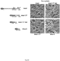
- Inka1-GFP was used to monitor the process crystal formation in living cells. Similar derivatives of Inka1. will allow us to study the effects of PAK4 inhibition in cells and model organisms, to allow better validation of therapeutic agents targeting PAK4.
- PAK isoforms are categorized into two groups on the basis of their structural and biochemical features: the conventional or group I PAKs in human comprise PAKs1-3, while the group II PAKs (PAK4-6) are encoded by three genes in mammals.
- PAK4-like kinases are ubiquitously expressed in metazoans, but not found in protozoa or fungi. This is consistent with PAK4 functioning primarily at cell-cell contacts in mammalian cells, with Cdc42 also being required for adherent junction formation.
- the phenotype of PAK4-null mice which is embryonic lethal, involves defects in the fetal heart as well as in neuronal development and axonal outgrowth 8 . The loss of PAK4 prevents proper polarization and thus formation of the endothelial lumen 9 , consistent with defects seen in PAK4 -/- mice.
- PAK4 is a kinase with strong links to cellular transformation and cancer metastasis.
- the structural basis for PAK4's preference for serine containing substrate sites has recently been elucidated.
- Cdc42 directly regulates PAK4 activity in mammalian cells through an auto-inhibitory domain (AID) that binds in a manner similar to pseudo-substrates 1,2 .
- AID auto-inhibitory domain
- PAK1 activation in vivo occurs through activation loop Thr-423 phosphorylation, it is notable that PAK4 is constitutively phosphorylated on Ser-474 1 , and kept in check through the intra-molecular association of the AID.
- the binding of Cdc42 can serve to activate PAK4 in cells, but it is unclear if there is any auto-phosphorylation event associated with this activation 1 . Since PAK4 does not appear to utilize adaptors we investigated the possibility that Inka1, first identified as a PAK4 binding protein in frogs, might fulfill this role.
- Inka proteins are in fact endogenous inhibitors of PAK4, with the two human Inka isoforms sharing a high degree of sequence identity in the region previously termed the Inca box.
- Inka1 contains an additional PAK4 inhibitory sequence at its C-terminus, and either of these sequences can promote crystallization of the catalytic domain of human PAK4 in mammalian cells.
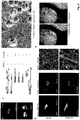
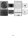
- An in-cellulo protein structure, from X-ray experiments on single crystals formed within a mammalian cell reveals a hexagonal array the PAK4cat subunits that was suggestive of an ability accommodate other proteins in the lattice. This was demonstrated by fusing Inka1 to GFP. Because of these features the PAK4 array has potential as a protein analogue of 'crystalline molecular flasks' in which guest molecules can reside to facilitate their X-ray analysis 7 .
- protein crystal it is meant to refer to a form of the solid state of matter having a three-dimensional crystal lattice, which is distinct from the amorphous or semi-crystalline state. Crystals display characteristic features, including a lattice structure, characteristic shapes and optical properties, such as, e.g., birefringence. Determination as to whether a protein is in a crystalline state may be carried out by any method known in the art, e.g., X-ray diffraction or powder X-ray diffraction or transmission electron microscopy (TEM).
- TEM transmission electron microscopy
- X-ray crystallography is a fundamental tool used for identifying the atomic and molecular structure of many materials which can form crystals, such as metals or minerals, as well as various inorganic, organic and biological molecules.
- crystals such as metals or minerals
- various inorganic, organic and biological molecules For example, the three-dimensional structure of a protein determines its function; consequently, structural insights into proteins at atomic resolution are important to understand the machinery of life or to develop new specifically designed drugs for medical applications.
- This technique requires sufficiently large crystals to obtain structural insights at atomic resolution, routinely obtained in vitro by time-consuming screening.
- successful structural information can be obtained from tiny protein microcrystals grown within living cells, offering exciting new possibilities for proteins that do not form crystals in vitro.
- crystal lattice is formed by the protein which makes and maintains most of the crystal contacts within the lattice, and that the crystal lattice itself may be altered by the presence of a second protein. Assuming there was an alteration, such an altered crystal lattice is included in our definition of "crystal lattice".
- Co-crystallization may also be used to define and describe the crystallization of the two proteins. It is defined as two different materials crystallizing into the same crystalline lattice. For example, a monovalent cation, divalent cation or polycation may crystallize into the same crystalline lattice as a protein having a negatively-charged side chains.
- co-crystals is meant a complex of the compound, molecular scaffold, or ligand bound non-covalently to the target molecule and present in a crystal form appropriate for analysis by X-ray or protein crystallography. The entire protein crystal comprising the two proteins may be co-expressed from a single (or more) nucleic acid construct.
- the said "space” may be utilized to accommodate the second protein.
- it may allow the second protein to pack in an ordered manner (or in any manner depending on its interaction with the first protein) into the crystal lattice of the first protein, which may be used as a "scaffold" molecule.
- first and second proteins in cellulo or in vitro.
- the first and second proteins may form a single protein chain, or may be from separate entities or polypeptide chains.
- any nucleic acid(s) that encode the protein crystal may be from one or more nucleic acid construct.
- the moiety is fused with either the first or second protein.
- the moiety may not be crystallised.
- the moiety is fused to iBox or iBox-C of Inkal.
- the moiety is a protein of interest likely having a molecular mass of less than 30 kDa.
- the moiety may also be a reporter molecule.
- the reporter molecule may be any one selected from the group comprising: fluorescent proteins, tags recognized by monoclonal antibodies, genetically encoded biosensors and the like.
- the molecules may be selected to respond to changes in intracellular or in-vitro environments, or externally applied chemicals or drugs.
- the present invention may be used for performing high throughput screening of crystallization of target materials, proteins, or any other moiety.
- Potential fields of use include microbiology, chemical synthesis, high throughput screening, drug discovery, medical diagnostics, pathogen identification, and enzymatic reactions.
- the present invention may be used to do exhaustive screening of protein crystallization conditions. This screening may be done in a random or systematic way. Alternatively, where high throughput screening in accordance with embodiments of the present invention does not produce crystals of sufficient size for direct X-ray crystallography, the crystals can be utilized as seed crystals for further crystallisation experiments. Promising screening results can also be utilized as a basis for further screening focusing on a narrower spectrum of crystallisation conditions, in a manner analogous to the use of standardised sparse matrix techniques.
- the protein crystal forms a hexagonal array with channels of 80 ⁇ in diameter.
- the ratio of the first protein to the second protein 1:1.
- each first and second protein may contain domains that allows it to dimerize or multimerize with each other and/or to other proteins.
- the domain that functions to dimerize or multimerize the proteins can either be a separate domain, or alternatively can be contained within one of the other domains of the protein.
- such dimeric proteins result in a protein crystal having available space in its lattice structure to accommodate the moiety.
- the moiety or combination of moieties may be of any suitable size.
- the moiety may have a molecular size of less than 30kDa.
- the moiety may have a molecular size of more than 30kDa, for example the molecular size of the moiety may be 40kDa, 50kDa, 60kDa, 65kDa or more.
- Dimerization or multimerization can occur between or among two or more of the proteins through dimerization or multimerization domains. Alternatively, dimerization or multimerization of the proteins can occur by chemical crosslinking. The dimers or multimers that are formed can be homodimeric/homomultimeric or heterodimeric/heteromultimeric.
- a “dimerization domain” is formed by the association of at least two amino acid residues or of at least two peptides or polypeptides (which may have the same, or different, amino acid sequences).
- the peptides or polypeptides may interact with each other through covalent and/or non-covalent association(s).
- Preferred dimerization domains contain at least one cysteine that is capable of forming an intermolecular disulfide bond with a cysteine on the partner protein.
- the dimerization domain can contain one or more cysteine residues such that disulfide bond(s) can form between the partner proteins.
- dimerization domains contain one, two or three to about ten cysteine residues.
- Additional exemplary dimerization domain can be any known in the art and include, but not limited to, coiled coils, acid patches, zinc fingers, calcium hands, a C H 1-C L pair, an "interface” with an engineered “knob” and/or “protruberance” as described in US Patent No. 5,821,333 , leucine zippers (e.g., from jun and/or fos) ( US Patent No.
- NGF nerve growth factor
- NT-3 neurotrophin-3
- IL-8 interleukin-8
- VEGF vascular endothelial growth factor
- VEGF-C vascular endothelial growth factor
- VEGF-D vascular endothelial growth factor
- PDGF vascular endothelial growth factor
- BDNF brain-derived neurotrophic factor
- a “multimerization domain” is a domain that causes three or more peptides or polypeptides to interact with each other through covalent and/or non-covalent association(s).
- Suitable multimerization domains include, but are not limited to, coiled-coil domains.
- a coiled-coil is a peptide sequence with a contiguous pattern of mainly hydrophobic residues spaced 3 and 4 residues apart, usually in a sequence of seven amino acids (heptad repeat) or eleven amino acids (undecad repeat), which assembles (folds) to form a multimeric bundle of helices.
- Coiled-coils with sequences including some irregular distribution of the 3 and 4 residues spacing are also contemplated.
- Hydrophobic residues are in particular the hydrophobic amino acids Val, Ile, Leu, Met, Tyr, Phe and Trp. Mainly hydrophobic means that at least 50% of the residues must be selected from the mentioned hydrophobic amino acids.
- the coiled coil domain may be derived from laminin.
- the heterotrimeric coiled coil protein laminin plays an important role in the formation of basement membranes. Hence, the multifunctional oligomeric structure is required for laminin function.
- Coiled coil domains may also be derived from the thrombospondins in which three (TSP-1 and TSP-2) or five (TSP-3, TSP-4 and TSP-5) chains are connected, or from COMP (COMPcc) ( Guo, et at., EMBO J., 1998, 17: 5265-5272 ) which folds into a parallel five- stranded coiled coil ( Malashkevich ,et al., Science, 274: 761-765 (1996 )). Additional coiled- coil domains derived from other proteins, and other domains that mediate polypeptide multimerization are known in the art and are suitable for use in the present proteins.
- the expression of the protein and the subsequent crystallization occur in cellulo.
- the protein and crystallization of the protein occurs in a mammalian cell.
- the mammalian cell may be any cell, including one that may be a part of a non-human transgenic animal.
- the recombinant kinase and inhibitor proteins are made and purified from other species, such as E.coli, and mixed to promote crystallization either in-vivo or in-vitro.
- the crystal may be of any size that is suitable for X-ray crystallography.
- the crystal is >50 ⁇ m in length and the crystal structure determined at ⁇ 3 ⁇ resolution.
- the present invention makes use of a PAK4 scaffold to generate high quality protein crystals in mammalian cells by co-expression with inhibitory protein Inkal (or a fragment thereof) fused to a protein of interest (third party protein or any moiety of choice).
- one or more isolated polypeptide molecule having a sequence or sequences that encode a protein or proteins which, upon crystallisation, form a protein crystal according to the first aspect of the present invention.
- the protein crystal may be expressed in a single or separate construct expression system.
- the protein molecules may be full-length or fragments thereof, so long as these sequences promote crystallization.
- the kinase PAK4 may be any suitable sequence and its inhibitor Inkal may contain any inhibitory sequence.
- a variant or mutation to the protein sequences could be used to promote crystallization wherein at one or more positions there have been insertions, deletions, or substitutions, either conservative or non-conservative, provided that such changes result in a sequence whose basic properties, for example promoting crystallization have not significantly been changed. "Significantly" in this context means that one skilled in the art would say that the properties of the variant may still be different but would not be unobvious over the ones of the original protein sequences.
- a fusion protein comprising: (a) a first protein, upon crystallisation, yields a crystal having available space in the lattice, the first protein being a p21-activated kinase 4, PAK4 or a catalytic domain thereof; and (b) a second protein crystal to be accommodated, upon crystallisation, in the available space in the lattice, the second protein being an iBox of Inka1, the first and second proteins are co-expressed from one or more nucleic acid construct, wherein the lattice further accommodates a moiety in the available space.
- the fusion protein may be in a single or separate construct expression system.
- the fusion protein additionally contain a domain that allows it to dimerize or multimerize with each other and/or to other proteins.
- nucleic acid molecule having a sequence or sequences that encode a protein or proteins which, upon crystallisation, form a protein crystal according to the first aspect of the present invention.
- an expression vector or vector combinations or a cultured host cell harbouring one or more isolated nucleic acid molecule in an example, there is provided an expression vector or vector combinations or a cultured host cell harbouring one or more isolated nucleic acid molecule.
- the native and mutated kinase and/or kinase inhibitor polypeptides described herein may be chemically synthesized in whole or part using techniques that are well-known in the art.
- a variety of host-expression vector systems may be utilized to express the kinase-inhibitor coding sequence. These include but are not limited to microorganisms such as bacteria transformed with recombinant bacteriophage DNA, plasmid DNA or cosmid DNA expression vectors containing the coding sequence; yeast transformed with recombinant yeast expression vectors containing the domain coding sequence; insect cell systems infected with recombinant virus expression vectors (e.g., baculovirus) containing the coding sequence; plant cell systems infected with recombinant virus expression vectors (e.g., cauliflower mosaic virus, CaMV; tobacco mosaic virus, TMV) or transformed with recombinant plasmid expression vectors (e.g., Ti plasmid) containing the coding sequence; or animal cell systems.
- the expression elements of these systems vary in their strength and specificities.
- any of a number of suitable transcription and translation elements may be used in the expression vector.
- inducible promoters such as pL of bacteriophage ⁇ , plac, ptrp, ptac (ptrp-lac hybrid promoter) and the like may be used; when cloning in insect cell systems, promoters such as the baculovirus polyhedrin promoter may be used; when cloning in plant cell systems, promoters derived from the genome of plant cells (e.g., heat shock promoters; the promoter for the small subunit of RUBISCO; the promoter for the chlorophyll a/b binding protein) or from plant viruses (e.g., the 35 S RNA promoter of CaMV; the coat protein promoter of TMV) may be used; when cloning in mammalian cell systems, promoters derived from the genome of
- a method for producing a protein crystal structure or a fusion protein comprising a first protein, upon crystallisation, yields a crystal having available space in the lattice, the first protein being a p21-activated kinase 4, PAK4, or a catalytic domain thereof; and a second protein is accommodated, upon crystallisation, in the available space in the lattice, the second protein being an iBox of Inka1, and the method comprising culturing a host cell under conditions that allow for the expression and/or production of the protein crystal or fusion protein, the first and second protein are co-expressed from one or more nucleic acid construct, wherein the crystal further accommodates a moiety in the available space in the lattice.
- the host cell may be a mammalian cell.
- the optimal conditions can be selected to allow for crystallization in-vitro from purified proteins.
- the method further comprises fusing a moiety with the second protein, wherein the moiety is accommodated, upon crystallisation, in the available space in the lattice.
- the moiety may not be crystallised but may be a part of the crystal lattice structure.
- the moiety may be fused with the first protein.
- the moiety being a protein of interest may have a molecular mass less than 30kDa and may further comprise a reporter molecule fused to it.
- the method further comprises isolating and purifying the protein crystal.
- the method further comprising obtaining structural data on the crystal.
- the crystals are generated in mammalian cells so that they are of sufficient quality for X-ray structural analysis.
- Computer models such as homology models (i.e., based on a known, experimentally derived structure) can be constructed using data from the co-crystal structures.
- preferred co-crystal structures for making homology models contain high sequence identity in the binding site of the protein sequence being modeled, and the proteins will preferentially also be within the same class and/or fold family. Knowledge of conserved residues in active sites of a protein class can be used to select homology models that accurately represent the binding site.
- Homology models can also be used to map structural information from a surrogate protein where an apo or co-crystal structure exists to the target protein.
- Virtual screening methods such as docking, can also be used to predict the binding configuration and affinity of scaffolds, compounds, and/or combinatorial library members to homology models.
- Virtual screening methods such as docking, can also be used to predict the binding configuration and affinity of scaffolds, compounds, and/or combinatorial library members to homology models.
- Using this data, and carrying out "virtual experiments" using computer software can save substantial resources and allow the person of ordinary skill to make decisions about which compounds can be suitable scaffolds or ligands, without having to actually synthesize the ligand and perform co-crystallization. Decisions thus can be made about which compounds merit actual synthesis and co-crystallization.
- An understanding of such chemical interactions aids in the discovery and design of drugs that interact more advantageously with target proteins and/or are more selective for one protein family member over others. Thus, applying these principles, compounds with superior properties can be discovered.
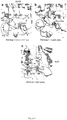
- plasmid constructs All plasmid constructs were generated by PCR-based DNA amplification and inserts completely sequenced.
- the mammalian pXJ40-based vector with Flag, HA and GFP fusion tags are contain a standard CMV-derived promoter and ⁇ -globin 5' intron sequence.
- Inka1 constructs were cloned in pXJ-HA (as indicated in Fig. 1 and 2 ) or pXJ-GFP ( Fig. 6 ), while PAK1 and PAK4 were cloned in pXJ-Flag.
- pGEX4T1 GE
- pET28a Novagen
- pSY5 His tagged
- the 13-residue peptide PAK substrate Raf1(S338) PRGQRDSSYYWEI (Raf13p) was as previously described 1 .
- Recombinant proteins were expressed in Escherichia coli BL21-CodonPlus(DE3) (Stratagene) grown at 30°C. The bacteria were grown to an optical density of 0.6 (OD 600 nm) before induction with 1.0 mM IPTG. Induction was carried out for 3 hours at RT, or 16 hours at 4°C. Bacterial lysates were purified with GSH-Sepharose (GE) or nickel Ni-NTA-Agarose (Qiagen) columns to extract the overexpressed proteins.
- GSH-Sepharose GE
- Ni-NTA-Agarose Qiagen
- the recombinant proteins were eluted in 50 mM Tris-HCl, pH 8.0, 150 mM NaCl, 0.5% Triton X-100, 10% glycerol with 5 mM glutathione (for GST fusions) or 250 mM imidazole (for poly-histidine tagged proteins). With PAK kinases the elution buffer was supplemented with 1 mM MgCl 2 . Proteins were diluted and snap frozen in aliquots prior to use. SDS-PAGE and Coomassie Brilliant Blue staining assessed protein purity to be greater than 90%.
- plasmid DNA typically, a total of 5 ⁇ g plasmid DNA was used per 60 mm dish; lysates were harvested 18 h later in ice cold lysis buffer (0.5 ml; 25 mM HEPES pH 7.3, 100 mM KCl, 5 mM MgCl 2 , 20 mM ⁇ -glycerophosphate, 5% glycerol, 0.5% Triton-X100, 5 mM DTT, 0.5 mM PMSF, 1 mM Na 3 VO 4 and x1 protease inhibitor cocktail (Roche)).
- ice cold lysis buffer 0.5 ml; 25 mM HEPES pH 7.3, 100 mM KCl, 5 mM MgCl 2 , 20 mM ⁇ -glycerophosphate, 5% glycerol, 0.5% Triton-X100, 5 mM DTT, 0.5 mM PMSF, 1 mM Na 3 VO 4 and x1
- the lysates were clarified by centrifugation (14,000 g) and the clarified lysates were incubated while rolling (2 h) with 20 ⁇ l M2 anti-Flag Sepharose (Sigma-Aldrich, A2220). Rabbit anti-Flag (Sigma-Aldrich, F7425) or HRP coupled anti-HA (Santa Cruz Biotechnology, sc-7392 HRP,1 ⁇ g/ml) were used for Western analysis.
- the cells were harvested 3 days after transfection by incubating in PBS with 0.125% (w/v) trypsin and 25% (v/v) glycerol (Merck) for 30 minutes. Individual cells containing single crystals were then mounted in 0.1-0.2 mm cryoloops (Hampton Research) and flash-cooled in liquid nitrogen.
- 6His-PAK4cat protein was purified under standard conditions using a semi-automated Akta system 11 .
- the crystallization of 6His-PAK4cat was carried by hanging drop at 5 mg/ml with 15 fold molar excess of the iBox 23mer synthetic peptide, AEDWTAALLNRGRSRQPLVLGDW, and two times molar excess of ATP.
- Bipyramidal-shaped crystals grew in 0.1 M Tris-HCl, pH 8.5, 12% PEG 8,000 at 25°C. Crystals were supplemented by 15% glycerol and flash-cooled in liquid nitrogen.
- X-ray data were collected at wavelength of 0.9686 ⁇ on I24 of the Diamond Light Source and structure solution and refinement carried out as documented for the in cellulo crystals.
- the cells were imaged at 60x 1.4 NA objective at 2 min intervals.
- SIM and confocal imaging cells were fixed in non-hardening mounting media (Vectashield).
- the slides were imaged by Delta vision OMX SIM with a 100X 1.4 NA objective.
- Confocal imaging used an Olympus FV1000 upright system with 60X 1.42NA objective.
- the 3D stacks were analyzed by IMARIS software.
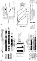
- Inka1 is an endogenous PAK4 inhibitor.
- One Xenopus PAK4 binding protein originally identified through a yeast two-hybrid screen is a 30 kDa neural crest enriched protein termed Inka1 [previously Inca 8,9 ], although the role of this putative adaptor was not determined.
- the protein is also designated FAM212a and FAM212b in the protein database based on their common central 38 amino acid sequence (166-203) here termed the Inka box (iBox, Figure1a ).
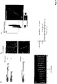
- Inka1 likely acts also on PAK5 and PAK6 since their substrate binding pockets are essentially identical. In vitro measurements indicate GST-Inka1 has a Ki of 30 nM ( Fig. 1d ), which is comparable with the avidity of PKI for PKA.
- the iBox sequence ( Fig. 1a ) contains the tripeptide PLV in common with the PAK4-AID, which binds in the substrate-docking site 2,10 .
- Inka1 has two functional inhibitory regions
- Inka1 functions as an In hibitor of k inase a ctivity; given that it lacks sequence conservation outside these PAK4 inhibitory motifs (the iBox or iBox-C) it seems likely the main function of the protein is to negatively regulate PAK4 activity.
- Deletion of either Inka1 or Inka2 cause subtle defects in frog and mouse development 8,9 , not inconsistent with human Inka1 being causative in a chromosomal micro-deletion being associated with cleft lip and CNS abnormalities.
- Inka1 is expressed in a number of cell types in the early mouse embryo 8 .
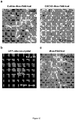
- Inka1 forms crystals with PAK4 in cells.
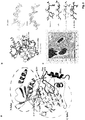
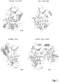
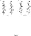
- PAK4cat adopts a typical 'closed' active kinase conformation that includes ATP bound to two magnesium ions.
- the activation (A) loop Ser474 is phosphorylated, and the central region of the iBox is packed against the kinase through both main chain and side chain interactions ( Fig. 3a ).
- the side chain of PAK4 Arg359 which lies at the end of the ⁇ C helix, stabilizes the catalytic competent state by interacting with the phospho-Ser474.
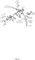
- the disposition of the core Inka1 sequence (RSRQPLVLGD) in the current structure shows docking in to the substrate binding pocket (primarily via R-2 and R-4 interactions, Fig. 4c ) and the inhibitor chain runs parallel to, and hydrogen bonds with, several main chain residues of the activation loop in a beta sheet-like manner ( Fig 3a ).
- Comparison of the PAK4-bound iBox structure ( Fig. 3a and b ) with that of the PAK4 AID PAK4 (Wang et al, 2013) reveals a common geometry underlying the inhibition.
- the iBox and AID core sequences resemble a bound consensus substrate peptide, however the iBox and AID contain a proline residue in place of target serine designated Ser(0).
- an alanine occupies the Ser(0) and again basic residues at the -2 and -3 positions are critical for kinase domain interaction in the substrate-binding pocket (RRNA(0)IHD) in PKI ⁇ .
- the AID and Inka1 structures similarly feature Arg-mediated salt bridges that bind an acidic pocket, and hydrophobic side chain interactions at the +2 and +3 positions.
- Inka1 binds to PAK4 in a substrate-like manner
- the crystal packing resembles that obtained for a short (346 residue) isoform of full-length PAK4 2 in which the N-terminal regulatory region is largely disordered, excepting the pseudosubstrate like peptide (4FIG).
- one set of crystal contacts is formed by the interaction between neighboring N-lobes that involves the two helices from one N-lobe interacting with the ⁇ -sheet of the adjacent N-lobe, an interaction area of 768 ⁇ 2 .
- the N-lobe interactions form strands that run the length of the crystal ( Fig. 5b ).
- the hexagonal packing requires that the N-lobe to be in a 'closed' state relative to the C-lobe, which is likely achieved through 'clamping' of the Inka1 inhibitory region.
- the PAK5cat sequence is slightly different at this interface, and thus does not generate in cellulo crystals with Inka1.
- the second set of contacts lies at the 3-fold axis mediated by the PAK4cat C-lobes involving primarily hydrophobic residues; each C-lobe contributes 576 ⁇ 2 to this crystal contact ( Fig. 5c ).
- the iBox is not involved in crystal contacts and is exposed to the large 80 ⁇ diameter central solvent channels that run the length of the crystals ( Fig. 5a ).
- the two molecules in the asymmetric unit of the P3 parent crystals possess the central channel and share similar packing to the single molecule in the asymmetric unit of the in cellulo P6 3 crystals.
- Both P3 and P6 3 crystals are able to accommodate larger constructs beyond the PAKcat domain that forms the entire crystal packing, namely the N-terminus of PAK4 and Inka1 sequences, respectively.
- the present invention includes any mutation to the protein sequences of the kinase and its inhibitor.
- mutation of the PAK4 sequence such that amino-acid changes at the kinase-kinase interface may increase (a) the stability of the crystal lattice, or (b) increases or alters the properties of the crystallization in cells or in vitro.
- the residues that may be mutated are shown in Figure 5 (for example mutations of L422 to F or A307 to V), which increase the extent of the hydrophobic interface between the C-lobe or the N-lobe interfaces - without disrupting the protein crystal structure.
- PF-03758309 appears to allow PAK4cat to incorporate with sub-stoichiometric levels of GFP-Inka1, consistent PF-03758309 either reducing the affinity of GFP-Inka1 or allowing PAK4cat incorporation without Inka1.
- the average crystal growth along the length ( Fig. 6d ) was 4.2 +/- 1.2 ⁇ m/hour, which equates to adding a new layer of crystal lattice every three seconds comprised of ⁇ 50,000 protein units (for a crystal with 2 ⁇ m cross section). Crystal growth slowed after PF3758309 addition. Based on this analysis we observed PAK4cat incorporated at both ends of the crystal ( Fig. 6d ).
- Inka1 is a potent vertebrate inhibitor of PAK4 with a Ki of ⁇ 30 nM ( Figure 1 ), which has a much higher affinity than the corresponding AID.
- Inka1 contains two copies of the kinase inhibitory domain, and both of these small regions of themselves can support PAK4cat crystal formation in cells ( Fig 4 ).
- Inka represents one of only six classes of established endogenous protein kinase inhibitors to be uncovered to date. It is likely that more remain to be found among the plethora of orphan open reading frames in the human genome, however none of these different proteins share sequence homology.
- Inka1 represents one of four whose basis of inhibition is understood at the structural level.
- the three members of the PKA inhibitor family termed PKls, are proteins of ⁇ 100 residues sharing an N-terminal region of 25 amino acids, which interact with the PKAc catalytic domain as illustrated in Figure 7 .
- PKI ⁇ is required for export of PKA catalytic subunits from the nucleus back to the cytoplasm following activation of PKA in the brain.
- PKI proteins can be found in many invertebrates (cf. K09E9.4 in C. elegans ) but not in certain groups such as Drosophila.
- CaM-KIIN Two closely related Ca 2+ calmodulin-dependent protein kinase II inhibitors (CaM-KIIN) of 78 and 79 amino acids have been characterized, and show ⁇ 50 nM Ki in vitro.
- the best-studied endogenous inhibitors are cyclin-dependent kinase (CDK) inhibitors.
- CDK cyclin-dependent kinase
- the INK4 gene family encodes p16INK4a, p15INK4b, p18INK4c, and p19INK4d, all bind to CDK4 and CDK6 and block their association with D-type cyclins.
- the INK4 inhibitor structure is different from the others described here, in being well folded in the absence of kinase ( Fig 7 ).
- the Cip/Kip family members vary widely in size and comprise p21 Cip1/Waf1/Sdi1, p27Kip1, and p57Kip2.
- Inka1 and Inka2 are nuclear localized proteins ( FIG. 2 ), which can be coimmunoprecipitated with Pak4, particularly when the kinase is in an open active state.
- Inka proteins share sequence homology only in the region that binds to PAK4, which was termed the Inca box, however we demonstrate that Inka1 (but not Inka2) contains two related functional PAK4 inhibitory modules.
- Inka1-LacZ allele expression in mice indicates expression in the cephalic mesenchyme, heart, and paraxial mesoderm prior to E8.5.
- Inka1 plays a redundant role in regulating PAK4 activity, and may well be compensated by Inka2 in mice.
- a coral fluorescent protein that forms diffraction-quality micron-sized crystals within mammalian cells is recently reported 6 . These crystals assemble much more quickly and likely recognized as foreign, since they are processed as autophagic cargos. By contrast our crystals form at a modest pace in the cellular context, and grow for 6-16h suggesting they are well tolerated in the cytosol over this time period.
- the complex between PAK4 and Inka1 is the first human protein structure to be solved within mammalian cells, and further, multiple constructs of Inka1 or fusions to other proteins can be incorporated into the PAK4 crystal lattice ( Fig. 2 and 6 ). Crystals have been grown in a variety of mammalian cell types, monkey COS-7 and human HeLa and HEK293 ( Figure 12 ).
Landscapes
- Chemical & Material Sciences (AREA)
- Health & Medical Sciences (AREA)
- Organic Chemistry (AREA)
- Life Sciences & Earth Sciences (AREA)
- Engineering & Computer Science (AREA)
- Zoology (AREA)
- Genetics & Genomics (AREA)
- General Health & Medical Sciences (AREA)
- Biochemistry (AREA)
- Bioinformatics & Cheminformatics (AREA)
- Wood Science & Technology (AREA)
- Molecular Biology (AREA)
- Crystallography & Structural Chemistry (AREA)
- Medicinal Chemistry (AREA)
- General Engineering & Computer Science (AREA)
- Metallurgy (AREA)
- Materials Engineering (AREA)
- Physics & Mathematics (AREA)
- Gastroenterology & Hepatology (AREA)
- Biophysics (AREA)
- Proteomics, Peptides & Aminoacids (AREA)
- Toxicology (AREA)
- Microbiology (AREA)
- Biotechnology (AREA)
- Biomedical Technology (AREA)
- Geometry (AREA)
- Analytical Chemistry (AREA)
- General Physics & Mathematics (AREA)
- Immunology (AREA)
- Pathology (AREA)
- Peptides Or Proteins (AREA)
- Enzymes And Modification Thereof (AREA)
Claims (15)
- Cristal de protéine comprenant :(a) un premier cristal de protéine dont le réseau comporte un espace disponible, la première protéine étant une kinase 4 activée par p21, PAK 4, ou un domaine catalytique de celles-ci ; et(b) un deuxième cristal de protéine destiné à être reçu dans l'espace disponible dans le réseau, la deuxième protéine étant un iBox d'Inka1, les première et deuxième protéines étant co-exprimées à partir d'une ou plusieurs constructions d'acide nucléique,
le cristal recevant en outre une fraction dans l'espace disponible dans le réseau. - Cristal de protéine selon la revendication 1, dans lequel la fraction est une protéine d'intérêt.
- Cristal de protéine selon la revendication 2, dans lequel la fraction est fusionnée au iBox ou iBox-C d'Inka1, et a une masse moléculaire inférieure à 30 kDa.
- Cristal de protéine selon l'une quelconque des revendications précédentes, dans lequel la fraction comprend en outre une molécule reporter, et la molécule reporter est n'importe quelle molécule sélectionnée dans le groupe comprenant : des protéines fluorescentes, des étiquettes reconnues par des anticorps monoclonaux, et des biocapteurs génétiquement codés.
- Cristal de protéine selon l'une quelconque des revendications précédentes, dans lequel le rapport de la première protéine contre la deuxième protéine est de 1:1 et le cristal de protéine forme un réseau hexagonal avec des canaux de 80 Å de diamètre.
- Cristal de protéine selon l'une quelconque des revendications précédentes, le cristal de protéine étant formé in cellulo dans une cellule mammifère.
- Cristal de protéine selon l'une quelconque des revendications précédentes, le cristal mesurant plus de 50 µm de long et la structure cristalline étant déterminée à une résolution inférieure à 3 Å.
- Protéine de fusion, la protéine de fusion comprenant :(a) une première protéine qui, lors de la cristallisation, produit un cristal dont le réseau comporte un espace disponible, la première protéine étant une kinase 4 activée par p21, PAK 4, ou un domaine catalytique de celles-ci ; et(b) un deuxième cristal de protéine destiné à être reçu, lors de la cristallisation, dans l'espace disponible dans le réseau, la deuxième protéine étant un iBox d'Inka1, les première et deuxième protéines étant co-exprimées à partir d'une ou plusieurs constructions d'acide nucléique, le réseau étant capable de recevoir une fraction dans l'espace disponible.
- Protéine de fusion selon la revendication 8, dans laquelle la fraction est une protéine d'intérêt.
- Une ou plusieurs molécules isolées de polypeptide ou d'acide nucléique ayant une ou plusieurs séquences qui codent une ou plusieurs protéines qui, lors de la cristallisation, forment un cristal de protéine selon l'une quelconque des revendications 1 à 7.
- Cellule hôte comprenant une ou plusieurs molécules isolées de polypeptide ou d'acide nucléique selon la revendication 10 ou un vecteur d'expression contenant une ou plusieurs molécules isolées d'acide nucléique selon la revendication 10.
- Procédé de production d'une structure de cristal de protéine ou d'une protéine de fusion comprenant une première protéine qui, lors de la cristallisation, produit un cristal dont le réseau comporte un espace disponible, la première protéine étant une kinase 4 activée par p21, PAK 4, ou un domaine catalytique de celles-ci, et une deuxième protéine reçue, lors de la cristallisation, dans l'espace disponible dans le réseau, la deuxième protéine étant un iBox d'Inka1, le procédé comprenant :
la mise en culture d'une cellule hôte dans des conditions qui permettent la production du cristal de protéine ou de la protéine de fusion, dans lequel les première et deuxième protéines sont co-exprimées à partir d'une ou plusieurs constructions d'acide nucléique, et le cristal reçoit en outre une fraction dans l'espace disponible dans le réseau. - Procédé selon la revendication 12, dans lequel la co-expression et/ou les conditions de cristallisation est/sont réalisées in vitro.
- Procédé selon la revendication 12, dans lequel la fraction est une protéine d'intérêt.
- Procédé selon l'une quelconque des revendications 12 à 14, comprenant en outre une fusion de la fraction avec la deuxième protéine ou une fusion de la fraction avec une molécule reporter, dans lequel la fraction est une protéine d'intérêt ayant une masse moléculaire inférieure à 30 kDa.
Applications Claiming Priority (2)
| Application Number | Priority Date | Filing Date | Title |
|---|---|---|---|
| SG10201508927SA SG10201508927SA (en) | 2015-10-29 | 2015-10-29 | Protein crystal |
| PCT/SG2016/050533 WO2017074268A1 (fr) | 2015-10-29 | 2016-10-31 | Cristal de protéine hybride comprenant une fraction |
Publications (3)
| Publication Number | Publication Date |
|---|---|
| EP3368552A1 EP3368552A1 (fr) | 2018-09-05 |
| EP3368552A4 EP3368552A4 (fr) | 2019-03-27 |
| EP3368552B1 true EP3368552B1 (fr) | 2020-09-09 |
Family
ID=58630754
Family Applications (1)
| Application Number | Title | Priority Date | Filing Date |
|---|---|---|---|
| EP16860402.3A Active EP3368552B1 (fr) | 2015-10-29 | 2016-10-31 | Cristal de protéine hybride comprenant une fraction |
Country Status (5)
| Country | Link |
|---|---|
| US (1) | US10822723B2 (fr) |
| EP (1) | EP3368552B1 (fr) |
| CN (1) | CN108350049A (fr) |
| SG (1) | SG10201508927SA (fr) |
| WO (1) | WO2017074268A1 (fr) |
Families Citing this family (1)
| Publication number | Priority date | Publication date | Assignee | Title |
|---|---|---|---|---|
| US12600999B2 (en) | 2020-02-20 | 2026-04-14 | Institute Of Science Tokyo | Protein crystal production method and crystalline structure analysis method |
Family Cites Families (5)
| Publication number | Priority date | Publication date | Assignee | Title |
|---|---|---|---|---|
| US5837815A (en) | 1994-12-15 | 1998-11-17 | Sugen, Inc. | PYK2 related polypeptide products |
| EP1278875A1 (fr) | 2000-05-05 | 2003-01-29 | Imperial College Innovations Limited | Procedes cristallographiques |
| EP1851310B1 (fr) * | 2005-02-23 | 2011-01-26 | Vertex Pharmaceuticals Incorporated | Structure cristalline de tak1-tab1 |
| US20090317850A1 (en) * | 2008-06-20 | 2009-12-24 | Tomoko Sunami | Crystal Structure of Human 70KD Ribosomal Protein S6 Kinase 1 Kinase Domain |
| EA201391514A1 (ru) * | 2011-05-13 | 2014-08-29 | Рецептос, Инк | Новые партнеры слияния для кристаллизации рецепторов, связанных с g-белками |
-
2015
- 2015-10-29 SG SG10201508927SA patent/SG10201508927SA/en unknown
-
2016
- 2016-10-31 US US15/771,863 patent/US10822723B2/en not_active Expired - Fee Related
- 2016-10-31 CN CN201680063793.9A patent/CN108350049A/zh active Pending
- 2016-10-31 WO PCT/SG2016/050533 patent/WO2017074268A1/fr not_active Ceased
- 2016-10-31 EP EP16860402.3A patent/EP3368552B1/fr active Active
Non-Patent Citations (1)
| Title |
|---|
| None * |
Also Published As
| Publication number | Publication date |
|---|---|
| CN108350049A (zh) | 2018-07-31 |
| US10822723B2 (en) | 2020-11-03 |
| US20180266014A1 (en) | 2018-09-20 |
| WO2017074268A1 (fr) | 2017-05-04 |
| EP3368552A4 (fr) | 2019-03-27 |
| SG10201508927SA (en) | 2017-05-30 |
| EP3368552A1 (fr) | 2018-09-05 |
Similar Documents
| Publication | Publication Date | Title |
|---|---|---|
| Shirakawa et al. | A SNARE geranylgeranyltransferase essential for the organization of the Golgi apparatus | |
| Baskaran et al. | An in cellulo-derived structure of PAK4 in complex with its inhibitor Inka1 | |
| Chen et al. | The WAVE regulatory complex links diverse receptors to the actin cytoskeleton | |
| Huang et al. | BubR1 phosphorylates CENP-E as a switch enabling the transition from lateral association to end-on capture of spindle microtubules | |
| Korndörfer et al. | The crystal structure of the human (S100A8/S100A9) 2 heterotetramer, calprotectin, illustrates how conformational changes of interacting α-helices can determine specific association of two EF-hand proteins | |
| Polley et al. | A structural basis for IκB kinase 2 activation via oligomerization-dependent trans auto-phosphorylation | |
| Rona et al. | Phosphorylation adjacent to the nuclear localization signal of human dUTPase abolishes nuclear import: structural and mechanistic insights | |
| Huang et al. | Binding of guide piRNA triggers methylation of the unstructured N-terminal region of Aub leading to assembly of the piRNA amplification complex | |
| Taguchi et al. | Structural model of microtubule dynamics inhibition by kinesin-4 from the crystal structure of KLP-12–tubulin complex | |
| EP3368552B1 (fr) | Cristal de protéine hybride comprenant une fraction | |
| Fan et al. | A divergent tumor overexpressed gene domain and oligomerization contribute to SPIRAL2 function in stabilizing microtubule minus ends | |
| Herrada et al. | Purification and structural analysis of LEM-domain proteins | |
| WO2007089899A2 (fr) | Proteine d'un recepteur couple a une proteine g hydrosoluble | |
| US7491523B2 (en) | Voltage-dependent calcium channel beta subunit functional core | |
| Maddumage | Understanding the structural and functional basis of cell polarity regulation in Trichoplax adhaerens | |
| Fukuda et al. | Molecular basis for Ras suppressor-1 recruitment to focal adhesions and stabilization of consensus adhesome complex | |
| Marcianò et al. | Unconventional kinetochore kinases KKT2 and KKT3 have a unique zinc finger that promotes their kinetochore localization | |
| Liu et al. | Homodimerized Arpins: Binding to Arp2/3 Complexes with Positive Synergy to Inhibit Lamellipodia-Dependent Tumor Metastasis | |
| Balikci | Structural Studies of Core Kinase Cascade of Hippo Signalling Pathway | |
| Ngow | Structural characterization of vaccinia-related kinase 1 (VRK1), a histone mitotic kinase | |
| Bianchi | Molecular Mechanisms of Kinesin Regulation | |
| Ahmed et al. | A molecular and cellular view of protein kinase CK2 | |
| Polley et al. | A Structural Basis for IkB Kinase 2 Activation Via Oligomerization-Dependent Trans | |
| O'Donnell | Timing of Events, Reset Mechanism, and Allostery Innate to the GTP Hydrolysis Cycle of Atlastin | |
| Costanzi | Structural analysis of molecular recognition and ligand association processes |
Legal Events
| Date | Code | Title | Description |
|---|---|---|---|
| STAA | Information on the status of an ep patent application or granted ep patent |
Free format text: STATUS: THE INTERNATIONAL PUBLICATION HAS BEEN MADE |
|
| PUAI | Public reference made under article 153(3) epc to a published international application that has entered the european phase |
Free format text: ORIGINAL CODE: 0009012 |
|
| STAA | Information on the status of an ep patent application or granted ep patent |
Free format text: STATUS: REQUEST FOR EXAMINATION WAS MADE |
|
| 17P | Request for examination filed |
Effective date: 20180406 |
|
| AK | Designated contracting states |
Kind code of ref document: A1 Designated state(s): AL AT BE BG CH CY CZ DE DK EE ES FI FR GB GR HR HU IE IS IT LI LT LU LV MC MK MT NL NO PL PT RO RS SE SI SK SM TR |
|
| AX | Request for extension of the european patent |
Extension state: BA ME |
|
| DAV | Request for validation of the european patent (deleted) | ||
| DAX | Request for extension of the european patent (deleted) | ||
| A4 | Supplementary search report drawn up and despatched |
Effective date: 20190221 |
|
| RIC1 | Information provided on ipc code assigned before grant |
Ipc: C12N 9/12 20060101ALI20190215BHEP Ipc: C07K 14/47 20060101AFI20190215BHEP Ipc: C12N 15/62 20060101ALI20190215BHEP |
|
| GRAP | Despatch of communication of intention to grant a patent |
Free format text: ORIGINAL CODE: EPIDOSNIGR1 |
|
| STAA | Information on the status of an ep patent application or granted ep patent |
Free format text: STATUS: GRANT OF PATENT IS INTENDED |
|
| INTG | Intention to grant announced |
Effective date: 20200102 |
|
| GRAJ | Information related to disapproval of communication of intention to grant by the applicant or resumption of examination proceedings by the epo deleted |
Free format text: ORIGINAL CODE: EPIDOSDIGR1 |
|
| STAA | Information on the status of an ep patent application or granted ep patent |
Free format text: STATUS: REQUEST FOR EXAMINATION WAS MADE |
|
| GRAP | Despatch of communication of intention to grant a patent |
Free format text: ORIGINAL CODE: EPIDOSNIGR1 |
|
| STAA | Information on the status of an ep patent application or granted ep patent |
Free format text: STATUS: GRANT OF PATENT IS INTENDED |
|
| INTC | Intention to grant announced (deleted) | ||
| INTG | Intention to grant announced |
Effective date: 20200519 |
|
| GRAS | Grant fee paid |
Free format text: ORIGINAL CODE: EPIDOSNIGR3 |
|
| GRAA | (expected) grant |
Free format text: ORIGINAL CODE: 0009210 |
|
| STAA | Information on the status of an ep patent application or granted ep patent |
Free format text: STATUS: THE PATENT HAS BEEN GRANTED |
|
| REG | Reference to a national code |
Ref country code: DE Ref legal event code: R082 Ref document number: 602016043890 Country of ref document: DE Representative=s name: MATHYS & SQUIRE EUROPE LLP, DE Ref country code: DE Ref legal event code: R082 Ref document number: 602016043890 Country of ref document: DE Representative=s name: MATHYS & SQUIRE GBR, DE Ref country code: DE Ref legal event code: R082 Ref document number: 602016043890 Country of ref document: DE Representative=s name: MATHYS & SQUIRE EUROPE PATENTANWAELTE PARTNERS, DE |
|
| AK | Designated contracting states |
Kind code of ref document: B1 Designated state(s): AL AT BE BG CH CY CZ DE DK EE ES FI FR GB GR HR HU IE IS IT LI LT LU LV MC MK MT NL NO PL PT RO RS SE SI SK SM TR |
|
| REG | Reference to a national code |
Ref country code: GB Ref legal event code: FG4D |
|
| REG | Reference to a national code |
Ref country code: AT Ref legal event code: REF Ref document number: 1311451 Country of ref document: AT Kind code of ref document: T Effective date: 20200915 Ref country code: CH Ref legal event code: EP |
|
| REG | Reference to a national code |
Ref country code: DE Ref legal event code: R096 Ref document number: 602016043890 Country of ref document: DE |
|
| REG | Reference to a national code |
Ref country code: IE Ref legal event code: FG4D |
|
| REG | Reference to a national code |
Ref country code: LT Ref legal event code: MG4D |
|
| REG | Reference to a national code |
Ref country code: DE Ref legal event code: R082 Ref document number: 602016043890 Country of ref document: DE Representative=s name: MATHYS & SQUIRE GBR, DE Ref country code: DE Ref legal event code: R082 Ref document number: 602016043890 Country of ref document: DE Representative=s name: MATHYS & SQUIRE EUROPE PATENTANWAELTE PARTNERS, DE |
|
| PG25 | Lapsed in a contracting state [announced via postgrant information from national office to epo] |
Ref country code: SE Free format text: LAPSE BECAUSE OF FAILURE TO SUBMIT A TRANSLATION OF THE DESCRIPTION OR TO PAY THE FEE WITHIN THE PRESCRIBED TIME-LIMIT Effective date: 20200909 Ref country code: LT Free format text: LAPSE BECAUSE OF FAILURE TO SUBMIT A TRANSLATION OF THE DESCRIPTION OR TO PAY THE FEE WITHIN THE PRESCRIBED TIME-LIMIT Effective date: 20200909 Ref country code: BG Free format text: LAPSE BECAUSE OF FAILURE TO SUBMIT A TRANSLATION OF THE DESCRIPTION OR TO PAY THE FEE WITHIN THE PRESCRIBED TIME-LIMIT Effective date: 20201209 Ref country code: NO Free format text: LAPSE BECAUSE OF FAILURE TO SUBMIT A TRANSLATION OF THE DESCRIPTION OR TO PAY THE FEE WITHIN THE PRESCRIBED TIME-LIMIT Effective date: 20201209 Ref country code: FI Free format text: LAPSE BECAUSE OF FAILURE TO SUBMIT A TRANSLATION OF THE DESCRIPTION OR TO PAY THE FEE WITHIN THE PRESCRIBED TIME-LIMIT Effective date: 20200909 Ref country code: GR Free format text: LAPSE BECAUSE OF FAILURE TO SUBMIT A TRANSLATION OF THE DESCRIPTION OR TO PAY THE FEE WITHIN THE PRESCRIBED TIME-LIMIT Effective date: 20201210 Ref country code: HR Free format text: LAPSE BECAUSE OF FAILURE TO SUBMIT A TRANSLATION OF THE DESCRIPTION OR TO PAY THE FEE WITHIN THE PRESCRIBED TIME-LIMIT Effective date: 20200909 |
|
| REG | Reference to a national code |
Ref country code: AT Ref legal event code: MK05 Ref document number: 1311451 Country of ref document: AT Kind code of ref document: T Effective date: 20200909 |
|
| REG | Reference to a national code |
Ref country code: NL Ref legal event code: MP Effective date: 20200909 |
|
| PG25 | Lapsed in a contracting state [announced via postgrant information from national office to epo] |
Ref country code: LV Free format text: LAPSE BECAUSE OF FAILURE TO SUBMIT A TRANSLATION OF THE DESCRIPTION OR TO PAY THE FEE WITHIN THE PRESCRIBED TIME-LIMIT Effective date: 20200909 Ref country code: RS Free format text: LAPSE BECAUSE OF FAILURE TO SUBMIT A TRANSLATION OF THE DESCRIPTION OR TO PAY THE FEE WITHIN THE PRESCRIBED TIME-LIMIT Effective date: 20200909 Ref country code: PL Free format text: LAPSE BECAUSE OF FAILURE TO SUBMIT A TRANSLATION OF THE DESCRIPTION OR TO PAY THE FEE WITHIN THE PRESCRIBED TIME-LIMIT Effective date: 20200909 |
|
| PG25 | Lapsed in a contracting state [announced via postgrant information from national office to epo] |
Ref country code: EE Free format text: LAPSE BECAUSE OF FAILURE TO SUBMIT A TRANSLATION OF THE DESCRIPTION OR TO PAY THE FEE WITHIN THE PRESCRIBED TIME-LIMIT Effective date: 20200909 Ref country code: SM Free format text: LAPSE BECAUSE OF FAILURE TO SUBMIT A TRANSLATION OF THE DESCRIPTION OR TO PAY THE FEE WITHIN THE PRESCRIBED TIME-LIMIT Effective date: 20200909 Ref country code: RO Free format text: LAPSE BECAUSE OF FAILURE TO SUBMIT A TRANSLATION OF THE DESCRIPTION OR TO PAY THE FEE WITHIN THE PRESCRIBED TIME-LIMIT Effective date: 20200909 Ref country code: PT Free format text: LAPSE BECAUSE OF FAILURE TO SUBMIT A TRANSLATION OF THE DESCRIPTION OR TO PAY THE FEE WITHIN THE PRESCRIBED TIME-LIMIT Effective date: 20210111 Ref country code: CZ Free format text: LAPSE BECAUSE OF FAILURE TO SUBMIT A TRANSLATION OF THE DESCRIPTION OR TO PAY THE FEE WITHIN THE PRESCRIBED TIME-LIMIT Effective date: 20200909 |
|
| PG25 | Lapsed in a contracting state [announced via postgrant information from national office to epo] |
Ref country code: AL Free format text: LAPSE BECAUSE OF FAILURE TO SUBMIT A TRANSLATION OF THE DESCRIPTION OR TO PAY THE FEE WITHIN THE PRESCRIBED TIME-LIMIT Effective date: 20200909 Ref country code: AT Free format text: LAPSE BECAUSE OF FAILURE TO SUBMIT A TRANSLATION OF THE DESCRIPTION OR TO PAY THE FEE WITHIN THE PRESCRIBED TIME-LIMIT Effective date: 20200909 Ref country code: ES Free format text: LAPSE BECAUSE OF FAILURE TO SUBMIT A TRANSLATION OF THE DESCRIPTION OR TO PAY THE FEE WITHIN THE PRESCRIBED TIME-LIMIT Effective date: 20200909 Ref country code: IS Free format text: LAPSE BECAUSE OF FAILURE TO SUBMIT A TRANSLATION OF THE DESCRIPTION OR TO PAY THE FEE WITHIN THE PRESCRIBED TIME-LIMIT Effective date: 20210109 |
|
| REG | Reference to a national code |
Ref country code: CH Ref legal event code: PL |
|
| REG | Reference to a national code |
Ref country code: DE Ref legal event code: R097 Ref document number: 602016043890 Country of ref document: DE |
|
| PG25 | Lapsed in a contracting state [announced via postgrant information from national office to epo] |
Ref country code: SK Free format text: LAPSE BECAUSE OF FAILURE TO SUBMIT A TRANSLATION OF THE DESCRIPTION OR TO PAY THE FEE WITHIN THE PRESCRIBED TIME-LIMIT Effective date: 20200909 Ref country code: LU Free format text: LAPSE BECAUSE OF NON-PAYMENT OF DUE FEES Effective date: 20201031 Ref country code: MC Free format text: LAPSE BECAUSE OF FAILURE TO SUBMIT A TRANSLATION OF THE DESCRIPTION OR TO PAY THE FEE WITHIN THE PRESCRIBED TIME-LIMIT Effective date: 20200909 |
|
| REG | Reference to a national code |
Ref country code: BE Ref legal event code: MM Effective date: 20201031 |
|
| PLBE | No opposition filed within time limit |
Free format text: ORIGINAL CODE: 0009261 |
|
| STAA | Information on the status of an ep patent application or granted ep patent |
Free format text: STATUS: NO OPPOSITION FILED WITHIN TIME LIMIT |
|
| 26N | No opposition filed |
Effective date: 20210610 |
|
| PG25 | Lapsed in a contracting state [announced via postgrant information from national office to epo] |
Ref country code: LI Free format text: LAPSE BECAUSE OF NON-PAYMENT OF DUE FEES Effective date: 20201031 Ref country code: SI Free format text: LAPSE BECAUSE OF FAILURE TO SUBMIT A TRANSLATION OF THE DESCRIPTION OR TO PAY THE FEE WITHIN THE PRESCRIBED TIME-LIMIT Effective date: 20200909 Ref country code: CH Free format text: LAPSE BECAUSE OF NON-PAYMENT OF DUE FEES Effective date: 20201031 Ref country code: DK Free format text: LAPSE BECAUSE OF FAILURE TO SUBMIT A TRANSLATION OF THE DESCRIPTION OR TO PAY THE FEE WITHIN THE PRESCRIBED TIME-LIMIT Effective date: 20200909 Ref country code: BE Free format text: LAPSE BECAUSE OF NON-PAYMENT OF DUE FEES Effective date: 20201031 |
|
| PG25 | Lapsed in a contracting state [announced via postgrant information from national office to epo] |
Ref country code: IE Free format text: LAPSE BECAUSE OF NON-PAYMENT OF DUE FEES Effective date: 20201031 Ref country code: IT Free format text: LAPSE BECAUSE OF FAILURE TO SUBMIT A TRANSLATION OF THE DESCRIPTION OR TO PAY THE FEE WITHIN THE PRESCRIBED TIME-LIMIT Effective date: 20200909 |
|
| PGFP | Annual fee paid to national office [announced via postgrant information from national office to epo] |
Ref country code: DE Payment date: 20211007 Year of fee payment: 6 Ref country code: GB Payment date: 20211010 Year of fee payment: 6 |
|
| PGFP | Annual fee paid to national office [announced via postgrant information from national office to epo] |
Ref country code: FR Payment date: 20211006 Year of fee payment: 6 |
|
| PG25 | Lapsed in a contracting state [announced via postgrant information from national office to epo] |
Ref country code: TR Free format text: LAPSE BECAUSE OF FAILURE TO SUBMIT A TRANSLATION OF THE DESCRIPTION OR TO PAY THE FEE WITHIN THE PRESCRIBED TIME-LIMIT Effective date: 20200909 Ref country code: MT Free format text: LAPSE BECAUSE OF FAILURE TO SUBMIT A TRANSLATION OF THE DESCRIPTION OR TO PAY THE FEE WITHIN THE PRESCRIBED TIME-LIMIT Effective date: 20200909 Ref country code: CY Free format text: LAPSE BECAUSE OF FAILURE TO SUBMIT A TRANSLATION OF THE DESCRIPTION OR TO PAY THE FEE WITHIN THE PRESCRIBED TIME-LIMIT Effective date: 20200909 |
|
| PG25 | Lapsed in a contracting state [announced via postgrant information from national office to epo] |
Ref country code: MK Free format text: LAPSE BECAUSE OF FAILURE TO SUBMIT A TRANSLATION OF THE DESCRIPTION OR TO PAY THE FEE WITHIN THE PRESCRIBED TIME-LIMIT Effective date: 20200909 |
|
| REG | Reference to a national code |
Ref country code: DE Ref legal event code: R119 Ref document number: 602016043890 Country of ref document: DE |
|
| GBPC | Gb: european patent ceased through non-payment of renewal fee |
Effective date: 20221031 |
|
| PG25 | Lapsed in a contracting state [announced via postgrant information from national office to epo] |
Ref country code: NL Free format text: LAPSE BECAUSE OF NON-PAYMENT OF DUE FEES Effective date: 20200923 |
|
| PG25 | Lapsed in a contracting state [announced via postgrant information from national office to epo] |
Ref country code: FR Free format text: LAPSE BECAUSE OF NON-PAYMENT OF DUE FEES Effective date: 20221031 Ref country code: DE Free format text: LAPSE BECAUSE OF NON-PAYMENT OF DUE FEES Effective date: 20230503 |
|
| PG25 | Lapsed in a contracting state [announced via postgrant information from national office to epo] |
Ref country code: GB Free format text: LAPSE BECAUSE OF NON-PAYMENT OF DUE FEES Effective date: 20221031 |