EP3336172A1 - Method for producing antigen specific monoclonal antibody - Google Patents
Method for producing antigen specific monoclonal antibody Download PDFInfo
- Publication number
- EP3336172A1 EP3336172A1 EP16835229.2A EP16835229A EP3336172A1 EP 3336172 A1 EP3336172 A1 EP 3336172A1 EP 16835229 A EP16835229 A EP 16835229A EP 3336172 A1 EP3336172 A1 EP 3336172A1
- Authority
- EP
- European Patent Office
- Prior art keywords
- cells
- antigen
- cell
- interest
- monoclonal antibody
- Prior art date
- Legal status (The legal status is an assumption and is not a legal conclusion. Google has not performed a legal analysis and makes no representation as to the accuracy of the status listed.)
- Granted
Links
- 239000000427 antigen Substances 0.000 title claims abstract description 174
- 108091007433 antigens Proteins 0.000 title claims abstract description 174
- 102000036639 antigens Human genes 0.000 title claims abstract description 174
- 238000004519 manufacturing process Methods 0.000 title claims abstract description 14
- 210000004027 cell Anatomy 0.000 claims abstract description 261
- 238000000034 method Methods 0.000 claims abstract description 110
- 108020004999 messenger RNA Proteins 0.000 claims abstract description 61
- 239000003431 cross linking reagent Substances 0.000 claims abstract description 43
- 239000002299 complementary DNA Substances 0.000 claims abstract description 38
- 210000000628 antibody-producing cell Anatomy 0.000 claims abstract description 35
- AYFVYJQAPQTCCC-UHFFFAOYSA-N Threonine Natural products CC(O)C(N)C(O)=O AYFVYJQAPQTCCC-UHFFFAOYSA-N 0.000 claims abstract description 21
- 239000004473 Threonine Substances 0.000 claims abstract description 21
- 239000012634 fragment Substances 0.000 claims abstract description 21
- 101000777277 Homo sapiens Serine/threonine-protein kinase Chk2 Proteins 0.000 claims abstract description 16
- 102100031075 Serine/threonine-protein kinase Chk2 Human genes 0.000 claims abstract description 16
- 230000002441 reversible effect Effects 0.000 claims abstract description 16
- 239000003599 detergent Substances 0.000 claims description 50
- 210000000170 cell membrane Anatomy 0.000 claims description 43
- 238000011282 treatment Methods 0.000 claims description 43
- 108090000623 proteins and genes Proteins 0.000 claims description 31
- 230000009089 cytolysis Effects 0.000 claims description 30
- 239000003161 ribonuclease inhibitor Substances 0.000 claims description 23
- 238000010186 staining Methods 0.000 claims description 18
- WSFSSNUMVMOOMR-UHFFFAOYSA-N Formaldehyde Chemical group O=C WSFSSNUMVMOOMR-UHFFFAOYSA-N 0.000 claims description 16
- 238000010804 cDNA synthesis Methods 0.000 claims description 16
- 238000002360 preparation method Methods 0.000 claims description 14
- 238000000926 separation method Methods 0.000 claims description 13
- 238000010438 heat treatment Methods 0.000 claims description 11
- 108091005804 Peptidases Proteins 0.000 claims description 9
- 239000004365 Protease Substances 0.000 claims description 9
- 102100037486 Reverse transcriptase/ribonuclease H Human genes 0.000 claims description 9
- 125000006850 spacer group Chemical group 0.000 claims description 8
- 108060003951 Immunoglobulin Proteins 0.000 claims description 7
- 102000018358 immunoglobulin Human genes 0.000 claims description 7
- 108091081024 Start codon Proteins 0.000 claims description 5
- 230000035699 permeability Effects 0.000 claims description 5
- 238000004132 cross linking Methods 0.000 claims description 4
- 125000003275 alpha amino acid group Chemical group 0.000 claims 8
- 238000002372 labelling Methods 0.000 abstract 2
- 238000004090 dissolution Methods 0.000 abstract 1
- 210000001822 immobilized cell Anatomy 0.000 abstract 1
- 239000004094 surface-active agent Substances 0.000 abstract 1
- 239000000243 solution Substances 0.000 description 39
- 108090000765 processed proteins & peptides Proteins 0.000 description 31
- 210000004180 plasmocyte Anatomy 0.000 description 28
- 150000001413 amino acids Chemical group 0.000 description 25
- 102100025064 Cellular tumor antigen p53 Human genes 0.000 description 23
- 101000721661 Homo sapiens Cellular tumor antigen p53 Proteins 0.000 description 23
- 241001465754 Metazoa Species 0.000 description 22
- LOKCTEFSRHRXRJ-UHFFFAOYSA-I dipotassium trisodium dihydrogen phosphate hydrogen phosphate dichloride Chemical compound P(=O)(O)(O)[O-].[K+].P(=O)(O)([O-])[O-].[Na+].[Na+].[Cl-].[K+].[Cl-].[Na+] LOKCTEFSRHRXRJ-UHFFFAOYSA-I 0.000 description 18
- 239000002953 phosphate buffered saline Substances 0.000 description 18
- 238000006243 chemical reaction Methods 0.000 description 17
- 230000027455 binding Effects 0.000 description 16
- 239000000523 sample Substances 0.000 description 15
- 238000002955 isolation Methods 0.000 description 14
- 210000004880 lymph fluid Anatomy 0.000 description 13
- 210000000601 blood cell Anatomy 0.000 description 12
- 210000001185 bone marrow Anatomy 0.000 description 12
- 210000002751 lymph Anatomy 0.000 description 12
- 241000282898 Sus scrofa Species 0.000 description 11
- 101150117115 V gene Proteins 0.000 description 11
- 230000003053 immunization Effects 0.000 description 11
- 229930040373 Paraformaldehyde Natural products 0.000 description 10
- 239000011324 bead Substances 0.000 description 10
- 230000006037 cell lysis Effects 0.000 description 10
- -1 glycoconjugates Proteins 0.000 description 10
- 229920002866 paraformaldehyde Polymers 0.000 description 10
- 230000003321 amplification Effects 0.000 description 9
- 239000007864 aqueous solution Substances 0.000 description 9
- 238000001943 fluorescence-activated cell sorting Methods 0.000 description 9
- 210000004408 hybridoma Anatomy 0.000 description 9
- 238000003199 nucleic acid amplification method Methods 0.000 description 9
- FWBHETKCLVMNFS-UHFFFAOYSA-N 4',6-Diamino-2-phenylindol Chemical compound C1=CC(C(=N)N)=CC=C1C1=CC2=CC=C(C(N)=N)C=C2N1 FWBHETKCLVMNFS-UHFFFAOYSA-N 0.000 description 8
- 230000036039 immunity Effects 0.000 description 8
- 239000012528 membrane Substances 0.000 description 8
- 102000004169 proteins and genes Human genes 0.000 description 8
- 238000002198 surface plasmon resonance spectroscopy Methods 0.000 description 8
- 238000002965 ELISA Methods 0.000 description 7
- 241000699670 Mus sp. Species 0.000 description 7
- UQLDLKMNUJERMK-UHFFFAOYSA-L di(octadecanoyloxy)lead Chemical compound [Pb+2].CCCCCCCCCCCCCCCCCC([O-])=O.CCCCCCCCCCCCCCCCCC([O-])=O UQLDLKMNUJERMK-UHFFFAOYSA-L 0.000 description 7
- 238000002649 immunization Methods 0.000 description 7
- 210000001165 lymph node Anatomy 0.000 description 7
- 239000011148 porous material Substances 0.000 description 7
- 238000004458 analytical method Methods 0.000 description 6
- 238000005119 centrifugation Methods 0.000 description 6
- 230000000052 comparative effect Effects 0.000 description 6
- 238000002073 fluorescence micrograph Methods 0.000 description 6
- 238000011084 recovery Methods 0.000 description 6
- QKNYBSVHEMOAJP-UHFFFAOYSA-N 2-amino-2-(hydroxymethyl)propane-1,3-diol;hydron;chloride Chemical compound Cl.OCC(N)(CO)CO QKNYBSVHEMOAJP-UHFFFAOYSA-N 0.000 description 5
- 241000700199 Cavia porcellus Species 0.000 description 5
- 108010067770 Endopeptidase K Proteins 0.000 description 5
- 229920001213 Polysorbate 20 Polymers 0.000 description 5
- 210000003719 b-lymphocyte Anatomy 0.000 description 5
- 238000010367 cloning Methods 0.000 description 5
- 108010021843 fluorescent protein 583 Proteins 0.000 description 5
- 239000000256 polyoxyethylene sorbitan monolaurate Substances 0.000 description 5
- 235000010486 polyoxyethylene sorbitan monolaurate Nutrition 0.000 description 5
- 229920000136 polysorbate Polymers 0.000 description 5
- 230000009870 specific binding Effects 0.000 description 5
- 239000012192 staining solution Substances 0.000 description 5
- FXYPGCIGRDZWNR-UHFFFAOYSA-N (2,5-dioxopyrrolidin-1-yl) 3-[[3-(2,5-dioxopyrrolidin-1-yl)oxy-3-oxopropyl]disulfanyl]propanoate Chemical compound O=C1CCC(=O)N1OC(=O)CCSSCCC(=O)ON1C(=O)CCC1=O FXYPGCIGRDZWNR-UHFFFAOYSA-N 0.000 description 4
- SXRSQZLOMIGNAQ-UHFFFAOYSA-N Glutaraldehyde Chemical compound O=CCCCC=O SXRSQZLOMIGNAQ-UHFFFAOYSA-N 0.000 description 4
- 108700005091 Immunoglobulin Genes Proteins 0.000 description 4
- TWRXJAOTZQYOKJ-UHFFFAOYSA-L Magnesium chloride Chemical compound [Mg+2].[Cl-].[Cl-] TWRXJAOTZQYOKJ-UHFFFAOYSA-L 0.000 description 4
- MTCFGRXMJLQNBG-UHFFFAOYSA-N Serine Natural products OCC(N)C(O)=O MTCFGRXMJLQNBG-UHFFFAOYSA-N 0.000 description 4
- 229920004890 Triton X-100 Polymers 0.000 description 4
- 239000013504 Triton X-100 Substances 0.000 description 4
- 239000002585 base Substances 0.000 description 4
- 210000000805 cytoplasm Anatomy 0.000 description 4
- 238000001514 detection method Methods 0.000 description 4
- 235000014113 dietary fatty acids Nutrition 0.000 description 4
- 239000003814 drug Substances 0.000 description 4
- 239000000194 fatty acid Substances 0.000 description 4
- 229930195729 fatty acid Natural products 0.000 description 4
- 238000000338 in vitro Methods 0.000 description 4
- KWGKDLIKAYFUFQ-UHFFFAOYSA-M lithium chloride Chemical compound [Li+].[Cl-] KWGKDLIKAYFUFQ-UHFFFAOYSA-M 0.000 description 4
- 238000002156 mixing Methods 0.000 description 4
- 150000002894 organic compounds Chemical class 0.000 description 4
- 239000000126 substance Substances 0.000 description 4
- 102100031585 ADP-ribosyl cyclase/cyclic ADP-ribose hydrolase 1 Human genes 0.000 description 3
- QTBSBXVTEAMEQO-UHFFFAOYSA-N Acetic acid Chemical compound CC(O)=O QTBSBXVTEAMEQO-UHFFFAOYSA-N 0.000 description 3
- CSCPPACGZOOCGX-UHFFFAOYSA-N Acetone Chemical compound CC(C)=O CSCPPACGZOOCGX-UHFFFAOYSA-N 0.000 description 3
- LFQSCWFLJHTTHZ-UHFFFAOYSA-N Ethanol Chemical compound CCO LFQSCWFLJHTTHZ-UHFFFAOYSA-N 0.000 description 3
- 241000282412 Homo Species 0.000 description 3
- 101000777636 Homo sapiens ADP-ribosyl cyclase/cyclic ADP-ribose hydrolase 1 Proteins 0.000 description 3
- 102000008394 Immunoglobulin Fragments Human genes 0.000 description 3
- 108010021625 Immunoglobulin Fragments Proteins 0.000 description 3
- 108010067060 Immunoglobulin Variable Region Proteins 0.000 description 3
- 229910019142 PO4 Inorganic materials 0.000 description 3
- 102000004160 Phosphoric Monoester Hydrolases Human genes 0.000 description 3
- 108090000608 Phosphoric Monoester Hydrolases Proteins 0.000 description 3
- 229920001214 Polysorbate 60 Polymers 0.000 description 3
- 108010083644 Ribonucleases Proteins 0.000 description 3
- 102000006382 Ribonucleases Human genes 0.000 description 3
- 125000003277 amino group Chemical group 0.000 description 3
- 229940125644 antibody drug Drugs 0.000 description 3
- 210000000988 bone and bone Anatomy 0.000 description 3
- 230000015556 catabolic process Effects 0.000 description 3
- 238000003320 cell separation method Methods 0.000 description 3
- 239000006285 cell suspension Substances 0.000 description 3
- BHQCQFFYRZLCQQ-OELDTZBJSA-N cholic acid Chemical class C([C@H]1C[C@H]2O)[C@H](O)CC[C@]1(C)[C@@H]1[C@@H]2[C@@H]2CC[C@H]([C@@H](CCC(O)=O)C)[C@@]2(C)[C@@H](O)C1 BHQCQFFYRZLCQQ-OELDTZBJSA-N 0.000 description 3
- 239000002812 cholic acid derivative Substances 0.000 description 3
- 238000006731 degradation reaction Methods 0.000 description 3
- 210000004443 dendritic cell Anatomy 0.000 description 3
- 230000000694 effects Effects 0.000 description 3
- 238000007654 immersion Methods 0.000 description 3
- 238000012744 immunostaining Methods 0.000 description 3
- NRNCYVBFPDDJNE-UHFFFAOYSA-N pemoline Chemical compound O1C(N)=NC(=O)C1C1=CC=CC=C1 NRNCYVBFPDDJNE-UHFFFAOYSA-N 0.000 description 3
- NBIIXXVUZAFLBC-UHFFFAOYSA-K phosphate Chemical compound [O-]P([O-])([O-])=O NBIIXXVUZAFLBC-UHFFFAOYSA-K 0.000 description 3
- 239000010452 phosphate Substances 0.000 description 3
- 238000001262 western blot Methods 0.000 description 3
- 108091032973 (ribonucleotides)n+m Proteins 0.000 description 2
- HZAXFHJVJLSVMW-UHFFFAOYSA-N 2-Aminoethan-1-ol Chemical compound NCCO HZAXFHJVJLSVMW-UHFFFAOYSA-N 0.000 description 2
- FWMNVWWHGCHHJJ-SKKKGAJSSA-N 4-amino-1-[(2r)-6-amino-2-[[(2r)-2-[[(2r)-2-[[(2r)-2-amino-3-phenylpropanoyl]amino]-3-phenylpropanoyl]amino]-4-methylpentanoyl]amino]hexanoyl]piperidine-4-carboxylic acid Chemical compound C([C@H](C(=O)N[C@H](CC(C)C)C(=O)N[C@H](CCCCN)C(=O)N1CCC(N)(CC1)C(O)=O)NC(=O)[C@H](N)CC=1C=CC=CC=1)C1=CC=CC=C1 FWMNVWWHGCHHJJ-SKKKGAJSSA-N 0.000 description 2
- 241000271566 Aves Species 0.000 description 2
- 108020004414 DNA Proteins 0.000 description 2
- 230000004544 DNA amplification Effects 0.000 description 2
- 102000017727 Immunoglobulin Variable Region Human genes 0.000 description 2
- 241000124008 Mammalia Species 0.000 description 2
- 241000699666 Mus <mouse, genus> Species 0.000 description 2
- 241000282339 Mustela Species 0.000 description 2
- 229920003171 Poly (ethylene oxide) Polymers 0.000 description 2
- 241000700159 Rattus Species 0.000 description 2
- FAPWRFPIFSIZLT-UHFFFAOYSA-M Sodium chloride Chemical compound [Na+].[Cl-] FAPWRFPIFSIZLT-UHFFFAOYSA-M 0.000 description 2
- 210000001744 T-lymphocyte Anatomy 0.000 description 2
- 150000005215 alkyl ethers Chemical class 0.000 description 2
- 210000004369 blood Anatomy 0.000 description 2
- 239000008280 blood Substances 0.000 description 2
- 210000002798 bone marrow cell Anatomy 0.000 description 2
- 239000003638 chemical reducing agent Substances 0.000 description 2
- 238000011109 contamination Methods 0.000 description 2
- 230000007850 degeneration Effects 0.000 description 2
- VJJPUSNTGOMMGY-MRVIYFEKSA-N etoposide Chemical compound COC1=C(O)C(OC)=CC([C@@H]2C3=CC=4OCOC=4C=C3[C@@H](O[C@H]3[C@@H]([C@@H](O)[C@@H]4O[C@H](C)OC[C@H]4O3)O)[C@@H]3[C@@H]2C(OC3)=O)=C1 VJJPUSNTGOMMGY-MRVIYFEKSA-N 0.000 description 2
- 229960005420 etoposide Drugs 0.000 description 2
- 238000002474 experimental method Methods 0.000 description 2
- 230000001900 immune effect Effects 0.000 description 2
- 238000002513 implantation Methods 0.000 description 2
- 230000003834 intracellular effect Effects 0.000 description 2
- 229910001629 magnesium chloride Inorganic materials 0.000 description 2
- 210000004940 nucleus Anatomy 0.000 description 2
- 229920002113 octoxynol Polymers 0.000 description 2
- 235000010482 polyoxyethylene sorbitan monooleate Nutrition 0.000 description 2
- 235000010483 polyoxyethylene sorbitan monopalmitate Nutrition 0.000 description 2
- 239000000249 polyoxyethylene sorbitan monopalmitate Substances 0.000 description 2
- 239000001818 polyoxyethylene sorbitan monostearate Substances 0.000 description 2
- 235000010989 polyoxyethylene sorbitan monostearate Nutrition 0.000 description 2
- 239000001816 polyoxyethylene sorbitan tristearate Substances 0.000 description 2
- 235000010988 polyoxyethylene sorbitan tristearate Nutrition 0.000 description 2
- 229920000053 polysorbate 80 Polymers 0.000 description 2
- 102000004196 processed proteins & peptides Human genes 0.000 description 2
- 230000009257 reactivity Effects 0.000 description 2
- 230000001235 sensitizing effect Effects 0.000 description 2
- 238000012360 testing method Methods 0.000 description 2
- 238000012546 transfer Methods 0.000 description 2
- 238000005406 washing Methods 0.000 description 2
- BHQCQFFYRZLCQQ-UHFFFAOYSA-N (3alpha,5alpha,7alpha,12alpha)-3,7,12-trihydroxy-cholan-24-oic acid Natural products OC1CC2CC(O)CCC2(C)C2C1C1CCC(C(CCC(O)=O)C)C1(C)C(O)C2 BHQCQFFYRZLCQQ-UHFFFAOYSA-N 0.000 description 1
- UMCMPZBLKLEWAF-BCTGSCMUSA-N 3-[(3-cholamidopropyl)dimethylammonio]propane-1-sulfonate Chemical compound C([C@H]1C[C@H]2O)[C@H](O)CC[C@]1(C)[C@@H]1[C@@H]2[C@@H]2CC[C@H]([C@@H](CCC(=O)NCCC[N+](C)(C)CCCS([O-])(=O)=O)C)[C@@]2(C)[C@@H](O)C1 UMCMPZBLKLEWAF-BCTGSCMUSA-N 0.000 description 1
- DIROHOMJLWMERM-UHFFFAOYSA-N 3-[dimethyl(octadecyl)azaniumyl]propane-1-sulfonate Chemical compound CCCCCCCCCCCCCCCCCC[N+](C)(C)CCCS([O-])(=O)=O DIROHOMJLWMERM-UHFFFAOYSA-N 0.000 description 1
- QZRAABPTWGFNIU-UHFFFAOYSA-N 3-[dimethyl(octyl)azaniumyl]propane-1-sulfonate Chemical compound CCCCCCCC[N+](C)(C)CCCS([O-])(=O)=O QZRAABPTWGFNIU-UHFFFAOYSA-N 0.000 description 1
- 235000002198 Annona diversifolia Nutrition 0.000 description 1
- 241000894006 Bacteria Species 0.000 description 1
- 241000283690 Bos taurus Species 0.000 description 1
- 241000282832 Camelidae Species 0.000 description 1
- KLWPJMFMVPTNCC-UHFFFAOYSA-N Camptothecin Natural products CCC1(O)C(=O)OCC2=C1C=C3C4Nc5ccccc5C=C4CN3C2=O KLWPJMFMVPTNCC-UHFFFAOYSA-N 0.000 description 1
- 241000282472 Canis lupus familiaris Species 0.000 description 1
- 241000283707 Capra Species 0.000 description 1
- 241000700198 Cavia Species 0.000 description 1
- 241000282693 Cercopithecidae Species 0.000 description 1
- 239000004380 Cholic acid Substances 0.000 description 1
- 241001125840 Coryphaenidae Species 0.000 description 1
- 241000699800 Cricetinae Species 0.000 description 1
- 241000699679 Cricetulus migratorius Species 0.000 description 1
- 241000238424 Crustacea Species 0.000 description 1
- 102000004127 Cytokines Human genes 0.000 description 1
- 108090000695 Cytokines Proteins 0.000 description 1
- 230000005778 DNA damage Effects 0.000 description 1
- 231100000277 DNA damage Toxicity 0.000 description 1
- 238000001712 DNA sequencing Methods 0.000 description 1
- 108010014303 DNA-directed DNA polymerase Proteins 0.000 description 1
- 102000016928 DNA-directed DNA polymerase Human genes 0.000 description 1
- 241001466109 Deuterostomia Species 0.000 description 1
- QRLVDLBMBULFAL-UHFFFAOYSA-N Digitonin Natural products CC1CCC2(OC1)OC3C(O)C4C5CCC6CC(OC7OC(CO)C(OC8OC(CO)C(O)C(OC9OCC(O)C(O)C9OC%10OC(CO)C(O)C(OC%11OC(CO)C(O)C(O)C%11O)C%10O)C8O)C(O)C7O)C(O)CC6(C)C5CCC4(C)C3C2C QRLVDLBMBULFAL-UHFFFAOYSA-N 0.000 description 1
- SHIBSTMRCDJXLN-UHFFFAOYSA-N Digoxigenin Natural products C1CC(C2C(C3(C)CCC(O)CC3CC2)CC2O)(O)C2(C)C1C1=CC(=O)OC1 SHIBSTMRCDJXLN-UHFFFAOYSA-N 0.000 description 1
- KCXVZYZYPLLWCC-UHFFFAOYSA-N EDTA Chemical compound OC(=O)CN(CC(O)=O)CCN(CC(O)=O)CC(O)=O KCXVZYZYPLLWCC-UHFFFAOYSA-N 0.000 description 1
- 241000979005 Ecdysozoa Species 0.000 description 1
- 241000283086 Equidae Species 0.000 description 1
- 241000283074 Equus asinus Species 0.000 description 1
- IAYPIBMASNFSPL-UHFFFAOYSA-N Ethylene oxide Chemical compound C1CO1 IAYPIBMASNFSPL-UHFFFAOYSA-N 0.000 description 1
- 241000282326 Felis catus Species 0.000 description 1
- 241000233866 Fungi Species 0.000 description 1
- 241000287828 Gallus gallus Species 0.000 description 1
- HTTJABKRGRZYRN-UHFFFAOYSA-N Heparin Chemical compound OC1C(NC(=O)C)C(O)OC(COS(O)(=O)=O)C1OC1C(OS(O)(=O)=O)C(O)C(OC2C(C(OS(O)(=O)=O)C(OC3C(C(O)C(O)C(O3)C(O)=O)OS(O)(=O)=O)C(CO)O2)NS(O)(=O)=O)C(C(O)=O)O1 HTTJABKRGRZYRN-UHFFFAOYSA-N 0.000 description 1
- 241000238631 Hexapoda Species 0.000 description 1
- 241001272567 Hominoidea Species 0.000 description 1
- 108090000144 Human Proteins Proteins 0.000 description 1
- 102000003839 Human Proteins Human genes 0.000 description 1
- 102000006496 Immunoglobulin Heavy Chains Human genes 0.000 description 1
- 108010019476 Immunoglobulin Heavy Chains Proteins 0.000 description 1
- 102100034343 Integrase Human genes 0.000 description 1
- 102000000588 Interleukin-2 Human genes 0.000 description 1
- 108010002350 Interleukin-2 Proteins 0.000 description 1
- 241000282838 Lama Species 0.000 description 1
- 241000289619 Macropodidae Species 0.000 description 1
- 241000699673 Mesocricetus auratus Species 0.000 description 1
- 241001430197 Mollicutes Species 0.000 description 1
- 241001529936 Murinae Species 0.000 description 1
- 101710163270 Nuclease Proteins 0.000 description 1
- 241000283973 Oryctolagus cuniculus Species 0.000 description 1
- 108020002230 Pancreatic Ribonuclease Proteins 0.000 description 1
- 102000005891 Pancreatic ribonuclease Human genes 0.000 description 1
- 241001494479 Pecora Species 0.000 description 1
- 206010057249 Phagocytosis Diseases 0.000 description 1
- 241000286209 Phasianidae Species 0.000 description 1
- 229920001219 Polysorbate 40 Polymers 0.000 description 1
- 229920002642 Polysorbate 65 Polymers 0.000 description 1
- 238000002123 RNA extraction Methods 0.000 description 1
- 108010092799 RNA-directed DNA polymerase Proteins 0.000 description 1
- 241000282941 Rangifer tarandus Species 0.000 description 1
- 108020004511 Recombinant DNA Proteins 0.000 description 1
- 101710141795 Ribonuclease inhibitor Proteins 0.000 description 1
- 229940122208 Ribonuclease inhibitor Drugs 0.000 description 1
- 102100037968 Ribonuclease inhibitor Human genes 0.000 description 1
- 108091028664 Ribonucleotide Proteins 0.000 description 1
- 206010070834 Sensitisation Diseases 0.000 description 1
- NWGKJDSIEKMTRX-AAZCQSIUSA-N Sorbitan monooleate Chemical compound CCCCCCCC\C=C/CCCCCCCC(=O)OC[C@@H](O)[C@H]1OC[C@H](O)[C@H]1O NWGKJDSIEKMTRX-AAZCQSIUSA-N 0.000 description 1
- 241000272534 Struthio camelus Species 0.000 description 1
- 241000282887 Suidae Species 0.000 description 1
- 241000288720 Suncus Species 0.000 description 1
- 241000251539 Vertebrata <Metazoa> Species 0.000 description 1
- 241001416177 Vicugna pacos Species 0.000 description 1
- 241000700605 Viruses Species 0.000 description 1
- 229910052783 alkali metal Inorganic materials 0.000 description 1
- 125000000217 alkyl group Chemical group 0.000 description 1
- 125000002947 alkylene group Chemical group 0.000 description 1
- 239000002269 analeptic agent Substances 0.000 description 1
- 238000003452 antibody preparation method Methods 0.000 description 1
- 210000000612 antigen-presenting cell Anatomy 0.000 description 1
- 238000010241 blood sampling Methods 0.000 description 1
- VSJKWCGYPAHWDS-FQEVSTJZSA-N camptothecin Chemical compound C1=CC=C2C=C(CN3C4=CC5=C(C3=O)COC(=O)[C@]5(O)CC)C4=NC2=C1 VSJKWCGYPAHWDS-FQEVSTJZSA-N 0.000 description 1
- 229940127093 camptothecin Drugs 0.000 description 1
- 150000001720 carbohydrates Chemical class 0.000 description 1
- 125000003178 carboxy group Chemical group [H]OC(*)=O 0.000 description 1
- 210000003855 cell nucleus Anatomy 0.000 description 1
- 235000013330 chicken meat Nutrition 0.000 description 1
- 235000019416 cholic acid Nutrition 0.000 description 1
- 229960002471 cholic acid Drugs 0.000 description 1
- 150000001875 compounds Chemical class 0.000 description 1
- 239000000470 constituent Substances 0.000 description 1
- 238000007796 conventional method Methods 0.000 description 1
- 238000001816 cooling Methods 0.000 description 1
- 238000005520 cutting process Methods 0.000 description 1
- 230000007423 decrease Effects 0.000 description 1
- 230000007123 defense Effects 0.000 description 1
- 238000000432 density-gradient centrifugation Methods 0.000 description 1
- KXGVEGMKQFWNSR-UHFFFAOYSA-N deoxycholic acid Natural products C1CC2CC(O)CCC2(C)C2C1C1CCC(C(CCC(O)=O)C)C1(C)C(O)C2 KXGVEGMKQFWNSR-UHFFFAOYSA-N 0.000 description 1
- 238000011161 development Methods 0.000 description 1
- 238000003745 diagnosis Methods 0.000 description 1
- FFYPMLJYZAEMQB-UHFFFAOYSA-N diethyl pyrocarbonate Chemical compound CCOC(=O)OC(=O)OCC FFYPMLJYZAEMQB-UHFFFAOYSA-N 0.000 description 1
- 230000029087 digestion Effects 0.000 description 1
- UVYVLBIGDKGWPX-KUAJCENISA-N digitonin Chemical compound O([C@@H]1[C@@H]([C@]2(CC[C@@H]3[C@@]4(C)C[C@@H](O)[C@H](O[C@H]5[C@@H]([C@@H](O)[C@@H](O[C@H]6[C@@H]([C@@H](O[C@H]7[C@@H]([C@@H](O)[C@H](O)CO7)O)[C@H](O)[C@@H](CO)O6)O[C@H]6[C@@H]([C@@H](O[C@H]7[C@@H]([C@@H](O)[C@H](O)[C@@H](CO)O7)O)[C@@H](O)[C@@H](CO)O6)O)[C@@H](CO)O5)O)C[C@@H]4CC[C@H]3[C@@H]2[C@@H]1O)C)[C@@H]1C)[C@]11CC[C@@H](C)CO1 UVYVLBIGDKGWPX-KUAJCENISA-N 0.000 description 1
- UVYVLBIGDKGWPX-UHFFFAOYSA-N digitonine Natural products CC1C(C2(CCC3C4(C)CC(O)C(OC5C(C(O)C(OC6C(C(OC7C(C(O)C(O)CO7)O)C(O)C(CO)O6)OC6C(C(OC7C(C(O)C(O)C(CO)O7)O)C(O)C(CO)O6)O)C(CO)O5)O)CC4CCC3C2C2O)C)C2OC11CCC(C)CO1 UVYVLBIGDKGWPX-UHFFFAOYSA-N 0.000 description 1
- QONQRTHLHBTMGP-UHFFFAOYSA-N digitoxigenin Natural products CC12CCC(C3(CCC(O)CC3CC3)C)C3C11OC1CC2C1=CC(=O)OC1 QONQRTHLHBTMGP-UHFFFAOYSA-N 0.000 description 1
- SHIBSTMRCDJXLN-KCZCNTNESA-N digoxigenin Chemical compound C1([C@@H]2[C@@]3([C@@](CC2)(O)[C@H]2[C@@H]([C@@]4(C)CC[C@H](O)C[C@H]4CC2)C[C@H]3O)C)=CC(=O)OC1 SHIBSTMRCDJXLN-KCZCNTNESA-N 0.000 description 1
- VHJLVAABSRFDPM-QWWZWVQMSA-N dithiothreitol Chemical compound SC[C@@H](O)[C@H](O)CS VHJLVAABSRFDPM-QWWZWVQMSA-N 0.000 description 1
- VSJKWCGYPAHWDS-UHFFFAOYSA-N dl-camptothecin Natural products C1=CC=C2C=C(CN3C4=CC5=C(C3=O)COC(=O)C5(O)CC)C4=NC2=C1 VSJKWCGYPAHWDS-UHFFFAOYSA-N 0.000 description 1
- NLEBIOOXCVAHBD-QKMCSOCLSA-N dodecyl beta-D-maltoside Chemical compound O[C@@H]1[C@@H](O)[C@H](OCCCCCCCCCCCC)O[C@H](CO)[C@H]1O[C@@H]1[C@H](O)[C@@H](O)[C@H](O)[C@@H](CO)O1 NLEBIOOXCVAHBD-QKMCSOCLSA-N 0.000 description 1
- 229940079593 drug Drugs 0.000 description 1
- 230000008030 elimination Effects 0.000 description 1
- 238000003379 elimination reaction Methods 0.000 description 1
- 238000010828 elution Methods 0.000 description 1
- 238000005516 engineering process Methods 0.000 description 1
- RTZKZFJDLAIYFH-UHFFFAOYSA-N ether Substances CCOCC RTZKZFJDLAIYFH-UHFFFAOYSA-N 0.000 description 1
- 239000013604 expression vector Substances 0.000 description 1
- 238000000605 extraction Methods 0.000 description 1
- 150000004665 fatty acids Chemical class 0.000 description 1
- 238000000684 flow cytometry Methods 0.000 description 1
- 239000007850 fluorescent dye Substances 0.000 description 1
- 238000010353 genetic engineering Methods 0.000 description 1
- 239000011521 glass Substances 0.000 description 1
- 229960002897 heparin Drugs 0.000 description 1
- 229920000669 heparin Polymers 0.000 description 1
- 238000012165 high-throughput sequencing Methods 0.000 description 1
- 230000006801 homologous recombination Effects 0.000 description 1
- 238000002744 homologous recombination Methods 0.000 description 1
- 230000008105 immune reaction Effects 0.000 description 1
- 210000000987 immune system Anatomy 0.000 description 1
- 230000005847 immunogenicity Effects 0.000 description 1
- 230000002401 inhibitory effect Effects 0.000 description 1
- 238000010255 intramuscular injection Methods 0.000 description 1
- 239000007927 intramuscular injection Substances 0.000 description 1
- 238000010253 intravenous injection Methods 0.000 description 1
- 230000002427 irreversible effect Effects 0.000 description 1
- IZWSFJTYBVKZNK-UHFFFAOYSA-N lauryl sulfobetaine Chemical compound CCCCCCCCCCCC[N+](C)(C)CCCS([O-])(=O)=O IZWSFJTYBVKZNK-UHFFFAOYSA-N 0.000 description 1
- 150000002632 lipids Chemical class 0.000 description 1
- YFVGRULMIQXYNE-UHFFFAOYSA-M lithium;dodecyl sulfate Chemical compound [Li+].CCCCCCCCCCCCOS([O-])(=O)=O YFVGRULMIQXYNE-UHFFFAOYSA-M 0.000 description 1
- 210000004185 liver Anatomy 0.000 description 1
- 239000000463 material Substances 0.000 description 1
- 229910052751 metal Inorganic materials 0.000 description 1
- 239000002184 metal Chemical class 0.000 description 1
- 244000005700 microbiome Species 0.000 description 1
- 244000309715 mini pig Species 0.000 description 1
- 239000000203 mixture Substances 0.000 description 1
- 210000005087 mononuclear cell Anatomy 0.000 description 1
- 108020004707 nucleic acids Proteins 0.000 description 1
- 102000039446 nucleic acids Human genes 0.000 description 1
- 150000007523 nucleic acids Chemical class 0.000 description 1
- 230000036961 partial effect Effects 0.000 description 1
- 230000008782 phagocytosis Effects 0.000 description 1
- 239000008363 phosphate buffer Substances 0.000 description 1
- 229920001223 polyethylene glycol Polymers 0.000 description 1
- 239000000244 polyoxyethylene sorbitan monooleate Substances 0.000 description 1
- 229950008882 polysorbate Drugs 0.000 description 1
- 229940068977 polysorbate 20 Drugs 0.000 description 1
- 229940101027 polysorbate 40 Drugs 0.000 description 1
- 229940113124 polysorbate 60 Drugs 0.000 description 1
- 229940099511 polysorbate 65 Drugs 0.000 description 1
- 229940068968 polysorbate 80 Drugs 0.000 description 1
- 229940068965 polysorbates Drugs 0.000 description 1
- 239000001397 quillaja saponaria molina bark Substances 0.000 description 1
- 238000011160 research Methods 0.000 description 1
- 238000003757 reverse transcription PCR Methods 0.000 description 1
- 108010066476 ribonuclease B Proteins 0.000 description 1
- 108010066483 ribonuclease C Proteins 0.000 description 1
- 239000002336 ribonucleotide Substances 0.000 description 1
- 239000004576 sand Substances 0.000 description 1
- 229930182490 saponin Natural products 0.000 description 1
- 150000007949 saponins Chemical class 0.000 description 1
- 230000035945 sensitivity Effects 0.000 description 1
- 230000008313 sensitization Effects 0.000 description 1
- 210000002966 serum Anatomy 0.000 description 1
- 235000015170 shellfish Nutrition 0.000 description 1
- 239000012279 sodium borohydride Substances 0.000 description 1
- 229910000033 sodium borohydride Inorganic materials 0.000 description 1
- 239000011780 sodium chloride Substances 0.000 description 1
- NRHMKIHPTBHXPF-TUJRSCDTSA-M sodium cholate Chemical compound [Na+].C([C@H]1C[C@H]2O)[C@H](O)CC[C@]1(C)[C@@H]1[C@@H]2[C@@H]2CC[C@H]([C@@H](CCC([O-])=O)C)[C@@]2(C)[C@@H](O)C1 NRHMKIHPTBHXPF-TUJRSCDTSA-M 0.000 description 1
- 229950004959 sorbitan oleate Drugs 0.000 description 1
- 238000010254 subcutaneous injection Methods 0.000 description 1
- 239000007929 subcutaneous injection Substances 0.000 description 1
- 238000001356 surgical procedure Methods 0.000 description 1
- 229940124597 therapeutic agent Drugs 0.000 description 1
- 230000001225 therapeutic effect Effects 0.000 description 1
- 210000001519 tissue Anatomy 0.000 description 1
- 230000001960 triggered effect Effects 0.000 description 1
- LENZDBCJOHFCAS-UHFFFAOYSA-N tris Chemical compound OCC(N)(CO)CO LENZDBCJOHFCAS-UHFFFAOYSA-N 0.000 description 1
- GPRLSGONYQIRFK-MNYXATJNSA-N triton Chemical compound [3H+] GPRLSGONYQIRFK-MNYXATJNSA-N 0.000 description 1
- 210000000689 upper leg Anatomy 0.000 description 1
- DGVVWUTYPXICAM-UHFFFAOYSA-N β‐Mercaptoethanol Chemical compound OCCS DGVVWUTYPXICAM-UHFFFAOYSA-N 0.000 description 1
Images


Classifications
-
- G—PHYSICS
- G01—MEASURING; TESTING
- G01N—INVESTIGATING OR ANALYSING MATERIALS BY DETERMINING THEIR CHEMICAL OR PHYSICAL PROPERTIES
- G01N33/00—Investigating or analysing materials by specific methods not covered by groups G01N1/00 - G01N31/00
- G01N33/48—Biological material, e.g. blood, urine; Haemocytometers
- G01N33/50—Chemical analysis of biological material, e.g. blood, urine; Testing involving biospecific ligand binding methods; Immunological testing
- G01N33/53—Immunoassay; Biospecific binding assay; Materials therefor
- G01N33/577—Immunoassay; Biospecific binding assay; Materials therefor involving monoclonal antibodies binding reaction mechanisms characterised by the use of monoclonal antibodies; monoclonal antibodies per se are classified with their corresponding antigens
-
- C—CHEMISTRY; METALLURGY
- C07—ORGANIC CHEMISTRY
- C07K—PEPTIDES
- C07K16/00—Immunoglobulins [IGs], e.g. monoclonal or polyclonal antibodies
- C07K16/18—Immunoglobulins [IGs], e.g. monoclonal or polyclonal antibodies against material from animals or humans
-
- C—CHEMISTRY; METALLURGY
- C07—ORGANIC CHEMISTRY
- C07K—PEPTIDES
- C07K16/00—Immunoglobulins [IGs], e.g. monoclonal or polyclonal antibodies
- C07K16/40—Immunoglobulins [IGs], e.g. monoclonal or polyclonal antibodies against enzymes
-
- C—CHEMISTRY; METALLURGY
- C12—BIOCHEMISTRY; BEER; SPIRITS; WINE; VINEGAR; MICROBIOLOGY; ENZYMOLOGY; MUTATION OR GENETIC ENGINEERING
- C12N—MICROORGANISMS OR ENZYMES; COMPOSITIONS THEREOF; PROPAGATING, PRESERVING, OR MAINTAINING MICROORGANISMS; MUTATION OR GENETIC ENGINEERING; CULTURE MEDIA
- C12N1/00—Microorganisms, e.g. protozoa; Compositions thereof; Processes of propagating, maintaining or preserving microorganisms or compositions thereof; Processes of preparing or isolating a composition containing a microorganism; Culture media therefor
- C12N1/02—Separating microorganisms from their culture media
-
- C—CHEMISTRY; METALLURGY
- C12—BIOCHEMISTRY; BEER; SPIRITS; WINE; VINEGAR; MICROBIOLOGY; ENZYMOLOGY; MUTATION OR GENETIC ENGINEERING
- C12N—MICROORGANISMS OR ENZYMES; COMPOSITIONS THEREOF; PROPAGATING, PRESERVING, OR MAINTAINING MICROORGANISMS; MUTATION OR GENETIC ENGINEERING; CULTURE MEDIA
- C12N15/00—Mutation or genetic engineering; DNA or RNA concerning genetic engineering, vectors, e.g. plasmids, or their isolation, preparation or purification; Use of hosts therefor
- C12N15/09—Recombinant DNA-technology
-
- C—CHEMISTRY; METALLURGY
- C12—BIOCHEMISTRY; BEER; SPIRITS; WINE; VINEGAR; MICROBIOLOGY; ENZYMOLOGY; MUTATION OR GENETIC ENGINEERING
- C12N—MICROORGANISMS OR ENZYMES; COMPOSITIONS THEREOF; PROPAGATING, PRESERVING, OR MAINTAINING MICROORGANISMS; MUTATION OR GENETIC ENGINEERING; CULTURE MEDIA
- C12N9/00—Enzymes; Proenzymes; Compositions thereof; Processes for preparing, activating, inhibiting, separating or purifying enzymes
- C12N9/14—Hydrolases (3)
- C12N9/48—Hydrolases (3) acting on peptide bonds (3.4)
- C12N9/50—Proteinases, e.g. Endopeptidases (3.4.21-3.4.25)
- C12N9/64—Proteinases, e.g. Endopeptidases (3.4.21-3.4.25) derived from animal tissue
-
- G—PHYSICS
- G01—MEASURING; TESTING
- G01N—INVESTIGATING OR ANALYSING MATERIALS BY DETERMINING THEIR CHEMICAL OR PHYSICAL PROPERTIES
- G01N33/00—Investigating or analysing materials by specific methods not covered by groups G01N1/00 - G01N31/00
- G01N33/48—Biological material, e.g. blood, urine; Haemocytometers
- G01N33/50—Chemical analysis of biological material, e.g. blood, urine; Testing involving biospecific ligand binding methods; Immunological testing
- G01N33/53—Immunoassay; Biospecific binding assay; Materials therefor
-
- C—CHEMISTRY; METALLURGY
- C07—ORGANIC CHEMISTRY
- C07K—PEPTIDES
- C07K2317/00—Immunoglobulins specific features
- C07K2317/50—Immunoglobulins specific features characterized by immunoglobulin fragments
- C07K2317/56—Immunoglobulins specific features characterized by immunoglobulin fragments variable (Fv) region, i.e. VH and/or VL
- C07K2317/565—Complementarity determining region [CDR]
-
- C—CHEMISTRY; METALLURGY
- C12—BIOCHEMISTRY; BEER; SPIRITS; WINE; VINEGAR; MICROBIOLOGY; ENZYMOLOGY; MUTATION OR GENETIC ENGINEERING
- C12N—MICROORGANISMS OR ENZYMES; COMPOSITIONS THEREOF; PROPAGATING, PRESERVING, OR MAINTAINING MICROORGANISMS; MUTATION OR GENETIC ENGINEERING; CULTURE MEDIA
- C12N11/00—Carrier-bound or immobilised enzymes; Carrier-bound or immobilised microbial cells; Preparation thereof
- C12N11/14—Enzymes or microbial cells immobilised on or in an inorganic carrier
-
- C—CHEMISTRY; METALLURGY
- C12—BIOCHEMISTRY; BEER; SPIRITS; WINE; VINEGAR; MICROBIOLOGY; ENZYMOLOGY; MUTATION OR GENETIC ENGINEERING
- C12N—MICROORGANISMS OR ENZYMES; COMPOSITIONS THEREOF; PROPAGATING, PRESERVING, OR MAINTAINING MICROORGANISMS; MUTATION OR GENETIC ENGINEERING; CULTURE MEDIA
- C12N5/00—Undifferentiated human, animal or plant cells, e.g. cell lines; Tissues; Cultivation or maintenance thereof; Culture media therefor
- C12N5/10—Cells modified by introduction of foreign genetic material
- C12N5/12—Fused cells, e.g. hybridomas
-
- C—CHEMISTRY; METALLURGY
- C12—BIOCHEMISTRY; BEER; SPIRITS; WINE; VINEGAR; MICROBIOLOGY; ENZYMOLOGY; MUTATION OR GENETIC ENGINEERING
- C12N—MICROORGANISMS OR ENZYMES; COMPOSITIONS THEREOF; PROPAGATING, PRESERVING, OR MAINTAINING MICROORGANISMS; MUTATION OR GENETIC ENGINEERING; CULTURE MEDIA
- C12N9/00—Enzymes; Proenzymes; Compositions thereof; Processes for preparing, activating, inhibiting, separating or purifying enzymes
- C12N9/14—Hydrolases (3)
- C12N9/48—Hydrolases (3) acting on peptide bonds (3.4)
- C12N9/50—Proteinases, e.g. Endopeptidases (3.4.21-3.4.25)
Abstract
Description
- The present invention relates to a method for separating a cell capable of producing a monoclonal antibody with specificity toward an antigen of interest and a method for producing a monoclonal antibody with specificity toward an antigen of interest using the cell separated by the separation method.
- This application claims the priority of Japanese Patent Application No.
2015-157859 filed on Aug. 10, 2015 - Antibodies are molecules in the center of biological defense and have been an important tool used in research and diagnosis. In more recent years, novel therapeutic agents, i.e. antibody drugs utilizing antibodies as medicaments, have shown remarkable therapeutic effects. In order to obtain novel antibodies that can be used as medicaments and the like in a rapid and secure manner, it is required to establish a production platform by introducing novel techniques.
- Many of the functional antigen epitopes of human proteins which are targeted by antibody drugs have low antigenicity in mice. Therefore, sufficient immune reaction is not triggered even when the antigens are inoculated to mice, and thus it is not easy to obtain antibodies toward such antigens with antigen epitopes having low antigenicity in mice. Therefore, there is a need for a technique that allows efficient production of antibodies in animals other than mice having high immunogenicity towards human antigens.
- Meanwhile, when obtaining a recombinant monoclonal antibody from immunized animals by genetic engineering means, a method is used in which antigen specific single plasma cells are separated. Proposed methods for separating antigen specific single plasma cells include those in which an antibody secreted from the single plasma cells is used and in which an antibody expressed on the single plasma cells is used.
-
PTL 1 discloses a fluorescent probe for identifying or isolating plasma cells and a method for identifying or isolating plasma cells using the probe. PTL 2 discloses a method for producing an antibody with specificity toward an antigen of interest from plasma cells isolated by the method described inPTL 1. -
NPL 1 discloses efficient generation of monoclonal antibodies from single human B cells by single cell RT-PCR and expression vector cloning. - NPL 2 discloses cloning and expression of murine IgGenes from single human B cells.
- NPL 3 discloses rapid isolation of antigen-specific antibody-secreting cells using a chip-based immunospot array.
- Prior art references
-
- PTL 1:
WO2011/118579 (PCT/JP2011/56831 - PTL 2:
WO2012/133572 (PCT/JP2012/058216 -
- NPL 1: Thomas Tiller et al., Journal of Immunological Methods 329 (2008) 112-124
- NPL 2: Thomas Tiller et al., Journal of Immunological Methods 350 (2009) 183-193
- NPL 3: Jin A et al., Nat Protoc. (2011) 6(5) 668-76
- NPL 4: Kurosaw et al., BMC Biol.(2012) 10:80
- NPL 5: Isolation of full-size mRNA from cells sorted by flow cytometry (J Biochem Biophys Methods, 1999)
- NPL 6: MARIS: Method for Analyzing RNA following Intracellular Sorting(PLOS one, 2014)
- The entire disclosures of
PTLs 1 and 2 andNPLs 1 to 6 are expressly incorporated herein by reference. - The method disclosed in
PTL 1 allows separation of plasma cells from various animals. However, according to the study carried out by the inventors of the present invention, it was difficult to separate antigen specific plasma cells from other plasma cells with high accuracy. Therefore, according to the method disclosed in PTL 2 which utilizes the method disclosed inPTL 1, an antibody with specificity toward an antigen of interest could not be efficiently prepared. - The methods disclosed in
NPL 1 and 2 are mouse and human monoclonal antibody preparation methods utilizing recombinant DNA techniques. However, the methods could not be applied to animal species other than mice and humans. Further, the methods also have issues of incapability of selection of antigen specific clones. - The method disclosed in NPL 3 allows selection of antigen specific clones in mice and humans. However, selection of antigen specific clones requires preparation of complicated devices and also requires a preliminary treatment which is concentration of plasma cells.
- The method disclosed in NPL 4 allows selection of antigen specific clones from various animals. However, antibodies secreted from single cells and antibodies expressed on the membrane are only a minute amount. Therefore, the method for separating antigen specific plasma cells which utilizes a signal resulting from binding of a labelled antigen to the antibody molecules still has many issues in terms of both accuracy and sensitivity. When the content of antigen specific plasma cells in a cell population is 0.01% or less, it also causes a problem that antigen specific clones cannot be selected with high accuracy.
- Thus, an object of the present invention is to provide a method that allows separation of at least one single cell capable of producing a monoclonal antibody with specificity toward an antigen of interest with high accuracy from various animals without requiring complicated methods, and to provide a method that allows production of a monoclonal antibody with specificity toward an antigen of interest with high accuracy from various animals without requiring complicated methods by using at least one single cell separated by the above method. Another object of the present invention is to provide a novel monoclonal antibody with specificity toward an antigen of interest.
- It is known that antibody-producing cells contain in the cytoplasm thereof at least 1000 times greater antibodies than the antibodies expressed on the membrane (NPL 4). The inventors of the present invention focused on this point and assumed that the signal obtained by binding of a labelled antigen to antibodies in the cytoplasm may be much stronger than the signal obtained by binding of a labelled antigen to membrane-bound antibodies. However, it was assumed that in order to actually allow binding of an antigen to antibodies in the cytoplasm, preliminary treatments are required such as fixing of proteins with a crosslinking reagent and cell membrane permeation treatment with a detergent. However, it is predicted that such preliminary treatments may highly likely cause intermolecular crosslinking between proteins and mRNA or degradation of RNA due to nucleases in the cytoplasm. Therefore, it has been thought to be very difficult to extract mRNA from single cells which have undergone fixation with a crosslinking reagent, membrane permeation with a detergent and staining with a labelled antigen and to amplify gene fragments with 250 bp or more in length from the extracted mRNA (NPL 5 and 6) .
- Under such circumstances, the inventors of the present invention carried out various examinations in order to break through the difficult situation. During the examinations, the inventors of the present invention developed a novel procedure for preparing an antigen specific monoclonal antibody by identifying and separating an antigen specific plasma cell from a cell population which has undergone fixing of cells with a reversible crosslinking reagent, treatment for membrane permeation and staining with a labelled antigen, and cloning immunoglobulin variable region genes from the plasma cell, thereby completing the present invention.
- Thus, the present invention is as follows.
- [1] A method for isolating a cell capable of producing a monoclonal antibody with specificity toward an antigen of interest, wherein the method comprises the following steps;
- (1) fixing a cell population containing antibody-producing cells with a crosslinking reagent, wherein the crosslinking reagent is a reversible crosslinking regent having the cell membrane permeability (a fixing step);
- (2) treating the fixed cell population with a detergent (a cell membrane lysis step);
- (3) reacting the cell membrane lysed cell population with a labelled antigen of interest (a staining step); and
- (4) isolating at least one single cell which has been reacted with the labelled antigen of interest from the cell population that underwent the staining step,
- [2] The method according to [1], wherein the crosslinking reagent is formalin or a bivalent crosslinking reagent having an S-S bond in a spacer chain.
- [3] The method according to [1] or [2], wherein the detergent is at least one detergent selected from the group consisting of a nonionic detergent, an amphoteric detergent and an ionic detergent.
- [4] The method according to [1] or [2], wherein the detergent is a nonionic detergent.
- [5] The method according to any one of [1] to [4], wherein at least one step of steps (1) to (4) is carried out in the presence of an RNase inhibitor.
- [6] A method for producing a monoclonal antibody with specificity toward an antigen of interest, the method comprises the following steps:
- (5) preparing a cDNA by separating mRNA from the at least one cell isolated by the method of any one of [1] to [5] (a cDNA preparing step); and
- (6) preparing the antigen specific monoclonal antibody or fragment thereof from the cDNA obtained in the cDNA preparing step (a preparing step of the monoclonal antibody specific to an antigen of interest).
- [7] The method according to [6], wherein at least step (5) is carried out in the presence of an RNase inhibitor.
- [8] The method according to [6] or [7], wherein the mRNA separation is performed on equal to or less than 10 of the isolated antibody producing cells.
- [9] The method according to any one of [6] to [8], wherein a primer having a sequence corresponding to a region around the initiation codon of an immunoglobulin of interest is used as a sense primer of the second PCR used for a cDNA synthesis in the cDNA preparation of step (5).
- [10] The method according to any one of [6] to [9], wherein the mRNA separation of step (5) comprises a decrosslinking treatment of the isolated cell.
- [11] The method according to [10], wherein the decrosslinking treatment is a heating treatment if the crosslinking reagent is formalin.
- [12] The method according to [11], wherein the heating treating is carried out in the present of protease to obtain fragments of variable region genes of antibody gene.
- [13] The method according to [10], wherein the decrosslinking treatment is a reduction treatment if the crosslinking reagent is a bivalent crosslinking reagent having an S-S bond in a spacer chain.
- [14] A monoclonal antibody specific to threonine 18-phosphorylated p53 (pT18-p53) having a gamma chain comprising a variable region having an amino acid sequence shown in SEQ ID NO: 9 and a kappa chain comprising a variable region having an amino acid sequence shown in SEQ ID NO: 10.
- [15] A monoclonal antibody specific to threonine 18-phosphorylated p53 (pT18-p53) having a gamma chain comprising CDR1, CDR2 and CDR3 having amino acid sequences shown in SEQ ID NOs: 3 to 5, respectively, and a kappa chain comprising CDR1, CDR2 and CDR3 having amino acid sequences shown in SEQ ID NOs: 6 to 8, respectively.
- [16] A monoclonal antibody specific to threonine 68-phosphorylated CHK2 (pT68-CHK2) having a gamma chain comprising a variable region having an amino acid sequence as shown in SEQ ID NO: 17 and a kappa chain comprising a variable region having an amino acid sequence as shown in SEQ ID NO: 18.
- [17] A monoclonal antibody specific to threonine 68-phosphorylated CHK2 (pT68-CHK2) having a gamma chain comprising CDR1, CDR2 and CDR3 having amino acid sequences shown in SEQ ID NOs: 11-13, respectively, and a kappa chain comprising CDR1, CDR2 and CDR3 having amino acid sequences shown in SEQ ID NOs: 14-16, respectively.
- According to the present invention, at least one single cell capable of producing a monoclonal antibody with specificity toward an antigen of interest can be separated (hereinafter, separation of at least one single cell may also be referred to as isolation of a cell) with high accuracy without limitation by the animal species used for immunization or without using a complicated method, and the present invention can provide a method allowing preparation of a monoclonal antibody with specificity toward an antigen of interest by using the isolated cell. As a result, the present invention enables development of antibody drugs and diagnostic drugs toward a molecule of interest, which otherwise have been difficult to prepare with existing techniques. According to the present invention, novel monoclonal antibodies specific to threonine 18-phosphorylated p53 (pT18-p53) and to threonine 68-phosphorylated CHK2 (pT68-CHK2) are provided.
-
- [
Fig. 1A ]
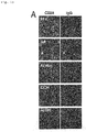
Fig. 1A shows fluorescence micrographs of Example 1 (A). - [
Fig. 1B ]
Fig. 1B shows an electrophoretic image of Example 1 (B). - [
Fig. 1C ]
Fig. 1C shows FACS results in Example 1 (C). - [
Fig. 1D ]
Fig. 1D shows electrophoretic images of Example 1 (D). - [
Fig. 1E ]
Fig. 1E shows the amplification success rate in Example 1 (E). - [
Fig. 2A ]
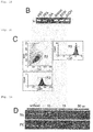
Fig. 2A shows FACS results in Example 2 (A). - [
Fig. 2B ]
Fig. 2B shows FACS results in Example 2 (B). - [
Fig. 2C ]
Fig. 2C shows electrophoretic images of Example 2 (C). - [
Fig. 2D ]
Fig. 2D shows the amplification success rates in Example 2 (D). - [
Fig. 3A ]
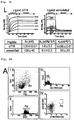
Fig. 3A shows fluorescence micrographs of Example 3 (A). - [
Fig. 3B ]
Fig. 3B shows FACS results in Example 3 (B). - [
Fig. 3C ]
Fig. 3C shows electrophoretic images of Example 3 (C). - [
Fig. 3D ]
Fig. 3D shows results of specificity analysis by ELISA in Example 3 (D). - [
Fig. 3E ]
Fig. 3E shows FACS results in Comparative Example 1 (A). - [
Fig. 3F ]
Fig. 3F shows electrophoretic images of Comparative Example 1 (B) . - [
Fig. 3G ]
Fig. 3G shows results of specificity analysis by ELISA in Comparative Example 1 (C). - [
Fig. 3H ]
Fig. 3H shows fluorescence micrographs after immunostaining in Example 3 (E). - [
Fig. 3I ]
Fig. 3I shows fluorescence micrographs after immunostaining in Example 3 (E). - [
Fig. 3J ]
Fig. 3J shows results of western blotting in Example 3 (E). - [
Fig. 3K ]
Fig. 3K shows results of western blotting in Example 3 (E). - [
Fig. 3L ]
Fig. 3L shows results of surface plasmon resonance (SPR) analysis in Example 3 (F). - [
Fig. 4A ]
Fig. 4A shows FACS results in Example 4 (A). - [
Fig. 4B ]
Fig. 4B shows results of specificity analysis by ELISA in Example 4 (B) . - [
Fig. 4C ]
Fig. 4C shows fluorescence micrographs of Example 4 (C). - [
Fig. 4D ]
Fig. 4D shows fluorescence micrographs of Example 4 (D). - [
Fig. 4E ]
Fig. 4E shows results of surface plasmon resonance (SPR) analysis in Example 4 (E). - The method for isolating a cell capable of producing a monoclonal antibody with specificity toward an antigen of interest of the present invention comprises the following steps (1) to (4) :
- (1) fixing a cell population containing antibody-producing cells with a crosslinking reagent, wherein the crosslinking reagent is a reversible crosslinking regent having the cell membrane permeability (a fixing step);
- (2) treating the fixed cell population with a detergent (a cell membrane lysis step);
- (3) reacting the cell membrane lysed cell population with a labelled antigen of interest (a staining step), wherein the cell membrane lysis step (2) and the staining step (3) can be carried out sequentially or in parallel; and
- (4) isolating at least one single cell which has been reacted with the labelled antigen of interest from the cell population that underwent the staining step (a cell isolation step).
- Further, the method for producing a monoclonal antibody with specificity toward an antigen of interest of the present invention includes the following steps (5) and (6):
- (5) preparing a cDNA by separating mRNA from the at least one cell isolated by the cell isolation method of the present invention (a cDNA preparing step); and
- (6) preparing the antigen specific monoclonal antibody or a fragment thereof from the cDNA obtained in the cDNA preparing step (a preparing step of the monoclonal antibody specific to the antigen of interest).
- In the fixing step, a cell population containing antibody-producing cells is subjected to fixing with a crosslinking reagent. The fixing treatment is to fix antibodies (such as IgG, IgA, IgM, IgD, IgE and IgY) in the antibody-producing cells and mRNA of the antibodies. Various methods are known for fixing biological substances including cells. In the present invention, antibodies and mRNA thereof in the antibody-producing cells are fixed, so that elution of the antibodies and mRNA thereof in the cells in the next detergent treatment step accompanying lysis of the cell membrane is suppressed. The antibodies in the cells are used for identification of antibody-producing cells using a labelled antigen of interest in the later step. Therefore, it is preferable that antibodies in the cells can remain in the cells after lysis of the cell membrane by fixing the antibodies in the cells and it is preferable that the antibodies are fixed in the state that the antibodies are capable of reacting with a labelled antigen of interest. mRNA of antibodies fixed in the cells after lysis of the cell membrane is used for cDNA synthesis in the later step. Prior to cDNA synthesis, fixed mRNA of the antibodies is defixed. Therefore, it is preferable to fix mRNA of the antibodies so that the mRNA can be used for cDNA synthesis after defixing, and that a method for defixing-the fixed mRNA of antibodies is available, and preferably defixing can be easily carried out. The fixation treatment which fulfils such requirements is a method in which formalin or a bivalent crosslinking reagent having an S-S bond in a spacer chain is used. The details of the method are described hereinafter.
- The cell population containing antibody-producing cells is obtained by immunizing an animal with an antigen of interest and collecting cells from the immunized animal. The manner of immunizing an animal with an antigen of interest and collecting a cell population containing antibody-producing cells from the immunized animal may be divided into two methods, one of which is a method (hereinafter referred to as the NHA method) in which a subject is a non-human animal and the other is a method (hereinafter referred to as the HU method) in which a subject is human. The NHA method in which a subject is a non-human animal includes collecting cells derived from the lymph fluid, lymph tissue, blood cell sample or bone marrow from a non-human animal and sensitizing in vitro the collected cells derived from the lymph fluid, lymph tissue, blood cell sample or bone marrow with an antigen of interest, or immunizing the non-human animal with an antigen of interest and collecting cells derived from the lymph fluid, lymph tissue, blood cell sample or bone marrow from the animal after establishment of the immunity. According to the NHA method, a cell population containing antibody-producing cells of a non-human animal which specifically bind to an antigen of interest can be obtained.
- The HU method in which a subject is human includes collecting cells derived from the lymph fluid, lymph tissue, blood cell sample or bone marrow from the subject and sensitizing in vitro the collected cells derived from the lymph fluid, lymph tissue, blood cell sample or bone marrow with an antigen of interest, or collecting cells derived from the lymph fluid, lymph tissue, blood cell sample or bone marrow from the subject having an antibody toward an antigen of interest. According to the HU method, a cell population containing antibody-producing cells of the subject which specifically bind to an antigen of interest can be obtained.
- The NHA method is hereinafter described.
- A non-human animal is immunized with an antigen of interest. The term "non-human animal" as used herein means any animal having an immune system other than humans. Examples of the animal include mammals and birds. Examples of mammals include apes, monkeys, dogs, cats, horses, cows, pigs, sheep, goats, donkeys, camels, llamas, alpacas, reindeers, buffaloes, yaks, guinea pigs, rabbits, minks, mice, rats, sand rats, hamsters, golden hamsters, Armenian hamsters, ferrets, miniature pigs, racoons, possums, Suncus, kangaroos, dolphins and the like. Examples of birds include chickens, quails, ostrich and the like.
- The terms "an antigen of interest" as used herein refers to microorganisms such as viruses, mycoplasmas, bacteria and fungi, Lophotrocozoa such as shellfish, Ecdysozoa such as insects and crustaceans, Deuterostomia such as vertebrates and constituents thereof, proteins, saccharides, lipids, glycoconjugates, nucleic acids, natural low molecular organic compounds, natural polymeric organic compounds, artificial low molecular organic compounds, artificial polymeric organic compounds, metal complexes and the like. It should be noted that the foregoing does not limit the type of the antigen of interest and is merely examples.
- The antigen of interest used for immunization of a non-human animal may be the antigen of interest per se, or an organism containing the antigen of interest or a dead organism or an extract of the organism, or a product obtained after binding to or mixing with an appropriate carrier.
- The cells derived from the lymph fluid, lymph tissue, blood cell sample or bone marrow are collected from the non-human animal and the collected cells derived from the lymph fluid, lymph tissue, blood cell sample or bone marrow are sensitized in vitro with the antigen of interest. Sensitization of the lymph fluid and the like in vitro with the antigen of interest may be carried out as described below. Antigen presenting cells including dendritic cells, T cells and B cells are collected from a non-human animal. In a test tube, the antigen is allowed to work on dendritic cells for phagocytosis and digestion, thereby preparing mature dendritic cells now possessing an ability of presenting the antigen. To the cells, T cells, B cells and an immune stimulating agent such as a cytokine including interleukin 2 or poly(dI-dC) are added, and B cells responding to the antigen are grown and differentiated in a test tube, resulting in a cell population containing antibody-producing cells.
- The phrase "immunize a non-human animal with an antigen of interest" or the like as used herein means to bring an antigen of interest into contact with a non-human animal to allow manifestation of immunity against the antigen of interest in the non-human animal. The manner for allowing manifestation of immunity against an antigen of interest is not particularly limited and may be, for example, administration or implantation of an antigen of interest to a non-human animal, thereby immunizing the non-human animal. The manner of administration or implantation of an antigen of interest is not particularly limited. Examples of the manner of administration include intratracheal administration, oral administration, subcutaneous injection, intravenous injection, intramuscular injection and gene transfer into a non-human animal to express an antigen in the animal body. Alternatively, a non-human animal may be immunized by bringing an antigen of interest into contact with the skin of the non-human animal.
- Immunization of a non-human animal with an antigen of interest is carried out until the immunity against the antigen of interest is established in the non-human animal. Therefore, the antigen of interest is brought into contact with the non-human animal until the immunity against the antigen of interest is established. The frequency and period of the contact of the antigen of interest with the non-human animal and the amount of the antigen of interest per dose may be appropriately selected according to an ease of establishment of the immunization. Whether or not the immunity against the antigen of interest is established in the non-human animal may be verified according to a conventional method such as ELISA for measuring antibodies in the serum after collection of blood from the non-human animal.
- From the animal after establishment of the immunity, cells derived from the lymph fluid, lymph tissue, blood cell sample or bone marrow are collected. As an object of the present method is to collect the cell population containing antibody-producing cells of a non-human animal that specifically bind to the antigen of interest, cells derived from the lymph fluid, lymph tissue, blood cell sample or bone marrow which are likely to contain the cell population containing antibody-producing cells are collected.
- The lymph fluid, lymph tissue, blood cell sample or bone marrow may be prepared, for example, as follows. From a non-human animal to which an antigen was injected subcutaneously, intramuscularly or at a pad on or before about one month ago, the swollen lymph tissue is removed with the lymph fluid by surgery. The tissue attached to the lymph node is eliminated under a stereoscopic microscope followed by breaking the membrane of the lymph node with forceps, thereby dispersing cells in the lymph node into a PBS solution (10 mM phosphate buffer, 120 mM NaCl, 2.7 mM KCl, pH 7.6). The blood cell sample is mononuclear cells which are separated by density gradient centrifugation of the blood collected by heparin blood sampling from an immunized animal. The bone marrow is bone marrow cells obtained by cutting both bone ends of a femur removed from an immunized animal and injecting the PBS solution from one bone end through a syringe needle inserted therein, thereby draining the bone marrow cells from the other bone end. Accordingly, the cell population containing antibody-producing cells may be collected.
- As described above, the cell population containing antibody-producing cells is treated with a crosslinking reagent which is formalin or a bivalent crosslinking reagent having an S-S bond in a spacer chain to fix antibodies and mRNA thereof in the antibody-producing cells. When cells are subjected to a preliminary treatment for fixing proteins using a crosslinking reagent such as glutaraldehyde with the purpose of fixing antibodies and mRNA in the cells, proteins constituting the cells are fixed. However, as glutaraldehyde is an irreversible crosslinking reagent, antibodies and mRNA in the cells are degenerated. Contrary to this, the crosslinking reagent used in the present invention is a reversible crosslinking reagent having the cell membrane permeability. Because of the cell membrane permeability, antibodies and mRNA in the cells may be intracellularly fixed and the reversible crosslinking reagent is used, so that mRNA can be decrosslinked to allow preparation of cDNA in the stage of cDNA preparation from mRNA which is the later step. Examples of such a reversible crosslinking reagent include formalin and a bivalent cross linking reagent having an S-S bond in a spacer chain. Examples of the bivalent crosslinking reagent having an S-S bond in a spacer chain include dithiobis [succinimidyl propionate] (DSP) and the like.
- When the collected cell population containing antibody-producing cells is subjected to the treatment using the reversible crosslinking reagent, antibodies and mRNA thereof in the cells are fixed in the cells without substantial degeneration (while being able to be decrosslinked) . Antibodies are fixed while having a reactivity with a labelled antigen of interest, and mRNA of the antibodies may be decrosslinked and used for cDNA synthesis in the later step. The extent of fixing varies according to the type and concentration of the reversible crosslinking reagent and the temperature and time of the treatment. By changing the type and concentration of the reversible crosslinking reagent and the temperature and time of the treatment according to the type of the antibody-producing cells used, an optimal fixation may result. Fixing may be carried out, for example, under such conditions that the collected cell population containing antibody-producing cells are immersed in an aqueous solution such as phosphate buffered saline (PBS) containing 1% to 10% reversible crosslinking reagent at a temperature in the range of 0°C to 30°C or preferably 0°C to 10°C for 1 to 60 minutes (the collected cell population containing antibody-producing cells are dispersed in a reversible crosslinking reagent-containing aqueous solution). According to the conditions, antibodies and mRNA thereof may be fixed under a good condition (without substantial degeneration). The thus obtained fixed cell population is preferably separated from the solution by centrifugation and the like and used in the next step.
- In the cell membrane lysis step, the cell population treated in the fixing step is subjected to a lysis treatment in order to make pores on the cell membrane. Specifically, the cell population which is fixed in the fixing step is mixed with a solution containing a detergent. By mixing with the detergent-containing solution, the cell membrane is lysed and pores are made on the cell membrane. The pores facilitate entrance of a labelled antigen of interest into the cells in the next step and also facilitate transfer of substances from and to the cells during cDNA synthesis from mRNA in the later step. The lysis of the cell membrane in this step is sufficient if pores serving the purpose are formed on the cell membrane and is appropriate if the treatment can provide the cell membrane which has the strength enduring the cell identification and isolation which follow even after being subjected to the lysis of the cell membrane. For this purpose, a detergent is used in the present invention. The type and concentration of the detergent may be appropriately selected by taking the above purpose into account.
- The detergent may be at least one detergent selected from the group consisting of nonionic detergents, amphoteric detergents and ionic detergents.
- Examples of the nonionic detergent include polyoxyethylene alkyl ethers (including Triton), sorbitan fatty acid esters (including Tween), alkyl polyglucosides, fatty acid diethanolamides, alkyl monoglyceryl ethers and the like. Specific examples of the polyoxyethylene alkyl ether nonionic detergent include Triton X-100 (octylphenol poly(ethylene glycol ether)n (wherein n is about 10), HLB: 13.4 to 13.5). Specific examples of the fatty acid sorbitan ester nonionic detergent include Tween 20 (
Polysorbate 20, polyoxyethylene sorbitan monolaurate, HLB: 16.7) .Tween 20 is a polysorbate and polysorbates are sorbitan fatty acid esters condensed with about 20 molecules of ethylene oxide. In addition toTween 20, mention may be made to Tween 40 (Polysorbate 40, polyoxyethylene sorbitan monopalmitate, HLB 15.6), Tween 60 (Polysorbate 60, polyoxyethylene sorbitan monostearate, HLB 14.9), Tween 65 (Polysorbate 65, polyoxyethylene sorbitan tristearate, HLB 10.5), Tween 80 (Polysorbate 80, polyoxyethylene sorbitan oleate, HLB 15.0), n-dodecyl β-D-maltoside, digitonin, saponin and the like. - Examples of the amphoteric detergent include CHAPS, 3-(N,N-dimethyloctylammonio)propane sulphonate, N-dodecyl-N,N-dimethyl-3-ammonio-1-propane sulphonate, 3-(N,N-dimethyloctadecylammonio)propane sulphonate and the like. The amphoteric detergents are known to rarely destroy the conformation of proteins and may be used for lysis of the membrane similarly to the nonionic detergent.
- The ionic detergent may be at least one ionic detergent selected from the group consisting of cholic acid derivatives. Examples of at least one ionic detergent selected from cholic acid derivatives include an anionic detergent such as a cholic acid alkali metal salt (such as sodium cholate) and digoxigenin which is a cholic acid derivative (modified at an alkylene carboxyl group) . The ionic detergents are known to rarely destroy the conformation of proteins and may be used for lysis of the membrane similarly to the nonionic detergent.
- The concentration of the detergent may be, for example, in the range of 0.1% to 5% according to the type of the detergent, the type of the cells and treatment conditions (temperature and time). The size of the pores formed on the cell membrane may be appropriately controlled by the type and concentration of the detergent and the treatment conditions (temperature and time) . The treatment conditions (temperature and time) may be, for example, immersion at a temperature in the range of 0°C to 30°C, preferably 10°C to 25°C for 1 to 60 minutes in a solution (such as a phosphate buffered aqueous solution) containing the detergent. The cell membrane lysis step may, as described hereinbelow, precede the following reaction step with a labelled antigen of interest or may be carried out in parallel with the reaction step with a labelled antigen of interest in the same solution.
- In the step, the cell population which has undergone the lysis treatment in the cell membrane lysis step and a labelled antigen of interest are mixed to allow reaction of antibodies in the cells and the labelled antigen of interest. When the cell membrane lysis step and the staining step are carried out in parallel, the cell population obtained in the fixing step is subjected to the treatment with a detergent and the reaction with a labelled antigen of interest in the same solution.
- The antigen of interest in the labelled antigen of interest is a substance having an epitope identical to the antigen of interest used for immunization of the non-human animal or the like. Therefore, the labelled antigen of interest and the antigen of interest used for immunization may be completely the same or may be different substances containing the same epitope.
- The label of the antigen of interest is not particularly limited as far as the label allows identification and separation of cells reacted with the labelled antigen of interest. Examples of the label include a fluorescence label, a magnetic bead label and the like. The type of the fluorescence label is not particularly limited. The antigen of interest may be appropriately labelled by a well-known method according to the type of the label used.
- The cell population obtained in the cell membrane lysis step and the labelled antigen of interest may be allowed to react by, for example, bringing the cell population and the labelled antigen of interest into contact in an aqueous solution such as phosphate buffered saline (PBS). The concentrations of the cell population and the labelled antigen of interest in the aqueous solution, temperature and time may be appropriately selected while confirming the progress of the reaction. The treatment conditions (temperature and time) may be, for example, immersion at a temperature in the range of 0°C to 30°C or preferably 0°C to 10°C for 1 to 60 minutes in a solution (such as a phosphate buffered aqueous solution) containing the labelled antigen of interest.
- When the cell population obtained after fixing is subjected in parallel to the cell membrane lysis treatment and the reaction with the labelled antigen of interest, the cell population is brought into contact with the detergent for cell lysis and the labelled antigen of interest in, for example, an aqueous solution such as phosphate buffered saline (PBS) . The concentrations of the cell population, the detergent and the labelled antigen of interest in the aqueous solution, temperature and time may be appropriately selected by confirming the progress of the reaction. The treatment conditions (temperature and time) may be, for example, immersion at a temperature in the range of 0°C to 30°C or preferably 0°C to 10°C for 1 to 60 minutes in a solution (such as a phosphate buffered aqueous solution) containing the detergent and the labelled antigen of interest.
- In the step (4), at least one single cell which is reacted with the labelled antigen of interest is separated as a single cell. The cells reacted with the labelled antigen of interest are first identified by using the label of the labelled antigen of interest as a guide and identified single cells are separated.
- In the method of the present invention, antibodies and mRNA thereof in the cells are fixed in the step (1) and the fixed cell population is treated with the detergent to make pores on the cell membrane in the step (2). Therefore, the labelled antigen of interest can easily enter the cells producing antibodies toward the antigen of interest through the pores on the cell membrane. The labelled antigen of interest binds to the antibody in the cells which are fixed and subjected to the cell membrane lysis treatment and allows identification of cells containing the labelled antigen of interest bound to antibodies. The cells which retain the labelled antigen of interest therein are highly possible to be antibody-producing cells because it is highly possible that the labelled antigen of interest binds to antibodies in the cells.
- The cells identified with the label of the labelled antigen of interest as a guide are separated as single cells. Separation of the identified antibody-producing cells may be carried out with, for example, a cell sorter. Separation of single antibody-producing cells by using a cell sorter may be carried out according to a well-known method. Cells may be selected and separated as a cell population including more than one cell or, preferably, each cell is individually selected and separated. The cells selected are highly possible to exhibit a binding ability to the antigen of interest. However, the cells may not always have an identical antibody toward the antigen of interest and it is expected that the amino acid sequences in the antigen-binding sites of the antibodies vary. Therefore, it is possible to obtain different monoclonal antibodies which specifically bind to the same antigen of interest by individually selecting and separating the cells and using each cell to the method for producing an antibody or a fragment of the antibody with specificity toward the antigen of interest described hereinbelow.
- The cells isolated in the step (4) are highly possible to show reactivity with the labelled antigen of interest, and thus highly possible to be antibody-producing cells. However, there is actually a possibility of contamination of labelled cells due to nonspecific staining (reaction with the labelled antigen of interest). Therefore, non-specifically stained cells may be eliminated (excluded) by using a labelled antigen of no interest, if necessary. For example, in Example 2, an antigen of no interest, the DsRed protein, is added to a cell staining solution in order to exclude DsRed protein positive cells as non-specifically stained cells. In Example 3, a non-phosphorylated peptide is added to a cell staining solution in order to separate phosphorylated peptide positive cells from non-phosphorylated peptide positive cells.
- It is preferable to carry out at least one of steps (1) to (4) above in the presence of an RNase inhibitor from the viewpoint of suppressing degradation of mRNA collected from cells to facilitate preparation of cDNA of a monoclonal antibody with specificity toward an antigen of interest or a fragment thereof. Examples of the RNase inhibitor include DEPC (diethylpyrocarbonate), vanadyl ribonucleotide, inhibitory proteins for ribonuclease A, ribonuclease B and ribonuclease C and anti-ribonuclease antibodies. Examples of commercially available RNase inhibitors include RNaseOUT (Life Technologies Corporation), RNasin® (Promega Corporation), Ribonuclease Inhibitor (derived from pig liver) (Takara Bio Inc.) and the like. It is preferable to carry out all of the steps (1) to (4) in the presence of an RNase inhibitor, and more specifically, it is preferable to add an appropriate amount of RNase inhibitor to solutions containing cells, antibodies and the antigen used in the steps. An appropriate amount of the RNase inhibitor may be appropriately selected according to the type of the RNase inhibitor, the type of the solution and the like.
- When materials of the labelled antigen of interest used in the step (3) are prevented from contamination of RNase, the recovery rate of mRNA may be increased and the antibody with specificity toward the antigen of interest may be prepared with an increased efficiency.
- The method for producing a monoclonal antibody with specificity toward an antigen of interest of the present invention includes preparing a monoclonal antibody with specificity toward an antigen of interest from at least one single cell obtained by the cell separation method of the present invention through following steps (5) and (6):
- (5) preparing a cDNA by separating mRNA from the at least one cell obtained by the cell separation method of the present invention (a cDNA preparing step); and
- (6) preparing the antigen specific monoclonal antibody or a fragment thereof from the cDNA obtained in the cDNA preparing step.
- According to the method, an antibody which exhibits a specific binding ability to an antigen of interest or an antibody fragment (the fragment per se exhibits a specific binding ability to an antigen of interest) which exhibits a specific binding ability to an antigen of interest may be produced.
- In the step (5), mRNA is separated from at least one single cell obtained by the cell separation method of the present invention and cDNA is prepared from the separated mRNA.
- For separation of mRNA, mRNA in the cells fixed in the step (1) may be defixed (decrosslinked). A manner of defixing may be appropriately selected according to the type of the reversible crosslinking reagent used in the step (1). When the reversible crosslinking reagent used in the step (1) is formalin, defixing may be carried out by heating the isolated cells and when an amino group-containing group coexists during heating, defixing (decrosslinking) may be facilitated. Examples of the amino group-containing group include tris(hydroxymethyl)aminomethane, amino acids, ethanolamine and the like. Defixing allows recovery of mRNA from cells with an increased efficiency and an increase in the recovery of cDNA. The conditions for the heating treatment may be appropriately selected by taking the extent of fixation, the type and concentration of the amino group-containing compound and the stability of mRNA into account. For example, heating may be carried out at 20°C to 70°C for 1 to 120 minutes. It is more preferable to carry out defixing by heat treating the isolated cells in the presence of a protease. The conditions for the heating treatment and the amount of the protease may be appropriately selected by taking the type and the amount of the protease, the extent of fixation and the stability of mRNA into account. For example, heating may be carried out at 40°C to 60°C for 1 to 120 minutes. The amount of the protease may be in the range of 0.1 µg/ml to 100 µg/ml. The protease may be any existing protease and may be, for example, protease K. The heating treatment in the presence of protease K allows defixing of mRNA from the isolated cells with an increased efficiency and increases the proportion of obtaining variable region gene fragments of antibody genes. As a result, the antigen specific monoclonal antibody may be efficiently prepared in the later step.
- When the reversible crosslinking reagent used in the step (1) is dithiobis (succinimidyl propionate) (DSP), defixing may be carried out by treating the isolated cells with a reducing agent such as β-mercaptoethanol and sodium borohydride. Similarly in this case, defixing allows recovery of mRNA in the cells with an increased efficiency and may increase the recovery of cDNA. The conditions for the reducing treatment may be appropriately selected by taking the extent of fixing, the type and concentration of the reducing agent and the stability of mRNA into account. The reducing treatment may be carried out, for example, at 20°C to 70°C for 1 to 120 minutes. Defixing by reducing treatment may also be carried out in combination with the protease treatment.
- Separation of mRNA from at least one single cell and preparation of cDNA may be carried out according to well-known methods. For example, methods disclosed in PTL 2 and NPL 4 (the method by Kurosawa et al.) may be used (the entire disclosures of PTL 2 and NPL 4 are specifically incorporated herein by reference) . It is preferable to separate mRNA from one separated antibody-producing cell in view of obtaining a heavy chain gene and a light chain gene of the antibody toward the antigen of interest. However, mRNA may be separated from two or more separated antibody-producing cells and in this case, the separated mRNA is a mixture of mRNAs from respective cells. For example, when mRNA is separated from two antibody-producing cells, two pairs of heavy chain genes and light chain genes are cloned. In this case, the possibility of obtaining the combination of the correct heavy chain gene and light chain gene is 50%: however, the correct pair may be attained from a technological standpoint. However, the possibility of obtaining the correct combination of the heavy chain gene and the light chain gene from mRNA collected from more than 10 antibody-producing cells is less than 1%. Although it is possible to attain the correct combination, the efficiency decreases. Therefore, mRNA is preferably separated from 10 or less separated antibody-producing cells.
- More specifically, collection of mRNA from antibodies toward an antigen of interest and preparation of cDNA from the collected mRNA may be carried out by employing the method disclosed in, for example,
WO 2009/091048 (US 2011/0020879 A1 ) (the entire disclosures of which are specifically incorporated herein by reference). The prepared cDNA may be cloned, if necessary. Cloning may be carried out according to the cloning method employing the homologous recombination disclosed inWO 2009/110606 andUS 2011/0117609 A1 (the entire disclosures of which are specifically incorporated herein by reference) . Further, the base sequence of the prepared cDNA may be identified. The identification of the base sequence of the cDNA may be carried out by using well-known DNA sequencing methods. By identifying the base sequence of the cDNA, an antibody gene toward the antigen of interest may be identified. - However, in the method of the present invention, it is preferable that the second primer used for cDNA synthesis is modified from the above well-known methods. In the method of the present invention, mRNA is inevitably degraded during the steps before cDNA synthesis. Therefore, the efficacy of full-length cDNA synthesis is low and a smearing band tends to be amplified after normal 5'RACE PCR (compare the bands in "10 min" and the bands in "unfixed" in
Fig. 1D which is the results of Example 1. The bands amplified from the cells of "10 min" are smeared. Similar tendency is observed inFig. 2C which is the results of Example 2). - Thus, in the present invention, it is preferable that the second primer for cDNA synthesis, i.e., a sense primer for the second round of PCR, has the sequence around the initiation codon of an immunoglobulin of interest in order to amplify only the full-length antibody gene. For example, in Example 3 (C), a sequence around the initiation codon of the guinea pig immunoglobulin is used as a sense primer for the second round of PCR. When the sequence around the initiation codon of the immunoglobulin is known, the primer may be prepared based on known information or sequence information of the immunoglobulin gene expressed in the immunized animal (in case of Example 3, guinea pig) obtained by high throughput sequencing.
- In the step (6), an antigen specific monoclonal antibody or a fragment thereof is prepared from cDNA prepared in the cDNA preparing step. Preparation of the antigen specific monoclonal antibody or a fragment thereof from cDNA may be carried out according to well-known methods. For example, preparation may be carried out with a fragment of an antibody gene prepared by the method for specifically preparing a DNA fragment disclosed in
WO 2011/027808 (the entire disclosure of which is specifically incorporated herein by reference). Disclosures in PTL 2 and NPL 4 (the method by Kurosawa et al.) may also be referred to for preparation of an antibody or a fragment of the antibody. The antibody obtained by the method of the present invention exhibits a specific binding ability to the antigen of interest and the fragment thereof is an antigen-binding fragment which exhibits a specific binding ability to the antigen of interest. Examples of the antigen specific antibody fragment includes Fab, F(ab')2, Fab', diabody, a single stranded antibody (such as scFv and dsFv) and the like. The antibody fragment may be appropriately prepared by utilizing the well-known method after determining the amino acid sequence and base sequence of the antibody gene from cDNA cloned in the step (5). - It is preferable to carry out the step (5) in the presence of an RNase inhibitor from the viewpoint of suppressing degradation of mRNA collected from cells to facilitate preparation of cDNA of a desired antigen specific monoclonal antibody or a fragment thereof. Examples of the RNase inhibitors are those mentioned for the steps (1) to (4). It is preferable to add an appropriate amount of the RNase inhibitor to the solutions used in the step (5) in order to carry out the step in the presence of the RNase inhibitor. An appropriate amount of the RNase inhibitor may be appropriately selected according to the type of the RNase inhibitor, the type of the solution and the like.
- In addition to carrying out at least some or all of the steps (1) to (5) of the present invention in the presence of the RNase inhibitor or instead of using the RNase inhibitor, the steps may be carried out under RNase activity suppressing conditions. By carrying out the steps under such conditions, the recovery rate of mRNA may be increased and an antibody with specificity toward the antigen of interest may be prepared with an increased efficiency.
- RNase activity may be suppressed by carrying out the experimental procedures under, for example, cooling conditions such as in or on ice.
- The present invention encompasses a monoclonal antibody specific to threonine 18-phosphorylated p53 (pT18-p53). The monoclonal antibody encompasses a monoclonal antibody specific to threonine 18-phosphorylated p53 (pT18-p53) having a γ chain comprising a variable region having an amino acid sequence shown in SEQ ID NO: 9 or a κ chain comprising a variable region having an amino acid sequence shown in SEQ ID NO: 10 and a monoclonal antibody specific to threonine 18-phosphorylated p53 (pT18-p53) having a γ chain comprising CDR1, CDR2 and CDR3 having amino acid sequences shown in SEQ ID NOs: 3 to 5, respectively, and a κ chain comprising CDR1, CDR2 and CDR3 having amino acid sequences shown in SEQ ID NOs: 6 to 8, respectively. Any of the antibodies may be prepared by the method of the present invention and the antibody is specifically described in Example 3.
- The present invention further encompasses a monoclonal antibody specific to threonine 68-phosphorylated CHK2 (pT68-CHK2) . The monoclonal antibody encompasses a monoclonal antibody specific to threonine 68-phosphorylated CHK2 (pT68-CHK2) having a γ chain comprising a variable region having an amino acid sequence shown in SEQ ID NO: 17 or a κ chain comprising a variable region having an amino acid sequence shown in SEQ ID NO: 18 and a monoclonal antibody specific to threonine 68-phosphorylated CHK2 (pT68-CHK2) having a γ chain comprising CDR1, CDR2 and CDR3 having amino acid sequences shown in SEQ ID NOs: 11 to 13, respectively, and a κ chain comprising CDR1, CDR2 and CDR3 having amino acid sequences shown in SEQ ID NOs: 14 to 16, respectively. Any of the antibodies may be prepared by the method of the present invention and the antibody is specifically described in Example 4.
- The present invention is further specifically described on the basis of Examples which exemplify the present invention. It is not intended that Examples limit the present invention.
-
- (A) OKT10 hybridoma cells attached on glass slides were fixed with 2% paraformaldehyde (PFA), 2% glutaraldehyde (GA), acetone, ethanol or 95% ethanol-5% acetic acid at room temperature for 10 minutes (fixing).
The cells were washed with PBS containing 0.1% Triton X-100 (hereinafter PBS containing 0.1% Triton X-100 is referred to as PBST) (cell membrane lysis).
Thereafter, staining was carried out with a Dylight 594-labelled anti-mouse IgG antibody and Dylight 488-labelled CD38 (reaction with a labelled antigen of interest).
For the cells fixed with paraformaldehyde, binding of the antigen, CD38, to antibodies in the cells was observed. For the cells treated with other fixing solutions, binding to CD38 was not observed. The results are shown inFig. 1A . - (B) OKT10 hybridoma cells were fixed with 2% paraformaldehyde at 4°C for 20 minutes (fixing).
The cells were then subjected to cell membrane lysis and staining of antibodies in the cells by using 250 µl of PBST containing a Dylight 594-labelled anti-mouse IgG antibody and 5 µl of RNaseOUT - RNase inhibitor (cell membrane lysis and reaction with a labelled antigen of interest).
The cells were recovered by centrifugation (cell isolation).
Thereafter, a cell lysis solution (100 mM Tris HCl (pH 7.5), 500 mM LiCl, 1% lithium dodecylsulphate and 5 mM dithiothreitol) was added to attain a concentration of 40 cells/1 µl. Extraction of mRNA from the cells and cDNA synthesis were carried out according to the method by Kurosawa et al. (Rapid production of antigen-specific monoclonal antibodies from a variety of animals, BMC Biology, 2012, 10:80) (cDNA synthesis).
Briefly, 2.5 µl of the cell lysis solution (100 cells) was added to 3 µg of magnetic beads (Dynabeads) bound to oligo dT25 to allow binding of mRNA in the cells to the magnetic beads. The magnetic beads were then washed once with 3 µl of mRNA washing solution A (10 mM Tris HCl (pH 7.5), 0.15 M LiCl, 0.1% LiDS or 50 mM Tris HCl (pH 8.3)) and then 3 µl of mRNA washing solution B (75 mM KC1, 3 mM MgCl2, 0.1% Triton X, 0.5 mM dNTP, 5 mM DTT and 2 units RNase inhibitor/µL) followed by cDNA synthesis. Briefly, a cDNA synthesis solution (50 mM Tris HCl (pH8.3), 75 mM KCl, 3 mM MgCl2, 0.1% Triton X-100, 0.5 mM dNTP, 5 mM DTT, 2 units RNase inhibitor/µL and 10 units SuperScript III Reverse transcriptase (Invitrogen)) was added to the washed magnetic beads and allowed to react at 40°C for 1 hour. The magnetic beads were washed with 3 µl of TE solution (10 mM Tris HCl (pH7.5),1 mM EDTA and 0.1% Triton X) followed by amplification of the mouse immunoglobulin γ chain constant region gene. To the magnetic beads was added 25 µl of PCR reaction solution (10 pmol of theprimers 1 and 2, respectively, 10 nmol of dNTP and 1U of Takara Bio PrimeSTAR heat resistant DNA polymerase) and the reaction was carried out for 35 cycles of 94°C for 30 seconds and 68°C for 40 seconds. The primers used were 5'-GTGGAACTCAGGCGCCCTGACCAGC-3' (SEQ ID NO: 1) and 5'-ACGCTGCTGAGGGAGTAGAGTCCTGAG-3' (SEQ ID NO: 2).
Amplification of the immunoglobulin heavy chain constant region gene fragment was observed for the cells fixed with paraformaldehyde. The results are shown inFig. 1B . - (C) Hybridoma cells (1 × 10⁶ cells) were fixed with 2% paraformaldehyde at 4°C for 10 minutes to 30 minutes (fixing).
The cells were collected by centrifugation to which 250 µL of PBST containing a Dylight 488-labelled anti-mouse IgG antibody and 200 units of RNaseOUT was added to allow antigen-antibody reaction at 4°C for 15 minutes (cell membrane lysis and reaction with a labelled antigen of interest).
The labelled anti-mouse IgG antibody used was diluted in PBST containing RNaseOUT and left to stand at room temperature for 1 hour before use. The cells were diluted in 3 ml of PBS containing 0.1 µg/mL DAPI (4',6-diamidino-2-phenylindole) and subjected to FACS. The cells were R1 and R2 gated to select single cells. The cells were R3 gated to select the cells having the intracellular immunoglobulin bound to the labelled antibody (cell isolation). The results are shown inFig. 1C . - (D) The R3-gated cells obtained in the above (C) were subjected to single sorting in 15 µl of cell lysis solution containing 3 µg of magnetic beads or in 15 µl of cell lysis solution containing 3 µg of magnetic beads containing 1 µg/mL of protease K. The cell lysis solution containing protease K was further heated at 50°C for 1 hour in order to decrosslink the linkages formed by paraformaldehyde. Amplification of the full-length immunoglobulin variable region from the cells was carried out according to the method by Kurosawa et al. (NPL 4). It was found that decrosslinking the cells undergone fixing over a short period of time allowed efficient amplification of the full-length immunoglobulin variable region from a single hybridoma cell. The results are shown in
Fig. 1D . - (E) The results in (D) are summarized in a graph. The experiments were repeated three times and the averages and the sample standard deviations are shown (N=12). The results are shown in
Fig. 1E . - OKT10 hybridoma cells and Jurkat cells were mixed at ratios of 1:100 (1%), 1:1000 (0.1%), 1:10,000 (0.01%) and 1:100,000 (0.001%). The cells were fixed with a 2% paraformaldehyde PBS solution at 4°C for 10 minutes (fixing).
- The cells were collected by centrifugation to which 250 µl of staining solution (PBST containing
anti-mouse IgG Dylight 488,anti-mouse IgG Dylight 650, 0.1 µg/ml of DsRed and 200 units of RNaseOUT) was added and left on ice for 15 minutes (cell membrane lysis and reaction with a labelled antigen of interest). - To the solution was added 3 ml of PBS solution containing 1 µg/ml of DAPI and OKT10 hybridoma cells were isolated with a flow cytometer. The cells were R1 (FSC vs SSC) and R2 (DAPI) gated to identify a single cell population. From the single cell population, cells having autofluorescence and non-specifically stained cells were eliminated with the FL2 channel (R3 gate). From the cells,
anti-mouse IgG Dylight 488 strongly positive andanti-mouse IgG Dylight 650 strongly positive cells were identified as OKT10 hybridoma cells (R4) (cell isolation). - (A)
Fig. 2A is a FACS graph of OKT10 hybridoma cells and Jurkat cells mixed at 1:100 (1%). - (B)
Fig. 2B is a FACS graph for the mixing ratios of 100%, 0.1%, 0.01% and 0.001%. In all cases, cells were observed in the R4 gate corresponding to the hybridoma cells. - (C) The R4-gated cells were subjected to single cell sorting in 15 µL of cell lysis solution containing 3µg of oligo-dT magnetic beads and 1 µg/ml of protease K. According to the method described in Example 1 (B), RNA extraction from the cells, cDNA synthesis and V gene amplification by 5' RACE PCR were carried out. The results are shown in
Figs. 2C and 2D . When the V gene was amplified from the cell lysis solution of 72 cells, the amplification success rates were 89%, 53%, 47% and 31% for mixing ratios of 1%, 0.1%, 0.01% and 0.001%, respectively. -
- (A) The iliac lymph node was collected from a guinea pig immunized with p53 phosphorylated peptide and a cell suspension was prepared therefrom. Cells (1 × 10⁷ cells) were fixed with 2% paraformaldehyde PBS solution at 4°C for 10 minutes (fixing).
The cells were collected by centrifugation to which 250 µl of staining solution (PBST containing anti-guineapig IgG Dylight 650, phosphorylated peptide-streptavidin Dylight 488, non-phosphorylated peptide-streptavidin Dylight 550, 0.1 µg/ml of DsRed and 200 units of RNaseOUT) was added and left on ice for 15 minutes for staining (cell membrane lysis and reaction with a labelled antigen of interest).
To the solution was added 3 ml of PBS solution containing 1 µg/ml of DAPI and a partial sample was observed under a fluorescence microscope. The results are shown inFig. 3A .
As a result, the phosphorylated p53 specific plasma cells were identified as anti-guinea pig IgG strongly positive, phosphorylated peptide-streptavidin strongly positive and non-phosphorylated peptide-streptavidin negative.
The p53 specific plasma cells were identified as anti-guinea pig IgG strongly positive, phosphorylated peptide-streptavidin strongly positive and non-phosphorylated peptide-streptavidin strongly positive.
Non-specific plasma cells were identified as anti-guinea pig IgG strongly positive, phosphorylated peptide-streptavidin negative and non-phosphorylated peptide-streptavidin negative. - (B) The cells were analyzed on a flow cytometer in order to separate the p53 specific plasma cells. The cells were R1 (FSC vs SSC) and R2 (DAPI) gated to identify a single cell population. From the single cell population, cells having autofluorescence, non-specifically stained cells and p53 specific plasma cells reacting with the non-phosphorylated peptide were eliminated with the FL2 channel (R3 gate) . From the cells, anti-mouse IgG strongly positive and phosphorylated peptide strongly positive cells were identified as the phosphorylated p53 specific plasma cells (R4). The cells appearing in the R4 gate had 100 times and 10 times stronger fluorescence intensities than other cells with regard to the binding to the phosphorylated peptide and to the anti-guinea pig antibody, respectively. The results are shown in
Fig. 3B . - (C) The R4-gated cells were subjected to single cell sorting by the same manner as in Example 2 (cell isolation). Thereafter, the V gene was amplified by 5' RACE PCR by the same manner as in Example 1 (B). The results are shown in
Fig. 3C . When the V gene was amplified from the cell lysis solution of 72 cells, the amplification efficiency was 84%. - (D) From the obtained V gene, the full-length heavy chain and light chain immunoglobulin genes were prepared according to the method by Kurosawa et al. (NPL 4) which were introduced into 293FT cells to prepare recombinant antibodies. The obtained antibodies were analyzed for the specificity by ELISA. The results are shown in
Fig. 3D . 93% (25/27) bound to the phosphorylated peptide, among which 68% (17/25) were specific to the phosphorylated peptide. -
- (A) From the iliac lymph node of a guinea pig immunized with the p53 phosphorylated peptide, a cell suspension was prepared. The cells (1 × 10⁷ cells) were suspended in PBS containing anti-guinea
pig IgG Dylight 650, phosphorylated peptide-streptavidin Dylight 488, non-phosphorylated peptide-streptavidin Dylight 550 and ER-tracker and left to stand at 4°C for 30 minutes to stain the cells.
The cells were analyzed on a flow cytometer in order to separate the p53 specific plasma cells. The cells were R1 (FSC vs SSC) gated to identify a single cell population. From the single cell population, cells having autofluorescence, non-specifically stained cells and p53 specific plasma cells reacting with the non-phosphorylated peptide were eliminated with the FL2 channel (R2 gate). The ER-tracker strongly positive cells were selected to identify plasma cells (R3 gate) from which anti-guinea pig IgG weakly positive and phosphorylated peptide positive cells were identified as phosphorylated p53 specific plasma cells (R4). The cells appearing in the R4 gate had only 10 times and a few times stronger fluorescence intensities than other cells with regard to the binding to the phosphorylated peptide and to the anti-guinea pig antibody, respectively. The results are shown inFig. 3E . - (B) The R4-gated cells were subjected to single cell sorting by the same manner as in Example 2 (cell isolation). Thereafter, the V gene was amplified by 5' RACE PCR by the same manner as in Example 1 (B). The results are shown in
Fig. 3F . When the V gene was amplified from the cell lysis solution of 72 cells, the amplification efficiency was 16%. - (C) From the obtained V gene, the full-length heavy chain and light chain immunoglobulin genes were prepared by the same manner as in Example 3 (D) which were introduced into 293FT cells to prepare recombinant antibodies. The obtained antibodies were analyzed for the specificity by ELISA. As a result (see
Fig. 3G ), 33% (9/27) bound to the phosphorylated peptide among which 44% (4/9) were specific to the phosphorylated peptide. - The results in Example 3 and Comparative Example 1 are summarized in Table 1 below. The significant effect of the present invention is apparent.
[Table 1] V gene amplification efficiency Number of antibodies bound to phosphorylated peptide Number of antibodies specific to phosphorylated peptide Example 3 84% 93% (25/27) 63% (17/27) Comparative Example 1 16% 33% (9/27) 15% (4/27) -
- (E) In order to prepare a monoclonal antibody specific to threonine 18-phosphorylated p53 (pT18-p53), HEK293 cells were subjected to immunostaining with antibody clones obtained in Example 3 (D). The #23 antibody was identified as an antibody clone which allowed specific signal detection in the nuclei of HEK293 cells in which DNA damage was caused by etoposide treatment. The signal obtained with the #23 antibody disappeared in the cells treated with λ phosphatase. The results are shown in
Fig. 3H . The signal also disappeared in the presence of the p53 peptide having pT18, while the signal did not disappear in the presence of the peptides in which neighboringserine 15 andserine 20 were phosphorylated. The results are shown inFig. 3I . In western blotting analysis, the #23 antibody allowed detection of a specific band at 50 kDa from the etoposide treated HEK293. The signal obtained with the #23 antibody disappeared in the cells treated with λ phosphatase. The results are shown inFig. 3J . The signal also disappeared in the presence of excess p53 peptide having pT18, while the signal did not disappear in the presence of the peptides in which neighboringserine 15 andserine 20 were phosphorylated. The results are shown inFig. 3K . - (F) The affinity of the #23 antibody toward pT18-p53 was measured on a surface plasmon resonance (SPR) device. As a result, the #23 antibody had a binding constant KD of 0.20 nM toward the pT18-p53 peptide. The results are shown in
Fig. 3L . - (G) The amino acid sequences (SEQ ID NOs: 3 to 5) of CDR1, CDR2 and CDR3 of the γ chain and the amino acid sequences (SEQ ID NOs: 6 to 8) of CDR1, CDR2 and CDR3 of the kappa chain of the #23 antibody are respectively shown in Table 2. The amino acid sequences (SEQ ID NOs: 9 and 10) of the variable regions of the γ and kappa chains of the #23 antibody are respectively shown in Table 3.
-
- (A) In order to prepare a monoclonal antibody specific to threonine 68 phosphorylated CHK2 (pT68-CHK2), the iliac lymph node was collected from a guinea pig immunized with pT68-CHK2 peptide and a cell suspension was prepared therefrom. Cells (1 × 10⁷ cells) were fixed with 2% paraformaldehyde PBS solution at 4°C for 10 minutes (fixing).
The cells were collected by centrifugation to which 250 µl of staining solution (PBST containing anti-guineapig IgG Dylight 650, pT68-CHK2 peptide-streptavidin Dylight 488, non-phosphorylated peptide (UM-CHK2)-streptavidin Dylight 550, 0.1 µg/ml of DsRed and 200 units of RNaseOUT) was added and left on ice for 15 minutes for staining (cell membrane lysis and reaction with a labelled antigen of interest).
To the solution was added 3 ml of PBS solution containing 1 µg/ml of DAPI, thereby the cell nucleus was stained. - (B) The cells were analyzed on a flow cytometer in order to separate pT68-CHK2 specific plasma cells. The cells were R1 (FSC vs SSC) and R2 (DAPI) gated to identify a single cell population. From the single cell population, cells having autofluorescence, non-specifically stained cells and CHK2 specific plasma cells reacting with the UM-CHK2 peptide were eliminated with the FL2 channel (R3 gate). From the cells, anti-mouse IgG strongly positive and pT68-CHK2 peptide strongly positive cells were identified as pT68-CHK2 specific plasma cells (R4). The cells appearing in the R4 gate had 100 times and 10 times IgG stronger fluorescence intensities than other cells with regard to the binding to the pT68-CHK2 peptide and to the anti-guinea pig antibody, respectively. The results are shown in
Fig. 4A . - (C) The R4-gated cells were subjected to single cell sorting by the same manner as in Example 2 (cell isolation). Thereafter, the V gene was amplified by isolated 5'RACE PCR by the same manner as in Example 1 (B), the full-length heavy chain and light chain immunoglobulin genes were prepared from the obtained V gene according to the method by Kurosawa et al. (NPL 4) and the genes were introduced into 293FT cells to prepare recombinant antibodies. The obtained antibodies were analyzed for the specificity by ELISA. The results are shown in
Fig. 4B . Among the antibodies, 59% (10/17) were specific to the pT68-CHK2 peptide. - (D) With the obtained antibody clones, HEK293 cells and Hela cells were immunostained. The #34 antibody was identified as an antibody clone which allowed specific signal detection in the nuclei of HEK293 cells in which DNA break was induced by camptothecin treatment. The results are shown in
Fig. 4C . The signal disappeared in the presence of excess pT68-CHK2 peptide, while the signal did not disappear in the presence of the UM-CHK2 peptide. The signal also disappeared in the cells treated with λ phosphatase. The results are shown inFig. 4D . From the above results, it was found that the #34 antibody allows detection of endogenous CHK2 in which threonine 68 is phosphorylated. - (E) The affinity of the #34 antibody toward pT68-CHK2 was measured on a surface plasmon resonance (SPR) device. As a result, the #34 antibody had a binding constant KD of 0.08 nM toward the pT68-CHK2 peptide. The results are shown in
Fig. 4E . - (F) The amino acid sequences (SEQ ID NOs: 11 to 13) of CDR1, CDR2 and CDR3 of the γ chain and the amino acid sequences (SEQ ID NOs: 14 to 16) of CDR1, CDR2 and CDR3 of the kappa chain of the #34 antibody are respectively shown in Table 4 (SEQ ID NO). The amino acid sequences (SEQ ID NOs: 17 and 18) of the variable regions of the γ and kappa chains of the #34 antibody are respectively shown in Table 5.
- The present invention is useful in the field relating to antibody preparation.
-
- SEQ ID NO: 1 PCR primer
- SEQ ID NO: 2 PCR primer
- SEQ ID NOs: 3 to 5 CDR1, CDR2 and CDR3 of the γ chain of pT18-p53 specific monoclonal antibody
- SEQ ID NOs: 6 to 8 CDR1, CDR2 and CDR3 of the kappa chain of pT18-p53 specific monoclonal antibody
- SEQ ID NO: 9 Variable region of the γ chain of pT18-p53 specific monoclonal antibody
- SEQ ID NO: 10 Variable region of the kappa chain of pT18-p53 specific monoclonal antibody
- SEQ ID NOs: 11 to 13 CDR1, CDR2 and CDR3 of the γ chain of pT68-CHK2 specific monoclonal antibody
- SEQ ID NOs: 14 to 16 CDR1, CDR2 and CDR3 of the kappa chain of pT68-CHK2 specific monoclonal antibody
- SEQ ID NO: 17 Variable region of the γ chain of pT68-CHK2 specific monoclonal antibody
- SEQ ID NO: 18 Variable region of the κ chain of pT68-CHK2 specific monoclonal antibody
-


| CDR1 | CDR2 | | |
| p53 # | |||
| 23 gamma | GFIFSSYA | ISRSDSNR | |
| p53 # | |||
| 23 kappa | QSLLHSNGKTY | RVS | FQNTHPPLT |
| Full V seq | |
| |
 |
| p53 # | |
| 23 kappa |
 |
| CDR1 | CDR2 | | |
| CHK2 # | |||
| 34 gamma | GFTFSNYF | ISGDSSNI | |
| CHK2 # | |||
| 34 kappa | QSLLYSRNNKNL | WAS | WQGISAPNTG |
| Full V seq | |
| |
 |
| CHK2 # | |
| 34 kappa |
 |
Claims (17)
- A method for isolating a cell capable of producing a monoclonal antibody with specificity toward an antigen of interest, wherein the method comprises the following steps;(1) fixing a cell population containing antibody-producing cells with a crosslinking reagent, wherein the crosslinking reagent is a reversible crosslinking regent having the cell membrane permeability (a fixing step);(2) treating the fixed cell population with a detergent (a cell membrane lysis step);(3) reacting the cell membrane lysed cell population with a labelled antigen of interest (a staining step); and(4) isolating at least one single cell which has been reacted with the labelled antigen of interest from the cell population that underwent the staining step,wherein the cell membrane lysis step (2) and the staining step (3) can be carried out sequentially or in parallel.
- The method according to claim 1, wherein the crosslinking reagent is formalin or a bivalent crosslinking reagent having an S-S bond in a spacer chain.
- The method according to claim 1 or 2, wherein the detergent is at least one detergent selected from the group consisting of a nonionic detergent, an amphoteric detergent and an ionic detergent.
- The method according to claim 1 or 2, wherein the detergent is a nonionic detergent.
- The method according to any one of claims 1 to 4, wherein at least one step of steps (1) to (4) is carried out in the presence of an RNase inhibitor.
- A method for producing a monoclonal antibody with specificity toward an antigen of interest, the method comprises the following steps:(5) preparing a cDNA by separating mRNA from the at least one cell isolated by the method of any one of claims 1 to 5 (a cDNA preparing step); and(6) preparing the antigen specific monoclonal antibody or fragment thereof from the cDNA obtained in the cDNA preparing step (a preparing step of the monoclonal antibody specific to an antigen of interest).
- The method according to claim 6, wherein at least step (5) is carried out in the presence of an RNase inhibitor.
- The method according to claim 6 or 7, wherein the mRNA separation is performed on equal to or less than 10 of the isolated antibody producing cells.
- The method according to any one of claims 6 to 8, wherein a primer having a sequence corresponding to a region around the initiation codon of an immunoglobulin of interest is used as a sense primer of the second PCR used for a cDNA synthesis in the cDNA preparation of step (5).
- The method according to any one of claims 6 to 9, wherein the mRNA separation of step (5) comprises a decrosslinking treatment of the isolated cell.
- The method according to claim 10, wherein the decrosslinking treatment is a heating treatment if the crosslinking reagent is formalin.
- The method according to claim 11, wherein the heating treating is carried out in the present of protease to obtain fragments of variable region genes of antibody gene.
- The method according to claim 10, wherein the decrosslinking treatment is a reduction treatment if the crosslinking reagent is a bivalent crosslinking reagent having an S-S bond in a spacer chain.
- A monoclonal antibody specific to threonine 18-phosphorylated p53 (pT18-p53) having a gamma chain comprising a variable region having an amino acid sequence shown in SEQ .ID NO: 9 and a kappa chain comprising a variable region having an amino acid sequence shown in SEQ ID NO: 10.
- A monoclonal antibody specific to threonine 18-phosphorylated p53 (pT18-p53) having a gamma chain comprising CDR1, CDR2 and CDR3 having amino acid sequences shown in SEQ ID NOs: 3 to 5, respectively, and a kappa chain comprising CDR1, CDR2 and CDR3 having amino acid sequences shown in SEQ ID NOs: 6 to 8, respectively.
- A monoclonal antibody specific to threonine 68-phosphorylated CHK2 (pT68-CHK2) having a gamma chain comprising a variable region having an amino acid sequence as shown in SEQ ID NO: 17 and a kappa chain comprising a variable region having an amino acid sequence as shown in SEQ ID NO: 18.
- A monoclonal antibody specific to threonine 68-phosphorylated CHK2 (pT68-CHK2) having a gamma chain comprising CDR1, CDR2 and CDR3 having amino acid sequences shown in SEQ ID NOs: 11-13, respectively, and a kappa chain comprising CDR1, CDR2 and CDR3 having amino acid sequences shown in SEQ ID NOs: 14-16, respectively.
Applications Claiming Priority (2)
| Application Number | Priority Date | Filing Date | Title |
|---|---|---|---|
| JP2015157859 | 2015-08-10 | ||
| PCT/JP2016/073664 WO2017026532A1 (en) | 2015-08-10 | 2016-08-10 | Method for producing antigen-specific monoclonal antibody |
Publications (3)
| Publication Number | Publication Date |
|---|---|
| EP3336172A1 true EP3336172A1 (en) | 2018-06-20 |
| EP3336172A4 EP3336172A4 (en) | 2018-08-29 |
| EP3336172B1 EP3336172B1 (en) | 2020-09-23 |
Family
ID=57984433
Family Applications (1)
| Application Number | Title | Priority Date | Filing Date |
|---|---|---|---|
| EP16835229.2A Active EP3336172B1 (en) | 2015-08-10 | 2016-08-10 | Method for producing antigen specific monoclonal antibody |
Country Status (5)
| Country | Link |
|---|---|
| US (1) | US10416165B2 (en) |
| EP (1) | EP3336172B1 (en) |
| JP (1) | JP6829469B2 (en) |
| CN (1) | CN107922912B (en) |
| WO (1) | WO2017026532A1 (en) |
Families Citing this family (2)
| Publication number | Priority date | Publication date | Assignee | Title |
|---|---|---|---|---|
| US11473004B2 (en) | 2016-12-02 | 2022-10-18 | University Of Wyoming | Microemulsions and uses thereof to displace oil in heterogeneous porous media |
| JP7209980B2 (en) | 2020-12-11 | 2023-01-23 | 東洋紡株式会社 | An antibody that specifically binds to the 5' to 3' exonuclease active domain of a DNA polymerase |
Family Cites Families (10)
| Publication number | Priority date | Publication date | Assignee | Title |
|---|---|---|---|---|
| ES2729974T3 (en) * | 2003-01-23 | 2019-11-07 | Ono Pharmaceutical Co | Specific antibody of human PD-1 and CD3 |
| US7425426B2 (en) * | 2004-03-15 | 2008-09-16 | Cyntellect, Inc. | Methods for purification of cells based on product secretion |
| TWI461436B (en) * | 2005-11-25 | 2014-11-21 | Kyowa Hakko Kirin Co Ltd | Human monoclonal antibody human cd134 (ox40) and methods of making and using same |
| WO2007148417A1 (en) | 2006-06-21 | 2007-12-27 | Oncotherapy Science, Inc. | Tumor-targeting monoclonal antibodies to fzd10 and uses thereof |
| US8993241B2 (en) | 2008-01-18 | 2015-03-31 | National University Corporation University Of Toyama | Reaction device, reaction method and method of synthesizing cDNA |
| WO2009110606A1 (en) | 2008-03-07 | 2009-09-11 | 国立大学法人富山大学 | Homologous recombination method, cloning method, and kit |
| AU2010290426B2 (en) | 2009-09-04 | 2014-12-11 | National University Corporation University Of Toyama | Method for specifically producing a joined DNA fragment comprising a sequence derived from a target gene |
| US20160130574A1 (en) * | 2009-12-23 | 2016-05-12 | Merck Sharp & Dohme Corp. | Methods of measuring gene expression in facs-sorted cells |
| KR20130079362A (en) | 2010-03-25 | 2013-07-10 | 내셔날 유니버시티 코포레이션 유니버시티 오브 토야마 | Fluorescent probe for plasma cell identification and isolation, and plasma cell identification or isolation method using the probe |
| WO2012133572A1 (en) | 2011-03-30 | 2012-10-04 | 国立大学法人富山大学 | Method for selecting plasma cells and plasmablasts, method for producing target antigen-specific antibody, and new monoclonal antibody |
-
2016
- 2016-08-10 US US15/751,792 patent/US10416165B2/en active Active
- 2016-08-10 EP EP16835229.2A patent/EP3336172B1/en active Active
- 2016-08-10 CN CN201680046633.3A patent/CN107922912B/en active Active
- 2016-08-10 WO PCT/JP2016/073664 patent/WO2017026532A1/en active Application Filing
- 2016-08-10 JP JP2017534496A patent/JP6829469B2/en active Active
Also Published As
| Publication number | Publication date |
|---|---|
| JPWO2017026532A1 (en) | 2018-05-31 |
| EP3336172B1 (en) | 2020-09-23 |
| US20180292407A1 (en) | 2018-10-11 |
| CN107922912B (en) | 2021-07-20 |
| EP3336172A4 (en) | 2018-08-29 |
| US10416165B2 (en) | 2019-09-17 |
| CN107922912A (en) | 2018-04-17 |
| JP6829469B2 (en) | 2021-02-10 |
| WO2017026532A1 (en) | 2017-02-16 |
Similar Documents
| Publication | Publication Date | Title |
|---|---|---|
| US11679164B2 (en) | Preparation of therapeutic exosomes using membrane proteins | |
| EP2703485B1 (en) | Method for selecting plasma cells and plasmablasts, and method for producing target antigen-specific antibody | |
| JP2009545322A (en) | Methods and compositions relating to a defined antigen-specific soluble monoclonal variable lymphocyte receptor | |
| JP2019503203A (en) | Screening methods for identifying antibodies that bind to cell surface epitopes | |
| AU2016247213A1 (en) | Identification of antigen-specific adaptive immune responses using arm-pcr and high-throughput sequencing | |
| EP3336172B1 (en) | Method for producing antigen specific monoclonal antibody | |
| Park et al. | Development and characterization of a monoclonal antibody specific for bovine CD209 | |
| EP1712563A1 (en) | Method of isolating monocytes | |
| EP2732269B1 (en) | Facs-based method for obtaining an antibody sequence | |
| CN114057871B (en) | Anti-human periostin monoclonal chimeric antibody and application thereof | |
| RU2678421C2 (en) | Target antigen discovery, phenotypic screens and use thereof for identification of target cell specific target epitopes | |
| CN111892657A (en) | Antibody and fragment thereof, kit and method for detecting blood group antigen of Mitigo | |
| CN117069835B (en) | anti-vWF/PF 4 protein monoclonal antibody and application thereof | |
| TWI766877B (en) | Antibody binding to TMEM132A, anticancer agent, and cancer detection method | |
| WO2009120651A1 (en) | Method to deplete monoclonal antibodies in a biological sample | |
| JP4697980B2 (en) | Monoclonal antibody specific for denatured human class I leukocyte antigen | |
| JP2022060048A (en) | Antibodies and use thereof | |
| RU2777769C2 (en) | Target antigen detection, phenotypic screening and its application for identification of specific target cell epitopes | |
| WO2021195116A1 (en) | Transferrin receptor 1 targeting for carcinogenesis prevention | |
| JP2008081414A (en) | Monoclonal antibody specific to human cancer antigen | |
| WO2012171896A1 (en) | Antibody binding to abca1 polypeptide | |
| JP2019156807A (en) | Antihuman cd26 monoclonal antibody |
Legal Events
| Date | Code | Title | Description |
|---|---|---|---|
| STAA | Information on the status of an ep patent application or granted ep patent |
Free format text: STATUS: THE INTERNATIONAL PUBLICATION HAS BEEN MADE |
|
| PUAI | Public reference made under article 153(3) epc to a published international application that has entered the european phase |
Free format text: ORIGINAL CODE: 0009012 |
|
| STAA | Information on the status of an ep patent application or granted ep patent |
Free format text: STATUS: REQUEST FOR EXAMINATION WAS MADE |
|
| 17P | Request for examination filed |
Effective date: 20180222 |
|
| AK | Designated contracting states |
Kind code of ref document: A1 Designated state(s): AL AT BE BG CH CY CZ DE DK EE ES FI FR GB GR HR HU IE IS IT LI LT LU LV MC MK MT NL NO PL PT RO RS SE SI SK SM TR |
|
| AX | Request for extension of the european patent |
Extension state: BA ME |
|
| STAA | Information on the status of an ep patent application or granted ep patent |
Free format text: STATUS: EXAMINATION IS IN PROGRESS |
|
| A4 | Supplementary search report drawn up and despatched |
Effective date: 20180727 |
|
| RIC1 | Information provided on ipc code assigned before grant |
Ipc: G01N 33/577 20060101ALI20180723BHEP Ipc: C12N 9/50 20060101ALI20180723BHEP Ipc: C07K 16/18 20060101ALI20180723BHEP Ipc: C12P 21/08 20060101ALI20180723BHEP Ipc: C12N 15/09 20060101ALI20180723BHEP Ipc: G01N 33/53 20060101ALI20180723BHEP Ipc: C12N 1/02 20060101AFI20180723BHEP Ipc: C12N 11/14 20060101ALI20180723BHEP Ipc: C12N 5/12 20060101ALI20180723BHEP |
|
| 17Q | First examination report despatched |
Effective date: 20180814 |
|
| DAV | Request for validation of the european patent (deleted) | ||
| DAX | Request for extension of the european patent (deleted) | ||
| GRAP | Despatch of communication of intention to grant a patent |
Free format text: ORIGINAL CODE: EPIDOSNIGR1 |
|
| STAA | Information on the status of an ep patent application or granted ep patent |
Free format text: STATUS: GRANT OF PATENT IS INTENDED |
|
| INTG | Intention to grant announced |
Effective date: 20200227 |
|
| GRAS | Grant fee paid |
Free format text: ORIGINAL CODE: EPIDOSNIGR3 |
|
| GRAA | (expected) grant |
Free format text: ORIGINAL CODE: 0009210 |
|
| STAA | Information on the status of an ep patent application or granted ep patent |
Free format text: STATUS: THE PATENT HAS BEEN GRANTED |
|
| AK | Designated contracting states |
Kind code of ref document: B1 Designated state(s): AL AT BE BG CH CY CZ DE DK EE ES FI FR GB GR HR HU IE IS IT LI LT LU LV MC MK MT NL NO PL PT RO RS SE SI SK SM TR |
|
| REG | Reference to a national code |
Ref country code: GB Ref legal event code: FG4D |
|
| REG | Reference to a national code |
Ref country code: CH Ref legal event code: EP |
|
| REG | Reference to a national code |
Ref country code: DE Ref legal event code: R096 Ref document number: 602016044685 Country of ref document: DE |
|
| REG | Reference to a national code |
Ref country code: IE Ref legal event code: FG4D |
|
| REG | Reference to a national code |
Ref country code: AT Ref legal event code: REF Ref document number: 1316417 Country of ref document: AT Kind code of ref document: T Effective date: 20201015 |
|
| PG25 | Lapsed in a contracting state [announced via postgrant information from national office to epo] |
Ref country code: FI Free format text: LAPSE BECAUSE OF FAILURE TO SUBMIT A TRANSLATION OF THE DESCRIPTION OR TO PAY THE FEE WITHIN THE PRESCRIBED TIME-LIMIT Effective date: 20200923 Ref country code: BG Free format text: LAPSE BECAUSE OF FAILURE TO SUBMIT A TRANSLATION OF THE DESCRIPTION OR TO PAY THE FEE WITHIN THE PRESCRIBED TIME-LIMIT Effective date: 20201223 Ref country code: NO Free format text: LAPSE BECAUSE OF FAILURE TO SUBMIT A TRANSLATION OF THE DESCRIPTION OR TO PAY THE FEE WITHIN THE PRESCRIBED TIME-LIMIT Effective date: 20201223 Ref country code: GR Free format text: LAPSE BECAUSE OF FAILURE TO SUBMIT A TRANSLATION OF THE DESCRIPTION OR TO PAY THE FEE WITHIN THE PRESCRIBED TIME-LIMIT Effective date: 20201224 Ref country code: HR Free format text: LAPSE BECAUSE OF FAILURE TO SUBMIT A TRANSLATION OF THE DESCRIPTION OR TO PAY THE FEE WITHIN THE PRESCRIBED TIME-LIMIT Effective date: 20200923 Ref country code: SE Free format text: LAPSE BECAUSE OF FAILURE TO SUBMIT A TRANSLATION OF THE DESCRIPTION OR TO PAY THE FEE WITHIN THE PRESCRIBED TIME-LIMIT Effective date: 20200923 |
|
| REG | Reference to a national code |
Ref country code: AT Ref legal event code: MK05 Ref document number: 1316417 Country of ref document: AT Kind code of ref document: T Effective date: 20200923 |
|
| PG25 | Lapsed in a contracting state [announced via postgrant information from national office to epo] |
Ref country code: LV Free format text: LAPSE BECAUSE OF FAILURE TO SUBMIT A TRANSLATION OF THE DESCRIPTION OR TO PAY THE FEE WITHIN THE PRESCRIBED TIME-LIMIT Effective date: 20200923 Ref country code: RS Free format text: LAPSE BECAUSE OF FAILURE TO SUBMIT A TRANSLATION OF THE DESCRIPTION OR TO PAY THE FEE WITHIN THE PRESCRIBED TIME-LIMIT Effective date: 20200923 |
|
| REG | Reference to a national code |
Ref country code: NL Ref legal event code: MP Effective date: 20200923 |
|
| REG | Reference to a national code |
Ref country code: LT Ref legal event code: MG4D |
|
| PG25 | Lapsed in a contracting state [announced via postgrant information from national office to epo] |
Ref country code: CZ Free format text: LAPSE BECAUSE OF FAILURE TO SUBMIT A TRANSLATION OF THE DESCRIPTION OR TO PAY THE FEE WITHIN THE PRESCRIBED TIME-LIMIT Effective date: 20200923 Ref country code: PT Free format text: LAPSE BECAUSE OF FAILURE TO SUBMIT A TRANSLATION OF THE DESCRIPTION OR TO PAY THE FEE WITHIN THE PRESCRIBED TIME-LIMIT Effective date: 20210125 Ref country code: RO Free format text: LAPSE BECAUSE OF FAILURE TO SUBMIT A TRANSLATION OF THE DESCRIPTION OR TO PAY THE FEE WITHIN THE PRESCRIBED TIME-LIMIT Effective date: 20200923 Ref country code: LT Free format text: LAPSE BECAUSE OF FAILURE TO SUBMIT A TRANSLATION OF THE DESCRIPTION OR TO PAY THE FEE WITHIN THE PRESCRIBED TIME-LIMIT Effective date: 20200923 Ref country code: EE Free format text: LAPSE BECAUSE OF FAILURE TO SUBMIT A TRANSLATION OF THE DESCRIPTION OR TO PAY THE FEE WITHIN THE PRESCRIBED TIME-LIMIT Effective date: 20200923 Ref country code: SM Free format text: LAPSE BECAUSE OF FAILURE TO SUBMIT A TRANSLATION OF THE DESCRIPTION OR TO PAY THE FEE WITHIN THE PRESCRIBED TIME-LIMIT Effective date: 20200923 |
|
| PG25 | Lapsed in a contracting state [announced via postgrant information from national office to epo] |
Ref country code: AT Free format text: LAPSE BECAUSE OF FAILURE TO SUBMIT A TRANSLATION OF THE DESCRIPTION OR TO PAY THE FEE WITHIN THE PRESCRIBED TIME-LIMIT Effective date: 20200923 Ref country code: AL Free format text: LAPSE BECAUSE OF FAILURE TO SUBMIT A TRANSLATION OF THE DESCRIPTION OR TO PAY THE FEE WITHIN THE PRESCRIBED TIME-LIMIT Effective date: 20200923 Ref country code: ES Free format text: LAPSE BECAUSE OF FAILURE TO SUBMIT A TRANSLATION OF THE DESCRIPTION OR TO PAY THE FEE WITHIN THE PRESCRIBED TIME-LIMIT Effective date: 20200923 Ref country code: IS Free format text: LAPSE BECAUSE OF FAILURE TO SUBMIT A TRANSLATION OF THE DESCRIPTION OR TO PAY THE FEE WITHIN THE PRESCRIBED TIME-LIMIT Effective date: 20210123 Ref country code: PL Free format text: LAPSE BECAUSE OF FAILURE TO SUBMIT A TRANSLATION OF THE DESCRIPTION OR TO PAY THE FEE WITHIN THE PRESCRIBED TIME-LIMIT Effective date: 20200923 |
|
| REG | Reference to a national code |
Ref country code: DE Ref legal event code: R097 Ref document number: 602016044685 Country of ref document: DE |
|
| PG25 | Lapsed in a contracting state [announced via postgrant information from national office to epo] |
Ref country code: SK Free format text: LAPSE BECAUSE OF FAILURE TO SUBMIT A TRANSLATION OF THE DESCRIPTION OR TO PAY THE FEE WITHIN THE PRESCRIBED TIME-LIMIT Effective date: 20200923 |
|
| PLBE | No opposition filed within time limit |
Free format text: ORIGINAL CODE: 0009261 |
|
| STAA | Information on the status of an ep patent application or granted ep patent |
Free format text: STATUS: NO OPPOSITION FILED WITHIN TIME LIMIT |
|
| PG25 | Lapsed in a contracting state [announced via postgrant information from national office to epo] |
Ref country code: SI Free format text: LAPSE BECAUSE OF FAILURE TO SUBMIT A TRANSLATION OF THE DESCRIPTION OR TO PAY THE FEE WITHIN THE PRESCRIBED TIME-LIMIT Effective date: 20200923 Ref country code: DK Free format text: LAPSE BECAUSE OF FAILURE TO SUBMIT A TRANSLATION OF THE DESCRIPTION OR TO PAY THE FEE WITHIN THE PRESCRIBED TIME-LIMIT Effective date: 20200923 |
|
| 26N | No opposition filed |
Effective date: 20210624 |
|
| PG25 | Lapsed in a contracting state [announced via postgrant information from national office to epo] |
Ref country code: IT Free format text: LAPSE BECAUSE OF FAILURE TO SUBMIT A TRANSLATION OF THE DESCRIPTION OR TO PAY THE FEE WITHIN THE PRESCRIBED TIME-LIMIT Effective date: 20200923 |
|
| REG | Reference to a national code |
Ref country code: CH Ref legal event code: PL |
|
| PG25 | Lapsed in a contracting state [announced via postgrant information from national office to epo] |
Ref country code: MC Free format text: LAPSE BECAUSE OF FAILURE TO SUBMIT A TRANSLATION OF THE DESCRIPTION OR TO PAY THE FEE WITHIN THE PRESCRIBED TIME-LIMIT Effective date: 20200923 |
|
| REG | Reference to a national code |
Ref country code: BE Ref legal event code: MM Effective date: 20210831 |
|
| GBPC | Gb: european patent ceased through non-payment of renewal fee |
Effective date: 20210810 |
|
| PG25 | Lapsed in a contracting state [announced via postgrant information from national office to epo] |
Ref country code: LI Free format text: LAPSE BECAUSE OF NON-PAYMENT OF DUE FEES Effective date: 20210831 Ref country code: CH Free format text: LAPSE BECAUSE OF NON-PAYMENT OF DUE FEES Effective date: 20210831 |
|
| PG25 | Lapsed in a contracting state [announced via postgrant information from national office to epo] |
Ref country code: LU Free format text: LAPSE BECAUSE OF NON-PAYMENT OF DUE FEES Effective date: 20210810 |
|
| PG25 | Lapsed in a contracting state [announced via postgrant information from national office to epo] |
Ref country code: IE Free format text: LAPSE BECAUSE OF NON-PAYMENT OF DUE FEES Effective date: 20210810 Ref country code: GB Free format text: LAPSE BECAUSE OF NON-PAYMENT OF DUE FEES Effective date: 20210810 Ref country code: FR Free format text: LAPSE BECAUSE OF NON-PAYMENT OF DUE FEES Effective date: 20210831 Ref country code: BE Free format text: LAPSE BECAUSE OF NON-PAYMENT OF DUE FEES Effective date: 20210831 |
|
| PG25 | Lapsed in a contracting state [announced via postgrant information from national office to epo] |
Ref country code: HU Free format text: LAPSE BECAUSE OF FAILURE TO SUBMIT A TRANSLATION OF THE DESCRIPTION OR TO PAY THE FEE WITHIN THE PRESCRIBED TIME-LIMIT; INVALID AB INITIO Effective date: 20160810 |
|
| PG25 | Lapsed in a contracting state [announced via postgrant information from national office to epo] |
Ref country code: NL Free format text: LAPSE BECAUSE OF NON-PAYMENT OF DUE FEES Effective date: 20200923 Ref country code: CY Free format text: LAPSE BECAUSE OF FAILURE TO SUBMIT A TRANSLATION OF THE DESCRIPTION OR TO PAY THE FEE WITHIN THE PRESCRIBED TIME-LIMIT Effective date: 20200923 |
|
| PGFP | Annual fee paid to national office [announced via postgrant information from national office to epo] |
Ref country code: DE Payment date: 20230627 Year of fee payment: 8 |
|
| PG25 | Lapsed in a contracting state [announced via postgrant information from national office to epo] |
Ref country code: MK Free format text: LAPSE BECAUSE OF FAILURE TO SUBMIT A TRANSLATION OF THE DESCRIPTION OR TO PAY THE FEE WITHIN THE PRESCRIBED TIME-LIMIT Effective date: 20200923 |