EP3325601B1 - Thin-film culture device for enumerating microorganisms - Google Patents
Thin-film culture device for enumerating microorganisms Download PDFInfo
- Publication number
- EP3325601B1 EP3325601B1 EP16831048.0A EP16831048A EP3325601B1 EP 3325601 B1 EP3325601 B1 EP 3325601B1 EP 16831048 A EP16831048 A EP 16831048A EP 3325601 B1 EP3325601 B1 EP 3325601B1
- Authority
- EP
- European Patent Office
- Prior art keywords
- compartment
- wall portion
- membrane filter
- pouch
- sample
- Prior art date
- Legal status (The legal status is an assumption and is not a legal conclusion. Google has not performed a legal analysis and makes no representation as to the accuracy of the status listed.)
- Active
Links
- 244000005700 microbiome Species 0.000 title claims description 69
- 239000010409 thin film Substances 0.000 title description 12
- 239000012528 membrane Substances 0.000 claims description 123
- 239000007788 liquid Substances 0.000 claims description 70
- 238000000034 method Methods 0.000 claims description 66
- 239000002250 absorbent Substances 0.000 claims description 42
- 230000002745 absorbent Effects 0.000 claims description 39
- 239000003349 gelling agent Substances 0.000 claims description 35
- 238000001514 detection method Methods 0.000 claims description 28
- 230000000813 microbial effect Effects 0.000 claims description 24
- 239000004820 Pressure-sensitive adhesive Substances 0.000 claims description 19
- 239000000853 adhesive Substances 0.000 claims description 18
- 230000001070 adhesive effect Effects 0.000 claims description 18
- 239000003153 chemical reaction reagent Substances 0.000 claims description 17
- 238000007789 sealing Methods 0.000 claims description 10
- 239000002245 particle Substances 0.000 claims description 3
- 239000000523 sample Substances 0.000 description 101
- 239000010408 film Substances 0.000 description 49
- PKDBCJSWQUOKDO-UHFFFAOYSA-M 2,3,5-triphenyltetrazolium chloride Chemical compound [Cl-].C1=CC=CC=C1C(N=[N+]1C=2C=CC=CC=2)=NN1C1=CC=CC=C1 PKDBCJSWQUOKDO-UHFFFAOYSA-M 0.000 description 34
- 235000015097 nutrients Nutrition 0.000 description 31
- 239000000843 powder Substances 0.000 description 31
- 239000010410 layer Substances 0.000 description 28
- 239000000463 material Substances 0.000 description 28
- 239000000203 mixture Substances 0.000 description 28
- 229920000247 superabsorbent polymer Polymers 0.000 description 24
- 238000000576 coating method Methods 0.000 description 21
- 239000000758 substrate Substances 0.000 description 21
- 239000011127 biaxially oriented polypropylene Substances 0.000 description 20
- 239000011248 coating agent Substances 0.000 description 20
- XLYOFNOQVPJJNP-UHFFFAOYSA-N water Substances O XLYOFNOQVPJJNP-UHFFFAOYSA-N 0.000 description 20
- 239000012790 adhesive layer Substances 0.000 description 19
- 238000009472 formulation Methods 0.000 description 17
- DXPPIEDUBFUSEZ-UHFFFAOYSA-N 6-methylheptyl prop-2-enoate Chemical compound CC(C)CCCCCOC(=O)C=C DXPPIEDUBFUSEZ-UHFFFAOYSA-N 0.000 description 16
- HRPVXLWXLXDGHG-UHFFFAOYSA-N Acrylamide Chemical compound NC(=O)C=C HRPVXLWXLXDGHG-UHFFFAOYSA-N 0.000 description 16
- 239000001974 tryptic soy broth Substances 0.000 description 16
- 108010050327 trypticase-soy broth Proteins 0.000 description 16
- 239000001963 growth medium Substances 0.000 description 14
- 238000012360 testing method Methods 0.000 description 13
- 238000010276 construction Methods 0.000 description 12
- 239000002609 medium Substances 0.000 description 12
- 239000011148 porous material Substances 0.000 description 12
- -1 polyethylene Polymers 0.000 description 11
- 239000004677 Nylon Substances 0.000 description 10
- 229920006378 biaxially oriented polypropylene Polymers 0.000 description 10
- 229920001778 nylon Polymers 0.000 description 10
- 239000003795 chemical substances by application Substances 0.000 description 9
- 238000001914 filtration Methods 0.000 description 9
- 239000002054 inoculum Substances 0.000 description 9
- 238000002360 preparation method Methods 0.000 description 9
- 239000008187 granular material Substances 0.000 description 8
- 238000011534 incubation Methods 0.000 description 8
- 241000894006 Bacteria Species 0.000 description 7
- 108090000790 Enzymes Proteins 0.000 description 7
- 102000004190 Enzymes Human genes 0.000 description 7
- 239000003085 diluting agent Substances 0.000 description 7
- 229920002907 Guar gum Polymers 0.000 description 6
- 239000000665 guar gum Substances 0.000 description 6
- 235000010417 guar gum Nutrition 0.000 description 6
- 229960002154 guar gum Drugs 0.000 description 6
- 229920001495 poly(sodium acrylate) polymer Polymers 0.000 description 6
- 230000008569 process Effects 0.000 description 6
- NNMHYFLPFNGQFZ-UHFFFAOYSA-M sodium polyacrylate Chemical compound [Na+].[O-]C(=O)C=C NNMHYFLPFNGQFZ-UHFFFAOYSA-M 0.000 description 6
- 240000004808 Saccharomyces cerevisiae Species 0.000 description 5
- 230000008901 benefit Effects 0.000 description 5
- 230000005484 gravity Effects 0.000 description 5
- 229920000139 polyethylene terephthalate Polymers 0.000 description 5
- 239000005020 polyethylene terephthalate Substances 0.000 description 5
- 229920001296 polysiloxane Polymers 0.000 description 5
- OPIFSICVWOWJMJ-AEOCFKNESA-N 5-bromo-4-chloro-3-indolyl beta-D-galactoside Chemical compound O[C@@H]1[C@@H](O)[C@@H](O)[C@@H](CO)O[C@H]1OC1=CNC2=CC=C(Br)C(Cl)=C12 OPIFSICVWOWJMJ-AEOCFKNESA-N 0.000 description 4
- 239000004743 Polypropylene Substances 0.000 description 4
- 238000010790 dilution Methods 0.000 description 4
- 239000012895 dilution Substances 0.000 description 4
- 239000000499 gel Substances 0.000 description 4
- 229920001155 polypropylene Polymers 0.000 description 4
- 230000000717 retained effect Effects 0.000 description 4
- 239000008223 sterile water Substances 0.000 description 4
- 238000004458 analytical method Methods 0.000 description 3
- QVGXLLKOCUKJST-UHFFFAOYSA-N atomic oxygen Chemical compound [O] QVGXLLKOCUKJST-UHFFFAOYSA-N 0.000 description 3
- 229920002678 cellulose Polymers 0.000 description 3
- 239000002131 composite material Substances 0.000 description 3
- 238000012258 culturing Methods 0.000 description 3
- 230000002550 fecal effect Effects 0.000 description 3
- 239000000835 fiber Substances 0.000 description 3
- 235000013305 food Nutrition 0.000 description 3
- 239000011159 matrix material Substances 0.000 description 3
- 239000004745 nonwoven fabric Substances 0.000 description 3
- 229910052760 oxygen Inorganic materials 0.000 description 3
- 239000001301 oxygen Substances 0.000 description 3
- 238000003860 storage Methods 0.000 description 3
- 239000004552 water soluble powder Substances 0.000 description 3
- 241000233866 Fungi Species 0.000 description 2
- TWRXJAOTZQYOKJ-UHFFFAOYSA-L Magnesium chloride Chemical compound [Mg+2].[Cl-].[Cl-] TWRXJAOTZQYOKJ-UHFFFAOYSA-L 0.000 description 2
- CSNNHWWHGAXBCP-UHFFFAOYSA-L Magnesium sulfate Chemical compound [Mg+2].[O-][S+2]([O-])([O-])[O-] CSNNHWWHGAXBCP-UHFFFAOYSA-L 0.000 description 2
- 229920002302 Nylon 6,6 Polymers 0.000 description 2
- 239000004698 Polyethylene Substances 0.000 description 2
- LCTONWCANYUPML-UHFFFAOYSA-N Pyruvic acid Chemical compound CC(=O)C(O)=O LCTONWCANYUPML-UHFFFAOYSA-N 0.000 description 2
- 229920000297 Rayon Polymers 0.000 description 2
- CDBYLPFSWZWCQE-UHFFFAOYSA-L Sodium Carbonate Chemical compound [Na+].[Na+].[O-]C([O-])=O CDBYLPFSWZWCQE-UHFFFAOYSA-L 0.000 description 2
- FAPWRFPIFSIZLT-UHFFFAOYSA-M Sodium chloride Chemical compound [Na+].[Cl-] FAPWRFPIFSIZLT-UHFFFAOYSA-M 0.000 description 2
- 210000004027 cell Anatomy 0.000 description 2
- 239000001913 cellulose Substances 0.000 description 2
- 230000008859 change Effects 0.000 description 2
- 239000012141 concentrate Substances 0.000 description 2
- 238000011109 contamination Methods 0.000 description 2
- 230000008878 coupling Effects 0.000 description 2
- 238000010168 coupling process Methods 0.000 description 2
- 238000005859 coupling reaction Methods 0.000 description 2
- 238000010586 diagram Methods 0.000 description 2
- 238000011049 filling Methods 0.000 description 2
- 239000000706 filtrate Substances 0.000 description 2
- 239000000017 hydrogel Substances 0.000 description 2
- 238000011081 inoculation Methods 0.000 description 2
- 230000001404 mediated effect Effects 0.000 description 2
- 230000002906 microbiologic effect Effects 0.000 description 2
- 239000012982 microporous membrane Substances 0.000 description 2
- 229920000058 polyacrylate Polymers 0.000 description 2
- 229920000573 polyethylene Polymers 0.000 description 2
- 239000004814 polyurethane Substances 0.000 description 2
- 229920002635 polyurethane Polymers 0.000 description 2
- 239000000243 solution Substances 0.000 description 2
- 239000004834 spray adhesive Substances 0.000 description 2
- 230000000007 visual effect Effects 0.000 description 2
- 239000000080 wetting agent Substances 0.000 description 2
- 210000005253 yeast cell Anatomy 0.000 description 2
- HDTRYLNUVZCQOY-UHFFFAOYSA-N α-D-glucopyranosyl-α-D-glucopyranoside Natural products OC1C(O)C(O)C(CO)OC1OC1C(O)C(O)C(O)C(CO)O1 HDTRYLNUVZCQOY-UHFFFAOYSA-N 0.000 description 1
- OMJVPFLTCMALSV-UHFFFAOYSA-N 2-(3,5-diethoxy-4-methylsulfanylphenyl)ethanamine Chemical compound CCOC1=CC(CCN)=CC(OCC)=C1SC OMJVPFLTCMALSV-UHFFFAOYSA-N 0.000 description 1
- BTJFGKUKBHSKHI-UHFFFAOYSA-N 2-(3-ethoxy-5-ethylsulfanyl-4-methoxyphenyl)ethanamine Chemical compound CCOC1=CC(CCN)=CC(SCC)=C1OC BTJFGKUKBHSKHI-UHFFFAOYSA-N 0.000 description 1
- KWTQSFXGGICVPE-UHFFFAOYSA-N 2-amino-5-(diaminomethylideneamino)pentanoic acid;hydron;chloride Chemical compound Cl.OC(=O)C(N)CCCN=C(N)N KWTQSFXGGICVPE-UHFFFAOYSA-N 0.000 description 1
- DHJFFLKPAYHPHU-BYNIDDHOSA-N 5-bromo-4-chloro-3-indolyl beta-D-glucuronide Chemical compound O1[C@H](C(O)=O)[C@@H](O)[C@H](O)[C@@H](O)[C@@H]1OC1=CNC2=CC=C(Br)C(Cl)=C12 DHJFFLKPAYHPHU-BYNIDDHOSA-N 0.000 description 1
- UXVMQQNJUSDDNG-UHFFFAOYSA-L Calcium chloride Chemical compound [Cl-].[Cl-].[Ca+2] UXVMQQNJUSDDNG-UHFFFAOYSA-L 0.000 description 1
- 229920002134 Carboxymethyl cellulose Polymers 0.000 description 1
- 241000588724 Escherichia coli Species 0.000 description 1
- 241001360526 Escherichia coli ATCC 25922 Species 0.000 description 1
- WQZGKKKJIJFFOK-GASJEMHNSA-N Glucose Natural products OC[C@H]1OC(O)[C@H](O)[C@@H](O)[C@@H]1O WQZGKKKJIJFFOK-GASJEMHNSA-N 0.000 description 1
- 239000004354 Hydroxyethyl cellulose Substances 0.000 description 1
- 229920000663 Hydroxyethyl cellulose Polymers 0.000 description 1
- 238000012369 In process control Methods 0.000 description 1
- GUBGYTABKSRVRQ-QKKXKWKRSA-N Lactose Natural products OC[C@H]1O[C@@H](O[C@H]2[C@H](O)[C@@H](O)C(O)O[C@@H]2CO)[C@H](O)[C@@H](O)[C@H]1O GUBGYTABKSRVRQ-QKKXKWKRSA-N 0.000 description 1
- 229920000161 Locust bean gum Polymers 0.000 description 1
- 241000204031 Mycoplasma Species 0.000 description 1
- 239000001888 Peptone Substances 0.000 description 1
- 108010080698 Peptones Proteins 0.000 description 1
- BELBBZDIHDAJOR-UHFFFAOYSA-N Phenolsulfonephthalein Chemical compound C1=CC(O)=CC=C1C1(C=2C=CC(O)=CC=2)C2=CC=CC=C2S(=O)(=O)O1 BELBBZDIHDAJOR-UHFFFAOYSA-N 0.000 description 1
- 229920003171 Poly (ethylene oxide) Polymers 0.000 description 1
- 239000004952 Polyamide Substances 0.000 description 1
- 239000004695 Polyether sulfone Substances 0.000 description 1
- 229920002396 Polyurea Polymers 0.000 description 1
- 241000700605 Viruses Species 0.000 description 1
- 238000010521 absorption reaction Methods 0.000 description 1
- 235000010443 alginic acid Nutrition 0.000 description 1
- 229920000615 alginic acid Polymers 0.000 description 1
- HDTRYLNUVZCQOY-LIZSDCNHSA-N alpha,alpha-trehalose Chemical compound O[C@@H]1[C@@H](O)[C@H](O)[C@@H](CO)O[C@@H]1O[C@@H]1[C@H](O)[C@@H](O)[C@H](O)[C@@H](CO)O1 HDTRYLNUVZCQOY-LIZSDCNHSA-N 0.000 description 1
- 244000052616 bacterial pathogen Species 0.000 description 1
- 235000015278 beef Nutrition 0.000 description 1
- 239000012620 biological material Substances 0.000 description 1
- 239000012472 biological sample Substances 0.000 description 1
- 235000012206 bottled water Nutrition 0.000 description 1
- 239000001110 calcium chloride Substances 0.000 description 1
- 229910001628 calcium chloride Inorganic materials 0.000 description 1
- 229940041514 candida albicans extract Drugs 0.000 description 1
- 239000001768 carboxy methyl cellulose Substances 0.000 description 1
- 235000010948 carboxy methyl cellulose Nutrition 0.000 description 1
- 239000008112 carboxymethyl-cellulose Substances 0.000 description 1
- 238000005119 centrifugation Methods 0.000 description 1
- 239000000919 ceramic Substances 0.000 description 1
- 238000004891 communication Methods 0.000 description 1
- 238000005520 cutting process Methods 0.000 description 1
- 239000008367 deionised water Substances 0.000 description 1
- 229910021641 deionized water Inorganic materials 0.000 description 1
- 239000008121 dextrose Substances 0.000 description 1
- 238000009792 diffusion process Methods 0.000 description 1
- ZPWVASYFFYYZEW-UHFFFAOYSA-L dipotassium hydrogen phosphate Chemical compound [K+].[K+].OP([O-])([O-])=O ZPWVASYFFYYZEW-UHFFFAOYSA-L 0.000 description 1
- 229910000396 dipotassium phosphate Inorganic materials 0.000 description 1
- 235000019797 dipotassium phosphate Nutrition 0.000 description 1
- 239000003651 drinking water Substances 0.000 description 1
- 230000007613 environmental effect Effects 0.000 description 1
- 239000000284 extract Substances 0.000 description 1
- 239000012467 final product Substances 0.000 description 1
- 239000012530 fluid Substances 0.000 description 1
- 230000002068 genetic effect Effects 0.000 description 1
- 239000008240 homogeneous mixture Substances 0.000 description 1
- 150000004677 hydrates Chemical class 0.000 description 1
- 230000000887 hydrating effect Effects 0.000 description 1
- 230000036571 hydration Effects 0.000 description 1
- 238000006703 hydration reaction Methods 0.000 description 1
- 230000002209 hydrophobic effect Effects 0.000 description 1
- 235000019447 hydroxyethyl cellulose Nutrition 0.000 description 1
- 239000001866 hydroxypropyl methyl cellulose Substances 0.000 description 1
- 229920003088 hydroxypropyl methyl cellulose Polymers 0.000 description 1
- 235000010979 hydroxypropyl methyl cellulose Nutrition 0.000 description 1
- UFVKGYZPFZQRLF-UHFFFAOYSA-N hydroxypropyl methyl cellulose Chemical compound OC1C(O)C(OC)OC(CO)C1OC1C(O)C(O)C(OC2C(C(O)C(OC3C(C(O)C(O)C(CO)O3)O)C(CO)O2)O)C(CO)O1 UFVKGYZPFZQRLF-UHFFFAOYSA-N 0.000 description 1
- 230000003100 immobilizing effect Effects 0.000 description 1
- 230000001900 immune effect Effects 0.000 description 1
- 238000010965 in-process control Methods 0.000 description 1
- 230000002401 inhibitory effect Effects 0.000 description 1
- 239000008101 lactose Substances 0.000 description 1
- 230000000670 limiting effect Effects 0.000 description 1
- 235000010420 locust bean gum Nutrition 0.000 description 1
- 239000000711 locust bean gum Substances 0.000 description 1
- 229910001629 magnesium chloride Inorganic materials 0.000 description 1
- 229910052943 magnesium sulfate Inorganic materials 0.000 description 1
- 235000019341 magnesium sulphate Nutrition 0.000 description 1
- 238000004519 manufacturing process Methods 0.000 description 1
- 235000013372 meat Nutrition 0.000 description 1
- 244000000010 microbial pathogen Species 0.000 description 1
- 229910000402 monopotassium phosphate Inorganic materials 0.000 description 1
- 235000019796 monopotassium phosphate Nutrition 0.000 description 1
- 239000002736 nonionic surfactant Substances 0.000 description 1
- 230000003287 optical effect Effects 0.000 description 1
- 239000011236 particulate material Substances 0.000 description 1
- 230000035515 penetration Effects 0.000 description 1
- 235000019319 peptone Nutrition 0.000 description 1
- 229960003531 phenolsulfonphthalein Drugs 0.000 description 1
- 229920003023 plastic Polymers 0.000 description 1
- 239000004033 plastic Substances 0.000 description 1
- 239000002985 plastic film Substances 0.000 description 1
- 229920006255 plastic film Polymers 0.000 description 1
- 229920002401 polyacrylamide Polymers 0.000 description 1
- 229920002647 polyamide Polymers 0.000 description 1
- 229920006289 polycarbonate film Polymers 0.000 description 1
- 229920006393 polyether sulfone Polymers 0.000 description 1
- 239000004810 polytetrafluoroethylene Substances 0.000 description 1
- 229920001343 polytetrafluoroethylene Polymers 0.000 description 1
- 239000004800 polyvinyl chloride Substances 0.000 description 1
- 229920000915 polyvinyl chloride Polymers 0.000 description 1
- GNSKLFRGEWLPPA-UHFFFAOYSA-M potassium dihydrogen phosphate Chemical compound [K+].OP(O)([O-])=O GNSKLFRGEWLPPA-UHFFFAOYSA-M 0.000 description 1
- 238000011045 prefiltration Methods 0.000 description 1
- 238000004886 process control Methods 0.000 description 1
- 229940107700 pyruvic acid Drugs 0.000 description 1
- 238000004451 qualitative analysis Methods 0.000 description 1
- 238000003908 quality control method Methods 0.000 description 1
- 238000012372 quality testing Methods 0.000 description 1
- 238000004445 quantitative analysis Methods 0.000 description 1
- 239000002964 rayon Substances 0.000 description 1
- 230000035484 reaction time Effects 0.000 description 1
- 239000002824 redox indicator Substances 0.000 description 1
- 229920005989 resin Polymers 0.000 description 1
- 239000011347 resin Substances 0.000 description 1
- 235000020183 skimmed milk Nutrition 0.000 description 1
- 229910000029 sodium carbonate Inorganic materials 0.000 description 1
- 239000011780 sodium chloride Substances 0.000 description 1
- 159000000000 sodium salts Chemical class 0.000 description 1
- 241000894007 species Species 0.000 description 1
- 239000000126 substance Substances 0.000 description 1
- 150000003457 sulfones Chemical class 0.000 description 1
- 239000002352 surface water Substances 0.000 description 1
- 239000000213 tara gum Substances 0.000 description 1
- 235000010491 tara gum Nutrition 0.000 description 1
- 239000012137 tryptone Substances 0.000 description 1
- 238000009736 wetting Methods 0.000 description 1
- 239000000230 xanthan gum Substances 0.000 description 1
- 235000010493 xanthan gum Nutrition 0.000 description 1
- 229920001285 xanthan gum Polymers 0.000 description 1
- 229940082509 xanthan gum Drugs 0.000 description 1
- 239000012138 yeast extract Substances 0.000 description 1
- NWONKYPBYAMBJT-UHFFFAOYSA-L zinc sulfate Chemical compound [Zn+2].[O-]S([O-])(=O)=O NWONKYPBYAMBJT-UHFFFAOYSA-L 0.000 description 1
- 229960001763 zinc sulfate Drugs 0.000 description 1
- 229910000368 zinc sulfate Inorganic materials 0.000 description 1
Images

Classifications
-
- C—CHEMISTRY; METALLURGY
- C12—BIOCHEMISTRY; BEER; SPIRITS; WINE; VINEGAR; MICROBIOLOGY; ENZYMOLOGY; MUTATION OR GENETIC ENGINEERING
- C12Q—MEASURING OR TESTING PROCESSES INVOLVING ENZYMES, NUCLEIC ACIDS OR MICROORGANISMS; COMPOSITIONS OR TEST PAPERS THEREFOR; PROCESSES OF PREPARING SUCH COMPOSITIONS; CONDITION-RESPONSIVE CONTROL IN MICROBIOLOGICAL OR ENZYMOLOGICAL PROCESSES
- C12Q1/00—Measuring or testing processes involving enzymes, nucleic acids or microorganisms; Compositions therefor; Processes of preparing such compositions
- C12Q1/02—Measuring or testing processes involving enzymes, nucleic acids or microorganisms; Compositions therefor; Processes of preparing such compositions involving viable microorganisms
- C12Q1/04—Determining presence or kind of microorganism; Use of selective media for testing antibiotics or bacteriocides; Compositions containing a chemical indicator therefor
-
- C—CHEMISTRY; METALLURGY
- C12—BIOCHEMISTRY; BEER; SPIRITS; WINE; VINEGAR; MICROBIOLOGY; ENZYMOLOGY; MUTATION OR GENETIC ENGINEERING
- C12M—APPARATUS FOR ENZYMOLOGY OR MICROBIOLOGY; APPARATUS FOR CULTURING MICROORGANISMS FOR PRODUCING BIOMASS, FOR GROWING CELLS OR FOR OBTAINING FERMENTATION OR METABOLIC PRODUCTS, i.e. BIOREACTORS OR FERMENTERS
- C12M1/00—Apparatus for enzymology or microbiology
- C12M1/34—Measuring or testing with condition measuring or sensing means, e.g. colony counters
-
- C—CHEMISTRY; METALLURGY
- C12—BIOCHEMISTRY; BEER; SPIRITS; WINE; VINEGAR; MICROBIOLOGY; ENZYMOLOGY; MUTATION OR GENETIC ENGINEERING
- C12M—APPARATUS FOR ENZYMOLOGY OR MICROBIOLOGY; APPARATUS FOR CULTURING MICROORGANISMS FOR PRODUCING BIOMASS, FOR GROWING CELLS OR FOR OBTAINING FERMENTATION OR METABOLIC PRODUCTS, i.e. BIOREACTORS OR FERMENTERS
- C12M23/00—Constructional details, e.g. recesses, hinges
- C12M23/26—Constructional details, e.g. recesses, hinges flexible
-
- C—CHEMISTRY; METALLURGY
- C12—BIOCHEMISTRY; BEER; SPIRITS; WINE; VINEGAR; MICROBIOLOGY; ENZYMOLOGY; MUTATION OR GENETIC ENGINEERING
- C12M—APPARATUS FOR ENZYMOLOGY OR MICROBIOLOGY; APPARATUS FOR CULTURING MICROORGANISMS FOR PRODUCING BIOMASS, FOR GROWING CELLS OR FOR OBTAINING FERMENTATION OR METABOLIC PRODUCTS, i.e. BIOREACTORS OR FERMENTERS
- C12M23/00—Constructional details, e.g. recesses, hinges
- C12M23/34—Internal compartments or partitions
-
- C—CHEMISTRY; METALLURGY
- C12—BIOCHEMISTRY; BEER; SPIRITS; WINE; VINEGAR; MICROBIOLOGY; ENZYMOLOGY; MUTATION OR GENETIC ENGINEERING
- C12M—APPARATUS FOR ENZYMOLOGY OR MICROBIOLOGY; APPARATUS FOR CULTURING MICROORGANISMS FOR PRODUCING BIOMASS, FOR GROWING CELLS OR FOR OBTAINING FERMENTATION OR METABOLIC PRODUCTS, i.e. BIOREACTORS OR FERMENTERS
- C12M41/00—Means for regulation, monitoring, measurement or control, e.g. flow regulation
- C12M41/30—Means for regulation, monitoring, measurement or control, e.g. flow regulation of concentration
- C12M41/36—Means for regulation, monitoring, measurement or control, e.g. flow regulation of concentration of biomass, e.g. colony counters or by turbidity measurements
Description
- This application claims priority to
U.S. Provisional Patent Application No. 62/196,375, filed July 24, 2015 - Many industries need to detect and quantify biological material in a sample, for instance, the determination of microbial concentration in food and water is an essential part of food and water quality testing. Similar demands arise from a multitude of industries including food, biotechnological, pharmaceutical, water treating industry, and also in medical microbiological diagnostics, environmental and scientific research. Samples are commonly scrutinized to, for instance, monitor microbial population in a production environment, in-process controls, post storage and also final product testing.
- Classical methods for the examination of samples particularly liquid samples typically demands incubation time or reaction time for analysis. Analysis may involve several different kinds of chemical, biochemical, physical or optical techniques and require many hours or even days for incubation and subsequent analysis. Reducing the time and/or labor for quantitative and qualitative analysis of samples is essential for making rapid decisions in quality and process control operations.
- With regard to testing of aqueous biological samples, it is advantageous to test large-volume samples, in order to detect relatively low concentrations of certain microorganisms (e.g., pathogenic microorganisms). Large-volume samples are often concentrated by filtration or centrifugation, for example, in order to make the sample more amenable to the traditional detection techniques (e.g., culture detection, molecular genetic detection, and immunological detection).
- Even though a variety of methods and devices exist for testing relatively large volumes of aqueous samples, there exists a need for improved devices.
- Patent
US6287849B1 discloses A culture monitor (10) for microbiological testing of a liquid sample including a housing having a liquid inlet (30) and a liquid outlet (32, 34), and a liquid sample filtration means inside the housing between the liquid inlet (30) and the liquid outlet (32, 34). The liquid sample filtration means including a filter medium (16) so that a liquid sample entering the housing through the liquid inlet (30) passes through the filter medium (16), with microorganisms present in the liquid sample being retained on the filter medium (16) and spent liquid or filtrate passing through the filter medium (16). A reservoir (53) is provided in the housing downstream of the liquid sample filtration means relative to the liquid inlet (30), with a volume of a rehydration agent for a dehydrated culture medium being provided in the reservoir (53), or the reservoir (53) being adapted to retain during filtration, as a rehydration agent, a portion of the filtrate. - Patent application
US2013316393A1 discloses a microbial detection article and methods of using the same, the article comprising: a base member comprising a self-supporting water impervious substrate with first and second generally opposed major surfaces; a filter assembly defining a filter assembly aperture therein, and having a composite filter body mounted across the filter assembly aperture; wherein the composite filter body comprises: a microporous membrane, and a water-absorbent layer in fluid communication with the microporous membrane; and a cover sheet. - Patent application
US3055808A discloses a dish for example of the Petri type, with a cover which is adapted to be releasably and hermetically sealed to the dish in such -a manner that only deliberately applied pressure can relieve such seal. Patent applicationCN203602615U discloses a filling sterile culture medium bag characterized by comprising a bag body, wherein an input tube is arranged in the middle of the top end of the bag body, the exposed end of the input tube is sealed by a screw cap, the other end of the input tube is provided with a built-in filter, a sterile filter membrane is arranged below the built-in filter and is tightly fitted with the bag body, the lower bag body of the sterile filter membrane is provided with a sealing belt which divides the bag body into an upper part and a lower part, an output tube is arranged at the bottom of the bag body, and the exposed end of the output tube is sealed by another screw cap. The filling sterile culture medium bag is used together with a vacuum pump and a filter. - Patent application
WO2015061213A1 discloses a self-contained anaerobic environment-generating culture device including a first substrate having opposing inner and outer surfaces, a second substrate having opposing inner and outer surfaces, a growth region disposed between the inner surfaces of the first and second substrates, an amount of a substantially dry enzyme component of an enzyme-mediated oxygen depletion system, an amount of a substantially dry enzyme substrate component of the enzyme-mediated oxygen depletion system, and a dry, cold-water-soluble gelling agent disposed in the growth region. The enzyme and enzyme substrate components are disposed in coatings in the growth region. The first and second substrates are substantially nontransmissible to gaseous oxygen. - SUMMARY The scope of protection is defined by the appended claims.
- The present disclosure generally relates to a device for culturing and detecting microorganisms. In addition, the present disclosure relates to a method for culturing and detecting microorganisms in a sample. In particular, the present disclosure relates to detecting culturing and detecting microorganisms present in a relatively large sample volume in a self-contained thin-film culture device. The present disclosure provides devices and methods for detecting and/or enumerating target microorganisms in relatively-large (e.g., about 25 mL to about 150 mL) liquid samples. It is now known that a self-contained device can include all of the components needed to concentrate microorganisms from a large liquid sample, immobilize the microorganisms in a cold water-soluble gelling agent, and provide a moist nutrient environment sufficient to grow and detect colonies of the microorganisms. Advantageously, the device can be used to detect and/or enumerate a wide variety of microorganisms (e.g., bacteria, yeast, and filamentous fungi) present in a liquid sample.
- In one aspect, the present disclosure provides a device for detecting microorganisms. The device can comprise a waterproof pouch. The waterproof pouch can comprise a first wall portion having an inner surface and an outer surface; a second wall portion having an inner surface and an outer surface; a porous membrane filter disposed in the pouch between the inner surface of the first wall portion and the inner surface of the second wall portion, the membrane filter having a first major surface and a second major surface opposite the first major surface; a first compartment defined in part by inner surface of the first wall portion and defined in part by the first major surface of the membrane filter; a sealable sample port that provides access to deposit a liquid into the first compartment; and a second compartment defined in part by the inner surface of the second wall portion and defined in part by the second major surface of the membrane filter. The membrane filter can permit passage of aqueous liquids from the first compartment to the second compartment and can prevent passage of particles of a predetermined size from the first compartment to the second compartment. The device further can comprise a dry cold water-soluble gelling agent adhered to the pouch in the first compartment, and an absorbent pad disposed in the second compartment. In any embodiment, the pouch further can comprise a deformable first wall portion disposed in the first compartment.
- In any of the above embodiments, the gelling agent can be adhered to the first wall portion. In any of the above embodiment, the gelling agent adhered to the pouch can define a second area that defines a colony enumeration area, the first compartment can be configured to receive a predetermined volume of about 100 mL to about 150 mL, and a ratio of the predetermined volume to the colony enumeration area can be less than 1 cm2 per mL. In any of the above embodiments, the sealable sample port can include a pressure-sensitive adhesive disposed therein and, optionally, a release liner removably adhered to the adhesive.
- In another aspect, the present disclosure provides a method. The method can comprise placing a predetermined volume of aqueous sample into the first compartment of the device of any one of the above embodiments of the device, sealing the sample port, incubating the device for a period of time at a temperature that facilitates growth and detection of a target microorganism, and detecting a presence or an absence of a colony of the target microorganism in the device.
- In any of the above embodiments, the method further can comprise passing at least 90% of the predetermined volume from the first compartment to the second compartment, optionally, by gravity force and/or capillary force. In any of the above embodiments, the method further can comprise bringing the gelling agent into contact with the membrane filter. In any of the above embodiments, the method further can comprise combining the aqueous sample with a nutrient, nutrient medium, indicator reagent and/or selective agent prior to placing the predetermined volume into the first compartment. In any of the preceding embodiments, the method further can comprise combining the aqueous sample with a nutrient, nutrient medium, indicator reagent and/or selective agent after placing the predetermined volume into the first compartment.
- As used herein, "reconstituted medium" refers to a solution or gel formed from the reconstitution of a cold-water-soluble powder with an aqueous liquid.
- The term "cold-water-soluble powder", as used herein, refers to a powder that forms a gel in room temperature water (e.g., about 18° C. to 24 ° C.) when combined with an aqueous test sample.
- The term "substantially impermeable to microorganisms and water vapor", as used herein, refers to a cover sheet that prevents undesired contamination and hydration of underlying layers of cold-water-soluble powder during shipping, storage, and use of thin film culture device(s), and avoids desiccation of the reconstituted medium, such that the reconstituted medium is suitable to support the growth of microorganisms during an incubation period.
- The term "substantially water-free", as used herein, designates a water content no greater than about the water content of the ambient environment.
- The words "preferred" and "preferably" refer to embodiments of the invention that may afford certain benefits, under certain circumstances. However, other embodiments may also be preferred, under the same or other circumstances. Furthermore, the recitation of one or more preferred embodiments does not imply that other embodiments are not useful, and is not intended to exclude other embodiments from the scope of the invention.
- As used herein, "a," "an," "the," "at least one," and "one or more" are used interchangeably. Thus, for example, a culture device comprising "an" indicator agent can be interpreted to mean that the culture device can comprise "one or more" indicator agents.
- The term "and/or" means one or all of the listed elements or a combination of any two or more of the listed elements.
- Also herein, the recitations of numerical ranges by endpoints include all numbers subsumed within that range (e.g., 1 to 5 includes 1, 1.5, 2, 2.75, 3, 3.80, 4, 5, etc.).
- The features and advantages of the present invention will be understood upon consideration of the detailed description of the preferred embodiment as well as the appended claims. These and other features and advantages of the invention may be described below in connection with various illustrative embodiments of the invention.
- The above summary of the present invention is not intended to describe each disclosed embodiment or every implementation of the present invention. The figures and the detailed description which follow more particularly exemplify illustrative embodiments. Other features, objects and advantages will become apparent from the description and drawings, and from the claims.
-
-
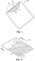
FIG. 1 is a perspective view of one embodiment of a device according to the present disclosure. -
FIG. 2 is another perspective view, partially in section, of the device ofFIG 1 . -
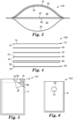
FIG. 3 is a cross-sectional view, taken along the line 3-3, of the device ofFIG. 2 . -
FIG. 4 is an exploded cross-sectional view, of the device ofFIG. 2 . -
FIG 5 is a plan view, partially in section, of an alternative embodiment of the device ofFIG 1 , showing an adhesive strip and a release liner releasably adhered thereto that form a sealable sample port. -
FIG. 6 is a plan view of an alternative embodiment of a device according to the present disclosure, wherein the device comprises a sealable sample port with a screwcap. -
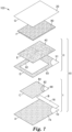
FIG. 7 is an exploded view of another alternative embodiment of a device according to the present disclosure. -
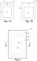
FIG. 7A is a first subassembly of the device ofFIG. 7 . -
FIG. 7B is a second subassembly of the device ofFIG. 7 . -
FIG. 8 is a plan view of the assembled device ofFIG. 7 . -
FIG. 9 is a cross-sectional view, taken along the line 9-9, of the device ofFIG. 8 . -
FIG. 10 is a block diagram of one embodiment of a method of detecting a target microorganism according to the present disclosure. - Before any embodiments of the present disclosure are explained in detail, it is to be understood that the invention is not limited in its application to the details of construction and the arrangement of components set forth in the following description or illustrated in the following drawings. The invention is capable of other embodiments and of being practiced or of being carried out in various ways. Also, it is to be understood that the phraseology and terminology used herein is for the purpose of description and should not be regarded as limiting. The use of "including," "comprising," or "having" and variations thereof herein is meant to encompass the items listed thereafter and equivalents thereof as well as additional items. Unless specified or limited otherwise, the terms "connected" and "coupled" and variations thereof are used broadly and encompass both direct and indirect connections and couplings. Further, "connected" and "coupled" are not restricted to physical or mechanical connections or couplings. It is to be understood that other embodiments may be utilized and structural or logical changes may be made without departing from the scope of the present disclosure. Furthermore, terms such as "front," "rear," "top," "bottom," and the like are only used to describe elements as they relate to one another, but are in no way meant to recite specific orientations of the apparatus, to indicate or imply necessary or required orientations of the apparatus, or to specify how the invention described herein will be used, mounted, displayed, or positioned in use.
- The present disclosure provides devices and methods for detecting and/or enumerating target microorganisms in relatively-large (e.g., about 25 mL to about 150 mL) liquid samples. It is now known that a self-contained device can include all of the components needed to concentrate microorganisms from a liquid sample, immobilize the microorganisms in a matrix, and provide a moist nutrient environment sufficient to grow and detect colonies of the microorganisms. Advantageously, the device can be used to detect and/or enumerate a wide variety of microorganisms (e.g., bacteria, yeast, and filamentous fungi) present in a liquid sample. In addition, the self-contained device provides certain advantages of thin-film culture devices such as, for example, sample-ready (i.e., just add liquid sample and then incubate), ease of use, portability, compactness, and a relatively long shelf-life.
- A device of the present disclosure can be used to enumerate microorganisms in a sample of water (e.g., surface water, process water, potable water). The water can be interrogated for the presence of certain target microorganisms including, for example, coliforms, fecal coliforms, E. coli, and/or total aerobic count or aerobic plate count (APC), yeast and mold. The presence of fecal coliforms in a water sample can indicate contamination of the water with human fecal material, which may contain certain pathogenic bacteria and/or viruses.
- The present disclosure provides a microbial detection device.
FIGS. 1-4 show various views of one embodiment of adevice 100 according to the present disclosure. Thedevice 100 comprises awaterproof pouch 5 defined by at least one wall. The at least one wall comprises afirst wall portion 10 and asecond wall portion 20. Thefirst wall portion 10 has aninner surface 12 and anouter surface 14. Thesecond wall portion 20 has aninner surface 22 and an outer surface 24. Disposed in thepouch 5 between theinner surface 12 of thefirst wall portion 10 and theinner surface 22 of thesecond wall portion 20 is amembrane filter 40. The membrane filter has a firstmajor surface 42 and a secondmajor surface 44 opposite the first major surface. - Although the
first wall portion 10 andsecond wall portion 20 may be distinct portions of a unitary pouch or bag, in any embodiment, the first wall portion and second wall portion alternatively may consist of separate sheets of polymeric film that are joined together (e.g., heat-sealed and/or adhesively sealed along the edges) to form the pouch, as shown inFIG. 5 , for example, and described herein. - The
pouch 5 is divided into at least two compartments (first compartment 50 andsecond compartment 52, respectively). Thefirst compartment 50 is defined in part by theinner surface 12 of thefirst wall portion 10 and also defined in part by the firstmajor surface 42 of themembrane filter 40. Thefirst compartment 50 has asealable sample port 60. In the illustrated embodiment ofFIGS. 1-3 , thesealable sample port 60 is simply anopening 61 along a portion of the perimeter of thepouch 5. Nonlimiting exemplary means for closing theopening 61 are discussed herein. Thesecond compartment 52 is defined in part by theinner surface 22 of thesecond wall portion 20 and defined in part by the secondmajor surface 44 of themembrane filter 40. - The
first compartment 50 is configured to receive a volume of liquid sample to be tested for presence of target microorganisms. The volume of liquid thefirst compartment 50 can receive will be influenced by several features of the device including, for example, the dimensions (e.g., the length "L" and width "W" shown inFIG. 3 ) of the first compartment and the flexibility of the materials (e.g., the first wall portion10 and membrane filter 40) that define the first compartment. Thesecond compartment 52 is configured to receive a volume of liquid approximately equal to the volume of liquid sample to be tested. Thus, the pouch of a device of the present disclosure may be dimensioned to hold up to about twice the volume of the sample to be tested. - In any embodiment, a device of the present disclosure is configured to test (i.e., configured to receive) at least about 25 milliliters of liquid sample. In any embodiment, a device of the present disclosure is configured to test at least about 50 milliliters of liquid sample. In any embodiment, a device of the present disclosure is configured to test at least about 75 milliliters of liquid sample. In any embodiment, a device of the present disclosure is configured to test at least about 100 milliliters of liquid sample. In any embodiment, a device of the present disclosure is configured to test at least about 125 milliliters of liquid sample. In any embodiment, a device of the present disclosure is configured to test at least about 150 milliliters of liquid sample. Thus, in any embodiment, the device according to the present disclosure is configured to receive at least about 25 mL, at least about 50 mL, at least about 75 mL, at least about 100 mL, at least about 125 mL, at least about 150 mL of liquid sample (e.g., aqueous liquid sample). Accordingly, in any embodiment, the first compartment of the device is configured to receive at least about 25 mL, at least about 50 mL, at least about 75 mL, at least about 100 mL, at least about 125 mL, at least about 150 mL of liquid sample (e.g., aqueous liquid sample).
- The
pouch 5 further comprises a dry (i.e., substantially water-free) cold water-soluble gelling agent adhered to the pouch (e.g., thefirst wall portion 10 of the pouch) in thefirst compartment 50.FIG. 3 shows the cold water-soluble gelling agent as adry coating 32 disposed on theinner surface 12 of thefirst wall portion 10. In any embodiment, thedry coating 32 can be adhered to thefirst wall portion 10 via anoptional adhesive layer 30. In addition, thepouch 5 has anabsorbent pad 80 disposed in thesecond compartment 52. - In any embodiment, the
dry coating 32 may be adhered to a first substrate (e.g., adhered to an adhesive layer coated on the substrate) that is adhered to thefirst wall portion 10 of thepouch 5. This optional configuration is shown inFIG. 7 and described hereinbelow. - Whether the cold-water-soluble gelling agent is adhered to the first wall portion of the pouch or to a first substrate that is adhered to the first wall portion, the area defined by the coating comprising the cold-water-soluble gelling agent also defines the area in which microorganisms from the sample grow and are enumerated after a sample is deposited into the first compartment. Because the device comprises an absorbent pad (described below) that absorbs most of the liquid from the sample, the cold-water-soluble gelling agent is hydrated by only a fraction of the liquid sample. Advantageously, the devices of the present disclosure use a surprisingly smaller ratio of growth area: sample volume than previously-reported thin-film culture devices.
- A number of thin-film culture devices are known. These devices, sold under the tradenames PETRIFILM, COMPACT DRY, and SANITA-KUN, for example. The devices typically include a gelling agent and/or water-absorptive matrix, nutrients, and chromogenic indicators to indicate presence of a microorganism colony. The thin-film culture devices typically are configured to receive one milliliter of a liquid sample, which hydrates the nutrients, indicators, and gelling agent and provide an environment for growth and enumeration of microorganism colonies. The one-milliliter sample is spread over a growth are of about 20 cm2 (e.g., PETRIFILM™ Aerobic Count Plate) to about 30 cm2 (e.g., PETRIFILM Yeast & Mold Count Plate). The PETRIFILM High-Sensitivity Coliform Count Plate is configured to receive 5 milliliters of sample, which is spread in the plate over an area of approximately 60 cm2. Thus, previous thin-film culture devices have a growth area (that includes a gelling agent and/or water-absorptive matrix) configured to receive about 1-5 milliliters of sample and to spread the microorganisms from that sample volume over a growth area that is equal to about 12 cm2 per mL of sample to about 30 cm2 per mL of sample.
- In contrast to previous thin-film culture devices, a device of the present disclosure is configured to receive 100-150 mL of a liquid sample and has a growth area (that includes a cold water-soluble gelling agent) of about 80 cm2. Thus, the microorganisms from the 150 mL sample volume is spread over a growth area that is equivalent to less than 1 cm2 per mL of sample.
- The pouch 5 (i.e., at least one wall, and wall portions thereof) is fabricated of a water-proof, deformable material. In any embodiment, the deformable material may comprise a flexible, sheet-like material such as a polymeric film, for example. Suitable materials for use when fabricating the at least one wall include polyethylene, polypropylene, polyethylene terephthalate, polyamide, polyurethane, polyvinyl chloride, polyacrylate, polyurea, and combinations thereof. The at least one wall of the pouch can be relatively thin (e.g., approximately 25 microns thick) or relatively thicker (e.g., approximately 125 microns thick), provided at least a portion of the at least one wall (e.g.,
first wall portion 10, which is opposite themembrane filter 40 in the first compartment 50) can deform when thepouch 5 receives a liquid sample (not shown) and/or at least a portion of the at least one wall (e.g.,second wall portion 20, which is proximate the absorbent pad described herein) can deform when at least a portion of the sample passes from the first compartment into the second compartment. - The
membrane filter 40 permits passage of a liquid (an aqueous liquid, not shown) from thefirst compartment 50 to thesecond compartment 52 and prevents passage of particles of a predetermined size from the first compartment to the second compartment. Thus, when an aqueous liquid sample suspected of containing a target microorganism is placed into thefirst compartment 50, a first portion of the aqueous liquid passes (e.g., by gravity flow) through themembrane filter 40 into thesecond compartment 52 where it is absorbed by theabsorbent pad 80. The target microorganism is trapped on or in thefilter membrane 40 or is retained in a second portion of the aqueous liquid that remains in thefirst compartment 50. - The use of membrane filters to trap and retain microorganisms is well known in the art. Accordingly, there are a number of suitable membrane filters that can be used in a device according to the present disclosure. Nonlimiting examples of suitable membrane filters include fibrous membrane filters made of nylon, polyether sulfone, polytetrafluoroethylene, or cellulosic materials (e.g., mixed cellulose esters), microporous plastic films (e.g., laser-etched polycarbonate film), and ceramic membrane filters.
- The porosity of the membrane filter generally is chosen so that the target microorganisms will not pass all the way though the pores from one side of the membrane filter to the other side, thereby insuring that substantially all target microorganisms in the sample are retained by the filter. Typical bacteria are about 0.5 to 5.0 µm in length. Certain smaller bacteria, such as Mycoplasma spp., are approximately 0.3 µm in diameter. Yeast cells are generally larger than bacteria. Typical yeast cells are approximately 3-4 µm in diameter, although some are as large as about 40 µm in diameter. Molds may exist as single cells, spores, or filamentous hyphae. Although typically larger than bacteria, the average size of mold cells varies by species. Accordingly, the selection of a membrane filter with a suitable pore size may depend upon the target microorganism. For example, a membrane filter with a nominal pore size of 1.0 µm or less, 0.8 µm or less, 0.6 µm or less, 0.4 µm or less, 0.2 µm or less, 0.1 µm or less, 0.05 µm or less, 0.03 µm or less, 0.02 µm or less, or 0.01 µm or less may be suitable to capture and detect target bacteria. For capturing and detecting target yeast or mold microorganisms, a membrane filter with a nominal pore size of 12 µm or less, 8 µm or less, 5 µm or less, 3 µm or less, 2 µm or less, 1 µm or less, 0.8 µm or less, 0.6 µm or less, 0.4 µm or less, 0.2 µm or less, or 0.1 µm or less may be suitable.
- Membrane filters may be prepared manually from suitable filtration media or, alternatively, may be purchased in pre-cut sizes and shapes. The size and shape of the membrane filter can be chosen based upon the sample volume and the expected load of particulate material in the sample. In general, membrane filters with larger surface areas will allow for higher filtration rates than membrane filters with smaller surface areas. Membrane filters may be used in combination with other filtration media (e.g., a prefilter, to trap larger debris in the sample) or other membrane filters.
- In any embodiment, the membrane filter may be supported (e.g., by a scrim, not shown) to provide physical stability for the membrane during use. In any embodiment, the support may be attached to the membrane filter (e.g., on the second major surface). In any embodiment, the membrane filter can comprise a wetting agent (e.g., a nonionic surfactant) to facilitate rapid and complete penetration of the liquid sample throughout the membrane filter. Preferably, the wetting agent is in an amount sufficient to facilitate wetting the membrane with an aqueous liquid, but in an amount that does not substantially inhibit growth of the target microorganism when using the device.
- The dry, cold water-soluble gelling agent is hydrated and forms a hydrogel when an aqueous sample is placed into the
first compartment 50 of thepouch 5. As the first portion of the aqueous liquid moves through themembrane filter 40 from thefirst compartment 50 to thesecond compartment 52, the hydrogel contacts the first surface of themembrane filter 40, thereby immobilizing any microorganisms retained on or in the membrane filter. - Cold water-soluble gelling agents that are suitable for use in thin-film culture devices are known in the art and include, for example, cold-water-soluble natural and synthetic gelling agents. Natural gelling agents such as algin, carboxymethyl cellulose, tara gum, hydroxyethyl cellulose, guar gum, locust bean gum, xanthan gum, and synthetic gelling agents such as polyacrylamide, polyurethane, polyethylene oxides, and mixtures thereof are generally suitable. Appropriate gelling agents can be selected according to the teaching of this disclosure and the disclosures of
U.S. Patent Nos. 4,565,783 ;5,089,413 ; and5,232,838 . Other preferred gelling agents include hydroxypropyl methylcellulose; these gelling agents being useful individually, or preferably, in combination with another gelling agent such as one of the aforementioned gelling agents. - In any embodiment, the dry, cold-water soluble gelling agent can be disposed in the pouch as a dry powder adhered to an adhesive layer, as described herein. Processes and adhesives for coating a dry powder onto a flexible film for use in a thin-film culture device are described, for example, in
U.S. Patent Nos. 4,565,783 ;5,089,413 ; and5,232,838 . In any embodiment, the adhesive layer, if present may comprise an indicator for indicating microorganism growth. For example, the adhesive may comprise triphenyltetrazolium chloride as described inU.S. Patent No. 5,409,838 . - In any embodiment, a device of the present disclosure optionally may comprise an effective amount of one or more dry nutrient (e.g., a nutrient medium selected to support growth of the target microorganism). The one or more dry nutrient may be disposed in the first compartment, for example. In any embodiment, the one or more dry nutrient may be disposed in the device (e.g., in the first compartment) as a dry powder or agglomerated powder. In any embodiment, the one or more nutrient can be adhered to the pouch (e.g., adhered to the first wall portion in the first compartment). In any embodiment, the one or more nutrient may be adhered to an adhesive layer that is adhered to the first wall portion, as described herein for the dry, cold water-soluble gelling agent.
- In any embodiment, the dry cold water-soluble gelling agent can be deposited onto the first wall portion of the pouch as an aqueous composition, optionally comprising the one or more nutrient, and subsequently dried, as described in
U.S. Patent Nos. 4,565,783 ;5,089,413 ; and5,232,838 . Optionally, in any embodiment, the dried coating can be adhered to an adhesive layer coated onto the first wall portion of the pouch. In any embodiment, the adhesive layer may further comprise an indicator for indicating microorganism growth, as described above. - Before a liquid sample is deposited into the pouch, the
absorbent pad 80 preferably relatively thin (e.g., less than or equal to 5 mm thick, less than or equal to 4 mm thick, less than or equal to 3 mm thick, less than or equal to 2 mm thick, less than or equal to about 1 mm thick) and is configured to absorb a quantity of deionized water equal to many time its own weight (e.g., at least 100-times its own weight, at least 150-times its own weight, at least 200-times its own weight, at least 250-times its own weight, at least 300-times its own weight, at least 350-times its own weight, at least 400-times its own weight, at least 500-times its own weight). In any embodiment, the absorbent pad may comprise a plurality of materials such as, for example, a super-absorbent material (e.g., a superabsorbent polymer; "herein, "SAP") and a less-absorbent or nonabsorbent carrier (e.g., cellulosic fibers). A nonlimiting example of a suitable absorbent pad is a composite polyacrylate laminate structure comprising a superabsorbent polymer granule base disposed between two cellulose sheets. In any embodiment of the absorbent pad, the pad may comprise SAP granules disposed in an air-laid nonwoven material or SAP fibers blended with carrier fibers into a nonwoven material. - Optionally, in any embodiment (not shown), the absorbent pad may be coupled to a component of the pouch (e.g., the second wall portion) in the second compartment. Advantageously, this can keep the pad from deforming (e.g., as it swells with liquid migrating from the first compartment) to an extent that it loses contact with a substantial portion of the membrane filter. The pad may be coupled to the pouch via an adhesive (e.g., a pressure-sensitive adhesive), a thermal weld or other suitable attachment means known in the art. In any embodiment, the absorbent pad may be releasably coupled to the pouch (e.g., by a water-soluble gum). This embodiment hold the pad in a proper position to receive liquid passing through the membrane filter, but permits lateral movement of the pad as it swells due to absorption of a large quantity of the liquid.
- Referring back to the drawings,
FIG. 5 shows one embodiment of asealable sample port 60 of adevice 101 according to the present disclosure. Thedevice 101 comprises apouch 5 having afirst wall portion 10, asecond wall portion 20, and asealable sample port 60 consisting of an opening, each as described herein. Theinner surface 12 of thefirst wall portion 10 comprises anadhesive strip 16 coated thereon along the edge of the inner surface proximate the opening. Adhered to theadhesive strip 16 is arelease liner 18. After the sample is deposited (e.g., by pouring or pipetting) into the first compartment (not shown inFIG. 5 ) through the opening (sample port 60), the operator removes the release liner and contacts theadhesive strip 16 with theinner surface 22 of thesecond wall portion 20 proximate the opening in order to seal the opening. Optionally, the operator can expel (out of the opening) some or all of the air from thefirst compartment 50 when completing the sealing process. -
FIG. 6 shows an alternative embodiment of adevice 102 comprising apouch 6 comprising asealable sample port 60 with anopening 61. In this embodiment, thesealable sample port 60 is a screw-cap opening into which the liquid test sample cab be poured or pipetted, for example. Alternatively, in any embodiment, thesealable sample port 60 can be a pierceable, elastically-deformable septum through which a needle or a pipet tip can be introduced to deliver the sample into the first compartment. After the needle or pipet is withdrawn from the septum, the elastically-deformable septum reseals the port. Advantageously, in these embodiments, the introduction of air into the first compartment can be minimized. - In another alternative embodiment (not shown), the sealable sample port can comprise interlocking zipper components (e.g., similar to a ZIPLOK® plastic storage bag) on each of the first wall portion and second wall portion and a zipper component that is used cooperatively with the interlocking components to open or seal the first compartment.
- In another aspect, the present disclosure provides a method of assembling a large-volume, thin-film culture device. Devices of the present disclosure can be assembled entirely from sheet-like materials. Advantageously, this enables the use of roll-to-roll processes when assembling a plurality of devices.
FIGS. 7-9 show various views of an alternative embodiment of adevice 103 according to the present disclosure. -
FIG. 7 shows the sheet-like materials that are used to assemble one embodiment of a device according to the present disclosure. Each part of the device can be cut into appropriately-sized sheets and subsequently assembled into the device or, alternatively can be cut to the appropriate size using controlled-depth die cutting using a roll-to-roll process known in the art. - In any embodiment, a device of the present disclosure can be partially assembled into one or more subassembly, which is subsequently combined with other components to make the device. Referring to
FIG. 7 , thedevice 103 includes a first subassembly I that comprises a first part A, a second part B, and a third part C. Another view of the assembled first subassembly I is shown inFIG. 7A . The first part A consists of thefirst wall portion 10 with anadhesive layer 74 coated thereon as described herein. Second part B consists of arelease liner 18 as described herein. Third part C consists of afirst substrate 90 coated on one side with anadhesive layer 82. Disposed on theadhesive layer 82 is acoating 84 that comprises the dry, cold water-soluble gelling agent described herein. Thecoating 84 can be deposited onto theadhesive layer 82 as a dry powder or as a liquid composition that is subsequently dried to a substantially water-free state, as described hereinabove. Thefirst substrate 90 can comprise a sheet-like material similar to those used for the walls of the pouch as described above. Alternatively, the first substrate can comprise a nonwoven fabric or a cellulosic material (e.g., paper). In any embodiment, the cellulosic material can be coated with a waterproof coating that is substantially noninhibitory to growth of microorganisms. The area defined by thecoating 84 on third part C also defines the growth and colony-enumeration area in the assembled device. - When assembling subassembly I, the
release liner 18 is releasably adhered to theadhesive layer 74 along the edge (edge 11) of thefirst wall portion 10 that forms the opening of the assembled device. In addition, the third part C is positioned centrally over part A with thecoating 84 facing away from theadhesive layer 74. Part C is then contacted with adhesive 74 to affix part C to part A with thecoating 84 exposed., as shown inFIG. 7A . - Referring back to
FIG. 7 , a second subassembly II includes a fourth part D and a fifth part E. The fourth part D comprises asecond substrate 91. Thesecond substrate 91 forms a frame comprising anaperture 92. Thesecond substrate 91 is coated on one side with anadhesive layer 93. Thesecond substrate 91 can comprise a sheet-like material (e.g., a flexible film) similar to those used for the walls of the pouch as described above. Alternatively, the second substrate can comprise a nonwoven fabric or a cellulosic material (e.g., paper). In any embodiment, the cellulosic material can be coated with a waterproof coating that is substantially noninhibitory to growth of microorganisms. Optionally, the absorbent pad can be coupled to the second substrate in the second compartment. - The second subassembly II also includes the fifth part E (i.e.,
membrane filter 40, as described herein). Themembrane filter 40 is dimensioned so that it completely covers the area defined by theaperture 92. When assembling subassembly II, themembrane filter 40 is adhered to theadhesive layer 93 so that it completely covers theaperture 92 of thesecond substrate 91, as shown inFIG. 7B . In use, liquid passes through the aperture from the first compartment to the second compartment of the device as the liquid passes through the membrane filter. In any embodiment, theaperture 92 defines a first area and thecoating 84 defines a second area. Preferably, the second area is greater than or equal to the first area. More preferably, the second area is shaped and dimensioned to completely overlap the area of the aperture. - Optionally, when assembling the
device 103 ofFIG. 7 , the subassembly I can be coupled to subassembly II to form a subassembly III. This can be done by placing the back side (i.e., the side that does not include adhesive layer 93) of subassembly II in overlaying contact with the adhesive-coated side of subassembly I. In addition, theaperture 92 of subassembly II is aligned with subassembly I so that it overlaps the third part C of subassembly I. - To complete the construction of the
device 103, the sixth part F (i.e.,absorbent pad 80, as described herein) is placed in overlaying contact with themembrane filter 40 of the subassembly III and the seventh part (i.e.,second wall portion 20, as described herein) is placed in overlaying contact with the first part A such that the seventh part G is adhesively coupled to the portion of theadhesive layer 74 at the periphery of the first part A.FIG. 8 shows a plan view andFIG. 9 shows a cross-sectional view of the assembleddevice 103 ofFIG. 7 . - In any embodiment, a device of the present disclosure further comprises an indicator reagent for indicating a presence of a viable microorganism. The indicator reagent is disposed in the pouch. In any embodiment, the indicator reagent may be disposed as a dry powder or dried coating in the first compartment and/or the second compartment of the pouch. In any embodiment, the indicator reagent can be disposed in an adhesive layer as described herein. Alternatively, or additionally, the indicator reagent may be a dry reagent coated onto an adhesive layer (e.g., with the cold water-soluble gelling agent as described herein).
- In any embodiment, the indicator reagent may be a general indicator (e.g., a redox indicator such as triphenyltetrazolium chloride, for example) of viable microorganisms or an indicator of a large class of target microorganisms (e.g., total aerobic microorganisms). Alternatively, the indicator reagent can be an indicator (e.g., a chromogenic or fluorogenic enzyme substrate) that reacts with a smaller group of target microorganisms. A person having ordinary skill in the will recognize an appropriate indicator reagent for a particular target microorganism.
- In any embodiment of a device according to the present disclosure, the device further comprises a stand-off layer (not shown) disposed in the second compartment between the membrane filter and the absorbent pad. The stand-off layer is a relatively-thin (e.g., about 0.1 mm to 2 mm thick) sheet-like material. In any embodiment, the stand-off layer is shaped and dimensioned to be at least coextensive with the membrane filter. In any embodiment, the stand-off layer is substantially less absorbent than the absorbent pad. In any embodiment, the absorbency of the stand-off layer is less than or equal to the absorbency of the membrane filter. The stand-off layer may comprise or consist essentially of a hydrophobic material (e.g., unmodified polypropylene).
- The stand-off layer functions to permit the passage of aqueous liquid from the membrane filter to the absorbent layer during the initial period in which over half of the aqueous liquid deposited into the first compartment passes into the second compartment, while restricting diffusion of nutrient from the first compartment to the second compartment while the device is being incubated to facilitate microbial colony growth.
- Suitable materials for use as the stand-off layer include, for example nonwoven fabrics comprising polypropylene; polyethylene; polyethylene terephthalate; a blend of polyethylene terephthalate and cellulose; a blend of polyethylene terephthalate and rayon; and mixtures thereof. Advantageously, devices comprising the stand-off layer can include dry nutrients coated on the first wall portion of the pouch and can retain enough nutrients in the hydrated cold water-soluble gelling agent to support growth of the target microorganisms in the hydrated nutrient gel.
- In another aspect, the present disclosure provides a method. The method can be used to detect and, optionally, enumerate target microorganisms in a liquid sample.
FIG. 10 shows a block diagram that shows the steps of one embodiment of amethod 1000 of detecting microorganisms in a liquid sample according to the present disclosure. - The
method 1000 comprises astep 200 of placing a predetermined volume of aqueous sample into the first compartment of the device of any one of the embodiments of the present disclosure. The aqueous sample can be any filterable liquid sample to be tested for presence of a target microorganism. The method is particularly useful for water samples that are suspected of containing relatively low concentrations (e.g., less than or equal to 10 microorganisms per milliliter, less than or equal to 1 microorganism per milliliter, less than or equal to 0.1 microorganisms per milliliter, less than or equal to 0.01 microorganism per milliliter,) of target microorganisms. Placing a predetermined volume of aqueous sample into the first compartment of the device comprises placing the predetermined volume into the device (e.g., via pipetting, pouring, injecting, or the like) through the sealable sample port. - The
method 1000 further comprises astep 210 of sealing the sample port. The procedure for sealing the sample port will depend upon the particular sealable sample port that is present in the device used in themethod 1000. For example, if thedevice 103 ofFIGS. 7-8 is used in the method, sealing the sample port comprises removing therelease liner 18 to expose an adhesive disposed on thefirst wall portion 10 and then contacting the adhesive on the first wall portion with the second wall portion to form a waterproof seal that closes the opening of the pouch. - For example, if the
device 102 ofFIG. 6 is used in themethod 1000, sealing the sample port comprises screwing the cap back onto the sample port, thereby forming a waterproof seal. - For example, if a device comprising an elastically-deformable pierceable septum (not shown) is used in the
method 1000, sealing the sample port will spontaneously occur as the pipet or needle used to introduce the sample into the device is withdrawn from the septum. - In any embodiment of the method, air may be expelled (e.g., manually, by squeezing) from the pouch via the sealable sample port before and or during the process of forming the waterproof seal.
- The
method 1000 further comprises astep 220 of incubating the device for a period of time at a temperature that facilitates growth and detection of a target microorganism. A person having ordinary skill in the art will recognize the incubation temperature and period of time will depend upon a number of factors (e.g., the target microorganism, nutrients present in the sample, nutrients present in the device, inhibitory agents present in the sample and/or the device) and will adjust the incubation time and temperature accordingly. - The
method 1000 further comprises astep 230 of detecting a presence or an absence of a colony of the target microorganism in the device. In any embodiment, detecting a presence or an absence of a colony of the target microorganism in the device can comprise detecting a colony (e.g., visually or using machine vision) in the first compartment of the device. In any embodiment, detecting a presence or an absence of a colony of the target microorganism in the device can comprise detecting a change associated with the indicator reagent. The indicator reagent may change from a first state (e.g., substantially colorless or nonfluorescent) to a second state (e.g., colored or fluorescent) in and/or surrounding a colony of the target microorganism. In any embodiment, the colonies can be enumerated and, optionally, the number of colonies of target microorganisms can be recorded. - In any embodiment, after sealing the sample port, the method further comprises laying the outer surface of the first wall portion of the device or laying the outer surface of the second wall portion of the device onto a surface that is substantially perpendicular to gravitational force. Advantageously, laying the outer surface of its second wall portion of the device onto a surface that is substantially perpendicular to the force of gravity facilitates flow of the sample liquid through the membrane filter by force of gravity. In addition, laying the outer surface of its second wall portion of the device onto a surface that is substantially perpendicular to the force of gravity facilitates contact between the hydrated cold water-soluble gelling agent adhered to the first wall portion and the membrane filter as the liquid passes through the membrane filter from the first compartment to the second compartment.
- In any embodiment, the method further comprises passing at least 90%, at least 92%, at least 95 %, at least 97% or at least 98% of the predetermined volume from the first compartment to the second compartment. The portion of the predetermined volume that remains in the first compartment is substantially present as part of the gel formed by hydrating the cold water-soluble gelling agent.
- In any embodiment, the method further comprises a
step 240 of combining the aqueous sample with a nutrient, nutrient medium, indicator reagent and/or selective agent prior to placing the predetermined volume into the first compartment. In any embodiment, the method further comprises combining the aqueous sample with a nutrient, nutrient medium, indicator reagent and/or selective agent after placing the predetermined volume into the first compartment. - Culture Media Formulations
Table 1. Culture Media Formulation A Component Source (Final Concentration in sterile water) Bacto™ Tryptic Soy Broth (30 g/L) Becton, Dickinson (New Franklin, NJ) BCIG (100 µg/mL) Biosynth International (Itasca, IL) Table 2. Culture Media Formulation B Component (Final Concentration in sterile water) Source Bacto™ Tryptic Soy Broth (30 g/L) Becton, Dickinson (New Franklin, NJ) BBL™ Beef Extract Becton, Dickinson (New Franklin, NJ) Sodium chloride (5 g/L) Sigma-Aldrich (St. Louis, MO) Lactose (5 g/L) Sigma-Aldrich (St. Louis, MO) Phenol red (0.5 g/L) Sigma-Aldrich (St. Louis, MO) BCIG (100 µg/mL) Biosynth International (Itasca, IL) Table 3. Culture Media Formulation C Component (Final Concentration in sterile water) Source Colilert® media (per manufacturer instructions) IDEXX Labs (Westbrook, ME) BCIG (100 µg/mL) Biosynth International (Itasca, IL) - Culture Media Formulation D was prepared by adding ReadyCult® media (EMD Millipore, Billerica, MA) to 100 mL of sterile water per the manufacturer instructions.
- The inoculum was prepared by growing an overnight culture of Escherichia coli ATCC 25922 (EZ-CFU™, Microbiologies Incorporated, St. Cloud, MN) in Bacto™ Tryptic Soy Broth (TSB) (Becton, Dickinson, New Franklin, NJ) at 37°C with shaking. The inoculum sample was serially diluted with Butterfield's Buffer (3M Company) with the final dilution in the series being 1mL of inoculum sample in either 99 mL of TSB, 99 mL of a diluent selected from Culture Media Formulations A-D, or 99 mL of Butterfield's Buffer. Each inoculum samples was serially-diluted to obtain final concentrations of about 10-700 cfu per 100 mL in the final dilution.
- The final dilution (100 mL) was then poured into the first compartment of a pouch device (selected from Examples 1-12). The release liner on the pouch was removed and the first compartment was sealed. The device was then placed on a flat, horizontal surface (outer surface of the second wall portion facing the horizontal surface) in an incubator and maintained at 37°C for 18 hours. The colonies (cfu) in each device were counted by visual examination at the end of the incubation period. The colonies were red-colored when TSB or Nutrient Formulation D was used as the final diluent. The colonies were blue-colored when the final diluent was selected from Nutrient Formulations A-D.
- As a reference, a 1 mL sample of the inoculum was taken immediately prior to the final dilution step and plated onto a PETRIFILM Aerobic Count Plate (3M Company, Maplewood, MN) according to the manufacturer's instructions. The reference plate was incubated at 37°C for 18 hours and the red-colored colonies were counted by visual examination.
- A microbial detection device according to
FIG. 7 was constructed. The second wall portion consisted of a 127 mm by 152.4 mm (5 in by 6 in) piece of 1.6 mil (0.04 mm) thick biaxially-oriented polypropylene (BOPP) film. The absorbent pad was a 76.2 mm by 101.6 mm (3 in by 4 in) piece of Gelok 30040-76 superabsorbent polymer (SAP) laminate (300 g/m2 of sodium polyacrylate granules laminated between tissue layers, Gelok Industries, Dunbridge, OH) and the membrane filter was a 101.6 mm by 127 mm (4 in by 5 in) piece of nylon membrane with a nominal pore size of 0.45 microns (#BA045 membrane, 3M Company, Maplewood, MN). The absorbent pad was placed and centered on the inner surface of the second wall portion. Likewise, the SAP laminate was placed and centered on the side of the absorbent pad facing opposite from the second wall. In the orientation of this construction, a 12.7 mm (0.5 in) strip along the perimeter of the inner surface of the second wall portion was not covered. - A frame layer was prepared by first coating one side of a 1.6 mil (0.04 mm) BOPP film with an isooctyl acrylate/acrylamide pressure sensitive adhesive containing tetrazolium chloride (TTC) according to the method described in
U.S. Patent No. 5,409,838 . The coated film was subsequently cut to form a frame having external dimensions of 127 mm by 152.4 mm (5 in by 6 in) and a centered 76.2 mm by 101.6 mm (3 in by 4 in) internal opening. The resulting frame had a 25.4 mm (1 in) wide adhesively coated border. - The frame then was adhesively attached to the membrane filter and the inner surface of the second wall creating a partially constructed device that had a 76.2 mm by 101.6 mm (3 in by 4 in) section of the membrane uncovered on one side.
- A separate 76.2 mm by 101.6 mm (3 in by 4 in) piece of 1.6 mil (0.04 mm) BOPP film that had been coated on one side with an isooctyl acrylate/acrylamide pressure sensitive adhesive containing tetrazolium chloride (TTC) according to the method described in U.S. Patent No.
5,409,838 was powder coated with guar gum (Meyprogat 150, Dupont Corporation, Wilmington, DE). The powder was uniformly applied to the adhesive and excess powder was removed by hand shaking of the film. The powder coated film was then placed to cover the previously uncovered membrane of the partially constructed device. The film was oriented such that the coated side of the film faced the membrane. - The first wall portion consisted of a 127 mm by 152.4 mm (5 in by 6 in) piece of 1.6 mil (0.04 mm) thick biaxially-oriented polypropylene (BOPP) film that had been coated on one side with an isooctyl acrylate/acrylamide pressure sensitive adhesive containing tetrazolium chloride (TTC) according to the method described in
U.S. Patent No. 5,409,838 . A one inch wide piece of silicone coated paper release liner was attached along one of the 127 mm edges on the coated surface of the first wall portion. The first wall portion was then edge aligned with and adhesively laminated to the surface of the frame layer that faced away from the second wall portion. This construction resulted in a pouch with an opening to a first compartment defined in part by the first wall portion and the membrane filter. - A microbial detection device was constructed according to the procedure described in Example 1 with the exception that the absorbent pad was larger [101.6 mm by 127 mm (4 in by 5 in), instead of 76.2 mm by 101.6 mm (3 in by 4 in)].
- A microbial detection device was constructed according to the procedure described in Example 1 with the exception that the absorbent pad was a 89 mm by 114.3 mm (3.5 in by 4.5 in) piece of Gelok 20040-76 superabsorbent polymer (SAP) laminate (200 g/m2 of sodium polyacrylate granules laminated between tissue layers, Gelok Industries, Dunbridge, OH), instead of Gelok 30040-76.
- A microbial detection device was constructed according to the procedure described in Example 3 with the exception that the absorbent pad was larger [101.6 mm by 127 mm (4 in by 5 in), instead of 89 mm by 114.3 mm (3.5 in by 4.5 in)].
- A microbial detection device was constructed according to the procedure described in Example 1 with the exception that the membrane filter was a 101.6 mm by 127 mm (4 in by 5 in) piece of
nylon - A microbial detection device was constructed according to the procedure described in Example 1 with the exception that the membrane filter was a 101.6 mm by 127 mm (4 in by 5 in) piece of
nylon - A microbial detection device was constructed according to the procedure described in Example 1 with the exception that the membrane filter was a 101.6 mm by 127 mm (4 in by 5 in) piece of DuraPES 450 poyether sulfone membrane (Membrana GMBH, Wuppertal, Germany) with a nominal pore size of 0.45 microns, instead of BA045 membrane.
- A microbial detection device according to
FIG. 7 was constructed. The second wall portion consisted of a 127 mm by 152.4 mm (5 in by 6 in) piece of 1.6 mil (0.04 mm) thick biaxially-oriented polypropylene (BOPP) film. The absorbent pad was a 101.6 mm by 127 mm (4 in by 5 in) piece of Gelok 30040-76 superabsorbent polymer (SAP) laminate 300 g/m2 of sodium polyacrylate granules laminated between tissue layers, Gelok Industries, Dunbridge, OH) and the membrane filter was a 101.6 mm by 127 mm (4 in by 5 in) piece of nylon membrane with a nominal pore size of 0.45 microns (#BA045 membrane, 3M Company, Maplewood, MN). A 101.6 mm by 127 mm (4 in by 5 in) piece of Typar 3091L non-woven polypropylene material (0.2 mm thick, 31.6 gsm, Midwest Filtration LLC, Cincinnati, OH) was adhesively laminated between the SAP laminate and nylon membrane with Super 77 spray adhesive (3M Company). The resulting laminate was placed and centered on the inner surface of the second wall portion oriented such that the absorbent pad faced the inner surface of the second wall. In the orientation of this construction, a 12.7 mm (0.5 in) strip along the perimeter of the inner surface of the second wall portion was not covered. - A frame layer was prepared by first coating one side of a 1.6 mil (0.04 mm) BOPP film with an isooctyl acrylate/acrylamide pressure sensitive adhesive containing tetrazolium chloride (TTC) according to the method described in
U.S. Patent No. 5,409,838 . The coated film was subsequently cut to form a frame having external dimensions of 127 mm by 152.4 mm (5in by 6 in) and a centered 76.2 mm by 101.6 mm (3 in by 4 in) internal opening. The resulting frame had a 25.4 mm (1 in) wide adhesively coated border. - The frame then was adhesively attached to the membrane filter and the inner surface of the second wall creating a partially constructed device that had a 76.2 mm by 101.6 mm (3 in by 4 in) section of the membrane uncovered on one side.
- A separate 76.2 mm by 101.6 mm (3 in by 4 in) piece of 1.6 mil (0.04 mm) BOPP film that had been coated on one side with an isooctyl acrylate/acrylamide pressure sensitive adhesive containing tetrazolium chloride (TTC) according to the method described in
U.S. Patent No. 5,409,838 was powder coated with guar gum (Meyprogat 150, Dupont Corporation, Wilmington, DE). The powder was uniformly applied to the adhesive and excess powder was removed by hand shaking of the film. The powder coated film was then placed to cover the previously uncovered membrane of the partially constructed device. The film was oriented such that the coated side of the film faced the membrane. - The first wall portion consisted of a 127 mm by 152.4 mm (5 in by 6 in) piece of 1.6 mil (0.04 mm) thick biaxially-oriented polypropylene (BOPP) film that had been coated on one side with an isooctyl acrylate/acrylamide pressure sensitive adhesive containing tetrazolium chloride (TTC) according to the method described in
U.S. Patent No. 5,409,838 . A one inch wide piece of silicone coated paper release liner was attached along one of the 127 mm edges on the coated surface of the first wall portion. The first wall portion was then edge aligned with and adhesively laminated to the surface of the frame layer that faced away from the second wall portion. This construction resulted in a pouch with an opening to a first compartment defined in part by the first wall portion and the membrane filter. - A microbial detection device was constructed according to the procedure described in Example 8 with the exception that the non-woven material adhesively laminated between the SAP laminate and nylon membrane was Fitesa ADL-2 (carded polester with resin bond, 55 gsm, 0.4 mm thick, Fitesa Company, Simpsonville, SC), instead of Typar 3091L.
- A microbial detection device was constructed according to the procedure described in Example 8 with the exception that the non-woven material adhesively laminated between the SAP laminate and nylon membrane was WC110 (a PET/viscose rayon (50/50) blend, 110 gsm, 0.5 mm thick, Nonwoven Solutions LLC, Ingleside, IL), instead of Typar 3091L.
- A microbial detection device according to
FIG. 7 was constructed. The second wall portion consisted of a 127 mm by 152.4 mm (5 in by 6 in) piece of 1.6 mil (0.04 mm) thick biaxially-oriented polypropylene (BOPP) film. The absorbent pad was a 101.6 mm by 127 mm (4 in by 5 in) piece of Gelok 30040-76 superabsorbent polymer (SAP) laminate 300 g/m2 of sodium polyacrylate granules laminated between tissue layers, Gelok Industries, Dunbridge, OH) and the membrane filter was a 101.6 mm by 127 mm (4 in by 5 in) piece of nylon membrane with a nominal pore size of 0.45 microns (#BA045 membrane, 3M Company, Maplewood, MN). The absorbent pad was placed and centered on the inner surface of the second wall portion. Likewise, the SAP laminate was placed and centered on the side of the absorbent pad facing opposite from the second wall. In the orientation of this construction, a 12.7 mm (0.5 in) strip along the perimeter of the inner surface of the second wall portion was not covered. - A frame layer was prepared by first coating one side of a 1.6 mil BOPP film with an isooctyl acrylate/acrylamide pressure sensitive adhesive containing tetrazolium chloride (TTC) according to the method described in
U.S. Patent No. 5,409,838 . The coated film was subsequently cut to form a frame having external dimensions of 127 mm by 152.4 mm (5in by 6 in) and a centered 76.2 mm by 101.6 mm (3 in by 4 in) internal opening. The resulting frame had a 25.4 mm (1 in-wide adhesively coated border. The frame then was adhesively attached to the membrane filter and the inner surface of the second wall creating a partially constructed device that had a 76.2 mm by 101.6 mm (3 in by 4 in) section of the membrane uncovered on one side. - A separate 76.2 mm by 101.6 mm (3 in by 4 in) piece of 1.6 mil BOPP film that had been coated on one side with an isooctyl acrylate/acrylamide pressure sensitive adhesive containing tetrazolium chloride (TTC) according to the method described in
U.S. Patent No. 5,409,838 was powder coated with a homogenous powder blend of guar gum (66%) and the Nutrient Formulation of Table 4 (33%). The powder mixture was uniformly applied to the adhesive and excess powder was removed by hand shaking of the film. The powder coated film was then placed to cover the previously uncovered membrane of the partially constructed device. The film was oriented such that the coated side of the film faced the membrane. - The first wall portion consisted of a 127 mm by 152.4 mm (5 in by 6 in) piece of 1.6 mil (0.04 mm) thick biaxially-oriented polypropylene (BOPP) film that had been coated on one side with an isooctyl acrylate/acrylamide pressure sensitive adhesive containing tetrazolium chloride (TTC) according to the method described in
U.S. Patent No. 5,409,838 . A one inch wide piece of silicone coated paper release liner was attached along one of the 127 mm edges on the coated surface of the first wall portion. The first wall portion was then edge aligned with and adhesively laminated to the surface of the frame layer that faced away from the second wall portion. This construction resulted in a pouch with an opening to a first compartment defined in part by the first wall portion and the membrane filter. - A microbial detection device according to
FIG. 7 was constructed. The second wall portion consisted of a 127 mm by 152.4 mm (5 in by 6 in) piece of 1.6 mil (0.04 mm) thick biaxially-oriented polypropylene (BOPP) film. The absorbent pad was a 101.6 mm by 127 mm (4 in by 5 in) piece of Gelok 30040-76 superabsorbent polymer (SAP) laminate 300 g/m2 of sodium polyacrylate granules laminated between tissue layers, Gelok Industries, Dunbridge, OH) and the membrane filter was a 101.6 mm by 127 mm (4 in by 5 in) piece of nylon membrane with a nominal pore size of 0.45 microns (#BA045 membrane, 3M Company, Maplewood, MN). A 101.6 mm by 127 mm (4 in by 5 in) piece of Fitesa-ADL2 non-woven material was adhesively laminated between the SAP laminate and nylon membrane with Super 77 spray adhesive (3M Company). The resulting laminate was placed and centered on the inner surface of the second wall portion oriented such that the absorbent pad faced the inner surface of the second wall. In the orientation of this construction, a 12.7 mm (0.5 in) strip along the perimeter of the inner surface of the second wall portion was not covered. - A frame layer was prepared by first coating one side of a 1.6 mil (0.04 mm) BOPP film with an isooctyl acrylate/acrylamide pressure sensitive adhesive containing tetrazolium chloride (TTC) according to the method described in
U.S. Patent No. 5,409,838 . The coated film was subsequently cut to form a frame having external dimensions of 127 mm by 152.4 mm (5in by 6 in) and a centered 76.2 mm by 101.6 mm (3 in by 4 in) internal opening. The resulting frame had a 25.4 mm (1 in) wide adhesively coated border. - The frame then was adhesively attached to the membrane filter and the inner surface of the second wall creating a partially constructed device that had a 76.2 mm by 101.6 mm (3 in by 4 in) section of the membrane uncovered on one side.
- A separate 76.2 mm by 101.6 mm (3 in by 4 in) piece of 1.6 mil (0.04 mm) BOPP film that had been coated on one side with an isooctyl acrylate/acrylamide pressure sensitive adhesive containing tetrazolium chloride (TTC) according to the method described in
U.S. Patent No. 5,409,838 was powder coated with a homogenous powder blend of guar gum (66%) and the Nutrient Formulation of Table 4 (33%). The powder mixture was uniformly applied to the adhesive and excess powder was removed by hand shaking of the film. The powder coated film was then placed to cover the previously uncovered membrane of the partially constructed device. The film was oriented such that the coated side of the film faced the membrane. - The first wall portion consisted of a 127 mm by 152.4 mm (5 in by 6 in) piece of 1.6 mil (0.04 mm) thick biaxially-oriented polypropylene (BOPP) film that had been coated on one side with an isooctyl acrylate/acrylamide pressure sensitive adhesive containing tetrazolium chloride (TTC) according to the method described in
U.S. Patent No. 5,409,838 . A one inch wide piece of silicone coated paper release liner was attached along one of the 127 mm edges on the coated surface of the first wall portion. The first wall portion was then edge aligned with and adhesively laminated to the surface of the frame layer that faced away from the second wall portion. This construction resulted in a pouch with an opening to a first compartment defined in part by the first wall portion and the membrane filter. - Table 4. Nutrient powder formulation used to prepare the device of Examples 11 and 12.
Component Amount (grams) Source Meat peptone, porcine 3.0 Alpha Biosciences, Baltimore, MD Soytone 3.0 Becton, Dickinson (New Franklin, NJ) Tryptone 3.0 Becton, Dickinson (New Franklin, NJ) Yeast extract 6.0 Alpha Biosciences (Baltimore, MD) Pyruvic acid (sodium salt) 18.5 J.T. Baker (Center Valley, PA) Potassium phosphate monobasic 1.2 EMD Millipore (Billerica, MA) Potassium phosphate dibasic 3.6 EMD Millipore (Billerica, MA) Dextrose 1.5 Becton, Dickinson (New Franklin, NJ) Magnesium sulfate 0.1 EMD Millipore (Billerica, MA) Calcium chloride 0.1 Mallinckrodt (St. Louis, MO) Magnesium chloride 0.1 Alfa Aesar (Ward Hill, MA) Sodium carbonate 0.1 Sigma-Aldrich (St. Louis, MO) Zinc sulfate 0.1 EMD Millipore (Billerica, MA) L-Arginine hydrochloride 0.26 EMD Millipore (Billerica, MA) D-(+)-Trehalose 0.5 Alfa Aesar (Ward Hill, MA) Skim milk powder 0.5 EMD Millipore (Billerica, MA) The components of the nutrient formulation were blended together to form a homogeneous mixture - A microbial detection device according to
FIG. 7 was constructed. The second wall portion consisted of a 127 mm by 152.4 mm (5 in by 6 in) piece of 1.6 mil (0.04 mm) thick biaxially-oriented polypropylene (BOPP) film that had been coated on the inner surface with an isooctyl acrylate/acrylamide pressure sensitive adhesive containing tetrazolium chloride (TTC) according to the method described inU.S. Patent No. 5,409,838 . The absorbent pad was a 72.6 mm by 101.6 mm (3 in by 4 in) piece of Gelok 30040-76 superabsorbent polymer (SAP) laminate 300 g/m2 of sodium polyacrylate granules laminated between tissue layers, Gelok Industries, Dunbridge, OH) and the membrane filter was a 101.6 mm by 127 mm (4 in by 5 in) piece of nylon membrane with a nominal pore size of 0.45 microns (#BA045 membrane, 3M Company, Maplewood, MN). The absorbent pad was placed and centered on the inner surface of the second wall portion. Likewise, the SAP laminate was placed and centered on the side of the absorbent pad facing opposite from the second wall. In the orientation of this construction, a 12.7 mm (0.5 in) strip along the perimeter of the inner surface of the second wall portion was not covered. - A frame layer was prepared by first coating one side of a 1.6 mil (0.04 mm) BOPP film with an isooctyl acrylate/acrylamide pressure sensitive adhesive containing tetrazolium chloride (TTC) according to the method described in
U.S. Patent No. 5,409,838 . The coated film was subsequently cut to form a frame having external dimensions of 127 mm by 152.4 mm (5in by 6 in) and a centered 76.2 mm by 101.6 mm (3 in by 4 in) internal opening. The resulting frame had a 25.4 mm (1 in) wide adhesively coated border. - The frame then was adhesively attached to the membrane filter and the inner surface of the second wall creating a partially constructed device that had a 76.2 mm by 101.6 mm (3 in by 4 in) section of the membrane uncovered on one side.
- A separate 76.2 mm by 101.6 mm (3 in by 4 in) piece of 1.6 mil (0.04 mm) BOPP film that had been coated on one side with an isooctyl acrylate/acrylamide pressure sensitive adhesive containing tetrazolium chloride (TTC) according to the method described in
U.S. Patent No. 5,409,838 was powder coated with guar gum (Meyprogat 150, Dupont Corporation, Wilmington, DE). The powder was uniformly applied to the adhesive and excess powder was removed by hand shaking of the film. The powder coated film was then placed to cover the previously uncovered membrane of the partially constructed device. The film was oriented such that the coated side of the film faced the membrane. - The first wall portion consisted of a 127 mm by 152.4 mm (5 in by 6 in) piece of 1.6 mil (0.04 mm) thick biaxially-oriented polypropylene (BOPP) film that had been coated on one side with an isooctyl acrylate/acrylamide pressure sensitive adhesive containing tetrazolium chloride (TTC) according to the method described in
U.S. Patent No. 5,409,838 . A one inch wide piece of silicone coated paper release liner was attached along one of the 127 mm edges on the coated surface of the first wall portion. The first wall portion was then edge aligned with and adhesively laminated to the surface of the frame layer that faced away from the second wall portion. This construction resulted in a pouch with an opening to a first compartment defined in part by the first wall portion and the membrane filter. - The colony counts (cfu) obtained from using the pouch-like devices of Examples 1-12 with the inoculation, incubation, and enumeration procedures described above are presented in Tables 5-8.
Table 5. Device Prepared by Final Diluent used for Preparation of the Inoculum (99 mL) Colony (cfu) Count Example 1 TSB 287 Example 2 TSB 269 Example 3 TSB 288 Example 4 TSB 284 Reference Plate Not applicable 331 Table 6 Device Prepared by Final Diluent used for Preparation of the Inoculum (99 mL) Colony (cfu) Count Example 5 TSB 217 Example 5 Culture Media Formulation A 134 Example 5 Culture Media Formulation B 157 Example 5 Culture Media Formulation C 157 Example 5 Culture Media Formulation D 153 Example 6 TSB 215 Example 7 TSB 184 Example 7 Culture Media Formulation A 137 Reference Plate Not applicable 181 Table 7. Device Prepared by Final Diluent used for Preparation of the Inoculum (99 mL) Colony (cfu) Count Example 8 TSB 123 Example 9 TSB 96 Example 10 TSB 79 Reference Plate Not applicable 92 Table 8. Device Prepared by Final Diluent used for Preparation of the Inoculum (99 mL) Colony (cfu) Count Example 11 TSB 249 Example 12 TSB 603 Example 12 Butterfield's Buffer 556 Reference Plate Not applicable 603 - The foregoing detailed description and examples have been given for clarity of understanding only. No unnecessary limitations are to be understood therefrom. The invention is not limited to the exact details shown and described, for variations obvious to one skilled in the art will be included within the invention defined by the claims.
- All headings are for the convenience of the reader and should not be used to limit the meaning of the text that follows the heading, unless so specified.
Claims (15)
- A microbial detection device, comprising:a water-proof pouch comprising:a first wall portion having an inner surface and an outer surface;a second wall portion having an inner surface and an outer surface;a porous membrane filter disposed in the pouch between the inner surface of the first wall portion and the inner surface of the second wall portion, the membrane filter having a first major surface and a second major surface opposite the first major surface;a first compartment defined in part by inner surface of the first wall portion and defined in part by the first major surface of the membrane filter;a sealable sample port that provides access to deposit a liquid into the first compartment;a second compartment defined in part by the inner surface of the second wall portion and defined in part by the second major surface of the membrane filter;wherein the membrane filter permits passage of aqueous liquids from the first compartment to the second compartment and prevents passage of particles of a predetermined size from the first compartment to the second compartment;a dry cold water-soluble gelling agent adhered to the pouch in the first compartment; andan absorbent pad disposed in the second compartment. wherein the absorbent pad of the second compartment is placed in overlaying contact with the membrane filter of the first compartment.
- The device of claim 1, wherein the pouch comprises a deformable first wall portion disposed opposite the membrane filter in the first compartment.
- The device of any one of the preceding claims, wherein the pouch comprises a deformable second wall portion disposed proximate the absorbent pad in the second compartment.
- The device of any one of the preceding claims, wherein the membrane filter is coupled to a frame, wherein the frame comprises an aperture through which liquid passes from the first compartment into the membrane filter.
- The device of any one of the preceding claims, wherein the gelling agent adhered to the pouch defines a second area, wherein the first compartment is configured to receive a predetermined volume of about 100 mL to about 150 mL, wherein the second area defines a colony enumeration area, wherein a ratio of the predetermined volume to the colony enumeration area is less than 1 cm2 per mL.
- The device of any one of the preceding claims, wherein the membrane filter comprises a supported membrane.
- The device of any one of the preceding claims, wherein the first wall portion or the second wall portion is fabricated from a sheet-like flexible film.
- The device of any one of claims 3 through 7, wherein the absorbent pad is coupled to the second wall portion or the absorbent pad is coupled to the frame.
- The device of any one of the preceding claims, wherein the first compartment has a first maximum capacity, wherein the device is dimensioned to receive a liquid sample having a volume between 25 mL and 150 mL, inclusive.
- The device of any one of the preceding claims, further comprising an indicator reagent for indicating a presence of a viable microorganism, wherein the indicator reagent is disposed in the pouch.
- The device of any one of the preceding claims, wherein the sealable sample port includes a pressure-sensitive adhesive disposed therein.
- The device of claim 11, further comprising a release liner removably adhered to the adhesive.
- The device of any one of the preceding claims, further comprising a stand-off layer disposed in the second compartment between the membrane filter and the absorbent pad.
- A method comprising:placing a predetermined volume of aqueous sample into the first compartment of the device of any one of the preceding claims;sealing the sample port;incubating the device for a period of time at a temperature that facilitates growth and detection of a target microorganism; anddetecting a presence or an absence of a colony of the target microorganism in the device.
- The method of claim 14, further comprising laying the outer surface of the first wall portion of the device or laying the outer surface of the second wall portion of the device onto a surface that is substantially perpendicular to gravitational force.
Applications Claiming Priority (2)
| Application Number | Priority Date | Filing Date | Title |
|---|---|---|---|
| US201562196375P | 2015-07-24 | 2015-07-24 | |
| PCT/US2016/042712 WO2017019345A1 (en) | 2015-07-24 | 2016-07-18 | Thin-film culture device for enumerating microorganisms |
Publications (3)
| Publication Number | Publication Date |
|---|---|
| EP3325601A1 EP3325601A1 (en) | 2018-05-30 |
| EP3325601A4 EP3325601A4 (en) | 2019-03-13 |
| EP3325601B1 true EP3325601B1 (en) | 2024-04-17 |
Family
ID=57885022
Family Applications (1)
| Application Number | Title | Priority Date | Filing Date |
|---|---|---|---|
| EP16831048.0A Active EP3325601B1 (en) | 2015-07-24 | 2016-07-18 | Thin-film culture device for enumerating microorganisms |
Country Status (6)
| Country | Link |
|---|---|
| US (1) | US20180127797A1 (en) |
| EP (1) | EP3325601B1 (en) |
| JP (1) | JP6833806B6 (en) |
| CN (1) | CN107849509B (en) |
| BR (1) | BR112018001243B1 (en) |
| WO (1) | WO2017019345A1 (en) |
Families Citing this family (13)
| Publication number | Priority date | Publication date | Assignee | Title |
|---|---|---|---|---|
| FR2993573B1 (en) | 2012-07-20 | 2016-02-26 | Biomerieux Sa | METHOD FOR ISOLATING MICROORGANISMS ON A CULTURE MEDIUM AND DEVICE THEREFOR |
| FR3032718B1 (en) * | 2015-02-16 | 2019-04-12 | Interscience | MICROBIAL CULTURE METHOD USING AN ANALYTICAL BAG COMPRISING CULTURE BROTH POWDER |
| EP3562931A4 (en) * | 2016-12-28 | 2020-09-02 | 3M Innovative Properties Company | Microbial detection devices and methods of using the same |
| CN110168070B (en) | 2016-12-28 | 2023-03-10 | 3M创新有限公司 | Microbial detection devices comprising adhesive-masked nutrients and methods of using these devices |
| FR3065735B1 (en) * | 2017-04-26 | 2020-11-20 | Biomerieux Sa | MICROBIOLOGICAL CULTURE DEVICE INCLUDING A DEHYDRATED POLYSACCHARIDIC HYDROGEL SHEET |
| FR3068617B1 (en) * | 2017-07-06 | 2019-08-02 | Interscience | FILTER BAG COMPRISING A DEVICE FOR FACILITATING THE COLLECTION OF A FILTRAT |
| CN111465683A (en) * | 2017-12-13 | 2020-07-28 | 3M创新有限公司 | Antimicrobial liquefied water reconstitutable media |
| WO2019213332A1 (en) * | 2018-05-03 | 2019-11-07 | 3M Innovative Properties Company | Selective thin-film culture device for enumerating microorganisms |
| US20210114017A1 (en) * | 2018-05-29 | 2021-04-22 | 3M Innovative Properties Company | Device for enumerating microorganisms |
| CN112352043A (en) | 2018-06-29 | 2021-02-09 | 3M创新有限公司 | Culture and/or enumeration of fire and fire-shedding nature bacteria and kits useful therefor |
| FR3094722B1 (en) * | 2019-04-08 | 2024-01-19 | Biomerieux Sa | MICROBIOLOGICAL CULTURE DEVICE |
| US11959056B2 (en) | 2019-06-30 | 2024-04-16 | Saint-Gobain Performance Plastics Corporation | Cell culture system |
| FR3112938B1 (en) * | 2020-07-28 | 2022-08-05 | Interscience | Improved Filter Sampling Bag |
Family Cites Families (21)
| Publication number | Priority date | Publication date | Assignee | Title |
|---|---|---|---|---|
| US3055808A (en) | 1959-07-23 | 1962-09-25 | Baltimore Biolog Lab Inc | Hermetically sealed petri dish |
| US4565783A (en) | 1981-01-27 | 1986-01-21 | Minnesota Mining And Manufacturing Company | Dry culture media |
| US5089413A (en) | 1989-05-19 | 1992-02-18 | Minnesota Mining And Manufacturing Company | Method and apparatus for culturing with microbiological dry culture medium |
| US5232838A (en) | 1991-12-09 | 1993-08-03 | Minnesota Mining And Manufacturing Company | Culture media device and method of use |
| US5409838A (en) | 1992-12-23 | 1995-04-25 | Minnesota Mining And Manufacturing Company | Use of acid to stabilize indicator dyes in acrylate adhesives |
| JPH08266268A (en) * | 1995-03-29 | 1996-10-15 | Dainippon Printing Co Ltd | Tool or implement for culturing microorganism |
| JPH08336381A (en) * | 1995-06-12 | 1996-12-24 | Dainippon Printing Co Ltd | Culture apparatus |
| US6146875A (en) * | 1997-05-02 | 2000-11-14 | Ward; N. Robert | Method for culturing microorganisms in prefilled flexible containers |
| US6596532B1 (en) * | 1997-12-12 | 2003-07-22 | BIOMéRIEUX, INC. | Device for isolation and surface culture of microorganisms from bulk fluids |
| JP2002506632A (en) * | 1998-03-19 | 2002-03-05 | アマンジ テクノロジーズ リミテッド | Microbiological testing of liquid samples |
| FR2897783B1 (en) * | 2006-02-24 | 2008-05-30 | Millipore Corp | DEVICE FOR MICROBIOLOGICAL CONTROL, CONTROL AND INCUBATION ASSEMBLIES COMPRISING THE SAME, AND METHOD FOR CARRYING OUT THE SAME |
| FR2901806B1 (en) * | 2006-06-01 | 2008-08-01 | Millipore Corp | DEVICE AND MICROBIOLOGICAL CONTROL ASSEMBLY |
| CN1896223A (en) * | 2006-06-30 | 2007-01-17 | 沈佳特 | Disposable and closed aseptic thin-film filtering culturing dish |
| FR2930443B1 (en) * | 2008-04-29 | 2010-06-25 | Oreal | EXTEMPORANE CARE PRODUCT BASED ON MICROORGANISM LYOPHILISATE AND SURFACTANT (S) HLB UP TO 12 |
| BR112012033359A2 (en) * | 2010-06-30 | 2016-11-29 | 3M Innovative Properties Co | '' Filter plate article that has a water absorbent filter assembly '' |
| MX2013000102A (en) * | 2010-06-30 | 2013-03-18 | 3M Innovative Properties Co | Microbial detection system and methods. |
| WO2012092242A2 (en) | 2010-12-29 | 2012-07-05 | 3M Innovative Properties Company | Microbial detection article having a water-absorbent filter assembly |
| JP6206412B2 (en) * | 2012-10-04 | 2017-10-04 | Jnc株式会社 | Microorganism incubator, microorganism inspection kit, dialysate inspection method, microorganism culture method, microorganism inspection method, and microorganism culture apparatus manufacturing method |
| JP6938151B2 (en) | 2013-10-24 | 2021-09-22 | スリーエム イノベイティブ プロパティズ カンパニー | Built-in anaerobic environment generation culture device and usage |
| CN203602615U (en) * | 2013-12-17 | 2014-05-21 | 叶永清 | Filling sterile culture medium bag |
| EP3562931A4 (en) * | 2016-12-28 | 2020-09-02 | 3M Innovative Properties Company | Microbial detection devices and methods of using the same |
-
2016
- 2016-07-18 JP JP2018503551A patent/JP6833806B6/en active Active
- 2016-07-18 WO PCT/US2016/042712 patent/WO2017019345A1/en active Application Filing
- 2016-07-18 BR BR112018001243-2A patent/BR112018001243B1/en active IP Right Grant
- 2016-07-18 EP EP16831048.0A patent/EP3325601B1/en active Active
- 2016-07-18 CN CN201680043324.0A patent/CN107849509B/en active Active
- 2016-07-18 US US15/559,349 patent/US20180127797A1/en active Pending
Also Published As
| Publication number | Publication date |
|---|---|
| EP3325601A4 (en) | 2019-03-13 |
| BR112018001243B1 (en) | 2022-08-30 |
| WO2017019345A1 (en) | 2017-02-02 |
| CN107849509A (en) | 2018-03-27 |
| JP2018521662A (en) | 2018-08-09 |
| CN107849509B (en) | 2022-04-08 |
| EP3325601A1 (en) | 2018-05-30 |
| US20180127797A1 (en) | 2018-05-10 |
| JP6833806B6 (en) | 2021-03-24 |
| JP6833806B2 (en) | 2021-02-24 |
| BR112018001243A2 (en) | 2018-12-04 |
Similar Documents
| Publication | Publication Date | Title |
|---|---|---|
| EP3325601B1 (en) | Thin-film culture device for enumerating microorganisms | |
| EP3562930B1 (en) | Microbial detection devices including adhesive masked nutrients and methods of using the same | |
| JP7385791B2 (en) | Microorganism detection device and how to use it | |
| JP3113665B2 (en) | Microbial drying medium | |
| EP2231850B1 (en) | Environmental sampling articles and methods | |
| US5858770A (en) | Cell culture plate with oxygen and carbon dioxide-permeable waterproof sealing membrane | |
| EP2519821B1 (en) | Microbial detection article | |
| WO2019202773A1 (en) | Culture equipment of microorganisms and method for counting number of microorganisms using same | |
| US20210114017A1 (en) | Device for enumerating microorganisms | |
| US11851643B2 (en) | Selective thin-film culture device for enumerating microorganisms | |
| JP7407204B2 (en) | Device for determining the presence of bacterial contaminants in fluids | |
| CN113677434B (en) | Device for determining the presence of bacterial contamination in a fluid | |
| WO2016176183A1 (en) | Self-contained anaerobic environment-generating culture device |
Legal Events
| Date | Code | Title | Description |
|---|---|---|---|
| STAA | Information on the status of an ep patent application or granted ep patent |
Free format text: STATUS: THE INTERNATIONAL PUBLICATION HAS BEEN MADE |
|
| PUAI | Public reference made under article 153(3) epc to a published international application that has entered the european phase |
Free format text: ORIGINAL CODE: 0009012 |
|
| STAA | Information on the status of an ep patent application or granted ep patent |
Free format text: STATUS: REQUEST FOR EXAMINATION WAS MADE |
|
| 17P | Request for examination filed |
Effective date: 20180116 |
|
| AK | Designated contracting states |
Kind code of ref document: A1 Designated state(s): AL AT BE BG CH CY CZ DE DK EE ES FI FR GB GR HR HU IE IS IT LI LT LU LV MC MK MT NL NO PL PT RO RS SE SI SK SM TR |
|
| AX | Request for extension of the european patent |
Extension state: BA ME |
|
| DAV | Request for validation of the european patent (deleted) | ||
| DAX | Request for extension of the european patent (deleted) | ||
| A4 | Supplementary search report drawn up and despatched |
Effective date: 20190212 |
|
| RIC1 | Information provided on ipc code assigned before grant |
Ipc: C12M 1/00 20060101ALI20190206BHEP Ipc: C12Q 1/04 20060101ALI20190206BHEP Ipc: C12M 1/34 20060101AFI20190206BHEP |
|
| STAA | Information on the status of an ep patent application or granted ep patent |
Free format text: STATUS: EXAMINATION IS IN PROGRESS |
|
| 17Q | First examination report despatched |
Effective date: 20210128 |
|
| STAA | Information on the status of an ep patent application or granted ep patent |
Free format text: STATUS: EXAMINATION IS IN PROGRESS |
|
| GRAP | Despatch of communication of intention to grant a patent |
Free format text: ORIGINAL CODE: EPIDOSNIGR1 |
|
| STAA | Information on the status of an ep patent application or granted ep patent |
Free format text: STATUS: GRANT OF PATENT IS INTENDED |
|
| INTG | Intention to grant announced |
Effective date: 20231114 |
|
| GRAS | Grant fee paid |
Free format text: ORIGINAL CODE: EPIDOSNIGR3 |
|
| GRAA | (expected) grant |
Free format text: ORIGINAL CODE: 0009210 |
|
| STAA | Information on the status of an ep patent application or granted ep patent |
Free format text: STATUS: THE PATENT HAS BEEN GRANTED |
|
| AK | Designated contracting states |
Kind code of ref document: B1 Designated state(s): AL AT BE BG CH CY CZ DE DK EE ES FI FR GB GR HR HU IE IS IT LI LT LU LV MC MK MT NL NO PL PT RO RS SE SI SK SM TR |
|
| REG | Reference to a national code |
Ref country code: GB Ref legal event code: FG4D |