EP3283641B1 - Cartographie spatiale de profils moléculaires d'échantillons de tissus biologiques - Google Patents
Cartographie spatiale de profils moléculaires d'échantillons de tissus biologiques Download PDFInfo
- Publication number
- EP3283641B1 EP3283641B1 EP16715585.2A EP16715585A EP3283641B1 EP 3283641 B1 EP3283641 B1 EP 3283641B1 EP 16715585 A EP16715585 A EP 16715585A EP 3283641 B1 EP3283641 B1 EP 3283641B1
- Authority
- EP
- European Patent Office
- Prior art keywords
- oligonucleotide probes
- nucleic acid
- roi
- sample
- sequence
- Prior art date
- Legal status (The legal status is an assumption and is not a legal conclusion. Google has not performed a legal analysis and makes no representation as to the accuracy of the status listed.)
- Active
Links
- 238000013507 mapping Methods 0.000 title description 8
- 239000002751 oligonucleotide probe Substances 0.000 claims description 266
- 108020005187 Oligonucleotide Probes Proteins 0.000 claims description 238
- 239000000523 sample Substances 0.000 claims description 238
- 150000007523 nucleic acids Chemical class 0.000 claims description 216
- 102000039446 nucleic acids Human genes 0.000 claims description 215
- 108020004707 nucleic acids Proteins 0.000 claims description 215
- 238000000034 method Methods 0.000 claims description 85
- 238000000926 separation method Methods 0.000 claims description 67
- 238000006243 chemical reaction Methods 0.000 claims description 46
- 238000009826 distribution Methods 0.000 claims description 27
- 238000012163 sequencing technique Methods 0.000 claims description 22
- 238000010839 reverse transcription Methods 0.000 claims description 19
- 238000009739 binding Methods 0.000 claims description 17
- 238000005516 engineering process Methods 0.000 claims description 11
- 238000009396 hybridization Methods 0.000 claims description 10
- 239000007788 liquid Substances 0.000 claims description 10
- 230000000295 complement effect Effects 0.000 claims description 9
- 238000002508 contact lithography Methods 0.000 claims description 8
- 238000000746 purification Methods 0.000 claims description 8
- 238000012546 transfer Methods 0.000 claims description 8
- 230000004544 DNA amplification Effects 0.000 claims description 3
- 210000001519 tissue Anatomy 0.000 description 155
- 229920002477 rna polymer Polymers 0.000 description 87
- 241000894007 species Species 0.000 description 77
- 108020004414 DNA Proteins 0.000 description 59
- 102000053602 DNA Human genes 0.000 description 59
- 239000003391 RNA probe Substances 0.000 description 34
- 238000003199 nucleic acid amplification method Methods 0.000 description 31
- 230000003321 amplification Effects 0.000 description 30
- 210000004027 cell Anatomy 0.000 description 21
- 239000010410 layer Substances 0.000 description 19
- 108020004518 RNA Probes Proteins 0.000 description 18
- 239000000976 ink Substances 0.000 description 17
- 239000002773 nucleotide Substances 0.000 description 16
- 125000003729 nucleotide group Chemical group 0.000 description 16
- 230000000875 corresponding effect Effects 0.000 description 14
- 239000000243 solution Substances 0.000 description 14
- 239000000539 dimer Substances 0.000 description 13
- 108020004999 messenger RNA Proteins 0.000 description 13
- LFQSCWFLJHTTHZ-UHFFFAOYSA-N Ethanol Chemical compound CCO LFQSCWFLJHTTHZ-UHFFFAOYSA-N 0.000 description 12
- 238000003556 assay Methods 0.000 description 12
- 238000007639 printing Methods 0.000 description 12
- 230000002596 correlated effect Effects 0.000 description 11
- 238000007481 next generation sequencing Methods 0.000 description 10
- 238000003752 polymerase chain reaction Methods 0.000 description 10
- 108010067770 Endopeptidase K Proteins 0.000 description 9
- 238000013459 approach Methods 0.000 description 9
- 238000007641 inkjet printing Methods 0.000 description 9
- 108090000623 proteins and genes Proteins 0.000 description 9
- 238000011529 RT qPCR Methods 0.000 description 8
- 238000004458 analytical method Methods 0.000 description 8
- 108091008146 restriction endonucleases Proteins 0.000 description 8
- 239000003153 chemical reaction reagent Substances 0.000 description 7
- 238000013467 fragmentation Methods 0.000 description 7
- 238000006062 fragmentation reaction Methods 0.000 description 7
- 238000003762 quantitative reverse transcription PCR Methods 0.000 description 7
- 125000006850 spacer group Chemical group 0.000 description 7
- 238000010367 cloning Methods 0.000 description 6
- 239000007791 liquid phase Substances 0.000 description 6
- 239000000463 material Substances 0.000 description 6
- 230000007170 pathology Effects 0.000 description 6
- 238000002360 preparation method Methods 0.000 description 6
- 238000010200 validation analysis Methods 0.000 description 6
- 208000034454 F12-related hereditary angioedema with normal C1Inh Diseases 0.000 description 5
- WSFSSNUMVMOOMR-UHFFFAOYSA-N Formaldehyde Chemical compound O=C WSFSSNUMVMOOMR-UHFFFAOYSA-N 0.000 description 5
- 206010028980 Neoplasm Diseases 0.000 description 5
- 230000004888 barrier function Effects 0.000 description 5
- 239000002299 complementary DNA Substances 0.000 description 5
- 125000004122 cyclic group Chemical group 0.000 description 5
- 238000000605 extraction Methods 0.000 description 5
- 208000016861 hereditary angioedema type 3 Diseases 0.000 description 5
- 238000010191 image analysis Methods 0.000 description 5
- WZUVPPKBWHMQCE-UHFFFAOYSA-N Haematoxylin Chemical compound C12=CC(O)=C(O)C=C2CC2(O)C1C1=CC=C(O)C(O)=C1OC2 WZUVPPKBWHMQCE-UHFFFAOYSA-N 0.000 description 4
- 108091034117 Oligonucleotide Proteins 0.000 description 4
- 238000003559 RNA-seq method Methods 0.000 description 4
- JLCPHMBAVCMARE-UHFFFAOYSA-N [3-[[3-[[3-[[3-[[3-[[3-[[3-[[3-[[3-[[3-[[3-[[5-(2-amino-6-oxo-1H-purin-9-yl)-3-[[3-[[3-[[3-[[3-[[3-[[5-(2-amino-6-oxo-1H-purin-9-yl)-3-[[5-(2-amino-6-oxo-1H-purin-9-yl)-3-hydroxyoxolan-2-yl]methoxy-hydroxyphosphoryl]oxyoxolan-2-yl]methoxy-hydroxyphosphoryl]oxy-5-(5-methyl-2,4-dioxopyrimidin-1-yl)oxolan-2-yl]methoxy-hydroxyphosphoryl]oxy-5-(6-aminopurin-9-yl)oxolan-2-yl]methoxy-hydroxyphosphoryl]oxy-5-(6-aminopurin-9-yl)oxolan-2-yl]methoxy-hydroxyphosphoryl]oxy-5-(6-aminopurin-9-yl)oxolan-2-yl]methoxy-hydroxyphosphoryl]oxy-5-(6-aminopurin-9-yl)oxolan-2-yl]methoxy-hydroxyphosphoryl]oxyoxolan-2-yl]methoxy-hydroxyphosphoryl]oxy-5-(5-methyl-2,4-dioxopyrimidin-1-yl)oxolan-2-yl]methoxy-hydroxyphosphoryl]oxy-5-(4-amino-2-oxopyrimidin-1-yl)oxolan-2-yl]methoxy-hydroxyphosphoryl]oxy-5-(5-methyl-2,4-dioxopyrimidin-1-yl)oxolan-2-yl]methoxy-hydroxyphosphoryl]oxy-5-(5-methyl-2,4-dioxopyrimidin-1-yl)oxolan-2-yl]methoxy-hydroxyphosphoryl]oxy-5-(6-aminopurin-9-yl)oxolan-2-yl]methoxy-hydroxyphosphoryl]oxy-5-(6-aminopurin-9-yl)oxolan-2-yl]methoxy-hydroxyphosphoryl]oxy-5-(4-amino-2-oxopyrimidin-1-yl)oxolan-2-yl]methoxy-hydroxyphosphoryl]oxy-5-(4-amino-2-oxopyrimidin-1-yl)oxolan-2-yl]methoxy-hydroxyphosphoryl]oxy-5-(4-amino-2-oxopyrimidin-1-yl)oxolan-2-yl]methoxy-hydroxyphosphoryl]oxy-5-(6-aminopurin-9-yl)oxolan-2-yl]methoxy-hydroxyphosphoryl]oxy-5-(4-amino-2-oxopyrimidin-1-yl)oxolan-2-yl]methyl [5-(6-aminopurin-9-yl)-2-(hydroxymethyl)oxolan-3-yl] hydrogen phosphate Polymers Cc1cn(C2CC(OP(O)(=O)OCC3OC(CC3OP(O)(=O)OCC3OC(CC3O)n3cnc4c3nc(N)[nH]c4=O)n3cnc4c3nc(N)[nH]c4=O)C(COP(O)(=O)OC3CC(OC3COP(O)(=O)OC3CC(OC3COP(O)(=O)OC3CC(OC3COP(O)(=O)OC3CC(OC3COP(O)(=O)OC3CC(OC3COP(O)(=O)OC3CC(OC3COP(O)(=O)OC3CC(OC3COP(O)(=O)OC3CC(OC3COP(O)(=O)OC3CC(OC3COP(O)(=O)OC3CC(OC3COP(O)(=O)OC3CC(OC3COP(O)(=O)OC3CC(OC3COP(O)(=O)OC3CC(OC3COP(O)(=O)OC3CC(OC3COP(O)(=O)OC3CC(OC3COP(O)(=O)OC3CC(OC3COP(O)(=O)OC3CC(OC3CO)n3cnc4c(N)ncnc34)n3ccc(N)nc3=O)n3cnc4c(N)ncnc34)n3ccc(N)nc3=O)n3ccc(N)nc3=O)n3ccc(N)nc3=O)n3cnc4c(N)ncnc34)n3cnc4c(N)ncnc34)n3cc(C)c(=O)[nH]c3=O)n3cc(C)c(=O)[nH]c3=O)n3ccc(N)nc3=O)n3cc(C)c(=O)[nH]c3=O)n3cnc4c3nc(N)[nH]c4=O)n3cnc4c(N)ncnc34)n3cnc4c(N)ncnc34)n3cnc4c(N)ncnc34)n3cnc4c(N)ncnc34)O2)c(=O)[nH]c1=O JLCPHMBAVCMARE-UHFFFAOYSA-N 0.000 description 4
- 230000008901 benefit Effects 0.000 description 4
- 239000006285 cell suspension Substances 0.000 description 4
- 238000003745 diagnosis Methods 0.000 description 4
- 230000029087 digestion Effects 0.000 description 4
- 239000011536 extraction buffer Substances 0.000 description 4
- 238000011534 incubation Methods 0.000 description 4
- 108091070501 miRNA Proteins 0.000 description 4
- 239000002679 microRNA Substances 0.000 description 4
- 239000000203 mixture Substances 0.000 description 4
- 230000004044 response Effects 0.000 description 4
- 239000002094 self assembled monolayer Substances 0.000 description 4
- 239000013545 self-assembled monolayer Substances 0.000 description 4
- 238000010186 staining Methods 0.000 description 4
- 102000007469 Actins Human genes 0.000 description 3
- 108010085238 Actins Proteins 0.000 description 3
- 238000001712 DNA sequencing Methods 0.000 description 3
- 102000016911 Deoxyribonucleases Human genes 0.000 description 3
- 108010053770 Deoxyribonucleases Proteins 0.000 description 3
- IAZDPXIOMUYVGZ-UHFFFAOYSA-N Dimethylsulphoxide Chemical compound CS(C)=O IAZDPXIOMUYVGZ-UHFFFAOYSA-N 0.000 description 3
- 102000004190 Enzymes Human genes 0.000 description 3
- 108090000790 Enzymes Proteins 0.000 description 3
- 101710086015 RNA ligase Proteins 0.000 description 3
- 238000000151 deposition Methods 0.000 description 3
- 238000009792 diffusion process Methods 0.000 description 3
- 201000010099 disease Diseases 0.000 description 3
- 208000037265 diseases, disorders, signs and symptoms Diseases 0.000 description 3
- 238000001879 gelation Methods 0.000 description 3
- 238000003384 imaging method Methods 0.000 description 3
- 230000035772 mutation Effects 0.000 description 3
- 238000000059 patterning Methods 0.000 description 3
- 230000008569 process Effects 0.000 description 3
- 102000004169 proteins and genes Human genes 0.000 description 3
- 238000012360 testing method Methods 0.000 description 3
- 238000005406 washing Methods 0.000 description 3
- 206010006187 Breast cancer Diseases 0.000 description 2
- 208000026310 Breast neoplasm Diseases 0.000 description 2
- 108090000695 Cytokines Proteins 0.000 description 2
- 102000004127 Cytokines Human genes 0.000 description 2
- ZHNUHDYFZUAESO-UHFFFAOYSA-N Formamide Chemical compound NC=O ZHNUHDYFZUAESO-UHFFFAOYSA-N 0.000 description 2
- 241000282414 Homo sapiens Species 0.000 description 2
- 102000003960 Ligases Human genes 0.000 description 2
- 108090000364 Ligases Proteins 0.000 description 2
- 108060004795 Methyltransferase Proteins 0.000 description 2
- 108091028043 Nucleic acid sequence Proteins 0.000 description 2
- 241001523956 Parengyodontium album Species 0.000 description 2
- 108091005804 Peptidases Proteins 0.000 description 2
- 108091093037 Peptide nucleic acid Proteins 0.000 description 2
- 239000004365 Protease Substances 0.000 description 2
- 238000010802 RNA extraction kit Methods 0.000 description 2
- 101710188535 RNA ligase 2 Proteins 0.000 description 2
- 101710204104 RNA-editing ligase 2, mitochondrial Proteins 0.000 description 2
- 102100037486 Reverse transcriptase/ribonuclease H Human genes 0.000 description 2
- 108091012456 T4 RNA ligase 1 Proteins 0.000 description 2
- 238000003848 UV Light-Curing Methods 0.000 description 2
- 239000000853 adhesive Substances 0.000 description 2
- 230000001070 adhesive effect Effects 0.000 description 2
- 238000003149 assay kit Methods 0.000 description 2
- 239000000090 biomarker Substances 0.000 description 2
- 239000008280 blood Substances 0.000 description 2
- 210000004369 blood Anatomy 0.000 description 2
- 239000000872 buffer Substances 0.000 description 2
- 201000011510 cancer Diseases 0.000 description 2
- 230000015556 catabolic process Effects 0.000 description 2
- 239000003086 colorant Substances 0.000 description 2
- 238000007796 conventional method Methods 0.000 description 2
- 230000009849 deactivation Effects 0.000 description 2
- 238000006731 degradation reaction Methods 0.000 description 2
- 238000004925 denaturation Methods 0.000 description 2
- 230000036425 denaturation Effects 0.000 description 2
- 230000001419 dependent effect Effects 0.000 description 2
- 230000008021 deposition Effects 0.000 description 2
- 238000006073 displacement reaction Methods 0.000 description 2
- YQGOJNYOYNNSMM-UHFFFAOYSA-N eosin Chemical compound [Na+].OC(=O)C1=CC=CC=C1C1=C2C=C(Br)C(=O)C(Br)=C2OC2=C(Br)C(O)=C(Br)C=C21 YQGOJNYOYNNSMM-UHFFFAOYSA-N 0.000 description 2
- 239000000834 fixative Substances 0.000 description 2
- 230000008014 freezing Effects 0.000 description 2
- 238000007710 freezing Methods 0.000 description 2
- 239000012520 frozen sample Substances 0.000 description 2
- 230000014509 gene expression Effects 0.000 description 2
- PCHJSUWPFVWCPO-UHFFFAOYSA-N gold Chemical compound [Au] PCHJSUWPFVWCPO-UHFFFAOYSA-N 0.000 description 2
- 239000010931 gold Substances 0.000 description 2
- 229910052737 gold Inorganic materials 0.000 description 2
- 230000002209 hydrophobic effect Effects 0.000 description 2
- 238000000338 in vitro Methods 0.000 description 2
- 238000011065 in-situ storage Methods 0.000 description 2
- 238000011901 isothermal amplification Methods 0.000 description 2
- 230000003211 malignant effect Effects 0.000 description 2
- 239000011159 matrix material Substances 0.000 description 2
- 230000001404 mediated effect Effects 0.000 description 2
- 238000002493 microarray Methods 0.000 description 2
- 238000000813 microcontact printing Methods 0.000 description 2
- 235000019419 proteases Nutrition 0.000 description 2
- -1 rRNA Proteins 0.000 description 2
- 230000005855 radiation Effects 0.000 description 2
- 238000007480 sanger sequencing Methods 0.000 description 2
- 238000007789 sealing Methods 0.000 description 2
- 239000007787 solid Substances 0.000 description 2
- 239000002904 solvent Substances 0.000 description 2
- 238000010561 standard procedure Methods 0.000 description 2
- 239000000126 substance Substances 0.000 description 2
- 230000009897 systematic effect Effects 0.000 description 2
- XLYOFNOQVPJJNP-UHFFFAOYSA-N water Substances O XLYOFNOQVPJJNP-UHFFFAOYSA-N 0.000 description 2
- 108091032973 (ribonucleotides)n+m Proteins 0.000 description 1
- 102000040650 (ribonucleotides)n+m Human genes 0.000 description 1
- 101150029019 ATP6 gene Proteins 0.000 description 1
- HRPVXLWXLXDGHG-UHFFFAOYSA-N Acrylamide Chemical compound NC(=O)C=C HRPVXLWXLXDGHG-UHFFFAOYSA-N 0.000 description 1
- 208000035143 Bacterial infection Diseases 0.000 description 1
- 108091032955 Bacterial small RNA Proteins 0.000 description 1
- 206010065163 Clonal evolution Diseases 0.000 description 1
- 239000003298 DNA probe Substances 0.000 description 1
- 102000016928 DNA-directed DNA polymerase Human genes 0.000 description 1
- 108010014303 DNA-directed DNA polymerase Proteins 0.000 description 1
- 241000196324 Embryophyta Species 0.000 description 1
- 108010042407 Endonucleases Proteins 0.000 description 1
- 102000004533 Endonucleases Human genes 0.000 description 1
- 241000233866 Fungi Species 0.000 description 1
- 101001012157 Homo sapiens Receptor tyrosine-protein kinase erbB-2 Proteins 0.000 description 1
- 206010061218 Inflammation Diseases 0.000 description 1
- 238000007397 LAMP assay Methods 0.000 description 1
- 241001465754 Metazoa Species 0.000 description 1
- 238000012179 MicroRNA sequencing Methods 0.000 description 1
- 229920002594 Polyethylene Glycol 8000 Polymers 0.000 description 1
- 238000002123 RNA extraction Methods 0.000 description 1
- 102100030086 Receptor tyrosine-protein kinase erbB-2 Human genes 0.000 description 1
- 108091028664 Ribonucleotide Proteins 0.000 description 1
- FAPWRFPIFSIZLT-UHFFFAOYSA-M Sodium chloride Chemical compound [Na+].[Cl-] FAPWRFPIFSIZLT-UHFFFAOYSA-M 0.000 description 1
- 208000036142 Viral infection Diseases 0.000 description 1
- 239000000427 antigen Substances 0.000 description 1
- 102000036639 antigens Human genes 0.000 description 1
- 108091007433 antigens Proteins 0.000 description 1
- 238000003491 array Methods 0.000 description 1
- 239000012298 atmosphere Substances 0.000 description 1
- 208000022362 bacterial infectious disease Diseases 0.000 description 1
- 239000011324 bead Substances 0.000 description 1
- 239000012472 biological sample Substances 0.000 description 1
- 238000001574 biopsy Methods 0.000 description 1
- 230000015572 biosynthetic process Effects 0.000 description 1
- 239000007853 buffer solution Substances 0.000 description 1
- 238000004113 cell culture Methods 0.000 description 1
- 210000001175 cerebrospinal fluid Anatomy 0.000 description 1
- 238000012512 characterization method Methods 0.000 description 1
- 238000003759 clinical diagnosis Methods 0.000 description 1
- 210000004748 cultured cell Anatomy 0.000 description 1
- 230000018044 dehydration Effects 0.000 description 1
- 238000006297 dehydration reaction Methods 0.000 description 1
- 239000005547 deoxyribonucleotide Substances 0.000 description 1
- 125000002637 deoxyribonucleotide group Chemical group 0.000 description 1
- 238000001514 detection method Methods 0.000 description 1
- 238000002405 diagnostic procedure Methods 0.000 description 1
- 230000004069 differentiation Effects 0.000 description 1
- 239000012154 double-distilled water Substances 0.000 description 1
- 239000003814 drug Substances 0.000 description 1
- 229940079593 drug Drugs 0.000 description 1
- 230000000694 effects Effects 0.000 description 1
- 238000006911 enzymatic reaction Methods 0.000 description 1
- 102000052116 epidermal growth factor receptor activity proteins Human genes 0.000 description 1
- 108700015053 epidermal growth factor receptor activity proteins Proteins 0.000 description 1
- 230000001973 epigenetic effect Effects 0.000 description 1
- 238000005530 etching Methods 0.000 description 1
- 238000001704 evaporation Methods 0.000 description 1
- 230000008020 evaporation Effects 0.000 description 1
- 238000002474 experimental method Methods 0.000 description 1
- 238000010195 expression analysis Methods 0.000 description 1
- 239000012530 fluid Substances 0.000 description 1
- 238000007672 fourth generation sequencing Methods 0.000 description 1
- 239000012634 fragment Substances 0.000 description 1
- 238000007654 immersion Methods 0.000 description 1
- 210000002865 immune cell Anatomy 0.000 description 1
- 238000007901 in situ hybridization Methods 0.000 description 1
- 230000002779 inactivation Effects 0.000 description 1
- 230000004054 inflammatory process Effects 0.000 description 1
- 238000002372 labelling Methods 0.000 description 1
- 238000000608 laser ablation Methods 0.000 description 1
- 238000000370 laser capture micro-dissection Methods 0.000 description 1
- 238000007169 ligase reaction Methods 0.000 description 1
- 210000002751 lymph Anatomy 0.000 description 1
- 239000012139 lysis buffer Substances 0.000 description 1
- 238000002844 melting Methods 0.000 description 1
- 230000008018 melting Effects 0.000 description 1
- 239000012528 membrane Substances 0.000 description 1
- 239000002184 metal Substances 0.000 description 1
- 229910052751 metal Inorganic materials 0.000 description 1
- 230000011987 methylation Effects 0.000 description 1
- 238000007069 methylation reaction Methods 0.000 description 1
- 238000001531 micro-dissection Methods 0.000 description 1
- 238000007479 molecular analysis Methods 0.000 description 1
- 238000012544 monitoring process Methods 0.000 description 1
- 239000000178 monomer Substances 0.000 description 1
- 230000000877 morphologic effect Effects 0.000 description 1
- YOHYSYJDKVYCJI-UHFFFAOYSA-N n-[3-[[6-[3-(trifluoromethyl)anilino]pyrimidin-4-yl]amino]phenyl]cyclopropanecarboxamide Chemical compound FC(F)(F)C1=CC=CC(NC=2N=CN=C(NC=3C=C(NC(=O)C4CC4)C=CC=3)C=2)=C1 YOHYSYJDKVYCJI-UHFFFAOYSA-N 0.000 description 1
- 239000013642 negative control Substances 0.000 description 1
- 108091027963 non-coding RNA Proteins 0.000 description 1
- 102000042567 non-coding RNA Human genes 0.000 description 1
- 231100000590 oncogenic Toxicity 0.000 description 1
- 230000002246 oncogenic effect Effects 0.000 description 1
- 230000002018 overexpression Effects 0.000 description 1
- 244000052769 pathogen Species 0.000 description 1
- 230000001717 pathogenic effect Effects 0.000 description 1
- 238000010827 pathological analysis Methods 0.000 description 1
- 230000035515 penetration Effects 0.000 description 1
- 238000000053 physical method Methods 0.000 description 1
- 229920000642 polymer Polymers 0.000 description 1
- 238000012175 pyrosequencing Methods 0.000 description 1
- 238000003908 quality control method Methods 0.000 description 1
- 238000010791 quenching Methods 0.000 description 1
- 230000000171 quenching effect Effects 0.000 description 1
- 239000011535 reaction buffer Substances 0.000 description 1
- 239000011541 reaction mixture Substances 0.000 description 1
- 238000011160 research Methods 0.000 description 1
- 238000003757 reverse transcription PCR Methods 0.000 description 1
- 239000002336 ribonucleotide Substances 0.000 description 1
- 125000002652 ribonucleotide group Chemical group 0.000 description 1
- 238000005096 rolling process Methods 0.000 description 1
- 238000007790 scraping Methods 0.000 description 1
- 238000006748 scratching Methods 0.000 description 1
- 230000002393 scratching effect Effects 0.000 description 1
- 239000011780 sodium chloride Substances 0.000 description 1
- 238000000527 sonication Methods 0.000 description 1
- 238000003892 spreading Methods 0.000 description 1
- 230000007480 spreading Effects 0.000 description 1
- 239000003381 stabilizer Substances 0.000 description 1
- 238000003860 storage Methods 0.000 description 1
- 238000013517 stratification Methods 0.000 description 1
- 239000000758 substrate Substances 0.000 description 1
- 239000004094 surface-active agent Substances 0.000 description 1
- 238000001356 surgical procedure Methods 0.000 description 1
- 239000000725 suspension Substances 0.000 description 1
- 238000009210 therapy by ultrasound Methods 0.000 description 1
- 125000003396 thiol group Chemical class [H]S* 0.000 description 1
- 238000013518 transcription Methods 0.000 description 1
- 230000035897 transcription Effects 0.000 description 1
- 230000002103 transcriptional effect Effects 0.000 description 1
- 210000004881 tumor cell Anatomy 0.000 description 1
- 230000009385 viral infection Effects 0.000 description 1
- 230000000007 visual effect Effects 0.000 description 1
- 238000012800 visualization Methods 0.000 description 1
Images

Classifications
-
- C—CHEMISTRY; METALLURGY
- C12—BIOCHEMISTRY; BEER; SPIRITS; WINE; VINEGAR; MICROBIOLOGY; ENZYMOLOGY; MUTATION OR GENETIC ENGINEERING
- C12Q—MEASURING OR TESTING PROCESSES INVOLVING ENZYMES, NUCLEIC ACIDS OR MICROORGANISMS; COMPOSITIONS OR TEST PAPERS THEREFOR; PROCESSES OF PREPARING SUCH COMPOSITIONS; CONDITION-RESPONSIVE CONTROL IN MICROBIOLOGICAL OR ENZYMOLOGICAL PROCESSES
- C12Q1/00—Measuring or testing processes involving enzymes, nucleic acids or microorganisms; Compositions therefor; Processes of preparing such compositions
- C12Q1/68—Measuring or testing processes involving enzymes, nucleic acids or microorganisms; Compositions therefor; Processes of preparing such compositions involving nucleic acids
- C12Q1/6813—Hybridisation assays
- C12Q1/6841—In situ hybridisation
-
- C—CHEMISTRY; METALLURGY
- C12—BIOCHEMISTRY; BEER; SPIRITS; WINE; VINEGAR; MICROBIOLOGY; ENZYMOLOGY; MUTATION OR GENETIC ENGINEERING
- C12Q—MEASURING OR TESTING PROCESSES INVOLVING ENZYMES, NUCLEIC ACIDS OR MICROORGANISMS; COMPOSITIONS OR TEST PAPERS THEREFOR; PROCESSES OF PREPARING SUCH COMPOSITIONS; CONDITION-RESPONSIVE CONTROL IN MICROBIOLOGICAL OR ENZYMOLOGICAL PROCESSES
- C12Q1/00—Measuring or testing processes involving enzymes, nucleic acids or microorganisms; Compositions therefor; Processes of preparing such compositions
- C12Q1/68—Measuring or testing processes involving enzymes, nucleic acids or microorganisms; Compositions therefor; Processes of preparing such compositions involving nucleic acids
- C12Q1/6844—Nucleic acid amplification reactions
-
- C—CHEMISTRY; METALLURGY
- C12—BIOCHEMISTRY; BEER; SPIRITS; WINE; VINEGAR; MICROBIOLOGY; ENZYMOLOGY; MUTATION OR GENETIC ENGINEERING
- C12Q—MEASURING OR TESTING PROCESSES INVOLVING ENZYMES, NUCLEIC ACIDS OR MICROORGANISMS; COMPOSITIONS OR TEST PAPERS THEREFOR; PROCESSES OF PREPARING SUCH COMPOSITIONS; CONDITION-RESPONSIVE CONTROL IN MICROBIOLOGICAL OR ENZYMOLOGICAL PROCESSES
- C12Q1/00—Measuring or testing processes involving enzymes, nucleic acids or microorganisms; Compositions therefor; Processes of preparing such compositions
- C12Q1/68—Measuring or testing processes involving enzymes, nucleic acids or microorganisms; Compositions therefor; Processes of preparing such compositions involving nucleic acids
- C12Q1/6869—Methods for sequencing
-
- C—CHEMISTRY; METALLURGY
- C12—BIOCHEMISTRY; BEER; SPIRITS; WINE; VINEGAR; MICROBIOLOGY; ENZYMOLOGY; MUTATION OR GENETIC ENGINEERING
- C12Q—MEASURING OR TESTING PROCESSES INVOLVING ENZYMES, NUCLEIC ACIDS OR MICROORGANISMS; COMPOSITIONS OR TEST PAPERS THEREFOR; PROCESSES OF PREPARING SUCH COMPOSITIONS; CONDITION-RESPONSIVE CONTROL IN MICROBIOLOGICAL OR ENZYMOLOGICAL PROCESSES
- C12Q1/00—Measuring or testing processes involving enzymes, nucleic acids or microorganisms; Compositions therefor; Processes of preparing such compositions
- C12Q1/68—Measuring or testing processes involving enzymes, nucleic acids or microorganisms; Compositions therefor; Processes of preparing such compositions involving nucleic acids
- C12Q1/6813—Hybridisation assays
- C12Q1/6834—Enzymatic or biochemical coupling of nucleic acids to a solid phase
- C12Q1/6837—Enzymatic or biochemical coupling of nucleic acids to a solid phase using probe arrays or probe chips
-
- C—CHEMISTRY; METALLURGY
- C12—BIOCHEMISTRY; BEER; SPIRITS; WINE; VINEGAR; MICROBIOLOGY; ENZYMOLOGY; MUTATION OR GENETIC ENGINEERING
- C12Q—MEASURING OR TESTING PROCESSES INVOLVING ENZYMES, NUCLEIC ACIDS OR MICROORGANISMS; COMPOSITIONS OR TEST PAPERS THEREFOR; PROCESSES OF PREPARING SUCH COMPOSITIONS; CONDITION-RESPONSIVE CONTROL IN MICROBIOLOGICAL OR ENZYMOLOGICAL PROCESSES
- C12Q2535/00—Reactions characterised by the assay type for determining the identity of a nucleotide base or a sequence of oligonucleotides
-
- C—CHEMISTRY; METALLURGY
- C12—BIOCHEMISTRY; BEER; SPIRITS; WINE; VINEGAR; MICROBIOLOGY; ENZYMOLOGY; MUTATION OR GENETIC ENGINEERING
- C12Q—MEASURING OR TESTING PROCESSES INVOLVING ENZYMES, NUCLEIC ACIDS OR MICROORGANISMS; COMPOSITIONS OR TEST PAPERS THEREFOR; PROCESSES OF PREPARING SUCH COMPOSITIONS; CONDITION-RESPONSIVE CONTROL IN MICROBIOLOGICAL OR ENZYMOLOGICAL PROCESSES
- C12Q2543/00—Reactions characterised by the reaction site, e.g. cell or chromosome
- C12Q2543/10—Reactions characterised by the reaction site, e.g. cell or chromosome the purpose being "in situ" analysis
- C12Q2543/101—Reactions characterised by the reaction site, e.g. cell or chromosome the purpose being "in situ" analysis in situ amplification
Definitions
- a method is presented that enables the spatial mapping of nucleic acids of tissue samples with high resolution.
- the method is based on the application of patterns of oligonucleotide probes comprising a barcode sequence that bind to the nucleic acids in the sample taking into account tissue information.
- Tumor tissue is heterogeneous in composition and surrounded by stromal and infiltrating immune cells. This spatial heterogeneity can affect the molecular analysis, as the DNA and RNA of the tumor cells is diluted by DNA and RNA from cells that are not targeted by the analysis.
- regions of interest (ROI) of the tissue/cells are selected on the slide and removed from the slide, for example, by scraping or (laser capture) micro dissection techniques, to be processed and analyzed in molecular assays and/or proteomic assays (protein compositions etc.). While collecting ROIs only limited positional information is stored and often the collected material is originating from different ROIs or ROIs are pooled and subsequently analyzed together.
- Heterogeneity maps have been created based on in-situ staining for molecular and protein biomarkers. See for example Almendro, et al., 2014, Cell Reports, 6: 514-527 . A different approach was described by Armani et a/., Lab Chip, 2009, 9(24): 3526-3534 and Armani et al., Anal Bioanal Chem, 2011, 400: 3383-3393 , where a tissue slice was pressed into a well plate where in each well a single qPCR or RT-qPCR reaction was performed. Subsequently, a 2D map was generated of the amplified target.
- US20140066318 A1 describes a probe array on a substrate onto which a tissue sample is placed.
- the probes bind to target nucleic acids and bound probe and target nucleic acid are extracted and used for molecular diagnosis.
- the method of the invention is based on the application of patterns of oligonucleotide probes comprising a barcode sequence taking into account tissue information, wherein the oligonucleotide probes bind to the nucleic acids in the sample.
- oligonucleotide probes comprising a barcode sequence taking into account tissue information, wherein the oligonucleotide probes bind to the nucleic acids in the sample.
- Various application technologies can be used and different ways of patterning can be employed, like a regular array with a certain pitch or alternatively an object-based patterning with defined regions of interest without shape constraints.
- a key aspect of the invention is the identification of a ROI and defining the spatial resolution of the pattern individually based on the tissue information and question to be answered.
- the region of interest as well as the spatial resolution of the pattern can be chosen in response to image-based analysis of the tissue before the application of the reagents.
- identification of at least one ROI and only providing oligonucleotide probes to the ROI fewer species of oligonucleotide probes are required to provide a spatial map.
- designing spatial resolution patterns individually without being restricted to an array format allows the number of oligonucleotide probes to be used to be determined on an individual basis. The method of the invention is therefore much cheaper than common prior art methods and compatible with selective profiling.
- Another advantage of the method of the invention is that it fits smoothly in the current digital pathology workflow.
- the common digital pathology workflow is:
- the invention relates to a method for spatial detecting nucleic acids in a tissue sample comprising the steps of:
- DNA molecules are generated from the nucleic acid-oligonucleotide probes complexes via DNA amplification prior to step d).
- the generation of DNA molecules occurs after a reverse transcription reaction.
- the binding of the oligonucleotide probes to the nucleic acids of the sample in step b) occurs via hybridization, wherein the oligonucleotide probes are used as primers.
- the binding of the oligonucleotide probes to the nucleic acids of the sample in step b) occurs via ligation.
- step e) further comprises correlating the spatial distribution of the targeted nucleic acid molecules with an image of the ROI or with an image of the tissue sample in which the ROI was identified obtained before or after step b).
- the method further comprises providing a two-dimensional spatial map to visualize the spatial distribution of the targeted nucleic acid molecules.
- the method further comprises overlaying the two-dimensional spatial map with the image of the ROI or with the image of the tissue sample in which the ROI was identified obtained before or after step b).
- At least two different species of oligonucleotide probes are bound to one targeted nucleic acid molecule in step b), wherein the unique combination of the at least two different species of oligonucleotide probes is used to identify the location of the targeted nucleic acid molecules within the ROI.
- At least one of the species of oligonucleotide probes that bind to one nucleic acid molecule comprises a generic sequence, wherein optionally the generic sequence is complementary to the targeted nucleic acid.
- At least one of the species of oligonucleotide probes that bind to one nucleic acid molecule comprises an additional sequence, wherein the additional sequence is a purification sequence or a primer alignment sequence.
- the predefined locations are defined by a separation mask.
- the separation mask is a top separation mask, optionally printed onto the sample, or a full separation mask, wherein the separation mask is a lattice separation mask, a freeform separation mask or a combination thereof.
- the oligonucleotide probes are applied in step b) onto the predefined locations by liquid transfer technologies, preferably by contact printing techniques or non-contact printing techniques, such that the applied oligonucleotide probes do not intermix between different predefined locations.
- the sample is a histopathological specimen, preferably a deparaffinised formalin-fixed paraffin-embedded (FFPE) sample, a fresh frozen (FF) sample or a fresh sample, or a cytology sample.
- FFPE deparaffinised formalin-fixed paraffin-embedded
- FF fresh frozen
- cytology sample preferably a cytology sample.
- the targeted nucleic acid molecule is RNA.
- the targeted nucleic acid molecule is DNA.
- the terms “about” and “approximately” denote an interval of accuracy that a person skilled in the art will understand to still ensure the technical effect of the feature in question.
- the term typically indicates a deviation from the indicated numerical value of ⁇ 20 %, preferably ⁇ 15 %, more preferably ⁇ 10 %, and even more preferably ⁇ 5 %.
- first, “second”, “third” or “(a)”, “(b)”, “(c)”, “(d)”, “i”, “ii” etc. relate to steps of a method or use or assay there is no time or time interval coherence between the steps, i.e. the steps may be carried out simultaneously or there may be time intervals of seconds, minutes, hours, days, weeks, months or even years between such steps, unless otherwise indicated in the application as set forth herein above or below.
- the present invention relates to the spatial mapping of nucleic acids in a sample.
- spatial mapping relates to the assignment of targeted nucleic acids to their spatial location in the sample.
- a spatial map may provide transcriptional or genomic information from multiple cells in a tissue sample wherein the information is characterized by a two-dimensional spatial resolution.
- a spatial map may be created based on an array or may have a unique spatial resolution since patterns may be chosen based on individual features of the ROI as well as in response to image-based analysis of the tissue before the application of the reagents.
- the spatial distribution of the targeted nucleic acids may be correlated with an image of the ROI or with an image of the tissue sample in which the ROI was identified to provide a visualization of the spatial map.
- the method according to the invention provides an individualized spatial map which may be designed based on the question to be answered taking into account tissue information.
- nucleic acid denotes any nucleic acid molecule that can be detected by using the methods herein.
- Nucleic acid molecules include naturally occurring nucleic acids such as deoxyribonucleic acid (DNA) or ribonucleic acid (RNA) as well as artificially designed nucleic acids, e.g. nucleic acid analogs chemically synthesized or generated by means of recombinant gene technology (see, for example, Sambrook, J. et al. (1989) Molecular, Cloning: A Laboratory Manual, 2nd ed., Cold Spring Harbor Laboratory Press, Cold Spring Harbor, NY ).
- specific examples of artificially designed nucleic acids include peptide nucleic acids (PNA) or locked nucleic acids (LNA).
- nucleic acids include DNA sequences such as genomic DNA or cDNA molecules as well as RNA sequences such as hnRNA, mRNA or rRNA molecules or the reverse complement nucleic acid sequences thereof.
- the nucleic acids can be of any length and can be either single-stranded or double-stranded molecules.
- nucleotide is to be understood as referring to both ribonucleotides and deoxyribonucleotides (i.e. RNA and DNA molecules).
- the nucleic acid is DNA.
- nucleic acid is RNA.
- target nucleic acid denotes the nucleic acid or nucleic acids to be mapped.
- the target nucleic acid relates to all nucleic acids in the sample, preferably to all DNA or to all RNA of the sample.
- the target nucleic acid relates to all nucleic acids of a specific type in the sample, for example, mRNA, tRNA or rRNA.
- the target nucleic acid is one or more specific nucleic acid of interest.
- the nucleic acid of interest may be any gene of a genome, preferably any gene of the human genome.
- the target nucleic acid may be associated with a disease, for example, a malignant disease such as cancer, inflammation, bacterial infection or viral infection.
- a disease for example, a malignant disease such as cancer, inflammation, bacterial infection or viral infection.
- nucleic acids include nucleic acids specific for or produced by a pathogen or the diseased cells or tissue, as well as nucleic acids produced in response to a disease such as nucleic acids encoding cytokines or antigens.
- Targeted nucleic acids may have any size.
- the targeted nucleic acid is DNA.
- the targeted nucleic acid is RNA.
- the targeted nucleic acid is RNA, preferably all mRNA of the sample.
- the target nucleic acid may be fragmented or intact.
- the target nucleic acid is fragmented.
- Fragmented nucleic acids may be produced by any means known in the art such as physical methods, for example, sonication or ultrasound treatment, chemical methods or enzymatic methods, for example, with endonucleases such as restriction enzymes. Fragmentation may be done before, during or after identifying the ROI in the tissue sample. In one embodiment, fragmentation is achieved in the step of fixing tissue. For example, the freezing of the sample or fixation of the sample with formalin may result in at least partial fragmentation of the nucleic acids. Other fixatives may produce similar results.
- a fragmented nucleic acid may be up to about 20000 nucleotides in length.
- fragmented nucleic acids are 10 to 10000 nucleotides in length, e.g. 20 to 2000 nucleotides, 30 to 1000 nucleotides or 50 to 500 nucleotides. Fragmentation of nucleic acids may not result in all nucleic acids being fragmented. Thus, after fragmentation the nucleic acids may be partially fragmented. In a preferred embodiment, the nucleic acids are at least partially fragmented. In another embodiment, the nucleic acids are intact. In one embodiment the targeted nucleic acid is fragmented DNA or fragmented RNA. In another embodiment the targeted nucleic acid is intact DNA or intact RNA. In a preferred embodiment, the targeted nucleic acid is at least partially fragmented RNA, preferably all mRNA of the sample.
- nucleic acid molecule refers to one specific nucleic acid molecule that is present in the predefined location within the ROI. For example, in embodiments where all mRNA within the ROI is to be targeted, a nucleic acid molecule represents one mRNA molecule.
- sample denotes any biological sample that can be derived from any organism, for example, plant, animal, fungi, human or any artificial sample.
- the sample is a tissue sample.
- tissue sample may be a harvested, cultured or biopsied tissue sample or a part thereof.
- the tissue sample may be derived from diseased tissue such as cancer tissue, inflamed tissue or infected tissue, tissue adjacent to diseased tissue, or healthy tissue.
- the tissue sample is derived from diseased tissue or tissue adjacent thereto.
- the tissue sample may be a biological tissue sample or an artificial tissue sample.
- a "biological tissue sample” refers to a naturally occurring ensemble of cells and includes clinical samples derived from biopsy or surgery, cytology samples or cultured cells forming a tissue.
- the biological tissue sample can be prepared by any conventional method of tissue sample preparation.
- the biological tissue sample is a histopathological specimen.
- the biological tissue sample is a cytology sample.
- An "artificial tissue sample” is prepared from cells not naturally forming a solid tissue, e.g. blood or suspension cell cultures.
- An artificial tissue sample may be prepared from a cell suspension obtained from clinical samples, such as whole blood, lymph or CSF, or from cell suspensions obtained by in vitro methods.
- the cells may be captured in a matrix such as a gel matrix and sectioned by conventional methods as described, for example, by Andersson et al., 2006, J Histochem Cytochem 54(12): 1413-23 .
- the artificial tissue sample may be a single cell layer or comprise multiple cell layers.
- the artificial tissue sample is prepared from a cell suspension obtained from clinical samples.
- the artificial tissue sample is prepared from a cell suspension obtained by in vitro methods.
- the tissue sample is a biological tissue sample, preferably a clinical sample, even more preferably a histopathological specimen or cytology sample.
- the tissue sample may have a layer of cells with a thickness of approximately 1 cell or less. In one embodiment the thickness of the tissue sample will be be less than 0.9, 0.8, 0.7, 0.6, 0.5, 0.4, 0.3, 0.2 or 0.1 of the cross-section of a cell. In another embodiment the tissue sample may have a thickness of greater than 1 cell. In one embodiment the thickness of the tissue sample section will be at least about 0.1 ⁇ m, preferably at least about 0.2, 0.3, 0.4, 0.5, 0.7, 1.0, 1.5, 2, 3, 4, 5, 6, 7, 8, 9 or 10 ⁇ m. In other embodiments the thickness of the tissue sample section is at least about 10, 11, 12, 13, 14, 15, 20, 30, 40 or 50 ⁇ m.
- the tissue sample may have a thickness of about 70 or 100 ⁇ m or more.
- the thickness of the tissue sample section is between about 1-100 ⁇ m, 1-50 ⁇ m, 1-30 ⁇ m, 1-25 ⁇ m, 1-20 ⁇ m, 1-15 ⁇ m, 1-10 ⁇ m, 3-10 ⁇ m, 2-8 ⁇ m, 3-7 ⁇ m or 4-6 ⁇ m.
- the tissue sample has a thickness in the range of 3-10 ⁇ m.
- the thickness of the tissue sample is not critical for the method of the invention and the values are representative values only.
- the tissue sample may have a size of about 2 cm 2 .
- the tissue sample has a length of about 0.5, 0.6, 0.7, 0.8, 0.9, 1.0, 1.2, 1.5, 1.7, 2.0, 2.5, 3.0, 3.5, or 4.0 cm or any number in between and a height of about 0.2, 0.3, 0.4, 0.5, 0.6, 0.7, 0.8, 0.9, 1.0, 1.2, 1.5, 1.7 or 2.0 cm or any number in between.
- the height and length of the sample are interchangeable.
- the tissue sample has a maximum size of 2.0 by 4.0 cm.
- the tissue sample has an area of about 0.5, 0.6, 0.7, 0.8, 0.9, 1.0, 1.2, 1.5, 1.7, 2.0, 2.5, 3.0, 3.5, 4.0, 4.5, 5.0, 5.5, 6.0, 6.5, 7.0, 7.5 or 8.0 cm 2 or any number in between.
- the tissue sample has a maximum size of about 2.0 by 4.0 cm, preferably the tissue sample has an area in the range of about 1.0 to 5.0 cm 2 , more preferably the area is about 2.0 cm 2 .
- the tissue sample may be fresh, frozen, fixed or unfixed.
- the tissue sample is a fresh sample.
- the tissue sample is a fresh frozen sample.
- the tissue sample is a fixed tissue sample. Any procedure known in the art may be used for freezing, fixing or embedding the tissue sample. In particular, any known fixatives or embedding materials may be used.
- the tissue sample is a deparaffinised formalin-fixation and paraffin-embedded (FFPE) sample.
- FFPE deparaffinised formalin-fixation and paraffin-embedded
- the tissue sample is a FFPE sample, fresh frozen sample or fresh sample of a histopathological specimen or cytology sample, preferably a FFPE sample of a histopathological specimen.
- the invention relates to a method for spatial detecting nucleic acids in a tissue sample comprising the steps of:
- method for spatial detecting relates to a method for detecting target nucleic acids and identifying the initial location of said nucleic acids in the sample.
- targeted nucleic acid molecules correspond to the nucleic acids of the sample that were bound by the oligonucleotide probes in step b).
- the region of interest may be identified in the tissue sample taking into account tissue information which may be obtained according to any procedure known in the art.
- the tissue sample may be imaged, stained or labeled by suitable markers.
- the ROI is identified by staining.
- the ROI is identified by labeling with suitable markers.
- the ROI is identified by imaging.
- the ROI is identified based on image analysis. Image analysis may be done automatically or manually. In case of automatic ROI selection image analysis is needed. All common methods of performing image analysis maybe used.
- the ROI may also comprise the surrounding area of the identified ROI, i.e. the area directly adjacent to the region identified as being of interest.
- the ROI may have any size between the same size as the tissue sample (maximum size) and the lattice cell size (minimum size). It is preferred that the size of the ROI is smaller than the tissue sample.
- the tissue sample at least one ROI may be identified.
- the tissue sample also more than one ROI may be identified.
- the tissue sample may comprise two, three, four, five, ten or more ROIs.
- the different ROIs may be identified by different means, for example, some of ROIs are identified by image analysis software automatically, and a pathologist may also indicate ROIs manually.
- at least one ROI is identified in the tissue sample, preferably two ROIs are identified in the tissue sample, more preferably more than two ROIs are identified in the tissue sample.
- the tissue sample may be cut into two layers. On one layer the labels for the imaging step are applied. Based on the image of this reference layer the ROI is selected and applied to the second layer (nucleic acid extraction slide). On the second layer the binding of oligonucleotide probes comprising the barcode sequence is carried out.
- the method has the advantage that imaging labels which are not compatible with the following steps of the protocol, e.g. binding of oligonucleotide probes, nucleic acid molecule extraction or sequencing can be used on the reference slide and therefore do not interfere with the reactions carried out on the second layer of the tissue sample (nucleic acid extraction slide).
- oligonucleotide probe denotes nucleic acid molecules comprising a barcode sequence. Oligonucleotide probes may be for example RNA probes or DNA probes. Oligonucleotide probes comprising identical barcode sequences are referred to as a "species of oligonucleotide probes". Each species of oligonucleotide probes contains a unique barcode sequence.
- the barcode sequences may be designed or generated using random sequence generation. The designed or randomly generated sequences may be analyzed to ensure that the barcode sequences will not interfere with the capture of the nucleic acids.
- the barcode sequence may be ligated to the nucleic acids in the sample. The barcode sequence does not act as primer.
- the barcode sequence may have a size in the range of about 1-8 nucleotides.
- the oligonucleotide probes may further comprise a "generic sequence".
- the generic sequence may be used to capture the nucleic acids in the sample by hybridizing to the nucleic acid to be targeted.
- the generic sequence is used for selective amplification of nucleic acids in the sample.
- the generic sequence is referred to as "primer sequence”.
- the primer sequence is complementary to the nucleic acids in the sample that are to be targeted.
- the primer sequence may comprise a poly-T sequence if the total mRNA of the sample is to be targeted or the primer sequence may comprise nucleotides complementary to a sequence stretch of a specific gene if only nucleic acids expressed by said gene are to be targeted.
- the generic sequence is used to ensure stable ligation of the barcode sequence to the nucleic acids in the sample, i.e. the generic sequence may be ligated to the nucleic acids in the sample to be targeted. In such an embodiment the generic sequence is referred to as "ligation sequence".
- the barcode sequence is ligated directly to the nucleic acids in the sample and no generic sequence is required.
- the oligonucleotide probe comprises at least one generic sequence.
- the generic sequence may have a size in the range of about 0 to 100 nucleotides, preferably 0 to 50 nucleotides, more preferably 0 to 30 nucleotides, even more preferably about 5 to 30 nucleotides.
- the oligonucleotide probes may further comprise one or more "additional sequences".
- an additional sequence may be used to enrich the nucleic acid ("enrichment sequence") or to purify the nucleic acid (“purification sequence”).
- the additional sequence may be used to enrich and purify the nucleic acid. Additional sequences may also be sequences for the sequencing process, such as adapters used for next-generation sequencing, or sequences for selection or identification.
- the additional sequence may be a sequence to which a primer aligns ("primer alignment sequence").
- the oligonucleotide probes comprise a ligation sequence
- the oligonucleotide probes may further comprise a primer alignment sequence. The primer sequence may be complementary to the primer alignment sequence.
- the oligonucleotide probes may further comprise one or more spacer sequence, also referred to as spacer.
- the spacers may be arranged between the different sequence elements of the oligonucleotide probes.
- the spacer may have a size in the range of about 0 to 20 nucleotides.
- the oligonucleotide probes may be between about 15 to 150 nucleotides in length. In a preferred embodiment, the oligonucleotide probes are about 15 to 100 nucleotides in length, preferably about 20 to 50 nucleotides.
- the oligonucleotide probes may be in a dried form or a liquid form.
- the oligonucleotide probes are in a liquid phase, preferably in a buffer solution or ink. Ink refers to any solution suitable for printing.
- the oligonucleotide probes are in a dried form, the oligonucleotide probes are dissolved before application.
- the oligonucleotide probes bind to all DNA molecules in the sample. In a preferred embodiment, the oligonucleotide probes bind to all RNA molecules in the sample in order to provide a spatial map of the transcriptome. In another preferred embodiment the oligonucleotide probes only bind specific RNA molecules such as mRNA, rRNA, tRNA or other non-coding RNA, preferably the oligonucleotide probes specifically bind mRNA.
- allowing the oligonucleotide probes to bind to the nucleic acids of the sample refers to the creation of nucleic acid-oligonucleotide probes complexes.
- the binding of the oligonucleotide probes to the nucleic acids of the sample can be achieved by different methods.
- the oligonucleotide probes are ligated to the targeted nucleic acids in the sample.
- the ligation may occur via the barcode sequence or a generic sequence (the ligation sequence).
- the binding of the oligonucleotide probes to the nucleic acids of the sample occurs via ligation.
- the oligonucleotide probes may be ligated to the target nucleic acid by any method known in the art. For example, ligation to mRNA may be via the poly-A tail, while ligation to DNA may be done after a digestion step producing sticky ends to which the oligonucleotide probes hybridize.
- the oligonucleotide probe comprises (i) a ligation sequence, (ii) a barcode sequence and optionally (iii) one or more additional sequences.
- the different sequences may be separated by spacers. Further, additional sequences may also be located between the ligation sequence and the barcode sequence. If the targeted nucleic acid is to be amplified, a primer alignment sequence is present.
- the oligonucleotide probe comprises (i) a ligation sequence, (ii) a barcode sequence and (iii) a primer alignment sequence.
- the oligonucleotide probe may comprise spacers and/ or further additional sequences.
- the sequences are arranged within the oligonucleotide probe in such a way that in an amplification or reverse transcription reaction the barcode is incorporated into the amplified DNA or cDNA, respectively.
- the oligonucleotide probe comprises a barcode sequence that is ligated to the nucleic acid which may be followed by one or more generic and/or additional sequence.
- the oligonucleotide probe comprises a primer sequence.
- amplification and/or reverse transcription of the nucleic acid is required.
- the binding of the oligonucleotide probes to the nucleic acids of the sample occurs via hybridization, wherein the oligonucleotide probes are used as primers for amplification or reverse transcription.
- the oligonucleotide probe acts as primer the oligonucleotide probe comprises (i) a primer sequence, (ii) a barcode sequence and optionally (iii) one or more additional sequences, wherein the primer sequence acts as primer for an amplification or reverse transcription reaction.
- the oligonucleotide probes may comprise spacers and/or further additional sequences.
- the sequences are arranged within the oligonucleotide probe in such a way that in an amplification or reverse transcription reaction the barcode is incorporated into the amplified DNA or cDNA, respectively.
- oligonucleotide probes to the nucleic acids in the sample is not restricted to the above described embodiments and may occur via numerous other ways.
- generic primer alignment sequences may be ligated to the targeted nucleic acids and the oligonucleotide probes maybe used as primers in an amplification or reverse transcription reaction.
- Another example may be ligation to nucleic acids, such as to sticky ends of DNA after digestion, onto which in a second ligation reaction an oligonucleotide probe is ligated.
- the skilled person would be aware of the different molecular technique to capture the nucleic acids to be targeted and different options to incorporate the barcode sequence.
- the binding of the oligonucleotide probes to the nucleic acids in the sample encompasses the binding of one or more species of oligonucleotide probes to nucleic acid molecules in a predefined location within the ROI.
- one species of oligonucleotide probes is applied onto each predefined location within the ROI.
- one species of oligonucleotide probes binds to one targeted nucleic acid molecule in one predefined location. This approach is preferred for a small number of predefined locations or low resolution approaches.
- At least two different species of oligonucleotide probes are applied onto one predefined locations within the ROI.
- at least two different species of oligonucleotide probes bind to one targeted nucleic acid molecule in one predefined location. This approach allows a high spatial resolution mapping of complex molecular profiles that efficiently uses multiplexing ROIs per sample.
- at least one of the species of oligonucleotide probes that binds to one nucleic acid molecule binds via ligation via its barcode sequence.
- At least one of the species of oligonucleotide probes that binds to one nucleic acid molecule comprises a generic sequence, wherein optionally the generic sequence is complementary to the targeted nucleic acid.
- one species of oligonucleotide probes may comprise a ligation sequence and a second species of oligonucleotide probes may comprise a primer sequence for amplification or reverse transcription of the nucleic acid.
- one species of oligonucleotide probes may not comprise a generic sequence and a second species of oligonucleotide probes may comprise a generic sequence; thus the oligonucleotide probe without generic sequence may bind to a nucleic acid molecule via ligation via its barcode sequence and the oligonucleotide probe with a generic sequence may bind via hybridization via its primer or ligation sequence.
- at least one of the species of oligonucleotide probes that binds to one nucleic acid molecule comprises an additional sequence, wherein the additional sequence is a purification sequence or a primer alignment sequence.
- one species of oligonucleotide probes may comprise a ligation sequence and a purification sequence and a second species of oligonucleotide probes may comprise a primer sequence.
- one species of oligonucleotide probes may not comprise a generic sequence but an additional sequence and a second species of oligonucleotide probes may only comprise a generic sequence; thus the oligonucleotide probe without generic sequence may bind to a nucleic acid molecule via ligation via its barcode sequence and comprise a primer alignment sequence onto which a forward primer hybridizes and the oligonucleotide probe with a generic sequence may bind via hybridization, wherein the generic sequence acts as reverse primer.
- the "predefined location" onto which oligonucleotide probes are applied represents one or more positions within the ROI.
- the whole ROI may be divided into separate predefined locations, i.e. one predefined location is right next to another predefined location so that they share a border, or parts of the ROI may be selected, i.e. the predefined location within the ROI may represent islets seperated by regions in the ROI onto which no probes are applied.
- the predefined locations refer to more than one position within the ROI.
- the predefined locations within the ROI are systematically arranged, for example in rows and columns.
- the ROI comprises at least 1, 2, 5, 10, 50, 100, 500, 750, 1000, 1500, 3000, 5000, 10000, 20000, 40000, 50000, 75000, 100000, 150000, 200000, 300000, 400000, 500000, 750000, 800000, 1000000 or more predefined locations.
- the ROI comprises at least 1, 2, 5, 10, 20, 30, 40, 50 or 100 predefined locations, preferably the ROI comprises at least 10 predefined locations.
- the area of each predefined location may be about 1 ⁇ m 2 , 2 ⁇ m 2 , 3 ⁇ m 2 4 ⁇ m 2 5 ⁇ m 2 10 ⁇ m 2 , 12 ⁇ m 2 , 15 ⁇ m 2 , 20 ⁇ m 2 , 50 ⁇ m 2 , 75 ⁇ m 2 , 100 ⁇ m 2 , 150 ⁇ m 2 , 200 ⁇ m 2 , 250 ⁇ m 2 , 300 ⁇ m 2 , 400 ⁇ m 2 , or 500 ⁇ m 2 .
- a unique species of oligonucleotide probes or a unique combination of two or more different species of oligonucleotide probes is applied.
- row 1 comprises one species of oligonucleotide probes that is different from the species of oligonucleotide probes in row 2 that in turn is different from the oligonucleotide probes in row 3 etc.
- Column A comprises a species of oligonucleotide probes that is different from any of the species of oligonucleotide probes applied in the rows
- column B comprises again a further different species of oligonucleotide probes etc.
- the predefined location in column A, row 1 (A1) has thus a unique combination of oligonucleotide probes that is different from the combination of A2, B1 or B2 etc. Using this approach fewer species of oligonucleotide probes need to be employed reducing the costs of the assay. Further species of oligonucleotide probes may be added, for example addition of different species of oligonucleotide probes in diagonals or to freely defined predefined locations such as sub-ROIs. In a preferred embodiment a unique combination of at least two species of oligonucleotide probes are applied to each predefined location, preferably a unique combination of two species of oligonucleotide probes.
- the predefined locations within the ROI are defined freely.
- the free arrangement of predefined locations has the advantage that further sub-regions of interest (sub-ROI) within the ROI can be defined without any restrictions.
- the free arrangement allows for predefined locations of any shape and size and different predefined locations with different shapes and sizes.
- a ROI may be identified based on the presence of a particular cell type.
- further sub-ROIs may be identified based on morphological features. These sub-ROIs may form distinct isles within the ROI having different shapes and sizes.
- the predefined locations within the ROI may be defined such that each sub-ROI represents a separate predefined location.
- the remaining ROI minus the regions covered by the sub-ROIs may be of no interest and no oligonucleotide probes are applied onto this region, or it may represent a further predefined location, or it may be further divided into predefined locations by systematical arrangement of rows and columns.
- a unique species of oligonucleotide probes or a unique combination of two or more species of oligonucleotide probes may be applied.
- Sub-ROIs representing separate predefined locations may be distinct from one another or may partially overlap.
- the region of overlap would be identifiable by the unique combination of the at least one species of oligonucleotide probes specific for a first sub-ROI and the at least one species of oligonucleotide probes specific for a second sub-ROI.
- at least two sub-ROIs overlap partially.
- more than two sub-ROIs overlap partially.
- the sub-ROIs do not overlap. Any information can be used to identify sub-ROIs and the identification of sub-ROIs is not restricted to information obtained by identification of the ROI.
- Features that may be used to identify a sub-ROI include, but are not limited to, cell type, morphology, color, transparency, presence/absence of specific molecules such as, for example, antibodies, cytokines or drugs, or the application of radiation to specific areas of the tissue sample before it was taken from the patient.
- the predefined locations are defined by application of a mask.
- the mask may be a "separation mask”.
- a separation mask may have any pattern to mark the ROI.
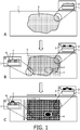
- the separation mask is a "lattice separation mask” providing rows and columns for a systematic positional characterization of the ROI ( Fig. 1 ).
- the separation mask may be a "freeform separation mask” based on the shape of the ROI and sub-ROIs ( Fig. 1 ).
- the separation mask may be a combination of a lattice separation mask and a freeform separation mask as exemplary shown in Figure 1 .
- the separation mask may be applied in different ways.
- the separation mask defines the predefined regions by providing a full trench that is applied through the entire thickness of the tissue sample ("full separation mask”), preferably by scratching, laser ablation or by creating physical barriers such as a metal grid.
- the separation mask defines the predefined regions by providing a top barrier (“top separation mask”).
- the top separation mask may define the predefined locations by physical barriers.
- the top separation mask may also define the predefined areas by a structuring process that confines reagents in the desired area (“positional mask”), preferably by topological constraints or by patterning the wettability of the tissue locally. Lithographic techniques can be employed to structure such layers.
- the surface of the ROI is structured with a self-assembled monolayer (SAM) on a thin gold layer that is evaporated on the surface of the ROI.
- SAM self-assembled monolayer
- the SAM layer may be applied by micro-contact printing of an organic thiol that is used as an etch resist in the etching of the gold layer.
- the SAM is hydrophobic and prevents the spreading of the oligonucleotide probes during inkjet printing.
- the top separation mask may also be printed on top of the ROI, wherein the ink acts as physical barrier.
- Figure 1 shows in the side views exemplarily a top separation mask 5 and a full separation mask 6. In one embodiment the predefined locations are defined by a separation mask.
- the separation mask is a top separation mask, optionally printed onto the sample, or a full separation mask, wherein the separation mask is a lattice separation mask, a freeform separation mask or a combination thereof.
- the separation mask is a top separation mask applied by printing. Inkjet printing of a wax with a heated inkjet print head can be used or inkjet printing with a UV curable ink. Both are well-known techniques in the graphics industry.
- Top masks can also be applied as a pre-manufactured integral part, like a micro-titer plate with open base.
- the micro-titer plate could be a 384 well plate or a 1536 well plate, or any other number when manufactured for this purpose.
- the well plate can be clamped on the tissue sample to ensure good physical contact between the plastic walls of the well plate and the tissue sample.
- oligonucleotide probes can be added dissolved in a buffer in each well that overlaps with the predetermined ROI.
- Each well can receive a different ink, i.e. a different oligonucleotide probe. Multiple depositions are possible to provide all reagents.
- Inkjet printing or micro-dosing can be used to provide the reagents to the wells.
- the height of the walls of such a top mask is of the order of 0.5 to 5 mm. This means that the dispensed volume in each well can be of the order of 100 nL to 10 ⁇ L, more preferably 0.5 to 2 ⁇ L.
- no separation mask (be it a physical, chemical or mechanical mask) is used and the predefined locations are defined by the local application of the oligonucleotide probes such that they do not intermix between different predefined locations on the sample.
- the oligonucleotide probes are applied using printing inks that do not spread readily or in which the volume is kept small enough to maintain the desired spatial resolution, wherein the printing pattern defines the predefined locations.
- the application of the oligonucleotide probes and defining the predefined locations occur simultaneously.
- the ink may be gelated after application to ensure fixation on the desired position, while the oligonucleotide probes are still able to diffuse into the ROI of the tissue sample.
- liquid transfer technologies may be used to apply the oligonucleotide probes in a specific pattern on the ROI.
- the oligonucleotide probes are applied onto the predefined locations by liquid transfer technologies, preferably by contact printing techniques or non-contact printing techniques, such that the applied oligonucleotide probes do not intermix between different predefined locations.
- the liquid transfer technology is a contact printing technique, preferably dip-pen plotting or micro-contact printing.
- the liquid transfer technology is a non-contact printing technique, preferably inkjet.
- oligonucleotide probes are printed in horizontal lines or in dots positioned in a horizontal line (representing rows).
- oligonucleotide probes are printed according to the shapes of identified sub-ROIs.
- the printing ink contains oligonucleotide probes dissolved in a solvent, like water or a saline buffer, optionally with surfactants and stabilizers. Concentrations of the oligonucleotide probes in the ink can be in the range between 1 nM to 1 mM, preferably between 100 nM and 10 ⁇ M. Different oligonucleotides can be dissolved in the same ink. Preferably also enzymes are dissolved in the same ink. For stability of enzymes BSA or other sera can be added in concentrations that are typical for stable storage, e.g. between 0.5 and 5 % w/w.
- Printing volumes are chosen typical for the deposition technique used and depending on the spatial resolution that is desired.
- the printing volume can be in the range between 50 pL and 5 ⁇ L, more preferably between 100 pL and 1 ⁇ L.
- the transferred volume depends on the pen size and residence time.
- Typical volumes that are transferred are in the range between 1 pL and 1 ⁇ L, preferably between 10 pL and 100 nL.
- the application of the oligonucleotide probes as described for embodiments in which no separation mask is used may also be used for embodiments in which the predefined locations are defined by a separation mask.
- the separation mask may be printed defining predefined locations and in a second step the oligonucleotide probes may be applied by printing onto the predefined locations.
- the oligonucleotide probes are applied by printing.
- the oligonucleotide probes are applied by a different method than printing such as pipetting.
- Oligonucleotide probes may also be prepared in a gel-like layer or gel-layer, wherein this layer is applied onto the predefined locations within the ROI.
- the gel layer may be applied in a single step on the ROI and the oligonucleotide probes are transferred to the ROI by diffusion.
- gel-like or “gel”, as used herein denotes a solid, jelly-like material that can have properties ranging from soft and weak to hard and tough, in which the oligonucleotide probes are able to diffuse from the gel into the ROI.
- the oligonucleotides probes are in the form of a gel layer or gel-like layer that is applied onto the sample.
- Oligonucleotide probes may also be applied in a separate disposable (e.g. with small wells, like a well plate) which will be connected later on to the ROI.
- This approach has the advantage that it can be manufactured offline, including relevant quality control measures to check if all liquids have been deposited. Deposition of the oligonucleotide probes in a separate disposable offline makes the use of liquid transfer equipment in the pathology lab obsolete.
- the ROI can be pressed into a well plate as described in Armani et al., 2009, Lab Chip 9(24): 3526-3534 and Armani et al., 2011, Anal Bioanal Chem 400: 3383-3393 .
- Oligonucleotide probes or a unique combination of oligonucleotide probes may be added to the bottom of each of the wells by spotting. The oligonucleotide probes may be dried prior to use.
- Oligonucleotide probes may also be applied by a combination of the above described means and/or a combination of using no separation mask and a separation mask. For example, in a first step oligonucleotide probes may be printed onto different sub-ROIs thereby defining the predefined locations without the use of a separation mask and in a second step oligonucleotide probes may be applied by pipetting onto predefined locations defined by a top separation mask. Thus, the oligonucleotide probes may be applied sequentially or simultaneously. It is to be understood that numerous further combinations are possible and the application of oligonucleotide probes is not restricted to the above described embodiments.
- oligonucleotide probes that are to be ligated to a nucleic acid molecule may be applied in a first step, providing conditions under which the probes are ligated to the nucleic acid molecules, and in a second step oligonucleotide probes hybridizing to the nucleic acid molecules and/or the oligonucleotide probe ligated to the nucleic acid molecules may be applied.
- different species of oligonucleotide probes are applied onto the predefined locations within the ROI sequentially.
- different species of oligonucleotide probes are applied onto the predefined locations within the ROI simultaneously.
- the oligonucleotide probes are applied in a liquid phase onto the predefined locations within the ROI. In a further preferred embodiment the oligonucleotide probes are applied in a liquid phase onto the predefined locations within the ROI, wherein the predefined locations were identified taking into account tissue information. In a further preferred embodiment the oligonucleotide probes are applied in a liquid phase with high ionic strength to enable hybridization to the target molecules. In another preferred embodiment the oligonucleotide probes are applied in a liquid phase that gelates immediately after application to the sample, either by a temperature step or radiation that is applied.
- the gelation should avoid possible intermixing of the probes at different predefined locations.
- the nucleic acid molecules in the ROI may be pre-treated.
- the nucleic acids of the sample are pretreated in order to target the right type of nucleic acid molecules and/or for efficient ligation of oligonucleotide probes.
- the pretreatment may encompass digestion of the DNA to provide DNA fragments of smaller size according to methods well known in the art.
- nucleic acid-oligonucleotide probes complex denotes a complex of the nucleic acid to be detected and at least one oligonucleotide probe.
- the nucleic acid to be detected may be in a complex with more than one species of oligonucleotide probe, for example, with two species of oligonucleotide probes.
- a humid atmosphere is provided at the surface to avoid evaporation of the solvents.
- a temperature step may be applied during that period to achieve a good conversion of the reactions.
- the separation mask may stay in place during that step or optionally be removed after the application of the probes.
- An elevated temperature accelerates diffusion and binding reactions.
- the temperature should be kept below the melting temperature of the probes in the case hybridization is used for binding. Alternatively the temperature should be chosen such that the ligase performs well. In a preferred embodiment the temperature is in the range between about 20 and 45°C, preferably between about 30 and 40°C.
- the nucleic acid-oligonucleotide probes complexes are extracted from the ROI.
- the extraction is performed by immersion of the ROI in an extraction buffer at elevated temperature. Extraction buffers common in sample preparation for molecular assays and known in the art may be used.
- the extracted nucleic acids may be collected in a single tube for further analysis.
- the solution of extracted nucleic acid-oligonucleotide probes complexes is referred to as the spatial molecular profile (SMP) extract.
- SMP spatial molecular profile
- the extracted nucleic acids are purified by methods known in the art. In another embodiment, the extracted nucleic acids are enriched by methods known in the art. In a further embodiment, the extracted nucleic acids are purified and enriched.
- DNA molecules are generated from the extracted nucleic acid-oligonucleotide probes complexes via DNA amplification, optionally wherein the generation of DNA molecules occurs after a reverse transcription reaction.
- the extracted nucleic acids are RNA molecules that are reverse transcribed and amplified.
- Nucleic acid amplification may be achieved by means of a cyclic amplification.
- the cyclic amplification may comprise any number of amplification cycles that is equal or greater than two.
- cyclic amplification reaction comprises at least 10 or at least 20 cycles.
- An exemplary cyclic amplification is a polymerase chain reaction (PCR).
- PCR is an established standard method in molecular biology that is described, for example, in detail in Sambrook et al., supra.
- PCR is used for the amplification of double-stranded DNA molecules by employing a thermostable DNA polymerase.
- the nucleic acid amplification is a cyclic amplification, preferably a PCR.
- nucleic acid amplification is achieved by means of an isothermal amplification method.
- Amplifying nucleic acids in isothermal conditions makes it possible to avoid the use of a thermocycling apparatus.
- isothermal nucleic acid amplification methods known to the skilled person, including nicking enzyme amplification reaction, transcription-mediated amplification, nucleic acid sequence-based amplification, signal-mediated amplification of RNA technology, strand displacement amplification, rolling circle amplification, loop-mediated isothermal amplification, isothermal multiple displacement amplification, helicase-dependent amplification, single primer isothermal amplification, and circular helicase-dependent amplification.
- Isothermal nucleic acid amplification technologies are reviewed, e.g., in P. Gill and A. Ghaemi, 2008, Nucleosides Nucleotides Nucleic Acids 27(3):224-243 .
- the generation of DNA molecules further comprises subjecting the nucleic acids to reverse transcription prior to subjecting them to amplification.
- Reverse transcription is another standard method in molecular biology and also described, e.g., in Sambrook et al., supra.
- sequencing the extracted nucleic acid molecules refers to the sequencing of DNA molecules, optionally obtained from RNA by reverse transcription. Methods suitable for sequencing DNA molecules are known in the art and include, but are not limited to, Sanger sequencing, pyrosequencing or next generation sequencing (NGS).
- sequencing the extracted nucleic acid molecules refers to the sequencing of RNA molecules by nanopore sequencing, microRNA sequencing or whole transcriptome shotgun sequencing. In a preferred embodiment the sequencing is DNA sequencing, preferably NGS.
- the oligonucleotide probes act as primers in NGS
- the oligonucleotide probes comprise as additional sequence a NGS adapter.
- sequenced nucleic acid molecules are correlated to the initial location of the corresponding targeted nucleic acid molecules within the ROI to generate a spatial distribution of the targeted nucleic acid molecules.
- a spatial two-dimensional map is created where the sequenced nucleic acids are shown according to the predefined locations, e.g. defined by a separation mask, from which they originated.
- sequenced nucleic acid molecules are correlated to the initial location of the corresponding targeted nucleic acid molecules within the predefined location within the ROI to generate a spatial distribution of the targeted nucleic acid molecules, wherein each location is identified by one or more species of oligonucleotide probes bound to the nucleic acids of the sample in predefined locations within the ROI.
- unique combination of at least two different species of oligonucleotide probes is used to identify the location of the targeted nucleic acid molecules within the ROI.
- the extracted and sequenced nucleic acid molecules comprise the barcode sequence of the oligonucleotide probes that bound to the target nucleic acids of the sample in a predefined location within the ROI.
- the extracted and sequenced nucleic acid molecules encompass the barcode sequence of the oligonucleotide probes.
- Each nucleic acid molecule may be correlated based on the barcode sequence it comprises to the predefined location onto which the species of oligonucleotide probes comprising said barcode sequence was applied.
- the resulting correlation provides a spatial map showing the initial locations of the targeted nucleic acids within the ROI.
- Such a spatial map may, for example, provide information on the expression status, the mutation status, the degradation status, the methylation status, the epigenetic status of the nucleic acids, or combinations thereof.
- the spatial distribution of the targeted nucleic acid molecules may be correlated with an image of the ROI or with an image of the tissue sample in which the ROI was identified.
- the spatial distribution of the targeted nucleic acid molecules may be visualized providing a two-dimensional spatial map.
- the spatial distribution of the targeted nucleic acid molecules is correlated to spatial patterns of an image obtained from the ROI.
- the method provides a two-dimensional spatial map to visualize the spatial distribution of the targeted nucleic acid molecules.
- the method further comprises overlaying the two-dimensional spatial map with the image of the ROI or with the image of the tissue sample in which the ROI was identified. An image of the ROI may be obtained before or after identification of the ROI.
- an image is obtained from the tissue sample before identifying at least one ROI.
- an image of the tissue is obtained after identifying at least one ROI.
- an image is obtained from the ROI before or after applying the oligonucleotide probes onto the predefined locations within the ROI.
- the images may be obtained by any method known in the art such as digital pathology or a microscope with a camera.
- the spatial distribution of the targeted nucleic acid molecules is correlated with the predefined locations located within the ROI and defined by a separation mask, wherein the image is obtained after applying the separation mask and before or after applying the oligonucleotide probes onto the predefined locations within the ROI, preferably before application of the oligonucleotide probes.
- the spatial distribution of the targeted nucleic acid molecules is correlated with an image of the ROI obtained before or after applying the oligonucleotide probes onto the predefined locations within the ROI, preferably before application of the oligonucleotide probes.
- the spatial distribution of the targeted nucleic acid molecules is correlated with an image of the ROI obtained after applying a separation mask and before applying the oligonucleotide probes onto the predefined locations defined by the separation mask.
- the spatial map can be visualized for instance by displaying false colors, with each predefined locations as defined by a grid representing an area of the same color.
- the spatial map can be overlaid with an image of the ROI or with an image of the tissue sample.
- the spatial map is configured as a semi-transparent image and is overlaid on the image of the ROI or with the image of the tissue sample.
- the image of the ROI or the image of the tissue sample is configured as a semi-transparent image and is overlaid on the spatial map.
- the number or amount of the same targeted nucleic acid molecules determined in the assay with the same species of oligonucleotide probes is related to the color of the map in the corresponding predefined location.
- a particular combination of targeted nucleic acid molecules is correlated to the color.
- the overexpression of a particular set of targeted nucleic acid molecules related to specific genes can be correlated to the color on the map. It is to be understood that many different profiles can be defined and related to a way of visual display, depending on the biological and/or clinical question that is addressed. Instead of or in combination with colors other annotations can be displayed in a spatial map.
- the correlation of the spatial distribution of the targeted nucleic acid molecules to the spatial patterns of the initial image may be used to answer a clinical question and provide guidance for a clinical decision.
- the results obtained by a spatial map created by the method of the invention may be used for clinical diagnosis or monitoring of a clinical response in a patient.
- the correlation of the spatial distribution of the targeted nucleic acid molecules to the spatial patterns of the initial image may also be used in the field of research.
- tissue sample and/or ROI are imaged.
- An image taken of a tissue sample may be used to identify the region of interest as well as the spatial resolution of the pattern based on the image-based analysis of the tissue before the application of the reagents.
- Another aspect of the invention relates to a reaction ligating 5'-oligonucleotide probes and 3'-oligonucleotide probes to the 5'-end and the 3'- end of the RNA molecule and simultaneously applying a DNA molecule introducing a restriction site on the ligation site of the oligonucleotide probe dimer and a restriction enzyme that is suitable to cleave the restriction site, wherein the 5'-end and 3'-end of the DNA molecule are modified so that they are not participating in the ligation reaction.
- the dimers that occur between the oligonucleotide probes when they react with each other i.e.
- oligonucleotide probe dimer are cleaved into fully active individual 5'-oligonucleotide probes and 3'-oligonucleotide probes.
- the cleaved oligonucleotide probes can be reused in the ligation reaction in order to be ligated to the 5'-end and 3'-end of the RNA molecule.
- a specific embodiment relates to the ligation reaction on a FFPE tissue sample, ligating 5'-oligonucleotide probes and 3'-oligonucleotide probes to the 5'-end and the 3'- end of the RNA molecule and simultaneously applying a DNA molecule introducing a restriction site on the ligation site of the oligonucleotide probe dimer and a restriction enzyme that is suitable to cleave the restriction site, wherein the 5'-end and 3'-end of the DNA molecule are modified so that they are not participating in the ligation reaction.
- the ligation reaction is quenched by a reverse transcription reaction thereby avoiding the formation of oligonucleotide probe dimers.
- the reverse transcription reaction is rather inefficient on a FFPE tissue sample.
- the oligonucleotide probe dimer is cleaved by the restriction enzyme, thus the reverse transcription reaction step can be avoided on the FFPE tissue sample.
- the T4 RNA ligase I and II reactions can be combined in one step, as described in the Examples and Figure 3 .
- the oligonucleotide probe ligation reaction method of the invention is advantagous over the prior art.
- the invention relates to a ligation reaction ligation 5'-oligonucleotide probes and 3'-oligonucleotide probes to the 5'-end and the 3'- end of the RNA molecule and simultaneously applying a DNA molecule introducing a HaeIII restriction site on the ligation site of the oligonucleotide probes dimer and a HaeIII restriction enzyme cleaving at the restriction site.
- the above described ligation reaction can be used in step b) of the method for spatial detecting RNA molecules in a FFPE tissue sample as described herein.
- the reverse transcription reaction has to be carried out on the FFPE tissue sample.
- This method is advantageous as it avoids the reverse transcription reaction which is rather inefficient to the FFPE sample.
- T4 RNA ligase I and II reactions can be combined in one step.
- Protocol types for the binding of the oligonucleotide probe to the nucleic acid molecule RNA DNA Hybridization [A] Target probe/oligo dT, PCR [B] Fragmentation, denaturation, hybridization, PCR Ligation Blunt [C] End specific ligation [D] Fragmentation, ligation Sticky [E] Digestion, ligation
- the tissue sample is treated with Proteinase K (ProtK) at 37°C, e.g. as described by Tullis and Rubin (Analytical Biochemistry, 1980, 107(1): 260-264 ) or using Proteinase K from Engyodontium album (Sigma-Aldrich) according to the manufacturer's instructions, followed by heat deactivation of ProtK for 15 minutes at 80°C.
- Proteinase K ProtK
- the tissue sample is deparaffinized, e.g. using deparaffinization solution (e.g. Qiagen) according to the manufacturer's instructions.
- deparaffinization solution e.g. Qiagen
- An image of the complete slide is acquired (Philips Digital Pathology Scanner UFS) and within the image a ROI is identified.
- a top selection mask is applied by inkjet printing (print head XAAR) a UV curable ink (Sunjet ink Crystal UFE 7573 (Black)) creating a lattice that separates the different reaction wells. UV curing of the mask is carried out at 360 to 380 nm with a UV-LED for 30s to 60s.
- a unique combination of two species of oligonucleotide probes comprising the barcode sequences is applied onto each predefined location by inkjet printing (using a FUJIFILM Dimatix printhead).
- the 5'-RNA probes and 3 '-RNA probes are ligated to the mRNA molecules in a single step using a modified protocol of the NEBNext® Small RNA library prep set for Illumina® (NEB).
- NEB NEBNext® Small RNA library prep set for Illumina®
- the RNA probes are ligated to the RNA molecule in a single step.
- the 5'-RNA probes react with the 3'-RNA probes and form dimers.
- RNA probe dimers with the complementary RNA-DNA hetroduplex will be cleaved on the ligation site of the two RNA probes only on the RNA strand while the DNA molecule stays intact.
- the cleaved RNA probes can participate again in the ligation reaction. After the application of the ligation mix the slide is covered with a lid (Grace Bio-Labs HybriWellTM sealing system, SecureSealTM adhesive chamber).
- RNA membrane purification e.g. using the RNeasy FFPE Kit (Qiagen) according to the manufacturer's instruction.
- the sample is heated during extraction and remains sealed in order to avoid dehydration of the sample.
- the sequence of the oligonucleotide probe can be used during the purification process, e.g. using magnetic beads.
- An incubation with DNase is carried out during or after the ligation RNA purification (e.g. at 37°C for 15 min) followed by DNase inactivation at 80°C for 15 min.
- DNase treatment may be conducted in parallel to the Proteinase K treatment step described above.
- RNA sequencing library preparation is performed using, e.g. the ScriptSeq v2 RNA-Seq Library Preparation Kit (Illumina) according to the manufacturer's instructions. RNA sequencing is performed and the barcoded transcripts are identified from the sequences. This information is then combined with the original image obtained from the ROI to build the spatial-transcriptional map.
- ScriptSeq v2 RNA-Seq Library Preparation Kit Illumina
- the tissue sample is deparaffinized, e.g. using deparaffinization solution (e.g. Qiagen) according to the manufacturer's instructions and dried.
- deparaffinization solution e.g. Qiagen
- the tissue sample is treated with Proteinase K (ProtK) at 37°C, e.g. as described by Tullis and Rubin (Analytical Biochemistry, 1980, 107(1): 260-264 ) or using Proteinase K from Engyodontium album (Sigma-Aldrich) according to the manufacturer's instructions, followed by heat deactivation of ProtK for 15 minutes at 80°C.
- Proteinase K ProtK
- the tissue sample is washed with EtOH and dried 40°C for 30 minutes.
- An image of the complete slide is acquired (Philips Digital Pathology Scanner UFS) and within the image a ROI is identified.
- the DNA molecules are denaturated by addition of denaturation solution (70% formamide in 2X SSC, pH 7.0 - 8.0) and incubation at 75 °C for 5 minutes followed by incubation for 1 minute in 70% EtOH, 1 minute in 85% EtOH and 1 minute in 100% EtOH. Then slide is dried and placed at 45 - 50 °C to allow remaining EtOH to evaporate. Within the ROI different locations are selected and a top selection mask is applied by inkjet printing (print head XAAR) a UV curable ink (Sunjet ink Crystal UFE 7573 (Black)) creating a lattice that separates the different reaction wells. UV curing of the mask is carried out at 360 to 380 nm with a UV-LED for 30s to 60s.
- denaturation solution 70% formamide in 2X SSC, pH 7.0 - 8.0
- incubation 75 °C for 5 minutes followed by incubation for 1 minute in 70% EtOH, 1 minute in 85% EtOH and 1 minute in 100% EtOH.
- Oligonucleotide probes comprising the barcode sequence are applied onto each predefined location by inkjet printing (using a FUJIFILM Dimatix printhead).
- the tissue sample is incubated at 40° for 2h (wet conditions).
- an elongation reaction is performed to amplify the target sequences which were hybridized using the primer sequence of the oligonucleotide probes.
- the slide is covered with a lid (Grace Bio-Labs HybriWellTM sealing system, SecureSealTM adhesive chamber).
- the extraction buffer is introduced and the sample is incubated at 60°C for 30 min.
- DNA sequencing library preparation and DNA sequencing is performed and the barcoded transcripts are identified from the sequences. This information is then combined with the original image obtained from the ROI to build the spatial-transcriptional map.
- well-known standard kits are applied according to the manufacturer's protocol.
- the goal of this experiment was to ligate the miRNA cloning linker on the 3' side of RNA and to ligate the RNA 5'adapter on the 5' side of RNA on a FFPET slide in a single reaction. This was done according to the protocol described below. RT-qPCR assays were done on the extracted RNA to determine if ligation reaction was successful.
- RNA has to become available/exposed on the slide for the ligation reaction to be successful. This was done by firstly, washing the slide in Xylyene (2x 10 minutes 1x8 minutes), rinsing it with 100% EtOH (1x5 minutes, 1x1 minute). The slide was then air dried for 5 minutes and a hydrophobic barrier pen was used for creating a circle around the tissue in which fluids would stay (slide was air dried for another 15-30 minutes).
- a slide holder was filled with 1x pretreatment solution (Quantigene® View RNA Assay kit Affimetrix; P/N: 17428). The slide holder was placed in a waterbath (95 °C), and the slide was carefully placed in this slide holder with the heated 1x pretreatment solution for 20 minutes. Slide was rinsed in MiliQ water (2x 1 minute) and PBS (1x5 seconds). An 200-400 ⁇ L aliquot of Protease QS solution (Quantigene® View RNA Assay kit Affimetrix; P/N: 16742) was added on the tissue and incubated for 20 minutes at 40 °C in a humidity box. The slide was washed in PBS (2x 5 seconds) to remove Protease QS solution.
- 1x pretreatment solution Quantigene® View RNA Assay kit Affimetrix; P/N: 17428.
- a reaction mixture was prepared as described in the table below: Solution Volume ( ⁇ L) 10x T4 RNA ligase reaction buffer 6 50% PEG 8000 12 100 ⁇ M Universal miRNA cloning linker 1,2 10 mM ATP 6 100 ⁇ M RNA oligo 3 DMSO 6 T4 RNA Ligase 2, truncated (T4 Rnl2tr) 3 T4 RNA Ligase 1 6 ddH2O 16,8
- RT-qPCR assays specifically designed for the ligated product. See the amplification curves in Figure 3 and the used oligonucleotides in the table below.
- the RT-qPCR assays detect ligated product on B-actin mRNA.
- a negative control which was a slide treated the same as describe above but without the actual addition of the ligation sequences (cloning linker and RNA oligo), shown in red.
- NTC no template control
Claims (15)
- Procédé de détection spatiale d'acides nucléiques dans un échantillon de tissu comprenant les étapes consistant à :a) identifier au moins une région d'intérêt (ROI) dans l'échantillon ;b) appliquer au moins une espèce de sondes d'oligonucléotides sur des emplacements prédéfinis dans la ROI et permettre aux sondes d'oligonucléotides de se lier aux acides nucléiques de l'échantillon, dans lequel chaque espèce des sondes d'oligonucléotides comprend une séquence à code-barres unique ;c) extraire les complexes d'acide nucléique - sondes d'oligonucléotides ;d) séquencer les molécules d'acide nucléique extraites ;e) corréler les molécules d'acide nucléique séquencées comportant la/les séquence(s) à code-barres des sondes d'oligonucléotides à l'emplacement initial des molécules d'acide nucléique ciblées correspondantes dans la ROI sur la base de la/des séquence(s) à code-barres pour générer une distribution spatiale des molécules d'acide nucléique ciblées, dans lequel chaque emplacement est identifié par une ou plusieurs espèces de sondes d'oligonucléotide liée(s) à l'étape b).
- Procédé selon la revendication 1, dans lequel avant l'étape d), des molécules d'ADN sont générées à partir des complexes d'acide nucléique - sondes d'oligonucléotide par amplification d'ADN.
- Procédé selon la revendication 2, dans lequel la génération de molécules d'ADN survient après une réaction de transcription inverse.
- Procédé selon la revendication 2 ou 3, dans lequel à l'étape b), la liaison des sondes d'oligonucléotides aux acides nucléiques de l'échantillon survient par hybridation et dans lequel les sondes d'oligonucléotides sont utilisées comme amorces.
- Procédé selon l'une quelconque des revendications 1 à 3, dans lequel à l'étape b), la liaison des sondes d'oligonucléotides aux acides nucléiques de l'échantillon survient par ligature.
- Procédé selon l'une quelconque des revendications 1 à 5, dans lequel l'étape e) comprend en outre la corrélation de la distribution spatiale des molécules d'acide nucléique ciblées avec une image de la ROI ou avec une image de l'échantillon de tissu dans lequel la ROI a été identifiée, obtenue avant ou après l'étape b).
- Procédé selon la revendication 6, dans lequel le procédé comprend en outre la fourniture d'une carte spatiale bidimensionnelle pour visualiser la distribution spatiale des molécules d'acide nucléique ciblées.
- Procédé selon la revendication 7, dans lequel le procédé comprend en outre la superposition d'une carte spatiale bidimensionnelle avec l'image de la ROI ou avec l'image de l'échantillon de tissu dans lequel la ROI a été identifiée, obtenue avant ou après l'étape b).
- Procédé selon l'une quelconque des revendications 1 à 8, dans lequel au moins deux espèces différentes de sondes d'oligonucléotides sont liées à une molécule d'acide nucléique ciblée dans l'étape b), et dans lequel la combinaison unique des au moins deux espèces différentes de sondes d'oligonucléotides est utilisée pour identifier l'emplacement des molécules d'acide nucléique ciblées dans la ROI.
- Procédé selon l'une quelconque des revendications 1 à 9, dans lequel au moins une des espèces de sondes d'oligonucléotides qui se lient à une molécule d'acide nucléique comprend une séquence générique, dans lequel facultativement la séquence générique est complémentaire à l'acide nucléique ciblé.
- Procédé selon l'une quelconque des revendications 1 à 10, dans lequel au moins une des espèces de sondes d'oligonucléotides qui se lient à une molécule d'acide nucléique comprend une séquence supplémentaire, dans lequel la séquence supplémentaire est une séquence de purification ou une séquence d'alignement d'amorce.
- Procédé selon l'une quelconque des revendications 1 à 11, dans lequel les emplacements prédéfinis sont définis par un masque de séparation.
- Procédé selon la revendication 12, dans lequel le masque de séparation est un masque de séparation supérieur, facultativement imprimé sur l'échantillon, ou un masque de séparation totale, dans lequel le masque de séparation est un masque de séparation en réseau, un masque de séparation de forme libre ou une combinaison de ceux-ci.
- Procédé selon l'une quelconque des revendications 1 à 13, dans lequel à l'étape b), les sondes d'oligonucléotides sont appliquées sur les emplacements prédéfinis par des technologies de transfert liquide, de préférence par des techniques d'impression par contact ou des techniques d'impression sans contact, de sorte que les sondes d'oligonucléotides appliquées ne se mélangent pas entre différents emplacements prédéfinis.
- Procédé selon l'une quelconque des revendications 1 à 14, dans lequel l'échantillon est un spécimen histopathologique, de préférence un échantillon déparaffiné fixé dans le formol et inclus dans la paraffine (FFPE), un échantillon congelé frais (FF) ou un échantillon frais, ou un échantillon de cytologie.
Applications Claiming Priority (2)
| Application Number | Priority Date | Filing Date | Title |
|---|---|---|---|
| EP15163481 | 2015-04-14 | ||
| PCT/EP2016/058067 WO2016166128A1 (fr) | 2015-04-14 | 2016-04-13 | Mappage spatial des profils moléculaires d'échantillons de tissus biologiques |
Publications (2)
| Publication Number | Publication Date |
|---|---|
| EP3283641A1 EP3283641A1 (fr) | 2018-02-21 |
| EP3283641B1 true EP3283641B1 (fr) | 2019-11-27 |
Family
ID=52991497
Family Applications (1)
| Application Number | Title | Priority Date | Filing Date |
|---|---|---|---|
| EP16715585.2A Active EP3283641B1 (fr) | 2015-04-14 | 2016-04-13 | Cartographie spatiale de profils moléculaires d'échantillons de tissus biologiques |
Country Status (7)
| Country | Link |
|---|---|
| US (1) | US10724078B2 (fr) |
| EP (1) | EP3283641B1 (fr) |
| JP (1) | JP6875998B2 (fr) |
| CN (1) | CN107709574B (fr) |
| BR (1) | BR112017021993A2 (fr) |
| RU (1) | RU2733545C2 (fr) |
| WO (1) | WO2016166128A1 (fr) |
Families Citing this family (108)
| Publication number | Priority date | Publication date | Assignee | Title |
|---|---|---|---|---|
| US9879313B2 (en) | 2013-06-25 | 2018-01-30 | Prognosys Biosciences, Inc. | Methods and systems for determining spatial patterns of biological targets in a sample |
| EP4321627A3 (fr) | 2015-04-10 | 2024-04-17 | 10x Genomics Sweden AB | Analyse de plusieurs acides nucléiques spatialement différenciés de spécimens biologiques |
| WO2016168825A1 (fr) * | 2015-04-17 | 2016-10-20 | Centrillion Technology Holdings Corporation | Procédés pour établir un profil spatial de molécules biologiques |
| CN115369161A (zh) | 2015-12-04 | 2022-11-22 | 10X 基因组学有限公司 | 用于核酸分析的方法和组合物 |
| US10724089B2 (en) * | 2016-10-06 | 2020-07-28 | The Board Of Trustees Of The University Of Illinois | Spatial molecular analysis of tissue |
| WO2018217862A1 (fr) * | 2017-05-23 | 2018-11-29 | Centrillion Technologies, Inc. | Procédés pour établir un profil spatial de molécules biologiques |
| US20210317524A1 (en) | 2018-08-28 | 2021-10-14 | 10X Genomics, Inc. | Resolving spatial arrays |
| CN113366117A (zh) | 2018-08-28 | 2021-09-07 | 10X基因组学股份有限公司 | 用于生物样品中转座酶介导的空间标记和分析基因组dna的方法 |
| US11519033B2 (en) | 2018-08-28 | 2022-12-06 | 10X Genomics, Inc. | Method for transposase-mediated spatial tagging and analyzing genomic DNA in a biological sample |
| WO2020123309A1 (fr) | 2018-12-10 | 2020-06-18 | 10X Genomics, Inc. | Résolution de réseaux spatiaux par déconvolution basée sur la proximité |
| CA3124789A1 (fr) * | 2018-12-31 | 2020-07-09 | Avava, Inc. | Systeme et procedes de traitement tissulaire |
| US11926867B2 (en) | 2019-01-06 | 2024-03-12 | 10X Genomics, Inc. | Generating capture probes for spatial analysis |
| US11649485B2 (en) | 2019-01-06 | 2023-05-16 | 10X Genomics, Inc. | Generating capture probes for spatial analysis |
| EP3931354A1 (fr) | 2019-02-28 | 2022-01-05 | 10X Genomics, Inc. | Profilage d'analytes biologiques avec des réseaux d'oligonucléotides à codes-barres spatiaux |
| CN114127309A (zh) | 2019-03-15 | 2022-03-01 | 10X基因组学有限公司 | 使用空间阵列进行单细胞测序的方法 |
| WO2020198071A1 (fr) | 2019-03-22 | 2020-10-01 | 10X Genomics, Inc. | Analyse spatiale tridimensionnelle |
| WO2020243579A1 (fr) | 2019-05-30 | 2020-12-03 | 10X Genomics, Inc. | Procédés de détection de l'hétérogénéité spatiale d'un échantillon biologique |
| CN117036248A (zh) | 2019-10-01 | 2023-11-10 | 10X基因组学有限公司 | 用于识别组织样品中的形态学模式的系统和方法 |
| US20210130881A1 (en) | 2019-11-06 | 2021-05-06 | 10X Genomics, Inc. | Imaging system hardware |
| EP4055185A1 (fr) | 2019-11-08 | 2022-09-14 | 10X Genomics, Inc. | Agents de capture d'analytes marqués spatialement pour le multiplexage d'analytes |
| WO2021092433A2 (fr) | 2019-11-08 | 2021-05-14 | 10X Genomics, Inc. | Amélioration de la spécificité de la liaison d'un analyte |
| US20230002812A1 (en) | 2019-11-13 | 2023-01-05 | 10X Genomics, Inc. | Generating capture probes for spatial analysis |
| AU2020388047A1 (en) | 2019-11-18 | 2022-06-23 | 10X Genomics, Inc. | Systems and methods for tissue classification |
| AU2020388573A1 (en) | 2019-11-21 | 2022-06-23 | 10X Genomics, Inc, | Spatial analysis of analytes |
| CA3158891A1 (fr) | 2019-11-22 | 2021-05-27 | Neil Ira WEISENFELD | Systemes et procedes d'analyse spatiale d'analytes a l'aide d'un alignement de reperes |
| CN114885610A (zh) * | 2019-12-23 | 2022-08-09 | 10X基因组学有限公司 | 使用rna模板化连接进行空间分析的方法 |
| EP4339299A2 (fr) | 2020-01-10 | 2024-03-20 | 10X Genomics, Inc. | Procédés de détermination d'un emplacement d'un acide nucléique cible dans un échantillon biologique |
| US11732299B2 (en) | 2020-01-21 | 2023-08-22 | 10X Genomics, Inc. | Spatial assays with perturbed cells |
| US11702693B2 (en) | 2020-01-21 | 2023-07-18 | 10X Genomics, Inc. | Methods for printing cells and generating arrays of barcoded cells |
| US11821035B1 (en) | 2020-01-29 | 2023-11-21 | 10X Genomics, Inc. | Compositions and methods of making gene expression libraries |
| US11898205B2 (en) | 2020-02-03 | 2024-02-13 | 10X Genomics, Inc. | Increasing capture efficiency of spatial assays |
| US11732300B2 (en) | 2020-02-05 | 2023-08-22 | 10X Genomics, Inc. | Increasing efficiency of spatial analysis in a biological sample |
| WO2021158925A1 (fr) | 2020-02-07 | 2021-08-12 | 10X Genomics, Inc. | Évaluation quantitative et automatisée de la performance de perméabilisation pour la transcriptomique spatiale |
| US11835462B2 (en) | 2020-02-11 | 2023-12-05 | 10X Genomics, Inc. | Methods and compositions for partitioning a biological sample |
| WO2021168278A1 (fr) | 2020-02-20 | 2021-08-26 | 10X Genomics, Inc. | Procédés pour combiner les synthèses d'adnc des premier et de deuxième brins pour l'analyse spatiale |
| US20210262018A1 (en) | 2020-02-21 | 2021-08-26 | 10X Genomics, Inc. | Methods and compositions for integrated in situ spatial assay |
| AU2021224760A1 (en) | 2020-02-21 | 2022-09-15 | 10X Genomics, Inc. | Capturing genetic targets using a hybridization approach |
| US11891654B2 (en) | 2020-02-24 | 2024-02-06 | 10X Genomics, Inc. | Methods of making gene expression libraries |
| US11926863B1 (en) | 2020-02-27 | 2024-03-12 | 10X Genomics, Inc. | Solid state single cell method for analyzing fixed biological cells |
| US11768175B1 (en) | 2020-03-04 | 2023-09-26 | 10X Genomics, Inc. | Electrophoretic methods for spatial analysis |
| WO2021216708A1 (fr) | 2020-04-22 | 2021-10-28 | 10X Genomics, Inc. | Procédés d'analyse spatiale utilisant un appauvrissement d'arn ciblée |
| WO2021225900A1 (fr) | 2020-05-04 | 2021-11-11 | 10X Genomics, Inc. | Modes de transfert transcriptomique spatial |
| WO2021236625A1 (fr) | 2020-05-19 | 2021-11-25 | 10X Genomics, Inc. | Cassettes et instrumentation d'électrophorèse |
| EP4153775A1 (fr) | 2020-05-22 | 2023-03-29 | 10X Genomics, Inc. | Mesure spatio-temporelle simultanée de l'expression génique et de l'activité cellulaire |
| WO2021237056A1 (fr) | 2020-05-22 | 2021-11-25 | 10X Genomics, Inc. | Analyse d'intégrité d'arn dans un échantillon biologique |
| WO2021237087A1 (fr) | 2020-05-22 | 2021-11-25 | 10X Genomics, Inc. | Analyse spatiale pour détecter des variants de séquence |
| WO2021242834A1 (fr) | 2020-05-26 | 2021-12-02 | 10X Genomics, Inc. | Procédé de réinitialisation d'un réseau |
| EP4025692A2 (fr) | 2020-06-02 | 2022-07-13 | 10X Genomics, Inc. | Procédés de banques d'acides nucléiques |
| CN116249785A (zh) | 2020-06-02 | 2023-06-09 | 10X基因组学有限公司 | 用于抗原-受体的空间转录组学 |
| EP4162074B1 (fr) | 2020-06-08 | 2024-04-24 | 10X Genomics, Inc. | Méthodes de détermination de marge chirurgicale et méthodes d'utilisation associées |
| EP4165207A1 (fr) | 2020-06-10 | 2023-04-19 | 10X Genomics, Inc. | Procédés de détermination d'un emplacement d'un analyte dans un échantillon biologique |
| WO2021252576A1 (fr) | 2020-06-10 | 2021-12-16 | 10X Genomics, Inc. | Procédés d'analyse spatiale utilisant des oligonucléotides de blocage |
| WO2021263111A1 (fr) | 2020-06-25 | 2021-12-30 | 10X Genomics, Inc. | Analyse spatiale de la méthylation de l'adn |
| US11761038B1 (en) | 2020-07-06 | 2023-09-19 | 10X Genomics, Inc. | Methods for identifying a location of an RNA in a biological sample |
| EP4189110A1 (fr) | 2020-07-31 | 2023-06-07 | 10X Genomics, Inc. | Composés de déréticulation et procédés d'utilisation pour analyse spatiale |
| EP4200441A1 (fr) | 2020-09-15 | 2023-06-28 | 10X Genomics, Inc. | Procédés de libération d'une sonde de capture étendue à partir d'un substrat et leurs utilisations |
| US20230313279A1 (en) | 2020-09-16 | 2023-10-05 | 10X Genomics, Inc. | Methods of determining the location of an analyte in a biological sample using a plurality of wells |
| WO2022061152A2 (fr) | 2020-09-18 | 2022-03-24 | 10X Genomics, Inc. | Appareil de manipulation d'échantillon et procédés système de distribution de fluide |
| WO2022061150A2 (fr) | 2020-09-18 | 2022-03-24 | 10X Geonomics, Inc. | Appareil de manipulation d'échantillons et procédés d'enregistrement d'images |
| US11926822B1 (en) | 2020-09-23 | 2024-03-12 | 10X Genomics, Inc. | Three-dimensional spatial analysis |
| WO2022081643A2 (fr) | 2020-10-13 | 2022-04-21 | 10X Genomics, Inc. | Compositions et procédés pour générer des molécules de liaison à un antigène recombinant à partir de cellules uniques |
| WO2022087273A1 (fr) | 2020-10-22 | 2022-04-28 | 10X Genomics, Inc. | Procédés d'analyse spatiale utilisant l'amplification par cercle roulant |
| CN116829733A (zh) | 2020-11-06 | 2023-09-29 | 10X基因组学有限公司 | 用于将分析物结合至捕获探针的组合物和方法 |
| US20230407404A1 (en) | 2020-11-18 | 2023-12-21 | 10X Genomics, Inc. | Methods and compositions for analyzing immune infiltration in cancer stroma to predict clinical outcome |
| US11827935B1 (en) | 2020-11-19 | 2023-11-28 | 10X Genomics, Inc. | Methods for spatial analysis using rolling circle amplification and detection probes |
| EP4012046A1 (fr) | 2020-12-11 | 2022-06-15 | 10X Genomics, Inc. | Procédés et compositions pour l'analyse multimodale in situ |
| WO2022140028A1 (fr) | 2020-12-21 | 2022-06-30 | 10X Genomics, Inc. | Procédés, compositions et systèmes pour capturer des sondes et/ou des codes à barres |
| US20240068017A1 (en) | 2020-12-30 | 2024-02-29 | 10X Genomics, Inc. | Methods for analyte capture determination |
| WO2022147296A1 (fr) | 2020-12-30 | 2022-07-07 | 10X Genomics, Inc. | Clivage de sondes de capture pour analyse spatiale |
| AU2022212231A1 (en) | 2021-01-29 | 2023-08-03 | 10X Genomics, Inc. | Method for transposase mediated spatial tagging and analyzing genomic dna in a biological sample |
| WO2022197490A1 (fr) * | 2021-03-16 | 2022-09-22 | Scale Biosciences Inc. | Procédé d'ajout de codes-barres adressables spatialement à des acides nucléiques d'un échantillon cellulaire in situ |
| WO2022198068A1 (fr) | 2021-03-18 | 2022-09-22 | 10X Genomics, Inc. | Capture multiplex de gène et expression de protéines à partir d'un échantillon biologique |
| EP4319792A1 (fr) | 2021-04-05 | 2024-02-14 | 10X Genomics, Inc. | Composition de ligase recombinante et ses utilisations |
| EP4305196A1 (fr) | 2021-04-14 | 2024-01-17 | 10X Genomics, Inc. | Procédés de mesure d'une mauvaise localisation d'un analyte |
| WO2022226057A1 (fr) | 2021-04-20 | 2022-10-27 | 10X Genomics, Inc. | Procédés d'évaluation de la qualité d'un échantillon avant une analyse spatiale à l'aide d'une ligature modélisée |
| WO2022236054A1 (fr) | 2021-05-06 | 2022-11-10 | 10X Genomics, Inc. | Procédés pour augmenter la résolution d'une analyse spatiale |
| EP4347879A1 (fr) | 2021-06-03 | 2024-04-10 | 10X Genomics, Inc. | Procédés, compositions, kits et systèmes pour améliorer la capture d'analytes pour une analyse spatiale |
| WO2022271820A1 (fr) | 2021-06-22 | 2022-12-29 | 10X Genomics, Inc. | Détection spatiale de sars-cov-2 à l'aide d'une ligature à matrice |
| EP4352252A1 (fr) | 2021-07-13 | 2024-04-17 | 10X Genomics, Inc. | Procédés d'analyse spatiale utilisant un silençage de sonde ciblée |
| US20230026886A1 (en) | 2021-07-13 | 2023-01-26 | 10X Genomics, Inc. | Methods for preparing polymerized matrix with controllable thickness |
| WO2023018799A1 (fr) | 2021-08-12 | 2023-02-16 | 10X Genomics, Inc. | Procédés, compositions et systèmes pour identifier des molécules de liaison à l'antigène |
| WO2023034489A1 (fr) | 2021-09-01 | 2023-03-09 | 10X Genomics, Inc. | Procédés, compositions et kits pour bloquer une sonde de capture sur un réseau spatial |
| WO2023044071A1 (fr) | 2021-09-17 | 2023-03-23 | 10X Genomics, Inc. | Systèmes et procédés de recalage ou d'alignement d'images |
| WO2023076345A1 (fr) | 2021-10-26 | 2023-05-04 | 10X Genomics, Inc. | Procédés d'analyse spatiale utilisant la capture ciblée d'arn |
| WO2023086880A1 (fr) | 2021-11-10 | 2023-05-19 | 10X Genomics, Inc. | Procédés, compositions et kits pour déterminer l'emplacement d'un analyte dans un échantillon biologique |
| WO2023086824A1 (fr) | 2021-11-10 | 2023-05-19 | 10X Genomics, Inc. | Procédés d'identification de molécules de liaison à l'antigène |
| US20230167495A1 (en) | 2021-11-30 | 2023-06-01 | 10X Genomics, Inc. | Systems and methods for identifying regions of aneuploidy in a tissue |
| EP4305195A2 (fr) | 2021-12-01 | 2024-01-17 | 10X Genomics, Inc. | Procédés, compositions et systèmes pour la détection améliorée d'analytes in situ et analyse spatiale |
| WO2023122033A1 (fr) | 2021-12-20 | 2023-06-29 | 10X Genomics, Inc. | Auto-test pour dispositif d'imagerie |
| WO2023150163A1 (fr) | 2022-02-01 | 2023-08-10 | 10X Genomics, Inc. | Procédés, compositions et systèmes de capture d'analytes à partir de tissu lymphatique |
| WO2023150171A1 (fr) | 2022-02-01 | 2023-08-10 | 10X Genomics, Inc. | Procédés, compositions et systèmes de capture d'analytes à partir d'échantillons de glioblastome |
| WO2023150098A1 (fr) | 2022-02-01 | 2023-08-10 | 10X Genomics, Inc. | Procédés, kits, compositions et systèmes d'analyse spatiale |
| WO2023152354A1 (fr) * | 2022-02-14 | 2023-08-17 | Miltenyi Biotec B.V. & Co. KG | Synthèse directe d'oligonucléotides sur des coupes de tissu à microtomie |
| WO2023159028A1 (fr) | 2022-02-15 | 2023-08-24 | 10X Genomics, Inc. | Systèmes et procédés d'analyse spatiale d'analytes à l'aide d'un alignement de repères |
| WO2023172670A2 (fr) | 2022-03-11 | 2023-09-14 | 10X Genomics, Inc. | Appareil de manipulation d'échantillon et procédés système de distribution de fluide |
| WO2023201235A2 (fr) | 2022-04-12 | 2023-10-19 | 10X Genomics, Inc. | Compositions et procédés de génération et de caractérisation de molécules de liaison à un antigène recombinant |
| US20240002902A1 (en) | 2022-05-06 | 2024-01-04 | 10X Genomics, Inc. | Analysis of antigen and antigen receptor interactions |
| WO2023215552A1 (fr) | 2022-05-06 | 2023-11-09 | 10X Genomics, Inc. | Lecteurs de codes-barres moléculaires pour détection d'analytes |
| WO2023225519A1 (fr) | 2022-05-17 | 2023-11-23 | 10X Genomics, Inc. | Transposons modifiés, compositions et utilisations de ceux-ci |
| WO2023229988A1 (fr) | 2022-05-23 | 2023-11-30 | 10X Genomics, Inc. | Moule pour échantillon de tissu |
| WO2023229982A2 (fr) | 2022-05-24 | 2023-11-30 | 10X Genomics, Inc. | Confinement de structure poreuse pour suppression de convection |
| WO2023250077A1 (fr) | 2022-06-22 | 2023-12-28 | 10X Genomics, Inc. | Procédés, compositions et systèmes pour la capture de sondes et/ou de code-barres |
| WO2024015862A1 (fr) | 2022-07-13 | 2024-01-18 | 10X Genomics, Inc. | Procédés de caractérisation de molécules de liaison à l'antigène à partir d'échantillons biologiques |
| WO2024015578A1 (fr) | 2022-07-15 | 2024-01-18 | 10X Genomics, Inc. | Procédés pour déterminer l'emplacement d'un acide nucléique cible dans un échantillon biologique |
| US20240052404A1 (en) | 2022-08-05 | 2024-02-15 | 10X Genomics, Inc. | Systems and methods for immunofluorescence quantification |
| WO2024036191A1 (fr) | 2022-08-10 | 2024-02-15 | 10X Genomics, Inc. | Systèmes et procédés de colocalisation |
| WO2024035844A1 (fr) | 2022-08-12 | 2024-02-15 | 10X Genomics, Inc. | Procédés de réduction de la capture d'analytes |
| WO2024044703A1 (fr) | 2022-08-24 | 2024-02-29 | 10X Genomics, Inc. | Compositions et procédés de cartographie d'épitopes antigéniques dans des échantillons biologiques |
Family Cites Families (6)
| Publication number | Priority date | Publication date | Assignee | Title |
|---|---|---|---|---|
| JP2007510199A (ja) | 2003-10-08 | 2007-04-19 | ライフスパン バイオサイエンス,インク. | 自動顕微鏡スライド組織サンプルマッピング及び画像取得 |
| US8995733B2 (en) * | 2009-04-28 | 2015-03-31 | Koninklijke Philips N.V. | Microdissection method and information processing system |
| CA2794522C (fr) * | 2010-04-05 | 2019-11-26 | Prognosys Biosciences, Inc. | Tests biologiques a codage spatial |
| GB201106254D0 (en) * | 2011-04-13 | 2011-05-25 | Frisen Jonas | Method and product |
| CN113406077A (zh) | 2012-10-03 | 2021-09-17 | 皇家飞利浦有限公司 | 组合的样品检查 |
| US9879313B2 (en) * | 2013-06-25 | 2018-01-30 | Prognosys Biosciences, Inc. | Methods and systems for determining spatial patterns of biological targets in a sample |
-
2016
- 2016-04-13 US US15/565,851 patent/US10724078B2/en active Active
- 2016-04-13 RU RU2017135434A patent/RU2733545C2/ru active
- 2016-04-13 CN CN201680034754.6A patent/CN107709574B/zh active Active
- 2016-04-13 BR BR112017021993A patent/BR112017021993A2/pt not_active Application Discontinuation
- 2016-04-13 JP JP2017554045A patent/JP6875998B2/ja active Active
- 2016-04-13 EP EP16715585.2A patent/EP3283641B1/fr active Active
- 2016-04-13 WO PCT/EP2016/058067 patent/WO2016166128A1/fr active Application Filing
Non-Patent Citations (1)
| Title |
|---|
| None * |
Also Published As
| Publication number | Publication date |
|---|---|
| US10724078B2 (en) | 2020-07-28 |
| WO2016166128A1 (fr) | 2016-10-20 |
| RU2017135434A3 (fr) | 2019-10-02 |
| JP2018516541A (ja) | 2018-06-28 |
| BR112017021993A2 (pt) | 2018-07-31 |
| US20180112261A1 (en) | 2018-04-26 |
| RU2017135434A (ru) | 2019-04-08 |
| RU2733545C2 (ru) | 2020-10-05 |
| CN107709574A (zh) | 2018-02-16 |
| EP3283641A1 (fr) | 2018-02-21 |
| CN107709574B (zh) | 2021-10-01 |
| JP6875998B2 (ja) | 2021-05-26 |
Similar Documents
| Publication | Publication Date | Title |
|---|---|---|
| EP3283641B1 (fr) | Cartographie spatiale de profils moléculaires d'échantillons de tissus biologiques | |
| CN108350484B (zh) | Rna分析的组合物和方法 | |
| US20220090058A1 (en) | Methods of Detecting Analytes | |
| EP4151748B1 (fr) | Analyse de plusieurs acides nucléiques spatialement différenciés de spécimens biologiques | |
| US6500620B2 (en) | Methods for amplifying and detecting multiple polynucleotides on a solid phase support | |
| JP6785839B2 (ja) | Dna試料を分析するためのプローブ組及びその使用方法 | |
| CN116848263A (zh) | 用于分析物检测的方法和组合物 | |
| CN113604547B (zh) | 一种用于组织样本的高分辨率空间组学检测方法 | |
| US20200140943A1 (en) | A method for the clustering of dna sequences | |
| US10927405B2 (en) | Molecular tag attachment and transfer | |
| US8945883B2 (en) | In situ cloning from pathological tissue specimens | |
| Dubey et al. | Microarray technology: basic concept, protocols, and applications | |
| WO2024073510A2 (fr) | Procédés et compositions pour analyse d'échantillon fixe | |
| Schoumans et al. | Laboratory methods for the detection of chromosomal abnormalities |
Legal Events
| Date | Code | Title | Description |
|---|---|---|---|
| STAA | Information on the status of an ep patent application or granted ep patent |
Free format text: STATUS: THE INTERNATIONAL PUBLICATION HAS BEEN MADE |
|
| PUAI | Public reference made under article 153(3) epc to a published international application that has entered the european phase |
Free format text: ORIGINAL CODE: 0009012 |
|
| STAA | Information on the status of an ep patent application or granted ep patent |
Free format text: STATUS: REQUEST FOR EXAMINATION WAS MADE |
|
| 17P | Request for examination filed |
Effective date: 20171114 |
|
| AK | Designated contracting states |
Kind code of ref document: A1 Designated state(s): AL AT BE BG CH CY CZ DE DK EE ES FI FR GB GR HR HU IE IS IT LI LT LU LV MC MK MT NL NO PL PT RO RS SE SI SK SM TR |
|
| AX | Request for extension of the european patent |
Extension state: BA ME |
|
| DAV | Request for validation of the european patent (deleted) | ||
| DAX | Request for extension of the european patent (deleted) | ||
| STAA | Information on the status of an ep patent application or granted ep patent |
Free format text: STATUS: EXAMINATION IS IN PROGRESS |
|
| 17Q | First examination report despatched |
Effective date: 20181024 |
|
| REG | Reference to a national code |
Ref country code: DE Ref legal event code: R079 Ref document number: 602016025051 Country of ref document: DE Free format text: PREVIOUS MAIN CLASS: C12Q0001680000 Ipc: C12Q0001681600 |
|
| GRAP | Despatch of communication of intention to grant a patent |
Free format text: ORIGINAL CODE: EPIDOSNIGR1 |
|
| STAA | Information on the status of an ep patent application or granted ep patent |
Free format text: STATUS: GRANT OF PATENT IS INTENDED |
|
| RIC1 | Information provided on ipc code assigned before grant |
Ipc: C12Q 1/6816 20180101AFI20190528BHEP Ipc: C12Q 1/6844 20180101ALI20190528BHEP Ipc: C12Q 1/6837 20180101ALI20190528BHEP Ipc: C12Q 1/6869 20180101ALI20190528BHEP Ipc: C12Q 1/6841 20180101ALI20190528BHEP |
|
| INTG | Intention to grant announced |
Effective date: 20190624 |
|
| GRAS | Grant fee paid |
Free format text: ORIGINAL CODE: EPIDOSNIGR3 |
|
| GRAA | (expected) grant |
Free format text: ORIGINAL CODE: 0009210 |
|
| STAA | Information on the status of an ep patent application or granted ep patent |
Free format text: STATUS: THE PATENT HAS BEEN GRANTED |
|
| AK | Designated contracting states |
Kind code of ref document: B1 Designated state(s): AL AT BE BG CH CY CZ DE DK EE ES FI FR GB GR HR HU IE IS IT LI LT LU LV MC MK MT NL NO PL PT RO RS SE SI SK SM TR |
|
| REG | Reference to a national code |
Ref country code: GB Ref legal event code: FG4D |
|
| REG | Reference to a national code |
Ref country code: CH Ref legal event code: EP |
|
| REG | Reference to a national code |
Ref country code: AT Ref legal event code: REF Ref document number: 1206712 Country of ref document: AT Kind code of ref document: T Effective date: 20191215 |
|
| REG | Reference to a national code |
Ref country code: DE Ref legal event code: R096 Ref document number: 602016025051 Country of ref document: DE |
|
| REG | Reference to a national code |
Ref country code: IE Ref legal event code: FG4D |
|
| RAP2 | Party data changed (patent owner data changed or rights of a patent transferred) |
Owner name: KONINKLIJKE PHILIPS N.V. |
|
| REG | Reference to a national code |
Ref country code: NL Ref legal event code: MP Effective date: 20191127 |
|
| REG | Reference to a national code |
Ref country code: LT Ref legal event code: MG4D |
|
| PG25 | Lapsed in a contracting state [announced via postgrant information from national office to epo] |
Ref country code: LV Free format text: LAPSE BECAUSE OF FAILURE TO SUBMIT A TRANSLATION OF THE DESCRIPTION OR TO PAY THE FEE WITHIN THE PRESCRIBED TIME-LIMIT Effective date: 20191127 Ref country code: SE Free format text: LAPSE BECAUSE OF FAILURE TO SUBMIT A TRANSLATION OF THE DESCRIPTION OR TO PAY THE FEE WITHIN THE PRESCRIBED TIME-LIMIT Effective date: 20191127 Ref country code: GR Free format text: LAPSE BECAUSE OF FAILURE TO SUBMIT A TRANSLATION OF THE DESCRIPTION OR TO PAY THE FEE WITHIN THE PRESCRIBED TIME-LIMIT Effective date: 20200228 Ref country code: LT Free format text: LAPSE BECAUSE OF FAILURE TO SUBMIT A TRANSLATION OF THE DESCRIPTION OR TO PAY THE FEE WITHIN THE PRESCRIBED TIME-LIMIT Effective date: 20191127 Ref country code: NL Free format text: LAPSE BECAUSE OF FAILURE TO SUBMIT A TRANSLATION OF THE DESCRIPTION OR TO PAY THE FEE WITHIN THE PRESCRIBED TIME-LIMIT Effective date: 20191127 Ref country code: NO Free format text: LAPSE BECAUSE OF FAILURE TO SUBMIT A TRANSLATION OF THE DESCRIPTION OR TO PAY THE FEE WITHIN THE PRESCRIBED TIME-LIMIT Effective date: 20200227 Ref country code: FI Free format text: LAPSE BECAUSE OF FAILURE TO SUBMIT A TRANSLATION OF THE DESCRIPTION OR TO PAY THE FEE WITHIN THE PRESCRIBED TIME-LIMIT Effective date: 20191127 Ref country code: BG Free format text: LAPSE BECAUSE OF FAILURE TO SUBMIT A TRANSLATION OF THE DESCRIPTION OR TO PAY THE FEE WITHIN THE PRESCRIBED TIME-LIMIT Effective date: 20200227 |
|
| PG25 | Lapsed in a contracting state [announced via postgrant information from national office to epo] |
Ref country code: HR Free format text: LAPSE BECAUSE OF FAILURE TO SUBMIT A TRANSLATION OF THE DESCRIPTION OR TO PAY THE FEE WITHIN THE PRESCRIBED TIME-LIMIT Effective date: 20191127 Ref country code: RS Free format text: LAPSE BECAUSE OF FAILURE TO SUBMIT A TRANSLATION OF THE DESCRIPTION OR TO PAY THE FEE WITHIN THE PRESCRIBED TIME-LIMIT Effective date: 20191127 Ref country code: IS Free format text: LAPSE BECAUSE OF FAILURE TO SUBMIT A TRANSLATION OF THE DESCRIPTION OR TO PAY THE FEE WITHIN THE PRESCRIBED TIME-LIMIT Effective date: 20200327 |
|
| PG25 | Lapsed in a contracting state [announced via postgrant information from national office to epo] |
Ref country code: AL Free format text: LAPSE BECAUSE OF FAILURE TO SUBMIT A TRANSLATION OF THE DESCRIPTION OR TO PAY THE FEE WITHIN THE PRESCRIBED TIME-LIMIT Effective date: 20191127 |
|
| PG25 | Lapsed in a contracting state [announced via postgrant information from national office to epo] |
Ref country code: CZ Free format text: LAPSE BECAUSE OF FAILURE TO SUBMIT A TRANSLATION OF THE DESCRIPTION OR TO PAY THE FEE WITHIN THE PRESCRIBED TIME-LIMIT Effective date: 20191127 Ref country code: DK Free format text: LAPSE BECAUSE OF FAILURE TO SUBMIT A TRANSLATION OF THE DESCRIPTION OR TO PAY THE FEE WITHIN THE PRESCRIBED TIME-LIMIT Effective date: 20191127 Ref country code: PT Free format text: LAPSE BECAUSE OF FAILURE TO SUBMIT A TRANSLATION OF THE DESCRIPTION OR TO PAY THE FEE WITHIN THE PRESCRIBED TIME-LIMIT Effective date: 20200419 Ref country code: EE Free format text: LAPSE BECAUSE OF FAILURE TO SUBMIT A TRANSLATION OF THE DESCRIPTION OR TO PAY THE FEE WITHIN THE PRESCRIBED TIME-LIMIT Effective date: 20191127 Ref country code: RO Free format text: LAPSE BECAUSE OF FAILURE TO SUBMIT A TRANSLATION OF THE DESCRIPTION OR TO PAY THE FEE WITHIN THE PRESCRIBED TIME-LIMIT Effective date: 20191127 |
|
| REG | Reference to a national code |
Ref country code: DE Ref legal event code: R097 Ref document number: 602016025051 Country of ref document: DE |
|
| PG25 | Lapsed in a contracting state [announced via postgrant information from national office to epo] |
Ref country code: SK Free format text: LAPSE BECAUSE OF FAILURE TO SUBMIT A TRANSLATION OF THE DESCRIPTION OR TO PAY THE FEE WITHIN THE PRESCRIBED TIME-LIMIT Effective date: 20191127 Ref country code: SM Free format text: LAPSE BECAUSE OF FAILURE TO SUBMIT A TRANSLATION OF THE DESCRIPTION OR TO PAY THE FEE WITHIN THE PRESCRIBED TIME-LIMIT Effective date: 20191127 |
|
| REG | Reference to a national code |
Ref country code: AT Ref legal event code: MK05 Ref document number: 1206712 Country of ref document: AT Kind code of ref document: T Effective date: 20191127 |
|
| PLBE | No opposition filed within time limit |
Free format text: ORIGINAL CODE: 0009261 |
|
| STAA | Information on the status of an ep patent application or granted ep patent |
Free format text: STATUS: NO OPPOSITION FILED WITHIN TIME LIMIT |
|
| PG25 | Lapsed in a contracting state [announced via postgrant information from national office to epo] |
Ref country code: ES Free format text: LAPSE BECAUSE OF FAILURE TO SUBMIT A TRANSLATION OF THE DESCRIPTION OR TO PAY THE FEE WITHIN THE PRESCRIBED TIME-LIMIT Effective date: 20191127 |
|
| 26N | No opposition filed |
Effective date: 20200828 |
|
| PG25 | Lapsed in a contracting state [announced via postgrant information from national office to epo] |
Ref country code: MC Free format text: LAPSE BECAUSE OF FAILURE TO SUBMIT A TRANSLATION OF THE DESCRIPTION OR TO PAY THE FEE WITHIN THE PRESCRIBED TIME-LIMIT Effective date: 20191127 Ref country code: SI Free format text: LAPSE BECAUSE OF FAILURE TO SUBMIT A TRANSLATION OF THE DESCRIPTION OR TO PAY THE FEE WITHIN THE PRESCRIBED TIME-LIMIT Effective date: 20191127 Ref country code: PL Free format text: LAPSE BECAUSE OF FAILURE TO SUBMIT A TRANSLATION OF THE DESCRIPTION OR TO PAY THE FEE WITHIN THE PRESCRIBED TIME-LIMIT Effective date: 20191127 Ref country code: AT Free format text: LAPSE BECAUSE OF FAILURE TO SUBMIT A TRANSLATION OF THE DESCRIPTION OR TO PAY THE FEE WITHIN THE PRESCRIBED TIME-LIMIT Effective date: 20191127 |
|
| REG | Reference to a national code |
Ref country code: CH Ref legal event code: PL |
|
| PG25 | Lapsed in a contracting state [announced via postgrant information from national office to epo] |
Ref country code: CH Free format text: LAPSE BECAUSE OF NON-PAYMENT OF DUE FEES Effective date: 20200430 Ref country code: LI Free format text: LAPSE BECAUSE OF NON-PAYMENT OF DUE FEES Effective date: 20200430 Ref country code: LU Free format text: LAPSE BECAUSE OF NON-PAYMENT OF DUE FEES Effective date: 20200413 Ref country code: IT Free format text: LAPSE BECAUSE OF FAILURE TO SUBMIT A TRANSLATION OF THE DESCRIPTION OR TO PAY THE FEE WITHIN THE PRESCRIBED TIME-LIMIT Effective date: 20191127 |
|
| REG | Reference to a national code |
Ref country code: BE Ref legal event code: MM Effective date: 20200430 |
|
| PG25 | Lapsed in a contracting state [announced via postgrant information from national office to epo] |
Ref country code: BE Free format text: LAPSE BECAUSE OF NON-PAYMENT OF DUE FEES Effective date: 20200430 |
|
| PG25 | Lapsed in a contracting state [announced via postgrant information from national office to epo] |
Ref country code: IE Free format text: LAPSE BECAUSE OF NON-PAYMENT OF DUE FEES Effective date: 20200413 |
|
| PG25 | Lapsed in a contracting state [announced via postgrant information from national office to epo] |
Ref country code: TR Free format text: LAPSE BECAUSE OF FAILURE TO SUBMIT A TRANSLATION OF THE DESCRIPTION OR TO PAY THE FEE WITHIN THE PRESCRIBED TIME-LIMIT Effective date: 20191127 Ref country code: MT Free format text: LAPSE BECAUSE OF FAILURE TO SUBMIT A TRANSLATION OF THE DESCRIPTION OR TO PAY THE FEE WITHIN THE PRESCRIBED TIME-LIMIT Effective date: 20191127 Ref country code: CY Free format text: LAPSE BECAUSE OF FAILURE TO SUBMIT A TRANSLATION OF THE DESCRIPTION OR TO PAY THE FEE WITHIN THE PRESCRIBED TIME-LIMIT Effective date: 20191127 |
|
| PG25 | Lapsed in a contracting state [announced via postgrant information from national office to epo] |
Ref country code: MK Free format text: LAPSE BECAUSE OF FAILURE TO SUBMIT A TRANSLATION OF THE DESCRIPTION OR TO PAY THE FEE WITHIN THE PRESCRIBED TIME-LIMIT Effective date: 20191127 |
|
| PGFP | Annual fee paid to national office [announced via postgrant information from national office to epo] |
Ref country code: FR Payment date: 20220427 Year of fee payment: 7 |
|
| PGFP | Annual fee paid to national office [announced via postgrant information from national office to epo] |
Ref country code: DE Payment date: 20220628 Year of fee payment: 8 |
|
| PGFP | Annual fee paid to national office [announced via postgrant information from national office to epo] |
Ref country code: GB Payment date: 20230418 Year of fee payment: 8 |
|
| PG25 | Lapsed in a contracting state [announced via postgrant information from national office to epo] |
Ref country code: FR Free format text: LAPSE BECAUSE OF NON-PAYMENT OF DUE FEES Effective date: 20230430 |