EP3268016B1 - Pancreatic endocrine progenitor cell therapies for the treatment of obesity and type 2 diabetes (t2d) - Google Patents
Pancreatic endocrine progenitor cell therapies for the treatment of obesity and type 2 diabetes (t2d) Download PDFInfo
- Publication number
- EP3268016B1 EP3268016B1 EP16760957.7A EP16760957A EP3268016B1 EP 3268016 B1 EP3268016 B1 EP 3268016B1 EP 16760957 A EP16760957 A EP 16760957A EP 3268016 B1 EP3268016 B1 EP 3268016B1
- Authority
- EP
- European Patent Office
- Prior art keywords
- cells
- pancreatic endocrine
- population
- mice
- subject
- Prior art date
- Legal status (The legal status is an assumption and is not a legal conclusion. Google has not performed a legal analysis and makes no representation as to the accuracy of the status listed.)
- Active
Links
Images


Classifications
-
- A—HUMAN NECESSITIES
- A61—MEDICAL OR VETERINARY SCIENCE; HYGIENE
- A61K—PREPARATIONS FOR MEDICAL, DENTAL OR TOILETRY PURPOSES
- A61K35/00—Medicinal preparations containing materials or reaction products thereof with undetermined constitution
- A61K35/12—Materials from mammals; Compositions comprising non-specified tissues or cells; Compositions comprising non-embryonic stem cells; Genetically modified cells
- A61K35/37—Digestive system
- A61K35/39—Pancreas; Islets of Langerhans
-
- A—HUMAN NECESSITIES
- A61—MEDICAL OR VETERINARY SCIENCE; HYGIENE
- A61K—PREPARATIONS FOR MEDICAL, DENTAL OR TOILETRY PURPOSES
- A61K31/00—Medicinal preparations containing organic active ingredients
- A61K31/13—Amines
- A61K31/155—Amidines (), e.g. guanidine (H2N—C(=NH)—NH2), isourea (N=C(OH)—NH2), isothiourea (—N=C(SH)—NH2)
-
- A—HUMAN NECESSITIES
- A61—MEDICAL OR VETERINARY SCIENCE; HYGIENE
- A61K—PREPARATIONS FOR MEDICAL, DENTAL OR TOILETRY PURPOSES
- A61K31/00—Medicinal preparations containing organic active ingredients
- A61K31/33—Heterocyclic compounds
- A61K31/395—Heterocyclic compounds having nitrogen as a ring hetero atom, e.g. guanethidine or rifamycins
- A61K31/435—Heterocyclic compounds having nitrogen as a ring hetero atom, e.g. guanethidine or rifamycins having six-membered rings with one nitrogen as the only ring hetero atom
- A61K31/44—Non condensed pyridines; Hydrogenated derivatives thereof
- A61K31/4427—Non condensed pyridines; Hydrogenated derivatives thereof containing further heterocyclic ring systems
- A61K31/4439—Non condensed pyridines; Hydrogenated derivatives thereof containing further heterocyclic ring systems containing a five-membered ring with nitrogen as a ring hetero atom, e.g. omeprazole
-
- A—HUMAN NECESSITIES
- A61—MEDICAL OR VETERINARY SCIENCE; HYGIENE
- A61K—PREPARATIONS FOR MEDICAL, DENTAL OR TOILETRY PURPOSES
- A61K31/00—Medicinal preparations containing organic active ingredients
- A61K31/33—Heterocyclic compounds
- A61K31/395—Heterocyclic compounds having nitrogen as a ring hetero atom, e.g. guanethidine or rifamycins
- A61K31/495—Heterocyclic compounds having nitrogen as a ring hetero atom, e.g. guanethidine or rifamycins having six-membered rings with two or more nitrogen atoms as the only ring heteroatoms, e.g. piperazine or tetrazines
- A61K31/4985—Pyrazines or piperazines ortho- or peri-condensed with heterocyclic ring systems
-
- A—HUMAN NECESSITIES
- A61—MEDICAL OR VETERINARY SCIENCE; HYGIENE
- A61K—PREPARATIONS FOR MEDICAL, DENTAL OR TOILETRY PURPOSES
- A61K45/00—Medicinal preparations containing active ingredients not provided for in groups A61K31/00 - A61K41/00
- A61K45/06—Mixtures of active ingredients without chemical characterisation, e.g. antiphlogistics and cardiaca
-
- A—HUMAN NECESSITIES
- A61—MEDICAL OR VETERINARY SCIENCE; HYGIENE
- A61P—SPECIFIC THERAPEUTIC ACTIVITY OF CHEMICAL COMPOUNDS OR MEDICINAL PREPARATIONS
- A61P3/00—Drugs for disorders of the metabolism
-
- A—HUMAN NECESSITIES
- A61—MEDICAL OR VETERINARY SCIENCE; HYGIENE
- A61P—SPECIFIC THERAPEUTIC ACTIVITY OF CHEMICAL COMPOUNDS OR MEDICINAL PREPARATIONS
- A61P3/00—Drugs for disorders of the metabolism
- A61P3/04—Anorexiants; Antiobesity agents
-
- A—HUMAN NECESSITIES
- A61—MEDICAL OR VETERINARY SCIENCE; HYGIENE
- A61P—SPECIFIC THERAPEUTIC ACTIVITY OF CHEMICAL COMPOUNDS OR MEDICINAL PREPARATIONS
- A61P3/00—Drugs for disorders of the metabolism
- A61P3/08—Drugs for disorders of the metabolism for glucose homeostasis
-
- A—HUMAN NECESSITIES
- A61—MEDICAL OR VETERINARY SCIENCE; HYGIENE
- A61P—SPECIFIC THERAPEUTIC ACTIVITY OF CHEMICAL COMPOUNDS OR MEDICINAL PREPARATIONS
- A61P3/00—Drugs for disorders of the metabolism
- A61P3/08—Drugs for disorders of the metabolism for glucose homeostasis
- A61P3/10—Drugs for disorders of the metabolism for glucose homeostasis for hyperglycaemia, e.g. antidiabetics
-
- A—HUMAN NECESSITIES
- A61—MEDICAL OR VETERINARY SCIENCE; HYGIENE
- A61P—SPECIFIC THERAPEUTIC ACTIVITY OF CHEMICAL COMPOUNDS OR MEDICINAL PREPARATIONS
- A61P43/00—Drugs for specific purposes, not provided for in groups A61P1/00-A61P41/00
-
- A—HUMAN NECESSITIES
- A61—MEDICAL OR VETERINARY SCIENCE; HYGIENE
- A61P—SPECIFIC THERAPEUTIC ACTIVITY OF CHEMICAL COMPOUNDS OR MEDICINAL PREPARATIONS
- A61P5/00—Drugs for disorders of the endocrine system
- A61P5/48—Drugs for disorders of the endocrine system of the pancreatic hormones
- A61P5/50—Drugs for disorders of the endocrine system of the pancreatic hormones for increasing or potentiating the activity of insulin
-
- C—CHEMISTRY; METALLURGY
- C12—BIOCHEMISTRY; BEER; SPIRITS; WINE; VINEGAR; MICROBIOLOGY; ENZYMOLOGY; MUTATION OR GENETIC ENGINEERING
- C12N—MICROORGANISMS OR ENZYMES; COMPOSITIONS THEREOF; PROPAGATING, PRESERVING, OR MAINTAINING MICROORGANISMS; MUTATION OR GENETIC ENGINEERING; CULTURE MEDIA
- C12N5/00—Undifferentiated human, animal or plant cells, e.g. cell lines; Tissues; Cultivation or maintenance thereof; Culture media therefor
- C12N5/06—Animal cells or tissues; Human cells or tissues
- C12N5/0602—Vertebrate cells
- C12N5/0676—Pancreatic cells
- C12N5/0677—Three-dimensional culture, tissue culture or organ culture; Encapsulated cells
Definitions
- Obesity is quickly becoming a global epidemic, which epidemic crosses all age and socio-economic groups.
- the number of overweight and obese people worldwide has risen from 857 million in 1980 to 2.1 billion in 2013 ( Ng, et al., Global, Regional & National Prevalence of Overweight and Obesity in Children and Adults During 1980-2013: A Systematic Analysis for the Global Burden of Disease Study, Lancet (2014 )).
- obesity is known to be a major risk factor for the development of a number of diseases including Type 2 Diabetes or Type 2 diabetes mellitus (T2D).
- T2D The International Diabetes Federation estimates that approximately 380 million people worldwide have diabetes, up to 95 % of which suffer from T2D.
- T2D the body fails to properly use insulin, or is insulin resistant.
- T2D is also generally characterized by hyperglycemia, insulin resistance and low insulin levels.
- T2D is thought to be primarily due to obesity and lack of exercise in people who are genetically predisposed.
- T2D Diet, exercise and weight control are the cornerstones of managing T2D.
- drug therapy may be required in which one or more drugs are used to control blood sugar levels.
- Current medications for treatment of T2D are oral medications including meglitinides, sulfonylureas, dipeptidyl-peptidase 4 ("DPP-4") inhibitors, biguanides, thiazolidinediones, alpha-glucosidase inhibitors, and sodium-glucose transporter 2 ("SGLT2”) inhibitors.
- injectable medications such as amylin mimetics and incretin mimetics are used to treat T2D.
- T1D diabetes mellitus
- Type 1 is a chronic condition in which little or no insulin is produced by the pancreas.
- T1D was treated with insulin administration in addition to the control of diet, exercise and weight.
- more recently treatment has included the transplantation of islets of Langerhans, which treatment suffers from a shortage of transplantable islets of Langerhans.
- treatment development focused on developing sources of insulin-producing cells appropriate for engraftment.
- One such approach is the generation of insulin-producing cells from pluripotent stem cells, such as embryonic stem cells.
- pancreatic endocrine precursor cells for example, US2009/0170198 ; Rezania, A. et al. Diabetes 2012 ; Rezania, A. et al. Stem Cells 2013 ; Bruin, J.E. et al. 2013; Bruin, J.E. et al. Stem Cell Research 2014 ).
- Published patent application, US2012/0039955 also describes a decrease in blood sugar in SCID mice with streptozotocin (STZ) induced T1D like state following transplantation of a population of encapsulated pancreatic endocrine precursor cells. It is also known that subsequent transplant of the pancreatic endocrine precursor cells into a body allows for still further differentiation into functional pancreatic endocrine cells.
- the present invention is based, in part, on the surprising discovery that a combination therapy of pancreatic endocrine precursor cells (Stage 4 cells) and a small molecule anti-diabetic drug was more effective in high-fat diet (HFD) fed mice than either small molecule anti-diabetic drugs or progenitor cell transplants alone. Moreover, surprisingly neither HFDs nor anti-diabetic drugs impacted the ability of human embryonic stem cell (hESC)-derived cells to mature in vivo and appropriately secrete insulin in response to glucose. The environment in which a stem cell matures is critical to the differentiated cells that result from the maturation process.
- Embodiments of the invention are further based on the discovery that pancreatic endocrine precursor cells may find particular utility as a therapeutic for treatment of Type 2 diabetes (T2D) in a subject.
- Embodiments of the invention are further based on the fortuitous finding that treatment with a combination of pancreatic endocrine precursor cells and a small molecule anti-diabetic drug is useful in the treatment of T2D, obesity, glucose intolerance and/or insulin resistance.
- the pancreatic endocrine progenitor cells described herein may be for use to improve glycemic control.
- the transplantation of pancreatic endocrine precursor cells in line with example 6 produce weight loss in a subject. Furthermore, the weight loss was also achieved by transplanting more differentiated cells (i.e. Stage 5, Stage 6 or Stage 7 cells).
- a population of pancreatic endocrine progenitor cells for use in a method for treating Type 2 diabetes (T2D) in a subject, the method including: (a) implanting a population of pancreatic endocrine progenitor cells into the subject; and (b) maturing in vivo the population of pancreatic endocrine progenitor cells to produce a population including at least 2% pancreatic endocrine cells; and (c) treating the subject with one or more anti-diabetic drugs selected from small molecule dipeptidyl-peptidase 4 (DPP-4) inhibitors and biguanides.
- DPP-4 small molecule dipeptidyl-peptidase 4
- a population of pancreatic endocrine progenitor cells for use in a method for treating obesity in a subject, or a subject with T2D, the method including: (a) implanting a population of pancreatic endocrine progenitor cells into the subject; and (b) maturing in vivo the population of pancreatic endocrine progenitor cells to produce a population including at least 2% pancreatic endocrine cells, wherein the treating obesity in a subject or the treating obesity in a subject with T2D further comprises treating the subject with one or more anti-diabetic drugs selected from small molecule dipeptidyl-peptidase 4 (DPP-4) inhibitors and biguanides.
- DPP-4 small molecule dipeptidyl-peptidase 4
- a method for treating glucose intolerance or insulin resistance in a subject with T2D including: (a) implanting a population of pancreatic endocrine progenitor cells into the subject; and (b) maturing in vivo the population of pancreatic endocrine progenitor cells to produce a population including at least 2% pancreatic endocrine cells.
- a population of pancreatic endocrine progenitor cells for use in a method improving glycemic control in a subject with T2D, the method including: (a) implanting a population of pancreatic endocrine progenitor cells into the subject; and (b) maturing in vivo the population of pancreatic endocrine progenitor cells to produce a population including at least 2% pancreatic endocrine cells.
- the method may further include treating the subject with one or more small molecule anti-diabetic drugs.
- the one or more small molecule anti-diabetic drugs may be selected from the following: of dipeptidyl-peptidase 4 (DPP-4) inhibitors; thiazolidinediones; and biguanides.
- the anti-diabetic drug may be selected from the group including of: sitagliptin; metformin; and rosiglitazone.
- the one or more small molecule anti-diabetic drugs may be selected from the following: meglitinides; sulfonylureas; dipeptidyl-peptidase 4 (DPP-4) inhibitors; biguanides; thiazolidinediones; alpha-glucosidase inhibitors; sodium-glucose transporter 2 (SGLT-2) inhibitors.
- the small molecule anti-diabetic drug may be selected from the group including of: repaglinide; nateglinide; glipizide; glimepiride; glyburide; saxagliptin; sitagliptin; linagliptin; metformin; rosiglitazone; pioglitazone; acarbose; miglitol; canagliflozin; dapagliflozin; empagliflozin; and colsevelam.
- the anti-diabetic drug may be sitagliptin.
- the anti-diabetic drug may be metformin.
- the anti-diabetic drug may be rosiglitazone.
- the pancreatic endocrine progenitor cells mature in vivo to produce a population comprising at least 2% pancreatic endocrine cells.
- the pancreatic endocrine progenitor cells may be encapsulated.
- the pancreatic endocrine progenitor cells may be unencapsulated.
- the pancreatic endocrine progenitor cells may be macro-encapsulated.
- the pancreatic endocrine progenitor cells may be micro-encapsulated.
- the population including pancreatic endocrine cells may be a mixed population.
- the population including pancreatic endocrine cells may include mature islet cells.
- the population including pancreatic endocrine cells may include mature pancreatic endocrine cells.
- the population including pancreatic endocrine cells may include beta-cells.
- the population including pancreatic endocrine cells may include alpha-cells.
- the present invention is based on the discovery that transplantation of pancreatic endocrine precursor cells into a subject for further differentiation into functional pancreatic endocrine cells, alone or in combination with the administration of a therapeutically effective amount of an anti-diabetic drug, results in weight loss in the subject. Additionally, co-therapy that includes transplantation of pancreatic endocrine precursor cells for further differentiation into functional pancreatic endocrine cells, alone or in combination with the administration of a therapeutically effective amount of selected anti-diabetic drugs, results in improvement in glucose tolerance and insulin resistance in Type 2 diabetes (T2D) model mammals.
- T2D Type 2 diabetes
- embodiments of the present invention provide a population of pancreatic endocrine progenitor cells, for use in a method for treating obesity in a subject (or a subject with T2D) comprising: (a) implanting a population of pancreatic endocrine progenitor cells into the subject; and (b) maturing in vivo the population of pancreatic endocrine precursor cells to produce a population of cells comprising about 2 % or more of pancreatic endocrine cells.
- the method for treating obesity also includes administering a therapeutically effective amount of one or more anti-diabetic drugs selected from small molecule dipeptidyl-peptidase 4 (DPP-4) inhibitors and biguanides to the subject.
- DPP-4 small molecule dipeptidyl-peptidase 4
- embodiments of the invention provide a population of pancreatic endocrine progenitor cells, for use in a method for treating glucose intolerance and insulin resistance in a subject with T2D comprising: (a) implanting a population of pancreatic endocrine progenitor cells into the subject; (b) maturing in vivo the population of pancreatic endocrine precursor cells to produce a population comprising at least 2% pancreatic endocrine cells; and (c) administering a therapeutically effective amount of one or more anti-diabetic drugs to the subject, wherein the anti-diabetic drugs are selected from the group consisting of dipeptidyl-peptidase 4 inhibitors, thiazolidinediones, and biguanides.
- the invention provides a population of pancreatic endocrine progenitor cells, for use in a method for treating obesity, glucose intolerance and insulin resistance in a subject with T2D comprising: (a) implanting a population of pancreatic endocrine precursor cells into the subject; (b) maturing in vivo the population of pancreatic endocrine precursor cells to produce a population comprising about 2 % or more of pancreatic endocrine cells; and (c) administering a therapeutically effective amount of one or more anti-diabetic drugs to the subject, wherein the anti-diabetic drugs are selected from the group consisting of dipeptidyl-peptidase 4 inhibitors, thiazolidinediones, and biguanides, except wherein the treating obesity in a subject with T2D comprises treating the subject with one or more anti-diabetic drugs selected from small molecule dipeptidyl-peptidase 4 (DPP-4) inhibitors and biguanides.
- DPP-4 small
- pancreatic endoderm-lineage cells which includes cells of Stages 4, 5, 6 and 7 or pancreatic endocrine cells derived from an in vitro maturation process as described herein may be used alone or in combination, but independent of any anti-diabetic drug, and used to treat obesity, glucose intolerance, glycemic control and insulin resistance.
- the stem cells or pluripotent stem cells used to provide the pancreatic endocrine precursor cells useful in the invention are undifferentiated cells defined by their ability, at the single cell level, to both self-renew and differentiate including but not limited to human embryonic stem cells, induced pluripotent stem cells, human umbilical cord tissue-derived cells, human amniotic fluid-derived cells, human placental-derived cells, and human parthenote-derived stem cells.
- the stem cells are also characterized by their ability to differentiate in vitro into functional cells of various cell lineages from multiple germ layers (endoderm, mesoderm, and ectoderm). The stem cells also give rise to tissues of multiple germ layers following transplantation and contribute substantially to most, if not all, tissues following injection into blastocysts.
- the stem cells are differentiated, which differentiation is the process by which an unspecialized ("uncommitted") or less specialized cell acquires the features of a specialized cell.
- a differentiated cell is one that has taken on a more specialized ("committed") position within the lineage of a cell.
- the term "committed”, when applied to the process of differentiation, refers to a cell that has proceeded in the differentiation pathway to a point where, under normal circumstances, it will continue to differentiate into a specific cell type or subset of cell types, and cannot, under normal circumstances, differentiate into a different cell type or revert to a less differentiated cell type.
- Markers may be used to characterize the stem cells and the various differentiated cells.
- Markers are nucleic acid or polypeptide molecules that are differentially expressed in a cell of interest.
- differential expression means an increased level for a positive marker and a decreased level for a negative marker as compared to an undifferentiated cell, a cell at another stage of differentiation within the same lineage, or a cell of a different lineage.
- the detectable level of the marker nucleic acid or polypeptide is sufficiently higher or lower in the cells of interest compared to other cells, such that the cell of interest can be identified and distinguished from other cells using any of a variety of methods known in the art.
- Stage 1 refers to the first step in the differentiation process in which pluripotent stem cells are differentiated into cells expressing markers characteristic of the definitive endoderm ("Stage 1 cells”).
- Stage 2 refers to the second step, the differentiation of cells expressing markers characteristic of the definitive endoderm cells into cells expressing markers characteristic of gut tube cells ("Stage 2 cells”).
- Stage 3 refers to the third step, differentiation of cells expressing markers characteristic of gut tube cells into cells expressing markers characteristic of foregut endoderm cells (“Stage 3 cells”).
- Stage 4" refers to the fourth step, the differentiation of cells expressing markers characteristic of foregut endoderm cells into cells expressing markers characteristic of pancreatic endocrine precursor cells ("Stage 4 cells”).
- the step-wise differentiation process includes differentiating pancreatic foregut precursor cells into pancreatic endocrine precursor cells ("Stage 5 cells”) or immature endocrine cells (“Stage 6 cells”) and, subsequently, further differentiation into more mature endocrine cells (“Stage 7 cells”), each stage identified by specific markers characteristic of the cells at the given stage.
- the actual numbered stage is not limiting as a particular population of cells is defined by any of cell types in the population, which cell types are identified by the markers they express relative to other cell types in the same population.
- Definitive endoderm or “endoderm-lineage cells” or equivalents thereof as used herein, refers to cells that express at least one of the following markers: FOXA2 (also known as hepatocyte nuclear factor 3-beta (“HNF3-beta”)), GATA4, SOX17, CXCR4, Brachyury, Cerberus, OTX2, goosecoid, C-Kit, CD99, and MIXL1. Markers characteristic of the definitive endoderm cells are CXCR4, FOXA2, and SOX17.
- Gut tube cells or equivalents thereof, as used herein, refers to cells derived from definitive endoderm that may be characterized by their substantially increased expression of HNF4-alpha over that expressed by definitive endoderm cells.
- Formgut endoderm cells or “PDX1 pancreatic endoderm cells” or equivalents thereof, as used herein, refers to cells that express at least one of the following markers: PDX1, FOXA2, CDX2, SOX2, and HNF4-alpha.
- Foregut endoderm cells may be characterized by an increase in expression of PDX1 compared to gut tube cells.
- Pantenatic foregut precursor cells or “pancreatic progenitor cells” or “Stage 4 cells” (S4 cells), or equivalents thereof as used herein, refers to cells that express at least one of the following markers: PDX1, NKX6.1, HNF6, SOX9, FOXA2, PTFia, PROX1 and HNF4 alpha. More specifically, pancreatic foregut precursor cells may be identified by being positive for the expression of at least one of PDX1, NKX6.1 and SOX9 and with low expression of NGN3 and NeuroD.
- Pancreatic endocrine precursor cells or “Stage 5 cells” (Stage 5 cells) or “pancreatic endoderm cells” or equivalents thereof, as used herein, refers to pancreatic endoderm cells capable of becoming a pancreatic hormone expressing cell and that express at least one of the following markers: NGN3; NKX2.2; NeuroDi; ISLi; PDX1; PAX4; PAX6; NKX6.1, or ARX.
- Pancreatic endocrine precursor cells may be characterized by their expression of NKX2.2, NKX6.1, PDX1 and NeuroDi.
- Pancreatic endocrine cells or “Pancreatic hormone expressing cell”, or “Cells expressing markers characteristic of the pancreatic endocrine lineage” or “Stage 6 or 7 cells” (S6 or S7 cells) or equivalents thereof as used herein, refer to cells capable of expressing at least one of the following hormones: insulin, glucagon, somatostatin, ghrelin, and pancreatic polypeptide.
- markers characteristic of pancreatic endocrine cells include one or more of NGN3, NeuroDi, ISL1, PDX1, NKX6.1, PAX4, ARX, NKX2.2, MNX1 (Hb9) and PAX6.
- Pancreatic endocrine cells expressing markers characteristic of beta-cells can be characterized by their expression of insulin and at least one of the following transcription factors: PDX1, NKX2.2, NKX6.1, NeuroDi, ISL1, HNF3-beta, MAFA, MNX1 and PAX6.
- "More mature endocrine cells” express markers characteristic of pancreatic endocrine cells, but have a more mature phenotype as compared to immature endocrine cells meaning that the more mature endocrine cells not only are insulin+, MAFA+, NKX6.1+ and PDX1+, but also display glucose responsive insulin secretion.
- the functional pancreatic beta cell expresses at least one marker characteristic of an endogenous mature pancreatic beta-cell selected from the group consisting of insulin, C-peptide, PDX 1, MAFA, NKX6- 1, PAX6, NEUROD1 (or NEUROD), glucokinase (GCK), SLC2A 1, PCS 1, KCNJi 1, ABCC8, SLC30A8, SNAP25, RAB3A, GAD2, PTPRN, NKX2-2, PAX4, IRX1, and IRX2.
- a marker characteristic of an endogenous mature pancreatic beta-cell selected from the group consisting of insulin, C-peptide, PDX 1, MAFA, NKX6- 1, PAX6, NEUROD1 (or NEUROD), glucokinase (GCK), SLC2A 1, PCS 1, KCNJi 1, ABCC8, SLC30A8, SNAP25, RAB3A, GAD2, PTPRN, NKX2-2, PAX4, IRX1, and
- “Appropriate growth factors” or “appropriate factors” or equivalents thereof refers to those particular growth factors and agents used to differentiate a population of cells from one stage to another or further differentiated stage.
- the appropriate factors or agents for each of the differentiation steps or stages are described in detail in D'Amour, KA. et al. 2005, D'Amour, KA. et al. 2006, Kroon E. et al. 2008, Schulz T. et al. 2012, Rezania A. et al. 2014, Bruin J. et al. 2014, Pagliuca FW. et al. 2014, Agulnick A.D. et al. 2015 (see also WO/2014/160413 ) and the like.
- Cells useful in the methods of the invention as defined in the appended claims may be any population of Stage 4 or Stage 5 pancreatic endocrine precursor cells, or Stage 6 or Stage 7 pancreatic endocrine cells and all cells up to, but not including mature beta-cells, and are collectively referred to as "pancreatic endocrine progenitor cells".
- cells useful in the methods may further include endocrine cells and in vitro matured pancreatic endocrine cells.
- cells useful in the invention as defined in the appended claims may be any of a population of immature pancreatic endocrine cells or more mature endocrine cells that are not only insulin +, MAFA+, NKX6.1+, and PDX1+ but also display glucose responsive insulin secretion.
- the cells used in the invention are pancreatic endocrine precursor cells.
- Subject or equivalents thereof as used herein refers to an animal, preferably a mammal, most preferably a human adult or child.
- Olesity as used herein means an accumulation of body fat that is undesirable or equal to or greater than about 20 % of a subject's ideal body weight.
- Effective amount or equivalents thereof of a compound, growth factor or agent refers to that concentration of the compound, growth factor or agent that is sufficient in the presence of the remaining components of the cell culture medium to either maintain the cell in an undifferentiated state (e.g. pluripotent cells) or promote differentiation of a cell.
- the effective amount is described in detail in at least D'Amour, KA. et al. 2005, D'Amour, KA. et al. 2006, Kroon E. et al. 2008, Schulz T. et al. 2012, Rezania A. et al. 2014, Bruin J. et al. 2014, Pagliuca FW. et al. 2014, Agulnick A.D. et al. 2015 and the like.
- the therapeutic effective amount is as compared to cell cultures which do not receive the same treatment or therapeutic effective amount of the compound, growth factor or agent.
- “Therapeutic effective amount” or equivalents thereof as used herein refer to one or more small molecule anti-diabetic drugs given alone or in combination to provide the desired benefit to the subject.
- the methods and co-therapies of the invention as defined in the appended claims utilize anti-diabetic drugs in addition to implanted, differentiated cells, to treat one or both of obesity and glucose intolerance in a subject.
- anti-diabetic drug or “anti-diabetic medication” is meant a medication, agent or the like that acts to lower blood sugar levels in a person with T2D.
- anti-diabetic drugs useful in the invention as defined in the appended claims may work by any of a number of ways to lower blood sugar, including simulating insulin release and production from the pancreas, inhibiting glucose release from the liver, inhibiting stomach enzymes that break-down carbohydrates, improving cells sensitivity to insulin, inhibiting glucose reabsorption in the kidneys, or slowing food motility in the stomach.
- anti-diabetic drugs include the following oral medications: meglitinides, for example such as repaglinide and nateglinide; sulfonylureas, such as glipizide, glimepiride, and glyburide; dipeptidyl-peptidase 4 ("DPP-4") inhibitors such as saxagliptin, sitagliptin, and linagliptin; biguanides such as metformin; thiazolidinediones such as rosiglitazone and pioglitazone; alpha-glucosidase inhibitors such as acarbose and miglitol; sodium-glucose transporter 2 ("SGLT2”) inhibitors such as canagliflozin, dapagliflozin, and empagliflozin; and bile acid sequestrants such as colsevelam.
- injectable medications such as amylin mimetics, including pran
- the anti-diabetic drugs are used in "therapeutically effective amounts", meaning the amount of anti-diabetic drug that elicits the biological or medicinal response in a tissue system, animal or human that is being sought by a researcher, veterinarian, medical doctor, or other clinician, which includes alleviation of one or more of the symptoms of the disease or disorder being treated or the reduction of the severity of one or more symptom of the disease or disorder being treated.
- pluripotent stem cells may be used in the invention as defined in the appended claims to provide the pancreatic endocrine precursor, pancreatic foregut precursor, and mature endocrine cells.
- exemplary types of pluripotent stem cells include established lines of pluripotent cells, including pre-embryonic tissue (such as, a blastocyst), embryonic tissue, or fetal tissue taken any time during gestation, typically but not necessarily, before approximately 10 to 12 weeks gestation.
- Non-limiting examples are established lines of human embryonic stem cells (hESCs) or human embryonic germ cells, such as, any of the current 362 human embryonic stem cell lines listed on the NIH Human Embryonic Stem Cell Registry including but not limited to H1 (NIH Code: WA01), H7 (NIH Code: WA07), H9 (NIH Code: WA09) (WiCell Research InstituteTM, Madison, WI, USA), SA002 (Cellartis AB CorporationTM, Goteburg, Sweden), CyT49 (ViaCyte, Inc.).
- H1 NIH Code: WA01
- H7 NIH Code: WA07
- H9 NIH Code: WA09
- SA002 Cellartis AB CorporationTM, Goteburg, Sweden
- CyT49 ViaCyte, Inc.
- Pluripotent stem cell markers include, for example, the expression of one or more of the following: ABCG2, cripto, FOXD3, CONNEXIN43, CONNEXIN45, OCT4, SOX2, NANOG, hTERT, UTF1, ZFP42, SSEA-3, SSEA-4, TRA-1-60, TRA-1-81. These may be detectable by RT-PCR or flow cytometry or similar technologies now or later developed.
- IPS Induced pluripotent cells
- reprogrammed pluripotent cells derived from adult somatic cells using forced expression of a number of pluripotent related transcription factors, such as OCT4, NANOG, SOX2, KLF4, and ZFP42 (Loh, YH. et al. 2011,f; see also IPS, Takahashi, K. and Yamanaka, S. 2006) may also be used.
- the human embryonic stem cells used in the methods of the invention as defined in the appended claims may also be prepared as described by Thomson et al. ( U.S. Patent No. 5,843,780 ; Thomson, JA. et al.
- Mutant human embryonic stem cell lines such as, BG01v (BresaGenTM, Athens, Georgia.), or cells derived from adult human somatic cells, such as, cells disclosed in Takahashi et al. 2007 may also be used.
- pluripotent stem cells suitable for use in the present invention as defined in the appended claims may be derived according to the methods described in: Li et al. 2009; Maherali et al. 2007; Stadtfeld et al. 2008; Nakagawa et al. 2008; Takahashi et al. 2007; and U.S. Patent App. Pub. No.
- pluripotent stem cells suitable for use in the present invention as defined in the appended claims may be considered “naive” and derived according to the methods described in: Gafni et al. 2013, and Ware et al. 2014.
- the pluripotent stem cells may be of non-embryonic origins.
- suitable cells include human umbilical cord tissue-derived cells, human amniotic fluid-derived cells, human placental-derived cells, and human parthenotes.
- the umbilical cord tissue-derived cells may be obtained by the method of U.S. Patent No. 7,510,873 .
- the placental tissue-derived cells may be obtained using the methods of U.S. Patent Application Publication No. 2005/0058631 .
- the amniotic fluid-derived cells may be obtained using the methods of U.S. Patent App. Pub. No. 2007/0122903 .
- Pluripotent stem cells are typically cultured on a layer of feeder cells that support the pluripotent stem cells in various ways.
- the pluripotent stem cells may be cultured in a culture system that is essentially free of feeder cells, but nonetheless supports proliferation of pluripotent stem cells without undergoing substantial differentiation.
- the growth of pluripotent stem cells in feeder-free culture without differentiation is often supported using a medium conditioned by culturing previously with another cell type.
- the growth of pluripotent stem cells in feeder-free culture without differentiation can be supported using a chemically defined medium.
- Pluripotent cells may be readily expanded in culture using various feeder layers or by using matrix protein coated vessels. Alternatively, chemically defined surfaces in combination with defined media such as media sold under the trademark mTeSRTM-1 and TeSRTM-2 (StemCell Technologies, Inc.TM, Vancouver, B.C., Canada) may be used for routine expansion of the undifferentiated cells. Pluripotent cells may be readily removed from culture plates using enzymatic digestion, mechanical separation, or various calcium chelators such as ethylenediaminetetraacetic acid ("EDTA"). Alternatively, pluripotent cells may be expanded in suspension in the absence of any matrix proteins or feeder layer.
- EDTA ethylenediaminetetraacetic acid
- the pluripotent stem cells may be plated onto a suitable culture substrate.
- An exemplary suitable culture substrate is an extracellular matrix component, such as those derived from basement membrane or that may form part of adhesion molecule receptor-ligand couplings.
- a suitable culture substrate is a reconstituted basement membrane sold under the trademark MATRIGELTM (Corning IncorporatedTM, Corning, New York).
- extracellular matrix components and component mixtures known in the art are suitable as an alternative. Depending on the cell type being proliferated, this may include laminin, fibronectin, proteoglycan, entactin, heparin sulfate, and the like, alone or in various combinations.
- the pluripotent stem cells may be plated onto the substrate in a suitable distribution and in the presence of a medium, which promotes cell survival, propagation, and retention of the desirable characteristics.
- pluripotent cells differentiate towards beta-cells, they differentiate through various stages each of which may be characterized by the presence or absence of particular markers. Differentiation of the cells into these stages is achieved by the specific culturing conditions including the presence and lack of certain factors added to the culture media. In general, this process involves differentiation of pluripotent stem cells into definitive endoderm cells. These definitive endoderm cells may then be further differentiated into gut tube cells, which in turn may then be differentiated into foregut endoderm cells. In one embodiment, foregut endoderm cells may be differentiated into pancreatic foregut precursor cells which may then be further differentiated into pancreatic endocrine precursor cells. These cells may be yet further differentiated into pancreatic hormone producing or secreting cells. In another embodiment, the foregut endoderm cells may be differentiated into pancreatic endocrine precursor cells and further differentiated into pancreatic hormone producing or secreting cells.
- a protocol starting with pluripotent stem cells is employed. This protocol includes:
- the invention provides population(s) of pancreatic endocrine progenitor cells for use in methods of treatment, and in particular for treating subjects suffering from one or more of obesity, glucose intolerance, and insulin resistance.
- the method further comprises the step of culturing pluripotent stem cells, for example as described herein, prior to the step of differentiating the pluripotent stem cells.
- the method further comprises the step of differentiating the cells in vivo after the step of implantation.
- the subject being treated by any of the methods is a mammal and preferably is a human.
- the cells may be implanted as dispersed cells or formed into clusters that may be infused into the vascular system, for example, the hepatic portal vein.
- the cells may be provided in a biocompatible, porous, polymeric support, degradable devices or non-degradable devices, or encapsulated (macro or micro encapsulation may be use) to protect the cells from the immune system of the host.
- Cells may be implanted into an appropriate site in a recipient including, for example, the liver, muscle adipose, pancreas, renal subscapular space, omentum, peritoneum, sub-serosal space, intestine, stomach, or a subcutaneous pocket.
- the site of transplantation may be pre-vascularized prior to cell implantation.
- a prevascularized subcutaneous site may be prepared for islet cell transplantation (see Pepper AR. et al. 2015).
- the inventors have shown that both Stage 4 and Stage 6 cells can survive, mature and function (i.e. showing both glucose-induction and C-peptide release) following subcutaneous transplant without a device using the Pepper AR. et al. 2015 methodology (data not shown).
- beta-cells Attempts to shield beta-cells from the immune destruction using protective capsules have many challenges (see Tang, Q. and Desai, TA. 2016).
- materials that are used to encapsulate beta-cells must be permeable to glucose and insulin while preventing immune cells and the toxic molecules that they produce from reaching the beta-cells.
- Sealing beta cells in such capsules can be problematic, since a high proportion of islets die shortly after transplantation as a result of ischemia, a condition that is worsened by encapsulation because the structure may prevent vascularization of the islets.
- encapsulation can reduce the speed at which beta-cells respond to changes in blood glucose levels because of the time needed for glucose and insulin to diffuse across the space between the capsule surface and the beta-cells.
- pancreatic islet clusters there is one capillary adjacent to each beta-cell for the efficient coupling of blood glucose changes with insulin release.
- Both problems in beta-cell survival and function can be exacerbated by an inflammatory foreign-body response (FBR), which is often induced by the material used to encapsulate the cells.
- FBR foreign-body response
- Macrophages in the transplant recipients recognize the materials as foreign and form a fibrous wall to contain them, which may lead to fouling of the device surface and suffocation of the cells within.
- additional factors such as growth factors, antioxidants, or anti-inflammatory agents may be administered before, simultaneously with, or after administration of the cells. These factors can be secreted by endogenous cells and exposed to the administered cells in situ. Implanted cells can be induced to differentiate by any combination of endogenous and exogenously administered growth factors known in the art. Additionally, it may be beneficial to administer one or more immunosuppressive drugs to the subject pre- or post-cell implantation to prevent rejection of the implanted cells.
- the amount of cells used in implantation depends on a number of factors including the condition of the implantation subject and response to the implanted therapy and can be determined by one skilled in the art.
- the cells can be maintained in vitro on a support prior to implantation into the patient.
- the support containing the cells can be directly implanted in the patient without additional in vitro culturing.
- the support can optionally be incorporated with at least one pharmaceutical agent that facilitates the survival and function of the transplanted cells.
- Transplantation of pancreatic progenitor cells to a host for in vivo maturation exposes the cells to many environmental influences, which may be permissive, prohibitive, detrimental, beneficial or some combination thereof. Furthermore, the many permutations and combinations of these influences are difficult to predict and the influences are bound to be dependent on many factors (for example, the stage of the cell, the type of cell, the host environment (including, disease state, medications etc.)). For example, cell maturation in a T1D model (i.e.
- T2D diabetes medications for example, repaglinide; nateglinide; glipizide; glimepiride; glyburide; saxagliptin; sitagliptin; linagliptin; metformin; rosiglitazone; pioglitazone; acarbose; miglitol; canagliflozin; dapagliflozin; empagliflozin; and colsevelam
- pancreatic progenitor cell maturation in vivo was not previously understood.
- Calcineurin inhibitors for example, Cyclosporine and tacrolimus
- a ductal ligation model see for example, Heit, JJ. et al. 2006; and Nir, T D. et al. 2007.
- exposure to a high fat diet did not appear to influence the maturation process in vivo (see Example 3).
- the H1 hESC line was obtained from the WiCell Research InstituteTM. All experiments at The University of British Columbia (UBC) with H1 cells were approved by the Canadian Stem Cell Oversight Committee and the UBC Clinical Research Ethics Board. Pluripotent H1 cells were differentiated into pancreatic progenitor cells according to our 14-day, four-stage protocol as previously described (Bruin et al., 2013). Other differentiation protocols for producing pancreatic progenitors or endrocine precursors and pancreatic endocrine cells are available and have been shown to develop and mature to become at least insulin secreting cells in response to physiological glucose levels (Kroon E. et al. 2008; Schulz T. et al. 2012; Rezania et al. 2014; Pagliuca, FW.
- pancreatic progenitor cell markers or endocrine cell markers are assessed prior to transplantation using well known methodologies including custom TaqmanTM qPCR Arrays (Applied BiosystemsTM).
- mice Male SCID-beige mice (C.B-Igh-1b/GbmsTac-Prkdcscid-LystbgN7,8-10 weeks old; TaconicTM) were maintained on a 12 hr light/dark cycle throughout the study. All experiments were approved by the UBC Animal Care Committee and carried out in accordance with the Canadian Council on Animal Care guidelines.
- T1D Type 1 diabetes
- STZ Streptozotocin
- a single high dose injection of Streptozotocin (STZ) for example, 190 mg/kg, (see Rezania et al. 2012), but may be anywhere above 50 mg/kg, but is typically greater than 150 mg/kg.
- STZ Streptozotocin
- a single high dose mimics the rapid and near-complete destruction of beta cells, but does lack the autoimmune component of TiD.
- TiD may be induced a rodent with several low dose injections of STZ to elicit an immune and inflammatory reaction, which often leads to the destruction of beta-cells.
- a single low dose of STZ (anywhere between 15-50 mg/kg) combined with high fat diet (HFD) is thought to better mimic the slow progression of beta-cell destruction in T2D associated with inflammation.
- HFD high fat diet
- STZ may be used to generate a model of T2D (see Bruin et al. 2015). Nevertheless, where the mice do not respond to HFD with increased blood glucose and weight gain, a low dose of STZ may be administered.
- pancreatic progenitor cells for transplantation are well known in the art (see for example, Rezania, A. et al. 2014; Pagliuca, FW. et al. 2014; Agulnick, AD. et al. 2015; and Russ, HA, et al. 2015).
- the procedure used for transplantation of macro-encapsulated pancreatic progenitor cells was as follows. Similar transplantation of macro-encapsulated pancreatic endocrine precursor cells or insulin-producing cells was described in Rezania et al. 2014, Pagliucca et al. 2014 and Agulnick A.D. et al. 2015. All mice were anaesthetized with inhalable isoflurane and transplant recipients received ⁇ 5x10 6 hESC-derived pancreatic progenitor cells subcutaneously (s.c.) within a 20 ⁇ l TheracyteTM macroencapsulation device (TheraCyte Inc.TM, Madison Hills, CA) on the right flank, as previously described (Bruin et al., 2013).
- mice received the same surgical procedure, but no macroencapsulation device was implanted.
- LFD controls all received sham surgery.
- the treatment groups are summarized in TABLE 2.
- GTTs Glucose tolerance tests
- ITTs insulin tolerance tests
- mice received an oral gavage of EnsureTM (8 uL/g body weight; Abbott LaboratoriesTM, Abbott Park, Illinois, USA) following an overnight fast ( ⁇ 16 hours).
- EnsureTM 8 uL/g body weight; Abbott LaboratoriesTM, Abbott Park, Illinois, USA
- mice received an i.p. injection of arginine (2 g/kg, 40% solution; Sigma-AldrichTM) following a 4-hour morning fast. Blood glucose levels were measured using a handheld glucometer (LifescanTM; Burnaby, Canada).
- Mouse hormone and lipid profiles were assessed in plasma using the following kits: leptin (Mouse Leptin ELISA, Crystal ChemInc.TM, Downers Grove, IL), insulin (Ultrasensitive Mouse Insulin ELISA, Alpco DiagnosticsTM, Salem, NH), C-peptide (Mouse C-peptide ELISA, Alpco DiagnosticsTM), triglycerides (Serum Triglyceride kit, Sigma-AldrichTM), free fatty acids (NEFA-HR(2) kit, Wako ChemicalTM, Richmond, VA) and cholesterol (Cholesterol ETM kit, Wako ChemicalTM).
- Hormone secretion from engrafted hESC-derived cells was assessed by measuring plasma human C-peptide (C-peptide ELISA, 80-CPTHU-E01.1; Alpco DiagnosticsTM) and human insulin and glucagon levels (K15160C-2; Meso Scale DiscoveryTM, Gaithersburg, MD). Hemoglobin A1c (HbAic) levels were measured with a Siemens DCA 200 Vantage AnalyzerTM (Siemens Healthcare DiagnosticsTM, Tarrytown, NY) from whole blood collected from the saphenous vein with EDTA as an anticoagulant.
- C-peptide ELISA plasma human C-peptide ELISA, 80-CPTHU-E01.1; Alpco DiagnosticsTM
- human insulin and glucagon levels K15160C-2; Meso Scale DiscoveryTM, Gaithersburg, MD
- Hemoglobin A1c (HbAic) levels were measured with a Siemens DCA 200 Vantage AnalyzerTM (Siemens Healthcare DiagnosticsTM, Tarrytown
- Body composition was determined using dual-energy X-ray absorptiometry (DEXA) with a PIXImus Mouse DensitometerTM (Inside Outside SalesTM). Data are expressed as % fat.
- TheracyteTM devices were harvested at 29 weeks post-transplantation from cohort 1 and stored for qPCR analysis.
- the qPCR analysis, human islet donors, and the procedure used to isolate RNA from engrafted tissue are described below.
- TheracyteTM devices were cut in half at the time of tissue harvest and stored in RNA Later Stabilization SolutionTM (Life TechnologiesTM, Carlsbad, CA) at -80°C until use. Excess mouse tissue was first removed from the outside of the device before placing the device in 2 mL PBS. The edge of the device was cut off, the outer membranes peeled back, and the device isolated and placed into 400 ⁇ l Qiagen Buffer RLT PlusTM (Qiagen Inc.TM, Valencia, CA) containing 0.1% (v/v) beta-mercaptoethanol. The PBS was collected and centrifuged at 2000xg for 4 min to collect any cells that spilled out of the device.
- Pre-amplification was performed using a primer pool specific for the genes run (TABLE 3) and TaqMan PreAmpTM 2x Master Mix (Thermo Fisher ScientificTM/Life TechnologiesTM) with the following cycling conditions: 95°C 10 min, 8 cycles of 95°C 15s and 6o°C 4 min, 99°C 10 min, and 4°C hold.
- TaqMan PreAmpTM 2x Master Mix Thermo Fisher ScientificTM/Life TechnologiesTM
- real-time PCR was performed on the Pre-amplified cDNA using primers specific to human GAPDH and mouse Gapdh and run against a standard curve made from known amounts of cDNA from a human cell line.
- pancreatic progenitor cells Prior to transplantation, a portion of differentiated pancreatic progenitor cells were fixed overnight in 4% paraformaldehyde (PFA) and then embedded in 1% agarose prior to paraffin embedding.
- PFA paraformaldehyde
- the TheractyeTM devices and a variety of tissues were harvested at 29 weeks post-transplantation, fixed in 4% PFA, and stored in 70% EtOH prior to paraffin embedding.
- S4D4 IDP cells were thawed into each of two 500ml PBS-MINI vertical spinners with 400ml Stage 5 thaw media per spinner. To ensure adequate dilution of DMSO in the cell suspension, while cells were settled on the bottom of the vial, approximately 3ml of cryopreservation media was aspirated from each vial prior to transfer of the cells to the PBS-MINI spinner. Subsequently, thaw media was added drop wise to the spinner to dilute the population.
- the thaw media consisted of DMEM-HG media supplemented with 2% KSR, 1:200 ITS-X, 10ug/ml Heparin, 100nM LDN, 1uM T3,5uM ALK5i, 250nM SANT-1, 50nM RA, 10uM Y-27632, and 4ku/ml DNase.
- Each vessel was placed on a PBS-MINI base in the BSC set to 25RPM and 2 x 5ml samples were pulled for cell counts using the accutase count method and the NC100 NucleoCounter. The vessels were then cultured overnight in a 37°C 5% CO2 incubator on a PBS-MINI base set to 20RPM.
- each vessel containing cells was placed in the BSC for ⁇ 5-10 minutes to allow cells to settle to the bottom of the spinner.
- the majority of the spent culture media was aspirated from the vessel and fresh Stage 5 media containing DMEM-HG media supplemented with 2% KSR, 1:200 ITS-X, 10ug/ml Heparin, 100nM LDN, 1uM T3, 5uM ALK5i, 250nM SANT-1, and 50nM RA.
- Each vessel was placed on a PBS-MINI base in the BSC set to 25RPM and 2 x 5ml samples were pulled for cell counts using the accutase count method and the NC100 NucleoCounter. The cultures were then again cultured overnight as described above except that the speed of the base was increased to 22RPM to help decrease clumping of clusters.
- the media was changed to Stage 6 media containing all Stage 5 components plus the addition of 100nM gamma secretase inhibitor (XX).
- the culture continued with daily stage 6 media exchanges until S6D7 complete at which time the cells were transferred to a perfusion spinner for washing and aliquoting.
- Cells were aliquoted into 5million cells/aliquot using the sampling port on the spinner flask and a 10cc syringe and then transferred to a 1.5ml centrifuge tube for kidney capsule transplant.
- Kidney capsule aliquots were transferred immediately to the transplant site via courier at ambient temperature in a DMEM-HG basal media supplemented with 2% KSR, 1:200 ITS-X, and 10ug/ml Heparin.
- a biomarker expression check was done on the cell population at S6D7 via FACS, PCR, IHC, and phase contrast imaging. Sterility samples were collected from the reagents and the residual media from all kidney capsule aliquots prior to transplantation.
- FIGURES 12 , 13 - Male and female SCID-Beige mice (C.B- Igh -1b/GbmsTac- Prkdc scid -Lyst bg N7; Taconic, Hudson, NY) received ad libitum access to a standard irradiated diet (Teklad Diet #2918 - Harlan Laboratories, Madison, WI, USA) and were maintained on a 12h light/dark cycle throughout the study.
- Differentiated human ES cells were produced using the methods described in Rezania, A. et al. 2014. Body weight was assessed bi-weekly or monthly throughout the study following a 4-hour morning fast.
- FIGURE 14 Six week old immunocompromised SCID Beige mice were acclimatized for 1 week post arrival. Animals were then injected with a single dose of 190 mg/kg of the beta-cell toxin streptozotocin (STZ) to induce a model of type 1 diabetes. 1.5 Million Stage 7 cells produced using the methods described in Rezania, A. et al. 2014, or ⁇ 6000 isolated human islet equivalents (IEQs) were transplanted under the kidney capsule. Control mice received neither STZ nor cell transplants. Mice were subsequently monitored weekly for 4h fasting blood glucose levels and body weight. Human islet recipient mice consisted of mice that had previously been injected with STZ but returned to normoglycemia, except for one animal that was hyperglycemic, in addition to non-STZ treated mice.
- STZ beta-cell toxin streptozotocin
- FIGURE 15A Blood for glucose measurements and sampling were from the saphenous vein of conscious restrained mice.
- blood was collected in heparinized capillaries, transferred on ice into 1.5 ml tubes and the plasma separated from blood cells after a 9 min spin at 7000 rpm. Samples were stored at -30°C before assaying for leptin by ELISA.
- FIGURES 15B to 17 - Body composition of mice was measured on the day following metabolic cage analysis via dual energy X-ray absorptiometry (DEXA) measurements on isoflurane anesthetized mice. Fat pad and organ weights were determined by dissection and immediate weighing of tissues.
- DEXA dual energy X-ray absorptiometry
- FIGURE 18 To differentiate cryostored Stage 4 cells to Stage 6 cells, cells were thawed into each of two 500 ml PBS-MINI vertical spinners with 400 ml Stage 5 thaw media per spinner. To ensure adequate dilution of DMSO in the cell suspension, while cells were settled on the bottom of the vial, approximately 3 ml of cryopreservation media was aspirated from each vial prior to transfer of the cells to the PBS-MINI spinner. Subsequently, thaw media was added drop wise to the spinner to dilute the population.
- the thaw media consisted of DMEM-HG media supplemented with 2% KSR, 1:200 ITS-X, 10 ⁇ g/ml Heparin, 100 nM LDN, 1 ⁇ M T3,5 ⁇ M ALK5i, 250 nM SANT-1, 50 nM RA, 10 ⁇ M Y-27632, and 4ku/ml DNase.
- Each vessel was placed on a PBS-MINI base in the BSC set to 25RPM and 2 x 5 ml samples were pulled for cell counts using the accutase count method and the NC100 NucleoCounter. The vessels were then cultured overnight in a 37°C 5% CO2 incubator on a PBS-MINI base set to 20RPM.
- each vessel containing cells was placed in the BSC for ⁇ 5-10 minutes to allow cells to settle to the bottom of the spinner.
- the majority of the spent culture media was aspirated from the vessel and fresh Stage 5 media containing DMEM-HG media supplemented with 2% KSR, 1:200 ITS-X, 10 ⁇ g/ml Heparin, 100 nM LDN, 1 ⁇ M T3,5 ⁇ M ALK5i, 250 nM SANT-1, and 50 nM RA.
- Each vessel was placed on a PBS-MINI base in the BSC set to 25RPM and 2 x 5 ml samples were pulled for cell counts using the accutase count method and the NC100 NucleoCounter.
- the cultures were then again cultured overnight as described above except that the speed of the base was increased to 22 RPM to help decrease clumping of clusters.
- the media was changed to Stage 6 media containing all Stage 5 components plus the addition of 100nM gamma secretase inhibitor (XX).
- the culture continued with daily stage 6 media exchanges until S6D7 complete at which time the cells were transferred to a perfusion spinner for washing and aliquoting. Cells were aliquoted for transplant.
- the catheter Once implanted, the catheter became adherent with blood proteins, leading to the formation of densely vascularized tissue, which exhibited a minimally visible profile. At the time of transplant, removal of the catheter revealed a vascularized lumen allowing for cellular transplant infusion.
- Deviceless-recipient mice were maintained under anesthesia with inhalant isoflurane and placed in a supine position.
- a field surrounding the implanted catheter was prepared by shaving and disinfecting the surface. Cranial to the superior edge of the implanted catheter, a small (4 mm) incision was made to gain access to the catheter. The tissue matrix surrounding the superior margin of the catheter was dissected to withdraw and remove the catheter. The cells were then delivered into the space using a pipette tip. The incision was sutured closed. Prior to recovery, recipients received a 0.1 mg/kg subcutaneous bolus of buprenorphine.
- cells were pooled, batched and transplanted in random allocation to either the DL or KC sites.
- a left lateral para-lumbar subcostal incision was made and the left kidney was delivered into the wound.
- the renal capsule was incised and space was made under the capsule to allow transplantation of the cells using PE-50 tubing. The subcostal incision was closed in two layers.
- Example 1 Development of a model of obesity and Type 2 Diabetes (T2D) in immunodeficient mice
- mice 8 to 10 week old male, SCID-beige mice (C.B- Igh -1b/GbmsTac- Prkdc scid - Lyst bg N7; TaconicTM, Hudson, NY) were maintained on a 12 hour light/dark cycle. All mice received ad libitum access to a standard irradiated diet (Harlan LaboratoriesTM, Teklad DietTM #2918, Madison, WI) for 2 weeks to allow for acclimatization.
- a standard irradiated diet Hard LaboratoriesTM, Teklad DietTM #2918, Madison, WI
- Hormone secretion from engrafted hESC-derived cells was assessed by measuring plasma human C-peptide (C-peptide ELISA, 80-CPTHU-E01.1; Alpco DiagnosticsTM) and human insulin and glucagon levels (K15160C-2; Meso Scale DiscoveryTM, Gaithersburg, MD). Hemoglobin A1c (HbA1c) levels were measured with a Siemens DCA 200 Vantage AnalyzerTM (Siemens Healthcare DiagnosticsTM, Tarrytown, NY) from whole blood collected from the saphenous vein with EDTA as an anticoagulant.
- C-peptide ELISA plasma human C-peptide ELISA, 80-CPTHU-E01.1; Alpco DiagnosticsTM
- human insulin and glucagon levels K15160C-2; Meso Scale DiscoveryTM, Gaithersburg, MD
- Hemoglobin A1c (HbA1c) levels were measured with a Siemens DCA 200 Vantage AnalyzerTM (Siemens Healthcare DiagnosticsTM, Tar
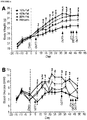
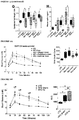
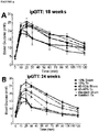
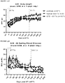
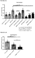
- FIGURE 1 and FIGURE 7 display body weight gain, measures of glucose homeostasis and adipocyte characterization in mice on the various diets. All three high fat diets (HFD; 45% fat, 60% fat, and western) induced rapid increases in fasting body weight ( FIGURE 1A ) and blood glucose levels ( FIGURE 1B ) compared to low fat diet (LFD) controls (10% fat). Moreover, after five days, mice in all three HFD groups were severely glucose intolerant relative to LFD controls ( FIGURE 7A ), prior to differences in body weight ( FIGURE 7B ). At 32 days, HFD mice were both glucose intolerant ( FIGURE 7C ) and significantly heavier ( FIGURE 7D ) than LFD controls.
- HFD high fat diets
- mice in all HFD groups developed glucose intolerance (day 47, FIGURE 1C ) and had insulin secretion kinetics that differed from LFD controls (either no glucose-induced insulin secretion or altered timing of peak insulin levels; FIGURE 1D ). All HFD mice were significantly overweight ( FIGURE 1A ) and had increased adiposity ( FIGURE 1F ) compared to LFD controls; mice fed 45% and 60% fat diets also had significantly elevated circulating leptin levels ( FIGURE 1G ).
- HFDs caused dyslipidemia, including significantly reduced plasma free fatty acid levels in all HFD-fed mice ( FIGURE 7E ), reduced triglyceride levels in 45% and 60% fat groups ( FIGURE 7F ), and elevated cholesterol levels in the western diet group compared to LFD controls ( FIGURE 7G ).
- the HFD-induced metabolic defects in immune-deficient mice were not associated with macrophage infiltration in adipose tissue (marked by F4/80 immuno-reactivity, whereas significant accumulation of F4/80-positive crown-like structures were observed in the epididymal fat of ob / ob mice, an immunocompetent model of T2D (micrographs not shown).
- Example 2 In vitro generation of pancreatic endocrine progenitor cells from human embryonic stem cells
- H1 human embryonic stem cell line H1 (WA01 cells, WiCell Research InstituteTM, Madison, WI) were seeded as single cells at 1 x 10 5 cells/cm 2 on 1:30 diluted MATRIGELTM (Becton Dickinson BiosciencesTM, Franklin Lakes, NJ; Catalogue ("Cat.") No. 356231) coated dishes in mTeSR-1TM (Stem Cell TechnologiesTM, Vancouver, BC; Cat. no. 05850). At ⁇ 70-80% confluency, the H1 cell cultures were rinsed with 1X Dulbeccos's phosphate buffered saline without Mg2+ and Ca2+ (InvitrogenTM, Carlsbad, CA; Cat.
- FBS fetal bovine serum
- AA activ
- Stage 2 (3 days): Stage 1 cells were cultured in DMEM-F12 medium (InvitrogenTM (GibcoTM); Cat. No. 10565-018) supplemented with 2 g/L sodium bicarbonate, 2% FBS and 50 ng/mL of FGF7 (PeprotechTM, Cat. No. 100-19) for three days.
- DMEM-F12 medium InvitrogenTM (GibcoTM); Cat. No. 10565-018) supplemented with 2 g/L sodium bicarbonate, 2% FBS and 50 ng/mL of FGF7 (PeprotechTM, Cat. No. 100-19) for three days.
- Stage 3 (4 days): Stage 2 cells were cultured in DMEM-HG (high glucose) medium (InvitrogenTM. Cat. No. 10569-044) supplemented with 0.25 ⁇ M SANT-1 (N-[(3,5-dimethyl-1-phenyl-1H-pyrazol-4-yl)methylene]-4-(phenylmethyl)-1-piperazineamine) (Sigma-AldrichTM, Cat. No. S4572), 2 ⁇ M retinoic acid (“RA”) (Sigma-AldrichTM, Catalog No. R2625), 100 ng/mL of NogginTM (R&D SystemsTM, Cat. No. 6057-NG), and 1% (v/v) B27 (InvitrogenTM (GibcoTM), Cat. No. 17504-044).
- SANT-1 N-[(3,5-dimethyl-1-phenyl-1H-pyrazol-4-yl)methylene]-4-(phenylmethyl)-1-piperazineamine
- RA
- Stage 4 (5 days): Stage 3 cells were cultured for 4 days in DMEM-HG medium supplemented with 0.1 ⁇ m 2-(3-(6-methylpyridin-2-yl)-1H-pyrazol-4-yl)-1,5-naphthyridine ("ALK5 inhibitor II", “ALK5i”) (AxxoraTM, San Diego, CA; Cat. No.
- the cell clusters were transferred into a polystyrene 125-500 ml Spinner Flask (CorningTM), and spun at 80-100 rpm overnight in suspension with DMEM-HG supplemented with 0.2 ⁇ M ALK5i, 100 nM (6-(4-(2-(piperidin-1-yl)ethoxy)phenyl)-3-(pyridin-4-yl)pyrazolo[1,5-a]pyrimidine, hydrochloride)) ("LDN”) a BMP receptor inhibitor (StemgentTM, San Diego, CA; Cat. No. 04-0074) and 1% B27.
- pancreatic endocrine precursor cells were assessed by fluorescence-activated flow cytometry ("FACS") and immuno-fluorescent staining (micrographs not shown). FACS staining was conducted as described in Diabetes, 61, 2016, 2012 and using the antibodies listed in TABLE 5. In brief, cells were incubated in TrypLETM Express (Life TechnologiesTM, Catalog No. 12604) for 3-5 minutes at 37 °C and released into single cell suspensions after which they were washed twice with a staining buffer of PBS containing 0.2% BSA (BD SciencesTM, Cat. No. 554657).
- FACS fluorescence-activated flow cytometry
- Intracellular antibody staining was accomplished by first incubating with Green Fluorescent LIVE/DEAD cell dye (Life TechnologiesTM, Cat. No. L23101) at 4 °C for 20 minutes followed by a single wash in cold PBS. Fixing of cells was in 250 ⁇ l of Cytofix/CytopermTM buffer (BD SciencesTM, Cat. No. 554723) followed by re-suspension of the cells in 100 ⁇ l of PermTM wash buffer staining/blocking solution with 2 % normal goat serum. Cells were incubated at 4 °C for 30 minutes with primary antibodies at empirically pre-determined dilutions followed by two washes in Perm/Wash buffer.
- Green Fluorescent LIVE/DEAD cell dye Life TechnologiesTM, Cat. No. L23101
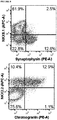
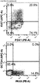
- FIGURE 8A, 8C, 8D and 8G Two key markers of pancreatic endoderm.
- Approximately 20% of PDXi-positive cells were in the cell cycle, as indicated by Ki67 or PCNA expression ( FIGURE 8A and 8D ), and the pluripotency marker OCT3/4 was not detected ( FIGURE 8A ).
- FIGURE 8A and 8B Although ⁇ 16% of progenitor cells expressed endocrine markers ( FIGURE 8A and 8B ), only 2.8% of synaptophysin-positive cells co-expressed NKX6.1 ( FIGURE 8A ) and most were polyhormonal ( FIGURE 8F ), indicative of an immature endocrine population.
- pancreatic endocrine precursor cells of Example 2 were encapsulated within a 20 ⁇ l TheracyteTM macro-encapsulation device (TheraCyte Inc.TM, Madison Hills, CA) as follows. Approximately 5 x 10 6 endocrine precursor cells (in cluster form) were placed into a positive displacement pipette. Using slight pressure, the tip of the capillary/piston tip containing cells was placed snug in the hub of the 24 gauge catheter and the cells dispensed from the positive displacement pipette through the catheter into the device. The device was sealed using a titanium barb. The encapsulated pancreatic endocrine precursor cells were then transplanted subcutaneously into seven SCID-beige mice from each of the four diet regimens.
- mice All mice were anaesthetized with inhalable isoflurane and transplant recipients received approximately 5x10 6 pancreatic endocrine precursor cells subcutaneously on the right flank.
- Four sham mice received the same surgical procedure, but no macro-encapsulation device was implanted.
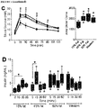
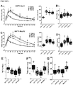
- pancreatic endocrine precursor cells Following transplantation, the pancreatic endocrine precursor cells further differentiated in vivo and the resulting cells, from all diet groups, secreted similar levels of human C-peptide under basal and fed conditions between 8 and 20 weeks ( FIGURE 2A ) and produced robust glucose-stimulated human C-peptide secretion at 18 weeks ( FIGUREs 2B,C ). Similarly, human insulin secretion was induced by an arginine challenge in all diet groups at 24 weeks ( FIGURE 2D ). A trend towards increased basal glucagon secretion in the HFD groups was observed, but because four out of five mice in the LFD group had undetectable fasting glucagon levels, it was not possible to do a statistical analysis ( FIGURE 2E ).
- Arginine-stimulated glucagon levels were similar between diet groups ( FIGUREs 2E,F ) and it was estimated that approximately half of the circulating glucagon may have originated from the hESC-derived cells, as indicated by the difference between transplanted and sham glucagon levels ( FIGURE 2F ).
- TheracyteTM devices were cut in half at the time of tissue harvest and stored in RNA later Stabilization SolutionTM (Qiagen, Inc.TM, Valencia, CA; Cat. No.76106) at - 80°C until use. Excess mouse tissue was first removed from the outside of the device before placing the device in 2 mL PBS. The edge of the device was cut off, the outer membranes peeled back, and the device isolated and placed into 400 ⁇ l QiagenTM Buffer RLT Plus (Qiagen Inc. TM, Cat. No.79216) containing 0.1% (v/v) beta-mercaptoethanol. The PBS was collected and centrifuged at 2000xg for 4 min to collect any cells that spilled out of the device.
- QiagenTM Buffer RLT Plus Qiagen Inc. TM, Cat. No.79216
- Human islets were obtained from four organ donors (23-48 years of age; two males and two females) as a positive control for quantitative polymerase chain reaction (“qPCR") analysis (Prodo Labs; Irvine, CA). Islet purity ranged from 85-95% and viability from 90-95%. All human islet preparations showed a 2 to 4-fold increase in human insulin secretion after incubation with high glucose concentration (data not shown) using a static glucose-stimulated insulin secretion assay. In brief, human islet cells (approximately 20 to 50 islet cells) were rinsed twice with Krebs buffer (129 mM NaCl, 4.8 mM KCL, 2.5 mM CaCl 2 , 1.2 mM MgSO 2 .
- Krebs buffer 129 mM NaCl, 4.8 mM KCL, 2.5 mM CaCl 2 , 1.2 mM MgSO 2 .
- Pre-amplification was performed using a primer pool specific for the genes run (TABLE 3) and TaqMan PreAmp 2x Master MixTM (Thermo Fisher ScientificTM/Life TechnologiesTM) with the following cycling conditions: 95°C 10 min, 8 cycles of 95°C 15s and 60°C 4 min, 99°C 10 min, and 4°C hold.
- pancreatic endocrine precursor cells Prior to transplant, a portion of the pancreatic endocrine precursor cells were fixed overnight in 4% paraformaldehyde ("PFA") and then embedded in 1% agarose prior to paraffin-embedding.
- PFA paraformaldehyde
- TheractyeTM devices, as well as a variety of tissues were harvested at 29 weeks post-transplant, fixed in 4% PFA and stored in 70% ethyl alcohol prior to paraffin-embedding. All paraffin sections (5 ⁇ m thickness) were prepared by Wax-it Histology ServicesTM (Vancouver, BC). Primary antibodies are provided in TABLE 4, above. Hemotoxyline and eosin (“H&E”) staining was performed using standard procedures and tissue analysis was performed in a blinded fashion by an independent pathologist (Nova Pathology PCTM, Bellingham, WA, USA).
- H&E Hemotoxyline and eosin
- the encapsulated hESC-derived grafts had similar or significantly higher levels of islet-related genes compared to human islets and there were no significant differences among different LFD or HFD groups (data not shown - CHGB; INS; CGC; SST; NKX6.1; PAX6; ISL1; MAFA; ABCC8; IAPP; PCSK1; PCSK2; GCGR; G6PC2; SLC30A8; and UCN3 ) .
- genes CHGB, INS, CGC, SST, MAFA and PCSK1 had similar gene expression in encapsulated hESC-derived grafts (i.e.
- hESC-derived graft cells For 10% fat; 45% fat; 60% fat and Western diets, when compared to human islets.
- NKX6.1, PAX6, ISL1, ABCC8, IAPP, PCSK2, GCGR, G6PC2, SLC30A8 and UCN3 gene expression in encapsulated hESC-derived grafts i.e. for 10% fat; 45% fat; 60% fat and Western diets
- the hESC-derived graft cells generally had significantly higher levels of gene expression than the human islets.
- Example 4 hESC-derived insulin secreting cells improved diet-induced dysglycemia and insulin resistance
- GTTs Glucose tolerance tests
- mice received an oral gavage of EnsureTM (8 uL/g body weight; Abbott LaboratoriesTM, Abbott Park, Illinois, USA) following an overnight fast ( ⁇ 16 hours).
- EnsureTM 8 uL/g body weight; Abbott LaboratoriesTM, Abbott Park, Illinois, USA
- ArgTT arginine tolerance tests
- mice received an i.p. injection of arginine (2 g/kg, 40% solution; Sigma-AldrichTM) following a 4-hour morning fast.
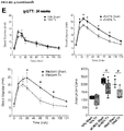
- HFD groups continued to be overweight and hyperglycemic under fasting conditions compared to LFD controls throughout the duration of the study. Transplantation of the encapsulated cells did not affect either body weight or fasting blood glucose levels compared to sham surgery (data not shown). However, significant improvements in long-term glycemic control, as measured by HbAiC, following transplantation alone ( FIGURES 4A and B ) was observed. HbAiC levels were elevated at 12 and 24 weeks in all HFD sham mice compared to LFD sham controls and significantly reduced by transplantation in the 45-60% fat group at both ages ( FIGURES 4A and 4B ).
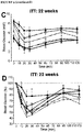
- Transplant recipients on 45-60% fat diets also displayed a significantly lower glucose excursion following a mixed-meal stimulus compared to sham mice at 20 weeks ( FIGURE 4C ) and all HFD transplant recipients had significantly improved glucose tolerance at 24 weeks post-transplant ( FIGURE 4E , FIGURE 9B ); these improvements were not yet evident at 18 weeks ( FIGURE 4D , FIGURE 9A ).
- Glucose tolerance in the 45-60% group was not completely ameliorated at 24 weeks, but transplant recipients in the western group had an area under the curve that was indistinguishable from controls ( FIGURE 4E ; FIGURE 9B ).
- FIGURE 4F A significant improvement in insulin sensitivity at 22 weeks in transplanted HFD-fed mice compared to shams ( FIGURE 4F ; FIGURE 9C and 9D ) was also observed, which may have contributed to the improved glucose tolerance in HFD transplant recipients ( FIGURE 4E ).
- pancreas sections per animal were immuno-stained for insulin and glucagon.
- Whole slide fluorescence scanning was performed using the ImageXpress Micro Imaging SystemTM, and images were stitched together and analyzed using MetaXpress SoftwareTM (Molecular Devices CorporationTM, Sunnyvale, CA).
- MetaXpress SoftwareTM Molecular Devices CorporationTM, Sunnyvale, CA.
- the beta cell or alpha cell fraction was calculated as the insulin-positive or glucagon-positive area /total pancreas area and the average of three sections per animal was then multiplied by the pancreas weight.
- the number of DAPI-positive nuclei were counted using the Multi Wavelength Cell ScoringTM module in MetaXpressTM and the number of cells that were immuno-reactive for insulin, glucagon or both hormones was counted manually by an investigator who was blinded to the treatment groups.
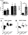
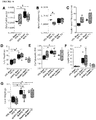
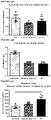
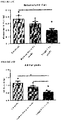
- Beta cell mass was significantly higher in all mice on 60% fat diets compared to LFD sham controls, and there was no difference between sham and transplanted mice in either diet group ( FIGURE 10A ). There was no effect of HFDs on alpha cell mass, but a significant reduction in alpha cell mass was observed in LFD transplant recipients compared to LFD shams ( FIGURE 10B ). There were no significant differences in the ratio of insulin-positive to glucagon-positive area in the pancreas of mice on either diet ( FIGURE 10C ).
- mice on the 45-60% fat diets had significantly higher body weight, adiposity (epididymal fat pad weight as a proportion of body weight) and circulating leptin levels than LFD shams ( FIGURES 10D-10F ).
- FIGURE 1A The obesity phenotype was more subtle in western diet mice during the first seven weeks ( FIGURE 1A ) and by the end of the study there were no significant differences in body weight, adiposity or leptin levels between Western-fed mice (sham and tx) and LFD sham controls ( FIGUREs 10D-10F ). All HFD groups had significantly higher liver weight ( FIGURE 10G ) and evidence of cytoplasmic vacuolation, consistent with dietary lipidosis in the liver (not shown) compared to LFD controls. Transplant recipients fed 45-60% fat diets had significantly reduced liver weight relative to shams (not shown), although a pathology assessment did not reveal differences in cytoplasmic vacuolation in H&E-stained liver sections (not shown).
- vacuolation of renal tubular epithelium was observed in kidney sections from all HFD groups (consistent with dietary lipidosis) and there was no effect of cell transplantation on this phenotype.
- Other tissue pathologies Adipose Tissue, Perirenal; Ileum; Skeletal Muscle; Cecum; Jejunum; Spleen; Colon; Kidney; Stomach, Glandular; Duodenum; Liver; Stomach, Nonglandular; Heart; Lung; and Testis
- Example 6 Combined treatment with pancreatic endocrine precursor cell transplants and an antidiabetic drug improved diet-induced obesity and glucose tolerance
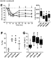
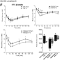
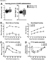
- FIGURE 5F Weight loss was observed within the first two weeks following transplantation in HFD-fed mice on antidiabetic drugs.
- FIGURES 5C , 5D and 5E antidiabetic drugs.
- FIGUREs 5A-E sham mice on any drug.
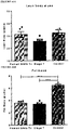
- All transplant recipients receiving antidiabetic drugs had significantly lower body weight on day 75 ( FIGURE 5F ) and reduced epididymal fat pad weight (relative to body weight; FIGURE 5G ) compared to sham mice, such that neither parameter was different from LFD-fed sham controls.
- FIGURE 5G There was no effect of transplantation on body weight ( FIGURE 5B and F ) or circulating leptin levels ( FIGURE 5H ) in HFD-fed mice without drug treatment, although we did observe a reduction in relative epididymal fat pad weight in this cohort ( FIGURE 5G ).
- the combination of a cell transplant with either metformin or sitagliptin resulted in significantly reduced circulating leptin levels compared to their respective sham controls ( FIGURE 5G ).
- FIGURE 5G There was no effect of the cell therapy on restoring leptin levels in the rosiglitazone group ( FIGURE. 5G ).
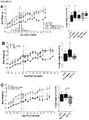
- FIGURE 6D Glycemic control during an oral glucose challenge was indistinguishable between the LFD controls and HFD-fed mice receiving sitagliptin with the cell therapy, with the exception only of a marginally higher peak glucose level at 15 minutes post-gavage.
- the improved glucose tolerance in cell transplant recipients from the metformin- and sitagliptin-treated mice was associated with significantly reduced fasting mouse C-peptide levels compared to their respective sham controls at 16 weeks post-transplant ( FIGURE 6G ), an effect that was not yet evident at 4 weeks ( FIGURE 6F ).
- the improvements in glucose tolerance were not associated with differences in the function of hESC-derived grafts. All transplant recipients showed robust glucose-responsive human C-peptide secretion at 16 weeks and there were no differences in human C-peptide levels between HFD-fed mice treated with different antidiabetic drugs ( FIGURE 6G ).
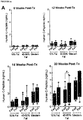
- Example 7 Comparison of Stage 4 (S4) and Stage 7 (S7) cell transplants in male and female mice
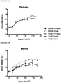
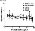
- Stage 4 cells pancreatic progenitors were compared to Stage 7 cells, which are more differentiated pancreatic endocrine cells, were both transplanted into normal, health male or female mice, on a normal diet. Following transplantation the body weights of the mice were compared following a 4 hour fast ( FIGURE 12 ) and an overnight fast ( FIGURE 13 ). In these studies, mice receiving Stage 7 cells tended to weigh less than mice receiving Stage 4 cells, whether weighted after a 4 hr ( FIGURE 12 ) or overnight ( FIGURE 13 ) fast. Accordingly, some Stage 7 cells may be slightly preferable for reducing weight gain.
- Example 8 Comparison of Stage 7 (S7) cell transplants with Human Islet cell transplants in mice
- Stage 7 cells and human islet cells were compared in mice that were either diabetic (i.e. given STZ) or not (i.e. not given STZ) prior to transplantation, wherein body weight ( FIGURE 14A ) and blood glucose ( FIGURE 14B ) were compared post transplant.
- body weight FIGURE 14A
- blood glucose FIGURE 14B
- FIGURE 14A body weight
- FIGURE 14B blood glucose mice
- FIGURE 14B those animals that received transplants appeared more able to regulate blood glucose post transplant similar to control mice.
- the there was a greater time lag i.e. ⁇ 120 days post Tx
- S7 Tx mice as compared to Human Islet TX mice (i.e.
- FIGURE 15A When the same animals studied in FIGURE 14A and 14B were compared for mouse leptin levels ( FIGURE 15A ) leptin was consistently lower in the transplanted mice (either Stage 7 or human islet) than controls. Leptin is typically proportional to fat mass and when fat mass was measured by DEXA, both the human islet and Stage 7 cells had lower fat mass ( FIGURE 15B ). Similarly, when body composition was compared between controls, human islet Tx mice and Stage 7 cell Tx mice the TX mice consistently had lower lean body mass ( FIGURE 16A ), fat mass ( FIGURE 16B ) and % body fat ( FIGURE 16C ).
- FIGURES 17A-D show reduced fat weight when comparing Stage 7 transplanted cells with either Human islet transplants or control cells in perirenal tissues ( FIGURE 17C ), epididymal tissues ( FIGURE 17D ), mesenteric fat (FIGURE 17E) and in all fat pads (FIGURE 17F), which were collected at sacrifice.
- FIGURE 17C shows perirenal tissues
- FIGURE 17D epididymal tissues
- FIGURE 17E mesenteric fat
- FIGURE 17F all fat pads
- FIGURE 17A there is shown data for both left and right kidneys and it is noted that the peri-renal fat around the left kidney as compared to the right kidney (except for the control the "right” kidney is labeled either "Human Islets Tx" or “Stage 7 Tx"), and when the data is combined for the right and left kidney the bar on the graph is designated "both".
- mice that got either islets or stage 7 cells cells were transplanted into the right kidney, which sees greater fat accumulation than the left kidney, except in the controls. It is likely that we see more fat accumulation around the kidney into which the cells were transplanted, because we speculate that there is local accumulation of insulin, and insulin is adipogenic.
- transplant of human islets and human Stage 7 cells may produce weight loss.
- T1D who receive an islet transplant and subsequently lose weight. This has been assumed to be due to the surgical procedure, immunosuppressive drugs, and /or life style changes. However, perhaps the weight loss is due to the islet cells themselves.
- GLP-1 is an incretin (i.e. a metabolic hormone) that is known to increase the amount of insulin released by pancreatic beta-cells and may subsequently decrease blood glucose.
- GLP-1 can produce weight loss and is now used as a drug in patients with T2D. In these patients weight loss has been reported.
- Sitagliptin is a known inhibitor of the enzyme dipeptidyl peptidase-4 (DPP-4), which degrades and inactivates GLP-1. Accordingly, it is possible that one mechanism by which the cells reduce weight gain is by producing GLP-1, and this affect is enhanced by stabilizing the GLP-1 with Sitagliptin.
- DPP-4 dipeptidyl peptidase-4
- TheraCyte devices used in FIGURES 19 and 20 contained predominantly what appeared to be endocrine cells (Synaptophysin positive). The cells appeared to be functioning well with good glucose responsive C-peptide production.
Landscapes
- Health & Medical Sciences (AREA)
- Life Sciences & Earth Sciences (AREA)
- Chemical & Material Sciences (AREA)
- General Health & Medical Sciences (AREA)
- Engineering & Computer Science (AREA)
- Veterinary Medicine (AREA)
- Animal Behavior & Ethology (AREA)
- Medicinal Chemistry (AREA)
- Public Health (AREA)
- Pharmacology & Pharmacy (AREA)
- Epidemiology (AREA)
- Biomedical Technology (AREA)
- Bioinformatics & Cheminformatics (AREA)
- Organic Chemistry (AREA)
- Diabetes (AREA)
- Biotechnology (AREA)
- Cell Biology (AREA)
- Zoology (AREA)
- Chemical Kinetics & Catalysis (AREA)
- Nuclear Medicine, Radiotherapy & Molecular Imaging (AREA)
- General Chemical & Material Sciences (AREA)
- Wood Science & Technology (AREA)
- Genetics & Genomics (AREA)
- Hematology (AREA)
- Obesity (AREA)
- Endocrinology (AREA)
- Immunology (AREA)
- Developmental Biology & Embryology (AREA)
- Virology (AREA)
- Nutrition Science (AREA)
- Physiology (AREA)
- Gastroenterology & Hepatology (AREA)
- General Engineering & Computer Science (AREA)
- Biochemistry (AREA)
- Microbiology (AREA)
- Emergency Medicine (AREA)
- Child & Adolescent Psychology (AREA)
- Medicines Containing Material From Animals Or Micro-Organisms (AREA)
- Pharmaceuticals Containing Other Organic And Inorganic Compounds (AREA)
- Acyclic And Carbocyclic Compounds In Medicinal Compositions (AREA)
Applications Claiming Priority (2)
| Application Number | Priority Date | Filing Date | Title |
|---|---|---|---|
| US201562131540P | 2015-03-11 | 2015-03-11 | |
| PCT/CA2016/000072 WO2016141460A1 (en) | 2015-03-11 | 2016-03-11 | Pancreatic endocrine progenitor cell therapies for the treatment of obesity and type 2 diabetes (t2d) |
Publications (3)
| Publication Number | Publication Date |
|---|---|
| EP3268016A1 EP3268016A1 (en) | 2018-01-17 |
| EP3268016A4 EP3268016A4 (en) | 2018-12-12 |
| EP3268016B1 true EP3268016B1 (en) | 2021-04-28 |
Family
ID=56879832
Family Applications (1)
| Application Number | Title | Priority Date | Filing Date |
|---|---|---|---|
| EP16760957.7A Active EP3268016B1 (en) | 2015-03-11 | 2016-03-11 | Pancreatic endocrine progenitor cell therapies for the treatment of obesity and type 2 diabetes (t2d) |
Country Status (7)
| Country | Link |
|---|---|
| US (2) | US10772917B2 (enExample) |
| EP (1) | EP3268016B1 (enExample) |
| JP (1) | JP6847044B2 (enExample) |
| CN (1) | CN107530379A (enExample) |
| AU (1) | AU2016228894B2 (enExample) |
| CA (1) | CA2979293C (enExample) |
| WO (1) | WO2016141460A1 (enExample) |
Families Citing this family (7)
| Publication number | Priority date | Publication date | Assignee | Title |
|---|---|---|---|---|
| WO2019118641A1 (en) * | 2017-12-12 | 2019-06-20 | The Regents Of The University Of California | Preservation of pancreatic islet grafts in the extrahepatic space |
| KR20200110338A (ko) | 2018-01-18 | 2020-09-23 | 다이이찌 산쿄 가부시키가이샤 | 디하이드로인돌리지논 유도체 |
| KR102503349B1 (ko) | 2019-05-14 | 2023-02-23 | 프로벤션 바이오, 인코포레이티드 | 제1형 당뇨병을 예방하기 위한 방법 및 조성물 |
| EP3972990A4 (en) | 2019-05-22 | 2023-10-11 | The Cleveland Clinic Foundation | GENERATION OF ENDODEREM CELLS OF THE DORSAL INTESTINAL TUBE AND THE ANTERIOR AREA |
| CN110585242A (zh) * | 2019-10-15 | 2019-12-20 | 南通大学 | 多系分化持续应激细胞的应用、治疗糖尿病的药物及其制备方法 |
| CN111411072B (zh) * | 2020-03-09 | 2022-10-11 | 同济大学 | SLC30A8基因表达下调的抗糖尿病胰岛β细胞及其应用 |
| IL298999A (en) | 2020-06-11 | 2023-02-01 | Provention Bio Inc | Methods and compositions for the prevention of type 1 diabetes |
Family Cites Families (18)
| Publication number | Priority date | Publication date | Assignee | Title |
|---|---|---|---|---|
| US6703017B1 (en) * | 1994-04-28 | 2004-03-09 | Ixion Biotechnology, Inc. | Reversal of insulin-dependent diabetes by islet-producing stem cells, islet progenitor cells and islet-like structures |
| US5843780A (en) | 1995-01-20 | 1998-12-01 | Wisconsin Alumni Research Foundation | Primate embryonic stem cells |
| MXPA04004311A (es) * | 2001-11-09 | 2005-03-31 | Artecel Sciences Inc | DIFERENCIACION DEL PáNCREAS ENDOCRINOS DE CELULAS ESTROMALES DERIVADAS DEL TEJIDO ADIPOSO Y USOS DE LAS MISMAS. |
| KR20050037508A (ko) * | 2002-05-24 | 2005-04-22 | 와라타 파마수티컬즈, 인크. | 당뇨병의 치료 |
| JP2005537803A (ja) * | 2002-09-06 | 2005-12-15 | アムサイト インコーポレーティッド | Cd56陽性ヒト成体膵臓内分泌前駆細胞 |
| US7510873B2 (en) | 2003-06-27 | 2009-03-31 | Ethicon, Incorporated | Postpartum cells isolated from umbilical cord tissue, and methods of making and using the same |
| CN1589904A (zh) * | 2003-09-01 | 2005-03-09 | 北京大学 | 造血干细胞动员剂制备治疗糖尿病药物的用途 |
| JP2006149380A (ja) * | 2004-10-27 | 2006-06-15 | Kobe Univ | 遺伝子改変動物の新規用途 |
| AU2006202209B2 (en) | 2005-05-27 | 2011-04-14 | Lifescan, Inc. | Amniotic fluid derived cells |
| US20080233087A1 (en) * | 2006-10-31 | 2008-09-25 | Shamblott Michael J | Methods for identifying, isolating, and utilizing endocrine progenitor cells from adult human pancreata |
| TWI504419B (zh) * | 2006-11-09 | 2015-10-21 | Orexigen Therapeutics Inc | 層狀醫藥調配物 |
| JP5710264B2 (ja) | 2007-11-27 | 2015-04-30 | ライフスキャン・インコーポレイテッドLifescan,Inc. | ヒト胚性幹細胞の分化 |
| CA2723820A1 (en) * | 2008-05-09 | 2009-11-12 | Vistagen Therapeutics, Inc. | Pancreatic endocrine progenitor cells derived from pluripotent stem cells |
| CN106177958A (zh) * | 2009-02-13 | 2016-12-07 | 勃林格殷格翰国际有限公司 | 包含dpp‑4抑制剂(利拉列汀)任选地组合其它抗糖尿病药的抗糖尿病药物 |
| AU2010319921A1 (en) | 2009-10-29 | 2012-05-17 | Janssen Biotech Inc. | Pluripotent stem cells |
| US9085757B2 (en) * | 2010-06-17 | 2015-07-21 | Regents Of The University Of Minnesota | Production of insulin producing cells |
| AU2011289379A1 (en) * | 2010-08-12 | 2013-02-07 | Janssen Biotech, Inc. | Treatment of diabetes with pancreatic endocrine precursor cells |
| US8859286B2 (en) | 2013-03-14 | 2014-10-14 | Viacyte, Inc. | In vitro differentiation of pluripotent stem cells to pancreatic endoderm cells (PEC) and endocrine cells |
-
2016
- 2016-03-11 JP JP2017547514A patent/JP6847044B2/ja active Active
- 2016-03-11 CA CA2979293A patent/CA2979293C/en active Active
- 2016-03-11 EP EP16760957.7A patent/EP3268016B1/en active Active
- 2016-03-11 WO PCT/CA2016/000072 patent/WO2016141460A1/en not_active Ceased
- 2016-03-11 US US15/556,907 patent/US10772917B2/en active Active
- 2016-03-11 AU AU2016228894A patent/AU2016228894B2/en active Active
- 2016-03-11 CN CN201680024439.5A patent/CN107530379A/zh active Pending
-
2020
- 2020-08-03 US US16/983,810 patent/US20210015872A1/en active Pending
Non-Patent Citations (1)
| Title |
|---|
| None * |
Also Published As
| Publication number | Publication date |
|---|---|
| EP3268016A4 (en) | 2018-12-12 |
| US20180055890A1 (en) | 2018-03-01 |
| CN107530379A (zh) | 2018-01-02 |
| US10772917B2 (en) | 2020-09-15 |
| WO2016141460A1 (en) | 2016-09-15 |
| US20210015872A1 (en) | 2021-01-21 |
| AU2016228894B2 (en) | 2021-03-04 |
| JP6847044B2 (ja) | 2021-03-24 |
| CA2979293A1 (en) | 2016-09-15 |
| EP3268016A1 (en) | 2018-01-17 |
| CA2979293C (en) | 2022-01-04 |
| AU2016228894A1 (en) | 2017-11-02 |
| JP2018507889A (ja) | 2018-03-22 |
Similar Documents
| Publication | Publication Date | Title |
|---|---|---|
| US20210015872A1 (en) | Pancreatic endocrine progenitor cell therapies for the treatment of obesity and type 2 diabetes (t2d) | |
| US11827905B2 (en) | SC-beta cells and compositions and methods for generating the same | |
| Navarro-Tableros et al. | Islet-like structures generated in vitro from adult human liver stem cells revert hyperglycemia in diabetic SCID mice | |
| KR20200087201A (ko) | 섬세포 제조 조성물 및 사용 방법 | |
| Kim et al. | Activin A, exendin-4, and glucose stimulate differentiation of human pancreatic ductal cells | |
| Samuelson | Sca-1 positive pancreatic progenitor cells: a replacement for transplanted islets |
Legal Events
| Date | Code | Title | Description |
|---|---|---|---|
| STAA | Information on the status of an ep patent application or granted ep patent |
Free format text: STATUS: THE INTERNATIONAL PUBLICATION HAS BEEN MADE |
|
| PUAI | Public reference made under article 153(3) epc to a published international application that has entered the european phase |
Free format text: ORIGINAL CODE: 0009012 |
|
| STAA | Information on the status of an ep patent application or granted ep patent |
Free format text: STATUS: REQUEST FOR EXAMINATION WAS MADE |
|
| 17P | Request for examination filed |
Effective date: 20170925 |
|
| AK | Designated contracting states |
Kind code of ref document: A1 Designated state(s): AL AT BE BG CH CY CZ DE DK EE ES FI FR GB GR HR HU IE IS IT LI LT LU LV MC MK MT NL NO PL PT RO RS SE SI SK SM TR |
|
| AX | Request for extension of the european patent |
Extension state: BA ME |
|
| DAV | Request for validation of the european patent (deleted) | ||
| DAX | Request for extension of the european patent (deleted) | ||
| A4 | Supplementary search report drawn up and despatched |
Effective date: 20181114 |
|
| RIC1 | Information provided on ipc code assigned before grant |
Ipc: A61K 31/4439 20060101ALI20181108BHEP Ipc: C07D 417/12 20060101ALI20181108BHEP Ipc: C12N 5/071 20100101ALI20181108BHEP Ipc: A61P 3/10 20060101ALI20181108BHEP Ipc: A61K 31/155 20060101ALI20181108BHEP Ipc: C07D 487/04 20060101ALI20181108BHEP Ipc: C07C 279/26 20060101ALI20181108BHEP Ipc: A61K 31/4985 20060101ALI20181108BHEP Ipc: A61P 3/00 20060101ALI20181108BHEP Ipc: A61K 35/39 20150101AFI20181108BHEP |
|
| STAA | Information on the status of an ep patent application or granted ep patent |
Free format text: STATUS: EXAMINATION IS IN PROGRESS |
|
| 17Q | First examination report despatched |
Effective date: 20200213 |
|
| RAP1 | Party data changed (applicant data changed or rights of an application transferred) |
Owner name: CCS VENTURES LIMITED |
|
| GRAP | Despatch of communication of intention to grant a patent |
Free format text: ORIGINAL CODE: EPIDOSNIGR1 |
|
| STAA | Information on the status of an ep patent application or granted ep patent |
Free format text: STATUS: GRANT OF PATENT IS INTENDED |
|
| INTG | Intention to grant announced |
Effective date: 20200922 |
|
| GRAS | Grant fee paid |
Free format text: ORIGINAL CODE: EPIDOSNIGR3 |
|
| GRAA | (expected) grant |
Free format text: ORIGINAL CODE: 0009210 |
|
| STAA | Information on the status of an ep patent application or granted ep patent |
Free format text: STATUS: THE PATENT HAS BEEN GRANTED |
|
| AK | Designated contracting states |
Kind code of ref document: B1 Designated state(s): AL AT BE BG CH CY CZ DE DK EE ES FI FR GB GR HR HU IE IS IT LI LT LU LV MC MK MT NL NO PL PT RO RS SE SI SK SM TR |
|
| REG | Reference to a national code |
Ref country code: GB Ref legal event code: FG4D |
|
| REG | Reference to a national code |
Ref country code: CH Ref legal event code: EP |
|
| REG | Reference to a national code |
Ref country code: AT Ref legal event code: REF Ref document number: 1386345 Country of ref document: AT Kind code of ref document: T Effective date: 20210515 |
|
| REG | Reference to a national code |
Ref country code: DE Ref legal event code: R096 Ref document number: 602016056930 Country of ref document: DE |
|
| REG | Reference to a national code |
Ref country code: IE Ref legal event code: FG4D |
|
| REG | Reference to a national code |
Ref country code: SE Ref legal event code: TRGR |
|
| REG | Reference to a national code |
Ref country code: NL Ref legal event code: FP |
|
| REG | Reference to a national code |
Ref country code: LT Ref legal event code: MG9D |
|
| REG | Reference to a national code |
Ref country code: AT Ref legal event code: MK05 Ref document number: 1386345 Country of ref document: AT Kind code of ref document: T Effective date: 20210428 |
|
| PG25 | Lapsed in a contracting state [announced via postgrant information from national office to epo] |
Ref country code: FI Free format text: LAPSE BECAUSE OF FAILURE TO SUBMIT A TRANSLATION OF THE DESCRIPTION OR TO PAY THE FEE WITHIN THE PRESCRIBED TIME-LIMIT Effective date: 20210428 Ref country code: LT Free format text: LAPSE BECAUSE OF FAILURE TO SUBMIT A TRANSLATION OF THE DESCRIPTION OR TO PAY THE FEE WITHIN THE PRESCRIBED TIME-LIMIT Effective date: 20210428 Ref country code: BG Free format text: LAPSE BECAUSE OF FAILURE TO SUBMIT A TRANSLATION OF THE DESCRIPTION OR TO PAY THE FEE WITHIN THE PRESCRIBED TIME-LIMIT Effective date: 20210728 Ref country code: AT Free format text: LAPSE BECAUSE OF FAILURE TO SUBMIT A TRANSLATION OF THE DESCRIPTION OR TO PAY THE FEE WITHIN THE PRESCRIBED TIME-LIMIT Effective date: 20210428 Ref country code: HR Free format text: LAPSE BECAUSE OF FAILURE TO SUBMIT A TRANSLATION OF THE DESCRIPTION OR TO PAY THE FEE WITHIN THE PRESCRIBED TIME-LIMIT Effective date: 20210428 |
|
| PG25 | Lapsed in a contracting state [announced via postgrant information from national office to epo] |
Ref country code: RS Free format text: LAPSE BECAUSE OF FAILURE TO SUBMIT A TRANSLATION OF THE DESCRIPTION OR TO PAY THE FEE WITHIN THE PRESCRIBED TIME-LIMIT Effective date: 20210428 Ref country code: PT Free format text: LAPSE BECAUSE OF FAILURE TO SUBMIT A TRANSLATION OF THE DESCRIPTION OR TO PAY THE FEE WITHIN THE PRESCRIBED TIME-LIMIT Effective date: 20210830 Ref country code: NO Free format text: LAPSE BECAUSE OF FAILURE TO SUBMIT A TRANSLATION OF THE DESCRIPTION OR TO PAY THE FEE WITHIN THE PRESCRIBED TIME-LIMIT Effective date: 20210728 Ref country code: PL Free format text: LAPSE BECAUSE OF FAILURE TO SUBMIT A TRANSLATION OF THE DESCRIPTION OR TO PAY THE FEE WITHIN THE PRESCRIBED TIME-LIMIT Effective date: 20210428 Ref country code: IS Free format text: LAPSE BECAUSE OF FAILURE TO SUBMIT A TRANSLATION OF THE DESCRIPTION OR TO PAY THE FEE WITHIN THE PRESCRIBED TIME-LIMIT Effective date: 20210828 Ref country code: GR Free format text: LAPSE BECAUSE OF FAILURE TO SUBMIT A TRANSLATION OF THE DESCRIPTION OR TO PAY THE FEE WITHIN THE PRESCRIBED TIME-LIMIT Effective date: 20210729 Ref country code: LV Free format text: LAPSE BECAUSE OF FAILURE TO SUBMIT A TRANSLATION OF THE DESCRIPTION OR TO PAY THE FEE WITHIN THE PRESCRIBED TIME-LIMIT Effective date: 20210428 |
|
| PG25 | Lapsed in a contracting state [announced via postgrant information from national office to epo] |
Ref country code: RO Free format text: LAPSE BECAUSE OF FAILURE TO SUBMIT A TRANSLATION OF THE DESCRIPTION OR TO PAY THE FEE WITHIN THE PRESCRIBED TIME-LIMIT Effective date: 20210428 Ref country code: ES Free format text: LAPSE BECAUSE OF FAILURE TO SUBMIT A TRANSLATION OF THE DESCRIPTION OR TO PAY THE FEE WITHIN THE PRESCRIBED TIME-LIMIT Effective date: 20210428 Ref country code: SK Free format text: LAPSE BECAUSE OF FAILURE TO SUBMIT A TRANSLATION OF THE DESCRIPTION OR TO PAY THE FEE WITHIN THE PRESCRIBED TIME-LIMIT Effective date: 20210428 Ref country code: SM Free format text: LAPSE BECAUSE OF FAILURE TO SUBMIT A TRANSLATION OF THE DESCRIPTION OR TO PAY THE FEE WITHIN THE PRESCRIBED TIME-LIMIT Effective date: 20210428 Ref country code: EE Free format text: LAPSE BECAUSE OF FAILURE TO SUBMIT A TRANSLATION OF THE DESCRIPTION OR TO PAY THE FEE WITHIN THE PRESCRIBED TIME-LIMIT Effective date: 20210428 Ref country code: CZ Free format text: LAPSE BECAUSE OF FAILURE TO SUBMIT A TRANSLATION OF THE DESCRIPTION OR TO PAY THE FEE WITHIN THE PRESCRIBED TIME-LIMIT Effective date: 20210428 Ref country code: DK Free format text: LAPSE BECAUSE OF FAILURE TO SUBMIT A TRANSLATION OF THE DESCRIPTION OR TO PAY THE FEE WITHIN THE PRESCRIBED TIME-LIMIT Effective date: 20210428 |
|
| REG | Reference to a national code |
Ref country code: DE Ref legal event code: R097 Ref document number: 602016056930 Country of ref document: DE |
|
| PLBE | No opposition filed within time limit |
Free format text: ORIGINAL CODE: 0009261 |
|
| STAA | Information on the status of an ep patent application or granted ep patent |
Free format text: STATUS: NO OPPOSITION FILED WITHIN TIME LIMIT |
|
| 26N | No opposition filed |
Effective date: 20220131 |
|
| PG25 | Lapsed in a contracting state [announced via postgrant information from national office to epo] |
Ref country code: IS Free format text: LAPSE BECAUSE OF FAILURE TO SUBMIT A TRANSLATION OF THE DESCRIPTION OR TO PAY THE FEE WITHIN THE PRESCRIBED TIME-LIMIT Effective date: 20210828 Ref country code: AL Free format text: LAPSE BECAUSE OF FAILURE TO SUBMIT A TRANSLATION OF THE DESCRIPTION OR TO PAY THE FEE WITHIN THE PRESCRIBED TIME-LIMIT Effective date: 20210428 |
|
| PG25 | Lapsed in a contracting state [announced via postgrant information from national office to epo] |
Ref country code: IT Free format text: LAPSE BECAUSE OF FAILURE TO SUBMIT A TRANSLATION OF THE DESCRIPTION OR TO PAY THE FEE WITHIN THE PRESCRIBED TIME-LIMIT Effective date: 20210428 |
|
| PG25 | Lapsed in a contracting state [announced via postgrant information from national office to epo] |
Ref country code: MC Free format text: LAPSE BECAUSE OF FAILURE TO SUBMIT A TRANSLATION OF THE DESCRIPTION OR TO PAY THE FEE WITHIN THE PRESCRIBED TIME-LIMIT Effective date: 20210428 |
|
| PG25 | Lapsed in a contracting state [announced via postgrant information from national office to epo] |
Ref country code: LU Free format text: LAPSE BECAUSE OF NON-PAYMENT OF DUE FEES Effective date: 20220311 Ref country code: IE Free format text: LAPSE BECAUSE OF NON-PAYMENT OF DUE FEES Effective date: 20220311 |
|
| P01 | Opt-out of the competence of the unified patent court (upc) registered |
Effective date: 20230427 |
|
| PG25 | Lapsed in a contracting state [announced via postgrant information from national office to epo] |
Ref country code: HU Free format text: LAPSE BECAUSE OF FAILURE TO SUBMIT A TRANSLATION OF THE DESCRIPTION OR TO PAY THE FEE WITHIN THE PRESCRIBED TIME-LIMIT; INVALID AB INITIO Effective date: 20160311 |
|
| PG25 | Lapsed in a contracting state [announced via postgrant information from national office to epo] |
Ref country code: MK Free format text: LAPSE BECAUSE OF FAILURE TO SUBMIT A TRANSLATION OF THE DESCRIPTION OR TO PAY THE FEE WITHIN THE PRESCRIBED TIME-LIMIT Effective date: 20210428 Ref country code: CY Free format text: LAPSE BECAUSE OF FAILURE TO SUBMIT A TRANSLATION OF THE DESCRIPTION OR TO PAY THE FEE WITHIN THE PRESCRIBED TIME-LIMIT Effective date: 20210428 |
|
| PG25 | Lapsed in a contracting state [announced via postgrant information from national office to epo] |
Ref country code: MT Free format text: LAPSE BECAUSE OF FAILURE TO SUBMIT A TRANSLATION OF THE DESCRIPTION OR TO PAY THE FEE WITHIN THE PRESCRIBED TIME-LIMIT Effective date: 20210428 |
|
| PGFP | Annual fee paid to national office [announced via postgrant information from national office to epo] |
Ref country code: NL Payment date: 20250226 Year of fee payment: 10 |
|
| PGFP | Annual fee paid to national office [announced via postgrant information from national office to epo] |
Ref country code: SE Payment date: 20250314 Year of fee payment: 10 |
|
| PGFP | Annual fee paid to national office [announced via postgrant information from national office to epo] |
Ref country code: DE Payment date: 20250313 Year of fee payment: 10 |
|
| PGFP | Annual fee paid to national office [announced via postgrant information from national office to epo] |
Ref country code: BE Payment date: 20250324 Year of fee payment: 10 |
|
| PGFP | Annual fee paid to national office [announced via postgrant information from national office to epo] |
Ref country code: FR Payment date: 20250331 Year of fee payment: 10 |
|
| PGFP | Annual fee paid to national office [announced via postgrant information from national office to epo] |
Ref country code: GB Payment date: 20250324 Year of fee payment: 10 |
|
| PGFP | Annual fee paid to national office [announced via postgrant information from national office to epo] |
Ref country code: CH Payment date: 20250401 Year of fee payment: 10 |