EP3232193A2 - Diagnose von mps i krankheiten durch messung von nicht-reduzierenden enden von glykosaminoglykanen nach behandlung mit enzymen - Google Patents
Diagnose von mps i krankheiten durch messung von nicht-reduzierenden enden von glykosaminoglykanen nach behandlung mit enzymen Download PDFInfo
- Publication number
- EP3232193A2 EP3232193A2 EP17162060.2A EP17162060A EP3232193A2 EP 3232193 A2 EP3232193 A2 EP 3232193A2 EP 17162060 A EP17162060 A EP 17162060A EP 3232193 A2 EP3232193 A2 EP 3232193A2
- Authority
- EP
- European Patent Office
- Prior art keywords
- formula
- mps
- glcn
- idoa
- glca
- Prior art date
- Legal status (The legal status is an assumption and is not a legal conclusion. Google has not performed a legal analysis and makes no representation as to the accuracy of the status listed.)
- Granted
Links
- 0 CO[C@@](C1O)O*C=C1O Chemical compound CO[C@@](C1O)O*C=C1O 0.000 description 1
Images


Classifications
-
- C—CHEMISTRY; METALLURGY
- C12—BIOCHEMISTRY; BEER; SPIRITS; WINE; VINEGAR; MICROBIOLOGY; ENZYMOLOGY; MUTATION OR GENETIC ENGINEERING
- C12Q—MEASURING OR TESTING PROCESSES INVOLVING ENZYMES, NUCLEIC ACIDS OR MICROORGANISMS; COMPOSITIONS OR TEST PAPERS THEREFOR; PROCESSES OF PREPARING SUCH COMPOSITIONS; CONDITION-RESPONSIVE CONTROL IN MICROBIOLOGICAL OR ENZYMOLOGICAL PROCESSES
- C12Q1/00—Measuring or testing processes involving enzymes, nucleic acids or microorganisms; Compositions therefor; Processes of preparing such compositions
- C12Q1/527—Measuring or testing processes involving enzymes, nucleic acids or microorganisms; Compositions therefor; Processes of preparing such compositions involving lyase
-
- A—HUMAN NECESSITIES
- A61—MEDICAL OR VETERINARY SCIENCE; HYGIENE
- A61B—DIAGNOSIS; SURGERY; IDENTIFICATION
- A61B5/00—Measuring for diagnostic purposes; Identification of persons
- A61B5/48—Other medical applications
- A61B5/4842—Monitoring progression or stage of a disease
-
- A—HUMAN NECESSITIES
- A61—MEDICAL OR VETERINARY SCIENCE; HYGIENE
- A61B—DIAGNOSIS; SURGERY; IDENTIFICATION
- A61B5/00—Measuring for diagnostic purposes; Identification of persons
- A61B5/48—Other medical applications
- A61B5/4866—Evaluating metabolism
-
- A—HUMAN NECESSITIES
- A61—MEDICAL OR VETERINARY SCIENCE; HYGIENE
- A61K—PREPARATIONS FOR MEDICAL, DENTAL OR TOILETRY PURPOSES
- A61K31/00—Medicinal preparations containing organic active ingredients
- A61K31/70—Carbohydrates; Sugars; Derivatives thereof
- A61K31/702—Oligosaccharides, i.e. having three to five saccharide radicals attached to each other by glycosidic linkages
-
- C—CHEMISTRY; METALLURGY
- C07—ORGANIC CHEMISTRY
- C07H—SUGARS; DERIVATIVES THEREOF; NUCLEOSIDES; NUCLEOTIDES; NUCLEIC ACIDS
- C07H11/00—Compounds containing saccharide radicals esterified by inorganic acids; Metal salts thereof
-
- C—CHEMISTRY; METALLURGY
- C07—ORGANIC CHEMISTRY
- C07H—SUGARS; DERIVATIVES THEREOF; NUCLEOSIDES; NUCLEOTIDES; NUCLEIC ACIDS
- C07H13/00—Compounds containing saccharide radicals esterified by carbonic acid or derivatives thereof, or by organic acids, e.g. phosphonic acids
- C07H13/02—Compounds containing saccharide radicals esterified by carbonic acid or derivatives thereof, or by organic acids, e.g. phosphonic acids by carboxylic acids
- C07H13/04—Compounds containing saccharide radicals esterified by carbonic acid or derivatives thereof, or by organic acids, e.g. phosphonic acids by carboxylic acids having the esterifying carboxyl radicals attached to acyclic carbon atoms
-
- C—CHEMISTRY; METALLURGY
- C07—ORGANIC CHEMISTRY
- C07H—SUGARS; DERIVATIVES THEREOF; NUCLEOSIDES; NUCLEOTIDES; NUCLEIC ACIDS
- C07H3/00—Compounds containing only hydrogen atoms and saccharide radicals having only carbon, hydrogen, and oxygen atoms
- C07H3/04—Disaccharides
-
- C—CHEMISTRY; METALLURGY
- C07—ORGANIC CHEMISTRY
- C07H—SUGARS; DERIVATIVES THEREOF; NUCLEOSIDES; NUCLEOTIDES; NUCLEIC ACIDS
- C07H3/00—Compounds containing only hydrogen atoms and saccharide radicals having only carbon, hydrogen, and oxygen atoms
- C07H3/06—Oligosaccharides, i.e. having three to five saccharide radicals attached to each other by glycosidic linkages
-
- C—CHEMISTRY; METALLURGY
- C07—ORGANIC CHEMISTRY
- C07H—SUGARS; DERIVATIVES THEREOF; NUCLEOSIDES; NUCLEOTIDES; NUCLEIC ACIDS
- C07H5/00—Compounds containing saccharide radicals in which the hetero bonds to oxygen have been replaced by the same number of hetero bonds to halogen, nitrogen, sulfur, selenium, or tellurium
- C07H5/04—Compounds containing saccharide radicals in which the hetero bonds to oxygen have been replaced by the same number of hetero bonds to halogen, nitrogen, sulfur, selenium, or tellurium to nitrogen
- C07H5/06—Aminosugars
-
- C—CHEMISTRY; METALLURGY
- C07—ORGANIC CHEMISTRY
- C07H—SUGARS; DERIVATIVES THEREOF; NUCLEOSIDES; NUCLEOTIDES; NUCLEIC ACIDS
- C07H7/00—Compounds containing non-saccharide radicals linked to saccharide radicals by a carbon-to-carbon bond
- C07H7/02—Acyclic radicals
- C07H7/033—Uronic acids
-
- C—CHEMISTRY; METALLURGY
- C12—BIOCHEMISTRY; BEER; SPIRITS; WINE; VINEGAR; MICROBIOLOGY; ENZYMOLOGY; MUTATION OR GENETIC ENGINEERING
- C12N—MICROORGANISMS OR ENZYMES; COMPOSITIONS THEREOF; PROPAGATING, PRESERVING, OR MAINTAINING MICROORGANISMS; MUTATION OR GENETIC ENGINEERING; CULTURE MEDIA
- C12N9/00—Enzymes; Proenzymes; Compositions thereof; Processes for preparing, activating, inhibiting, separating or purifying enzymes
- C12N9/88—Lyases (4.)
-
- G—PHYSICS
- G01—MEASURING; TESTING
- G01N—INVESTIGATING OR ANALYSING MATERIALS BY DETERMINING THEIR CHEMICAL OR PHYSICAL PROPERTIES
- G01N33/00—Investigating or analysing materials by specific methods not covered by groups G01N1/00 - G01N31/00
- G01N33/48—Biological material, e.g. blood, urine; Haemocytometers
- G01N33/50—Chemical analysis of biological material, e.g. blood, urine; Testing involving biospecific ligand binding methods; Immunological testing
- G01N33/66—Chemical analysis of biological material, e.g. blood, urine; Testing involving biospecific ligand binding methods; Immunological testing involving blood sugars, e.g. galactose
-
- G—PHYSICS
- G01—MEASURING; TESTING
- G01N—INVESTIGATING OR ANALYSING MATERIALS BY DETERMINING THEIR CHEMICAL OR PHYSICAL PROPERTIES
- G01N33/00—Investigating or analysing materials by specific methods not covered by groups G01N1/00 - G01N31/00
- G01N33/48—Biological material, e.g. blood, urine; Haemocytometers
- G01N33/50—Chemical analysis of biological material, e.g. blood, urine; Testing involving biospecific ligand binding methods; Immunological testing
- G01N33/68—Chemical analysis of biological material, e.g. blood, urine; Testing involving biospecific ligand binding methods; Immunological testing involving proteins, peptides or amino acids
-
- G—PHYSICS
- G01—MEASURING; TESTING
- G01N—INVESTIGATING OR ANALYSING MATERIALS BY DETERMINING THEIR CHEMICAL OR PHYSICAL PROPERTIES
- G01N2333/00—Assays involving biological materials from specific organisms or of a specific nature
- G01N2333/90—Enzymes; Proenzymes
- G01N2333/988—Lyases (4.), e.g. aldolases, heparinase, enolases, fumarase
-
- G—PHYSICS
- G01—MEASURING; TESTING
- G01N—INVESTIGATING OR ANALYSING MATERIALS BY DETERMINING THEIR CHEMICAL OR PHYSICAL PROPERTIES
- G01N2400/00—Assays, e.g. immunoassays or enzyme assays, involving carbohydrates
- G01N2400/10—Polysaccharides, i.e. having more than five saccharide radicals attached to each other by glycosidic linkages; Derivatives thereof, e.g. ethers, esters
- G01N2400/38—Heteroglycans, i.e. polysaccharides having more than one sugar residue in the main chain in either alternating or less regular sequence, e.g. gluco- or galactomannans, Konjac gum, Locust bean gum or Guar gum
- G01N2400/40—Glycosaminoglycans, i.e. GAG or mucopolysaccharides, e.g. chondroitin sulfate, dermatan sulfate, hyaluronic acid, heparin, heparan sulfate, and related sulfated polysaccharides
-
- G—PHYSICS
- G01—MEASURING; TESTING
- G01N—INVESTIGATING OR ANALYSING MATERIALS BY DETERMINING THEIR CHEMICAL OR PHYSICAL PROPERTIES
- G01N2800/00—Detection or diagnosis of diseases
- G01N2800/04—Endocrine or metabolic disorders
- G01N2800/042—Disorders of carbohydrate metabolism, e.g. diabetes, glucose metabolism
-
- G—PHYSICS
- G01—MEASURING; TESTING
- G01N—INVESTIGATING OR ANALYSING MATERIALS BY DETERMINING THEIR CHEMICAL OR PHYSICAL PROPERTIES
- G01N2800/00—Detection or diagnosis of diseases
- G01N2800/28—Neurological disorders
-
- G—PHYSICS
- G01—MEASURING; TESTING
- G01N—INVESTIGATING OR ANALYSING MATERIALS BY DETERMINING THEIR CHEMICAL OR PHYSICAL PROPERTIES
- G01N2800/00—Detection or diagnosis of diseases
- G01N2800/38—Pediatrics
- G01N2800/385—Congenital anomalies
-
- G—PHYSICS
- G01—MEASURING; TESTING
- G01N—INVESTIGATING OR ANALYSING MATERIALS BY DETERMINING THEIR CHEMICAL OR PHYSICAL PROPERTIES
- G01N2800/00—Detection or diagnosis of diseases
- G01N2800/52—Predicting or monitoring the response to treatment, e.g. for selection of therapy based on assay results in personalised medicine; Prognosis
-
- G—PHYSICS
- G01—MEASURING; TESTING
- G01N—INVESTIGATING OR ANALYSING MATERIALS BY DETERMINING THEIR CHEMICAL OR PHYSICAL PROPERTIES
- G01N2800/00—Detection or diagnosis of diseases
- G01N2800/70—Mechanisms involved in disease identification
- G01N2800/7057—(Intracellular) signaling and trafficking pathways
- G01N2800/7066—Metabolic pathways
- G01N2800/7071—Carbohydrate metabolism, e.g. glycolysis, gluconeogenesis
Definitions
- Glycosaminoglycans comprise a reducing end and a non-reducing end. Normal biological processes degrade glycosaminoglycans (such as heparan sulfate which has a normal component of about 50-80 kDa) into monosaccharides. Disorders associated with abnormal glycosaminoglycan degradation, biosynthesis, and/or accumulation can result in an accumulation of glycosaminoglycans and fragments thereof.
- populations of glycosaminoglycans that are transformed into populations of oligosaccharides using glycosaminoglycan lyases. Further described herein are the use of analytical instruments to characterize the population of oligosaccharides in order to provide relevant information about the population of oligosaccharides, the population of glycosaminoglycans and the biological sample that provided the population of glycosaminoglycans.
- a process for diagnosing the identity and/or severity of abnormal glycosaminoglycan accumulation in an individual, or a disorder thereof comprising the steps of:
- the presence of and/or measure the amount of the biomarker is utilized to diagnose of the presence, identity, and/or severity of abnormal glycosaminoglycan accumulation.
- the oligosaccharide(s) detected or measured is one or more C4-C5 non-reducing end saturated oligosaccharide(s).
- treating a population of glycosaminoglycans to transform the glycosaminoglycans into the population of the one or more oligosaccharide comprises contacting the glycosaminoglycans with at least one digesting glycosaminoglycan lyase.
- the at least one digesting glycosaminoglycan lyase is one or more heparin lyase, one or more chondroitinase, one or more keratanase, one or more hyaluronidase, or a combination thereof.
- the at least one digesting glycosaminoglycan lyase is one or more heparin lyase.
- the one or more oligosaccharides detected and/or measured are free of carbon-carbon unsaturation.
- the abnormal glycosaminoglycan accumulation comprises abnormal heparan sulfate accumulation, abnormal chondroitin sulfate accumulation, abnormal keratan sulfate accumulation, abnormal hyaluronan accumulation, or a combination thereof.
- the abnormal glycosaminoglycan accumulation is abnormal heparan sulfate accumulation.
- any process descried herein of preparing a transformed biological sample comprises purifying a population of oligosaccharides in the biological sample that has been treated with the at least one heparin lyase, the transformed biological sample comprising the isolated population of oligosaccharides. In some embodiments, any process described herein of preparing a transformed biological sample comprises purifying a population of glycosaminoglycans in the biological sample prior to treatment with the at least one heparin lyase.
- any process described herein of detecting the presence of or measuring the amount of a population of one or more oligosaccharide present in a transformed biological sample comprises:
- a subpopulation of one or more oligosaccharides is isolated using, by way of non-limiting example, chromatography or electrophoresis.
- the chromatography is high performance liquid chromatography (HPLC), gas chromatography (GC), column chromatography, affinity chromatography, or thin layer chromatography (TLC).
- HPLC high performance liquid chromatography
- GC gas chromatography
- TLC thin layer chromatography
- any process of detecting oligosaccharides described herein comprises detecting oligosaccharides using mass spectrometry.
- any process described herein of preparing a transformed biological sample comprises tagging the reducing end of a representative portion of the one or more oligosaccharides in the transformed biological sample with a detectable label.
- the detectable label is a mass label, a radio label, a fluorescent label, a chromophore label, or affinity label.
- the tagged portion of the one or more oligosaccharides is detected or measured using UV-Vis spectroscopy, IR spectroscopy, mass spectrometry, or a combination thereof.
- a digesting glycosaminoglycan lyase utilized in any process described herein comprises heparan sulfate, chondroitin sulfate, keratan sulfate, hyaluronan, or a combination thereof.
- a digesting glycosaminoglycan lyase utilized in any process described herein comprises heparan sulfate.
- a process described herein comprises detecting or measuring a disaccharide having the formula: [IdoA-GlcN(Ac) m ](SO 3 R) n , wherein m is 0-1, n is 0-3, and R is H or a negative charge.
- the term R used in any formula described herein is H or a negative charge.
- any process described herein comprises detecting or measuring a disaccharide with the formula:
- the disaccharide above is detected and/or measured in a process of diagnosing a disorder associated with abnormal glycosaminoglycan degradation that is MPS I.
- any process described herein comprises detecting or measuring a disaccharide with the formula:
- the disaccharide above is detected and/or measured in a process of diagnosing a disorder associated with abnormal glycosaminoglycan degradation that is MPS II.
- any process described herein comprises detecting or measuring a trisaccharide with the formula [GlcN(Ac) m -(IdoA/GlcA)-GlcN(Ac) n ](SP 3 R) p , wherein IdoA/GlcA is either IdoA or GlcA, m is 0-1, n is 0-1, p is 0-5, and R is H or a negative charge.
- any process described herein comprises detecting or measuring a trisaccharide with the formula:
- the trisaccharide above is detected and/or measured in a process of diagnosing a disorder associated with abnormal glycosaminoglycan degradation that is MPS IIIA.
- any process described herein comprises detecting or measuring a trisaccharide with the formula:
- the trisaccharide above is detected and/or measured in a process of diagnosing a disorder associated with abnormal glycosaminoglycan degradation that is MPS IIIB.
- any process described herein comprises detecting or measuring a trisaccharide with the formula:
- the trisaccharide above is detected and/or measured in a process of diagnosing a disorder associated with abnormal glycosaminoglycan degradation that is MPS IIIC.
- any process described herein comprises detecting or measuring a trisaccharide with the formula:
- the trisaccharide above is detected and/or measured in a process of diagnosing a disorder associated with abnormal glycosaminoglycan degradation that is MPS IIID
- any process described herein comprises detecting or measuring a disaccharide with the formula [GlcA-GlcN(Ac) n ](SO 3 R) m , wherein n is 0-1, m is 0-2, and R is H or a negative charge.
- any process described herein comprises detecting or measuring a disaccharide with the formula:
- the disaccharide above is detected and/or measured in a process of diagnosing a disorder associated with abnormal glycosaminoglycan degradation that is MPS VII.
- a population of glycosaminoglycans treated with at least one digesting glycosaminoglycan lyase comprises dermatan sulfate, chondroitin sulfate, or a combination thereof.
- detection and/or measurement of [Gal6S-GalNAc](SO 3 R) n is used in a method of diagnosing MPS IVA or the severity thereof.
- detection and/or measurement of [Gal-GalNAc](SO 3 R) n is used in a method of diagnosing MPS IVB or the severity thereof.
- any process described herein comprises:
- a control biological sample utilized in any process described herein was provided from an individual that does not have mucopolysaccharidosis (e.g., a non-MPS cell line). In some embodiments, any control biological sample utilized in a process described herein was provided from an individual that has mucopolysaccharidosis. In specific embodiments, a control biological sample was provided from an individual that has MPS I, MPS II, MPS IIIA, MPS IIIB, MPS IIIC, MPS IIID, MPS IVA, MPS IVB, MPS VII, MPS IX, or a combination thereof. In specific embodiments, a control biological sample was provided from an individual that has MPS I, MPS II, MPS IIIA, MPS IIIB, MPS IIIC, MPS IIID, MPS VII, or a combination thereof.
- an analytical sample comprising any oligosaccharide described herein, including an oligosaccharide described herein and further attached to a detectable label (e.g., at the reducing end of the oligosaccharide).
- a detectable label e.g., at the reducing end of the oligosaccharide.
- an analytical sample provided for herein is for use in high performance liquid chromatography. In some embodiments, an analytical sample provided for herein is for use in mass spectrometry. In certain embodiments, an analytical sample provided for herein is for use in gas chromatography. In some embodiments, any analytical sample provided herein comprises at least one disaccharide or trisaccharide from a transformed biological sample from an individual with a disorder associated with abnormal glycosaminoglycan accumulation.
- an analytical method comprising treating a biological sample that comprises glycosaminoglycans with at least one digesting glycosaminoglycan lyase to transform a representative portion of the glycosaminoglycans into one or more oligosaccharides.
- an analytical method provided for herein comprises purifying one or more oligosaccharides from other components of the biological sample.
- the purifying step includes use of chromatography.
- an analytical method provided for herein comprises detecting and/or measuring the presence of at least one of the oligosaccharides (e.g., after purification).
- oligosaccharides are detected and/or measured according to any process or method (used interchangeably herein) described herein using UV-Vis spectroscopy, IR spectroscopy, mass spectrometry, or a combination thereof.
- any process described herein comprises tagging at least one of the oligosaccharides with a detectable label.
- the at least one digesting glycosaminoglycan lyase utilized in any process or method described herein comprises one or more heparin lyase, one or more chondroitinase, one or more keratanase, one or more hyaluronidase, or a combination thereof.
- an analytical method described herein is used in a method of detecting and/or measuring one or more oligosaccharides that are free of carbon-carbon unsaturation.
- any process described herein comprises detecting or measuring a disaccharide with the formula: [IdoA-GlcN(Ac)m](SO 3 R) n , wherein m is 0-1, n is 0-3, and R is H or a negative charge.
- any process described herein comprises detecting or measuring a trisaccharide with the formula: [GlcN(Ac) m -(IdoA/GlcA)-GlcN(Ac) n ](SO 3 R) p , wherein IdoA/GlcA is either IdoA or GlcA, m is 0-1, n is 0-1, and p is 0-5.
- any process described herein comprises detecting or measuring a disaccharide with the formula: GlcA-GlcNAc.
- any process described herein comprises detecting or measuring a disaccharide with the formula: [GlcA-GalNAc](SO 3 R) m , wherein m is 0-2 (e.g., for diagnosing CS accumulation in MPS VII, or the severity thereof).
- a process described herein includes a method of monitoring the treatment of disorders associated with the abnormal degradation, biosynthesis and/or accumulation of glycosaminoglycans (GAGs), the methods comprising:
- increases or decreases in the amount of the biomarker measured is utilized to monitor the treatment of disorders associated with the abnormal degradation, biosynthesis and/or accumulation of glycosaminoglycans.
- glycosaminoglycans GAGs
- other glycans e.g., glycolipids
- the glycans e.g., glycosaminoglycans (GAGs)
- GAGs glycosaminoglycans
- the glycans are present in cells within a biological sample (e.g., within a lysosome thereof), and/or are present in a biological sample free of cells.
- a method of diagnosing any disorder characterized by the accumulation of glycosaminoglycans such as a lysosomal storage disease (LSD).
- LSD lysosomal storage disease
- the glycosaminoglycan accumulation is a primary accumulative effect.
- primary accumulative effects include accumulation that is a direct result of an abnormal biosynthetic process, such as abnormal production enzymes involved in the glycan biosynthetic pathway (e.g., underproduction or production of poorly functioning enzymes), including glycan bio-synthesis or depolymerization.
- the glycosaminoglycan accumulation is a secondary accumulative effect.
- a secondary accumulative effect results from a cascading effect, e.g., accumulation of other components, such as GAGs or other glycans, such as glycolipids, causes the GAG biosynthetic pathway to be hindered or interrupted.
- a cascading effect e.g., accumulation of other components, such as GAGs or other glycans, such as glycolipids, causes the GAG biosynthetic pathway to be hindered or interrupted.
- glycosaminoglycans include, by way of non-limiting example, heparan sulfate, heparin, chondroitin sulfate, dermatan sulfate, hyaluronan, keratan sulfate, or the like, or a combination thereof.
- an analytical method provided herein comprising treating a biological sample that comprises glycosaminoglycans with at least one agent suitable for cleaving bonds between saccharide residues of glycosaminoglycans.
- treating a biological sample that comprises glycosaminoglycans with at least one agent suitable for cleaving bonds between saccharide residues of glycosaminoglycans comprises treating the biological sample with one or more digesting glycosaminoglycan (GAG) lyase.
- GAG digesting glycosaminoglycan
- any glycosaminoglycan (GAG) lyase suitable for cleaving the bonds (e.g., the bonds linking saccharide residues of the GAG to one another) of a glycosaminoglycan (GAG) analyze is utilized.
- the lyase is utilized to transform a representative portion of the glycosaminoglycans into one or more oligosaccharides.
- glycosaminoglycan (GAG) lyases are suitable for preparing di- and/or tri-saccharides from the glycosaminoglycan present.
- Glycosaminoglycan (GAG) lyases suitable for use in various embodiments provided herein include, by way of non-limiting example, one or more heparin lyase (heparinase), one or more chondroitinase, one or more keratanase, one or more hyaluronidase, or a combination thereof.
- Other glycans that are optionally detected by a method described herein include, e.g., glycolipids.
- lyases utilized herein include, by way of non-limiting example, Hyaluronate lyase, Pectate lyase, Poly(beta-D-mannuronate) lyase, Chondroitin ABC lyase, Chondroitin AC lyase, Oligogalacturonide lyase, Heparin lyase, Heparin-sulfate lyase, Pectate disaccharide-lyase, Pectin lyase, Poly(alpha-L-guluronate) lyase, Xanthan lyase, Exo-(1->4)-alpha-D-glucan lyase, Glucuronan lyase, Anhydrosialidase, Levan fructotransferase, Inulin fructotransferase, Inulin fructotransferase, Chondroitin B lyase.
- Hyaluronate lyase (EC 4.2.2.1) is an enzyme that catalyzes the cleavage or hyaluronate chains at a beta-D-GalNAc-(1->4)-beta-D-GlcA bond, ultimately breaking the polysaccharide down to 3-(4-deoxy-beta-D-gluc-4-enuronosyl)-N-acetyl-D-glucosamine.
- Pectate lyase (EC 4.2.2.2) is an enzyme that catalyzes the eliminative cleavage of (1->4)-alpha-D-galacturonan to give oligosaccharides with 4-deoxy-alpha-D-galact-4-enuronosyl groups at their non-reducing ends.
- Poly(beta-D-mannuronate) lyase (EC 4.2.2.3) is an enzyme that catalyzes the eliminative cleavage of polysaccharides containing beta-D-mannuronate residues to give oligosaccharides with 4-deoxy-alpha-L-erythro-hex-4-enopyranuronosyl groups at their ends.
- Chondroitin ABC lyase (EC 4.2.2.4) is an enzyme that catalyzes the eliminative degradation of polysaccharides containing 1,4-beta-D-hexosaminyl and 1,3-beta-D-glucuronosyl linkages to disaccharides containing 4-deoxy-beta-D-gluc-4-enuronosyl groups.
- Chondroitin ABC lyase (EC 4.2.2.4) also catalyzes the eliminative cleavage of dermatan sulfate containing 1,4-beta-D-hexosaminyl and 1,3-beta-D-glucurosonyl or 1,3-alpha-L-iduronosyl linkages to disaccharides containing 4-deoxy-beta-D-gluc-4-enuronosyl groups to yield a 4,5-unsaturated dermatan-sulfate disaccharide (deltaUA-GalNAc-4S).
- Chondroitin AC lyase (EC 4.2.2.5) is an enzyme that catalyzes the eliminative degradation of polysaccharides containing 1,4-beta-D-hexosaminyl and 1,3-beta-D-glucuronosyl linkages to disaccharides containing 4-deoxy-beta-D-gluc-4-enuronosyl groups.
- Oligogalacturonide lyase (EC 4.2.2.6) is an enzyme that catalyzes the cleavage of 4-(4-deoxy-beta-D-gluc-4-enuronosyl)-D-galacturonate into 2 5-dehydro-4-deoxy-D-glucuronate.
- Heparin lyase (EC 4.2.2.7) is an enzyme that catalyzes the eliminative cleavage of polysaccharides containing 1,4-linked D-glucuronate or L-iduronate residues and 1,4-alpha-linked 2-sulfoamino-2-deoxy-6-sulfo-D-glucose residues to give oligosaccharides with terminal 4-deoxy-alpha-D-gluc-4-enuronosyl groups at their non-reducing ends.
- Heparin lyase (EC 4.2.2.7) tolerates alternative sulfation of the substrate.
- Heparin-sulfate lyase (EC 4.2.2.8) is an enzyme that catalyzes the eliminative cleavage of polysaccharides containing 1,4-linked D-glucuronate or L-iduronate residues and 1,4-alpha-linked 2-sulfoamino-2-deoxy-6-sulfo-D-glucose residues to give oligosaccharides with terminal 4-deoxy-alpha-D-gluc-4-enuronosyl groups at their non-reducing ends.
- Heparin-sulfate lyase (EC 4.2.2.8) tolerates alternative sulfation of the substrate.
- Pectate disaccharide-lyase (EC 4.2.2.9) is an enzyme that catalyzes the eliminative cleavage of 4-(4-deoxy-alpha-D-galact-4-enuronosyl)-D-galacturonate from the reducing end of pectate, i.e. de-esterified pectin.
- Pectin lyase (EC 4.2.2.10) is an enzyme that catalyzes the eliminative cleavage of (1->4)-alpha-D-galacturonan methyl ester to give oligosaccharides with 4-deoxy-6-O-methyl-alpha-D-galact-4-enuronosyl groups at their non-reducing ends.
- Poly(alpha-L-guluronate) lyase (EC 4.2.2.11) is an enzyme that catalyzes the eliminative cleavage of polysaccharides containing a terminal alpha-L-guluronate group, to give oligosaccharides with 4-deoxy-alpha-L-erythro-hex-4-enuronosyl groups at their non-reducing ends.
- Xanthan lyase (EC 4.2.2.12) is an enzyme that catalyzes the cleavage of the beta-D-mannosyl-beta-D-1,4-glucuronosyl bond on the polysaccharide xanthan.
- Exo-(1->4)-alpha-D-glucan lyase (E.C. 4.2.2.13) is an enzyme that catalyzes the sequential degradation of (1->4)-alpha-D-glucans from the non-reducing end with the release of 1,5-anhydro-D-fructose.
- Glucuronan lyase (EC 4.2.2.14) is an enzyme that catalyzes the eliminative cleavage of (1->4)-beta-D-glucuronans.
- Anhydrosialidase (EC 4.2.2.15) is an enzyme that catalyzes the elimination of alpha-sialyl groups in N-acetylneuraminic acid glycosides, releasing 2,7-anhydro-alpha-N-acetylneuraminate.
- Levan fructotransferase (DFA-IV-forming) (EC 4.2.2.16) is an enzyme that produces di-beta-D-fructofuranose 2,6':2',6-dianhydride (DFA IV) by successively eliminating the diminishing (2->6)-beta-D-fructan (levan) chain from the terminal D-fructosyl-D-fructosyl disaccharide.
- Inulin fructotransferase (DFA-I-forming) (EC 4.2.2.17) is an enzyme that produces alpha-D-fructofuranose beta-D-fructofuranose 1,2':2,1'-dianhydride (DFA I) by successively eliminating the diminishing (2->1)-beta-D-fructan (inulin) chain from the terminal D-fructosyl-D-fructosyl disaccharide.
- Inulin fructotransferase (DFA-III-forming) (EC 4.2.2.18) is an enzyme that produces alpha-D-fructofuranose beta-D-fructofuranose 1,2':2,3'-dianhydride (DFA III) by successively eliminating the diminishing (2->1)-beta-D-fructan (inulin) chain from the terminal D-fructosyl-D-fructosyl disaccharide.
- Chondroitin B lyase (EC 4.2.2.19) is an enzyme that catalyzes the eliminative cleavage of dermatan sulfate containing 1,4-beta-D-hexosaminyl and 1,3-beta-D-glucurosonyl or 1,3-alpha-L-iduronosyl linkages to disaccharides containing 4-deoxy-beta-D-gluc-4-enuronosyl groups to yield a 4,5-unsaturated dermatan-sulfate disaccharide (deltaUA-GalNAc-4S).
- Any other suitable enzyme is also optionally utilized.
- any keratanase may be used, e.g., as isolated from bacteria or evolved/designed from a related lyase.
- the analytical process comprises detecting and/or measuring the one or more oligosaccharide present in the biological sample after it has been treated with one or more glycosaminoglycan lyase.
- the one or more oligosaccharide detected and/or measured is one or more disaccharide and/or one or more trisaccharide.
- the one or more oligosaccharides detected and/or measured are saturated at 4 and 5 carbons of the non-reducing end saccharide residue.
- the non-reducing end residue of the one or more oligosaccharides detected and/or measured are free of carbon-carbon unsaturation.
- the one or more oligosaccharides detected and/or measured are free of carbon-carbon unsaturation.
- Biological samples suitable for analysis according to the methods and processes described herein include, by way of non-limiting example, blood, serum, urine, hair, saliva, skin, tissue, plasma, cerebrospinal fluid (CSF), amniotic fluid, nipple aspirate, sputum, feces, synovial fluid, nails, or the like.
- the biological samples suitable for analysis according to the methods and processes described herein include, by way of non-limiting example, urine, serum, plasma, or CSF.
- processes for detecting glycosoaminoglycans in a sample comprise providing, from the individual, a test biological sample that comprises glycosaminoglycans.
- providing a test biological sample from an individual includes obtaining the sample from the individual or obtaining the sample from another source (e.g., from a technician or institution that obtained the sample from the individual).
- the biological sample is obtained from any suitable source, e.g., any tissue or cell (e.g., urine, serum, plasma, or CSF) of an individual.
- the tissue and/or cell from which the GAGs are recovered is obtained from liver tissue or cells, brain tissue or cells, kidney tissue or cells, or the like.
- Figure 1 illustrates the cleavage of the glycosaminoglycan (GAG) heparan sulfate with a glycosaminoglycan lyase (heparinase II).
- GAG glycosaminoglycan
- heparinase II glycosaminoglycan lyase II
- internal cleavage of glycosaminoglycans with glycosaminoglycan lyases provides oligosaccharides with carbon-carbon unsaturation between the C4 and C5 carbons of the non-reducing end of the oligosaccharide produce (i.e., the newly created oligosaccharide).
- the one or more oligosaccharide detected and/or measured according to a method described herein is one or more disaccharide and/or one or more trisaccharide, each oligosaccharide being is comprised of two or three saccharide residues that formed the original two or three saccharide residues of a glycosaminoglycan (GAG) prior to treatment with the one or more glycosaminoglycan (GAG) lyase.
- GAG glycosaminoglycan
- analytical methods provided herein further comprise methods of purification.
- purification methods are performed prior to treating a biological sample with a lyase, as described herein.
- purification methods are performed after treating a biological sample with a lyase, as described herein.
- purification methods are utilized before and after treating a biological sample with a lyase, as described herein.
- purification methods include purifying one or more glycosaminoglycan and/or one or more oligosaccharide from other components (e.g., cells, cell parts, other polysaccharides, or the like) of the biological sample.
- purification methods include purifying one or more glycosaminoglycan from other polysaccharides (e.g., other glycans, other glycosaminoglycans, other sugars, or the like).
- the GAGs provided in a biological sample are present in lysosomes of cells.
- any process described herein includes lysing a biological sample to free the GAGs from the cells therein.
- a process for diagnosing the identity and/or severity of abnormal glycosaminoglycan (or other glycan, e.g., glycolipid) accumulation in an individual, or a disorder thereof comprising the step of: detecting the presence of and/or measuring the amount of a population of one or more oligosaccharides present in a transformed biological sample (e.g., urine, serum, plasma, or CSF).
- a transformed biological sample e.g., urine, serum, plasma, or CSF.
- the process for diagnosing the identity and/or severity of abnormal glycosaminoglycan accumulation in an individual is a process of diagnosing the individual as an individual suffering from, homozygous for, or symptomatic for such a disorder.
- the process for diagnosing the identity and/or severity of abnormal glycosaminoglycan accumulation in an individual is a process of diagnosing the individual as an individual suffering from such a disorder as a carrier for, or heterozygous for, such a disorder.
- individuals that are carriers for, or heterozygous for, such a disorder has an elevated level of glycosaminoglycan accumulation (e.g., when compared to a normal individual), but the elevated level is less than an individual diagnosed with having the disorder.
- individuals that are carriers for, or heterozygous for, such a disorder has an elevated level of glycosaminoglycan accumulation (e.g., when compared to a normal individual), but are asymptomatic (including substantially asymptomatic) for a GAG accumulation disorder.
- Carriers and individuals having a GAG accumulation disease are identified utilizing any appropriate procedure.
- carriers or carrier specimens may be identified as accumulating, e.g., 2-100 times more GAG than a non-carrier or wild type specimen.
- individuals that are symptomatic or have a GAG accumulation disease state accumulate more than 2 times more (e.g., 2-100x) GAG than a carrier.
- diagnosis of one or more carrier parent is optionally utilized to make a progeny risk assessment (e.g., likelihood of a child being a carrier for or having a disease state).
- a process for diagnosing abnormal glycosaminoglycan accumulation in an individual, or a disorder thereof comprising the step of: using an analytical instrument to detect the presence of and/or measure the amount of a population of one or more oligosaccharides present in a transformed biological sample that has been prepared by treating a population of glycosaminoglycans, in or isolated from a biological sample from the individual, with at least one digesting glycosaminoglycan lyase to transform the glycosaminoglycans into the population of the one or more oligosaccharide.
- the oligosaccharide(s) detected or measured is one or more C4-C5 non-reducing end saturated oligosaccharide(s).
- a process for diagnosing the identity (or type, e.g., heparan sulfate, chondroitin sulfate, or any other glycosaminoglycan) of abnormal glycosaminoglycan accumulation in an individual, or a disorder thereof comprising the step of: using an analytical instrument to detect the presence of and/or measure the amount of a population of one or more oligosaccharides present in a transformed biological sample that has been prepared by treating a population of glycosaminoglycans, in or isolated from a biological sample from the individual, with at least one digesting glycosaminoglycan lyase to transform the glycosaminoglycans into the population of the one or more oligosaccharide.
- the oligosaccharide(s) detected or measured is one or more C4-C5 non-reducing end saturated oligosaccharide(s).
- a process for diagnosing the severity of abnormal glycosaminoglycan accumulation in an individual, or a disorder thereof comprising the step of: using an analytical instrument to detect the presence of and/or measure the amount of a population of one or more oligosaccharides present in a transformed biological sample that has been prepared by treating a population of glycosaminoglycans, in or isolated from a biological sample from the individual, with at least one digesting glycosaminoglycan lyase to transform the glycosaminoglycans into the population of the one or more oligosaccharide.
- the oligosaccharide(s) detected or measured is one or more C4-C5 non-reducing end saturated oligosaccharide(s).
- a process for diagnosing an individual as being a carrier of a gene that causes abnormal glycosaminoglycan accumulation in an individual, or a disorder thereof comprising the step of: using an analytical instrument to detect the presence of and/or measure the amount of a population of one or more oligosaccharides present in a transformed biological sample that has been prepared by treating a population of glycosaminoglycans, in or isolated from a biological sample from the individual, with at least one digesting glycosaminoglycan lyase to transform the glycosaminoglycans into the population of the one or more oligosaccharide.
- such a process involves determining the severity of abnormal glycosaminoglycan accumulation, wherein such accumulation is below a certain threshold (e.g., a predetermined level, a level whereby the individual becomes symptomatic, or the like).
- a certain threshold e.g., a predetermined level, a level whereby the individual becomes symptomatic, or the like.
- the oligosaccharide(s) detected or measured is one or more C4-C5 non-reducing end saturated oligosaccharide(s).
- a process for diagnosing abnormal glycosaminoglycan accumulation in a human infant (e.g., a newborn) or fetus, or a disorder thereof comprising the step of: using an analytical instrument to detect the presence of and/or measure the amount of a population of one or more oligosaccharides present in a transformed biological sample that has been prepared by treating a population of glycosaminoglycans, in or isolated from a biological sample from the individual, with at least one digesting glycosaminoglycan lyase to transform the glycosaminoglycans into the population of the one or more oligosaccharide.
- the oligosaccharide(s) detected or measured is one or more C4-C5 non-reducing end saturated oligosaccharide(s).
- any of the processes described herein further comprise the step of displaying or recording the presence of or a measure of a population of one or more oligosaccharide.
- the display may be on a computer screen or a paper print out.
- the recording may be on any computer readable disk (e.g., a hard drive, CD, DVD, portable memory device, such as a CF device or SD device, or the like), a sheet of paper, or the like.
- the transformed biological sample is prepared by treating a population of glycosaminoglycans or other glycan (e.g., glycolipid), the glycosaminoglycans or other glycan (e.g., glycolipid) being present in or isolated from a biological sample (e.g., urine, serum, plasma, or CSF) from an individual.
- a biological sample e.g., urine, serum, plasma, or CSF
- Diagnostics, methods and compositions of matter described herein when referring to a GAG in general or a specific GAG, e.g., heparan sulfate, is understood to contain disclosure for any suitable glycan (e.g., a glycolipid).
- the glycosaminoglycans are treated with at least one agent suitable for cleaving bonds between saccharide residues of glycosaminoglycans.
- a process described herein comprises transforming a biological sample by treating a population of glycosaminoglycans, the glycosaminoglycans being present in or isolated from a biological sample from an individual.
- the glycosaminoglycans are treated with at least one agent suitable for cleaving bonds between saccharide residues of glycosaminoglycans.
- treating a biological sample that comprises glycosaminoglycans with at least one agent suitable for cleaving bonds between saccharide residues of glycosaminoglycans comprises treating the biological sample with one or more digesting glycosaminoglycan (GAG) lyase.
- GAG digesting glycosaminoglycan
- the one or more digesting glycosaminoglycan lyase is one or more heparin lyase, one or more chondroitinase, one or more keratanase, one or more hyaluronidase, or a combination thereof.
- treatment of the glycosaminoglycan with the lyase provides to transform the glycosaminoglycans into the population of the one or more oligosaccharide.
- the at least one digesting glycosaminoglycan lyase is one or more heparin lyase.
- the abnormal glycosaminoglycan accumulation comprises abnormal heparan sulfate accumulation, abnormal chondroitin sulfate accumulation, abnormal keratan sulfate accumulation, abnormal hyaluronan accumulation, abnormal dermatan sulfate accumulation, or a combination thereof.
- disorders associated with abnormal glycosaminoglycan accumulation include lysosomal storage diseases, such as, by way of non-limiting example, mucopolysaccharidosis (MPS) (e.g., MPS I, MPS II, MPS IIIA, MPS IIIB, MPS IIIC, MPS IIID, MPS IVA, MPS IVB, MPS VI, MPS VII, MPS IX, or the like).
- MPS mucopolysaccharidosis
- the process of diagnosing the identity of or the severity of a disorder associated with the accumulation of glycosaminoglycans is a disorder associated with abnormal heparan sulfate accumulation.
- disorders associated with abnormal heparan sulfate accumulation include, by way of non-limiting example, MPS I, MPS II, MPS IIIA, MPS IIIB, MPS IIIC, MPS IIID, MPS VII, or the like.
- the process of diagnosing the identity of or the severity of a disorder associated with the accumulation of glycosaminoglycans is a disorder associated with abnormal dermatan sulfate accumulation (e.g., in some instances, MPS I, MPS II, MPS VI, or the like).
- the process of diagnosing the identity of or the severity of a disorder associated with the accumulation of glycosaminoglycans is a disorder associated with abnormal chondroitin sulfate accumulation (e.g., in some instances, MPS VI, MPS VII, or the like).
- the process of diagnosing the identity of or the severity of a disorder associated with the accumulation of glycosaminoglycans is a disorder associated with abnormal keratan sulfate accumulation (e.g., in some instances, MPS IVA, MPS IVB, or the like).
- the process of diagnosing the identity of or the severity of a disorder associated with the accumulation of glycosaminoglycans is a disorder associated with abnormal hyaluronan accumulation (e.g., in some instances, MPS VII, MPS IX, or the like).
- oligosaccharides provided by treating the glycosaminoglycan with a suitable glycosaminoglycan lyase are utilized in processes described herein to diagnose the identity of and/or measure the severity of a disorder associated with the abnormal accumulation of the particular glycosaminoglycan.
- Specific oligosaccharides provided by treating various glycosaminoglycans with glycosaminoglycan lyases are provided herein in the oligosaccharide section.
- the diagnostic methods described herein are suitable for diagnosing (or measuring the efficacy of a treatment of) a disorder in an individual involved with glycan (e.g., GAG) accumulation or any disorder involved with altered GAG synthesis and degradation (e.g., any disorder that provides a unique GAG or population of GAGs that can be detected by a process described herein).
- a disease includes Alzheimer's Disease, wherein GAGs are present in plaques, and a biological sample is taken from the plaque and analyzed according to a process described herein.
- such a disease includes cancer.
- oligosaccharides are detected and/or measured according to methods and/or processes described herein to diagnose the identity and/or severity of a specific disorder associated with glycosaminoglycan accumulation. In some embodiments, such oligosaccharides are described herein.
- a process for diagnosing the identity or severity of a disorder associated with the accumulation of glycosaminoglycans provided herein comprises detecting and/or measuring one or more oligosaccharide set forth in Formulas I-XX or any other oligosaccharide described in the figures. In certain embodiments, the one or more oligosaccharides detected and/or measured are free of carbon-carbon unsaturation.
- the one or more oligosaccharides detected and/or measured are free of C4 and C5 carbon unsaturation on the saccharide residue at the non-reducing end of the oligosaccharide.
- the oligosaccharide of any of Formulas I-XX is a disaccharide or trisaccharide comprised of two or three saccharide residues that formed the original two or three saccharide residues of a glycosaminoglycan (GAG) prior to treatment with the one or more glycosaminoglycan (GAG) lyase.
- the amount of disaccharide or trisaccharide of any of Formulas I-XX free of non-reducing end carbon-carbon is representative of the amount of accumulated glycosaminoglycans comprising the same disaccharide or trisaccharide as residue thereof, at its non-reducing end.
- the GAG accumulation provides a unique population of GAGs depending on the specific MPS class.
- the unique population of GAGs can be identified as being correlated with a specific MPS class by detecting and/or measuring oligosaccharides in a sample taken from an individual diagnosed with or suspected of having an MPS disorder, the oligosaccharides being free of C4 and C5 carbon unsaturation on the saccharide residue at the non-reducing end of the oligosaccharide.
- the oligosaccharides are digested with a suitable enzyme, such as a lyase (e.g., a bacterial lyase or heparin lyase) prior to detection/measurement and the resulting oligosaccharide (shorter in certain instances than the sample oligosaccharide, such as di- or tri-saccharides) are detected/measured.
- a suitable enzyme such as a lyase (e.g., a bacterial lyase or heparin lyase) prior to detection/measurement and the resulting oligosaccharide (shorter in certain instances than the sample oligosaccharide, such as di- or tri-saccharides) are detected/measured.
- the degradation enzymes e.g., heparin lyase
- the degradation enzymes work by an eliminase mechanism which introduces an unsaturated bond on the newly generated non-reducing end; whereas preexisting non-reducing ends retain their full mass (e.g., these non-reducing ends are free of C4 and C5 carbon unsaturation).
- the digested oligosaccharides comprising non-reducing ends that are free of C4 and C5 carbon unsaturation are representative of the total number of oligosaccharides present in the original sample composition.
- the mechanism of digesting e.g., with a heparin lyase
- tags the preexisting ends to allow for their identification by their unique mass (e.g., being 18 Daltons larger than the other oligosaccharides provided by internal oligosaccharide residues).
- identification of these preexisting non-reducing ends are excellent biomarkers because, e.g., in certain instances (1) they are homogenous within an MPS class (e.g., in certain instances for MPS II, they all end in 2-O sulfated uronic acid); (2) there are many more non-reducing ends in GAGs from individuals suffering from MPS than in non-MPS individuals); and/or (3) in non-MPS individuals the non-reducing end saccharide residues are heterogeneous (see, e.g., Figure 2 ).
- Figure 3A illustrates the large number of oligosaccharide residues found within GAGs and Figures 3B and 3C summarize the predicted non-reducing end heparan sulfate oligosaccharide residues (e.g., the biomarkers) for 7 MPS classes.
- any one or more of these oligosaccharides are detected and/or measured in a method of diagnosing an individual suffering from the specific MPS class described.
- additional biomarkers for these MPS classes that originate from chondroitin and dermatan can also be analyzed using the same method. The same approach (using chondroitinase, keratanase, and hyaluronidases) is provided for in certain embodiments for other MPS classes that accumulate CS, DS, KS, and/or HA.
- a process for diagnosing the identity or severity of a disorder associated with the accumulation of glycosaminoglycans comprises detecting and/or measuring one or more oligosaccharide set forth in Formulas XXI-XXIX or in any of the figures described herein.
- the one or more oligosaccharides detected and/or measured comprise at least one point of carbon-carbon unsaturation.
- the one or more oligosaccharides detected and/or measured comprise C4 and C5 carbon unsaturation on the saccharide residue at the non-reducing end of the oligosaccharide.
- processes described herein, including diagnostic processes include preparing a transformed biological sample by purifying a population of oligosaccharides in a biological sample that has been treated with the at least one glycosaminoglycan lyase (e.g., one or more heparin lyase), the transformed biological sample comprising the isolated population of oligosaccharides.
- glycosaminoglycans of the biological sample from an individual are purified prior to treatment with the one or more glycosaminoglycan lyase.
- a diagnositic (including identity or severity diagnostic) process comprises comparing a detection or measurement according to the process to a control reading.
- the comparison to a control comprises comparing the amount of the population of one or more oligosaccharide present in the transformed biological sample to an amount of a population of the one or more oligosaccharide present in a control biological sample that has been treated in a manner substantially similar to the transformed biological sample.
- the control biological sample was provided from an individual that does not have a disorder associated with abnormal glycosaminoglycan accumulation (e.g., mucopolysaccharidosis (MPS)).
- MPS mucopolysaccharidosis
- control biological sample was provided from an individual that has a disorder associated with abnormal glycosaminoglycan accumulation (e.g., mucopolysaccharidosis (MPS)).
- the control is from an individual with an abnormal glycosaminoglycan accumulation selected from, by way of non-limiting example, MPS I, MPS II, MPS IIIA, MPS IIIB, MPS IIIC, MPS IIID, MPS IVA, MPS IVB, MPS VI, MPS VII, MPS IX, and a combination thereof (e.g., MPS I, MPS II, MPS IIIA, MPS IIIB, MPS IIIC, MPS IIID, MPS VII, and a combination thereof).
- MPS I, MPS II, MPS IIIA, MPS IIIB, MPS IIIC, MPS IIID, MPS VII, and a combination thereof e.g., MPS I, MPS II, MPS IIIA, MPS IIIB, MPS IIIC, MPS IIID, MPS VII, and
- detecting the presence of or measuring the amount of a population of one or more oligosaccharide present in a transformed biological sample according to a process described herein comprises:
- Isolation of the subpopulation of one or more oligosaccharides in the transformed biological sample is achieved in any suitable manner, e.g., using a purification process described herein (e.g., chromatography, electrophoresis, filtration, centrifugation, etc.).
- a purification process described herein e.g., chromatography, electrophoresis, filtration, centrifugation, etc.
- the detection of and/or measuring the presence of one or more oligosaccharide is achieved utilizing any suitable process, including those detection processes set forth herein (e.g., spectrometry, UV-Visible spectrometry, IR spectrometry, NMR spectrometry, mass spectrometry, or the like).
- any process described herein further comprises tagging the reducing end of a representative portion of the one or more oligosaccharides in the transformed biological sample with any suitable detectable label (e.g., a mass label, a radio label, a fluorescent label, a chromophore label, affinity label, etc.).
- any suitable detectable label e.g., a mass label, a radio label, a fluorescent label, a chromophore label, affinity label, etc.
- the detection of the presence and/or measure of the amount of oligosaccharide is performed utilizing an analytical instrument.
- the analytical device comprises a spectrometer that detects and/or measures the amount of a detectable label.
- the detection and/or measurement of amounts of a detectable label serves as a proxy to the presence or amounts of GAGs present.
- the spectrometer includes, by way of non-limiting example, one or more of a mass spectrometer, a nuclear magnetic resonance spectrometer, a UV-Vis spectrometer, an IR spectrometer, a fluorimeter, a phosphorimeter, a radiation spectrometer, or the like.
- the analytical device comprises a purification device coupled to a detector or a measuring device (e.g., a HPLC system coupled to a UV-Vis spectrometer).
- a measuring device e.g., a HPLC system coupled to a UV-Vis spectrometer.
- an analytical device is a liquid chromatography mass spectrometer (LC-MS) that detects and/or measures the mass of an oligosaccharide.
- LC-MS liquid chromatography mass spectrometer
- the presence detected and/or the measure of the population of the oligosaccharide is displayed or recorded.
- the process comprises displaying or recording the results of the characterization.
- the results are displayed on a display monitor (e.g., a computer monitor, television, PDA, or the like), or print out.
- the results are recorded on an electronic medium (e.g., a hard disk drive, magnetic storage drive, optical storage drive or the like; a disk such as a floppy disk, CD, DVD, BLU-ray or the like; a flash memory drive; removable drive or the like).
- the individual is a mammal, e.g., a human.
- the human is a newborn.
- the human is an embryo in utero.
- the human has been diagnosed with a lysosomal storage disease.
- the human is suspected of suffering from a lysosomal storage disease.
- compositions comprising any one or more oligosaccharides provided herein.
- the composition provided herein is an analytical sample, suitable analysis in any analytical device, e.g., one provided herein (such as, by way of non-limiting example, high performance liquid chromatography, mass spectrometry, gas chromatography, or the like).
- a composition provided herein comprises at least one disaccharide or trisaccharide from a transformed biological sample from an individual with a disorder associated with abnormal glycosaminoglycan accumulation.
- the transformed biological sample was prepared by treating a biological sample comprising glycosaminoglycans with one or more digesting glycosaminoglycan lyase.
- an analytical sample provided herein comprises one or more oligosaccharide of any of Formulas I to XX, any one of Formulas XXI to XXIX, or any of Formulas I to XXIX.
- an analytical sample provided herein comprises one or more oligosaccharide of any of Formulas I to XX, any one of Formulas XXI to XXIX, or any of Formulas I to XXIX, wherein the one or more oligosaccharides further comprise a detectable label attached (e.g., covalently and/or non-covalently) to the reducing end of the one or more oligosaccharide.
- composition comprising isolated glycans, whererein the glycans were isolated from a biological sample, and one or more glycan degradation enzyme.
- the composition further comprises one or more biomarker generated according to any method described herein (e.g., wherein the biomarker is a non-reducing saturated oligosaccharide).
- biomarker is a non-reducing saturated oligosaccharide.
- an oligosaccharide described herein e.g., a labeled or non-labeled non-reducing saturated oligosaccharide
- analytical instrument or chromatographic resin e.g., chromatographic resin.
- biomarkers comprise one or more oligosaccharides (e.g., disaccharide(s) and/or trisaccharide(s)).
- the one or more oligosaccharides comprise any one or more of the oligosaccharides described herein.
- GlcN(Ac) 1 are a glucosamine (e.g., 2-deoxy-2-amino- ⁇ -D-glucopyranosyl) saccharide residue wherein the 2-amino group is acetylated.
- Gal is a galactose saccharide residue.
- GalNAc represents an N-acetylgalactosamine residue.
- glucosamine e.g., 2-deoxy-2-amino- ⁇ -D-glucopyranosyl
- iduronic acid, glucuronic acid, glucosamine, and/or galactose saccharide residues are saturated at 4 and 5 carbons of the non-reducing end saccharide residue, or are free of carbon-carbon unsaturation. In other instances, any one or more of the saccharide residues is unsaturated, e.g., at the 4 and 5 carbon positions of the saccharide residue at the non-reducing end of an oligosaccharide provided herein.
- the symbolic nomenclature used herein follows the "Symbol and Text Nomenclature for Representation of Glycan Structure" as promulgated by the Nomenclature Committee for the Consortium for Functional Glycomics, as amended on October 2007.
- Recitation of an NS indicates that the amino group thereof is substituted with (SO 3 R). If the NS is associated with GlcN(Ac) m or above or below the residue is GlcN(SO 3 R), wherien the amino group bears the (SO 3 R).
- Recitation of a 2S indicates that the hydroxyl group at the two carbon position of the indicated saccharide residue is substituted with (SO 3 R).
- Recitation of a 3S (e.g., above of below any of the aforementioned structures) indicates that the hydroxyl group at the three carbon position of the indicated saccharide residue is substituted with (SO 3 R).
- Recitation of a 4S indicates that the hydroxyl group at the four carbon position of the indicated saccharide residue is substituted with (SO 3 R).
- Recitation of a 6S indicates that the hydroxyl group at the six carbon position of the indicated saccharide residue is substituted with (SO 3 R).
- oligosaccharide having the formula: [IdoA-GlcN(Ac) m ](SO 3 R) n Formula I
- oligosaccharides described herein are disaccharides of Formula I, wherein m is 0-1, and n is 0-3.
- each R is independently H or a negative charge.
- the disaccharide is optionally sulfated with n SO 3 R groups in any suitable location.
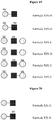
- a disaccharide of Formula I has a structure of IdoA-GlcNAc, IdoA-GlcNS, IdoA-GlcNS6S, IdoA2S-GlcNAc, IdoA2S-GlcNS, IdoA2S-GlcNAc6S, IdoA2S-GlcNS6S or as set forth in any of Formulas I-A to I-G, as set forth in Figure 4 .
- compounds of Formulas I-A to I-G are provided by treating the glycosaminoglycan heparan sulfate with a suitable glycosaminoglycan lyase.
- the detection and/or measurement of any one or more disaccharide of Formulas I-A to I-C is utilized in a process for diagnosing MPS I. In certain embodiments, the detection and/or measurement of any one or more disaccharide of Formulas I-D to I-G is utilized in a process for diagnosing MPS II.
- oligosaccharide having the formula: [GlcN(Ac) m -(IdoA/GlcA)-GlcN(Ac) n ](SO 3 R) p
- formula II [GlcN(Ac) m -(IdoA/GlcA)-GlcN(Ac) n ](SO 3 R) p
- oligosaccharides described herein are trisaccharides of Formula II, wherein IdoA/GlcA is either IdoA or GlcA, m is 0-1, n is 0-1, and p is 0-5.
- the trisaccharide is optionally sulfated with p SO 3 R groups in any suitable location.
- a compound of Formula II has a structure as set forth in Formula III: [GlcNS-IdoA-GlcN(Ac) n ](SO 3 R) p Formula III
- oligosaccharides described herein are trisaccharides of Formula III, wherein n is 0-1, and p is 0-3.
- the trisaccharide is optionally sulfated withp SO 3 R groups in any suitable location.
- a trisaccharide of Formula III has a structure of GlcNS-IdoA-GlcNAc, GlcNS-IdoA2S-GlcNAc, GlcNS-IdoA-GlcNS, GlcNS-IdoA2S-GlcNS, GlcNS-IdoA-GlcNAc6S, GlcNS-IdoA2S-GlcNAc6S, GlcNS-IdoA-GlcNS6S, GlcNS-IdoA2S-GlcNS6S, or as set forth in any of the trisaccharides of Figure 5 .
- compounds of Formulas III-A to III-H are provided by treating the glycosaminoglycan heparan sulfate with a suitable glycosaminoglycan lyase.
- a compound of Formula II has a structure as set forth in Formula IV: [GlcNS-GlcA-GlcN(Ac) n ](SO 3 R) p Formula IV
- oligosaccharides described herein are trisaccharides of Formula IV, wherein n is 0-1, and p is 0-2.
- the trisaccharide is optionally sulfated withp SO 3 R groups in any suitable location.
- a trisaccharide of Formula IV has a structure of GlcNS-GlcA-GlcNAc, GlcNS-GlcA-GlcNS, GlcNS-GlcA-GlcNAc6S, GlcNS-GlcA-GlcNS6S or as set forth in any of the trisaccharides of Figure 6 .
- compounds of Formulas IV-A to IV-D are provided by treating the glycosaminoglycan heparan sulfate with a suitable glycosaminoglycan lyase.
- the detection and/or measurement of any one or more trisaccharide of Formulas III and/or IV is utilized in a process for diagnosing MPS IIIA.
- a compound of Formula II has a structure as set forth in Formula V: [GlcNAc-Ido-GlcN(Ac) n ](SO 3 R) p Formula V
- oligosaccharides described herein are trisaccharides of Formula V, wherein n is 0-1, and p is 0-3.
- the trisaccharide is optionally sulfated withp SO 3 R groups in any suitable location.
- a trisaccharide of Formula V has a structure of GlcNAc-IdoA-GlcNAc, GlcNAc-IdoA2S-GlcNAc, GlcNAc-IdoA-GlcNS, GlcNAc-IdoA2S-GlcNS, GlcNAc-IdoA-GlcNAc6S, GlcNAc-IdoA2S-GlcNAc6S, GlcNAc-IdoA-GlcNS6S, GlcNAc-IdoA2S-GlcNS6, or as set forth in any of the trisaccharides of Figure 7 .
- compounds of Formulas V-A to V-H are provided by treating the glycosaminoglycan heparan sulfate with a suitable glycosaminoglycan lyase.
- a compound of Formula II has a structure as set forth in Formula VI: [GlcNAc-GlcA-GlcN(Ac) n ](SO 3 R) p Formula VI
- oligosaccharides described herein are trisaccharides of Formula VI, wherein n is 0-1, and p is 0-2.
- the trisaccharide is optionally sulfated withp SO 3 R groups in any suitable location.
- a trisaccharide of Formula VI has a structure of GlcNAc-GlcA-GlcNAc, GlcNAc-GlcA-GlcNS, GlcNAc-GlcA-GlcNAc6S, GlcNAc-GlcA-GlcNS6S or as set forth in any of the trisaccharides of Figure 8 .
- compounds of Formulas VI-A to VI-D are provided by treating the glycosaminoglycan heparan sulfate with a suitable glycosaminoglycan lyase.
- the detection and/or measurement of any one or more trisaccharide of Formulas V and/or VI is utilized in a process for diagnosing MPS IIIB.
- a compound of Formula II has a structure as set forth in Formula VII: [GlcN-Ido-GlcN(Ac) n ](SO 3 R) p Formula VII
- oligosaccharides described herein are trisaccharides of Formula VII, wherein n is 0-1, and p is 0-4.
- the trisaccharide is optionally sulfated withp SO 3 R groups in any suitable location.
- a trisaccharide of Formula VII has a structure of GlcN-IdoA-GlcNAc, GlcN-IdoA2S-GlcNAc, GlcN-IdoA-GlcNS, GlcN-IdoA2S-GlcNS, GlcN-IdoA-GlcNAc6S, GlcN-IdoA2S-GlcNAc6S, GlcN-IdoA-GlcNS6S, GlcN-IdoA2S-GlcNS6S, or as set forth in any of the trisaccharides of Figure 9 .
- compounds of Formulas VII-A to VII-H are provided by treating the glycosaminoglycan heparan sulfate with a suitable glycosaminoglycan lyase.
- a compound of Formula II has a structure as set forth in Formula VIII: [GlcN-GlcA-GlcN(Ac) n ](SO 3 R) p
- Formula VIII [GlcN-GlcA-GlcN(Ac) n ](SO 3 R) p
- oligosaccharides described herein are trisaccharides of Formula VIII, wherein n is 0-1, and p is 0-4.
- the trisaccharide is optionally sulfated with p SO 3 R groups in any suitable location.
- a trisaccharide of Formula VIII has a structure of GlcN-GlcA-GlcNAc, GlcN-GlcA-GlcNS, GlcN-GlcA-GlcNAc6S, GlcN-GlcA-GlcNS6S, or as set forth in any of the trisaccharides of Figure 10 .
- compounds of Formulas VIII-A to VIII-D are provided by treating the glycosaminoglycan heparan sulfate with a suitable glycosaminoglycan lyase.
- the detection and/or measurement of any one or more trisaccharide of Formulas VII and/or VIII is utilized in a process for diagnosing MPS IIIC.
- a compound of Formula II has a structure as set forth in Formula IX: [GlcNAc6S-Ido-GlcN(Ac) n ](SO 3 R) p Formula IX
- oligosaccharides described herein are trisaccharides of Formula IX, wherein n is 0-1, and p is 0-3.
- the trisaccharide is optionally sulfated withp SO 3 R groups in any suitable location.
- a trisaccharide of Formula VII has a structure of GlcNAc6S-IdoA-GlcNAc, GlcNAc6S-IdoA2S-GlcNAc, GlcNAc6S-IdoA-GlcNS, GlcNAc6S-IdoA2S-GlcNS, GlcNAc6S-IdoA-GlcNAc6S, GlcNAc6S-IdoA2S-GlcNAc6S, GlcNAc6S-IdoA-GlcNS6S, GlcNAc6S-IdoA2S-GlcNS6S, or as set forth in any of the trisaccharides of Figure 11 .
- compounds of Formulas IX-A to IX-H are provided by treating the glycosaminoglycan heparan sulfate with a suitable glycosaminoglycan
- a compound of Formula II has a structure as set forth in Formula X: [GlcNAc6S-GlcA-GlcN(Ac) n ](SO 3 R) p Formula X
- oligosaccharides described herein are trisaccharides of Formula X, wherein n is 0-1, and p is 0-2.
- the trisaccharide is optionally sulfated with p SO 3 R groups in any suitable location.
- a trisaccharide of Formula X has a structure of GlcNAc6S-GlcA-GlcNAc, GlcNAc6S-GlcA-GlcNS, GlcNAc6S-GlcA-GlcNAc6S, GlcNAc6S-GlcA-GlcNS6S, or as set forth in any of the trisaccharides of Figure 12 .
- compounds of Formulas X-A to X-D are provided by treating the glycosaminoglycan heparan sulfate with a suitable glycosaminoglycan lyase.
- the detection and/or measurement of any one or more trisaccharide of Formulas IX and/or X is utilized in a process for diagnosing MPS IIID.
- oligosaccharide having the formula: [GlcA-GlcN(Ac) m ](SO 3 R) n
- oligosaccharides described herein are disaccharides of Formula XI, wherein m is 0-1, and n is 0-2.
- the disaccharide is optionally sulfated with n SO 3 R groups in any suitable location.
- a disaccharide of Formula XI has a structure of GlcA-GlcNAc, GlcA-GlcNAc 6S, GlcA-GlcNS, GlcA-GlcNS6S, or as set forth in any of the disaccharides of Figure 13 .
- compounds of Formulas XI-A to XI-D are provided by treating the glycosaminoglycan heparan sulfate with a suitable glycosaminoglycan lyase.
- the detection and/or measurement of any one or more disaccharide of Formula XI is utilized in a process for diagnosing MPS VII.
- oligosaccharide having the formula: [IdoA-GalNAc](SO 3 R) n Formula XII
- oligosaccharides described herein are disaccharides of Formula XII, wherein n is 0-2.
- the disaccharide is optionally sulfated with n SO 3 R groups in any suitable location.
- a disaccharide of Formula XII has a structure of IdoA-GalNAc, IdoA-GalNAc6S, IdoA-GalNAc4S, IdoA-GalNAc4S6S, or as set forth in any of the disaccharides of Figure 14 .
- compounds of Formulas XII-A to XII-D are provided by treating the glycosaminoglycan dermatan sulfate with a suitable glycosaminoglycan lyase.
- the detection and/or measurement of any one or more disaccharide of Formula XII is utilized in a process for diagnosing MPS I. In certain embodiments, the detection and/or measurement of any one or more disaccharide of Formula XII and one or more disaccharide of Formulas I-A to I-C is utilized in a process for diagnosing MPS I.
- oligosaccharide having the formula: [IdoA2S-GalNAc](SO 3 R) n Formula XIII
- oligosaccharides described herein are disaccharides of Formula XIII, wherein n is 0-2.
- the disaccharide is optionally sulfated with n SO 3 R groups in any suitable location.
- a disaccharide of Formula XIII has a structure of IdoA2S-GalNAc, IdoA2S-GalNAc6S, IdoA2S-GalNAc4S, IdoA2S-GalNAc4S6S, or as set forth in any of the disaccharides of Figure 15 .
- compounds of Formulas XIII-A to XIII-D are provided by treating the glycosaminoglycan dermatan sulfate with a suitable glycosaminoglycan lyase.
- the detection and/or measurement of any one or more disaccharide of Formula XIII is utilized in a process for diagnosing MPS II. In certain embodiments, the detection and/or measurement of any one or more disaccharide of Formula XIII and one or more disaccharide of Formulas I-D to I-G is utilized in a process for diagnosing MPS II.
- oligosaccharide having the formula: [GalNAc4S-IdoA-GalNAc](SO 3 R) n Formula XIV
- oligosaccharides described herein are trisaccharides of Formula XIV, wherein n is 0-4.
- the trisaccharide is optionally sulfated with n SO 3 R groups in any suitable location.
- a disaccharide of Formula XIV has a structure of GalNAc4S( ⁇ 6S)-IdoA( ⁇ 2S)-GalNAc( ⁇ 4S)( ⁇ 6S), or as set forth in any of the disaccharides of Figure 16 , wherein ⁇ indicates that the indicated sulfation is independently present or absent.
- compounds of Formulas XIV-A to XIV-P are provided by treating the glycosaminoglycan dermatan sulfate with a suitable glycosaminoglycan lyase.
- the detection and/or measurement of any one or more trisaccharide of Formula XIV is utilized in a process for diagnosing MPS VI.
- oligosaccharide having the formula: [GalNAc4S-GlcA-GalNAc](SO 3 R) n Formula XV
- oligosaccharides described herein are trisaccharides of Formula XV, wherein n is 0-3.
- the trisaccharide is optionally sulfated with n SO 3 R groups in any suitable location.
- a disaccharide of Formula XV has a structure of GalNAc4S( ⁇ 6S)-GlcA( ⁇ 2S)-GlcNAc( ⁇ 4S)( ⁇ 6S), or as set forth in any of the trisaccharides of Figure 17 , wherein ⁇ indicates that the indicated sulfation is independently present or absent.
- compounds of Formulas XV-A to XV-P are provided by treating the glycosaminoglycan chondroitin sulfate with a suitable glycosaminoglycan lyase.
- the detection and/or measurement of any one or more trisaccharide of Formula XV is utilized in a process for diagnosing MPS VI. In certain embodiments, the detection and/or measurement of any one or more trisaccharide of Formula XIV and one or more trisaccharide of Formula XV is utilized in a process for diagnosing MPS VI.
- oligosaccharide having the formula: GlcA-GlcNAc-GlcA Formula XVI
- oligosaccharides described herein are trisaccharides of Formula XVI.
- compounds of Formulas XVI are provided by treating the glycosaminoglycan hyaluronan with a suitable glycosaminoglycan lyase.
- the detection and/or measurement of any one or more trisaccharide of Formula XVI is utilized in a process for diagnosing MPS IIIB.
- the detection and/or measurement of any one or more trisaccharide of Formula XVI and one or more trisaccharide of either of Formulas V and/or VI is utilized in a process for diagnosing MPS IIIB.
- oligosaccharide having the formula: GlcA-GlcNAc Formula XVII
- oligosaccharides described herein are disaccharides of Formula XVII.
- compounds of Formulas XVII are provided by treating the glycosaminoglycan hyaluronan with a suitable glycosaminoglycan lyase.
- the detection and/or measurement of any one or more disaccharide of Formula XVII is utilized in a process for diagnosing MPS VII or MPS IX.
- the detection and/or measurement of a disaccharide of Formula XVII and one or more disaccharide of Formula XI is utilized in a process for diagnosing MPS VII. In some embodiments, the detection and/or measurement of a disaccharide of Formula XVII and the absence of one or more disaccharide of Formula XI is utilized in a process for diagnosing MPS IX.
- oligosaccharide having the formula: [GlcNAc6S-Gal-GlcNAc](SO 3 R) n Formula XVIII
- oligosaccharides described herein are trisaccharides of Formula XVIII, and wherein n is 0-2.
- the trisaccharide is optionally sulfated with n SO 3 R groups in any suitable location.
- a disaccharide of Formula XVIII has a structure of GlcNAc6S-Gal-GlcNAc, GlcNAc6S-Gal6S-GlcNAc, GlcNAc6S-Gal-GlcNAc6S, GlcNAc6S-Gal6S-GlcNAc6S, or as set forth in any of the trisaccharides of Figure 18 .
- compounds of Formulas XVIII-A to XVIII-D are provided by treating the glycosaminoglycan keratan sulfate with a suitable glycosaminoglycan lyase.
- the detection and/or measurement of any one or more trisaccharide of Formula XVIII is utilized in a process for diagnosing MPS IIID. In certain embodiments, the detection and/or measurement of any one or more trisaccharide of Formula XVIII and one or more trisaccharide of Formulas IX and/or X is utilized in a process for diagnosing MPS IIID.
- oligosaccharide having the formula: [Gal6S-GlcNAc-(Gal) m ](SO 3 R) n
- oligosaccharides described herein are disaccharides of Formula XIX, wherein n is 0-2 and m is 0-1.
- the disaccharide is optionally sulfated with n SO 3 R groups in any suitable location.
- a disaccharide of Formula XIX has a structure of Gal6S-GlcNAc, Gal6S-GlcNAc6S, or as set forth in any of the disaccharides of Figure 19 .
- compounds of Formulas XIX-A to XIX-B are provided by treating the glycosaminoglycan keratan sulfate with a suitable glycosaminoglycan lyase.
- the detection and/or measurement of any one or more disaccharide of Formula XIX is utilized in a process for diagnosing MPS IVA.
- oligosaccharide having the formula: [Gal-GlcNAc](SO 3 R) n Formula XX
- oligosaccharides described herein are disaccharides of Formula XX, wherein n is 0-1.
- the disaccharide is optionally sulfated with n SO 3 R groups in any suitable location.
- a disaccharide of Formula XX has a structure of Gal-GlcNAc, Gal-GlcNAc6S, or as set forth in any of the disaccharides of Figure 20 .
- compounds of Formulas XX-A to XX-B are provided by treating the glycosaminoglycan keratan sulfate with a suitable glycosaminoglycan lyase.
- the detection and/or measurement of any one or more disaccharide of Formula XX is utilized in a process for diagnosing MPS IVB.
- any analytical method or diagnostic process described herein comprises detecting and/or measuring any one or more of the oligosaccharides of any of Formulas I-XX, wherein the non-reducing end saccharide residue is saturated at 4 and 5 carbons of the non-reducing end saccharide residue, the non-reducing end saccharide residue is free of carbon-carbon unsaturation, or the oligosaccharide is free of carbon-carbon unsaturation. In certain instances, the non-reducing end saccharide residue is on the left end of the oligosaccharides disclosed herein.
- Figure 1 illustrates the cleavage of the glycosaminoglycan (GAG) heparan sulfate with a glycosaminoglycan lyase (heparinase II).
- GAG glycosaminoglycan
- heparinase II glycosaminoglycan lyase II
- internal cleavage of glycosaminoglycans with glycosaminoglycan lyases provides oligosaccharides with carbon-carbon unsaturation between the C4 and C5 carbons of the non-reducing end of the oligosaccharide produce (i.e., the newly created oligosaccharide).
- the oligosaccharide of any of Formulas I-XX is a disaccharide or trisaccharide comprised of two or three saccharide residues that formed the original two or three saccharide residues of a glycosaminoglycan (GAG) prior to treatment with the one or more glycosaminoglycan (GAG) lyase.
- GAG glycosaminoglycan
- oligosaccharide having the formula: [IdoA-GlcN(Ac) m -IdoA](SO 3 R) n
- oligosaccharides described herein are trisaccharides of Formula XXI, wherein m is 0-1 and n is 0-4.
- the trisaccharide is optionally sulfated with n SO 3 R groups in any suitable location.
- compounds of Formulas XXI are provided by treating the glycosaminoglycan heparan sulfate with a suitable glycosaminoglycan lyase.
- oligosaccharide having the formula: [IdoA-GlcN(Ac) m -GlcA](SO 3 R) n
- Formula XXII [IdoA-GlcN(Ac) m -GlcA](SO 3 R) n
- oligosaccharides described herein are trisaccharides of Formula XXII, wherein m is 0-1 and n is 0-3.
- the trisaccharide is optionally sulfated with n SO 3 R groups in any suitable location.
- compounds of Formulas XXII are provided by treating the glycosaminoglycan heparan sulfate with a suitable glycosaminoglycan lyase.
- oligosaccharide having the formula: [GlcA-GlcN(Ac) m -GlcA](SO 3 R) n
- oligosaccharides described herein are trisaccharides of Formula XXIII, wherein m is 0-1 and n is 0-3.
- the trisaccharide is optionally sulfated with n SO 3 R groups in any suitable location.
- compounds of Formulas XXIII are provided by treating the glycosaminoglycan heparan sulfate with a suitable glycosaminoglycan lyase.
- the detection and/or measurement of any one or more trisaccharide of any of Formulas XXI to XXIII is utilized in a process for diagnosing a disorder associated with the accumulation of heparan sulfate (e.g., a heparan sulfate lysosomal storage disease such as, by way of non-limiting example, MPS I, MPS II, MPS IIIA, MPS IIIB, MPS IIIC, MPS IIID, MPS VII, or a combination thereof).
- heparan sulfate e.g., a heparan sulfate lysosomal storage disease such as, by way of non-limiting example, MPS I, MPS II, MPS IIIA, MPS IIIB, MPS IIIC, MPS IIID, MPS VII, or a combination thereof.
- an oligosaccharide of any of Formulas XXI to XXIII is saturated or unsaturated at the C4 and C5 positions of the saccharide residue at the non-reducing end of the oligosaccharide.
- a process for diagnosing a disorder associated with the accumulation of heparan sulfate e.g., MPS I, MPS II, MPS IIIA, MPS IIIB, MPS IIIC, MPS IIID, MPS VII, or a combination thereof
- detecting one or more oligosaccharide as set forth above and as appropriate (e.g., any of Formulas I-XX, as they pertain to the particular disorder being diagnosed).
- oligosaccharide having the formula: [IdoA-GalNAc-Ido](SO 3 R) n Formula XXIV
- oligosaccharides described herein are trisaccharides of Formula XXIV, wherein n is 0-4.
- the trisaccharide is optionally sulfated with n SO 3 R groups in any suitable location.
- compounds of Formulas XXIV are provided by treating the glycosaminoglycan chondroitin and/or dermatan sulfate with a suitable glycosaminoglycan lyase.
- oligosaccharide having the formula: [IdoA-GalNAc-GlcA](SO 3 R) n Formula XXV
- oligosaccharides described herein are trisaccharides of Formula XXV, n is 0-3.
- the trisaccharide is optionally sulfated with n SO 3 R groups in any suitable location.
- compounds of Formulas XXV are provided by treating the glycosaminoglycan chondroitin and/or dermatan sulfate with a suitable glycosaminoglycan lyase.
- oligosaccharide having the formula: [GlcA-GalNAc-Ido](SO 3 R) n Formula XXVI
- oligosaccharides described herein are trisaccharides of Formula XXVI, wherein n is 0-3.
- the trisaccharide is optionally sulfated with n SO 3 R groups in any suitable location.
- compounds of Formulas XXVI are provided by treating the glycosaminoglycan chondroitin and/or dermatan sulfate with a suitable glycosaminoglycan lyase.
- oligosaccharide having the formula: [GlcA-GalNAc-GlcA](SO 3 R) n
- oligosaccharides described herein are trisaccharides of Formula XXVII, wherein n is 0-2.
- the trisaccharide is optionally sulfated with n SO 3 R groups in any suitable location.
- compounds of Formulas XXVII are provided by treating the glycosaminoglycan chondroitin and/or dermatan sulfate with a suitable glycosaminoglycan lyase.
- the detection and/or measurement of any one or more trisaccharide of any of Formulas XXIV to XXVII is utilized in a process for diagnosing a disorder associated with the accumulation of chondroitin and/or dermatan sulfate (e.g., a chondroitin and/or dermatan sulfate lysosomal storage disease).
- a disorder associated with the accumulation of chondroitin and/or dermatan sulfate e.g., a chondroitin and/or dermatan sulfate lysosomal storage disease.
- an oligosaccharide of any of Formulas XXIV to XXVII is saturated or unsaturated at the C4 and C5 positions of the saccharide residue at the non-reducing end of the oligosaccharide.
- a process for diagnosing a disorder associated with the accumulation of chondroitin and/or dermatan sulfate further comprises detecting one or more oligosaccharide, as set forth above and as appropriate (e.g., any of Formulas I-XX, as they pertain to the particular disorder being diagnosed).
- oligosaccharide having the formula: GlcA-GlcNAc-GlcA Formula XXVIII
- oligosaccharides described herein e.g., those detected and/or measured according to the methods and/or processes described herein, are trisaccharides of Formula XXVIII.
- compounds of Formulas XXVIII are provided by treating the glycosaminoglycan hyaluronan with a suitable glycosaminoglycan lyase.
- the detection and/or measurement of a trisaccharide of Formula XXVIII is utilized in a process for diagnosing a disorder associated with the accumulation of hyaluronan (e.g., a hyaluronan lysosomal storage disease).
- a disorder associated with the accumulation of hyaluronan e.g., a hyaluronan lysosomal storage disease.
- an oligosaccharide of Formula XXVIII is saturated or unsaturated at the C4 and C5 positions of the saccharide residue at the non-reducing end of the oligosaccharide.
- a process for diagnosing a disorder associated with the accumulation of hyaluronan further comprises detecting one or more oligosaccharide, as set forth above and as appropriate (e.g., any of Formulas I-XX, as they pertain to the particular disorder being diagnosed).
- oligosaccharide having the formula: [Gal-GlcNAc-Gal](SO 3 R) n Formula XXIX
- oligosaccharides described herein are trisaccharides of Formula XXIX, wherein n is 0-3.
- the trisaccharide is optionally sulfated with n SO 3 R groups in any suitable location.
- compounds of Formulas XXIX are provided by treating the glycosaminoglycan keratan sulfate with a suitable glycosaminoglycan lyase.
- the detection and/or measurement of any one or more trisaccharide of Formula XXIX is utilized in a process for diagnosing a disorder associated with the accumulation of keratan sulfate (e.g., a keratan sulfate lysosomal storage disease).
- a disorder associated with the accumulation of keratan sulfate e.g., a keratan sulfate lysosomal storage disease.
- an oligosaccharide of Formula XXIX is saturated or unsaturated at the C4 and C5 positions of the saccharide residue at the non-reducing end of the oligosaccharide.
- the oligosaccharide is saturated at the C4 and C5 positions of the saccharide residue at the non-reducing end of the oligosaccharide.
- a process for diagnosing a disorder associated with the accumulation of keratan sulfate further comprises detecting one or more oligosaccharide, as set forth above and as appropriate (e.g., any of Formulas I-XX, as they pertain to the particular disorder being diagnosed).
- oligosaccharide having the formula: [GalNAc6S-GlcA-(GalNAc) m ](SO 3 R) n
- oligosaccharides described herein are di- and tri-saccharides of Formula XXX, wherein n is 0-3.
- the di- or tri-saccharide is optionally sulfated with n SO 3 R groups in any suitable location.
- compounds of Formulas XXX are provided by treating the glycosaminoglycan chondroitin sulfate with a suitable glycosaminoglycan lyase.
- the detection and/or measurement of any one or more disaccharide or trisaccharide of Formula XXX is utilized in a process for diagnosing MPS IVA.
- a di- or tri-saccharide of Formula XXX has a structure o as set forth in any of the disaccharides of Figure 21 .
- oligosaccharide having the formula: [GalNAc6S-IdoA-(GalNAc) m ](SO 3 R) n
- oligosaccharides described herein are di- or tri-saccharides of Formula XXXI, wherein n is 0-4 and m is 0-1.
- the di- or tri-saccharide is optionally sulfated with n SO 3 R groups in any suitable location.
- a disaccharide of Formula XVIII has a structure of any of the di- or tri-saccharides of Figure 22 .
- compounds of Formulas XXXI-A to XXI-T are provided by treating the glycosaminoglycan dermatan sulfate with a suitable glycosaminoglycan lyase.
- the detection and/or measurement of any one or more di- or tri-saccharide of Formula XXXI is utilized in a process for diagnosing MPS IVA.
- oligosaccharide having the formula: [GlcA-GalNAc](SO 3 R) n Formula XXXII
- oligosaccharides described herein are disaccharides of Formula XXXII, wherein n is 0-2.
- the disaccharide is optionally sulfated with n SO 3 R groups in any suitable location.
- a disaccharide of Formula XXXII has a structure as set forth in any of the disaccharides of Figure 23 .
- compounds of Formulas XXXII-A to XXXII-C are provided by treating the glycosaminoglycan chondroitin sulfate with a suitable glycosaminoglycan lyase.
- the detection and/or measurement of any one or more disaccharide of Formula XXXII is utilized in a process for diagnosing MPS VII.
- an oligosaccharide of any of Formulas XXX to XXXII is saturated or unsaturated at the C4 and C5 positions of the saccharide residue at the non-reducing end of the oligosaccharide.
- any of the processes or methods provided herein comprise detecting and/or measuring the amount of any oligosaccharide provided in any of Formulas I to XX, any of Formulas XXI to XXIX, or any of Formulas I to XXXII.
- any process or method provided herein comprises detecting and/or measuring the amount of any oligosaccharide provided in any of Formulas I to XX, any of Formulas XXI to XXIX, or any of Formulas I to XXXII, wherein the oligosaccharide is attached to a detectable label at the reducing end of the oligosaccharide.
- provided herein is a compound of any of Formulas I to XX, any one of Formulas XXI to XXIX, or any of Formulas I to XXXII.
- a compound of any of Figures 4-23 is provided herein.
- the compound of any of Formulas I to XXXII or Figures 4-23 is an isolated and/or purified compound.
- the isolated and/or purified compound of any of Formulas I to XXXII or Figures 4-23 is substantially free of oligosaccharides comprising a saccharide residue at the non-reducing end of the oligosaccharide that is unsaturated at the C4 and C5 positions.
- an L-iduronic acid (IdoA) residue that is saturated at the C4 and C5 positions has a structure as follows: whereas an L-iduronic acid (IdoA) residue at the non-reducing end of the oligosaccharide that is unsaturated at the C4 and C5 positions may have a structure as follows: or the like.
- Oligosaccharides having non-reducing end saccharide residues that are saturated at the C4 and C5 position are referred to herein as "C4-C5 non-reducing end saturated oligosaccharides”.
- unsaturated saccharide residues include the unsaturated residue of IdoA which is denoted by and the unsaturated residue of GlcA which is denoted by
- the isolated and/or purified compound described herein comprises at least at least 50%, at least 60%, at least 70%, at least 80%, at least 90%, at least 95%, at least 97%, at least 98%, or at least 99% oligosaccharide (by weight). In further or alternative embodiments, the isolated and/or purified compound comprises 20%, at least 30%, at least 40%, at least 50%, at least 60%, at least 70%, at least 80%, at least 90%, at least 95%, at least 97%, at least 98%, or at least 99% NRE-saturated oligosaccharide (by weight).
- a composition comprising one or more isolated oligosaccharide of any of Figures 4-23 .
- the oligosaccharide present in the composition is less than 90% by weight non-reducing end unsaturated oligosaccharide.
- the oligosaccharide present in the composition is less than 80% by weight non-reducing end unsaturated oligosaccharide.
- the oligosaccharide present in the composition is less than 70% by weight non-reducing end unsaturated oligosaccharide.
- the oligosaccharide present in the composition is less than 60% by weight non-reducing end unsaturated oligosaccharide.
- the oligosaccharide present in the composition is less than 50% by weight non-reducing end unsaturated oligosaccharide. In certain embodiments, the oligosaccharide present in the composition is less than 40% by weight non-reducing end unsaturated oligosaccharide. In certain embodiments, the oligosaccharide present in the composition is less than 30% by weight non-reducing end unsaturated oligosaccharide. In certain embodiments, the oligosaccharide present in the composition is less than 25% by weight non-reducing end unsaturated oligosaccharide. In certain embodiments, the oligosaccharide present in the composition is less than 20% by weight non-reducing end unsaturated oligosaccharide.
- the oligosaccharide present in the composition is less than 15% by weight non-reducing end unsaturated oligosaccharide. In certain embodiments, the oligosaccharide present in the composition is less than 10% by weight non-reducing end unsaturated oligosaccharide. In certain embodiments, the oligosaccharide present in the composition is less than 5% by weight non-reducing end unsaturated oligosaccharide. In some embodiments, the composition further comprises chromatographic resin or electrophoretic resin. In some embodiments, the resin is a high performance liquid chromatographic resin, or the like.
- the compounds of any of Formulas I-XXXII or Figures 4-23 are obtained in any suitable manner, e.g., by generation and purification from natural sources, chemical synthesis, or any other suitable method. These structures do not naturally exist in an isolated and/or purified form.
- the described di- and trisaccharides are optionally generated and purified from natural sources.
- these di- and trisaccharides are generated by treating the glycan with glycosaminoglycan lyases (for example heparin lyases or chondroitinases) and purifying the di- and trisaccharides liberated from the pre-existing non-reducing ends.
- glycosaminoglycan lyases for example heparin lyases or chondroitinases
- the di- and trisaccharides liberated from the pre-existing non-reducing end are unique because they do not contain a c4-5 double bond due to the action of the lyase.
- the yield of the desired di- and trisaccharides are increased by using glycosaminoglycans that have been treated with glycan degradative enzymes (for example, heparanase or lysosomal exo-enzymes such as the 2-sulfatase, N-sulfatase, etc) to generate smaller fragments or fragments with a greater number of desirable non-reducing ends.
- glycan degradative enzymes for example, heparanase or lysosomal exo-enzymes such as the 2-sulfatase, N-sulfatase, etc
- the yield is increased by starting with glycosaminoglycans that have been isolated from an organism or cell that has a defect in glycosaminoglycan degradation such that the desirable non-reducing ends are enriched (for example, glycosaminoglycans from iduronidase deficient systems, would be enriched in glycosaminoglycans that terminal on the non-reducing end with iduronate residues).
- these unique non-reducing end disaccharides can be isolated by HPLC.
- Figure 24 shows disaccharides isolated from heparan sulfate from a normal human sample
- Figure 25 shows disaccharides isolated from an MPS I (iduronidase deficient) sample.
- the saturated disaccharides (indicated with red arrows) can be isolated by collecting the appropriate fractions from this or a similar preparative chromatography.
- Additional or alternative methods of obtaining such compounds include, e.g., chemical transformation of the unsaturated saccharides into the saturated saccharides.
- the described di- and trisaccharides can be synthesized by chemical or chemoenzymatic methods. Chemical methods for the synthesis of these saturated di- and trisaccharides are optionally converted from the methods described by Prabhu, Venot, and Boons (Organic Letters 2003 Vol. 5, No. 26 4975-4978 ), which is incorporated herein for such synthesis.
- a compound of any of Formulas I-XXXII or Figures 4-23 wherein the compound is tagged with any label as described herein.
- an isolated or otherwise generated oligosaccharide described herein has a molecular weight of less than 2000 g/mol, less than 1500 g/mol, less than 1000 g/mol, less than 500 g/mol, less than 400 g/mol, less than 300 g/mol, less than 260 g/mol, less than 200 g/mol, less than 100 g/mol, or the like (e.g., prior to tagging with any detectable label that may be included in a process described herein).
- a biomarker or oligosaccharide described herein is not present in abundance in samples (e.g., samples of the same type) from individuals (e.g., a the amount in a single such individual, an average of such individuals, a mean of such individuals, a median of such individuals, or the like) with abnormal glycosaminoglycan accumulation relative to individuals with normal glycosaminoglycan (e.g., a the amount in a single such individual, an average of such individuals, a mean of such individuals, a median of such individuals, or the like).
- not present in an abundance includes not present in an amount over 1.01x (i.e., 1.01 times), 1.05x, 1.1x, 1.2x, 1.25x, 1.3x, 1.4x, 1.5x, 1.6x, 1.7x, 1.8x, 1.9x, 2x, or the like.
- Oligosaccharides are detected and/or measured in processes described herein in any suitable manner.
- detectable labels are detected and/or quantified according to any process described herein using any technique, particularly any technique suitable for the detectable label utilized.
- suitable detection techniques include, by way of non-limiting example, one or more of a mass spectrometer, a nuclear magnetic resonance spectrometer, a UV-Vis spectrometer, an IR spectrometer, a fluorimeter, a phosphorimeter, a radiation spectrometer (e.g., a scintillation counter), a thin layer chromatographic technique, or the like.
- oligosaccharides are optionally directly detected using a suitable technique, such as quantitative nuclear magnetic resonance. Quantitative nuclear magnetic resonance is also optionally utilized to quantify and/or detect the presence of a detectable label. In certain embodiments, one or more oligosaccharides are optionally detected using a suitable liquid chromatography mass spectrometer (LC-MS).
- LC-MS liquid chromatography mass spectrometer
- oligosaccharides are tagged with an antibody or probe, and are quantified using any suitable method (e.g., dot blot techniques, immune detection techniques (e.g., ELISA), or the like).
- suitable method e.g., dot blot techniques, immune detection techniques (e.g., ELISA), or the like.
- a column e.g., Cosmogel DEAE, Tsk Gel DEAE, Cosmogel QA, Cosmogel CM, Cosmogel SP, 130A BEH particle Phenyl (1.7, 2.5, 3.5, 5, or 10 uM particle size), 130A BEH particle C18 (1.7, 2.5, 3.5, 5, or 10 uM particle size), HSS particle C18 (1.8, 3.5, or 5 uM particle size), 300A BEH particle C18 (1.7, 3.5, 5, 10 uM particle size), or the like with suitable length and internal diameter) is optionally loaded with an equilibrating solvent (e.g., a buffer or salt solution, such as a potassium acetate solution, sodium chloride solution, sodium acetate solution, ammonium acetate solution, or the like), e.g., with a pH of about 6, 7, or 8.
- an equilibrating solvent e.g., a buffer or salt solution, such as a potassium acetate solution, sodium chloride solution, sodium acetate solution,
- the buffer or salt solution has a concentration of about 10 mM, 20 mM, 30 mM, 50 mM, 100 mM, 500 mM, 1 M, 2 M, or the like. Any suitable flow rate is used, e.g., 0.5 mL/min, 1 mL, min, 1.5 mL/min, 2 mL/min, or the like.
- a linear gradient is optionally utilized. In some embodiments, the linear gradient is run over 1-20 min, 1-10 min, 10-20 min, 1-5 min, 5-10 min, or the like.
- the gradient is a buffer or salt solution, e.g., as described above (e.g., from 0 M to 0.5 M, from 0 M to 3 M, from 0.5 M to 2 M, from 0 M to 2 M, from 1 M to 2 M, from 0 M to 3 M, from 2 M to 0 M, from 3 M to 0 M, or the like).
- the eluent is optionally held at the final concentration for a suitable period of time (e.g., 1-20 min, 5-10 min, 10-15 min, 1-5 min, 1-10 min, 15-20 min, or the like).
- the eluent may be switched to a second solvent or solvent system (e.g., an alcohol, such as methanol, ethanol, or isopropanol, acetonitrile, water, or the like).
- the switch to the second solvent system may be over a period of time, e.g., 15 seconds, 30 seconds, 45 seconds, 60 seconds, 2 min, 3 min, or the like.
- the second solvent system is optionally held for a period of time, such as 1 min, 2 min, 3 min, 4 min, 5 min, 6 min, or the like.
- the column is optionally restored to initial solvent conditions.
- detection or measurement of an analytical device provides for diagnosis of a disease, diagnosis of severity of a disease, of efficacy of a therapy, or analysis based on other processes described herein.
- absence of a peak or signal e.g., a peak or signal indicative of the presence of a particular oligosaccharide indicates that an individual is in a non-diseased state, in remission for a disease state, or undergoing effective therapy for a disease, depending on the circumstances of the diagnosis.
- the presence and/or area of a peak or signal is utilized to determine the severity of a disease.
- the presence and/or area of a peak or signal is utilized to determine disease, disease severity, disease carrier status or the like, based on a certain threshold value for the disease, disease severity, disease carrier status.
- thresholds are optionally determined in any suitable manner, e.g., by analyzing control samples, such control samples coming from non-diseased individuals, diseased individuals, or the like.
- a control biological sample utilized in any process described herein was provided from an individual that does not suffer from a disorder being diagnosed.
- a control biological sample is taken from an individual suffering from a disorder being diagnosed.
- the result obtained from the control biological sample is stored in a database.
- a test sample is optionally compared to a plurality of control data in a database.
- any diagnostic process described herein is optionally utilized alone or in combination with other diagnostic techniques.
- Other diagnostic techniques include, by way of non-limiting example, symptom analysis, biopsies, detection of accumulation of other compounds in biological samples, or the like.
- control biological samples are optionally taken from the same individual at substantially the same time, simply from a different location (e.g., one inflamed/arthritic synovial joint fluid vs the contralateral non-arthritic synovial joint). In other embodiments, control biological samples are optionally taken from the same individual at different points in time (e.g., before therapy and after therapy if the method being utilized is a method of monitoring a treatment therapy).
- detectable labels useful in the processes or methods described herein include, by way of non-limiting example, mass labels, antibodies, affinity labels, radiolabels, chromophores, fluorescent labels, or the like.
- Fluorescent labels suitable for use in various embodiments herein include, by way of non-limiting example, 2-aminopyridine (2-AP), 2-aminobenzoic acid (2-AA), 2-aminobenzamide (2-AB), 2-aminoacridone (AMAC), p-aminobenzoic acid ethyl ester (ABEE), p-aminobenzonitrile (ABN), 2-amino-6-cyanoethylpyridine (ACP), 7-amino-4-methylcoumarine (AMC), 8-aminonaphthalene-1,3,6-trisulfate (ANTS), 7-aminonaphthalene-1,3-disulfide (ANDS), and 8-aminopyrene-1,3,6-trisulfate (APTS), or the like.
- the fluorescent labels can be attached by reductive amination with the fluorescent label and sodium cyanoborohydride or the like.
- Mass labels suitable for use in various embodiments herein include, by way of non-limiting example, D-2-anthranilic acid, D-2-aminopyridine, D-methyl iodide, 13 C methyl iodide, deuterated-pyridyl-amine, D-biotin or the like.
- the mass labels can be attached by permethylation or reductive amination by any method that is known to those of skill in the art.
- Affinity labels suitable for use in various embodiments herein include, by way of non-limiting example, biotin and derivatives.
- Radio labels suitable for use in various embodiments herein include, by way of non-limiting example, sodium borotritide (NaB 3 H 4 ), or the like.
- Chromophores suitable for use in various embodiments herein include, by way of non-limiting example, 4-amino-1,1'-azobenzene, 4'-N,N-dimethylamino-4-aminoazobenzene, aminoazobenzene, diaminoazobenzene, Direct Red 16, CI Acid Red 57, CI Acid Blue 45, CI Acid Blue 22, CL Mordant Brown 13, CI Direct Orange 75, or the like.
- the chromophores may be labeled by any method that is known to those of skill in the art, such as reductive amination with the chromophore and sodium cyanoborohydride.
- the detectable label is an antibody.
- the antibody is attached to a detectable compound, such as mass labels, radiolabels, chromophores, fluorescent labels, or the like.
- antibodies are themselves detected and/or are detectable in various manners, e.g., as a chromophore, a fluorophore, or the like; or with a probe (e.g., using dot blot techniques, immune-detection techniques, or the like).
- the processes described herein comprises further treatment steps of the test and/or control samples.
- the samples are homogenized and/or purified.
- homogenization is achieved in any suitable manner including, by way of non-limiting example, with a basic solution (e.g., 0.1 N NaOH), sonication, tissue grinding or other chemical agents).
- samples, including test samples and/or control samples, described herein are optionally purified prior to glycosaminoglycan processing (e.g., lyase treatment) and/or characterization.
- Test samples and/or control samples i.e., one or more or all of the glycans found therein
- Test samples and/or control samples are optionally purified at any suitable point in a process described herein, including before or after tagging of the glycans founds within the sample.
- purification techniques include centrifugation, electrophoresis, chromatography (e.g., silica gel or alumina column chromatography), gas chromatography, high performance liquid chromatography (HPLC) (e.g., reverse phase HPLC on chiral or achiral columns), thin layer chromatography, ion exchange chromatography, gel chromatography (e.g., gel filtration or permeation or size exclusion chromatography, gel electrophoresis), molecular sieve chromatography, affinity chromatography, size exclusion, filtration (e.g.
- glycosaminoglycans such as heparan sulfate
- a core protein e.g., heparan sulfate
- purification processes of separating glycosaminoglycan fragments e.g., heparan sulfate fragments
- GAGs glycosaminoglycans
- GAGs glycosaminoglycans
- the oligosaccharide(s) detected or measured is one or more C4-C5 non-reducing end saturated oligosaccharide(s).
- the agent is administered one or more times. In certain embodiments, the agent is administered multiple times. In some embodiments, the agent is administered in a loading dose one or more times (e.g., in a loading dosing schedule) and subsequently administered in a maintenance dose (e.g., in a maintenance dosing schedule, such as three times a day, twice a day, once a day, once every two days, once every three days, once every four days, once a week, or the like). In some embodiments, when GAG oligosaccharide accumulation begins to increase or accelerate, the dose is optionally adjusted (e.g., the maintenance dose is increased, or an additional loading dose or dosing schedule is utilized).
- monitoring the accumulation of glycosaminoglycans comprises repeating the step of: using an analytical instrument to detect the presence of and/or measure the amount of a population of one or more oligosaccharides present in a transformed biological sample that has been prepared by treating a population of glycosaminoglycans, in or isolated from a biological sample from the individual, with at least one digesting glycosaminoglycan lyase to transform the glycosaminoglycans into the population of the one or more oligosaccharide.
- the step is repeated at periodic intervals (e.g., every day, every other day, every 2 days, every 3 days, every 4 days, every week, every month, every 3 months, quarterly, every 6 months, yearly, or the like), at regular times following a dose (e.g., 4 hours after a administration of the agent, 6 hours after administration of the agent, 8 hours after administration of the agent, 12 hours after administration of the agent, or the like), prior to administration of the dose (e.g., immediately prior to administration of the agent, 2 hours prior to administration of the agent, or the like), or any other monitoring schedule.
- periodic intervals e.g., every day, every other day, every 2 days, every 3 days, every 4 days, every week, every month, every 3 months, quarterly, every 6 months, yearly, or the like
- a dose e.g., 4 hours after a administration of the agent, 6 hours after administration of the agent, 8 hours after administration of the agent, 12 hours after administration of the agent, or the like
- prior to administration of the dose
- the monitoring of the accumulation of glycosaminoglycans is conducted over a period of time, e.g., over a week, two weeks, a month, two months, three months, six months, a year, or the like.
- the method for quantifying the amount of one or more oligosaccharides in a lyase digested biological sample comprises detecting and/or measuring (e.g., with an analytical device), one or more oligosaccharides within the lyase digested biological sample from the individual after the biological sample obtained from the individual has been treated with one or more glycosaminoglycan lyases.
- such glycosaminoglycan lyases are suitable for preparing di- and/or tri-saccharides from the glycosaminoglycans present in the biological sample obtained from the individual.
- the reducing end of a representative portion of the one or more oligosaccharides in the transformed biological sample is tagged with any suitable detectable label (e.g., a mass label, a radio label, a fluorescent label, a chromophore label, affinity label, an antibody).
- the process comprises displaying or recording such a characterization of the population of oligosaccharides and/or tagged oligosaccharides.
- the agent that modulates glycosaminoglycan biosynthesis includes glycosaminoglycan accumulation inhibitors, agents that promote glycosaminoglycan degradation, agents that activate enzymes that degrade glycosaminoglycans, agents that inhibit biosynthesis of glycosaminoglycans, or the like.
- the agent that modulates glycosaminoglycan biosynthesis is an agent that selectively modulates heparan sulfate biosynthesis, an agent that selectively modulates chondroitin sulfate biosynthesis, an agent that selectively modulates dermatan sulfate biosynthesis, an agent that selectively modulates keratan sulfate biosynthesis, an agent that selectively modulates hyaluronan biosynthesis, or a combination thereof.
- the detection and/or the quantification of the identity and/or amount of oligosaccharides present in a biological sample is used to identify and/or diagnose a disorder associated with abnormal degradation, biosynthesis and/or accumulation of glycosaminoglycans in an individual suspected of having such a disorder.
- the detection and/or the quantification of the identity and/or amount of oligosaccharides present in the biological sample is used to monitor severity and course of the disease in an individual diagnosed with or suspected of having a disorder associated with the abnormal degradation, biosynthesis and/or accumulation of glycosaminoglycans. In some instances, the detection and/or the quantification of the identity and/or amount of oligosaccharides present in the biological sample is used to calculate the administered dose of an agent that modulates (e.g., promotes and/or inhibits) glycosaminoglycan biosynthesis and/or degradation.
- an agent that modulates e.g., promotes and/or inhibits
- the detection and/or the quantification of the identity and/or amount of oligosaccharides present in a biological sample provides for a treatment regimen to be modified depending on the severity and course of the disease, disorder or condition, previous therapy, the individual's health status and response to the drugs, and the judgment of the treating physician.
- monitoring the accumulation of glycosaminoglycans in the individual comprises detecting or quantifying the amount of an oligosaccharide (or one or more oligosaccharides) in a sample obtained from the individual (e.g., according to any method described herein) to obtain a first accumulation result (e.g., an initial reading before treatment has begun, or at any other time) and a second accumulation result that is subsequent to obtaining the first result.
- the second result is compared to the first result to determine if the treatment is effectively reducing, maintaining, or reducing the rate of increasing the oligosaccharide levels in a substantially identically obtained sample from the individual being treated.
- the treatment can be altered, e.g., to increase or decrease the amount of agent administered; to substitute the therapeutic agent with an alternative therapeutic agent; or the like.
- the dose of the therapeutic agent is decreased to a maintenance level (e.g., if the oligosaccharide level has been reduced sufficiently); further monitoring of oligosaccharide levels is optional in such situation, e.g., to ensure that reduced or maintained levels of oligosaccharide (e.g., GAG oligosaccharide(s)) are achieved.
- a method of detecting response to therapy in an individual or a method of predicting response to therapy in an individual comprising:
- the oligosaccharide(s) detected or measured is one or more C4-C5 non-reducing end saturated oligosaccharide(s). It is to be understood that a plurality of cells from an individual includes cells that are directly taken from the individual, and/or cells that are taken from an individual followed by culturing to expand the population thereof.
- FIGS. 24 and 25 illustrate that C4-C5 non-reducing end saturated oligosaccharides can be isolated by, e.g., HPLC.
- monitoring the accumulation of glycosaminoglycans comprises repeating the step of: using an analytical instrument to detect the presence of and/or measure the amount of a population of one or more oligosaccharides present in a transformed biological sample that has been prepared by treating a population of glycosaminoglycans, in or isolated from a biological sample from the individual, with at least one digesting glycosaminoglycan lyase to transform the glycosaminoglycans into the population of the one or more oligosaccharide.
- the step is repeated at periodic intervals (e.g., every day, every other day, every 2 days, every 3 days, every 4 days, every week, or the like), or any other monitoring schedule.
- disorders associated with the abnormal degradation, biosynthesis and/or accumulation of glycosaminoglycans useful in the treatment and diagnostic methods and processes described herein include any disorder wherein accumulation of glycosaminoglycans and/or fragments thereof can be detected in a biological sample taken from an individual suffering from such a disorder.
- disorders associated with abnormal glycosaminoglycan degradation, biosynthesis, and/or accumulation include e.g., lysosomal storage diseases.
- a lysosomal storage disease is mucopolysaccharidosis (MPS).
- a mucopolysaccharidosis is MPS I, MPS II, MPS IIIA, MPS IIIB, MPS IIIC, MPS IIID, MPS IVA, MPS IVB, MPS VI, MPS VII, MPS IX, or a combination thereof.
- a lysosomal storage disease is caused by abnormal glycosaminoglycan degradation.
- a lysosomal storage disease causes an accumulation of heparan sulfate and/or dermatan sulfate (e.g., MPS I) and is caused by an iduronidase deficiency.
- a lysosomal storage disease causes an accumulation of heparan sulfate and/or dermatan sulfate (e.g., MPS II) and is caused by a 2-sulfatase deficiency.
- Figure 26 illustrates accumulation of oligosaccharides in MPS II human fibroblast versus the accumulation of oligosaccharides in normal human fibroblasts.
- a lysosomal storage disease causes an accumulation of heparan sulfate and/or hyaluronan (e.g., MPS IIIB) and is caused by an N-acetylglulcosaminidase deficiency.
- a lysosomal storage disease causes an accumulation of heparan sulfate (e.g., MPS IIIC) and is caused by a glucosamine acetyltransferase deficiency.
- a lysosomal storage disease causes an accumulation of heparan sulfate and/or keratan sulfate (e.g., MPS IIID) and is caused by a N-acetylglucosamine 6-sulfatase deficiency.
- a lysosomal storage disease causes an accumulation of keratan sulfate (e.g., MPS IVA) and is caused by a galactose 6-sulfatase deficiency.
- a lysosomal storage disease causes an accumulation of keratan sulfate (e.g., MPS IVB) and is caused by an N-acetylglactosamine 4-sulfatase deficiency.
- a lysosomal storage disease causes an accumulation of heparan sulfate and/or hyaluronan (e.g., MPS VII) and is caused by a glucuronidase deficiency.
- a lysosomal storage disease causes an accumulation of hyaluronan (e.g., MPS IX) and is caused by a hyaluronidase deficiency.
- a MPS is Hunter's disease.
- Hunter's disease causes an accumulation of dermatan sulfate and heparan sulfate glycosaminoglycans.
- the accumulation of dermatan sulfate and heparan sulfate glycosaminoglycans in Hunter's disease is associated with a deficiency in a sulfatase.
- the MPS is Hurler's disease.
- Hurler's disease causes an accumulation of dermatan sulfate and heparan sulfate glycosaminoglycans.
- the accumulation of dermatan sulfate and heparan sulfate glycosaminoglycans in Hurler's disease is associated with a deficiency in an iduronidase.
- a disorder associated with abnormal glycosaminoglycan degradation, biosynthesis and/or accumulation is undesired angiogenesis (e.g., angiogenesis associated with cancer, diabetic blindness, age-related macular degeneration, rheumatoid arthritis, or psoriasis), insufficient angiogenesis (e.g., coronary artery disease, stroke, or delayed wound healing), amyloidosis, a spinal cord injury, hypertriglyceridemia, inflammation, or a wound.
- angiogenesis e.g., angiogenesis associated with cancer, diabetic blindness, age-related macular degeneration, rheumatoid arthritis, or psoriasis
- insufficient angiogenesis e.g., coronary artery disease, stroke, or delayed wound healing
- amyloidosis e.g., a spinal cord injury, hypertriglyceridemia, inflammation, or a wound.
- amyloidosis is present in various diseases including, e.g., Alzheimer's disease, Parkinson's disease, type-2 diabetes, Huntington's disease, spongiform encephalopathies (Creutzfeld-Jakob, Kuru, Mad Cow), diabetic amyloidosis, rheumatoid arthritis, juvenile chronic arthritis, Ankylosing spondylitis, psoriasis, psoriatic arthritis, adult still disease, Becet syndrome, famalial Mediterranean fever, Crohn's disease, leprosy, osteomyelitis, tuberculosis, chronic bronciectasis, Castleman disease, Hodgkin's disease, renal cell carcinoma, carcinoma of the gut, lung or urogenital tract.
- the Alzheimer's disease is associated with changes in the content and structure of one or more GAG (e.g., heparan sulfate, keratan sulfate, or the like).
- disorders associated with abnormal glycosaminoglycan accumulation include disorders associated with abnormal biosynthesis (e.g., polymerization and/or sulfation) of glycosaminoglycans.
- the abnormal biosynthesis of glycosaminoglycans results in glycosaminoglycans that are not readily degraded by normal glycosaminoglycan degrading enzymes.
- disorders associated with abnormal GAG biosynthesis include osteoarthritis.
- osteoarthritis is associated with changes in sulfation of chondroitin sulfate, changes in length of chondroitin sulfate, changes in expression levels of chondroitin sulfate, or any combination of thereof.
- osteoarthritis is associated abnormal chondroitin sulfate sulfotransferase.
- the osteoarthritis is associated with changes in sulfation of dermatan sulfate, changes in length of dermatan sulfate, changes in expression levels of dermatan sulfate, or any combination of thereof.
- the osteoarthritis is associated with changes in sulfation of keratan sulfate, changes in length of keratan sulfate, changes in expression levels of keratan sulfate, or any combination of thereof.
- a disorder associated with abnormal glycosaminoglycan degradation, biosynthesis and/or accumulation is macular corneal dystrophy.
- macular corneal dystrophy is associated with low amounts of keratan sulfate.
- the keratan sulfate levels are due to failure to initiate keratan sulfate synthesis, polymerize the keratan sulfate chain length, or any combination thereof.
- a disorder associated with abnormal glycosaminoglycan degradation, biosynthesis and/or accumulation is an infectious or viral disease.
- the infectious or viral disease includes herpes, diphtheria, papilloma virus, hepatitis, HIV, coronavirus, or adenovirus.
- a disorder associated with abnormal glycosaminoglycan degradation, biosynthesis and/or accumulation is a cancer.
- the cancer is breast cancer, ovarian cancer, colorectal cancer, cervical cancer, pancreatic cancer, gastric cancer, esophageal cancer, head and neck cancer, hepatocellular cancer, prostate cancer, melanoma, osteosarcoma, endometrial cancer, multiple myeloma, gastric cancer, lung cancer, glioma, kidney cancer, bladder cancer, thyroid cancer, neuroblastoma, or non-Hodgkin lymphoma.
- cancer is associated with abnormal heparan sulfate depolymerization and degradation that results in unbound, accumulated heparan sulfate.
- abnormal heparan sulfate depolymerization and degradation is associated with melanomas, gliomas, multiple myelomas, ovarian, breast, colon, cervical, pancreatic, gastric, and esophageal cancers.
- abnormal heparan sulfate depolymerization and degradation contributes to angiogenesis, metastasis and growth factor mobilization.
- the abnormal heparan sulfate depolymerization and degradation is from increased activity of a heparanase.
- cancer is associated with abnormal heparan sulfate sulfation that results in accumulated heparan sulfate.
- abnormal heparan sulfate sulfation is associated with colon carcinoma, myeloma, ovarian cancer, hepatocellular carcinoma, head and neck squamous cell carcinoma and prostate cancer.
- heparan sulfate sulfation is decreased in certain cancers, while in other embodiments; the heparan sulfate sulfation is increased.
- abnormal heparan sulfate sulfation is caused by abnormal heparan sulfate sulfotransferase function.
- cancer is associated with abnormal chondroitin sulfate sulfation.
- the abnormal chondroitin sulfate sulfation is associated with lung cancer.
- the chondroitin sulfate sulfation is increased in certain cancers.
- the abnormal chondroitin sulfate sulfation is caused by abnormal chondroitin sulfate sulfotransferase function.
- increased production of chondroitin sulfate is associated in breast cancer, melanoma, and transformed fibroblasts.
- cancer is associated with dermatan sulfate epimerase expression.
- the dermatan sulfate epimerase expression is increased in squamous cell carcinoma, glioma, gynecological cancer, pancreatic cancer, colorectal carcinoma, and prostate cancer.
- the cancer is associated with accumulation of dermatan sulfate levels.
- the dermatan sulfate levels are increased in pancreatic cancer.
- cancer is associated with abnormal keratan sulfate sulfation.
- the abnormal keratan sulfate sulfation is associated with glioblastomas.
- abnormal keratan sulfate sulfation is caused by abnormal keratan sulfate sulfotransferase function.
- keratan sulfate expression is increased in glioblastomas.
- cancer is associated with abnormal hyaluronan accumulation.
- abnormal hyaluronan accumulation is associated with breast cancer, prostate cancer, aggressive subtypes of non-Hodgkin lymphoma, and colorectal cancer.
- hyaluronan accumulation contributes to metastasis of certain cancers.
- the hyaluronan accumulation results from the over expression of a hyaluronan synthase.
- a process for identifying an agent that inhibits the accumulation of glycosaminoglycans in a cell comprising:
- the mammalian cells are optionally transformed by e.g., tagging a population of glycosaminoglycans on and/or in the cells with a detectable label. In some instances, the mammalian cells are optionally transformed by e.g., isolating a population of glycosaminoglycans on and/or in the cells using any suitable purification technique described herein.
- the cell is present in an individual (e.g., a human or other mammal) and is incubated at body temperature.
- the cell line that accumulates an abnormal amount of glycosaminoglycans being a mucopolysaccharidosis (MPS) cell line (e.g., a human MPS cell line).
- MPS mucopolysaccharidosis
- the MPS cell line is a cell line for MPS I, MPS II, MPS IIIA, MPS IIIB, MPS IIIC, MPS IIID, MPS IVA, MPS IVB, MPS VI, MPS VII, MPS IX, or a combination thereof.
- the human MPS cell line is present in a human or other mammal.
- inhibitors of the accumulation of glycosaminoglycans are compounds that reduce the rate of accumulation of glycosaminoglycans in the cell, and/or agents that reduce the total amount of glycosaminoglycans accumulated in the cell (i.e., diminish the amount of glycosaminoglycan that has been accumulated in the cell).
- Agents that are optionally tested for the screening process described herein include any compound such as, by way of non-limiting example, a polynucleotide (e.g., siRNA), a polypeptide, or a small molecule compound (e.g., having a molecular weight of less than 2,000 g/mol).
- Example 1 - Purification The biological sample (cells, tissue, blood, serum, or the like) is homogenized and solublized in 0.1 - 1.0 N NaOH (e.g., 0.1 N, 0.2 N, 0.3 N, 0.4 N, 0.5 N, 0.6 N, 0.7 N, 0.8 N, 0.9 N, or 1.0 N) or acetic acid and then neutralized with acetic acid or NaOH. Next a small sample is taken to measure protein content of the sample using standard methods.
- 0.1 N, 0.2 N, 0.3 N, 0.4 N, 0.5 N, 0.6 N, 0.7 N, 0.8 N, 0.9 N, or 1.0 N e.g., 0.1 N, 0.2 N, 0.3 N, 0.4 N, 0.5 N, 0.6 N, 0.7 N, 0.8 N, 0.9 N, or 1.0 N
- acetic acid e.g., 0.1 N, 0.2 N, 0.3 N, 0.4 N, 0.5 N, 0.6 N, 0.7 N,
- 0.01 - 0.5 mg/mL (0.01 mg/mL, 0.07 mg/mL, 0.12 mg/mL, 0.17 mg/mL, 0.22 mg/mL, 0.27 mg/mL, 0.32 mg/mL, 0.37 mg/mL, 0.42 mg/mL, or 0.5 mg/mL) protease (trypsin, chymotrypsin, pepsin, pronase, papain, or elastase) is treated in 0.1 - 0.5 M (e.g., 0.1 M, 0.16 M, 0.23 M, 0.32 M, 0.39 M, 0.44 M, or 0.5 M) NaCl, 0.01 - 0.1 M (e.g., 0.01 M, 0.02 M, 0.04 M, 0.06 M, 0.08 M, 0.1 M) NaOAc, at pH 5.5 - 7.5 (e.g., 5.5, 6.0, 6.5, 7.0, or 7.5) and 25 - 40 C (e.g.,
- the sample is diluted to reduce the ionic strength and loaded onto an ion exchange column in 5 - 100 mM (e.g., 5 mM, 10 mM, 20 mM, 30 mM, 40 mM, 50 mM, 60 mM, 70 mM, 75 mM, 80 mM, 90 mM, 95 mM, 100 mM) NaOAc pH 5 - 7 with 0 - 300 mM NaCl.
- 5 mM e.g., 5 mM, 10 mM, 20 mM, 30 mM, 40 mM, 50 mM, 60 mM, 70 mM, 75 mM, 80 mM, 90 mM, 95 mM, 100 mM
- NaOAc pH 5 - 7 with 0 - 300 mM NaCl NaOAc pH 5 - 7 with 0 - 300 mM NaCl.
- the bound glycosaminoglycans are eluted with 5 - 100 mM NaOAc pH 5 - 7 (e.g., 5, 5.5, 6, 6.5, 7) with 0.8 - 3 M (e.g., 0.8 M, 1 M, 1.2 M, 1.4 M, 1.6 M, 1.8 M, 2 M, 2.5 M, or 3 M) NaCl.
- the eluted glycans are then concentrated and desalted by ethanol precipitation, size exclusion, or other methods.
- the purified glycans are dried for further analysis.
- 0.02 - 1 mM e.g., 0.02, 0.04, 0.06, 0.08, 0.1, 0.2, 0.3,
- Example 3 - Tagging Dried glycan sample is re-suspended in 2 - 100 ⁇ L 0.003 - 0.1 M (e.g., 0.003 M, 0.003 M, 0.03 M, 0.06 M, 0.1 M) AB, AA, AMAC, or Bodipy dye and incubated at room temperature for 1 - 120 minutes (e.g., 1-10 min, 10-15 min, 15-20 min, 20-25 min, 25-30 min, 30-40 min, 40-50 min, 50-60 min, 60-90 min, 90-120 min).
- 1-10 min e.g., 10-15 min, 15-20 min, 20-25 min, 25-30 min, 30-40 min, 40-50 min, 50-60 min, 60-90 min, 90-120 min.
- reaction is initiated with 2 - 100 ⁇ L (2 ⁇ L, 5 ⁇ L, 10 ⁇ L, 15 ⁇ L, 20 ⁇ L, 25 ⁇ L, 30 ⁇ L, 40 ⁇ L, 50 ⁇ L, 60 ⁇ L, 70 ⁇ L, 80 ⁇ L, 90 ⁇ L, or 100 ⁇ L) 1 M NaCNBH 4 and the reaction is allowed to proceed at 25 - 100 C. (e.g., 25 C, 30 C, 35 C, 40 C, 50 C, 60 C, 70 C, 80 C, 90 C, 100 C).
- Tested was the ability of a non-reducing end detection method to identify and classify MPS samples by blindly analyzing normal and MPS GAGs from cultured fibroblasts.
- Human primary fibroblast cultures from MPS patients with MPS I, II, IIA, and IIIB (GM01256, GM00298, GM00879, and GM01426, respectively) were obtained from the NIGMS Human Genetic Cell Repository. Cells were seeded at high density and allowed to accumulate GAGs for 7-14 days. At harvest, GAGs were isolated using the procedure described in the specifications. Total GAGs were digested with heparin lyases to completion.
- FIG. 27-31 - Panel A shows representative structures of predicted oligosaccharides for each class of MPS.
- Figure 27-31 Panels B, C, D, and E show the extracted ion currents for representative biomarker(s) for MPS I, MPS II, MPS IIIA, and MPS IIIB, respectively.
- MS analysis was done according to the methods of Lawrence et al. J Biol Chem. 2008 Nov 28;283(48):33674-84 .
- Insets are the corresponding MS spectra for the non-reducing end structures detected.
- the actual structures of the biomarkers can be deduced or reduced to a few likely structures.
- the sensitivity of the method to detect and correctly identify MPS disease from small cell culture samples indicates that this method has the sensitivity to work on clinically relevant samples such as blood or urine.
- the non-reducing end biomarker method was used to measure the accumulation of GAGs in the MPS IIIB mouse model. Previous reports have had difficulty measuring GAG accumulation in the brain, despite microscopic validation of lysosomal inclusions. This has been especially puzzling because the primary phenotype in MPS IIIB patients are neurological symptoms. We used this challenging case to determine if this biomarker method can detect GAG accumulation in difficult tissues. 20 mg (wet weight) tissue samples were homogenized and total cellular GAGs were purified using the method described in the specifications. The purified GAGs were them treated with heparin lyases to liberate the unique non-reducing ends from the heterogeneous collection of fragments.
- the following data is generated by measuring the abundance of a saturated disaccharide liberated from GAGs by heparin lyases that accumulate in MPS I samples. Using the described method, the analyzed saturated disaccharide elutes at 3.07 minutes.
- Example 7A In these studies, 9 ⁇ L frozen urine samples from MPS I affected dogs and carrier dogs (unaffected) were analyzed for the presence of saturated disaccharide liberated from the non-reducing end of the disease causing glycans. Picomoles of saturated disaccharide per milliliter of serum are shown in Figure 35 . Figure 35 illustrates that the assay is useful for detecting the presence of MPS disease in urine samples.
- Example 7B In these studies 30 ⁇ L frozen serum samples from MPS I affected dogs and carrier dogs (unaffected) were analyzed for the presence of saturated disaccharide liberated from the non-reducing end of the disease causing glycans. Picomoles of saturated disaccharide per milliliter of serum are shown in Figure 36 . Figure 36 illustrates that this assay is useful for identifying the presence of MPS (e.g., MPS I) disease in serum. By using the unique markers appropriate for other MPS classes, the same is accomplished for other MPS classes.
- MPS e.g., MPS I
- Example 7C In these studies 30 ⁇ L frozen CSF samples from MPS I affected dogs and carrier dogs (unaffected) were analyzed for the presence of saturated disaccharide liberated from the non-reducing end of the disease causing glycans. Picomoles of saturated disaccharide per milliliter of CSF are shown in Figure 37 . Figure 37 illustrates that this method is useful for detecting disease in CSF from MPS patients.
- Example 7D To determine if the GAG accumulation assay described here is able to detect MPS I disease in tissue samples, we analyzed the GAG accumulation in 2.5 mg samples of cortex taken from unaffected and affected MPS I dogs. Samples were analyzed for the presence of saturated disaccharide liberated from the non-reducing end of the disease causing glycans. Picomoles of saturated disaccharide per milligram of cortex are shown in Figure 38 . Figure 38 illustrates this method is useful for detecting MPS disease in tissue samples from patients.
- the following data is generated by measuring the abundance of a saturated disaccharide liberated from GAGs by heparin lyases that accumulate in MPS I samples. Using the described method, this analyzed saturated disaccharide elutes at 3.07 minutes.
- the following data is generated by measuring the abundance of a saturated disaccharide liberated from GAGs by heparin lyases that accumulate in MPS I samples. Using the described method, this analyzed saturated disaccharide elutes at 3.07 minutes.
- Example 9A In these studies 30 ⁇ L frozen CSF samples from MPS I affected dogs before treatment (untreated) and after monthly intrathecal (IT) treatment with laronidase (treated). Samples were analyzed for the presence of saturated disaccharide liberated from the non-reducing end of the disease causing glycans. Picomoles of saturated disaccharide per milliliter of CSF are shown in Figure 40 . Figure 40 illustrates that this method is useful for detecting the response to therapy in CSF from MPS patients.
- Example 9B To determine if the GAG accumulation assay described here is able to measure different levels of response to treatments, a group of MPS I dogs who were treated with IT laronidase were analyzed with respect to their level of therapy directed antibodies. "Tolerant" dogs are dogs that do not mount a significant immune reaction to the laronidase treatment while “not tolerant” dogs mount a significant antibody response. This antibody response is expected to reduce the efficacy of the enzyme replacement therapy, laronidase. To determine if the assay can detect this differential response, 30 ⁇ L frozen CSF samples from MPS I affected dogs before treatment (untreated) and after monthly intrathecal (IT) treatment with 0.58 mg/kg laronidase (treated).
- Example 9C To determine if the GAG accumulation assay described here is able to detect a response to therapy in MPS I disease in tissue samples treated with IT laronidase, we analyzed the GAG accumulation in 2.5 mg samples of cortex taken from unaffected and affected MPS I dogs. Differential response to therapy is seen based on the tolerance status of the specific dogs. Samples were analyzed for the presence of saturated disaccharide liberated from the non-reducing end of the disease causing glycans and summarized with respect to their tolerance class. The response to therapy is shown for three different dogs affected with MPS I. Picomoles of saturated disaccharide per milliliter of CSF are shown in Figure 42 . Figure 42 illustrates that this method is useful for detecting response to therapy in tissue samples from patients.
- Example 9D In these studies 30 ⁇ L frozen serum samples from MPS I affected dogs before treatment (sample #1) and monthly samples through weekly IV treatment with laronidase (samples #2, 3, 4 etc.). Serum samples were taken immediately prior to the next IV infusion. Samples were analyzed for the presence of unsaturated disaccharides liberated from the non-reducing end of the disease causing glycans. The response to therapy is shown for three different dogs affected with MPS I. Picomoles of these structures per milliliter of serum are shown in Figure 43 . Figure 43 illustrates that this method is useful for detecting the response to therapy in serum from MPS patients.
- Example 10 - Diagnosing Carriers To determine if the GAG accumulation assay described here is able to detect carriers of the genetic cause of MPS, we analyzed wild-type dogs and dogs that are heterozygous for the MPS I causing mutation. Carriers are presumed to lack any clinical manifestation of MPS I; however, it is possible that the partial deficiency in a lysosomal enzyme will lead to low levels of GAG accumulation that can be detected with this assay. Samples were analyzed for the presence of unsaturated saturated disaccharide liberated from the non-reducing end of the disease causing glycans and summarized with respect to their tolerance class. Picomoles of saturated disaccharide per milligram of tissue are shown in Figure 44 . Figure 44 illustrates that the method described herein is useful for detecting carriers of the genetic cause of MPS. This application discloses inter alia following items:
Landscapes
- Chemical & Material Sciences (AREA)
- Health & Medical Sciences (AREA)
- Life Sciences & Earth Sciences (AREA)
- Organic Chemistry (AREA)
- Engineering & Computer Science (AREA)
- Molecular Biology (AREA)
- General Health & Medical Sciences (AREA)
- Biochemistry (AREA)
- Biotechnology (AREA)
- Genetics & Genomics (AREA)
- Biomedical Technology (AREA)
- Immunology (AREA)
- Hematology (AREA)
- Physics & Mathematics (AREA)
- Wood Science & Technology (AREA)
- Zoology (AREA)
- Microbiology (AREA)
- Urology & Nephrology (AREA)
- Medicinal Chemistry (AREA)
- Proteomics, Peptides & Aminoacids (AREA)
- Analytical Chemistry (AREA)
- Pathology (AREA)
- Bioinformatics & Cheminformatics (AREA)
- Biophysics (AREA)
- Veterinary Medicine (AREA)
- Public Health (AREA)
- Animal Behavior & Ethology (AREA)
- General Engineering & Computer Science (AREA)
- General Physics & Mathematics (AREA)
- Food Science & Technology (AREA)
- Cell Biology (AREA)
- Diabetes (AREA)
- Epidemiology (AREA)
- Pharmacology & Pharmacy (AREA)
- Heart & Thoracic Surgery (AREA)
- Medical Informatics (AREA)
- Surgery (AREA)
- Obesity (AREA)
- Measuring Or Testing Involving Enzymes Or Micro-Organisms (AREA)
- Saccharide Compounds (AREA)
Applications Claiming Priority (5)
| Application Number | Priority Date | Filing Date | Title |
|---|---|---|---|
| US14229109P | 2009-01-02 | 2009-01-02 | |
| US16436509P | 2009-03-27 | 2009-03-27 | |
| US12/649,094 US9029530B2 (en) | 2009-01-02 | 2009-12-29 | Detection of oligosaccharides |
| PCT/US2009/069941 WO2010078511A2 (en) | 2009-01-02 | 2009-12-31 | Detection of oligosaccharides |
| EP09837207.1A EP2376915B1 (de) | 2009-01-02 | 2009-12-31 | Verfahren zur diagnose von mps iiib syndrom |
Related Parent Applications (1)
| Application Number | Title | Priority Date | Filing Date |
|---|---|---|---|
| EP09837207.1A Division EP2376915B1 (de) | 2009-01-02 | 2009-12-31 | Verfahren zur diagnose von mps iiib syndrom |
Publications (3)
| Publication Number | Publication Date |
|---|---|
| EP3232193A2 true EP3232193A2 (de) | 2017-10-18 |
| EP3232193A3 EP3232193A3 (de) | 2017-12-13 |
| EP3232193B1 EP3232193B1 (de) | 2023-03-01 |
Family
ID=41795899
Family Applications (2)
| Application Number | Title | Priority Date | Filing Date |
|---|---|---|---|
| EP17162060.2A Active EP3232193B1 (de) | 2009-01-02 | 2009-12-31 | Diagnose von mps i krankheiten durch messung von nicht-reduzierenden enden von glykosaminoglykanen nach behandlung mit enzymen |
| EP09837207.1A Active EP2376915B1 (de) | 2009-01-02 | 2009-12-31 | Verfahren zur diagnose von mps iiib syndrom |
Family Applications After (1)
| Application Number | Title | Priority Date | Filing Date |
|---|---|---|---|
| EP09837207.1A Active EP2376915B1 (de) | 2009-01-02 | 2009-12-31 | Verfahren zur diagnose von mps iiib syndrom |
Country Status (14)
| Country | Link |
|---|---|
| US (4) | US9029530B2 (de) |
| EP (2) | EP3232193B1 (de) |
| CA (2) | CA2786111C (de) |
| CY (1) | CY1118871T1 (de) |
| DK (1) | DK2376915T3 (de) |
| ES (1) | ES2623917T3 (de) |
| GB (1) | GB2468386B (de) |
| HR (1) | HRP20170578T1 (de) |
| LT (1) | LT2376915T (de) |
| PL (1) | PL2376915T3 (de) |
| PT (1) | PT2376915T (de) |
| SI (1) | SI2376915T1 (de) |
| SM (1) | SMT201700216T1 (de) |
| WO (1) | WO2010078511A2 (de) |
Families Citing this family (12)
| Publication number | Priority date | Publication date | Assignee | Title |
|---|---|---|---|---|
| US8809009B2 (en) | 2009-01-02 | 2014-08-19 | Biomarin Pharmaceutical Inc. | Methods of diagnosing a disease and methods of monitoring treatment of a disease by quantifying a non-reducing end glycan residual compound and comparing to a second biomarker |
| US9029530B2 (en) | 2009-01-02 | 2015-05-12 | Biomarin Pharmaceutical Inc. | Detection of oligosaccharides |
| US8232073B2 (en) | 2009-01-02 | 2012-07-31 | Zacharon Pharmaceuticals, Inc. | Quantification of non-reducing end glycan residual compounds |
| US8592140B2 (en) | 2009-01-02 | 2013-11-26 | Biomarin Pharmaceutical Inc. | Detection of oligosaccharides |
| EP2598652A4 (de) * | 2010-07-29 | 2014-04-30 | Shire Human Genetic Therapies | Tests zum nachweis von glykosaminoglykanen |
| WO2013070760A1 (en) * | 2011-11-07 | 2013-05-16 | Shire Human Genetic Therapies, Inc. | Biomarkers for sanfilippo syndrome and uses thereof |
| SI2900831T1 (sl) * | 2012-09-27 | 2019-02-28 | Biomarin Pharmaceutical Inc. | Kvantifikacija spojin ne-reducirajočega konca ostanka glikana |
| ES2679374T3 (es) | 2012-11-27 | 2018-08-24 | Biomarin Pharmaceutical Inc. | Proteínas de fusión terapéuticas dirigidas de enzima lisosómica y usos de las mismas |
| CA2907887C (en) | 2013-03-26 | 2020-10-06 | Kunming Institute Of Botany, Chinese Academy Of Sciences | Low-molecular-weight glycosaminoglycan derivative, pharmaceutical composition thereof, preparation method therefor and use thereof |
| MA40954A (fr) * | 2014-11-14 | 2017-09-19 | Shire Human Genetic Therapies | Détermination de niveaux de glycosaminoglycane par spectrométrie de masse |
| WO2018183369A1 (en) * | 2017-03-27 | 2018-10-04 | Rhode Island Council On Postsecondary Education | Cranberry-derived compositions for potentiating antibiotic efficacy against bacterial persistence |
| CN115925765B (zh) * | 2022-09-05 | 2025-06-17 | 华熙生物科技股份有限公司 | 一种bdde修饰的透明质酸寡糖的分离方法 |
Family Cites Families (36)
| Publication number | Priority date | Publication date | Assignee | Title |
|---|---|---|---|---|
| US153752A (en) * | 1874-08-04 | Improvement in compost-distributers | ||
| US159343A (en) * | 1875-02-02 | Improvement in stays for the bottoms of pantaloons | ||
| US138105A (en) * | 1873-04-22 | Improvement in seed-planters | ||
| US5185245A (en) | 1989-02-24 | 1993-02-09 | Thomas Jefferson University | Immumoassays and kit for detection of proteoglycans |
| AUPN261895A0 (en) | 1995-04-28 | 1995-05-18 | Australian National University, The | Preparation and use of sulfated oligosaccharides |
| ES2155954T3 (es) | 1996-08-17 | 2001-06-01 | Aventis Behring Gmbh | Procedimiento para la cuantificacion de glicosaminoglicanos en soluciones que contienen antitrombina iii. |
| US6852696B2 (en) | 1999-03-26 | 2005-02-08 | The University Of Texas System | Inhibitors of glycosaminoglycans |
| US20050159343A1 (en) | 1999-03-26 | 2005-07-21 | Board Of Regents, University Of Texas System | Inhibitors of glycosaminoglycans |
| DK1165112T3 (da) | 1999-03-26 | 2006-10-09 | Univ Texas | Modulatorer af polysaccharider og anvendelser deraf |
| ES2349427T3 (es) | 1999-10-26 | 2011-01-03 | Stichting Dienst Landbouwkundig Onderzoek | Glicosilación de tipo mamífero en plantas. |
| PT1228231E (pt) | 1999-10-26 | 2007-09-25 | Plant Res Int Bv | Glicosilação do tipo mamífero em plantas |
| US6936424B1 (en) | 1999-11-16 | 2005-08-30 | Matritech, Inc. | Materials and methods for detection and treatment of breast cancer |
| EP1360485A2 (de) | 2000-06-07 | 2003-11-12 | Duke University | Diagnostisches verfahren für pompekrankheit und andere glykogenspeicherkrankheiten |
| DE60324393D1 (de) | 2002-02-12 | 2008-12-11 | Univ California | Nichtinvasive messung der biosynthese- und abbaugeschwindigkeiten biologischer moleküle, die einer direkten probenahme unzugänglich oder nicht leicht zugänglich sind, durch einbau einer markierung in metabolische derivate und katabole produkte |
| AU2003231220A1 (en) | 2002-04-30 | 2003-11-17 | Saint Louis University | A method for detecting lysosomal storage diseases |
| AUPS293002A0 (en) | 2002-06-14 | 2002-07-04 | Women's And Children's Hospital | Identification of oligosaccharides and their use in the diagnosis and evaluation of mucopolysaccharidoses and other related disorders |
| AU2002950878A0 (en) | 2002-08-20 | 2002-09-12 | Proteome Systems Intellectual Property Pty Ltd | Method for diagnosing disorders |
| US20060127950A1 (en) | 2004-04-15 | 2006-06-15 | Massachusetts Institute Of Technology | Methods and products related to the improved analysis of carbohydrates |
| US20050238536A1 (en) | 2004-04-21 | 2005-10-27 | Striepeke Steven K | Device and method for measuring glycosaminoglycans in body fluids |
| US7651847B2 (en) | 2004-06-22 | 2010-01-26 | The Regents Of The University Of California | Methods of oligosaccharide profiling for the detection of cancer |
| US7582737B2 (en) * | 2004-07-20 | 2009-09-01 | Academia Sinica | Orthogonally protected disaccharide building blocks for synthesis of heparin oligosaccharides |
| US7892752B2 (en) | 2005-04-26 | 2011-02-22 | Dwek Raymond A | Glycosylation markers for cancer diagnosing and monitoring |
| FI20055417A0 (fi) | 2005-07-20 | 2005-07-20 | Glykos Finland Oy | Syöpäpesifiset glykaanit ja niiden käyttö |
| JP4965999B2 (ja) | 2005-12-27 | 2012-07-04 | セント ルイス ユニバーシティ | ムコ多糖症の診断方法 |
| EP2008096A2 (de) | 2006-04-03 | 2008-12-31 | Massachusetts Institute of Technology | Glykomische muster für den nachweis von erkrankungen |
| GB0610350D0 (en) | 2006-05-25 | 2006-07-05 | Univ Liverpool | prevention and/or treatment of neuodegenerative disorders |
| WO2010003023A2 (en) | 2008-07-01 | 2010-01-07 | Zacharon Pharmaceuticals, Inc. | Heparan sulfate inhibitors |
| US8232073B2 (en) | 2009-01-02 | 2012-07-31 | Zacharon Pharmaceuticals, Inc. | Quantification of non-reducing end glycan residual compounds |
| US9029530B2 (en) | 2009-01-02 | 2015-05-12 | Biomarin Pharmaceutical Inc. | Detection of oligosaccharides |
| US8183003B2 (en) | 2009-01-02 | 2012-05-22 | Zacharon Pharmaceuticals, Inc. | Polymer end group detection |
| US8592140B2 (en) | 2009-01-02 | 2013-11-26 | Biomarin Pharmaceutical Inc. | Detection of oligosaccharides |
| US8809009B2 (en) * | 2009-01-02 | 2014-08-19 | Biomarin Pharmaceutical Inc. | Methods of diagnosing a disease and methods of monitoring treatment of a disease by quantifying a non-reducing end glycan residual compound and comparing to a second biomarker |
| US20120100609A1 (en) | 2009-03-27 | 2012-04-26 | Crawford Brett E | N-linked glycan biosynthesis modulators |
| WO2010111711A2 (en) | 2009-03-27 | 2010-09-30 | Zacharon Pharmaceuticals, Inc. | Ganglioside biosynthesis modulators |
| WO2011082175A2 (en) | 2009-12-31 | 2011-07-07 | Zacharon Pharmaceuticals, Inc. | Glycosaminoglycan inhibitors |
| WO2016168420A1 (en) | 2015-04-15 | 2016-10-20 | Biomarin Pharmaceutical Inc. | Methods for treating lysosomal storage disorders |
-
2009
- 2009-12-29 US US12/649,094 patent/US9029530B2/en active Active
- 2009-12-31 DK DK09837207.1T patent/DK2376915T3/en active
- 2009-12-31 EP EP17162060.2A patent/EP3232193B1/de active Active
- 2009-12-31 PL PL09837207T patent/PL2376915T3/pl unknown
- 2009-12-31 PT PT98372071T patent/PT2376915T/pt unknown
- 2009-12-31 CA CA2786111A patent/CA2786111C/en active Active
- 2009-12-31 GB GB0922711A patent/GB2468386B/en active Active
- 2009-12-31 EP EP09837207.1A patent/EP2376915B1/de active Active
- 2009-12-31 SI SI200931646A patent/SI2376915T1/sl unknown
- 2009-12-31 SM SM20170216T patent/SMT201700216T1/it unknown
- 2009-12-31 WO PCT/US2009/069941 patent/WO2010078511A2/en not_active Ceased
- 2009-12-31 LT LTEP09837207.1T patent/LT2376915T/lt unknown
- 2009-12-31 HR HRP20170578TT patent/HRP20170578T1/hr unknown
- 2009-12-31 ES ES09837207.1T patent/ES2623917T3/es active Active
- 2009-12-31 CA CA3047436A patent/CA3047436A1/en not_active Abandoned
-
2015
- 2015-05-07 US US14/706,794 patent/US9796999B2/en active Active
-
2017
- 2017-04-25 CY CY20171100464T patent/CY1118871T1/el unknown
- 2017-09-15 US US15/705,677 patent/US10683530B2/en active Active
-
2020
- 2020-05-04 US US16/866,397 patent/US20210017570A1/en not_active Abandoned
Non-Patent Citations (2)
| Title |
|---|
| LAWRENCE ET AL., J BIOL CHEM., vol. 283, no. 48, 28 November 2008 (2008-11-28), pages 33674 - 84 |
| PRABHU; VENOT; BOONS, ORGANIC LETTERS, vol. 5, no. 26, 2003, pages 4975 - 4978 |
Also Published As
| Publication number | Publication date |
|---|---|
| GB2468386B (en) | 2011-10-26 |
| CY1118871T1 (el) | 2018-01-10 |
| US10683530B2 (en) | 2020-06-16 |
| US20180080059A1 (en) | 2018-03-22 |
| US20210017570A1 (en) | 2021-01-21 |
| WO2010078511A3 (en) | 2010-10-21 |
| WO2010078511A2 (en) | 2010-07-08 |
| US20100184013A1 (en) | 2010-07-22 |
| GB0922711D0 (en) | 2010-02-17 |
| EP3232193B1 (de) | 2023-03-01 |
| DK2376915T3 (en) | 2017-05-08 |
| EP2376915B1 (de) | 2017-03-29 |
| PL2376915T3 (pl) | 2017-08-31 |
| US20150307915A1 (en) | 2015-10-29 |
| EP2376915A4 (de) | 2012-09-05 |
| HRP20170578T1 (hr) | 2017-08-11 |
| SI2376915T1 (sl) | 2017-07-31 |
| EP3232193A3 (de) | 2017-12-13 |
| GB2468386A (en) | 2010-09-08 |
| LT2376915T (lt) | 2017-06-26 |
| SMT201700216T1 (it) | 2017-07-18 |
| CA3047436A1 (en) | 2010-07-08 |
| CA2786111C (en) | 2019-08-06 |
| ES2623917T3 (es) | 2017-07-12 |
| PT2376915T (pt) | 2017-05-08 |
| US9029530B2 (en) | 2015-05-12 |
| CA2786111A1 (en) | 2010-07-08 |
| US9796999B2 (en) | 2017-10-24 |
| EP2376915A2 (de) | 2011-10-19 |
Similar Documents
| Publication | Publication Date | Title |
|---|---|---|
| US10683530B2 (en) | Detection of oligosaccharides | |
| US8592140B2 (en) | Detection of oligosaccharides | |
| US8183003B2 (en) | Polymer end group detection | |
| Lawrence et al. | Disease-specific non–reducing end carbohydrate biomarkers for mucopolysaccharidoses | |
| AU2003233255C1 (en) | Oligosaccharide biomarkers for mucopolysaccharidoses and other related disorders | |
| US7585642B2 (en) | Methods for evaluating the quality of a heparin sample | |
| Osago et al. | Quantitative analysis of glycosaminoglycans, chondroitin/dermatan sulfate, hyaluronic acid, heparan sulfate, and keratan sulfate by liquid chromatography–electrospray ionization–tandem mass spectrometry | |
| HK1239817A1 (en) | Diagnosis of mps i disorders by measuring non-reducing ends of gags after enzymatic digest | |
| HK1239817A (en) | Diagnosis of mps i disorders by measuring non-reducing ends of gags after enzymatic digest | |
| KR102078431B1 (ko) | 비환원성 말단 글리칸 잔류 화합물의 정량화 | |
| CA2426517A1 (en) | Methods and compositions for glycosidase assays | |
| WO2016022927A1 (en) | Detection of oligosaccharides | |
| AU2010201154B2 (en) | Methods and products related to low molecular weight heparin | |
| JP6360738B2 (ja) | グリコサミノグリカン糖鎖の配列構造を決定する方法 | |
| Heiss et al. | Structural analysis of glycosaminoglycans—An indispensable tool for the diagnosis, treatment and prevention of human disease |
Legal Events
| Date | Code | Title | Description |
|---|---|---|---|
| PUAI | Public reference made under article 153(3) epc to a published international application that has entered the european phase |
Free format text: ORIGINAL CODE: 0009012 |
|
| STAA | Information on the status of an ep patent application or granted ep patent |
Free format text: STATUS: REQUEST FOR EXAMINATION WAS MADE |
|
| 17P | Request for examination filed |
Effective date: 20170321 |
|
| AC | Divisional application: reference to earlier application |
Ref document number: 2376915 Country of ref document: EP Kind code of ref document: P |
|
| AK | Designated contracting states |
Kind code of ref document: A2 Designated state(s): AT BE BG CH CY CZ DE DK EE ES FI FR GB GR HR HU IE IS IT LI LT LU LV MC MK MT NL NO PL PT RO SE SI SK SM TR |
|
| PUAL | Search report despatched |
Free format text: ORIGINAL CODE: 0009013 |
|
| AK | Designated contracting states |
Kind code of ref document: A3 Designated state(s): AT BE BG CH CY CZ DE DK EE ES FI FR GB GR HR HU IE IS IT LI LT LU LV MC MK MT NL NO PL PT RO SE SI SK SM TR |
|
| RIC1 | Information provided on ipc code assigned before grant |
Ipc: G01N 33/66 20060101ALI20171109BHEP Ipc: G01N 33/52 20060101ALI20171109BHEP Ipc: G01N 33/50 20060101AFI20171109BHEP |
|
| REG | Reference to a national code |
Ref country code: HK Ref legal event code: DE Ref document number: 1239817 Country of ref document: HK |
|
| RBV | Designated contracting states (corrected) |
Designated state(s): AT BE BG CH CY CZ DE DK EE ES FI FR GB GR HR HU IE IS IT LI LT LU LV MC MK MT NL NO PL PT RO SE SI SK SM TR |
|
| STAA | Information on the status of an ep patent application or granted ep patent |
Free format text: STATUS: EXAMINATION IS IN PROGRESS |
|
| 17Q | First examination report despatched |
Effective date: 20181016 |
|
| GRAP | Despatch of communication of intention to grant a patent |
Free format text: ORIGINAL CODE: EPIDOSNIGR1 |
|
| STAA | Information on the status of an ep patent application or granted ep patent |
Free format text: STATUS: GRANT OF PATENT IS INTENDED |
|
| INTG | Intention to grant announced |
Effective date: 20200626 |
|
| GRAJ | Information related to disapproval of communication of intention to grant by the applicant or resumption of examination proceedings by the epo deleted |
Free format text: ORIGINAL CODE: EPIDOSDIGR1 |
|
| STAA | Information on the status of an ep patent application or granted ep patent |
Free format text: STATUS: EXAMINATION IS IN PROGRESS |
|
| INTC | Intention to grant announced (deleted) | ||
| RIN1 | Information on inventor provided before grant (corrected) |
Inventor name: BROWN, JILLIAN R. Inventor name: CRAWFORD, BRETT E. Inventor name: GLASS, CHARLES A. |
|
| GRAP | Despatch of communication of intention to grant a patent |
Free format text: ORIGINAL CODE: EPIDOSNIGR1 |
|
| STAA | Information on the status of an ep patent application or granted ep patent |
Free format text: STATUS: GRANT OF PATENT IS INTENDED |
|
| INTG | Intention to grant announced |
Effective date: 20220929 |
|
| GRAS | Grant fee paid |
Free format text: ORIGINAL CODE: EPIDOSNIGR3 |
|
| GRAA | (expected) grant |
Free format text: ORIGINAL CODE: 0009210 |
|
| STAA | Information on the status of an ep patent application or granted ep patent |
Free format text: STATUS: THE PATENT HAS BEEN GRANTED |
|
| AC | Divisional application: reference to earlier application |
Ref document number: 2376915 Country of ref document: EP Kind code of ref document: P |
|
| AK | Designated contracting states |
Kind code of ref document: B1 Designated state(s): AT BE BG CH CY CZ DE DK EE ES FI FR GB GR HR HU IE IS IT LI LT LU LV MC MK MT NL NO PL PT RO SE SI SK SM TR |
|
| REG | Reference to a national code |
Ref country code: GB Ref legal event code: FG4D |
|
| RIN1 | Information on inventor provided before grant (corrected) |
Inventor name: GLASS, CHARLES A. Inventor name: BROWN, JILLIAN R. Inventor name: CRAWFORD, BRETT E. |
|
| REG | Reference to a national code |
Ref country code: CH Ref legal event code: EP Ref country code: AT Ref legal event code: REF Ref document number: 1551319 Country of ref document: AT Kind code of ref document: T Effective date: 20230315 |
|
| REG | Reference to a national code |
Ref country code: DE Ref legal event code: R096 Ref document number: 602009064768 Country of ref document: DE |
|
| REG | Reference to a national code |
Ref country code: IE Ref legal event code: FG4D |
|
| REG | Reference to a national code |
Ref country code: LT Ref legal event code: MG9D |
|
| REG | Reference to a national code |
Ref country code: NL Ref legal event code: MP Effective date: 20230301 |
|
| P01 | Opt-out of the competence of the unified patent court (upc) registered |
Effective date: 20230527 |
|
| PG25 | Lapsed in a contracting state [announced via postgrant information from national office to epo] |
Ref country code: NO Free format text: LAPSE BECAUSE OF FAILURE TO SUBMIT A TRANSLATION OF THE DESCRIPTION OR TO PAY THE FEE WITHIN THE PRESCRIBED TIME-LIMIT Effective date: 20230601 Ref country code: LV Free format text: LAPSE BECAUSE OF FAILURE TO SUBMIT A TRANSLATION OF THE DESCRIPTION OR TO PAY THE FEE WITHIN THE PRESCRIBED TIME-LIMIT Effective date: 20230301 Ref country code: LT Free format text: LAPSE BECAUSE OF FAILURE TO SUBMIT A TRANSLATION OF THE DESCRIPTION OR TO PAY THE FEE WITHIN THE PRESCRIBED TIME-LIMIT Effective date: 20230301 Ref country code: HR Free format text: LAPSE BECAUSE OF FAILURE TO SUBMIT A TRANSLATION OF THE DESCRIPTION OR TO PAY THE FEE WITHIN THE PRESCRIBED TIME-LIMIT Effective date: 20230301 Ref country code: ES Free format text: LAPSE BECAUSE OF FAILURE TO SUBMIT A TRANSLATION OF THE DESCRIPTION OR TO PAY THE FEE WITHIN THE PRESCRIBED TIME-LIMIT Effective date: 20230301 |
|
| REG | Reference to a national code |
Ref country code: AT Ref legal event code: MK05 Ref document number: 1551319 Country of ref document: AT Kind code of ref document: T Effective date: 20230301 |
|
| PG25 | Lapsed in a contracting state [announced via postgrant information from national office to epo] |
Ref country code: SE Free format text: LAPSE BECAUSE OF FAILURE TO SUBMIT A TRANSLATION OF THE DESCRIPTION OR TO PAY THE FEE WITHIN THE PRESCRIBED TIME-LIMIT Effective date: 20230301 Ref country code: PL Free format text: LAPSE BECAUSE OF FAILURE TO SUBMIT A TRANSLATION OF THE DESCRIPTION OR TO PAY THE FEE WITHIN THE PRESCRIBED TIME-LIMIT Effective date: 20230301 Ref country code: NL Free format text: LAPSE BECAUSE OF FAILURE TO SUBMIT A TRANSLATION OF THE DESCRIPTION OR TO PAY THE FEE WITHIN THE PRESCRIBED TIME-LIMIT Effective date: 20230301 Ref country code: GR Free format text: LAPSE BECAUSE OF FAILURE TO SUBMIT A TRANSLATION OF THE DESCRIPTION OR TO PAY THE FEE WITHIN THE PRESCRIBED TIME-LIMIT Effective date: 20230602 Ref country code: FI Free format text: LAPSE BECAUSE OF FAILURE TO SUBMIT A TRANSLATION OF THE DESCRIPTION OR TO PAY THE FEE WITHIN THE PRESCRIBED TIME-LIMIT Effective date: 20230301 |
|
| PG25 | Lapsed in a contracting state [announced via postgrant information from national office to epo] |
Ref country code: SM Free format text: LAPSE BECAUSE OF FAILURE TO SUBMIT A TRANSLATION OF THE DESCRIPTION OR TO PAY THE FEE WITHIN THE PRESCRIBED TIME-LIMIT Effective date: 20230301 Ref country code: RO Free format text: LAPSE BECAUSE OF FAILURE TO SUBMIT A TRANSLATION OF THE DESCRIPTION OR TO PAY THE FEE WITHIN THE PRESCRIBED TIME-LIMIT Effective date: 20230301 Ref country code: PT Free format text: LAPSE BECAUSE OF FAILURE TO SUBMIT A TRANSLATION OF THE DESCRIPTION OR TO PAY THE FEE WITHIN THE PRESCRIBED TIME-LIMIT Effective date: 20230703 Ref country code: EE Free format text: LAPSE BECAUSE OF FAILURE TO SUBMIT A TRANSLATION OF THE DESCRIPTION OR TO PAY THE FEE WITHIN THE PRESCRIBED TIME-LIMIT Effective date: 20230301 Ref country code: CZ Free format text: LAPSE BECAUSE OF FAILURE TO SUBMIT A TRANSLATION OF THE DESCRIPTION OR TO PAY THE FEE WITHIN THE PRESCRIBED TIME-LIMIT Effective date: 20230301 Ref country code: AT Free format text: LAPSE BECAUSE OF FAILURE TO SUBMIT A TRANSLATION OF THE DESCRIPTION OR TO PAY THE FEE WITHIN THE PRESCRIBED TIME-LIMIT Effective date: 20230301 |
|
| PG25 | Lapsed in a contracting state [announced via postgrant information from national office to epo] |
Ref country code: SK Free format text: LAPSE BECAUSE OF FAILURE TO SUBMIT A TRANSLATION OF THE DESCRIPTION OR TO PAY THE FEE WITHIN THE PRESCRIBED TIME-LIMIT Effective date: 20230301 Ref country code: IS Free format text: LAPSE BECAUSE OF FAILURE TO SUBMIT A TRANSLATION OF THE DESCRIPTION OR TO PAY THE FEE WITHIN THE PRESCRIBED TIME-LIMIT Effective date: 20230701 |
|
| REG | Reference to a national code |
Ref country code: DE Ref legal event code: R097 Ref document number: 602009064768 Country of ref document: DE |
|
| PLBE | No opposition filed within time limit |
Free format text: ORIGINAL CODE: 0009261 |
|
| STAA | Information on the status of an ep patent application or granted ep patent |
Free format text: STATUS: NO OPPOSITION FILED WITHIN TIME LIMIT |
|
| PG25 | Lapsed in a contracting state [announced via postgrant information from national office to epo] |
Ref country code: SI Free format text: LAPSE BECAUSE OF FAILURE TO SUBMIT A TRANSLATION OF THE DESCRIPTION OR TO PAY THE FEE WITHIN THE PRESCRIBED TIME-LIMIT Effective date: 20230301 Ref country code: DK Free format text: LAPSE BECAUSE OF FAILURE TO SUBMIT A TRANSLATION OF THE DESCRIPTION OR TO PAY THE FEE WITHIN THE PRESCRIBED TIME-LIMIT Effective date: 20230301 |
|
| 26N | No opposition filed |
Effective date: 20231204 |
|
| PG25 | Lapsed in a contracting state [announced via postgrant information from national office to epo] |
Ref country code: IT Free format text: LAPSE BECAUSE OF FAILURE TO SUBMIT A TRANSLATION OF THE DESCRIPTION OR TO PAY THE FEE WITHIN THE PRESCRIBED TIME-LIMIT Effective date: 20230301 |
|
| REG | Reference to a national code |
Ref country code: HK Ref legal event code: WD Ref document number: 1239817 Country of ref document: HK |
|
| PG25 | Lapsed in a contracting state [announced via postgrant information from national office to epo] |
Ref country code: BG Free format text: LAPSE BECAUSE OF FAILURE TO SUBMIT A TRANSLATION OF THE DESCRIPTION OR TO PAY THE FEE WITHIN THE PRESCRIBED TIME-LIMIT Effective date: 20230301 |
|
| PG25 | Lapsed in a contracting state [announced via postgrant information from national office to epo] |
Ref country code: BG Free format text: LAPSE BECAUSE OF FAILURE TO SUBMIT A TRANSLATION OF THE DESCRIPTION OR TO PAY THE FEE WITHIN THE PRESCRIBED TIME-LIMIT Effective date: 20230301 |
|
| PGFP | Annual fee paid to national office [announced via postgrant information from national office to epo] |
Ref country code: CH Payment date: 20250101 Year of fee payment: 16 |
|
| PG25 | Lapsed in a contracting state [announced via postgrant information from national office to epo] |
Ref country code: CY Free format text: LAPSE BECAUSE OF FAILURE TO SUBMIT A TRANSLATION OF THE DESCRIPTION OR TO PAY THE FEE WITHIN THE PRESCRIBED TIME-LIMIT; INVALID AB INITIO Effective date: 20091231 |
|
| PG25 | Lapsed in a contracting state [announced via postgrant information from national office to epo] |
Ref country code: HU Free format text: LAPSE BECAUSE OF FAILURE TO SUBMIT A TRANSLATION OF THE DESCRIPTION OR TO PAY THE FEE WITHIN THE PRESCRIBED TIME-LIMIT; INVALID AB INITIO Effective date: 20091231 |
|
| PG25 | Lapsed in a contracting state [announced via postgrant information from national office to epo] |
Ref country code: TR Free format text: LAPSE BECAUSE OF FAILURE TO SUBMIT A TRANSLATION OF THE DESCRIPTION OR TO PAY THE FEE WITHIN THE PRESCRIBED TIME-LIMIT Effective date: 20230301 |
|
| PGFP | Annual fee paid to national office [announced via postgrant information from national office to epo] |
Ref country code: LU Payment date: 20251119 Year of fee payment: 17 |
|
| REG | Reference to a national code |
Ref country code: CH Ref legal event code: U11 Free format text: ST27 STATUS EVENT CODE: U-0-0-U10-U11 (AS PROVIDED BY THE NATIONAL OFFICE) Effective date: 20260101 |
|
| PGFP | Annual fee paid to national office [announced via postgrant information from national office to epo] |
Ref country code: DE Payment date: 20251126 Year of fee payment: 17 |
|
| PGFP | Annual fee paid to national office [announced via postgrant information from national office to epo] |
Ref country code: GB Payment date: 20251120 Year of fee payment: 17 |
|
| PGFP | Annual fee paid to national office [announced via postgrant information from national office to epo] |
Ref country code: MC Payment date: 20251124 Year of fee payment: 17 |
|
| PGFP | Annual fee paid to national office [announced via postgrant information from national office to epo] |
Ref country code: FR Payment date: 20251120 Year of fee payment: 17 |
|
| PGFP | Annual fee paid to national office [announced via postgrant information from national office to epo] |
Ref country code: BE Payment date: 20251119 Year of fee payment: 17 |
|
| PGFP | Annual fee paid to national office [announced via postgrant information from national office to epo] |
Ref country code: IE Payment date: 20251121 Year of fee payment: 17 |