EP3229684B1 - Procédés de mesure d'un mouvement physiologiquement pertinent - Google Patents
Procédés de mesure d'un mouvement physiologiquement pertinent Download PDFInfo
- Publication number
- EP3229684B1 EP3229684B1 EP15867321.0A EP15867321A EP3229684B1 EP 3229684 B1 EP3229684 B1 EP 3229684B1 EP 15867321 A EP15867321 A EP 15867321A EP 3229684 B1 EP3229684 B1 EP 3229684B1
- Authority
- EP
- European Patent Office
- Prior art keywords
- subject
- noise
- motion
- biometric
- acceleration
- Prior art date
- Legal status (The legal status is an assumption and is not a legal conclusion. Google has not performed a legal analysis and makes no representation as to the accuracy of the status listed.)
- Active
Links
- 230000033001 locomotion Effects 0.000 title claims description 223
- 238000000034 method Methods 0.000 title claims description 79
- 230000001133 acceleration Effects 0.000 claims description 79
- 230000008859 change Effects 0.000 claims description 55
- 238000009826 distribution Methods 0.000 claims description 50
- 230000000694 effects Effects 0.000 claims description 33
- 230000009897 systematic effect Effects 0.000 claims description 32
- 239000011159 matrix material Substances 0.000 claims description 26
- 238000002560 therapeutic procedure Methods 0.000 claims description 22
- 210000004556 brain Anatomy 0.000 claims description 18
- 238000005259 measurement Methods 0.000 claims description 11
- 238000002599 functional magnetic resonance imaging Methods 0.000 claims description 6
- 230000029058 respiratory gaseous exchange Effects 0.000 claims description 4
- 238000002567 electromyography Methods 0.000 claims description 3
- 238000005070 sampling Methods 0.000 claims description 3
- 210000003205 muscle Anatomy 0.000 claims description 2
- 208000029560 autism spectrum disease Diseases 0.000 description 114
- 239000003814 drug Substances 0.000 description 104
- 229940079593 drug Drugs 0.000 description 101
- 208000037265 diseases, disorders, signs and symptoms Diseases 0.000 description 56
- 238000004458 analytical method Methods 0.000 description 41
- 210000003128 head Anatomy 0.000 description 37
- 208000035475 disorder Diseases 0.000 description 33
- 230000006870 function Effects 0.000 description 31
- 238000002483 medication Methods 0.000 description 30
- 210000000707 wrist Anatomy 0.000 description 28
- 208000020706 Autistic disease Diseases 0.000 description 25
- 206010003805 Autism Diseases 0.000 description 24
- 230000001419 dependent effect Effects 0.000 description 24
- 201000010099 disease Diseases 0.000 description 23
- 238000012544 monitoring process Methods 0.000 description 20
- 238000011282 treatment Methods 0.000 description 18
- 238000013459 approach Methods 0.000 description 17
- 230000006399 behavior Effects 0.000 description 15
- 208000014674 injury Diseases 0.000 description 14
- 230000001186 cumulative effect Effects 0.000 description 12
- 238000011160 research Methods 0.000 description 12
- 206010010071 Coma Diseases 0.000 description 10
- 208000012902 Nervous system disease Diseases 0.000 description 10
- 238000002955 isolation Methods 0.000 description 10
- 238000002610 neuroimaging Methods 0.000 description 10
- 208000024891 symptom Diseases 0.000 description 10
- 208000006096 Attention Deficit Disorder with Hyperactivity Diseases 0.000 description 9
- 108091034341 Gamma family Proteins 0.000 description 9
- 230000003542 behavioural effect Effects 0.000 description 9
- 238000006073 displacement reaction Methods 0.000 description 9
- 230000003993 interaction Effects 0.000 description 9
- 230000001953 sensory effect Effects 0.000 description 9
- 208000003443 Unconsciousness Diseases 0.000 description 8
- 208000027418 Wounds and injury Diseases 0.000 description 8
- 230000006378 damage Effects 0.000 description 8
- 230000006735 deficit Effects 0.000 description 8
- 238000002595 magnetic resonance imaging Methods 0.000 description 8
- 229940001470 psychoactive drug Drugs 0.000 description 8
- 238000012360 testing method Methods 0.000 description 8
- 208000036864 Attention deficit/hyperactivity disease Diseases 0.000 description 7
- 208000030886 Traumatic Brain injury Diseases 0.000 description 7
- 238000009825 accumulation Methods 0.000 description 7
- 239000001961 anticonvulsive agent Substances 0.000 description 7
- 239000000935 antidepressant agent Substances 0.000 description 7
- 229940005513 antidepressants Drugs 0.000 description 7
- 208000015802 attention deficit-hyperactivity disease Diseases 0.000 description 7
- 230000008901 benefit Effects 0.000 description 7
- 238000011284 combination treatment Methods 0.000 description 7
- 238000005315 distribution function Methods 0.000 description 7
- 208000020016 psychiatric disease Diseases 0.000 description 7
- 230000002269 spontaneous effect Effects 0.000 description 7
- 230000008733 trauma Effects 0.000 description 7
- 239000003693 atypical antipsychotic agent Substances 0.000 description 6
- 229940127236 atypical antipsychotics Drugs 0.000 description 6
- 239000000523 sample Substances 0.000 description 6
- 230000009529 traumatic brain injury Effects 0.000 description 6
- 208000019430 Motor disease Diseases 0.000 description 5
- 208000029726 Neurodevelopmental disease Diseases 0.000 description 5
- 208000025966 Neurological disease Diseases 0.000 description 5
- 230000009471 action Effects 0.000 description 5
- 230000001430 anti-depressive effect Effects 0.000 description 5
- 238000003745 diagnosis Methods 0.000 description 5
- 238000005516 engineering process Methods 0.000 description 5
- 210000003414 extremity Anatomy 0.000 description 5
- 230000004044 response Effects 0.000 description 5
- 208000016285 Movement disease Diseases 0.000 description 4
- 206010028980 Neoplasm Diseases 0.000 description 4
- 208000006011 Stroke Diseases 0.000 description 4
- 239000000556 agonist Substances 0.000 description 4
- 230000001773 anti-convulsant effect Effects 0.000 description 4
- 229960003965 antiepileptics Drugs 0.000 description 4
- 230000004886 head movement Effects 0.000 description 4
- 238000013507 mapping Methods 0.000 description 4
- 230000003340 mental effect Effects 0.000 description 4
- 238000012545 processing Methods 0.000 description 4
- 238000004393 prognosis Methods 0.000 description 4
- 238000011084 recovery Methods 0.000 description 4
- 239000000021 stimulant Substances 0.000 description 4
- 230000002123 temporal effect Effects 0.000 description 4
- 238000012800 visualization Methods 0.000 description 4
- 206010052770 Coma states Diseases 0.000 description 3
- 208000001089 Multiple system atrophy Diseases 0.000 description 3
- 208000018737 Parkinson disease Diseases 0.000 description 3
- 208000027089 Parkinsonian disease Diseases 0.000 description 3
- 229940125681 anticonvulsant agent Drugs 0.000 description 3
- 238000012937 correction Methods 0.000 description 3
- 230000002939 deleterious effect Effects 0.000 description 3
- 238000011161 development Methods 0.000 description 3
- 230000018109 developmental process Effects 0.000 description 3
- 238000002474 experimental method Methods 0.000 description 3
- 238000003384 imaging method Methods 0.000 description 3
- 230000001771 impaired effect Effects 0.000 description 3
- 210000000653 nervous system Anatomy 0.000 description 3
- 230000003767 neural control Effects 0.000 description 3
- 230000000926 neurological effect Effects 0.000 description 3
- 238000001543 one-way ANOVA Methods 0.000 description 3
- 230000035935 pregnancy Effects 0.000 description 3
- 230000035945 sensitivity Effects 0.000 description 3
- 238000000926 separation method Methods 0.000 description 3
- 238000007619 statistical method Methods 0.000 description 3
- 230000000638 stimulation Effects 0.000 description 3
- 208000011580 syndromic disease Diseases 0.000 description 3
- 230000009885 systemic effect Effects 0.000 description 3
- 238000013519 translation Methods 0.000 description 3
- 230000014616 translation Effects 0.000 description 3
- KWTSXDURSIMDCE-QMMMGPOBSA-N (S)-amphetamine Chemical compound C[C@H](N)CC1=CC=CC=C1 KWTSXDURSIMDCE-QMMMGPOBSA-N 0.000 description 2
- 208000000044 Amnesia Diseases 0.000 description 2
- 208000031091 Amnestic disease Diseases 0.000 description 2
- 208000003174 Brain Neoplasms Diseases 0.000 description 2
- 206010010904 Convulsion Diseases 0.000 description 2
- 201000010374 Down Syndrome Diseases 0.000 description 2
- 206010019196 Head injury Diseases 0.000 description 2
- HTTJABKRGRZYRN-UHFFFAOYSA-N Heparin Chemical compound OC1C(NC(=O)C)C(O)OC(COS(O)(=O)=O)C1OC1C(OS(O)(=O)=O)C(O)C(OC2C(C(OS(O)(=O)=O)C(OC3C(C(O)C(O)C(O3)C(O)=O)OS(O)(=O)=O)C(CO)O2)NS(O)(=O)=O)C(C(O)=O)O1 HTTJABKRGRZYRN-UHFFFAOYSA-N 0.000 description 2
- 238000007476 Maximum Likelihood Methods 0.000 description 2
- 206010034010 Parkinsonism Diseases 0.000 description 2
- NIJJYAXOARWZEE-UHFFFAOYSA-N Valproic acid Chemical compound CCCC(C(O)=O)CCC NIJJYAXOARWZEE-UHFFFAOYSA-N 0.000 description 2
- 230000001154 acute effect Effects 0.000 description 2
- 230000006986 amnesia Effects 0.000 description 2
- 230000037007 arousal Effects 0.000 description 2
- 230000002146 bilateral effect Effects 0.000 description 2
- 230000003925 brain function Effects 0.000 description 2
- 208000029028 brain injury Diseases 0.000 description 2
- 210000003169 central nervous system Anatomy 0.000 description 2
- 230000002490 cerebral effect Effects 0.000 description 2
- 238000004891 communication Methods 0.000 description 2
- 230000000295 complement effect Effects 0.000 description 2
- 230000008602 contraction Effects 0.000 description 2
- 238000007405 data analysis Methods 0.000 description 2
- 230000001627 detrimental effect Effects 0.000 description 2
- 238000009509 drug development Methods 0.000 description 2
- 210000001652 frontal lobe Anatomy 0.000 description 2
- 230000036541 health Effects 0.000 description 2
- 230000006872 improvement Effects 0.000 description 2
- 230000010354 integration Effects 0.000 description 2
- 230000003155 kinesthetic effect Effects 0.000 description 2
- 239000003550 marker Substances 0.000 description 2
- 230000007246 mechanism Effects 0.000 description 2
- 230000004630 mental health Effects 0.000 description 2
- 230000003990 molecular pathway Effects 0.000 description 2
- 208000015122 neurodegenerative disease Diseases 0.000 description 2
- 238000006213 oxygenation reaction Methods 0.000 description 2
- 230000001936 parietal effect Effects 0.000 description 2
- 230000002093 peripheral effect Effects 0.000 description 2
- 208000005026 persistent vegetative state Diseases 0.000 description 2
- 238000011458 pharmacological treatment Methods 0.000 description 2
- 230000035790 physiological processes and functions Effects 0.000 description 2
- 238000011176 pooling Methods 0.000 description 2
- 230000001003 psychopharmacologic effect Effects 0.000 description 2
- 210000001747 pupil Anatomy 0.000 description 2
- 230000033764 rhythmic process Effects 0.000 description 2
- 238000012216 screening Methods 0.000 description 2
- 238000001228 spectrum Methods 0.000 description 2
- 238000001356 surgical procedure Methods 0.000 description 2
- 230000003976 synaptic dysfunction Effects 0.000 description 2
- 230000001360 synchronised effect Effects 0.000 description 2
- 230000002861 ventricular Effects 0.000 description 2
- 230000001720 vestibular Effects 0.000 description 2
- 230000003442 weekly effect Effects 0.000 description 2
- AHOUBRCZNHFOSL-YOEHRIQHSA-N (+)-Casbol Chemical compound C1=CC(F)=CC=C1[C@H]1[C@H](COC=2C=C3OCOC3=CC=2)CNCC1 AHOUBRCZNHFOSL-YOEHRIQHSA-N 0.000 description 1
- DIWRORZWFLOCLC-HNNXBMFYSA-N (3s)-7-chloro-5-(2-chlorophenyl)-3-hydroxy-1,3-dihydro-1,4-benzodiazepin-2-one Chemical compound N([C@H](C(NC1=CC=C(Cl)C=C11)=O)O)=C1C1=CC=CC=C1Cl DIWRORZWFLOCLC-HNNXBMFYSA-N 0.000 description 1
- WSEQXVZVJXJVFP-HXUWFJFHSA-N (R)-citalopram Chemical compound C1([C@@]2(C3=CC=C(C=C3CO2)C#N)CCCN(C)C)=CC=C(F)C=C1 WSEQXVZVJXJVFP-HXUWFJFHSA-N 0.000 description 1
- RTHCYVBBDHJXIQ-MRXNPFEDSA-N (R)-fluoxetine Chemical compound O([C@H](CCNC)C=1C=CC=CC=1)C1=CC=C(C(F)(F)F)C=C1 RTHCYVBBDHJXIQ-MRXNPFEDSA-N 0.000 description 1
- VSWBSWWIRNCQIJ-GJZGRUSLSA-N (R,R)-asenapine Chemical compound O1C2=CC=CC=C2[C@@H]2CN(C)C[C@H]2C2=CC(Cl)=CC=C21 VSWBSWWIRNCQIJ-GJZGRUSLSA-N 0.000 description 1
- PYHRZPFZZDCOPH-QXGOIDDHSA-N (S)-amphetamine sulfate Chemical compound [H+].[H+].[O-]S([O-])(=O)=O.C[C@H](N)CC1=CC=CC=C1.C[C@H](N)CC1=CC=CC=C1 PYHRZPFZZDCOPH-QXGOIDDHSA-N 0.000 description 1
- HNSDLXPSAYFUHK-UHFFFAOYSA-N 1,4-bis(2-ethylhexyl) sulfosuccinate Chemical compound CCCCC(CC)COC(=O)CC(S(O)(=O)=O)C(=O)OCC(CC)CCCC HNSDLXPSAYFUHK-UHFFFAOYSA-N 0.000 description 1
- PXFBZOLANLWPMH-UHFFFAOYSA-N 16-Epiaffinine Natural products C1C(C2=CC=CC=C2N2)=C2C(=O)CC2C(=CC)CN(C)C1C2CO PXFBZOLANLWPMH-UHFFFAOYSA-N 0.000 description 1
- SVUOLADPCWQTTE-UHFFFAOYSA-N 1h-1,2-benzodiazepine Chemical compound N1N=CC=CC2=CC=CC=C12 SVUOLADPCWQTTE-UHFFFAOYSA-N 0.000 description 1
- GLQPTZAAUROJMO-UHFFFAOYSA-N 4-(3,4-dimethoxyphenyl)benzaldehyde Chemical compound C1=C(OC)C(OC)=CC=C1C1=CC=C(C=O)C=C1 GLQPTZAAUROJMO-UHFFFAOYSA-N 0.000 description 1
- 206010000234 Abortion spontaneous Diseases 0.000 description 1
- 241000238876 Acari Species 0.000 description 1
- 208000024827 Alzheimer disease Diseases 0.000 description 1
- CEUORZQYGODEFX-UHFFFAOYSA-N Aripirazole Chemical compound ClC1=CC=CC(N2CCN(CCCCOC=3C=C4NC(=O)CCC4=CC=3)CC2)=C1Cl CEUORZQYGODEFX-UHFFFAOYSA-N 0.000 description 1
- 208000036640 Asperger disease Diseases 0.000 description 1
- 201000006062 Asperger syndrome Diseases 0.000 description 1
- 208000009017 Athetosis Diseases 0.000 description 1
- 208000025978 Athletic injury Diseases 0.000 description 1
- 206010003694 Atrophy Diseases 0.000 description 1
- 206010048962 Brain oedema Diseases 0.000 description 1
- 235000006693 Cassia laevigata Nutrition 0.000 description 1
- GHXZTYHSJHQHIJ-UHFFFAOYSA-N Chlorhexidine Chemical compound C=1C=C(Cl)C=CC=1NC(N)=NC(N)=NCCCCCCN=C(N)N=C(N)NC1=CC=C(Cl)C=C1 GHXZTYHSJHQHIJ-UHFFFAOYSA-N 0.000 description 1
- GJSURZIOUXUGAL-UHFFFAOYSA-N Clonidine Chemical compound ClC1=CC=CC(Cl)=C1NC1=NCCN1 GJSURZIOUXUGAL-UHFFFAOYSA-N 0.000 description 1
- 208000018652 Closed Head injury Diseases 0.000 description 1
- 208000011990 Corticobasal Degeneration Diseases 0.000 description 1
- 206010067276 Cytotoxic oedema Diseases 0.000 description 1
- FBPFZTCFMRRESA-KVTDHHQDSA-N D-Mannitol Chemical compound OC[C@@H](O)[C@@H](O)[C@H](O)[C@H](O)CO FBPFZTCFMRRESA-KVTDHHQDSA-N 0.000 description 1
- 108010000437 Deamino Arginine Vasopressin Proteins 0.000 description 1
- 208000009881 Decerebrate State Diseases 0.000 description 1
- 206010012289 Dementia Diseases 0.000 description 1
- 206010067889 Dementia with Lewy bodies Diseases 0.000 description 1
- 206010013710 Drug interaction Diseases 0.000 description 1
- 208000012661 Dyskinesia Diseases 0.000 description 1
- 208000014094 Dystonic disease Diseases 0.000 description 1
- 206010014612 Encephalitis viral Diseases 0.000 description 1
- 208000001914 Fragile X syndrome Diseases 0.000 description 1
- 238000001135 Friedman test Methods 0.000 description 1
- 201000011240 Frontotemporal dementia Diseases 0.000 description 1
- VPNYRYCIDCJBOM-UHFFFAOYSA-M Glycopyrronium bromide Chemical compound [Br-].C1[N+](C)(C)CCC1OC(=O)C(O)(C=1C=CC=CC=1)C1CCCC1 VPNYRYCIDCJBOM-UHFFFAOYSA-M 0.000 description 1
- INJOMKTZOLKMBF-UHFFFAOYSA-N Guanfacine Chemical compound NC(=N)NC(=O)CC1=C(Cl)C=CC=C1Cl INJOMKTZOLKMBF-UHFFFAOYSA-N 0.000 description 1
- 206010019233 Headaches Diseases 0.000 description 1
- 208000002972 Hepatolenticular Degeneration Diseases 0.000 description 1
- 208000023105 Huntington disease Diseases 0.000 description 1
- 208000004044 Hypesthesia Diseases 0.000 description 1
- 206010061216 Infarction Diseases 0.000 description 1
- 206010022773 Intracranial pressure increased Diseases 0.000 description 1
- 238000001276 Kolmogorov–Smirnov test Methods 0.000 description 1
- 208000009829 Lewy Body Disease Diseases 0.000 description 1
- 229930195725 Mannitol Natural products 0.000 description 1
- 201000009906 Meningitis Diseases 0.000 description 1
- 206010028813 Nausea Diseases 0.000 description 1
- 206010068357 Neurological decompensation Diseases 0.000 description 1
- 201000010133 Oligodendroglioma Diseases 0.000 description 1
- AHOUBRCZNHFOSL-UHFFFAOYSA-N Paroxetine hydrochloride Natural products C1=CC(F)=CC=C1C1C(COC=2C=C3OCOC3=CC=2)CNCC1 AHOUBRCZNHFOSL-UHFFFAOYSA-N 0.000 description 1
- 208000012202 Pervasive developmental disease Diseases 0.000 description 1
- 208000000609 Pick Disease of the Brain Diseases 0.000 description 1
- 206010036437 Posturing Diseases 0.000 description 1
- 206010037660 Pyrexia Diseases 0.000 description 1
- 208000006289 Rett Syndrome Diseases 0.000 description 1
- 208000036353 Rett disease Diseases 0.000 description 1
- 241000522641 Senna Species 0.000 description 1
- 208000009106 Shy-Drager Syndrome Diseases 0.000 description 1
- 206010042008 Stereotypy Diseases 0.000 description 1
- 208000007536 Thrombosis Diseases 0.000 description 1
- 206010044565 Tremor Diseases 0.000 description 1
- 108010059993 Vancomycin Proteins 0.000 description 1
- QYSXJUFSXHHAJI-XFEUOLMDSA-N Vitamin D3 Natural products C1(/[C@@H]2CC[C@@H]([C@]2(CCC1)C)[C@H](C)CCCC(C)C)=C/C=C1\C[C@@H](O)CCC1=C QYSXJUFSXHHAJI-XFEUOLMDSA-N 0.000 description 1
- 238000001793 Wilcoxon signed-rank test Methods 0.000 description 1
- 208000018839 Wilson disease Diseases 0.000 description 1
- 206010052428 Wound Diseases 0.000 description 1
- 230000001594 aberrant effect Effects 0.000 description 1
- 230000002411 adverse Effects 0.000 description 1
- VREFGVBLTWBCJP-UHFFFAOYSA-N alprazolam Chemical compound C12=CC(Cl)=CC=C2N2C(C)=NN=C2CN=C1C1=CC=CC=C1 VREFGVBLTWBCJP-UHFFFAOYSA-N 0.000 description 1
- DKNWSYNQZKUICI-UHFFFAOYSA-N amantadine Chemical compound C1C(C2)CC3CC2CC1(N)C3 DKNWSYNQZKUICI-UHFFFAOYSA-N 0.000 description 1
- 229960003805 amantadine Drugs 0.000 description 1
- 229940025084 amphetamine Drugs 0.000 description 1
- 238000010171 animal model Methods 0.000 description 1
- 239000003242 anti bacterial agent Substances 0.000 description 1
- 229940088710 antibiotic agent Drugs 0.000 description 1
- 239000002249 anxiolytic agent Substances 0.000 description 1
- 230000000949 anxiolytic effect Effects 0.000 description 1
- 229960004372 aripiprazole Drugs 0.000 description 1
- 229960005245 asenapine Drugs 0.000 description 1
- QVGXLLKOCUKJST-UHFFFAOYSA-N atomic oxygen Chemical compound [O] QVGXLLKOCUKJST-UHFFFAOYSA-N 0.000 description 1
- 229960002430 atomoxetine Drugs 0.000 description 1
- VHGCDTVCOLNTBX-QGZVFWFLSA-N atomoxetine Chemical compound O([C@H](CCNC)C=1C=CC=CC=1)C1=CC=CC=C1C VHGCDTVCOLNTBX-QGZVFWFLSA-N 0.000 description 1
- 230000037444 atrophy Effects 0.000 description 1
- 201000002922 basal ganglia calcification Diseases 0.000 description 1
- 229940049706 benzodiazepine Drugs 0.000 description 1
- 208000016791 bilateral striopallidodentate calcinosis Diseases 0.000 description 1
- 239000000090 biomarker Substances 0.000 description 1
- 230000015572 biosynthetic process Effects 0.000 description 1
- 239000008280 blood Substances 0.000 description 1
- 210000004369 blood Anatomy 0.000 description 1
- 230000037086 body physiology Effects 0.000 description 1
- 230000036760 body temperature Effects 0.000 description 1
- 238000009529 body temperature measurement Methods 0.000 description 1
- 208000006752 brain edema Diseases 0.000 description 1
- 229960001058 bupropion Drugs 0.000 description 1
- SNPPWIUOZRMYNY-UHFFFAOYSA-N bupropion Chemical compound CC(C)(C)NC(C)C(=O)C1=CC=CC(Cl)=C1 SNPPWIUOZRMYNY-UHFFFAOYSA-N 0.000 description 1
- QWCRAEMEVRGPNT-UHFFFAOYSA-N buspirone Chemical compound C1C(=O)N(CCCCN2CCN(CC2)C=2N=CC=CN=2)C(=O)CC21CCCC2 QWCRAEMEVRGPNT-UHFFFAOYSA-N 0.000 description 1
- 229960002495 buspirone Drugs 0.000 description 1
- 238000005266 casting Methods 0.000 description 1
- 238000012512 characterization method Methods 0.000 description 1
- 208000024825 childhood disintegrative disease Diseases 0.000 description 1
- 229960003260 chlorhexidine Drugs 0.000 description 1
- 230000001684 chronic effect Effects 0.000 description 1
- 229960001653 citalopram Drugs 0.000 description 1
- 229960002896 clonidine Drugs 0.000 description 1
- 230000001149 cognitive effect Effects 0.000 description 1
- 230000003920 cognitive function Effects 0.000 description 1
- 231100000867 compulsive behavior Toxicity 0.000 description 1
- 238000013170 computed tomography imaging Methods 0.000 description 1
- 230000009514 concussion Effects 0.000 description 1
- 238000010276 construction Methods 0.000 description 1
- 238000011109 contamination Methods 0.000 description 1
- 230000010485 coping Effects 0.000 description 1
- 238000013016 damping Methods 0.000 description 1
- 230000034994 death Effects 0.000 description 1
- 231100000517 death Toxicity 0.000 description 1
- 230000006837 decompression Effects 0.000 description 1
- 230000001934 delay Effects 0.000 description 1
- 238000013461 design Methods 0.000 description 1
- 229960004281 desmopressin Drugs 0.000 description 1
- NFLWUMRGJYTJIN-NXBWRCJVSA-N desmopressin Chemical compound C([C@H]1C(=O)N[C@H](C(N[C@@H](CC(N)=O)C(=O)N[C@@H](CSSCCC(=O)N[C@@H](CC=2C=CC(O)=CC=2)C(=O)N1)C(=O)N1[C@@H](CCC1)C(=O)N[C@@H](CCCNC(N)=N)C(=O)NCC(N)=O)=O)CCC(=O)N)C1=CC=CC=C1 NFLWUMRGJYTJIN-NXBWRCJVSA-N 0.000 description 1
- 238000001514 detection method Methods 0.000 description 1
- 229960000632 dexamfetamine Drugs 0.000 description 1
- 229960001042 dexmethylphenidate Drugs 0.000 description 1
- DUGOZIWVEXMGBE-CHWSQXEVSA-N dexmethylphenidate Chemical compound C([C@@H]1[C@H](C(=O)OC)C=2C=CC=CC=2)CCCN1 DUGOZIWVEXMGBE-CHWSQXEVSA-N 0.000 description 1
- 229940119751 dextroamphetamine sulfate Drugs 0.000 description 1
- 229940018602 docusate Drugs 0.000 description 1
- 238000003255 drug test Methods 0.000 description 1
- 229960002496 duloxetine hydrochloride Drugs 0.000 description 1
- 208000010118 dystonia Diseases 0.000 description 1
- 230000002500 effect on skin Effects 0.000 description 1
- 230000008451 emotion Effects 0.000 description 1
- 230000002996 emotional effect Effects 0.000 description 1
- 230000006397 emotional response Effects 0.000 description 1
- 229960000610 enoxaparin Drugs 0.000 description 1
- 229960004341 escitalopram Drugs 0.000 description 1
- WSEQXVZVJXJVFP-FQEVSTJZSA-N escitalopram Chemical compound C1([C@]2(C3=CC=C(C=C3CO2)C#N)CCCN(C)C)=CC=C(F)C=C1 WSEQXVZVJXJVFP-FQEVSTJZSA-N 0.000 description 1
- 229960001578 eszopiclone Drugs 0.000 description 1
- GBBSUAFBMRNDJC-INIZCTEOSA-N eszopiclone Chemical compound C1CN(C)CCN1C(=O)O[C@H]1C2=NC=CN=C2C(=O)N1C1=CC=C(Cl)C=N1 GBBSUAFBMRNDJC-INIZCTEOSA-N 0.000 description 1
- 238000011156 evaluation Methods 0.000 description 1
- 230000007717 exclusion Effects 0.000 description 1
- 238000013401 experimental design Methods 0.000 description 1
- 239000000284 extract Substances 0.000 description 1
- 238000000605 extraction Methods 0.000 description 1
- 206010016256 fatigue Diseases 0.000 description 1
- 229940063722 ferrous sulfate 300 mg Drugs 0.000 description 1
- 229960002464 fluoxetine Drugs 0.000 description 1
- 229940083563 folic acid 1 mg Drugs 0.000 description 1
- 230000002068 genetic effect Effects 0.000 description 1
- 229940015042 glycopyrrolate Drugs 0.000 description 1
- 229960002048 guanfacine Drugs 0.000 description 1
- 210000004247 hand Anatomy 0.000 description 1
- 231100000869 headache Toxicity 0.000 description 1
- 230000002008 hemorrhagic effect Effects 0.000 description 1
- 230000007574 infarction Effects 0.000 description 1
- 208000015181 infectious disease Diseases 0.000 description 1
- 238000007689 inspection Methods 0.000 description 1
- 238000011835 investigation Methods 0.000 description 1
- 150000002500 ions Chemical class 0.000 description 1
- 208000028867 ischemia Diseases 0.000 description 1
- 229940062717 keppra Drugs 0.000 description 1
- 229960001848 lamotrigine Drugs 0.000 description 1
- PYZRQGJRPPTADH-UHFFFAOYSA-N lamotrigine Chemical compound NC1=NC(N)=NN=C1C1=CC=CC(Cl)=C1Cl PYZRQGJRPPTADH-UHFFFAOYSA-N 0.000 description 1
- 230000003902 lesion Effects 0.000 description 1
- HPHUVLMMVZITSG-LURJTMIESA-N levetiracetam Chemical compound CC[C@@H](C(N)=O)N1CCCC1=O HPHUVLMMVZITSG-LURJTMIESA-N 0.000 description 1
- 229960001451 lisdexamfetamine Drugs 0.000 description 1
- VOBHXZCDAVEXEY-JSGCOSHPSA-N lisdexamfetamine Chemical compound NCCCC[C@H](N)C(=O)N[C@@H](C)CC1=CC=CC=C1 VOBHXZCDAVEXEY-JSGCOSHPSA-N 0.000 description 1
- 229960004391 lorazepam Drugs 0.000 description 1
- 229940118179 lovenox Drugs 0.000 description 1
- 239000000594 mannitol Substances 0.000 description 1
- 235000010355 mannitol Nutrition 0.000 description 1
- 239000000463 material Substances 0.000 description 1
- 229960002260 meropenem Drugs 0.000 description 1
- DMJNNHOOLUXYBV-PQTSNVLCSA-N meropenem Chemical compound C=1([C@H](C)[C@@H]2[C@H](C(N2C=1C(O)=O)=O)[C@H](O)C)S[C@@H]1CN[C@H](C(=O)N(C)C)C1 DMJNNHOOLUXYBV-PQTSNVLCSA-N 0.000 description 1
- 210000001259 mesencephalon Anatomy 0.000 description 1
- DUGOZIWVEXMGBE-STQMWFEESA-N methyl (S)-phenyl[(S)-piperidin-2-yl]acetate Chemical compound C([C@H]1[C@@H](C(=O)OC)C=2C=CC=CC=2)CCCN1 DUGOZIWVEXMGBE-STQMWFEESA-N 0.000 description 1
- 229960001785 mirtazapine Drugs 0.000 description 1
- RONZAEMNMFQXRA-UHFFFAOYSA-N mirtazapine Chemical compound C1C2=CC=CN=C2N2CCN(C)CC2C2=CC=CC=C21 RONZAEMNMFQXRA-UHFFFAOYSA-N 0.000 description 1
- 208000015994 miscarriage Diseases 0.000 description 1
- 238000012986 modification Methods 0.000 description 1
- 230000004048 modification Effects 0.000 description 1
- 230000007659 motor function Effects 0.000 description 1
- 201000006417 multiple sclerosis Diseases 0.000 description 1
- 208000010125 myocardial infarction Diseases 0.000 description 1
- JFTURWWGPMTABQ-UHFFFAOYSA-N n,n-dimethyl-3-naphthalen-1-yloxy-3-thiophen-2-ylpropan-1-amine Chemical compound C=1C=CC2=CC=CC=C2C=1OC(CCN(C)C)C1=CC=CS1 JFTURWWGPMTABQ-UHFFFAOYSA-N 0.000 description 1
- 230000008693 nausea Effects 0.000 description 1
- 230000001537 neural effect Effects 0.000 description 1
- 230000007472 neurodevelopment Effects 0.000 description 1
- 230000001123 neurodevelopmental effect Effects 0.000 description 1
- 210000002569 neuron Anatomy 0.000 description 1
- 230000007935 neutral effect Effects 0.000 description 1
- 239000002547 new drug Substances 0.000 description 1
- 238000012316 non-parametric ANOVA Methods 0.000 description 1
- 210000000869 occipital lobe Anatomy 0.000 description 1
- 238000001584 occupational therapy Methods 0.000 description 1
- 206010073131 oligoastrocytoma Diseases 0.000 description 1
- 208000031237 olivopontocerebellar atrophy Diseases 0.000 description 1
- 239000011022 opal Substances 0.000 description 1
- 229960001816 oxcarbazepine Drugs 0.000 description 1
- CTRLABGOLIVAIY-UHFFFAOYSA-N oxcarbazepine Chemical compound C1C(=O)C2=CC=CC=C2N(C(=O)N)C2=CC=CC=C21 CTRLABGOLIVAIY-UHFFFAOYSA-N 0.000 description 1
- 229910052760 oxygen Inorganic materials 0.000 description 1
- 239000001301 oxygen Substances 0.000 description 1
- 229960002296 paroxetine Drugs 0.000 description 1
- 230000007170 pathology Effects 0.000 description 1
- 238000003909 pattern recognition Methods 0.000 description 1
- 230000000149 penetrating effect Effects 0.000 description 1
- 208000027232 peripheral nervous system disease Diseases 0.000 description 1
- 238000005293 physical law Methods 0.000 description 1
- 230000002980 postoperative effect Effects 0.000 description 1
- 229940102656 potassium chloride 20 meq Drugs 0.000 description 1
- 238000007781 pre-processing Methods 0.000 description 1
- 230000008569 process Effects 0.000 description 1
- 201000002212 progressive supranuclear palsy Diseases 0.000 description 1
- 230000000272 proprioceptive effect Effects 0.000 description 1
- 230000000506 psychotropic effect Effects 0.000 description 1
- 238000011002 quantification Methods 0.000 description 1
- 229960004431 quetiapine Drugs 0.000 description 1
- URKOMYMAXPYINW-UHFFFAOYSA-N quetiapine Chemical compound C1CN(CCOCCO)CCN1C1=NC2=CC=CC=C2SC2=CC=CC=C12 URKOMYMAXPYINW-UHFFFAOYSA-N 0.000 description 1
- 230000011514 reflex Effects 0.000 description 1
- 230000003989 repetitive behavior Effects 0.000 description 1
- 208000013406 repetitive behavior Diseases 0.000 description 1
- 230000000284 resting effect Effects 0.000 description 1
- 238000012552 review Methods 0.000 description 1
- 229960001534 risperidone Drugs 0.000 description 1
- RAPZEAPATHNIPO-UHFFFAOYSA-N risperidone Chemical compound FC1=CC=C2C(C3CCN(CC3)CCC=3C(=O)N4CCCCC4=NC=3C)=NOC2=C1 RAPZEAPATHNIPO-UHFFFAOYSA-N 0.000 description 1
- 201000000980 schizophrenia Diseases 0.000 description 1
- 230000000698 schizophrenic effect Effects 0.000 description 1
- 238000005201 scrubbing Methods 0.000 description 1
- 230000004799 sedative–hypnotic effect Effects 0.000 description 1
- 229940124513 senna glycoside Drugs 0.000 description 1
- 230000031893 sensory processing Effects 0.000 description 1
- 229960003660 sertraline hydrochloride Drugs 0.000 description 1
- 230000011664 signaling Effects 0.000 description 1
- 210000003625 skull Anatomy 0.000 description 1
- 230000003997 social interaction Effects 0.000 description 1
- 208000000995 spontaneous abortion Diseases 0.000 description 1
- 230000006641 stabilisation Effects 0.000 description 1
- 238000011105 stabilization Methods 0.000 description 1
- 230000003068 static effect Effects 0.000 description 1
- -1 stimulant Substances 0.000 description 1
- 230000008080 stochastic effect Effects 0.000 description 1
- 238000005309 stochastic process Methods 0.000 description 1
- 208000003755 striatonigral degeneration Diseases 0.000 description 1
- 238000007920 subcutaneous administration Methods 0.000 description 1
- 230000000946 synaptic effect Effects 0.000 description 1
- 210000003478 temporal lobe Anatomy 0.000 description 1
- 230000009466 transformation Effects 0.000 description 1
- 230000001052 transient effect Effects 0.000 description 1
- 230000007704 transition Effects 0.000 description 1
- 230000000472 traumatic effect Effects 0.000 description 1
- 229960003991 trazodone Drugs 0.000 description 1
- PHLBKPHSAVXXEF-UHFFFAOYSA-N trazodone Chemical compound ClC1=CC=CC(N2CCN(CCCN3C(N4C=CC=CC4=N3)=O)CC2)=C1 PHLBKPHSAVXXEF-UHFFFAOYSA-N 0.000 description 1
- 238000002604 ultrasonography Methods 0.000 description 1
- 229960000604 valproic acid Drugs 0.000 description 1
- 229960003165 vancomycin Drugs 0.000 description 1
- MYPYJXKWCTUITO-LYRMYLQWSA-N vancomycin Chemical compound O([C@@H]1[C@@H](O)[C@H](O)[C@@H](CO)O[C@H]1OC1=C2C=C3C=C1OC1=CC=C(C=C1Cl)[C@@H](O)[C@H](C(N[C@@H](CC(N)=O)C(=O)N[C@H]3C(=O)N[C@H]1C(=O)N[C@H](C(N[C@@H](C3=CC(O)=CC(O)=C3C=3C(O)=CC=C1C=3)C(O)=O)=O)[C@H](O)C1=CC=C(C(=C1)Cl)O2)=O)NC(=O)[C@@H](CC(C)C)NC)[C@H]1C[C@](C)(N)[C@H](O)[C@H](C)O1 MYPYJXKWCTUITO-LYRMYLQWSA-N 0.000 description 1
- MYPYJXKWCTUITO-UHFFFAOYSA-N vancomycin Natural products O1C(C(=C2)Cl)=CC=C2C(O)C(C(NC(C2=CC(O)=CC(O)=C2C=2C(O)=CC=C3C=2)C(O)=O)=O)NC(=O)C3NC(=O)C2NC(=O)C(CC(N)=O)NC(=O)C(NC(=O)C(CC(C)C)NC)C(O)C(C=C3Cl)=CC=C3OC3=CC2=CC1=C3OC1OC(CO)C(O)C(O)C1OC1CC(C)(N)C(O)C(C)O1 MYPYJXKWCTUITO-UHFFFAOYSA-N 0.000 description 1
- 230000002792 vascular Effects 0.000 description 1
- 229960004688 venlafaxine Drugs 0.000 description 1
- PNVNVHUZROJLTJ-UHFFFAOYSA-N venlafaxine Chemical compound C1=CC(OC)=CC=C1C(CN(C)C)C1(O)CCCCC1 PNVNVHUZROJLTJ-UHFFFAOYSA-N 0.000 description 1
- 201000002498 viral encephalitis Diseases 0.000 description 1
- 230000000007 visual effect Effects 0.000 description 1
- QYSXJUFSXHHAJI-YRZJJWOYSA-N vitamin D3 Chemical compound C1(/[C@@H]2CC[C@@H]([C@]2(CCC1)C)[C@H](C)CCCC(C)C)=C\C=C1\C[C@@H](O)CCC1=C QYSXJUFSXHHAJI-YRZJJWOYSA-N 0.000 description 1
- 235000005282 vitamin D3 Nutrition 0.000 description 1
- 239000011647 vitamin D3 Substances 0.000 description 1
- 229940021056 vitamin d3 Drugs 0.000 description 1
- 229960003474 ziprasidone hydrochloride Drugs 0.000 description 1
- ZCBZSCBNOOIHFP-UHFFFAOYSA-N ziprasidone hydrochloride hydrate Chemical compound [H+].O.[Cl-].C1=CC=C2C(N3CCN(CC3)CCC3=CC=4CC(=O)NC=4C=C3Cl)=NSC2=C1 ZCBZSCBNOOIHFP-UHFFFAOYSA-N 0.000 description 1
Images


Classifications
-
- A—HUMAN NECESSITIES
- A61—MEDICAL OR VETERINARY SCIENCE; HYGIENE
- A61B—DIAGNOSIS; SURGERY; IDENTIFICATION
- A61B5/00—Measuring for diagnostic purposes; Identification of persons
- A61B5/40—Detecting, measuring or recording for evaluating the nervous system
- A61B5/4076—Diagnosing or monitoring particular conditions of the nervous system
- A61B5/4082—Diagnosing or monitoring movement diseases, e.g. Parkinson, Huntington or Tourette
-
- A—HUMAN NECESSITIES
- A61—MEDICAL OR VETERINARY SCIENCE; HYGIENE
- A61B—DIAGNOSIS; SURGERY; IDENTIFICATION
- A61B5/00—Measuring for diagnostic purposes; Identification of persons
- A61B5/103—Detecting, measuring or recording devices for testing the shape, pattern, colour, size or movement of the body or parts thereof, for diagnostic purposes
- A61B5/11—Measuring movement of the entire body or parts thereof, e.g. head or hand tremor, mobility of a limb
- A61B5/1101—Detecting tremor
-
- A—HUMAN NECESSITIES
- A61—MEDICAL OR VETERINARY SCIENCE; HYGIENE
- A61B—DIAGNOSIS; SURGERY; IDENTIFICATION
- A61B5/00—Measuring for diagnostic purposes; Identification of persons
- A61B5/72—Signal processing specially adapted for physiological signals or for diagnostic purposes
- A61B5/7203—Signal processing specially adapted for physiological signals or for diagnostic purposes for noise prevention, reduction or removal
- A61B5/7207—Signal processing specially adapted for physiological signals or for diagnostic purposes for noise prevention, reduction or removal of noise induced by motion artifacts
- A61B5/721—Signal processing specially adapted for physiological signals or for diagnostic purposes for noise prevention, reduction or removal of noise induced by motion artifacts using a separate sensor to detect motion or using motion information derived from signals other than the physiological signal to be measured
-
- A—HUMAN NECESSITIES
- A61—MEDICAL OR VETERINARY SCIENCE; HYGIENE
- A61B—DIAGNOSIS; SURGERY; IDENTIFICATION
- A61B5/00—Measuring for diagnostic purposes; Identification of persons
- A61B5/01—Measuring temperature of body parts ; Diagnostic temperature sensing, e.g. for malignant or inflamed tissue
-
- A—HUMAN NECESSITIES
- A61—MEDICAL OR VETERINARY SCIENCE; HYGIENE
- A61B—DIAGNOSIS; SURGERY; IDENTIFICATION
- A61B5/00—Measuring for diagnostic purposes; Identification of persons
- A61B5/02—Detecting, measuring or recording pulse, heart rate, blood pressure or blood flow; Combined pulse/heart-rate/blood pressure determination; Evaluating a cardiovascular condition not otherwise provided for, e.g. using combinations of techniques provided for in this group with electrocardiography or electroauscultation; Heart catheters for measuring blood pressure
- A61B5/024—Detecting, measuring or recording pulse rate or heart rate
-
- A—HUMAN NECESSITIES
- A61—MEDICAL OR VETERINARY SCIENCE; HYGIENE
- A61B—DIAGNOSIS; SURGERY; IDENTIFICATION
- A61B5/00—Measuring for diagnostic purposes; Identification of persons
- A61B5/24—Detecting, measuring or recording bioelectric or biomagnetic signals of the body or parts thereof
- A61B5/316—Modalities, i.e. specific diagnostic methods
- A61B5/369—Electroencephalography [EEG]
-
- A—HUMAN NECESSITIES
- A61—MEDICAL OR VETERINARY SCIENCE; HYGIENE
- A61B—DIAGNOSIS; SURGERY; IDENTIFICATION
- A61B5/00—Measuring for diagnostic purposes; Identification of persons
- A61B5/24—Detecting, measuring or recording bioelectric or biomagnetic signals of the body or parts thereof
- A61B5/316—Modalities, i.e. specific diagnostic methods
- A61B5/389—Electromyography [EMG]
-
- A—HUMAN NECESSITIES
- A61—MEDICAL OR VETERINARY SCIENCE; HYGIENE
- A61B—DIAGNOSIS; SURGERY; IDENTIFICATION
- A61B5/00—Measuring for diagnostic purposes; Identification of persons
- A61B5/45—For evaluating or diagnosing the musculoskeletal system or teeth
- A61B5/4538—Evaluating a particular part of the muscoloskeletal system or a particular medical condition
- A61B5/459—Evaluating the wrist
Definitions
- the present invention relates generally to methods for measuring and monitoring physiologically relevant motion, particularly, with respect to diagnosing and/or monitoring a disease or disorder, such as a neurological disorder or a traumatic brain injury, in a subject.
- a disease or disorder such as a neurological disorder or a traumatic brain injury
- Patent application US 2014/275854 discloses a wearable heart rate monitor comprising a motion detecting sensor.
- Non-fatal sTBI may result in immediate unconsciousness (coma) and amnesia states followed by slow recovery with subsequent extended periods of impairments in one or more general functional areas. These may include impaired cognitive and/or motor functions as well as impaired sensations and/or emotional responses.
- Physicians and researchers now generally recognize that the spectrum of disorders related to coma can be more broadly defined as a range of disorders of consciousness (DOC) that can be mapped onto a multi-dimensional space primarily defined by cognitive and motor impairments.
- DOC disorders of consciousness
- the initial coma state may evolve towards improved levels of consciousness and physical function such as a minimally conscious state (MCS).
- MCS minimally conscious state
- GCS Glasgow Coma Scale
- CRS-R Coma Recovery Scale-Revised
- AIS Abbreviated Injury Scale
- Trauma Score or Abbreviated Trauma Score among others.
- GCS Glasgow Coma Scale
- CRS-R Coma Recovery Scale-Revised
- AIS Abbreviated Injury Scale
- Trauma Score or Abbreviated Trauma Score among others.
- Other tools used in the hospital settings include objective assessments of the brain condition using imaging techniques. The use of these techniques is however limited to a few times per year, due primarily to their cost and regional availability.
- the method comprises measuring the movement of the subject (e.g., movement of the arm or head).
- the method comprises measuring the movement of the subject (e.g., movement of the arm or head) and measuring at least one biometric of the subject (e.g., body temperature); determining the noise-to-signal ratio for the movement measured as a function of the biometric; and identifying the movements where the signal is greater than noise at a particular biometric as a physiologically relevant motion.
- the movement of the subject is a micromovement.
- the subject is not actively attempting to move and/or is attempting to remain still.
- the method comprises measuring the movement (e.g., physiologically relevant motion) of the subject.
- a difference in the physiologically relevant motion of the subject compared to a healthy individual and/or the presence of a physiologically relevant motion associated with a disease or disorder indicates whether that the tested subject has the disease or disorder.
- the method comprises measuring the motion (e.g., physiologically relevant motion) of the subject after administering the therapy (e.g., pharmaceutical based or non-pharmaceutical therapy) to the subject.
- the method further comprises measuring a motion (e.g., physiologically relevant motion) of the subject prior to the administration of the therapy (e.g., as a baseline).
- the modulation of the motion pattern of the subject after administration of the therapy indicates that the therapy modulates the disease or disorder.
- motion tracking can be used, optionally, in combination with other physiologically relevant signals (temperature, electrodermal activity, heart beat variability, etc.), to help medical personnel and care givers assess the patient's mental and physical states daily, both during the hospitalization period and after discharge (e.g., when the patient goes into rehabilitation).
- a disease or disorder e.g., neurological disorder or post TBI (e.g., severe TBI)
- the methods are illustrated with data from a pregnant patient who underwent a severe TBI, slipped into a coma and had her baby successfully delivered by C-section.
- the methods are also illustrated with subjects having an autism spectrum disorder.
- the method comprises monitoring or measuring the movement of the subject.
- the movement is a micromovement (e.g., a movement not recognizable (or not easily recognizable) by the naked eye; e.g., movements in the millisecond and/millimeter range (e.g., parameters of the movement are measured at intervals in the millisecond and/millimeter range)).
- the subject is actively attempting not to move and/or is attempting to remain still.
- the method comprises monitoring or measuring the movement of the subject and monitoring or measuring at least one biometric of the subject; determining the noise-to-signal ratio for the movement as a function of the biometric; and identifying the movements where the signal is greater than noise at a particular biometric as a physiologically relevant motion.
- the movement and biometric are monitored or measured simultaneously (e.g., in tandem).
- the movement and biometric are monitored or measured over a period of time (e.g., timecourse).
- the movements with the lowest noise noise-to-signal ratio e.g., the lowest 50%, lowest 25%, the lowest 10%, the lowest 5% or the lowest 1%) are identified as the physiologically relevant motions.
- any body part of the subject can be monitored for motion.
- the hands, head, trunk, limbs, arms, etc. can be monitored.
- the motion pattern of the subject's arm or hand, particularly the dominant arm or hand is measured.
- the motion pattern of the subject's head is measured.
- the difference in size of the body parts (e.g., limb size) of subjects and controls is accounted for (e.g., normalized).
- Any parameter of the motion of the subject may be measured (e.g., by a wearable motion sensor).
- Parameters that can be measured include, without limitation: velocity, acceleration, speed profile, max speed, max acceleration, minimum speed, minimum acceleration, time to reach maximum speed, time to reach maximum acceleration, max retraction speed, time to reach max retraction speed, inter-peak intervals, three-dimensional path, accuracy of target touching, overall amount of time for motion, body part rotation or positioning, translational movement, rotational movement, and joint angle.
- acceleration of the movement is measured.
- any biometric of the subject may be measured.
- the subject's temperature e.g., skin temperature
- heart rate e.g., beats per minute or inter beat time interval
- electrodermal activity e.g., electrical activity of brain (e.g., brain waves (e.g., as measured by electroencephalogram (EEG)), electrical activity of muscles (e.g., as measured by electromyography (EMG)), and/or breathing pattern
- EEG electroencephalogram
- EMG electromyography
- breathing pattern e.g., the subject's temperature is measured.
- the subject may be conscious or unconscious. In a particular embodiment, the subject is unconscious. In a particular embodiment, the subject has suffered a traumatic brain injury.
- traumatic brain injury or "TBI” refers to an acquired brain injury or a head injury, when a trauma causes damage to the brain. Trauma includes, e.g., post-head trauma, impact trauma, and other traumas to the head such as, for example, traumas caused by an external, physical force, accidents and/or sports injuries, military injuries, concussive injuries, penetrating head wounds, brain tumors, stroke, heart attack, meningitis, viral encephalitis, and other conditions that deprive the brain of oxygen.
- the damage can be focal (confined to one area of the brain) or diffuse (involving more than one area of the brain).
- the TBI can be chronic or acute.
- the traumatic brain injury can result from a closed head injury (a brain injury when the head suddenly and violently hits an object but the object does not break through the skull).
- the TBI is severe.
- the method comprises monitoring the motion (e.g., physiologically relevant motion or spontaneous movement) of the subject by the methods described herein over time (e.g., at least two time points).
- the movement is a micromovement.
- the subject is actively attempting not to move and/or is attempting to remain still.
- the subject is unconscious or in a coma.
- the subject may have suffered a TBI, particularly a severe TBI.
- the subject may have a neurological disorder.
- the subject has an autism spectrum disorder.
- the methods of the instant invention diagnose and/or monitor a subtype of autism spectrum disorder.
- the gender and/or age of the subject and the control standards are the same.
- the method further comprises monitoring or measuring at least one biometric of the subject.
- the method comprises monitoring or measuring the movement of the subject and monitoring or measuring at least one biometric of the subject; determining the noise-to-signal ratio for the movement as a function of the biometric; and identifying the movements where the signal is greater than noise at a particular biometric as a physiologically relevant motion.
- the movement and biometric are monitored or measured simultaneously (e.g., in tandem).
- the movement and biometric are monitored or measured over a period of time (e.g., timecourse).
- the movements with the lowest noise noise-to-signal ratio e.g., the lowest 50%, lowest 25%, the lowest 10%, the lowest 5% or the lowest 1%) are identified as the physiologically relevant motions.
- the instant invention also encompasses methods for determining the ability of a therapy to modulate a disease or disorder in a subject.
- the method comprises administering the therapy to a subject and monitoring the motion (e.g., physiologically relevant motion or spontaneous movement) of the subject by the methods described herein (e.g., over time) to determine whether the administered therapy modulated (e.g., treated) the disease or disorder (e.g., by comparing to standards/controls or previously obtained standards of the subject).
- the modulation of the motion (e.g., physiologically relevant motion) of the subject after administration of the therapy indicates that the therapy modulates the disease or disorder.
- the method comprises monitoring the motion of the subject, administering the therapy to the subject, and re-monitoring the motion of the subject, wherein a change in the motion after therapy compared to before therapy indicates that the therapy modulates the disease or disorder.
- a change in the motion after therapy compared to before therapy indicates that the therapy modulates the disease or disorder.
- the method further comprises monitoring or measuring at least one biometric of the subject (e.g., before and/or after therapy).
- the method comprises monitoring or measuring the movement of the subject and monitoring or measuring at least one biometric of the subject; determining the noise-to-signal ratio for the movement as a function of the biometric; and identifying the movements where the signal is greater than noise at a particular biometric as a physiologically relevant motion.
- the movement and biometric are monitored or measured simultaneously (e.g., in tandem).
- the movement and biometric are monitored or measured over a period of time (e.g., timecourse).
- the movements with the lowest noise noise-to-signal ratio e.g., the lowest 50%, lowest 25%, the lowest 10%, the lowest 5% or the lowest 1%) are identified as the physiologically relevant motions.
- the disease or disorder of the instant invention is a developmental/mental disability or neurological disorder.
- Neurological disorders include neurodevelopmental and neurodegenerative disorders.
- Specific examples of neurological disorders include, without limitation: Parkinson's disease, parkinsonian syndrome, Autism, Autism spectrum disorder, Huntington's disease, athetosis, dystonia, cerebellar and spinal atrophy, multiple system atrophy, striatonigral degeneration, olivopontocerebellar atrophy, Shy-Drager syndrome, corticobasal degeneration, progressive supranuclear palsy, basal ganglia calcification, parkinsonism-dementia syndrome, diffuse Lewy body disease, Alzheimer's disease, Pick's disease, Wilson's disease, multiple sclerosis, peripheral nerve disease, brain tumor, cerebral stroke, attention deficit hyperactivity disorder (ADHD), Down syndrome, William syndrome, schizophrenias, etc.
- ADHD attention deficit hyperactivity disorder
- the developmental/mental disabilities or neurological disorders that the instant methods can be used with include, without limitation, attention deficit hyperactivity disorder (ADHD), Parkinson's Disease, stroke (e.g., stroke in the cortex, particularly the posterior parietal cortex; Torres et al. (2010) J. Neurophysiol., 104:2375-2388 ), Down syndrome, William syndrome, schizophrenics, concussive injuries (e.g., sports concussion), autism spectrum disorders, autism, Tourette's, neurodegenerative disorders, Fragile X syndrome, movement disorders, and the like.
- the neurological disorder is Autism, Autism spectrum disorder, or Parkinson's disease.
- diagnosis refers to detecting and identifying a disease/disorder in a subject.
- the term may also encompass assessing or evaluating the disease/disorder status (severity, classification, progression, regression, stabilization, response to treatment, etc.) in a patient.
- the diagnosis may include a prognosis of the disease/disorder in the subject.
- the term “prognosis” refers to providing information regarding the impact of the presence of a disease/disorder on a subject's future health (e.g., expected morbidity or mortality). In other words, the term “prognosis” refers to providing a prediction of the probable course and outcome of a disease/disorder or the likelihood of recovery from the disease/disorder.
- treat refers to any type of treatment that imparts a benefit to a patient afflicted with a disease, including improvement in the condition of the patient (e.g., in one or more symptoms), delay in the progression of the condition, etc.
- millisecond range may refer to a time frame that is less than one second, particularly less than about 0.5 second, less than about 100 milliseconds, less than about 50 milliseconds, less than about 25 milliseconds, or less than about 5 or 10 milliseconds.
- the motion of the subject e.g., a parameter of the motion (e.g., the speed or accelerator) is observed over segments or intervals of time in the millisecond range (e.g., from about one to about 3 millisecond, from about 1 to about 5 milliseconds, from about 1 to about 10 milliseconds, from about 1 to about 25 milliseconds, about 1 to about 50 milliseconds, about 1 to about 100 milliseconds, or about 1 to about 500 milliseconds).
- a parameter of the motion e.g., the speed or accelerator
- millimeter range may refer to a distance that is less than 100 cm, particularly less than about 100 mm or less than about 10 mm. In a particular embodiment, the millimeter range is from about 0.1 mm to about 100 cm, from about 1 mm to about 100 mm, or about 1 to about 10 mm.
- autistic spectrum disorder or “ASD” refers to autism and similar disorders.
- ASD include disorders listed in the Diagnostic and Statistical Manual of Mental Disorders (DSM-V). Examples include, without limitation, autistic disorder, Asperger's disorder, pervasive developmental disorder, childhood disintegrative disorder, and Rett's disorder.
- Known ASD diagnostic screenings methods include, without limitation: Modified Checklist for Autism in Toddlers (M-CHAT), the Early Screening of Autistic Traits Questionnaire, and the First Year Inventory; the M-CHAT and its predecessor CHAT on children aged 18-30 months, Autism Diagnostic Interview (ADI), Autism Diagnostic Interview-Revised (ADI-R), the Autism Diagnostic Observation Schedule (ADOS) The Childhood Autism Rating Scale (CARS), and combinations thereof.
- M-CHAT Modified Checklist for Autism in Toddlers
- ADI Autism Diagnostic Interview
- ADI-R Autism Diagnostic Interview-Revised
- ADOS Autism Diagnostic Observation Schedule
- CARS Childhood Autism Rating Scale
- Known symptoms, impairments, or behaviors associated with ASD include without limitation: impairment in social interaction, impairment in social development, impairment with communication, behavior problems, repetitive behavior, stereotypy, compulsive behavior, sameness, ritualistic behavior, restricted behavior, self-injury, unusual response to sensory stimuli, impairment in emotion, problems with emotional attachment, impaired communication, and combinations thereof.
- MC is a 39-year-old, right handed woman who was pregnant when diagnosed with a grade 2 oligoastrocytoma on 03/01/14 after worsening headaches, fatigue, nausea and some degree of confusion which prompted an MRI scan.
- the MRI revealed on 03/07/14 a right frontal lobe mass lesion (8.5 x 5) with characteristics suggestive of oligodendroglioma.
- Surgical excision was recommended by the neurologist and scheduled for 03/12/14 in consultation with her high-risk Ob/Gyn.
- Patient MC is on a trach collar.
- Her ABG on 06/30/14 showed adequate oxygenation.
- Her weekly scores on the Western Neuro Sensory Stimulation Profile (WNSSP) from June 4th 2014 till October 8th 2014 are reported on Table 1.
- Table 1 Weekly scores from the Western Neuro Sensory Stimulation Profile (WNSSP) commonly used to track changes in neural sensory processing.
- Month (Day) WNSSP June (4) 11 (11) 10 (18) 26 (25) 27 July (2) 27 (10) 22 (17) 22 (24) 22 (31) 29 August (6) 13 (13) 14 (20) 5 (27) 17 September (3) 7 (10) 3 (17) 10 (24) 3 October (1) 9 (8) 14
- Medications administered per feeding tube Amantadine 150mg, 50mg in the AM and 100mg noon; Desmopressin 0.1mg per day; Docusate 2mg per day; Ferrous sulfate 300mg; Folic acid 1mg; Glycopyrrolate 0.5mg; Keppra 1000mg; Multivitamin (1 tablet); Potassium chloride 20mEq; Senna two tabs; Vitamin D3 2000 IU; Aquatears to both eyes four times a day; Chlorhexidine 15ml for oral care 4 times daily; Meropenem 1g IV q 8.
- Medications administered by subcutaneous bid Enoxaparin 50mg and Vancomycin 1g IV.
- IMU inertial measurement units
- APDM opal Portland, OR
- magnetometer magnetometer data at 128Hz.
- the units are synchronized and operate through wireless technology in live streaming mode and also in robust logging mode.
- the former enables real time visualization of the synchronous data with no loss of data, while the latter allows the same without visualization of the recordings streamed in real time.
- Data is reported from the right and left wrists of the patient, synchronously recorded in robust logging mode (no data loss). Each session comprises several hours. Table 2 provides information on the number of hours per session when the data were registered.
- Table 2 Number of hours recorded by the APDM sensors per each day session across the 4 months. Day, hours Day, hours Day, hours Day, hours Day, hours Day, hours Day, hours April 24 7.09 25 7.22 26 7.16 29 6.41 May 3 9.45 6 12.56 8 12.28 11 3.45 13 6.26 17 6.74 27 12.24 June 5 3.57 8 11.33 12 7.32 20 13.29 July 1 9.34 9 7.06 12 9.43 15 4.32 17 5.37 19 7.14
- the motion patterns were analyzed along with those of the temperature values, both registered simultaneously by the sensors.
- the analyses were focused on the linear acceleration obtained from the tri-axial linear accelerometers.
- the linear acceleration is first expressed as the time series of the norm of the three-dimensional vector of accelerations expressed as a function of the temperature range in each section.
- the patterns of variability of the maximal instantaneous deviations of the acceleration from the overall mean acceleration across the session were examined using distributional analyses described in other work involving velocity- and acceleration dependent signals ( Torres, E.B. (2011) Exp. Brain Res., 215:269-83 ; Torres, E.B. (2013) Front. Integr. Neurosci., 7:50 ; Torres, E.B.
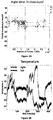
- Figure 2 shows representative data from the patient's wrists.
- Figure 2A shows the plots of the tri-axial acceleration profiles over several hours obtained on April 24th 2014 (see also Table 2).
- the a i are the tri-axial components along the x, y, and z axes.
- Figure 2D shows the scalar acceleration expressed as a function of the temperature range registered by the sensors.
- the mean acceleration value and the instantaneous maximal deviation are taken from the overall mean of the session. These profiles are then obtained as a function of temperature.
- For each minute of the session all samples of the maximal deviation from the mean acceleration are obtained and plotted in matrix form in Figure 2E (shown for a session in May 8th 2014) for 12.28 hours (739.6 minutes shown along the rows).
- the columns of the matrix show one-degree Celsius intervals spanning the range of temperatures for that session.
- the color of each entry in the matrix reflects for each minute and degree interval the maximal amount of motion deviating from the mean acceleration (see color bar) in units/s 2 .
- Figure 3 illustrates the steps followed to build these matrices.
- the acceleration and temperature data is first harnessed in one-minute-long intervals (128Hz x 60sec, 7,680 registered frames). For each degree the range of motion registered is obtained over time. The example in Figure 3 shows this for the 34-35°C-interval. All motion data occurring in that interval is harnessed (inset in right panel). Then for each minute and each °C the maximal deviation from the mean acceleration is obtained. Across the minutes and degrees, these are the entries of the matrix depicted in Figure 3 . The color indicates the amount of motion maximally deviating from the mean acceleration of the session on May 8th.
- the May 8th matrix is used to further illustrate the methods.
- the range from 33-35°C is used to show the statistics of the motion.
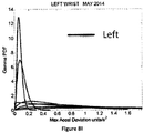
- the number of maximal deviations (peaks) across the session (6.26-hours or 375 minutes along the rows of the matrix) were counted and were gathered in a frequency histogram.
- a probability distribution function was then fit.
- MLE maximum likelihood estimation
- estimates of the shape (a) and the scale (b) parameters of the Gamma probability distribution with 95% confidence intervals are obtained.
- the continuous Gamma family of probability distributions has been used to characterize the range of human motion variability across a range of neurological disorders and typical motions.
- the Gamma statistical parameters (mean and variance) are obtained and then plotted on a ( ⁇ , ⁇ )-plane. Each point represents the Gamma statistical parameters of the acceleration-dependent motions for a temperature °C-interval taken across the time length of the session.
- this session reveals a pattern of motion whereby the motion noise registered by these accelerometers overpowers the signal.
- the range from 33-35°C used in Figure 5 to illustrate the methods are also marked here to show their range of noise-to-signal.
- the Gamma distribution shape and scale parameters of the distributions corresponding to the noise-to-signal values are estimated. This was done to determine the physiologically appropriate statistical regimes in the motion data to further analyze that data. These are regimes of temperature where the motion maintains minimal noise-to-signal ratios across the session, as opposed to the signal being overpowered by instrumentation noise.
- the estimated shape and scale parameters on the Gamma plane with 95% confidence intervals are plotted in Figure 4C .
- the color code corresponds to the frequency histograms of 4B and the legend reflects the corresponding temperature °C-interval for this May 8th session.
- the points corresponding to the shape value of 1 are at the most random noise-to-signal levels. Those towards the right correspond to statistically more predictable (systematic) regimes of noise-to-signal levels (towards symmetric shapes of the distribution of the noise-to-signal ratios.)
- higher values indicate higher levels of noise (highest marked by blue star in correspondence with the frequency histogram in 4B).
- the 33-35°C temperature interval used in 4B are marked to illustrate the methods to isolate the physiologically relevant motion regimes and in correspondence with the frequency histograms of the noise-to-signal ratio in 4B.
- Figure 7 depicts the longitudinal stochastic trajectory of the noise-to-signal ratios extracted from the motion data across all sessions. There are 124 measurements automatically extracted from 21 sessions registered across 4 months (spanning from April to July).
- the Gamma (b)-scale parameter relates to the Fano Factor, the Gamma estimated variance divided by the Gamma estimated mean value.
- the former is a.b while the latter is a.b 2 .
- the Fano Factor is then b, which is the scale parameter.
- Figure 7A (right wrist) and 7B (left wrist) show the 3-dimensional trajectories of the changes in these Gama parameters (X-Y log-log plane) along the temperature ranges (Z-axis °C) registered by the sensors.
- the vector field (black arrows) indicates the direction and the magnitude of the change in the reliability and predictability of the changes in the noise-to-signal ratio form the motion data.
- Low changes in values vs. high changes in values are better appreciated in Figures 7C-D along the surface fitted through the 124 points of physiologically relevant (low noise) data across all sessions.
- the Z-axis of these surfaces are the changes in temperature level. Notice that the right wrist had a dramatically sharp change in the month of May, while the left wrist had a gradual change in temperature from June onwards. Points along the 0-change lines of temperature, scale and shape are steady states in each session.
- Figure 8 shows the frequency histograms of the right and left wrists data involving the maximal deviations from the mean acceleration obtained within the proper temperature intervals (those identified with the lowest noise-to-signal levels). The figure focuses on the month of May which Figure 7 identified as critical for the dominant hand. Notice the changes in the shape and width of these frequency histograms across the various sessions in May.
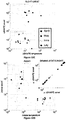
- Figure 9A tracks the stochastic trajectories of the estimated Gamma parameters for each wrist (corresponding to the acceleration-dependent motions) and identifies (with a star) in each case the session with the largest rate of change towards the regimes of lowest variability (most reliable) and most symmetric shape, towards systematic motions, away from the (most random) Exponential distribution regimes of the Gamma plane. The starting and ending points of the trajectories are also highlighted.
- Figure 9B shows for each day the Gamma estimated statistics (mean and variance) highlighting in the legend the dates of the sessions and the largest change in statistical regimes. Other analyzes of the rates of change in these estimated parameters were performed for the month of May and for other months as well.
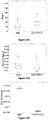
- Figure 10A shows the result of the analyses corresponding to the stochastic changes in the shape of the distribution estimated for each of the sessions of each month where the noise-to-signal was at its minimum.
- the frequency distribution of the rate of change of the shape parameter in each session was well fit by the Gamma family.
- the estimated shape and scale parameters are plotted with 95% confidence intervals on the (log-log) Gamma Plane.
- This plane shows a clear separation in the clustering of the points corresponding to the sessions in the month of May for the right wrist. This separation is consistent with the overall behavior of the changes in temperature and motion data identified in Figure 7C and 7D .
- the upward shift in this cluster along the vertical axis indicates an increase in the variability (the width) of the shapes of the distributions of the acceleration-dependent motion parameter.
- the frequency distributions of the rate of change of surface skin temperature noise followed the Gamma distribution as well.
- the shape and scale parameters of each session were estimated with minimal acceleration-dependent motion noise and the point from each session was plotted on the (log-log) Gamma plane with 95% confidence interval.
- the month of May once again stood out as a separate cluster with shifts downwards towards regimes of reliable measurements (low noise) and shifts rightwards towards more systematic regimes tending towards symmetric shapes of the distribution of the rate of change in temperature noise. These patterns were not present in the values registered by the left wrist. In the left wrist the points from the sessions in the month of May did not cluster apart from those estimated from the measurements taken in the other months. Unlike in the right wrist, no reliable and systematic changes were revealed in the motions of the left wrist during the month of May.
- Figure 10C shows the patterns corresponding to the rate of change in the shape parameter discussed in 10A-B for the acceleration-dependent motion as a function of the temperature.
- the points representing the month of May cluster apart from the rest.
- May had larger values for the change in the shape of the acceleration-dependent distribution corresponding to larger values in the change of the shape of the temperature-dependent distribution.
- Figure 10C speaks of systematic changes in the shapes of the parameters' distributions
- Figure 10D speaks of the changes in their noise levels.
- the inset zooms in the Gamma statistics of the changes in the shape of the distributions of maximal deviation from the mean acceleration.
- the figure shows that in May the motions were more systematic than in the other months and their variability in the shape of the distribution was higher. In particular, by May 17th the changes in surface skin temperature were steadier as the changes in motion patterns turned more systematic (as revealed by the higher values of the shape of the distribution of the relevant acceleration and temperature dependent parameters).
- Patient MC had the C-section delivery of her baby boy on May 22, 2014. All the data preceding that date indicated patterns of systematic variability in her motions from the dominant (right) hand that were absent in the motions from her non-dominant (left) hand. Furthermore, the medical records indicated the formation of a blood clot in the right arm after May.
- Figure 7D shows a slow gradual increase in the changes in surface skin temperature for the left wrist that also coincided with higher levels of motion. These motions from the left wrist however had no discernable patterns of systematic changes in variability levels as those observed in May.
- New methods are provided herein to assess in a personalized manner the longitudinal progression of body motions as a function of surface skin temperature using wearable sensors.
- the statistical metrics introduced here permit the continuous longitudinal assessment of patients as they move and as they undergo changes in physiological states.
- a particular case of post-sTBI has been used to illustrate the methods, yet these methods can be generally extended and used in other patients as well.
- These methods do not assume population statistics or expected values of the parameters of interest. Instead, they empirically estimate the probability distributions most likely underlying the changes in motion and physiologically relevant parameters registered in tandem within each daily session and longitudinally over months. The methods focus on the rates of change of these parameters' statistics along a continuum.
- Newtonian mechanics concerned with acceleration estimations has no known relation to thermodynamics.
- the laws of mechanics governing physical motions were derived for inanimate objects and rigid bodies, rather than for biological bodies in motion undergoing physiological changes that impact the motions' variability.
- the field of neural control of movement employs primarily Newtonian mechanics in the analyses and modelings of behavioral states ( Shadiolo et al. (2005) The computational neurobiology of reaching and pointing : a foundation for motor learning. Cambridge, Mass.:MIT Press )
- physiologically relevant measurements such as temperature, heartbeat, breathing patterns, etc.
- the human body is in constant motion in tandem with other physiological patterns of the person. Such patterns fluctuate and change over time.
- the absolute values of the parameters of interest are often registered, but very little is said about their rates of change over time.
- the rates of change of those parameters over time contained information predictive of a relevant upcoming event.
- May was the month of highest relevance in these longitudinal data sets.
- a dramatic and sharp change in the patterns of motion and surface skin temperature of this patient's dominant hand manifested in May preceding the birth of her baby boy by C-section.
- This task of characterizing longitudinally the individualized profiles of various physiological stages of pregnancy in an objective manner can be performed using the new analytics presented herein in tandem with a broad range of wearable sensors available in the market.
- the current market offers sensors that capture heart rate variability, electro dermal activities, and blood-volume levels, among others. The outcomes of these biomarkers are currently examined in isolation.
- the analytics provided herein allow for integrating them with the motion's temporal profiles in a multi-dimensional setting. In such a setting, such physiological signals are used as natural filters to isolate systematic changes in bodily motion patterns that are physiologically relevant and independent of instrumentation noise.
- the human body is in constant motion, from every breath taken to every visibly purposeful action performed. Remaining still on command is one of the hardest things to achieve because micromovements across the body are hard to control under volition.
- ASD autism spectrum disorders
- researchers ask participants to remain still and use motion-correcting methods to eliminate periods of high movement.
- uncorrected resting-state scans from 605 participants were examined and excess noise and randomness in the head displacements and rotations of the ASD participants were found. Such patterns were exacerbated with psychotropic medications, but found as well without medication.
- the sensitivity and specificity of new individualized statistical biometrics to the sensory-motor patterns associated with the use of one or more commonly prescribed medications in the ASD cohort are reported, as well as interactions between specific medications and age.
- the signatures of micro-movement noise accumulation are a biologically informed core feature of ASD with medication-specific information to help assess risks and benefits of pharmacological treatments across different ages.
- Neurosci., 6:124 also extended to the head micro-movements, particularly when the person is lying down with the head padded to dampen these motions, this would indicate that corrupted motor output variability is a systemic problem, signaling a failure to anticipate sensory consequences of the impeding actions in these individuals.
- these tools could help researchers to characterize ASD objectively from head-to-toe and to add a putative specific disorder type (e.g. head-trigeminal-ganglia- vs. body-dorsal-root-ganglia- noise prevalence) to the list of infantile neurological sensory-motor disorders of the nervous systems.
- a putative specific disorder type e.g. head-trigeminal-ganglia- vs. body-dorsal-root-ganglia- noise prevalence
- RDoC Research Domain Criteria
- NIMH National Institutes of Mental Health
- DSM Diagnostic Statistical Manual
- ICD International Classification of Diseases
- the head micro-motion data obtainable from a large number of functional neuroimaging datasets deposited in the Autism Brain Imaging Data Exchange (ABIDE) database (containing datasets of 1,112 individuals with and without ASD), offers researchers a unique opportunity to characterize normative data and better profile ASD. To this end, a new statistical platform is used herein for the personalized analyses and sensory-motor profiling of the variability inherent to resting-state behavior.
- ABIDE Autism Brain Imaging Data Exchange
- micro-movements' disorders were a systemic feature of ASD and if they were present in individuals currently on or off psychotropic medications, across multiple age groups, one can use these new techniques to provide a personalized dynamic measure of 'precision phenotyping' of ASD as the disorder evolves in time with and without medication. This would enable steering away from symptom-based medication towards individualized target treatments, in line with the current goals of Precision Medicine.
- Datasets used in this study were obtained from public, freely accessible Autism Brain Imaging Data Exchange (ABIDE) database (fcon_1000.projects.nitrc.org/indi/abide/). Data are de-identified in compliance with U.S. Health Insurance Portability and Accountability Act (HIPAA) guidelines. Participants at all sites signed written informed consent and assent (and parental consent, if participants were less than 18 years) in accordance with U.S. 45 CFR 46 and Declaration of Helsinki for participation; research protocols which included neuroimaging and clinical assessments at each site, were approved by the local ethics committees. Analyses of these de-identified data were reviewed and approved by Institutional Review Boards of Rutgers University and Columbia University Medical Center.
- HIPAA Health Insurance Portability and Accountability Act
- ABIDE sites were included that (i) deposited raw resting-state functional Magnetic Resonance Imaging (MRI) scans (i.e., no motion correction or "scrubbing" ( Power et al. (2012) Neuroimage 59:2142-2154 ) was applied to these data), (ii) had a total scan duration at least 8 minutes, and/or (iii) had at least 15 individuals with ASD.
- MRI Magnetic Resonance Imaging
- BOLD Blood Oxygenation Level Dependent
- GE GE Medical Systems, Milwaukee, WI
- Siemens Siemens (Siemens Healthcare, Er Weg, Germany) 3 Tesla MR scanners.
- UM_1 and UM_2 scans were 10 minute each (300 volumes) and USM was 8 minutes (240 volumes).
- realignment of scanned volumes involves estimating the six parameters of an affine 'rigid-body' transformation (b-splines interpolation using least-squares approach) that minimizes the differences between each successive scan and a reference scan ( Friston, K.J. in Statistical Parametric Mapping: The analysis of functional brain images (eds. K.J. Friston et al.) (Academic Press, 2008 ); Friston et al. (1995) Human Brain Mapping 2:165-189 ).
- the default reference scan in SPM8 is the first scan (volume), to which all subsequent volumes are realigned.
- the output with the six motion parameters (3 translations in x, y, z directions, and 3 rotations: pitch (about x-axis), roll (about y-axis), and yaw (about z-axis)) is recorded as an rp_%s.txt file.
- Raw NIfTI (.nii) files were separately processed for each site in the ABIDE database used in the current study because of differences in the inter-scan interval (Repetition Time, TR), number of slices, and total scan duration (number of volumes) across sites. Whenever the data from all 7 sites were pooled, ratios were defined that would consider the above mentioned sampling disparities across datasets.
- micro-movements Small trial-by-trial variations in performance as captured by fluctuations in the amplitude or the timing of critical kinematic parameters. These may include velocity- and acceleration-dependent parameters such as the maxima, the minima, and the time to reach the peaks, or the inter-peak interval timings along continuous time series of changes in the positions of some parameter from some physiological signal, etc. This definition should not be confused with small movements or with sub-movements comprising a single movement or motor actions.
- the present experiment assesses the scan-by-scan velocity-dependent variations in the linear displacement and in the angular rotations of the head's small motions. The analyses refer to the stochastic signatures of those minute motor variations, their accumulation and individualized empirically-estimated statistical features.
- the rate of change of linear displacement was obtained in vector form (a three-dimensional velocity field over time).
- the Euclidean norm was used to obtain the magnitude of each element in this scalar field over time, i.e. the linear speed profile corresponding to the given session.
- the three rotational components were treated as Euler angles and converted to quaternions for proper use of the Euclidean norm on the angular velocity field.
- the resulting scalar field was used as the angular speed profile over the given session.
- the time series of the speed values was then plotted for each participant as a profile in time, measured (in seconds) across the length of the scanner session.
- a , b 1 ⁇ a b a x a ⁇ 1 e ⁇ x b in which a is the shape parameter, b is the scale parameter, and ⁇ is the Gamma function ( Ross, S.M. Stochastic processes 2nd Ed. (Wiley, 1996 )).
- the Gamma parameters are empirically estimated using maximum likelihood estimation with 95% confidence intervals.
- the estimated parameter for each individual is plotted on the Gamma plane with confidence intervals to compare the individual to others in the cohort.
- the data from ensembles of participants is also pooled and the Gamma parameters estimated and plotted on the Gamma plane with confidence intervals to compare different groups in the database.
- the noise to signal ratio the Fano Factor (FF) (Fano, 1947) obtained from the empirically estimated Gamma variance divided by the empirically estimated Gamma mean.
- the data follows the memoryless Exponential probability distribution. This is the most random distribution whereby events in the past do not accumulate information predictive of events in the future. Larger values towards the right of the shape axes on the Gamma ( a , b )-plane tend towards the symmetric distributions, with a variety of skewed distributions in between the two extremes.
- Figure 11D shows the results of the median noise-to-signal ratio compared between the two groups.
- the speed maxima were normalized to avoid allometric effects due to scan length and sampling resolution differences across sites and studies. To this end, the averaged speed value between each two local minima in the time series were obtained. Each speed maximum was then divided by the sum of the speed maximum and the average speed between the two corresponding minima. The same procedure explained for Figure 11 was then applied to the normalized speed maxima (denoted normalized peak velocity index, PV index). Smaller values of this index indicate larger values of the average speed in the denominator (i.e. faster rates of change in linear (angular) displacements (rotations) on average). Since there is interest in the cumulative effect over time and their rates of change across the scanning session, the empirical cumulative probability distribution function (eCDF) for these speed-dependent parameters (i.e. average speed, PV index, etc.) was also obtained.
- eCDF empirical cumulative probability distribution function
- the empirically estimated Gamma shape and scale parameters were plotted as points on the Gamma plane, each representing a study site for the ensemble data.
- each point corresponds to the stochastic signatures of a single participant.
- a scatter was obtained and studied on the log-log Gamma plane in search for power law relations.
- the power law relation obtained is reported with the goodness of fit parameters.
- the fitting error between the line obtained using the estimated exponent of the power relation (the slope of the line) and the data point from the scatter was obtained for each participant and their histograms compared between ASD and controls.
- the Gamma scale parameter i.e.
- the noise to signal ratio or Fano Factor was plotted as a function of this error (denoted here delta) and statistical comparisons performed along each dimension.
- the Gamma statistics (the empirically estimated Gamma mean and Gamma variance) were plotted against the delta to fit a surface across the signatures of all ASD participants and those of the controls.
- Figure 11 illustrates analyses of the magnitudes of the rates of change of linear displacements, shown here for UM_1 site, using pooled data across all participants within each of the ASD and TD groups (similar trends were found for the rates of change of the head's angular rotations).
- Figures 11A and 11B show qualitative differences in the magnitude of the raw, scan-by-scan head micromotions between ASD and TD participants, whereby ASD patients have noticeably higher and more frequent head fluctuations. The differences in these speed maxima were quantitated using frequency histograms presented in Figure 11C , which show rapid accumulation of these peaks in the ASD group. Note that the squared log of the raw peak speed values was used for better visualization of the significant statistical disparity seen in Fig. 11A .
- Figure 12A shows the normalized speed maxima index denoted peak velocity (PV) index for each of the three main sites. Across the 3 main studies (UM_1, UM_2 and USM) it was possible to differentiate ASD from control participants as the Gamma parameters and the estimated mean and variance separated these individuals.
- Figure 12B shows the eCDFs while Figure 12C shows the estimated Gamma statistics (mean and variance) for ASD and TD participants.
- the insets show the differences in the empirical cumulative distribution functions of the PV index pooled across the ensemble of the three studies. Note that these group differences held independently for NYU, UCLA_1, OLIN, and PITT sites and are presented for both linear displacements and rotations.
- Figure 12D unfolds the ensemble data for all participants from the 3 main studies of comparable temporal resolution and scanning times.
- the analyses of the Fano Factor revealed in the ASD group a subset of individuals with higher noise levels than that in the TD group (i.e., with FF above 0.06, about 2.5 standard deviations from the mean, in Figure 12F ). Closer inspection of this ratio revealed higher levels of variability in ASD.
- the scatter was examined along three dimensions comprising the mean, the standard deviation and the delta residual as a measure of failure to follow the power law. This is shown in Figure 13A for controls and Figure 13B for ASD.
- Figures 13C-13E show the box plots resulting from the Kruskall-Wallis test that yielded significant differences between ASD and TD participants for each of these dimensions (at the alpha 0.01 level).
- the surface fitted to the scatter revealed that the subgroup with higher noise levels was comprised of some ASD participants that were currently taking at least one psychotropic medication and some that were not.
- the ASD sub-groups included participants taking no medications, two medications and three medications from the UM_1 and UM_2 study-sites included in Figures 12-13 .
- Figure 14A shows the results of this comparison on the Gamma plane.
- Systematic increase in the levels of noise (upwards shift along the scale, the noise-to-signal ratio axis) and in randomness (leftward shift along the shape axis towards a 1 the special case of the memoryless Exponential distribution) of the head micro-movement signatures was found. Consistent results were found for microdisplacements and micro-rotations.
- the number of medications taken pulled the stochastic signatures of head micro-motions of medicated ASD participants away from those of TD controls.
- the medicated ASD with 3 medication-combination-treatments were the farthest apart from TD controls. They fell along the directions of the Gamma plane towards the random exponential distribution, and with higher levels of noise-to-signal ratio than controls.
- Figure 14B shows the differences in the total cumulative distances traveled along the linear and angular domains for each patient.
- This comparison includes two studies (UM_1 and UM_2) in Figures 12-13 that reported medication intake by participants, with 300 frames per participant (both studies at 1 ⁇ 2 Hz). These were used because they had congruent fixed scanning rate needed to be able to compare the cumulative linear and rotational total excursions of the head during the session.
- a statistically significant systematic increase was found with medication quantity between medicated ASD and TD controls (P ⁇ 0.01), meaning that statistical patterns were significantly worse for ASD participants taking multiple medications.
- This difference in shape and scale was also consistent between medicated and non-medicated ASD, as well as, notably, between non-medicated ASD and controls.
- the medication information by medication classes broken down into different age groups was then examined. Given the broad range of ages in the sample (6 to 50 years old) it was desirable to elucidate several questions. First, the classes that were most likely driving the statistically significant effects of medications on the head micro-movement patterns of ASD participants was investigated. Second, it was asked whether age was a factor with variable effects (i.e. across medications). If so, it was asked which age group accounted for the worst increase in noise and randomness levels away from the non-medicated ASD, and which age would pull the medicated-ASD towards the controls (with highest reliability and predictability in the speed dependent signal). Lastly it was investigated whether there was an overall medication-class trend.
- Figure 15A shows the panels with translation (T) and rotation (R) for different age groups (G1-G5). Notice that when taken as part of a combination, the anticonvulsants, across all age groups and for both translation and rotation speeds, correspond to data points that are the highest along the scale axis and the lowest along the shape axis, i.e. the farthest from the TD controls. The systematic order is shown in the Figure 15 legend with atypical antipsychotic at the lowest end, closer to TD controls. The effects on medicated ASD were different across age groups maximally pulling them away from TD in the oldest age group 5. In contrast other medication classes pulled them towards the TD group in the younger groups (G1-G3).
- the most prescribed medication in combination is the alpha-agonist in the youngest group (6 to 11 year olds), whereas antidepressant and stimulants are the most prescribed in the oldest group (those above 17 years old).
- Anticonvulsants abound in groups 2 and 3 (spanning 11 to 15 year olds) whereas group 4 (16 to 17 year olds) has the atypical ADHD medication as the most prescribed in combination with others.
- Figure 16 shows the results across age groups (in the case of missing medication class, it was not included).
- the younger medicated ASD groups are pulled away from the non-medicated ASD participants, towards the TD participants, indicating a benefit.
- this trend is opposite.
- Examination of these older age groups shows that the patterns of medicated ASD participants systematically pull away from those of the TD participants.
- the older the person the more deleterious the effects are in the noise-to-signal ratio of the speed-dependent parameters and the farther away from symmetric (Gaussian-like) distributions that the TD manifest.
- the examination of the proportions of medications across age groups revealed that the atypical antipsychotics are likely to be the most prescribed in the youngest group while the oldest participants (congruent with the analyses above) have a higher percentage of antidepressants and stimulants prescribed to them.
- the corrupted kinesthetic motor reafference may stem in part from synaptic dysfunction putatively producing specific patterns of sensory-motor noise.
- the medications reported in the ABIDE study-sites are those commonly used to treat symptoms of ASD. They are noted to have motor side effects, including tremors, dyskinesias, involuntary ticks and other motor disorders in adult populations. A form of monitoring that relies on self-report therefore poses a substantial risk to the patient, particularly to the children, because these effects and drug interactions are not well understood for the young developing nervous systems at the peak of their plasticity.
- the stochastic signatures of micro-movements taps into core biological features of neurodevelopmental disorders such as ASD, and is amenable to constitute one of the dimensions of the RDoC that cuts across research domains ( Bernard et al. (2015) Psychol. Med., 1-5 ).
- the statistical sensitivity of these signatures to specific effects from different medication classes opens the possibility of combining this dimension with other domains of research and treatments to capture interactions in other neurological disorders.
- the existing clinical classification was used as the basis for performing comparisons.
- the same methods could be used to blindly identify self-emerging clusters as a function of interaction of medication classes and the rates of change in stochastic signatures across different pathologies.
- the present results can be translated into the clinic by using a combination of wearable sensing devices (for example, attached to the head, trunk, and limbs) and the dynamic biometrics presented here.
- a participant with a neurodevelopmental disorder could be measured continuously at home and in the clinic using any of the commercially available technology specifically adapted to medication intake.
- Currently such devices Apple watch, fitbits, etc.
- the same statistical platform presented here for the characterization of head-micromotions can be used to profile the micro-motion patterns of such physiological signals and their fluctuations and rates of change unique to the person.
- This quantitative information would assist physicians in reaching the decision on whether or not to alter the psychopharmacological regimen according to the personalized longitudinal assessment of each patient in relation to his/her own patterns from previous visits. It would also allow insurance companies to draw a conclusion on coverage based on an objective outcome measure automatically revealing trends in the person's signatures.
Claims (16)
- Procédé de mesure d'un mouvement physiologiquement pertinent d'un sujet ; ledit procédé comprenant les étapes consistant à :a) mesurer automatiquement des micromouvements d'un sujet difficilement reconnaissables à l'œil nu et mesurer automatiquement au moins une valeur biométrique sans mouvement du sujet, dans lequel les micromouvements du sujet et la au moins une valeur biométrique sont mesurés simultanément sur une période de temps ;b) déterminer automatiquement un rapport bruit/signal pour les micromouvements mesurés à l'étape a) pour chacun d'une pluralité d'intervalles de temps dans la période de temps et pour chacune d'une pluralité de plages de valeurs pour la valeur biométrique mesurée à l'étape a) ; etc) identifier automatiquement au moins une plage de la pluralité de plages de valeurs pour la valeur biométrique et au moins un intervalle de temps de la pluralité d'intervalles de temps dans lequel le rapport bruit/signal est inférieur à un ayant des micromouvements d'un mouvement physiologiquement pertinent.
- Procédé selon la revendication 1, dans lequel les mouvements physiologiquement pertinents ont un rapport bruit/signal le plus faible.
- Procédé selon la revendication 1, dans lequel les micromouvements sont mesurés avec un capteur de mouvement portable comprenant une unité de mesure inertielle (« IMU ») .
- Procédé selon la revendication 1, dans lequel la au moins une valeur biométrique comprend : la température du sujet, la fréquence cardiaque du sujet, l'activité électrodermique, l'activité électrique d'un cerveau mesurée par électroencéphalogramme (« EEG »), l'activité électrique de muscles mesurée par électromyographie (« EMG ») et/ou un schéma respiratoire.
- Procédé selon la revendication 1, dans lequel les micromouvements comprennent au moins un parmi :
vitesse, accélération, profil de vitesse, vitesse maximale, accélération maximale, vitesse minimale, accélération minimale, temps pour atteindre la vitesse maximale, temps pour atteindre l'accélération maximale, vitesse de rétraction maximale, temps pour atteindre la vitesse de rétraction maximale, intervalles entre pics, trajectoire tridimensionnelle, précision du toucher de la cible, durée globale du mouvement, rotation ou positionnement d'une partie du corps, mouvement de translation, mouvement de rotation, ou angle d'articulation. - Procédé selon la revendication 1, dans lequel la mesure automatique de micromouvements d'un sujet comprend une mesure d'un mouvement de la main, de la tête, du tronc, d'un membre ou d'un bras du sujet.
- Procédé selon la revendication 1, dans lequel la mesure automatique de micromouvements d'un sujet comprend une mesure du mouvement du bras du sujet.
- Procédé selon la revendication 1, dans lequel ladite valeur biométrique est la température du sujet.
- Procédé selon la revendication 1, dans lequel ledit micromouvement est un micromouvement d'accélération maximale.
- Procédé selon la revendication 1, comprenant en outre les étapes consistant à :après qu'une thérapie ait été administrée à un sujet, répéter les étapes a) à c) ; etdéterminer automatiquement une modification du mouvement physiologiquement pertinent dudit sujet.
- Procédé selon la revendication 1, comprenant en outre les étapes consistant à :déterminer automatiquement un taux de variation de rapport signal/bruit pour une première plage de la pluralité de plages de valeurs pour la valeur biométrique sur des intervalles de temps successifs de la pluralité d'intervalles de temps ; etutiliser le taux de changement pour déterminer si le mouvement physiologiquement pertinent à chaque intervalle biométrique est systématique ou spontanément aléatoire.
- Procédé selon la revendication 1, dans lequel la détermination du rapport bruit/signal pour les micromouvements dans une première plage de la pluralité de plages de valeurs pour la valeur biométrique comprend en outre les étapes consistant à :ajuster automatiquement un paramètre de forme et un paramètre d'échelle d'une distribution Gamma continue sur la base de micromouvements dans la première plage pour la valeur biométrique ; et,déterminer automatiquement le rapport bruit/signal sur la base du paramètre de forme de la distribution Gamma continue.
- Procédé selon la revendication 1, dans lequel la mesure des micromouvements du sujet comprend en outre l'utilisation d'un procédé d'estimation de mouvement pour estimer un mouvement d'une tête du sujet sur la période de temps en :
capturant et collectant une séquence d'images de la tête du sujet au cours de la période de temps à partir d'un appareil d'imagerie par résonance magnétique fonctionnelle (IRMf). - Procédé selon la revendication 1, dans lequel le procédé comprend en outre l'affichage d'une matrice tridimensionnelle sur un affichage, la matrice tridimensionnelle comprenant une pluralité de cellules dans un plan bidimensionnel, dans lequel :une première dimension est la pluralité d'intervalles de temps dans la période de temps ;une seconde dimension est la pluralité de plages de valeurs pour la valeur biométrique ; etchacune de la pluralité de cellules est codée par une couleur ou une échelle de gris qui représente un écart maximal par rapport à une accélération moyenne du sujet correspondant à un intervalle de temps dans la pluralité d'intervalles de temps et une plage de valeurs de la pluralité de plages de valeurs pour la valeur biométrique.
- Procédé selon la revendication 14, dans lequel :les micromouvements sont mesurés avec une résolution d'échantillonnage du capteur de mouvement ; etla valeur biométrique est une fréquence cardiaque ou une température cutanée.
- Procédé selon la revendication 1, comprenant en outre l'étape consistant à :
produire automatiquement au moins un tracé de premières données sur la base de micromouvements du mouvement physiologiquement pertinent.
Applications Claiming Priority (2)
| Application Number | Priority Date | Filing Date | Title |
|---|---|---|---|
| US201462089031P | 2014-12-08 | 2014-12-08 | |
| PCT/US2015/064440 WO2016094375A1 (fr) | 2014-12-08 | 2015-12-08 | Procédés de mesure d'un mouvement physiologiquement pertinent |
Publications (3)
| Publication Number | Publication Date |
|---|---|
| EP3229684A1 EP3229684A1 (fr) | 2017-10-18 |
| EP3229684A4 EP3229684A4 (fr) | 2018-07-25 |
| EP3229684B1 true EP3229684B1 (fr) | 2020-04-01 |
Family
ID=56108043
Family Applications (1)
| Application Number | Title | Priority Date | Filing Date |
|---|---|---|---|
| EP15867321.0A Active EP3229684B1 (fr) | 2014-12-08 | 2015-12-08 | Procédés de mesure d'un mouvement physiologiquement pertinent |
Country Status (3)
| Country | Link |
|---|---|
| US (1) | US10898122B2 (fr) |
| EP (1) | EP3229684B1 (fr) |
| WO (1) | WO2016094375A1 (fr) |
Families Citing this family (19)
| Publication number | Priority date | Publication date | Assignee | Title |
|---|---|---|---|---|
| CN104503592A (zh) * | 2015-01-23 | 2015-04-08 | 北京智谷睿拓技术服务有限公司 | 头部动作确定方法和装置 |
| US10357210B2 (en) | 2015-02-04 | 2019-07-23 | Proprius Technologies S.A.R.L. | Determining health change of a user with neuro and neuro-mechanical fingerprints |
| US9590986B2 (en) | 2015-02-04 | 2017-03-07 | Aerendir Mobile Inc. | Local user authentication with neuro and neuro-mechanical fingerprints |
| US9577992B2 (en) | 2015-02-04 | 2017-02-21 | Aerendir Mobile Inc. | Data encryption/decryption using neuro and neuro-mechanical fingerprints |
| US9836896B2 (en) | 2015-02-04 | 2017-12-05 | Proprius Technologies S.A.R.L | Keyless access control with neuro and neuro-mechanical fingerprints |
| US10599980B2 (en) * | 2015-12-21 | 2020-03-24 | Intel Corporation | Technologies for cognitive cuing based on knowledge and context |
| US10918288B2 (en) | 2016-10-14 | 2021-02-16 | Rutgers, The State University Of New Jersey | Systems and methods for tracking neuro-development disorders |
| US10768196B2 (en) | 2016-12-19 | 2020-09-08 | Huami Inc. | Determine wearing position of a wearable device |
| EP3735172B1 (fr) * | 2018-01-03 | 2022-02-16 | Cardiac Pacemakers, Inc. | Imagerie d'une partie corporelle à l'aide de sons |
| JP7165207B2 (ja) | 2018-05-01 | 2022-11-02 | ブラックソーン セラピューティクス インコーポレイテッド | 機械学習ベースの診断分類器 |
| EP3870030A4 (fr) | 2018-10-23 | 2022-08-03 | Blackthorn Therapeutics, Inc. | Systèmes et procédés de sélection, de diagnostic et de stratification de patients |
| EP3870029A4 (fr) * | 2018-10-24 | 2022-08-03 | Research Triangle Institute | Systèmes, procédés et dispositifs permettant de déterminer des lésions cérébrales traumatiques légères |
| US11164319B2 (en) | 2018-12-20 | 2021-11-02 | Smith & Nephew, Inc. | Machine learning feature vector generator using depth image foreground attributes |
| CN110473202B (zh) * | 2019-06-18 | 2023-03-31 | 山东工商学院 | 一种高阶动态功能连接网络的时序不变特征提取方法 |
| US10559145B1 (en) * | 2019-07-17 | 2020-02-11 | Abdulaziz Mohammed Almehmadi | Systems and methods for providing behavioral based intention detection |
| US11134859B2 (en) | 2019-10-15 | 2021-10-05 | Imperative Care, Inc. | Systems and methods for multivariate stroke detection |
| CN113143257B (zh) * | 2021-02-09 | 2023-01-17 | 国体智慧体育技术创新中心(北京)有限公司 | 基于个体运动行为层次模型的泛化应用系统及方法 |
| US20220327947A1 (en) * | 2021-04-13 | 2022-10-13 | D2L Corporation | Systems and methods for automatically revising feedback in electronic learning systems |
| WO2023235777A2 (fr) * | 2022-06-01 | 2023-12-07 | Rutgers, The State University Of New Jersey | Techniques de mesure de neurodéveloppement atypique chez des nouveau-nés sur la base d'une courte vidéo |
Family Cites Families (16)
| Publication number | Priority date | Publication date | Assignee | Title |
|---|---|---|---|---|
| US812169A (en) * | 1905-04-19 | 1906-02-06 | Augusta Grasser | Fly-trap. |
| US20030105409A1 (en) * | 2001-11-14 | 2003-06-05 | Donoghue John Philip | Neurological signal decoding |
| US7006856B2 (en) * | 2003-01-10 | 2006-02-28 | Nellcor Puritan Bennett Incorporated | Signal quality metrics design for qualifying data for a physiological monitor |
| US20050143589A1 (en) * | 2003-11-09 | 2005-06-30 | Donoghue John P. | Calibration systems and methods for neural interface devices |
| US20050165327A1 (en) * | 2004-01-28 | 2005-07-28 | Thibault Lawrence E. | Apparatus and method for detecting the severity of brain function impairment |
| US7733224B2 (en) * | 2006-06-30 | 2010-06-08 | Bao Tran | Mesh network personal emergency response appliance |
| ES2569411T3 (es) * | 2006-05-19 | 2016-05-10 | The Queen's Medical Center | Sistema de seguimiento de movimiento para imágenes adaptativas en tiempo real y espectroscopia |
| EP2018825A1 (fr) * | 2007-07-26 | 2009-01-28 | Ecole Polytechnique Fédérale de Lausanne (EPFL) | Procédés et dispositif pour quantifier un modèle d'activité physique humaine |
| US8121694B2 (en) | 2007-10-16 | 2012-02-21 | Medtronic, Inc. | Therapy control based on a patient movement state |
| US8718980B2 (en) * | 2009-09-11 | 2014-05-06 | Qualcomm Incorporated | Method and apparatus for artifacts mitigation with multiple wireless sensors |
| US8401629B2 (en) * | 2010-04-28 | 2013-03-19 | Medtronic, Inc. | Method and apparatus for detecting and discriminating tachycardia |
| US9615547B2 (en) * | 2011-07-14 | 2017-04-11 | Petpace Ltd. | Pet animal collar for health and vital signs monitoring, alert and diagnosis |
| US10176299B2 (en) * | 2011-11-11 | 2019-01-08 | Rutgers, The State University Of New Jersey | Methods for the diagnosis and treatment of neurological disorders |
| US9005129B2 (en) * | 2012-06-22 | 2015-04-14 | Fitbit, Inc. | Wearable heart rate monitor |
| CN104684459B (zh) * | 2012-08-01 | 2017-09-12 | 皇家飞利浦有限公司 | 光体积描记术测量中识别运动伪影并且改善测量结果和警报的可靠性的方法和系统 |
| EP2895050B8 (fr) * | 2012-09-11 | 2018-12-19 | L.I.F.E. Corporation S.A. | Plateforme de communication portable |
-
2015
- 2015-12-08 WO PCT/US2015/064440 patent/WO2016094375A1/fr active Application Filing
- 2015-12-08 EP EP15867321.0A patent/EP3229684B1/fr active Active
-
2017
- 2017-06-08 US US15/617,822 patent/US10898122B2/en active Active
Non-Patent Citations (1)
| Title |
|---|
| None * |
Also Published As
| Publication number | Publication date |
|---|---|
| US10898122B2 (en) | 2021-01-26 |
| WO2016094375A1 (fr) | 2016-06-16 |
| EP3229684A4 (fr) | 2018-07-25 |
| EP3229684A1 (fr) | 2017-10-18 |
| US20170340261A1 (en) | 2017-11-30 |
Similar Documents
| Publication | Publication Date | Title |
|---|---|---|
| EP3229684B1 (fr) | Procédés de mesure d'un mouvement physiologiquement pertinent | |
| Onorati et al. | Multicenter clinical assessment of improved wearable multimodal convulsive seizure detectors | |
| Mansur et al. | A review on techniques for tremor recording and quantification | |
| Leach et al. | Natural turn measures predict recurrent falls in community-dwelling older adults: a longitudinal cohort study | |
| EP2983586B1 (fr) | Procédés pour surveiller la conscience | |
| AU2012285379B2 (en) | Method and system for estimating brain concussion | |
| CN105658134B (zh) | 脑健康的多模态药理诊断评价 | |
| JP6975853B2 (ja) | 脳機能検査システムおよびそのデバイス | |
| Bidwell et al. | Seizure reporting technologies for epilepsy treatment: A review of clinical information needs and supporting technologies | |
| Thomas et al. | A treatment-response index from wearable sensors for quantifying Parkinson's disease motor states | |
| Luppi et al. | Connectome harmonic decomposition of human brain dynamics reveals a landscape of consciousness | |
| Luo et al. | Biomarkers for prediction of schizophrenia: Insights from resting-state EEG microstates | |
| JP2003521987A (ja) | 定量的eegを使用して脳の生理学的不均衡を分類および処置する方法 | |
| CN104903897A (zh) | 癫痫和癫痫发作的易发性的评定 | |
| Pulliam et al. | Motion sensor dyskinesia assessment during activities of daily living | |
| Qadeer et al. | Bispectral index monitoring of conscious sedation with the combination of meperidine and midazolam during endoscopy | |
| Gallego-Jutgla et al. | A theta-band EEG based index for early diagnosis of Alzheimer's disease | |
| Zach et al. | The patient's perspective: the effect of levodopa on Parkinson symptoms | |
| Donohue et al. | A preliminary investigation of whether HRCA signals can differentiate between swallows from healthy people and swallows from people with neurodegenerative diseases | |
| Torres et al. | Objective and personalized longitudinal assessment of a pregnant patient with post severe brain trauma | |
| Schaefer et al. | Parkinson patients without tremor show changed patterns of mechanical muscle oscillations during a specific bilateral motor task compared to controls | |
| Castillo Rodriguez et al. | Differentiation of subclinical and clinical electrographic events in long‐term electroencephalographic recordings | |
| Johnson | Detection of Parkinson disease rest tremor | |
| Azami et al. | Evaluation of resting-state magnetoencephalogram complexity in Alzheimer's disease with multivariate multiscale permutation and sample entropies | |
| Tanabe et al. | Left ventricular hypertrophy by electrocardiogram as a predictor of success in home blood pressure control: HOMED-BP study |
Legal Events
| Date | Code | Title | Description |
|---|---|---|---|
| STAA | Information on the status of an ep patent application or granted ep patent |
Free format text: STATUS: THE INTERNATIONAL PUBLICATION HAS BEEN MADE |
|
| PUAI | Public reference made under article 153(3) epc to a published international application that has entered the european phase |
Free format text: ORIGINAL CODE: 0009012 |
|
| STAA | Information on the status of an ep patent application or granted ep patent |
Free format text: STATUS: REQUEST FOR EXAMINATION WAS MADE |
|
| 17P | Request for examination filed |
Effective date: 20170628 |
|
| AK | Designated contracting states |
Kind code of ref document: A1 Designated state(s): AL AT BE BG CH CY CZ DE DK EE ES FI FR GB GR HR HU IE IS IT LI LT LU LV MC MK MT NL NO PL PT RO RS SE SI SK SM TR |
|
| AX | Request for extension of the european patent |
Extension state: BA ME |
|
| DAV | Request for validation of the european patent (deleted) | ||
| DAX | Request for extension of the european patent (deleted) | ||
| A4 | Supplementary search report drawn up and despatched |
Effective date: 20180625 |
|
| RIC1 | Information provided on ipc code assigned before grant |
Ipc: A61B 5/11 20060101ALI20180619BHEP Ipc: A61B 5/103 20060101AFI20180619BHEP Ipc: A61B 5/01 20060101ALI20180619BHEP |
|
| GRAP | Despatch of communication of intention to grant a patent |
Free format text: ORIGINAL CODE: EPIDOSNIGR1 |
|
| STAA | Information on the status of an ep patent application or granted ep patent |
Free format text: STATUS: GRANT OF PATENT IS INTENDED |
|
| INTG | Intention to grant announced |
Effective date: 20190912 |
|
| GRAS | Grant fee paid |
Free format text: ORIGINAL CODE: EPIDOSNIGR3 |
|
| GRAA | (expected) grant |
Free format text: ORIGINAL CODE: 0009210 |
|
| STAA | Information on the status of an ep patent application or granted ep patent |
Free format text: STATUS: THE PATENT HAS BEEN GRANTED |
|
| AK | Designated contracting states |
Kind code of ref document: B1 Designated state(s): AL AT BE BG CH CY CZ DE DK EE ES FI FR GB GR HR HU IE IS IT LI LT LU LV MC MK MT NL NO PL PT RO RS SE SI SK SM TR |
|
| REG | Reference to a national code |
Ref country code: GB Ref legal event code: FG4D |
|
| REG | Reference to a national code |
Ref country code: AT Ref legal event code: REF Ref document number: 1250292 Country of ref document: AT Kind code of ref document: T Effective date: 20200415 Ref country code: CH Ref legal event code: EP |
|
| REG | Reference to a national code |
Ref country code: DE Ref legal event code: R096 Ref document number: 602015050049 Country of ref document: DE |
|
| REG | Reference to a national code |
Ref country code: IE Ref legal event code: FG4D |
|
| PG25 | Lapsed in a contracting state [announced via postgrant information from national office to epo] |
Ref country code: BG Free format text: LAPSE BECAUSE OF FAILURE TO SUBMIT A TRANSLATION OF THE DESCRIPTION OR TO PAY THE FEE WITHIN THE PRESCRIBED TIME-LIMIT Effective date: 20200701 |
|
| REG | Reference to a national code |
Ref country code: NL Ref legal event code: MP Effective date: 20200401 |
|
| REG | Reference to a national code |
Ref country code: LT Ref legal event code: MG4D |
|
| PG25 | Lapsed in a contracting state [announced via postgrant information from national office to epo] |
Ref country code: GR Free format text: LAPSE BECAUSE OF FAILURE TO SUBMIT A TRANSLATION OF THE DESCRIPTION OR TO PAY THE FEE WITHIN THE PRESCRIBED TIME-LIMIT Effective date: 20200702 Ref country code: NO Free format text: LAPSE BECAUSE OF FAILURE TO SUBMIT A TRANSLATION OF THE DESCRIPTION OR TO PAY THE FEE WITHIN THE PRESCRIBED TIME-LIMIT Effective date: 20200701 Ref country code: LT Free format text: LAPSE BECAUSE OF FAILURE TO SUBMIT A TRANSLATION OF THE DESCRIPTION OR TO PAY THE FEE WITHIN THE PRESCRIBED TIME-LIMIT Effective date: 20200401 Ref country code: SE Free format text: LAPSE BECAUSE OF FAILURE TO SUBMIT A TRANSLATION OF THE DESCRIPTION OR TO PAY THE FEE WITHIN THE PRESCRIBED TIME-LIMIT Effective date: 20200401 Ref country code: NL Free format text: LAPSE BECAUSE OF FAILURE TO SUBMIT A TRANSLATION OF THE DESCRIPTION OR TO PAY THE FEE WITHIN THE PRESCRIBED TIME-LIMIT Effective date: 20200401 Ref country code: PT Free format text: LAPSE BECAUSE OF FAILURE TO SUBMIT A TRANSLATION OF THE DESCRIPTION OR TO PAY THE FEE WITHIN THE PRESCRIBED TIME-LIMIT Effective date: 20200817 Ref country code: FI Free format text: LAPSE BECAUSE OF FAILURE TO SUBMIT A TRANSLATION OF THE DESCRIPTION OR TO PAY THE FEE WITHIN THE PRESCRIBED TIME-LIMIT Effective date: 20200401 Ref country code: CZ Free format text: LAPSE BECAUSE OF FAILURE TO SUBMIT A TRANSLATION OF THE DESCRIPTION OR TO PAY THE FEE WITHIN THE PRESCRIBED TIME-LIMIT Effective date: 20200401 Ref country code: IS Free format text: LAPSE BECAUSE OF FAILURE TO SUBMIT A TRANSLATION OF THE DESCRIPTION OR TO PAY THE FEE WITHIN THE PRESCRIBED TIME-LIMIT Effective date: 20200801 |
|
| REG | Reference to a national code |
Ref country code: AT Ref legal event code: MK05 Ref document number: 1250292 Country of ref document: AT Kind code of ref document: T Effective date: 20200401 |
|
| PG25 | Lapsed in a contracting state [announced via postgrant information from national office to epo] |
Ref country code: HR Free format text: LAPSE BECAUSE OF FAILURE TO SUBMIT A TRANSLATION OF THE DESCRIPTION OR TO PAY THE FEE WITHIN THE PRESCRIBED TIME-LIMIT Effective date: 20200401 Ref country code: RS Free format text: LAPSE BECAUSE OF FAILURE TO SUBMIT A TRANSLATION OF THE DESCRIPTION OR TO PAY THE FEE WITHIN THE PRESCRIBED TIME-LIMIT Effective date: 20200401 Ref country code: LV Free format text: LAPSE BECAUSE OF FAILURE TO SUBMIT A TRANSLATION OF THE DESCRIPTION OR TO PAY THE FEE WITHIN THE PRESCRIBED TIME-LIMIT Effective date: 20200401 |
|
| PG25 | Lapsed in a contracting state [announced via postgrant information from national office to epo] |
Ref country code: AL Free format text: LAPSE BECAUSE OF FAILURE TO SUBMIT A TRANSLATION OF THE DESCRIPTION OR TO PAY THE FEE WITHIN THE PRESCRIBED TIME-LIMIT Effective date: 20200401 |
|
| REG | Reference to a national code |
Ref country code: DE Ref legal event code: R097 Ref document number: 602015050049 Country of ref document: DE |
|
| PG25 | Lapsed in a contracting state [announced via postgrant information from national office to epo] |
Ref country code: IT Free format text: LAPSE BECAUSE OF FAILURE TO SUBMIT A TRANSLATION OF THE DESCRIPTION OR TO PAY THE FEE WITHIN THE PRESCRIBED TIME-LIMIT Effective date: 20200401 Ref country code: RO Free format text: LAPSE BECAUSE OF FAILURE TO SUBMIT A TRANSLATION OF THE DESCRIPTION OR TO PAY THE FEE WITHIN THE PRESCRIBED TIME-LIMIT Effective date: 20200401 Ref country code: ES Free format text: LAPSE BECAUSE OF FAILURE TO SUBMIT A TRANSLATION OF THE DESCRIPTION OR TO PAY THE FEE WITHIN THE PRESCRIBED TIME-LIMIT Effective date: 20200401 Ref country code: SM Free format text: LAPSE BECAUSE OF FAILURE TO SUBMIT A TRANSLATION OF THE DESCRIPTION OR TO PAY THE FEE WITHIN THE PRESCRIBED TIME-LIMIT Effective date: 20200401 Ref country code: EE Free format text: LAPSE BECAUSE OF FAILURE TO SUBMIT A TRANSLATION OF THE DESCRIPTION OR TO PAY THE FEE WITHIN THE PRESCRIBED TIME-LIMIT Effective date: 20200401 Ref country code: AT Free format text: LAPSE BECAUSE OF FAILURE TO SUBMIT A TRANSLATION OF THE DESCRIPTION OR TO PAY THE FEE WITHIN THE PRESCRIBED TIME-LIMIT Effective date: 20200401 Ref country code: DK Free format text: LAPSE BECAUSE OF FAILURE TO SUBMIT A TRANSLATION OF THE DESCRIPTION OR TO PAY THE FEE WITHIN THE PRESCRIBED TIME-LIMIT Effective date: 20200401 |
|
| PLBE | No opposition filed within time limit |
Free format text: ORIGINAL CODE: 0009261 |
|
| STAA | Information on the status of an ep patent application or granted ep patent |
Free format text: STATUS: NO OPPOSITION FILED WITHIN TIME LIMIT |
|
| PG25 | Lapsed in a contracting state [announced via postgrant information from national office to epo] |
Ref country code: SK Free format text: LAPSE BECAUSE OF FAILURE TO SUBMIT A TRANSLATION OF THE DESCRIPTION OR TO PAY THE FEE WITHIN THE PRESCRIBED TIME-LIMIT Effective date: 20200401 Ref country code: PL Free format text: LAPSE BECAUSE OF FAILURE TO SUBMIT A TRANSLATION OF THE DESCRIPTION OR TO PAY THE FEE WITHIN THE PRESCRIBED TIME-LIMIT Effective date: 20200401 |
|
| 26N | No opposition filed |
Effective date: 20210112 |
|
| PG25 | Lapsed in a contracting state [announced via postgrant information from national office to epo] |
Ref country code: SI Free format text: LAPSE BECAUSE OF FAILURE TO SUBMIT A TRANSLATION OF THE DESCRIPTION OR TO PAY THE FEE WITHIN THE PRESCRIBED TIME-LIMIT Effective date: 20200401 |
|
| REG | Reference to a national code |
Ref country code: CH Ref legal event code: PL |
|
| PG25 | Lapsed in a contracting state [announced via postgrant information from national office to epo] |
Ref country code: MC Free format text: LAPSE BECAUSE OF FAILURE TO SUBMIT A TRANSLATION OF THE DESCRIPTION OR TO PAY THE FEE WITHIN THE PRESCRIBED TIME-LIMIT Effective date: 20200401 |
|
| REG | Reference to a national code |
Ref country code: BE Ref legal event code: MM Effective date: 20201231 |
|
| PG25 | Lapsed in a contracting state [announced via postgrant information from national office to epo] |
Ref country code: IE Free format text: LAPSE BECAUSE OF NON-PAYMENT OF DUE FEES Effective date: 20201208 Ref country code: LU Free format text: LAPSE BECAUSE OF NON-PAYMENT OF DUE FEES Effective date: 20201208 |
|
| PG25 | Lapsed in a contracting state [announced via postgrant information from national office to epo] |
Ref country code: LI Free format text: LAPSE BECAUSE OF NON-PAYMENT OF DUE FEES Effective date: 20201231 Ref country code: CH Free format text: LAPSE BECAUSE OF NON-PAYMENT OF DUE FEES Effective date: 20201231 |
|
| PG25 | Lapsed in a contracting state [announced via postgrant information from national office to epo] |
Ref country code: TR Free format text: LAPSE BECAUSE OF FAILURE TO SUBMIT A TRANSLATION OF THE DESCRIPTION OR TO PAY THE FEE WITHIN THE PRESCRIBED TIME-LIMIT Effective date: 20200401 Ref country code: MT Free format text: LAPSE BECAUSE OF FAILURE TO SUBMIT A TRANSLATION OF THE DESCRIPTION OR TO PAY THE FEE WITHIN THE PRESCRIBED TIME-LIMIT Effective date: 20200401 Ref country code: CY Free format text: LAPSE BECAUSE OF FAILURE TO SUBMIT A TRANSLATION OF THE DESCRIPTION OR TO PAY THE FEE WITHIN THE PRESCRIBED TIME-LIMIT Effective date: 20200401 |
|
| PG25 | Lapsed in a contracting state [announced via postgrant information from national office to epo] |
Ref country code: MK Free format text: LAPSE BECAUSE OF FAILURE TO SUBMIT A TRANSLATION OF THE DESCRIPTION OR TO PAY THE FEE WITHIN THE PRESCRIBED TIME-LIMIT Effective date: 20200401 |
|
| PG25 | Lapsed in a contracting state [announced via postgrant information from national office to epo] |
Ref country code: BE Free format text: LAPSE BECAUSE OF NON-PAYMENT OF DUE FEES Effective date: 20201231 |
|
| PGFP | Annual fee paid to national office [announced via postgrant information from national office to epo] |
Ref country code: DE Payment date: 20221228 Year of fee payment: 8 |
|
| P01 | Opt-out of the competence of the unified patent court (upc) registered |
Effective date: 20230525 |
|
| PGFP | Annual fee paid to national office [announced via postgrant information from national office to epo] |
Ref country code: GB Payment date: 20231227 Year of fee payment: 9 |
|
| PGFP | Annual fee paid to national office [announced via postgrant information from national office to epo] |
Ref country code: FR Payment date: 20231227 Year of fee payment: 9 |