EP3215846B1 - Composites métalliques destinés à une imagerie améliorée - Google Patents
Composites métalliques destinés à une imagerie améliorée Download PDFInfo
- Publication number
- EP3215846B1 EP3215846B1 EP15857571.2A EP15857571A EP3215846B1 EP 3215846 B1 EP3215846 B1 EP 3215846B1 EP 15857571 A EP15857571 A EP 15857571A EP 3215846 B1 EP3215846 B1 EP 3215846B1
- Authority
- EP
- European Patent Office
- Prior art keywords
- film
- analyte
- antibodies
- cancer
- antigens
- Prior art date
- Legal status (The legal status is an assumption and is not a legal conclusion. Google has not performed a legal analysis and makes no representation as to the accuracy of the status listed.)
- Active
Links
- 238000003384 imaging method Methods 0.000 title claims description 28
- 239000002905 metal composite material Substances 0.000 title 1
- 239000010931 gold Substances 0.000 claims description 143
- 239000012491 analyte Substances 0.000 claims description 132
- 239000000758 substrate Substances 0.000 claims description 129
- 238000009739 binding Methods 0.000 claims description 128
- 230000027455 binding Effects 0.000 claims description 127
- 238000000034 method Methods 0.000 claims description 103
- 239000000427 antigen Substances 0.000 claims description 100
- 108091007433 antigens Proteins 0.000 claims description 99
- 102000036639 antigens Human genes 0.000 claims description 99
- 239000002086 nanomaterial Substances 0.000 claims description 93
- 239000011324 bead Substances 0.000 claims description 79
- 229910052737 gold Inorganic materials 0.000 claims description 68
- 210000004027 cell Anatomy 0.000 claims description 60
- 239000000090 biomarker Substances 0.000 claims description 58
- 102000040430 polynucleotide Human genes 0.000 claims description 52
- 108091033319 polynucleotide Proteins 0.000 claims description 52
- 239000002157 polynucleotide Substances 0.000 claims description 52
- 239000002105 nanoparticle Substances 0.000 claims description 51
- -1 antibodies Proteins 0.000 claims description 49
- 206010028980 Neoplasm Diseases 0.000 claims description 46
- 108020004414 DNA Proteins 0.000 claims description 43
- 108090000623 proteins and genes Proteins 0.000 claims description 43
- 102000004169 proteins and genes Human genes 0.000 claims description 37
- 238000002493 microarray Methods 0.000 claims description 33
- 201000011510 cancer Diseases 0.000 claims description 32
- 210000001808 exosome Anatomy 0.000 claims description 27
- VYPSYNLAJGMNEJ-UHFFFAOYSA-N silicon dioxide Inorganic materials O=[Si]=O VYPSYNLAJGMNEJ-UHFFFAOYSA-N 0.000 claims description 26
- 150000007523 nucleic acids Chemical class 0.000 claims description 23
- 102000039446 nucleic acids Human genes 0.000 claims description 22
- 108020004707 nucleic acids Proteins 0.000 claims description 22
- 210000001519 tissue Anatomy 0.000 claims description 22
- 208000024172 Cardiovascular disease Diseases 0.000 claims description 21
- 241000282414 Homo sapiens Species 0.000 claims description 21
- 108091032973 (ribonucleotides)n+m Proteins 0.000 claims description 20
- 239000011521 glass Substances 0.000 claims description 19
- 201000005485 Toxoplasmosis Diseases 0.000 claims description 18
- 108090000765 processed proteins & peptides Proteins 0.000 claims description 18
- 201000005404 rubella Diseases 0.000 claims description 17
- 239000002055 nanoplate Substances 0.000 claims description 16
- 208000015181 infectious disease Diseases 0.000 claims description 15
- 239000000463 material Substances 0.000 claims description 15
- 108010072866 Prostate-Specific Antigen Proteins 0.000 claims description 14
- 230000005291 magnetic effect Effects 0.000 claims description 14
- 102000004127 Cytokines Human genes 0.000 claims description 13
- 108090000695 Cytokines Proteins 0.000 claims description 13
- 108010074051 C-Reactive Protein Proteins 0.000 claims description 12
- 108010022366 Carcinoembryonic Antigen Proteins 0.000 claims description 12
- 102100025475 Carcinoembryonic antigen-related cell adhesion molecule 5 Human genes 0.000 claims description 12
- 239000012528 membrane Substances 0.000 claims description 12
- 241000700721 Hepatitis B virus Species 0.000 claims description 11
- 230000001788 irregular Effects 0.000 claims description 11
- 102100032752 C-reactive protein Human genes 0.000 claims description 10
- 230000001965 increasing effect Effects 0.000 claims description 10
- 239000000377 silicon dioxide Substances 0.000 claims description 10
- 208000035473 Communicable disease Diseases 0.000 claims description 9
- 108091023037 Aptamer Proteins 0.000 claims description 8
- 208000001490 Dengue Diseases 0.000 claims description 8
- 206010012310 Dengue fever Diseases 0.000 claims description 8
- 101000623901 Homo sapiens Mucin-16 Proteins 0.000 claims description 8
- 241000700588 Human alphaherpesvirus 1 Species 0.000 claims description 8
- UQSXHKLRYXJYBZ-UHFFFAOYSA-N Iron oxide Chemical compound [Fe]=O UQSXHKLRYXJYBZ-UHFFFAOYSA-N 0.000 claims description 8
- 102100023123 Mucin-16 Human genes 0.000 claims description 8
- 208000025729 dengue disease Diseases 0.000 claims description 8
- 229920000642 polymer Polymers 0.000 claims description 8
- 201000000596 systemic lupus erythematosus Diseases 0.000 claims description 8
- 102000013394 Troponin I Human genes 0.000 claims description 7
- 108010065729 Troponin I Proteins 0.000 claims description 7
- 235000012239 silicon dioxide Nutrition 0.000 claims description 7
- 208000006379 syphilis Diseases 0.000 claims description 7
- 102000004190 Enzymes Human genes 0.000 claims description 6
- 108090000790 Enzymes Proteins 0.000 claims description 6
- 208000022559 Inflammatory bowel disease Diseases 0.000 claims description 6
- 108700039882 Protein Glutamine gamma Glutamyltransferase 2 Proteins 0.000 claims description 6
- 102100038095 Protein-glutamine gamma-glutamyltransferase 2 Human genes 0.000 claims description 6
- 108090000253 Thyrotropin Receptors Proteins 0.000 claims description 6
- 238000001493 electron microscopy Methods 0.000 claims description 6
- 210000000265 leukocyte Anatomy 0.000 claims description 6
- 229920000435 poly(dimethylsiloxane) Polymers 0.000 claims description 6
- 239000004800 polyvinyl chloride Substances 0.000 claims description 6
- INZOTETZQBPBCE-NYLDSJSYSA-N 3-sialyl lewis Chemical compound O[C@H]1[C@H](O)[C@H](O)[C@H](C)O[C@H]1O[C@H]([C@H](O)CO)[C@@H]([C@@H](NC(C)=O)C=O)O[C@H]1[C@H](O)[C@@H](O[C@]2(O[C@H]([C@H](NC(C)=O)[C@@H](O)C2)[C@H](O)[C@H](O)CO)C(O)=O)[C@@H](O)[C@@H](CO)O1 INZOTETZQBPBCE-NYLDSJSYSA-N 0.000 claims description 5
- 102000004121 Annexin A5 Human genes 0.000 claims description 5
- 108090000672 Annexin A5 Proteins 0.000 claims description 5
- 102000017926 CHRM2 Human genes 0.000 claims description 5
- 102100025570 Cancer/testis antigen 1 Human genes 0.000 claims description 5
- 108010002947 Connectin Proteins 0.000 claims description 5
- 102000017914 EDNRA Human genes 0.000 claims description 5
- 102000017930 EDNRB Human genes 0.000 claims description 5
- 101000856237 Homo sapiens Cancer/testis antigen 1 Proteins 0.000 claims description 5
- 101000967299 Homo sapiens Endothelin receptor type B Proteins 0.000 claims description 5
- 101000967336 Homo sapiens Endothelin-1 receptor Proteins 0.000 claims description 5
- 101000928929 Homo sapiens Muscarinic acetylcholine receptor M2 Proteins 0.000 claims description 5
- 101000753178 Homo sapiens Sodium/potassium-transporting ATPase subunit alpha-3 Proteins 0.000 claims description 5
- 101000685323 Homo sapiens Succinate dehydrogenase [ubiquinone] flavoprotein subunit, mitochondrial Proteins 0.000 claims description 5
- 101000690425 Homo sapiens Type-1 angiotensin II receptor Proteins 0.000 claims description 5
- 108010021625 Immunoglobulin Fragments Proteins 0.000 claims description 5
- 102000008394 Immunoglobulin Fragments Human genes 0.000 claims description 5
- 102000003505 Myosin Human genes 0.000 claims description 5
- 108060008487 Myosin Proteins 0.000 claims description 5
- 239000000020 Nitrocellulose Substances 0.000 claims description 5
- 239000004677 Nylon Substances 0.000 claims description 5
- XUIMIQQOPSSXEZ-UHFFFAOYSA-N Silicon Chemical compound [Si] XUIMIQQOPSSXEZ-UHFFFAOYSA-N 0.000 claims description 5
- 102100021952 Sodium/potassium-transporting ATPase subunit alpha-3 Human genes 0.000 claims description 5
- 102100023155 Succinate dehydrogenase [ubiquinone] flavoprotein subunit, mitochondrial Human genes 0.000 claims description 5
- 102100026803 Type-1 angiotensin II receptor Human genes 0.000 claims description 5
- 230000002757 inflammatory effect Effects 0.000 claims description 5
- 108091070501 miRNA Proteins 0.000 claims description 5
- 239000002679 microRNA Substances 0.000 claims description 5
- 229920001220 nitrocellulos Polymers 0.000 claims description 5
- 229920001778 nylon Polymers 0.000 claims description 5
- 229920000915 polyvinyl chloride Polymers 0.000 claims description 5
- 229910052710 silicon Inorganic materials 0.000 claims description 5
- 239000010703 silicon Substances 0.000 claims description 5
- 201000008827 tuberculosis Diseases 0.000 claims description 5
- 101710132601 Capsid protein Proteins 0.000 claims description 4
- 208000037262 Hepatitis delta Diseases 0.000 claims description 4
- 101710128560 Initiator protein NS1 Proteins 0.000 claims description 4
- 201000009906 Meningitis Diseases 0.000 claims description 4
- 101710144127 Non-structural protein 1 Proteins 0.000 claims description 4
- 239000004793 Polystyrene Substances 0.000 claims description 4
- 229920002223 polystyrene Polymers 0.000 claims description 4
- 239000010453 quartz Substances 0.000 claims description 4
- 208000000230 African Trypanosomiasis Diseases 0.000 claims description 3
- 206010008631 Cholera Diseases 0.000 claims description 3
- 208000008953 Cryptosporidiosis Diseases 0.000 claims description 3
- 206010011502 Cryptosporidiosis infection Diseases 0.000 claims description 3
- 238000000018 DNA microarray Methods 0.000 claims description 3
- 241001115402 Ebolavirus Species 0.000 claims description 3
- 206010014596 Encephalitis Japanese B Diseases 0.000 claims description 3
- 108010061711 Gliadin Proteins 0.000 claims description 3
- 101000687905 Homo sapiens Transcription factor SOX-2 Proteins 0.000 claims description 3
- 201000005807 Japanese encephalitis Diseases 0.000 claims description 3
- 241000710842 Japanese encephalitis virus Species 0.000 claims description 3
- 208000004554 Leishmaniasis Diseases 0.000 claims description 3
- 208000016604 Lyme disease Diseases 0.000 claims description 3
- 201000005505 Measles Diseases 0.000 claims description 3
- 241000243985 Onchocerca volvulus Species 0.000 claims description 3
- 239000002033 PVDF binder Substances 0.000 claims description 3
- 206010035664 Pneumonia Diseases 0.000 claims description 3
- 206010037742 Rabies Diseases 0.000 claims description 3
- 241000702670 Rotavirus Species 0.000 claims description 3
- 108010034949 Thyroglobulin Proteins 0.000 claims description 3
- 102000009843 Thyroglobulin Human genes 0.000 claims description 3
- 102100024270 Transcription factor SOX-2 Human genes 0.000 claims description 3
- 241000223105 Trypanosoma brucei Species 0.000 claims description 3
- 208000037386 Typhoid Diseases 0.000 claims description 3
- 208000003152 Yellow Fever Diseases 0.000 claims description 3
- 208000029080 human African trypanosomiasis Diseases 0.000 claims description 3
- 206010022000 influenza Diseases 0.000 claims description 3
- 201000004792 malaria Diseases 0.000 claims description 3
- 238000012737 microarray-based gene expression Methods 0.000 claims description 3
- 238000012243 multiplex automated genomic engineering Methods 0.000 claims description 3
- 208000002042 onchocerciasis Diseases 0.000 claims description 3
- 229920003023 plastic Polymers 0.000 claims description 3
- 239000004033 plastic Substances 0.000 claims description 3
- 229920001690 polydopamine Polymers 0.000 claims description 3
- 229920002981 polyvinylidene fluoride Polymers 0.000 claims description 3
- 201000002612 sleeping sickness Diseases 0.000 claims description 3
- 229960002175 thyroglobulin Drugs 0.000 claims description 3
- 201000008297 typhoid fever Diseases 0.000 claims description 3
- 108091026838 U1 spliceosomal RNA Proteins 0.000 claims description 2
- 239000004205 dimethyl polysiloxane Substances 0.000 claims description 2
- 235000013870 dimethyl polysiloxane Nutrition 0.000 claims description 2
- 102000007066 Prostate-Specific Antigen Human genes 0.000 claims 4
- 102000004726 Connectin Human genes 0.000 claims 2
- 102000003911 Thyrotropin Receptors Human genes 0.000 claims 2
- 239000000523 sample Substances 0.000 description 83
- 239000000243 solution Substances 0.000 description 54
- 238000001514 detection method Methods 0.000 description 48
- 238000003556 assay Methods 0.000 description 36
- 235000018102 proteins Nutrition 0.000 description 34
- 229940027941 immunoglobulin g Drugs 0.000 description 31
- 208000037265 diseases, disorders, signs and symptoms Diseases 0.000 description 30
- 201000010099 disease Diseases 0.000 description 28
- 239000000975 dye Substances 0.000 description 27
- PCHJSUWPFVWCPO-UHFFFAOYSA-N gold Chemical compound [Au] PCHJSUWPFVWCPO-UHFFFAOYSA-N 0.000 description 26
- 238000010899 nucleation Methods 0.000 description 22
- 230000003321 amplification Effects 0.000 description 20
- 239000012530 fluid Substances 0.000 description 20
- 238000003199 nucleic acid amplification method Methods 0.000 description 20
- 230000009467 reduction Effects 0.000 description 20
- 238000006722 reduction reaction Methods 0.000 description 20
- 241000701022 Cytomegalovirus Species 0.000 description 17
- 239000002585 base Substances 0.000 description 17
- 210000004369 blood Anatomy 0.000 description 17
- 239000008280 blood Substances 0.000 description 17
- 125000003729 nucleotide group Chemical group 0.000 description 17
- 229910052709 silver Inorganic materials 0.000 description 17
- 238000012163 sequencing technique Methods 0.000 description 16
- 229910052751 metal Inorganic materials 0.000 description 15
- 239000002184 metal Substances 0.000 description 15
- 239000004332 silver Substances 0.000 description 15
- 238000012360 testing method Methods 0.000 description 15
- 108091003079 Bovine Serum Albumin Proteins 0.000 description 14
- 150000002500 ions Chemical class 0.000 description 14
- PYWVYCXTNDRMGF-UHFFFAOYSA-N rhodamine B Chemical compound [Cl-].C=12C=CC(=[N+](CC)CC)C=C2OC2=CC(N(CC)CC)=CC=C2C=1C1=CC=CC=C1C(O)=O PYWVYCXTNDRMGF-UHFFFAOYSA-N 0.000 description 14
- 108091034117 Oligonucleotide Proteins 0.000 description 13
- 230000012010 growth Effects 0.000 description 13
- 210000003296 saliva Anatomy 0.000 description 13
- 210000002966 serum Anatomy 0.000 description 13
- 239000012099 Alexa Fluor family Substances 0.000 description 12
- BQCADISMDOOEFD-UHFFFAOYSA-N Silver Chemical compound [Ag] BQCADISMDOOEFD-UHFFFAOYSA-N 0.000 description 12
- 239000000203 mixture Substances 0.000 description 12
- 210000004379 membrane Anatomy 0.000 description 11
- 239000012071 phase Substances 0.000 description 11
- 102000005962 receptors Human genes 0.000 description 11
- 108020003175 receptors Proteins 0.000 description 11
- 102100038358 Prostate-specific antigen Human genes 0.000 description 10
- 102100030086 Receptor tyrosine-protein kinase erbB-2 Human genes 0.000 description 10
- 241000700584 Simplexvirus Species 0.000 description 10
- 239000011575 calcium Substances 0.000 description 10
- 238000002866 fluorescence resonance energy transfer Methods 0.000 description 10
- 239000002773 nucleotide Substances 0.000 description 10
- 239000000126 substance Substances 0.000 description 10
- 241000725303 Human immunodeficiency virus Species 0.000 description 9
- 239000012091 fetal bovine serum Substances 0.000 description 9
- 238000001556 precipitation Methods 0.000 description 9
- 102000004196 processed proteins & peptides Human genes 0.000 description 9
- XLYOFNOQVPJJNP-UHFFFAOYSA-N water Substances O XLYOFNOQVPJJNP-UHFFFAOYSA-N 0.000 description 9
- 208000023275 Autoimmune disease Diseases 0.000 description 8
- 206010009944 Colon cancer Diseases 0.000 description 8
- 238000002965 ELISA Methods 0.000 description 8
- 238000004630 atomic force microscopy Methods 0.000 description 8
- 210000001124 body fluid Anatomy 0.000 description 8
- 230000000875 corresponding effect Effects 0.000 description 8
- 238000000386 microscopy Methods 0.000 description 8
- 239000000047 product Substances 0.000 description 8
- 239000012279 sodium borohydride Substances 0.000 description 8
- 229910000033 sodium borohydride Inorganic materials 0.000 description 8
- VHUUQVKOLVNVRT-UHFFFAOYSA-N Ammonium hydroxide Chemical compound [NH4+].[OH-] VHUUQVKOLVNVRT-UHFFFAOYSA-N 0.000 description 7
- OYPRJOBELJOOCE-UHFFFAOYSA-N Calcium Chemical compound [Ca] OYPRJOBELJOOCE-UHFFFAOYSA-N 0.000 description 7
- 201000009030 Carcinoma Diseases 0.000 description 7
- 101150029707 ERBB2 gene Proteins 0.000 description 7
- 229910004042 HAuCl4 Inorganic materials 0.000 description 7
- 108060003951 Immunoglobulin Proteins 0.000 description 7
- 101710100968 Receptor tyrosine-protein kinase erbB-2 Proteins 0.000 description 7
- 239000002253 acid Substances 0.000 description 7
- 230000001363 autoimmune Effects 0.000 description 7
- 229910052791 calcium Inorganic materials 0.000 description 7
- 230000001413 cellular effect Effects 0.000 description 7
- 238000006243 chemical reaction Methods 0.000 description 7
- 239000003795 chemical substances by application Substances 0.000 description 7
- 230000000295 complement effect Effects 0.000 description 7
- 150000001875 compounds Chemical class 0.000 description 7
- 239000007850 fluorescent dye Substances 0.000 description 7
- 102000018358 immunoglobulin Human genes 0.000 description 7
- 239000002245 particle Substances 0.000 description 7
- 210000002381 plasma Anatomy 0.000 description 7
- 239000002243 precursor Substances 0.000 description 7
- 230000035945 sensitivity Effects 0.000 description 7
- 102100029974 GTPase HRas Human genes 0.000 description 6
- 101710091881 GTPase HRas Proteins 0.000 description 6
- WQZGKKKJIJFFOK-GASJEMHNSA-N Glucose Natural products OC[C@H]1OC(O)[C@H](O)[C@@H](O)[C@@H]1O WQZGKKKJIJFFOK-GASJEMHNSA-N 0.000 description 6
- OAKJQQAXSVQMHS-UHFFFAOYSA-N Hydrazine Chemical compound NN OAKJQQAXSVQMHS-UHFFFAOYSA-N 0.000 description 6
- 206010033128 Ovarian cancer Diseases 0.000 description 6
- 206010061535 Ovarian neoplasm Diseases 0.000 description 6
- 230000009471 action Effects 0.000 description 6
- 239000000908 ammonium hydroxide Substances 0.000 description 6
- 230000015572 biosynthetic process Effects 0.000 description 6
- 230000006378 damage Effects 0.000 description 6
- 238000002372 labelling Methods 0.000 description 6
- 239000010410 layer Substances 0.000 description 6
- 239000003446 ligand Substances 0.000 description 6
- 230000001404 mediated effect Effects 0.000 description 6
- 201000001441 melanoma Diseases 0.000 description 6
- 229910021645 metal ion Inorganic materials 0.000 description 6
- 210000000056 organ Anatomy 0.000 description 6
- 229920001184 polypeptide Polymers 0.000 description 6
- 238000002360 preparation method Methods 0.000 description 6
- 230000008569 process Effects 0.000 description 6
- 239000002096 quantum dot Substances 0.000 description 6
- 230000002441 reversible effect Effects 0.000 description 6
- 241000894007 species Species 0.000 description 6
- 238000005406 washing Methods 0.000 description 6
- NJYVEMPWNAYQQN-UHFFFAOYSA-N 5-carboxyfluorescein Chemical compound C12=CC=C(O)C=C2OC2=CC(O)=CC=C2C21OC(=O)C1=CC(C(=O)O)=CC=C21 NJYVEMPWNAYQQN-UHFFFAOYSA-N 0.000 description 5
- 102000005666 Apolipoprotein A-I Human genes 0.000 description 5
- 108010059886 Apolipoprotein A-I Proteins 0.000 description 5
- 208000003950 B-cell lymphoma Diseases 0.000 description 5
- 108091006146 Channels Proteins 0.000 description 5
- 208000015943 Coeliac disease Diseases 0.000 description 5
- 206010018338 Glioma Diseases 0.000 description 5
- 208000030836 Hashimoto thyroiditis Diseases 0.000 description 5
- 241000711549 Hepacivirus C Species 0.000 description 5
- 241000701074 Human alphaherpesvirus 2 Species 0.000 description 5
- 206010039491 Sarcoma Diseases 0.000 description 5
- 208000031981 Thrombocytopenic Idiopathic Purpura Diseases 0.000 description 5
- 108010004469 allophycocyanin Proteins 0.000 description 5
- 238000004458 analytical method Methods 0.000 description 5
- 229940098773 bovine serum albumin Drugs 0.000 description 5
- 239000003638 chemical reducing agent Substances 0.000 description 5
- 239000011248 coating agent Substances 0.000 description 5
- 238000000576 coating method Methods 0.000 description 5
- 230000002708 enhancing effect Effects 0.000 description 5
- 229940088598 enzyme Drugs 0.000 description 5
- 230000005284 excitation Effects 0.000 description 5
- 238000000684 flow cytometry Methods 0.000 description 5
- 206010073071 hepatocellular carcinoma Diseases 0.000 description 5
- 230000002055 immunohistochemical effect Effects 0.000 description 5
- 150000002739 metals Chemical class 0.000 description 5
- 238000003498 protein array Methods 0.000 description 5
- 239000007787 solid Substances 0.000 description 5
- 208000011580 syndromic disease Diseases 0.000 description 5
- FWBHETKCLVMNFS-UHFFFAOYSA-N 4',6-Diamino-2-phenylindol Chemical compound C1=CC(C(=N)N)=CC=C1C1=CC2=CC=C(C(N)=N)C=C2N1 FWBHETKCLVMNFS-UHFFFAOYSA-N 0.000 description 4
- WCKQPPQRFNHPRJ-UHFFFAOYSA-N 4-[[4-(dimethylamino)phenyl]diazenyl]benzoic acid Chemical compound C1=CC(N(C)C)=CC=C1N=NC1=CC=C(C(O)=O)C=C1 WCKQPPQRFNHPRJ-UHFFFAOYSA-N 0.000 description 4
- HSHNITRMYYLLCV-UHFFFAOYSA-N 4-methylumbelliferone Chemical compound C1=C(O)C=CC2=C1OC(=O)C=C2C HSHNITRMYYLLCV-UHFFFAOYSA-N 0.000 description 4
- YMZMTOFQCVHHFB-UHFFFAOYSA-N 5-carboxytetramethylrhodamine Chemical compound C=12C=CC(N(C)C)=CC2=[O+]C2=CC(N(C)C)=CC=C2C=1C1=CC=C(C(O)=O)C=C1C([O-])=O YMZMTOFQCVHHFB-UHFFFAOYSA-N 0.000 description 4
- 108091008803 APLNR Proteins 0.000 description 4
- IKYJCHYORFJFRR-UHFFFAOYSA-N Alexa Fluor 350 Chemical compound O=C1OC=2C=C(N)C(S(O)(=O)=O)=CC=2C(C)=C1CC(=O)ON1C(=O)CCC1=O IKYJCHYORFJFRR-UHFFFAOYSA-N 0.000 description 4
- JLDSMZIBHYTPPR-UHFFFAOYSA-N Alexa Fluor 405 Chemical compound CC[NH+](CC)CC.CC[NH+](CC)CC.CC[NH+](CC)CC.C12=C3C=4C=CC2=C(S([O-])(=O)=O)C=C(S([O-])(=O)=O)C1=CC=C3C(S(=O)(=O)[O-])=CC=4OCC(=O)N(CC1)CCC1C(=O)ON1C(=O)CCC1=O JLDSMZIBHYTPPR-UHFFFAOYSA-N 0.000 description 4
- 206010006187 Breast cancer Diseases 0.000 description 4
- 208000026310 Breast neoplasm Diseases 0.000 description 4
- 241000283707 Capra Species 0.000 description 4
- 208000005443 Circulating Neoplastic Cells Diseases 0.000 description 4
- 208000011231 Crohn disease Diseases 0.000 description 4
- 208000032612 Glial tumor Diseases 0.000 description 4
- 102100028006 Heme oxygenase 1 Human genes 0.000 description 4
- 101001079623 Homo sapiens Heme oxygenase 1 Proteins 0.000 description 4
- 101001054921 Homo sapiens Lymphatic vessel endothelial hyaluronic acid receptor 1 Proteins 0.000 description 4
- 101001047090 Homo sapiens Potassium voltage-gated channel subfamily H member 2 Proteins 0.000 description 4
- 241000701806 Human papillomavirus Species 0.000 description 4
- 206010058467 Lung neoplasm malignant Diseases 0.000 description 4
- 102100026849 Lymphatic vessel endothelial hyaluronic acid receptor 1 Human genes 0.000 description 4
- MUBZPKHOEPUJKR-UHFFFAOYSA-N Oxalic acid Chemical compound OC(=O)C(O)=O MUBZPKHOEPUJKR-UHFFFAOYSA-N 0.000 description 4
- 206010061902 Pancreatic neoplasm Diseases 0.000 description 4
- KPKZJLCSROULON-QKGLWVMZSA-N Phalloidin Chemical compound N1C(=O)[C@@H]([C@@H](O)C)NC(=O)[C@H](C)NC(=O)[C@H](C[C@@](C)(O)CO)NC(=O)[C@H](C2)NC(=O)[C@H](C)NC(=O)[C@@H]3C[C@H](O)CN3C(=O)[C@@H]1CSC1=C2C2=CC=CC=C2N1 KPKZJLCSROULON-QKGLWVMZSA-N 0.000 description 4
- 102000012288 Phosphopyruvate Hydratase Human genes 0.000 description 4
- 108010022181 Phosphopyruvate Hydratase Proteins 0.000 description 4
- 102100022807 Potassium voltage-gated channel subfamily H member 2 Human genes 0.000 description 4
- 206010060862 Prostate cancer Diseases 0.000 description 4
- 208000000236 Prostatic Neoplasms Diseases 0.000 description 4
- 208000006265 Renal cell carcinoma Diseases 0.000 description 4
- 208000005718 Stomach Neoplasms Diseases 0.000 description 4
- 102100029337 Thyrotropin receptor Human genes 0.000 description 4
- 241000223997 Toxoplasma gondii Species 0.000 description 4
- 206010047115 Vasculitis Diseases 0.000 description 4
- 208000009956 adenocarcinoma Diseases 0.000 description 4
- 102000013529 alpha-Fetoproteins Human genes 0.000 description 4
- 108010026331 alpha-Fetoproteins Proteins 0.000 description 4
- 208000010668 atopic eczema Diseases 0.000 description 4
- 230000001580 bacterial effect Effects 0.000 description 4
- 230000008901 benefit Effects 0.000 description 4
- 239000012472 biological sample Substances 0.000 description 4
- DEGAKNSWVGKMLS-UHFFFAOYSA-N calcein Chemical compound O1C(=O)C2=CC=CC=C2C21C1=CC(CN(CC(O)=O)CC(O)=O)=C(O)C=C1OC1=C2C=C(CN(CC(O)=O)CC(=O)O)C(O)=C1 DEGAKNSWVGKMLS-UHFFFAOYSA-N 0.000 description 4
- 150000001720 carbohydrates Chemical class 0.000 description 4
- 238000000701 chemical imaging Methods 0.000 description 4
- 229910052681 coesite Inorganic materials 0.000 description 4
- 239000000306 component Substances 0.000 description 4
- 208000029078 coronary artery disease Diseases 0.000 description 4
- 229910052906 cristobalite Inorganic materials 0.000 description 4
- 230000008021 deposition Effects 0.000 description 4
- 238000003745 diagnosis Methods 0.000 description 4
- 206010012818 diffuse large B-cell lymphoma Diseases 0.000 description 4
- 210000003238 esophagus Anatomy 0.000 description 4
- GNBHRKFJIUUOQI-UHFFFAOYSA-N fluorescein Chemical compound O1C(=O)C2=CC=CC=C2C21C1=CC=C(O)C=C1OC1=CC(O)=CC=C21 GNBHRKFJIUUOQI-UHFFFAOYSA-N 0.000 description 4
- 235000013305 food Nutrition 0.000 description 4
- 230000006870 function Effects 0.000 description 4
- 206010017758 gastric cancer Diseases 0.000 description 4
- 239000008103 glucose Substances 0.000 description 4
- 210000002216 heart Anatomy 0.000 description 4
- 238000007901 in situ hybridization Methods 0.000 description 4
- 238000000338 in vitro Methods 0.000 description 4
- 239000012678 infectious agent Substances 0.000 description 4
- 230000003993 interaction Effects 0.000 description 4
- 210000003734 kidney Anatomy 0.000 description 4
- 208000032839 leukemia Diseases 0.000 description 4
- 239000007788 liquid Substances 0.000 description 4
- 210000004072 lung Anatomy 0.000 description 4
- 208000015486 malignant pancreatic neoplasm Diseases 0.000 description 4
- 238000004519 manufacturing process Methods 0.000 description 4
- 239000003550 marker Substances 0.000 description 4
- FNEZBBILNYNQGC-UHFFFAOYSA-N methyl 2-(3,6-diamino-9h-xanthen-9-yl)benzoate Chemical compound COC(=O)C1=CC=CC=C1C1C2=CC=C(N)C=C2OC2=CC(N)=CC=C21 FNEZBBILNYNQGC-UHFFFAOYSA-N 0.000 description 4
- 238000007481 next generation sequencing Methods 0.000 description 4
- 229960002378 oftasceine Drugs 0.000 description 4
- 210000001672 ovary Anatomy 0.000 description 4
- 210000000496 pancreas Anatomy 0.000 description 4
- 201000002528 pancreatic cancer Diseases 0.000 description 4
- 208000008443 pancreatic carcinoma Diseases 0.000 description 4
- 238000004445 quantitative analysis Methods 0.000 description 4
- 238000004626 scanning electron microscopy Methods 0.000 description 4
- QZAYGJVTTNCVMB-UHFFFAOYSA-N serotonin Chemical compound C1=C(O)C=C2C(CCN)=CNC2=C1 QZAYGJVTTNCVMB-UHFFFAOYSA-N 0.000 description 4
- 239000004055 small Interfering RNA Substances 0.000 description 4
- 229910052682 stishovite Inorganic materials 0.000 description 4
- 201000011549 stomach cancer Diseases 0.000 description 4
- 229910052905 tridymite Inorganic materials 0.000 description 4
- 210000002700 urine Anatomy 0.000 description 4
- SGTNSNPWRIOYBX-UHFFFAOYSA-N 2-(3,4-dimethoxyphenyl)-5-{[2-(3,4-dimethoxyphenyl)ethyl](methyl)amino}-2-(propan-2-yl)pentanenitrile Chemical compound C1=C(OC)C(OC)=CC=C1CCN(C)CCCC(C#N)(C(C)C)C1=CC=C(OC)C(OC)=C1 SGTNSNPWRIOYBX-UHFFFAOYSA-N 0.000 description 3
- BGWLYQZDNFIFRX-UHFFFAOYSA-N 5-[3-[2-[3-(3,8-diamino-6-phenylphenanthridin-5-ium-5-yl)propylamino]ethylamino]propyl]-6-phenylphenanthridin-5-ium-3,8-diamine;dichloride Chemical compound [Cl-].[Cl-].C=1C(N)=CC=C(C2=CC=C(N)C=C2[N+]=2CCCNCCNCCC[N+]=3C4=CC(N)=CC=C4C4=CC=C(N)C=C4C=3C=3C=CC=CC=3)C=1C=2C1=CC=CC=C1 BGWLYQZDNFIFRX-UHFFFAOYSA-N 0.000 description 3
- IHHSSHCBRVYGJX-UHFFFAOYSA-N 6-chloro-2-methoxyacridin-9-amine Chemical compound C1=C(Cl)C=CC2=C(N)C3=CC(OC)=CC=C3N=C21 IHHSSHCBRVYGJX-UHFFFAOYSA-N 0.000 description 3
- CSCPPACGZOOCGX-UHFFFAOYSA-N Acetone Chemical compound CC(C)=O CSCPPACGZOOCGX-UHFFFAOYSA-N 0.000 description 3
- 206010001052 Acute respiratory distress syndrome Diseases 0.000 description 3
- WEJVZSAYICGDCK-UHFFFAOYSA-N Alexa Fluor 430 Substances CC[NH+](CC)CC.CC1(C)C=C(CS([O-])(=O)=O)C2=CC=3C(C(F)(F)F)=CC(=O)OC=3C=C2N1CCCCCC(=O)ON1C(=O)CCC1=O WEJVZSAYICGDCK-UHFFFAOYSA-N 0.000 description 3
- WHVNXSBKJGAXKU-UHFFFAOYSA-N Alexa Fluor 532 Substances [H+].[H+].CC1(C)C(C)NC(C(=C2OC3=C(C=4C(C(C(C)N=4)(C)C)=CC3=3)S([O-])(=O)=O)S([O-])(=O)=O)=C1C=C2C=3C(C=C1)=CC=C1C(=O)ON1C(=O)CCC1=O WHVNXSBKJGAXKU-UHFFFAOYSA-N 0.000 description 3
- ZAINTDRBUHCDPZ-UHFFFAOYSA-M Alexa Fluor 546 Substances [H+].[Na+].CC1CC(C)(C)NC(C(=C2OC3=C(C4=NC(C)(C)CC(C)C4=CC3=3)S([O-])(=O)=O)S([O-])(=O)=O)=C1C=C2C=3C(C(=C(Cl)C=1Cl)C(O)=O)=C(Cl)C=1SCC(=O)NCCCCCC(=O)ON1C(=O)CCC1=O ZAINTDRBUHCDPZ-UHFFFAOYSA-M 0.000 description 3
- IGAZHQIYONOHQN-UHFFFAOYSA-N Alexa Fluor 555 Substances C=12C=CC(=N)C(S(O)(=O)=O)=C2OC2=C(S(O)(=O)=O)C(N)=CC=C2C=1C1=CC=C(C(O)=O)C=C1C(O)=O IGAZHQIYONOHQN-UHFFFAOYSA-N 0.000 description 3
- 239000012114 Alexa Fluor 647 Substances 0.000 description 3
- QGZKDVFQNNGYKY-UHFFFAOYSA-N Ammonia Chemical compound N QGZKDVFQNNGYKY-UHFFFAOYSA-N 0.000 description 3
- 201000003076 Angiosarcoma Diseases 0.000 description 3
- 206010002556 Ankylosing Spondylitis Diseases 0.000 description 3
- 206010003445 Ascites Diseases 0.000 description 3
- 206010003571 Astrocytoma Diseases 0.000 description 3
- 201000001320 Atherosclerosis Diseases 0.000 description 3
- 206010003827 Autoimmune hepatitis Diseases 0.000 description 3
- UHOVQNZJYSORNB-UHFFFAOYSA-N Benzene Chemical compound C1=CC=CC=C1 UHOVQNZJYSORNB-UHFFFAOYSA-N 0.000 description 3
- 206010004593 Bile duct cancer Diseases 0.000 description 3
- 206010005003 Bladder cancer Diseases 0.000 description 3
- 101710155857 C-C motif chemokine 2 Proteins 0.000 description 3
- 206010008342 Cervix carcinoma Diseases 0.000 description 3
- 102000000018 Chemokine CCL2 Human genes 0.000 description 3
- 208000006332 Choriocarcinoma Diseases 0.000 description 3
- 206010009900 Colitis ulcerative Diseases 0.000 description 3
- 208000001333 Colorectal Neoplasms Diseases 0.000 description 3
- 206010012438 Dermatitis atopic Diseases 0.000 description 3
- WSFSSNUMVMOOMR-UHFFFAOYSA-N Formaldehyde Chemical compound O=C WSFSSNUMVMOOMR-UHFFFAOYSA-N 0.000 description 3
- 102000003886 Glycoproteins Human genes 0.000 description 3
- 108090000288 Glycoproteins Proteins 0.000 description 3
- 208000003807 Graves Disease Diseases 0.000 description 3
- 208000015023 Graves' disease Diseases 0.000 description 3
- 208000001258 Hemangiosarcoma Diseases 0.000 description 3
- 208000035186 Hemolytic Autoimmune Anemia Diseases 0.000 description 3
- 241000724709 Hepatitis delta virus Species 0.000 description 3
- 208000017604 Hodgkin disease Diseases 0.000 description 3
- 208000021519 Hodgkin lymphoma Diseases 0.000 description 3
- 208000010747 Hodgkins lymphoma Diseases 0.000 description 3
- 101000595923 Homo sapiens Placenta growth factor Proteins 0.000 description 3
- 101001012157 Homo sapiens Receptor tyrosine-protein kinase erbB-2 Proteins 0.000 description 3
- 206010021245 Idiopathic thrombocytopenic purpura Diseases 0.000 description 3
- 208000010159 IgA glomerulonephritis Diseases 0.000 description 3
- 206010021263 IgA nephropathy Diseases 0.000 description 3
- 208000031422 Lymphocytic Chronic B-Cell Leukemia Diseases 0.000 description 3
- 208000000172 Medulloblastoma Diseases 0.000 description 3
- 206010027406 Mesothelioma Diseases 0.000 description 3
- OKKJLVBELUTLKV-UHFFFAOYSA-N Methanol Chemical compound OC OKKJLVBELUTLKV-UHFFFAOYSA-N 0.000 description 3
- 208000003445 Mouth Neoplasms Diseases 0.000 description 3
- 102400000569 Myeloperoxidase Human genes 0.000 description 3
- 108090000235 Myeloperoxidases Proteins 0.000 description 3
- 206010061306 Nasopharyngeal cancer Diseases 0.000 description 3
- 206010029260 Neuroblastoma Diseases 0.000 description 3
- 108020004711 Nucleic Acid Probes Proteins 0.000 description 3
- KDLHZDBZIXYQEI-UHFFFAOYSA-N Palladium Chemical compound [Pd] KDLHZDBZIXYQEI-UHFFFAOYSA-N 0.000 description 3
- 102100040283 Peptidyl-prolyl cis-trans isomerase B Human genes 0.000 description 3
- 108010004729 Phycoerythrin Proteins 0.000 description 3
- 102100035194 Placenta growth factor Human genes 0.000 description 3
- 206010035226 Plasma cell myeloma Diseases 0.000 description 3
- 102100025073 Potassium voltage-gated channel subfamily H member 8 Human genes 0.000 description 3
- 201000004681 Psoriasis Diseases 0.000 description 3
- 201000001263 Psoriatic Arthritis Diseases 0.000 description 3
- 208000036824 Psoriatic arthropathy Diseases 0.000 description 3
- 208000013616 Respiratory Distress Syndrome Diseases 0.000 description 3
- AUNGANRZJHBGPY-SCRDCRAPSA-N Riboflavin Chemical compound OC[C@@H](O)[C@@H](O)[C@@H](O)CN1C=2C=C(C)C(C)=CC=2N=C2C1=NC(=O)NC2=O AUNGANRZJHBGPY-SCRDCRAPSA-N 0.000 description 3
- 206010039710 Scleroderma Diseases 0.000 description 3
- PJANXHGTPQOBST-VAWYXSNFSA-N Stilbene Natural products C=1C=CC=CC=1/C=C/C1=CC=CC=C1 PJANXHGTPQOBST-VAWYXSNFSA-N 0.000 description 3
- 201000009594 Systemic Scleroderma Diseases 0.000 description 3
- 206010042953 Systemic sclerosis Diseases 0.000 description 3
- 208000024313 Testicular Neoplasms Diseases 0.000 description 3
- 102100026260 Titin Human genes 0.000 description 3
- 206010067584 Type 1 diabetes mellitus Diseases 0.000 description 3
- 201000006704 Ulcerative Colitis Diseases 0.000 description 3
- 208000006105 Uterine Cervical Neoplasms Diseases 0.000 description 3
- 239000000370 acceptor Substances 0.000 description 3
- RGCKGOZRHPZPFP-UHFFFAOYSA-N alizarin Chemical compound C1=CC=C2C(=O)C3=C(O)C(O)=CC=C3C(=O)C2=C1 RGCKGOZRHPZPFP-UHFFFAOYSA-N 0.000 description 3
- 150000001413 amino acids Chemical class 0.000 description 3
- 206010002026 amyotrophic lateral sclerosis Diseases 0.000 description 3
- 201000008937 atopic dermatitis Diseases 0.000 description 3
- 201000000448 autoimmune hemolytic anemia Diseases 0.000 description 3
- 201000003710 autoimmune thrombocytopenic purpura Diseases 0.000 description 3
- 201000004988 autoimmune vasculitis Diseases 0.000 description 3
- 239000010839 body fluid Substances 0.000 description 3
- 210000004556 brain Anatomy 0.000 description 3
- 210000000481 breast Anatomy 0.000 description 3
- 239000006143 cell culture medium Substances 0.000 description 3
- 210000003169 central nervous system Anatomy 0.000 description 3
- 201000010881 cervical cancer Diseases 0.000 description 3
- 210000000349 chromosome Anatomy 0.000 description 3
- 208000029742 colonic neoplasm Diseases 0.000 description 3
- 108010048032 cyclophilin B Proteins 0.000 description 3
- 230000001419 dependent effect Effects 0.000 description 3
- 238000011161 development Methods 0.000 description 3
- 230000018109 developmental process Effects 0.000 description 3
- YJHDFAAFYNRKQE-YHPRVSEPSA-L disodium;5-[[4-anilino-6-[bis(2-hydroxyethyl)amino]-1,3,5-triazin-2-yl]amino]-2-[(e)-2-[4-[[4-anilino-6-[bis(2-hydroxyethyl)amino]-1,3,5-triazin-2-yl]amino]-2-sulfonatophenyl]ethenyl]benzenesulfonate Chemical compound [Na+].[Na+].N=1C(NC=2C=C(C(\C=C\C=3C(=CC(NC=4N=C(N=C(NC=5C=CC=CC=5)N=4)N(CCO)CCO)=CC=3)S([O-])(=O)=O)=CC=2)S([O-])(=O)=O)=NC(N(CCO)CCO)=NC=1NC1=CC=CC=C1 YJHDFAAFYNRKQE-YHPRVSEPSA-L 0.000 description 3
- 238000001035 drying Methods 0.000 description 3
- 238000005516 engineering process Methods 0.000 description 3
- 230000007613 environmental effect Effects 0.000 description 3
- VYXSBFYARXAAKO-UHFFFAOYSA-N ethyl 2-[3-(ethylamino)-6-ethylimino-2,7-dimethylxanthen-9-yl]benzoate;hydron;chloride Chemical compound [Cl-].C1=2C=C(C)C(NCC)=CC=2OC2=CC(=[NH+]CC)C(C)=CC2=C1C1=CC=CC=C1C(=O)OCC VYXSBFYARXAAKO-UHFFFAOYSA-N 0.000 description 3
- 238000001506 fluorescence spectroscopy Methods 0.000 description 3
- 201000011243 gastrointestinal stromal tumor Diseases 0.000 description 3
- 108010004903 glycosylated serum albumin Proteins 0.000 description 3
- 201000010536 head and neck cancer Diseases 0.000 description 3
- 208000014829 head and neck neoplasm Diseases 0.000 description 3
- 238000009396 hybridization Methods 0.000 description 3
- 239000001257 hydrogen Substances 0.000 description 3
- 229910052739 hydrogen Inorganic materials 0.000 description 3
- 238000007654 immersion Methods 0.000 description 3
- 238000003018 immunoassay Methods 0.000 description 3
- 229940072221 immunoglobulins Drugs 0.000 description 3
- 210000000936 intestine Anatomy 0.000 description 3
- 210000000867 larynx Anatomy 0.000 description 3
- 230000000670 limiting effect Effects 0.000 description 3
- 208000012987 lip and oral cavity carcinoma Diseases 0.000 description 3
- 210000004185 liver Anatomy 0.000 description 3
- 201000005202 lung cancer Diseases 0.000 description 3
- 208000020816 lung neoplasm Diseases 0.000 description 3
- 210000001165 lymph node Anatomy 0.000 description 3
- 238000005259 measurement Methods 0.000 description 3
- 239000002609 medium Substances 0.000 description 3
- 108020004999 messenger RNA Proteins 0.000 description 3
- 230000004048 modification Effects 0.000 description 3
- 238000012986 modification Methods 0.000 description 3
- 239000003607 modifier Substances 0.000 description 3
- 201000006417 multiple sclerosis Diseases 0.000 description 3
- 201000005962 mycosis fungoides Diseases 0.000 description 3
- 208000010125 myocardial infarction Diseases 0.000 description 3
- 208000002154 non-small cell lung carcinoma Diseases 0.000 description 3
- 239000002853 nucleic acid probe Substances 0.000 description 3
- 201000008968 osteosarcoma Diseases 0.000 description 3
- 210000002741 palatine tonsil Anatomy 0.000 description 3
- 108010044156 peptidyl-prolyl cis-trans isomerase b Proteins 0.000 description 3
- BASFCYQUMIYNBI-UHFFFAOYSA-N platinum Chemical compound [Pt] BASFCYQUMIYNBI-UHFFFAOYSA-N 0.000 description 3
- 239000013641 positive control Substances 0.000 description 3
- 208000029340 primitive neuroectodermal tumor Diseases 0.000 description 3
- 210000002307 prostate Anatomy 0.000 description 3
- 230000002829 reductive effect Effects 0.000 description 3
- 210000000582 semen Anatomy 0.000 description 3
- 210000002027 skeletal muscle Anatomy 0.000 description 3
- 230000009870 specific binding Effects 0.000 description 3
- 238000001228 spectrum Methods 0.000 description 3
- 210000000952 spleen Anatomy 0.000 description 3
- PJANXHGTPQOBST-UHFFFAOYSA-N stilbene Chemical compound C=1C=CC=CC=1C=CC1=CC=CC=C1 PJANXHGTPQOBST-UHFFFAOYSA-N 0.000 description 3
- 235000021286 stilbenes Nutrition 0.000 description 3
- 210000002784 stomach Anatomy 0.000 description 3
- 238000004416 surface enhanced Raman spectroscopy Methods 0.000 description 3
- 201000003120 testicular cancer Diseases 0.000 description 3
- 210000001685 thyroid gland Anatomy 0.000 description 3
- YBJHBAHKTGYVGT-ZKWXMUAHSA-N (+)-Biotin Chemical compound N1C(=O)N[C@@H]2[C@H](CCCCC(=O)O)SC[C@@H]21 YBJHBAHKTGYVGT-ZKWXMUAHSA-N 0.000 description 2
- YDRYQBCOLJPFFX-REOHCLBHSA-N (2r)-2-amino-3-(1,1,2,2-tetrafluoroethylsulfanyl)propanoic acid Chemical compound OC(=O)[C@@H](N)CSC(F)(F)C(F)F YDRYQBCOLJPFFX-REOHCLBHSA-N 0.000 description 2
- VGIRNWJSIRVFRT-UHFFFAOYSA-N 2',7'-difluorofluorescein Chemical compound OC(=O)C1=CC=CC=C1C1=C2C=C(F)C(=O)C=C2OC2=CC(O)=C(F)C=C21 VGIRNWJSIRVFRT-UHFFFAOYSA-N 0.000 description 2
- PRDFBSVERLRRMY-UHFFFAOYSA-N 2'-(4-ethoxyphenyl)-5-(4-methylpiperazin-1-yl)-2,5'-bibenzimidazole Chemical compound C1=CC(OCC)=CC=C1C1=NC2=CC=C(C=3NC4=CC(=CC=C4N=3)N3CCN(C)CC3)C=C2N1 PRDFBSVERLRRMY-UHFFFAOYSA-N 0.000 description 2
- XDFNWJDGWJVGGN-UHFFFAOYSA-N 2-(2,7-dichloro-3,6-dihydroxy-9h-xanthen-9-yl)benzoic acid Chemical compound OC(=O)C1=CC=CC=C1C1C2=CC(Cl)=C(O)C=C2OC2=CC(O)=C(Cl)C=C21 XDFNWJDGWJVGGN-UHFFFAOYSA-N 0.000 description 2
- FZWGECJQACGGTI-UHFFFAOYSA-N 2-amino-7-methyl-1,7-dihydro-6H-purin-6-one Chemical compound NC1=NC(O)=C2N(C)C=NC2=N1 FZWGECJQACGGTI-UHFFFAOYSA-N 0.000 description 2
- ASJSAQIRZKANQN-CRCLSJGQSA-N 2-deoxy-D-ribose Chemical group OC[C@@H](O)[C@@H](O)CC=O ASJSAQIRZKANQN-CRCLSJGQSA-N 0.000 description 2
- ZVDGOJFPFMINBM-UHFFFAOYSA-N 3-(6-methoxyquinolin-1-ium-1-yl)propane-1-sulfonate Chemical compound [O-]S(=O)(=O)CCC[N+]1=CC=CC2=CC(OC)=CC=C21 ZVDGOJFPFMINBM-UHFFFAOYSA-N 0.000 description 2
- AUUIARVPJHGTSA-UHFFFAOYSA-N 3-(aminomethyl)chromen-2-one Chemical compound C1=CC=C2OC(=O)C(CN)=CC2=C1 AUUIARVPJHGTSA-UHFFFAOYSA-N 0.000 description 2
- PBVAJRFEEOIAGW-UHFFFAOYSA-N 3-[bis(2-carboxyethyl)phosphanyl]propanoic acid;hydrochloride Chemical compound Cl.OC(=O)CCP(CCC(O)=O)CCC(O)=O PBVAJRFEEOIAGW-UHFFFAOYSA-N 0.000 description 2
- NJIRSTSECXKPCO-UHFFFAOYSA-M 3-[n-methyl-4-[2-(1,3,3-trimethylindol-1-ium-2-yl)ethenyl]anilino]propanenitrile;chloride Chemical compound [Cl-].C1=CC(N(CCC#N)C)=CC=C1\C=C\C1=[N+](C)C2=CC=CC=C2C1(C)C NJIRSTSECXKPCO-UHFFFAOYSA-M 0.000 description 2
- MJKVTPMWOKAVMS-UHFFFAOYSA-N 3-hydroxy-1-benzopyran-2-one Chemical compound C1=CC=C2OC(=O)C(O)=CC2=C1 MJKVTPMWOKAVMS-UHFFFAOYSA-N 0.000 description 2
- OIVLITBTBDPEFK-UHFFFAOYSA-N 5,6-dihydrouracil Chemical compound O=C1CCNC(=O)N1 OIVLITBTBDPEFK-UHFFFAOYSA-N 0.000 description 2
- SJQRQOKXQKVJGJ-UHFFFAOYSA-N 5-(2-aminoethylamino)naphthalene-1-sulfonic acid Chemical compound C1=CC=C2C(NCCN)=CC=CC2=C1S(O)(=O)=O SJQRQOKXQKVJGJ-UHFFFAOYSA-N 0.000 description 2
- BUJRUSRXHJKUQE-UHFFFAOYSA-N 5-carboxy-X-rhodamine triethylammonium salt Chemical compound CC[NH+](CC)CC.[O-]C(=O)C1=CC(C(=O)[O-])=CC=C1C1=C(C=C2C3=C4CCCN3CCC2)C4=[O+]C2=C1C=C1CCCN3CCCC2=C13 BUJRUSRXHJKUQE-UHFFFAOYSA-N 0.000 description 2
- VWOLRKMFAJUZGM-UHFFFAOYSA-N 6-carboxyrhodamine 6G Chemical compound [Cl-].C=12C=C(C)C(NCC)=CC2=[O+]C=2C=C(NCC)C(C)=CC=2C=1C1=CC(C(O)=O)=CC=C1C(=O)OCC VWOLRKMFAJUZGM-UHFFFAOYSA-N 0.000 description 2
- YXHLJMWYDTXDHS-IRFLANFNSA-N 7-aminoactinomycin D Chemical compound C[C@H]1OC(=O)[C@H](C(C)C)N(C)C(=O)CN(C)C(=O)[C@@H]2CCCN2C(=O)[C@@H](C(C)C)NC(=O)[C@H]1NC(=O)C1=C(N)C(=O)C(C)=C2OC(C(C)=C(N)C=C3C(=O)N[C@@H]4C(=O)N[C@@H](C(N5CCC[C@H]5C(=O)N(C)CC(=O)N(C)[C@@H](C(C)C)C(=O)O[C@@H]4C)=O)C(C)C)=C3N=C21 YXHLJMWYDTXDHS-IRFLANFNSA-N 0.000 description 2
- 108700012813 7-aminoactinomycin D Proteins 0.000 description 2
- LRFVTYWOQMYALW-UHFFFAOYSA-N 9H-xanthine Chemical compound O=C1NC(=O)NC2=C1NC=N2 LRFVTYWOQMYALW-UHFFFAOYSA-N 0.000 description 2
- 208000024893 Acute lymphoblastic leukemia Diseases 0.000 description 2
- 208000014697 Acute lymphocytic leukaemia Diseases 0.000 description 2
- 208000003200 Adenoma Diseases 0.000 description 2
- 206010001233 Adenoma benign Diseases 0.000 description 2
- 102100035990 Adenosine receptor A2a Human genes 0.000 description 2
- 239000012103 Alexa Fluor 488 Substances 0.000 description 2
- 239000012105 Alexa Fluor 514 Substances 0.000 description 2
- 239000012110 Alexa Fluor 594 Substances 0.000 description 2
- 239000012111 Alexa Fluor 610 Substances 0.000 description 2
- 239000012112 Alexa Fluor 633 Substances 0.000 description 2
- 239000012115 Alexa Fluor 660 Substances 0.000 description 2
- 239000012116 Alexa Fluor 680 Substances 0.000 description 2
- 239000012117 Alexa Fluor 700 Substances 0.000 description 2
- 239000012118 Alexa Fluor 750 Substances 0.000 description 2
- 208000003343 Antiphospholipid Syndrome Diseases 0.000 description 2
- 102100029459 Apelin Human genes 0.000 description 2
- 102100030949 Apelin receptor Human genes 0.000 description 2
- 102000016555 Apelin receptors Human genes 0.000 description 2
- 102100029470 Apolipoprotein E Human genes 0.000 description 2
- 101710095339 Apolipoprotein E Proteins 0.000 description 2
- CIWBSHSKHKDKBQ-JLAZNSOCSA-N Ascorbic acid Chemical compound OC[C@H](O)[C@H]1OC(=O)C(O)=C1O CIWBSHSKHKDKBQ-JLAZNSOCSA-N 0.000 description 2
- 208000031212 Autoimmune polyendocrinopathy Diseases 0.000 description 2
- 206010050245 Autoimmune thrombocytopenia Diseases 0.000 description 2
- 208000010839 B-cell chronic lymphocytic leukemia Diseases 0.000 description 2
- 206010004146 Basal cell carcinoma Diseases 0.000 description 2
- 208000008439 Biliary Liver Cirrhosis Diseases 0.000 description 2
- 208000033222 Biliary cirrhosis primary Diseases 0.000 description 2
- 208000018084 Bone neoplasm Diseases 0.000 description 2
- 208000003174 Brain Neoplasms Diseases 0.000 description 2
- 101800000407 Brain natriuretic peptide 32 Proteins 0.000 description 2
- 101710089613 Breast cancer type 2 susceptibility protein Proteins 0.000 description 2
- 102100025399 Breast cancer type 2 susceptibility protein Human genes 0.000 description 2
- 208000011691 Burkitt lymphomas Diseases 0.000 description 2
- 108010029697 CD40 Ligand Proteins 0.000 description 2
- 102100032937 CD40 ligand Human genes 0.000 description 2
- OKTJSMMVPCPJKN-UHFFFAOYSA-N Carbon Chemical compound [C] OKTJSMMVPCPJKN-UHFFFAOYSA-N 0.000 description 2
- 102000012336 Cholesterol Ester Transfer Proteins Human genes 0.000 description 2
- 108010061846 Cholesterol Ester Transfer Proteins Proteins 0.000 description 2
- 208000005243 Chondrosarcoma Diseases 0.000 description 2
- 201000009047 Chordoma Diseases 0.000 description 2
- 102100035396 Cingulin-like protein 1 Human genes 0.000 description 2
- 108091026890 Coding region Proteins 0.000 description 2
- 102100023699 Collagen and calcium-binding EGF domain-containing protein 1 Human genes 0.000 description 2
- 206010056370 Congestive cardiomyopathy Diseases 0.000 description 2
- 208000009798 Craniopharyngioma Diseases 0.000 description 2
- 102100032757 Cysteine-rich protein 2 Human genes 0.000 description 2
- 102100030549 Cytochrome b5 type B Human genes 0.000 description 2
- 102000053602 DNA Human genes 0.000 description 2
- 102100033553 Delta-like protein 4 Human genes 0.000 description 2
- 201000004624 Dermatitis Diseases 0.000 description 2
- 229920002307 Dextran Polymers 0.000 description 2
- 201000010046 Dilated cardiomyopathy Diseases 0.000 description 2
- 102100027088 Dual specificity protein phosphatase 5 Human genes 0.000 description 2
- 206010058314 Dysplasia Diseases 0.000 description 2
- 102100037334 E3 ubiquitin-protein ligase CHIP Human genes 0.000 description 2
- 108010031111 EBV-encoded nuclear antigen 1 Proteins 0.000 description 2
- 102000001301 EGF receptor Human genes 0.000 description 2
- 108060006698 EGF receptor Proteins 0.000 description 2
- 102100023792 ETS domain-containing protein Elk-4 Human genes 0.000 description 2
- 201000009051 Embryonal Carcinoma Diseases 0.000 description 2
- 206010014733 Endometrial cancer Diseases 0.000 description 2
- 206010014759 Endometrial neoplasm Diseases 0.000 description 2
- 206010014967 Ependymoma Diseases 0.000 description 2
- 208000000461 Esophageal Neoplasms Diseases 0.000 description 2
- 208000006168 Ewing Sarcoma Diseases 0.000 description 2
- 102100030969 Exocyst complex component 3-like protein Human genes 0.000 description 2
- 102100023639 FYVE, RhoGEF and PH domain-containing protein 5 Human genes 0.000 description 2
- 201000003542 Factor VIII deficiency Diseases 0.000 description 2
- 102100037738 Fatty acid-binding protein, heart Human genes 0.000 description 2
- 101710136552 Fatty acid-binding protein, heart Proteins 0.000 description 2
- 208000001362 Fetal Growth Retardation Diseases 0.000 description 2
- 201000008808 Fibrosarcoma Diseases 0.000 description 2
- 108010040721 Flagellin Proteins 0.000 description 2
- OZLGRUXZXMRXGP-UHFFFAOYSA-N Fluo-3 Chemical compound CC1=CC=C(N(CC(O)=O)CC(O)=O)C(OCCOC=2C(=CC=C(C=2)C2=C3C=C(Cl)C(=O)C=C3OC3=CC(O)=C(Cl)C=C32)N(CC(O)=O)CC(O)=O)=C1 OZLGRUXZXMRXGP-UHFFFAOYSA-N 0.000 description 2
- 108010001517 Galectin 3 Proteins 0.000 description 2
- 102100039558 Galectin-3 Human genes 0.000 description 2
- 208000022072 Gallbladder Neoplasms Diseases 0.000 description 2
- 206010051066 Gastrointestinal stromal tumour Diseases 0.000 description 2
- 208000021309 Germ cell tumor Diseases 0.000 description 2
- 206010018364 Glomerulonephritis Diseases 0.000 description 2
- 208000024869 Goodpasture syndrome Diseases 0.000 description 2
- 208000009329 Graft vs Host Disease Diseases 0.000 description 2
- 241001326189 Gyrodactylus prostae Species 0.000 description 2
- 206010066476 Haematological malignancy Diseases 0.000 description 2
- 206010019280 Heart failures Diseases 0.000 description 2
- 208000002250 Hematologic Neoplasms Diseases 0.000 description 2
- 102100028008 Heme oxygenase 2 Human genes 0.000 description 2
- 208000009292 Hemophilia A Diseases 0.000 description 2
- 101000783751 Homo sapiens Adenosine receptor A2a Proteins 0.000 description 2
- 101000771523 Homo sapiens Apelin Proteins 0.000 description 2
- 101000737619 Homo sapiens Cingulin-like protein 1 Proteins 0.000 description 2
- 101000978341 Homo sapiens Collagen and calcium-binding EGF domain-containing protein 1 Proteins 0.000 description 2
- 101000942088 Homo sapiens Cysteine-rich protein 2 Proteins 0.000 description 2
- 101000726631 Homo sapiens Cytochrome b5 type B Proteins 0.000 description 2
- 101000872077 Homo sapiens Delta-like protein 4 Proteins 0.000 description 2
- 101001057612 Homo sapiens Dual specificity protein phosphatase 5 Proteins 0.000 description 2
- 101000879619 Homo sapiens E3 ubiquitin-protein ligase CHIP Proteins 0.000 description 2
- 101001048716 Homo sapiens ETS domain-containing protein Elk-4 Proteins 0.000 description 2
- 101000938524 Homo sapiens Exocyst complex component 3-like protein Proteins 0.000 description 2
- 101000827825 Homo sapiens FYVE, RhoGEF and PH domain-containing protein 5 Proteins 0.000 description 2
- 101001079615 Homo sapiens Heme oxygenase 2 Proteins 0.000 description 2
- 101000972488 Homo sapiens Laminin subunit alpha-4 Proteins 0.000 description 2
- 101001063370 Homo sapiens Legumain Proteins 0.000 description 2
- 101001082142 Homo sapiens Pentraxin-related protein PTX3 Proteins 0.000 description 2
- 101000620348 Homo sapiens Plasmalemma vesicle-associated protein Proteins 0.000 description 2
- 101000876829 Homo sapiens Protein C-ets-1 Proteins 0.000 description 2
- 101000898093 Homo sapiens Protein C-ets-2 Proteins 0.000 description 2
- 101000875526 Homo sapiens Protein FAM110D Proteins 0.000 description 2
- 101000707951 Homo sapiens Ras and Rab interactor 3 Proteins 0.000 description 2
- 101000669921 Homo sapiens Rho-associated protein kinase 2 Proteins 0.000 description 2
- 101000832225 Homo sapiens Stabilin-1 Proteins 0.000 description 2
- 101000832213 Homo sapiens Stabilin-2 Proteins 0.000 description 2
- 101000669960 Homo sapiens Thrombospondin type-1 domain-containing protein 1 Proteins 0.000 description 2
- 101000837837 Homo sapiens Transcription factor EC Proteins 0.000 description 2
- 101000652326 Homo sapiens Transcription factor SOX-18 Proteins 0.000 description 2
- 101000642523 Homo sapiens Transcription factor SOX-7 Proteins 0.000 description 2
- 101000800807 Homo sapiens Tumor necrosis factor alpha-induced protein 8 Proteins 0.000 description 2
- 108091006905 Human Serum Albumin Proteins 0.000 description 2
- 102000008100 Human Serum Albumin Human genes 0.000 description 2
- 241000701085 Human alphaherpesvirus 3 Species 0.000 description 2
- UFHFLCQGNIYNRP-UHFFFAOYSA-N Hydrogen Chemical compound [H][H] UFHFLCQGNIYNRP-UHFFFAOYSA-N 0.000 description 2
- 206010020772 Hypertension Diseases 0.000 description 2
- 102000015271 Intercellular Adhesion Molecule-1 Human genes 0.000 description 2
- 108010064593 Intercellular Adhesion Molecule-1 Proteins 0.000 description 2
- 108010063738 Interleukins Proteins 0.000 description 2
- 102000015696 Interleukins Human genes 0.000 description 2
- KFZMGEQAYNKOFK-UHFFFAOYSA-N Isopropanol Chemical compound CC(C)O KFZMGEQAYNKOFK-UHFFFAOYSA-N 0.000 description 2
- 208000003456 Juvenile Arthritis Diseases 0.000 description 2
- 208000007766 Kaposi sarcoma Diseases 0.000 description 2
- 208000008839 Kidney Neoplasms Diseases 0.000 description 2
- FGBAVQUHSKYMTC-UHFFFAOYSA-M LDS 751 dye Chemical compound [O-]Cl(=O)(=O)=O.C1=CC2=CC(N(C)C)=CC=C2[N+](CC)=C1C=CC=CC1=CC=C(N(C)C)C=C1 FGBAVQUHSKYMTC-UHFFFAOYSA-M 0.000 description 2
- 102100022743 Laminin subunit alpha-4 Human genes 0.000 description 2
- 208000006404 Large Granular Lymphocytic Leukemia Diseases 0.000 description 2
- 206010023825 Laryngeal cancer Diseases 0.000 description 2
- 102100030985 Legumain Human genes 0.000 description 2
- 102000003960 Ligases Human genes 0.000 description 2
- 108090000364 Ligases Proteins 0.000 description 2
- 108010051335 Lipocalin-2 Proteins 0.000 description 2
- 102000013519 Lipocalin-2 Human genes 0.000 description 2
- 206010025323 Lymphomas Diseases 0.000 description 2
- 208000006644 Malignant Fibrous Histiocytoma Diseases 0.000 description 2
- 108010000684 Matrix Metalloproteinases Proteins 0.000 description 2
- 102000002274 Matrix Metalloproteinases Human genes 0.000 description 2
- 102100028389 Melanoma antigen recognized by T-cells 1 Human genes 0.000 description 2
- 241001465754 Metazoa Species 0.000 description 2
- 208000003250 Mixed connective tissue disease Diseases 0.000 description 2
- 208000034578 Multiple myelomas Diseases 0.000 description 2
- 241001529936 Murinae Species 0.000 description 2
- 101100398650 Mus musculus Lamb1 gene Proteins 0.000 description 2
- 208000009525 Myocarditis Diseases 0.000 description 2
- 201000002481 Myositis Diseases 0.000 description 2
- 102400001263 NT-proBNP Human genes 0.000 description 2
- 101800001904 NT-proBNP Proteins 0.000 description 2
- 208000001894 Nasopharyngeal Neoplasms Diseases 0.000 description 2
- 208000034176 Neoplasms, Germ Cell and Embryonal Diseases 0.000 description 2
- PXHVJJICTQNCMI-UHFFFAOYSA-N Nickel Chemical compound [Ni] PXHVJJICTQNCMI-UHFFFAOYSA-N 0.000 description 2
- 208000015914 Non-Hodgkin lymphomas Diseases 0.000 description 2
- 108091028043 Nucleic acid sequence Proteins 0.000 description 2
- 206010030155 Oesophageal carcinoma Diseases 0.000 description 2
- 201000010133 Oligodendroglioma Diseases 0.000 description 2
- 108030001694 Pappalysin-1 Proteins 0.000 description 2
- 241000721454 Pemphigus Species 0.000 description 2
- 102100027351 Pentraxin-related protein PTX3 Human genes 0.000 description 2
- 108010009711 Phalloidine Proteins 0.000 description 2
- 108010064785 Phospholipases Proteins 0.000 description 2
- 102000015439 Phospholipases Human genes 0.000 description 2
- 208000007641 Pinealoma Diseases 0.000 description 2
- 208000007913 Pituitary Neoplasms Diseases 0.000 description 2
- 102100022427 Plasmalemma vesicle-associated protein Human genes 0.000 description 2
- 239000002202 Polyethylene glycol Substances 0.000 description 2
- 229920001213 Polysorbate 20 Polymers 0.000 description 2
- 101710163348 Potassium voltage-gated channel subfamily H member 8 Proteins 0.000 description 2
- 208000006664 Precursor Cell Lymphoblastic Leukemia-Lymphoma Diseases 0.000 description 2
- 102000005819 Pregnancy-Associated Plasma Protein-A Human genes 0.000 description 2
- 208000012654 Primary biliary cholangitis Diseases 0.000 description 2
- 206010036790 Productive cough Diseases 0.000 description 2
- 102100035251 Protein C-ets-1 Human genes 0.000 description 2
- 102100021890 Protein C-ets-2 Human genes 0.000 description 2
- 102100035974 Protein FAM110D Human genes 0.000 description 2
- 208000003670 Pure Red-Cell Aplasia Diseases 0.000 description 2
- 238000001069 Raman spectroscopy Methods 0.000 description 2
- 102100031439 Ras and Rab interactor 3 Human genes 0.000 description 2
- 206010038389 Renal cancer Diseases 0.000 description 2
- 201000000582 Retinoblastoma Diseases 0.000 description 2
- 102100039314 Rho-associated protein kinase 2 Human genes 0.000 description 2
- KJTLSVCANCCWHF-UHFFFAOYSA-N Ruthenium Chemical compound [Ru] KJTLSVCANCCWHF-UHFFFAOYSA-N 0.000 description 2
- 201000010208 Seminoma Diseases 0.000 description 2
- 108700028909 Serum Amyloid A Proteins 0.000 description 2
- 102000054727 Serum Amyloid A Human genes 0.000 description 2
- 208000021386 Sjogren Syndrome Diseases 0.000 description 2
- 108091027967 Small hairpin RNA Proteins 0.000 description 2
- 108020004459 Small interfering RNA Proteins 0.000 description 2
- FAPWRFPIFSIZLT-UHFFFAOYSA-M Sodium chloride Chemical compound [Na+].[Cl-] FAPWRFPIFSIZLT-UHFFFAOYSA-M 0.000 description 2
- 208000006045 Spondylarthropathies Diseases 0.000 description 2
- 102100024471 Stabilin-1 Human genes 0.000 description 2
- 102100024470 Stabilin-2 Human genes 0.000 description 2
- 206010042033 Stevens-Johnson syndrome Diseases 0.000 description 2
- 108010090804 Streptavidin Proteins 0.000 description 2
- 208000006011 Stroke Diseases 0.000 description 2
- 208000000389 T-cell leukemia Diseases 0.000 description 2
- 208000028530 T-cell lymphoblastic leukemia/lymphoma Diseases 0.000 description 2
- 210000001744 T-lymphocyte Anatomy 0.000 description 2
- 208000017064 TORCH syndrome Diseases 0.000 description 2
- 206010057644 Testis cancer Diseases 0.000 description 2
- 108010000499 Thromboplastin Proteins 0.000 description 2
- 102100039311 Thrombospondin type-1 domain-containing protein 1 Human genes 0.000 description 2
- 102100030859 Tissue factor Human genes 0.000 description 2
- 102100028503 Transcription factor EC Human genes 0.000 description 2
- 102100030249 Transcription factor SOX-18 Human genes 0.000 description 2
- 102100036730 Transcription factor SOX-7 Human genes 0.000 description 2
- 206010052779 Transplant rejections Diseases 0.000 description 2
- 239000007983 Tris buffer Substances 0.000 description 2
- 102000004903 Troponin Human genes 0.000 description 2
- 108090001027 Troponin Proteins 0.000 description 2
- 102100033649 Tumor necrosis factor alpha-induced protein 8 Human genes 0.000 description 2
- 102100033019 Tyrosine-protein phosphatase non-receptor type 11 Human genes 0.000 description 2
- 208000015778 Undifferentiated pleomorphic sarcoma Diseases 0.000 description 2
- XSQUKJJJFZCRTK-UHFFFAOYSA-N Urea Chemical compound NC(N)=O XSQUKJJJFZCRTK-UHFFFAOYSA-N 0.000 description 2
- 208000007097 Urinary Bladder Neoplasms Diseases 0.000 description 2
- 201000005969 Uveal melanoma Diseases 0.000 description 2
- 208000014070 Vestibular schwannoma Diseases 0.000 description 2
- 208000033559 Waldenström macroglobulinemia Diseases 0.000 description 2
- 208000008383 Wilms tumor Diseases 0.000 description 2
- 208000027418 Wounds and injury Diseases 0.000 description 2
- 102100021144 Zinc-alpha-2-glycoprotein Human genes 0.000 description 2
- ZYVSOIYQKUDENJ-UHFFFAOYSA-N [6-[[6-[4-[4-(5-acetyloxy-4-hydroxy-4,6-dimethyloxan-2-yl)oxy-5-hydroxy-6-methyloxan-2-yl]oxy-5-hydroxy-6-methyloxan-2-yl]oxy-7-(3,4-dihydroxy-1-methoxy-2-oxopentyl)-4,10-dihydroxy-3-methyl-5-oxo-7,8-dihydro-6h-anthracen-2-yl]oxy]-4-(4-hydroxy-5-methoxy-6 Chemical compound CC=1C(O)=C2C(O)=C3C(=O)C(OC4OC(C)C(O)C(OC5OC(C)C(O)C(OC6OC(C)C(OC(C)=O)C(C)(O)C6)C5)C4)C(C(OC)C(=O)C(O)C(C)O)CC3=CC2=CC=1OC(OC(C)C1OC(C)=O)CC1OC1CC(O)C(OC)C(C)O1 ZYVSOIYQKUDENJ-UHFFFAOYSA-N 0.000 description 2
- 238000002835 absorbance Methods 0.000 description 2
- 238000004847 absorption spectroscopy Methods 0.000 description 2
- 208000004064 acoustic neuroma Diseases 0.000 description 2
- DPKHZNPWBDQZCN-UHFFFAOYSA-N acridine orange free base Chemical compound C1=CC(N(C)C)=CC2=NC3=CC(N(C)C)=CC=C3C=C21 DPKHZNPWBDQZCN-UHFFFAOYSA-N 0.000 description 2
- PEJLNXHANOHNSU-UHFFFAOYSA-N acridine-3,6-diamine;10-methylacridin-10-ium-3,6-diamine;chloride Chemical compound [Cl-].C1=CC(N)=CC2=NC3=CC(N)=CC=C3C=C21.C1=C(N)C=C2[N+](C)=C(C=C(N)C=C3)C3=CC2=C1 PEJLNXHANOHNSU-UHFFFAOYSA-N 0.000 description 2
- 230000001154 acute effect Effects 0.000 description 2
- 208000011341 adult acute respiratory distress syndrome Diseases 0.000 description 2
- 201000000028 adult respiratory distress syndrome Diseases 0.000 description 2
- 150000001299 aldehydes Chemical class 0.000 description 2
- 230000000172 allergic effect Effects 0.000 description 2
- 150000001412 amines Chemical class 0.000 description 2
- 235000001014 amino acid Nutrition 0.000 description 2
- 125000003277 amino group Chemical group 0.000 description 2
- 210000004381 amniotic fluid Anatomy 0.000 description 2
- 230000000890 antigenic effect Effects 0.000 description 2
- 239000007864 aqueous solution Substances 0.000 description 2
- 208000006673 asthma Diseases 0.000 description 2
- 239000012298 atmosphere Substances 0.000 description 2
- 239000003637 basic solution Substances 0.000 description 2
- DZBUGLKDJFMEHC-UHFFFAOYSA-N benzoquinolinylidene Natural products C1=CC=CC2=CC3=CC=CC=C3N=C21 DZBUGLKDJFMEHC-UHFFFAOYSA-N 0.000 description 2
- 239000013060 biological fluid Substances 0.000 description 2
- SIPUZPBQZHNSDW-UHFFFAOYSA-N bis(2-methylpropyl)aluminum Chemical compound CC(C)C[Al]CC(C)C SIPUZPBQZHNSDW-UHFFFAOYSA-N 0.000 description 2
- 230000000903 blocking effect Effects 0.000 description 2
- 239000007853 buffer solution Substances 0.000 description 2
- 125000002091 cationic group Chemical group 0.000 description 2
- 229920002301 cellulose acetate Polymers 0.000 description 2
- 210000001175 cerebrospinal fluid Anatomy 0.000 description 2
- 208000026106 cerebrovascular disease Diseases 0.000 description 2
- 230000008859 change Effects 0.000 description 2
- TUESWZZJYCLFNL-DAFODLJHSA-N chembl1301 Chemical compound C1=CC(C(=N)N)=CC=C1\C=C\C1=CC=C(C(N)=N)C=C1O TUESWZZJYCLFNL-DAFODLJHSA-N 0.000 description 2
- 238000002512 chemotherapy Methods 0.000 description 2
- 208000032852 chronic lymphocytic leukemia Diseases 0.000 description 2
- 238000011109 contamination Methods 0.000 description 2
- 239000013068 control sample Substances 0.000 description 2
- 230000001276 controlling effect Effects 0.000 description 2
- ZYGHJZDHTFUPRJ-UHFFFAOYSA-N coumarin Chemical compound C1=CC=C2OC(=O)C=CC2=C1 ZYGHJZDHTFUPRJ-UHFFFAOYSA-N 0.000 description 2
- GLNDAGDHSLMOKX-UHFFFAOYSA-N coumarin 120 Chemical compound C1=C(N)C=CC2=C1OC(=O)C=C2C GLNDAGDHSLMOKX-UHFFFAOYSA-N 0.000 description 2
- 208000004921 cutaneous lupus erythematosus Diseases 0.000 description 2
- 230000001351 cycling effect Effects 0.000 description 2
- 201000001981 dermatomyositis Diseases 0.000 description 2
- GFZPJHFJZGRWMQ-UHFFFAOYSA-M diOC18(3) dye Chemical compound [O-]Cl(=O)(=O)=O.O1C2=CC=CC=C2[N+](CCCCCCCCCCCCCCCCCC)=C1C=CC=C1N(CCCCCCCCCCCCCCCCCC)C2=CC=CC=C2O1 GFZPJHFJZGRWMQ-UHFFFAOYSA-M 0.000 description 2
- 239000003085 diluting agent Substances 0.000 description 2
- OOYIOIOOWUGAHD-UHFFFAOYSA-L disodium;2',4',5',7'-tetrabromo-4,5,6,7-tetrachloro-3-oxospiro[2-benzofuran-1,9'-xanthene]-3',6'-diolate Chemical compound [Na+].[Na+].O1C(=O)C(C(=C(Cl)C(Cl)=C2Cl)Cl)=C2C21C1=CC(Br)=C([O-])C(Br)=C1OC1=C(Br)C([O-])=C(Br)C=C21 OOYIOIOOWUGAHD-UHFFFAOYSA-L 0.000 description 2
- 208000035475 disorder Diseases 0.000 description 2
- VHJLVAABSRFDPM-QWWZWVQMSA-N dithiothreitol Chemical compound SC[C@@H](O)[C@H](O)CS VHJLVAABSRFDPM-QWWZWVQMSA-N 0.000 description 2
- VYFYYTLLBUKUHU-UHFFFAOYSA-N dopamine Chemical compound NCCC1=CC=C(O)C(O)=C1 VYFYYTLLBUKUHU-UHFFFAOYSA-N 0.000 description 2
- 239000003814 drug Substances 0.000 description 2
- 230000005684 electric field Effects 0.000 description 2
- CTSPAMFJBXKSOY-UHFFFAOYSA-N ellipticine Chemical compound N1=CC=C2C(C)=C(NC=3C4=CC=CC=3)C4=C(C)C2=C1 CTSPAMFJBXKSOY-UHFFFAOYSA-N 0.000 description 2
- 208000030172 endocrine system disease Diseases 0.000 description 2
- 150000002118 epoxides Chemical class 0.000 description 2
- IINNWAYUJNWZRM-UHFFFAOYSA-L erythrosin B Chemical compound [Na+].[Na+].[O-]C(=O)C1=CC=CC=C1C1=C2C=C(I)C(=O)C(I)=C2OC2=C(I)C([O-])=C(I)C=C21 IINNWAYUJNWZRM-UHFFFAOYSA-L 0.000 description 2
- 201000004101 esophageal cancer Diseases 0.000 description 2
- ZMMJGEGLRURXTF-UHFFFAOYSA-N ethidium bromide Chemical compound [Br-].C12=CC(N)=CC=C2C2=CC=C(N)C=C2[N+](CC)=C1C1=CC=CC=C1 ZMMJGEGLRURXTF-UHFFFAOYSA-N 0.000 description 2
- 229960005542 ethidium bromide Drugs 0.000 description 2
- 230000005281 excited state Effects 0.000 description 2
- 238000002474 experimental method Methods 0.000 description 2
- 210000003608 fece Anatomy 0.000 description 2
- 210000004700 fetal blood Anatomy 0.000 description 2
- 230000001605 fetal effect Effects 0.000 description 2
- 208000030941 fetal growth restriction Diseases 0.000 description 2
- 206010016629 fibroma Diseases 0.000 description 2
- 238000002060 fluorescence correlation spectroscopy Methods 0.000 description 2
- 238000001917 fluorescence detection Methods 0.000 description 2
- 238000000799 fluorescence microscopy Methods 0.000 description 2
- 238000002376 fluorescence recovery after photobleaching Methods 0.000 description 2
- 238000002509 fluorescent in situ hybridization Methods 0.000 description 2
- DVGHHMFBFOTGLM-UHFFFAOYSA-L fluorogold Chemical compound F[Au][Au]F DVGHHMFBFOTGLM-UHFFFAOYSA-L 0.000 description 2
- 239000012634 fragment Substances 0.000 description 2
- YFHXZQPUBCBNIP-UHFFFAOYSA-N fura-2 Chemical compound CC1=CC=C(N(CC(O)=O)CC(O)=O)C(OCCOC=2C(=CC=3OC(=CC=3C=2)C=2OC(=CN=2)C(O)=O)N(CC(O)=O)CC(O)=O)=C1 YFHXZQPUBCBNIP-UHFFFAOYSA-N 0.000 description 2
- 201000010175 gallbladder cancer Diseases 0.000 description 2
- 230000002496 gastric effect Effects 0.000 description 2
- 239000003365 glass fiber Substances 0.000 description 2
- 208000005017 glioblastoma Diseases 0.000 description 2
- 230000005283 ground state Effects 0.000 description 2
- 201000009277 hairy cell leukemia Diseases 0.000 description 2
- 201000002222 hemangioblastoma Diseases 0.000 description 2
- 231100000844 hepatocellular carcinoma Toxicity 0.000 description 2
- 235000020256 human milk Nutrition 0.000 description 2
- 210000004251 human milk Anatomy 0.000 description 2
- 229920001477 hydrophilic polymer Polymers 0.000 description 2
- 229950005911 hydroxystilbamidine Drugs 0.000 description 2
- 208000003532 hypothyroidism Diseases 0.000 description 2
- FDGQSTZJBFJUBT-UHFFFAOYSA-N hypoxanthine Chemical compound O=C1NC=NC2=C1NC=N2 FDGQSTZJBFJUBT-UHFFFAOYSA-N 0.000 description 2
- 238000011532 immunohistochemical staining Methods 0.000 description 2
- 238000011065 in-situ storage Methods 0.000 description 2
- PNDZEEPOYCVIIY-UHFFFAOYSA-N indo-1 Chemical compound CC1=CC=C(N(CC(O)=O)CC(O)=O)C(OCCOC=2C(=CC=C(C=2)C=2N=C3[CH]C(=CC=C3C=2)C(O)=O)N(CC(O)=O)CC(O)=O)=C1 PNDZEEPOYCVIIY-UHFFFAOYSA-N 0.000 description 2
- 208000027866 inflammatory disease Diseases 0.000 description 2
- 208000014674 injury Diseases 0.000 description 2
- 238000009830 intercalation Methods 0.000 description 2
- 238000011901 isothermal amplification Methods 0.000 description 2
- 201000010982 kidney cancer Diseases 0.000 description 2
- 206010023841 laryngeal neoplasm Diseases 0.000 description 2
- 150000002632 lipids Chemical class 0.000 description 2
- 206010024627 liposarcoma Diseases 0.000 description 2
- SXQCTESRRZBPHJ-UHFFFAOYSA-M lissamine rhodamine Chemical compound [Na+].C=12C=CC(=[N+](CC)CC)C=C2OC2=CC(N(CC)CC)=CC=C2C=1C1=CC=C(S([O-])(=O)=O)C=C1S([O-])(=O)=O SXQCTESRRZBPHJ-UHFFFAOYSA-M 0.000 description 2
- 239000012280 lithium aluminium hydride Substances 0.000 description 2
- 208000014018 liver neoplasm Diseases 0.000 description 2
- 208000012804 lymphangiosarcoma Diseases 0.000 description 2
- 230000001926 lymphatic effect Effects 0.000 description 2
- 239000006166 lysate Substances 0.000 description 2
- 210000002540 macrophage Anatomy 0.000 description 2
- 230000008774 maternal effect Effects 0.000 description 2
- 208000023356 medullary thyroid gland carcinoma Diseases 0.000 description 2
- 206010027191 meningioma Diseases 0.000 description 2
- 230000001394 metastastic effect Effects 0.000 description 2
- 206010061289 metastatic neoplasm Diseases 0.000 description 2
- BDAGIHXWWSANSR-UHFFFAOYSA-N methanoic acid Natural products OC=O BDAGIHXWWSANSR-UHFFFAOYSA-N 0.000 description 2
- HQCYVSPJIOJEGA-UHFFFAOYSA-N methoxycoumarin Chemical compound C1=CC=C2OC(=O)C(OC)=CC2=C1 HQCYVSPJIOJEGA-UHFFFAOYSA-N 0.000 description 2
- CFCUWKMKBJTWLW-BKHRDMLASA-N mithramycin Chemical compound O([C@@H]1C[C@@H](O[C@H](C)[C@H]1O)OC=1C=C2C=C3C[C@H]([C@@H](C(=O)C3=C(O)C2=C(O)C=1C)O[C@@H]1O[C@H](C)[C@@H](O)[C@H](O[C@@H]2O[C@H](C)[C@H](O)[C@H](O[C@@H]3O[C@H](C)[C@@H](O)[C@@](C)(O)C3)C2)C1)[C@H](OC)C(=O)[C@@H](O)[C@@H](C)O)[C@H]1C[C@@H](O)[C@H](O)[C@@H](C)O1 CFCUWKMKBJTWLW-BKHRDMLASA-N 0.000 description 2
- AHEWZZJEDQVLOP-UHFFFAOYSA-N monobromobimane Chemical compound BrCC1=C(C)C(=O)N2N1C(C)=C(C)C2=O AHEWZZJEDQVLOP-UHFFFAOYSA-N 0.000 description 2
- 210000003097 mucus Anatomy 0.000 description 2
- 206010028417 myasthenia gravis Diseases 0.000 description 2
- 208000025113 myeloid leukemia Diseases 0.000 description 2
- 201000008383 nephritis Diseases 0.000 description 2
- 230000005298 paramagnetic effect Effects 0.000 description 2
- AFAIELJLZYUNPW-UHFFFAOYSA-N pararosaniline free base Chemical compound C1=CC(N)=CC=C1C(C=1C=CC(N)=CC=1)=C1C=CC(=N)C=C1 AFAIELJLZYUNPW-UHFFFAOYSA-N 0.000 description 2
- 244000052769 pathogen Species 0.000 description 2
- 239000008363 phosphate buffer Substances 0.000 description 2
- 125000002467 phosphate group Chemical group [H]OP(=O)(O[H])O[*] 0.000 description 2
- 238000007540 photo-reduction reaction Methods 0.000 description 2
- 238000000206 photolithography Methods 0.000 description 2
- 229960003171 plicamycin Drugs 0.000 description 2
- 229920001223 polyethylene glycol Polymers 0.000 description 2
- 238000003752 polymerase chain reaction Methods 0.000 description 2
- 239000000256 polyoxyethylene sorbitan monolaurate Substances 0.000 description 2
- 235000010486 polyoxyethylene sorbitan monolaurate Nutrition 0.000 description 2
- 210000004909 pre-ejaculatory fluid Anatomy 0.000 description 2
- 239000002244 precipitate Substances 0.000 description 2
- RSRNHSYYBLEMOI-UHFFFAOYSA-M primuline Chemical compound [Na+].S1C2=C(S([O-])(=O)=O)C(C)=CC=C2N=C1C(C=C1S2)=CC=C1N=C2C1=CC=C(N)C=C1 RSRNHSYYBLEMOI-UHFFFAOYSA-M 0.000 description 2
- 238000007639 printing Methods 0.000 description 2
- XJMOSONTPMZWPB-UHFFFAOYSA-M propidium iodide Chemical compound [I-].[I-].C12=CC(N)=CC=C2C2=CC=C(N)C=C2[N+](CCC[N+](C)(CC)CC)=C1C1=CC=CC=C1 XJMOSONTPMZWPB-UHFFFAOYSA-M 0.000 description 2
- BBEAQIROQSPTKN-UHFFFAOYSA-N pyrene Chemical compound C1=CC=C2C=CC3=CC=CC4=CC=C1C2=C43 BBEAQIROQSPTKN-UHFFFAOYSA-N 0.000 description 2
- INCIMLINXXICKS-UHFFFAOYSA-M pyronin Y Chemical compound [Cl-].C1=CC(=[N+](C)C)C=C2OC3=CC(N(C)C)=CC=C3C=C21 INCIMLINXXICKS-UHFFFAOYSA-M 0.000 description 2
- 238000011002 quantification Methods 0.000 description 2
- 238000010791 quenching Methods 0.000 description 2
- 238000007830 receptor-based assay Methods 0.000 description 2
- 230000004044 response Effects 0.000 description 2
- 201000009410 rhabdomyosarcoma Diseases 0.000 description 2
- 206010039073 rheumatoid arthritis Diseases 0.000 description 2
- 229940043267 rhodamine b Drugs 0.000 description 2
- 239000010948 rhodium Substances 0.000 description 2
- 229910052707 ruthenium Inorganic materials 0.000 description 2
- 201000000306 sarcoidosis Diseases 0.000 description 2
- 201000008407 sebaceous adenocarcinoma Diseases 0.000 description 2
- 239000002094 self assembled monolayer Substances 0.000 description 2
- 239000013545 self-assembled monolayer Substances 0.000 description 2
- 238000007841 sequencing by ligation Methods 0.000 description 2
- 208000017520 skin disease Diseases 0.000 description 2
- 208000000587 small cell lung carcinoma Diseases 0.000 description 2
- 150000003384 small molecules Chemical class 0.000 description 2
- 239000011780 sodium chloride Substances 0.000 description 2
- 238000000638 solvent extraction Methods 0.000 description 2
- 230000003595 spectral effect Effects 0.000 description 2
- 201000005671 spondyloarthropathy Diseases 0.000 description 2
- 210000003802 sputum Anatomy 0.000 description 2
- 208000024794 sputum Diseases 0.000 description 2
- 206010041823 squamous cell carcinoma Diseases 0.000 description 2
- 238000010186 staining Methods 0.000 description 2
- 239000006228 supernatant Substances 0.000 description 2
- 238000004415 surface enhanced infrared absorption spectroscopy Methods 0.000 description 2
- 210000004243 sweat Anatomy 0.000 description 2
- 206010042863 synovial sarcoma Diseases 0.000 description 2
- 238000003786 synthesis reaction Methods 0.000 description 2
- 210000001138 tear Anatomy 0.000 description 2
- ABZLKHKQJHEPAX-UHFFFAOYSA-N tetramethylrhodamine Chemical compound C=12C=CC(N(C)C)=CC2=[O+]C2=CC(N(C)C)=CC=C2C=1C1=CC=CC=C1C([O-])=O ABZLKHKQJHEPAX-UHFFFAOYSA-N 0.000 description 2
- JGVWCANSWKRBCS-UHFFFAOYSA-N tetramethylrhodamine thiocyanate Chemical compound [Cl-].C=12C=CC(N(C)C)=CC2=[O+]C2=CC(N(C)C)=CC=C2C=1C1=CC=C(SC#N)C=C1C(O)=O JGVWCANSWKRBCS-UHFFFAOYSA-N 0.000 description 2
- ACOJCCLIDPZYJC-UHFFFAOYSA-M thiazole orange Chemical compound CC1=CC=C(S([O-])(=O)=O)C=C1.C1=CC=C2C(C=C3N(C4=CC=CC=C4S3)C)=CC=[N+](C)C2=C1 ACOJCCLIDPZYJC-UHFFFAOYSA-M 0.000 description 2
- 150000007970 thio esters Chemical class 0.000 description 2
- 208000008732 thymoma Diseases 0.000 description 2
- 206010044412 transitional cell carcinoma Diseases 0.000 description 2
- LENZDBCJOHFCAS-UHFFFAOYSA-N tris Chemical compound OCC(N)(CO)CO LENZDBCJOHFCAS-UHFFFAOYSA-N 0.000 description 2
- 238000012176 true single molecule sequencing Methods 0.000 description 2
- 201000005112 urinary bladder cancer Diseases 0.000 description 2
- 239000012808 vapor phase Substances 0.000 description 2
- 230000003612 virological effect Effects 0.000 description 2
- 108010047303 von Willebrand Factor Proteins 0.000 description 2
- 102100036537 von Willebrand factor Human genes 0.000 description 2
- 229960001134 von willebrand factor Drugs 0.000 description 2
- SFLSHLFXELFNJZ-QMMMGPOBSA-N (-)-norepinephrine Chemical compound NC[C@H](O)C1=CC=C(O)C(O)=C1 SFLSHLFXELFNJZ-QMMMGPOBSA-N 0.000 description 1
- LORKUZBPMQEQET-UHFFFAOYSA-M (2e)-1,3,3-trimethyl-2-[(2z)-2-(1-methyl-2-phenylindol-1-ium-3-ylidene)ethylidene]indole;chloride Chemical compound [Cl-].CC1(C)C2=CC=CC=C2N(C)\C1=C/C=C(C1=CC=CC=C1[N+]=1C)/C=1C1=CC=CC=C1 LORKUZBPMQEQET-UHFFFAOYSA-M 0.000 description 1
- MZOFCQQQCNRIBI-VMXHOPILSA-N (3s)-4-[[(2s)-1-[[(2s)-1-[[(1s)-1-carboxy-2-hydroxyethyl]amino]-4-methyl-1-oxopentan-2-yl]amino]-5-(diaminomethylideneamino)-1-oxopentan-2-yl]amino]-3-[[2-[[(2s)-2,6-diaminohexanoyl]amino]acetyl]amino]-4-oxobutanoic acid Chemical compound OC[C@@H](C(O)=O)NC(=O)[C@H](CC(C)C)NC(=O)[C@H](CCCN=C(N)N)NC(=O)[C@H](CC(O)=O)NC(=O)CNC(=O)[C@@H](N)CCCCN MZOFCQQQCNRIBI-VMXHOPILSA-N 0.000 description 1
- QGKMIGUHVLGJBR-UHFFFAOYSA-M (4z)-1-(3-methylbutyl)-4-[[1-(3-methylbutyl)quinolin-1-ium-4-yl]methylidene]quinoline;iodide Chemical compound [I-].C12=CC=CC=C2N(CCC(C)C)C=CC1=CC1=CC=[N+](CCC(C)C)C2=CC=CC=C12 QGKMIGUHVLGJBR-UHFFFAOYSA-M 0.000 description 1
- VQVUBYASAICPFU-UHFFFAOYSA-N (6'-acetyloxy-2',7'-dichloro-3-oxospiro[2-benzofuran-1,9'-xanthene]-3'-yl) acetate Chemical compound O1C(=O)C2=CC=CC=C2C21C1=CC(Cl)=C(OC(C)=O)C=C1OC1=C2C=C(Cl)C(OC(=O)C)=C1 VQVUBYASAICPFU-UHFFFAOYSA-N 0.000 description 1
- CHADEQDQBURGHL-UHFFFAOYSA-N (6'-acetyloxy-3-oxospiro[2-benzofuran-1,9'-xanthene]-3'-yl) acetate Chemical compound O1C(=O)C2=CC=CC=C2C21C1=CC=C(OC(C)=O)C=C1OC1=CC(OC(=O)C)=CC=C21 CHADEQDQBURGHL-UHFFFAOYSA-N 0.000 description 1
- WHTVZRBIWZFKQO-AWEZNQCLSA-N (S)-chloroquine Chemical compound ClC1=CC=C2C(N[C@@H](C)CCCN(CC)CC)=CC=NC2=C1 WHTVZRBIWZFKQO-AWEZNQCLSA-N 0.000 description 1
- 102000016752 1-Alkyl-2-acetylglycerophosphocholine Esterase Human genes 0.000 description 1
- XKKCQTLDIPIRQD-JGVFFNPUSA-N 1-[(2r,5s)-5-(hydroxymethyl)oxolan-2-yl]-5-methylpyrimidine-2,4-dione Chemical compound O=C1NC(=O)C(C)=CN1[C@@H]1O[C@H](CO)CC1 XKKCQTLDIPIRQD-JGVFFNPUSA-N 0.000 description 1
- CTTVWDKXMPBZMQ-UHFFFAOYSA-N 1-[6-(dimethylamino)naphthalen-2-yl]undecan-1-one Chemical compound CCCCCCCCCCC(=O)c1ccc2cc(ccc2c1)N(C)C CTTVWDKXMPBZMQ-UHFFFAOYSA-N 0.000 description 1
- WVXRAFOPTSTNLL-NKWVEPMBSA-N 2',3'-dideoxyadenosine Chemical compound C1=NC=2C(N)=NC=NC=2N1[C@H]1CC[C@@H](CO)O1 WVXRAFOPTSTNLL-NKWVEPMBSA-N 0.000 description 1
- ZIIUUSVHCHPIQD-UHFFFAOYSA-N 2,4,6-trimethyl-N-[3-(trifluoromethyl)phenyl]benzenesulfonamide Chemical compound CC1=CC(C)=CC(C)=C1S(=O)(=O)NC1=CC=CC(C(F)(F)F)=C1 ZIIUUSVHCHPIQD-UHFFFAOYSA-N 0.000 description 1
- UFBJCMHMOXMLKC-UHFFFAOYSA-N 2,4-dinitrophenol Chemical compound OC1=CC=C([N+]([O-])=O)C=C1[N+]([O-])=O UFBJCMHMOXMLKC-UHFFFAOYSA-N 0.000 description 1
- 150000003923 2,5-pyrrolediones Chemical class 0.000 description 1
- ADAOOVVYDLASGJ-UHFFFAOYSA-N 2,7,10-trimethylacridin-10-ium-3,6-diamine;chloride Chemical compound [Cl-].CC1=C(N)C=C2[N+](C)=C(C=C(C(C)=C3)N)C3=CC2=C1 ADAOOVVYDLASGJ-UHFFFAOYSA-N 0.000 description 1
- NOFPXGWBWIPSHI-UHFFFAOYSA-N 2,7,9-trimethylacridine-3,6-diamine;hydrochloride Chemical compound Cl.CC1=C(N)C=C2N=C(C=C(C(C)=C3)N)C3=C(C)C2=C1 NOFPXGWBWIPSHI-UHFFFAOYSA-N 0.000 description 1
- JNGRENQDBKMCCR-UHFFFAOYSA-N 2-(3-amino-6-iminoxanthen-9-yl)benzoic acid;hydrochloride Chemical compound [Cl-].C=12C=CC(=[NH2+])C=C2OC2=CC(N)=CC=C2C=1C1=CC=CC=C1C(O)=O JNGRENQDBKMCCR-UHFFFAOYSA-N 0.000 description 1
- IXZONVAEGFOVSF-UHFFFAOYSA-N 2-(5'-chloro-2'-phosphoryloxyphenyl)-6-chloro-4-(3H)-quinazolinone Chemical compound OP(O)(=O)OC1=CC=C(Cl)C=C1C1=NC(=O)C2=CC(Cl)=CC=C2N1 IXZONVAEGFOVSF-UHFFFAOYSA-N 0.000 description 1
- RUVJFMSQTCEAAB-UHFFFAOYSA-M 2-[3-[5,6-dichloro-1,3-bis[[4-(chloromethyl)phenyl]methyl]benzimidazol-2-ylidene]prop-1-enyl]-3-methyl-1,3-benzoxazol-3-ium;chloride Chemical compound [Cl-].O1C2=CC=CC=C2[N+](C)=C1C=CC=C(N(C1=CC(Cl)=C(Cl)C=C11)CC=2C=CC(CCl)=CC=2)N1CC1=CC=C(CCl)C=C1 RUVJFMSQTCEAAB-UHFFFAOYSA-M 0.000 description 1
- ALVZYHNBPIMLFM-UHFFFAOYSA-N 2-[4-[2-(4-carbamimidoylphenoxy)ethoxy]phenyl]-1h-indole-6-carboximidamide;dihydrochloride Chemical compound Cl.Cl.C1=CC(C(=N)N)=CC=C1OCCOC1=CC=C(C=2NC3=CC(=CC=C3C=2)C(N)=N)C=C1 ALVZYHNBPIMLFM-UHFFFAOYSA-N 0.000 description 1
- PDURUKZNVHEHGO-UHFFFAOYSA-N 2-[6-[bis(carboxymethyl)amino]-5-(carboxymethoxy)-1-benzofuran-2-yl]-1,3-oxazole-5-carboxylic acid Chemical compound O1C=2C=C(N(CC(O)=O)CC(O)=O)C(OCC(=O)O)=CC=2C=C1C1=NC=C(C(O)=O)O1 PDURUKZNVHEHGO-UHFFFAOYSA-N 0.000 description 1
- RJPSHDMGSVVHFA-UHFFFAOYSA-N 2-[carboxymethyl-[(7-hydroxy-4-methyl-2-oxochromen-8-yl)methyl]amino]acetic acid Chemical compound OC(=O)CN(CC(O)=O)CC1=C(O)C=CC2=C1OC(=O)C=C2C RJPSHDMGSVVHFA-UHFFFAOYSA-N 0.000 description 1
- OCLZPNCLRLDXJC-NTSWFWBYSA-N 2-amino-9-[(2r,5s)-5-(hydroxymethyl)oxolan-2-yl]-3h-purin-6-one Chemical compound C1=2NC(N)=NC(=O)C=2N=CN1[C@H]1CC[C@@H](CO)O1 OCLZPNCLRLDXJC-NTSWFWBYSA-N 0.000 description 1
- KZMAWJRXKGLWGS-UHFFFAOYSA-N 2-chloro-n-[4-(4-methoxyphenyl)-1,3-thiazol-2-yl]-n-(3-methoxypropyl)acetamide Chemical compound S1C(N(C(=O)CCl)CCCOC)=NC(C=2C=CC(OC)=CC=2)=C1 KZMAWJRXKGLWGS-UHFFFAOYSA-N 0.000 description 1
- WFOTVGYJMFZMTD-UHFFFAOYSA-N 3',10'-dihydroxyspiro[2-benzofuran-3,7'-benzo[c]xanthene]-1-one Chemical compound O1C(=O)C2=CC=CC=C2C21C(C=CC=1C3=CC=C(O)C=1)=C3OC1=CC(O)=CC=C21 WFOTVGYJMFZMTD-UHFFFAOYSA-N 0.000 description 1
- KKAJSJJFBSOMGS-UHFFFAOYSA-N 3,6-diamino-10-methylacridinium chloride Chemical compound [Cl-].C1=C(N)C=C2[N+](C)=C(C=C(N)C=C3)C3=CC2=C1 KKAJSJJFBSOMGS-UHFFFAOYSA-N 0.000 description 1
- KFKRXESVMDBTNQ-UHFFFAOYSA-N 3-[18-(2-carboxylatoethyl)-8,13-bis(1-hydroxyethyl)-3,7,12,17-tetramethyl-22,23-dihydroporphyrin-21,24-diium-2-yl]propanoate Chemical compound N1C2=C(C)C(C(C)O)=C1C=C(N1)C(C)=C(C(O)C)C1=CC(C(C)=C1CCC(O)=O)=NC1=CC(C(CCC(O)=O)=C1C)=NC1=C2 KFKRXESVMDBTNQ-UHFFFAOYSA-N 0.000 description 1
- HAPJROQJVSPKCJ-UHFFFAOYSA-N 3-[4-[2-[6-(dibutylamino)naphthalen-2-yl]ethenyl]pyridin-1-ium-1-yl]propane-1-sulfonate Chemical compound C1=CC2=CC(N(CCCC)CCCC)=CC=C2C=C1C=CC1=CC=[N+](CCCS([O-])(=O)=O)C=C1 HAPJROQJVSPKCJ-UHFFFAOYSA-N 0.000 description 1
- IXFSUSNUALIXLU-UHFFFAOYSA-N 3-[4-[2-[6-(dioctylamino)naphthalen-2-yl]ethenyl]pyridin-1-ium-1-yl]propane-1-sulfonate Chemical compound C1=CC2=CC(N(CCCCCCCC)CCCCCCCC)=CC=C2C=C1C=CC1=CC=[N+](CCCS([O-])(=O)=O)C=C1 IXFSUSNUALIXLU-UHFFFAOYSA-N 0.000 description 1
- WEVYNIUIFUYDGI-UHFFFAOYSA-N 3-[6-[4-(trifluoromethoxy)anilino]-4-pyrimidinyl]benzamide Chemical compound NC(=O)C1=CC=CC(C=2N=CN=C(NC=3C=CC(OC(F)(F)F)=CC=3)C=2)=C1 WEVYNIUIFUYDGI-UHFFFAOYSA-N 0.000 description 1
- QWZHDKGQKYEBKK-UHFFFAOYSA-N 3-aminochromen-2-one Chemical compound C1=CC=C2OC(=O)C(N)=CC2=C1 QWZHDKGQKYEBKK-UHFFFAOYSA-N 0.000 description 1
- VIIIJFZJKFXOGG-UHFFFAOYSA-N 3-methylchromen-2-one Chemical compound C1=CC=C2OC(=O)C(C)=CC2=C1 VIIIJFZJKFXOGG-UHFFFAOYSA-N 0.000 description 1
- PQJVKBUJXQTCGG-UHFFFAOYSA-N 3-n,6-n-dibenzylacridine-3,6-diamine;hydrochloride Chemical compound Cl.C=1C=CC=CC=1CNC(C=C1N=C2C=3)=CC=C1C=C2C=CC=3NCC1=CC=CC=C1 PQJVKBUJXQTCGG-UHFFFAOYSA-N 0.000 description 1
- LOJNBPNACKZWAI-UHFFFAOYSA-N 3-nitro-1h-pyrrole Chemical compound [O-][N+](=O)C=1C=CNC=1 LOJNBPNACKZWAI-UHFFFAOYSA-N 0.000 description 1
- YSCNMFDFYJUPEF-OWOJBTEDSA-N 4,4'-diisothiocyano-trans-stilbene-2,2'-disulfonic acid Chemical compound OS(=O)(=O)C1=CC(N=C=S)=CC=C1\C=C\C1=CC=C(N=C=S)C=C1S(O)(=O)=O YSCNMFDFYJUPEF-OWOJBTEDSA-N 0.000 description 1
- OSWFIVFLDKOXQC-UHFFFAOYSA-N 4-(3-methoxyphenyl)aniline Chemical compound COC1=CC=CC(C=2C=CC(N)=CC=2)=C1 OSWFIVFLDKOXQC-UHFFFAOYSA-N 0.000 description 1
- LHYQAEFVHIZFLR-UHFFFAOYSA-L 4-(4-diazonio-3-methoxyphenyl)-2-methoxybenzenediazonium;dichloride Chemical compound [Cl-].[Cl-].C1=C([N+]#N)C(OC)=CC(C=2C=C(OC)C([N+]#N)=CC=2)=C1 LHYQAEFVHIZFLR-UHFFFAOYSA-L 0.000 description 1
- YPGZWUVVEWKKDQ-UHFFFAOYSA-M 4-(4-dihexadecylaminostyryl)-N-methylpyridium iodide Chemical compound [I-].C1=CC(N(CCCCCCCCCCCCCCCC)CCCCCCCCCCCCCCCC)=CC=C1C=CC1=CC=[N+](C)C=C1 YPGZWUVVEWKKDQ-UHFFFAOYSA-M 0.000 description 1
- UDGUGZTYGWUUSG-UHFFFAOYSA-N 4-[4-[[2,5-dimethoxy-4-[(4-nitrophenyl)diazenyl]phenyl]diazenyl]-n-methylanilino]butanoic acid Chemical compound COC=1C=C(N=NC=2C=CC(=CC=2)N(C)CCCC(O)=O)C(OC)=CC=1N=NC1=CC=C([N+]([O-])=O)C=C1 UDGUGZTYGWUUSG-UHFFFAOYSA-N 0.000 description 1
- YOQMJMHTHWYNIO-UHFFFAOYSA-N 4-[6-[16-[2-(2,4-dicarboxyphenyl)-5-methoxy-1-benzofuran-6-yl]-1,4,10,13-tetraoxa-7,16-diazacyclooctadec-7-yl]-5-methoxy-1-benzofuran-2-yl]benzene-1,3-dicarboxylic acid Chemical compound COC1=CC=2C=C(C=3C(=CC(=CC=3)C(O)=O)C(O)=O)OC=2C=C1N(CCOCCOCC1)CCOCCOCCN1C(C(=CC=1C=2)OC)=CC=1OC=2C1=CC=C(C(O)=O)C=C1C(O)=O YOQMJMHTHWYNIO-UHFFFAOYSA-N 0.000 description 1
- NZVGXJAQIQJIOY-UHFFFAOYSA-N 4-[6-[6-(4-methylpiperazin-1-yl)-1h-benzimidazol-2-yl]-1h-benzimidazol-2-yl]benzenesulfonamide;trihydrochloride Chemical compound Cl.Cl.Cl.C1CN(C)CCN1C1=CC=C(N=C(N2)C=3C=C4NC(=NC4=CC=3)C=3C=CC(=CC=3)S(N)(=O)=O)C2=C1 NZVGXJAQIQJIOY-UHFFFAOYSA-N 0.000 description 1
- 102100033051 40S ribosomal protein S19 Human genes 0.000 description 1
- JMHHECQPPFEVMU-UHFFFAOYSA-N 5-(dimethylamino)naphthalene-1-sulfonyl fluoride Chemical compound C1=CC=C2C(N(C)C)=CC=CC2=C1S(F)(=O)=O JMHHECQPPFEVMU-UHFFFAOYSA-N 0.000 description 1
- IPJDHSYCSQAODE-UHFFFAOYSA-N 5-chloromethylfluorescein diacetate Chemical compound O1C(=O)C2=CC(CCl)=CC=C2C21C1=CC=C(OC(C)=O)C=C1OC1=CC(OC(=O)C)=CC=C21 IPJDHSYCSQAODE-UHFFFAOYSA-N 0.000 description 1
- LRSASMSXMSNRBT-UHFFFAOYSA-N 5-methylcytosine Chemical compound CC1=CNC(=O)N=C1N LRSASMSXMSNRBT-UHFFFAOYSA-N 0.000 description 1
- OZFPSOBLQZPIAV-UHFFFAOYSA-N 5-nitro-1h-indole Chemical compound [O-][N+](=O)C1=CC=C2NC=CC2=C1 OZFPSOBLQZPIAV-UHFFFAOYSA-N 0.000 description 1
- ZMERMCRYYFRELX-UHFFFAOYSA-N 5-{[2-(iodoacetamido)ethyl]amino}naphthalene-1-sulfonic acid Chemical compound C1=CC=C2C(S(=O)(=O)O)=CC=CC2=C1NCCNC(=O)CI ZMERMCRYYFRELX-UHFFFAOYSA-N 0.000 description 1
- VDBJCDWTNCKRTF-UHFFFAOYSA-N 6'-hydroxyspiro[2-benzofuran-3,9'-9ah-xanthene]-1,3'-dione Chemical compound O1C(=O)C2=CC=CC=C2C21C1C=CC(=O)C=C1OC1=CC(O)=CC=C21 VDBJCDWTNCKRTF-UHFFFAOYSA-N 0.000 description 1
- HWQQCFPHXPNXHC-UHFFFAOYSA-N 6-[(4,6-dichloro-1,3,5-triazin-2-yl)amino]-3',6'-dihydroxyspiro[2-benzofuran-3,9'-xanthene]-1-one Chemical compound C=1C(O)=CC=C2C=1OC1=CC(O)=CC=C1C2(C1=CC=2)OC(=O)C1=CC=2NC1=NC(Cl)=NC(Cl)=N1 HWQQCFPHXPNXHC-UHFFFAOYSA-N 0.000 description 1
- IDLISIVVYLGCKO-UHFFFAOYSA-N 6-carboxy-4',5'-dichloro-2',7'-dimethoxyfluorescein Chemical compound O1C(=O)C2=CC=C(C(O)=O)C=C2C21C1=CC(OC)=C(O)C(Cl)=C1OC1=C2C=C(OC)C(O)=C1Cl IDLISIVVYLGCKO-UHFFFAOYSA-N 0.000 description 1
- WQZIDRAQTRIQDX-UHFFFAOYSA-N 6-carboxy-x-rhodamine Chemical compound OC(=O)C1=CC=C(C([O-])=O)C=C1C(C1=CC=2CCCN3CCCC(C=23)=C1O1)=C2C1=C(CCC1)C3=[N+]1CCCC3=C2 WQZIDRAQTRIQDX-UHFFFAOYSA-N 0.000 description 1
- STQGQHZAVUOBTE-UHFFFAOYSA-N 7-Cyan-hept-2t-en-4,6-diinsaeure Natural products C1=2C(O)=C3C(=O)C=4C(OC)=CC=CC=4C(=O)C3=C(O)C=2CC(O)(C(C)=O)CC1OC1CC(N)C(O)C(C)O1 STQGQHZAVUOBTE-UHFFFAOYSA-N 0.000 description 1
- FWEOQOXTVHGIFQ-UHFFFAOYSA-N 8-anilinonaphthalene-1-sulfonic acid Chemical compound C=12C(S(=O)(=O)O)=CC=CC2=CC=CC=1NC1=CC=CC=C1 FWEOQOXTVHGIFQ-UHFFFAOYSA-N 0.000 description 1
- SGAOZXGJGQEBHA-UHFFFAOYSA-N 82344-98-7 Chemical compound C1CCN2CCCC(C=C3C4(OC(C5=CC(=CC=C54)N=C=S)=O)C4=C5)=C2C1=C3OC4=C1CCCN2CCCC5=C12 SGAOZXGJGQEBHA-UHFFFAOYSA-N 0.000 description 1
- TUCVPZNBGBRVRL-UHFFFAOYSA-N 9'-chloro-3',10'-dihydroxyspiro[2-benzofuran-3,7'-benzo[c]xanthene]-1-one Chemical compound O1C(=O)C2=CC=CC=C2C21C1=CC(Cl)=C(O)C=C1OC1=C2C=CC2=CC(O)=CC=C21 TUCVPZNBGBRVRL-UHFFFAOYSA-N 0.000 description 1
- ICISKFRDNHZCKS-UHFFFAOYSA-N 9-(4-aminophenyl)-2-methylacridin-3-amine;nitric acid Chemical compound O[N+]([O-])=O.C12=CC=CC=C2N=C2C=C(N)C(C)=CC2=C1C1=CC=C(N)C=C1 ICISKFRDNHZCKS-UHFFFAOYSA-N 0.000 description 1
- XGWFJBFNAQHLEF-UHFFFAOYSA-N 9-anthroic acid Chemical compound C1=CC=C2C(C(=O)O)=C(C=CC=C3)C3=CC2=C1 XGWFJBFNAQHLEF-UHFFFAOYSA-N 0.000 description 1
- 208000030507 AIDS Diseases 0.000 description 1
- 208000002008 AIDS-Related Lymphoma Diseases 0.000 description 1
- 208000035657 Abasia Diseases 0.000 description 1
- 208000007876 Acrospiroma Diseases 0.000 description 1
- 206010000871 Acute monocytic leukaemia Diseases 0.000 description 1
- 208000031261 Acute myeloid leukaemia Diseases 0.000 description 1
- 208000036762 Acute promyelocytic leukaemia Diseases 0.000 description 1
- 208000001783 Adamantinoma Diseases 0.000 description 1
- 208000026872 Addison Disease Diseases 0.000 description 1
- 102100033346 Adenosine receptor A1 Human genes 0.000 description 1
- 102100035984 Adenosine receptor A2b Human genes 0.000 description 1
- 102100036006 Adenosine receptor A3 Human genes 0.000 description 1
- 108010076365 Adiponectin Proteins 0.000 description 1
- 102000011690 Adiponectin Human genes 0.000 description 1
- 208000009746 Adult T-Cell Leukemia-Lymphoma Diseases 0.000 description 1
- 208000016683 Adult T-cell leukemia/lymphoma Diseases 0.000 description 1
- 108010000239 Aequorin Proteins 0.000 description 1
- 201000010000 Agranulocytosis Diseases 0.000 description 1
- 239000012104 Alexa Fluor 500 Substances 0.000 description 1
- 239000012109 Alexa Fluor 568 Substances 0.000 description 1
- 239000012113 Alexa Fluor 635 Substances 0.000 description 1
- 239000012119 Alexa Fluor 790 Substances 0.000 description 1
- 206010027654 Allergic conditions Diseases 0.000 description 1
- 108090000020 Alpha-catenin Proteins 0.000 description 1
- 102000003730 Alpha-catenin Human genes 0.000 description 1
- 208000037540 Alveolar soft tissue sarcoma Diseases 0.000 description 1
- 229910000497 Amalgam Inorganic materials 0.000 description 1
- QGZKDVFQNNGYKY-UHFFFAOYSA-O Ammonium Chemical compound [NH4+] QGZKDVFQNNGYKY-UHFFFAOYSA-O 0.000 description 1
- 102100033830 Amphiphysin Human genes 0.000 description 1
- 108091093088 Amplicon Proteins 0.000 description 1
- 108010090849 Amyloid beta-Peptides Proteins 0.000 description 1
- 102000013455 Amyloid beta-Peptides Human genes 0.000 description 1
- 206010061424 Anal cancer Diseases 0.000 description 1
- 208000001446 Anaplastic Thyroid Carcinoma Diseases 0.000 description 1
- 206010073478 Anaplastic large-cell lymphoma Diseases 0.000 description 1
- 206010002240 Anaplastic thyroid cancer Diseases 0.000 description 1
- 206010002329 Aneurysm Diseases 0.000 description 1
- 206010002383 Angina Pectoris Diseases 0.000 description 1
- 206010051810 Angiomyolipoma Diseases 0.000 description 1
- PAYRUJLWNCNPSJ-UHFFFAOYSA-N Aniline Chemical compound NC1=CC=CC=C1 PAYRUJLWNCNPSJ-UHFFFAOYSA-N 0.000 description 1
- 208000007860 Anus Neoplasms Diseases 0.000 description 1
- 208000032467 Aplastic anaemia Diseases 0.000 description 1
- 206010073360 Appendix cancer Diseases 0.000 description 1
- 101100504181 Arabidopsis thaliana GCS1 gene Proteins 0.000 description 1
- 108010024976 Asparaginase Proteins 0.000 description 1
- 206010060971 Astrocytoma malignant Diseases 0.000 description 1
- 201000008271 Atypical teratoid rhabdoid tumor Diseases 0.000 description 1
- 208000032116 Autoimmune Experimental Encephalomyelitis Diseases 0.000 description 1
- 208000030767 Autoimmune encephalitis Diseases 0.000 description 1
- 206010064539 Autoimmune myocarditis Diseases 0.000 description 1
- 206010055128 Autoimmune neutropenia Diseases 0.000 description 1
- 241000271566 Aves Species 0.000 description 1
- 108090001008 Avidin Proteins 0.000 description 1
- 208000004736 B-Cell Leukemia Diseases 0.000 description 1
- 208000036170 B-Cell Marginal Zone Lymphoma Diseases 0.000 description 1
- 208000032791 BCR-ABL1 positive chronic myelogenous leukemia Diseases 0.000 description 1
- 208000023514 Barrett esophagus Diseases 0.000 description 1
- 208000023665 Barrett oesophagus Diseases 0.000 description 1
- 208000009137 Behcet syndrome Diseases 0.000 description 1
- 206010004446 Benign prostatic hyperplasia Diseases 0.000 description 1
- 206010004453 Benign salivary gland neoplasm Diseases 0.000 description 1
- 102000015735 Beta-catenin Human genes 0.000 description 1
- 108060000903 Beta-catenin Proteins 0.000 description 1
- 208000033932 Blackfan-Diamond anemia Diseases 0.000 description 1
- 206010005949 Bone cancer Diseases 0.000 description 1
- 206010048396 Bone marrow transplant rejection Diseases 0.000 description 1
- ZOXJGFHDIHLPTG-UHFFFAOYSA-N Boron Chemical group [B] ZOXJGFHDIHLPTG-UHFFFAOYSA-N 0.000 description 1
- 206010006143 Brain stem glioma Diseases 0.000 description 1
- 102000014231 Breast cancer type 1 susceptibility protein (BRCA1) Human genes 0.000 description 1
- 108050003062 Breast cancer type 1 susceptibility protein (BRCA1) Proteins 0.000 description 1
- 208000007690 Brenner tumor Diseases 0.000 description 1
- 206010073258 Brenner tumour Diseases 0.000 description 1
- 208000003170 Bronchiolo-Alveolar Adenocarcinoma Diseases 0.000 description 1
- 206010058354 Bronchioloalveolar carcinoma Diseases 0.000 description 1
- 206010070487 Brown tumour Diseases 0.000 description 1
- 102100031172 C-C chemokine receptor type 1 Human genes 0.000 description 1
- 101710149814 C-C chemokine receptor type 1 Proteins 0.000 description 1
- 102100031151 C-C chemokine receptor type 2 Human genes 0.000 description 1
- 101710149815 C-C chemokine receptor type 2 Proteins 0.000 description 1
- 102100032528 C-type lectin domain family 11 member A Human genes 0.000 description 1
- 101710167766 C-type lectin domain family 11 member A Proteins 0.000 description 1
- MWNLTKCQHFZFHN-UHFFFAOYSA-N CBQCA reagent Chemical compound C1=CC(C(=O)O)=CC=C1C(=O)C1=CC2=CC=CC=C2N=C1C=O MWNLTKCQHFZFHN-UHFFFAOYSA-N 0.000 description 1
- 102100021975 CREB-binding protein Human genes 0.000 description 1
- 102000000905 Cadherin Human genes 0.000 description 1
- 108050007957 Cadherin Proteins 0.000 description 1
- 208000004434 Calcinosis Diseases 0.000 description 1
- 108010052500 Calgranulin A Proteins 0.000 description 1
- 108090000565 Capsid Proteins Proteins 0.000 description 1
- UGFAIRIUMAVXCW-UHFFFAOYSA-N Carbon monoxide Chemical compound [O+]#[C-] UGFAIRIUMAVXCW-UHFFFAOYSA-N 0.000 description 1
- 206010007275 Carcinoid tumour Diseases 0.000 description 1
- 206010007279 Carcinoid tumour of the gastrointestinal tract Diseases 0.000 description 1
- 208000009458 Carcinoma in Situ Diseases 0.000 description 1
- 201000000274 Carcinosarcoma Diseases 0.000 description 1
- 206010007559 Cardiac failure congestive Diseases 0.000 description 1
- 208000031229 Cardiomyopathies Diseases 0.000 description 1
- 206010062746 Carditis Diseases 0.000 description 1
- 102100027473 Cartilage oligomeric matrix protein Human genes 0.000 description 1
- 208000005024 Castleman disease Diseases 0.000 description 1
- 108090000994 Catalytic RNA Proteins 0.000 description 1
- 102000053642 Catalytic RNA Human genes 0.000 description 1
- 102100028906 Catenin delta-1 Human genes 0.000 description 1
- 108090000613 Cathepsin S Proteins 0.000 description 1
- 102100035654 Cathepsin S Human genes 0.000 description 1
- 102000011068 Cdc42 Human genes 0.000 description 1
- 208000037138 Central nervous system embryonal tumor Diseases 0.000 description 1
- 206010007953 Central nervous system lymphoma Diseases 0.000 description 1
- 241000282693 Cercopithecidae Species 0.000 description 1
- 206010008190 Cerebrovascular accident Diseases 0.000 description 1
- 102100023321 Ceruloplasmin Human genes 0.000 description 1
- 201000003762 Chilblain lupus Diseases 0.000 description 1
- 229920002101 Chitin Polymers 0.000 description 1
- VEXZGXHMUGYJMC-UHFFFAOYSA-M Chloride anion Chemical compound [Cl-] VEXZGXHMUGYJMC-UHFFFAOYSA-M 0.000 description 1
- 206010008583 Chloroma Diseases 0.000 description 1
- 206010008609 Cholangitis sclerosing Diseases 0.000 description 1
- 201000005262 Chondroma Diseases 0.000 description 1
- 208000004378 Choroid plexus papilloma Diseases 0.000 description 1
- 206010008874 Chronic Fatigue Syndrome Diseases 0.000 description 1
- 208000030939 Chronic inflammatory demyelinating polyneuropathy Diseases 0.000 description 1
- 208000010833 Chronic myeloid leukaemia Diseases 0.000 description 1
- 238000006214 Clemmensen reduction reaction Methods 0.000 description 1
- 102100026735 Coagulation factor VIII Human genes 0.000 description 1
- 201000003874 Common Variable Immunodeficiency Diseases 0.000 description 1
- 208000002330 Congenital Heart Defects Diseases 0.000 description 1
- 206010053138 Congenital aplastic anaemia Diseases 0.000 description 1
- 206010052012 Congenital teratoma Diseases 0.000 description 1
- 206010010741 Conjunctivitis Diseases 0.000 description 1
- 102400000060 Copeptin Human genes 0.000 description 1
- 101800000115 Copeptin Proteins 0.000 description 1
- IVOMOUWHDPKRLL-KQYNXXCUSA-N Cyclic adenosine monophosphate Chemical compound C([C@H]1O2)OP(O)(=O)O[C@H]1[C@@H](O)[C@@H]2N1C(N=CN=C2N)=C2N=C1 IVOMOUWHDPKRLL-KQYNXXCUSA-N 0.000 description 1
- 108010061642 Cystatin C Proteins 0.000 description 1
- 102000012192 Cystatin C Human genes 0.000 description 1
- 102100032756 Cysteine-rich protein 1 Human genes 0.000 description 1
- 102100027364 Cysteine-rich protein 3 Human genes 0.000 description 1
- 239000003154 D dimer Substances 0.000 description 1
- BRDJPCFGLMKJRU-UHFFFAOYSA-N DDAO Chemical compound ClC1=C(O)C(Cl)=C2C(C)(C)C3=CC(=O)C=CC3=NC2=C1 BRDJPCFGLMKJRU-UHFFFAOYSA-N 0.000 description 1
- 230000004568 DNA-binding Effects 0.000 description 1
- XPDXVDYUQZHFPV-UHFFFAOYSA-N Dansyl Chloride Chemical compound C1=CC=C2C(N(C)C)=CC=CC2=C1S(Cl)(=O)=O XPDXVDYUQZHFPV-UHFFFAOYSA-N 0.000 description 1
- WEAHRLBPCANXCN-UHFFFAOYSA-N Daunomycin Natural products CCC1(O)CC(OC2CC(N)C(O)C(C)O2)c3cc4C(=O)c5c(OC)cccc5C(=O)c4c(O)c3C1 WEAHRLBPCANXCN-UHFFFAOYSA-N 0.000 description 1
- 206010011878 Deafness Diseases 0.000 description 1
- 208000006313 Delayed Hypersensitivity Diseases 0.000 description 1
- 208000016192 Demyelinating disease Diseases 0.000 description 1
- 206010012442 Dermatitis contact Diseases 0.000 description 1
- 208000008334 Dermatofibrosarcoma Diseases 0.000 description 1
- 206010057070 Dermatofibrosarcoma protuberans Diseases 0.000 description 1
- 208000001154 Dermoid Cyst Diseases 0.000 description 1
- 208000008743 Desmoplastic Small Round Cell Tumor Diseases 0.000 description 1
- 206010064581 Desmoplastic small round cell tumour Diseases 0.000 description 1
- 201000004449 Diamond-Blackfan anemia Diseases 0.000 description 1
- 206010051392 Diapedesis Diseases 0.000 description 1
- 206010052337 Diastolic dysfunction Diseases 0.000 description 1
- 101100216227 Dictyostelium discoideum anapc3 gene Proteins 0.000 description 1
- 102100020743 Dipeptidase 1 Human genes 0.000 description 1
- 108090000204 Dipeptidase 1 Proteins 0.000 description 1
- 108010024212 E-Selectin Proteins 0.000 description 1
- 102100023471 E-selectin Human genes 0.000 description 1
- 102100023078 Early endosome antigen 1 Human genes 0.000 description 1
- 206010014522 Embolism venous Diseases 0.000 description 1
- 241000196324 Embryophyta Species 0.000 description 1
- 102400000686 Endothelin-1 Human genes 0.000 description 1
- 101800004490 Endothelin-1 Proteins 0.000 description 1
- 208000002460 Enteropathy-Associated T-Cell Lymphoma Diseases 0.000 description 1
- 241000709661 Enterovirus Species 0.000 description 1
- 208000033832 Eosinophilic Acute Leukemia Diseases 0.000 description 1
- 201000008228 Ependymoblastoma Diseases 0.000 description 1
- 206010014968 Ependymoma malignant Diseases 0.000 description 1
- 201000005231 Epithelioid sarcoma Diseases 0.000 description 1
- 239000004593 Epoxy Substances 0.000 description 1
- 206010015218 Erythema multiforme Diseases 0.000 description 1
- 208000031637 Erythroblastic Acute Leukemia Diseases 0.000 description 1
- 208000036566 Erythroleukaemia Diseases 0.000 description 1
- QTANTQQOYSUMLC-UHFFFAOYSA-O Ethidium cation Chemical compound C12=CC(N)=CC=C2C2=CC=C(N)C=C2[N+](CC)=C1C1=CC=CC=C1 QTANTQQOYSUMLC-UHFFFAOYSA-O 0.000 description 1
- 208000012468 Ewing sarcoma/peripheral primitive neuroectodermal tumor Diseases 0.000 description 1
- 208000010201 Exanthema Diseases 0.000 description 1
- 108700024394 Exon Proteins 0.000 description 1
- 208000017259 Extragonadal germ cell tumor Diseases 0.000 description 1
- 208000010368 Extramammary Paget Disease Diseases 0.000 description 1
- 206010061850 Extranodal marginal zone B-cell lymphoma (MALT type) Diseases 0.000 description 1
- 101710191461 F420-dependent glucose-6-phosphate dehydrogenase Proteins 0.000 description 1
- 102100038635 FYVE, RhoGEF and PH domain-containing protein 1 Human genes 0.000 description 1
- 102100038636 FYVE, RhoGEF and PH domain-containing protein 2 Human genes 0.000 description 1
- 102100038638 FYVE, RhoGEF and PH domain-containing protein 3 Human genes 0.000 description 1
- 102100023636 FYVE, RhoGEF and PH domain-containing protein 4 Human genes 0.000 description 1
- 201000001342 Fallopian tube cancer Diseases 0.000 description 1
- 208000013452 Fallopian tube neoplasm Diseases 0.000 description 1
- 208000001640 Fibromyalgia Diseases 0.000 description 1
- 206010016654 Fibrosis Diseases 0.000 description 1
- OUVXYXNWSVIOSJ-UHFFFAOYSA-N Fluo-4 Chemical compound CC1=CC=C(N(CC(O)=O)CC(O)=O)C(OCCOC=2C(=CC=C(C=2)C2=C3C=C(F)C(=O)C=C3OC3=CC(O)=C(F)C=C32)N(CC(O)=O)CC(O)=O)=C1 OUVXYXNWSVIOSJ-UHFFFAOYSA-N 0.000 description 1
- 206010016935 Follicular thyroid cancer Diseases 0.000 description 1
- 102100020921 Follistatin Human genes 0.000 description 1
- 208000004262 Food Hypersensitivity Diseases 0.000 description 1
- 206010016946 Food allergy Diseases 0.000 description 1
- 241000233866 Fungi Species 0.000 description 1
- 102220566467 GDNF family receptor alpha-1_S65A_mutation Human genes 0.000 description 1
- 102220566469 GDNF family receptor alpha-1_S65T_mutation Human genes 0.000 description 1
- 102220566453 GDNF family receptor alpha-1_Y66F_mutation Human genes 0.000 description 1
- 102220566451 GDNF family receptor alpha-1_Y66H_mutation Human genes 0.000 description 1
- 102220566455 GDNF family receptor alpha-1_Y66W_mutation Human genes 0.000 description 1
- 201000004066 Ganglioglioma Diseases 0.000 description 1
- 201000000628 Gas Gangrene Diseases 0.000 description 1
- 206010017993 Gastrointestinal neoplasms Diseases 0.000 description 1
- 206010061183 Genitourinary tract neoplasm Diseases 0.000 description 1
- 208000000527 Germinoma Diseases 0.000 description 1
- 208000002966 Giant Cell Tumor of Bone Diseases 0.000 description 1
- 208000007465 Giant cell arteritis Diseases 0.000 description 1
- 201000010915 Glioblastoma multiforme Diseases 0.000 description 1
- 201000005409 Gliomatosis cerebri Diseases 0.000 description 1
- 206010068601 Glioneuronal tumour Diseases 0.000 description 1
- 206010018381 Glomus tumour Diseases 0.000 description 1
- 206010018404 Glucagonoma Diseases 0.000 description 1
- 102100041003 Glutamate carboxypeptidase 2 Human genes 0.000 description 1
- 102000017011 Glycated Hemoglobin A Human genes 0.000 description 1
- 102000002068 Glycopeptides Human genes 0.000 description 1
- 108010015899 Glycopeptides Proteins 0.000 description 1
- 206010072579 Granulomatosis with polyangiitis Diseases 0.000 description 1
- 208000005234 Granulosa Cell Tumor Diseases 0.000 description 1
- 108010041834 Growth Differentiation Factor 15 Proteins 0.000 description 1
- 102100040896 Growth/differentiation factor 15 Human genes 0.000 description 1
- ZIXGXMMUKPLXBB-UHFFFAOYSA-N Guatambuinine Natural products N1C2=CC=CC=C2C2=C1C(C)=C1C=CN=C(C)C1=C2 ZIXGXMMUKPLXBB-UHFFFAOYSA-N 0.000 description 1
- 208000035895 Guillain-Barré syndrome Diseases 0.000 description 1
- 208000006050 Hemangiopericytoma Diseases 0.000 description 1
- 208000037319 Hepatitis infectious Diseases 0.000 description 1
- 101000799712 Homo sapiens Adenosine receptor A1 Proteins 0.000 description 1
- 101000783756 Homo sapiens Adenosine receptor A2b Proteins 0.000 description 1
- 101000783645 Homo sapiens Adenosine receptor A3 Proteins 0.000 description 1
- 101000779845 Homo sapiens Amphiphysin Proteins 0.000 description 1
- 101000884714 Homo sapiens Beta-defensin 4A Proteins 0.000 description 1
- 101000945426 Homo sapiens CB1 cannabinoid receptor-interacting protein 1 Proteins 0.000 description 1
- 101000896987 Homo sapiens CREB-binding protein Proteins 0.000 description 1
- 101000725508 Homo sapiens Cartilage oligomeric matrix protein Proteins 0.000 description 1
- 101000911390 Homo sapiens Coagulation factor VIII Proteins 0.000 description 1
- 101000942084 Homo sapiens Cysteine-rich protein 1 Proteins 0.000 description 1
- 101000726271 Homo sapiens Cysteine-rich protein 3 Proteins 0.000 description 1
- 101001050162 Homo sapiens Early endosome antigen 1 Proteins 0.000 description 1
- 101001031749 Homo sapiens FYVE, RhoGEF and PH domain-containing protein 2 Proteins 0.000 description 1
- 101001031752 Homo sapiens FYVE, RhoGEF and PH domain-containing protein 3 Proteins 0.000 description 1
- 101000827819 Homo sapiens FYVE, RhoGEF and PH domain-containing protein 4 Proteins 0.000 description 1
- 101000931668 Homo sapiens Follistatin Proteins 0.000 description 1
- 101000892862 Homo sapiens Glutamate carboxypeptidase 2 Proteins 0.000 description 1
- 101000599940 Homo sapiens Interferon gamma Proteins 0.000 description 1
- 101001033249 Homo sapiens Interleukin-1 beta Proteins 0.000 description 1
- 101000599048 Homo sapiens Interleukin-6 receptor subunit alpha Proteins 0.000 description 1
- 101000578784 Homo sapiens Melanoma antigen recognized by T-cells 1 Proteins 0.000 description 1
- 101600119072 Homo sapiens Nucleotide-binding oligomerization domain-containing protein 2 (isoform 1) Proteins 0.000 description 1
- 101000609255 Homo sapiens Plasminogen activator inhibitor 1 Proteins 0.000 description 1
- 101001092930 Homo sapiens Prosaposin Proteins 0.000 description 1
- 101600132127 Homo sapiens Receptor tyrosine-protein kinase erbB-2 (isoform 1) Proteins 0.000 description 1
- 101000984753 Homo sapiens Serine/threonine-protein kinase B-raf Proteins 0.000 description 1
- 101000990915 Homo sapiens Stromelysin-1 Proteins 0.000 description 1
- 101000659879 Homo sapiens Thrombospondin-1 Proteins 0.000 description 1
- 101000633605 Homo sapiens Thrombospondin-2 Proteins 0.000 description 1
- 101000633608 Homo sapiens Thrombospondin-3 Proteins 0.000 description 1
- 101000633617 Homo sapiens Thrombospondin-4 Proteins 0.000 description 1
- 101000819088 Homo sapiens Transcription factor GATA-6 Proteins 0.000 description 1
- 101000851018 Homo sapiens Vascular endothelial growth factor receptor 1 Proteins 0.000 description 1
- 102000003839 Human Proteins Human genes 0.000 description 1
- 108090000144 Human Proteins Proteins 0.000 description 1
- 241000714260 Human T-lymphotropic virus 1 Species 0.000 description 1
- 241000702617 Human parvovirus B19 Species 0.000 description 1
- AVXURJPOCDRRFD-UHFFFAOYSA-N Hydroxylamine Chemical compound ON AVXURJPOCDRRFD-UHFFFAOYSA-N 0.000 description 1
- WTDHULULXKLSOZ-UHFFFAOYSA-N Hydroxylamine hydrochloride Chemical compound Cl.ON WTDHULULXKLSOZ-UHFFFAOYSA-N 0.000 description 1
- 206010020751 Hypersensitivity Diseases 0.000 description 1
- 206010021042 Hypopharyngeal cancer Diseases 0.000 description 1
- 206010056305 Hypopharyngeal neoplasm Diseases 0.000 description 1
- 206010021067 Hypopituitarism Diseases 0.000 description 1
- UGQMRVRMYYASKQ-UHFFFAOYSA-N Hypoxanthine nucleoside Natural products OC1C(O)C(CO)OC1N1C(NC=NC2=O)=C2N=C1 UGQMRVRMYYASKQ-UHFFFAOYSA-N 0.000 description 1
- XQFRJNBWHJMXHO-RRKCRQDMSA-N IDUR Chemical compound C1[C@H](O)[C@@H](CO)O[C@H]1N1C(=O)NC(=O)C(I)=C1 XQFRJNBWHJMXHO-RRKCRQDMSA-N 0.000 description 1
- 101150085950 IL10 gene Proteins 0.000 description 1
- 201000009794 Idiopathic Pulmonary Fibrosis Diseases 0.000 description 1
- 208000007924 IgA Deficiency Diseases 0.000 description 1
- DGAQECJNVWCQMB-PUAWFVPOSA-M Ilexoside XXIX Chemical compound C[C@@H]1CC[C@@]2(CC[C@@]3(C(=CC[C@H]4[C@]3(CC[C@@H]5[C@@]4(CC[C@@H](C5(C)C)OS(=O)(=O)[O-])C)C)[C@@H]2[C@]1(C)O)C)C(=O)O[C@H]6[C@@H]([C@H]([C@@H]([C@H](O6)CO)O)O)O.[Na+] DGAQECJNVWCQMB-PUAWFVPOSA-M 0.000 description 1
- 206010061598 Immunodeficiency Diseases 0.000 description 1
- 208000029462 Immunodeficiency disease Diseases 0.000 description 1
- 108010043496 Immunoglobulin Idiotypes Proteins 0.000 description 1
- 206010061216 Infarction Diseases 0.000 description 1
- 206010061218 Inflammation Diseases 0.000 description 1
- 208000005726 Inflammatory Breast Neoplasms Diseases 0.000 description 1
- 206010021980 Inflammatory carcinoma of the breast Diseases 0.000 description 1
- 102100037850 Interferon gamma Human genes 0.000 description 1
- 102100039065 Interleukin-1 beta Human genes 0.000 description 1
- 102100039064 Interleukin-3 Human genes 0.000 description 1
- 108010002386 Interleukin-3 Proteins 0.000 description 1
- 108090001005 Interleukin-6 Proteins 0.000 description 1
- 102100037792 Interleukin-6 receptor subunit alpha Human genes 0.000 description 1
- 108090001007 Interleukin-8 Proteins 0.000 description 1
- 208000029523 Interstitial Lung disease Diseases 0.000 description 1
- 206010061252 Intraocular melanoma Diseases 0.000 description 1
- 108091092195 Intron Proteins 0.000 description 1
- 208000009164 Islet Cell Adenoma Diseases 0.000 description 1
- 206010059176 Juvenile idiopathic arthritis Diseases 0.000 description 1
- 208000011200 Kawasaki disease Diseases 0.000 description 1
- 208000007666 Klatskin Tumor Diseases 0.000 description 1
- 208000000675 Krukenberg Tumor Diseases 0.000 description 1
- 102000007330 LDL Lipoproteins Human genes 0.000 description 1
- 108010007622 LDL Lipoproteins Proteins 0.000 description 1
- 201000010743 Lambert-Eaton myasthenic syndrome Diseases 0.000 description 1
- 229920001543 Laminarin Polymers 0.000 description 1
- 239000005717 Laminarin Substances 0.000 description 1
- 208000031671 Large B-Cell Diffuse Lymphoma Diseases 0.000 description 1
- 208000032004 Large-Cell Anaplastic Lymphoma Diseases 0.000 description 1
- 208000018142 Leiomyosarcoma Diseases 0.000 description 1
- 206010024218 Lentigo maligna Diseases 0.000 description 1
- 102000016267 Leptin Human genes 0.000 description 1
- 108010092277 Leptin Proteins 0.000 description 1
- 206010024305 Leukaemia monocytic Diseases 0.000 description 1
- 102000001109 Leukocyte L1 Antigen Complex Human genes 0.000 description 1
- 108010069316 Leukocyte L1 Antigen Complex Proteins 0.000 description 1
- 201000001779 Leukocyte adhesion deficiency Diseases 0.000 description 1
- 206010061523 Lip and/or oral cavity cancer Diseases 0.000 description 1
- 108010047357 Luminescent Proteins Proteins 0.000 description 1
- 102000006830 Luminescent Proteins Human genes 0.000 description 1
- 201000002171 Luteoma Diseases 0.000 description 1
- 206010025219 Lymphangioma Diseases 0.000 description 1
- 208000028018 Lymphocytic leukaemia Diseases 0.000 description 1
- 206010025312 Lymphoma AIDS related Diseases 0.000 description 1
- 201000003791 MALT lymphoma Diseases 0.000 description 1
- 108010010995 MART-1 Antigen Proteins 0.000 description 1
- FYYHWMGAXLPEAU-UHFFFAOYSA-N Magnesium Chemical compound [Mg] FYYHWMGAXLPEAU-UHFFFAOYSA-N 0.000 description 1
- 206010064281 Malignant atrophic papulosis Diseases 0.000 description 1
- 208000030070 Malignant epithelial tumor of ovary Diseases 0.000 description 1
- 206010025557 Malignant fibrous histiocytoma of bone Diseases 0.000 description 1
- 206010073059 Malignant neoplasm of unknown primary site Diseases 0.000 description 1
- 208000032271 Malignant tumor of penis Diseases 0.000 description 1
- 241000124008 Mammalia Species 0.000 description 1
- 208000025205 Mantle-Cell Lymphoma Diseases 0.000 description 1
- 102100030412 Matrix metalloproteinase-9 Human genes 0.000 description 1
- 108010015302 Matrix metalloproteinase-9 Proteins 0.000 description 1
- 208000007054 Medullary Carcinoma Diseases 0.000 description 1
- 208000009018 Medullary thyroid cancer Diseases 0.000 description 1
- 208000035490 Megakaryoblastic Acute Leukemia Diseases 0.000 description 1
- 108010052285 Membrane Proteins Proteins 0.000 description 1
- 102000018697 Membrane Proteins Human genes 0.000 description 1
- 208000002030 Merkel cell carcinoma Diseases 0.000 description 1
- 102000005741 Metalloproteases Human genes 0.000 description 1
- 108010006035 Metalloproteases Proteins 0.000 description 1
- 206010027462 Metastases to ovary Diseases 0.000 description 1
- 206010049567 Miller Fisher syndrome Diseases 0.000 description 1
- 208000003430 Mitral Valve Prolapse Diseases 0.000 description 1
- 208000035489 Monocytic Acute Leukemia Diseases 0.000 description 1
- 101100444898 Mus musculus Egr1 gene Proteins 0.000 description 1
- 208000023178 Musculoskeletal disease Diseases 0.000 description 1
- 206010028424 Myasthenic syndrome Diseases 0.000 description 1
- 101000890749 Mycobacterium tuberculosis (strain ATCC 25618 / H37Rv) F420-dependent hydroxymycolic acid dehydrogenase Proteins 0.000 description 1
- 201000003793 Myelodysplastic syndrome Diseases 0.000 description 1
- 208000033761 Myelogenous Chronic BCR-ABL Positive Leukemia Diseases 0.000 description 1
- 208000033776 Myeloid Acute Leukemia Diseases 0.000 description 1
- 208000037538 Myelomonocytic Juvenile Leukemia Diseases 0.000 description 1
- 208000014767 Myeloproliferative disease Diseases 0.000 description 1
- 201000007224 Myeloproliferative neoplasm Diseases 0.000 description 1
- YDGMGEXADBMOMJ-LURJTMIESA-N N(g)-dimethylarginine Chemical compound CN(C)C(\N)=N\CCC[C@H](N)C(O)=O YDGMGEXADBMOMJ-LURJTMIESA-N 0.000 description 1
- KWYHDKDOAIKMQN-UHFFFAOYSA-N N,N,N',N'-tetramethylethylenediamine Chemical compound CN(C)CCN(C)C KWYHDKDOAIKMQN-UHFFFAOYSA-N 0.000 description 1
- SNIXRMIHFOIVBB-UHFFFAOYSA-N N-Hydroxyl-tryptamine Chemical compound C1=CC=C2C(CCNO)=CNC2=C1 SNIXRMIHFOIVBB-UHFFFAOYSA-N 0.000 description 1
- NQTADLQHYWFPDB-UHFFFAOYSA-N N-Hydroxysuccinimide Chemical group ON1C(=O)CCC1=O NQTADLQHYWFPDB-UHFFFAOYSA-N 0.000 description 1
- 206010028729 Nasal cavity cancer Diseases 0.000 description 1
- 206010028767 Nasal sinus cancer Diseases 0.000 description 1
- 208000002454 Nasopharyngeal Carcinoma Diseases 0.000 description 1
- 206010029240 Neuritis Diseases 0.000 description 1
- 206010029266 Neuroendocrine carcinoma of the skin Diseases 0.000 description 1
- 201000004404 Neurofibroma Diseases 0.000 description 1
- 208000005890 Neuroma Diseases 0.000 description 1
- 208000033755 Neutrophilic Chronic Leukemia Diseases 0.000 description 1
- 206010029488 Nodular melanoma Diseases 0.000 description 1
- 101150100944 Nos2 gene Proteins 0.000 description 1
- 102100030569 Nuclear receptor corepressor 2 Human genes 0.000 description 1
- 101710153660 Nuclear receptor corepressor 2 Proteins 0.000 description 1
- 102300033867 Nucleotide-binding oligomerization domain-containing protein 2 isoform 1 Human genes 0.000 description 1
- CTQNGGLPUBDAKN-UHFFFAOYSA-N O-Xylene Chemical compound CC1=CC=CC=C1C CTQNGGLPUBDAKN-UHFFFAOYSA-N 0.000 description 1
- 206010029888 Obliterative bronchiolitis Diseases 0.000 description 1
- 208000000160 Olfactory Esthesioneuroblastoma Diseases 0.000 description 1
- 108020005187 Oligonucleotide Probes Proteins 0.000 description 1
- 206010048757 Oncocytoma Diseases 0.000 description 1
- 206010031096 Oropharyngeal cancer Diseases 0.000 description 1
- 206010057444 Oropharyngeal neoplasm Diseases 0.000 description 1
- 108010061952 Orosomucoid Proteins 0.000 description 1
- 102000012404 Orosomucoid Human genes 0.000 description 1
- 108010035042 Osteoprotegerin Proteins 0.000 description 1
- 102000008108 Osteoprotegerin Human genes 0.000 description 1
- 241000237502 Ostreidae Species 0.000 description 1
- 208000007571 Ovarian Epithelial Carcinoma Diseases 0.000 description 1
- 206010061328 Ovarian epithelial cancer Diseases 0.000 description 1
- 206010033268 Ovarian low malignant potential tumour Diseases 0.000 description 1
- 206010073261 Ovarian theca cell tumour Diseases 0.000 description 1
- 208000002063 Oxyphilic Adenoma Diseases 0.000 description 1
- 108010035766 P-Selectin Proteins 0.000 description 1
- 102100023472 P-selectin Human genes 0.000 description 1
- 229910019142 PO4 Inorganic materials 0.000 description 1
- 208000025618 Paget disease of nipple Diseases 0.000 description 1
- 241000282577 Pan troglodytes Species 0.000 description 1
- 201000010630 Pancoast tumor Diseases 0.000 description 1
- 208000015330 Pancoast tumour Diseases 0.000 description 1
- 206010033661 Pancytopenia Diseases 0.000 description 1
- 206010033701 Papillary thyroid cancer Diseases 0.000 description 1
- 208000037064 Papilloma of choroid plexus Diseases 0.000 description 1
- 206010061332 Paraganglion neoplasm Diseases 0.000 description 1
- 208000003937 Paranasal Sinus Neoplasms Diseases 0.000 description 1
- 208000000821 Parathyroid Neoplasms Diseases 0.000 description 1
- 206010034277 Pemphigoid Diseases 0.000 description 1
- 208000002471 Penile Neoplasms Diseases 0.000 description 1
- 206010034299 Penile cancer Diseases 0.000 description 1
- 208000005228 Pericardial Effusion Diseases 0.000 description 1
- 208000031839 Peripheral nerve sheath tumour malignant Diseases 0.000 description 1
- 208000018262 Peripheral vascular disease Diseases 0.000 description 1
- 208000000360 Perivascular Epithelioid Cell Neoplasms Diseases 0.000 description 1
- 208000031845 Pernicious anaemia Diseases 0.000 description 1
- 208000009565 Pharyngeal Neoplasms Diseases 0.000 description 1
- 206010034811 Pharyngeal cancer Diseases 0.000 description 1
- 108091000080 Phosphotransferase Proteins 0.000 description 1
- 206010050487 Pinealoblastoma Diseases 0.000 description 1
- 208000021308 Pituicytoma Diseases 0.000 description 1
- 201000005746 Pituitary adenoma Diseases 0.000 description 1
- 206010061538 Pituitary tumour benign Diseases 0.000 description 1
- 102100039418 Plasminogen activator inhibitor 1 Human genes 0.000 description 1
- 201000008199 Pleuropulmonary blastoma Diseases 0.000 description 1
- QBKMWMZYHZILHF-UHFFFAOYSA-L Po-Pro-1 Chemical compound [I-].[I-].O1C2=CC=CC=C2[N+](C)=C1C=C1C=CN(CCC[N+](C)(C)C)C=C1 QBKMWMZYHZILHF-UHFFFAOYSA-L 0.000 description 1
- CZQJZBNARVNSLQ-UHFFFAOYSA-L Po-Pro-3 Chemical compound [I-].[I-].O1C2=CC=CC=C2[N+](C)=C1C=CC=C1C=CN(CCC[N+](C)(C)C)C=C1 CZQJZBNARVNSLQ-UHFFFAOYSA-L 0.000 description 1
- BOLJGYHEBJNGBV-UHFFFAOYSA-J PoPo-1 Chemical compound [I-].[I-].[I-].[I-].O1C2=CC=CC=C2[N+](C)=C1C=C1C=CN(CCC[N+](C)(C)CCC[N+](C)(C)CCCN2C=CC(=CC3=[N+](C4=CC=CC=C4O3)C)C=C2)C=C1 BOLJGYHEBJNGBV-UHFFFAOYSA-J 0.000 description 1
- GYPIAQJSRPTNTI-UHFFFAOYSA-J PoPo-3 Chemical compound [I-].[I-].[I-].[I-].O1C2=CC=CC=C2[N+](C)=C1C=CC=C1C=CN(CCC[N+](C)(C)CCC[N+](C)(C)CCCN2C=CC(=CC=CC3=[N+](C4=CC=CC=C4O3)C)C=C2)C=C1 GYPIAQJSRPTNTI-UHFFFAOYSA-J 0.000 description 1
- 241000276498 Pollachius virens Species 0.000 description 1
- 239000004952 Polyamide Substances 0.000 description 1
- 208000007048 Polymyalgia Rheumatica Diseases 0.000 description 1
- 206010036105 Polyneuropathy Diseases 0.000 description 1
- 208000037062 Polyps Diseases 0.000 description 1
- 206010036297 Postpartum hypopituitarism Diseases 0.000 description 1
- 101710163352 Potassium voltage-gated channel subfamily H member 4 Proteins 0.000 description 1
- 208000006994 Precancerous Conditions Diseases 0.000 description 1
- 206010065857 Primary Effusion Lymphoma Diseases 0.000 description 1
- 206010036697 Primary hypothyroidism Diseases 0.000 description 1
- 208000026149 Primary peritoneal carcinoma Diseases 0.000 description 1
- 206010057846 Primitive neuroectodermal tumour Diseases 0.000 description 1
- WDVSHHCDHLJJJR-UHFFFAOYSA-N Proflavine Chemical compound C1=CC(N)=CC2=NC3=CC(N)=CC=C3C=C21 WDVSHHCDHLJJJR-UHFFFAOYSA-N 0.000 description 1
- 102100037248 Prolyl hydroxylase EGLN2 Human genes 0.000 description 1
- 101710170760 Prolyl hydroxylase EGLN2 Proteins 0.000 description 1
- 208000033759 Prolymphocytic T-Cell Leukemia Diseases 0.000 description 1
- 208000033826 Promyelocytic Acute Leukemia Diseases 0.000 description 1
- 208000004403 Prostatic Hyperplasia Diseases 0.000 description 1
- 108010029485 Protein Isoforms Proteins 0.000 description 1
- 102000001708 Protein Isoforms Human genes 0.000 description 1
- 102100032442 Protein S100-A8 Human genes 0.000 description 1
- 102100021487 Protein S100-B Human genes 0.000 description 1
- 101710122255 Protein S100-B Proteins 0.000 description 1
- 208000006930 Pseudomyxoma Peritonei Diseases 0.000 description 1
- 206010037549 Purpura Diseases 0.000 description 1
- 241001672981 Purpura Species 0.000 description 1
- BDJDTKYGKHEMFF-UHFFFAOYSA-M QSY7 succinimidyl ester Chemical compound [Cl-].C=1C=C2C(C=3C(=CC=CC=3)S(=O)(=O)N3CCC(CC3)C(=O)ON3C(CCC3=O)=O)=C3C=C\C(=[N+](\C)C=4C=CC=CC=4)C=C3OC2=CC=1N(C)C1=CC=CC=C1 BDJDTKYGKHEMFF-UHFFFAOYSA-M 0.000 description 1
- 230000004570 RNA-binding Effects 0.000 description 1
- 208000034541 Rare lymphatic malformation Diseases 0.000 description 1
- 102300033264 Receptor tyrosine-protein kinase erbB-2 isoform 1 Human genes 0.000 description 1
- 108020004511 Recombinant DNA Proteins 0.000 description 1
- 208000015634 Rectal Neoplasms Diseases 0.000 description 1
- 208000033464 Reiter syndrome Diseases 0.000 description 1
- 102100024735 Resistin Human genes 0.000 description 1
- 108010047909 Resistin Proteins 0.000 description 1
- 206010038910 Retinitis Diseases 0.000 description 1
- 208000008938 Rhabdoid tumor Diseases 0.000 description 1
- 208000005678 Rhabdomyoma Diseases 0.000 description 1
- 208000025747 Rheumatic disease Diseases 0.000 description 1
- 206010039085 Rhinitis allergic Diseases 0.000 description 1
- 108091028664 Ribonucleotide Proteins 0.000 description 1
- 208000025316 Richter syndrome Diseases 0.000 description 1
- 241000710799 Rubella virus Species 0.000 description 1
- 102000013674 S-100 Human genes 0.000 description 1
- 108700021018 S100 Proteins 0.000 description 1
- SUYXJDLXGFPMCQ-INIZCTEOSA-N SJ000287331 Natural products CC1=c2cnccc2=C(C)C2=Nc3ccccc3[C@H]12 SUYXJDLXGFPMCQ-INIZCTEOSA-N 0.000 description 1
- 208000025280 Sacrococcygeal teratoma Diseases 0.000 description 1
- 208000004337 Salivary Gland Neoplasms Diseases 0.000 description 1
- 206010061934 Salivary gland cancer Diseases 0.000 description 1
- 208000006938 Schwannomatosis Diseases 0.000 description 1
- 102000003800 Selectins Human genes 0.000 description 1
- 108090000184 Selectins Proteins 0.000 description 1
- 206010039915 Selective IgA immunodeficiency Diseases 0.000 description 1
- 102100027103 Serine/threonine-protein kinase B-raf Human genes 0.000 description 1
- 208000000097 Sertoli-Leydig cell tumor Diseases 0.000 description 1
- 108010042291 Serum Response Factor Proteins 0.000 description 1
- 102100022056 Serum response factor Human genes 0.000 description 1
- 208000002669 Sex Cord-Gonadal Stromal Tumors Diseases 0.000 description 1
- 208000009359 Sezary Syndrome Diseases 0.000 description 1
- 201000009895 Sheehan syndrome Diseases 0.000 description 1
- 208000003252 Signet Ring Cell Carcinoma Diseases 0.000 description 1
- FOIXSVOLVBLSDH-UHFFFAOYSA-N Silver ion Chemical compound [Ag+] FOIXSVOLVBLSDH-UHFFFAOYSA-N 0.000 description 1
- 208000000453 Skin Neoplasms Diseases 0.000 description 1
- 206010041067 Small cell lung cancer Diseases 0.000 description 1
- 208000021712 Soft tissue sarcoma Diseases 0.000 description 1
- 206010041329 Somatostatinoma Diseases 0.000 description 1
- 108010039445 Stem Cell Factor Proteins 0.000 description 1
- 231100000168 Stevens-Johnson syndrome Toxicity 0.000 description 1
- 206010072148 Stiff-Person syndrome Diseases 0.000 description 1
- 102100030416 Stromelysin-1 Human genes 0.000 description 1
- 102100028847 Stromelysin-3 Human genes 0.000 description 1
- 108050005271 Stromelysin-3 Proteins 0.000 description 1
- LSNNMFCWUKXFEE-UHFFFAOYSA-N Sulfurous acid Chemical class OS(O)=O LSNNMFCWUKXFEE-UHFFFAOYSA-N 0.000 description 1
- 206010042553 Superficial spreading melanoma stage unspecified Diseases 0.000 description 1
- 208000004732 Systemic Vasculitis Diseases 0.000 description 1
- 208000031673 T-Cell Cutaneous Lymphoma Diseases 0.000 description 1
- 208000029052 T-cell acute lymphoblastic leukemia Diseases 0.000 description 1
- 201000008717 T-cell large granular lymphocyte leukemia Diseases 0.000 description 1
- 206010042971 T-cell lymphoma Diseases 0.000 description 1
- 208000027585 T-cell non-Hodgkin lymphoma Diseases 0.000 description 1
- 208000026651 T-cell prolymphocytic leukemia Diseases 0.000 description 1
- 208000020982 T-lymphoblastic lymphoma Diseases 0.000 description 1
- 206010043276 Teratoma Diseases 0.000 description 1
- 201000000331 Testicular germ cell cancer Diseases 0.000 description 1
- 239000004098 Tetracycline Substances 0.000 description 1
- 229920000398 Thiolyte Polymers 0.000 description 1
- 206010043515 Throat cancer Diseases 0.000 description 1
- 208000007536 Thrombosis Diseases 0.000 description 1
- 102100036034 Thrombospondin-1 Human genes 0.000 description 1
- 102100029529 Thrombospondin-2 Human genes 0.000 description 1
- 102100029524 Thrombospondin-3 Human genes 0.000 description 1
- 102100029219 Thrombospondin-4 Human genes 0.000 description 1
- 201000009365 Thymic carcinoma Diseases 0.000 description 1
- 208000024770 Thyroid neoplasm Diseases 0.000 description 1
- 206010043781 Thyroiditis chronic Diseases 0.000 description 1
- 206010043784 Thyroiditis subacute Diseases 0.000 description 1
- 229910021626 Tin(II) chloride Inorganic materials 0.000 description 1
- DPXHITFUCHFTKR-UHFFFAOYSA-L To-Pro-1 Chemical compound [I-].[I-].S1C2=CC=CC=C2[N+](C)=C1C=C1C2=CC=CC=C2N(CCC[N+](C)(C)C)C=C1 DPXHITFUCHFTKR-UHFFFAOYSA-L 0.000 description 1
- QHNORJFCVHUPNH-UHFFFAOYSA-L To-Pro-3 Chemical compound [I-].[I-].S1C2=CC=CC=C2[N+](C)=C1C=CC=C1C2=CC=CC=C2N(CCC[N+](C)(C)C)C=C1 QHNORJFCVHUPNH-UHFFFAOYSA-L 0.000 description 1
- MZZINWWGSYUHGU-UHFFFAOYSA-J ToTo-1 Chemical compound [I-].[I-].[I-].[I-].C12=CC=CC=C2C(C=C2N(C3=CC=CC=C3S2)C)=CC=[N+]1CCC[N+](C)(C)CCC[N+](C)(C)CCC[N+](C1=CC=CC=C11)=CC=C1C=C1N(C)C2=CC=CC=C2S1 MZZINWWGSYUHGU-UHFFFAOYSA-J 0.000 description 1
- 206010044223 Toxic epidermal necrolysis Diseases 0.000 description 1
- 231100000087 Toxic epidermal necrolysis Toxicity 0.000 description 1
- 241000223996 Toxoplasma Species 0.000 description 1
- 102220615016 Transcription elongation regulator 1_S65C_mutation Human genes 0.000 description 1
- 102100021382 Transcription factor GATA-6 Human genes 0.000 description 1
- 108020004566 Transfer RNA Proteins 0.000 description 1
- 208000003441 Transfusion reaction Diseases 0.000 description 1
- 101000638142 Treponema pallidum (strain Nichols) Membrane lipoprotein TmpC Proteins 0.000 description 1
- BMQYVXCPAOLZOK-UHFFFAOYSA-N Trihydroxypropylpterisin Natural products OCC(O)C(O)C1=CN=C2NC(N)=NC(=O)C2=N1 BMQYVXCPAOLZOK-UHFFFAOYSA-N 0.000 description 1
- APJYDQYYACXCRM-UHFFFAOYSA-N Tryptamine Natural products C1=CC=C2C(CCN)=CNC2=C1 APJYDQYYACXCRM-UHFFFAOYSA-N 0.000 description 1
- 108060008682 Tumor Necrosis Factor Proteins 0.000 description 1
- 102000000852 Tumor Necrosis Factor-alpha Human genes 0.000 description 1
- IVOMOUWHDPKRLL-UHFFFAOYSA-N UNPD107823 Natural products O1C2COP(O)(=O)OC2C(O)C1N1C(N=CN=C2N)=C2N=C1 IVOMOUWHDPKRLL-UHFFFAOYSA-N 0.000 description 1
- VGQOVCHZGQWAOI-UHFFFAOYSA-N UNPD55612 Natural products N1C(O)C2CC(C=CC(N)=O)=CN2C(=O)C2=CC=C(C)C(O)=C12 VGQOVCHZGQWAOI-UHFFFAOYSA-N 0.000 description 1
- 206010046431 Urethral cancer Diseases 0.000 description 1
- 206010046458 Urethral neoplasms Diseases 0.000 description 1
- 102000005630 Urocortins Human genes 0.000 description 1
- 108010059705 Urocortins Proteins 0.000 description 1
- 208000008385 Urogenital Neoplasms Diseases 0.000 description 1
- 208000024780 Urticaria Diseases 0.000 description 1
- 208000002495 Uterine Neoplasms Diseases 0.000 description 1
- 206010046851 Uveitis Diseases 0.000 description 1
- 208000009311 VIPoma Diseases 0.000 description 1
- 238000005411 Van der Waals force Methods 0.000 description 1
- 208000005475 Vascular calcification Diseases 0.000 description 1
- 102100033178 Vascular endothelial growth factor receptor 1 Human genes 0.000 description 1
- 206010047741 Vulval cancer Diseases 0.000 description 1
- 208000004354 Vulvar Neoplasms Diseases 0.000 description 1
- 208000021146 Warthin tumor Diseases 0.000 description 1
- 208000000260 Warts Diseases 0.000 description 1
- 208000027207 Whipple disease Diseases 0.000 description 1
- 238000005644 Wolff-Kishner reduction reaction Methods 0.000 description 1
- ULHRKLSNHXXJLO-UHFFFAOYSA-L Yo-Pro-1 Chemical compound [I-].[I-].C1=CC=C2C(C=C3N(C4=CC=CC=C4O3)C)=CC=[N+](CCC[N+](C)(C)C)C2=C1 ULHRKLSNHXXJLO-UHFFFAOYSA-L 0.000 description 1
- ZVUUXEGAYWQURQ-UHFFFAOYSA-L Yo-Pro-3 Chemical compound [I-].[I-].O1C2=CC=CC=C2[N+](C)=C1C=CC=C1C2=CC=CC=C2N(CCC[N+](C)(C)C)C=C1 ZVUUXEGAYWQURQ-UHFFFAOYSA-L 0.000 description 1
- GRRMZXFOOGQMFA-UHFFFAOYSA-J YoYo-1 Chemical compound [I-].[I-].[I-].[I-].C12=CC=CC=C2C(C=C2N(C3=CC=CC=C3O2)C)=CC=[N+]1CCC[N+](C)(C)CCC[N+](C)(C)CCC[N+](C1=CC=CC=C11)=CC=C1C=C1N(C)C2=CC=CC=C2O1 GRRMZXFOOGQMFA-UHFFFAOYSA-J 0.000 description 1
- JSBNEYNPYQFYNM-UHFFFAOYSA-J YoYo-3 Chemical compound [I-].[I-].[I-].[I-].C12=CC=CC=C2C(C=CC=C2N(C3=CC=CC=C3O2)C)=CC=[N+]1CCC(=[N+](C)C)CCCC(=[N+](C)C)CC[N+](C1=CC=CC=C11)=CC=C1C=CC=C1N(C)C2=CC=CC=C2O1 JSBNEYNPYQFYNM-UHFFFAOYSA-J 0.000 description 1
- 208000012018 Yolk sac tumor Diseases 0.000 description 1
- WREGKURFCTUGRC-POYBYMJQSA-N Zalcitabine Chemical compound O=C1N=C(N)C=CN1[C@@H]1O[C@H](CO)CC1 WREGKURFCTUGRC-POYBYMJQSA-N 0.000 description 1
- 108010000711 Zn-Alpha-2-Glycoprotein Proteins 0.000 description 1
- APERIXFHHNDFQV-UHFFFAOYSA-N [2-[2-[2-[bis(carboxymethyl)amino]-5-methylphenoxy]ethoxy]-4-[3,6-bis(dimethylamino)xanthen-9-ylidene]cyclohexa-2,5-dien-1-ylidene]-bis(carboxymethyl)azanium;chloride Chemical compound [Cl-].C12=CC=C(N(C)C)C=C2OC2=CC(N(C)C)=CC=C2C1=C(C=1)C=CC(=[N+](CC(O)=O)CC(O)=O)C=1OCCOC1=CC(C)=CC=C1N(CC(O)=O)CC(O)=O APERIXFHHNDFQV-UHFFFAOYSA-N 0.000 description 1
- 230000002159 abnormal effect Effects 0.000 description 1
- 238000010521 absorption reaction Methods 0.000 description 1
- 206010059394 acanthoma Diseases 0.000 description 1
- 230000001133 acceleration Effects 0.000 description 1
- RZUBARUFLYGOGC-MTHOTQAESA-L acid fuchsin Chemical compound [Na+].[Na+].[O-]S(=O)(=O)C1=C(N)C(C)=CC(C(=C\2C=C(C(=[NH2+])C=C/2)S([O-])(=O)=O)\C=2C=C(C(N)=CC=2)S([O-])(=O)=O)=C1 RZUBARUFLYGOGC-MTHOTQAESA-L 0.000 description 1
- 230000002378 acidificating effect Effects 0.000 description 1
- 208000006336 acinar cell carcinoma Diseases 0.000 description 1
- 206010000583 acral lentiginous melanoma Diseases 0.000 description 1
- IVHDZUFNZLETBM-IWSIBTJSSA-N acridine red 3B Chemical compound [Cl-].C1=C\C(=[NH+]/C)C=C2OC3=CC(NC)=CC=C3C=C21 IVHDZUFNZLETBM-IWSIBTJSSA-N 0.000 description 1
- BGLGAKMTYHWWKW-UHFFFAOYSA-N acridine yellow Chemical compound [H+].[Cl-].CC1=C(N)C=C2N=C(C=C(C(C)=C3)N)C3=CC2=C1 BGLGAKMTYHWWKW-UHFFFAOYSA-N 0.000 description 1
- 150000001251 acridines Chemical class 0.000 description 1
- 229940023020 acriflavine Drugs 0.000 description 1
- 208000021841 acute erythroid leukemia Diseases 0.000 description 1
- 208000013593 acute megakaryoblastic leukemia Diseases 0.000 description 1
- 208000020700 acute megakaryocytic leukemia Diseases 0.000 description 1
- 208000026784 acute myeloblastic leukemia with maturation Diseases 0.000 description 1
- 125000000777 acyl halide group Chemical group 0.000 description 1
- 150000001266 acyl halides Chemical class 0.000 description 1
- 208000002517 adenoid cystic carcinoma Diseases 0.000 description 1
- 208000026562 adenomatoid odontogenic tumor Diseases 0.000 description 1
- 239000002671 adjuvant Substances 0.000 description 1
- 208000020990 adrenal cortex carcinoma Diseases 0.000 description 1
- 208000007128 adrenocortical carcinoma Diseases 0.000 description 1
- 201000006966 adult T-cell leukemia Diseases 0.000 description 1
- 208000015230 aggressive NK-cell leukemia Diseases 0.000 description 1
- 230000032683 aging Effects 0.000 description 1
- 125000000217 alkyl group Chemical group 0.000 description 1
- 208000002029 allergic contact dermatitis Diseases 0.000 description 1
- 201000010105 allergic rhinitis Diseases 0.000 description 1
- 230000007815 allergy Effects 0.000 description 1
- 102000012005 alpha-2-HS-Glycoprotein Human genes 0.000 description 1
- 108010075843 alpha-2-HS-Glycoprotein Proteins 0.000 description 1
- 208000008524 alveolar soft part sarcoma Diseases 0.000 description 1
- 230000002707 ameloblastic effect Effects 0.000 description 1
- 229910021529 ammonia Inorganic materials 0.000 description 1
- 108010064539 amyloid beta-protein (1-42) Proteins 0.000 description 1
- 206010002449 angioimmunoblastic T-cell lymphoma Diseases 0.000 description 1
- 238000002399 angioplasty Methods 0.000 description 1
- VGQOVCHZGQWAOI-HYUHUPJXSA-N anthramycin Chemical compound N1[C@@H](O)[C@@H]2CC(\C=C\C(N)=O)=CN2C(=O)C2=CC=C(C)C(O)=C12 VGQOVCHZGQWAOI-HYUHUPJXSA-N 0.000 description 1
- 201000011165 anus cancer Diseases 0.000 description 1
- 208000021780 appendiceal neoplasm Diseases 0.000 description 1
- 210000001742 aqueous humor Anatomy 0.000 description 1
- KBZOIRJILGZLEJ-LGYYRGKSSA-N argipressin Chemical compound C([C@H]1C(=O)N[C@@H](CCC(N)=O)C(=O)N[C@@H](CC(N)=O)C(=O)N[C@@H](CSSC[C@@H](C(N[C@@H](CC=2C=CC(O)=CC=2)C(=O)N1)=O)N)C(=O)N1[C@@H](CCC1)C(=O)N[C@@H](CCCN=C(N)N)C(=O)NCC(N)=O)C1=CC=CC=C1 KBZOIRJILGZLEJ-LGYYRGKSSA-N 0.000 description 1
- 238000003491 array Methods 0.000 description 1
- 206010003119 arrhythmia Diseases 0.000 description 1
- 230000006793 arrhythmia Effects 0.000 description 1
- 206010003230 arteritis Diseases 0.000 description 1
- 210000003567 ascitic fluid Anatomy 0.000 description 1
- 235000010323 ascorbic acid Nutrition 0.000 description 1
- 239000011668 ascorbic acid Substances 0.000 description 1
- 229960005070 ascorbic acid Drugs 0.000 description 1
- 238000003149 assay kit Methods 0.000 description 1
- 230000003143 atherosclerotic effect Effects 0.000 description 1
- JPIYZTWMUGTEHX-UHFFFAOYSA-N auramine O free base Chemical compound C1=CC(N(C)C)=CC=C1C(=N)C1=CC=C(N(C)C)C=C1 JPIYZTWMUGTEHX-UHFFFAOYSA-N 0.000 description 1
- 150000001540 azides Chemical class 0.000 description 1
- OJVABJMSSDUECT-UHFFFAOYSA-L berberin sulfate Chemical compound [O-]S([O-])(=O)=O.C1=C2CC[N+]3=CC4=C(OC)C(OC)=CC=C4C=C3C2=CC2=C1OCO2.C1=C2CC[N+]3=CC4=C(OC)C(OC)=CC=C4C=C3C2=CC2=C1OCO2 OJVABJMSSDUECT-UHFFFAOYSA-L 0.000 description 1
- WQZGKKKJIJFFOK-VFUOTHLCSA-N beta-D-glucose Chemical compound OC[C@H]1O[C@@H](O)[C@H](O)[C@@H](O)[C@@H]1O WQZGKKKJIJFFOK-VFUOTHLCSA-N 0.000 description 1
- 210000000941 bile Anatomy 0.000 description 1
- 201000007180 bile duct carcinoma Diseases 0.000 description 1
- 201000009036 biliary tract cancer Diseases 0.000 description 1
- 208000020790 biliary tract neoplasm Diseases 0.000 description 1
- 238000004166 bioassay Methods 0.000 description 1
- 239000012620 biological material Substances 0.000 description 1
- 238000001574 biopsy Methods 0.000 description 1
- 229960002685 biotin Drugs 0.000 description 1
- 235000020958 biotin Nutrition 0.000 description 1
- 239000011616 biotin Substances 0.000 description 1
- 201000001531 bladder carcinoma Diseases 0.000 description 1
- 201000009076 bladder urachal carcinoma Diseases 0.000 description 1
- 201000000053 blastoma Diseases 0.000 description 1
- 210000000601 blood cell Anatomy 0.000 description 1
- 239000012503 blood component Substances 0.000 description 1
- 201000011143 bone giant cell tumor Diseases 0.000 description 1
- 210000001185 bone marrow Anatomy 0.000 description 1
- 208000012172 borderline epithelial tumor of ovary Diseases 0.000 description 1
- 201000003848 bronchiolitis obliterans Diseases 0.000 description 1
- 208000023367 bronchiolitis obliterans with obstructive pulmonary disease Diseases 0.000 description 1
- 208000003362 bronchogenic carcinoma Diseases 0.000 description 1
- 239000000872 buffer Substances 0.000 description 1
- 208000019748 bullous skin disease Diseases 0.000 description 1
- UHYPYGJEEGLRJD-UHFFFAOYSA-N cadmium(2+);selenium(2-) Chemical compound [Se-2].[Cd+2] UHYPYGJEEGLRJD-UHFFFAOYSA-N 0.000 description 1
- 230000002308 calcification Effects 0.000 description 1
- AMKVJCBQCWSOLQ-UHFFFAOYSA-H calcium green 1 Chemical compound [K+].[K+].[K+].[K+].[K+].[K+].[O-]C(=O)CN(CC([O-])=O)C1=CC=CC=C1OCCOC1=CC(NC(=O)C=2C=C3C(C4(C5=CC(Cl)=C([O-])C=C5OC5=CC([O-])=C(Cl)C=C54)OC3=O)=CC=2)=CC=C1N(CC([O-])=O)CC([O-])=O AMKVJCBQCWSOLQ-UHFFFAOYSA-H 0.000 description 1
- NMUGYJRMGWBCPU-UHFFFAOYSA-N calcium orange Chemical compound C=12C=CC(=[N+](C)C)C=C2OC2=CC(N(C)C)=CC=C2C=1C(C(=C1)C([O-])=O)=CC=C1NC(=S)NC(C=1)=CC=C(N(CC(=O)OCOC(C)=O)CC(=O)OCOC(C)=O)C=1OCCOC1=CC=CC=C1N(CC(=O)OCOC(C)=O)CC(=O)OCOC(C)=O NMUGYJRMGWBCPU-UHFFFAOYSA-N 0.000 description 1
- 238000011088 calibration curve Methods 0.000 description 1
- 208000035269 cancer or benign tumor Diseases 0.000 description 1
- 239000004202 carbamide Substances 0.000 description 1
- 229910052799 carbon Inorganic materials 0.000 description 1
- 229910002091 carbon monoxide Inorganic materials 0.000 description 1
- 239000002041 carbon nanotube Substances 0.000 description 1
- 229910021393 carbon nanotube Inorganic materials 0.000 description 1
- 125000003178 carboxy group Chemical group [H]OC(*)=O 0.000 description 1
- 208000002458 carcinoid tumor Diseases 0.000 description 1
- 230000000747 cardiac effect Effects 0.000 description 1
- 150000003943 catecholamines Chemical class 0.000 description 1
- 108010051348 cdc42 GTP-Binding Protein Proteins 0.000 description 1
- 238000004113 cell culture Methods 0.000 description 1
- 230000032823 cell division Effects 0.000 description 1
- 230000010261 cell growth Effects 0.000 description 1
- 238000005119 centrifugation Methods 0.000 description 1
- 229940106189 ceramide Drugs 0.000 description 1
- 201000007335 cerebellar astrocytoma Diseases 0.000 description 1
- 208000030239 cerebral astrocytoma Diseases 0.000 description 1
- 210000002939 cerumen Anatomy 0.000 description 1
- 208000019065 cervical carcinoma Diseases 0.000 description 1
- 210000003756 cervix mucus Anatomy 0.000 description 1
- 210000003679 cervix uteri Anatomy 0.000 description 1
- NAXWWTPJXAIEJE-UHFFFAOYSA-N chembl1398678 Chemical compound C1=CC=CC2=C(O)C(N=NC3=CC=C(C=C3)C3=NC4=CC=C(C(=C4S3)S(O)(=O)=O)C)=CC(S(O)(=O)=O)=C21 NAXWWTPJXAIEJE-UHFFFAOYSA-N 0.000 description 1
- HQKOBNMULFASAN-UHFFFAOYSA-N chembl1991515 Chemical compound OC1=CC=C(Cl)C=C1N=NC1=C(O)C=CC2=CC=CC=C12 HQKOBNMULFASAN-UHFFFAOYSA-N 0.000 description 1
- VYXSBFYARXAAKO-WTKGSRSZSA-N chembl402140 Chemical compound Cl.C1=2C=C(C)C(NCC)=CC=2OC2=C\C(=N/CC)C(C)=CC2=C1C1=CC=CC=C1C(=O)OCC VYXSBFYARXAAKO-WTKGSRSZSA-N 0.000 description 1
- 239000003153 chemical reaction reagent Substances 0.000 description 1
- 230000003399 chemotactic effect Effects 0.000 description 1
- 229930002875 chlorophyll Natural products 0.000 description 1
- 235000019804 chlorophyll Nutrition 0.000 description 1
- ATNHDLDRLWWWCB-AENOIHSZSA-M chlorophyll a Chemical compound C1([C@@H](C(=O)OC)C(=O)C2=C3C)=C2N2C3=CC(C(CC)=C3C)=[N+]4C3=CC3=C(C=C)C(C)=C5N3[Mg-2]42[N+]2=C1[C@@H](CCC(=O)OC\C=C(/C)CCC[C@H](C)CCC[C@H](C)CCCC(C)C)[C@H](C)C2=C5 ATNHDLDRLWWWCB-AENOIHSZSA-M 0.000 description 1
- 229960003677 chloroquine Drugs 0.000 description 1
- WHTVZRBIWZFKQO-UHFFFAOYSA-N chloroquine Natural products ClC1=CC=C2C(NC(C)CCCN(CC)CC)=CC=NC2=C1 WHTVZRBIWZFKQO-UHFFFAOYSA-N 0.000 description 1
- 208000006990 cholangiocarcinoma Diseases 0.000 description 1
- ZYVSOIYQKUDENJ-WKSBCEQHSA-N chromomycin A3 Chemical compound O([C@@H]1C[C@@H](O[C@H](C)[C@@H]1OC(C)=O)OC=1C=C2C=C3C[C@H]([C@@H](C(=O)C3=C(O)C2=C(O)C=1C)O[C@@H]1O[C@H](C)[C@@H](O)[C@H](O[C@@H]2O[C@H](C)[C@@H](O)[C@H](O[C@@H]3O[C@@H](C)[C@H](OC(C)=O)[C@@](C)(O)C3)C2)C1)[C@H](OC)C(=O)[C@@H](O)[C@@H](C)O)[C@@H]1C[C@@H](O)[C@@H](OC)[C@@H](C)O1 ZYVSOIYQKUDENJ-WKSBCEQHSA-N 0.000 description 1
- 230000014107 chromosome localization Effects 0.000 description 1
- 208000019069 chronic childhood arthritis Diseases 0.000 description 1
- 230000009693 chronic damage Effects 0.000 description 1
- 230000001684 chronic effect Effects 0.000 description 1
- 201000005795 chronic inflammatory demyelinating polyneuritis Diseases 0.000 description 1
- 230000012085 chronic inflammatory response Effects 0.000 description 1
- 201000006778 chronic monocytic leukemia Diseases 0.000 description 1
- 201000010902 chronic myelomonocytic leukemia Diseases 0.000 description 1
- 201000010903 chronic neutrophilic leukemia Diseases 0.000 description 1
- 208000025302 chronic primary adrenal insufficiency Diseases 0.000 description 1
- 210000001268 chyle Anatomy 0.000 description 1
- 210000004913 chyme Anatomy 0.000 description 1
- 210000003040 circulating cell Anatomy 0.000 description 1
- 206010009887 colitis Diseases 0.000 description 1
- 201000010276 collecting duct carcinoma Diseases 0.000 description 1
- 238000004891 communication Methods 0.000 description 1
- 239000002299 complementary DNA Substances 0.000 description 1
- 239000002131 composite material Substances 0.000 description 1
- 238000013329 compounding Methods 0.000 description 1
- 238000001218 confocal laser scanning microscopy Methods 0.000 description 1
- 208000028831 congenital heart disease Diseases 0.000 description 1
- 230000021615 conjugation Effects 0.000 description 1
- 238000010276 construction Methods 0.000 description 1
- 239000000356 contaminant Substances 0.000 description 1
- 230000002596 correlated effect Effects 0.000 description 1
- 229960000956 coumarin Drugs 0.000 description 1
- 235000001671 coumarin Nutrition 0.000 description 1
- AFYCEAFSNDLKSX-UHFFFAOYSA-N coumarin 460 Chemical compound CC1=CC(=O)OC2=CC(N(CC)CC)=CC=C21 AFYCEAFSNDLKSX-UHFFFAOYSA-N 0.000 description 1
- 238000009223 counseling Methods 0.000 description 1
- 230000008878 coupling Effects 0.000 description 1
- 238000010168 coupling process Methods 0.000 description 1
- 238000005859 coupling reaction Methods 0.000 description 1
- 201000003278 cryoglobulinemia Diseases 0.000 description 1
- 208000017563 cutaneous Paget disease Diseases 0.000 description 1
- 201000007241 cutaneous T cell lymphoma Diseases 0.000 description 1
- 208000017763 cutaneous neuroendocrine carcinoma Diseases 0.000 description 1
- 229940095074 cyclic amp Drugs 0.000 description 1
- 210000002726 cyst fluid Anatomy 0.000 description 1
- 208000002445 cystadenocarcinoma Diseases 0.000 description 1
- 235000018417 cysteine Nutrition 0.000 description 1
- 150000001945 cysteines Chemical class 0.000 description 1
- 230000009089 cytolysis Effects 0.000 description 1
- 238000004163 cytometry Methods 0.000 description 1
- 230000001086 cytosolic effect Effects 0.000 description 1
- 125000001295 dansyl group Chemical group [H]C1=C([H])C(N(C([H])([H])[H])C([H])([H])[H])=C2C([H])=C([H])C([H])=C(C2=C1[H])S(*)(=O)=O 0.000 description 1
- 238000013500 data storage Methods 0.000 description 1
- STQGQHZAVUOBTE-VGBVRHCVSA-N daunorubicin Chemical compound O([C@H]1C[C@@](O)(CC=2C(O)=C3C(=O)C=4C=CC=C(C=4C(=O)C3=C(O)C=21)OC)C(C)=O)[C@H]1C[C@H](N)[C@H](O)[C@H](C)O1 STQGQHZAVUOBTE-VGBVRHCVSA-N 0.000 description 1
- 230000034994 death Effects 0.000 description 1
- 238000000354 decomposition reaction Methods 0.000 description 1
- 230000003247 decreasing effect Effects 0.000 description 1
- 230000007812 deficiency Effects 0.000 description 1
- 108010031971 delta catenin Proteins 0.000 description 1
- 206010061811 demyelinating polyneuropathy Diseases 0.000 description 1
- 210000004443 dendritic cell Anatomy 0.000 description 1
- CFCUWKMKBJTWLW-UHFFFAOYSA-N deoliosyl-3C-alpha-L-digitoxosyl-MTM Natural products CC=1C(O)=C2C(O)=C3C(=O)C(OC4OC(C)C(O)C(OC5OC(C)C(O)C(OC6OC(C)C(O)C(C)(O)C6)C5)C4)C(C(OC)C(=O)C(O)C(C)O)CC3=CC2=CC=1OC(OC(C)C1O)CC1OC1CC(O)C(O)C(C)O1 CFCUWKMKBJTWLW-UHFFFAOYSA-N 0.000 description 1
- 239000005547 deoxyribonucleotide Substances 0.000 description 1
- 125000002637 deoxyribonucleotide group Chemical group 0.000 description 1
- 206010012601 diabetes mellitus Diseases 0.000 description 1
- 238000002059 diagnostic imaging Methods 0.000 description 1
- 235000014113 dietary fatty acids Nutrition 0.000 description 1
- 230000004069 differentiation Effects 0.000 description 1
- 230000001079 digestive effect Effects 0.000 description 1
- 208000018554 digestive system carcinoma Diseases 0.000 description 1
- 208000010643 digestive system disease Diseases 0.000 description 1
- JVXZRNYCRFIEGV-UHFFFAOYSA-M dilC18(3) dye Chemical compound [O-]Cl(=O)(=O)=O.CC1(C)C2=CC=CC=C2N(CCCCCCCCCCCCCCCCCC)C1=CC=CC1=[N+](CCCCCCCCCCCCCCCCCC)C2=CC=CC=C2C1(C)C JVXZRNYCRFIEGV-UHFFFAOYSA-M 0.000 description 1
- BMAUDWDYKLUBPY-UHFFFAOYSA-L disodium;3-[[4-[(4,6-dichloro-1,3,5-triazin-2-yl)amino]-2-methylphenyl]diazenyl]naphthalene-1,5-disulfonate Chemical compound [Na+].[Na+].C=1C=C(N=NC=2C=C3C(=CC=CC3=C(C=2)S([O-])(=O)=O)S([O-])(=O)=O)C(C)=CC=1NC1=NC(Cl)=NC(Cl)=N1 BMAUDWDYKLUBPY-UHFFFAOYSA-L 0.000 description 1
- BDYOOAPDMVGPIQ-QDBORUFSSA-L disodium;5-[(4-anilino-6-methoxy-1,3,5-triazin-2-yl)amino]-2-[(e)-2-[4-[(4-anilino-6-methoxy-1,3,5-triazin-2-yl)amino]-2-sulfonatophenyl]ethenyl]benzenesulfonate Chemical compound [Na+].[Na+].N=1C(NC=2C=C(C(\C=C\C=3C(=CC(NC=4N=C(OC)N=C(NC=5C=CC=CC=5)N=4)=CC=3)S([O-])(=O)=O)=CC=2)S([O-])(=O)=O)=NC(OC)=NC=1NC1=CC=CC=C1 BDYOOAPDMVGPIQ-QDBORUFSSA-L 0.000 description 1
- 238000006073 displacement reaction Methods 0.000 description 1
- 238000009826 distribution Methods 0.000 description 1
- 125000002228 disulfide group Chemical group 0.000 description 1
- 229960003638 dopamine Drugs 0.000 description 1
- 229940079593 drug Drugs 0.000 description 1
- 201000004997 drug-induced lupus erythematosus Diseases 0.000 description 1
- 201000004428 dysembryoplastic neuroepithelial tumor Diseases 0.000 description 1
- 230000002500 effect on skin Effects 0.000 description 1
- 230000000694 effects Effects 0.000 description 1
- 230000005518 electrochemistry Effects 0.000 description 1
- 230000009881 electrostatic interaction Effects 0.000 description 1
- 230000008030 elimination Effects 0.000 description 1
- 238000003379 elimination reaction Methods 0.000 description 1
- 201000008184 embryoma Diseases 0.000 description 1
- 239000010976 emerald Substances 0.000 description 1
- 229910052876 emerald Inorganic materials 0.000 description 1
- 206010014599 encephalitis Diseases 0.000 description 1
- 206010014665 endocarditis Diseases 0.000 description 1
- 210000000750 endocrine system Anatomy 0.000 description 1
- 208000001991 endodermal sinus tumor Diseases 0.000 description 1
- 230000002357 endometrial effect Effects 0.000 description 1
- 208000027858 endometrioid tumor Diseases 0.000 description 1
- 230000003511 endothelial effect Effects 0.000 description 1
- YQGOJNYOYNNSMM-UHFFFAOYSA-N eosin Chemical compound [Na+].OC(=O)C1=CC=CC=C1C1=C2C=C(Br)C(=O)C(Br)=C2OC2=C(Br)C(O)=C(Br)C=C21 YQGOJNYOYNNSMM-UHFFFAOYSA-N 0.000 description 1
- 208000037828 epithelial carcinoma Diseases 0.000 description 1
- 210000002919 epithelial cell Anatomy 0.000 description 1
- 208000032099 esthesioneuroblastoma Diseases 0.000 description 1
- 210000003527 eukaryotic cell Anatomy 0.000 description 1
- NNMXSTWQJRPBJZ-UHFFFAOYSA-K europium(iii) chloride Chemical compound Cl[Eu](Cl)Cl NNMXSTWQJRPBJZ-UHFFFAOYSA-K 0.000 description 1
- 201000005884 exanthem Diseases 0.000 description 1
- 230000029142 excretion Effects 0.000 description 1
- 230000001747 exhibiting effect Effects 0.000 description 1
- 210000003722 extracellular fluid Anatomy 0.000 description 1
- 238000000605 extraction Methods 0.000 description 1
- 201000008819 extrahepatic bile duct carcinoma Diseases 0.000 description 1
- 201000001155 extrinsic allergic alveolitis Diseases 0.000 description 1
- 239000000194 fatty acid Substances 0.000 description 1
- 229930195729 fatty acid Natural products 0.000 description 1
- 230000002550 fecal effect Effects 0.000 description 1
- 201000010972 female reproductive endometrioid cancer Diseases 0.000 description 1
- 210000003754 fetus Anatomy 0.000 description 1
- 239000000835 fiber Substances 0.000 description 1
- 230000004761 fibrosis Effects 0.000 description 1
- 238000001914 filtration Methods 0.000 description 1
- GVEPBJHOBDJJJI-UHFFFAOYSA-N fluoranthrene Natural products C1=CC(C2=CC=CC=C22)=C3C2=CC=CC3=C1 GVEPBJHOBDJJJI-UHFFFAOYSA-N 0.000 description 1
- 238000000198 fluorescence anisotropy Methods 0.000 description 1
- 108091006047 fluorescent proteins Proteins 0.000 description 1
- 102000034287 fluorescent proteins Human genes 0.000 description 1
- 238000005558 fluorometry Methods 0.000 description 1
- 201000003444 follicular lymphoma Diseases 0.000 description 1
- 235000019253 formic acid Nutrition 0.000 description 1
- 238000007672 fourth generation sequencing Methods 0.000 description 1
- 102000037865 fusion proteins Human genes 0.000 description 1
- 108020001507 fusion proteins Proteins 0.000 description 1
- 108010084448 gamma Catenin Proteins 0.000 description 1
- 102000054078 gamma Catenin Human genes 0.000 description 1
- 201000008361 ganglioneuroma Diseases 0.000 description 1
- 150000002270 gangliosides Chemical class 0.000 description 1
- 201000011587 gastric lymphoma Diseases 0.000 description 1
- 230000002068 genetic effect Effects 0.000 description 1
- 201000003115 germ cell cancer Diseases 0.000 description 1
- 201000008822 gestational choriocarcinoma Diseases 0.000 description 1
- 201000007116 gestational trophoblastic neoplasm Diseases 0.000 description 1
- RWSXRVCMGQZWBV-WDSKDSINSA-N glutathione Chemical class OC(=O)[C@@H](N)CCC(=O)N[C@@H](CS)C(=O)NCC(O)=O RWSXRVCMGQZWBV-WDSKDSINSA-N 0.000 description 1
- 235000003969 glutathione Nutrition 0.000 description 1
- 150000004676 glycans Chemical class 0.000 description 1
- 108091005995 glycated hemoglobin Proteins 0.000 description 1
- 108091005996 glycated proteins Proteins 0.000 description 1
- 150000002334 glycols Chemical class 0.000 description 1
- 210000002175 goblet cell Anatomy 0.000 description 1
- 208000003064 gonadoblastoma Diseases 0.000 description 1
- 208000024908 graft versus host disease Diseases 0.000 description 1
- 201000007192 granulomatous hepatitis Diseases 0.000 description 1
- 210000004209 hair Anatomy 0.000 description 1
- 150000004820 halides Chemical class 0.000 description 1
- 230000010370 hearing loss Effects 0.000 description 1
- 231100000888 hearing loss Toxicity 0.000 description 1
- 208000016354 hearing loss disease Diseases 0.000 description 1
- 201000010235 heart cancer Diseases 0.000 description 1
- 210000002064 heart cell Anatomy 0.000 description 1
- 208000019622 heart disease Diseases 0.000 description 1
- 208000024348 heart neoplasm Diseases 0.000 description 1
- 238000010438 heat treatment Methods 0.000 description 1
- 208000014951 hematologic disease Diseases 0.000 description 1
- 208000007475 hemolytic anemia Diseases 0.000 description 1
- 208000005252 hepatitis A Diseases 0.000 description 1
- 208000024557 hepatobiliary disease Diseases 0.000 description 1
- 206010066957 hepatosplenic T-cell lymphoma Diseases 0.000 description 1
- 206010019847 hepatosplenomegaly Diseases 0.000 description 1
- 201000011045 hereditary breast ovarian cancer syndrome Diseases 0.000 description 1
- 208000029824 high grade glioma Diseases 0.000 description 1
- 238000013537 high throughput screening Methods 0.000 description 1
- 208000018060 hilar cholangiocarcinoma Diseases 0.000 description 1
- 244000005702 human microbiome Species 0.000 description 1
- 210000004408 hybridoma Anatomy 0.000 description 1
- 229940042795 hydrazides for tuberculosis treatment Drugs 0.000 description 1
- 239000000017 hydrogel Substances 0.000 description 1
- 150000002431 hydrogen Chemical class 0.000 description 1
- 230000002209 hydrophobic effect Effects 0.000 description 1
- 125000001165 hydrophobic group Chemical group 0.000 description 1
- 125000002887 hydroxy group Chemical group [H]O* 0.000 description 1
- 208000017819 hyperplastic polyp Diseases 0.000 description 1
- 208000022098 hypersensitivity pneumonitis Diseases 0.000 description 1
- 206010020871 hypertrophic cardiomyopathy Diseases 0.000 description 1
- 201000006866 hypopharynx cancer Diseases 0.000 description 1
- 230000002267 hypothalamic effect Effects 0.000 description 1
- 230000002989 hypothyroidism Effects 0.000 description 1
- 208000013643 idiopathic inflammatory myopathy Diseases 0.000 description 1
- 238000002675 image-guided surgery Methods 0.000 description 1
- 230000003100 immobilizing effect Effects 0.000 description 1
- 230000001900 immune effect Effects 0.000 description 1
- 230000028993 immune response Effects 0.000 description 1
- 208000026278 immune system disease Diseases 0.000 description 1
- 230000003053 immunization Effects 0.000 description 1
- 238000002649 immunization Methods 0.000 description 1
- 230000007813 immunodeficiency Effects 0.000 description 1
- 238000010166 immunofluorescence Methods 0.000 description 1
- 238000010569 immunofluorescence imaging Methods 0.000 description 1
- 201000007156 immunoglobulin alpha deficiency Diseases 0.000 description 1
- 201000004933 in situ carcinoma Diseases 0.000 description 1
- 238000010348 incorporation Methods 0.000 description 1
- 238000011534 incubation Methods 0.000 description 1
- 210000004263 induced pluripotent stem cell Anatomy 0.000 description 1
- 230000007574 infarction Effects 0.000 description 1
- 230000002458 infectious effect Effects 0.000 description 1
- 230000008595 infiltration Effects 0.000 description 1
- 238000001764 infiltration Methods 0.000 description 1
- 208000021646 inflammation of heart layer Diseases 0.000 description 1
- 201000004653 inflammatory breast carcinoma Diseases 0.000 description 1
- 230000004054 inflammatory process Effects 0.000 description 1
- 238000013101 initial test Methods 0.000 description 1
- 238000007641 inkjet printing Methods 0.000 description 1
- 230000009878 intermolecular interaction Effects 0.000 description 1
- 208000036971 interstitial lung disease 2 Diseases 0.000 description 1
- 238000007917 intracranial administration Methods 0.000 description 1
- 229910052741 iridium Inorganic materials 0.000 description 1
- GKOZUEZYRPOHIO-UHFFFAOYSA-N iridium atom Chemical compound [Ir] GKOZUEZYRPOHIO-UHFFFAOYSA-N 0.000 description 1
- BAUYGSIQEAFULO-UHFFFAOYSA-L iron(2+) sulfate (anhydrous) Chemical compound [Fe+2].[O-]S([O-])(=O)=O BAUYGSIQEAFULO-UHFFFAOYSA-L 0.000 description 1
- VBMVTYDPPZVILR-UHFFFAOYSA-N iron(2+);oxygen(2-) Chemical group [O-2].[Fe+2] VBMVTYDPPZVILR-UHFFFAOYSA-N 0.000 description 1
- 229910000359 iron(II) sulfate Inorganic materials 0.000 description 1
- 208000028867 ischemia Diseases 0.000 description 1
- 201000002529 islet cell tumor Diseases 0.000 description 1
- 238000002955 isolation Methods 0.000 description 1
- 201000005992 juvenile myelomonocytic leukemia Diseases 0.000 description 1
- 201000002215 juvenile rheumatoid arthritis Diseases 0.000 description 1
- 210000003292 kidney cell Anatomy 0.000 description 1
- 208000017169 kidney disease Diseases 0.000 description 1
- 208000011080 lentigo maligna melanoma Diseases 0.000 description 1
- 229940039781 leptin Drugs 0.000 description 1
- NRYBAZVQPHGZNS-ZSOCWYAHSA-N leptin Chemical compound O=C([C@H](CO)NC(=O)[C@H](CC(C)C)NC(=O)[C@H](CC(O)=O)NC(=O)[C@H](CC(C)C)NC(=O)[C@H](CCC(N)=O)NC(=O)[C@H](CC=1C2=CC=CC=C2NC=1)NC(=O)[C@H](CC(C)C)NC(=O)[C@@H](NC(=O)[C@H](CC(O)=O)NC(=O)[C@H](CCC(N)=O)NC(=O)[C@H](CC(C)C)NC(=O)[C@H](CO)NC(=O)CNC(=O)[C@H](CCC(N)=O)NC(=O)[C@@H](N)CC(C)C)CCSC)N1CCC[C@H]1C(=O)NCC(=O)N[C@@H](CS)C(O)=O NRYBAZVQPHGZNS-ZSOCWYAHSA-N 0.000 description 1
- 201000002364 leukopenia Diseases 0.000 description 1
- 231100001022 leukopenia Toxicity 0.000 description 1
- 239000011981 lindlar catalyst Substances 0.000 description 1
- IOOMXAQUNPWDLL-UHFFFAOYSA-M lissamine rhodamine anion Chemical compound C=12C=CC(=[N+](CC)CC)C=C2OC2=CC(N(CC)CC)=CC=C2C=1C1=CC=C(S([O-])(=O)=O)C=C1S([O-])(=O)=O IOOMXAQUNPWDLL-UHFFFAOYSA-M 0.000 description 1
- 201000007270 liver cancer Diseases 0.000 description 1
- 210000005229 liver cell Anatomy 0.000 description 1
- 230000004807 localization Effects 0.000 description 1
- 125000001921 locked nucleotide group Chemical group 0.000 description 1
- DLBFLQKQABVKGT-UHFFFAOYSA-L lucifer yellow dye Chemical compound [Li+].[Li+].[O-]S(=O)(=O)C1=CC(C(N(C(=O)NN)C2=O)=O)=C3C2=CC(S([O-])(=O)=O)=CC3=C1N DLBFLQKQABVKGT-UHFFFAOYSA-L 0.000 description 1
- 238000004020 luminiscence type Methods 0.000 description 1
- 208000016992 lung adenocarcinoma in situ Diseases 0.000 description 1
- 201000005296 lung carcinoma Diseases 0.000 description 1
- 210000005265 lung cell Anatomy 0.000 description 1
- 208000024169 luteoma of pregnancy Diseases 0.000 description 1
- 210000002751 lymph Anatomy 0.000 description 1
- 210000004880 lymph fluid Anatomy 0.000 description 1
- 208000037829 lymphangioendotheliosarcoma Diseases 0.000 description 1
- 210000004698 lymphocyte Anatomy 0.000 description 1
- 208000003747 lymphoid leukemia Diseases 0.000 description 1
- 201000000564 macroglobulinemia Diseases 0.000 description 1
- 229910052749 magnesium Inorganic materials 0.000 description 1
- 239000011777 magnesium Substances 0.000 description 1
- NGCVJRFIBJVSFI-UHFFFAOYSA-I magnesium green Chemical compound [K+].[K+].[K+].[K+].[K+].C1=C(N(CC([O-])=O)CC([O-])=O)C(OCC(=O)[O-])=CC(NC(=O)C=2C=C3C(C4(C5=CC(Cl)=C([O-])C=C5OC5=CC([O-])=C(Cl)C=C54)OC3=O)=CC=2)=C1 NGCVJRFIBJVSFI-UHFFFAOYSA-I 0.000 description 1
- 229940107698 malachite green Drugs 0.000 description 1
- FDZZZRQASAIRJF-UHFFFAOYSA-M malachite green Chemical compound [Cl-].C1=CC(N(C)C)=CC=C1C(C=1C=CC=CC=1)=C1C=CC(=[N+](C)C)C=C1 FDZZZRQASAIRJF-UHFFFAOYSA-M 0.000 description 1
- 208000030883 malignant astrocytoma Diseases 0.000 description 1
- 230000003211 malignant effect Effects 0.000 description 1
- 201000011614 malignant glioma Diseases 0.000 description 1
- 208000006178 malignant mesothelioma Diseases 0.000 description 1
- 201000009020 malignant peripheral nerve sheath tumor Diseases 0.000 description 1
- 208000015179 malignant superior sulcus neoplasm Diseases 0.000 description 1
- 201000001117 malignant triton tumor Diseases 0.000 description 1
- 208000026045 malignant tumor of parathyroid gland Diseases 0.000 description 1
- 150000002692 maltoses Chemical class 0.000 description 1
- 208000027202 mammary Paget disease Diseases 0.000 description 1
- 208000000516 mast-cell leukemia Diseases 0.000 description 1
- 230000007246 mechanism Effects 0.000 description 1
- 201000000349 mediastinal cancer Diseases 0.000 description 1
- 208000029586 mediastinal germ cell tumor Diseases 0.000 description 1
- 201000008203 medulloepithelioma Diseases 0.000 description 1
- 210000004914 menses Anatomy 0.000 description 1
- 229960000901 mepacrine Drugs 0.000 description 1
- MJGFBOZCAJSGQW-UHFFFAOYSA-N mercury sodium Chemical compound [Na].[Hg] MJGFBOZCAJSGQW-UHFFFAOYSA-N 0.000 description 1
- YVUZUKYBUMROPQ-UHFFFAOYSA-N mercury zinc Chemical compound [Zn].[Hg] YVUZUKYBUMROPQ-UHFFFAOYSA-N 0.000 description 1
- 208000037970 metastatic squamous neck cancer Diseases 0.000 description 1
- DWCZIOOZPIDHAB-UHFFFAOYSA-L methyl green Chemical compound [Cl-].[Cl-].C1=CC(N(C)C)=CC=C1C(C=1C=CC(=CC=1)[N+](C)(C)C)=C1C=CC(=[N+](C)C)C=C1 DWCZIOOZPIDHAB-UHFFFAOYSA-L 0.000 description 1
- VWKNUUOGGLNRNZ-UHFFFAOYSA-N methylbimane Chemical compound CC1=C(C)C(=O)N2N1C(C)=C(C)C2=O VWKNUUOGGLNRNZ-UHFFFAOYSA-N 0.000 description 1
- 208000004141 microcephaly Diseases 0.000 description 1
- 230000003278 mimic effect Effects 0.000 description 1
- 208000024191 minimally invasive lung adenocarcinoma Diseases 0.000 description 1
- FZTMEYOUQQFBJR-UHFFFAOYSA-M mitoTracker Orange Chemical compound [Cl-].C=12C=CC(=[N+](C)C)C=C2OC2=CC(N(C)C)=CC=C2C=1C1=CC=C(CCl)C=C1 FZTMEYOUQQFBJR-UHFFFAOYSA-M 0.000 description 1
- IKEOZQLIVHGQLJ-UHFFFAOYSA-M mitoTracker Red Chemical compound [Cl-].C1=CC(CCl)=CC=C1C(C1=CC=2CCCN3CCCC(C=23)=C1O1)=C2C1=C(CCC1)C3=[N+]1CCCC3=C2 IKEOZQLIVHGQLJ-UHFFFAOYSA-M 0.000 description 1
- 208000012268 mitochondrial disease Diseases 0.000 description 1
- 239000003068 molecular probe Substances 0.000 description 1
- SUIPVTCEECPFIB-UHFFFAOYSA-N monochlorobimane Chemical compound ClCC1=C(C)C(=O)N2N1C(C)=C(C)C2=O SUIPVTCEECPFIB-UHFFFAOYSA-N 0.000 description 1
- 201000006894 monocytic leukemia Diseases 0.000 description 1
- MLEBFEHOJICQQS-UHFFFAOYSA-N monodansylcadaverine Chemical compound C1=CC=C2C(N(C)C)=CC=CC2=C1S(=O)(=O)NCCCCCN MLEBFEHOJICQQS-UHFFFAOYSA-N 0.000 description 1
- 125000004573 morpholin-4-yl group Chemical group N1(CCOCC1)* 0.000 description 1
- 208000022669 mucinous neoplasm Diseases 0.000 description 1
- 208000001725 mucocutaneous lymph node syndrome Diseases 0.000 description 1
- 206010051747 multiple endocrine neoplasia Diseases 0.000 description 1
- 208000037890 multiple organ injury Diseases 0.000 description 1
- 206010028320 muscle necrosis Diseases 0.000 description 1
- 208000017445 musculoskeletal system disease Diseases 0.000 description 1
- 230000035772 mutation Effects 0.000 description 1
- 208000029766 myalgic encephalomeyelitis/chronic fatigue syndrome Diseases 0.000 description 1
- 201000000050 myeloid neoplasm Diseases 0.000 description 1
- 201000005987 myeloid sarcoma Diseases 0.000 description 1
- 230000003680 myocardial damage Effects 0.000 description 1
- 208000031225 myocardial ischemia Diseases 0.000 description 1
- 208000009091 myxoma Diseases 0.000 description 1
- 208000001611 myxosarcoma Diseases 0.000 description 1
- VMGAPWLDMVPYIA-HIDZBRGKSA-N n'-amino-n-iminomethanimidamide Chemical compound N\N=C\N=N VMGAPWLDMVPYIA-HIDZBRGKSA-N 0.000 description 1
- VMCOQLKKSNQANE-UHFFFAOYSA-N n,n-dimethyl-4-[6-[6-(4-methylpiperazin-1-yl)-1h-benzimidazol-2-yl]-1h-benzimidazol-2-yl]aniline Chemical compound C1=CC(N(C)C)=CC=C1C1=NC2=CC=C(C=3NC4=CC(=CC=C4N=3)N3CCN(C)CC3)C=C2N1 VMCOQLKKSNQANE-UHFFFAOYSA-N 0.000 description 1
- CSJXLKVNKAXFSI-UHFFFAOYSA-N n-(2-aminoethyl)-5-(dimethylamino)naphthalene-1-sulfonamide Chemical compound C1=CC=C2C(N(C)C)=CC=CC2=C1S(=O)(=O)NCCN CSJXLKVNKAXFSI-UHFFFAOYSA-N 0.000 description 1
- HSEVJGUFKSTHMH-UHFFFAOYSA-N n-(2-chloroethyl)-n-ethyl-3-methyl-4-[2-(1,3,3-trimethylindol-1-ium-2-yl)ethenyl]aniline Chemical compound CC1=CC(N(CCCl)CC)=CC=C1C=CC1=[N+](C)C2=CC=CC=C2C1(C)C HSEVJGUFKSTHMH-UHFFFAOYSA-N 0.000 description 1
- UPBAOYRENQEPJO-UHFFFAOYSA-N n-[5-[[5-[(3-amino-3-iminopropyl)carbamoyl]-1-methylpyrrol-3-yl]carbamoyl]-1-methylpyrrol-3-yl]-4-formamido-1-methylpyrrole-2-carboxamide Chemical compound CN1C=C(NC=O)C=C1C(=O)NC1=CN(C)C(C(=O)NC2=CN(C)C(C(=O)NCCC(N)=N)=C2)=C1 UPBAOYRENQEPJO-UHFFFAOYSA-N 0.000 description 1
- 208000014761 nasopharyngeal type undifferentiated carcinoma Diseases 0.000 description 1
- 201000011216 nasopharynx carcinoma Diseases 0.000 description 1
- 208000018280 neoplasm of mediastinum Diseases 0.000 description 1
- 208000025189 neoplasm of testis Diseases 0.000 description 1
- 208000028732 neoplasm with perivascular epithelioid cell differentiation Diseases 0.000 description 1
- 230000001613 neoplastic effect Effects 0.000 description 1
- BMQYVXCPAOLZOK-XINAWCOVSA-N neopterin Chemical compound OC[C@@H](O)[C@@H](O)C1=CN=C2NC(N)=NC(=O)C2=N1 BMQYVXCPAOLZOK-XINAWCOVSA-N 0.000 description 1
- 208000007538 neurilemmoma Diseases 0.000 description 1
- 201000009494 neurilemmomatosis Diseases 0.000 description 1
- 208000027831 neuroepithelial neoplasm Diseases 0.000 description 1
- 208000029974 neurofibrosarcoma Diseases 0.000 description 1
- 230000000926 neurological effect Effects 0.000 description 1
- 201000001119 neuropathy Diseases 0.000 description 1
- 230000007823 neuropathy Effects 0.000 description 1
- 229910052759 nickel Inorganic materials 0.000 description 1
- VOFUROIFQGPCGE-UHFFFAOYSA-N nile red Chemical compound C1=CC=C2C3=NC4=CC=C(N(CC)CC)C=C4OC3=CC(=O)C2=C1 VOFUROIFQGPCGE-UHFFFAOYSA-N 0.000 description 1
- QJGQUHMNIGDVPM-UHFFFAOYSA-N nitrogen group Chemical group [N] QJGQUHMNIGDVPM-UHFFFAOYSA-N 0.000 description 1
- 229910000069 nitrogen hydride Inorganic materials 0.000 description 1
- 229910000510 noble metal Inorganic materials 0.000 description 1
- 201000000032 nodular malignant melanoma Diseases 0.000 description 1
- 229960002748 norepinephrine Drugs 0.000 description 1
- SFLSHLFXELFNJZ-UHFFFAOYSA-N norepinephrine Natural products NCC(O)C1=CC=C(O)C(O)=C1 SFLSHLFXELFNJZ-UHFFFAOYSA-N 0.000 description 1
- 238000007899 nucleic acid hybridization Methods 0.000 description 1
- XXUPLYBCNPLTIW-UHFFFAOYSA-N octadec-7-ynoic acid Chemical compound CCCCCCCCCCC#CCCCCCC(O)=O XXUPLYBCNPLTIW-UHFFFAOYSA-N 0.000 description 1
- QIQXTHQIDYTFRH-UHFFFAOYSA-N octadecanoic acid Chemical compound CCCCCCCCCCCCCCCCCC(O)=O QIQXTHQIDYTFRH-UHFFFAOYSA-N 0.000 description 1
- 201000002575 ocular melanoma Diseases 0.000 description 1
- 206010073131 oligoastrocytoma Diseases 0.000 description 1
- 239000002751 oligonucleotide probe Substances 0.000 description 1
- 208000005963 oophoritis Diseases 0.000 description 1
- 201000011130 optic nerve sheath meningioma Diseases 0.000 description 1
- 208000022982 optic pathway glioma Diseases 0.000 description 1
- 201000005737 orchitis Diseases 0.000 description 1
- 201000006958 oropharynx cancer Diseases 0.000 description 1
- 230000010355 oscillation Effects 0.000 description 1
- 229910052762 osmium Inorganic materials 0.000 description 1
- SYQBFIAQOQZEGI-UHFFFAOYSA-N osmium atom Chemical compound [Os] SYQBFIAQOQZEGI-UHFFFAOYSA-N 0.000 description 1
- 201000008482 osteoarthritis Diseases 0.000 description 1
- 208000021284 ovarian germ cell tumor Diseases 0.000 description 1
- 235000006408 oxalic acid Nutrition 0.000 description 1
- 235000020636 oyster Nutrition 0.000 description 1
- VYNDHICBIRRPFP-UHFFFAOYSA-N pacific blue Chemical compound FC1=C(O)C(F)=C2OC(=O)C(C(=O)O)=CC2=C1 VYNDHICBIRRPFP-UHFFFAOYSA-N 0.000 description 1
- 229910052763 palladium Inorganic materials 0.000 description 1
- 201000011116 pancreatic cholera Diseases 0.000 description 1
- 201000002530 pancreatic endocrine carcinoma Diseases 0.000 description 1
- 210000001819 pancreatic juice Anatomy 0.000 description 1
- 208000022102 pancreatic neuroendocrine neoplasm Diseases 0.000 description 1
- 208000004019 papillary adenocarcinoma Diseases 0.000 description 1
- 201000010198 papillary carcinoma Diseases 0.000 description 1
- 208000003154 papilloma Diseases 0.000 description 1
- 208000029211 papillomatosis Diseases 0.000 description 1
- 208000007312 paraganglioma Diseases 0.000 description 1
- 201000007052 paranasal sinus cancer Diseases 0.000 description 1
- 230000001575 pathological effect Effects 0.000 description 1
- 230000004963 pathophysiological condition Effects 0.000 description 1
- 208000030940 penile carcinoma Diseases 0.000 description 1
- 210000004912 pericardial fluid Anatomy 0.000 description 1
- 210000005259 peripheral blood Anatomy 0.000 description 1
- 239000011886 peripheral blood Substances 0.000 description 1
- 210000001428 peripheral nervous system Anatomy 0.000 description 1
- 208000033808 peripheral neuropathy Diseases 0.000 description 1
- 201000005207 perivascular epithelioid cell tumor Diseases 0.000 description 1
- NTGBUUXKGAZMSE-UHFFFAOYSA-N phenyl n-[4-[4-(4-methoxyphenyl)piperazin-1-yl]phenyl]carbamate Chemical compound C1=CC(OC)=CC=C1N1CCN(C=2C=CC(NC(=O)OC=3C=CC=CC=3)=CC=2)CC1 NTGBUUXKGAZMSE-UHFFFAOYSA-N 0.000 description 1
- 208000028591 pheochromocytoma Diseases 0.000 description 1
- 210000004214 philadelphia chromosome Anatomy 0.000 description 1
- 235000021317 phosphate Nutrition 0.000 description 1
- 150000003003 phosphines Chemical class 0.000 description 1
- ACVYVLVWPXVTIT-UHFFFAOYSA-N phosphinic acid Chemical class O[PH2]=O ACVYVLVWPXVTIT-UHFFFAOYSA-N 0.000 description 1
- AQSJGOWTSHOLKH-UHFFFAOYSA-N phosphite(3-) Chemical class [O-]P([O-])[O-] AQSJGOWTSHOLKH-UHFFFAOYSA-N 0.000 description 1
- 150000003013 phosphoric acid derivatives Chemical class 0.000 description 1
- 102000020233 phosphotransferase Human genes 0.000 description 1
- 229920002120 photoresistant polymer Polymers 0.000 description 1
- 230000004962 physiological condition Effects 0.000 description 1
- INAAIJLSXJJHOZ-UHFFFAOYSA-N pibenzimol Chemical compound C1CN(C)CCN1C1=CC=C(N=C(N2)C=3C=C4NC(=NC4=CC=3)C=3C=CC(O)=CC=3)C2=C1 INAAIJLSXJJHOZ-UHFFFAOYSA-N 0.000 description 1
- 208000024724 pineal body neoplasm Diseases 0.000 description 1
- 201000004123 pineal gland cancer Diseases 0.000 description 1
- 201000004119 pineal parenchymal tumor of intermediate differentiation Diseases 0.000 description 1
- 201000003113 pineoblastoma Diseases 0.000 description 1
- 208000021310 pituitary gland adenoma Diseases 0.000 description 1
- 208000010916 pituitary tumor Diseases 0.000 description 1
- 230000003169 placental effect Effects 0.000 description 1
- 208000010626 plasma cell neoplasm Diseases 0.000 description 1
- 239000013612 plasmid Substances 0.000 description 1
- 229910052697 platinum Inorganic materials 0.000 description 1
- 210000004910 pleural fluid Anatomy 0.000 description 1
- 229920000889 poly(m-phenylene isophthalamide) Polymers 0.000 description 1
- 229920001481 poly(stearyl methacrylate) Polymers 0.000 description 1
- 229920002647 polyamide Polymers 0.000 description 1
- 201000006292 polyarteritis nodosa Diseases 0.000 description 1
- 208000024246 polyembryoma Diseases 0.000 description 1
- 238000006116 polymerization reaction Methods 0.000 description 1
- 208000005987 polymyositis Diseases 0.000 description 1
- 230000007824 polyneuropathy Effects 0.000 description 1
- 125000004424 polypyridyl Polymers 0.000 description 1
- 229920001282 polysaccharide Polymers 0.000 description 1
- 239000005017 polysaccharide Substances 0.000 description 1
- 230000001376 precipitating effect Effects 0.000 description 1
- 230000035935 pregnancy Effects 0.000 description 1
- 125000002924 primary amino group Chemical group [H]N([H])* 0.000 description 1
- 208000016800 primary central nervous system lymphoma Diseases 0.000 description 1
- 208000025638 primary cutaneous T-cell non-Hodgkin lymphoma Diseases 0.000 description 1
- 102000034567 proadrenomedullin Human genes 0.000 description 1
- 108010012004 proadrenomedullin Proteins 0.000 description 1
- 238000004886 process control Methods 0.000 description 1
- 238000012545 processing Methods 0.000 description 1
- 229960000286 proflavine Drugs 0.000 description 1
- 210000001236 prokaryotic cell Anatomy 0.000 description 1
- 210000004908 prostatic fluid Anatomy 0.000 description 1
- 238000002731 protein assay Methods 0.000 description 1
- 238000000159 protein binding assay Methods 0.000 description 1
- 108010024559 prothrombin fragment 1.2 Proteins 0.000 description 1
- 201000009732 pulmonary eosinophilia Diseases 0.000 description 1
- 150000003212 purines Chemical class 0.000 description 1
- 210000004915 pus Anatomy 0.000 description 1
- KXXXUIKPSVVSAW-UHFFFAOYSA-K pyranine Chemical compound [Na+].[Na+].[Na+].C1=C2C(O)=CC(S([O-])(=O)=O)=C(C=C3)C2=C2C3=C(S([O-])(=O)=O)C=C(S([O-])(=O)=O)C2=C1 KXXXUIKPSVVSAW-UHFFFAOYSA-K 0.000 description 1
- 150000003230 pyrimidines Chemical class 0.000 description 1
- CXZRDVVUVDYSCQ-UHFFFAOYSA-M pyronin B Chemical compound [Cl-].C1=CC(=[N+](CC)CC)C=C2OC3=CC(N(CC)CC)=CC=C3C=C21 CXZRDVVUVDYSCQ-UHFFFAOYSA-M 0.000 description 1
- 238000012175 pyrosequencing Methods 0.000 description 1
- 230000000171 quenching effect Effects 0.000 description 1
- GPKJTRJOBQGKQK-UHFFFAOYSA-N quinacrine Chemical compound C1=C(OC)C=C2C(NC(C)CCCN(CC)CC)=C(C=CC(Cl)=C3)C3=NC2=C1 GPKJTRJOBQGKQK-UHFFFAOYSA-N 0.000 description 1
- UKOBAUFLOGFCMV-UHFFFAOYSA-N quinacrine mustard Chemical compound C1=C(Cl)C=CC2=C(NC(C)CCCN(CCCl)CCCl)C3=CC(OC)=CC=C3N=C21 UKOBAUFLOGFCMV-UHFFFAOYSA-N 0.000 description 1
- 230000005855 radiation Effects 0.000 description 1
- 239000000700 radioactive tracer Substances 0.000 description 1
- 201000008158 rapidly progressive glomerulonephritis Diseases 0.000 description 1
- 229910052761 rare earth metal Inorganic materials 0.000 description 1
- 150000002910 rare earth metals Chemical class 0.000 description 1
- 206010037844 rash Diseases 0.000 description 1
- 208000002574 reactive arthritis Diseases 0.000 description 1
- 206010038038 rectal cancer Diseases 0.000 description 1
- 201000001275 rectum cancer Diseases 0.000 description 1
- 208000030859 renal pelvis/ureter urothelial carcinoma Diseases 0.000 description 1
- BOLDJAUMGUJJKM-LSDHHAIUSA-N renifolin D Natural products CC(=C)[C@@H]1Cc2c(O)c(O)ccc2[C@H]1CC(=O)c3ccc(O)cc3O BOLDJAUMGUJJKM-LSDHHAIUSA-N 0.000 description 1
- 230000003252 repetitive effect Effects 0.000 description 1
- 210000005132 reproductive cell Anatomy 0.000 description 1
- 208000017443 reproductive system disease Diseases 0.000 description 1
- 238000011160 research Methods 0.000 description 1
- 238000002165 resonance energy transfer Methods 0.000 description 1
- HSSLDCABUXLXKM-UHFFFAOYSA-N resorufin Chemical compound C1=CC(=O)C=C2OC3=CC(O)=CC=C3N=C21 HSSLDCABUXLXKM-UHFFFAOYSA-N 0.000 description 1
- 210000002345 respiratory system Anatomy 0.000 description 1
- 230000000552 rheumatic effect Effects 0.000 description 1
- 208000004124 rheumatic heart disease Diseases 0.000 description 1
- MYFATKRONKHHQL-UHFFFAOYSA-N rhodamine 123 Chemical compound [Cl-].COC(=O)C1=CC=CC=C1C1=C2C=CC(=[NH2+])C=C2OC2=CC(N)=CC=C21 MYFATKRONKHHQL-UHFFFAOYSA-N 0.000 description 1
- 229910052703 rhodium Inorganic materials 0.000 description 1
- MHOVAHRLVXNVSD-UHFFFAOYSA-N rhodium atom Chemical compound [Rh] MHOVAHRLVXNVSD-UHFFFAOYSA-N 0.000 description 1
- 239000002336 ribonucleotide Substances 0.000 description 1
- 125000002652 ribonucleotide group Chemical group 0.000 description 1
- 108020004418 ribosomal RNA Proteins 0.000 description 1
- 108091092562 ribozyme Proteins 0.000 description 1
- 102200055464 rs113488022 Human genes 0.000 description 1
- 102200089551 rs5030826 Human genes 0.000 description 1
- 238000010079 rubber tapping Methods 0.000 description 1
- 239000010979 ruby Substances 0.000 description 1
- 229910001750 ruby Inorganic materials 0.000 description 1
- 201000007416 salivary gland adenoid cystic carcinoma Diseases 0.000 description 1
- 150000003839 salts Chemical class 0.000 description 1
- 206010039667 schwannoma Diseases 0.000 description 1
- 208000010157 sclerosing cholangitis Diseases 0.000 description 1
- 210000002374 sebum Anatomy 0.000 description 1
- 208000011581 secondary neoplasm Diseases 0.000 description 1
- 230000003248 secreting effect Effects 0.000 description 1
- 208000029138 selective IgA deficiency disease Diseases 0.000 description 1
- 239000004065 semiconductor Substances 0.000 description 1
- DYPYMMHZGRPOCK-UHFFFAOYSA-N seminaphtharhodafluor Chemical compound O1C(=O)C2=CC=CC=C2C21C(C=CC=1C3=CC=C(O)C=1)=C3OC1=CC(N)=CC=C21 DYPYMMHZGRPOCK-UHFFFAOYSA-N 0.000 description 1
- 238000011896 sensitive detection Methods 0.000 description 1
- 238000000926 separation method Methods 0.000 description 1
- 102000023888 sequence-specific DNA binding proteins Human genes 0.000 description 1
- 108091008420 sequence-specific DNA binding proteins Proteins 0.000 description 1
- 229940076279 serotonin Drugs 0.000 description 1
- 208000028467 sex cord-stromal tumor Diseases 0.000 description 1
- 230000019491 signal transduction Effects 0.000 description 1
- 201000008123 signet ring cell adenocarcinoma Diseases 0.000 description 1
- 239000002210 silicon-based material Substances 0.000 description 1
- 201000000849 skin cancer Diseases 0.000 description 1
- 201000008261 skin carcinoma Diseases 0.000 description 1
- 201000010153 skin papilloma Diseases 0.000 description 1
- 208000000649 small cell carcinoma Diseases 0.000 description 1
- 201000002314 small intestine cancer Diseases 0.000 description 1
- 229910052708 sodium Inorganic materials 0.000 description 1
- 239000011734 sodium Substances 0.000 description 1
- 229910001023 sodium amalgam Inorganic materials 0.000 description 1
- ZSOMPVKQDGLTOT-UHFFFAOYSA-J sodium green Chemical compound C[N+](C)(C)C.C[N+](C)(C)C.C[N+](C)(C)C.C[N+](C)(C)C.COC=1C=C(NC(=O)C=2C=C(C(=CC=2)C2=C3C=C(Cl)C(=O)C=C3OC3=CC([O-])=C(Cl)C=C32)C([O-])=O)C(OC)=CC=1N(CCOCC1)CCOCCOCCN1C(C(=C1)OC)=CC(OC)=C1NC(=O)C1=CC=C(C2=C3C=C(Cl)C(=O)C=C3OC3=CC([O-])=C(Cl)C=C32)C(C([O-])=O)=C1 ZSOMPVKQDGLTOT-UHFFFAOYSA-J 0.000 description 1
- UGJCNRLBGKEGEH-UHFFFAOYSA-N sodium-binding benzofuran isophthalate Chemical compound COC1=CC=2C=C(C=3C(=CC(=CC=3)C(O)=O)C(O)=O)OC=2C=C1N(CCOCC1)CCOCCOCCN1C(C(=CC=1C=2)OC)=CC=1OC=2C1=CC=C(C(O)=O)C=C1C(O)=O UGJCNRLBGKEGEH-UHFFFAOYSA-N 0.000 description 1
- SDKPSXWGRWWLKR-UHFFFAOYSA-M sodium;9,10-dioxoanthracene-1-sulfonate Chemical compound [Na+].O=C1C2=CC=CC=C2C(=O)C2=C1C=CC=C2S(=O)(=O)[O-] SDKPSXWGRWWLKR-UHFFFAOYSA-M 0.000 description 1
- GFWRVVCDTLRWPK-KPKJPENVSA-N sofalcone Chemical compound C1=CC(OCC=C(C)C)=CC=C1\C=C\C(=O)C1=CC=C(OCC=C(C)C)C=C1OCC(O)=O GFWRVVCDTLRWPK-KPKJPENVSA-N 0.000 description 1
- 239000002689 soil Substances 0.000 description 1
- 239000004071 soot Substances 0.000 description 1
- 238000004611 spectroscopical analysis Methods 0.000 description 1
- 206010062261 spinal cord neoplasm Diseases 0.000 description 1
- 208000037959 spinal tumor Diseases 0.000 description 1
- 206010062113 splenic marginal zone lymphoma Diseases 0.000 description 1
- 208000017572 squamous cell neoplasm Diseases 0.000 description 1
- 108010042747 stallimycin Proteins 0.000 description 1
- 238000010561 standard procedure Methods 0.000 description 1
- 235000011150 stannous chloride Nutrition 0.000 description 1
- 210000000130 stem cell Anatomy 0.000 description 1
- 238000003860 storage Methods 0.000 description 1
- 208000011834 subacute cutaneous lupus erythematosus Diseases 0.000 description 1
- 201000007497 subacute thyroiditis Diseases 0.000 description 1
- 238000006467 substitution reaction Methods 0.000 description 1
- 208000030457 superficial spreading melanoma Diseases 0.000 description 1
- 201000008205 supratentorial primitive neuroectodermal tumor Diseases 0.000 description 1
- 238000002198 surface plasmon resonance spectroscopy Methods 0.000 description 1
- 230000003746 surface roughness Effects 0.000 description 1
- 201000010965 sweat gland carcinoma Diseases 0.000 description 1
- 230000009182 swimming Effects 0.000 description 1
- 210000001179 synovial fluid Anatomy 0.000 description 1
- 238000001308 synthesis method Methods 0.000 description 1
- 230000009897 systematic effect Effects 0.000 description 1
- 108091035539 telomere Proteins 0.000 description 1
- 102000055501 telomere Human genes 0.000 description 1
- 210000003411 telomere Anatomy 0.000 description 1
- 206010043207 temporal arteritis Diseases 0.000 description 1
- ISIJQEHRDSCQIU-UHFFFAOYSA-N tert-butyl 2,7-diazaspiro[4.5]decane-7-carboxylate Chemical compound C1N(C(=O)OC(C)(C)C)CCCC11CNCC1 ISIJQEHRDSCQIU-UHFFFAOYSA-N 0.000 description 1
- 238000010998 test method Methods 0.000 description 1
- 210000001550 testis Anatomy 0.000 description 1
- 229960002180 tetracycline Drugs 0.000 description 1
- 229930101283 tetracycline Natural products 0.000 description 1
- 235000019364 tetracycline Nutrition 0.000 description 1
- 150000003522 tetracyclines Chemical class 0.000 description 1
- LQSATJAZEBYDQQ-UHFFFAOYSA-J tetrapotassium;2-[4-[bis(carboxylatomethyl)amino]-3-(carboxylatomethoxy)phenyl]-1h-indole-6-carboxylate Chemical compound [K+].[K+].[K+].[K+].C1=C(N(CC([O-])=O)CC([O-])=O)C(OCC(=O)[O-])=CC(C=2NC3=CC(=CC=C3C=2)C([O-])=O)=C1 LQSATJAZEBYDQQ-UHFFFAOYSA-J 0.000 description 1
- QOFZZTBWWJNFCA-UHFFFAOYSA-N texas red-X Chemical compound [O-]S(=O)(=O)C1=CC(S(=O)(=O)NCCCCCC(=O)O)=CC=C1C(C1=CC=2CCCN3CCCC(C=23)=C1O1)=C2C1=C(CCC1)C3=[N+]1CCCC3=C2 QOFZZTBWWJNFCA-UHFFFAOYSA-N 0.000 description 1
- 208000001644 thecoma Diseases 0.000 description 1
- 230000001225 therapeutic effect Effects 0.000 description 1
- 238000002560 therapeutic procedure Methods 0.000 description 1
- 230000004797 therapeutic response Effects 0.000 description 1
- 238000009210 therapy by ultrasound Methods 0.000 description 1
- JADVWWSKYZXRGX-UHFFFAOYSA-M thioflavine T Chemical compound [Cl-].C1=CC(N(C)C)=CC=C1C1=[N+](C)C2=CC=C(C)C=C2S1 JADVWWSKYZXRGX-UHFFFAOYSA-M 0.000 description 1
- 125000003396 thiol group Chemical group [H]S* 0.000 description 1
- 206010043554 thrombocytopenia Diseases 0.000 description 1
- 230000001732 thrombotic effect Effects 0.000 description 1
- 201000002510 thyroid cancer Diseases 0.000 description 1
- 208000030901 thyroid gland follicular carcinoma Diseases 0.000 description 1
- 208000030045 thyroid gland papillary carcinoma Diseases 0.000 description 1
- 208000019179 thyroid gland undifferentiated (anaplastic) carcinoma Diseases 0.000 description 1
- 206010043778 thyroiditis Diseases 0.000 description 1
- AXZWODMDQAVCJE-UHFFFAOYSA-L tin(II) chloride (anhydrous) Chemical compound [Cl-].[Cl-].[Sn+2] AXZWODMDQAVCJE-UHFFFAOYSA-L 0.000 description 1
- 239000003104 tissue culture media Substances 0.000 description 1
- 238000012876 topography Methods 0.000 description 1
- 201000007363 trachea carcinoma Diseases 0.000 description 1
- 238000004627 transmission electron microscopy Methods 0.000 description 1
- 238000002054 transplantation Methods 0.000 description 1
- 238000011282 treatment Methods 0.000 description 1
- XYYVDQWGDNRQDA-UHFFFAOYSA-K trichlorogold;trihydrate;hydrochloride Chemical compound O.O.O.Cl.Cl[Au](Cl)Cl XYYVDQWGDNRQDA-UHFFFAOYSA-K 0.000 description 1
- 239000000107 tumor biomarker Substances 0.000 description 1
- 210000004881 tumor cell Anatomy 0.000 description 1
- 208000029729 tumor suppressor gene on chromosome 11 Diseases 0.000 description 1
- 208000001072 type 2 diabetes mellitus Diseases 0.000 description 1
- 238000001392 ultraviolet--visible--near infrared spectroscopy Methods 0.000 description 1
- 208000018417 undifferentiated high grade pleomorphic sarcoma of bone Diseases 0.000 description 1
- 208000010570 urinary bladder carcinoma Diseases 0.000 description 1
- 239000000777 urocortin Substances 0.000 description 1
- 210000002229 urogenital system Anatomy 0.000 description 1
- 208000023747 urothelial carcinoma Diseases 0.000 description 1
- 206010046766 uterine cancer Diseases 0.000 description 1
- 208000037965 uterine sarcoma Diseases 0.000 description 1
- 206010046885 vaginal cancer Diseases 0.000 description 1
- 208000013139 vaginal neoplasm Diseases 0.000 description 1
- 230000002792 vascular Effects 0.000 description 1
- 239000013598 vector Substances 0.000 description 1
- 208000004043 venous thromboembolism Diseases 0.000 description 1
- 208000008662 verrucous carcinoma Diseases 0.000 description 1
- 108700026220 vif Genes Proteins 0.000 description 1
- 210000004916 vomit Anatomy 0.000 description 1
- 230000008673 vomiting Effects 0.000 description 1
- 201000005102 vulva cancer Diseases 0.000 description 1
- 235000012431 wafers Nutrition 0.000 description 1
- 229940075420 xanthine Drugs 0.000 description 1
- 239000008096 xylene Substances 0.000 description 1
- 229960000523 zalcitabine Drugs 0.000 description 1
Images

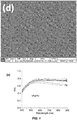

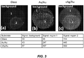

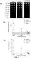


Classifications
-
- G—PHYSICS
- G01—MEASURING; TESTING
- G01N—INVESTIGATING OR ANALYSING MATERIALS BY DETERMINING THEIR CHEMICAL OR PHYSICAL PROPERTIES
- G01N33/00—Investigating or analysing materials by specific methods not covered by groups G01N1/00 - G01N31/00
- G01N33/48—Biological material, e.g. blood, urine; Haemocytometers
- G01N33/50—Chemical analysis of biological material, e.g. blood, urine; Testing involving biospecific ligand binding methods; Immunological testing
- G01N33/53—Immunoassay; Biospecific binding assay; Materials therefor
- G01N33/543—Immunoassay; Biospecific binding assay; Materials therefor with an insoluble carrier for immobilising immunochemicals
- G01N33/54313—Immunoassay; Biospecific binding assay; Materials therefor with an insoluble carrier for immobilising immunochemicals the carrier being characterised by its particulate form
- G01N33/54346—Nanoparticles
-
- C—CHEMISTRY; METALLURGY
- C12—BIOCHEMISTRY; BEER; SPIRITS; WINE; VINEGAR; MICROBIOLOGY; ENZYMOLOGY; MUTATION OR GENETIC ENGINEERING
- C12Q—MEASURING OR TESTING PROCESSES INVOLVING ENZYMES, NUCLEIC ACIDS OR MICROORGANISMS; COMPOSITIONS OR TEST PAPERS THEREFOR; PROCESSES OF PREPARING SUCH COMPOSITIONS; CONDITION-RESPONSIVE CONTROL IN MICROBIOLOGICAL OR ENZYMOLOGICAL PROCESSES
- C12Q1/00—Measuring or testing processes involving enzymes, nucleic acids or microorganisms; Compositions therefor; Processes of preparing such compositions
- C12Q1/68—Measuring or testing processes involving enzymes, nucleic acids or microorganisms; Compositions therefor; Processes of preparing such compositions involving nucleic acids
- C12Q1/6813—Hybridisation assays
- C12Q1/6834—Enzymatic or biochemical coupling of nucleic acids to a solid phase
-
- G—PHYSICS
- G01—MEASURING; TESTING
- G01N—INVESTIGATING OR ANALYSING MATERIALS BY DETERMINING THEIR CHEMICAL OR PHYSICAL PROPERTIES
- G01N21/00—Investigating or analysing materials by the use of optical means, i.e. using sub-millimetre waves, infrared, visible or ultraviolet light
- G01N21/62—Systems in which the material investigated is excited whereby it emits light or causes a change in wavelength of the incident light
- G01N21/63—Systems in which the material investigated is excited whereby it emits light or causes a change in wavelength of the incident light optically excited
- G01N21/64—Fluorescence; Phosphorescence
- G01N21/645—Specially adapted constructive features of fluorimeters
- G01N21/648—Specially adapted constructive features of fluorimeters using evanescent coupling or surface plasmon coupling for the excitation of fluorescence
-
- G—PHYSICS
- G01—MEASURING; TESTING
- G01N—INVESTIGATING OR ANALYSING MATERIALS BY DETERMINING THEIR CHEMICAL OR PHYSICAL PROPERTIES
- G01N33/00—Investigating or analysing materials by specific methods not covered by groups G01N1/00 - G01N31/00
- G01N33/48—Biological material, e.g. blood, urine; Haemocytometers
- G01N33/50—Chemical analysis of biological material, e.g. blood, urine; Testing involving biospecific ligand binding methods; Immunological testing
- G01N33/58—Chemical analysis of biological material, e.g. blood, urine; Testing involving biospecific ligand binding methods; Immunological testing involving labelled substances
- G01N33/582—Chemical analysis of biological material, e.g. blood, urine; Testing involving biospecific ligand binding methods; Immunological testing involving labelled substances with fluorescent label
-
- G—PHYSICS
- G01—MEASURING; TESTING
- G01N—INVESTIGATING OR ANALYSING MATERIALS BY DETERMINING THEIR CHEMICAL OR PHYSICAL PROPERTIES
- G01N33/00—Investigating or analysing materials by specific methods not covered by groups G01N1/00 - G01N31/00
- G01N33/48—Biological material, e.g. blood, urine; Haemocytometers
- G01N33/50—Chemical analysis of biological material, e.g. blood, urine; Testing involving biospecific ligand binding methods; Immunological testing
- G01N33/68—Chemical analysis of biological material, e.g. blood, urine; Testing involving biospecific ligand binding methods; Immunological testing involving proteins, peptides or amino acids
- G01N33/6854—Immunoglobulins
-
- Y—GENERAL TAGGING OF NEW TECHNOLOGICAL DEVELOPMENTS; GENERAL TAGGING OF CROSS-SECTIONAL TECHNOLOGIES SPANNING OVER SEVERAL SECTIONS OF THE IPC; TECHNICAL SUBJECTS COVERED BY FORMER USPC CROSS-REFERENCE ART COLLECTIONS [XRACs] AND DIGESTS
- Y02—TECHNOLOGIES OR APPLICATIONS FOR MITIGATION OR ADAPTATION AGAINST CLIMATE CHANGE
- Y02A—TECHNOLOGIES FOR ADAPTATION TO CLIMATE CHANGE
- Y02A50/00—TECHNOLOGIES FOR ADAPTATION TO CLIMATE CHANGE in human health protection, e.g. against extreme weather
- Y02A50/30—Against vector-borne diseases, e.g. mosquito-borne, fly-borne, tick-borne or waterborne diseases whose impact is exacerbated by climate change
Definitions
- a widely varying set of techniques may be used to detect biological molecules, such as nucleic acids, proteins, antibodies, subcellular vesicles, cells, and tissues, depending on the molecule and application. Fluorescence based detection has become one of the leading sensing technologies in biomedical, biological and related sciences, for example, flow cytometry, gene sequencing, immunoassay, immunohistochemical imaging (IHC) and immunofluorescence imaging, single molecule imaging and fluorescence in situ hybridization (FISH).
- IHC immunohistochemical imaging
- FISH fluorescence in situ hybridization
- detection of the desired biological molecule may be limited, depending on the method, by the intensity of the fluorescence output by the probing fluorescent molecule bound to the biological molecule.
- immunoassay techniques such as enzyme-linked immunosorbent assay (ELISA)
- ELISA enzyme-linked immunosorbent assay
- the detection limit of proteins and antibodies is approximately 1 nanogram/ml (ng/ml) and the dynamic range of ELISA only spans ⁇ 3 orders of magnitude.
- biomarkers such as cytokines and antibodies may be well below 1 ng/ml in biological fluids including whole blood, plasma, serum, and saliva at early disease stages, including prostate-specific antigen (PSA) for early stage cancer diagnosis and troponin I (cTnI) for cardiovascular disease.
- PSA prostate-specific antigen
- cTnI troponin I
- Tabakman, et al. (Nature Communications, 2(3): 466, 2011 ) described plasmonic substrates for multiplexed protein microarrays with femtomolar sensitivity and broad dynamic range; US 2013/0172207 described fluorescence enhancing plasmonic nanoscopic gold films and assays based thereon; Fales, et al.
- the present disclosure provides methods and compositions for detecting fluorescent signals with high sensitivity, speed and signal-to-noise ratio.
- the methods and compositions provided herein may be used for a variety of biomedical imaging and assay applications with fluorescent enhancement in the visible and near-infrared region broadly across about 400 nanometers (nm) to about 2100 nm.
- the invention provides a film comprising raised nanostructures on a substrate.
- the nanostructures of the film comprise silver-on-gold nanoparticles; the nanostructures are separated from one another by gaps; and intensity of a fluorescent signal from a fluorophore in proximity to the film is enhanced relative to the fluorescent signal obtained from the fluorophore in proximity to the substrate in the absence of the film.
- the gaps have widths between 5 nm to 50 nm, and lengths between 5 nm and 1000 nm.
- the nanostructures have an average width and length between 50 nm to 500 nm, such as between 100 nm to 200 nm.
- the film has a nanoplate size of between 1000 nm 2 to 250,000 nm 2 .
- the film can comprise irregular features and a heterogenous structure.
- the height of the film is between 5 nm and 500 nm.
- the film can be quasi-continuous through a percolating path and conducting based on electron microscopy imaging and electrical conductivity, or discontinuous based on electron microscopy imaging and electrical conductivity.
- the fluorescent signal that is enhanced can be in the range of 400 nm to 2100 nm. In some embodiments, the fluorescent signal is enhanced for fluorophores within 1000 nm of the surface of the film.
- the fluorophore is a near-infra-red fluorophore (e.g. IR680 or IR800) having an emission of about 700 nm to about 800 nm, and the intensity of the fluorescent signal is enhanced by at least 30-fold (e.g. at least 100-fold).
- the fluorophore is a visible dye (e.g. DAPI, Alexa488, Cy3, or Cy5) having an emission of about 400 nm to about 700 nm, and the intensity of the fluorescent signal is enhanced by at least 3-fold (e.g. at least 30-fold).
- the substrate can comprise one or more materials selected from the group consisting of: glass, polystyrene, quartz, silica, nylon, nitrocellulose, polyvinyl chloride, polydimethyl siloxane, polyvinylidene fluoride, polydopamine, silicon, silicon dioxide, a polymer, iron oxide, and a plastic.
- the substrate can comprise a flat surface, a curved surface, a spherical surface, or a three-dimensional porous membrane.
- the substrate is a bead, such as a bead having a diameter ranging from 0.05 microns to 200 microns.
- the bead may or may not have a magnetic or paramagnetic core.
- the bead can be in a container, such as a well in a multi-well plate (e.g. a 96- or 384-well plate).
- a container such as a well in a multi-well plate (e.g. a 96- or 384-well plate).
- the substrate itself is the interior of a well plate, such as a well in a multi-well plate (e.g. a 96- or 384-well plate)
- a film of the present invention can further comprise an array of binding elements on the film, wherein the binding elements bind to an analyte.
- the array can comprise a plurality of different binding elements, each of which binds a different analyte.
- the binding elements are selected from the group consisting of: proteins, antibodies, antigen-binding antibody fragments, cells, aptamers, peptides, polynucleotides, exosomes, and tissue slices.
- the binding elements can be antigens for detecting antibodies in a sample.
- the antibodies bound by the antigens may be one or more selected from the group consisting of: total human IgG, IgM, IgA and IgE ; anti-HLA (human leukocyte antigen) antibodies; anti-dsDNA antibodies; anti-Smith antibodies; antibodies diagnostic of Systemic Lupus Erythematosus (SLE), such as anti-nucleosome, anti U1RNP, and anti-PO antibodies; antibodies diagnostic of cardiovascular disease (e.g.
- dilated cardiomyopathy Toxoplasmosis, Rubella, Rabies, Malaria, lyme disease, African Trypanosomiasis, cholera, cryptosporidiosis, dengue, influenza, Japanese Encephalitis, Leishmaniasis, measles, meningitis, onchocerciasis, pneumonia, tuberculosis, typhoid, or yellow fever; antibodies specific for CMV, HSV-1/2, HBA, HBV, HCV, HDV, HIV; HPV, Ebola virus, rotavirus, human leukocyte antigens, Thyroid Stimulating Hormone Receptor (TSHR), thyroperoxidate, Thyroglobulin, tissue transglutaminase (tTG), endomysium, deamidated gliadin peptide; and antibodies specific for tumor-associated antigens selected from p53, NY-ESO-1, MAGE A4, HuD, CAGE, GBU4-5, and SOX2.
- the analyte can be a protein, an antibody, a peptide, a nucleic acid, an enzyme, a cell, an exosome, a cell free DNA, or a tissue.
- the analyte can be a biomarker for a condition of a subject.
- examples of analytes include an inflammatory cytokine, a biomarker for cardiovascular disease (e.g. troponin I (cTnI), c-reactive protein (CRP), NT-ProBNP, or an antibody to Annexin A5, SDHA, ATP1A3, titin, myosin, ADBRK, EDNRA, EDNRB, AGTR1, CHRM2, or HSPD), a biomarker for infectious disease (e.g.
- HBV hepatitis B virus
- HBV surface antigen HBV surface antigen
- Dengue NS1 antigen antibodies to Toxoplasmosis gondii, Rubella, HCV, HIV, syphilis, or HSV
- a biomarker for inflammatory bowel disease or a biomarker for cancer
- cancer e.g. prostate-specific antigen (PSA), carcinoembryonic antigen (CEA), cancer antigen-125 (CA125), AFP, SCC, CA19-9, CA242, NSE, Cyfa21-1, CA15-3, and total T-PSA.
- the antigens comprise one or more Toxoplasmosis gondii antigens, one or more Rubella antigens, one or more CMV antigens, and one or more HSV antigens.
- the disclosure provides methods for making films of the present disclosure, such as films comprising raised nanostructures comprising silver-on-gold nanoparticles.
- the method comprises adsorbing gold (Au) nanoparticle seeds on a substrate, or growing Au nanoparticle seeds in solution or vapor phase on a substrate; and growing silver nanostructures around the gold nanoparticle seeds, such as in the solution phase.
- Au gold
- the film may have any combination of characteristics of films disclosed herein.
- the invention provides a method of detecting an analyte.
- the method comprises providing a film comprising raised nanostructures on a substrate, wherein nanostructures of the film comprise silver-on-gold nanoparticles, and the nanostructures are separated from one another by gaps as defined in the claims; applying to the film an analyte and a label for the analyte, wherein the label comprises a fluorophore; and detecting the analyte by detecting a fluorescent signal of the fluorophore, wherein intensity of the fluorescent signal is enhanced relative to the fluorescent signal of the fluorophore in the absence of the film.
- the gaps have widths between 5 nm to 50 nm, and lengths between 5 nm and 200 nm.
- the film can have any one or more the following characteristics: the nanostructures have an average width and length between 50 nm to 500 nm; the film has a nanoplate size of between 1000 nm 2 to 250,000 nm 2 ; the height of the film is between 5 nm and 500 nm; the film comprises irregular features and a heterogenous structure; the film imparts a plasmon from about 400 nm to about 2100 nm; the substrate comprises a flat surface, a curved surface, a spherical surface, or a three-dimensional porous membrane; and the substrate is a bead.
- the fluorescent signal that is enhanced can be in the range of 400 nm to 2100 nm.
- the fluorescent signal is enhanced for fluorophores within 1000 nm of the surface of the film.
- the fluorophore is a near-infra-red fluorophore (e.g. IR680 or IR800) having an emission of about 700 nm to about 800 nm, and the intensity of the fluorescent signal is enhanced by at least 30-fold (e.g. at least 100-fold).
- the fluorophore is a visible dye (e.g. DAPI, Alexa488, Cy3, or Cy5) having an emission of about 400 nm to about 700 nm, and the intensity of the fluorescent signal is enhanced by at least 3-fold (e.g. at least 30-fold).
- the method can further comprise determining concentration, identity, or location of the analyte based on detecting the fluorescent signal.
- the analyte is bound to the label
- the analyte is on a surface (e.g. bound to the surface by a binding element conjugated to the surface)
- step (b) comprises applying the film to the analyte on the surface.
- the surface can be a DNA microarray, an RNA microarray, a miRNA microarray, a peptide microarray, a protein microarray, an antigen microarray, or an antibody microarray.
- Imaging can include imaging by a microscope or a scanner.
- the film can further comprise an array of binding elements on the film, wherein the binding elements bind to the analyte.
- Binding elements can include any one or more selected from the group consisting of proteins, antibodies, antigen-binding antibody fragments, cells, exosomes, cell free DNA, aptamers, and nucleic acids. In some cases, the binding elements are antigens for detecting antibodies in a sample.
- the analyte can be a protein, an antibody, a peptide, a nucleic acid, an enzyme, a cell, or a tissue.
- the analyte can be a biomarker for a condition of a subject.
- the analyte is an inflammatory cytokine, a biomarker for cardiovascular disease (e.g. troponin I, c-reactive protein (CRP), NT-ProBNP, or an antibody to Annexin A5, SDHA, ATP1A3, titin, myosin, ADBRK, EDNRA, EDNRB, AGTR1, CHRM2, or HSPD), a biomarker for infectious disease (e.g.
- a biomarker for cardiovascular disease e.g. troponin I, c-reactive protein (CRP), NT-ProBNP, or an antibody to Annexin A5, SDHA, ATP1A3, titin, myosin, ADBRK, EDNRA, EDNRB, AGTR1, CHRM2, or HSPD
- a biomarker for infectious disease e.g.
- HBV hepatitis B virus
- HBV surface antigen HBV surface antigen
- Dengue NS1 antigen antibodies to one or more of Toxoplasmosis gondii, Rubella, HCV, HSV, HIV, Syphilis, and CMV
- a biomarker for cancer e.g. prostate-specific antigen (PSA), carcinoembryonic antigen (CEA), cancer antigen-125 (CA125) AFP, SCC, CA19-9, CA242, NSE, Cyfa21-1, CA15-3, or total T-PSA).
- PSA prostate-specific antigen
- CEA carcinoembryonic antigen
- CA125 cancer antigen-125
- the array can comprise a plurality of different binding elements conjugated to the film at known locations, each of which binds a different analyte; and the method further comprises identifying an analyte based on the location of a detected fluorescent signal.
- the binding element is an oligonucleotide conjugated to the film
- the substrate is a bead
- the analyte is a target polynucleotide that hybridizes to the oligonucleotide via sequence complementarity or an amplification product thereof.
- the binding element is an oligonucleotide
- the oligonucleotide can be a primer
- detection can comprise amplifying the target polynucleotide to produce a detectable amplified product.
- a plurality of different analytes and a corresponding plurality of different labels are applied to the film, and each of the different labels is detected in a single assay.
- the analyte is from a sample of a subject.
- the method can further comprise identifying a phenotype of a cell in the sample (e.g. cancer) based on detecting the fluorescent signal (e.g. through immunohistochemical (IHC) staining).
- IHC immunohistochemical
- Detecting can comprise single-molecule imaging and tracking, or single-nanoparticle imaging and tracking.
- the fluorophore can be a member of a Fluorescence Resonance Energy Transfer (FRET) pair, and fluorescent signals for one or both members of the pair can be enhanced by the film.
- the label is a fluorescence in situ hybridization (FISH) probe.
- Methods of detecting one or more analytes may further comprise diagnosing a subject as having a condition associated with the presence, absence, or level of the one or more analytes, such as a condition described herein. Methods may further comprise taking medical action on the basis of detecting one or more analytes, and/or a resulting diagnosis.
- the disclosure provides a method of sequencing a polynucleotide.
- the method comprises (a) providing a film comprising raised nanostructures on a substrate, wherein nanostructures of the film comprise silver-on-gold nanoparticles or gold-on-gold nanoparticles, and the nanostructures are separated from one another by gaps; (b) hybridizing an oligonucleotide to a target polynucleotide; (c) extending the oligonucleotide with one or more bases complementary to corresponding positions on the target polynucleotide in the direction of extension; and (d) identifying the one or more bases added in step (c) by detecting a fluorescent signal of one or more fluorophores; wherein intensity of the fluorescent signal is enhanced by the film relative to the fluorescent signal of the fluorophore in the absence of the film.
- Extension in step (c) can comprise extension by a polymerase, or extension by a ligase.
- the film may be on a plurality of beads, which may optionally be flowing through or conjugated to a flow cell.
- the film may have any one or more characteristics of films described herein.
- the disclosure provides a composition comprising beads distributed in or on a porous substrate.
- the beads have bead surfaces, and on the bead surfaces is a film comprising raised nanostructures; the nanostructures comprise silver-on-gold nanoparticles, or gold-on-gold nanoparticles; the nanostructures are separated from one another by gaps; and intensity of a fluorescent signal from a fluorophore in proximity to the film is enhanced relative to the fluorescent signal obtained from the fluorophore in proximity to the substrate in the absence of the film.
- the composition may be further characterized by any one or more of the following: (a) the beads have a magnetic core; (b) the beads have a diameter between 0.05 microns to 200 microns; (c) the porous substrate is a porous membrane material; and/or (d) the film comprises a plurality of binding elements on the film.
- the porous substrate can be a porous membrane material comprising nitrocellulose, glass fiber, nylon, or cellulose acetate.
- the film may comprise a plurality of binding elements on the film, and the binding elements are selected from the group consisting of proteins, antibodies, antigen-binding antibody fragments, cells, aptamers, and nucleic acids.
- the film may have any one or more characteristics of films described herein.
- the disclosure provides a bead on which is a film comprising raised nanostructures.
- the nanostructures comprise silver-on-gold nanoparticles, or gold-on-gold nanoparticles; the nanostructures are separated from one another by gaps; a plurality of fluorophores are conjugated to the bead or to the film; and intensity of a fluorescent signal from the fluorophore is enhanced relative to the fluorescent signal obtained from the fluorophore on the bead in the absence of the film.
- the bead may further comprise one or more binding element conjugated to the bead or to the film. A plurality of such beads may be employed in a method for labeling a target.
- the method can comprise contacting the target with a plurality of the beads, wherein the binding element binds to the target.
- the film may have any one or more characteristics of films described herein.
- the bead has one or more of (e.g.
- the bead has a size in the range of 0.01-10 microns;
- the bead comprises a magnetic coating underlying the film, optionally wherein the magnetic coating comprises iron oxide;
- a plurality of binding elements are complexed to the bead, wherein the binding elements bind an analyte, optionally wherein the analyte is an exosome, a protein, an antibody, a polynucleotide (e.g. DNA or RNA), or a cell-free polynucleotide (e.g. DNA or RNA); and
- the bead comprises a polymer or silica core.
- the disclosure provides a bead on which is a film comprising raised nanostructures.
- the nanostructures comprise silver-on-gold nanoparticles, or gold-on-gold nanoparticles; the nanostructures are separated from one another by gaps; a plurality of binding elements are conjugated to the bead or to the film; and intensity of a fluorescent signal from a fluorophore complexed to the bead is enhanced relative to the fluorescent signal obtained from the fluorophore complexed to the bead in the absence of the film.
- the bead has one or more of (e.g.
- the bead has a size in the range of 0.01-10 microns;
- the bead comprises a magnetic coating underlying the film, optionally wherein the magnetic coating comprises iron oxide;
- a plurality of binding elements are complexed to the bead, wherein the binding elements bind an analyte, optionally wherein the analyte is an exosome, a protein, an antibody, a polynucleotide (e.g. DNA or RNA), or a cell-free polynucleotide (e.g. DNA or RNA); and
- the bead comprises a polymer or silica core.
- the film may have any one or more characteristics of films described herein.
- the film has one or more of the following characteristics: the gaps have widths between 5 nm to 50 nm, and lengths between 5 nm and 200 nm; the nanostructures have an average width and length between 50 nm to 500 nm; the film has a nanoplate size of between 1000 nm 2 to 250,000 nm 2 ; the height of the film is between 5 nm and 500 nm; the film comprises irregular features and a heterogenous structure; and the film imparts a plasmon from about 400 nm to about 2100 nm.
- a plurality of such beads may be employed in a method of detecting an analyte, such as an exosome or a polynucleotide (e.g.
- the method comprises (a) combining the beads with a sample comprising the analyte, wherein the analyte binds to the binding element; (b) labeling the bound analyte with a label that binds the analyte, wherein the label comprises a fluorophore; and (c) detecting the bound analyte by detecting a fluorescent signal from the fluorophore; wherein intensity of the fluorescent signal is enhanced relative to the fluorescent signal of the fluorophore in the absence of the film.
- the analyte is an exosome and the label is an antibody that binds a surface antigen of the exosome; or (b) the analyte is a cell-free DNA and the label is an intercalating dye.
- the disclosure provides an apparatus comprising a film on a film substrate, and an analyte receptacle.
- the film comprises raised nanostructures, wherein the raised nanostructures (i) comprise silver-on-gold nanoparticles, or gold-on-gold nanoparticles, and (ii) are separated from one another by gaps;
- the analyte receptacle is configured to retain an analyte in contact with a first surface of the analyte receptacle;
- the film has a film surface that is in contact with a second surface of the analyte receptacle or is spaced within 1000nm of the first surface of the analyte receptacle, wherein the second surface is located opposite the first surface; and (d) intensity of a fluorescent signal from a fluorophore associated with the analyte is enhanced relative to the fluorescent signal obtained from the fluorophore in the absence of the
- the film comprises raised nanostructures, wherein the raised nanostructures (i) comprise silver-on-gold nanoparticles, or gold-on-gold nanoparticles, and (ii) are separated from one another by gaps;
- the film substrate is a bead in the analyte receptacle;
- the analyte receptacle is configured to retain an analyte and the bead; and
- intensity of a fluorescent signal from a fluorophore in proximity to the film is enhanced relative to the fluorescent signal obtained from the fluorophore in proximity to the substrate in the absence of the film.
- the apparatus may be further characterized in that: (a) the beads have a magnetic core; (b) the beads have a diameter between 0.05 microns to 200 microns; (c) the film comprises a plurality of binding elements on the film; and/or (d) the analyte receptacle is a well of a multi-well plate.
- the film can have any one or more of the following characteristics: the gaps have widths between 5 nm to 50 nm, and lengths between 5 nm and 200 nm; the nanostructures have an average width and length between 50 nm to 500 nm; the film has a nanoplate size of between 1000 nm 2 to 250,000 nm 2 ; the height of the film is between 5 nm and 500 nm; the film comprises irregular features and a heterogenous structure; and the film imparts a plasmon from about 400 nm to about 2100 nm.
- the apparatus may be provided for use in a method of detecting an analyte, which method may further comprise applying to the analyte receptacle an analyte and a label for the analyte, wherein the label comprises a fluorophore; and detecting the analyte by detecting a fluorescent signal of the fluorophore, wherein intensity of the fluorescent signal is enhanced relative to the fluorescent signal of the fluorophore in the absence of the film.
- the film may have any one or more characteristics of films described herein.
- the disclosure provides a method of detecting one or more antibodies in a sample.
- the method comprises: (a) providing a film comprising raised nanostructures on a substrate, wherein (i) nanostructures of the film comprise silver-on-gold nanoparticles or gold-on-gold nanoparticles, (ii) the nanostructures are separated from one another by gaps, and (iii) a plurality of antigens are complexed to a surface of the film; (b) contacting the plurality of antigens with a sample and one or more labels for detecting the one or more antibodies bound to an antigen of the plurality of antigens, wherein each label comprises a fluorophore; and (c) detecting the one or more bound antibodies by detecting a fluorescent signal of the fluorophore, wherein intensity of the fluorescent signal is enhanced relative to the fluorescent signal of the fluorophore in the absence of the film; wherein the plurality of antigens comprises one or more Toxoplasmo
- the plurality of antigens further comprise one or more HIV antigens, and/or one or more Syphilis antigens.
- the one or more antibodies are IgG antibodies, and the one or more labels are anti-IgG antibodies.
- the one or more antibodies comprise one or more antibody subtypes selected from IgG, IgM, IgA, and IgE;
- the one or more labels comprise a plurality of corresponding label antibodies selected from (and optionally includes all of) anti-IgG, anti-IgM, anti IgA, and anti-IgE antibodies;
- the fluorophore of a label antibody corresponding to one subtype is different from the fluorophore corresponding to any other subtype (e.g. selected from Cy3, Cy5, IR680, and IR800), and
- the method further comprises identifying or quantifying the different subtypes based on the fluorescent signal.
- the plurality of antigens are arranged at known locations in an array, and the method further comprises identifying the one or more bound antibodies based on the location of the fluorescent signal in the array.
- the film and substrate can be any of those described herein.
- the film comprises silver-on-gold nanoparticles.
- the film has one or more of the following characteristics: the gaps have widths between 5 nm to 50 nm, and lengths between 5 nm and 200 nm; the nanostructures have an average width and length between 50 nm to 500 nm; the film has a nanoplate size of between 1000 nm 2 to 250,000 nm 2 ; the height of the film is between 5 nm and 500 nm; the film comprises irregular features and a heterogenous structure; the film imparts a plasmon from about 400 nm to about 2100 nm; the substrate comprises a flat surface, a curved surface, a spherical surface, or a three-dimensional porous membrane; and the substrate is a bead.
- the fluorophore is a near-infra-red fluorophore having an emission of about 700 nm to about 800 nm, and the intensity of the fluorescent signal is enhanced by at least 30-fold.
- the fluorophore is a visible dye having an emission of about 400 nm to about 700 nm, and the intensity of the fluorescent signal is increased by at least 3-fold.
- the method further comprises determining concentration, identity, and/or location of the analyte based on detecting the fluorescent signal.
- the sample is a whole blood, plasma, serum, saliva, or urine sample having a volume between 1-100 ⁇ L.
- the sample is a whole blood sample having a volume between 1-10 ⁇ L, optionally diluted in a diluent solution to a total volume of 100 ⁇ L or less.
- a cell includes a plurality of cells, including mixtures thereof.
- polynucleotide refers to a polymeric form of nucleotides of any length, either deoxyribonucleotides or ribonucleotides, or analogs thereof.
- Polynucleotides may have any three dimensional structure, and may perform any function, known or unknown.
- polynucleotides coding or non-coding regions of a gene or gene fragment, loci (locus) defined from linkage analysis, exons, introns, messenger RNA (mRNA), transfer RNA, ribosomal RNA, short interfering RNA (siRNA), short-hairpin RNA (shRNA), micro-RNA (miRNA), ribozymes, cDNA, recombinant polynucleotides, branched polynucleotides, plasmids, vectors, isolated DNA of any sequence, isolated RNA of any sequence, nucleic acid probes, and primers.
- loci locus defined from linkage analysis, exons, introns, messenger RNA (mRNA), transfer RNA, ribosomal RNA, short interfering RNA (siRNA), short-hairpin RNA (shRNA), micro-RNA (miRNA), ribozymes, cDNA, recombinant polynucleotides, branched poly
- a polynucleotide may comprise one or more modified nucleotides, such as methylated nucleotides and nucleotide analogs. If present, modifications to the nucleotide structure may be imparted before or after assembly of the polymer. The sequence of nucleotides may be interrupted by non-nucleotide components. A polynucleotide may be further modified after polymerization, such as by conjugation with a labeling component.
- a nucleotide analog may be an analog or mimic of a primary nucleotide having modification on the primary nucleobase (A, C, G, T and U), the deoxyribose/ribose structure, the phosphate group of the primary nucleotide, or any combination thereof.
- a nucleotide analog can have a modified base, either naturally existing or man-made.
- modified bases include, without limitation, methylated nucleobases, modified purine bases (e.g. hypoxanthine, xanthine, 7-methylguanine, isodG), modified pyrimidine bases (e.g. 5,6-dihydrouracil and 5-methylcytosine, isodC), universal bases (e.g. 3-nitropyrrole and 5-nitroindole), non-binding base mimics (e.g.
- nucleotide analogs having modified deoxyribose e.g. dideoxynucleosides such as dideoxyguanosine, dideoxyadenosine, dideoxythymidine, and dideoxycytidine
- phosphate structure (together referred to as the backbone structure) includes, without limitation, glycol nucleotides, morpholinos, and locked nucleotides.
- fluorescence refers to the process wherein a molecule relaxes to its ground state from an excited state by emission of a photon.
- fluorescence can also encompass phosphorescence.
- a molecule is promoted to an electronically excited state generally by the absorption of ultraviolet, visible, or near infrared radiation. The excited molecule then decays back to the ground state, or to a lower-lying excited electronic state, by emission of light.
- plasmonically active in reference to a material refers to a material which supports plasmons (e.g., surface plasmons), thereby exhibiting plasmonic properties.
- Surface plasmons may be used to enhance the surface sensitivity of several spectroscopic measurements including fluorescence, Raman scattering, and second harmonic generation.
- plasmonic properties refers to properties exhibited by surface plasmons, or the collective oscillations of electrical charge on the surfaces of metals. In this sense, plasmonic properties refers to measurable properties, such as properties described in Nagao et al. "Plasmons in nanoscale and atomic-scale systems," Sci. Technol. Adv. Mater. 11 (2010) 054506 (12pp ), describing plasmonic sensors, such as those used for surface-enhanced IR absorption spectroscopy (SEIRA), surface-enhanced Raman scattering (SERS). Another plasmonic property is plasmon-enhanced fluorescence, described e.g. in Sensors and Actuators B 107 (2005) 148-153 .
- the term “continuous” refers to the inter-connectivity of nanostructures in plasmonic metal film, creating an electrically conductive path, optionally with gaps existing between some of the nanostructures that are not in the conducting path.
- discontinuous refers to the presence of one or more isolated nanostructures in plasmonic metal film, where the nanostructures are separated from each other and not interconnected.
- proximity refers to a distance between a fluorescent molecule and nanostructures in plasmonic metal film, within which distance the fluorescence intensity of the fluorescent molecule increases, such as by a specified fold-increase. In some cases, proximity may be measured in angstroms (e.g. within 1-1000 angstroms), in nanometers (e.g. within 1-1000 nm), or millimeters (e.g. within 1-10 mm). In some cases, fluorescent intensity is enhanced for fluorophores within about 1000 nm of the surface of the film.
- compositions, systems and methods that are capable of amplifying or enhancing fluorescent signals, such as signals spanning from about 400 nm to about 2100 nm.
- the compositions and systems may be able to facilitate fluorescence detection with improved detection limits, sensitivity, speed, specificity and signal-to-noise ratio, thereby finding use in a wide context of applications that rely on fluorescence as a detection method.
- the present disclosure provides a film comprising raised nanostructures on a substrate.
- the film may be further characterized in that the nanostructures of the film comprise silver-on-gold nanoparticles; the nanostructures are separated from one another by gaps; and intensity of a fluorescent signal from a fluorophore in proximity to the film is enhanced relative to the fluorescent signal obtained from the fluorophore in proximity to the substrate in the absence of the film.
- the film may be applied directly or indirectly to the substrate.
- the nanostructures comprise a first metal on nanoparticles of a second metal, where the first and second metal may be the same or different (e.g. gold-on-gold, or silver-on-gold).
- the nanostructures may be formed by at least one type of noble metals, for example, ruthenium (Ru), rhodium (Rh), palladium (Pd), silver (Ag), osmium (Os), iridium (Ir), platinum (Pt), and gold (Au).
- the nanostructures can take various shapes, e.g., sphere, cube, cuboid, cone, cylinder, prism, pyramid, tube, plate, disc, rod, or any regular or irregular shapes.
- the nanostructures comprise nanoparticles.
- the metals can form a layered or core/shell structure, for example, a silver-on-gold nanoparticle.
- each of the nanostructures has the same shape. In some cases, it may be preferred to have nanostructures of different shapes (or heterogeneous).
- Sizes may vary, depending upon, applications that the nanostructures are used for. For example, it may be preferred to have nanostructures that are much smaller than the wavelength of light used for fluorescence excitation and emission.
- the nanostructures may have a width, length, and/or height of less than or equal to about 1 millimeter (mm), such as less than or equal to about 750 micron ( ⁇ m), 500 ⁇ m, 250 ⁇ m, 100 ⁇ m, 75 ⁇ m, 50 ⁇ m, 25 ⁇ m, 10 ⁇ m, 5 ⁇ m, 1 ⁇ m, 900 nm, 800 nm, 700 nm, 600 nm, 500 nm, 400 nm, 300 nm, 200 nm, 100 nm, 90 nm, 80 nm, 70 nm, 60 nm, 50 nm, 40 nm, 30 nm, 20 nm, 10 nm, 9 nm, 8 nm, 7 nm, 6 nm, 5 nm, 4 nm, 3 nm, 2 nm, 1 nm, 800 picometers (pm), 600 pm.
- mm millimeter
- ⁇ m micron
- the width, length, and/or height of the nanostructures may be greater than or equal to about 1 pm, 5 pm, 10 pm, 25 pm, 50 pm, 75 pm, 100 pm, 250 pm, 500 pm, 750 pm, 1 nm, 2nm, 3 nm, 4 nm, 5 nm, 6nm, 7 nm, 8 nm, 9 nm, 10 nm, 15 nm, 20 nm, 30 nm, 40 nm, 50 nm, 60 nm, 70 nm, 80 nm, 90 nm, 100 nm, 150 nm, 200 nm, 250 nm, 300 nm, 350 nm, 400 nm, 450 nm, 500 nm, 600 nm, 700 nm, 800 nm, 900 nm, 1 ⁇ m, 5 ⁇ m, 10 ⁇ m, 30
- the width, length, and/or height of the nanostructures may be between any of the two values described herein, for example, the nanostructures may have an average width and length between about 50 nm to about 500 nm, or between about 100 nm to about 200 nm. As a further example, the film may have a height of between 5 nm to 500 nm.
- the nanostructures may have a cross-sectional area of at least about 0.001 nm 2 , 0.005 nm 2 , 0.01 nm 2 , 0.05 nm 2 , 0.075 nm 2 , 0.1 nm 2 , 0.5 nm 2 , 0.75 nm 2 , 1 nm 2 , 10 nm 2 , 20 nm 2 , 40 nm 2 , 60 nm 2 , 80 nm 2 , 100 nm 2 , 250 nm 2 , 500 nm 2 , 750 nm 2 , 1,000 nm 2 , 2,500 nm 2 , 5,000 nm 2 , 7,500 nm 2 , 10,000 nm 2 , 25,000 nm 2 , 50,000 nm 2 , 75,000 nm 2 , 100,000 nm 2 , 200,000 nm 2 , 300,000 nm 2 , 400,000 nm 2 , 500,000 nm
- the nanostructures may have a cross-sectional area of less than or equal to about 25,000,000 nm 2 , 10,000,000 nm 2 , 8,000,000 nm 2 , 6,000,000 nm 2 , 4,000,000 nm 2 , 2,000,000 nm 2 , 1,000,000 nm 2 , 800,000 nm 2 , 600,000 nm 2 , 500,000 nm 2 , 450,000 nm 2 , 400,000 nm 2 , 350,000 nm 2 , 300,000 nm 2 , 250,000 nm 2 , 200,000 nm 2 , 150,000 nm 2 , 100,000 nm 2 , 80,000 nm 2 , 60,000 nm 2 , 50,000 nm 2 , 40,000 nm 2 , 30,000 nm 2 , 20,000 nm 2 , 10,000 nm 2 , 8,000 nm 2 , 6,000 nm 2 , 4,000 nm 2 , 2,000 nm 2
- the cross-sectional area of the nanostructures may fall between any of the two values described herein, for example, between about 100 nm 2 to about 250,000 nm 2 , or between about 1,000 nm 2 to about 250,000 nm 2 .
- each of the nanostructures comprised in the film may have the same shape, structure, and/or size.
- the nanostructures may be of varied size, shape, and/or structure. Depending on the application, it may be preferred that a certain percentage of the nanostructures have the same or a different size, shape, and/or structure, for example, about 1%, 5%, 10%, 15%, 20%, 30%, 40%, 50%, 60%, 70%, 75%, 80%, 85%, 90%, 91%, 92%, 93%, 94%, 95%, 96%, 97%, 98%, 99%, or 100% of the nanostructures may have the same or a different size, shape, and/or structure.
- Nanostructures may or may not be separated from each other. In cases where the nanostructures are separated from one another, they may be separated by gaps. Gap distance, a distance between the nanostructures, may vary. In some cases, a large gap distance may be created. In other cases, a small gap distance may be used. Large and small gaps can be constructed at different locations within the same film.
- the gap distances or an average gap distance may be less than or equal to about 1 mm, such as less than or equal to about 750 ⁇ m, 500 ⁇ m, 250 ⁇ m, 100 ⁇ m, 75 ⁇ m, 50 ⁇ m, 25 ⁇ m, 10 ⁇ m, 7.5 ⁇ m, 5 ⁇ m, 2.5 ⁇ m, 1 ⁇ m, 900 nm, 800 nm, 700 nm, 600 nm, 500 nm, 400 nm, 300 nm, 200 nm, 100 nm, 90 nm, 80 nm, 70 nm, 60 nm, 50 nm, 40 nm, 30 nm, 20 nm, 10 nm, 9 nm, 8 nm, 7 nm, 6 nm, 5 nm, 4 nm, 3 nm, 2 nm, 1 nm, 0.75 nm, 0.5 nm, 0.25 nm, 0.1 nm,
- the gap distance may be at least about 0.0001 nm, 0.0005 nm, 0.001 nm, 0.005 nm, 0.01 nm, 0.05 nm, 0.1 nm, 0.5 nm, 1 nm, 5 nm, 7.5 nm, 10 nm, 20 nm, 40 nm, 60 nm, 80 nm, 100 nm, 200 nm, 300 nm, 400 nm, 500 nm, 600 nm, 700 nm, 800 nm, 900 nm, 1 ⁇ m, 2 ⁇ m, 3 ⁇ m, 4 ⁇ m, 5 ⁇ m, 6 ⁇ m, 7 ⁇ m, 8 ⁇ m, 9 ⁇ m, 10 ⁇ m, 25 ⁇ m, 50 ⁇ m, 75 ⁇ m, 100 ⁇ m, 500 ⁇ m and 750 ⁇ m.
- the gap distance can be between any of the two values described herein, for example, between
- Gaps may be of varies widths and/or lengths.
- the gaps may have widths and/or lengths that are less than or equal to about 5,000 nm, 4,000 nm, 3,000 nm, 2,000 nm, 1,000 nm, 800 nm, 600 nm, 400 nm, 200 nm, 100 nm, 80 nm, 60 nm, 40 nm, 20 nm, 10 nm, 7.5 nm, 5 nm, 1 nm, 0.75 nm, 0.5 nm, 0.25 nm, 0.1 nm, 0.05 nm, 0.01 nm, 0.005 nm, or 0.001 nm.
- the widths and/or lengths of the gaps may be greater than or equal to about 0.005 nm, 0.0075 nm, 0.01 nm, 0.05 nm, 0.075 nm, 0.1 nm, 0.5 nm, 0.75 nm, 1 nm, 2.5 nm, 5 nm, 7.5 nm, 10 nm, 30 nm, 50 nm, 70 nm, 90 nm, 100 nm, 200 nm, 300 nm, 400 nm, 500 nm, 750 nm, 1,000 nm, 2,500 nm, or 5,000 nm.
- the gaps may have widths and/or lengths falling into a range between any of the two values described herein, for example, the gaps may have widths between about 5 nm to about 50nm, and lengths between about 5 nm to about 200 nm.
- a film can be continuous, quasi-continuous, or discontinuous, depending upon, the inter-connectivity of nanostructures comprised in the film.
- the film may be a continuous film, such that it comprises raised nanostructures that are interconnected, creating an electrically conductive path, with gaps existing between some of the nanostructures that are not in the conducting path.
- the film may be discontinuous, such that at least some portion of the film (e.g. at least 10%, 25%, 50%, 75%, 90%, or more) is comprised of nanostructures that are not connected by an electrically conducting path.
- a film as provided in the present disclosure can be determined and characterized by various techniques, such as, for example, electrical conductivity measurement, and/or microscopy techniques including standard light microscopy , transmission electron microscopy (TEM), confocal laser scanning microscopy, scanning electron microscopy (SEM) and atomic force microscopy (AFM).
- TEM transmission electron microscopy
- SEM scanning electron microscopy
- AFM atomic force microscopy
- a film that is quasi-continuous through a percolating path and conducting can be determined based on electron microscopy imaging and/or electrical conductivity.
- a discontinuous film may be characterized based on electron microscopy imaging and electrical conductivity.
- various materials may be used to fabricate a substrate.
- the materials can be organic or inorganic, synthetic or natural, solid or semi-solid.
- Non-limiting examples of materials that can be used to form the substrate may comprise glass, quartz, plastic, nitrocellulose, silicon-based material (e.g., silicon, silicon dioxide), polymer (e.g., polystyrene, nylon, polydopamine (PDA), polyvinyl chloride (PVC), poly(dimethylsiloxane) (PDMS), polyvinylidene fluoride etc.), bioassay or combinations thereof.
- a substrate may be of varied shape, e.g., 3-dimensional or 2-dimensional, regular or irregular, homogeneous or heterogeneous.
- the lateral shape of the substrate can be of round, square, rectangle, polygon, elliptical, elongated bar, polygon, or any other regular or irregular shapes or combinations thereof.
- the substrate is a bead or barcode.
- the substrate may be a magnetic bead (e.g., a bead comprising a magnetic or paramagnetic core) that may facilitate subsequent separation and detection processes.
- the substrate may also comprise a flat surface, a curved surface, a spherical surface, or a three-dimensional porous membrane.
- the substrate may be the interior of a well of a multi-well plate, comprised, in some cases, of polystyrene. In some cases, the substrate may be all wells of a multi-well plate.
- the substrate is a bead.
- Beads may be made from any of a variety of materials (e.g. a substrate material described herein), and some varieties are commercially available.
- the beads have an average diameter of at least about 0.001 microns (e.g. 0.005 microns, 0.01 microns, 0.05 microns, 0.1 microns, 1 micron, 10 microns, 50 microns, 100 microns, 250 microns, 500 microns, or more); less than about 500 microns (e.g.
- Beads may be provided in a container, such as in a tube or a well of a multi-well plate. As with any of the other substrates described herein, the bead and/or the film on the bead may be conjugated to one or more binding elements, such as multiple copies of a single binding element, or a plurality of different binding elements.
- the beads are further disposed in or on a support, such as a porous substrate material.
- a support such as a porous substrate material.
- porous substrates are available, selection of which may depend on the particular application, the size of the beads, and the like.
- porous membranes for use as bead supports include nitrocellulose, hydrogels, 3D polymers, glass fiber, nylon, or cellulose acetate.
- intensity of a fluorescent signal from a fluorescent molecule in proximity to the film may be enhanced relative to the fluorescent signal obtained in the absence of the film.
- Such enhancement of the fluorescent signal may be characterized or quantified by an enhancement factor, which is defined as the ratio of a fluorescent signal obtained with the presence of the film to the same signal obtained without the film. For example, if a fluorescent signal is 410 and 15, with and without the presence of the film, respectively, then the enhancement factor is about 27.
- fluorescent signal in the range of 400 nm to 2100 nm can be enhanced with varied enhancement factors, dependent upon, e.g., wavelength of the fluorescent signals, features and characteristics of the film etc.
- the intensity of a near-infra-red fluorescent signal having an emission of about 700 nm to about 800 nm may be enhanced by about 30-fold or more (e.g. at least 50-fold, 100-fold, 250-fold, 500-fold, or more) by using a film of the present disclosure.
- the intensity of the fluorescent signal of a visible dye having an emission of about 400 nm to about 700 nm may be enhanced by about 3-fold or more (e.g. at least 5-fold, 10-fold, 25-fold, 50-fold, 100-fold, or more).
- the film has fluorescence enhancement of fluorescent signals up to about 1,000-fold, 800-fold, 700-fold, 600-fold, 500-fold, 400-fold, 300-fold, 200-fold, 100-fold, 90-fold, 80-fold, 70-fold, 60-fold, 50-fold, 40-fold, 30-fold, 20-fold, 10-fold, 9-fold, 8-fold, 7-fold, 6-fold, 5-fold, 4-fold, 3-fold, or 2-fold.
- the intensity of the fluorescent signals can be enhanced by at least about 1-fold, 2-fold, 3-fold, 4-fold, 5-fold, 6-fold, 7-fold, 8-fold, 9-fold, 10-fold, 20-fold, 30-fold, 40-fold, 50-fold, 60-fold, 70-fold, 80-fold, 90-fold, 100-fold, 150-fold, 200-fold, 250-fold, 300-fold, 350-fold, 400-fold, 450-fold, 500-fold, 600-fold, 700-fold, 800-fold, 900-fold, or 1,000-fold.
- the enhancement factor may be between any of the two values described herein, for example, about 10- to 30-fold, or about 100-to 200-fold.
- enhancement is obtained for a fluorescent signal having a wavelength of at least 300 nm, 400 nm, 500 nm, 600 nm, 700 nm, 800 nm, 900 nm, 1000 nm, 1200 nm, 1400 nm, 1600 nm, 1800 nm, 2000 nm, or more; less than 2200 nm, 2000 nm, 1800 nm, 1600 nm, 1400 nm, 1200 nm, 1000 nm, 900 nm, 800 nm, 700 nm, 600 nm, 500 nm, or less; or for a range of wavelengths between any of these, such as between 300nm to 2200nm, 400nm to 800nm, 500nm to 900 nm, or 800 nm to 1400 nm.
- fluorescent molecules can be utilized in the present disclosure, for example, fluorophores, small molecules, dyes, fluorescent proteins and quantum dots.
- fluorescent molecules may include: fluorescent in situ hybridization(FISH) probes, 1,5 IAEDANS; 1,8-ANS; 4-Methylumbelliferone; 5-carboxy-2,7-dichlorofluorescein; 5-Carboxyfluorescein (5-FAM); 5-Carboxynapthofluorescein; 5-Carboxytetramethylrhodamine (5-TAMRA); 5-FAM (5-Carboxyfluorescein); 5-HAT (Hydroxy Tryptamine); 5-Hydroxy Tryptamine (HAT); 5-ROX (carboxy-X-rhodamine); 5-TAMRA (5-Carboxytetramethylrhodamine); 6-Carboxyrhodamine 6G; 6-CR 6G; 6-JOE; 7-Amino-4-methylcoumarin; 7-Aminoactino
- FISH fluorescent in
- the fluorescent molecule is a near-infrared fluorophore, such as IR680 or IR880.
- NIR near-infrared
- the term “near-infrared” (NIR) is used to refer to the near infrared region of the electromagnetic spectrum (e.g. from 0.6 to 2.1 ⁇ m).
- NIR fluorophores include Cy5, Cy5.5, and Cy7, each of which are available from GE Healthcare; VivoTag-680, VivoTag-S680, VivoTag-S750, each of which are available from VisEn Medical; AlexaFluor660, AlexaFluor680, AlexaFluor700, AlexaFluor750, and Alexa Fluor790, each of which are available from Invitrogen; Dy677, Dy676, Dy682, Dy752, Dy780, each of which are available from Dyomics; DyLight 677, available from Thermo Scientific; HiLyte Fluor 647, HiLyte Fluor 680, and HiLyte Fluor 750, each of which are available from AnaSpec; IRDye 800, IRDye800CW, IRDye 800RS, IRDye680CW and IRDye 700DX, each of which are available from Li-Cor; and ADS780WS, ADS830WS, and ADS83
- quantum dots of CdSe, PbS, CuInS 2 , rare earth nanoparticles, carbon nanotubes belong to NIR fluorescence agents emitting in the 700-2100nm range.
- NIR labels can be enhanced by NIR fluorescence enhancement (NIR-FE), whereby nanostructures of the disclosure favorably modify the spectral properties of fluorophores and alleviate some of their more classical photophysical constraints.
- the fluorescent molecule is a visible dye such as Alexa 488, Cy3 or Cy5.
- visible dye and “visible label” are used to refer to a label with fluorescence emission wavelength in the visible region of the electromagnetic spectrum (e.g. 300 nm to 650 nm).
- visible dyes include Cy3 available from GE Healthcare; FITC available from Pierce; VivoTag-645, available from VisEn Medical; AlexaFluor350, AlexaFluor405, AlexaFluor430, AlexaFluor488, AlexaFluor514, AlexaFluor532, AlexaFluor546, AlexaFluor555, AlexaFluor594, AlexaFluor610, AlexaFluor633 and Alexa Fluor647, each of which are available from Invitrogen; Dy405, Dy415, Dy430, Dy490, Dy495, Dy505, Dy530, Dy547, Dy560, Dy590, Dy605, Dy610, Dy615, Dy630, and Dy647 each of which are available from Dyomics; DyLight547 and DyLight647, each of which are available from Thermo Scientific; HiLyte Fluor 405, HiLyte Fluor 488, HiLyte Fluor 532, Hi
- a fluorescent molecule may be a member of a fluorescence resonance energy transfer (FRET) pair and FRET is used to produce a signal that can be correlated with the binding of binding elements and analytes.
- FRET arises from the properties of certain fluorophores.
- Such produced signals for one or both members of the pair may be enhanced with the presence of a film as provided herein.
- Molecules that can be used in FRET may include the fluorophores described above, and include fluorescein, 5-carboxyfluorescein (FAM), 2'7'-dimethoxy-4'5'-dichloro-6-carboxyfluorescein (JOE), rhodamine, 6-carboxyrhodamine (R6G), N,N,N',N'-tetramethyl-6-carboxyrhodamine (TAMRA), 6-carboxy-X-rhodamine (ROX), 4-(4'-dimethylaminophenylazo) benzoic acid (DABCYL), and 5-(2-aminoethyl)aminonaphthalene-1-sulfonic acid (EDANS).
- fluorescein fluorescein
- FAM 5-carboxyfluorescein
- JE 2'7'-dimethoxy-4'5'-dichloro-6-carboxyfluorescein
- the acceptor of the FRET pair is used to quench the fluorescence of the donor. In some cases, the acceptor has little to no fluorescence.
- the FRET acceptors that are useful for quenching are referred to as quenchers.
- Quenchers useful in the methods of the present invention include, without limitation, Black Hole Quencher Dyes (Biosearch Technologies such as BHQ-0, BHQ-1, BHQ-2, BHQ-3, BHQ-10; QSY Dye fluorescent quenchers (from Molecular Probes/Invitrogen) such as QSY7, QSY9, QSY21, QSY35, and other quenchers such as Dabcyl and Dabsyl; Cy5Q and Cy7Q and Dark Cyanine dyes (GE Healthcare), which can be used, for example, in conjunction with donor fluorophors such as Cy3B, Cy3, or Cy5; DY-Quenchers (Dyomics), such as DYQ-660 and DYQ-661; and ATTO fluorescent quench
- a film of the present disclosure may further comprise a plurality of binding elements.
- the plurality of binding elements may be an array of binding elements.
- the array of binding elements may be a microarray.
- the plurality of binding elements are in direct or indirect contact with the film, such as by way of direct attachment or indirectly attached to an intermediate that is attached to the film.
- the binding elements may be on top of the film.
- the binding elements may be underneath the film.
- the binding elements may be attached to a substrate and indirectly contact the film.
- the substrate may comprise an avidin or streptavidin layer which is between the film and the binding elements.
- the binding elements may be attached to the substrate via a linking molecule (or a linker).
- the linker may be any type of molecule (e.g. chemical or biological) that is capable of linking the binding elements with the substrate.
- the linker is a chemical bond.
- the binding elements can be attached covalently to the surface of the substrate.
- a number of different chemical surface modifiers can be added to substrates to attach the binding elements to the substrates.
- chemical surface modifiers may include, but not limited to, N-hydroxy succinimide (NHS) groups, amines, aldehydes, epoxides, carboxyl groups, hydroxyl groups, hydrazides, hydrophobic groups, membranes, maleimides, biotin, streptavidin, thiol groups, nickel chelates, photoreactive groups, boron groups, thioesters, cysteines, disulfide groups, alkyl and acyl halide groups, glutathiones, maltoses, azides, phosphates, phosphines, and combinations thereof.
- NHS N-hydroxy succinimide
- substrate surfaces reactive towards amines may be utilized.
- examples of such surfaces may include NHS-esters, aldehyde, epoxide, acyl halide, and thio-ester.
- Molecules e.g., proteins, peptides, glycopeptides
- Nucleic acid probes with internal or terminal amine groups can also be synthesized, (e.g., from IDT or Operon) and bound (e.g., covalently or non-covalently) to surfaces using similar chemistries.
- an array of capture agents or binding elements may be attached to a film via an extra layer, for example, a self-assembled monolayer on the film.
- a hydrophilic polymer e.g., Polyethylene glycol (PEG)
- dextran is linked to a self-assembled monolayer on the film, wherein the binding elements (e.g., biological molecules) are linked to the hydrophilic polymer or dextran.
- systems of the present disclosure may further comprise an array of samples including analytes to be detected or identified.
- the array may be a microarray.
- the array of samples may be disposed in contact with a film of the present disclosure.
- the sample may be a fluid, e.g., a biological fluid.
- a fluidic sample may include, but is not limited to, blood or blood component (e.g., whole blood, plasma), cord blood, saliva, urine, sweat, serum, semen, vaginal fluid, gastric and digestive fluid, spinal fluid, placental fluid, cavity fluid, ocular fluid, serum, breast milk, lymphatic fluid, or combinations thereof.
- Analytes may be detected at low concentration in a sample, or in samples of small volumes.
- the volume of a sample may be less than 1000 ⁇ L, 750 ⁇ L, 500 ⁇ L, 250 ⁇ L, 100 ⁇ L, 50 ⁇ L, 25 ⁇ L, 10 ⁇ L, or less.
- the fluid sample is between 1-100 ⁇ L, or 1-10 ⁇ L.
- Fluid sample may be diluted in a diluent solution (e.g. fetal bovine serum, non-cross-reacting animal serum, or a BSA solution in PBST), such as up to a total volume of 500 ⁇ L, 250 ⁇ L, 100 ⁇ L, or less.
- a sample may be solid, for example, a biological tissue.
- the sample may comprise normal healthy tissues. The tissues may be associated with various types of organs.
- Non-limiting examples of organs may include brain, breast, liver, lung, kidney, prostate, ovary, spleen, lymph node (including tonsil), thyroid, pancreas, heart, skeletal muscle, intestine, larynx, esophagus, stomach, or combinations thereof.
- a sample may be an environmental sample (e.g. samples from agricultural fields, lakes, rivers, water reservoirs, air vents, walls, roofs, soil samples, plants, or swimming pools), or an industrial sample (e.g. samples from clean rooms, hospitals, food processing areas, food production areas, food stuffs, medical laboratories, pharmacies, or pharmaceutical compounding centers).
- Tumors may be benign (non-cancer) or malignant (cancer).
- tumors include : fibrosarcoma, myxosarcoma, liposarcoma, chondrosarcoma, osteogenic sarcoma, chordoma, angiosarcoma, endothelio sarcoma, lymphangiosarcoma, lymphangioendothelio sarcoma, synovioma, mesothelioma, Ewing's tumor, leiomyosarcoma, rhabdomyosarcoma, gastrointestinal system carcinomas, colon carcinoma, pancreatic cancer, breast cancer, genitourinary system carcinomas, ovarian cancer, prostate cancer, squamous cell carcinoma, basal cell carcinoma, adenocarcinoma, sweat gland carcinoma, sebaceous gland carcinoma, papillary carcinoma, papillary adenocarcinomas, cystadenocarcinoma, me
- the tumors may be associated with various types of organs.
- organs may include brain, breast, liver, lung, kidney, prostate, ovary, spleen, lymph node (including tonsil), thyroid, pancreas, heart, skeletal muscle, intestine, larynx, esophagus, stomach, or combinations thereof.
- a sample may comprise a mix of normal healthy tissues or tumor tissues.
- the tissues may be associated with various types of organs.
- organs include brain, breast, liver, lung, kidney, prostate, ovary, spleen, lymph node (including tonsil), thyroid, pancreas, heart, skeletal muscle, intestine, larynx, esophagus, stomach, or combinations thereof.
- a sample comprises a variety of cells, including, but not limited to: eukaryotic cells, prokaryotic cells, fungi cells, heart cells, lung cells, kidney cells, liver cells, pancreas cells, reproductive cells, stem cells, induced pluripotent stem cells, gastrointestinal cells, blood cells, cancer cells, bacterial cells, bacterial cells isolated from a human microbiome sample, and circulating cells in the human blood, one or more of which may be the subject of a detection method utilizing a film of the present disclosure.
- the sample comprises contents of a cell, such as, for example, the contents of a single cell or the contents of multiple cells.
- a binding element as provided herein may be designed or selected to bind to one or more specific analytes with greater affinity than it binds to other substances contained in a sample.
- the binding between the binding elements and the analytes can be through various types of molecular recognition mechanisms, for example, hybridization.
- the strength of binding can be referred to as "affinity”.
- Affinities between biological molecules can be influenced by non-covalent intermolecular interactions including, for example, hydrogen bonding, hydrophobic interactions, electrostatic interactions and Van der Waals forces. In some cases, for example, for multiplexed analysis, a plurality of analytes and binding elements are involved.
- an experiment may involve testing the binding between a plurality of different nucleic acid molecules or between different proteins.
- analytes may be preferred to bind to binding elements for which they have greater affinity.
- the plurality of binding elements may be configured to conjugate to the film at different known locations and each of the binding element binds to a different analyte.
- an array may comprise at least 2 (e.g. at least 10, 25, 50, 100, 1000, 5000, 10000, or more) different binding elements, each having binding specificity for a different analyte (e.g. a different polynucleotide sequence, or a different protein). Based on the location of a detected fluorescent signal, the analyte can be identified.
- the binding can be, for example, a receptor-ligand, enzyme-substrate, antibody-antigen, or a nucleic acid hybridization interaction.
- the binding element/analyte binding pair can be nucleic acid to nucleic acid, e.g. DNA/DNA, DNA/RNA, RNA/DNA, RNA/RNA, RNA.
- the binding element/analyte binding pair can be a polypeptide and a nucleic acid, e.g. polypeptide/DNA and polypeptide/RNA, such as a sequence specific DNA binding protein.
- the binding element/analyte binding pair can be any nucleic acid, including synthetic DNA/RNA binding ligands (such as polyamides) capable of sequence-specific DNA or RNA recognition.
- the binding element/analyte binding pair can comprise natural binding compounds such as natural enzymes and antibodies, and synthetic binding compounds.
- the binding element/analyte binding can comprise aptamers, which are nucleic acid or polypeptide species that have been engineered to have specific binding properties, usually through repeated rounds of in vitro selection or equivalently, SELEX (systematic evolution of ligands by exponential enrichment).
- Binding elements and/or analytes as provided herein can be any type of organic or inorganic molecules or compounds, for example, biomolecules.
- the binding elements and/or analytes comprise proteins, peptides, antibodies, antigen-binding antibody fragments, polysaccharides, enzymes, aptamers, nucleic acids, antigens, cells, or tissues.
- the protein may be derived from cell or tissue lysate, body fluid, or other sample source, such as in the case of reverse phase protein array analysis.
- the binding elements and/or analytes can be antigens for detecting antibodies in a sample.
- Non-limiting examples of antibodies include, total human IgG, IgM, IgA and IgE; anti-HLA (human leukocyte antigen) antibodies; anti-dsDNA antibodies; anti-Smith antibodies; antibodies diagnostic of Systemic Lupus Erythematosus (SLE), Toxoplasmosis, Rubella, Rabies, Malaria, lyme disease, African Trypanosomiasis, cholera, cryptosporidiosis, dengue, influenza, Japanese Encephalitis, Leishmaniasis, measles, meningitis, onchocerciasis, pneumonia, tuberculosis, typhoid, or yellow fever; antibodies specific for cytomegalovirus (CMV), Toxoplasma gondii, Rubella virus, Herpes simplex virus 1 and 2 (HSV-1/2), anti-Hemoglobin Alpha (HBA), Hepatitis B virus (HBV), Hepatitis C virus (HCV), Hepatitis D Virus (HDV), human immunodefic
- TORCH infections are a group of congenitally acquired infections that cause significant morbidity and mortality in neonates. These infections are acquired by the mother and passed either transplacentally or during the birth process. While each infection is distinct, there are many similarities in how these infections present.
- TORCH infections whenever a neonate presents with intrauterine growth restriction (IUGR), microcephaly, intracranial calcifications, conjunctivitis, hearing loss, rash, hepatosplenomegaly, or thrombocytopenia.
- IUGR intrauterine growth restriction
- microcephaly e.g., a neonate presents with intrauterine growth restriction
- intracranial calcifications e.g., conjunctivitis, hearing loss, rash, hepatosplenomegaly, or thrombocytopenia.
- HIV human immunodeficiency virus
- VZV varicella zoster virus
- Herpes e.g. in the same binding reaction
- Syphilis e.g., Herpes, Syphilis, parvovirus B19, enteroviruses, and others.
- antibodies of a TORCH assay may include, but is not limited to Toxoplasma gondii Antibody, Immunoglobulin G (IgG); Rubella Antibody, IgG; Herpes Simplex Virus Type 1 and/or 2 antibodies, IgG; Cytomegalovirus Antibody, IgG; Toxoplasma gondii IgM Antibody, Immunoglobulin M (IgM); Rubella Antibody, IgM; Herpes Simplex Virus Type 1 and/or 2 Antibodies, IgM; and Cytomegalovirus Antibody, IgM.
- the binding element is an antigen (e.g.
- Toxoplasmosis gondii antigens one or more Rubella antigens, one or more CMV antigens, and one or more HSV antigens
- the analyte is an antibody directed against one or more of the antigens.
- an antibody diagnostic of a given condition is one that binds a molecule associated with that condition.
- the antibody may be one that binds a protein of the infectious agent, such as a viral capsid protein or a bacterial cell surface protein.
- the antigen is an infectious agent or component thereof.
- the antibody may be an autoantibody, and the antigen is a human protein or portion thereof.
- analytes can comprise a biomarker.
- the biomarker may be associated with a biological state or condition of an organism, such as a subject. Examples of such biological state or condition include, without limitation, a disease, a disorder, a non-disease condition, a healthy condition, or therapeutic responses to different drug treatments and other therapies.
- the analyte can be an inflammatory cytokine, a biomarker for cardiovascular disease, a biomarker for infectious disease, a biomarker for inflammatory disease (e.g. inflammatory bowel disease), or a biomarker for cancer.
- biomarkers include glycated proteins, glycated hemoglobin (HbA1c), HbA-Glyc, HbA-SNO, glycated albumin (GA), glucose (e.g., fasting plasma glucose), human serum albumin (HSA), HSA-Cys, HSA-Glyc, apolipoprotein A-I (apoA-I), apoA-I MetO, GA, glycated apolipoprotein A-1 (GapoA-I), Alpha-fetoprotein (AFP), philadelphia chromosome (BCR-ABL), breast cancer type 1 susceptibility protein (BRCA1), breast cancer type 2 susceptibility protein (BRCA2), v-Raf murine sarcoma viral oncogene homolog B (BRAF V600E), carcinoma antigen 125 (CA-125), carbohydrate antigen 19-9 (CA19.9), Zn- ⁇ 2 glycoprotein (ZAG), carcinoembryonic anti
- the biomarker is a biomarker of an inflammatory bowel disease, non-limiting examples of which include C-reactive protein (CRP), ⁇ 1 acid glycoprotein (orosomucoid), perinuclear antineutrophil cytoplasmic antibodies (pANCA), anti-Saccharomyces cerevisiae antibodies (ASCA), fecal calprotectin, nucleotide-binding oligomerization domain-containing protein 2 (NOD2), anti-Outer Membrane Protein C (anti-OmpC), pancreatic autoantibodies (PAB), anti-laminaribioside carbohydrate antibodies (ALCA), anti-chitobioside carbohydrate antibodies (ACCA), anti-mannobioside carbohydrate antibodies (AMCA), anti-laminarin (anti-L), anti-Cbirl flagellin, anti-I2 antibody, anti-chitin (anti-C) antibody, antibodies against goblet cells (GAB), and antibodies to bacterial flagellin CBir1 (CRP),
- the biomarker is a biomarker associated with cardio vascular disease (CVD).
- CVD cardiovascular disease
- CVD generally refers to a number of diseases that affect the heart and circulatory system, including aneurysms; angina; arrhythmia; atherosclerosis; cardiomyopathies; cerebrovascular accident (stroke); cerebrovascular disease; congenital heart disease; congestive heart failure; coronary heart disease (CHD), also referred to as coronary artery disease (CAD), ischemic heart disease or atherosclerotic heart disease; dilated cardiomyopathy; diastolic dysfunction; endocarditis; heart failure; hypertension (high blood pressure); hypertrophic cardiomyopathy; mitral valve prolapse; myocardial infarction (heart attack); myocarditis; peripheral vascular disease; rheumatic heart disease; valve disease; and venous thromboembolism.
- CVD cardio vascular disease
- cardiovascular disease also encompasses reference to ischemia; arterial damage (damage to the endothelial lineage) due to physical damage (endartiectomie, balloon angioplasty) or as a result of chronic damage (including atherosclerosis); myocardial damage (myocardial necrosis); and myonecrosis.
- a biomarker may be considered associated with CVD when present at a higher level as compared to the level of the same biomarker in a subject or population of subjects not suffering from CVD.
- biomarkers associated with cardiovascular disease include, but are not limited to, troponin, troponin I (cTnI), creatinin kinase MB subfraction, ADORA1, ADORA2A, ADORA2B, ADORA3, AGTRL1 (APLNR), AMPH, APLN, CCBE1, CDC42, CGNL1, CREBBP, CRIP1, CRIP2, CRIP3, CYB5B, DLL4, DUSP5, EEA1, egr-1, ELK1, ELK3, ELK4 (SAP1), EP300, ERG1 (KCNH2), ETS1, ETS2, EXOC3L, FGD1, FGD2, FGD3, FGD4, FGD5, FLT1, FST, GATA6, GRRP1, HO-1 (HMOX1), HO-2 (HMOX2), IFNG, ILIA, IL1B, LAMA4, Lamb1-1, LGMN, MMP3, Nos2, PAI1, PHD1, PLVAP, RAB5
- the marker is one or more of (e.g. 2, 3, 4, 5, 10, 15, 20, 25, 30 or all) genes selected from the group consisting of ADORA2A, AGTRL1 (APLNR), APLN, CCBE1, CGNL1, CRIP2, CYB5B, DLL4, DUSP5, ELK3, ERG1 (KCNH2), ETS1, ETS2, EXOC3L, FGD5, GRRP1, HO-1 (HMOX1), HO-2 (HMOX2), LAMA4, Lamb1-1, LGMN, PLVAP, RIN3, ROCK2, SOX7, SOX18, STAB1, STAB2, STUB1, TFEC, THSD1, TNFAIP8, and XLKD1 (LYVE1).
- ADORA2A AGTRL1
- APLN APLN
- the CVD biomarker is an auto-antibody, such as an antibody directed to Annexin A5, SDHA, ATP1A3, titin, myosin, ADBRK, EDNRA, EDNRB, AGTR1, CHRM2, HSPD.
- Biomarkers may be detected at the polynucleotide level (e.g. genomic level, in cell-free DNA, or mRNA expression level) or at the protein level. Methods employing such biomarkers may comprise one or more steps selected from obtaining a sample from a subject, detecting an analyte in a sample from a subject, diagnosing a subject as having or being at risk for CVD, and taking medical action on the basis analyte detection.
- the biomarker is a biomarker associated with cancer.
- cancer refers broadly to any neoplastic disease (whether invasive or metastatic) characterized by abnormal and uncontrolled cell division causing malignant growth or tumor (e.g., unregulated cell growth).
- Non-limiting examples of cancer include Acanthoma, Acinic cell carcinoma, Acoustic neuroma, Acral lentiginous melanoma, Acrospiroma, Acute eosinophilic leukemia, Acute lymphoblastic leukemia, Acute megakaryoblastic leukemia, Acute monocytic leukemia, Acute myeloblastic leukemia with maturation, Acute myeloid dendritic cell leukemia, Acute myeloid leukemia, Acute promyelocytic leukemia, Adamantinoma, Adenocarcinoma, Adenoid cystic carcinoma, Adenoma, Adenomatoid odontogenic tumor, Adrenocortical carcinoma, Adult T-cell leukemia, Aggressive NK-cell leukemia, AIDS-Related Cancers, AIDS-related lymphoma, Alveolar soft part sarcoma, Ameloblastic fibroma, Anal cancer, Anaplastic large cell lymphoma, Anaplastic
- a marker can be considered associated with a particular cancer when it is present in a subject having such cancer at a higher level than in a subject not having such cancer, or expressed at a higher level in a cancer tissue than in normal tissue of the same type.
- specific associations include, but are not limited to, acute lymphoblastic leukemia (etv6, am11, cyclophilin b), B cell lymphoma (Ig-idiotype), glioma (E-cadherin, ⁇ -catenin, ⁇ -catenin, ⁇ -catenin, p120ctn), bladder cancer (p21ras), biliary cancer (p21ras), breast cancer (MUC family, HER2/neu, c-erbB-2), cervical carcinoma (p53, p21ras), colon carcinoma (p21ras, HER2/neu, c-erbB-2, MUC family), colorectal cancer (Colorectal associated antigen (CRC)-CO17-1A/GA733, A
- Methods employing such cancer biomarkers may comprise one or more steps selected from obtaining a sample from a subject, detecting an analyte in a sample from a subject, diagnosing a subject as having or being at risk for cancer, and taking medical action on the basis analyte detection.
- the biomarker is a biomarker for an autoimmune disease, such as an auto-antibody.
- autoimmune diseases include but are not limited to inflammation, antiphospholipid syndrome, systemic lupus erythematosus, rheumatoid arthritis, autoimmune vasculitis, celiac disease, autoimmune thyroiditis, post-transfusion immunization, maternal-fetal incompatibility, transfusion reactions, immunological deficiency such IgA deficiency, common variable immunodeficiency, drug-induced lupus, diabetes mellitus, Type I diabetes, Type II diabetes, juvenile onset diabetes, juvenile rheumatoid arthritis, psoriatic arthritis, multiple sclerosis, immunodeficiency, allergies, asthma, psoriasis, atopic dermatitis, allergic contact dermatitis, chronic skin diseases, amyotrophic lateral sclerosis, chemotherapy-induced injury, graft-vs-host diseases, bone marrow transplant rejection, An
- autoimmune vasculitis mixed connective tissue disease, idiopathic thrombocytopenic purpura, Crohn's disease, human adjuvant disease, osteoarthritis, juvenile chronic arthritis, a spondyloarthropathy, an idiopathic inflammatory myopathy, systemic vasculitis, sarcoidosis, autoimmune hemolytic anemia, autoimmune thrombocytopenia, thyroiditis, immune-mediated renal disease, a demyelinating disease of the central or peripheral nervous system, idiopathic demyelinating polyneuropathy, Guillain-Barre syndrome, a chronic inflammatory demyelinating polyneuropathy, a hepatobiliary disease, infectious or autoimmune chronic active he
- the autoimmune disease is SLE, rheumatoid arthritis, or celiac's disease.
- biomarkers for autoimmune diseases include those described in US20070141625 , US20090226440 , US20090263474 , US20100075891 , US20100104579 , US20100105086 , US20100131286 , US20100144055 , US20100151471 , US20090176217 , US20090202469 , US20020119118 , US20080213280 , US20090023166 , US20080221016 , US20080194474 , US20070224638 , US20070135335 , US20070128189 , US20070122413 , US20090130683 , US20090110667 , and US20090023166 .
- any of the binding elements and/or analytes may be tagged with one or more reporting molecules (or labels).
- the labels may comprise fluorescent molecules. Binding of the binding elements and the analytes that comprise the fluorescent molecules may produce a fluorescent signal that may be enhanced by a film of the present disclosure.
- the binding elements and/or analytes may be tagged with a primary antibody, which may be bound by a secondary antibody comprising at least one fluorescent molecule. Occurrence of binding events between the binding elements and the analytes may then produce a fluorescent signal that can be enhanced with the presence of the film.
- the binding element is an antibody, which may be used for capturing, labeling, or otherwise detecting an analyte.
- An "antibody” is an immunoglobulin molecule capable of specific binding to a target, such as a carbohydrate, polynucleotide, lipid, polypeptide, etc., through at least one antigen recognition site, located in the variable region of the immunoglobulin molecule.
- the term encompasses not only intact polyclonal or monoclonal antibodies, but also fragments thereof (such as Fab, Fab', F(ab')2, Fv), single chain (ScFv), mutants thereof, fusion proteins comprising an antibody portion (such as domain antibodies), and any other modified configuration of the immunoglobulin molecule that comprises an antigen recognition site.
- An antibody includes an antibody of any class, such as IgG, IgA, or IgM (or sub-class thereof), and the antibody need not be of any particular class.
- immunoglobulins can be assigned to different classes.
- immunoglobulins There are five major classes of immunoglobulins: IgA, IgD, IgE, IgG, and IgM, and several of these may be further divided into subclasses (isotypes), e.g., IgG1, IgG2, IgG3, IgG4, IgA1 and IgA2.
- the heavy-chain constant domains that correspond to the different classes of immunoglobulins are called alpha, delta, epsilon, gamma, and mu, respectively.
- the antibody may be a monoclonal antibody.
- monoclonal antibody refers to an antibody obtained from a population of substantially homogeneous antibodies, i.e., the individual antibodies comprising the population are identical except for possible naturally-occurring mutations that may be present in minor amounts. Monoclonal antibodies are highly specific, being directed against a single antigenic site. Furthermore, in contrast to polyclonal antibody preparations, which typically include different antibodies directed against different determinants (epitopes), each monoclonal antibody is directed against a single determinant on the antigen.
- the modifier "monoclonal” indicates the character of the antibody as being obtained from a substantially homogeneous population of antibodies, and is not to be construed as requiring production of the antibody by any particular method.
- the monoclonal antibodies can be made by the hybridoma method first described by Kohler and Milstein, 1975, Nature, 256:495 , or may be made by recombinant DNA methods such as described in U.S. Pat. No. 4,816,567 .
- the monoclonal antibodies may also be isolated from phage libraries generated using the techniques described in McCafferty et al., 1990, Nature, 348:552-554 , for example.
- the disclosure provides methods for making a film described herein.
- the method comprises the steps of: (a) adsorbing nanoparticle seeds, such as gold nanoparticle seeds, on a substrate, or growing nanoparticle seeds in a solution or vapor phase on a substrate; and (b) growing nanostructures, such as silver nanostructures, around the nanoparticle seeds.
- nanoparticles seeds and the nanostructures grown around the nanoparticle seeds can be made of various types of materials, for example, small molecules, chemical compounds, polymers or metals.
- the substrate may or may not be treated or modified prior to the seeding process.
- An example method for preparing the film of the present disclosure may comprise: (a) applying to a substrate a first solution containing metal ions; (b) precipitating the metal ions from the solution onto the substrate using a basic solution; (c) reducing the metal ions precipitated onto the substrate in step (b) to produce seed particles on the substrate; and (d) adding a second solution comprising metal ions to the seed particles from step (c) to grow isolated areas in a film.
- the film can be continuous, quasi-continuous or discontinuous.
- the array of molecules may be disposed as different molecular species at discrete locations on the film and coupled to the film, whereby fluorescent signals produced by the capturing events between the binding elements and the analytes can be enhanced by the film.
- the first and the second solutions of metal ions may comprise the same type of metal, or different types of metals to form a metallic composite.
- the first solution comprises a plurality of Au ions and therefore Au(0) seed particles are produced on a substrate.
- the second solution provided may comprise Au or Ag ions, which may grow Au or Ag nanostructures around Au(0) seeds.
- a continuous, discontinuous, or quasi-continuous Ag/Au or Au/Au film may be fabricated.
- Height or thickness of a film may vary. In some cases, the height of the film is less than or equal to about 5,000 nm, 4,000 nm, 3,000 nm, 2,000 nm, 1,000 nm, 900 nm, 800 nm, 700 nm, 600 nm, 500 nm, 400 nm, 300 nm, 200 nm, 100 nm, 90 nm, 80 nm, 70 nm, 60 nm, 50 nm, 40 nm, 30 nm, 20 nm, 10 nm, 9 nm, 8 nm, 7 nm, 6 nm, 5 nm, 4 nm, 3 nm, 2 nm, 1 nm, 0.5 nm, 0.1 nm, 0.05 nm, 0.01 nm, 0.005 nm, or 0.001 nm.
- the height of the film may be greater than or equal to about 0.0001 nm, 0.00025 nm, 0.0005 nm, 0.00075 nm, 0.001 nm, 0.0025 nm, 0.005 nm, 0.0075 nm, 0.01 nm, 0.025 nm, 0.05 nm, 0.075 nm, 0.1 nm, 0.25 nm, 0.5 nm, 0.75 nm, 1 nm, 2 nm, 3 nm, 4 nm, 5 nm, 6 nm, 7 nm, 8 nm, 9 nm, 10 nm, 15 nm, 20 nm, 25 nm, 30 nm, 35 nm, 40 nm, 45 nm, 50 nm, 55 nm, 60 nm, 65 nm, 70 nm, 75 nm, 80 nm, 85 nm, 90 nm, 95 nm,
- fluorescent signals produced within a certain distance of the surface of the film may be enhanced, with enhancement factor varying as a function of a number of factors including such distance.
- the fluorescent signals may be in the range of about 400 nm to about 2100 nm.
- factors that may influence the enhancement factor of a fluorescent signal may include film characteristics and/or features (film thickness, gap size, size of raised nanostructures, type of substrate, shape of substrate, roughness of film surface, roughness of substrate surface), type of fluorescent molecules, light source, or combinations thereof.
- the fluorescent signal may be enhanced for fluorophores within 1,000 nm of the surface of the film.
- the fluorophore is a near-infra-red fluorophore having an emission of about 700 nm to about 800 nm, and the intensity of the fluorescent signal is enhanced by at least 30-fold.
- the fluorophore is a visible dye having an emission of about 400 nm to about 700 nm, and the intensity of the fluorescent signal is enhanced by at least about 3-fold.
- the seeding density may be varied by tuning the initial concentration and/or pH values of the solution.
- the seeding density may in turn determine the film density and morphology (e.g., gap size, inter-particle spacing, nanoparticle sizes etc.). In some cases, a high seeding density may be required. In some cases, a low seeding density may be preferred.
- the seeding density may be less than or equal to about 1 x 10 9 seeds/mm 2 , 5 x 10 8 seeds/mm 2 , 1 x 10 8 seeds/mm 2 , 5 x 10 7 seeds/mm 2 , 2.5 x 10 7 seeds/mm 2 , 1 x 10 7 seeds/mm 2 , 5 x 10 6 seeds/mm 2 , 1 x 10 6 seeds/mm 2 , 5 x 10 5 seeds/mm 2 , 1x 10 5 seeds/mm 2 , 5 x 10 4 seeds/mm 2 or 1 x 10 4 seeds/mm 2 .
- the seeding density may be greater than or equal to about 1 x 10 4 seeds/mm 2 , 5 x 10 4 seeds/mm 2 , 1 x 10 5 seeds/mm 2 , 5 x 10 5 seeds/mm 2 , 1 x 10 6 seeds/mm 2 , 5 x 10 6 seeds/mm 2 , 1 x 10 7 seeds/mm 2 , 5 x 10 7 seeds/mm 2 , 1 x 10 8 seeds/mm 2 , 5 x 10 8 seeds/mm 2 , 1 x 10 9 seeds/mm 2 or 5 x 10 9 seeds/mm 2 .
- the seeding density may be between any of the two values described herein, for example, about 4.3 x 10 7 seeds/mm 2 .
- the methods and materials employ solution phase growth of plasmonic discontinuous silver-on-gold (dAg/Au) and continuous silver-on-gold (cAg/Au) films in a method that begins with rapid, in situ "seeding" of gold nanoparticles by deposition/precipitation of Au 3+ ions onto unmodified surfaces, followed by solution-phase reduction of the ions to Au 0 .
- the gold seeds are grown into a film by the glucose reduction of Ag 1+ and the resulting films with different degrees of growth are referred to as continuous silver-on-gold (cAg/Au) or discontinuous silver-on-gold (dAg/Au) films.
- this involves a three step process in the preparation of the present nanoscopic cAg/Au and dAg/Au films:
- the initial seeding (precipitation) step can be carried out on a variety of substrates by immersing the substrate in the ionic gold solution.
- the substrate does not need to be but can be pretreated in any way to increase gold adhesion.
- the ionic concentration of the gold salt is selected to control the size and spacing of the "seeds.” Without wishing to be bound by theory, it is believed that the final size of and distance between nanoplates affects the fluorescent enhancement properties of the substrate and can be optimized to maximize fluorescence enhancement in the visible and near-infrared region.
- near infrared fluorescence from an infrared fluorophore was increased 10-200 fold by controlling the cAg/Au nano-nanoplates size to be on the order of hundreds of nanometers spaced at several to tens of nanometer gaps.
- visible fluorescence from a visible fluorophore (Cy5) and a near-infrared fluorophore (IRDye800) were increased between 2-100-fold as compared with bare glass substrates by controlling the cAg/Au raised nanostructure size to have average feature width between 5 nm to 100 nm and gap width of 1 to 20 nm.
- assays or methods utilizing the film to capture or detect fluorescent signals can be highly multiplexed.
- Different types of capture agents (or binding elements) can be deposited on different locations of the film, wherein each location may have different morphology or properties (e.g., size, shape and density of nanostructures, gap size, roughness of the surface of the film etc.).
- analytes e.g., cytokines or other proteins
- substantially lower limit of detection e.g., down to about 0.01 pg/mL ( ⁇ 1-10 fM) minimum detectable concentration
- Different fluorescent molecules with non-overlapping emission wavelengths can be used in the same assay to label different classes of analytes (e.g., proteins or antibodies) to achieve multi-color differentiation of subtypes of analytes such as IgG, IgM, IgA in the same assay.
- the film can be utilized to build multi-color microarrays capable of measuring different sub-types of antibodies with low and high abundances in human serum, with the maximally enhanced fluorophore for reporting the least abundant molecule.
- the ratio of the signal to the noise can be any suitably high value (i.e., suitably high to achieve a certain accuracy).
- the signal to noise ratio may be at least about 2 to 1 , about 3 to 1 , about 4 to 1 , about 5 to 1 , about 6 to 1 , about 7 to 1, about 8 to 1, about 9 to 1, about 10 to 1, about 100 to 1, about 1,000 to 1, about 10,000 to one, or more.
- Reduction of ions contained in the first and/or the second solutions may be achieved by various methods, for example, via thermal- or photo-induced reduction, or with the aid of chemical reagents such as reducing agents.
- reducing agents may include Ascorbic acid, Hydrazine, Hydroxylamine, Ammonium or sodium borohydrate, Formic acid, D-glucose, a hydrogen gas atmosphere, Lithium aluminum hydride (LiAlH 4 ), Nascent (atomic) hydrogen, Sodium amalgam, Diborane, Sodium borohydride (NaBH 4 ), Compounds containing the Sn 2+ ion, such as tin(II) chloride, Sulfite compounds, Hydrazine (Wolff-Kishner reduction), Zinc-mercury amalgam (Zn(Hg)) (Clemmensen reduction), Diisobutylaluminum hydride (DIBAL-H), Lindlar catalyst, Oxalic acid (C 2 H 2 O 4
- Systems and compositions of the present disclosure can be applied for a wide context of applications. Any methods or techniques that utilize fluorescence detection may fall into the scope of the applications as provided herein. Examples of applications include, but are not limited to, microarrays of proteins, nucleic acids, and antibodies for detection via direct and indirect fluorescence antibody techniques with or without multiplexing, gene sequencing, in vitro diagnostics including fluorescence in situ hybridization (FISH), immunohistochemical staining (IHC), exosome capture for IHC staining and FISH, cell-free DNA capture and quantitation assay, chemical imaging (e.g., Mid-infrared chemical imaging, Near-infrared chemical imaging, Raman chemical imaging, Fluorescence Imaging (Ultraviolet, visible and near infrared regions)), Fluorescence Intensity Decay Shape Microscopy (FIDSAM), Fluorescence anisotropy, Fluorescence correlation spectroscopy (FCS), Fluorescence image-guided surgery (FIGS), Fluorescence Loss in Photo
- Some aspects of the present disclosure provide methods of detecting one or more analytes.
- the methods may comprise: (a) providing a film as described herein; (b) applying to the film an analyte and a label for the analyte, wherein the label may comprise a fluorescent molecule (e.g., a fluorophore) and the analyte may or may not be bound to the label; and (c) detecting the analyte by detecting a fluorescent signal of the fluorescent molecule, wherein the intensity of the fluorescent signal can be enhanced relative to the fluorescent signal in the absence of the film.
- a fluorescent molecule e.g., a fluorophore
- methods and techniques may be used to detect the fluorescent signals, e.g., Fluorescence spectroscopy (or fluorometry, spectrofluorometry), Fluorescence microscopes, Fluorescence scanners and Flow cytometers.
- the detection of fluorescent signals may comprise imaging by microscopy.
- methods of detecting one or more analytes may further comprise diagnosing a subject as having a condition associated with the presence, absence, or level of the one or more analytes. For example, detection of a biomarker for the presence of an infectious agent in a sample from a subject may be followed by diagnosing the subject as being infected with or a carrier of the infectious agent.
- a method may further comprise taking medical action on the basis of detecting one or more analytes and/or a resulting diagnosis. Medical action can include therapeutic intervention, and further testing. Detection can comprise detecting a fluorescent signal from a complex comprising a binding element, an analyte bound to the binding element, and a fluorescent label, all of which may be complexed to the film.
- the film, binding element, analyte, and fluorescent label can be any of those described herein.
- a film that can be utilized in the methods may have one or more of characteristics including, e.g., (a) the gaps having widths between about 5 nm to about 50 nm, and lengths between about 5 nm and about 200 nm; (b) the nanostructures having an average width and length between about 50 nm to about 500 nm; (c) the film having a nanoplate size of between about 1000 nm 2 to about 250,000 nm 2 ; (d) the height of the film being between about 5 nm and about 500 nm; (e) the film comprising irregular features and a heterogenous structure; (f) the film imparting a plasmon from about 400 nm to about 2100 nm; (g) the substrate comprising a flat surface, a curved surface, a spherical surface, or a three-dimensional porous membrane; and (h) the substrate being a bead.
- the film may comprise silver-on
- the methods can further comprise determining the concentration, identity (e.g., a phenotype of a cell, or sample source), and/or location of the analyte, or other characteristic of the sample or sample source based upon the detected fluorescent signal. Examples of characteristics include identity of a sample source (e.g. an individual subject or location), presence or absence of contamination, presence or absence of disease or condition, type of protein, nucleotide sequence, or other identifying characteristic. In some cases, the methods comprise identifying a disease or condition of a sample, or identifying a sample source (e.g. a subject).
- diseases or conditions are a cardiovascular disease or condition, a kidney-associated disease or condition, a prenatal or pregnancy-related disease or condition, a neurological or neuropsychiatric disease or condition, an autoimmune or immune-related disease or condition, a cancer, an infectious disease or condition, a pediatric disease, disorder, or condition, a mitochondrial disorder, a respiratory-gastrointestinal tract disease or condition, a reproductive disease or condition, an ophthalmic disease or condition, a musculo-skeletal disease or condition, or a dermal disease or condition.
- Other diseases may be identified, such as by identifying the presence of one or more biomarkers or a degree of match with a biosignature associated with the condition. Examples of biomarkers are provided herein.
- a sample comprising an analyte may be applied directly to the surface of the film, such as by adding a sample liquid to a well having an inner surface coated with the film.
- a sample may be directly contacted to the film after first contacting the sample to a sample surface, such as a slide, which can result in sandwiching the analyte between the sample surface and the film.
- a sample containing an analyte is contacted with a sample substrate having a first surface in contact with the sample (such as the inner surface of a well, the top surface of a slide, or a channel within a microfluidic device) and a second surface that is not in contact with the sample (such as the bottom surface of a well or slide), and detection of a fluorescent signal associated with the analyte comprises bringing the second surface in proximity with the film (e.g. contact with, or bringing within 1000 nm).
- the analyte may be labeled with a fluorescent label at any point preceding detection of the analyte in proximity with the film.
- the film When the film is on the surface of a bead, the film is brought in proximity to the analyte to be detected by contacting the sample with the beads. Fluorescence may then be detected, such as in the sample container (e.g. as in a well of a multi-well plate). In some cases, film-coated beads that have been contacted with a sample are analyzed by flow cytometry, fluorescence imaging microscopes, or scanners.
- an analyte may be on a surface.
- the analyte may be directly or indirectly attached to the surface.
- the analyte may be bound to the surface via a binding element.
- the binding elements may be the same or different, and may be any of a variety of binding elements, examples of which are provided herein.
- the microarray can comprise a DNA microarray, a RNA microarray, a miRNA microarray, a peptide microarray, a protein microarray, an antibody microarray, or any types of microarray of biological or chemical molecules.
- a film of the present disclosure may be applied to the analyte on the surface.
- the film may comprise a plurality of binding elements or capture agents (e.g., an array of binding elements). Each of the binding elements may be configured to bind to the same or a different type of analyte.
- the film may comprise a plurality of discrete and isolated locations. The plurality of locations may be independently addressable and each of the locations may include a specific type of binding element that is able to bind to a certain analyte. Binding of each pair of binding element and analyte may produce a fluorescent signal that can be enhanced by the film and captured by a detector.
- the binding element is an oligonucleotide (e.g., a primer) conjugated to the film which is applied to a substrate (e.g., a bead), and the analyte is a target polynucleotide that can hybridize to the oligonucleotide via sequence complementary or an amplification product thereof.
- amplification reaction may be initiated to produce a detectable amplified product (or amplicon).
- the detection may further comprise single molecule analysis, e.g., single molecule imaging and tracking, or single nanoparticle tracking and imaging.
- the array of binding elements is a protein array.
- Protein microarrays can be prepared in a number of ways, such as by spotting the desired proteins onto the film or onto a sample substrate. Exemplary methods for preparing protein arrays are described e.g. in US 2003/0013130 , US US 2003/0108726 , and US 2009/0088329 .
- the array of binding elements is a polynucleotide array, such as in an array of oligonucleotide probes on the film or a sample substrate.
- Exemplary methods for fabricating polynucleotide arrays include the use of fine-pointed pins onto glass slides, photolithography using pre-made masks, photolithography using dynamic micromirror devices, ink-jet printing, or electrochemistry. Exemplary methods are described e.g. in Fodor et al., 1991, Science 251:767-773 ; Pease et al., 1994, Proc. Natl. Acad. Sci. U.S.A. 91:5022-5026 ; Lockhart et al., 1996, Nature Biotechnology 14:1675 ; U.S. Pat. Nos. 5,578,832 ; 5,556,752 ; and 5,510,270 .
- the assay is a receptor based assay.
- receptor based assays comprise detecting an interaction between two binding partners, an analyte receptor (also referred to as a binding element) and an analyte.
- an analyte receptor and an analyte in a given pair of binding partners are distinguished on the basis of which one is known (the analyte receptor), and which is being detected (the analyte).
- exemplary analyte receptors described herein may be detected as analytes in other embodiments, and exemplary analytes as described herein may be used as analyte receptors for detection of respective binding partners in other embodiments.
- the analyte receptor, the analyte, or both comprise a protein.
- Analyte receptors include, but are not limited to: natural or synthetic proteins, cellular receptor proteins, antibodies, enzymes, polypeptides, polynucleotides (e.g. nucleic acid probes, primers, and aptamers), lipids, small organic or inorganic molecules, antigens (e.g.
- the binding affinity of an analyte receptor for an analyte is a K D of less than about 5 ⁇ 10 -6 M, 1 ⁇ 10 -6 M, 5 ⁇ 10 -7 M, 1 ⁇ 10 -7 M 5 ⁇ 10 -8 M, 1 ⁇ 10 -8 M, 5 ⁇ 10 -9 M, 1 ⁇ 10 -9 M, 5 ⁇ 10 -10 M, 1 ⁇ 10 -10 M, 5 ⁇ 10 -11 , 1 ⁇ 10 -11 , or less.
- a variety of analytes and analyte receptors are available, as well as assays employing the same. See e.g.
- an array comprises a plurality of different binding elements
- a corresponding plurality of different analytes may be detected in a single reaction.
- the sensitivity of detecting for that analyte is increased, such that smaller amounts of analyte may be detected above a background level than is possible in the absence of a film of the present disclosure.
- In vitro diagnostic imaging may consist of adhering cells and cellular vesicles to a substrate, typically surface-modified glass, and detecting certain cellular membrane molecules using a complementary antibody or antigen conjugated to a fluorescent dye.
- autofluorescence from the cell and photobleaching of the dye are both concerns. Endogenous auto-fluorescence of cells is highest in the visible region from 350-500 nm, decreasing out towards the near-infrared.
- fluorescence signal of selected labels can be amplified (e.g. for fluorophores with peak excitation wavelengths at 680 nm and 800 nm regions) with minimal autofluorescence. By amplifying fluorescence 10-200 fold in this region, lower intensity excitation can be used, reducing photobleaching.
- the detection assay comprises detecting amplification of one or more target polynucleotides.
- Methods of amplification may include, for example, polymerase chain reaction (PCR), strand displacement amplification (SDA), and nucleic acid sequence based amplification (NASBA), and Rolling Circle Amplification (RCA).
- PCR polymerase chain reaction
- SDA strand displacement amplification
- NASBA nucleic acid sequence based amplification
- RCA Rolling Circle Amplification
- the amplification method can be temperature cycling or be isothermal.
- the amplification method can be exponential or linear.
- a temperature cycle may generally correspond to an amplification cycle. Isothermal amplifications can in some cases have amplification cycles, such as denaturing cycles, and in other cases, the isothermal amplification reaction will occur monotonically without any specific amplification cycle.
- the amplification method may be used to amplify specific regions (i.e., target regions), or nucleotide sequences of a nucleic acid molecule (e.g., DNA, RNA).
- This region can be, for example, a single gene, a part of a gene, or a non-coding sequence.
- the film may be coated on a substrate, wherein the substrate is a bead.
- the beads are further coated with polynucleotide probes as binding elements.
- a plurality of beads may be provided, and each bead may be coated with multiple copies of the same polynucleotide, or with a plurality of different polynucleotides.
- the probes may comprise sequences that are complementary to specific target sequences, or may comprise random or partially random sequences for detecting a plurality of different target sequences. Detection may be by way of hybridization alone, such as where a target polynucleotide bearing a fluorescent label hybridizes to the bead and fluorescence is detected.
- detection may comprise additional manipulation steps, such as in an amplification reaction in which the probes bound to the beads are extended along target polynucleotides that are used as templates in an amplification reaction. Amplified products may be detected during amplification and/or after amplification is completed.
- labels for the detection of amplification products are available, such as ethidium bromide, SYBR green, SYBR blue, DAPI, acriflavine, fluorcoumanin, ellipticine, daunomycin, chloroquine, distamycin D, chromomycin, propidium iodine, Hoeste, SYBR gold, acridines, proflavine, acridine orange, homidium, mithramycin, ruthenium polypyridyls, anthramycin, and other suitable agents.
- Immunoassay detection via ELISA is utilized in both clinical diagnostics and as a life science research tool as a method to quantify nucleic acid, antibody, or protein concentration in a solution.
- a capture antibody or antigen is coated on the surface of a glass slide or 96-well plate at a given concentration, in order to bind to the target molecule in solution.
- a detection antibody or antigen is incubated and luminescence, either via absorption or fluorescence spectroscopy, is used to quantify the concentration of the target analyte in solution, typically using a calibration curve as reference.
- ELISA performed in the absence of films of the disclosure is typically limited to ⁇ 3 logs of dynamic range, as well as a lower limit of detection and quantification at ⁇ 1 nanogram analyte/milliliter solution.
- Many biological molecules have clinically relevant concentrations below 1 ng/ml and dynamic ranges that span 6 or more log, such as cytokines and proteins indicative of a disease-state at an early stage.
- the dynamic range and limit of detection can be expanded to detect analytes at and even below such ranges.
- the dynamic range may span at least 3, 4, 5, 6, 7, or more logs.
- a film as described herein may be used in conjunction with any of a variety of microscopy techniques, examples of which are described herein.
- the label selected will depend on the target analyte and detection technique.
- the detection method is fluorescence in situ hybridization (FISH).
- FISH fluorescence in situ hybridization
- a labeled polynucleotide (a FISH probe) complementary to a sequence of interest is annealed to fixed chromosomes preparations, and the presence of the sequence of interest as well as the chromosomal localization is detected by microscopy.
- FISH can be performed by immobilizing the nucleic acids of interest on a substrate including without limitation glass, silicon, or fiber.
- FISH may also be used quantitatively (Q-FISH) to detect the presence and length of repetitive sequences such as telomeres. This may be done by quantitating the intensity of emitted fluorescence as measured by microscopy.
- FISH assays utilizing the subject fluorescent compounds can be performed for detecting a specific segment of a DNA molecule or a chromosome. These features can be used in genetic counseling (e.g., prenatal-screens), medicine, and species identification.
- the assay may be performed directly on the film, or on a substrate (e.g. a slide) under which the film is placed to enhance fluorescence for detection.
- the methods may comprise: (a) providing a film as provided herein (e.g. silver-on-gold nanoparticles or gold-on-gold nanoparticles); (b) hybridizing an oligonucleotide to a target polynucleotide; (c) extending the oligonucleotide with one or more bases complementary to corresponding positions on the target sequence in the direction of extension; and (d) identifying the one or more bases added in step (c) by detecting a fluorescent signal of one or more fluorescent molecules (e.g., fluorophores), wherein intensity of the fluorescent signal is enhanced relative to the fluorescent signal of the fluorescent molecules in the absence of the film.
- a fluorescent signal of one or more fluorescent molecules e.g., fluorophores
- the oligonucleotide can be extended by a polymerase or a ligase.
- the four bases used for extension may comprise the same or a different type of fluorescent molecules.
- each of the four bases may be associated with a different fluorescent molecule such that incorporation of each type of the bases produces a distinguishable detectable signal.
- sequence of the nucleic acid molecule can be determined.
- the film is on a plurality of beads, which may be flowing through or conjugated to a flow cell. Sequencing can be performed by any of a variety of sequencing methods, including next-generation sequencing (NGS) platforms.
- NGS technology can involve sequencing of clonally amplified DNA templates or single DNA molecules in a massively parallel fashion (e.g.
- the next-generation sequencing platform can be a commercially available platform.
- Commercially available platforms include, e.g., platforms for sequencing-by-synthesis, ion semiconductor sequencing, pyrosequencing, reversible dye terminator sequencing, sequencing by ligation, single-molecule sequencing, sequencing by hybridization, and nanopore sequencing. Platforms for sequencing by synthesis are available from, e.g., Illumina, 454 Life Sciences, Helicos Biosciences, and Qiagen.
- Illumina platforms can include, e.g., Illumina's Solexa platform, Illumina's Genome Analyzer, and are described in Gudmundsson et al (Nat. Genet. 2009 41:1122-6 ), Out et al (Hum. Mutat. 2009 30:1703-12 ) and Turner (Nat. Methods 2009 6:315-6 ), U.S. Patent Application Pub nos. US20080160580 and US20080286795 , U.S. Pat. Nos. 6,306,597 , 7,115,400 , and 7232656 . 454 Life Science platforms include, e.g., the GS Flex and GS Junior, and are described in U.S. Pat. No. 7,323,305 .
- Platforms from Helicos Biosciences include the True Single Molecule Sequencing platform.
- Platforms for ion seminconductor sequencing include, e.g., the Ion Torrent Personal Genome Machine (PGM) and are described in U.S. Pat. No. 7,948,015 .
- Platforms for pryosequencing include the GS Flex 454 system and are described in U.S. Pat. Nos. 7,211,390 ; 7,244,559 ; 7264929 .
- Platforms and methods for sequencing by ligation include, e.g., the SOLiD sequencing platform and are described in U.S. Pat. No. 5,750,341 .
- Platforms for single-molecule sequencing include the SMRT system from Pacific Bioscience and the Helicos True Single Molecule Sequencing platform.
- Exosomes in a sample can be captured on a surface (e.g. slides or beads) coated with the fluorescence enhancing films through immobilized exosome specific binding elements, such as antibodies (e.g. anti-CD63, -CD81 and -CD9), and further subjected to a fluorescence based tagging assay.
- the tagging assay is for identification and quantitation of protein biomarkers or nucleic acids including DNA and RNA, or assessing signaling pathways or disease states of a subject from which the sample is derived.
- Exosomes may be isolated from biological samples, which may include, for example, cell culture media, tissue, fluid, or any samples that may contain exosomes.
- biological samples include peripheral blood, sera, plasma, ascites, urine, cerebrospinal fluid (CSF), sputum, saliva, bone marrow, synovial fluid, aqueous humor, amniotic fluid, cerumen, breast milk, broncheoalveolar lavage fluid, semen (including prostatic fluid), Cowper's fluid or pre-ejaculatory fluid, female ejaculate, sweat, fecal matter, hair, tears, cyst fluid, pleural and peritoneal fluid, pericardial fluid, lymph, chyme, chyle, bile, interstitial fluid, menses, pus, sebum, vomit, vaginal secretions, mucosal secretion, stool water, pancreatic juice, lavage fluids from sinus cavities, bronchopulmonary aspirates or other lavage fluids.
- CSF cerebros
- Biological samples may also include samples from the blastocyl cavity, umbilical cord blood, or maternal circulation which may be of fetal or maternal origin.
- the biological sample is a tissue sample or biopsy, from which exosomes may be obtained.
- the sample is a solid sample, cells from the sample can be cultured to induce exosome product.
- the sample is ascites fluid from a subject, e.g., ascites fluid from a human subject with ovarian cancer; cell culture media supernatant from a human primary melanoma cell line; cell culture media supernatant from a human primary colon cancer cell line; or murine macrophage, e.g., murine macrophage infected with tuberculosis.
- exosomes are cancer exosomes.
- Cancer exosomes may include exosomes obtained or derived from cancer cells and/or tumor cells (primary or cell culture) such as breast cancer, ovarian cancer, lung cancer, colon cancer, hyperplastic polyp, adenoma, colorectal cancer (such as CRC Dukes B or Dukes C-D), high grade dysplasia, low grade dysplasia, prostatic hyperplasia, prostate cancer, melanoma, pancreatic cancer, brain cancer (such as a glioblastoma), hematological malignancy (such as B-Cell Chronic Lymphocytic Leukemia, B-Cell Lymphoma-DLBCL, B-Cell Lymphoma-DLBCL-germinal center-like, B-Cell Lymphoma-DLBCL-activated B-cell-like, and Burkitt's lymphoma), hepatocellular carcinoma, cervical cancer, endometrial cancer, head and neck cancer, es tumor
- Cell free polynucleotides e.g., cell free DNA or cfDNA
- a sample e.g. a body fluid
- a surface e.g. slides or beads
- immobilized DNA-specific antibodies e.g. anti-dsDNA, -ssDNA, and -DNA/RNA complexes
- complementary polynucleotides e.g. anti-dsDNA, -ssDNA, and -DNA/RNA complexes
- a fluorescence based tagging assay for identification and quantitation of cfDNA for assessing the biology or disease states of a subject from which the sample was derived.
- cell-free polynucleotides are extracellular polynucleotides present in a sample (e.g.
- cell-free polynucleotides include polynucleotides released into circulation upon death of a cell, and are isolated as cell-free polynucleotides from the plasma fraction of a blood sample.
- Cell free polynucleotides may be derived from a variety of sources including human, mammal, non-human mammal, ape, monkey, chimpanzee, reptilian, amphibian, or avian, sources. Further, samples may be extracted from variety of animal fluids containing cell free sequences, including but not limited to blood, serum, plasma, vitreous, sputum, urine, tears, perspiration, saliva, semen, mucosal excretions, mucus, spinal fluid, amniotic fluid, lymph fluid and the like. Cell free polynucleotides may be fetal in origin (via fluid taken from a pregnant subject), or may be derived from tissue of the subject itself.
- Isolation and extraction of cell free polynucleotides may be performed through collection of bodily fluids using a variety of techniques.
- collection may comprise aspiration of a bodily fluid from a subject using a syringe.
- collection may comprise pipetting or direct collection of fluid into a collecting vessel.
- cell free polynucleotides may be isolated and extracted using a variety of techniques. In some cases, cell free polynucleotides may be isolated, extracted and prepared using commercially available kits such as the Qiagen Qiamp® Circulating Nucleic Acid Kit protocol. In some examples, Qiagen QubitTM dsDNA HS Assay kit protocol, AgilentTM DNA 1000 kit, or TruSeqTM Sequencing Library Preparation; Low-Throughput (LT) protocol may be used.
- Qiagen QubitTM dsDNA HS Assay kit protocol AgilentTM DNA 1000 kit
- TruSeqTM Sequencing Library Preparation Low-Throughput (LT) protocol
- cell free polynucleotides are extracted and isolated by from bodily fluids through a partitioning step in which cell free DNAs, as found in solution, are separated from cells and other non-soluble components of the bodily fluid. Partitioning may include, but is not limited to, techniques such as centrifugation or filtration. In some cases, cells are not partitioned from cell free DNA first, but rather lysed. In this example, the genomic DNA of intact cells is partitioned through selective precipitation. Cell free polynucleotides, including DNA, may remain soluble and may be separated from insoluble genomic DNA and extracted. Generally, after addition of buffers and other wash steps specific to different kits, DNA may be precipitated using isopropanol precipitation. Further clean up steps may be used such as silica based columns to remove contaminants or salts. General steps may be optimized for specific applications. Nonspecific bulk carrier polynucleotides, for example, may be added throughout the reaction to optimize certain aspects of the procedure such as yield.
- one or more, or all, of the steps in a method of detecting a fluorescent signal are automated, such as by use of one or more automated devices.
- automated devices are devices that are able to operate without human direction-an automated system can perform a function during a period of time after a human has finished taking any action to promote the function, e.g. by entering instructions into a computer, after which the automated device performs one or more steps without further human operation.
- PLC Programmable Logic Controller
- PLCs may be configured to execute one or a variety of control programs, and are capable of receiving inputs from a user or another device and/or providing outputs to a user or another device, in a manner similar to that of a personal computer.
- An automated system can include a liquid handler.
- liquid handlers include liquid handlers from Perkin-Elmer, Beckman Coulter, Caliper Life Sciences, Tecan, Eppendorf.
- Example 1 Solution phase method of silver-on-gold film preparation
- This example describes production of silver-on-gold films.
- Materials used in this example included glass and quartz slides purchased from Fisher Scientific, which were rinsed with acetone, IPA, and methanol prior to use. Silicon wafers with native oxide layers were purchased from the Center for Integrated Systems at Stanford University. Poly(vinyl chloride) coverslips were purchased from Ted Pella, Inc. Sylgard 184 was purchased from Dow Corning and cured by standard procedures. Chloroauric acid trihydrate, hydroxylamine HCl, sodium borohydride and glucose were purchased from Sigma-Aldrich. Ammonium Hydroxide (30% ammonia) was purchased from Fisher Chemicals.
- Plasmon resonances of cAg/Au films on glass substrates were measured by UV-vis-NIR absorbance spectroscopy by a Cary 300 spectrophotometer, background-corrected for any glass contribution.
- Seeded gold substrates were prepared at varying HAuCl 4 concentrations as described below on ⁇ 0.25 cm 2 SiO 2 substrates. Following reduction, the substrates were dried and directly imaged in tapping mode by a Nanoscope III multimode AFM (Veeco) with Nanoscope 5 software for height analysis.
- cAg/Au films grown on glass and SiO 2 were imaged via scanning electron microscopy due to high surface roughness not amenable to AFM. Images were acquired on an FEI XL30 Sirion SEM with FEG source at 5 kV acceleration voltage.
- the substrate of choice was initially submersed in a solution of chloroauric acid, to which ammonium hydroxide was added. Following incubation in the seeding solution, the substrate was washed by immersion in a water bath. Immediately following the wash steps, the Au 3+ seeded substrate was submersed into a solution of between 0.1 mM and 100 mM sodium borohydride at room temp on a linear shaker or agitated manually. The reduction was nearly immediate, and resulted in a faint pink color formed on the surface of densely seeded substrates, and was accompanied by development of a plasmon absorbance at 525 nm suggesting nanoparticle formation. AFM confirmed the presence of nanoscopic spheres with heights 5-10 nm ( FIG.
- FIG. 1 view (b) and FIG. 2 view (a) Reduction was allowed to proceed for 1-5 minutes, followed by two submersions of the substrate in water baths. Heating of the substrate to high temperatures (> 80 °C) also caused a reduction to Au 0 without the need for a reducing agent, likely due to decomposition of the Au-containing cluster.
- the Au nanoparticle-seeded substrates were moved directly from wash water baths to a solution under reducing conditions with Ag 1+ in solution. Growth proceeded until obvious development of the film ceased, between 15 seconds to 20 minutes. The process of silver-on-gold growth on glass slides is visually captured in FIG. 1 , views (a) to (e).
- the initial density (seeds/mm 2 ) and size of the seeds first deposited on the substrate was controlled by the in situ Au seeding step.
- the addition of ammonium hydroxide to chloroauric acid in basic pH may lead to ligand exchange of chloride for ammine (or amino) ligands around the Au 3+ center, with a general form Au(NH 3 ) 2 (H 2 O) 2-x (OH) x (3-x)+ .
- the resulting amine-gold complexes do not rapidly hydrolyze in basic solutions and aggregate into clusters.
- the low-solubility, cationic clusters are then deposited onto negatively charged substrate surfaces.
- the deposited gold precipitates were subsequently reduced into Au 0 nanoparticles by sodium borohydride in aqueous solution (step (2) above).
- the Au nanoparticle seeding methodology presented here is broadly applicable to a variety of substrates.
- Gold cluster seeding density can be varied to obtain conditions that yielded uniform and dense seed distribution. Seeding density increased with increasing Au 3+ concentration ( FIG. 2 views (a)-(c)), which then influence the film density and morphology. Seeding of cationic Au clusters was also found to be dependent on pH, resulting in a uniform seed layer at pH 8-10.
- the deposited small clusters of gold cations were reduced by NaBH 4 prior to the growth of the final cAg/Au film, without which, little silver growth was observed and the growth which occurred was uneven throughout.
- AFM was used to confirm the presence of nanoscopic spheres with heights 5-10 nm ( FIG. 1 view (b)).
- Gold nanoparticle seed density controlled by the HAuCl 4 concentration during precipitation/deposition
- the final nanoplate size controlled by the seed density, growth time as well as the concentration of Au 3+ or Ag 1+ ions present during the film growth step, allow relatively precise control over nano-nanoplate gap spacing.
- proximal plasmonic nanoparticle nanoplates may be used as a parameter in determining both the energy of the ensemble plasmon resonance, and the magnitude of the local electric field enhancement, and thus metal-enhanced fluorescence effect, provided by the film.
- the interaction at the interface between the gold seed and the silver outer coating for the cAg/Au and dAg/Au films may also be a parameter facilitating a broader plasmon and broader fluorescence enhancement than single metal films.
- the synthetic conditions can be varied systematically to afford a library of plasmonic substrates.
- the density of Au seeds was varied by the concentration of the initial chloroauric acid.
- the concentration of Ag 1+ ions during the second step of growth determined the nano-gap distance between the raised nanostructures, ranging from isolated small nanoplates to nanoplates with 5-50 nm gaps, and to continuous rough films with narrow 1-10 nm gaps.
- Such solution phase synthesis method produced a library of fluorescence enhancing plasmonic (visible-NIR) cAg/Au and dAg/Au substrates with ⁇ 5-50 nm nano-gap spacing, and plasmonic peaks in the 450-1000 nm range useful for fluorescence enhancement in the broad visible-NIR range of 400-2100 nm.
- visible-NIR fluorescence enhancing plasmonic
- Preparation of Ag/Au films involved three steps: seeding of gold precursors, reduction into Au 0 clusters, and selective growth by reduction of Ag 1+ ( FIG. 1 views (a)-(e)). Seeding was accomplished by addition of ammonium hydroxide into a solution of chloroauric acid containing the substrate of choice. Immediately following ammonium hydroxide addition, the transparent yellow, acidic HAuCl 4 solution became cloudy and orange-yellow, with pH ⁇ 9. The deposition rate of the Au 3+ species onto the substrate was found to be rapid. Increased exposure times from one minute to twenty minutes did not significantly affect the density or size of gold seeds immobilized on the substrate.
- Seeding density of Au seeds was dependent upon the initial concentration of HAuCl 4 into which the substrate was submerged prior to precipitation by ammonium hydroxide.
- HAuCl 4 For inorganic substrates such as glass and SiO 2 , an increase of HAuCl 4 from 0.5 mM to 5 mM led to significantly increased density and uniformity of Au NP precursor seeds ( FIG. 2 views (a)-(c)).
- Polymeric substrates such as poly(vinyl chloride), PVC, and poly(dimethylsiloxane), PDMS, required slightly higher Au 3+ concentrations of 10 mM in order to obtain high density seeding.
- TOTO-3 is a DNA staining dye with excitation (642 nm) and emission (660 nm) similar to Cy5.
- a solution of TOTO-3 was pipetted onto glass, Au/Au, and cAg/Au substrates and dried in order to initially assess the fluorescence enhancement of the substrates.
- Au/Au demonstrated greater than 10-fold increase in signal after background subtraction using the Cy5 channel of the Agilent GenePix scanner.
- the cAg/Au resulted in an even greater signal increase of more than 25-fold after background subtraction ( FIG. 3 ).
- Both Au/Au and cAg/Au exhibited a much higher signal-to-noise ratio for the TOTO-3 fluorescence when compared with the glass substrate. This can be utilized for highly sensitive cell free DNA assay on the plasmonic films.
- the Ag/Au film substrates for visible and NIR fluorescence enhancement applications are uniform enough to provide quantitative analysis with a dynamic range of over 6 orders of magnitude for cytokine assays ( FIG. 4 ).
- high-throughput screening methods may benefit from the expanded dynamic range afforded by multiplexed microarray Ag/Au assays, where concentrations of analytes, as well as binding constants, may span a significant and unknown range.
- the present near-infrared fluorescence enhancement based upon Ag/Au films may also find additional applications as an in vitro imaging tool. For example, fluorescent agents bound to the membrane of live cells have been enhanced by Ag/Au films.
- cAg/Au film substrates for visible and NIR fluorescence enhancement applications were demonstrated for Reverse Phase Protein Array (RPPA) applications, providing quantitative analysis for the expression of human epidermal growth factor receptor 2 (HER2) in cell lines or biological tissues, using lysate from as few as 25 cells ( FIG. 5 ).
- HER2 was labeled by anti-HER2 tagged with a visible dye, AlexaFluor647, IRDye800 and imaged with the LI-COR Odyssey Scanner.
- the cAg/Au film substrates demonstrated approximately 9-fold higher fluorescence signal as well as significantly higher signal to background ratio when compared with commercially available epoxy-coated slides (NEXTERION, from Schott).
- Microscopic beads were coated with a Ag/Au film of the present disclosure and used in a flow cytometry-based detection of troponin I, a marker for mycardial infarction. Results of medium fluorescence intensity (MFI) over a range of marker concentrations are provided in Fig. 6 . Background (BKGD) is indicated by a dashed line. Beads can be magnetic or non-magnetic.
- Serum samples from 56 patients were tested for Toxoplasma antibodies using a standard IgG dye assay method without a film of the disclosure (referred to in this example as the "dye test"), and using a film of the disclosure (referred to in this example as a "plasmonic slide test"). Results for comparison are shown in FIG. 7 views (A)-(B) and FIG. 8 views (A)-(B).
- a slide comprising a film of the disclosure (referred to as a "plasmonic slide") was loaded into a microarray printing robot (GESIM Nanoplotter).
- GESIM Nanoplotter a microarray printing robot
- spots were printed in triplicate in each well (16 wells in all per slide) for each antigen.
- the slides were printed to detect antigens IgG and IgM.
- the antigens were diluted in phosphate buffer saline (PBS) and were printed at 25 °C, resulting in microarray feature diameters of ⁇ 300 micron.
- PBS phosphate buffer saline
- the printed slides were sealed under vacuum and stored at -80 °C until needed.
- the printed slides were initially blocked with a buffer solution containing 1x PBS, 1mM Tris, and 5% Bovine Serum Albumin (BSA). After blocking, the slide was washed with PBS + Tween 20 solution (PBST) on an automated slide washer.
- PBST PBS + Tween 20 solution
- the sample human serum or whole blood, was diluted into fetal bovine serum (FBS) solution (1:100) and 100 microliter of solution was applied to a given well with printed antigens.
- FBS fetal bovine serum
- a separate well was incubated with a positive control solution diluted 1:200 in FBS. The control solution may also be applied to each well. The solution was incubated for 1 hour at room temperature followed by washing with PBST using an automated slide washer.
- the array was then incubated in 4 nM IRDye800 conjugated goat anti-human IgM and 4 nM IRDye680 conjugated goat anti-human IgG in FBS for 30 min at room temperature in the dark.
- the plasmonic slides were then washed with an automated slide washer in PBST, followed by immersion in DI water and subsequent drying with a slide centrifuge.
- each slide was scanned with MidaScan in the 800 mm channel.
- the fluorescence signal intensity was recorded, reflecting appropriate antibody in serum/whole blood/saliva.
- FIG. 7 illustrates Toxo IgG titer levels based on the dye test method (A) and using the fluorescence intensity levels of plasmonic slide assay signals normalized to a control sample (B).
- FIG. 8 illustrates Toxo IgM titer based on the ELISA method (A) and using the fluorescence intensity levels on plasmonic slides normalized to a control sample (B). As shown in the figures, the test results based on plasmonic slide assays are highly consistent with those of the dye test.
- ToRCH pilot testing was performed on plasmonic slides and the test results were compared with those from commercial assays.
- a plasmonic slide, as in Example 9, was loaded into the microarray printing robot (GESIM Nanoplotter). In the holder, spots were printed in triplicate in each well (16 wells in all/slide) for each antigen.
- the slides were printed with a Rubella antigen recognized by an IgG antibody, a CMV antigen recognized by an IgG antibody, a CMV antigen recognized by an IgM antibody, and four HSV antigens.
- the antigens, the antibodies that recognize them, and the antibody label are listed in view (A) of FIG. 9 .
- the antigens were diluted in phosphate buffer saline (PBS) and were printed at 25 °C, resulting in microarray feature diameters of ⁇ 300 micron.
- PBS phosphate buffer saline
- the printed slides are sealed under vacuum and stored at -80 °C until needed.
- the printed slides were initially blocked with a buffer solution containing 1x PBS, 1mM Tris, and 5% Bovine Serum Albumin (BSA). After blocking, the slide was washed with PBS + Tween 20 solution (PBST) on an automated slide washer.
- PBST PBS + Tween 20 solution
- the sample human serum or whole blood, was diluted into fetal bovine serum (FBS) solution (1:100) and 100 microliter of solution was applied to a given well with printed antigens.
- FBS fetal bovine serum
- a separate well was incubated with a positive control solution diluted 1:200 in FBS. The control solution may also be applied to each well. The solution was incubated for 1 hour at room temperature followed by washing with PBST using an automated slide washer.
- the array was then incubated in 4 nM IRDye800 conjugated goat anti-human IgM and 4 nM IRDye680 conjugated goat anti-human IgG in FBS for 30 min at room temperature in the dark.
- the plasmonic slides were then washed with an automated slide washer in PBST, followed by immersion in DI water and subsequent drying with a slide centrifuge.
- each slide was scanned with MidaScan in the 800 mm channel.
- the fluorescence signal intensity was recorded, reflecting appropriate antibody in serum/whole blood/saliva.
- FIG. 9 view (A) shows a correlation between ELISA tests for ToRCH panel antibodies and preliminary plasmonic slide ToRCH assay.
- the column % represents the correlation of plasmonic slide assay with the following tests: antibodies against Rubella tested with Architect ABBOTT (Abbott Laboratories, Wiesbaden, Germany); IgG and IgM against CMV and HSV tested with Evolis (Bio-Rad Laboratories, Hercules, CA). Commercial tests were conducted in accordance with manufacturer instructions.
- the cut-off value represents the normalized intensity of the signal compared with a positive control sample.
- FIG.9 view (B) schematically illustrates the layout of the printed microarray slide (spot spacing is at 1 mm), along with a scanned image of a patient (shown in FIG.9 view (C)).
- spot spacing is at 1 mm
- C scanned image of a patient
- Plasmonic slide assay was used to detect toxoplasmosis IgG in saliva and whole blood samples.
- the protocol is substantially the same as that for whole blood samples. Saliva is retrieved from a person by spitting into an Eppendorf tube, and the sample is centrifuged to remove any particles. The same protocol as described above in Examples 9 and 10 was then followed for conducting the assays.
- FIG. 10 illustrates assay results of anti-toxoplasmosis IgG in the saliva and whole blood samples of two people, with detection on plasmonic slides. These results show successful detection of toxoplasmosis IgG in saliva samples, matching the data obtained with serum and blood.
- Beads in the 0.01 to 10 micron size range are coated with a film in accordance with the present disclosure, comprising nanostructures comprising silver-on-gold nanoparticles (referred to in this example as "plasmonic beads").
- the beads may or may not have a magnetic iron oxide core.
- the beads have a surface that is functionalized with binding elements for the capture of exosomes or cell-free DNA.
- the beads may be imparted with a magnetic property to facilitate capture and washing of exosomes or cell-free DNA.
- the captured exosomes or cell-free DNA is analyzed while on the plasmonic beads using a fluorescence microscope, a fluorimeter, or flow cytometry.
- detection comprises attaching a fluorescently labeled detection antibody specific for an antigen on the exosomes.
- detection comprises labeling the cell-free DNA with an intercalating dye.
Landscapes
- Health & Medical Sciences (AREA)
- Life Sciences & Earth Sciences (AREA)
- Engineering & Computer Science (AREA)
- Chemical & Material Sciences (AREA)
- Immunology (AREA)
- Molecular Biology (AREA)
- Biomedical Technology (AREA)
- Urology & Nephrology (AREA)
- Hematology (AREA)
- Analytical Chemistry (AREA)
- Biochemistry (AREA)
- General Health & Medical Sciences (AREA)
- Physics & Mathematics (AREA)
- General Physics & Mathematics (AREA)
- Biotechnology (AREA)
- Microbiology (AREA)
- Pathology (AREA)
- Food Science & Technology (AREA)
- Cell Biology (AREA)
- Medicinal Chemistry (AREA)
- Proteomics, Peptides & Aminoacids (AREA)
- Organic Chemistry (AREA)
- Wood Science & Technology (AREA)
- Zoology (AREA)
- Nanotechnology (AREA)
- Nuclear Medicine, Radiotherapy & Molecular Imaging (AREA)
- Biophysics (AREA)
- Bioinformatics & Cheminformatics (AREA)
- General Engineering & Computer Science (AREA)
- Genetics & Genomics (AREA)
- Investigating, Analyzing Materials By Fluorescence Or Luminescence (AREA)
- Investigating Or Analysing Materials By The Use Of Chemical Reactions (AREA)
Claims (16)
- Film comprenant des nanostructures surélevées sur un substrat, dans lequel :les nanostructures du film comprennent des nanoparticules d'or revêtues d'argent ; etles nanostructures sont séparées les unes des autres par des espaces, les espaces ayant des largeurs de 5 nm à 50 nm, et des longueurs de 5 nm à 1 000 nm.
- Film selon la revendication 1, dans lequel :les nanostructures ont une largeur et une longueur moyennes de 50 nm à 500 nm, par exemple une largeur et une longueur moyennes de 100 nm à 200 nm ;le film ayant une taille de nanofeuille de 1 000 nm2 à 250 000 nm2 ;le film comprenant des caractéristiques irrégulières et une structure hétérogène ; oula hauteur du film étant de 5 nm à 500 nm.
- Film selon la revendication 1 ou la revendication 2, le film étant quasi-continu à travers un chemin de percolation et conducteur sur la base d'une imagerie par microscopie électronique et de la conductivité électrique ; ou le film étant discontinu sur la base d'une imagerie par microscopie électronique et de la conductivité électrique.
- Film selon l'une quelconque des revendications précédentes, dans lequel le substrat comprend un ou plusieurs matériaux sélectionnés dans le groupe constitué : du verre, du polystyrène, du quartz, de la silice, du nylon, de la nitrocellulose, du chlorure de polyvinyle, de la polydopamine, du polydiméthylsiloxane, du fluorure de polyvinylidène, du silicium, du dioxyde de silicium, d'un polymère, de l'oxyde de fer et d'un plastique.
- Film selon l'une quelconque des revendications précédentes, dans lequel le substrat comprend une surface plate, une surface courbe, une surface sphérique, un puits dans une plaque multi-puits, ou une membrane poreuse tridimensionnelle.
- Film selon l'une quelconque des revendications précédentes, dans lequel le substrat est une perle avec ou sans noyau magnétique ; optionnellement dans lequel la perle a un diamètre de 0,05 micron à 200 microns ; optionnellement dans lequel la perle se trouve dans un contenant, par exemple un puits dans une plaque de 96 puits ou une plaque de 384 puits.
- Film selon l'une quelconque des revendications précédentes, comprenant en outre un réseau d'éléments de liaison sur le film, les éléments de liaison se liant à un analyte ; optionnellement dans lequel le réseau d'éléments de liaison comprend une pluralité de différents éléments de liaison, chacun d'entre eux se liant à un différent analyte.
- Film selon la revendication 7, dans lequel les éléments de liaison sont sélectionnés dans le groupe constitué de protéines, d'anticorps, de fragments d'anticorps se liant à des antigènes, de cellules, d'aptamères, de peptides, de polynucléotides, d'exosomes et de tranches de tissus ;
par exemple dans lequel les éléments de liaison sont des antigènes pour la détection d'anticorps dans un échantillon ; optionnellement dans lequel les antigènes se lient à un ou plusieurs anticorps sélectionnés dans le groupe constitué de : IgG humaine totale, IgM, IgA et IgE ; anticorps anti-HLA ; anticorps anti-ADNdb ; anticorps anti-Smith ; anticorps diagnostiques pour le lupus érythémateux disséminé (LED), tels que les anticorps anti-nucléosome, anti U1RNP, et anti-P0 ; anticorps diagnostiques pour des maladies cardiovasculaires ; anticorps diagnostiques pour la toxoplasmose, la rubéole, la rage, la dengue, le paludisme, la maladie de Lyme, la trypanosomatose africaine, le choléra, la cryptosporidiose, la dengue, la grippe, l'encéphalite japonaise, la leishmaniose, la rougeole, la méningite, l'onchocercose, la pneumonie, la tuberculose, la typhoïde ou la fièvre jaune ; anticorps spécifiques pour le CMV, le HSV-1/2, l'antigène HB, le VHB, le VHC, le VHD, le VIH ; VPH, virus Ebola, rotavirus, antigènes leucocytaires humains, récepteur de la thyréostimuline (TSHR), thyroperoxydate, thyroglobuline, transglutaminase tissulaire (tTG), endomysium, peptide de gliadine désamidée ; et anticorps spécifiques pour des antigènes associés à des tumeurs sélectionnés parmi p53, NY-ESO-1, MAGE A4, HuD, CAGE, GBU4-5 et SOX2 ; ou dans lequel les antigènes comprennent (a) un ou plusieurs antigènes de la toxoplasmose gondii, un ou plusieurs antigènes de la rubéole, un ou plusieurs antigènes du CMV, et un ou plusieurs antigènes du HSV ; et, optionnellement (b), un ou plusieurs antigènes de la syphilis et un ou plusieurs antigènes du VIH. - Film selon la revendication 7, dans lequel l'analyte est une protéine, un anticorps, un peptide, un acide nucléique, une enzyme, une cellule, un exosome, un ADN acellulaire, ou un tissu ;
optionnellement dans lequel l'analyte est un biomarqueur pour une affection d'un sujet ; par exemple l'analyte est une cytokine inflammatoire, un biomarqueur pour une maladie cardiovasculaire, un biomarqueur pour une maladie infectieuse, un biomarqueur pour une maladie inflammatoire intestinale, ou un biomarqueur pour un cancer ;
par exemple dans lequel (a) l'analyte est un biomarqueur pour une maladie cardiovasculaire sélectionné parmi la troponine I, la protéine C-réactive (CRP), NT-ProBNP et un anticorps dirigé contre l'Annexine A5, SDHA, ATP1A3, la titine, la myosine, ADBRK, EDNRA, EDNRB, AGTR1, CHRM2, ou HSPD ; (b) l'analyte est un biomarqueur pour une maladie infectieuse sélectionné parmi l'antigène de la nucléocapside du virus de l'hépatite B (VHB), l'antigène de surface du VHB, l'antigène NS1 de la dengue, et des anticorps dirigés contre un ou plusieurs de la toxoplasmose gondii, de la rubéole, du CMV, du VHC, du VIH, de la syphilis et du HSV ; ou (c) l'analyte est un biomarqueur pour un cancer sélectionné parmi l'antigène spécifique de la prostate (PSA), l'antigène carcinoembryonnaire (CEA), l'antigène tumoral 125 (CA125), l'AFP, SCC, CA19-9, CA242, NSE, Cyfa21-1, CA15-3, et PSA total T-PSA. - Procédé de détection d'un analyte, le procédé comprenant :la fourniture d'un film comprenant des nanostructures surélevées sur un substrat, dans lequel les nanostructures du film comprennent des nanoparticules d'or revêtues d'argent, et les nanostructures sont séparées les unes des autres par des espaces ; dans lequel les espaces ont des largeurs de 5 nm à 50 nm, et des longueurs de 5 nm à 1 000 nm ;l'application sur le film d'un analyte et d'un marqueur pour l'analyte, le marqueur comprenant un fluorophore ; etla détection de l'analyte par détection d'un signal fluorescent du fluorophore, l'intensité du signal fluorescent étant augmentée par rapport au signal fluorescent du fluorophore en l'absence de film ;et comprenant optionnellement en outre une détermination de la concentration, de l'identité ou de l'emplacement de l'analyte en fonction de la détection du signal fluorescent.
- Procédé selon la revendication 10, dans lequel
le signal fluorescent est dans une longueur d'onde de 400 nm à 2 100 nm ; ou
le signal fluorescent est augmenté pour les fluorophores qui se trouvent à 1 000 nm au maximum de la surface du film ; ou
le fluorophore est un fluorophore qui émet dans l'infrarouge proche à une longueur d'onde d'environ 700 nm à environ 800 nm, et l'intensité du signal fluorescent est multipliée au moins par 30 ; ou
le fluorophore est un colorant visible qui émet à une longueur d'onde d'environ 400 nm à environ 700 nm, et l'intensité du signal fluorescent est multipliée au moins par 3. - Procédé selon la revendication 10 ou la revendication 11, dans lequel l'analyte se trouve sur une surface, par exemple dans lequel l'analyte est lié à la surface par un élément de liaison conjugué à la surface ;
optionnellement dans lequel la surface est un microréseau d'ADN, un microréseau d'ARN, un microréseau de miARN, un microréseau de peptides, un microréseau d'antigènes, un microréseau de protéines, ou un microréseau d'anticorps. - Procédé selon l'une quelconque des revendications 10 à 12, dans lequel l'analyte est une cytokine inflammatoire, un biomarqueur pour une maladie cardiovasculaire, un biomarqueur pour une maladie infectieuse, ou un biomarqueur pour un cancer ; par exemple dans lequel (a) l'analyte est un biomarqueur pour une maladie cardiovasculaire sélectionné parmi la troponine I, la protéine C-réactive (CRP), NT-ProBNP et un anticorps dirigé contre l'Annexine A5, SDHA, ATP1A3, la titine, la myosine, ADBRK, EDNRA, EDNRB, AGTR1, CHRM2, ou HSPD ; (b) l'analyte est un biomarqueur pour une maladie infectieuse sélectionné parmi l'antigène de la nucléocapside du virus de l'hépatite B (VHB), l'antigène de surface du VHB, l'antigène NS1 de la dengue, et des anticorps dirigés contre un ou plusieurs de la toxoplasmose gondii, de la rubéole, du VHC, du HSV, du VIH, de la syphilis et du CMV ; ou (c) l'analyte est un biomarqueur pour un cancer sélectionné parmi l'antigène spécifique de la prostate (PSA), l'antigène carcinoembryonnaire (CEA), l'antigène tumoral 125 (CA125), l'AFP, SCC, CA19-9, CA242, NSE, Cyfa21-1, CA15-3, et PSA total T-PSA.
- Procédé selon l'une quelconque des revendications 10 à 13, dans lequel le film comprend en outre un réseau d'éléments de liaison sur le film, les éléments de liaison se liant à l'analyte.
- Procédé selon la revendication 14, dans lequel les éléments de liaison sont une pluralité d'antigènes pour la détection d'anticorps dans un échantillon, les antigènes se trouvant sur une surface du film ; et
le procédé comprenant :la mise en contact de la pluralité d'antigènes avec un échantillon et un ou plusieurs marqueurs pour la détection desdits un ou plusieurs anticorps liés à un antigène de la pluralité d'antigènes, chaque marqueur comprenant un fluorophore ; etla détection desdits un ou plusieurs anticorps liés par détection d'un signal fluorescent du fluorophore, l'intensité du signal fluorescent étant augmentée par rapport au signal fluorescent du fluorophore en l'absence de film. - Procédé selon la revendication 15, dans lequel lesdits un ou plusieurs anticorps sont des anticorps IgG, et lesdits un ou plusieurs marqueurs sont des anticorps anti-IgG ;
ou dans lequel (i) lesdits un ou plusieurs anticorps comprennent un ou plusieurs sous-types d'anticorps sélectionnés parmi IgG, IgM, IgA, et IgE ; (ii) lesdits un ou plusieurs marqueurs comprennent une pluralité d'anticorps marqueurs correspondants sélectionnés parmi des anticorps anti-IgG, anti-IgM, anti-IgA et anti-IgE ; (iii) le fluorophore d'un anticorps marqueur correspondant à un sous-type est différent du fluorophore correspondant à tout autre sous-type, les fluorophores étant sélectionnés par exemple parmi Cy3, Cy5, IR680 et IR800 ; et (iv) le procédé comprenant en outre une identification ou une quantification des différents sous-types en fonction du signal fluorescent.
Applications Claiming Priority (3)
| Application Number | Priority Date | Filing Date | Title |
|---|---|---|---|
| US201462075785P | 2014-11-05 | 2014-11-05 | |
| US201562192672P | 2015-07-15 | 2015-07-15 | |
| PCT/US2015/059321 WO2016073778A2 (fr) | 2014-11-05 | 2015-11-05 | Composites métalliques destinés à une imagerie améliorée |
Publications (3)
| Publication Number | Publication Date |
|---|---|
| EP3215846A2 EP3215846A2 (fr) | 2017-09-13 |
| EP3215846A4 EP3215846A4 (fr) | 2018-03-21 |
| EP3215846B1 true EP3215846B1 (fr) | 2020-03-11 |
Family
ID=55910038
Family Applications (1)
| Application Number | Title | Priority Date | Filing Date |
|---|---|---|---|
| EP15857571.2A Active EP3215846B1 (fr) | 2014-11-05 | 2015-11-05 | Composites métalliques destinés à une imagerie améliorée |
Country Status (4)
| Country | Link |
|---|---|
| US (1) | US20160146799A1 (fr) |
| EP (1) | EP3215846B1 (fr) |
| CN (2) | CN111024936A (fr) |
| WO (1) | WO2016073778A2 (fr) |
Families Citing this family (27)
| Publication number | Priority date | Publication date | Assignee | Title |
|---|---|---|---|---|
| WO2013078452A1 (fr) * | 2011-11-22 | 2013-05-30 | University Of Maryland, Baltimore | Substrats plasmoniques pour détection, imagerie et dosages basés sur fluorescence améliorée par métal |
| US9823246B2 (en) | 2011-12-28 | 2017-11-21 | The Board Of Trustees Of The Leland Stanford Junior University | Fluorescence enhancing plasmonic nanoscopic gold films and assays based thereon |
| US10845305B1 (en) * | 2015-11-20 | 2020-11-24 | Verily Life Sciences Llc | Method for increasing sensor resolution by spectrally stacking responsive dyes |
| AU2017305979A1 (en) * | 2016-08-01 | 2019-02-07 | Cognoptix, Inc. | System and method for detecting tau protein in ocular tissue |
| US10605733B1 (en) | 2016-12-15 | 2020-03-31 | Verily Life Sciences Llc | Light sheet imaging flow cytometer |
| EP3372691A1 (fr) * | 2017-03-06 | 2018-09-12 | Technische Universität Braunschweig | Arrangement pour détecter et amplifier un signal basé sur une molécule cible unique et méthode pour détecter et amplifier un signal basé sur une molécule cible unique |
| CN106932568B (zh) * | 2017-03-09 | 2019-04-23 | 济南大学 | 一种基于界面转化电化学发光免疫传感器的制备方法 |
| US11331019B2 (en) | 2017-08-07 | 2022-05-17 | The Research Foundation For The State University Of New York | Nanoparticle sensor having a nanofibrous membrane scaffold |
| WO2019053521A1 (fr) * | 2017-09-18 | 2019-03-21 | Wang Chih Yuan | Biomarqueur pour le pronostic d'un cancer de la thyroïde |
| CN109540854B (zh) * | 2018-11-03 | 2023-06-27 | 复旦大学 | 增强近红外波段荧光信号的纳米金属结构及其制备方法 |
| US11988663B2 (en) | 2018-12-18 | 2024-05-21 | The University Of Memphis Research Foundation | Compositions and methods for the detection and molecular profiling of membrane bound vesicles |
| US20220120687A1 (en) * | 2019-02-04 | 2022-04-21 | Life Technologies Holdings Pte Ltd | Method of analyzing samples, analyzing device and computer program |
| CN109991199B (zh) * | 2019-03-06 | 2021-06-04 | 浙江大学 | 一种用于肺纤维化染色的荧光探针 |
| CN111686826B (zh) * | 2019-03-15 | 2023-05-23 | 国家纳米科学中心 | 分层结构的微流控芯片及其应用 |
| WO2020206170A1 (fr) * | 2019-04-02 | 2020-10-08 | Progenity, Inc. | Procédés, systèmes et compositions de comptage de molécules d'acide nucléique |
| CN110609024B (zh) * | 2019-09-09 | 2022-01-25 | 安徽大学 | 一种荧光增强型双色可视化毒品检测探针及其制备方法 |
| CN111307766B (zh) * | 2019-11-26 | 2023-03-21 | 福建医科大学 | 基于上转换纳米材料和赛博绿的荧光共振能量转移体系用于血清中磷脂酶a2的检测 |
| CN111504961B (zh) * | 2020-03-31 | 2023-05-02 | 南昌大学 | 一种基于谷胱甘肽金纳米簇的荧光植酸检测方法 |
| US11609229B2 (en) * | 2020-04-30 | 2023-03-21 | Mytech Co. Ltd. | Fluorescence counting system for quantifying viruses or antibodies on an immobilized metal substrate by using an antigen-antibody reaction |
| CN111514460B (zh) * | 2020-05-22 | 2021-12-14 | 西安交通大学 | 大气压冷等离子体在抑制谷氨酰胺酶活性方面的用途及酶抑制剂 |
| CN111914504B (zh) * | 2020-07-17 | 2024-03-15 | 中科亿海微电子科技(苏州)有限公司 | 一种应用电路的三模冗余方法及装置 |
| CN112225464B (zh) * | 2020-09-30 | 2023-06-30 | 西京学院 | 高荧光增强效果的金银合金纳米岛状薄膜及其制备方法 |
| CN112375560A (zh) * | 2020-10-21 | 2021-02-19 | 西南科技大学 | 一种功能化生物杂化微纳米马达及其制备方法 |
| DE102020130954A1 (de) | 2020-11-23 | 2022-05-25 | Universität Duisburg-Essen | Markierungspartikel für Raman-Streuung und/oder Fluoreszenz-Strahlung |
| CN112656369B (zh) * | 2020-11-30 | 2022-11-25 | 浙江大学医学院附属第一医院 | 一种基于ar技术的近红外荧光甲状旁腺的识别增强方法 |
| CN115791717B (zh) * | 2022-09-22 | 2024-07-26 | 四川大学华西医院 | 基于竞争选择性识别和二元可视化检测还原性盐的检测方法及其应用 |
| WO2024158935A1 (fr) | 2023-01-26 | 2024-08-02 | Sun Chemical Corporation | Encre fluorescente et système d'imagerie pour la détection de défauts sur des objets photosensibles imprimés |
Family Cites Families (47)
| Publication number | Priority date | Publication date | Assignee | Title |
|---|---|---|---|---|
| US4816567A (en) | 1983-04-08 | 1989-03-28 | Genentech, Inc. | Recombinant immunoglobin preparations |
| US5143854A (en) | 1989-06-07 | 1992-09-01 | Affymax Technologies N.V. | Large scale photolithographic solid phase synthesis of polypeptides and receptor binding screening thereof |
| US5578832A (en) | 1994-09-02 | 1996-11-26 | Affymetrix, Inc. | Method and apparatus for imaging a sample on a device |
| US5556752A (en) | 1994-10-24 | 1996-09-17 | Affymetrix, Inc. | Surface-bound, unimolecular, double-stranded DNA |
| US5750341A (en) | 1995-04-17 | 1998-05-12 | Lynx Therapeutics, Inc. | DNA sequencing by parallel oligonucleotide extensions |
| ES2563643T3 (es) | 1997-04-01 | 2016-03-15 | Illumina Cambridge Limited | Método de secuenciación de ácido nucleico |
| US20020119118A1 (en) | 1997-11-03 | 2002-08-29 | Genentech, Inc. | Novel polypeptides and nucleic acids encoding bolekine |
| US6787308B2 (en) | 1998-07-30 | 2004-09-07 | Solexa Ltd. | Arrayed biomolecules and their use in sequencing |
| AR021833A1 (es) | 1998-09-30 | 2002-08-07 | Applied Research Systems | Metodos de amplificacion y secuenciacion de acido nucleico |
| US20030218744A1 (en) * | 2000-09-19 | 2003-11-27 | Shalaev Vladimir M. | Optical structures employing semicontinuous metal films |
| US7211390B2 (en) | 1999-09-16 | 2007-05-01 | 454 Life Sciences Corporation | Method of sequencing a nucleic acid |
| US7244559B2 (en) | 1999-09-16 | 2007-07-17 | 454 Life Sciences Corporation | Method of sequencing a nucleic acid |
| US7148058B2 (en) | 2000-06-05 | 2006-12-12 | Chiron Corporation | Protein microarrays on mirrored surfaces for performing proteomic analyses |
| EP1351707B9 (fr) | 2001-01-09 | 2012-01-25 | Baylor Research Institute | Traitements de maladies auto-immunes chez un sujet et techniques diagnostiques i in vitro /i |
| WO2003006058A1 (fr) | 2001-07-12 | 2003-01-23 | Wyeth | Marqueurs differentiels cd25+ et leurs utilisations |
| US20030108726A1 (en) | 2001-10-18 | 2003-06-12 | Schembri Carol T. | Chemical arrays |
| EP1556162A1 (fr) | 2002-11-01 | 2005-07-27 | McMaster University | Microreseaux de proteines a plusieurs composants |
| ES2352050T3 (es) * | 2002-11-07 | 2011-03-18 | Erasmus Universiteit Rotterdam | Método y sondas para la detección de una proteína de fusión específica de un tumor. |
| AU2004209426B2 (en) | 2003-01-29 | 2008-04-17 | 454 Life Sciences Corporation | Method for preparing single-stranded DNA libraries |
| EP1597276A4 (fr) | 2003-02-10 | 2006-11-29 | Autogen Res Pty Ltd | Molecules therapeutiques |
| JP2006522811A (ja) | 2003-04-09 | 2006-10-05 | ジェネンテック・インコーポレーテッド | TNFαインヒビターに対して不十分な反応を示す患者の自己免疫疾患治療法 |
| US7939634B2 (en) | 2004-01-27 | 2011-05-10 | Compugen Ltd. | Polynucleotides encoding polypeptides and methods using same |
| WO2005113019A2 (fr) | 2004-05-14 | 2005-12-01 | University Of Louisville Research Foundation, Inc. | Methodes de traitement de maladies auto-immunes |
| US20070141625A1 (en) | 2005-02-03 | 2007-06-21 | Santos Jose H | Method for assessing risk of and predisposition to development of a pathology related to the presence of anti-epcr autoantibodies |
| US7939333B2 (en) * | 2005-06-17 | 2011-05-10 | University Of Maryland, Baltimore County | Metal enhanced fluorescence-based sensing methods |
| AU2006281978A1 (en) | 2005-08-12 | 2007-02-22 | Garvan Institute Of Medical Research | Phrophylactic and/or therapeutic method for treatment of autoimmune disease |
| IL172297A (en) | 2005-10-03 | 2016-03-31 | Compugen Ltd | Soluble vegfr-1 variants for the diagnosis of preeclampsia |
| EP1960433A2 (fr) | 2005-11-28 | 2008-08-27 | Zymogenetics, Inc. | Antagonistes du recepteur de il-21 |
| WO2007111714A2 (fr) | 2005-11-28 | 2007-10-04 | Zymogenetics, Inc. | antagonistes de l'IL-21 |
| FR2896588B1 (fr) | 2006-01-20 | 2016-08-19 | Univ D'angers | Methode de diagnostic in vitro de reponse immunitaire autoimmune par detection d'anticorps diriges contre l'antigene pentraxine 3. |
| US20070224638A1 (en) | 2006-03-27 | 2007-09-27 | Institut Pasteur | Secreted proteins as early markers and drug targets for autoimmunity, tumorigenesis and infections |
| CA2651597C (fr) | 2006-05-12 | 2013-02-05 | Keio University | Detection d'une maladie inflammatoire et composition destinee a la prevention ou au traitement d'une maladie inflammatoire |
| ITTO20060614A1 (it) | 2006-08-21 | 2008-02-22 | Uni Degli Studi Del Piemonte | "diagnosi differenziale per la sclerodermia" |
| EP1905841A1 (fr) | 2006-09-25 | 2008-04-02 | Max Delbrück Centrum für Molekulare Medizin (MDC) Berlin-Buch; | Trex1 comme marqueur du lupus érythemateux |
| EP2677309B9 (fr) | 2006-12-14 | 2014-11-19 | Life Technologies Corporation | Procédé pour le séquençage d'un acide nucléique en utilisant des grandes matrices à FET, adapté à la détection dans une gamme de pH limitée |
| US8586385B2 (en) * | 2006-12-28 | 2013-11-19 | Intel Corporation | Method and device for biomolecule preparation and detection using magnetic array |
| EP1975252A1 (fr) | 2007-03-29 | 2008-10-01 | INSERM (Institut National de la Santé et de la Recherche Medicale) | Procédés pour le pronostic ou le diagnostic d'une maladie thyroïdienne |
| US8062852B2 (en) | 2007-10-01 | 2011-11-22 | The Children's Hospital And Regional Medical Center | Detection and treatment of autoimmune disorders |
| WO2009065132A1 (fr) | 2007-11-16 | 2009-05-22 | Oklahoma Medical Research Foundation | Prédiction et diagnostic de patients atteints d'une maladie auto-immune |
| EP3699296A1 (fr) | 2008-11-07 | 2020-08-26 | Adaptive Biotechnologies Corporation | Procédés de surveillance de maladies par l'analyse de séquences |
| US20100144055A1 (en) | 2008-11-07 | 2010-06-10 | Nanosphere, Inc. | Assays For Clinical Assessments of Disease-Associated Autoantibodies |
| US8815778B2 (en) * | 2008-12-18 | 2014-08-26 | The Texas A&M University System | PH modulation method to detect ligand-receptor binding |
| CN102072891B (zh) * | 2009-11-20 | 2012-04-25 | 中国科学院化学研究所 | 金属修饰的光子晶体生物检测薄膜及其制备方法和用途 |
| US8435738B2 (en) | 2011-09-25 | 2013-05-07 | Theranos, Inc. | Systems and methods for multi-analysis |
| US9823246B2 (en) * | 2011-12-28 | 2017-11-21 | The Board Of Trustees Of The Leland Stanford Junior University | Fluorescence enhancing plasmonic nanoscopic gold films and assays based thereon |
| WO2014160175A1 (fr) | 2013-03-14 | 2014-10-02 | The Board Of Trustees Of The Leland Stanford Junior University | Substrat plasmonique pour une analyse multiplexe du diabète de type 1 |
| CN103286312A (zh) * | 2013-05-08 | 2013-09-11 | 复旦大学 | 一种表面共增强荧光及表面增强拉曼的多层核壳结构的复合微粒及其制备方法 |
-
2015
- 2015-11-05 CN CN201910906410.5A patent/CN111024936A/zh active Pending
- 2015-11-05 US US14/933,922 patent/US20160146799A1/en not_active Abandoned
- 2015-11-05 WO PCT/US2015/059321 patent/WO2016073778A2/fr active Application Filing
- 2015-11-05 CN CN201580072490.9A patent/CN107110851B/zh not_active Expired - Fee Related
- 2015-11-05 EP EP15857571.2A patent/EP3215846B1/fr active Active
Non-Patent Citations (1)
| Title |
|---|
| None * |
Also Published As
| Publication number | Publication date |
|---|---|
| EP3215846A4 (fr) | 2018-03-21 |
| US20160146799A1 (en) | 2016-05-26 |
| CN107110851B (zh) | 2019-10-22 |
| CN107110851A (zh) | 2017-08-29 |
| EP3215846A2 (fr) | 2017-09-13 |
| WO2016073778A2 (fr) | 2016-05-12 |
| CN111024936A (zh) | 2020-04-17 |
| WO2016073778A3 (fr) | 2016-09-01 |
Similar Documents
| Publication | Publication Date | Title |
|---|---|---|
| EP3215846B1 (fr) | Composites métalliques destinés à une imagerie améliorée | |
| Bellassai et al. | Surface plasmon resonance for biomarker detection: advances in non-invasive cancer diagnosis | |
| US11022611B2 (en) | Methods and systems for diagnosis and treatment of viral infections | |
| EP2644615B1 (fr) | Bioessais utilisant des nanomarqueurs SERS | |
| Sun et al. | Designed microdevices for in vitro diagnostics | |
| US8988679B2 (en) | SERS nanotag assays | |
| Yoo et al. | A highly sensitive plasma-based amyloid-β detection system through medium-changing and noise cancellation system for early diagnosis of the Alzheimer’s disease | |
| Romeo et al. | Biomarker sensing platforms based on fluorescent metal nanoclusters | |
| JP2009540326A (ja) | 結合及び非結合ラベルの測定による検体検出の増加した特異性 | |
| Lu et al. | Voltammetric determination of the Alzheimer’s disease-related ApoE 4 gene from unamplified genomic DNA extracts by ferrocene-capped gold nanoparticles | |
| US20240069035A1 (en) | Methods and systems for detection and discovery of biomarkers | |
| TW200902977A (en) | Separation/purification method and microfluid circuit | |
| Zong et al. | Eliminating nonspecific binding sites for highly reliable immunoassay via super-resolution multicolor fluorescence colocalization | |
| Chen et al. | Upconversion nanoparticle‐assisted single‐molecule assay for detecting circulating antigens of aggressive prostate cancer | |
| Liu et al. | Using time-shared scanning optical tweezers assisted two-photon fluorescence imaging to establish a versatile CRISPR/Cas12a-mediated biosensor | |
| Shao et al. | Aptamer-Based Functionalized SERS Biosensor for Rapid and Ultrasensitive Detection of Gastric Cancer-Related Biomarkers | |
| Roy et al. | SERS-based biosensors as potential next-generation point-of-care cancer diagnostic platforms | |
| Kumar et al. | Ultrasensitive melanoma biomarker detection using a microchip SERS immunoassay with anisotropic Au–Ag alloy nanoboxes | |
| Shen et al. | A dual-amplification strategy-intergated SERS biosensor for ultrasensitive hepatocellular carcinoma-related telomerase activity detection | |
| Panta et al. | Saliva-based point-of-care in oral cancer detection: current trend and future opportunities | |
| Cheng et al. | Recent Trends and Innovations in Bead-Based Biosensors for Cancer Detection | |
| Nie et al. | Ultra-sensitive detection of tumor necrosis factor alpha based on silver-coated gold core shell and magnetically separated recognition of SERS aptamer sensors | |
| Cao et al. | Diagnosis and staging of cervical cancer using label-free surface-enhanced Raman spectroscopy and BWRPCA-TLNN model | |
| Shi et al. | Spherical Nucleic Acid‐Amplified Digital Flow Cytometric Bead Assay for the Ultrasensitive Detection of Protein and Exosome | |
| De Luca et al. | Metal Enhanced Fluorescence Immunosensor based on Gold Nanoparticles Array for Early Diagnosis of Prostate Cancer |
Legal Events
| Date | Code | Title | Description |
|---|---|---|---|
| STAA | Information on the status of an ep patent application or granted ep patent |
Free format text: STATUS: THE INTERNATIONAL PUBLICATION HAS BEEN MADE |
|
| PUAI | Public reference made under article 153(3) epc to a published international application that has entered the european phase |
Free format text: ORIGINAL CODE: 0009012 |
|
| STAA | Information on the status of an ep patent application or granted ep patent |
Free format text: STATUS: REQUEST FOR EXAMINATION WAS MADE |
|
| 17P | Request for examination filed |
Effective date: 20170522 |
|
| AK | Designated contracting states |
Kind code of ref document: A2 Designated state(s): AL AT BE BG CH CY CZ DE DK EE ES FI FR GB GR HR HU IE IS IT LI LT LU LV MC MK MT NL NO PL PT RO RS SE SI SK SM TR |
|
| AX | Request for extension of the european patent |
Extension state: BA ME |
|
| DAV | Request for validation of the european patent (deleted) | ||
| DAX | Request for extension of the european patent (deleted) | ||
| A4 | Supplementary search report drawn up and despatched |
Effective date: 20180215 |
|
| RIC1 | Information provided on ipc code assigned before grant |
Ipc: G01N 21/00 20060101ALI20180210BHEP Ipc: G01N 33/533 20060101AFI20180210BHEP Ipc: C12Q 1/00 20060101ALI20180210BHEP |
|
| STAA | Information on the status of an ep patent application or granted ep patent |
Free format text: STATUS: EXAMINATION IS IN PROGRESS |
|
| 17Q | First examination report despatched |
Effective date: 20181114 |
|
| GRAP | Despatch of communication of intention to grant a patent |
Free format text: ORIGINAL CODE: EPIDOSNIGR1 |
|
| STAA | Information on the status of an ep patent application or granted ep patent |
Free format text: STATUS: GRANT OF PATENT IS INTENDED |
|
| INTG | Intention to grant announced |
Effective date: 20190924 |
|
| RIN1 | Information on inventor provided before grant (corrected) |
Inventor name: TANG, MEIJIE Inventor name: ZHAO, SU Inventor name: ROBINSON, JOSHUA, T. Inventor name: DAI, HONGJIE |
|
| GRAS | Grant fee paid |
Free format text: ORIGINAL CODE: EPIDOSNIGR3 |
|
| GRAA | (expected) grant |
Free format text: ORIGINAL CODE: 0009210 |
|
| STAA | Information on the status of an ep patent application or granted ep patent |
Free format text: STATUS: THE PATENT HAS BEEN GRANTED |
|
| AK | Designated contracting states |
Kind code of ref document: B1 Designated state(s): AL AT BE BG CH CY CZ DE DK EE ES FI FR GB GR HR HU IE IS IT LI LT LU LV MC MK MT NL NO PL PT RO RS SE SI SK SM TR |
|
| REG | Reference to a national code |
Ref country code: GB Ref legal event code: FG4D |
|
| REG | Reference to a national code |
Ref country code: CH Ref legal event code: EP |
|
| REG | Reference to a national code |
Ref country code: AT Ref legal event code: REF Ref document number: 1243783 Country of ref document: AT Kind code of ref document: T Effective date: 20200315 |
|
| REG | Reference to a national code |
Ref country code: IE Ref legal event code: FG4D |
|
| REG | Reference to a national code |
Ref country code: DE Ref legal event code: R096 Ref document number: 602015048768 Country of ref document: DE |
|
| PG25 | Lapsed in a contracting state [announced via postgrant information from national office to epo] |
Ref country code: NO Free format text: LAPSE BECAUSE OF FAILURE TO SUBMIT A TRANSLATION OF THE DESCRIPTION OR TO PAY THE FEE WITHIN THE PRESCRIBED TIME-LIMIT Effective date: 20200611 Ref country code: FI Free format text: LAPSE BECAUSE OF FAILURE TO SUBMIT A TRANSLATION OF THE DESCRIPTION OR TO PAY THE FEE WITHIN THE PRESCRIBED TIME-LIMIT Effective date: 20200311 Ref country code: RS Free format text: LAPSE BECAUSE OF FAILURE TO SUBMIT A TRANSLATION OF THE DESCRIPTION OR TO PAY THE FEE WITHIN THE PRESCRIBED TIME-LIMIT Effective date: 20200311 |
|
| REG | Reference to a national code |
Ref country code: NL Ref legal event code: MP Effective date: 20200311 |
|
| PG25 | Lapsed in a contracting state [announced via postgrant information from national office to epo] |
Ref country code: HR Free format text: LAPSE BECAUSE OF FAILURE TO SUBMIT A TRANSLATION OF THE DESCRIPTION OR TO PAY THE FEE WITHIN THE PRESCRIBED TIME-LIMIT Effective date: 20200311 Ref country code: LV Free format text: LAPSE BECAUSE OF FAILURE TO SUBMIT A TRANSLATION OF THE DESCRIPTION OR TO PAY THE FEE WITHIN THE PRESCRIBED TIME-LIMIT Effective date: 20200311 Ref country code: SE Free format text: LAPSE BECAUSE OF FAILURE TO SUBMIT A TRANSLATION OF THE DESCRIPTION OR TO PAY THE FEE WITHIN THE PRESCRIBED TIME-LIMIT Effective date: 20200311 Ref country code: GR Free format text: LAPSE BECAUSE OF FAILURE TO SUBMIT A TRANSLATION OF THE DESCRIPTION OR TO PAY THE FEE WITHIN THE PRESCRIBED TIME-LIMIT Effective date: 20200612 Ref country code: BG Free format text: LAPSE BECAUSE OF FAILURE TO SUBMIT A TRANSLATION OF THE DESCRIPTION OR TO PAY THE FEE WITHIN THE PRESCRIBED TIME-LIMIT Effective date: 20200611 |
|
| REG | Reference to a national code |
Ref country code: LT Ref legal event code: MG4D |
|
| PG25 | Lapsed in a contracting state [announced via postgrant information from national office to epo] |
Ref country code: NL Free format text: LAPSE BECAUSE OF FAILURE TO SUBMIT A TRANSLATION OF THE DESCRIPTION OR TO PAY THE FEE WITHIN THE PRESCRIBED TIME-LIMIT Effective date: 20200311 |
|
| PG25 | Lapsed in a contracting state [announced via postgrant information from national office to epo] |
Ref country code: RO Free format text: LAPSE BECAUSE OF FAILURE TO SUBMIT A TRANSLATION OF THE DESCRIPTION OR TO PAY THE FEE WITHIN THE PRESCRIBED TIME-LIMIT Effective date: 20200311 Ref country code: CZ Free format text: LAPSE BECAUSE OF FAILURE TO SUBMIT A TRANSLATION OF THE DESCRIPTION OR TO PAY THE FEE WITHIN THE PRESCRIBED TIME-LIMIT Effective date: 20200311 Ref country code: SM Free format text: LAPSE BECAUSE OF FAILURE TO SUBMIT A TRANSLATION OF THE DESCRIPTION OR TO PAY THE FEE WITHIN THE PRESCRIBED TIME-LIMIT Effective date: 20200311 Ref country code: EE Free format text: LAPSE BECAUSE OF FAILURE TO SUBMIT A TRANSLATION OF THE DESCRIPTION OR TO PAY THE FEE WITHIN THE PRESCRIBED TIME-LIMIT Effective date: 20200311 Ref country code: LT Free format text: LAPSE BECAUSE OF FAILURE TO SUBMIT A TRANSLATION OF THE DESCRIPTION OR TO PAY THE FEE WITHIN THE PRESCRIBED TIME-LIMIT Effective date: 20200311 Ref country code: SK Free format text: LAPSE BECAUSE OF FAILURE TO SUBMIT A TRANSLATION OF THE DESCRIPTION OR TO PAY THE FEE WITHIN THE PRESCRIBED TIME-LIMIT Effective date: 20200311 Ref country code: PT Free format text: LAPSE BECAUSE OF FAILURE TO SUBMIT A TRANSLATION OF THE DESCRIPTION OR TO PAY THE FEE WITHIN THE PRESCRIBED TIME-LIMIT Effective date: 20200805 Ref country code: IS Free format text: LAPSE BECAUSE OF FAILURE TO SUBMIT A TRANSLATION OF THE DESCRIPTION OR TO PAY THE FEE WITHIN THE PRESCRIBED TIME-LIMIT Effective date: 20200711 |
|
| REG | Reference to a national code |
Ref country code: AT Ref legal event code: MK05 Ref document number: 1243783 Country of ref document: AT Kind code of ref document: T Effective date: 20200311 |
|
| REG | Reference to a national code |
Ref country code: DE Ref legal event code: R097 Ref document number: 602015048768 Country of ref document: DE |
|
| PLBE | No opposition filed within time limit |
Free format text: ORIGINAL CODE: 0009261 |
|
| STAA | Information on the status of an ep patent application or granted ep patent |
Free format text: STATUS: NO OPPOSITION FILED WITHIN TIME LIMIT |
|
| PG25 | Lapsed in a contracting state [announced via postgrant information from national office to epo] |
Ref country code: AT Free format text: LAPSE BECAUSE OF FAILURE TO SUBMIT A TRANSLATION OF THE DESCRIPTION OR TO PAY THE FEE WITHIN THE PRESCRIBED TIME-LIMIT Effective date: 20200311 Ref country code: DK Free format text: LAPSE BECAUSE OF FAILURE TO SUBMIT A TRANSLATION OF THE DESCRIPTION OR TO PAY THE FEE WITHIN THE PRESCRIBED TIME-LIMIT Effective date: 20200311 Ref country code: ES Free format text: LAPSE BECAUSE OF FAILURE TO SUBMIT A TRANSLATION OF THE DESCRIPTION OR TO PAY THE FEE WITHIN THE PRESCRIBED TIME-LIMIT Effective date: 20200311 Ref country code: IT Free format text: LAPSE BECAUSE OF FAILURE TO SUBMIT A TRANSLATION OF THE DESCRIPTION OR TO PAY THE FEE WITHIN THE PRESCRIBED TIME-LIMIT Effective date: 20200311 |
|
| 26N | No opposition filed |
Effective date: 20201214 |
|
| PG25 | Lapsed in a contracting state [announced via postgrant information from national office to epo] |
Ref country code: PL Free format text: LAPSE BECAUSE OF FAILURE TO SUBMIT A TRANSLATION OF THE DESCRIPTION OR TO PAY THE FEE WITHIN THE PRESCRIBED TIME-LIMIT Effective date: 20200311 Ref country code: SI Free format text: LAPSE BECAUSE OF FAILURE TO SUBMIT A TRANSLATION OF THE DESCRIPTION OR TO PAY THE FEE WITHIN THE PRESCRIBED TIME-LIMIT Effective date: 20200311 |
|
| PG25 | Lapsed in a contracting state [announced via postgrant information from national office to epo] |
Ref country code: MC Free format text: LAPSE BECAUSE OF FAILURE TO SUBMIT A TRANSLATION OF THE DESCRIPTION OR TO PAY THE FEE WITHIN THE PRESCRIBED TIME-LIMIT Effective date: 20200311 |
|
| REG | Reference to a national code |
Ref country code: CH Ref legal event code: PL |
|
| PG25 | Lapsed in a contracting state [announced via postgrant information from national office to epo] |
Ref country code: LU Free format text: LAPSE BECAUSE OF NON-PAYMENT OF DUE FEES Effective date: 20201105 |
|
| REG | Reference to a national code |
Ref country code: BE Ref legal event code: MM Effective date: 20201130 |
|
| PG25 | Lapsed in a contracting state [announced via postgrant information from national office to epo] |
Ref country code: LI Free format text: LAPSE BECAUSE OF NON-PAYMENT OF DUE FEES Effective date: 20201130 Ref country code: CH Free format text: LAPSE BECAUSE OF NON-PAYMENT OF DUE FEES Effective date: 20201130 |
|
| PG25 | Lapsed in a contracting state [announced via postgrant information from national office to epo] |
Ref country code: IE Free format text: LAPSE BECAUSE OF NON-PAYMENT OF DUE FEES Effective date: 20201105 |
|
| PGFP | Annual fee paid to national office [announced via postgrant information from national office to epo] |
Ref country code: DE Payment date: 20211126 Year of fee payment: 7 Ref country code: GB Payment date: 20211129 Year of fee payment: 7 Ref country code: FR Payment date: 20211124 Year of fee payment: 7 |
|
| PG25 | Lapsed in a contracting state [announced via postgrant information from national office to epo] |
Ref country code: TR Free format text: LAPSE BECAUSE OF FAILURE TO SUBMIT A TRANSLATION OF THE DESCRIPTION OR TO PAY THE FEE WITHIN THE PRESCRIBED TIME-LIMIT Effective date: 20200311 Ref country code: MT Free format text: LAPSE BECAUSE OF FAILURE TO SUBMIT A TRANSLATION OF THE DESCRIPTION OR TO PAY THE FEE WITHIN THE PRESCRIBED TIME-LIMIT Effective date: 20200311 Ref country code: CY Free format text: LAPSE BECAUSE OF FAILURE TO SUBMIT A TRANSLATION OF THE DESCRIPTION OR TO PAY THE FEE WITHIN THE PRESCRIBED TIME-LIMIT Effective date: 20200311 |
|
| PG25 | Lapsed in a contracting state [announced via postgrant information from national office to epo] |
Ref country code: MK Free format text: LAPSE BECAUSE OF FAILURE TO SUBMIT A TRANSLATION OF THE DESCRIPTION OR TO PAY THE FEE WITHIN THE PRESCRIBED TIME-LIMIT Effective date: 20200311 Ref country code: AL Free format text: LAPSE BECAUSE OF FAILURE TO SUBMIT A TRANSLATION OF THE DESCRIPTION OR TO PAY THE FEE WITHIN THE PRESCRIBED TIME-LIMIT Effective date: 20200311 |
|
| PG25 | Lapsed in a contracting state [announced via postgrant information from national office to epo] |
Ref country code: BE Free format text: LAPSE BECAUSE OF NON-PAYMENT OF DUE FEES Effective date: 20201130 |
|
| REG | Reference to a national code |
Ref country code: DE Ref legal event code: R119 Ref document number: 602015048768 Country of ref document: DE |
|
| GBPC | Gb: european patent ceased through non-payment of renewal fee |
Effective date: 20221105 |
|
| PG25 | Lapsed in a contracting state [announced via postgrant information from national office to epo] |
Ref country code: GB Free format text: LAPSE BECAUSE OF NON-PAYMENT OF DUE FEES Effective date: 20221105 Ref country code: DE Free format text: LAPSE BECAUSE OF NON-PAYMENT OF DUE FEES Effective date: 20230601 |
|
| PG25 | Lapsed in a contracting state [announced via postgrant information from national office to epo] |
Ref country code: FR Free format text: LAPSE BECAUSE OF NON-PAYMENT OF DUE FEES Effective date: 20221130 |