EP3210541A1 - Verfahren und vorrichtung zur auswahl eines detektionsbereichs und elastizitätsdetektionssystem - Google Patents
Verfahren und vorrichtung zur auswahl eines detektionsbereichs und elastizitätsdetektionssystem Download PDFInfo
- Publication number
- EP3210541A1 EP3210541A1 EP15852840.6A EP15852840A EP3210541A1 EP 3210541 A1 EP3210541 A1 EP 3210541A1 EP 15852840 A EP15852840 A EP 15852840A EP 3210541 A1 EP3210541 A1 EP 3210541A1
- Authority
- EP
- European Patent Office
- Prior art keywords
- organ tissue
- area
- detection
- image
- detection sub
- Prior art date
- Legal status (The legal status is an assumption and is not a legal conclusion. Google has not performed a legal analysis and makes no representation as to the accuracy of the status listed.)
- Pending
Links
Images


Classifications
-
- A—HUMAN NECESSITIES
- A61—MEDICAL OR VETERINARY SCIENCE; HYGIENE
- A61B—DIAGNOSIS; SURGERY; IDENTIFICATION
- A61B5/00—Measuring for diagnostic purposes; Identification of persons
- A61B5/0033—Features or image-related aspects of imaging apparatus classified in A61B5/00, e.g. for MRI, optical tomography or impedance tomography apparatus; arrangements of imaging apparatus in a room
- A61B5/0037—Performing a preliminary scan, e.g. a prescan for identifying a region of interest
-
- A—HUMAN NECESSITIES
- A61—MEDICAL OR VETERINARY SCIENCE; HYGIENE
- A61B—DIAGNOSIS; SURGERY; IDENTIFICATION
- A61B5/00—Measuring for diagnostic purposes; Identification of persons
- A61B5/05—Detecting, measuring or recording for diagnosis by means of electric currents or magnetic fields; Measuring using microwaves or radio waves
- A61B5/055—Detecting, measuring or recording for diagnosis by means of electric currents or magnetic fields; Measuring using microwaves or radio waves involving electronic [EMR] or nuclear [NMR] magnetic resonance, e.g. magnetic resonance imaging
-
- A—HUMAN NECESSITIES
- A61—MEDICAL OR VETERINARY SCIENCE; HYGIENE
- A61B—DIAGNOSIS; SURGERY; IDENTIFICATION
- A61B5/00—Measuring for diagnostic purposes; Identification of persons
- A61B5/42—Detecting, measuring or recording for evaluating the gastrointestinal, the endocrine or the exocrine systems
- A61B5/4222—Evaluating particular parts, e.g. particular organs
- A61B5/4244—Evaluating particular parts, e.g. particular organs liver
-
- A—HUMAN NECESSITIES
- A61—MEDICAL OR VETERINARY SCIENCE; HYGIENE
- A61B—DIAGNOSIS; SURGERY; IDENTIFICATION
- A61B6/00—Apparatus for radiation diagnosis, e.g. combined with radiation therapy equipment
- A61B6/02—Devices for diagnosis sequentially in different planes; Stereoscopic radiation diagnosis
- A61B6/03—Computerised tomographs
- A61B6/032—Transmission computed tomography [CT]
-
- A—HUMAN NECESSITIES
- A61—MEDICAL OR VETERINARY SCIENCE; HYGIENE
- A61B—DIAGNOSIS; SURGERY; IDENTIFICATION
- A61B6/00—Apparatus for radiation diagnosis, e.g. combined with radiation therapy equipment
- A61B6/52—Devices using data or image processing specially adapted for radiation diagnosis
- A61B6/5211—Devices using data or image processing specially adapted for radiation diagnosis involving processing of medical diagnostic data
- A61B6/5217—Devices using data or image processing specially adapted for radiation diagnosis involving processing of medical diagnostic data extracting a diagnostic or physiological parameter from medical diagnostic data
-
- A—HUMAN NECESSITIES
- A61—MEDICAL OR VETERINARY SCIENCE; HYGIENE
- A61B—DIAGNOSIS; SURGERY; IDENTIFICATION
- A61B8/00—Diagnosis using ultrasonic, sonic or infrasonic waves
- A61B8/46—Ultrasonic, sonic or infrasonic diagnostic devices with special arrangements for interfacing with the operator or the patient
- A61B8/467—Ultrasonic, sonic or infrasonic diagnostic devices with special arrangements for interfacing with the operator or the patient characterised by special input means
- A61B8/469—Ultrasonic, sonic or infrasonic diagnostic devices with special arrangements for interfacing with the operator or the patient characterised by special input means for selection of a region of interest
-
- A—HUMAN NECESSITIES
- A61—MEDICAL OR VETERINARY SCIENCE; HYGIENE
- A61B—DIAGNOSIS; SURGERY; IDENTIFICATION
- A61B8/00—Diagnosis using ultrasonic, sonic or infrasonic waves
- A61B8/48—Diagnostic techniques
- A61B8/483—Diagnostic techniques involving the acquisition of a 3D volume of data
-
- A—HUMAN NECESSITIES
- A61—MEDICAL OR VETERINARY SCIENCE; HYGIENE
- A61B—DIAGNOSIS; SURGERY; IDENTIFICATION
- A61B8/00—Diagnosis using ultrasonic, sonic or infrasonic waves
- A61B8/48—Diagnostic techniques
- A61B8/485—Diagnostic techniques involving measuring strain or elastic properties
-
- A—HUMAN NECESSITIES
- A61—MEDICAL OR VETERINARY SCIENCE; HYGIENE
- A61B—DIAGNOSIS; SURGERY; IDENTIFICATION
- A61B8/00—Diagnosis using ultrasonic, sonic or infrasonic waves
- A61B8/52—Devices using data or image processing specially adapted for diagnosis using ultrasonic, sonic or infrasonic waves
- A61B8/5215—Devices using data or image processing specially adapted for diagnosis using ultrasonic, sonic or infrasonic waves involving processing of medical diagnostic data
- A61B8/5223—Devices using data or image processing specially adapted for diagnosis using ultrasonic, sonic or infrasonic waves involving processing of medical diagnostic data for extracting a diagnostic or physiological parameter from medical diagnostic data
-
- G—PHYSICS
- G16—INFORMATION AND COMMUNICATION TECHNOLOGY [ICT] SPECIALLY ADAPTED FOR SPECIFIC APPLICATION FIELDS
- G16H—HEALTHCARE INFORMATICS, i.e. INFORMATION AND COMMUNICATION TECHNOLOGY [ICT] SPECIALLY ADAPTED FOR THE HANDLING OR PROCESSING OF MEDICAL OR HEALTHCARE DATA
- G16H50/00—ICT specially adapted for medical diagnosis, medical simulation or medical data mining; ICT specially adapted for detecting, monitoring or modelling epidemics or pandemics
- G16H50/30—ICT specially adapted for medical diagnosis, medical simulation or medical data mining; ICT specially adapted for detecting, monitoring or modelling epidemics or pandemics for calculating health indices; for individual health risk assessment
-
- A—HUMAN NECESSITIES
- A61—MEDICAL OR VETERINARY SCIENCE; HYGIENE
- A61B—DIAGNOSIS; SURGERY; IDENTIFICATION
- A61B2576/00—Medical imaging apparatus involving image processing or analysis
- A61B2576/02—Medical imaging apparatus involving image processing or analysis specially adapted for a particular organ or body part
-
- A—HUMAN NECESSITIES
- A61—MEDICAL OR VETERINARY SCIENCE; HYGIENE
- A61B—DIAGNOSIS; SURGERY; IDENTIFICATION
- A61B5/00—Measuring for diagnostic purposes; Identification of persons
- A61B5/0033—Features or image-related aspects of imaging apparatus classified in A61B5/00, e.g. for MRI, optical tomography or impedance tomography apparatus; arrangements of imaging apparatus in a room
- A61B5/0035—Features or image-related aspects of imaging apparatus classified in A61B5/00, e.g. for MRI, optical tomography or impedance tomography apparatus; arrangements of imaging apparatus in a room adapted for acquisition of images from more than one imaging mode, e.g. combining MRI and optical tomography
-
- A—HUMAN NECESSITIES
- A61—MEDICAL OR VETERINARY SCIENCE; HYGIENE
- A61B—DIAGNOSIS; SURGERY; IDENTIFICATION
- A61B5/00—Measuring for diagnostic purposes; Identification of persons
- A61B5/0033—Features or image-related aspects of imaging apparatus classified in A61B5/00, e.g. for MRI, optical tomography or impedance tomography apparatus; arrangements of imaging apparatus in a room
- A61B5/004—Features or image-related aspects of imaging apparatus classified in A61B5/00, e.g. for MRI, optical tomography or impedance tomography apparatus; arrangements of imaging apparatus in a room adapted for image acquisition of a particular organ or body part
-
- A—HUMAN NECESSITIES
- A61—MEDICAL OR VETERINARY SCIENCE; HYGIENE
- A61B—DIAGNOSIS; SURGERY; IDENTIFICATION
- A61B5/00—Measuring for diagnostic purposes; Identification of persons
- A61B5/42—Detecting, measuring or recording for evaluating the gastrointestinal, the endocrine or the exocrine systems
- A61B5/4222—Evaluating particular parts, e.g. particular organs
-
- A—HUMAN NECESSITIES
- A61—MEDICAL OR VETERINARY SCIENCE; HYGIENE
- A61B—DIAGNOSIS; SURGERY; IDENTIFICATION
- A61B8/00—Diagnosis using ultrasonic, sonic or infrasonic waves
- A61B8/08—Detecting organic movements or changes, e.g. tumours, cysts, swellings
-
- A—HUMAN NECESSITIES
- A61—MEDICAL OR VETERINARY SCIENCE; HYGIENE
- A61B—DIAGNOSIS; SURGERY; IDENTIFICATION
- A61B8/00—Diagnosis using ultrasonic, sonic or infrasonic waves
- A61B8/48—Diagnostic techniques
- A61B8/486—Diagnostic techniques involving arbitrary m-mode
-
- A—HUMAN NECESSITIES
- A61—MEDICAL OR VETERINARY SCIENCE; HYGIENE
- A61B—DIAGNOSIS; SURGERY; IDENTIFICATION
- A61B8/00—Diagnosis using ultrasonic, sonic or infrasonic waves
- A61B8/48—Diagnostic techniques
- A61B8/488—Diagnostic techniques involving Doppler signals
Definitions
- Embodiments of the present invention relate to the field of medical image processing technology, in particular to a method and a device for selecting a detection area, and an elasticity detection system.
- Imaging including ultrasound imaging, magnetic resonance imaging (MRI), computed tomography (CT), etc.
- MRI magnetic resonance imaging
- CT computed tomography
- an organ tissue detection area is mainly selected in the following two ways: first, the organ tissue in a fixed depth range is seemed as the detection area; second, the organ tissue detection area is artificially selected.
- the position and shape of the tissue may be different.
- the detection range thereof is fixed, that is, the organ tissue of 2.5-6.5 cm subcutaneous, for ordinary people, while for obese or large individuals, 3.5 cm subcutaneous may still be cortical. Therefore, the method of fixed detection range will bring errors for some individuals.
- the second method which employs manual selecting of the detection area, requires the operator to be very familiar with the structure and the image information of the organ tissue, such that the boundary of the organ tissue can be accurately selected, while resulting in a high requirement of the operator; meanwhile, since an artificial selection process is introduced into the detection process, the detection time becomes longer.
- the aim of the present invention is to propose a method and a device for selecting a detection area, and an elasticity detection system, thereby automatically adjusting an detection area.
- the present invention provides a method for selecting a detection area, including:
- the present invention provides a device for selecting a detection area, including: an area dividing unit, configured to divide organ tissue information to be recognized into a plurality of detection sub-areas; a feature value calculating unit, configured to calculate feature values of the organ tissue information in the detection sub-areas; a boundary area recognizing unit, configured to determine an organ tissue boundary area according to the organ tissue information to be recognized; and a detection area determining unit, configured to determine an organ tissue detection area according to the organ tissue boundary area and a preset feature value condition.
- the present invention provides an elasticity detection system, including: an information acquiring device, an elasticity imaging device, a probe setting device, a processing device, and a display device, and further including a device for selecting a detection area, provided in any embodiment of the present invention.
- the information acquiring device is configured to acquire organ tissue information to be recognized;
- the probe setting device is configured to adjust a position of a probe in the elasticity imaging device, such that a detection range of the probe includes a detection area determined by the device for selecting the detection area;
- the elasticity imaging device is configured to acquire elasticity information of the organ tissue;
- the display device is configure to display the elasticity information in the detection area.
- the method and the device for selecting the detection area as well as the elasticity detection system provided in the embodiment of the present invention are capable of automatically adjusting an organ tissue detection area.
- the method for selecting the detection area provided in the embodiment of the present invention determines the organ tissue boundary area according to the organ tissue information to be recognized, and determines the organ tissue detection area according to the organ tissue boundary area and the preset feature value condition. In this method, the positions and sizes of the detection areas are different when the organ tissue information is different, that is, the method can adjust the position and size of the organ detection area.
- FIG. 1 is an implementation flow chart of a method for selecting a detection area provided in the first embodiment of the present invention. The method can be performed by a device for selecting the detection area. As shown in FIG. 1 , the implementation flow includes:
- the organ tissue information to be recognized may include an one-dimensional, two-dimensional, or three-dimensional ultrasound image of the organ tissue, and may also include an one-dimensional, two-dimensional, or three-dimensional ultrasound signal of the organ tissue, such as the organ tissue information may be an A-type ultrasound signal of the organ tissue, an M-type ultrasound signal of the organ tissue, an B-type ultrasound image of the organ tissue, CT image of the organ tissue or MRI image of the organ tissue.
- the feature value of the organ tissue information may be a mean value of the organ tissue information or a standard deviation of the organ tissue information.
- Step 12 determining an organ tissue boundary area according to the organ tissue information to be recognized.
- the organ tissue boundary area may be determined according to the feature value of the organ tissue information in each detection sub-area calculated in step 11.
- the organ tissue boundary area of the organ tissue information may also be recognized by using the image processing technique or the signal processing technique with feature of the organ tissue corresponding to the organ tissue information and feature of the organ tissue boundary.
- the organ tissue boundary area is determined based on the feature value of the organ tissue information in the detection sub-area; and when the organ tissue information is a three-dimensional ultrasonic signal of the organ tissue, the organ tissue boundary area in the organ tissue information is recognized based on the feature of the organ tissue and the feature of the organ tissue boundary.
- Step 13 determining an organ tissue detection area, according to the organ tissue boundary area and a preset feature value condition.
- the preset feature value condition may be: a distance from the organ tissue boundary area is within a preset depth range. That is, the organ tissue information within the preset depth range from the organ tissue boundary area may be determined as the detection area of the organ tissue information. Where the preset depth range may be from 2.6 cm to 6.5 cm.
- the preset feature value condition may be: the mean value and the standard deviation which are corresponding to the intensity value of the image or signal within each detection sub-area satisfy the preset range.
- the organ tissue detection area may be determined based on the organ tissue boundary area and the preset feature value condition, which may include: if the standard deviation, corresponding to the intensity value of the ultrasonic signal in each of a plurality of continuous detection sub-areas within the organ tissue boundary area, is less than a standard deviation threshold, the plurality of continuous detection sub-areas are determined as the organ tissue detection area.
- the organ tissue detection area may be determined based on the organ tissue boundary area and the preset feature value condition, which may include: if the mean value, corresponding to the intensity value of the image in each of a plurality of continuous detection sub-areas within the organ tissue boundary area, is less than a mean value threshold, and if the standard deviation, corresponding to the intensity value of the image in each of a plurality of continuous detection sub-areas within the organ tissue boundary area, is less than a standard deviation threshold, then the plurality of continuous detection sub-areas are determined as the organ tissue detection area.
- the mean value threshold may be 20% of the maximum intensity value of the ultrasonic signal or image in each detection sub-area, and the standard deviation threshold may be 5% of the maximum intensity value of the ultrasonic signal or image in each detection sub-area.
- the intensity range of the CT image in the detection sub-area of liver tissue can be from 0 HU to 300 HU (Hounsfield unit), the mean threshold thereof can be 60 HU, and the standard deviation threshold thereof can be 15 HU.
- the method may further include: calculating an elasticity value of an organ tissue in the organ tissue detection area. That is, calculating the elasticity value of the organ tissue in the determined organ tissue detection area, so as to realize the ultrasonic detection of the organ tissue.
- the method for selecting a detection area provided in the first embodiment of the present invention divides the organ tissue information into a plurality of detection sub-areas and calculates the feature values of the organ tissue information in each detection sub-area, determines the organ tissue boundary area according to the organ tissue information and determines the organ tissue detection area according to the organ tissue boundary area and the preset feature value condition, that is, the method is capable of automatically selecting the detection area. Since in the method for selecting a detection area provided in the first embodiment of the present invention, when the organ tissue information is different, the detection area is different. Namely, in the first embodiment of the present invention, the positions and sizes of the detection area can be automatically adjusted according to the features of organ tissue information in different individuals.
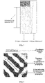
- FIG. 2 is an implementation flow chart of a method for selecting a detection area provided in a second embodiment of the present invention, which is applicable to an one-dimensional ultrasonic signal of an organ tissue.
- FIG. 3 is an effect view of a selection of a detection area based on an M-type ultrasonic signal of an organ tissue in the second embodiment of the present invention;
- FIG. 4 is a schematic view of the quantitative elasticity modulus of an organ tissue in the second embodiment of the present invention.
- the method further including:
- the one-dimensional ultrasound signal of the organ tissue may be an A-type ultrasound signal of the organ tissue or an M-type ultrasonic signal of the organ tissue. Assuming that one ultrasonic signal contains n sampling points, and the ultrasonic signal of the corresponding organ tissue has a scanning depth d (unit: mm), and then n/d points are included in per 1 mm depth.
- the n sampling points are divided into several segments of detection sub-areas S i , and the scanning depth corresponding to the detection sub-areas S i is d i , where i is an integer, and the scanning depth d i may be a mean value or an end value of the depths of detection sub-areas S i , herein the scanning depth d i is the end value.
- Step 22 calculating a Nakagami distribution value m i of an ultrasonic signal R i of the organ tissue in each detection sub-area S i .
- E(.) is mean value function
- ⁇ (.) represents Gamma function
- ⁇ E(r 2 )
- U(.) represents unit step function
- m is the Nakagami distribution value
- r is dependent variable of the probability distribution function f(r), r ⁇ 0 and m ⁇ 0; for each detection sub-area S i , m i is m value in the S i area and R i is the envelope value of the ultrasonic signal.
- An one-dimensional ultrasound signal of the organ tissue follows the pre-Rayleigh distribution when m is in the range of (0, 1); the one-dimensional ultrasound echo signal follows the Rayleigh distribution when m equals to 1; and the one-dimensional ultrasound echo signal follows the post-Rayleigh distribution when m is greater than 1.
- d i is the scanning depth corresponding to the detection sub-areas S i
- d i may be taken as the mean value or end value of depth of the detection sub-areas S i .
- the weight W i of each detection sub-area may be traversed, and the detection sub-area corresponding to the maximum weight is taken as the organ tissue boundary area.
- Step 24 if the standard deviation, corresponding to the intensity value of the one-dimensional ultrasonic signal in each of a plurality of continuous detection sub-areas within the organ tissue boundary area, is less than a standard deviation threshold, the plurality of continuous detection sub-areas are determined as the organ tissue detection area.
- the standard deviation SD i corresponding to the intensity value of the ultrasonic signal R i in each detection sub-area S i within the organ tissue boundary area is calculated, and each detection sub-area within the organ tissue boundary area is traversed, if the standard deviation corresponding to the intensity values of one-dimensional ultrasound signals in each of a plurality of continuous detection sub-areas is less than a standard deviation threshold from a certain detection sub-area, then the plurality of continuous detection sub-areas are determined as the organ tissue detection area, that is, completing the automatic selection of the organ tissue detection area.
- the quantitative elasticity information may include a numerical value of the quantitative elasticity information of the organ tissue within the detection area determined in Step 23, and the numerical value is measured by the elasticity measurement device, usually in unit of kPa. Where the vertical axis represents the scanning depth and the horizontal axis represents the time.
- the quantitative elasticity information may also include trajectory image of instantaneous vibrations propagating over time along the depth during the transient elastic imaging process. The image also contains line segment AB indicating the propagation of the instantaneous vibrations.
- the elasticity modulus of the organ tissue can be calculated by calculating the propagation velocity of the shear wave generated by the instantaneous vibration in the area indicated by the indication mark.
- the method for selecting a detection area provided in the second embodiment of the present invention can automatically select an organ tissue detection area through an A-type or an M-type ultrasound signal of the organ tissue.
- the algorithm possesses high recognition efficiency for the organ tissue boundary due to its low complexity, thereby realizing a real-time automatic localization of the organ tissue boundary.
- FIG. 5 is an implementation flow chart of a method for selecting a detection area provided in a third embodiment of the present invention
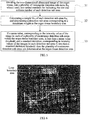
- FIG. 6 is an effect view of a selection of a detection area based on a B-type ultrasonic signal of an organ tissue in the third embodiment of the present invention
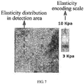
- FIG. 7 is a schematic view of a quantitative elasticity modulus of the organ tissue in the third embodiment of the present invention. Referring to FIG. 5 to FIG. 7 , the method includes:
- the two-dimensional ultrasound image of the organ tissue may be a B-type ultrasound image of an organ tissue.
- the size of the B-type ultrasound image is w*h, where w is a width of the two-dimensional ultrasound image of the organ tissue, h is a height of the two-dimensional ultrasound image of the organ tissue (w and h are both in unit of pixel), and the corresponding scanning depth is d (unit: mm), then h/d pixels are contained in a 1 mm depth on one scan line in the depth direction.
- the B-type image having a size of w*h is divided into a plurality of rectangular detection sub-areas R ij .
- Step 32 calculating a weight W ij of each detection sub-area R ij , and determining a detection sub-area corresponding to a maximum weight as the organ tissue boundary area.
- M kj is a grayscale mean value of the two-dimensional ultrasound image of the organ tissue in the detection sub-areas R kj
- SD kj is a grayscale standard deviation of the two-dimensional ultrasound image of the organ tissue in the detection sub-areas R kj
- d kj is a scanning depth of the detection sub-areas R kj .
- the grayscale mean value of the organ tissue boundary area is large; in addition, since the liver capsule area possesses uniformity in the B-type ultrasound image, the grayscale standard deviation is small.
- a searching is performed from the detection sub-areas locating at the centerline of the B-type ultrasound image. If the detection sub-area R k1 is the one having the largest weight in a series of the detection area R kj , the detection sub-area R k1 is determined as a boundary area of the liver tissue.
- Step 33 if a mean value, corresponding to the intensity value of the image in each of a plurality of continuous detection sub-areas within the organ tissue boundary area, is less than a mean value threshold, and a standard deviation, corresponding to intensity values of the images in each detection sub-area is less than a standard deviation threshold, then the plurality of continuous detection sub-areas are determined as the organ tissue detection area.
- the plurality of continuous detection sub-areas are determined as a detection area, that is, completing the automatic selection of the detection area.
- the quantitative elasticity information includes a numerical value (in unit of kPa) of the quantitative elasticity information of the organ tissue in the detection area shown in the structural view of the organ tissue obtained by the elasticity measurement.
- the elasticity modulus information includes an elasticity modulus distribution of the organ tissue structure in the detection area. Where the elasticity modulus distribution can be color-encoded, with different colors representing different elasticity modulus; the elasticity modulus distribution may also be represented in grayscale or other encoding forms, that is, the color-encoding grayscale-encoding or other encoding forms can be applied to represent intensity values of the two-dimensional ultrasound image of the organ tissue within the detection sub-area.
- the elasticity modulus distribution further includes a scalogram representing the elasticity modulus encoding.
- the method for selecting a detection area provided in the third embodiment of the present invention can automatically select an organ tissue detection area via a B-type ultrasound image of an organ tissue.
- the algorithm possesses high recognition efficiency for the organ tissue boundary due to its low complexity, thereby realizing a real-time automatic localization of the organ tissue boundary.
- FIG. 8 is an implementation flow chart of a method for selecting a detection area provided in a fourth embodiment of the present invention
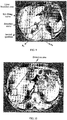
- FIG. 9 is an effect view of a three-dimensional image boundary based on an organ tissue in the fourth embodiment of the present invention
- FIG. 10 is an effect view of a selection of a detection area based on a three-dimensional image of an organ tissue in the fourth embodiment of the present invention
- FIG. 11 is a schematic view of a quantitative elasticity modulus of an organ tissue in the fourth embodiment of the present invention. Referring to FIG. 8 to FIG. 11 , the method includes:
- Step 41 extracting a binary image of a skin and a binary image of a bone in a CT image of the organ tissue or an MRI image of the organ tissue, by using an area growing segmentation method.
- the binary image of the skin is extracted. Pixel with image coordinate of (0, 0) is used as a seed point, and the binary image of the skin is extracted by using the area growing segmentation method, where the area growing criterion of air CT value is [-1024, -500]HU (Hounsfield unit, Heinz).
- the binary image of the bone is extracted, including a binary image of the vertebra and a binary image of the rib.
- Threshold segmentation with a threshold range of [350,1024]HU is performed on the whole image to extract the binary image of bone.
- Step 42 calculating a centroid of the binary image of the bone, and calculating a point on the binary image of the skin closest to the centroid.
- centroid P C of the binary image of the bone is calculated. Since the ribs are generally symmetrical about the vertebra and the vertebra has a large proportion in the bone image, the centroid of the bone image is the centroid P C of the vertebra.
- a point on the binary image of the skin that is closest to the centroid P C is denoted as P N , starting with the vertebra centroid P C .
- Step 43 dividing the organ tissue information into four quadrants based on the centroid and the point closest to the centroid.
- the CT image is divided into four quadrants by using the centroid P C and the point P N closest to the centroid, that is, a straight line passing the centroid P C and the point P N closest to the centroid P C is taken as a vertical axis, and a straight line passing the centroid P C and perpendicular to the vertical axis is taken as the horizontal axis.
- a straight line passing the centroid P C and perpendicular to the vertical axis is taken as the horizontal axis.
- Step 44 fitting each rib point in the second quadrant to acquire a rib fitting curve.
- Each rib point in the second quadrant is fitted with a B-spline curve or a skin curve to obtain a rib fitting curve.
- Step 45 moving the rib fitting curve toward a first quadrant by a preset value for being a boundary curve, and determining an area between the boundary curve and the rib fitting curve as the organ tissue boundary area.
- the rib curve Since the rib curve is close to the liver capsule, the rib curve is moved inwardly by a preset value and taken as a boundary curve, and the area between the boundary curve and the rib fitting curve is determined as the organ tissue boundary area.
- the preset value may be 5 mm.
- Step 46 determining an area surrounded by the organ tissue boundary area as an organ tissue area.
- Step 47 dividing the organ tissue area into a plurality of detection sub-areas.
- Step 48 calculating a standard deviation corresponding to intensity values and a mean value corresponding to intensity values of a two-dimensional ultrasound image of the organ tissue in each detection sub-area.
- Step 49 if a mean value, corresponding to the intensity value of the image in each of a plurality of continuous detection sub-areas within the area surrounded by the organ tissue boundary area, is less than a mean value threshold, and a standard deviation, corresponding to intensity values of the images in each detection sub-area is less than a standard deviation threshold, then the plurality of continuous detection sub-areas are determined as the organ tissue detection area.
- Each detection sub-area within the liver is searched starting from the boundary of the liver tissue, and if a mean value of the intensity values of the images in each of a plurality of continuous detection sub-areas is less than a mean value threshold, and a standard deviation of the intensity values of the images is less than a standard deviation threshold, then the plurality of continuous detection sub-areas are determined as a detection area, that is, completing the automatic selection of the detection area.
- the quantitative elasticity information includes a numerical value (in unit of kPa) of the quantitative elasticity information of the organ tissue shown in the structural view of the organ tissue obtained by the elasticity measurement.
- the elasticity modulus information also includes an elasticity modulus distribution of the tissue in the area indicated by an indicator mark in a tissue structural view.
- the distribution image can be color-encoded, with different colors representing different elasticity modulus; the distribution image may also be represented in grayscale or other encoding forms.
- the elasticity modulus distribution further includes a scalogram representing the elasticity modulus encoding.
- the automatically selected detection area can be 3D geometry.
- the detection area of the image in the frame of the organ tissue image is automatically selected by using the method for selecting the detection area provided in the present embodiment. And then a three-dimensional geometry, i.e., three-dimensional detection area, is reconstructed by using each detection area corresponding to each frame of images.
- the elasticity detection probe is used to detect the elasticity information in the two-dimensional detection area of each frame of CT images and reconstruct the three-dimensional elasticity distribution of the organ tissue, thereby obtaining the three-dimensional elasticity information of the organ tissue.
- the method for selecting a detection area provided in the fourth embodiment of the present invention can automatically select an organ tissue detection area by a three-dimensional image of an organ tissue, for example, the CT image of the organ tissue or the MRI image of the organ tissue.
- the algorithm possesses high recognition efficiency for the organ tissue boundary due to its low complexity, thereby realizing a real-time automatic localization of the organ tissue boundary.
- FIG. 12 is a schematic structural view of a device for selecting a detection area provided in a fifth embodiment of the present invention.
- the device for selecting the detection area described in the present embodiment may include: an area dividing unit 51, configured to divide the organ tissue information to be recognized into a plurality of detection sub-areas; a feature value calculating unit 52, configured to calculate a feature value of the organ tissue information in the detection sub-area; a boundary area recognizing unit 53, configured to determine an organ tissue boundary area according to the organ tissue information to be recognized; and a detection area determining unit 54, configured to determine an organ tissue detection area according to the organ tissue boundary area and a preset feature value condition.
- the device may further include: an elasticity value calculating unit, configured to calculate an elasticity value of an organ tissue in the organ tissue detecting area.
- the preset feature value condition may be such that a distance from the organ tissue boundary area is within a preset depth range.
- the preset feature value condition may be such that both a mean value and a standard deviation corresponding to intensity values of the image or signal in each detection sub-area satisfy a preset range.
- d i is a scanning depth corresponding to the detection sub-area S i .
- M kj is a grayscale mean value of the two-dimensional ultrasound image of the organ tissue in the detection sub-area R kj ;
- SD kj is a grayscale standard deviation of the two-dimensional ultrasound image of the organ tissue in the detection sub-area R kj ;
- d kj is a scanning depth corresponding to the detection sub-area R kj ; and
- k i max /2.
- the boundary area recognizing unit 53 may specifically include: a binary image acquiring sub-unit, configured to extract a binary image of a skin and a binary image of a bone in the CT image of the organ tissue or the MRI image of the organ tissue by using an image segmentation method; and a feature point determining sub-unit, configured to calculate a centroid of the binary image of the bone, and calculate a point on the binary image of the skin closest to the centroid; an image dividing sub-unit, configured to divide the image of the organ tissue into four quadrants according to the centroid and the point closest to the centroid; a curve fitting sub-unit, configured to fit each rib point in the second quadrant to acquire a rib fitting curve; and a boundary area determining sub-unit, configured to move the rib fitting curve toward a first quadrant by a preset value for being a boundary area curve, and determine an area between the boundary area curve and the rib fitting curve
- a device for selecting a detection area divides the organ tissue information to be recognized into a plurality of detection sub-areas, calculates a feature value of the organ tissue information in the detection sub-areas, determines an organ tissue boundary area according to the organ tissue information to be recognized, and determines an organ tissue detection area according to the determined organ tissue boundary area and a preset feature value condition.
- the position and size of the detection area are different when the organ tissue information is different, i.e., the device can adjust the position and size of the detection area.
- FIG. 13 is a schematic structural view of an elasticity detection system provided in a sixth embodiment of the present invention.
- the elasticity detection system described by the present embodiment may include an information acquiring device 61, an elasticity imaging device 63, a probe setting device 64, a processing device 65, and a display device 66, further includes a device for selecting the detection area 62 provided in the fifth embodiment of the present invention, where the information acquiring device 61 is configured to acquire organ tissue information to be recognized; the probe setting device 64 is configured to adjust a position of a probe in the elasticity imaging device such that the detection range of the probe includes a detection area determined by the device for selecting the detection area; the elasticity imaging device 63 is configured to acquire elasticity information of an organ tissue; the processing device 65 is configured to process the elasticity information acquired by the elasticity imaging device, thereby acquiring the elasticity information in the detection area; and the display device 66 is configured to display the elasticity information in the detection area.
- the elasticity detection system provided in the sixth embodiment of the present invention is capable of automatically recognizing the organ tissue boundary area and automatically adjusting the position and size of the detection area, thereby saving the elastic detection time, reducing the operation difference among different operators and the same operator with different operations, and realizing a accurate, fast, highly reproducible elasticity detection of the organ tissue.
Applications Claiming Priority (2)
| Application Number | Priority Date | Filing Date | Title |
|---|---|---|---|
| CN201410561699.9A CN104398272B (zh) | 2014-10-21 | 2014-10-21 | 选择检测区域的方法及装置及弹性检测系统 |
| PCT/CN2015/081817 WO2016062107A1 (zh) | 2014-10-21 | 2015-06-18 | 选择检测区域的方法及装置及弹性检测系统 |
Publications (2)
| Publication Number | Publication Date |
|---|---|
| EP3210541A1 true EP3210541A1 (de) | 2017-08-30 |
| EP3210541A4 EP3210541A4 (de) | 2018-07-04 |
Family
ID=52635988
Family Applications (1)
| Application Number | Title | Priority Date | Filing Date |
|---|---|---|---|
| EP15852840.6A Pending EP3210541A4 (de) | 2014-10-21 | 2015-06-18 | Verfahren und vorrichtung zur auswahl eines detektionsbereichs und elastizitätsdetektionssystem |
Country Status (9)
| Country | Link |
|---|---|
| US (1) | US10925582B2 (de) |
| EP (1) | EP3210541A4 (de) |
| JP (1) | JP6588087B2 (de) |
| KR (1) | KR101913976B1 (de) |
| CN (1) | CN104398272B (de) |
| AU (1) | AU2015335554B2 (de) |
| BR (1) | BR112017008162B1 (de) |
| RU (1) | RU2695619C2 (de) |
| WO (1) | WO2016062107A1 (de) |
Cited By (1)
| Publication number | Priority date | Publication date | Assignee | Title |
|---|---|---|---|---|
| CN106908747A (zh) * | 2015-12-23 | 2017-06-30 | 中国科学院深圳先进技术研究院 | 化学位移编码成像方法及装置 |
Families Citing this family (13)
| Publication number | Priority date | Publication date | Assignee | Title |
|---|---|---|---|---|
| CN104398272B (zh) * | 2014-10-21 | 2017-09-19 | 无锡海斯凯尔医学技术有限公司 | 选择检测区域的方法及装置及弹性检测系统 |
| WO2017152629A1 (zh) * | 2016-03-10 | 2017-09-14 | 无锡海斯凯尔医学技术有限公司 | 自动触发弹性检测的方法和装置 |
| CN105816204A (zh) * | 2016-03-10 | 2016-08-03 | 无锡海斯凯尔医学技术有限公司 | 自动触发弹性检测的方法和装置 |
| US20170265846A1 (en) * | 2016-03-18 | 2017-09-21 | Siemens Medical Solutions Usa, Inc. | Alert assistance for survey mode ultrasound imaging |
| JP6689666B2 (ja) * | 2016-05-12 | 2020-04-28 | 株式会社日立製作所 | 超音波撮像装置 |
| EP3531954A4 (de) * | 2016-10-25 | 2020-09-16 | Mobius Imaging LLC | Verfahren und system für robotergestützte chirurgie |
| CN108305247B (zh) * | 2018-01-17 | 2022-03-04 | 中南大学湘雅三医院 | 一种基于ct图像灰度值检测组织硬度的方法 |
| CN108888284A (zh) * | 2018-05-18 | 2018-11-27 | 沈阳东软医疗系统有限公司 | 图像调整方法、装置及设备、存储介质 |
| EP3998106A1 (de) | 2018-12-17 | 2022-05-18 | Shanghai United Imaging Healthcare Co., Ltd. | Systeme und verfahren zur bestimmung einer region von interesse eines probanden |
| CN109727234B (zh) * | 2018-12-24 | 2021-02-19 | 上海联影医疗科技股份有限公司 | 显示面板生成方法、扫描范围规划方法及设备 |
| CN109893172B (zh) * | 2019-02-22 | 2020-06-19 | 清华大学 | 基于弹性成像的力学参数的确定方法及装置、计算机设备 |
| CN110807770A (zh) * | 2019-10-30 | 2020-02-18 | 杭州依图医疗技术有限公司 | 医学影像的处理、识别、显示方法及存储介质 |
| US11227392B2 (en) * | 2020-05-08 | 2022-01-18 | GE Precision Healthcare LLC | Ultrasound imaging system and method |
Family Cites Families (23)
| Publication number | Priority date | Publication date | Assignee | Title |
|---|---|---|---|---|
| JP2003334194A (ja) * | 2002-03-14 | 2003-11-25 | Matsushita Electric Ind Co Ltd | 画像処理装置及び超音波診断装置 |
| US7283652B2 (en) * | 2002-11-27 | 2007-10-16 | General Electric Company | Method and system for measuring disease relevant tissue changes |
| JP4763502B2 (ja) * | 2006-04-11 | 2011-08-31 | 日立アロカメディカル株式会社 | 超音波診断装置 |
| US8172754B2 (en) * | 2006-04-18 | 2012-05-08 | Panasonic Corporation | Ultrasonograph |
| WO2007135884A1 (ja) * | 2006-05-19 | 2007-11-29 | Hitachi Medical Corporation | 超音波診断装置及び超音波診断方法 |
| JP5038304B2 (ja) * | 2006-06-06 | 2012-10-03 | 株式会社日立メディコ | 超音波診断装置 |
| JP5016911B2 (ja) * | 2006-12-20 | 2012-09-05 | 株式会社日立メディコ | 超音波診断装置 |
| JP4615528B2 (ja) * | 2007-01-22 | 2011-01-19 | 株式会社日立メディコ | 超音波診断装置 |
| KR100961856B1 (ko) * | 2007-03-08 | 2010-06-09 | 주식회사 메디슨 | 초음파 영상을 형성하는 초음파 시스템 및 방법 |
| CA2718343A1 (en) * | 2007-03-15 | 2008-09-18 | Jean Meunier | Image segmentation |
| JP5479353B2 (ja) * | 2008-10-14 | 2014-04-23 | 株式会社日立メディコ | 超音波診断装置 |
| US8394026B2 (en) * | 2008-11-03 | 2013-03-12 | University Of British Columbia | Method and apparatus for determining viscoelastic parameters in tissue |
| CN102956035A (zh) * | 2011-08-25 | 2013-03-06 | 深圳市蓝韵实业有限公司 | 用于乳腺x线图像中提取乳腺区域的预处理方法及系统 |
| TWI454246B (zh) * | 2011-09-30 | 2014-10-01 | Mackay Memorial Hospital | Immediate monitoring of the target location of the radiotherapy system |
| WO2013051275A1 (ja) * | 2011-10-04 | 2013-04-11 | パナソニック株式会社 | 超音波診断装置および超音波診断装置の制御方法 |
| CN102920477B (zh) * | 2012-03-05 | 2015-05-20 | 杭州弘恩医疗科技有限公司 | 医学影像的目标区域边界确定装置和方法 |
| WO2013155300A1 (en) * | 2012-04-11 | 2013-10-17 | The Trustees Of Columbia University In The City Of New York | Techniques for segmentation of organs and tumors and objects |
| US20150141822A1 (en) * | 2012-06-07 | 2015-05-21 | Hitachi Aloka Medical, Ltd. | Method for setting regions of interest and ultrasound diagnostic apparatus |
| RU2526752C1 (ru) * | 2013-03-18 | 2014-08-27 | Корпорация "САМСУНГ ЭЛЕКТРОНИКС Ко., Лтд." | Система и способ для автоматического планирования двухмерных видов в объемных медицинских изображениях |
| CN203280412U (zh) * | 2013-05-29 | 2013-11-13 | 北京索瑞特医学技术有限公司 | 对组织的定量弹性信息和结构信息进行组合显示的系统 |
| CN103720489B (zh) * | 2013-12-30 | 2015-10-28 | 中国科学院深圳先进技术研究院 | 病变组织生长监测方法和系统 |
| CN204379311U (zh) * | 2014-10-21 | 2015-06-10 | 无锡海斯凯尔医学技术有限公司 | 一种选择检测区域的装置及弹性检测系统 |
| CN104398272B (zh) * | 2014-10-21 | 2017-09-19 | 无锡海斯凯尔医学技术有限公司 | 选择检测区域的方法及装置及弹性检测系统 |
-
2014
- 2014-10-21 CN CN201410561699.9A patent/CN104398272B/zh active Active
-
2015
- 2015-06-18 BR BR112017008162-8A patent/BR112017008162B1/pt active IP Right Grant
- 2015-06-18 EP EP15852840.6A patent/EP3210541A4/de active Pending
- 2015-06-18 KR KR1020177006749A patent/KR101913976B1/ko active IP Right Grant
- 2015-06-18 JP JP2017512997A patent/JP6588087B2/ja active Active
- 2015-06-18 WO PCT/CN2015/081817 patent/WO2016062107A1/zh active Application Filing
- 2015-06-18 AU AU2015335554A patent/AU2015335554B2/en active Active
- 2015-06-18 RU RU2017117301A patent/RU2695619C2/ru active
-
2017
- 2017-04-03 US US15/478,021 patent/US10925582B2/en active Active
Cited By (2)
| Publication number | Priority date | Publication date | Assignee | Title |
|---|---|---|---|---|
| CN106908747A (zh) * | 2015-12-23 | 2017-06-30 | 中国科学院深圳先进技术研究院 | 化学位移编码成像方法及装置 |
| CN106908747B (zh) * | 2015-12-23 | 2019-05-07 | 中国科学院深圳先进技术研究院 | 化学位移编码成像方法及装置 |
Also Published As
| Publication number | Publication date |
|---|---|
| CN104398272B (zh) | 2017-09-19 |
| US10925582B2 (en) | 2021-02-23 |
| RU2695619C2 (ru) | 2019-07-24 |
| JP6588087B2 (ja) | 2019-10-09 |
| JP2017536856A (ja) | 2017-12-14 |
| RU2017117301A3 (de) | 2018-11-19 |
| KR20170041879A (ko) | 2017-04-17 |
| US20170202540A1 (en) | 2017-07-20 |
| RU2017117301A (ru) | 2018-11-19 |
| BR112017008162B1 (pt) | 2023-02-14 |
| KR101913976B1 (ko) | 2018-10-31 |
| AU2015335554B2 (en) | 2018-03-22 |
| WO2016062107A1 (zh) | 2016-04-28 |
| CN104398272A (zh) | 2015-03-11 |
| BR112017008162A2 (pt) | 2017-12-26 |
| EP3210541A4 (de) | 2018-07-04 |
| AU2015335554A1 (en) | 2017-05-04 |
Similar Documents
| Publication | Publication Date | Title |
|---|---|---|
| US10925582B2 (en) | Method and device for selecting detection area, and elasticity detection system | |
| US10748291B2 (en) | Liver boundary identification method and system | |
| CN110072465B (zh) | 用于肺部超声的目标探头放置 | |
| CN110945560B (zh) | 胎儿超声图像处理 | |
| US20130182924A1 (en) | Ultrasound image segmentation | |
| KR20190022185A (ko) | 태아의 신체 계측 방법 및 이를 이용한 태아의 신체 계측 디바이스 | |
| CN204379311U (zh) | 一种选择检测区域的装置及弹性检测系统 | |
| EP4171386B1 (de) | Bestimmung der nadelposition |
Legal Events
| Date | Code | Title | Description |
|---|---|---|---|
| STAA | Information on the status of an ep patent application or granted ep patent |
Free format text: STATUS: THE INTERNATIONAL PUBLICATION HAS BEEN MADE |
|
| PUAI | Public reference made under article 153(3) epc to a published international application that has entered the european phase |
Free format text: ORIGINAL CODE: 0009012 |
|
| STAA | Information on the status of an ep patent application or granted ep patent |
Free format text: STATUS: REQUEST FOR EXAMINATION WAS MADE |
|
| 17P | Request for examination filed |
Effective date: 20170519 |
|
| AK | Designated contracting states |
Kind code of ref document: A1 Designated state(s): AL AT BE BG CH CY CZ DE DK EE ES FI FR GB GR HR HU IE IS IT LI LT LU LV MC MK MT NL NO PL PT RO RS SE SI SK SM TR |
|
| AX | Request for extension of the european patent |
Extension state: BA ME |
|
| DAV | Request for validation of the european patent (deleted) | ||
| DAX | Request for extension of the european patent (deleted) | ||
| A4 | Supplementary search report drawn up and despatched |
Effective date: 20180604 |
|
| RIC1 | Information provided on ipc code assigned before grant |
Ipc: A61B 5/055 20060101ALI20180528BHEP Ipc: A61B 8/00 20060101ALI20180528BHEP Ipc: A61B 5/00 20060101ALI20180528BHEP Ipc: A61B 8/08 20060101AFI20180528BHEP Ipc: A61B 6/03 20060101ALI20180528BHEP |
|
| STAA | Information on the status of an ep patent application or granted ep patent |
Free format text: STATUS: EXAMINATION IS IN PROGRESS |
|
| 17Q | First examination report despatched |
Effective date: 20220105 |