EP3206645B1 - Linsenfragmentierung mit augenlaserchirurgie - Google Patents
Linsenfragmentierung mit augenlaserchirurgie Download PDFInfo
- Publication number
- EP3206645B1 EP3206645B1 EP15788522.9A EP15788522A EP3206645B1 EP 3206645 B1 EP3206645 B1 EP 3206645B1 EP 15788522 A EP15788522 A EP 15788522A EP 3206645 B1 EP3206645 B1 EP 3206645B1
- Authority
- EP
- European Patent Office
- Prior art keywords
- laser
- eye
- lens
- fragmentation pattern
- rotation angle
- Prior art date
- Legal status (The legal status is an assumption and is not a legal conclusion. Google has not performed a legal analysis and makes no representation as to the accuracy of the status listed.)
- Active
Links
- 238000013467 fragmentation Methods 0.000 title claims description 112
- 238000006062 fragmentation reaction Methods 0.000 title claims description 112
- 238000001356 surgical procedure Methods 0.000 title claims description 73
- 238000000034 method Methods 0.000 claims description 85
- 238000005259 measurement Methods 0.000 claims description 58
- 230000011218 segmentation Effects 0.000 claims description 49
- 238000013507 mapping Methods 0.000 claims description 11
- 210000001508 eye Anatomy 0.000 description 347
- 210000000695 crystalline len Anatomy 0.000 description 256
- 210000004087 cornea Anatomy 0.000 description 81
- 238000012014 optical coherence tomography Methods 0.000 description 67
- 238000011282 treatment Methods 0.000 description 64
- 230000003287 optical effect Effects 0.000 description 58
- 210000001519 tissue Anatomy 0.000 description 54
- 210000000554 iris Anatomy 0.000 description 33
- 239000002775 capsule Substances 0.000 description 30
- 238000010586 diagram Methods 0.000 description 28
- 238000003384 imaging method Methods 0.000 description 28
- 238000005520 cutting process Methods 0.000 description 26
- 230000033001 locomotion Effects 0.000 description 26
- 208000002177 Cataract Diseases 0.000 description 23
- 238000001514 detection method Methods 0.000 description 20
- 210000001747 pupil Anatomy 0.000 description 19
- 239000000463 material Substances 0.000 description 18
- 238000005286 illumination Methods 0.000 description 17
- 238000003032 molecular docking Methods 0.000 description 17
- 230000007246 mechanism Effects 0.000 description 14
- 230000015654 memory Effects 0.000 description 14
- 230000004044 response Effects 0.000 description 14
- 238000003325 tomography Methods 0.000 description 11
- 238000012876 topography Methods 0.000 description 11
- 230000005540 biological transmission Effects 0.000 description 10
- 239000007788 liquid Substances 0.000 description 10
- 230000000007 visual effect Effects 0.000 description 10
- 238000004422 calculation algorithm Methods 0.000 description 9
- 230000015556 catabolic process Effects 0.000 description 9
- 238000000605 extraction Methods 0.000 description 9
- 230000006870 function Effects 0.000 description 9
- FAPWRFPIFSIZLT-UHFFFAOYSA-M Sodium chloride Chemical compound [Na+].[Cl-] FAPWRFPIFSIZLT-UHFFFAOYSA-M 0.000 description 7
- 230000008859 change Effects 0.000 description 6
- 238000004891 communication Methods 0.000 description 6
- 230000000875 corresponding effect Effects 0.000 description 6
- 230000009977 dual effect Effects 0.000 description 6
- 230000008569 process Effects 0.000 description 6
- 230000004075 alteration Effects 0.000 description 5
- 239000012530 fluid Substances 0.000 description 5
- 230000010287 polarization Effects 0.000 description 5
- 210000003786 sclera Anatomy 0.000 description 5
- 238000013519 translation Methods 0.000 description 5
- 210000002159 anterior chamber Anatomy 0.000 description 4
- 230000008878 coupling Effects 0.000 description 4
- 238000010168 coupling process Methods 0.000 description 4
- 238000005859 coupling reaction Methods 0.000 description 4
- 210000003128 head Anatomy 0.000 description 4
- 230000010354 integration Effects 0.000 description 4
- 238000002430 laser surgery Methods 0.000 description 4
- 238000013532 laser treatment Methods 0.000 description 4
- 239000011780 sodium chloride Substances 0.000 description 4
- 230000003595 spectral effect Effects 0.000 description 4
- 238000011269 treatment regimen Methods 0.000 description 4
- VYPSYNLAJGMNEJ-UHFFFAOYSA-N Silicium dioxide Chemical compound O=[Si]=O VYPSYNLAJGMNEJ-UHFFFAOYSA-N 0.000 description 3
- 238000002059 diagnostic imaging Methods 0.000 description 3
- 210000003664 lens nucleus crystalline Anatomy 0.000 description 3
- 238000007639 printing Methods 0.000 description 3
- 238000003860 storage Methods 0.000 description 3
- 238000002604 ultrasonography Methods 0.000 description 3
- 238000013459 approach Methods 0.000 description 2
- 210000001742 aqueous humor Anatomy 0.000 description 2
- 201000009310 astigmatism Diseases 0.000 description 2
- 230000009286 beneficial effect Effects 0.000 description 2
- 230000008901 benefit Effects 0.000 description 2
- 210000004204 blood vessel Anatomy 0.000 description 2
- 239000007975 buffered saline Substances 0.000 description 2
- 230000003139 buffering effect Effects 0.000 description 2
- 238000004590 computer program Methods 0.000 description 2
- 230000003750 conditioning effect Effects 0.000 description 2
- 238000012937 correction Methods 0.000 description 2
- 238000013500 data storage Methods 0.000 description 2
- 230000000994 depressogenic effect Effects 0.000 description 2
- 238000005516 engineering process Methods 0.000 description 2
- 239000000835 fiber Substances 0.000 description 2
- 230000004886 head movement Effects 0.000 description 2
- 238000011065 in-situ storage Methods 0.000 description 2
- 238000003780 insertion Methods 0.000 description 2
- 230000037431 insertion Effects 0.000 description 2
- 238000012986 modification Methods 0.000 description 2
- 230000004048 modification Effects 0.000 description 2
- 208000001491 myopia Diseases 0.000 description 2
- 230000004379 myopia Effects 0.000 description 2
- 238000012634 optical imaging Methods 0.000 description 2
- 238000000059 patterning Methods 0.000 description 2
- 230000001179 pupillary effect Effects 0.000 description 2
- 238000002310 reflectometry Methods 0.000 description 2
- 230000002040 relaxant effect Effects 0.000 description 2
- 210000001525 retina Anatomy 0.000 description 2
- 239000000243 solution Substances 0.000 description 2
- 238000012546 transfer Methods 0.000 description 2
- 230000001131 transforming effect Effects 0.000 description 2
- 241000239290 Araneae Species 0.000 description 1
- -1 BK-7 Substances 0.000 description 1
- 201000004569 Blindness Diseases 0.000 description 1
- XUIMIQQOPSSXEZ-UHFFFAOYSA-N Silicon Chemical compound [Si] XUIMIQQOPSSXEZ-UHFFFAOYSA-N 0.000 description 1
- 238000002679 ablation Methods 0.000 description 1
- 238000010521 absorption reaction Methods 0.000 description 1
- 230000001133 acceleration Effects 0.000 description 1
- NIXOWILDQLNWCW-UHFFFAOYSA-N acrylic acid group Chemical group C(C=C)(=O)O NIXOWILDQLNWCW-UHFFFAOYSA-N 0.000 description 1
- 238000003491 array Methods 0.000 description 1
- 238000005452 bending Methods 0.000 description 1
- 230000015572 biosynthetic process Effects 0.000 description 1
- 230000004397 blinking Effects 0.000 description 1
- 238000004364 calculation method Methods 0.000 description 1
- 239000003086 colorant Substances 0.000 description 1
- 238000005094 computer simulation Methods 0.000 description 1
- 230000002596 correlated effect Effects 0.000 description 1
- 238000013481 data capture Methods 0.000 description 1
- 230000007812 deficiency Effects 0.000 description 1
- 238000006731 degradation reaction Methods 0.000 description 1
- 230000001419 dependent effect Effects 0.000 description 1
- 238000011161 development Methods 0.000 description 1
- 230000018109 developmental process Effects 0.000 description 1
- 238000002405 diagnostic procedure Methods 0.000 description 1
- 208000029436 dilated pupil Diseases 0.000 description 1
- 239000006185 dispersion Substances 0.000 description 1
- 238000006073 displacement reaction Methods 0.000 description 1
- 230000000694 effects Effects 0.000 description 1
- 230000005670 electromagnetic radiation Effects 0.000 description 1
- 230000004424 eye movement Effects 0.000 description 1
- 238000001914 filtration Methods 0.000 description 1
- 238000010304 firing Methods 0.000 description 1
- 238000000799 fluorescence microscopy Methods 0.000 description 1
- 230000004907 flux Effects 0.000 description 1
- 210000001061 forehead Anatomy 0.000 description 1
- 239000012634 fragment Substances 0.000 description 1
- 239000005350 fused silica glass Substances 0.000 description 1
- 239000011521 glass Substances 0.000 description 1
- 238000007654 immersion Methods 0.000 description 1
- 230000003116 impacting effect Effects 0.000 description 1
- 238000002513 implantation Methods 0.000 description 1
- 230000003993 interaction Effects 0.000 description 1
- 230000002262 irrigation Effects 0.000 description 1
- 238000003973 irrigation Methods 0.000 description 1
- 238000003698 laser cutting Methods 0.000 description 1
- 230000007257 malfunction Effects 0.000 description 1
- 230000001404 mediated effect Effects 0.000 description 1
- 239000012528 membrane Substances 0.000 description 1
- 230000005055 memory storage Effects 0.000 description 1
- 238000012544 monitoring process Methods 0.000 description 1
- 238000000399 optical microscopy Methods 0.000 description 1
- 238000005457 optimization Methods 0.000 description 1
- 230000000399 orthopedic effect Effects 0.000 description 1
- 238000003909 pattern recognition Methods 0.000 description 1
- 230000008447 perception Effects 0.000 description 1
- 230000002085 persistent effect Effects 0.000 description 1
- 229920000642 polymer Polymers 0.000 description 1
- 238000012805 post-processing Methods 0.000 description 1
- 238000002360 preparation method Methods 0.000 description 1
- 238000003825 pressing Methods 0.000 description 1
- 238000012545 processing Methods 0.000 description 1
- 230000000750 progressive effect Effects 0.000 description 1
- 208000014733 refractive error Diseases 0.000 description 1
- 230000002207 retinal effect Effects 0.000 description 1
- 238000002432 robotic surgery Methods 0.000 description 1
- 238000005070 sampling Methods 0.000 description 1
- 238000007789 sealing Methods 0.000 description 1
- 238000000926 separation method Methods 0.000 description 1
- 229910052710 silicon Inorganic materials 0.000 description 1
- 239000010703 silicon Substances 0.000 description 1
- 239000000377 silicon dioxide Substances 0.000 description 1
- 238000004088 simulation Methods 0.000 description 1
- 238000001228 spectrum Methods 0.000 description 1
- 230000000087 stabilizing effect Effects 0.000 description 1
- 230000003068 static effect Effects 0.000 description 1
- 238000006467 substitution reaction Methods 0.000 description 1
- 230000002123 temporal effect Effects 0.000 description 1
- 238000012549 training Methods 0.000 description 1
- 230000009466 transformation Effects 0.000 description 1
- 239000012780 transparent material Substances 0.000 description 1
- 238000012795 verification Methods 0.000 description 1
- 239000008154 viscoelastic solution Substances 0.000 description 1
- 239000003190 viscoelastic substance Substances 0.000 description 1
- 229940006076 viscoelastic substance Drugs 0.000 description 1
- 230000004393 visual impairment Effects 0.000 description 1
- 210000004127 vitreous body Anatomy 0.000 description 1
- XLYOFNOQVPJJNP-UHFFFAOYSA-N water Substances O XLYOFNOQVPJJNP-UHFFFAOYSA-N 0.000 description 1
- 238000004383 yellowing Methods 0.000 description 1
Images


Classifications
-
- A—HUMAN NECESSITIES
- A61—MEDICAL OR VETERINARY SCIENCE; HYGIENE
- A61F—FILTERS IMPLANTABLE INTO BLOOD VESSELS; PROSTHESES; DEVICES PROVIDING PATENCY TO, OR PREVENTING COLLAPSING OF, TUBULAR STRUCTURES OF THE BODY, e.g. STENTS; ORTHOPAEDIC, NURSING OR CONTRACEPTIVE DEVICES; FOMENTATION; TREATMENT OR PROTECTION OF EYES OR EARS; BANDAGES, DRESSINGS OR ABSORBENT PADS; FIRST-AID KITS
- A61F9/00—Methods or devices for treatment of the eyes; Devices for putting-in contact lenses; Devices to correct squinting; Apparatus to guide the blind; Protective devices for the eyes, carried on the body or in the hand
- A61F9/007—Methods or devices for eye surgery
- A61F9/008—Methods or devices for eye surgery using laser
- A61F9/00825—Methods or devices for eye surgery using laser for photodisruption
- A61F9/0084—Laser features or special beam parameters therefor
-
- A—HUMAN NECESSITIES
- A61—MEDICAL OR VETERINARY SCIENCE; HYGIENE
- A61B—DIAGNOSIS; SURGERY; IDENTIFICATION
- A61B3/00—Apparatus for testing the eyes; Instruments for examining the eyes
- A61B3/10—Objective types, i.e. instruments for examining the eyes independent of the patients' perceptions or reactions
- A61B3/102—Objective types, i.e. instruments for examining the eyes independent of the patients' perceptions or reactions for optical coherence tomography [OCT]
-
- A—HUMAN NECESSITIES
- A61—MEDICAL OR VETERINARY SCIENCE; HYGIENE
- A61F—FILTERS IMPLANTABLE INTO BLOOD VESSELS; PROSTHESES; DEVICES PROVIDING PATENCY TO, OR PREVENTING COLLAPSING OF, TUBULAR STRUCTURES OF THE BODY, e.g. STENTS; ORTHOPAEDIC, NURSING OR CONTRACEPTIVE DEVICES; FOMENTATION; TREATMENT OR PROTECTION OF EYES OR EARS; BANDAGES, DRESSINGS OR ABSORBENT PADS; FIRST-AID KITS
- A61F9/00—Methods or devices for treatment of the eyes; Devices for putting-in contact lenses; Devices to correct squinting; Apparatus to guide the blind; Protective devices for the eyes, carried on the body or in the hand
- A61F9/007—Methods or devices for eye surgery
- A61F9/008—Methods or devices for eye surgery using laser
- A61F9/00802—Methods or devices for eye surgery using laser for photoablation
- A61F9/00804—Refractive treatments
-
- A—HUMAN NECESSITIES
- A61—MEDICAL OR VETERINARY SCIENCE; HYGIENE
- A61F—FILTERS IMPLANTABLE INTO BLOOD VESSELS; PROSTHESES; DEVICES PROVIDING PATENCY TO, OR PREVENTING COLLAPSING OF, TUBULAR STRUCTURES OF THE BODY, e.g. STENTS; ORTHOPAEDIC, NURSING OR CONTRACEPTIVE DEVICES; FOMENTATION; TREATMENT OR PROTECTION OF EYES OR EARS; BANDAGES, DRESSINGS OR ABSORBENT PADS; FIRST-AID KITS
- A61F9/00—Methods or devices for treatment of the eyes; Devices for putting-in contact lenses; Devices to correct squinting; Apparatus to guide the blind; Protective devices for the eyes, carried on the body or in the hand
- A61F9/007—Methods or devices for eye surgery
- A61F9/008—Methods or devices for eye surgery using laser
- A61F9/00825—Methods or devices for eye surgery using laser for photodisruption
- A61F9/00827—Refractive correction, e.g. lenticle
-
- A—HUMAN NECESSITIES
- A61—MEDICAL OR VETERINARY SCIENCE; HYGIENE
- A61F—FILTERS IMPLANTABLE INTO BLOOD VESSELS; PROSTHESES; DEVICES PROVIDING PATENCY TO, OR PREVENTING COLLAPSING OF, TUBULAR STRUCTURES OF THE BODY, e.g. STENTS; ORTHOPAEDIC, NURSING OR CONTRACEPTIVE DEVICES; FOMENTATION; TREATMENT OR PROTECTION OF EYES OR EARS; BANDAGES, DRESSINGS OR ABSORBENT PADS; FIRST-AID KITS
- A61F9/00—Methods or devices for treatment of the eyes; Devices for putting-in contact lenses; Devices to correct squinting; Apparatus to guide the blind; Protective devices for the eyes, carried on the body or in the hand
- A61F9/007—Methods or devices for eye surgery
- A61F9/008—Methods or devices for eye surgery using laser
- A61F2009/00844—Feedback systems
- A61F2009/00851—Optical coherence topography [OCT]
-
- A—HUMAN NECESSITIES
- A61—MEDICAL OR VETERINARY SCIENCE; HYGIENE
- A61F—FILTERS IMPLANTABLE INTO BLOOD VESSELS; PROSTHESES; DEVICES PROVIDING PATENCY TO, OR PREVENTING COLLAPSING OF, TUBULAR STRUCTURES OF THE BODY, e.g. STENTS; ORTHOPAEDIC, NURSING OR CONTRACEPTIVE DEVICES; FOMENTATION; TREATMENT OR PROTECTION OF EYES OR EARS; BANDAGES, DRESSINGS OR ABSORBENT PADS; FIRST-AID KITS
- A61F9/00—Methods or devices for treatment of the eyes; Devices for putting-in contact lenses; Devices to correct squinting; Apparatus to guide the blind; Protective devices for the eyes, carried on the body or in the hand
- A61F9/007—Methods or devices for eye surgery
- A61F9/008—Methods or devices for eye surgery using laser
- A61F2009/00853—Laser thermal keratoplasty or radial keratotomy
-
- A—HUMAN NECESSITIES
- A61—MEDICAL OR VETERINARY SCIENCE; HYGIENE
- A61F—FILTERS IMPLANTABLE INTO BLOOD VESSELS; PROSTHESES; DEVICES PROVIDING PATENCY TO, OR PREVENTING COLLAPSING OF, TUBULAR STRUCTURES OF THE BODY, e.g. STENTS; ORTHOPAEDIC, NURSING OR CONTRACEPTIVE DEVICES; FOMENTATION; TREATMENT OR PROTECTION OF EYES OR EARS; BANDAGES, DRESSINGS OR ABSORBENT PADS; FIRST-AID KITS
- A61F9/00—Methods or devices for treatment of the eyes; Devices for putting-in contact lenses; Devices to correct squinting; Apparatus to guide the blind; Protective devices for the eyes, carried on the body or in the hand
- A61F9/007—Methods or devices for eye surgery
- A61F9/008—Methods or devices for eye surgery using laser
- A61F2009/00855—Calibration of the laser system
-
- A—HUMAN NECESSITIES
- A61—MEDICAL OR VETERINARY SCIENCE; HYGIENE
- A61F—FILTERS IMPLANTABLE INTO BLOOD VESSELS; PROSTHESES; DEVICES PROVIDING PATENCY TO, OR PREVENTING COLLAPSING OF, TUBULAR STRUCTURES OF THE BODY, e.g. STENTS; ORTHOPAEDIC, NURSING OR CONTRACEPTIVE DEVICES; FOMENTATION; TREATMENT OR PROTECTION OF EYES OR EARS; BANDAGES, DRESSINGS OR ABSORBENT PADS; FIRST-AID KITS
- A61F9/00—Methods or devices for treatment of the eyes; Devices for putting-in contact lenses; Devices to correct squinting; Apparatus to guide the blind; Protective devices for the eyes, carried on the body or in the hand
- A61F9/007—Methods or devices for eye surgery
- A61F9/008—Methods or devices for eye surgery using laser
- A61F2009/00861—Methods or devices for eye surgery using laser adapted for treatment at a particular location
- A61F2009/0087—Lens
-
- A—HUMAN NECESSITIES
- A61—MEDICAL OR VETERINARY SCIENCE; HYGIENE
- A61F—FILTERS IMPLANTABLE INTO BLOOD VESSELS; PROSTHESES; DEVICES PROVIDING PATENCY TO, OR PREVENTING COLLAPSING OF, TUBULAR STRUCTURES OF THE BODY, e.g. STENTS; ORTHOPAEDIC, NURSING OR CONTRACEPTIVE DEVICES; FOMENTATION; TREATMENT OR PROTECTION OF EYES OR EARS; BANDAGES, DRESSINGS OR ABSORBENT PADS; FIRST-AID KITS
- A61F9/00—Methods or devices for treatment of the eyes; Devices for putting-in contact lenses; Devices to correct squinting; Apparatus to guide the blind; Protective devices for the eyes, carried on the body or in the hand
- A61F9/007—Methods or devices for eye surgery
- A61F9/008—Methods or devices for eye surgery using laser
- A61F2009/00861—Methods or devices for eye surgery using laser adapted for treatment at a particular location
- A61F2009/00872—Cornea
-
- A—HUMAN NECESSITIES
- A61—MEDICAL OR VETERINARY SCIENCE; HYGIENE
- A61F—FILTERS IMPLANTABLE INTO BLOOD VESSELS; PROSTHESES; DEVICES PROVIDING PATENCY TO, OR PREVENTING COLLAPSING OF, TUBULAR STRUCTURES OF THE BODY, e.g. STENTS; ORTHOPAEDIC, NURSING OR CONTRACEPTIVE DEVICES; FOMENTATION; TREATMENT OR PROTECTION OF EYES OR EARS; BANDAGES, DRESSINGS OR ABSORBENT PADS; FIRST-AID KITS
- A61F9/00—Methods or devices for treatment of the eyes; Devices for putting-in contact lenses; Devices to correct squinting; Apparatus to guide the blind; Protective devices for the eyes, carried on the body or in the hand
- A61F9/007—Methods or devices for eye surgery
- A61F9/008—Methods or devices for eye surgery using laser
- A61F2009/00878—Planning
- A61F2009/00882—Planning based on topography
-
- A—HUMAN NECESSITIES
- A61—MEDICAL OR VETERINARY SCIENCE; HYGIENE
- A61F—FILTERS IMPLANTABLE INTO BLOOD VESSELS; PROSTHESES; DEVICES PROVIDING PATENCY TO, OR PREVENTING COLLAPSING OF, TUBULAR STRUCTURES OF THE BODY, e.g. STENTS; ORTHOPAEDIC, NURSING OR CONTRACEPTIVE DEVICES; FOMENTATION; TREATMENT OR PROTECTION OF EYES OR EARS; BANDAGES, DRESSINGS OR ABSORBENT PADS; FIRST-AID KITS
- A61F9/00—Methods or devices for treatment of the eyes; Devices for putting-in contact lenses; Devices to correct squinting; Apparatus to guide the blind; Protective devices for the eyes, carried on the body or in the hand
- A61F9/007—Methods or devices for eye surgery
- A61F9/008—Methods or devices for eye surgery using laser
- A61F2009/00885—Methods or devices for eye surgery using laser for treating a particular disease
- A61F2009/00887—Cataract
Claims (15)
- Augenlaserchirurgiesystem, umfassend:einen Laser zum Erzeugen eines Laserstrahls;ein räumliches Messsystem zum Erzeugen eines Messstrahls und Messen einer räumlichen Anordnung eines Auges;einen mit dem Laser und dem räumlichen Messsystem gekoppelten Prozessor, wobei der Prozessor ein materielles Medium umfasst, das Anweisungen umsetzt zum:Festlegen eines räumlichen Modells des Auges in einem Augenkoordinaten-Referenzsystem basierend auf dem Messstrahl;Abbilden des räumlichen Modells aus dem Augenkoordinaten-Referenzsystem auf ein Maschinenkoordinaten-Referenzsystem;Empfangen eines Rotationswinkels einer kornealen Inzision relativ zu einer Referenzachse des Maschinenkoordinaten-Referenzsystems;Ermitteln eines Laserfragmentierungsmusters basierend auf einer Mehrzahl von Laserfragmentierungsparametern;dadurch gekennzeichnet, dass das materielle Medium ferner Anweisungen umsetzt zum:Ermitteln eines ersten Rotationswinkels basierend auf dem Rotationswinkel der kornealen Inzision und einem Rotationswinkel des Laserfragmentierungsmusters relativ zu der Referenzachse des Maschinenkoordinaten-Referenzsystems;Rotieren des räumlichen Modells um einen Negativwert des ersten Rotationswinkels; undRotieren des Laserfragmentierungsmusters und des räumlichen Modells um den ersten Rotationswinkel, sodass das räumliche Modell mit der Referenzachse des Maschinenkoordinaten-Referenzsystems ausgerichtet ist und das rotierte Laserfragmentierungsmuster mit der kornealen Inzision ausgerichtet ist.
- Lasersystem nach Anspruch 1, wobei das Laserfragmentierungsmuster einen ersten Linsenabschnitt definiert, der zuerst extrahiert werden soll und der der kornealen Inzision gegenüberliegend ausgerichtet ist.
- Lasersystem nach Anspruch 1, wobei das Laserfragmentierungsmuster asymmetrisch ist.
- Lasersystem nach Anspruch 1, wobei das Laserfragmentierungsmuster einen ersten Abschnitt und einen zweiten Abschnitt umfasst, der mindestens eines von einem unterschiedlichen Segmentierungsmuster und Erweichungsmuster aufweist.
- Lasersystem nach Anspruch 1, wobei das Laserfragmentierungsmuster einen ersten nicht erweichten Abschnitt und einen zweiten erweichten Abschnitt aufweist.
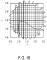
- Lasersystem nach Anspruch 1, wobei das Laserfragmentierungsmuster einen ersten Abschnitt aufweist, der zwei Oktanten definiert, und einen zweiten Abschnitt, der drei Quadranten definiert.
- Lasersystem nach Anspruch 1, wobei das Laserfragmentierungsmuster einen ersten Abschnitt aufweist, der zwei nicht erweichte Oktanten definiert, und einen zweiten Abschnitt, der drei erweichte Quadranten definiert.
- Lasersystem nach Anspruch 1, wobei der Laserstrahl basierend auf dem rotierten Laserfragmentierungsmuster erzeugt wird.
- Lasersystem nach Anspruch 1, wobei der erste Rotationswinkel auf Benutzereingabe basiert.
- Verfahren zum Erzeugen eines Linsenfragmentierungsmusters, umfassend:Festlegen eines räumlichen Modells des Auges in einem Augenkoordinaten-Referenzsystem basierend auf dem Messstrahl;Abbilden des räumlichen Modells aus dem Augenkoordinaten-Referenzsystem auf ein Maschinenkoordinaten-Referenzsystem;Empfangen eines Rotationswinkels einer kornealen Inzision relativ zu einer Referenzachse des Maschinenkoordinaten-Referenzsystems;Ermitteln eines Laserfragmentierungsmusters basierend auf einer Mehrzahl von Laserfragmentierungsparametern;gekennzeichnet durch:Ermitteln eines ersten Rotationswinkels basierend auf dem Rotationswinkel der kornealen Inzision und einem Rotationswinkel des Laserfragmentierungsmusters relativ zu der Referenzachse des Maschinenkoordinaten-Referenzsystems;Rotieren des räumlichen Modells um einen Negativwert des ersten Rotationswinkels; undRotieren des Laserfragmentierungsmusters und des räumlichen Modells um den ersten Rotationswinkel, sodass das räumliche Modell mit der Referenzachse des Maschinenkoordinaten-Referenzsystems ausgerichtet ist und das rotierte Laserfragmentierungsmuster mit der kornealen Inzision ausgerichtet ist.
- Verfahren nach Anspruch 10, wobei das Laserfragmentierungsmuster einen ersten Linsenabschnitt definiert, der zuerst extrahiert werden soll und der der kornealen Inzision gegenüberliegend ausgerichtet ist.
- Verfahren nach Anspruch 10, wobei das Laserfragmentierungsmuster asymmetrisch ist.
- Verfahren nach Anspruch 10, wobei das Linsenfragmentierungsmuster einen ersten Abschnitt und einen zweiten Abschnitt umfasst, der mindestens eines von einem unterschiedlichen Segmentierungsmuster und Erweichungsmuster aufweist.
- Verfahren nach Anspruch 10, wobei das Linsenfragmentierungsmuster aufweist:(i) einen ersten nicht erweichten Abschnitt und einen zweiten erweichten Abschnitt; oder(ii) einen ersten Abschnitt, der zwei Oktanten definiert, und einen zweiten Abschnitt, der drei Quadranten definiert; oder(iii) einen ersten Abschnitt, der zwei nicht erweichte Oktanten definiert, und einen zweiten Abschnitt, der drei erweichte Quadranten definiert.
- Verfahren nach Anspruch 10, wobei der erste Rotationswinkel auf Benutzereingabe basiert.
Applications Claiming Priority (2)
| Application Number | Priority Date | Filing Date | Title |
|---|---|---|---|
| US201462065469P | 2014-10-17 | 2014-10-17 | |
| PCT/US2015/056029 WO2016061511A1 (en) | 2014-10-17 | 2015-10-16 | Laser eye surgery lens fragmentation |
Publications (2)
| Publication Number | Publication Date |
|---|---|
| EP3206645A1 EP3206645A1 (de) | 2017-08-23 |
| EP3206645B1 true EP3206645B1 (de) | 2019-11-20 |
Family
ID=54366520
Family Applications (1)
| Application Number | Title | Priority Date | Filing Date |
|---|---|---|---|
| EP15788522.9A Active EP3206645B1 (de) | 2014-10-17 | 2015-10-16 | Linsenfragmentierung mit augenlaserchirurgie |
Country Status (6)
| Country | Link |
|---|---|
| US (3) | US10327953B2 (de) |
| EP (1) | EP3206645B1 (de) |
| JP (1) | JP6621816B2 (de) |
| AU (1) | AU2015331733A1 (de) |
| CA (1) | CA2964897A1 (de) |
| WO (1) | WO2016061511A1 (de) |
Families Citing this family (11)
| Publication number | Priority date | Publication date | Assignee | Title |
|---|---|---|---|---|
| WO2014071221A2 (en) * | 2012-11-02 | 2014-05-08 | Optimedica Corporation | Optical surface identification for laser surgery |
| CA2976025A1 (en) * | 2015-02-06 | 2016-08-11 | Optimedica Corporation | Closed-loop laser eye surgery treatment |
| EP3217144A1 (de) * | 2016-03-11 | 2017-09-13 | Haag-Streit Ag | Augenvermessung |
| CN105868503B (zh) * | 2016-04-25 | 2019-06-11 | 北京卫星环境工程研究所 | 地基激光移除空间碎片过程的三维建模与仿真方法 |
| US9916501B2 (en) * | 2016-07-22 | 2018-03-13 | Yung-Hui Li | Smart eyeglasses with iris recognition device |
| AU2018215207A1 (en) * | 2017-01-31 | 2019-07-18 | Amo Development, Llc | Methods and systems for laser ophthalmic surgery that provide for iris exposures below a predetermined exposure limit |
| ES2941994T3 (es) | 2017-10-17 | 2023-05-29 | Alcon Inc | Perfiles quirúrgicos oftálmicos personalizados |
| US10768431B2 (en) | 2017-12-20 | 2020-09-08 | Aperture In Motion, LLC | Light control devices and methods for regional variation of visual information and sampling |
| US10175490B1 (en) * | 2017-12-20 | 2019-01-08 | Aperture In Motion, LLC | Light control devices and methods for regional variation of visual information and sampling |
| CN110338906B (zh) * | 2019-07-10 | 2020-10-30 | 清华大学深圳研究生院 | 用于光交联手术的智能治疗系统及建立方法 |
| US20230368441A1 (en) * | 2022-05-10 | 2023-11-16 | Johnson & Johnson Surgical Vision, Inc. | Phacoemulsifation guidance |
Family Cites Families (28)
| Publication number | Priority date | Publication date | Assignee | Title |
|---|---|---|---|---|
| US5984916A (en) | 1993-04-20 | 1999-11-16 | Lai; Shui T. | Ophthalmic surgical laser and method |
| US5743902A (en) | 1995-01-23 | 1998-04-28 | Coherent, Inc. | Hand-held laser scanner |
| US6454761B1 (en) | 1995-01-30 | 2002-09-24 | Philip D. Freedman | Laser surgery device and method |
| US5720894A (en) | 1996-01-11 | 1998-02-24 | The Regents Of The University Of California | Ultrashort pulse high repetition rate laser system for biological tissue processing |
| US7655002B2 (en) | 1996-03-21 | 2010-02-02 | Second Sight Laser Technologies, Inc. | Lenticular refractive surgery of presbyopia, other refractive errors, and cataract retardation |
| US6019472A (en) | 1997-05-12 | 2000-02-01 | Koester; Charles J. | Contact lens element for examination or treatment of ocular tissues |
| US9668649B2 (en) * | 2000-04-07 | 2017-06-06 | Amo Development, Llc | System and methods for mitigating changes in pupil size during laser refractive surgery to maintain ablation centration |
| US8262646B2 (en) | 2006-01-20 | 2012-09-11 | Lensar, Inc. | System and method for providing the shaped structural weakening of the human lens with a laser |
| US20110319875A1 (en) | 2007-01-19 | 2011-12-29 | Frieder Loesel | Apparatus and Method for Morphing a Three-Dimensional Target Surface into a Two-Dimensional Image for Use in Guiding a Laser Beam in Ocular Surgery |
| DE102007028042B3 (de) | 2007-06-14 | 2008-08-07 | Universität Zu Lübeck | Verfahren zur Laserbearbeitung transparenter Materialien |
| EP2200549A4 (de) | 2007-09-18 | 2013-01-23 | Alcon Lensx Inc | Verfahren und geräte für integrierte katarakt-operation |
| JP2011502585A (ja) | 2007-11-02 | 2011-01-27 | アルコン レンゼックス, インコーポレーテッド | 術後の眼の光学的性能を改善するための方法および装置 |
| US7717907B2 (en) | 2007-12-17 | 2010-05-18 | Technolas Perfect Vision Gmbh | Method for intrastromal refractive surgery |
| US11185226B2 (en) * | 2008-07-25 | 2021-11-30 | Lensar, Inc. | System and method for measuring tilt in the crystalline lens for laser phaco fragmentation |
| EP2184005B1 (de) | 2008-10-22 | 2011-05-18 | SensoMotoric Instruments Gesellschaft für innovative Sensorik mbH | Verfahren und Vorrichtung zur Bildverarbeitung für computerunterstützte Augenoperationen |
| US8382745B2 (en) | 2009-07-24 | 2013-02-26 | Lensar, Inc. | Laser system and method for astigmatic corrections in association with cataract treatment |
| EP2477587B1 (de) * | 2009-09-18 | 2017-01-11 | AMO Development, LLC | Registrierung einer hornhautklappe durch optische mess- und/oder behandlungsdaten für lasik- und andere verfahren |
| US8414564B2 (en) | 2010-02-18 | 2013-04-09 | Alcon Lensx, Inc. | Optical coherence tomographic system for ophthalmic surgery |
| US8845624B2 (en) | 2010-06-25 | 2014-09-30 | Alcon LexSx, Inc. | Adaptive patient interface |
| US10684449B2 (en) * | 2011-04-01 | 2020-06-16 | Lensar, Inc. | System and method for laser generated corneal and crystalline lens incisions using a variable F/# optical system with aspheric contact interface to the cornea or rotating and adaptive optics |
| CA2865217C (en) | 2012-04-20 | 2016-11-01 | Wavelight Gmbh | Technique for controlling a corneal ablation laser |
| US10702209B2 (en) * | 2012-10-24 | 2020-07-07 | Amo Development, Llc | Graphical user interface for laser eye surgery system |
| US9445946B2 (en) * | 2012-11-02 | 2016-09-20 | Optimedica Corporation | Laser eye surgery system |
| US10369053B2 (en) * | 2013-04-17 | 2019-08-06 | Optimedica Corporation | Corneal topography measurements and fiducial mark incisions in laser surgical procedures |
| CN105517514B (zh) * | 2013-04-18 | 2018-09-21 | 光学医疗公司 | 角膜手术程序的角膜形貌测量和对准 |
| CN105530853B (zh) * | 2013-07-25 | 2018-12-04 | 光学医疗公司 | 对物质的折射率的原位确定 |
| CA2916057A1 (en) * | 2013-10-08 | 2015-04-16 | Optimedica Corporation | Laser eye surgery system calibration |
| RU2703694C2 (ru) * | 2014-09-17 | 2019-10-21 | Янтек, Инк. | Устройства и способы для удаления хрусталиковой ткани |
-
2015
- 2015-10-16 WO PCT/US2015/056029 patent/WO2016061511A1/en active Application Filing
- 2015-10-16 AU AU2015331733A patent/AU2015331733A1/en not_active Abandoned
- 2015-10-16 US US14/885,596 patent/US10327953B2/en active Active
- 2015-10-16 CA CA2964897A patent/CA2964897A1/en not_active Abandoned
- 2015-10-16 JP JP2017520911A patent/JP6621816B2/ja active Active
- 2015-10-16 EP EP15788522.9A patent/EP3206645B1/de active Active
-
2019
- 2019-06-13 US US16/440,846 patent/US11406537B2/en active Active
-
2022
- 2022-08-04 US US17/817,648 patent/US20220370245A1/en active Pending
Non-Patent Citations (1)
| Title |
|---|
| None * |
Also Published As
| Publication number | Publication date |
|---|---|
| US20190358084A1 (en) | 2019-11-28 |
| US20220370245A1 (en) | 2022-11-24 |
| WO2016061511A1 (en) | 2016-04-21 |
| US10327953B2 (en) | 2019-06-25 |
| US20160106588A1 (en) | 2016-04-21 |
| US11406537B2 (en) | 2022-08-09 |
| JP2017530831A (ja) | 2017-10-19 |
| EP3206645A1 (de) | 2017-08-23 |
| CA2964897A1 (en) | 2016-04-21 |
| AU2015331733A1 (en) | 2017-05-04 |
| JP6621816B2 (ja) | 2019-12-18 |
Similar Documents
| Publication | Publication Date | Title |
|---|---|---|
| US11872162B2 (en) | Corneal topography measurement and alignment of corneal surgical procedures | |
| US20220273493A1 (en) | Corneal topography measurements and fiducial mark incisions in laser surgical procedures | |
| US11406537B2 (en) | Laser eye surgery lens fragmentation | |
| US20220287882A1 (en) | System and method for laser corneal incisions for keratoplasty procedures | |
| AU2015320309B2 (en) | Methods and systems for corneal topography, blink detection and laser eye surgery | |
| CA2965004A1 (en) | Corneal topography measurements and fiducial mark incisions in laser surgical procedures |
Legal Events
| Date | Code | Title | Description |
|---|---|---|---|
| STAA | Information on the status of an ep patent application or granted ep patent |
Free format text: STATUS: THE INTERNATIONAL PUBLICATION HAS BEEN MADE |
|
| PUAI | Public reference made under article 153(3) epc to a published international application that has entered the european phase |
Free format text: ORIGINAL CODE: 0009012 |
|
| STAA | Information on the status of an ep patent application or granted ep patent |
Free format text: STATUS: REQUEST FOR EXAMINATION WAS MADE |
|
| 17P | Request for examination filed |
Effective date: 20170418 |
|
| AK | Designated contracting states |
Kind code of ref document: A1 Designated state(s): AL AT BE BG CH CY CZ DE DK EE ES FI FR GB GR HR HU IE IS IT LI LT LU LV MC MK MT NL NO PL PT RO RS SE SI SK SM TR |
|
| AX | Request for extension of the european patent |
Extension state: BA ME |
|
| DAV | Request for validation of the european patent (deleted) | ||
| DAX | Request for extension of the european patent (deleted) | ||
| REG | Reference to a national code |
Ref country code: DE Ref legal event code: R079 Ref document number: 602015042159 Country of ref document: DE Free format text: PREVIOUS MAIN CLASS: A61F0009008000 Ipc: A61B0003100000 |
|
| GRAP | Despatch of communication of intention to grant a patent |
Free format text: ORIGINAL CODE: EPIDOSNIGR1 |
|
| STAA | Information on the status of an ep patent application or granted ep patent |
Free format text: STATUS: GRANT OF PATENT IS INTENDED |
|
| RIC1 | Information provided on ipc code assigned before grant |
Ipc: A61F 9/008 20060101ALI20181115BHEP Ipc: A61B 3/10 20060101AFI20181115BHEP |
|
| INTG | Intention to grant announced |
Effective date: 20181217 |
|
| GRAJ | Information related to disapproval of communication of intention to grant by the applicant or resumption of examination proceedings by the epo deleted |
Free format text: ORIGINAL CODE: EPIDOSDIGR1 |
|
| STAA | Information on the status of an ep patent application or granted ep patent |
Free format text: STATUS: REQUEST FOR EXAMINATION WAS MADE |
|
| GRAP | Despatch of communication of intention to grant a patent |
Free format text: ORIGINAL CODE: EPIDOSNIGR1 |
|
| STAA | Information on the status of an ep patent application or granted ep patent |
Free format text: STATUS: GRANT OF PATENT IS INTENDED |
|
| INTC | Intention to grant announced (deleted) | ||
| INTG | Intention to grant announced |
Effective date: 20190527 |
|
| GRAS | Grant fee paid |
Free format text: ORIGINAL CODE: EPIDOSNIGR3 |
|
| RAP1 | Party data changed (applicant data changed or rights of an application transferred) |
Owner name: OPTIMEDICA CORPORATION |
|
| GRAA | (expected) grant |
Free format text: ORIGINAL CODE: 0009210 |
|
| STAA | Information on the status of an ep patent application or granted ep patent |
Free format text: STATUS: THE PATENT HAS BEEN GRANTED |
|
| AK | Designated contracting states |
Kind code of ref document: B1 Designated state(s): AL AT BE BG CH CY CZ DE DK EE ES FI FR GB GR HR HU IE IS IT LI LT LU LV MC MK MT NL NO PL PT RO RS SE SI SK SM TR |
|
| REG | Reference to a national code |
Ref country code: GB Ref legal event code: FG4D |
|
| REG | Reference to a national code |
Ref country code: CH Ref legal event code: EP |
|
| REG | Reference to a national code |
Ref country code: IE Ref legal event code: FG4D |
|
| REG | Reference to a national code |
Ref country code: DE Ref legal event code: R096 Ref document number: 602015042159 Country of ref document: DE |
|
| REG | Reference to a national code |
Ref country code: AT Ref legal event code: REF Ref document number: 1203209 Country of ref document: AT Kind code of ref document: T Effective date: 20191215 |
|
| REG | Reference to a national code |
Ref country code: NL Ref legal event code: MP Effective date: 20191120 |
|
| REG | Reference to a national code |
Ref country code: LT Ref legal event code: MG4D |
|
| PG25 | Lapsed in a contracting state [announced via postgrant information from national office to epo] |
Ref country code: FI Free format text: LAPSE BECAUSE OF FAILURE TO SUBMIT A TRANSLATION OF THE DESCRIPTION OR TO PAY THE FEE WITHIN THE PRESCRIBED TIME-LIMIT Effective date: 20191120 Ref country code: BG Free format text: LAPSE BECAUSE OF FAILURE TO SUBMIT A TRANSLATION OF THE DESCRIPTION OR TO PAY THE FEE WITHIN THE PRESCRIBED TIME-LIMIT Effective date: 20200220 Ref country code: NL Free format text: LAPSE BECAUSE OF FAILURE TO SUBMIT A TRANSLATION OF THE DESCRIPTION OR TO PAY THE FEE WITHIN THE PRESCRIBED TIME-LIMIT Effective date: 20191120 Ref country code: SE Free format text: LAPSE BECAUSE OF FAILURE TO SUBMIT A TRANSLATION OF THE DESCRIPTION OR TO PAY THE FEE WITHIN THE PRESCRIBED TIME-LIMIT Effective date: 20191120 Ref country code: LV Free format text: LAPSE BECAUSE OF FAILURE TO SUBMIT A TRANSLATION OF THE DESCRIPTION OR TO PAY THE FEE WITHIN THE PRESCRIBED TIME-LIMIT Effective date: 20191120 Ref country code: LT Free format text: LAPSE BECAUSE OF FAILURE TO SUBMIT A TRANSLATION OF THE DESCRIPTION OR TO PAY THE FEE WITHIN THE PRESCRIBED TIME-LIMIT Effective date: 20191120 Ref country code: NO Free format text: LAPSE BECAUSE OF FAILURE TO SUBMIT A TRANSLATION OF THE DESCRIPTION OR TO PAY THE FEE WITHIN THE PRESCRIBED TIME-LIMIT Effective date: 20200220 Ref country code: GR Free format text: LAPSE BECAUSE OF FAILURE TO SUBMIT A TRANSLATION OF THE DESCRIPTION OR TO PAY THE FEE WITHIN THE PRESCRIBED TIME-LIMIT Effective date: 20200221 |
|
| PG25 | Lapsed in a contracting state [announced via postgrant information from national office to epo] |
Ref country code: RS Free format text: LAPSE BECAUSE OF FAILURE TO SUBMIT A TRANSLATION OF THE DESCRIPTION OR TO PAY THE FEE WITHIN THE PRESCRIBED TIME-LIMIT Effective date: 20191120 Ref country code: IS Free format text: LAPSE BECAUSE OF FAILURE TO SUBMIT A TRANSLATION OF THE DESCRIPTION OR TO PAY THE FEE WITHIN THE PRESCRIBED TIME-LIMIT Effective date: 20200320 Ref country code: HR Free format text: LAPSE BECAUSE OF FAILURE TO SUBMIT A TRANSLATION OF THE DESCRIPTION OR TO PAY THE FEE WITHIN THE PRESCRIBED TIME-LIMIT Effective date: 20191120 |
|
| PG25 | Lapsed in a contracting state [announced via postgrant information from national office to epo] |
Ref country code: AL Free format text: LAPSE BECAUSE OF FAILURE TO SUBMIT A TRANSLATION OF THE DESCRIPTION OR TO PAY THE FEE WITHIN THE PRESCRIBED TIME-LIMIT Effective date: 20191120 |
|
| PG25 | Lapsed in a contracting state [announced via postgrant information from national office to epo] |
Ref country code: RO Free format text: LAPSE BECAUSE OF FAILURE TO SUBMIT A TRANSLATION OF THE DESCRIPTION OR TO PAY THE FEE WITHIN THE PRESCRIBED TIME-LIMIT Effective date: 20191120 Ref country code: DK Free format text: LAPSE BECAUSE OF FAILURE TO SUBMIT A TRANSLATION OF THE DESCRIPTION OR TO PAY THE FEE WITHIN THE PRESCRIBED TIME-LIMIT Effective date: 20191120 Ref country code: EE Free format text: LAPSE BECAUSE OF FAILURE TO SUBMIT A TRANSLATION OF THE DESCRIPTION OR TO PAY THE FEE WITHIN THE PRESCRIBED TIME-LIMIT Effective date: 20191120 Ref country code: PT Free format text: LAPSE BECAUSE OF FAILURE TO SUBMIT A TRANSLATION OF THE DESCRIPTION OR TO PAY THE FEE WITHIN THE PRESCRIBED TIME-LIMIT Effective date: 20200412 Ref country code: ES Free format text: LAPSE BECAUSE OF FAILURE TO SUBMIT A TRANSLATION OF THE DESCRIPTION OR TO PAY THE FEE WITHIN THE PRESCRIBED TIME-LIMIT Effective date: 20191120 Ref country code: CZ Free format text: LAPSE BECAUSE OF FAILURE TO SUBMIT A TRANSLATION OF THE DESCRIPTION OR TO PAY THE FEE WITHIN THE PRESCRIBED TIME-LIMIT Effective date: 20191120 |
|
| REG | Reference to a national code |
Ref country code: AT Ref legal event code: MK05 Ref document number: 1203209 Country of ref document: AT Kind code of ref document: T Effective date: 20191120 |
|
| REG | Reference to a national code |
Ref country code: DE Ref legal event code: R097 Ref document number: 602015042159 Country of ref document: DE |
|
| PG25 | Lapsed in a contracting state [announced via postgrant information from national office to epo] |
Ref country code: SK Free format text: LAPSE BECAUSE OF FAILURE TO SUBMIT A TRANSLATION OF THE DESCRIPTION OR TO PAY THE FEE WITHIN THE PRESCRIBED TIME-LIMIT Effective date: 20191120 Ref country code: SM Free format text: LAPSE BECAUSE OF FAILURE TO SUBMIT A TRANSLATION OF THE DESCRIPTION OR TO PAY THE FEE WITHIN THE PRESCRIBED TIME-LIMIT Effective date: 20191120 |
|
| PLBE | No opposition filed within time limit |
Free format text: ORIGINAL CODE: 0009261 |
|
| STAA | Information on the status of an ep patent application or granted ep patent |
Free format text: STATUS: NO OPPOSITION FILED WITHIN TIME LIMIT |
|
| RAP2 | Party data changed (patent owner data changed or rights of a patent transferred) |
Owner name: AMO DEVELOPMENT, LLC |
|
| 26N | No opposition filed |
Effective date: 20200821 |
|
| PG25 | Lapsed in a contracting state [announced via postgrant information from national office to epo] |
Ref country code: SI Free format text: LAPSE BECAUSE OF FAILURE TO SUBMIT A TRANSLATION OF THE DESCRIPTION OR TO PAY THE FEE WITHIN THE PRESCRIBED TIME-LIMIT Effective date: 20191120 Ref country code: PL Free format text: LAPSE BECAUSE OF FAILURE TO SUBMIT A TRANSLATION OF THE DESCRIPTION OR TO PAY THE FEE WITHIN THE PRESCRIBED TIME-LIMIT Effective date: 20191120 Ref country code: AT Free format text: LAPSE BECAUSE OF FAILURE TO SUBMIT A TRANSLATION OF THE DESCRIPTION OR TO PAY THE FEE WITHIN THE PRESCRIBED TIME-LIMIT Effective date: 20191120 |
|
| REG | Reference to a national code |
Ref country code: DE Ref legal event code: R082 Ref document number: 602015042159 Country of ref document: DE Representative=s name: MUELLER-BORE & PARTNER PATENTANWAELTE PARTG MB, DE Ref country code: DE Ref legal event code: R081 Ref document number: 602015042159 Country of ref document: DE Owner name: AMO DEVELOPMENT, LLC, SANTA ANA, US Free format text: FORMER OWNER: OPTIMEDICA CORPORATION, SANTA ANA, CA, US |
|
| REG | Reference to a national code |
Ref country code: GB Ref legal event code: 732E Free format text: REGISTERED BETWEEN 20201217 AND 20201223 |
|
| PG25 | Lapsed in a contracting state [announced via postgrant information from national office to epo] |
Ref country code: IT Free format text: LAPSE BECAUSE OF FAILURE TO SUBMIT A TRANSLATION OF THE DESCRIPTION OR TO PAY THE FEE WITHIN THE PRESCRIBED TIME-LIMIT Effective date: 20191120 |
|
| REG | Reference to a national code |
Ref country code: CH Ref legal event code: PL |
|
| PG25 | Lapsed in a contracting state [announced via postgrant information from national office to epo] |
Ref country code: LU Free format text: LAPSE BECAUSE OF NON-PAYMENT OF DUE FEES Effective date: 20201016 Ref country code: MC Free format text: LAPSE BECAUSE OF FAILURE TO SUBMIT A TRANSLATION OF THE DESCRIPTION OR TO PAY THE FEE WITHIN THE PRESCRIBED TIME-LIMIT Effective date: 20191120 |
|
| REG | Reference to a national code |
Ref country code: BE Ref legal event code: MM Effective date: 20201031 |
|
| PG25 | Lapsed in a contracting state [announced via postgrant information from national office to epo] |
Ref country code: FR Free format text: LAPSE BECAUSE OF NON-PAYMENT OF DUE FEES Effective date: 20201031 |
|
| PG25 | Lapsed in a contracting state [announced via postgrant information from national office to epo] |
Ref country code: LI Free format text: LAPSE BECAUSE OF NON-PAYMENT OF DUE FEES Effective date: 20201031 Ref country code: CH Free format text: LAPSE BECAUSE OF NON-PAYMENT OF DUE FEES Effective date: 20201031 Ref country code: BE Free format text: LAPSE BECAUSE OF NON-PAYMENT OF DUE FEES Effective date: 20201031 |
|
| PG25 | Lapsed in a contracting state [announced via postgrant information from national office to epo] |
Ref country code: IE Free format text: LAPSE BECAUSE OF NON-PAYMENT OF DUE FEES Effective date: 20201016 |
|
| PG25 | Lapsed in a contracting state [announced via postgrant information from national office to epo] |
Ref country code: TR Free format text: LAPSE BECAUSE OF FAILURE TO SUBMIT A TRANSLATION OF THE DESCRIPTION OR TO PAY THE FEE WITHIN THE PRESCRIBED TIME-LIMIT Effective date: 20191120 Ref country code: MT Free format text: LAPSE BECAUSE OF FAILURE TO SUBMIT A TRANSLATION OF THE DESCRIPTION OR TO PAY THE FEE WITHIN THE PRESCRIBED TIME-LIMIT Effective date: 20191120 Ref country code: CY Free format text: LAPSE BECAUSE OF FAILURE TO SUBMIT A TRANSLATION OF THE DESCRIPTION OR TO PAY THE FEE WITHIN THE PRESCRIBED TIME-LIMIT Effective date: 20191120 |
|
| PG25 | Lapsed in a contracting state [announced via postgrant information from national office to epo] |
Ref country code: MK Free format text: LAPSE BECAUSE OF FAILURE TO SUBMIT A TRANSLATION OF THE DESCRIPTION OR TO PAY THE FEE WITHIN THE PRESCRIBED TIME-LIMIT Effective date: 20191120 |
|
| PGFP | Annual fee paid to national office [announced via postgrant information from national office to epo] |
Ref country code: GB Payment date: 20220901 Year of fee payment: 8 |
|
| P01 | Opt-out of the competence of the unified patent court (upc) registered |
Effective date: 20230529 |
|
| PGFP | Annual fee paid to national office [announced via postgrant information from national office to epo] |
Ref country code: DE Payment date: 20230830 Year of fee payment: 9 |