EP3200808B1 - Cardiosphere-derived cells and exosomes secreted by such cells in the treatment of muscular dystrophy - Google Patents
Cardiosphere-derived cells and exosomes secreted by such cells in the treatment of muscular dystrophy Download PDFInfo
- Publication number
- EP3200808B1 EP3200808B1 EP15846113.7A EP15846113A EP3200808B1 EP 3200808 B1 EP3200808 B1 EP 3200808B1 EP 15846113 A EP15846113 A EP 15846113A EP 3200808 B1 EP3200808 B1 EP 3200808B1
- Authority
- EP
- European Patent Office
- Prior art keywords
- mir
- exosomes
- cdc
- cells
- cdcs
- Prior art date
- Legal status (The legal status is an assumption and is not a legal conclusion. Google has not performed a legal analysis and makes no representation as to the accuracy of the status listed.)
- Active
Links
- 210000001808 exosome Anatomy 0.000 title claims description 435
- 210000004027 cell Anatomy 0.000 title claims description 195
- 238000011282 treatment Methods 0.000 title claims description 76
- 201000006938 muscular dystrophy Diseases 0.000 title claims description 8
- 108091032320 miR-146 stem-loop Proteins 0.000 claims description 84
- 108091024530 miR-146a stem-loop Proteins 0.000 claims description 84
- 239000000203 mixture Substances 0.000 claims description 76
- 108700011259 MicroRNAs Proteins 0.000 claims description 69
- 108090000623 proteins and genes Proteins 0.000 claims description 69
- 230000001965 increasing effect Effects 0.000 claims description 68
- 238000000034 method Methods 0.000 claims description 66
- 238000002347 injection Methods 0.000 claims description 61
- 239000007924 injection Substances 0.000 claims description 61
- 102000004169 proteins and genes Human genes 0.000 claims description 59
- 206010013801 Duchenne Muscular Dystrophy Diseases 0.000 claims description 52
- 230000003247 decreasing effect Effects 0.000 claims description 41
- 206010019280 Heart failures Diseases 0.000 claims description 40
- 102000008186 Collagen Human genes 0.000 claims description 37
- 108010035532 Collagen Proteins 0.000 claims description 37
- 102100031701 Nuclear factor erythroid 2-related factor 2 Human genes 0.000 claims description 37
- 229920001436 collagen Polymers 0.000 claims description 37
- 101000588302 Homo sapiens Nuclear factor erythroid 2-related factor 2 Proteins 0.000 claims description 31
- 206010016654 Fibrosis Diseases 0.000 claims description 29
- 230000004761 fibrosis Effects 0.000 claims description 29
- 206010061218 Inflammation Diseases 0.000 claims description 27
- 230000001684 chronic effect Effects 0.000 claims description 27
- 230000014509 gene expression Effects 0.000 claims description 27
- 230000004054 inflammatory process Effects 0.000 claims description 27
- 210000004165 myocardium Anatomy 0.000 claims description 27
- -1 miR-27a Proteins 0.000 claims description 26
- 102000002737 Heme Oxygenase-1 Human genes 0.000 claims description 22
- 108010018924 Heme Oxygenase-1 Proteins 0.000 claims description 22
- 102100032891 Superoxide dismutase [Mn], mitochondrial Human genes 0.000 claims description 22
- 230000002107 myocardial effect Effects 0.000 claims description 22
- 230000009467 reduction Effects 0.000 claims description 22
- 108010045815 superoxide dismutase 2 Proteins 0.000 claims description 22
- 230000002438 mitochondrial effect Effects 0.000 claims description 21
- 230000002861 ventricular Effects 0.000 claims description 19
- 239000003963 antioxidant agent Substances 0.000 claims description 18
- 238000001802 infusion Methods 0.000 claims description 17
- 230000004898 mitochondrial function Effects 0.000 claims description 17
- 230000001086 cytosolic effect Effects 0.000 claims description 16
- 210000004969 inflammatory cell Anatomy 0.000 claims description 15
- 108091027034 miR-148a stem-loop Proteins 0.000 claims description 15
- 108091092825 miR-24 stem-loop Proteins 0.000 claims description 15
- 108091032978 miR-24-3 stem-loop Proteins 0.000 claims description 15
- 108091064025 miR-24-4 stem-loop Proteins 0.000 claims description 15
- 239000004017 serum-free culture medium Substances 0.000 claims description 15
- 102000016938 Catalase Human genes 0.000 claims description 13
- 108010053835 Catalase Proteins 0.000 claims description 13
- 102000012422 Collagen Type I Human genes 0.000 claims description 13
- 108010022452 Collagen Type I Proteins 0.000 claims description 13
- 101000934372 Homo sapiens Macrosialin Proteins 0.000 claims description 13
- 102100025136 Macrosialin Human genes 0.000 claims description 13
- 108091062170 Mir-22 Proteins 0.000 claims description 13
- 102100025222 CD63 antigen Human genes 0.000 claims description 12
- 101000934368 Homo sapiens CD63 antigen Proteins 0.000 claims description 12
- 108091046841 MiR-150 Proteins 0.000 claims description 12
- 210000002540 macrophage Anatomy 0.000 claims description 12
- 230000002829 reductive effect Effects 0.000 claims description 12
- 108091027766 Mir-143 Proteins 0.000 claims description 11
- 108091062762 miR-21 stem-loop Proteins 0.000 claims description 11
- 108091041631 miR-21-1 stem-loop Proteins 0.000 claims description 11
- 108091044442 miR-21-2 stem-loop Proteins 0.000 claims description 11
- 108091048308 miR-210 stem-loop Proteins 0.000 claims description 11
- 210000001744 T-lymphocyte Anatomy 0.000 claims description 10
- 230000003197 catalytic effect Effects 0.000 claims description 10
- 230000003412 degenerative effect Effects 0.000 claims description 10
- 108091060382 miR-140 stem-loop Proteins 0.000 claims description 10
- 108091030617 miR-140-1 stem-loop Proteins 0.000 claims description 10
- 108091023370 miR-140-2 stem-loop Proteins 0.000 claims description 10
- 108091050874 miR-19a stem-loop Proteins 0.000 claims description 10
- 108091086850 miR-19a-1 stem-loop Proteins 0.000 claims description 10
- 108091088468 miR-19a-2 stem-loop Proteins 0.000 claims description 10
- 108091037787 miR-19b stem-loop Proteins 0.000 claims description 10
- 108091035591 miR-23a stem-loop Proteins 0.000 claims description 10
- 108091070404 miR-27b stem-loop Proteins 0.000 claims description 10
- 108091080995 Mir-9/mir-79 microRNA precursor family Proteins 0.000 claims description 9
- 238000009825 accumulation Methods 0.000 claims description 9
- 238000001361 intraarterial administration Methods 0.000 claims description 9
- 238000001990 intravenous administration Methods 0.000 claims description 9
- 108091070946 miR-128 stem-loop Proteins 0.000 claims description 9
- 108091047758 miR-185 stem-loop Proteins 0.000 claims description 9
- 108091059501 miR-320a stem-loop Proteins 0.000 claims description 9
- 108091088570 miR-320a-1 stem-loop Proteins 0.000 claims description 9
- 108091070041 miR-320a-2 stem-loop Proteins 0.000 claims description 9
- 108091065447 miR-320a-3 stem-loop Proteins 0.000 claims description 9
- 108091054114 miR-320a-4 stem-loop Proteins 0.000 claims description 9
- 108091071616 miR-376c stem-loop Proteins 0.000 claims description 9
- 108091047084 miR-9 stem-loop Proteins 0.000 claims description 9
- 108010016731 PPAR gamma Proteins 0.000 claims description 8
- 102100038825 Peroxisome proliferator-activated receptor gamma Human genes 0.000 claims description 8
- 230000008437 mitochondrial biogenesis Effects 0.000 claims description 8
- 108010071382 NF-E2-Related Factor 2 Proteins 0.000 claims description 7
- 239000007795 chemical reaction product Substances 0.000 claims description 7
- 235000014113 dietary fatty acids Nutrition 0.000 claims description 7
- 239000000194 fatty acid Substances 0.000 claims description 7
- 229930195729 fatty acid Natural products 0.000 claims description 7
- 150000004665 fatty acids Chemical class 0.000 claims description 7
- 238000005502 peroxidation Methods 0.000 claims description 7
- 102100027221 CD81 antigen Human genes 0.000 claims description 6
- 101000914479 Homo sapiens CD81 antigen Proteins 0.000 claims description 6
- 108090000364 Ligases Proteins 0.000 claims description 6
- 102000003960 Ligases Human genes 0.000 claims description 6
- 230000000750 progressive effect Effects 0.000 claims description 5
- 102000017946 PGC-1 Human genes 0.000 claims 2
- 108700038399 PGC-1 Proteins 0.000 claims 2
- 210000002216 heart Anatomy 0.000 description 121
- 241000699670 Mus sp. Species 0.000 description 87
- 230000008901 benefit Effects 0.000 description 76
- 210000004413 cardiac myocyte Anatomy 0.000 description 72
- 239000003981 vehicle Substances 0.000 description 65
- 210000001519 tissue Anatomy 0.000 description 61
- 241000699666 Mus <mouse, genus> Species 0.000 description 42
- 230000001225 therapeutic effect Effects 0.000 description 40
- 241001465754 Metazoa Species 0.000 description 38
- 210000000130 stem cell Anatomy 0.000 description 32
- 230000008929 regeneration Effects 0.000 description 30
- 238000011069 regeneration method Methods 0.000 description 30
- 238000001262 western blot Methods 0.000 description 28
- 230000006872 improvement Effects 0.000 description 26
- 231100000241 scar Toxicity 0.000 description 26
- 230000003278 mimic effect Effects 0.000 description 25
- 108091032973 (ribonucleotides)n+m Proteins 0.000 description 23
- 239000002679 microRNA Substances 0.000 description 23
- 230000000694 effects Effects 0.000 description 22
- 230000006870 function Effects 0.000 description 22
- 230000001976 improved effect Effects 0.000 description 22
- 230000000747 cardiac effect Effects 0.000 description 21
- 241000700159 Rattus Species 0.000 description 20
- 238000002054 transplantation Methods 0.000 description 19
- IAZDPXIOMUYVGZ-UHFFFAOYSA-N Dimethylsulphoxide Chemical compound CS(C)=O IAZDPXIOMUYVGZ-UHFFFAOYSA-N 0.000 description 18
- 206010061216 Infarction Diseases 0.000 description 18
- 239000012528 membrane Substances 0.000 description 18
- 208000031229 Cardiomyopathies Diseases 0.000 description 17
- 108090000695 Cytokines Proteins 0.000 description 17
- 102000004127 Cytokines Human genes 0.000 description 17
- 208000037265 diseases, disorders, signs and symptoms Diseases 0.000 description 17
- 230000007574 infarction Effects 0.000 description 17
- 239000002953 phosphate buffered saline Substances 0.000 description 17
- 239000002243 precursor Substances 0.000 description 17
- 230000001154 acute effect Effects 0.000 description 16
- 230000033115 angiogenesis Effects 0.000 description 16
- 201000010099 disease Diseases 0.000 description 16
- 238000004519 manufacturing process Methods 0.000 description 16
- 239000002245 particle Substances 0.000 description 16
- 239000005441 aurora Substances 0.000 description 15
- 230000006378 damage Effects 0.000 description 15
- 210000003470 mitochondria Anatomy 0.000 description 15
- 230000001413 cellular effect Effects 0.000 description 14
- 210000005003 heart tissue Anatomy 0.000 description 14
- 238000002560 therapeutic procedure Methods 0.000 description 14
- 230000004217 heart function Effects 0.000 description 13
- 208000010125 myocardial infarction Diseases 0.000 description 13
- 238000007634 remodeling Methods 0.000 description 13
- 208000021642 Muscular disease Diseases 0.000 description 12
- 235000006708 antioxidants Nutrition 0.000 description 12
- 238000005516 engineering process Methods 0.000 description 12
- 239000003102 growth factor Substances 0.000 description 12
- 230000007246 mechanism Effects 0.000 description 12
- 239000008188 pellet Substances 0.000 description 12
- 230000001172 regenerating effect Effects 0.000 description 12
- 238000012360 testing method Methods 0.000 description 12
- 102000017420 CD3 protein, epsilon/gamma/delta subunit Human genes 0.000 description 11
- 108050005493 CD3 protein, epsilon/gamma/delta subunit Proteins 0.000 description 11
- 238000004458 analytical method Methods 0.000 description 11
- 230000006907 apoptotic process Effects 0.000 description 11
- 230000001143 conditioned effect Effects 0.000 description 11
- 230000007717 exclusion Effects 0.000 description 11
- 238000002474 experimental method Methods 0.000 description 11
- 210000002950 fibroblast Anatomy 0.000 description 11
- 238000000338 in vitro Methods 0.000 description 11
- 238000001727 in vivo Methods 0.000 description 11
- 108091089860 miR-148 stem-loop Proteins 0.000 description 11
- 230000036542 oxidative stress Effects 0.000 description 11
- 230000037361 pathway Effects 0.000 description 11
- 230000008569 process Effects 0.000 description 11
- 230000035755 proliferation Effects 0.000 description 11
- 230000004083 survival effect Effects 0.000 description 11
- 208000017667 Chronic Disease Diseases 0.000 description 10
- 206010021143 Hypoxia Diseases 0.000 description 10
- 230000002411 adverse Effects 0.000 description 10
- 238000013459 approach Methods 0.000 description 10
- 238000002659 cell therapy Methods 0.000 description 10
- 239000003636 conditioned culture medium Substances 0.000 description 10
- 230000001351 cycling effect Effects 0.000 description 10
- 230000034994 death Effects 0.000 description 10
- 238000002955 isolation Methods 0.000 description 10
- 238000010172 mouse model Methods 0.000 description 10
- 238000010186 staining Methods 0.000 description 10
- 230000002459 sustained effect Effects 0.000 description 10
- FWBHETKCLVMNFS-UHFFFAOYSA-N 4',6-Diamino-2-phenylindol Chemical compound C1=CC(C(=N)N)=CC=C1C1=CC2=CC=C(C(N)=N)C=C2N1 FWBHETKCLVMNFS-UHFFFAOYSA-N 0.000 description 9
- 230000015572 biosynthetic process Effects 0.000 description 9
- 239000000872 buffer Substances 0.000 description 9
- 150000002632 lipids Chemical class 0.000 description 9
- 239000000047 product Substances 0.000 description 9
- 230000008439 repair process Effects 0.000 description 9
- 239000000523 sample Substances 0.000 description 9
- 239000000243 solution Substances 0.000 description 9
- 230000035899 viability Effects 0.000 description 9
- OYPRJOBELJOOCE-UHFFFAOYSA-N Calcium Chemical compound [Ca] OYPRJOBELJOOCE-UHFFFAOYSA-N 0.000 description 8
- 102000001039 Dystrophin Human genes 0.000 description 8
- 108010069091 Dystrophin Proteins 0.000 description 8
- 238000000692 Student's t-test Methods 0.000 description 8
- 102000040945 Transcription factor Human genes 0.000 description 8
- 108091023040 Transcription factor Proteins 0.000 description 8
- 108010046516 Wheat Germ Agglutinins Proteins 0.000 description 8
- 230000003078 antioxidant effect Effects 0.000 description 8
- 238000003556 assay Methods 0.000 description 8
- 230000037396 body weight Effects 0.000 description 8
- 239000011575 calcium Substances 0.000 description 8
- 229910052791 calcium Inorganic materials 0.000 description 8
- 230000015556 catabolic process Effects 0.000 description 8
- 210000000170 cell membrane Anatomy 0.000 description 8
- 238000006731 degradation reaction Methods 0.000 description 8
- 238000010790 dilution Methods 0.000 description 8
- 239000012895 dilution Substances 0.000 description 8
- 230000005764 inhibitory process Effects 0.000 description 8
- 208000014674 injury Diseases 0.000 description 8
- 238000005259 measurement Methods 0.000 description 8
- 230000001404 mediated effect Effects 0.000 description 8
- 108020004999 messenger RNA Proteins 0.000 description 8
- 238000002360 preparation method Methods 0.000 description 8
- 230000002441 reversible effect Effects 0.000 description 8
- 230000011664 signaling Effects 0.000 description 8
- 238000005199 ultracentrifugation Methods 0.000 description 8
- 101000605835 Homo sapiens Serine/threonine-protein kinase PINK1, mitochondrial Proteins 0.000 description 7
- MHAJPDPJQMAIIY-UHFFFAOYSA-N Hydrogen peroxide Chemical compound OO MHAJPDPJQMAIIY-UHFFFAOYSA-N 0.000 description 7
- 108091008611 Protein Kinase B Proteins 0.000 description 7
- 102100038376 Serine/threonine-protein kinase PINK1, mitochondrial Human genes 0.000 description 7
- 208000027418 Wounds and injury Diseases 0.000 description 7
- 230000005856 abnormality Effects 0.000 description 7
- 230000035508 accumulation Effects 0.000 description 7
- 230000003750 conditioning effect Effects 0.000 description 7
- 230000006735 deficit Effects 0.000 description 7
- 230000002500 effect on skin Effects 0.000 description 7
- 102000039446 nucleic acids Human genes 0.000 description 7
- 108020004707 nucleic acids Proteins 0.000 description 7
- 150000007523 nucleic acids Chemical class 0.000 description 7
- 238000000108 ultra-filtration Methods 0.000 description 7
- 108020004414 DNA Proteins 0.000 description 6
- 102000016359 Fibronectins Human genes 0.000 description 6
- 108010067306 Fibronectins Proteins 0.000 description 6
- 102000006467 TATA-Box Binding Protein Human genes 0.000 description 6
- 108010044281 TATA-Box Binding Protein Proteins 0.000 description 6
- 102000002689 Toll-like receptor Human genes 0.000 description 6
- 108020000411 Toll-like receptor Proteins 0.000 description 6
- 238000006243 chemical reaction Methods 0.000 description 6
- 239000003795 chemical substances by application Substances 0.000 description 6
- 210000001671 embryonic stem cell Anatomy 0.000 description 6
- 238000001914 filtration Methods 0.000 description 6
- 210000003958 hematopoietic stem cell Anatomy 0.000 description 6
- 230000001146 hypoxic effect Effects 0.000 description 6
- 230000002055 immunohistochemical effect Effects 0.000 description 6
- 230000000770 proinflammatory effect Effects 0.000 description 6
- 230000001105 regulatory effect Effects 0.000 description 6
- 230000004044 response Effects 0.000 description 6
- 230000028327 secretion Effects 0.000 description 6
- 238000000926 separation method Methods 0.000 description 6
- 230000035882 stress Effects 0.000 description 6
- 238000012546 transfer Methods 0.000 description 6
- 230000001052 transient effect Effects 0.000 description 6
- 102000040650 (ribonucleotides)n+m Human genes 0.000 description 5
- 108010030844 2-methylcitrate synthase Proteins 0.000 description 5
- 101710155857 C-C motif chemokine 2 Proteins 0.000 description 5
- 108010071536 Citrate (Si)-synthase Proteins 0.000 description 5
- 102000006732 Citrate synthase Human genes 0.000 description 5
- WSMYVTOQOOLQHP-UHFFFAOYSA-N Malondialdehyde Chemical compound O=CCC=O WSMYVTOQOOLQHP-UHFFFAOYSA-N 0.000 description 5
- 108020005196 Mitochondrial DNA Proteins 0.000 description 5
- 101150114527 Nkx2-5 gene Proteins 0.000 description 5
- 101150014014 Traf6 gene Proteins 0.000 description 5
- 101100460507 Xenopus laevis nkx-2.5 gene Proteins 0.000 description 5
- 230000004913 activation Effects 0.000 description 5
- 230000003190 augmentative effect Effects 0.000 description 5
- 238000001574 biopsy Methods 0.000 description 5
- 230000007423 decrease Effects 0.000 description 5
- 230000002950 deficient Effects 0.000 description 5
- 230000003292 diminished effect Effects 0.000 description 5
- 238000005538 encapsulation Methods 0.000 description 5
- 210000001163 endosome Anatomy 0.000 description 5
- 230000002068 genetic effect Effects 0.000 description 5
- 208000019622 heart disease Diseases 0.000 description 5
- 239000003550 marker Substances 0.000 description 5
- 238000007491 morphometric analysis Methods 0.000 description 5
- 210000000107 myocyte Anatomy 0.000 description 5
- 210000004940 nucleus Anatomy 0.000 description 5
- 230000007310 pathophysiology Effects 0.000 description 5
- 238000001556 precipitation Methods 0.000 description 5
- 230000002062 proliferating effect Effects 0.000 description 5
- 210000002027 skeletal muscle Anatomy 0.000 description 5
- 230000003442 weekly effect Effects 0.000 description 5
- UZOVYGYOLBIAJR-UHFFFAOYSA-N 4-isocyanato-4'-methyldiphenylmethane Chemical compound C1=CC(C)=CC=C1CC1=CC=C(N=C=O)C=C1 UZOVYGYOLBIAJR-UHFFFAOYSA-N 0.000 description 4
- 208000030090 Acute Disease Diseases 0.000 description 4
- 102100022005 B-lymphocyte antigen CD20 Human genes 0.000 description 4
- 102000004263 Glutamate-Cysteine Ligase Human genes 0.000 description 4
- 108010081687 Glutamate-cysteine ligase Proteins 0.000 description 4
- WZUVPPKBWHMQCE-UHFFFAOYSA-N Haematoxylin Chemical compound C12=CC(O)=C(O)C=C2CC2(O)C1C1=CC=C(O)C(O)=C1OC2 WZUVPPKBWHMQCE-UHFFFAOYSA-N 0.000 description 4
- 101000897405 Homo sapiens B-lymphocyte antigen CD20 Proteins 0.000 description 4
- 206010020880 Hypertrophy Diseases 0.000 description 4
- 239000000232 Lipid Bilayer Substances 0.000 description 4
- 201000009623 Myopathy Diseases 0.000 description 4
- 102000018745 NF-KappaB Inhibitor alpha Human genes 0.000 description 4
- 108010052419 NF-KappaB Inhibitor alpha Proteins 0.000 description 4
- 229920001213 Polysorbate 20 Polymers 0.000 description 4
- 102000016971 Proto-Oncogene Proteins c-kit Human genes 0.000 description 4
- 108010014608 Proto-Oncogene Proteins c-kit Proteins 0.000 description 4
- 229930006000 Sucrose Natural products 0.000 description 4
- CZMRCDWAGMRECN-UGDNZRGBSA-N Sucrose Chemical compound O[C@H]1[C@H](O)[C@@H](CO)O[C@@]1(CO)O[C@@H]1[C@H](O)[C@@H](O)[C@H](O)[C@@H](CO)O1 CZMRCDWAGMRECN-UGDNZRGBSA-N 0.000 description 4
- 230000000735 allogeneic effect Effects 0.000 description 4
- 230000004075 alteration Effects 0.000 description 4
- 230000002424 anti-apoptotic effect Effects 0.000 description 4
- 230000002238 attenuated effect Effects 0.000 description 4
- 230000009286 beneficial effect Effects 0.000 description 4
- 230000004071 biological effect Effects 0.000 description 4
- 239000003181 biological factor Substances 0.000 description 4
- 239000008280 blood Substances 0.000 description 4
- 238000005119 centrifugation Methods 0.000 description 4
- 230000008859 change Effects 0.000 description 4
- HVYWMOMLDIMFJA-DPAQBDIFSA-N cholesterol Chemical compound C1C=C2C[C@@H](O)CC[C@]2(C)[C@@H]2[C@@H]1[C@@H]1CC[C@H]([C@H](C)CCCC(C)C)[C@@]1(C)CC2 HVYWMOMLDIMFJA-DPAQBDIFSA-N 0.000 description 4
- 210000004351 coronary vessel Anatomy 0.000 description 4
- 238000009295 crossflow filtration Methods 0.000 description 4
- 238000001514 detection method Methods 0.000 description 4
- 230000004069 differentiation Effects 0.000 description 4
- 231100000673 dose–response relationship Toxicity 0.000 description 4
- 239000003814 drug Substances 0.000 description 4
- 238000001249 flow field-flow fractionation Methods 0.000 description 4
- 238000004128 high performance liquid chromatography Methods 0.000 description 4
- 230000007954 hypoxia Effects 0.000 description 4
- 210000002865 immune cell Anatomy 0.000 description 4
- 238000003125 immunofluorescent labeling Methods 0.000 description 4
- 210000004263 induced pluripotent stem cell Anatomy 0.000 description 4
- 239000003112 inhibitor Substances 0.000 description 4
- 229940118019 malondialdehyde Drugs 0.000 description 4
- 238000004949 mass spectrometry Methods 0.000 description 4
- 210000002901 mesenchymal stem cell Anatomy 0.000 description 4
- 108091070501 miRNA Proteins 0.000 description 4
- 230000004065 mitochondrial dysfunction Effects 0.000 description 4
- 230000003076 paracrine Effects 0.000 description 4
- 230000007170 pathology Effects 0.000 description 4
- 239000000902 placebo Substances 0.000 description 4
- 229940068196 placebo Drugs 0.000 description 4
- 210000001778 pluripotent stem cell Anatomy 0.000 description 4
- 229920001223 polyethylene glycol Polymers 0.000 description 4
- 238000003752 polymerase chain reaction Methods 0.000 description 4
- 239000000256 polyoxyethylene sorbitan monolaurate Substances 0.000 description 4
- 235000010486 polyoxyethylene sorbitan monolaurate Nutrition 0.000 description 4
- 239000011148 porous material Substances 0.000 description 4
- 102000004196 processed proteins & peptides Human genes 0.000 description 4
- 108090000765 processed proteins & peptides Proteins 0.000 description 4
- 238000000746 purification Methods 0.000 description 4
- 230000019491 signal transduction Effects 0.000 description 4
- 239000005720 sucrose Substances 0.000 description 4
- 239000006228 supernatant Substances 0.000 description 4
- 238000001356 surgical procedure Methods 0.000 description 4
- 230000002195 synergetic effect Effects 0.000 description 4
- 230000008685 targeting Effects 0.000 description 4
- 238000004627 transmission electron microscopy Methods 0.000 description 4
- 230000003827 upregulation Effects 0.000 description 4
- QTBSBXVTEAMEQO-UHFFFAOYSA-N Acetic acid Chemical compound CC(O)=O QTBSBXVTEAMEQO-UHFFFAOYSA-N 0.000 description 3
- 102100021943 C-C motif chemokine 2 Human genes 0.000 description 3
- 108010067770 Endopeptidase K Proteins 0.000 description 3
- 241001559542 Hippocampus hippocampus Species 0.000 description 3
- 108091033773 MiR-155 Proteins 0.000 description 3
- 102100025725 Mothers against decapentaplegic homolog 4 Human genes 0.000 description 3
- 101710143112 Mothers against decapentaplegic homolog 4 Proteins 0.000 description 3
- 241000283973 Oryctolagus cuniculus Species 0.000 description 3
- 108020002230 Pancreatic Ribonuclease Proteins 0.000 description 3
- 102000005891 Pancreatic ribonuclease Human genes 0.000 description 3
- 238000011529 RT qPCR Methods 0.000 description 3
- 108700031126 Tetraspanins Proteins 0.000 description 3
- 102000043977 Tetraspanins Human genes 0.000 description 3
- 230000002159 abnormal effect Effects 0.000 description 3
- 238000010171 animal model Methods 0.000 description 3
- 230000003110 anti-inflammatory effect Effects 0.000 description 3
- 230000000975 bioactive effect Effects 0.000 description 3
- 239000000090 biomarker Substances 0.000 description 3
- 230000001625 cardiomyogenic effect Effects 0.000 description 3
- 239000003153 chemical reaction reagent Substances 0.000 description 3
- 238000004587 chromatography analysis Methods 0.000 description 3
- 210000000805 cytoplasm Anatomy 0.000 description 3
- 238000012217 deletion Methods 0.000 description 3
- 230000037430 deletion Effects 0.000 description 3
- 230000003205 diastolic effect Effects 0.000 description 3
- 238000002592 echocardiography Methods 0.000 description 3
- 210000002889 endothelial cell Anatomy 0.000 description 3
- 230000002708 enhancing effect Effects 0.000 description 3
- 238000011156 evaluation Methods 0.000 description 3
- 239000000284 extract Substances 0.000 description 3
- 102000010660 flotillin Human genes 0.000 description 3
- 108060000864 flotillin Proteins 0.000 description 3
- 238000002695 general anesthesia Methods 0.000 description 3
- 230000012010 growth Effects 0.000 description 3
- 239000001963 growth medium Substances 0.000 description 3
- 238000003384 imaging method Methods 0.000 description 3
- 230000002163 immunogen Effects 0.000 description 3
- 230000001506 immunosuppresive effect Effects 0.000 description 3
- 238000011534 incubation Methods 0.000 description 3
- 208000015181 infectious disease Diseases 0.000 description 3
- 230000008595 infiltration Effects 0.000 description 3
- 238000001764 infiltration Methods 0.000 description 3
- 230000002757 inflammatory effect Effects 0.000 description 3
- 230000000302 ischemic effect Effects 0.000 description 3
- 108091059456 miR-92-1 stem-loop Proteins 0.000 description 3
- 108091084336 miR-92-2 stem-loop Proteins 0.000 description 3
- 238000002493 microarray Methods 0.000 description 3
- 230000008811 mitochondrial respiratory chain Effects 0.000 description 3
- 108010045576 mitochondrial transcription factor A Proteins 0.000 description 3
- 230000004048 modification Effects 0.000 description 3
- 238000012986 modification Methods 0.000 description 3
- 210000002487 multivesicular body Anatomy 0.000 description 3
- 208000037891 myocardial injury Diseases 0.000 description 3
- 230000001537 neural effect Effects 0.000 description 3
- 108091027963 non-coding RNA Proteins 0.000 description 3
- 102000042567 non-coding RNA Human genes 0.000 description 3
- 210000000056 organ Anatomy 0.000 description 3
- 230000036284 oxygen consumption Effects 0.000 description 3
- 230000001991 pathophysiological effect Effects 0.000 description 3
- 230000003389 potentiating effect Effects 0.000 description 3
- 230000037452 priming Effects 0.000 description 3
- 238000011002 quantification Methods 0.000 description 3
- 239000001397 quillaja saponaria molina bark Substances 0.000 description 3
- 238000003753 real-time PCR Methods 0.000 description 3
- 102000005962 receptors Human genes 0.000 description 3
- 108020003175 receptors Proteins 0.000 description 3
- 230000035806 respiratory chain Effects 0.000 description 3
- 229930182490 saponin Natural products 0.000 description 3
- 150000007949 saponins Chemical class 0.000 description 3
- 239000000758 substrate Substances 0.000 description 3
- 230000001629 suppression Effects 0.000 description 3
- 239000000725 suspension Substances 0.000 description 3
- 238000011277 treatment modality Methods 0.000 description 3
- GPRLSGONYQIRFK-MNYXATJNSA-N triton Chemical compound [3H+] GPRLSGONYQIRFK-MNYXATJNSA-N 0.000 description 3
- 210000005166 vasculature Anatomy 0.000 description 3
- MNULEGDCPYONBU-WMBHJXFZSA-N (1r,4s,5e,5'r,6'r,7e,10s,11r,12s,14r,15s,16s,18r,19s,20r,21e,25s,26r,27s,29s)-4-ethyl-11,12,15,19-tetrahydroxy-6'-[(2s)-2-hydroxypropyl]-5',10,12,14,16,18,20,26,29-nonamethylspiro[24,28-dioxabicyclo[23.3.1]nonacosa-5,7,21-triene-27,2'-oxane]-13,17,23-trio Polymers O([C@@H]1CC[C@@H](/C=C/C=C/C[C@H](C)[C@@H](O)[C@](C)(O)C(=O)[C@H](C)[C@@H](O)[C@H](C)C(=O)[C@H](C)[C@@H](O)[C@H](C)/C=C/C(=O)O[C@H]([C@H]2C)[C@H]1C)CC)[C@]12CC[C@@H](C)[C@@H](C[C@H](C)O)O1 MNULEGDCPYONBU-WMBHJXFZSA-N 0.000 description 2
- MNULEGDCPYONBU-DJRUDOHVSA-N (1s,4r,5z,5'r,6'r,7e,10s,11r,12s,14r,15s,18r,19r,20s,21e,26r,27s)-4-ethyl-11,12,15,19-tetrahydroxy-6'-(2-hydroxypropyl)-5',10,12,14,16,18,20,26,29-nonamethylspiro[24,28-dioxabicyclo[23.3.1]nonacosa-5,7,21-triene-27,2'-oxane]-13,17,23-trione Polymers O([C@H]1CC[C@H](\C=C/C=C/C[C@H](C)[C@@H](O)[C@](C)(O)C(=O)[C@H](C)[C@@H](O)C(C)C(=O)[C@H](C)[C@H](O)[C@@H](C)/C=C/C(=O)OC([C@H]2C)C1C)CC)[C@]12CC[C@@H](C)[C@@H](CC(C)O)O1 MNULEGDCPYONBU-DJRUDOHVSA-N 0.000 description 2
- MNULEGDCPYONBU-YNZHUHFTSA-N (4Z,18Z,20Z)-22-ethyl-7,11,14,15-tetrahydroxy-6'-(2-hydroxypropyl)-5',6,8,10,12,14,16,28,29-nonamethylspiro[2,26-dioxabicyclo[23.3.1]nonacosa-4,18,20-triene-27,2'-oxane]-3,9,13-trione Polymers CC1C(C2C)OC(=O)\C=C/C(C)C(O)C(C)C(=O)C(C)C(O)C(C)C(=O)C(C)(O)C(O)C(C)C\C=C/C=C\C(CC)CCC2OC21CCC(C)C(CC(C)O)O2 MNULEGDCPYONBU-YNZHUHFTSA-N 0.000 description 2
- MNULEGDCPYONBU-VVXVDZGXSA-N (5e,5'r,7e,10s,11r,12s,14s,15r,16r,18r,19s,20r,21e,26r,29s)-4-ethyl-11,12,15,19-tetrahydroxy-6'-[(2s)-2-hydroxypropyl]-5',10,12,14,16,18,20,26,29-nonamethylspiro[24,28-dioxabicyclo[23.3.1]nonacosa-5,7,21-triene-27,2'-oxane]-13,17,23-trione Polymers C([C@H](C)[C@@H](O)[C@](C)(O)C(=O)[C@@H](C)[C@H](O)[C@@H](C)C(=O)[C@H](C)[C@@H](O)[C@H](C)/C=C/C(=O)OC([C@H]1C)[C@H]2C)\C=C\C=C\C(CC)CCC2OC21CC[C@@H](C)C(C[C@H](C)O)O2 MNULEGDCPYONBU-VVXVDZGXSA-N 0.000 description 2
- MNULEGDCPYONBU-UHFFFAOYSA-N 4-ethyl-11,12,15,19-tetrahydroxy-6'-(2-hydroxypropyl)-5',10,12,14,16,18,20,26,29-nonamethylspiro[24,28-dioxabicyclo[23.3.1]nonacosa-5,7,21-triene-27,2'-oxane]-13,17,23-trione Polymers CC1C(C2C)OC(=O)C=CC(C)C(O)C(C)C(=O)C(C)C(O)C(C)C(=O)C(C)(O)C(O)C(C)CC=CC=CC(CC)CCC2OC21CCC(C)C(CC(C)O)O2 MNULEGDCPYONBU-UHFFFAOYSA-N 0.000 description 2
- 229930182536 Antimycin Natural products 0.000 description 2
- 238000009020 BCA Protein Assay Kit Methods 0.000 description 2
- 108091032955 Bacterial small RNA Proteins 0.000 description 2
- 102100027217 CD82 antigen Human genes 0.000 description 2
- 241000283707 Capra Species 0.000 description 2
- BMZRVOVNUMQTIN-UHFFFAOYSA-N Carbonyl Cyanide para-Trifluoromethoxyphenylhydrazone Chemical compound FC(F)(F)OC1=CC=C(NN=C(C#N)C#N)C=C1 BMZRVOVNUMQTIN-UHFFFAOYSA-N 0.000 description 2
- 206010007572 Cardiac hypertrophy Diseases 0.000 description 2
- 208000006029 Cardiomegaly Diseases 0.000 description 2
- 102000000018 Chemokine CCL2 Human genes 0.000 description 2
- 206010056370 Congestive cardiomyopathy Diseases 0.000 description 2
- KDXKERNSBIXSRK-RXMQYKEDSA-N D-lysine Chemical compound NCCCC[C@@H](N)C(O)=O KDXKERNSBIXSRK-RXMQYKEDSA-N 0.000 description 2
- 201000010046 Dilated cardiomyopathy Diseases 0.000 description 2
- 102000004190 Enzymes Human genes 0.000 description 2
- 108090000790 Enzymes Proteins 0.000 description 2
- 101000914469 Homo sapiens CD82 antigen Proteins 0.000 description 2
- 101000777628 Homo sapiens Leukocyte antigen CD37 Proteins 0.000 description 2
- 101000980823 Homo sapiens Leukocyte surface antigen CD53 Proteins 0.000 description 2
- 101000979342 Homo sapiens Nuclear factor NF-kappa-B p105 subunit Proteins 0.000 description 2
- 108090001090 Lectins Proteins 0.000 description 2
- 102000004856 Lectins Human genes 0.000 description 2
- 102100031586 Leukocyte antigen CD37 Human genes 0.000 description 2
- 102100024221 Leukocyte surface antigen CD53 Human genes 0.000 description 2
- TWRXJAOTZQYOKJ-UHFFFAOYSA-L Magnesium chloride Chemical compound [Mg+2].[Cl-].[Cl-] TWRXJAOTZQYOKJ-UHFFFAOYSA-L 0.000 description 2
- 102100027754 Mast/stem cell growth factor receptor Kit Human genes 0.000 description 2
- 108091093142 MiR-144 Proteins 0.000 description 2
- 108091026807 MiR-214 Proteins 0.000 description 2
- 108091028066 Mir-126 Proteins 0.000 description 2
- 108091028684 Mir-145 Proteins 0.000 description 2
- 108010057466 NF-kappa B Proteins 0.000 description 2
- 102000003945 NF-kappa B Human genes 0.000 description 2
- 206010028980 Neoplasm Diseases 0.000 description 2
- 102100023050 Nuclear factor NF-kappa-B p105 subunit Human genes 0.000 description 2
- 229930040373 Paraformaldehyde Natural products 0.000 description 2
- KPKZJLCSROULON-QKGLWVMZSA-N Phalloidin Chemical compound N1C(=O)[C@@H]([C@@H](O)C)NC(=O)[C@H](C)NC(=O)[C@H](C[C@@](C)(O)CO)NC(=O)[C@H](C2)NC(=O)[C@H](C)NC(=O)[C@@H]3C[C@H](O)CN3C(=O)[C@@H]1CSC1=C2C2=CC=CC=C2N1 KPKZJLCSROULON-QKGLWVMZSA-N 0.000 description 2
- LCTONWCANYUPML-UHFFFAOYSA-M Pyruvate Chemical compound CC(=O)C([O-])=O LCTONWCANYUPML-UHFFFAOYSA-M 0.000 description 2
- 206010063837 Reperfusion injury Diseases 0.000 description 2
- 102000006382 Ribonucleases Human genes 0.000 description 2
- 108010083644 Ribonucleases Proteins 0.000 description 2
- 101000650578 Salmonella phage P22 Regulatory protein C3 Proteins 0.000 description 2
- 102100024550 Sphingomyelin phosphodiesterase 2 Human genes 0.000 description 2
- 238000003917 TEM image Methods 0.000 description 2
- 102000005747 Transcription Factor RelA Human genes 0.000 description 2
- 108010031154 Transcription Factor RelA Proteins 0.000 description 2
- 101001040920 Triticum aestivum Alpha-amylase inhibitor 0.28 Proteins 0.000 description 2
- 108090000631 Trypsin Proteins 0.000 description 2
- 102000004142 Trypsin Human genes 0.000 description 2
- 230000008649 adaptation response Effects 0.000 description 2
- 230000003510 anti-fibrotic effect Effects 0.000 description 2
- 239000000427 antigen Substances 0.000 description 2
- 230000000890 antigenic effect Effects 0.000 description 2
- 108091007433 antigens Proteins 0.000 description 2
- 102000036639 antigens Human genes 0.000 description 2
- CQIUKKVOEOPUDV-IYSWYEEDSA-N antimycin Chemical compound OC1=C(C(O)=O)C(=O)C(C)=C2[C@H](C)[C@@H](C)OC=C21 CQIUKKVOEOPUDV-IYSWYEEDSA-N 0.000 description 2
- 239000011324 bead Substances 0.000 description 2
- 230000000903 blocking effect Effects 0.000 description 2
- 210000004369 blood Anatomy 0.000 description 2
- 210000000601 blood cell Anatomy 0.000 description 2
- 210000000988 bone and bone Anatomy 0.000 description 2
- DEGAKNSWVGKMLS-UHFFFAOYSA-N calcein Chemical compound O1C(=O)C2=CC=CC=C2C21C1=CC(CN(CC(O)=O)CC(O)=O)=C(O)C=C1OC1=C2C=C(CN(CC(O)=O)CC(=O)O)C(O)=C1 DEGAKNSWVGKMLS-UHFFFAOYSA-N 0.000 description 2
- 230000009787 cardiac fibrosis Effects 0.000 description 2
- 230000034303 cell budding Effects 0.000 description 2
- 230000030833 cell death Effects 0.000 description 2
- 230000005754 cellular signaling Effects 0.000 description 2
- 238000012512 characterization method Methods 0.000 description 2
- 235000012000 cholesterol Nutrition 0.000 description 2
- GVPFVAHMJGGAJG-UHFFFAOYSA-L cobalt dichloride Chemical compound [Cl-].[Cl-].[Co+2] GVPFVAHMJGGAJG-UHFFFAOYSA-L 0.000 description 2
- 230000000052 comparative effect Effects 0.000 description 2
- 230000000295 complement effect Effects 0.000 description 2
- 239000000306 component Substances 0.000 description 2
- 239000012141 concentrate Substances 0.000 description 2
- 238000004624 confocal microscopy Methods 0.000 description 2
- 235000020805 dietary restrictions Nutrition 0.000 description 2
- 238000001085 differential centrifugation Methods 0.000 description 2
- 230000003828 downregulation Effects 0.000 description 2
- 238000001493 electron microscopy Methods 0.000 description 2
- 238000010828 elution Methods 0.000 description 2
- 230000003511 endothelial effect Effects 0.000 description 2
- 229940088598 enzyme Drugs 0.000 description 2
- 210000001723 extracellular space Anatomy 0.000 description 2
- 238000000605 extraction Methods 0.000 description 2
- 235000013861 fat-free Nutrition 0.000 description 2
- 238000005188 flotation Methods 0.000 description 2
- 239000012634 fragment Substances 0.000 description 2
- 230000004927 fusion Effects 0.000 description 2
- 238000002523 gelfiltration Methods 0.000 description 2
- 102000006602 glyceraldehyde-3-phosphate dehydrogenase Human genes 0.000 description 2
- 108020004445 glyceraldehyde-3-phosphate dehydrogenase Proteins 0.000 description 2
- 230000005484 gravity Effects 0.000 description 2
- 230000036541 health Effects 0.000 description 2
- 108091008147 housekeeping proteins Proteins 0.000 description 2
- 230000001900 immune effect Effects 0.000 description 2
- 238000012744 immunostaining Methods 0.000 description 2
- 230000001771 impaired effect Effects 0.000 description 2
- 230000006698 induction Effects 0.000 description 2
- 230000003993 interaction Effects 0.000 description 2
- 230000003834 intracellular effect Effects 0.000 description 2
- QWTDNUCVQCZILF-UHFFFAOYSA-N isopentane Chemical compound CCC(C)C QWTDNUCVQCZILF-UHFFFAOYSA-N 0.000 description 2
- 239000002523 lectin Substances 0.000 description 2
- 239000006166 lysate Substances 0.000 description 2
- 229940049920 malate Drugs 0.000 description 2
- BJEPYKJPYRNKOW-UHFFFAOYSA-N malic acid Chemical compound OC(=O)C(O)CC(O)=O BJEPYKJPYRNKOW-UHFFFAOYSA-N 0.000 description 2
- 239000000463 material Substances 0.000 description 2
- 108091079012 miR-133a Proteins 0.000 description 2
- 108091024038 miR-133a stem-loop Proteins 0.000 description 2
- 108091062895 miR-144 stem-loop Proteins 0.000 description 2
- 108091091751 miR-17 stem-loop Proteins 0.000 description 2
- 108091069239 miR-17-2 stem-loop Proteins 0.000 description 2
- 108091031103 miR-181a stem-loop Proteins 0.000 description 2
- 108091046591 miR-181a-4 stem-loop Proteins 0.000 description 2
- 108091049627 miR-181a-5 stem-loop Proteins 0.000 description 2
- 108091063348 miR-193 stem-loop Proteins 0.000 description 2
- 108091036762 miR-193a stem-loop Proteins 0.000 description 2
- 108091073055 miR-193a-1 stem-loop Proteins 0.000 description 2
- 108091040345 miR-193a-2 stem-loop Proteins 0.000 description 2
- 108091025686 miR-199a stem-loop Proteins 0.000 description 2
- 108091092012 miR-199b stem-loop Proteins 0.000 description 2
- 108091007431 miR-29 Proteins 0.000 description 2
- 108091088477 miR-29a stem-loop Proteins 0.000 description 2
- 108091029716 miR-29a-1 stem-loop Proteins 0.000 description 2
- 108091092089 miR-29a-2 stem-loop Proteins 0.000 description 2
- 108091066559 miR-29a-3 stem-loop Proteins 0.000 description 2
- 108091007432 miR-29b Proteins 0.000 description 2
- 108091047189 miR-29c stem-loop Proteins 0.000 description 2
- 108091054490 miR-29c-2 stem-loop Proteins 0.000 description 2
- 108091063344 miR-30b stem-loop Proteins 0.000 description 2
- 108091074487 miR-34 stem-loop Proteins 0.000 description 2
- 108091092493 miR-34-1 stem-loop Proteins 0.000 description 2
- 108091059780 miR-34-2 stem-loop Proteins 0.000 description 2
- 108091039521 miR-363 stem-loop Proteins 0.000 description 2
- 108091056495 miR-363-1 stem-loop Proteins 0.000 description 2
- 108091025820 miR-363-2 stem-loop Proteins 0.000 description 2
- 108091027983 miR-378-1 stem-loop Proteins 0.000 description 2
- 108091089716 miR-378-2 stem-loop Proteins 0.000 description 2
- 108091056170 miR-499 stem-loop Proteins 0.000 description 2
- 108091050885 miR-499-1 stem-loop Proteins 0.000 description 2
- 108091038523 miR-499-2 stem-loop Proteins 0.000 description 2
- 108091034121 miR-92a stem-loop Proteins 0.000 description 2
- 108091041519 miR-92a-3 stem-loop Proteins 0.000 description 2
- 235000013336 milk Nutrition 0.000 description 2
- 239000008267 milk Substances 0.000 description 2
- 210000004080 milk Anatomy 0.000 description 2
- 230000006540 mitochondrial respiration Effects 0.000 description 2
- 230000021125 mitochondrion degradation Effects 0.000 description 2
- 210000003205 muscle Anatomy 0.000 description 2
- 108040005466 neutral sphingomyelin phosphodiesterase activity proteins Proteins 0.000 description 2
- 210000000440 neutrophil Anatomy 0.000 description 2
- 230000008599 nitrosative stress Effects 0.000 description 2
- 229960002378 oftasceine Drugs 0.000 description 2
- 229930191479 oligomycin Natural products 0.000 description 2
- MNULEGDCPYONBU-AWJDAWNUSA-N oligomycin A Polymers O([C@H]1CC[C@H](/C=C/C=C/C[C@@H](C)[C@H](O)[C@@](C)(O)C(=O)[C@@H](C)[C@H](O)[C@@H](C)C(=O)[C@@H](C)[C@H](O)[C@@H](C)/C=C/C(=O)O[C@@H]([C@@H]2C)[C@@H]1C)CC)[C@@]12CC[C@H](C)[C@H](C[C@@H](C)O)O1 MNULEGDCPYONBU-AWJDAWNUSA-N 0.000 description 2
- 238000001543 one-way ANOVA Methods 0.000 description 2
- 239000007800 oxidant agent Substances 0.000 description 2
- 230000001590 oxidative effect Effects 0.000 description 2
- 230000010627 oxidative phosphorylation Effects 0.000 description 2
- 229910052760 oxygen Inorganic materials 0.000 description 2
- 239000001301 oxygen Substances 0.000 description 2
- 229920002866 paraformaldehyde Polymers 0.000 description 2
- 238000003068 pathway analysis Methods 0.000 description 2
- DHRLEVQXOMLTIM-UHFFFAOYSA-N phosphoric acid;trioxomolybdenum Chemical compound O=[Mo](=O)=O.O=[Mo](=O)=O.O=[Mo](=O)=O.O=[Mo](=O)=O.O=[Mo](=O)=O.O=[Mo](=O)=O.O=[Mo](=O)=O.O=[Mo](=O)=O.O=[Mo](=O)=O.O=[Mo](=O)=O.O=[Mo](=O)=O.O=[Mo](=O)=O.OP(O)(O)=O DHRLEVQXOMLTIM-UHFFFAOYSA-N 0.000 description 2
- 230000026731 phosphorylation Effects 0.000 description 2
- 238000006366 phosphorylation reaction Methods 0.000 description 2
- 239000013612 plasmid Substances 0.000 description 2
- BASFCYQUMIYNBI-UHFFFAOYSA-N platinum Chemical compound [Pt] BASFCYQUMIYNBI-UHFFFAOYSA-N 0.000 description 2
- 229920000642 polymer Polymers 0.000 description 2
- 239000002244 precipitate Substances 0.000 description 2
- 230000002206 pro-fibrotic effect Effects 0.000 description 2
- 230000003362 replicative effect Effects 0.000 description 2
- 238000011160 research Methods 0.000 description 2
- 229940080817 rotenone Drugs 0.000 description 2
- JUVIOZPCNVVQFO-UHFFFAOYSA-N rotenone Natural products O1C2=C3CC(C(C)=C)OC3=CC=C2C(=O)C2C1COC1=C2C=C(OC)C(OC)=C1 JUVIOZPCNVVQFO-UHFFFAOYSA-N 0.000 description 2
- 150000003839 salts Chemical class 0.000 description 2
- 238000000822 sequential centrifugation Methods 0.000 description 2
- 210000002966 serum Anatomy 0.000 description 2
- 230000000392 somatic effect Effects 0.000 description 2
- 238000007619 statistical method Methods 0.000 description 2
- UCSJYZPVAKXKNQ-HZYVHMACSA-N streptomycin Chemical compound CN[C@H]1[C@H](O)[C@@H](O)[C@H](CO)O[C@H]1O[C@@H]1[C@](C=O)(O)[C@H](C)O[C@H]1O[C@@H]1[C@@H](NC(N)=N)[C@H](O)[C@@H](NC(N)=N)[C@H](O)[C@H]1O UCSJYZPVAKXKNQ-HZYVHMACSA-N 0.000 description 2
- 239000000126 substance Substances 0.000 description 2
- 229940124598 therapeutic candidate Drugs 0.000 description 2
- 230000000451 tissue damage Effects 0.000 description 2
- 231100000827 tissue damage Toxicity 0.000 description 2
- 230000009092 tissue dysfunction Effects 0.000 description 2
- 239000012096 transfection reagent Substances 0.000 description 2
- 230000009261 transgenic effect Effects 0.000 description 2
- 239000012588 trypsin Substances 0.000 description 2
- 230000007306 turnover Effects 0.000 description 2
- 230000000007 visual effect Effects 0.000 description 2
- DGVVWUTYPXICAM-UHFFFAOYSA-N β‐Mercaptoethanol Chemical compound OCCS DGVVWUTYPXICAM-UHFFFAOYSA-N 0.000 description 2
- 102000004899 14-3-3 Proteins Human genes 0.000 description 1
- XIAYFENBYCWHGY-UHFFFAOYSA-N 2-[2,7-bis[[bis(carboxymethyl)amino]methyl]-3-hydroxy-6-oxoxanthen-9-yl]benzoic acid Chemical compound C=12C=C(CN(CC(O)=O)CC(O)=O)C(=O)C=C2OC=2C=C(O)C(CN(CC(O)=O)CC(=O)O)=CC=2C=1C1=CC=CC=C1C(O)=O XIAYFENBYCWHGY-UHFFFAOYSA-N 0.000 description 1
- JKMHFZQWWAIEOD-UHFFFAOYSA-N 2-[4-(2-hydroxyethyl)piperazin-1-yl]ethanesulfonic acid Chemical compound OCC[NH+]1CCN(CCS([O-])(=O)=O)CC1 JKMHFZQWWAIEOD-UHFFFAOYSA-N 0.000 description 1
- PCGISRHGYLRXSR-UHFFFAOYSA-N 4-hydroxy-7-[(5-hydroxy-7-sulfonaphthalen-2-yl)carbamoylamino]naphthalene-2-sulfonic acid Chemical compound OC1=CC(S(O)(=O)=O)=CC2=CC(NC(=O)NC=3C=C4C=C(C=C(C4=CC=3)O)S(O)(=O)=O)=CC=C21 PCGISRHGYLRXSR-UHFFFAOYSA-N 0.000 description 1
- XTWYTFMLZFPYCI-KQYNXXCUSA-N 5'-adenylphosphoric acid Chemical compound C1=NC=2C(N)=NC=NC=2N1[C@@H]1O[C@H](COP(O)(=O)OP(O)(O)=O)[C@@H](O)[C@H]1O XTWYTFMLZFPYCI-KQYNXXCUSA-N 0.000 description 1
- BGWLYQZDNFIFRX-UHFFFAOYSA-N 5-[3-[2-[3-(3,8-diamino-6-phenylphenanthridin-5-ium-5-yl)propylamino]ethylamino]propyl]-6-phenylphenanthridin-5-ium-3,8-diamine;dichloride Chemical compound [Cl-].[Cl-].C=1C(N)=CC=C(C2=CC=C(N)C=C2[N+]=2CCCNCCNCCC[N+]=3C4=CC(N)=CC=C4C4=CC=C(N)C=C4C=3C=3C=CC=CC=3)C=1C=2C1=CC=CC=C1 BGWLYQZDNFIFRX-UHFFFAOYSA-N 0.000 description 1
- PCFGFYKGPMQDBX-FEKONODYSA-N 78355-50-7 Chemical compound C([C@@H](C(=O)N[C@@H](CCSC)C(=O)N[C@@H](CCCNC(N)=N)C(=O)N[C@@H](CCCNC(N)=N)C(=O)N[C@@H](C(C)C)C(=O)NCC(=O)N[C@@H](CCCNC(N)=N)C(=O)N1[C@@H](CCC1)C(=O)N[C@@H](CCC(O)=O)C(=O)N[C@@H](CC=1C2=CC=CC=C2NC=1)C(=O)N[C@@H](CC=1C2=CC=CC=C2NC=1)C(=O)N[C@@H](CCSC)C(=O)N[C@@H](CC(O)=O)C(=O)N[C@@H](CC=1C=CC(O)=CC=1)C(=O)N[C@@H](CCC(N)=O)C(=O)N[C@@H](CCCCN)C(=O)N[C@@H](CCCNC(N)=N)C(=O)N[C@@H](CC=1C=CC(O)=CC=1)C(=O)NCC(=O)NCC(=O)N[C@@H](CC=1C=CC=CC=1)C(=O)N[C@@H](CC(C)C)C(O)=O)NC(=O)CNC(=O)CNC(=O)[C@@H](N)CC=1C=CC(O)=CC=1)C1=CC=CC=C1 PCFGFYKGPMQDBX-FEKONODYSA-N 0.000 description 1
- 239000012099 Alexa Fluor family Substances 0.000 description 1
- 206010002091 Anaesthesia Diseases 0.000 description 1
- 102000000412 Annexin Human genes 0.000 description 1
- 108050008874 Annexin Proteins 0.000 description 1
- 238000012935 Averaging Methods 0.000 description 1
- 239000011547 Bouin solution Substances 0.000 description 1
- YDNKGFDKKRUKPY-JHOUSYSJSA-N C16 ceramide Natural products CCCCCCCCCCCCCCCC(=O)N[C@@H](CO)[C@H](O)C=CCCCCCCCCCCCCC YDNKGFDKKRUKPY-JHOUSYSJSA-N 0.000 description 1
- 241000244203 Caenorhabditis elegans Species 0.000 description 1
- 102000007590 Calpain Human genes 0.000 description 1
- 108010032088 Calpain Proteins 0.000 description 1
- 101100507655 Canis lupus familiaris HSPA1 gene Proteins 0.000 description 1
- 206010007559 Cardiac failure congestive Diseases 0.000 description 1
- 102000019034 Chemokines Human genes 0.000 description 1
- 108010012236 Chemokines Proteins 0.000 description 1
- 206010009192 Circulatory collapse Diseases 0.000 description 1
- KRKNYBCHXYNGOX-UHFFFAOYSA-K Citrate Chemical compound [O-]C(=O)CC(O)(CC([O-])=O)C([O-])=O KRKNYBCHXYNGOX-UHFFFAOYSA-K 0.000 description 1
- 102000029816 Collagenase Human genes 0.000 description 1
- 108060005980 Collagenase Proteins 0.000 description 1
- 102100039441 Cytochrome b-c1 complex subunit 2, mitochondrial Human genes 0.000 description 1
- 102100030878 Cytochrome c oxidase subunit 1 Human genes 0.000 description 1
- FBPFZTCFMRRESA-KVTDHHQDSA-N D-Mannitol Chemical compound OC[C@@H](O)[C@@H](O)[C@H](O)[C@H](O)CO FBPFZTCFMRRESA-KVTDHHQDSA-N 0.000 description 1
- 102100033215 DNA nucleotidylexotransferase Human genes 0.000 description 1
- 108010008286 DNA nucleotidylexotransferase Proteins 0.000 description 1
- KCXVZYZYPLLWCC-UHFFFAOYSA-N EDTA Chemical compound OC(=O)CN(CC(O)=O)CCN(CC(O)=O)CC(O)=O KCXVZYZYPLLWCC-UHFFFAOYSA-N 0.000 description 1
- 238000002965 ELISA Methods 0.000 description 1
- 101710204837 Envelope small membrane protein Proteins 0.000 description 1
- 102100035233 Furin Human genes 0.000 description 1
- 108090001126 Furin Proteins 0.000 description 1
- 206010064571 Gene mutation Diseases 0.000 description 1
- 102100033398 Glutamate-cysteine ligase regulatory subunit Human genes 0.000 description 1
- SXRSQZLOMIGNAQ-UHFFFAOYSA-N Glutaraldehyde Chemical compound O=CCCCC=O SXRSQZLOMIGNAQ-UHFFFAOYSA-N 0.000 description 1
- 239000007995 HEPES buffer Substances 0.000 description 1
- 208000013875 Heart injury Diseases 0.000 description 1
- 102100034051 Heat shock protein HSP 90-alpha Human genes 0.000 description 1
- 102100031573 Hematopoietic progenitor cell antigen CD34 Human genes 0.000 description 1
- 241000282412 Homo Species 0.000 description 1
- 101000760079 Homo sapiens 14-3-3 protein epsilon Proteins 0.000 description 1
- 101000746756 Homo sapiens Cytochrome b-c1 complex subunit 2, mitochondrial Proteins 0.000 description 1
- 101000919849 Homo sapiens Cytochrome c oxidase subunit 1 Proteins 0.000 description 1
- 101000870644 Homo sapiens Glutamate-cysteine ligase regulatory subunit Proteins 0.000 description 1
- 101001016865 Homo sapiens Heat shock protein HSP 90-alpha Proteins 0.000 description 1
- 101000777663 Homo sapiens Hematopoietic progenitor cell antigen CD34 Proteins 0.000 description 1
- 101000979735 Homo sapiens NADH dehydrogenase [ubiquinone] 1 beta subcomplex subunit 8, mitochondrial Proteins 0.000 description 1
- 101000780643 Homo sapiens Protein argonaute-2 Proteins 0.000 description 1
- 101001091538 Homo sapiens Pyruvate kinase PKM Proteins 0.000 description 1
- 101000874160 Homo sapiens Succinate dehydrogenase [ubiquinone] iron-sulfur subunit, mitochondrial Proteins 0.000 description 1
- 101000611194 Homo sapiens Trinucleotide repeat-containing gene 6A protein Proteins 0.000 description 1
- 108091067654 Homo sapiens miR-148a stem-loop Proteins 0.000 description 1
- 108010001336 Horseradish Peroxidase Proteins 0.000 description 1
- 238000009015 Human TaqMan MicroRNA Assay kit Methods 0.000 description 1
- 206010020772 Hypertension Diseases 0.000 description 1
- 108060003951 Immunoglobulin Proteins 0.000 description 1
- 238000012404 In vitro experiment Methods 0.000 description 1
- 208000026350 Inborn Genetic disease Diseases 0.000 description 1
- 102100036342 Interleukin-1 receptor-associated kinase 1 Human genes 0.000 description 1
- 101710199015 Interleukin-1 receptor-associated kinase 1 Proteins 0.000 description 1
- 206010048858 Ischaemic cardiomyopathy Diseases 0.000 description 1
- PIWKPBJCKXDKJR-UHFFFAOYSA-N Isoflurane Chemical compound FC(F)OC(Cl)C(F)(F)F PIWKPBJCKXDKJR-UHFFFAOYSA-N 0.000 description 1
- 101150116862 KEAP1 gene Proteins 0.000 description 1
- 239000007836 KH2PO4 Substances 0.000 description 1
- 238000010824 Kaplan-Meier survival analysis Methods 0.000 description 1
- YQEZLKZALYSWHR-UHFFFAOYSA-N Ketamine Chemical compound C=1C=CC=C(Cl)C=1C1(NC)CCCCC1=O YQEZLKZALYSWHR-UHFFFAOYSA-N 0.000 description 1
- 238000001276 Kolmogorov–Smirnov test Methods 0.000 description 1
- 238000012313 Kruskal-Wallis test Methods 0.000 description 1
- 208000034693 Laceration Diseases 0.000 description 1
- 238000001295 Levene's test Methods 0.000 description 1
- 108091007772 MIRLET7C Proteins 0.000 description 1
- 229930195725 Mannitol Natural products 0.000 description 1
- 108091028080 MiR-132 Proteins 0.000 description 1
- 238000012179 MicroRNA sequencing Methods 0.000 description 1
- 108010085220 Multiprotein Complexes Proteins 0.000 description 1
- 102000007474 Multiprotein Complexes Human genes 0.000 description 1
- 101100186934 Mus musculus Nfe2l2 gene Proteins 0.000 description 1
- 208000010428 Muscle Weakness Diseases 0.000 description 1
- 206010028372 Muscular weakness Diseases 0.000 description 1
- 102100038895 Myc proto-oncogene protein Human genes 0.000 description 1
- 102100038610 Myeloperoxidase Human genes 0.000 description 1
- 108090000235 Myeloperoxidases Proteins 0.000 description 1
- CRJGESKKUOMBCT-VQTJNVASSA-N N-acetylsphinganine Chemical compound CCCCCCCCCCCCCCC[C@@H](O)[C@H](CO)NC(C)=O CRJGESKKUOMBCT-VQTJNVASSA-N 0.000 description 1
- 102100024975 NADH dehydrogenase [ubiquinone] 1 beta subcomplex subunit 8, mitochondrial Human genes 0.000 description 1
- 108010082699 NADPH Oxidase 4 Proteins 0.000 description 1
- 102100021872 NADPH oxidase 4 Human genes 0.000 description 1
- 102000007560 NF-E2-Related Factor 1 Human genes 0.000 description 1
- 108010071380 NF-E2-Related Factor 1 Proteins 0.000 description 1
- 101150006407 NRF1 gene Proteins 0.000 description 1
- 108091093105 Nuclear DNA Proteins 0.000 description 1
- 239000002033 PVDF binder Substances 0.000 description 1
- 229930182555 Penicillin Natural products 0.000 description 1
- JGSARLDLIJGVTE-MBNYWOFBSA-N Penicillin G Chemical compound N([C@H]1[C@H]2SC([C@@H](N2C1=O)C(O)=O)(C)C)C(=O)CC1=CC=CC=C1 JGSARLDLIJGVTE-MBNYWOFBSA-N 0.000 description 1
- 108010009711 Phalloidine Proteins 0.000 description 1
- 102000001253 Protein Kinase Human genes 0.000 description 1
- 102100034207 Protein argonaute-2 Human genes 0.000 description 1
- 241001510071 Pyrrhocoridae Species 0.000 description 1
- 102100034911 Pyruvate kinase PKM Human genes 0.000 description 1
- 238000002123 RNA extraction Methods 0.000 description 1
- 238000003559 RNA-seq method Methods 0.000 description 1
- 241000700157 Rattus norvegicus Species 0.000 description 1
- 230000010799 Receptor Interactions Effects 0.000 description 1
- 101710088839 Replication initiation protein Proteins 0.000 description 1
- 208000004756 Respiratory Insufficiency Diseases 0.000 description 1
- 241000283984 Rodentia Species 0.000 description 1
- 102000000583 SNARE Proteins Human genes 0.000 description 1
- 108010041948 SNARE Proteins Proteins 0.000 description 1
- 101150059736 SRY gene Proteins 0.000 description 1
- VYPSYNLAJGMNEJ-UHFFFAOYSA-N Silicium dioxide Chemical compound O=[Si]=O VYPSYNLAJGMNEJ-UHFFFAOYSA-N 0.000 description 1
- BQCADISMDOOEFD-UHFFFAOYSA-N Silver Chemical compound [Ag] BQCADISMDOOEFD-UHFFFAOYSA-N 0.000 description 1
- 108020004459 Small interfering RNA Proteins 0.000 description 1
- 102100035726 Succinate dehydrogenase [ubiquinone] iron-sulfur subunit, mitochondrial Human genes 0.000 description 1
- 206010049418 Sudden Cardiac Death Diseases 0.000 description 1
- 241000282887 Suidae Species 0.000 description 1
- 108010021188 Superoxide Dismutase-1 Proteins 0.000 description 1
- 102000008221 Superoxide Dismutase-1 Human genes 0.000 description 1
- 102000003714 TNF receptor-associated factor 6 Human genes 0.000 description 1
- 108090000009 TNF receptor-associated factor 6 Proteins 0.000 description 1
- 102000004887 Transforming Growth Factor beta Human genes 0.000 description 1
- 108090001012 Transforming Growth Factor beta Proteins 0.000 description 1
- 102000009618 Transforming Growth Factors Human genes 0.000 description 1
- 108010009583 Transforming Growth Factors Proteins 0.000 description 1
- 102100040241 Trinucleotide repeat-containing gene 6A protein Human genes 0.000 description 1
- 239000013504 Triton X-100 Substances 0.000 description 1
- 229920004890 Triton X-100 Polymers 0.000 description 1
- 238000010162 Tukey test Methods 0.000 description 1
- COQLPRJCUIATTQ-UHFFFAOYSA-N Uranyl acetate Chemical compound O.O.O=[U]=O.CC(O)=O.CC(O)=O COQLPRJCUIATTQ-UHFFFAOYSA-N 0.000 description 1
- 208000033774 Ventricular Remodeling Diseases 0.000 description 1
- 241000700605 Viruses Species 0.000 description 1
- 238000001793 Wilcoxon signed-rank test Methods 0.000 description 1
- 210000002593 Y chromosome Anatomy 0.000 description 1
- 230000003213 activating effect Effects 0.000 description 1
- 230000037328 acute stress Effects 0.000 description 1
- 238000004115 adherent culture Methods 0.000 description 1
- 230000001464 adherent effect Effects 0.000 description 1
- 210000004504 adult stem cell Anatomy 0.000 description 1
- 210000004381 amniotic fluid Anatomy 0.000 description 1
- 230000037005 anaesthesia Effects 0.000 description 1
- 238000000540 analysis of variance Methods 0.000 description 1
- 238000005349 anion exchange Methods 0.000 description 1
- 230000003206 anti-remodeling effect Effects 0.000 description 1
- 230000030741 antigen processing and presentation Effects 0.000 description 1
- 230000001640 apoptogenic effect Effects 0.000 description 1
- 238000003782 apoptosis assay Methods 0.000 description 1
- 238000003491 array Methods 0.000 description 1
- 230000006793 arrhythmia Effects 0.000 description 1
- 206010003119 arrhythmia Diseases 0.000 description 1
- QVGXLLKOCUKJST-UHFFFAOYSA-N atomic oxygen Chemical compound [O] QVGXLLKOCUKJST-UHFFFAOYSA-N 0.000 description 1
- 230000003416 augmentation Effects 0.000 description 1
- 210000003719 b-lymphocyte Anatomy 0.000 description 1
- 239000003012 bilayer membrane Substances 0.000 description 1
- 230000008436 biogenesis Effects 0.000 description 1
- 230000033228 biological regulation Effects 0.000 description 1
- 230000005540 biological transmission Effects 0.000 description 1
- 238000001815 biotherapy Methods 0.000 description 1
- OWMVSZAMULFTJU-UHFFFAOYSA-N bis-tris Chemical compound OCCN(CCO)C(CO)(CO)CO OWMVSZAMULFTJU-UHFFFAOYSA-N 0.000 description 1
- HOQPTLCRWVZIQZ-UHFFFAOYSA-H bis[[2-(5-hydroxy-4,7-dioxo-1,3,2$l^{2}-dioxaplumbepan-5-yl)acetyl]oxy]lead Chemical compound [Pb+2].[Pb+2].[Pb+2].[O-]C(=O)CC(O)(CC([O-])=O)C([O-])=O.[O-]C(=O)CC(O)(CC([O-])=O)C([O-])=O HOQPTLCRWVZIQZ-UHFFFAOYSA-H 0.000 description 1
- 210000001185 bone marrow Anatomy 0.000 description 1
- 201000011510 cancer Diseases 0.000 description 1
- 150000001720 carbohydrates Chemical group 0.000 description 1
- 238000013184 cardiac magnetic resonance imaging Methods 0.000 description 1
- 230000003293 cardioprotective effect Effects 0.000 description 1
- 238000004113 cell culture Methods 0.000 description 1
- 230000003915 cell function Effects 0.000 description 1
- 230000007910 cell fusion Effects 0.000 description 1
- 230000003833 cell viability Effects 0.000 description 1
- 230000007248 cellular mechanism Effects 0.000 description 1
- 229940106189 ceramide Drugs 0.000 description 1
- ZVEQCJWYRWKARO-UHFFFAOYSA-N ceramide Natural products CCCCCCCCCCCCCCC(O)C(=O)NC(CO)C(O)C=CCCC=C(C)CCCCCCCCC ZVEQCJWYRWKARO-UHFFFAOYSA-N 0.000 description 1
- 239000005081 chemiluminescent agent Substances 0.000 description 1
- 230000019771 cognition Effects 0.000 description 1
- 229960002424 collagenase Drugs 0.000 description 1
- 230000001447 compensatory effect Effects 0.000 description 1
- 230000021615 conjugation Effects 0.000 description 1
- 239000000470 constituent Substances 0.000 description 1
- 239000000356 contaminant Substances 0.000 description 1
- 238000011443 conventional therapy Methods 0.000 description 1
- 230000003138 coordinated effect Effects 0.000 description 1
- 239000008358 core component Substances 0.000 description 1
- 230000000875 corresponding effect Effects 0.000 description 1
- 239000003246 corticosteroid Substances 0.000 description 1
- 229960001334 corticosteroids Drugs 0.000 description 1
- 230000008878 coupling Effects 0.000 description 1
- 238000010168 coupling process Methods 0.000 description 1
- 238000005859 coupling reaction Methods 0.000 description 1
- 230000002089 crippling effect Effects 0.000 description 1
- 210000004748 cultured cell Anatomy 0.000 description 1
- 238000005520 cutting process Methods 0.000 description 1
- 210000000172 cytosol Anatomy 0.000 description 1
- 230000003013 cytotoxicity Effects 0.000 description 1
- 231100000135 cytotoxicity Toxicity 0.000 description 1
- 230000007850 degeneration Effects 0.000 description 1
- 230000005786 degenerative changes Effects 0.000 description 1
- 230000003413 degradative effect Effects 0.000 description 1
- 230000000593 degrading effect Effects 0.000 description 1
- 230000002939 deleterious effect Effects 0.000 description 1
- 238000010217 densitometric analysis Methods 0.000 description 1
- 238000000432 density-gradient centrifugation Methods 0.000 description 1
- 230000001419 dependent effect Effects 0.000 description 1
- 230000000994 depressogenic effect Effects 0.000 description 1
- 238000009795 derivation Methods 0.000 description 1
- 230000024531 detection of redox state Effects 0.000 description 1
- 238000001784 detoxification Methods 0.000 description 1
- 238000011161 development Methods 0.000 description 1
- 230000018109 developmental process Effects 0.000 description 1
- HRLIOXLXPOHXTA-NSHDSACASA-N dexmedetomidine Chemical compound C1([C@@H](C)C=2C(=C(C)C=CC=2)C)=CN=C[N]1 HRLIOXLXPOHXTA-NSHDSACASA-N 0.000 description 1
- 229960004253 dexmedetomidine Drugs 0.000 description 1
- 206010012601 diabetes mellitus Diseases 0.000 description 1
- 238000003745 diagnosis Methods 0.000 description 1
- 238000010586 diagram Methods 0.000 description 1
- 230000029087 digestion Effects 0.000 description 1
- 230000010339 dilation Effects 0.000 description 1
- LOKCTEFSRHRXRJ-UHFFFAOYSA-I dipotassium trisodium dihydrogen phosphate hydrogen phosphate dichloride Chemical compound P(=O)(O)(O)[O-].[K+].P(=O)(O)([O-])[O-].[Na+].[Na+].[Cl-].[K+].[Cl-].[Na+] LOKCTEFSRHRXRJ-UHFFFAOYSA-I 0.000 description 1
- 208000037765 diseases and disorders Diseases 0.000 description 1
- 208000035475 disorder Diseases 0.000 description 1
- 238000002224 dissection Methods 0.000 description 1
- 238000010494 dissociation reaction Methods 0.000 description 1
- 230000005593 dissociations Effects 0.000 description 1
- 238000009826 distribution Methods 0.000 description 1
- 229940079593 drug Drugs 0.000 description 1
- 239000003937 drug carrier Substances 0.000 description 1
- 239000000975 dye Substances 0.000 description 1
- 230000005584 early death Effects 0.000 description 1
- 239000012636 effector Substances 0.000 description 1
- 210000002253 embryonic cardiomyocyte Anatomy 0.000 description 1
- 230000002121 endocytic effect Effects 0.000 description 1
- 230000007613 environmental effect Effects 0.000 description 1
- DEFVIWRASFVYLL-UHFFFAOYSA-N ethylene glycol bis(2-aminoethyl)tetraacetic acid Chemical compound OC(=O)CN(CC(O)=O)CCOCCOCCN(CC(O)=O)CC(O)=O DEFVIWRASFVYLL-UHFFFAOYSA-N 0.000 description 1
- 239000000706 filtrate Substances 0.000 description 1
- 239000012530 fluid Substances 0.000 description 1
- MHMNJMPURVTYEJ-UHFFFAOYSA-N fluorescein-5-isothiocyanate Chemical compound O1C(=O)C2=CC(N=C=S)=CC=C2C21C1=CC=C(O)C=C1OC1=CC(O)=CC=C21 MHMNJMPURVTYEJ-UHFFFAOYSA-N 0.000 description 1
- 230000004907 flux Effects 0.000 description 1
- 239000012737 fresh medium Substances 0.000 description 1
- 239000005350 fused silica glass Substances 0.000 description 1
- 210000001035 gastrointestinal tract Anatomy 0.000 description 1
- 208000016361 genetic disease Diseases 0.000 description 1
- 239000011521 glass Substances 0.000 description 1
- 150000004676 glycans Polymers 0.000 description 1
- 230000003394 haemopoietic effect Effects 0.000 description 1
- 230000035876 healing Effects 0.000 description 1
- 210000002064 heart cell Anatomy 0.000 description 1
- 210000004536 heart mitochondria Anatomy 0.000 description 1
- 208000018578 heart valve disease Diseases 0.000 description 1
- 239000012510 hollow fiber Substances 0.000 description 1
- 210000005260 human cell Anatomy 0.000 description 1
- 238000007654 immersion Methods 0.000 description 1
- 230000002519 immonomodulatory effect Effects 0.000 description 1
- 230000036737 immune function Effects 0.000 description 1
- 230000003832 immune regulation Effects 0.000 description 1
- 230000028993 immune response Effects 0.000 description 1
- 210000000987 immune system Anatomy 0.000 description 1
- 230000005847 immunogenicity Effects 0.000 description 1
- 102000018358 immunoglobulin Human genes 0.000 description 1
- 229940072221 immunoglobulins Drugs 0.000 description 1
- 238000013115 immunohistochemical detection Methods 0.000 description 1
- 238000000099 in vitro assay Methods 0.000 description 1
- 238000010348 incorporation Methods 0.000 description 1
- 230000001939 inductive effect Effects 0.000 description 1
- 239000004615 ingredient Substances 0.000 description 1
- 230000000977 initiatory effect Effects 0.000 description 1
- 230000015788 innate immune response Effects 0.000 description 1
- 230000035992 intercellular communication Effects 0.000 description 1
- 230000007154 intracellular accumulation Effects 0.000 description 1
- 230000037041 intracellular level Effects 0.000 description 1
- 150000002500 ions Chemical class 0.000 description 1
- 230000002427 irreversible effect Effects 0.000 description 1
- 239000013038 irreversible inhibitor Substances 0.000 description 1
- 208000037906 ischaemic injury Diseases 0.000 description 1
- 208000028867 ischemia Diseases 0.000 description 1
- 208000012947 ischemia reperfusion injury Diseases 0.000 description 1
- 229960002725 isoflurane Drugs 0.000 description 1
- 229960003299 ketamine Drugs 0.000 description 1
- 238000002372 labelling Methods 0.000 description 1
- 210000005240 left ventricle Anatomy 0.000 description 1
- 108091091807 let-7a stem-loop Proteins 0.000 description 1
- 108091057746 let-7a-4 stem-loop Proteins 0.000 description 1
- 108091028376 let-7a-5 stem-loop Proteins 0.000 description 1
- 108091024393 let-7a-6 stem-loop Proteins 0.000 description 1
- 108091091174 let-7a-7 stem-loop Proteins 0.000 description 1
- 108091007423 let-7b Proteins 0.000 description 1
- 108091024449 let-7e stem-loop Proteins 0.000 description 1
- 108091044227 let-7e-1 stem-loop Proteins 0.000 description 1
- 108091071181 let-7e-2 stem-loop Proteins 0.000 description 1
- 108091063986 let-7f stem-loop Proteins 0.000 description 1
- 239000003446 ligand Substances 0.000 description 1
- 230000000670 limiting effect Effects 0.000 description 1
- 238000001294 liquid chromatography-tandem mass spectrometry Methods 0.000 description 1
- 238000001325 log-rank test Methods 0.000 description 1
- 230000005923 long-lasting effect Effects 0.000 description 1
- 231100000516 lung damage Toxicity 0.000 description 1
- 239000012139 lysis buffer Substances 0.000 description 1
- 229920002521 macromolecule Polymers 0.000 description 1
- 229910001629 magnesium chloride Inorganic materials 0.000 description 1
- 238000002595 magnetic resonance imaging Methods 0.000 description 1
- 230000014759 maintenance of location Effects 0.000 description 1
- 239000000594 mannitol Substances 0.000 description 1
- 235000010355 mannitol Nutrition 0.000 description 1
- 238000001819 mass spectrum Methods 0.000 description 1
- 108010082117 matrigel Proteins 0.000 description 1
- 210000003519 mature b lymphocyte Anatomy 0.000 description 1
- 230000010534 mechanism of action Effects 0.000 description 1
- 238000013160 medical therapy Methods 0.000 description 1
- 239000002609 medium Substances 0.000 description 1
- 108091044988 miR-125a stem-loop Proteins 0.000 description 1
- 108091091360 miR-125b stem-loop Proteins 0.000 description 1
- 108091092722 miR-23b stem-loop Proteins 0.000 description 1
- 108091031298 miR-23b-1 stem-loop Proteins 0.000 description 1
- 108091082339 miR-23b-2 stem-loop Proteins 0.000 description 1
- 108091061970 miR-26a stem-loop Proteins 0.000 description 1
- 108091055059 miR-30c stem-loop Proteins 0.000 description 1
- 108091037240 miR-423 stem-loop Proteins 0.000 description 1
- 238000010208 microarray analysis Methods 0.000 description 1
- 210000004088 microvessel Anatomy 0.000 description 1
- 230000005012 migration Effects 0.000 description 1
- 238000013508 migration Methods 0.000 description 1
- 230000006705 mitochondrial oxidative phosphorylation Effects 0.000 description 1
- 238000010369 molecular cloning Methods 0.000 description 1
- 229910000402 monopotassium phosphate Inorganic materials 0.000 description 1
- 235000019796 monopotassium phosphate Nutrition 0.000 description 1
- 230000000921 morphogenic effect Effects 0.000 description 1
- 238000013425 morphometry Methods 0.000 description 1
- 239000012120 mounting media Substances 0.000 description 1
- 208000031225 myocardial ischemia Diseases 0.000 description 1
- 210000001087 myotubule Anatomy 0.000 description 1
- 239000002105 nanoparticle Substances 0.000 description 1
- 239000013642 negative control Substances 0.000 description 1
- 210000001178 neural stem cell Anatomy 0.000 description 1
- VVGIYYKRAMHVLU-UHFFFAOYSA-N newbouldiamide Natural products CCCCCCCCCCCCCCCCCCCC(O)C(O)C(O)C(CO)NC(=O)CCCCCCCCCCCCCCCCC VVGIYYKRAMHVLU-UHFFFAOYSA-N 0.000 description 1
- 230000007959 normoxia Effects 0.000 description 1
- 230000005937 nuclear translocation Effects 0.000 description 1
- 238000010899 nucleation Methods 0.000 description 1
- 210000003463 organelle Anatomy 0.000 description 1
- 230000008520 organization Effects 0.000 description 1
- 229910052762 osmium Inorganic materials 0.000 description 1
- SYQBFIAQOQZEGI-UHFFFAOYSA-N osmium atom Chemical compound [Os] SYQBFIAQOQZEGI-UHFFFAOYSA-N 0.000 description 1
- 230000002018 overexpression Effects 0.000 description 1
- 239000006174 pH buffer Substances 0.000 description 1
- 238000004806 packaging method and process Methods 0.000 description 1
- 230000036961 partial effect Effects 0.000 description 1
- 230000008506 pathogenesis Effects 0.000 description 1
- 230000001575 pathological effect Effects 0.000 description 1
- 230000008289 pathophysiological mechanism Effects 0.000 description 1
- 230000006995 pathophysiological pathway Effects 0.000 description 1
- 229940049954 penicillin Drugs 0.000 description 1
- 108010069653 peptide E (adrenal medulla) Proteins 0.000 description 1
- 230000008823 permeabilization Effects 0.000 description 1
- 230000002085 persistent effect Effects 0.000 description 1
- 239000008194 pharmaceutical composition Substances 0.000 description 1
- 230000000144 pharmacologic effect Effects 0.000 description 1
- 230000035790 physiological processes and functions Effects 0.000 description 1
- 238000007747 plating Methods 0.000 description 1
- 229910052697 platinum Inorganic materials 0.000 description 1
- 231100000614 poison Toxicity 0.000 description 1
- 239000002574 poison Substances 0.000 description 1
- 239000004926 polymethyl methacrylate Substances 0.000 description 1
- 150000004804 polysaccharides Polymers 0.000 description 1
- 229920002981 polyvinylidene fluoride Polymers 0.000 description 1
- 239000013641 positive control Substances 0.000 description 1
- 230000034190 positive regulation of NF-kappaB transcription factor activity Effects 0.000 description 1
- 230000029279 positive regulation of transcription, DNA-dependent Effects 0.000 description 1
- 238000010149 post-hoc-test Methods 0.000 description 1
- GNSKLFRGEWLPPA-UHFFFAOYSA-M potassium dihydrogen phosphate Chemical compound [K+].OP(O)([O-])=O GNSKLFRGEWLPPA-UHFFFAOYSA-M 0.000 description 1
- 230000002028 premature Effects 0.000 description 1
- 238000004321 preservation Methods 0.000 description 1
- 230000000861 pro-apoptotic effect Effects 0.000 description 1
- 238000004393 prognosis Methods 0.000 description 1
- 230000005522 programmed cell death Effects 0.000 description 1
- 208000026526 progressive weakness Diseases 0.000 description 1
- 230000001737 promoting effect Effects 0.000 description 1
- 230000001681 protective effect Effects 0.000 description 1
- 238000003498 protein array Methods 0.000 description 1
- 239000003531 protein hydrolysate Substances 0.000 description 1
- 108060006633 protein kinase Proteins 0.000 description 1
- 230000017854 proteolysis Effects 0.000 description 1
- 238000003908 quality control method Methods 0.000 description 1
- 102000016949 rab GTP-Binding Proteins Human genes 0.000 description 1
- 108010014420 rab GTP-Binding Proteins Proteins 0.000 description 1
- 238000011536 re-plating Methods 0.000 description 1
- 238000011084 recovery Methods 0.000 description 1
- 210000001567 regular cardiac muscle cell of ventricle Anatomy 0.000 description 1
- 238000009877 rendering Methods 0.000 description 1
- 230000000241 respiratory effect Effects 0.000 description 1
- 201000004193 respiratory failure Diseases 0.000 description 1
- 230000011506 response to oxidative stress Effects 0.000 description 1
- 230000000979 retarding effect Effects 0.000 description 1
- 239000013037 reversible inhibitor Substances 0.000 description 1
- 238000013341 scale-up Methods 0.000 description 1
- 230000036573 scar formation Effects 0.000 description 1
- 238000012216 screening Methods 0.000 description 1
- 230000003248 secreting effect Effects 0.000 description 1
- 238000001338 self-assembly Methods 0.000 description 1
- 230000035945 sensitivity Effects 0.000 description 1
- 230000035939 shock Effects 0.000 description 1
- 206010040560 shock Diseases 0.000 description 1
- 229910052709 silver Inorganic materials 0.000 description 1
- 239000004332 silver Substances 0.000 description 1
- 239000002356 single layer Substances 0.000 description 1
- 238000011947 six minute walk test Methods 0.000 description 1
- 208000013363 skeletal muscle disease Diseases 0.000 description 1
- 150000003384 small molecules Chemical class 0.000 description 1
- 210000002460 smooth muscle Anatomy 0.000 description 1
- 239000002904 solvent Substances 0.000 description 1
- 230000003595 spectral effect Effects 0.000 description 1
- 230000004936 stimulating effect Effects 0.000 description 1
- 230000000638 stimulation Effects 0.000 description 1
- 229960005322 streptomycin Drugs 0.000 description 1
- 238000004114 suspension culture Methods 0.000 description 1
- 208000024891 symptom Diseases 0.000 description 1
- 238000012353 t test Methods 0.000 description 1
- ZRKFYGHZFMAOKI-QMGMOQQFSA-N tgfbeta Chemical compound C([C@H](NC(=O)[C@H](C(C)C)NC(=O)CNC(=O)[C@H](CCC(O)=O)NC(=O)[C@H](CCCNC(N)=N)NC(=O)[C@H](CC(N)=O)NC(=O)[C@H](CC(C)C)NC(=O)[C@H]([C@@H](C)O)NC(=O)[C@H](CCC(O)=O)NC(=O)[C@H]([C@@H](C)O)NC(=O)[C@H](CC(C)C)NC(=O)CNC(=O)[C@H](C)NC(=O)[C@H](CO)NC(=O)[C@H](CCC(N)=O)NC(=O)[C@@H](NC(=O)[C@H](C)NC(=O)[C@H](C)NC(=O)[C@@H](NC(=O)[C@H](CC(C)C)NC(=O)[C@@H](N)CCSC)C(C)C)[C@@H](C)CC)C(=O)N[C@@H]([C@@H](C)O)C(=O)N[C@@H](C(C)C)C(=O)N[C@@H](CC=1C=CC=CC=1)C(=O)N[C@@H](C)C(=O)N1[C@@H](CCC1)C(=O)N[C@@H]([C@@H](C)O)C(=O)N[C@@H](CC(N)=O)C(=O)N[C@@H](CCC(O)=O)C(=O)N[C@@H](C)C(=O)N[C@@H](CC=1C=CC=CC=1)C(=O)N[C@@H](CCCNC(N)=N)C(=O)N[C@@H](C)C(=O)N[C@@H](CC(C)C)C(=O)N1[C@@H](CCC1)C(=O)N1[C@@H](CCC1)C(=O)N[C@@H](CCCNC(N)=N)C(=O)N[C@@H](CCC(O)=O)C(=O)N[C@@H](CCCNC(N)=N)C(=O)N[C@@H](CO)C(=O)N[C@@H](CCCNC(N)=N)C(=O)N[C@@H](CC(C)C)C(=O)N[C@@H](CC(C)C)C(O)=O)C1=CC=C(O)C=C1 ZRKFYGHZFMAOKI-QMGMOQQFSA-N 0.000 description 1
- 230000017423 tissue regeneration Effects 0.000 description 1
- 238000013518 transcription Methods 0.000 description 1
- 230000035897 transcription Effects 0.000 description 1
- 230000010474 transient expression Effects 0.000 description 1
- 238000013519 translation Methods 0.000 description 1
- 230000032258 transport Effects 0.000 description 1
- 230000008733 trauma Effects 0.000 description 1
- 230000001960 triggered effect Effects 0.000 description 1
- 230000004614 tumor growth Effects 0.000 description 1
- 231100000588 tumorigenic Toxicity 0.000 description 1
- 230000000381 tumorigenic effect Effects 0.000 description 1
- 238000013042 tunel staining Methods 0.000 description 1
- 210000003954 umbilical cord Anatomy 0.000 description 1
- 210000003606 umbilical vein Anatomy 0.000 description 1
- 210000001635 urinary tract Anatomy 0.000 description 1
- 210000002700 urine Anatomy 0.000 description 1
- 210000005167 vascular cell Anatomy 0.000 description 1
- 230000006492 vascular dysfunction Effects 0.000 description 1
- 210000003462 vein Anatomy 0.000 description 1
- 239000002435 venom Substances 0.000 description 1
- 210000001048 venom Anatomy 0.000 description 1
- 231100000611 venom Toxicity 0.000 description 1
- 238000005406 washing Methods 0.000 description 1
- XLYOFNOQVPJJNP-UHFFFAOYSA-N water Substances O XLYOFNOQVPJJNP-UHFFFAOYSA-N 0.000 description 1
- XOSXWYQMOYSSKB-LDKJGXKFSA-L water blue Chemical compound CC1=CC(/C(\C(C=C2)=CC=C2NC(C=C2)=CC=C2S([O-])(=O)=O)=C(\C=C2)/C=C/C\2=N\C(C=C2)=CC=C2S([O-])(=O)=O)=CC(S(O)(=O)=O)=C1N.[Na+].[Na+] XOSXWYQMOYSSKB-LDKJGXKFSA-L 0.000 description 1
- 230000029663 wound healing Effects 0.000 description 1
Images


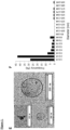

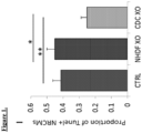


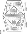
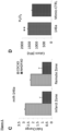


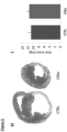

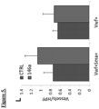


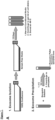


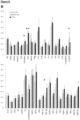

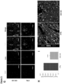


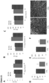


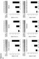


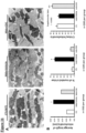

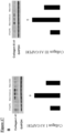
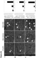


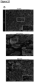
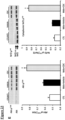


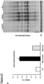


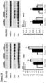


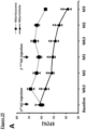


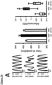


Classifications
-
- A—HUMAN NECESSITIES
- A61—MEDICAL OR VETERINARY SCIENCE; HYGIENE
- A61K—PREPARATIONS FOR MEDICAL, DENTAL OR TOILETRY PURPOSES
- A61K35/00—Medicinal preparations containing materials or reaction products thereof with undetermined constitution
- A61K35/12—Materials from mammals; Compositions comprising non-specified tissues or cells; Compositions comprising non-embryonic stem cells; Genetically modified cells
- A61K35/34—Muscles; Smooth muscle cells; Heart; Cardiac stem cells; Myoblasts; Myocytes; Cardiomyocytes
-
- A—HUMAN NECESSITIES
- A61—MEDICAL OR VETERINARY SCIENCE; HYGIENE
- A61K—PREPARATIONS FOR MEDICAL, DENTAL OR TOILETRY PURPOSES
- A61K31/00—Medicinal preparations containing organic active ingredients
- A61K31/70—Carbohydrates; Sugars; Derivatives thereof
- A61K31/7088—Compounds having three or more nucleosides or nucleotides
- A61K31/713—Double-stranded nucleic acids or oligonucleotides
-
- A—HUMAN NECESSITIES
- A61—MEDICAL OR VETERINARY SCIENCE; HYGIENE
- A61K—PREPARATIONS FOR MEDICAL, DENTAL OR TOILETRY PURPOSES
- A61K38/00—Medicinal preparations containing peptides
- A61K38/16—Peptides having more than 20 amino acids; Gastrins; Somatostatins; Melanotropins; Derivatives thereof
- A61K38/17—Peptides having more than 20 amino acids; Gastrins; Somatostatins; Melanotropins; Derivatives thereof from animals; from humans
- A61K38/1703—Peptides having more than 20 amino acids; Gastrins; Somatostatins; Melanotropins; Derivatives thereof from animals; from humans from vertebrates
- A61K38/1709—Peptides having more than 20 amino acids; Gastrins; Somatostatins; Melanotropins; Derivatives thereof from animals; from humans from vertebrates from mammals
-
- A—HUMAN NECESSITIES
- A61—MEDICAL OR VETERINARY SCIENCE; HYGIENE
- A61K—PREPARATIONS FOR MEDICAL, DENTAL OR TOILETRY PURPOSES
- A61K9/00—Medicinal preparations characterised by special physical form
- A61K9/0012—Galenical forms characterised by the site of application
- A61K9/0019—Injectable compositions; Intramuscular, intravenous, arterial, subcutaneous administration; Compositions to be administered through the skin in an invasive manner
-
- A—HUMAN NECESSITIES
- A61—MEDICAL OR VETERINARY SCIENCE; HYGIENE
- A61P—SPECIFIC THERAPEUTIC ACTIVITY OF CHEMICAL COMPOUNDS OR MEDICINAL PREPARATIONS
- A61P21/00—Drugs for disorders of the muscular or neuromuscular system
-
- A—HUMAN NECESSITIES
- A61—MEDICAL OR VETERINARY SCIENCE; HYGIENE
- A61P—SPECIFIC THERAPEUTIC ACTIVITY OF CHEMICAL COMPOUNDS OR MEDICINAL PREPARATIONS
- A61P9/00—Drugs for disorders of the cardiovascular system
- A61P9/10—Drugs for disorders of the cardiovascular system for treating ischaemic or atherosclerotic diseases, e.g. antianginal drugs, coronary vasodilators, drugs for myocardial infarction, retinopathy, cerebrovascula insufficiency, renal arteriosclerosis
-
- C—CHEMISTRY; METALLURGY
- C12—BIOCHEMISTRY; BEER; SPIRITS; WINE; VINEGAR; MICROBIOLOGY; ENZYMOLOGY; MUTATION OR GENETIC ENGINEERING
- C12N—MICROORGANISMS OR ENZYMES; COMPOSITIONS THEREOF; PROPAGATING, PRESERVING, OR MAINTAINING MICROORGANISMS; MUTATION OR GENETIC ENGINEERING; CULTURE MEDIA
- C12N15/00—Mutation or genetic engineering; DNA or RNA concerning genetic engineering, vectors, e.g. plasmids, or their isolation, preparation or purification; Use of hosts therefor
- C12N15/09—Recombinant DNA-technology
- C12N15/11—DNA or RNA fragments; Modified forms thereof; Non-coding nucleic acids having a biological activity
- C12N15/113—Non-coding nucleic acids modulating the expression of genes, e.g. antisense oligonucleotides; Antisense DNA or RNA; Triplex- forming oligonucleotides; Catalytic nucleic acids, e.g. ribozymes; Nucleic acids used in co-suppression or gene silencing
-
- C—CHEMISTRY; METALLURGY
- C12—BIOCHEMISTRY; BEER; SPIRITS; WINE; VINEGAR; MICROBIOLOGY; ENZYMOLOGY; MUTATION OR GENETIC ENGINEERING
- C12N—MICROORGANISMS OR ENZYMES; COMPOSITIONS THEREOF; PROPAGATING, PRESERVING, OR MAINTAINING MICROORGANISMS; MUTATION OR GENETIC ENGINEERING; CULTURE MEDIA
- C12N2310/00—Structure or type of the nucleic acid
- C12N2310/10—Type of nucleic acid
- C12N2310/14—Type of nucleic acid interfering N.A.
- C12N2310/141—MicroRNAs, miRNAs
-
- C—CHEMISTRY; METALLURGY
- C12—BIOCHEMISTRY; BEER; SPIRITS; WINE; VINEGAR; MICROBIOLOGY; ENZYMOLOGY; MUTATION OR GENETIC ENGINEERING
- C12N—MICROORGANISMS OR ENZYMES; COMPOSITIONS THEREOF; PROPAGATING, PRESERVING, OR MAINTAINING MICROORGANISMS; MUTATION OR GENETIC ENGINEERING; CULTURE MEDIA
- C12N2320/00—Applications; Uses
- C12N2320/30—Special therapeutic applications
- C12N2320/31—Combination therapy
-
- C—CHEMISTRY; METALLURGY
- C12—BIOCHEMISTRY; BEER; SPIRITS; WINE; VINEGAR; MICROBIOLOGY; ENZYMOLOGY; MUTATION OR GENETIC ENGINEERING
- C12N—MICROORGANISMS OR ENZYMES; COMPOSITIONS THEREOF; PROPAGATING, PRESERVING, OR MAINTAINING MICROORGANISMS; MUTATION OR GENETIC ENGINEERING; CULTURE MEDIA
- C12N2320/00—Applications; Uses
- C12N2320/30—Special therapeutic applications
- C12N2320/32—Special delivery means, e.g. tissue-specific
Definitions
- This invention relates to cells and their extracts, specifically cellular exosomes, for therapeutic use, including treatment of heart disease.
- Duchenne muscular dystrophy afflicts ⁇ 20,000 boys and young men in the USA.
- the central cause is a genetic abnormality in the dystrophin complex, with secondary damage to skeletal muscle and heart tissue. Although virtually all patients are treated with corticosteroids, no treatment has been proven effective.
- Heart failure (HF) secondary to DMD afflicts virtually all DMD patients aged >15 years, and is often the cause of death.
- DMD-associated HF aggressively progresses from the initial insult (a genetic abnormality in the dystrophin complex), to asymptomatic abnormalities in cardiac structure and function (stage B), to overt symptomatic HF (stage C), to advanced HF (stage D) and death.
- HF Progression of HF is associated with high risk of hospitalization and intense overall health care resource utilization. Modes of death during the course of DMD-associated HF include sudden cardiac death (which increases as HF worsens), or progressive HF culminating in circulatory collapse. Moreover, much of the disability in the later years of DMD is due to HF rather than to skeletal muscle disease. Thus, DMD HF represents an important, neglected target for innovative therapy.
- a highly promising avenue of therapy for cardiac-related diseases and conditions includes cardiosphere-derived cells (CDCs) that are capable of stimulating regeneration, angiogenesis, and functional improvement in infarcted myocardium.
- CDCs cardiosphere-derived cells
- Previous or ongoing trials involving CDCs target adult patients in stage B; wherein heart function is depressed, but symptoms of HF have yet to appear.
- DMD-associated HF therapeutic approaches may be most dramatic for stages C and D. These later stages of disease are associated with high mortality (>20% per year) despite optimal medical therapy, which have also never been shown to actually slow the progression of disease in DMD patients.
- heart transplantation is not an option for DMD patients. These patients are also not candidates for mechanical circulatory support devices.
- no treatment modality currently available addresses the underlying pathophysiology of DMD-associated HF, which is a loss of functional heart muscle and conversion of living heart muscle to scar.
- CDCs are rich biological factories that secrete many growth factors and cytokines, the beneficial therapeutic effects of CDCs manage to persist long after the injected cells have been cleared.
- these positive factors may exist in cellular exosomes produced by CDCs, the lipid bilayer nanovesicles secreted by cells when multivesicular endosomes fuse with the plasma membrane. Confirming a role for secreted exosomes in these processes has yet to be considered, and understanding these processes governing CDC-initiated regeneration may open new avenues therapeutic approaches.
- CDC-derived exosomes may effectively address a major unmet medical need, by recruiting various synergistic mechanisms of benefit that have been observed in animal models of HF. This includes the ability to attract endogenous stem cells to sites of myocardial injury, promote differentiation into heart muscle and vessels, and potentially reversing the pathophysiology of HF.
- the potential benefits of an exosome-based approach as an alternative to cell therapy is particularly compelling given the unavailability of conventional therapy to late stage patients.
- the possibility exists that CDC-derived exosomes may fundamentally alter the natural history of the disease.
- compositions and techniques related to generation and therapeutic application of CDC-derived exosomes contain a unique milieu of biological factors, including cytokines, growth factors, transcription factors, nucleic acids including non-coding nucleic acids such as microRNAs that serve to initiate and promote many of the therapeutic effects of CDCs.
- cytokines include cytokines, growth factors, transcription factors, nucleic acids including non-coding nucleic acids such as microRNAs that serve to initiate and promote many of the therapeutic effects of CDCs.
- nucleic acids including non-coding nucleic acids such as microRNAs that serve to initiate and promote many of the therapeutic effects of CDCs.
- the Inventors' work demonstrates that exosomes and their constituent microRNAs favorably modulate apoptosis, inflammation and fibrosis in the injured heart, leading to functional recovery and increase tissue viability.
- CDC-derived exosomes represent a novel "cell-free" therapeutic candidate for tissue repair.
- WO2014028493 discloses methods of repairing and/or regenerating damaged or diseased tissue comprising administering compositions comprising exosomes.
- the exosomes may comprise one or more microRNA that result in alterations in gene or protein expression, which in turn result in improved cell or tissue viability and/or function.
- WO2006007529 discloses methods and compositions for use in mediating an immunosuppressive reaction, wherein the compositions comprise exosomes having immunosuppressive activity. Such exosomes are used for the treatment of diseases and disorders associated with undesirable activation of the immune system. Also disclosed are exosomes isolated directly from immunosuppressive serum.
- US2011300112 discloses methods and compositions for the generation of therapeutic cells having reduced incidence of karyotypic abnormalities.
- pages E90 - E91 discloses a study wherein CDCs or CDC-XOs are transplanted into mice, indicating that cardiac function and exercise capacity can be improved in the mdx mice which are treated with CDCs or CDC-XOs.
- a composition comprising a plurality of exosomes, wherein the plurality of the exosomes are isolated from cardiosphere-derived cells (CDCs) grown in serum-free media, comprise exosomes with a diameter of 90 nm to 200 nm and are CD81 +, CD63+, or both, for use in a human in a method of treating progressive reduction in Left Ventricular Ejection Fraction (LVEF) heart failure secondary to a chronic degenerative muscular dystrophy, wherein the composition is cell-free and lacks CDCs.
- CDCs cardiosphere-derived cells
- composition comprising a plurality of cardiosphere-derived cell (CDC) exosomes from a human, for use in a human in a method of treating progressive reduction in LVEF heart failure secondary to a chronic degenerative muscular dystrophy, wherein the composition is cell-free and lacks CDCs.
- CDC cardiosphere-derived cell
- a method of treatment including selecting a subject in need of treatment for heart failure secondary to a chronic degenerative muscular disease and administering a composition including a plurality of exosomes to the subject, wherein the plurality of the exosomes are isolated from cardiosphere-derived cells (CDCs) grown in serum-free media, include exosomes with a diameter of about 90 nm to about 200 nm and are CD81+, CD63+, or both, and further wherein administration of the composition treats the subject.
- the chronic degenerative muscular disease is Duchenne muscular dystrophy.
- administering a composition includes about 1 to about 100 mg exosome protein in a single dose.
- administering a composition includes injection.
- the injection includes percutaneous injection.
- the injection is directly into heart muscle.
- administering a composition includes myocardial infusion.
- myocardial infusion is intra-arterial or intravenous.
- treatment of the subject results in decreased fibrosis, decreased inflammation, increased mitochondrial function and/or increased cardiomyogenesis.
- decreased fibrosis includes a reduction in collagen accumulation.
- collagen includes collagen I and/or collagen III.
- decreased inflammation includes an increase in cytoplasmic nuclear factor (erythroid-derived 2)-like 2 (Nrf2), reduction in fatty acid peroxidation end products, reduced numbers of inflammatory cells, and/or upregulated expression of antioxidants.
- antioxidants include heme oxygenase-1 (HO-1), catalase, superoxide dismutase-2 (SOD-2), and glutamate-cystein ligase catalytic (GCLC) subunit.
- inflammatory cells include CD68+ macrophages and CD3+ T-cells.
- increased mitochondrial function includes increased mitochondrial ultrastructure and/or increased mitochondrial biogenesis.
- increased mitochondrial function includes increased nuclear PPAR- ⁇ co-activator-1 (PGC-1) expression.
- the exosomes include one or more microRNAs selected from the group consisting of: microRNAs miR-146a, miR148a, miR22, miR-24, miR-210, miR-150, miR-140-3p, miR-19a, miR-27b, miR-19b, miR-27a, miR-376c, miR-128, miR-320a, miR-143, miR-21, miR-130a, miR-9, miR-185, and miR-23a.
- a method of treatment including selecting a subject in need of treatment for heart failure secondary to a chronic muscular disease and administering a composition including cardiosphere-derived cells (CDCs), wherein administration of the composition treats the subject.
- the chronic muscular disease is Duchenne muscular dystrophy.
- administering a composition includes about 1 ⁇ 10 5 to about 1 ⁇ 10 8 or more CDCs in a single dose.
- administering a composition includes myocardial infusion.
- myocardial infusion is intracoronary.
- myocardial infusion is intra-arterial or intravenous.
- treatment of the subject results in decreased fibrosis, decreased inflammation, increased mitochondrial function and/or increased cardiomyogenesis.
- decreased fibrosis includes a reduction in collagen accumulation.
- collagen includes collagen I and/or collagen III.
- decreased inflammation includes an increase in cytoplasmic nuclear factor (erythroid-derived 2)-like 2 (Nrf2), reduction in fatty acid peroxidation end products, reduced numbers of inflammatory cells, and/or upregulated expression of antioxidants.
- antioxidants include heme oxygenase-1 (HO-1), catalase, superoxide dismutase-2 (SOD-2), and glutamate-cystein ligase catalytic (GCLC) subunit.
- inflammatory cells include CD68+ macrophages and CD3+ T-cells.
- increased mitochondrial function includes increased mitochondrial ultrastructure and/or increased mitochondrial biogenesis.
- increased mitochondrial function includes increased nuclear PPAR- ⁇ co-activator-1 (PGC-1) expression.
- Duchenne muscular dystrophy a crippling genetic disease leading to premature death, affects the heart as well as skeletal muscle. Indeed, cardiomyopathy is the leading cause of death in Duchenne patients. There are no approved treatments for the cardiomyopathy, and novel Duchenne-specific experimental approaches such as exon skipping do not benefit the heart.
- cardiosphere-derived cells CDCs
- Exosomes secreted by human CDCs reproduce the benefits of CDCs in mdx mice, and reverse abnormalities of calcium cycling and mitochondrial respiration in human Duchenne cardiomyocytes.
- DMD Duchenne muscular dystrophy
- CDCs Cardiosphere-derived cells
- CDCs are not only regenerative, but also anti-inflammatory and anti-fibrotic; they work indirectly via the secretion of exosomes laden with noncoding RNA including microRNAs (miRs).
- miRs microRNAs
- CDC-exosomes mimic the functional and structural benefits of CDCs, while blockade of exosome biosynthesis renders CDCs ineffective.
- the Inventors reasoned that CDCs might be useful in treating Duchenne cardiomyopathy. The goal is not to replace dystrophin, but rather to offset the pathophysiological consequences of dystrophin deletion, by recruiting regeneration, reversing fibrosis and targeting inflammation.
- Exosomes secreted lipid vesicles containing a rich milieu of biological factors, provide powerful paracrine signals by which stem cells potentiate their biological effects to neighboring cells, including diseased or injured cells.
- bio-active lipid and nucleic acid "cargo"
- these natural delivery devices are capable of inducing significant phenotypic and functional changes in recipient cells that lead to activation of regenerative programs.
- the role of such indirect mechanisms to in stem cell initiated regeneration is strongly suggested by growing evidence that after stem cell administration and clearance from delivery sites in tissue and organs, regeneration processes nevertheless persist and arise from endogenous tissues.
- Stem cell-derived exosomes have been identified and isolated from supernatants of several cell types with demonstrated therapeutic potential, including mesenchymal stromal (MSC), (bone marrow stem cells) mononuclear (MNC) cells, immune cells (dendritic and CD34+), human neural stem cells (hNSCs), among others.
- MSC mesenchymal stromal
- MNC mononuclear
- hNSCs human neural stem cells
- human cardiosphere derived cells CDCs
- Stem cell-derived exosomes including those produced by CDCs, may provide a potent and rich source for developing "cell-free" therapies.
- exosome-based, "cell-free” therapies in contrast to cell therapy, provide distinct advantages in regenerative medicine.
- these features significantly obviate safety issues.
- stem cell-derived exosomes are less immunogenic than parental cells, as a result of a lower content of membrane-bound proteins, including MHC complex molecules. Replacing the administration of live cells with their secreted exosomes, mitigates many of the safety concerns and limitations associated with the transplantation of viable replicating cells.
- exosome encapsulation of bioactive components in lipid vesicles allows protection of contents from degradation in vivo, thereby potentially negating obstacles often associated with delivery of soluble molecules such as cytokines, growth factors, transcription factors and RNAs. This comparative ease of administration may ultimately allow for repeated and sustained delivery to patients, thereby maximizing the potential for regeneration and repair of diseased and/or dysfunctional tissue.
- exosome production under defined conditions allows for easier manufacture and scale-up opportunity.
- Manufacture of exosomes is akin to conventional biopharmacological product manufacture, allowing for standardization in production and quality control for dosage and biological activity testing.
- the durability of exosomes in culture allows for the acquisition of large quantities of exosomes through their collection from a culture medium in which the exosomes are secreted over periods of time.
- exosomes are involved in intercellular communication between different cell types, much remains to be discovered in regard to the mechanisms of their production within parental cells of origin and effects on target recipient cells. Exosomes have been reported to be involved in numerous cellular, tissue and physiological processes, including immune modulating processes, angiogenesis, migration of endothelial cells in connection with tumor growth, or reducing damage in ischemia reperfusion injury. Of critical scientific interest in establishing whether exosomes secreted by cells, such as cardiosphere-derived cells (CDCs), are capable of reproducing the therapeutic benefits of their parental cells, or possibly, are indispensable in effectuating such therapeutic benefits.
- CDCs cardiosphere-derived cells
- exosomes secreted by a wide range of cell types, exosomes are lipid bilayer vesicles that are enriched in a variety of biological factors, including cytokines, growth factors, transcription factors, lipids, and coding and non-coding nucleic acids. Exosomes are found in blood, urine, amniotic fluid, interstitial and extracellular spaces. These exocytosed vesicles of endosomal origin can range in size between 30-200 nm, including sizes of 40-100 nm, and possess a cup-shaped morphology, as revealed by electron microscopy.
- MVB multivesicular bodies
- exosomes reflect their parental cellular origin, as containing distinct subsets of biological factors in connection with their parent cellular origin, including the cell regulatory state of the parental cells when formed.
- the rich biological milieu of different proteins, including cytokines and growth factors, lipids, coding and noncoding RNA molecules, within exosomes are all necessarily derived from their parental cells.
- exosomes further express the extracellular domain of membrane-bound receptors at the surface of the membrane.
- endosome-associated proteins e.g., Rab GTPase, SNAREs, Annexins, and flotillin
- proteins that are known to cluster into microdomains at the plasma membrane or at endosomes four transmembrane domain tetraspanins, e.g., CD63, CD81, CD82, CD53, and CD37
- lipid raft associated proteins e.g., glycosylphosphatidylinositol- anchored proteins and flotillin
- cholesterol sphingomyelin
- hexosylceramides as examples.
- exosomes In addition to these core components reflecting their vesicle origin, a critical property of exosomes is a demonstrated capability to contain both mRNA and microRNA associated with signaling processes, with both cargo mRNA being capable to translation in recipient cells, or microRNA functionally degrading target mRNA in recipient cells. Other noncoding RNAs, capable for influencing gene expression, may also be present in exosomes. While the processes governing the selective incorporation of mRNA or microRNA populations into exosomes is not entirely understood, it is clear that RNA molecules are selectively, not randomly incorporated into exosomes, as revealed by studies reporting enrichment of exosome cargo RNAs when compared to the RNA profiles of the originating cells. Given the growing understanding of how such RNA molecules play a role in disease pathogenesis and regenerative processes, the presence of RNA molecules in exosomes and apparent potency in affecting target recipient cells suggests critical features that can be deployed in therapeutic approaches.
- the natural bilayer membrane encapsulation of exosomes provides a protected and controlled internal microenvironment that allows cargo contents to persist or migrate in the bloodstream or within tissues without degradation.
- Their release into the extracellular environment allows for interaction with recipient cells via adhesion to the cell surface mediated by lipid-ligand receptor interactions, internalization via endocytic uptake, or by direct fusion of the vesicles and cell membrane. These processes lead to the release of exosome cargo content into the target cell.
- exosome-cell interactions modulation of genetic pathways in the target recipient cell, as induced through any of several different mechanisms including antigen presentation, the transfer of transcription factors, cytokines, growth factors, nucleic acid such as mRNA and microRNAs.
- ESC embryonic stem cell
- mRNA and proteins to hematopoietic progenitors.
- Other studies have shown that adult stem cell-derived exosomes also shuttle selected patterns of mRNA, microRNA and pre-microRNA associated with several cellular functions involved in the control of transcription, proliferation and cell immune regulation.
- Exosome isolation relies on exploiting their generic biochemical and biophysical features for separation and analysis. For example, differential ultracentrifugation has become a leading technique wherein secreted exosomes are isolated from the supernatants of cultured cells. This approach allows for separation of exosomes from nonmembranous particles, by exploiting their relatively low buoyant density. Size exclusion allows for their separation from biochemically similar, but biophysically different microvesicles, which possess larger diameters of up to 1,000 nm. Differences in flotation velocity further allows for separation of differentially sized exosomes. In general, exosome sizes will possess a diameter ranging from 30-200 nm, including sizes of 40-100 nm. Further purification may rely on specific properties of the particular exosomes of interest. This includes, for example, use of immunoadsorption with a protein of interest to select specific vesicles with exoplasmic or outward orientations.
- differential ultracentrifugation is the most commonly used for exosome isolation. This technique utilizes increasing centrifugal force from 2000xg to 10,000xg to separate the medium- and larger-sized particles and cell debris from the exosome pellet at 100,000xg. Centrifugation alone allows for significant separation/collection of exosomes from a conditioned medium, although it is insufficient to remove various protein aggregates, genetic materials, particulates from media and cell debris that are common contaminants.
- Enhanced specificity of exosome purification may deploy sequential centrifugation in combination with ultrafiltration, or equilibrium density gradient centrifugation in a sucrose density gradient, to provide for the greater purity of the exosome preparation (flotation density 1.1-1.2g/ml) or application of a discrete sugar cushion in preparation.
- ultrafiltration can be used to purify exosomes without compromising their biological activity.
- Membranes with different pore sizes - such as 100 kDa molecular weight cut-off (MWCO) and gel filtration to eliminate smaller particles - have been used to avoid the use of a nonneutral pH or non-physiological salt concentration.
- MWCO molecular weight cut-off
- THF tangential flow filtration
- HPLC can also be used to purify exosomes to homogeneouslysized particles and preserve their biological activity as the preparation is maintained at a physiological pH and salt concentration.
- exosomes for precipitation techniques, addition to volume-excluding polymers (e.g., polyethylene glycols (PEGs)), possibly combined additional rounds of centrifugation or filtration.
- a precipitation reagent e.g., ExoQuick ®
- ExoQuick ® can be added to conditioned cell media to quickly and rapidly precipitate a population of exosomes, although re-suspension of pellets prepared via this technique may be difficult.
- Flow field-flow fractionation is an elution-based technique that is used to separate and characterize macromolecules (e.g., proteins) and nano-to micro-sized particles (e.g., organelles and cells) and which has been successfully applied to fractionate exosomes from culture media.
- macromolecules e.g., proteins
- nano-to micro-sized particles e.g., organelles and cells
- exosomes may be applied to isolated specific exosomes of interest. This includes relying on antibody immunoaffinity to recognizing certain exosome-associated antigens. As described, exosomes further express the extracellular domain of membrane-bound receptors at the surface of the membrane. This presents a ripe opportunity for isolating and segregating exosomes in connections with their parental cellular origin, based on a shared antigenic profile. Conjugation to magnetic beads, chromatography matrices, plates or microfluidic devices allows isolating of specific exosome populations of interest as may be related to their production from a parent cell of interest or associated cellular regulatory state. Other affinity-capture methods use lectins which bind to specific saccharide residues on the exosome surface.
- Exosome-Based Therapies A chief goal of developing exosome-based therapy is the creation of "cell-free" therapies, wherein the benefits of cellular therapeutics can be provided with reduced risks or in scenarios in which cell therapy would be unavailable.
- DMD Duchenne muscular dystrophy
- HF heart failure
- the therapeutic benefits of cell-based therapies such as cardiosphere-derived cells (CDCs) appear to occur through indirect mechanisms involving regenerated myocardium and vasculature arising from endogenous origin.
- CDCs cardiosphere-derived cells
- CDC-derived exosomes may effectively address a major unmet medical need, by recruiting synergistic mechanisms to attract endogenous stem cells to sites of myocardial injury, promote differentiation into heart muscle and vessels, thereby reversing the pathophysiology of HF.
- DMD is an X-linked recessive disorder characterized by myopathy (cell membrane damage in muscle fiber) as exemplified by a variety of pathological features. This includes skeletal muscle weakness starting 3-5 years from onset, progressive weakness, wheelchair dependency at approximately 13 years from onset. Importantly, cardiomyopathy is observed to take hold in 1/3 of patients from less than 13 years from onset, increasing to 1/2 of patients less than 18 years from onset, and in all patients after 18 years. Dilated cardiomyopathy includes left ventricle posterobasal fibrosis; conduction abnormalities are mainly intra-atrial: SVT with abnormal AV nodal conduction. Patients may further suffer from smooth muscle myopathy including vascular dysfunction, further including GI and urinary tract systems involvement.
- dystrophin gene mutation deletion
- loss of dystrophin results in cellular membrane damage and leakage of extracellular Ca 2+ into the cell.
- Elevated intracellular levels ultimately result in increased oxidative and/or nitrosative stress and inflammation, and activation of calpain.
- the combination of these effects results in muscle proteolysis and apoptosis, leading to the degradative features described above.
- CDCs have been demonstrated as promoting antioxidative, anti-inflammatory, anti-apoptotic, anti-remodeling effects, in addition to enhancing regenerative capacity.
- CDC administration is beneficial in retarding/reversing DMD, and exosome populations derived from CDCs may allow for these benefits to be delivered, while avoiding obstacles associated with cell-based therapy.
- stem cell-derived exosomes are likely to be less immunogenic than parental cells.
- exosome encapsulation of bioactive components in lipid vesicles allows protection of contents from degradation in vivo, thereby potentially negating obstacles often associated with delivery of soluble molecules such as cytokines, growth factors, transcription factors and RNAs, while potentially allowing for increased concentrations to be provided.
- soluble molecules such as cytokines, growth factors, transcription factors and RNAs
- compositions and methods and compositions providing significant benefits in the repair or regeneration of damaged or diseased tissues via "cell-free” methods involving exosomes.
- human cardiosphere-derived cells (CDC)-derived exosomes are demonstrated as effective in reducing scar size and regenerating viable myocardium.
- CDC cardiosphere-derived cells
- Described herein is a method of treatment, including selecting a subject in need of treatment for heart failure secondary to a chronic degenerative muscular disease and administering a composition including a plurality of exosomes to the subject, wherein the plurality of the exosomes are isolated from cardiosphere-derived cells (CDCs) grown in serum-free media, include exosomes with a diameter of about 90 nm to about 200 nm and are CD81+, CD63+, or both, and further wherein administration of the composition treats the subject.
- the chronic degenerative muscular disease is Duchenne muscular dystrophy.
- administering a composition includes about 1 to about 100 mg exosome protein in a single dose.
- administering a composition includes injection.
- the injection includes percutaneous injection.
- the injection is directly into heart muscle.
- administering a composition includes myocardial infusion.
- myocardial infusion is intra-arterial or intravenous.
- treatment of the subject results in decreased fibrosis, decreased inflammation, increased mitochondrial function and/or increased cardiomyogenesis.
- decreased fibrosis includes a reduction in collagen accumulation.
- collagen includes collagen I and/or collagen III.
- decreased inflammation includes an increase in cytoplasmic nuclear factor (erythroid-derived 2)-like 2 (Nrf2), reduction in fatty acid peroxidation end products, reduced numbers of inflammatory cells, and/or upregulated expression of antioxidants.
- antioxidants include heme oxygenase-1 (HO-1), catalase, superoxide dismutase-2 (SOD-2), and glutamate-cystein ligase catalytic (GCLC) subunit.
- inflammatory cells include CD68+ macrophages and CD3+ T-cells.
- increased mitochondrial function includes increased mitochondrial ultrastructure and/or increased mitochondrial biogenesis.
- increased mitochondrial function includes increased nuclear PPAR- ⁇ co-activator-1 (PGC-1) expression.
- the exosomes include one or more microRNAs selected from the group consisting of: microRNAs miR-146a, miR148a, miR22, miR-24, miR-210, miR-150, miR-140-3p, miR-19a, miR-27b, miR-19b, miR-27a, miR-376c, miR-128, miR-320a, miR-143, miR-21, miR-130a, miR-9, miR-185, and miR-23a.
- a composition including cardiosphere-derived cells including cardiosphere-derived cells (CDCs), wherein administration of the composition treats the subject.
- the chronic muscular disease is Duchenne muscular dystrophy.
- administering a composition includes about 1 ⁇ 10 5 to about 1 ⁇ 10 8 or more CDCs in a single dose.
- the number of administered CDCs includes intracoronary 25 million CDCs per coronary artery (i.e., 75 million CDCs total) as another baseline for exosome dosage quantity.
- the numbers of CDCs includes 1 ⁇ 10 5 , 1 ⁇ 10 6 , 1 ⁇ 10 7 , 1 ⁇ 10 8 , 1 ⁇ 10 9 CDCs in a single dose as another baseline for exosome dosage quantity. In certain instances, this may be prorated to body weight (range 100,000-1M CDCs/kg body weight total CDC dose).
- administering a composition includes myocardial infusion.
- myocardial infusion is intracoronary.
- myocardial infusion is intra-arterial or intravenous.
- treatment of the subject results in decreased fibrosis, decreased inflammation, increased mitochondrial function and/or increased cardiomyogenesis.
- decreased fibrosis includes a reduction in collagen accumulation.
- collagen includes collagen I and/or collagen III.
- decreased inflammation includes an increase in cytoplasmic nuclear factor (erythroid-derived 2)-like 2 (Nrf2), reduction in fatty acid peroxidation end products, reduced numbers of inflammatory cells, and/or upregulated expression of antioxidants.
- antioxidants include heme oxygenase-1 (HO-1), catalase, superoxide dismutase-2 (SOD-2), and glutamate-cystein ligase catalytic (GCLC) subunit.
- inflammatory cells include CD68+ macrophages and CD3+ T-cells.
- increased mitochondrial function includes increased mitochondrial ultrastructure and/or increased mitochondrial biogenesis. In other embodiments, increased mitochondrial function includes increased nuclear PPAR- ⁇ co-activator-1 (PGC-1) expression. Further examples are found in U.S. App. No. 11/666,685 , 12/622,143 , and 12/622,106 .
- compositions including a plurality of exosomes.
- the plurality of exosomes are generated by a method including providing a population of cells, and isolating a plurality of exosomes from the population of cells.
- the cells are stem cells, progenitors and/or precursors.
- the stem cells, progenitors and/or precursors are cardiosphere-derived cells (CDCs).
- the stem cells, progenitors and/or precursors are pluripotent stem cells (pSCs), such as embryonic stem cells (ESCs) and induced pluripotent stem cells (iPSCs) derived from any one of various somatic sources in the body such as fibroblasts, blood and hematopoietic stem cells (hSCs), immune cells, bone and bone marrow, neural tissue, among others.
- pSCs pluripotent stem cells
- ESCs embryonic stem cells
- iPSCs induced pluripotent stem cells derived from any one of various somatic sources in the body
- fibroblasts fibroblasts
- hSCs hematopoietic stem cells
- immune cells bone and bone marrow, neural tissue, among others.
- the stem cells, progenitors and/or precursors include hSCs, mesenchymal stem cells (MSCs), or endothelial precursor cells (EPCs).
- the cells are stem cells, progenitors and/or precursors derived from human biopsy tissue.
- the cells are stem cells, progenitors and/or precursors are a primary culture.
- the cells are stem cells, progenitors and/or precursors which may constitute a cell line capable of serial passaging.
- the plurality of exosomes is isolated from the supernatants of the population of cells. This includes, for example, exosomes secreted into media as conditioned by a population of cells in culture, further including cell lines capable of serial passaging.
- the cells are cultured in serum-free media.
- the cells in culture are grown to 10, 20, 30, 40, 50, 60, 70, 80, 90, or 90% or more confluency when exosomes are isolated.
- the population of cells has been genetically manipulated. This includes, for example, knockout (KO) or transgenic (TG) cell lines, wherein an endogenous gene has been removed and/or an exogenous introduced in a stable, persistent manner.
- the population of cells has been altered by exposure to environmental conditions (e.g., hypoxia), small molecule addition, presence/absence of exogenous factors (e.g., growth factors, cytokines) at the time, or substantially contemporaneous with, isolating the plurality of exosomes in a manner altering the regulatory state of the cell.
- environmental conditions e.g., hypoxia
- exogenous factors e.g., growth factors, cytokines
- altering the regulatory state of the cell changes composition of one or more exosomes in the plurality of exosomes.
- the plurality of exosomes include one or more exosomes that are about 10 nm to about 250 nm in diameter, including those about 10 nm to about 15 nm, about 15 nm to about 20 nm, about 20 nm to about 25 nm, about 25 nm to about 30 nm, about 30 nm to about 35 nm, about 35 nm to about 40 nm, about 40 nm to about 50 nm, about 50 nm to about 60 nm3 about 60 nm to about 70 nm, about 70 nm to about 80 nm, about 80 nm to about 90 nm, about 90 nm to about 95 nm, about 95 nm to about 100 nm, about 100 nm to about 105 nm, about 105 nm to about 110 nm, about 110 nm to about 115 nm, about 115 nm to about 120 nm, about 120 nm to about
- the plurality of exosomes includes one or more exosomes expressing a biomarker.
- the biomarkers are tetraspanins.
- the tetraspanins are one or more selected from the group including CD63, CD81, CD82, CD53, and CD37.
- the exosomes express one or more lipid raft associated protiens (e.g., glycosylphosphatidylinositol-anchored proteins and flotillin), cholesterol, sphingomyelin, and/or hexosylceramides.
- the plurality of exosomes includes one or more exosomes containing a biological protein.
- the biological protein includes transcription factors, cytokines, growth factors, and similar proteins capable of modulating signaling pathways in a target cell.
- the biological protein is capable of facilitating regeneration and/or improved function of a tissue.
- the biological protein is capable of modulating pathways related to Irak1, Traf6, toll-like receptor (TLR) signaling pathway, NOX-4, SMAD-4, and/or TGF- ⁇ .
- TLR Irak1, Traf6, toll-like receptor
- the biological protein related to exosome formation and packaging of cytosolic proteins such as Hsp70, Hsp90, 14-3-3 epsilon, PKM2, GW182 and AGO2.
- the plurality of exosomes includes one or more exosomes containing a signaling lipid. This includes ceramide and derivatives. In other embodiments, the plurality of exosomes includes one or more exosomes containing a coding and/or non-coding nucleic acid.
- the plurality of exosomes includes one or more exosomes containing microRNAs.
- these microRNAs can include miR-146a, miR22, miR-24, miR-210, miR-150, miR-140-3p, miR-19a, miR-27b, miR-19b, miR-27a, miR-376c, miR-128, miR-320a, miR-143, miR-21, miR-130a, miR-9, miR-185, and/or miR-23a.
- the plurality of exosomes include one or more exosomes enriched in at least one of miR-146a, miR22, miR-24, miR-210, miR-150, miR-140-3p, miR-19a, miR-27b, miR-19b, miR-27a, miR-376c, miR-128, miR-320a, miR-143, miR-21, miR-130a, miR-9, miR-185, and/or miR-23a.
- the plurality of exosomes includes one or more exosomes enriched in at least one of miR-146a, miR-22, miR-24.
- Enrichment can be measured by, for example, comparing the amount of one or more of the described microRNAs when derived from cells providing salutary benefit in a therapeutic setting (e.g., cardiosphere-derived cells (CDCs) compared to cells that do not provide such a salutary benefit (e.g., fibroblasts). Enrichment may also be measured in absolute or relative quantities, such as when compared to a standardized dilution series.
- a therapeutic setting e.g., cardiosphere-derived cells (CDCs)
- fibroblasts e.g., fibroblasts
- the plurality of exosomes can include one or more exosomes containing microRNAs.
- the plurality of exosomes can include one or more exosomes containing microRNAs.
- microRNAs known in the art, such as miR-17, miR-21 , miR-92, miR92a, miR-29, miR-29a, miR-29b, miR-29c, miR-34, mi-R34a, miR-150, miR-451, miR-145, miR-143, miR-144, miR-193a-3p, miR-133a, miR-155, miR-181a, miR-214, miR-199b, miR-199a, miR-210, miR-126, miR-378, miR-363 and miR-30b, and miR-499.
- microRNAs known in the art include miR-92, miR-17, miR-21, miR-92, miR92a, miR-29, miR- 29a, miR-29b, miR-29c, miR-34, mi-R34a, miR-150, miR-451, miR-145, miR-143, miR- 144, miR-193a-3p, miR-133a, miR-155, miR-181a, miR-214, miR-199b, miR-199a, miR- 126, miR-378, miR-363 and miR-30b, and/or miR-499.
- isolating a plurality of exosomes from the population of cells includes centrifugation of the cells and/or media conditioned by the cells. In several embodiments, ultracentrifugation is used. In several embodiments, isolating a plurality of exosomes from the population of cells is via size-exclusion filtration. In other embodiments, isolating a plurality of exosomes from the population of cells includes use of discontinuous density gradients, immunoaffinity, ultrafiltration and/or high performance liquid chromatography (HPLC).
- HPLC high performance liquid chromatography
- differential ultracentrifugation includes using centrifugal force from 1000-2000xg, 2000-3000xg, 3000-4000xg, 4000-5000xg, 5000xg-6000xg, 6000-7000xg, 7000-8000xg, 8000-9000xg, 9000-10,000xg, to 10,000xg or more to separate larger-sized particles from a plurality of exosome derived from the cells.
- differential ultracentrifugation includes using centrifugal force from 10,000-20,000xg, 20,000-30,000xg, 30,000-40,000xg, 40,000-50,000xg, 50,000xg-60,000xg, 60,000-70,000xg, 70,000-80,000xg, 80,000-90,000xg, 90,000-100,000xg, to 10,000xg or more to separate larger-sized particles from a plurality of exosome derived from the cells.
- isolating a plurality of exosomes from the population of cells includes use of filtration or ultrafiltration.
- a size exclusion membrane with different pore sizes is used.
- a size exclusion membrane can include use of a filter with a pore size of 0.1-0.5 ⁇ M, 0.5-1.0 ⁇ M, 1-2.5 ⁇ M, 2.5-5 ⁇ M, 5 or more ⁇ M. In certain embodiments, the pore size is about 0.2 ⁇ M.
- filtration or ultrafiltration includes size exclusion ranging from 100-500 daltons (Da), 500-1 kDa, 1-2 kDa, 2-5 kDa, 5-10 kDa, 10-25 kDa, 25-50 kDa, 50-100 kDa, 100-250 kDa, 250-500 kDa, 500 or more kDa.
- the size exclusion is for about 2-5 kDa. In certain embodiments, the size exclusion is for about 3 kDa.
- filtration or ultrafiltration includes size exclusion includes use of hollow fiber membranes capable of isolating particles ranging from 100-500 daltons (Da), 500-1 kDa, 1-2 kDa, 2-5 kDa, 5-10 kDa, 10-25 kDa, 25-50 kDa, 50-100 kDa, 100-250 kDa, 250-500 kDa, 500 or more kDa.
- the size exclusion is for about 2-5 kDa. In certain embodiments, the size exclusion is for about 3 kDa.
- a molecular weight cut-off (MWCO) gel filtration capable of isolating particles ranging from 100-500 daltons (Da), 500-1 kDa, 1-2 kDa, 2-5 kDa, 5-10 kDa, 10-25 kDa, 25-50 kDa, 50-100 kDa, 100-250 kDa, 250-500 kDa, 500 or more kDa.
- the size exclusion is for about 2-5 kDa. In certain embodiments, the size exclusion is for about 3 kDa. In various embodiments, such systems are used in combination with variable fluid flow systems.
- isolating a plurality of exosomes from the population of cells includes use of tangential flow filtration (TFF) systems are used purify and/or concentrate the exosome fractions.
- isolating a plurality of exosomes from the population of cells includes use of (HPLC) can also be used to purify exosomes to homogeneously sized particles.
- density gradients as used such as centrifugation in a sucrose density gradient or application of a discrete sugar cushion in preparation.
- isolating a plurality of exosomes from the population of cells includes use of a precipitation reagent.
- a precipitation reagent ExoQuick ®
- ExoQuick ® can be added to conditioned cell media to quickly and rapidly precipitate a population of exosomes.
- isolating a plurality of exosomes from the population of cells includes use of volume-excluding polymers (e.g., polyethylene glycols (PEGs)) are used.
- isolating a plurality of exosomes from the population of cells includes use of flow field-flow fractionation (FlFFF), an elution-based technique.
- FlFFF flow field-flow fractionation
- isolating a plurality of exosomes from the population of cells includes use of one or more capture agents to isolate one or more exosomes possessing specific biomarkers or containing particular biological molecules.
- one or more capture agents include at least one antibody.
- antibody immunoaffinity recognizing exosome-associated antigens is used to capture specific exosomes.
- the at least one antibody are conjugated to a fixed surface, such as magnetic beads, chromatography matrices, plates or microfluidic devices, thereby allowing isolation of the specific exosome populations of interest.
- isolating a plurality of exosomes from the population of cells includes use of one or more capture agents that is not an antibody.
- the non-antibody capture agent is a lectin capable of binding to polysaccharide residues on the exosome surface.
- the CDCs are mammalian. In other embodiments, the CDCs are human. As disclosed above, in some embodiments, synthetic exosomes are generated, which can be isolated by similar mechanisms as those above. In various embodiments, the composition that is a plurality of exosomes is a pharmaceutical composition further including a pharmaceutically acceptable carrier.
- the plurality of exosomes range in size from 30 to 300 nm. In various embodiments, the plurality of exosomes range in size from 40 to 100 nm. In certain embodiments, the plurality of exosomes is cardiosphere-derived cell (CDC) exosomes. In certain embodiments, the plurality of exosomes includes exosomes that are CD63+.
- CDC cardiosphere-derived cell
- the exosomes include microRNAs miR-146a, miR22, miR-24, miR-210, miR-150, miR-140-3p, miR-19a, miR-27b, miR-19b, miR-27a, miR-376c, miR-128, miR-320a, miR-143, miR-21, miR-130a, miR-9, miR-185, and/or miR-23a.
- the exosomes are 2-5 kDa, such as 3 kDa.
- Other examples or embodiments relating to the composition and techniques involving exosomes are presented, in PCT Pub. No. WO 2014/028,493 .
- Described herein is a method for treatment including, selecting a subject in need of treatment, administering a composition including a plurality of exosomes to the individual, wherein the administration of the composition treats the subject.
- the subject is in need to treatment for a disease and/or condition involving tissue damage or dysfunction.
- the disease and/or condition involving tissue damage or dysfunction is heart disease.
- the plurality of exosomes includes exosomes including one or more microRNAs.
- the plurality of exosomes are generated by a method including providing a population of cells, and isolating a plurality of exosomes from the population of cells.
- the cells are stem cells, progenitors and/or precursors.
- the stem cells, progenitors and/or precursors are cardiosphere-derived cells (CDCs).
- the stem cells, progenitors and/or precursors are pluripotent stem cells (pSCs), such as embryonic stem cells (ESCs) and induced pluripotent stem cells (iPSCs) derived from any one of various somatic sources in the body such as fibroblasts, blood and hematopoietic stem cells (hSCs), immune cells, bone and bone marrow, neural tissue, among others.
- pSCs pluripotent stem cells
- ESCs embryonic stem cells
- iPSCs induced pluripotent stem cells
- the stem cells, progenitors and/or precursors includes hSCs, mesenchymal stem cells (MSCs), or endothelial precursor cells (EPCs).
- the cells are stem cells, progenitors and/or precursors derived from human biopsy tissue.
- the cells are stem cells, progenitors and/or precursors are a primary culture. In various embodiments, the cells are stem cells, progenitors and/or precursors are a cell line capable of serial passaging. In certain embodiments, the exosomes are synthetic.
- the plurality of exosomes is derived from cardiosphere-derived cells (CDCs). In other embodiments, the plurality of exosomes includes exosomes including one or more biological molecules. In other embodiments, the plurality of exosomes includes exosomes enriched for one or more biological molecules when derived from CDCs compared to exosome derived from non-CDC sources. In various embodiments, the one or more biological molecules are proteins, growth factors, cytokines, transcription factors and/or morphogenic factors. In other embodiments, the plurality of exosomes includes exosomes enriched for one or more biological molecules includes microRNAs, further including microRNAs that are enriched when derived from CDCs compared to exosome derived from non-CDC sources.
- these microRNAs can include miR-146a, miR22, miR-24, miR-210, miR-150, miR-140-3p, miR-19a, miR-27b, miR-19b, miR-27a, miR-376c, miR-128, miR-320a, miR-143, miR-21, miR-130a, miR-9, miR-185, and/or miR-23a.
- the plurality of exosomes includes one or more exosomes enriched in at least one of miR-146a, miR-22, miR-24.
- the CDCs are mammalian. In other embodiments, the CDCs are human.
- the exosomes are synthetic. In certain embodiments, the synthetic exosomes possess substantially similar content (e.g., microRNAs, biological molecules) as exosomes derived from CDCs.
- administration of the plurality of exosomes alters gene expression in the damaged or dysfunctional tissue, improves viability of the damaged tissue, and/or enhances regeneration or production of new tissue in the individual.
- the quantities of exosomes that are administered to achieved these effects range from 1 ⁇ 10 6 to 1 ⁇ 10 7 , 1 ⁇ 10 7 to 1 ⁇ 10 8 , 1 ⁇ 10 8 to 1 ⁇ 10 9 , 1 ⁇ 10 9 to 1 ⁇ 10 10 , 1 ⁇ 10 10 to 1 ⁇ 10 11 , 1 ⁇ 10 11 to 1 ⁇ 10 12 , 1 ⁇ 10 12 or more.
- the numbers of exosomes is relative to the number of cells used in a clinically relevant dose for a cell-therapy method.
- 3mL / 3 ⁇ 10 5 CDCs is capable of providing therapeutic benefit in intracoronary administration, and therefore, a plurality of exosomes as derived from that number of cells in a clinically relevant dose for a cell-therapy method.
- administration can be in repeated doses.
- the number of administered CDCs includes intracoronary 25 million CDCs per coronary artery (i.e., 75 million CDCs total) as another baseline for exosome dosage quantity.
- the numbers of CDCs includes 1 ⁇ 10 5 , 1 ⁇ 10 6 , 1 ⁇ 10 7 , 1 ⁇ 10 8 , 1 ⁇ 10 9 CDCs in a single dose as another baseline for exosome dosage quantity. In certain instances, this may be prorated to body weight (range 100,000-1M CDCs/kg body weight total CDC dose).
- exosome quantity may be defined by protein quantity, such as dosages including 1-10, 10-25, 25-50, 50-75, 75-100, or 100 or more mg exosome protein.
- Defining an effective dose range, dosing regimen and route of administration may be guided by studies using fluorescently labeled exosomes, and measuring target tissue retention, which can be >10X, >50X, or >100X background, as measured 5, 10, 15, 30, or 30 or more min as a screening criterion.
- >100X background measured at 30 mins is a baseline measurement for a low and high dose that is then assess for safety and bioactivity (e.g., using MRI endpoints: scar size, global and regional function).
- single doses are compared to two, three, four, four or more sequentially-applied doses.
- the repeated or sequentially-applied doses are provided for treatment of an acute disease and/or condition.
- the repeated or sequentially-applied doses are provided for treatment of a chronic disease and/or condition.
- administration of exosomes to the subject occurs through any of known techniques in the art. In some embodiments, this includes percutaneous delivery. In other embodiments, myocardial infusion is used, for example, the use of intracoronary catheters. In various embodiments, delivery can be intra-arterial or intravenous. Additional delivery sites include any one or more compartments of the heart, such as arterial, venous, and/or ventricular locations. In certain embodiments, administration can include delivery to a tissue or organ site that is different from the site or diseased and/or dysfunctional tissue. In certain embodiments, the delivery is via inhalation or oral administration.
- administration of the plurality of exosomes alters gene expression in the damaged or dysfunctional tissue, improves viability of the damaged tissue, and/or enhances regeneration or production of new tissue in the individual. In various embodiments, administration of the exosomes results in functional improvement in the tissue. In several embodiments, the damaged or dysfunctional tissue includes cardiac tissue.
- functional improvement may include increased cardiac output, contractility, ventricular function and/or reduction in arrhythmia (among other functional improvements).
- improved function may be realized as well, such as enhanced cognition in response to treatment of neural damage, improved blood-oxygen transfer in response to treatment of lung damage, improved immune function in response to treatment of damaged immunological-related tissues.
- administration of the plurality of exosomes alters gene expression in the damaged or dysfunctional tissue, improves viability of the damaged tissue, and/or enhances regeneration or production of new tissue in the individual. In various embodiments, administration of the exosomes results in functional improvement in the tissue.
- the damaged or dysfunctional tissue includes skeletal muscle tissue.
- functional improvement may include increased contractile strength, improved ability to walk (for example, and increase in the six-minute walk test results), improved ability to stand from a seated position, improved ability to sit from a recumbent or supine position, or improved manual dexterity such as pointing and/or clicking a mouse.
- the damaged or dysfunctional tissue is in need of repair, regeneration, or improved function due to an acute event.
- Acute events include, but are not limited to, trauma such as laceration, crush or impact injury, shock, loss of blood or oxygen flow, infection, chemical or heat exposure, poison or venom exposure, drug overuse or overexposure, and the like.
- the damaged tissue is cardiac tissue and the acute event includes a myocardial infarction.
- administration of the exosomes results in an increase in cardiac wall thickness in the area subjected to the infarction.
- tissue is also subject to damage due to chronic disease, such as for example congestive heart failure, including as conditions secondary to diseases such as Duchenne muscular dystrophy, ischemic heart disease, hypertension, valvular heart disease, dilated cardiomyopathy, infection, diabetes, and the like.
- the administration can be in repeated doses, such as two, three, four, four or more sequentially-applied doses.
- the repeated or sequentially-applied doses are provided for treatment of an acute disease and/or condition.
- the repeated or sequentially-applied doses are provided for treatment of a chronic disease and/or condition.
- the regenerative cells are from the same tissue type as is in need of repair or regeneration. In several other embodiments, the regenerative cells are from a tissue type other than the tissue in need of repair or regeneration.
- the method of treatment includes, selecting a subject in need of treatment for a heart related disease and/or condition, administering a composition including a plurality of exosomes to the individual, wherein the administration of the composition treats thesubject.
- the heart related disease and/or condition includes heart failure, further including Duchenne muscular dystrophy related heart failure.
- the plurality of exosomes range in size from 30 to 300 nm. In various embodiments, the plurality of exosomes range in size from 40 to 100 nm.
- the plurality of exosomes are cardiosphere-derived cell (CDC) exosomes.
- the plurality of exosomes include exosomes that are CD63+.
- the exosomes include microRNAs miR-146a, miR22, miR-24, miR-210, miR-150, miR-140-3p, miR-19a, miR-27b, miR-19b, miR-27a, miR-376c, miR-128, miR-320a, miR-143, miR-21, miR-130a, miR-9, miR-185, and/or miR-23a.
- the exosomes are 2-5 kDa, such as 3 kDa.
- administering a composition includes a dosage of 1 ⁇ 10 8 , 1 ⁇ 10 8 to 1 ⁇ 10 9 , 1 ⁇ 10 9 to 1 ⁇ 10 10 , 1 ⁇ 10 10 to 1 ⁇ 10 11 , 1 ⁇ 10 11 to 1 ⁇ 10 12 , 1 ⁇ 10 12 or more exosomes.
- 3mL / 3 ⁇ 10 5 CDCs is capable of providing therapeutic benefit in intracoronary administration, and therefore, a plurality of exosomes as derived from that number of cells in a clinically relevant dose for a cell-therapy method.
- administration can be in repeated doses.
- the number of administered CDCs includes intracoronary 25 million CDCs per coronary artery (i.e., 75 million CDCs total) as another baseline for exosome dosage quantity.
- the numbers of CDCs includes 1 ⁇ 10 5 , 1 ⁇ 10 6 , 1 ⁇ 10 7 , 1 ⁇ 10 8 , 1 ⁇ 10 9 CDCs in a single dose. In certain instances, this may be prorated to body weight (range 100,000-1M CDCs/kg body weight total CDC dose).
- exosome quantity may be defined by protein quantity, such as dosages including 1-10, 10-25, 25-50, 50-75, 75-100, or 100 ore more mg exosome protein.
- administering a composition includes multiple dosages of the exosomes.
- the repeated or sequentially-applied doses are provided for treatment of an acute disease and/or condition.
- the repeated or sequentially-applied doses are provided for treatment of a chronic disease and/or condition.
- administering a composition includes percutaneous injection.
- administering a composition includes myocardial infusion.
- administering a composition includes use of a intracoronary catheter.
- administration a composition includes intra-arterial or intravenous delivery.
- improving cardiac performance in a subject including, selecting a subject, administering a composition including a plurality of exosomes to the individual, wherein the administration of the composition improves cardiac performance in the subject.
- improving cardiac performance can be demonstrated, by for example, improvements in baseline ejection volume.
- improving cardiac performance relates to increases in viable tissue, reduction in scar mass, improvements in wall thickness, regenerative remodeling of injury sites, enhanced angiogenesis, improvements in cardiomyogenic effects, reduction in apoptosis, and/or decrease in levels of pro-inflammatory cytokines.
- the method of improving cardiac performance includes, selecting a subject in need of treatment for a heart related disease and/or condition, administering a composition including a plurality of exosomes to the individual, wherein the administration of the composition treats thesubject.
- the heart related disease and/or condition includes heart failure, further including Duchenne muscular dystrophy related heart failure.
- the plurality of exosomes range in size from 30 to 300 nm. In various embodiments, the plurality of exosomes range in size from 40 to 100 nm.
- the plurality of exosomes are cardiosphere-derived cell (CDC) exosomes.
- the plurality of exosomes include exosomes that are CD63+.
- the exosomes include microRNAs miR-146a, miR22, miR-24, miR-210, miR-150, miR-140-3p, miR-19a, miR-27b, miR-19b, miR-27a, miR-376c, miR-128, miR-320a, miR-143, miR-21, miR-130a, miR-9, miR-185, and/or miR-23a.
- the exosomes are 2-5 kDa, such as 3 kDa.
- administering a composition includes a dosage of 1 ⁇ 10 8 , 1 ⁇ 10 8 to 1 ⁇ 10 9 , 1 ⁇ 10 9 to 1 ⁇ 10 10 , 1 ⁇ 10 10 to 1 ⁇ 10 11 , 1 ⁇ 10 11 to 1 ⁇ 10 12 , 1 ⁇ 10 12 or more exosomes.
- 3mL / 3 ⁇ 10 5 CDCs is capable of providing therapeutic benefit in intracoronary administration, and therefore, a plurality of exosomes as derived from that number of cells in a clinically relevant dose for a cell-therapy method.
- administration can be in repeated doses.
- the number of administered CDCs includes intracoronary 25 million CDCs per coronary artery (i.e., 75 million CDCs total) as another baseline for exosome dosage quantity.
- the numbers of CDCs includes 1 ⁇ 10 5 , 1 ⁇ 10 6 , 1 ⁇ 10 7 , 1 ⁇ 10 8 , 1 ⁇ 10 9 CDCs in a single dose. In certain instances, this may be prorated to body weight (range 100,000-1M CDCs/kg body weight total CDC dose).
- exosome quantity may be defined by protein quantity, such as dosages including 1-10, 10-25, 25-50, 50-75, 75-100, or 100 or more mg exosome protein.
- administering a composition includes multiple dosages of the exosomes.
- the repeated or sequentially-applied doses are provided for treatment of an acute disease and/or condition.
- the repeated or sequentially-applied doses are provided for treatment of a chronic disease and/or condition.
- administering a composition includes percutaneous injection.
- administering a composition includes myocardial infusion.
- administering a composition includes use of a intracoronary catheter.
- administration a composition includes intra-arterial or intravenous delivery.
- CDCs cardiosphere-derived cells
- Exosomes secreted by human CDCs reproduce the benefits of CDCs in mdx mice, and reverse abnormalities of calcium cycling and mitochondrial respiration in human Duchenne cardiomyocytes.
- Both CDCs and their exosomes improve heart function in mdx mice; a single injection of CDCs suffices to increase maximal exercise capacity and improve survival.
- CDCs ameliorate Duchenne cardiomyopathy via exosome-mediated transfer of signaling molecules including miR-148a.
- the present findings motivate clinical testing of CDCs in patients with Duchenne cardiomyopathy.
- Endomyocardial biopsies from the right ventricular aspect of the interventricular septum are obtained from healthy hearts of deceased tissue donors. Cardiosphere-derived cells were derived as described previously. See Makkar et al., (2012). "Intracoronary cardiosphere-derived cells for heart regeneration after myocardial infarction (CADUCEUS): a prospective, randomized phase 1 trial.” Lancet 379, 895-904 (2012 ).
- CADUCEUS myocardial infarction
- heart biopsies are minced into small fragments and briefly digested with collagenase. Explants were then cultured on 20 mg/ml fibronectin-coated dishes. Stromal-like flat cells and phase-bright round cells grow out spontaneously from tissue fragments and reach confluence by 2-3 weeks. These cells are harvested using 0.25% trypsin and cultured in suspension on 20 mg/ml poly d-lysine to form self-aggregating cardiospheres.
- cardiosphere-derived cells CDCs
- All cultures are maintained at 5% CO2 at 37°C, using IMDM basic medium supplemented with 20% FBS, 1% penicillin/streptomycin, and 0.1 ml 2-mercaptoethanol.
- Exosomes are harvested from CDCs at passage 4.
- NHDF normal human dermal fibroblasts
- CDCs and NHDFs are conditioned in serum-free media for 15 days at 100% confluence. Aspirated media is then centrifuged at 3,000xg for 15 min to remove cellular debris. Exosomes were then isolated using Exoquick Exosome Precipitation Solution.
- Exosome pellets are resuspended in the appropriate media and used for assays. Expression of the conserved exosome marker CD63 is verified using ELISA. RNA content of exosome pellets can also be quantified using a Nanodrop spectrophotometer. For generation of miR-146a-deficient exosomes, CDC are transfected in suspension with miRIDIAN miR-146a hairpin inhibitor or a miRIDIAN hairpin control and seeded on to fibronectin-coated flasks. Exosomes are isolated from serum-free conditioned media (48 hr conditioning).
- Exosomal RNA degradation is performed by suspending exosome pellets in 2 ml of PBS. To one sample, 100 ml of Triton X-100 (Sigma Aldrich) is added to achieve 5% triton concentration. Exosomes are treated with 0.4 mg/ml RNase A treatment for 10 min at 37°C. Samples are further treated with 0.1 mg/ml Proteinase K for 20 min at 37°C. RNA is purified from samples using an microRNA isolation kit. RNA levels are measured using Nanodrop.
- Proteins are prepared for digestion using the filter-assisted sample preparation (FASP) method. Concentrations re measured using a Qubitfluorometer. Trypsin is added at a 1:40 enzyme-to-substrate ratio and the sample incubated overnight on a heat block at 37°C. The device was centrifuged and the filtrate collected. Digested peptides are desalted using C18 stop-and-go extraction (STAGE) tips. Peptides are fractionated by strong anion exchange STAGE tip chromatography. Peptides re eluted from the C18 STAGE tip and dried. Each fraction is analyzed with liquid chromatography-tandem mass spectrometry. Samples are loaded to a 2 cm ⁇ 100 mm I.D. trap column.
- FASP filter-assisted sample preparation
- the analytical column is 13 cm ⁇ 75 mm I.D. fused silica with a pulled tip emitter.
- the mass spectrometer is programmed to acquire, by data-dependent acquisition, tandem mass spectra from the top 15 ions in the full scan from 400 to 1,400 m/z.
- Mass spectrometer RAW data files re converted to MGF format using msconvert.
- MGF files re searched using X!Hunter against the latest spectral library available on the GPM at the time.
- MGF files are also searched using X! !Tandem using both the native and k-score scoring algorithms and by OMSSA.
- Proteins re required to have one or more unique peptides with peptide E-value scores of 0.01 or less from X!Tandem, 0.01 or less from OMSSA, 0.001 or less and theta values of 0.5 or greater from X!Hunter searches, and protein E-value scores of 0.0001 or less from X! !Tandem and X!Hunter.
- NRCMs neonatal rat cardiomyoctes
- NRCMs neonatal rat cardiomyoctes
- Another useful source includes human vein umbilical vein endothelial cells plated on growth factor-deprived Matrigel (BD Biosciences) to assay angiogenesis.
- Cells are then incubated with 7 ⁇ 10 8 and 4.0 ⁇ 10 8 CDC exosomes or NHDF exosomes, respectively. Difference in doses reflects the different exosome output from cells during conditioning. Cells were allowed to produce exosomes under similar conditions such that the relative doses might be representative of the relative exosome production in vivo. Four hours later, tube formation was measured.
- the Inventors plated 1.5 ⁇ 10 4 NRCMs in fibronectin-coated eight-chamber slides. After 5 days, media is replaced with new fresh media containing 3.5 ⁇ 10 8 or 2 ⁇ 10 8 CDC or NHDF exosomes, respectively. Cells are then fixed with 4% paraformaldehyde for 30 min at 4°C. Chambers are washed three times with cold PBS then blocked and permeabilized with Dako/0.1% Saponin (Invitrogen) for 1 hr at 37°C. Cells are incubated (overnight, 4°C) with rabbit anti-Ki-67 (1:100) primary antibody and mouse anti-a-sarcomericactinin (Abcam).
- One injury model can include use of NRCMs plated in a monolayer on fibronectin-coated 12-well plates and treated with either 40 nM of miR-146a or mimic for 24 hr. Media is then changed and cells were washed three times with PBS. Cells are then stressed using hydrogen peroxide (100 mM H2O2 in serum free media for 2 hr) or cobalt chloride (5mMCoCl2 in serum-free media for 2 hr). Viability is measured by washing cells with PBS and treating with 20 mM Calcein PBS solution for 20 min at 37°C in dark conditions. Fluorescence is read using a Soft Max Pro 5 Plate Reader (Molecular Devices). Data per well is the average of nine consecutive measurements.
- a second model includes plating cardiomyocytes on 25 mm precoated glass coverslips (Fischer Scientific) in six-well plates. Cells are stressed using 50 mM H2O2 for 15min followed incubation with transwell membrane inserts containing CDCs or incubation with CDC exosomes for 4 hr. Cells are then washed, fixed with paraformaldehyde, and stained for analysis as explained above.
- CDCs are grown to confluence in T175 flasks.
- CDCs were conditioned for 15 days in 20 mM GW4869 (Sigma Aldrich), serum-free media, or serum-free media containing an equivalent volume of DMSO.
- 20 mM GW4869 Sigma Aldrich
- serum-free media serum-free media containing an equivalent volume of DMSO.
- CDCs are washed three times in PBS and supplanted with serum-free media. Inserts containing treated CDCs are added into six-well plates containing cardiomyocytes.
- CDCs are treated with 20 mM GW4869 or an equivalent volume of DMSO for 12 hr. Prior to injection, CDC flasks are washed twice with PBS, trypsinized, and counted; 10 5 CDCs were injected per animal.
- SCID severe combined immunodeficient mice
- isoflurane Three-month-old male severe combined immunodeficient (SCID)- beige mice are anesthetized with isoflurane. Following surgical preparation, a 2 cm vertical incision is introduced in the midclavicular line for a lateral thoracotomy. The left anterior descending was ligated using 7-0 silk. Animals are injected with exosomes, microRNAs, CDCs, or media control at two peri-infarct sites with a volume of 40 ml per injection.
- mice are infarcted as described above without any treatment administration. Three weeks later, the animals are given the treatment in the same manner as above.
- pellets are resuspended in Iscove's Modified Dulbecco's (IMDM) basal media. Animals are injected with 2.8 ⁇ 10 9 and 1.56 ⁇ 10 9 of CDC and NHDF exosomes, respectively.
- microRNA-treated animals are injected with 80 ng of miR-146a or microRNA mimic control.
- miRIDIAN miR-146a or miRIDIAN negative control is vortexed in Dharmafect (Thermo Scientific) transfection reagent and IMDM basal media and incubated for 10 min at room temperature to allow complexes to form. microRNA complexes are resuspended in IMDM for injection. For CDC treatments, animals are injected with 10 5 CDCs as described.
- Echocardiography SCID beige mice are evaluated via echocardiography 24 hr (baseline), 14 days, and 4 weeks after surgery using Vevo 770 Imaging System (Visual Sonics). After induction of light general anesthesia, hearts are 3D imaged in the long axis view at the level of maximum left ventricular diameter. Left ventricular ejection fraction can be measured with Visual Sonics version 1.3.8 software from 2D views of LV end-diastolic and LV end-systolic area. Each animal/time point is measured multiple times and the average used for statistical analysis.
- Infarct size can be established by measuring area and intensity of blue in each section to calculate infarct size. Percent viable and infarct mass were calculated by averaging percent infarct across four sections analyzed per heart. Infarct and viable masses were calculated as the product of the infarct or viable tissue, the height of the average mouse heart (3 mm) and the specific gravity of heart tissue (1.05 g/ml). Infarct wall thickness is calculated by measuring the thinnest area of the infarct. In a chronic model of MI where significant hypertrophy and adverse remodeling took place, one can adjust the viable mass of each heart based on the derived mass of cardiomyocytes in the tissue.
- Myocyte mass is obtained by measuring the cross-sectional area of perpendicularly sectioned cardiomyocytes (defined as round cells with red cytoplasm and a visible nucleus in the center). The Inventors measured at least 25 myocytes per heart. Myocyte volume is quantified using the simplifying assumption of a cylindrical shape; mass was derived by multiplying volumes by the specific gravity of a cardiomyocyte (1.15 mg/ml). The viable mass of each mouse heart was divided by the mass of the cardiomyocytes in that heart.
- Exosomes are isolated from serum-free media conditioned over 15 days by cultured human CDCs (or normal human dermal fibroblasts (NHDFs) as a therapeutically inert control) ( Figure 7 available online). By the end of the conditioning period, most of the CDCs remained alive despite the absence of regular media changes ( Figures 7B and 7C ). Purified exosome pellets were enriched in RNA ( Figure 1A ). The Inventors confirmed that the RNA resides within exosomes by exposing the pellet to RNase A in the presence of 5% triton ( Figure 1B ), with proteinase K added to dissociate protein complexes that may shield RNA.
- CDC exosomes but not NHDF exosomes, promoted tube formation in human umbilical cord endothelial cells, indicative of enhanced angiogenesis ( Figure 1G ).
- CDC-exosome-treated neonatal cardiomyocytes proliferated more than those exposed to NHDF exosomes or media only, as evidenced by higher proportions of Ki67-positive nuclei ( Figure 1H ).
- CDC-exosome-treated cardiomyocytes exhibited fewer terminal deoxynucleotidyltransferase nick end labeling (TUNEL)-positive nuclei ( Figure 1I ).
- TUNEL terminal deoxynucleotidyltransferase nick end labeling
- CDCs stimulate functional improvement and regeneration in the infarcted myocardium in both animals and humans, but of central important to the present technology is whether exosomes derived from CDCs can reproduce or are indispensable to these processes.
- the Inventors induced acute MI in immunodeficient mice then injected CDC exosomes, NHDF exosomes, or serum-free media into the MI border zone.
- microRNAs were differentially present in the two groups; among these, miR-146a was the most highly enriched in CDC exosomes (262-fold higher than in NHDF exosomes; Figures 4A , 4B , and 11 ).
- miR-146a tissue levels were increased in post-MI hearts from animals injected with CDC exosomes relative to those injected with NHDF exosomes ( Figure 4C ), rendering plausible the idea that CDC exosomes might act via miR-146a transfer.
- Exposure of neonatal rat cardiomyocytes to a miR-146a mimic increased cardiomyocyte viability and protected against oxidant stress ( Figures 4D and 12A ).
- Whole-transcriptome microarrays revealed downregulation of Irak1 and Traf6, two signaling mediators of the TLR-NFkB axis that are known targets of miR-146a ( Figure 4E ).
- miR-146a leads to thicker infarct wall thickness and increased viable tissue in a mouse model of myocardial infarct.
- the Inventors developed miR-146a-deficient exosomes by transfecting CDCs with a miR-146a hairpin inhibitor (or a control hairpin) followed by media conditioning and exosome isolation. Successful knockdown of miR-146a was confirmed by qPCR on resultant exosomes and on NRVMs exposed to either control or miR-146adepleted exosomes ( Figures 12B and 12C ).
- Exogenous miR-146a is known to suppress ischemia/reperfusion injury via targeting of Irak-1 and Traf6, both involved in the toll-like receptor (TLR) signaling pathway.
- TLR signaling underlying innate immunity plays a major role in the pathology of sterile inflammation, including MI.
- CDC-exosome-mediated reductions of proinflammatory cytokines ( Figure 8 ) and suppression of Irak1 and Traf6 by miR-146a (which is augmented in hearts injected with CDC exosomes; Figure 4C ) are consistent with blunted TLR signaling.
- miR-146a suppresses NOX-4, which has been shown to impart oxidative stress and to potentiate cardiac injury, and SMAD4, a member of the transforming growth factor b (TGF-b) profibrotic pathway.
- TGF-b transforming growth factor b
- Cardiosphere-derived cells have been shown to induce therapeutic regeneration of the infarcted human heart.
- CDCs led to shrinkage of scar and growth of new, functional myocardium. Similar effects have been corroborated in animal models.
- the Inventors show that exosomes reproduce CDC-induced therapeutic regeneration, and that inhibition of exosome production undermines the benefits of CDCs.
- Exosomes contain microRNAs, which have the ability to alter cell behavior through paracrine mechanisms ( Figure 6B ).
- the Inventors have identified miR-146a as being particularly enriched in CDC exosomes.
- miR-146a When administered alone, miR-146a reproduces some, but not all, of the salient benefits of CDCs and of CDC exosomes ( Figure 3 ). Likewise, miR-146a-depleted exosomes were still able to suppress cardiomyocyte apoptosis ( Figure 5A ), albeit more weakly than when miR-146a is present. Treating hearts with miR-146a in a chronic model of MI (after the scar is permanent) does reproduce the increase in viable mass that is the signature of therapeutic regeneration, but fails to mimic two key beneficial effects of CDC exosomes: decreased scar mass and improved global function. The increase in viable myocardium does not suffice to increase function, possibly because of inadequate angiogenesis elicited by miR-146a.
- miR-146a plays an important part in mediating the effects of CDC exosomes, but alone does not suffice to confer comprehensive therapeutic benefit.
- Other microRNAs in the repertoire may exert synonymous or perhaps synergistic effects with miR-146a.
- miR-22 another microRNA highly enriched in CDC exosomes
- miR-24 also identified in CDC exosomes
- CDC exosomes are naturally cell permeant, and their lipid bilayer coat protects their payloads from degradation as particles shuttle from cell to cell, so that the intact particles themselves may be well suited for disease applications.
- Injection of CDC-derived exosomes into the injured heart mimics the structural and functional benefits of CDC transplantation; conversely, inhibition of exosome secretion by CDC s abrogates the therapeutic benefits of transplanted CDCs. Not all exosomes are salutary: Injection of exosomes from dermal fibroblasts, control cells which are therapeutically inert, had no benefit. DC -exosomes decreased acute cardiomyocyte death and inflammatory cytokine release, while attenuating left ventricular (LV) remodeling and fibrosis in the injured heart. MicroRNA arrays reveal several "signature microRNAs" that are highly up-regulated in CDC-exosomes. In contrast, mass spectrometry indicates that the protein composition of CDC-exosomes is conventional and comparable to that of fibroblast exosomes.
- CDCs exert diverse but coordinated effects: they recruit endogenous progenitor cells and coax surviving heart cells to proliferate; on the other hand, injected CDCs suppress maladaptive LV remodeling, apoptosis, inflammation, and tissue fibrosis after MI. While it is possible that CDCs secrete a medley of individual growth factors and cytokines that collectively produce diverse benefits, the involvement of master-regulator microRNAs within exosomes would help tie together the various effects without postulating complex mixtures of numerous secreted protein factors.
- CDC exosomes contain rich signaling information conferred by a cell type that is the first shown to be capable of producing regeneration in a setting of "permanent" injury, and confer the same benefits as CDCs without transplantation of living cells. For all these reasons, CDC exosomes merit further development as cell-free therapeutic candidates.
- CDC-exosomes are demonstrated as capable of treating heart-related conditions, such as treat heart failure (HF) associated with Duchenne muscular dystrophy (DMD).
- HF heart failure
- DMD Duchenne muscular dystrophy
- Exosomes secreted by cells are capable of reproducing therapeutic benefits of their parental cells and based on the described knockdown studies, appear to be indispensable in effectuating such therapeutic benefits.
- these results have further identified that within their rich biological cargo of various proteins and RNA, microRNAs play a central role in activating regenerative processes, suggesting compelling applications in clinical therapeutics.
- Exosomes have significant advantages over traditional cell-based therapies including manufacturing advantages, relative ease of definition and characterization, lack of tumorgenicity and immunogenicity, and possibility of administration in therapeutic scenarios for which cell, tissue, organ or mechanical transplant is not available.
- CDC-exosomes represent a significant advance in biologic therapy.
- results are presented as mean ⁇ SEM; results for alternans are presented as mean ⁇ SD.
- Normality and equality of variances of data sets were first tested using Kolmogorov-Smirnov and Levene's tests, respectively. If both were confirmed, t-test or analysis of variance followed by Bonferroni 's post hoc test were used for determination of statistical significance; if either normality or equality of variances was not assured, nonparametric tests (Wilcoxon test or Kruskal-Wallis test followed by Dunn's post-test) were applied (SPSS II, SPSS Inc., Chicago, Illinois). No preliminary data were available for a power analysis. Experiments were planned with a sample size of 4 animals per group as an initial pilot project. Results from the pilot project allowed us to power subsequent studies. The study followed preclinical reporting standards, as described.
- Echocardiographic studies were performed two days before (Baseline) and 3 weeks, 2 and 3 months after first CDC/CDC exosome (CDC-XO) injection and 3 weeks, 2 and 3 months after second CDC/CDC-XO injection using the Vevo 770 Imaging System (VisualSonics, Toronto, Canada). The same imaging system was used to perform echocardiographic studies at baseline (2 day before) and 3 weeks after miR148a mimic injection. After induction of light general anesthesia, the heart was imaged at the level of the greatest LV diameter. LV ejection fraction (LVEF) was measured with VisualSonics version 1.3.8 software from 2-dimensional long-axis views.
- LVEF LV ejection fraction
- Exercise capacity was assessed weekly with Exer-3/6 open treadmill (Columbus Instruments, Columbus, OH), beginning 3 weeks after CDC/vehicle injection (exercise capacity measured in a subset of mdx mice 1 week pre-operatively was equivalent to that measured 3 weeks post-operatively in the Mdx+Vehicle group; data not shown). After an acclimation period (10 m/min for 20 min) stepwise increases in average speed (1 m/min) were applied every two minutes during treadmill exercise until the mouse became exhausted (spending >10 seconds on shocker; continuous nudging was used during treadmill to help mice stay on the track). Subsequently, the mouse was returned to the cage and the total distance recorded. After 3 months of weekly exercise, CDC/vehicle mdx mice along with wild-type age-matched mice were followed for assessment of mortality. The treadmill protocol conformed to guidelines from the American Physiological Society 3 .
- Mouse CDCs were expanded from wild-type strain-matched mouse hearts (C57BL/10ScSnJ wild type mouse heart) as described 2 . Briefly, ventricular tissues were minced into ⁇ 1 mm explants, partially digested enzymatically and plated on adherent (fibronectin-coated) culture dishes. These explants spontaneously yield outgrowth cells (explant-derived cells) which were harvested once confluent and plated in suspension culture (10 5 cells/mL on poly-D-lysine-coated dishes) to enable self-assembly of three-dimensional cardiospheres. Subsequent replating of cardiospheres on adherent culture dishes yielded CDCs which were used in all experiments at passage one.
- PCR Quantitative polymerase chain reaction
- Paraffin-embedded sections from apical, middle and basal parts of each heart were used for Masson's trichrome staining and immunostaining with antibodies against Ki67 and aurora B.
- Myocardial abundance of collagen I A1 and collagen III A1 was measured by Western blot analysis (Fig. 29).
- Exosomes were isolated from serum-free media conditioned overnight (24 hr) by cultured human CDCs (CDC-XO) [or normal human dermal fibroblasts (NHDF) as a control] in hypoxia (2% O 2 ; default condition) or normoxia (20% O 2 , solely for studies comparing RNA content of exosomes).
- Ultracentrifugation 100,000g for 1 hr was used to isolate exosomes from conditioned media after sequential centrifugations at 300g (10min) and 10,000g (30min) and filtration with 0.22 micron filters. Isolated exosomes underwent RNA extraction and subsequently RNA sequencing (Fig.
- preliminary dose-response experiments were performed, which identified 1 ⁇ 10 5 cells in first injection and 1 ⁇ 10 4 cells in second injection (3 months after first injection) as effective doses, consistent with prior dose-ranging experiments in ischemic and nonischemic mouse models.
- a total of 1 ⁇ 10 5 cells/40 ⁇ L phosphate-buffered saline (PBS; first injection) or 1 ⁇ 10 4 cells/40 ⁇ L PBS (second injection) or PBS alone were injected into left ventricular (LV) myocardium divided equally among 4 points as described.
- the LV was visually divided into three zones: basal, middle, and apical, with one injection in the basal, two in the middle and one in the apical zone.
- a miR-148a mimic (hsa-miR-148a-3p, 2 ⁇ g; Sigma-Aldrich, St. Louis, MO) was mixed with RNAiMAX transfection reagent (life technologies, Grand Island, NY) for 30 min at room temperature at a total volume of 40 ⁇ l and injected into 4 points per heart as described above.
- Paraffin-embedded sections from apical, middle and basal parts of each heart were used for histology.
- Masson's trichrome staining HT15 Trichrome Stain [Masson] Kit; Sigma-Aldrich, St.
- T cells, B cells and macrophages were assessed by immunostaining with antibodies against mouse CD3, CD20 and CD68, respectively, and the average number of cells in each heart was calculated from counting cells in 10 fields (20x magnification) from each of 10 sections selected randomly from the apical (3 sections; 50 ⁇ m interval), middle (4sections; 50 ⁇ m interval) and basal (3 sections; 50 ⁇ m interval) regions of each heart.
- Immunofluorescence staining was conducted using antibodies against mouse Ki-67 (SP6; 1:50; Thermo Fisher Scientific, Fremont, CA), WGA (Wheat germ agglutinin; 1:200; Life Technologies, Grand Island, NY), Nrf2 (C20; 1:50; Santa Cruz Biotechnology, Santa Cruz, CA), aurora B (1:250; BD Biosciences, San Jose, CA).
- Immunoperoxidase staining Immunohistochemical detection of CD3, CD20 and CD68 was performed on 5- ⁇ m sections using prediluted rabbit monoclonal antibodies from Ventana Medical System (Tuscon, AZ; CD68) and Cell Marque (Rocklin, CA; CD3, CD20).
- Staining was conducted on the Leica Bond-Max Ventana automated slide stainer (Chicago, IL) using onboard heat-induced epitope retrieval method in high pH ER2 buffer (Leica Biosystems, Buffalo Grove, IL). The staining was visualized using the Dako Envision + rabbit detection System and Dako DAB (Carpinteria, CA). The slides were subsequently counterstained with mayer's hematoxylin for 1 minute and coverslipped.
- Nrf2 signaling [Nrf2, phospho-Nrf2 (Nrf2-p s40 ) and Nrf2 downstream gene products: heme oxygenase-1 (HO-1), catalase, superoxide dismutase-2 (SOD-2), and catalytic subunit of glutamate-cysteine ligase (GCLC)], Nrf2 phosphorylation [phospho-Akt(Akt-p 308 )], oxidative phosphorylation [CI (NDUFB8 subunit), CII (SDHB subunit), CIV (MTCO1 subunit), CIII (UQCRC2 subunit) and CV (ATPSA subunit)], mitochondrial biogenesis (PGC-1), mitophagy (PINK1), inflammation (NF- ⁇ B and MCP-1) and fibrosis (Collagen IA1 and collagen IIIA1).
- Nrf2 signaling [Nrf2, phospho-Nrf2 (Nrf2-p s40 ) and Nrf2 downstream gene products: heme oxygenase
- Myocardial density of malondialdehyde protein adducts was also measured by Western blotting (WB).
- WB Western blotting
- Samples from apical, middle and basal parts of each heart were mixed and homogenized, and nuclear and cytoplasmic fractions were extracted per manufacturer's instructions (CelLytic NuCLEAR Extraction Kit, Sigma-Aldrich, St. Louis, MO).
- Cytoplasmic, nuclear and mitochondrial extracts for WB analysis were stored at -80C°.
- the protein concentrations in extracts were determined by the Micro BCA Protein Assay Kit (Life technologies, Grand Island, NY).
- Target proteins in the cytoplasmic, nuclear and mitochondrial fractions were measured by Western blot analysis using the following antibodies: antibodies against mouse Nrf2, HO-1, catalase, SOD-2, GCLC, collagen IA1, and collagen IIIA1, and PGC-1 were purchased from Santa Cruz Biotechnology (Santa Cruz, CA), phospho-Nrf2 (Nrf2-p s40 ; Biorbyt, San Francisco, CA), respiratory chain subunits (Total OXPHOS Rodent WB Antibody Cocktail antibody), malondialdehyde, citrate synthase and TBP (Abcam, Cambridge, MA), Akt and Akt-p T308 , I ⁇ B- ⁇ , p-I ⁇ B- ⁇ , (Cell Signaling Technology, Denver, CO), PINK1, MCP-1 and
- TBP TATA binding protein
- citrate synthase cytosolic and mitochondrial target proteins.
- Western blot methods Briefly, aliquots containing 20 ⁇ g proteins were fractionated on 8, 10 and 4-12% Bis-Tris gel (Life technologies, Grand Island, NY) at 120 V for 2 h and transferred to a PVDF membrane (Life technologies, Grand Island, NY). The membrane was incubated for 1 h in blocking buffer (1 ⁇ TBS, 0.05% Tween-20 and 5% nonfat milk) and then overnight in the same buffer containing the given antibodies at optimal dilutions listed in Table 1. Table 1.
- Antibodies and the optimal dilutions applied in Western blot analyses Antibody p-Akt Akt Malondialdehyde NF- ⁇ B p65 Nrf2 HO-1 p-Nrf2 Dilution 1:500 1:1000 1:1000 1:1000 1:500 1:500 1:250 Antibody TBP Collagen I Collagen III Citrate syn. p-I ⁇ B- ⁇ I ⁇ B- ⁇ PGC-1 Dilution 1:2000 1:500 1:500 1:2000 1:500 1:1000 1:500 Antibody Catalase SOD-2 GCLC MCP OXPHOS PINK1 Dilution 1:500 1:500 1:500 1:100 1:500 2 ⁇ g/ml
- the membrane was washed 3 times for 5 min in 1 ⁇ TBS, 0.05% Tween-20 before a 2-h incubation in a buffer (1 ⁇ TBS, 0.05% Tween-20 and 3% nonfat milk) containing horseradish peroxidase-linked anti-rabbit IgG, anti-mouse IgG (Cell Signaling Technology, Denver, CO) and anti-goat IgG (Sigma-Aldrich, St. Louis, MO) at 1:1000-3000 dilution.
- the membrane was washed 3 times for 5 min in 1 ⁇ TBS, 0.05% Tween-20 and developed by autoluminography using the ECL chemiluminescent agents (Super Signal West Pico Chemiluminescent Substrate; Life Technologies, Grand Island, NY). Citrate synthase and TBP were used as housekeeping proteins against which expressions of the proteins of interest were normalized. Phosphorylated Akt, Nrf2 and I ⁇ B- ⁇ were normalized to total Akt, Nrf2 and I ⁇ B- ⁇ . Western blot analyses of collagen I and collagen III were conducted under nonreducing, non-denaturing condition.
- Extracted DNAs (QIAamp DNA Mini Kit, QIAGEN, Germantown, MD) from whole heart tissue were used to measure mitochondrial to nuclear DNA ratio using PCR format per manufacturer's instructions (NovaQUANT TM Mouse Mitochondrial to Nuclear Ratio kit, EMD Millipore, Billerica, MA).
- mice were sacrificed via cervical dislocation after isofluorane anesthesia.
- Hearts were immediately excised, rinsed in PBS and homogenized via polytron in 1mL ice cold HES buffer (250mM sucrose, 1mM EDTA, 10mM HEPES, pH 7.4). Lysates were spun down at 1000g for 5min at 4°C to remove unbroken cells and large debris. Supernatant was then spun down at 7000g for 10min at 4°C to separate mitochondria-enriched fraction from crude cytosol. Pellet was resuspended in 1mL HES buffer (A subportion in lysis buffer for WB).
- iPS- derived cardiomyocytes were loaded for 30 min with 5 ⁇ M of the fluorogenic calcium-sensitive dye, Cal-520 (AAT Bioquest, Sunnyvale, CA) and paced via field stimulation at a frequency of 1 Hz using an Ion-Optix Myopacer (IonOptix Corp) delivering 0.2 ms square voltage pulses with an amplitude of 20 V via two platinum wires placed on each side of the chamber base ( ⁇ 1 cm separation).
- the Inventors used the xyt mode (2D) of a Leica TCS-SP5-II (Leica Microsystems Inc.; Wetzlar, Germany) to image intracellular Ca 2+ .
- Cal 520 was excited with a 488 nm laser and its emission (>505 nm) was collected with a 10X objective (Leica: N PLAN 10x/0.25) at scan speeds ranging from 36 to 7 ms per frame depending on the field size.
- the fluorescence intensity (F) proportional to Ca 2+ concentration was normalized to baseline fluorescence, F0 (F/F0).
- Time to peak and Ca 2+ transient amplitude (F/F0) were analyzed with the software Clampfit (ver. 10.2, Molecular Devices, Inc.). Beat-to-beat alternans in each group calculated over the 5-10 sec interval of pacing at 1 Hz.
- the amplitude of each transient from each cell was measured during pacing and mean and standard deviation were calculated and compared among groups.
- CDC injection enhanced ambulatory function ( Fig. 23B ).
- CDC-treated mdx mice showed a substantial increase in maximal exercise capacity, relative to vehicle-treated mdx mice, over the 3 mos that it was measured; survival also differed in the two groups ( Fig. 23C ).
- Fig. 23C By ⁇ 23 mos of age all vehicle-treated mdx mice had died, whereas >50% of CDC-treated mdx mice remained alive ( Fig. 23C ).
- Nrf2 is normally sequestered in the cytoplasm via binding to its repressor molecule, Keap1.
- Oxidative stress (as well as Nrf2 phosphorylation by protein kinases such as Akt) causes dissociation of the Nrf2-Keap1 complex, which culminates in nuclear translocation of Nrf2 and transcriptional activation of antioxidant enzymes.
- Akt and cytoplasmic and nuclear Nrf2 were high (as expected in response to oxidative stress); CDC treatment further increased their protein levels ( Fig. 23E ).
- downstream effectors heme oxygenase-1 (HO-1), catalase, superoxide dismutase-2 (SOD-2), and the catalytic subunit of glutamate-cysteine ligase (GCLC) were upregulated ( Fig. 23E ), leading to a profound reduction of malondialdehyde adducts (fatty acid peroxidation end products; Fig. 23F ) in CDC-treated mdx heart.
- Mitochondrial structure and function are abnormal in muscular dystrophy-associated heart failure.
- mitochondrial integrity improved 3 weeks after CDC injection: CDCs restored mitochondrial ultrastructure ( Fig. 24A ), increased mitochondrial DNA copy numbers (but not mitochondrial number; Fig. 24B & C ), augmented levels of respiratory chain subunits ( Fig. 24D ) and normalized the deficient respiratory capacity of isolated mdx mitochondria ( Fig. 24E ).
- NF ⁇ B the master regulator of pro-inflammatory cytokines and chemokines
- MCP1 monocyte chemoattractant protein1
- CD68 + macrophages and CD3 + T cells Increases in phosphorylated I ⁇ B and nuclear p65 contents were accompanied by marked upregulation of MCP1 (monocyte chemoattractant protein1) and accumulation of CD68 + macrophages and CD3 + T cells.
- CDC treatment reversed activation of NF ⁇ B and decreased the number of inflammatory cells in mdx hearts 3 weeks after CDC injection ( Fig. 24G & H ). The Inventors also probed the effects of CDCs on cardiomyogenesis.
- CDCs are known to increase endogenous cardiomyogenesis in ischemic and non-ischemic models. Likewise, CDC treatment promoted cardiomyogenesis in the mdx heart, as evidenced by a marked increase in Ki67 + and aurora B + cardiomyocytes (Fig. 29).
- Exosomes secreted by CDCs mimic the functional and structural benefits of CDCs in a murine model of myocardial infarction.
- CDC-exosomes In the mdx mouse model of DMD, likewise, functional, anti-fibrotic, and cardiomyogenic benefits of CDCs are reproduced by administration of exosomes isolated from media conditioned by hypoxic CDCs.
- Intramyocardial injection of two repeat doses of human CDC-exosomes led to sustained improvement in EF in mdx mice, relative to vehicle-treated mice ( Fig. 25A & Fig. 27).
- the amounts of collagen I and III decreased in CDC-exosome-injected mdx hearts ( Fig. 25B ), along with marked increases in the numbers of cycling (Ki67 + , Fig. 25C 1) and proliferating (aurora B + , Fig. 25C 2) cardiomyocytes.
- Duchenne human iPS-derived cardiomyocytes exhibit a number of phenotypic deficits also seen in mdx mouse hearts. Decreased oxygen consumption rate (OCR), reminiscent of that observed in mdx heart mitochondria ( Fig. 24E ), and abnormal calcium cycling are among the reported deficits 21 .
- OCR oxygen consumption rate
- CDCs attenuate fibrosis and inflammation in the mdx heart, while improving pump function, increasing exercise capacity and enhancing survival.
- the salient benefits of CDCs were reproduced by CDC-exosomes.
- the Inventors' findings support the hypothesis that CDCs act by secreting exosomes laden with genetic signals, including (but not limited to) miR-148a. These exosomes are taken up by the surrounding myocardium, where they antagonize multiple pathophysiological pathways that underlie DMD cardiomyopathy.
- the constellation of effects is synergistic: oxidative stress, inflammation and fibrosis are blunted, while cardiomyogenesis and mitochondrial function are augmented.
- cardiosphere derived cells are sources of cardiosphere derived cells, the use of alternative sources such as cells derived directly from heart biopsies (explant-derived cells), or from self-assembling clusters of heart-derived cells (cardiospheres), exosomes produced by such cells, method of isolating, characterizing or altering exosomes produced by such cells, and the particular use of the products created through the teachings of the invention.
- Various embodiments of the invention can specifically include or exclude any of these variations or elements.
- the numbers expressing quantities of ingredients, properties such as concentration, reaction conditions, and so forth, used to describe and claim certain embodiments of the invention are to be understood as being modified in some instances by the term "about.” Accordingly, in some embodiments, the numerical parameters set forth in the written description and attached claims are approximations that can vary depending upon the desired properties sought to be obtained by a particular embodiment. In some embodiments, the numerical parameters should be construed in light of the number of reported significant digits and by applying ordinary rounding techniques. Notwithstanding that the numerical ranges and parameters setting forth the broad scope of some embodiments of the invention are approximations, the numerical values set forth in the specific examples are reported as precisely as practicable. The numerical values presented in some embodiments of the invention may contain certain errors necessarily resulting from the standard deviation found in their respective testing measurements.
Landscapes
- Health & Medical Sciences (AREA)
- Life Sciences & Earth Sciences (AREA)
- Chemical & Material Sciences (AREA)
- Engineering & Computer Science (AREA)
- General Health & Medical Sciences (AREA)
- Medicinal Chemistry (AREA)
- Veterinary Medicine (AREA)
- Public Health (AREA)
- Animal Behavior & Ethology (AREA)
- Pharmacology & Pharmacy (AREA)
- Bioinformatics & Cheminformatics (AREA)
- Epidemiology (AREA)
- Zoology (AREA)
- Organic Chemistry (AREA)
- Biomedical Technology (AREA)
- Genetics & Genomics (AREA)
- Biotechnology (AREA)
- Molecular Biology (AREA)
- Cell Biology (AREA)
- Developmental Biology & Embryology (AREA)
- Immunology (AREA)
- Vascular Medicine (AREA)
- Cardiology (AREA)
- Chemical Kinetics & Catalysis (AREA)
- Nuclear Medicine, Radiotherapy & Molecular Imaging (AREA)
- General Chemical & Material Sciences (AREA)
- Biochemistry (AREA)
- General Engineering & Computer Science (AREA)
- Wood Science & Technology (AREA)
- Gastroenterology & Hepatology (AREA)
- Proteomics, Peptides & Aminoacids (AREA)
- Marine Sciences & Fisheries (AREA)
- Dermatology (AREA)
- Virology (AREA)
- Physical Education & Sports Medicine (AREA)
- Heart & Thoracic Surgery (AREA)
- Urology & Nephrology (AREA)
- Neurology (AREA)
- Orthopedic Medicine & Surgery (AREA)
- Biophysics (AREA)
Description
- This invention was made with government support under R01 HL083109 awarded by the National Institutes of Health. The government has certain rights in the invention.
- This invention relates to cells and their extracts, specifically cellular exosomes, for therapeutic use, including treatment of heart disease.
- Duchenne muscular dystrophy (DMD) afflicts ~20,000 boys and young men in the USA. The central cause is a genetic abnormality in the dystrophin complex, with secondary damage to skeletal muscle and heart tissue. Although virtually all patients are treated with corticosteroids, no treatment has been proven effective. Heart failure (HF) secondary to DMD afflicts virtually all DMD patients aged >15 years, and is often the cause of death. DMD-associated HF aggressively progresses from the initial insult (a genetic abnormality in the dystrophin complex), to asymptomatic abnormalities in cardiac structure and function (stage B), to overt symptomatic HF (stage C), to advanced HF (stage D) and death. Progression of HF is associated with high risk of hospitalization and intense overall health care resource utilization. Modes of death during the course of DMD-associated HF include sudden cardiac death (which increases as HF worsens), or progressive HF culminating in circulatory collapse. Moreover, much of the disability in the later years of DMD is due to HF rather than to skeletal muscle disease. Thus, DMD HF represents an important, neglected target for innovative therapy.
- A highly promising avenue of therapy for cardiac-related diseases and conditions includes cardiosphere-derived cells (CDCs) that are capable of stimulating regeneration, angiogenesis, and functional improvement in infarcted myocardium. Previous or ongoing trials involving CDCs target adult patients in stage B; wherein heart function is depressed, but symptoms of HF have yet to appear. In DMD-associated HF, therapeutic approaches may be most dramatic for stages C and D. These later stages of disease are associated with high mortality (>20% per year) despite optimal medical therapy, which have also never been shown to actually slow the progression of disease in DMD patients. Because of exclusionary comorbidities, heart transplantation is not an option for DMD patients. These patients are also not candidates for mechanical circulatory support devices. In summary, no treatment modality currently available addresses the underlying pathophysiology of DMD-associated HF, which is a loss of functional heart muscle and conversion of living heart muscle to scar.
- Interestingly, growing evidence suggests that the positive therapeutic benefits of CDCs occur through indirect mechanisms, with most of the newly regenerated myocardium and vasculature being of endogenous origin. Perhaps due to the fact that CDCs are rich biological factories that secrete many growth factors and cytokines, the beneficial therapeutic effects of CDCs manage to persist long after the injected cells have been cleared. Of critical interest is understanding whether these positive factors may exist in cellular exosomes produced by CDCs, the lipid bilayer nanovesicles secreted by cells when multivesicular endosomes fuse with the plasma membrane. Confirming a role for secreted exosomes in these processes has yet to be considered, and understanding these processes governing CDC-initiated regeneration may open new avenues therapeutic approaches. Because no existing therapy can reverse the progression of DMD HF, CDC-derived exosomes may effectively address a major unmet medical need, by recruiting various synergistic mechanisms of benefit that have been observed in animal models of HF. This includes the ability to attract endogenous stem cells to sites of myocardial injury, promote differentiation into heart muscle and vessels, and potentially reversing the pathophysiology of HF. The potential benefits of an exosome-based approach as an alternative to cell therapy is particularly compelling given the unavailability of conventional therapy to late stage patients. The possibility exists that CDC-derived exosomes may fundamentally alter the natural history of the disease.
- Described herein are compositions and techniques related to generation and therapeutic application of CDC-derived exosomes. These biological molecules contain a unique milieu of biological factors, including cytokines, growth factors, transcription factors, nucleic acids including non-coding nucleic acids such as microRNAs that serve to initiate and promote many of the therapeutic effects of CDCs. The Inventors' work demonstrates that exosomes and their constituent microRNAs favorably modulate apoptosis, inflammation and fibrosis in the injured heart, leading to functional recovery and increase tissue viability. Thus, CDC-derived exosomes represent a novel "cell-free" therapeutic candidate for tissue repair.
- Ahmed Gamal-Eldin Ibrahim et al, "Exosomes as Critical Agents of Cardiac Regeneration Triggered by Cell Therapy", Stem Cell Reports, (20140501), vol. 2, no. 5, pages 606 - 619, discloses that exosomes secreted by human cardiosphere-derived cells (CDCs) inhibit apoptosis and promote proliferation of cardiomyocytes, while enhancing angiogenesis, and that CDC exosomes contain a distinctive complement of microRNAs, with particular enrichment of miR-146a. Selective administration of a miR-146a mimic reproduces some of the benefits of CDC exosomes.
- Lee Shuo-Tsan et al, "Intramyocardial Injection of Autologous Cardiospheres or Cardiosphere-Derived Cells Preserves Function and Minimizes Adverse Ventricular Remodeling in Pigs With Heart Failure Post-Myocardial Infarction", Journal of the American College of Cardiology, (201101), vol. 57, no. 4, pages 455 - 465, discloses that dosage-optimized direct injection of CDCs is safe and effective in preserving ventricular function in porcine Ischemic cardiomyopathy.
-
WO2014028493 discloses methods of repairing and/or regenerating damaged or diseased tissue comprising administering compositions comprising exosomes. The exosomes may comprise one or more microRNA that result in alterations in gene or protein expression, which in turn result in improved cell or tissue viability and/or function. -
WO2006007529 discloses methods and compositions for use in mediating an immunosuppressive reaction, wherein the compositions comprise exosomes having immunosuppressive activity. Such exosomes are used for the treatment of diseases and disorders associated with undesirable activation of the immune system. Also disclosed are exosomes isolated directly from immunosuppressive serum. - Hine et al, "NRF2 and the Phase II Response in Acute Stress Resistance Induced by Dietary Restriction", Journal of Clinical & Experimental Pathology, (20120619), vol. S4, no. 4, pages 1 - 33, discloses the role of the redox-sensing transcription factor NF-E2-
related factor 2 and its control of the evolutionarily conserved antioxidant/redox cycling and detoxification systems, collectively known as the Phase II response, in the adaptive response to dietary restriction. -
US2011300112 discloses methods and compositions for the generation of therapeutic cells having reduced incidence of karyotypic abnormalities. - Tseliou et al., "Allogeneic Cardiospheres Safely Boost Cardiac Function and Attenuate Adverse Remodeling After Myocardial Infarction in Immunologically Mismatched Rat Strains", Journal of the American College of Cardiology, (20130312), vol. 61, no. 10, pages 1108 - 1119, discloses that allogeneic cardiospheres increase viable myocardium, decrease scar, improve function, and attenuate adverse remodeling infarcted rat hearts, without deleterious immunological sequelae.
Aminzadeh Mark A et al, "Heart-derived Cell Therapy for Duchenne Cardiomyopathy: Cardiosphere-derived Cells and their Exosomes Improve Function, Restore Mitochondrial Integrity and Reverse Degenerative Changes in the Hearts of Mdx Mice", Circulation Research, & Scientific Sessions Of The American-Heart-Association And American-Stroke-Association / Resuscitation; CHICAGO, IL, USA; NOVEMBER 15 -19, 2014, (201412), vol. 115, no. 12, pages E90 - E91 discloses a study wherein CDCs or CDC-XOs are transplanted into mice, indicating that cardiac function and exercise capacity can be improved in the mdx mice which are treated with CDCs or CDC-XOs. - The invention is defined by the claims. Any subject-matter which falls outside of the claims is presented for comparative purposes only.
- According to a first aspect of the invention there is provided a composition comprising a plurality of exosomes, wherein the plurality of the exosomes are isolated from cardiosphere-derived cells (CDCs) grown in serum-free media, comprise exosomes with a diameter of 90 nm to 200 nm and are CD81 +, CD63+, or both, for use in a human in a method of treating progressive reduction in Left Ventricular Ejection Fraction (LVEF) heart failure secondary to a chronic degenerative muscular dystrophy, wherein the composition is cell-free and lacks CDCs.
- According to a second aspect of the invention, there is provided a composition comprising a plurality of cardiosphere-derived cell (CDC) exosomes from a human, for use in a human in a method of treating progressive reduction in LVEF heart failure secondary to a chronic degenerative muscular dystrophy, wherein the composition is cell-free and lacks CDCs.
- Also disclosed herein, and not forming a part of the invention, is a method of treatment, including selecting a subject in need of treatment for heart failure secondary to a chronic degenerative muscular disease and administering a composition including a plurality of exosomes to the subject, wherein the plurality of the exosomes are isolated from cardiosphere-derived cells (CDCs) grown in serum-free media, include exosomes with a diameter of about 90 nm to about 200 nm and are CD81+, CD63+, or both, and further wherein administration of the composition treats the subject. In the disclosure the chronic degenerative muscular disease is Duchenne muscular dystrophy. In the disclosure, administering a composition includes about 1 to about 100 mg exosome protein in a single dose. In the disclosure, a single dose is administered multiple times to the subject. In the disclosure, administering a composition includes injection. In the disclosure, the injection includes percutaneous injection. In the disclosure, the injection is directly into heart muscle. In the disclosure, administering a composition includes myocardial infusion. In the disclosure, myocardial infusion is intra-arterial or intravenous. In the disclosure, treatment of the subject results in decreased fibrosis, decreased inflammation, increased mitochondrial function and/or increased cardiomyogenesis. In the disclosure, decreased fibrosis includes a reduction in collagen accumulation. In the disclosure, collagen includes collagen I and/or collagen III. In the disclosure, decreased inflammation includes an increase in cytoplasmic nuclear factor (erythroid-derived 2)-like 2 (Nrf2), reduction in fatty acid peroxidation end products, reduced numbers of inflammatory cells, and/or upregulated expression of antioxidants. In the disclosure, antioxidants include heme oxygenase-1 (HO-1), catalase, superoxide dismutase-2 (SOD-2), and glutamate-cystein ligase catalytic (GCLC) subunit. In the disclosure, inflammatory cells include CD68+ macrophages and CD3+ T-cells. In the disclosure, increased mitochondrial function includes increased mitochondrial ultrastructure and/or increased mitochondrial biogenesis. In the disclosure, increased mitochondrial function includes increased nuclear PPAR-γ co-activator-1 (PGC-1) expression. In the disclosure, the exosomes include one or more microRNAs selected from the group consisting of: microRNAs miR-146a, miR148a, miR22, miR-24, miR-210, miR-150, miR-140-3p, miR-19a, miR-27b, miR-19b, miR-27a, miR-376c, miR-128, miR-320a, miR-143, miR-21, miR-130a, miR-9, miR-185, and miR-23a. Further disclosed herein, and not forming part of the invention, is a method of treatment, including selecting a subject in need of treatment for heart failure secondary to a chronic muscular disease and administering a composition including cardiosphere-derived cells (CDCs), wherein administration of the composition treats the subject. In the disclosure, the chronic muscular disease is Duchenne muscular dystrophy. In the disclosure, administering a composition includes about 1 × 10⁵ to about 1 × 10⁸ or more CDCs in a single dose. In the disclosure, administering a composition includes myocardial infusion. In the disclosure, myocardial infusion is intracoronary. In the disclosure, myocardial infusion is intra-arterial or intravenous. In the disclosure, treatment of the subject results in decreased fibrosis, decreased inflammation, increased mitochondrial function and/or increased cardiomyogenesis. In the disclosure, decreased fibrosis includes a reduction in collagen accumulation. In the disclosure, collagen includes collagen I and/or collagen III. In the disclosure, decreased inflammation includes an increase in cytoplasmic nuclear factor (erythroid-derived 2)-like 2 (Nrf2), reduction in fatty acid peroxidation end products, reduced numbers of inflammatory cells, and/or upregulated expression of antioxidants. In the disclosure, antioxidants include heme oxygenase-1 (HO-1), catalase, superoxide dismutase-2 (SOD-2), and glutamate-cystein ligase catalytic (GCLC) subunit. In the disclosure, inflammatory cells include CD68+ macrophages and CD3+ T-cells. In the disclosure, increased mitochondrial function includes increased mitochondrial ultrastructure and/or increased mitochondrial biogenesis. In the disclosure , increased mitochondrial function includes increased nuclear PPAR-γ co-activator-1 (PGC-1) expression.
-
-
Figure 1 . Characterization of Cardiosphere-Derived Cells Exosomes. (A) RNA content measured in exosome pellets derived from cardiosphere-derived cells (CDCs) and normal human dermal fibroblasts (NHDF) compared to conditioned media from both samples. (B) Exosomal RNA is protected from RNase degradation by the lipid bilayer membrane of exosomes. Exosome pellets were treated with RNase A in the presence or absence of triton to assess protection from RNase-mediated degradation. All samples were treated with proteinase K to dissociate complexes which might otherwise shield RNA (n = 4 technical replicates). (C) CDC and NHDF exosomes express ubiquitous exosome markers as revealed by mass spectrometry. (D) Exosome quantification from CDC- and NHDF-conditioned media based on expression of the conserved CD63 marker (n = 3 technical repeats). (E) Exosomes isolated from CDCs Visualized by transmission electron microscopy. Three populations (by size) are illustrated. (F) Size distribution of exosomes derived from CDCs, measured from transmission electron microscopic images; n = 100 exosomes counted. CDC exosomes enhance angiogenesis and promote neonatal rat cardiomyocyte (NRCM) survival and proliferation in vitro. -
Figure 2 . CDC Exosomes Produce Structural and Functional Benefits in Mouse Hearts after MI. (A) In the acute model, SCID Beige mice underwent MI and hearts were injected with CDC exosomes, NHDF exosomes, or vehicle (control). Animals (n = 8 animals per group) were echoed atdays Figures 8 and9 . -
Figure 3 . Exosome Inhibition Attenuates CDC-Mediated Benefits. (A) GW4869 inhibited exosome production in CDCs in a dose-dependent manner (n = 3 technical replicates). (B) GW4869 does not affect CDC viability as shown by calcein assay of CDCs treated with GW4869 or its solvent DMSO (n = 4 technical replicates). (C and D) Neonatal rat cardiomyocytes (NRCMs) were cultured on chamber slides and treated with media conditioned by CDCs exposed to either GW4869 or DMSO. NRCMs were then treated with culture media and after 5 days, slides were stained for Ki67 and TUNEL to assess proliferation and apoptosis (n = 4 technical replicates per group). (E) Pooled data for left ventricular ejection fraction (n = 8 animals per group). (F-I) Representative Masson's trichrome-stained heart sections from two groups (F) and pooled morphometric analysis (G-I; n = 4 hearts per group) reveal impairment of CDC-mediated benefit as evident in pooled data for scar mass, viable mass, and infarct wall thickness (IWT) in hearts injected with GW869-treated CDCs. *p < 0.05 and **p < 0.01 using Student's t test. Data are represented as mean and SEM. See alsoFigure 10 . -
Figure 4 . miR-146a Is Highly Enriched in CDC Exosomes and Confers Therapeutic Benefit In Vitro and In Vivo. (A) Fold changes of microRNA abundance in CDC exosomes compared to NHDF exosomes (n = 4 independent experiments). Total RNA(including microRNAs) was isolated from CDC exosomes and NHDF exosomes. qRT-PCR was performed on an microRNA array. (B) Venn diagram showing the variable microRNA profile between CDC and NHDF exosomes. Font size reflects the magnitude of differential expression of each microRNA. (C) Infarcted mouse hearts treated with CDC-derived exosomes have elevated levels of miR-146a compared to NHDF exosome-treated hearts(n = 2 animals per group, three technical replicates per group). (D) miR-146a protects stressed neonatal rat cardiomyocytes. Cardiomyocytes were pretreated with 80 nM miR-146a mimic or mimic control then exposed to 100 mM hydrogen peroxide for 2.5 hr in serum-free media (n = 4 technical replicates; neonatal rat cardiomyocytes under study were derived from 20 to 30 rat pups from three different mothers). (E) Microarray data showing fold differences in mRNA abundance between miR-146a and mimic-control treated cardiomyocytes. miR-146asuppresses Irak1 and Traf6 in stressed neonatal rat cardiomyocytes. (F) miR-146a-deficient animals have severely impaired cardiac function and structure following acute MI. Pooled data for left ventricular ejection fraction (n = 8 animals per group). (G-J) Representative Masson's trichrome-stained sections of hearts from three groups (G) and pooled morphometric analysis (H-J; n = 4 hearts per group) reveal impairment of CDC-mediated benefit as evident in pooled data for scar mass, viable mass, and infarct wall thickness (IWT) in hearts injected with GW869-treated CDCs. *p < 0.05, {p < 0.05; **p < 0.01, and {{ p < 0.01 using Student's t test (*KO versus WT; {KO versus KO-R). Data are represented as mean and SEM. See alsoFigures 11 and12 . -
Figure 5 . miR-146a Improves Systolic Function in Acute and Chronic Mouse Models of MI. (A) miR-146a knockdown in CDC exosomes diminishes their capacity to protect stressed NRVMs in vitro (n = three technical replicates; neonatal rat cardiomyocytes were derived from 20 to 30 rat pups from three different mothers). CDCs were transfected with either a miR-146a inhibitor or a hairpin control with a sequence based on Caenorhabditis elegans microRNAs (HP-CTRL). (B-F) Acute MI protocol data. Time course of left ventricular ejection fraction (n = 6 animals per group; B). Representative Masson's trichrome-stained sections of hearts from each of the two groups (C) and pooled morphometric analysis (n = 4 hearts per group) reveal decreased scar mass, increased viable mass, and increased infarct wall thickness in animals treated with miR-146a compared to microRNA control (D-F). (G-L) miR-146a reproduces some of the structural and functional benefits seen in CDC-exosome-treated hearts in a mouse model of chronic MI (miR-146a mimic or mimic control injected onday 21 after MI; n = 6 animals per group). Three weeks later (day 42), animals treated with miR-146a showed comparable cardiac function to control (G) but adverse remodeling was significantly attenuated (H). Scar mass was also similar (I). Viability and infarct wall thickness were significant structural benefits (J and K), but scar mass was not reduced (I). Analysis was done using Student's t test; *p < 0.05, **p < 0.01, and ***p < 0.001. Data are represented as mean and SEM. See alsoFigures 12 and13 . -
Figure 6 . miR-146a Targets Genes Involved in MI Pathology. (A and B) Downregulation of known miR-146a targets in chronicMI mouse hearts 7 days after injection of miR-146a or mimic control. (A) Western blot for IRAK, TRAF6, SMAD4, NOX4, and MPO (a marker of neutrophil infiltration). Each well is loaded with protein lysate pooled from two hearts per group, so that the blot represents pooled samples of two animals each with n = 4 technical replicates. (B) Densitometric analysis of blot in (A) normalized to glyceraldehyde 3-phosphate dehydrogenase (GAPDH). (C) Schematic of the Inventors' working hypothesis. CDCs promote functional and structural benefits in the injured myocardium in a primarily paracrine manner. CDCs secrete exosomes that contain microRNAs that mediate benefits in the injured myocardium. These microRNAs target transcripts in the various compartments of the myocardium, which ultimately leads to increased cardiac function, increases in viable tissue, and decrease of scar after MI. -
Figure 7 . Isolation of Exosomes from CDCs. (A) Graphical representation of exosome isolation and purification for exosomes. (B) Cell viability (calcein) and cell death (Ethidium homodimer-1) assay performed on CDCs over the 15 day serum-free conditioning period. (C) Representative images of CDCs before and after serum-free conditioning. -
Figure 8 . CDC Exosomes Reduce Inflammation In A Mouse Model Of Acute MI. (A) Representative protein arrays for 40 pro-inflammatory markers. (B) Quantification of inflammatory proteins in mouse hearts treated with CDC-exosomes, NHDF-exosomes, or control. Data comes from three mouse hearts per group. Analysis was done using one-way ANOVA (95% CI) (n=3 hearts per group). Data represented as mean and standard error of the mean. -
Figure 9 . CDC-Exosomes Produce Structural And Functional Benefits In Mouse Hearts After MI. CDC-exosomes stimulate functional improvement and attenuate adverse remodeling and cardiac hypertrophy in a mouse model of chronic MI. Animals treated with CDC-exosomes showed significant functional improvement compared to control as shown by fractional area change (A), end systolic volume (B) and end diastolic volume (C) (A-C, n=6 animals per group). Animals treated with CDCexosomes also showed structural improvements as noted as seen in percent of the circumference of tissue sections that are scar (D), decreased cardiomyocyte hypertrophy (E) as measured by staining with wheat germ agglutinin and DAPI (F) and increased angiogenesis in the infarct zone (G). Less cardiomyocyte death was observed in the border zone of CDC-exosome-treated animals compared to control. (H, I) (D-I n=4 hearts per group) *P<0.05, **P<0.01, ***P<0.001. using Student's t test, all scale bars represent 50 µm. Data represented as mean and standard error of the mean. -
Figure 10 . Inhibition Of Exosome Secretion In CDCs Diminishes The Protective Effects Of CDCs In Vitro. Neonatal rat ventricular myocytes were stressed with 50 µM H2O2 for 15 minutes followed by trans-well treatment with CDCs pre-treated with 5 µM of Spiroepoxide, 20 µM of GW4869, or vehicle (DMSO). (A) Cell death was measured using TUNEL staining (red), Phalloidin (green), and DAPI (blue). (B) Pooled data of the four groups represented as proportion of TUNEL positive cardiomyocyte nuclei of total cells counted (n=3 technical replicates from neonatal rat cardiomyocytes derived from 20-30 rat pups from 3 different mothers) (B). *P<0.05, **P<0.01, ***P<0.001 using Student's t test, all scale bars represent 50 µm. Data represented as mean and standard error of the mean. -
Figure 11 . Heat Map Of Mir PCR Array Identifies Mir-146a As The Most Differentially Expressed microRNA. Heat map showing fold regulation differential abundance data for transcripts between CDC exosomes and NHDF exosomes overlaid onto the PCR Array plate layout. -
Figure 12 . miR-146a Protects Stressed Neonatal Rat Cardiomyocytes. (A) Cardiomyocytes were pre-treated with 80 nM miR-146a mimic or mimic-control then exposed to 5 mM cobalt chloride for 2 hours (n=4 technical replicates per group of neonatal rat cardiomyocytes derived from 20-30 rat pups from 3 different mothers) (B, C) CDC exosomes derived from CDCs transfected with mir-146a hairpin inhibitor. Exosomes were derived from conditioned media and mir146a knockdown confirmed by qPCR in exosomes. (C) Decreased levels of mir-146a in NRVMs treated with 146a-free exosomes compared to control (n=3 technical replicates per group of neonatal rat cardiomyocytes derived from 20-30 rat pups from 3 different mothers). Pathway analysis derived from transcriptome data showing affected pathways and (B) Pathway depiction showing MYC activation as a putative hub, based on microarray data analysis. -
Figure 13 . miR-146a Reproduces Some But Not All The Effects Of CDC-Exosomes. miR-146a attenuates adverse remodeling and cardiac hypertrophy in a mouse model of chronic MI. (A,C) Animals treated with CDC-exosomes showed no significant functional improvement compared to control as shown by fractional area change (A) , end systolic volume (B) and end diastolic volume (C) (AC, n= 6 animals per group). Structural improvements however were noted as seen in percent of the circumference of tissue sections that are scar (D), and decreased cardiomyocyte hypertrophy (E) as measured by staining with wheat germ agglutinin and DAPI. No differences in angiogenesis were observed between the two groups (G). Less cardiomyocyte death was observed in the border zone ofmir 146a-treated animals compared to control. (H, I) (D-I, n=4 hearts per group) *P<0.05, **P<0.01, ***P<0.001 using Student's t test, all scale bars represent 50 µm. Data represented as mean and standard error of the mean. -
Figure 14 . CDC Treatment Heightened Activity Of Nrf2 Antioxidative Pathway And Increased Expression Of Nrf2 Downstream Gene Products. (A) Representative immunohistochemical images depicting Nrf2 in the mdx mouse hearts three weeks after treatment with vehicle (Mdx+Vehicle) or CDC (Mdx+CDC). Age-matched wild type mice (CTL) served as control. (B) and (C): Representative western blots and pool data demonstrating cytoplasmic and nuclear Nrf2 content (B) and the protein abundance of Nrf2 downstream-gene products (C): HO-1 , modulatory (GCLM) and catalytic (GCLC) subunits of glutamate-cysteine ligase, SOD-1, catalase and SOD-2 in themdx mouse hearts 3 weeks after treatment with vehicle or CDC. The experimental mice were recruited at 10 months of age. Marked increase in phosphorylated Nrf2 (Nrf2-ps40) in the cytoplasm was accompanied with augmented nuclear Nrf2 content and increased expression of Nrf2 downstream gene products in the CDC-treated mdx mice (B,C). Data are means ± SEM; n = 7 in each group. †P < 0.05 vs. Mdx+Vehicle and control (CTL; wild type); Scale bars: 5 µm. -
Figure 15 . CDC Treatment Markedly Restored Mitochondrial Structure And Content And, Enhanced Expression Of Respiratory Chain Subunits In The Heart Tissue Of Mdx Mice. (A): Representative images of transmission electron microscopy of cardiomyocyte mitochondria inmdx mice 3 weeks after treatment with vehicle (Mdx+Vehicle) or CDC (Mdx+CDC). Elongated mitochondria with altered (rounded/tubular) crista were predominant in the cardiomyocyte of mdx mice at 10 months of age. CDC treatment significantly restored cardiomyocyte mitochondrial size and crista structure (lamellar crista). (B): Representative western blots and pool data depicting nuclear Nrf1 protein content and protein abundance of cytoplasmic and nuclear mitochondrial transcription factor A (mtTFA) in the heart tissue of vehicle/CDC-treated Mdx mice and age-matched wild-type mice (CTL) 3 weeks after treatment. (C): Bar graph demonstrating mitochondrial DNA copy numbers per cell in the heart tissue ofexperimental animals 3 weeks after treatment. (D): Representative western blots and pool data showing protein content of mitochondrial respiratory chain subunits in the heart tissue ofMdx mice 3 weeks after treatment with vehicle (Mdx+vehicle) and CDC (Mdx+CDC). Concomitant upregulation of Nrf1 and mtTFA were associated with increased mitochondrial DNA copy numbers and accompanied with restored expression of mitochondrial respiratory chain subunits. PC*: positive control. Data are means ± SEM; n = 7 in each group. †P < 0.05 vs. Mdx+Vehicle and control (CTL; wild type); †† P < 0.001 vs. Mdx+CDC and control (CTL; wild type). -
Figure 16 . Ultrastructeral Degenerative Alterations In The Heart Of 10-Month-Old Mdx Mice Diminished Markedly 3 Weeks After Treatment With CDC. (A): Representative images of transmission electron microscopy of cardiomyocytes illustrating intracellular accumulation of amorphous proteins, extensive sarcomeric disruption and irregularities (Z streaming) and disorganized altered interfibrillar mitochondria in the 10-month-old Mdx mice. CDC markedly decreased cardiomyocytedegenerative alterations 3 weeks after intramyocardial injection. (B): Bar graphs depicting average length of mitochondria and total number and percentage of rounded crista in mitochondria in wild type control mice (CTL) and in vehicle- (Mdx+vehicle) and CDC-treated Mdx mice(Mdx+CDC) 3 weeks after treatment. Data are means ± SEM;†P < 0.005 vs. Mdx+CDC and control (CTL; wild type); †† P < 0.005 vs. Mdx+vehicle and control (CTL; wild type). -
Figure 17 . CDC Treatment Reduced Cardiac Collagen Content And Fibrosis. Representative Masson trichrome images (A) and western blots and pooled data (B) representing fibrosis and collagen content in themdx mouse hearts 3 weeks after treatment with vehicle (Mdx+Vehicle) or CDC (Mdx+CDC). Age-matched wild type mice (CTL) served as control. Collagen band size: 90-150 kDa. Data are means ± SEM; n = 7 in each group. †P < 0.01 vs. Mdx+CDC and control (CTL; wild type). Scale bars: 1mm -
Figure 18 . CDC Treatment Increased Cardiomyocyte Cycling And Proliferation And Augmented Number Of C-Kit Positive Cells Differentiating Into Cardiac Lineage (C-Kit+Nkx2.5+). Representative immunohistochemical images and pooled data ((A)-(C); CTL [wild type], vehicle and CDC-treated Mdx mouse hearts stained for Ki67 (A), aurora B (B), c-kit and Nkx2.5 (C)) from Mdx mice treated at 10 months of age. Arrows point to Ki67+ (A) and aurora B+ (B) cardiomyocytes and the cells positive for both c-kit and Nkx2.5 (C). Fractions of cycling (Ki67+) and proliferating (Aurora B+) cardiomyocytes are expressed as the number of Ki67+ and aurora B+ cardiomyocytes divided by the total number of cardiomyocytes per high-power field (HPF), respectively (Pooled data (A), (B)). The portion of c-kit+Nkx2.5+ cells was calculated as the number of c-kit+Nkx2.5+ cells divided by the total number of cardiomyocytes per HPF (Pooled data (C)). WGA (Wheatgerm agglutinin) was applied for staining and delineation of cell membrane. Data are means ± SEM; n =7 in each group. †P < 0.01 vs. Mdx+Vehicle and control (CTL; wild type); Scale bars: 10 µm. -
Figure 19 . Functional Benefits After Cardiosphere-Derived Cell (CDC) Transplantation. Pooled data for left ventricular ejection fraction (EF) and LV end-diastolic (LV EDV) and end-systolic (LV ESV) volumes show that CDC transplantation resulted in a sustained improvement of EF, LV EDV and LV ESV for 3 months in Mdx mice that received CDC at 10 months of age. Data are means ± SEM; n=5 (control wild type) and n=12 (Mdx+vehicle, Mdx+CDC). * P<0.05 vs Gq+CDC; *** P<0.001 vs Gq+CDC. -
Figure 20 . Enhanced Maximal Exercise Capacity With CDC Treatment. Age-matched wild type mice (CTL) and 10-month-old Mdx mice treated with vehicle (Mdx+vehicle) or CDC (Mdx+CDC) were subjected to weekly high intensity exercise (stepwise increase in average speed every two minutes until exhaustion), starting 3 weeks after CDC/vehicle treatment. Sustained improvement of exercise capacity was observed in CDC-treated mdx mice relative to vehicle-treated mice. Data are means ± SEM; n=6 (control wild type) and n=11 (Mdx+vehicle, Mdx+CDC). * P<0.05 vs Gq+Vehicle. -
Figure 21 . Functional Benefits After Transplantation Of Human CDC-Derived Exosomes. Pooled data for left ventricular ejection fraction (EF) and LV end-diastolic (LV EDV) and end-systolic (LV ESV) volumes show that exosome transplantation resulted in improvement of EF, LV EDV and LV ESV three weeks after intramyocardial injection in 10-month-old Mdx mice. Data are means ± SEM; n=11 in each group. * P<0.05 vs Gq+CDC. -
Figure. 22 . CDC-Derived Exosomes Reduce Cardiac Collagen Content And Fibrosis. Representative western blots and pooled data depicting collagen I and III protein content in themdx mouse hearts 3 weeks after treatment with vehicle (Mdx+Vehicle) or exosomes (Mdx+XO). Age-matched wild type mice (CTL) served as control. Collagen band size: 90-150 kDa. Data are means ± SEM; n = 7 in each group. †P < 0.01 vs. Mdx+XO and control (CTL; wild type). -
Figure 23 . Function, survival and antioxidant pathways improved by CDC transplantation in mdx mice. A: Ejection fraction (EF) in CDC-injected mdx mice (Mdx+CDC) and vehicle-injected mdx mice (Mdx+Vehicle) in response to injections at baseline (10 mos of age) and 3 months later (n=12 each). B: Exercise capacity in mice subjected to weekly high-intensity treadmill exercise, starting 3 weeks after CDC or vehicle administration (CTL: n=7; Mdx+Vehicle & Mdx+CDC: n=11 each). C: Kaplan-Meier analysis of survival in the same animals as B shows lower survival in vehicle-treated mdx mice than in CDC-treated mdx mice or wild-type controls (p<0.001, log rank test); the latter two groups, however, were statistically comparable. D: Immunohistochemical images of Nrf2 inmdx mouse hearts 3 weeks after administration of vehicle or CDCs. Age-matched wild-type mice (CTL) served as control. Scale bars: 10 µm. E: Western blots and pooled data for protein abundance of phospho-Akt(Akt-pT308), cytoplasmic phospho-Nrf2 (Nrf2-pS40), nuclear Nrf2 and downstream gene products: heme oxygenase-1 (HO-1), catalase, superoxide dismutase2 (SOD-2), and catalytic subunit of glutamate-cysteine ligase (GCLC) inmdx mouse hearts 3 weeks after administration of vehicle or CDCs (n=4-6). F: Pooled data and representative western blot of myocardialmalondialdehyde protein adducts 3 weeks after injections as indicated, showing attenuation of oxidative stress in Mdx+CDC. Pooled data are means ± SEM. *P<0.05 vs. Mdx+CDC; #P<0.005 vs. Mdx+CDC; †P<0.05 vs. Mdx+Vehicle and CTL (WT, wild type mice); ‡P<0.002 vs. Mdx+CDC and CTL (WT, wild type mice). -
Figure 24 . Mitochondrial dysfunction and inflammation attenuated by CDC transplantation in mdx mouse hearts. A: Transmission electron microscopy (TEM) images frommdx mouse hearts 3 weeks after administration of vehicle (Mdx+Vehicle) or CDC (Mdx+CDC). Age-matched wild-type mice (CTL) served as control. B: Numbers of mitochondria from TEM images. C: Mitochondrial DNA copy numbers (per nuclear genome) in theheart tissue 3 weeks after treatment. D: Representative western blots and pooled data for mitochondrial respiratory chain subunits in heart tissue from CTL andmdx mice 3 wks after treatment (n=4-6 per group). E: Oxygen consumption rate (OCR) of mitochondria isolated from the hearts of CTL and CDC- or vehicle-treatedmdx mice 3 weeks after treatment (CTL: n=3; Mdx+Vehicle & Mdx+CDC: n=8 each). Substrates (pyruvate, malate and ADP), a selective uncoupler (FCCP) and blockers (Oligomycin [Olig.]; Antimycin & Rotenone [Anti. & Rot.]) of oxidative phosphorylation were applied when indicated. F: Western blots and pooled data depicting protein abundance of mitochondrial PINK1 and nuclear PPARγ co-activator-1 (PGC-1) 3 days and 3 weeks after CDC administration in mdx mouse hearts (n=4-6). G: Immunohistochemical images of hearts stained for inflammatory cell markers CD68, CD20 and CD3. H: Western blots, pooled data and bar graph (lower right) representing average number of indicated inflammatory cells in mdx mouse hearts. In CDC-treated mice, accumulation of CD68+ macrophages (upper row) and CD3+ T cells (lower row) was reduced in association with inhibition of NF-κB pathway. Data are means ± SEM. †P<0.05 vs. Mdx+Vehicle and CTL (WT, wild type mice); ‡P<0.003 vs. Mdx+CDC and CTL (WT, wild type mice); *P<0.05 vs. Mdx+CDC. Scale bars: 5µm(A); 10 µm (G). -
Figure 25 . CDC exosomes reproduce benefits of CDCs in mdx mice. A: Sustained functional benefit for at least 3 months with each of two sequential CDC exosome injections in mdx mice (n=11). B&C: Diminished cardiac collagen content (B) and enhanced cardiomyogenesis (C) 3 weeks after CDC exosome injection. Western blots and pooled data for cardiac collagen IA and IIIA (B), and immunohistochemical images and pooled data (C: CTL [wild type], vehicle and CDCexosome-treated [Mdx+XO] mdx mouse hearts stained for Ki67[C1] and Aurora B [C2]; n=4-6 per group). Arrows point to Ki67+ (C1) and Aurora B+ (C2) cardiomyocytes. WGA (Wheat germ agglutinin) was applied for staining and delineation of cell membrane. Data are means ± SEM; *P<0.05 vs. Mdx+XO; †P<0.02 vs. Mdx+Vehicle and CTL (WT, wild type mice); ‡P<0.01 vs. Mdx+XO and CTL (WT, wild type mice), scale bar:10µm. -
Figure 26 . CDC exosomes in human Duchenne cardiomyocytes and miR-148 in mdx mice. A: Calcium transients from normal and Duchenne human iPS-derived cardiomyocytes measured during 1Hz burst pacing. Duchenne cardiomyocytes primed with vehicle (DMD CM) orCDC exosomes 1 week before assessment (DMD CM+XO). Bar graphs of calcium transient: time to peak and alternans (variation in beat-to-beat calcium transient amplitude). B: Oxygen consumption rate (OCR) in human Duchenne cardiomyocytes primed with CDC exosomes [DMD CM (CDC-XO+)] or exosomes from normal human dermal fibroblasts [NHDF, as control; DMD CM (NHDF-XO+)] 1 week before OCR measurement. Normal (Normal CM) and non-treated Duchenne cardiomyocytes (DMD CM) were studied in parallel. SeeFig. 2 legend for abbreviations. C: Differential expression of microRNAs in CDC exosomes isolated from hypoxic conditioned media (2% O2) compared to CDC exosomes isolated from normoxic conditioned media (n=2), including only miRs with >20 sequence hits. D: Injection of miR-148 mimic intramyocardially partially restored cardiac function inmdx mouse hearts 3 weeks after treatment. E: Western blots and pooled data for nuclear p65 (left) and phosphorylated Akt (right) inmdx mouse hearts 3 weeks after miR-148 treatment. F: Schematic of pathophysiological mechanisms operative in Duchenne cardiomyopathy and the cellular mechanisms recruited by CDCs and their exosomes (XO). All data are means ± SEM except for the box plot (means ± SD). - Figure 27. LV end-diastolic (LV EDV) and end-systolic (LV ESV) volumes after cardiospherederived cell (CDC), CDC exosome (CDC-XO) and miR-148 administrations. CDC and CDC-XO transplantations resulted in a sustained improvement of LV EDV and LV ESV for 3 months after both first and second (3 months interval) injections in mdx mice, relative to placebo. Three weeks after miR148 injection, LV EDV and LV ESV were partially improved. Data are means ± SEM; n = 12 in each group. #P<0.05 vs Mdx+Vehicle.
- Figure 28. Percentage engraftment of
CDCs - Figure 29. Cardiomyogenesis and diminished fibrosis with CDC treatment in mdx mice. Enhanced cardiomyogenesis (A&B) and diminished cardiac fibrosis (C) and collagen content (D) 3 weeks after CDC injection in mdx mice. Representative immunohistochemical images and pooled data (A&B: CTL [wild type], vehicle and CDC-treated [Mdx+CDC] mdx mouse hearts stained for Ki67 [A] and Aurora B [B]; n=4-6 per group). Arrows point to Ki67+ (A) and aurora B+ (B) cardiomyocytes. Representative Masson trichrome images (C) and western blots and pooled data (D) depicting cardiac collagen IA and IIIA. Data are means ± SEM; †P<0.05 vs. Mdx+Vehicle and CTL (control); #P<0.05 vs. Mdx+CDC and CTL (control). Scale bars: 10µm (A).
- Figure 30. Fold changes of microRNAs in CDC exosomes isolated from hypoxic conditioned media. Under hypoxic conditions (2% O2) compared to CDC exosomes isolated from normoxic conditioned media; fold change >10 and <20 were included. NEBNext Small RNA Library Prep kit (New England BioLabs, Ipswich, MA) was used for miRNA-seq library preparation of extracted small RNAs from the exosomes. RNAs were extracted from exosomes using miRNeasy Serum/Plasma Kit (QIAGEN, Germantown, MD).
- Figure 31. Isolated exosomes by ultracentrifugation were analyzed by nanoparticle tracking. Using the NanoSight NS300 system (NanoSight Ltd, UK), videos were collected and analyzed using NTAsoftware (version 2.3), with the minimal expected particle size, minimum track length, and blur setting, all set to automatic. Camera shutter speed was fixed at 30.01 ms and camera gain was set to 500. Camera sensitivity and detection threshold were set close to maximum (15 or 16) and minimum (3 or 4), respectively, to reveal small particles. Ambient temperature was recorded manually, ranging from 24 to 27°C. For each sample, five videos of 60 seconds duration were recorded, with a 10-second delay between recordings, generating five replicate histograms that were averaged. Representative five replicate histograms depicting size/concentration. Standard error of the mean concentration, calculated from 5 replicates, is shown in red in right graph.
- Unless defined otherwise, technical and scientific terms used herein have the same meaning as commonly understood by one of ordinary skill in the art to which this invention belongs. Allen et al., Remington: The Science and Practice of Pharmacy 22nd ed., Pharmaceutical Press (September 15, 2012); Hornyak et al., Introduction to Nanoscience and Nanotechnology, CRC Press (2008); Singleton and Sainsbury, Dictionary of Microbiology and Molecular Biology 3rd ed., revised ed., J. Wiley & Sons (New York, NY 2006); Smith, March's Advanced Organic Chemistry Reactions, Mechanisms and Structure 7th ed., J. Wiley & Sons (New York, NY 2013); Singleton, Dictionary of DNA and Genome Technology 3rd ed., Wiley-Blackwell (November 28, 2012); and Green and Sambrook, Molecular Cloning: A Laboratory Manual 4th ed., Cold Spring Harbor Laboratory Press (Cold Spring Harbor, NY 2012), provide one skilled in the art with a general guide to many of the terms used in the present application. For references on how to prepare antibodies, see Greenfield, Antibodies A Laboratory Manual 2nd ed., Cold Spring Harbor Press (Cold Spring Harbor NY, 2013); Köhler and Milstein, Derivation of specific antibody-producing tissue culture and tumor lines by cell fusion, Eur. J. Immunol. 1976 Jul, 6(7):511-9; Queen and Selick, Humanized immunoglobulins , U. S. Patent No.
5,585,089 (1996 Dec ); and Riechmann et al., Reshaping human antibodies for therapy, Nature 1988 . - One skilled in the art will recognize many methods and materials similar or equivalent to those described herein, which could be used in the practice of the present invention. Indeed, the present invention is in no way limited to the methods and materials described. For purposes of the present invention, the following terms are defined below.
- As used in the description herein and throughout the claims that follow, the meaning of "a," "an," and "the" includes plural reference unless the context clearly dictates otherwise. Also, as used in the description herein, the meaning of "in" includes "in" and "on" unless the context clearly dictates otherwise.
- Duchenne muscular dystrophy, a crippling genetic disease leading to premature death, affects the heart as well as skeletal muscle. Indeed, cardiomyopathy is the leading cause of death in Duchenne patients. There are no approved treatments for the cardiomyopathy, and novel Duchenne-specific experimental approaches such as exon skipping do not benefit the heart. Here the Inventors demonstrate that cardiosphere-derived cells (CDCs), in advanced clinical testing for therapeutic regeneration after myocardial infarction, reverse the key pathophysiological hallmarks of Duchenne cardiomyopathy (oxidative stress, inflammation, fibrosis and mitochondrial dysfunction) in mdx mice. Exosomes secreted by human CDCs reproduce the benefits of CDCs in mdx mice, and reverse abnormalities of calcium cycling and mitochondrial respiration in human Duchenne cardiomyocytes.
- Absence of dystrophin in Duchenne muscular dystrophy (DMD) leads to membrane fragility and secondary damage to muscle (both skeletal and cardiac). Early disability is due predominantly to the skeletal myopathy, but heart failure is the most common cause of death. No currently available treatment modality addresses the underlying pathophysiology of DMD-associated heart failure, a loss of functional heart muscle and conversion of living heart muscle to scar. Cardiosphere-derived cells (CDCs) may represent a viable therapeutic option. Healthy heart muscle regrew and scar decreased in the first-in-human CADUCEUS trial of CDCs in myocardial infarction; these findings are now being further tested in a randomized, placebocontrolled multicenter clinical trial of allogeneic CDCs. Preclinical studies show that CDCs are not only regenerative, but also anti-inflammatory and anti-fibrotic; they work indirectly via the secretion of exosomes laden with noncoding RNA including microRNAs (miRs). In a murine model of myocardial infarction, CDC-exosomes mimic the functional and structural benefits of CDCs, while blockade of exosome biosynthesis renders CDCs ineffective. Given the clinical data and the mechanism of action, the Inventors reasoned that CDCs might be useful in treating Duchenne cardiomyopathy. The goal is not to replace dystrophin, but rather to offset the pathophysiological consequences of dystrophin deletion, by recruiting regeneration, reversing fibrosis and targeting inflammation.
- Exosomes, secreted lipid vesicles containing a rich milieu of biological factors, provide powerful paracrine signals by which stem cells potentiate their biological effects to neighboring cells, including diseased or injured cells. Through the encapsulation and transfer of proteins, bio-active lipid and nucleic acid "cargo", there is increasing recognition that these natural delivery devices are capable of inducing significant phenotypic and functional changes in recipient cells that lead to activation of regenerative programs. The role of such indirect mechanisms to in stem cell initiated regeneration is strongly suggested by growing evidence that after stem cell administration and clearance from delivery sites in tissue and organs, regeneration processes nevertheless persist and arise from endogenous tissues.
- The "paracrine hypothesis" of stem cell regenerative activity has created a paradigm shift by which clinical applications based on exosomes secreted by the stem cells may prove superior, or provide distinct advantages, when compared to transplant and delivery of stem cells themselves. Stem cell-derived exosomes have been identified and isolated from supernatants of several cell types with demonstrated therapeutic potential, including mesenchymal stromal (MSC), (bone marrow stem cells) mononuclear (MNC) cells, immune cells (dendritic and CD34+), human neural stem cells (hNSCs), among others. In the context of heart disease, human cardiosphere derived cells (CDCs) are known to improve myocardium and vasculature. Stem cell-derived exosomes, including those produced by CDCs, may provide a potent and rich source for developing "cell-free" therapies.
- In addition, exosome-based, "cell-free" therapies, in contrast to cell therapy, provide distinct advantages in regenerative medicine. As non-viable entities, with reduced or non-existent immunogenic or tumorigenic potential, these features significantly obviate safety issues. For example, stem cell-derived exosomes are less immunogenic than parental cells, as a result of a lower content of membrane-bound proteins, including MHC complex molecules. Replacing the administration of live cells with their secreted exosomes, mitigates many of the safety concerns and limitations associated with the transplantation of viable replicating cells. In addition, exosome encapsulation of bioactive components in lipid vesicles allows protection of contents from degradation in vivo, thereby potentially negating obstacles often associated with delivery of soluble molecules such as cytokines, growth factors, transcription factors and RNAs. This comparative ease of administration may ultimately allow for repeated and sustained delivery to patients, thereby maximizing the potential for regeneration and repair of diseased and/or dysfunctional tissue.
- Also, exosome production under defined conditions allows for easier manufacture and scale-up opportunity. Manufacture of exosomes is akin to conventional biopharmacological product manufacture, allowing for standardization in production and quality control for dosage and biological activity testing. Further, the durability of exosomes in culture allows for the acquisition of large quantities of exosomes through their collection from a culture medium in which the exosomes are secreted over periods of time.
- While it is now well-established that exosomes are involved in intercellular communication between different cell types, much remains to be discovered in regard to the mechanisms of their production within parental cells of origin and effects on target recipient cells. Exosomes have been reported to be involved in numerous cellular, tissue and physiological processes, including immune modulating processes, angiogenesis, migration of endothelial cells in connection with tumor growth, or reducing damage in ischemia reperfusion injury. Of critical scientific interest in establishing whether exosomes secreted by cells, such as cardiosphere-derived cells (CDCs), are capable of reproducing the therapeutic benefits of their parental cells, or possibly, are indispensable in effectuating such therapeutic benefits.
- General Features of Exosomes. Secreted by a wide range of cell types, exosomes are lipid bilayer vesicles that are enriched in a variety of biological factors, including cytokines, growth factors, transcription factors, lipids, and coding and non-coding nucleic acids. Exosomes are found in blood, urine, amniotic fluid, interstitial and extracellular spaces. These exocytosed vesicles of endosomal origin can range in size between 30-200 nm, including sizes of 40-100 nm, and possess a cup-shaped morphology, as revealed by electron microscopy. Their initial formation begins with inward budding of the cell membrane to form endosomes, which is followed by invagination of the limiting membrane of late endosomes to form multivesicular bodies (MVB). Fusion of the MVB with the plasma membrane results in the release of the internal vesicles to the extracellular space, through the formation of vesicles thereafter known as exosomes.
- As described, the "cargo" contents of exosomes reflect their parental cellular origin, as containing distinct subsets of biological factors in connection with their parent cellular origin, including the cell regulatory state of the parental cells when formed. The rich biological milieu of different proteins, including cytokines and growth factors, lipids, coding and noncoding RNA molecules, within exosomes are all necessarily derived from their parental cells. In addition to containing a rich array of cytosolic derivatives, exosomes further express the extracellular domain of membrane-bound receptors at the surface of the membrane.
- The described encapsulation and formation processes necessarily create heterogeneity in exosome compositions based on parental cellular origin and regulatory state at time of formation. Nevertheless, generic budding formation and release mechanisms establish a common set of features as a consequence of their origin, such as endosome-associated proteins (e.g., Rab GTPase, SNAREs, Annexins, and flotillin), proteins that are known to cluster into microdomains at the plasma membrane or at endosomes (four transmembrane domain tetraspanins, e.g., CD63, CD81, CD82, CD53, and CD37), lipid raft associated proteins (e.g., glycosylphosphatidylinositol- anchored proteins and flotillin), cholesterol, sphingomyelin, and hexosylceramides, as examples.
- In addition to these core components reflecting their vesicle origin, a critical property of exosomes is a demonstrated capability to contain both mRNA and microRNA associated with signaling processes, with both cargo mRNA being capable to translation in recipient cells, or microRNA functionally degrading target mRNA in recipient cells. Other noncoding RNAs, capable for influencing gene expression, may also be present in exosomes. While the processes governing the selective incorporation of mRNA or microRNA populations into exosomes is not entirely understood, it is clear that RNA molecules are selectively, not randomly incorporated into exosomes, as revealed by studies reporting enrichment of exosome cargo RNAs when compared to the RNA profiles of the originating cells. Given the growing understanding of how such RNA molecules play a role in disease pathogenesis and regenerative processes, the presence of RNA molecules in exosomes and apparent potency in affecting target recipient cells suggests critical features that can be deployed in therapeutic approaches.
- Importantly, the natural bilayer membrane encapsulation of exosomes provides a protected and controlled internal microenvironment that allows cargo contents to persist or migrate in the bloodstream or within tissues without degradation. Their release into the extracellular environment allows for interaction with recipient cells via adhesion to the cell surface mediated by lipid-ligand receptor interactions, internalization via endocytic uptake, or by direct fusion of the vesicles and cell membrane. These processes lead to the release of exosome cargo content into the target cell.
- The net result of exosome-cell interactions is modulation of genetic pathways in the target recipient cell, as induced through any of several different mechanisms including antigen presentation, the transfer of transcription factors, cytokines, growth factors, nucleic acid such as mRNA and microRNAs. In the stem cell context, embryonic stem cell (ESC)-derived exosomes have been demonstrated to shuttle/transfer mRNA and proteins to hematopoietic progenitors. Other studies have shown that adult stem cell-derived exosomes also shuttle selected patterns of mRNA, microRNA and pre-microRNA associated with several cellular functions involved in the control of transcription, proliferation and cell immune regulation.
- Isolation and Preparation of Exosomes. Exosome isolation relies on exploiting their generic biochemical and biophysical features for separation and analysis. For example, differential ultracentrifugation has become a leading technique wherein secreted exosomes are isolated from the supernatants of cultured cells. This approach allows for separation of exosomes from nonmembranous particles, by exploiting their relatively low buoyant density. Size exclusion allows for their separation from biochemically similar, but biophysically different microvesicles, which possess larger diameters of up to 1,000 nm. Differences in flotation velocity further allows for separation of differentially sized exosomes. In general, exosome sizes will possess a diameter ranging from 30-200 nm, including sizes of 40-100 nm. Further purification may rely on specific properties of the particular exosomes of interest. This includes, for example, use of immunoadsorption with a protein of interest to select specific vesicles with exoplasmic or outward orientations.
- Among current methods (differential centrifugation, discontinuous density gradients, immunoaffinity, ultrafiltration and high performance liquid chromatography (HPLC), differential ultracentrifugation is the most commonly used for exosome isolation. This technique utilizes increasing centrifugal force from 2000xg to 10,000xg to separate the medium- and larger-sized particles and cell debris from the exosome pellet at 100,000xg. Centrifugation alone allows for significant separation/collection of exosomes from a conditioned medium, although it is insufficient to remove various protein aggregates, genetic materials, particulates from media and cell debris that are common contaminants. Enhanced specificity of exosome purification may deploy sequential centrifugation in combination with ultrafiltration, or equilibrium density gradient centrifugation in a sucrose density gradient, to provide for the greater purity of the exosome preparation (flotation density 1.1-1.2g/ml) or application of a discrete sugar cushion in preparation.
- Importantly, ultrafiltration can be used to purify exosomes without compromising their biological activity. Membranes with different pore sizes - such as 100 kDa molecular weight cut-off (MWCO) and gel filtration to eliminate smaller particles - have been used to avoid the use of a nonneutral pH or non-physiological salt concentration. Currently available tangential flow filtration (TFF) systems are scalable (to >10,000L), allowing one to not only purify, but concentrate the exosome fractions, and such approaches are less time consuming than differential centrifugation. HPLC can also be used to purify exosomes to homogeneouslysized particles and preserve their biological activity as the preparation is maintained at a physiological pH and salt concentration.
- Other chemical methods have exploit differential solubility of exosomes for precipitation techniques, addition to volume-excluding polymers (e.g., polyethylene glycols (PEGs)), possibly combined additional rounds of centrifugation or filtration. For example, a precipitation reagent, ExoQuick®, can be added to conditioned cell media to quickly and rapidly precipitate a population of exosomes, although re-suspension of pellets prepared via this technique may be difficult. Flow field-flow fractionation (FlFFF) is an elution-based technique that is used to separate and characterize macromolecules (e.g., proteins) and nano-to micro-sized particles (e.g., organelles and cells) and which has been successfully applied to fractionate exosomes from culture media.
- Beyond these techniques relying on general biochemical and biophysical features, focused techniques may be applied to isolated specific exosomes of interest. This includes relying on antibody immunoaffinity to recognizing certain exosome-associated antigens. As described, exosomes further express the extracellular domain of membrane-bound receptors at the surface of the membrane. This presents a ripe opportunity for isolating and segregating exosomes in connections with their parental cellular origin, based on a shared antigenic profile. Conjugation to magnetic beads, chromatography matrices, plates or microfluidic devices allows isolating of specific exosome populations of interest as may be related to their production from a parent cell of interest or associated cellular regulatory state. Other affinity-capture methods use lectins which bind to specific saccharide residues on the exosome surface.
- Exosome-Based Therapies. A chief goal of developing exosome-based therapy is the creation of "cell-free" therapies, wherein the benefits of cellular therapeutics can be provided with reduced risks or in scenarios in which cell therapy would be unavailable. For example, Duchenne muscular dystrophy (DMD) associated heart failure (HF), particularly at later stages, presents significant exclusionary comorbidities, wherein cell, tissue, heart or mechanical transplantation may not be an option for late stages C and D. As described, the therapeutic benefits of cell-based therapies such as cardiosphere-derived cells (CDCs) appear to occur through indirect mechanisms involving regenerated myocardium and vasculature arising from endogenous origin. Cellular exosomes produced by CDCs may allow for production and delivery of growth factors, transcription factors, cytokines and nucleic acids for new therapeutic approaches in a manner that not only ameliorates progression of the disease, but repairs and regenerates disease and/or dysfunctional tissue. In this regard, CDC-derived exosomes may effectively address a major unmet medical need, by recruiting synergistic mechanisms to attract endogenous stem cells to sites of myocardial injury, promote differentiation into heart muscle and vessels, thereby reversing the pathophysiology of HF.
- More specifically, DMD is an X-linked recessive disorder characterized by myopathy (cell membrane damage in muscle fiber) as exemplified by a variety of pathological features. this includes skeletal muscle weakness starting 3-5 years from onset, progressive weakness, wheelchair dependency at approximately 13 years from onset. Importantly, cardiomyopathy is observed to take hold in 1/3 of patients from less than 13 years from onset, increasing to 1/2 of patients less than 18 years from onset, and in all patients after 18 years. Dilated cardiomyopathy includes left ventricle posterobasal fibrosis; conduction abnormalities are mainly intra-atrial: SVT with abnormal AV nodal conduction. Patients may further suffer from smooth muscle myopathy including vascular dysfunction, further including GI and urinary tract systems involvement. Common prognosis is death from respiratory insufficiency or cardiomyopathy. Underlying these clinical features is dystrophin gene mutation (deletion) wherein loss of dystrophin results in cellular membrane damage and leakage of extracellular Ca2+ into the cell. Elevated intracellular levels ultimately result in increased oxidative and/or nitrosative stress and inflammation, and activation of calpain. The combination of these effects results in muscle proteolysis and apoptosis, leading to the degradative features described above.
- Based on this pathophysiology of DMD patients, including an environment of increased oxidative and/or nitrosative stress, elevated inflammation, pro-apoptotic and remodeling states, therapeutic approaches involving CDCs may provide significant benefits in reversing the course of the disease. CDCs have been demonstrated as promoting antioxidative, anti-inflammatory, anti-apoptotic, anti-remodeling effects, in addition to enhancing regenerative capacity. In this regard, it is suggested that CDC administration is beneficial in retarding/reversing DMD, and exosome populations derived from CDCs may allow for these benefits to be delivered, while avoiding obstacles associated with cell-based therapy.
- In particular, stem cell-derived exosomes are likely to be less immunogenic than parental cells. The possibility of replacing the administration of live cells with secreted exosomes, mitigates many of the safety concerns and limitations associated with the transplantation of viable cells. In addition, exosome encapsulation of bioactive components in lipid vesicles allows protection of contents from degradation in vivo, thereby potentially negating obstacles often associated with delivery of soluble molecules such as cytokines, growth factors, transcription factors and RNAs, while potentially allowing for increased concentrations to be provided. Particularly for chronic conditions, such as DMD, repeated and sustained delivery to patients may maximize the potential for regeneration and repair of diseased and/or dysfunctional tissue, in a manner that would be difficult or unsafe with a cell-based therapy. Fully realizing these benefits requires an improved understanding of whether exosomes secreted by cells such as CDCs, are alone capable of reproducing therapeutic benefits of their parental cells, or possibly indispensable in these processes. Confirming the role of exosomes in such processes will allow their application in new therapeutic approaches, including "cell-free" use in subjects for which cellular transplant or administration is unavailable (e.g., late stage heart disease), as pharmacological, device-based intervention or surgery may not be prudent treatment modalities for such subject. There is a great need in the art for identifying means by which to deliver the benefits of stem cell regeneration, without resorting to mechanisms involving administration or transplant of the cell themselves.
- Described herein are compositions and methods and compositions providing significant benefits in the repair or regeneration of damaged or diseased tissues via "cell-free" methods involving exosomes. Specifically, human cardiosphere-derived cells (CDC)-derived exosomes are demonstrated as effective in reducing scar size and regenerating viable myocardium. Such results confirm that the major benefits of CDC cell therapy are mediated by exosomes, including specific microRNAs identified by the Inventors as enriched in CDCs.
- Described herein is a method of treatment, including selecting a subject in need of treatment for heart failure secondary to a chronic degenerative muscular disease and administering a composition including a plurality of exosomes to the subject, wherein the plurality of the exosomes are isolated from cardiosphere-derived cells (CDCs) grown in serum-free media, include exosomes with a diameter of about 90 nm to about 200 nm and are CD81+, CD63+, or both, and further wherein administration of the composition treats the subject. In other embodiments, the chronic degenerative muscular disease is Duchenne muscular dystrophy. In other embodiments, administering a composition includes about 1 to about 100 mg exosome protein in a single dose. In other embodiments, a single dose is administered multiple times to the subject. In other embodiments, administering a composition includes injection. In other embodiments, the injection includes percutaneous injection. In other embodiments, the injection is directly into heart muscle. In other embodiments, administering a composition includes myocardial infusion. In other embodiments, myocardial infusion is intra-arterial or intravenous. In other embodiments, treatment of the subject results in decreased fibrosis, decreased inflammation, increased mitochondrial function and/or increased cardiomyogenesis. In other embodiments, decreased fibrosis includes a reduction in collagen accumulation. In other embodiments, collagen includes collagen I and/or collagen III. In other embodiments, decreased inflammation includes an increase in cytoplasmic nuclear factor (erythroid-derived 2)-like 2 (Nrf2), reduction in fatty acid peroxidation end products, reduced numbers of inflammatory cells, and/or upregulated expression of antioxidants. In other embodiments, antioxidants include heme oxygenase-1 (HO-1), catalase, superoxide dismutase-2 (SOD-2), and glutamate-cystein ligase catalytic (GCLC) subunit. In other embodiments, inflammatory cells include CD68+ macrophages and CD3+ T-cells. In other embodiments, increased mitochondrial function includes increased mitochondrial ultrastructure and/or increased mitochondrial biogenesis. In other embodiments, increased mitochondrial function includes increased nuclear PPAR-γ co-activator-1 (PGC-1) expression. In other embodiments, the exosomes include one or more microRNAs selected from the group consisting of: microRNAs miR-146a, miR148a, miR22, miR-24, miR-210, miR-150, miR-140-3p, miR-19a, miR-27b, miR-19b, miR-27a, miR-376c, miR-128, miR-320a, miR-143, miR-21, miR-130a, miR-9, miR-185, and miR-23a.
- Further described herein is method of treatment, including selecting a subject in need of treatment for heart failure secondary to a chronic muscular disease and administering a composition including cardiosphere-derived cells (CDCs), wherein administration of the composition treats the subject. In other embodiments, the chronic muscular disease is Duchenne muscular dystrophy. In other embodiments, administering a composition includes about 1 × 10⁵ to about 1 × 10⁸ or more CDCs in a single dose. In another example, the number of administered CDCs includes intracoronary 25 million CDCs per coronary artery (i.e., 75 million CDCs total) as another baseline for exosome dosage quantity. In various embodiments, the numbers of CDCs includes 1 × 10⁵, 1 × 10⁶, 1 × 10⁷, 1 × 10⁸, 1 × 10⁹ CDCs in a single dose as another baseline for exosome dosage quantity. In certain instances, this may be prorated to body weight (range 100,000-1M CDCs/kg body weight total CDC dose). In other embodiments, administering a composition includes myocardial infusion. In other embodiments, myocardial infusion is intracoronary. In other embodiments, myocardial infusion is intra-arterial or intravenous. In other embodiments, treatment of the subject results in decreased fibrosis, decreased inflammation, increased mitochondrial function and/or increased cardiomyogenesis. In other embodiments, decreased fibrosis includes a reduction in collagen accumulation. In other embodiments, collagen includes collagen I and/or collagen III. In other embodiments, decreased inflammation includes an increase in cytoplasmic nuclear factor (erythroid-derived 2)-like 2 (Nrf2), reduction in fatty acid peroxidation end products, reduced numbers of inflammatory cells, and/or upregulated expression of antioxidants. In other embodiments, antioxidants include heme oxygenase-1 (HO-1), catalase, superoxide dismutase-2 (SOD-2), and glutamate-cystein ligase catalytic (GCLC) subunit. In other embodiments, inflammatory cells include CD68+ macrophages and CD3+ T-cells. In other embodiments, increased mitochondrial function includes increased mitochondrial ultrastructure and/or increased mitochondrial biogenesis. In other embodiments, increased mitochondrial function includes increased nuclear PPAR-γ co-activator-1 (PGC-1) expression. Further examples are found in
U.S. App. No. 11/666,685 ,12/622,143 12/622,106 - Described herein is a composition including a plurality of exosomes. In certain embodiments, the plurality of exosomes are generated by a method including providing a population of cells, and isolating a plurality of exosomes from the population of cells.
- In various embodiments, the cells are stem cells, progenitors and/or precursors. In other embodiments, the stem cells, progenitors and/or precursors are cardiosphere-derived cells (CDCs). In other embodiments, the stem cells, progenitors and/or precursors are pluripotent stem cells (pSCs), such as embryonic stem cells (ESCs) and induced pluripotent stem cells (iPSCs) derived from any one of various somatic sources in the body such as fibroblasts, blood and hematopoietic stem cells (hSCs), immune cells, bone and bone marrow, neural tissue, among others. In other embodiments, the stem cells, progenitors and/or precursors include hSCs, mesenchymal stem cells (MSCs), or endothelial precursor cells (EPCs). In various embodiments, the cells are stem cells, progenitors and/or precursors derived from human biopsy tissue. In various embodiments, the cells are stem cells, progenitors and/or precursors are a primary culture. In various embodiments, the cells are stem cells, progenitors and/or precursors which may constitute a cell line capable of serial passaging.
- In various embodiments, the plurality of exosomes is isolated from the supernatants of the population of cells. This includes, for example, exosomes secreted into media as conditioned by a population of cells in culture, further including cell lines capable of serial passaging. In certain embodiments, the cells are cultured in serum-free media. In certain embodiments, the cells in culture are grown to 10, 20, 30, 40, 50, 60, 70, 80, 90, or 90% or more confluency when exosomes are isolated. In certain embodiments, the population of cells has been genetically manipulated. This includes, for example, knockout (KO) or transgenic (TG) cell lines, wherein an endogenous gene has been removed and/or an exogenous introduced in a stable, persistent manner. This further includes transient knockdown of one or more genes and associated coding and non-coding transcripts within the population of cells, via any number of methods known in the art, such as introduction of dsRNA, siRNA, microRNA, etc. This further includes transient expression of one or more genes and associated coding and non-coding transcripts within the population of cells, via any number of methods known in the art, such as introduction of a vector, plasmid, artificial plasmid, replicative and/or non-relicative virus, etc. In other embodiments, the population of cells has been altered by exposure to environmental conditions (e.g., hypoxia), small molecule addition, presence/absence of exogenous factors (e.g., growth factors, cytokines) at the time, or substantially contemporaneous with, isolating the plurality of exosomes in a manner altering the regulatory state of the cell. For example, one may add a differentiation agent to a population of stem cells, progenitors and/or precursors in order to promote partial or full differentiation of the cell, and thereafter derive a plurality of exosomes. In various embodiments, altering the regulatory state of the cell changes composition of one or more exosomes in the plurality of exosomes.
- In various embodiments, the plurality of exosomes include one or more exosomes that are about 10 nm to about 250 nm in diameter, including those about 10 nm to about 15 nm, about 15 nm to about 20 nm, about 20 nm to about 25 nm, about 25 nm to about 30 nm, about 30 nm to about 35 nm, about 35 nm to about 40 nm, about 40 nm to about 50 nm, about 50 nm to about 60 nm3 about 60 nm to about 70 nm, about 70 nm to about 80 nm, about 80 nm to about 90 nm, about 90 nm to about 95 nm, about 95 nm to about 100 nm, about 100 nm to about 105 nm, about 105 nm to about 110 nm, about 110 nm to about 115 nm, about 115 nm to about 120 nm, about 120 nm to about 125 nm, about 125 nm to about 130 nm, about 130 nm to about 135 nm, about 135 nm to about 140 nm, about 140 nm to about 145 nm, about 145 nm to about 150 nm, about 150 to about 200 nm, about 200 nm to about 250 nm, about 250 nm or more.
- In various embodiments, the plurality of exosomes includes one or more exosomes expressing a biomarker. In certain embodiments, the biomarkers are tetraspanins. In other embodiments, the tetraspanins are one or more selected from the group including CD63, CD81, CD82, CD53, and CD37. In other embodiments, the exosomes express one or more lipid raft associated protiens (e.g., glycosylphosphatidylinositol-anchored proteins and flotillin), cholesterol, sphingomyelin, and/or hexosylceramides.
- In several embodiments, the plurality of exosomes includes one or more exosomes containing a biological protein. In various embodiments, the biological protein includes transcription factors, cytokines, growth factors, and similar proteins capable of modulating signaling pathways in a target cell. In various embodiments, the biological protein is capable of facilitating regeneration and/or improved function of a tissue. In various embodiments, the biological protein is capable of modulating pathways related to Irak1, Traf6, toll-like receptor (TLR) signaling pathway, NOX-4, SMAD-4, and/or TGF-β. In other embodiments, the biological protein related to exosome formation and packaging of cytosolic proteins such as Hsp70, Hsp90, 14-3-3 epsilon, PKM2, GW182 and AGO2.
- In other embodiments, the plurality of exosomes includes one or more exosomes containing a signaling lipid. This includes ceramide and derivatives. In other embodiments, the plurality of exosomes includes one or more exosomes containing a coding and/or non-coding nucleic acid.
- In several embodiments, the plurality of exosomes includes one or more exosomes containing microRNAs. In various embodiments, these microRNAs can include miR-146a, miR22, miR-24, miR-210, miR-150, miR-140-3p, miR-19a, miR-27b, miR-19b, miR-27a, miR-376c, miR-128, miR-320a, miR-143, miR-21, miR-130a, miR-9, miR-185, and/or miR-23a. In several embodiments, the plurality of exosomes include one or more exosomes enriched in at least one of miR-146a, miR22, miR-24, miR-210, miR-150, miR-140-3p, miR-19a, miR-27b, miR-19b, miR-27a, miR-376c, miR-128, miR-320a, miR-143, miR-21, miR-130a, miR-9, miR-185, and/or miR-23a. In several embodiments, the plurality of exosomes includes one or more exosomes enriched in at least one of miR-146a, miR-22, miR-24. Enrichment can be measured by, for example, comparing the amount of one or more of the described microRNAs when derived from cells providing salutary benefit in a therapeutic setting (e.g., cardiosphere-derived cells (CDCs) compared to cells that do not provide such a salutary benefit (e.g., fibroblasts). Enrichment may also be measured in absolute or relative quantities, such as when compared to a standardized dilution series.
- In other embodiments, the plurality of exosomes can include one or more exosomes containing microRNAs. This includes various microRNAs known in the art, such as miR-23a, miR-23b, miR-24, miR-26a, miR27-a, miR-30c, let-7e, mir- 19b, miR-125b, mir-27b, let-7a, miR-19a, let-7c, miR-140-3p, miR-125a-5p, miR-132, miR-150, miR-155, mir-210, let-7b, miR-24, miR-423-5p, miR-22, let-7f, and/or miR-146a.
- In other embodiments, the plurality of exosomes can include one or more exosomes containing microRNAs. This includes various microRNAs known in the art, such as miR-17, miR-21 , miR-92, miR92a, miR-29, miR-29a, miR-29b, miR-29c, miR-34, mi-R34a, miR-150, miR-451, miR-145, miR-143, miR-144, miR-193a-3p, miR-133a, miR-155, miR-181a, miR-214, miR-199b, miR-199a, miR-210, miR-126, miR-378, miR-363 and miR-30b, and miR-499. Other microRNAs known in the art include miR-92, miR-17, miR-21, miR-92, miR92a, miR-29, miR- 29a, miR-29b, miR-29c, miR-34, mi-R34a, miR-150, miR-451, miR-145, miR-143, miR- 144, miR-193a-3p, miR-133a, miR-155, miR-181a, miR-214, miR-199b, miR-199a, miR- 126, miR-378, miR-363 and miR-30b, and/or miR-499.
- In several embodiments, isolating a plurality of exosomes from the population of cells includes centrifugation of the cells and/or media conditioned by the cells. In several embodiments, ultracentrifugation is used. In several embodiments, isolating a plurality of exosomes from the population of cells is via size-exclusion filtration. In other embodiments, isolating a plurality of exosomes from the population of cells includes use of discontinuous density gradients, immunoaffinity, ultrafiltration and/or high performance liquid chromatography (HPLC).
- In certain embodiments, differential ultracentrifugation includes using centrifugal force from 1000-2000xg, 2000-3000xg, 3000-4000xg, 4000-5000xg, 5000xg-6000xg, 6000-7000xg, 7000-8000xg, 8000-9000xg, 9000-10,000xg, to 10,000xg or more to separate larger-sized particles from a plurality of exosome derived from the cells. In certain embodiments, differential ultracentrifugation includes using centrifugal force from 10,000-20,000xg, 20,000-30,000xg, 30,000-40,000xg, 40,000-50,000xg, 50,000xg-60,000xg, 60,000-70,000xg, 70,000-80,000xg, 80,000-90,000xg, 90,000-100,000xg, to 10,000xg or more to separate larger-sized particles from a plurality of exosome derived from the cells.
- In other embodiments, isolating a plurality of exosomes from the population of cells includes use of filtration or ultrafiltration. In certain embodiments, a size exclusion membrane with different pore sizes is used. For example, a size exclusion membrane can include use of a filter with a pore size of 0.1-0.5 µM, 0.5-1.0 µM, 1-2.5 µM, 2.5-5 µM, 5 or more µM. In certain embodiments, the pore size is about 0.2 µM. In certain embodiments, filtration or ultrafiltration includes size exclusion ranging from 100-500 daltons (Da), 500-1 kDa, 1-2 kDa, 2-5 kDa, 5-10 kDa, 10-25 kDa, 25-50 kDa, 50-100 kDa, 100-250 kDa, 250-500 kDa, 500 or more kDa. In certain embodiments, the size exclusion is for about 2-5 kDa. In certain embodiments, the size exclusion is for about 3 kDa. In other embodiments, filtration or ultrafiltration includes size exclusion includes use of hollow fiber membranes capable of isolating particles ranging from 100-500 daltons (Da), 500-1 kDa, 1-2 kDa, 2-5 kDa, 5-10 kDa, 10-25 kDa, 25-50 kDa, 50-100 kDa, 100-250 kDa, 250-500 kDa, 500 or more kDa. In certain embodiments, the size exclusion is for about 2-5 kDa. In certain embodiments, the size exclusion is for about 3 kDa. In other embodiments, a molecular weight cut-off (MWCO) gel filtration capable of isolating particles ranging from 100-500 daltons (Da), 500-1 kDa, 1-2 kDa, 2-5 kDa, 5-10 kDa, 10-25 kDa, 25-50 kDa, 50-100 kDa, 100-250 kDa, 250-500 kDa, 500 or more kDa. In certain embodiments, the size exclusion is for about 2-5 kDa. In certain embodiments, the size exclusion is for about 3 kDa. In various embodiments, such systems are used in combination with variable fluid flow systems.
- In other embodiments, isolating a plurality of exosomes from the population of cells includes use of tangential flow filtration (TFF) systems are used purify and/or concentrate the exosome fractions. In other embodiments, isolating a plurality of exosomes from the population of cells includes use of (HPLC) can also be used to purify exosomes to homogeneously sized particles. In various embodiments, density gradients as used, such as centrifugation in a sucrose density gradient or application of a discrete sugar cushion in preparation.
- In other embodiments, isolating a plurality of exosomes from the population of cells includes use of a precipitation reagent. For example, a precipitation reagent, ExoQuick®, can be added to conditioned cell media to quickly and rapidly precipitate a population of exosomes. In other embodiments, isolating a plurality of exosomes from the population of cells includes use of volume-excluding polymers (e.g., polyethylene glycols (PEGs)) are used. In another embodiment, isolating a plurality of exosomes from the population of cells includes use of flow field-flow fractionation (FlFFF), an elution-based technique.
- In certain embodiments, isolating a plurality of exosomes from the population of cells includes use of one or more capture agents to isolate one or more exosomes possessing specific biomarkers or containing particular biological molecules. In one embodiment, one or more capture agents include at least one antibody. For example, antibody immunoaffinity recognizing exosome-associated antigens is used to capture specific exosomes. In other embodiments, the at least one antibody are conjugated to a fixed surface, such as magnetic beads, chromatography matrices, plates or microfluidic devices, thereby allowing isolation of the specific exosome populations of interest. In other embodiments, isolating a plurality of exosomes from the population of cells includes use of one or more capture agents that is not an antibody. This includes, for example, use of a "bait" molecule presenting an antigenic feature complementary to a corresponding molecule of interest on the exosome surface, such as a receptor or other coupling molecule. In one embodiment, the non-antibody capture agent is a lectin capable of binding to polysaccharide residues on the exosome surface.
- In various embodiments, the CDCs are mammalian. In other embodiments, the CDCs are human. As disclosed above, in some embodiments, synthetic exosomes are generated, which can be isolated by similar mechanisms as those above. In various embodiments, the composition that is a plurality of exosomes is a pharmaceutical composition further including a pharmaceutically acceptable carrier.
- In various embodiments, the plurality of exosomes range in size from 30 to 300 nm. In various embodiments, the plurality of exosomes range in size from 40 to 100 nm. In certain embodiments, the plurality of exosomes is cardiosphere-derived cell (CDC) exosomes. In certain embodiments, the plurality of exosomes includes exosomes that are CD63+. In various embodiments, the exosomes include microRNAs miR-146a, miR22, miR-24, miR-210, miR-150, miR-140-3p, miR-19a, miR-27b, miR-19b, miR-27a, miR-376c, miR-128, miR-320a, miR-143, miR-21, miR-130a, miR-9, miR-185, and/or miR-23a. In other embodiments, the exosomes are 2-5 kDa, such as 3 kDa. Other examples or embodiments relating to the composition and techniques involving exosomes are presented, in
PCT Pub. No. WO 2014/028,493 . - Described herein is a method for treatment including, selecting a subject in need of treatment, administering a composition including a plurality of exosomes to the individual, wherein the administration of the composition treats the subject. In certain embodiments, the subject is in need to treatment for a disease and/or condition involving tissue damage or dysfunction. In other embodiments, the disease and/or condition involving tissue damage or dysfunction is heart disease. In other embodiments, the plurality of exosomes includes exosomes including one or more microRNAs.
- In certain embodiments, the plurality of exosomes are generated by a method including providing a population of cells, and isolating a plurality of exosomes from the population of cells. In various embodiments, the cells are stem cells, progenitors and/or precursors. In other embodiments, the stem cells, progenitors and/or precursors are cardiosphere-derived cells (CDCs). In other embodiments, the stem cells, progenitors and/or precursors are pluripotent stem cells (pSCs), such as embryonic stem cells (ESCs) and induced pluripotent stem cells (iPSCs) derived from any one of various somatic sources in the body such as fibroblasts, blood and hematopoietic stem cells (hSCs), immune cells, bone and bone marrow, neural tissue, among others. In other embodiments, the stem cells, progenitors and/or precursors includes hSCs, mesenchymal stem cells (MSCs), or endothelial precursor cells (EPCs). In various embodiments, the cells are stem cells, progenitors and/or precursors derived from human biopsy tissue. In various embodiments, the cells are stem cells, progenitors and/or precursors are a primary culture. In various embodiments, the cells are stem cells, progenitors and/or precursors are a cell line capable of serial passaging. In certain embodiments, the exosomes are synthetic.
- In various embodiments, the plurality of exosomes is derived from cardiosphere-derived cells (CDCs). In other embodiments, the plurality of exosomes includes exosomes including one or more biological molecules. In other embodiments, the plurality of exosomes includes exosomes enriched for one or more biological molecules when derived from CDCs compared to exosome derived from non-CDC sources. In various embodiments, the one or more biological molecules are proteins, growth factors, cytokines, transcription factors and/or morphogenic factors. In other embodiments, the plurality of exosomes includes exosomes enriched for one or more biological molecules includes microRNAs, further including microRNAs that are enriched when derived from CDCs compared to exosome derived from non-CDC sources. In various embodiments, these microRNAs can include miR-146a, miR22, miR-24, miR-210, miR-150, miR-140-3p, miR-19a, miR-27b, miR-19b, miR-27a, miR-376c, miR-128, miR-320a, miR-143, miR-21, miR-130a, miR-9, miR-185, and/or miR-23a. In several embodiments, the plurality of exosomes includes one or more exosomes enriched in at least one of miR-146a, miR-22, miR-24.
- In various embodiments, the CDCs are mammalian. In other embodiments, the CDCs are human. In certain embodiments, the exosomes are synthetic. In certain embodiments, the synthetic exosomes possess substantially similar content (e.g., microRNAs, biological molecules) as exosomes derived from CDCs.
- In various embodiments, administration of the plurality of exosomes alters gene expression in the damaged or dysfunctional tissue, improves viability of the damaged tissue, and/or enhances regeneration or production of new tissue in the individual. In various embodiments, the quantities of exosomes that are administered to achieved these effects range from 1 × 10⁶ to 1 × 10⁷, 1 × 10⁷ to 1 × 10⁸, 1 × 10⁸ to 1 × 10⁹, 1 × 10⁹ to 1 × 10¹⁰, 1 × 10¹⁰ to 1 × 10¹¹, 1 × 10¹¹ to 1 × 10¹², 1 × 10¹² or more. In other embodiments, the numbers of exosomes is relative to the number of cells used in a clinically relevant dose for a cell-therapy method. For example, it has been demonstrated that 3mL / 3 × 10⁵ CDCs, is capable of providing therapeutic benefit in intracoronary administration, and therefore, a plurality of exosomes as derived from that number of cells in a clinically relevant dose for a cell-therapy method. In various embodiments, administration can be in repeated doses. In another example, the number of administered CDCs includes intracoronary 25 million CDCs per coronary artery (i.e., 75 million CDCs total) as another baseline for exosome dosage quantity. In various embodiments, the numbers of CDCs includes 1 × 10⁵, 1 × 10⁶, 1 × 10⁷, 1 × 10⁸, 1 × 10⁹ CDCs in a single dose as another baseline for exosome dosage quantity. In certain instances, this may be prorated to body weight (range 100,000-1M CDCs/kg body weight total CDC dose). In various embodiments, exosome quantity may be defined by protein quantity, such as dosages including 1-10, 10-25, 25-50, 50-75, 75-100, or 100 or more mg exosome protein.
- Defining an effective dose range, dosing regimen and route of administration, may be guided by studies using fluorescently labeled exosomes, and measuring target tissue retention, which can be >10X, >50X, or >100X background, as measured 5, 10, 15, 30, or 30 or more min as a screening criterion. In certain embodiments, >100X background measured at 30 mins is a baseline measurement for a low and high dose that is then assess for safety and bioactivity (e.g., using MRI endpoints: scar size, global and regional function). In various embodiments, single doses are compared to two, three, four, four or more sequentially-applied doses. In various embodiments, the repeated or sequentially-applied doses are provided for treatment of an acute disease and/or condition. In various embodiments, the repeated or sequentially-applied doses are provided for treatment of a chronic disease and/or condition.
- In various embodiments, administration of exosomes to the subject occurs through any of known techniques in the art. In some embodiments, this includes percutaneous delivery. In other embodiments, myocardial infusion is used, for example, the use of intracoronary catheters. In various embodiments, delivery can be intra-arterial or intravenous. Additional delivery sites include any one or more compartments of the heart, such as arterial, venous, and/or ventricular locations. In certain embodiments, administration can include delivery to a tissue or organ site that is different from the site or diseased and/or dysfunctional tissue. In certain embodiments, the delivery is via inhalation or oral administration.
- In various embodiments, administration of the plurality of exosomes alters gene expression in the damaged or dysfunctional tissue, improves viability of the damaged tissue, and/or enhances regeneration or production of new tissue in the individual. In various embodiments, administration of the exosomes results in functional improvement in the tissue. In several embodiments, the damaged or dysfunctional tissue includes cardiac tissue.
- For example, in certain embodiments in which cardiac tissue is damaged or dysfunctional, functional improvement may include increased cardiac output, contractility, ventricular function and/or reduction in arrhythmia (among other functional improvements). For other tissues, improved function may be realized as well, such as enhanced cognition in response to treatment of neural damage, improved blood-oxygen transfer in response to treatment of lung damage, improved immune function in response to treatment of damaged immunological-related tissues.
- In various embodiments, administration of the plurality of exosomes alters gene expression in the damaged or dysfunctional tissue, improves viability of the damaged tissue, and/or enhances regeneration or production of new tissue in the individual. In various embodiments, administration of the exosomes results in functional improvement in the tissue. In several embodiments, the damaged or dysfunctional tissue includes skeletal muscle tissue.
- For example, in certain embodiments in which skeletal muscle tissue is damaged or dysfunctional, functional improvement may include increased contractile strength, improved ability to walk (for example, and increase in the six-minute walk test results), improved ability to stand from a seated position, improved ability to sit from a recumbent or supine position, or improved manual dexterity such as pointing and/or clicking a mouse.
- In various embodiments, the damaged or dysfunctional tissue is in need of repair, regeneration, or improved function due to an acute event. Acute events include, but are not limited to, trauma such as laceration, crush or impact injury, shock, loss of blood or oxygen flow, infection, chemical or heat exposure, poison or venom exposure, drug overuse or overexposure, and the like. In other embodiments, the damaged tissue is cardiac tissue and the acute event includes a myocardial infarction. In some embodiments, administration of the exosomes results in an increase in cardiac wall thickness in the area subjected to the infarction.
- In other embodiments, tissue is also subject to damage due to chronic disease, such as for example congestive heart failure, including as conditions secondary to diseases such as Duchenne muscular dystrophy, ischemic heart disease, hypertension, valvular heart disease, dilated cardiomyopathy, infection, diabetes, and the like. In various embodiments, the administration can be in repeated doses, such as two, three, four, four or more sequentially-applied doses. In various embodiments, the repeated or sequentially-applied doses are provided for treatment of an acute disease and/or condition. In various embodiments, the repeated or sequentially-applied doses are provided for treatment of a chronic disease and/or condition.
- Other sources of damage also include, but are not limited to, injury, age-related degeneration, cancer, and infection. In several embodiments, the regenerative cells are from the same tissue type as is in need of repair or regeneration. In several other embodiments, the regenerative cells are from a tissue type other than the tissue in need of repair or regeneration.
- In certain embodiments, the method of treatment includes, selecting a subject in need of treatment for a heart related disease and/or condition, administering a composition including a plurality of exosomes to the individual, wherein the administration of the composition treats thesubject. In various embodiments, the heart related disease and/or condition includes heart failure, further including Duchenne muscular dystrophy related heart failure. In various embodiments, the plurality of exosomes range in size from 30 to 300 nm. In various embodiments, the plurality of exosomes range in size from 40 to 100 nm. In certain embodiments, the plurality of exosomes are cardiosphere-derived cell (CDC) exosomes. In certain embodiments, the plurality of exosomes include exosomes that are CD63+. In various embodiments, the exosomes include microRNAs miR-146a, miR22, miR-24, miR-210, miR-150, miR-140-3p, miR-19a, miR-27b, miR-19b, miR-27a, miR-376c, miR-128, miR-320a, miR-143, miR-21, miR-130a, miR-9, miR-185, and/or miR-23a. In other embodiments, the exosomes are 2-5 kDa, such as 3 kDa. In other embodiments, administering a composition includes a dosage of 1 × 10⁸, 1 × 10⁸ to 1 × 10⁹, 1 × 10⁹ to 1 × 10¹⁰, 1 × 10¹⁰ to 1 × 10¹¹, 1 × 10¹¹ to 1 × 10¹², 1 × 10¹² or more exosomes. For example, it has been demonstrated that 3mL / 3 × 10⁵ CDCs, is capable of providing therapeutic benefit in intracoronary administration, and therefore, a plurality of exosomes as derived from that number of cells in a clinically relevant dose for a cell-therapy method. In various embodiments, administration can be in repeated doses. In another example, the number of administered CDCs includes intracoronary 25 million CDCs per coronary artery (i.e., 75 million CDCs total) as another baseline for exosome dosage quantity. In various embodiments, the numbers of CDCs includes 1 × 10⁵, 1 × 10⁶, 1 × 10⁷, 1 × 10⁸, 1 × 10⁹ CDCs in a single dose. In certain instances, this may be prorated to body weight (range 100,000-1M CDCs/kg body weight total CDC dose). In various embodiments, exosome quantity may be defined by protein quantity, such as dosages including 1-10, 10-25, 25-50, 50-75, 75-100, or 100 ore more mg exosome protein. In various embodiments, administering a composition includes multiple dosages of the exosomes. In various embodiments, the repeated or sequentially-applied doses are provided for treatment of an acute disease and/or condition. In various embodiments, the repeated or sequentially-applied doses are provided for treatment of a chronic disease and/or condition. In other embodiments, administering a composition includes percutaneous injection. In other embodiments, administering a composition includes myocardial infusion. In other embodiments, administering a composition includes use of a intracoronary catheter. In other embodiments, administration a composition includes intra-arterial or intravenous delivery.
- Further described herein is a method of improving cardiac performance in a subject including, selecting a subject, administering a composition including a plurality of exosomes to the individual, wherein the administration of the composition improves cardiac performance in the subject. In other embodiments, improving cardiac performance can be demonstrated, by for example, improvements in baseline ejection volume. In other embodiments, improving cardiac performance relates to increases in viable tissue, reduction in scar mass, improvements in wall thickness, regenerative remodeling of injury sites, enhanced angiogenesis, improvements in cardiomyogenic effects, reduction in apoptosis, and/or decrease in levels of pro-inflammatory cytokines.
- In certain embodiments, the method of improving cardiac performance includes, selecting a subject in need of treatment for a heart related disease and/or condition, administering a composition including a plurality of exosomes to the individual, wherein the administration of the composition treats thesubject. In various embodiments, the heart related disease and/or condition includes heart failure, further including Duchenne muscular dystrophy related heart failure. In various embodiments, the plurality of exosomes range in size from 30 to 300 nm. In various embodiments, the plurality of exosomes range in size from 40 to 100 nm. In certain embodiments, the plurality of exosomes are cardiosphere-derived cell (CDC) exosomes. In certain embodiments, the plurality of exosomes include exosomes that are CD63+. In various embodiments, the exosomes include microRNAs miR-146a, miR22, miR-24, miR-210, miR-150, miR-140-3p, miR-19a, miR-27b, miR-19b, miR-27a, miR-376c, miR-128, miR-320a, miR-143, miR-21, miR-130a, miR-9, miR-185, and/or miR-23a. In other embodiments, the exosomes are 2-5 kDa, such as 3 kDa. In other embodiments, administering a composition includes a dosage of 1 × 10⁸, 1 × 10⁸ to 1 × 10⁹, 1 × 10⁹ to 1 × 10¹⁰, 1 × 10¹⁰ to 1 × 10¹¹, 1 × 10¹¹ to 1 × 10¹², 1 × 10¹² or more exosomes. For example, it has been demonstrated that 3mL / 3 × 10⁵ CDCs, is capable of providing therapeutic benefit in intracoronary administration, and therefore, a plurality of exosomes as derived from that number of cells in a clinically relevant dose for a cell-therapy method. In various embodiments, administration can be in repeated doses. In another example, the number of administered CDCs includes intracoronary 25 million CDCs per coronary artery (i.e., 75 million CDCs total) as another baseline for exosome dosage quantity. In various embodiments, the numbers of CDCs includes 1 × 10⁵, 1 × 10⁶, 1 × 10⁷, 1 × 10⁸, 1 × 10⁹ CDCs in a single dose. In certain instances, this may be prorated to body weight (range 100,000-1M CDCs/kg body weight total CDC dose). In various embodiments, exosome quantity may be defined by protein quantity, such as dosages including 1-10, 10-25, 25-50, 50-75, 75-100, or 100 or more mg exosome protein. In various embodiments, administering a composition includes multiple dosages of the exosomes. In various embodiments, the repeated or sequentially-applied doses are provided for treatment of an acute disease and/or condition. In various embodiments, the repeated or sequentially-applied doses are provided for treatment of a chronic disease and/or condition. In other embodiments, administering a composition includes percutaneous injection. In other embodiments, administering a composition includes myocardial infusion. In other embodiments, administering a composition includes use of a intracoronary catheter. In other embodiments, administration a composition includes intra-arterial or intravenous delivery.
- Herein the Inventors demonstrate that cardiosphere-derived cells (CDCs), in advanced clinical testing for therapeutic regeneration after myocardial infarction, reverse the key pathophysiological hallmarks of Duchenne cardiomyopathy (oxidative stress, inflammation, fibrosis and mitochondrial dysfunction) in mdx mice. Exosomes secreted by human CDCs reproduce the benefits of CDCs in mdx mice, and reverse abnormalities of calcium cycling and mitochondrial respiration in human Duchenne cardiomyocytes. Both CDCs and their exosomes improve heart function in mdx mice; a single injection of CDCs suffices to increase maximal exercise capacity and improve survival. Delivery of a microRNA enriched in CDC exosomes, miR-148a, mimics key effects of CDCs and CDC exosomes. Thus, CDCs ameliorate Duchenne cardiomyopathy via exosome-mediated transfer of signaling molecules including miR-148a. The present findings motivate clinical testing of CDCs in patients with Duchenne cardiomyopathy.
- Endomyocardial biopsies from the right ventricular aspect of the interventricular septum are obtained from healthy hearts of deceased tissue donors. Cardiosphere-derived cells were derived as described previously. See Makkar et al., (2012). "Intracoronary cardiosphere-derived cells for heart regeneration after myocardial infarction (CADUCEUS): a prospective, ).
- In brief, heart biopsies are minced into small fragments and briefly digested with collagenase. Explants were then cultured on 20 mg/ml fibronectin-coated dishes. Stromal-like flat cells and phase-bright round cells grow out spontaneously from tissue fragments and reach confluence by 2-3 weeks. These cells are harvested using 0.25% trypsin and cultured in suspension on 20 mg/ml poly d-lysine to form self-aggregating cardiospheres. cardiosphere-derived cells (CDCs) are obtained by seeding cardiospheres onto fibronectin-coated dishes and passaged. All cultures are maintained at 5% CO2 at 37°C, using IMDM basic medium supplemented with 20% FBS, 1% penicillin/streptomycin, and 0.1 ml 2-mercaptoethanol.
- Exosomes are harvested from CDCs at
passage 4. One can also isolate exosomes from normal human dermal fibroblasts (NHDF), cells that have been previously utilized as controls providing no salutary benefit, as a control. - CDCs and NHDFs are conditioned in serum-free media for 15 days at 100% confluence. Aspirated media is then centrifuged at 3,000xg for 15 min to remove cellular debris. Exosomes were then isolated using Exoquick Exosome Precipitation Solution.
- Exosome pellets are resuspended in the appropriate media and used for assays. Expression of the conserved exosome marker CD63 is verified using ELISA. RNA content of exosome pellets can also be quantified using a Nanodrop spectrophotometer. For generation of miR-146a-deficient exosomes, CDC are transfected in suspension with miRIDIAN miR-146a hairpin inhibitor or a miRIDIAN hairpin control and seeded on to fibronectin-coated flasks. Exosomes are isolated from serum-free conditioned media (48 hr conditioning).
- Exosomal RNA degradation is performed by suspending exosome pellets in 2 ml of PBS. To one sample, 100 ml of Triton X-100 (Sigma Aldrich) is added to achieve 5% triton concentration. Exosomes are treated with 0.4 mg/ml RNase A treatment for 10 min at 37°C. Samples are further treated with 0.1 mg/ml Proteinase K for 20 min at 37°C. RNA is purified from samples using an microRNA isolation kit. RNA levels are measured using Nanodrop.
- Proteins are prepared for digestion using the filter-assisted sample preparation (FASP) method. Concentrations re measured using a Qubitfluorometer. Trypsin is added at a 1:40 enzyme-to-substrate ratio and the sample incubated overnight on a heat block at 37°C. The device was centrifuged and the filtrate collected. Digested peptides are desalted using C18 stop-and-go extraction (STAGE) tips. Peptides are fractionated by strong anion exchange STAGE tip chromatography. Peptides re eluted from the C18 STAGE tip and dried. Each fraction is analyzed with liquid chromatography-tandem mass spectrometry. Samples are loaded to a 2 cm × 100 mm I.D. trap column. The analytical column is 13 cm × 75 mm I.D. fused silica with a pulled tip emitter. The mass spectrometer is programmed to acquire, by data-dependent acquisition, tandem mass spectra from the top 15 ions in the full scan from 400 to 1,400 m/z. Mass spectrometer RAW data files re converted to MGF format using msconvert. MGF files re searched using X!Hunter against the latest spectral library available on the GPM at the time. MGF files are also searched using X! !Tandem using both the native and k-score scoring algorithms and by OMSSA. Proteins re required to have one or more unique peptides with peptide E-value scores of 0.01 or less from X!!Tandem, 0.01 or less from OMSSA, 0.001 or less and theta values of 0.5 or greater from X!Hunter searches, and protein E-value scores of 0.0001 or less from X! !Tandem and X!Hunter.
- For studies establishing the effects of exosome application, a variety of cell types can be used. For example, neonatal rat cardiomyoctes (NRCMs) can be isolated from 1- to 2-day-old Sprague Dawley rat pups and cultured in monolayers. Another useful source includes human vein umbilical vein endothelial cells plated on growth factor-deprived Matrigel (BD Biosciences) to assay angiogenesis.
- Cells are then incubated with 7×10⁸ and 4.0×10⁸ CDC exosomes or NHDF exosomes, respectively. Difference in doses reflects the different exosome output from cells during conditioning. Cells were allowed to produce exosomes under similar conditions such that the relative doses might be representative of the relative exosome production in vivo. Four hours later, tube formation was measured.
- The Inventors plated 1.5×10⁴ NRCMs in fibronectin-coated eight-chamber slides. After 5 days, media is replaced with new fresh media containing 3.5×10⁸ or 2×10⁸ CDC or NHDF exosomes, respectively. Cells are then fixed with 4% paraformaldehyde for 30 min at 4°C. Chambers are washed three times with cold PBS then blocked and permeabilized with Dako/0.1% Saponin (Invitrogen) for 1 hr at 37°C. Cells are incubated (overnight, 4°C) with rabbit anti-Ki-67 (1:100) primary antibody and mouse anti-a-sarcomericactinin (Abcam). Cells are then washed three times with PBS and incubated with goat antimouse (Cy5) and goat antirabbit (FITC) in TUNEL stain solution for 1 hr at 37°C. Slides are then washed three times in PBS, stained with 1:8,000 40,6-diamidino-2-phenylindole stain solution, and mounted using Prolong antifade solution(Invitrogen). Slides were imaged using confocal microscopy.
- One injury model can include use of NRCMs plated in a monolayer on fibronectin-coated 12-well plates and treated with either 40 nM of miR-146a or mimic for 24 hr. Media is then changed and cells were washed three times with PBS. Cells are then stressed using hydrogen peroxide (100 mM H2O2 in serum free media for 2 hr) or cobalt chloride (5mMCoCl2 in serum-free media for 2 hr). Viability is measured by washing cells with PBS and treating with 20 mM Calcein PBS solution for 20 min at 37°C in dark conditions. Fluorescence is read using a
Soft Max Pro 5 Plate Reader (Molecular Devices). Data per well is the average of nine consecutive measurements. - A second model includes plating cardiomyocytes on 25 mm precoated glass coverslips (Fischer Scientific) in six-well plates. Cells are stressed using 50 mM H2O2 for 15min followed incubation with transwell membrane inserts containing CDCs or incubation with CDC exosomes for 4 hr. Cells are then washed, fixed with paraformaldehyde, and stained for analysis as explained above.
- CDCs are grown to confluence in T175 flasks. For in vitro studies, CDCs were conditioned for 15 days in 20 mM GW4869 (Sigma Aldrich), serum-free media, or serum-free media containing an equivalent volume of DMSO. For in vitro transwell insert assays of cardiomyocyte stress, one can treat CDCs with 20 mM GW4869 (Sigma Aldrich), or 5 mM Spiroepoxide (Santa Cruz Biotechnology) for 12 hr. CDCs are washed three times in PBS and supplanted with serum-free media. Inserts containing treated CDCs are added into six-well plates containing cardiomyocytes. For in vivo studies, CDCs are treated with 20 mM GW4869 or an equivalent volume of DMSO for 12 hr. Prior to injection, CDC flasks are washed twice with PBS, trypsinized, and counted; 105 CDCs were injected per animal.
- Three-month-old male severe combined immunodeficient (SCID)- beige mice are anesthetized with isoflurane. Following surgical preparation, a 2 cm vertical incision is introduced in the midclavicular line for a lateral thoracotomy. The left anterior descending was ligated using 7-0 silk. Animals are injected with exosomes, microRNAs, CDCs, or media control at two peri-infarct sites with a volume of 40 ml per injection.
- For the chronic model of MI, animals are infarcted as described above without any treatment administration. Three weeks later, the animals are given the treatment in the same manner as above. For exosome treatments, pellets are resuspended in Iscove's Modified Dulbecco's (IMDM) basal media. Animals are injected with 2.8×10⁹ and 1.56×10⁹ of CDC and NHDF exosomes, respectively. microRNA-treated animals are injected with 80 ng of miR-146a or microRNA mimic control. In brief, miRIDIAN miR-146a or miRIDIAN negative control is vortexed in Dharmafect (Thermo Scientific) transfection reagent and IMDM basal media and incubated for 10 min at room temperature to allow complexes to form. microRNA complexes are resuspended in IMDM for injection. For CDC treatments, animals are injected with 105 CDCs as described.
- Echocardiography SCID beige mice are evaluated via
echocardiography 24 hr (baseline), 14 days, and 4 weeks after surgery using Vevo 770 Imaging System (Visual Sonics). After induction of light general anesthesia, hearts are 3D imaged in the long axis view at the level of maximum left ventricular diameter. Left ventricular ejection fraction can be measured with Visual Sonics version 1.3.8 software from 2D views of LV end-diastolic and LV end-systolic area. Each animal/time point is measured multiple times and the average used for statistical analysis. - Animals are sacrificed 4 weeks after MI. Hearts are harvested and a transverse cut is made slightly above the MI suture. The apical portion is then imbedded in optimum cutting temperature solution in a base mold/embedding ring block. Blocks are immediately frozen by submersion in cold 2-methylbutane. Hearts are sectioned at a thickness of 5 mM.
- Two slides containing a total of four sections per heart are stained using Masson's trichrome stain. In brief, sections are treated overnight in Bouin's solution. Slides are then rinsed for 10 min under running water and stained with Weigert's hematoxylin for 5 min. Thereafter, slides are then rinsed and stained with scarlet-acid fuchsin for 5 min and rinsed again. Slides are then further stained with phosphotungstic/phosphomolybdic, aniline blue, and 2% acetic acid for 5 min each. Slides were then rinsed, dried, and mounted using DPX mounting media.
- Morphometric analysis of heart sections was performed using Image J software. Briefly, 2D images of stained sections are split into blue, red, and green channels (only the blue was used). Infarct size can be established by measuring area and intensity of blue in each section to calculate infarct size. Percent viable and infarct mass were calculated by averaging percent infarct across four sections analyzed per heart. Infarct and viable masses were calculated as the product of the infarct or viable tissue, the height of the average mouse heart (3 mm) and the specific gravity of heart tissue (1.05 g/ml). Infarct wall thickness is calculated by measuring the thinnest area of the infarct. In a chronic model of MI where significant hypertrophy and adverse remodeling took place, one can adjust the viable mass of each heart based on the derived mass of cardiomyocytes in the tissue.
- Myocyte mass is obtained by measuring the cross-sectional area of perpendicularly sectioned cardiomyocytes (defined as round cells with red cytoplasm and a visible nucleus in the center). The Inventors measured at least 25 myocytes per heart. Myocyte volume is quantified using the simplifying assumption of a cylindrical shape; mass was derived by multiplying volumes by the specific gravity of a cardiomyocyte (1.15 mg/ml). The viable mass of each mouse heart was divided by the mass of the cardiomyocytes in that heart.
- Exosomes are isolated from serum-free media conditioned over 15 days by cultured human CDCs (or normal human dermal fibroblasts (NHDFs) as a therapeutically inert control) (
Figure 7 available online). By the end of the conditioning period, most of the CDCs remained alive despite the absence of regular media changes (Figures 7B and 7C ). Purified exosome pellets were enriched in RNA (Figure 1A ). The Inventors confirmed that the RNA resides within exosomes by exposing the pellet to RNase A in the presence of 5% triton (Figure 1B ), with proteinase K added to dissociate protein complexes that may shield RNA. Mass spectrometry confirmed the presence of conserved exosomal biogenesis proteins (Figure 1C ) including CD63, which the Inventors used to quantify exosome yield (Figure 1D ). Transmission electron microscopy revealed most exosomes to be 30-90 nm in diameter, although smaller and larger particles were also present (Figures 1E and 1F ), consistent with reports of exosomes derived from vascular cells. In vitro assays revealed major effects of CDC exosomes on angiogenesis, cardiomyocyte proliferation, and apoptosis. - CDC exosomes, but not NHDF exosomes, promoted tube formation in human umbilical cord endothelial cells, indicative of enhanced angiogenesis (
Figure 1G ). CDC-exosome-treated neonatal cardiomyocytes proliferated more than those exposed to NHDF exosomes or media only, as evidenced by higher proportions of Ki67-positive nuclei (Figure 1H ). In addition, CDC-exosome-treated cardiomyocytes exhibited fewer terminal deoxynucleotidyltransferase nick end labeling (TUNEL)-positive nuclei (Figure 1I ). Thus, CDC exosomes stimulate angiogenesis, promote cardiomyocyte proliferation, and decrease programmed cell death. These effects reproduce those of the parent CDCs. - It is known that CDCs stimulate functional improvement and regeneration in the infarcted myocardium in both animals and humans, but of central important to the present technology is whether exosomes derived from CDCs can reproduce or are indispensable to these processes. To assess therapeutic efficacy in an established preclinical model, the Inventors induced acute MI in immunodeficient mice then injected CDC exosomes, NHDF exosomes, or serum-free media into the MI border zone.
- At 15 and 30 days after injection, global heart function was greater in animals injected with CDC exosomes compared with NHDF exosomes or media controls (
Figure 2A ). At the histological level, CDC-exosome-treated hearts exhibited decreased scar mass, increased viable mass, and increased infarcted wall thickness compared to NHDF exosome and media controls (Figures 2B-2E ). Proinflammatory cytokine levels were also lower in CDC-exosome-treated hearts (Figure 8 ). In all these respects, CDC exosomes mimic the known benefits of CDCs themselves. - The acute MI model, while used extensively to assess bioactivity, cannot distinguish cardioprotective effects from genuine regeneration. To make this distinction, the Inventors performed another set of experiments in which the Inventors injected
exosomes 21 days after MI, when myocardial scar is well established. Three weeks later, hearts injected with CDC exosomes showed multiple structural and functional benefits: improved ejection fraction (Figure 2F ; also improved fractional area change,Figure 9A ), lower scar mass (representative images inFigure 2G and pooled data inFigure 2H ), higher viable mass (Figure 2I ), and thicker infarcted walls (Figure 2J ). Moreover, hearts treated with CDC exosomes exhibited less chamber dilation (Figures 9B and 9C ), smaller infarct circumference (Figure 9D ), and diminished compensatory myocyte hypertrophy (Figures 9E and 9F ) relative to the grossly distorted control hearts. The density of microvessels was increased (Figures 2K and9G ) and apoptotic cardiomyocyte nuclei were less frequent (Figures 9H and 9I ) in CDC-exosome-treated hearts. The net growth of new myocardium in the setting of established scar fulfills the central criterion for therapeutic regeneration; the improvement in function and the attenuation of adverse remodeling attest to the physiological significance of the tissue changes. The Inventors conclude that CDC exosomes indeed mediate genuine cardiac regeneration, while favoring angiogenesis and tissue preservation. - If exosomes mediate the therapeutic effects of CDC transplantation, then inhibition of exosome secretion would logically be expected to block the benefits. To test this concept, the Inventors treated CDCs with GW4869, a reversible inhibitor of neutral sphingomyelinase that prevents exosome release. Exposure to GW4869 blocked exosome production in a dose-dependent manner (
Figure 3A ), with complete suppression at 20 mM (a dose without apparent short-term cytotoxicity; e.g., no impairment of proliferation;Figure 3B ). - Suppression of exosome release abrogated the indirect benefits of CDCs in vitro because media conditioned by GW4869-treated CDCs did not enhance cardiomyocyte proliferation or attenuate apoptosis (
Figures 3C and 3D ). Spiroepoxide, a specific irreversible inhibitor of neutral sphingomyelinase, mimicked the antiapoptotic effects of GW4869 on stressed cardiomyocytes (Figures 10A and 10B ). In vivo, CDCs pretreated with GW4869 exerted no functional (Figure 3E ) or structural (Figures 3F-3I ) benefits in acute MI, in contrast to vehicle-only (DMSO) controls that conferred all the expected therapeutic effects of CDCs. Thus, exosome secretion by CDCs is required for CDCmediated benefits in vitro and in vivo. - To investigate the basis of the therapeutic benefit of CDC exosomes, the Inventors compared their microRNA repertoire to that of NHDF exosomes using a PCR microarray of the 88 best-defined microRNAs. The microRNA content of the two cell types differed dramatically. Forty-three microRNAs were differentially present in the two groups; among these, miR-146a was the most highly enriched in CDC exosomes (262-fold higher than in NHDF exosomes;
Figures 4A ,4B , and11 ). Furthermore, miR-146a tissue levels were increased in post-MI hearts from animals injected with CDC exosomes relative to those injected with NHDF exosomes (Figure 4C ), rendering plausible the idea that CDC exosomes might act via miR-146a transfer. Exposure of neonatal rat cardiomyocytes to a miR-146a mimic increased cardiomyocyte viability and protected against oxidant stress (Figures 4D and12A ). Whole-transcriptome microarrays revealed downregulation of Irak1 and Traf6, two signaling mediators of the TLR-NFkB axis that are known targets of miR-146a (Figure 4E ). Ingenuity pathway analysis pointed to changes in pathways involved in cell survival, cell cycling, cellular organization, and morphology, all of which are relevant to ischemic injury (Figure 11D) and share links to the basal transcription factor Myc (Figure 11E). To probe the biological role of miR-146a in myocardial injury, the Inventors induced acute MI in miR-146a knockout (146a KO) mice and compared them with wild-type mice of the same strain (WT), as well as 146a KO mice "rescued" by injection of a miR-146a mimic at the time of MI (146a KO-R). - After MI, the 146a KO mice showed deeply impaired heart function and adverse remodeling compared to
WTor 146a KO-R (Figures 4F and4G ). Histological analysis revealed significant increases in scar mass (Figure 4H ) and decreases in infarct wall thickness in the 146a KO, but not in WT or 146a KO-R (Figure 4J ). Viable mass was greatest in the 146a KO-R group (Figure 4I ), perhaps indicating a supraphysiological effect of the injected miR-146a mimic. These findings point to a critical role of miR-146a in MI and give reason to suspect that miR-146a may mediate some of the therapeutic benefits of CDC exosomes. - Importantly, the Inventors further established that miR-146a leads to thicker infarct wall thickness and increased viable tissue in a mouse model of myocardial infarct. To investigate the contribution of miR-146a to the greater exosome effect, the Inventors developed miR-146a-deficient exosomes by transfecting CDCs with a miR-146a hairpin inhibitor (or a control hairpin) followed by media conditioning and exosome isolation. Successful knockdown of miR-146a was confirmed by qPCR on resultant exosomes and on NRVMs exposed to either control or miR-146adepleted exosomes (
Figures 12B and 12C ). The antiapoptotic effect of CDC exosomes was evident by comparing TUNEL positivity in untreated NRVMs (left column,Figure 5A ) to that in NRVMs treated with control CDC exosomes (right column,Figure 5A ). Exosomes deficient in miR-146a conferred less protection from oxidant stress (middle column,Figure 5A ) than did control CDC exosomes, but still significantly suppressed apoptosis. These data hint that miR-146a underlies some, but not all, of the beneficial effect of CDC exosomes. To further probe this question in vivo, the Inventors implemented the same MI models as inFigure 2 but injecting either a miR-146a mimic or a microRNA mimic control. Mice injected with miR-146a mimic during acute MI exhibited improved pump function (Figure 5B ), decreased scar mass, and increased viable heart tissue (Figures 5C-5F ). In the chronic MI model, where regeneration can be studied more rigorously, animals treated with miR-146a showed only minor, statistically insignificant functional improvement (Figures 5G and13A ). Furthermore, histological analysis showed no difference in scar mass (Figures 5H and 5I ). However, hearts treated with miR-146a mimic did show increased viable tissue, thicker infarcted walls (Figures 5J and 5K ), and less adverse remodeling than controls (Figures 13B-13D ). Evaluation of angiogenesis showed no significant differences between the two groups (Figures 5L and13G ). However, lower frequencies of cardiomyocyte apoptosis were observed in the miR-146a-injected hearts (Figures 13H and 13I ), consistent with the in vitro data (Figure 5A ). Thus, in the chronic MI model, miR-146a reproduces the cardiomyogenic and antiapoptotic effects, but not the remaining functional and structural benefits, of CDC exosomes (cf.Figures 2F-2J and9A-9C ). Exogenous miR-146a is known to suppress ischemia/reperfusion injury via targeting of Irak-1 and Traf6, both involved in the toll-like receptor (TLR) signaling pathway. TLR signaling underlying innate immunity plays a major role in the pathology of sterile inflammation, including MI. - The CDC-exosome-mediated reductions of proinflammatory cytokines (
Figure 8 ) and suppression of Irak1 and Traf6 by miR-146a (which is augmented in hearts injected with CDC exosomes;Figure 4C ) are consistent with blunted TLR signaling. In addition, miR-146a suppresses NOX-4, which has been shown to impart oxidative stress and to potentiate cardiac injury, and SMAD4, a member of the transforming growth factor b (TGF-b) profibrotic pathway. To confirm that these targets are indeed downregulated, the Inventors performed western blots onchronic MI hearts 7 days after treatment with miR-146a. Indeed, all of the aforementioned targets were silenced in miR-146a-treated hearts compared to mimic control (Figures 6A and 6B ). The Inventors also found lower levels of myeloperoxidase, a surrogate of neutrophil infiltration. - The Inventors observed a noticeably high baseline ejection fraction for these animals. It was surmised that this difference is due to the different background strain of mice used in the knockouts (C57BL6). In all other experiments in the manuscript, the strain of mice used is SCID-Beige. SCID-Beige mice lack mature B and T cells as well as Natural Killer (NK) cells. This fundamental difference in immune competence likely accounts for the contrast in the baseline measurement as they respond to injury differently. In most of the experiments in this manuscript the Inventors chose the SCID-Beige mouse since they are permissive to human cells (which are the source of the CDC and the exosomes). However an appropriate control for the 146a KO mouse was a wild type from the same background strain which the BL6 background. This has been previously documented. Strain has previously been shown to be a significant determinant of wound healing after myocardial infarction.
- Attenuating the inflammatory immune response is not necessarily abrogating it altogether. Innate immune cells including macrophages have been shown to play pro regenerative roles. Furthermore unpublished data from the Inventors' lab show that macrophage trafficking is not affected by CDC treatment, but macrophages treated with CDCs do switch from an M1 (proinflammatory) to an anti-inflammatory and pro-healing phenotype M2.
- Cardiosphere-derived cells have been shown to induce therapeutic regeneration of the infarcted human heart. In a form of injury traditionally thought to be irreversible, CDCs led to shrinkage of scar and growth of new, functional myocardium. Similar effects have been corroborated in animal models. Here, the Inventors show that exosomes reproduce CDC-induced therapeutic regeneration, and that inhibition of exosome production undermines the benefits of CDCs. Exosomes contain microRNAs, which have the ability to alter cell behavior through paracrine mechanisms (
Figure 6B ). Among these, the Inventors have identified miR-146a as being particularly enriched in CDC exosomes. When administered alone, miR-146a reproduces some, but not all, of the salient benefits of CDCs and of CDC exosomes (Figure 3 ). Likewise, miR-146a-depleted exosomes were still able to suppress cardiomyocyte apoptosis (Figure 5A ), albeit more weakly than when miR-146a is present. Treating hearts with miR-146a in a chronic model of MI (after the scar is permanent) does reproduce the increase in viable mass that is the signature of therapeutic regeneration, but fails to mimic two key beneficial effects of CDC exosomes: decreased scar mass and improved global function. The increase in viable myocardium does not suffice to increase function, possibly because of inadequate angiogenesis elicited by miR-146a. The Inventors conclude that miR-146a plays an important part in mediating the effects of CDC exosomes, but alone does not suffice to confer comprehensive therapeutic benefit. Other microRNAs in the repertoire may exert synonymous or perhaps synergistic effects with miR-146a. For instance, miR-22 (another microRNA highly enriched in CDC exosomes) has been shown to be critical for adaptive responses to cardiac stress. Likewise, miR-24 (also identified in CDC exosomes) modulates cardiac fibrosis by targeting furin, a member of the profibrotic TGF-b signaling pathway; overexpression of miR-24 in a model of MI decreased myocardial scar formation. The possible roles of these microRNAs as mediators of CDC exosome benefits, alone or in combination with miR-146a, remain to be studied. Whereas dissection of the active principles within CDC exosomes is worthwhile, deconstruction of the nanovesicles may be counterproductive from a therapeutic perspective. CDC exosomes are naturally cell permeant, and their lipid bilayer coat protects their payloads from degradation as particles shuttle from cell to cell, so that the intact particles themselves may be well suited for disease applications. - Injection of CDC-derived exosomes into the injured heart mimics the structural and functional benefits of CDC transplantation; conversely, inhibition of exosome secretion by CDC s abrogates the therapeutic benefits of transplanted CDCs. Not all exosomes are salutary: Injection of exosomes from dermal fibroblasts, control cells which are therapeutically inert, had no benefit. DC -exosomes decreased acute cardiomyocyte death and inflammatory cytokine release, while attenuating left ventricular (LV) remodeling and fibrosis in the injured heart. MicroRNA arrays reveal several "signature microRNAs" that are highly up-regulated in CDC-exosomes. In contrast, mass spectrometry indicates that the protein composition of CDC-exosomes is conventional and comparable to that of fibroblast exosomes.
- This work implicates exosomes, and the microRNAs they contain, as crucial mediators of CDC-induced cardiac regeneration. CDCs exert diverse but coordinated effects: they recruit endogenous progenitor cells and coax surviving heart cells to proliferate; on the other hand, injected CDCs suppress maladaptive LV remodeling, apoptosis, inflammation, and tissue fibrosis after MI. While it is possible that CDCs secrete a medley of individual growth factors and cytokines that collectively produce diverse benefits, the involvement of master-regulator microRNAs within exosomes would help tie together the various effects without postulating complex mixtures of numerous secreted protein factors. Moreover, microRNAs are known to confer long-lasting benefits and fundamental alterations of the injured microenvironment helping to rationalize the sustained benefits of CDCs despite their evanescent survival in the tissue. CDC exosomes contain rich signaling information conferred by a cell type that is the first shown to be capable of producing regeneration in a setting of "permanent" injury, and confer the same benefits as CDCs without transplantation of living cells. For all these reasons, CDC exosomes merit further development as cell-free therapeutic candidates.
- Based on the results described herein, CDC-exosomes are demonstrated as capable of treating heart-related conditions, such as treat heart failure (HF) associated with Duchenne muscular dystrophy (DMD). Exosomes secreted by cells are capable of reproducing therapeutic benefits of their parental cells and based on the described knockdown studies, appear to be indispensable in effectuating such therapeutic benefits. Importantly, these results have further identified that within their rich biological cargo of various proteins and RNA, microRNAs play a central role in activating regenerative processes, suggesting compelling applications in clinical therapeutics. Exosomes have significant advantages over traditional cell-based therapies including manufacturing advantages, relative ease of definition and characterization, lack of tumorgenicity and immunogenicity, and possibility of administration in therapeutic scenarios for which cell, tissue, organ or mechanical transplant is not available. Thus, CDC-exosomes represent a significant advance in biologic therapy.
- All results are presented as mean±SEM; results for alternans are presented as mean ±SD. Normality and equality of variances of data sets were first tested using Kolmogorov-Smirnov and Levene's tests, respectively. If both were confirmed, t-test or analysis of variance followed by Bonferroni 's post hoc test were used for determination of statistical significance; if either normality or equality of variances was not assured, nonparametric tests (Wilcoxon test or Kruskal-Wallis test followed by Dunn's post-test) were applied (SPSS II, SPSS Inc., Chicago, Illinois). No preliminary data were available for a power analysis. Experiments were planned with a sample size of 4 animals per group as an initial pilot project. Results from the pilot project allowed us to power subsequent studies. The study followed preclinical reporting standards, as described.
- Echocardiographic studies were performed two days before (Baseline) and 3 weeks, 2 and 3 months after first CDC/CDC exosome (CDC-XO) injection and 3 weeks, 2 and 3 months after second CDC/CDC-XO injection using the Vevo 770 Imaging System (VisualSonics, Toronto, Canada). The same imaging system was used to perform echocardiographic studies at baseline (2 day before) and 3 weeks after miR148a mimic injection. After induction of light general anesthesia, the heart was imaged at the level of the greatest LV diameter. LV ejection fraction (LVEF) was measured with VisualSonics version 1.3.8 software from 2-dimensional long-axis views. Changes in left ventricular (LV) end diastolic and systolic volumes: First and second CDC or CDC-XO transplantation resulted in a sustained improvement of LV end-diastolic (LV EDV) and end-systolic (LV ESV) volumes in mdx mice, relative to placebo, for at least 6 months. Delivery of miR-148a partially improved LV EDV and LV ESV (Fig. 27).
- Exercise capacity was assessed weekly with Exer-3/6 open treadmill (Columbus Instruments, Columbus, OH), beginning 3 weeks after CDC/vehicle injection (exercise capacity measured in a subset of
mdx mice 1 week pre-operatively was equivalent to that measured 3 weeks post-operatively in the Mdx+Vehicle group; data not shown). After an acclimation period (10 m/min for 20 min) stepwise increases in average speed (1 m/min) were applied every two minutes during treadmill exercise until the mouse became exhausted (spending >10 seconds on shocker; continuous nudging was used during treadmill to help mice stay on the track). Subsequently, the mouse was returned to the cage and the total distance recorded. After 3 months of weekly exercise, CDC/vehicle mdx mice along with wild-type age-matched mice were followed for assessment of mortality. The treadmill protocol conformed to guidelines from the American Physiological Society3. - Mouse CDCs were expanded from wild-type strain-matched mouse hearts (C57BL/10ScSnJ wild type mouse heart) as described2. Briefly, ventricular tissues were minced into ~1 mm explants, partially digested enzymatically and plated on adherent (fibronectin-coated) culture dishes. These explants spontaneously yield outgrowth cells (explant-derived cells) which were harvested once confluent and plated in suspension culture (105 cells/mL on poly-D-lysine-coated dishes) to enable self-assembly of three-dimensional cardiospheres. Subsequent replating of cardiospheres on adherent culture dishes yielded CDCs which were used in all experiments at passage one.
- Quantitative polymerase chain reaction (PCR) was performed 1, 2 and 3 weeks after CDC injection to assess cell engraftment. Male CDCs were injected to enable detection of the SRY gene located on the Y chromosome as a marker of engraftment using the TaqMan assay (Applied Biosystems, Foster City, CA). The whole mouse heart was harvested, weighed, and homogenized. A standard curve was generated with multiple dilutions of genomic DNA isolated from the injected CDCs. All samples were spiked with equal amounts of genomic DNA from non-injected mouse hearts as a control. For each reaction, 50 ng of genomic DNA was used. Real-time PCR was performed in triplicate. Engraftment was quantified from the standard curve. Percentage engraftment of CDCs at 1 week was ~8% and <1% at 2 weeks. By 3 weeks, no surviving CDCs could be detected (Fig. 28).
- Paraffin-embedded sections from apical, middle and basal parts of each heart were used for Masson's trichrome staining and immunostaining with antibodies against Ki67 and aurora B. Myocardial abundance of collagen I A1 and collagen III A1 was measured by Western blot analysis (Fig. 29).
- Exosomes were isolated from serum-free media conditioned overnight (24 hr) by cultured human CDCs (CDC-XO) [or normal human dermal fibroblasts (NHDF) as a control] in hypoxia (2% O2; default condition) or normoxia (20% O2, solely for studies comparing RNA content of exosomes). Ultracentrifugation (100,000g for 1 hr) was used to isolate exosomes from conditioned media after sequential centrifugations at 300g (10min) and 10,000g (30min) and filtration with 0.22 micron filters. Isolated exosomes underwent RNA extraction and subsequently RNA sequencing (Fig. 30) or were re-suspended in PBS (for in vivo and in vitro experiments) and the ratio of exosome to protein was measured using Micro BCA Protein Assay Kit (Life technologies, Grand Island, NY) and Nanosight particle counter (Fig. 31), respectively. Preliminary dose-response studies identified 2×10⁷ and 1×10⁹exosomes/µg protein from hypoxic CDCs as effective doses for in vitro and in vivo experiments, respectively. Similar concentrations of exosomes were used for the experiments in which NHDF exosomes were applied. Preliminary pilot in vivo experiments were performed using exosomes isolated by ultracentrifugation or Exoquick kit (SBI, Mountain View, CA) as described, yielding similar results with the two isolation methods.
- To optimize the process of CDC transplantation, preliminary dose-response experiments were performed, which identified 1×10⁵ cells in first injection and 1×10⁴ cells in second injection (3 months after first injection) as effective doses, consistent with prior dose-ranging experiments in ischemic and nonischemic mouse models. A total of 1×10⁵ cells/40µL phosphate-buffered saline (PBS; first injection) or 1×10⁴ cells/40µL PBS (second injection) or PBS alone were injected into left ventricular (LV) myocardium divided equally among 4 points as described. The LV was visually divided into three zones: basal, middle, and apical, with one injection in the basal, two in the middle and one in the apical zone. 10-month-old CDC/mdx and vehicle/mdx mice were injected with CDCs (Mdx+CDC, n=12) or vehicle [placebo: Mdx+Vehicle (PBS), n=12] twice (3 months interval), respectively. Injections were during open-chest thoracotomy via a 28 ½ gauge-needle. All surgical procedures were carried out while the animals were under general anesthesia (Dexmedetomidine (0.5mg/kg)/Ketamine (75mg/kg); IP; once before surgery). Similar protocols were used for injection of CDC-exosomes and miR-148 into myocardium. A miR-148a mimic (hsa-miR-148a-3p, 2µg; Sigma-Aldrich, St. Louis, MO) was mixed with RNAiMAX transfection reagent (life technologies, Grand Island, NY) for 30 min at room temperature at a total volume of 40 µl and injected into 4 points per heart as described above.
- Mice were sacrificed 3 weeks (CTL: n=4; Mdx+Vehicle: n=6; Mdx+CDC/Mdx+CDC-XO: n=6 each) or 3 months (CTL: n=4; Mdx+Vehicle: n=6; Mdx+CDC/Mdx+CDC-XO: n=6) after first CDC/CDC-XO injections and 3 weeks after miR-148 injection (n=6). Paraffin-embedded sections from apical, middle and basal parts of each heart were used for histology. Masson's trichrome staining (HT15 Trichrome Stain [Masson] Kit; Sigma-Aldrich, St. Louis, MO) was performed for evaluation of fibrosis. T cells, B cells and macrophages were assessed by immunostaining with antibodies against mouse CD3, CD20 and CD68, respectively, and the average number of cells in each heart was calculated from counting cells in 10 fields (20x magnification) from each of 10 sections selected randomly from the apical (3 sections; 50µm interval), middle (4sections; 50µm interval) and basal (3 sections; 50µm interval) regions of each heart. Actively-cycling and proliferating (Ki67+ & Aurora B+) cardiomyocytes were counted in the same manner, and the cycling and proliferating fractions were expressed as the number of Ki67+ and Aurora B+ cardiomyocytes divided by the total number of cardiomyocytes per high-power field (HPF), respectively, as described. Measurements were averaged for each heart. Immunofluorescence staining: Heat-induced epitope retrieval in low pH buffer (DAKO, Carpinteria, CA) was followed by 2 hours permeabilization/blocking with Protein Block Solution (DAKO, Carpinteria, CA) contained 1% saponin (Sigma, St. Louis, MO; Protein Block Solution contained 3% saponin was applied for immunofluorescence staining of Ki67). Subsequently, primary antibodies in Protein Block Solution were applied overnight in 4 C° for immunofluorescence staining of 5-µm sections from apical, middle and basal parts of each heart. After 3x wash with PBS, each 10 minutes, Alexa Fluor secondary antibodies (Life Technologies, Grand Island, NY) were used for detection. Images were taken by a Leica TCS SP5 X confocal microscopy system. Immunofluorescence staining was conducted using antibodies against mouse Ki-67 (SP6; 1:50; Thermo Fisher Scientific, Fremont, CA), WGA (Wheat germ agglutinin; 1:200; Life Technologies, Grand Island, NY), Nrf2 (C20; 1:50; Santa Cruz Biotechnology, Santa Cruz, CA), aurora B (1:250; BD Biosciences, San Jose, CA). Immunoperoxidase staining: Immunohistochemical detection of CD3, CD20 and CD68 was performed on 5-µm sections using prediluted rabbit monoclonal antibodies from Ventana Medical System (Tuscon, AZ; CD68) and Cell Marque (Rocklin, CA; CD3, CD20). Staining was conducted on the Leica Bond-Max Ventana automated slide stainer (Chicago, IL) using onboard heat-induced epitope retrieval method in high pH ER2 buffer (Leica Biosystems, Buffalo Grove, IL). The staining was visualized using the Dako Envision+ rabbit detection System and Dako DAB (Carpinteria, CA). The slides were subsequently counterstained with mayer's hematoxylin for 1 minute and coverslipped. Electron microscopy: Apical (1 cube), middle (3 cubes from right, middle and left subparts) and basal (3 cubes from right, middle and left subparts) parts of posterior wall from each heart (CTL: n=3; Mdx+Vehicle: n=3; Mdx+CDC: n=3) were fixed by immersion of 1mm2 cubes in 2% glutaraldehyde, postfixed in osmium, and embedded in epon. Sections were cut at silver thickness, stained with uranyl acetate and lead citrate, and viewed with JEOL 1010 equipped with AMT digital camera system.
- Western blot analysis was performed to compare myocardial abundance of target proteins contributing to Nrf2 signaling [Nrf2, phospho-Nrf2 (Nrf2-ps40) and Nrf2 downstream gene products: heme oxygenase-1 (HO-1), catalase, superoxide dismutase-2 (SOD-2), and catalytic subunit of glutamate-cysteine ligase (GCLC)], Nrf2 phosphorylation [phospho-Akt(Akt-p308)], oxidative phosphorylation [CI (NDUFB8 subunit), CII (SDHB subunit), CIV (MTCO1 subunit), CIII (UQCRC2 subunit) and CV (ATPSA subunit)], mitochondrial biogenesis (PGC-1), mitophagy (PINK1), inflammation (NF-κB and MCP-1) and fibrosis (Collagen IA1 and collagen IIIA1). Myocardial density of malondialdehyde protein adducts, a marker of oxidative stress, was also measured by Western blotting (WB). Samples from apical, middle and basal parts of each heart (each 1 mm-thick transverse section) were mixed and homogenized, and nuclear and cytoplasmic fractions were extracted per manufacturer's instructions (CelLytic NuCLEAR Extraction Kit, Sigma-Aldrich, St. Louis, MO). Mitochondria were extracted from fresh whole hearts (CTL: n=3; Mdx+Vehicle: n=8; Mdx+CDC: n=8) as described in respirometry section. Cytoplasmic, nuclear and mitochondrial extracts for WB analysis were stored at -80C°. The protein concentrations in extracts were determined by the Micro BCA Protein Assay Kit (Life technologies, Grand Island, NY). Target proteins in the cytoplasmic, nuclear and mitochondrial fractions were measured by Western blot analysis using the following antibodies: antibodies against mouse Nrf2, HO-1, catalase, SOD-2, GCLC, collagen IA1, and collagen IIIA1, and PGC-1 were purchased from Santa Cruz Biotechnology (Santa Cruz, CA), phospho-Nrf2 (Nrf2-ps40; Biorbyt, San Francisco, CA), respiratory chain subunits (Total OXPHOS Rodent WB Antibody Cocktail antibody), malondialdehyde, citrate synthase and TBP (Abcam, Cambridge, MA), Akt and Akt-pT308 , IκB-α, p-IκB-α, (Cell Signaling Technology, Denver, CO), PINK1, MCP-1 and NF-κB p65 (Sigma-Aldrich, St. Louis, MO) antibodies were purchased from the cited sources. Antibodies to TBP (TATA binding protein) and citrate synthase were used for measurements of the housekeeping proteins for nuclear (TBP), cytosolic and mitochondrial (citrate synthase) target proteins. Western blot methods: Briefly, aliquots containing 20 µg proteins were fractionated on 8, 10 and 4-12% Bis-Tris gel (Life technologies, Grand Island, NY) at 120 V for 2 h and transferred to a PVDF membrane (Life technologies, Grand Island, NY). The membrane was incubated for 1 h in blocking buffer (1× TBS, 0.05% Tween-20 and 5% nonfat milk) and then overnight in the same buffer containing the given antibodies at optimal dilutions listed in Table 1.
Table 1. Antibodies and the optimal dilutions applied in Western blot analyses. Antibody p-Akt Akt Malondialdehyde NF-κB p65 Nrf2 HO-1 p-Nrf2 Dilution 1:500 1:1000 1:1000 1:1000 1:500 1:500 1:250 Antibody TBP Collagen I Collagen III Citrate syn. p-IκB-α IκB-α PGC-1 Dilution 1:2000 1:500 1:500 1:2000 1:500 1:1000 1:500 Antibody Catalase SOD-2 GCLC MCP OXPHOS PINK1 Dilution 1:500 1:500 1:500 1:100 1:500 2µg/ml - The membrane was washed 3 times for 5 min in 1× TBS, 0.05% Tween-20 before a 2-h incubation in a buffer (1× TBS, 0.05% Tween-20 and 3% nonfat milk) containing horseradish peroxidase-linked anti-rabbit IgG, anti-mouse IgG (Cell Signaling Technology, Denver, CO) and anti-goat IgG (Sigma-Aldrich, St. Louis, MO) at 1:1000-3000 dilution. The membrane was washed 3 times for 5 min in 1 × TBS, 0.05% Tween-20 and developed by autoluminography using the ECL chemiluminescent agents (Super Signal West Pico Chemiluminescent Substrate; Life Technologies, Grand Island, NY). Citrate synthase and TBP were used as housekeeping proteins against which expressions of the proteins of interest were normalized. Phosphorylated Akt, Nrf2 and IκB-α were normalized to total Akt, Nrf2 and IκB-α. Western blot analyses of collagen I and collagen III were conducted under nonreducing, non-denaturing condition.
- Extracted DNAs (QIAamp DNA Mini Kit, QIAGEN, Germantown, MD) from whole heart tissue were used to measure mitochondrial to nuclear DNA ratio using PCR format per manufacturer's instructions (NovaQUANT™ Mouse Mitochondrial to Nuclear Ratio kit, EMD Millipore, Billerica, MA).
- Mice were sacrificed via cervical dislocation after isofluorane anesthesia. Hearts were immediately excised, rinsed in PBS and homogenized via polytron in 1mL ice cold HES buffer (250mM sucrose, 1mM EDTA, 10mM HEPES, pH 7.4). Lysates were spun down at 1000g for 5min at 4°C to remove unbroken cells and large debris. Supernatant was then spun down at 7000g for 10min at 4°C to separate mitochondria-enriched fraction from crude cytosol. Pellet was resuspended in 1mL HES buffer (A subportion in lysis buffer for WB). Protein quantification was performed and adjustment with HES buffer to obtain sample containing 10µg protein in 50µL buffer which was loaded into a 24-well Seahorse cell culture plate, which was spun down at 2000g for 20min at 4°C to allow mitochondria adherence to the plate surface. 450µL MAS buffer (70mM sucrose, 220mM mannitol, 5mM KH2PO4, 5mM MgCl2, 1mM EGTA, 0.2% fatty acid-free BSA, pH 7.4) was then added prior to Seahorse XF24 mitochondria stress test. 5mM/5mM pyruvate/malate and 0.25mM ADP was used to stimulate mitochondrial oxidative phosphorylation followed by 1µM oligomycin, 1µM FCCP, and a mixture of 1µM antimycin, 500nM rotenone. Citrate synthase activity was measured in sample lysates to normalize for actual amount of mitochondria loaded for test. Seahorse respirometry on normal and human Duchenne iPs cell derived cardiomyocytes was performed using Seahorse™ XF96 Extracellular Flux analyzer as described.
- iPS- derived cardiomyocytes were loaded for 30 min with 5 µM of the fluorogenic calcium-sensitive dye, Cal-520 (AAT Bioquest, Sunnyvale, CA) and paced via field stimulation at a frequency of 1 Hz using an Ion-Optix Myopacer (IonOptix Corp) delivering 0.2 ms square voltage pulses with an amplitude of 20 V via two platinum wires placed on each side of the chamber base (~1 cm separation). The Inventors used the xyt mode (2D) of a Leica TCS-SP5-II (Leica Microsystems Inc.; Wetzlar, Germany) to image intracellular Ca2+.
Cal 520 was excited with a 488 nm laser and its emission (>505 nm) was collected with a 10X objective (Leica: N PLAN 10x/0.25) at scan speeds ranging from 36 to 7 ms per frame depending on the field size. The fluorescence intensity (F) proportional to Ca2+ concentration was normalized to baseline fluorescence, F0 (F/F0). Time to peak and Ca2+ transient amplitude (F/F0) were analyzed with the software Clampfit (ver. 10.2, Molecular Devices, Inc.). Beat-to-beat alternans in each group calculated over the 5-10 sec interval of pacing at 1 Hz. The amplitude of each transient from each cell (n=10 cells in each group) was measured during pacing and mean and standard deviation were calculated and compared among groups. - Intramyocardial injection of first and second (lower) doses of CDCs in mdx mice produced a sustained improvement of left ventricular function (as manifested by ejection fraction [EF]) and volumes, relative to placebo, for at least 6 months (
Fig. 23A and 27). The CDC-induced improvement in EF persisted beyond the point at which no surviving CDCs were detectable in mdx hearts (3 weeks after CDC delivery; Fig. 28). In addition to improving EF, CDC injection enhanced ambulatory function (Fig. 23B ). Age-matched wild-type mice (CTL) and 10-month-old mdx mice (distinct from the mdx mice studied for evaluation of cardiac function) were subjected to weekly high-intensity treadmill exercise, starting 3 weeks after CDC or vehicle administration. CDC-treated mdx mice showed a substantial increase in maximal exercise capacity, relative to vehicle-treated mdx mice, over the 3 mos that it was measured; survival also differed in the two groups (Fig. 23C ). By ~23 mos of age all vehicle-treated mdx mice had died, whereas >50% of CDC-treated mdx mice remained alive (Fig. 23C ). Injection of CDCs led to activation of the Nrf2 anti-oxidant pathway and upregulation of downstream gene products (Fig. 23E ). Concomitantly, oxidative stress was attenuated (Fig. 23F ). Nrf2 is normally sequestered in the cytoplasm via binding to its repressor molecule, Keap1. Oxidative stress (as well as Nrf2 phosphorylation by protein kinases such as Akt) causes dissociation of the Nrf2-Keap1 complex, which culminates in nuclear translocation of Nrf2 and transcriptional activation of antioxidant enzymes. In mdx hearts, levels of phosphorylated Akt and cytoplasmic and nuclear Nrf2 were high (as expected in response to oxidative stress); CDC treatment further increased their protein levels (Fig. 23E ). As a consequence, downstream effectors heme oxygenase-1 (HO-1), catalase, superoxide dismutase-2 (SOD-2), and the catalytic subunit of glutamate-cysteine ligase (GCLC) were upregulated (Fig. 23E ), leading to a profound reduction of malondialdehyde adducts (fatty acid peroxidation end products;Fig. 23F ) in CDC-treated mdx heart. Histological analysis revealed extensive fibrosis in a typical vehicle-treated mdx heart, but much less in a CDC-treated mdx heart (comparable to an age-matched wild-type [WT] control). Likewise, Western blot analysis showed that CDC treatment largely reversed the accumulation of collagens I and III inmdx heart tissue 3 weeks after treatment (Fig. 29). - Mitochondrial structure and function are abnormal in muscular dystrophy-associated heart failure. In mdx hearts, mitochondrial integrity improved 3 weeks after CDC injection: CDCs restored mitochondrial ultrastructure (
Fig. 24A ), increased mitochondrial DNA copy numbers (but not mitochondrial number;Fig. 24B & C ), augmented levels of respiratory chain subunits (Fig. 24D ) and normalized the deficient respiratory capacity of isolated mdx mitochondria (Fig. 24E ). - Key regulators of mitochondrial biogenesis and mitophagy, PGC-1 (nuclear PPARγ co-activator1) and PINK1, respectively, were upregulated 3 days and downregulated 3 weeks after CDC treatment (
Fig. 24F ), consistent with an initial turnover of damaged mitochondria followed by repopulation with stable competent mitochondria. Of note, the improved mitochondrial integrity and decreased mitochondrial turnover observed 3 weeks after CDC treatment in mdx mouse hearts were associated with upregulation of antioxidant enzymes and reductions of oxidative stress and inflammation (Fig. 24G &H ). NFκB, the master regulator of pro-inflammatory cytokines and chemokines, was activated in vehicle mdx hearts: Increases in phosphorylated IκB and nuclear p65 contents were accompanied by marked upregulation of MCP1 (monocyte chemoattractant protein1) and accumulation of CD68+ macrophages and CD3+ T cells. CDC treatment reversed activation of NFκB and decreased the number of inflammatory cells inmdx hearts 3 weeks after CDC injection (Fig. 24G &H ). The Inventors also probed the effects of CDCs on cardiomyogenesis. Vehicle-treated mdx hearts exhibited a several-fold increase in the numbers of cycling (Ki67+) and proliferating (aurora B+) cardiomyocytes (Fig. 29), presumably as a compensation for ongoing cardiomyocyte loss. CDCs are known to increase endogenous cardiomyogenesis in ischemic and non-ischemic models. Likewise, CDC treatment promoted cardiomyogenesis in the mdx heart, as evidenced by a marked increase in Ki67+ and aurora B+ cardiomyocytes (Fig. 29). - Exosomes secreted by CDCs (CDC-exosomes) mimic the functional and structural benefits of CDCs in a murine model of myocardial infarction. In the mdx mouse model of DMD, likewise, functional, anti-fibrotic, and cardiomyogenic benefits of CDCs are reproduced by administration of exosomes isolated from media conditioned by hypoxic CDCs. Intramyocardial injection of two repeat doses of human CDC-exosomes (separated by 3 months) led to sustained improvement in EF in mdx mice, relative to vehicle-treated mice (
Fig. 25A & Fig. 27). Meanwhile, the amounts of collagen I and III decreased in CDC-exosome-injected mdx hearts (Fig. 25B ), along with marked increases in the numbers of cycling (Ki67+,Fig. 25C 1) and proliferating (aurora B+,Fig. 25C 2) cardiomyocytes. - Duchenne human iPS-derived cardiomyocytes (DMD CMs) exhibit a number of phenotypic deficits also seen in mdx mouse hearts. Decreased oxygen consumption rate (OCR), reminiscent of that observed in mdx heart mitochondria (
Fig. 24E ), and abnormal calcium cycling are among the reported deficits21. Priming DMD CMs with CDC-exosomes one week earlier normalized OCR, but priming with exosomes from normal human dermal fibroblasts (NHDFexosomes) had no effect. Beat-to-beat calcium transient alternans during 1Hz burst pacing, a measure of arrhythmogenicity, was likewise suppressed by priming DMD CMs with CDCexosomes (Fig. 26A &B ). Comparison of microRNA (miR) content of CDC-exosomes isolated from hypoxic versus normoxic CDCs revealed differences in miR expression (Fig. 26C ), with notable augmentation of miR-148a in hypoxia. Given that the Inventors' CDC-exosomes were grown under hypoxia, the Inventors tested the effects of miR-148a administration. Three weeks after intramyocardial injection of miR-148a mimic, EF fraction was partially restored, and NFκB was suppressed, by miR-148a, but phospho-Akt level decreased (Fig. 26D & E ). The phospho-Akt changes are directionally opposite to those seen with CDC injection (Fig. 23E ), indicating that miR-148a mimics some, but not all, of the effects of CDCs and CDC-exosomes.Table 2. 95% Confidence interval of difference *pAkt(CDC) *pNrf2 * Nuclear Nrf2 *HO-1 1 (0.20 , 0.43) 1 (0.46 , 0.62) 1 (0.14 , 0.20) 1 (0.04 , 0.23) 2(0.11,0.31) 2 (0.42 , 0.56) 2 (0.06 , 0.13) 2 (0.02 , 0.23) MDA p-IκB p65 MCP-1 1 (1.37 , 1.50) 1 (0.43 , 0.52) 1 (0.32 , 0.36) 1 (0.28 , 0.41) 3 (1.08 , 1.21) 3 (0.28 , 0.37) 3 (0.28 , 0.32) 3 (0.27 ,0.36) CIII CV Collagen I(CDC) Collagen III(CDC) 1 (-1.27, -1.07) 1 (-0.82 , -0.63) 1 (0.10 , 0.19) 1 (0.05 , 0.23) 3 (-0.42 , -0.29) 3 (-0.57 , -0.39) 3 (0.07 , 0.14) 3 (0.05 , 0.11) PINK1(Wk3) *PGC-1(D3) PGC-1(Wk3) Collagen I(XO) 1 (0.22 , 0.26) 1 (0.36 , 0.46) 1 (0.13 , 0.26) 1 (0.38 , 0.71) 3 (0.05 , 0.16) 2 (0.13 , 0.22) 3 (0.11 , 0.17) 3 (0.36, 0.69) CD3 * AuroraB(CDC) *Ki67(CDC) * AuroraB(XO) 1 (0.79 , 1.73) 1 (0.001 ,0.01) 1 (0.001 , 0.01) 1 (0.001, 0.004) 3 (0.04 , 1.08) 2 (0.0001, 0.004) 2 (0.0001 , 0.01) 2 (0.0002, 0.004) *Catalase *SOD-2 *GCLC 1 (0.04 ,0.21) 1 (0.16 , 0.25) 1 (0.09 , 0.13) 2 (0.07 ,0.11) 2 (0.10,0.14) 2 (0.08 , 0.15) CI CII CIV 1 (-1.69 , -1.56) 1 (-1.85 , -1.72) 1 (-1.14 ,-0.97) 3 (-0.79 , -0.70) 3 (-0.70, - 0.54) 3 (-0.51 , -0.41) P65(miR-148) pAkt(miR-148) *PINK1(D3) 1 (0.37 , 0.46) 1 (0.096 , 0.15) 1 (0.25 , 0.34) 3 (0.08 , 0.53) 3 (0.054 , 0.15) 2 (0.05 , 0.19) Collagen III(XO) Alternans CD68 1 (0.36 , 0.59) 1 (-0.024 , 0.83) 1 (0.18 , 0.67) 3 (0.25, 0.35) 3 (-0.59 , 0.37) 3 (0.01 , 0.56) *Ki67(XO) 1 (0.001, 0.01) 2 (0.0001, 0.01) 1: Wild type 2: Mdx+Vehicle 3: Mdx+CDC or Mdx+CDC-exosome or Mdx+miR148
* 95% confidence interval (CI) for the difference between 3vs 1; 3 vs 2. The rest denotes 95% CI for the difference between 2vs - Although heart disease may not be apparent in DMD patients for a decade or more after the diagnosis of skeletal myopathy, cardiomyopathy progresses rapidly once it becomes evident. Serial cardiac magnetic resonance imaging studies have revealed that fibrosis, while often initially restricted to just one segment of the heart, spreads quickly and inexorably thereafter23. The result is impairment of global heart function and early death. There is no effective treatment to reverse, prevent, or slow the progression of DMD cardiomyopathy. Recognizing that CDCs exert regenerative effects that may be salutary in DMD, the Inventors tested the effects of CDC injection early in the course of DMD cardiomyopathy. The Inventors discovered that CDCs attenuate fibrosis and inflammation in the mdx heart, while improving pump function, increasing exercise capacity and enhancing survival. The salient benefits of CDCs were reproduced by CDC-exosomes. The Inventors' findings support the hypothesis that CDCs act by secreting exosomes laden with genetic signals, including (but not limited to) miR-148a. These exosomes are taken up by the surrounding myocardium, where they antagonize multiple pathophysiological pathways that underlie DMD cardiomyopathy. The constellation of effects is synergistic: oxidative stress, inflammation and fibrosis are blunted, while cardiomyogenesis and mitochondrial function are augmented. The results are notable in that CDCs and their exosomes not only forestall progression, but actually reverse the central functional deficits of DMD cardiomyopathy. Major improvements in mortality and exercise capacity occur without targeting dystrophin, providing proof of concept that the root genetic cause need not be corrected in order for DMD therapies to be highly effective. Given that CDCs are already in advanced clinical testing, the Inventors' results support the initiation of clinical trials of CDCs in patients with DMD cardiomyopathy. Indeed, based upon the present findings, the HOPE-Duchenne trial will soon investigate the safety and tolerability of allogeneic CDCs administered by multi-vessel intracoronary infusion in subjects with heart failure secondary to DMD.
- The various methods and techniques described above provide a number of ways to carry out the invention. Of course, it is to be understood that not necessarily all objectives or advantages described may be achieved in accordance with any particular embodiment described herein. Thus, for example, those skilled in the art will recognize that the methods can be performed in a manner that achieves or optimizes one advantage or group of advantages as taught herein without necessarily achieving other objectives or advantages as may be taught or suggested herein. A variety of advantageous and disadvantageous alternatives are mentioned herein. It is to be understood that some preferred embodiments specifically include one, another, or several advantageous features, while others specifically exclude one, another, or several disadvantageous features, while still others specifically mitigate a present disadvantageous feature by inclusion of one, another, or several advantageous features.
- Furthermore, the skilled artisan will recognize the applicability of various features from different embodiments. Similarly, the various elements, features and steps discussed above, as well as other known equivalents for each such element, feature or step, can be mixed and matched by one of ordinary skill in this art to perform methods in accordance with principles described herein. Among the various elements, features, and steps some will be specifically included and others specifically excluded in diverse embodiments.
- Although the invention has been disclosed in the context of certain embodiments and examples, it will be understood by those skilled in the art that the embodiments of the invention extend beyond the specifically disclosed embodiments to other alternative embodiments and/or uses and modifications and equivalents thereof.
- Many variations and alternative elements have been disclosed in embodiments of the present invention. Still further variations and alternate elements will be apparent to one of skill in the art. Among these variations, without limitation, are sources of cardiosphere derived cells, the use of alternative sources such as cells derived directly from heart biopsies (explant-derived cells), or from self-assembling clusters of heart-derived cells (cardiospheres), exosomes produced by such cells, method of isolating, characterizing or altering exosomes produced by such cells, and the particular use of the products created through the teachings of the invention. Various embodiments of the invention can specifically include or exclude any of these variations or elements.
- In some embodiments, the numbers expressing quantities of ingredients, properties such as concentration, reaction conditions, and so forth, used to describe and claim certain embodiments of the invention are to be understood as being modified in some instances by the term "about." Accordingly, in some embodiments, the numerical parameters set forth in the written description and attached claims are approximations that can vary depending upon the desired properties sought to be obtained by a particular embodiment. In some embodiments, the numerical parameters should be construed in light of the number of reported significant digits and by applying ordinary rounding techniques. Notwithstanding that the numerical ranges and parameters setting forth the broad scope of some embodiments of the invention are approximations, the numerical values set forth in the specific examples are reported as precisely as practicable. The numerical values presented in some embodiments of the invention may contain certain errors necessarily resulting from the standard deviation found in their respective testing measurements.
- In some embodiments, the terms "a" and "an" and "the" and similar references used in the context of describing a particular embodiment of the invention (especially in the context of certain of the following claims) can be construed to cover both the singular and the plural. The recitation of ranges of values herein is merely intended to serve as a shorthand method of referring individually to each separate value falling within the range. Unless otherwise indicated herein, each individual value is incorporated into the specification as if it were individually recited herein. All methods described herein can be performed in any suitable order unless otherwise indicated herein or otherwise clearly contradicted by context. The use of any and all examples, or exemplary language (e.g. "such as") provided with respect to certain embodiments herein is intended merely to better illuminate the invention and does not pose a limitation on the scope of the invention otherwise claimed. No language in the specification should be construed as indicating any non-claimed element essential to the practice of the invention.
- Groupings of alternative elements or embodiments of the invention disclosed herein are not to be construed as limitations. Each group member can be referred to and claimed individually or in any combination with other members of the group or other elements found herein. One or more members of a group can be included in, or deleted from, a group for reasons of convenience and/or patentability. When any such inclusion or deletion occurs, the specification is herein deemed to contain the group as modified thus fulfilling the written description of all Markush groups used in the appended claims.
- Preferred embodiments of this invention are described herein, including the best mode known to the inventors for carrying out the invention. Variations on those preferred embodiments will become apparent to those of ordinary skill in the art upon reading the foregoing description. It is contemplated that skilled artisans can employ such variations as appropriate, and the invention can be practiced otherwise than specifically described herein. Accordingly, many embodiments of this invention include all modifications and equivalents of the subject matter recited in the claims appended hereto as permitted by applicable law. Moreover, any combination of the above-described elements in all possible variations thereof is encompassed by the invention unless otherwise indicated herein or otherwise clearly contradicted by context.
- In closing, it is to be understood that the embodiments of the invention disclosed herein are illustrative of the principles of the present invention. Other modifications that can be employed can be within the scope of the invention. Thus, by way of example, but not of limitation, alternative configurations of the present invention can be utilized in accordance with the teachings herein. Accordingly, embodiments of the present invention are not limited to that precisely as shown and described.
Claims (14)
- A composition comprising a plurality of exosomes, wherein the plurality of the exosomes are isolated from cardiosphere-derived cells (CDCs) grown in serum-free media, comprise exosomes with a diameter of 90 nm to 200 nm and are CD81+, CD63+, or both, for use in a human in a method of treating progressive reduction in Left Ventricular Ejection Fraction (LVEF) heart failure secondary to a chronic degenerative muscular dystrophy, wherein the composition is cell-free and lacks CDCs.
- A composition of claim 1, for use according to claim 1, wherein the chronic muscular dystrophy is Duchenne muscular dystrophy.
- A composition of claim 1 or 2, for use according to claim 1 or 2, wherein the composition is administered by a method comprising 1 to 100 mg exosome protein in a single dose.
- A composition of any of the preceding claims, for use according to any of the preceding claims, wherein the composition is administered by a method comprising injection or myocardial infusion, wherein the injection is percutaneous injection or directly into heart muscle, and wherein myocardial infusion is intra-arterial or intravenous.
- A composition of any of the preceding claims, for use according to any of the preceding claims, wherein treatment of the subject results in decreased fibrosis, decreased inflammation, increased mitochondrial function and/or increased cardiomyogenesis, wherein decreased fibrosis comprises a reduction in collagen accumulation, and wherein collagen comprises collagen I and/or collagen III.
- A composition of claim 5, for use according to claim 5, wherein decreased inflammation comprises an increase in cytoplasmic nuclear factor (erythroid-derived 2)-like 2 (Nrf2),
reduction in fatty acid peroxidation end products, reduced numbers of inflammatory cells, and/or upregulated expression of antioxidants, wherein antioxidants comprise heme oxygenase-1 (HO-1), catalase, superoxide dismutase-2 (SOD-2), and glutamate-cystein ligase catalytic (GCLC) subunit, and wherein inflammatory cells comprise CD68+ macrophages and CD3+ T-cells. - A composition of claim 5 or 6, for use according to claim 5 or 6, wherein increased mitochondrial function comprises increased mitochondrial ultrastructure, increased mitochondrial biogenesis, and/or increased nuclear PPAR-γ co-activator- 1 (PGC-1) expression.
- A composition of any of the preceding claims, for use according to any of the preceding claims, wherein the exosomes comprise one or more microRNAs selected from the group consisting of: microRNAs miR-146a, miR148a, miR22, miR-24, miR-210, miR-150, miR-140-3p, miR-19a, miR-27b, miR-19b, miR-27a, miR-376c, miR-128, miR- 320a, miR-143, miR-21, miR-130a, miR-9, miR-185, and miR-23a
- A composition comprising a plurality of cardiosphere-derived cell (CDC) exosomes from a human, for use in a human in a method of treating progressive reduction in LVEF heart failure secondary to a chronic degenerative muscular dystrophy, wherein the composition is cell-free and lacks CDCs.
- A composition of claim 9, for use according to claim 9, wherein the muscular dystrophy is Duchenne muscular dystrophy.
- A composition of any of the preceding claims, for use according to any of the preceding claims, wherein the composition is administered by a method comprising intracoronary, intraarterial or intravenous myocardial infusion.
- Any composition of claims 9-11, for use according to any of claims 9-11, wherein treatment of the subject results in decreased fibrosis, decreased inflammation, increased mitochondrial function and/or increased cardiomyogenesis, wherein decreased fibrosis comprises a reduction in collagen accumulation, and wherein collagen comprises collagen I and/or collagen III.
- A composition of claim 12, for use according to claim 12, wherein decreased inflammation comprises an increase in cytoplasmic nuclear factor (erythroid-derived 2)-like 2(Nrf2), reduction in fatty acid peroxidation end products, reduced numbers of inflammatory cells, and/or upregulated expression of antioxidants, wherein antioxidants comprise heme oxygenase-1 (HO-1), catalase, superoxide dismutase-2 (SOD-2) and glutamate-cystein ligase catalytic (GCLC) subunit, and inflammatory cells comprise CD68+ macrophages and CD3+ T-cells.
- A composition of claim 12 or 13, for use according to claim 12 or 13, wherein increased mitochondrial function comprises increased mitochondrial ultrastructure, increased mitochondrial biogenesis, and/or increased nuclear PPAR-γ co-activator- 1 (PGC-1) expression.
Applications Claiming Priority (2)
| Application Number | Priority Date | Filing Date | Title |
|---|---|---|---|
| US201462059308P | 2014-10-03 | 2014-10-03 | |
| PCT/US2015/053853 WO2016054591A1 (en) | 2014-10-03 | 2015-10-02 | Cardiosphere-derived cells and exosomes secreted by such cells in the treatment of muscular dystrophy |
Publications (3)
| Publication Number | Publication Date |
|---|---|
| EP3200808A1 EP3200808A1 (en) | 2017-08-09 |
| EP3200808A4 EP3200808A4 (en) | 2018-05-16 |
| EP3200808B1 true EP3200808B1 (en) | 2024-07-31 |
Family
ID=55631661
Family Applications (1)
| Application Number | Title | Priority Date | Filing Date |
|---|---|---|---|
| EP15846113.7A Active EP3200808B1 (en) | 2014-10-03 | 2015-10-02 | Cardiosphere-derived cells and exosomes secreted by such cells in the treatment of muscular dystrophy |
Country Status (6)
| Country | Link |
|---|---|
| US (2) | US11357799B2 (en) |
| EP (1) | EP3200808B1 (en) |
| JP (3) | JP6878274B2 (en) |
| AU (2) | AU2015327812B2 (en) |
| CA (1) | CA2962444C (en) |
| WO (1) | WO2016054591A1 (en) |
Families Citing this family (25)
| Publication number | Priority date | Publication date | Assignee | Title |
|---|---|---|---|---|
| US11660317B2 (en) | 2004-11-08 | 2023-05-30 | The Johns Hopkins University | Compositions comprising cardiosphere-derived cells for use in cell therapy |
| US11286463B2 (en) | 2012-03-08 | 2022-03-29 | Advanced ReGen Medical Technologies, LLC | Reprogramming of aged adult stem cells |
| JP6433896B2 (en) | 2012-08-13 | 2018-12-05 | シーダーズ−サイナイ・メディカル・センターCedars−Sinai Medical Center | Exosomes and microribonucleic acids for tissue regeneration |
| US10772911B2 (en) | 2013-12-20 | 2020-09-15 | Advanced ReGen Medical Technologies, LLC | Cell free compositions for cellular restoration and methods of making and using same |
| CR20160307A (en) | 2013-12-20 | 2016-11-08 | Advanced Regen Medical Tech Llc | Compositions for cellular restoration and methods of making and using same |
| US11357799B2 (en) | 2014-10-03 | 2022-06-14 | Cedars-Sinai Medical Center | Cardiosphere-derived cells and exosomes secreted by such cells in the treatment of muscular dystrophy |
| EP3204117A4 (en) * | 2014-10-06 | 2018-05-09 | Cedars-Sinai Medical Center | Polarization of macrophages to a healing phenotype by cardiosphere-derived cells and by the exosomes secreted by such cells |
| WO2017123662A1 (en) | 2016-01-11 | 2017-07-20 | Cedars-Sinai Medical Center | Cardiosphere-derived cells and exosomes secreted by such cells in the treatment of heart failure with preserved ejection fraction |
| US11203754B2 (en) | 2016-04-29 | 2021-12-21 | Advanced ReGen Medical Technologies, LLC | Microrna compositions and methods of making and using same |
| WO2017210652A1 (en) | 2016-06-03 | 2017-12-07 | Cedars-Sinai Medical Center | Cdc-derived exosomes for treatment of ventricular tachyarrythmias |
| EP3515459A4 (en) | 2016-09-20 | 2020-08-05 | Cedars-Sinai Medical Center | Cardiosphere-derived cells and their extracellular vesicles to retard or reverse aging and age-related disorders |
| EP3525801A1 (en) * | 2016-10-13 | 2019-08-21 | VBC Holdings LLC | Medical uses of exosomes |
| EP3573601A4 (en) * | 2017-01-27 | 2020-12-09 | Memorial Sloan-Kettering Cancer Center | Method for identifying mitochondrial dna in extracellular vesicles and treatment of mtdna-related disorders and cancer |
| CA3059910A1 (en) * | 2017-04-19 | 2018-10-25 | Cedars-Sinai Medical Center | Methods and compositions for treating skeletal muscular dystrophy |
| US20210085724A1 (en) * | 2017-08-04 | 2021-03-25 | Cedars-Sinai Medical Center | Cardiosphere-derived cells and their extracellular vesicles for treatment and prevention of cancer |
| US11446323B2 (en) * | 2017-08-31 | 2022-09-20 | Tel Hashomer Medical Research Infrastructure And Services Ltd. | Compositions and methods for the treatment of fibrotic diseases |
| EP3727351A4 (en) | 2017-12-20 | 2021-10-06 | Cedars-Sinai Medical Center | Engineered extracellular vesicles for enhanced tissue delivery |
| WO2019143847A1 (en) | 2018-01-18 | 2019-07-25 | Advanced ReGen Medical Technologies, LLC | Therapeutic compositions and methods of making and using the same |
| EP3598978B1 (en) * | 2018-07-26 | 2024-05-29 | EXOFIX S.r.l. | Fibroadipogenic progenitor-derived exosomes for regeneration of dystrophic muscles |
| EP3674420A1 (en) * | 2018-12-28 | 2020-07-01 | Fundación Para la Investigación del Hospital Universitario y Politécnico La Fe de la Comunidad Valenciana | Method for predicting cardiotoxicity risk in cancer patients receiving anthracyclines chemotherapy |
| AU2020321880A1 (en) * | 2019-07-26 | 2022-02-24 | Mayo Foundation For Medical Education And Research | Antioxidant and antiviral compositions and methods |
| WO2021127003A1 (en) * | 2019-12-16 | 2021-06-24 | Children's Medical Center Corporation | Treating heart disease in muscular dystrophy patients |
| EP4247938A4 (en) * | 2020-11-18 | 2024-05-15 | FUJIFILM Corporation | Generation of secretome-containing compositions, and methods of using and analyzing the same |
| WO2024025893A2 (en) * | 2022-07-26 | 2024-02-01 | Mayo Foundation For Medical Education And Research | Compositions and methods for treating peripheral vascular disease |
| CN116473512B (en) * | 2023-03-22 | 2024-05-03 | 上海交通大学 | Monitoring device and monitoring method for exosomes in animal circulatory system |
Family Cites Families (349)
| Publication number | Priority date | Publication date | Assignee | Title |
|---|---|---|---|---|
| US3470876A (en) | 1966-09-28 | 1969-10-07 | John Barchilon | Dirigible catheter |
| US4106488A (en) | 1974-08-20 | 1978-08-15 | Robert Thomas Gordon | Cancer treatment method |
| US3964468A (en) | 1975-05-30 | 1976-06-22 | The Board Of Trustees Of Leland Stanford Junior University | Bioptome |
| US4659839A (en) | 1984-10-10 | 1987-04-21 | Mallinckrodt, Inc. | Coupling agents for radiolabeled antibody fragments |
| GB8711614D0 (en) | 1987-05-16 | 1987-06-24 | Medical Res Council | Proteins |
| US4960134A (en) | 1988-11-18 | 1990-10-02 | Webster Wilton W Jr | Steerable catheter |
| US5175004A (en) | 1988-12-27 | 1992-12-29 | Matsumura Kenneth N | Propagatable, new combinant cells for cellular replacement therapy |
| US4921482A (en) | 1989-01-09 | 1990-05-01 | Hammerslag Julius G | Steerable angioplasty device |
| US5052402A (en) | 1989-01-31 | 1991-10-01 | C.R. Bard, Inc. | Disposable biopsy forceps |
| US5104787A (en) | 1990-03-05 | 1992-04-14 | Lindstrom Richard L | Method for apparatus for a defined serumfree medical solution useful for corneal preservation |
| US6326198B1 (en) | 1990-06-14 | 2001-12-04 | Regents Of The University Of Michigan | Methods and compositions for the ex vivo replication of stem cells, for the optimization of hematopoietic progenitor cell cultures, and for increasing the metabolism, GM-CSF secretion and/or IL-6 secretion of human stromal cells |
| US5401629A (en) | 1990-08-07 | 1995-03-28 | The Salk Institute Biotechnology/Industrial Associates, Inc. | Assay methods and compositions useful for measuring the transduction of an intracellular signal |
| ATE123957T1 (en) | 1990-12-07 | 1995-07-15 | Ruesch Willy Ag | MEDICAL INSTRUMENT WITH DIRECTORABLE TIP. |
| AU660444B2 (en) | 1991-02-15 | 1995-06-29 | Ingemar H. Lundquist | Torquable catheter and method |
| US5454787A (en) | 1991-02-15 | 1995-10-03 | Lundquist; Ingemar H. | Torquable tubular assembly and torquable catheter utilizing the same |
| US5329923A (en) | 1991-02-15 | 1994-07-19 | Lundquist Ingemar H | Torquable catheter |
| US5315996A (en) | 1991-02-15 | 1994-05-31 | Lundquist Ingemar H | Torquable catheter and method |
| US5228441A (en) | 1991-02-15 | 1993-07-20 | Lundquist Ingemar H | Torquable catheter and method |
| US5981165A (en) | 1991-07-08 | 1999-11-09 | Neurospheres Holdings Ltd. | In vitro induction of dopaminergic cells |
| US5287857A (en) | 1992-06-22 | 1994-02-22 | David Mann | Apparatus and method for obtaining an arterial biopsy |
| US5334145A (en) | 1992-09-16 | 1994-08-02 | Lundquist Ingemar H | Torquable catheter |
| US5243167A (en) | 1992-09-16 | 1993-09-07 | Ingemar H. Lundquist | Apparatus and method for manufacturing a slotted torque tube |
| US5383852A (en) | 1992-12-04 | 1995-01-24 | C. R. Bard, Inc. | Catheter with independent proximal and distal control |
| US5368564A (en) | 1992-12-23 | 1994-11-29 | Angeion Corporation | Steerable catheter |
| US5492825A (en) | 1993-08-06 | 1996-02-20 | The Regents Of The University Of California | Mammalian inward rectifier potassium channel cDNA, IRK1, corresponding vectors, and transformed cells |
| US5616568A (en) | 1993-11-30 | 1997-04-01 | The Research Foundation Of State University Of New York | Functionalized derivatives of hyaluronic acid |
| US5454827A (en) | 1994-05-24 | 1995-10-03 | Aust; Gilbert M. | Surgical instrument |
| US5840502A (en) | 1994-08-31 | 1998-11-24 | Activated Cell Therapy, Inc. | Methods for enriching specific cell-types by density gradient centrifugation |
| US5702905A (en) | 1994-09-28 | 1997-12-30 | Spectral Diagnostics | Monoclonal antibody to human ventricular myosin light chains |
| DE69621528T2 (en) | 1995-02-07 | 2003-01-16 | Shiseido Co. Ltd., Tokio/Tokyo | ANTI-INFLAMATIVE AGENTS |
| US5551427A (en) | 1995-02-13 | 1996-09-03 | Altman; Peter A. | Implantable device for the effective elimination of cardiac arrhythmogenic sites |
| US5715832A (en) | 1995-02-28 | 1998-02-10 | Boston Scientific Corporation | Deflectable biopsy catheter |
| US5925567A (en) | 1995-05-19 | 1999-07-20 | T. Breeders, Inc. | Selective expansion of target cell populations |
| US5702433A (en) | 1995-06-27 | 1997-12-30 | Arrow International Investment Corp. | Kink-resistant steerable catheter assembly for microwave ablation |
| EP0845043B1 (en) | 1995-07-28 | 2007-06-27 | Marie Curie Cancer Care | Transport proteins and their uses |
| FR2739621B1 (en) | 1995-10-05 | 1997-12-05 | Centre Nat Rech Scient | PEPTIDES FOR USE AS VECTORS FOR INTRACELLULAR ADDRESSING OF ACTIVE MOLECULES |
| US6337387B1 (en) | 1995-11-17 | 2002-01-08 | Asahi Kasei Kabushiki Kaisha | Differentiation-suppressive polypeptide |
| US5762069A (en) | 1995-12-29 | 1998-06-09 | Akos Biomedical, Inc. | Multiple sample biopsy forceps |
| US5856155A (en) | 1996-02-23 | 1999-01-05 | The Johns Hopkins University School Of Medicine | Compounds and related methods for modulating potassium ion channels and assays for such compounds |
| US6132390A (en) | 1996-02-28 | 2000-10-17 | Eupalamus Llc | Handle for manipulation of a stylet used for deflecting a tip of a lead or catheter |
| US5824031A (en) | 1996-02-28 | 1998-10-20 | Cardio Source | Apparatus and method for deflecting a tip of a lead or catheter |
| US5782743A (en) | 1996-05-06 | 1998-07-21 | Russell; John J. | Magnetic medical treatment device |
| GB9615726D0 (en) | 1996-07-26 | 1996-09-04 | Medical Res Council | Anti-viral agent 11 |
| US5955275A (en) | 1997-02-14 | 1999-09-21 | Arcaris, Inc. | Methods for identifying nucleic acid sequences encoding agents that affect cellular phenotypes |
| US6017735A (en) | 1997-01-23 | 2000-01-25 | Marie Curie Cancer Care | Materials and methods for intracellular transport and their uses |
| US6547787B1 (en) | 1997-03-13 | 2003-04-15 | Biocardia, Inc. | Drug delivery catheters that attach to tissue and methods for their use |
| US6086582A (en) | 1997-03-13 | 2000-07-11 | Altman; Peter A. | Cardiac drug delivery system |
| US6511477B2 (en) | 1997-03-13 | 2003-01-28 | Biocardia, Inc. | Method of drug delivery to interstitial regions of the myocardium |
| US6443949B2 (en) | 1997-03-13 | 2002-09-03 | Biocardia, Inc. | Method of drug delivery to interstitial regions of the myocardium |
| US6416510B1 (en) | 1997-03-13 | 2002-07-09 | Biocardia, Inc. | Drug delivery catheters that attach to tissue and methods for their use |
| DE69841002D1 (en) | 1997-05-14 | 2009-09-03 | Univ British Columbia | Highly effective encapsulation of nucleic acids in lipid vesicles |
| US6099832A (en) | 1997-05-28 | 2000-08-08 | Genzyme Corporation | Transplants for myocardial scars |
| US5851212A (en) | 1997-06-11 | 1998-12-22 | Endius Incorporated | Surgical instrument |
| US5899914A (en) | 1997-06-11 | 1999-05-04 | Endius Incorporated | Surgical instrument |
| CA2241615A1 (en) | 1997-06-26 | 1998-12-26 | An-Go-Gen Inc. | Catheters |
| US7514074B2 (en) | 1997-07-14 | 2009-04-07 | Osiris Therapeutics, Inc. | Cardiac muscle regeneration using mesenchymal stem cells |
| WO1999003973A1 (en) | 1997-07-14 | 1999-01-28 | Osiris Therapeutics, Inc. | Cardiac muscle regeneration using mesenchymal stem cells |
| GB9718609D0 (en) | 1997-09-02 | 1997-11-05 | Imp College Innovations Ltd | Fusion protein |
| US6123699A (en) | 1997-09-05 | 2000-09-26 | Cordis Webster, Inc. | Omni-directional steerable catheter |
| US7037648B1 (en) | 1997-11-07 | 2006-05-02 | John Hopkins University | Somatic transfer of modified genes to predict drug effects |
| US5938603A (en) | 1997-12-01 | 1999-08-17 | Cordis Webster, Inc. | Steerable catheter with electromagnetic sensor |
| US6203487B1 (en) | 1997-12-31 | 2001-03-20 | Thomas Jefferson University | Use of magnetic particles in the focal delivery of cells |
| WO1999039624A1 (en) | 1998-02-05 | 1999-08-12 | Biosense Inc. | Intracardiac drug delivery |
| EP1064356A2 (en) | 1998-03-23 | 2001-01-03 | ZymoGenetics, Inc. | Cardiac-derived stem cells |
| DE29825182U1 (en) | 1998-04-02 | 2005-12-22 | Vossen, Franz | Device for removing broken parts from material sheets |
| US20010044619A1 (en) | 1998-04-08 | 2001-11-22 | Peter A. Altman | Cardiac drug delivery system and method for use |
| US6296630B1 (en) | 1998-04-08 | 2001-10-02 | Biocardia, Inc. | Device and method to slow or stop the heart temporarily |
| US6171610B1 (en) | 1998-04-24 | 2001-01-09 | University Of Massachusetts | Guided development and support of hydrogel-cell compositions |
| US6165164A (en) | 1999-03-29 | 2000-12-26 | Cordis Corporation | Catheter for injecting therapeutic and diagnostic agents |
| US6540725B1 (en) | 1998-06-04 | 2003-04-01 | Biosense Webster, Inc. | Injection catheter with controllably extendable injection needle |
| DE19833476B4 (en) | 1998-07-24 | 2005-08-25 | Huss, Ralf, Dr. | Genetically modified CD34 negatives, adherently growing hematopoietic stem cells and their use in gene therapy |
| US6102887A (en) | 1998-08-11 | 2000-08-15 | Biocardia, Inc. | Catheter drug delivery system and method for use |
| US6074408A (en) | 1998-10-13 | 2000-06-13 | Freeman; Kenneth V. | Modular medical instrument and method of using same |
| US6153582A (en) | 1998-11-05 | 2000-11-28 | Bausch & Lomb Surgical, Inc. | Defined serumfree medical solution for ophthalmology |
| US6572611B1 (en) | 1998-11-23 | 2003-06-03 | C. R. Bard, Inc. | Intracardiac grasp catheter |
| US6642049B1 (en) | 1998-12-04 | 2003-11-04 | The United States Of America As Represented By The Secretary Of The Navy | Human brain endothelial cells and growth medium and method for expansion of primitive CD34+CD38-bone marrow stem cells |
| US6193763B1 (en) | 1998-12-17 | 2001-02-27 | Robert A. Mackin | Apparatus and method for contemporaneous treatment and fluoroscopic mapping of body tissue |
| AUPP785098A0 (en) | 1998-12-21 | 1999-01-21 | Victor Chang Cardiac Research Institute, The | Treatment of heart disease |
| US6783510B1 (en) | 1999-07-08 | 2004-08-31 | C.R. Bard, Inc. | Steerable catheter |
| US7015037B1 (en) | 1999-08-05 | 2006-03-21 | Regents Of The University Of Minnesota | Multiponent adult stem cells and methods for isolation |
| AU6519100A (en) | 1999-08-05 | 2001-03-05 | Biocardia, Inc. | A system and method for delivering thermally sensitive and reverse-thermal gelation matrials |
| SE9903185D0 (en) | 1999-09-08 | 1999-09-08 | Europ I Of Science Ab | Therapeutic method and device based on magnetism |
| US20030161817A1 (en) | 2001-03-28 | 2003-08-28 | Young Henry E. | Pluripotent embryonic-like stem cells, compositions, methods and uses thereof |
| WO2001021767A2 (en) | 1999-09-24 | 2001-03-29 | Morphogen Pharmaceuticals, Inc. | Pluripotent embryonic-like stem cells, compositions, methods and uses thereof |
| CA2403279A1 (en) | 1999-10-13 | 2001-04-19 | Biocardia, Inc. | Pulmonary vein arrhythmia diagnostic device and method for use |
| US6716242B1 (en) | 1999-10-13 | 2004-04-06 | Peter A. Altman | Pulmonary vein stent and method for use |
| AU4305101A (en) | 1999-11-22 | 2001-06-04 | Research Foundation Of The State University Of New York, The | Magnetic nanoparticles for selective therapy |
| US6224587B1 (en) | 1999-11-22 | 2001-05-01 | C.R. Bard, Inc. | Steerable catheter |
| CN100475953C (en) | 1999-12-06 | 2009-04-08 | 通用医疗公司 | Pancreatic stem cells |
| CA2395950A1 (en) | 1999-12-28 | 2001-07-05 | Kyowa Hakko Kogyo Co., Ltd. | Cells capable of differentiating into heart muscle cells |
| KR20030032912A (en) | 2000-01-14 | 2003-04-26 | 베쓰 이스라엘 디코니스 메디칼 센터 | Cardiac-cell specific enhancer elements and uses thereof |
| US6530944B2 (en) | 2000-02-08 | 2003-03-11 | Rice University | Optically-active nanoparticles for use in therapeutic and diagnostic methods |
| US6478776B1 (en) | 2000-04-05 | 2002-11-12 | Biocardia, Inc. | Implant delivery catheter system and methods for its use |
| US6585716B2 (en) | 2000-04-05 | 2003-07-01 | Biocardia, Inc. | Method of treating the heart |
| US20040087016A1 (en) | 2000-05-12 | 2004-05-06 | University Of Utah Research Foundation | Compositions and methods for cell dedifferentiation and tissue regeneration |
| WO2002005866A2 (en) | 2000-07-13 | 2002-01-24 | Bioheart, Inc. | Deployment system for myocardial cellular material |
| EP1315510A4 (en) | 2000-07-31 | 2005-08-03 | New York Medical College | Methods and compositions for the repair and/or regeneration of damaged myocardium |
| US20020098167A1 (en) | 2000-07-31 | 2002-07-25 | Piero Anversa | Methods and compositions for the repair and/or regeneration of damaged myocardium |
| US7862810B2 (en) | 2000-07-31 | 2011-01-04 | New York Medical College | Methods and compositions for the repair and/or regeneration of damaged myocardium |
| US7547674B2 (en) | 2001-06-06 | 2009-06-16 | New York Medical College | Methods and compositions for the repair and/or regeneration of damaged myocardium |
| AU8880701A (en) | 2000-09-06 | 2002-03-22 | Univ Johns Hopkins | Cardiac arrhythmia treatment methods |
| US6569105B1 (en) | 2000-09-14 | 2003-05-27 | Syntheon, Llc | Rotatable and deflectable biopsy forceps |
| FR2814752A1 (en) | 2000-10-02 | 2002-04-05 | Chru Lille | METHOD FOR OBTAINING IN VITRO INSULIN-SECRETORY MAMMAL CELLS AND USES THEREOF |
| US6511471B2 (en) | 2000-12-22 | 2003-01-28 | Biocardia, Inc. | Drug delivery catheters that attach to tissue and methods for their use |
| EP2316918B1 (en) | 2001-02-14 | 2015-07-01 | Anthrogenesis Corporation | Post-partum mammalian placenta, its use and placental stem cells therefrom |
| AU2002247616A1 (en) | 2001-03-21 | 2002-10-03 | Vitrox Aps | Method and housing for performing operations on a material |
| CA2452160A1 (en) | 2001-03-23 | 2003-02-27 | Regents Of The University Of California | Generation of multipotent central nervous system stem cells |
| AU2002338529A1 (en) | 2001-04-27 | 2002-11-11 | Johns Hopkins University | Biological pacemaker |
| US7026121B1 (en) | 2001-06-08 | 2006-04-11 | Expression Diagnostics, Inc. | Methods and compositions for diagnosing and monitoring transplant rejection |
| US6905827B2 (en) | 2001-06-08 | 2005-06-14 | Expression Diagnostics, Inc. | Methods and compositions for diagnosing or monitoring auto immune and chronic inflammatory diseases |
| AU2002320455A1 (en) | 2001-06-29 | 2003-06-30 | Xanoptix, Inc. | Bicmos ac filter circuit |
| CA2351156A1 (en) | 2001-07-04 | 2003-01-04 | Peter W. Zandstra | A bioprocess for the generation of pluripotent cell derived cells and tissues |
| US6796963B2 (en) | 2001-07-10 | 2004-09-28 | Myocardial Therapeutics, Inc. | Flexible tissue injection catheters with controlled depth penetration |
| AU2002313670C1 (en) | 2001-07-12 | 2009-08-06 | Asterias Biotherapeutics, Inc. | Cells of the cardiomyocyte lineage produced from human pluripotent stem cells |
| WO2003008535A2 (en) | 2001-07-20 | 2003-01-30 | Technion Research And Development Foundation Ltd. | Methods of generating human cardiac cells and tissues and uses thereof |
| US7731648B2 (en) | 2001-07-25 | 2010-06-08 | Aduro Biotech | Magnetic nanoscale particle compositions, and therapeutic methods related thereto |
| US7074175B2 (en) | 2001-07-25 | 2006-07-11 | Erik Schroeder Handy | Thermotherapy via targeted delivery of nanoscale magnetic particles |
| WO2003018780A1 (en) | 2001-08-27 | 2003-03-06 | Advanced Cell Technology, Inc. | De-differentiation and re-differentiation of somatic cells and production of cells for cell therapies |
| US6805860B1 (en) | 2001-09-30 | 2004-10-19 | Eckhard Alt | Method of transluminal application of myogenic cells for repair or replacement of heart tissue |
| US7452532B2 (en) | 2001-09-30 | 2008-11-18 | Scicotec Gmbh | Transluminal application of adult stem cells for body organ tissue repair |
| US20030082153A1 (en) | 2001-10-22 | 2003-05-01 | The Government Of The United States Of America | Stem cells that transform to beating cardiomyocytes |
| AU2002347575A1 (en) | 2001-11-05 | 2003-05-19 | Medgenics, Inc. | Dosing and administration of therapeutic micro-organs in living subjects and devices and methods for same |
| DE10162353A1 (en) | 2001-12-18 | 2003-07-03 | Roehm Gmbh | Extrusion nozzle for foils or plates with lateral melt feed |
| US20040014209A1 (en) | 2002-01-23 | 2004-01-22 | Lassar Andrew B. | Compositions and methods for modulating cell differentiation |
| GB0202149D0 (en) | 2002-01-30 | 2002-03-20 | Univ Edinburgh | Pluripotency determining factors and uses thereof |
| TWI288779B (en) | 2002-03-28 | 2007-10-21 | Blasticon Biotech Forschung | Dedifferentiated, programmable stem cells of monocytic origin, and their production and use |
| US6866117B2 (en) | 2002-04-05 | 2005-03-15 | Wing Enterprises, Inc. | Light weight ladder systems and methods |
| US7840261B2 (en) | 2002-06-05 | 2010-11-23 | Biocardia, Inc. | Catheter systems and methods for placing bi-ventricular pacing leads |
| EP1539799B1 (en) | 2002-06-21 | 2013-12-11 | The University of Utah Research Foundation | Crosslinked compounds and methods of making and using thereof |
| WO2004009767A2 (en) | 2002-07-23 | 2004-01-29 | Boston Scientific Limited | Cell therapy for regeneration |
| US20040110287A1 (en) | 2002-07-29 | 2004-06-10 | Es Cell International Pte Ltd. | Multi-step method for the differentiation of insulin positive, glucose responsive cells |
| WO2004019767A2 (en) | 2002-08-29 | 2004-03-11 | Baylor College Of Medicine | Heart derived cells for cardiac repair |
| CA2505251A1 (en) | 2002-11-05 | 2004-05-27 | Brigham And Women's Hospital, Inc. | Mesenchymal stem cells and methods of use thereof |
| US7794702B2 (en) | 2003-01-15 | 2010-09-14 | The Trustees Of Columbia University In The City Of New York | Mesenchymal stem cells as a vehicle for ion channel transfer in syncytial structures |
| WO2004070013A2 (en) | 2003-01-31 | 2004-08-19 | The Regents Of The University Of California | Use of islet 1 as a marker for isolating or generating stem cells |
| DE10331439B3 (en) | 2003-07-10 | 2005-02-03 | Micromod Partikeltechnologie Gmbh | Magnetic nanoparticles with improved magnetic properties |
| EP2287312A1 (en) | 2003-03-04 | 2011-02-23 | Intercell AG | Streptococcus pyogenes antigens |
| US7837631B2 (en) | 2003-03-14 | 2010-11-23 | Boston Scientific Scimed Inc. | Biopsy forceps with removable jaw segments |
| US20060041182A1 (en) | 2003-04-16 | 2006-02-23 | Forbes Zachary G | Magnetically-controllable delivery system for therapeutic agents |
| US20040214182A1 (en) | 2003-04-25 | 2004-10-28 | Vinod Sharma | Genetic modification of targeted regions of the cardiac conduction system |
| US7329638B2 (en) | 2003-04-30 | 2008-02-12 | The Regents Of The University Of Michigan | Drug delivery compositions |
| US20060025713A1 (en) | 2003-05-12 | 2006-02-02 | Alex Rosengart | Magnetic particle-based therapy |
| CA2431425A1 (en) | 2003-06-05 | 2004-12-05 | Angiogene, Inc. | Epas1 gene transfer to improve cell therapy |
| GB0313259D0 (en) | 2003-06-09 | 2003-07-16 | Consejo Superior Investigacion | Magnetic nanoparticles |
| AU2004247157A1 (en) | 2003-06-12 | 2004-12-23 | Regents Of The University Of Minnesota | Directing cells to target tissues or organs |
| ES2265199B1 (en) | 2003-06-12 | 2008-02-01 | Cellerix, S.L. | MOTHER CELLS MULTIPOTENT ADULTS FROM DEDIFFERENTIATED DRIVERS AND ITS APPLICATIONS. |
| US7510873B2 (en) | 2003-06-27 | 2009-03-31 | Ethicon, Incorporated | Postpartum cells isolated from umbilical cord tissue, and methods of making and using the same |
| EP2360240A1 (en) | 2003-06-30 | 2011-08-24 | Eisai R&D Management Co., Ltd. | A method for forming a chondroid tissue |
| EP1649007A4 (en) | 2003-07-17 | 2008-05-14 | Gamida Cell Ltd | Methods for ex-vivo expanding stem/progenitor cells |
| ITRM20030376A1 (en) | 2003-07-31 | 2005-02-01 | Univ Roma | PROCEDURE FOR THE ISOLATION AND EXPANSION OF CARDIOC STAMIN CELLS FROM BIOPSIA. |
| US20080213812A1 (en) | 2003-09-29 | 2008-09-04 | Andrews William H | Methods and compositions for modulating telomerase reverse transcriptase (TERT) expression |
| JP2005110565A (en) | 2003-10-07 | 2005-04-28 | Nobuya Yamanaka | Agent for keeping differentiation/pluripotency |
| US7280863B2 (en) | 2003-10-20 | 2007-10-09 | Magnetecs, Inc. | System and method for radar-assisted catheter guidance and control |
| CN1537646A (en) | 2003-10-22 | 2004-10-20 | 高春平 | Tumour local comprehensive therepy method and device |
| US20050090732A1 (en) | 2003-10-28 | 2005-04-28 | Triton Biosystems, Inc. | Therapy via targeted delivery of nanoscale particles |
| JP4976852B2 (en) | 2003-11-10 | 2012-07-18 | ザ スクリプス リサーチ インスティチュート | Compositions and methods for inducing cell dedifferentiation |
| GB0329310D0 (en) | 2003-12-18 | 2004-01-21 | Univ Keele | Method |
| US7625581B2 (en) | 2003-12-19 | 2009-12-01 | Ethicon, Inc. | Tissue scaffolds for use in muscoloskeletal repairs |
| US20070196281A1 (en) | 2003-12-31 | 2007-08-23 | Sungho Jin | Method and articles for remote magnetically induced treatment of cancer and other diseases, and method for operating such article |
| US20080274998A1 (en) | 2004-02-17 | 2008-11-06 | Yeda Research And Development Co. Ltd. | Disaccharide Molecules and Derivatives Thereof and Methods of Using Same |
| WO2005085422A1 (en) | 2004-02-27 | 2005-09-15 | Michigan State University | Adult stem cells and uses thereof |
| US20050214938A1 (en) | 2004-03-26 | 2005-09-29 | Gold Joseph D | Cardiac bodies: clusters of spontaneously contracting cells for regenerating cardiac function |
| US20090074669A1 (en) | 2004-03-31 | 2009-03-19 | Ebert Steven N | PNMT as a novel marker for progenitor cells |
| WO2005110395A1 (en) | 2004-05-19 | 2005-11-24 | University Of South Carolina | System and device for magnetic drug targeting with magnetic drug carrier particles |
| WO2005113754A1 (en) | 2004-05-20 | 2005-12-01 | New York Medical College | Pluripotent adult stem cells |
| JP2006006125A (en) | 2004-06-22 | 2006-01-12 | Nipro Corp | Method for preparing marrow mononuclear cell and apparatus for preparing cell |
| US20060018897A1 (en) | 2004-06-28 | 2006-01-26 | Transtarget Inc. | Bispecific antibodies |
| US20060116321A1 (en) | 2004-07-01 | 2006-06-01 | Robbins Paul D | Immunosuppressive exosomes |
| EP1765980A4 (en) | 2004-07-02 | 2007-12-12 | Cytori Therapeutics Inc | Systems and methods for isolating and using clinically safe adipose derived regenerative cells |
| US20070196918A1 (en) | 2004-07-15 | 2007-08-23 | Sayre Chauncey B | Reprogramming of adult human testicular stem cells to pluripotent germ-line stem cells |
| JP5053089B2 (en) | 2004-08-03 | 2012-10-17 | ベクトン・ディキンソン・アンド・カンパニー | Use of magnetic materials for direct isolation of compounds and fractionation of multicomponent samples |
| ES2313805B1 (en) | 2004-10-04 | 2009-12-23 | Cellerix, S.L. | IDENTIFICATION AND ISOLATION OF MULTIPOTENT CELLS OF NON-OSTEOCONDRAL MESENQUIMAL FABRIC. |
| US8431397B2 (en) | 2004-09-14 | 2013-04-30 | The Trustees Of Columbia University In The City Of New York | Differentiation of human mesenchymal stem cells to cardiac progenitor cells that promote cardiac repair |
| US20060234375A1 (en) | 2004-09-30 | 2006-10-19 | Doronin Sergey V | Use of human stem cells and/or factors they produce to promote adult mammalian cardiac repair through cardiomyocyte cell division |
| EP1812556A4 (en) | 2004-10-05 | 2011-02-02 | Univ Georgia | Neuronal progenitors from feeder-free human embryonic stem cell culture |
| FR2877571B1 (en) | 2004-11-05 | 2007-04-13 | Nanobiotix Sarl | NANOPARTICLES WITH INTRACELLULAR TARGETING ELEMENT, PREPARATION AND USES |
| US11660317B2 (en) | 2004-11-08 | 2023-05-30 | The Johns Hopkins University | Compositions comprising cardiosphere-derived cells for use in cell therapy |
| EP2546333A3 (en) | 2004-11-08 | 2013-04-10 | Johns Hopkins University | Cardiac stem cells |
| US7402151B2 (en) | 2004-12-17 | 2008-07-22 | Biocardia, Inc. | Steerable guide catheters and methods for their use |
| EP1674128A1 (en) | 2004-12-22 | 2006-06-28 | Steinbeis-Transferzentrum für Herz-Kreislaufforschung | Magnetic pole matrices useful for tissue engineering and treatment of disease |
| US7850960B2 (en) | 2004-12-30 | 2010-12-14 | University Of Washington | Methods for regulation of stem cells |
| JP5058822B2 (en) | 2005-01-25 | 2012-10-24 | ファイブ プライム セラピューティクス, インコーポレイテッド | Compositions and methods for treating cardiac conditions |
| EP2295541B1 (en) | 2005-03-04 | 2016-04-27 | Kyoto University | Pluripotent stem cell derived from cardiac tissue |
| ES2654428T3 (en) | 2005-04-12 | 2018-02-13 | Mesoblast, Inc. | Isolation of adult multipotential cells by non-tissue alkaline phosphatase |
| DE102005016873A1 (en) | 2005-04-12 | 2006-10-19 | Magforce Nanotechnologies Ag | New nano-particle useful for production of composition to treatment and/or prophylaxis of proliferative illnesses, cancer and bacterial infections, where nano-particle is bonded therapeutic substance |
| US7846393B2 (en) | 2005-04-21 | 2010-12-07 | California Institute Of Technology | Membrane filter for capturing circulating tumor cells |
| GB0508110D0 (en) | 2005-04-22 | 2005-06-01 | Univ Keele | Gene delivery |
| US20060281791A1 (en) | 2005-04-29 | 2006-12-14 | Children's Medical Center Corporation | Methods of increasing proliferation of adult mammalian cardiomyocytes through p38 map kinase inhibition |
| US20070003528A1 (en) | 2005-06-29 | 2007-01-04 | Paul Consigny | Intracoronary device and method of use thereof |
| US20070014869A1 (en) | 2005-07-15 | 2007-01-18 | Cormatrix Cardiovascular, Inc. | Compositions for reconstruction, replacement or repair of intracardiac tissue |
| ES2933478T3 (en) | 2005-08-03 | 2023-02-09 | Astellas Inst For Regenerative Medicine | Improved Methods of Animal Somatic Cell Reprogramming |
| US20070048383A1 (en) | 2005-08-25 | 2007-03-01 | Helmus Michael N | Self-assembled endovascular structures |
| US20070054397A1 (en) | 2005-08-26 | 2007-03-08 | Harald Ott | Adult cardiac uncommitted progenitor cells |
| CA2621973A1 (en) | 2005-09-09 | 2007-03-15 | The Trustees Of Columbia University In The City Of New York | Chimeric hcn channels |
| EP2368973A1 (en) | 2005-10-13 | 2011-09-28 | Anthrogenesis Corporation | Production Of Oligodendrocytes From Placenta-Derived Stem Cells |
| US7736346B2 (en) | 2005-10-18 | 2010-06-15 | Biocardia, Inc. | Bio-interventional therapeutic treatments for cardiovascular diseases |
| EP1941908B1 (en) | 2005-10-21 | 2015-08-19 | Chugai Seiyaku Kabushiki Kaisha | Therapeutic agent for heart disease |
| CN100355454C (en) | 2005-10-25 | 2007-12-19 | 南京工业大学 | Nanometer magnetic powder-antihuman liver cancer monoclonal antibody HAb18 target medicine for magnetic thermal therapy |
| CN100509059C (en) | 2005-10-25 | 2009-07-08 | 南京工业大学 | Nanometer magnetic powder-anti CEA antibody targeting medicine for magnetic thermotherapy |
| EP1945256B1 (en) | 2005-10-25 | 2012-07-04 | Women's & Children's Health Research Institute | Methods and compositions for improving wound repair |
| EP1970446B1 (en) | 2005-12-13 | 2011-08-03 | Kyoto University | Nuclear reprogramming factor |
| WO2007133812A2 (en) | 2005-12-30 | 2007-11-22 | Philadelphia Health & Education Corporation, D/B/A Drexel University College Of Medicine | Improved carriers for delivery of nucleic acid agents to cells and tissues |
| US7875451B2 (en) | 2006-01-19 | 2011-01-25 | The University Of Washington | Formulation to improve survival of transplanted cells |
| US7869854B2 (en) | 2006-02-23 | 2011-01-11 | Magnetecs, Inc. | Apparatus for magnetically deployable catheter with MOSFET sensor and method for mapping and ablation |
| WO2007113572A1 (en) | 2006-04-03 | 2007-10-11 | Keele University | Targeted therapy |
| WO2007147014A2 (en) | 2006-06-13 | 2007-12-21 | Fmc Biopolymer As | Method and systems for using biopolymer-based beads and hydrogels |
| US8568286B2 (en) | 2006-06-14 | 2013-10-29 | Cardiac Pacemakers, Inc. | Methods to position therapeutic agents using a magnetic field |
| US9149564B2 (en) | 2006-06-23 | 2015-10-06 | The Regents Of The University Of California | Articles comprising large-surface-area bio-compatible materials and methods for making and using them |
| EP2038308A2 (en) | 2006-07-11 | 2009-03-25 | University of Utah Research Foundation | Thiolated macromolecules and methods of making and using thereof |
| JP2010502992A (en) | 2006-09-05 | 2010-01-28 | コロンバス ナノワークス,インコーポレーテッド | Magnetic particles and methods of making and using the same |
| CA2663962A1 (en) | 2006-09-19 | 2008-03-27 | Asuragen, Inc. | Mir-15, mir-26, mir-31,mir-145, mir-147, mir-188, mir-215, mir-216, mir-331, mmu-mir-292-3p regulated genes and pathways as targets for therapeutic intervention |
| US20080089874A1 (en) | 2006-09-28 | 2008-04-17 | The Regents Of The University Of California | Directed differentiation and maturation of stem cell-derived cardiomyocytes |
| WO2008043521A2 (en) | 2006-10-09 | 2008-04-17 | Julius-Maximilians-Universität Würzburg | Microrna (mirna) for the diagnosis and treatment of heart diseases |
| US8562972B2 (en) | 2006-10-23 | 2013-10-22 | Anthrogenesis Corporation | Methods and compositions for treatment of bone defects with placental cell populations |
| US7787949B2 (en) | 2006-10-30 | 2010-08-31 | Medtronic, Inc. | Biological pacemaker compositions and systems incorporating interstitial cells of Cajal |
| CA2668826A1 (en) | 2006-11-07 | 2008-05-15 | Keck Graduate Institute | Enriched stem cell and progenitor cell populations, and methods of producing and using such populations |
| EP2087098A4 (en) | 2006-11-09 | 2010-03-31 | Univ Johns Hopkins | Dedifferentiation of adult mammalian cardiomyocytes into cardiac stem cells |
| US8076305B2 (en) | 2006-11-30 | 2011-12-13 | Medtronic, Inc. | Biological pacemakers including mutated hyperpolarization-activated cyclic nucleotide-gated (HCN) channels |
| KR100830889B1 (en) | 2006-12-18 | 2008-05-21 | 주식회사 케이티 | Nanoparticle conjugated with cetuximab and manufacturing method thereof |
| DE102007008650B4 (en) | 2007-02-20 | 2012-06-06 | Charité - Universitätsmedizin Berlin | Cells for the therapy of the heart |
| SG193653A1 (en) | 2007-03-23 | 2013-10-30 | Wisconsin Alumni Res Found | Somatic cell reprogramming |
| EP2145000A4 (en) | 2007-04-07 | 2010-05-05 | Whitehead Biomedical Inst | Reprogramming of somatic cells |
| US8496926B2 (en) | 2007-04-16 | 2013-07-30 | Biocardia, Inc. | Treatment for chronic myocardial infarction |
| US9205112B2 (en) | 2007-04-23 | 2015-12-08 | Creative Medical Health, Inc. | Combination treatment of cardiovascular disease |
| US20080297287A1 (en) | 2007-05-30 | 2008-12-04 | Magnetecs, Inc. | Magnetic linear actuator for deployable catheter tools |
| FI20075417A0 (en) | 2007-06-05 | 2007-06-05 | Marjo-Riitta Suuronen | Formulations and methods for culturing embryonic stem cells |
| WO2009032456A2 (en) | 2007-08-01 | 2009-03-12 | Primegen Biotech Llc | Non-viral delivery of transcription factors that reprogram human somatic cells into a stem cell-like state |
| US20090136582A1 (en) | 2007-08-03 | 2009-05-28 | Albrecht Ralph M | Colloidal magnetic nanobioparticles for cytotoxicity and drug delivery |
| WO2009023510A1 (en) | 2007-08-09 | 2009-02-19 | Boston Scientific Scimed, Inc. | Catheter devices for myocardial injections or other uses |
| US20090081276A1 (en) | 2007-08-13 | 2009-03-26 | Eben Alsberg | Bioresorbable implant composition |
| US20090081170A1 (en) | 2007-09-13 | 2009-03-26 | Paul Riley | Cardiac progenitor cells |
| US8541185B2 (en) | 2007-09-24 | 2013-09-24 | Technion Research & Development Foundation Limited | Method of predicting responsiveness to autologous adoptive cell transfer therapy |
| CA2702173C (en) | 2007-10-10 | 2017-03-28 | Kyoto University | Preparation for treating heart disease used in cell therapy |
| ES2409180T3 (en) | 2007-10-15 | 2013-06-25 | Fresenius Medical Care Deutschland Gmbh | Use of microvesicles (MVS), to prepare a medicament that has adjuvant activity in endothelial cell transplantation, particularly in the treatment of diabetes through pancreatic islet transplantation, and related method |
| WO2009058818A2 (en) | 2007-10-29 | 2009-05-07 | The Board Of Regents Of The University Of Texas System | Compositions comprising a micro-rna and methods of their use in regulating cardiac remodeling |
| DE102007052114B4 (en) | 2007-10-30 | 2011-01-05 | T2Cure Gmbh | Method for modulating the function, growth or differentiation of a cell |
| WO2009061382A2 (en) | 2007-11-02 | 2009-05-14 | Marban Eduardo T | Cardiac stem cell and myocyte secreted paracrine factors and uses thereof |
| WO2009062143A2 (en) | 2007-11-09 | 2009-05-14 | New York Medical College | Methods and compositions for the repair and/or regeneration of damaged myocardium using cytokines and variants thereof |
| KR20100099158A (en) | 2007-11-09 | 2010-09-10 | 보드 오브 리전츠 더 유니버시티 오브 텍사스 시스템 | Micro-rnas of the mir-15 family modulate cardiomyocyte survival and cardiac repair |
| WO2009067644A1 (en) | 2007-11-21 | 2009-05-28 | University Of Miami | Compositions, systems and methods for obtaining and expanding insulin-producing cells |
| US9186317B2 (en) | 2007-11-26 | 2015-11-17 | Stc.Unm | Active nanoparticles and method of using |
| CA2743697A1 (en) | 2007-11-30 | 2009-06-11 | New York Medical College | Methods of isolating non-senescent cardiac stem cells and uses thereof |
| CA2743701A1 (en) | 2007-11-30 | 2009-06-11 | New York Medical College | Compositions comprising hdac inhibitors and methods of their use in restoring stem cell function and preventing heart failure |
| CA2743682A1 (en) | 2007-11-30 | 2009-06-11 | New York Medical College | Methods of reducing transplant rejection and cardiac allograft vasculopathy by implanting autologous stem cells |
| US8512696B2 (en) | 2007-11-30 | 2013-08-20 | Autologous, Llc | Methods of isolating non-senescent cardiac stem cells and uses thereof |
| AU2008334058B2 (en) | 2007-11-30 | 2014-07-10 | New York Medical College | Compositions comprising vascular and myocyte progenitor cells and methods of their use |
| WO2009079592A2 (en) | 2007-12-17 | 2009-06-25 | California Institute Of Technology | Modulating immune system development and function through microrna mir-146 |
| US7750027B2 (en) | 2008-01-18 | 2010-07-06 | Oxagen Limited | Compounds having CRTH2 antagonist activity |
| US7999025B2 (en) | 2008-01-28 | 2011-08-16 | University Of Utah Research Foundation | Asymmetrically-functionalized nanoparticles organized on one-dimensional chains |
| US9078932B2 (en) | 2008-02-04 | 2015-07-14 | Emmetrope, Inc. | Magnetic cells for localizing delivery and tissue repair |
| WO2009103818A1 (en) | 2008-02-22 | 2009-08-27 | INSERM (Institut National de la Santé et de la Recherche Médicale) | Methods for obtaining progenitor cells and uses thereof in the treatment of tissue or organ damage |
| KR101640381B1 (en) | 2008-02-22 | 2016-07-18 | 에이전시 포 사이언스, 테크놀로지 앤드 리서치 | Mesenchymal stem cell particles |
| WO2009120702A2 (en) | 2008-03-25 | 2009-10-01 | Emory University | Elemental iron nanoparticles |
| GB2467982A (en) | 2008-05-08 | 2010-08-25 | Coretherapix Slu | Pluripotent adult stem cells |
| EP2294196A1 (en) | 2008-06-06 | 2011-03-16 | Centre National de la Recherche Scientifique - CNRS | Use of endo-lysosomal system and secreted vesicles (exosome-like) in treatments and diagnostics based on small rna and experimental study of small rna |
| AU2009257663B2 (en) | 2008-06-09 | 2014-06-26 | New York Medical College | Compositions comprising cardiac stem cells overexpressing specific microRNA and methods of their use in repairing damaged myocardium |
| WO2009149599A1 (en) | 2008-06-11 | 2009-12-17 | Liu Yanfang | Liposome medicament and preparation method and use thereof |
| US8481492B2 (en) | 2008-06-13 | 2013-07-09 | Life & Brain Gmbh | Fusion protein and use thereof |
| EP2300607A2 (en) | 2008-06-26 | 2011-03-30 | Spectrum Health Innovations, LLC | Method for treating and preventing radiation damage using genetically modified mesenchymal stem cells |
| GB0814302D0 (en) | 2008-08-05 | 2008-10-01 | Coretherapix Slu | Compounds and methods |
| US20130309304A1 (en) | 2008-08-05 | 2013-11-21 | Bernardo Nadal-Ginard | Compounds and methods |
| US20110092961A1 (en) | 2008-08-13 | 2011-04-21 | Searete Llc | Artificial cells |
| US8172831B2 (en) | 2008-09-02 | 2012-05-08 | Abbott Cardiovascular Systems Inc. | Catheter configured for incremental rotation |
| US20110300111A1 (en) | 2008-11-20 | 2011-12-08 | Cedars-Sinai Medical Center | Generation of induced pluripotent stem cells without the use of viral vectors |
| US20120046242A1 (en) | 2008-12-24 | 2012-02-23 | Massachusetts Institute Of Technology | Molecular activators of the wnt/beta-catenin pathway |
| US20110280834A1 (en) | 2009-01-16 | 2011-11-17 | Cedars-Sinai Medical Center | Methods and compositions for cardiac tissue regeneration |
| EP2228444A1 (en) | 2009-03-09 | 2010-09-15 | Julius-Maximilians-Universität Würzburg | microRNA for diagnostic and therapeutic purposes in cardiovascular diseases |
| WO2010118059A1 (en) | 2009-04-06 | 2010-10-14 | Capricor, Inc. | Systems and methods for cardiac tissue repair |
| WO2010127155A2 (en) | 2009-04-29 | 2010-11-04 | Nutrition Physiology Company, Llc | Inhibition of pathogenic growth on plant materials using lactic acid producing microorganisms |
| BRPI1012113A2 (en) | 2009-05-20 | 2016-03-29 | Univ Texas | identification of micro-rnas involved in post-myocardial infarction remodeling and heart failure |
| KR101314868B1 (en) | 2009-07-01 | 2013-10-08 | 주식회사이언메딕스 | Microvesicles derived from mammalian nucleated cell and use thereof |
| US20120258093A1 (en) | 2009-08-20 | 2012-10-11 | Institut National De La Sante Et De La Recherche Medicale (Inserm) | Vla-4 as a biomarker for prognosis and target for therapy in duchenne muscular dystrophy |
| US8609617B2 (en) | 2009-09-04 | 2013-12-17 | University Of Miami | KLF family members regulate intrinsic axon regeneration ability |
| CA2773619C (en) | 2009-09-10 | 2019-11-12 | Svend Lindenberg | Method for the preparation of micro-rna and its therapeutic application |
| US20150010640A1 (en) | 2009-10-27 | 2015-01-08 | Cedars-Sinai Medical Center | Bi-functional compositions for targeting cells to diseased tissues and methods of using same |
| EP2493547A4 (en) | 2009-10-27 | 2013-06-19 | Cedars Sinai Medical Center | External magnetic force for targeted cell delivery with enhanced cell retention |
| WO2011053901A2 (en) | 2009-10-30 | 2011-05-05 | Northwestern University | Magnetic nanostructures as theranostic agents |
| JP2011101162A (en) | 2009-11-05 | 2011-05-19 | Renesas Electronics Corp | Data processor, and communication system |
| DK2498796T3 (en) | 2009-11-09 | 2018-03-05 | Aal Scient Inc | HEART DISEASE TREATMENT |
| US20120288481A1 (en) | 2009-11-09 | 2012-11-15 | The Brigham And Women's Hospital, Inc. | Treatment of heart disease |
| WO2011062244A1 (en) | 2009-11-18 | 2011-05-26 | Kuroda Masahiko | Carrier, method for producing same, and applications of same |
| EP2327781A1 (en) | 2009-11-27 | 2011-06-01 | RWTH Aachen | Micro-RNA and tissue repair |
| US20110135577A1 (en) | 2009-12-03 | 2011-06-09 | National Taiwan University | Superparamagnetic nanoparticles IN MEDICAL THERAPEUTICS and manufacturing method THEREOF |
| CN102791859A (en) | 2009-12-15 | 2012-11-21 | 得克萨斯系统大学董事会 | Micro-RNA regulation in ischemia and ischemia-reperfusion injury |
| WO2011082038A2 (en) | 2009-12-31 | 2011-07-07 | Fate Therapeutics, Inc. | Improved reprogramming compositions |
| EP2371370A1 (en) | 2010-04-01 | 2011-10-05 | Johann Wolfgang Goethe-Universität Frankfurt am Main | Antagonists of miRNA-29 expression and their use in the prevention and treatment of aortic aneurysms and atherosclerotic plaque destabilization |
| US9821009B2 (en) | 2010-04-13 | 2017-11-21 | Jiangsu Mingma Biotech Co., Ltd. | Method for modulating microRNA content in living beings and the use thereof |
| WO2011139688A2 (en) | 2010-04-28 | 2011-11-10 | The J. David Gladstone Institutes | Methods for generating cardiomyocytes |
| US9249392B2 (en) | 2010-04-30 | 2016-02-02 | Cedars-Sinai Medical Center | Methods and compositions for maintaining genomic stability in cultured stem cells |
| US9845457B2 (en) | 2010-04-30 | 2017-12-19 | Cedars-Sinai Medical Center | Maintenance of genomic stability in cultured stem cells |
| EP2385120A1 (en) | 2010-05-04 | 2011-11-09 | Justus-Liebig- Universitat Giessen | Use of anti-miRNA antisense oligonucleotides for the treatment of pulmonary hypertension |
| TWI542690B (en) | 2010-05-12 | 2016-07-21 | 再生醫學Tx有限責任公司 | Bioactive renal cells |
| EP2579898B1 (en) | 2010-06-10 | 2019-01-09 | Midatech Ltd. | Peptide-carrying nanoparticles |
| US8980279B2 (en) | 2010-07-26 | 2015-03-17 | Qu Biologics | Personalized site-specific immunomodulation |
| WO2012019103A2 (en) | 2010-08-06 | 2012-02-09 | The General Hospital Corporation D/B/A | System and apparatus for cell treatment |
| AU2011288262A1 (en) | 2010-08-13 | 2013-04-04 | The University Court Of The University Of Glasgow | Therapeutic uses of microvesicles and related microRNAs |
| US20120093885A1 (en) | 2010-10-18 | 2012-04-19 | Northwestern University | Therapeutic vesicles |
| EP2446929A1 (en) | 2010-10-27 | 2012-05-02 | Johann Wolfgang Goethe-Universität Frankfurt am Main | Microvesicles derived from atheroprotective endothelial cells for the treatment and prevention of atherosclerotic diseases |
| EP2638159B1 (en) | 2010-11-11 | 2019-04-24 | University of Miami | Compositions, kits and methods for treatment of cardiovascular, immunological, and inflammatory diseases |
| SG183579A1 (en) | 2011-02-11 | 2012-09-27 | Agency Science Tech & Res | Methods of detecting therapeutic exosomes |
| KR102063069B1 (en) | 2011-03-11 | 2020-01-08 | 칠드런'즈 메디컬 센터 코포레이션 | Methods and compositions relating to mesenchymal stem cell exosomes |
| US8871731B2 (en) | 2011-03-16 | 2014-10-28 | Migagen Therapeutics, Inc. | Micro-RNA for the regulation of cardiac apoptosis and contractile function |
| EP2691512B1 (en) | 2011-03-29 | 2019-05-01 | Asterias Biotherapeutics, Inc. | Enriched populations of cardiomyocyte lineage cells from pluripotent stem cells |
| US9241950B2 (en) | 2011-04-28 | 2016-01-26 | New York University | MiR-33 inhibitors and uses thereof to decrease inflammation |
| WO2012162741A1 (en) | 2011-06-01 | 2012-12-06 | Monash University | Enrichment of cardiomyocytes |
| EP2535412A1 (en) | 2011-06-17 | 2012-12-19 | Universitat Pompeu-Fabra | New treatment for muscular dystrophies |
| EP2549399A1 (en) | 2011-07-19 | 2013-01-23 | Koninklijke Philips Electronics N.V. | Assessment of Wnt pathway activity using probabilistic modeling of target gene expression |
| US8802144B2 (en) | 2011-08-25 | 2014-08-12 | Wisconsin Alumni Research Foundation | 3-dimensional cardiac fibroblast derived extracellular matrix |
| WO2013048734A1 (en) | 2011-09-28 | 2013-04-04 | Tufts Medical Center, Inc. | Treatment and prevention of cardiovascular disease with cell derived lipid vesicles, microvesicles and exosomes |
| US20130280205A1 (en) | 2012-01-13 | 2013-10-24 | Georgia Regents University | Activators of SGK-1 for Use as Cardioprotective Agents |
| KR101389850B1 (en) | 2012-05-04 | 2014-04-29 | 이화여자대학교 산학협력단 | Method for Culture of cardiac progenitor cells and Uses Therefor |
| WO2013170170A2 (en) | 2012-05-10 | 2013-11-14 | Board Of Regents Of The University Of Nebraska | Compositions and methods for gene therapy |
| US9884076B2 (en) | 2012-06-05 | 2018-02-06 | Capricor, Inc. | Optimized methods for generation of cardiac stem cells from cardiac tissue and their use in cardiac therapy |
| EP2687219A1 (en) | 2012-07-18 | 2014-01-22 | Universität Duisburg-Essen | Use of preparations comprising exosomes derived from mesenchymal stem cells (MSCs) in the prevention and therapy of inflammatory conditions |
| JP2015529450A (en) | 2012-07-19 | 2015-10-08 | リニューロン・リミテッドReNeuron Limited | Stem cell microparticles |
| JP6433896B2 (en) | 2012-08-13 | 2018-12-05 | シーダーズ−サイナイ・メディカル・センターCedars−Sinai Medical Center | Exosomes and microribonucleic acids for tissue regeneration |
| US9492484B2 (en) | 2012-09-27 | 2016-11-15 | The Regents Of The University Of California | Cardiosphere derived cell population and methods of use |
| EP2914273B1 (en) | 2012-10-26 | 2018-07-04 | Cedars-Sinai Medical Center | Therapeutic cells depleted of specific subpopulations of cells for use in tissue repair of regeneration |
| WO2014114465A1 (en) | 2013-01-24 | 2014-07-31 | Bernardo Nadal-Ginard | Modulation of cardiac stem-progenitor cell differentiation, assays and uses thereof |
| EP2968238A1 (en) | 2013-03-13 | 2016-01-20 | University of Cincinnati | Treatment of a diastolic cardiac dysfunction with a trpv2 receptor agonist |
| US20140275976A1 (en) | 2013-03-15 | 2014-09-18 | Adventist Health System/Sunbelt, Inc. | Global Ventricular Cardiac Diastolic Function Evaluation System and Associated Methods |
| ES2724549T3 (en) | 2013-10-18 | 2019-09-12 | Inst Nat Sante Rech Med | Small RNAs derived from RNY as biomarkers for atherosclerosis-related disorders |
| US11377639B2 (en) | 2013-11-15 | 2022-07-05 | Wisconsin Alumni Research Foundation | Lineage reprogramming to induced cardiac progenitor cells (iCPC) by defined factors |
| JP2017501694A (en) | 2013-12-04 | 2017-01-19 | ボード・オブ・リージエンツ,ザ・ユニバーシテイ・オブ・テキサス・システム | Analysis of genomic DNA, RNA and proteins in exosomes for diagnosis and therapy |
| WO2015092020A2 (en) | 2013-12-20 | 2015-06-25 | Université de Lausanne | Diagnostic, prognostic and therapeutic uses of long noncoding rnas for heart disease and regenerative medicine |
| US20160346334A1 (en) | 2014-02-05 | 2016-12-01 | Stc.Unm | Exosomes as a therapeutic for cancer |
| US20150328263A1 (en) | 2014-05-14 | 2015-11-19 | University Of Maryland, Baltimore | Cardiac stem cells for cardiac repair |
| US11357799B2 (en) * | 2014-10-03 | 2022-06-14 | Cedars-Sinai Medical Center | Cardiosphere-derived cells and exosomes secreted by such cells in the treatment of muscular dystrophy |
| EP3204117A4 (en) | 2014-10-06 | 2018-05-09 | Cedars-Sinai Medical Center | Polarization of macrophages to a healing phenotype by cardiosphere-derived cells and by the exosomes secreted by such cells |
| WO2016090178A2 (en) | 2014-12-03 | 2016-06-09 | Capricor Therapeutics, Inc. | Processes for producing exosomes in reduced oxygen culture conditions |
| US10131878B2 (en) | 2015-04-06 | 2018-11-20 | Wisconsin Alumni Research Foundation | Methods for epicardial differentiation of human pluripotent stem cells |
| KR101818560B1 (en) | 2015-05-18 | 2018-01-16 | 가톨릭관동대학교기술지주 주식회사 | Pharmaceutical composition for preventing or treating arrhythmias containing microvesicles from stem cell |
| US10624849B2 (en) | 2015-09-28 | 2020-04-21 | Northwestern University | Targeted extracellular vesicles comprising membrane proteins with engineered glycosylation sites |
| WO2017123662A1 (en) | 2016-01-11 | 2017-07-20 | Cedars-Sinai Medical Center | Cardiosphere-derived cells and exosomes secreted by such cells in the treatment of heart failure with preserved ejection fraction |
| JP2019512014A (en) | 2016-02-26 | 2019-05-09 | イェール ユニバーシティーYale University | Compositions and methods of using piRNA in cancer diagnostics and therapeutics |
| WO2017160884A1 (en) | 2016-03-14 | 2017-09-21 | Capricor, Inc. | Methods of treating ocular inflammation and chemical injuries of the eye with extracellular vesicles |
| WO2017173034A1 (en) | 2016-03-30 | 2017-10-05 | The University Of North Carolina At Chapel Hill | Biological agent-exosome compositions and uses thereof |
| WO2017210652A1 (en) | 2016-06-03 | 2017-12-07 | Cedars-Sinai Medical Center | Cdc-derived exosomes for treatment of ventricular tachyarrythmias |
| EP3515459A4 (en) | 2016-09-20 | 2020-08-05 | Cedars-Sinai Medical Center | Cardiosphere-derived cells and their extracellular vesicles to retard or reverse aging and age-related disorders |
| CA3059910A1 (en) | 2017-04-19 | 2018-10-25 | Cedars-Sinai Medical Center | Methods and compositions for treating skeletal muscular dystrophy |
| PL3431609T3 (en) | 2017-07-17 | 2020-11-02 | Masarykova Univerzita | Method of diagnosis of colorectal cancer |
| US20210085724A1 (en) | 2017-08-04 | 2021-03-25 | Cedars-Sinai Medical Center | Cardiosphere-derived cells and their extracellular vesicles for treatment and prevention of cancer |
| WO2019050071A1 (en) | 2017-09-08 | 2019-03-14 | 고려대학교 산학협력단 | Composition for preventing or treating liver fibrosis, containing exosome or exosome-derived ribonucleic acid |
| EP3727351A4 (en) | 2017-12-20 | 2021-10-06 | Cedars-Sinai Medical Center | Engineered extracellular vesicles for enhanced tissue delivery |
| EP3746137A4 (en) | 2018-01-30 | 2021-11-24 | Capricor, Inc. | Activation-induced tissue-effector cells suitable for cell therapy and extracelluar vesicles derived therefrom |
| EP3749344A4 (en) | 2018-02-05 | 2022-01-26 | Cedars-Sinai Medical Center | Methods for therapeutic use of exosomes and y-rnas |
| AU2020267561A1 (en) | 2019-05-08 | 2021-12-23 | Capricor, Inc. | Therapeutically active cells and exosomes |
| WO2021178514A1 (en) | 2020-03-04 | 2021-09-10 | Cedars-Sinai Medical Center | Cardiosphere-derived cells, exosomes derived therefrom, and methods of using same to treat volumetric muscle loss |
| WO2021188899A1 (en) | 2020-03-20 | 2021-09-23 | Cedars-Sinai Medical Center | Cardiosphere-derived cell (cdc) therapy for the treatment of viral infections |
-
2015
- 2015-10-02 US US15/516,658 patent/US11357799B2/en active Active
- 2015-10-02 JP JP2017518059A patent/JP6878274B2/en active Active
- 2015-10-02 WO PCT/US2015/053853 patent/WO2016054591A1/en active Application Filing
- 2015-10-02 AU AU2015327812A patent/AU2015327812B2/en active Active
- 2015-10-02 EP EP15846113.7A patent/EP3200808B1/en active Active
- 2015-10-02 CA CA2962444A patent/CA2962444C/en active Active
-
2021
- 2021-04-28 JP JP2021076780A patent/JP7275193B2/en active Active
- 2021-06-02 AU AU2021203597A patent/AU2021203597B2/en active Active
- 2021-11-17 US US17/528,822 patent/US20220072062A1/en active Pending
-
2023
- 2023-03-13 JP JP2023038837A patent/JP2023075258A/en active Pending
Also Published As
| Publication number | Publication date |
|---|---|
| WO2016054591A1 (en) | 2016-04-07 |
| JP2022017178A (en) | 2022-01-25 |
| JP6878274B2 (en) | 2021-05-26 |
| EP3200808A4 (en) | 2018-05-16 |
| AU2021203597A1 (en) | 2021-07-01 |
| US20220072062A1 (en) | 2022-03-10 |
| US20170290860A1 (en) | 2017-10-12 |
| US11357799B2 (en) | 2022-06-14 |
| AU2021203597B2 (en) | 2024-05-02 |
| CA2962444A1 (en) | 2016-04-07 |
| AU2015327812B2 (en) | 2021-04-15 |
| JP2017530158A (en) | 2017-10-12 |
| CA2962444C (en) | 2023-09-05 |
| AU2015327812A1 (en) | 2017-04-27 |
| EP3200808A1 (en) | 2017-08-09 |
| JP2023075258A (en) | 2023-05-30 |
| JP7275193B2 (en) | 2023-05-17 |
Similar Documents
| Publication | Publication Date | Title |
|---|---|---|
| AU2021203597B2 (en) | Cardiosphere-derived cells and exosomes secreted by such cells in the treatment of muscular dystrophy | |
| Hassanzadeh et al. | Mesenchymal stem/stromal cell-derived exosomes in regenerative medicine and cancer; overview of development, challenges, and opportunities | |
| Zhang et al. | Systemic administration of cell-free exosomes generated by human bone marrow derived mesenchymal stem cells cultured under 2D and 3D conditions improves functional recovery in rats after traumatic brain injury | |
| US20170304368A1 (en) | Polarization of macrophages to a healing phenotype by cardiosphere-derived cells and by the exosomes secreted by such cells | |
| US20230381243A1 (en) | Methods and compositions for treating skeletal muscular dystrophy | |
| US11053476B2 (en) | Generation of cancer stem cells and use thereof | |
| WO2016196822A1 (en) | Urodele exosomes as therapeutic agents | |
| US10695293B2 (en) | Method of preventing or treating radiation-induced dermatitis with extracellular vesicles | |
| Jeyaraman et al. | Current understanding of MSC-derived exosomes in the management of knee osteoarthritis | |
| WO2016054569A1 (en) | Cardiosphere-derived cells (cdcs) as therapeutic agents for pulmonary hypertension | |
| WO2021195154A1 (en) | Isolation and purification of exosomes for regenerative medicine | |
| Zhou et al. | The bone mesenchymal stem cell-derived exosomal miR-146a-5p promotes diabetic wound healing in mice via macrophage M1/M2 polarization | |
| Zhou et al. | MSC-exosomes in regenerative medicine | |
| EP2937090A1 (en) | Tissue regeneration accelerator | |
| EP2837389A1 (en) | Medicinal composition for treating infarction | |
| Lim | The Effects of Aging on Mesenchymal Stem Cell Extracellular Vesicle Treatment Following Myocardial Ischemia/Reperfusion | |
| Kjems et al. | Extracellular vesicular miRNAs in osteoblastogenesis |
Legal Events
| Date | Code | Title | Description |
|---|---|---|---|
| STAA | Information on the status of an ep patent application or granted ep patent |
Free format text: STATUS: THE INTERNATIONAL PUBLICATION HAS BEEN MADE |
|
| PUAI | Public reference made under article 153(3) epc to a published international application that has entered the european phase |
Free format text: ORIGINAL CODE: 0009012 |
|
| STAA | Information on the status of an ep patent application or granted ep patent |
Free format text: STATUS: REQUEST FOR EXAMINATION WAS MADE |
|
| 17P | Request for examination filed |
Effective date: 20170323 |
|
| AK | Designated contracting states |
Kind code of ref document: A1 Designated state(s): AL AT BE BG CH CY CZ DE DK EE ES FI FR GB GR HR HU IE IS IT LI LT LU LV MC MK MT NL NO PL PT RO RS SE SI SK SM TR |
|
| AX | Request for extension of the european patent |
Extension state: BA ME |
|
| DAV | Request for validation of the european patent (deleted) | ||
| DAX | Request for extension of the european patent (deleted) | ||
| RIC1 | Information provided on ipc code assigned before grant |
Ipc: A61K 9/00 20060101ALI20180403BHEP Ipc: C12N 15/113 20100101ALI20180403BHEP Ipc: A61P 21/00 20060101ALI20180403BHEP Ipc: A61K 38/17 20060101ALI20180403BHEP Ipc: A61P 9/10 20060101ALI20180403BHEP Ipc: A61K 35/34 20150101AFI20180403BHEP Ipc: A61K 31/713 20060101ALI20180403BHEP |
|
| A4 | Supplementary search report drawn up and despatched |
Effective date: 20180413 |
|
| STAA | Information on the status of an ep patent application or granted ep patent |
Free format text: STATUS: EXAMINATION IS IN PROGRESS |
|
| 17Q | First examination report despatched |
Effective date: 20210325 |
|
| STAA | Information on the status of an ep patent application or granted ep patent |
Free format text: STATUS: EXAMINATION IS IN PROGRESS |
|
| P01 | Opt-out of the competence of the unified patent court (upc) registered |
Effective date: 20230524 |
|
| GRAP | Despatch of communication of intention to grant a patent |
Free format text: ORIGINAL CODE: EPIDOSNIGR1 |
|
| STAA | Information on the status of an ep patent application or granted ep patent |
Free format text: STATUS: GRANT OF PATENT IS INTENDED |
|
| INTG | Intention to grant announced |
Effective date: 20240227 |
|
| GRAS | Grant fee paid |
Free format text: ORIGINAL CODE: EPIDOSNIGR3 |
|
| GRAA | (expected) grant |
Free format text: ORIGINAL CODE: 0009210 |
|
| STAA | Information on the status of an ep patent application or granted ep patent |
Free format text: STATUS: THE PATENT HAS BEEN GRANTED |
|
| AK | Designated contracting states |
Kind code of ref document: B1 Designated state(s): AL AT BE BG CH CY CZ DE DK EE ES FI FR GB GR HR HU IE IS IT LI LT LU LV MC MK MT NL NO PL PT RO RS SE SI SK SM TR |
|
| REG | Reference to a national code |
Ref country code: CH Ref legal event code: EP Ref country code: GB Ref legal event code: FG4D |
|
| REG | Reference to a national code |
Ref country code: DE Ref legal event code: R096 Ref document number: 602015089407 Country of ref document: DE |
|
| REG | Reference to a national code |
Ref country code: IE Ref legal event code: FG4D |