EP3199641B1 - Moyens et procédés permettant d'évaluer l'évolution, de saisir les données et de traiter une maladie cancéreuse - Google Patents
Moyens et procédés permettant d'évaluer l'évolution, de saisir les données et de traiter une maladie cancéreuse Download PDFInfo
- Publication number
- EP3199641B1 EP3199641B1 EP16152883.1A EP16152883A EP3199641B1 EP 3199641 B1 EP3199641 B1 EP 3199641B1 EP 16152883 A EP16152883 A EP 16152883A EP 3199641 B1 EP3199641 B1 EP 3199641B1
- Authority
- EP
- European Patent Office
- Prior art keywords
- cancer
- dccs
- dcc
- somatic
- alterations
- Prior art date
- Legal status (The legal status is an assumption and is not a legal conclusion. Google has not performed a legal analysis and makes no representation as to the accuracy of the status listed.)
- Active
Links
- 238000000034 method Methods 0.000 title claims description 151
- 208000037265 diseases, disorders, signs and symptoms Diseases 0.000 title claims description 150
- 201000010099 disease Diseases 0.000 title claims description 148
- 206010028980 Neoplasm Diseases 0.000 claims description 269
- 210000004027 cell Anatomy 0.000 claims description 233
- 230000000392 somatic effect Effects 0.000 claims description 168
- 230000004075 alteration Effects 0.000 claims description 164
- 201000001441 melanoma Diseases 0.000 claims description 149
- 201000011510 cancer Diseases 0.000 claims description 134
- 230000035772 mutation Effects 0.000 claims description 97
- 210000001165 lymph node Anatomy 0.000 claims description 94
- 101000984753 Homo sapiens Serine/threonine-protein kinase B-raf Proteins 0.000 claims description 84
- 102100027103 Serine/threonine-protein kinase B-raf Human genes 0.000 claims description 81
- 102100039788 GTPase NRas Human genes 0.000 claims description 52
- 206010027476 Metastases Diseases 0.000 claims description 52
- 210000000349 chromosome Anatomy 0.000 claims description 35
- 239000008194 pharmaceutical composition Substances 0.000 claims description 27
- 230000035755 proliferation Effects 0.000 claims description 15
- 210000005005 sentinel lymph node Anatomy 0.000 claims description 14
- 238000011161 development Methods 0.000 claims description 7
- 101000744505 Homo sapiens GTPase NRas Proteins 0.000 claims 2
- 108020004414 DNA Proteins 0.000 description 124
- 239000000523 sample Substances 0.000 description 65
- UYTPUPDQBNUYGX-UHFFFAOYSA-N guanine Chemical group O=C1NC(N)=NC2=C1N=CN2 UYTPUPDQBNUYGX-UHFFFAOYSA-N 0.000 description 62
- 230000004077 genetic alteration Effects 0.000 description 50
- 101710204378 GTPase NRas Proteins 0.000 description 49
- 238000001514 detection method Methods 0.000 description 38
- 230000004076 epigenetic alteration Effects 0.000 description 38
- 125000003729 nucleotide group Chemical group 0.000 description 34
- 238000004458 analytical method Methods 0.000 description 33
- 239000002773 nucleotide Substances 0.000 description 31
- 210000001519 tissue Anatomy 0.000 description 29
- 231100000118 genetic alteration Toxicity 0.000 description 28
- 102100022430 Melanocyte protein PMEL Human genes 0.000 description 27
- 238000012163 sequencing technique Methods 0.000 description 26
- 239000012634 fragment Substances 0.000 description 23
- 230000009401 metastasis Effects 0.000 description 23
- 238000002560 therapeutic procedure Methods 0.000 description 23
- 210000004369 blood Anatomy 0.000 description 22
- 239000008280 blood Substances 0.000 description 22
- 108091034117 Oligonucleotide Proteins 0.000 description 21
- 239000003814 drug Substances 0.000 description 20
- 229930024421 Adenine Natural products 0.000 description 19
- 208000005443 Circulating Neoplastic Cells Diseases 0.000 description 19
- 229960000643 adenine Drugs 0.000 description 19
- GFFGJBXGBJISGV-UHFFFAOYSA-N adenyl group Chemical group N1=CN=C2N=CNC2=C1N GFFGJBXGBJISGV-UHFFFAOYSA-N 0.000 description 19
- 241000282414 Homo sapiens Species 0.000 description 17
- 230000037437 driver mutation Effects 0.000 description 17
- 210000001185 bone marrow Anatomy 0.000 description 16
- 230000002068 genetic effect Effects 0.000 description 16
- 230000004083 survival effect Effects 0.000 description 16
- 102100028757 Chondroitin sulfate proteoglycan 4 Human genes 0.000 description 15
- 101000916489 Homo sapiens Chondroitin sulfate proteoglycan 4 Proteins 0.000 description 15
- 230000011987 methylation Effects 0.000 description 15
- 238000007069 methylation reaction Methods 0.000 description 15
- 108700020796 Oncogene Proteins 0.000 description 14
- 206010061289 metastatic neoplasm Diseases 0.000 description 14
- 230000005757 colony formation Effects 0.000 description 13
- 230000006870 function Effects 0.000 description 13
- 230000000869 mutational effect Effects 0.000 description 13
- 230000001394 metastastic effect Effects 0.000 description 12
- 238000010899 nucleation Methods 0.000 description 12
- 238000011160 research Methods 0.000 description 12
- 241000699670 Mus sp. Species 0.000 description 11
- 102000043276 Oncogene Human genes 0.000 description 11
- 229940079593 drug Drugs 0.000 description 11
- 238000000729 Fisher's exact test Methods 0.000 description 10
- 108091092878 Microsatellite Proteins 0.000 description 10
- OPTASPLRGRRNAP-UHFFFAOYSA-N cytosine Chemical group NC=1C=CNC(=O)N=1 OPTASPLRGRRNAP-UHFFFAOYSA-N 0.000 description 10
- 239000003550 marker Substances 0.000 description 10
- 150000007523 nucleic acids Chemical class 0.000 description 10
- 108090000623 proteins and genes Proteins 0.000 description 10
- 230000002759 chromosomal effect Effects 0.000 description 9
- 238000002347 injection Methods 0.000 description 9
- 239000007924 injection Substances 0.000 description 9
- 108020004707 nucleic acids Proteins 0.000 description 9
- 102000039446 nucleic acids Human genes 0.000 description 9
- 238000013459 approach Methods 0.000 description 8
- 238000005516 engineering process Methods 0.000 description 8
- 108091008146 restriction endonucleases Proteins 0.000 description 8
- 238000001356 surgical procedure Methods 0.000 description 8
- 238000012360 testing method Methods 0.000 description 8
- 230000008901 benefit Effects 0.000 description 7
- 238000002487 chromatin immunoprecipitation Methods 0.000 description 7
- 230000000694 effects Effects 0.000 description 7
- 238000001727 in vivo Methods 0.000 description 7
- 230000000670 limiting effect Effects 0.000 description 7
- 230000001926 lymphatic effect Effects 0.000 description 7
- 230000004048 modification Effects 0.000 description 7
- 238000012986 modification Methods 0.000 description 7
- 230000008685 targeting Effects 0.000 description 7
- 238000002689 xenotransplantation Methods 0.000 description 7
- 108700028369 Alleles Proteins 0.000 description 6
- 102000018651 Epithelial Cell Adhesion Molecule Human genes 0.000 description 6
- 108010066687 Epithelial Cell Adhesion Molecule Proteins 0.000 description 6
- 238000009098 adjuvant therapy Methods 0.000 description 6
- 230000034994 death Effects 0.000 description 6
- 230000018109 developmental process Effects 0.000 description 6
- 238000003745 diagnosis Methods 0.000 description 6
- 238000009826 distribution Methods 0.000 description 6
- 238000009396 hybridization Methods 0.000 description 6
- 230000003902 lesion Effects 0.000 description 6
- 238000001325 log-rank test Methods 0.000 description 6
- 230000003211 malignant effect Effects 0.000 description 6
- 239000000463 material Substances 0.000 description 6
- 238000007481 next generation sequencing Methods 0.000 description 6
- 210000000056 organ Anatomy 0.000 description 6
- 230000037438 passenger mutation Effects 0.000 description 6
- RWQNBRDOKXIBIV-UHFFFAOYSA-N thymine Chemical group CC1=CNC(=O)NC1=O RWQNBRDOKXIBIV-UHFFFAOYSA-N 0.000 description 6
- MLDQJTXFUGDVEO-UHFFFAOYSA-N BAY-43-9006 Chemical compound C1=NC(C(=O)NC)=CC(OC=2C=CC(NC(=O)NC=3C=C(C(Cl)=CC=3)C(F)(F)F)=CC=2)=C1 MLDQJTXFUGDVEO-UHFFFAOYSA-N 0.000 description 5
- 108010033040 Histones Proteins 0.000 description 5
- 208000008839 Kidney Neoplasms Diseases 0.000 description 5
- 239000005511 L01XE05 - Sorafenib Substances 0.000 description 5
- 238000000585 Mann–Whitney U test Methods 0.000 description 5
- 108091028043 Nucleic acid sequence Proteins 0.000 description 5
- 206010038389 Renal cancer Diseases 0.000 description 5
- 239000003795 chemical substances by application Substances 0.000 description 5
- 229940104302 cytosine Drugs 0.000 description 5
- 238000013412 genome amplification Methods 0.000 description 5
- 201000010982 kidney cancer Diseases 0.000 description 5
- 108010082117 matrigel Proteins 0.000 description 5
- 239000007787 solid Substances 0.000 description 5
- 229960003787 sorafenib Drugs 0.000 description 5
- 230000009885 systemic effect Effects 0.000 description 5
- 238000002054 transplantation Methods 0.000 description 5
- 229960003862 vemurafenib Drugs 0.000 description 5
- GPXBXXGIAQBQNI-UHFFFAOYSA-N vemurafenib Chemical group CCCS(=O)(=O)NC1=CC=C(F)C(C(=O)C=2C3=CC(=CN=C3NC=2)C=2C=CC(Cl)=CC=2)=C1F GPXBXXGIAQBQNI-UHFFFAOYSA-N 0.000 description 5
- IGAZHQIYONOHQN-UHFFFAOYSA-N Alexa Fluor 555 Chemical compound C=12C=CC(=N)C(S(O)(=O)=O)=C2OC2=C(S(O)(=O)=O)C(N)=CC=C2C=1C1=CC=C(C(O)=O)C=C1C(O)=O IGAZHQIYONOHQN-UHFFFAOYSA-N 0.000 description 4
- LSNNMFCWUKXFEE-UHFFFAOYSA-M Bisulfite Chemical compound OS([O-])=O LSNNMFCWUKXFEE-UHFFFAOYSA-M 0.000 description 4
- 201000009030 Carcinoma Diseases 0.000 description 4
- 208000031404 Chromosome Aberrations Diseases 0.000 description 4
- 238000010824 Kaplan-Meier survival analysis Methods 0.000 description 4
- 241001026602 Quintana Species 0.000 description 4
- 208000007660 Residual Neoplasm Diseases 0.000 description 4
- ISAKRJDGNUQOIC-UHFFFAOYSA-N Uracil Chemical compound O=C1C=CNC(=O)N1 ISAKRJDGNUQOIC-UHFFFAOYSA-N 0.000 description 4
- 231100000005 chromosome aberration Toxicity 0.000 description 4
- 230000000052 comparative effect Effects 0.000 description 4
- 230000000295 complement effect Effects 0.000 description 4
- 238000012217 deletion Methods 0.000 description 4
- 230000037430 deletion Effects 0.000 description 4
- 238000002474 experimental method Methods 0.000 description 4
- 230000012010 growth Effects 0.000 description 4
- 230000001976 improved effect Effects 0.000 description 4
- 239000003112 inhibitor Substances 0.000 description 4
- NOESYZHRGYRDHS-UHFFFAOYSA-N insulin Chemical compound N1C(=O)C(NC(=O)C(CCC(N)=O)NC(=O)C(CCC(O)=O)NC(=O)C(C(C)C)NC(=O)C(NC(=O)CN)C(C)CC)CSSCC(C(NC(CO)C(=O)NC(CC(C)C)C(=O)NC(CC=2C=CC(O)=CC=2)C(=O)NC(CCC(N)=O)C(=O)NC(CC(C)C)C(=O)NC(CCC(O)=O)C(=O)NC(CC(N)=O)C(=O)NC(CC=2C=CC(O)=CC=2)C(=O)NC(CSSCC(NC(=O)C(C(C)C)NC(=O)C(CC(C)C)NC(=O)C(CC=2C=CC(O)=CC=2)NC(=O)C(CC(C)C)NC(=O)C(C)NC(=O)C(CCC(O)=O)NC(=O)C(C(C)C)NC(=O)C(CC(C)C)NC(=O)C(CC=2NC=NC=2)NC(=O)C(CO)NC(=O)CNC2=O)C(=O)NCC(=O)NC(CCC(O)=O)C(=O)NC(CCCNC(N)=N)C(=O)NCC(=O)NC(CC=3C=CC=CC=3)C(=O)NC(CC=3C=CC=CC=3)C(=O)NC(CC=3C=CC(O)=CC=3)C(=O)NC(C(C)O)C(=O)N3C(CCC3)C(=O)NC(CCCCN)C(=O)NC(C)C(O)=O)C(=O)NC(CC(N)=O)C(O)=O)=O)NC(=O)C(C(C)CC)NC(=O)C(CO)NC(=O)C(C(C)O)NC(=O)C1CSSCC2NC(=O)C(CC(C)C)NC(=O)C(NC(=O)C(CCC(N)=O)NC(=O)C(CC(N)=O)NC(=O)C(NC(=O)C(N)CC=1C=CC=CC=1)C(C)C)CC1=CN=CN1 NOESYZHRGYRDHS-UHFFFAOYSA-N 0.000 description 4
- 238000002955 isolation Methods 0.000 description 4
- 201000007270 liver cancer Diseases 0.000 description 4
- 208000014018 liver neoplasm Diseases 0.000 description 4
- 238000007479 molecular analysis Methods 0.000 description 4
- 238000002559 palpation Methods 0.000 description 4
- 239000013610 patient sample Substances 0.000 description 4
- 230000008569 process Effects 0.000 description 4
- 102000004169 proteins and genes Human genes 0.000 description 4
- 210000004881 tumor cell Anatomy 0.000 description 4
- 230000005740 tumor formation Effects 0.000 description 4
- 230000004614 tumor growth Effects 0.000 description 4
- OUYCCCASQSFEME-UHFFFAOYSA-N tyrosine Natural products OC(=O)C(N)CC1=CC=C(O)C=C1 OUYCCCASQSFEME-UHFFFAOYSA-N 0.000 description 4
- 125000001493 tyrosinyl group Chemical group [H]OC1=C([H])C([H])=C(C([H])=C1[H])C([H])([H])C([H])(N([H])[H])C(*)=O 0.000 description 4
- 206010006187 Breast cancer Diseases 0.000 description 3
- 208000026310 Breast neoplasm Diseases 0.000 description 3
- 238000001353 Chip-sequencing Methods 0.000 description 3
- 108010077544 Chromatin Proteins 0.000 description 3
- 108020004705 Codon Proteins 0.000 description 3
- 239000006144 Dulbecco’s modified Eagle's medium Substances 0.000 description 3
- 108010042407 Endonucleases Proteins 0.000 description 3
- 102000004533 Endonucleases Human genes 0.000 description 3
- 206010039491 Sarcoma Diseases 0.000 description 3
- JLCPHMBAVCMARE-UHFFFAOYSA-N [3-[[3-[[3-[[3-[[3-[[3-[[3-[[3-[[3-[[3-[[3-[[5-(2-amino-6-oxo-1H-purin-9-yl)-3-[[3-[[3-[[3-[[3-[[3-[[5-(2-amino-6-oxo-1H-purin-9-yl)-3-[[5-(2-amino-6-oxo-1H-purin-9-yl)-3-hydroxyoxolan-2-yl]methoxy-hydroxyphosphoryl]oxyoxolan-2-yl]methoxy-hydroxyphosphoryl]oxy-5-(5-methyl-2,4-dioxopyrimidin-1-yl)oxolan-2-yl]methoxy-hydroxyphosphoryl]oxy-5-(6-aminopurin-9-yl)oxolan-2-yl]methoxy-hydroxyphosphoryl]oxy-5-(6-aminopurin-9-yl)oxolan-2-yl]methoxy-hydroxyphosphoryl]oxy-5-(6-aminopurin-9-yl)oxolan-2-yl]methoxy-hydroxyphosphoryl]oxy-5-(6-aminopurin-9-yl)oxolan-2-yl]methoxy-hydroxyphosphoryl]oxyoxolan-2-yl]methoxy-hydroxyphosphoryl]oxy-5-(5-methyl-2,4-dioxopyrimidin-1-yl)oxolan-2-yl]methoxy-hydroxyphosphoryl]oxy-5-(4-amino-2-oxopyrimidin-1-yl)oxolan-2-yl]methoxy-hydroxyphosphoryl]oxy-5-(5-methyl-2,4-dioxopyrimidin-1-yl)oxolan-2-yl]methoxy-hydroxyphosphoryl]oxy-5-(5-methyl-2,4-dioxopyrimidin-1-yl)oxolan-2-yl]methoxy-hydroxyphosphoryl]oxy-5-(6-aminopurin-9-yl)oxolan-2-yl]methoxy-hydroxyphosphoryl]oxy-5-(6-aminopurin-9-yl)oxolan-2-yl]methoxy-hydroxyphosphoryl]oxy-5-(4-amino-2-oxopyrimidin-1-yl)oxolan-2-yl]methoxy-hydroxyphosphoryl]oxy-5-(4-amino-2-oxopyrimidin-1-yl)oxolan-2-yl]methoxy-hydroxyphosphoryl]oxy-5-(4-amino-2-oxopyrimidin-1-yl)oxolan-2-yl]methoxy-hydroxyphosphoryl]oxy-5-(6-aminopurin-9-yl)oxolan-2-yl]methoxy-hydroxyphosphoryl]oxy-5-(4-amino-2-oxopyrimidin-1-yl)oxolan-2-yl]methyl [5-(6-aminopurin-9-yl)-2-(hydroxymethyl)oxolan-3-yl] hydrogen phosphate Polymers Cc1cn(C2CC(OP(O)(=O)OCC3OC(CC3OP(O)(=O)OCC3OC(CC3O)n3cnc4c3nc(N)[nH]c4=O)n3cnc4c3nc(N)[nH]c4=O)C(COP(O)(=O)OC3CC(OC3COP(O)(=O)OC3CC(OC3COP(O)(=O)OC3CC(OC3COP(O)(=O)OC3CC(OC3COP(O)(=O)OC3CC(OC3COP(O)(=O)OC3CC(OC3COP(O)(=O)OC3CC(OC3COP(O)(=O)OC3CC(OC3COP(O)(=O)OC3CC(OC3COP(O)(=O)OC3CC(OC3COP(O)(=O)OC3CC(OC3COP(O)(=O)OC3CC(OC3COP(O)(=O)OC3CC(OC3COP(O)(=O)OC3CC(OC3COP(O)(=O)OC3CC(OC3COP(O)(=O)OC3CC(OC3COP(O)(=O)OC3CC(OC3CO)n3cnc4c(N)ncnc34)n3ccc(N)nc3=O)n3cnc4c(N)ncnc34)n3ccc(N)nc3=O)n3ccc(N)nc3=O)n3ccc(N)nc3=O)n3cnc4c(N)ncnc34)n3cnc4c(N)ncnc34)n3cc(C)c(=O)[nH]c3=O)n3cc(C)c(=O)[nH]c3=O)n3ccc(N)nc3=O)n3cc(C)c(=O)[nH]c3=O)n3cnc4c3nc(N)[nH]c4=O)n3cnc4c(N)ncnc34)n3cnc4c(N)ncnc34)n3cnc4c(N)ncnc34)n3cnc4c(N)ncnc34)O2)c(=O)[nH]c1=O JLCPHMBAVCMARE-UHFFFAOYSA-N 0.000 description 3
- 230000003321 amplification Effects 0.000 description 3
- 210000001124 body fluid Anatomy 0.000 description 3
- 239000010839 body fluid Substances 0.000 description 3
- 238000004113 cell culture Methods 0.000 description 3
- 239000002771 cell marker Substances 0.000 description 3
- 239000006285 cell suspension Substances 0.000 description 3
- 210000003483 chromatin Anatomy 0.000 description 3
- 230000001332 colony forming effect Effects 0.000 description 3
- 230000001186 cumulative effect Effects 0.000 description 3
- 230000001419 dependent effect Effects 0.000 description 3
- 230000014509 gene expression Effects 0.000 description 3
- 238000011331 genomic analysis Methods 0.000 description 3
- 238000010166 immunofluorescence Methods 0.000 description 3
- 231100000518 lethal Toxicity 0.000 description 3
- 230000001665 lethal effect Effects 0.000 description 3
- 238000001531 micro-dissection Methods 0.000 description 3
- 238000003199 nucleic acid amplification method Methods 0.000 description 3
- 239000002245 particle Substances 0.000 description 3
- 229920002338 polyhydroxyethylmethacrylate Polymers 0.000 description 3
- 238000012545 processing Methods 0.000 description 3
- 238000003908 quality control method Methods 0.000 description 3
- 102200055464 rs113488022 Human genes 0.000 description 3
- 238000000527 sonication Methods 0.000 description 3
- 210000000130 stem cell Anatomy 0.000 description 3
- 230000008093 supporting effect Effects 0.000 description 3
- 229940113082 thymine Drugs 0.000 description 3
- 230000007704 transition Effects 0.000 description 3
- 238000002604 ultrasonography Methods 0.000 description 3
- FWBHETKCLVMNFS-UHFFFAOYSA-N 4',6-Diamino-2-phenylindol Chemical compound C1=CC(C(=N)N)=CC=C1C1=CC2=CC=C(C(N)=N)C=C2N1 FWBHETKCLVMNFS-UHFFFAOYSA-N 0.000 description 2
- LRSASMSXMSNRBT-UHFFFAOYSA-N 5-methylcytosine Chemical compound CC1=CNC(=O)N=C1N LRSASMSXMSNRBT-UHFFFAOYSA-N 0.000 description 2
- 239000012103 Alexa Fluor 488 Substances 0.000 description 2
- 229940125431 BRAF inhibitor Drugs 0.000 description 2
- 101001027327 Bos taurus Growth-regulated protein homolog alpha Proteins 0.000 description 2
- 101100338243 Caenorhabditis elegans hil-6 gene Proteins 0.000 description 2
- 102000016950 Chemokine CXCL1 Human genes 0.000 description 2
- 230000007067 DNA methylation Effects 0.000 description 2
- 230000033616 DNA repair Effects 0.000 description 2
- 238000001712 DNA sequencing Methods 0.000 description 2
- 241000283074 Equus asinus Species 0.000 description 2
- 108010034791 Heterochromatin Proteins 0.000 description 2
- 241000282412 Homo Species 0.000 description 2
- 101000620359 Homo sapiens Melanocyte protein PMEL Proteins 0.000 description 2
- 101000578784 Homo sapiens Melanoma antigen recognized by T-cells 1 Proteins 0.000 description 2
- 101000945496 Homo sapiens Proliferation marker protein Ki-67 Proteins 0.000 description 2
- 102000004877 Insulin Human genes 0.000 description 2
- 108090001061 Insulin Proteins 0.000 description 2
- 108091054455 MAP kinase family Proteins 0.000 description 2
- 102000043136 MAP kinase family Human genes 0.000 description 2
- 108010010995 MART-1 Antigen Proteins 0.000 description 2
- 102000016200 MART-1 Antigen Human genes 0.000 description 2
- 102100028389 Melanoma antigen recognized by T-cells 1 Human genes 0.000 description 2
- 241001465754 Metazoa Species 0.000 description 2
- 241000699666 Mus <mouse, genus> Species 0.000 description 2
- 208000007256 Nevus Diseases 0.000 description 2
- 206010030155 Oesophageal carcinoma Diseases 0.000 description 2
- 241000276498 Pollachius virens Species 0.000 description 2
- 102100034836 Proliferation marker protein Ki-67 Human genes 0.000 description 2
- 208000006265 Renal cell carcinoma Diseases 0.000 description 2
- 201000000582 Retinoblastoma Diseases 0.000 description 2
- 208000021712 Soft tissue sarcoma Diseases 0.000 description 2
- 208000005718 Stomach Neoplasms Diseases 0.000 description 2
- 208000024770 Thyroid neoplasm Diseases 0.000 description 2
- 230000001594 aberrant effect Effects 0.000 description 2
- 230000021736 acetylation Effects 0.000 description 2
- 238000006640 acetylation reaction Methods 0.000 description 2
- 230000003213 activating effect Effects 0.000 description 2
- 208000009956 adenocarcinoma Diseases 0.000 description 2
- 230000003698 anagen phase Effects 0.000 description 2
- 238000000137 annealing Methods 0.000 description 2
- 239000000427 antigen Substances 0.000 description 2
- 230000009674 basal proliferation Effects 0.000 description 2
- 238000001574 biopsy Methods 0.000 description 2
- 210000004556 brain Anatomy 0.000 description 2
- 208000002458 carcinoid tumor Diseases 0.000 description 2
- 230000022131 cell cycle Effects 0.000 description 2
- 230000001413 cellular effect Effects 0.000 description 2
- 238000007621 cluster analysis Methods 0.000 description 2
- 230000007812 deficiency Effects 0.000 description 2
- 210000004207 dermis Anatomy 0.000 description 2
- 238000005315 distribution function Methods 0.000 description 2
- 238000002651 drug therapy Methods 0.000 description 2
- 230000002500 effect on skin Effects 0.000 description 2
- 230000004049 epigenetic modification Effects 0.000 description 2
- 238000011049 filling Methods 0.000 description 2
- 238000010230 functional analysis Methods 0.000 description 2
- 206010017758 gastric cancer Diseases 0.000 description 2
- 230000037442 genomic alteration Effects 0.000 description 2
- 238000007490 hematoxylin and eosin (H&E) staining Methods 0.000 description 2
- 206010073071 hepatocellular carcinoma Diseases 0.000 description 2
- 210000004458 heterochromatin Anatomy 0.000 description 2
- 238000001114 immunoprecipitation Methods 0.000 description 2
- 201000004933 in situ carcinoma Diseases 0.000 description 2
- 238000011065 in-situ storage Methods 0.000 description 2
- 230000000415 inactivating effect Effects 0.000 description 2
- 229940125396 insulin Drugs 0.000 description 2
- 230000003993 interaction Effects 0.000 description 2
- 230000002601 intratumoral effect Effects 0.000 description 2
- 230000009545 invasion Effects 0.000 description 2
- 238000002372 labelling Methods 0.000 description 2
- 238000001001 laser micro-dissection Methods 0.000 description 2
- 210000004698 lymphocyte Anatomy 0.000 description 2
- 238000013507 mapping Methods 0.000 description 2
- 208000037819 metastatic cancer Diseases 0.000 description 2
- 208000011575 metastatic malignant neoplasm Diseases 0.000 description 2
- 230000005012 migration Effects 0.000 description 2
- 238000013508 migration Methods 0.000 description 2
- 239000000203 mixture Substances 0.000 description 2
- 208000002154 non-small cell lung carcinoma Diseases 0.000 description 2
- 210000004940 nucleus Anatomy 0.000 description 2
- 201000008106 ocular cancer Diseases 0.000 description 2
- 231100000590 oncogenic Toxicity 0.000 description 2
- 230000002246 oncogenic effect Effects 0.000 description 2
- 238000011275 oncology therapy Methods 0.000 description 2
- 230000037361 pathway Effects 0.000 description 2
- 210000004694 pigment cell Anatomy 0.000 description 2
- 239000000047 product Substances 0.000 description 2
- 230000002062 proliferating effect Effects 0.000 description 2
- 239000013074 reference sample Substances 0.000 description 2
- 230000001850 reproductive effect Effects 0.000 description 2
- 230000002441 reversible effect Effects 0.000 description 2
- 102200124924 rs11554290 Human genes 0.000 description 2
- 102200124923 rs121913254 Human genes 0.000 description 2
- 238000007480 sanger sequencing Methods 0.000 description 2
- 238000012216 screening Methods 0.000 description 2
- 238000011270 sentinel node biopsy Methods 0.000 description 2
- 210000003491 skin Anatomy 0.000 description 2
- 201000008261 skin carcinoma Diseases 0.000 description 2
- 239000000243 solution Substances 0.000 description 2
- 238000010186 staining Methods 0.000 description 2
- 201000011549 stomach cancer Diseases 0.000 description 2
- 230000010741 sumoylation Effects 0.000 description 2
- 230000000153 supplemental effect Effects 0.000 description 2
- 201000002510 thyroid cancer Diseases 0.000 description 2
- 206010044412 transitional cell carcinoma Diseases 0.000 description 2
- 208000029729 tumor suppressor gene on chromosome 11 Diseases 0.000 description 2
- 231100000588 tumorigenic Toxicity 0.000 description 2
- 230000000381 tumorigenic effect Effects 0.000 description 2
- 229940035893 uracil Drugs 0.000 description 2
- 230000003442 weekly effect Effects 0.000 description 2
- QRXMUCSWCMTJGU-UHFFFAOYSA-L (5-bromo-4-chloro-1h-indol-3-yl) phosphate Chemical compound C1=C(Br)C(Cl)=C2C(OP([O-])(=O)[O-])=CNC2=C1 QRXMUCSWCMTJGU-UHFFFAOYSA-L 0.000 description 1
- JKMHFZQWWAIEOD-UHFFFAOYSA-N 2-[4-(2-hydroxyethyl)piperazin-1-yl]ethanesulfonic acid Chemical compound OCC[NH+]1CCN(CCS([O-])(=O)=O)CC1 JKMHFZQWWAIEOD-UHFFFAOYSA-N 0.000 description 1
- ZCYVEMRRCGMTRW-UHFFFAOYSA-N 7553-56-2 Chemical compound [I] ZCYVEMRRCGMTRW-UHFFFAOYSA-N 0.000 description 1
- 208000006468 Adrenal Cortex Neoplasms Diseases 0.000 description 1
- 102000002260 Alkaline Phosphatase Human genes 0.000 description 1
- 108020004774 Alkaline Phosphatase Proteins 0.000 description 1
- 208000037540 Alveolar soft tissue sarcoma Diseases 0.000 description 1
- 206010061424 Anal cancer Diseases 0.000 description 1
- 201000003076 Angiosarcoma Diseases 0.000 description 1
- 208000007860 Anus Neoplasms Diseases 0.000 description 1
- 206010004146 Basal cell carcinoma Diseases 0.000 description 1
- 206010004593 Bile duct cancer Diseases 0.000 description 1
- 206010005003 Bladder cancer Diseases 0.000 description 1
- 206010005949 Bone cancer Diseases 0.000 description 1
- 208000018084 Bone neoplasm Diseases 0.000 description 1
- 208000003174 Brain Neoplasms Diseases 0.000 description 1
- 206010006143 Brain stem glioma Diseases 0.000 description 1
- PCDQPRRSZKQHHS-XVFCMESISA-N CTP Chemical compound O=C1N=C(N)C=CN1[C@H]1[C@H](O)[C@H](O)[C@@H](COP(O)(=O)OP(O)(=O)OP(O)(O)=O)O1 PCDQPRRSZKQHHS-XVFCMESISA-N 0.000 description 1
- 241000283707 Capra Species 0.000 description 1
- OKTJSMMVPCPJKN-UHFFFAOYSA-N Carbon Chemical compound [C] OKTJSMMVPCPJKN-UHFFFAOYSA-N 0.000 description 1
- 208000009458 Carcinoma in Situ Diseases 0.000 description 1
- 206010008342 Cervix carcinoma Diseases 0.000 description 1
- 238000007450 ChIP-chip Methods 0.000 description 1
- 208000005243 Chondrosarcoma Diseases 0.000 description 1
- 208000006332 Choriocarcinoma Diseases 0.000 description 1
- 208000001333 Colorectal Neoplasms Diseases 0.000 description 1
- 102000053602 DNA Human genes 0.000 description 1
- 238000007400 DNA extraction Methods 0.000 description 1
- 238000000219 DamID Methods 0.000 description 1
- 208000008334 Dermatofibrosarcoma Diseases 0.000 description 1
- 206010057070 Dermatofibrosarcoma protuberans Diseases 0.000 description 1
- 206010061818 Disease progression Diseases 0.000 description 1
- 206010059866 Drug resistance Diseases 0.000 description 1
- 208000006402 Ductal Carcinoma Diseases 0.000 description 1
- 208000001976 Endocrine Gland Neoplasms Diseases 0.000 description 1
- 206010014733 Endometrial cancer Diseases 0.000 description 1
- 206010014759 Endometrial neoplasm Diseases 0.000 description 1
- 108010067770 Endopeptidase K Proteins 0.000 description 1
- 206010014967 Ependymoma Diseases 0.000 description 1
- 208000000461 Esophageal Neoplasms Diseases 0.000 description 1
- 208000006168 Ewing Sarcoma Diseases 0.000 description 1
- 201000001342 Fallopian tube cancer Diseases 0.000 description 1
- 208000013452 Fallopian tube neoplasm Diseases 0.000 description 1
- 102100024785 Fibroblast growth factor 2 Human genes 0.000 description 1
- 108090000379 Fibroblast growth factor 2 Proteins 0.000 description 1
- 201000008808 Fibrosarcoma Diseases 0.000 description 1
- 240000008168 Ficus benjamina Species 0.000 description 1
- 230000010337 G2 phase Effects 0.000 description 1
- 208000022072 Gallbladder Neoplasms Diseases 0.000 description 1
- 201000003741 Gastrointestinal carcinoma Diseases 0.000 description 1
- 206010017993 Gastrointestinal neoplasms Diseases 0.000 description 1
- 208000034826 Genetic Predisposition to Disease Diseases 0.000 description 1
- 108700039691 Genetic Promoter Regions Proteins 0.000 description 1
- 208000032612 Glial tumor Diseases 0.000 description 1
- 206010018338 Glioma Diseases 0.000 description 1
- 239000007995 HEPES buffer Substances 0.000 description 1
- 239000012981 Hank's balanced salt solution Substances 0.000 description 1
- 208000001258 Hemangiosarcoma Diseases 0.000 description 1
- HTTJABKRGRZYRN-UHFFFAOYSA-N Heparin Chemical compound OC1C(NC(=O)C)C(O)OC(COS(O)(=O)=O)C1OC1C(OS(O)(=O)=O)C(O)C(OC2C(C(OS(O)(=O)=O)C(OC3C(C(O)C(O)C(O3)C(O)=O)OS(O)(=O)=O)C(CO)O2)NS(O)(=O)=O)C(C(O)=O)O1 HTTJABKRGRZYRN-UHFFFAOYSA-N 0.000 description 1
- 208000033640 Hereditary breast cancer Diseases 0.000 description 1
- 241000701806 Human papillomavirus Species 0.000 description 1
- 206010061252 Intraocular melanoma Diseases 0.000 description 1
- 208000007766 Kaposi sarcoma Diseases 0.000 description 1
- 102000011782 Keratins Human genes 0.000 description 1
- 108010076876 Keratins Proteins 0.000 description 1
- 206010023825 Laryngeal cancer Diseases 0.000 description 1
- 208000018142 Leiomyosarcoma Diseases 0.000 description 1
- 206010061523 Lip and/or oral cavity cancer Diseases 0.000 description 1
- 206010062038 Lip neoplasm Diseases 0.000 description 1
- 206010058467 Lung neoplasm malignant Diseases 0.000 description 1
- 208000004059 Male Breast Neoplasms Diseases 0.000 description 1
- 208000032271 Malignant tumor of penis Diseases 0.000 description 1
- 241000124008 Mammalia Species 0.000 description 1
- 238000007476 Maximum Likelihood Methods 0.000 description 1
- 208000000172 Medulloblastoma Diseases 0.000 description 1
- 208000002030 Merkel cell carcinoma Diseases 0.000 description 1
- 206010027406 Mesothelioma Diseases 0.000 description 1
- 208000003445 Mouth Neoplasms Diseases 0.000 description 1
- 241000204031 Mycoplasma Species 0.000 description 1
- 208000001894 Nasopharyngeal Neoplasms Diseases 0.000 description 1
- 206010061306 Nasopharyngeal cancer Diseases 0.000 description 1
- 208000034176 Neoplasms, Germ Cell and Embryonal Diseases 0.000 description 1
- 206010029260 Neuroblastoma Diseases 0.000 description 1
- 208000009905 Neurofibromatoses Diseases 0.000 description 1
- 208000010505 Nose Neoplasms Diseases 0.000 description 1
- 206010057444 Oropharyngeal neoplasm Diseases 0.000 description 1
- 241000283973 Oryctolagus cuniculus Species 0.000 description 1
- 206010061902 Pancreatic neoplasm Diseases 0.000 description 1
- 208000000821 Parathyroid Neoplasms Diseases 0.000 description 1
- 208000002471 Penile Neoplasms Diseases 0.000 description 1
- 206010034299 Penile cancer Diseases 0.000 description 1
- 201000007100 Pharyngitis Diseases 0.000 description 1
- 102000004160 Phosphoric Monoester Hydrolases Human genes 0.000 description 1
- 108090000608 Phosphoric Monoester Hydrolases Proteins 0.000 description 1
- 208000007913 Pituitary Neoplasms Diseases 0.000 description 1
- 206010060862 Prostate cancer Diseases 0.000 description 1
- 208000000236 Prostatic Neoplasms Diseases 0.000 description 1
- 241000283984 Rodentia Species 0.000 description 1
- 239000006146 Roswell Park Memorial Institute medium Substances 0.000 description 1
- 208000004337 Salivary Gland Neoplasms Diseases 0.000 description 1
- 206010061934 Salivary gland cancer Diseases 0.000 description 1
- 229940124639 Selective inhibitor Drugs 0.000 description 1
- 238000012300 Sequence Analysis Methods 0.000 description 1
- XUIMIQQOPSSXEZ-UHFFFAOYSA-N Silicon Chemical compound [Si] XUIMIQQOPSSXEZ-UHFFFAOYSA-N 0.000 description 1
- 108020004682 Single-Stranded DNA Proteins 0.000 description 1
- 208000000453 Skin Neoplasms Diseases 0.000 description 1
- 206010041067 Small cell lung cancer Diseases 0.000 description 1
- 108700025695 Suppressor Genes Proteins 0.000 description 1
- 208000024313 Testicular Neoplasms Diseases 0.000 description 1
- 206010057644 Testis cancer Diseases 0.000 description 1
- 208000000728 Thymus Neoplasms Diseases 0.000 description 1
- 208000033781 Thyroid carcinoma Diseases 0.000 description 1
- 102000044209 Tumor Suppressor Genes Human genes 0.000 description 1
- 108700025716 Tumor Suppressor Genes Proteins 0.000 description 1
- 206010046431 Urethral cancer Diseases 0.000 description 1
- 206010046458 Urethral neoplasms Diseases 0.000 description 1
- 208000007097 Urinary Bladder Neoplasms Diseases 0.000 description 1
- 208000008385 Urogenital Neoplasms Diseases 0.000 description 1
- 102000012349 Uroplakins Human genes 0.000 description 1
- 108010061861 Uroplakins Proteins 0.000 description 1
- 208000006105 Uterine Cervical Neoplasms Diseases 0.000 description 1
- 208000002495 Uterine Neoplasms Diseases 0.000 description 1
- 201000005969 Uveal melanoma Diseases 0.000 description 1
- 208000004354 Vulvar Neoplasms Diseases 0.000 description 1
- 208000008383 Wilms tumor Diseases 0.000 description 1
- 210000001766 X chromosome Anatomy 0.000 description 1
- 230000002159 abnormal effect Effects 0.000 description 1
- 230000035508 accumulation Effects 0.000 description 1
- 238000009825 accumulation Methods 0.000 description 1
- 208000002517 adenoid cystic carcinoma Diseases 0.000 description 1
- 230000032683 aging Effects 0.000 description 1
- 208000008524 alveolar soft part sarcoma Diseases 0.000 description 1
- 231100001075 aneuploidy Toxicity 0.000 description 1
- 208000036878 aneuploidy Diseases 0.000 description 1
- 238000010171 animal model Methods 0.000 description 1
- 108091007433 antigens Proteins 0.000 description 1
- 102000036639 antigens Human genes 0.000 description 1
- 239000002246 antineoplastic agent Substances 0.000 description 1
- 201000011165 anus cancer Diseases 0.000 description 1
- 230000006907 apoptotic process Effects 0.000 description 1
- 239000007640 basal medium Substances 0.000 description 1
- 238000013476 bayesian approach Methods 0.000 description 1
- 239000011324 bead Substances 0.000 description 1
- 230000033228 biological regulation Effects 0.000 description 1
- 230000015572 biosynthetic process Effects 0.000 description 1
- 239000003560 cancer drug Substances 0.000 description 1
- 229910052799 carbon Inorganic materials 0.000 description 1
- JJWKPURADFRFRB-UHFFFAOYSA-N carbonyl sulfide Chemical compound O=C=S JJWKPURADFRFRB-UHFFFAOYSA-N 0.000 description 1
- 230000032823 cell division Effects 0.000 description 1
- 230000010261 cell growth Effects 0.000 description 1
- 201000007455 central nervous system cancer Diseases 0.000 description 1
- 201000010881 cervical cancer Diseases 0.000 description 1
- 230000008859 change Effects 0.000 description 1
- 239000013043 chemical agent Substances 0.000 description 1
- 239000007795 chemical reaction product Substances 0.000 description 1
- 229940044683 chemotherapy drug Drugs 0.000 description 1
- 230000008711 chromosomal rearrangement Effects 0.000 description 1
- 238000010835 comparative analysis Methods 0.000 description 1
- 230000002860 competitive effect Effects 0.000 description 1
- 238000012790 confirmation Methods 0.000 description 1
- 238000011109 contamination Methods 0.000 description 1
- 238000007796 conventional method Methods 0.000 description 1
- 238000012937 correction Methods 0.000 description 1
- 230000008878 coupling Effects 0.000 description 1
- 238000010168 coupling process Methods 0.000 description 1
- 238000005859 coupling reaction Methods 0.000 description 1
- 208000017763 cutaneous neuroendocrine carcinoma Diseases 0.000 description 1
- 230000001351 cycling effect Effects 0.000 description 1
- 238000004163 cytometry Methods 0.000 description 1
- 230000003247 decreasing effect Effects 0.000 description 1
- 238000004925 denaturation Methods 0.000 description 1
- 230000036425 denaturation Effects 0.000 description 1
- 238000010790 dilution Methods 0.000 description 1
- 239000012895 dilution Substances 0.000 description 1
- 230000003292 diminished effect Effects 0.000 description 1
- 230000009266 disease activity Effects 0.000 description 1
- 230000005750 disease progression Effects 0.000 description 1
- 208000035475 disorder Diseases 0.000 description 1
- 231100000673 dose–response relationship Toxicity 0.000 description 1
- 238000012137 double-staining Methods 0.000 description 1
- 239000003596 drug target Substances 0.000 description 1
- 230000029578 entry into host Effects 0.000 description 1
- 230000007608 epigenetic mechanism Effects 0.000 description 1
- 210000002919 epithelial cell Anatomy 0.000 description 1
- 201000004101 esophageal cancer Diseases 0.000 description 1
- 238000011156 evaluation Methods 0.000 description 1
- 201000008819 extrahepatic bile duct carcinoma Diseases 0.000 description 1
- 208000024519 eye neoplasm Diseases 0.000 description 1
- 238000001914 filtration Methods 0.000 description 1
- 238000000684 flow cytometry Methods 0.000 description 1
- 239000012530 fluid Substances 0.000 description 1
- 235000013305 food Nutrition 0.000 description 1
- 201000010175 gallbladder cancer Diseases 0.000 description 1
- 230000002496 gastric effect Effects 0.000 description 1
- 230000004547 gene signature Effects 0.000 description 1
- 208000003884 gestational trophoblastic disease Diseases 0.000 description 1
- 230000000762 glandular Effects 0.000 description 1
- 201000010536 head and neck cancer Diseases 0.000 description 1
- 208000014829 head and neck neoplasm Diseases 0.000 description 1
- 229920000669 heparin Polymers 0.000 description 1
- 229960002897 heparin Drugs 0.000 description 1
- 231100000844 hepatocellular carcinoma Toxicity 0.000 description 1
- 208000025581 hereditary breast carcinoma Diseases 0.000 description 1
- 238000012165 high-throughput sequencing Methods 0.000 description 1
- 230000002962 histologic effect Effects 0.000 description 1
- 230000014200 hypermethylation of CpG island Effects 0.000 description 1
- 201000006866 hypopharynx cancer Diseases 0.000 description 1
- 230000000642 iatrogenic effect Effects 0.000 description 1
- 210000002865 immune cell Anatomy 0.000 description 1
- 230000005746 immune checkpoint blockade Effects 0.000 description 1
- 210000000987 immune system Anatomy 0.000 description 1
- 238000003125 immunofluorescent labeling Methods 0.000 description 1
- 238000003364 immunohistochemistry Methods 0.000 description 1
- 238000009169 immunotherapy Methods 0.000 description 1
- 238000000338 in vitro Methods 0.000 description 1
- 230000002779 inactivation Effects 0.000 description 1
- 208000015181 infectious disease Diseases 0.000 description 1
- 230000005764 inhibitory process Effects 0.000 description 1
- 230000000977 initiatory effect Effects 0.000 description 1
- 230000016507 interphase Effects 0.000 description 1
- 201000002313 intestinal cancer Diseases 0.000 description 1
- 229910052740 iodine Inorganic materials 0.000 description 1
- 239000011630 iodine Substances 0.000 description 1
- 210000003734 kidney Anatomy 0.000 description 1
- 208000022013 kidney Wilms tumor Diseases 0.000 description 1
- 230000002147 killing effect Effects 0.000 description 1
- 229940043355 kinase inhibitor Drugs 0.000 description 1
- 206010023841 laryngeal neoplasm Diseases 0.000 description 1
- 210000000265 leukocyte Anatomy 0.000 description 1
- 201000006721 lip cancer Diseases 0.000 description 1
- 206010024627 liposarcoma Diseases 0.000 description 1
- 230000007774 longterm Effects 0.000 description 1
- 201000005202 lung cancer Diseases 0.000 description 1
- 208000020816 lung neoplasm Diseases 0.000 description 1
- 210000002751 lymph Anatomy 0.000 description 1
- 210000004324 lymphatic system Anatomy 0.000 description 1
- 210000001365 lymphatic vessel Anatomy 0.000 description 1
- 210000003563 lymphoid tissue Anatomy 0.000 description 1
- 201000003175 male breast cancer Diseases 0.000 description 1
- 208000010907 male breast carcinoma Diseases 0.000 description 1
- 230000036210 malignancy Effects 0.000 description 1
- 208000015486 malignant pancreatic neoplasm Diseases 0.000 description 1
- 208000026045 malignant tumor of parathyroid gland Diseases 0.000 description 1
- 208000016847 malignant urinary system neoplasm Diseases 0.000 description 1
- 238000013178 mathematical model Methods 0.000 description 1
- 230000035800 maturation Effects 0.000 description 1
- 230000007246 mechanism Effects 0.000 description 1
- 239000002609 medium Substances 0.000 description 1
- 229920000609 methyl cellulose Polymers 0.000 description 1
- 238000012164 methylation sequencing Methods 0.000 description 1
- 239000001923 methylcellulose Substances 0.000 description 1
- 238000002493 microarray Methods 0.000 description 1
- 244000005700 microbiome Species 0.000 description 1
- 230000011278 mitosis Effects 0.000 description 1
- 230000000394 mitotic effect Effects 0.000 description 1
- 238000010369 molecular cloning Methods 0.000 description 1
- 238000012544 monitoring process Methods 0.000 description 1
- 239000012120 mounting media Substances 0.000 description 1
- 206010051747 multiple endocrine neoplasia Diseases 0.000 description 1
- 231100000150 mutagenicity / genotoxicity testing Toxicity 0.000 description 1
- 239000002105 nanoparticle Substances 0.000 description 1
- 208000037830 nasal cancer Diseases 0.000 description 1
- 238000013188 needle biopsy Methods 0.000 description 1
- 201000008026 nephroblastoma Diseases 0.000 description 1
- 201000004931 neurofibromatosis Diseases 0.000 description 1
- 230000000683 nonmetastatic effect Effects 0.000 description 1
- 201000002575 ocular melanoma Diseases 0.000 description 1
- 201000005443 oral cavity cancer Diseases 0.000 description 1
- 201000006958 oropharynx cancer Diseases 0.000 description 1
- 201000008968 osteosarcoma Diseases 0.000 description 1
- 208000008443 pancreatic carcinoma Diseases 0.000 description 1
- 201000001219 parotid gland cancer Diseases 0.000 description 1
- 230000009057 passive transport Effects 0.000 description 1
- 230000007170 pathology Effects 0.000 description 1
- 230000002093 peripheral effect Effects 0.000 description 1
- 239000003757 phosphotransferase inhibitor Substances 0.000 description 1
- 201000002511 pituitary cancer Diseases 0.000 description 1
- 208000037920 primary disease Diseases 0.000 description 1
- 238000011002 quantification Methods 0.000 description 1
- 230000002285 radioactive effect Effects 0.000 description 1
- 238000003753 real-time PCR Methods 0.000 description 1
- 230000014493 regulation of gene expression Effects 0.000 description 1
- 230000003252 repetitive effect Effects 0.000 description 1
- 238000002271 resection Methods 0.000 description 1
- 238000012552 review Methods 0.000 description 1
- 201000006402 rhabdoid cancer Diseases 0.000 description 1
- 201000009410 rhabdomyosarcoma Diseases 0.000 description 1
- 238000004062 sedimentation Methods 0.000 description 1
- 238000009394 selective breeding Methods 0.000 description 1
- 229910052710 silicon Inorganic materials 0.000 description 1
- 239000010703 silicon Substances 0.000 description 1
- 201000000849 skin cancer Diseases 0.000 description 1
- 208000000587 small cell lung carcinoma Diseases 0.000 description 1
- 201000002314 small intestine cancer Diseases 0.000 description 1
- 241000894007 species Species 0.000 description 1
- 201000011096 spinal cancer Diseases 0.000 description 1
- 208000014618 spinal cord cancer Diseases 0.000 description 1
- 230000002269 spontaneous effect Effects 0.000 description 1
- 230000007480 spreading Effects 0.000 description 1
- 206010041823 squamous cell carcinoma Diseases 0.000 description 1
- 238000007619 statistical method Methods 0.000 description 1
- 210000002784 stomach Anatomy 0.000 description 1
- 238000010254 subcutaneous injection Methods 0.000 description 1
- 239000000126 substance Substances 0.000 description 1
- 239000000758 substrate Substances 0.000 description 1
- 239000006228 supernatant Substances 0.000 description 1
- 208000024891 symptom Diseases 0.000 description 1
- 206010042863 synovial sarcoma Diseases 0.000 description 1
- 238000009121 systemic therapy Methods 0.000 description 1
- 238000002626 targeted therapy Methods 0.000 description 1
- 201000003120 testicular cancer Diseases 0.000 description 1
- 230000001225 therapeutic effect Effects 0.000 description 1
- 201000009377 thymus cancer Diseases 0.000 description 1
- 208000013077 thyroid gland carcinoma Diseases 0.000 description 1
- 238000013518 transcription Methods 0.000 description 1
- 230000035897 transcription Effects 0.000 description 1
- 238000011830 transgenic mouse model Methods 0.000 description 1
- 230000001960 triggered effect Effects 0.000 description 1
- 208000029387 trophoblastic neoplasm Diseases 0.000 description 1
- 201000005112 urinary bladder cancer Diseases 0.000 description 1
- 201000004435 urinary system cancer Diseases 0.000 description 1
- 206010046766 uterine cancer Diseases 0.000 description 1
- 208000037965 uterine sarcoma Diseases 0.000 description 1
- 206010046885 vaginal cancer Diseases 0.000 description 1
- 208000013139 vaginal neoplasm Diseases 0.000 description 1
- 238000010200 validation analysis Methods 0.000 description 1
- 108700026220 vif Genes Proteins 0.000 description 1
- 239000011800 void material Substances 0.000 description 1
- 201000005102 vulva cancer Diseases 0.000 description 1
- 238000005406 washing Methods 0.000 description 1
- XLYOFNOQVPJJNP-UHFFFAOYSA-N water Substances O XLYOFNOQVPJJNP-UHFFFAOYSA-N 0.000 description 1
- 238000007482 whole exome sequencing Methods 0.000 description 1
- 238000012070 whole genome sequencing analysis Methods 0.000 description 1
Images
Classifications
-
- C—CHEMISTRY; METALLURGY
- C12—BIOCHEMISTRY; BEER; SPIRITS; WINE; VINEGAR; MICROBIOLOGY; ENZYMOLOGY; MUTATION OR GENETIC ENGINEERING
- C12Q—MEASURING OR TESTING PROCESSES INVOLVING ENZYMES, NUCLEIC ACIDS OR MICROORGANISMS; COMPOSITIONS OR TEST PAPERS THEREFOR; PROCESSES OF PREPARING SUCH COMPOSITIONS; CONDITION-RESPONSIVE CONTROL IN MICROBIOLOGICAL OR ENZYMOLOGICAL PROCESSES
- C12Q1/00—Measuring or testing processes involving enzymes, nucleic acids or microorganisms; Compositions therefor; Processes of preparing such compositions
- C12Q1/68—Measuring or testing processes involving enzymes, nucleic acids or microorganisms; Compositions therefor; Processes of preparing such compositions involving nucleic acids
- C12Q1/6876—Nucleic acid products used in the analysis of nucleic acids, e.g. primers or probes
- C12Q1/6883—Nucleic acid products used in the analysis of nucleic acids, e.g. primers or probes for diseases caused by alterations of genetic material
- C12Q1/6886—Nucleic acid products used in the analysis of nucleic acids, e.g. primers or probes for diseases caused by alterations of genetic material for cancer
-
- A—HUMAN NECESSITIES
- A61—MEDICAL OR VETERINARY SCIENCE; HYGIENE
- A61P—SPECIFIC THERAPEUTIC ACTIVITY OF CHEMICAL COMPOUNDS OR MEDICINAL PREPARATIONS
- A61P35/00—Antineoplastic agents
-
- A—HUMAN NECESSITIES
- A61—MEDICAL OR VETERINARY SCIENCE; HYGIENE
- A61P—SPECIFIC THERAPEUTIC ACTIVITY OF CHEMICAL COMPOUNDS OR MEDICINAL PREPARATIONS
- A61P35/00—Antineoplastic agents
- A61P35/04—Antineoplastic agents specific for metastasis
-
- A—HUMAN NECESSITIES
- A61—MEDICAL OR VETERINARY SCIENCE; HYGIENE
- A61P—SPECIFIC THERAPEUTIC ACTIVITY OF CHEMICAL COMPOUNDS OR MEDICINAL PREPARATIONS
- A61P43/00—Drugs for specific purposes, not provided for in groups A61P1/00-A61P41/00
-
- C—CHEMISTRY; METALLURGY
- C12—BIOCHEMISTRY; BEER; SPIRITS; WINE; VINEGAR; MICROBIOLOGY; ENZYMOLOGY; MUTATION OR GENETIC ENGINEERING
- C12Q—MEASURING OR TESTING PROCESSES INVOLVING ENZYMES, NUCLEIC ACIDS OR MICROORGANISMS; COMPOSITIONS OR TEST PAPERS THEREFOR; PROCESSES OF PREPARING SUCH COMPOSITIONS; CONDITION-RESPONSIVE CONTROL IN MICROBIOLOGICAL OR ENZYMOLOGICAL PROCESSES
- C12Q2600/00—Oligonucleotides characterized by their use
- C12Q2600/112—Disease subtyping, staging or classification
-
- C—CHEMISTRY; METALLURGY
- C12—BIOCHEMISTRY; BEER; SPIRITS; WINE; VINEGAR; MICROBIOLOGY; ENZYMOLOGY; MUTATION OR GENETIC ENGINEERING
- C12Q—MEASURING OR TESTING PROCESSES INVOLVING ENZYMES, NUCLEIC ACIDS OR MICROORGANISMS; COMPOSITIONS OR TEST PAPERS THEREFOR; PROCESSES OF PREPARING SUCH COMPOSITIONS; CONDITION-RESPONSIVE CONTROL IN MICROBIOLOGICAL OR ENZYMOLOGICAL PROCESSES
- C12Q2600/00—Oligonucleotides characterized by their use
- C12Q2600/118—Prognosis of disease development
-
- C—CHEMISTRY; METALLURGY
- C12—BIOCHEMISTRY; BEER; SPIRITS; WINE; VINEGAR; MICROBIOLOGY; ENZYMOLOGY; MUTATION OR GENETIC ENGINEERING
- C12Q—MEASURING OR TESTING PROCESSES INVOLVING ENZYMES, NUCLEIC ACIDS OR MICROORGANISMS; COMPOSITIONS OR TEST PAPERS THEREFOR; PROCESSES OF PREPARING SUCH COMPOSITIONS; CONDITION-RESPONSIVE CONTROL IN MICROBIOLOGICAL OR ENZYMOLOGICAL PROCESSES
- C12Q2600/00—Oligonucleotides characterized by their use
- C12Q2600/156—Polymorphic or mutational markers
Definitions
- the present invention relates to methods for diagnosing, staging and treating cancer.
- the present invention provides methods for determining the stage/type of a cancerous disease, comprising detecting somatic alterations of the DNA of one or more disseminated cancer cells (DCCs), obtained after homing to a distant organ, such as a lymph node; and determining the somatic evolution of the DCC(s) based on the detected somatic alterations, wherein the somatic evolution is indicative of the stage/type of the cancerous disease and wherein the cancerous disease is a melanoma.
- DCCs disseminated cancer cells
- Cancer staging/typing is the process of determining the extent to which a cancer has developed by spreading.
- Contemporary practice is to assign a number from I-IV to a cancer, with I being an isolated cancer and IV being a cancer which has spread to the limit of what the assessment measures.
- stage IV indicates distant spread of the cancer.
- the stage generally takes into account the size of a tumor, whether it has invaded adjacent organs, how many regional (nearby) lymph nodes it has spread to (if any), and whether it has appeared in more distant locations (metastasized).
- the determined stage of a cancer is generally used to find a suitable strategy for therapy of the cancer, e.g. surgical therapy or drug therapy.
- therapy in particular drug therapy, is often unsuccessful due to, inter alia, resistance.
- MRD minimal residual disease
- DCCs disseminated cancer cells
- CTCs circulating tumor cells
- PT primary tumor
- WO 02/37113 and WO 2015/023553 disclose a method for staging, typing and treating cancer. Ulmer et al. (2014) PLOS Medicine vol. 11, no. 2, page e1001604 discloses that sentinel lymph node spread is a factor in melanoma outcome.
- the technical problem underlying the present invention is the provision of accurate methods for the early determination of the stage/type of a cancerous disease and corresponding improved means and methods for treating a cancerous disease.
- the invention accordingly, relates to a method for staging and/or typing of a cancerous disease, said method comprising the following steps:
- the illustrative appended Examples demonstrate that DCCs acquire alterations that are critical for metastatic progression within lymph nodes. This indicates that parallel progression of the primary tumor and DCCs takes place (as opposed by intratumoral progression of DCCs).
- Parallel progression of the primary tumor and DCCs means that analysis of the primary cancer cannot be used to evaluate whether DCCs progress to metastases.
- Parallel progression of the primary tumor and DCCs also means that progression of DCCs to metastasis cannot be prevented by resection of the primary cancer.
- parallel progression means that analysis of the primary cancer cannot be used to determine whether the metastases respond to a certain therapy.
- this metastasis signature can advantageously be used to evaluate whether the DCCs will form metastases.
- this metastases signature can be used in order to evaluate whether the metastases will respond to a certain therapy.
- the present invention relates to a method for identifying the metastasis signature of one or more DCC(s) (i.e. for staging and/or typing of a cancerous disease) said method comprising the following steps:
- the stage/type of a cancerous disease can be used to evaluate whether the cancerous disease melanoma responds to a certain therapy. Therefore, the method of the present invention is useful to determine whether a certain therapy should be initiated, continued or discontinued.
- Another embodiment of the present invention relates to a method for treating a cancerous disease, said method comprising the following steps:
- the invention also relates to a pharmaceutical composition for use in treating a cancerous disease in a subject, wherein treatment is initiated, continued or discontinued based on the stage/type of said cancerous disease, wherein said stage/type of said cancerous disease is determined by:
- Said pharmaceutical composition may be any type of medicament for the treatment of a cancerous disease, including composition comprising a chemotherapeutic drug or a composition comprising a drug for immunotherapy.
- One embodiment of the present invention relates to the methods or pharmaceutical composition of the invention, further comprising determination of the DCC density (DCCD), wherein the DCCD is the number of DCCs per million cells in the lymph node used to obtain the DCCs, wherein the DCCD is indicative of the stage/type of the cancerous disease.
- DCCD DCC density
- the appended Examples show that DCCs from lymph nodes with a DCCD of > 100 are able to grow to a colony and to develop to tumors in a xenograft experiment. Therefore, one aspect of the present invention relates to the herein provided methods, or the herein provided pharmaceutical composition, wherein a DCCD of > 100 is indicative for the development of metastases.
- the appended Examples show that depending on the metastasis signature (i.e. the type of somatic alterations) patient-derived DCCs are able to form tumors in mice.
- all DCCs that were able to form tumors had either a BRAF mutation, a loss of chromosome 9p11-13, a loss of chromosome 9p21-24, a gain of chromosome 7q21-36, or a NRAS mutation.
- deletions of 9p11-13 and/or 9p21-24 are observed in about 90% of cells carrying more than one somatic alteration. All together, in about 20% of the samples, a loss of 9p11 was observed in combination with a loss of 9p24.
- one aspect of the present invention relates to the methods of the invention, or the pharmaceutical composition of the invention, wherein the somatic alterations comprise at least one of the somatic alterations selected from the group consisting of a BRAF mutation, a loss of chromosome 9p11-13, a loss of chromosome 9p21-24, a gain of chromosome 7q21-36, and a NRAS mutation.
- the somatic alterations may also comprise at least two, three, four or all of the somatic alterations selected from the group consisting of a BRAF mutation, a loss of chromosome 9p11-13, a loss of chromosome 9p21-24, a gain of chromosome 7q21-36, and a NRAS mutation.
- a BRAF mutation indicates that the DCC(s) will develop to metastases.
- step (a) further comprises evaluating the proliferation of the DCC(s), wherein in step (b) an increased proliferation of the DCC(s) indicates that the DCC(s) will develop to metastases.
- the proliferation can be measured, e.g., by analyzing the amount of proliferation markers such as the proliferation marker Ki-67.
- the frequency of Ki-67 positive cells can be determined by assessing the percentage of cells that stain positive for the marker, e.g. by immunohistochemistry, immunofluorescence. This percentage provides an estimate of cells in the cell cycle, i.e. the percentage of proliferating cells.
- the stage/type of a cancerous disease can be used to evaluate whether the cancerous disease, i.e. a melanoma, responds to a certain therapy.
- the presence or absence of genetic alterations within DCCs also indicates whether a certain treatment is to be initiated, continued or discontinued.
- one embodiment of the present invention relates to the methods of the invention, or the pharmaceutical composition of the invention, wherein if the DCC(s) are found to carry a BRAF mutation, a treatment that is directed against cells carrying said mutation is to be initiated or continued. However, if the DCC(s) were found to not carry a BRAF mutation, a treatment that is directed against cells carrying said mutation might be harmful to the patient.
- a treatment that is directed against cells carrying said mutation is to be initiated or continued.
- a treatment that is directed against cells carrying said mutation might be harmful to the patient.
- the type of cancerous disease is a melanoma.
- the present invention relates to a method for staging and/or typing of a cancerous disease, a method for treating a cancerous disease and a pharmaceutical composition for use in treating a cancerous disease, in particular cancer, wherein the cancerous disease is a melanoma.
- the stage/type of a cancerous disease, in particular cancer can be determined by detecting the somatic alterations, in particular genetic and/or epigenetic alterations, in the DNA of disseminated cancer cells (DCCs) obtained from a tissue sample of a subject, in particular obtained from one or more lymph node(s) of a subject, in particular draining and/or regional lymph node(s) and/or sentinel lymph node(s).
- DCCs disseminated cancer cells
- the present invention provides a method for staging and/or typing of a cancerous disease, a method for treating a cancerous disease and a pharmaceutical composition for use in treating a cancerous disease, in particular cancer where the stage/type of a cancerous disease, in particular cancer, is determined by detecting the somatic alterations, in particular genetic and/or epigenetic alterations, in the DNA of one or more CTC(s) obtained from blood of a subject, in particular obtained from a blood sample obtained from a subject or obtained using an in vivo CTC-capturing-device. That is, the embodiments provided herein may also be applied to CTC(s), unless indicated otherwise, instead or in addition to DCC(s). The skilled person is readily aware how to adapt the teaching provided herein for DCC(s) to CTC(s) accordingly.
- the inventors determined the time point of metastatic seeding of melanoma relative to the depth of dermal invasion and characterized ectopic molecular evolution in cancer, in particular melanoma; see inter alia Example 1 and Figure 1 . More specifically, as also large melanomas may be non-invasive, the time point of metastatic seeding of melanoma relative to the depth of dermal invasion (not relative to the total size) of the melanoma was analyzed. Dissemination to lymph nodes preferentially occurred around 0.4 mm (95%CI 0.04-0.75 mm) before intradermal expansion.
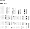
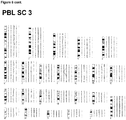
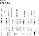
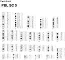
- DCCs Disseminated cancer cells lacked typical driver changes before lymphatic colony formation regardless of primary tumor thickness; see inter alia Example 5 and Figures 2 , 3 and 4 .
- driver mutations in BRAF or NRAS and signature chromosomal rearrangements became significantly enriched in colony-forming DCCs and/or xenografts from DCCs and put patients at high risk for death.
- DCCs leave primary tumors early and evolve at different sites in parallel; see inter alia Example 6 and Figure 5 .
- staging and/or typing of cancerous diseases can be improved, in particular a more accurate method of staging and/or typing of cancerous diseases can be provided, by using information from cells derived from primary tumors, in particular DCCs obtained from different sites where cells evolve, i.e. sites where DCCs migrate, like for example lymph nodes, or sites where cells, in particular CTCs derived from a primary tumor circulate, for example blood.
- the inventors surprisingly found that the somatic evolution of said DCCs or CTCs, respectively, determined based on somatic, in particular genetic and/or epigenetic alterations detected in the DNA of said DCCs or CTCs, respectively, can be used to stage/type a cancerous disease, in particular cancer.
- the stage of a cancerous disease is usually determined using, for example, the TNM staging system.
- TNM Classification of Malignant Tumours is a cancer staging notation system that gives codes to describe the stage of a patient's cancer, wherein T describes the size of the original (primary) tumor and whether it has invaded nearby tissue, N describes nearby (regional) lymph nodes that are involved, and M describes distant metastasis (spread of cancer from one part of the body to another).
- Tx tumor cannot be evaluated
- Tis carcinoma in situ
- T0 no signs of tumor
- T1, T2, T3, T4 size and/or extension of the primary tumor.
- the above stages of cancerous diseases are in many cases insufficient to determine a suitable strategy for therapy of a cancerous disease. Therefore, a new model was found, which uses the somatic evolution of cells, in particular DCCs obtained from one or more lymph node(s) of a subject, in particular draining and/or regional lymph node(s) and/or sentinel lymph node(s), but also, if available, DCCs from other organs than lymph nodes, such as bone marrow or liquor, informing about the molecular state of brain DCCs.
- the somatic evolution of cells may also be determined using CTCs obtained from blood of a subject.
- novel and inventive methods of the present invention use the somatic evolution of cells, in particular DCCs and/or CTCs, as indicator of the stage/type of a cancerous disease, in particular cancer.
- DCCs are cells that disseminated from a primary tumor site before surgery and spread lymphatically or hematogeneously. They may be detected using epithelial cell markers or melanocytic markers in case of melanoma, which are not found on the surface of lymphatic cells or cells found in bone marrow.
- EpCAM and/or cytokeratins are cell markers that may be targeted in the detection of DCCs ( Klein (2009), Nat Rev Cancer 9:302-312 ), in particular in the case of carcinomas.
- the targeting cell markers can be expressed and/or located intracellularly and/or at the surface of the cell.
- DCCs from melanoma may be detected by using gp100 and/or MCSP (melanoma-associated chondroitin sulfate proteoglycan) and/or Melan A (MART-1), and/or CD146 as targeting cell markers.
- DCCs can be detected and isolated for example from bone marrow aspirates or lymph nodes.
- the DCCD After disaggregating and/or processing into single cell suspension, the DCCD can be determined by immunocytology.
- the single cell suspension may be stained with antibodies against the said targeting cell markers such as EpCAM ( Guzvic et al. (2014) Cancer Res. 74:7383-7394 ), cytokeratin ( Schardt et al.
- DCCD DCC-density
- CTCs are cells that are also derived from a primary tumor site before surgery, but that circulate in the blood system. After primary tumor surgery they may be derived from latent or manifest metastatic colonies. They may be detected and isolated using EpCAM as targeting cell marker, but are not limited to this targeting cell marker. Enrichment and detection markers are similar to those used for detection in other mesenchymal organs (bone marrow, lymph nodes) and include for example EpCAM and Cytokeratins for epithelial markers and the mentioned melanocytic markers in melanoma. CTCs may be obtained from blood (e.g. from leukapheresis products) ( Fischer et al.
- Such devices or particles are implanted or infused into a subject/patient to capture and enrich CTCs in the body of the subject/patient.
- the CTCs can then be retrieved from the CTC-capturing-device or - particles ex vivo or in vivo.
- the term "somatic evolution” as used herein relates to the accumulation of alterations in cells, in particular cancerous disease cells, in particular DCCs, during a lifetime, and the effects of those alterations on the fitness of those cells.
- Somatic evolution of cancer cells is due to spontaneous or induced genetic/epigenetic alterations and subsequent natural selection during clonal expansion, which selects cells according to fitness, proliferation rate, apoptosis rate and the like.
- cancer therapy acts as a form of artificial selection, killing sensitive cancer cells, but leaving behind resistant cells. Often the tumor will regrow from those resistant cells, the patient will relapse, and the therapy that had been previously used will no longer be effective.
- somatic evolution is an ongoing process during the development of cancerous diseases and/or therapy of a cancerous disease.
- somatic evolution is determined based on somatic alterations, including epigenetic alterations and/or genetic alterations. Therefore, based on the number and/or nature of somatic alterations, including epigenetic alterations and/or genetic alterations, the somatic evolution of a cell, in particular a DCC, can be determined.
- nucleic acids in particular DNA, comprised in a cell, in particular a DCC.
- Alteration means a deviation from a reference state like for example a state at a given point of time in a reference sample obtained from a subject, for example in a sample obtained from a subject comprising cells derived from other tissue than the sample to be analyzed in the methods of the present invention and/or comprising stem cells of the subject, or a state generally taken as reference with regard to a particular segment of DNA, for example a DNA sequence taken from a sequence database or a known pattern of epigenetic alterations such as a methylation pattern.
- a somatic alteration may be detected by comparing an observed state with a reference state. Any observed deviation may then be categorized as somatic alteration.
- said alteration may be categorized as real somatic alteration or an error introduced by the employed detection method, for example sequencing method.
- the present invention discloses means and methods particularly useful for error-free detection of somatic alterations. Such methods are preferred to be used in the methods of the present invention.
- Somatic alterations within the meaning of the present invention include epigenetic and/or genetic alterations. Genetic alterations include alterations on the level of the sequence of the DNA, i.e. the succession of nucleotides comprised in DNA, and alterations of the relative abundance of segments of the DNA comprised in the sample to be analyzed.
- genetic alterations within the meaning of the present invention may include, inter alia, single nucleotide variations (SNV), sequence mutations, Microsatellite shifts and instability, Loss of heterozygosity (LOH), copy number alterations, karyotypic variations including chromosome structural aberrations and/or aneuploidy.
- SNV single nucleotide variations
- LH Loss of heterozygosity
- copy number alterations include chemical alterations of DNA that are not on the level of the DNA sequence and alterations of external factors involved in DNA structure formation, processing and the like.
- epigenetic alterations within the meaning of the present invention may include, inter alia, methlyation, in particular methlyation of CG pairs, deficiencies of DNA repair proteins, and/or alterations in histone architecture or structure like methylation, acetylation, sumoylation, activating or inactivating histone marks and/or alterations in chromatin architecture like for example in eu- or hetero-chromatin and the like.
- somatic alterations in particular genetic alterations, comprise one or more driver mutation(s) and/or passenger mutation(s).
- driver mutation refers to mutations that give a selective advantage to a clone in its microenvironment, through either increasing its survival or reproduction. Driver mutations tend to cause clonal expansions.
- the somatic alterations, in particular genetic alterations, determined in the methods of the present invention may comprise one or more driver mutation(s).
- one common feature of cellular, in particular DCC progression is the expansion of a clone with a somatic alteration, in particular a genetic and/or epigenetic alteration.
- driver mutations are known in the art.
- driver mutations may be, inter alia, mutations in known oncogenes.
- Oncogenes are genes known to be associated with the development of a cancerous disease, in particular cancer.
- driver mutation(s) within the meaning of the present invention include, but are not limited to, mutations in known oncogenes.
- the genes coding for the neuroblastoma RAS viral oncogene homolog (NRAS) or BRAF are known oncogenes. Further known oncogenes are reported in databases such as the COSMIC database by Forbes et al. (2014) Nucleic Acid Res. 43, D805-811 , or in reports by Vogelstein et al. (2013) Science 339(6127): 1546-58 , Akagi et al. (2004) Nucleic Acid Res. 32, D523-7 or Huret et al. (2000) Nucleic Acid Res. 28(1):349-51 .
- driver mutations may be detected in the methods of the present invention.
- the methods used in the present invention for detecting the somatic alterations, in particular genetic and/or epigenetic alterations, in oncogenes are suitable for simultaneous detection of somatic alterations in multiple oncogenes.
- Particular driver mutations that may be detected in the methods of the present invention comprise driver mutations in the gene coding for NRAS comprising, inter alia, the exon 3 mutation c181C>A.
- the present invention relates to a method for staging and/or typing of a cancerous disease, the method comprising the steps of detecting somatic alterations of the DNA of one or more DCC(s) obtained from a tissue sample, in particular one or more lymph node(s); and determining the somatic evolution of the DCC(s) based on the detected somatic alteration(s) in the DNA of the one or more DCC(s), wherein the somatic evolution of the DCC(s) is indicative of the stage/type of the cancerous disease, wherein detection of somatic alterations comprises detection of the nucleotide at position 2986 of SEQ ID NO:1, wherein a cytosine at position 2986 of SEQ ID NO:1 is indicative of a non-diseased stage/type and an adenine at position 2986 of SEQ ID NO:1 is indicative of a diseased stage/type.
- a guanine at position 2987 of SEQ ID NO:1 or a thymine at position 2987 of SEQ ID NO:1 is indicative of a diseased stage.
- the present invention furthermore relates to a method for treating a cancerous disease, the method comprising the steps of detecting somatic alterations in the DNA of one or more DCC(s) obtained from a tissue sample, in particular one or more lymph node(s) of a subject; determining the somatic evolution of the DCC(s) based on the detected somatic alteration(s) in the DNA of the one or more DCC(s); and determining the stage/type of the cancerous disease based on the somatic evolution of the cancerous disease determined based on the detected somatic alterations in the DNA of the one or more DCC(s), wherein the stage/type of the cancerous disease is used to initiate, continue or discontinue therapy of said cancerous disease, wherein detection of somatic alterations comprises detection of the nucleotide at position 2986 of SEQ ID NO:1, where
- a guanine at position 2987 of SEQ ID NO:1 or a thymine at position 2987 of SEQ ID NO:1 is used to initiate or continue therapy of the cancerous disease.
- the invention furthermore relates to a pharmaceutical composition for use in treating a cancerous disease in a subject, wherein treatment is initiated, continued or discontinued based on the stage/type of the cancerous disease, wherein the stage/type of the cancerous disease is determined by detecting somatic alterations in the DNA of one or more DCC(s) obtained from a tissue sample, in particular one or more lymph node(s) of a subject; determining the somatic evolution of the DCC(s) based on the detected somatic alteration(s) in the DNA of the one or more DCC(s); and determining the stage/type of the cancerous disease based on the somatic evolution of the cancerous disease determined based on the detected somatic alterations in the DNA of the one or more DCC(s), wherein detection of somatic alterations comprises detection of the nucleotide
- a guanine at position 2987 of SEQ ID NO:1 or a thymine at position 2987 of SEQ ID NO:1 is used to continue or initiate treatment of the cancerous disease.
- the detection of somatic alterations may comprise the detection of genetic and/or epigenetic alterations of one or more oncogenes.
- the detection of somatic alterations may comprise the detection of somatic alterations in known oncogenes.
- known driver mutations may also be detected in BRAF, another known oncogene encoding a protein involved in directing cell growth.
- BRAF another known oncogene encoding a protein involved in directing cell growth.
- driver mutations are described in the art to cause a constitutively active form of BRAF, which is associated with the development of cancer.
- BRAF inhibitors directed to the native and mutated form have been described; see e.g. Wan et al. (2004) Cell 116 (6): 855-67 ; Tsai et al.
- sorafenib is a small molecular kinase inhibitor drug approved for the treatment of primary kidney cancer (advanced renal cell carcinoma), advanced primary liver cancer (hepatocellular carcinoma), and radioactive iodine resistant advanced thyroid carcinoma. Sorafenib inhibits, inter alia, wild-type and mutant BRAF.
- a further exemplary BRAF inhibitor is vemurafenib, which is a selective inhibitor of the V600E mutated BRAF protein responsible for an aggressive form of melanoma. It has also been shown to be an effective inhibitor of V600K mutated BRAF protein.
- vemurafenib promotes tumor growth; see Hatzivassiliou et al. (2010) Nature 464 (7287): 431-5 or Halaban et al. (2010) Pigment Cell Melanoma Res. 23(2): 190-200 .
- classical melanoma diagnosis does not comprise determination of the mutational state of patients with regard to the BRAF oncogene.
- the herein described new model of parallel evolution of cells in the primary tumor and DCC(s) indicates that detection of the mutational state in the primary tumor is insufficient for determination of the best suitable therapy of melanoma patients.
- the herein provided more accurate methods of cancer staging/typing may comprise a step of detecting the mutational state of the BRAF oncogene. Therefore, the present invention relates to a method for staging and/or typing of a cancerous disease, the method comprising the steps of detecting somatic alterations of the DNA of one or more DCC(s) obtained from a tissue sample, in particular one or more lymph node(s); and determining the somatic evolution of the DCC(s) based on the detected somatic alteration(s) in the DNA of the one or more DCC(s), wherein the somatic evolution of the DCC(s) is indicative of the stage/type of the cancerous disease, wherein detection of somatic alterations comprises detection of the nucleotide triplet at positions 171428 to 171430 of SEQ ID NO:2, wherein a nucleotide triplet guanine at position 171428, tyrosine at position 171429 and guanine at position 171430 of SEQ ID NO:2 is indicative of
- nucleotide triplet adenine at position 171428, adenine at position 171429 and guanine at position 171430 of SEQ ID NO:2 is indicative of a diseased stage/type.
- the present invention furthermore relates to a method for treating a cancerous disease, the method comprising the steps of detecting somatic alterations in the DNA of one or more DCC(s) obtained from a tissue sample, in particular one or more lymph node(s); determining the somatic evolution of the DCC(s) based on the detected somatic alteration(s) in the DNA of the one or more DCC(s); and determining the stage/type of the cancerous disease based on the somatic evolution of the cancerous disease determined based on the detected somatic alterations in the DNA of the one or more DCC(s), wherein the stage/type of the cancerous disease is used to initiate, continue or discontinue therapy of said cancerous disease, wherein detection of somatic alterations comprises detection of the nucleotide triplet at positions 171428 to 171430 of SEQ ID NO:2, wherein a nucleotide triplet guanine at position 171428, tyrosine at position 171429 and guanine at position 171430 of SEQ ID
- a nucleotide triplet adenine at position 171428, adenine at position 171429 and guanine at position 171430 of SEQ ID NO:2 and a nucleotide triplet adenine at position 171428, guanine at position 171429 and guanine at position 171430 of SEQ ID NO:2 is used to initiate or continue treatment. It is preferred that treatment comprises the use of an inhibitor of BRAF. Accordingly, treatment preferably comprises the use of sorafenib or vemurafenib. Moreover, it is preferred that the cancerous disease is melanoma.
- the invention furthermore relates to a pharmaceutical composition for use in treating a cancerous disease in a subject, wherein treatment is initiated, continued or discontinued based on the stage/type of the cancerous disease, wherein the stage/type of the cancerous disease is determined by detecting somatic alterations in the DNA of one or more DCC(s) obtained from a tissue sample, in particular one or more lymph node(s) of a subject; determining the somatic evolution of the DCC(s) based on the detected somatic alteration(s) in the DNA of the one or more DCC(s); and determining the stage/type of the cancerous disease based on the somatic evolution of the cancerous disease determined based on the detected somatic alterations in the DNA of the one or more DCC(s), wherein detection of somatic alterations comprises detection of the nucleotide triplet at positions 171428 to 171430 of SEQ ID NO:2, wherein a nucleotide triplet guanine at position 171428, tyrosine at position 171429 and
- nucleotide triplet adenine at position 171428, adenine at position 171429 and guanine at position 171430 of SEQ ID NO:2 and a nucleotide triplet adenine at position 171428, guanine at position 171429 and guanine at position 171430 of SEQ ID NO:2 is used to initiate or continue treatment.
- the pharmaceutical composition comprises an inhibitor of BRAF. Accordingly, it is preferred that the pharmaceutical composition comprises sorafenib or vemurafenib.
- the cancerous disease is liver cancer, kidney cancer or melanoma. It is more preferred that the cancerous disease is melanoma.
- the present invention relates to, inter alia, a pharmaceutical composition
- a BRAF inhibitor preferably sorafenib or vemurafenib
- a cancerous disease in a subject, wherein treatment is initiated, continued or discontinued based on the stage/type of the cancerous disease, wherein the stage/type of the cancerous disease is determined by detecting somatic alterations in the DNA of one or more DCC(s) obtained from a tissue sample, in particular one or more lymph node(s) of a subject; determining the somatic evolution of the DCC(s) based on the detected somatic alteration(s) in the DNA of the one or more DCC(s); and determining the stage/type of the cancerous disease based on the somatic evolution of the cancerous disease determined based on the detected somatic alterations in the DNA of the one or more DCC(s), wherein detection of somatic alterations comprises detection of the nucleotide triplet at positions 171428 to 171430 of SEQ ID NO:
- a nucleotide triplet adenine at position 171428, adenine at position 171429 and guanine at position 171430 of SEQ ID NO:2 and a nucleotide triplet adenine at position 171428, guanine at position 171429 and guanine at position 171430 of SEQ ID NO:2 is used to initiate or continue treatment.
- the cancerous disease is preferably liver cancer, kidney cancer or melanoma. It is more preferred that the cancerous disease is melanoma.
- the invention provides means and methods of treating a cancerous disease, in particular cancer.
- the present invention relates to a method of treating a cancerous disease, in particular cancer, comprising obtaining and analyzing a sample from a patient suspected of having or having a cancerous disease, in particular cancer; wherein analyzing comprises sequencing of the DNA of DCC(s) comprised in the sample; detecting the presence or absence of somatic alterations, in particular genetic and/or epigenetic alterations, in the DNA, wherein the presence of somatic alterations indicates that an effective amount of a pharmaceutical composition has to be administered to the patient.
- the invention furthermore relates to a method of diagnosing a cancerous disease, comprising obtaining and analyzing a sample from a patient suspected of having a cancerous disease, in particular cancer; wherein analyzing comprises sequencing of the DNA of DCC(s) comprised in the sample; detecting the presence or absence of somatic alterations, in particular genetic and/or epigenetic alterations, in the DNA, wherein the presence of somatic alterations indicates a patient has, or is likely to have, a cancerous disease and the absence of somatic alterations indicates the patient does not have, or is not likely to have, a cancerous disease.
- Somatic alterations, in particular genetic alterations, determined in the methods of the present invention may also comprise one or more passenger mutations.
- a passenger mutation is a mutation that has generally no or only little effect on the fitness of a clone but may be associated with a clonal expansion because it occurs in the same genome with a driver mutation.
- passenger mutations may be those mutations that are known to have a higher probability to be found in a genome where a driver mutation occurred or is likely to occur in the future.
- Somatic alterations, in particular genetic or epigenetic alterations, determined in the methods of the present invention may also comprise one or more copy number alterations.
- Copy-number alterations are alterations of the DNA of a genome that result in the cell having an abnormal or, for certain genes, a normal variation in the number of copies of one or more sections of the DNA.
- a copy number alteration may correspond to parts of the genome that have been deleted (fewer than the normal number) or multiplied (more than the normal number).
- Each alteration may correspond to the deletion or gain, in particular duplication, of a genomic region, which may range from about one kilobase (1,000 nucleotide bases) to several megabases (1,000,000 nucleotide bases) in size.
- Copy number alterations have been associated with susceptibility or resistance to disease. For example, gene copy number can be elevated in cells associated with cancer, e.g. in DCCs. Accordingly, copy number alterations may also be determined in the methods of the present invention in order to determine the somatic evolution of a cell, in particular of a DCC. The somatic evolution may then be used to determine the stage/type of a cancerous disease, in particular cancer.
- Particular copy number alterations determined in the methods of the present invention comprise loss of chromosome 9p11-13, loss of chromosome 9p21-24 and/or gain of chromosome 7q21. Accordingly, the present invention relates to the methods or pharmaceutical compositions of the invention, wherein the somatic alterations of the DNA comprise at least one of the alterations selected from a loss of chromosome 9p11-13, a loss of chromosome 9p21-24 and gain of chromosome 7q21.
- the number of somatic alterations, in particular genetic and/or epigenetic alterations may also be determined in order to determine the somatic evolution of a cell, in particular a DCC.
- a DCC somatic evolution of a cell
- the overall number of accumulated somatic alterations may also achieve a threshold value, which is indicative of an advanced stage of the cancerous disease, in particular the cancer; see e.g. Shain et al. (2015) N Engl J Med 373; 20:1926 .
- the accuracy of the methods of the present invention may be further increased by using additional parameters to determine the overall somatic evolution of a cell population, in particular DCCs comprised in a lymph node of a subject or derived from other tissue (early) invaded by cancer cells, e.g. bone marrow or brain.
- DCCs comprised in a lymph node of a subject or derived from other tissue (early) invaded by cancer cells, e.g. bone marrow or brain.
- mice transplanted with cells, in particular DCCs, derived from lymph nodes of patients in which said cells, in particular DCCs, were present at a cell density above a specific threshold value gave rise to a tumor in transplanted mice; see Figure 5C .
- the overall cell number of DCCs observed in the respective tissue sample e.g. a sample derived from a lymph node or a bone marrow sample, may serve as additional parameter to further increase accuracy of the methods of the present invention.
- the threshold value indicative for a high probability that cells accumulated important driver mutations in their genome so that an advanced stage/type of the cancerous disease, in particular cancer is determined to be in the range of about 50 or more, preferably 60, 70, 80, 90 or more, most preferably 100 cells per one million cells in the respective tissue sample. Accordingly, a DCCD between about 50 and about 100, preferably between about 60 and about 100, preferably between about 70 and about 100, preferably between about 80 and about 100 and most preferably between about 90 and about 100 is indicative of an advanced stage/type of a cancerous disease, in particular cancer.
- the cell density can be calculated with respect to the overall number of cells in the obtained sample or with respect to another cell population found in the obtained sample.
- the DCC cell population comprised in the tissue sample obtained from a patient is detected using a marker, e.g. EpCAM and/or cytokeratins.
- the DCC cell population comprised in a melanoma tissue sample obtained from a patient is preferably detected using gp100 as a marker.
- alternative DCC markers may also be used, which may cause alternative threshold values corresponding to an advanced stage/type of the cancerous disease, as the person skilled in the art will appreciate.
- the determined DCCD in a tissue sample, in particular lymph node sample or bone marrow sample obtained from a patient, which is used as an indicator of the stage/type of a cancerous disease, in particular cancer can vary based on the method used to determine the cell density.
- gp100 is used as a marker to determine the number of DCCs comprised in the obtained sample, wherein DCCs are gp100 positive.
- the methods of the present invention in addition to determination of somatic alterations, in particular genetic and/or epigenetic alterations, comprised in one or more DCCs obtained from one or more lymph nodes of a patient, comprise a step of determining the DCCD in the lymph node used to obtain the DCCs, in particular the lymph node obtained from a patient.
- the methods of the present invention may further comprise a step of determining the CTCD in order to improve accuracy.
- determining the CTCD in addition to determination of somatic alterations, in particular genetic and/or epigenetic alterations, comprised in one or more CTCs from a patient, in particular retrieved from blood samples or in vivo CTC-capturing-devices, comprise a step of determining the CTC density (CTCD).
- the CTCD is the number of CTCs per one million cells in the blood sample obtained from a patient, or in the case of an in vivo capturing-device by some parameter that relates the CTC number to the filtered blood volume (blood volume or filtering time as surrogate).
- a specific threshold value that may be determined based on the used detection method of CTCs, for example comprising the use of MCSP or EpCAM as a marker, but not limited to this, an advanced stage/type of the cancerous disease may be determined.
- the one or more cell(s), in particular the one or more DCC(s) is/are preferably obtained from one or more lymph node(s).
- Lymph nodes are oval-shaped organs of the lymphatic system, distributed widely throughout the body including the armpit and stomach and linked by lymphatic vessels. Lymph nodes are major sites of B, T, and other immune cells. Lymph nodes are important for the proper functioning of the immune system, acting as filters for foreign particles and cancer cells. Lymph nodes are known to have clinical significance. They become inflamed or enlarged in various infections and diseases, which range from throat infections to cancer.
- the one or more lymph node(s) is/are regional lymph node(s), wherein a regional lymph node is a lymph node close to a site being in a status of developing a disease, in particular a site susceptible to developing a cancerous disease, in particular cancer or a site that has developed a cancerous disease, in particular cancer.
- the regional lymph node is a lymph node anatomically close to a cancer site, for example a tumor.
- the one or more lymph node used in the methods of the invention is/are (a) draining lymph node(s). Draining lymph nodes are lymph nodes to which cells migrate.
- the draining lymph node(s) of a diseased site or a site susceptible of developing a disease is/are the first lymph node(s) invaded by cells migrating from said site.
- lymph node(s) is/are also called sentinel lymph node(s), i.e. the hypothetical first lymph node or group of nodes draining a cancerous disease, in particular a cancer.
- sentinel lymph node/s is/are the target organs primarily reached by metastasizing cancer cells from the tumor.
- sentinel lymph nodes can be totally void of cancer because they were detected prior to dissemination.
- the DCCs used in the methods of the present invention are derived from one or more sentinel lymph node(s) in order to determine whether a subject is susceptible to develop a cancerous disease, in particular cancer, or determine the stage/type of a cancerous disease, in particular cancer.
- sample obtained from one or more lymph node(s) refers to a sample of cells, tissue or fluid that is removed from a subject for the purpose of determining if the sample contains cancerous tissue, in particular DCC(s), and/or for determining the somatic evolution of said cells.
- sample is obtained because a subject is suspected of having cancer due to an earlier diagnosis and/or a predisposition.
- the sample may then also be examined for the presence or absence of cancer, cancer stem cells, and/or cancer stem cell gene signature expression using methods known in the art.
- the lymph node samples used in the methods of the present invention may be obtained by lymph node biopsy, for example needle biopsy or open biopsy as described in the art; see e.g. Chang KL et al. (2009) Modern Surgical Pathology. 2nd ed. Philadelphia, PA: Saunders Elsevier; 2009:chap 41 .
- the term “subject” refers to any animal (e.g., a mammal), including, but not limited to, humans, non-human primates, rodents, and the like, which is to be the recipient of a particular treatment.
- the terms “subject” and “patient” are used interchangeably herein in reference to a human subject. Accordingly, it is preferred that the one or more DCC(s) are obtained from a human subject, more preferred a human patient, wherein the subject/patient may be suspected of having cancer or may be susceptible to a cancerous disease, in particular cancer.
- the term "subject suspected of having cancer” or "patient susceptible to a cancerous disease” refers to a subject/patient that presents one or more symptoms indicative of a cancer (e.g., a noticeable lump or mass) or is being screened for a cancer (e.g., during a routine physical).
- a subject suspected of having cancer or a patient susceptible to a cancerous disease, in particular cancer can also have one or more risk factors.
- a "subject suspected of having cancer” or “patient susceptible to a cancerous disease” encompasses an individual who has received an initial diagnosis but for whom the stage of cancer is not known. The term further includes people who once had cancer (e.g., an individual in remission).
- the term also refers to a subject with one or more risk factors for developing a specific cancer. Risk factors include, but are not limited to, gender, age, genetic predisposition, environmental exposure, previous incidents of cancer, preexisting non-cancer diseases, and lifestyle.
- the methods of the present invention are for staging and/or typing and/or predicting outcome and/or treating a cancerous disease, in particular cancer wherein the cancerous disease is a melanoma.
- dissemination relates to the process of migration by active migration or passive transport by body fluids from a primary disease site to a secondary site, for example a lymph node or bone marrow or blood.
- the methods of the present invention are for staging/typing of a cancerous disease, in particular a cancerous disease involving dissemination of cells, preferably involving lymphatic dissemination wherein the cancerous disease is a melanoma.
- the methods of the present invention are preferably for staging and/or typing and/or predicting outcome and/or treating of solid cancer and wherein the cancerous disease is a melanoma.
- a solid cancer refers to one or more cells which are growing or have grown in an uncontrolled manner to form cancer tissue.
- the term “solid cancer” includes, but is not limited to “carcinomas”, “adenocarcinomas”.
- Carcinomas are cancers of epithelial (lining) cells.
- Adenocarcinoma refers to carcinoma derived from cells of glandular origin.
- cancer and “tumor” are used interchangeably throughout the subject specification.
- Solid cancers may arise in nearly any tissue of the body and the methods of the present invention may be used for staging and/or typing and/or predicting outcome and/or treating of any of these cancers.
- Exemplary "solid cancers" which may be staged and/or typed and/or whose outcome may be predicted and/or which may be treated in accordance with the present disclosure include acoustic neoma, adenocystic carcinoma, adrenocortical cancer, alveolar soft-part sarcoma, anal cancer, angiosarcoma, basal cell carcinoma (bcc), bladder cancer, bone cancers, bowel cancer, brain stem glioma, breast cancer, CNS cancers, carcinoid cancers, cervical cancer, childhood brain cancers, childhood soft tissue sarcoma, chondrosarcoma, choriocarcinoma, colorectal cancers, dermatofibrosarcoma-protuberans, desmoplastic small round cell cancer, ductal carcinoma,
- the present invention relates to a method for staging and/or typing of melanoma, said method comprising the steps of detecting somatic alterations of the DNA of one or more DCC(s) obtained from one or more lymph node(s); and determining the somatic evolution of the DCC(s) based on the detected somatic alteration(s) in the DNA of the one or more DCC(s), wherein the somatic evolution of the DCC(s) is indicative of the stage/type of melanoma.
- the present invention relates to a method for treating melanoma, said method comprising the steps of detecting somatic alterations in the DNA of one or more DCC(s) obtained from lymph node(s) of a patient; determining the somatic evolution of the DCC(s) based on the detected somatic alteration(s) in the DNA of the one or more DCC(s); and determining the stage/type of melanoma of said patient based on the somatic evolution of melanoma determined based on the detected somatic alteration(s) in the DNA of the one or more DCC(s), wherein the stage/type of melanoma is used to initiate, continue or discontinue melanoma therapy.
- the present invention relates to a therapeutic intervention, preferably a pharmaceutical composition, for use in treating melanoma in a patient, wherein treatment is initiated, continued or discontinued based on the stage/type of melanoma, wherein said stage/type of melanoma is determined by detecting somatic alterations in the DNA of one or more DCC(s) obtained from lymph node(s) or liquor (preferably form lymph node(s), more preferably from a sentinel lymph node) of said patient; determining the somatic evolution of the DCC(s) based on the detected somatic alteration(s) in the DNA of the one or more DCC(s); and determining the stage/type of melanoma of said patient based on the somatic evolution of melanoma determined by detecting somatic alterations in the DNA of one or more DCC(s) obtained from lymph node(s) or liquor of said patient.
- a therapeutic intervention preferably a pharmaceutical composition
- the invention relates to a method for staging and/or typing of melanoma, said method comprising the steps of detecting somatic alterations of the DNA of one or more CTC(s) obtained from blood; and determining the somatic evolution of the CTC(s) based on the detected somatic alteration(s) in the DNA of the one or more CTC(s), wherein the somatic evolution of the CTC(s) is indicative of the stage/type of melanoma.
- in vivo CTC-capturing-devices may be used for obtaining the CTCs from blood.
- the present invention relates to a method for treating melanoma, said method comprising the steps of detecting somatic alterations in the DNA of one or more CTC(s) obtained from blood of a patient; determining the somatic evolution of the CTC(s) based on the detected somatic alteration(s) in the DNA of the one or more CTC(s); and determining the stage/type of melanoma of said patient based on the somatic evolution of melanoma determined based on the detected somatic alteration(s) in the DNA of the one or more CTC(s), wherein the stage/type of melanoma is used to initiate, continue or discontinue melanoma therapy.
- the present invention relates to a pharmaceutical composition for use in treating melanoma in a patient, wherein treatment is initiated, continued or discontinued based on the stage/type of melanoma, wherein said stage/type of melanoma is determined by detecting somatic alterations in the DNA of one or more CTC(s) obtained from blood of said patient; determining the somatic evolution of the CTC(s) based on the detected somatic alteration(s) in the DNA of the one or more CTC(s); and determining the stage/type of melanoma of said patient based on the somatic evolution of melanoma determined by detecting somatic alterations in the DNA of one or more CTC(s) obtained from blood of said patient.
- the methods of the present invention comprise a step of detecting somatic alterations of the DNA of one or more DCC(s) or CTC(s), respectively, obtained from a tissue sample, in particular a lymph node sample or bone marrow sample or blood.
- a tissue sample in particular a lymph node sample or bone marrow sample or blood.
- somatic alterations may comprise epigenetic and/or genetic alterations.
- epigenetic alterations the person skilled in the art is well-aware of methods capable of detecting epigenetic alterations with regard to a reference sample or with regard to a reference standard known in the art.
- epigenetic alterations to be detected in the DNA of one or more DCC(s) may include methlyation, in particular methlyation of CpG(s), deficiencies of DNA repair proteins, and/or alterations in histone architecture or structure like methylation, acetylation, sumoylation, activating or inactivating histone marks and/or alterations in chromatin architecture like for example in eu- or hetero-chromatin and the like. Accordingly, in the methods of the present invention a step of DNA methylation analysis may be included.
- Methylation of the 5' carbon of cytosine is a form of epigenetic modification that does not affect the primary DNA sequence, but affects secondary interactions that play a critical role in the regulation of gene expression. Aberrant DNA methylation may suppress transcription and subsequently gene expression. Methylation analysis as in the methods of the present invention may comprise selective modification of the target DNA.
- Such modification may comprise the addition of methylation-dependent restriction enzymes (MDREs) or methylation-sensitive restriction enzymes (MSREs), preferably MDREs.
- MDREs methylation-dependent restriction enzymes
- MSREs methylation-sensitive restriction enzymes
- Selective modification of the target DNA may also comprise addition of a chemical agent that is able to selectively differentiate between methylated or unmethylated nucleotides.
- methylation analysis as employed in the methods of the present invention may be able to selectively identify methylated cytosines that may later be read-out using methods known in the art, e.g. methods described in WO 2015/118077 or WO 2000/017390 .
- treatment with bisulfite is known to convert unmethylated cytosines (C) to uracil (U) while methylated cytosines are not converted ( Frommer et al. 1992. Proc. Natl. Acad. Sci. USA 89, 1827-1831 ).
- Sequencing of DNA subsequent to treatment with bisulfite may be used to identify methylated nucleotides, in particular cytosines.
- Treatment with MDREs leads to methylation-dependent restriction of DNA fragments, while treatment with MSREs leads to methylation-dependent inhibition of restriction.
- Sequencing of DNA subsequent to MDRE/MSRE restriction in addition to Msel restriction may be used to identify methylated nucleotides, in particular cytosines.
- chromatin immunoprecipitation ChIP
- ChIP-chip ChIP-PET, ChIP-Seq, MeDIP, DamID or the like; see Minard et al. (2009) Genesis 47(8), pp.559-72 and references cited therein.
- methylation of DNA may genome-wide be detected using MeDIP, MeDIP-chip or MeDIP-seq.
- Exemplary protocols comprise steps of subjecting purified DNA to sonication to shear it into random fragments.
- the resulting fragments may range from 300 to 1000 base pairs (bp) in length, preferably between 400 and 600 bp.
- the DNA fragments are then denatured to produce single-stranded DNA. Following denaturation, the DNA is incubated with 5-methylcytosine (5-mC) antibodies available in the art.
- 5-methylcytosine (5-mC) antibodies available in the art.
- the classical immunoprecipitation technique is then applied: magnetic beads conjugated to anti-mouse-IgG are used to bind the anti-5mC antibodies, and unbound DNA is removed in the supernatant.
- proteinase K is added to digest the antibodies and release the DNA, which can be collected and prepared for DNA detection. Subsequent DNA detection is done using methods described further below for detection of genetic alterations.
- a fraction of the input DNA obtained after the sonication step above is labeled with cyanine-5 (Cy5; red) deoxy-cytosine-triphosphate while the methylated DNA, enriched after the immunoprecipitation step, is labeled with cyanine-3 (Cy3; green).
- the labeled DNA samples are cohybridized on a 2-channel, high-density genomic microarray to probe for presence and relative quantities. The purpose of this comparison is to identify sequences that show significant differences in hybridization levels, thereby confirming the sequence of interest is enriched, i.e. methylated.
- MeDIP-seq the coupling of MeDIP with next generation, short-read sequencing technologies such as 454 or Illumina sequencing.
- the high-throughput sequencing of the methylated DNA fragments produces a large number of short reads (36-50 bp or 400 bp, depending on the technology).
- the short reads are aligned to a reference genome using alignment software such as Mapping and Assembly with Quality (Maq) which uses a Bayesian approach, along with base and mapping qualities to model error probabilities for the alignments.
- the reads can then be extended to represent the ⁇ 400 to 700 bp fragments from the sonication step.
- the coverage of these extended reads can be used to estimate the methylation level of the region.
- a genome browser such as ENSEMBL can also be used to visualize the data.
- Validation of the approach to assess quality and accuracy of the data can be done using, inter alia, quantitative PCR This is done by comparing a sequence from the MeDIP sample against an unmethylated control sequence. The samples are then run on a gel and the band intensities are compared. The relative intensity serves as the guide for finding enrichment.
- ChIP techniques may also be used to detect other epigenetic alterations, such as histone modifications or modifications relating to other proteins bound to DNA.
- epigenetic alterations such as histone modifications or modifications relating to other proteins bound to DNA.
- Such techniques are known in the art and extensively described in various standard reference books, such as " Chromatin Immunoprecipitation Assays - Methods and Protocols" by Philippe Collas, Humana Press (2009 ).
- any technique suitable for detecting such modifications, in particular epigenetic alterations may be applied.
- next generation sequencing techniques as marketed by Illumina® are known to the person skilled in the art and are suitable for detecting epigenetic alterations such as methylation, in particular combined with further techniques such as next-generation sequencing, in particular methylation sequencing; and/or for detecting protein-DNA interactions, techniques such as chromatin immunoprecipitation (ChIP) and/or ChIP combined with next-generation sequencing, in particular ChIP-Seq (for example using protocols available for Illumina® sequencing machines).
- Chrin immunoprecipitation ChIP
- ChIP-Seq for example using protocols available for Illumina® sequencing machines.
- a preferred example of a method suitable for isolated and/or simultaneous detection of epigenetic and/or genetic alterations is a method comprising the steps of providing a sample comprising DNA, in particular a sample derived from one or more lymph node(s) or a bone marrow sample comprising one or more DCC(s) or CTC(s); adding an agent to said DNA that selectively recognizes epigenetically modified nucleic acids, in particular methlylated nucleic acids, e.g.
- sequence information obtained in the last step of the above described method may be used to simultaneously detect genetic alterations.
- the obtained sequence information may, for example, be used in methods for DNA sequence analysis like whole genome sequencing, whole exome sequencing, whole regulome sequencing, sequencing-based methylation analysis, sequencing-based breakpoint detection, ChIP sequencing, or targeted sequencing and variations thereof.
- the methods of the present invention comprise a step of detecting somatic alterations of the DNA of one or more DCC(s) or CTC(s), respectively, obtained from a tissue or body fluid sample, in particular a lymph node sample or bone marrow sample or liquor (in the case of DCC(s)), or blood (in the case of CTC(s)), respectively.
- a tissue or body fluid sample in particular a lymph node sample or bone marrow sample or liquor (in the case of DCC(s)), or blood (in the case of CTC(s)), respectively.
- the resulting sequence may be compared to reference sequences known in the art.
- methods for sequencing DNA any method may be employed within the context of the present invention. However, it is preferred to use methods which are fast, efficient, reliable and only require low input amounts. This is particularly important for detecting DCCs or CTCs in an early phase of cancer spread where only one single cell may be present in a tissue or body fluid sample, in particular a lymph node sample or bone marrow sample or liquor sample, or blood respectively.
- sequencing methods known in the art may be employed that are particularly suitable for sequencing of low amounts of input DNA, e.g. as described in Mardis (2008) Annu Rev Genomics Hum Genet 9, 387-402 .
- a preferred method to be employed in the present invention is a DNA sequencing method comprising the use of endonucleases, oligonucleotides specifically recognizing overhangs created by the endonucleases and further oligonucleotides for amplification.
- the oligonucleotides may, for example, comprise barcode sequences in order to distinguish between fragments generated by the endonucleases and in order to generate an internal sequencing reference for error-free sequencing.
- An exemplary and preferred method for DNA sequencing comprises the steps of providing a sample comprising DNA, in particular a sample derived from one or more lymph node(s) or a bone marrow sample or a liquor or blood sample comprising one or more DCC(s) or CTC(s); digesting the DNA with a restriction endonuclease under conditions suitable to obtain DNA fragments of similar length, wherein said restriction endonuclease is capable of providing 5' overhangs, wherein the terminal nucleotide of the overhang is phosphorylated or, wherein said restriction endonuclease is capable of providing 3' overhangs, wherein the terminal nucleotide of the overhang is hydroxylated on said DNA fragments; annealing a first oligonucleotide to said DNA fragments, wherein a first sequence of said first oligonucleotide is complementary to the 5' or 3' overhang, respectively, of said DNA fragment, and a second sequence of said first oligonucleot
- the methods of the present invention comprise a step of determining the somatic evolution based on the detected somatic alterations.
- the somatic evolution is determined based on the accumulated somatic alterations during the lifetime of a cell, in particular a DCC or CTC, preferably comprising further parameters such as the DCCD/CTCD.
- the somatic evolution may be determined and the stage/type of the cancerous disease, in particular cancer, may be determined, treatment methods may be adjusted and the like.
- the person skilled in the art will appreciate how to express the determined stage/type of a cancerous disease, using, for example, letter codes, number codes or the like.
- T1 and T4 melanomas employs primary tumor tissue. It assumes that the molecular characteristics of metastasis-initiating DCCs can be identified within the primary tumor, because primary tumors and DCCs are thought to be largely identical. To test this assumption, investigated the genomic profiles of primary tumors and their matched DCCs was investigated.
- BRAF and NRAS mutations are frequent (in respectively 40% and 21% of cases on average) in melanoma ( Platz et al., (2008) Mol Oncol 1:395-405 ), it was investigated whether these mutations are transmitted from the primary tumor to DCCs. Both alleles (wild type and mutant) could reliably be retrieved from single cells with heterozygous BRAF and NRAS mutations ( Figure 3C ).
- DCCs from thick melanomas could harbor characteristic alterations responsible for the prognostic relevance of high T stage, which are absent in DCCs from thin melanomas. Therefore, a focus was set on DCCs before evident lymph node colonization as being representative of the time of arrival and their genomic profile was investigated.
- Cancer cells forming a lymph node colony displayed a characteristic signature of alterations.
- DCCs were transplanted into NSG-mice.
- First conditions for xenotransplantation of rare melanoma cells were evaluated ( Quintana (2008) Nature 456, 593-598 ).
- two approaches were compared: direct transplantation of groups of DCCs and transplantation of DCC-spheres after brief culture under melanosphere conditions.
- Melanospheres formed tumors in immunodeficient NSG mice more frequently than groups of single cells (p ⁇ 0.0001, log-rank test; Figure 6A , B and Figure 14 ).
- the melanoma cell lines A375 and MelHo were used (obtained from the Leibniz Institute DSMZ-German Collection of Microorganisms and Cell cultures). MelHo is listed in the ICLAC-database for mis-identified cell lines due to unclear patient-origin, but was used as the cell line is heterogenic for the exon 15 mutation c1799T>A ( BRAF ). The origin of the cell lines was verified by short tandem repeat (STR) analysis (Cell-IDTM, Promega).
- STR short tandem repeat
- the cell lines 70-61 and 102-4 were developed from DCC-derived xenografts and are exon 15 mutation c1799T>A ( BRAF ) and exon 3 mutation c181C>A (NRAS) mutated, respectively, as determined by Sanger sequencing (Sequiserve, Vaterstetten, Germany). Their patient-origin was verified by short tandem repeat (STR) analysis (Cell-IDTM, Promega), their melanoma-origin by a human pathologist and their aberrant genotype by CGH. A375 and MelHo were maintained in DMEM, 10%FCS, 0.5 x Pen/Strep; 102-4 and 70-61 in RPMI, 10% FCS, 0.5xPen/Strep. All cell lines were routinely tested for mycoplasma and were found to be negative.
- STR short tandem repeat
- lymphatic tissue was cut into 1-mm pieces and disaggregated mechanically into a single-cell suspension by rotating knives (DAKO Medimachine, DAKO), washed with HBSS (Life Technologies, Heidelberg, Germany) and centrifuged on a density gradient made of a 60% Percoll solution (Amersham, Uppsala, Sweden). Cells were counted using a Neubauer counting chamber.
- MCSP + cells were isolated using a micromanipulator (Eppendorf PatchMan NP2) and transplanted.
- Single cells of disaggregated sentinel lymph nodes were plated in 6 cm poly-HEMA (12mg/mL, Sigma-Aldrich) coated cell culture plates (Sigma-Aldrich) at a density of 200,000 viable cells/mL.
- Cells were grown in a serum-free DMEM/Ham's F12 basal medium (PAN Biotech GmbH), supplemented with 0.5 x Pen/Strep (PAN Biotech GmbH), 0.5 % BSA (VWR-Biochemical), 10 ⁇ g/mL insulin (Sigma-Aldrich), 10 nM HEPES (Sigma-Aldrich), 1 x B27 (Life Technology GmbH), 10 ng/mL EGF (Sigma-Aldrich) and 10 ng/mL bFGF (Sigma-Aldrich), 4 ⁇ g/mL heparin (Sigma-Aldrich), 5 ng/mL GRO- ⁇ (R&D Systems), 20 ng/mL HIL-6 (
- Spheres or MCSP + cells from disaggregated sentinel lymph nodes were collected using a micropipettor or micromanipulator and pooled in a microwell (volume 10-15 ⁇ l, Terasaki). Microwells were pre-coated over night with 12 mg/mL poly-HEMA (Sigma-Aldrich) at RT. Single cells were transplanted in a final volume of 30 ⁇ l and 25% high-concentration matrigel (BD Biosciences) as published before ( Quintana (2008) Nature 456, 593-598 ).
- mice were injected with an insulin syringe (Microfine, 29G, U-50, BD Biosciences) sub-cutaneously into NOD.Cg -Prkdc scid IL2r ⁇ tmWjl / Sz (NSG, 6-8 weeks old, males and females). Mice were purchased from the Jackson Laboratory and maintained under specific-pathogen free conditions, with acidified water and food ad libitum in the research animal facilities of the University of Regensburg, Germany. All approved experimental animal procedures were conducted to German federal and state regulations. Mice were palpated every week at the site of injection.
- insulin syringe Microfine, 29G, U-50, BD Biosciences
- STR short tandem repeat
- the PALM Microbeam system (Bernried) was used for microdissection and catapulting. DNA was processed as previously described ( Klein (2002) Lancet 360, 683-689 ; Klein (2002) J Exp Med 196, 359-368 ).
- NRAS and BRAF genes were detected using Sanger sequencing (Sequiserve, Vaterstetten, Germany) after gene specific amplification from WGA samples.
- the primers for BRAF exon 15 analysis were as follows: forward 5'-TCCAGACAACTGTTCAAACTG and reverse 5'-CTCTTCATAATGCTTGCTCTG, encompassing the mutations of codon 600 (V600E, previously called V599E; V600K, V600R).
- Cycling temperatures were set to 94 °C (2 min), 60 °C (30 sec) and 72 °C (2 min) for one cycle; 94 °C (15 sec), 60 °C (30 sec) and 72 °C (20 sec) for 14 cycles; 94 °C (15 sec), 60 °C (30 sec) and 72 °C (30 sec) for 24 cycles and an additional final extension step at 72 °C (2 min).
- the PCR primers for NRAS exon 3 codon 61 analysis were: forward 5'-GGCAAATACACAGAGGAAGC and reverse 5'-ACCCCCAGGATTCTTACAGA encompassing the common mutations of codon 61: Q61K and Q61R.
- the PCR cycler was set to 94 °C (2 min), 63 °C (30 sec) and 72 °C (2 min) for one cycle; 94 °C (15 sec), 63 °C (30 sec) and 72 °C (20 sec) for 14 cycles; 94 °C (15 sec), 63 °C (30 sec) and 72 °C (30 sec) for 24 cycles and an additional final extension step at 72 °C (2 min).
- PCR products were sent for sequencing to Sequiserve, Vatterstetten.
- the mutation assay was established using single cells or genomic DNA of cell lines with known exon 15 mutation c1799T>A (BRAF) and exon 3 mutation c181C>A ( NRAS ).
- the mutant BRAF allele was detected in 62% (70-61), 84% (MelHo) of detected sequences over all analyzed single cells and in 61% (70-61) and 86% (MelHo) in bulk genomic DNA.
- the mutation NRAS allele was present in 59% of all single cells and 46% of the bulk genomic DNA.
- the number of samples with a DCCD>100 increased with tumor thickness as a Weibull cumulative distribution function with a median of 8.9 mm (95% Cl 6.8 to 14.3 mm), i.e. 22 times higher than the median thickness at seeding ( Fig. 1C ).
- the obtained estimate is fitted by a Weibull distribution.
- Explicit formulas for the Weibull distribution were used to calculate the hazard rates.
- the hazard function describes the instantaneous risk per unit thickness for an event (dissemination, colonization) for those tumors, for which the event has not yet occurred. For example, the hazard rate of 1 per mm (0.2 per mm) indicates that the tumor needs to grow 1 mm (5 mm, respectively) on average for the event to occur.
- DCCD- and thickness thresholds associated with low p-values might indicate primary tumor sizes and genomic alterations that facilitate direct dissemination to lymph nodes.
- CNAs were analyzed as they best reflect cancerous progression as opposed to point mutations ( Bauer (2006) Dermatologic therapy 19, 40-49 ; Shain (2015) The New England journal of medicine 373, 1926-1936 ⁇ , which are frequently detected also in benign lesions in contrast to CNAs ( Hafner (2010) PNAS 107, 20780-20785 ; Klein (2013) Nature 501, 365-372 ; Shain (2015) The New England journal of medicine 373, 1926-1936 ). For this reason, the study did not address dissemination relative to the acquisition of non-CNA alterations except of BRAF or NRAS.
- DCCs were analyzed before colony formation from patients with thick melanomas. They were compared with pre-colonizing DCCs from thin melanomas, but no differentiating CNAs could be identified. Also the number of CNAs did not differ between pre-colonizing DCCs from thin vs. thick melanomas.
- the mean proliferation index of 11% before colonization resembles that of T1 melanomas at transition to the tumorigenic vertical growth phase (VGP) previously found to range between 9-13% ( Gimotty (2005) Journal of clinical oncology : official journal of the American Society of Clinical Oncology 23, 8048-8056 ).
- VGP tumorigenic vertical growth phase
- BRAF mutations are found in DCCs at colony formation in sentinel lymph nodes but rarely before. Although benign nevi harbor BRAF mutant cells in 70-88% ( Pollock (2003) Nature genetics 33, 19-20 ; Shain (2015) The New England journal of medicine 373, 1926-1936 ), it is unclear whether BRAF mutations (or generally MAPK pathway mutations) initiate melanoma and are transmitted linearly. Several reasons argue against this scenario for many melanomas. First, primary tumors display generally lower rates of BRAF mutations than benign nevi (40% vs.
- VGP melanomas expand in the dermis i.e. become tumorigenic
- they often acquire BRAF mutations Dong (2003) Cancer research 63, 3883-3885 ; Verlinden (2014) Medicine 93, e285
- Ki-67 frequencies of >20% being a marker of poor outcome
- Gimotty (2005) Journal of clinical oncology : official journal of the American Society of Clinical Oncology 23, 8048-8056 DCCs in SLN and possibly other metastatic sites re-capitulate this process during colonization.
- T1 melanomas are mostly cured by surgery ( Balch (2009) Journal of clinical oncology : official journal of the American Society of Clinical Oncology 27, 6199-6206 ) strongly supports proliferation-stimulating factors secreted more abundantly by thick melanomas, which then promote the acquisition of genetic changes required for colony formation.
- Formal proof of this scenario would require model systems that enable genomic in vitro progression of immature to mature cancer cells triggered by supporting factors. Such models are currently not available. Unstimulated and consequently slow in vivo progression over years as observed in some patients may account for late recurrences ( Ossowski (2010) Pigment cell & melanoma research 23, 41-56 ) and argue at least partially for a lead-time effect.
Landscapes
- Health & Medical Sciences (AREA)
- Chemical & Material Sciences (AREA)
- Life Sciences & Earth Sciences (AREA)
- Organic Chemistry (AREA)
- Proteomics, Peptides & Aminoacids (AREA)
- Engineering & Computer Science (AREA)
- Zoology (AREA)
- Immunology (AREA)
- Analytical Chemistry (AREA)
- Pathology (AREA)
- Genetics & Genomics (AREA)
- Wood Science & Technology (AREA)
- General Health & Medical Sciences (AREA)
- Oncology (AREA)
- Bioinformatics & Cheminformatics (AREA)
- General Engineering & Computer Science (AREA)
- Molecular Biology (AREA)
- Microbiology (AREA)
- Hospice & Palliative Care (AREA)
- Biochemistry (AREA)
- Biotechnology (AREA)
- Biophysics (AREA)
- Physics & Mathematics (AREA)
- Nuclear Medicine, Radiotherapy & Molecular Imaging (AREA)
- Veterinary Medicine (AREA)
- Medicinal Chemistry (AREA)
- Chemical Kinetics & Catalysis (AREA)
- Pharmacology & Pharmacy (AREA)
- Animal Behavior & Ethology (AREA)
- Public Health (AREA)
- General Chemical & Material Sciences (AREA)
- Measuring Or Testing Involving Enzymes Or Micro-Organisms (AREA)
- Medicines That Contain Protein Lipid Enzymes And Other Medicines (AREA)
- Medicines Containing Antibodies Or Antigens For Use As Internal Diagnostic Agents (AREA)
- Investigating Or Analysing Biological Materials (AREA)
- Pharmaceuticals Containing Other Organic And Inorganic Compounds (AREA)
- Medicines Containing Material From Animals Or Micro-Organisms (AREA)
- Peptides Or Proteins (AREA)
Claims (8)
- Procédé de stadification et/ou de typage d'une maladie cancéreuse, ledit procédé comprenant les étapes suivantes :(a) détection d'altérations somatiques de l'ADN d'une ou plusieurs cellules cancéreuses disséminées (CCD) obtenues à partir d'un ou plusieurs ganglions lymphatiques ; et(b) détermination de l'évolution somatique des CCD, sur la base de la ou des altérations somatiques détectées dans l'étape (a),dans lequel l'évolution somatique des CCD indique le stade/type de la maladie cancéreuse et dans lequel la maladie cancéreuse est un mélanome.
- Composition pharmaceutique destinée à une utilisation dans le traitement d'une maladie cancéreuse chez un sujet, dans laquelle le traitement est démarré, poursuivi ou interrompu sur la base du stade/type de ladite maladie cancéreuse, dans laquelle ledit stade/type de ladite maladie cancéreuse est déterminé par :(a) détection d'altérations somatiques dans l'ADN d'une ou plusieurs CCD obtenues à partir d'un ou plusieurs ganglions lymphatiques d'un sujet ;(b) détermination de l'évolution somatique des CCD sur la base de la ou des altérations somatiques détectées dans l'étape (a) ; et(c) détermination du stade/type de la maladie cancéreuse sur la base de l'évolution somatique des CCD déterminée dans l'étape (b),dans laquelle la maladie cancéreuse est un mélanome.
- Procédé selon la revendication 1 ou composition pharmaceutique selon la revendication 2, dans lesquels les CCD sont obtenues à partir d'un ganglion lymphatique sentinelle.
- Procédé selon les revendications 1 ou 3 ou composition pharmaceutique selon les revendications 2 ou 3, comprenant en outre la détermination de la densité des CCD (DCCD), dans lesquels la DCCD est le nombre de CCD par million de cellules dans le ganglion lymphatique utilisé pour obtenir les CCD, dans lesquels la DCCD indique le stade/type de la maladie cancéreuse.
- Procédé selon la revendication 4 ou composition pharmaceutique selon la revendication 4, dans lesquels une DCCD de plus d'environ 50 à environ 100 indique le développement de métastases.
- Procédé selon l'une quelconque des revendications 1 et 3 à 5, ou composition pharmaceutique selon l'une quelconque des revendications 2 à 5, dans lesquels les altérations somatiques comprennent au moins une des altérations somatiques sélectionnées dans le groupe constitué par une mutation BRAF, une perte du chromosome 9p11-13, une perte du chromosome 9p21-24, un gain du chromosome 7q21-36, et une mutation NRAS.
- Procédé selon l'une quelconque des revendications 1 et 3 à 6, ou composition pharmaceutique selon l'une quelconque des revendications 2 à 6, dans lesquels une mutation BRAF, une perte du chromosome 9p11-13, une perte du chromosome 9p21-24, un gain du chromosome 7q21-36, et/ou une mutation NRAS indiquent que les CCD se développeront en métastases.
- Procédé selon l'une quelconque des revendications 1 et 3 à 7, ou composition pharmaceutique selon l'une quelconque des revendications 2 à 7, dans lesquels l'étape (a) comprend en outre l'évaluation de la prolifération des CCD, dans lesquels une prolifération accrue des CCD indique que les CCD se développeront en métastases.
Priority Applications (11)
| Application Number | Priority Date | Filing Date | Title |
|---|---|---|---|
| EP16152883.1A EP3199641B1 (fr) | 2016-01-27 | 2016-01-27 | Moyens et procédés permettant d'évaluer l'évolution, de saisir les données et de traiter une maladie cancéreuse |
| ES16152883T ES2905208T3 (es) | 2016-01-27 | 2016-01-27 | Medios y métodos para la estadificación, tipificación y tratamiento de una enfermedad cancerosa |
| PCT/EP2017/051789 WO2017129753A1 (fr) | 2016-01-27 | 2017-01-27 | Moyens et procédés de stadification, typage et traitement d'une maladie cancéreuse |
| CN201780008861.6A CN108770360B (zh) | 2016-01-27 | 2017-01-27 | 对癌性疾病进行分期、分型和治疗的手段和方法 |
| EP17706412.8A EP3408411A1 (fr) | 2016-01-27 | 2017-01-27 | Moyens et procédés de stadification, typage et traitement d'une maladie cancéreuse |
| US16/073,271 US11702701B2 (en) | 2016-01-27 | 2017-01-27 | Means and methods for staging, typing and treating a cancerous disease |
| CA3012404A CA3012404A1 (fr) | 2016-01-27 | 2017-01-27 | Moyens et procedes de stadification, typage et traitement d'une maladie cancereuse |
| SG11201805990XA SG11201805990XA (en) | 2016-01-27 | 2017-01-27 | Means and methods for staging, typing and treating a cancerous disease |
| KR1020187024606A KR20180102674A (ko) | 2016-01-27 | 2017-01-27 | 암 질환의 단계 구분, 유형 분류 및 치료를 위한 수단 및 방법 |
| JP2018539138A JP2019508032A (ja) | 2016-01-27 | 2017-01-27 | がん性疾患をステージング、タイピングおよび処置するための手段および方法 |
| AU2017211976A AU2017211976B2 (en) | 2016-01-27 | 2017-01-27 | Means and methods for staging, typing and treating a cancerous disease |
Applications Claiming Priority (1)
| Application Number | Priority Date | Filing Date | Title |
|---|---|---|---|
| EP16152883.1A EP3199641B1 (fr) | 2016-01-27 | 2016-01-27 | Moyens et procédés permettant d'évaluer l'évolution, de saisir les données et de traiter une maladie cancéreuse |
Publications (2)
| Publication Number | Publication Date |
|---|---|
| EP3199641A1 EP3199641A1 (fr) | 2017-08-02 |
| EP3199641B1 true EP3199641B1 (fr) | 2021-09-29 |
Family
ID=55310656
Family Applications (2)
| Application Number | Title | Priority Date | Filing Date |
|---|---|---|---|
| EP16152883.1A Active EP3199641B1 (fr) | 2016-01-27 | 2016-01-27 | Moyens et procédés permettant d'évaluer l'évolution, de saisir les données et de traiter une maladie cancéreuse |
| EP17706412.8A Withdrawn EP3408411A1 (fr) | 2016-01-27 | 2017-01-27 | Moyens et procédés de stadification, typage et traitement d'une maladie cancéreuse |
Family Applications After (1)
| Application Number | Title | Priority Date | Filing Date |
|---|---|---|---|
| EP17706412.8A Withdrawn EP3408411A1 (fr) | 2016-01-27 | 2017-01-27 | Moyens et procédés de stadification, typage et traitement d'une maladie cancéreuse |
Country Status (10)
| Country | Link |
|---|---|
| US (1) | US11702701B2 (fr) |
| EP (2) | EP3199641B1 (fr) |
| JP (1) | JP2019508032A (fr) |
| KR (1) | KR20180102674A (fr) |
| CN (1) | CN108770360B (fr) |
| AU (1) | AU2017211976B2 (fr) |
| CA (1) | CA3012404A1 (fr) |
| ES (1) | ES2905208T3 (fr) |
| SG (1) | SG11201805990XA (fr) |
| WO (1) | WO2017129753A1 (fr) |
Families Citing this family (1)
| Publication number | Priority date | Publication date | Assignee | Title |
|---|---|---|---|---|
| JP7300394B2 (ja) | 2017-01-17 | 2023-06-29 | ヘパリジェニックス ゲーエムベーハー | 肝再生の促進又は肝細胞死の低減もしくは予防のためのプロテインキナーゼ阻害 |
Family Cites Families (7)
| Publication number | Priority date | Publication date | Assignee | Title |
|---|---|---|---|---|
| AU755499B2 (en) | 1998-09-18 | 2002-12-12 | Micromet Ag | DNA amplification of a single cell |
| AU2002216009A1 (en) * | 2000-11-03 | 2002-05-15 | Michael Giesing | Clinical and functional validation of targets from disseminated cancer cells |
| ITRM20110149A1 (it) * | 2011-03-25 | 2012-09-26 | Massimo Zollo | Biomarcatore per la rilevazione di cellule tumorali circolanti e relativi metodi e kit di rilevazione. |
| US20140227692A1 (en) * | 2011-07-08 | 2014-08-14 | Deborah W. Neklason | Methods of detecting hereditary cancer predisposition |
| WO2013086478A1 (fr) * | 2011-12-10 | 2013-06-13 | Abbott Molecular Inc. | Agents et méthodes de diagnostic du mélanome malin et de pronostic des métastases du mélanome malin |
| WO2015023552A1 (fr) * | 2013-08-13 | 2015-02-19 | Bionumerik Pharmaceuticals, Inc. | Administration de karénitécine pour le traitement du cancer de l'ovaire avancé, comprenant un cancer résistant à la chimiothérapie et/ou les sous-types d'adénocarcinome mucineux |
| US10273538B2 (en) | 2014-02-05 | 2019-04-30 | Fraunhofer-Gesellschaft zur Förderung der angewandten Forschung e.V. | Error-free sequencing of DNA |
-
2016
- 2016-01-27 ES ES16152883T patent/ES2905208T3/es active Active
- 2016-01-27 EP EP16152883.1A patent/EP3199641B1/fr active Active
-
2017
- 2017-01-27 JP JP2018539138A patent/JP2019508032A/ja active Pending
- 2017-01-27 CN CN201780008861.6A patent/CN108770360B/zh active Active
- 2017-01-27 EP EP17706412.8A patent/EP3408411A1/fr not_active Withdrawn
- 2017-01-27 SG SG11201805990XA patent/SG11201805990XA/en unknown
- 2017-01-27 KR KR1020187024606A patent/KR20180102674A/ko active Search and Examination
- 2017-01-27 CA CA3012404A patent/CA3012404A1/fr active Pending
- 2017-01-27 WO PCT/EP2017/051789 patent/WO2017129753A1/fr active Application Filing
- 2017-01-27 AU AU2017211976A patent/AU2017211976B2/en active Active
- 2017-01-27 US US16/073,271 patent/US11702701B2/en active Active
Also Published As
| Publication number | Publication date |
|---|---|
| ES2905208T3 (es) | 2022-04-07 |
| US11702701B2 (en) | 2023-07-18 |
| WO2017129753A1 (fr) | 2017-08-03 |
| EP3199641A1 (fr) | 2017-08-02 |
| CN108770360B (zh) | 2022-06-14 |
| WO2017129753A8 (fr) | 2017-10-19 |
| SG11201805990XA (en) | 2018-08-30 |
| EP3408411A1 (fr) | 2018-12-05 |
| AU2017211976B2 (en) | 2022-09-22 |
| KR20180102674A (ko) | 2018-09-17 |
| CA3012404A1 (fr) | 2017-08-03 |
| CN108770360A (zh) | 2018-11-06 |
| JP2019508032A (ja) | 2019-03-28 |
| US20190062845A1 (en) | 2019-02-28 |
| AU2017211976A1 (en) | 2018-08-02 |
Similar Documents
| Publication | Publication Date | Title |
|---|---|---|
| Cho et al. | Sporadic early-onset diffuse gastric cancers have high frequency of somatic CDH1 alterations, but low frequency of somatic RHOA mutations compared with late-onset cancers | |
| Radke et al. | The genomic and transcriptional landscape of primary central nervous system lymphoma | |
| Perrone et al. | PI3KCA/PTEN deregulation contributes to impaired responses to cetuximab in metastatic colorectal cancer patients | |
| Mazeh et al. | Development of a microRNA-based molecular assay for the detection of papillary thyroid carcinoma in aspiration biopsy samples | |
| Aden et al. | Epithelial RNase H2 maintains genome integrity and prevents intestinal tumorigenesis in mice | |
| US9441277B2 (en) | Compositions and methods for detecting cancer metastasis | |
| Vincenzo et al. | Application of molecular biology of differentiated thyroid cancer for clinical prognostication | |
| Zhai et al. | Arid1a inactivation in an Apc‐and Pten‐defective mouse ovarian cancer model enhances epithelial differentiation and prolongs survival | |
| Lorenzo-Martín et al. | VAV2 signaling promotes regenerative proliferation in both cutaneous and head and neck squamous cell carcinoma | |
| CA2931181A1 (fr) | Procede pour la detection de l'inactivation de voie de recombinaison homologue (brca1/2) dans des tumeurs humaines | |
| Lee et al. | Prognostic implication of MET overexpression in myxofibrosarcomas: an integrative array comparative genomic hybridization, real-time quantitative PCR, immunoblotting, and immunohistochemical analysis | |
| Peng et al. | Array‐based comparative genomic hybridization analysis of high‐grade neuroendocrine tumors of the lung | |
| Puls et al. | Molecular pathology of bone tumours: diagnostic implications | |
| Mellai et al. | Promoter hypermethylation of the EMP3 gene in a series of 229 human gliomas | |
| Powers et al. | The molecular pathology of sarcomas | |
| Zhao et al. | TWIST2: A new candidate tumor suppressor in prostate cancer | |
| Serna et al. | Correlation between EGFR amplification and the expression of microRNA-200c in primary glioblastoma multiforme | |
| EP3199641B1 (fr) | Moyens et procédés permettant d'évaluer l'évolution, de saisir les données et de traiter une maladie cancéreuse | |
| CN114502745A (zh) | 利用脱氧核糖核酸甲基化突变的抗癌免疫治疗反应性预测方法 | |
| Di Mauro et al. | MDM2 amplification and fusion gene ss18-ssx in a poorly differentiated synovial sarcoma: a rare but puzzling conjunction | |
| Vasseur et al. | Transcriptome profiling of gastric-type endocervical adenocarcinomas identifies key signaling pathways for tumor progression | |
| Repo et al. | Functional assay for assessment of pathogenicity of BAP1 variants | |
| Ashford | The mutational landscape of metastatic cutaneous squamous cell carcinoma | |
| US20090155786A1 (en) | Compositions and methods for detecting markers of cancer | |
| Sluka et al. | Identification of novel oncogenic events occurring early in prostate carcinogenesis using purified autologous malignant and non‐malignant prostate epithelial cells |
Legal Events
| Date | Code | Title | Description |
|---|---|---|---|
| PUAI | Public reference made under article 153(3) epc to a published international application that has entered the european phase |
Free format text: ORIGINAL CODE: 0009012 |
|
| STAA | Information on the status of an ep patent application or granted ep patent |
Free format text: STATUS: THE APPLICATION HAS BEEN PUBLISHED |
|
| AK | Designated contracting states |
Kind code of ref document: A1 Designated state(s): AL AT BE BG CH CY CZ DE DK EE ES FI FR GB GR HR HU IE IS IT LI LT LU LV MC MK MT NL NO PL PT RO RS SE SI SK SM TR |
|
| AX | Request for extension of the european patent |
Extension state: BA ME |
|
| STAA | Information on the status of an ep patent application or granted ep patent |
Free format text: STATUS: REQUEST FOR EXAMINATION WAS MADE |
|
| 17P | Request for examination filed |
Effective date: 20180131 |
|
| RBV | Designated contracting states (corrected) |
Designated state(s): AL AT BE BG CH CY CZ DE DK EE ES FI FR GB GR HR HU IE IS IT LI LT LU LV MC MK MT NL NO PL PT RO RS SE SI SK SM TR |
|
| STAA | Information on the status of an ep patent application or granted ep patent |
Free format text: STATUS: EXAMINATION IS IN PROGRESS |
|
| 17Q | First examination report despatched |
Effective date: 20180615 |
|
| STAA | Information on the status of an ep patent application or granted ep patent |
Free format text: STATUS: EXAMINATION IS IN PROGRESS |
|
| GRAP | Despatch of communication of intention to grant a patent |
Free format text: ORIGINAL CODE: EPIDOSNIGR1 |
|
| STAA | Information on the status of an ep patent application or granted ep patent |
Free format text: STATUS: GRANT OF PATENT IS INTENDED |
|
| RIC1 | Information provided on ipc code assigned before grant |
Ipc: A61P 35/00 20060101ALI20210301BHEP Ipc: C12Q 1/6886 20180101ALI20210301BHEP Ipc: C12Q 1/68 20180101AFI20210301BHEP Ipc: A61P 35/04 20060101ALI20210301BHEP Ipc: A61P 43/00 20060101ALI20210301BHEP |
|
| INTG | Intention to grant announced |
Effective date: 20210407 |
|
| GRAS | Grant fee paid |
Free format text: ORIGINAL CODE: EPIDOSNIGR3 |
|
| GRAA | (expected) grant |
Free format text: ORIGINAL CODE: 0009210 |
|
| STAA | Information on the status of an ep patent application or granted ep patent |
Free format text: STATUS: THE PATENT HAS BEEN GRANTED |
|
| AK | Designated contracting states |
Kind code of ref document: B1 Designated state(s): AL AT BE BG CH CY CZ DE DK EE ES FI FR GB GR HR HU IE IS IT LI LT LU LV MC MK MT NL NO PL PT RO RS SE SI SK SM TR |
|
| REG | Reference to a national code |
Ref country code: GB Ref legal event code: FG4D |
|
| REG | Reference to a national code |
Ref country code: DE Ref legal event code: R096 Ref document number: 602016064224 Country of ref document: DE |
|
| REG | Reference to a national code |
Ref country code: CH Ref legal event code: EP Ref country code: AT Ref legal event code: REF Ref document number: 1434231 Country of ref document: AT Kind code of ref document: T Effective date: 20211015 |
|
| REG | Reference to a national code |
Ref country code: IE Ref legal event code: FG4D |
|
| REG | Reference to a national code |
Ref country code: LT Ref legal event code: MG9D |
|
| REG | Reference to a national code |
Ref country code: NL Ref legal event code: FP |
|
| PG25 | Lapsed in a contracting state [announced via postgrant information from national office to epo] |
Ref country code: BG Free format text: LAPSE BECAUSE OF FAILURE TO SUBMIT A TRANSLATION OF THE DESCRIPTION OR TO PAY THE FEE WITHIN THE PRESCRIBED TIME-LIMIT Effective date: 20211229 Ref country code: LT Free format text: LAPSE BECAUSE OF FAILURE TO SUBMIT A TRANSLATION OF THE DESCRIPTION OR TO PAY THE FEE WITHIN THE PRESCRIBED TIME-LIMIT Effective date: 20210929 Ref country code: NO Free format text: LAPSE BECAUSE OF FAILURE TO SUBMIT A TRANSLATION OF THE DESCRIPTION OR TO PAY THE FEE WITHIN THE PRESCRIBED TIME-LIMIT Effective date: 20211229 Ref country code: FI Free format text: LAPSE BECAUSE OF FAILURE TO SUBMIT A TRANSLATION OF THE DESCRIPTION OR TO PAY THE FEE WITHIN THE PRESCRIBED TIME-LIMIT Effective date: 20210929 Ref country code: HR Free format text: LAPSE BECAUSE OF FAILURE TO SUBMIT A TRANSLATION OF THE DESCRIPTION OR TO PAY THE FEE WITHIN THE PRESCRIBED TIME-LIMIT Effective date: 20210929 Ref country code: RS Free format text: LAPSE BECAUSE OF FAILURE TO SUBMIT A TRANSLATION OF THE DESCRIPTION OR TO PAY THE FEE WITHIN THE PRESCRIBED TIME-LIMIT Effective date: 20210929 Ref country code: SE Free format text: LAPSE BECAUSE OF FAILURE TO SUBMIT A TRANSLATION OF THE DESCRIPTION OR TO PAY THE FEE WITHIN THE PRESCRIBED TIME-LIMIT Effective date: 20210929 |
|
| REG | Reference to a national code |
Ref country code: AT Ref legal event code: MK05 Ref document number: 1434231 Country of ref document: AT Kind code of ref document: T Effective date: 20210929 |
|
| PG25 | Lapsed in a contracting state [announced via postgrant information from national office to epo] |
Ref country code: LV Free format text: LAPSE BECAUSE OF FAILURE TO SUBMIT A TRANSLATION OF THE DESCRIPTION OR TO PAY THE FEE WITHIN THE PRESCRIBED TIME-LIMIT Effective date: 20210929 Ref country code: GR Free format text: LAPSE BECAUSE OF FAILURE TO SUBMIT A TRANSLATION OF THE DESCRIPTION OR TO PAY THE FEE WITHIN THE PRESCRIBED TIME-LIMIT Effective date: 20211230 |
|
| REG | Reference to a national code |
Ref country code: ES Ref legal event code: FG2A Ref document number: 2905208 Country of ref document: ES Kind code of ref document: T3 Effective date: 20220407 |
|
| RAP4 | Party data changed (patent owner data changed or rights of a patent transferred) |
Owner name: UNIVERSITAET REGENSBURG Owner name: FRAUNHOFER-GESELLSCHAFT ZUR FOERDERUNG DER ANGEWANDTEN FORSCHUNG E.V. |
|
| PG25 | Lapsed in a contracting state [announced via postgrant information from national office to epo] |
Ref country code: AT Free format text: LAPSE BECAUSE OF FAILURE TO SUBMIT A TRANSLATION OF THE DESCRIPTION OR TO PAY THE FEE WITHIN THE PRESCRIBED TIME-LIMIT Effective date: 20210929 |
|
| PG25 | Lapsed in a contracting state [announced via postgrant information from national office to epo] |
Ref country code: IS Free format text: LAPSE BECAUSE OF FAILURE TO SUBMIT A TRANSLATION OF THE DESCRIPTION OR TO PAY THE FEE WITHIN THE PRESCRIBED TIME-LIMIT Effective date: 20220129 Ref country code: SK Free format text: LAPSE BECAUSE OF FAILURE TO SUBMIT A TRANSLATION OF THE DESCRIPTION OR TO PAY THE FEE WITHIN THE PRESCRIBED TIME-LIMIT Effective date: 20210929 Ref country code: RO Free format text: LAPSE BECAUSE OF FAILURE TO SUBMIT A TRANSLATION OF THE DESCRIPTION OR TO PAY THE FEE WITHIN THE PRESCRIBED TIME-LIMIT Effective date: 20210929 Ref country code: PT Free format text: LAPSE BECAUSE OF FAILURE TO SUBMIT A TRANSLATION OF THE DESCRIPTION OR TO PAY THE FEE WITHIN THE PRESCRIBED TIME-LIMIT Effective date: 20220131 Ref country code: PL Free format text: LAPSE BECAUSE OF FAILURE TO SUBMIT A TRANSLATION OF THE DESCRIPTION OR TO PAY THE FEE WITHIN THE PRESCRIBED TIME-LIMIT Effective date: 20210929 Ref country code: EE Free format text: LAPSE BECAUSE OF FAILURE TO SUBMIT A TRANSLATION OF THE DESCRIPTION OR TO PAY THE FEE WITHIN THE PRESCRIBED TIME-LIMIT Effective date: 20210929 Ref country code: CZ Free format text: LAPSE BECAUSE OF FAILURE TO SUBMIT A TRANSLATION OF THE DESCRIPTION OR TO PAY THE FEE WITHIN THE PRESCRIBED TIME-LIMIT Effective date: 20210929 Ref country code: AL Free format text: LAPSE BECAUSE OF FAILURE TO SUBMIT A TRANSLATION OF THE DESCRIPTION OR TO PAY THE FEE WITHIN THE PRESCRIBED TIME-LIMIT Effective date: 20210929 |
|
| REG | Reference to a national code |
Ref country code: DE Ref legal event code: R097 Ref document number: 602016064224 Country of ref document: DE |
|
| PG25 | Lapsed in a contracting state [announced via postgrant information from national office to epo] |
Ref country code: DK Free format text: LAPSE BECAUSE OF FAILURE TO SUBMIT A TRANSLATION OF THE DESCRIPTION OR TO PAY THE FEE WITHIN THE PRESCRIBED TIME-LIMIT Effective date: 20210929 |
|
| PLBE | No opposition filed within time limit |
Free format text: ORIGINAL CODE: 0009261 |
|
| STAA | Information on the status of an ep patent application or granted ep patent |
Free format text: STATUS: NO OPPOSITION FILED WITHIN TIME LIMIT |
|
| PG25 | Lapsed in a contracting state [announced via postgrant information from national office to epo] |
Ref country code: MC Free format text: LAPSE BECAUSE OF FAILURE TO SUBMIT A TRANSLATION OF THE DESCRIPTION OR TO PAY THE FEE WITHIN THE PRESCRIBED TIME-LIMIT Effective date: 20210929 |
|
| 26N | No opposition filed |
Effective date: 20220630 |
|
| REG | Reference to a national code |
Ref country code: BE Ref legal event code: MM Effective date: 20220131 |
|
| PG25 | Lapsed in a contracting state [announced via postgrant information from national office to epo] |
Ref country code: LU Free format text: LAPSE BECAUSE OF NON-PAYMENT OF DUE FEES Effective date: 20220127 |
|
| PG25 | Lapsed in a contracting state [announced via postgrant information from national office to epo] |
Ref country code: SI Free format text: LAPSE BECAUSE OF FAILURE TO SUBMIT A TRANSLATION OF THE DESCRIPTION OR TO PAY THE FEE WITHIN THE PRESCRIBED TIME-LIMIT Effective date: 20210929 Ref country code: BE Free format text: LAPSE BECAUSE OF NON-PAYMENT OF DUE FEES Effective date: 20220131 |
|
| PG25 | Lapsed in a contracting state [announced via postgrant information from national office to epo] |
Ref country code: IE Free format text: LAPSE BECAUSE OF NON-PAYMENT OF DUE FEES Effective date: 20220127 |
|
| P01 | Opt-out of the competence of the unified patent court (upc) registered |
Effective date: 20230524 |
|
| PGFP | Annual fee paid to national office [announced via postgrant information from national office to epo] |
Ref country code: NL Payment date: 20240123 Year of fee payment: 9 |
|
| PG25 | Lapsed in a contracting state [announced via postgrant information from national office to epo] |
Ref country code: HU Free format text: LAPSE BECAUSE OF FAILURE TO SUBMIT A TRANSLATION OF THE DESCRIPTION OR TO PAY THE FEE WITHIN THE PRESCRIBED TIME-LIMIT; INVALID AB INITIO Effective date: 20160127 |
|
| PGFP | Annual fee paid to national office [announced via postgrant information from national office to epo] |
Ref country code: ES Payment date: 20240216 Year of fee payment: 9 |
|
| PG25 | Lapsed in a contracting state [announced via postgrant information from national office to epo] |
Ref country code: SM Free format text: LAPSE BECAUSE OF FAILURE TO SUBMIT A TRANSLATION OF THE DESCRIPTION OR TO PAY THE FEE WITHIN THE PRESCRIBED TIME-LIMIT Effective date: 20210929 Ref country code: MK Free format text: LAPSE BECAUSE OF FAILURE TO SUBMIT A TRANSLATION OF THE DESCRIPTION OR TO PAY THE FEE WITHIN THE PRESCRIBED TIME-LIMIT Effective date: 20210929 Ref country code: CY Free format text: LAPSE BECAUSE OF FAILURE TO SUBMIT A TRANSLATION OF THE DESCRIPTION OR TO PAY THE FEE WITHIN THE PRESCRIBED TIME-LIMIT Effective date: 20210929 |
|
| PGFP | Annual fee paid to national office [announced via postgrant information from national office to epo] |
Ref country code: DE Payment date: 20240119 Year of fee payment: 9 Ref country code: GB Payment date: 20240124 Year of fee payment: 9 Ref country code: CH Payment date: 20240202 Year of fee payment: 9 |
|
| PGFP | Annual fee paid to national office [announced via postgrant information from national office to epo] |
Ref country code: IT Payment date: 20240131 Year of fee payment: 9 Ref country code: FR Payment date: 20240123 Year of fee payment: 9 |
|
| PG25 | Lapsed in a contracting state [announced via postgrant information from national office to epo] |
Ref country code: MT Free format text: LAPSE BECAUSE OF FAILURE TO SUBMIT A TRANSLATION OF THE DESCRIPTION OR TO PAY THE FEE WITHIN THE PRESCRIBED TIME-LIMIT Effective date: 20210929 |


