EP3189131B1 - In-vitro-gallenausscheidungsanordnung - Google Patents
In-vitro-gallenausscheidungsanordnung Download PDFInfo
- Publication number
- EP3189131B1 EP3189131B1 EP15839013.8A EP15839013A EP3189131B1 EP 3189131 B1 EP3189131 B1 EP 3189131B1 EP 15839013 A EP15839013 A EP 15839013A EP 3189131 B1 EP3189131 B1 EP 3189131B1
- Authority
- EP
- European Patent Office
- Prior art keywords
- chemical entity
- hepatocytes
- bile
- cell culture
- cell
- Prior art date
- Legal status (The legal status is an assumption and is not a legal conclusion. Google has not performed a legal analysis and makes no representation as to the accuracy of the status listed.)
- Active
Links
Images





Classifications
-
- G—PHYSICS
- G01—MEASURING; TESTING
- G01N—INVESTIGATING OR ANALYSING MATERIALS BY DETERMINING THEIR CHEMICAL OR PHYSICAL PROPERTIES
- G01N33/00—Investigating or analysing materials by specific methods not covered by groups G01N1/00 - G01N31/00
- G01N33/48—Biological material, e.g. blood, urine; Haemocytometers
- G01N33/50—Chemical analysis of biological material, e.g. blood, urine; Testing involving biospecific ligand binding methods; Immunological testing
- G01N33/5005—Chemical analysis of biological material, e.g. blood, urine; Testing involving biospecific ligand binding methods; Immunological testing involving human or animal cells
- G01N33/5091—Chemical analysis of biological material, e.g. blood, urine; Testing involving biospecific ligand binding methods; Immunological testing involving human or animal cells for testing the pathological state of an organism
-
- G—PHYSICS
- G01—MEASURING; TESTING
- G01N—INVESTIGATING OR ANALYSING MATERIALS BY DETERMINING THEIR CHEMICAL OR PHYSICAL PROPERTIES
- G01N33/00—Investigating or analysing materials by specific methods not covered by groups G01N1/00 - G01N31/00
- G01N33/48—Biological material, e.g. blood, urine; Haemocytometers
- G01N33/50—Chemical analysis of biological material, e.g. blood, urine; Testing involving biospecific ligand binding methods; Immunological testing
- G01N33/5005—Chemical analysis of biological material, e.g. blood, urine; Testing involving biospecific ligand binding methods; Immunological testing involving human or animal cells
- G01N33/5008—Chemical analysis of biological material, e.g. blood, urine; Testing involving biospecific ligand binding methods; Immunological testing involving human or animal cells for testing or evaluating the effect of chemical or biological compounds, e.g. drugs, cosmetics
- G01N33/5044—Chemical analysis of biological material, e.g. blood, urine; Testing involving biospecific ligand binding methods; Immunological testing involving human or animal cells for testing or evaluating the effect of chemical or biological compounds, e.g. drugs, cosmetics involving specific cell types
- G01N33/5067—Liver cells
Definitions
- Therapeutic chemical entities are often undesirably removed from an animal's circulatory system by first-pass metabolism in the liver. If a chemical entity is taken up by hepatocytes and excreted in bile via the bile canaliculi the chemical entity will never reach its therapeutic target.
- Transport proteins endogenous to hepatocytes are responsible for moving substrates across the sinusoidal membrane of the hepatocytes and then into bile canaliculi.
- Bile canaliculi are structures within liver tissue that receive excreted components from the hepatocytes and transport the bile to a common bile duct for removal from the animal. Biliary excretion of substrates is thus a complex process involving translocation across the sinusoidal membrane, movement through the cytoplasm, and transport across the canalicular membrane.
- the art has taught two general types of in vitro assays for biliary clearance of a chemical entity.
- the first assay type is a two-culture assay format that utilizes two parallel cultures of hepatocytes.
- BI Yi-an, et al., "Use of cryopreserved human hepatocytes in sandwich culture to measure hepatobiliary transport," Drug Metab Dispos., Vol. 34, No. 9, pp. 1658-65 (2006 ); ANSEDE, John H., et al., "An In Vitro Assay to Assess Transporter-Based Cholestatic Hepatotoxicity Using Sandwich-Cultured Rat Hepatocytes," Drug metabolism and Disposition, Vol. 38, pp.
- hepatocytes are exposed to normal culture media and form and maintain canaliculi.
- the second culture hepatocytes are exposed to culture media designed to disrupt canaliculi, such as culture media that is calcium and magnesium free.
- a chemical entity is then exposed to each culture and allowed to interact with the hepatocytes for a culture period. Then the cultures are washed and the amount of chemical entity associated with the cells in each culture is assessed.
- chemical entity associated with the cells may be localized in the cell cytoplasm or present in canaliculi following biliary excretion.
- the second culture there are no intact canaliculi so any chemical entity associated with the cells must be present in the cytoplasm.
- the two-culture assay format has several drawbacks.
- a first drawback is the simple reality that creating a single biliary excretion data point using this assay format requires two cultures of primary hepatocytes.
- Primary hepatocytes are difficult to procure and therefore are very expensive.
- a one culture method will achieve a 50% reduction in the number of primary hepatocytes needed for the assay and is, therefore, very desirable.
- a second drawback is that biliary accumulation of a chemical entity is necessarily calculated in a two-culture assay format by measuring two values that are not biliary accumulation, namely total cellular accumulation (cytoplasm and bile) and cytoplasmic accumulation, and then calculating the difference between these values.
- a measurement made utilizing this two-culture process will necessarily have more inherent variability than a single, direct measurement of biliary accumulation.
- a direct measurement of biliary accumulation is not possible in the two-culture assay format.
- the methods comprise a) providing cell culture comprising hepatocytes forming at least one bile canaliculus; b) contacting the cell culture with a first chemical entity for a time sufficient to allow uptake of the chemical entity by hepatocytes in the culture; c) disrupting the at least one bile canaliculus without lysing the hepatocytes and detecting the amount (if any) of the first chemical entity and/or a metabolite thereof released by the at least one bile canaliculus; and d) lysing the hepatocytes and detecting the amount of the first chemical entity and/or a metabolite thereof released by the hepatocytes.
- at least one wash step is included between steps a) and b), between steps b) and c), and/or between steps c) and d).
- biliary accumulation of a chemical entity and cytoplasmic accumulation of the chemical entity are both measured using a single hepatocyte culture, such as a single well of the tissue culture plate.
- the cell culture is a hepatocyte-stromal cell coculture comprising hepatocytes and stromal cells disposed on a surface of a solid substrate.
- the amount of the first chemical entity and/or a metabolite thereof released by the at least one bile canaliculus in step c) is higher than the amount of the first chemical entity and/or a metabolite thereof released by the hepatocytes in step d).
- the amount of the first chemical entity and/or a metabolite thereof released by the at least one bile canaliculus in step c) is lower than the amount of the first chemical entity and/or a metabolite thereof released by the hepatocytes in step d).
- the amount of the first chemical entity and/or a metabolite thereof in steps c) and/or d) is detected using LC-MS/MS.
- the first chemical entity does not comprise a label.
- the at least one bile canaliculus is disrupted without lysing hepatocytes in the culture by incubating the culture in media comprising latrunculin A (LatA) and/or not comprising calcium.
- LatA latrunculin A
- the methods further comprise determining the intrinsic biliary clearance (CL bile ) and/or the biliary excretion index (BEI) for the first chemical entity in the cell culture.
- the methods further comprise determining the intrinsic biliary clearance (CL bile ) and/or the biliary excretion index (BEI) for the first chemical entity in the cell culture; and further comprise comparing the CL bile and/or BEI of the first chemical entity to the CL bile and/or BEI of a control chemical entity and characterizing the biliary excretion of the first chemical entity based on the comparison.
- CL bile intrinsic biliary clearance
- BEI biliary excretion index
- the activity of at least one hepatocyte transport protein is inhibited in the hepatocytes.
- the methods further comprise contacting the cell culture with a second chemical entity in step b).
- the methods further comprise contacting the cell culture with a second chemical entity in step b); and the methods, further comprise detecting the amount of the second chemical entity and/or a metabolite thereof released by the at least one bile canaliculus in step c).
- the methods further comprise contacting the cell culture with a second chemical entity in step b); and the methods, further comprise detecting the amount of the second chemical entity and/or a metabolite thereof released by the at least one bile canaliculus in step c); and the methods further comprise detecting the amount of the second chemical entity and/or a metabolite thereof released by the hepatocytes in step d).
- the methods comprise a) providing a first cell culture comprising hepatocytes forming at least one bile canaliculus, wherein the activity of at least one hepatocyte transport protein is inhibited in the hepatocytes of the first cell culture; b) contacting the first cell culture with a first chemical entity; c) disrupting the at least one bile canaliculus in the first cell culture without lysing the hepatocytes in the first cell culture and detecting the amount (if any) of the first chemical entity and/or a metabolite thereof released by the at least one bile canaliculus; d) lysing the hepatocytes in the first cell culture and detecting the amount of the first chemical entity and/or a metabolite thereof released by the hepatocytes; e) providing a second cell culture comprising hepatocytes forming at least one bile canaliculus, wherein the activity of the at least one hepatocyte transport protein is not inhibited in the hepatocytes of
- At least one wash step is included between steps a) and b), between steps b) and c), and/or between steps c) and d). In some embodiments at least one wash step is included between steps e) and f), between steps f) and g), and/or between steps g) and h).
- the first and second cell cultures are hepatocyte-stromal cell cocultures comprising hepatocytes and stromal cells disposed on a surface of a solid substrate.
- the methods further comprise determining the CL bile and/or BEI for the first chemical entity in the first cell culture and determining the CL bile and/or BEI for the first chemical entity in the second cell culture.
- the methods further comprise determining the CL bile and/or BEI for the first chemical entity in the first cell culture and determining the CL bile and/or BEI for the first chemical entity in the second cell culture; and the CL bile and/or BEI for the first chemical entity is lower in the first cell culture than in the second cell culture, indicating that biliary clearance of the first chemical entity is mediated at least on part by the at least one hepatocyte transport protein.
- the methods further comprise determining the CL bile and/or BEI for the first chemical entity in the first cell culture and determining the CL bile and/or BEI for the first chemical entity in the second cell culture; and the CL bile and/or BEI for the first chemical entity is not lower in the first cell culture than in the second cell culture, indicating that biliary clearance of the first chemical entity is not mediated at least on part by the at least one hepatocyte transport protein.
- the amount of the first chemical entity and/or a metabolite thereof in steps c) and/or d) and/or e) and/or h) is detected using LC-MS/MS.
- the first chemical entity and/or a metabolite thereof in steps b) and/or f) does not comprise a label.
- the at least one bile canaliculus is disrupted in the first and second cell cultures without lysing hepatocytes in the cell cultures by incubating the cell cultures in media comprising latrunculin A (LatA) and/or not comprising calcium.
- LatA latrunculin A
- the methods further comprise contacting the first and/or second cell cultures with a second chemical entity in steps b) and/or f).
- the methods further comprise contacting the first and/or second cell cultures with a second chemical entity in steps b) and/or f); and further comprise detecting the amount of the second chemical entity and/or a metabolite thereof released by the at least one bile canaliculus in steps c) and/or g).
- the methods further comprise contacting the first and/or second cell cultures with a second chemical entity in steps b) and/or f); and further comprise detecting the amount of the second chemical entity and/or a metabolite thereof released by the at least one bile canaliculus in steps c) and/or g); and further comprise detecting the amount of the second chemical entity and/or a metabolite thereof released by the hepatocytes in steps d) and/or h).
- the methods comprise a) providing a cell culture comprising hepatocytes forming at least one bile canaliculus; b) simultaneously contacting the cell culture with a marker chemical entity and a test chemical entity, wherein the marker chemical entity is a known substrate of at least one hepatocyte transport protein with a determined CL bile and/or BEI; c) disrupting the at least one bile canaliculus without lysing the hepatocytes and detecting the amount of the marker chemical entity and/or a metabolite thereof released by the at least one bile canaliculus; and d) lysing the hepatocytes and detecting the amount of the marker chemical entity and/or a metabolite thereof released by the hepatocytes.
- at least one wash step is included between steps a) and b), between steps b) and c), and/or between steps c) and d).
- the cell culture is a hepatocyte-stromal cell coculture comprising hepatocytes and stromal cells disposed on a surface of a solid substrate.
- the methods further comprise determining the CL bile and/or BEI for the marker chemical entity in the hepatocyte-stromal cell coculture in the presence of the test chemical entity.
- the methods further comprise determining the CL bile and/or BEI for the marker chemical entity in the hepatocyte-stromal cell coculture in the presence of the test chemical entity; and the CL bile and/or BEI for the marker chemical entity is lower in the presence of the test chemical entity than in the absence of the test chemical entity, indicating that biliary clearance of the test chemical entity is mediated at least in part by the at least one least one hepatocyte transport protein.
- the methods further comprise determining the CL bile and/or BEI for the marker chemical entity in the hepatocyte-stromal cell coculture in the presence of the test chemical entity; and the CL bile and/or BEI for the marker chemical entity is not lower in the presence of the test chemical entity than in the absence of the test chemical entity, indicating that biliary clearance of the test chemical entity is not mediated at least in part by the at least one least one hepatocyte transport protein.
- the amount of the marker chemical entity and/or metabolite thereof in steps c) and/or d) is detected using LC-MS/MS.
- the at least one bile canaliculus is disrupted in the cell culture without lysing hepatocytes in the cell culture by incubating the cell culture in media comprising latrunculin A (LatA) and/or not comprising calcium.
- LatA latrunculin A
- the activity of at least one hepatocyte transport protein is inhibited in the hepatocytes of the cell culture.
- the in vitro methods of this invention utilize cultured hepatocytes that have formed at least one biliary canaliculus.
- a feature of the methods is that two different direct measurements are obtained from a single culture of hepatocytes in the methods of the invention. First, the amount of a chemical entity excreted into the biliary canaliculi of the culture is measured directly. Then, the amount of the chemical entity that is imported into the hepatocyte cytoplasm but not excreted into the biliary canaliculi is measured directly.
- the features of direct measurement and use of a single culture for both measurements distinguish the methods of the invention from prior art methods and provide several advantages that will be apparent to a skilled artisan in view of this disclosure.
- the methods of this invention comprise a) providing a cell culture comprising hepatocytes forming at least one bile canaliculus; b) contacting the cell culture with a first chemical entity for a time sufficient to allow uptake of the chemical entity by hepatocytes in the culture; c) disrupting the at least one bile canaliculus without lysing the hepatocytes and detecting the amount (if any) of the first chemical entity and/or a metabolite thereof released by the at least one bile canaliculus; and d) lysing the hepatocytes and detecting the amount of the first chemical entity and/or a metabolite thereof released by the hepatocytes.
- at least one wash step is included between steps a) and b), between steps b) and c), and/or between steps c) and d).
- step b) is performed by diluting a first chemical entity in culture media and replacing the culture media used in step a) with the media comprising the diluted first chemical entity. After a time sufficient to allow uptake the culture media of step b) is removed from the culture. Because the hepatocytes are typically adhered to a substrate this is typically accomplished using standard tissue culture aspiration and pipetting techniques or equivalent to remove culture media and then add new media Of course, a skilled artisan will appreciate that any suitable method may be used. At this stage of the method the hepatocyte culture may be washed by one or more changes of culture media that does not comprise the first chemical entity, such as by one wash, by two washes, or by three washes.
- the cell culture comprising hepatocytes forming at least one bile canaliculus is incubated in fresh media for a culture period and the culture media is then collected.
- This culture media may be analyzed for accumulation of first chemical entity in the culture media.
- First chemical entity that accumulates under this culture conditions will have originated form basolateral transport processes of the hepatocytes and thus inclusion of this step in embodiments of the methods allows measurement of basolateral transport, biliary accumulation, and cytoplasmic accumulation of a chemical entity in a single cell culture comprising hepatocytes forming at least one bile canaliculus.
- step c) the at least one bile canaliculus is disrupted by any suitable technique known in the art.
- An exemplary method is by exchanging the culture media with media that is calcium free or that is calcium and magnesium free. Incubation in media that is calcium free or that is calcium and magnesium free disrupts tight junctions and causes the contents of bile canaliculi to be released into the culture media.
- Another exemplary method is by exchanging the culture media for media comprising an effective concentration of LatA, Incubation in media that comprising an effective concentration of LatA disrupts tight junctions and causes the contents of bile canaliculi to be released into the culture media.
- the detecting in step c) typically is by collecting the media following disruption of canaliculi and characterization of first chemical entity present in the collected media.
- the detection may be qualitative and/or quantitative.
- the detection methods comprise use of LC-MS/MS to detect the first chemical entity.
- LC-MS/MS is used to measure the amount of the first chemical entity present in the media following disruption of the at least one bile canaliculus.
- the media comprising the material released from the canaliculi is processed to remove undesirable components using steps that may include, for example, at least one of filtration, chromatography, centrifugation, and evaporation.
- step d) the remaining cells are lysed by any suitable technique known in the art.
- An exemplary method is by exposure to deionized water.
- the detecting in step d) typically is by collecting the media following lysing of the remaining cells and characterization of first chemical entity present in the collected media.
- the detection may be qualitative and/or quantitative.
- the detection methods comprise use of LC-MS/MS to detect the first chemical entity.
- LC-MS/MS is used to measure the amount of the first chemical entity present in the media following cell lysis.
- the media comprising the material released from the lysed cells is processed to remove undesirable components using steps that may include, for example, at least one of filtration, chromatography, centrifugation, and evaporation.
- the cell culture comprising hepatocytes forming at least one bile canaliculus may be any suitable hepatocyte culture.
- the cell culture comprising hepatocytes forming at least one bile canaliculus is a culture configuration that does not comprise a liver slice.
- Exemplary cell cultures comprising hepatocytes forming at least one bile canaliculus are described in Section D: "Cultures Comprising Hepatocytes Forming a Bile Canaliculus" of this DETAILED DESCRIPTION section of the disclosure. Any Cultures Comprising Hepatocytes Forming a Bile Canaliculus may be used in the methods of this disclosure.
- the cell culture is a hepatocyte-stromal cell coculture comprising hepatocytes and stromal cells disposed on a surface of a solid substrate.
- the amount of the first chemical entity and/or a metabolite thereof released by the at least one bile canaliculus in step c) is higher than the amount of the first chemical entity and/or a metabolite thereof released by the hepatocytes in step d).
- the amount of the first chemical entity and/or a metabolite thereof released by the at least one bile canaliculus in step c) is lower than the amount of the first chemical entity and/or a metabolite thereof released by the hepatocytes in step d).
- the amount of the first chemical entity and/or a metabolite thereof in steps c) and/or d) is detected using LC-MS/MS. In some embodiments of the methods the amount of the first chemical entity and/or a metabolite thereof in steps c) and d) is detected using an equivalent LC-MS/MS technique.
- the first chemical entity does not comprise a label.
- a label is a moiety that emits a signal that may be detected in an assay.
- exemplary labels are fluorescent moieties and radioactive moieties.
- the methods do not comprise detecting a signal from a label moiety of a chemical entity.
- the at least one bile canaliculus is disrupted without lysing hepatocytes in the culture by incubating the culture in media comprising latrunculin A (LatA) and/or not comprising calcium.
- LatA latrunculin A
- the methods further comprise determining the intrinsic biliary clearance (CL bile ) and/or the biliary excretion index (BEI) for the first chemical entity in the cell culture.
- the methods further comprise determining the intrinsic biliary clearance (CL bile ) and/or the biliary excretion index (BEI) for the first chemical entity in the cell culture; and further comprise comparing the CL bile and/or BEI of the first chemical entity to the CL bile and/or BEI of a control chemical entity and characterizing the biliary excretion of the first chemical entity based on the comparison.
- CL bile intrinsic biliary clearance
- BEI biliary excretion index
- the activity of at least one hepatocyte transport protein is inhibited in the hepatocytes.
- the at least one hepatocyte transport protein is selected from at least one sinusoidal membrane transport protein and at least one bile membrane transport protein.
- the at least one hepatocyte transport protein is selected from NTCP, OATP1A1, OATP1A2, OATP1A4, OATPB2, OATP1B1, OATP1B3, OATP2B1, OAT2, OAT3, OAT4, OCT1, OCT3, OCTN1, OCTN2, BSEP, MRP1, MRP2, MRP3, MRP4, MRP5, MRP6, MRP7, MRP8, MRP9, MDR1, MDR1A/B, MDR2, MDR3, BCRP, ABCG5, ABCG8, and FIC-1.
- the methods further comprise contacting the cell culture with a second chemical entity in step b).
- the methods further comprise contacting the cell culture with a second chemical entity in step b); and the methods, further comprise detecting the amount of the second chemical entity and/or a metabolite thereof released by the at least one bile canaliculus in step c).
- the methods further comprise contacting the cell culture with a second chemical entity in step b); and the methods, further comprise detecting the amount of the second chemical entity and/or a metabolite thereof released by the at least one bile canaliculus in step c); and the methods further comprise detecting the amount of the second chemical entity and/or a metabolite thereof released by the hepatocytes in step d).
- the methods of this invention comprise comparing (1) biliary excretion of a first chemical entity in cultured hepatocytes in which the activity of at least one hepatocyte transport protein is inhibited in the hepatocytes with (2) biliary excretion of the first chemical entity in cultured hepatocytes in which the activity of at least one hepatocyte transport protein is not inhibited in the hepatocytes, in order to characterize the effect of inhibition of the at least one hepatocyte transport protein on biliary excretion of the first chemical entity.
- such methods may comprise a) providing a first cell culture comprising hepatocytes forming at least one bile canaliculus, wherein the activity of at least one hepatocyte transport protein is inhibited in the hepatocytes of the first cell culture; b) contacting the first cell culture with a first chemical entity; c) disrupting the at least one bile canaliculus in the first cell culture without lysing the hepatocytes in the first cell culture and detecting the amount (if any) of the first chemical entity and/or a metabolite thereof released by the at least one bile canaliculus; d) lysing the hepatocytes in the first cell culture and detecting the amount of the first chemical entity and/or a metabolite thereof released by the hepatocytes; e) providing a second cell culture comprising hepatocytes forming at least one bile canaliculus, wherein the activity of the at least one hepatocyte transport protein is not inhibited in the hepat
- At least one wash step is included between steps a) and b), between steps b) and c), and/or between steps c) and d). In some embodiments at least one wash step is included between steps e) and f), between steps f) and g), and/or between steps g) and h).
- steps b) and f) are performed by diluting a first chemical entity in culture media and replacing the culture media used in steps a) or e) with the media comprising the diluted first chemical entity. After a time sufficient to allow uptake the culture media of step b) or f) is removed from the culture. Because the hepatocytes are typically adhered to a substrate this is typically accomplished using standard tissue culture aspiration and pipetting techniques or equivalent to remove culture media and then add new media Of course, a skilled artisan will appreciate that any suitable method may be used. At this stage of the method the hepatocyte culture may be washed by one or more changes of culture media that does not comprise the first chemical entity, such as by one wash, by two washes, or by three washes.
- the cell culture comprising hepatocytes forming at least one bile canaliculus is incubated in fresh media for a culture period and the culture media is then collected.
- This culture media may be analyzed for accumulation of first chemical entity in the culture media.
- First chemical entity that accumulates under this culture conditions will have originated form basolateral transport processes of the hepatocytes and thus inclusion of this step in embodiments of the methods allows measurement of basolateral transport, biliary accumulation, and cytoplasmic accumulation of a chemical entity in a single cell culture comprising hepatocytes forming at least one bile canaliculus.
- steps c) and f) the at least one bile canaliculus is disrupted by any suitable technique known in the art.
- An exemplary method is by exchanging the culture media with media that is calcium free or that is calcium and magnesium free. Incubation in media that is calcium free or that is calcium and magnesium free disrupts tight junctions and causes the contents of bile canaliculi to be released into the culture media.
- Another exemplary method is by exchanging the culture media for media comprising an effective concentration of LatA, Incubation in media that comprising an effective concentration of LatA disrupts tight junctions and causes the contents of bile canaliculi to be released into the culture media.
- the detecting in steps c) and f) typically is by collecting the media following disruption of canaliculi and characterization of first chemical entity present in the collected media.
- the detection may be qualitative and/or quantitative.
- the detection methods comprise use of LC-MS/MS to detect the first chemical entity.
- LC-MS/MS is used to measure the amount of the first chemical entity present in the media following disruption of the at least one bile canaliculus.
- the media comprising the material released from the canaliculi is processed to remove undesirable components using steps that may include, for example, at least one of filtration, chromatography, and centrifugation.
- the remaining cells are lysed by any suitable technique known in the art.
- An exemplary method is by exposure to deionized water.
- the detecting in steps d) and h) typically is by collecting the media following lysing of the remaining cells and characterization of first chemical entity present in the collected media.
- the detection may be qualitative and/or quantitative.
- the detection methods comprise use of LC-MS/MS to detect the first chemical entity.
- LC-MS/MS is used to measure the amount of the first chemical entity present in the media following cell lysis.
- the media comprising the material released from the lysed cells is processed to remove undesirable components using steps that may include, for example, at least one of filtration, chromatography, and centrifugation.
- the cell cultures comprising hepatocytes forming at least one bile canaliculus may be any suitable hepatocyte culture.
- the cell cultures comprising hepatocytes forming at least one bile canaliculus are cultures in a configuration that does not comprise a liver slice.
- Exemplary cell cultures comprising hepatocytes forming at least one bile canaliculus are described in Section D: "Cultures Comprising Hepatocytes Forming a Bile Canaliculus" of this DETAILED DESCRIPTION section of the disclosure. Any Cultures Comprising Hepatocytes Forming a Bile Canaliculus may be used in the methods of this disclosure.
- the first and second cell cultures are hepatocyte-stromal cell cocultures comprising hepatocytes and stromal cells disposed on a surface of a solid substrate.
- the methods further comprise determining the CL bile and/or BEI for the first chemical entity in the first cell culture and determining the CL bile and/or BEI for the first chemical entity in the second cell culture.
- the methods further comprise determining the CL bile and/or BEI for the first chemical entity in the first cell culture and determining the CL bile and/or BEI for the first chemical entity in the second cell culture; and the CL bile and/or BEI for the first chemical entity is lower in the first cell culture than in the second cell culture, indicating that biliary clearance of the first chemical entity is mediated at least on part by the at least one hepatocyte transport protein.
- the methods further comprise determining the CL bile and/or BEI for the first chemical entity in the first cell culture and determining the CL bile and/or BEI for the first chemical entity in the second cell culture; and the CL bile and/or BEI for the first chemical entity is not lower in the first cell culture than in the second cell culture, indicating that biliary clearance of the first chemical entity is not mediated at least on part by the at least one hepatocyte transport protein.
- the amount of the first chemical entity and/or a metabolite thereof in steps c) and/or d) and/or e) and/or h) is detected using LC-MS/MS. In some embodiments of the methods the amount of the first chemical entity and/or a metabolite thereof in steps c) and d) and g) and h) is detected using an equivalent LC-MS/MS technique.
- the first chemical entity and/or a metabolite thereof in steps b) and/or f) does not comprise a label.
- a "label” is a moiety that emits a signal that may be detected in an assay.
- Exemplary labels are fluorescent moieties and radioactive moieties.
- the methods do not comprise detecting a signal from a label moiety of a chemical entity.
- the at least one bile canaliculus is disrupted in the first and second cell cultures without lysing hepatocytes in the cell cultures by incubating the cell cultures in media comprising latrunculin A (LatA) and/or not comprising calcium.
- LatA latrunculin A
- the methods further comprise contacting the first and/or second cell cultures with a second chemical entity in steps b) and/or f).
- the methods further comprise contacting the first and/or second cell cultures with a second chemical entity in steps b) and/or f); and further comprise detecting the amount of the second chemical entity and/or a metabolite thereof released by the at least one bile canaliculus in steps c) and/or g).
- the methods further comprise contacting the first and/or second cell cultures with a second chemical entity in steps b) and/or f); and further comprise detecting the amount of the second chemical entity and/or a metabolite thereof released by the at least one bile canaliculus in steps c) and/or g); and further comprise detecting the amount of the second chemical entity and/or a metabolite thereof released by the hepatocytes in steps d) and/or h.
- the at least one hepatocyte transport protein is selected from at least one sinusoidal membrane transport protein and at least one bile membrane transport protein.
- the at least one hepatocyte transport protein is selected from NTCP, OATP1A1, OATP1A2, OATP1A4, OATPB2, OATP1B1, OATP1B3, OATP2B1, OAT2, OAT3, OAT4, OCT1, OCT3, OCTN1, OCTN2, BSEP, MRP1, MRP2, MRP3, MRP4, MRP5, MRP6, MRP7, MRP8, MRP9, MDR1, MDR1A/B, MDR2, MDR3, BCRP, ABCG5, ABCG8, and FIC-1.
- the methods of this disclosure comprise simultaneously exposing a cell culture comprising hepatocytes forming at least one bile canaliculus to a marker chemical entity and a test chemical entity and characterizing biliary excretion of the marker chemical entity in the presence of the test chemical entity in order to characterize biliary excretion of the test chemical entity.
- these methods allow detection of the use of a common transporter by the marker chemical entity and the test chemical entity.
- such methods may comprise a) providing a cell culture comprising hepatocytes forming at least one bile canaliculus; b) simultaneously contacting the cell culture with a marker chemical entity and a test chemical entity, wherein the marker chemical entity is a known substrate of at least one hepatocyte transport protein with a determined CL bile and/or BEI; c) disrupting the at least one bile canaliculus without lysing the hepatocytes and detecting the amount of the marker chemical entity and/or a metabolite thereof released by the at least one bile canaliculus; and d) lysing the hepatocytes and detecting the amount of the marker chemical entity and/or a metabolite thereof released by the hepatocytes.
- at least one wash step is included between steps a) and b), between steps b) and c), and/or between steps c) and d).
- step b) is performed by diluting the marker chemical entity and test chemical entity in culture media and replacing the culture media used in step a) with the media comprising the diluted marker chemical entity and test chemical entity. After a time sufficient to allow uptake the culture media of step b) is removed from the culture. Because the hepatocytes are typically adhered to a substrate this is typically accomplished using standard tissue culture aspiration and pipetting techniques or equivalent to remove culture media and then add new media Of course, a skilled artisan will appreciate that any suitable method may be used. At this stage of the method the hepatocyte culture may be washed by one or more changes of culture media that does not comprise the marker chemical entity and test chemical entity, such as by one wash, by two washes, or by three washes. In some embodiments of the methods step b) is preceded by a step in which the culture is contacted by the test chemical entity and not the marker chemical entity to allow preloading of a transporter with the test chemical entity.
- step c) the at least one bile canaliculus is disrupted by any suitable technique known in the art.
- An exemplary method is by exchanging the culture media with media that is calcium free or that is calcium and magnesium free. Incubation in media that is calcium free or that is calcium and magnesium free disrupts tight junctions and causes the contents of bile canaliculi to be released into the culture media.
- Another exemplary method is by exchanging the culture media for media comprising an effective concentration of LatA, Incubation in media that comprising an effective concentration of LatA disrupts tight junctions and causes the contents of bile canaliculi to be released into the culture media.
- the detecting in step c) typically is by collecting the media following disruption of canaliculi and characterization of marker chemical entity present in the collected media.
- the detection may be qualitative and/or quantitative.
- the detection methods comprise use of LC-MS/MS to detect the first chemical entity.
- LC-MS/MS is used to measure the amount of the marker chemical entity present in the media following disruption of the at least one bile canaliculus.
- the media comprising the material released from the canaliculi is processed to remove undesirable components using steps that may include, for example, at least one of filtration, chromatography, and centrifugation.
- step d) the remaining cells are lysed by any suitable technique known in the art.
- An exemplary method is by exposure to deionized water.
- the detecting in step d) typically is by collecting the media following lysing of the remaining cells and characterization of marker chemical entity present in the collected media.
- the detection may be qualitative and/or quantitative.
- the detection methods comprise use of LC-MS/MS to detect the marker chemical entity.
- LC-MS/MS is used to measure the amount of the marker chemical entity present in the media following cell lysis.
- the media comprising the material released from the lysed cells is processed to remove undesirable components using steps that may include, for example, at least one of filtration, chromatography, and centrifugation.
- the cell culture comprising hepatocytes forming at least one bile canaliculus may be any suitable hepatocyte culture.
- the cell culture comprising hepatocytes forming at least one bile canaliculus is a culture configuration that does not comprise a liver slice.
- Exemplary cell cultures comprising hepatocytes forming at least one bile canaliculus are described in Section D: "Cultures Comprising Hepatocytes Forming a Bile Canaliculus" of this DETAILED DESCRIPTION section of the disclosure. Any Cultures Comprising Hepatocytes Forming a Bile Canaliculus may be used in the methods of this disclosure.
- the cell culture is a hepatocyte-stromal cell coculture comprising hepatocytes and stromal cells disposed on a surface of a solid substrate.
- the methods further comprise determining the CL bile and/or BEI for the marker chemical entity in the hepatocyte-stromal cell coculture in the presence of the test chemical entity.
- the methods further comprise determining the CL bile and/or BEI for the marker chemical entity in the hepatocyte-stromal cell coculture in the presence of the test chemical entity; and the CL bile and/or BEI for the marker chemical entity is lower in the presence of the test chemical entity than in the absence of the test chemical entity, indicating that biliary clearance of the test chemical entity is mediated at least in part by the at least one least one hepatocyte transport protein.
- the methods further comprise determining the CL bile and/or BEI for the marker chemical entity in the hepatocyte-stromal cell coculture in the presence of the test chemical entity; and the CL bile and/or BEI for the marker chemical entity is not lower in the presence of the test chemical entity than in the absence of the test chemical entity, indicating that biliary clearance of the test chemical entity is not mediated at least in part by the at least one least one hepatocyte transport protein.
- the amount of the marker chemical entity and/or metabolite thereof in steps c) and/or d) is detected using LC-MS/MS. In some embodiments of the methods the amount of the marker chemical entity and/or a metabolite thereof in steps c) and d) is detected using an equivalent LC-MS/MS technique.
- the marker chemical entity does not comprise a label.
- a "label” is a moiety that emits a signal that may be detected in an assay.
- exemplary labels are fluorescent moieties and radioactive moieties.
- the methods do not comprise detecting a signal from a label moiety of a chemical entity.
- the at least one bile canaliculus is disrupted in the cell culture without lysing hepatocytes in the cell culture by incubating the cell culture in media comprising latrunculin A (LatA) and/or not comprising calcium.
- LatA latrunculin A
- the activity of at least one hepatocyte transport protein is inhibited in the hepatocytes.
- the at least one hepatocyte transport protein is selected from at least one sinusoidal membrane transport protein and at least one bile membrane transport protein.
- the at least one hepatocyte transport protein is selected from NTCP, OATP1A1, OATP1A2, OATP1A4, OATPB2, OATP1B1, OATP1B3, OATP2B1, OAT2, OAT3, OAT4, OCT1, OCT3, OCTN1, OCTN2, BSEP, MRP1, MRP2, MRP3, MRP4, MRP5, MRP6, MRP7, MRP8, MRP9, MDR1, MDR1A/B, MDR2, MDR3, BCRP, ABCG5, ABCG8, and FIC-1.
- a bile canaliculus is a thin tube that collects bile secreted by hepatocytes.
- the bile canaliculi merge and form bile ductules, which eventually become the common hepatic duct.
- Hepatocytes are polyhedral in shape, with surfaces facing the sinusoids (called sinusoidal faces) and surfaces which contact other hepatocytes (called lateral faces).
- Bile canaliculi are formed by grooves on some of the lateral faces of adjacent hepatocytes. Under appropriate conditions cultured hepatocytes have the ability to form canaliculi.
- the methods of this disclosure utilize in vitro cell cultures comprising hepatocytes that have formed at least one bile canaliculus.
- the hepatocytes may be any type of hepatocyte including without limitation primary hepatocyte, hepatocyte cell lines, and hepatocytes formed by differentiating stem cells (such as embryonic stem cells, adult stem cells, or induced pluripotent stem cells) into hepatocytes.
- the hepatocytes may be from any mammal.
- the hepatocytes are from a mammal selected from a human, a non-human primate (such as a cynomolgus monkey), a farm animal (such as pig, horse, cow, and sheep), a domestic mammal (such as dogs, cats, guinnea pig and rabbit), and rodents (such as mice and rats).
- a mammal selected from a human, a non-human primate (such as a cynomolgus monkey), a farm animal (such as pig, horse, cow, and sheep), a domestic mammal (such as dogs, cats, guinnea pig and rabbit), and rodents (such as mice and rats).
- the hepatocyte-stromal cell cocultures comprise hepatocytes from a plurality of mammalian species.
- the hepatocytes are primary hepatocytes.
- Primary hepatocytes may need not be supplied in cryopreserved form.
- Cropreserved human hepatocytes may be obtained from Life Technologies Corporation.
- Cropreserved non-human primate hepatocytes may be obtained from Life Technologies Corporation.
- Cropreserved dog hepatocytes may be obtained from IVT Bioreclemation.
- Cropreserved rat hepatocytes may be obtained from Life Technologies Corporation.
- the culture comprises hepatocytes present in a three dimensional bioprinted configuration. In some embodiments the culture comprises hepatocytes present in a spheroid configuration. In some embodiments the culture comprises hepatocytes present in a gel sandwich configuration. In some embodiments the culture comprises a capillary bed. In some embodiments the culture is made by a method that does not comprise creating a liver slice. In some embodiments the culture does not comprise a liver slice.
- the culture is a hepatocyte-stromal cell coculture comprising hepatocytes and at least one stromal cell type disposed on the surface of a solid substrate.
- the hepatocytes and a single stromal cell type represent at least about 95%, at least about 96%, at least about 97%, at least about 98%, at least about 99%, at least about 99.5%, at least about 99.9%, or at least about 99.99% of the cells in the coculture.
- the hepatocytes and stromal cells are present in the coculture at a ratio of from 1:10 to 10:1. In some embodiments the hepatocytes and stromal cells are present in the coculture at a ratio of from 2:10 to 10:2. In some embodiments the hepatocytes and stromal cells are present in the coculture at a ratio of from 2:10 to 4:10. In some embodiments the hepatocytes and stromal cells are present in the coculture at a ratio of from 4:10 to 6:10. In some embodiments the hepatocytes and stromal cells are present in the coculture at a ratio of from 6:10 to 8:10.
- the hepatocytes and stromal cells are present in the coculture at a ratio of from 8:10 to 1:1. In some embodiments the hepatocytes and stromal cells are present in the coculture at a ratio of from 1:1 to 10:8. In some embodiments the hepatocytes and stromal cells are present in the coculture at a ratio of from 10:8 to 10:6. In some embodiments the hepatocytes and stromal cells are present in the coculture at a ratio of from 10:6 to 10:4. In some embodiments the hepatocytes and stromal cells are present in the coculture at a ratio of from 10:4 to 10:2.
- the hepatocytes and stromal cells are present in the coculture at a ratio of about 10:1, 10:2, 10:3, 10:4, 10:5, :10:6, 10:7, 10:8, 10:9, 1:1, 9:10, 8:10, 7:10, 6:10, 5:10, 4:10, 3:10, 2:10, or 1:10.
- the hepatocyte-stromal cell coculture comprises at least two stromal cell types. In some embodiments the hepatocyte-stromal cell coculture comprises two stromal cell types that each represent at least about 0.01%, at least about 0.1%, at least about 0.5%, at least about 1%, at least about 2%, at least about 3%, at least about 4%, at least about 5%, or at least about 10% of the cells in the coculture.
- the stromal cell type is from the same type of mammal as the hepatocytes. In some embodiments the stromal cell type is from a different type of mammal than the hepatocytes.
- the hepatocyte-stromal cell coculture comprises a third cell type.
- the third cell type is a stromal cell.
- the third cell type is not a stromal cell.
- the third cell type is a parenchymal cell.
- the third cell type is not a non-parenchymal cell.
- the third cell type is selected from Ito cells, endothelial cells, biliary duct cells, immune-mediating cells, and stem cells.
- the immune-mediating cells are selected from macrophages, T cells, neutrophils, dendritic cells, mast cells, eosinophils and basophils.
- the third cell type is a Kupffer cell.
- the Kupffer cells represent at least about 0.01%, at least about 0.1%, at least about 0.5%, at least about 1%, at least about 2%, at least about 3%, at least about 4%, at least about 5%, or at least about 10% of the cells in the coculture.
- the stromal cell type is an endothelial cell. In some embodiments the stromal cell type is a fibroblast cell. In some embodiments the stromal cell is a primary cell. In some embodiments the stromal cell is obtained from a cell line. In some embodiments the stromal cell is a transformed cell. In some embodiments the stromal cell is differentiated in vitro from a stem cell, such as an embryonic stem cell, adult stem cell, or induced pluripotent stem cell. Numerous sources of stromal cells such as fibroblasts are known in the art and may be utilized in the hepatocyte-stromal cell cocultures. One example is the NIH 3T3-J2 cell line. (See for example US 2013/0266939 A1 .)
- hepatocytes and stromal cells are improved by disposing hepatocytes and stromal cells onto a solid substrate such that the hepatocytes are attached to the substrate in a first step in a cellular island configuration.
- a non-parenchymal cell type such as a stromal cell type.
- the hepatocyte islands are formed by first placing an extracellular matrix component or derivative onto a solid substrate in an island pattern and then allowing the hepatocytes to adhere to the extracellular matrix component or derivative.
- the non-parenchymal cell type is then added and allowed to "fill in" the portions of the substrate that don't contain hepatocytes.
- a fundamental feature of such systems is that the hepatocytes are not dispursed accross across the substrate surface.
- the invention utilizes a hepatocyte-stromal cell coculture comprising hepatocytes distributed in a cellular island configuration such as described in US 2013/0266939 A1 .
- the hepatocytes are substantially dispersed across the surface of the solid substrate.
- hepatocyte-stromal cell coculture in reference to an arrangement of hepatocytes on a solid support in a hepatocyte-stromal cell coculture means that at least one of the following criteria applies to the coculture: 1) at least about 20%, at least about 30%, at least about 40%, or at least about 50% of the surface of the solid substrate is covered by at least one hepatocyte; 2) at least about 2%, at least about 5% or at least about 10% of the hepatocytes in the coculture are located on top of a stromal cell that is in contact with the solid substrate; and 3) the hepatocytes were not seeded onto the solid substrate by adding the hepatocytes to a solid substrate comprising islands of at least one extracellular matrix component to create islands of hepatocytes attached to the solid substrate. Note that a single hepatocyte may be counted as meeting criteria 1 and criteria 2.
- the coculture comprises at least one bile canaliculus.
- the coculture comprises only a single bile canaliculus while in other embodiments the coculture comprises a plurality of canaliculi.
- the metabolic function of the hepatocyte-stromal cell coculture is long enduring throughout a culture period.
- the culture period is for at least one day, at least two days, at least three days, at least five days, at least seven days, at least ten days, at least fourteen days, at least twenty-one days, or at least twenty-eight days.
- the metabolic function of the hepatocyte-stromal cell coculture is determined by measuring an activity selected from gene expression, cell function, metabolic activity, morphology, and a combination thereof, of the hepatocytes in the coculture.
- the metabolic function of the hepatocyte-stromal cell coculture is determined by measuring the level of expression and/or activity of at least one CYP450 enzyme.
- the level of expression and/or activity of at least one CYP450 enzyme may be measured by measuring expression of the CYP450 enzyme mRNA, by measuring expression of the CYP450 enzyme protein, or by a functional assay of CYP450 enzyme activity.
- the metabolic activity is a CYP450 enzyme activity.
- the CYP450 enzyme is a CYP450 enzyme selected from CYP1A2, CYP1B1, CYP2A6, CYP2B6, CYP2C, CYP2D6, CYP2E1, CYP2F1, CYP2J2, CYP3A4, CYP4A, and CYP4B.
- the metabolic function of the hepatocyte-stromal cell coculture is considered long enduring if the metabolic function of the coculture endures longer in the hepatocyte-stromal cell coculture than the metabolic function of a control hepatocyte monoculture. In some embodiments the metabolic function of the coculture endures for at least seven days. In some embodiments the metabolic function of the coculture endures for at least fourteen days. In some embodiments the metabolic function of the coculture endures for at least twenty-one days. In some embodiments the metabolic function of the coculture endures for at least twenty-eight days.
- the coculture is cultured in serum-free or essentially serum-free media. In some embodiments the coculture is cultured in media containing serum. In some embodiments the media comprises about 0.1% serum, about 0.2% serum, about 0.3% serum, about 0.4% serum, about 0.5% serum, about 0.6% serum, about 0.7% serum, about 0.8% serum, about 0.9% serum, about 1% serum, about 2% serum, about 3% serum, about 4% serum, about 5% serum, about 6% serum, about 7% serum, about 8% serum, about 9% serum, or about 10% serum.
- the media comprises at least about 0.1% serum, at least about 0.2% serum, at least about 0.3% serum, at least about 0.4% serum, at least about 0.5% serum, at least about 0.6% serum, at least about 0.7% serum, at least about 0.8% serum, at least about 0.9% serum, at least about 1% serum, at least about 2% serum, at least about 3% serum, at least about 4% serum, at least about 5% serum, at least about 6% serum, at least about 7% serum, at least about 8% serum, at least about 9% serum, or at least about 10% serum.
- the media comprises less than or equal to about 0.1% serum, less than or equal to about 0.2% serum, less than or equal to about 0.3% serum, less than or equal to about 0.4% serum, less than or equal to about 0.5% serum, less than or equal to about 0.6% serum, less than or equal to about 0.7% serum, less than or equal to about 0.8% serum, less than or equal to about 0.9% serum, less than or equal to about 1% serum, less than or equal to about 2% serum, less than or equal to about 3% serum, less than or equal to about 4% serum, less than or equal to about 5% serum, less than or equal to about 6% serum, less than or equal to about 7% serum, less than or equal to about 8% serum, less than or equal to about 9% serum, or less than or equal to about 10% serum.
- At least one hepatocyte transporter selected from a sinusoidal membrane transporter and a canalicular transporter is inhibited.
- a sinusoidal membrane transporter and a canalicular transporter are known in the art and a skilled artisan will appreciate that any known or subsequently discovered transporter may be used with the methods of the invention.
- Exemplary transporters include but are not limited to the sodium/bile acid cotransporter also known as the Na+-taurocholate cotransporting polypeptide (NTCP) or liver bile acid transporter (LBAT), a protein that in humans is encoded by the SLC10A1 (solute carrier family 10 member 1) gene; an organic anion-transporting polypeptide, for example one selected from OATP1A1, OATP1A2, OATP1A4, OATPB2, OATP1B1, OATP1B3, and OATP2B1; an organic anion transporter, for example one selected from OAT2, OAT3, OAT4; an organic cation transport protein, for example one selected from OCT1, OCT3, OCTN1, and OCTN2; the bile salt export pump (BSEP) protein, also known as ATP-binding cassette, sub-family B member 11 (ABCB11), encoded by the ABCB11 gene in humans; a multidrug resistance-associated protein, for example one selected from MRP1, MRP2, MRP3, MRP
- Exemplary inhibitors of these transporter proteins include but are not limited to ritonavir, cyclosporine, verapamil, erythromycin, ketocoanzole, itraconazole, and quinidine.
- Exemplary inhibitors of BCRP include but are not limited to elacridar, Imatinib, and fumitremorgin C.
- Exemplary inhibitors of Mate 1 include but are not limited to cimetidine.
- Exemplary inhibitors of BSEP include but are not limited to Atorvastatin, Cerivastatin, Clofazimine, and Glyburide.
- Exemplary inhibitors of MRP2 include but are not limited to cyclosporine, probenecid, furosemide, and lamivudine.
- Cryopreserved human hepatocytes were removed from liquid nitrogen and thawed. After thawing, cells were resuspended in medium and cell number and cell viability was determined using trypan blue exclusion. Stromal cells were passed in a CO 2 incubator until used for experimental plating. On plating day cells were detached from the plate, washed, and re-suspended in medium. Cell number and viability were determined using trypan blue exclusion.
- Hepatocytes and stromal cells were seeded into collagen-coated 96-well plates at a density of 30,000 hepatocytes per well.
- the stromal cells were growth arrested prior to seeding. Cultures were maintained for 7 days before the start of any biliary excretion assays. Before the experiments were started, the cells were stained with CDFDA on day 7 to ensure canalicular formation.
- Cyclosporin A is a known inhibitor of the Mrp2 transporter.
- CSA Cyclosporin A
- Assessing excretion of a chemical entity into the bile in the hepatocyte-stromal cell coculture requires a method of distinguising between the chemical entity present in canaliculi of the coculture and chemical entity that may be present in the cytoplasm of cells in the coculture.
- Two methods of canalicular disruption were tested. In the first method, the cultures were exposed to a calcium free (Ca-) free buffer. The lack of calcium inhibits the ability of the cell to form tight junctions which allows bile present in the canaliculi to escape. Cells were first washed two times in HBSS buffer with Ca+. Cells were then incubated with 5 uM CDFDA in HBSS buffer without Ca- for 20 min. Cells were then washed two times in HBSS buffer without Ca-. Because accumulated CDF escaped the canaliculi after exposure to Ca- buffer no CDF staining was apparent (data not shown).
- the cultures were exposed to buffer containing latranculin A (LatA).
- the LatA functions to disrupt microfilament organization, a key step in tight junction formation, which allows bile present in the canaliculi to escape.
- Cells were first washed twice in HBSS buffer with Ca+.
- Cells were then incubated with 5 uM CDFDA in HBSS buffer with Ca+ and 10 uM LatA for 20 min. Cells were then washed two times in HBSS buffer with Ca+. Because accumulated CDA escaped the canaliculi after exposure to LatA containing buffer no CDA staining was apparent (data not shown).
- the cells were then exposed to either Ca- buffer or LatA buffer for 30 minutes (collection #4) which functions to disrupt the canaliculi present in the cultures but does not lyse cells in the coculture. This releases taurcholate accumulated in the bile of the canaliculi.
- the cells were then exposed to deionized water (collection #5) which functions to disrupt the cellular membrane and release the compound accumulated in the cellular compartment. Because any taurcholate present in the canaliculi was already collected in collection #4, collection #5 contains any taurcholate present in the cell but not including any present in the canaliculi.
- the concentration of taurcholate in collection #4 ( Figure 1 ) and collection 5 ( Figure 2 ) was determined using LC-MS/MS. The experiment was run in triplicate.
- the underlying data shown in Figure 1 for Lat A was 117.6 nM, 115.4 nM and 158.6 nM.
- the underlying data shown in Figure 1 for Ca- was 204 nM, 150.2 nM and 158 nM.
- the underlying data shown in Figure 2 , collection #5, for Lat A was 199 nM, 144.4 nM and 234 nM.
- the underlying data shown in Figure 2 , collection #5, for Ca- numbers was 141 nM, 95.4 nM and 120.8 nM.
- LC-MS/MS LC-MS/MS system
- the LC-MS/MS system comprised a Shimadzu LC-10ADvp pump (Shimadzu, Columbia, MD), HTS PAL CTC autosampler (Leap Technologies, Carboro, NC), and an API 4000 mass spectrometer with a Turbo Ion Spray probe (Applied Biosystems/MDS SCIEX, Ontario, Canada).
- the separation of compounds was achieved using a reversed phased stationary phase (Synergi Hydro, Phenomenex).
- the mobile phase was a gradient with 0.1% formic acid with 0.15gm Ammonium acetate in water (A) and 0.1% formic acid with 0.15gm Ammonium acetate in acetonitrile (B) with a flow rate of 0.5 mL/min.
- the initial composition of the mobile phase was 2% of B for 0.3 min, followed by a linear gradient to 100% of B over 1.3 min, and back to 2% of B in 0.2 min, and maintaining 2% B for another 0.2 min.
- Taurcholic acid was detected using multiple reaction monitoring (MRM) in negative ion mode.
- MRM multiple reaction monitoring
- the area ratio of the analytes to the internal standard was calculated using the Analyst1 software v. 1.4.1 (Applied Biosystems).
- Hepatocyte-stromal cell cocultures according to Example 1 were used to measure uptake rates of five compounds: taurocholic acid (taurocholate) at 2uM, estradiol-glucuronide at 2uM, digonxin at 2uM, rosuvastatin at 2uM, and pravastatin at 5uM.
- taurocholic acid taurocholate
- estradiol-glucuronide at 2uM
- digonxin at 2uM
- rosuvastatin at 2uM
- pravastatin pravastatin
- a coculture was exposed to each tested compound in culture media at the indicated concentration for 20 minutes.
- the media was then removed (collection #1) and stored for analysis of remaining compound.
- the cultures were washed twice with buffer solution (collections #2 and #3) and each wash solution was stored for analysis of remaining taurcholate.
- the cells were then exposed to HEPES buffer containing latranculin at a concentration of 5uM for 30 minutes, which functions to disrupt the canaliculi present in the cultures but does not lyse cells in the coculture. This releases taurcholate accumulated in the bile of the canaliculi.
- the culture media was collected (collection #4).
- Equation 2 was used to calculate the BEI for each compound in this system.
- the data were used to calculate the Biliary Clearance (Equiation 1) and BEI (Equation 2) for each tested compound. The data are presented in Table 1.
- Hepatocyte-stromal cell cocultures according to Example 1 were prepared using primary rat hepatocytes (Lifetech®) and used to measure uptake rates of five compounds: taurocholic acid (taurocholate), estradiol-glucuronide, digonxin, rosuvastatin, and pravastatin. The single well method of assessing biliary clearance of the invention was used.
- a coculture was exposed to each tested compound in culture media at concentrations of taurocholic acid (taurocholate) at 2uM, estradiol-glucuronide at 2uM, digoxin at 2uM, rosuvastatin at 2uM, and pravastatin at 5uM for 20 minutes.
- the media was then removed (collection #1) and stored for analysis of remaining compound.
- the cultures were washed twice with buffer solution (collections #2 and #3) and each wash solution was stored for analysis of remaining taurocholate.
- the cells were then exposed to HEPES buffer containing latrunculin at a concentration of 5uM for 30 minutes, which functions to disrupt the canaliculi present in the cultures but does not lyse cells in the coculture.
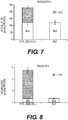
- the measured BEI of taurocholic acid was 60% ( Figure 9 )
- the measured BEI of rosuvastatin was 72%
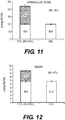
- the measured BEI of estradiol-glucuronide was 40% ( Figure 11 )
- the measured BEI of digonxin was 67% ( Figure 12 )
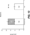
- the measured BEI of pravastatin was 58% ( Figure 13 ).
- Example 6 Lundquist (2014) Fold Difference Rat In Vitro PK Rat In Vitro PK Rat In Vivo PK Intrinsic CL int, biliary ( ⁇ l/min/mg protein) Predicted CL biliary (ml/min/kg) In Vivo CL biliary (ml/min/kg) Digoxin 0.31 ⁇ 0.02 2.5 ⁇ 0.12 0.8 ⁇ 0.3 3.1 Rosuvastatin 3.5 ⁇ 0.3 27.6 ⁇ 2.1 48.0 ⁇ 10.8 0.6 Estradiol-Gluc 2.7 ⁇ 0.1 21.6 ⁇ 0.4 n/a Taurocholate 8.4 ⁇ 0.6 67.1 ⁇ 4.8 n/a Pravastatin 0.29 ⁇ 0.02 2.3 ⁇ 0.14 n/a
- Ritonavir is a known BCRP inhibitor and was used at 0-100 ⁇ M.
- Erythromycin Estolate is a known broad spectrum inhibitor and was used at 50 ⁇ M.
- In order to asses inhibition cultures were first preincubated in the indicated inhibitor for 20 min and then incubated in the presence of the substrate and inhibitor for an additional 20 min. The cultures were then washed twice to remove excess substrate and inhibitor and the procotol proceeded as in Example 5.
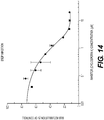
- the data for BSEP inhibition by cyclosporin A are shown in Figure 14 . Cyclosporin A inhibited BSEP mediated transport of taurocholate with an IC 50 of 0.46 ⁇ M.
- the data for BCRP inhibition by ritonavir are shown in Figure 15 .
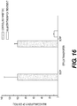
- Ritonavir inhibited BCRP mediated transport of ritonavir with an IC 50 of 0.50 ⁇ M. Inhibition by erythromycin estolate was performed in the same way except that substrates taurocholate and ritonavir were added together. As shown in Figure 16 , erythromycin estolate completely inhibited BSEP mediated transport of taurocholate and significantly inhibited BCRP mediated transport of ritonavir. Taken together, these data demonstrate the utility of the direct measurement one culture assay format of the invention for characterizing inhibition of efflux transporters by xenobiotics or drugs. [EN: Please add any additional conclusions or clarifications.]
- the methods of this invention comprise direct measurement of biliary excretion of a chemical entity in a one culture assay format.
- the accuracy and reproducibility of this assay format was demonstrated by performing three independent assays using human hepatocyte stromal cell coculture according to Example 1, for each of taurocholate and pravastatin.
- the data for taurocholic acid are shown in Table 3 and Figure 17 .
- the data for pravastatin are shown in Table 4 and Figure 18 .
- a statistical analysis presented in Table 5 indicates that there is no statistically-significant variation (P ⁇ 0.05) in the results of the replicate assay results for each compound.
- TABLE 3 Taurocholic Acid Bile Cell Mean SD Mean SD Exp. 1 541.90 7.22 273.08 86.93 Exp.
Landscapes
- Health & Medical Sciences (AREA)
- Life Sciences & Earth Sciences (AREA)
- Engineering & Computer Science (AREA)
- Immunology (AREA)
- Biomedical Technology (AREA)
- Urology & Nephrology (AREA)
- Molecular Biology (AREA)
- Chemical & Material Sciences (AREA)
- Hematology (AREA)
- Cell Biology (AREA)
- Physics & Mathematics (AREA)
- General Health & Medical Sciences (AREA)
- Pathology (AREA)
- Biotechnology (AREA)
- Food Science & Technology (AREA)
- Medicinal Chemistry (AREA)
- Tropical Medicine & Parasitology (AREA)
- Analytical Chemistry (AREA)
- Biochemistry (AREA)
- Microbiology (AREA)
- General Physics & Mathematics (AREA)
- Physiology (AREA)
- Gastroenterology & Hepatology (AREA)
- Bioinformatics & Cheminformatics (AREA)
- Toxicology (AREA)
- Measuring Or Testing Involving Enzymes Or Micro-Organisms (AREA)
- Micro-Organisms Or Cultivation Processes Thereof (AREA)
Claims (12)
- In-vitro-Verfahren zur Charakterisierung der Gallenausscheidung einer chemischen Einheit, Folgendes umfassend:a) Bereitstellen einer Zellkultur, die Hepatozyten umfasst, die mindestens einen Gallengang bilden;b) Inkontaktbringen der Zellkultur mit einer ersten chemischen Einheit für einen Zeitraum, der ausreicht, damit die chemische Einheit von den Hepatozyten in der Kultur aufgenommen werden kann;c) Aufbrechen des mindestens einen Gallenganges in der Zellkultur ohne Lyse der Hepatozyten und Erfassen der Menge (falls vorhanden) der ersten chemischen Einheit und/oder eines Metaboliten davon, die von dem mindestens einen Gallengang in der Zellkultur freigesetzt wird, unter Verwendung von LC-MS/MS; undd) Lysieren der Hepatozyten der Zellkultur und Erfassen der Menge der ersten chemischen Einheit und/oder eines Metaboliten davon, die von den Hepatozyten der Zellkultur freigesetzt wird, unter Verwendung von LC-MS/MS.
- In-vitro-Verfahren zur Charakterisierung der Gallenausscheidung einer chemischen Einheit nach Anspruch 1, wobei die Aktivität mindestens eines Hepatozytentransportproteins in den Hepatozyten der Zellkultur, genannt erste Zellkultur, gehemmt wird, wobei das Verfahren ferner Folgendes umfasst:e) Bereitstellen einer zweiten Zellkultur, umfassend Hepatozyten, die mindestens einen Gallengang bilden, wobei die Aktivität des mindestens einen Hepatozytentransportproteins in den Hepatozyten der zweiten Zellkultur nicht gehemmt ist;f) Inkontaktbringen der zweiten Zellkultur mit der ersten chemischen Einheit für einen Zeitraum, der ausreicht, damit die chemische Einheit von den Hepatozyten in der zweiten Zellkultur aufgenommen werden kann;g) Aufbrechen des mindestens einen Gallenganges in der zweiten Zellkultur ohne Lyse der Hepatozyten in der zweiten Zellkultur und Erfassen der Menge (falls vorhanden) der ersten chemischen Einheit und/oder eines Metaboliten davon, die von dem mindestens einen Gallengang in der zweiten Zellkultur freigesetzt wird, unter Verwendung von LC-MS/MS; undh) Lysieren der Hepatozyten in der zweiten Zellkultur und Erfassen der Menge der ersten chemischen Einheit und/oder eines Metaboliten davon, die von den Hepatozyten freigesetzt wird, unter Verwendung von LC-MS/MS.
- Verfahren nach Anspruch 1 oder Anspruch 2, wobei die Zellkultur oder die erste(n) und zweite(n) Zellkultur(en) eine oder mehrere Hepatozyten-Stromazellen-Kokultur(en) ist (sind), umfassend Hepatozyten und Stromazellen, die auf einer Oberfläche eines festen Substrats angeordnet sind.
- Verfahren nach einem der Ansprüche 1 bis 3, wobei die Menge der ersten chemischen Einheit und/oder eines Metaboliten davon, die von dem mindestens einen Gallengang in Schritt c) freigesetzt wird, höher ist als die Menge der ersten chemischen Einheit und/oder eines Metaboliten davon, die von den Hepatozyten in Schritt d) freigesetzt wird.
- Verfahren nach einem der Ansprüche 1 bis 4, wobei die Menge der ersten chemischen Einheit und/oder eines Metaboliten davon, die von dem mindestens einen Gallengang in Schritt c) freigesetzt wird, geringer ist als die Menge der ersten chemischen Einheit und/oder eines Metaboliten davon, die von den Hepatozyten in Schritt d) freigesetzt wird.
- Verfahren nach einem der Ansprüche 1 bis 5, wobei der mindestens eine Gallengang in der Zellkultur oder in der ersten und zweiten Zellkultur aufgebrochen wird, ohne Lyse der Hepatozyten in der Kultur, indem die Kultur in Medien inkubiert wird, die Latrunculin A (LatA) umfassen und/oder kein Calcium umfassen.
- Verfahren nach einem der Ansprüche 1 bis 5, ferner umfassend das Bestimmen der intrinsischen Gallen-Clearance (CLbile) und/oder des Gallenausscheidungsindex (BEI) für die erste chemische Einheit in der Zellkultur.
- Verfahren nach Anspruch 7, weiterhin umfassend das Vergleichen des CLbile und/oder BEI der ersten chemischen Einheit mit dem CLbile und/oder BEI einer chemischen Kontrolleinheit und das Charakterisieren der Gallenausscheidung der ersten chemischen Einheit auf der Grundlage des Vergleichs.
- Verfahren nach einem der Ansprüche 1 bis 8, ferner umfassend das Inkontaktbringen der Zellkultur mit einer zweiten chemischen Einheit in Schritt b) und/oder f).
- Verfahren nach Anspruch 9, ferner umfassend das Erfassen der Menge der zweiten chemischen Einheit und/oder eines Metaboliten davon, die von dem mindestens einen Gallengang in Schritt c) und/oder g) freigesetzt wird.
- Verfahren nach Anspruch 10, ferner umfassend das Erfassen der Menge der zweiten chemischen Einheit und/oder eines Metaboliten davon, die von den Hepatozyten in Schritt d) und/oder h) freigesetzt wird.
- In-vitro-Verfahren zur Charakterisierung der Gallenausscheidung einer chemischen Testeinheit, Folgendes umfassend:a) Bereitstellen einer Zellkultur, die Hepatozyten umfasst, die mindestens einen Gallengang bilden;b) gleichzeitiges Inkontaktbringen der Zellkultur mit einer chemischen Markereinheit und einer chemischen Testeinheit, wobei die chemische Markereinheit ein bekanntes Substrat von mindestens einem Hepatozytentransportprotein mit einem bestimmten CLbile und/oder BEI ist;c) Aufbrechen des mindestens einen Gallenganges ohne Lyse der Hepatozyten und Erfassen der Menge der chemischen Markereinheit und/oder eines Metaboliten davon, die von dem mindestens einen Gallengang freigesetzt wird, unter Verwendung von LC-MS/MS; undd) Lysieren der Hepatozyten und Erfassen der Menge der chemischen Markereinheit und/oder eines Metaboliten davon, die von den Hepatozyten freigesetzt wird, unter Verwendung von LC-MS/MS.
Applications Claiming Priority (2)
| Application Number | Priority Date | Filing Date | Title |
|---|---|---|---|
| US201462044813P | 2014-09-02 | 2014-09-02 | |
| PCT/US2015/048019 WO2016036785A1 (en) | 2014-09-02 | 2015-09-02 | In vitro biliary excretion assay |
Publications (3)
| Publication Number | Publication Date |
|---|---|
| EP3189131A1 EP3189131A1 (de) | 2017-07-12 |
| EP3189131A4 EP3189131A4 (de) | 2018-03-07 |
| EP3189131B1 true EP3189131B1 (de) | 2020-07-15 |
Family
ID=55402182
Family Applications (1)
| Application Number | Title | Priority Date | Filing Date |
|---|---|---|---|
| EP15839013.8A Active EP3189131B1 (de) | 2014-09-02 | 2015-09-02 | In-vitro-gallenausscheidungsanordnung |
Country Status (7)
| Country | Link |
|---|---|
| US (2) | US20160061820A1 (de) |
| EP (1) | EP3189131B1 (de) |
| JP (1) | JP2017527283A (de) |
| CN (1) | CN107208050A (de) |
| AU (1) | AU2015312066A1 (de) |
| CA (1) | CA2960080A1 (de) |
| WO (1) | WO2016036785A1 (de) |
Families Citing this family (1)
| Publication number | Priority date | Publication date | Assignee | Title |
|---|---|---|---|---|
| CN113396225A (zh) * | 2019-02-26 | 2021-09-14 | 株式会社日立高新技术 | 细胞内的化合物的动态分析法 |
Family Cites Families (5)
| Publication number | Priority date | Publication date | Assignee | Title |
|---|---|---|---|---|
| JPH08503610A (ja) * | 1992-11-25 | 1996-04-23 | メルク エンド カンパニー インコーポレーテッド | 肝モデル |
| EP1659403B1 (de) * | 1999-03-17 | 2009-07-22 | University Of North Carolina At Chapel Hill | Screening-Verfahren für Kandidatenverbindungen auf Empfänglichkeit für Gallenexkretion |
| JP2008545410A (ja) * | 2005-05-24 | 2008-12-18 | ザ レジェンツ オブ ザ ユニヴァースティ オブ カリフォルニア | 微小規模のマイクロパターン化インビトロ操作・作製組織 |
| WO2007146203A1 (en) * | 2006-06-08 | 2007-12-21 | The University Of North Carolina At Chapel Hill | Pulsing of bile compartments in sandwich-cultured hepatocytes |
| WO2010062911A2 (en) * | 2008-11-26 | 2010-06-03 | Hurel Corporation | Compositions and methods of functionally enhanced in vitro cell culture system |
-
2015
- 2015-09-02 JP JP2017511986A patent/JP2017527283A/ja active Pending
- 2015-09-02 EP EP15839013.8A patent/EP3189131B1/de active Active
- 2015-09-02 AU AU2015312066A patent/AU2015312066A1/en not_active Abandoned
- 2015-09-02 CN CN201580058484.8A patent/CN107208050A/zh active Pending
- 2015-09-02 WO PCT/US2015/048019 patent/WO2016036785A1/en not_active Ceased
- 2015-09-02 CA CA2960080A patent/CA2960080A1/en not_active Abandoned
- 2015-09-02 US US14/842,950 patent/US20160061820A1/en not_active Abandoned
-
2019
- 2019-12-22 US US16/724,333 patent/US20200141926A1/en not_active Abandoned
Non-Patent Citations (1)
| Title |
|---|
| None * |
Also Published As
| Publication number | Publication date |
|---|---|
| US20160061820A1 (en) | 2016-03-03 |
| CN107208050A (zh) | 2017-09-26 |
| AU2015312066A1 (en) | 2017-03-23 |
| WO2016036785A1 (en) | 2016-03-10 |
| EP3189131A4 (de) | 2018-03-07 |
| CA2960080A1 (en) | 2016-03-10 |
| EP3189131A1 (de) | 2017-07-12 |
| US20200141926A1 (en) | 2020-05-07 |
| JP2017527283A (ja) | 2017-09-21 |
Similar Documents
| Publication | Publication Date | Title |
|---|---|---|
| Ramaiahgari et al. | A 3D in vitro model of differentiated HepG2 cell spheroids with improved liver-like properties for repeated dose high-throughput toxicity studies | |
| Gunness et al. | 3D organotypic cultures of human HepaRG cells: a tool for in vitro toxicity studies | |
| US10481149B2 (en) | Method for measuring bile salt export transport and/or formation activity | |
| De Bruyn et al. | Sandwich-cultured hepatocytes: utility for in vitro exploration of hepatobiliary drug disposition and drug-induced hepatotoxicity | |
| EP1659403B1 (de) | Screening-Verfahren für Kandidatenverbindungen auf Empfänglichkeit für Gallenexkretion | |
| Novik et al. | Long-enduring primary hepatocyte-based co-cultures improve prediction of hepatotoxicity | |
| EP3712245B1 (de) | Bestandteil- und medikamentenwirksamkeitanalyseverfahren | |
| US20140199704A1 (en) | Multiplex cell signalling assays | |
| US20200141926A1 (en) | In vitro biliary excretion assay | |
| EP3430396B1 (de) | Auf 3d-gewebekultur basierendes verfahren zur beurteilung der mitochondrialen beeinträchtigung | |
| Panda et al. | Modification of the ATM/ATR directed DNA damage response state with aging and long after hepatocyte senescence induction in vivo | |
| Sidhu et al. | Effect of tissue-culture substratum and extracellular matrix overlay on liver-selective and xenobiotic inducible gene expression in primary rat hepatocytes | |
| Lenich et al. | Influence of 3D Architecture and Coculture on In Vitro CYP Enzyme Metabolism and Induction: Benchmarking Human Hepatic Spheroids for Studies in Drug Development | |
| US20180113119A1 (en) | Methods for characterizing time-based hepatotoxicity | |
| Silva et al. | In vitro models for studying induction of cytochrome P450 enzymes | |
| Saha et al. | Acute dietary methionine restriction highlights sensitivity of neocortex development to metabolic variations | |
| Preiss | Exploration of novel hepatic In-vitro systems for drug metabolism and pharmacokinetic studies | |
| Gerk et al. | Multidrug resistance proteins and hepatic transport of endo-and xenobiotics | |
| Sharma | Disposition (ADME) | |
| HK1155224A (en) | Method of screening candidate compounds for susceptibility to biliary excretion | |
| WO1995012686A1 (en) | Method for screening for compounds which change the tyrosine hydroxylase content of intact cells in culture |
Legal Events
| Date | Code | Title | Description |
|---|---|---|---|
| STAA | Information on the status of an ep patent application or granted ep patent |
Free format text: STATUS: THE INTERNATIONAL PUBLICATION HAS BEEN MADE |
|
| PUAI | Public reference made under article 153(3) epc to a published international application that has entered the european phase |
Free format text: ORIGINAL CODE: 0009012 |
|
| STAA | Information on the status of an ep patent application or granted ep patent |
Free format text: STATUS: REQUEST FOR EXAMINATION WAS MADE |
|
| 17P | Request for examination filed |
Effective date: 20170329 |
|
| AK | Designated contracting states |
Kind code of ref document: A1 Designated state(s): AL AT BE BG CH CY CZ DE DK EE ES FI FR GB GR HR HU IE IS IT LI LT LU LV MC MK MT NL NO PL PT RO RS SE SI SK SM TR |
|
| AX | Request for extension of the european patent |
Extension state: BA ME |
|
| DAV | Request for validation of the european patent (deleted) | ||
| DAX | Request for extension of the european patent (deleted) | ||
| A4 | Supplementary search report drawn up and despatched |
Effective date: 20180205 |
|
| RIC1 | Information provided on ipc code assigned before grant |
Ipc: C12Q 1/04 20060101ALI20180130BHEP Ipc: G01N 33/50 20060101ALI20180130BHEP Ipc: C12N 5/071 20100101AFI20180130BHEP |
|
| STAA | Information on the status of an ep patent application or granted ep patent |
Free format text: STATUS: EXAMINATION IS IN PROGRESS |
|
| 17Q | First examination report despatched |
Effective date: 20190404 |
|
| 17Q | First examination report despatched |
Effective date: 20190507 |
|
| GRAP | Despatch of communication of intention to grant a patent |
Free format text: ORIGINAL CODE: EPIDOSNIGR1 |
|
| STAA | Information on the status of an ep patent application or granted ep patent |
Free format text: STATUS: GRANT OF PATENT IS INTENDED |
|
| INTG | Intention to grant announced |
Effective date: 20200206 |
|
| GRAS | Grant fee paid |
Free format text: ORIGINAL CODE: EPIDOSNIGR3 |
|
| GRAA | (expected) grant |
Free format text: ORIGINAL CODE: 0009210 |
|
| STAA | Information on the status of an ep patent application or granted ep patent |
Free format text: STATUS: THE PATENT HAS BEEN GRANTED |
|
| AK | Designated contracting states |
Kind code of ref document: B1 Designated state(s): AL AT BE BG CH CY CZ DE DK EE ES FI FR GB GR HR HU IE IS IT LI LT LU LV MC MK MT NL NO PL PT RO RS SE SI SK SM TR |
|
| REG | Reference to a national code |
Ref country code: CH Ref legal event code: EP Ref country code: GB Ref legal event code: FG4D |
|
| REG | Reference to a national code |
Ref country code: IE Ref legal event code: FG4D |
|
| REG | Reference to a national code |
Ref country code: DE Ref legal event code: R096 Ref document number: 602015055922 Country of ref document: DE |
|
| REG | Reference to a national code |
Ref country code: AT Ref legal event code: REF Ref document number: 1291070 Country of ref document: AT Kind code of ref document: T Effective date: 20200815 |
|
| REG | Reference to a national code |
Ref country code: CH Ref legal event code: NV Representative=s name: NOVAGRAAF INTERNATIONAL SA, CH |
|
| REG | Reference to a national code |
Ref country code: LT Ref legal event code: MG4D |
|
| REG | Reference to a national code |
Ref country code: AT Ref legal event code: MK05 Ref document number: 1291070 Country of ref document: AT Kind code of ref document: T Effective date: 20200715 |
|
| REG | Reference to a national code |
Ref country code: NL Ref legal event code: MP Effective date: 20200715 |
|
| PG25 | Lapsed in a contracting state [announced via postgrant information from national office to epo] |
Ref country code: LT Free format text: LAPSE BECAUSE OF FAILURE TO SUBMIT A TRANSLATION OF THE DESCRIPTION OR TO PAY THE FEE WITHIN THE PRESCRIBED TIME-LIMIT Effective date: 20200715 Ref country code: AT Free format text: LAPSE BECAUSE OF FAILURE TO SUBMIT A TRANSLATION OF THE DESCRIPTION OR TO PAY THE FEE WITHIN THE PRESCRIBED TIME-LIMIT Effective date: 20200715 Ref country code: HR Free format text: LAPSE BECAUSE OF FAILURE TO SUBMIT A TRANSLATION OF THE DESCRIPTION OR TO PAY THE FEE WITHIN THE PRESCRIBED TIME-LIMIT Effective date: 20200715 Ref country code: BG Free format text: LAPSE BECAUSE OF FAILURE TO SUBMIT A TRANSLATION OF THE DESCRIPTION OR TO PAY THE FEE WITHIN THE PRESCRIBED TIME-LIMIT Effective date: 20201015 Ref country code: PT Free format text: LAPSE BECAUSE OF FAILURE TO SUBMIT A TRANSLATION OF THE DESCRIPTION OR TO PAY THE FEE WITHIN THE PRESCRIBED TIME-LIMIT Effective date: 20201116 Ref country code: ES Free format text: LAPSE BECAUSE OF FAILURE TO SUBMIT A TRANSLATION OF THE DESCRIPTION OR TO PAY THE FEE WITHIN THE PRESCRIBED TIME-LIMIT Effective date: 20200715 Ref country code: SE Free format text: LAPSE BECAUSE OF FAILURE TO SUBMIT A TRANSLATION OF THE DESCRIPTION OR TO PAY THE FEE WITHIN THE PRESCRIBED TIME-LIMIT Effective date: 20200715 Ref country code: NO Free format text: LAPSE BECAUSE OF FAILURE TO SUBMIT A TRANSLATION OF THE DESCRIPTION OR TO PAY THE FEE WITHIN THE PRESCRIBED TIME-LIMIT Effective date: 20201015 Ref country code: GR Free format text: LAPSE BECAUSE OF FAILURE TO SUBMIT A TRANSLATION OF THE DESCRIPTION OR TO PAY THE FEE WITHIN THE PRESCRIBED TIME-LIMIT Effective date: 20201016 Ref country code: FI Free format text: LAPSE BECAUSE OF FAILURE TO SUBMIT A TRANSLATION OF THE DESCRIPTION OR TO PAY THE FEE WITHIN THE PRESCRIBED TIME-LIMIT Effective date: 20200715 |
|
| PGFP | Annual fee paid to national office [announced via postgrant information from national office to epo] |
Ref country code: FR Payment date: 20201014 Year of fee payment: 7 |
|
| PG25 | Lapsed in a contracting state [announced via postgrant information from national office to epo] |
Ref country code: PL Free format text: LAPSE BECAUSE OF FAILURE TO SUBMIT A TRANSLATION OF THE DESCRIPTION OR TO PAY THE FEE WITHIN THE PRESCRIBED TIME-LIMIT Effective date: 20200715 Ref country code: LV Free format text: LAPSE BECAUSE OF FAILURE TO SUBMIT A TRANSLATION OF THE DESCRIPTION OR TO PAY THE FEE WITHIN THE PRESCRIBED TIME-LIMIT Effective date: 20200715 Ref country code: RS Free format text: LAPSE BECAUSE OF FAILURE TO SUBMIT A TRANSLATION OF THE DESCRIPTION OR TO PAY THE FEE WITHIN THE PRESCRIBED TIME-LIMIT Effective date: 20200715 Ref country code: IS Free format text: LAPSE BECAUSE OF FAILURE TO SUBMIT A TRANSLATION OF THE DESCRIPTION OR TO PAY THE FEE WITHIN THE PRESCRIBED TIME-LIMIT Effective date: 20201115 |
|
| PG25 | Lapsed in a contracting state [announced via postgrant information from national office to epo] |
Ref country code: NL Free format text: LAPSE BECAUSE OF FAILURE TO SUBMIT A TRANSLATION OF THE DESCRIPTION OR TO PAY THE FEE WITHIN THE PRESCRIBED TIME-LIMIT Effective date: 20200715 |
|
| REG | Reference to a national code |
Ref country code: DE Ref legal event code: R097 Ref document number: 602015055922 Country of ref document: DE |
|
| PG25 | Lapsed in a contracting state [announced via postgrant information from national office to epo] |
Ref country code: MC Free format text: LAPSE BECAUSE OF FAILURE TO SUBMIT A TRANSLATION OF THE DESCRIPTION OR TO PAY THE FEE WITHIN THE PRESCRIBED TIME-LIMIT Effective date: 20200715 Ref country code: SM Free format text: LAPSE BECAUSE OF FAILURE TO SUBMIT A TRANSLATION OF THE DESCRIPTION OR TO PAY THE FEE WITHIN THE PRESCRIBED TIME-LIMIT Effective date: 20200715 Ref country code: RO Free format text: LAPSE BECAUSE OF FAILURE TO SUBMIT A TRANSLATION OF THE DESCRIPTION OR TO PAY THE FEE WITHIN THE PRESCRIBED TIME-LIMIT Effective date: 20200715 Ref country code: EE Free format text: LAPSE BECAUSE OF FAILURE TO SUBMIT A TRANSLATION OF THE DESCRIPTION OR TO PAY THE FEE WITHIN THE PRESCRIBED TIME-LIMIT Effective date: 20200715 Ref country code: CZ Free format text: LAPSE BECAUSE OF FAILURE TO SUBMIT A TRANSLATION OF THE DESCRIPTION OR TO PAY THE FEE WITHIN THE PRESCRIBED TIME-LIMIT Effective date: 20200715 Ref country code: DK Free format text: LAPSE BECAUSE OF FAILURE TO SUBMIT A TRANSLATION OF THE DESCRIPTION OR TO PAY THE FEE WITHIN THE PRESCRIBED TIME-LIMIT Effective date: 20200715 Ref country code: IT Free format text: LAPSE BECAUSE OF FAILURE TO SUBMIT A TRANSLATION OF THE DESCRIPTION OR TO PAY THE FEE WITHIN THE PRESCRIBED TIME-LIMIT Effective date: 20200715 |
|
| PLBE | No opposition filed within time limit |
Free format text: ORIGINAL CODE: 0009261 |
|
| STAA | Information on the status of an ep patent application or granted ep patent |
Free format text: STATUS: NO OPPOSITION FILED WITHIN TIME LIMIT |
|
| PG25 | Lapsed in a contracting state [announced via postgrant information from national office to epo] |
Ref country code: AL Free format text: LAPSE BECAUSE OF FAILURE TO SUBMIT A TRANSLATION OF THE DESCRIPTION OR TO PAY THE FEE WITHIN THE PRESCRIBED TIME-LIMIT Effective date: 20200715 |
|
| 26N | No opposition filed |
Effective date: 20210416 |
|
| PG25 | Lapsed in a contracting state [announced via postgrant information from national office to epo] |
Ref country code: SK Free format text: LAPSE BECAUSE OF FAILURE TO SUBMIT A TRANSLATION OF THE DESCRIPTION OR TO PAY THE FEE WITHIN THE PRESCRIBED TIME-LIMIT Effective date: 20200715 Ref country code: LU Free format text: LAPSE BECAUSE OF NON-PAYMENT OF DUE FEES Effective date: 20200902 |
|
| PG25 | Lapsed in a contracting state [announced via postgrant information from national office to epo] |
Ref country code: IE Free format text: LAPSE BECAUSE OF NON-PAYMENT OF DUE FEES Effective date: 20200902 Ref country code: SI Free format text: LAPSE BECAUSE OF FAILURE TO SUBMIT A TRANSLATION OF THE DESCRIPTION OR TO PAY THE FEE WITHIN THE PRESCRIBED TIME-LIMIT Effective date: 20200715 |
|
| PGFP | Annual fee paid to national office [announced via postgrant information from national office to epo] |
Ref country code: CH Payment date: 20210914 Year of fee payment: 7 |
|
| PGFP | Annual fee paid to national office [announced via postgrant information from national office to epo] |
Ref country code: DE Payment date: 20210908 Year of fee payment: 7 Ref country code: BE Payment date: 20210913 Year of fee payment: 7 Ref country code: GB Payment date: 20210910 Year of fee payment: 7 |
|
| PG25 | Lapsed in a contracting state [announced via postgrant information from national office to epo] |
Ref country code: TR Free format text: LAPSE BECAUSE OF FAILURE TO SUBMIT A TRANSLATION OF THE DESCRIPTION OR TO PAY THE FEE WITHIN THE PRESCRIBED TIME-LIMIT Effective date: 20200715 Ref country code: MT Free format text: LAPSE BECAUSE OF FAILURE TO SUBMIT A TRANSLATION OF THE DESCRIPTION OR TO PAY THE FEE WITHIN THE PRESCRIBED TIME-LIMIT Effective date: 20200715 Ref country code: CY Free format text: LAPSE BECAUSE OF FAILURE TO SUBMIT A TRANSLATION OF THE DESCRIPTION OR TO PAY THE FEE WITHIN THE PRESCRIBED TIME-LIMIT Effective date: 20200715 |
|
| PG25 | Lapsed in a contracting state [announced via postgrant information from national office to epo] |
Ref country code: MK Free format text: LAPSE BECAUSE OF FAILURE TO SUBMIT A TRANSLATION OF THE DESCRIPTION OR TO PAY THE FEE WITHIN THE PRESCRIBED TIME-LIMIT Effective date: 20200715 |
|
| REG | Reference to a national code |
Ref country code: DE Ref legal event code: R119 Ref document number: 602015055922 Country of ref document: DE |
|
| REG | Reference to a national code |
Ref country code: CH Ref legal event code: PL |
|
| GBPC | Gb: european patent ceased through non-payment of renewal fee |
Effective date: 20220902 |
|
| REG | Reference to a national code |
Ref country code: BE Ref legal event code: MM Effective date: 20220930 |
|
| PG25 | Lapsed in a contracting state [announced via postgrant information from national office to epo] |
Ref country code: LI Free format text: LAPSE BECAUSE OF NON-PAYMENT OF DUE FEES Effective date: 20220930 Ref country code: FR Free format text: LAPSE BECAUSE OF NON-PAYMENT OF DUE FEES Effective date: 20220930 Ref country code: DE Free format text: LAPSE BECAUSE OF NON-PAYMENT OF DUE FEES Effective date: 20230401 Ref country code: CH Free format text: LAPSE BECAUSE OF NON-PAYMENT OF DUE FEES Effective date: 20220930 |
|
| PG25 | Lapsed in a contracting state [announced via postgrant information from national office to epo] |
Ref country code: BE Free format text: LAPSE BECAUSE OF NON-PAYMENT OF DUE FEES Effective date: 20220930 |
|
| PG25 | Lapsed in a contracting state [announced via postgrant information from national office to epo] |
Ref country code: GB Free format text: LAPSE BECAUSE OF NON-PAYMENT OF DUE FEES Effective date: 20220902 |