EP3175820B1 - Drainage device for controlling intraocular pressure in glaucoma - Google Patents
Drainage device for controlling intraocular pressure in glaucoma Download PDFInfo
- Publication number
- EP3175820B1 EP3175820B1 EP15814308.1A EP15814308A EP3175820B1 EP 3175820 B1 EP3175820 B1 EP 3175820B1 EP 15814308 A EP15814308 A EP 15814308A EP 3175820 B1 EP3175820 B1 EP 3175820B1
- Authority
- EP
- European Patent Office
- Prior art keywords
- implant
- eye
- aqueous humor
- sclera
- cornea
- Prior art date
- Legal status (The legal status is an assumption and is not a legal conclusion. Google has not performed a legal analysis and makes no representation as to the accuracy of the status listed.)
- Active
Links
Images


Classifications
-
- A—HUMAN NECESSITIES
- A61—MEDICAL OR VETERINARY SCIENCE; HYGIENE
- A61F—FILTERS IMPLANTABLE INTO BLOOD VESSELS; PROSTHESES; DEVICES PROVIDING PATENCY TO, OR PREVENTING COLLAPSING OF, TUBULAR STRUCTURES OF THE BODY, e.g. STENTS; ORTHOPAEDIC, NURSING OR CONTRACEPTIVE DEVICES; FOMENTATION; TREATMENT OR PROTECTION OF EYES OR EARS; BANDAGES, DRESSINGS OR ABSORBENT PADS; FIRST-AID KITS
- A61F2/00—Filters implantable into blood vessels; Prostheses, i.e. artificial substitutes or replacements for parts of the body; Appliances for connecting them with the body; Devices providing patency to, or preventing collapsing of, tubular structures of the body, e.g. stents
- A61F2/02—Prostheses implantable into the body
-
- A—HUMAN NECESSITIES
- A61—MEDICAL OR VETERINARY SCIENCE; HYGIENE
- A61F—FILTERS IMPLANTABLE INTO BLOOD VESSELS; PROSTHESES; DEVICES PROVIDING PATENCY TO, OR PREVENTING COLLAPSING OF, TUBULAR STRUCTURES OF THE BODY, e.g. STENTS; ORTHOPAEDIC, NURSING OR CONTRACEPTIVE DEVICES; FOMENTATION; TREATMENT OR PROTECTION OF EYES OR EARS; BANDAGES, DRESSINGS OR ABSORBENT PADS; FIRST-AID KITS
- A61F9/00—Methods or devices for treatment of the eyes; Devices for putting in contact-lenses; Devices to correct squinting; Apparatus to guide the blind; Protective devices for the eyes, carried on the body or in the hand
- A61F9/007—Methods or devices for eye surgery
- A61F9/00781—Apparatus for modifying intraocular pressure, e.g. for glaucoma treatment
-
- A—HUMAN NECESSITIES
- A61—MEDICAL OR VETERINARY SCIENCE; HYGIENE
- A61F—FILTERS IMPLANTABLE INTO BLOOD VESSELS; PROSTHESES; DEVICES PROVIDING PATENCY TO, OR PREVENTING COLLAPSING OF, TUBULAR STRUCTURES OF THE BODY, e.g. STENTS; ORTHOPAEDIC, NURSING OR CONTRACEPTIVE DEVICES; FOMENTATION; TREATMENT OR PROTECTION OF EYES OR EARS; BANDAGES, DRESSINGS OR ABSORBENT PADS; FIRST-AID KITS
- A61F2/00—Filters implantable into blood vessels; Prostheses, i.e. artificial substitutes or replacements for parts of the body; Appliances for connecting them with the body; Devices providing patency to, or preventing collapsing of, tubular structures of the body, e.g. stents
- A61F2/02—Prostheses implantable into the body
- A61F2/14—Eye parts, e.g. lenses or corneal implants; Artificial eyes
-
- A—HUMAN NECESSITIES
- A61—MEDICAL OR VETERINARY SCIENCE; HYGIENE
- A61F—FILTERS IMPLANTABLE INTO BLOOD VESSELS; PROSTHESES; DEVICES PROVIDING PATENCY TO, OR PREVENTING COLLAPSING OF, TUBULAR STRUCTURES OF THE BODY, e.g. STENTS; ORTHOPAEDIC, NURSING OR CONTRACEPTIVE DEVICES; FOMENTATION; TREATMENT OR PROTECTION OF EYES OR EARS; BANDAGES, DRESSINGS OR ABSORBENT PADS; FIRST-AID KITS
- A61F9/00—Methods or devices for treatment of the eyes; Devices for putting in contact-lenses; Devices to correct squinting; Apparatus to guide the blind; Protective devices for the eyes, carried on the body or in the hand
- A61F9/007—Methods or devices for eye surgery
-
- A—HUMAN NECESSITIES
- A61—MEDICAL OR VETERINARY SCIENCE; HYGIENE
- A61M—DEVICES FOR INTRODUCING MEDIA INTO, OR ONTO, THE BODY; DEVICES FOR TRANSDUCING BODY MEDIA OR FOR TAKING MEDIA FROM THE BODY; DEVICES FOR PRODUCING OR ENDING SLEEP OR STUPOR
- A61M1/00—Suction or pumping devices for medical purposes; Devices for carrying-off, for treatment of, or for carrying-over, body-liquids; Drainage systems
-
- A—HUMAN NECESSITIES
- A61—MEDICAL OR VETERINARY SCIENCE; HYGIENE
- A61M—DEVICES FOR INTRODUCING MEDIA INTO, OR ONTO, THE BODY; DEVICES FOR TRANSDUCING BODY MEDIA OR FOR TAKING MEDIA FROM THE BODY; DEVICES FOR PRODUCING OR ENDING SLEEP OR STUPOR
- A61M27/00—Drainage appliance for wounds or the like, i.e. wound drains, implanted drains
- A61M27/002—Implant devices for drainage of body fluids from one part of the body to another
-
- A—HUMAN NECESSITIES
- A61—MEDICAL OR VETERINARY SCIENCE; HYGIENE
- A61F—FILTERS IMPLANTABLE INTO BLOOD VESSELS; PROSTHESES; DEVICES PROVIDING PATENCY TO, OR PREVENTING COLLAPSING OF, TUBULAR STRUCTURES OF THE BODY, e.g. STENTS; ORTHOPAEDIC, NURSING OR CONTRACEPTIVE DEVICES; FOMENTATION; TREATMENT OR PROTECTION OF EYES OR EARS; BANDAGES, DRESSINGS OR ABSORBENT PADS; FIRST-AID KITS
- A61F9/00—Methods or devices for treatment of the eyes; Devices for putting in contact-lenses; Devices to correct squinting; Apparatus to guide the blind; Protective devices for the eyes, carried on the body or in the hand
- A61F9/007—Methods or devices for eye surgery
- A61F9/008—Methods or devices for eye surgery using laser
- A61F2009/00885—Methods or devices for eye surgery using laser for treating a particular disease
- A61F2009/00891—Glaucoma
-
- A—HUMAN NECESSITIES
- A61—MEDICAL OR VETERINARY SCIENCE; HYGIENE
- A61F—FILTERS IMPLANTABLE INTO BLOOD VESSELS; PROSTHESES; DEVICES PROVIDING PATENCY TO, OR PREVENTING COLLAPSING OF, TUBULAR STRUCTURES OF THE BODY, e.g. STENTS; ORTHOPAEDIC, NURSING OR CONTRACEPTIVE DEVICES; FOMENTATION; TREATMENT OR PROTECTION OF EYES OR EARS; BANDAGES, DRESSINGS OR ABSORBENT PADS; FIRST-AID KITS
- A61F2230/00—Geometry of prostheses classified in groups A61F2/00 - A61F2/26 or A61F2/82 or A61F9/00 or A61F11/00 or subgroups thereof
- A61F2230/0002—Two-dimensional shapes, e.g. cross-sections
- A61F2230/0017—Angular shapes
- A61F2230/0019—Angular shapes rectangular
Definitions
- Glaucoma is, after diabetes, the second leading cause of blindness in the world. Vision loss as a result of glaucoma involves both central and peripheral vision and has a major impact on the ability of people to live independent lives.
- Glaucoma is an optic neuropathy (a disorder of the optic nerve) caused by an elevated intraocular pressure.
- the high pressure inside the eye compresses the outlet of the optic nerve (papilla) causing changes in its appearance and in the visual function (increase of the optic cup and alterations to the visual field). If the pressure remains increased over a sufficient period of time, a complete and irreversible vision loss takes place.
- Aqueous humor is a fluid produced by the ciliary body in the posterior chamber of the eye at a rate of approximately 3 to 5 microliters per minute. The fluid produced then passes through the pupillary opening of the iris into the anterior chamber of the eye.
- the fluid leaves the eye through two different routes: the "uveoscleral” route responsible for 10% of the outflow, in which the fluid is filtered between muscle fibers of the ciliary body, and the “canalicular” route responsible for 90% of the outflow , in which the fluid drains through the junction between the cornea and the iris (iridocorneal angle) through a filter called Trabecula which is composed by collagen bundles arranged in a three-dimensional structure similar to a sieve. From that point the aqueous humor goes to the so-called Schlemm's canal and then to the episcleral veins.
- the production and outflow of aqueous humor necessarily have to be equal. This causes a pressure inside the eye that is determined by the amount of fluid produced and the capacity of outflow thereof. In normal conditions, the intraocular pressure is between 10 and 21 mm Hg.
- aqueous humor production increases with respect to outflow resistance
- the intraocular pressure will increase to force the outflow. If the outflow resistance increases, the intraocular pressure will increase to force the outflow even with a normal aqueous humor production. Both scenarios will increase intraocular pressure and may cause glaucoma, being much more frequent the increase in the outflow resistance.
- Aqueous humor outflow resistance may be increased by several factors.
- the abnormal resistance is produced along the outer aspect of the trabecular meshwork and the inner wall of the Schlemm's canal.
- angle-closure glaucoma the resistance increases due to mechanical blocking of the angle and in “secondary glaucoma” due to various reasons such as inflammatory waste deposits, vascularization of the area, pigment blocking, etc.
- increased intraocular pressure compresses the outlet of the optic nerve and interferes in its vascularization, causing the death of nerve cells that carry visual stimuli to the brain, producing alterations in the visual field and, in advanced cases, total and irreversible blindness.
- the only therapeutic approach currently available for glaucoma is to reduce intraocular pressure.
- the clinical treatment of glaucoma is done step by step, the first step being the medical treatment either by oral means or with drops.
- the drugs work by reducing aqueous humor production or by facilitating its outflow.
- Currently available medicinal products can have many side effects, some serious, including congestive heart failure, respiratory distress, hypertension, depression, kidney stones, aplastic anemia, sexual dysfunction and death. Compliance with medication is also a major problem with more than half of glaucoma patients being estimated as not following their dosing schedules correctly.
- laser trabeculoplasty thermal energy from a laser is applied to a number of noncontiguous spots in the trabecular meshwork. It is believed that the laser energy somehow stimulates the metabolism of the trabecular cells and changes the extracellular material of the trabecular meshwork. In approximately 80% of patients, aqueous outflow is increased and the pressure decreases. However, the effect often is not long lasting and 50% of patients develop hypertension again in five years. In addition, laser trabeculoplasty is not an effective treatment for primary open angle glaucoma in patients of less than fifty years old nor is it effective for angle-closure glaucoma and many secondary glaucomas.
- trabeculectomy The most commonly performed filtering procedure is trabeculectomy.
- a trabeculectomy an incision is made in the conjunctiva, which is the transparent tissue that covers the sclera.
- the conjunctival tissue is lifted and a scleral flap of approximately 4x4 mm is made with a depth of 50% of the scleral thickness and is lamellar dissected up to 1 mm into the cornea.
- the anterior chamber is entered beneath the scleral flap and a section of the deep sclera and trabecular meshwork is excised.
- the scleral flap is loosely sewn back into place and conjunctival incision is closed well.
- the aqueous humor passes through the hole beneath the scleral flap and goes to the subconjunctival space where it forms a blister from where it passes to the blood vessels of the conjunctiva or goes through the conjunctiva to the surface.
- an opening is created in the limbus cornea region of the eye.
- a filtering implant having a foot portion and a body portion is provided for implantation. The foot portion of the implant is placed through the limbal opening into the anterior chamber of the eye, and the body portion is buried beneath a scleral flap.
- the apparatus of the invention is a filtering implant.
- Patent document US 5 704 907 discloses a method and an apparatus for lowering the intraocular pressure of an eye are provided.
- Patent document The apparatus of the invention is a filtering implant adapted to extend from the anterior chamber of the eye through an opening in the limbus corneae to a drainage area beneath a scleral flap.
- Patent document US 2006/155238 discloses an ophthalmic shunt implantable in an eye having an elongate body and a conduit for conducting aqueous humor from an anterior chamber of the eye to the suprachoroidal space of the eye.
- the elongate body has a forward end and an insertion head that extends from the forward end.
- the insertion head defines a shearing edge suitable for cutting eye tissue engage thereby.
- the forward end and the insertion head of the body define a shoulder surface.
- the conduit has a first end defined on a portion of a top surface of the insertion head.
- the conduit also extends through the body from the forward end to a back end thereof. The first end of the conduit is spaced from the shearing edge and, in one example, from the shoulder of the body.
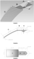
- Figure 1 shows a three-dimensional view of the device object of the present invention which has been divided into three parts: "A”, “B” and “C”.
- This device will be made of stainless steel or another biocompatible material that does not adhere to the eye tissues, the device will have a uniform or variable thickness.
- Part “A” will be arranged in the anterior chamber between the cornea and the iris and will have a double triangle shape, which allows introducing and removing it with ease while at the same time keeping it fixed in its position.
- Part “B” will puncture the eye in the corneal limbus, being arranged between the cornea and the sclera, and part “C” will be arranged outside the eye covered by a scleral flap, on the episclera or both.
- Figure 2 shows a side view of the device object of the present invention.
- the device will have a uniform or variable thickness and will measure between 0.02 and 0.3 mm.
- the device is a one-piece device, but it has been divided into in three parts for description purposes: "A”, "B” and “C”.
- Part “A” will be arranged inside the eye in the anterior chamber between the cornea and the iris.
- Part “B” will puncture the corneal limbus forming two virtual spaces through which it will drain the aqueous humor and part “C” will be arranged outside the eye, in the interscleral space, episcleral space or both.
- the parts forming the device of the invention are:
- Figure 2 shows a side view of a filtering device in which part "A", which will be arranged in the anterior chamber between the cornea and the iris (see Figure 6 ), can be seen.
- This part “A” will have a double triangle shape with an upper base “b” having measurements between 0.3 and 1.0 mm, height “h” between 0.3 and 0.9 mm and a rounded lower vertex “v” to facilitate its insertion. This allows a less traumatic anchorage in addition to facilitating its introduction or removal.
- Figure 2 shows a side view of the filtering device in which part "B" can be seen.
- This part will puncture the corneal limbus, forming a virtual space upwardly between the cornea and the upper surface of the device and another virtual space downwardly between the sclera and the lower surface of the device.
- the aqueous humor will drain through these virtual spaces from the anterior chamber to an intrascleral or episcleral space.
- the sheet forming part "B" of the device will have a thickness between 0.02 and 0.3 mm.
- Figure 3 shows a top view of the device in which part "B" can be seen, the upper surface of which will be in contact with the cornea and form with it a virtual space through which the aqueous humor will flow.
- the lower surface will be in contact with the sclera and form another virtual space.
- the surfaces of this sheet can be smooth, rough, wavy, grooved or with another finish.
- Part "B” of the device will have a length between 0.3 and 1.2 mm with a width equal to that of part "A", this width will measure between 0.5 and 4 mm.
- Figure 2 shows a side view of the filtering device in which part "C" can be seen.
- This part will have a length between 0.5 and 15 mm and a thickness between 0.02 and 0.3 mm, it will be thin, malleable and will be curved to approximately the eye curvature.
- Figure 3 shows a top view of the device in which part "C”, being in the form of a rectangular sheet with rounded edges and wider than part "B", can be seen. It will measure between 0.7 and 5 mm in width and will have a length between 0.5 and 15 mm. This sheet can be smooth, rough, wavy, grooved or with another finish, it will be arranged outside the eye and covered by a scleral flap, arranged in the episcleral space or both.
- FIG. 5 is an illustration showing the area of the eye where the flap or scleral flap will be made to create the interscleral virtual space.
- This flap will be similar to that obtained for a trabeculectomy surgery and will have a length of approximately 4 mm, a width of 4 mm and a depth of about 50% the scleral thickness.
- Figure 6 is an illustration showing the device object of the present invention in its working position and in which part "A" will be arranged in the anterior chamber.
- Part “B” will puncture the eye and create a virtual space upwardly between the cornea and the upper surface of the device and create a virtual space downwardly between the sclera and the lower surface of the device.
- Part “C” will be arranged outside the eye between the scleral flap and the sclera, similarly creating two virtual spaces, one towards the flap and another towards the sclera.
- Aqueous humor will flow through these virtual spaces and be diffused into the interscleral space and subconjuntival space.
- Figure 7 is an illustration similar to that of Figure 6 but in which part "C" is longer and, in addition to being arranged between the scleral flap and the sclera, it comes out the flap towards the episcleral space, creating a virtual corridor that leads to the ocular equator, increasing the area of diffusion of the aqueous humor. This can be very beneficial in cases of neovascular glaucoma.
- the surgical procedure needed to insert the device can include all or some of the following steps: 1. an incision is made in the conjunctiva that may be limbus or fornix based, 2. a scleral flap of approximately 4 x 4 mm with a thickness of approximately 50% the scleral thickness is delimited and a dissection parallel to the sclera is made until entering 1 mm into the corneal tissue, 3. paracentesis is performed parallel to the limbus with a blade of 15°, 4. the device is inserted into the paracentesis, 5. the flap is closed with 2 loose stitches, 6. the conjunctiva is closed.
Landscapes
- Health & Medical Sciences (AREA)
- Ophthalmology & Optometry (AREA)
- Heart & Thoracic Surgery (AREA)
- General Health & Medical Sciences (AREA)
- Veterinary Medicine (AREA)
- Biomedical Technology (AREA)
- Engineering & Computer Science (AREA)
- Public Health (AREA)
- Life Sciences & Earth Sciences (AREA)
- Animal Behavior & Ethology (AREA)
- Vascular Medicine (AREA)
- Nuclear Medicine, Radiotherapy & Molecular Imaging (AREA)
- Surgery (AREA)
- Cardiology (AREA)
- Oral & Maxillofacial Surgery (AREA)
- Transplantation (AREA)
- Anesthesiology (AREA)
- Hematology (AREA)
- Otolaryngology (AREA)
- Prostheses (AREA)
- Pharmaceuticals Containing Other Organic And Inorganic Compounds (AREA)
- External Artificial Organs (AREA)
Applications Claiming Priority (2)
| Application Number | Priority Date | Filing Date | Title |
|---|---|---|---|
| PE2014001057A PE20151266A1 (es) | 2014-07-01 | 2014-07-01 | Dispositivo de drenaje para el control de la presion intraocular en glaucoma |
| PCT/PE2015/000009 WO2016003294A1 (es) | 2014-07-01 | 2015-07-01 | Dispositivo de drenaje para el control de la presion intraocular en glaucoma |
Publications (4)
| Publication Number | Publication Date |
|---|---|
| EP3175820A1 EP3175820A1 (en) | 2017-06-07 |
| EP3175820A4 EP3175820A4 (en) | 2018-06-06 |
| EP3175820B1 true EP3175820B1 (en) | 2025-03-19 |
| EP3175820C0 EP3175820C0 (en) | 2025-03-19 |
Family
ID=54330905
Family Applications (1)
| Application Number | Title | Priority Date | Filing Date |
|---|---|---|---|
| EP15814308.1A Active EP3175820B1 (en) | 2014-07-01 | 2015-07-01 | Drainage device for controlling intraocular pressure in glaucoma |
Country Status (14)
| Country | Link |
|---|---|
| US (1) | US10758412B2 (enExample) |
| EP (1) | EP3175820B1 (enExample) |
| JP (1) | JP6825761B2 (enExample) |
| KR (1) | KR102268311B1 (enExample) |
| CN (1) | CN106687072B (enExample) |
| BR (1) | BR112016030196B1 (enExample) |
| CL (1) | CL2016003238A1 (enExample) |
| CO (1) | CO2016005603A2 (enExample) |
| ES (1) | ES3019033T3 (enExample) |
| IL (1) | IL249688B (enExample) |
| MX (1) | MX392355B (enExample) |
| PE (1) | PE20151266A1 (enExample) |
| SG (2) | SG11201610957WA (enExample) |
| WO (1) | WO2016003294A1 (enExample) |
Families Citing this family (14)
| Publication number | Priority date | Publication date | Assignee | Title |
|---|---|---|---|---|
| EP3463044A4 (en) | 2016-05-31 | 2020-07-29 | Qura, Inc. | IMPLANTABLE INTRAOCULAR PRESSURE SENSORS AND METHODS OF USE |
| US11166849B2 (en) | 2017-07-20 | 2021-11-09 | Shifamed Holdings, Llc | Adjustable flow glaucoma shunts and methods for making and using same |
| JP7191398B2 (ja) | 2017-07-20 | 2022-12-19 | シファメド・ホールディングス・エルエルシー | 調整可能流量緑内障シャントおよびその製造および使用方法 |
| EP3742958A4 (en) | 2018-01-23 | 2021-10-20 | Avisi Technologies, Inc. | METHOD AND DEVICE FOR TREATMENT OF EYE DISEASES |
| KR101999479B1 (ko) | 2018-02-06 | 2019-07-11 | 가톨릭대학교 산학협력단 | 녹내장 억제용 방수배출기구 |
| EP3911285A4 (en) | 2019-01-18 | 2022-10-19 | Shifamed Holdings, LLC | ADJUSTABLE FLOW GLAUCOMA SHUNTS AND METHODS OF MAKING AND USE THEREOF |
| EP3893721A4 (en) * | 2019-01-18 | 2022-10-12 | Avisi Technologies, Inc. | Method and device for treating eye disease |
| EP4041149A4 (en) | 2019-10-10 | 2023-11-15 | Shifamed Holdings, LLC | ADJUSTABLE FLOW GLAUCOMA SHUNTS AND ASSOCIATED SYSTEMS AND METHODS |
| CA3165037A1 (en) | 2020-01-23 | 2021-07-29 | Robert Chang | Adjustable flow glaucoma shunts and associated systems and methods |
| WO2021163566A1 (en) | 2020-02-14 | 2021-08-19 | Shifamed Holdings, Llc | Shunting systems with rotation-based flow control assemblies, and associated systems and methods |
| EP4106695A4 (en) | 2020-02-18 | 2024-03-20 | Shifamed Holdings, LLC | ADJUSTABLE FLOW GLAUCOMA SHUNTS WITH NON-LINEAR FLOW CONTROL ELEMENTS AND ASSOCIATED SYSTEMS AND METHODS |
| WO2021188952A1 (en) | 2020-03-19 | 2021-09-23 | Shifamed Holdings, Llc | Intraocular shunts with low-profile actuation elements and associated systems and methods |
| EP4135640B1 (en) | 2020-04-16 | 2025-12-24 | Shifamed Holdings, LLC | Adjustable glaucoma treatment devices and associated systems |
| US11865283B2 (en) | 2021-01-22 | 2024-01-09 | Shifamed Holdings, Llc | Adjustable shunting systems with plate assemblies, and associated systems and methods |
Family Cites Families (15)
| Publication number | Priority date | Publication date | Assignee | Title |
|---|---|---|---|---|
| US4946436A (en) * | 1989-11-17 | 1990-08-07 | Smith Stewart G | Pressure-relieving device and process for implanting |
| US5520631A (en) * | 1994-07-22 | 1996-05-28 | Wound Healing Of Oklahoma | Method and apparatus for lowering the intraocular pressure of an eye |
| US5704907A (en) * | 1994-07-22 | 1998-01-06 | Wound Healing Of Oklahoma | Method and apparatus for lowering the intraocular pressure of an eye |
| US5601094A (en) * | 1994-11-22 | 1997-02-11 | Reiss; George R. | Ophthalmic shunt |
| US5882327A (en) * | 1997-04-17 | 1999-03-16 | Jacob; Jean T. | Long-term glaucoma drainage implant |
| US6699210B2 (en) * | 1999-04-27 | 2004-03-02 | The Arizona Board Of Regents | Glaucoma shunt and a method of making and surgically implanting the same |
| US6558342B1 (en) * | 1999-06-02 | 2003-05-06 | Optonol Ltd. | Flow control device, introducer and method of implanting |
| EP1534363B1 (en) * | 2002-07-19 | 2008-12-31 | Yale University | Uveoscleral drainage device |
| US7160264B2 (en) * | 2002-12-19 | 2007-01-09 | Medtronic-Xomed, Inc. | Article and method for ocular aqueous drainage |
| US20060069340A1 (en) * | 2003-06-16 | 2006-03-30 | Solx, Inc. | Shunt for the treatment of glaucoma |
| WO2004110391A2 (en) * | 2003-06-16 | 2004-12-23 | Solx, Inc. | Shunt for the treatment of glaucoma |
| US20080306429A1 (en) * | 2007-06-07 | 2008-12-11 | Shields Milton B | Uveoscleral drainage device |
| AU2010266013B2 (en) * | 2009-06-25 | 2015-03-26 | Alcon Inc. | Fiber matrix for maintaining space in soft tissues |
| WO2011035336A1 (en) * | 2009-09-21 | 2011-03-24 | Vidus Ocular, Inc. | Uveoscleral drainage device |
| US8852136B2 (en) * | 2011-12-08 | 2014-10-07 | Aquesys, Inc. | Methods for placing a shunt into the intra-scleral space |
-
2014
- 2014-07-01 PE PE2014001057A patent/PE20151266A1/es active IP Right Revival
-
2015
- 2015-07-01 SG SG11201610957WA patent/SG11201610957WA/en unknown
- 2015-07-01 CN CN201580035881.3A patent/CN106687072B/zh active Active
- 2015-07-01 WO PCT/PE2015/000009 patent/WO2016003294A1/es not_active Ceased
- 2015-07-01 MX MX2016017411A patent/MX392355B/es unknown
- 2015-07-01 JP JP2016576047A patent/JP6825761B2/ja active Active
- 2015-07-01 SG SG10201901871WA patent/SG10201901871WA/en unknown
- 2015-07-01 EP EP15814308.1A patent/EP3175820B1/en active Active
- 2015-07-01 KR KR1020177001536A patent/KR102268311B1/ko active Active
- 2015-07-01 ES ES15814308T patent/ES3019033T3/es active Active
- 2015-07-01 BR BR112016030196-0A patent/BR112016030196B1/pt active IP Right Grant
-
2016
- 2016-12-14 US US15/378,946 patent/US10758412B2/en active Active
- 2016-12-15 CL CL2016003238A patent/CL2016003238A1/es unknown
- 2016-12-21 IL IL249688A patent/IL249688B/en unknown
- 2016-12-21 CO CONC2016/0005603A patent/CO2016005603A2/es unknown
Also Published As
| Publication number | Publication date |
|---|---|
| US10758412B2 (en) | 2020-09-01 |
| PE20151266A1 (es) | 2015-09-10 |
| KR20170028936A (ko) | 2017-03-14 |
| MX392355B (es) | 2025-03-24 |
| BR112016030196B1 (pt) | 2022-10-18 |
| CL2016003238A1 (es) | 2017-10-13 |
| SG10201901871WA (en) | 2019-03-28 |
| JP6825761B2 (ja) | 2021-02-03 |
| CN106687072B (zh) | 2021-01-22 |
| US20170095370A1 (en) | 2017-04-06 |
| WO2016003294A9 (es) | 2017-02-23 |
| BR112016030196A2 (enExample) | 2017-08-22 |
| EP3175820A4 (en) | 2018-06-06 |
| CO2016005603A2 (es) | 2017-03-10 |
| KR102268311B1 (ko) | 2021-06-23 |
| EP3175820A1 (en) | 2017-06-07 |
| IL249688A0 (en) | 2017-02-28 |
| CN106687072A (zh) | 2017-05-17 |
| ES3019033T3 (en) | 2025-05-20 |
| EP3175820C0 (en) | 2025-03-19 |
| IL249688B (en) | 2022-03-01 |
| SG11201610957WA (en) | 2017-02-27 |
| BR112016030196A8 (pt) | 2022-07-05 |
| MX2016017411A (es) | 2017-05-15 |
| JP2017519592A (ja) | 2017-07-20 |
| WO2016003294A1 (es) | 2016-01-07 |
Similar Documents
| Publication | Publication Date | Title |
|---|---|---|
| EP3175820B1 (en) | Drainage device for controlling intraocular pressure in glaucoma | |
| US11980574B2 (en) | Ocular device for treating glaucoma and related minimally invasive glaucoma surgery method | |
| EP1534363B1 (en) | Uveoscleral drainage device | |
| EP1707166A1 (en) | Irrigation tip | |
| US6699211B2 (en) | Method and apparatus for treatment of glaucoma | |
| US20090177138A1 (en) | Shunt Device for Glaucoma Treatment | |
| EP4438015A2 (en) | Bleb control glaucoma shunts | |
| US7713228B2 (en) | Surgical method | |
| CA2368342A1 (en) | Stent device and method for treating glaucoma | |
| JP2018512942A (ja) | 眼科インターポジションインプラント | |
| US20060173077A1 (en) | Surgical method | |
| RU2089149C1 (ru) | Способ лечения вторичной глаукомы | |
| HK1237641A1 (en) | Drainage device for controlling intraocular pressure in glaucoma | |
| HK1237641B (zh) | 用於控制青光眼中的眼内压的排水装置 | |
| JP6739839B2 (ja) | 眼科手術用補助具 | |
| EP4577165A2 (en) | Ocular device for treating glaucoma and related minimally invasive glaucoma surgery method | |
| WO2014152072A1 (en) | Miniature glaucoma shunt |
Legal Events
| Date | Code | Title | Description |
|---|---|---|---|
| STAA | Information on the status of an ep patent application or granted ep patent |
Free format text: STATUS: THE INTERNATIONAL PUBLICATION HAS BEEN MADE |
|
| 17P | Request for examination filed |
Effective date: 20170201 |
|
| AK | Designated contracting states |
Kind code of ref document: A1 Designated state(s): AL AT BE BG CH CY CZ DE DK EE ES FI FR GB GR HR HU IE IS IT LI LT LU LV MC MK MT NL NO PL PT RO RS SE SI SK SM TR |
|
| AX | Request for extension of the european patent |
Extension state: BA ME |
|
| PUAI | Public reference made under article 153(3) epc to a published international application that has entered the european phase |
Free format text: ORIGINAL CODE: 0009012 |
|
| STAA | Information on the status of an ep patent application or granted ep patent |
Free format text: STATUS: REQUEST FOR EXAMINATION WAS MADE |
|
| DAV | Request for validation of the european patent (deleted) | ||
| DAX | Request for extension of the european patent (deleted) | ||
| A4 | Supplementary search report drawn up and despatched |
Effective date: 20180507 |
|
| RIC1 | Information provided on ipc code assigned before grant |
Ipc: A61F 2/14 20060101ALI20180430BHEP Ipc: A61F 9/007 20060101AFI20180430BHEP |
|
| STAA | Information on the status of an ep patent application or granted ep patent |
Free format text: STATUS: EXAMINATION IS IN PROGRESS |
|
| 17Q | First examination report despatched |
Effective date: 20201029 |
|
| RAP1 | Party data changed (applicant data changed or rights of an application transferred) |
Owner name: MM INSTRUMENTS LLC |
|
| RIN1 | Information on inventor provided before grant (corrected) |
Inventor name: MIRANDA VELASQUEZ, MARIO EDUARDO |
|
| GRAP | Despatch of communication of intention to grant a patent |
Free format text: ORIGINAL CODE: EPIDOSNIGR1 |
|
| STAA | Information on the status of an ep patent application or granted ep patent |
Free format text: STATUS: GRANT OF PATENT IS INTENDED |
|
| INTG | Intention to grant announced |
Effective date: 20241206 |
|
| GRAS | Grant fee paid |
Free format text: ORIGINAL CODE: EPIDOSNIGR3 |
|
| GRAA | (expected) grant |
Free format text: ORIGINAL CODE: 0009210 |
|
| STAA | Information on the status of an ep patent application or granted ep patent |
Free format text: STATUS: THE PATENT HAS BEEN GRANTED |
|
| AK | Designated contracting states |
Kind code of ref document: B1 Designated state(s): AL AT BE BG CH CY CZ DE DK EE ES FI FR GB GR HR HU IE IS IT LI LT LU LV MC MK MT NL NO PL PT RO RS SE SI SK SM TR |
|
| REG | Reference to a national code |
Ref country code: GB Ref legal event code: FG4D |
|
| REG | Reference to a national code |
Ref country code: CH Ref legal event code: EP |
|
| REG | Reference to a national code |
Ref country code: IE Ref legal event code: FG4D |
|
| REG | Reference to a national code |
Ref country code: DE Ref legal event code: R096 Ref document number: 602015091255 Country of ref document: DE |
|
| U01 | Request for unitary effect filed |
Effective date: 20250408 |
|
| U07 | Unitary effect registered |
Designated state(s): AT BE BG DE DK EE FI FR IT LT LU LV MT NL PT RO SE SI Effective date: 20250414 |
|
| REG | Reference to a national code |
Ref country code: ES Ref legal event code: FG2A Ref document number: 3019033 Country of ref document: ES Kind code of ref document: T3 Effective date: 20250520 |
|
| PG25 | Lapsed in a contracting state [announced via postgrant information from national office to epo] |
Ref country code: RS Free format text: LAPSE BECAUSE OF FAILURE TO SUBMIT A TRANSLATION OF THE DESCRIPTION OR TO PAY THE FEE WITHIN THE PRESCRIBED TIME-LIMIT Effective date: 20250619 |
|
| PG25 | Lapsed in a contracting state [announced via postgrant information from national office to epo] |
Ref country code: NO Free format text: LAPSE BECAUSE OF FAILURE TO SUBMIT A TRANSLATION OF THE DESCRIPTION OR TO PAY THE FEE WITHIN THE PRESCRIBED TIME-LIMIT Effective date: 20250619 |
|
| PG25 | Lapsed in a contracting state [announced via postgrant information from national office to epo] |
Ref country code: HR Free format text: LAPSE BECAUSE OF FAILURE TO SUBMIT A TRANSLATION OF THE DESCRIPTION OR TO PAY THE FEE WITHIN THE PRESCRIBED TIME-LIMIT Effective date: 20250319 |
|
| PG25 | Lapsed in a contracting state [announced via postgrant information from national office to epo] |
Ref country code: GR Free format text: LAPSE BECAUSE OF FAILURE TO SUBMIT A TRANSLATION OF THE DESCRIPTION OR TO PAY THE FEE WITHIN THE PRESCRIBED TIME-LIMIT Effective date: 20250620 |
|
| U20 | Renewal fee for the european patent with unitary effect paid |
Year of fee payment: 11 Effective date: 20250708 |
|
| PG25 | Lapsed in a contracting state [announced via postgrant information from national office to epo] |
Ref country code: SM Free format text: LAPSE BECAUSE OF FAILURE TO SUBMIT A TRANSLATION OF THE DESCRIPTION OR TO PAY THE FEE WITHIN THE PRESCRIBED TIME-LIMIT Effective date: 20250319 |
|
| PGFP | Annual fee paid to national office [announced via postgrant information from national office to epo] |
Ref country code: ES Payment date: 20250801 Year of fee payment: 11 |
|
| PG25 | Lapsed in a contracting state [announced via postgrant information from national office to epo] |
Ref country code: PL Free format text: LAPSE BECAUSE OF FAILURE TO SUBMIT A TRANSLATION OF THE DESCRIPTION OR TO PAY THE FEE WITHIN THE PRESCRIBED TIME-LIMIT Effective date: 20250319 |
|
| PGFP | Annual fee paid to national office [announced via postgrant information from national office to epo] |
Ref country code: TR Payment date: 20250701 Year of fee payment: 11 |
|
| PGFP | Annual fee paid to national office [announced via postgrant information from national office to epo] |
Ref country code: GB Payment date: 20250728 Year of fee payment: 11 |
|
| PG25 | Lapsed in a contracting state [announced via postgrant information from national office to epo] |
Ref country code: CZ Free format text: LAPSE BECAUSE OF FAILURE TO SUBMIT A TRANSLATION OF THE DESCRIPTION OR TO PAY THE FEE WITHIN THE PRESCRIBED TIME-LIMIT Effective date: 20250319 |
|
| PG25 | Lapsed in a contracting state [announced via postgrant information from national office to epo] |
Ref country code: SK Free format text: LAPSE BECAUSE OF FAILURE TO SUBMIT A TRANSLATION OF THE DESCRIPTION OR TO PAY THE FEE WITHIN THE PRESCRIBED TIME-LIMIT Effective date: 20250319 |
|
| PG25 | Lapsed in a contracting state [announced via postgrant information from national office to epo] |
Ref country code: IS Free format text: LAPSE BECAUSE OF FAILURE TO SUBMIT A TRANSLATION OF THE DESCRIPTION OR TO PAY THE FEE WITHIN THE PRESCRIBED TIME-LIMIT Effective date: 20250719 |