EP3153856A1 - Verfahren zur bestimmung der konzentration von retikulierten thrombozyten - Google Patents
Verfahren zur bestimmung der konzentration von retikulierten thrombozyten Download PDFInfo
- Publication number
- EP3153856A1 EP3153856A1 EP15188536.5A EP15188536A EP3153856A1 EP 3153856 A1 EP3153856 A1 EP 3153856A1 EP 15188536 A EP15188536 A EP 15188536A EP 3153856 A1 EP3153856 A1 EP 3153856A1
- Authority
- EP
- European Patent Office
- Prior art keywords
- platelets
- absorption
- refractive index
- sample
- biofluid
- Prior art date
- Legal status (The legal status is an assumption and is not a legal conclusion. Google has not performed a legal analysis and makes no representation as to the accuracy of the status listed.)
- Withdrawn
Links
Images


Classifications
-
- G—PHYSICS
- G01—MEASURING; TESTING
- G01N—INVESTIGATING OR ANALYSING MATERIALS BY DETERMINING THEIR CHEMICAL OR PHYSICAL PROPERTIES
- G01N33/00—Investigating or analysing materials by specific methods not covered by groups G01N1/00 - G01N31/00
- G01N33/48—Biological material, e.g. blood, urine; Haemocytometers
- G01N33/50—Chemical analysis of biological material, e.g. blood, urine; Testing involving biospecific ligand binding methods; Immunological testing
- G01N33/53—Immunoassay; Biospecific binding assay; Materials therefor
- G01N33/569—Immunoassay; Biospecific binding assay; Materials therefor for microorganisms, e.g. protozoa, bacteria, viruses
- G01N33/56966—Animal cells
-
- G—PHYSICS
- G01—MEASURING; TESTING
- G01N—INVESTIGATING OR ANALYSING MATERIALS BY DETERMINING THEIR CHEMICAL OR PHYSICAL PROPERTIES
- G01N15/00—Investigating characteristics of particles; Investigating permeability, pore-volume or surface-area of porous materials
- G01N15/10—Investigating individual particles
- G01N15/14—Optical investigation techniques, e.g. flow cytometry
- G01N15/1456—Optical investigation techniques, e.g. flow cytometry without spatial resolution of the texture or inner structure of the particle, e.g. processing of pulse signals
- G01N15/1459—Optical investigation techniques, e.g. flow cytometry without spatial resolution of the texture or inner structure of the particle, e.g. processing of pulse signals the analysis being performed on a sample stream
-
- G—PHYSICS
- G01—MEASURING; TESTING
- G01N—INVESTIGATING OR ANALYSING MATERIALS BY DETERMINING THEIR CHEMICAL OR PHYSICAL PROPERTIES
- G01N33/00—Investigating or analysing materials by specific methods not covered by groups G01N1/00 - G01N31/00
- G01N33/48—Biological material, e.g. blood, urine; Haemocytometers
- G01N33/50—Chemical analysis of biological material, e.g. blood, urine; Testing involving biospecific ligand binding methods; Immunological testing
- G01N33/80—Chemical analysis of biological material, e.g. blood, urine; Testing involving biospecific ligand binding methods; Immunological testing involving blood groups or blood types or red blood cells
-
- G—PHYSICS
- G01—MEASURING; TESTING
- G01N—INVESTIGATING OR ANALYSING MATERIALS BY DETERMINING THEIR CHEMICAL OR PHYSICAL PROPERTIES
- G01N15/00—Investigating characteristics of particles; Investigating permeability, pore-volume or surface-area of porous materials
- G01N15/01—Investigating characteristics of particles; Investigating permeability, pore-volume or surface-area of porous materials specially adapted for biological cells, e.g. blood cells
- G01N2015/018—Platelets
-
- G—PHYSICS
- G01—MEASURING; TESTING
- G01N—INVESTIGATING OR ANALYSING MATERIALS BY DETERMINING THEIR CHEMICAL OR PHYSICAL PROPERTIES
- G01N15/00—Investigating characteristics of particles; Investigating permeability, pore-volume or surface-area of porous materials
- G01N15/10—Investigating individual particles
- G01N2015/1006—Investigating individual particles for cytology
-
- G—PHYSICS
- G01—MEASURING; TESTING
- G01N—INVESTIGATING OR ANALYSING MATERIALS BY DETERMINING THEIR CHEMICAL OR PHYSICAL PROPERTIES
- G01N15/00—Investigating characteristics of particles; Investigating permeability, pore-volume or surface-area of porous materials
- G01N15/10—Investigating individual particles
- G01N15/14—Optical investigation techniques, e.g. flow cytometry
- G01N2015/1402—Data analysis by thresholding or gating operations performed on the acquired signals or stored data
-
- G—PHYSICS
- G01—MEASURING; TESTING
- G01N—INVESTIGATING OR ANALYSING MATERIALS BY DETERMINING THEIR CHEMICAL OR PHYSICAL PROPERTIES
- G01N15/00—Investigating characteristics of particles; Investigating permeability, pore-volume or surface-area of porous materials
- G01N15/10—Investigating individual particles
- G01N15/14—Optical investigation techniques, e.g. flow cytometry
- G01N2015/1488—Methods for deciding
-
- G—PHYSICS
- G01—MEASURING; TESTING
- G01N—INVESTIGATING OR ANALYSING MATERIALS BY DETERMINING THEIR CHEMICAL OR PHYSICAL PROPERTIES
- G01N2800/00—Detection or diagnosis of diseases
- G01N2800/22—Haematology
- G01N2800/224—Haemostasis or coagulation
-
- G—PHYSICS
- G01—MEASURING; TESTING
- G01N—INVESTIGATING OR ANALYSING MATERIALS BY DETERMINING THEIR CHEMICAL OR PHYSICAL PROPERTIES
- G01N2800/00—Detection or diagnosis of diseases
- G01N2800/22—Haematology
- G01N2800/226—Thrombotic disorders, i.e. thrombo-embolism irrespective of location/organ involved, e.g. renal vein thrombosis, venous thrombosis
Definitions
- the invention relates to a method for a particularly fully automated determination of the concentration of reticulated platelets in a thrombocyte-containing sample of a biofluid, in particular in a blood sample.
- the invention further relates to an analysis system for a particularly fully automated implementation of this method.
- Reticulated platelets are immature platelets formed by constriction from the megakaryocytes. Like reticulocytes, ie immature erythrocytes, reticulated platelets provide an indication of the underlying formation process, in this case thrombopoiesis, with the number of reticulated platelets correlating with the activity of the megakaryocytes. A determination of the concentration, ie a relative number (fraction) or absolute number of reticulated platelets, gives the opportunity to diagnose diseases that influence the activity of the megakaryocytes or that result in the disease sequence a consumption or a destruction of platelets.
- ITP Immunune Thrombocytopenia
- TTP Thrombotic Thrombocytopenic Purpura
- DIC Disminated Intravascular Coagulopathy
- HIT Heparin-induced Thrombocytopenia
- HUS Hemolytic Uremic Syndrome
- the percentage of reticulated platelets to the total number of platelets can help in particular to make a corresponding diagnosis of thrombocytopenia.
- the analysis of this value in samples of biofluids, especially in blood samples, should be as fully automated as possible. At present, this is made possible by automatically working optical analysis systems in that the RNA (ribonucleic acid), which is still increasingly contained in reticulated thromobocytes, has dyed a dye and fluorescence optical the concentration of reticulated platelets contained or their absolute number or fraction is determined.
- RNA ribonucleic acid
- the object of the invention is to specify an alternative method for determining the concentration of reticulated platelets in samples of a biofluid, which can also be carried out automatically with optical analysis systems.
- a sample of the biofluid is supplied with a dye which is selective for nucleic acid and stains RNA, wherein after staining in the sample in the absorption region of the dye, a multiplicity of optical, particle-related measured values are in each case in the form of tuples of refractive index and adsorption degree wherein measured values with an adsorption degree within a partial range of all measured values of the adsorption degree and with a refractive index within a partial range of all measured values of the refractive index are selected, and wherein the number of selected measured values is used to determine the concentration of the reticulated platelets.
- the invention is based on the consideration that with a cytometric method, which is carried out for example in flow geometry, in a comparison of two-dimensional measurements from the absorptance and associated refractive index in a cytogram, the reticulated platelets not only comparatively simple mature platelets, but for example can also be separated from erythrocytes. Since reticulated platelets still contain a relatively large amount of RNA, after selective staining their absorbance initially differs from the mature platelets, which receive little RNA. In this respect, a subarea with a higher degree of absorption can be assigned to the measured values derived from reticulated platelets in a cytogram of absorptance and refractive index.
- erythrocytes show because of their hemoglobin content compared to platelets a higher refractive index, so that a portion of lower refractive index can be assigned to the platelet-derived measurements.
- a partial range of measured values can be assigned to the reticulated platelets from a cytometric recording of the degree of absorption and the refractive index. The number of appropriately selected measurements is then used to determine the concentration of the reticulated platelets. It has been shown that the measurement against erythrocytes is largely interference-free.
- a particularly two-dimensional cytometric measurement of optical particle-relevant features belongs to the standard method of automated optical analysis systems of biofluids.
- the staining of RNA by means of a dye has hitherto been carried out mainly for the determination of the number or the fraction of reticulocytes.
- the invention differs therefrom.
- the invention further enables a largely interference-free detection of the reticulated platelets against fragments of leukocytes and other particles having a comparable absorptivity as the reticulated platelets. It has been found that such fragments and particles show a lower refractive index than the reticulated platelets.
- the scattering, reflection and / or transmisson of a light beam, in particular of a laser beam, caused in each case by the measuring volume is detected in flow-resolved time-resolved manner.
- tuples of optical characteristics, in particular also the refractive index and the degree of absorption are then determined as measured values which allow conclusions to be drawn about the particles contained as disperse phase in the biofluid.
- This so-called flow cytometry is a common measurement method of modern optical analysis systems for the analysis of biofluids, as used for example to a fully automated creation of a blood count or the like.
- the method described above, in particular fully automated is performed in an analysis system for the optical analysis of a sample of a biofluid.
- Oxazine 750 is preferably used. Oxazine 750 is frequently used for staining nucleic acid and in the present case for staining RNA.
- the upper limit of the partial region of the refractive index is selected such that refractive indices of the erythrocytes are separated from refractive indices of the platelets, so that interference is excluded or at least minimized.
- the refractive indices of erythrocytes and platelets are widely spaced, so that a corresponding limit can be easily determined by considering respective refractive indices. Such refractive indices can be taken from corresponding output channels in automated analysis systems.
- hypochromic erythrocytes that is erythrocytes with a comparatively low hemoglobin content
- the frequency distribution of the measured refractive indices is determined and herein identified a population of erythrocytes in a range of higher refractive indices and a population of thrombocytes in a range of lower refractive indices.
- a refractive index is selected from a range between the two populations. This ensures that potential interference of platelet and red blood cell readings is largely avoided.
- the lower limit of the sub-range of the degree of absorption is selected such that the degrees of absorption of mature platelets are separated from the absorption levels of reticulated platelets. This is possible because, as already mentioned, mature platelets have no or only a very small proportion of RNA, so that their degree of absorption is reduced in relation to the absorption range of the dye used compared with that of reticulated platelets.
- the absorption of platelets is patient-specific and thus varies from sample to sample.
- the degrees of absorption of the platelets are measured in the respective sample of biofluid and determined their frequency distribution. From the frequency distribution, a frequency maximum is determined, and as the lower limit of the partial range of the degree of absorption, an absorption degree above the absorption coefficient assigned to the frequency maximum is selected. In this way, a specific specific limit in the degree of absorption is determined, which separates the mature platelets of immature, reticulated platelets for each sample.
- the distance between the lower limit of the sub-range of the absorption coefficient and the absorption coefficient assigned to the frequency maximum is chosen such that the standard biofluid, e.g. a blood sample of a healthy patient, from the selected measured values determined fraction of the reticulated platelets with respect to all platelets in a range between 1% and 7%.
- the normal average is 3%.
- the choice of this limit in the degree of absorption is based on the consideration that the lifetime of human platelets in peripheral blood is about 10 days. Therefore, the platelet daily reproductive rate is about 10% to keep the platelet count constant. If you think about it assuming that the maturation time of the platelets in the periphery is 6 to 8 hours, then average values of reticulated platelets for normal healthy patients are about 3%.
- the lower limit of the refractive index portion in particular variable over the degree of absorption, is selected such that refractive indices of the platelets are separated from refractive indices of fragments of the leukocytes and other particles.
- this is possible because fragments of leukocytes and also other particles which show a degree of absorption comparable to platelets have a reduced refractive index.
- a number of platelets is determined in the sample of the biofluid, in particular automatically, wherein a fraction of the reticulated platelets is determined from the number of selected measured values and the number of platelets.
- the total number of platelets is separately measured independently of the cytogram of absorbance and refractive index or removed from a corresponding separate output channel of an automated analysis system. The number of selected measured values is then set in relation to this separately determined total number of platelets.
- the total number of platelets can not be taken from the same cytogram, from which the number of reticulated platelets is determined, since the refractive index of the platelets is changed by the stained RNA.
- the invention further relates to an analysis system for optical analysis of a sample of a biofluid, in particular a blood sample, which is set up and designed to perform a method of the type described above, in particular fully automated.
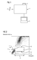
- FIG. 1 schematically an optical analysis system 1 for performing an automated analysis of the sample of a biofluid is shown.
- the analysis system 1 in this case comprises a light source 2, in particular a laser, which radiates through a sample volume.
- a variety of optical characteristics such as in particular particle-related refractive indices, absorption levels, transmittances, reflectances, etc., are detected by observing the laser light passing through a sample volume in scattering, reflection, transmission geometry, etc.
- the analysis system 1 is assigned an output unit 3.
- the respectively determined optical features can be output, displayed or further evaluated over a variety of output channels.
- the output unit 3 it is possible via the output unit 3 to produce a complete blood count in tabular form with output all relevant data including the fraction of reticulated platelets.
- a sample 4 of a biofluid in particular a blood sample, is supplied to the analysis system 1 accordingly.
- the subsequent evaluation is done fully automatically including any supply of auxiliaries, dyes, etc.
- FIG. 2 a cytogram is shown in which the refractive index is plotted against the degree of absorption.
- the acquired particle-related measurements each comprising a tuple of refractive index and absorbance, are registered as the result of an automated analysis process. From a range of 5 selective measurements, the number of reticulated platelets can be taken. This region comprises a partial region 6 of the measured absorption grades and a partial region 7 of the measured refractive indices.
- the region 5 is separated, in particular, from the measured values of erythrocytes by an upper limit 10 of the refractive index, to mature platelets by a lower limit 11 of the absorption coefficient and to fragments of leucocytes and other particles by a lower limit 12 of the refractive index.
- the number of reticulated platelets can be determined largely interference-free over the marked borders.
- the upper limit 10 of the refractive index for delimiting the measured values of reticulated platelets against measured values of erythrocytes is determined from a histogram according to FIG FIG. 3 , in which the frequencies of the respectively measured refractive indices is shown, determined.
- the large population 14 of erythrocytes and the small population 15 of platelets are identified. Between the two populations 14, 15, the upper limit of a refractive index for determining the reticulated platelets is determined.
- the corresponding upper limit 10 of the refractive index is in FIG. 4 shown that similar to another cytogram FIG. 2 shows.
- To determine the lower limit of the absorption level for the area 5 of the reticulated platelets are according to FIG.
- the above-described determination of the lower limit 11 of the degree of absorption specifically refers to the fact that platelets of different patients show different degrees of absorption.
- the lower limit of the degree of absorption relative to the maximum absorption degree is to be shifted to the right by a number of steps or corresponding channels, thus the absorption coefficient of the lower limit is chosen to be greater than the absorption coefficient at the maximum of the frequency distribution ,
Landscapes
- Health & Medical Sciences (AREA)
- Life Sciences & Earth Sciences (AREA)
- Immunology (AREA)
- Chemical & Material Sciences (AREA)
- Engineering & Computer Science (AREA)
- Hematology (AREA)
- Urology & Nephrology (AREA)
- Molecular Biology (AREA)
- Biomedical Technology (AREA)
- Biochemistry (AREA)
- General Health & Medical Sciences (AREA)
- Cell Biology (AREA)
- Physics & Mathematics (AREA)
- Pathology (AREA)
- Analytical Chemistry (AREA)
- General Physics & Mathematics (AREA)
- Microbiology (AREA)
- Biotechnology (AREA)
- Food Science & Technology (AREA)
- Medicinal Chemistry (AREA)
- Virology (AREA)
- Tropical Medicine & Parasitology (AREA)
- Zoology (AREA)
- Dispersion Chemistry (AREA)
- Investigating Or Analysing Biological Materials (AREA)
Abstract
Die Erfindung betrifft ein Verfahren zur Bestimmung der Konzentration von retikulierten Thrombozyten in einer Thrombozyten enthaltenden Probe (4) eines Biofluids, insbesondere in einer Blutprobe, wobei der Probe (4) des Biofluids ein für Nukleinsäure selektiver Farbstoff zugeführt wird, wobei nach der Anfärbung in der Probe (4) im Absorptionsbereich des Farbstoffes eine Vielzahl von optischen, partikelbedingten Messwerten jeweils als Tupel aus Brechungsindex und Absorptionsgrad bestimmt wird, wobei Messwerte mit einem Absorptionsgrad innerhalb eines Teilbereiches (6) aller gemessenen Werte des Absorptionsgrades und mit einem Brechungsindex innerhalb eines Teilbereiches (7) aller gemessenen Werte des Brechungsindexes selektiert werden, und wobei die Anzahl der selektierten Messwerte zur Bestimmung der Konzentration der retikulierten Thrombozyten herangezogen wird.
Description
- Die Erfindung betrifft ein Verfahren zu einer insbesondere vollautomatisierten Bestimmung der Konzentration von retikulierten Thrombozyten in einer Thrombozyten enthaltenden Probe eines Biofluids, insbesondere in einer Blutprobe. Die Erfindung betrifft weiter ein Analysesystem zu einer insbesondere vollautomatisierten Durchführung dieses Verfahrens.
- Retikulierte Thrombozyten sind unreife Thrombozyten, die durch Abschnürung aus den Megakaryozyten gebildet werden. Wie Retikulozyten, also unreife Erythrozyten, liefern retikulierte Thrombozyten einen Hinweis auf den zugrundeliegenden Bildungsprozess, vorliegend die Thrombopoese, wobei die Anzahl der retikulierten Thrombozyten mit der Aktivität der Megakaryozyten korreliert. Eine Bestimmung der Konzentration, also einer relativen Anzahl (Fraktion) oder absoluten Anzahl an retikulierten Thrombozyten, gibt die Möglichkeit, Krankheiten zu diagnostizieren, die Einfluss auf die Aktivität der Megakaryozyten nehmen oder die als Krankheitsfolge einen Verbrauch oder eine Destruktion von Thrombozyten zur Folge haben. Beispiele sind hierbei ITP (Immunthrombozytopenie), TTP (Thrombotisch-thrombozytopenische Purpura), DIC (disseminierte intravasale Koagulopathie), HIT (Heparininduzierte Thrombozytopenie), HUS (hämolytisch-urämisches Syndrom) und andere.
- Der Prozentsatz an retikulierten Thrombozyten zur gesamten Anzahl an Thrombozyten kann insbesondere mithelfen, eine entsprechende Diagnose bei Thrombozythopenie zu erstellen. Die Analyse dieses Wertes in Proben von Biofluiden, insbesondere in Blutproben, soll möglichst vollautomatisiert erfolgen. Gegenwärtig ist dies mit automatisch arbeitenden optischen Analysesystemen dadurch ermöglicht, dass die in retikulierten Thromobzyten noch vermehrt enthaltene RNA (Ribonukleinsäure)mit einem Farbstoff angefärbt und fluoreszenzoptisch die Konzentration an enthaltenen retikulierten Thrombozyten bzw. deren absolute Anzahl oder Fraktion ermittelt wird.
- Aufgabe der Erfindung ist es, ein alternatives Verfahren zur Bestimmung der Konzentration an retikulierten Thrombozyten in Proben eines Biofluids anzugeben, welches sich ebenfalls automatisiert mit optischen Analysesystemen durchführen lässt.
- Diese Aufgabe wird erfindungsgemäß dadurch gelöst, dass einer Probe des Biofluids ein für Nukleinsäure selektiver Farbstoff zugeführt wird, der hier RNA einfärbt, wobei nach der Anfärbung in der Probe im Absorptionsbereich des Farbstoffes eine Vielzahl von optischen, partikelbedingten Messwerten jeweils als Tupel aus Brechungsindex und Adsorptionsgrad bestimmt wird, wobei Messwerte mit einem Adsorptionsgrad innerhalb eines Teilbereiches aller gemessenen Werte des Adsorptionsgrades und mit einem Brechungsindex innerhalb eines Teilbereiches aller gemessenen Werte des Brechungsindexes selektiert werden, und wobei die Anzahl der selektierten Messwerte zur Bestimmung der Konzentration der retikulierten Thrombozyten herangezogen wird.
- Die Erfindung geht dabei von der Überlegung aus, dass sich mit einem zytometrischen Verfahren, welches beispielsweise in Durchflussgeometrie durchgeführt wird, bei einer Gegenüberstellung der zweidimensionalen Messwerte aus Absorptionsgrad und zugehörigem Brechungsindex in einem Zytogramm die retikulierten Thrombozyten nicht nur vergleichsweise einfach von reifen Thrombozyten, sondern beispielsweise auch von Erythrozyten getrennt werden können. Da retikulierte Thrombozyten noch relativ viel RNA enthalten, unterscheidet sich nach selektiver Anfärbung deren Absorptionsgrad zunächst von dem reifer Thrombozyten, die wenig RNA erhalten. Insofern kann in einem Zytogramm aus Absorptionsgrad und Brechungsindex ein Teilbereich mit höherem Absorptionsgrad den von retikulierten Thrombozyten stammenden Messwerten zugeordnet werden. Andererseits zeigen Erythrozyten aufgrund ihres Hämoglobingehalts gegenüber Thrombozyten einen höheren Brechungsindex, so dass ein Teilbereich niedrigeren Brechungsindexes den von Thrombozyten stammenden Messwerten zugeordnet werden kann. Insofern kann aus einer zytometrischen Erfassung aus Absorptionsgrad und Brechungsindex ein Teilbereich an Messwerten den retikulierten Thrombozyten zugeordnet werden. Die Anzahl der entsprechend selektierten Messwerte wird dann zur Bestimmung der Konzentration der retikulierten Thrombozyten herangezogen. Es hat sich dabei gezeigt, dass die Messung gegenüber Erythrozyten weitgehend interferenzfrei ist.
- Eine insbesondere zweidimensionale zytometrische Messung von optischen partikelrelevanten Merkmalen gehört zum Standardverfahren von automatisierten optischen Analysesystemen von Biofluiden. Jedoch wird die Anfärbung von RNA mittels eines Farbstoffs bislang hauptsächlich zur Bestimmung der Anzahl oder der Fraktion an Retikulozyten durchgeführt. Die Erfindung unterscheidet sich jedoch hiervon.
- Die Erfindung ermöglicht weiter auch eine weitgehend interferenzfreie Erfassung der retikulierten Thrombozyten gegenüber Fragmenten von Leukozyten und anderen Teilchen, die einen vergleichbaren Absorptionsgrad wie die retikulierten Thrombozyten aufweisen. Es hat sich gezeigt, dass derartige Fragmente und Teilchen gegenüber den retikulierten Thrombozyten einen geringeren Brechungsindex zeigen.
- Zum Ermitteln der Messwerte, die jeweils ein Tupel aus Absorptionsgrad und Brechungsindex umfassen, wird beispielsweise in Durchflussgeometrie zeitaufgelöst die jeweils durch im Messvolumen befindliche Partikel verursachte Streuung, Reflexion und/oder Transmisson eines Lichtstrahls, insbesondere eines Laserstrahls, erfasst. Aus den in verschiedenen Messgeometrien gemessenen optischen Parametern werden dann als Messwerte jeweils Tupel aus optischen Merkmalen, insbesondere auch Brechungsindex und Absorptionsgrad, ermittelt, die Rückschlüsse auf die als disperse Phase in dem Biofluid enthaltenen Partikeln erlauben. Diese sogenannte Durchflusszytometrie ist hierbei ein gängiges Messverfahren moderner optischer Analysesysteme zur Analyse von Biofluiden, wie sie beispielsweise zu einer vollautomatisierten Erstellung eines Blutbildes oder dergleichen eingesetzt werden. Bevorzugt wird daher das vorbeschriebene Verfahren, insbesondere vollautomatisiert, in einem Analysesystem zur optischen Analyse einer Probe eines Biofluids durchgeführt. Als ein geeigneter Farbstoff wird bevorzugt Oxazin 750 verwendet. Oxazin 750 wird häufig zur Anfärbung von Nukleinsäure und vorliegend zur Anfärbung von RNA eingesetzt.
- In einer vorteilhaften Ausgestaltung wird die obere Grenze des Teilbereiches des Brechungsindexes, insbesondere veränderlich über den Absorptionsgrad, derart gewählt, dass Brechungsindizes der Erythrozyten von Brechungsindizes der Thrombozyten separiert werden, sodass eine Interferenz ausgeschlossen oder zumindest minimiert wird. Gewöhnlich liegen die Brechungsindizes von Erythrozyten und Thrombozyten weit auseinander, so dass eine entsprechende Grenze leicht durch die Betrachtung von entsprechenden Brechungsindizes festgelegt werden kann. Derartige Brechungsindizes können bei automatisierten Analysesystemen aus entsprechenden Ausgabekanälen entnommen werden.
- Hypochrome Erythrozyten, also Erythrozyten mit einem vergleichsweise geringen Hämoglobingehalt, können jedoch einen Brechungsindex zeigen, der in der Nähe der Brechungsindizes von Thrombozyten liegt. Aus diesem Grund wird in einer vorteilhaften Variante die Häufigkeitsverteilung der gemessenen Brechungsindizes ermittelt und hierin eine Population der Erythrozyten in einem Bereich höherer Brechungsindizes und eine Population der Thrombozyten in einem Bereich niedrigerer Brechungsindizes identifiziert. Als obere Grenze des Teilbereiches des Brechungsindizes wird dann ein Brechungsindex aus einem Bereich zwischen den beiden Populationen gewählt. Auf diese Weise wird sichergestellt, dass eine mögliche Interferenz von Messwerten von Thrombozyten und Messwerten von Erythrozyten weitestgehend vermieden ist.
- Weiter bevorzugt wird die untere Grenze des Teilbereiches des Absorptionsgrades derart gewählt, dass Absorptionsgrade reifer Thrombozyten von Absorptionsgraden retikulierter Thrombozyten separiert werden. Dies ist deswegen möglich, da wie bereits erwähnt reife Thrombozyten keine oder nur noch einen sehr geringen Anteil an RNA aufweisen, so dass deren Absorptionsgrad in Bezug auf den Absorptionsbereich des eingesetzten Farbstoffes gegenüber dem von retikulierten Thrombozyten verringert ist.
- Es hat sich gezeigt, dass die Absorption von Thrombozyten patientenspezifisch ist und somit von Probe zu Probe variiert. Zweckmäßigerweise werden daher in der jeweiligen Probe des Biofluids die Absorptionsgrade der Thrombozyten gemessen und deren Häufigkeitsverteilung ermittelt. Aus der Häufigkeitsverteilung wird ein Häufigkeitsmaximum bestimmt und als untere Grenze des Teilbereiches des Absorptionsgrades wird ein Absorptionsgrad oberhalb des dem Häufigkeitsmaximum zugeordneten Absorptionsgrad gewählt. Auf diese Weise wird eine konkrete spezifische Grenze im Absorptionsgrad bestimmt, die für die jeweilige Probe die reifen Thrombozyten von unreifen, retikulierten Thrombozyten trennt.
- Vorteilhaft wird hierbei der Abstand der unteren Grenze des Teilbereiches des Absorptionsgrades zu dem dem Häufigkeitsmaximum zugeordneten Absorptionsgrad derart gewählt, dass die für ein Normbiofluid, z.B. eine Blutprobe eines gesunden Patienten, aus den selektierten Messwerten ermittelte Fraktion der retikulierten Thrombozyten bezüglich aller Thrombozyten in einem Bereich zwischen 1 % und 7 % liegt. Der normale Durchschnittswert liegt hierbei bei 3 %.
- Als Wahl dieser Grenze im Absorptionsgrad liegt die Überlegung zugrunde, dass die Lebensdauer an menschlichen Thrombozyten in peripherem Blut etwa 10 Tage beträgt. Daher beträgt die tägliche Reproduktionsrate der Thrombozyten etwa 10 %, um die Thrombozytenzahl konstant zu halten. Wenn man davon ausgeht, dass die Reifungszeit der Thrombozyten in der Peripherie 6 bis 8 Stunden beträgt, liegen somit Durchschnittswerte an retikulierten Thrombozyten für gesunde Normalpatienten bei etwa 3 %.
- Zweckmäßigerweise wird die untere Grenze des Teilbereiches des Brechungsindexes, insbesondere veränderlich über den Absorptionsgrad, derart gewählt, dass Brechungsindizes der Thrombozyten von Brechungsindizes von Fragmenten der Leukozyten und anderen Teilchen separiert werden. Wie bereits ausgeführt ist dies möglich, da Fragmente von Leukozyten und auch andere Teilchen, die einen den Thrombozyten vergleichbaren Absorptionsgrad zeigen, einen verringerten Brechungsindex aufweisen.
- In einer weiteren bevorzugten Ausgestaltung des Verfahrens wird in der Probe des Biofluids, insbesondere automatisiert, eine Anzahl von Thrombozyten ermittelt, wobei aus der Anzahl der selektierten Messwerte und der Anzahl der Thrombozyten eine Fraktion der retikulierten Thrombozyten ermittelt wird. Bei dieser Variante wird die Gesamtzahl an Thrombozyten separat unabhängig vom Zytogramm aus Absorptionsgrad und Brechungsindex vermessen bzw. aus einem entsprechenden separaten Ausgabekanal eines automatisierten Analysesystems entnommen. Die Anzahl der selektierten Messwerte wird dann in Relation zu dieser separat ermittelten Gesamtzahl der Thrombozyten gesetzt. Bei dieser Variante wird davon ausgegangen, dass die Gesamtzahl an Thrombozyten nicht dem gleichen Zytogramm entnommen werden kann, aus dem die Anzahl an retikulierten Thrombozyten ermittelt wird, da der Brechungsindex der Thrombozyten durch die angefärbte RNA verändert wird.
- Die Erfindung betrifft weiter ein Analysesystem zur optischen Analyse einer Probe eines Biofluids, insbesondere einer Blutprobe, welches eingerichtet und ausgebildet ist, ein Verfahren der vorbeschriebenen Art, insbesondere vollautomatisiert, durchzuführen.
- Ausführungsbeispiele der Erfindung werden anhand einer Zeichnung näher erläutert. Dabei zeigen:
- FIG 1
- schematisch ein automatisiertes optisches Analysesystem für Proben eines Biofluids,
- FIG 2
- ein Zytogramm, wobei der Brechungsindex "Refractive Index" gegenüber dem Absorptionsgrad "Absorption" aufgetragen ist,
- FIG 3
- ein Histogramm gemessener Brechungsindizes,
- FIG 4
- ein weiteres Zytogramm, wobei der Brechungsindex über dem Absorptionsgrad aufgetragen ist,
- FIG 5
- ein Histogramm der für Thrombozyten gemessenen Absorptionsgrade und
- FIG 6
- in einer Gegenüberstellung verschiedene Zytogramm mit unterschiedlichen Grenzen zwischen reifen und unreifen Thrombozyten.
- In
FIG 1 ist schematisch ein optisches Analysesystem 1 zur Durchführung einer automatisierten Analyse der Probe eines Biofluids dargestellt. Das Analysesystem 1 umfasst hierbei eine Lichtquelle 2, insbesondere einen Laser, der ein Probenvolumen durchstrahlt. Eine Vielzahl von optischen Merkmalen, wie insbesondere partikelbezogene Brechungsindizes, Absorptionsgrade, Transmissionsgrade, Reflexionsgrade etc. werden durch Beobachtung des ein Probenvolumen durcheilenden Laserlichts in Streu-, Reflexions-, Transmissionsgeometrie etc. erfasst. Weiter ist dem Analysesystem 1 eine Ausgabeeinheit 3 zugeordnet. Über die Ausgabeeinheit 3 können über eine Vielzahl von Ausgabekanälen die jeweils ermittelten optischen Merkmale ausgegeben, dargestellt oder weiter ausgewertet werden. Über die Ausgabeeinheit 3 ist es insbesondere möglich, ein vollständiges Blutbild in tabellarischer Form mit Ausgabe aller relevanter Daten einschließlich der Fraktion an retikulierten Thrombozyten auszugeben. Zur Analyse wird eine Probe 4 eines Biofluids, insbesondere eine Blutprobe, dem Analysesystem 1 entsprechend zugeführt. Die anschließende Auswertung geschieht vollautomatisch einschließlich einer etwaigen Zuführung von Hilfs-, Färbestoffen usw. - In
FIG 2 ist ein Zytogramm dargestellt, in dem der Brechungsindex gegenüber dem Absorptionsgrad aufgetragen ist. Die erfassten partikelbezogenen Messwerte, die jeweils ein Tupel aus Brechungsindex und Absorptionsgrad umfassen, sind als Ergebnis eines automatisierten Analyseverfahrens eingetragen. Aus einem Bereich 5 selektiver Messwerte kann die Anzahl an retikulierten Thrombozyten entnommen werden. Dieser Bereich umfasst einen Teilbereich 6 der gemessenen Absorptionsgrade und einen Teilbereich 7 der gemessenen Brechungsindizes. Der Bereich 5 ist hierbei insbesondere gegenüber den Messwerten von Erythrozyten durch eine obere Grenze 10 des Brechungsindexes, gegenüber reifen Thrombozyten durch eine untere Grenze 11 des Absorptionsgrades und gegenüber Fragmenten von Leukozyten und anderen Teilchen durch eine untere Grenze 12 des Brechungsindexes abgetrennt. Über die eingezeichneten Grenzen kann die Anzahl der retikulierten Thrombozyten weitestgehend interferenzfrei bestimmt werden. - Die obere Grenze 10 des Brechungsindexes zur Abgrenzung der Messwerte von retikulierten Thrombozyten gegenüber Messwerten von Erythrozyten wird aus einem Histogramm gemäß
FIG 3 , in dem die Häufigkeiten der jeweils gemessenen Brechungsindizes dargestellt ist, ermittelt. Es wird hierbei die große Population 14 an Erythrozyten und die kleine Population 15 an Thrombozyten identifiziert. Zwischen den beiden Populationen 14, 15 wird die obere Grenze eines Brechungsindexes zur Ermittlung der retikulierten Thrombozyten festgelegt. Die entsprechende obere Grenze 10 des Brechungsindexes ist inFIG 4 dargestellt, die ein anderes Zytogramm ähnlichFIG 2 zeigt. Zur Bestimmung der unteren Grenze des Absorptionsgrades für den Bereich 5 der retikulierten Thrombozyten werden gemäßFIG 5 in einem Histogramm die Häufigkeiten der in der Probe des Biofluids gemessenen Absorptionsgrade der Thrombozyten (anderer Kanal) ermittelt. Es wird hierin der jeweilige dem Maximum zugeordnete Absorptionsgrad bestimmt. In den Zytogrammen gemäßFIG 6 werden als untere Grenzen 11 des jeweiligen Absorptionsgrades Absorptionsgrade größer als der dem Maximum zugeordnete Absorptionsgrad festgelegt. InFIG 6 (a) ist die untere Grenze 11 des Absorptionsgrades gegenüber der unteren Grenze 11 des Absorptionsgrades inFIG 6 (b) zu kleineren Werten verschoben. - Durch die vorbeschriebene Bestimmung der unteren Grenze 11 des Absorptionsgrades wird spezifisch auf die Tatsache Bezug genommen, dass Thrombozyten verschiedener Patienten unterschiedliche Absorptionsgrade zeigen. Vorliegend hat es sich gezeigt, dass die untere Grenze des Absorptionsgrades gegenüber dem dem Maximum zugehörenden Absorptionsgrad um eine Anzahl von Schritten bzw. entsprechenden Kanälen nach rechts zu verschieben ist, somit also der Absorptionsgrad der unteren Grenze gegenüber dem Absorptionsgrad am Maximum der Häufigkeitsverteilung größer gewählt ist.
-
- 1
- Analysesystem
- 2
- Lichtquelle
- 3
- Ausgabegerät
- 4
- Probe Biofluid (Blutprobe)
- 5
- Bereich selektierter Messwerte
- 6
- Teilbereich Absorptionsgrad
- 7
- Teilbereich Brechungsindex
- 10
- obere Grenze Teilbereich Brechungsindex
- 11
- untere Grenze Teilbereich Absorptionsgrad
- 12
- untere Grenze Teilbereich Brechungsindex
- 14
- Population Erythrozyten
- 15
- Population Thrombozyten
Claims (10)
- Verfahren zur Bestimmung der Konzentration von retikulierten Thrombozyten in einer Thrombozyten enthaltenden Probe (4) eines Biofluids, insbesondere in einer Blutprobe, wobei der Probe (4) des Biofluids ein für Nukleinsäure selektiver Farbstoff zugeführt wird, wobei nach der Anfärbung in der Probe (4) im Absorptionsbereich des Farbstoffes eine Vielzahl von optischen, partikelbedingten Messwerten jeweils als Tupel aus Brechungsindex und Absorptionsgrad bestimmt wird, wobei Messwerte mit einem Absorptionsgrad innerhalb eines Teilbereiches (6) aller gemessenen Werte des Absorptionsgrades und mit einem Brechungsindex innerhalb eines Teilbereiches (7) aller gemessenen Werte des Brechungsindexes selektiert werden, und wobei die Anzahl der selektierten Messwerte zur Bestimmung der Konzentration der retikulierten Thrombozyten herangezogen wird.
- Verfahren nach Anspruch 1,
welches, insbesondere vollautomatisiert, in einem Analysesystem (1) zur optischen Analyse einer Probe (4) eines Biofluids durchgeführt wird. - Verfahren nach Anspruch 1 oder 2,
wobei die obere Grenze (10) des Teilbereiches des Brechungsindexes, insbesondere veränderlich über den Absorptionsgrad, derart gewählt wird, dass Brechungsindizes der Erythrozyten von Brechungsindizes der Thrombozyten separiert werden. - Verfahren nach Anspruch 3,
wobei die Häufigkeitsverteilung der gemessenen Brechungsindizes ermittelt wird, hierin eine Population (14) der Erythrozyten in einem Bereich höherer Brechungsindizes und eine Population (15) der Thrombozyten in einem Bereich niedrigerer Brechungsindizes identifiziert wird, und wobei als obere Grenze (10) des Teilbereiches des Brechungsindexes ein Brechungsindex aus einem Bereich zwischen den beiden Populationen (14, 15) gewählt wird. - Verfahren nach einem der vorhergehenden Ansprüche,
wobei die untere Grenze (11) des Teilbereiches des Absorptionsgrades, insbesondere veränderlich über den Absorptionsgrad, derart gewählt wird, dass Absorptionsgrade reifer Thrombozyten von Absorptionsgraden retikulierter Thrombozyten separiert werden. - Verfahren nach Anspruch 5,
wobei in der Probe (4) des Biofluids die Absorptionsgrade der Thrombozyten gemessen werden und deren Häufigkeitsverteilung ermittelt wird, wobei aus der Häufigkeitsverteilung ein Häufigkeitsmaximum bestimmt wird, und wobei als untere Grenze (11) des Teilbereiches des Absorptionsgrades ein Absorptionsgrad oberhalb des dem Häufigkeitsmaximum zugeordneten Absorptionsgrad gewählt wird. - Verfahren nach Anspruch 6,
wobei der Abstand der unteren Grenze (11) des Teilbereiches des Absorptionsgrades zu dem dem Häufigkeitsmaximum zugeordneten Absorptionsgrad derart gewählt wird, dass die für ein Normbiofluid aus den selektierten Messwerten ermittelte Fraktion der retikulierten Thrombozyten bezüglich aller Thrombozyten in einem Bereich zwischen 1% und 7% liegt. - Verfahren nach einem der vorhergehenden Ansprüche,
wobei die untere Grenze (12) des Teilbereiches des Brechungsindexes, insbesondere abhängig vom Absorptionsgrad, derart gewählt wird, dass Brechungsindizes der Thrombozyten von Brechungsindizes von Fragmenten der Leukozyten und anderen Teilchen separiert werden - Verfahren nach einem der vorhergehenden Ansprüche,
wobei in der Probe (4) des Biofluids, insbesondere automatisiert, eine Anzahl der Thrombozyten ermittelt wird, und wobei aus der Anzahl der selektierten Messwerte und der Anzahl der Thrombozyten eine Fraktion der retikulierten Thrombozyten ermittelt wird. - Analysesystem (1) zur optischen Analyse einer Probe (4) eines Biofluids, insbesondere einer Blutprobe, welches eingerichtet und ausgebildet ist, ein Verfahren mit den Merkmalen eines der vorhergehenden Ansprüche, insbesondere vollautomatisiert, durchzuführen.
Priority Applications (1)
| Application Number | Priority Date | Filing Date | Title |
|---|---|---|---|
| EP15188536.5A EP3153856A1 (de) | 2015-10-06 | 2015-10-06 | Verfahren zur bestimmung der konzentration von retikulierten thrombozyten |
Applications Claiming Priority (1)
| Application Number | Priority Date | Filing Date | Title |
|---|---|---|---|
| EP15188536.5A EP3153856A1 (de) | 2015-10-06 | 2015-10-06 | Verfahren zur bestimmung der konzentration von retikulierten thrombozyten |
Publications (1)
| Publication Number | Publication Date |
|---|---|
| EP3153856A1 true EP3153856A1 (de) | 2017-04-12 |
Family
ID=54293076
Family Applications (1)
| Application Number | Title | Priority Date | Filing Date |
|---|---|---|---|
| EP15188536.5A Withdrawn EP3153856A1 (de) | 2015-10-06 | 2015-10-06 | Verfahren zur bestimmung der konzentration von retikulierten thrombozyten |
Country Status (1)
| Country | Link |
|---|---|
| EP (1) | EP3153856A1 (de) |
-
2015
- 2015-10-06 EP EP15188536.5A patent/EP3153856A1/de not_active Withdrawn
Non-Patent Citations (4)
| Title |
|---|
| BENJAMIN D. HEDLEY ET AL: "Combined accurate platelet enumeration and reticulated platelet determination by flow cytometry", CYTOMETRY. PART B, CLINICAL CYTOMETRY, vol. 88, no. 5, 10 September 2015 (2015-09-10), US, pages 330 - 337, XP055249900, ISSN: 1552-4949, DOI: 10.1002/cyto.b.21245 * |
| DANA OELLERS: "Die Analyse retikulierter Thrombozyten beim Hund mit den Hämatologiegeräten Sysmex XT-2000iV und ADVIA 120/2120", 19 February 2015 (2015-02-19), XP055249731, Retrieved from the Internet <URL:http://geb.uni-giessen.de/geb/volltexte/2015/11397/pdf/OellersDana_2015_02_10.pdf> [retrieved on 20160212] * |
| GORAN E MATIC ET AL: "Whole Blood Analysis of Reticulated Platelets: Improvements of Detection and Assay Stability", CYTOMETRY (COMMUNICATIONS IN CLINICAL CYTOMETRY), vol. 34, 1 January 1998 (1998-01-01), pages 229 - 234, XP055249883 * |
| JOHANNES J M L HOFFMANN ET AL: "Reticulated platelets: analytical aspects and clinical utility", CLIN CHEM LAB MED, vol. 52, no. 8, 23 June 2014 (2014-06-23), pages 1107 - 1117, XP055249787, DOI: 10.1515/cclm-2014-0165 * |
Similar Documents
| Publication | Publication Date | Title |
|---|---|---|
| DE3781233T2 (de) | Programm zur sicherstellung gleicher qualitaet in verschiedenen labors. | |
| DE69131806T2 (de) | Analysegerät und Verfahren zur Eichung eines Analysegerätes | |
| EP2997365B1 (de) | Analyseverfahren zur klassifikationsunterstützung | |
| DE68918004T2 (de) | Verfahren zur Analyse von Zellbestandteilen in einer Flüssigkeit. | |
| DE3786233T2 (de) | Durchfluss-Zytometrieverfahren zum Klassifizieren von Leukozyten und dazu angewandte Reagenzien. | |
| DE69905310T2 (de) | Reflexalgorithmus zur frühen und kostengünstigen Diagnose von myokardischen Infarkten geeignet für automatisierte diagnostische Plattformen | |
| DE3586159T2 (de) | Zusammensetzung und verfahren zur differenzierung von weissen blutzellen. | |
| DE112009004366B4 (de) | Automatische Analysevorrichtung | |
| DE69217757T2 (de) | Probenvorbereitungsverfahren zur Leukozytenzählung und -klassifikation | |
| DE69924968T2 (de) | Verfahren zum unterscheiden von kernhaltigen roten blutzellen | |
| DE3208629C2 (de) | ||
| Watanabe et al. | Reticulocyte maturity as an indicator for estimating qualitative abnormality of erythropoiesis. | |
| DE3544867C2 (de) | ||
| DE112009003799T5 (de) | Genauigkeitsmanagementverfahren | |
| DE2153405A1 (de) | Vorrichtung zum Bestimmen des prozentualen Anteils von Partikelarten in einem Medium | |
| Buttarello et al. | Reticulocyte count and extended reticulocyte parameters by Mindray BC‐6800: reference intervals and comparison with Sysmex XE‐5000 | |
| Ryan | Examination of the blood | |
| EP0364583A1 (de) | Verfahren zur analyse der aggregation von thrombozyten und einrichtung zur durchführung desselben | |
| EP3329284B1 (de) | Verfahren zur bestimmung von fibrinogen | |
| EP3371571B1 (de) | Verfahren zum nachweis von neutrophilen extrazellulären fallen | |
| DE3789736T2 (de) | Verfahren zur Auswertung eines Teilchengrössenverteilung. | |
| EP0003958B1 (de) | Verfahren zur Analyse einer Messflüssigkeit in Abhängigkeit von einem Aufbereitungszustand einer in der Messflüssigkeit enthaltenen Probe | |
| DE4331018C2 (de) | Verfahren zur Bewertung von Blutproben | |
| DE69931613T2 (de) | Verfahren zum Zählen von Leukozyten und Leukozytenzähler | |
| EP0343380A1 (de) | Verfahren und Mittel zur Differenzierung und Bestimmung von Leukozyten |
Legal Events
| Date | Code | Title | Description |
|---|---|---|---|
| PUAI | Public reference made under article 153(3) epc to a published international application that has entered the european phase |
Free format text: ORIGINAL CODE: 0009012 |
|
| STAA | Information on the status of an ep patent application or granted ep patent |
Free format text: STATUS: THE APPLICATION HAS BEEN PUBLISHED |
|
| AK | Designated contracting states |
Kind code of ref document: A1 Designated state(s): AL AT BE BG CH CY CZ DE DK EE ES FI FR GB GR HR HU IE IS IT LI LT LU LV MC MK MT NL NO PL PT RO RS SE SI SK SM TR |
|
| AX | Request for extension of the european patent |
Extension state: BA ME |
|
| STAA | Information on the status of an ep patent application or granted ep patent |
Free format text: STATUS: THE APPLICATION IS DEEMED TO BE WITHDRAWN |
|
| 18D | Application deemed to be withdrawn |
Effective date: 20171013 |