EP3140651B1 - Markersequenzen zur diagnose und stratifizierung von systemische sklerose patienten - Google Patents
Markersequenzen zur diagnose und stratifizierung von systemische sklerose patienten Download PDFInfo
- Publication number
- EP3140651B1 EP3140651B1 EP15727889.6A EP15727889A EP3140651B1 EP 3140651 B1 EP3140651 B1 EP 3140651B1 EP 15727889 A EP15727889 A EP 15727889A EP 3140651 B1 EP3140651 B1 EP 3140651B1
- Authority
- EP
- European Patent Office
- Prior art keywords
- seq
- ssc
- encoded
- lssc
- dssc
- Prior art date
- Legal status (The legal status is an assumption and is not a legal conclusion. Google has not performed a legal analysis and makes no representation as to the accuracy of the status listed.)
- Active
Links
Images





Classifications
-
- G—PHYSICS
- G01—MEASURING; TESTING
- G01N—INVESTIGATING OR ANALYSING MATERIALS BY DETERMINING THEIR CHEMICAL OR PHYSICAL PROPERTIES
- G01N33/00—Investigating or analysing materials by specific methods not covered by groups G01N1/00 - G01N31/00
- G01N33/48—Biological material, e.g. blood, urine; Haemocytometers
- G01N33/50—Chemical analysis of biological material, e.g. blood, urine; Testing involving biospecific ligand binding methods; Immunological testing
- G01N33/53—Immunoassay; Biospecific binding assay; Materials therefor
- G01N33/564—Immunoassay; Biospecific binding assay; Materials therefor for pre-existing immune complex or autoimmune disease, i.e. systemic lupus erythematosus, rheumatoid arthritis, multiple sclerosis, rheumatoid factors or complement components C1-C9
-
- G—PHYSICS
- G01—MEASURING; TESTING
- G01N—INVESTIGATING OR ANALYSING MATERIALS BY DETERMINING THEIR CHEMICAL OR PHYSICAL PROPERTIES
- G01N33/00—Investigating or analysing materials by specific methods not covered by groups G01N1/00 - G01N31/00
- G01N33/48—Biological material, e.g. blood, urine; Haemocytometers
- G01N33/50—Chemical analysis of biological material, e.g. blood, urine; Testing involving biospecific ligand binding methods; Immunological testing
- G01N33/58—Chemical analysis of biological material, e.g. blood, urine; Testing involving biospecific ligand binding methods; Immunological testing involving labelled substances
- G01N33/60—Chemical analysis of biological material, e.g. blood, urine; Testing involving biospecific ligand binding methods; Immunological testing involving labelled substances involving radioactive labelled substances
-
- G—PHYSICS
- G01—MEASURING; TESTING
- G01N—INVESTIGATING OR ANALYSING MATERIALS BY DETERMINING THEIR CHEMICAL OR PHYSICAL PROPERTIES
- G01N2500/00—Screening for compounds of potential therapeutic value
- G01N2500/04—Screening involving studying the effect of compounds C directly on molecule A (e.g. C are potential ligands for a receptor A, or potential substrates for an enzyme A)
-
- G—PHYSICS
- G01—MEASURING; TESTING
- G01N—INVESTIGATING OR ANALYSING MATERIALS BY DETERMINING THEIR CHEMICAL OR PHYSICAL PROPERTIES
- G01N2800/00—Detection or diagnosis of diseases
- G01N2800/10—Musculoskeletal or connective tissue disorders
-
- G—PHYSICS
- G01—MEASURING; TESTING
- G01N—INVESTIGATING OR ANALYSING MATERIALS BY DETERMINING THEIR CHEMICAL OR PHYSICAL PROPERTIES
- G01N2800/00—Detection or diagnosis of diseases
- G01N2800/52—Predicting or monitoring the response to treatment, e.g. for selection of therapy based on assay results in personalised medicine; Prognosis
Definitions
- SSc systemic sclerosis
- PSS progressive systemic sclerosis
- the present invention relates to the use of the markers CENPB (SEQ ID No. 793 or encoded by SEQ ID NO: 155 or 474), TOP1 (SEQ ID No. 952 or encoded by SEQ ID NO: 314 or 633), KDM6B (SEQ ID No. 639 or encoded by SEQ ID NO: 1 or 320,) and BICD2 (SEQ ID No. 640 or encoded by SEQ ID NO: 2 or 321) in the in vitro/ex vivo diagnosis of SSc; and a solid support on which these markers are fixed.
- CENPB SEQ ID No. 793 or encoded by SEQ ID NO: 155 or 474
- TOP1 SEQ ID No. 952 or encoded by SEQ ID NO: 314 or 633
- KDM6B SEQ ID No. 639 or encoded by SEQ ID NO: 1 or 320
- BICD2 SEQ ID No. 640 or
- SSc is a chronic, inflammatory, rheumatic disease that is one of the classic immunological diseases of the connective tissue (collagenosis).
- SSc is a heterogeneous disease characterized by excessive fibrosis of the skin.
- Other organ systems such as the lungs, gastrointestinal tract, kidneys, heart and blood vessels can also be affected. Joint symptoms (arthritis) also occur.
- SSc is a very rare condition. The incidence is about 0.5-1.5/100,000 inhabitants/year. It usually occurs between the 30th-50th year of life. Women are affected 10-15 times more often than men (LeRoy et al. 1988).
- the limited form of SSc occurs with a frequency of up to 60%. This is characterized by fibrosis of the hands and feet that spreads to below the elbow and knee joints. Before the onset of skin fibrosis, the Raynaud phenomenon has often existed for several years. Gastrointestinal changes (difficulty swallowing) and pulmonary arterial hypertension (PAH) also occur frequently.
- the limited form also includes the CREST syndrome: calcinosis cutis, Raynaud's phenomenon, esophageal dysmotility, sclerodactyly and telangiectasia.
- the diffuse form is the faster and more severe form of SSc.
- the fibrosis spreads from the elbows to the body and face.
- skin fibrosis occurs as early as 1-2 years after the onset of Raynaud's phenomenon.
- scleroderma In addition to the main symptoms of scleroderma, symptoms of other non-organ-specific autoimmune diseases such as myositis, lupus erythematosus/SLE, Sjögren's syndrome and rheumatoid arthritis/RA also occur in scleroderma overlap syndrome.
- SSc antinuclear antibodies
- ANA antinuclear antibodies
- the ANA test is not specific for SSc, as other collagen diseases and up to 20% of healthy people also test positive.
- the three major autoantibodies in SSc are anti-topoisomerase I (Scl-70), anti-centromere (CENP), and anti-RNA polymerase III (anti-RNAP III). These autoantibodies show a high specificity for SSc and are often associated with a subform of SSc.
- Anti-topoisomerase antibodies have a high specificity for SSc and are detectable in approximately 30% of patients with diffuse SSc.
- anti-centromere antibodies are detectable in about 50-60% of patients with limited SSc and in 10% of patients with diffuse SSc.
- Both autoantibodies exclude each other and can only be detected together in patients in very rare cases.
- Anti-RNAP III antibodies are more frequently detectable in the diffuse form and represent a risk factor for renal crisis. Overall, only about 70% of SSc patients can be identified diagnostically with the autoantibodies against anti-topoisomerase, anti-centromeres and anti-RNAP (Mierau et al. 2011; Mehra et al. 2013).
- Antibodies against U1-RNP and PM-Scl antibodies also occur less frequently. However, these show only a low specificity for SSc: Anti-PM-Scl antibodies are often detected in patients with polymyositis/SSc overlap syndrome. Antibodies against U1-RNP are detectable in SSc as well as Mixed Connective Tissue Diseases (MCTD) and SLE. In about one third of the patients, antibodies against typical collagenosis antigens such as Rho52/SS-A, Ro60/SS-B, as well as citrullinated peptides (ACPA) and rheumatoid factors are detected.
- MCTD Mixed Connective Tissue Diseases
- ACPA citrullinated peptides
- RA rheumatoid arthritis
- SPA ankylosing spondylitis or ankylosing spondylitis
- a multi-stage method for identifying specific markers for SSc and the markers for SSc identified using the method, the use and/or specific therapeutic application of these markers for the diagnosis and/or differential diagnosis of systemic sclerosis and/or for distinguishing from clinical ones is disclosed Subgroups of SSc.
- the beads used in the disclosed method in steps a) to e) are preferably fluorescently labeled.
- systemic sclerosis RA or early RA
- SLE SPA
- markers are selected by having a threshold p less than 0.05 and a 1.5-fold altered reactivity in the SSc group versus the control group.
- the control group includes or consists of patients with SLE and / or patients with early RA and / or patients with SPA and / or healthy. Healthy individuals are those in whom no SSc, no SSc subform, no early RA and no SPA has been detected or is detectable.
- SSc subgroups are, for example, diffuse SSc (abbreviated: dSSc), limited SSc (abbreviated: lSSc) and/or SSc overlap syndrome (abbreviated: SSc-OS).
- dSSc diffuse SSc
- lSSc limited SSc
- SSc-OS SSc overlap syndrome
- biomarker panel for SSc corresponding panels of markers for SSc for individual patients or patient groups can be put together individually for the relevant SSc subtype (subgroup). It is therefore also necessary to have a large number of potential markers for SSc available in order to select appropriate subgroups or subtype-specific markers for SSc for each individual case.
- a corresponding panel can be designed, for example, in the form of an arrangement, an array or also one or more beads.
- the subject of the invention is therefore a solid support on which the markers CENPB (SEQ ID No. 793 or coded by SEQ ID NO: 155 or 474), TOP1 (SEQ ID No. 952 or coded by SEQ ID NO: 314 or 633) , KDM6B (SEQ ID No. 639 or encoded by SEQ ID NO: 1 or 320) and BICD2 (SEQ ID No. 640 or encoded by SEQ ID NO: 2 or 321) are fixed.
- CENPB SEQ ID No. 793 or coded by SEQ ID NO: 155 or 474
- TOP1 SEQ ID No. 952 or coded by SEQ ID NO: 314 or 633
- KDM6B SEQ ID No. 639 or encoded by SEQ ID NO: 1 or 320
- BICD2 SEQ ID No. 640 or encoded by SEQ ID NO: 2 or 321
- the invention also relates to the use of the markers CENPB (SEQ ID No. 793 or coded by SEQ ID NO: 155 or 474), TOP1 (SEQ ID No. 952 or coded by SEQ ID NO: 314 or 633), KDM6B ( SEQ ID No. 639 or encoded by SEQ ID NO: 1 or 320,) and BICD2 (SEQ ID No. 640 or encoded by SEQ ID NO: 2 or 321) in the in vitro/ex vivo diagnosis of systemic sclerosis (SSc) , where CENPB, TOP1, KDM6B and BICD2 are used to interact with antibodies from a body fluid or tissue extract from a patient with SSc.
- CENPB SEQ ID No. 793 or coded by SEQ ID NO: 155 or 474
- TOP1 SEQ ID No. 952 or coded by SEQ ID NO: 314 or 633
- KDM6B SEQ ID No. 639 or encoded by SEQ ID NO: 1 or 320
- NSUN5 (SEQ ID No. 641 or encoded by SEQ ID NO: 3 or 322) and/or PPP1R2 (SEQ ID No. 933 or encoded by SEQ ID NO: 295 or 614) are preferably additionally used in order to determine an interaction with antibodies a body fluid or tissue extract from a patient with SSc.
- At least one marker is preferably additionally selected from the group RTEL1 (SEQ ID No. 642 or coded by SEQ ID NO: 4 or 323), MYST2 (SEQ ID No. 643 or coded by SEQ ID NO: 5 or 324), PSAT1 (SEQ ID No. 644 or encoded by SEQ ID NO: 6 or 325), TEX264 (SEQ ID No. 645 or encoded by SEQ ID NO: 7 or 326), TRIM21 (SEQ ID No. 646 or encoded by SEQ ID NO : 8 or 327), ABCB8 (SEQ ID No. 647 or encoded by SEQ ID NO: 9 or 328), AVEN (SEQ ID No.
- LTF SEQ ID No. 713 or encoded by SEQ ID NO: 75 or 394
- STMN4 SEQ ID No. 730 or encoded of SEQ ID NO: 92 or 411)
- ACBD6 SEQ ID No. 746 or encoded by SEQ ID NO: 108 or 427
- GTF2F1 SEQ ID No. 772 or encoded by SEQ ID NO: 134 or 453
- KDR SEQ ID No. 776 or encoded by SEQ ID NO: 138 or 457
- DIP2C SEQ ID No. 851 or encoded by SEQ ID NO: 213 or 532
- FXC1 SEQ ID No.
- HNRNPA1 SEQ ID No. 865 or encoded by SEQ ID NO: 227 or 546
- HSBP1L1 SEQ ID No. 867 or encoded by SEQ ID NO: 229 or 548
- KARS SEQ ID No. 870 or encoded by SEQ ID NO: 232 or 551
- PBXIP1 SEQ ID No. 895 or encoded by SEQ ID NO: 257 or 576
- ZNF431 SEQ ID No. 928 or encoded by SEQ ID NO: 290 or 609
- STMN1 SEQ ID No. 931 or encoded by SEQ ID NO: 293 or 612
- SSRP1 SEQ ID No. 935 or encoded by SEQ ID NO: 297 or 616) used to create a we to have an interaction with antibodies from a body fluid or a tissue extract from a patient with SSc.
- the markers NSUN5 (SEQ ID No. 641 or coded by SEQ ID NO: 3 or 322) PPP1R2 (SEQ ID No. 933 or coded by SEQ ID NO: 295 or 614) RTEL1 (SEQ ID No. 642 or encoded by SEQ ID NO: 4 or 323), TRIM21 (SEQ ID No. 646 or encoded by SEQ ID NO: 8 or 327), ABCB8 (SEQ ID No. 647 or encoded by SEQ ID NO: 9 or 328), AVEN (SEQ ID No. 648 or encoded by SEQ ID NO: 10 or 329), CENPA (SEQ ID No.
- STMN4 SEQ ID No. 730 or encoded by SEQ ID NO: 92 or 411)
- ACBD6 SEQ ID No. 746 or encoded by SEQ ID NO: 108 or 427
- GTF2F1 SEQ ID No. 772 or encoded by SEQ ID NO: 134 or 453
- KDR SEQ ID No. 776 or encoded by SEQ ID NO: 138 or 457
- DIP2C SEQ ID No. 851 or encoded by SEQ ID NO: 213 or 532
- FXC1 SEQ ID No. 862 or encoded by SEQ ID NO: 224 or 543
- HNRNPA1 SEQ ID No.
- SEQ ID No. 865 or encoded by SEQ ID NO: 227 or 546 HSBP1L1 (SEQ ID No. 867 or encoded by SEQ ID NO: 229 or 548), KARS (SEQ ID No. 870 or encoded by SEQ ID NO: 232 or 551), PBXIP1 (SEQ ID No. 895 or encoded by SEQ ID NO: 257 or 576), ZNF431 (SEQ ID No. 928 or encoded by SEQ ID NO: 290 or 609), STMN1 (SEQ ID No. 931 or encoded by SEQ ID NO : 293 or 612), and SSRP1 (SEQ ID No. 935 or encoded by SEQ ID NO: 297 or 616) used to detect an interaction with antibodies from a body liquid or a tissue extract from a patient with SSc.
- KARS SEQ ID No. 870 or encoded by SEQ ID NO: 232 or 551
- PBXIP1 SEQ ID No. 895 or encoded
- NSUN5 SEQ ID No. 641 or coded by SEQ ID NO: 3 or 322
- RTEL1 SEQ ID No. 642 or coded by SEQ ID NO: 4 or 323
- TRIM21 SEQ ID No. 646 or encoded by SEQ ID NO: 8 or 327
- ABCB8 SEQ ID No. 647 or encoded by SEQ ID NO: 9 or 328
- AVEN SEQ ID No. 648 or encoded by SEQ ID NO: 10 or 329)
- CENPA SEQ ID No. 651 or encoded by SEQ ID NO: 13 or 332
- CENPC1 SEQ ID No. 652 or encoded by SEQ ID NO: 14 or 333
- CENPT SEQ ID No.
- SEQ ID No. 653 or encoded by SEQ ID NO: 15 or 334) GYS1 (SEQ ID No. 656 or encoded by SEQ ID NO: 18 or 337), C19or2 (SEQ ID No. 686 or encoded by SEQ ID NO: 48 or 367), STMN4 (SEQ ID No. 730 or encoded by SEQ ID NO: 92 or 411), ACBD6 (SEQ ID No. 746 or encoded by SEQ ID NO: 108 or 427), CBX3 (SEQ ID No. 753 or encoded by SEQ ID NO: 115 or 434), CENPH (SEQ ID No. 756 or encoded by SEQ ID NO: 118 or 437), EIF4E (SEQ ID No.
- SSRP1 SEQ ID No 935 or encoded by SEQ ID NO: 297 or 616 used to interact with antibodies from a body fluid or tissue extract from a patient with SSc.
- the invention comprises a solid support on which the markers CENPB (SEQ ID No. 793 or encoded by SEQ ID NO: 155 or 474), TOP1 (SEQ ID No. 952 or encoded by SEQ ID NO: 314 or 633), KDM6B (SEQ ID No. 639 or encoded by SEQ ID NO: 1 or 320) and BICD2 (SEQ ID No. 640 or encoded by SEQ ID NO: 2 or 321) are fixed.
- CENPB SEQ ID No. 793 or encoded by SEQ ID NO: 155 or 474
- TOP1 SEQ ID No. 952 or encoded by SEQ ID NO: 314 or 633
- KDM6B SEQ ID No. 639 or encoded by SEQ ID NO: 1 or 320
- BICD2 SEQ ID No. 640 or encoded by SEQ ID NO: 2 or 321
- the solid support is, for example, a filter, membrane, small plate or bead, for example a magnetic or fluorophore-labeled bead, a silicon wafer, bead, chip, mass spectrometric target or matrix, or the like.
- Various materials which are known to the person skilled in the art are suitable as carriers, for example glass, metal, plastic, filters, PVDF, nitrocellulose or nylon (e.g. Immobilon P Millipore, Protran Whatman, Hybond N+ Amersham).
- the carrier can, for example, correspond to a grid that has the size of a microtiter plate (8-12 well strips, 96 wells, 384 wells or more), a silicon wafer, a chip, a mass spectrometric target or a matrix.
- the markers for SSc are present as clone sequences or clone(s).
- the markers CENPB, TOP1, KDM6B and BICD2 according to the invention can be combined, supplemented or expanded with known biomarkers for SSc or biomarkers for other diseases.
- the SSc markers are used outside of the human or animal body, so the diagnosis is ex vivo / in vitro.
- Diagnosis within the meaning of this invention means the positive determination of SSc using the markers according to the invention and the assignment of the patients or their symptoms to the disease SSc.
- diagnosis includes medical diagnostics and related examinations, in particular in vitro diagnostics and laboratory diagnostics,
- diagnosis therefore includes in particular the differential diagnosis of SSc using the markers according to the invention.
- patient is understood to mean any test person, any individual—human or mammalian—with the proviso that the test person or individual is examined for SSc.
- marker for SSc within the meaning of this invention means that the encoded amino acid sequence or the polypeptide or protein is significant (specific) for SSc and/or the autoantibody profiles associated with SSc.
- Markers according to the invention are CENPB, TOP1, KDM6B and BICD2 in combination, possibly additionally with the additional markers mentioned in claims 2-6.
- nucleic acid sequences and/or amino acid sequences as defined in the attached sequence listing (SEQ ID No. 1 to SEQ ID No. 955), their homologues and partial sequences and modified nucleic acid and amino acid sequences are also disclosed.
- marker for SSc means, for example, that the polypeptide or protein interacts with substances from the body fluid or tissue extract of a patient with SSc (e.g. antigen (epitope)/antibody (paratope) interaction).
- the marker for SSc is an (auto)antigen or part of an antigen.
- markers for SSc can also be characterized by the fact that they interact with substances from the body fluid or tissue extract from patients with SSc, because these substances no longer occur or are expressed in SSC, or at least in a significantly lower amount/concentration, whereas these substances present or at least present to a significantly higher extent in patients without SSC.
- Markers for SSC can also be present in healthy subjects, but their amount (concentration) changes, for example, with the development, establishment and therapy of SSC. In this way, one or more markers can depict a profile of substances from body fluid and tissue extract, for example an SSc-associated autoantibody profile of the patient in question.
- Markers of the invention are biomarkers for SSc.
- Autoantibody profiles include the amount of one or more autoantibodies whose occurrence/expression is associated with the development and/or establishment of SSc. Autoantibody profiles thus include on the one hand the composition, i.e. one or more autoantibodies are only expressed in SSc, and on the other hand the amount/concentration of individual autoantibodies, i.e. the amount/concentration of individual autoantibodies changes during the development and establishment of SSc. These changes can be detected using the marker (sequences) according to the invention.
- the SSc marker recognizes/binds to autoantibodies which are (increased) present or are present to a lesser extent (or no longer) in the course of the development, establishment and therapy of SSc.
- Autoantibodies are made by the body against endogenous antigens that arise, for example, in SSc.
- Autoantibodies are formed by the body against different substances and pathogens.
- the autoantibodies are detected which are formed upon the occurrence and in the course of the formation of SSc and/or whose expression is upregulated or downregulated.
- These autoantibodies can be detected using the methods and markers according to the invention and their detection and monitoring (e.g. the amount) can be used for early detection, diagnosis and/or therapy monitoring/therapy control and for prognosis and prediction of the risk of SSc recurrence in the context of follow-up care will.
- the autoantibody profiles can already be sufficiently characterized using the SSc markers CENPB, TOP1, KDM6B and BICD2.
- autoantibodies can be detected with SSc markers which are derived from another individual and which originate, for example, from a commercial cDNA library using at least CENPB, TOP1, KDM6B and BICD2.
- these autoantibodies can be detected with SSc markers which are derived from the same individual and which originate, for example, from a cDNA bank specially prepared for the patient or a group of patients, for example as part of individualized medicine.
- SSc markers which are derived from the same individual and which originate, for example, from a cDNA bank specially prepared for the patient or a group of patients, for example as part of individualized medicine.
- CENPB, TOP1, KDM6B and BICD2 are used.
- Autoantibodies can be formed by the patient many years before the first symptoms of the disease appear. This means that early detection, diagnosis and also prognosis and preventive treatment or lifestyle changes are possible other possibilities of prevention are possible years before the visible outbreak of the disease.
- the uses or solid carriers according to the invention thus enable very early intervention compared to known methods, which significantly improves the prevention, treatment options and consequences of SSc.
- the invention also enables the detection and monitoring of SSc at every stage of development and treatment, as well as monitoring as part of SSC aftercare.
- the uses or solid carriers according to the invention also allow easy handling by the patient at home and inexpensive routine preventive care for early detection.
- Different patients may have different SSc-associated autoantibody profiles, e.g. different cohorts or populations differ.
- Each patient can form one or more different SSc-associated autoantibodies in the course of the development of SSc and the progression of the SSc disease, ie also different autoantibody profiles.
- the composition and/or the amount of the autoantibodies formed can change during the course of SSc development and the progression of the disease, so that a quantitative evaluation becomes necessary.
- the therapy/treatment of SSc also leads to changes in the composition and/or the amount of SSc-associated autoantibodies.
- SSc markers that can be used within the scope of this invention together with CENPB, TOP1, KDM6B and BICD2 allows the individual assembly of SSc markers in an array, a panel for individual patients, groups of patients, specific cohorts, population groups and the same.
- the detection of SSc-associated autoantibodies for example in the serum or plasma of patients, has the advantage over other biomarkers of high stability and storability and good detectability.
- the presence of autoantibodies is also not subject to any circadian rhythm, so that sampling is independent of the time of day, food intake and the like.
- the SSc-associated autoantibodies can be detected using the corresponding antigens/autoantigens in known assays such as ELISA or Western blot, and the results can be checked in this way.
- an interaction between the SSc marker and the relevant serum, for example an autoantibody of the patient is detected.
- Such an interaction is, for example, binding, in particular a binding substance, to at least one marker for SSc.
- the interaction between a patient's body fluid or tissue extract and markers for SSc is a protein-protein interaction.
- such substances for example antigens, autoantigens, SSc-associated autoantibodies, are part of a body fluid, in particular blood, whole blood, blood plasma, blood serum, patient serum, urine, cerebrospinal fluid, synovial fluid or a tissue extract from the patient.
- a body fluid in particular blood, whole blood, blood plasma, blood serum, patient serum, urine, cerebrospinal fluid, synovial fluid or a tissue extract from the patient.
- the invention relates in particular to the use of these body fluids and tissue extracts for early detection, diagnosis, prognosis, therapy control and aftercare.
- the SSc-specific markers have a recognition signal which is addressed to the substance to be bound (e.g Antibody).
- the recognition signal for a protein is preferably an epitope and/or paratope and/or hapten.
- the SSc-specific markers may have amino acid sequence modifications such as citrullination, acetylation, phosphorylation, glycosylation, methylation, polyA strand elongation, and other modifications known to those skilled in the art.
- the respective SSc marker can be represented in different amounts in one or more areas on the carrier. This allows the sensitivity to be varied.
- the regions can each have a total of SSc markers, i.e. a sufficient number of different SSc markers, with at least CENPB, TOP1, KDM6B and BICD2 being represented on the support.
- 20 to 50 (numerical) or more preferably more than 100, particularly preferably 150 or more, for example 25,000 or 5,000 or 10,000 different or identical SSc marker sequences and optionally other nucleic acids and / or proteins, in particular other biomarkers on the carrier or in be represented on the panel.
- Arrangement means synonymously “array” and synonymously “panel”. If this "array” is used to identify substances on SSc markers, this is preferably to be understood as an “assay” or a bead or a diagnostic device or a screening assay. In the context of this invention, the arrangement is designed in such a way that the markers represented on the arrangement, which include CENPB, TOP1, KDM6B and BICD2, are present in the form of a grid on a carrier.
- Such arrangements are preferred that have a high-density (high-density ) Allow arrangement of SSC markers Preferably the markers are spotted
- Such high density mocked arrangements are for example in the WO 99/57311 and WO 99/57312 disclosed and can be used advantageously in a robot-supported automated high-throughput method.
- the claimed use can include embodiments such as ELISA, bead-based assays, line assays, western blots, immunochromatographic methods (e.g. so-called lateral flow immunoassays) or similar immunological single or multiplex detection methods.
- a “protein array” within the meaning of this invention is the systematic arrangement of SSc markers comprising CENPB, TOP1, KDM6B and BICD2 on a solid support and the support can have any shape and/or size and the support is a solid support.
- the SSc markers of the arrangement are fixed on the carrier, preferably spotted or immobilized, printed or the like, in particular applied in a reproducible manner.
- One or more SSc markers can be present multiple times in the totality of all SSc markers and can be present in different amounts based on one spot.
- the SSc markers can be standardized on the carrier (e.g. using serial dilution series of e.g. human globulins as internal calibrators for data normalization and quantitative evaluation). If appropriate, a standard (e.g. a gold standard) can also be applied to the carrier.
- Expression libraries are known to those skilled in the art, they can be set up according to standard works, such as Sambrook et al, "Molecular Cloning, A laboratory handbook, 2nd edition (1989), CSH press, Cold Spring Harbor, New York getting produced. Also preferred are those expression libraries that are tissue-specific (e.g. human tissue, especially human organs.
- ⁇ protein arrays or corresponding expression libraries that have no redundancy
- Uniclone ® library protein arrays or corresponding expression libraries that have no redundancy
- WO 99/57311 and WO 99/57312 can be produced, for example.
- These preferred uniclone libraries have a high proportion of non-defective fully expressed proteins of a cDNA expression library.
- the SSc markers can be present in the respective form as a fusion protein which, for example, contains at least one affinity epitope or "tag", the tag being selected, for example, from c-myc, His-tag, Arg-tag, FLAG, alkaline phosphatase, V5- Tag, T7 tag or strep tag, HAT tag, NusA, S tag, SBP tag, thioredoxin, DsbA or the fusion protein, for example, has one or more additional domains, for example a cellulose-binding domain, green fluorescent protein, maltose-binding Protein, calmodulin binding protein, glutathione S-transferase or lacZ.
- affinity epitope or "tag” the tag being selected, for example, from c-myc, His-tag, Arg-tag, FLAG, alkaline phosphatase, V5- Tag, T7 tag or strep tag, HAT tag, NusA, S tag, SBP tag, thior
- an assay for example a multiplex assay, a bead-based assay or protein array, for identifying and characterizing a substance, for example a hit, a lead substance, an active ingredient for SSc.
- a substance to be examined is used. This can be any native or non-native biomolecule, a (synthetic) chemical molecule, a natural product, a mixture or a compound library. After the substance to be examined has contacted an SSC marker, the binding success is evaluated, for example using commercially available image analysis Software (GenePix Pro (Axon Laboratories), Aida (Raytest), ScanArray (Packard Bioscience).
- binding successes interactions such as protein-protein interactions (e.g. protein to SSc markers such as antigen/antibody) or corresponding "means for detecting the binding success" can be carried out, for example, by means of fluorescence labeling, biotinylation, radio-isotope labeling or colloidal gold or latex particle marking in the usual way.
- Bound antibodies are preferably detected using secondary antibodies that are labeled with commercially available reporter molecules (e.g. Cy, Alexa, Dyomics, FITC or similar fluorescent dyes, colloidal gold or latex particles), or with reporter enzymes such as alkaline phosphatase, horseradish peroxidase, etc. and the corresponding colorimetric, fluorescent or chemiluminescent substrates.
- reporter molecules e.g. Cy, Alexa, Dyomics, FITC or similar fluorescent dyes, colloidal gold or latex particles
- reporter enzymes such as alkaline phosphatase, horseradish peroxidase, etc. and the corresponding colorimetric, fluorescent
- the invention relates to a drug or an active substance or a prodrug for SSc developed and obtainable by using an SSc marker according to the invention.
- an SSc marker selected from sequences SEQ ID No. 1 to 955 and partial sequences of SEQ ID no. 1 to 955 with at least 90%, preferably at least 95% of the length of SEQ ID no. 1 to 955 and homologues of SEQ ID no. 1 to 955 and their partial sequences with an identity of at least 95%, preferably at least 98% or more to the corresponding sequences and proteins/peptides encoded by the sequences SEQ ID No. 1 to 638, encoded by their partial sequences and homologues as affinity material for carrying out apheresis or blood washing for patients with SSc.
- Blood samples from 100 SSc patients, 100 patients with SLS, 537 patients with early rheumatoid arthritis ("RA"; disease duration less than 6 months) and 82 patients with ankylosing spondylitis (SPA) or ankylosing spondylitis were analyzed.
- 343 blood samples were obtained from the Bavarian Red Cross (BRK).
- a declaration of consent (informed consent) was obtained from the ethics committee of the clinical partners and the biobank of the BRK from all subjects.
- Table 1 Patient samples and clinical data SLE SSc SSc SSc SSc early RA ( ⁇ 6 months) SPA Healthy subgroup lSSc subgroup dSSc Subgroup SSc-OV without specifying the subgroup Number of patients or samples 100 50 32 9 9 537 82 343 Average age (years) 39.8 +/- 11.9 61.53 +/-16.97 53.88 +/-14.75 51.78 +/-9.18 47.56 +/-16.97 56.8 +/-14.3 43.7 +/-10.1 47.7 +/- 11.7 number of female patients or samples 83 50 32 8th 9 62.2 15.9 58.3 ANA positive 100 47 32 9 7 ND ND anti-CENP ND 30 4 2 2 ND ND ND Anti-Sc170 ND 9 20 1 2 ND ND ND
- cDNA libraries generated from different human tissues were used for the production of the recombinant antigens. All cDNAs were expressed in E. coli under the transcriptional control of the lactose-inducible promoter. The resulting proteins carry an additional sequence for a hexahistidine purification tag (His6 tag) at their amino terminus.
- Target antigens not present in the cDNA library were generated by chemical synthesis (Life Technologies) and cloned into the expression vector pQE30-NST, which already encodes an amino-terminal His6 tag.
- the proteins After recombinant expression of the proteins, they were isolated under denaturing conditions and purified by metal affinity chromatography (IMAC). The proteins were lyophilized and stored at -20°C until further use.
- IMAC metal affinity chromatography
- BBAs The production of BBAs was adapted to a microtiter plate format so that 384 coupling reactions could be set up in parallel with pipetting machines (Starlet, Hamilton Robotics, Evo Freedom 150, Tecan). For use of The individual bead regions were transferred to coupling plates (96 well Greiner) and the antigens to 2D barcode vessels (Thermo Scientific) using automatic pipetting machines. For each coupling reaction, 0.6 to 2.5 million beads and, depending on the antigen, 1 to 100 ⁇ g protein were used.
- the beads were washed 3x with 150 ⁇ l LxKPT buffer and then the protein solution was added. After two hours of incubation on the shaker (RT, 900 rpm, protected from light), the beads were washed three times with 150 ⁇ l of LxWPT buffer. To block free binding sites, 100 ⁇ l of LxCBSP buffer (PBS, 1% BSA, 0.05% ProClin300) were added and incubated for 20 min on a shaker (RT, 900 rpm, protected from light). This was followed by incubation at 4-8°C overnight. The BBA was prepared by combining antigen-coupled beads and stored protected from light at 4-8°C until use.
- LxCBSP buffer PBS, 1% BSA, 0.05% ProClin300
- BBAs were incubated with sera and all IgG-based autoantibodies bound to antigens were detected using a secondary antibody.
- the application of BBAs was adapted to a microtiter plate format, so that either an 8-channel (Starlet, Hamilton Robotics) or a 96-channel (Evo Freedom 150, Tecan) pipetting machine could be used.
- the sera to be examined were transferred to 2D barcode vessels and then diluted 1:100 with assay buffer (PBS, 0.5% BSA, 10% E.coli lysate, 50% low-cross buffer (Candor Technologies)).
- assay buffer PBS, 0.5% BSA, 10% E.coli lysate, 50% low-cross buffer (Candor Technologies)
- the sera dilutions were preincubated for 20 min.
- the biostatistical analysis included univariate and multivariate methods to describe the statistical properties of individual antigens as well as groups of antigens. For finding interesting panel candidates, the key property was good separation between groups of samples based on MFI values. In order to find antigen candidates for panel generation, univariate testing, receiver operating characteristic (ROC) analysis, correlation profiles, Powered partial least squares discriminant analysis (PPLS-DA) and random forests applied. Biostatistical analyzes were subjected to expert evaluation to define final antigen panels.
- ROC receiver operating characteristic
- PPLS-DA Powered partial least squares discriminant analysis
- MFI values Prior to statistical analysis, MFI values were log2-transformed. Antigens missing more than 20% of values were excluded from the analysis and missing values were replaced by median imputation. A quantile normalization taking into account the reference sera was performed in order to normalize all measured samples on individual plates per BBA set.
- a linear discriminant analysis with the latent components as predictors was used for the final classification.
- a random forest was applied in which binary decision trees are combined. the Decision trees were built based on multiple bootstrap samples of a training sample and by randomly selecting a subset of explanatory variables at each node. The number of input variables randomly chosen at each division step was set as the square root of the total number of variables, and the number of trees in the random forest was set at 1000. A 500-fold cross-validation was implemented for both multivariate approaches.
- Example 6 Autoantibodies/ antigen reactivities differentiate SSc from healthy controls, SLE, rheumatoid arthritis and other autoimmune diseases (AID)
- the antigen reactivities of 100 SSc patients, 537 patients with early RA, 82 patients with SPA and 343 healthy controls matched by age and sex were tested differentially.
- the autoantibody reactivities of these blood samples were tested on 5857 antigens coupled to Luminex beads.
- Table 2 includes all autoantigens identified in SSc patients (Table 2).
- figure 3 shows the autoantibody reactivity of SSc patients compared to healthy donors and SLE patients.
- a so-called heat map of the logarithmic MFI values is shown, with the signal height being displayed in a black/white scale.
- Group 1 includes antigens that fall below a threshold of p ⁇ 0.05 in the group of all SSc patients compared to healthy controls and/or other rheumatic diseases and achieve a 1.5-fold changed reactivity compared to the control group: KDM6B, NSUN5, RTEL1, MYST2, TRIM21, ABCB8, AVEN, VEGFB, C19orf60, CENPA, CENPC1, CENPT, CHRM3, GORASP1, GYS1, HDAC5, ILKAP, LSM1, MARVELD2, NFIX, OSBP2, PIM3, PLXNB1, PMF1, PPIE, PRDM16, TTLL3, USP48, AZGP1, AZGP1, ZYX.
- Table 3 summarizes the results of the statistical tests for 36 antigens from Table 2, which have a p-value of ⁇ 0.05 compared to healthy samples.
- p-value The p-value (p-value), the increase in reactivity compared to the control group (fold change), the area under the curve (AUC) and the confidence interval (CI), as well as sensitivity (Sens.) and specificity ( spec.).
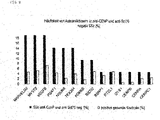
- figure 4 shows the frequency of 8 new antigens in SSc patients compared to anti-centromere antibodies.
- Group 2 includes 72 additional antigens suitable for identifying patients with diffuse SSc.
- Group 2 Antigenic Genes Symbol: PSAT1, TEX264, ABCF3, ACAT2, TCEAL4, ACTN4, ACTR5, ALDH1A1, ALPK1, APBA2, ARHGEF16, ASMTL, ATP13A2, C11orf30, C11orf60, C17orf48, C19orf52, C20orf43, CDK5RAP2, CBK,CBK,CBK,CORO2,CORO2 CTBP2, CYP4F2, DENND4B, DHRS4, DTNBP1, EEF1D, EIF2B3, EPS8L2, FEZ1, FOXL1, FUCA2, GAL3ST4, GEMIN8, GLOD4, GSN, HMGCS1, HSP90AA1, INPP5B, IRAK1, ISCU, LARS, LTF, MGEA5, MPND, MSH3, NCSTN, NEU1, P4HB, PER1, PEX14, PGCP, PPL, PRKAR1B, PTPN5, RAB
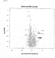
- figure 5 shows the Vulcano plot of autoantibody reactivities of SSc patients with diffuse subform versus healthy controls.
- figure 6 shows the frequency of autoantibodies in the diffuse form compared to the limited form.
- Table 4 summarizes the results of the static tests for selected group 1 and 2 antigens. These antigens are limited and diffusely useful for identifying and distinguishing the SSc subgroups. Table 4: Summary of the p-values, AUC and fold-change reactivity of the antigens in the SSc subgroups limited and diffuse together.
- GeneID genes icon test p-value Fold change AUC sensitivity Specificity 57099 AVEN Diffuse vs HV 6.04E-01 -1.46 0.4 4 0.48 0.56 57099 AVEN Limited vs HV 1.74E-06 4.00 0.7 0 0.63 0.67 23299 BICD2 Diffuse vs HV 7.98E-01 1.05 0.4 5 0.30 0.56 23299 BICD2 Limited vs HV 2.53E-05 2.10 0.7 1st 0.59 0.71 1058 CENPA Diffuse vs HV 3.21E-01 1.26 0.5 5 0.35 0.70 1058 CENPA Limited vs HV 9.49E-11 31.39 0.7 9 0.70 0.85 1059 CENPB Diffuse vs HV 8.87E-01 -1.05 0.5 0 0.36 0.63 1059 CENPB Limited vs HV 7.69E-12 28.22 0.8 2 0.72 0.91 1060 CENPC1 Diffuse vs HV 2.96E-02
- Example 8 Identification of autoantibody reactivities in patients with limited subform.
- figure 7 shows the Vulcano plot of the autoantibody reactivities of patients with limited SSc compared to healthy samples.
- Table 4 summarizes the results of the statistical tests of the SSc subgroups in a limited and diffuse way.
- Group 3 includes 69 additional antigens suitable for identifying patients with limited SSc.
- Group 3 antigen genes symbol: BICD2, SPEG, ZFYVE19, AATF, ABI2, ACBD6, AIP, ANK1, ARHGDIA, AZIN1, C1orf131, C9orf16, CBX3, CCDC102A, CCNDBP1, CENPH, CORO7, DDX1, DTX4, DYNC1LI1, EIF4E, EML3, ENAH, EOMES, FAM50A, FCHSD1, GAB1, GCSH, GIPC1, GIT1, GPBP1, GTF2F1, HIST1H1A, IKBKB, ISY1, KDR, KIFAP3, MAPT, MED21, MOCOS, MRPL34, NSFL1C, PDIA6, PLK1S1, PLXNB2, RGS16, RNF4, RPL5, S100B, SET, SFPQ, SH3GL2, CENPB, SPIRE2, SPTAN1, SSB, SYF2, TAF9, TDRKH, TTC1, TTC28,
- figure 8 shows the frequency of autoantibodies in the diffuse form compared to the limited form.
- anti-centromeres and anti-Ro52 (TRIM21) antibodies In addition to anti-centromeres and anti-Ro52 (TRIM21) antibodies, antibodies against the antigens KDM6B (38%), BICD2 (26%), RTEL1 (22%), NSUN5 ( 20%), SSRP1 (14%), AVEN (12%), PMF1 (10%), NFKBIL2 (10%), SPEG (10%) and PLK1S1 (10%).
- SSc A common form of SSc is the so-called overlap syndrome. In clinical practice, differentiation from other collagen diseases is often difficult.
- Group 4 includes 119 antigens suitable for identifying patients with overlap syndrome: Group 4 Antigenic Genes Symbol:ACOT7, ACTR1B, ADD1, AIFM1, AK1, AKAP1, AKT1, ANAPC4, ANKRD16, ANKS6, APC, ARHGDIB, ASB6, ATP5D, ATP5H, AVPI1, B3GNTL1, BCKDHA, BRPF3, C11orf1, C11orf84, C14orf105, C20orf20, C3orf1, CA5A, CALB2, CAPG, CBLC, CC2D1A, CCDC137, CCDC40, CCDC43, CDC2L2, CEP76, CNDP2, CNTROB, COIL, CRYAB, DES, DHX29, DIP2C, DR1, EID1, ERLIN1, FAM104B FAM60A FASN FCGR1A FKBP15 FKBP8 FUBP3 FXC1 GSTZ1 H2AFV HNRNP
- Group 5 in Table 2 contains additional statistically significant antigens that can be used for the diagnosis and differential diagnosis of SSc versus healthy and other autoimmune diseases.
- Group 5 Antigenic Genes Symbol: HIST1H2BA, NEFL, STMN1, STMN1, CDR2, PPP1R2, RBM28, SSRP1, C4orf27, CACNB3, CASP9, CHD1, DFNA5, FEN1, GTF2E2, MMP2, MRPL40, NAPA, PLA2G4B, PPP2CA, PVRL2, RECQL5, FAF1, WDR44.
- antigens were selected from Tables 3 and 4.
- antigens were selected against which anti-Scl70 negative and anti-centromere negative patients are reactive.
- figure 9 shows the frequency of autoantibodies against a selection of antigens from Tables 3 and 4 in anti-centromere and anti-Scl70 negative patients.
- the antigens MARVELD2 (19%), MYST2 (19%), VEGFB (19%) .PSAT1 (14%) . NSUN5 (14%) and TEX264 (14%) have a frequency greater than 10% in anti-centromere and anti-Scl70 negative SSc patients. These antigens are therefore particularly suitable for improving the diagnosis.
- Table 5 Composition of the improved diagnostic panel.
- Gene ID genes icon Gene name Panel I Panel II 1059 CENPB centromeric protein B, 80kDa x x 7250 TOP1 topoisomerase (DNA) I x x 23135 KDM6B lysine (K)-specific demethylase 6B x 23299 BICD2 bicaudal D homolog 2 (Drosophila) x 55695 NSUN5 NOL1/NOP2/Sun domain family, member 5 x 51750 RTEL1 regulator of telomere elongation helicase 1 x 11143 MYST2 MYST histone acetyltransferase 2 x 29968 PSAT1 phosphoserine aminotransferase 1 x
- the first 7 antigens in Table 2 include the major antigens used for the calculation of biomarker panels to diagnose SSc: KDM6B, NSUN5, BICD2, RTEL1, MYST2, PSAT1 and TEX264
- the MFI values of the antigens TOP1, CENPB, KDM6B, NSUN5, BICD2, RTEL1, MYST2, PSAT1 and TEX264 were used to calculate the area under the curve (AUC), sensitivity and specificity.
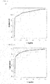
- Figure 10a shows the receiver operator curves (ROC) for a logistic regression model based on panel I consisting of anti-CENPB and anti-Scl70.
- Figure 10b the ROC curve for the improved panel consisting of anti-CENPB and anti-Scl70 and the five new antigens KDM6B, NSUN5, BICD2, RTEL1, MYST2, PSAT1 and TEX264.
- the sensitivity could be increased by 7% from 72% to 79%.
- the example makes it clear how the prediction quality of a biomarker model can be increased by including the additional markers in order to achieve better classification of patients.
- SSc cohort II The markers listed in Table 2 were tested in a second independent sample collective (SSc cohort II).
- EUSTAR is a multicentre, prospective cohort of the European League against Rheumatism (EULAR) Scleroderma Trials and Research (EUSTAR) group.
- Table 6 Demographic, clinical and serological data of SSc Cohort II.
- the human proteins listed in Table 2 were coupled to Luminex beads and the protein-coupled beads were measured in a multiplex assay with the patient samples.
- the Autoantibody binding was measured using a PE-conjugated autoantibody in a Luminex instrument.
- Table 7 Summary of p-values (Wilcoxon rank sum test) for 41 markers for the diagnosis of SSc in SSc Cohort II SSc vs HC SSc vs AD Marker No genes icon SSc Scl70 & CENPB neg dSSc lSSc SSc 1 KDM6B 3.09E-10 1.33E-03 6.13E-05 2.02E-08 2.25E-02 2 BICD2 4.19E-03 6.48E-01 7.77E-01 5.11E-04 7.37E-01 3 NSUN5 1.62E-05 4.06E-01 9.41E-01 4.30E-06 5.04E-03 4 RTEL1 1.28E-03 3.69E-01 4.16E-01 4.32E-04 7.74E-02 8th TRIM21 5.30E-09 1.51E-05 1.65E-06 5.90E-07 3.88E-01 9 ABCB8 1.81E-03 8.10E-01 3.37E-01 1.13E-02 1.02E-02 10 AVEN 2.06E-
- Example 12 Application of autoantibody panels to diagnose SSc
- Table 8 contains an overview of the antigens used in panels 1,3-8.
- a diagnostic panel 9 was calculated using all markers from Table 2. Panels 1,3,4,5 and 9 are particularly suitable for diagnosing all SSc patients, regardless of the respective subtype.
- the AUC (area under the curve) of the panels 1,3,4,6 and 9 is given in Table 8 for 2 independent SSc sample collectives (Cohort I and Cohort II).
- Panel 1 consists of the commonly used diagnostic antigens Scl70(Top1) and CENPB and achieves an AUC of 0.81 in cohort I and 0.85 in cohort II for the comparison of SSc versus healthy (HC).
- Panel 3 consists of two diagnostic antigens, CENPB and Scl70, and two new SSc antigens, KDM6B and BICD2. Compared to the commonly used markers of panel 1, a logistic model for panel 3 shows a significantly higher AUC for the comparison SSc versus healthy (HC): for cohort I 0.88 versus 0.81 and for cohort II 0.87 versus 0.85.
- Panel 4 is based on Panel 3 and includes two other new markers, NSUN5 and PPP1R2.
- a logistic regression model for panel 4 achieves an AUC of 0.9 for cohort I and an AUC of 0.85 for cohort II for the diagnosis of SSc compared to healthy samples.
- Panel 5 is based on Panel 4 and contains the diagnostic markers CENPB and Scl70, the markers KDM6B, BICD2, NSUN5, PPP1R2, RTEL1, TRIM21, ABCB8, AVEN, CENPA,CENPC1, GYS1, MARVELD2, PMF1, TTLL3, ATP13A2, LTF, STMN4, ACBD6, GTF2F1, KDR, DIP2C, FXC1, HNRNPA1, HSBP1L1, KARS, PBXIP1, ZNF431, STMN1 and SSRP1.
- Table 8 Composition of the marker panels Diagnosis SSc CENPB & Scl70 neg.
- Example 13 Use of autoantibody panels to identify patients lacking detection of anti-centromere and anti-Scl70 (Top1) autoantibodies.
- Panel 6 contains markers KDM6B, TRIM21, GYS1, ALPK1, STMN4, HNRNPA1, STMN1.
- Example 14 Application of autoantibody panels to identify SSc patients with diffuse form As shown in Table 6, 36.8% of SSc patients with diffuse SSc (dSSc) and 25.3% of patients with limited form tested positive for autoantibodies against Scl70 (Top1). . This shows that anti-Scl70 antibodies are associated with the diffuse form but are not specific. Therefore, additional markers are needed to diagnose the diffuse form. Panel 7 includes 6 markers that are particularly suitable for diagnosing diffuse SSc.
- Panel 7 contains the markers KDM6B, TOP1, TRIM21, CENPC1, ISY1, HNRNPA1.
- Example 15 Use of autoantibody panels to identify SSc patients with limited form As shown in Table 6, 41% of SSc patients with limited SSc(lSSc) and 8.8% of patients with diffuse form tested positive for autoantibodies to CENPB. This shows, that anti-CENPB antibodies are associated with the limited form but are not specific. Additional markers are therefore required to diagnose the limited form. Panel 8 includes 27 markers that are particularly useful for diagnosing limited SSc.
- Panel 8 includes markers KDM6B, BICD2, NSUN5, CENPB, TOP1, RTEL1, TRIM21, ABCB8, AVEN, CENPA, CENPC1, CENPT, GYS1, C19orf52, STMN4, ACBD6, CBX3, CENPH, EIF4E, GTF2F1, MAPT, SPTAN1, DIP2C , PBXIP1, NEFL, STMN1, SSRP1.
- Example 16 ELISA for the determination of anti-KDM6B antibodies in system sclerosis
- KDM6B recombinantly produced and improved KDM6B protein (Seq ID 315), which comprises amino acids 42-421 de Uniprot database entry 015054.
- KDM6B is purified in several steps using nickel chelate affinity chromatography, size exclusion chromatography and ion exchange chromatography. The purified KDM6B was then applied at a concentration of 1 ⁇ g per milliliter in a sodium carbonate buffer pH 9.0 to a flat-bottomed ELISA plate (e.g. NUNC) with 100 ⁇ l per well and incubated for 4 hours at room temperature (RT).
- a flat-bottomed ELISA plate e.g. NUNC
- Serum or plasma samples diluted 1:101 in HBS-T buffer were applied to the wells and incubated for 30 min at room temperature. During this time, the KD6MB-specific IgG autoantibodies from the serum or plasma sample bind to the bound protein.
- Non-specifically bound antibodies are removed after incubation by washing twice with a PBS Tween buffer. This is followed by a half-hour incubation with an HRP (horse radish peroxidase-conjugated) anti-human IgG detection antibody. Again, after the incubation, non-specific binding partners are removed by washing twice with PBS-Tween buffer.
- HRP horseradish peroxidase-conjugated
- the colorimetric detection reaction is performed by adding a TMB (Tetra Methylbenzidine) substrate. This reaction is stopped after 15 minutes by adding 1M sulfuric acid and evaluated in an ELISA measuring device (TECAN) at a wavelength of 450 nm vs. 620 nm.
- TECAN ELISA measuring device
- the color intensity (optical density, OD) of the detection reaction is directly proportional to the concentration of KDM6B-specific IgG autoantibodies in the serum or plasma sample.
- the test is evaluated semi-quantitatively via a one-point calibration.
- the OD values of the anti-KDM6B ELISA were used to calculate p-values using Wilcoxon rank sum test and to calculate a logistic regression model with ROC analysis for the comparison of SSc versus HC. Since the group of SSc patients is larger than that of the control group HC, random under-sampling is carried out for the logistic regression, so that the SSc cohort II has groups of the same size.
- the ROC analysis was calculated by using 75% for training and 25% for testing the model from this scaled down data set of SSc Cohort II. This approach is repeated 100 times.
- Table 10 shows the p-values for the comparison SSc versus healthy controls and SSc versus autoimmune controls, which was calculated from samples from SSc cohort II.
- Table 11 shows the empirically calculated area under the curve (AUC), sensitivity and specificity of the anti-KDM6B ELISA for the diagnosis of SSc (SSc Cohort II) compared to healthy samples.
- figure 12 shows ROC curve for a logistic regression model based on the ELISA OD values of KDM6B for the diagnosis of SSc compared to healthy controls.
- Example 17 ELISA for the determination of anti-BICD2 antibodies in system sclerosis
- recombinantly produced BICD2 protein was purified in several stages using nickel chelate affinity chromatography, size exclusion chromatography and ion exchanger chromatography. Subsequently, the purified BICD2 in a concentration of 1.7 ⁇ g per milliliter in a Na carbonate buffer pH 9.0 applied to a flat bottom ELISA plate (e.g. NUNC) with 100 ⁇ l per well and incubated for 4 h at room temperature.
- a flat bottom ELISA plate e.g. NUNC
- Serum or plasma samples diluted 1:101 in HBS-T buffer were applied to the wells and incubated for 30 min at room temperature. During this time, the BICD2-specific IgG autoantibodies from the serum or plasma sample bind to the bound protein.
- Non-specifically bound antibodies are removed after incubation by washing twice with a PBS Tween buffer. This is followed by a half-hour incubation with an HRP (horseradish peroxidase-conjugated anti-human IgG detection antibody). Again, after the incubation, non-specific binding partners are removed by washing twice with PBS-Tween buffer.
- HRP horseradish peroxidase-conjugated anti-human IgG detection antibody
- the colorimetric detection reaction is performed by adding a TMB (Tetra Methylbenzidine) substrate. This reaction is stopped after 15 minutes by adding 1M sulfuric acid and evaluated in an ELISA measuring device (TECAN) at a wavelength of 450 nm vs. 620 nm.
- TECAN ELISA measuring device
- the OD values of the antigen BICD2 were used to calculate the p-values using the Wilcoxon rank sum test and the calculation of the receiver operator curves (ROC).
- the OD values of the anti-BICD2 ELISA were used to calculate the p-values using the Wilcoxon rank sum test and the calculation of a logistic regression model with ROC analysis for the comparison of SSc versus HC. Since the group of SSc patients is larger than that of the control group HC, random under-sampling is carried out for the logistic regression, so that the SSc cohort II has groups of the same size.
- the ROC analysis was calculated by using 75% for training and 25% for testing the model from this scaled down data set of SSc Cohort II. This approach is repeated 100 times.
- Table 10 shows the p-values for the comparison SSc versus healthy controls (HC) and SSc versus autoimmune controls (AD), which was calculated from samples from SSc cohort II.
- Table 10 Wilcoxon rank sum test (p-value) for the anti-KDM6B and anti-BICD2 ELISA for the diagnosis of SSc compared to healthy controls.
- figure 11 shows the boxplot of the BICD2 ELISA for SSc samples compared to healthy samples.
- Table 11 shows the empirically calculated area under the curve (AUC), sensitivity and specificity for the anti-BICD2 ELISA. The calculation was performed for samples from SSc cohort II and healthy samples. Table 11: Empirically calculated, sensitivity and specificity of the anti-KDM6B and anti-BICD2 ELISAs for the SSc cohort II for the diagnosis of SSc compared to healthy controls ELISA test value anti-KDM6B AUC 0.63 sensitivity 0.19 specificity 0.90 anti BICD2 AUC 0.64 sensitivity 0.19 specificity 0.95
- figure 12 shows ROC curve with confidence bands for a logistic regression model based on the ELISA OD values of BICD2 for the diagnosis of SSc compared to healthy controls.
Landscapes
- Health & Medical Sciences (AREA)
- Life Sciences & Earth Sciences (AREA)
- Immunology (AREA)
- Engineering & Computer Science (AREA)
- Hematology (AREA)
- Urology & Nephrology (AREA)
- Molecular Biology (AREA)
- Biomedical Technology (AREA)
- Chemical & Material Sciences (AREA)
- Food Science & Technology (AREA)
- Physics & Mathematics (AREA)
- Cell Biology (AREA)
- Pathology (AREA)
- Biotechnology (AREA)
- General Physics & Mathematics (AREA)
- Medicinal Chemistry (AREA)
- Microbiology (AREA)
- Analytical Chemistry (AREA)
- Biochemistry (AREA)
- General Health & Medical Sciences (AREA)
- Rheumatology (AREA)
- Rehabilitation Therapy (AREA)
- Measuring Or Testing Involving Enzymes Or Micro-Organisms (AREA)
- Peptides Or Proteins (AREA)
- Investigating Or Analysing Biological Materials (AREA)
Description
- Offenbahrt sind Verfahren zur Identifizierung von Markern für Systemische Sklerose (SSc; auch Sklerodermie oder synonym Progressive Systemische Sklerose (PSS)) und die mit Hilfe dieses Verfahrens identifizierten Marker, die zwischen SSc und anderen Autoimmunerkrankungen einerseits und zwischen verschiedenen SSc-Subgruppen andererseits differenzieren können. Die vorliegende Erfindung betrifft die Verwendung von den Markern CENPB (SEQ ID No. 793 oder kodiert von SEQ ID NO: 155 oder 474), TOP1 (SEQ ID No. 952 oder kodiert von SEQ ID NO: 314 oder 633), KDM6B (SEQ ID No. 639 oder kodiert von SEQ ID NO: 1 oder 320,) und BICD2 (SEQ ID No. 640 oder kodiert von SEQ ID NO: 2 oder 321) bei der in vitro/ex vivo diagnose von SSc; sowie einen festen Träger, auf dem diese Marker fixiert sind.
- Die SSc ist eine chronische, entzündliche, rheumatische Erkrankung, die zu den klassischen immunologischen Bindegewebserkrankungen (Kollagenosen) zählt.
- Die SSc ist eine heterogene Erkrankung mit übermäßiger Fibrose der Haut. Es können auch weitere Organsysteme wie z.B. Lunge, Magen-Darm, Niere, Herz und Blutgefäße betroffen sein. Zusätzlich treten Gelenksymptome (Arthritis) auf.
- SSc ist eine sehr seltene Erkrankung. Die Inzidenz beträgt ca. 0,5-1,5/100.000 Einwohner/Jahr. Sie tritt meist zwischen dem 30.-50. Lebensjahr auf. Frauen sind 10-15 mal häufiger betroffen als Männer (LeRoy et al. 1988).
- Klinisch werden nach LeRoy et al. (1988) zwischen der limitierten und der diffusen SSc unterschieden. In frühen Phasen der Erkrankung ist es oftmals schwierig Patienten eindeutig zu klassifizieren, welches als undifferenzierte SSc bezeichnet wird. Treten zusätzlich zur Sklerodermie auch wesentliche Symptome anderer rheumatischer Krankheiten auf, spricht man von einem Sklerodermie-Überlappungssyndrom oder Overlap-Syndrome.
- Die limitierte Form der SSc tritt mit einer Frequenz von bis zu 60% auf. Diese ist charakterisiert durch eine Fibrose der Hände und Füße, die sich bis unterhalb der Ellbogen und Kniegelenke ausbreitet. Vor Auftreten der Hautfibrose besteht häufig bereits über mehrere Jahre das Raynaud-Phänomen. Häufig treten auch gastrointestinale Veränderungen (Schluckbeschwerden) und pulmonale arterielle Hypertonie (PAH) auf. Zur limitierten Form gehört auch das CREST-Syndrom: Calcinosis cutis, Raynaud-Phänomen, Ösophagusmotilitätsstörung, Sklerodaktylie und Teleangiektasie.
- Bei der diffusen Form handelt es sich um die schneller und schwerwiegender verlaufende Form der SSc. Die Fibrose breitet sich dabei über die Ellbogen über den Körper und Gesicht aus. Im Gegensatz zur limitierten Form treten bereits 1-2 Jahre nach dem Auftreten des Raynaud-Phänomens Hautfibrosen auf.
- Beim Sklerodermie-Überlappungssyndrom treten neben den Hauptsymptomen der Sklerodermie auch Symptome weiterer nichtorganspezifischer Autoimmunerkrankungen, wie z.B. der Myositis, Lupus Erythematodes/SLE, Sjögren-Syndrom und rheumatoide Arthritis/RA auf.
- Patienten mit undifferenzierter SSc weisen das Raynaud-Syndrom auf und haben für SSc typische geschwollene Finger und pulmonale arterielle Hypertonie. Nur ein Teil der Patienten entwickelt später tatsächlich eine diffuse oder limitierte SSc.
- Die Diagnose der SSc kann aufgrund des klinischen Bildes mit den typischen Hautveränderungen gestellt werden. Dies kann jedoch in Frühstadien der Erkrankung zum Teil schwierig sein. Weiterhin wird der Nachweis von antinukleären Antikörpern (ANA) herangezogen. In etwa 90% der SSc Patienten sind ANA nachweisbar. Jedoch ist der ANA-Test nicht spezifisch für SSc, da auch andere Kollagenosen und bis zu 20% der Gesunden positiv getestet werden. Die drei wichtigsten Autoantikörper bei SSc sind anti-Topoisomerase I (Scl-70), anti-Centromere (CENP) und anti-RNA-Polymerase III (anti-RNAP III). Diese Autoantikörper weisen eine hohe Spezifität für SSc auf und sind häufig mit einer Subform der SSc assoziiert. Jedoch eignen sich diese 3 Autoantikörper nur eingeschränkt zur Subtypisierung der SSc, da diese nicht ausschließlich in einem Subtyp vorkommen und deren Frequenz offenbar in verschiedenen Ethnizitäten abweichen kann. Anti-Topoisomerase-Antikörper weisen eine hohe Spezifität für SSc auf und sind in etwa 30% der Patienten mit diffuser SSc nachweisbar. Anti-Centromere Antikörper sind dagegen in etwa 50-60% der Patienten mit limitierter SSc und in 10% der Patienten mit diffuser SSc nachweisbar. Beide Autoantikörper schließen sich aus und sind nur in sehr seltenen Fällen gemeinsam in Patienten nachweisbar. Anti-RNAP III Antikörper sind häufiger in der diffusen Form nachweisbar und stellen einen Risikofaktor für die eine renale Krise dar. Insgesamt können mit den Autoantikörpern gegen anti-Topoisomerase, anti-Centromere und anti-RNAP nur etwa 70% der SSc Patienten diagnostisch identifiziert werden (Mierau et al. 2011; Mehra et al. 2013).
- Seltener treten auch Antikörper gegen U1-RNP und PM-Scl Antikörper auf. Diese weisen jedoch nur eine geringe Spezifität für SSc auf: Anti-PM-Scl Antikörper werden häufig in Patienten mit Polymyositis/SSc Overlap Syndrom nachgewiesen. Antikörper gegen U1-RNP sind sowohl bei SSc als auch Mixed Connective Tissue Diseases (MCTD) und SLE nachweisbar. Bei etwa einem Drittel der Patienten werden auch Antikörper gegen typische Kollagenosen-Antigene, wie Rho52/SS-A, Ro60/SS-B, sowie citrullinated Peptide (ACPA) und Rheumafaktoren nachgewiesen.
- In der klinischen Praxis ist die Diagnosestellung einer frühen Form der SSc und deren Klassifizierung in die Subgruppen diffus, limitiert oder Overlap Syndrom oftmals schwierig, da noch nicht alle Symptome vorliegen bzw. etwa 10-30% der Patienten Symptome einer anderen Kollagenose (Bindegewebserkrankung, Connective Tissue Disease) tragen. Da die verschiedenen Unterformen eine sehr unterschiedliche Prognose haben, besteht ein substantieller Bedarf an Biomarkern für eine verbesserte Diagnosestellung der SSc und für eine Einteilung in SSc-Subgruppen. Weiterhin besteht ein hoher Bedarf an prognostischen und prädiktiven Biomarkern.
- Ein weiteres Problem der derzeit angewendeten diagnostischen Verfahren ist, dass die Tauglichkeit der bisher untersuchten Autoantigene zur Diagnose von Organbeteiligungen und Komplikationen umstritten sind und zum Teil widersprüchliche Daten veröffentlicht wurden.
- Hudson M. et al. "Diagnostic criteria of systemic sclerosis", Journal of Autoimmunity 48, 38-41 offenbahrt Bemühungen, Kriterien für die Diagnose von SSc aufzustellen.
- Daher besteht weiterhin Bedarf, an neuen Markern für SSc sowie die Sensitivität und Spezifität der bisher am häufigsten diagnostisch eingesetzten Autoantigene durch den Einsatz neuer Autoantigene oder Marker zu verbessern.
- Offenbahrt ist, dass ein differentielles, eine Vielzahl von Schritten umfassendes Verfahren entwickelt wurde, bei dem Serumproben einer Vielzahl von Gesunden und Patienten mit verschiedenen Autoimmunerkrankungen vergleichend im Hinblick auf ihre Reaktivität mit einer Vielzahl von potentiellen Antigenen untersucht und diese Ergebnisse statistisch ausgewertet wurden. Die Auswahl der Serumproben und die Abfolge der Schritte ermöglichte überraschend die Identifizierung hochspezifischer Marker für SSc, die auch geeignet sind, SSc-Subgruppen zu identifizieren und
- Komplikationen zu diagnostizieren sowie eine Differentialdiagnose im Hinblick auf andere Autoimmunerkrankungen wie rheumatoide Arthritis (RA), insbesondere Frühstadien von RA ("frühe RA") und Spondylitis ankylosans bzw. Morbus Bechterew (SPA).
- Offenbahrt ist ein mehrstufiges Verfahren zur Identifizierung von spezifischen Markern für SSc sowie die mit Hilfe des Verfahrens identifizierten Marker für SSc, die Verwendung und / oder spezifische therapeutische Anwendung dieser Marker zur Diagnose und / oder differentiellen Diagnose der Systemischen Sklerose und / oder zur Unterscheidung von klinischen Subgruppen von SSc.
- Offenbahrt ist daherein Verfahren zur Identifizierung von Markern für Systemische Sklerose (SSc) umfassend die Schritte:
- a) Serumproben von mindestens 50, vorzugsweise 100 SSc Patienten werden mit mehr als 5000 an Beads, beispielsweise Luminex-Beads gekoppelten Antigenen in Kontakt gebracht, die Bindung der einzelnen Antigene an Proteine in den Serumproben der SSc Patienten durch Immunfluoreszenzassay gemessen und die mittlere Fluoreszenzintensität (MFI = median fluorescence intensity) für jedes einzelne Antigen bestimmt;
- b) Serumproben von mindestens 50, vorzugsweise 100 Patienten mit Lupus Erythematodes (SLE) werden mit denselben an Beads, beispielsweise Luminex-Beads gekoppelten Antigenen in Kontakt gebracht, die Bindung der einzelnen Antigene an Proteine in den Serumproben der SLE Patienten durch Immunfluoreszenzassay gemessen und daraus die mittlere Fluoreszenzintensität (MFI) für jedes einzelne Antigen bestimmt;
- c) Serumproben von mindestens 50, vorzugsweise 537 Patienten mit früher rheumatoider Arthritis (RA) werden mit denselben an Beads, beispielsweise Luminex-Beads gekoppelten Antigenen in Kontakt gebracht, die Bindung der einzelnen Antigene an Proteine in den Serumproben der RA Patienten durch Immunfluoreszenzassay gemessen und daraus die mittlere Fluoreszenzintensität (MFI) für jedes einzelne Antigen bestimmt;
- d) Serumproben von mindestens 50, vorzugsweise 82 Patienten mit ankylosierender Spondylitis (SPA) werden mit denselben an Beads, beispielsweise Luminex-Beads gekoppelten Antigenen in Kontakt gebracht, die Bindung der einzelnen Antigene an Proteine in den Serumproben der SPA Patienten durch Immunfluoreszenzassay gemessen und daraus die mittlere Fluoreszenzintensität (MFI) für jedes einzelne Antigen bestimmt;
- e) Serumproben von mindestens 50, vorzugsweise 343 Gesunden werden mit denselben an Beads, beispielsweise Luminex-Beads gekoppelten Antigenen in Kontakt gebracht, die Bindung der einzelnen Antigene an Proteine in den Serumproben der Gesunden durch Immunfluoreszenzassay gemessen und daraus die mittlere Fluoreszenzintensität (MFI) für jedes einzelne Antigen bestimmt;
- f) die MFI Daten jedes einzelnen Antigens aus a), b), c), d) und e) werden statistisch mittels univariater Analyse ausgewertet und dadurch Marker identifiziert, mit denen SSc Patienten von Patienten mit SLE, Patienten mit früher RA, Patienten mit SPA und von Gesunden differenziert werden können;
- g) und wobei die Marker ausgewählt werden aus den Sequenzen SEQ ID No. 1 bis 955, Homologen der Sequenzen SEQ ID No. 1 bis 955 mit mindestens 95 % Homologie und Teilsequenzen von SEQ ID No. 1 bis 955 und Teilsequenzen der Homologen von SEQ ID No. 1 bis 955 mit mindestens 95 % Homologie und Sequenzen kodiert durch SEQ ID No. 1 bis 319.
- Die Beads, die in dem offenbahrten Verfahren in den Schritten a) bis e) verwendet werden, sind vorzugsweise fluoreszenzmarkiert.
- Die Begriffe Systemische Sklerose (SSc), RA bzw. frühe RA, SLE und SPA sind beispielsweise in Pschyrembel, Klinisches Wörterbuch, de Gruyter, 261. Auflage (2011) definiert.
- Zum Beispiel werden die Marker nach univariater statistischer Auswertung dadurch ausgewählt, dass sie einen Schwellenwert von p kleiner 0,05 und eine 1,5-fach veränderte Reaktivität in der SSc-Gruppe gegenüber der Kontrollgruppe haben. Die Kontrollgruppe umfasst oder besteht aus Patienten mit SLE und / oder Patienten mit früher RA und / oder Patienten mit SPA und / oder Gesunden. Gesunde sind Personen, bei denen keine SSc, keine SSc-Subform, keine frühe RA und keine SPA nachgewiesen wurde oder nachweisbar ist.
- Offenbahrt ist auch ein Marker für SSc oder eine oder mehrere SSc-Subformen erhältlich durch das erfindungsgemäße Verfahren. Die SSc-Subgruppen sind beispielsweise diffuse SSc (kurz: dSSc), limitierte SSc (kurz: lSSc) und / oder SSc Overlap-Syndrom (kurz: SSc-OS).
- Aufgrund der hohen klinischen und serologischen Heterogenität der SSC Erkrankung ist es schwierig, mit nur einem Biomarker SSC eindeutig zu diagnostizieren. Daher ist es oft erforderlich, möglichst unkorrelierte Autoantigene zu sogenannten Paneln von Markern ("Biomarker-Panel für SSc") zu kombinieren. Beispielsweise im Rahmen der individualisierten Medizin können entsprechende Panel von Markern für SSc für einzelne Patienten oder Patientengruppen individuell für den betreffenden SSc-Subtyp (Subgruppe) zusammengestellt werden. Deshalb ist es auch notwendig, eine Vielzahl von potentiellen Markern für SSc zur Verfügung zu haben, um entsprechende Subgruppen bzw. Subtypen spezifische Marker für SSc für den jeweils individuellen Fall auszuwählen. Ein entsprechendes Panel kann beispielsweise in Form einer Anordnung, eines Arrays oder auch eines oder mehrerer Beads ausgestaltet sein.
- Gegenstand der Erfindung ist somit ein fester Träger, auf dem die Marker CENPB (SEQ ID No. 793 oder kodiert von SEQ ID NO: 155 oder 474), TOP1 (SEQ ID No. 952 oder kodiert von SEQ ID NO: 314 oder 633), KDM6B (SEQ ID No. 639 oder kodiert von SEQ ID NO: 1 oder 320) und BICD2 (SEQ ID No. 640 oder kodiert von SEQ ID NO: 2 oder 321) fixiert sind.
- Gegenstand der Erfindung ist auch die Verwendung von den Markern CENPB (SEQ ID No. 793 oder kodiert von SEQ ID NO: 155 oder 474), TOP1 (SEQ ID No. 952 oder kodiert von SEQ ID NO: 314 oder 633), KDM6B (SEQ ID No. 639 oder kodiert von SEQ ID NO: 1 oder 320,) und BICD2 (SEQ ID No. 640 oder kodiert von SEQ ID NO: 2 oder 321) bei der in vitro/ex vivo diagnose von Systemische Sklerose (SSc), wobei CENPB, TOP1, KDM6B und BICD2 verwendet werden um eine Wechselwirkung mit Antikörpern aus einer Körperflüssigkeit oder eines Gewebeauszug eines Patienten mit SSc aufzuweisen.
- Vorzugsweise werden dabei zusätzlich NSUN5 (SEQ ID No. 641 oder kodiert von SEQ ID NO: 3 oder 322) und/oder PPP1R2 (SEQ ID No. 933 oder kodiert von SEQ ID NO: 295 oder 614) verwended um eine Wechselwirkung mit Antikörpern aus einer Körperflüssigkeit oder eines Gewebeauszug eines Patienten mit SSc aufzuweisen.
- Vorzugsweise werden dabei zusätzlich mindestens ein Marker ausgewählt aus der Gruppe RTEL1 (SEQ ID No. 642 oder kodiert von SEQ ID NO: 4 oder 323), MYST2 (SEQ ID No. 643 oder kodiert von SEQ ID NO: 5 oder 324), PSAT1 (SEQ ID No. 644 oder kodiert von SEQ ID NO: 6 oder 325), TEX264 (SEQ ID No. 645 oder kodiert von SEQ ID NO: 7 oder 326), TRIM21 (SEQ ID No. 646 oder kodiert von SEQ ID NO: 8 oder 327), ABCB8 (SEQ ID No. 647 oder kodiert von SEQ ID NO: 9 oder 328), AVEN (SEQ ID No. 648 oder kodiert von SEQ ID NO: 10 oder 329), CENPA (SEQ ID No. 651 oder kodiert von SEQ ID NO: 13 oder 332), CENPC1 (SEQ ID No. 652 oder kodiert von SEQ ID NO: 14 oder 333), GYS1 (SEQ ID No. 656 oder kodiert von SEQ ID NO: 18 oder 337), MARVELD2 (SEQ ID No. 660 oder kodiert von SEQ ID NO: 22 oder 341), PMF1 (SEQ ID No. 665 oder kodiert von SEQ ID NO: 27 oder 346), TTLL3 (SEQ ID No. 668 oder kodiert von SEQ ID NO: 30 oder 349), ATP13A2 (SEQ ID No. 682 oder kodiert von SEQ ID NO: 44 oder 363), LTF (SEQ ID No. 713 oder kodiert von SEQ ID NO: 75 oder 394), STMN4 (SEQ ID No. 730 oder kodiert von SEQ ID NO: 92 oder 411), ACBD6 (SEQ ID No. 746 oder kodiert von SEQ ID NO: 108 oder 427), GTF2F1 (SEQ ID No. 772 oder kodiert von SEQ ID NO: 134 oder 453), KDR (SEQ ID No. 776 oder kodiert von SEQ ID NO: 138 oder 457), DIP2C (SEQ ID No. 851 oder kodiert von SEQ ID NO: 213 oder 532), FXC1 (SEQ ID No. 862 oder kodiert von SEQ ID NO: 224 oder 543), HNRNPA1 (SEQ ID No. 865 oder kodiert von SEQ ID NO: 227 oder 546), HSBP1L1 (SEQ ID No. 867 oder kodiert von SEQ ID NO: 229 oder 548), KARS (SEQ ID No. 870 oder kodiert von SEQ ID NO: 232 oder 551), PBXIP1 (SEQ ID No. 895 oder kodiert von SEQ ID NO: 257 oder 576), ZNF431 (SEQ ID No. 928 oder kodiert von SEQ ID NO: 290 oder 609), STMN1 (SEQ ID No. 931 oder kodiert von SEQ ID NO: 293 oder 612), SSRP1 (SEQ ID No. 935 oder kodiert von SEQ ID NO: 297 oder 616) verwended um eine Wechselwirkung mit Antikörpern aus einer Körperflüssigkeit oder eines Gewebeauszug eines Patienten mit SSc aufzuweisen.
- Vorzugsweise werden dabei zusätzlich die Marker NSUN5 (SEQ ID No. 641 oder kodiert von SEQ ID NO: 3 oder 322) PPP1R2 (SEQ ID No. 933 oder kodiert von SEQ ID NO: 295 oder 614) RTEL1 (SEQ ID No. 642 oder kodiert von SEQ ID NO: 4 oder 323), TRIM21 (SEQ ID No. 646 oder kodiert von SEQ ID NO: 8 oder 327), ABCB8 (SEQ ID No. 647 oder kodiert von SEQ ID NO: 9 oder 328), AVEN (SEQ ID No. 648 oder kodiert von SEQ ID NO: 10 oder 329), CENPA (SEQ ID No. 651 oder kodiert von SEQ ID NO: 13 oder 332), CENPC1 (SEQ ID No. 652 oder kodiert von SEQ ID NO: 14 oder 333), GYS1 (SEQ ID No. 656 oder kodiert von SEQ ID NO: 18 oder 337), MARVELD2 (SEQ ID No. 660 oder kodiert von SEQ ID NO: 22 oder 341), PMF1 (SEQ ID No. 665 oder kodiert von SEQ ID NO: 27 oder 346), TTLL3 (SEQ ID No. 668 oder kodiert von SEQ ID NO: 30 oder 349), ATP13A2 (SEQ ID No. 682 oder kodiert von SEQ ID NO: 44 oder 363), LTF (SEQ ID No. 713 oder kodiert von SEQ ID NO: 75 oder 394), STMN4 (SEQ ID No. 730 oder kodiert von SEQ ID NO: 92 oder 411), ACBD6 (SEQ ID No. 746 oder kodiert von SEQ ID NO: 108 oder 427), GTF2F1 (SEQ ID No. 772 oder kodiert von SEQ ID NO: 134 oder 453), KDR (SEQ ID No. 776 oder kodiert von SEQ ID NO: 138 oder 457), DIP2C (SEQ ID No. 851 oder kodiert von SEQ ID NO: 213 oder 532), FXC1 (SEQ ID No. 862 oder kodiert von SEQ ID NO: 224 oder 543), HNRNPA1 (SEQ ID No. 865 oder kodiert von SEQ ID NO: 227 oder 546), HSBP1L1 (SEQ ID No. 867 oder kodiert von SEQ ID NO: 229 oder 548), KARS (SEQ ID No. 870 oder kodiert von SEQ ID NO: 232 oder 551), PBXIP1 (SEQ ID No. 895 oder kodiert von SEQ ID NO: 257 oder 576), ZNF431 (SEQ ID No. 928 oder kodiert von SEQ ID NO: 290 oder 609), STMN1 (SEQ ID No. 931 oder kodiert von SEQ ID NO: 293 oder 612), und SSRP1 (SEQ ID No. 935 oder kodiert von SEQ ID NO: 297 oder 616) verwended um eine Wechselwirkung mit Antikörpern aus einer Körperflüssigkeit oder eines Gewebeauszug eines Patienten mit SSc aufzuweisen.
- Vorzugsweise werden dabei zusätzlich die Marker NSUN5 (SEQ ID No. 641 oder kodiert von SEQ ID NO: 3 oder 322), RTEL1 (SEQ ID No. 642 oder kodiert von SEQ ID NO: 4 oder 323), TRIM21 (SEQ ID No. 646 oder kodiert von SEQ ID NO: 8 oder 327), ABCB8 (SEQ ID No. 647 oder kodiert von SEQ ID NO: 9 oder 328), AVEN (SEQ ID No. 648 oder kodiert von SEQ ID NO: 10 oder 329), CENPA (SEQ ID No. 651 oder kodiert von SEQ ID NO: 13 oder 332), CENPC1 (SEQ ID No. 652 oder kodiert von SEQ ID NO: 14 oder 333), CENPT (SEQ ID No. 653 oder kodiert von SEQ ID NO: 15 oder 334), GYS1 (SEQ ID No. 656 oder kodiert von SEQ ID NO: 18 oder 337), C19or2 (SEQ ID No. 686 oder kodiert von SEQ ID NO: 48 oder 367), STMN4 (SEQ ID No. 730 oder kodiert von SEQ ID NO: 92 oder 411), ACBD6 (SEQ ID No. 746 oder kodiert von SEQ ID NO: 108 oder 427), CBX3 (SEQ ID No. 753 oder kodiert von SEQ ID NO: 115 oder 434), CENPH (SEQ ID No. 756 oder kodiert von SEQ ID NO: 118 oder 437), EIF4E (SEQ ID No. 761 oder kodiert von SEQ ID NO: 123 oder 442), GTF2F1 (SEQ ID No. 772 oder kodiert von SEQ ID NO: 134 oder 453), MAPT (SEQ ID No. 778 oder kodiert von SEQ ID NO: 140 oder 459), DIP2C (SEQ ID No. 851 oder kodiert von SEQ ID NO: 213 oder 532), PBXIP1 (SEQ ID No. 895 oder kodiert von SEQ ID NO: 257 oder 576), NEFL (SEQ ID No. 930 oder kodiert von SEQ ID NO: 292 oder 611), STMN1 (SEQ ID No. 931 oder kodiert von SEQ ID NO: 293 oder 612), und SSRP1 (SEQ ID No. 935 oder kodiert von SEQ ID NO: 297 oder 616) verwended um eine Wechselwirkung mit Antikörpern aus einer Körperflüssigkeit oder eines Gewebeauszug eines Patienten mit SSc aufzuweisen.
- Die große klinische Heterogenität von SSc stellt derzeit sowohl für die Diagnostik als auch für die Wirkstoffentwicklung ein großes Problem dar. Die Identifizierung von spezifischen Antikörpersignaturen in SSC-Patienten-Subgruppen stellt daher einen wichtigen Schritt für die bessere Definition von Patientengruppen in klinischen Studien dar. So könnten beispielsweise spezifische Autoantikörper für dSSc, lSSc oder SSc-OS dazu verwendet werden, diese Subgruppe für Medikamentenstudien zu rekrutieren.
- Die Erfindung umfasst einem festen Träger,auf dem die Marker CENPB (SEQ ID No. 793 oder kodiert von SEQ ID NO: 155 oder 474), TOP1 (SEQ ID No. 952 oder kodiert von SEQ ID NO: 314 oder 633), KDM6B (SEQ ID No. 639 oder kodiert von SEQ ID NO: 1 oder 320) und BICD2 (SEQ ID No. 640 oder kodiert von SEQ ID NO: 2 oder 321) fixiert sind.
- Der feste Träger ist beispielsweise ein Filter, eine Membran, ein kleines Plättchen oder Kügelchen, beispielsweise ein magnetisches oder Fluorophor-markiertes Kügelchen, ein Silizium-Wafer, ein Bead, ein Chip, ein massenspektrometrometrischer Target oder eine Matrix oder ähnliches. Als Träger sind unterschiedliche Materialien geeignet, die dem Fachmann bekannt sind, beispielsweise Glas, Metall, Kunststoff, Filter, PVDF, Nitrocellulose oder Nylon (z.B. Immobilon P Millipore, Protran Whatman, Hybond N+ Amersham).
- Der Träger kann beispielsweise einem Gitter entsprechen, dass die Größenordnung einer Mikrotiterplatte (8-12 Wells Streifen, 96 Wells, 384 Wells oder mehr), eines Silizium-Wafers, eines Chips, eines massenspektrometrischen Targets oder einer Matrix besitzt.
- In einer Ausführungsform der Erfindung liegen die Marker für SSc als Klonsequenzen bzw. Klon(e) vor.
- Die erfindungsgemäßen Marker CENPB, TOP1, KDM6B und BICD2 können mit bekannten Biomarkern für SSc oder Biomarkern für andere Erkrankungen kombiniert, ergänzt oder erweitert werden.
- Die Verwendung der SSc Marker erfolgt außerhalb des menschlichen oder tierischen Körpers, also erfolgt die Diagnose ex vivo / in vitro.
- "Diagnose" im Sinne dieser Erfindung bedeutet die positive Feststellung von SSc mit Hilfe der erfindungsgemäßen Marker und die Zuordnung der Patienten bzw. deren Symptomen zu der Erkrankung SSc dar. Der Begriff der Diagnose umfasst die medizinische Diagnostik und diesbezügliche Untersuchungen, insbesondere die in-vitro Diagnostik und Labordiagnostik,
- ebenfalls Proteomics und Nukleinsäureblots. Weitere Untersuchungen können zur Absicherung und zum Ausschluss anderer Krankheiten vonnöten sein. Daher umfasst der Begriff Diagnose insbesondere die Differentialdiagnose von SSc mittels der erfindungsgemäßen Marker.
- Im Rahmen dieser Erfindung wird unter "Patient" bzw. "Patientin" ein beliebiger Proband, ein beliebiges Individuum - Mensch oder Säugetier - verstanden, mit der Maßgabe, dass der Proband bzw. das Individuum auf SSc untersucht wird.
- Der Begriff "Marker für SSc" im Sinne dieser Erfindung bedeutet, dass die kodierte Aminosäuresequenz oder das Polypeptid oder Protein signifikant (spezifisch) für SSc und / oder die mit SSc einhergehenden Autoantikörperprofile sind. "Erfindungsgemäße Marker" sind CENPB, TOP1, KDM6B und BICD2 in Kombination, möglicherweise zusätzlich mit den zusätzlichen Markern, die in Ansprüchen 2-6 erwähnt sind. Offenbahrt sind Nukleinsäuresequenzen und / oder Aminosäuresequenzen gemäß der Definition im anliegenden Sequenzprotokoll (SEQ ID No. 1 bis SEQ ID No. 955), deren Homologe und Teilsequenzen und wobei auch modifizierte Nukleinsäure- und Aminosäuresequenzen offenbahrt sind. Dabei bedeutet, Marker für SSc beispielsweise, dass das Polypeptid oder Protein eine Wechselwirkung mit Substanzen aus der Körperflüssigkeit oder Gewebeauszug eines Patienten mit SSc aufweisen (z.B. Antigen (Epitop) / Antikörper (Paratop) Wechselwirkung). In einer besonders bevorzugten Ausführungsform der Erfindung ist der Marker für SSc ein (Auto-)Antigen oder ein Teil eines Antigens.
- Die Substanzen aus der Körperflüssigkeit oder Gewebeauszug treten entweder nur oder zumindest verstärkt bei SSc auf beziehungsweise werden exprimiert, wohingegen diese Substanzen bei Patienten ohne SSc oder Gesunden nicht oder zumindest in geringerem Maße (geringere Menge, geringerer Konzentration) vorhanden sind. Marker für SSc können sich andererseits auch dadurch auszeichnen, dass sie eine Wechselwirkung mit Substanzen aus der Körperflüssigkeit oder Gewebeauszug von Patienten mit SSc eingehen, weil diese Substanzen nicht mehr oder zumindest in deutlich geringerer Menge/Konzentration bei SSC auftreten beziehungsweise exprimiert werden, wohingegen diese Substanzen bei Patienten ohne SSC vorhanden oder zumindest in deutlich höherem Maße vorhanden sind. Marker für SSC können auch in gesunden Probanden vorhanden sein, jedoch verändert sich ihre Menge (Konzentration) beispielsweise bei der Entstehung, Etablierung und Therapie von SSC. Ein oder mehrere Marker können auf diese Weise ein Profil von Substanzen aus Körperflüssigkeit und Gewebeauszug abbilden, beispielsweise ein SSc assoziiertes Autoantikörperprofil des betreffenden Patienten. Erfindungsgemäße Marker sind Biomarker für SSc.
- Autoantikörperprofile umfassen die Menge an einem oder mehreren Autoantikörpern deren Vorkommen/Expression mit der Entstehung und/oder Etablierung von SSc einhergehen. Autoantikörperprofile umfassen somit einerseits die Zusammensetzung, d.h. es werden beispielsweise ein oder mehrere Autoantikörper nur bei SSc exprimiert, und anderseits die Menge/Konzentration einzelner Autoantikörper, d.h. die Menge/Konzentration einzelner Autoantikörper verändert sich bei der Entstehung und Etablierung von SSc. Diese Veränderungen können mit Hilfe der erfindungsgemäßen Marker(sequenzen) nachgewiesen werden.
- In einer besonders bevorzugten Ausführungsform erkennt/bindet der SSc Marker an Autoantikörper, die im Verlauf der Entstehung, Etablierung und Therapie von SSc (verstärkt) vorhanden sind oder in geringerem Maße (oder nicht mehr) vorhanden sind. Autoantikörper werden vom Körper gegen körpereigene Antigene, die beispielsweise bei SSc entstehen, gebildet. Autoantikörper werden von Körper gegen unterschiedliche Substanzen und Pathogene gebildet. Im Rahmen der vorliegenden Erfindung werden insbesondere die Autoantikörper detektiert, die beim Auftreten und im Laufe der Entstehung von SSc gebildet werden und/oder in ihrer Expression hoch- beziehungsweise herunterreguliert werden. Diese Autoantikörper können mit Hilfe der erfindungsgemäßen Verfahren und Marker nachgewiesen werden und ihr Nachweis und ihre Überwachung (z.B. der Menge) kann zur Früherkennung, Diagnose und/oder Therapieüberwachung/Therapiesteuerung sowie zur Prognose und Vorhersage des Risikos des Wiederauftretens von SSc im Rahmen der Nachsorge verwendet werden.
- Die Autoantikörperprofile können bereits bei Verwendung der SSc Marker CENPB, TOP1, KDM6B und BICD2 ausreichend charakterisiert werden.
- In einer Ausführungsform der Erfindung können Autoantikörper mit SSc Markern nachgewiesen werden, die sich von einem anderen Individuum ableiten und die beispielsweise aus einer kommerziellen cDNA-Bank stammen, wobei mindestens CENPB, TOP1, KDM6B und BICD2 verwendet werden.
- In einer anderen Ausführungsform der Erfindung können diese Autoantikörper mit SSc Markern nachgewiesen werden, die sich von dem gleichen Individuum ableiten und die beispielsweise aus einer eigens für den Patienten oder eine Gruppe von Patienten beispielsweise im Rahmen der individualisierten Medizin hergestellten cDNA-Bank stammen. Mindestens werden CENPB, TOP1, KDM6B und BICD2 verwendet.
- Autoantikörper können bereits viele Jahre vor Auftreten der ersten Krankheitssymptome vom Patienten gebildet werden. Damit sind eine Früherkennung, Diagnose und auch Prognose und vorbeugende Behandlung bzw. Umstellung der Lebensweise und andere Möglichkeiten der Prävention bereits Jahre vor dem sichtbaren Krankheitsausbruch möglich. Die erfindungsgemäßen Verwendungen oder feste Träger ermöglichen somit ein im Vergleich zu bekannten Verfahren sehr frühes Eingreifen, was die Prävention, Behandlungsmöglichkeiten und Folgen von SSc deutlich verbessert.
- Da sich die SSc-assoziierten Autoantikörperprofile während der Etablierung und Behandlung/Therapie von SSc verändern, ermöglicht die Erfindung auch den Nachweis und die Überwachung von SSc in jedem Stadium der Entstehung und Behandlung sowie die Überwachung im Rahmen der SSC Nachsorge. Die erfindungsgemäßen Verwendungen oder feste Träger erlauben auch eine einfache Handhabung zu Hause durch den Patienten und die kostengünstige routinemäßige Vorsorge zur Früherkennung.
- Verschiedene Patienten können unterschiedliche SSc assoziierte Autoantikörperprofile aufweisen, beispielsweise unterscheiden sich verschiedene Kohorten oder Bevölkerungsgruppen. Jeder Patient kann dabei ein oder mehrere verschiedene SSc assoziierte Autoantikörper im Laufe der Entstehung von SSc und des Fortschreitens der SSc Erkrankung, d.h. ebenfalls verschiedene Autoantikörperprofile, bilden. Außerdem kann sich die Zusammensetzung und/oder die Menge der gebildeten Autoantikörper im Laufe der SSc Entstehung und des Fortschreitens der Krankheit verändern, so dass eine quantitative Auswertung notwendig wird. Auch die Therapie/Behandlung von SSc führt zu Veränderungen in der Zusammensetzung und/oder der Menge an SSc assoziierten Autoantikörpern. Die große Auswahl an SSc Markern, die im Rahmen dieser Erfindung zusammen mit CENPB, TOP1, KDM6B und BICD2 verwendet werden können, ermöglicht die individuelle Zusammenstellung von SSc Markern in einer Anordnung, einem Panel für einzelne Patienten, Gruppen von Patienten, bestimmte Kohorten, Bevölkerungsgruppen und dergleichen.
- Der Nachweis von SSc assoziierten Autoantikörpern beispielsweise im Serum oder Plasma von Patienten hat gegenüber anderen Biomarkern den Vorteil einer hohen Stabilität und Lagerfähigkeit und einer guten Nachweisbarkeit. Auch unterliegt die Anwesenheit von Autoantikörpern keinem circardianen Rhythmus, so dass die Probennahme unabhängig von Tageszeit, Nahrungsaufnahme und dergleichen ist.
- Daneben können die SSc assoziierten Autoantikörper mit Hilfe der korrespondierenden Antigene/Autoantigene in bekannten Assays wie z.B. ELISA oder Western-Blot nachgewiesen werden und auf diese Weise die Ergebnisse überprüft werden.
- Im Sinne der Erfindung wird eine Wechselwirkung zwischen dem SSc Marker und dem betreffenden Serum, beispielsweise einem Autoantikörper des Patienten, nachgewiesen. Eine solche Wechselwirkung ist z.B. eine Bindung, insbesondere eine bindende Substanz an mindestens einen Marker für SSc. Die Wechselwirkung zwischen der Körperflüssigkeit oder dem Gewebeauszuges eines Patienten und den Markern für SSc ist eine Protein-Protein Wechselwirkung.
- Solche Substanzen, beispielsweise Antigene, Autoantigene, SSc assoziierte Autoantikörper, sind erfindungsgemäß Bestandteil einer Körperflüssigkeit, insbesondere Blut, Vollblut, Blutplasma, Blutserum, Patientenserum, Urin, Cerebrospinalflüssigkeit, Synovialflüssigkeit oder eines Gewebeauszuges des Patienten. Die Erfindung betrifft insbesondere die Verwendung dieser Körperflüssigkeiten und Gewebeauszüge für Früherkennung, Diagnose, Prognose, Therapiesteuerung und Nachsorge.
- Die SSc spezifischen Marker verfügen in einer weiteren Ausführungsform der Erfindung über ein Erkennungssignal, welches an die zu bindende Substanz adressiert ist (z.B. Antikörper). Erfindungsgemäß bevorzugt ist für ein Protein das Erkennungssignal Epitop und / oder Paratop und / oder Hapten.
- Die SSc spezifischen Marker können Modifikationen der Aminosäuresequenz aufweisen, beispielsweise Citrullinierung, Acetylierung, Phosphorylierung, Glykosilierung, Methylierung, polyA-Strang-Verlängerung und andere dem Fachmann einschlägig bekannte Modifikationen.
- In einer weiteren Ausführungsform kann der jeweilige SSc Marker in unterschiedlichen Mengen in einem oder mehreren Bereichen auf dem Träger repräsentiert sein. Dies erlaubt eine Variation der Sensitivität. Die Bereiche können jeweils eine Gesamtheit von SSc Markern aufweisen, d.h. eine genügende Zahl an verschiedenen SSc Markern, wobei mindestens CENPB, TOP1, KDM6B und BICD2 auf dem Träger repräsentiert sind. Beispielsweise können 20 bis 50 (numerisch) oder mehr, bevorzugt mehr als 100, besonders bevorzugt 150 oder mehr, beispielsweise 25.000 oder 5.000 oder 10.000 verschiedene oder gleiche SSc Markersequenzen und gegebenenfalls weitere Nukleinsäuren und/oder Proteine, insbesondere andere Biomarker auf dem Träger oder in dem Panel repräsentiert sein.
- Es bedeutet "Anordnung" synonym "Array" und synonym "Panel. Sofern dieser "Array" zur Identifizierung von Substanzen an SSc Markern verwendet wird, ist hierunter vorzugsweise ein "Assay" oder ein Bead oder eine diagnostische Vorrichtung oder ein Screening Assay zu verstehen. Im Rahmen dieser Erfindung ist die Anordnung derart gestaltet, dass die auf der Anordnung repräsentierten Marker, welche CENPB, TOP1, KDM6B und BICD2 umfassen, in Form eines Gitters auf einem Träger vorliegen. Ferner sind solche Anordnungen bevorzugt, die eine hochdichte (high-density) Anordnung von SSC Markern erlauben. Vorzugsweise werden die Marker gespottet. Solche hochdichte gespotteten Anordnungen sind beispielsweise in der
WO 99/57311 WO 99/57312 - Im Rahmen dieser Erfindung kann die beanspruchte Verwendung Ausführungsformen wie ELISA, Bead-based Assay, Line Assay, Western Blot, immunchromatographische Verfahren (z.B. so genannte Lateral Flow Immunoassays) oder ähnliche immunologische Single- oder Multiplex-Nachweisverfahren beinhalten.
- Ein "Proteinarray" im Sinne dieser Erfindung ist die systematische Anordnung von SSc Markern umfassend CENPB, TOP1, KDM6B und BICD2 auf einem festen Träger und wobei der Träger jede beliebige Form und/oder Größe haben kann und wobei der Träger ein fester Träger ist.
- Die SSc Marker der Anordnung sind auf dem Träger fixiert, vorzugsweise gespottet oder immobilisiert, aufgedruckt oder ähnliches, insbesondere reproduzierbar aufgebracht. Ein oder mehrere SSc Marker können mehrfach in der Gesamtheit aller SSc Marker präsent sein und in unterschiedlichen Mengen bezogen auf einen Spot vorliegen. Ferner können die SSc Marker auf dem Träger standardisiert sein (z.B. mittels serieller Verdünnungsreihen von z.B. Humanglobulinen als interne Kalibratoren zur Datennormalisierung und quantitativen Auswertung). Gegebenenfalls kann auch ein Standard (z.B. ein Gold Standard) auf dem Träger aufgebracht sein.
- Expressionsbibliotheken sind dem Fachmann bekannt, diese können nach Standardwerken, wie Sambrook et al, "Molecular Cloning, A laboratory handbook, 2nd edition (1989), CSH press, Cold Spring Harbor, New York hergestellt werden. Weiterhin bevorzugt sind solche Expressionsbibliotheken, die gewebespezifisch sind (z.B. humanes Gewebe, insbesondere humane Organe.
- Weiterhin bevorzugt sind Proteinarrays oder entsprechende Expressionsbibliotheken, die keine Redundanz aufweisen (so genannte: Uniclone®-Bibliothek) und nach den Lehren der
WO 99/57311 WO 99/57312 - Zusätzlich können die SSc Marker in der jeweiligen Form als Fusionsprotein vorliegen, welches beispielsweise mindestens ein Affinitätsepitop oder "Tag" enthält, wobei das Tag beispielsweise ausgewählt wird aus c-myc, His-Tag, Arg-tag, FLAG, alkalische Phosphatase, V5-Tag, T7-Tag oder Strep-Tag, HAT-tag, NusA, S-tag, SBP-tag, Thioredoxin, DsbA oder das Fusionsprotein beispielsweise ein oder mehrere zusätzliche Domänen aufweist, beispielsweise eine Cellulose-bindende Domäne, grünfluoreszierendes Protein, Maltose bindendes Protein, Calmodulin-bindendes Protein, Glutathione S-transferase oder lacZ.
- Weiterhin offenbahrt ist einen Assay, beispielsweise einen multiplex Assay, einen Bead basierten Assay oder Proteinarray zum Identifizieren und Charakterisieren einer Substanz, beispielsweise eines Hits, einer Leitsubstanz, eines Wirkstoffs für SSc. Dabei wird eine zu untersuchende Substanz eingesetzt. Diese kann ein beliebiges natives oder nichtnatives Biomolekül, ein (synthetisches) chemisches Molekül, ein Naturstoff, eine Mischung oder eine Substanzbibliothek sein. Nachdem die zu untersuchende Substanz einen SSC Marker kontaktiert hat, erfolgt die Auswertung des Bindungserfolges, beispielsweise unter Verwendung mit handelsüblicher Image-Analyse Software (GenePix Pro (Axon Laboratories), Aida (Raytest), ScanArray (Packard Bioscience).
- Die Visualisierung erfindungsgemäßer Bindungen, Bindungserfolge, Wechselwirkungen wie Protein-Protein-Wechselwirkungen (z.B. Protein an SSc Marker wie Antigen/Antikörper) oder entsprechende "Mittel zum Nachweis des Bindungserfolges" kann beispielsweise mittels Fluoresenzmarkierung, Biotiniylierung, Radio-Isotopen-Markierung oder kolloidale Gold- oder Latex-Partikel-Markierung in üblicher Weise erfolgen. Ein Nachweis von gebundenen Antikörpern erfolgt vorzugsweise mit Hilfe von sekundären Antikörpern, die mit handelsüblichen Reportermolekülen markiert sind (z.B. Cy-, Alexa-, Dyomics, FITC- oder ähnliche Fluoreszenzfarbstoffe, kolloidale Gold- oder Latex-Partikel), oder mit Reporter-Enzymen wie alkalischer Phosphatase, Meerrettichperoxidase, usw. und den entsprechenden colorimetrischen, fluoreszenten oder chemolumineszenten Substraten. Eine Auslesung erfolgt z.B. mittels eines Microarray-Laserscanners, einer CCD-Kamera oder visuell.
- In einer weiteren Ausführungsform betrifft die Erfindung ein Arzneimittel oder einen Wirkstoff oder eine Prodrug für SSc entwickelt und erhältlich durch den Einsatz eines erfindungsgemäßen SSc Markers.
- Offenbahrt ist auch die Verwendung eines SSc Markers ausgewählt aus Sequenzen SEQ ID No. 1 bis 955 und Teilsequenzen von SEQ ID No. 1 bis 955 mit mindestens 90 %, vorzugsweise mindestens 95 % der Länge von SEQ ID No. 1 bis 955 und Homologen von SEQ ID No. 1 bis 955 und deren Teilsequenzen mit einer Identität von mindestens 95 %, vorzugsweise mindestens 98 % oder mehr zu den entsprechenden Sequenzen und Proteinen/Peptiden kodiert durch die Sequenzen SEQ ID No. 1 bis 638, kodiert durch deren Teilsequenzen und Homologen als Affinitätsmaterial zur Durchführung einer Apherese oder Blutwäsche für Patienten mit SSc. Offenbahrt ist somit die Verwendung der offenbahrten Marker, vorzugsweise in Form einer Anordnung, als Affinitätsmaterial zur Durchführung einer Blutwäsche im weiteren Sinne, wobei Substanzen aus Körperflüssigkeiten eines Patienten mit SSc, wie Blut oder Plasma, an die Marker binden und folglich der Körperflüssigkeit selektiv entzogen werden können.
- Die nachfolgenden Beispiele dienen der Erläuterung der Erfindung ohne die Erfindung jedoch auf die Beispiele einzuschränken. In den nachfolgen Figuren wird die Systemische Sklerose mit PPS (Progressive Systemische Sklerose) bezeichnet.
-
Figur 1 : Vulcano Plot der relativen Antigenreaktivitäten der SSc Patienten im Vergleich zu Gesundkontrollen -
Figur 2 : Vulcano Plot der Antigenreaktivitäten von SSc Patienten gegen eine vereinigte Gruppe von Patienten mit verschiedenen Autoimmunerkrankungen wie SSC SPA, frühere Rheumatoide Arthritis und SPA. -
Figur 3 : Autoantikörperreaktivität in SSc Patientenseren im Vergleich zu Gesundkontrollen und SSC Patienten. -
Figur 4 : Frequenz der Autoantikörperreaktivitäten ausgewählter Antigen in SSc Patienten und Gesundproben. Es wurde ein Schwellenwert von 3 SD Abweichungen über dem Mittelwert der Gesundprobe angesetzt. -
Figur 5 : Vulcano Plot der Autoantikörperreaktivitäten von SSc Patienten mit diffuser Subform gegen Gesundkontrollen -
Figur 6 : Frequenz der Autoantikörperreaktivitäten ausgewählter Antigen in der limitierten und diffusen SSc-Subform. Es wurde ein Schwellenwert von 3 Standard Abweichungen über dem Mittelwert der Gesundprobe angesetzt. -
Figur 7 : Vulcano Plot der Autoantikörperreaktivitäten von SSc Patienten mit limitierter Subform gegen Gesundkontrollen. -
Figur 8 : Vulcano Plot der Autoantikörperreaktivitäten von Patienten mit Overlap Syndrome gegen Gesundkontrollen -
Figur 9 : Frequenz der Autoantikörper in anti-CENP und anti-Sc170 negativen Patienten. -
Figur 10 : Receiver Operating Characteristic-Kurven(ROC)für die Diagnose von SSc im Vergleich zu Gesundproben; A) ROC Kurve Panel I, B) ROC Kurve Panel II. -
Figur 11 : Boxplot basierend auf den anti-KDM6B und anti-BICD2 ELISA Messungen für die Diagnose von SSc im Vergleich zu Gesundkontrollen. -
Figur 12 : Receiver Operating Characteristic-Kurven(ROC)für die ELISA Bestimmung anti-KDM6B und anti-BICD2 Antikörpern:- a) Anti-KDM6B ELISA: SSc im Vergleich zu Gesundkontrollen
- b) Anti-BICD2 ELISA: SSc im Vergleich zu Gesundkontrollen Beispiele:
Beispiel 1: Auswahl der SSc Patienten- und Kontrollproben Patienten und Probanden
- Auswahl der zu testenden Patientengruppen: Es wurden Blutproben von 100 SSc Patienten, 100 Patienten mit SLS, 537 Patienten mit früher Rheumatoide Arthritis ("RA"; Erkrankungsdauer unter 6 Monaten) und 82 Patienten mit ankylosierende Spondylitis (SPA) bzw. Morbus Bechterew analysiert. Als Kontrollgruppe wurden 343 Blutproben vom Bayrischen Roten Kreuz (BRK) bezogen. Von allen Probanden wurde eine Einverständniserklärung (Informed Consent) der Ethikkommission der klinischen Partner und der Biobank des BRK eingeholt.
Tabelle 1: Patientenproben und klinische Daten SLE SSc SSc SSc SSc frühe RA (<6 Monate) SPA Gesund Subgruppe lSSc Subgruppe dSSc Subgruppe SSc-OV ohne Angabe der Subgruppe Anzahl der Patienten bzw. Proben 100 50 32 9 9 537 82 343 Durchschnittliches Alter (Jahre) 39.8 +/-11.9 61.53 +/-16.97 53.88 +/-14.75 51.78 +/-9.18 47.56 +/-16.97 56.8 +/-14.3 43.7 +/-10.1 47.7 +/-11.7 Anzahl der weibl. Patien-ten bzw. Proben 83 50 32 8 9 62.2 15.9 58.3 ANA positive 100 47 32 9 7 N.D. N.D. N.D. Anti-CENP N.D. 30 4 2 2 N.D. N.D. N.D. Anti-Sc170 N.D. 9 20 1 2 N.D. N.D. N.D. - Fünf cDNA Bibliotheken, die aus verschiedenen humanen Geweben (fötales Gehirn, Darm, Lunge, Leber und T-Zellen) erzeugt worden waren, wurden für die Produktion der rekombinanten Antigene verwendet. Alle cDNAs wurden in E.coli unter der transkriptionellen Kontrolle des Lactose-induzierbaren Promotors exprimiert. Die resultierenden Proteine tragen an ihrem Aminoterminus eine zusätzliche Sequenz für einen Hexahistidin-Aufreinigungstag (His6-Tag). Targetantigene, die nicht in der cDNA Biobliothek vorhanden waren, wurden durch chemische Synthese (Life Technologies) erzeugt und in den Expressionsvektor pQE30-NST, der bereits einen Aminoterminalen His6-Tag kodiert, kloniert.
- Nach rekombinanter Expression der Proteine wurden diese unter denaturierenden Bedingungen isoliert und mittels Metallaffinitätschromatographie (IMAC) aufgereinigt. Bis zur weiteren Verwendung wurden die Proteine lyophilisiert und bei -20°C gelagert.
- Die Herstellung von BBAs wurde auf ein Mikrotiterplatten-Format adaptiert, so dass 384 Kopplungsreaktionen parallel mit Pipettierautomaten (Starlet, Hamilton Robotics, Evo Freedom 150, Tecan) angesetzt werden konnten. Für die Verwendung von Pipettierautomaten wurden die einzelnen Beadregionen in Kopplungsplatten (96 Well Greiner) und die Antigene in 2D Barcode Gefäße (Thermo Scientific) überführt. Für jede Kopplungsreaktion wurden 0.6 bis 2.5 Millionen Beads und abhängig von Antigen 1 bis 100 µg Protein eingesetzt.
- Alle Wasch- und Pipettierschritte der Kopplungsreaktion wurden in Kopplungsplatten, die auf Magneten fixiert wurden, durchgeführt. Die Beads wurden zweimal mit 100 µl LxAP-Puffer (100 mM NaH2PO4, pH 6.2) gewaschen und anschließend in 120 µl LxAP-Puffer aufgenommen. Für die Aktivierung wurden 15 µl 1-Ethyl-3-(3-dimethylaminopropyl)carbodiimid (EDC; 50mg/ml) und 15 µl N-Hydroxysulfosuccinimide (Sulfo-NHS; 50mg/ml) zur Bead-Suspension pipettiert und diese wurde anschließend 20 Minuten auf dem Schüttler (RT, 900 rpm, lichtgeschützt) inkubiert. Die Beads wurden 3x mit 150 µl LxKPT-Puffer gewaschen und anschließend die Proteinlösung zugegeben. Nach zweistündiger Inkubation auf dem Schüttler (RT, 900 rpm, lichtgeschützt) wurden die Beads dreimal mit 150 µl LxWPT-Puffer gewaschen. Zur Blockierung von freien Bindungsstellen wurden 100 µl LxCBSP-Puffer (PBS, 1% BSA, 0.05% ProClin300) zugegeben und 20 min auf dem Schüttler inkubiert (RT, 900 rpm, lichtgeschützt). Anschließend folgte eine Inkubation bei 4-8°C über Nacht. Das BBA wurde durch die Vereinigung von mit Antigen gekoppelten Beads hergestellt und bis zur Anwendung bei 4-8°C lichtgeschützt gelagert.
- Für die Anwendung wurden BBAs mit Seren inkubiert und alle an Antigene gebunden IgG-basierte Autoantikörper wurden mit Hilfe eines sekundären Antikörper detektiert. Um einen hohen Durchsatz an Messungen zu ermöglichen, wurde die Anwendung von BBAs auf ein Mikrotiterplatten-Format adaptiert, so dass entweder ein 8-Kanal (Starlet, Hamilton Robotics) oder ein 96-Kanal (Evo Freedom 150, Tecan) Pipettierautomat verwendet werden konnte. Die zu untersuchenden Seren wurden in 2D Barcode Gefäße überführt und anschließend 1:100 mit Assaypuffer (PBS, 0.5% BSA, 10% E.coli Lysate, 50% Low-Cross Buffer (Candor Technologies)) verdünnt. Um humane Antikörper, die gegen E.coli Proteine gerichtet sind, zu neutralisieren, erfolgte eine Präinkubation der Serenverdünnungen für 20 min. In dieser Zeit wurden 500 Beads pro Beadregion in die Assayplatte verteilt. In die Kopplungsplatte wurden 50 µl verdünntes Serum zu den Beads gegeben und die Reaktionsansätze 18-22 h auf dem Schüttler inkubiert (4-8 °C, 900 rpm, lichtgeschützt). Nach drei Waschschritte mit jeweils 100 µl LxWPT-Puffer wurden 5 pg/ml des Detektionsantikörpers Ziegeanti-human IgG-PE (Dianova) zu den Reaktionsansätzen gegeben und für 1 h auf dem Schüttler inkubiert (RT, 900 rpm). Anschließenden wurden die Beads dreimal mit 100 µl L×WPT gewaschen und in 100 µl Trägerflüssigkeit (Luminex) aufgenommen. Das Fluoreszenzsignal der Beads wurde mit Hilfe des FlexMAP3D Instruments detektiert. Dabei wurden zum einen der Beadcount und zum anderen der MFI-Wert (Median der Fluoreszenzintensitität)
- Die biostatistische Analyse umfasste univariate und multivariate Methoden um die statistischen Eigenschaften einzelner Antigene sowie von Gruppen von Antigenen zu beschreiben. Für das Auffinden von interessanten Kandidaten für Panels war die entscheidende Eigenschaft eine gute Trennung zwischen den Gruppen von Proben auf Basis der MFI-Werte. Um Antigen-Kandidaten für eine Panel-Generierung zu finden, wurden als Methoden univariates Testen, Receiver Operating Characteristic (ROC) Analysen, Korrelationsprofile, Powered Partial Least Squares Diskriminanzanalyse (PPLS-DA) und Zufallswälder angewendet. Biostatistische Analysen wurden einer Expertenbewertung unterzogen um finale Antigen-Panels zu definieren.
- Vor der statistischen Analyse wurden die MFI-Werte log2-transformiert Antigene, bei denen mehr als 20% der Werte fehlten, wurden von der Analyse ausgeschlossen und fehlende Werte durch Median-Imputation ersetzt. Eine Quantil-Normalisierung unter Berücksichtigung der Referenzseren wurde durchgeführt, um pro BBA-Set alle gemessenen Proben auf individuellen Platten zu normalisieren.
- Neben deskriptiven Standardstatistiken für MFI-Werte wurden mit Hilfe des zweiseitigen Mann-Whitney-U-Tests nichtparametrische Tests durchgeführt um Unterschiede in den Medianwerten der Gruppen zu entdecken. Korrektur des Testniveaus für multiples Testen erfolgte nach der Bonferroni-Holm Prozedur. Außerdem wurde die Benjamini-Hochberg Prozedur inklusive der Bestimmung der False Discovery Rate (FDR, q-Wert) angewendet. Zusätzlich wurden Fold-Change und Effektgröße bestimmt. Um die Klassifikationsgüte zu bewerten wurde eine ROC-Analyse durchgeführt, in deren Rahmen Sensitivität, Spezifität und die Fläche unter der ROC-Kurve (AUC) berechnet wurden, jeweils inklusive der 95% Konfidenzintervalle auf Basis des Bootstrap-Verfahrens. Zur grafischen Darstellung wurden Boxplots und Volcanoplots genutzt. Auf Basis der univariaten Ergebnisse wurde ein Scoring-System implementiert.
- Durch die Anwendung einer PPLS-DA wurde die Korrelation zwischen den Komponenten und der Response-Matrix maximiert. Für die finale Klassifizierung wurde eine lineare Diskriminanzanalyse mit den latenten Komponenten als Prädiktoren genutzt. Ein Zufallswald wurde angewendet, in dem binäre Entscheidungsbäume kombiniert werden. Die Entscheidungsbäume wurden gebildet auf Basis mehrerer Bootstrap-Stichproben einer Lernstichprobe und durch zufälliges Auswählen einer Subgruppe von erklärenden Variablen an jedem Knoten. Die Anzahl der Eingangsvariablen, die an jedem Teilungsschritt zufällig gewählt wurde, wurde als die Quadratwurzel der Gesamtzahl der Variablen festgelegt, und die Anzahl der Bäume im Zufallswald wurde auf 1000 gesetzt. Für beide multivariaten Ansätze wurde eine Kreuzvalidierung mit 500-fachem Durchlauf implementiert.
- Bespiel 6: Autoantikörper/ Antigenreaktivitäten differenzieren SSc von Gesundkontrollen, SLE, Rheumatoider Arthritis und anderen Autoimmunerkrankungen (AID)
- In einem Screening wurden die Antigenreaktivitäten von 100 SSc Patienten, 537 Patienten mit Early RA, 82 Patienten mit SPA und 343 nach Alter und Geschlecht zugeordnete Gesundkontrollen wurden differentiell getestet. Dazu wurden die Autoantikörperreaktivitäten dieser Blutproben an 5857 an Luminex-Beads gekoppelten Antigenen getestet.
- Um Antigene zu identifizieren, mit denen die Gruppe aller SSc Patienten von verschiedenen Kontrollgruppen bestehend aus Gesundproben und Patienten mit verschiedenen rheumatischen Erkrankungen unterschieden werden kann, wurden univariate statistische Tests durchgeführt. Das Ergebnis der statistischen Tests ist als Vulcano Plot für alle 5857 Antigene dargestellt. Im Vulcano Plot wird auf der x-Achse die relative Änderung der Antigenreaktivtät in SSc Patienten im Vergleich zu Gesundkontrollen (
Figur 1 ) und AID Patienten (Figur 2 ) dargestellt. Die Y-Achse gibt den p-Wert des statistischen Tests wieder.Figuren 1 und2 zeigen, dass spezifische Autoantikörperreaktivitäten gefunden wurden, die in der Gruppe aller SSc erhöht sind und diese sowohl von gesunden Spendern als auch von Patienten mit rheumatischen Erkrankungen unterschieden unterscheiden können. - Tabelle 2 umfasst alle in SSc Patienten identifizierte Autoantigene (Tabelle 2).
- Angegeben ist die fortlaufende Sequenz ID, die Gene ID, Gene Symbol und der Gene Name. Die Gruppe bezeichnet die Verwendung des Biomarkers zur Identifizierung aller SSc Patienten oder bestimmter SSc-Subgruppen basierend auf einem statistischen Schwellenwert von p<0.05 und relativ höheren Reaktivität (Fold-Change) gegenüber der Kontrollgruppe von größer 1.5.
- Gruppe 1: Marker zur Identifizierung aller SSc Patienten unabhängig von der klinischen Subform;
- Gruppe 2: Marker zur Identifizierung von diffuser SSc (dSSc);
- Gruppe 3: Marker zur Identifizierung der limitierten SSc (lSSc);
- Gruppe 4: Marker zur Identifizierung von SSc Overlap-Syndrom (Überlappungssydromen; SSc-OS) und
- Gruppe 5: zusätzliche Marker, die keiner bestimmten Gruppe zugeordnet werden.
-
Figur 3 zeigt die Autoantikörperreaktivität von SSc Patienten im Vergleich zu Gesunden Spendern und SLE Patienten. Dargestellt ist eine sogenannte Heatmap der logarithmierten MFI Werte, wobei Signalhöhe in einer Schwarz/Weiß Skala wiedergegeben wird. - Nach univariater statistischer Auswertung wurde ein Schwellenwert von p<0.05 und eine 1,5 fach veränderte Reaktivität gegenüber der Kontrollgruppe angelegt.
- Gruppe 1 umfasst Antigene, die in der Gruppe aller SSc Patienten im Vergleicht zu Gesundkontrollen und/oder anderen rheumatischen Erkrankungen einen Schwellenwert von p<0.05 unterschreiten und eine 1,5 fach veränderte Reaktivität gegenüber der Kontrollgruppe erreichen: KDM6B, NSUN5, RTEL1, MYST2, TRIM21, ABCB8, AVEN, VEGFB, C19orf60, CENPA, CENPC1, CENPT, CHRM3, GORASP1, GYS1, HDAC5, ILKAP, LSM1, MARVELD2, NFIX, OSBP2, PIM3, PLXNB1, PMF1, PPIE, PRDM16, TTLL3, USP48, AZGP1, AZGP1, ZYX.
- Tabelle 3 fasst die Ergebnisse der statistischen Tests für 36 Antigene aus Tabelle 2 zusammen, die einen p-Wert von <0.05 gegenüber Gesundproben aufweisen.
- Angegeben sind der p-Wert (p-value), die Erhöhung der Reaktivität gegenüber der Kontrollgruppe (Fold-Change), die Area under the Curve (AUC) und der Confidence Interval (CI), sowie Sensitivität (Sens.) und Spezifität (Spec.).


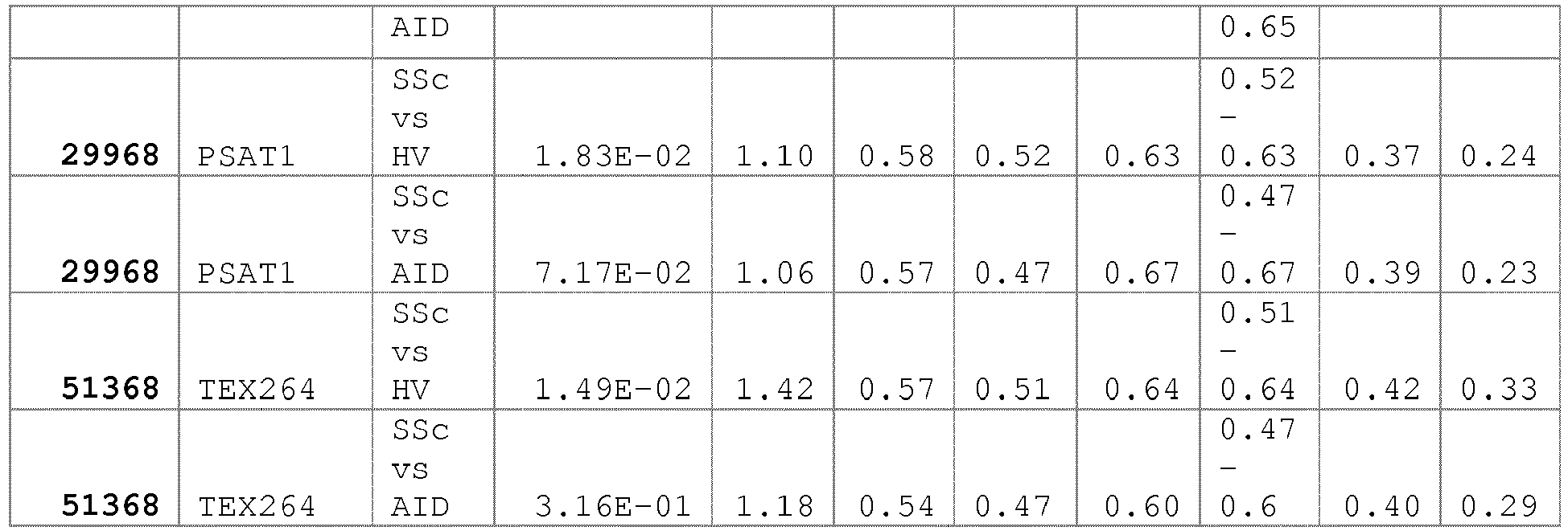
- Um die Frequenz der neuidentifizierten Antigene aus Tabelle 3 im Vergleich zu bekannten Antigenen zu analysieren, wurde ein Schwellenwert von 3 Standardabweichungen (SD) über dem Mittelwert der Gesundproben definiert.
- Erstaunlicherweise wurden mindestens 6 zusätzliche Antigene identifiziert, deren Frequenz bei Betrachtung aller SSc Patienten größer gleich 10% ist. Dazu gehören KDM6B, (21%), BICD2 (19%), RTEL1 (14%), NSUN5 (13%), SSRP1 (10%) und SPEG (10%).Zusätzlich wurden zwei weitere Antigene mit einer Frequenz größer oder gleich 5% in SSc gefunden: VEGFB(9%) und PSAT1 (7%).
-
Figur 4 zeigt die Frequenz von 8 neuen Antigenen in SSc Patienten im Vergleich zu anti-Centromere Antikörpern. - Beispiel 7: Identifizierung von Autoantikörper Reaktivitäten in Patienten mit diffuser Subform.
- Nur etwa 62.5% der analysierten Patienten mit limitierter Form wiesen Anti-Scl70 Antikörper auf. Weitere 12.5% der Patienten zeigten anti-Centromere Antikörper. Um weitere Autoantikörper in Patienten mit limitierter SSc zu identifizieren, wurden die Serumproben der Patienten mit diffuser SSc mit verschiedenen Kontrollgruppen verglichen. Diese bestanden aus Patienten mit limitierter SSc und Patienten mit Overlap Syndrome. Autoantikörper, die in der Gruppe der SSc Patienten mit diffuser Form einen p-Wert von kleiner als 0.05 und einen Fold-Change größer als 1.5 aufwiesen wurden ausgewählt. Das Ergebnis der statistischen Tests ist in Tabelle 2 zusammengefasst.
- Gruppe 2 umfasst 72 zusätzliche Antigene, die für die Identifizierung von Patienten mit diffuser SSc geeignet sind.
- Gruppe 2 Antigene Gene Symbol: PSAT1, TEX264, ABCF3, ACAT2, TCEAL4, ACTN4, ACTR5, ALDH1A1, ALPK1, APBA2, ARHGEF16, ASMTL, ATP13A2, C11orf30, C11orf60, C17orf48, C19orf52, C20orf43, CDK5RAP2, CMPK1, CORO2B, COX5A, CTBP2, CYP4F2, DENND4B, DHRS4, DTNBP1, EEF1D, EIF2B3, EPS8L2, FEZ1, FOXL1, FUCA2, GAL3ST4, GEMIN8, GLOD4, GSN, HMGCS1, HSP90AA1, INPP5B, IRAK1, ISCU, LARS, LTF, MGEA5, MPND, MSH3, NCSTN, NEU1, P4HB, PER1, PEX14, PGCP, PPL, PRKAR1B, PTPN5, RAB11B, RAB11FIP4, RUNDC3A, STMN3, STMN4, STXBP3, SYCE1, TBCD, TMF1, TSFM, TXNRD1, UBE2Q1, UBE2Q2, UBE2Z, YTHDF1, ZNF217.
-
Figur 5 zeigt den Vulcano Plot der Autoantikörperreaktivitäten von SSc Patienten mit diffuser Subform gegen Gesundkontrollen. -
Figur 6 zeigt die Frequenz der Autantikörper in der diffusen Form im Vergleich zur limitierten Form. - Patienten mit diffuser SSc weisen häufiger Antikörper für die Antigene PSAT1 (12.5%), VEGFB (12.5%), CMPK1 (9.4%), TEX264 (9.4%), WDR44 (9.4%), PPL (6.3%) und MYST2 (6.3%) auf.
- Tabelle 4 fasst die Ergebnisse der statischen Tests für ausgewählte Antigene der Gruppe 1 und 2 zusammen. Diese Antigene sind zur Identifizierung und Unterscheidung der SSc-Subgruppen limitiert und diffus geeignet.
Tabelle 4: Zusammenfassung der p-values, AUC und Fold-Change Reaktivität der Antigene in den SSc-Subgruppen limitiert und diffuse zusammen. GeneID Gene Symbol Test p-value Fold-change AUC Sensitivi ty Specifici ty 57099 AVEN Diffuse vs HV 6.04E-01 -1.46 0.4 4 0.48 0.56 57099 AVEN Limited vs HV 1.74E-06 4.00 0.7 0 0.63 0.67 23299 BICD2 Diffuse vs HV 7.98E-01 1.05 0.4 5 0.30 0.56 23299 BICD2 Limited vs HV 2.53E-05 2.10 0.7 1 0.59 0.71 1058 CENPA Diffuse vs HV 3.21E-01 1.26 0.5 5 0.35 0.70 1058 CENPA Limited vs HV 9.49E-11 31.39 0.7 9 0.70 0.85 1059 CENPB Diffuse vs HV 8.87E-01 -1.05 0.5 0 0.36 0.63 1059 CENPB Limited vs HV 7.69E-12 28.22 0.8 2 0.72 0.91 1060 CENPC1 Diffuse vs HV 2.96E-02 1.51 0.6 3 0.45 0.77 1060 CENPC1 Limited vs HV 7.42E-16 12.12 0.8 4 0.63 0.90 51727 CMPK1 Diffuse vs HV 1.14E-01 1.73 0.6 0 0.55 0.60 51727 CMPK1 Limited vs HV 5.98E-02 -1.51 0.6 0 0.64 0.48 23135 KDM6B Diffuse vs HV 2.82E-03 2.06 0.6 5 0.63 0.67 23135 KDM6B Limited vs HV 1.82E-14 7.15 0.8 4 0.70 0.82 11143 MYST2 Diffuse vs HV 3.44E-03 3.32 0.6 5 0.55 0.71 11143 MYST2 Limited vs HV 1.95E-01 1.33 0.5 6 0.45 0.63 4796 NFKBIL2 Diffuse vs HV 1.21E-01 1.47 0.5 7 0.39 0.69 4796 NFKBIL2 Limited vs HV 9.90E-03 2.42 0.6 4 0.56 0.73 55695 NSUN5 Diffuse vs HV 2.02E-01 1.05 0.5 8 0.42 0.75 55695 NSUN5 Limited vs HV 4.07E-12 2.69 0.8 1 0.71 0.87 55857 PLK1S1 Diffuse vs HV 2.01E-01 -1.39 0.5 5 0.61 0.55 55857 PLK1S1 Limited vs HV 9.02E-03 1.65 0.6 0 0.49 0.61 11243 PMF1 Diffuse vs HV 2.89E-02 2.45 0.6 4 0.60 0.67 11243 PMF1 Limited vs HV 5.75E-04 3.29 0.6 4 0.55 0.69 5493 PPL Diffuse vs HV 1.53E-03 2.05 0.6 5 0.53 0.68 5493 PPL Limited vs HV 8.45E-02 1.30 0.5 8 0.50 0.64 29968 PSAT1 Diffuse vs HV 2.46E-03 2.24 0.6 6 0.52 0.80 29968 PSAT1 Limited vs HV 6.76E-01 1.01 0.5 2 0.33 0.75 51750 RTEL1 Diffuse vs HV 3.30E-01 -1.35 0.4 5 0.49 0.48 51750 RTEL1 Limited vs HV 5.56E-10 5.97 0.7 8 0.74 0.75 10290 SPEG Diffuse vs HV 2.46E-01 1.02 0.5 4 0.33 0.55 10290 SPEG Limited vs HV 7.89E-04 2.34 0.6 9 0.61 0.62 6749 SSRP1 Diffuse vs HV 2.45E-01 -1.15 0.4 7 0.40 0.54 6749 SSRP1 Limited vs HV 1.37E-05 2.11 0.7 1 0.62 0.73 51368 TEX264 Diffuse vs HV 1.66E-02 2.02 0.6 3 0.52 0.70 51368 TEX264 Limited vs HV 1.12E-01 1.31 0.5 7 0.36 0.64 7423 VEGFB Diffuse vs HV 1.77E-02 2.46 0.6 2 0.54 0.66 7423 VEGFB Limited vs HV 8.40E-03 2.15 0.6 1 0.56 0.64 54521 WDR44 Diffuse vs HV 5.07E-02 2.37 0.6 4 0.57 0.61 54521 WDR44 Limited vs HV 3.00E-01 -1.21 0.5 6 0.58 0.44 - Beispiel 8: Identifizierung von Autoantikörper Reaktivitäten in Patienten mit limitierter Subform.
- Nur etwa 60% der analysierten Patienten mit limitierter Form wiesen Anti-Centromere Antikörper auf. Weitere 18% der Patienten zeigten anti-Scl70 Antikörper. Um weitere Autoantikörper in Patienten mit limitierter SSc zu identifizieren, wurden die Serumproben der Patienten mit limitierter SSc mit verschiedenen Kontrollgruppen verglichen. Diese bestanden aus Patienten mit diffuser SSc und Patienten mit Overlap Syndrome. Autoantikörper, die in der Gruppe der SSc Patienten mit limitierter Form einen p-Wert von kleiner als 0.05 und einen Fold-Change größer als 1.5 aufwiesen wurden ausgewählt. Das Ergebnis der statistischen Tests ist in Tabelle 2 zusammengefasst.
-
Figur 7 zeigt den Vulcano Plot der Autoantikörperreaktivitäten von Patienten mit limitierter SSc im Vergleich zu Gesundproben. - Tabelle 4 fasst die Ergebnisse der statistischen Tests der SSc-Subgruppen limitiert und diffus zusammen.
- Gruppe 3 umfasst 69 zusätzliche Antigene, die für die Identifizierung von Patienten mit limitierter SSc geeignet sind.
- Gruppe 3 Antigene Gene Symbol:
BICD2, SPEG, ZFYVE19, AATF, ABI2, ACBD6, AIP, ANK1, ARHGDIA, AZIN1, C1orf131, C9orf16, CBX3, CCDC102A, CCNDBP1, CENPH, CORO7, DDX1, DTX4, DYNC1LI1, EIF4E, EML3, ENAH, EOMES, FAM50A, FCHSD1, GAB1, GCSH, GIPC1, GIT1, GPBP1, GTF2F1, HIST1H1A, IKBKB, ISY1, KDR, KIFAP3, MAPT, MED21, MOCOS, MRPL34, NSFL1C, PDIA6, PLK1S1, PLXNB2, RGS16, RNF4, RPL5, S100B, SET, SFPQ, SH3GL2, CENPB, SPIRE2, SPTAN1, SSB, SYF2, TAF9, TDRKH, TTC1, TTC28, TWF2, UBAP2, UNC119, WIZ, XBP1, YLPM1, ZCCHC17, ZCCHC9 -
Figur 8 zeigt die Frequenz der Autoantikörper in der diffusen Form im Vergleich zur limitierten Form. - Neben anti-Centromere und anti-Ro52 (TRIM21) Antikörpern wurden in Patienten mit limitierter SSc mit einer Frequenz von größer gleich 10% Antikörper gegen die Antigene KDM6B (38%), BICD2 (26%), RTEL1 (22%), NSUN5 (20%), SSRP1 (14%), AVEN (12%), PMF1 (10%), NFKBIL2 (10%),SPEG (10%) und PLK1S1 (10%) gefunden.
- Beispiel 9: Identifizierung von Autoantikörper Reaktivitäten in Patienten mit Overlap Syndrome
- Eine häufige Verlaufsform der SSc ist das sogenannte Überlappungssyndrom oder Overlap Syndrome. In der klinischen Praxis ist die Abgrenzung zu anderen Kollagenosen oftmals schwierig.
- Gruppe 4 umfasst 119 Antigene, die zur Identifizierung von Patienten mit Overlap Syndrome geeignet sind:
Gruppe 4 Antigene Gene Symbol:ACOT7, ACTR1B, ADD1, AIFM1, AK1, AKAP1, AKT1, ANAPC4, ANKRD16, ANKS6, APC, ARHGDIB, ASB6, ATP5D, ATP5H, AVPI1, B3GNTL1, BCKDHA, BRPF3, C11orf1, C11orf84, C14orf105, C20orf20, C3orf1, CA5A, CALB2, CAPG, CBLC, CC2D1A, CCDC137, CCDC40, CCDC43, CDC2L2, CEP76, CNDP2, CNTROB, COIL, CRYAB, DES, DHX29, DIP2C, DR1, EID1, ERLIN1, FAM104B, FAM60A, FASN, FCGR1A, FKBP15, FKBP8, FUBP3, FXC1, GSTZ1, H2AFV, HNRNPA1, HNRPLL, HSBP1L1, HSPA8, IRAK1BP1, KARS, KCNK5, LASS5, LIPE, LOC100129119, LOC643733, LSM14A, LSM14B, LSR, LUC7L2, MAOA, MAPK8IP2, MBD3, MRPL10, MRPL11, MSN, MXD3, MXI1, NFE2L2, NHSL1, NNAT, NRBP1, NUP35, ORC6L, PANK4, PBXIP1, PCBP4, PPP1R14A, PRDM8, PTN, RPLP0, RPS3, SCARB2, SDCCAG8, SDR39U1, SDS, SEC31A, SERPINA6, SH3KBP1, SMAD1, SNX22, SOX13, SRP68, TCEAL2, TCEB3, TCERG1, TCOF1, TIMM13, TRAK1, TRIM10, TRIM56, UBTD2, UGT1A8, UHRF1BP1L, USP47, VAT1, WDR47, ZMYND8, ZNF431, ZNF563. - Gruppe 5 in Tabelle 2 enthält weitere statistisch signifikante Antigene, die für die Diagnose und Differentialdiagnose von SSc gegenüber Gesund und anderen Autoimmunerkrankungen herangezogen werden können.
- Gruppe 5 Antigene Gene Symbol: HIST1H2BA, NEFL, STMN1, STMN1, CDR2, PPP1R2, RBM28, SSRP1, C4orf27, CACNB3, CASP9, CHD1, DFNA5, FEN1, GTF2E2, MMP2, MRPL40, NAPA, PLA2G4B, PPP2CA, PVRL2, RECQL5, FAF1, WDR44.
- Beispiel 10: Anwendung von Antigen Panels und zur verbesserten Diagnose von SSc
- Aufgrund der klinischen und serologischen Heterogenität der SSc Erkrankung ist es nicht möglich mit nur einem Biomarker diese zu diagnostizieren. Laut Mierau et al. (2011) weisen nur ca. 35.9% der SSc Patienten Autoantikörper gegen Centromere-Proteine und nur 30.1% Autoantikörper gegen anti-topoisomerase I (anti-Scl70) auf. Daher besteht weiterhin ein hoher Bedarf an spezifischen diagnostischen und prognostischen Markern.
- Um ein verbessertes diagnostisches Antigenpanel für SSc zu entwickeln, wurden aus den Tabellen 3 und 4 Antigene ausgewählt.
- In die engere Auswahl gelangten zunächst fünf der in SSc am häufigsten vertretenen Antigene KDM6B, BICD2, RTEL1 und NSUN5 (
Figur 4 ). - Zusätzlich berücksichtigt wurde auch die Häufigkeit von Autoantikörpern in der besonders schwer verlaufenden diffusen SSc. Hier wurden zwei der am häufigsten reaktiven Antigene BICD2 (12.5) und PSAT1 (12.5) ausgewählt.
- Zusätzlich wurden Antigene ausgewählt, gegen die anti-Scl70 negative und anti-Centromere negative Patienten reaktiv sind.
-
Figur 9 zeigt die Häufigkeit von Autoantikörpern gegen eine Auswahl von Antigenen aus Tabelle 3 und 4 in anti-Centromere und anti-Scl70 negativen Patienten. - Insbesondere die Antigene MARVELD2 (19%), MYST2 (19%), VEGFB (19%) .PSAT1 (14%) . NSUN5 (14%) und TEX264 (14%) weisen eine Frequenz von mehr als 10% in anti-Centromere und anti-Scl70 negativen SSc Patienten auf. Diese Antigene sind daher besonders zur Verbesserung der Diagnose geeignet.
- Für die Entwicklung eines verbesserten diagnostischen Tests wurden daher vier der am häufigsten in doppelt negativen SSc Patienten reaktiven Antigene MYST2, PSAT1, NSUN5 und TEX264 ausgewählt.
- Die finale Zusammensetzung der Panels ist in Tabelle 5 dargestellt.
Tabelle 5: Zusammensetzung des verbesserten diagnostischen Panels. Gene ID Gene Symbol Gene Name Panel I Panel II 1059 CENPB centromere protein B, 80kDa x x 7250 TOP1 topoisomerase (DNA) I x x 23135 KDM6B lysine (K)-specific demethylase 6B x 23299 BICD2 bicaudal D homolog 2 (Drosophila) x 55695 NSUN5 NOL1/NOP2/Sun domain family, member 5 x 51750 RTEL1 regulator of telomere elongation helicase 1 x 11143 MYST2 MYST histone acetyltransferase 2 x 29968 PSAT1 phosphoserine aminotransferase 1 x - Die ersten 7 Antigene in Tabelle 2 umfassen die wichtigsten Antigene, die für die Berechnung von Biomarker-Panels zur Diagnose von SSc verwendet werden: KDM6B, NSUN5, BICD2, RTEL1, MYST2, PSAT1 und TEX264
- Zur Validierung der in Tabelle 5 genannten Antigene aus Panel I und Panel II wurden 100 SSc Patienten und 100 Gesundproben mit Antigen-gekoppelten Luminex Beads vermessen.
- Die MFI Werte der Antigene TOP1, CENPB, KDM6B, NSUN5, BICD2, RTEL1, MYST2, PSAT1 und TEX264 wurden zur Berechnung der Area under the Curve (AUC), Sensitivität und Spezifität herangezogen.
-
Figur 10a zeigt die Receiver Operator Kurven (ROC) für ein logistisches Regressionsmodel basierend auf dem Panel I bestehend aus anti-CENPB und anti-Scl70. -
Figur 10b die ROC Kurve für das verbesserte Panel bestehend aus anti-CENPB und anti-Scl70 und den fünf neuen Antigenen KDM6B, NSUN5, BICD2, RTEL1, MYST2, PSAT1 und TEX264. - Durch den Einschluss der 5 Antigene KDM6B, NSUN5, BICD2, RTEL1, MYST2, PSAT1 und TEX264 konnte die Sensitivität um 7% von 72% auf 79% gesteigert werden.
- Anhand des Beispiels wird deutlich, wie sich unter Einbeziehung der zusätzlichen Marker die Vorhersagequalität eines Biomarkermodells steigern lässt um eine bessere Klassifikation von Erkrankten zu erreichen.
- Die Einbeziehung zusätzlicher Marker zu den bekannten Markern verbessert die Vorhersagequalität im angewendeten Verfahren mit etwa 7% ROC deutlich.
Tabelle 6: AUC, Sensitivität und Spezifität der SSc Panels SSc vs Healthy Control AUC CI (AUC) Sens. CI (Sens.) Spec. CI (Spec) panel I 0.91 [0.862, 0.965] 0.72 [0.621, 0.823] 0.97 [0.935, 1] panel II 0.94 [0.891, 0.98] 0.79 [0.71, 0.868] 0.94 [0.88, 1] 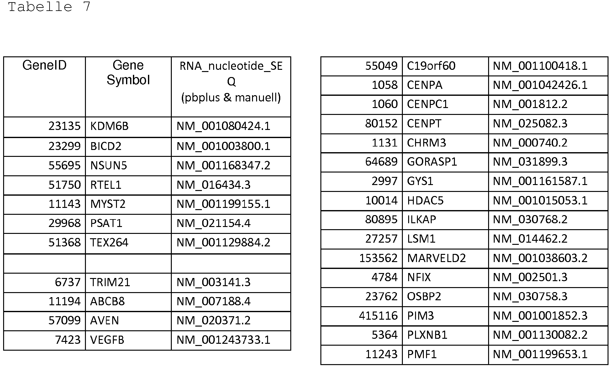


- Die in Tabelle 2 genannten Marker wurden in einem zweiten unabhängigen Probenkollektiv getestet (SSc Kohorte II). Insgesamt wurden 180 Serumproben von SSc Patienten analysiert, deren demogrophischen und klinischen Daten der EUSTAR Datenbank entnommen worden sind. EUSTAR ist eine multizentrische, prospektive Kohorte der European League Against Rheumatism (EULAR) Scleroderma Trials and Research (EUSTAR) group. Als Kontrollproben wurden n=99 Serumproben von Gesundkontrollen und n=110 Serumproben von Patienten mit rheumatischen Autoimmunerkrankungen (AD) eingesetzt. Diese setzten sich zusammen aus Serumproben von Patienten mit der Diagnose SLE, Myositis, Arthritis und Sjögrens's Syndrom.
Tabelle 6: Demographische, klinische und serologische Daten der SSc Kohorte II. Anzahl (%); Mittelwert (%) Gesamt SSc Hautbeteiligung SSc (n=180) dSSc(n=57) lSSc (n=83) Weiblich n (%) 142 (79.9) 42 (737) 67 (80.7) Männlich n (%) 38 (21.1) 15 (26.3) 16 (19.3) Alter Mittelwert (SD) 56.5 (13.8) 52.9 (12.2) 58.2 (15.1) Mittlere Erkrankungsdauer in Jahren (SD) 8.2 (9.7) 6.98 (6.8) 9.9 (11.9) ANA positive n (%) 176 (97.8) 55 (96.5) 82 (98.8) anti-CENPB positive n (%) 66 (36.7) 5 (8.8) 34 (41) anti-Scl70 positive n (%) 50 (27.8) 21 (36.8) 21 (25.3) - Hierzu wurden die in Tabelle 2 genannten humanen Proteine an Luminex Beads gekoppelt und die Protein-gekoppelten Beads in einem Multiplex Assay mit den Patientenproben vermessen. Die Bindung von Autoantikörpern wurde mittels eines PEkonjugierten Autoantikörpers in einem Luminex Instrument gemessen.
- Es wurde eine univariate statistische Auswertugen durchgeführt mit dem Wilcoxon-Rangsummentest. Das vorgegebene Signifikanzniveau wurde bei 0.05 angelegt. Tabelle 7 enthält 41 Antigene, die sowohl in der SSc Kohorte I, die zur Entdeckung der Marker verwendet wurde (Bespiel 6), als auch in der SSc Kohorte II einen p-Wert kleiner als das festgelegte Signifikanzviveau 0.05 erreichten. Für den Vergleich aller SSc Proben gegen Gesundkontrollen (HC) erreichten 31 Marker einen p-Wert kleiner als 0.05. Für den Vergleich aller SSc Proben gegen eine zusammengefasste Gruppe aus Serumproben verschiedener rheumatoider Erkrankungen (AD) erreichten 24 Marker einen p-Wert kleiner als 0.05.
Tabelle 7: Zusammenfassung der p-Werte (Wilcoxon-Rangsummentest)für 41 Marker für die Diagnose von SSc in der SSc Kohorte II SSc vs HC SSc vs AD Mar ker Nr Gene Symbol SSc Scl70 & CENPB neg dSSc lSSc SSc 1 KDM6B 3.09E-10 1.33E-03 6.13E-05 2.02E-08 2.25E-02 2 BICD2 4.19E-03 6.48E-01 7.77E-01 5.11E-04 7.37E-01 3 NSUN5 1.62E-05 4.06E-01 9.41E-01 4.30E-06 5.04E-03 4 RTEL1 1.28E-03 3.69E-01 4.16E-01 4.32E-04 7.74E-02 8 TRIM21 5.30E-09 1.51E-05 1.65E-06 5.90E-07 3.88E-01 9 ABCB8 1.81E-03 8.10E-01 3.37E-01 1.13E-02 1.02E-02 10 AVEN 2.06E-03 7.33E-01 4.37E-01 1.83E-03 7.88E-02 13 CENPA 2.31E-04 3.02E-01 3.03E-01 8.03E-05 1.92E-03 14 CENPC 4.17E-11 5.64E-03 2.25E-02 5.14E-10 5.01E-07 15 CENPT 7.50E-02 4.61E-01 8.25E-01 1.05E-02 1.98E-02 18 GYS1 1.99E-02 2.91E-02 3.40E-01 2.07E-02 8.24E-01 22 MARVELD2 3.77E-02 1.84E-01 1.26E-01 1.71E-01 1.78E-02 27 PMF1 4.96E-02 9.77E-01 3.39E-01 3.28E-01 3.60E-02 30 TTLL3 2.01E-02 1.30E-01 9.82E-01 2.04E-02 9.67E-01 40 ALPK1 1.07E-02 1.90E-03 5.15E-02 5.65E-02 7.13E-01 44 ATP13A2 1.83E-02 7.52E-02 6.30E-01 1.61E-02 3.62E-01 48 C19orf52 8.67E-03 1.70E-01 7.77E-01 3.51E-02 5.48E-03 75 LTF 7.97E-03 4.52E-01 7.16E-01 3.10E-03 9.21E-01 92 STMN4 5.50E-05 6.47E-04 3.64E-03 7.59E-04 3.06E-02 108 ACBD6 4.17E-03 3.71E-01 2.99E-01 6.41E-03 4.02E-02 115 CBX3 2.66E-03 5.41E-01 2.33E-01 3.48E-02 4.45E-03 118 CENPH 2.27E-01 1.78E-01 5.44E-01 4.22E-02 2.19E-02 123 EIF4E 2.78E-02 9.74E-01 6.72E-01 1.52E-02 2.54E-04 134 GTF2F1 1.07E-04 4.08E-02 3.28E-02 6.88E-05 2.30E-01 137 ISY1 2.19E-02 2.06E-02 4.30E-02 1.35E-01 3.65E-01 138 KDR 3.68E-02 1.77E-01 4.65E-02 4.22E-01 3.39E-04 140 MAPT 2.50E-04 5.34E-01 2.76E-01 2.63E-05 7.82E-01 155 CENPB 4.27E-12 7.99E-01 8.41E-03 6.90E-11 2.91E-09 158 SSB 9.04E-02 1.56E-01 6.06E-01 3.07E-02 2.61E-01 213 DIP2C 2.44E-02 5.27E-02 5.08E-01 8.26E-03 1.87E-01 224 TIMM10B 4.05E-02 5.16E-03 3.86E-02 2.42E-01 3.51E-01 227 HNRNPA1 1.48E-02 2.54E-02 1.67E-03 1.28E-01 7.77E-01 229 HSBP1L1 2.45E-02 1.03E-01 1.91E-02 2.88E-01 1.05E-01 232 KARS 1.25E-02 5.71E-03 2.85E-02 1.12E-01 8.70E-03 257 PBXIP1 6.43E-06 2.08E-01 2.49E-01 8.61E-05 3.83E-01 290 ZNF431 3.94E-02 3.68E-02 1.59E-01 9.45E-02 4.67E-02 292 NEFL 6.97E-03 6.01E-02 9.19E-02 3.95E-02 8.50E-01 293 STMN1 6.11E-05 6.13E-04 3.69E-03 8.75E-04 4.19E-03 295 PPP1R2 1.19E-02 7.79E-01 3.69E-01 6.39E-02 1.98E-01 297 SSRP1 4.79E-02 7.17E-01 7.91E-01 2.02E-02 4.19E-03 314 TOP1 6.19E-07 4.23E-01 6.94E-05 4.10E-05 4.96E-07 - Aus den in Tabelle 2 und Tabelle 7 enthaltenen Markern wurden verschiedene Markerkombinationen mittels logistischer Regressionsmodelle getestet. Dabei wurde als Vorgabe die Spezifität gegenüber der Kontrollgruppe auf mindestens 95% gesetzt. Eine Übersicht über die in den Panels 1,3-8 verwendeten Antigene ist in Tabelle 8 enthalten. Zusätzlich wurde ein diagnostisches Panel 9 unter Verwendung aller Marker aus Tabelle 2 gerechnet. Die Panels 1,3,4,5 und 9 eignen sich bevorzugt zur Diagnose aller SSc Patienten, unabhängig von der jeweiligen Subform. Die AUC (Area under the Curve) der Panels 1,3,4,6 und 9 ist in Tabelle 8 für 2 unabhängige SSc Probenkollektive (Kohorte I und Kohorte II) angegeben.
- Panel 1 besteht aus den üblicherweise diagnostisch verwendeten Antigenen Scl70(Top1) und CENPB und erreicht eine AUC von 0.81 in Kohorte I und 0.85 in Kohorte II für den Vergleich SSc versus Gesund (HC).
- Panel 3 besteht aus aus zwei diagnostischen Antigenen, CENPB und Scl70, und zwei neuen SSc Antigenen, KDM6B und BICD2. Gegenüber dem üblicherweise verwendeten Markern des Panels 1 zeigt ein logistisches Model für das Panel 3, eine deutlich höhere AUC für den Vergleich SSc versus Gesund (HC): für Kohorte I 0.88 gegenüber 0.81 und für Kohorte II 0.87 gegenüber 0.85.
- Panel 4 basiert auf Panel 3 und enthält zwei weitere neue Marker, NSUN5 und PPP1R2. Ein logistisches Regressionsmodel für das Panel 4 erreicht für die Diagnose von SSc im Vergleich zu Gesundproben für die Kohorte I eine AUC von 0.9 und für die Kohorte II eine AUC von 0.85.
- Panel 5 basiert auf Panel 4 und enthält neben den diagnostischen Markern CENPB und Scl70, die Marker KDM6B, BICD2, NSUN5, PPP1R2, RTEL1, TRIM21, ABCB8, AVEN, CENPA,CENPC1, GYS1, MARVELD2, PMF1, TTLL3, ATP13A2, LTF, STMN4, ACBD6, GTF2F1, KDR, DIP2C, FXC1, HNRNPA1, HSBP1L1, KARS, PBXIP1, ZNF431, STMN1 und SSRP1.
Tabelle 8: Zusammensetzung der Marker-Panels Diagnose SSc CENPB & Scl70 neg. dSSc lSSc # Gene Symbol P1 P3 P4 P5 P6 P7 P8 155 CENPB x x x x x 314 TOP1 x x x x x x 1 KDM6B x x x x x x 2 BICD2 x x x x 3 NSUN5 x x x 4 RTEL1 x x 8 TRIM21 x x x x 9 ABCB8 x x 10 AVEN x x 13 CENPA x x 14 CENPC1 x x x 15 CENPT x 18 GYS1 x x x 22 MARVELD2 x 27 PMF1 x 30 TTLL3 x 40 ALPK1 x 44 ATP13A2 x 48 C19orf52 x 75 LTF x 92 STMN4 x x x 108 ACBD6 x x 115 CBX3 x 118 CENPH x 123 EIF4E x 134 GTF2F1 x x 137 ISY1 x 138 KDR x 140 MAPT x 158 SSB x 213 DIP2C x x 224 FXC1 x 227 HNRNPA1 x x x 229 HSBP1L1 x 232 KARS x 257 PBXIP1 x x 290 ZNF431 x 292 NEFL x 293 STMN1 x x x 295 PPP1R2 x x 297 SSRP1 x x - Beispiel 13: Anwendung von Autoantikörper-Panels zur Identifizierung von Patienten mit fehlendem Nachweis von anti-Zentromer und anti-Scl70 (Top1)- Autoantikörpern.
- Wie in Tabelle 6 dargelegt, testeten nur etwa 41% der SSc Patienten aus der SSc Kohorte II positiv für anti-CENPB Antikörper und 25.3% positiv für anti-Scl70 Antikörper. Da in der Literatur sehr wenige Patienten mit doppelt-positiver Reaktivität beschrieben worden sind, wird der Anteil an anti-CENPB und anti-Scl70 negativen Patienten mit etwa 30% angegeben. Um diese diagnostische Lücke zu schließen, wurde ein logistisches Regressionsmodel für Panels 6, das aus 7 Markern besteht erstellt.
- Panel 6 enthält die Marker KDM6B, TRIM21, GYS1, ALPK1, STMN4, HNRNPA1, STMN1.
- Wie in Tabelle 6 zusammengefasst wurde mit den Markern des Panels 6 in der Kohorte I eine AUC von 0.91 und für Kohorte II eine AUC von 0.84 erreicht.
- Beispiel 14: Anwendung von Autoantikörper-Panels zur Identifizierung von SSc Patienten mit diffuser Form Wie in Tabelle 6 dargelegt wurden 36.8% der SSc Patienten mit diffuser SSc (dSSc) und 25.3% der Patienten mit limitierter Form positiv für Autoantikörper gegen Scl70 (Top1) getestet. Dies zeigt, dass anti-Scl70 Antikörper zwar mit der diffusen Form assoziiert sind, aber nicht spezifisch sind. Zur Diagnose der diffusen Form werden daher zusätzliche Marker benötigt. Panel 7 umfasst 6 Marker, die besonders zur Diagnose der diffusen SSc geeignet sind.
- Panel 7 enthält die Marker KDM6B, TOP1, TRIM21, CENPC1, ISY1, HNRNPA1.
- Für Panel 7 wurde mittels einer logistischen Regression eine AUC von 0.81 für Kohorte I und 0.87 für Kohorte II berechnet.
- Beispiel 15: Anwendung von Autoantikörper-Panels zur Identifizierung von SSc Patienten mit limitierter Form Wie in Tabelle 6 dargelegt wurden 41% der SSc Patienten mit limitierter SSc(lSSc) und 8.8% der Patienten mit diffuser Form positiv für Autoantikörper gegen CENPB getestet. Dies zeigt, dass anti-CENPB Antikörper zwar mit der limitierten Form assoziiert sind, aber nicht spezifisch sind. Zur Diagnose der limitierten Form werden daher zusätzliche Marker benötigt. Panel 8 umfasst 27 Marker, die besonders zur Diagnose der limitierten SSc geeignet sind.
- Panel 8 umfasst die Marker KDM6B, BICD2, NSUN5, CENPB, TOP1, RTEL1, TRIM21, ABCB8, AVEN, CENPA, CENPC1, CENPT, GYS1, C19orf52, STMN4, ACBD6, CBX3, CENPH, EIF4E, GTF2F1, MAPT, SPTAN1, DIP2C, PBXIP1, NEFL, STMN1, SSRP1.
- Für Panel 8 wurde mittels einer logistischen Regression eine AUC von 0.97 für Kohorte I und 0.99 für Kohorte II berechnet.
Tabelle 9: AUC der SSc Panels in der SSc Kohorte I und Kohorte II Panel SSc Kohorte Vergleich AUC Panel 1 Kohorte I SSc vs HC 0.81 Panel 1 Kohorte I SSc vs SLE 0.84 Panel 3 Kohorte I SSc vs HC 0.88 Panel 3 Kohorte I SSc vs SLE 0.84 Panel 4 Kohorte I SSc vs HC 0.90 Panel 4 Kohorte I SSc vs SLE 0.84 Panel 5 Kohorte I SSc vs HC 0.93 Panel 6 Kohorte I SSc CENPB & Scl70 neg vs HC 0.91 Panel 7 Kohorte I dSSc vs HC 0.81 Panel 8 Kohorte I lSSc vs HC 0.97 Panel 9 Kohorte I SSc vs HC 0.99 Panel 1 Kohorte II SSc vs HC 0.85 Panel 1 Kohorte II SSc vs AI 0.84 Panel 3 Kohorte II SSc vs HC 0.87 Panel 3 Kohorte II SSc vs AI 0.85 Panel 4 Kohorte II SSc vs HC 0.88 Panel 4 Kohorte II SSc vs AI 0.85 Panel 5 Kohorte II SSc vs HC 0.96 Panel 6 Kohorte II SSc CENPB & Scl70 neg vs HC 0.84 Panel 7 Kohorte II dSSc vs HC 0.87 Panel 8 Kohorte II lSSc vs HC 0.99 Panel 9 Kohorte II SSc vs HC 0.99 - Entsprechend der Erfindung wurde rekombinant hergestelltes und verbessertes KDM6B Protein (Seq ID 315), welches die Aminosäuren 42-421 de Uniprot Datenbank Eintrags 015054 umfasst. KDM6B wird über mehrere Stufen mittels Nickelchelat-Affinitäts Chromatographie, Größenausschlusschromatographie und Ionentauscher Chromatographie aufgereinigt. Anschließend wurde das aufgereinigte KDM6B in einer Konzentration von 1 µg pro Milliliter in einem Na-Karbonatpuffer pH 9.0 auf eine Flachboden ELISA Platte (beispielsweise NUNC) mit 100µl pro Kavität aufgebracht und 4h bei Raumtemperatur (RT) inkubiert.
- Nach der Inkubation wurde der überschüssige Puffer entfernt und die freien Bindungsstellen der ELISA Platte wurden mit einem Inert-Protein (Kalt-Wasserfisch-Gelantine) in PBS für 1h bei Raumtemperatur blockiert.
- Serum oder Plasmaproben wurden 1:101 in HBS-T Puffer verdünnt in die Kavitäten aufgebracht und 30 min bei Raumtemperatur inkubiert. Während dieser Zeit binden die KD6MB spezifischen IgG Autoantikörper aus der Serum oder Plasma Probe an das gebundene Protein.
- Unspezifisch-gebundene Antikörper werden nach der Inkubation durch zweimaliges Waschen mit einem PBS Tween Puffer entfernt. Es folgt eine halbstündige Inkubation mit einem HRP (Horse-Radish-Peroxidase-konjugierten) anti-human IgG Detektionsantikörper. Wiederum werden nach der Inkubation unspezifische Bindungspartner durch zweimaliges waschen mit PBS-Tween-Puffer entfernt.
- Die kolorimetrische Nachweis Reaktion erfolgt durch Zugabe eines TMB (Tetra Methylbenzidin) Substrats. Diese Reaktion wird nach 15 Minuten durch Zugabe eine 1M Schwefelsäure gestoppt, und in einem ELISA Messgerät (TECAN) bei einer Wellenlänge von 450nm vs 620nm ausgewertet. Die farbliche Intensität(optische Dichte, OD) der Nachweis Reaktion ist direkt proportional zur Konzentration der KDM6B-spezifischen IgG Autoantikörper in der Serum oder Plasma Probe.
- Die Auswertung des Tests erfolgt semi-quantitativ über eine ein-Punkt Kalibration.
- Die OD-Werte des anti-KDM6B ELISAs wurden zur Berechnung der p-Werte mittels Wilcoxon-Rangsummentest und der Berechnung eines logistischen Regressionsmodells mit ROC-Analyse für den Vergleich SSc gegen HC herangezogen. Da die Gruppe der SSc-Patienten größer als die der Kontrollgruppe HC ist, wird für die logistische Regression ein Random Under-Sampling durchgeführt, sodass die SSc Kohorte II gleich große Gruppen hat. Die ROC-Analyse wurde berechnet, indem aus diesem verkleinerten Datensatz der SSc Kohorte II 75% zum Trainieren und 25% zum Testen des Modells verwendet werden. Dieser Ansatz wird 100 Mal wiederholt.
- Tabelle 10 zeigt die p-Werte für den Vergleich SSc versus Gesundkontrollen und SSc versus Autoimmunkontrollen, welcher aus Proben der SSc Kohorte II berechnet wurde.
- Tabelle 11 zeigt die empirisch kalkulierte Fläche unter der Kurve (AUC), Sensitivität und Spezfität des anti-KDM6B ELISAs für die Diagnose SSc (SSc Kohorte II) im Vergleich zu Gesundproben.
-
Figur 12 zeigt ROC Kurve für ein logistisches Regressionsmodel basierend auf den ELISA OD Werten von KDM6B für die Diagnose von SSc im Vergleich zu Gesundkontrollen. - Entsprechend der Erfindung wurde rekombinant hergestelltes BICD2 Protein über mehrere Stufen mittels Nickelchelat-Affinitäts Chromatographie, Größenausschlusschromatographie und Ionentauscher Chromatographie aufgereinigt. Anschließend wurde das aufgereinigte BICD2 in einer Konzentration von 1,7 µg pro Milliliter in einem Na-Karbonatpuffer pH 9.0 auf eine Flachboden ELISA Platte (beispielsweise NUNC) mit 100µl pro Kavität aufgebracht und 4h bei RT inkubiert.
- Nach der Inkubation wurde der überschüssige Puffer entfernt und die freien Bindungsstellen der ELISA Platte werden mit einem Inert-Protein (Kalt-Wasserfisch-Gelantine) in PBS für 1h bei Raumtemperatur blockiert.
- Serum oder Plasmaproben wurden 1:101 in HBS-T Puffer verdünnt in die Kavitäten aufgebracht und 30 min bei Raumtemperatur inkubiert. Während dieser Zeit binden die BICD2-spezifischen IgG Autoantikörper aus der Serum oder Plasma Probe an das gebundene Protein.
- Unspezifisch-gebundene Antikörper werden nach der Inkubation durch zweimaliges Waschen mit einem PBS Tween Puffer entfernt. Es folgt eine halbstündige Inkubation mit einem HRP (Horse-Radish-Peroxidase-konjugierten anti-human IgG Detektionsantikörper. Wiederum werden nach der Inkubation unspezifische Bindungspartner durch zweimaliges waschen mit PBS-Tween-Puffer entfernt.
- Die kolorimetrische Nachweis Reaktion erfolgt durch Zugabe eines TMB (Tetra Methylbenzidin) Substrats. Diese Reaktion wird nach 15 Minuten durch Zugabe eine 1M Schwefelsäure gestoppt, und in einem ELISA Messgerät (TECAN) bei einer Wellenlänge von 450nm vs 620nm ausgewertet. Die farbliche Intensität der Nachweis Reaktion ist direkt proportional zur Konzentration der BICD2-spezifischen IgG Autoantikörper in der Serum oder Plasma Probe.
- Die OD-Werte des Antigens BICD2 wurde zur Berechnung der p-Werte mittels Wilcoxon-Rangsummentest und der Berechnung von die Receiver Operator Kurven (ROC) herangezogen.
- Die OD-Werte des anti-BICD2 ELISAs wurden zur Berechnung der p-Werte mittels Wilcoxon-Rangsummentest und der Berechnung eines logistischen Regressionsmodells mit ROC-Analyse für den Vergleich SSc gegen HC herangezogen. Da die Gruppe der SSc-Patienten größer als die der Kontrollgruppe HC ist, wird für die logistische Regression ein Random Under-Sampling durchgeführt, sodass die SSc Kohorte II gleich große Gruppen hat. Die ROC-Analyse wurde berechnet, indem aus diesem verkleinerten Datensatz der SSc Kohorte II 75% zum Trainieren und 25% zum Testen des Modells verwendet werden. Dieser Ansatz wird 100 Mal wiederholt.
- Tabelle 10 zeigt die p-Werte für den Vergleich SSc versus Gesundkontrollen (HC) und SSc versus Autoimmunkontrollen (AD), welcher aus Proben der SSc Kohorte II berechnet wurde.
Tabelle 10: Wilcoxon-Rangsummentest (p-Wert) für den anti-KDM6B und anti-BICD2 ELISA für die Diagnose von SSc im Vergleich zu Gesundkontrollen. Vergleich KDM6B BICD2 SSc vs HC 1.05E-04 1.13E-04 -
Figur 11 zeigt den Boxplot des BICD2 ELISAs für SSc Proben im Vergleich zu Gesundproben. - Tabelle 11 zeigt die empirisch kalkulierte Fläche unter der Kurve (AUC), Sensitivität und Spezfität für den anti-BICD2 ELISA.Die Berechnung wurde für Proben der SSc Kohorte II und Gesundproben durchgeführt.
Tabelle 11: Empirisch kalkulierte, Sensitivität und Spezifität des anti-KDM6B und anti-BICD2 ELISAs für die SSc Kohorte II für die Diagnose SSc im Vergleich zu Gesundkontrollen ELISA Test Wert anti-KDM6B AUC 0.63 Sensitivität 0.19 Spezifität 0.90 anti-BICD2 AUC 0.64 Sensitivität 0.19 Spezifität 0.95 -
Figur 12 zeigt ROC Kurve mit Konfidenzbändern für ein logistisches Regressionsmodel basierend auf den ELISA OD Werten von BICD2 für die Diagnose von SSc im Vergleich zu Gesundkontrollen. - Literatur:
- Mehra S, Walker J, Patterson K, Fritzler MJ (2013). Autoantibodies in systemic sclerosis. Autoimmun Rev. 12 (3) : 340-54.
- Mierau R, Moinzadeh P, Riemekasten G, Melchers I, Meurer M, Reichenberger F,Buslau M, Worm M, Blank N, Hein R, Müller-Ladner U, Kuhn A, Sunderkötter C, Juche A, Pfeiffer C, Fiehn C, Sticherling M, Lehmann P, Stadler R, Schulze-Lohoff E,Seitz C, Foeldvari I, Krieg T, Genth E, Hunzelmann N (2011). Frequency of disease-associated and other nuclear autoantibodies in patients of the German Network for Systemic Scleroderma: correlation with characteristic clinical features. Arthritis Res Ther. 13(5):R172
- LeRoy EC, Black C, Fleischmajer R, Jablonska S, Krieg T, Medsger TA Jr, Rowell N, Wollheim F (1988). Scleroderma (systemic sclerosis): classification, subsets and pathogenesis. J Rheumatol. 15(2):202-5.
- Watts R., (2006). Autoantibodies in the autoimmune rheumatic diseases, Medicine, 34 (11): 441-444
| SEQ ID No. | Gene ID | Gene Symbol | Gene Name | Gruppe | p<0.05 und fold-change >1.5 in Gruppe |
| Hauptantigene / Hauptmarker | |||||
| 1 | 23135 | KDM6B | lysine (K)-specific demethylase 6B | 1 | SSc; dSSc; lSSc; SSc-OS |
| 2 | 23299 | BICD2 | bicaudal D homolog 2 (Drosophila) | 3 | lSSc |
| 3 | 55695 | NSUN5 | NOL1/NOP2/Sun domain family, member 5 | 1 | SSc; lSSc |
| 4 | 51750 | RTEL1 | regulator of telomere elongation helicase 1 | 1 | SSc; lSSc |
| 5 | 11143 | MYST2 | MYST histone acetyltransferase 2 | 1 | SSc; dSSc |
| 6 | 29968 | PSAT1 | phosphoserine aminotransferase 1 | 2 | dSSc |
| 7 | 51368 | TEX264 | testis expressed 264 | 2 | dSSc |
| Bevorzugte Antigene / bevorzugte Marker | |||||
| 8 | 6737 | TRIM21 | tripartite motif containing 21 | 1 | SSc; dSSc; SSc-OS; lSSc |
| 9 | 11194 | ABCB8 | ATP-binding cassette, sub-family B (MDR/TAP), member 8 | 1 | SSc; lSSc |
| 10 | 57099 | AVEN | apoptosis, caspase activation inhibitor | 1 | SSc; lSSc |
| 11 | 7423 | VEGFB | vascular endothelial growth factor B | 1 | SSc; dSSc; lSSc; SSc-OS |
| 12 | 55049 |  | chromosome 19 open reading frame 60 | 1 | SSc |
| 13 | 1058 | CENPA | centromere protein A | 1 | SSc; lSSc; |
| 14 | 1060 | CENPC1 | centromere protein C 1 | 1 | SSc; lSSc; dSSc |
| 15 | 80152 | CENPT | centromere protein T | 1 | SSc; lSSc |
| 16 | 1131 | CHRM3 | cholinergic receptor, muscarinic 3 | 1 | SSc; lSSc; SSc-OS |
| 17 | 64689 | GORASP1 | golgi reassembly stacking protein 1, 65kDa | 1 | SSc; SSc-OS |
| 18 | 2997 | GYS1 | glycogen synthase 1 (muscle) | 1 | SSc; lSSc |
| 19 | 10014 | HDAC5 | histone deacetylase 5 | 1 | SSc |
| 20 | 80895 | ILKAP | integrin-linked kinaseassociated serine/threonine phosphatase 2C | 1 | SSc; lSSc |
| 21 | 27257 | LSM1 | LSM1 homolog, U6 small nuclear RNA associated (S. cerevisiae) | 1 | SSc |
| 22 | 153562 |  | MARVEL domain containing 2 | 1 | SSc; lSSc |
| 23 | 4784 | NFIX | nuclear factor I/X (CCAAT-binding transcription factor) | 1 | SSc; lSSc |
| 24 | 23762 | OSBP2 | oxysterol binding protein 2 | 1 | SSc; lSSc |
| 25 | 415116 | PIM3 | pim-3 oncogene | 1 | SSc |
| 26 | 5364 | PLXNB1 | plexin B1 | 1 | SSc; |
| 27 | 11243 | PMF1 | polyamine-modulated factor 1 | 1 | SSc; dSSc; lSSc |
| 28 | 10450 | PPIE | peptidylprolyl isomerase E (cyclophilin E) | 1 | SSc; dSSc; SSc-OS |
| 29 | 63976 | PRDM16 | PR domain containing 16 | 1 | SSc |
| 30 | 26140 | TTLL3 | tubulin tyrosine ligase-like family, member 3 | 1 | SSc; dSSc; |
| 31 | 84196 | USP48 | ubiquitin specific peptidase 48 | 1 | SSc |
| 32 | 563 | AZGP1 | alpha-2-glycoprotein 1, zinc-binding | 1 | SSc; dSSc; lSSc; SSc-OS |
| 33 | 7791 | ZYX | zyxin | 1 | SSc; dSSc; |
| 34 | 55324 | ABCF3 | ATP-binding cassette, sub-family F (GCN20), member 3 | 2 | dSSc |
| 35 | 39 | ACAT2 | acetyl-Coenzyme A acetyltransferase 2 | 2 | dSSc |
| 36 | 79921 | TCEAL4 | transcription elongation factor A (SII)-like 4 | 2 | dSSc |
| 37 | 81 | ACTN4 | actinin, alpha 4 | 2 | dSSc |
| 38 | 79913 | ACTR5 | ARP5 actin-related protein 5 homolog (yeast) | 2 | dSSc |
| 39 | 216 | ALDH1A1 | aldehyde dehydrogenase 1 family, member A1 | 2 | dSSc |
| 40 | 80216 | ALPK1 | alpha-kinase 1 | 2 | dSSc |
| 41 | 321 | APBA2 | amyloid beta (A4) precursor proteinbinding, family A, member 2 | 2 | dSSc |
| 42 | 27237 |  | Rho guanine exchange factor (GEF) 16 | 2 | dSSc |
| 43 | 8623 | ASMTL | acetylserotonin O-methyltransferase-like | 2 | dSSc |
| 44 | 23400 | ATP13A2 | ATPase type 13A2 | 2 | dSSc |
| 45 | 56946 |  | chromosome 11 open reading frame 30 | 2 | dSSc |
| 46 | 56912 |  | chromosome 11 open reading frame 60 | 2 | dSSc |
| 47 | 56985 |  | chromosome 17 open reading frame 48 | 2 | dSSc |
| 48 | 90580 |  | chromosome 19 open reading frame 52 | 2 | dSSc |
| 49 | 51507 |  | chromosome 20 open reading frame 43 | 2 | dSSc |
| 50 | 55755 |  | CDK5 regulatory subunit associated protein 2 | 2 | dSSc |
| 51 | 51727 | CMPK1 | cytidine monophosphate (UMP-CMP) kinase 1, cytosolic | 2 | dSSc |
| 52 | 10391 | CORO2B | coronin, actin binding protein, 2B | 2 | dSSc |
| SEQ ID No. | Gene ID | Gene Symbol | Gene Name | Gruppe | p<0.05 und fold-change >1.5 in Gruppe |
| 53 | 9377 | COX5A | cytochrome c oxidase subunit Va | 2 | dSSc |
| 54 | 1488 | CTBP2 | C-terminal binding protein 2 | 2 | dSSc |
| 55 | 8529 | CYP4F2 | cytochrome P450, family 4, subfamily F, polypeptide 2 | 2 | dSSc |
| 56 | 9909 | DENND4B | DENN/MADD domain containing 4B | 2 | dSSc |
| 57 | 10901 | DHRS4 | dehydrogenase/reductase (SDR family) member 4 | 2 | dSSc |
| 58 | 84062 | DTNBP1 | dystrobrevin binding protein 1 | 2 | dSSc |
| 59 | 1936 | EEF1D | eukaryotic translation elongation factor 1 delta (guanine nucleotide exchange protein) | 2 | dSSc |
| 60 | 8891 | EIF2B3 | eukaryotic translation initiation factor 2B, subunit 3 gamma, 58kDa | 2 | dSSc |
| 61 | 64787 | EPS8L2 | EPS8-like 2 | 2 | dSSc |
| 62 | 9638 | FEZ1 | fasciculation and elongation protein zeta 1 (zygin I) | 2 | dSSc |
| 63 | 2300 | FOXL1 | forkhead box L1 | 2 | dSSc |
| 64 | 2519 | FUCA2 | fucosidase, alpha-L- 2, plasma | 2 | dSSc |
| 65 | 79690 | GAL3ST4 | galactose-3-O-sulfotransferase 4 | 2 | dSSc |
| 66 | 54960 | GEMIN8 | gern (nuclear organelle) associated protein 8 | 2 | dSSc |
| 67 | 51031 | GLOD4 | glyoxalase domain containing 4 | 2 | dSSc |
| 68 | 2934 | GSN | gelsolin (amyloidosis, Finnish type) | 2 | dSSc |
| 69 | 3157 | HMGCS1 | 3-hydroxy-3-methyl glut aryl-Coenzyme A synthase 1 (soluble) | 2 | dSSc |
| 70 | 3320 |  | heat shock protein 90kDa alpha (cytosolic), class A member 1 | 2 | dSSc |
| 71 | 3633 | INPP5B | inositol polyphosphate-5-phosphatase, 75kDa | 2 | dSSc |
| 72 | 3654 | IRAK1 | interleukin-1 receptorassociated kinase 1 | 2 | dSSc |
| 73 | 23479 | ISCU | iron-sulfur cluster scaffold homolog (E. coli) | 2 | dSSc |
| 74 | 51520 | LARS | leucyl-tRNA synthetase | 2 | dSSc |
| 75 | 4057 | LTF | lactotransferrin | 2 | dSSc |
| 76 | 10724 | MGEA5 | meningioma expressed antigen 5 (hyaluronidase) | 2 | dSSc |
| 77 | 84954 | MPND | MPN domain containing | 2 | dSSc |
| 78 | 4437 | MSH3 | mutS homolog 3 (E. coli) | 2 | dSSc |
| 79 | 23385 | NCSTN | nicastrin | 2 | dSSc |
| 80 | 4758 | NEU1 | sialidase 1 (lysosomal sialidase) | 2 | dSSc |
| 81 | 5034 | P4HB | prolyl 4-hydroxylase, beta polypeptide | 2 | dSSc |
| 82 | 5187 | PER1 | period homolog 1 (Drosophila) | 2 | dSSc |
| 83 | 5195 | PEX14 | peroxisomal biogenesis factor 14 | 2 | dSSc |
| 84 | 10404 | PGCP | plasma glutamate carboxypeptidase | 2 | dSSc |
| 85 | 5493 | PPL | periplakin | 2 | dSSc |
| 86 | 5575 | PRKAR1B | protein kinase, cAMPdependent, regulatory, type I, beta | 2 | dSSc |
| 87 | 84867 | PTPN5 | protein tyrosine phosphatase, nonreceptor type 5 (striatum-enriched) | 2 | dSSc |
| 88 | 9230 | RAB11B | RAB11B, member RAS oncogene family | 2 | dSSc |
| 89 | 84440 |  | RAB11 family interacting protein 4 (class II) | 2 | dSSc |
| 90 | 10900 | RUNDC3A | RUN domain containing 3A | 2 | dSSc |
| 91 | 50861 | STMN3 | stathmin-like 3 | 2 | dSSc |
| 92 | 81551 | STMN4 | stathmin-like 4 | 2 | dSSc |
| 93 | 6814 | STXBP3 | syntaxin binding protein 3 | 2 | dSSc |
| 94 | 93426 | SYCE1 | synaptonemal complex central element protein 1 | 2 | dSSc |
| 95 | 6904 | TBCD | tubulin folding cofactor D | 2 | dSSc |
| 96 | 7110 | TMF1 | TATA element modulatory factor 1 | 2 | dSSc |
| 97 | 10102 | TSFM | Ts translation elongation factor, mitochondrial | 2 | dSSc |
| 98 | 7296 | TXNRD1 | thioredoxin reductase 1 | 2 | dSSc |
| 99 | 55585 | UBE2Q1 | ubiquitin-conjugating enzyme E2Q family member 1 | 2 | dSSc |
| 100 | 92912 | UBE2Q2 | ubiquitin-conjugating enzyme E2Q family member 2 | 2 | dSSc |
| 101 | 65264 | UBE2Z | ubiquitin-conjugating enzyme E2Z | 2 | dSSc |
| 102 | 54915 | YTHDF1 | YTH domain family, member 1 | 2 | dSSc |
| 103 | 7764 | ZNF217 | zinc finger protein 217 | 2 | dSSc |
| 104 | 10290 | SPEG | SPEG complex locus | 3 | lSSc |
| 105 | 84936 | ZFYVE19 | zinc finger, FYVE domain containing 19 | 3 | lSSc |
| 106 | 26574 | AATF | apoptosis antagonizing transcription factor | 3 | lSSc |
| 107 | 10152 | ABI2 | abl-interactor 2 | 3 | lSSc |
| 108 | 84320 | ACBD6 | acyl-Coenzyme A binding domain containing 6 | 3 | lSSc |
| 109 | 9049 | AIP | aryl hydrocarbon receptor interacting protein | 3 | lSSc |
| 110 | 286 | ANK1 | ankyrin 1, erythrocytic | 3 | lSSc |
| 111 | 396 | ARHGDIA | Rho GDP dissociation inhibitor (GDI) alpha | 3 | lSSc |
| 112 | 51582 | AZIN1 | antizyme inhibitor 1 | 3 | lSSc |
| 113 | 128061 |  | chromosome 1 open reading frame 131 | 3 | lSSc |
| 114 | 79095 | C9orf16 | chromosome 9 open reading frame 16 | 3 | lSSc |
| 115 | 11335 | CBX3 | chromobox homolog 3 (HP1 gamma homolog, Drosophila) | 3 | lSSc |
| 116 | 92922 |  | coiled-coil domain containing 102A | 3 | lSSc |
| 117 | 23582 | CCNDBP1 | cyclin D-type bindingprotein 1 | 3 | lSSc |
| 118 | 64946 | CENPH | centromere protein H | 3 | lSSc |
| 119 | 79585 | CORO7 | coronin 7 | 3 | lSSc |
| 120 | 1653 | DDX1 | DEAD (Asp-Glu-Ala-Asp) box polypeptide 1 | 3 | lSSc |
| 121 | 23220 | DTX4 | deltex homolog 4 (Drosophila) | 3 | lSSc |
| 122 | 51143 |  | dynein, cytoplasmic 1, light intermediate chain 1 | 3 | lSSc |
| 123 | 1977 | EIF4E | eukaryotic translation initiation factor 4E | 3 | lSSc |
| 124 | 256364 | EML3 | echinoderm microtubule | 3 | lSSc |
| associated protein like 3 | |||||
| 125 | 55740 | ENAH | enabled homolog (Drosophila) | 3 | lSSc |
| 126 | 8320 | EOMES | eomesodermin homolog (Xenopus laevis) | 3 | lSSc |
| 127 | 9130 | FAM50A | family with sequence similarity 50, member A | 3 | lSSc |
| 128 | 89848 | FCHSD1 | FCH and double SH3 domains 1 | 3 | lSSc |
| 129 | 2549 | GAB1 | GRB2-associated binding protein 1 | 3 | lSSc |
| 130 | 2653 | GCSH | glycine cleavage system protein H (aminomethyl carrier) | 3 | lSSc |
| 131 | 10755 | GIPC1 | GIPC PDZ domain containing family, member 1 | 3 | lSSc |
| 132 | 28964 | GIT1 | G protein-coupled receptor kinase interacting ArfGAP 1 | 3 | lSSc |
| 133 | 65056 | GPBP1 | GC-rich promoter binding protein 1 | 3 | lSSc |
| 134 | 2962 | GTF2F1 | general transcription factor IIF, polypeptide 1, 74kDa | 3 | lSSc |
| 135 | 3024 |  | histone cluster 1, H1a | 3 | lSSc |
| 136 | 3551 | IKBKB | inhibitor of kappa light polypeptide gene enhancer in B-cells, kinase beta | 3 | lSSc |
| 137 | 57461 | ISY1 | ISY1 splicing factor homolog (S. cerevisiae) | 3 | lSSc |
| 138 | 3791 | KDR | kinase insert domain receptor (a type III receptor tyrosine kinase) | 3 | lSSc |
| 139 | 22920 | KIFAP3 | kinesin-associated protein 3 | 3 | lSSc |
| 140 | 4137 | MAPT | microtubule-associated protein tau | 3 | lSSc |
| 141 | 9412 | MED21 | mediator complex subunit 21 | 3 | lSSc |
| 142 | 55034 | MOCOS | molybdenum cofactor sulfurase | 3 | lSSc |
| 143 | 64981 | MRPL34 | mitochondrial ribosomal protein L34 | 3 | lSSc |
| 144 | 55968 | NSFL1C | NSFL1 (p97) cofactor (p47) | 3 | lSSc |
| 145 | 10130 | PDIA6 | protein disulfide isomerase family A, member 6 | 3 | lSSc |
| 146 | 55857 | PLK1S1 | polo-like kinase 1 substrate 1 | 3 | lSSc |
| 147 | 23654 | PLXNB2 | plexin B2 | 3 | lSSc |
| 148 | 6004 | RGS16 | regulator of G-protein signaling 16 | 3 | lSSc |
| 149 | 6047 | RNF4 | ring finger protein 4 | 3 | lSSc |
| 150 | 6125 | RPL5 | ribosomal protein L5 | 3 | lSSc |
| 151 | 6285 | S100B | S100 calcium binding protein B | 3 | lSSc |
| 152 | 6418 | SET | SET nuclear oncogene | 3 | lSSc |
| 153 | 6421 | SFPQ | splicing factor proline/glutamine-rich (polypyrimidine tract binding protein associated) | 3 | lSSc |
| 154 | 6456 | SH3GL2 | SH3-domain GRB2-like 2 | 3 | lSSc |
| 155 | 1059 | CENPB | centromere protein B, 80kDa | 3 | lSSc |
| 172 | 255626 |  | histone cluster 1, H2ba | 5 | lSSc; SSc-OS |
| 156 | 84501 | SPIRE2 | spire homolog 2 (Drosophila) | 3 | lSSc |
| 157 | 6709 | SPTAN1 | spectrin, alpha, nonerythrocytic 1 (alphafodrin) | 3 | lSSc |
| 158 | 6741 | SSB | Sjogren syndrome antigen B (autoantigen La) | 3 | lSSc |
| 159 | 25949 | SYF2 | SYF2 homolog, RNA splicing factor (S. cerevisiae) | 3 | lSSc |
| 160 | 6880 | TAF9 | TAF9 RNA polymerase II, TATA box binding protein (TBP)-associated factor, 32kDa | 3 | lSSc |
| 161 | 11022 | TDRKH | tudor and KH domain containing | 3 | lSSc |
| 162 | 7265 | TTC1 | tetratricopeptide repeat domain 1 | 3 | lSSc |
| 163 | 23331 | TTC28 | tetratricopeptide repeat domain 28 | 3 | lSSc |
| 164 | 11344 | TWF2 | twinfilin, actinbinding protein, homolog 2 (Drosophila) | 3 | lSSc |
| 165 | 55833 | UBAP2 | ubiquitin associated protein 2 | 3 | lSSc |
| 166 | 9094 | UNC119 | unc-119 homolog (C. elegans ) | 3 | lSSc |
| 167 | 58525 | WIZ | widely interspaced zinc finger motifs | 3 | lSSc |
| 168 | 7494 | XBP1 | X-box binding protein 1 | 3 | lSSc |
| 169 | 56252 | YLPM1 | YLP motif containing 1 | 3 | lSSc |
| 170 | 51538 | ZCCHC17 | zinc finger, CCHC domain containing 17 | 3 | lSSc |
| 171 | 84240 | ZCCHC9 | zinc finger, CCHC domain containing 9 | 3 | lSSc |
| 173 | 11332 | ACOT7 | acyl-CoA thioesterase 7 | 4 | SSc-OS |
| 174 | 10120 | ACTR1B | ARP1 actin-related protein 1 homolog B, centractin beta (yeast) | 4 | SSc-OS |
| 175 | 118 | ADD1 | adducin 1 (alpha) | 4 | SSc-OS |
| 176 | 9131 | AIFM1 | apoptosis-inducing factor, mitochondrionassociated, 1 | 4 | SSc-OS |
| 177 | 203 | AK1 | adenylate kinase 1 | 4 | SSc-OS |
| 178 | 8165 | AKAP1 | A kinase (PRKA) anchor protein 1 | 4 | SSc-OS |
| 179 | 207 | AKT1 | v-akt murine thymoma viral oncogene homolog 1 | 4 | SSc-OS |
| 180 | 29945 | ANAPC4 | anaphase promoting complex subunit 4 | 4 | SSc-OS |
| 181 | 54522 | ANKRD16 | ankyrin repeat domain 16 | 4 | SSc-OS |
| 182 | 203286 | ANKS6 | ankyrin repeat and sterile alpha motif domain containing 6 | 4 | SSc-OS |
| 183 | 324 | APC | adenomatous polyposis coli | 4 | SSc-OS |
| 184 | 397 | ARHGDIB | Rho GDP dissociation inhibitor (GDI) beta | 4 | SSc-OS |
| 185 | 140459 | ASB6 | ankyrin repeat and SOCS box-containing 6 | 4 | SSc-OS |
| 186 | 513 | ATP5D | ATP synthase, H+ transporting, mitochondrial F1 complex, delta subunit | 4 | SSc-OS |
| 187 | 10476 | ATP5H | ATP synthase, H+ transporting, mitochondrial F0 complex, subunit d | 4 | SSc-OS |
| 188 | 60370 | AVPI1 | arginine vasopressininduced 1 | 4 | SSc-OS |
| 189 | 146712 | B3GNTL1 | UDP-GlcNAc:betaGal beta-1,3-N-acetylglucosaminyltrans ferase-like 1 | 4 | SSc-OS |
| 190 | 593 | BCKDHA | branched chain keto acid dehydrogenase E1, alpha polypeptide | 4 | SSc-OS |
| 191 | 27154 | BRPF3 | bromodomain and PHD finger containing, 3 | 4 | SSc-OS |
| 192 | 64776 | C11orf1 | chromosome 11 open reading frame 1 | 4 | SSc-OS |
| 193 | 144097 |  | chromosome 11 open reading frame 84 | 4 | SSc-OS |
| 194 | 55195 |  | chromosome 14 open reading frame 105 | 4 | SSc-OS |
| 195 | 55257 |  | chromosome 20 open reading frame 20 | 4 | SSc-OS |
| 196 | 51300 | C3orf1 | chromosome 3 open reading frame 1 | 4 | SSc-OS |
| 197 | 763 | CA5A | carbonic anhydrase VA, mitochondrial | 4 | SSc-OS |
| 198 | 794 | CALB2 | calbindin 2 | 4 | SSc-OS |
| 199 | 822 | CAPG | capping protein (actin filament), gelsolinlike | 4 | SSc-OS |
| 200 | 23624 | CBLC | Cas-Br-M (murine) ecotropic retroviral transforming sequence c | 4 | SSc-OS |
| 201 | 54862 | CC2D1A | coiled-coil and C2 domain containing 1A | 4 | SSc-OS |
| 202 | 339230 | CCDC137 | coiled-coil domain containing 137 | 4 | SSc-OS |
| 203 | 55036 | CCDC40 | coiled-coil domain containing 40 | 4 | SSc-OS |
| 204 | 124808 | CCDC43 | coiled-coil domain containing 43 | 4 | SSc-OS |
| 205 | 728642 | CDC2L2 | cell division cycle 2-like 2 (PITSLRE proteins) | 4 | SSc-OS |
| 206 | 79959 | CEP76 | centrosomal protein 76kDa | 4 | SSc-OS |
| 207 | 55748 | CNDP2 | CNDP dipeptidase 2 (metallopeptidase M20 family) | 4 | SSc-OS |
| 208 | 116840 | CNTROB | centrobin, centrosomal BRCA2 interacting protein | 4 | SSc-OS |
| 209 | 8161 | COIL | coilin | 4 | SSc-OS |
| 210 | 1410 | CRYAB | crystallin, alpha B | 4 | SSc-OS |
| 211 | 1674 | DES | desmin | 4 | SSc-OS |
| 212 | 54505 | DHX29 | DEAH (Asp-Glu-Ala-His) box polypeptide 29 | 4 | SSc-OS |
| 213 | 22982 | DIP2C | DIP2 disco-interacting protein 2 homolog C (Drosophila) | 4 | SSc-OS |
| 214 | 1810 | DR1 | down-regulator of transcription 1, TBPbinding (negative cofactor 2) | 4 | SSc-OS |
| 215 | 23741 | EID1 | EP300 interacting inhibitor of differentiation 1 | 4 | SSc-OS |
| 216 | 10613 | ERLIN1 | ER lipid raft associated 1 | 4 | SSc-OS |
| 217 | 90736 | FAM104B | family with sequence similarity 104, member B | 4 | SSc-OS |
| 218 | 58516 | FAM60A | family with sequence similarity 60, member A | 4 | SSc-OS |
| 219 | 2194 | FASN | fatty acid synthase | 4 | SSc-OS |
| 220 | 2209 | FCGR1A | Fc fragment of IgG, high affinity Ia, receptor (CD64) | 4 | SSc-OS |
| 221 | 23307 | FKBP15 | FK506 binding protein 15, 133kDa | 4 | SSc-OS |
| 222 | 23770 | FKBP8 | FK506 binding protein 8, 38kDa | 4 | SSc-OS |
| 223 | 8939 | FUBP3 | far upstream element (FUSE) binding protein 3 | 4 | SSc-OS |
| 224 | 26515 | FXC1 | fracture callus 1 homolog (rat) | 4 | SSc-OS |
| 225 | 2954 | GSTZ1 | glutathione transferase zeta 1 | 4 | SSc-OS |
| 226 | 94239 | H2AFV | H2A histone family, member V | 4 | SSc-OS |
| 227 | 3178 | HNRNPA1 | heterogeneous nuclear ribonucleoprotein A1 | 4 | SSc-OS |
| 228 | 92906 | HNRPLL | heterogeneous nuclear ribonucleoprotein L-like | 4 | SSc-OS |
| 229 | 440498 | HSBP1L1 | heat shock factor binding protein 1-like 1 | 4 | SSc-OS |
| 230 | 3312 | HSPA8 | heat shock 70kDa protein 8 | 4 | SSc-OS |
| 231 | 134728 |  | interleukin-1 receptorassociated kinase 1 binding protein 1 | 4 | SSc-OS |
| 232 | 3735 | KARS | lysyl-tRNA synthetase | 4 | SSc-OS |
| 233 | 8645 | KCNK5 | potassium channel, subfamily K, member 5 | 4 | SSc-OS |
| 234 | 91012 | LASS5 | LAG1 homolog, ceramide synthase 5 | 4 | SSc-OS |
| 235 | 3991 | LIPE | lipase, hormonesensitive | 4 | SSc-OS |
| 236 | 1001291 19 |  | hypothetical LOC100129119 | 4 | SSc-OS |
| 237 | 643733 |  | hypothetical LOC643733 | 4 | SSc-OS |
| 238 | 26065 | LSM14A | LSM14A, SCD6 homolog A (S. cerevisiae) | 4 | SSc-OS |
| 239 | 149986 | LSM14B | LSM14B, SCD6 homolog B (S. cerevisiae) | 4 | SSc-OS |
| 240 | 51599 | LSR | lipolysis stimulated lipoprotein receptor | 4 | SSc-OS |
| 241 | 51631 | LUC7L2 | LUC7-like 2 (S. cerevisiae) | 4 | SSc-OS |
| 242 | 4128 | MAOA | monoamine oxidase A | 4 | SSc-OS |
| 243 | 23542 |  | mitogen-activated protein kinase 8 interacting protein 2 | 4 | SSc-OS |
| 244 | 53615 | MBD3 | methyl-CpG binding domain protein 3 | 4 | SSc-OS |
| 245 | 124995 | MRPL10 | mitochondrial ribosomal protein L10 | 4 | SSc-OS |
| 246 | 65003 | MRPL11 | mitochondrial ribosomal protein L11 | 4 | SSc-OS |
| 247 | 4478 | MSN | moesin | 4 | SSc-OS |
| 248 | 83463 | MXD3 | MAX dimerization protein 3 | 4 | SSc-OS |
| 249 | 4601 | MXI1 | MAX interactor 1 | 4 | SSc-OS |
| 250 | 4780 | NFE2L2 | nuclear factor (erythroid-derived 2)-like 2 | 4 | SSc-OS |
| 251 | 57224 | NHSL1 | NHS-like 1 | 4 | SSc-OS |
| 252 | 4826 | NNAT | neuronatin | 4 | SSc-OS |
| 253 | 29959 | NRBP1 | nuclear receptor binding protein 1 | 4 | SSc-OS |
| 254 | 129401 | NUP35 | nucleoporin 35kDa | 4 | SSc-OS |
| 255 | 23594 | ORC6L | origin recognition complex, subunit 6 like (yeast) | 4 | SSc-OS |
| 256 | 55229 | PANK4 | pantothenate kinase 4 | 4 | SSc-OS |
| 257 | 57326 | PBXIP1 | pre-B-cell leukemia homeobox interacting protein 1 | 4 | SSc-OS |
| 258 | 57060 | PCBP4 | poly(rC) binding protein 4 | 4 | SSc-OS |
| 259 | 94274 |  | protein phosphatase 1, regulatory (inhibitor) subunit 14A | 4 | SSc-OS |
| 260 | 56978 | PRDM8 | PR domain containing 8 | 4 | SSc-OS |
| 261 | 5764 | PTN | pleiotrophin | 4 | SSc-OS |
| 262 | 6175 | RPLP0 | ribosomal protein, large, P0 | 4 | SSc-OS |
| 263 | 6188 | RPS3 | ribosomal protein S3 | 4 | SSc-OS |
| 264 | 950 | SCARB2 | scavenger receptor class B, member 2 | 4 | SSc-OS |
| 265 | 10806 | SDCCAG8 | serologically defined colon cancer antigen 8 | 4 | SSc-OS |
| 266 | 56948 | SDR39U1 | short chain dehydrogenase/reductase family 39U, member 1 | 4 | SSc-OS |
| 267 | 10993 | SDS | serine dehydratase | 4 | SSc-OS |
| 268 | 22872 | SEC31A | SEC31 homolog A (S. cerevisiae) | 4 | SSc-OS |
| 269 | 866 |  | serpin peptidase inhibitor, clade A (alpha-1 antiproteinase, antitrypsin), member 6 | 4 | SSc-OS |
| 270 | 30011 | SH3KBP1 | SH3-domain kinase binding protein 1 | 4 | SSc-OS |
| 271 | 4086 | SMAD1 | SMAD family member 1 | 4 | SSc-OS |
| 272 | 79856 | SNX2 2 | sorting nexin 22 | 4 | SSc-OS |
| 273 | 9580 | SOX13 | SRY (sex determining region Y)-box 13 | 4 | SSc-OS |
| 274 | 6730 | SRP68 | signal recognition particle 68kDa | 4 | SSc-OS |
| 275 | 140597 | TCEAL2 | transcription elongation factor A (SII)-like 2 | 4 | SSc-OS |
| 276 | 6924 | TCEB3 | transcription elongation factor B (SIII), polypeptide 3 (110kDa, elongin A) | 4 | SSc-OS |
| 277 | 10915 | TCERG1 | transcription elongation regulator 1 | 4 | SSc-OS |
| 278 | 6949 | TCOF1 | Treacher Collins-Franceschetti syndrome 1 | 4 | SSc-OS |
| 279 | 26517 | TIMM13 | translocase of inner mitochondrial membrane 13 homolog (yeast) | 4 | SSc-OS |
| 280 | 22906 | TRAK1 | trafficking protein, kinesin binding 1 | 4 | SSc-OS |
| 281 | 10107 | TRIM10 | tripartite motif-containing 10 | 4 | SSc-OS |
| 282 | 81844 | TRIM56 | tripartite motifcontaining 56 | 4 | SSc-OS |
| 283 | 92181 | UBTD2 | ubiquitin domain containing 2 | 4 | SSc-OS |
| 284 | 54576 | UGT1A8 | UDP glucuronosyltransferase 1 family, polypeptide A8 | 4 | SSc-OS |
| 285 | 23074 |  | UHRF1 binding protein 1-like | 4 | SSc-OS |
| 286 | 55031 | USP47 | ubiquitin specific peptidase 47 | 4 | SSc-OS |
| 287 | 10493 | VAT1 | vesicle amine transport protein 1 homolog (T. californica) | 4 | SSc-OS |
| 288 | 22911 | WDR47 | WD repeat domain 47 | 4 | SSc-OS |
| 289 | 23613 | ZMYND8 | zinc finger, MYND-type containing 8 | 4 | SSc-OS |
| 290 | 170959 | ZNF431 | zinc finger protein 431 | 4 | SSc-OS |
| 291 | 147837 | ZNF563 | zinc finger protein 563 | 4 | SSc-OS |
| 292 | 4747 | NEFL | neurofilament, light polypeptide | 5 | lSSc; SSc-OS; dSSc; |
| 293 | 3925 | STMN1 | stathmin 1 | 5 | lSSc; SSc-OS; dSSC |
| 294 | 1039 | CDR2 | cerebellar degeneration-related protein 2, 62kDa | 5 | lSSc; SSc-OS |
| 295 | 5504 | PPP1R2 | protein phosphatase 1, regulatory (inhibitor) subunit 2 | 5 | lSSc; SSc-OS |
| 296 | 55131 | RBM28 | RNA binding motif protein 28 | 5 | lSSc; SSc-OS |
| 297 | 6749 | SSRP1 | structure specific recognition protein 1 | 5 | lSSc; SSc-OS |
| 298 | 54969 | C4orf27 | chromosome 4 open reading frame 27 | 5 | dSSc; lSSc |
| 299 | 784 | CACNB3 | calcium channel, voltage-dependent, beta 3 subunit | 5 | dSSc; lSSc |
| 300 | 842 | CASP9 | caspase 9, apoptosisrelated cysteine peptidase | 5 | dSSc; lSSc |
| 301 | 1105 | CHD1 | chromodomain helicase DNA binding protein 1 | 5 | dSSc; lSSc |
| 302 | 1687 | DFNA5 | deafness, autosomal dominant 5 | 5 | dSSc; lSSc |
| 303 | 2237 | FEN1 | flap structure-specific endonuclease 1 | 5 | dSSc; lSSc |
| 304 | 2961 | GTF2E2 | general transcription factor IIE, polypeptide 2, beta 34kDa | 5 | dSSc; lSSc |
| 305 | 4313 | MMP2 | matrix metallopeptidase 2 (gelatinase A, 72kDa gelatinase, 72kDa type IV collagenase) | 5 | dSSc; lSSc |
| 306 | 64976 | MRPL40 | mitochondrial ribosomal protein L40 | 5 | dSSc; lSSc |
| 307 | 8775 | NAPA | N-ethylmaleimidesensitive factor attachment protein, alpha | 5 | dSSc; lSSc |
| 308 | 1001370 49 | PLA2G4B | phospholipase A2, group IVB (cytosolic) | 5 | dSSc; lSSc |
| 309 | 5515 | PPP2CA | protein phosphatase 2 (formerly 2A), catalytic subunit, alpha isoform | 5 | dSSc; lSSc |
| 310 | 5819 | PVRL2 | poliovirus receptorrelated 2 (herpesvirus entry mediator B) | 5 | dSSc; lSSc |
| 311 | 9400 | RECQL5 | RecQ protein-like 5 | 5 | dSSc; lSSc |
| 312 | 11124 | FAF1 | Fas (TNFRSF6) associated factor 1 | 5 | SSc-OS; dSSc |
| 313 | 54521 | WDR44 | WD repeat domain 44 | 5 | dSSc; SSc-OS |
| 314 | 7150 | TOP1 | TOP1 | 1 | SSc |
| 315 | 23135 | KDM6B | KDM6B | 1 | SSc;dSSc;SS c-OS; |
| 316 | 55695 | NSUN5 | NSUN5 | 1 | SSc;lSSc |
| 317 | 7644 | ZNF91 | ZNF91 | 2 | dSSc |
| 318 | / | / | PGSScAg318 | 1 | SSc |
| 319 | 7 | / | PGSScAg319 | 1 | SSc |
Claims (7)
- Verwendung von den Markern CENPB (SEQ ID No. 793 oder kodiert von SEQ ID NO: 155 oder 474), TOP1 (SEQ ID No. 952 oder kodiert von SEQ ID NO: 314 oder 633), KDM6B (SEQ ID No. 639 oder kodiert von SEQ ID NO: 1 oder 320,) und BICD2 (SEQ ID No. 640 oder kodiert von SEQ ID NO: 2 oder 321) bei der in vitro/ex vivo diagnose von Systemische Sklerose (SSc), wobei CENPB, TOP1, KDM6B und BICD2 verwendet werden um eine Wechselwirkung mit Antikörpern aus einer Körperflüssigkeit oder eines Gewebeauszug eines Patienten mit SSc aufzuweisen.
- Verwendung nach Anspruch 1 wobei zusätzlich NSUN5 (SEQ ID No. 641 oder kodiert von SEQ ID NO: 3 oder 322) und / oder PPP1R2 (SEQ ID No. 933 oder kodiert von SEQ ID NO: 295 oder 614) verwended werden um eine Wechselwirkung mit Antikörpern aus einer Körperflüssigkeit oder eines Gewebeauszug eines Patienten mit SSc aufzuweisen.
- Verwendung nach Anspruch 1 wobei zusätzlich NSUN5 (SEQ ID No. 641 oder kodiert von SEQ ID NO: 3 oder 322) und PPP1R2 (SEQ ID No. 933 oder kodiert von SEQ ID NO: 295 oder 614) verwended werden um eine Wechselwirkung mit Antikörpern aus einer Körperflüssigkeit oder eines Gewebeauszug eines Patienten mit SSc aufzuweisen.
- Verwendung nach einem der Ansprüche 1 bis 3 wobei zusätzlich mindestens ein Marker ausgewählt aus der Gruppe RTEL1 (SEQ ID No. 642 oder kodiert von SEQ ID NO: 4 oder 323), MYST2 (SEQ ID No. 643 oder kodiert von SEQ ID NO: 5 oder 324), PSAT1 (SEQ ID No. 644 oder kodiert von SEQ ID NO: 6 oder 325), TEX264 (SEQ ID No. 645 oder kodiert von SEQ ID NO: 7 oder 326), TRIM21 (SEQ ID No. 646 oder kodiert von SEQ ID NO: 8 oder 327), ABCB8 (SEQ ID No. 647 oder kodiert von SEQ ID NO: 9 oder 328), AVEN (SEQ ID No. 648 oder kodiert von SEQ ID NO: 10 oder 329), CENPA (SEQ ID No. 651 oder kodiert von SEQ ID NO: 13 oder 332), CENPC1 (SEQ ID No. 652 oder kodiert von SEQ ID NO: 14 oder 333), GYS1 (SEQ ID No. 656 oder kodiert von SEQ ID NO: 18 oder 337), MARVELD2 (SEQ ID No. 660 oder kodiert von SEQ ID NO: 22 oder 341), PMF1 (SEQ ID No. 665 oder kodiert von SEQ ID NO: 27 oder 346), TTLL3 (SEQ ID No. 668 oder kodiert von SEQ ID NO: 30 oder 349), ATP13A2 (SEQ ID No. 682 oder kodiert von SEQ ID NO: 44 oder 363), LTF (SEQ ID No. 713 oder kodiert von SEQ ID NO: 75 oder 394), STMN4 (SEQ ID No. 730 oder kodiert von SEQ ID NO: 92 oder 411), ACBD6 (SEQ ID No. 746 oder kodiert von SEQ ID NO: 108 oder 427), GTF2F1 (SEQ ID No. 772 oder kodiert von SEQ ID NO: 134 oder 453), KDR (SEQ ID No. 776 oder kodiert von SEQ ID NO: 138 oder 457), DIP2C (SEQ ID No. 851 oder kodiert von SEQ ID NO: 213 oder 532), FXC1 (SEQ ID No. 862 oder kodiert von SEQ ID NO: 224 oder 543), HNRNPA1 (SEQ ID No. 865 oder kodiert von SEQ ID NO: 227 oder 546), HSBP1L1 (SEQ ID No. 867 oder kodiert von SEQ ID NO: 229 oder 548), KARS (SEQ ID No. 870 oder kodiert von SEQ ID NO: 232 oder 551), PBXIP1 (SEQ ID No. 895 oder kodiert von SEQ ID NO: 257 oder 576), ZNF431 (SEQ ID No. 928 oder kodiert von SEQ ID NO: 290 oder 609), STMN1 (SEQ ID No. 931 oder kodiert von SEQ ID NO: 293 oder 612), SSRP1 (SEQ ID No. 935 oder kodiert von SEQ ID NO: 297 oder 616) verwended wird um eine Wechselwirkung mit Antikörpern aus einer Körperflüssigkeit oder eines Gewebeauszug eines Patienten mit SSc aufzuweisen.
- Verwendung nach Anspruch 1, wobei zusätzlich die Marker NSUN5 (SEQ ID No. 641 oder kodiert von SEQ ID NO: 3 oder 322) PPP1R2 (SEQ ID No. 933 oder kodiert von SEQ ID NO: 295 oder 614) RTEL1 (SEQ ID No. 642 oder kodiert von SEQ ID NO: 4 oder 323), TRIM21 (SEQ ID No. 646 oder kodiert von SEQ ID NO: 8 oder 327), ABCB8 (SEQ ID No. 647 oder kodiert von SEQ ID NO: 9 oder 328), AVEN (SEQ ID No. 648 oder kodiert von SEQ ID NO: 10 oder 329), CENPA (SEQ ID No. 651 oder kodiert von SEQ ID NO: 13 oder 332), CENPC1 (SEQ ID No. 652 oder kodiert von SEQ ID NO: 14 oder 333), GYS1 (SEQ ID No. 656 oder kodiert von SEQ ID NO: 18 oder 337), MARVELD2 (SEQ ID No. 660 oder kodiert von SEQ ID NO: 22 oder 341), PMF1 (SEQ ID No. 665 oder kodiert von SEQ ID NO: 27 oder 346), TTLL3 (SEQ ID No. 668 oder kodiert von SEQ ID NO: 30 oder 349), ATP13A2 (SEQ ID No. 682 oder kodiert von SEQ ID NO: 44 oder 363), LTF (SEQ ID No. 713 oder kodiert von SEQ ID NO: 75 oder 394), STMN4 (SEQ ID No. 730 oder kodiert von SEQ ID NO: 92 oder 411), ACBD6 (SEQ ID No. 746 oder kodiert von SEQ ID NO: 108 oder 427), GTF2F1 (SEQ ID No. 772 oder kodiert von SEQ ID NO: 134 oder 453), KDR (SEQ ID No. 776 oder kodiert von SEQ ID NO: 138 oder 457), DIP2C (SEQ ID No. 851 oder kodiert von SEQ ID NO: 213 oder 532), FXC1 (SEQ ID No. 862 oder kodiert von SEQ ID NO: 224 oder 543), HNRNPA1 (SEQ ID No. 865 oder kodiert von SEQ ID NO: 227 oder 546), HSBP1L1 (SEQ ID No. 867 oder kodiert von SEQ ID NO: 229 oder 548), KARS (SEQ ID No. 870 oder kodiert von SEQ ID NO: 232 oder 551), PBXIP1 (SEQ ID No. 895 oder kodiert von SEQ ID NO: 257 oder 576), ZNF431 (SEQ ID No. 928 oder kodiert von SEQ ID NO: 290 oder 609), STMN1 (SEQ ID No. 931 oder kodiert von SEQ ID NO: 293 oder 612), und SSRP1 (SEQ ID No. 935 oder kodiert von SEQ ID NO: 297 oder 616) verwended werden um eine Wechselwirkung mit Antikörpern aus einer Körperflüssigkeit oder eines Gewebeauszug eines Patienten mit SSc aufzuweisen.
- Verwendung nach Anspruch 1, wobei zusätzlich die Marker NSUN5 (SEQ ID No. 641 oder kodiert von SEQ ID NO: 3 oder 322), RTEL1 (SEQ ID No. 642 oder kodiert von SEQ ID NO: 4 oder 323), TRIM21 (SEQ ID No. 646 oder kodiert von SEQ ID NO: 8 oder 327), ABCB8 (SEQ ID No. 647 oder kodiert von SEQ ID NO: 9 oder 328), AVEN (SEQ ID No. 648 oder kodiert von SEQ ID NO: 10 oder 329), CENPA (SEQ ID No. 651 oder kodiert von SEQ ID NO: 13 oder 332), CENPC1 (SEQ ID No. 652 oder kodiert von SEQ ID NO: 14 oder 333), CENPT (SEQ ID No. 653 oder kodiert von SEQ ID NO: 15 oder 334), GYS1 (SEQ ID No. 656 oder kodiert von SEQ ID NO: 18 oder 337), C19orf52 (SEQ ID No. 686 oder kodiert von SEQ ID NO: 48 oder 367), STMN4 (SEQ ID No. 730 oder kodiert von SEQ ID NO: 92 oder 411), ACBD6 (SEQ ID No. 746 oder kodiert von SEQ ID NO: 108 oder 427), CBX3 (SEQ ID No. 753 oder kodiert von SEQ ID NO: 115 oder 434), CENPH (SEQ ID No. 756 oder kodiert von SEQ ID NO: 118 oder 437), EIF4E (SEQ ID No. 761 oder kodiert von SEQ ID NO: 123 oder 442), GTF2F1 (SEQ ID No. 772 oder kodiert von SEQ ID NO: 134 oder 453), MAPT (SEQ ID No. 778 oder kodiert von SEQ ID NO: 140 oder 459), DIP2C (SEQ ID No. 851 oder kodiert von SEQ ID NO: 213 oder 532), PBXIP1 (SEQ ID No. 895 oder kodiert von SEQ ID NO: 257 oder 576), NEFL (SEQ ID No. 930 oder kodiert von SEQ ID NO: 292 oder 611), STMN1 (SEQ ID No. 931 oder kodiert von SEQ ID NO: 293 oder 612), und SSRP1 (SEQ ID No. 935 oder kodiert von SEQ ID NO: 297 oder 616) verwended werden um eine Wechselwirkung mit Antikörpern aus einer Körperflüssigkeit oder eines Gewebeauszug eines Patienten mit SSc aufzuweisen.
- Ein fester Träger, auf dem die Marker CENPB (SEQ ID No. 793 oder kodiert von SEQ ID NO: 155 oder 474), TOP1 (SEQ ID No. 952 oder kodiert von SEQ ID NO: 314 oder 633), KDM6B (SEQ ID No. 639 oder kodiert von SEQ ID NO: 1 oder 320) und BICD2 (SEQ ID No. 640 oder kodiert von SEQ ID NO: 2 oder 321) fixiert sind.
Applications Claiming Priority (2)
| Application Number | Priority Date | Filing Date | Title |
|---|---|---|---|
| EP14167807 | 2014-05-09 | ||
| PCT/EP2015/060396 WO2015169973A2 (de) | 2014-05-09 | 2015-05-11 | Markersequenzen zur diagnose und stratifizierung von systemische sklerose patienten |
Publications (2)
| Publication Number | Publication Date |
|---|---|
| EP3140651A2 EP3140651A2 (de) | 2017-03-15 |
| EP3140651B1 true EP3140651B1 (de) | 2022-10-26 |
Family
ID=50679947
Family Applications (1)
| Application Number | Title | Priority Date | Filing Date |
|---|---|---|---|
| EP15727889.6A Active EP3140651B1 (de) | 2014-05-09 | 2015-05-11 | Markersequenzen zur diagnose und stratifizierung von systemische sklerose patienten |
Country Status (3)
| Country | Link |
|---|---|
| US (1) | US10677795B2 (de) |
| EP (1) | EP3140651B1 (de) |
| WO (1) | WO2015169973A2 (de) |
Families Citing this family (7)
| Publication number | Priority date | Publication date | Assignee | Title |
|---|---|---|---|---|
| WO2016005295A1 (de) | 2014-07-07 | 2016-01-14 | Protagen Ag | Markersequenzen zur diagnose und stratifizierung von systemische sklerose patienten |
| WO2019143621A1 (en) * | 2018-01-16 | 2019-07-25 | Dicerna Pharmaceuticals, Inc. | Compositions and methods for inhibiting aldh2 expression |
| CN109932408B (zh) * | 2019-01-21 | 2021-07-13 | 宁波大学 | 基于辅酶a适配体的电化学生物传感器的制备方法及其应用 |
| CN110108880A (zh) * | 2019-05-30 | 2019-08-09 | 四川大学华西医院 | Fez1自身抗体检测试剂在制备肺癌筛查试剂盒中的用途 |
| EP4100741B1 (de) * | 2020-02-04 | 2025-09-10 | Oklahoma Medical Research Foundation | Antikörpertests zur identifizierung von ro-negativem sjögren-syndrom und verwendung als biomarker für dysregulierte b-zellreaktionen, b-zell-lymphom, gewebefibrose-speicheldrüsenfunktionsstörung |
| CN120539415A (zh) * | 2024-02-21 | 2025-08-26 | 复旦大学附属华山医院 | 一种系统性硬化症生物标志物及其检测方法与应用 |
| CN119395294B (zh) * | 2024-10-17 | 2025-05-13 | 北京大学第三医院(北京大学第三临床医学院) | 一种rnap iii特异性多肽抗原在检测系统性硬化症试剂盒中的应用 |
Family Cites Families (5)
| Publication number | Priority date | Publication date | Assignee | Title |
|---|---|---|---|---|
| DE1073771T1 (de) | 1998-04-30 | 2002-06-13 | Max-Planck-Gesellschaft Zur Foerderung Der Wissenschaften E.V. | Neue methode zur selektion von klonen einer expressionsbibliothek mittels "rearraying" |
| WO1999057311A2 (en) | 1998-04-30 | 1999-11-11 | MAX-PLANCK-Gesellschaft zur Förderung der Wissenschaften e.V. | Novel method for the identification of clones conferring a desired biological property from an expression library |
| CN101426489A (zh) * | 2004-04-16 | 2009-05-06 | 曹义海 | 抑制血管生成的成分和方法 |
| KR20140027174A (ko) * | 2011-03-24 | 2014-03-06 | 옵코 파마슈티칼스, 엘엘씨 | 비드 또는 입자 기초 라이브러리들, 진단적 키트들을 및 치료제들을 사용한 복합 생물학적 유체에서 바이오마커 발견 |
| EP2607900A1 (de) * | 2011-12-22 | 2013-06-26 | Protagen AG | Markersequenzen für Brustkrebs und deren Verwendung |
-
2015
- 2015-05-11 US US15/309,886 patent/US10677795B2/en active Active
- 2015-05-11 EP EP15727889.6A patent/EP3140651B1/de active Active
- 2015-05-11 WO PCT/EP2015/060396 patent/WO2015169973A2/de not_active Ceased
Non-Patent Citations (2)
| Title |
|---|
| HUDSON MARIE ET AL: "Diagnostic criteria of systemic sclerosis", JOURNAL OF AUTOIMMUNITY, vol. 48, 22 January 2014 (2014-01-22), pages 38 - 41, XP028636651, ISSN: 0896-8411, DOI: 10.1016/J.JAUT.2013.11.004 * |
| MARLENE KRÄMER ET AL: "Inhibition of H3K27 histone trimethylation activates fibroblasts and induces fibrosis", ANNALS OF THE RHEUMATIC DISEASES, vol. 72, no. 4, 25 April 2013 (2013-04-25), GB, pages 614 - 620, XP055461459, ISSN: 0003-4967, DOI: 10.1136/annrheumdis-2012-201615 * |
Also Published As
| Publication number | Publication date |
|---|---|
| WO2015169973A3 (de) | 2016-02-25 |
| US10677795B2 (en) | 2020-06-09 |
| US20180017554A1 (en) | 2018-01-18 |
| WO2015169973A2 (de) | 2015-11-12 |
| EP3140651A2 (de) | 2017-03-15 |
Similar Documents
| Publication | Publication Date | Title |
|---|---|---|
| EP3105591B1 (de) | Markersequenzen zur diagnose und stratifizierung von systemic lupus erythematosus (sle) patienten | |
| EP3140651A2 (de) | Markersequenzen zur diagnose und stratifizierung von systemische sklerose patienten | |
| EP2281203B1 (de) | Marker für implantatfehler und mortalität | |
| US20080254482A1 (en) | Autoimmune disease biomarkers | |
| EP2441848A1 (de) | Markersequenzen für systemischer Lupus Erythematodes und deren Verwendung | |
| EP3472620A2 (de) | Markersequenzen zur therapiesteuerung von patienten mit rheumatoider arthritis | |
| EP2607900A1 (de) | Markersequenzen für Brustkrebs und deren Verwendung | |
| EP2605017A1 (de) | Markersequenzen für gynäkologisches Malignom und deren Verwendung | |
| EP2831275A1 (de) | Markersequenzen für rheumatoide arthritis | |
| EP3167287B1 (de) | Markersequenzen zur diagnose und stratifizierung von systemische sklerose patienten | |
| EP2712932A2 (de) | Markersequenzen für Prostataentzündungserkrankungen, Prostatakarzinom und deren Verwendung | |
| EP2255195A2 (de) | Markersequenzen für rheumatoide arthritis und deren verwendung | |
| EP2622350A2 (de) | Markersequenzen für multiple sklerose und deren verwendung | |
| EP3436828A1 (de) | Markersequenzen für rheumatoide arthritis | |
| JP6687391B2 (ja) | リウマチ性関節炎(ra)の存在を決定するためのサンプルの測定方法 | |
| EP2924437A1 (de) | Verfahren zum nachweis einer mit entzündung und/oder demyelination verbundenen neurologischen krankheit | |
| EP2926141A1 (de) | Markersequenzen für neuromyelitis optica (nmo) und verwendung davon | |
| KR20170126696A (ko) | 암 검출 방법 | |
| EP2644704A1 (de) | Markersequenzen für Rheumatoide Arthritis | |
| CN107022028B (zh) | 特异性抗CitH3单克隆抗体及其酶联免疫吸附试验试剂盒在脓毒症诊断中的应用 | |
| US20210302420A1 (en) | Diagnostic method for systemic lupus erythematosus using smyd3 autoantibody | |
| DE102010042359A1 (de) | Markersequenzen für Multiple Sklerose und deren Verwendung | |
| EP2275568A1 (de) | Markersequenzen für Adipositas und deren Verwendung |
Legal Events
| Date | Code | Title | Description |
|---|---|---|---|
| STAA | Information on the status of an ep patent application or granted ep patent |
Free format text: STATUS: THE INTERNATIONAL PUBLICATION HAS BEEN MADE |
|
| PUAI | Public reference made under article 153(3) epc to a published international application that has entered the european phase |
Free format text: ORIGINAL CODE: 0009012 |
|
| STAA | Information on the status of an ep patent application or granted ep patent |
Free format text: STATUS: REQUEST FOR EXAMINATION WAS MADE |
|
| 17P | Request for examination filed |
Effective date: 20161209 |
|
| AK | Designated contracting states |
Kind code of ref document: A2 Designated state(s): AL AT BE BG CH CY CZ DE DK EE ES FI FR GB GR HR HU IE IS IT LI LT LU LV MC MK MT NL NO PL PT RO RS SE SI SK SM TR |
|
| AX | Request for extension of the european patent |
Extension state: BA ME |
|
| DAV | Request for validation of the european patent (deleted) | ||
| DAX | Request for extension of the european patent (deleted) | ||
| PUAG | Search results despatched under rule 164(2) epc together with communication from examining division |
Free format text: ORIGINAL CODE: 0009017 |
|
| STAA | Information on the status of an ep patent application or granted ep patent |
Free format text: STATUS: EXAMINATION IS IN PROGRESS |
|
| 17Q | First examination report despatched |
Effective date: 20180413 |
|
| B565 | Issuance of search results under rule 164(2) epc |
Effective date: 20180413 |
|
| RIC1 | Information provided on ipc code assigned before grant |
Ipc: G01N 33/50 20060101AFI20180410BHEP Ipc: G01N 33/564 20060101ALI20180410BHEP Ipc: G01N 33/68 20060101ALI20180410BHEP |
|
| RAP1 | Party data changed (applicant data changed or rights of an application transferred) |
Owner name: PROTAGEN GMBH |
|
| RAP1 | Party data changed (applicant data changed or rights of an application transferred) |
Owner name: ONCIMMUNE GERMANY GMBH |
|
| GRAP | Despatch of communication of intention to grant a patent |
Free format text: ORIGINAL CODE: EPIDOSNIGR1 |
|
| STAA | Information on the status of an ep patent application or granted ep patent |
Free format text: STATUS: GRANT OF PATENT IS INTENDED |
|
| INTG | Intention to grant announced |
Effective date: 20220520 |
|
| GRAS | Grant fee paid |
Free format text: ORIGINAL CODE: EPIDOSNIGR3 |
|
| GRAA | (expected) grant |
Free format text: ORIGINAL CODE: 0009210 |
|
| STAA | Information on the status of an ep patent application or granted ep patent |
Free format text: STATUS: THE PATENT HAS BEEN GRANTED |
|
| AK | Designated contracting states |
Kind code of ref document: B1 Designated state(s): AL AT BE BG CH CY CZ DE DK EE ES FI FR GB GR HR HU IE IS IT LI LT LU LV MC MK MT NL NO PL PT RO RS SE SI SK SM TR |
|
| REG | Reference to a national code |
Ref country code: GB Ref legal event code: FG4D Free format text: NOT ENGLISH |
|
| RIN1 | Information on inventor provided before grant (corrected) |
Inventor name: SCHNEIDER, MATTHIAS Inventor name: LUEKING, ANGELIKA Inventor name: ZUCHT, DIETER Inventor name: BUDDE, PETRA |
|
| REG | Reference to a national code |
Ref country code: CH Ref legal event code: EP |
|
| REG | Reference to a national code |
Ref country code: AT Ref legal event code: REF Ref document number: 1527375 Country of ref document: AT Kind code of ref document: T Effective date: 20221115 |
|
| REG | Reference to a national code |
Ref country code: DE Ref legal event code: R096 Ref document number: 502015016130 Country of ref document: DE |
|
| REG | Reference to a national code |
Ref country code: IE Ref legal event code: FG4D Free format text: LANGUAGE OF EP DOCUMENT: GERMAN |
|
| REG | Reference to a national code |
Ref country code: LT Ref legal event code: MG9D |
|
| REG | Reference to a national code |
Ref country code: NL Ref legal event code: MP Effective date: 20221026 |
|
| PG25 | Lapsed in a contracting state [announced via postgrant information from national office to epo] |
Ref country code: NL Free format text: LAPSE BECAUSE OF FAILURE TO SUBMIT A TRANSLATION OF THE DESCRIPTION OR TO PAY THE FEE WITHIN THE PRESCRIBED TIME-LIMIT Effective date: 20221026 |
|
| PG25 | Lapsed in a contracting state [announced via postgrant information from national office to epo] |
Ref country code: SE Free format text: LAPSE BECAUSE OF FAILURE TO SUBMIT A TRANSLATION OF THE DESCRIPTION OR TO PAY THE FEE WITHIN THE PRESCRIBED TIME-LIMIT Effective date: 20221026 Ref country code: PT Free format text: LAPSE BECAUSE OF FAILURE TO SUBMIT A TRANSLATION OF THE DESCRIPTION OR TO PAY THE FEE WITHIN THE PRESCRIBED TIME-LIMIT Effective date: 20230227 Ref country code: NO Free format text: LAPSE BECAUSE OF FAILURE TO SUBMIT A TRANSLATION OF THE DESCRIPTION OR TO PAY THE FEE WITHIN THE PRESCRIBED TIME-LIMIT Effective date: 20230126 Ref country code: LT Free format text: LAPSE BECAUSE OF FAILURE TO SUBMIT A TRANSLATION OF THE DESCRIPTION OR TO PAY THE FEE WITHIN THE PRESCRIBED TIME-LIMIT Effective date: 20221026 Ref country code: FI Free format text: LAPSE BECAUSE OF FAILURE TO SUBMIT A TRANSLATION OF THE DESCRIPTION OR TO PAY THE FEE WITHIN THE PRESCRIBED TIME-LIMIT Effective date: 20221026 Ref country code: ES Free format text: LAPSE BECAUSE OF FAILURE TO SUBMIT A TRANSLATION OF THE DESCRIPTION OR TO PAY THE FEE WITHIN THE PRESCRIBED TIME-LIMIT Effective date: 20221026 |
|
| PG25 | Lapsed in a contracting state [announced via postgrant information from national office to epo] |
Ref country code: RS Free format text: LAPSE BECAUSE OF FAILURE TO SUBMIT A TRANSLATION OF THE DESCRIPTION OR TO PAY THE FEE WITHIN THE PRESCRIBED TIME-LIMIT Effective date: 20221026 Ref country code: PL Free format text: LAPSE BECAUSE OF FAILURE TO SUBMIT A TRANSLATION OF THE DESCRIPTION OR TO PAY THE FEE WITHIN THE PRESCRIBED TIME-LIMIT Effective date: 20221026 Ref country code: LV Free format text: LAPSE BECAUSE OF FAILURE TO SUBMIT A TRANSLATION OF THE DESCRIPTION OR TO PAY THE FEE WITHIN THE PRESCRIBED TIME-LIMIT Effective date: 20221026 Ref country code: IS Free format text: LAPSE BECAUSE OF FAILURE TO SUBMIT A TRANSLATION OF THE DESCRIPTION OR TO PAY THE FEE WITHIN THE PRESCRIBED TIME-LIMIT Effective date: 20230226 Ref country code: HR Free format text: LAPSE BECAUSE OF FAILURE TO SUBMIT A TRANSLATION OF THE DESCRIPTION OR TO PAY THE FEE WITHIN THE PRESCRIBED TIME-LIMIT Effective date: 20221026 Ref country code: GR Free format text: LAPSE BECAUSE OF FAILURE TO SUBMIT A TRANSLATION OF THE DESCRIPTION OR TO PAY THE FEE WITHIN THE PRESCRIBED TIME-LIMIT Effective date: 20230127 |
|
| REG | Reference to a national code |
Ref country code: DE Ref legal event code: R097 Ref document number: 502015016130 Country of ref document: DE |
|
| PG25 | Lapsed in a contracting state [announced via postgrant information from national office to epo] |
Ref country code: SM Free format text: LAPSE BECAUSE OF FAILURE TO SUBMIT A TRANSLATION OF THE DESCRIPTION OR TO PAY THE FEE WITHIN THE PRESCRIBED TIME-LIMIT Effective date: 20221026 Ref country code: RO Free format text: LAPSE BECAUSE OF FAILURE TO SUBMIT A TRANSLATION OF THE DESCRIPTION OR TO PAY THE FEE WITHIN THE PRESCRIBED TIME-LIMIT Effective date: 20221026 Ref country code: EE Free format text: LAPSE BECAUSE OF FAILURE TO SUBMIT A TRANSLATION OF THE DESCRIPTION OR TO PAY THE FEE WITHIN THE PRESCRIBED TIME-LIMIT Effective date: 20221026 Ref country code: DK Free format text: LAPSE BECAUSE OF FAILURE TO SUBMIT A TRANSLATION OF THE DESCRIPTION OR TO PAY THE FEE WITHIN THE PRESCRIBED TIME-LIMIT Effective date: 20221026 Ref country code: CZ Free format text: LAPSE BECAUSE OF FAILURE TO SUBMIT A TRANSLATION OF THE DESCRIPTION OR TO PAY THE FEE WITHIN THE PRESCRIBED TIME-LIMIT Effective date: 20221026 |
|
| PGFP | Annual fee paid to national office [announced via postgrant information from national office to epo] |
Ref country code: FR Payment date: 20230516 Year of fee payment: 9 Ref country code: DE Payment date: 20230524 Year of fee payment: 9 Ref country code: CH Payment date: 20230602 Year of fee payment: 9 |
|
| PG25 | Lapsed in a contracting state [announced via postgrant information from national office to epo] |
Ref country code: SK Free format text: LAPSE BECAUSE OF FAILURE TO SUBMIT A TRANSLATION OF THE DESCRIPTION OR TO PAY THE FEE WITHIN THE PRESCRIBED TIME-LIMIT Effective date: 20221026 Ref country code: AL Free format text: LAPSE BECAUSE OF FAILURE TO SUBMIT A TRANSLATION OF THE DESCRIPTION OR TO PAY THE FEE WITHIN THE PRESCRIBED TIME-LIMIT Effective date: 20221026 |
|
| PLBE | No opposition filed within time limit |
Free format text: ORIGINAL CODE: 0009261 |
|
| STAA | Information on the status of an ep patent application or granted ep patent |
Free format text: STATUS: NO OPPOSITION FILED WITHIN TIME LIMIT |
|
| 26N | No opposition filed |
Effective date: 20230727 |
|
| PGFP | Annual fee paid to national office [announced via postgrant information from national office to epo] |
Ref country code: GB Payment date: 20230518 Year of fee payment: 9 |
|
| PG25 | Lapsed in a contracting state [announced via postgrant information from national office to epo] |
Ref country code: SI Free format text: LAPSE BECAUSE OF FAILURE TO SUBMIT A TRANSLATION OF THE DESCRIPTION OR TO PAY THE FEE WITHIN THE PRESCRIBED TIME-LIMIT Effective date: 20221026 |
|
| PG25 | Lapsed in a contracting state [announced via postgrant information from national office to epo] |
Ref country code: MC Free format text: LAPSE BECAUSE OF FAILURE TO SUBMIT A TRANSLATION OF THE DESCRIPTION OR TO PAY THE FEE WITHIN THE PRESCRIBED TIME-LIMIT Effective date: 20221026 |
|
| REG | Reference to a national code |
Ref country code: BE Ref legal event code: MM Effective date: 20230531 |
|
| PG25 | Lapsed in a contracting state [announced via postgrant information from national office to epo] |
Ref country code: MC Free format text: LAPSE BECAUSE OF FAILURE TO SUBMIT A TRANSLATION OF THE DESCRIPTION OR TO PAY THE FEE WITHIN THE PRESCRIBED TIME-LIMIT Effective date: 20221026 Ref country code: LU Free format text: LAPSE BECAUSE OF NON-PAYMENT OF DUE FEES Effective date: 20230511 |
|
| REG | Reference to a national code |
Ref country code: IE Ref legal event code: MM4A |
|
| PG25 | Lapsed in a contracting state [announced via postgrant information from national office to epo] |
Ref country code: IE Free format text: LAPSE BECAUSE OF NON-PAYMENT OF DUE FEES Effective date: 20230511 |
|
| PG25 | Lapsed in a contracting state [announced via postgrant information from national office to epo] |
Ref country code: IE Free format text: LAPSE BECAUSE OF NON-PAYMENT OF DUE FEES Effective date: 20230511 |
|
| PG25 | Lapsed in a contracting state [announced via postgrant information from national office to epo] |
Ref country code: IT Free format text: LAPSE BECAUSE OF FAILURE TO SUBMIT A TRANSLATION OF THE DESCRIPTION OR TO PAY THE FEE WITHIN THE PRESCRIBED TIME-LIMIT Effective date: 20221026 Ref country code: BE Free format text: LAPSE BECAUSE OF NON-PAYMENT OF DUE FEES Effective date: 20230531 |
|
| REG | Reference to a national code |
Ref country code: AT Ref legal event code: MM01 Ref document number: 1527375 Country of ref document: AT Kind code of ref document: T Effective date: 20230511 |
|
| PG25 | Lapsed in a contracting state [announced via postgrant information from national office to epo] |
Ref country code: AT Free format text: LAPSE BECAUSE OF NON-PAYMENT OF DUE FEES Effective date: 20230511 |
|
| PG25 | Lapsed in a contracting state [announced via postgrant information from national office to epo] |
Ref country code: AT Free format text: LAPSE BECAUSE OF NON-PAYMENT OF DUE FEES Effective date: 20230511 |
|
| PG25 | Lapsed in a contracting state [announced via postgrant information from national office to epo] |
Ref country code: BG Free format text: LAPSE BECAUSE OF FAILURE TO SUBMIT A TRANSLATION OF THE DESCRIPTION OR TO PAY THE FEE WITHIN THE PRESCRIBED TIME-LIMIT Effective date: 20221026 |
|
| PG25 | Lapsed in a contracting state [announced via postgrant information from national office to epo] |
Ref country code: BG Free format text: LAPSE BECAUSE OF FAILURE TO SUBMIT A TRANSLATION OF THE DESCRIPTION OR TO PAY THE FEE WITHIN THE PRESCRIBED TIME-LIMIT Effective date: 20221026 |
|
| REG | Reference to a national code |
Ref country code: DE Ref legal event code: R119 Ref document number: 502015016130 Country of ref document: DE |
|
| REG | Reference to a national code |
Ref country code: CH Ref legal event code: PL |
|
| GBPC | Gb: european patent ceased through non-payment of renewal fee |
Effective date: 20240511 |
|
| PG25 | Lapsed in a contracting state [announced via postgrant information from national office to epo] |
Ref country code: CH Free format text: LAPSE BECAUSE OF NON-PAYMENT OF DUE FEES Effective date: 20240531 |
|
| PG25 | Lapsed in a contracting state [announced via postgrant information from national office to epo] |
Ref country code: DE Free format text: LAPSE BECAUSE OF NON-PAYMENT OF DUE FEES Effective date: 20241203 |
|
| PG25 | Lapsed in a contracting state [announced via postgrant information from national office to epo] |
Ref country code: FR Free format text: LAPSE BECAUSE OF NON-PAYMENT OF DUE FEES Effective date: 20240531 |
|
| PG25 | Lapsed in a contracting state [announced via postgrant information from national office to epo] |
Ref country code: GB Free format text: LAPSE BECAUSE OF NON-PAYMENT OF DUE FEES Effective date: 20240511 |
|
| PG25 | Lapsed in a contracting state [announced via postgrant information from national office to epo] |
Ref country code: CY Free format text: LAPSE BECAUSE OF FAILURE TO SUBMIT A TRANSLATION OF THE DESCRIPTION OR TO PAY THE FEE WITHIN THE PRESCRIBED TIME-LIMIT; INVALID AB INITIO Effective date: 20150511 |
|
| PG25 | Lapsed in a contracting state [announced via postgrant information from national office to epo] |
Ref country code: HU Free format text: LAPSE BECAUSE OF FAILURE TO SUBMIT A TRANSLATION OF THE DESCRIPTION OR TO PAY THE FEE WITHIN THE PRESCRIBED TIME-LIMIT; INVALID AB INITIO Effective date: 20150511 |
|
| PG25 | Lapsed in a contracting state [announced via postgrant information from national office to epo] |
Ref country code: TR Free format text: LAPSE BECAUSE OF FAILURE TO SUBMIT A TRANSLATION OF THE DESCRIPTION OR TO PAY THE FEE WITHIN THE PRESCRIBED TIME-LIMIT Effective date: 20221026 |