EP3138531A1 - Zusammensetzungen und verfahren zur verabreichung von rapamycinanaloga mithilfe von medizinischen vorrichtungen für langzeitwirkung - Google Patents
Zusammensetzungen und verfahren zur verabreichung von rapamycinanaloga mithilfe von medizinischen vorrichtungen für langzeitwirkung Download PDFInfo
- Publication number
- EP3138531A1 EP3138531A1 EP16171630.3A EP16171630A EP3138531A1 EP 3138531 A1 EP3138531 A1 EP 3138531A1 EP 16171630 A EP16171630 A EP 16171630A EP 3138531 A1 EP3138531 A1 EP 3138531A1
- Authority
- EP
- European Patent Office
- Prior art keywords
- stent
- zotarolimus
- stents
- clause
- drug
- Prior art date
- Legal status (The legal status is an assumption and is not a legal conclusion. Google has not performed a legal analysis and makes no representation as to the accuracy of the status listed.)
- Withdrawn
Links
- 0 CC(CC(CS(CCC(C/C=C(\[*+])/C(*(C=O)O[Si])O)=O)#*)CC1)C1[n]1nnnc1 Chemical compound CC(CC(CS(CCC(C/C=C(\[*+])/C(*(C=O)O[Si])O)=O)#*)CC1)C1[n]1nnnc1 0.000 description 1
Images


Classifications
-
- A—HUMAN NECESSITIES
- A61—MEDICAL OR VETERINARY SCIENCE; HYGIENE
- A61L—METHODS OR APPARATUS FOR STERILISING MATERIALS OR OBJECTS IN GENERAL; DISINFECTION, STERILISATION OR DEODORISATION OF AIR; CHEMICAL ASPECTS OF BANDAGES, DRESSINGS, ABSORBENT PADS OR SURGICAL ARTICLES; MATERIALS FOR BANDAGES, DRESSINGS, ABSORBENT PADS OR SURGICAL ARTICLES
- A61L31/00—Materials for other surgical articles, e.g. stents, stent-grafts, shunts, surgical drapes, guide wires, materials for adhesion prevention, occluding devices, surgical gloves, tissue fixation devices
- A61L31/14—Materials characterised by their function or physical properties, e.g. injectable or lubricating compositions, shape-memory materials, surface modified materials
- A61L31/16—Biologically active materials, e.g. therapeutic substances
-
- A—HUMAN NECESSITIES
- A61—MEDICAL OR VETERINARY SCIENCE; HYGIENE
- A61L—METHODS OR APPARATUS FOR STERILISING MATERIALS OR OBJECTS IN GENERAL; DISINFECTION, STERILISATION OR DEODORISATION OF AIR; CHEMICAL ASPECTS OF BANDAGES, DRESSINGS, ABSORBENT PADS OR SURGICAL ARTICLES; MATERIALS FOR BANDAGES, DRESSINGS, ABSORBENT PADS OR SURGICAL ARTICLES
- A61L2300/00—Biologically active materials used in bandages, wound dressings, absorbent pads or medical devices
- A61L2300/40—Biologically active materials used in bandages, wound dressings, absorbent pads or medical devices characterised by a specific therapeutic activity or mode of action
- A61L2300/416—Anti-neoplastic or anti-proliferative or anti-restenosis or anti-angiogenic agents, e.g. paclitaxel, sirolimus
-
- A—HUMAN NECESSITIES
- A61—MEDICAL OR VETERINARY SCIENCE; HYGIENE
- A61L—METHODS OR APPARATUS FOR STERILISING MATERIALS OR OBJECTS IN GENERAL; DISINFECTION, STERILISATION OR DEODORISATION OF AIR; CHEMICAL ASPECTS OF BANDAGES, DRESSINGS, ABSORBENT PADS OR SURGICAL ARTICLES; MATERIALS FOR BANDAGES, DRESSINGS, ABSORBENT PADS OR SURGICAL ARTICLES
- A61L2300/00—Biologically active materials used in bandages, wound dressings, absorbent pads or medical devices
- A61L2300/60—Biologically active materials used in bandages, wound dressings, absorbent pads or medical devices characterised by a special physical form
- A61L2300/602—Type of release, e.g. controlled, sustained, slow
Definitions
- the present invention relates to novel drug delivery systems having the characteristics of improved drug delivery to tissues, safety, and diminished inflammatory responses.
- Stents are tubes made, for example, of expandable wire mesh or other perforated material. Stents are widely used in several other body lumens, not just blood vessels, including bile ducts, esophagus, trachea or large bronchi, ureters, and urethra. These life-saving devices bear the name of the English dentist, Charles Stent (1845-1901).
- Stents were exploited in the late 1980's to maintain vessel patency after angioplasty. Stenting is involved in 90% of angioplasty performed today. Before the introduction of stents, the rate of restenosis ranged from 30% to 50% of the patients who were treated with balloon angioplasty. The recurrence rate after dilatation ot in-stent restenosis can be as high as 70% in selected patient subsets, while the angiographic restenosis rate in de novo stent placement is about 20%. Placement of the stent reduced the restenosis rate to 15% to 20%. This percentage likely represents the best results obtainable with purely mechanical stenting.
- a medical device having an interventional component including stents in blood vessels, urinary tracts or other difficult-to-access places for the purpose of preventing restenosis, providing vessel or lumen wall support or reinforcement and for other therapeutic or restorative functions has become a common form of long-term treatment.
- intervention components are applied to a location of interest using a vascular catheter, or similar transluminal device, to carry the stent to the location of interest where it is thereafter released to expand or be expanded in situ.
- vascular catheter, or similar transluminal device to carry the stent to the location of interest where it is thereafter released to expand or be expanded in situ.
- These devices are generally designed as permanent implants that may become incorporated in the vascular or other tissue that they contact at implantation.
- Implanted interventional components including stents have also been used to carry medicinal agents, including thrombolytic agents.
- a thermal, memoried expanding plastic stent device that can be formulated to carry a medicinal agent by using the material of the stent itself as an inert polymeric drug carrier has been disclosed (Froix, 1992).
- Drug elution rates from a drug-loaded coating having a hydrophilic (or lipophobic) drug are usually very fast initially when the coated device contacts body fluid or blood.
- an ongoing problem for drug delivery stents is achieving therapeutic drug concentrations at a target site within the body with minimal losses and systemic side effects.
- One technique to reduce the so-called "burst effect” is to add a membrane that includes porosigen over the coating layer having the biologically active material (Eury et al., 1997; Helmus et al., 1995). Polymers are also used on stents as drug release coatings (Yang et al., 2002). Ding and Helmus describe elastomer coated implants in which the elastomer overcoat to control release of biologically active agent from an undercoat applied to a stent (Ding and Helmus, 2001). Tuch discloses a porous polymer on a stent to control the administration of a drug (Tuch, 1997). Kopia et al.
- Pinchuk discloses a stent of a polymeric material that may be employed with a coating associated with the delivery of drugs (Pinchuk, 1992).
- Devices of the class using bio-degradable or bio-sorbable polymers have also been disclosed (MacGregor, 1991; Tang et al., 1990).
- Sahatjian discloses a coating applied to a stent consisting of a hydrogel polymer and a preselected drug; possible drugs include cell growth inhibitors and heparin (Sahatjian, 1994).
- a further method of making a coated intravascular stent carrying a therapeutic material in which a polymer coating is dissolved in a solvent and the therapeutic material dispersed in the solvent and the solvent thereafter evaporated (Berg et al., 1995).
- Polymer/ drug/membrane systems including those releasing heparin (Helmus, 1990) require two distinct layers to function.
- Ding and Helmus describe a process for coating a stent prosthesis using a biostable hydrophobic elastomer in which biologically active species are incorporated within a cured coating. In these coatings, the amount of polymer is relatively high, for example about 70% of the drug-loaded coating (Ding and Helmus, 2002).
- vascular stents While effective in treating deleterious lumen constrictions, vascular stents in an instance of medical irony, also risk re-creating the condition that they were used to treat. Stents can incur the development of thick endothelial tissue inside the lumen--the neointima. Although the degree of development varies, the neointima can grow to occlude the vessel lumen, a type of restenosis. Restenosis is a healing process of damaged coronary arterial walls, with neointimal tissue impinging significantly on the vessel lumen. The toll for temporary passage is therefore steep.
- Rapamycin also known as sirolimus, is a macrocyclic triene antibiotic that inhibits fungal growth, particularly against Candida albicans, both in vitro and in vivo (Baker et al., 1978; Sehgal, 1975; Sehgal, 1976; Sehgal et al., 1975; Vezina et al., 1975). Rapamycin alone (Surendra, 1989) or in combination with picibanil (Eng, 1983) has been shown to have anti-tumor activity.
- rapamycin was shown to be effective as an immunosuppressant in experimental models for allergic encephalomyelitis (a model for multiple sclerosis), adjuvant arthritis, and rheumatoid arthritis (Martel et al., 1977). Rapamycin also effectively inhibits the formation ot IgE-like antibodies (Martel et al., 1977).
- cyclosporine (cyclosporin A) has found wide use since its introduction in the fields of organ transplantation and immunomodulation, and has brought about a significant increase in the success rate for transplantation procedures.
- macrocyclic compounds having potent immunomodulatory activity have been discovered.
- a number of macrocyclic compounds isolated from the genus Streptomyces, including the immunosuppressant FK-506, a 23 membered macrocyclic lactone, which was isolated from a strain ot S. tsukubaensis have been previously described (Okuhara et al., 1986).
- FR-900520 and FR-900523 which differ from FK-506 in their alkyl substituent at C-21, have been isolated from S. lygroscopicus yakushimnaensis.
- Unsatisfactory side-effects associated with cyclosporine and FK-506 including nephrotoxicity have led to a continued search for immunosuppressant compounds having improved efficacy and safety, including an immunosupressive agent which is effective topically, but ineffective systemically (Luly, 1995).
- rapamycin The immunosuppressive effects of rapamycin have also been disclosed in FASEB, 1989, 3 , 3411 as has its ability to prolong survival time of organ grafts in histoincompatible rodents (Morris and Meiser, 1989). The ability of rapamycin to inhibit T-cell activation was disclosed by M. Strauch (FASEB, 1989, 3, 3411 ). These and other biological effects of rapamycin have been previously reviewed (Morris, 1992).
- Rapamycin has been shown to reduce neointimal proliferation in animal models, and to reduce the rate of restenosis in humans. Evidence has been published showing that rapamycin also exhibits an anti-inflammatory effect, a characteristic which supported its selection as an agent for the treatment of rheumatoid arthritis. Because both cell proliferation and inflammation are thought to be causative factors in the formation of restenotic lesions after balloon angioplasty and stent placement, rapamycin and analogs thereof have been proposed for the prevention of restenosis.
- Ester and diester derivatives of rapamycin have been shown to be useful as antifungal agents (Rakhit, 1982) and as water soluble prodrugs of rapamycin (Stella, 1987).
- rapamycin Numerous chemical modifications of rapamycin have been attempted. These include the preparation of ester and diester derivatives of rapamycin (Caufield, 1992), 27-oximes of rapamycin (Failli, 1992a); 42-oxo analog of rapamycin (Caufield, 1991); bicyclic rapamycins (Kao, 1992a); rapamycin dimers (Kao, 1992b); silyl ethers of rapamycin (Failli, 1992b); and arylsulfonates and sultamates (Failli, 1993). Rapamycin was recently synthesized in its naturally occurring enantiomeric form (Hayward et al., 1993; Nicolaou et al., 1993; Romo et al., 1993).
- Rapamycin like FK-506, binds to FKBP-12 (Bierer et al ., 1991; Dumont et al ., 1990; Fretz et al, 1991; Harding et al, 1989; Siekierka et al, 1989).
- the rapamycin:FKBP-12 complex binds to yet another protein that is distinct from calcineurin, the protein that the FK-506:FKBP-12 complex inhibits (Brown et al, 1994; Sabatini et al, 1994).
- PTCA Percutaneous transluminal coronary angioplasty
- stents were introduced to maintain vessel patency after angioplasty. Stenting is involved in 90% of angioplasty performed today.
- rate of restenosis ranged from 30% to 50% of the patients who were treated with balloon angioplasty.
- the recurrence rate after dilatation of in-stent restenosis may be as high as 70% in selected patient subsets, while the angiographic restenosis rate in de novo stent placement is about 20%. Placement of the stent reduced the restenosis rate to 15% to 20%. This percentage likely represents the best results obtainable with purely mechanical stenting.
- the restenotic lesion is caused primarily by neointimal hyperplasia, which is distinctly different from atherosclerotic disease both in time-course and in histopathologic appearance. Restenosis is a healing process of damaged coronary arterial walls, with neointimal tissue impinging significantly on the vessel lumen. Vascular brachytherapy appears to be efficacious against in-stent restenotic lesions. Radiation, however, has limitations of practicality and expense, and lingering questions about safety and durability.
- ABT-578 [40-epi-(1-tetrazolyl)-rapamycin], known better today as zotarolimus, is a semi-synthetic macrolide triene antibiotic derived from rapamycin.
- Zotarolimus is a potent inhibitor of T-cell lymphocyte proliferation, similar to its precursor rapamycin.
- Zotarolimus has found exceptional applications in coating cardiovascular stents, especially drug-eluting stents (DES's) to minimize restenosis (Mollison et al., 2003).
- DES's drug-eluting stents
- rapamycin has been attempted. These include the preparation of mono- and di-ester derivatives of rapamycin (Caufield, 1992), 27-oximes of rapamycin (Failli, 1992a); 42-oxo analog of rapamycin (Caufield, 1991); bicyclic rapamycin (Kao, 1992a); rapamycin dimers (Kao, 1992b); silyl ethers of rapamycin (Failli, 1992b); and arylsulfonates and sultamates (Failli, 1993).
- rapamycin and zotarolimus In addition to its anti-fungal, immunosuppressant and anti-tumor activities, rapamycin and zotarolimus, like other mTOR inhibitors, reduce neointimal proliferation in animal models, as well as the rate of restenosis in humans. Rapamycin and zotarolimus also exhibit anti-inflammatory effects. Stents coated with analogues of rapamycin, including tacrolimus (FK506), rapamycin, everolimus and especially zotarolimus, are effective at preventing restenosis in clinical trials.
- tacrolimus tacrolimus
- rapamycin everolimus and especially zotarolimus
- the invention is drawn to drug delivery systems that have a supporting structure, which has a pharmaceutically acceptable carrier or excipient; and a first therapeutic composition comprising zotarolimus or prodrugs, derivatives, esters, salts thereof, wherein, when the systems are implanted in a body lumen of a subject, delivery of zotarolimus to a lumen wall adjacent to the system is greater than that when compared to delivery of a control therapeutic composition from a control drug delivery system containing a similar dose to the first therapeutic composition.
- the control therapeutic can be an olimus drug, including everolimus, rapamycin, tacrolimus (FK506), biolimus A9, CCI-779, RAD 001, AP23573 and combinations thereof; as well as an anti-inflammatory, including dexamethasone hydrocortisone, estradiol, acetaminophen, ibuprofen, naproxen, fluticasone, clobetasol, adalimumab, sulindac, and combinations thereof.
- Both systems can include addition therapeutics, such as other olimus drugs and anti-inflammatories, as well as anti-proliferative agents, anti-platelet agents, anti-thrombolytic and anti-thrombotic agents, including antibodies.
- zotarolimus delivery to the lumen wall can be increased for at least 28 days after implantation when compared to the control.
- the cumulative percentage of zotarolimus eluted from the system can be significantly greater than a cumulative percentage of rapamycin eluted from the control drug delivery system containing rapamycin 28 days after implantation.
- the difference of drug delivery can be striking, from 5-fold greater than that of the control therapeutic 14 days or less after implantation of the systems up to, and even exceeding 10-fold greater than that of the control therapeutic.
- the body lumen can be, for example, a blood vessel, in which case, implantation of the system having zotarolimus correlates with a reduction of neointima hyperplasia when compared to the control drug delivery system containing the second therapeutic composition at greater than or equal to three months after implantation.
- Reduction can be ⁇ 60% when compared to the control drug delivery system 180 days after implantation, and ⁇ 30% when compared to the control drug delivery system 90 days after implantation.
- inflammation can be significantly reduced when compared to the control drug delivery system containing a second therapeutic by at least 56 days after implantation, and 182 days after implantation.
- stents When used in stent overlap studies, endothelialization is significantly favored when compared to control drug delivery system containing rapamycin 28 days after implantation. Likewise, fibrin production is significantly reduced when compared to control drug delivery system containing rapamycin 28 days after implantation in stent overlap studies.
- the systems can include
- such stents can have a concentration of zotarolimus is 10 ⁇ g/mm of the stent, and the concentration of the control therapeutic composition is 10 ⁇ g/mm of the stent; and the control therapeutic can include rapamycin.
- Subjects that can benefit from such systems include vertebrates, such as pigs, rabbits, and humans.
- the invention is drawn to drug delivery systems a supporting structure having a pharmaceutically acceptable carrier or excipient; and a first therapeutic composition comprising zotarolimus or prodrugs, derivatives, esters, salts thereof, wherein, when the system is implanted in a body lumen of a subject, neointima hyperplasia is significantly reduced when compared to delivery of a control therapeutic composition from a control drug delivery system containing a similar dose to the first therapeutic composition at 90 days or greater after implantation.
- the control therapeutic can be an olimus drug, including everolimus, rapamycin, tacrolimus (FK506), biolimus A9, CCI-779, RAD 001, AP23573 and combinations thereof; as well as an anti-inflammatory, including dexamethasone hydrocortisone, estradiol, acetaminophen, ibuprofen, naproxen, fluticasone, clobetasol, adalimumab, sulindac, and combinations thereof.
- Both systems can include addition therapeutics, such as other olimus drugs and anti-inflammatories, as well as anti-proliferative agents, anti-platelet agents, anti-thrombolytic and anti-thrombotic agents, including antibodies.
- zotarolimus delivery to the lumen wall can be increased for at least 28 days after implantation when compared to the control.
- the cumulative percentage of zotarolimus eluted from the system can be significantly greater than a cumulative percentage of rapamycin eluted from the control drug delivery system containing rapamycin 28 days after implantation.
- the difference of drug delivery can be striking, from 5-fold greater than that of the control therapeutic 14 days or less after implantation of the systems up to, and even exceeding 10-fold greater than that of the control therapeutic.
- the body lumen can be, for example, a blood vessel, in which case, implantation of the system having zotarolimus correlates with a reduction of neointima hyperplasia when compared to the control drug delivery system containing the second therapeutic composition at greater than or equal to three months after implantation.
- Reduction can be ⁇ 60% when compared to the control drug delivery system 180 days after implantation, and ⁇ 30% when compared to the control drug delivery system 90 days after implantation.
- inflammation can be significantly reduced when compared to the control drug delivery system containing a second therapeutic by at least 56 days after implantation, and 182 days after implantation.
- stents When used in stent overlap studies, endothelialization is significantly favored when compared to control drug delivery system containing rapamycin 28 days after implantation. Likewise, fibrin production is significantly reduced when compared to control drug delivery system containing rapamycin 28 days after implantation in stent overlap studies.
- the systems can include
- such stents can have a concentration of zotarolimus is 10 ⁇ g/mm of the stent, and the concentration of the control therapeutic composition is 10 ⁇ g/mm of the stent; and the control therapeutic can include rapamycin.
- Subjects that can benefit from such systems include vertebrates, such as pigs, rabbits, and humans.
- the invention is drawn to drug delivery systems that have a supporting structure having a pharmaceutically acceptable carrier or excipient; and a therapeutic composition comprising zotarolimus or prodrugs, derivatives, esters, or salts thereof, wherein, when the system is implanted in a body lumen of a subject, inflammation is significantly reduced when compared to delivery of a control therapeutic composition from a control drug delivery system containing a similar dose to the first therapeutic composition at 90 days after implantation.
- the control therapeutic can be an olimus drug, including everolimus, rapamycin, tacrolimus (FK506), biolimus A9, CCI-779, RAD 001, AP23573 and combinations thereof; as well as an anti-inflammatory, including dexamethasone hydrocortisone, estradiol, acetaminophen, ibuprofen, naproxen, fluticasone, clobetasol, adalimumab, sulindac, and combinations thereof.
- Both systems can include addition therapeutics, such as other olimus drugs and anti-inflammatories, as well as anti-proliferative agents, anti-platelet agents, anti-thrombolytic and anti-thrombotic agents, including antibodies.
- zotarolimus delivery to the lumen wall can be increased for at least 28 days after implantation when compared to the control.
- the cumulative percentage of zotarolimus eluted from the system can be significantly greater than a cumulative percentage of rapamycin eluted from the control drug delivery system containing rapamycin 28 days after implantation.
- the difference of drug delivery can be striking, from 5-fold greater than that of the control therapeutic 14 days or less after implantation of the systems up to, and even exceeding 10-fold greater than that of the control therapeutic.
- the body lumen can be, for example, a blood vessel, in which case, implantation of the system having zotarolimus correlates with a reduction of neointima hyperplasia when compared to the control drug delivery system containing the second therapeutic composition at greater than or equal to three months after implantation.
- Reduction can be ⁇ 60% when compared to the control drug delivery system 180 days after implantation, and ⁇ 30% when compared to the control drug delivery system 90 days after implantation.
- inflammation can be significantly reduced when compared to the control drug delivery system containing a second therapeutic by at least 56 days after implantation, and 182 days after implantation.
- stents When used in stent overlap studies, endothelialization is significantly favored when compared to control drug delivery system containing rapamycin 28 days after implantation. Likewise, fibrin production is significantly reduced when compared to control drug delivery system containing rapamycin 28 days after implantation in stent overlap studies.
- the systems can include
- such stents can have a concentration of zotarolimus is 10 ⁇ g/mm of the stent, and the concentration of the control therapeutic composition is 10 ⁇ g/mm of the stent; and the control therapeutic can include rapamycin.
- Subjects that can benefit from such systems include vertebrates, such as pigs, rabbits, and humans.
- the invention is drawn to treating subjects using the drug delivery systems of the invention, and include implanting the systems in blood vessel lumens.
- the invention is directed to drug delivery systems that have a supporting structure capable of having a pharmaceutically acceptable carrier or excipient; and a therapeutic composition that includes zotarolimus or prodrugs, derivatives, esters, or salts thereof, wherein zotarolimus is significantly eluted from the supporting structure 30 days after implantation in a lumen of a blood vessel of a subject.
- the eluted zotarolimus can comprise 85% to 100% of the zotarolimus loaded onto the device 15 - 30 days after implanting the device, and the eluted zotarolimus is 5- to 15-fold more concentrated in walls of the blood vessel adjacent to the delivery system when compared to a control drug delivery system containing rapamycin.
- the control therapeutic composition is rapamycin
- the amount of zotarolimus in tissue is greater than rapamycin at the same time point; and likewise, the concentration of zotarolimus in blood is less than the concentration of rapamycin at the same time point.
- a concentration c e of eluted zotarolimus per unit of blood vessel wall adjacent to the delivery system is at time t in hours after implantation includes:
- the invention provides systems for drug delivery and methods of treatments using the systems.
- the systems of the invention exploit the combination of zotarolimus combined with a solid support and optionally, a polymer coating. These systems have exceptional properties, especially when compared to currently available products, including stents coated with rapamycin or tacrolimus (FK506). These advantages include increased drug delivery to tissues immediately adjacent to the systems, as opposed to systemic distribution of the drug, decreased inflammation, and most importantly, long-term efficacy.
- the systems of the invention better avoid the instances of medical irony , wherein the condition treated returns as a side-effect of the treatment, as is especially the case when the system is implanted in a blood vessel lumen to keep it open.
- prodrugs refers to those prodrugs of pharmaceuticals which are, within the scope of sound medical judgment, suitable for use in contact with the tissues of humans and lower mammals without undue toxicity, irritation, and allergic response, are commensurate with a reasonable benefit/risk ratio, and are effective for their intended use, as well as the zwitterionic forms, where possible, of the compounds of the invention.
- Particularly useful pharmaceutically acceptable prodrugs of this invention are prodrug esters of the C-31 hydroxyl group of compounds of this invention.
- Olimus drugs include everolimus, rapamycin, tacrolimus (FK506), biolimus A9, CCI-779, RAD 001, and AP23573.
- Prodrug esters refers to any of several ester-forming groups that are hydrolyzed under physiological conditions.
- Examples of prodrug ester groups include acetyl, ethanoyl, pivaloyl, pivaloyloxymethyl, acetoxymethyl, phthalidyl, methoxymethyl, and indanyl, as well as ester groups derived from the coupling of naturally or unnaturally-occurring amino acids to the C-31 hydroxyl group of compounds of this invention.
- Subject means a vertebrate, including a warm-blooded vertebrate, including a monkey, dog, cat, rabbit, cow, pig, goat, sheep, horse, rat, mouse, guinea pig, etc .; and a human.
- a vertebrate including a warm-blooded vertebrate, including a monkey, dog, cat, rabbit, cow, pig, goat, sheep, horse, rat, mouse, guinea pig, etc .; and a human.
- Supporting structure means a framework that is capable of containing or supporting a pharmaceutically acceptable carrier or excipient, which carrier or excipient can have one or more therapeutic agents or substances, e . g ., one or more drugs and/or other compounds.
- the supporting structure is typically formed of metal or a polymeric material.
- Suitable supporting structures formed of polymeric materials, including biodegradable polymers, capable of including the therapeutic agents or substances include, without limitation, those disclosed in U.S. Patent Nos. 6,413,272 and 5,527,337 (Igaki, 2002; Stack et al., 1996).
- Therapeutic compound means any pharmaceutical substance that when administered to a subject appropriately at an appropriate doses, has a beneficial effect on the subject.
- the coating can comprise any polymeric material in which the therapeutic agent, i.e., the drug, is substantially soluble, or can be effectively dispersed.
- the purpose of the coating is to serve as a controlled release vehicle for the therapeutic agent or as a reservoir for a therapeutic agent to be delivered at the site of a lesion.
- the coating can be polymeric and can further be hydrophilic, hydrophobic, biodegradable, or non-biodegradable.
- Biodegradable polymers include polymers including poly(L-lactic acid), poly(DL-lactic acid), polycaprolactone, poly(hydroxy butyrate), polyglycolide, poly(diaxanone), poly(hydroxy valerate), polyorthoester; copolymers including poly (lactide-co-glycolide), polyhydroxy (butyrate-co-valerate), polyglycolide-co-trimethylene carbonate; polyanhydrides; polyphosphoester; polyphosphoester-urethane; polyamino acids; polycyanoacrylates; biomolecules including fibrin, fibrinogen, cellulose, starch, collagen and hyaluronic acid; and mixtures of the foregoing.
- Biostable materials that are suitable for use in this invention include polymers including polyurethane, silicones, polyesters, polyolefins, polyamides, polycaprolactam, polyimide, polyvinyl chloride, polyvinyl methyl ether, polyvinyl alcohol, acrylic polymers and copolymers, polyacrylonitrile, polystyrene copolymers of vinyl monomers with olefins (including styrene acrylonitrile copolymers, ethylene methyl methacrylate copolymers, ethylene vinyl acetate), polyethers, rayons, cellulosics (including cellulose acetate, cellulose nitrate, cellulose propionate, etc. ), parylene and derivatives thereof; and mixtures and copolymers of the foregoing.
- polymers including polyurethane, silicones, polyesters, polyolefins, polyamides, polycaprolactam, polyimide, polyvinyl chloride, polyvinyl methyl
- the compounds or drugs described herein can be applied to stents that have been coated with a polymeric compound, including those previously described (Bowers et al., 2000; Bowers et al., 1998; Lewis and Leppard, 2002; Lewis and Leppard, 2005). Incorporation of the compound or drug into the polymeric coating of the stent can be carried out by dipping the polymer-coated stent into a solution having the compound or drug for a sufficient period of time (including, for example, five minutes) and then drying the coated stent, by means of air drying for a sufficient period of time (including, for example, 30 minutes). Other methods of applying therapeutic compounds, including spraying or ink-jet application, can be used.
- Overcoat thickness (if an overcoat is used) can be used to modulate drug delivery without excessively impeding release kinetics of the drugs.
- a stent e.g., Figure 1A
- ratios of any drugs used in combination can be varied relative to each other.
- an embodiment has a 90:10 total drug:polymer ratio where the ratio of drugs in the combination can be 1:1.
- a stent delivering a zotarolimus/paclitaxel combination can have 10 ⁇ g/mm zotarolimus and 10 ⁇ g/mm paclitaxel in a PC polymer layer with a 5 ⁇ g/mm PC topcoat.
- Total drug:polymer ratio can be lower, however, e . g ., 40:60 or less.
- Upper limits on the total amount of drug will depend on several factors, including miscibility of the selected drugs in the selected polymer, the stability of the drug/polymer mixture, e . g ., compatibility with sterilization, and the physical properties of the mixture, e . g ., flowability/processability, elasticity, brittleness, viscosity (including this coating does not web or bridge between stent struts), coating thickness that adds substantially to the stent profile or causes delamination or cracking or is difficult to crimp.
- Typical stent struts are spaced about 60-80 microns apart, suggesting an upper limit of the drug/polymer/polymer overcoat is about 30 microns.
- compositions comprise at least one therapeutic compound and a pharmaceutically acceptable carrier or excipient, which can be administered orally, rectally, parenterally, intracisternally, intravaginally, intraperitoneally, topically (as by powders, ointments, drops or transdermal patch), bucally, as an oral or nasal spray, or locally, as in a stent placed within the vasculature.
- Pharmaceutically acceptable carriers are non-toxic solid, semi-solid or liquid filler, diluent, encapsulating materials or formulations auxiliary of any type.
- Parenteral administration includes intravenous, intraarterial, intramuscular, intraperitoneal, intrasternal, intrathecal or intraspinal, subcutaneous or intradermal, and intraarticular injection, infusion, and placement, including, for example, in vasculature.
- compositions for parenteral injection comprise pharmaceutically acceptable sterile aqueous or nonaqueous solutions, dispersions, suspensions or emulsions as well as sterile powders for reconstitution into sterile injectable solutions or dispersions just prior to use.
- suitable aqueous and nonaqueous carriers, diluents, solvents or vehicles include water, ethanol, polyols (including glycerol, propylene glycol, and polyethylene glycol), carboxymethylcellulose and suitable mixtures thereof, vegetable oils (including olive oil), and injectable organic esters including ethyl oleate.
- Proper fluidity can be maintained, for example, by the use of coating materials including lecithin, by the maintenance of the required particle size in the case of dispersions, and by the use of surfactants.
- compositions can also include adjuvants including preservatives, wetting agents, emulsifying agents, and dispersing agents. Prevention of the action of microorganisms can be ensured by the inclusion of various antibacterial and antifungal agents, for example, paraben, chlorobutanol, and phenol sorbic acid. It can also be desirable to include isotonic agents including sugars, and sodium chloride. Prolonged absorption of the injectable pharmaceutical form can be brought about by the inclusion of agents that delay absorption including aluminum monostearate and gelatin.
- the absorption of the drug can be retarded by subcutaneous or intramuscular injection. This can be accomplished by the use of a liquid suspension of crystalline or amorphous material with poor water solubility. The rate of absorption of the drug then depends upon its rate of dissolution which, in turn, can depend upon crystal size and crystalline form. Alternatively, delayed absorption of a parenterally administered drug form is accomplished by dissolving or suspending the drug in an oil vehicle.
- Injectable depot forms are made by forming microencapsule matrices of the drug in biodegradable polymers including polylactide-polyglycolide. Depending upon the ratio of drug to polymer and the nature of the particular polymer employed, the rate of drug release can be controlled. Examples of other biodegradable polymers include poly(orthoesters) and poly(anhydrides). Depot injectable formulations are also prepared by entrapping the drug in liposomes or microemulsions which are compatible with body tissues.
- the injectable formulations can be sterilized, for example, by filtration through a bacterial-retaining filter, or by incorporating sterilizing agents in the form of sterile solid compositions which can be dissolved or dispersed in sterile water or other sterile injectable medium just prior to use.
- Solid dosage forms for oral administration include capsules, tablets, pills, powders, and granules.
- the active compound is mixed with at least one inert, pharmaceutically acceptable excipient or carrier including sodium citrate or dicalcium phosphate and/or (a) fillers or extenders including starches, lactose, sucrose, glucose, mannitol, and silicic acid, (b) binders including, for example, carboxymethylcellulose, alginates, gelatin, polyvinylpyrrolidone, sucrose, and acacia, (c) humectants including glycerol, (d) disintegrating agents including agar-agar, calcium carbonate, potato or tapioca starch, alginic acid, certain silicates, and sodium carbonate, (e) solution retarding agents including paraffin, (f) absorption accelerators including quaternary ammonium compounds, (g) wetting agents including, for example, cetyl alcohol and glycerol monostearate, (
- compositions of a similar type can also be employed as fillers in soft, semi-solid and hard-filled gelatin capsules or liquid-filled capsules using such excipients as lactose or milk sugar as well as high molecular weight polyethylene glycols.
- the solid dosage forms of tablets, dragees, capsules, pills, and granules can be prepared with coatings and shells including enteric coatings and other coatings well known in the pharmaceutical formulating art. They can optionally include opacifying agents and can also be of a composition that they release the active ingredient(s) only, or preferentially, in a certain part of the intestinal tract, optionally, in a delayed manner.
- coatings and shells including enteric coatings and other coatings well known in the pharmaceutical formulating art. They can optionally include opacifying agents and can also be of a composition that they release the active ingredient(s) only, or preferentially, in a certain part of the intestinal tract, optionally, in a delayed manner.
- embedding compositions that can be used include polymeric substances and waxes. Those embedding compositions having a drug can be placed on medical devices, including stents, grafts, catheters, and balloons.
- the active compounds can also be in micro-encapsulated form, if appropriate, with one or more excipients.
- compositions for rectal or vaginal administration are suppositories or retention enemas which can be prepared by mixing the compounds of this invention with suitable non-irritating excipients or carriers including cocoa butter, polyethylene glycol or a suppository wax which are solid at room temperature but liquid at body temperature and therefore melt in the rectum or vaginal cavity and release the active compound.
- the compounds described herein for use in polymer-coated stents can be used in combination with other pharmacological agents.
- the pharmacological agents that would, in combination with the compounds of this invention, be most effective in preventing restenosis can be classified into the categories of anti-proliferative agents, anti-platelet agents, anti-inflammatory agents, anti-thrombotic agents, and thrombolytic agents. These classes can be further sub-divided.
- anti-proliferative agents can be anti-mitotic.
- Anti-mitotic agents inhibit or affect cell division, whereby processes normally involved in cell division do not take place.
- One sub-class of anti-mitotic agents includes vinca alkaloids.
- vinca alkaloids include, but are not limited to, vincristine, vinblastine, paclitaxel, etoposide, teniposide, nocodazole, indirubin, and anthracycline derivatives, including, for example, daunorubicin, daunomycin, and plicamycin.
- anti-mitotic agents include anti-mitotic alkylating agents, including, for example, cyclophosphamide, cisplatin, carmustine, tauromustine, bofumustine, and fotemustine, and anti-mitotic metabolites, including, for example, methotrexate, fluorouracil, 5-bromodeoxyuridine, 6-azacytidine, and cytarabine.
- Anti-mitotic alkylating agents affect cell division by covalently modifying DNA, RNA, or proteins, thereby inhibiting DNA replication, RNA transcription, RNA translation, protein synthesis, or combinations of the foregoing.
- antiproliferative or anti-neoplastic agents which may be utilized include anti-biotics including bleomycin or plicamycin.
- Antiproliferative agents which act as inhibitors of protein kinase may also be utilized. These include Y-27632, AP23464, PD98059, genistein, staurosporine, PKC412, CGP-41251, daphnetin, SB203580, KN-62, H-7, TKI963, RPR101511A and K252A.
- paclitaxel An exemplary anti-mitotic is paclitaxel.
- paclitaxel includes the alkaloid itself and naturally occurring forms and derivatives thereof, as well as synthetic and semi-synthetic forms thereof.
- Anti-platelet agents are therapeutic entities that act by (1) inhibiting adhesion of platelets to a surface, typically a thrombogenic surface, (2) inhibiting aggregation of platelets, (3) inhibiting activation of platelets, or (4) combinations of the foregoing.
- Activation of platelets is a process whereby platelets are converted from a quiescent, resting state to one in which platelets undergo a number of morphologic changes induced by contact with a thrombogenic surface. These changes include changes in the shape of the platelets, accompanied by the formation of pseudopods, binding to membrane receptors, and secretion of small molecules and proteins, including, for example, ADP and platelet factor 4.
- Anti-platelet agents that act as inhibitors of adhesion of platelets include, but are not limited to, eptifibatide, tirofiban, RGD (Arg-Gly-Asp)-based peptides that inhibit binding to glycoprotein IIbIIIa or ⁇ v ⁇ 3, antibodies that block binding to glycoprotein IIaIIIb or ⁇ v ⁇ 3, anti-P-selectin antibodies, anti-E-selectin antibodies, peptides that block P-selectin or E-selectin binding to their respective ligands, saratin, and anti-von Willebrand factor antibodies.
- Agents that inhibit ADP-mediated platelet aggregation include, but are not limited to, disagregin and cilostazol.
- Other anti-platelet agents that may be utilized include clopidogrel, dipyridamole and ticlopidine.
- Anti-inflammatory agents can also be used. Examples of these include, but are not limited to, prednisone, dexamethasone, hydrocortisone, estradiol, and non-steroidal antiinflammatories, including, for example, the salicylic acid derivatives aspirin, sodium salicylate, salsalate, diflunisal, salicylsalicylic acid, sulfasalazine and olsalazine, the para-aminophenol derivatives including acetaminophen, the arylpropionic acids including ibuprofen, naproxen, ketoprofen, flurbiprofen, fenoprofen and oxaprozin, the anthranilic acids including mefanamic acid and meclofenamic acid, the heteroaryl acetic acids including tolmetin, diclofenac and ketorolac, the enolic acids including oxicams (piroxicam, tenoxicam, meloxicam),
- antiinflammatory agents include leflunomide, fluticasone, clobetasol and adalimumab.
- anti-inflammtory agents include those that inhibit binding of cytohines or chemohines to the cognate receptors to inhibit pro-inflammatory signals transduced by the cytokines or the chemokines.
- Representative examples of these agents include, but are not limited to, anti-IL1, anti-IL2, anti-IL3, anti-IL4, anti-IL8, anti-IL15, anti-MCP1, anti-GM-CSF, and anti-TNF antibodies.
- Anti-thrombotic agents include chemical and biological entities that can intervene at any stage in the coagulation pathway. Examples of specific entities include, but are not limited to, small molecules that inhibit the activity of factor Xa.
- heparinoid-type agents that can inhibit both FXa and thrombin, either directly or indirectly, including, for example, heparin, heparan sulfate, low molecular weight heparins, including, for example, the compound having the trademark Clivarin®, and synthetic oligosaccharides, including, for example, the compound having the trademark Arixtra®.
- Other inhibitors of Factor Xa include fondaparinux and idraparinux.
- direct thrombin inhibitors including, for example, melagatran, ximelagatran, argatroban, inogatran, and peptidomimetics of binding site of the Phe-Pro-Arg fibrinogen substrate for thrombin.
- Additional thrombin inhibitors which may be utilized are hirudin, hirugen, hirulog and bivalrudin.
- Another class of anti-thrombotic agents that can be delivered are factor VII/VIIa inhibitors, including, for example, anti-factor VII/VIIa antibodies, rNAPc2, and tissue factor pathway inhibitor (TFPI).
- Additional approaches may include inhibitors of factor Va/VIIIa, including protein C, activated protein C and soluble thrombomodulin and agents which enhance endogenous fibrinolytic activity, including inhibitors of plasminogen activator inhibitor-1 (PAI-1), activated TAFI (TAFIa) or Factor XIIIa.
- PAI-1 plasminogen activator inhibitor-1
- TAFIa activated TAFI
- Factor XIIIa Factor XIIIa
- Thrombolytic agents which can be defined as agents that help degrade thrombi (clots), can also be used as adjunctive agents, because the action of lysing a clot helps to disperse platelets trapped within the fibrin matrix of a thrombus.
- Representative examples of thrombolytic agents include, but are not limited to, urokinase or recombinant urokinase, pro-urokinase or recombinant pro-urokinase, tissue plasminogen activator or its recombinant form, alteplase, anistreplase, retaplase and streptokinase.
- cytotoxic drugs including, for example, apoptosis inducers, including TGF, and topoisomerase inhibitors, including, 10-hydroxycamptothecin, irinotecan, and doxorubicin.
- cytotoxic drugs including, for example, apoptosis inducers, including TGF, and topoisomerase inhibitors, including, 10-hydroxycamptothecin, irinotecan, and doxorubicin.
- topoisomerase inhibitors including, 10-hydroxycamptothecin, irinotecan, and doxorubicin.
- drugs that inhibit cell de-differentiation and cytostatic drugs include, for example, apoptosis inducers, including TGF, and topoisomerase inhibitors, including, 10-hydroxycamptothecin, irinotecan, and doxorubicin.
- anti-lipaedemic agents including, for example, fenofibrate, matrix metalloproteinase inhibitors, including, for example, batimistat, antagonists of the endothelin-A receptor, including, for example, darusentan, and antagonists of the ⁇ v ⁇ 3 integrin receptor.
- Zotarolimus compounds and its derivatives can also be co-administered with one or more immunosuppressant agents.
- immunosuppressant agents within the scope of this invention include, but are not limited to, IMURAN® azathioprine sodium, brequinar sodium, SPANIDIN® gusperimus trihydrochloride (also known as deoxyspergualin), mizoribine (also known as bredinin), CELLCEPT® mycophenolate mofetil, NEORAL® Cylosporin A (also marketed as different formulation of Cyclosporin A under the trademark SANDIMMUNE®), PROGRAF® tacrolimus (also known as FK-506), rapamycin and RAPAMUNE®, leflunomide (also known as HWA-486), glucocorticoids, including prednisolone and its derivatives, antibody therapies including orthoclone (OKT3) and Zenapax®, and antithymyocyte globulins, including thymoglobulins
- the ratio, r of zotarolimus:compound by weight is such that the activity of one drug does not attenuate the activity of the other ( i.e., interfere), and the overall effect of the co-administration is additive, and sometimes synergistic.
- useful stents using zotarolimus and paclitaxel at ratios including 10:7 (zotarolimus:paclitaxel) stent having 5 ⁇ g/mm of zotarolimus and 3.5 ⁇ g/mm of paclitaxel; for example, in a 10:1 stent, 10 ⁇ g/mm of zotarolimus can be applied, and 1 ⁇ g/mm of paclitaxel.
- drugs useful in combinations do not adversely affect the desired activity of the other drug in the combination.
- one drug in the proposed combination does not inhibit the desired activity, e . g ., anti-proliferative activity, of the other drug.
- drug cause or enhance the degradation of the other drug.
- the compounds of the invention possess immunomodulatory activity in mammals (especially humans).
- immunosuppressants zotarolimus and derivatives are useful for the treatment and prevention of immune-mediated diseases including the resistance by transplantation of organs or tissue including heart, kidney, liver, medulla ossium, skin, cornea, lung, pancreas, intestinum ***, limb, muscle, nerves, duodenum, small-bowel, and pancreatic-islet-cell; graft- versus -host diseases brought about by medulla ossium transplantation; autoimmune diseases including rheumatoid arthritis, systemic lupus erythematosus, Hashimoto's thyroiditis, multiple sclerosis, myasthenia gravis, type I diabetes, uveitis, allergic encephalomyelitis, and glomerulonephritis.
- Further uses include the treatment and prophylaxis of inflammatory and hyperproliferative skin diseases and cutaneous manifestations of immunologically-mediated illnesses, including psoriasis, atopic dermatitis, contact dermatitis and further eczematous dermatitises, seborrhoeis dermatitis, lichen planus, pemphigus, bullous pemphigoid, epidermolysis bullosa, urticaria, angioedemas, vasculitides, erythemas, cutaneous eosinophilias, lupus erythematosus, acne and alopecia areata; various eye diseases (autoimmune and otherwise) including keratoconjunctivitis, vernal conjunctivitis, uveitis associated with Behcet's disease, keratitis, herpetic keratitis, conical cornea, dystrophia epithelialis corneae, corneal leukoma, and o
- reversible obstructive airway disease which includes conditions including asthma (for example, bronchial asthma, allergic asthma, intrinsic asthma, extrinsic asthma and dust asthma), particularly chronic or inveterate asthma (for example, late asthma and airway hyper-responsiveness), bronchitis, and allergic rhinitis are targeted by compounds of this invention.
- asthma for example, bronchial asthma, allergic asthma, intrinsic asthma, extrinsic asthma and dust asthma
- chronic or inveterate asthma for example, late asthma and airway hyper-responsiveness
- treatable conditions include but are not limited to ischemic bowel diseases, inflammatory bowel diseases, necrotizing enterocolitis, intestinal inflammations/allergies including Coeliac diseases, proctitis, eosinophilic gastroenteritis, mastocytosis, Crohn's disease and ulcerative colitis; nervous diseases including multiple myositis, Guillain-Barre syndrome, Meniere's disease, polyneuritis, multiple neuritis, mononeuritis and radiculopathy; endocrine diseases including hyperthyroidism and Basedow's disease; hematic diseases including pure red cell aplasia, aplastic anemia, hypoplastic anemia, idiopathic thrombocytopenic purpura, autoimmune hemolytic anemia, agranulocytosis, pernicious anemia, megaloblastic anemia and anerythroplasia; bone diseases including osteoporosis; respiratory diseases including sarcoidosis, fibroid lung and idiopathic interstitial pneumonia; skin disease
- the compounds of the invention are useful for the treatment and prevention of hepatic disease including immunogenic diseases (for example, chronic autoimmune liver diseases including autoimmune hepatitis, primary biliary cirrhosis and sclerosing cholangitis), partial liver resection, acute liver necrosis ( e .
- immunogenic diseases for example, chronic autoimmune liver diseases including autoimmune hepatitis, primary biliary cirrhosis and sclerosing cholangitis
- partial liver resection for example, chronic autoimmune liver diseases including autoimmune hepatitis, primary biliary cirrhosis and sclerosing cholangitis
- partial liver resection for example, chronic autoimmune liver diseases including autoimmune hepatitis, primary biliary cirrhosis and sclerosing cholangitis
- partial liver resection for example, chronic autoimmune liver diseases including autoimmune hepatitis, primary biliary cirrhosis and sclerosing cholang
- necrosis caused by toxin, viral hepatitis, shock or anoxia B-virus hepatitis, non-A/non-B hepatitis, cirrhosis (including alcoholic cirrhosis) and hepatic failure including fulminant hepatic failure, late-onset hepatic failure and "acute-on-chronic" liver failure (acute liver failure on chronic liver diseases), and moreover are useful for various diseases because of their useful activity including augmention of chemotherapeutic effect, cytomegalovirus infection, particularly HCMV infection, anti-inflammatory activity, sclerosing and fibrotic diseases including nephrosis, scleroderma, pulmonary fibrosis, arteriosclerosis, congestive heart failure, ventricular hypertrophy, post-surgical adhesions and scarring, stroke, myocardial infarction and injury associated with ischemia and reperfusion.
- compounds of the invention possess FK-506 antagonistic properties. These compounds can thus be used in the treatment of immunodepression or a disorder involving immunodepression.
- disorders involving immunodepression include AIDS, cancer, fungal infections, senile dementia, trauma (including wound healing, surgery and shock) chronic bacterial infection, and certain central nervous system disorders.
- the immunodepression to be treated can be caused by an overdose of an immunosuppressive macrocyclic compound, for example derivatives of 12-(2-cyclohexyl-1-methylvinyl)-13, 19,21,27-tetramethyl-11,28-dioxa-4-azatricyclo[22.3.1.0] octacos-18-ene including FK-506 or rapamycin.
- the overdosing of such medicaments by patients is quite common upon their realizing that they have forgotten to take their medication at the prescribed time and can lead to serious side effects.
- Proliferative diseases include smooth muscle proliferation, systemic sclerosis, cirrhosis of the liver, adult respiratory distress syndrome, idiopathic cardiomyopathy, lupus erythematosus, diabetic retinopathy or other retinopathies, psoriasis, scleroderma, prostatic hyperplasia, cardiac hyperplasia, restenosis following arterial injury or other pathologic stenosis of blood vessels.
- these compounds antagonize cellular responses to several growth factors, and therefore possess antiangiogenic properties, making them useful agents to control or reverse the growth of certain tumors, as well as fibrotic diseases of the lung, liver, and kidney.
- Aqueous liquid compositions are particularly useful for the treatment and prevention of various diseases of the eye including autoimmune diseases (including, for example, conical cornea, keratitis, dysophia epithelialis corneae, leukoma, Mooren's ulcer, sclevitis and Graves' ophthalmopathy) and rejection of corneal transplantation.
- autoimmune diseases including, for example, conical cornea, keratitis, dysophia epithelialis corneae, leukoma, Mooren's ulcer, sclevitis and Graves' ophthalmopathy
- a therapeutically effective amount of zotarolimus for example, can be employed in pure form or, where such forms exist, in pharmaceutically acceptable salt, ester or prodrug form.
- the compound can be administered as a pharmaceutical composition including the compound of interest in combination with one or more pharmaceutically acceptable excipients.
- therapeutically effective amount means a sufficient amount of the compound to treat disorders, at a reasonable benefit/risk ratio applicable to any medical treatment. It will be understood, however, that the total daily usage of compounds and compositions are decided by an attending physician within the scope of sound medical judgment.
- the specific therapeutically effective dose level for any particular patient will depend upon a variety of factors including the disorder being treated and the severity of the disorder; activity of the specific compound employed; the specific composition employed; the age, body weight, general health, sex and diet of the patient; the time of administration, route of administration, and rate of excretion of the specific compound employed; the duration of the treatment; drugs used in combination or coincidental with the specific compound employed; and like factors well known in the medical arts. For example, it is well within the skill of the art to start doses of the compound at levels lower than required to achieve the desired therapeutic effect and to gradually increase the dosage until the desired effect is achieved.
- the total daily dose of compounds used in this invention administered to a human or lower animal can range from about 0.01 to about 10 mg/kg/day.
- doses can be in the range of from about 0.001 to about 3 mg/kg/day.
- the daily dose that a patient receives depends on the length of the stent.
- a 15 mm coronary stent can include a drug in an amount ranging from about 1 to about 1,000 micrograms and can deliver that drug over a time period ranging from several hours to several weeks.
- the effective daily dose can be divided into multiple doses for purposes of administration; consequently, single dose compositions can include such amounts or submultiples thereof to make up the daily dose.
- Topical administration can involve doses ranging from 0.001 to 3 mg/kg/day, depending on the site of application.
- This test can be used to test safety and efficacy.
- the test exploits the art-accepted porcine coronary overstretch model (Schwartz, 1992) and is usually conducted for approximately 3 to 365 days.
- rabbits can be used, or any other art-accepted model.
- experimental design includes at least a stent control that resembles the experimental stent in every way except for the change of a single variable, including a therapeutic compound or polymer.
- two major coronary arteries are implanted with one test stent each, and the third major coronary artery is implanted with a control stent in each pig.
- Additional controls can be pigs implanted with three control stents, one each in a major coronary artery. Stents should be the same dimensions, or as close as possible.
- Stents are implanted using standard techniques. At the conclusion of the study, animals are euthanized, and the hearts are removed, washed and fixed using standard histological preservation techniques (including formalin, formaldehyde, etc. ). Stented vessels are excised, then infiltrated and embedded in a suitable medium for sectioning, including methylmethacrylate (MMA), paraffin, or cryomedia. All blocks containing stented vessels are sectioned so that informative sections are obtained; for example, three, in-stent sections and two control sections.
- MMA methylmethacrylate
- Serial thin sections (approximately 5 ⁇ m) are usually taken at each level and stained to visualize the cells and tissues (e.g., hematoxylin and eosin (HE) and Masson's Verhoett Elastin (MVE)). Sections are evaluated and scored using an image analysis system or other art accepted methods of morphological data collection and quantification. The data are scored for neointimal area, neointimal thickness, and percent-area stenosis.
- HE hematoxylin and eosin
- MVE Masson's Verhoett Elastin
- Inflammation can also be graded from histological sections as 0, none; 1, scattered inflammatory cells; 2, inflammatory cells encompassing 50% of a strut in at least 25% to 50% of the circumference of the artery; 3, inflammatory cells surrounding a strut in at least 25% to 50% of the circumference of the artery ( see (Carter et al., 2004) for more details).
- a solubilizing agent may be needed if the drugs have very low water solubility.
- the dissolution medium can buffered to minimize the degradation of drugs that occurs at pH's above 6. Buffering at pH 4 solves this problem in the case of olimus drugs. If the eluted drugs have minimum dissociation at these pH ranges, pH should have little impact on elution rate.
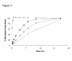
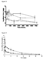
- Samples are pulled from the dissolution bath at selected time intervals using, for example, a syringe sampler fitted with only Teflon, stainless steel or glass surfaces. In those cases where a time course study is desired, aliquots can be periodically collected, including after 15 min, 30 min, 1 hr, 2 hr, 4 hr, 6 hr, 8 hr, 12 hr and 24 hr. The samples are assayed for zotarolimus concentration via HPLC. Data are expressed as drug-eluted in micrograms and mean-percent eluted.
- This assay can be used to demonstrate the ability of the device to effectively deliver the therapeutic compound to the tissues adjacent to the site of implantation. Ideally, a better system delivers the loaded therapeutic compound(s) to tissues adjacent to it, and not in the blood ( i.e ., systemically).
- stents that had been implanted as indicated in Testing for neointimal hyperplasia, inflammation and endothelialization after stent implantation are removed.
- the arterial tissue is removed from the stents, and the amount of drug that had penetrated the arterial walls adjacent to the stent is assayed for the presence and concentration of the target therapeutic compound(s).
- the data are then typically averaged, and plotted in a graph where the x -axis represent time, and the y -axis represent the amount of drug in tissue.
- Any art-accepted model for determining the concentration of a therapeutic compound can be used, including HPLC or other chromatography, immunoassays, activity assays, or other method of identification.
- This assay can be used to demonstrate the relative efficacy of a therapeutic compound delivered from the system of the invention to not enter the blood stream and is ideally used in conjunction with Drug penetration assay. Ideally, a better system delivers the loaded therapeutic compound(s) to tissues adjacent to it, and not in the blood ( i.e., systemically).
- blood samples from the subjects that have stents that had been implanted as indicated in Testing for neointimal hyperplasia, inflammation and endothelialization after stent implantation are collected by any art-accepted method, including venipuncture.
- Blood concentrations of the loaded therapeutic compounds are determined using any art-accepted method of detection, including immunoassay, chromatography (including liquid/liquid extraction HPLC tandem mass spectrometric method (LC-MS/MS) (Ji et al., 2004)), and activity assays.
- kits can be included in a kit, container, pack, or dispenser together with instructions for administration and use.
- the different components of the composition can be packaged in separate containers.
- Kits can also include reagents in separate containers.
- the reagents included in the kits can be supplied in containers or packaging of any sort such that the life of the different components are preserved and are not adsorbed or altered by the materials of the container.
- sealed glass ampules may contain lyophilized buffer that has been packaged under a neutral non-reacting gas, such as nitrogen.
- Ampules may consist of any suitable material, such as glass, organic polymers, such as polycarbonate, polystyrene, etc., ceramic, metal or any other material typically employed to hold reagents.
- suitable containers include bottles that can be fabricated from similar substances as ampules, and envelopes, that consist of foil-lined interiors, such as aluminum or an alloy.
- Containers include test tubes, vials, flasks, bottles and syringes.
- Containers can have a sterile access port, such as a bottle having a stopper that can be pierced by a hypodermic injection needle.
- Other containers can have two compartments that are separated by a readily removable membrane that upon removal permits the components to mix.
- Removable membranes can be glass, plastic, rubber, etc.
- Kits can also be supplied with instructional materials. Instructions can be printed on paper or other substrate, and/or can be supplied as an electronic-readable medium, such as a floppy disc, CD-ROM, DVD-ROM, Zip disc, videotape, audiotape, mini-disc, cassette tape or provided by calling a prescribed telephone number. Detailed instructions may not be physically associated with the kit; instead, a user may be directed to an Internet web site specified by the manufacturer or distributor of the kit, or supplied as electronic mail.
- an electronic-readable medium such as a floppy disc, CD-ROM, DVD-ROM, Zip disc, videotape, audiotape, mini-disc, cassette tape or provided by calling a prescribed telephone number.
- Detailed instructions may not be physically associated with the kit; instead, a user may be directed to an Internet web site specified by the manufacturer or distributor of the kit, or supplied as electronic mail.
- Example 1A A solution of Example 1A in isopropyl acetate (0.3 ml) was treated sequentially with diisopropylethylamine (87 ml, 0.5 mmol) and 1H-tetrazole (35 mg, 0.5 mmol), and thereafter stirred for 18 hours. This mixture was partitioned between water (10 ml) and ether (10 ml). The organics were washed with brine (10 ml) and dried (Na 2 SO 4 ).
- Example 1B Collection of the slower moving band from the chromatography column using the hexane:acetone (1:1) mobile phase in Example 1B provided the designated compound, which is zotarolimus. MS (ESI) m/e 966 (M) - .
- the immunosuppressant activity of zotarolimus was compared to rapamycin and two rapamycin analogs: 40-epi-N-[2'-pyridone]-rapamycin and 40-epi-N-[4'-pyridone]-rapamycin.
- the activity was determined using the human mixed lymphocyte reaction (MLR) assay described (Kino et al., 1987).
- MLR human mixed lymphocyte reaction
- AUC Area under the curve
- Example 2 Example 2 and the internal standard were determined using the Sciex MacQuanTM software.
- Calibration curves were derived from peak area ratio (parent dr ⁇ g/internal standard) of the spiked blood standards using least squares linear regression of the ratio versus the theoretical concentration. The methods were linear for both compounds over the range of the standard curve (correlation > 0.99) with an estimated quantitation limit of 0.1 ng/ml.
- the maximum blood concentration (C max ) and the time to reach the maximum blood concentration (T max ) were read directly from the observed blood concentration-time data.
- the blood concentration data were submitted to multi-exponential curve fitting using CSTRIP to obtain estimates of pharmacokinetic parameters.
- the estimated parameters were further defined using NONLIN84.
- the area under the blood concentration-time curve from 0 to t hours (last measurable blood concentration time point) after dosing (AUC 0-t ) was calculated using the linear trapezoidal rule for the blood-time profiles.
- the residual area extrapolated to infinity determined as the final measured blood concentration (C t ) divided by the terminal elimination rate constant ( ⁇ ), and added to AUC 0-t to produce the total area under the curve (AUC 0-t ).
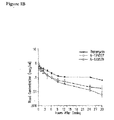
- Example 1 and Example 2 had a surprisingly substantially shorter terminal elimination half-life (t 1/2 ) when compared to rapamycin.
- t 1/2 terminal elimination half-life
- Table 2 Compound AUC (ng ⁇ hr/ml) t 1/2 (hours) Rapamycin 6.87 16.7 2-pyridone 2.55 2.8 4-pyridone 5.59 13.3
- Example 1 2.35 5.0
- the purpose of this example was to determine the effects of a rapamycin analog on neointimal formation in porcine coronary arteries in which stents were placed.
- This example illustrates that the rapamycin analog zotarolimus, when compounded and delivered from the Biocompatibles BiodiviYsio PC Coronary stent favorably affects neointimal hyperplasia and lumen size in porcine coronary arteries.
- This finding suggests that a combination from a drug-eluting stent including zotarolimus can be of substantial clinical benefit if properly applied in humans by limiting neointimal hyperplasia.
- Stents were implanted in two blood vessels in each pig. Pigs used in this model were generally 2-4 months old and weighed 30-40 kg. Two coronary stents were thus implanted in each pig by visually assessing a normal stent:artery ratio of 1.1-1.2.
- pigs were given oral aspirin (325 mg daily) and continued for the remainder of their course.
- General anesthesia was achieved by means of intramuscular injection followed by intravenous ketamine (30 mg/kg) and xylazine (3 mg/kg).
- Additional medication at the time of induction included atropine (1 mg) and flocillin (1 g) administered intramuscularly.
- an intra-arterial bolus of 10,000 units of heparin was administered.
- Arterial access was obtained by cutdown on the right external carotid and placement of an 8F sheath. After the procedure, the animals were maintained on a normal diet without cholesterol or other special supplementation.
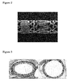
- the BiodivYsio stent was used with nominal vessel target size of 3.0 mm. See Figure 2 .
- Two coronary arteries per pig were assigned at random to deployment of the stents.
- the stent was either a drug eluting stent (polymer plus drug stent) or a stent coated with a polymer only (polymer only stent).
- the stents were delivered by means of standard guide catheters and wires.
- the stent balloons were inflated to appropriate sizes for less than 30 seconds.
- Each pig had one polymer only stent and one polymer plus drug stent placed in separate coronary arteries, so that each pig would have one stent for drug and one for control.
- a sample size of 20 pigs total was chosen to detect a projected difference in neointimal thickness of 0.2 mm with a standard deviation of 0.15 mm, at a power of 0.95 and beta 0.02.
- Low and high power light microscopy were used to make length measurements in the plane of microscopic view by means of a calibrated reticle and a digital microscopy system connected to a computer employing calibrated analysis software.
- a histopathologic injury score in stented blood vessels has been validated as being closely related to neointimal thickness. This score is related to depth of injury and as shown in Table 3: TABLE 3 Histopathologic injury scoring Score Description of Injury 0 Internal elastic lamina intact; endothelium typically denuded, media compressed but not lacerated. 1 Internal elastic lamina lacerated; media typically compressed but not lacerated. 2 Internal elastic lacerated; media visibly lacerated; external elastic lamina intact but compressed. 3 External elastic lamina lacerated; typically large lacerations of media extending through the external elastic lamina; coil wires sometimes residing in adventitia.
- neointimal thickness was averaged to obtain a mean injury score for each section.
- the measurement of neointimal thickness was made to the abluminal side of the stent wire, because the neointimal in all cases includes this thickness.
- the mid-stent segment was used for measurement, analysis, and comparison. Data were also recorded (and included in the data section of this report) for proximal and distal segments.
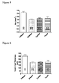
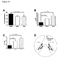
- Table 4 shows the summary results for all data for mean injury and neointimal thickness for each stent, including proximal, mid, and distal segments. Table 4 also shows lumen size, percent stenosis, and artery size as measured by the internal elastic laminae (IEL) and external elastic laminae (EEL). TABLE 4 Summary: All Measures (Distal, Mid.
- Table 5 shows the statistical t-test comparisons across test groups and control groups. There was a statistically significant difference in neointimal thickness, neointimal area, lumen size, and percent lumen stenosis, the drug eluting stent being clearly favored. Conversely, there were no statistically significant differences between the test group (polymer plus drug stents) and the control group (polymer only stents) for mean injury score, external elastic laminae, or internal elastic laminae areas. TABLE 5 Statistical Comparison of Test vs .
- the stent of this invention results in lower neointimal area, lower neointimal thickness, and greater lumen area.
- a stent that includes zotarolimus with a polymer showed a reduction in neointimal hyperplasia in the porcine model when placed in a coronary artery.
- the purpose of this example was to determine the rate of release of zotarolimus drug from 316L electropolished stainless steel coupons coated with a biocompatible polymer having phosphorylcholine side groups.
- the test samples were 316L stainless steel coupons that had been previously coated with a biocompatible polymer having phosphorylcholine side groups (PC polymer).
- Coronary stents are commonly made of 316L stainless steel and can be coated with the PC polymer to provide a depot site for loading drugs.
- the coated coupons which serve to simulate stents, were placed onto the septa.
- a solution of zotarolimus and ethanol (10 ⁇ l) was applied to the surface of each coupon.
- Zotarolimus (30.6 mg) was dissolved in 100% ethanol (3.0 ml). The syringe was cleaned with ethanol between each application. The cap to the glass vial was placed on the vial loosely, thereby assuring proper ventilation. The coupon was allowed to dry for a minimum of 1.5 hours. Twelve (12) coupons were loaded in this way - six being used to determine the average amount of drug loaded onto the device and six being used to measure the time needed to release the drug from the devices.
- a coupon was removed from the vial and placed into 50/50 acetonitrile/ 0.01M phosphate buffer (pH 6.0, 5.0 ml). The coupon was placed onto a 5210 Branson sonicator for one hour. The coupon was then removed from the solution, and the solution was assayed by HPLC.
- the time release studies were performed by immersing and removing the individual coupons from fresh aliquots (10.0 ml) of 0.01 M phosphate buffer at a pH of 6.0 at each of the following time intervals - 5, 15, 30 and 60 minutes. For the remaining time points of 120, 180, 240, 300, 360 minutes, volumes of 5.0 ml of buffer were used. To facilitate mixing during the drug release phase, the samples were placed onto an Eberbach shaker set at low speed. All solution aliquots were assayed by HPLC after the testing of the last sample was completed.
- a solution of zotarolimus in ethanol at a concentration of 50 mg/ml was prepared and dispensed into twelve vials. Twelve individual polymer-coated stents were placed on fixtures designed to hold the stent in a vertical position and the stents were immersed vertically in the drug solution for five minutes. The stents and fixtures were removed from the vials and excess drug solution was blotted away by contacting the stents with an absorbent material. The stents were then allowed to dry in air for 30 minutes in an inverted vertical position.
- the stents were removed from the fixtures, and each stent was placed into 50/50 acetonitrile/phosphate buffer (pH 5.1, 2.0 ml) and sonicated for one hour.
- the stents were removed from the solution and solutions were assayed for concentration of drug, which allowed calculation of the amount of drug originally on the stents. This method was independently shown to remove at least 95% of the drug from the stent coating. On average, the stents contained 60 micrograms of drug ⁇ 20 micrograms.
- the purpose of this example was to evaluate the safety of different drug dosages in a pig over-stretch model.
- Drug was delivered from the BiodivYsio OC stent (15 mm) coated with zotarolimus.
- In-stent neointima formation was measured at four time intervals - 3 days, 1 month, and 3 months - in the coronary arteries of adult miniature swine. Forty (40) animals were studied at each time interval (10 animals per dose). Each animal received one drug-coated stent and one control stent. The control stent contained no drug.
- Table 8 shows the dosing scheme for swine efficacy study.
- Dose group 1 ( ⁇ g) Dose group 2 ( ⁇ g) Dose group 3 ( ⁇ g) Dose group 4 ( ⁇ g) zotarolimus per stent 15 45 150 400 zotarolimus per mm of stent 1 3 10 27
- Histopathology in combination with scanning electron microscopy provided information regarding the short-term response to the implanted stent.
- the responses were similar in the control group and all dose groups, and the responses involved compression of the tunica media without remarkable necrosis, an accumulation of thrombus and inflammatory cells mostly localized to the stent struts, and early evidence of endothelial recovery and smooth muscle cell invasion of the thin mural thrombi.
- the adventitia in some samples displayed either focal or diffuse inflammatory infiltrates, and occasionally, there was plugging or congestion of the vasa vasora. There was no evidence of medial necrosis in any sample.
- the histomorphometry data for the one-month series indicated a significant inhibitory effect of locally eluted zotarolimus on neointima formation in stented coronary arteries of swine.
- Intima area normalized to injury score was significantly decreased for dose groups 3 and 4 (10 and 27 ⁇ g/mm) as compared with the control; there were also trends for decreases in absolute intima area and intima thickness for both dose groups 3 and 4 as compared with the control, and a tendency towards decreased histologic % stenosis for dose group 3 as compared with the control.
- the control stents displayed morphology typical of stents implanted in coronary arteries of Yucatan miniature swine at one month.
- the tunica media was compressed or thinned without necrosis subjacent to profiles of stent struts; there were only occasional inflammatory infiltrates; and the neointima ranged in size from relatively thin to moderately thin, and were composed of spindle-shaped and stellate cells in an abundant extracellular matrix, with only rare small foci of fibrinoid material around the profiles of the stent struts.
- the drug-coated stents showed similar compression of the tunica media without any substantial necrosis at any dose; like control devices, there was little inflammation present.
- the neointima was notably thinner in dose groups 3 and 4, in some cases being composed of only a few layers of cells.
- dose groups 3 and 4 there were substantial numbers of samples in which moderately sized fibrinoid deposits and inspisated thrombi were observed in the deep neointima. These were usually associated with the stent struts but sometimes extended between strut profiles.
- thrombus on the luminal surface, as the deposits were encapsulated within tibrocellular tissue and covered with a flattened layer of periluminal endothelial-like cells.
- the stent coated with zotarolimus reduced in-stent neointima formation in swine coronary arteries and provided clear evidence of a biologic drug effect (unresorbed thrombus/fibrin deposits of neointima) at one month. There was a weak tendency for the stent coated with zotarolimus to show a persistent inhibitory effect at the longer-term time interval of three months. There was no local coronary arterial wall toxicity in the form of medial necrosis or stent malapposition associated with any dose group, including the highest dose of approximately 27 ⁇ g/mm stent length at any time interval examined.
- Drug-eluting stents used in this study were similar to the ZoMaxx TM Drug-Eluting Stent System (Abbott Vascular; Redwood City, CA) in that they contained 10 ⁇ g/mm zotarolimus and a phosphorylcholine-based polymer (PC) topcoat designed to slow the elution of zotarolimus through diffusion.
- the stents in this study were made of stainless steel and four different groups were constructed with the only difference being that the amount of topcoat was varied by weight (0, 2, 5, or 10 ⁇ g/min).
- a porcine coronary overstretch model study (Schwartz, 1992) was conducted to examine neointimal formation for 28 days following stent implantation. The study evaluated a number of drug-eluting stents randomized vs . comparator stents containing only a polymer coating without drug (TriMaxxTM).
- each pig the three major coronary arteries were implanted with one test stent each. Additionally, three pigs were implanted with three non-drug containing TriMaxxTM stents (Abbott Laboratories; Abbott Park, IL) each (9 total stents) for comparison.
- TriMaxxTM stents Abbott Laboratories; Abbott Park, IL
- the stents that were compared included ZoMaxxTM stents (3.0 x 15 mm), commercially available rapamycin (8.5 ⁇ g/mm or 1.40 ⁇ g/mm 2 )-polymer coated Cypher® stents (3.0 x 13 mm; Cordis Corp.; Miami, FL) and paclitaxel-(6.8 ⁇ g/mm or 1.0 ⁇ g/mm 2 ) polymer coated Taxus® stents (3.0 x 16 mm; Boston Scientific; Natick, MA) stents.
- ZoMaxxTM stents 3.0 x 15 mm
- commercially available rapamycin 8.5 ⁇ g/mm or 1.40 ⁇ g/mm 2
- Cypher® stents (3.0 x 13 mm; Cordis Corp.; Miami, FL)
- Stents were implanted with a balloon/artery ratio of 1:1.30 as determined by quantitative coronary angiography. There were no cardiac- or stent- related mortalities in the study. After 28 days, animals were euthanized, and the hearts were removed and perfusion fixed at ⁇ 100 mm Hg with lactated Ringer's solution until cleared of blood, followed by 10% neutral buffered formalin. Stented vessels were excised, then infiltrated and embedded in methylmethacrylate (MMA). All blocks containing stented vessels were sectioned so that three, in-stent sections and two control sections (proximal and distal to the stent) were taken.
- MMA methylmethacrylate
- Example 9 Comparison of the elution profiles, tissue concentration and blood concentration of zotarolimus-polymer coated stents (ZoMaxxTM) and rapamycin-coated stents (Cypher®) implanted in rabbit iliac arteries
- the data were then averaged, and plotted in the graph shown in Figure 10 , where filled circles represent the data points for ZoMaxx TM and the filled squares represent the data points for Cypher®.
- the error bars represent S.E.M.
- the x- axis represent time in days, and the y -axis represent the amount of drug in tissue expressed as ⁇ g/g tissue.
- the tissue concentration of zotarolimus eluted from the ZoMaxx TM stent was significantly greater than the tissue concentration of rapamycin eluted from the Cypher® stent.
- the amount of rapamycin never exceeded 10 ⁇ g/g of tissue, while the amount of zotarolimus fluctuated from a high of slightly less than 120 ⁇ g/g of tissue at 5 days after implantation, to a low of slightly greater than 10 ⁇ g/g tissue at 28 days after implantation.
- the concentration of zotarolimus eluted from the ZoMaxx TM stents in the blood was lower than rapamycin eluted from the Cypher® stents up to 28 days, where at 28 days, the concentration was approximately similar.
- the blood concentration of rapamycin is dramatically elevated ( e.g., approximately 8, 6 and 4 ng/ml at the three earliest time points) compared to zotarolimus.
- the blood concentration of zotarolimus continues to decrease from a high of 4 ng/ml (initial time point) to a low of approximately 0.5-1 ng/ml (5 days and after implantation).
- stents While the purpose of stents is to keep a body lumen pendent, implantation in blood vessels often results in neointima formation, which occludes the lumen, and provokes an inflammatory response.
- This study set out to determine the neointima and inflammatory profiles of the ZoMaxx TM stent compared to the Cypher® stent over an extended period of time.
- the hearts were harvested, and the coronary arteries were perfusion-fixed with 10% buffered formalin at 100 mm Hg.
- the stented coronary artery segments were processed for plastic embedding, staining and morphometric analysis of six sections from the proximal through the distal margin of the stent.
- the specimens were embedded in methyl methacrylate and sections were obtained. Sections were then polished, mounted on a glass slide and stained with metachromatic stain. All histopathologic analysis was completed by a single independent investigator blinded to treatment group. Vessel morphometry and morphologic analysis of inflammation and smooth muscle content were completed using published methods (Kornowski et al., 1998; Schwartz, 1992; Suzuki et al., 2001).
- Inflammation was graded as 0, none; 1, scattered inflammatory cells; 2, inflammatory cells encompassing 50% of a strut in at least 25% to 50% of the circumference of the artery; 3, inflammatory cells surrounding a strut in at least 25% to 50% of the circumference of the artery.
- the morphometric measurements from each of the 4-stent sections were summed and divided by 4 to generate the mean value for each parameter within the stent.
- the mean differences between treatment groups were tested with ANOVA.
- scores were assigned to each of the four sections within the stented segment, the median value used as the score for the stent.
- the data were ranked within each cohort (3, 30, 90, or 180 days) and stratified. An ANOVA was performed on these ranks. Categorical data were compared with chi-square analysis. Data are expressed as mean F S.D. unless otherwise stated.
- Figure 13 shows the results of the neointimal area studies, wherein the x -axis represents time in days, and the y -axis represents the average neointimal area expressed in mm 2 .
- the data obtained using the rapamycin coated Cypher® stent is shown by the small filled squares and is excerpted from (Carter et al., 2004); data obtained from the zotarolimus coated ZoMaxx TM stent is shown by the large filled squares.
- neointimal areas for both stents were similar (approximately 0.45 mm 2 for rapamycin and 0.50 mm 2 for zotarolimus 3 days after implantation, and approximately 1.45 mm 2 for rapamycin and 1.60 mm 2 for zotarolimus 30 days after implantation).
- day 90 the differences between the two stents was startling: the neointimal area for those subjects receiving the zotarolimus-coated stents actually decreased to approximately 1.50 mm 2 , while those receiving the rapamycin-coated stents increased dramatically to approximately 3.0 mm 2 .
- This trend was echoed at 180 days after implantation, with rapamycin neointimal area approaching 3.25 mm 2 while the zotarolimus area decreased to approximately 0.85 mm 2 .
- Figure 14 shows the results of the inflammation studies, wherein the x -axis represents time in days, and the y -axis represents the average inflammation score.
- Pigs implanted with ZoMaxx TM stents experienced a marked reduction in inflammation over time, with a steady, almost linear decrease in inflammation over time, from a high of 0.97 ⁇ 0.315 at 3 days after implantation, to a low of 0.180 ⁇ 0.190 at 180 days after implantation.
- pigs receiving the Cypher® stent were less fortunate. While inflammation was quickly reduced at 30 days after implantation (0.200 ⁇ 0.420) and was less than in those pigs with the ZoMaxx TM stent (0.490 ⁇ 0.510), inflammation scores remarkablyd to a exceed those seen after initial transplantation, especially at 180 days (1.600 ⁇ 1.260), exceeding those scores for the ZoMaxx TM by almost 10-fold (0.180 v . 1.6).
- Pigs receiving zotarolimus delivered from ZoMaxx TM stents show significantly less neointima formation and reduced inflammation over the long run than those receiving rapamycin delivered from Cypher® stents.
- Example 10 This example set out to more thoroughly explore long-term efficacy of implanted stents as observed in Example 10.
- This example differs from Example 10 in that an additional stent was added for comparison (in this case, eluting paclitaxel) and the study was performed in rabbit iliac arteries.
- additional stent was added for comparison (in this case, eluting paclitaxel) and the study was performed in rabbit iliac arteries.
- other parameters were measured, including morphometric analyses, fibrin deposition, granuloma and giant cell reactions, inflammation scores.
- the experimental set-up is shown in Table 12; in Figure 15D , the placement of the stents is diagrammed.
- ZoMaxx IVUS a clinical, angiographic and intravascular ultrasound (IVUS) trial was conducted in 40 patients with a mean age of 59 years (+/- 9). Eighty percent of patients had hyperlipidemia, while 40 percent had diabetes, and 40 percent had a prior heart attack. The mean lesion length stented was 14.4 mm (+/- 3.3). Before treatment with a ZoMaxx stent, percent diameter stenosis (DS), the percent of vessel blocked with disease, was 70% (+/- 10). Directly after treatment with ZoMaxx, percent diameter stenosis improved to 5.1 percent (+/- 5.3) inside the stent (in-stent) and 19 percent (+/- 7) in-segment.
- IVUS intravascular ultrasound
- Late lumen loss a measure of the change in vessel diameter between the time immediately following stent placement and at four-months, was 0.20 mm (+/- 0.35) in-stent and 0.17 mm (+/- 0.35) in-segment.
- In-stent net volume obstruction the amount of blockage that re-formed inside the stent in the four-months following the initial procedure, was 6.5 percent (+/- 6.2).
- the ZoMaxx stent was delivered with 100 percent success, and no major adverse cardiac events (MACE) occurred during the procedure or during the four months of follow-up.
- MACE major adverse cardiac events
- ACS acute coronary syndrome
- PCI percutaneous intervention
- obesity is often associated with a pro-inflammatory state and endothelial dysfunction. Both conditions are known to be independent predictors of early restenosis after coronary stent placement.
- CRP c-reactive protein
- Plasma IL-6 and tumor necrosis factor- ⁇ (TNF- ⁇ ) are inflammatory cytokines that are also elevated in the obese patient, and in type 2 diabetics.
- TNF- ⁇ tumor necrosis factor- ⁇
- VCAM-1 serum vascular cell adhesion molecule-1
- Plaque rupture can be induced by increased concentrations of matrix metalloproteinases secreted by foam cells, leading to plaque instability and ultimate rupture of the thin fibrous cap which overlies the developing lesion.
- tissue factor which is expressed on the surface of foam cells, activates coagulation factor VII, which leads to the formation of thrombin. Generation of this protein leads to platelet activation and aggregation, as well as the conversion of fibrinogen to fibrin, and the clear formation of thrombus.
- the stents described herein will be deployed in patients who are diagnosed with ischemic heart disease due to stenotic lesions in coronary arteries and in subsets of the clinical population at higher risk for recurrent coronary disease and other adverse clinical events.
- Other targets for intervention include peripheral vascular diseases including stenoses in the superficial femoral arteries, renal arteries, iliacs, and vessels below the knee.
- Target vessels for interventional procedures will be reached using percutaneous vascular access via either the femoral or radial artery, and a guiding catheter will be inserted into the vessel. The target lesion will then be crossed with a guide wire, and the balloon catheter will be inserted either over the wire or using a rapid exchange system.
- the physician will determine the appropriate size of the stent to be implanted by online quantitative coronary angiography (QCA) or by visual estimate.
- the stent will be deployed using appropriate pressure as indicated by the compliance of the stent, and a post-procedure angiogram can then be obtained.
- the procedure is completed, the patient will be regularly monitored for angina status and for the existence of any adverse events. The need for repeat procedures will also be assessed.
Applications Claiming Priority (7)
| Application Number | Priority Date | Filing Date | Title |
|---|---|---|---|
| US66448805P | 2005-03-23 | 2005-03-23 | |
| US71508205P | 2005-09-08 | 2005-09-08 | |
| US72687805P | 2005-10-14 | 2005-10-14 | |
| US72708005P | 2005-10-14 | 2005-10-14 | |
| US72719605P | 2005-10-14 | 2005-10-14 | |
| US73257705P | 2005-10-17 | 2005-10-17 | |
| EP06739478A EP1877004A4 (de) | 2005-03-23 | 2006-03-22 | Zusammensetzungen und verfahren zur verabreichung von rapamycin-analoga mit medizinprodukten für langzeitwirkung |
Related Parent Applications (1)
| Application Number | Title | Priority Date | Filing Date |
|---|---|---|---|
| EP06739478A Division EP1877004A4 (de) | 2005-03-23 | 2006-03-22 | Zusammensetzungen und verfahren zur verabreichung von rapamycin-analoga mit medizinprodukten für langzeitwirkung |
Publications (1)
| Publication Number | Publication Date |
|---|---|
| EP3138531A1 true EP3138531A1 (de) | 2017-03-08 |
Family
ID=37865406
Family Applications (2)
| Application Number | Title | Priority Date | Filing Date |
|---|---|---|---|
| EP06739478A Ceased EP1877004A4 (de) | 2005-03-23 | 2006-03-22 | Zusammensetzungen und verfahren zur verabreichung von rapamycin-analoga mit medizinprodukten für langzeitwirkung |
| EP16171630.3A Withdrawn EP3138531A1 (de) | 2005-03-23 | 2006-03-22 | Zusammensetzungen und verfahren zur verabreichung von rapamycinanaloga mithilfe von medizinischen vorrichtungen für langzeitwirkung |
Family Applications Before (1)
| Application Number | Title | Priority Date | Filing Date |
|---|---|---|---|
| EP06739478A Ceased EP1877004A4 (de) | 2005-03-23 | 2006-03-22 | Zusammensetzungen und verfahren zur verabreichung von rapamycin-analoga mit medizinprodukten für langzeitwirkung |
Country Status (3)
| Country | Link |
|---|---|
| EP (2) | EP1877004A4 (de) |
| JP (1) | JP5242374B2 (de) |
| WO (1) | WO2007032777A2 (de) |
Families Citing this family (11)
| Publication number | Priority date | Publication date | Assignee | Title |
|---|---|---|---|---|
| US8394398B2 (en) | 1997-09-26 | 2013-03-12 | Abbott Laboratories | Methods of administering rapamycin analogs with anti-inflammatories using medical devices |
| US8257726B2 (en) | 1997-09-26 | 2012-09-04 | Abbott Laboratories | Compositions, systems, kits, and methods of administering rapamycin analogs with paclitaxel using medical devices |
| US7378105B2 (en) | 1997-09-26 | 2008-05-27 | Abbott Laboratories | Drug delivery systems, kits, and methods for administering zotarolimus and paclitaxel to blood vessel lumens |
| US7357942B2 (en) | 1997-09-26 | 2008-04-15 | Abbott Laboratories | Compositions, systems, and kits for administering zotarolimus and paclitaxel to blood vessel lumens |
| US8431145B2 (en) | 2004-03-19 | 2013-04-30 | Abbott Laboratories | Multiple drug delivery from a balloon and a prosthesis |
| JP5541593B2 (ja) * | 2006-08-15 | 2014-07-09 | アボット ラボラトリーズ | ラパマイシン類似体及びパクリタキセルを含む組成物及び薬物送達システム |
| CN101940802B (zh) * | 2007-09-04 | 2015-02-04 | 株式会社日本斯滕特技术 | 药物缓释性支架 |
| US8951595B2 (en) | 2009-12-11 | 2015-02-10 | Abbott Cardiovascular Systems Inc. | Coatings with tunable molecular architecture for drug-coated balloon |
| US20130303496A1 (en) * | 2012-05-08 | 2013-11-14 | Abbott Cardiovascular Systems Inc. | Method Of Treating Vascular Lesions |
| US9610385B2 (en) * | 2013-03-07 | 2017-04-04 | Abbott Cardiovascular Systems Inc. | Method of fabricating an implantable medical device comprising a rapamycin derivative |
| KR20230150790A (ko) * | 2020-12-29 | 2023-10-31 | 시노 메디칼 사이언시즈 테크놀로지, 인코포레이티드 | 약물 용출 스텐트 |
Citations (33)
| Publication number | Priority date | Publication date | Assignee | Title |
|---|---|---|---|---|
| US3929992A (en) | 1972-09-29 | 1975-12-30 | Ayerst Mckenna & Harrison | Rapamycin and process of preparation |
| US3993749A (en) | 1974-04-12 | 1976-11-23 | Ayerst Mckenna And Harrison Ltd. | Rapamycin and process of preparation |
| US4316885A (en) | 1980-08-25 | 1982-02-23 | Ayerst, Mckenna And Harrison, Inc. | Acyl derivatives of rapamycin |
| US4401653A (en) | 1981-03-09 | 1983-08-30 | Ayerst, Mckenna & Harrison Inc. | Combination of rapamycin and picibanil for the treatment of tumors |
| EP0184162A2 (de) | 1984-12-03 | 1986-06-11 | Fujisawa Pharmaceutical Co., Ltd. | Tricyclische Verbindungen, Verfahren zu ihrer Herstellung und diese enthaltende pharmazeutische Zusammensetzung |
| US4650803A (en) | 1985-12-06 | 1987-03-17 | University Of Kansas | Prodrugs of rapamycin |
| US4885171A (en) | 1978-11-03 | 1989-12-05 | American Home Products Corporation | Use of rapamycin in treatment of certain tumors |
| US4916193A (en) | 1987-12-17 | 1990-04-10 | Allied-Signal Inc. | Medical devices fabricated totally or in part from copolymers of recurring units derived from cyclic carbonates and lactides |
| US4994071A (en) | 1989-05-22 | 1991-02-19 | Cordis Corporation | Bifurcating stent apparatus and method |
| US5023262A (en) | 1990-08-14 | 1991-06-11 | American Home Products Corporation | Hydrogenated rapamycin derivatives |
| EP0467606A1 (de) | 1990-07-16 | 1992-01-22 | American Home Products Corporation | Rapamycinderivate |
| US5092877A (en) | 1988-09-01 | 1992-03-03 | Corvita Corporation | Radially expandable endoprosthesis |
| WO1992005179A1 (en) | 1990-09-19 | 1992-04-02 | American Home Products Corporation | Carboxylic acid esters of rapamycin |
| US5120727A (en) | 1991-05-29 | 1992-06-09 | American Home Products Corporation | Rapamycin dimers |
| US5120842A (en) | 1991-04-01 | 1992-06-09 | American Home Products Corporation | Silyl ethers of rapamycin |
| US5120725A (en) | 1991-05-29 | 1992-06-09 | American Home Products Corporation | Bicyclic rapamycins |
| US5163852A (en) | 1992-02-11 | 1992-11-17 | W. L. Gore & Associates, Inc. | Coaxial cable side tape connector assembly and processes for assembly |
| US5177203A (en) | 1992-03-05 | 1993-01-05 | American Home Products Corporation | Rapamycin 42-sulfonates and 42-(N-carboalkoxy) sulfamates useful as immunosuppressive agents |
| US5304121A (en) | 1990-12-28 | 1994-04-19 | Boston Scientific Corporation | Drug delivery system making use of a hydrogel polymer coating |
| US5447724A (en) | 1990-05-17 | 1995-09-05 | Harbor Medical Devices, Inc. | Medical device polymer |
| US5464650A (en) | 1993-04-26 | 1995-11-07 | Medtronic, Inc. | Intravascular stent and method |
| US5527337A (en) | 1987-06-25 | 1996-06-18 | Duke University | Bioabsorbable stent and method of making the same |
| US5605696A (en) | 1995-03-30 | 1997-02-25 | Advanced Cardiovascular Systems, Inc. | Drug loaded polymeric material and method of manufacture |
| US5705583A (en) | 1991-07-05 | 1998-01-06 | Biocompatibles Limited | Polymeric surface coatings |
| US6090901A (en) | 1991-07-05 | 2000-07-18 | Biocompatibles Limited | Polymeric surface coatings |
| US6284305B1 (en) | 1996-06-13 | 2001-09-04 | Schneider (Usa) Inc. | Drug coating with topcoat |
| WO2001087372A1 (en) | 2000-05-12 | 2001-11-22 | Cordis Corporation | Drug combinations useful for prevention of restenosis |
| US6358556B1 (en) | 1995-04-19 | 2002-03-19 | Boston Scientific Corporation | Drug release stent coating |
| US6413272B1 (en) | 1997-03-31 | 2002-07-02 | Kabushikikaisha Igaki Iryo Sekkei | Stent for vessel |
| US6419692B1 (en) | 1999-02-03 | 2002-07-16 | Scimed Life Systems, Inc. | Surface protection method for stents and balloon catheters for drug delivery |
| WO2002055122A1 (en) | 2001-01-11 | 2002-07-18 | Biocompatibles Uk Limited | Stens with drug-containing amphiphilic polymer coating |
| US20030129215A1 (en) | 1998-09-24 | 2003-07-10 | T-Ram, Inc. | Medical devices containing rapamycin analogs |
| WO2004022124A1 (en) * | 2002-09-06 | 2004-03-18 | Abbott Laboratories | Medical device having hydration inhibitor |
Family Cites Families (7)
| Publication number | Priority date | Publication date | Assignee | Title |
|---|---|---|---|---|
| US20040213789A1 (en) * | 1997-09-02 | 2004-10-28 | Oron Yacoby-Zeevi | Heparanase activity neutralizing anti-heparanase monoclonal antibody and other anti-heparanase antibodies |
| AP2003002828A0 (en) * | 2001-01-16 | 2003-09-30 | Vascular Therapies Llc | Implantable device containing resorbable matrix material and anti-proliferative drugs for preventing or treating failure of hemodialysis vascular access and other vascular grafts. |
| US7708712B2 (en) * | 2001-09-04 | 2010-05-04 | Broncus Technologies, Inc. | Methods and devices for maintaining patency of surgically created channels in a body organ |
| WO2005011796A1 (ja) * | 2003-08-05 | 2005-02-10 | Kaneka Corporation | 生体留置用ステント |
| JP4379044B2 (ja) * | 2003-08-22 | 2009-12-09 | 株式会社カネカ | 生体留置用ステント |
| US20050208093A1 (en) * | 2004-03-22 | 2005-09-22 | Thierry Glauser | Phosphoryl choline coating compositions |
| EP1802365A4 (de) * | 2004-07-19 | 2010-01-20 | Broncus Tech Inc | Verfahren und vorrichtungen zur aufrechterhaltung von chirurgisch erzeugten kanälen in einem körperorgan |
-
2006
- 2006-03-22 JP JP2008503210A patent/JP5242374B2/ja not_active Expired - Fee Related
- 2006-03-22 EP EP06739478A patent/EP1877004A4/de not_active Ceased
- 2006-03-22 EP EP16171630.3A patent/EP3138531A1/de not_active Withdrawn
- 2006-03-22 WO PCT/US2006/010700 patent/WO2007032777A2/en active Application Filing
Patent Citations (36)
| Publication number | Priority date | Publication date | Assignee | Title |
|---|---|---|---|---|
| US3929992A (en) | 1972-09-29 | 1975-12-30 | Ayerst Mckenna & Harrison | Rapamycin and process of preparation |
| US3993749A (en) | 1974-04-12 | 1976-11-23 | Ayerst Mckenna And Harrison Ltd. | Rapamycin and process of preparation |
| US4885171A (en) | 1978-11-03 | 1989-12-05 | American Home Products Corporation | Use of rapamycin in treatment of certain tumors |
| US4316885A (en) | 1980-08-25 | 1982-02-23 | Ayerst, Mckenna And Harrison, Inc. | Acyl derivatives of rapamycin |
| US4401653A (en) | 1981-03-09 | 1983-08-30 | Ayerst, Mckenna & Harrison Inc. | Combination of rapamycin and picibanil for the treatment of tumors |
| EP0184162A2 (de) | 1984-12-03 | 1986-06-11 | Fujisawa Pharmaceutical Co., Ltd. | Tricyclische Verbindungen, Verfahren zu ihrer Herstellung und diese enthaltende pharmazeutische Zusammensetzung |
| US4650803A (en) | 1985-12-06 | 1987-03-17 | University Of Kansas | Prodrugs of rapamycin |
| US5527337A (en) | 1987-06-25 | 1996-06-18 | Duke University | Bioabsorbable stent and method of making the same |
| US4916193A (en) | 1987-12-17 | 1990-04-10 | Allied-Signal Inc. | Medical devices fabricated totally or in part from copolymers of recurring units derived from cyclic carbonates and lactides |
| US5092877A (en) | 1988-09-01 | 1992-03-03 | Corvita Corporation | Radially expandable endoprosthesis |
| US4994071A (en) | 1989-05-22 | 1991-02-19 | Cordis Corporation | Bifurcating stent apparatus and method |
| US5447724A (en) | 1990-05-17 | 1995-09-05 | Harbor Medical Devices, Inc. | Medical device polymer |
| EP0467606A1 (de) | 1990-07-16 | 1992-01-22 | American Home Products Corporation | Rapamycinderivate |
| US5023262A (en) | 1990-08-14 | 1991-06-11 | American Home Products Corporation | Hydrogenated rapamycin derivatives |
| WO1992005179A1 (en) | 1990-09-19 | 1992-04-02 | American Home Products Corporation | Carboxylic acid esters of rapamycin |
| US5304121A (en) | 1990-12-28 | 1994-04-19 | Boston Scientific Corporation | Drug delivery system making use of a hydrogel polymer coating |
| US5120842B1 (de) | 1991-04-01 | 1993-07-06 | A Failli Amedeo | |
| US5120842A (en) | 1991-04-01 | 1992-06-09 | American Home Products Corporation | Silyl ethers of rapamycin |
| US5120725A (en) | 1991-05-29 | 1992-06-09 | American Home Products Corporation | Bicyclic rapamycins |
| US5120727A (en) | 1991-05-29 | 1992-06-09 | American Home Products Corporation | Rapamycin dimers |
| US5705583A (en) | 1991-07-05 | 1998-01-06 | Biocompatibles Limited | Polymeric surface coatings |
| US6090901A (en) | 1991-07-05 | 2000-07-18 | Biocompatibles Limited | Polymeric surface coatings |
| US5163852A (en) | 1992-02-11 | 1992-11-17 | W. L. Gore & Associates, Inc. | Coaxial cable side tape connector assembly and processes for assembly |
| US5177203A (en) | 1992-03-05 | 1993-01-05 | American Home Products Corporation | Rapamycin 42-sulfonates and 42-(N-carboalkoxy) sulfamates useful as immunosuppressive agents |
| US5464650A (en) | 1993-04-26 | 1995-11-07 | Medtronic, Inc. | Intravascular stent and method |
| US5624411A (en) | 1993-04-26 | 1997-04-29 | Medtronic, Inc. | Intravascular stent and method |
| US5605696A (en) | 1995-03-30 | 1997-02-25 | Advanced Cardiovascular Systems, Inc. | Drug loaded polymeric material and method of manufacture |
| US6358556B1 (en) | 1995-04-19 | 2002-03-19 | Boston Scientific Corporation | Drug release stent coating |
| US6284305B1 (en) | 1996-06-13 | 2001-09-04 | Schneider (Usa) Inc. | Drug coating with topcoat |
| US6413272B1 (en) | 1997-03-31 | 2002-07-02 | Kabushikikaisha Igaki Iryo Sekkei | Stent for vessel |
| US20030129215A1 (en) | 1998-09-24 | 2003-07-10 | T-Ram, Inc. | Medical devices containing rapamycin analogs |
| US6419692B1 (en) | 1999-02-03 | 2002-07-16 | Scimed Life Systems, Inc. | Surface protection method for stents and balloon catheters for drug delivery |
| WO2001087372A1 (en) | 2000-05-12 | 2001-11-22 | Cordis Corporation | Drug combinations useful for prevention of restenosis |
| WO2002055122A1 (en) | 2001-01-11 | 2002-07-18 | Biocompatibles Uk Limited | Stens with drug-containing amphiphilic polymer coating |
| US20050004661A1 (en) | 2001-01-11 | 2005-01-06 | Lewis Andrew L | Stens with drug-containing amphiphilic polymer coating |
| WO2004022124A1 (en) * | 2002-09-06 | 2004-03-18 | Abbott Laboratories | Medical device having hydration inhibitor |
Non-Patent Citations (41)
| Title |
|---|
| AGGARWAL, A.; D.J. SCHNEIDER; B.E. SOBEL; H.L. DAUENNAN: "Comparison of inflammatory markers in patients with diabetes mellitus versus those without before and after coronary arterial stenting", AM J CARDIOL., vol. 92, 2003, pages 924 - 9 |
| BAKER, H.; A. SIDOROWICZ; S.N. SEHGAL; C. VEZINA: "Rapamycin (AY-22,989), a new antifungal antibiotic. III. In vitro and in vivo evaluation", JANTIBIOT (TOKYO), vol. 31, 1978, pages 539 - 45 |
| BIERER, B.E.; S.L. SCHREIBER; S.J. BURAKOFF: "The effect of the immunosuppressant FK-506 on alternate pathways ofT cell activation", EUR J IMMUNOL., vol. 21, 1991, pages 439 - 45 |
| BIONDI-ZOCCAI, G.G.; A. ABBATE; G. LIUZZO; L.M. BIASUCCI.: "Atherothrombosis, inflammation, and diabetes", JAM COLL CARDIOL., vol. 41, 2003, pages 1071 - 7 |
| BROWN, E.J.; M.W. ALBERS; T.B. SHIN; K. ICHIKAWA; C.T. KEITH; W.S. LANE; S.L. SCHREIBER: "A mammalian protein targeted by GI-arresting rapamycin-receptor complex", NATURE, vol. 369, 1994, pages 756 - 8, XP002031997, DOI: doi:10.1038/369756a0 |
| BUNCHMAN, T.E.; C.A. BROOKSHIRE: "Smooth muscle cell proliferation by conditioned media from cyclosporine-treated endothelial cells: a role of endothelin", TRANSPLANT PROC, vol. 23, 1991, pages 967 - 8 |
| CARTER, A.J.; M. AGGARWAL; G.A. KOPIA; F. TIO; P.S. TSAO; R. KOLATA; A.C. YEUNG; G. LLANOS; J. DOOLEY; R. FALOTICO.: "Long-term effects of polymer-based, slow-release, sirolimus-eluting stents in a porcine coronary model", CARDIOVASC RES, vol. 63, 2004, pages 617 - 24 |
| DANDONA, P.; A. ALJADA: "A rational approach to pathogenesis and treatment of type 2 diabetes mellitus, insulin resistance, inflammation, and atherosclerosis", AM J CARDIOL., vol. 90, 2002, pages 27G - 33G |
| DUMONT, F.J.; M.R. MELINO; M.J. STARUCH; S.L. KOPRAK; P.A. FISCHER; N.H. SIGAL: "The immunosuppressive macrolides FK-506 and rapamycin act as reciprocal antagonists in murine T cells", J IMMUNOL., vol. 144, 1990, pages 1418 - 24 |
| FRETZ, H., M; ALBERS; A. GALA; R. STANDAERT; W. LANE; S. BURAKOFF; B. BIERER; S. SCHREIBER: "Rapamycin and FK506 binding proteins (immunophilins).", J. AM. CHEM. SOC., vol. 113, 1991, pages 1409 - 1411, XP000310589, DOI: doi:10.1021/ja00004a051 |
| GARCIA-TOUCHARD A ET AL: "Zotarolimus-eluting stents reduce experimental coronary artery neointimal hyperplasia after 4 weeks", EUROPEAN HEART JOURNAL, OXFORD UNIVERSITY PRESS, GB, vol. 27, 1 January 2006 (2006-01-01), pages 988 - 993, XP002487648, ISSN: 0195-668X, [retrieved on 20060131], DOI: 10.1093/EURHEARTJ/EHI752 * |
| GRECH, E.D.; D.R. RAMSDALE: "Acute coronary syndrome: unstable angina and non-ST segment elevation myocardial infarction", BMJ, vol. 326, 2003, pages 1259 - 61 |
| HARDING, M.W.; A. GALAT; D.E. UEHLING; S.L. SCHREIBER.: "A receptor for the immunosuppressant FK506 is a cis-trans peptidyl-prolyl isomerase", NATURE, vol. 341, 1989, pages 758 - 60, XP001153476, DOI: doi:10.1038/341758a0 |
| HAYWARD, C.; D. YOHANNES; S. DANISHEFSKY: "Total synthesis of rapamycin via a novel titanium-mediated aldol macrocyclization reaction", J. AM. CHEM. SOC., vol. 115, 1993, pages 9345 - 9346, XP002156864, DOI: doi:10.1021/ja00073a083 |
| HELMUS, M: "Medical Device Design--A Systems Approach: Central Venous Catheters", 22ND INTERNATIONAL SOCIETY FOR THE ADVANCEMENT OF MATERIAL AND PROCESS ENGINEERING TECHNICAL CONFERENCE, 1990 |
| HIGUCHI, T.; V. STELLA, PRO-DRUGS AS NOVEL DELIVERY SYSTEMS, 1987 |
| HONGO YOICHIRO ET AL: "One year results of intravascular ultrasound analysis for the ABT-578 eluting phosphorylcholine-coated stent implantation in de novo coronary lesions", CIRCULATION, LIPPINCOTT WILLIAMS & WILKINS, US, vol. 110, no. 17 Suppl. S, 1 October 2004 (2004-10-01), pages 758, XP008132057, ISSN: 0009-7322 * |
| JI, Q.; M. REIMER; T. EL-SHOURBAGY: "96-Well liquid-liquid extraction liquid chromatography-tandem mass spectrometry method for the quantitative determination of ABT-578 in human blood samples", JOURNAL OF CHROMATOGRAPHY B., vol. 805, 2004, pages 67 - 75, XP004503767, DOI: doi:10.1016/j.jchromb.2004.02.014 |
| KINO, T.; N. INAMURA; F. SAKAI; K. NAKAHARA; T. GOTO; M. OKUHARA; M. KOHSAKA; H. AOKI; T. OCHIAI: "Effect of FK-506 on human mixed lymphocyte reaction in vitro", TRANSPLANT PROC, vol. 19, 1987, pages 36 - 9 |
| KORNOWSKI, R.; M.K. HONG; F.O. TIO; O. BRAMWELL; H. WU; M.B. LEON: "In-stent restenosis: contributions of inflammatory responses and arterial injury to neointimal hyperplasia", JAM COLL CARDIOL., vol. 31, 1998, pages 224 - 30 |
| LULY, MACROCYCLIC IMMUNOMODULATORS, 1995 |
| M. STRAUCH, FASEB, vol. 3, 1989, pages 3411 |
| MARTEL, R.R.; J. KLICIUS; S. GALET: "Inhibition of the immune response byrapamycin, a new antifungal antibiotic", CAN JPHYSIOL PHARMACOL., vol. 55, 1977, pages 48 - 51 |
| MORRIS, R.; B. MEISER: "Identification of a new pharmacologic action for an old compound", MED. SCI. RES., vol. 17, 1989, pages 609 |
| MORRIS, R: "Rapamycins: antifungal, antitumor, antiproliferative, and immunosuppressive macrolides", TRANSPLANT. REV., vol. 6, 1992, pages 39 - 87, XP009110318 |
| MUNI ET AL: "Coronary drug-eluting stent development: Issues in trial design", AMERICAN HEART JOURNAL, MOSBY- YEAR BOOK INC, US, vol. 149, no. 3, 1 March 2005 (2005-03-01), pages 415 - 433, XP005686540, ISSN: 0002-8703 * |
| NICOLAOU, K.; T. CHAKRABORTY; A. PISCOPIO; N. MINOWA; P. BERTINATO.: "Total synthesis of rapamycin", J. AM. CHEM. SOC., vol. 115, 1993, pages 4419 - 4420 |
| PAIVA, N.L.; A.L. DEMAIN; M.F. ROBERTS: "Incorporation of acetate, propionate, and methionine into rapamycin by Streptomyces hygroscopicus", J NAT PROD., vol. 54, 1991, pages 167 - 77, XP008050894, DOI: doi:10.1021/np50073a015 |
| ROCHE, E: "Bioreversible Carriers in Drug Design", 1987, AMERICAN PHARMACEUTICAL ASSOCIATION AND PERGAMON PRESS |
| ROFFI, M.; E.J. TOPOL: "Percutaneous coronary intervention in diabetic patients with non-ST-segment elevation acute coronary syndromes", EUR HEART J., vol. 25, 2004, pages 190 - 8 |
| ROMO, D.; S. MEYER; D. JOHSNON; S. SCHRIEBER: "Total synthesis of (-)-rapamycin using an Evans-Tishchenko fragment coupling", J. AM. CHEM. SOC., vol. 115, 1993, pages 7906 - 7907 |
| SABATINI, D.M.; H. ERDJUMENT-BROMAGE; M. LUI; P. TEMPST; S.H. SNYDER: "RAFT1: a mammalian protein that binds to FKBP12 in a rapamycin-dependent fashion and is homologous to yeast TORs", CELL, vol. 78, 1994, pages 35 - 43, XP023908888, DOI: doi:10.1016/0092-8674(94)90570-3 |
| SCHWARTZ, R: "Restenosis and the proportional neointimal response to coronary artery injury: results in a porcine model", JAM COLL CARDIOL, vol. 19, 1992, pages 267 - 274 |
| SEHGAL, S.N.; H. BAKER; C. VEZINA: "Rapamycin (AY-22,989), a new antifungal antibiotic. II. Fermentation, isolation and characterization", J ANTIBIOT (TOKYO)., vol. 28, 1975, pages 727 - 32 |
| SEHGAL, S.N; H. BAKER; C.P. ENG; K. SINGH; C. VEZINA: "Demethoxyrapamycin (AY-24,668), a new antifungal antibiotic", JANTIBIOT (TOKYO)., vol. 36, 1983, pages 351 - 4 |
| SHICHIRI, M.; Y. HIRATA; T. NAKAJIMA; K. ANDO; T. IMAI; M. YANAGISAWA; T. MASAKI; F. MARUMO.: "Endothelin-1 is an autocrine/paracrine growth factor for human cancer cell lines", J CLIN INVEST., vol. 87, 1991, pages 1867 - 71 |
| SIEKIERKA, J.J.; S.H. HUNG; M. POE; C.S. LIN; N.H. SIGAL: "A cytosolic binding protein for the immunosuppressant FK506 has peptidyl-prolyl isomerase activity but is distinct from cyclophilin", NATURE, vol. 341, 1989, pages 755 - 7, XP001018902, DOI: doi:10.1038/341755a0 |
| SUZUKI, T.; G. KOPIA; S. HAYASHI; L.R. BAILEY; G. LLANOS; R. WILENSKY; B.D. KLUGHERZ; G. PAPANDREOU; P. NARAYAN; M.B. LEON: "Stent-based delivery of sirolimus reduces neointimal formation in a porcine coronary model", CIRCULATION, vol. 104, 2001, pages 118 8 - 93 |
| VEZINA, C.; A. KUDELSKI; S.N. SEHGAL.: "Rapamycin (AY-22,989), a new antifungal antibiotic. I. Taxonomy of the producing streptomycete and isolation of the active principle", J ANTIBIOT (TOKYO)., vol. 28, 1975, pages 721 - 6, XP001161176 |
| YAMAGISHI, S.; C.C. HSU; K. KOBAYASHI; H. YAMAMOTO: "Endothelin 1 mediates endothelial cell-dependent proliferation of vascular pericytes", BIOCHEM BIOPHYS RES COMMUN, vol. 191, 1993, pages 840 - 6, XP024767712, DOI: doi:10.1006/bbrc.1993.1293 |
| YUDKIN, J.S.; M. KUMARI; S.E. HUMPHRIES; V. MOHAMED-ALI: "Inflammation, obesity, stress and coronary heart disease: is interleukin-6 the link?", ATHEROSCLEROSIS., vol. 148, 2000, pages 209 - 14, XP002982688, DOI: doi:10.1016/S0021-9150(99)00463-3 |
Also Published As
| Publication number | Publication date |
|---|---|
| EP1877004A4 (de) | 2012-01-25 |
| EP1877004A2 (de) | 2008-01-16 |
| WO2007032777A2 (en) | 2007-03-22 |
| JP5242374B2 (ja) | 2013-07-24 |
| WO2007032777A3 (en) | 2007-10-25 |
| JP2008534074A (ja) | 2008-08-28 |
Similar Documents
| Publication | Publication Date | Title |
|---|---|---|
| US20060198867A1 (en) | Compositions and methods of administering rapamycin analogs using medical devices for long-term efficacy | |
| US7378105B2 (en) | Drug delivery systems, kits, and methods for administering zotarolimus and paclitaxel to blood vessel lumens | |
| US8318190B2 (en) | Method of treating disorders using compositions comprising zotarolimus and paclitaxel | |
| EP1933785B1 (de) | Zusammensetzungen, systeme, kits und verfahren zur verabreichung von rapamycin-analoga mit paclitaxel unter verwendung von medizinprodukten | |
| EP3138531A1 (de) | Zusammensetzungen und verfahren zur verabreichung von rapamycinanaloga mithilfe von medizinischen vorrichtungen für langzeitwirkung | |
| US8394398B2 (en) | Methods of administering rapamycin analogs with anti-inflammatories using medical devices | |
| US8057816B2 (en) | Compositions and methods of administering paclitaxel with other drugs using medical devices | |
| US20090162413A1 (en) | Compositions and methods of administering rapamycin analogs with paclitaxel using medical devices | |
| EP2292280A1 (de) | Medizinische Geräte, die Rapamycinanaloga enthalten | |
| EP2068932B1 (de) | Zusammensetzungen und wirkstoffverabreichungssystem mit rapamycin-analogen und paclitaxel | |
| US8257726B2 (en) | Compositions, systems, kits, and methods of administering rapamycin analogs with paclitaxel using medical devices |
Legal Events
| Date | Code | Title | Description |
|---|---|---|---|
| PUAI | Public reference made under article 153(3) epc to a published international application that has entered the european phase |
Free format text: ORIGINAL CODE: 0009012 |
|
| AC | Divisional application: reference to earlier application |
Ref document number: 1877004 Country of ref document: EP Kind code of ref document: P |
|
| AK | Designated contracting states |
Kind code of ref document: A1 Designated state(s): AT BE BG CH CY CZ DE DK EE ES FI FR GB GR HU IE IS IT LI LT LU LV MC NL PL PT RO SE SI SK TR |
|
| 17P | Request for examination filed |
Effective date: 20170908 |
|
| RBV | Designated contracting states (corrected) |
Designated state(s): AT BE BG CH CY CZ DE DK EE ES FI FR GB GR HU IE IS IT LI LT LU LV MC NL PL PT RO SE SI SK TR |
|
| REG | Reference to a national code |
Ref country code: HK Ref legal event code: DE Ref document number: 1235264 Country of ref document: HK |
|
| 17Q | First examination report despatched |
Effective date: 20180209 |
|
| STAA | Information on the status of an ep patent application or granted ep patent |
Free format text: STATUS: THE APPLICATION IS DEEMED TO BE WITHDRAWN |
|
| 18D | Application deemed to be withdrawn |
Effective date: 20180620 |
|
| REG | Reference to a national code |
Ref country code: HK Ref legal event code: WD Ref document number: 1235264 Country of ref document: HK |