EP3123444B1 - Device and method for medical imaging of coronary vessels - Google Patents
Device and method for medical imaging of coronary vessels Download PDFInfo
- Publication number
- EP3123444B1 EP3123444B1 EP15711697.1A EP15711697A EP3123444B1 EP 3123444 B1 EP3123444 B1 EP 3123444B1 EP 15711697 A EP15711697 A EP 15711697A EP 3123444 B1 EP3123444 B1 EP 3123444B1
- Authority
- EP
- European Patent Office
- Prior art keywords
- vessel
- maps
- coronary vessels
- medical imaging
- map
- Prior art date
- Legal status (The legal status is an assumption and is not a legal conclusion. Google has not performed a legal analysis and makes no representation as to the accuracy of the status listed.)
- Active
Links
Images


Classifications
-
- G—PHYSICS
- G06—COMPUTING OR CALCULATING; COUNTING
- G06T—IMAGE DATA PROCESSING OR GENERATION, IN GENERAL
- G06T7/00—Image analysis
- G06T7/30—Determination of transform parameters for the alignment of images, i.e. image registration
- G06T7/38—Registration of image sequences
-
- A—HUMAN NECESSITIES
- A61—MEDICAL OR VETERINARY SCIENCE; HYGIENE
- A61B—DIAGNOSIS; SURGERY; IDENTIFICATION
- A61B6/00—Apparatus or devices for radiation diagnosis; Apparatus or devices for radiation diagnosis combined with radiation therapy equipment
- A61B6/48—Diagnostic techniques
- A61B6/486—Diagnostic techniques involving generating temporal series of image data
- A61B6/487—Diagnostic techniques involving generating temporal series of image data involving fluoroscopy
-
- A—HUMAN NECESSITIES
- A61—MEDICAL OR VETERINARY SCIENCE; HYGIENE
- A61B—DIAGNOSIS; SURGERY; IDENTIFICATION
- A61B6/00—Apparatus or devices for radiation diagnosis; Apparatus or devices for radiation diagnosis combined with radiation therapy equipment
- A61B6/02—Arrangements for diagnosis sequentially in different planes; Stereoscopic radiation diagnosis
- A61B6/03—Computed tomography [CT]
- A61B6/032—Transmission computed tomography [CT]
-
- A—HUMAN NECESSITIES
- A61—MEDICAL OR VETERINARY SCIENCE; HYGIENE
- A61B—DIAGNOSIS; SURGERY; IDENTIFICATION
- A61B6/00—Apparatus or devices for radiation diagnosis; Apparatus or devices for radiation diagnosis combined with radiation therapy equipment
- A61B6/44—Constructional features of apparatus for radiation diagnosis
- A61B6/4429—Constructional features of apparatus for radiation diagnosis related to the mounting of source units and detector units
- A61B6/4435—Constructional features of apparatus for radiation diagnosis related to the mounting of source units and detector units the source unit and the detector unit being coupled by a rigid structure
- A61B6/4441—Constructional features of apparatus for radiation diagnosis related to the mounting of source units and detector units the source unit and the detector unit being coupled by a rigid structure the rigid structure being a C-arm or U-arm
-
- A—HUMAN NECESSITIES
- A61—MEDICAL OR VETERINARY SCIENCE; HYGIENE
- A61B—DIAGNOSIS; SURGERY; IDENTIFICATION
- A61B6/00—Apparatus or devices for radiation diagnosis; Apparatus or devices for radiation diagnosis combined with radiation therapy equipment
- A61B6/48—Diagnostic techniques
- A61B6/488—Diagnostic techniques involving pre-scan acquisition
-
- A—HUMAN NECESSITIES
- A61—MEDICAL OR VETERINARY SCIENCE; HYGIENE
- A61B—DIAGNOSIS; SURGERY; IDENTIFICATION
- A61B6/00—Apparatus or devices for radiation diagnosis; Apparatus or devices for radiation diagnosis combined with radiation therapy equipment
- A61B6/50—Apparatus or devices for radiation diagnosis; Apparatus or devices for radiation diagnosis combined with radiation therapy equipment specially adapted for specific body parts; specially adapted for specific clinical applications
- A61B6/503—Apparatus or devices for radiation diagnosis; Apparatus or devices for radiation diagnosis combined with radiation therapy equipment specially adapted for specific body parts; specially adapted for specific clinical applications for diagnosis of the heart
-
- A—HUMAN NECESSITIES
- A61—MEDICAL OR VETERINARY SCIENCE; HYGIENE
- A61B—DIAGNOSIS; SURGERY; IDENTIFICATION
- A61B6/00—Apparatus or devices for radiation diagnosis; Apparatus or devices for radiation diagnosis combined with radiation therapy equipment
- A61B6/50—Apparatus or devices for radiation diagnosis; Apparatus or devices for radiation diagnosis combined with radiation therapy equipment specially adapted for specific body parts; specially adapted for specific clinical applications
- A61B6/504—Apparatus or devices for radiation diagnosis; Apparatus or devices for radiation diagnosis combined with radiation therapy equipment specially adapted for specific body parts; specially adapted for specific clinical applications for diagnosis of blood vessels, e.g. by angiography
-
- A—HUMAN NECESSITIES
- A61—MEDICAL OR VETERINARY SCIENCE; HYGIENE
- A61B—DIAGNOSIS; SURGERY; IDENTIFICATION
- A61B6/00—Apparatus or devices for radiation diagnosis; Apparatus or devices for radiation diagnosis combined with radiation therapy equipment
- A61B6/52—Devices using data or image processing specially adapted for radiation diagnosis
- A61B6/5211—Devices using data or image processing specially adapted for radiation diagnosis involving processing of medical diagnostic data
- A61B6/5229—Devices using data or image processing specially adapted for radiation diagnosis involving processing of medical diagnostic data combining image data of a patient, e.g. combining a functional image with an anatomical image
- A61B6/5235—Devices using data or image processing specially adapted for radiation diagnosis involving processing of medical diagnostic data combining image data of a patient, e.g. combining a functional image with an anatomical image combining images from the same or different ionising radiation imaging techniques, e.g. PET and CT
-
- A—HUMAN NECESSITIES
- A61—MEDICAL OR VETERINARY SCIENCE; HYGIENE
- A61B—DIAGNOSIS; SURGERY; IDENTIFICATION
- A61B6/00—Apparatus or devices for radiation diagnosis; Apparatus or devices for radiation diagnosis combined with radiation therapy equipment
- A61B6/52—Devices using data or image processing specially adapted for radiation diagnosis
- A61B6/5211—Devices using data or image processing specially adapted for radiation diagnosis involving processing of medical diagnostic data
- A61B6/5229—Devices using data or image processing specially adapted for radiation diagnosis involving processing of medical diagnostic data combining image data of a patient, e.g. combining a functional image with an anatomical image
- A61B6/5247—Devices using data or image processing specially adapted for radiation diagnosis involving processing of medical diagnostic data combining image data of a patient, e.g. combining a functional image with an anatomical image combining images from an ionising-radiation diagnostic technique and a non-ionising radiation diagnostic technique, e.g. X-ray and ultrasound
-
- A—HUMAN NECESSITIES
- A61—MEDICAL OR VETERINARY SCIENCE; HYGIENE
- A61B—DIAGNOSIS; SURGERY; IDENTIFICATION
- A61B6/00—Apparatus or devices for radiation diagnosis; Apparatus or devices for radiation diagnosis combined with radiation therapy equipment
- A61B6/52—Devices using data or image processing specially adapted for radiation diagnosis
- A61B6/5288—Devices using data or image processing specially adapted for radiation diagnosis involving retrospective matching to a physiological signal
-
- G—PHYSICS
- G06—COMPUTING OR CALCULATING; COUNTING
- G06T—IMAGE DATA PROCESSING OR GENERATION, IN GENERAL
- G06T7/00—Image analysis
- G06T7/30—Determination of transform parameters for the alignment of images, i.e. image registration
- G06T7/33—Determination of transform parameters for the alignment of images, i.e. image registration using feature-based methods
-
- G—PHYSICS
- G06—COMPUTING OR CALCULATING; COUNTING
- G06T—IMAGE DATA PROCESSING OR GENERATION, IN GENERAL
- G06T2207/00—Indexing scheme for image analysis or image enhancement
- G06T2207/10—Image acquisition modality
- G06T2207/10072—Tomographic images
-
- G—PHYSICS
- G06—COMPUTING OR CALCULATING; COUNTING
- G06T—IMAGE DATA PROCESSING OR GENERATION, IN GENERAL
- G06T2207/00—Indexing scheme for image analysis or image enhancement
- G06T2207/10—Image acquisition modality
- G06T2207/10072—Tomographic images
- G06T2207/10076—4D tomography; Time-sequential 3D tomography
-
- G—PHYSICS
- G06—COMPUTING OR CALCULATING; COUNTING
- G06T—IMAGE DATA PROCESSING OR GENERATION, IN GENERAL
- G06T2207/00—Indexing scheme for image analysis or image enhancement
- G06T2207/10—Image acquisition modality
- G06T2207/10072—Tomographic images
- G06T2207/10081—Computed x-ray tomography [CT]
-
- G—PHYSICS
- G06—COMPUTING OR CALCULATING; COUNTING
- G06T—IMAGE DATA PROCESSING OR GENERATION, IN GENERAL
- G06T2207/00—Indexing scheme for image analysis or image enhancement
- G06T2207/30—Subject of image; Context of image processing
- G06T2207/30004—Biomedical image processing
- G06T2207/30048—Heart; Cardiac
-
- G—PHYSICS
- G06—COMPUTING OR CALCULATING; COUNTING
- G06T—IMAGE DATA PROCESSING OR GENERATION, IN GENERAL
- G06T2207/00—Indexing scheme for image analysis or image enhancement
- G06T2207/30—Subject of image; Context of image processing
- G06T2207/30004—Biomedical image processing
- G06T2207/30101—Blood vessel; Artery; Vein; Vascular
Definitions
- the present invention relates to a device and a method for medical imaging of coronary vessels.
- US 8,055,044 B2 describes a system for visualization of blood vessels and bones.
- One of two data sets, originating from two different radiological methods is processed to generate interim results, yielded by an operation on one of the data sets.
- the interim results are used to modify the other data set.
- Different imaging capabilities of the employed radiological methods promote a particular task, e.g. the segmentation of a given type of tissue.
- the combined data of both methods is displayed, clinical users benefit from the complementary information. It is conducted, that only relevant information is presented to the user, as to avoid irrelevant data obscuring any data of interest. Therefore, the data to be displayed is further filtered based on content, e.g. the type of tissue, and on location. Three-dimensional computer tomography and three-dimensional rotational angiography are particularly applicable radiological methods.
- US 2009/0123046 A1 describes a system and a method of generating intraoperative three-dimensional image data including the processes of acquiring baseline three-dimensional image data of a region of interest. Non-contrast three-dimensional image data of the region and intra-operative two-dimensional image data of the region are acquired in addition.
- the intra-operative two-dimensional image data and the baseline three-dimensional image data are each aligned to the non-contrast three-dimensionsal image data, whereby a rendering of intra-operative three-dimensional image data results from the alignment of both the baseline three-dimensional and intra-operative two-dimensional image data to the non-contrast three-dimensional image data.
- US 2009/093712 A1 describes a method for navigating a catheter with a catheter tip through a blockage region in a vessel, especially a coronary vessel, whereby the catheter is pushed forward under real-time radiological observation.
- a three-dimensional path through the blockage region is determined by reference to a set of sectional images or a three-dimensional representation of the blockage region, recorded beforehand as part of a preliminary investigation, whereby a data set including the path coordinates is brought into register with the real-time radiological images, and whereby the path or a projection of the path is visualized on a display, overlaid on the real-time radiological images.
- the epicardical surface is registered to a 3D reconstruction of the coronary artery tree made from the biplane cineangiograms.
- the initial registration is used as a basis for a time series of transformations of the epicardial surface throughout the cardiac cycle.
- CTO chronically total occluded
- coronary arteries are one of the most difficult percutaneous interventions, because the course of the occluded part of the vessel is invisible in angiography.
- the occluded portion being visible in computed tomography angiography, CTA, exams, it has been proposed to extract a complete map of the coronary arteries from the computed tomography, CT, and to present it aligned with the angiography to help planning and navigation in the cath lab.
- cardiac CTA are only performed at diastole in practice.
- the complete coronary map is only available at one heart phase. This is an important limitation for some applications like three-dimensional cardiac road-mapping in the case of CTO.
- An aspect of the invention relates to a device for medical imaging of coronary vessels, the device comprising: a data extracting module configured to extract a first vessel map from computed tomography angiography data covering at least one reference cardiac phase and a set of second vessel maps from three-dimensional rotational angiography data covering at least one cardiac cycle; an interpolation module configured to generate a series of warped versions of the first vessel map aligned with the set of second vessel maps, the series starting at the at least one reference cardiac phase; and a merging module configured to merge the series and the set of second vessel maps at the different phases in order to generate a final imaging map of the coronary vessels.
- a further aspect of the invention relates to an X-ray medical imaging system comprising a computed tomography angiography device providing computed tomography angiography data, a three-dimensional rotational angiography device providing three-dimensional rotational angiography data and a device for medical imaging of coronary vessels.
- a further aspect of the invention relates to a method for medical imaging of coronary vessels, the method comprising the steps of: extracting a first vessel map from computed tomography angiography data covering at least one cardiac phase and a set of second vessel maps from three-dimensional rotational angiography data covering at least one cardiac cycle; generating a series of versions the first vessel map based on estimated motions of the coronary vessels and the initial alignement of both datasets at the at least one reference phase; interpolating the set of second vessel maps from the reference cardiac phase to any other phase based on the estimated motions of the coronary vessels; and merging the series and the set of second vessel maps at different phases in order to generate a final imaging map of the coronary vessels.
- the present invention provides an approach to rely on a C-arm CT reconstruction of three-dimensional rotational angiography of the coronary arteries to generate a complete multiphase map. Indeed, accurate three-dimensional maps of the injected coronaries (over which the occluded segments are invisible) can be extracted from the three-dimensional rotational angiography, 3DRA, at any given heart phase.
- the present invention provides to warp the CTA coronary map over each of the 3DRA coronary maps, in order to generate accurate multiphase coronary maps that show the occluded vessel segments.
- a final imaging map of the coronary vessels is generated, in which vessel segments that are occluded in the 3DRA vessel maps have been made visible based on their appearance in the CT vessel map.
- the present invention advantageously provides to rely on a set of second coronary vessel maps, which presents the opposite characteristics: they can be extracted accurately at any given heart phase, but they show only the vessel parts that are injected - and thus miss the occluded parts.
- These second coronary maps can be gained from a 3DRA, typically an X-per swing acquisition or any other single or dual axis rotational coronary angiography.
- the present invention advantageously provides to create coronary vessel maps that combine the advantage of the CTA-based and the 3DRA-based maps.
- both maps are registered at diastole, when the arteries geometry is similar.
- the motion from the arteries is estimated from the 3DRA maps, and applied to the CTA-maps in order to create the missing phases of the CTA-maps.
- both maps are merged.
- the present invention advantageously protects the alignment of the CTA-based coronary map with the 3DRA-based coronary map; the coronary vessel maps could be aligned directly, without going through the two phase's motion compensation.
- the present invention can be advantageously applied for treatments of coronary chronic total occlusions, in short CTO treatments, in PCI, percutaneous coronary intervention.
- the multiphase coronary maps can for instance be displayed in real time next to the fluoroscopic image. Alternatively, they can be properly positioned and overlaid over the fluoroscopic image (three-dimensional road-mapping).
- the present invention may be advantageously applied to C-arm based systems any other single or dual axis rotational coronary angiography system.
- the data extracting module is configured to extract the first vessel map from the computed tomography angiography data covering a diastole of the cardiac cycle as the at least one reference cardiac phase.
- the interpolation module is configured to, at reference cardiac phase, register from the first vessel map to the set of second vessel maps maps and to interpolate from the reference cardiac phase to any other phase based on motions of the coronary vessels estimated of the set of second vessel maps. This advantageously provides to create coronary maps that combine the advantage of the CTA-based and the 3DRA-based maps.
- the interpolation module is configured to align the first map and the set of second maps by aligning common branches of the coronary vessels. This is advantageously applied for treatments of coronary chronic total occlusions.
- the merging module is configured to merge the warped series and the set of second maps by applying successively estimated motions over the first vessel map. This advantageously generates accurate multiphase coronary maps that show the occluded vessel segments.
- the merging module is configured to add calcified or otherwise occluded part of the vessels, visible in the first vessel map (e.g. a CT vessel map), to moving vessels detected on the set of second vessel maps (e.g. a set of 3DRA vessel maps covering an entire cardiac cycle)
- the first vessel map e.g. a CT vessel map
- the set of second vessel maps e.g. a set of 3DRA vessel maps covering an entire cardiac cycle
- the at least one cardiac phase is a diastole of the cardiac cycle.
- the step of registering from the first map to the set of second maps is conducted. This advantageously provides to create coronary maps that combine the advantage of the CTA-based and the 3DRA-based maps.
- the step of merging of the series and the set of second maps is conducted by aligning common branches of the coronary vessels.
- the step of merging the series and the set of second maps is conducted by applying successively estimated motions. This advantageously generates accurate multiphase coronary maps that show the occluded vessel segments.
- the step of merging adds calcified part of the vessels, visible in the first vessel map, to the moving vessels detected on the set of second maps.
- the method further comprises the step of visualizing the final imaging map of the coronary vessels. This advantageously provides visualization of vessel segments occluded in the set of second maps.
- the invention further relates to medical imaging of coronary vessels as used for molecular diagnostics, molecular pathology, in particular for cardiac applications, biological sample analysis, chemical sample analysis, food analysis, and/or forensic analysis.
- Molecular diagnostics may for example be accomplished with the help of magnetic beads or fluorescent particles that are directly or indirectly attached to target molecules.
- the computer program may be stored on a computer-readable medium.
- a computer-readable medium may be a floppy disk, a hard disk, a CD, a DVD, an USB (Universal Serial Bus) storage device, a RAM (Random Access Memory), a ROM (Read Only Memory) and an EPROM (Erasable Programmable Read Only Memory).
- a computer-readable medium may also be a data communication network, for example the Internet, which allows downloading a program code.
- DSP Digital Signal Processor
- ASIC application specific integrated circuit
- the invention can be implemented in digital electronic circuitry, or in computer hardware, firmware, software, or in combinations thereof, e.g. in available hardware of conventional mobile devices or in new hardware dedicated for processing the methods described herein.
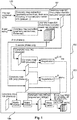
- Figure 1 shows a schematic flowchart diagram of a method for medical imaging of coronary vessels according to an exemplary embodiment of the invention.
- a function block contains input variables, output variables, through variables, internal variables, and an internal behavior description of the function block.
- Function blocks are used primarily to specify the properties of a user function. Many software languages are based on function blocks.
- the method or function block 100 may comprise, as sub-elements, four steps S1, S2, S3, S4 or four function blocks 101, 102, 103, 104:
- a coronary maps extraction is conducted, wherein the coronary maps extraction may be conducted in both in CTA and 3DRA data.
- the output of this first step may be the following:
- a coronary maps registration at diastole may be conducted.
- Aligning the common branches of the maps MC and MX(diastole) may be conducted. Most of the branches are visible on both maps (the only exceptions being of the occluded segments).
- a registration of the 3DRA coronary maps at different phases is conducted.
- the same algorithms used in the previous step or function block can also be used here, though the constraint on robustness can be relaxed (no extra branch, for instance, calcified, are expected from one map to the next).
- a fourth function block 104 corresponding to the fourth step S4 as shown in Figure 2 , coronary maps alignment and fusion may be performed.
- a further implementation would display MC'( ⁇ ).
- generating a 3D+t multiphase coronary map from the CT aligned with the cath lab is performed.
- a catheterization laboratory or cath lab is an examination room in a hospital or clinic with diagnostic imaging equipment used to visualize the arteries of the heart.
- the generating of the 3D+t multiphase could be used as a live multiphase roadmap for the CTO treatment, for instance.
- the coronary maps gained from the X-per swing present a superior spatial resolution. Analyzing the maps MC' for the occluded segments may be performed, wherein those that are not present in MX are processed, extracted and added to the maps MX. The augmented 3D+t multiphase MX maps may be further added.
- the finally merged maps do not need to be restricted to pure geometrical aspects.
- Information about the nature of the plaque can also be used there.
- Figure 2 shows a schematic flowchart diagram of a method for medical imaging of coronary vessels according to an exemplary embodiment of the invention.
- the method for medical imaging of coronary vessels may comprise the following steps.
- generating S2 a series of warped versions of the first vessel map based on the alignment of the coronary vessels is performed.
- interpolating S3 the set of second maps from the reference cardiac phase to any other phase based on the estimated motions of the coronary vessels is performed.
- merging S4 the series and the set of second maps at different phases in order to generate a final imaging map of the coronary vessels is conducted.
- these steps may be mixed or simultaneously processed.
- Figure 3 shows a schematic diagram of a device for medical imaging of coronary vessels according to an exemplary embodiment of the invention.
- a device 300 for medical imaging of coronary vessels may comprise a data extracting module 301 configured to extract a first vessel map from computed tomography angiography data covering at least one reference cardiac phase and a set of second vessel maps from three-dimensional rotational angiography data covering at least one cardiac cycle.
- the device may further comprise an interpolation module 302 configured to generate a series of warped versions of the first map aligned with the set of second maps, the series starting at the at least one reference cardiac phase.
- an interpolation module 302 configured to generate a series of warped versions of the first map aligned with the set of second maps, the series starting at the at least one reference cardiac phase.
- the device may comprise a merging module 303 configured to merge the series and the set of second maps at the different phases in order to generate a final imaging map of the coronary vessels.
- Figure 4 shows a schematic diagram of an X-ray medical imaging system for medical imaging of coronary vessels according to an exemplary embodiment of the invention.
- An X-ray medical imaging system 1000 may comprise a computed tomography angiography device 400 providing computed tomography angiography data, a three-dimensional rotational angiography device 400 providing three-dimensional rotational angiography data and a device 300 for medical imaging of coronary vessels. Contrary to the embodiment as depicted in Figure 4 , the computed tomography angiography device 400 and the three-dimensional rotational angiography device 400 may be constructed as separated and single units.
- a computer program or a computer program element is provided that is characterized by being adapted to execute the method steps of the method according to one of the preceding embodiments, on an appropriate system.
- the computer program element might therefore be stored on a computer unit, which might also be part of an embodiment of the present invention.
- This computing unit may be adapted to perform or induce a performing of the steps of the method described above.
- the computing unit can be adapted to operate automatically and/or to execute the orders of a user.
- a computer program may be loaded into a working memory of a data processor.
- the data processor may thus be equipped to carry out the method of the invention.
- This exemplary embodiment of the invention covers both, a computer program that right from the beginning uses the invention and a computer program that by means of an up-date turns an existing program into a program that uses the invention.
- the computer program element might be able to provide all necessary steps to fulfill the procedure of an exemplary embodiment of the method as described above.
- a computer readable medium such as a CD-ROM
- the computer readable medium has a computer program element stored on it, which computer program element is described by the preceding section.
- a computer program may be stored and/or distributed on a suitable medium, such as an optical storage medium or a solid state medium supplied together with or as part of other hardware, but may also be distributed in other forms, such as via the internet or other wired or wireless telecommunication systems.
- a suitable medium such as an optical storage medium or a solid state medium supplied together with or as part of other hardware, but may also be distributed in other forms, such as via the internet or other wired or wireless telecommunication systems.
- the computer program may also be presented over a network like the World Wide Web and can be downloaded into the working memory of a data processor from such a network.
- a medium for making a computer program element available for downloading is provided, which computer program element is arranged to perform a method according to one of the previously described embodiments of the invention.
Landscapes
- Health & Medical Sciences (AREA)
- Life Sciences & Earth Sciences (AREA)
- Engineering & Computer Science (AREA)
- Medical Informatics (AREA)
- Physics & Mathematics (AREA)
- Nuclear Medicine, Radiotherapy & Molecular Imaging (AREA)
- Radiology & Medical Imaging (AREA)
- General Health & Medical Sciences (AREA)
- Molecular Biology (AREA)
- Veterinary Medicine (AREA)
- Pathology (AREA)
- Public Health (AREA)
- Biomedical Technology (AREA)
- Heart & Thoracic Surgery (AREA)
- Biophysics (AREA)
- Surgery (AREA)
- Animal Behavior & Ethology (AREA)
- High Energy & Nuclear Physics (AREA)
- Optics & Photonics (AREA)
- Computer Vision & Pattern Recognition (AREA)
- Dentistry (AREA)
- Theoretical Computer Science (AREA)
- Oral & Maxillofacial Surgery (AREA)
- General Physics & Mathematics (AREA)
- Vascular Medicine (AREA)
- Cardiology (AREA)
- Physiology (AREA)
- Pulmonology (AREA)
- Apparatus For Radiation Diagnosis (AREA)
- Quality & Reliability (AREA)
Applications Claiming Priority (2)
| Application Number | Priority Date | Filing Date | Title |
|---|---|---|---|
| EP14305429 | 2014-03-26 | ||
| PCT/EP2015/055387 WO2015144467A1 (en) | 2014-03-26 | 2015-03-16 | Device and method for medical imaging of coronary vessels |
Publications (2)
| Publication Number | Publication Date |
|---|---|
| EP3123444A1 EP3123444A1 (en) | 2017-02-01 |
| EP3123444B1 true EP3123444B1 (en) | 2019-11-06 |
Family
ID=50440613
Family Applications (1)
| Application Number | Title | Priority Date | Filing Date |
|---|---|---|---|
| EP15711697.1A Active EP3123444B1 (en) | 2014-03-26 | 2015-03-16 | Device and method for medical imaging of coronary vessels |
Country Status (5)
| Country | Link |
|---|---|
| US (2) | US10275896B2 (enExample) |
| EP (1) | EP3123444B1 (enExample) |
| JP (1) | JP6419211B2 (enExample) |
| CN (1) | CN106163401B (enExample) |
| WO (1) | WO2015144467A1 (enExample) |
Families Citing this family (13)
| Publication number | Priority date | Publication date | Assignee | Title |
|---|---|---|---|---|
| MX2015012569A (es) * | 2013-03-15 | 2016-06-06 | Seno Medical Instr Inc | Sistema y método para soporte de clasificación de vectores de diagnóstico. |
| US10022101B2 (en) * | 2016-02-29 | 2018-07-17 | General Electric Company | X-ray/intravascular imaging colocation method and system |
| JP6871007B2 (ja) * | 2017-02-13 | 2021-05-12 | キヤノンメディカルシステムズ株式会社 | 医用画像処理装置及び医用画像診断システム |
| CN108294768B (zh) * | 2017-12-29 | 2020-05-19 | 华中科技大学 | 序列图像多参数配准的x射线心血管造影减影方法及系统 |
| JP6970835B2 (ja) * | 2018-09-28 | 2021-11-24 | 株式会社Pfu | 画像処理装置、制御方法及び制御プログラム |
| US10854329B2 (en) * | 2018-10-16 | 2020-12-01 | Canon Medical Systems Corporation | Motion estimation method and apparatus |
| EP3756549A1 (en) | 2019-06-28 | 2020-12-30 | Koninklijke Philips N.V. | Vessel registration using functional information |
| WO2021014181A1 (en) * | 2019-07-22 | 2021-01-28 | Siemens Healthcare Gmbh | Assessment of coronary artery calcification in angiographic images |
| CN112037186A (zh) * | 2020-08-24 | 2020-12-04 | 杭州深睿博联科技有限公司 | 一种基于多视图模型融合的冠脉血管提取方法及装置 |
| CN112927275B (zh) * | 2021-02-22 | 2022-03-01 | 北京安德医智科技有限公司 | 一种图像处理方法及装置、电子设备和存储介质 |
| CN114937100B (zh) * | 2022-03-11 | 2025-07-25 | 首都医科大学附属北京安贞医院 | 一种冠状动脉路径图的生成方法、装置、可读存储介质 |
| CN117058162A (zh) * | 2022-05-05 | 2023-11-14 | 苏州润迈德智能科技发展有限公司 | 冠状动脉树提取方法及相关装置 |
| CN117788533A (zh) * | 2023-12-26 | 2024-03-29 | 上海联影医疗科技股份有限公司 | 图像配准方法、系统和电子装置 |
Family Cites Families (6)
| Publication number | Priority date | Publication date | Assignee | Title |
|---|---|---|---|---|
| WO2004081877A1 (en) * | 2003-03-14 | 2004-09-23 | Koninklijke Philips Electronics N.V. | Motion-corrected three-dimensional volume imaging method |
| JP5295562B2 (ja) * | 2004-08-17 | 2013-09-18 | コーニンクレッカ フィリップス エレクトロニクス エヌ ヴィ | フレキシブル3次元回転血管造影−コンピュータ断層撮影融合方法 |
| US20090123046A1 (en) | 2006-05-11 | 2009-05-14 | Koninklijke Philips Electronics N.V. | System and method for generating intraoperative 3-dimensional images using non-contrast image data |
| DE102008031146B4 (de) * | 2007-10-05 | 2012-05-31 | Siemens Aktiengesellschaft | Vorrichtung zur Navigation eines Katheters durch eine Verschlussregion eines Gefäßes |
| CN102202576B (zh) * | 2008-10-10 | 2015-04-08 | 皇家飞利浦电子股份有限公司 | 用于为了在微创的x射线引导的介入中减少x射线剂量而产生覆盖经分割的目标结构或病变的减小视场的、具有自动快门适应的血管造影图像采集系统和方法 |
| US9384546B2 (en) * | 2012-02-22 | 2016-07-05 | Siemens Aktiengesellschaft | Method and system for pericardium based model fusion of pre-operative and intra-operative image data for cardiac interventions |
-
2015
- 2015-03-16 EP EP15711697.1A patent/EP3123444B1/en active Active
- 2015-03-16 WO PCT/EP2015/055387 patent/WO2015144467A1/en not_active Ceased
- 2015-03-16 US US15/127,972 patent/US10275896B2/en active Active
- 2015-03-16 JP JP2016558170A patent/JP6419211B2/ja active Active
- 2015-03-16 CN CN201580016196.6A patent/CN106163401B/zh active Active
-
2018
- 2018-10-18 US US16/164,483 patent/US10853956B2/en active Active
Non-Patent Citations (1)
| Title |
|---|
| MARTIN HOFFMANN ET AL: "Noninvasive Coronary Angiography With Multislice Computed Tomography", JAMA, 25 May 2005 (2005-05-25), United States, pages 2471 - 2478, XP055519488, Retrieved from the Internet <URL:https://jamanetwork.com/journals/jama/fullarticle/200952> DOI: 10.1001/jama.293.20.2471 * |
Also Published As
| Publication number | Publication date |
|---|---|
| CN106163401B (zh) | 2020-03-06 |
| US10853956B2 (en) | 2020-12-01 |
| US10275896B2 (en) | 2019-04-30 |
| WO2015144467A1 (en) | 2015-10-01 |
| JP6419211B2 (ja) | 2018-11-07 |
| JP2017508541A (ja) | 2017-03-30 |
| EP3123444A1 (en) | 2017-02-01 |
| US20190051002A1 (en) | 2019-02-14 |
| US20170091927A1 (en) | 2017-03-30 |
| CN106163401A (zh) | 2016-11-23 |
Similar Documents
| Publication | Publication Date | Title |
|---|---|---|
| US10853956B2 (en) | Device and method for medical imaging of coronary vessels | |
| JP6912168B2 (ja) | 3d+時間再構成を改良する方法および装置 | |
| US9375191B2 (en) | Method and apparatus for determining three-dimensional reconstruction of an object | |
| CN101961245B (zh) | 在采集的放射线照相术图像中补偿呼吸运动的系统和方法 | |
| US10083511B2 (en) | Angiographic roadmapping mask | |
| JP5566370B2 (ja) | 医用画像処理装置及び方法 | |
| Metz et al. | Registration of $3 {\rm D}+{\rm t} $ Coronary CTA and Monoplane $2 {\rm D}+{\rm t} $ X-Ray Angiography | |
| CN107170021B (zh) | 时变数据的细化重构 | |
| WO2015044433A1 (en) | Merging vessel maps | |
| JP6898047B2 (ja) | 時変データの定量的評価 | |
| EP3721412B1 (en) | Registration of static pre-procedural planning data to dynamic intra-procedural segmentation data | |
| JP6934948B2 (ja) | 流体解析装置および流体解析装置の作動方法並びに流体解析プログラム | |
| JP6068032B2 (ja) | 医用画像処理装置 | |
| JP6662580B2 (ja) | 医用画像処理装置 | |
| JP6811872B2 (ja) | 流体解析装置および流体解析装置の作動方法並びに流体解析プログラム |
Legal Events
| Date | Code | Title | Description |
|---|---|---|---|
| STAA | Information on the status of an ep patent application or granted ep patent |
Free format text: STATUS: THE INTERNATIONAL PUBLICATION HAS BEEN MADE |
|
| PUAI | Public reference made under article 153(3) epc to a published international application that has entered the european phase |
Free format text: ORIGINAL CODE: 0009012 |
|
| STAA | Information on the status of an ep patent application or granted ep patent |
Free format text: STATUS: REQUEST FOR EXAMINATION WAS MADE |
|
| 17P | Request for examination filed |
Effective date: 20161026 |
|
| AK | Designated contracting states |
Kind code of ref document: A1 Designated state(s): AL AT BE BG CH CY CZ DE DK EE ES FI FR GB GR HR HU IE IS IT LI LT LU LV MC MK MT NL NO PL PT RO RS SE SI SK SM TR |
|
| AX | Request for extension of the european patent |
Extension state: BA ME |
|
| DAV | Request for validation of the european patent (deleted) | ||
| DAX | Request for extension of the european patent (deleted) | ||
| STAA | Information on the status of an ep patent application or granted ep patent |
Free format text: STATUS: EXAMINATION IS IN PROGRESS |
|
| 17Q | First examination report despatched |
Effective date: 20170717 |
|
| REG | Reference to a national code |
Ref country code: DE Ref legal event code: R079 Ref document number: 602015041091 Country of ref document: DE Free format text: PREVIOUS MAIN CLASS: G06T0007000000 Ipc: G06T0007330000 |
|
| RIC1 | Information provided on ipc code assigned before grant |
Ipc: G06T 7/33 20170101AFI20190401BHEP |
|
| GRAP | Despatch of communication of intention to grant a patent |
Free format text: ORIGINAL CODE: EPIDOSNIGR1 |
|
| STAA | Information on the status of an ep patent application or granted ep patent |
Free format text: STATUS: GRANT OF PATENT IS INTENDED |
|
| INTG | Intention to grant announced |
Effective date: 20190527 |
|
| GRAS | Grant fee paid |
Free format text: ORIGINAL CODE: EPIDOSNIGR3 |
|
| GRAA | (expected) grant |
Free format text: ORIGINAL CODE: 0009210 |
|
| STAA | Information on the status of an ep patent application or granted ep patent |
Free format text: STATUS: THE PATENT HAS BEEN GRANTED |
|
| AK | Designated contracting states |
Kind code of ref document: B1 Designated state(s): AL AT BE BG CH CY CZ DE DK EE ES FI FR GB GR HR HU IE IS IT LI LT LU LV MC MK MT NL NO PL PT RO RS SE SI SK SM TR |
|
| REG | Reference to a national code |
Ref country code: GB Ref legal event code: FG4D |
|
| REG | Reference to a national code |
Ref country code: CH Ref legal event code: EP Ref country code: AT Ref legal event code: REF Ref document number: 1199814 Country of ref document: AT Kind code of ref document: T Effective date: 20191115 |
|
| REG | Reference to a national code |
Ref country code: DE Ref legal event code: R096 Ref document number: 602015041091 Country of ref document: DE |
|
| REG | Reference to a national code |
Ref country code: IE Ref legal event code: FG4D |
|
| REG | Reference to a national code |
Ref country code: NL Ref legal event code: MP Effective date: 20191106 |
|
| RAP2 | Party data changed (patent owner data changed or rights of a patent transferred) |
Owner name: KONINKLIJKE PHILIPS N.V. |
|
| REG | Reference to a national code |
Ref country code: LT Ref legal event code: MG4D |
|
| PG25 | Lapsed in a contracting state [announced via postgrant information from national office to epo] |
Ref country code: LV Free format text: LAPSE BECAUSE OF FAILURE TO SUBMIT A TRANSLATION OF THE DESCRIPTION OR TO PAY THE FEE WITHIN THE PRESCRIBED TIME-LIMIT Effective date: 20191106 Ref country code: NL Free format text: LAPSE BECAUSE OF FAILURE TO SUBMIT A TRANSLATION OF THE DESCRIPTION OR TO PAY THE FEE WITHIN THE PRESCRIBED TIME-LIMIT Effective date: 20191106 Ref country code: SE Free format text: LAPSE BECAUSE OF FAILURE TO SUBMIT A TRANSLATION OF THE DESCRIPTION OR TO PAY THE FEE WITHIN THE PRESCRIBED TIME-LIMIT Effective date: 20191106 Ref country code: FI Free format text: LAPSE BECAUSE OF FAILURE TO SUBMIT A TRANSLATION OF THE DESCRIPTION OR TO PAY THE FEE WITHIN THE PRESCRIBED TIME-LIMIT Effective date: 20191106 Ref country code: BG Free format text: LAPSE BECAUSE OF FAILURE TO SUBMIT A TRANSLATION OF THE DESCRIPTION OR TO PAY THE FEE WITHIN THE PRESCRIBED TIME-LIMIT Effective date: 20200206 Ref country code: NO Free format text: LAPSE BECAUSE OF FAILURE TO SUBMIT A TRANSLATION OF THE DESCRIPTION OR TO PAY THE FEE WITHIN THE PRESCRIBED TIME-LIMIT Effective date: 20200206 Ref country code: PT Free format text: LAPSE BECAUSE OF FAILURE TO SUBMIT A TRANSLATION OF THE DESCRIPTION OR TO PAY THE FEE WITHIN THE PRESCRIBED TIME-LIMIT Effective date: 20200306 Ref country code: LT Free format text: LAPSE BECAUSE OF FAILURE TO SUBMIT A TRANSLATION OF THE DESCRIPTION OR TO PAY THE FEE WITHIN THE PRESCRIBED TIME-LIMIT Effective date: 20191106 Ref country code: GR Free format text: LAPSE BECAUSE OF FAILURE TO SUBMIT A TRANSLATION OF THE DESCRIPTION OR TO PAY THE FEE WITHIN THE PRESCRIBED TIME-LIMIT Effective date: 20200207 Ref country code: PL Free format text: LAPSE BECAUSE OF FAILURE TO SUBMIT A TRANSLATION OF THE DESCRIPTION OR TO PAY THE FEE WITHIN THE PRESCRIBED TIME-LIMIT Effective date: 20191106 |
|
| PG25 | Lapsed in a contracting state [announced via postgrant information from national office to epo] |
Ref country code: RS Free format text: LAPSE BECAUSE OF FAILURE TO SUBMIT A TRANSLATION OF THE DESCRIPTION OR TO PAY THE FEE WITHIN THE PRESCRIBED TIME-LIMIT Effective date: 20191106 Ref country code: HR Free format text: LAPSE BECAUSE OF FAILURE TO SUBMIT A TRANSLATION OF THE DESCRIPTION OR TO PAY THE FEE WITHIN THE PRESCRIBED TIME-LIMIT Effective date: 20191106 Ref country code: IS Free format text: LAPSE BECAUSE OF FAILURE TO SUBMIT A TRANSLATION OF THE DESCRIPTION OR TO PAY THE FEE WITHIN THE PRESCRIBED TIME-LIMIT Effective date: 20200306 |
|
| PG25 | Lapsed in a contracting state [announced via postgrant information from national office to epo] |
Ref country code: AL Free format text: LAPSE BECAUSE OF FAILURE TO SUBMIT A TRANSLATION OF THE DESCRIPTION OR TO PAY THE FEE WITHIN THE PRESCRIBED TIME-LIMIT Effective date: 20191106 |
|
| PG25 | Lapsed in a contracting state [announced via postgrant information from national office to epo] |
Ref country code: CZ Free format text: LAPSE BECAUSE OF FAILURE TO SUBMIT A TRANSLATION OF THE DESCRIPTION OR TO PAY THE FEE WITHIN THE PRESCRIBED TIME-LIMIT Effective date: 20191106 Ref country code: RO Free format text: LAPSE BECAUSE OF FAILURE TO SUBMIT A TRANSLATION OF THE DESCRIPTION OR TO PAY THE FEE WITHIN THE PRESCRIBED TIME-LIMIT Effective date: 20191106 Ref country code: ES Free format text: LAPSE BECAUSE OF FAILURE TO SUBMIT A TRANSLATION OF THE DESCRIPTION OR TO PAY THE FEE WITHIN THE PRESCRIBED TIME-LIMIT Effective date: 20191106 Ref country code: DK Free format text: LAPSE BECAUSE OF FAILURE TO SUBMIT A TRANSLATION OF THE DESCRIPTION OR TO PAY THE FEE WITHIN THE PRESCRIBED TIME-LIMIT Effective date: 20191106 Ref country code: EE Free format text: LAPSE BECAUSE OF FAILURE TO SUBMIT A TRANSLATION OF THE DESCRIPTION OR TO PAY THE FEE WITHIN THE PRESCRIBED TIME-LIMIT Effective date: 20191106 |
|
| REG | Reference to a national code |
Ref country code: DE Ref legal event code: R097 Ref document number: 602015041091 Country of ref document: DE |
|
| REG | Reference to a national code |
Ref country code: AT Ref legal event code: MK05 Ref document number: 1199814 Country of ref document: AT Kind code of ref document: T Effective date: 20191106 |
|
| PG25 | Lapsed in a contracting state [announced via postgrant information from national office to epo] |
Ref country code: SK Free format text: LAPSE BECAUSE OF FAILURE TO SUBMIT A TRANSLATION OF THE DESCRIPTION OR TO PAY THE FEE WITHIN THE PRESCRIBED TIME-LIMIT Effective date: 20191106 Ref country code: SM Free format text: LAPSE BECAUSE OF FAILURE TO SUBMIT A TRANSLATION OF THE DESCRIPTION OR TO PAY THE FEE WITHIN THE PRESCRIBED TIME-LIMIT Effective date: 20191106 |
|
| PLBE | No opposition filed within time limit |
Free format text: ORIGINAL CODE: 0009261 |
|
| STAA | Information on the status of an ep patent application or granted ep patent |
Free format text: STATUS: NO OPPOSITION FILED WITHIN TIME LIMIT |
|
| 26N | No opposition filed |
Effective date: 20200807 |
|
| PG25 | Lapsed in a contracting state [announced via postgrant information from national office to epo] |
Ref country code: MC Free format text: LAPSE BECAUSE OF FAILURE TO SUBMIT A TRANSLATION OF THE DESCRIPTION OR TO PAY THE FEE WITHIN THE PRESCRIBED TIME-LIMIT Effective date: 20191106 |
|
| REG | Reference to a national code |
Ref country code: CH Ref legal event code: PL |
|
| PG25 | Lapsed in a contracting state [announced via postgrant information from national office to epo] |
Ref country code: SI Free format text: LAPSE BECAUSE OF FAILURE TO SUBMIT A TRANSLATION OF THE DESCRIPTION OR TO PAY THE FEE WITHIN THE PRESCRIBED TIME-LIMIT Effective date: 20191106 Ref country code: AT Free format text: LAPSE BECAUSE OF FAILURE TO SUBMIT A TRANSLATION OF THE DESCRIPTION OR TO PAY THE FEE WITHIN THE PRESCRIBED TIME-LIMIT Effective date: 20191106 |
|
| REG | Reference to a national code |
Ref country code: BE Ref legal event code: MM Effective date: 20200331 |
|
| PG25 | Lapsed in a contracting state [announced via postgrant information from national office to epo] |
Ref country code: LU Free format text: LAPSE BECAUSE OF NON-PAYMENT OF DUE FEES Effective date: 20200316 |
|
| PG25 | Lapsed in a contracting state [announced via postgrant information from national office to epo] |
Ref country code: CH Free format text: LAPSE BECAUSE OF NON-PAYMENT OF DUE FEES Effective date: 20200331 Ref country code: IT Free format text: LAPSE BECAUSE OF FAILURE TO SUBMIT A TRANSLATION OF THE DESCRIPTION OR TO PAY THE FEE WITHIN THE PRESCRIBED TIME-LIMIT Effective date: 20191106 Ref country code: LI Free format text: LAPSE BECAUSE OF NON-PAYMENT OF DUE FEES Effective date: 20200331 Ref country code: IE Free format text: LAPSE BECAUSE OF NON-PAYMENT OF DUE FEES Effective date: 20200316 |
|
| PG25 | Lapsed in a contracting state [announced via postgrant information from national office to epo] |
Ref country code: BE Free format text: LAPSE BECAUSE OF NON-PAYMENT OF DUE FEES Effective date: 20200331 |
|
| PG25 | Lapsed in a contracting state [announced via postgrant information from national office to epo] |
Ref country code: TR Free format text: LAPSE BECAUSE OF FAILURE TO SUBMIT A TRANSLATION OF THE DESCRIPTION OR TO PAY THE FEE WITHIN THE PRESCRIBED TIME-LIMIT Effective date: 20191106 Ref country code: MT Free format text: LAPSE BECAUSE OF FAILURE TO SUBMIT A TRANSLATION OF THE DESCRIPTION OR TO PAY THE FEE WITHIN THE PRESCRIBED TIME-LIMIT Effective date: 20191106 Ref country code: CY Free format text: LAPSE BECAUSE OF FAILURE TO SUBMIT A TRANSLATION OF THE DESCRIPTION OR TO PAY THE FEE WITHIN THE PRESCRIBED TIME-LIMIT Effective date: 20191106 |
|
| PG25 | Lapsed in a contracting state [announced via postgrant information from national office to epo] |
Ref country code: MK Free format text: LAPSE BECAUSE OF FAILURE TO SUBMIT A TRANSLATION OF THE DESCRIPTION OR TO PAY THE FEE WITHIN THE PRESCRIBED TIME-LIMIT Effective date: 20191106 |
|
| PGFP | Annual fee paid to national office [announced via postgrant information from national office to epo] |
Ref country code: FR Payment date: 20240326 Year of fee payment: 10 |
|
| PG25 | Lapsed in a contracting state [announced via postgrant information from national office to epo] |
Ref country code: FR Free format text: LAPSE BECAUSE OF NON-PAYMENT OF DUE FEES Effective date: 20250331 |
|
| PGFP | Annual fee paid to national office [announced via postgrant information from national office to epo] |
Ref country code: GB Payment date: 20260323 Year of fee payment: 12 |
|
| PGFP | Annual fee paid to national office [announced via postgrant information from national office to epo] |
Ref country code: DE Payment date: 20260320 Year of fee payment: 12 |